Handbook of Experimental Pharmacology
380
-
Upload
khangminh22 -
Category
Documents
-
view
0 -
download
0
Transcript of Handbook of Experimental Pharmacology


Handbook of Experimental Pharmacology
Volume 187
Editor-in-Chief
F. Hofmann, München
Editorial Board
J. Beavo, Seattle, WAA. Busch, BerlinD. Ganten, BerlinJ.-A. Karlsson, SingaporeM. Michel, AmsterdamC.P. Page, LondonW. Rosenthal, Berlin

Kian Fan Chung • John WiddicombeEditors
Pharmacology andTherapeutics of Cough
Contributors
M.G. Belvisi, F. Bertram, S.S. Birring, D.C. Bolser, A.C. Bonham, J. Bric,B.J. Canning, M.J. Carr, C.-Y. Chen, Y.-L. Chou, K.F. Chung, P.W. Davenport,P.V. Dicpinigaitis, R. Eccles, E. Ernst, R. Gatti, P. Geppetti, Q. Gu, D.J. Hele,J.P. Joad, L.-Y. Lee, S. Materazzi, S.B. Mazzone, M.A. McAlexander,I.M. McFadzean, L. McGarvey, A.H. Morice, R. Nassini, C.P. Page, D.I. Pavord,A.A. Raj, T. Shirasaki, F. Soeda, D. Spina, K. Takahama, M. Trevisani, B.J. Undem,J.G. Widdicombe
ABC

Prof. Kian Fan ChungNational Heart & Lung InstituteImperial CollegeDovehouse StreetLondon SW3 [email protected]
Prof. Dr. John WiddicombeUniversity of London116 Pepys RoadLondon SW20 [email protected]
ISBN 978-3-540-79841-5 e-ISBN 978-3-540-79842-2
Handbook of Experimental Pharmacology ISSN 0171-2004
Library of Congress Control Number: 2008929590
c© 2009 Springer-Verlag Berlin Heidelberg This work is subject to copyright. All rights are reserved,
whether the whole or part of the material is concerned, specifically the rights of translation, reprinting,reuse of illustrations, recitation, broadcasting, reproduction on microfilm or in any other way, and storagein data banks. Duplication of this publication or parts thereof is permitted only under the provisions of theGerman Copyright Law of September 9, 1965, in its current version, and permission for use must alwaysbe obtained from Springer. Violations are liable to prosecution under the German Copyright Law. The
use of general descriptive names, registered names, trademarks, etc. in this publication does not imply,even in the absence of a specific statement, that such names are exempt from the relevant protective lawsand regulations and therefore free for general use.
Product liability: The publisher cannot guarantee the accuracy of any information about dosage andapplication contained in this book. In every individual case the user must check such information byconsulting the relevant literature.
Cover Design: WMXDesign GmbH, Heidelberg
Printed on acid-free paper
9 8 7 6 5 4 3 2 1
springer.com

Preface
The last decade or so has seen remarkable advances in our knowledge of cough. Thisapplies especially to its basic mechanisms: the types of airway sensors, the pharma-cological receptors on their membranes, the brainstem organization of the ‘coughcentre’, and the involvement of the cerebral cortex in the sensations and the volun-tary control of cough. With the exception of the last of these, nearly all the studieshave been on experimental animals rather than humans, for obvious reasons. Onegroup of experimental studies has particular relevance to human patients, and that isthe demonstration of the sensitization of cough pathways both in the periphery andin the brainstem. Similar sensitizations have been shown for patients with chroniccough or who have been exposed to pollutants, and it is reasonable to suppose thatthis is the basis of their cough and that the underlying mechanisms are generallysimilar in humans and other species.
Important advances are also being made in clinical cough research. For thethree main causes of clinical cough, asthma, post-nasal drip syndrome, and gastro-oesophageal reflux disease, we are beginning to understand the pathologicalprocesses involved. There remains a diagnostically obdurate group of idiopathicchronic coughers, but even for them approaches are being devised to clarify under-lying mechanisms and to establish diagnoses.
Perhaps surprisingly, the field in which there has been the least spectacular ad-vance is the therapy of cough. This is not because current therapies work; indeedmost seem to work little better than a placebo. This applies not only to the manyremedies bought over the counter at the pharmacist and to those administered as partof complementary and alternative medicine, but also to those available on prescrip-tion (only codeine, pholcodine, and dextromethorphan in the UK). Basic studies arepointing to many potentially valuable approaches to the treatment of cough, basedon understanding the basic peripheral receptor mechanisms, the brainstem pathwaysin the control of cough, and the sensitization processes that may apply in disease.The pharmacological industry is following up these leads, and clinicians are waitinghopefully for the fruits of their research.
An indication of the growth of interest in cough is the recent surge in publica-tions dedicated to the subject. Before 1996, the editors can only think of two or
v

vi Preface
three. Since then there have been two multiauthor books, at least ten internationalsymposia, with the proceedings of nearly all of them being published as journalsupplements, and at least five task-force reports set up by national and internationalorganizations such as the American College of Chest Physicians and the EuropeanRespiratory Society. These publications will be frequently referred to in the chaptersin the present volume. If asked ‘Does this justify more description and analysis?’,the answer is an emphatic yes! The field is being explored very fast; and new andemerging results are very important for understanding and alleviating one of thecommonest disease symptoms of mankind. In this volume, we hope to show thatbasic mechanisms are helping us to understand clinical cough and also the otherway round.
The editors are grateful to all the contributors, including co-authors, who, asis well known, often do most of the hard work; and to the diligent but tolerantpublishers, especially Susanne Dathe, for their help and encouragement.
London, UK Kian Fan ChungJohn G. Widdicombe

Contents
Cough: Setting the Scene . . . . . . . . . . . . . . . . . . . . . . . . . . . . . . . . . . . . . . . . . 1K.F. Chung and J.G. Widdicombe
Cough Sensors. I. Physiological and Pharmacological Propertiesof the Afferent Nerves Regulating Cough . . . . . . . . . . . . . . . . . . . . . . . . . . . . 23B.J. Canning and Y.-L. Chou
Cough Sensors. II. Transient Receptor Potential Membrane Receptorson Cough Sensors . . . . . . . . . . . . . . . . . . . . . . . . . . . . . . . . . . . . . . . . . . . . . . . 49S. Materazzi, R. Nassini, R. Gatti, M. Trevisani, and P. Geppetti
Cough Sensors. III. Opioid and Cannabinoid Receptors on VagalSensory Nerves . . . . . . . . . . . . . . . . . . . . . . . . . . . . . . . . . . . . . . . . . . . . . . . . . 63M.G. Belvisi and D.J. Hele
Cough Sensors. IV. Nicotinic Membrane Receptors on Cough Sensors . . . 77L.-Y. Lee and Q. Gu
Cough Sensors. V. Pharmacological Modulation of Cough Sensors . . . . . . 99S.B. Mazzone and B.J. Undem
Peripheral Mechanisms I: Plasticity of Peripheral Pathways . . . . . . . . . . . 129M.A. McAlexander and M.J. Carr
Peripheral Mechanisms II: The Pharmacology of Peripherally ActiveAntitussive Drugs . . . . . . . . . . . . . . . . . . . . . . . . . . . . . . . . . . . . . . . . . . . . . . . 155D. Spina, I. McFadzean, F.K.R. Bertram, and C.P. Page
Central Mechanisms I: Plasticity of Central Pathways . . . . . . . . . . . . . . . . 187C.-Y. Chen, J.P. Joad, J. Bric, and A.C. Bonham
Central Mechanisms II: Pharmacology of Brainstem Pathways . . . . . . . . . 203D.C. Bolser
vii

viii Contents
Central Mechanisms III: Neuronal Mechanisms of Action of CentrallyActing Antitussives Using Electrophysiological and NeurochemicalStudy Approaches . . . . . . . . . . . . . . . . . . . . . . . . . . . . . . . . . . . . . . . . . . . . . . . 219K. Takahama, T. Shirasaki, and F. Soeda
Central Mechanisms IV: Conscious Control of Cough and the PlaceboEffect . . . . . . . . . . . . . . . . . . . . . . . . . . . . . . . . . . . . . . . . . . . . . . . . . . . . . . . . . 241R. Eccles
Clinical Cough I: The Urge-To-Cough: A Respiratory Sensation . . . . . . . . 263P.W. Davenport
Clinical Cough II: Therapeutic Treatments and Management of ChronicCough . . . . . . . . . . . . . . . . . . . . . . . . . . . . . . . . . . . . . . . . . . . . . . . . . . . . . . . . . 277A.H. Morice and L. McGarvey
Clinical Cough III: Measuring the Cough Response in the Laboratory . . . 297P.V. Dicpinigaitis
Clinical Cough IV: What is the Minimal Important Difference for theLeicester Cough Questionnaire? . . . . . . . . . . . . . . . . . . . . . . . . . . . . . . . . . . . 311A.A. Raj, D.I. Pavord, and S.S. Birring
Clinical Cough V: Complementary and Alternative Medicine: Therapyof Cough . . . . . . . . . . . . . . . . . . . . . . . . . . . . . . . . . . . . . . . . . . . . . . . . . . . . . . . 321J.G. Widdicombe and E. Ernst
Clinical Cough VI: The Need for New Therapies for Cough:Disease-Specific and Symptom-Related Antitussives . . . . . . . . . . . . . . . . . . 343K.F. Chung
Index . . . . . . . . . . . . . . . . . . . . . . . . . . . . . . . . . . . . . . . . . . . . . . . . . . . . . . . . . . . . . 369

Contributors
M.G. BelvisiRespiratory Pharmacology, Airway Diseases, National Heart & Lung Institute,Faculty of Medicine, Imperial College, Guy Scadding Building, Dovehouse Street,London SW3 6LY, [email protected]
F. BertramSackler Institute of Pulmonary Pharmacology, Division of Pharmaceutical Sciences,School of Biomedical and Health Sciences, King’s College, London SE1 1UL, UK
S.S. BirringDepartment of Respiratory Medicine, King’s College Hospital,London SE5 9RS, [email protected]
D.C. BolserDepartment of Physiological Sciences, College of Veterinary Medicine, Universityof Florida, Gainesville, FL 32610-0144, [email protected]
A.C. BonhamDepartment of Pharmacology, University of California, Davis School of Medicine,4150 V Street, 1104 PSSB Sacramento, CA 95817, [email protected]
J. BricDepartment of Pharmacology, University of California, Davis School of Medicine,4150 V Street, 1104 PSSB Sacramento, CA 95817, USA
B.J. CanningJohns Hopkins Asthma and Allergy Center, 5501 Hopkins Bayview Circle, Rm. 3A24, Baltimore, MD 21224, [email protected]
ix

x Contributors
M.J. CarrGlaxoSmithKline, 709 Swedeland Rd, King of Prussia, PA 19406, [email protected]
C.-Y. ChenDepartment of Pharmacology, Davis School of Medicine, University of California,4150 V Street, 1104 PSSB Sacramento, CA 95817, USA
Y.-L. ChouJohns Hopkins Asthma and Allergy Center, 5501 Hopkins Bayview Circle,Rm. 3A.24, Baltimore, MD 21224, USA
K.F. ChungNational Heart and Lung Institute, Imperial College,Dovehouse Street, London SW3 6LY, [email protected]
P.W. DavenportDepartment of Physiological Sciences, Box 100144, HSC, University of Florida,Gainesville, FL 32610, [email protected]
P.V. DicpinigaitisEinstein Division/Montefiore Medical Center, 1825 Eastchester Road, New York,NY 10461, [email protected]
R. EcclesCommon Cold Centre, Cardiff School of Biosciences, Cardiff University,Cardiff CF10 3US, [email protected]
E. ErnstComplementary Medicine, Peninsula Medical School, Exeter EX2 [email protected]〈mailto:[email protected]〉R. GattiDepartment of Critical Care Medicine and Surgery, University of Florence, VialePieraccini, 6, 50139, Florence, Italy
P. GeppettiDepartment of Critical Care Medicine and Surgery, University of Florence, VialePieraccini, 6, 50139, Florence, [email protected]
Q. GuDepartment of Physiology, University of Kentucky, Lexington, KY 40536-0298,USA

Contributors xi
D.J. HeleRespiratory Pharmacology, Airway Diseases, National Heart & Lung Institute,Faculty of Medicine, Imperial College, Guy Scadding Building, Dovehouse Street,London SW3 6LY, UK
J.P. JoadDepartment of Pharmacology, University of California, Davis School of Medicine,4150 V Street, 1104 PSSB Sacramento, CA 95817, USA
L.-Y. LeeDepartment of Physiology, University of Kentucky, Lexington,KY 40536-0298, [email protected]
S. MaterazziDepartment of Critical Care Medicine and Surgery, University of Florence, VialePieraccini, 6, 50139, Florence, Italy
S.B. MazzoneSchool of Biomedical Sciences, The University of Queensland, St. Lucia QLD4072, [email protected]
M.A. McAlexanderGlaxoSmithKline, 709 Swedeland Rd, King of Prussia, PA 19406, USA
I.M. McFadzeanSackler Institute of Pulmonary Pharmacology, Division of Pharmaceutical Sciences,School of Biomedical and Health Sciences, King’s College, London SE1 1UL, UK
L. McGarveyDepartment of Medicine, Institute of Clinical Science, The Queen’s University ofBelfast, Grosvenor Road, Belfast, BT12 6BJ, [email protected]
A.H. MoriceCardiovascular and Respiratory Studies, Castle Hill Hospital, University of HullCastle Road, Cottingham, East Yorkshire, HU16 5JQ, [email protected]
R. NassiniDepartment of Critical Care Medicine and Surgery, University of Florence,Viale Pieraccini, 6, 50139, Florence, Italy
C.P. PageSackler Institute of Pulmonary Pharmacology, Division of PharmaceuticalSciences, School of Biomedical and Health Sciences,King’s College, London SE1 1UL, [email protected]
xii Contributors
A.A. Raj, D.I. PavordDepartment of Respiratory Medicine, King’s College Hospital,London SE5 9RS, [email protected]
T. ShirasakiDepartment of Environmental and Molecular Health Sciences, Graduate Schoolof Pharmaceutical Sciences, Kumamoto University, 5–1 Oe-honmachi,Kumamoto 862–0973, Japan
F. SoedaDepartment of Environmental and Molecular Health Sciences, Graduate Schoolof Pharmaceutical Sciences, Kumamoto University, 5–1 Oe-honmachi, Kumamoto862–0973, Japan
D. SpinaSackler Institute of Pulmonary Pharmacology, Division of Pharmaceutical Sciences,School of Biomedical and Health Sciences, King’s College, London SE1 1UL, UK
K. TakahamaDepartment of Environmental and Molecular Health Sciences, Graduate Schoolof Pharmaceutical Sciences, Kumamoto University, 5–1 Oe-honmachi, Kumamoto862–0973, [email protected]
M. TrevisaniDepartment of Critical Care Medicine and Surgery, University of FlorenceViale Pieraccini, 6, 50139, Florence, Italy
B.J. UndemHoward Florey Institute, University of Melbourne, VIC 3010, Australia
J.G. WiddicombeUniversity of London, 116 Pepys Road, London SW20 8NY, [email protected]

Cough: Setting the Scene
K.F. Chung(�) and J.G. Widdicombe
Contents
1 Introduction . . . . . . . . . . . . . . . . . . . . . . . . . . . . . . . . . . . . . . . . . . . . . . . . . . . . . . . . . . . . . . . . 12 Definition and Semantics . . . . . . . . . . . . . . . . . . . . . . . . . . . . . . . . . . . . . . . . . . . . . . . . . . . . . 23 Cough Triggers and Sensors . . . . . . . . . . . . . . . . . . . . . . . . . . . . . . . . . . . . . . . . . . . . . . . . . . . 34 Cough Pathways . . . . . . . . . . . . . . . . . . . . . . . . . . . . . . . . . . . . . . . . . . . . . . . . . . . . . . . . . . . . . 65 Peripheral and Central Cough Sensitization . . . . . . . . . . . . . . . . . . . . . . . . . . . . . . . . . . . . . . 76 Sensory Correlates of Cough . . . . . . . . . . . . . . . . . . . . . . . . . . . . . . . . . . . . . . . . . . . . . . . . . . 87 Epidemiology of Clinical Cough . . . . . . . . . . . . . . . . . . . . . . . . . . . . . . . . . . . . . . . . . . . . . . . 98 Clinical Associations of Cough . . . . . . . . . . . . . . . . . . . . . . . . . . . . . . . . . . . . . . . . . . . . . . . . 109 Idiopathic Cough . . . . . . . . . . . . . . . . . . . . . . . . . . . . . . . . . . . . . . . . . . . . . . . . . . . . . . . . . . . . 1110 Enhanced Cough . . . . . . . . . . . . . . . . . . . . . . . . . . . . . . . . . . . . . . . . . . . . . . . . . . . . . . . . . . . . 1211 Measuring Cough . . . . . . . . . . . . . . . . . . . . . . . . . . . . . . . . . . . . . . . . . . . . . . . . . . . . . . . . . . . . 1312 Need for More Effective Antitussive Therapies . . . . . . . . . . . . . . . . . . . . . . . . . . . . . . . . . . . 1413 Conclusions: The Future of Cough . . . . . . . . . . . . . . . . . . . . . . . . . . . . . . . . . . . . . . . . . . . . . 15References . . . . . . . . . . . . . . . . . . . . . . . . . . . . . . . . . . . . . . . . . . . . . . . . . . . . . . . . . . . . . . . . . . . . . 15
1 Introduction
This introductory chapter is intended to bring a wide variety of physiological, clin-ical, and therapeutic aspects of cough together, but with a minimum of overlap. Ingeneral, it is true to say that the last decade or so has seen dramatic advances inour knowledge of the physiological mechanisms of acute cough in experimental an-imals, and that these are now moving in the direction of understanding increasedsensitivity of cough in chronic conditions. This latter aspect has clear implicationsfor patients in whom acute cough may be an irritation but is seldom a major causeof concern, while chronic cough can destroy the quality of life and arouse seriousconcerns in both patient and carer.
K.F. ChungNational Heart and Lung Institute, Imperial College, Dovehouse Street, London SW3 6LY, [email protected]
K.F. Chung, J.G. Widdicombe (eds.), Pharmacology and Therapeutics of Cough, 1Handbook of Experimental Pharmacology 187,c© Springer-Verlag Berlin Heidelberg 2009

2 K.F. Chung, J.G. Widdicombe
2 Definition and Semantics
Physiology textbooks describe cough as consisting of a three- or four-phase action:(1) the inspiratory phase, consisting of a deep inspiration; (2) the compressive phase,with closure of the larynx and a forced expiratory effort; (3) the expulsive phase,when the larynx opens and rapid expiration occurs with characteristic first coughsound; and (4) the restorative phase, when a final deep breath is taken (Fig. 1). Allphases are characteristic of a voluntary cough, but a reflex cough such as that evokedby inhalation of an irritant substance, or one occurring spontaneously in diseasemay be quite different. There may be a second (or third or even fourth) closureof the larynx during the expulsive phase, producing a second (or third or fourth)cough sound, although this is often absent. Little is understood about conditionsthat lead to these extra cough sounds or that are associated with their absence. Theinitial inspiration may be absent; this is seen with the expiration reflex from thelarynx or tracheobronchial tree. This starts with closure of the larynx and a forced
Fig. 1 The changes of the following variables during a representative cough: sound level, lungvolume, flow rate, subglottic pressure. During inspiration the flow rate is negative; at the glotticclosure the flow rate is zero; and during the expiratory phase the flow rate is positive. The last phasecan be divided into three parts: growing, constant, and decreasing. (From Bianco and Robuschi1989)

Cough: Setting the Scene 3
expiratory effort (the compressive phase) followed by an expulsive phase (Korpasand Tomori 1979; Widdicombe and Fontana 2006; Fontana and Widdicombe 2007;Tatar et al. 2008). Whether the expiration reflex should be called a cough is debated,although it possesses all the features of a cough except for the lack of the initialinspiration. It may have a different function from that of a cough, in that it shouldprevent aspiration of pharyngeal material into the lungs, whereas a cough expelsmaterial already in the lungs and requires a reserve of air deep in the lungs to makeit efficient.
We have described the reflex cough and the expiration reflex as if they wereisolated three- or four-phase activities, but in disease and on cough provocationthey often consist of a sequence of expiratory efforts, usually interspersed with in-spirations. These episodes are usually called cough epochs, but there is no cleardistinction between a repeated cough or expiration reflex and an epoch. An arbi-trary definition suggests that a 2-s gap between expulsive efforts is needed to callthem separate events, and with less than a 2-s gap they become an epoch (Kelsallet al. 2008). There have been recent and valuable detailed analyses of the reflexcough and expiration reflex events during cough epochs (Smith Hammond 2008;Vovk et al. 2007; Kim et al. 2008). With auditory records of cough, it is difficult toanalyze events during an epoch; however, it is possible with flow, pressure, or elec-tromyographic records, although this may not be practical in the clinic. Therefore,for clinical purposes, it may be convenient to describe cough as “a forced expulsivemanoeuvre, usually against a closed glottis and which is associated with a charac-teristic sound” (Morice et al. 2007). The presence of a forced expulsive maneuverand a characteristic sound of cough can be used to define cough clinically, and is thebasis for many instruments used to measure coughs, either as single discrete eventsor as cough epochs.
The semantics of cough is confusing. You can have wet, dry, and moist coughs,depending on the ear of the listener. The first cough sound is usually called expul-sive or explosive. The second cough sound has been called glottal or voiced. Anepoch has been given a variety of names: bout, attack, peal, even peel. A new word“dystussia” has been suggested (Paul Davenport, personal communication) to de-scribe a disordered cough airflow pattern (Smith Hammond 2008). This is a generalterm referring to a cough that is “abnormal” on the basis of altered cough patterns. Itseems an attractive word for a cough that has reduced expiratory airflow rates and/oraltered compression phase. In addition, perhaps, we should also consider eutussia(normal cough), atussia (absence of cough), hypotussia (reduced cough), and hyper-tussia (increased cough). Our view is that, while uniformity is desirable if it can beagreed, it is more important to define precisely what is being described, and to tryto understand its mechanism.
3 Cough Triggers and Sensors
What transduces the cough response? A lot of research is still being undertakento understand the “receptors” that can transduce the cough response since the first

4 K.F. Chung, J.G. Widdicombe
description of the irritant rapidly adapting receptor as being a cough “receptor”(Widdicombe 1954). We now prefer to call these sensors rather than receptors, sincethe latter term is now almost always used for membrane pharmacological recep-tors (Yu 2005). We also believe that there are an embarrassing number of coughsensors in the airways, i.e., those that can sense and transduce the cough response(Canning 2002, 2007; Canning and Chou 2008; Canning et al. 2006). Embarrassingbecause there is an embarras de richesses of both sensors and membrane recep-tors (Table 1). The sensors must all have slightly different reflex actions, and wedo not know what the differences are. It is unlikely that they all cause identicalrespiratory reflex patterns of cough, and their nonrespiratory (e.g., cardiovascular,bronchomotor, mucosecretor, sensation) actions may also be different. Presumably,the primary first-order neurones in the vagi link up with different patterns ofmedullary second-order neurones. The different sensors may “sense” different ir-ritant stimuli that cause cough (see below). The sensors include, for the tracheo-bronchial tree, bronchial C-fiber, Aδ-nociceptors, “cough receptors,” and “rapidlyadapting receptors” (Canning et al. 2006) and, for the laryngopharyngeal region,“irritant receptors” and C-fiber sensors (Widdicombe et al. 1988; Sant’Ambrogioand Sant’Ambrogio 1996). Full details of these sensors, their response to variousstimuli, and the reflexes they induce are given elsewhere in this volume (Canningand Chou 2008).
Other airway and lung sensors may also influence cough, although they may notcause it. Pulmonary C-fiber sensors have been claimed to cause cough, but there isalso evidence that they inhibit it (Tatar et al. 1988); their action may be determinedor modulated by other influences such as inputs from other bronchopulmonarysensors and brainstem conditions. Slowly adapting pulmonary stretch receptorsstrongly enhance the expiration reflex, and probably also strengthen the reflex cough(Korpas and Tomori 1979; Tatar et al. 2008), although they do not themselves causecough. Other bronchopulmonary sensors, e.g., neuroepithelial bodies (Adriaensenet al. 2003) and visceral pleural sensors (Pintelon et al. 2007), and other laryngealsensors, such as “drive” and “temperature” sensors (Widdicombe et al. 1988), have
Table 1 Some characteristics of bronchopulmonary sensors probably responsible for cough
Receptor Agonist Nodose Aδ Nodose C Jugular C Jugular Aδ Nodose RARs
TRPV-1 Capsaicin,acid, heat,AA
No Yes Yes Yes No
5-HT3 5-HT No Yes No No NoP2X Purines No Yes No No YesASIC Acid No Yes Yes Yes NoNicotinic Nicotine No Yes? Yes? Unknown UnknownBKB2 Bradykinin No Yes Yes Yes NoAdenosine Adenosine No Yes No No No
Modified from Kollarik and Undem (2006)RARs rapidly adapting receptors, TRPV-1 transient receptor potential vanilloid-1, AA arachidonicacid, 5-HT 5-hydroxytryptamine, ASIC acid-sensing ion channel, BKB2 bradykinin B2

Cough: Setting the Scene 5
not been shown to influence cough, although no-one may have looked for this effect.How these sensors may interact and modulate the ultimate cough output remain tobe explored.
There have been many studies on the morphology of sensors in the airways andlungs. Some, such as slowly adapting pulmonary stretch receptors and neuroep-ithelial bodies, have had their structures well delineated (Krauhs 1984; Adriaensenet al. 2003). But for the majority of sensors thought to be involved in cough, al-though they ramify within and below the airway epithelium, identification of thehistological structure of the sensor is tenuous. Similarly, it is difficult to ascribe anyone type of reflex associated with cough with a particular sensor and afferent path-way. A partial exception may be the expiration reflex from the larynx, which has alatency of 15–25 ms from mucosa to muscle in cats and humans (Tatar et al. 2008),and therefore must be conducted by myelinated afferent nerves, but this still leavesseveral possibilities open.
The membrane receptors on the sensors show as much diversity as the sen-sors themselves (Table 1). They include at least eight “specific” receptors, whichwhen activated open ion-selective channels leading to production of action poten-tials. Other receptors, e.g., cannabinoid receptors, may close excitatory channels andthus inhibit cough, or they may open “inhibitory” channels (e.g., potassium chan-nels). Extensive reviews of cough membrane receptors are included in this volume(Belvisi and Hele 2008; Lee and Gu 2008; Materazzi et al. 2008; Mazzone andUndem 2008). We will give one illustration. Acid stimulates at least three coughsensors (Aδ-nociceptors, C-fiber sensors, and probably rapidly-adapting receptors),but with different patterns and timings of neuronal activity (Kollarik and Undem2006; Kollarik et al. 2007). The membrane receptors involved belong mainly to twofamilies, the transient receptor potential and the acid-sensing ion channel families(note the word “family”!). The actual number of membrane receptor types may addup to dozens. Their concentration and distribution are not known in detail even forthe guinea pig, the species most studied. Yet, there are great species differences incough reflexes (Belvisi and Bolser 2002), so even these partial results cannot beapplied accurately to humans.
The problem can be illustrated in a different way by three other examples.Firstly, alkalis such as ammonia are powerful stimulants of cough (Widdicombe1954; Boushey et al. 1972; Van Hirtum and Berckmans 2004; Li et al. 2006;Rahman et al. 2007). Yet as far as we can discover, no-one has identified the sensorsand membrane receptors that respond to alkali. It is unlikely that, when they causecough, they have an “antiacid effect” or else they would inhibit cough; that is pre-cisely what weak concentrations of ammonia have been shown to do to citric acidinduced cough (Mercaux et al. 2000). Secondly, both hyperosmolar and hypoos-molar solutions of sodium chloride provoke cough (Koskela et al. 2008; Lavoriniet al. 2007) (one might expect the two to have opposite actions), yet the mediatingsensors and membrane receptors have not been identified. Thirdly, cold air is an es-tablished cause of cough (Cho et al. 2003), and may be one of the factors causinghigh-altitude cough. Yet, exercising polar explorers do not complain of cough (Ma-son and Barry 2007) and, as far as we can determine, no-one has identified airway

6 K.F. Chung, J.G. Widdicombe
sensors that respond to cold and cause cough. Possibly the laryngeal “cold” sensorsare a candidate (Widdicombe et al. 1988); however, they also respond to airflow,which has not been shown to cause cough. There must be hundreds of differentagents that can cause cough, but there are only basic detailed studies on acid andcapsaicin, and to a lesser extent on nicotine, adenosine compounds, bradykinin, and5-hydroxytryptamine.
The whole subject of cough sensors and membrane receptors is a tangled web;an appropriate term to apply to cough sensory neurones and their tortuous terminalsat both ends. However, the web continues to be unraveled by persistent research.
4 Cough Pathways
As far as we know, all afferent pathways for cough, either from the larynx or fromthe tracheobronchial tree, travel in the vagus nerves. Vagotomy or vagal block withlocal anesthesia abolishes all reflex cough in humans and other animals (Korpas andTomori 1979; Guz et al. 1970). There are some afferent pathways from the lungs thattravel in the sympathetic nerves, for example, coming from neuroepithelial bodiesand visceral pleural sensors, but these have not been shown to mediate cough. Thevagal nerves have their cell bodies in the nodose and jugular ganglia; Undem andhis colleagues have differentiated between cough sensors with cell bodies in eachtype of ganglion, and have shown that the ganglia have different embryonic ori-gins, from placodes and neural crest, respectively (Kollarik et al. 2007). Whetherthey correspond to different types of cough is not known. The vagal afferent nerves(first-order neurones) travel to the nucleus of the solitary tract (NTS), especially thecaudal part of the tract (Mutolo et al. 2008), where they synapse with second-orderneurones. From thereon, the picture becomes very complicated. Second- (or later-)order neurones travel not only to cause cough, but also to influence modalities suchas breathing, the cardiovascular system, skeletal muscle tone, airway mucosecretion,and sensation (inter alia). With the exception of cough, none of these connectionshas been worked out in any detail. When cough is initiated, the central respiratoryrhythm generator is “switched off” and “gates” are thought to open to allow ac-tivation of the cough generator (Bolser and Davenport 2004; Bolser et al. 2006).Presumably these gates are closed in the absence of a stimulus that leads to a cough.
Detailed maps of the brainstem pathways that mediate cough have been deter-mined (Shannon et al. 2000, 2004), and their relationship to the brainstem neuronalcontrol of breathing described (Bolser and Davenport 2004). They are too com-plicated to summarize here, but their importance can be illustrated in five ways.Bolser et al. (2006) have proposed a “holarchical” system for cough in the brain-stem whereby different functional control elements regulate the different behaviorsrelated to cough, including cough itself. This system includes “gates” which, byopening and closing, can determine whether or not a particular activity, e.g., cough,is permitted. Secondly, they are a site of sensitization (and possibly desensitiza-tion) of the cough reflex (Bonham et al. 2006; Chen et al. 2008), and are therefore

Cough: Setting the Scene 7
very relevant to what happens in diseases associated with cough. Thirdly, they arethe site of action of many antitussive drugs (Bolser 2008; Takahama et al. 2008).The understanding of these medullary pathways could lead to important advancesin antitussive therapy (Chung 2007, 2008). Fourthly, the circuitries for the tracheo-bronchial reflex cough and for the laryngeal expiration reflex have been establishedas different (Baekey et al. 2004). Since the two reflexes have different physiologicaland pharmacological controls (Tatar et al. 2008), this observation is of potential sig-nificance in the development of future antitussive therapeutic strategies. And fifthly,there seem to be different gating systems for cough from the larynx compared withcough from the lower airways (Bolser and Davenport 2004); this could correspondto the different circuitries for the expiration reflex compared with the reflex cough,and these have similar implications for therapy.
But the influence of cough inputs extends far beyond the brainstem. They causethe sensations of “irritation” and “urge-to-cough” (see later), and activate manyparts of the cerebral cortex and upper brain. This has been well illustrated by thefunctional magnetic resonance brain imaging studies in humans that associate theurge-to-cough sensation with cortical neuronal activation pathways (Mazzone et al.2007). We do not know whether these supramedullary pathways are enhanced in dis-eases associated with a chronic cough. A comparison with pain has been drawn; thelatter elicits many reflexes, sensations, and emotive changes, and similar processesare now being studied in relation to cough (Gracely et al. 2007; Widdicombe 2008).
5 Peripheral and Central Cough Sensitization
There have been many recent studies that show that peripheral cough sensors canbe “sensitized” in animals (Carr 2004, 2007; Carr and Lee 2006; McAlexander andCarr 2008); these studies show that exposure to an appropriate peripheral stimulus,for example, by development of an allergic sensitivity or irritation by pollutantsor their constituents, can lower the threshold and increase the cough response totussigenic agents such as citric acid or capsaicin, and increase the action potentialresponse in fibers thought to originate from cough sensors. Histological examinationof the sensors shows that their structure may change, in particular to contain moreinclusions such as those of neuropeptides (Chuachoo et al. 2006). It seems almostcertain that the same process of sensitization can occur in humans. For example,atmospheric exposure to pollutants or experimental exposure to ozone lowers thecough threshold to agents such as citric acid and capsaicin (Joad et al. 2007).
To what extent the same process applies to patients with cough is more diffi-cult to decide. The disease process could cause a greater stimulus to cough sen-sors otherwise of “normal” sensitivity; for example, the presence of excess mucus,edema in the mucosa, and greater release of tussigenic agents such as bradykininor neuropeptides could move the cough sensor response up the stimulus/responsecurve and give the impression of sensitization, while in reality it is the stimulusthat is increased (Widdicombe 1996). It is not known whether mediator release in

8 K.F. Chung, J.G. Widdicombe
the airways’ mucosa sensitizes the nerves there. One might even speculate that theincreased acidity of the airway surface liquid in asthmatics (Koutsokera et al. 2008)is a sensitizing or cough-promoting agent. Inhalation of weak ammonia concentra-tions may inhibit cough (Moreaux et al. 2000). For the clinician, this should not bea semantic quibble; if there is an added cough stimulus (such as mucus) then it maybe preferable to decrease the stimulus rather than depress the cough, whereas if thecough is sensitized, a symptomatic (antitussive) approach may be better.
Neural mechanisms of reflex cough are regulated by the inspiratory and expi-ratory networks of the brainstem, pons, and cerebellum, particularly in brainstemnuclei in the NTS where there are connections to respiratory related neurones inthe central respiratory generator (Shannon et al. 2000, 2004). Changes in the cen-tral processing at the level of the ganglia or brainstem (“central sensitization”)encompass changes in sensory pathways with the release of neurotransmitters orneuromodulators, or in excitability of postsynaptic neurones, or in a change in thestructure of the nerve (Bonham et al. 2006). Central nervous system sensitization ofthe cough reflex has also been shown in animals, including primates In particular,the role of substance P released from first-order neurones and acting on second-orderneurones in the NTS has been established, and the membrane receptor mechanismson the second-order neurones have been analyzed in detail (Chen et al. 2008). Withan upregulated cough reflex, due, for example, to inhaled pollutants, the substance Plevels in the NTS are increased, just as they are in the first-order neurones (the sen-sory fibers) (Chen et al. 2008). Injections of substance P into the NTS enhancecoughing due to a peripheral stimulus, and neurokinin 1 receptor antagonists de-press cough in animals (Advenier and Emonds-Alts 1996; Bonham et al. 2006). Forobvious reasons it is impossible to repeat these studies in humans, and in patientswho have a sensitized cough reflex it is difficult to partition the response betweenperiphery and brainstem. On theoretical grounds, it seems likely that both sites areinvolved. Disease processes in the airways will sensitize the sensors there, whichin turn will cause sensitization at the first- and second-order neurones in the brain-stem, as seems to happen in experimental animals. From the therapeutic point ofview, effective neurokinin 1 receptor antagonists might act at both levels (Advenierand Emonds-Alts 1996).
6 Sensory Correlates of Cough
Reflex coughing, as distinct from voluntary or habit coughing, is often associatedwith unpleasant sensation in the chest or throat; however, this is not always present,especially with conditions in the lower airways involving, for example, excessivemucus. The terms used to describe the sensations are various, and include “irrita-tion,” “rawness,” and even “pain” (Widdicombe 2008).
Urge-to-cough is a distinct sensation that, with increasing levels of cough stim-ulation, has a lower threshold and occurs before the cough itself (Davenport 2008;

Cough: Setting the Scene 9
Vovt et al. 2007), Other respiratory sensations, such as tightness, air-hunger, senseof effort and sense of lung volume are not usually associated with cough.
Patients with chronic cough often complain of a persistent tickling or irritatingsensation in the throat (feeling of an itch) or a choking sensation, and it is some-times felt in the chest, that often leads to paroxysms of coughing. Triggers suchas changes in ambient temperature, taking a deep breath, laughing, talking overthe phone for more than a few minutes, cigarette smoke, aerosol sprays, perfumesor eating crumbly dry food are common. Unpleasant sensation related to coughmay be localized vertically, in the throat or in the chest, but not usually more pre-cisely or laterally. Vagotomy or vagal anesthesia prevents the sensation (Petit 1970;Winning et al. 1988), and that from the chest is absent in patients with bilateral lungtransplant (Butler et al. 2001)
Urge-to-cough has been extensively studied in the last few years, especially byDavenport and colleagues. It is described in detail elsewhere in this volume byDavenport (Davenport 2008). The parts of the cerebral cortex and upper brain thatare activated by these sensations have also been mapped out (Mazzone et al. 2007).Urge-to-cough can occur with stimuli, such as aerosols of capsaicin, citric acid,and distilled water, and intravenous injections of lobeline and capsaicin, which aretoo weak to cause cough, and in the presence or absence of unpleasant sensation(Widdicombe 2008). While urge-to-cough has no particular location in the body,unpleasant sensation related to cough may be felt in the chest or the throat (Butleret al. 2001). In the latter case it must be referred from another site, since intra-venously administered lobeline is thought to act on bronchopulmonary sensors butarouses a raw sensation in the larynx. A similar referred unpleasant sensation is seenwith some patients with unilateral lung disease, when the sensation is identified ascoming from the ipsilateral side of the face (Sarlani et al. 2003). But cough is notusually associated with this condition.
We cannot say which sensor or sensors in the lungs are responsible for the respi-ratory sensations, but it seems likely that there are different combinations of activityfor cough, urge-to-cough, and rawness. For example, distilled water aerosol pro-duces cough and urge-to-cough but no rawness (Lavorini et al. 2007), while anec-dotally many lung conditions produce cough and rawness but no urge-to-cough, orcough with neither rawness nor urge-to-cough. The complexity of the airway sen-sory system mediating cough, as already described, makes it unlikely that identicalpathways are responsible to all three reactions.
7 Epidemiology of Clinical Cough
Chronic cough is not uncommon and its prevalence varies from 9 to 33% of thepopulation, and there is an association with cigarette smoking (Cullinan 1992;Ford et al. 2006; Zemp et al. 1999), in that chronic smokers have a threefold in-crease in prevalence of chronic cough compared with never smokers and ex-smokers(Zemp et al. 1999). Other associations are reported too with asthma or respiratory

10 K.F. Chung, J.G. Widdicombe
wheeze, or with symptoms of gastroesophageal reflux disease (GORD) (Janson et al.2001; Ford et al. 2006). Exposure to environmental pollutants, particularly PM10particulates, is also associated in adults and schoolchildren with productive coughor chronic nocturnal dry cough (Braun-Fahrlander et al. 1997; Pierse et al. 2006). In-creases in levels of PM10 and of nitrogen dioxide have been correlated to reductionsin peak expiratory flows and to increasing reporting of cough, sputum production,and sore throat in children. Clearly more research is needed to firm and explainthe link with environmental pollution. For the respiratory physician, patients with achronic cough probably account for 10–38% of his/her outpatient practice. Only aminority of the population identified in epidemiological surveys seek medical helpor advice about their symptom. It is important to find out whether there are any med-ical associations with chronic cough in the community and of the natural history ofthis symptom.
8 Clinical Associations of Cough
Cough has been divided into an acute self-limiting cough lasting less than 3 weeksor a chronic persistent cough, usually defined as lasting for more than 8 weeks.Acute cough is usually the result of an upper respiratory tract virus infection thatusually clears within 2 weeks in two thirds of people. Nonviral causes of acutecough include exacerbation of existing asthma or potential exposure to environmen-tal pollutants. Other types of cough last for a limited period of 3–8 weeks, whichis referred to as subacute cough, reported to be postinfective (Kwon et al. 2006).Eleven to 25% of patients with chronic cough report a postinfectious cough (Poeet al. 1989). Persistent cough following Mycoplasma or Bordetella pertussis infec-tions have been highlighted (Davis et al. 1995), but no doubt other infections maybe involved and further research in this area is needed.
In North America and Europe, the most common conditions associated withcausing chronic cough, with normal findings on a chest radiograph, includethe corticosteroid-responsive eosinophic airway diseases (asthma, cough-variantasthma, and eosinophilic bronchitis), and a range of conditions typically associatedwith an inhaled corticosteroid-resistant cough, including GORD and the postnasaldrip syndrome or rhinosinusitis. The frequency of these causes has varied in dif-ferent series depending on the location of the clinic and its particular interest, onthe age of the patient, and on local definition of the disease entities (Chung andPavord 2008). For example, with regard to the latter, in Japan, atopic cough andsinobronchial disease are more commonly diagnosed, while GORD is much less so(Niimi 2007; Kohno et al. 2006). The associations of various diseases with chroniccough still need to be worked out carefully, and the mechanisms of cough in diseaseare in need of clarification.
Asthma may present predominantly with cough, often nocturnal, and the diagno-sis is supported by the presence of bronchial hyperresponsiveness. Three other con-ditions, cough-variant asthma, atopic cough, and eosinophilic bronchitis, are related

Cough: Setting the Scene 11
to classic asthma, and are all associated with an eosinophilic airway inflammationand the cough responds well to inhaled corticosteroid therapy. This raises the possi-bility that eosinophils may directly contribute to increasing cough sensitivity.
GORD encompasses symptoms or complications such as heart burn, chest pain,sour taste, or regurgitation, and also a chronic persistent cough. Direct aspirationof gastric contents into the larynx and upper airways that could directly stimulatecough sensors and increases in tracheal acidity have been recorded during episodesof reflux (Jack et al. 1995). On the other hand, direct infusion of acid into the dis-tal esophagus of patients with chronic cough due to GORD induces cough (Inget al. 1994), through vagal cholinergic pathways. However, the majority of coughsin GORD do not coincide with an acid reflux episode (Ours et al. 1999; Irwin et al.1989). Nonacid components such as pepsin, bile, and other gastric enzymes mayinduce cough. In addition, associated dysmotility of the esophagus is implicated butwith not much evidence.
Postnasal drip (“nasal catarrh”) is characterized by a sensation of nasal secre-tions or of a “drip” at the back of the throat, accompanied very often by frequentneed to clear the throat (“throat-clearing”) associated with nasal discharge or nasalstuffiness. The term “upper airway cough syndrome” is proposed as an alternativeto stress the association of upper airways disease with cough (Pratter 2006). Thepathogenesis of cough in the postnasal drip syndrome may be related to the directpharyngeal, laryngeal, or sublaryngeal stimulation by the mucoid secretions fromthe rhinosinuses, which contain inflammatory mediators to induce cough.
9 Idiopathic Cough
Earlier series of chronic cough patients rarely identified patients in whom no identi-fiable cause was found or failure of treatment of identifiable causes occurred. Morerecent series have identified a significant proportion of patients labeled as “idio-pathic” cough, ranging from 7 to 46%, despite thorough diagnostic workup (Irwinet al. 1981, 1990, 2006; Poe et al. 1989; O’Connell et al. 1994; Pratter et al. 1993;Smyrnios et al. 1995; Mello et al. 1996; French et al. 1998; McGarvey et al. 1998;Brightling et al. 1999; Birring et al. 2004b; Niimi et al. 2005; Kastelik et al. 2005;Fujimura et al. 2005; Shirahata et al. 2005; Palombini et al. 1999; Carney et al.1997). It may be interesting to determine whether this represents a genuine changeor whether different methods were being used regarding diagnostic approaches.The initiating cause of the cough may have disappeared, but its effect on enhancingthe cough reflex may be more prolonged. An example could be the transient ap-pearance of an upper respiratory tract virus infection or an exposure to toxic fumesthat results in prolonged damage of the airways’ mucosa. The repetitive mechanicaland physical effects of coughing bouts on airway cells could activate the releaseof various chemical mediators that could enhance chronic cough through inflam-matory mechanisms (Heino et al. 1990), providing a positive feed-forward systemfor cough persistence. It is quite possible that there is an induction of changes in

12 K.F. Chung, J.G. Widdicombe
the upper airways of inflammation and tissue remodeling induced by various causesassociated with cough or by the act of coughing itself that could lead to an en-hanced cough reflex, which in turn is responsible for maintaining cough. The coughbecomes “idiopathic” when the primary inciting cause has resolved while coughis persistent. It is clear that more needs to be learned about idiopathic cough, andwhether it is all “idiopathic” is the big question; in the meantime, it is reasonable tostudy this group as a separate entity.
10 Enhanced Cough
Patients with chronic cough often complain of a persistent tickling or irritating sen-sation in the throat (feeling of an itch), or a choking sensation and sometimes felt inthe chest, that often leads to paroxysms of coughing. Triggers such as changes in am-bient temperature, taking a deep breath, laughing, talking over the phone for morethan a few minutes, cigarette smoke, aerosol sprays, perfumes, or eating crumblydry food are common.
The mechanisms of idiopathic cough are unclear, but we assume that the initiat-ing cause of the cough has disappeared, leaving an enhancement of the cough reflexwhich can be measured by the tussive response to inhalation of citric acid or cap-saicin, as compared with noncoughers (Choudry and Fuller, 1992). The increase incough sensitivity to capsaicin is related to the presence of a tickling or irritating sen-sation localized to the throat or lower-chest area that often leads to a paroxysm ofcoughing which patients with chronic cough find most distressing because it cannotbe controlled. The paroxysm can be triggered in some patients by inhaling cold air,by a deep breath, by the act of laughing, and by breathing irritants such as cigarettesmoke, aerosol sprays, or perfumes. The urge-to-cough is a sensory measure of thissensation of tickling or irritation that is induced at concentrations of inhaled cap-saicin that are lower than those necessary to elicit a cough reflex, which is a motorcough behavior (Davenport et al. 2007), but may also be present in patients withchronic cough. This sensation may be a “referred” sensation since very often thereare no visible abnormalities of the pharynx and larynx that are associated with it.
This enhanced cough reflex may result from an increased sensitivity of coughreceptors with plasticity of the afferent innervation such as changes in nerve den-sities or in ion channels (peripheral sensitization) (Lee and Undem 2004; Carr andLee 2006). The presence of increased expression of the transient receptor poten-tial vanniloid-1 (TRPV-1) receptor in epithelial nerves of patients with nonasth-matic chronic cough indicates a potential mechanism of peripheral sensitization(Groneberg et al. 2004). Inflammation and remodeling of the airway submucosawith an increase in submucosal mast cells and airway wall remodeling with gob-let cell hyperplasia, subepithelial fibrosis, and increased vascularity is reported inchronic cough patients (Niimi et al. 2005). Increased mast cells have also beenobserved in bronchoalveolar lavage fluid (McGarvey et al. 1999), with increasedneutrophils (Jatakanon et al. 1999), and higher histamine, prostaglandins D2 and

Cough: Setting the Scene 13
Eosinophil
Histamine, LTD4
Submucosalgland Neutrophil
Epithelium
Periciliary fluid Mucus
Monocyte
Local axon reflex
Vagus
nerve
Mucus
‘Central’
RARC-fibers
Oedema
‘Cough receptor’
‘Peripheral’
COUGH
DiaphragmIntercostal musclesLaryngeal musclesAbdominal muscles
Volitional control
Phrenic nervesSpinal motor nervesRrecurrent laryngeal nerves
Central cough generator
nTS relayneurones
Brain stem
Volitional control
Cerebral cortex
Urge-to-cough
±
SAR
Airway smooth muscle
Mast cell
Goblet cell
Blood vessel
TRPV-1CGRP
Subbasement membrane
H+ ≠
PGE2
≠ NK1R
Fig. 2 Afferent pathways and central control of the cough reflex with peripheral and central sensiti-zation of the reflex by a variety of mechanisms. CGRP calcitonin gene-related peptide, nTS nucleusof the solitary tract, LT D4 leukotriene D4, NK neurokinin, PGE2 prostaglandin E2, RAR rapidlyadapting receptors, SAR slowly adapting receptors, TRPV-1 transient receptor potential vanilloid-1
E2, tumor necrosis factor α, and interleukin-8 concentrations in induced sputum(Birring et al. 2004a). These inflammatory changes could certainly contribute to pe-ripheral sensitization of the cough reflex. However, while the changes observed inthe airways could also result from physical damage from the coughing act, theycould nevertheless contribute to the chronicity of the cough, a possibility worthexploring. Some of the mechanisms underlying the enhanced cough response inchronic cough are illustrated in Fig. 2.
11 Measuring Cough
There has been a great deal of progress made in the field of cough measurementover the last 10 years (Chung 2006). Cough can be measured subjectively usingsymptom scores and specific quality-of-life measures, and objectively by measuringcough numbers and intensity, and by assessing the cough response to capsaicin orcitric acid. Most previous reported clinical series of chronic cough do not state howthe clinical response of the chronic cough patients was measured, and yet providesuccess of intervention as “yes/no.” This could be the reason why there is a diversityof success in treating chronic cough in the literature. In the small studies of the anti-tussive effects of various agents, a variety of instruments have been used, includinga cough scoring system or visual analogue scale completed by the patient, or tussive

14 K.F. Chung, J.G. Widdicombe
response to capsaicin or citric acid. Cough visual analogue scores are used mostcommonly in clinical trials. Typically these assess cough according to the patient’sown experience: for example, the patient will be asked to rate his/her cough on a10-cm scale fixed at both ends by “no cough” and “the worst cough ever.” Assess-ments are responsive and repeatable but they are of no value in comparing coughseverity between individuals or populations.
Cough-specific quality-of-life questionnaires have been used to identify the manydifferent components of impaired health status seen in patients with chronic cough(French et al. 1998; Birring et al. 2003). The Leicester Cough Questionnaire com-prises of 19 items and three domains made of physical, psychological, and socialattributes, with a seven-point Likert response scale, and responsiveness to treatmenthas been shown in a group of patients with cough that were successfully treated(Birring et al. 2006).
The ultimate objective assessment of cough is to measure its frequency and in-tensity (Chung 2006) and there are now reliable ambulatory systems to measurecough, although measurement of cough intensity may not be easy (Birring andYousaf 2008).
Measurement of the cough reflex has been studied using inhalation of citric acidor of capsaicin; both techniques have been well validated and the methods are wellstandardized. Capsaicin cough sensitivity is probably the most widely used test, asit induces cough reliably and assessments of the cough reflex with inhaled capsaicinare reproducible (Dicpinigaitis 2003; Dicpinigaitis and Alva 2005). An increase incough sensitivity has been reported in most conditions associated with a chroniccough and improvements in cough sensitivity are seen in patients whose chroniccough has been successfully treated (O’Connell et al. 1994).
The need for objective measures in clinical trials is demonstrated by the morerecent studies that have shown the limitation of available antitussives in the treat-ment of chronic cough. Codeine is probably the most commonly prescribed opioid-derived antitussive and recent studies using objective counts have shown that it isineffective against the acute cough of the common cold (Freestone and Eccles 1997),or against cough in patients with COPD (Smith et al. 2006), despite the findings re-ported in previous publications on its antitussive effects. The correct use of theseinstruments to measure cough in the clinic as well as in the clinical trials assess-ing antitussive therapies will certainly be defined in the years to come (Pavord andChung 2008).
12 Need for More Effective Antitussive Therapies
In patients with idiopathic cough or in those in whom treatments directed againstassociated cause (termed “specific antitussives”) are not successful, there is a needfor symptomatic antitussive therapies. The efficacy of codeine or dextrometorphanin chronic cough is limited at the recommended doses, and higher doses causesunacceptable side effects. There continues to be great interest from the pharmaceu-tical industry to develop new antitussives on the basis of our understanding of the

Cough: Setting the Scene 15
mechanisms of the enhanced cough reflex (Chung 2005). New-generation opioidsor inhibitors that target afferent nerves involved in sensitization of the cough reflexsuch as TRPV-1 antagonists, tachykinin receptor antagonists, or chloride channelblockers have been identified as potential antitussives. There has been little progressin translating this research to effective antitussives and few studies have been donein chronic cough patients (Chung 2005). This may relate to the fact that most targetsfor antitussive therapies are derived from animal models that differ from humans andthat the human cough reflex pathway is difficult to study.
On the other hand, potential antitussives have arisen from clinical reports of ex-isting drugs in the treatment of cough, particularly the use of centrally acting drugssuch as the antiepileptics gabapentin and carbamazepine, and the antidepressantsamitriptyline and paroxetine (Chung 2007). There is a pressing need to demonstratetheir antitussive effects in controlled trials using appropriate cough-assessment toolsand to investigate their mechanism of action. They may have an effect on the sen-sitization process, akin to the effect of these agents in controlling neuropathic pain,through inhibition of various neural inflammatory pathways, or through effectson supramedullary pathways (Widdicombe et al. 2006). For example, there havebeen reports of positive findings for the use of amitriptyline in 12 cough patients(Bastian et al. 2006), and in an open controlled study of postviral persistent cough(Jeyakumar et al. 2006). The applicants had anecdotal experience of the beneficialantitussive actions of this drug in chronic idiopathic cough patients. However, con-firmation of the antitussive effects in double-blind controlled trials using validatedmeasures of cough is urgently needed and the potential antitussive mechanisms needto be investigated.
13 Conclusions: The Future of Cough
In the next 10 years, we expect to continue to increase our understanding of thephysiological mechanisms of acute and chronic cough, including from the point ofview of peripheral and central sensitizations of the cough reflex. From the clini-cal aspect, we may expect to understand better the relationship of diseases that arelinked to chronic cough to the pathogenesis of cough. Better tools available to mea-sure cough should allow us to pick up efficacious treatments for chronic cough. Asa result we should start to assess the real impact of potential symptomatic antitus-sives in chronic cough. If successful antitussives become available, they will changedramatically our approach to the management of chronic cough.
References
Adriaensen D, Broons I, Van Genechten J, Timmermans JP (2003) Functional morphology ofpulmonary neuroepithelial bodies: Extremely complex airway receptors. Anat Rec 270A:25–40

16 K.F. Chung, J.G. Widdicombe
Advenier C, Emonds-Alts X (1996) Tachykinin antagonists and cough. Pulm Pharmacol 9:329–324
Baekey DM, Morris KF, Nuding SG, Segers LS, Lindsay BG, Shannon R (2004) Ventrolateralmedullary respiratory network participation in the expiration reflex. J Appl Physiol 96:2057–2072
Bastian RW, Vaidya AM, Delsupehe KG (2006) Sensory neuropathic cough: A common and treat-able cause of chronic cough. Otolaryngol Head Neck Surg 135:17–21
Belvisi MG, Bolser DC (2002) Animal models for cough. Pulm Pharmacol Ther 15:249–250Belvisi MG, Hele DJ (2008) Cough sensors II: Opioid and cannabinoid receptors on vagal sen-
sory nerves. In: Chung KF, Widdicombe JG (eds) Pharmacology and therapeutics of cough,Handbook of experimental pharmacology, vol 187. Springer, Berlin
Bianco S, Robuschi M (1989) Mechanics of cough. In: Braga PC, AllegraL (eds) Cough. Raven,New York, pp 29–36
Birring SS, Yousaf N (2008) Clinical Cough III: Measuring cough in the patient. In: Chung KF,Widdicombe JG (eds) Pharmacology and therapeutics of cough, Handbook of experimentalpharmacology, vol 187. Springer, Berlin
Birring SS, Prudon B, Carr AJ, Singh SJ, Morgan MD, Pavord ID (2003) Development of a symp-tom specific health status measure for patients with chronic cough: Leicester Cough Question-naire (LCQ). Thorax 58:339–343
Birring SS, Parker D, Brightling CE, Bradding P, Wardlaw AJ, Pavord ID (2004a) Induced sputuminflammatory mediator concentrations in chronic cough. Am J Respir Crit Care Med 169:15–19
Birring SS, Passant C, Patel RB, Prudon B, Murty GE, Pavord ID (2004b) Chronic tonsillar en-largement and cough: Preliminary evidence of a novel and treatable cause of chronic cough.Eur Respir J 23:199–201
Birring SS, Matos S, Patel RB, Prudon B, Evans DH, Pavord ID (2006) Cough frequency, coughsensitivity and health status in patients with chronic cough. Respir Med 100:1105–1109
Bolser DC (2008) Central mechanisms II: Pharmacology of brainstem pathways. In: Chung KF,Widdicombe JG (eds) Pharmacology and therapeutics of cough, Handbook of experimentalpharmacology, vol 187. Springer, Berlin
Bolser DC, Davenport PW (2004) Functional organization of the central cough generation mecha-nism. Pulm Pharmacol Ther 15:221–226
Bolser DC, Poliacek I, Jakus J, Fuller DD, Davenport PW (2006) Neurogenesis of cough, otherairway defensive behaviors and breathing: A holarchical system? Respir Physiol Neurobiol152:255–265
Bonham AC, Sekizawa C-I, Chen C-Y, Joad JP (2006) Plasticity of brainstem mechanisms ofcough. Respir Physiol Neurobiol 152:312–319
Boushey HA, Richardson PS, Widdicombe JG (1972) Reflex effects of laryngeal irritation on thepattern of breathing and total lung resistance. J Physiol 224:501–513
Braga PC, Allegra L (eds) (1989) Cough. Raven Press, New York, pp 29–36Braun-Fahrlander C, Vuille JC, Sennhauser FH, Neu U, Kunzle T, Grize L, Gassner M, Minder C,
Schindler C, Varonier HS, Wuthrich B (1997) Respiratory health and long-term exposure to airpollutants in Swiss schoolchildren. SCARPOL Team. Swiss Study on Childhood Allergy andRespiratory Symptoms with Respect to Air Pollution, Climate and Pollen. Am J Respir CritCare Med 155:1042–1049
Brightling CE, Ward R, Goh KL, Wardlaw AJ, Pavord ID (1999) Eosinophilic bronchitis is animportant cause of chronic cough. Am J Respir Crit Care Med 160:406–410
Butler JE, Anand A, Crawford MR, Glanville AR, McKenzie DK, Paintal AS, Taylor JL,Gandevia SC (2001) Changes in respiratory sensations induced by lobeline after humanbilateral lung transplantation. J Physiol 534:583–593
Canning BJ (2002) Interactions between vagal afferent nerve subtypes mediating cough. PulmPharmacol Ther 15:187–192
Canning BJ (2007) Encoding of the cough reflex. Pulm Pharmacol Ther 20:396–401

Cough: Setting the Scene 17
Canning BJ, Chou Y-L (2008) Physiological and pharmacological properties of the afferent nervesregulating cough. In: Chung KF, Widdicombe JG (eds) Pharmacology and therapeutics ofcough, Handbook of experimental pharmacology, vol 187. Springer, Berlin (in press)
Canning BJ, Mori N, Mazzone SB (2006) Vagal afferent nerves regulating the cough reflex. RespirPhysiol Neurobiol 152:223–242
Carney IK, Gibson PG, Murree-Allen K, Saltos N, Olson LG, Hensley MJ (1997) A systematicevaluation of mechanisms in chronic cough. Am J Respir Crit Care Med 156:211–216
Carr MJ (2004) Plasticity of vagal afferent fibres mediating cough. Pulm Pharmacol Ther 17:447–452
Carr MJ (2007) Plasticity of the afferent innervation of the airways: The role of ion channels. PulmPharmacol Ther 20:412–416
Carr MJ, Lee L-Y (2006) Plasticity of peripheral mechanisms of cough. Respir Physiol Neurobiol152:298–311
Chen C-Y, Joad JP, Bric J, Bonham AC. (2008) Central mechanisms I: Plasticity of central path-ways. In: Chung KF, Widdicombe JG (eds) Pharmacology and therapeutics of cough, Hand-book of experimental pharmacology vol 187. Springer, Berlin (in press)
Cho YS, Park SY, Lee CK, Lee EY, Shin JH, Yoo B, Moon HB (2003) Enhanced cough responseto hyperpnea with cold air challenge in chronic cough patients showing increased cough sensi-tivity to inhaled capsaicin. Allergy 58:486–491
Choudry NB, Fuller RW (1992) Sensitivity of the cough reflex in patients with chronic cough. EurRespir J 5:296–300
Chuachoo B, Hunter DD, Myers AC, Kollarik M, Undem BJ (2006) Allergen-induced substanceP synthesis in large-diameter sensory neurons innervating the lungs. J Allergy Clin Immunol116:325–331
Chung KF (2005) Drugs to suppress cough. Exp Opin Investig Drugs 14:19–27Chung KF (2006) Measurement of cough. Respir Physiol Neurobiol 152:329–339Chung KF (2007) Effective antitussives for the cough patient: An unmet need. Pulm Pharmacol
Ther 20:438–445Chung KF (2008) Clinical cough V: The need for new therapies for cough: Disease-specific and
symptom-related antitussives. In: Chung KF, Widdicombe JG (eds) Pharmacology and thera-peutics of cough, Handbook of experimental Pharmacology, vol 187. Springer, Berlin (in press)
Chung KF, Pavord ID (2008) Prevalence, pathogenesis, and causes of chronic cough. Lancet371:1364–1374
Cullinan P (1992) Persistent cough and sputum: Prevalence and clinical characteristics in southeast England. Respir Med 86:143–149
Davenport PW (2008) The urge-to-cough: A respiratory sensation. In: Chung KF, WiddicombeJG (eds) Pharmacology and therapeutics of cough, Handbook of experimental pharmacology,vol 187. Springer, Berlin (in press)
Davenport PW, Bolser DC, Vickroy T, Berry RB, Martin AD, Hey JA, Danzig M (2007) Theeffect of codeine on the Urge-to-Cough response to inhaled capsaicin. Pulm Pharmacol Ther20:338–346
Davis SF, Sutter RW, Strebel PM, Orton C, Alexander V, Sanden GN, Cassell GH, Thacker WL,Cochi SL (1995) Concurrent outbreaks of pertussis and Mycoplasma pneumoniae infection:Clinical and epidemiological characteristics of illnesses manifested by cough. Clin Infect Dis20:621–628
Dicpinigaitis PV (2003) Short- and long-term reproducibility of capsaicin cough challenge testing.Pulm Pharmacol Ther 16:61–65
Dicpinigaitis PV, Alva RV (2005) Safety of capsaicin cough challenge testing. Chest 128:196–202Fontana GA, Widdicombe JG (2007) What is cough and what should be measured? Pulm Pharma-
col Ther 20:307–312Ford AC, Forman D, Moayyedi P, Morice AH (2006) Cough in the community: A cross sectional
survey and the relationship to gastrointestinal symptoms. Thorax 61:975–979Freestone C, Eccles R (1997) Assessment of the antitussive efficacy of codeine in cough associated
with common cold. J Pharm Pharmacol 49:1045–1049

18 K.F. Chung, J.G. Widdicombe
French CL, Irwin RS, Curley FJ, Krikorian CJ (1998) Impact of chronic cough on quality of life.Arch Intern Med 158:1657–1661
Fujimura M, Abo M, Ogawa H, Nishi K, Kibe Y, Hirose T, Nakatsumi Y, Iwasa K (2005) Impor-tance of atopic cough, cough variant asthma and sinobronchial syndrome as causes of chroniccough in the Hokuriku area of Japan. Respirology 10:201–207
Gracely RH, Undem BJ, Banzett RB (2007) Cough, pain and dyspnoea: Similarities and differ-ences. Pulm Pharmacol Ther 20:433–437
Groneberg DA, Niimi A, Dinh QT, Cosio B, Hew M, Fischer A, Chung KF (2004) Increasedexpression of transient receptor potential vanilloid-1 in airway nerves of chronic cough. Am JRespir Crit Care Med 170:1276–1280
Guz A, Noble MIM, Eisele JH, Trenchard D (1970) Experimental results of vagal block incardiopulmonary disease. In: Porter R (ed) Breathing: Hering-Breuer centenary symposium.Churchill, London, pp 315–328
Heino M, Juntunen-Backman K, Leijala M, Rapola J, Laitinen LA (1990) Bronchial epithelialinflammation in children with chronic cough after early lower respiratory tract illness. Am RevRespir Dis 141:428–432
Ing AJ, Ngu MC, Breslin AB (1994) Pathogenesis of chronic persistent cough associated withgastroesophageal reflux. Am J Respir Crit Care Med 149:160–167
Irwin RS, Carrao WM, Pratter MR (1981) Chronic persistent cough in the adult: The spectrumand frequency of causes and successful outcome of specific therapy. Am Rev Respir Dis 123:413–417
Irwin RS, Zawacki JK, Curley FJ, French CL, Hoffman PJ (1989) Chronic cough as the sole pre-senting manifestation of gastroesophageal reflux. Am Rev Respir Dis 140:1294–1300
Irwin RS, Curley FJ, French CL (1990) Chronic cough: The spectrum and frequency of causes,key components of the diagnostic evaluation, and outcome of specific therapy. Am Rev RespirDis 141:640–647
Irwin RS, Ownbey R, Cagle PT, Baker S, Fraire AE (2006) Interpreting the histopathology ofchronic cough: A prospective, controlled, comparative study. Chest 130:362–370
Jack CIA, Calverley PMA, Donnelly RJ, Tran J, Russell G, Hind CRK, Evans CC (1995) Simulta-neous tracheal and oesophageal pH measurements in asthmatic patients with gastroesophagealreflux. Thorax 50:201–204
Janson C, Chinn S, Jarvis D, Burney P (2001) Determinants of cough in young adults participatingin the European Community Respiratory Health Survey. Eur Respir J 18:647–654
Jatakanon A, Lalloo UG, Lim S, Chung KF, Barnes PJ (1999) Increased neutrophils and cytokines,TNF-alpha and IL-8, in induced sputum of non-asthmatic patients with chronic dry cough.Thorax 54:234–237
Jeyakumar A, Brickman TM, Haben M (2006) Effectiveness of amitriptyline versus cough sup-pressants in the treatment of chronic cough resulting from postviral vagal neuropathy. Laryn-goscope 116:2108–2112
Joad JP, Sekizawa S-I, Chen C-Y, Bonham AC (2007) Air pollutants and cough. Pulm PharmacolTher 20:347–354
Kastelik JA, Aziz I, Ojoo JC, Thompson RH, Redington AE, Morice AH (2005) Investigation andmanagement of chronic cough using a probability-based algorithm. Eur Respir J 25:235–243
Kohno S, Ishida T, Uchida Y, Kishimoto H, Sasaki H, Shioya T, Tokuyama K, Niimi A, Nishi K,Fujimura M, Matsuse H, Suzaki H (2006) The Japanese Respiratory Society guidelines formanagement of cough. Respirology 11(Suppl 4):S135–S186
Kelsall A, Decalmer S, Webster D, Brown N, McGuinness K, Woodcock A, Smith J (2008) Howto quantify coughing? Correlations with quality of life in chronic cough. Eur Respir J in press
Kim J, Davenport P, Sapienza C (2008) Effect of expiratory muscle strength training on elderlycough function. Arch Geront Geriatr in press
Kollarik M, Undem BJ (2006) Sensory transduction in cough-associated nerves. Respir PhysiolNeurobiol 152:243–254
Kollarik M, Ru F, Undem BJ (2007) Acid-sensitive vagal sensory pathways and cough. Pulm Phar-macol Ther 20:402–411

Cough: Setting the Scene 19
Korpas J, Tomori Z (1979) Cough and other respiratory reflexes. Karger, BaselKoskela HO, Purokivi MK, Kontra KM, Taivainen AH, Tukiainen HO (2008) Hypertonic saline
cough provocation test with salbutamol pre-treatment: Evidence for sensorineural dysfunctionin asthma. Clin Exp Allergy
Koutsokera A, Loukides S, Gourgoulianis KI, Kostikas K (2008) Biomarkers in the exhaled breathcondensate of healthy adults: Mapping the path towards reference values. Curr Med Chem15:620–630
Krauhs JM (1984) Morphology of presumptive slowly adapting receptors in the dog trachea. AnatRes 210:73–84
Kwon NH, Oh MJ, Min TH, Lee BJ, Choi DC (2006) Causes and clinical features of subacutecough. Chest 129:1142–1147
Lavorini F, Fantana GA, Pantaleo T, Geri P, Piumelli R, Pistolesi M, Widdicombe J (2007) Fog-induced cough with impaired respiratory sensation in congenital central hypoventilation syn-drome. Amer J Respir Crit Care Med 176: 825–832
Lee L-Y, Gu Q (2008) Cough sensors III: Nicotinic membrane receptors on cough sensors. In:Chung KF, Widdicombe JG (eds) Pharmacology and therapeutics of cough, Handbook of ex-perimental pharmacology, vol 187. Springer, Berlin
Lee LY, Undem BJ (2004) Mechanisms of chronic cough. Pulm Pharmacol Ther 17:463–464Lee M-G, Kollarik M, Chuaychoo B, Undem BJ (2004) Ionotropic and metabotropic receptor
medicated airway sensory nerve activation. Pulm Pharmacol Ther 17:355–360Li PB, Mu Y, Wang YG, Su WW (2006) Experimental studies on antitussive, expectorant and anti-
asthmatic extracts from Citrus grandis var. tomentosa. Zhongguo Zhong Yao Za Zhi 31:1350–1352
Mason NP, Barry PW (2007) Altitude-related cough. Pulm Pharmacol Ther 20:388–395Materazzi S, Nassini R, Gatti R, Trevisani M, Geppetti P. (2008) Cough sensors I: TRP membrane
receptors on cough sensors. In: Chung KF, Widdicombe JG (eds) Pharmacology and therapeu-tics of cough, Handbook of experimental pharmacology, vol 187. Springer, Berlin
Mazzone SB, Undem BJ (2008) Cough sensors IV: Pharmacological modulation of cough sensors.In: Chung KF, Widdicombe JG (eds) Pharmacology and therapeutics of cough, Handbook ofexperimental pharmacology, vol 187. Springer, Berlin (in press)
Mazzone SB, McLennan L, McGovern AE, Egan GF, Farrell MJ (2007) Representation ofcapsaicin-evoked urge-to-cough in the human brain using functional magnetic resonance imag-ing. Am J Respir Crit care Med 176:327–332
McAlexander MA, Carr MJ (2008) Peripheral mechanisms I: Plasticity of peripheral pathways.In: Chung KF, Widdicombe JG (eds) Pharmacology and therapeutics of cough, Handbook ofexperimental pharmacology, vol 187. Springer, Berlin
McGarvey LP, Heaney LG, Lawson JT, Johnston BT, Scally CM, Ennis M, Shepherd DR,MacMahon J (1998) Evaluation and outcome of patients with chronic non-productive coughusing a comprehensive diagnostic protocol. Thorax 53:738–743
McGarvey LP, Forsythe P, Heaney LG, MacMahon J, Ennis M (1999) Bronchoalveolar lavagefindings in patients with chronic nonproductive cough. Eur Respir J 13:59–65
Mello CJ, Irwin RS, Curley FJ (1996) Predictive values of the character, timing, and complicationsof chronic cough in diagnosing its cause. Arch Intern Med 156:997–1003
Moreaux B, Nemmar A, Beerens D, Gustin P (2000) Inhibiting effect of ammonia on citric acid-induced cough in pigs: A possible involvement of substance P. Pharmacol Toxicol 87:279–285
Morice AH, Fontana GA, Belvisi MG, Birring SS, Chung KF, Dicpinigaitis PV, Kastelik JA,McGarvey LP, Smith JA, Tatar M, Widdicombe JG (2007) ERS guidelines on the assessmentof cough. Eur Respir J 29:1256–1275
Mutulo D, Bongianni F, Cinelli E, Fontana G, Pantaleo T (2008) Modulation of the cough reflexby antitussive agents within the caudal aspect of the nucleus tractus solitarius in the rabbit. AmJ Physiol Regul Integr Comp Physiol, in press
Niimi A (2007) Geography and cough aetiology. Pulm Pharmacol Ther 20:383–387Niimi A, Torrego A, Nicholson AG, Cosio BG, Oates TB, Chung KF (2005) Nature of airway
inflammation and remodeling in chronic cough. J Allergy Clin Immunol 116:565–570

20 K.F. Chung, J.G. Widdicombe
O’Connell F, Thomas VE, Pride NB, Fuller RW (1994) Capsaicin cough sensitivity decreases withsuccessful treatment of chronic cough. Am J Respir Crit Care Med 150:374–380
Ours TM, Kavuru MS, Schilz RJ, Richter JE (1999) A prospective evaluation of esophageal testingand a double-blind, randomized study of omeprazole in a diagnostic and therapeutic algorithmfor chronic cough. Am J Gastroenterol 94:3131–3138
Palombini BC, Villanova CA, Araujo E, Gastal OL, Alt DC, Stolz DP, Palombini CO (1999) Apathogenic triad in chronic cough: Asthma, postnasal drip syndrome, and gastroesophagealreflux disease. Chest 116:279–284
Pavord ID, Chung KF (2008) Management of chronic cough. Lancet 371(9):1375–1384Petit J. (1970) Sensory innervation of the airways. In: Porter R (ed) Breathing: Hering-Breuer
centenary symposium. Churchill, London, pp 111–114Pierse N, Rushton L, Harris RS, Kuehni CE, Silverman M, Grigg J (2006) Locally generated
particulate pollution and respiratory symptoms in young children. Thorax 61:216–220Pintelon L, Brouns I, De Proost I, Van Meir F, Timmermanns JP, Adriaensen D (2007) Sensory
receptors in the visceral pleura: Neurochemical coding and live staining in whole mounts. AmJ Respir Cell Mol Biol 36:542–551
Poe RH, Harder RV, Israel RH, Kallay MC (1989) Chronic persistent cough. Experience in diag-nosis and outcome using an anatomic diagnostic protocol. Chest 95:723–728
Pratter MR (2006) Chronic upper airway cough syndrome secondary to rhinosinus diseases (pre-viously referred to as postnasal drip syndrome): ACCP evidence-based clinical practice guide-lines. Chest 129:63S–71S
Pratter MR, Bartter T, Akers S, DuBois J (1993) An algorithmic approach to chronic cough. AnnIntern Med 119:977–983
Rahman MH, Bratveit M, Moen BE (2007) Exposure to ammonia and acute respiratory effects ina urea factory. Int J Occup Environ Health 13:153–159
Sarlani E, Schwartz AH, Greenspan JD, Grace EG (2003) Facial pain as first manifestation of lungcancer: A case of lung cancer-related cluster headache and a review of the literature. J OrofacPain 17:262–267
Sant’Ambrogio G, Sant’Ambrogio FB (1996) Sensory mechanisms in cough: Role of laryngealafferens in cough. Pulm Pharmacol 9:309–314
Shannon R, Baekey DM, Morris KF, Li Z, Lindsey BG (2000) Functional connectivity amongventrolateral medullary neurones during fictive cough in the cat. J Physiol 525:207–224
Shannon R, Baekey DM, Morris KF, Nuding SC, Segers LS, Lindsey BG (2004) Production ofreflex cough by brainstem respiratory networks. Pulm Pharmacol Ther 17:369–276
Shirahata K, Fujimoto K, Arioka H, Shouda R, Kudo K, Ikeda S (2005) Prevalence and clini-cal features of cough variant asthma in a general internal medicine outpatient clinic in Japan.Respirology 10:354–358
Smith J, Owen E, Earis J, Woodcock A (2006) Effect of codeine on objective measurement ofcough in chronic obstructive pulmonary disease. J Allergy Clin Immunol 117:831–835
Smith Hammond C (2008) Cough and aspiration of food and liquids due to oral pharyngeal dys-phagia. Lung 186(Suppl 1):S35–S40
Smyrnios NA, Irwin RS, Curley FJ (1995) Chronic cough with a history of excessive sputum pro-duction. The spectrum and frequency of causes, key components of the diagnostic evaluation,and outcome of specific therapy. Chest 108:991–997
Takahama K, Shirasaki T, Soeda F (2008) Central mechanisms III: Neuronal mechanisms ofaction of centrally-acting antitussives using electrophysiological and neurochemical study ap-proaches. In: Chung KF, Widdicombe JG (eds) Pharmacology and therapeutics of cough,Handbook of experimental pharmacology, vol 187. Springer, Berlin (in press)
Tatar M, Webber SE, Widdicombe JG (1988) Lung C-fibre receptor activation and defensive re-flexes in anaesthetized cats. J Physiol 402:411–420
Tatar M, Hanacek J, Widdicombe JG (2008) The expiration reflex from the trachea and bronchi.Eur Respir J 31:385–390
Van Hirtum A, Berckmans D (2004) Objective recognition of cough sound as biomarker for aerialpollutants. Indoor Air 14:10–15

Cough: Setting the Scene 21
Vovk A, Bolser DC, Hey JA, Danzig M, Vickroy T, Berry R, Martin ADS, Davenport PW (2007)Capsaicin exposure elicits complex airway defensive motor patterns in normal humans in aconcentration-dependent manner. Pulm Pharmacol Ther 20:423–432
Widdicombe JG (1954) Receptors in the trachea and bronchi of the cat. J Physiol 123:71–103Widdicombe JG (1996) Sensory neurophysiology of the cough reflex 98:584–9Widdicombe JG (2008) Lung afferent activity: Implications for respiratory sensation. Respir Phys-
iol NeurobiolWiddicombe JG, Fontana G (2006) Cough: What’s in a name. Eur Respir J 28:10–15Widdicombe JG, Sant’Ambrogio G, Mathew OP (1988) Nerve receptors in the upper airway. In:
Mathew OP, Sant’Ambrogio G (eds) Respiratory function of the upper airway. Marcel Dekker,New York pp. 193–232
Widdicombe JG, Eccles R, Fontana G (2006) Supramedullary influences on cough. Respir PhysiolNeurobiol 152:320–328
Winning AJ, Hamilton RD, Shea SA, Guz A (1988) The effect of airway anaesthesia on the controlof breathing and the sensation of breathlessness in man. Clin Sci (Lond) 68:215–225
Yu J (2005) Airway mechanosensors. Respir Physiol Neurobiol 148:217–243Zemp E, Elsasser S, Schindler C, Kunzli N, Perruchoud AP, Domenighetti G, Medici T,
Ackermann-Liebrich U, Leuenberger P, Monn C, Bolognini G, Bongard JP, Brandli O,Karrer W, Keller R, Schoni MH, Tschopp JM, Villiger B, Zellweger JP (1999) Long-term am-bient air pollution and respiratory symptoms in adults (SAPALDIA study). The SAPALDIATeam. Am J Respir Crit Care Med 159:1257–1266

Cough Sensors. I. Physiologicaland Pharmacological Propertiesof the Afferent Nerves Regulating Cough
B.J. Canning(�) and Y.-L. Chou
Contents
1 Introduction . . . . . . . . . . . . . . . . . . . . . . . . . . . . . . . . . . . . . . . . . . . . . . . . . . . . . . . . . . . . . . . . 242 Widdicombe’s Studies of Cough and Description of the “Cough Receptors” . . . . . . . . . . . 243 Identification of the Afferent Nerves Regulating Cough in Anesthetized Guinea Pigs . . . 274 Intrapulmonary Rapidly Adapting Receptors . . . . . . . . . . . . . . . . . . . . . . . . . . . . . . . . . . . . . 315 C-Fibers . . . . . . . . . . . . . . . . . . . . . . . . . . . . . . . . . . . . . . . . . . . . . . . . . . . . . . . . . . . . . . . . . . . 356 Central Terminations and Pharmacology of Airway and Lung Afferent Nerves . . . . . . . . . 377 Conclusions . . . . . . . . . . . . . . . . . . . . . . . . . . . . . . . . . . . . . . . . . . . . . . . . . . . . . . . . . . . . . . . . 39References . . . . . . . . . . . . . . . . . . . . . . . . . . . . . . . . . . . . . . . . . . . . . . . . . . . . . . . . . . . . . . . . . . . . . 40
Abstract The afferent nerves regulating cough have been reasonably well defined.The selective effects of general anesthesia on C-fiber-dependent cough and the op-posing effects of C-fiber subtypes in cough have led to some uncertainty about theirregulation of this defensive reflex. But a role for C-fibers in cough seems almostcertain, given the unique pharmacological properties of these unmyelinated vagalafferent nerves and the ability of many C-fiber-selective stimulants to evoke cough.The role of myelinated laryngeal, tracheal, and bronchial afferent nerve subtypesthat can be activated by punctate mechanical stimuli, inhaled particulates, accu-mulated secretions, and acid has also been demonstrated. These “cough receptors”are distinct from the slowly and rapidly adapting intrapulmonary stretch receptorsresponding to lung inflation. Indeed, intrapulmonary rapidly and slowly adaptingreceptors and pulmonary C-fibers may play no role or a nonessential role in cough,or might even actively inhibit cough upon activation. A critical review of the studiesof the afferent nerve subtypes most often implicated in cough is provided.
B.J. CanningJohns Hopkins Asthma and Allergy Center, 5501 Hopkins Bayview Circle, Rm. 3A.24, Baltimore,MD 21224, [email protected]
K.F. Chung, J.G. Widdicombe (eds.), Pharmacology and Therapeutics of Cough, 23Handbook of Experimental Pharmacology 187,c© Springer-Verlag Berlin Heidelberg 2009

24 B.J. Canning, Y.-L. Chou
1 Introduction
Cough is a defensive reflex initiated primarily from the larynx, trachea, and largebronchi. Stimuli initiating cough include punctate mechanical stimuli, accumulatedsecretions, aspirate, particulate (e.g., powder, dust), capsaicin, bradykinin, and in-terventions that alter the pH or tonicity of airway surface liquid. Although afferentsthroughout the upper and lower airways and sensory nerves innervating the medi-astinum, respiratory muscles, and chest wall all likely contribute to the encoding ofcough thresholds and intensity, vagal afferent nerves innervating the large extrapul-monary and intrapulmonary airways are the primary regulators of cough. The phys-iological properties of airway vagal afferent nerves have been described in detailelsewhere (Canning et al. 2006). In this review, a description of the known physio-logical, morphological, and pharmacological properties of the vagal afferent nervesubtypes primarily implicated in cough is provided, as well as a summary of theimportant contributions of Widdicombe.
2 Widdicombe’s Studies of Cough and Descriptionof the “Cough Receptors”
The landmark studies by Widdicombe published in 1954 and cited in subsequentpapers nearly 1,000 times since remain the best characterization of the afferentnerves regulating cough (Widdicombe 1954a,b,c). Three attributes of those stud-ies account for their importance and lasting impact on the field. First, the methodsfor maintaining and monitoring respiration and respiratory reflexes while isolatingthe trachea and bronchi for selective afferent stimulation were highly novel andhave served as the model for many subsequent studies of airway neural control.Second, the combination of respiratory reflex measurements with parallel singleand/or multifiber afferent nerve recordings as well as phrenic nerve recordings insome preparations provided unmatched insight into the cause and effect of airwayneural control. Finally, the rigor with which the studies were carried out – comparingdifferent anesthetics (pentobarbital and chlorolase) with decerebrate preparations,the care with which the afferents were described (see Table 1), the identificationof afferent nerve termination sites, the first-ever comparison of pulmonary stretchreceptors with tracheal/bronchial stretch receptors, and the differentiation of nearly300 units into four subtypes – have greatly influenced subsequent studies of airwayneural control. Notably, these studies formed the basis of Widdicombe’s graduatethesis (Widdicombe 2001). Because this work has been so influential and affirmedrepeatedly in the years following, a summary of the key findings serves well as anintroduction to this review.
Focusing initially on afferents stimulated by lung inflation in cats, Widdicombedescribed two mechanically sensitive afferent nerve subtypes. The majority of theafferents identified by lung inflation were slowly adapting, with adaptation indices

Cough Sensors. I. Physiological and Pharmacological Properties 25
Table 1 Tracheal, bronchial, and lung stretch receptor subtypes in cats identified by Widdicombe
Slowlyadaptingreceptors
Rapidlyadaptingreceptorsa
Intermediatereceptors
Stretch receptors responsive to whole lung inflation
Adaptation index <70 ≥70Response to lung deflation Not activated ActivatedPressure/volume threshold Low ModerateTracheal/bronchial inflation Not activated ActivatedTracheal/bronchial deflation Not activated ActivatedTermination sites Peripheral
airways/lungTrachea/carina/mainstembronchi
Stretch receptors responsive to tracheal/bronchial inflation and deflationa
Pressure/volume threshold Low Not assessed HighActive at eupnea Yes No NoTermination site Bronchi Trachea/carina Trachea/bronchiTracheal/bronchial mucosal probing Unresponsive Activated ActivatedSulfur dioxide inhalation 11% sensitized 15% sensitized 80% sensitizedPowder inhalation Not activated Activated Not activatedResponse to topical anesthesia Insensitive Sensitive Moderately sensitiveResponse to repetitive stimulation Sustained Sustained DecrementingaRapidly adapting receptors as defined by whole lung inflation correspond to the tracheal/bronchialstretch receptors described in the lower portion of the table. Many of the attributes listed (e.g.,activated, not activated, low, high) are generalized, not uniformly expressed in each subtype. Seethe text for further details
of 40 or less. Amongst all fibers classified as slowly adapting receptors (SARs;adaptation index of less than 70), just 19% responded to lung deflation. The majorityof SARs could be activated by mechanically probing the lung but not by tra-cheal/bronchial distension, suggesting a peripheral lung termination. About 80%of rapidly adapting receptors (RARs; defined by an adaptation index 70 or more)responded to lung deflation. Vagal cooling poorly differentiated the subtypes, whiledistension/volume thresholds were positively correlated with adaptation index.
Using catheters that allowed partition of the airways into pulmonary and tracheal/mainstem bronchial segments, each region subject to selective distension, and thetrachea and bronchi accessible to mechanical probing, Widdicombe subsequentlylocalized the terminals of RARs (as defined by whole lung inflation). In contrast toSARs, RARs terminated in the trachea and bronchi, not in the lung. When only thetrachea and bronchi were distended, a highly heterogeneous group of afferent nerveswas identified. The physiological properties of 166 such stretch receptors innervat-ing the trachea and bronchi (thus differentiating this study from those of Adrian andof Knowlton and Larrabee (Adrian 1933; Knowlton and Larrabee 1946) were thencharacterized. Three subtypes were identified. Half of the tracheal and bronchialstretch receptors (82/166) gave a regular discharge in response to modest lung in-flations and deflations. Adaptation indices amongst this group of stretch receptors

26 B.J. Canning, Y.-L. Chou
varied widely, with about half of those fully characterized having adaptation in-dices to tracheal/bronchial inflation of 0–50%. Receptor discharge amongst thesetracheal/bronchial SARs increased or decreased incrementally as the tracheal pres-sure was altered. Most (approximately 90%) SARs in Widdicombe’s analysis ofthe tracheal/bronchial stretch receptors terminated in the mainstem bronchi. Tra-cheal/bronchial SARs were unaffected by topically applied procaine, just partiallyinhibited by ether vapor, and only modestly responsive to mechanical stimulation ofthe airway mucosa.
The two additional subtypes of tracheal and bronchial stretch receptors were de-scribed as RARs (46 of 166 fibers) and intermediate receptors (38 of 166). Thetracheal and bronchial RARs produced a burst of activity only during the dynamicphases of either inflation or deflation of the trachea/bronchi, all having an adaptationindex of 100%. The RARs were more responsive to airway deflation than inflation.The majority (approximately 90%) of tracheal/bronchial RARs terminated in thetrachea or carina. Topically applied procaine and ether vapor prevented RAR dis-charge but had little effect on the other tracheal/bronchial stretch receptors. RARswere also more responsive to dust/powder inhalation and to punctate mechanicalstimulation of the airway mucosa than the other airway stretch receptor subtypes.As the name suggests, the intermediate receptors had characteristics of both RARsand SARs, but with one significant difference being a progressive diminution ofresponsiveness to repeated airway inflations/deflations. The ability of sulfur diox-ide to sensitize the majority (80%) of intermediate receptors to airway distensionor collapse relative to its inability to sensitize tracheal/bronchial RARs (15%) orSARs (11%) to airway pressure changes also differentiated this subtype from theother tracheal/bronchial stretch receptors. Intermediate receptors were distributedthroughout the trachea, carina, and bronchi and had inflation pressure thresholds foractivation nearly 10 times that of the tracheal/bronchial or pulmonary SARs.
Because the tracheal/bronchial RARs identified by Widdicombe were most re-sponsive to negative luminal pressures, their inflation pressure thresholds were notevaluated. It is also unclear whether tracheal/bronchial RARs and intermediate re-ceptors would have been activated by whole lung inflation unless very high intratra-cheal pressures were sustained (see later).
Taken together and using responsiveness to whole lung and then tracheal/bronchial inflation and deflation for differentiation, Widdicombe described at leastfour subtypes of airway and lung stretch receptors in his initial studies: pulmonarySARs, tracheal/bronchial SARs (which adapt rapidly to whole lung inflation), tra-cheal/bronchial RARs, and intermediate receptors, with characteristics of both thetracheal/bronchial RARs and SARs (Table 1). In subsequent studies, Widdicombeand colleagues (Mills et al. 1969, 1970; Sellick and Widdicombe 1969, 1971;Widdicombe et al. 1962) described another subtype of stretch receptor, the lungirritant receptors, also known as the intrapulmonary RARs, which are describedin a subsequent section. Only the pulmonary and tracheal/bronchial SARs and theintrapulmonary RARs (lung irritant receptors) are thought to have any activity ateupnea (owing to their low pressure thresholds). On the basis of the characteristicsof the subtypes described above and parallel studies of the stimuli initiating cough,

Cough Sensors. I. Physiological and Pharmacological Properties 27
Widdicombe concluded that tracheal/bronchial RARs and intermediate receptorsregulate the coughing initiated by mechanical and chemical (sulfur dioxide) stim-ulation, respectively. These conclusions have not been refuted in the more than50 years that have passed since their original publication.
In several places throughout the text of these papers, Widdicombe used the term“cough receptor” to describe the vagal afferent nerves that were activated by stim-uli that initiated cough (Widdicombe 1954a,c). Used sparingly by Widdicombeand other physiologists since, the term has nevertheless achieved some degree ofacceptance in the clinical literature but with little regard for what specific affer-ent nerves are being described (Barry et al. 1997; Chang et al. 1996; Fujimuraet al. 1992, 1993; Malik et al. 1978). There are several arguments against using thisterm to describe an airway afferent nerve subtype. For starters, the term is nonsensi-cal, and read literally may conjure images of one’s hands, or perhaps handkerchief,amongst the more refined. Second, it is nonspecific, and might prompt groupingof afferent nerve subtypes that can all initiate coughing upon activation but oth-erwise share no other physiological attributes. The term is also limiting, implyingthat these afferents may subserve no other reflex functions, and also implying asomewhat circular approach to characterization. Yet for better or worse, it seems,cough researchers are stuck with the term “cough receptor.” If so, then cough re-ceptor should refer only to afferent nerves with attributes similar to those of thetracheal/bronchial RARs and intermediate receptors identified by Widdicombe, andshould not be used to describe any other afferent nerve subtypes implicated in cough(e.g., C-fibers, intrapulmonary RARs) but possessing their own unique physiologi-cal characteristics.
3 Identification of the Afferent Nerves Regulating Coughin Anesthetized Guinea Pigs
Guinea pigs have become the most frequently used species in studies of cough de-spite the fact that up until the past decade or so very little information about thephysiological properties of their airway and lung afferent nerves had been pub-lished. Most cough studies in guinea pigs have been carried out in conscious an-imals, arguably the most clinically relevant approach for pharmacological analysisbut providing limited physiological insight (Belvisi and Bolser 2002; Canning 2008;Karlsson and Fuller 1999; Lewis et al. 2007). Still, there are many advantages to theguinea pig for cough research. Guinea pigs can be used in both conscious and anes-thetized models of cough, largely infeasible in cats and dogs (because it is difficultto study conscious cough in these species), or in human subjects and nonhuman pri-mates (because the desirable, invasive aspects of cough studies done in anesthetizedanimals are generally not possible in humans or nonhuman primates). Guinea pigsare also small enough (as are rabbits, cats, and the rodents rats and mice; guinea pigsmay not be rodents; D’Erchia et al. 1996) such that stereotaxic methods for study-ing central processes relevant to cough can be employed, as well as tracing and in

28 B.J. Canning, Y.-L. Chou
vitro electrophysiological studies. Guinea pigs also cough to the same stimuli (e.g.,capsaicin, bradykinin, acid, punctate mechanical stimuli) that initiate coughing inhuman subjects, while there is some controversy as to whether mice or rats cough(Belvisi and Bolser 2002; Kamei et al. 1993; Ohi et al. 2004; Tatar et al. 1996, 1997).Studies from the laboratories of Advenier, Belvisi, Bolser, Chung, Fujimura, Kamei,Karlsson, McLeod, Morice, Sekizawa, Tatar and colleagues, and others have definedthe pharmacology and pathophysiology of cough in guinea pigs (Bolser et al. 1991,1994, 1997; Daoui et al. 1998; El-Hashim and Amine 2005; Forsberg et al. 1988;Fox et al. 1996; Gatti et al. 2006; Girard et al. 1995; Hara et al. 2008; Jia et al. 2002;Kamei and Takahashi 2006; Kamei et al. 2005; Karlsson et al. 1991a,b; Lallooet al. 1995; Laude et al. 1993; Leung et al. 2007; Lewis et al. 2007; Liu et al. 2001;McLeod et al. 2001; O’Connell et al. 1994; Pinto et al. 1995; Plevkova et al. 2004;Tatar et al. 1996, 1997; Trevisani et al. 2004; Xiang et al. 2002). These advan-tages and the careful electrophysiological analyses by Bergren (Bergren 1997, 2001;Bergren et al. 1984; Bergren and Kincaid 1984; Bergren and Myers 1984; Bergrenand Sampson 1982), Fox (Fox et al. 1993, 1995), Joad (Bonham et al. 1995, 1996;Joad et al. 1997, 2004; Mutoh et al. 1999), Tsubone (Sano et al. 1992; Tsuboneet al. 1991), Undem (Canning et al. 2004; Chuaychoo et al. 2005, 2006; Kollarikand Undem 2002; Lee et al. 2005; McAlexander et al. 1999; McAlexander andUndem 2000; Riccio et al. 1996; Ricco et al. 1996; Undem et al. 2004), and col-leagues have made it feasible to identify the afferent nerves regulating cough inguinea pigs.
Working from the pre-existing knowledge base about laryngeal, tracheal, andbronchial afferent nerves in guinea pigs, Canning et al. (2004) established a modelin anesthetized guinea pigs whereby cough could be evoked electrically, mechani-cally, or by acid applied topically to the laryngeal and tracheal mucosa. Capsaicinor bradykinin applied topically to the tracheal mucosa of these anesthetized guineapigs did not evoke coughing. It was also observed that cutting the recurrent laryn-geal nerves prevented cough evoked from the rostral trachea and larynx, while cut-ting the superior laryngeal nerves was without effect. A subsequent analysis of theresponsiveness and projections of the various afferent nerve subtypes innervatingthe trachea and larynx was compared with the results of these cough studies. Re-sponsiveness to mechanical and acid stimulation did not reliably differentiate theafferents that regulate cough, nor did recurrent laryngeal nerve transections. Thus,all three known tracheal/laryngeal afferent nerve subtypes project axons to the tra-chea and larynx via the recurrent laryngeal nerves, and all subtypes, albeit withvarying sensitivities, are responsive to acidic and mechanical stimuli (Kollarik andUndem 2002; Ricco et al. 1996; Undem et al. 2004). But the inability of capsaicinor bradykinin to acutely initiate coughing when applied topically to the trachealand laryngeal mucosa of anesthetized guinea pigs strongly implicated the capsaicin-insensitive afferent nerves arising from the nodose ganglia (Myers et al. 2002; Riccoet al. 1996). Conversely, the inability of the superior laryngeal nerves to sustain acough reflex following recurrent laryngeal nerve transection argued against affer-ent nerves arising from the jugular ganglia, given that very few nodose ganglianeurons project to the airways via the superior laryngeal nerves, while about half

Cough Sensors. I. Physiological and Pharmacological Properties 29
of the jugular ganglia neurons innervating the larynx and rostral trachea projectto these airways via the superior laryngeal nerves. The evidence described abovecombined with the inability of capsaicin desensitization (which renders jugular gan-glia neurons unresponsive to any stimuli) to prevent electrical-, mechanical-, or acid-induced coughing led to the conclusion that the capsaicin-insensitive nodose ganglianeurons innervating the trachea and larynx were both sufficient and necessary forinitiating the cough reflex in anesthetized guinea pigs (Canning et al. 2004).
The nodose ganglia neurons innervating the larynx, trachea, and mainstembronchi of guinea pigs had in previous studies by Undem and colleagues been calledRARs on the basis of their response to punctate mechanical stimuli (McAlexanderet al. 1999; Myers et al. 2002). To better define these afferent nerves regulatingcough, we compared their physiological properties wit those of intrapulmonary re-ceptors innervating the guinea pig airways and lungs. Using a whole lung prepa-ration, we identified intrapulmonary afferent nerve subtypes that adapted eitherrapidly or slowly to distending pressures applied to the trachea. Like the trachealafferent nerves regulating cough, both stretch receptor subtypes innervating the in-trapulmonary airways and lung had cell bodies in the nodose ganglia. But in contrastto the tracheal/bronchial cough receptors, which have axonal conduction velocitiesof approximately 5ms−1, the intrapulmonary stretch receptors have axon conduc-tion velocities of approximately 16ms−1. Other differences, including responsive-ness to distending pressures, airway smooth muscle contraction, and ATP receptoractivation, led to the conclusion that the afferent nerves regulating cough evokedfrom the trachea and larynx are distinct from the well-defined RARs, SARs, andC-fibers of the airways and lungs (Canning et al. 2004).
The afferent nerves regulating cough in guinea pigs are similar to those describedby Widdicombe in the cat (Widdicombe 1954a). Differences may, however, exist.Widdicombe’s studies leave it unclear whether all of the fibers responding to tra-cheal/bronchial pressure changes and comprising three tracheal/bronchial subtypeswould have been identified as RARs in whole lung inflation studies (Widdicombe1954a). In the guinea pig, cough receptors are unresponsive to changes in lu-minal pressure (Canning et al. 2004). In fact, the cough receptors described incats were activated by selective tracheal/bronchial distension or collapse, stimulithat reportedly caused cough in this species, whereas the guinea pig cough re-ceptors were insensitive to airway pressure changes, even highly unphysiologi-cal distending and collapsing pressures, and these pressure changes also failed tocause cough in guinea pigs. This may simply reflect the physical/structural dif-ferences in cat and guinea pig airways. It is also possible, however, that the ap-parent species difference is nonexistent. Thus, of the eight RARs (as identified bywhole lung inflation) characterized by Widdicombe using both whole lung and tra-cheal/bronchial lung inflation and prompting his conclusion that lung RARs werein fact tracheal/bronchial stretch receptors, all eight were described as SAR-typetracheal/bronchial stretch receptors (Widdicombe 1954a). The tracheal/bronchialSARs are essentially identical in sites of termination (bronchi) and function tothe intrapulmonary RARs described by Widdicombe in subsequent studies (Millset al. 1969, 1970; Sellick and Widdicombe 1969, 1971). They are also the only

30 B.J. Canning, Y.-L. Chou
tracheal/bronchial stretch receptor subtype not implicated in cough in cats by thisinvestigator, and they represent only about half of the stretch receptors activated bytracheal bronchial luminal pressure changes. Also, while Widdicombe stated in thetext that changes in tracheal/bronchial luminal pressures evoked coughing, the datapresented in support of this assertion are not convincing (Widdicombe 1954c). Asthe reader can test, forceful expiratory and inspiratory efforts against a closed glot-tis can generate very large positive and negative airway pressures and sensations,and yet does not evoke coughing (Davies et al. 1984; Green and Kaufman 1990;Rao et al. 1981). In fact, Widdicombe argued against the notion that luminalpressure changes were a relevant stimulus for initiating cough, given the highpressure thresholds for the tracheal/bronchial RARs and intermediate receptors(Widdicombe 1954a, 1954c).
Widdicombe’s studies in anesthetized cats (Widdicombe 1954a, 1954c) and ourstudies in anesthetized guinea pigs (Canning et al. 2004) concluded that the affer-ent nerves that regulate cough were myelinated (on the basis of axonal conductionvelocities or sensitivity to vagal cooling). It has not been established whether theaxons of cat cough receptors are also relatively slowly conducting (approximately5ms−1), as in guinea pigs. It is also unclear whether the cough receptors in catsare insensitive to smooth-muscle contraction, as are guinea pig cough receptors, incontrast to the intrapulmonary RARs in all species (see later).
Through the combination of electrophysiological studies with intravital label-ing methods, retrograde neuronal tracing, organotypic cultures, and immunohisto-chemistry, the peripheral terminals of cough receptors in the guinea pig tracheaand bronchus have been identified (Canning et al. 2006b). Terminating betweenthe epithelium and smooth-muscle layers of the airways mucosa, the cough re-ceptors assume a circumferential position in the extracellular matrix. Branchingis extensive at the terminals, with axons projecting from longitudinal nerve bun-dles through the smooth-muscle layer. Similar structures have been described inthe airway mucosa of other species but their identity as cough receptors is unclear(De Proost et al. 2007; Gaylor 1934; Larsell 1921, 1922; Yamamoto et al. 1995;Yu 2005). Immunohistochemistry confirms the selective expression of subtypes ofNa+–K+–ATPase and Na+–K+–2Cl− transporter in guinea pig cough receptors(Canning et al. 2006b; Mazzone and McGovern 2006). More recently, Tetrodotoxin-insensitive Na+ channels have been localized to these cough receptors (Kwonget al. 2008). Pharmacological analyses suggest that these regulators of ion fluxand gradients, as well as Cl− channels and voltage-sensitive K+ channels may becritical to the regulation of cough receptor responsiveness to chemical (acid) andpunctate mechanical stimuli (Canning 2007; Canning et al. 2006a; Fox et al. 1995;Mazzone and McGovern 2006; McAlexander and Undem 2000). No other stimulithus far studied, including a variety of autacoids and neurotransmitters and ion chan-nel modulators, alter cough receptor excitability or the ability of acid or mechanicalstimuli to initiate coughing in guinea pigs.

Cough Sensors. I. Physiological and Pharmacological Properties 31
4 Intrapulmonary Rapidly Adapting Receptors
The term “rapidly adapting receptor” (RAR) was originally used to describe a sub-type of airway and lung stretch receptor that is activated during the dynamic phaseof lung inflation, but that unlike SARs becomes quiescent during static lung inflation(Knowlton and Larrabee 1946; Widdicombe 1954a). While elegant in clarity and asa means of differentiating the stereotypical responses of these subtypes of airwaysensory nerves to this specific stimulus, the term “rapidly adapting” has also beenthe source of considerable confusion. Often, describing an afferent nerve as rapidlyadapting or slowly adapting based on responsiveness to one stimulus fails to ade-quately describe the properties of the spectrum of afferent nerves captured by thisterm (Canning et al. 2004; Widdicombe 1954a; Yu 2000, 2005). Indeed, there arereports of rapidly adapting SARs (Bergren and Peterson 1993; Widdicombe 1954a;Yu 2000, 2005). Other studies have shown that RARs, as defined by their response tostatic lung inflation, may adapt very slowly to lung deflation, airway smooth musclecontraction, pulmonary embolism, and carbon dust inhalation (Armstrong and Luck1974; Bergren 1997; Canning et al. 2004; Ho et al. 2001; Mills et al. 1969, 1970;Sellick and Widdicombe 1969, 1971; Widdicombe, 1954a). Still another problem isuse of the term “rapidly adapting receptors” to describe subtypes of airway sensorynerves (extrapulmonary irritant receptors (cough receptors), intrapulmonary irri-tant receptors or RARs) that share few physiological or morphological properties(Canning et al. 2004; Sellick and Widdicombe 1969, 1971; Widdicombe 1954a).Thus, in this review article, the term “rapidly adapting receptor” refers only to thoseintrapulmonary stretch receptors that rapidly adapt to sustained lung inflation.
Intrapulmonary RARs in most species display some activity during the dynamicphase of inspiration. Basal activity in RARs varies widely within and amongstspecies (Fig. 1), which may reflect heterogeneity of this subtype but also in howthese receptors are defined. In general, RARs are considerably less active thanSARs but more active than C-fibers during tidal breathing. Given their response tolung stretch/inflation, it is perhaps not surprising that RARs are activated (eitherdirectly or indirectly) by a variety of mechanical stimuli in the lung, includingairway smooth muscle contraction, pulmonary edema, decreased lung compliance,lung collapse, and negative airway luminal pressures. The responsiveness of RARsto airway smooth muscle contraction implies that RARs may be associated with air-way smooth muscle. Direct evidence for such an association in the intrapulmonaryairways is, however, lacking, as there are no morphological studies adequately iden-tifying the peripheral terminals of RARs.
Intrapulmonary RAR activation initiates bronchospasm and mucus secretion viaparasympathetic reflexes and tachypnea, characterized by decreased postexpira-tory/preinspiratory pause (Canning et al. 2001; Haxhiu et al. 1997, 2000; Millset al. 1969; Raj et al. 1995; Widdicombe et al. 1962; Yu et al. 1989) (Fig. 2). Butintrapulmonary RAR activation does not initiate coughing. Intrapulmonary RARstimulants, including lung deflation/collapse, pulmonary embolism, decreased lungcompliance, and airway smooth muscle contraction, are highly ineffective at initi-ating cough in either conscious or anesthetized animals or humans (Canning et al.

32 B.J. Canning, Y.-L. Chou
Rats Guinea pigs Rabbits Cats Dogs0
10
20
30
40
50
60
70
SARs
C-fibers
RARsim
puls
es/s
ec
Fig. 1 Basal activity of rapidly adapting receptors (RARs), slowly adapting receptors (SARs),and C-fibers during tidal breathing in various species (Adcock et al. 2003; Armstrong and Luck1974; Bergren 1997, 2001; Bergren and Peterson 1993; Bonham et al. 1995; Coleridge andColeridge 1977, 1984; Coleridge et al. 1965, 1983; Davies et al. 1996; Delpierre et al. 1981; Greenet al. 1986; Gu et al. 2003; Gu and Lee 2002; Hargreaves et al. 1993; Ho et al. 2001; Jonzonet al. 1986; Karlsson et al. 1993; Kaufman et al. 1980; Kunz et al. 1976; Lee and Morton 1993; Linand Lee 2002; Matsumoto 1997; Matsumoto et al. 1990; Mills et al. 1970; Mohammed et al. 1993;Paintal 1969; Sellick and Widdicombe 1971; Vidruk et al. 1977; Yu et al. 1987, 1987, 1991)
1µg /kg Histamine 2µg /kg Capsaicin 0.6 mg/kg Adenosine
i.v.i.v.
30 secs
i.v.
Fig. 2 Representative traces illustrating the respiratory reflex effects initiated in anesthetizedguinea pigs by activation of RARs by intravenous histamine and C-fibers by intravenously admin-istered capsaicin and adenosine. Adenosine acts selectively on C-fibers arising from the nodoseganglia in guinea pigs, whereas capsaicin activates all bronchopulmonary C-fibers in this species.Note that none of these challenges initiate coughing and the distinct differences in reflex effects ini-tiated by the nonselective C-fiber stimulant capsaicin, which evokes both apnea and tachypnea, andthe nodose C-fiber-selective stimulant adenosine, which evokes only tachypnea. In these same an-imals, mechanically probing the laryngeal, tracheal, or bronchial mucosa, or acid applied topicallyto the mucosa readily evokes coughing. These studies illustrate the distinct reflex effects initiatedby RAR and C-fiber subtypes and facilitates differentiation of the afferent nerves regulating coughfrom other afferent nerve subtypes in the airways

Cough Sensors. I. Physiological and Pharmacological Properties 33
RA
Rs
-Cap
saic
in
051015202530
Rat
sG
uine
api
gsR
abbi
tsC
ats
Dog
s
impulses/secB
asel
ine
Pea
k
C fi
bers
-Cap
saic
in
051015202530
Rat
sG
uine
api
gsR
abbi
tsC
ats
Dog
s
impulses/sec
Bas
elin
eP
eak
C fi
bers
-His
tam
ine
051015202530
Rat
sG
uine
api
gsR
abbi
tsC
ats
Dog
s
impulses/sec
Bas
elin
eP
eak
RA
Rs
-His
tam
ine
051015202530
Rat
sG
uine
api
gsR
abbi
tsC
ats
Dog
s
impulses/sec
Bas
elin
eP
eak
a b
c d
*
*
Fig.
3a–
dM
ean
data
illus
trat
ing
the
resp
onsi
vene
ssof
airw
ayan
dlu
ngR
AR
san
dC
-fibe
rsin
vari
ous
spec
ies
tohi
stam
ine
and
caps
aici
nch
alle
nges
.Ope
nba
rsde
pict
mea
n±
stan
dard
erro
rof
the
mea
n(S
EM
)ba
sala
ctiv
itydu
ring
tidal
brea
thin
g.Fi
lled
bars
depi
ctm
ean±
SEM
peak
activ
ityfo
llow
ing
chal
leng
ew
ithei
ther
caps
aici
nor
hist
amin
e.W
here
mul
tiple
stud
ies
have
eval
uate
dth
eac
tions
ofhi
stam
ine
orca
psai
cin
ona
spec
ific
affe
rent
nerv
esu
btyp
ein
asi
ngle
spec
ies,
the
aver
age
ofth
ere
sults
inth
ese
stud
ies
ispr
esen
ted
(Adc
ock
etal
.200
3;A
rmst
rong
and
Luc
k19
74;B
ergr
en19
97,2
001;
Ber
gren
and
Pete
rson
1993
;C
oler
idge
and
Col
erid
ge19
77,1
984;
Col
erid
geet
al.1
965;
Del
pier
reet
al.1
981;
Ho
etal
.200
1;L
eean
dM
orto
n19
93;L
inan
dL
ee20
02;M
ills
etal
.197
0;M
oham
med
etal
.199
3;Pa
inta
l196
9;Se
llick
and
Wid
dico
mbe
1971
;Vid
ruk
etal
.197
7).B
oth
stim
uliw
ere
adm
inis
tere
dei
ther
intr
avas
cula
rly
(bro
nchi
alar
tery
orri
ghta
tria
linj
ectio
n)or
byin
hala
tion
atva
riou
sdo
ses
and
inan
imal
sth
atw
ere
eith
erfr
eely
brea
thin
gor
para
lyze
dan
dve
ntila
ted,
with
orw
ithou
tope
nch
est.
Des
pite
thes
edi
ffer
ente
xper
imen
talc
ondi
tions
,acl
ear
patte
rnof
sele
ctiv
ityfo
rca
psai
cin
(C-fi
bers
)an
dhi
stam
ine
(RA
Rs)
isap
pare
nt.C
ompa
rabl
ese
lect
ivity
forC
-fibe
rsha
sbe
enre
port
edfo
rbra
dyki
nin
and
5-H
T3
rece
ptor
sele
ctiv
eag
onis
ts,w
hile
stim
ulit
hatl
ike
hist
amin
eev
oke
bron
chos
pasm
,suc
has
subs
tanc
eP,
met
hach
olin
e,an
def
fere
ntva
gus
nerv
est
imul
atio
n,ha
vebe
enfo
und
tobe
rela
tivel
yse
lect
ive
forR
AR
s*n
otpr
evio
usly
stud
ied

34 B.J. Canning, Y.-L. Chou
2006b). Conversely, stimuli that do initiate coughing (e.g., capsaicin, bradykinin,acid) are either modestly effective or ineffective at activating RARs (Armstrongand Luck 1974; Bergren 1997; Coleridge and Coleridge 1984; Ho et al. 2001;Mohammed et al. 1993) (Fig. 3 a–d). It is unclear whether intrapulmonary RARscan even modulate the cough reflex. Pulmonary edema is a potent RAR (and pul-monary C-fiber) stimulant but fails to induce cough and may even inhibit coughacutely (Korpas et al. 1993; Polacek et al. 1986; Sellick and Widdicombe 1969).In humans, there is a poor correlation between lung function and responsive-ness to tussive stimuli. Asthmatics, for example, may have a cough responseto experimental challenge that is indistinguishable from that of normal subjects(Dicpinigaitis 2007). Similarly, acute pretreatment with bronchodilators or bron-choconstrictors has no effect on cough responsiveness despite profoundly and op-positely altering airway smooth muscle tone and likely RAR activity (Fujimuraet al. 1992, 1993). The data in animals are less clear. There is a very poor corre-lation between the ability of stimuli to initiate coughing and bronchospasm. Thebronchoconstrictor substance P is ineffective at inducing cough and without effecton cough evoked subsequently by citric acid (El-Hashim and Amine 2005). We havefound similar results in anesthetized guinea pigs using histamine as a means of in-ducing bronchospasm (Fig. 4). By contrast, in dogs, histamine and allergen (both ofwhich induce bronchospasm) fail to evoke coughing but greatly enhance coughingevoked subsequently by mechanical stimulation of the airways (House et al. 2004).
Vehicle Adenosine 2 Methyl 5-HT Histamine0
2
4
6
8
10
12
14
16
Cum
ulat
ive
Num
ber
of C
ough
s
Fig. 4 Selective activation of pulmonary C-fibers arising from the nodose ganglia with eitheradenosine or 2-methyl-5-hydroxytryptamine (a 5-HT3 receptor selective agonist) markedly inhibitscitric acid induced coughing in anesthetized guinea pigs, while intrapulmonary RAR activation byhistamine is without effect. Citric acid (0.001–2 M) was applied topically to the tracheal mucosain 100−µL aliquots at 1-min intervals and in ascending concentrations. Each bar represents themean ± SEM cumulative number of coughs evoked by citric acid in five to six experiments

Cough Sensors. I. Physiological and Pharmacological Properties 35
Problematic in interpreting these results, however, are the indirect consequences ofbronchospasm on the airways that might influence subsequently evoked reflexes,including changes in respiratory pattern and blood gases and perhaps a coinci-dently evoked reflex mucus secretion (Cohn et al. 1978; Coleridge et al. 1982; Greenet al. 1986; Lin et al. 2005; Olgiati et al. 1981). Histamine may also modulate ex-citability or activate some airway C-fibers (Coleridge and Coleridge 1984; Lee andMorton 1993). Taken together, however, the data suggest more of a modulatory,nonessential role of intrapulmonary RARs in cough.
5 C-Fibers
Given their defining physiological attribute of an axonal conduction velocity of2ms−1 or less, bronchopulmonary C-fibers are the most readily identifiable vagalafferent nerve subtype innervating the airways. C-fibers can be activated by severalchemical and mechanical stimuli, with responses depending upon the stimulus andthe C-fiber subtype studied (Coleridge and Coleridge 1984; Lee and Pisarri 2001;Ricco et al. 1996; Undem et al. 2004). The majority of C-fibers innervating theairways and lungs of all species are activated by the TRPV1 receptor agonist cap-saicin, a predictable observation, given the known expression patterns of TRPV1in afferent C-fibers throughout the body of most species (Caterina et al. 1997). Butit is inappropriate to conclude from these data that responsiveness to capsaicin isthe defining characteristic of airway C-fibers. C-fibers in dogs, rats, and mice thatare not activated by lung capsaicin challenge have been described (Coleridge andColeridge 1984; Ho et al. 2001; Kollarik et al. 2003). Moreover, perhaps secondaryto the end organ effects associated with C-fiber activation (mucus secretion, vascularengorgement, airway smooth muscle contraction, altered respiratory pattern, andcough), other afferent nerve subtypes, especially intrapulmonary RARs, can be ac-tivated by capsaicin challenge (Bergren 1997; Mohammed et al. 1993; Morikawaet al. 1997). A lack of responsiveness to mechanical stimulation and basal activ-ity may also fail to differentiate C-fibers from other subtypes of bronchopulmonaryafferent nerves. While C-fibers are generally less responsive to mechanical stimula-tion, they can be activated by punctate mechanical stimulation or lung inflation,and can have basal activity comparable to that of some RARs (Coleridge andColeridge 1984; Fox et al. 1993; Lee and Pisarri 2001; Ricco et al. 1996).
C-fibers are found throughout the airways and lungs of all species. The exten-sively branched terminals of C-fibers in guinea pig and rat tracheae can be im-munohistochemically labeled for the neuropeptides calcitonin gene-related peptide(CGRP), substance P, and neurokinin A (Baluk et al. 1992; Hunter and Undem 1999;Kummer et al. 1992; McDonald et al. 1988; Yamamoto et al. 2007). Comparablestructures can be found in the airways of other species and in the peripheral airwaysof guinea pigs (Dey et al. 1990; Lamb and Sparrow 2002; Watanabe et al. 2006;Yamamoto et al. 1998). C-fiber terminals can also be found in the airway mi-crovasculature and airway smooth muscle layer, and comprise at least a portion

36 B.J. Canning, Y.-L. Chou
of Paintal’s J-receptors, suggesting peripheral/interstitial lung terminations (Baluket al. 1992; McDonald et al. 1988; Paintal 1973). To date, however, very little spe-cific information about the intrapulmonary airway and lung terminations of C-fibersis available.
The chemical stimuli most effective at activating bronchopulmonary C-fibers,including capsaicin, bradykinin, and acid, are similarly very effective at initiatingcough in conscious human subjects and in conscious animals (Dicpinigaitis 2007;Forsberg et al. 1988; Jia et al. 2002; Karlsson and Fuller 1999; Laude et al. 1993;Trevisani et al. 2004). These stimuli work entirely or partly through TRPV1,and immunohistochemical and single-cell PCR confirms expression of TRPV1 inairway C-fibers (Groneberg et al. 2004; Kwong et al. 2008; Myers et al. 2002;Watanabe et al. 2006). Prior capsaicin desensitization prevents citric acid inducedcoughing in awake guinea pigs, as does pretreatment with TRPV1 receptor antago-nists (Bolser et al. 1991; Forsberg et al. 1988; Gatti et al. 2006; Lalloo et al. 1995;Leung et al. 2007; Trevisani et al. 2004). Taken together, these and other observa-tions argue strongly for a role of bronchopulmonary C-fibers in cough (Canninget al. 2006b).
Some controversy about the role of C-fibers in cough has arisen from the utterinability of C-fiber-selective stimulants to evoke coughing in anesthetized animals(Canning et al. 2004, 2006a; Karlsson et al. 1993; Tatar et al. 1988, 1994) (Fig. 2).Anesthesia has no effect on coughing evoked by mechanical or acid stimulationof the airway mucosa and does not prevent C-fiber activation or other C-fiber-dependent reflexes, and yet capsaicin and bradykinin have been consistently ineffec-tive at evoking cough in anesthetized animals (Canning et al. 2006a; Coleridge andColeridge 1984; Tatar et al. 1988). But perhaps it should be expected that C-fiber-selective stimulants would fail to evoke coughing in anesthetized animals. Airwayand lung C-fibers share many characteristics with somatosensory nociceptors, andit is the objective of general anesthesia to prevent the sensations and reflexes asso-ciated with nociceptor activation.
While the effects of anesthesia on nociceptor signaling may explain the in-ability of C-fiber-selective stimulants to evoke coughing in anesthetized animals,anesthesia cannot account for the known acute inhibitory effects C-fiber activa-tion may have on cough in anesthetized animals, or the inability of some C-fiberstimuli to evoke coughing in conscious animals and in conscious human subjects(Tatar et al. 1988, 1994). We speculated that C-fiber subtypes might account forthese opposing effects on cough. Subtypes have been described in several species(Coleridge and Coleridge 1984; Kollarik et al. 2003; Undem et al. 2004). In guineapigs, airway vagal C-fiber subtypes can be differentiated by their ganglionic origin,distribution in the airways, and responsiveness to ATP, adenosine, and serotonin5-HT3 receptor agonists (Chuaychoo et al. 2005, 2006; Undem et al. 2004). Theability of C-fiber activation to evoke coughing in awake guinea pigs is reasonablywell established, and we also reported a facilitating effect of C-fiber activation oncough (Canning et al. 2006b; Mazzone et al. 2005). In these latter studies, capsaicinor bradykinin applied topically to the tracheal mucosa greatly enhanced sensitiv-ity to subsequent tussive stimuli. On the basis of the location of these bradykinin

Cough Sensors. I. Physiological and Pharmacological Properties 37
and capsaicin challenges, C-fibers arising from the jugular ganglia likely promotecoughing. By inference, then, we further speculated that nodose C-fiber activationmight acutely inhibit coughing. Consistent with this hypothesis, we found that selec-tive activation of nodose C-fibers with adenosine or 2-methyl-5-hydroxytryptaminedid not evoke coughing but greatly reduced the ability of citric acid to evokecoughing in anesthetized animals (Fig. 4). Prior adenosine inhalation also inhibitedcapsaicin-induced coughing in conscious guinea pigs.
The results of studies carried out in other species are at least consistent with thenotion that C-fiber subtypes may have opposing effects on cough. In anesthetizeddogs and cats, C-fiber activation by either capsaicin or phenyldiguanide (a 5-HT3receptor agonist) can inhibit cough (Tatar et al. 1988, 1994). There is no publishedevidence that capsaicin, bradykinin, or citric acid can evoke coughing in awake dogsand cats, but if they cannot, these would be the only species studied that are knownto cough but do not cough in response to these stimuli when awake. Similarly, inrats, a species in which we (and others) have been unable to evoke cough, it isunclear from the published literature whether the jugular type (which are insensi-tive to ATP, adenosine, and serotonin receptor activation), cough-promoting C-fibersthat can be found in guinea pigs innervate the airways of rats, a species that mayhave little or no capacity to cough (Belvisi and Bolser 2002; Lee and Pisarri 2001;Ohi et al. 2004; Tatar et al. 1996, 1997). In rabbits, a species in which cough canbe evoked by citric acid aerosol inhalation (at least suggestive of a TRPV1 andC-fiber-dependent mechanism; Adcock et al. 2003; Tatar et al. 1997), it has alsobeen reported that acute sulfur dioxide inhalation is acutely inhibitory on cough(Hanacek et al. 1984). These data have been reasonably interpreted as evidence fora permissive effect of SARs in cough, given the peculiar sensitivity of rabbit SARsto sulfur dioxide inhalation. But sulfur dioxide is also known to activate lung C-fibers (Ho et al. 2001). Adcock and colleagues even speculated that the inhibitoryeffects of the compound RSD931 in cough induced in rabbits might be due to itsability to activate pulmonary C-fibers (Adcock et al. 2003). In humans, who coughreadily in response to capsaicin and bradykinin challenge, serotonin and adenosinechallenge may not cause coughing (Burki et al. 2005; Stone et al. 1993). There isalso at least one report of serotonin-mediated inhibition of cough in human sub-jects (Stone et al. 1993). A comparable inability of intravenous capsaicin to evokecoughing has been reported in studies using conscious nonhuman primates (Deepet al. 2001).
6 Central Terminations and Pharmacology of Airway and LungAfferent Nerves
Studies of airway reflexes in response to stimuli known to be selective for thevarious airway afferent nerve subtypes largely substantiate the accepted classifi-cation schemes for afferent nerves. Implicit in the observation that afferent nervesubtypes subserve distinct reflex functions is that central termination sites of the

38 B.J. Canning, Y.-L. Chou
various afferent nerve subpopulations must diverge to some extent, allowing for re-flex specificity. From the little published evidence available, this notion would seemto be substantiated. Most of the work on central terminations of airway sensorynerves has been carried out in cats and rats. Bronchopulmonary C-fibers and RARsterminate extensively and often bilaterally in nucleus tractus solitarius (nTS), par-ticularly in the commissural and medial subnuclei (Bonham and Joad 1991; Daviesand Kubin 1986; Ezure et al. 1991; Kalia and Richter 1988; Kubin et al. 1991, 2006;Lipski et al. 1991; Mazzone and Canning 2002; Otake et al. 1992). SARs terminateprimarily ipsilateral to their vagal origin, rostral to obex in the lateral and interstitialsubnuclei (Bonham and McCrimmon 1990; Davies et al. 1987; Ezure et al. 2002;Kalia and Richter 1985; Kubin et al. 2006). No attempt at differentiating terminationsites of RAR, SAR, or C-fiber subtypes has been described.
With respect to the central terminations of the afferent nerve subtypes primar-ily implicated in cough (cough receptors, tracheal/bronchial C-fibers, as opposedto pulmonary C-fibers), very little specific information derived from physiologicalstudies has been published. The complex multichannel recordings of Shannon andcolleagues, and the anatomical results from c-fos and tracing studies provide someinsight (Bolser et al. 2006; Gestreau et al. 1997; Jakus et al. 2008; Ohi et al. 2005;Shannon et al. 2004), but to date there have been no electrophysiological or func-tional studies definitively implicating specific nTS subnuclei as primary sites ofcough receptor or tracheal/bronchial C-fiber termination. There are several potentialexplanations for this gap in our knowledge. For starters, while any brainstem record-ing in a freely breathing animal is problematic, it would be especially problematicin an animal that was coughing. Similarly, while it is somewhat complicated to de-liver stimuli selectively to the airways while an animal is held in a stereotaxic frame,it would be even more difficult to deliver stimuli selectively to the larynx, trachea,and/or mainstem bronchi in such restrained animals. Even if an experimenter coulddevise a way in which stimuli could be isolated to these large airways and maintainviable recordings while the animals were coughing, the number of afferents targetedwould represent only a fraction of those targeted in whole lung challenges (lung in-flations or deflations, intravenous capsaicin) to search for nTS termination sites ofintrapulmonary RARs, SARs, or C-fibers. In short, an experimenter would have tobe both very lucky and very skilled to position an electrode near enough to the re-lay neurons of the very few afferent nerves targeted by a stimulus confined to theextrapulmonary airways.
As an alternative approach, we have employed a functional assay combined withstereotaxic microinjections to identify the location of the primary synapses of thetracheal afferent nerves regulating cough in guinea pigs (Canning et al. 2006b).What made this approach feasible was the ease with which we could transitionfrom microinjection to subsequent cough challenges in guinea pigs, and the rela-tively small portion of the airway from which cough is evoked in our studies. Wevaried the concentration of glutamate receptor antagonists and the location of bilat-eral microinjection in an attempt to locate the nTS location likely corresponding tothe cough receptor termination sites. Bilateral microinjections of a combination ofthe glutamate receptor antagonists CNQX (6-Cyano-7-nitroquinoxaline-2,3-dione)

Cough Sensors. I. Physiological and Pharmacological Properties 39
and AP-5 (D-(-)-2-Amino-5-phosphonopentanoic acid) about 1 mm rostral fromobex and 1 mm lateral from midline, near the medial and/or intermediate subnucleiof nTS, readily blocked cough at doses that were without effect when microin-jected into adjacent nTS/brainstem locations (0.5–2 mm distal). Importantly, thesesame microinjections were without effect on basal respiratory rate (largely SAR de-pendent) or tachypnea evoked by either intravenous histamine (an intrapulmonaryRAR-dependent reflex) or intravenous bradykinin (a bronchopulmonary C-fiber-dependent reflex). Subsequent anterograde and retrograde tracing experiments haveconfirmed a preponderance of cough receptor terminations in this discrete regionof nTS.
Establishing this cough model and the stereotaxic locations relevant to coughwill guide subsequent electrophysiological analyses. These methods also permitmore thorough pharmacological analyses of the central regulation of cough. Ini-tial studies have documented a prominent role of glutamate N-methyl-D-aspartatereceptors in the regulation of cough. As has been reported by other investiga-tors, γ-aminobutyric acid (inhibiting) and substance P (facilitating) were found tohave opposing effects on cough (Bolser et al. 1994; Joad et al. 2004; Mazzoneet al. 2005; Mutolo et al. 2007). Previous studies have implicated a role for neu-rokinins acting centrally in the regulation of cough and other C-fiber-dependentreflexes, even though neurokinins seem to have little or no direct role in RAR-,SAR-, or cough-receptor-dependent signaling in nTS (Bolser et al. 1997; Canninget al. 2001; Mazzone and Canning 2002; Mazzone et al. 2005).
7 Conclusions
Much of what we know about the afferent nerves regulating cough was establishedin the initial studies of Widdicombe. Unfortunately, these relatively firmly estab-lished notions relating to cough have been obscured by an imprecise nomenclature.The role of C-fibers in cough may be the most widely accepted notion, given theirunique pharmacological properties and the ability of C-fiber-selective stimulantsto initiate coughing in awake animals and human subjects. A role for the laryn-geal/tracheal/bronchial mechanoreceptors activated by mechanical stimulation andacid has also been well established. What has not been so well established, how-ever, is an appropriate name for these mechanoreceptors. While they rapidly adaptto a punctate mechanical stimulus, they share few other physiological characteris-tics with other afferent nerve subtypes called RARs. Cough receptors might be anappropriate name, but only if used to describe these nerves alone. Future studiesof the afferent nerves regulating cough should fill gaps in our knowledge about thecentral pathways regulating cough, and potential targets for cough therapy at thenerve terminals and perhaps also at the central terminations of these vagal afferentnerves.

40 B.J. Canning, Y.-L. Chou
References
Adcock JJ, Douglas GJ, Garabette M, Gascoigne M, Beatch G, Walker M, Page CP (2003)RSD931, a novel anti-tussive agent acting on airway sensory nerves. Br J Pharmacol 138:407–416
Adrian ED (1933) Afferent impulses in the vagus and their effect on respiration. J Physiol 79:332–358
Armstrong DJ, Luck JC (1974) A comparative study of irritant and type J receptors in the cat.Respir Physiol 21:47–60
Baluk P, Nadel JA, McDonald DM (1992) Substance P-immunoreactive sensory axons in the ratrespiratory tract: a quantitative study of their distribution and role in neurogenic inflammation.J Comp Neurol 319:586–598
Barry PW, Mason NP, Riordan M, O’Callaghan C (1997) Cough frequency and cough-receptorsensitivity are increased in man at altitude. Clin Sci (Lond) 93:181–186
Belvisi MG, Bolser DC (2002) Summary: animal models for cough. Pulm Pharmacol Ther 15:249–250
Bergren DR (1997) Sensory receptor activation by mediators of defense reflexes in guinea-piglungs. Respir Physiol 108:195–204
Bergren DR (2001) Enhanced lung C-fiber responsiveness in sensitized adult guinea pigs exposedto chronic tobacco smoke. J Appl Physiol 91:1645–1654
Bergren DR, Kincaid RJ (1984) Rapidly-adapting receptor activity and intratracheal pressure inguinea pigs. II. Action of aspirin and salicylic acid in antagonizing mediators of allergic asthma.Prostaglandins Leukot Med 16:163–171
Bergren DR, Myers DL (1984) Rapidly-adapting receptor activity and intratracheal pressure inguinea pigs. I. Action of leukotriene C4. Prostaglandins Leukot Med 16:147–161
Bergren DR, Peterson DF (1993) Identification of vagal sensory receptors in the rat lung: are theresubtypes of slowly adapting recseptors? J Physiol 464:681–698
Bergren DR, Sampson SR (1982) Characterization of intrapulmonary, rapidly adapting receptorsof guinea pigs. Respir Physiol 47:83–95
Bergren DR, Gustafson JM, Myers DL (1984) Effect of prostaglandin F2 alpha on pulmonaryrapidly-adapting-receptors in the guinea pig. Prostaglandins 27:391–405
Bolser DC, Aziz SM, Chapman RW (1991) Ruthenium red decreases capsaicin and citric acid-induced cough in guinea pigs. Neurosci Lett 126:131–133
Bolser DC, DeGennaro FC, O’Reilly S, Chapman RW, Kreutner W, Egan RW, Hey JA (1994)Peripheral and central sites of action of GABA-B agonists to inhibit the cough reflex in the catand guinea pig. Br J Pharmacol 113:1344–1348
Bolser DC, DeGennaro FC, O’Reilly S, McLeod RL, Hey JA (1997) Central antitussive activity ofthe NK1 and NK2 tachykinin receptor antagonists, CP-99,994 and SR 48968, in the guinea-pigand cat. Br J Pharmacol 121:165–170
Bolser DC, Poliacek I, Jakus J, Fuller DD, Davenport PW (2006) Neurogenesis of cough, otherairway defensive behaviors and breathing: a holarchical system? Respir Physiol Neurobiol152:255–265
Bonham AC, Joad JP (1991) Neurones in commissural nucleus tractus solitarii required for fullexpression of the pulmonary C fibre reflex in rat. J Physiol 441:95–112
Bonham AC, McCrimmon DR (1990) Neurones in a discrete region of the nucleus tractus solitariusare required for the Breuer-Hering reflex in rat. J Physiol 427:261–280
Bonham AC, Kappagoda CT, Kott KS, Joad JP (1995) Exposing young guinea pigs to side-stream tobacco smoke decreases rapidly adapting receptor responsiveness. J Appl Physiol 78:1412–1420
Bonham AC, Kott KS, Joad JP (1996) Sidestream smoke exposure enhances rapidly adapting re-ceptor responses to substance P in young guinea pigs. J Appl Physiol 81:1715–1722
Burki NK, Dale WJ, Lee LY (2005) Intravenous adenosine and dyspnea in humans. J Appl Physiol98:180–185

Cough Sensors. I. Physiological and Pharmacological Properties 41
Canning BJ (2007) Encoding of the cough reflex. Pulm Pharmacol Ther 20:396–401Canning BJ (2008) The cough reflex in animals: relevance to human cough research. Lung
186:S23–S28Canning BJ, Reynolds SM, Mazzone SB (2001) Multiple mechanisms of reflex bronchospasm in
guinea pigs. J Appl Physiol 91:2642–2653Canning BJ, Mazzone SB, Meeker SN, Mori N, Reynolds SM, Undem BJ (2004) Identification
of the tracheal and laryngeal afferent neurones mediating cough in anaesthetized guinea-pigs.J Physiol 557:543–558
Canning BJ, Farmer DG, Mori N (2006a) Mechanistic studies of acid-evoked coughing in anes-thetized guinea pigs. Am J Physiol Regul Integr Comp Physiol 291:R454–R463
Canning BJ, Mori N, Mazzone SB (2006b) Vagal afferent nerves regulating the cough reflex. RespirPhysiol Neurobiol 152:223–242
Caterina MJ, Schumacher MA, Tominaga M, Rosen TA, Levine JD, Julius D (1997) The capsaicinreceptor: a heat-activated ion channel in the pain pathway. Nature 389:816–824
Chang AB, Phelan PD, Roberts RG, Robertson CF (1996) Capsaicin cough receptor sensitivity testin children. Eur Respir J 9:2220–2223
Chuaychoo B, Lee MG, Kollarik M, Undem BJ (2005) Effect of 5-hydroxytryptamine on vagalC-fiber subtypes in guinea pig lungs. Pulm Pharmacol Ther 18:269–276
Chuaychoo B, Lee MG, Kollarik M, Pullmann R, Jr., Undem BJ (2006) Evidence for both adeno-sine A1 and A2A receptors activating single vagal sensory C-fibres in guinea pig lungs. J Phys-iol 575:481–490
Cohn MA, Baier H, Wanner A (1978) Failure of hypoxic pulmonary vasoconstriction in the canineasthma model. Effect of prostaglandin inhibitors. J Clin Invest 61:1463–1470
Coleridge HM, Coleridge JC (1977) Impulse activity in afferent vagal C-fibres with endings in theintrapulmonary airways of dogs. Respir Physiol 29:125–142
Coleridge HM, Coleridge JC, Luck JC (1965) Pulmonary afferent fibres of small diameter stimu-lated by capsaicin and by hyperinflation of the lungs. J Physiol 179:248–262
Coleridge HM, Coleridge JC, Roberts AM (1983) Rapid shallow breathing evoked by selectivestimulation of airway C fibres in dogs. J Physiol 340:415–433
Coleridge JC, Coleridge HM (1984) Afferent vagal C fibre innervation of the lungs and airwaysand its functional significance. Rev Physiol Biochem Pharmacol 99:1–110
Coleridge JC, Coleridge HM, Roberts AM, Kaufman MP, Baker DG (1982) Tracheal contractionand relaxation initiated by lung and somatic afferents in dogs. J Appl Physiol 52:984–990
Daoui S, Cognon C, Naline E, Emonds-Alt X, Advenier C (1998) Involvement of tachykinin NK3receptors in citric acid-induced cough and bronchial responses in guinea pigs. Am J Respir CritCare Med 158:42–48
Davies A, Nadal JA, Weinmann G (1984) Effect of changes in airway pressure on breathing patternin conscious dogs. Q J Exp Physiol 69:145–153
Davies A, Pirie L, Eyre-Todd RA (1996) Adaptation of pulmonary receptors in the spontaneouslybreathing anaesthetized rat. Eur Respir J 9:1637–1642
Davies RO, Kubin L (1986) Projection of pulmonary rapidly adapting receptors to the medulla ofthe cat: an antidromic mapping study. J Physiol 373:63–86
Davies RO, Kubin L, Pack AI (1987) Pulmonary stretch receptor relay neurones of the cat: locationand contralateral medullary projections. J Physiol 383:571–585
Deep V, Singh M, Ravi K (2001) Role of vagal afferents in the reflex effects of capsaicin andlobeline in monkeys. Respir Physiol 125:155–168
Delpierre S, Grimaud C, Jammes Y, Mei N (1981) Changes in activity of vagal bronchopulmonaryC fibres by chemical and physical stimuli in the cat. J Physiol 316:61–74
De Proost I, Pintelon I, Brouns I, Timmermans JP, Adriaensen D (2007) Selective visualisationof sensory receptors in the smooth muscle layer of ex-vivo airway whole-mounts by styrylpyridinium dyes. Cell Tissue Res 329:421–431
D’Erchia AM, Gissi C, Pesole G, Saccone C, Arnason U (1996) The guinea-pig is not a rodent.Nature 381:597–600

42 B.J. Canning, Y.-L. Chou
Dey RD, Altemus JB, Zervos I, Hoffpauir J (1990) Origin and colocalization of CGRP- and SP-reactive nerves in cat airway epithelium. J Appl Physiol 68:770–778
Dicpinigaitis PV (2007) Experimentally induced cough. Pulm Pharmacol Ther 20:319–324El-Hashim AZ, Amine SA (2005) The role of substance P and bradykinin in the cough reflex and
bronchoconstriction in guinea-pigs. Eur J Pharmacol 513:125–133Ezure K, Otake K, Lipski J, She RB (1991) Efferent projections of pulmonary rapidly adapting
receptor relay neurons in the cat. Brain Res 564:268–278Ezure K, Tanaka I, Saito Y, Otake K (2002) Axonal projections of pulmonary slowly adapting
receptor relay neurons in the rat. J Comp Neurol 446:81–94Forsberg K, Karlsson JA, Theodorsson E, Lundberg JM, Persson CG (1988) Cough and bron-
choconstriction mediated by capsaicin-sensitive sensory neurons in the guinea-pig. Pulm Phar-macol 1:33–39
Fox AJ, Barnes PJ, Urban L, Dray A (1993) An in vitro study of the properties of single vagalafferents innervating guinea-pig airways. J Physiol 469:21–35
Fox AJ, Barnes PJ, Dray A (1995) Stimulation of guinea-pig tracheal afferent fibres by non-isosmotic and low-chloride stimuli and the effect of frusemide. J Physiol 482 (Pt 1):179–187
Fox AJ, Lalloo UG, Belvisi MG, Bernareggi M, Chung KF, Barnes PJ (1996) Bradykinin-evokedsensitization of airway sensory nerves: a mechanism for ACE-inhibitor cough. Nat Med 2:814–817
Fujimura M, Sakamoto S, Kamio Y, Matsuda T (1992) Effects of methacholine induced bron-choconstriction and procaterol induced bronchodilation on cough receptor sensitivity to inhaledcapsaicin and tartaric acid. Thorax 47:441–445
Fujimura M, Sakamoto S, Kamio Y, Bando T, Kurashima K, Matsuda T (1993) Effect of inhaledprocaterol on cough receptor sensitivity to capsaicin in patients with asthma or chronic bron-chitis and in normal subjects. Thorax 48:615–618
Gatti R, Andre E, Amadesi S, Dinh TQ, Fischer A, Bunnett NW, Harrison S, Geppetti P,Trevisani M (2006) Protease-activated receptor-2 activation exaggerates TRPV1-mediatedcough in guinea pigs. J Appl Physiol 101:506–511
Gaylor JB (1934) The intrinsic nervous mechanism of the human lung. Brain 57:143–160Gestreau C, Bianchi AL, Grelot L (1997) Differential brainstem Fos-like immunoreactivity after
laryngeal-induced coughing and its reduction by codeine. J Neurosci 17:9340–9352Girard V, Naline E, Vilain P, Emonds-Alt X, Advenier C (1995) Effect of the two tachykinin
antagonists, SR 48968 and SR 140333, on cough induced by citric acid in the unanaesthetizedguinea pig. Eur Respir J 8:1110–1114
Green JF, Kaufman MP (1990) Pulmonary afferent control of breathing as end-expiratory lungvolume decreases. J Appl Physiol 68:2186–2194
Green JF, Schertel ER, Coleridge HM, Coleridge JC (1986) Effect of pulmonary arterial PCO2 onslowly adapting pulmonary stretch receptors. J Appl Physiol 60:2048–2055
Groneberg DA, Niimi A, Dinh QT, Cosio B, Hew M, Fischer A, Chung KF (2004) Increasedexpression of transient receptor potential vanilloid-1 in airway nerves of chronic cough. Am JRespir Crit Care Med 170:1276–1280
Gu Q, Lee LY (2002) Alveolar hypercapnia augments pulmonary C-fiber responses to chemicalstimulants: role of hydrogen ion. J Appl Physiol 93:181–188
Gu Q, Kwong K, Lee LY (2003) Ca2+ transient evoked by chemical stimulation is enhancedby PGE2 in vagal sensory neurons: role of cAMP/PKA signaling pathway. J Neurophysiol89:1985–1993
Hanacek J, Davies A, Widdicombe JG (1984) Influence of lung stretch receptors on the coughreflex in rabbits. Respiration 45:161–168
Hara J, Fujimura M, Ueda A, Myou S, Oribe Y, Ohkura N, Kita T, Yasui M, Kasahara K (2008)Effect of pressure stress applied to the airway on cough-reflex sensitivity in Guinea pigs. Am JRespir Crit Care Med 177:585–592
Hargreaves M, Ravi K, Kappagoda CT (1993) Effect of bradykinin on respiratory rate in anaes-thetized rabbits; role of rapidly adapting receptors. J Physiol 468:501–513

Cough Sensors. I. Physiological and Pharmacological Properties 43
Haxhiu MA, Erokwu B, Dreshaj IA (1997) The role of excitatory amino acids in airway reflexresponses in anesthetized dogs. J Auton Nerv Syst 67:192–199
Haxhiu MA, Chavez JC, Pichiule P, Erokwu B, Dreshaj IA (2000) The excitatory amino acid glu-tamate mediates reflexly increased tracheal blood flow and airway submucosal gland secretion.Brain Res 883:77–86
Ho CY, Gu Q, Lin YS, Lee LY (2001) Sensitivity of vagal afferent endings to chemical irritants inthe rat lung. Respir Physiol 127:113–124
House A, Celly C, Skeans S, Lamca J, Egan RW, Hey JA, Chapman RW (2004) Cough reflex inallergic dogs. Eur J Pharmacol 492:251–258
Hunter DD, Undem BJ (1999) Identification and substance P content of vagal afferent neurons in-nervating the epithelium of the guinea pig trachea. Am J Respir Crit Care Med 159: 1943–1948
Jakus J, Poliacek I, Halasova E, Murin P, Knocikova J, Tomori Z, Bolser DC (2008) Brainstem cir-cuitry of tracheal-bronchial cough: C-FOS study in anesthetized cats. Respir Physiol Neurobiol160:289–300
Jia Y, McLeod RL, Wang X, Parra LE, Egan RW, Hey JA (2002) Anandamide induces cough inconscious guinea-pigs through VR1 receptors. Br J Pharmacol 137:831–836
Joad JP, Kott KS, Bonham AC (1997) Nitric oxide contributes to substance P-induced increases inlung rapidly adapting receptor activity in guinea-pigs. J Physiol 503(Pt 3):635–643
Joad JP, Munch PA, Bric JM, Evans SJ, Pinkerton KE, Chen CY, Bonham AC (2004) Passivesmoke effects on cough and airways in young guinea pigs: role of brainstem substance P. Am JRespir Crit Care Med 169:499–504
Jonzon A, Pisarri TE, Coleridge JC, Coleridge HM (1986) Rapidly adapting receptor activity indogs is inversely related to lung compliance. J Appl Physiol 61:1980–1987
Kalia M, Richter D (1985) Morphology of physiologically identified slowly adapting lung stretchreceptor afferents stained with intra-axonal horseradish peroxidase in the nucleus of the tractussolitarius of the cat. I. A light microscopic analysis. J Comp Neurol 241:503–520
Kalia M, Richter D (1988) Rapidly adapting pulmonary receptor afferents. II. Fine structure andsynaptic organization of central terminal processes in the nucleus of the tractus solitarius.J Comp Neurol 274:574–594
Kamei J, Takahashi Y (2006) Involvement of ionotropic purinergic receptors in the histamine-induced enhancement of the cough reflex sensitivity in guinea pigs. Eur J Pharmacol 547:160–164
Kamei J, Iwamoto Y, Suzuki T, Misawa M, Nagase H, Kasuya Y (1993) Antitussive effects ofnaltrindole, a selective delta-opioid receptor antagonist, in mice and rats. Eur J Pharmacol249:161–165
Kamei J, Takahashi Y, Yoshikawa Y, Saitoh A (2005) Involvement of P2X receptor subtypes inATP-induced enhancement of the cough reflex sensitivity. Eur J Pharmacol 528:158–161
Karlsson JA, Fuller RW (1999) Pharmacological regulation of the cough reflex – from experimentalmodels to antitussive effects in Man. Pulm Pharmacol Ther 12:215–228
Karlsson JA, Zackrisson C, Lundberg JM (1991a) Hyperresponsiveness to tussive stimuli in ciga-rette smoke-exposed guinea-pigs: a role for capsaicin-sensitive, calcitonin gene-related peptide-containing nerves. Acta Physiol Scand 141:445–454
Karlsson JA, Hansson L, Wollmer P, Dahlback M (1991b) Regional sensitivity of the respiratorytract to stimuli causing cough and reflex bronchoconstriction. Respir Med 85(Suppl A):47–50
Karlsson JA, Sant’Ambrogio FB, Forsberg K, Palecek F, Mathew OP, Sant’Ambrogio G (1993)Respiratory and cardiovascular effects of inhaled and intravenous bradykinin, PGE2, and PGF2alpha in dogs. J Appl Physiol 74:2380–2386
Kaufman MP, Coleridge HM, Coleridge JC, Baker DG (1980) Bradykinin stimulates afferent vagalC-fibers in intrapulmonary airways of dogs. J Appl Physiol 48:511–517
Knowlton GC, Larrabee MG (1946) A unitary analysis of pulmonary volume receptors. Am JPhysiol Lung Cell Mol Physiol 147:100–114
Kollarik M, Undem BJ (2002) Mechanisms of acid-induced activation of airway afferent nervefibres in guinea-pig. J Physiol 543:591–600

44 B.J. Canning, Y.-L. Chou
Kollarik M, Dinh QT, Fischer A, Undem BJ (2003) Capsaicin-sensitive and -insensitive vagalbronchopulmonary C-fibres in the mouse. J Physiol 551:869–879
Korpas J, Widdicombe JG, Vrabec M, Kudlicka J (1993) Effect of experimental lung oedema oncough sound creation. Respir Med 87:55–59
Kubin L, Kimura H, Davies RO (1991) The medullary projections of afferent bronchopulmonaryC fibres in the cat as shown by antidromic mapping. J Physiol 435:207–228
Kubin L, Alheid GF, Zuperku EJ, McCrimmon DR (2006) Central pathways of pulmonary andlower airway vagal afferents. J Appl Physiol 101:618–627
Kummer W, Fischer A, Kurkowski R, Heym C (1992) The sensory and sympathetic innervationof guinea-pig lung and trachea as studied by retrograde neuronal tracing and double-labellingimmunohistochemistry. Neuroscience 49:715–737
Kunz AL, Kawashiro T, Scheid P (1976) Study of CO2 sensitive vagal afferents in the cat lung.Respir Physiol 27:347–355
Kwong K, Carr MJ, Gibbard A, Savage TJ, Singh K, Jing J, Meeker S, Undem BJ (2008) Voltage-gated sodium channels in nociceptive versus non-nociceptive nodose vagal sensory neuronsinnervating guinea pig lungs. J Physiol 586:1321–1336
Lalloo UG, Fox AJ, Belvisi MG, Chung KF, Barnes PJ (1995) Capsazepine inhibits cough inducedby capsaicin and citric acid but not by hypertonic saline in guinea pigs. J Appl Physiol 79:1082–1087
Lamb JP, Sparrow MP (2002) Three-dimensional mapping of sensory innervation with substancep in porcine bronchial mucosa: comparison with human airways. Am J Respir Crit Care Med166:1269–1281
Larsell O (1921) Nerve termiunation in the lung of the rabbit. J Comp Neurol 33:105–131Larsell O (1922) The ganglia, plexuses and nerve-termination of the mammalian lung and pleura
pulmonis. J Comp Neurol 35:97–132Laude EA, Higgins KS, Morice AH (1993) A comparative study of the effects of citric acid, cap-
saicin and resiniferatoxin on the cough challenge in guinea-pig and man. Pulm Pharmacol6:171–175
Lee LY, Morton RF (1993) Histamine enhances vagal pulmonary C-fiber responses to capsaicinand lung inflation. Respir Physiol 93:83–96
Lee LY, Pisarri TE (2001) Afferent properties and reflex functions of bronchopulmonary C-fibers.Respir Physiol 125:47–65
Lee MG, Weinreich D, Undem BJ (2005) Effect of olvanil and anandamide on vagal C-fiber sub-types in guinea pig lung. Br J Pharmacol 146:596–603
Leung SY, Niimi A, Williams AS, Nath P, Blanc FX, Dinh QT, Chung KF (2007) Inhibition ofcitric acid- and capsaicin-induced cough by novel TRPV-1 antagonist, V112220, in guinea-pig.Cough 3:10
Lewis CA, Ambrose C, Banner K, Battram C, Butler K, Giddings J, Mok J, Nasra J, Winny C,Poll C (2007) Animal models of cough: literature review and presentation of a novel cigarettesmoke-enhanced cough model in the guinea-pig. Pulm Pharmacol Ther 20:325–333
Lin RL, Gu Q, Lin YS, Lee LY (2005) Stimulatory effect of CO2 on vagal bronchopulmonaryC-fiber afferents during airway inflammation. J Appl Physiol 99:1704–1711
Lin YS, Lee LY (2002) Stimulation of pulmonary vagal C-fibres by anandamide in anaesthetizedrats: role of vanilloid type 1 receptors. J Physiol 539:947–955
Lipski J, Ezure K, Wong She RB (1991) Identification of neurons receiving input from pulmonaryrapidly adapting receptors in the cat. J Physiol 443:55–77
Liu Q, Fujimura M, Tachibana H, Myou S, Kasahara K, Yasui M (2001) Characterization of in-creased cough sensitivity after antigen challenge in guinea pigs. Clin Exp Allergy 31:474–484
Malik SK, Kher V, Kumar B, Kaur S (1978) Impaired cough-receptor function in leprosy. Lancet1:1094–1095
Matsumoto S (1997) The changes in pulmonary C-fiber activity and lung mechanics induced byvagal stimulation in rabbits. Life Sci 61:2189–2195

Cough Sensors. I. Physiological and Pharmacological Properties 45
Matsumoto S, Shimizu T, Kanno T, Yamasaki M, Nagayama T (1990) Effects of histamine onslowly adapting pulmonary stretch receptor activities in vagotomized rabbits. Jpn J Physiol40:737–752
Mazzone SB, Canning BJ (2002) Synergistic interactions between airway afferent nerve sub-types mediating reflex bronchospasm in guinea pigs. Am J Physiol Regul Integr Comp Physiol283:R86–98
Mazzone SB, McGovern AE (2006) Na+-K+-2Cl- cotransporters and Cl- channels regulate citricacid cough in guinea pigs. J Appl Physiol 101:635–643
Mazzone SB, Mori N, Canning BJ (2005) Synergistic interactions between airway afferent nervesubtypes regulating the cough reflex in guinea-pigs. J Physiol 569:559–573
McAlexander MA, Undem BJ (2000) Potassium channel blockade induces action potential gener-ation in guinea-pig airway vagal afferent neurones. J Auton Nerv Syst 78:158–164
McAlexander MA, Myers AC, Undem BJ (1999) Adaptation of guinea-pig vagal airway afferentneurones to mechanical stimulation. J Physiol 521(Pt 1):239–247
McDonald DM, Mitchell RA, Gabella G, Haskell A (1988) Neurogenic inflammation in the rattrachea. II. Identity and distribution of nerves mediating the increase in vascular permeability.J Neurocytol 17:605–628
McLeod RL, Parra LE, Mutter JC, Erickson CH, Carey GJ, Tulshian DB, Fawzi AB, Smith-TorhanA, Egan RW, Cuss FM, Hey JA (2001) Nociceptin inhibits cough in the guinea-pig by activationof ORL(1) receptors. Br J Pharmacol 132:1175–1178
Mills JE, Sellick H, Widdicombe JG (1969) Activity of lung irritant receptors in pulmonary mi-croembolism, anaphylaxis and drug-induced bronchoconstrictions. J Physiol 203:337–357
Mills JE, Sellick H, Widdicombe JG (1970) Epithelial irritant receptors in the lungs. In: Ruth P(ed) Breathing: Hering-Breuer centenary symposium. Churchill, London, pp 77–99
Mohammed SP, Higenbottam TW, Adcock JJ (1993) Effects of aerosol-applied capsaicin, hista-mine and prostaglandin E2 on airway sensory receptors of anaesthetized cats. J Physiol 469:51–66
Morikawa T, Gallico L, Widdicombe JG (1997) Actions of moguisteine on cough and pulmonaryrapidly adapting receptor activity in the guinea pig. Pharmacol Res 35:113–118
Mutoh T, Bonham AC, Kott KS, Joad JP (1999) Chronic exposure to sidestream tobacco smokeaugments lung C-fiber responsiveness in young guinea pigs. J Appl Physiol 87:757–768
Mutolo D, Bongianni F, Fontana GA, Pantaleo T (2007) The role of excitatory amino acids andsubstance P in the mediation of the cough reflex within the nucleus tractus solitarii of the rabbit.Brain Res Bull 74:284–293
Myers AC, Kajekar R, Undem BJ (2002) Allergic inflammation-induced neuropeptide productionin rapidly adapting afferent nerves in guinea pig airways. Am J Physiol Lung Cell Mol Physiol282:L775–L781
O’Connell F, Thomas VE, Fuller RW, Pride NB, Karlsson JA (1994) Effect of clonidine on in-duced cough and bronchoconstriction in guinea pigs and healthy humans. J Appl Physiol 76:1082–1087
Ohi Y, Yamazaki H, Takeda R, Haji A (2004) Phrenic and iliohypogastric nerve discharges dur-ing tussigenic stimulation in paralyzed and decerebrate guinea pigs and rats. Brain Res 1021:119–127
Ohi Y, Yamazaki H, Takeda R, Haji A (2005) Functional and morphological organization of thenucleus tractus solitarius in the fictive cough reflex of guinea pigs. Neurosci Res 53:201–209
Olgiati R, Birch S, Rao A, Wanner A (1981) Differential effects of methacholine and antigenchallenge on gas exchange in allergic subjects. J Allergy Clin Immunol 67:325–329
Otake K, Ezure K, Lipski J, Wong She RB (1992) Projections from the commissural subnucleusof the nucleus of the solitary tract: an anterograde tracing study in the cat. J Comp Neurol324:365–378
Paintal AS (1969) Mechanism of stimulation of type J pulmonary receptors. J Physiol 203:511–532Paintal AS (1973) Vagal sensory receptors and their reflex effects. Physiol Rev 53:159–227Pinto A, Yanai M, Sekizawa K, Aikawa T, Sasaki H (1995) Conditioned enhancement of cough
response in awake guinea pigs. Int Arch Allergy Immunol 108:95–98

46 B.J. Canning, Y.-L. Chou
Plevkova J, Kollarik M, Brozmanova M, Revallo M, Varechova S, Tatar M (2004) Modulation ofexperimentally-induced cough by stimulation of nasal mucosa in cats and guinea pigs. RespirPhysiol Neurobiol 142:225–235
Polacek H, Korpas J, Tatar M, Plank L, Pullmann R (1986) Study of cough in anaesthetized catswith experimental pulmonary oedema. Physiol Bohemoslov 35:481–489
Raj H, Singh VK, Anand A, Paintal AS (1995) Sensory origin of lobeline-induced sensations: acorrelative study in man and cat. J Physiol 482 (Pt 1):235–246
Rao SV, Sant’Ambrogio FB, Sant’Ambrogio G (1981) Respiratory reflexes evoked by trachealdistension. J Appl Physiol 50:421–427
Riccio MM, Myers AC, Undem BJ (1996) Immunomodulation of afferent neurons in guinea-pigisolated airway. J Physiol 491(Pt 2):499–509
Ricco MM, Kummer W, Biglari B, Myers AC, Undem BJ (1996) Interganglionic segregation ofdistinct vagal afferent fibre phenotypes in guinea-pig airways. J Physiol 496(Pt 2):521–530
Sano M, Tsubone H, Sugano S (1992) Vagal afferent activities and respiratory reflexes duringdrug-induced bronchoconstriction in the guinea pig. J Vet Med Sci 54:989–998
Sellick H, Widdicombe JG (1969) The activity of lung irritant receptors during pneumothorax,hyperpnoea and pulmonary vascular congestion. J Physiol 203:359–381
Sellick H, Widdicombe JG (1971) Stimulation of lung irritant receptors by cigarette smoke, carbondust, and histamine aerosol. J Appl Physiol 31:15–19
Shannon R, Baekey DM, Morris KF, Nuding SC, Segers LS, Lindsey BG (2004) Production ofreflex cough by brainstem respiratory networks. Pulm Pharmacol Ther 17:369–376
Stone RA, Worsdell YM, Fuller RW, Barnes PJ (1993) Effects of 5-hydroxytryptamine and 5-hydroxytryptophan infusion on the human cough reflex. J Appl Physiol 74:396–401
Tatar M, Webber SE, Widdicombe JG (1988) Lung C-fibre receptor activation and defensive re-flexes in anaesthetized cats. J Physiol 402:411–420
Tatar M, Sant’Ambrogio G, Sant’Ambrogio FB (1994) Laryngeal and tracheobronchial cough inanesthetized dogs. J Appl Physiol 76:2672–2679
Tatar M, Karcolova D, Pecova R, Brozmanova M (1996) The role of partial laryngeal denervationon the cough reflex in awake guinea-pigs, rats and rabbits. Pulm Pharmacol 9:371–372
Tatar M, Pecova R, Karcolova D (1997) [Sensitivity of the cough reflex in awake guinea pigs, ratsand rabbits]. Bratisl Lek Listy 98:539–543
Trevisani M, Milan A, Gatti R, Zanasi A, Harrison S, Fontana G, Morice AH, Geppetti P (2004)Antitussive activity of iodo-resiniferatoxin in guinea pigs. Thorax 59:769–772
Tsubone H, Sant’Ambrogio G, Anderson JW, Orani GP (1991) Laryngeal afferent activity andreflexes in the guinea pig. Respir Physiol 86:215–231
Undem BJ, Chuaychoo B, Lee MG, Weinreich D, Myers AC, Kollarik M (2004) Subtypes of vagalafferent C-fibres in guinea-pig lungs. J Physiol 556:905–917
Vidruk EH, Hahn HL, Nadel JA, Sampson SR (1977) Mechanisms by which histamine stimulatesrapidly adapting receptors in dog lungs. J Appl Physiol 43:397–402
Watanabe N, Horie S, Michael GJ, Keir S, Spina D, Page CP, Priestley JV (2006) Immunohis-tochemical co-localization of transient receptor potential vanilloid (TRPV)1 and sensory neu-ropeptides in the guinea-pig respiratory system. Neuroscience 141:1533–1543
Widdicombe JG (2001) The race to explore the pathway to cough: who won the silver medal? AmJ Respir Crit Care Med 164:729–730
Widdicombe JG (1954a) Receptors in the trachea and bronchi of the cat. J Physiol 123:71–104Widdicombe JG (1954b) Respiratory reflexes excited by inflation of the lungs. J Physiol 123:
105–115Widdicombe JG (1954c) Respiratory reflexes from the trachea and bronchi of the cat. J Physiol
123:55–70Widdicombe JG, Kent DC, Nadel JA (1962) Mechanism of bronchoconstriction during inhalation
of dust. J Appl Physiol 17:613–616Xiang A, Uchida Y, Nomura A, Iijima H, Sakamoto T, Ishii Y, Morishima Y, Masuyama K,
Zhang M, Hirano K, Sekizawa K (2002) Involvement of thromboxane A(2) in airway mucouscells in asthma-related cough. J Appl Physiol 92:763–770

Cough Sensors. I. Physiological and Pharmacological Properties 47
Yamamoto Y, Atoji Y, Suzuki Y (1995) Nerve endings in bronchi of the dog that react with anti-bodies against neurofilament protein. J Anat 187 (Pt 1):59–65
Yamamoto Y, Ootsuka T, Atoji Y, Suzuki Y (1998) Morphological and quantitative study of theintrinsic nerve plexuses of the canine trachea as revealed by immunohistochemical staining ofprotein gene product 9.5. Anat Rec 250:438–447
Yamamoto Y, Sato Y, Taniguchi K (2007) Distribution of TRPV1- and TRPV2-immunoreactiveafferent nerve endings in rat trachea. J Anat 211:775–783
Yu J (2000) Spectrum of myelinated pulmonary afferents. Am J Physiol Regul Integr Comp Physiol279:R2142–2148
Yu J (2005) Airway mechanosensors. Respir Physiol Neurobiol 148:217–243Yu J, Coleridge JC, Coleridge HM (1987) Influence of lung stiffness on rapidly adapting receptors
in rabbits and cats. Respir Physiol 68:161–176Yu J, Schultz HD, Goodman J, Coleridge JC, Coleridge HM, Davis B (1989) Pulmonary rapidly
adapting receptors reflexly increase airway secretion in dogs. J Appl Physiol 67:682–687Yu J, Pisarri TE, Coleridge JC, Coleridge HM (1991) Response of slowly adapting pulmonary
stretch receptors to reduced lung compliance. J Appl Physiol 71:425–431

Cough Sensors. II. Transient Receptor PotentialMembrane Receptors on Cough Sensors
S. Materazzi, R. Nassini, R. Gatti, M. Trevisani, and P. Geppetti(�)
Contents
1 The Transient Receptor Potential Family of Channels . . . . . . . . . . . . . . . . . . . . . . . . . . . . . . 502 TRPV1 . . . . . . . . . . . . . . . . . . . . . . . . . . . . . . . . . . . . . . . . . . . . . . . . . . . . . . . . . . . . . . . . . . . . 513 TRPA1 and Other TRP Channels . . . . . . . . . . . . . . . . . . . . . . . . . . . . . . . . . . . . . . . . . . . . . . . 524 The Contribution of Neurogenic Inflammation to Cough . . . . . . . . . . . . . . . . . . . . . . . . . . . 535 Localization and Function and Plasticity of TRPV1 in the Airways . . . . . . . . . . . . . . . . . . 546 Cough and TRPV1. . . . . . . . . . . . . . . . . . . . . . . . . . . . . . . . . . . . . . . . . . . . . . . . . . . . . . . . . . . 557 Conclusions . . . . . . . . . . . . . . . . . . . . . . . . . . . . . . . . . . . . . . . . . . . . . . . . . . . . . . . . . . . . . . . . 57References . . . . . . . . . . . . . . . . . . . . . . . . . . . . . . . . . . . . . . . . . . . . . . . . . . . . . . . . . . . . . . . . . . . . . 57
Abstract The transient receptor potential (TRP) family of channels is representedby at least six members in primary sensory neurons. These include the TRP vanil-loid subtypes 1 (TRPV1), 2, 3, and 4, the cold and menthol receptor TRPM8, andTRPA1. Much interest has been directed to the study of the TRPV1, because cap-saicin has been instrumental in discovering the unique role of a subset of primarysensory neurons in causing nociceptive responses, in activating reflex pathways in-cluding cough, and in producing neurogenic inflammation. TRPV1 is now regardedas an integrator of diverse sensory modalities because it undergoes marked plas-ticity and sensitization through a variety of mechanisms, including activation ofG-protein-coupled or tyrosine kinase receptors. Evidence in experimental animalsand in patients with airway diseases indicates a marked hypersensitivity to coughinduced by TRPV1 agonists. Recent studies with newly developed high-affinity andselective TRPV1 antagonists have revealed that TRPV1 inhibition reduces coughinduced by citric acid or antigen challenge.
P. GeppettiDepartment of Critical Care Medicine and Surgery, University of Florence, Viale Pieraccini, 6,50139 Florence, [email protected]
K.F. Chung, J.G. Widdicombe (eds.), Pharmacology and Therapeutics of Cough, 49Handbook of Experimental Pharmacology 187,c© Springer-Verlag Berlin Heidelberg 2009

50 S. Materazzi et al.
1 The Transient Receptor Potential Family of Channels
Few areas of research have known such an intense process of investigation in healthand disease like that directed at the identification of the pathophysiological roles ofthe transient receptor potential (TRP) family of proteins, most likely because theseion channels have been recognized to sense a vast range of stimuli and because oftheir ubiquitous distribution in different tissues and organs. TRPs putatively consistof six transmembrane-domain proteins that assemble as tetramers to form cation-permeable pores. Three main subclasses of TRP channels have been defined: TRPC,TRPM, and TRPV (V stands for vanilloid) (Clapham 2003; Montell et al. 2002).TRPP, TRPML and TRPN are additional and newly proposed subtypes of TRPs(Clapham 2003; Montell et al. 2002). More recently, a novel TRP-like channel thatresponds to cold temperature (below 19◦C) has been cloned and termed ANKTM1or TRPA1 (Jaquemar et al. 1999; McKemy et al. 2002; Nilius et al. 2007; Peieret al. 2002).
Uncertainty exists as regard to the precise and multiple roles of TRPs. Their lo-calization at the plasma membranes of neurons (or additional intracellular sites) orother cells and a large body of evidence collected using a plethora of stimuli indicatethat these channels are sensors of chemical and physical stimuli. TRPs are the mole-cular structures used by living organisms from worm to man to sense osmolarity, cellstretching, appreciate sweet and bitter tastes, and discriminate warmth, heat, andcold. However, intracellular localization (e.g., in the endoplasmic reticulum) (Karaiet al. 2004) and evidence obtained about the cellular regulation of ion flux has sug-gested a role as modulators of Ca2+ homeostasis (Clapham 2003; Montell 1997),downstream to G-protein-coupled receptors, most probably via the phospholipase C(PLC) pathway. However, this otherwise fascinating hypothesis is not supported yetby the identification of a messenger molecule, which directly binds and activatesthe channel (Clapham, 2003). Phosphatidylinositol 4,5-bisphosphate (PIP2) bind-ing and PIP2 hydrolysis inhibits and activates, respectively, TRP-like (TPRL) in theDrosophila (Hardie 2003) and the mammalian TRPV1 (Chuang et al. 2001; Prescottand Julius 2003). However, a major role of PIP2 as a TRP regulator has been chal-lenged by the observation that constitutive activity of TRPM7 is increased by PIP2binding and reduced by PIP2 hydrolysis (Runnels et al. 2002). For some TRPs, in-cluding TRPC3, TPPC6, and TRPC7, diacylglycerol has been proposed to act asthe intracellular activator (Clapham 2003); however, this does not seem to be thecase for TRPV1 (Hofmann et al. 1999; Hwang et al. 2000). Finally, TRPs have beenproposed to regulate the so-called capacitance Ca2+ entry or store-operated Ca2+
entry. Store-operated Ca2+ entries are considered channels that link Ca2+ store de-pletion with Ca2+ entry. However, final proof for this hypothesis is still lacking(Clapham 2003). Although there is no evidence for one or more specific and high-affinity endogenous ligands for TRPs, a series of lipid derivatives, including arachi-donic acid metabolites, have been claimed to gate TRPs.

Cough Sensors. II. TRP Membrane Receptors on Cough Sensors 51
2 TRPV1
A 426-unit amino acid protein in the rat (Caterina et al. 1997) uniquely sensitive tovanilloid molecules, including capsaicin, the hot principle contained in the plants ofthe genus Capsicum (Szallasi and Blumberg, 1999), and activated by low extracel-lular pH (pH 6–5) (Bevan and Geppetti, 1994; Geppetti et al. 1991; Tominaga et al.1998) and elevated concentrations (in the micromolar range) of the endocannabinoidanandamide (Zygmunt et al. 1999), the lipoxygenase metabolites of arachidonicacid, leukotriene B4 or 12-hydroperoxyeicosatetraenoic acids (Hwang et al. 2000),and N-arachidonoyl dopamine (Huang et al. 2002) has been termed TRPV1. Mod-erate noxious temperature between 42 and 53◦C also stimulates TRPV1 (Caterinaet al. 1997), which with the contribution of other TRP channels expressed on sen-sory neurons (TRPA1, TRPM8, TRPV3, TRPV4, and TRPV2) has been proposed toenable mammals to discriminate different temperatures from noxious cold to nox-ious heat (Clapham 2003; Montell 1997; Montell et al. 2002). A subset of primarysensory neurons of the trigeminal, vagal, and dorsal root ganglia with C- and A-δfibers are highly enriched with TRPV1. Because of their ability to detect noxiouschemical, thermal, and high-threshold mechanical stimuli these neurons have beendefined as polymodal nociceptors. Limbic system, striatum, hypothalamus, thalamicnuclei, substantia nigra, reticular formation, locus coeruleus, and cerebellum are ar-eas of the central nervous system where high levels of messenger RNA (mRNA) forTRPV1 have been reported (Mezey et al. 2000). Nonneuronal cells, including ep-ithelial cells of the urothelium (Birder et al. 2001), keratinocytes (Inoue et al. 2002)and epithelial cells of the palatal rugae (Kido et al. 2003), seem to express significantamounts of TRPV1 mRNA and protein. However, the physiological and pathophysi-ological role, if any, of TRPV1 either in nonneuronal cells or in nonsensory neuronalcells has not been established yet.
The nocifensor role of sensory neurons, that now we know express TRPV1,was proposed by Sir Thomas Lewis in 1936 following the proposal that stimula-tion of the nerve terminal results in a depolarization that initiates action potentials,propagated not only orthodromically, but also antidromically. Whereas orthodromicdepolarization contributes to reflex responses, including cough, urinary bladdervoiding, peristalsis in the gut, and other responses, antidromic diffusion of actionpotentials to collateral nerve fibers is associated with sensory-neuron-dependent va-somotor responses. There is compelling evidence that neurogenic vasodilatation andplasma protein extravasation result from the local release of neuropeptides, includ-ing calcitonin gene-related peptide (CGRP) and the tachykinins substance P (SP)and neurokinin A (NKA) (Geppetti and Holzer, 1996).
TRPV1 does not seem to be required for appropriate temperature sensing, asTRPV1-deficient mice show a normal phenotype for temperature detection (Daviset al. 2000). However, TRPV1 is required to develop thermal hyperalgesia (Daviset al. 2000). Deletion of TRPV1 resulted also in altered urinary bladder func-tion (Birder et al. 2002). The possibility that TRPV1 contributes also to mechan-ical hyperalgesia derives not from genetic studies (Davis et al. 2000), but frommore recent pharmacological evidence obtained with first-generation (capsazepine)

52 S. Materazzi et al.
(Walker et al. 2003) and second-generation (Lee et al. 2003; Pomonis et al. 2003)TRPV1 antagonists. The possibility that TRPV1 senses acidic pH and mediatesthermal and mechanical hyperalgesia justifies its localization to peripheral termi-nals of primary sensory neurons. However, TRPV1 expression on central terminalsin the dorsal horn of the spinal cord and medulla oblongata raises the question asto whether endogenous ligands activate terminals of primary sensory neurons atthis anatomical site and on the pathophysiological role of this activation (Tognettoet al. 2000)
3 TRPA1 and Other TRP Channels
TRPA1 is a recently identified excitatory ion channel (Jaquemar et al. 1999;Nilius et al. 2007), coexpressed with the “capsaicin receptor,” TRPV1, by a sub-population of primary afferent somatosensory neurons, which contain the neuropep-tides SP, NKA, and CGRP and mediate pain and neurogenic inflammation (Bautistaet al. 2006; Caterina et al. 1997; Geppetti and Holzer, 1996; Story et al. 2003).TRPA1 is activated by some pungent ingredients present in various spices, includ-ing mustard, garlic, and cinnamon (Jordt et al. 2004; Nilius et al. 2007), and by en-vironmental irritants such as formaldehyde (McNamara et al. 2007). More recently,TRPA1 has been recognized as the target of a series of endogenous α,β-unsaturatedaldehydes, which are produced by lipid peroxidation in response to oxydativestress at sites of inflammation and tissue injury (Bautista et al. 2006; Macphersonet al. 2007; Trevisani et al. 2007). These aldehydes include 4-hydroxy-2-nonenal(HNE), which is produced by peroxidation of omega-6 polyunsaturated fatty acids,such as linoleic acid and arachidonic acid (Benedetti et al. 1980; Esterbauer et al.1991). HNE is produced in large amounts (1µM to 5 mM) in various organs in re-sponse to oxidative insults (Uchida et al. 1993), as in the lungs of patients withchronic obstructive pulmonary disease (COPD) (Rahman et al. 2002). HNE is ahighly diffusible molecule in cells and tissues and has been proposed to medi-ate many of the toxic effects of reactive oxygen species far from the site of free-radical formation (Esterbauer et al. 1991; Uchida et al. 1993). Acrolein is anotherα,β-unsaturated aldehyde able to stimulate TRPA1, and is produced endogenouslyor can be generated from the metabolism of chemotherapeutic agents, and is con-tained in large amounts in cigarette smoke (Bautista et al. 2006). A variety of knownTRPA1 agonists, including acrolein and other α,β-unsaturated aldehydes, possess anelectrophilic carbon or sulfur atom that is subjected to nucleophilic attack (Dalle-Donne et al. 2006) by cysteine, lysine, or histidine. Indeed, mutagenesis studies haveclarified that such reactivity promotes channel gating through covalent modifica-tion of residues within the cytoplasmic N-terminal domain of the channel (Hinmanet al. 2006; Macpherson et al. 2007; Trevisani et al. 2007).
The number of agents with a proposed pathophysiological role in airwaysdiseases and that gate TRPA1 is increasing fast. Among these, two constituents ofcigarette smoke should be highlighted. Recent reports showed that the saturated

Cough Sensors. II. TRP Membrane Receptors on Cough Sensors 53
aldehyde, acetaldehyde, at millimolar concentrations, was able to gate TRPA1 ex-pressed in mouse neurons and in recombinant systems (Bang et al. 2007) andthat H2O2 produces a delayed activation of the neuronal wild-type or recombinantTRPA1 channel (Andersson et al. 2008). Much less is known on the other TRPchannels expressed by sensory neurons regarding their distribution and function inthe airways. Nonneuronal localization in smooth-muscle cells or epithelial cells ofTRPV2 and TRPV4 (Dietrich et al. 2006; Jia et al. 2004), although of great interest,is beyond the scope of the present chapter. TRPM8 is not coexpressed in capsaicin-sensitive TRPV1-positive dorsal root ganglion neurons (Story et al. 2003). In addi-tion, TRPM8 does not seem to be expressed in mouse vagal neurons (Nassensteinet al. 2008).
4 The Contribution of Neurogenic Inflammation to Cough
Biological responses produced by neuropeptides released from peripheral endingsof capsaicin-sensitive and TRPV1-expressing sensory neurons are collectively re-ferred to as “neurogenic inflammation.” In the upper and lower airways, neurogenicinflammatory responses encompass vascular and nonvascular effects. Arterial va-sodilatation (CGRP) and plasma protein extravasation and leukocyte adhesion tothe vascular endothelium of postcapillary venules (both SP/NKA) are the main vas-cular components of neurogenic inflammation (Geppetti and Holzer, 1996), whereasnonvascular responses are bronchoconstriction, secretion of mucus from seromucusglands (Geppetti et al. 1993), and excitation of postganglionic cholinergic nerve ter-minals (Myers et al. 2005). However, it should be underlined that in man althoughtachykinins, though stimulation of NK2 and NK1 receptors, mediate a robust bron-choconstriction (Amadesi et al. 2001), sensory nerve stimulation by capsaicin orother irritant stimuli does not seem to produce significant bronchoconstriction.
Seromucous secretion or bronchocostriction, effects which are produced bytachykinins through NK1 and NK2 receptor activation, may contribute to cough ex-acerbation. No evidence supports a direct role of CGRP in cough. However, CGRPlevels were found to be significantly higher in coughers with esophagitis than innoncoughers with esophagitis, and elevated CGRP, but not SP or NKA, is associ-ated with chronic cough in children with esophagitis. The level of CGRP in bron-choalveolar lavage appeared to be related to cough sensitivity and at least could beimportant as a marker of a chronic cough condition (Chang et al. 2007). On the otherhand, tachykinins have been proposed to stimulate cough by acting at the peripherallevel (Kohrogi et al. 1988). Central localization of tachykinin receptors and a seriesof pharmacological studies have suggested the possibility that tachykinin releasedfrom central endings of airway sensory neurons modulates tussive responses. How-ever, these proposals have not been confirmed by clinical studies, as cough producedby different stimuli, including hypertonic saline (Fahy et al. 1995), was not inhibitedby tachykinin NK1 receptor antagonists. Thus, the contribution to cough of the in-flammatory neuropeptides that are released in the airways upon stimulation of TRP

54 S. Materazzi et al.
channels on sensory neurons remains controversial. Nevertheless, the ability of in-flammation, and possibly of neurogenic inflammation, to exaggerate responses toTRP channels is supported by robust evidence and, accordingly, the ability to limitinflammation may be a contributing factor to reduce the cough mediated by TRPchannels.
5 Localization and Function and Plasticity of TRPV1in the Airways
In the guinea pig TRPV1-positive nerve fibers are localized within the epithelium ofthe trachea and around smooth muscle and blood vessels and within the lower air-ways, in the vicinity of bronchi and bronchioles, and around alveolar tissue, whereasno TRPV1 was found within airway epithelial cells (Watanabe et al. 2005). Thephysiological relevance of TRPV1 mRNA expression, together with that of otherion channels in bronchial epithelial cells (Agopyan et al. 2003), remains question-able. Similarly, expression of TRPV1 in dendritic cells (Basu and Srivastava, 2005)and the associated hypothesis that TRPV1 orchestrates an early immune responsehas been challenged by failure to detect any functional TRPV1 in these inflamma-tory cells (O’Connell et al. 2005). We cannot exclude that in the airways, as inother tissues, TRPV1 occurs in extraneuronal cells, from where it may contributeto homeostasis, inflammation, and ultimately the regulation of the tussive response.However, we underline that this hypothesis lacks conclusive experimental proof.
TRPV1 expression of mRNA/protein and function, as those of other TRP chan-nels, including TRPA1, undergo plasticity by a series of regulatory and inflamma-tory mediators. Nerve growth factor, which is required for survival of newbornrat dorsal root ganglia neurons and for expression of the TRPV1 phenotype inadult rat dorsal root ganglia neurons in culture (Bevan and Winter 1995), increasesTRPV1 protein transportation to the peripheral endings of sensory neurons, a phe-nomenon associated with an increased neuronal sensitivity (Ji et al. 2002). A ma-jor proinflammatory peptide, bradykinin, via the B2 receptor, sensitizes TRPV1through different mechanisms, including protein kinase C-ε (Premkumar and Ah-ern 2000; Sugiura et al. 2002), displacement of PIP2 from TRPV1 binding (Chuanget al. 2001), and production of 12-lipoxygenase and 5-lipoxygenase metabolites(Carr et al. 2003; Shin et al. 2002). Trypsin and tryptase are signaling moleculesthat activate protease-activated receptor-2 (PAR-2) via receptor proteolysis. PAR-2 is expressed in TRPV1-positive sensory neurons and its stimulation promotesneurogenic inflammation and sensory neuronal hyperactivity (Steinhoff et al. 2000;Vergnolle et al. 2001) also in airway tissue (Schmidlin et al. 2002; Su et al. 2005).PAR-2 stimulation upregulated the function of TRPV1 through a protein kinase C(PKC) dependent mechanism (Amadesi et al. 2004) (Fig. 1).
Sensitization of TRPV1 by PKC and cyclic AMP dependent protein kinase(PKA) pathways seems to be promiscuously used by different stimuli, includ-ing capsaicin, anandamide, heat, and protons (Bhave et al. 2002; De Petrocellis

Cough Sensors. II. TRP Membrane Receptors on Cough Sensors 55
TRPV1
B2PARs
EP1/EP2
αα β γ
TrkANGF
Gs
OtherTRPs
ASIC
cAMP
+
+
Temperature(43-528C)
EthanolLow pH (pH 5-6)
Capsaicin
AEANADA
+
+
+
+
+
+
?
?
P
PLCPKC
PKA AC
Fig. 1 Transient receptor potential vanilloid 1 (TRPV1) and other transient receptor potential chan-nels expressed in primary sensory neurons and their regulation by inflammatory mediators and ex-ogenous or putative endogenous ligands. NFG nerve growth factor, PAR protease activated receptor2, B2 bradykinin B2 receptor, ASIC acid-sensing ion channel, EP1/2 prostanoid E receptor
et al. 2001; Premkumar and Ahern, 2000; Vellani et al. 2001), but these mecha-nisms are not unique to TRPV1 as also TRPA1 appears to be regulated by similarpathways. Thus, TRPA1 sensitivity is upregulated by PAR-2 via a PLC- and PIP2-dependent pathway (Dai et al. 2007), and by bradykinin B2 receptor via PLC-and PKA-dependent pathways (Wang et al. 2008). Neuronal sensitivity to TRPV1agonists in the cough response is regulated by activation of G-protein-coupled re-ceptors, as indicated by the observation that PAR-2 stimulation exaggerates TRPV1-dependent cough by activation of different mechanism(s), including PKC, PKA, andprostanoid release (Gatti et al. 2006). Clinical findings that treatment of asthmaticpatients with prostaglandin I2 increases cough reflex sensitivity to capsaicin indicatethat the mechanism described in guinea pigs may be present in man under specificpathological conditions (Ishiura et al. 2007).
6 Cough and TRPV1
The ability of TRPV1 agonists to cause cough is well established in animal models,in healthy volunteers, and in asthmatic or COPD patients (Chung 2007; Dicpinigaitisand Alva, 2005). Previous review articles have covered these issues (Geppettiet al. 2006; Jia and Lee, 2007; Kollarik and Undem, 2006; Mazzone and McGovern,2007). Here, we will review more recent data that further underline the importanceof TRPV1 in cough pathophysiology and the potential of TRPV1 antagonists as newantitussive drugs.

56 S. Materazzi et al.
The demonstration that endogenously produced agonists of TRPV1 can causecough supports the view that TRPV1 is relevant in the cough mechanism. Theendocannabinoid anandamide has been recognized as a TRPV1 agonist (Zygmuntet al. 1999), and an anandamide membrane transporter inhibitor reduced anandamide-induced cough, an effect that is most likely produced through TRPV1 activation(Kamei et al. 2006). Low extracellular pH is often found in the inflammatory milieuand citric acid is a commonly used stimulus to induce cough. Conflicting findingshave been reported on the molecular mechanism by which protons cause cough. Arecent paper has shown that citric acid evoked coughing in anesthetized guinea pigsis mediated by direct activation of capsaicin-insensitive vagal afferent nerves mostlikely through sequential activation of acid-sensing ion channels and chloride chan-nels without the contribution of TRPV1 (Canning et al. 2006). However, previousstudies using first-generation antagonists (Lalloo et al. 1995; Trevisani et al. 2004)and more recent evidence with second-generation and chemically unrelated TRPV1antagonists (Bhattacharya et al. 2007; Leung et al. 2007) showed that all these drugsselectively inhibited citric acid induced cough in guinea pigs, thus providing robustcircumstantial evidence that TRPV1 contributes to acidic-media-induced cough.
Cough is caused by exposure to environmental pollutants or by endogenousproinflammatory mechanisms. Exposure to the irritant pollutant, sulfur dioxide(SO2) enhanced TRPV1 receptor function at the level of the nodose ganglia, andthis effect occurred in parallel with an increase sensitivity of the cough response tocapsaicin (McLeod et al. 2007). The TRPV1 antagonist N-(4-Tertiarybutylphenyl)-4(3-cholorphyridin-2-yl)-tetrahydro-pyrazine1(2H)-carboxamide (BCTC) inhibitedcapsaicin-induced cough and also inhibited antigen-evoked cough responses inovalbumin-sensitized guinea pigs. Thus, TRPV1 channel activation seems to playa major role in cough mediated by the allergic reaction in guinea pigs (McLeodet al. 2006).
Gender difference has been detected previously in the cough threshold to cap-saicin, and more recent findings have confirmed that women appear more sensitiveto capsaicin-induced cough (Yamasaki et al. 2008). In addition to women, patientswith chronic inflammatory diseases are often characterized by hypersensitivity to,or exhibit a lower threshold for capsaicin or citric acid induced cough. However,little is known about the underlying mechanisms of this hypersensitivity. Consider-ing that the cough stress to airway wall may induce a self-perpetuating cough-reflexcycle it has been speculated that the act of coughing itself may contribute to therelease of proinflammatory mediators that, in turn, may exaggerate the cough re-sponse (Jatakanon et al. 1999). An intriguing observation reports that in guinea pigsthe number of neutrophils in the bronchoalveolar lavage correlated significantly withthe number of coughs and that hydroxyurea, which depletes neutrophils, inhibitedboth the cough-stress-dependent increase in the number of coughs and the associ-ated increase in airway neutrophil accumulation (Hara et al. 2008). It is possible thatcough itself by producing a traumatic mechanical stress to the airway wall inducesneutrophilic airway inflammation and cough-reflex hypersensitivity to capsaicin.

Cough Sensors. II. TRP Membrane Receptors on Cough Sensors 57
7 Conclusions
One of the commonest causes of chronic cough is gastroesophageal reflux disease.Selectivity of TRPV1 regulation by acidic-induced inflammation is suggested by theobservation that in asthmatics inhibition of gastric acid secretion does not influencebronchial hyperresponsiveness to methacholine, but decreases tussive sensitivity tocapsaicin and this effect is related to proximal reflux (Ferrari et al. 2007). The in-teraction between sensory nerve supply with the inflamed or injured tissue mayfollow nonobvious anatomical circuitry as acid instillation in the esophagus acutelyincreases the cough reflex sensitivity to capsaicin in patients with gastroesophagealreflux disease and chronic cough (Javorkova et al. 2008). This phenomenon suggeststhat cough due to acid reflux does not require the actual presence of acidic mediain the airways. The complexity of the pathophysiology of chronic cough has ham-pered the identification of common underlying mechanisms that may represent theideal target for efficacious and safe antitussive medicines. The molecular and clini-cal findings reported in this chapter start from the notion that chronic inflammationdetermines the tussive phenotype by diverse and sometimes unexpected mechanismswhich ultimately regulate the sensitivity sensory neurons to a large variety of pro-tussive endogenous mediators and exogenous irritants. In this scenario, the TRPV1channel seems to play a role. The introduction of TRPV1 antagonists in clinical in-vestigations should soon answer the question as to whether and to what extent thisrole is pivotal in chronic cough.
Acknowledgments This work was supported in part by a grant from MURST, Rome, ARCA,Padua, and Fondazione-DEI, Florence, Italy.
References
Agopyan N, Bhatti T, Yu S, et al. (2003) Vanilloid receptor activation by 2- and 10-microm par-ticles induces responses leading to apoptosis in human airway epithelial cells. Toxicol ApplPharmacol 192:21–35
Amadesi S, Moreau J, Tognetto M, et al. (2001) NK1 receptor stimulation causes contraction andinositol phosphate increase in medium-size human isolated bronchi. Am J Respir Crit CareMed 163:1206–1211
Amadesi S, Nie J, Vergnolle N, et al. (2004) Protease-activated receptor 2 sensitizes the capsaicinreceptor transient receptor potential vanilloid receptor 1 to induce hyperalgesia. J Neurosci24:4300–4312
Andersson DA, Gentry C, Moss S, et al. (2008) Transient receptor potential A1 is a sensory recep-tor for multiple products of oxidative stress. J Neurosci 28:2485–2494
Bang S, Kim KY, Yoo S, et al. (2007) Transient receptor potential A1 mediates acetaldehyde-evoked pain sensation. Eur J Neurosci 26:2516–2523
Basu S, Srivastava P (2005) Immunological role of neuronal receptor vanilloid receptor 1 expressedon dendritic cells. Proc Natl Acad Sci USA 102:5120–125
Bautista DM, Jordt SE, Nikai T, et al. (2006) TRPA1 mediates the inflammatory actions of envi-ronmental irritants and proalgesic agents. Cell 124:1269–1282

58 S. Materazzi et al.
Benedetti A, Comporti M, Esterbauer H (1980) Identification of 4-hydroxynonenal as a cytotoxicproduct originating from the peroxidation of liver microsomal lipids. Biochim Biophys Acta620:281–296
Bevan S, Geppetti P (1994) Protons: Small stimulants of capsaicin-sensitive sensory nerves. TrendsNeurosci 17:509–512
Bevan S, Winter J (1995) Nerve growth factor (NGF) differentially regulates the chemosensitivityof adult rat cultured sensory neurons. J Neurosci 15:4918–4926
Bhattacharya A, Scott BP, Nasser N, et al. (2007) Pharmacology and antitussive efficacy of 4-(3-trifluoromethyl-pyridin-2-yl)-piperazine-1-carboxylic acid (5-trifluoromethyl-pyridin-2-yl)-amide (JNJ17203212), a transient receptor potential vanilloid 1 antagonist in guinea pigs. JPharmacol Exp Ther 323:665–674
Bhave G, Zhu W, Wang H, et al. (2002) cAMP-dependent protein kinase regulates desensitizationof the capsaicin receptor (VR1) by direct phosphorylation. Neuron 35:721–731
Birder LA, Kanai AJ, de Groat WC, et al. (2001) Vanilloid receptor expression suggests a sensoryrole for urinary bladder epithelial cells. Proc Natl Acad Sci USA 98:13396–13401
Birder LA, Nakamura Y, Kiss S, et al. (2002) Altered urinary bladder function in mice lacking thevanilloid receptor TRPV1. Nat Neurosci 5:856–860
Canning BJ, Farmer DG, Mori N (2006) Mechanistic studies of acid-evoked coughing in anes-thetized guinea pigs. Am J Physiol Regul Integr Comp Physiol 291:R454–R463
Carr MJ, Kollarik M, Meeker SN, et al. (2003) A role for TRPV1 in bradykinin-induced excitationof vagal airway afferent nerve terminals. J Pharmacol Exp Ther 304:1275–1279
Caterina MJ, Schumacher MA, Tominaga M, et al. (1997) The capsaicin receptor: a heat-activatedion channel in the pain pathway. Nature 389:816–824
Chang AB, Gibson PG, Ardill J, et al. (2007) Calcitonin gene-related peptide relates to coughsensitivity in children with chronic cough. Eur Respir J 30:66–72
Chuang HH, Prescott ED, Kong H, et al. (2001) Bradykinin and nerve growth factor release thecapsaicin receptor from PtdIns(4,5)P2-mediated inhibition. Nature 411:957–962
Chung KF (2007) Chronic cough: future directions in chronic cough: Mechanisms and antitussives.Chron Respir Dis 4:159–165
Clapham DE (2003) TRP channels as cellular sensors. Nature 426:517–524Dai Y, Wang S, Tominaga M, et al. (2007) Sensitization of TRPA1 by PAR2 contributes to the
sensation of inflammatory pain. J Clin Invest 117:1979–1987Dalle-Donne I, Aldini G, Carini M, et al. (2006) Protein carbonylation, cellular dysfunction, and
disease progression. J Cell Mol Med 10:389–406Davis JB, Gray J, Gunthorpe MJ, et al. (2000) Vanilloid receptor-1 is essential for inflammatory
thermal hyperalgesia. Nature 405:183–187De Petrocellis L, Harrison S, Bisogno T, et al. (2001) The vanilloid receptor (VR1)-mediated ef-
fects of anandamide are potently enhanced by the cAMP-dependent protein kinase. J Neu-rochem 77:1660–1663
Dicpinigaitis PV, Alva RV (2005) Safety of capsaicin cough challenge testing. Chest 128:196–202Dietrich A, Chubanov V, Kalwa H, et al. (2006) Cation channels of the transient receptor potential
superfamily: their role in physiological and pathophysiological processes of smooth musclecells. Pharmacol Ther 112:744–760
Esterbauer H, Schaur RJ, Zollner H (1991) Chemistry and biochemistry of 4-hydroxynonenal,malonaldehyde and related aldehydes. Free Radic Biol Med 11:81–128
Fahy JV, Wong HH, Geppetti P, et al. (1995) Effect of an NK1 receptor antagonist (CP-99,994)on hypertonic saline-induced bronchoconstriction and cough in male asthmatic subjects. Am JRespir Crit Care Med 152:879–884
Ferrari M, Benini L, Brotto E, et al. (2007) Omeprazole reduces the response to capsaicin but notto methacholine in asthmatic patients with proximal reflux. Scand J Gastroenterol 42:299–307
Gatti R, Andre E, Amadesi S, et al. (2006) Protease-activated receptor-2 activation exaggeratesTRPV1-mediated cough in guinea pigs. J Appl Physiol 101:506–511
Geppetti P, Holzer P (1996) Neurogenic inflammation. CRC Press, Boca Raton

Cough Sensors. II. TRP Membrane Receptors on Cough Sensors 59
Geppetti P, Del Bianco E, Patacchini R, et al. (1991) Low pH-induced release of calcitonin gene-related peptide from capsaicin-sensitive sensory nerves: mechanism of action and biologicalresponse. Neuroscience 41:295–301
Geppetti P, Bertrand C, Bacci E, et al. (1993) Characterization of tachykinin receptors in ferrettrachea by peptide agonists and nonpeptide antagonists. Am J Physiol 265:L164–L169
Geppetti P, Materazzi S, Nicoletti P (2006) The transient receptor potential vanilloid 1: role inairway inflammation and disease. Eur J Pharmacol 533:207–214
Hara J, Fujimura M, Ueda A, et al. (2008) Effect of pressure stress applied to the airway on cough-reflex sensitivity in Guinea pigs. Am J Respir Crit Care Med 177:585–592
Hardie RC (2003) Regulation of TRP channels via lipid second messengers. Annu Rev Physiol65:735–759
Hinman A, Chuang HH, Bautista DM, et al. (2006) TRP channel activation by reversible covalentmodification. Proc Natl Acad Sci U S A 103:19564–19568
Hofmann T, Obukhov AG, Schaefer M, et al. (1999) Direct activation of human TRPC6 and TRPC3channels by diacylglycerol. Nature 397:259–263
Huang SM, Bisogno T, Trevisani M, et al. (2002) An endogenous capsaicin-like substance withhigh potency at recombinant and native vanilloid VR1 receptors. Proc Natl Acad Sci U S A99:8400–8405
Hwang SW, Cho H, Kwak J, et al. (2000) Direct activation of capsaicin receptors by products oflipoxygenases: endogenous capsaicin-like substances. Proc Natl Acad Sci USA 97:6155–6160
Inoue K, Koizumi S, Fuziwara S, et al. (2002) Functional vanilloid receptors in cultured normalhuman epidermal keratinocytes. Biochem Biophys Res Commun 291:124–129
Ishiura Y, Fujimura M, Nobata K, et al. (2007) Prostaglandin I2 enhances cough reflex sensitivityto capsaicin in the asthmatic airway. Cough 3:2
Jaquemar D, Schenker T, Trueb B (1999) An ankyrin-like protein with transmembrane domainsis specifically lost after oncogenic transformation of human fibroblasts. J Biol Chem 274:7325–7333
Jatakanon A, Lalloo UG, Lim S, et al. (1999) Increased neutrophils and cytokines, TNF-alpha andIL-8, in induced sputum of non-asthmatic patients with chronic dry cough. Thorax 54:234–237
Javorkova N, Varechova S, Pecova R, et al. (2008) Acidification of the oesophagus acutely in-creases the cough sensitivity in patients with gastro-oesophageal reflux and chronic cough.Neurogastroenterol Motil 20:119–124
Ji RR, Samad TA, Jin SX, et al. (2002) p38 MAPK activation by NGF in primary sensory neuronsafter inflammation increases TRPV1 levels and maintains heat hyperalgesia. Neuron 36:57–68
Jia Y, Lee LY (2007) Role of TRPV receptors in respiratory diseases. Biochim Biophys Acta1772:915–927
Jia Y, Wang X, Varty L, et al. (2004) Functional TRPV4 channels are expressed in human airwaysmooth muscle cells. Am J Physiol Lung Cell Mol Physiol 287:L272–L278
Jordt SE, Bautista DM, Chuang HH, et al. (2004) Mustard oils and cannabinoids excite sensorynerve fibres through the TRP channel ANKTM1. Nature 427:260–265
Kamei J, Yoshikawa Y, Saitoh A (2006) Effect of N-arachidonoyl-(2-methyl-4-hydroxyphenyl)amine (VDM11), an anandamide transporter inhibitor, on capsaicin-induced cough in mice.Cough 2:2
Karai LJ, Russell JT, Iadarola MJ, et al. (2004) Vanilloid receptor 1 regulates multiple calciumcompartments and contributes to Ca2+-induced Ca2+ release in sensory neurons. J Biol Chem279:16377–16387
Kido MA, Muroya H, Yamaza T, et al. (2003) Vanilloid receptor expression in the rat tongue andpalate. J Dent Res 82:393–397
Kohrogi H, Graf PD, Sekizawa K, et al. (1988) Neutral endopeptidase inhibitors potentiate sub-stance P- and capsaicin-induced cough in awake guinea pigs. J Clin Invest 82:2063–2068
Kollarik M, Undem BJ (2006) Sensory transduction in cough-associated nerves. Respir PhysiolNeurobiol 152:243–254
Lalloo UG, Fox AJ, Belvisi MG, et al. (1995) Capsazepine inhibits cough induced by capsaicinand citric acid but not by hypertonic saline in guinea pigs. J Appl Physiol 79:1082–1087

60 S. Materazzi et al.
Lee J, Kang M, Shin M, et al. (2003) N-(3-acyloxy-2-benzylpropyl)-N′-[4-(methylsulfonyl-amino)benzyl]thiourea analogues: novel potent and high affinity antagonists and partial an-tagonists of the vanilloid receptor. J Med Chem 46:3116–3126
Leung SY, Niimi A, Williams AS, et al. (2007) Inhibition of citric acid- and capsaicin-inducedcough by novel TRPV-1 antagonist, V112220, in guinea-pig. Cough 3:10
Macpherson LJ, Dubin AE, Evans MJ, et al. (2007) Noxious compounds activate TRPA1 ion chan-nels through covalent modification of cysteines. Nature 445:541–545
Mazzone SB, McGovern AE (2007) Sensory neural targets for the treatment of cough. Clin ExpPharmacol Physiol 34:955–962
McKemy DD, Neuhausser WM, Julius D (2002) Identification of a cold receptor reveals a generalrole for TRP channels in thermosensation. Nature 416:52–58
McLeod RL, Fernandez X, Correll CC, et al. (2006) TRPV1 antagonists attenuate antigen-provoked cough in ovalbumin sensitized guinea pigs. Cough 2:10
McLeod RL, Jia Y, McHugh NA, et al. (2007) Sulfur-dioxide exposure increases TRPV1-mediatedresponses in nodose ganglia cells and augments cough in guinea pigs. Pulm Pharmacol Ther20:750–757
McNamara CR, Mandel-Brehm J, Bautista DM, et al. (2007) TRPA1 mediates formalin-inducedpain. Proc Natl Acad Sci USA. 104:13525–13530
Mezey E, Toth ZE, Cortright DN, et al. (2000) Distribution of mRNA for vanilloid receptor subtype1 (VR1), and VR1-like immunoreactivity, in the central nervous system of the rat and human.Proc Natl Acad Sci USA 97:3655–3660
Montell C (1997) New light on TRP and TRPL. Mol Pharmacol 52:755–763Montell C, Birnbaumer L, Flockerzi V, et al. (2002) A unified nomenclature for the superfamily of
TRP cation channels. Mol Cell 9:229–231Myers AC, Goldie RG, Hay DW (2005) A novel role for tachykinin neurokinin-3 receptors in
regulation of human bronchial Ganglia neurons. Am J Respir Crit Care Med 171:212–216Nassenstein C, Kwong K, Taylor-Clark T, et al. (2008) Expression and function of the ion channel
TRPA1 in vagal afferent nerves innervating mouse lungs. J Physiol 586:1595–1604Nilius B, Owsianik G, Voets T, et al. (2007) Transient receptor potential cation channels in disease.
Physiol Rev. 87:165–217O’Connell P J, Pingle SC, Ahern GP (2005) Dendritic cells do not transduce inflammatory stimuli
via the capsaicin receptor TRPV1. FEBS Lett 2:2Peier AM, Moqrich A, Hergarden AC, et al. (2002) A TRP channel that senses cold stimuli and
menthol. Cell 108:705–715Pomonis JD, Harrison JE, Mark L, et al. (2003) N-(4-Tertiarybutylphenyl)-4-(3-cholorphyridin-2-
yl)tetrahydropyrazine-1(2H)-carbox-amide (BCTC), a novel, orally effective vanilloid receptor1 antagonist with analgesic properties. II. In vivo characterization in rat models of inflammatoryand neuropathic pain. J Pharmacol Exp Ther 306:387–393
Premkumar LS, Ahern GP (2000) Induction of vanilloid receptor channel activity by protein kinaseC. Nature 408:985–990
Prescott ED, Julius D (2003) A modular PIP2 binding site as a determinant of capsaicin receptorsensitivity. Science 300:1284–1288
Rahman I, van Schadewijk AA, Crowther AJ, et al. (2002) 4-Hydroxy-2-nonenal, a specific lipidperoxidation product, is elevated in lungs of patients with chronic obstructive pulmonary dis-ease. Am J Respir Crit Care Med 166:490–495
Runnels LW, Yue L, Clapham DE (2002) The TRPM7 channel is inactivated by PIP(2) hydrolysis.Nat Cell Biol 4:329–336
Schmidlin F, Amadesi S, Dabbagh K, et al. (2002) Protease-activated receptor 2 mediateseosinophil infiltration and hyperreactivity in allergic inflammation of the airway. J Immunol169:5315–5321
Shin J, Cho H, Hwang SW, et al. (2002) Bradykinin-12-lipoxygenase-VR1 signaling pathway forinflammatory hyperalgesia. Proc Natl Acad Sci USA 99:10150–10155
Steinhoff M, Vergnolle N, Young SH, et al. (2000) Agonists of proteinase-activated receptor 2induce inflammation by a neurogenic mechanism. Nat Med 6:151–158

Cough Sensors. II. TRP Membrane Receptors on Cough Sensors 61
Story GM, Peier AM, Reeve AJ, et al. (2003) ANKTM1, a TRP-like channel expressed in noci-ceptive neurons, is activated by cold temperatures. Cell 112:819–829
Su X, Camerer E, Hamilton JR, et al. (2005) Protease-activated receptor-2 activation induces acutelung inflammation by neuropeptide-dependent mechanisms. J Immunol 175:2598–2605
Sugiura T, Tominaga M, Katsuya H, et al. (2002) Bradykinin lowers the threshold temperature forheat activation of vanilloid receptor 1. J Neurophysiol 88:544–548
Szallasi A, Blumberg PM (1999) Vanilloid (Capsaicin) receptors and mechanisms. Pharmacol Rev51:159–212
Tognetto M, Amadesi S, Harrison S, et al. (2000) Anandamide excites central terminals of dorsalroot ganglion neurons via vanilloid receptor-1 (VR-1) activation. J Neurosci 21:1104–1109
Tominaga M, Caterina MJ, Malmberg AB, et al. (1998) The cloned capsaicin receptor integratesmultiple pain-producing stimuli. Neuron 21:531–543
Trevisani M, Gazzieri D, Benvenuti F, et al. (2004) Ethanol causes inflammation in the airways bya neurogenic and TRPV1-dependent mechanism. J Pharmacol Exp Ther 309:1167–1173
Trevisani M, Siemens J, Materazzi S, et al. (2007) 4-Hydroxynonenal, an endogenous aldehyde,causes pain and neurogenic inflammation through activation of the irritant receptor TRPA1.Proc Natl Acad Sci USA. 104:13519–13524
Uchida K, Szweda LI, Chae HZ, et al. (1993) Immunochemical detection of 4-hydroxynonenalprotein adducts in oxidized hepatocytes. Proc Natl Acad Sci USA 90:8742–8746
Vellani V, Mapplebeck S, Moriondo A, et al. (2001) Protein kinase C activation potentiates gat-ing of the vanilloid receptor VR1 by capsaicin, protons, heat and anandamide. J Physiol 534:813–825
Vergnolle N, Bunnett NW, Sharkey KA, et al. (2001) Proteinase-activated receptor-2 and hyperal-gesia: A novel pain pathway. Nat Med 7:821–826
Walker KM, Urban L, Medhurst SJ, et al. (2003) The VR1 antagonist capsazepine reverses me-chanical hyperalgesia in models of inflammatory and neuropathic pain. J Pharmacol Exp Ther304:56–62
Wang S, Dai Y, Fukuoka T, et al. (2008) Phospholipase C and protein kinase A mediate bradykininsensitization of TRPA1: a molecular mechanism of inflammatory pain. Brain 20:20
Watanabe N, Horie S, Michael GJ, et al. (2005) Immunohistochemical localization of vanilloidreceptor subtype 1 (TRPV1) in the guinea pig respiratory system. Pulm Pharmacol Ther 18:187–197
Yamasaki M, Ebihara S, Freeman S, et al. (2008) Sex differences in the preference for place ofdeath in community-dwelling elderly people in Japan. J Am Geriatr Soc 56:376
Zygmunt PM, Petersson J, Andersson DA, et al. (1999) Vanilloid receptors on sensory nervesmediate the vasodilator action of anandamide. Nature 400:452–457

Cough Sensors. III. Opioid and CannabinoidReceptors on Vagal Sensory Nerves
M.G. Belvisi(�) and D.J. Hele
Contents
1 Introduction . . . . . . . . . . . . . . . . . . . . . . . . . . . . . . . . . . . . . . . . . . . . . . . . . . . . . . . . . . . . . . . . 642 Opioid Receptors . . . . . . . . . . . . . . . . . . . . . . . . . . . . . . . . . . . . . . . . . . . . . . . . . . . . . . . . . . . . 653 Cannabinoid Receptors . . . . . . . . . . . . . . . . . . . . . . . . . . . . . . . . . . . . . . . . . . . . . . . . . . . . . . . 704 Summary . . . . . . . . . . . . . . . . . . . . . . . . . . . . . . . . . . . . . . . . . . . . . . . . . . . . . . . . . . . . . . . . . . . 73References . . . . . . . . . . . . . . . . . . . . . . . . . . . . . . . . . . . . . . . . . . . . . . . . . . . . . . . . . . . . . . . . . . . . . 73
Abstract Cough is a persistent symptom of many inflammatory airways’ diseases.Cough is mediated by receptors sited on sensory nerves and then through vagal af-ferent pathways, which terminate in the brainstem respiratory centre. Cough is oftendescribed as an unmet clinical need. Opioids are the only prescription-based anti-tussives currently available in the UK. They possess limited efficacy and exhibitserious unwanted side effects, such as physical dependence, sedation, respiratorydepression and gastrointestinal symptoms. There are three classical opioid recep-tors: the mu, kappa and delta receptors. Peripheral opioid receptors are sited onsensory nerves innervating the airways. A greater understanding of the role of theperipheral and centrally sited opioid receptors is necessary to allow the developmentof targeted treatments for cough. Because of the limited efficacy and the side-effectprofile of the opioids, potential new treatments are sought to alleviate cough. Oneclass of compounds that is currently under examination is the cannabinoids. Like theopioids, cannabinoids have peripheral and centrally sited receptors and also sufferfrom the blight of unwanted centrally mediated side effects such as sedation, cog-nitive dysfunction, tachycardia and psychotropic effects. Two cannabinoid recep-tors have been identified, the CB1 and CB2 receptors, and their distribution variesthroughout the peripheral and central nervous system. Encouragingly, early studieswith these compounds suggest that it may be possible to separate their antitussive
M.G. BelvisiRespiratory Pharmacology, Airway Diseases, National Heart & Lung Institute,Faculty of Medicine, Imperial College, Guy Scadding Building, Dovehouse Street,London SW3 6LY, [email protected]
K.F. Chung, J.G. Widdicombe (eds.), Pharmacology and Therapeutics of Cough, 63Handbook of Experimental Pharmacology 187,c© Springer-Verlag Berlin Heidelberg 2009

64 M.G. Belvisi, D.J. Hele
activity from their centrally mediated side effects, with CB2 agonists showing po-tential as putative new treatments for cough. In this chapter, we describe the opioidand cannabinoid receptors, their distribution and the effects they mediate. Moreover,we highlight their potential advantages and disadvantages in the treatment of cough.
1 Introduction
Cough is a dominant and persistent symptom of many inflammatory lung diseases,including asthma, chronic obstructive pulmonary disease (COPD), viral infectionsand bronchiectasis, and is also thought to be induced by gastro-oesophageal disor-ders (Kastelik et al. 2005). Chronic cough can also be idiopathic in nature, whereno obvious causal mechanism is evident. Cough is mediated by the activation of va-gal afferent pathways, which converge in brainstem respiratory centres (Canninget al. 2006; Canning 2007). All afferent nerve subtypes innervating the airwayscan modulate the cough reflex. Rapidly adapting and slowly adapting stretch re-ceptors (RARs and SARs, respectively) innervating the intrapulmonary airways andlung may facilitate coughing. Extrapulmonary C-fibres (sensitive to anaesthesia)and RARs can initiate coughing upon activation. Tracheal and bronchial C-fibresmay also interact with other afferents to enhance coughing. Recent studies in anes-thetised guinea pigs have identified a myelinated afferent nerve subtype that can bedifferentiated from intrapulmonary RARs and SARs and plays an essential role ininitiating cough. These have been termed ‘cough receptors’ and are stimulated bylow-pH solutions, e.g. citric acid (Canning et al. 2006).
In chronic cough, changes in intrinsic and synaptic excitability at the ganglionic,spinal or brainstem level may be the mechanism by which the cough reflex isenhanced (Chung 2007a). Sensory nerve activity may be enhanced during inflam-mation such that the protective cough reflex becomes exacerbated and troublesome(Barnes 2001; Belvisi 2002). The cough reflex is often enhanced in airway diseasessuch as asthma and COPD (Higenbottam 2002). In COPD this can occur owingto increased vagal afferent nerve activity and increased sensitivity to inflammatorymediators (Undem and Kollarik 2005; Carr and Lee 2006).
Cough is the most common complaint for which medical attention is sought, andthere are few, if any, effective treatments for cough. Those treatments that exist useopioids, which target receptors on both peripheral sensory nerves and receptors sitedin the central nervous system (CNS) and, as such, often display unwanted side ef-fects (Schroeder and Fahey 2002; Eccles 2002; Chung 2007b). Opioids have longprovided the mainstay for cough treatment (Sevelius et al. 1971; Chung 2007b), andcurrently there are three prescription-based medicines available for the treatment ofcough in the UK. These are codeine, pholcodeine and the opiate-like dextromethor-phan (Widdicombe and Chung 2007). The clinical evidence for the use of opioidsto treat cough is sketchy at best, but recently low-dose morphine has been shownto be an effective antitussive in the treatment of intractable chronic cough (Moriceet al. 2007). As always with studies conducted with the opioids, it is difficult to

Cough Sensors. III. Opioid and Cannabinoid Receptors on Vagal Sensory Nerves 65
achieve beneficial effects while avoiding undesirable side effects, and in this study,an increase in dose produced sedation and drowsiness. It is for these reasons thatnew and effective treatments for cough are constantly being sought. One of theareas currently being explored to provide a potential new treatment for cough in-volves the study of the cannabinoids and their receptors (Patel et al. 2003; Chung2005). One of the potential advantages with the cannabinoids is that by developingreceptor-selective treatments it may be possible to separate their antitussive efficacyfrom their unwanted centrally mediated effects, a problem that has so bedevilled theuse of opioids. In this chapter, we will discuss the opioid and cannabinoid recep-tors involved in the cough response and attempt to highlight their advantages andtheir failings.
2 Opioid Receptors
Opioids are widely used drugs in the treatment of severe pain, and they have longbeen advocated for the treatment of cough (Chung 2007b). Agents such as mor-phine, diamorphine and codeine have all laid claim to efficacy in the treatment ofcough but there are few clinical trial data to support these claims. Small, single-dose trials of codeine in chronic bronchitis have shown some benefit (Sevelius andColmore 1966; Sevelius et al. 1971; Aylward et al. 1984) but a further more recenttrial in COPD patients suggested little or no effect of codeine when compared withthat of a placebo (Smith et al. 2006). Studies conducted with opiates in acute coughand cough challenge in normal volunteers also suggest a lack of efficacy (Freestoneand Eccles 1997). Added to their modest success in controlling cough, opioids sufferfrom the major drawback that at the dose levels used to treat cough they may causephysical dependence, sedation, respiratory depression and gastrointestinal symp-toms such as constipation and colic. However, despite these drawbacks, morphineand diamorphine have proved useful in treating severe distressing cough in patientswith a terminal illness such as bronchial carcinoma where dependence is not anissue (Belvisi and Geppetti 2004; Morice et al. 2007).
Originally four opioid receptors were identified, µ, δ, κ and σ (mu, delta, kappaand sigma, respectively). They were initially named after the drugs used to identifythem: mu for morphine, kappa for ketocyclazocine and sigma for SKF 10047. Thedelta receptor was named after deferens for mouse vas deferens (Martin et al. 1976;Lord et al. 1977) (Table 1). However, it was later discovered that the sigma re-ceptor (activated by dextramethorphan) was a non-opioid receptor; therefore, threeclassical opioid receptors remain (Gundlach et al. 1986). Of these the delta recep-tor is thought to have two subtypes, delta-1 and delta-2 (Jiang et al. 1991), andrecent work has been carried out to determine the role of these subtypes in medi-ating cough or antitussive effects (Kamei 2002). The three opioid receptors havenow been cloned, and there is a high structural homology between the clones.It has also been confirmed that they belong to the superfamily of seven trans-membrane G-protein-coupled receptors. More recently, another endogenous opioid

66 M.G. Belvisi, D.J. Hele
Table 1 Opioid receptor classification: proposed terminology
Proposed nomenclaturea Previous nomenclature Endogenous ligands
µ, mu, MOP, MOR OP3 ß-endorphinenkephalinsendomorphin-1;endomorphin-2
δ, delta or DOP, DOR OP1 Enkephalinsß-endorphin
κ, kappa or KOP, KOR OP2 Dynorphin A; dynorphinB; α-neoendorphin
NOP, NOR OP4 N/OFQ
aSome nomenclature guidelines suggest that when first described in a publication, the receptorshould be defined as MOP, DOP, KOP or NOP. Some authors simply use the well-establishedGreek terminology for µ, δ and κ receptors. Others have suggested that MOR, DOR and KORshould be used to refer to the genes for these receptors
peptide nociceptin/orphanin FQ has also been discovered, which has some homol-ogy to the dynorphin family but lacks the N-terminal tyrosine residue, which isessential for binding classical opioid receptors (see Table 1). Nociceptin binds tothe G-protein-coupled opioid receptor-like 1 receptor (NOP, formerly ORL1).
Shortly after their first characterisation, it was recognised that opioid receptorsare not only expressed in the CNS but also in peripheral tissues, including the lung(Hughes et al. 1975). Autoradiographic binding studies established the presence ofdense localisations of opioid receptors in many areas of the brainstem, includingthe parabrachial nuclei, the superior colliculus, the ventral median raphe nucleusand the nucleus tractus solitarius (Atweh and Kuhar 1977). Some of these areas,such as the raphe nucleus, have been shown to have a role to play in cough (Baekeyet al. 2001), and stimulation of laryngeal afferent nerves demonstrated enhanced im-munoreactivity in the dorsal raphe nucleus in cat (Ambalavanar et al. 1999). Resultsfrom in vivo experiments have suggested that centrally acting opioid antitussivesprimarily act on the brainstem cough centre. It has also been reported that fictivecough selectively increased immunoreactivity in the nucleus tractus solitarius, thereticular formation, the ambigual complex and the medial parabrachial nucleus inthe cat. In all of these areas, codeine significantly reduced the increase in immunore-activity (Gestreau et al. 1997). These nuclei and the dorsal vagal nerve nucleus showintense expression of mu and kappa opioid receptors. Delta opioid receptors are lessabundant in these areas, with the exception of the nucleus parabrachialis, whichhas a high density of delta receptors. It has been suggested that the nucleus tractussolitarius contains the cough centre, as this region primarily receives sensory inputfrom the airways and its stimulation causes cough-like responses (Takahama andShirasaki 2007). The nucleus tractus solitarius is also more heavily labelled by amu ligand than by a kappa ligand in guinea pigs and cats (Dashwood et al. 1988),and microinjection of codeine into the nucleus tractus solitarius inhibited a fictivecough reflex in the guinea pig (Ohi et al. 2005). The raphe nuclei may also be a

Cough Sensors. III. Opioid and Cannabinoid Receptors on Vagal Sensory Nerves 67
candidate for the site of action of opioid antitussives, since stimulation of this re-gion depresses the reflex activity caused by stimulation of the superior laryngeal orvagal nerve (Sessle et al. 1981).
Opioid receptors are also located in peripheral sensory nerves, and opioid antitus-sives have been shown to be effective via these receptors (Adcock 1991). Inhalationsof aerosolised codeine, morphine and a peripherally acting specific mu-opioidreceptor agonist have demonstrated antitussive effects in guinea pigs (Karlssonet al. 1990; Callaway et al. 1991). It is thought that opioid antitussives delivereddirectly into the airways exert their effect on sensory nerve endings via activationof mu-opioid receptors in the airways. Further studies by Atweh et al. (1978) estab-lished the presence of opioid receptors in sensory nerve fibres in the vagus nerve.Autoradiographic binding studies in rat lung using [3H]morphine demonstrated thepresence of high- and low-affinity binding sites with high densities in the alveo-lar wall and lower densities in the smooth muscle of the trachea and main bronchi(Cabot et al. 1994). The high density of opioid binding sites within the alveolar wallis consistent with the results of early electron micrograph studies, which showed thataxons of sensory nerve fibres innervated the alveolar wall of both rat and human lung(Merick and Reid 1971; Fox et al. 1980). However, it is not likely that these recep-tors play a role in the inhibitory effects of opioids on the cough reflex given cough isnot initiated from this site. A further study employing selective ligands for each ofthe opioid receptors demonstrated receptor binding in the rat trachea, main bronchi,parenchyma and pulmonary artery (Bhargava et al. 1997). Cabot et al. (1996) spec-ulated that the opioid binding sites located within the trachea and main bronchi ofthe rat airways may be the prejunctional opioid receptors on C-afferent nerve fi-bres, which modulate the release of potent inflammatory neuropeptides. These arethe same nerve fibres known to be involved in bronchoconstriction and the coughreflex. It has been shown that opioids can inhibit non-adrenergic non-cholinergicnerve-mediated bronchoconstrictor responses both in vitro in guinea pig bronchi(Frossard and Barnes 1987) and in vivo in guinea pig airways (Belvisi et al. 1988).
With regard to cough, various studies have shown that all three opioid receptorsmay have a role to play, and agonists for each of these receptors have been shownto produce antitussive effects (see Table 2 for a list of receptor-selective ligands).Codeine, which is considered to be a mu-opioid receptor agonist, has been shownto inhibit citric acid induced cough in guinea pigs by a peripheral action (Karlssonet al. 1990). Kappa-opioid receptor selective agonists, such as U-50,488H and U-62,066E, have been shown to inhibit cough in animal models by a central action(Kamei et al. 1990), and delta agonists such as SB227122 have been shown to in-hibit citric acid induced cough in guinea pigs (Kotzer et al. 2000). As mentionedearlier, work by Kamei (2002) has been carried out to determine the role of thedelta receptor subtypes in the cough response and it has been suggested that delta-1opioid receptors may play an inhibitory role, whereas delta-2 receptors may play asynergistic role, in antitussive processes mediated by delta receptors. Kamei (2002)showed that the delta receptor antagonists naltrindole and 7-benzylidenenaltrexoneproduce potent antitussive effects and further showed that these effects may be me-diated by the antagonism of delta-1, but not delta-2 opioid receptors. They further
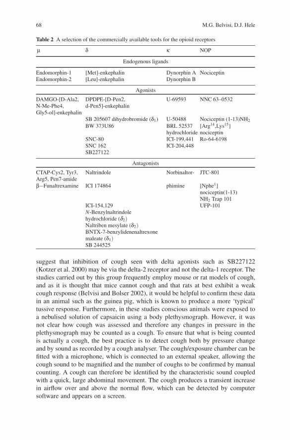
68 M.G. Belvisi, D.J. Hele
Table 2 A selection of the commercially available tools for the opioid receptors
µ δ κ NOP
Endogenous ligands
Endomorphin-1 [Met]-enkephalin Dynorphin A NociceptinEndomorphin-2 [Leu]-enkephalin Dynorphin B
Agonists
DAMGO-[D-Ala2,N-Me-Phe4,Gly5-ol]-enkephalin
DPDPE-[D-Pen2,d-Pen5]-enkephalin
U-69593 NNC 63–0532
SB 205607 dihydrobromide (δ1) U-50488 Nociceptin (1-13)NH2BW 373U86 BRL 52537
hydrochloride[Arg14,Lys15]nociceptin
SNC-80 ICI-199,441 Ro-64-6198SNC 162 ICI-204,448SB227122
Antagonists
CTAP-Cys2, Tyr3,Arg5, Pen7-amide
Naltrindole Norbinaltor- JTC-801
β−Funaltrexamine ICI 174864 phimine [Nphe1]nociceptin(1-13)NH2 Trap 101
ICI-154,129 UFP-101N-Benzylnaltrindolehydrochloride (δ2)Naltriben mesylate (δ2)BNTX-7-benzylidenenaltrexonemaleate (δ1)SB 244525
suggest that inhibition of cough seen with delta agonists such as SB227122(Kotzer et al. 2000) may be via the delta-2 receptor and not the delta-1 receptor. Thestudies carried out by this group frequently employ mouse or rat models of cough,and as it is thought that mice cannot cough and that rats at best exhibit a weakcough response (Belvisi and Bolser 2002), it would be helpful to confirm these datain an animal such as the guinea pig, which is known to produce a more ‘typical’tussive response. Furthermore, in these studies conscious animals were exposed toa nebulised solution of capsaicin using a body plethysmograph. However, it wasnot clear how cough was assessed and therefore any changes in pressure in theplethysmograph may be counted as a cough. To ensure that what is being countedis actually a cough, the best practice is to detect cough both by pressure changeand by sound as recorded by a cough analyser. The cough/exposure chamber can befitted with a microphone, which is connected to an external speaker, allowing thecough sound to be magnified and the number of coughs to be confirmed by manualcounting. A cough can therefore be identified by the characteristic sound coupledwith a quick, large abdominal movement. The cough produces a transient increasein airflow over and above the normal flow, which can be detected by computersoftware and appears on a screen.

Cough Sensors. III. Opioid and Cannabinoid Receptors on Vagal Sensory Nerves 69
More recently, another endogenous opioid peptide, nociceptin/orphanin FQ,has also been discovered which has some homology to the dynorphin familybut lacks the N-terminal tyrosine residue, which is essential for binding classicalopioid receptors (see Table 1). Nociceptin binds to the G-protein-coupled opioidreceptor-like 1 receptor (NOP, formerly ORL1). NOP1 receptors are expressed incapsaicin-sensitive vagal sensory nerves innervating airways, and nociceptin ef-fectively inhibits tachykinin release from vagal C-fibres in guinea pig bronchi(Fischer et al. 1998; Corboz et al. 2001). Intravenous administration of nociceptinalso effectively inhibits in conscious animals cough reflexes evoked by mechanicalperturbation of the airways or by inhalation of capsaicin (McLeod et al. 2001; Bolseret al. 2001). Although nociceptin may act centrally to inhibit cough, observationsthat nociceptin can inhibit the function of vagal afferent nerves within the airwayindicate that it may also inhibit cough through a peripheral mechanism of action(Fischer et al. 1998; Corboz et al. 2001). In a more recent study, nociceptin ef-fectively inhibited acid-evoked cough in guinea pigs by nearly 70%. Acid (pH 5)increased intracellular free calcium in acutely dissociated vagal jugular ganglionicneurons. The acid-induced increase in intracellular calcium was inhibited by a selec-tive transient receptor potential vanilloid-1 antagonist, 5-iodoresiniferatoxin (1 µM,approximately 80% reduction). The inhibitory effect of 5-iodoresiniferatoxin onacid-induced increases in calcium was mimicked by nociceptin. In gramicidin-perforated patch-clamp recordings on airway-specific capsaicin-sensitive jugularganglion neurons, acid (pH 5) induced two distinct inward currents. A transientcurrent was evoked that was inhibited by amiloride, and a sustained current wasevoked that was inhibited by 5-iodoresiniferatoxin. Nociceptin selectively inhibitedonly the sustained component of acid-induced inward current, suggesting that theinhibitory effect of nociceptin on acid-induced cough may result from a direct in-hibitory effect on peripheral C-fibre activity caused by the selective inhibition ofacid-induced transient receptor potential vanilloid-1 activation (Lee et al. 2006). Agroup at Hoffmann-La Roche reported on the selective, non-peptide NOP agonistRo 64-6198, which became the most extensively reported non-peptide NOP ago-nist and a valuable pharmacological tool in determining the potential of the NOPreceptor as a therapeutic target. Ro 64-6198 is systemically active and achieves highbrain penetration. It has subnanomolar affinity for the NOP receptor and is at least100 times more selective for the NOP receptor over the classic opioid receptors. Ro64-6198 ranges from partial to full agonist, depending on the assay. Preclinical dataindicate that Ro 64-6198 may have broad clinical uses, such as in treating stress andanxiety, addiction, neuropathic pain, cough and anorexia (Shoblock 2007).
It is now recognised that dextromethorphan, a commonly used non-narcotic an-titussive, has a binding Ki of 15 nM at the sigma receptor. Dextromethorphan ex-hibits a dose-related antitussive activity in a guinea pig model of cough (Callawayet al. 1991; Braga et al. 1994), and it is reduced by treatment with rimcazole, a sigmareceptor antagonist (Kamei et al. 1993).
In summary, opioid receptors have a role to play in the cough reflex, and by inter-fering with them with opioid agonists, we may be able to alleviate cough in animalmodels and in man. This does not inform us as to where the opioids are exerting theireffects. We know that their receptors are sited both centrally at sites in the brain or

70 M.G. Belvisi, D.J. Hele
brainstem and peripherally in nerves that innervate the airways. However, opioidscould be exerting their effects at either site or to varying degrees at both sites. Inanimal models and man the extent to which opioids cross the blood brain barrier isnot known, although the fact that they produce centrally mediated effects such assedation, drowsiness and euphoria confirms that they do cross the blood–brain bar-rier. The cough centre in the brain has been tentatively identified, and it is probablethat drugs like the opioids exert their antitussive actions both centrally on brainstemopioid receptors and peripherally on receptors located on sensory nerve endings inthe airways. We have little knowledge of where their specific sites of action resideand, if their site of action is central, we cannot say where in the physiological com-plex of the cough centre it resides. The signal transduction mechanism of action bywhich opioid receptors inhibit sensory nerve activation and thereby the cough reflexis not yet known; however, at the neuronal level, mu-opioid and delta-opioid recep-tor activation causes a decrease in adenylyl cyclase activity, which in turn openspotassium channels (hyperpolarisation) and closes calcium channels. Both these ac-tions may result in decreased sensory nerve activity, thereby suppressing the coughreflex (Pleuvry 2005). Despite these gaps in our knowledge, opioids will remainthe mainstay of cough treatment until novel treatments are developed that possessimproved efficacy and an improved side-effect profile.
3 Cannabinoid Receptors
Currently there is renewed interest in the therapeutic potential of cannabinoids asantitussives (Chung 2005). Non-selective cannabinoids have been shown to havewide therapeutic applications for a number of important medical conditions, includ-ing pain, anxiety, glaucoma, nausea, emesis, muscle spasms and wasting diseases;however, associated side effects such as sedation, cognitive dysfunction, tachycar-dia and psychotropic effects have hampered the use of these compounds in the clinic(Porter and Felder 2001).
Cannabinoids mediate their effects via at least two specific G-protein-coupled re-ceptors, termed the CB1 and CB2 receptors (Matsuda et al. 1990; Munro et al. 1993)(see Table 3 for a list of selective ligands). The cannabinoid receptors were namedbecause of their affinity for the agonist δ9-tetrahydrocannabinol, a ligand found inorganic extracts from Cannabis sativa (Howlett 2002). CB1 receptors are predom-inantly distributed throughout the brain and spinal cord and are found mainly atthe terminals of central neurones and are also expressed at low levels in severalperipheral tissues. There is evidence that these receptors can mediate inhibition ofongoing release of a number of different excitatory and inhibitory transmitters, in-cluding acetylcholine, noradrenaline, dopamine and 5-hydroxytryptamine (Howlettet al. 2002). It is therefore thought that an important role of the CB1 receptor is tomodulate neurotransmitter release in order to maintain homeostasis in health anddisease by preventing the development of excessive neuronal activity in the CNS(Pertwee 2007). In contrast, CB2 receptors are not commonly expressed in theCNS (Munro et al. 1993; Griffin et al. 1997; Buckley et al. 2000), but primarily on
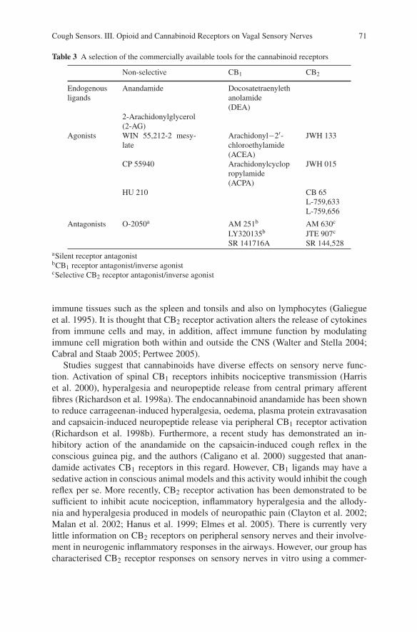
Cough Sensors. III. Opioid and Cannabinoid Receptors on Vagal Sensory Nerves 71
Table 3 A selection of the commercially available tools for the cannabinoid receptors
Non-selective CB1 CB2
Endogenousligands
Anandamide Docosatetraenylethanolamide(DEA)
2-Arachidonylglycerol(2-AG)
Agonists WIN 55,212-2 mesy-late
Arachidonyl−2′-chloroethylamide(ACEA)
JWH 133
CP 55940 Arachidonylcyclopropylamide(ACPA)
JWH 015
HU 210 CB 65L-759,633L-759,656
Antagonists O-2050a AM 251b AM 630c
LY320135b JTE 907c
SR 141716A SR 144,528aSilent receptor antagonistbCB1 receptor antagonist/inverse agonistcSelective CB2 receptor antagonist/inverse agonist
immune tissues such as the spleen and tonsils and also on lymphocytes (Galiegueet al. 1995). It is thought that CB2 receptor activation alters the release of cytokinesfrom immune cells and may, in addition, affect immune function by modulatingimmune cell migration both within and outside the CNS (Walter and Stella 2004;Cabral and Staab 2005; Pertwee 2005).
Studies suggest that cannabinoids have diverse effects on sensory nerve func-tion. Activation of spinal CB1 receptors inhibits nociceptive transmission (Harriset al. 2000), hyperalgesia and neuropeptide release from central primary afferentfibres (Richardson et al. 1998a). The endocannabinoid anandamide has been shownto reduce carrageenan-induced hyperalgesia, oedema, plasma protein extravasationand capsaicin-induced neuropeptide release via peripheral CB1 receptor activation(Richardson et al. 1998b). Furthermore, a recent study has demonstrated an in-hibitory action of the anandamide on the capsaicin-induced cough reflex in theconscious guinea pig, and the authors (Caligano et al. 2000) suggested that anan-damide activates CB1 receptors in this regard. However, CB1 ligands may have asedative action in conscious animal models and this activity would inhibit the coughreflex per se. More recently, CB2 receptor activation has been demonstrated to besufficient to inhibit acute nociception, inflammatory hyperalgesia and the allody-nia and hyperalgesia produced in models of neuropathic pain (Clayton et al. 2002;Malan et al. 2002; Hanus et al. 1999; Elmes et al. 2005). There is currently verylittle information on CB2 receptors on peripheral sensory nerves and their involve-ment in neurogenic inflammatory responses in the airways. However, our group hascharacterised CB2 receptor responses on sensory nerves in vitro using a commer-

72 M.G. Belvisi, D.J. Hele
cially available cannabinoid CB2 receptor agonist, JWH 133. In these studies, it wasdemonstrated that activation of the CB2 receptor subtype inhibits capsaicin-induceddepolarisation of the guinea pig and human vagus (Patel et al. 2003). Moreover, ithas also been demonstrated that inhibition of sensory nerve function in vivo, alsowith the CB2 agonist JWH 133, results in antitussive action in a conscious guineapig model of cough (Patel et al. 2003). In summary, activation of the CB2 receptorsubtype inhibits sensory nerve activation of guinea pig and human vagus nerve, andimportantly it has been shown to inhibit the cough reflex in conscious guinea pigs.CB2 receptors have been shown to be primarily sited in the periphery, with littleevidence for their presence in the CNS. This evidence offers the hope that the devel-opment of CB2 agonists, devoid of CB1-mediated central effects, may provide newand safe antitussive treatments for cough.
It is not clear yet how cannabinoid receptors may inhibit sensory nerve acti-vation; however, both CB1 and CB2 receptors are coupled through Gi/o proteins,negatively to adenylyl cyclase and positively to mitogen-activated protein kinase(Howlett et al. 2002). Furthermore, CB1 receptors are also thought to be coupledto ion channels through Gi/o proteins, positively to A-type and inwardly rectify-ing potassium channels and negatively to N-type and P/Q-type calcium channels
airway sensory nerve
CoughCoughChest tightnessChest tightness
AHR?AHR?Cholinergic Cholinergic Reflex activationReflex activation
Mucus secretionMucus secretion
Inflammation
µ Opioidsδ OpioidsNociceptin agonistsCannabinoid CB2 agonists
TRPV-1
−
+
Capsaicin, low pH
Fig. 1 An airway sensory nerve ending, which can be activated by capsaicin and low-pH stimulito evoke cough, airway hyperresponsiveness, mucus secretion and cholinergic reflex activation. Ininflammatory conditions, these nerves may be sensitised so that they respond at lower thresholds.Opioids, nociceptin and cannabinoid ligands have all been postulated to exert an inhibitory effecton sensory nerve activation

Cough Sensors. III. Opioid and Cannabinoid Receptors on Vagal Sensory Nerves 73
(Howlett 2002; Pertwee 1997). Details of additional signalling mechanisms havealso been documented for cannabinoid CB1 and CB2 receptors (Howlett et al. 2002).
4 Summary
Opioid receptors and cannabinoid receptors have been characterised and intensivelystudied. They occur both in the brain or brainstem and in the peripheral tissues,including the airways (Fig. 1). The exact site of these receptors is largely unknown.Cough has long been identified as a debilitating symptom of various airway diseasesand can also exist without any obvious cause. Cough is mediated by receptorson sensory nerves sited in the airways via vagal afferent nerves with terminals inthe centrally located cough centres. Current treatment for cough is largely unsatis-factory, consisting of over-the-counter cough medicines with limited efficacy andprescription-based opioids, which also have limited efficacy and cause undesirableside effects (Chung 2007a, b). The cannabinoids are a class of compounds that arebeing evaluated in an attempt to provide a viable antitussive treatment, which willtarget peripheral cannabinoid receptors and be free of the centrally mediated side ef-fects that are also associated with this class of compounds. Some degree of successhas been achieved towards this end but further work needs to be carried out to clar-ify the receptors involved and to determine whether the cannabinoids will provide aviable alternative to opioids in the clinic.
References
Adcock JJ (1991) Peripheral opioid receptors and the cough reflex. Respir Med 85 (Suppl A):43–46Ambalavanar R, Tanaka Y, Damirjian M et al. (1999) Laryngeal afferent stimulation enhances Fos
immunoreactivity in periaqueductal grey in the cat. J Comp Neurol 409:411–423Atweh SF, Kuhar MJ (1977) Autoradiographic localization of opiate receptors in rat brain. II. The
brain stem. Brain Res 129:1–12Atweh SF, Murrin LC, Kuhar MJ (1978) Presynaptic localization of opiate receptors in the vagal
and accessory optic systems: an autoradiographic study. Neurophamacology 17:65–71Aylward M, Maddock J, Davies DE et al. (1984) Dextromethorphan and codeine: comparison of
plasma kinetics and antitussive effects. Eur J Respir Dis 65:283–291Baekey DM, Morris K, Gestreau C et al. (2001) Medullary respiratory neurones and control of
laryngeal motoneurones during fictive eupnoea and cough in the cat. J Physiol 534:565–581Barnes PJ (2001) Neurogenic inflammation in the airways. Respir Physiol 125:145–154Belvisi MG (2002) Overview of the innervation of the lung. Curr Opin Pharmacol 2:211–215Belvisi MG, Bolser D (2002) Summary: animal models for cough. Pulm Pharmacol Ther
15(3):249–250Belvisi MG, Geppetti P (2004) Cough 7: current and future drugs for the treatment of chronic
cough. Thorax 59(5):438Belvisi MG, Chung KF, Jackson DM et al. (1988) Opioid modulation of non-cholinergic neural
bronchoconstriction in guinea-pig in vivo. Br J Pharmacol 95(2):413–418

74 M.G. Belvisi, D.J. Hele
Bhargava HN, Villar VM, Cortijo J et al. (1997) Binding of [3H][D-Ala2, MePhe4, Gly-ol5]enkephalin, [3H][D-Pen2, D-Pen5]enkephalin, and [3H]U-69,593 to airway and pulmonarytissues of normal and sensitized rats. Peptides 18:1603–1608
Bolser DC, McLeod RL, Tulshian DB, Hey JA (2001) Antitussive action of nociceptin in the cat.Eur J Pharmacol 430:107–111
Braga PC, Fossati A, Vimercati MG, Caputo R, Guffanti EE (1994) Dextrorphan and dextromethor-phan: comparative antitussive effects on guinea pigs. Drugs Exp Clin Res 20:199–203
Buckley NE, McCoy KL, Mezey E et al. (2000) Immunomodulation by cannabinoids is absent inmice deficient for the cannabinoid CB2 receptor. Eur J Pharmacol 396:141–149
Cabot PJ, Crammond T, Smith MT (1996) Quantitative autoradiography of peripheral opioid bind-ing sites in rat lung. Eur J Pharmacol 310:47–53
Cabot PJ, Dodd PR, Crammond T et al. (1994) Characterization of non-conventional opioid bind-ing sites in rat and human lung. Eur J Pharmacol 268(2):247–255
Cabral GA, Staab A (2005) Effects on the immune system. In: Pertwee RG (ed) Cannabinoids.Handbook of experimental pharmacology, vol. 168. Springer, Berlin, pp 385–423
Caligano A, Katano I, Desarnaud F et al. (2000) Bidirectional control of airway responsiveness byendogenous cannabinoids. Nature 408(6808):96–101
Callaway JK, King RG, Boura AL (1991) Evidence for peripheral mechanisms mediating the an-titussive actions of opioids in the guinea pig. Gen Pharmacol 22(6):1103–1108
Canning BJ (2007) Encoding of the cough reflex. Pulm Pharmacol Ther 20:396–401Canning BJ, Nori N, Mazzone SB (2006) Vagal afferent nerves regulating the cough reflex. Respir
Physiol Neurobiol 152(3):223–242Carr MJ, Lee LY (2006) Plasticity of peripheral mechanisms of cough. Respir Physiol Neurobiol
152(3):298–311Chung KF (2005) Drugs to suppress cough. Expert Opin Investig Drugs 14(1):19–27Chung KF (2007a) Review series: chronic cough: future directions in chronic cough: mechanisms
and antitussives. Chron Respir Dis 4(3):159–165Chung KF (2007b) Effective antitussives for the cough patient an unmet need. Pulm Pharmacol
Ther 20(4):438–445Clayton N, Marshall FH, Bountra C et al. (2002) CB1 and CB2 cannabinoid receptors are impli-
cated in inflammatory pain. Pain 96:253–260Corboz MR, Fernandez X, Egan RW, Hey JA (2001) Inhibitory activity of nociceptin/orphanin FQ
on capsaicin-induced bronchoconstriction in the guinea-pig. Life Sci 69:1203–1211Dashwood MR, Muddle JR, Spyer KM (1988) Opiate receptor subtypes in the nucleus tractus
solitarii of the cat: the effect of vagal section. Eur J Pharmacol 155(1–2):85–92Eccles R (2002) The powerful placebo in cough studies? Pulm Pharmacol Ther 15(3):303–308Elmes SJ, Winyard LA, Medhurst SJ et al. (2005) Activation of CB1 and CB2 receptors attenuates
the induction and maintenance of inflammatory pain in the rat. Pain 118:327–335Fischer A, Forssmann WG, Undem BJ. (1998). Nociceptin-induced inhibition of tachykinergic
neurotransmission in guinea pig bronchus. J Pharmacol Exp Ther 285:902–907Fox B, Bull TB, Guz A (1980) Innervation of alveolar walls in the human lung: an electron micro-
scopic study. J Anat 131:683–692Freestone C, Eccles R (1997) Assessment of the antitussive efficacy of codeine in cough associated
with common cold. J Pharm Pharmacol 49:1045–1049Frossard N, Barnes PJ (1987) Mu-opioid receptors modulate non-cholinergic constrictor nerves in
guinea-pig airways. Eur J Pharmacol 141(3):519–522Galiegue S, Mary S, Marchland J et al. (1995) Expression of central and peripheral cannabinoid
receptors in human immune tissues and leukocyte subpopulations. Eur J Biochem 232:54–61Gestreau C, Bianchi AL, Grelot L (1997) Differential brainstem Fos-like immunoreactivity after
laryngeal-induced coughing and its reduction by codeine. J Neurosci 17:9340–9352Griffin G, Fernando SR, Ross RA et al. (1997) Evidence for the presence of CB2 like cannabinoid
receptors on peripheral nerve terminals. Eur J Pharmacol 339:53–61Gundlach A L, Largent BL, Snyder SH (1986) Phencyclidine (PCP) receptors: autoradiographic
localization in brain with the selective ligand, [3H]TCP. Brain Res 386:266–279

Cough Sensors. III. Opioid and Cannabinoid Receptors on Vagal Sensory Nerves 75
Hanus L, Breuer A, Tchilibon S et al. (1999) HU-308: a specific agonist for CB(2), a peripheralcannabinoid receptor. Proc Natl Acad Sci USA 96:14228–14333
Harris J, Drew LJ, Chapman V (2000) Spinal anandamide inhibits nociceptive transmission viacannabinoid receptor activation in vivo. Neuroreport 11:2817–2819
Higenbottam T (2002) Chronic cough and the cough reflex in common lung diseases. Pulm Phar-macol Ther 15(3):241–247
Howlett AC (2002) The cannabinoid receptors. Prostaglandins Other Lipid Mediat 68–69:619–631Howlett AC, Barth F, Bonner TI, Cabral G, Casellas P, Devane WA, Felder CC, Herkenham M,
Mackie K, Martin BR, Mechoulam R, Pertwee RG (2002) International Union of Pharmacol-ogy. XXVII. Classification of cannabinoid receptors. Pharmacol Rev 54:161–202
Hughes J, Smith TW, Kosterlitz et al. (1975) Identification of two related pentapeptides from thebrain with potent opiate agonist activity. Nature 258:577–580
Jiang Q, Takemori AE, Sultana M et al. (1991) Differential antagonism of opioid delta antinoci-ception by [D-Ala2,Leu5,Cys6]enkephalin and naltrindole 5′-isothiocyanate: evidence for deltareceptor subtypes. J Pharmacol Exp Ther 257(3):1069–1075
Kamei J (2002) Delta-opioid receptor antagonists as a new concept for central acting antitussivedrugs. Pulm Pharmacol Ther 15(3):235–240
Kamei J, Tanihara H, Kasuya Y (1990) Antitussive effects of two specific kappa-opioid agonists,U-50,488H and U-62,066E, in rats. Eur J Pharmacol 187:281–286
Kamei J, Iwamoto Y, Misawa M, Kasuya Y (1993) Effects of rimcazole, a specific antagonistof sigma sites, on the antitussive effects of non-narcotic antitussive drugs. Eur J Pharmacol242:209–211
Karlsson JA, Lanner AS, Persson CG (1990) Airway opioid receptors mediate inhibition of coughand reflex bronchoconstriction in guinea pigs. J Pharmacol Exp Ther 252:863–868
Kastelik JA, Aziz I, Ojoo JC et al. (2005) Investigation and management of chronic cough using aprobability-based algorithm. Eur Respir J 25(2):235–243
Kotzer CJ, Haw DW, Dondio G et al. (2000) The antitussive activity of delta-opioid stimulation inguinea pigs. J Pharmacol Exp Ther 292(2):803–809
Lee MG, Undem BJ, Brown C, Carr MJ. (2006) Effect of nociceptin in acid-evoked cough andairway sensory nerve activation in guinea pigs. Am J Respir Crit Care Med 173(3):271–275
Lord JA, Waterfield AA, Hughes J et al. (1977) Endogenous opioid peptides: multiple agonists andreceptors. Nature 267:495–499
Malan TP, Ibrahim MM, Vanderah TW et al. (2002) Inhibition of pain responses by activation ofCB2 cannabinoid receptors. Chem Phys Lipids 121:191–200
Martin WR, Eades CG, Thompson JA et al. (1976) The effects of morphine- and nalorphine-likedrugs in nondependent and morphine-dependent chronic spinal dog. J Pharmacol Exp Ther197:517–532
Matsuda LA, Lolait SJ, Brownstein MJ et al. (1990) Structure of a cannabinoid receptor and func-tional expression of the cloned cDNA. Nature 346:561–564
McLeod RL, Parra LE, Mutter JC, Erickson CH, Carey GJ, Tulshian DB, Fawzi AB,Smith-Torhan A, Egan RW, Cuss FM et al. (2001) Nociceptin inhibits cough in the guinea-pig by activation of ORL1 receptors. Br J Pharmacol 132:1175–1178
Merick B, Reid L (1971) Nerves in rat intra-acinar alveoli: an electron microscope study. RespirPhysiol 11:367–377
Morice AH, Menon MS, Mulrennan SA et al. (2007) Opiate therapy in chronic cough. Am J RespirCrit Care Med 175(4):312–315
Munro S, Thomas KL, Abu-Shaar M (1993) Molecular characterisation of a peripheral receptorfor cannabinoids. Nature 365:61–65
Ohi Y, Yamazaki H, Takeda R et al. (2005) Functional and morphological organization of thenucleus tractus solitarius in the fictive cough reflex of guinea pigs. Neurosci Res 53(2): 201–209
Patel HJ, Birrell MA, Crispino N et al. (2003) Inhibition of guinea pig and human sensory nerveactivity and the cough reflex in guinea pigs by cannabinoid (CB2) receptor activation. Br JPharmacol 140:261–268

76 M.G. Belvisi, D.J. Hele
Pertwee RG (1997) Pharmacology of cannabinoid CB1 and CB2 receptors. Pharmacol Ther74(2):129–180
Pertwee RG (2005) Pharmacological actions of cannabinoids. In: Pertwee RG (ed). Cannabinoids.Handbook of experimental pharmacology, vol. 168. Springer, Berlin, pp 1–51
Pertwee RG (2007) The diverse CB1 and CB2 receptor pharmacology of three plant cannabinoids:D9-tetrahydrocannabinol, cannabidiol and D9-tetrahydrocannabivarin. Br J Pharmacol (Sep 10Epub ahead of print)
Pleuvry BJ (2005) Opioid mechanisms and opioid drugs. Anaesth Intensive Care Med 6(1):30–34Porter AC, Felder CC (2001) The endocannabinoid nervous system: unique opportunities for ther-
apeutic intervention. Pharmacol Ther 90:45–60Richardson JD, Aanonsen L, Hargreaves KM (1998a) Antihyperalgesic effects of spinal cannabi-
noids. Eur J Pharmacol 345:145–153Richardson JD, Kilo S, Hargreaves KM (1998b) Cannabinoids reduce hyperalgesia and inflamma-
tion via interaction with peripheral CB1 receptors. Pain 75:111–119Schroeder K, Fahey T (2002) Systematic review of randomised controlled trials of over the counter
cough medicines for acute cough in adults. Br Med J 324:1–6Sessle BJ, Ball GJ, Lucier GE (1981) Suppressive influences from periaqueductal gray and nucleus
raphe magnus on respiration and related reflex activities and on solitary tract neurons, and effectof naloxone. Brain Res 216(1):145–161
Sevelius H, Colmore JP (1966) Objective assessment of antitussive agents in patients with chroniccough. J New Drugs 6:216–223
Sevelius H, McCoy JF, Colmore JP (1971) Dose response to codeine in patients with chroniccough. Clin Pharmacol Ther 12:449–455
Shoblock JR (2007) The pharmacology of Ro 64-6198, a systemically active, nonpeptide NOPreceptor (opiate receptor-like 1, ORL-1) agonist with diverse preclinical therapeutic activity.CNS Drug Rev (Spring) 13(1):107–136
Smith J, Owen E, Earis J et al. (2006) Effect of codeine on objective measurement of cough inchronic obstructive pulmonary disease. J Allergy Clin Immunol 117:831–835
Takahama K, Shirasaki T (2007) Central and peripheral mechanisms of narcotic antitussives:codeine-sensitive and -resistant coughs. Cough 3:1–8
Undem BJ, Kollarik M (2005) The role of vagal afferent nerves in chronic obstructive pulmonarydisease. Proc Am Thorac Soc 2(4):355–360
Walter L, Stella N (2004) Cannabinoids and neuroinflammation. Br J Pharmacol 141:775–785Widdicombe JG, Chung KF (2007) Cough. Pulm Pharmacol Ther 20(4):305–306

Cough Sensors. IV. Nicotinic MembraneReceptors on Cough Sensors
L.-Y. Lee(�) and Q. Gu
Contents
1 Cough Evoked by Inhaled Cigarette Smoke . . . . . . . . . . . . . . . . . . . . . . . . . . . . . . . . . . . . . . 781.1 Role of Nicotine in Cigarette Smoke-Induced Airway Irritation . . . . . . . . . . . . . . . . 781.2 Cough Receptors in the Lung and Airways . . . . . . . . . . . . . . . . . . . . . . . . . . . . . . . . . 811.3 Nicotine-Sensitive Afferents in the Lung and Airways . . . . . . . . . . . . . . . . . . . . . . . 82
2 Neuronal Nicotinic Acetylcholine Receptors on Pulmonary Sensory Neurons . . . . . . . . . . 852.1 Pharmacological Properties of NnAChRs . . . . . . . . . . . . . . . . . . . . . . . . . . . . . . . . . . 862.2 Nicotine Sensitivity in Isolated Pulmonary Sensory Neurons . . . . . . . . . . . . . . . . . . 872.3 Expression of NnAChRs on Pulmonary Sensory Neurons . . . . . . . . . . . . . . . . . . . . . 88
3 Other Physiological Functions of NnAChRs on Cough Sensors . . . . . . . . . . . . . . . . . . . . . . 903.1 Other Physiological Responses Evoked by Activation of NnAChRs
on Pulmonary Afferents . . . . . . . . . . . . . . . . . . . . . . . . . . . . . . . . . . . . . . . . . . . . . . . . . 903.2 Interaction with Other Ion Channels Expressed on Cough Sensors . . . . . . . . . . . . . 91
4 Conclusion . . . . . . . . . . . . . . . . . . . . . . . . . . . . . . . . . . . . . . . . . . . . . . . . . . . . . . . . . . . . . . . . . 92References . . . . . . . . . . . . . . . . . . . . . . . . . . . . . . . . . . . . . . . . . . . . . . . . . . . . . . . . . . . . . . . . . . . . . 94
Abstract Cigarette smoke is undoubtedly one of the most common inhaled irritantsin the human respiratory tract, and invariably evokes coughing in both smokers andnonsmokers. Results obtained from the studies in human volunteers and from single-fiber recording of vagal bronchopulmonary afferents in animals clearly indicate thatnicotine is primarily responsible for the airway irritation and coughing caused byinhalation of cigarette smoke. Furthermore, both nicotine and acetylcholine canevoke inward current, membrane depolarization, and action potentials in isolatedpulmonary sensory neurons, and these responses are blocked by hexamethonium.Taken together, these findings suggest that the tussive effect of nicotine is probablymediated through an activation of nicotinic acetylcholine receptors (nAChRs) ex-pressed on the sensory terminals of cough receptors located in the airway mucosa.Indeed, the expressions of α4−α7 and β2−β4 subunits of nAChR transcripts in
L.-Y. LeeDepartment of Physiology, University of Kentucky, Lexington, KY 40536-0298, [email protected]
K.F. Chung, J.G. Widdicombe (eds.), Pharmacology and Therapeutics of Cough, 77Handbook of Experimental Pharmacology 187,c© Springer-Verlag Berlin Heidelberg 2009

78 L.-Y. Lee, Q. Gu
pulmonary sensory neurons have lent further support to this conclusion. The specificsubtypes of the neuronal nAChRs and their subunit compositions expressed on thecough sensors remain to be determined.
1 Cough Evoked by Inhaled Cigarette Smoke
One of the most important functions of the cough reflex is to protect the lung andthe rest of body from the hazardous effects of inhaled irritants and environmen-tal air pollutants. There is a long list of common inhaled irritants known to elicitcough reflex in humans, including tobacco smoke, dust, automobile exhausts, acidaerosols, noxious gases, allergens, and a variety of airborne chemical irritants. Theseirritant substances can activate cough sensors either by directly acting on certain ionchannels or receptor proteins expressed on the membrane of sensory terminals orindirectly via an action on other target cells in the airways (e.g., bronchial smoothmuscles, immune cells such as macrophages or mast cells, etc.) which can in turnevoke stimulatory effects on the cough sensors. Among these tussive substances,cigarette smoke is undoubtedly one of the most common inhaled irritants in humanairways.
In nonsmokers, inhaling only a small puff of cigarette smoke can evoke vigorouscough responses and airway irritation. Similar irritant effects can also be gener-ated by brief exposure to the side-stream smoke, the smoke released from a smol-dering cigarette (Tarlo 2006). In smokers, cigarette smoking is the major cause ofchronic cough (Braman 2006). Indeed, cough not only serves as an important de-fense function, but is also a common symptom of various airway diseases associatedwith chronic smoking (Braman 2006); in conjunction with the mucociliary system,cough can expel from the respiratory tract the cigarette smoke constituents that aredeposited in the airway lumen as well as the excessive airway secretion producedby smoke inhalation.
1.1 Role of Nicotine in Cigarette Smoke-Induced Airway Irritation
Despite the well-documented irritant effects of inhaled cigarette smoke, the under-lying mechanism and smoke constituent(s) that are responsible for generating theairway irritation and cough were not fully understood. In addition to cough, in-haled cigarette smoke can also elicit reflex bronchoconstriction. For a long time,it was suggested that particulates in the cigarette smoke were primarily responsi-ble for the irritant effects and for eliciting reflex bronchoconstriction (Nadel andComroe 1961). This notion appeared to be consistent with the finding that the ir-ritant receptors in the lung could be activated not only by cigarette smoke butalso by “inert” carbon dust (Sellick and Widdicombe 1971). On the other hand,nicotine, a major constituent of cigarette smoke, was known to activate sensory

Cough Sensors. IV. Nicotinic Membrane Receptors on Cough Sensors 79
receptors in various organs, including skin, mesentery, stomach, lung, and carotidbody (Douglas 1952; Douglas and Gray 1953; Paintal 1954, 1955). Whether nico-tine delivered in the smoke could activate cough sensors was not known.
In a psychometric study of the role of nicotine in cigarette smoking, Henningfieldet al. (1985) found that nicotine administered by intravenous injection causedcoughing and airway irritation in human smokers. Whether the irritant effects re-ported in that study were due to an excitatory effect of nicotine on cough receptors inthe airways and lung was not known since nicotine injected via the intravenous routemay also activate other sensory receptors that can modulate respiratory responses(e.g., cardiac afferents, carotid body chemoreceptors, etc.); a similar circulatory lagfor the injected nicotine to reach airway receptors (via bronchial circulation) andthese other receptors located in the systemic circulation made it difficult to identifythe origin of its irritant effect.
In a study aimed at determining the role of nicotine in cigarette smoke-evokedairway irritation, the responses to inhalation of a single puff of cigarette smokewith different nicotine content were compared in healthy young nonsmokers (Leeet al. 1993). After the upper airways had been locally anesthetized and the view ofthe smoke delivery system had been blocked, the subjects breathed quietly througha mouthpiece when a bolus of cigarette smoke of 30-ml volume (approximatelythe average puff volume of smokers) was delivered without notice and immediatelyinhaled at the front portion of the next inspired volume, mimicking the smoke in-halation pattern in smokers (Tobin and Sackner 1982). Subjects were instructed tosignal the detection and the intensity of airway irritation with a push-button device.In eight of the 12 subjects, inhalation of high-nicotine cigarette smoke immedi-ately triggered bouts of coughs (total number of coughs 4.13 ± 0.64). By contrast,low-nicotine (0.38± 0.26; P < 0.001) and gas-phase smoke (0± 0) did not causecough in the same subjects (Fig. 1a). The intensity of airway irritation measuredby the total number of push-button signals after inhalation of high-nicotine ciga-rette smoke was 6.61± 0.87 (n = 12), whereas inhalation of low-nicotine and gas-phase smoke either was not detected or caused only very mild irritation (0.89±0.4and 0.36± 0.22, respectively). Furthermore, the intensities of airway irritation andcoughs evoked by high-nicotine smoke were markedly reduced after premedica-tion with aerosolized hexamethonium (10% in isotonic saline), a nicotinic acetyl-choline receptor antagonist; number of coughs was 4.75±1.02 after administrationof a placebo, and 1.26± 0.28 after administration of hexamethonium (P < 0.001,n = 8; Fig. 1b). The irritant effect was clearly attenuated but not completely abol-ished, largely owing to a marginal blocking effect of the hexamethonium in two ofthe eight subjects. However, after a higher concentration (20%) of hexamethoniumaerosol had been administered in these two subjects, the smoke-evoked airway irri-tation and cough were completely blunted (Lee et al. 1993). In addition, inhalationof aerosolized nicotine (2% in isotonic saline) also evoked vigorous coughs (totalnumber of coughs 7.36±1.68, n = 5) and intense airway irritation (Fig. 1c). Takentogether, these studies have provided strong evidence indicating that nicotine is theprimary agent in cigarette smoke-evoked cough and airway irritation in nonsmok-ers. The short latency time of the detection signal (0.93± 0.11s) further suggested

80 L.-Y. Lee, Q. Gu
Fig. 1 a Airway irritation and number of coughs evoked by inhalation of three different types ofcigarette smoke. The intensity of airway irritation was measured by the number of push-buttonsignals generated in the first 5 s following inhalation of a puff of cigarette smoke. Data representthe mean ± the standard error of the mean (SEM) of 12 subjects; data obtained from three consec-utive days were averaged for each subject. *, significantly different (P < 0.001) from the responseto high-nicotine cigarette smoke. b A comparison of airway irritation and cough responses to in-halation of high-nicotine cigarette smoke after premedications with aerosolized saline placebo andhexamethonium (10% concentration). † and *, significant difference (P < 0.01 and P < 0.001,respectively; n = 8) between the responses after hexamethonium and placebo administration.c Airway irritation and number of coughs evoked by inhalation of aerosolized nicotine or itsvehicle. *, significant difference (P < 0.001; n = 5) between the responses to nicotine and vehicle.(Modified from Lee et al. 1993, with permission)

Cough Sensors. IV. Nicotinic Membrane Receptors on Cough Sensors 81
that the sensory receptors involved in detecting the irritant effect of smoke and elic-iting the cough reflex are located in the respiratory tract and the afferent signals areprobably conducted by vagus nerves.
1.2 Cough Receptors in the Lung and Airways
The “cough receptor” that initiates the reflex response consists of multiple typesof airway afferents with different sensory modalities (chemical vs. mechanical),as characterized by different types of receptor proteins expressed on the neuronalmembrane of the sensory endings (Geppetti 2008; Belvisi 2008; Mazzone andUndem 2008). These cough receptors are located in different regions of the res-piratory tract, including larynx and tracheobronchial tree. The anatomical loca-tions, neural pathways, and pharmacological properties of the cough receptors aredescribed in more detail elsewhere (Canning 2008). The afferent activities that arisefrom various cough sensors located in the airways are conducted almost exclusivelyby branches of vagus nerves (Coleridge and Coleridge 1986; Lee and Pisarri 2001;Sant’Ambrogio 1982; Widdicombe 1998). These vagal afferent fibers innervate theentire respiratory tract diffusively, and project to the “cough centers” located in themedulla (Takahama 2008).
On the basis of the general properties and classically defined criteria, vagal affer-ents arising from endings located in the lower airways and lungs can be classifiedinto three major categories: slowly adapting receptors (stretch receptors), rapidlyadapting receptors (RARs or irritant receptors), and bronchopulmonary C-fibers(Coleridge and Coleridge 1986; Widdicombe 1981). Afferent activities generatedfrom the first two types of receptors are conducted by myelinated A-fibers, whereasthose generated by the last one are conducted by nonmyelinated (C-) fibers. Formany decades, investigators have been searching for the identities and classificationsof a subgroup(s) of sensory receptors in the airways that are specifically responsiblefor eliciting the cough reflex. RARs, particularly those located in the major airwaysand the carina, were believed to be primarily responsible for detecting the inhaled ir-ritants and signaling the central nervous system to elicit the cough response in largermammals (e.g., dogs, cats) (Widdicombe 1998). The evidence in support of an im-portant role of bronchopulmonary C-fiber afferents in eliciting the cough reflex hasalso been extensively documented. Stimulation of C-fiber afferents by inhalation ofcapsaicin or acid aerosol consistently produces coughs in a dose-dependent man-ner in both animals and humans (Bolser et al. 1995; Dicpinigaitis 2007; Groneberget al. 2004; Hansson et al. 1992; Karlsson and Fuller 1999; Liu et al. 2001; Winninget al. 1986). Recent studies by Canning et al. (2004) and Mazzone (2004) havedemonstrated that the cough reflex elicited by the upper-airway irritation in guineapigs seems to be mediated through the activation of a subset of Aδ afferents inner-vating the trachea and larynx. These cough sensors are activated by acid solutionand punctate mechanical stimulation applied to the upper airways. However, theyare not activated by either capsaicin or bradykinin, and are, therefore, unlikely to be

82 L.-Y. Lee, Q. Gu
involved in eliciting the capsaicin-evoked cough reflex. Furthermore, they appear tobe different from the RARs in the airways because they conduct action potentials ata much slower velocity than RARs, and they are not activated by either lung infla-tion or bronchoconstriction (Canning et al. 2004; Mazzone 2004). Whether they aresensitive to nicotine is not known.
It remains to be determined whether cough receptors belong to a subgroup ofthese classically defined bronchopulmonary afferents, or represent a distinctly dif-ferent group of airway afferents whose primary function is to detect tussive stim-uli. Furthermore, whether the cough receptor mediating the protective function inhealthy lungs against inhaled irritants such as cigarette smoke is also the same sen-sor that is responsible for triggering the acute or chronic cough in various airwaydiseases is also not known. Many of these important and yet unanswered questionshave been discussed in recent reviews and conference proceedings (Widdicombeand Chung 2004, 2007; Widdicombe and Undem 2006). This chapter will focussolely on the vagal bronchopulmonary afferents that exhibit sensitivity to nico-tine and may function as sensors for triggering the cigarette smoke-evoked coughresponses.
1.3 Nicotine-Sensitive Afferents in the Lung and Airways
1.3.1 Bronchopulmonary C-Fiber Afferents
Bronchopulmonary C-fibers represent more than 75% of the afferent fibers in thepulmonary branch of the vagus nerve (Agostoni et al. 1957; Jammes et al. 1982),and play an important role in regulating the airway functions in both physiologicaland pathological conditions (Coleridge and Coleridge 1984; Lee and Pisarri 2001).These afferents can be broadly subdivided into two major groups on the basisof their anatomic locations in the respiratory tract and circulatory accessibility(Coleridge and Coleridge 1984). Pulmonary C-fibers arise from the endings lo-cated in small intrapulmonary airways and the lung parenchyma, receiving bloodsupply from the pulmonary circulation, whereas bronchial C-fiber endings are lo-cated in the medium-sized and large airways, receiving blood perfusion primarilyfrom bronchial circulation. It appears that pulmonary C-fibers are more sensitive tomechanical stimuli (e.g., lung inflation), whereas bronchial C-fibers display greaterchemosensitivity (Coleridge and Coleridge 1984). A recent study has revealed thatbronchial and pulmonary C-fiber afferents arise from different ganglion origins andrepresent different phenotypes, which may offer a plausible explanation for the no-ticed difference in the sensory modality between these two groups of C-fibers (Un-dem et al. 2004). It was further suggested that stimulated bronchial C-fibers are morelikely to function as cough sensors than pulmonary C-fibers (Canning et al. 2006),but definite evidence remains to be established (Hansson et al. 1992).
In a study aimed to determine whether vagal bronchopulmonary C-fibers can beactivated by cigarette smoke, afferent activities of the vagal C-fibers arising from

Cough Sensors. IV. Nicotinic Membrane Receptors on Cough Sensors 83
endings in the lung were recorded using the single-fiber recording technique inanesthetized and mechanically ventilated dogs when cigarette smoke (120 ml) wasdelivered by the respirator in a single ventilator cycle (Lee et al. 1989). To deter-mine the role of nicotine, the effects of low- and high-nicotine cigarette smoke onthe same C-fiber afferents were compared; the volume of smoke generated fromthe low- and high-nicotine cigarettes was estimated to contain approximately 0.032and 0.162 mg of nicotine, respectively. The delivery of smoke generated by high-nicotine cigarettes triggered an abrupt and intense burst of activity in the major-ity (approximately 91%) of the pulmonary C-fibers tested; the activity increasedfrom 0.3± 0.1 impulses per second to a peak of 12.6± 1.3 impulses per second(n = 60) within 1-2 s of the smoke delivery and lasted for 3-5 s. In comparison,smoke generated by low-nicotine cigarettes evoked a milder stimulation in only33% of pulmonary C-fibers, but did not significantly affect the overall firing fre-quency. Furthermore, hexamethonium (0.7-1.2 mg/kg, intravenously) prevented C-fiber stimulation by high-nicotine cigarette smoke, but did not alter the responseto right atrial injection of capsaicin in the same fibers, indicating a specific stim-ulatory effect of nicotine on these C-fiber endings. Indeed, Kou et al. (1989) sub-sequently demonstrated that nicotine solutions injected directly into right atriumstimulated pulmonary C-fibers in a dose-dependent manner in anesthetized dogs(Fig. 2). A direct stimulatory effect of nicotine and cigarette smoke on bronchial
CAPSAICIN (5 mg/kg)a b
c
AP
10
0
Pt
5 sec
IMP
ULS
ES
/ S
EC
ON
D
TIME (SECOND)
IMP
ULS
ES
/ S
EC
ON
D
2.5 (n=29)5.0 (n=29)10 (n=29)20 (n=17)
BASELINETREATMENT
16
12
8
4
0-4 -2 0 2
2.5SALINE 5 10
4 6
16
12
8
4
0
(cmH2O)
(cmH2O)
10
0
Pt
SALINE
AP
NICOTINE (10 mg/kg)
NICOTINE (20 mg/kg)
NICOTINE DOSE (mg/kg)
NICOTINE DOSE (mg/kg)
Fig. 2 a Effects of capsaicin, saline and nicotine on a vagal pulmonary C-fiber arising from anending in the right diaphragmatic lobe of an anesthetized dog. AP action potentials, Pt trachealpressure. In each panel, solution was injected into a catheter at the first arrow and then flushed intothe right atrium by a bolus of isotonic saline at the second arrow. b Effects of injections of nicotineinto the right atrium on vagal pulmonary C-fibers. Data are the mean ±SEM; n number of fibersstudied. Nicotine solution was injected into the right atrium at time zero. c Peak responses of vagalpulmonary C-fibers to injections into the right atrium of saline and increasing doses of nicotine(n = 29). The effect of treatment was measured as the peak discharge frequency within the first 3 safter each injection in each fiber. Baseline activity was determined as the mean discharge frequencyfor 10 s immediately preceding each injection. (From Kou et al. 1989, with permission)

84 L.-Y. Lee, Q. Gu
and pulmonary C-fibers has also been demonstrated in other species (Ho et al. 2001;Pisarri et al. 1991; Xu et al. 2007).
It should be pointed out that cough sensitivity mediated by C-fibers is suscep-tible to suppression by anesthesia (Canning 2007; Canning et al. 2006; Mazzoneet al. 2005). In fact, an inhibitory effect of pulmonary C-fiber stimulation on coughreflex elicited by mechanical irritation of the tracheobronchial tree and larynx hasbeen reported (Tatar et al. 1988). However, extensive and convincing evidence in thestudies of awake animals and humans indicates a distinct role of the bronchopul-monary C-fibers in eliciting the cough reflex. The fact that they are sensitive tonicotine lends additional support to the involvement of these afferents in the coughresponse to cigarette smoke.
1.3.2 Bronchopulmonary RARs
RARs are located along the entire respiratory tract, with a higher density in the largerairways, and can be identified by the rapid adaptation of their response to lung in-flation (Sant’Ambrogio 1982; Widdicombe 1954). The majority of RARs exhibitpolymodal sensitivity; they can be activated by certain chemical irritants, lung infla-tion and deflation, and an increase in lung stiffness (Coleridge and Coleridge 1986;Sant’Ambrogio 1982; Sant’Ambrogio and Widdicombe 2001; Widdicombe 1981).
Delivery of a single breath of high-nicotine cigarette smoke into the lung gener-ated an immediate stimulatory effect on RARs in anesthetized, mechanically ven-tilated dogs (Kou and Lee 1990); activity increased from a baseline of 0.9± 0.2 toa peak of 9.9± 1.2 impulses per breath (n = 58). After three to six breaths whenthe receptor discharge returned toward the baseline, a delayed increase of activ-ity emerged (peak activity 3.4± 0.6 impulses per breath). By contrast, only a mildstimulatory effect of low-nicotine cigarette smoke was found, either immediatelyor after a delay, in 28% of the receptors studied. To further understand the mech-anisms underlying the responses, studies were repeated after a pretreatment withaerosolized hexamethonium (10%) or isoproterenol (2%), a β-adrenergic agonistand bronchodilator. The initial response of RARs was totally prevented by pretreat-ment with hexamethonium but was not affected by isoproterenol. The delayed RARdischarge was invariably accompanied by an increase of total lung resistance follow-ing smoke delivery; both were abolished by pretreatment with isoproterenol. Theseresults suggest that a direct effect of nicotine on these receptors may be responsi-ble for the immediate stimulation, while the bronchoconstrictive effects of absorbednicotine may be involved in evoking the delayed stimulation. The fact that cigarettesmoke exerted an immediate stimulatory effect on RARs (latency less than 1 s) be-fore any change of bronchomotor tone took place (Kou and Lee 1990, 1991) furthersupports a direct action of nicotine on RARs. A similar stimulatory effect of smokeon RARs associated with bronchoconstriction has also been reported in anesthetizedrabbits (Sellick and Widdicombe 1971), guinea pigs (Bergren and Sampson 1982)and less consistently in dogs (Sampson and Vidruk 1975).

Cough Sensors. IV. Nicotinic Membrane Receptors on Cough Sensors 85
RARs are known to be activated by an increase of fluid filtration into the intersti-tial space in the lung (Kappagoda and Ravi 2006). An increase in the interstitial fluidvolume can also result from the action of nitric oxide, an endogenous free-radicalgas, that increases the microvascular permeability and fluid flux into the perivascu-lar space in the airway tissue. Since substance P and other tachykinins are knownto trigger the release of nitric oxide from endothelial cells (Nguyen et al. 1995),activation of pulmonary C-fibers by nicotine can produce a secondary stimulatoryeffect on RARs (Joad et al. 1997). Whether this indirect effect plays a part in thecigarette smoke-induced stimulation of RARs is not known.
1.3.3 Laryngeal Irritant Receptors
The larynx, located in the front position of the respiratory tract, plays a criticalrole in the protective functions against inhaled irritants. Not surprisingly, the lar-ynx is extremely sensitive to chemical and mechanical stimulations in eliciting thecough reflex (Karlsson et al. 1991; Widdicombe 1954). The larynx is supplied bythe largest number of sensory nerves per unit of lumenal surface area in the en-tire respiratory tract (Sant’Ambrogio and Widdicombe 2001), and is innervated bymultiple afferent branches: the recurrent laryngeal nerves and the internal and exter-nal branches of the superior laryngeal nerves (Sant’Ambrogio et al. 1995). The cellbodies of these laryngeal afferents are located in the vagal sensory ganglia, with themajority of them in the nodose ganglion (Widdicombe et al. 1988).
A small percentage of the laryngeal afferents exhibit no or irregular sponta-neous activity, but possess distinct sensitivity to chemical and mechanical stimu-lations (Boushey et al. 1974; Lee et al. 1987). They consist of both myelinated andnonmyelinated afferents, and are believed to be responsible for eliciting the pro-tective reflex responses such as cough (Sant’Ambrogio et al. 1995). To examinewhether these laryngeal afferents are affected by cigarette smoke, neural activitywas recorded from the superior laryngeal nerve in anesthetized dogs. A box-balloonsystem, connected to the breathing circuit, allowed smoke to be inhaled sponta-neously through the isolated upper airway while preserving its normal respiratoryflow and pressure. Inhalation of one or two breaths of cigarette smoke (25–50%smoke in air) caused a marked increase in activity of laryngeal irritant receptors(Lee et al. 1987). Interestingly, in sharp contrast to their counterparts in the lowerairways and lung, there was no difference between the responses of these laryngealafferents to high- and low-nicotine cigarette smoke.
2 Neuronal Nicotinic Acetylcholine Receptors on PulmonarySensory Neurons
The studies in both humans and animals described in the previous sections sug-gest that the tussive effect of nicotine is probably mediated through an activation of

86 L.-Y. Lee, Q. Gu
neuronal nicotinic acetylcholine receptors (NnAChRs) expressed on the sensory ter-minals of cough receptors. However, direct evidence of their presence on pulmonarysensory nerves was not established in those studies.
2.1 Pharmacological Properties of NnAChRs
The NnAChRs are pentameric ligand-gated ion channels formed by five subunits;each subunit comprises four transmembrane domains, M1–M4, of which M2 is be-lieved to form the ion channel (Gotti and Clementi 2004; Fig. 3a, b). To date, nine α(α2−α10) and three β (β2− β4) neuronal subunit genes have been cloned, andthese subunits assemble in numerous combinations to form functional NnAChRsubtypes (Jensen et al. 2005; Kalamida et al. 2007). The α2 − α6 and β2 − β4subunits form heteromeric receptors, usually in an (αx)2(βy)3 stoichiometry andarranged as αβαββ; the α7−α9 subunits can form homomeric receptor complexes(Fig. 3c); and the α10 subunit is not able to form a functional homomeric receptor,but it can form a functional NnAChR together with α9 (Jensen et al. 2005). It isthought that the homomeric NnAChRs have five identical binding sites for acetyl-choline and other orthosteric ligands per receptor molecule (one at each α subunitinterface), whereas heteromeric NnAChRs have two orthosteric binding sites perreceptor molecule located at the interface between an α and a β subunit (Gotti andClementi 2004; Gotti et al. 2006; Jensen et al. 2005; see Fig. 3c).
Ion channel pore
Intracellular
Extracellular
Homomericreceptor
α7
α7
α7α7
α7
cba
N
c
M1 M2 M3 M4 Heterometricreceptor
Plasmamembrane
α4
α4
β2
β2
β2
Fig. 3 a–c Organization and structure of neuronal nicotinic acetylcholine receptors (NnAChRs).a The putative transmembrane topology of NnAChR subunits. The model shows the extracellu-lar amino terminal portion, followed by three hydrophobic transmembrane domains (M1–M3), alarge intracellular loop, and then a fourth hydrophobic transmembrane domain (M4). b Pentamericarrangement of NnAChR subunits in an assembled receptor. c Subunit arrangement in the homo-meric α7 and heteromeric α4β2 subtypes, and localization of the acetylcholine (ACh) bindingsite. (From Gotti and Clementi 2004, with permission)

Cough Sensors. IV. Nicotinic Membrane Receptors on Cough Sensors 87
Recent heterologous expression studies have demonstrated that both α and β sub-units are involved in characterizing the functional and pharmacological propertiesof the expressed NnAChR subtypes (Gotti and Clementi, 2004). For example, whenexpressed with the β2 subunit, the α2, α3, α4, and α6 subunits all form functionalreceptors that differ in their channel open time, single channel conductance and ago-nist/antagonist sensitivity (Gotti et al. 1997). However, not all subunit combinationsform functional NnAChRs. The β3 and α5 subunits cannot form functional receptorswhen expressed alone or in paired combinations with other subunits; they only formoperational channels when coexpressed with other functional subunit combinationsand act as “regulatory” subunits (Gotti et al. 1997; Grinevich et al. 2005). In ad-dition, expression of the α7-α9 subunits alone produces homomeric receptors acti-vated by acetylcholine and blocked by nanomolar concentrations of α-bungarotoxinwith dramatically larger Ca2+ permeabilities and faster desensitization rates thanthose of the heteromeric NnAChRs (Gotti and Clementi 2004; Jensen et al. 2005).
The properties of NnAChRs are best described by an allosteric model (Changeuxand Edelstein 1998; Jensen et al. 2005). According to this model, NnAChR fluctu-ates between at least three functional states in the absence of agonists: a resting state,an active state, and a desensitization state (often divided into a fast-onset state anda slow-onset state). Binding of an agonist or a competitive antagonist to the orthos-teric sites of NnAChR stabilizes its active and resting states, respectively; whereasa noncompetitive antagonist inhibits the receptor function without interacting withthe orthosteric binding sites or directly affecting the binding of an agonist (Hoggand Bertrand 2004; Jensen et al. 2005). The agonists for NnAChRs such as acetyl-choline, nicotine, cytisine, epibatidine, and their derived compounds have limitedselectivity and cannot discriminate among the different subtypes; however, theirdifferent affinity for a certain α–β or α–α interface may be used to characterize var-ious subtypes (Gotti et al. 2006; Jensen et al. 2005). The peptide α-bungarotoxinand methyllycaconitine are competitive and highly selective antagonists to theα7,α9, and α9/α10 NnAChRs; other classical competitive NnAChR antagonistssuch as dihydro-β -erythroidine and D-tubocurarine are far less discriminative be-tween different NnAChR subtypes (Jensen et al. 2005); whereas noncompetitive an-tagonists of NnAChRs, including mecamylamine and hexamethonium, demonstraterelatively low affinity for NnAChRs (Hirota and McKay 2006). A thorough discus-sion of NnAChR agonists and antagonists has been given in detail in recent reviews(Arias 2000; Bunnelle et al. 2004; Gotti et al. 2006; Jensen et al. 2005; Romanelliand Gualtieri 2003).
2.2 Nicotine Sensitivity in Isolated Pulmonary Sensory Neurons
The studies described in Sect. 1.3 demonstrated a dominant role of nicotine in thestimulatory effect of cigarette smoke on vagal bronchopulmonary afferents. How-ever, NnAChRs are also known to be expressed on other cell types in the airways,including smooth muscles and cholinergic ganglion neurons. Indeed, nicotine has

88 L.-Y. Lee, Q. Gu
been shown to induce airway smooth-muscle contraction (Hartiala et al. 1985; Honget al. 1995; Lee et al. 1995). Therefore, the stimulatory effect of nicotine on RARsmay have also resulted from an indirect action generated by the nicotine-inducedbronchoconstriction because the delayed stimulatory effect of cigarette smoke onRARs was attenuated when bronchoconstriction was prevented by pretreatment witha bronchodilator in canine lungs (Kou and Lee 1991; Sampson and Vidruk 1975).
As mentioned already, NnAChRs are pentameric transmembrane proteins thatform cationic channels permeable to both Na+ and Ca2+ (Gotti and Clementi 2004;Jensen et al. 2005). To investigate the direct effect of nicotine, Xu et al. (2007)measured the change in intracellular Ca2+ concentration in response to nicotine inisolated pulmonary sensory neurons that were identified by retrograde labeling fromthe lungs with a fluorescent tracer (Kwong and Lee 2002). Their results showed thatapplication of nicotine (10, 30, and 100µM, 20 s) evoked an abrupt and transientincrease in intracellular Ca2+ concentration in a concentration-dependent manner.Pretreatment with hexamethonium (100µM, 5 min) almost completely blocked theCa2+ transient evoked by the same concentration of nicotine (100µM, 20 s), and theblocking effect could be reversed after 15–20-min washout. In 522 pulmonary sen-sory neurons tested with both capsaicin and nicotine, 100µM nicotine and 0.1µMcapsaicin evoked a pronounced Ca2+ transient in 33.5 and 57.5% of cells, respec-tively. In the nicotine-sensitive neurons, 59.7% were also activated by capsaicinchallenge.
A recent whole-cell patch-clamp recording study (Gu et al. 2008; Fig. 4) has fur-ther demonstrated that a brief application of acetylcholine (3, 10, 30, and 100µM,2–6 s) concentration-dependently evoked an inward current in pulmonary sensoryneurons. The response was reversibly blocked by hexamethonium (100µM, 5 min)pretreatment, indicating the involvement of activation of NnAChRs. Nicotine anddimethylphenylpiperazinium also evoked inward currents in these neurons involtage-clamp configuration, and induced membrane depolarization and action po-tential generation in current-clamp recordings. In addition, the results of the studyhave shown that the sensitivities to nicotine and the phenotypes of inward currentevoked by nicotine varied among different neurons, which may suggest activationof different NnAChR subtypes (Genzen et al. 2001).
2.3 Expression of NnAChRs on Pulmonary Sensory Neurons
The expression of NnAChR subunit messenger RNAs (mRNAs) in pulmonarysensory nerves has been detected recently. Gu et al. (2008) performed Reversetranscriptase-PCR and a nested PCR by using the cytoplasm collected from ap-proximately 20 isolated rat pulmonary sensory neurons. The results revealed the ex-pression of mRNA encoding for the α4, α5, α6, α7, β2, β3, and β4 subunits of theNnAChR, whereas α2 and α3 subunits were not detected (Fig. 5), and α9 and α10subunits were not tested. Since the results were based on the analysis of mRNA ex-pression from a pool of multiple neurons, they do not provide the definite evidence

Cough Sensors. IV. Nicotinic Membrane Receptors on Cough Sensors 89
Fig. 4 a–h ACh-, nicotine- and dimethylphenylpiperazinium-evoked whole-cell responses in ratvagal pulmonary sensory neurons. a ACh (3, 10, 30, and 100µM, 3-s duration) concentration-dependently evoked an inward current in a pulmonary sensory neuron (jugular; capacitance24.9 pF). b ACh (100µM, 2 s)-evoked inward current was reversibly inhibited by hexametho-nium (100µM, 5 min) in a nodose neuron (19.6 pF). c Group data showing the inward currentevoked by increasing concentration of ACh (3− 100µM, 2–6 s). d Whole-cell responses (upperpanel voltage-clamp recording; lower panel current-clamp recording) to nicotine (Nic; 10, 30, and100µM, 4 s), dimethylphenylpiperazinium (DMPP; 10, 30, and 100µM, 4 s), and ACh (100µM,4 s) in a single nodose neuron (27.2 pF). Insets: Action potentials shown on a larger scale. e Groupdata showing the inward current evoked by different concentrations of nicotine (3−100µM, 3–6 s).f Comparison of the inward currents evoked by the same concentration (100µM, 3–4 s) of nicotine,dimethylphenylpiperazinium and ACh recorded from the same neurons. g A different phenotypeof inward currents evoked by nicotine (10, 30, and 100µM, 6 s) in a jugular neuron (28.6 pF).h Inward currents evoked by 100µM nicotine (3 s) at different intervals between two consecutivechallenges in a nodose neuron (21.2 pF). (From Gu et al. 2008, with permission)
to prove the existence of specific subtypes of NnAChR and their subunit compo-sitions in individual pulmonary sensory neurons. To better understand the role ofNnAChRs in regulating the excitability of pulmonary sensory nerve and cough sen-sitivity in the chronic airway diseases associated with smoking, further study withthe employment of additional technical approaches, such as single-cell RT-PCR andreal-time quantitative PCR, and use of more specific NnAChR agonists/antagonistswill be required.

90 L.-Y. Lee, Q. Gu
Fig. 5 Reverse transcriptase-PCR analysis of NnAChR messenger RNA (mRNA) in rat vagal pul-monary sensory neurons. Cytoplasm of 20 isolated pulmonary sensory neurons was collected into asingle PCR tube. One-step Reverse transcriptase-PCR and a nested PCR were carried out to detectthe presence of nicotinic mRNA subunits. The nested PCR products were run on a 1% agarose geland stained with ethidium bromide. M 100-bp DNA marker. (From Gu et al. 2008, with permission)
3 Other Physiological Functions of NnAChRs on Cough Sensors
3.1 Other Physiological Responses Evoked by Activationof NnAChRs on Pulmonary Afferents
In addition to eliciting the cough reflex via the activation by nicotine, what are otherpotential roles of the NnAChRs expressed on these pulmonary afferents? Reflex re-sponses and neural pathways of the two major types of nicotine-sensitive afferentsinnervating the lung and airways, C-fibers and RARs, have been described in detailin several extensive reviews (Coleridge and Coleridge 1986; Lee and Pisarri 2001;Sant’Ambrogio and Widdicombe 2001; Widdicombe 1981). Briefly, when these af-ferents are activated by chemical irritants inhaled into the lung (e.g., nicotine in theinhaled cigarette smoke), action potentials are conducted through the vagus nervesto the nucleus tractus solitarius and immediately elicit a wide range of reflex re-sponses, including reflex bronchoconstriction and mucus secretion, in addition tocough.
Activation of NnAChRs can trigger an abrupt increase in intracellular calciumconcentration in the neuron resulting from direct passage of calcium through thereceptor channel (Colquhoun and Patrick 1997). Intracellular calcium is an impor-tant signal transduction molecule in neurons and plays a critical role in the con-trol of neuronal membrane excitability (Kostyuk and Verkhratsky 1994), synapticactivity, and neurotransmitter release (Llinas et al. 1992; Robitaille et al. 1993).Transient changes in intracellular calcium concentration are known to contribute toshort- or long-term alterations in ion channels and gene expression, and can mod-

Cough Sensors. IV. Nicotinic Membrane Receptors on Cough Sensors 91
ulate the overall function of these afferents (Ghosh and Greenberg 1995; Simpsonet al. 1995).
Several proinflammatory neuropeptides (e.g., tachykinins and calcitonin gene-related peptide) are synthesized primarily in the cell bodies of bronchopulmonaryC-fiber neurons (to a distinctly lesser extent in Aδ -fiber neurons), and are stored inlarge-granular vesicles in the sensory terminals innervating the airways of variousspecies, including human (Barnes and Lundberg 1991; Joos et al. 2000; Lundbergand Saria 1987). Superficial location of these tachykinin-containing nerve endingsin the airway mucosa in the human respiratory tract (Komatsu et al. 1991) suggeststheir susceptibility to inhaled irritants such as nicotine in cigarette smoke. Whenairway C-fiber endings are stimulated by nicotine, nerve action potentials are trans-mitted to the central nervous system via the vagus nerves, evoking the irritating sen-sation, eliciting the defense respiratory reflex responses such as cough and, at thesame time, triggering a surge of calcium influx and local release of tachykinins fromthe sensory endings in the airways (Hong et al. 1995; Lee et al. 1987). Tachykinins(substance P, neurokinins A and B, hemokinins, and endokinins) are a family ofstructurally related small peptides (Page 2004). Inhalation of aerosolized substanceP can elicit the cough reflex in guinea pigs (Kohrogi et al. 1988; Sekizawa et al.1996), and in patients with respiratory tract infection but not in healthy human sub-jects (Katsumata et al. 1989). Interestingly, in patients with chronic nonproductivecough, the increased cough sensitivity was associated with an increase in substanceP content in the nasal lavage fluid (Cho et al. 2003).
The potent effects of these neuropeptides on airway functions have been welldocumented (Barnes et al. 1991; Joos et al. 2000; Lundberg and Saria 1987; Solwayand Leff 1991). Moreover, these neuropeptides can activate certain target cells inthe airways (e.g., mast cells) and lead to the release of various inflammatory media-tors (e.g., histamine and bradykinin) (Barnes et al. 1991; Foreman et al. 1982; Jooset al. 2000). Thus, sustained and intense stimulation of these endings can lead to thedevelopment of “neurogenic inflammatory reaction” in the airways, characterizedby pronounced local responses such as bronchoconstriction, protein extravasation,airway mucosal edema, and inflammatory cell chemotaxis (Barnes et al. 1991; Jooset al. 2000; Lundberg and Saria 1987); these effects seem to be particularly potent inrodent (e.g., guinea pig and rat) airways. Whether and to what extent the repetitiveassaults generated by the acute irritant effects of inhaled nicotine contribute to theprogressive development of chronic airway diseases in human smokers remains tobe determined.
3.2 Interaction with Other Ion Channels Expressed on CoughSensors
In the study using the single-fiber recording technique in an anesthetized dog prepa-ration, the majority of the pulmonary C-fibers identified by their sensitivity to cap-saicin were also activated by right atrial injection of nicotine. Since the action of

92 L.-Y. Lee, Q. Gu
capsaicin is known to be mediated through an activation of the transient receptorpotential vanilloid type 1 receptor (TRPV1), a polymodal transducer for nocicep-tion (Caterina et al. 1997), these findings suggest the presence of both TRPV1 andNnAChR in the same sensory terminals (Kou et al. 1989; Xu et al. 2007). Indeed,this finding was further confirmed in the study of isolated neurons; a high percentage(approximately 60%) of the nicotine-sensitive pulmonary neurons were activatedby capsaicin (Xu et al. 2007). Interestingly, a recent study in dorsal root ganglion(DRG) neurons showed that activation of NnAChRs by nicotine attenuated the cur-rent amplitude evoked by subsequent capsaicin challenges, accompanied by a fasterdesensitization (Fucile et al. 2005). Although the possible mechanisms underlyingthe interaction between these two channel proteins in DRG sensory neurons are notfully understood, the Ca2+ transient resulting from activation of NnAChRs is be-lieved to be involved (Fig. 6). Furthermore, an inhibitory effect of nicotine on thetetrodotoxin-resistant sodium currents, independent of NnAChR activation, has alsobeen demonstrated in trigeminal neurons by Liu et al. (2004). These recent studiesin the somatic nociceptive neurons have provided evidence in support of the previ-ous findings that the analgesic action of nicotine is also in part mediated through theperipheral sensory nerves and that the antinociceptive effects can be induced by ac-tivation of the nicotinic cholinergic pathways (Jain 2004). The potential implicationof the role of nicotine in regulating the cough receptor sensitivity to capsaicin in theairways remains to be explored. Interestingly, a diminished cough sensitivity to cap-saicin has recently been reported in chronic smokers (Dicpinigaitis 2003; Millqvistand Bende 2001).
4 Conclusion
In summary, a substantial amount of evidence has demonstrated that nicotine isa potent tussive agent in human airways, and is primarily responsible for thecigarette smoke-evoked cough response. Nicotine stimulates vagal C-fiber affer-ents and RARs in the lung and airways of anesthetized animals when it is inhaledas aerosol or cigarette smoke. Nicotine also evokes an inward current with a cal-cium transient in isolated pulmonary sensory neurons in a concentration-dependentmanner. The tussive effect of nicotine is believed to result from an activation ofNnAChRs expressed on the sensory terminals of cough receptors located in the air-way mucosa because both the cough reflex in humans and responses recorded insensory nerves and isolated neurons can be prevented by pretreatment with hexam-ethonium. The expressions of α4−α7 and β2−β4 subunits of NnAChR transcriptsin pulmonary sensory neurons have lent further support to this conclusion.
There are a number of interesting but unanswered question related to the normalphysiological function of NnAChRs on the cough sensors. Acetylcholine appliedat relatively high concentrations can stimulate isolated pulmonary sensory neuronsvia activation of NnAChRs (Gu et al. 2008). The possibility that acetylcholine canact as an endogenous activator of NnAChRs in the airway sensory nerves cannot be

Cough Sensors. IV. Nicotinic Membrane Receptors on Cough Sensors 93
Fig. 6 Effects of acute stimulation of NnAChRs on the capsaicin-induced whole-cell currents inadult dorsal root ganglion (DRG) neurons. a Simultaneous recordings of whole-cell currents (bot-tom) and of intracellular Ca2+ concentration changes (top) in a single DRG neuron. Capsaicinpulses (0.5µM, 0.5 s) were applied every 20 s in a Ca2+-free medium (as indicated). Note the pro-longed nicotine application (500µM, 15 s) in the presence of normal external Ca2+, inducing in-ward current (gray line) and Ca2+ entry, and modifying the subsequent capsaicin-evoked currents.b Capsaicin-induced currents recorded before and after nicotine application. Current amplitudeswere normalized to allow a better comparison of desensitization kinetics. Superimposed lines rep-resent single exponential functions best fitting the data, with τ values of 3.70 s (before nicotine)and 2.69 s (after nicotine). Asterisks indicate the corresponding currents in a and b. c Time courseof normalized τ of capsaicin-induced currents before and after a 15-s nicotine application (hor-izontal line, mean τ of the current preceding nicotine application, 3.0± 1.0s). Note absence ofτ recovery after nicotine washout. d Time course of normalized amplitudes of capsaicin-inducedcurrents before and after nicotine application (mean amplitude of the current preceding nicotineapplication, 480±70pA). Note again absence of current recovery. e Effects of different treatmentson τ of capsaicin-evoked currents. Same protocol as a. Values were obtained by averaging the nor-malized τ from the first five currents following the treatment. ∗P < 0.05. (From Fucile et al. 2005,with permission)
completely ruled out, particularly in light of the finding that substance P-containingafferent nerve profiles, presumably C-fiber endings, have been identified in the closevicinity of cholinergic ganglion neurons in the ferret trachea (Dey et al. 1996).
The specific subtypes of the NnAChRs mediating the response of pulmonarysensory neurons to nicotine and their subunit compositions require further inves-tigation. In view of the recent discoveries and developments of selective ligandstargeting specific subunits of NnAChRs as the potential therapeutic interventionsfor neurodegenerative diseases (e.g., dementia) (Gatto et al. 2004; Ray et al. 2004),their possible side effects on pulmonary sensory nerves and airway irritation cer-tainly merit further investigation.

94 L.-Y. Lee, Q. Gu
Acknowledgements This work was supported in part by NIH grants HL40369 and HL67379 toL.-Y.L. Q.G. is a Parker B. Francis Fellow in Pulmonary Research.
References
Agostoni E, Chinnock JE, De Daly MB, Murray JG (1957) Functional and histological studies ofthe vagus nerve and its branches to the heart, lungs and abdominal viscera in the cat. J Physiol135:182-205
Arias HR (2000) Localization of agonist and competitive antagonist binding sites on nicotinicacetylcholine receptors. Neurochem Int 36:595-645
Barnes PJ, Lundberg JM (1991) Airway neuropeptides and asthma. In Kaliner MA, Barnes PJ,Persson CGA (eds) Lung Biology in Health and Disease Series, Vol. 49: Asthma: Its Pathologyand Treatment. Dekker, New York
Barnes PJ, Baraniuk JN, Belvisi MG (1991) Neuropeptides in the respiratory tract. I. Am RevRespir Dis 144:1187-1198
Belvisi M (2008) Cough sensors. II. Cannabinoid and opioid membrane receptors on cough sen-sors. In Chung KF, Widdicombe JG (eds) Pharmacology and Therapeutics of Cough. Springer,Berlin
Bergren DR, Sampson SR (1982) Characterization of intrapulmonary, rapidly adapting receptorsof guinea pigs. Respir Physiol 47:83-95
Bolser DC, DeGennaro FC, O’Reilly S, Hey JA, Chapman RW (1995) Pharmacological studies ofallergic cough in the guinea pig. Eur J Pharmacol 277:159-164
Boushey HA, Richardson PS, Widdicombe JG, Wise JC (1974) The response of laryngeal afferentfibres to mechanical and chemical stimuli. J Physiol 240:153-175
Braman SS (2006) Chronic cough due to chronic bronchitis: ACCP evidence-based clinical prac-tice guidelines. Chest 129:104S-115S
Bunnelle WH, Dart MJ, Schrimpf MR (2004) Design of ligands for the nicotinic acetylcholinereceptors: The quest for selectivity. Curr Top Med Chem 4:299-334
Canning BJ (2007) Encoding of the cough reflex. Pulm Pharmacol Ther 20:396-401Canning BJ (2008) Mechanisms. II. Sensory and afferent pathways for cough. In Chung KF,
Widdicombe JG (eds) Pharmacology and Therapeutics of Cough. Springer, BerlinCanning BJ, Mazzone SB, Meeker SN, Mori N, Reynolds SM, Undem BJ (2004) Identification
of the tracheal and laryngeal afferent neurones mediating cough in anaesthetized guinea-pigs.J Physiol 557:543-558
Canning BJ, Mori N, Mazzone SB (2006) Vagal afferent nerves regulating the cough reflex. RespirPhysiol Neurobiol 152:223-242
Caterina MJ, Schumacher MA, Tominaga M, Rosen TA, Levine JD, Julius D (1997) The capsaicinreceptor: a heat-activated ion channel in the pain pathway. Nature 389:816-824
Changeux JP, Edelstein SJ (1998) Allosteric receptors after 30 years. Neuron 21:959-980Cho YS, Park SY, Lee CK, Yoo B, Moon HB (2003) Elevated substance P levels in nasal lavage flu-
ids from patients with chronic nonproductive cough and increased cough sensitivity to inhaledcapsaicin. J Allergy Clin Immunol 112:695-701
Coleridge HM, Coleridge JCG (1986) Reflexes evoked from tracheobronchial tree and lungs. InCherniak NS, Widdicombe JG (eds) Handbook of Physiology, Section 3: The Respiratory Sys-tem, Vol. II: Control of Breathing, Part I. American Physiological Society, Washington, DC, pp395-429
Coleridge JCG, Coleridge HM (1984) Afferent vagal C fibre innervation of the lungs and airwaysand its functional significance. Rev Physiol Biochem Pharmacol 99:1-110
Colquhoun LM, Patrick JW (1997) Pharmacology of neuronal nicotinic acetylcholine receptorsubtypes. Adv Pharmacol 39:191-220

Cough Sensors. IV. Nicotinic Membrane Receptors on Cough Sensors 95
Dey RD, Altemus JB, Rodd A, Mayer B, Said SI, Coburn RF (1996) Neurochemical characteriza-tion of intrinsic neurons in ferret tracheal plexus. Am J Respir Cell Mol Biol 14:207-216
Dicpinigaitis PV (2003) Cough reflex sensitivity in cigarette smokers. Chest 123:685-688Dicpinigaitis PV (2007) Experimentally induced cough. Pulm Pharmacol Ther 20:319-324Douglas WW (1952) The effect of a ganglion-blocking drug, hexamethonium, on the response of
the cat’s carotid body to various stimuli. J Physiol 118:373-383Douglas WW, Gray JA (1953) The excitant action of acetylcholine and other substances on cuta-
neous sensory pathways and its prevention by hexamethonium and D-tubocurarine. J Physiol119:118-128
Foreman JC, Jordan CC, Piotrowski W (1982) Interaction of neurotensin with the substance Preceptor mediating histamine release from rat mast cells and the flare in human skin. Br JPharmacol 77:531-539
Fucile S, Sucapane A, Eusebi F (2005) Ca2+ permeability of nicotinic acetylcholine receptors fromrat dorsal root ganglion neurones. J Physiol 565:219-228
Gatto GJ, Bohme GA, Caldwell WS, Letchworth SR, Traina VM, Obinu MC, Laville M,Reibaud M, Pradier L, Dunbar G, Bencherif M (2004) TC-1734: An orally active neuronalnicotinic acetylcholine receptor modulator with antidepressant, neuroprotective and long-lasting cognitive effects. CNS Drug Rev 10:147-166
Genzen JR, Van Cleve W, McGehee DS (2001) Dorsal root ganglion neurons express multiplenicotinic acetylcholine receptor subtypes. J Neurophysiol 86:1773-1782
Geppetti P (2008) Cough sensors. I. TRP membrane receptors on cough sensors. In Chung KF,Widdicombe JG (eds) Pharmacology and Therapeutics of Cough. Springer, Berlin
Ghosh A, Greenberg ME (1995) Calcium signaling in neurons: Molecular mechanisms and cellularconsequences. Science 268:239-247
Gotti C, Clementi F (2004) Neuronal nicotinic receptors: From structure to pathology. Prog Neu-robiol 74:363-396
Gotti C, Fornasari D, Clementi F (1997) Human neuronal nicotinic receptors. Prog Neurobiol53:199-237
Gotti C, Zoli M, Clementi F (2006) Brain nicotinic acetylcholine receptors: Native subtypes andtheir relevance. Trends Pharmacol Sci 27:482-491
Grinevich VP, Letchworth SR, Lindenberger KA, Menager J, Mary V, Sadieva KA, Buhlman LM,Bohme GA, Pradier L, Benavides J, Lukas RJ, Bencherif M (2005) Heterologous expressionof human {alpha}6{beta}4{beta}3{alpha}5 nicotinic acetylcholine receptors: Binding proper-ties consistent with their natural expression require quaternary subunit assembly including the{alpha}5 subunit. J Pharmacol Exp Ther 312:619-626
Groneberg DA, Niimi A, Dinh QT, Cosio B, Hew M, Fischer A, Chung KF (2004) Increasedexpression of transient receptor potential vanilloid-1 in airway nerves of chronic cough. Am JRespir Crit Care Med 170:1276-1280
Gu Q, Ni D, Lee LY (2008) Expression of neuronal nicotinic acetylcholine receptors in rat vagalpulmonary sensory neurons. Respir Physiol Neurobiol 161:87-91
Hansson L, Wollmer P, Dahlback M, Karlsson JA (1992) Regional sensitivity of human airways tocapsaicin-induced cough. Am Rev Respir Dis 145:1191-1195
Hartiala JJ, Mapp C, Mitchell RA, Gold WM (1985) Nicotine-induced respiratory effects of ciga-rette smoke in dogs. J Appl Physiol 59:64-71
Henningfield JE, Miyasato K, Jasinski DR (1985) Abuse liability and pharmacodynamic charac-teristics of intravenous and inhaled nicotine. J Pharmacol Exp Ther 234:1-12
Hirota CL, McKay DM (2006) Cholinergic regulation of epithelial ion transport in the mammalianintestine. Br J Pharmacol 149:463-479
Ho CY, Gu Q, Lin YS, Lee LY (2001) Sensitivity of vagal afferent endings to chemical irritants inthe rat lung. Respir Physiol 127:113-124
Hogg RC, Bertrand D (2004) Nicotinic acetylcholine receptors as drug targets. Curr Drug TargetsCNS Neurol Disord 3:123-130
Hong JL, Rodger IW, Lee LY (1995) Cigarette smoke-induced bronchoconstriction: Cholinergicmechanisms, tachykinins, and cyclooxygenase products. J Appl Physiol 78:2260-2266

96 L.-Y. Lee, Q. Gu
Jain KK (2004) Modulators of nicotinic acetylcholine receptors as analgesics. Curr Opin InvestDrugs 5:76-81
Jammes Y, Fornaris E, Mei N, Barrat E (1982) Afferent and efferent components of the bronchialvagal branches in cats. J Auton Nerv Syst 5:165-176
Jensen AA, Frolund B, Liljefors T, Krogsgaard-Larsen P (2005) Neuronal nicotinic acetylcholinereceptors: Structural revelations, target identifications, and therapeutic inspirations. J MedChem 48:4705-4745
Joad JP, Kott KS, Bonham AC (1997) Nitric oxide contributes to substance P-induced increases inlung rapidly adapting receptor activity in guinea-pigs. J Physiol 503:635-643
Joos GF, Germonpre PR, Pauwels RA (2000) Role of tachykinins in asthma. Allergy 55:321-337Kalamida D, Poulas K, Avramopoulou V, Fostieri E, Lagoumintzis G, Lazaridis K, Sideri A, Zouri-
dakis M, Tzartos SJ (2007) Muscle and neuronal nicotinic acetylcholine receptors. Structure,function and pathogenicity. FEBS J 274:3799-3845
Kappagoda CT, Ravi K (2006) The rapidly adapting receptors in mammalian airways and theirresponses to changes in extravascular fluid volume. Exp Physiol 91:647-654
Karlsson JA, Fuller RW (1999) Pharmacological regulation of the cough reflex – from experimentalmodels to antitussive effects in man. Pulm Pharmacol Ther 12:215-228
Karlsson JA, Hansson L, Wollmer P, Dahlback M (1991) Regional sensitivity of the respiratorytract to stimuli causing cough and reflex bronchoconstriction. Respir Med 85(Suppl A):47-50
Katsumata U, Sekizawa K, Inoue H, Sasaki H, Takishima T (1989) Inhibitory actions of procaterol,a beta-2 stimulant, on substance P-induced cough in normal subjects during upper respiratorytract infection. Tohoku J Exp Med 158:105-106
Kohrogi H, Graf PD, Sekizawa K, Borson DB, Nadel JA (1988) Neutral endopeptidase inhibitorspotentiate substance P- and capsaicin-induced cough in awake guinea pigs. J Clin Invest82:2063-2068
Komatsu T, Yamamoto M, Shimokata K, Nagura H (1991) Distribution of substance P-immunoreactive and calcitonin gene-related peptide-immunoreactive nerves in normal humanlungs. Int Arch Allergy Appl Immunol 95:23-28
Kostyuk P, Verkhratsky A (1994) Calcium stores in neurons and glia. Neuroscience 63:381-404Kou YR, Lee LY (1990) Stimulation of rapidly adapting receptors in canine lungs by a single
breath of cigarette smoke. J Appl Physiol 68:1203-1210Kou YR, Lee LY (1991) Mechanisms of cigarette smoke-induced stimulation of rapidly adapting
receptors in canine lungs. Respir Physiol 83:61-75Kou YR, Frazier DT, Lee LY (1989) The stimulatory effect of nicotine on vagal pulmonary C-fibers
in dogs. Respir Physiol 76:347-356Kwong K, Lee LY (2002) PGE(2) sensitizes cultured pulmonary vagal sensory neurons to chemical
and electrical stimuli. J Appl Physiol 93:1419-1428Lee LY, Pisarri TE (2001) Afferent properties and reflex functions of bronchopulmonary C-fibers.
Respir Physiol 125:47-65Lee LY, Sant’Ambrogio FB, Mathew OP, Sant’Ambrogio G (1987) Acute effect of cigarette smoke
on laryngeal receptors. J Appl Physiol 62:1575-1581Lee LY, Kou YR, Frazier DT, Beck ER, Pisarri TE, Coleridge HM, Coleridge JC (1989) Stimula-
tion of vagal pulmonary C-fibers by a single breath of cigarette smoke in dogs. J Appl Physiol66:2032-2038
Lee LY, Gerhardstein DC, Wang AL, Burki NK (1993) Nicotine is responsible for airway irritationevoked by cigarette smoke inhalation in men. J Appl Physiol 75:1955-1961
Lee LY, Lou YP, Hong JL, Lundberg JM (1995) Cigarette smoke-induced bronchoconstriction andrelease of tachykinins in guinea pig lungs. Respir Physiol 99:173-181
Liu L, Zhu W, Zhang ZS, Yang T, Grant A, Oxford G, Simon SA (2004) Nicotine inhibits voltage-dependent sodium channels and sensitizes vanilloid receptors. J Neurophysiol 91:1482-1491
Liu Q, Fujimura M, Tachibana H, Myou S, Kasahara K, Yasui M (2001) Characterization of in-creased cough sensitivity after antigen challenge in guinea pigs. Clin Exp Allergy 31:474-484
Llinas R, Sugimori M, Silver RB (1992) Microdomains of high calcium concentration in a presy-naptic terminal. Science 256:677-679

Cough Sensors. IV. Nicotinic Membrane Receptors on Cough Sensors 97
Lundberg JM, Saria A (1987) Polypeptide-containing neurons in airway smooth muscle. Annu RevPhysiol 49:557-572
Mazzone SB (2004) Sensory regulation of the cough reflex. Pulm Pharmacol Ther 17:361-368Mazzone SB, Undem BJ (2008) Cough sensors V: Pharmacological modulation of cough sen-
sors. In Chung KF, Widdicombe JG (eds) Pharmacology and Therapeutics of Cough. Springer,Berlin
Mazzone SB, Mori N, Canning BJ (2005) Synergistic interactions between airway afferent nervesubtypes regulating the cough reflex in guinea-pigs. J Physiol 569:559-573
Millqvist E, Bende M (2001) Capsaicin cough sensitivity is decreased in smokers. Respir Med95:19-21
Nadel JA, Comroe JH Jr. (1961) Acute effects of inhalation of cigarette smoke on airway conduc-tance. J Appl Physiol 16:713-716
Nguyen LS, Villablanca AC, Rutledge JC (1995) Substance P increases microvascular permeabilityvia nitric oxide-mediated convective pathways. Am J Physiol 268:R1060-R1068
Page NM (2004) Hemokinins and endokinins. Cell Mol Life Sci 61:1652-1663Paintal AS (1954) The response of gastric stretch receptors and certain other abdominal and tho-
racic vagal receptors to some drugs. J Physiol 126:271-285Paintal AS (1955) Impulses in vagal afferent fibres from specific pulmonary deflation receptors:
The response of these receptors to phenyl diguanide, potato starch, 5-hydroxytryptamine andnicotine, and their role in respiratory and cardiovascular reflexes. Q J Exp Physiol Cogn MedSci 40:89-111
Pisarri TE, Lee LY, Coleridge HM, Coleridge JCG (1991) Stimulation of vagal bronchial C-fibersby cigarette smoke in dogs (Abstract). FASEB J 5:A1480
Ray M, Bohr I, McIntosh JM, Ballard C, McKeith I, Chalon S, Guilloteau D, Perry R, Perry E,Court JA, Piggott M (2004) Involvement of alpha6/alpha3 neuronal nicotinic acetylcholinereceptors in neuropsychiatric features of Dementia with Lewy bodies: [(125)I]-alpha-conotoxinMII binding in the thalamus and striatum. Neurosci Lett 372:220-225
Robitaille R, Garcia ML, Kaczorowski GJ, Charlton MP (1993) Functional colocalization of cal-cium and calcium-gated potassium channels in control of transmitter release. Neuron 11:645-655
Romanelli MN, Gualtieri F (2003) Cholinergic nicotinic receptors: Competitive ligands, allostericmodulators, and their potential applications. Med Res Rev 23:393-426
Sampson SR, Vidruk EH (1975) Properties of ‘irritant’ receptors in canine lung. Respir Physiol25:9-22
Sant’Ambrogio G (1982) Information arising from the tracheobronchial tree of mammals. PhysiolRev 62:531-569
Sant’Ambrogio G, Widdicombe JG (2001) Reflexes from airway rapidly adapting receptors. RespirPhysiol 125:33-45
Sant’Ambrogio G, Tsubone H, Sant’Ambrogio FB (1995) Sensory information from the upperairway: Role in the control of breathing. Respir Physiol 102:1-16
Sekizawa K, Jia YX, Ebihara T, Hirose Y, Hirayama Y, Sasaki H (1996) Role of substance P incough. Pulm Pharmacol 9:323-328
Sellick H, Widdicombe JG (1971) Stimulation of lung irritant receptors by cigarette smoke, carbondust, and histamine aerosol. J Appl Physiol 31:15-19
Simpson PB, Challiss RA, Nahorski SR (1995) Neuronal Ca2+ stores: Activation and function.Trends Neurosci 18:299-306
Solway J, Leff AR (1991) Sensory neuropeptides and airway function. J Appl Physiol 71:2077-2087
Takahama K (2008) Central mechanisms. III. Mechanisms of action of centrally-acting antitus-sives. In Chung KF, Widdicombe JG (eds) Pharmacology and Therapeutics of Cough. Springer,Berlin
Tarlo SM (2006) Cough: Occupational and environmental considerations: ACCP evidence-basedclinical practice guidelines. Chest 129:186S-196S

98 L.-Y. Lee, Q. Gu
Tatar M, Webber SE, Widdicombe JG (1988) Lung C-fibre receptor activation and defensive re-flexes in anaesthetized cats. J Physiol 402:411-420
Tobin MJ, Sackner MA (1982) Monitoring smoking patterns of low and high tar cigarettes withinductive plethysmography. Am Rev Respir Dis 126:258-264
Undem BJ, Chuaychoo B, Lee MG, Weinreich D, Myers AC, Kollarik M (2004) Subtypes of vagalafferent C-fibres in guinea-pig lungs. J Physiol 556:905-917
Widdicombe JG (1954) Receptors in the trachea and bronchi of the cat. J Physiol 123:71-104Widdicombe JG (1981) Nerve receptors in the respiratory tree and lungs. In Hornbein T (ed) Lung
Biology in Health and Disease. Regulation of Breathing. Dekker, New York, chap 6Widdicombe JG (1998) Afferent receptors in the airways and cough. Respir Physiol 114:5-15Widdicombe JG, Chung KF (eds) (2004) The third international symposium on cough: Acute and
chronic. Pulm Pharmacol Ther 17:327-474Widdicombe JG, Undem BJ (eds) (2006) Cough and its regulation. Respir Physiol Neurobiol
152:221-372Widdicombe JG, Chung KF (eds) (2007) The fourth international symposium on cough: Acute and
chronic. Pulm Pharmacol Ther 20:305-452Widdicombe JG, Sant’Ambrogio G, Mathew OP (1988) Nerve receptors of the upper airway.
In Mathew OP, Sant’Ambrogio G (eds) Respiratory Function of the Upper Airway. Dekker,New York
Winning AJ, Hamilton RD, Shea SA, Guz A (1986) Respiratory and cardiovascular effects ofcentral and peripheral intravenous injections of capsaicin in man: Evidence for pulmonarychemosensitivity. Clin Sci (Lond) 71:519-526
Xu J, Yang W, Zhang G, Gu Q, Lee LY (2007) Calcium transient evoked by nicotine in isolated ratvagal pulmonary sensory neurons. Am J Physiol Lung Cell Mol Physiol 292:L54-L61

Cough Sensors. V. Pharmacological Modulationof Cough Sensors
S.B. Mazzone(�) and B.J. Undem
Contents
1 Introduction . . . . . . . . . . . . . . . . . . . . . . . . . . . . . . . . . . . . . . . . . . . . . . . . . . . . . . . . . . . . . . . . 1002 Increasing Activity . . . . . . . . . . . . . . . . . . . . . . . . . . . . . . . . . . . . . . . . . . . . . . . . . . . . . . . . . . . 100
2.1 Touch-Sensitive Aδ -Fibers (Cough Receptors) . . . . . . . . . . . . . . . . . . . . . . . . . . . . . . 1002.2 Rapidly Adapting Receptors . . . . . . . . . . . . . . . . . . . . . . . . . . . . . . . . . . . . . . . . . . . . . 1022.3 Bronchopulmonary C-Fibers . . . . . . . . . . . . . . . . . . . . . . . . . . . . . . . . . . . . . . . . . . . . . 104
3 Decreasing Activity . . . . . . . . . . . . . . . . . . . . . . . . . . . . . . . . . . . . . . . . . . . . . . . . . . . . . . . . . . 1143.1 Opioids . . . . . . . . . . . . . . . . . . . . . . . . . . . . . . . . . . . . . . . . . . . . . . . . . . . . . . . . . . . . . . 1153.2 Nociceptin . . . . . . . . . . . . . . . . . . . . . . . . . . . . . . . . . . . . . . . . . . . . . . . . . . . . . . . . . . . . 1163.3 Sodium Channel Blockers . . . . . . . . . . . . . . . . . . . . . . . . . . . . . . . . . . . . . . . . . . . . . . . 1163.4 NKCC1 and Chloride Channel Blockers . . . . . . . . . . . . . . . . . . . . . . . . . . . . . . . . . . . 1173.5 α3 Na+++/K+++ ATPase Inhibitors . . . . . . . . . . . . . . . . . . . . . . . . . . . . . . . . . . . . . . . . . . . 1183.6 Menthol . . . . . . . . . . . . . . . . . . . . . . . . . . . . . . . . . . . . . . . . . . . . . . . . . . . . . . . . . . . . . . 119
4 Concluding Remarks . . . . . . . . . . . . . . . . . . . . . . . . . . . . . . . . . . . . . . . . . . . . . . . . . . . . . . . . . 120References . . . . . . . . . . . . . . . . . . . . . . . . . . . . . . . . . . . . . . . . . . . . . . . . . . . . . . . . . . . . . . . . . . . . . 120
Abstract Several airway afferent nerve subtypes have been implicated in coughing.These include bronchopulmonary C-fibers, rapidly adapting airway mechanorecep-tors and touch-sensitive tracheal Aδ -fibers (also called cough receptors). Althoughthe last two afferent nerve subtypes are primarily sensitive to mechanical stimuli, allcan be acted upon by one or more different chemical stimuli. In this review we cat-alogue the chemical agents that stimulate and/or modulate the activity of the airwayafferent nerves involved in cough, and describe the specific mechanisms involvedin these effects. In addition, we describe the mechanisms of action of a numberof chemical inhibitors of these afferent nerve subtypes, and attempt to relate thisinformation to the regulation of coughing in health and disease.
S.B. MazzoneSchool of Biomedical Sciences, The University of Queensland, St. Lucia QLD 4072, [email protected]
K.F. Chung, J.G. Widdicombe (eds.), Pharmacology and Therapeutics of Cough, 99Handbook of Experimental Pharmacology 187,c© Springer-Verlag Berlin Heidelberg 2009

100 S.B. Mazzone, B.J. Undem
1 Introduction
The mammalian airways are innervated by a variety of afferent nerve subtypes thatplay differing roles in the regulation of cough. This topic has been recently re-viewed elsewhere (Mazzone 2004a, Mazzone 2005) and is also covered in detailby Canning and Chou in this volume. Sensory innervation to the airways and lungsis derived from vagal and spinal sources, although there are only data to supportthe role of vagal afferents in the cough reflex. There are two broad classes of air-way vagal sensory nerve types: one class is predominately mechanically sensitive,and includes rapidly and slowly adapting airway mechanosensors, while the otherclass is predominately chemosensitive, and includes bronchopulmonary C-fibersand chemosensitive Aδ -fibers (Canning et al. 2001; Ho et al. 2001). Accordingly,airway sensory nerve subtypes are differentiated by their physiological, anatomi-cal, and neurochemical characteristics and the reflexes that they initiate. Althoughthere is debate over which nerve subtypes are involved in initiating and/or regulat-ing coughing, a general consensus suggests that rapidly adapting receptors (RARs)and bronchopulmonary C-fibers, are directly responsible for the initiation of cough,whereas slowly adapting receptors (SARs) play a more modulatory role in shapingthe reflex response (Mazzone 2005). More recently, tracheal touch-sensitive Aδ -fibers (also known as cough receptors) have been shown to be involved in coughingin guinea pigs (Canning et al. 2004a). These fibers display characteristics that aredistinct from those of other classically described pulmonary mechanoreceptors.
In this chapter we review the current knowledge relating to the chemical activa-tion, modulation and inhibition of airway afferent nerves, focusing particularly ontouch-sensitive Aδ -fibers, RARs, and bronchopulmonary C-fibers as these are thelikely afferents nerves that are primarily involved in coughing. Although the mostup-to-date information is presented, it is readily apparent from reading the proceed-ing chapters that a great many more substances are known to modulate the activ-ity of bronchopulmonary C-fibers compared with the other types of airway afferentnerves. This regrettably necessitates that a larger portion of the chapter be devoted toC-fiber pharmacology. It is also worth noting that distinct subtypes of bronchopul-monary C-fibers have been described (e.g., pulmonary and bronchial C-fibers indogs, placodal and neural crest C-fibers in guinea pigs) and there is some debateabout the role of each of these afferent subclasses in the cough reflex. This matterhas not been addressed in the current chapter, but is dealt with in detail in the chapterby Canning and Chou. Rather, the discussion has been limited to the pharmacologi-cal modulation of each afferent nerve type.
2 Increasing Activity
2.1 Touch-Sensitive Aδ -Fibers (Cough Receptors)
Studies in guinea pigs have identified a distinct airway afferent nerve subtype,termed the “cough receptor,” which appears to play an essential role in regulating

Cough Sensors. V. Pharmacological Modulation of Cough Sensors 101
the cough reflex in this species (Canning et al. 2004a; reviewed in Mazzone 2005).Cough receptors are characterized as extrapulmonary, low-threshold Aδ -fibermechanosensors that are highly sensitive to touch. The functional and structuralproperties of cough receptors are considered quite distinct from those of the clas-sic intrapulmonary RARs and SARs. Their conduction velocity is about 3–5 timesslower than that of the intrapulmonary RARs and SARs, but about 3–5 times fasterthan that of C-fibers in this species (Canning et al. 2004a). Although this sen-sory nerve subtype adapts rapidly to mechanical stimuli (McAlexander et al. 1999),touch-sensitive Aδ -fibers are not sensitive to airway stretch or bronchospasm (stim-uli that activate classic RARs). Rather touch-sensitive Aδ -fibers respond to punctatemechanical stimuli (touch) and rapid changes in pH (Canning et al. 2004a; .Kollarikand Undem 2002). These are two stimuli that readily evoke cough in conscious andanesthetized animals and humans. Their location within the airways and functionaland structural profiles makes cough receptors the likely sensory nerve subtype in-volved in defensive coughing in guinea pigs (Canning et al. 2004a; Mazzone 2004a,Mazzone 2005). Circumstantial evidence also suggests that similar nerve fibers mayregulate defensive coughing in other species, including humans.
2.1.1 Acid
Touch-sensitive Aδ -fibers in the guinea pig extrapulmonary airways (cough recep-tors) are largely insensitive to many chemical stimuli. However, they are activatedin response to sudden reductions in pH via a mechanism that does not involve stim-ulation of the capsaicin-sensitive channel, TRPV1 (involved in the acid sensitivityof other airway afferent nerve subtypes; see later) (Kollarik and Undem 2002). In-deed, cough evoked by citric acid activation of touch-sensitive Aδ -fibers in anes-thetized guinea pigs is actually potentiated in the presence of the TRPV1 antagonistcapsazepine (Canning et al. 2006), albeit via an unknown mechanism since this af-ferent nerve subtype does not express TRPV1 channels (Myers et al. 2002) andcapsazepine has no effect on acid-evoked firing of individual cough receptors invitro (Kollarik and Undem 2002).
The mechanism by which acid activates tracheal touch-sensitive Aδ -fibers ispresently unclear, although possible insights may be gained by assessing the uniquekinetics of the acid-evoked responses. For example, acid only activates touch-sensitive Aδ -fibers in the guinea pig extrapulmonary airways when the reduction inpH occurs very rapidly (from pH 7.4 to 5.0 in 1–2 s) (Kollarik and Undem 2002). Acomparable reduction in pH over a much slower time scale (e.g., 60 s) does not acti-vate touch-sensitive Aδ -fibers (Kollarik and Undem 2002) and does not cause coughin anesthetized guinea pigs (Mazzone, unpublished observations). Furthermore, fol-lowing an initial activation, there is a rapid inactivation of the response to acidapplication. This time course resembles that of certain acid-sensing ion channels(ASICs). The ASICs are a family of channels that are related to the epithelial/sodium(degenerin) channels (ENaCs). There are several subtypes and splice variants, in-cluding ASIC1a, ASIC1b, ASIC2a, ASIC2b, ASIC3, and ASIC4. Of these subtypes,

102 S.B. Mazzone, B.J. Undem
ASIC3 has the characteristics that best reflect the acid-evoked responses in touch-sensitive Aδ -fibers. Consistent with this hypothesis, using a single-cell PCR strat-egy, we have identified ASIC3 messenger RNA (mRNA) within individual nodoseganglion neurons (Undem, unpublished observations). It is also of note that ex-pression of the ASICs in sensory neurons is increased by inflammatory mediators(Mamet et al. 2002). In theory, this may translate into enhanced acid responsive-ness of the sensory nerves. Nevertheless, amiloride and other inhibitors of ASICsare only modestly effective, at best, at reducing acid-evoked coughing mediated viatouch-sensitive Aδ -fibers in guinea pigs (Canning et al. 2006). Given that there aremany other types of ion channels sensitive to decreases in pH, it remains unknownwhether an ASIC or some other ionic mechanism is involved in acid-induced acti-vation of touch-sensitive Aδ -fibers.
2.2 Rapidly Adapting Receptors
RARs are sporadically active throughout the respiratory cycle, excited by the dy-namic mechanical forces accompanying lung inflation and deflation and becomingmore active as the rate and volume of lung inflation increase (Ho et al. 2001; Packand DeLaney 1983). Indeed a wide range of mechanically related stimuli can modifythe activity of RARs. Conversely, reports of chemical stimuli that can directly acti-vate RAR nerve fibers are limited. RARs are insensitive to the direct effects of sero-tonin (5-hydroxytryptamine, 5-HT), capsaicin, bradykinin, and adenosine, stimulithat readily excite bronchopulmonary C-fibers and other chemosensitive nerve sub-types (see later) (Canning et al. 2001; Ho et al. 2001). Nevertheless, some chemicalstimuli may activate RARs indirectly, secondary to producing mechanical-like per-turbations in the local lung tissue environment surrounding individual RAR nerveterminals.
2.2.1 Adenosine Triphosphate
In guinea pigs, RARs may be activated directly by some purinergic receptor agonists(Canning et al. 2004a). For example, intrapulmonary RARs in vitro are robustly ex-cited by adenosine triphosphate (ATP) and the selective P2X receptor agonist α ,β -methylene ATP. Responses to both ATP and α ,β -methylene ATP are abolished bythe P2X receptor antagonist pyridoxal phosphate 6-azophenyl-2′,4′-disulfonic acid(Canning et al. 2004a). It is not known if purinergic mechanisms are involved in theendogenous excitation of RARs in vivo, nor is it known which specific subtype ofP2X receptor is expressed by RARs. With respect to the latter, given the pharma-cological profile of α ,β -methylene ATP and on the basis of studies assessing theexpression of purinergic receptors in the vagal (nodose) sensory ganglia, it is rea-sonable to speculate that receptors incorporating P2X2 and P2X3 subunits may beinvolved.

Cough Sensors. V. Pharmacological Modulation of Cough Sensors 103
Inhaled ATP causes dyspnea and cough in asthmatic subjects. Whether thisis secondary to activation of RAR or C-fibers (see later) is not known (Basogluet al. 2005). However, inhaled α ,β -methylene ATP does not evoke cough in con-scious guinea pigs (at concentrations up to 1 mM; Canning et al. 2004a) or anaes-thetized guinea pigs (at concentrations up to 10 mM; Mazzone, unpublishedobservations) despite these same animals responding readily to citric acid. Purinergicagonists may, however, potentiate cough evoked by other stimuli (Kamei et al. 2005),and endogenous ATP may be involved in the sensitizing effects of histamine on thecough reflex (Kamei and Takahashi 2006).
2.2.2 Agents That Evoke Smooth-Muscle Contraction and VascularCongestion
Although RARs are insensitive to the direct actions of many chemicals, the sen-sitivity of RARs to stretch and other mechanical perturbations means that theiractivity can be increased by any chemical stimulus that evokes bronchospasm orobstruction resulting from mucus secretion or edema in the airways (Bergren andSampson 1982; Bonham et al. 1996; Canning et al. 2001; Jonzon et al. 1986; Packand DeLaney 1983; Mohammed et al. 1993). Bronchospastic agents such as hista-mine, methacholine, and leukotirene C4, and agents that evoke plasma extravasa-tion, including substance P (or capsaicin, via neurogenic release of substance P),produce robust activation of RARs, not necessarily because RARs express recep-tors for these agents, but rather owing to the secondary pulmonary end organ ef-fects (Canning et al. 2001). Not surprisingly then, RAR activation by chemicalssuch as histamine, capsaicin and substance P is markedly inhibited or abolishedby preventing the local end organ effects that these stimuli evoke. This is perhapsbest highlighted by the fact that pretreatment with the bronchodilator isoproteronolprevents bronchospasm-evoked activation of RARs, whereas blocking endogenousβ -adrenoceptors with propranolol potentiates bronchospasm-evoked activation ofRARs (Bergren 1997; Canning et al. 2001).
Indirect activation of RARs by chemical stimuli may confound attempts toascribe a sensory nerve subtype to a particular reflex response. For example, thewell-known cough-evoking effects of capsaicin could be presumed to be medi-ated via airway capsaicin-sensitive C-fibers. However, peripherally acting neu-rokinin receptor antagonists effectively reduce capsaicin-evoked cough in severalmodels, suggesting that axon reflex mediated end organ effects, and the subse-quent activation of RARs, may also be involved (reviewed in Mazzone 2004b).Nevertheless, although lung inflation may be associated with cough under certaincircumstances, many stimuli that produce robust activation of RARs (e.g., throm-boxane, leukotriene C4, histamine, neurokinins, methacholine) are ineffective oronly modestly effective at evoking cough (Barnes et al. 1984; Canning et al. 2004a;Joos et al. 1987). Moreover, in some coughing species (e.g., guinea pigs) manyRARs are spontaneously active throughout the respiratory cycle and yet coughis only induced in response to very specific stimuli (Bergren and Sampson 1982;Canning et al. 2001; Mazzone and Canning 2002).

104 S.B. Mazzone, B.J. Undem
2.3 Bronchopulmonary C-Fibers
Afferent C-fibers are thin unmyelinated nerves that conduct action potentials at avelocity typically less than 1 m/s. It should be kept in mind, however, that thereis more than one subtype of afferent C-fibers. The respiratory tract is innervatedmainly by afferent C-fibers arising from the vagus nerve, but also by a substantialnumber of spinal afferent C-fibers with cell bodies situated in the thoracic dorsalroot ganglia (DRG). In addition to vagal and spinal C-fiber subtypes, the Coleridgesand their colleagues noted in a series of elegant studies that there are subtypes ofC-fibers arising from the vagus nerve (Coleridge and Coleridge 1984). The vagalC-fibers innervating the large airways (referred to as bronchial C-fibers) may bepharmacologically distinguishable from those situated in more peripheral lung tis-sue (pulmonary C-fibers). Part of the phenotypic distinction between different vagalC-fiber subtypes may be based on whether their cell bodies are located in the nodoseor jugular vagal ganglia. Neurons in the nodose ganglia are derived embryonicallyfrom the epibranchial placodes, whereas the neurons in the jugular ganglia are de-rived from the neural crest (Baker 2005). In this sense, jugular ganglia neurons aresimilar to spinal DRG neurons. Several studies have demonstrated pharmacologi-cal and neurochemical differences between the neural crest and placodal C-fibersinnervating the respiratory tract (Kollarik and Undem 2006).
C-fibers respond to a variety of chemical irritants and endogenous inflam-matory mediators, and inhalation of chemicals known to stimulate bronchopul-monary C-fibers is effective at evoking cough in conscious animals, includingguinea pigs, rabbits, cats, dogs, and humans. Unlike mechanical stimulation of theextrapulmonary airways, selective C-fiber stimulants are less effective or ineffectiveat evoking cough in anesthetized animals (Canning et al. 2004a). Whether the pla-codal C-fibers, neural crest C-fibers or both subtypes evoke cough when activated isnot yet understood.
Chemicals activate (evoke action potential discharge) in the C-fiber terminals viaone of two general mechanisms: they either act directly on ligand-gated ion chan-nels that result in an inward current upon stimulation, or they act on metabotropicreceptors (e.g., G-protein-coupled receptors) that evoke inward currents in the nerveterminal secondary to various signal transduction schemes that ultimately alter ionchannel kinetics.
2.3.1 Activation via Ligand-Gated Ion Channels
Nicotinic Receptor Agonists
The nicotinic cholinergic receptor is perhaps the most thoroughly studied ligand-gated ion channel. The functional receptor takes the form of a pentomere compris-ing various combinations of α and β subunits (Lukas et al. 1999). There are tenidentified α subunits (α1–α10) and four β subunits (β1–4). Nine (α2–α10) of theα subunits have been identified in the vertebrate nervous system, although α8 islimited to avians.

Cough Sensors. V. Pharmacological Modulation of Cough Sensors 105
Inhalation of cigarette smoke, in naıve smokers, causes intense airway irritationand cough. A series of studies revealed that this is largely accounted for by nico-tinic receptor activation (Lee et al. 1993). The most parsimonious explanation ofhow inhaled nicotine could lead to sensations of irritation and urge to cough is thatnicotine directly activates C-fibers near the airway epithelium. This hypothesis hasbeen addressed using extracellular recording of C-fibers in vivo (Lee et al. 2007).Nicotine and cigarette smoke both lead to action potential discharge in a populationof airway C-fibers, and this can be blocked by the nicotininc receptor antagonisthexamethonium. A population of RAR fibers is also activated upon inhalation ofnicotine (Lee et al. 2007). From these in vivo studies one cannot be certain whetherthe nerve activation is due to a direct effect of nicotine on the nerve terminals, orsecondary to other effects in the lungs (vascular effects, bronchoconstriction, etc.;see earlier).
That nicotine directly interacts with receptors on the afferent nerves is sup-ported by studies showing that nodose neurons express nicotinic receptors (Maoet al. 2006). Furthermore, in functional studies that assess increases in cytosoliccalcium as a measure of neuronal activation, nicotine and other nicotinic receptoragonists cause activation of a subset of capsaicin-sensitive neurons retrogradely la-beled from the lungs. Nicotine also activated a subset of capsaicin-insensitive lungafferent nodose neurons (perhaps RAR neurons) (Xu et al. 2007).
The nicotinic receptor subtype responsible for stimulation of pulmonary afferentnerves is not known. Immunoprecipitation and immunhistochemical studies pro-vide evidence of α2, α3, α4, and α5 subunits along with β2 and β4 in the nodoseganglion neurons (Mao et al. 2006). The α3 and α5 subunits may be particularlyprominent in the capsaicin-sensitive (i.e., C-fiber) population (Spies et al. 2006).It is also not known whether the nicotinic receptors on afferent nerve terminals inthe lungs are ever stimulated by endogenous agonists. There is little evidence thatcholinergic parasympathetic nerves synapse with afferent C-fibers. There, are how-ever, potential nonneuronal sources of acetylcholine in the airways that may be jux-taposed to afferent nerve terminals, including the airway epithelial cells (Klapprothet al. 1997). Moreover, choline, a rather ubiquitous chemical, is an effective agonistat certain subtypes of nicotinic receptors (Lukas et al. 1999).
5-Hydroxytryptamine
Among the dozen or so 5-HT receptor subtypes only 5-HT3 is an ionotropic re-ceptor. The functional 5-HT3 receptor, like the nicotinic receptor, takes the form ofa pentomer (Hoyer et al. 1994). So far three 5-HT3 subunits have been described(5-HT3A, 5-HT3B, and 5HT-3C). In the 1970s and 1980s several laboratories wereusing phenyldiguanide to stimulate pulmonary C-fibers (or J-receptors) in classicalstudies of the pulmonary chemoreflex (Coleridge and Coleridge 1984). These stud-ies predated the discovery that phenyldiguanide is a very selective 5-HT3 receptoragonist; nevertheless, in retrospect, they provided the first evidence that 5-HT3receptor activation can lead to stimulation of pulmonary C-fibers. Incidentally,

106 S.B. Mazzone, B.J. Undem
phenyldiguanide is still a useful 5-HT3-selective agonist, but owing to slight 5HT3receptor sequence differences among species, it fails to stimulate 5-HT3 receptorsin some mammals, including guinea pigs (Lankiewicz et al. 1998). 2-Methyl-5-hydroxytryptamine is a selective 5-HT3 receptor agonist that is more universallyeffective among species.
5-HT effectively activates C-fibers (but not RARs or SARs) in rabbit lungs andthis is blocked by a selective 5-HT3 antagonist. Interestingly, the same 5-HT3receptor antagonist prevents C-fiber activation caused by experimentally inducedpulmonary embolism (Kay and Armstrong 1991). In guinea pigs, 5-HT was noteffective at stimulating all C-fiber subtypes. The nodose (placodal) C-fibers in thelungs were effectively stimulated by 5-HT (and 5-HT3-selective agonists), whereasthe jugular (neural-crest)-derived C-fibers were not stimulated by 5-HT (Chuaychooet al. 2005). Consistent with these finding, 5-HT evoked an inward current in patch-clamped pulmonary nodose neurons, but failed to evoke such a current in pulmonaryjugular neurons. Like the jugular neurons, the dorsal root ganglion C-fiber neuronslabeled from the lungs are also unresponsive to 5-HT. In dogs, 5-HT was found toevoke a larger response in bronchial than in pulmonary C-fibers.
Adenosine Triphosphate
ATP binds to P2Y and P2X purinergic receptors, but only P2X receptors areionotropic. There are seven subtypes of P2X receptors (P2X1–P2X7) (Chizh andIlles 2001). The functional receptors are trimeric and can be either homopolymersor heteropolymers. As described earlier, the P2X receptors in nodose neurons aregenerally thought to be P2X2, P2X3, and heteromeric P2X2,3 receptors (Cockayneet al. 2005).
Pelleg and Hurt (1996) were first to show ATP and P2X-selective agonists causeaction potential discharge in dog lungs studied in vivo. The P2X receptors seem todefine the placodal population of afferent neurons innervating the respiratory tract(Undem et al. 2004; Canning et al. 2004a). In patch-clamp studies, P2X currents canbe seen on virtually all nodose neurons (Cockayne et al. 2005). By contrast, jugu-lar ganglion neurons and lung-specific dorsal root ganglion neurons (neural crestin origin) do not respond to selective P2X agonists. In extracellular recording stud-ies, ATP, or the P2X3-selective agonist αsβ -methyl ATP, evokes action potentialdischarge in nodose C-fiber terminals, but not jugular C-fibers (Undem et al. 2004).Given that inhaled α,β -methyl ATP (up to 1 mM) does not evoke cough in con-scious guinea pigs, a question could be raised as to the specific role in cough playedby nodose-derived C-fiber afferent nerves (Canning et al. 2004a).
Stimuli of TRPV1 Channels
Capsaicin is commonly used as a pharmacological tool to activate visceral andsomatosensory C-fibers. It had long been suspected that capsaicin may act via a

Cough Sensors. V. Pharmacological Modulation of Cough Sensors 107
specific receptor, but it was not until 1997 that the capsaicin receptor was identified(Caterina et al. 1997). It was found to be a ligand (capsaicin)-gated ion channel orig-inally termed vanilloid receptor-1, but later renamed TRPV1. TRPV1 is a memberof a large family of transient receptor potential (TRP) channels that is thoroughlyreviewed in Nilius et al. (2007).
The relevance of TRPV1 in C-fiber biology was amplified by the findings thatthis ion channel can be gated by mechanisms independently of chemicals found inhot peppers (Tominaga et al. 2008). Temperature itself can open TRPV1 and leadto action potential discharge, but this typically requires temperature in excess ofwhat would be found in the lower respiratory tract. A decrease in pH can also acti-vate C-fibers in the airways via TRPV1 gating (see later). This is almost certainlyrelevant to acid-provoked cough, and may be relevant in inflammatory diseases ingeneral inasmuch as the pH of inflamed tissues is typically acidic. TRPV1 can alsobe gated by signaling mechanisms downstream of stimulation of metabotropic re-ceptors. Chuang et al. (2001) reported that TRPV1 is tonically inhibited by themembrane phospholipid phosphatidylinositol 4,5-bisphosphate (PIP2). Activationof phospholipase C results in the conversion of PIP2 to diacylglycerol and inositol1,4,5-trisphosphate, thereby removing the tonic inhibition. Another way in whichphospholipase C activation can lead to activation is through the downstream produc-tion of lipoxygenase products of arachidonic acid (Shin et al. 2002). Lipoxygenasemetabolites such as 12-hydroxyeicosatetraenoic acid or 15-hydroxyeicosatetraenoicacid are structurally similar to capsaicin and are effective at intracellularly gating theTRPV1 channel (Hwang et al. 2000) and evoking TRPV1-dependent reflex bron-chospasm in guinea pigs (Mazzone and Canning 2002). These findings explain whybradykinin-induced action potential discharge in airway C-fibers is inhibited in anonredundant manner by TRPV1 antagonists (or in TRPV1 knockout mice) and12-lipoxygenase inhibitors (Carr et al. 2003; Kollarik and Undem 2003).
Stimuli of TRPA1 Channels
TRPA1 is another member of the TRP family of ion channels and is named forits prominent ankyrin repeats in the N-terminal domain (Nilius et al. 2007). Inpreliminary studies we found that all lung-specific C-fibers in the mouse expressboth TRPV1 and TRPA1 mRNA, and ligands known to activate either TRPV1 orTRPA1 lead to action potential discharge in mouse and guinea pig bronchopul-monary C-fibers. TRPA1 agonists also evoke neurogenic (tachykinin-mediated)bronchospasm in anesthetized guinea pigs (Mazzone, unpublished observations).TRPA1 is not activated by capsaicin, but is stimulated by cinnamaldahyde and al-lyl isothiocyante (active ingredient in mustard oil, wasabi, horseradish, etc.) (Jordtet al. 2004; Bandell et al. 2004). Like TRPV1, TRPA1 may be gated by autacoidsthat act through certain G-protein-coupled receptors, e.g., bradykinin acting via B2receptors. The mechanism(s) underlying G-protein-coupled-receptor-dependent ac-tivation of TRPA1, however, appears to be different from those involved in TRPV1activation. TRPA1 has an N-terminal EF-hand calcium-binding domain making it

108 S.B. Mazzone, B.J. Undem
sensitive to any stimulus that increases intracellular calcium. These mechanismsprovide a pathway through which inflammatory mediators acting via metabotropicreceptors may increase the activation rate of TRPA1 containing nerves (Doerneret al. 2007). Stimuli that lead to calcium influx through other ligand-gated ion chan-nels, including TRPV1, may also indirectly activate TRPA1 via this mechanism.Decreases in temperature can activate TRPA1 and this may have relevance in cold-air-induced reflex activity in the airways. In addition, certain relevant environmentalirritants such as isothiocyantes and acrolein may directly activate TRPA1, provid-ing a mechanism by which air-pollutants can lead to nasal and bronchial C-fiberactivation (Bautista et al. 2006).
Acid
Acid is an effective stimulator of bronchopulmonary C-fibers (Fox et al. 1995a;Hong et al. 1997; Kollarik and Undem 2002). The mechanism by which C-fibersare activated by acid depends on both the acid concentration and the rate of changein pH. Sustained acidification of the airway mucosal surface in the guinea pig evokessustained action potential discharge in vagal C-fiber nerves. The pH threshold foractivation of these nerves is about 6.0 (Fox et al. 1995a; Kollarik and Undem 2002).Pharmacological and TRPV1 knockout studies revealed that this activation is in partmediated by TRPV1; however, up to 50% of this activation is TRPV1-independent(Kollarik and Undem 2004).
An analysis of the characteristics of the TRPV1-independent acid-induced re-sponse in the nerve terminals of the touch-sensitive Aδ -fibers and C-fibers predictsan acid transducer with low pH threshold (pH > 6.7) that rapidly (within seconds)inactivates in the presence of acid (Kollarik and Undem 2002). It also predicts thatinactivation may occur in the absence of activation if the rate of pH change is slow.As described earlier for tracheal touch-sensitive Aδ -fibers, ASIC3 most closelymatches the properties of the TRPV1-independent acid-induced response in vagalafferent C-fibers.
2.3.2 Activation via G-Protein-Coupled “Metabotropic” Receptors
There are a large number of G-protein-coupled-receptor agonists that through var-ious second messenger systems can affect the afferent function of airway sensorynerves. With a few notable exceptions, the ligands acting via G-protein-coupledreceptors do not overtly activate the C-fiber terminals. Rather, they alter the ex-citability of the nerve such that the net response of any given stimulus is enhancedor inhibited. This change in excitability, or “neuromodulation,” typically involvesphosphorylation of various ion channels or pumps in the nerve terminal. In somecases, ligands acting via G-protein-coupled receptors have been found to directlyactivate the C-fibers, causing action potential discharge (Table 1).

Cough Sensors. V. Pharmacological Modulation of Cough Sensors 109
Table 1 Overview of the known chemicals that activate, modulate, or inhibit the airway sensorynerves involved in cough
Agent Pharmacological Touch sensitive Nodose Jugular RARsa
Mechanism of action Aδ -fibers C-fibers C-fibersand Aδ -fibers
ActivatorsAdenosine A1/A2A receptors No Yes No NoAcid TRPV1 channels No Yes Yes No
ASICsb Yes Yes Yes UnknownAcetylcholine/nicotine
nAChRcreceptors No Yesd Yesd Somed
ATP P2Xereceptors No Yes No YesBradykinin B2 receptors No Yes Yes NoCapsaicin, heat,lipoxygenaseproducts
TRPV1 channels No Yes Yes No
Thrombin PAR1 receptors No Yes Yes No5-HT 5-HT3 receptors No Yes No NoCinnamaldehyde,mustard oil, extremecold
TRPA1 channels No Yesf Yesf No
ModulatorsHistamine H1 receptors No Yes Unknown NoCysteinylleukotrienes
cys-LT1 receptors No Yes Yes No
Neurokinins NK1/NK2 receptors No Yes Unknown NoThrombin PAR2greceptors No Yes Yes NoPGE2 EP3 receptors No Yes Yes No
InhibitorsCl− channelblockers
Cl− channels Possiblyh Yes Yes Unknown
Loop diuretics NKCC1 Possiblyh Yes Yes Possiblyh
Local anesthetics Na+ channels Yes Yes Yes YesMentholi TRPM8/TRPA1
channelsUnknown Unknown Unknown Unknown
Nociceptin NOP1 receptors No Yes Yes UnknownOuabain α3-ATPase pumps Yes Unknown No UnknownOpioids Mu receptors No Yes Yes Unknown
Kappa receptors Unknown Unknown Unknown UnknownDelta receptors Unknown Unknown Unknown Unknown
Activators are agents that evoke action potential formation; modulators are agents that alter themembrane potential thereby altering neuronal excitability, and inhibitors are only those compoundsthat reduce nerve excitability (i.e., although specific antagonists of the known activators will ef-fectively inhibit that activator from evoking afferent responses, such compounds do not reduce theoverall excitability of the nerve and are therefore not included in the list). The mechanism of actionis the pharmacological receptor, ion channel or ion transporter that the listed compound directly in-teracts with. All afferent nerves can be activated to punctuate mechanical stimuli and large changesin extracellular osmolarity, albeit with varying sensitivities. The mechanisms of these activationsare presently unknown
Table 1 Continued

110 S.B. Mazzone, B.J. Undem
RARs rapidly adapting receptors, ASICs, acid-sensing ion channels, nAChR nicotinic acetylcholinereceptor, 5-HT 5- hydroxytryptamine, PGE2 prostaglandin E2, PAR2 protease-activated receptor-2,NKCC1 furosemide-sensitive Na+/K+/2Cl− cotransporteraOnly agents that directly activate RARs are listedbAcid activates airway afferent nerves in part via a TRPV1-independent mechanism. Although itis not clear yet, this likely involves activation of one or more ASICs.cThe subunits that comprise the nAChR are unknowndThe evidence for C-fiber and RAR fiber activation comes from studies in dogs. The precise fibersactivated are uncleareThe subunits that comprise the P2X receptor are not known, although they are likely to be P2X2and P2X3fEndogenous activators of TRPA1 are unknown at present. Also, the exact fibers activated byTRPA1 stimulants (cinnamaldehyde, mustard oil, etc.) have not been fully defined, althoughTRPV1-expressing nociceptors are the likely candidatesgSensitization of airway nociceptors via PAR2 mechanisms is probably secondary to PGE2 releasefrom the epitheliumhFunctional studies provide some evidence for a role of NKCC1 and/or Cl−channels in regulatingcough receptor excitabilityiMenthol inhibits cough; however, the mechanism via which it does so has not been investigated
Bradykinin
Bradykinin B2 receptors have been localized on neurons in human nodose ganglia,and bradykinin causes sneezing and coughing when applied to appropriate sites inhuman airways (Choudry et al. 1989; Riccio and Proud 1996). Bradykinin stimu-lates action potential discharge in airway C-fiber afferent nerves in several speciesstudied in vivo (Coleridge et al. 1989; Hargreaves et al. 1993). In the guinea pig iso-lated airway preparation, bradykinin is one of the few G-protein-coupled-receptoragonists that consistently leads to action potential discharge when applied directlyto the receptive fields of afferent nerves in the airway wall (Fox et al. 1993; Kajekaret al. 1999). This effect of bradykinin is blocked by B2 receptor antagonists, and isselective for C-fibers and Aδ nociceptive fibers (Kajekar et al. 1999).
The question remains as to the ionic mechanism that leads to the depolarizinggenerator potential following bradykinin B2 receptor activation. At least two mech-anisms appear to be involved. As mentioned above, it is known that bradykinin B2receptor stimulation can lead to the opening of TRPV1. Supporting this idea are thefindings that the bradykinin-induced action potential discharge in airway C-fibers isinhibited by TRPV1 antagonist, and reduced in fibres from TRPV1 knockout mice(Carr et al. 2003; Kollarik and Undem 2004). Blocking TRPV1 does not, however,abolish the bradykinin response. The balance of the response may be due to theopening of certain calcium-activated chloride channels. The efflux of chloride downits concentration gradient leads to the membrane depolarization and action potentialdischarge. This hypothesis is supported by the observations that the bradykinin-

Cough Sensors. V. Pharmacological Modulation of Cough Sensors 111
induced inward current in nodose and jugular neurons is dependent on chloridechannel activity (Oh and Weinreich 2004; Lee et al. 2005), and chloride channelblockers inhibit the action potential discharge in guinea pig tracheal C-fiber termi-nals (Lee et al. 2005). Another potential ion channel that may be involved in theresponse to bradykinin is TRPA1. In this case, as mentioned earlier, TRPA1 may begated secondary to an elevation in intracellular calcium.
Adenosine
Adenosine can evoke sensations of dyspnea and cough in humans subjects (Burkiet al. 2005; Basoglu et al. 2005). Adenosine increases action potential discharge inrat pulmonary C-fibers (Hong et al. 1998) studied in vivo. This effect is unaffectedby adenosine A2 antagonists, but is inhibited by A1 antagonists. In the isolatedinnervated guinea pig lung preparation, adenosine effectively evokes action poten-tial discharge in C-fiber terminals. The effect of adenosine can be mimicked byadenosine A1 or adenosine A2A receptor selective agonists, and is blocked entirelyonly with a combination of A2A and A1 receptor antagonists. Single-cell Reversetranscriptase-PCR analysis of guinea pig lung-specific C-fiber neurons revealed thatboth A1 and A2A receptors are expressed. These data suggest that signaling mech-anisms downstream of either A1 or A2A receptor are capable of causing inwardcurrents that activate guinea pig lung C-fiber terminals (Chuaychoo et al. 2006).The precise signaling mechanisms have not yet been studied. The effect of adeno-sine on guinea pig lung C-fibers is relatively selective for nodose (placodal) C-fiberswith the response of jugular C-fibers being either trivial or absent.
Protease-Activated Receptor-1
Among the four known protease-activated receptors (PARs) (PAR1–PAR4), PAR1and PAR2 appear to the types most often linked to C-fiber activity. In the vagallyinnervated, isolated mouse lung preparation, thrombin and selective PAR1 activa-tors robustly evoke action potential discharge in bronchopulmonary C-fibers. ThePAR1-selective activating peptide also causes a rapid and large inward current inlung-specific C-fiber neurons. In addition, single neuron Reverse transcriptase-PCRanalysis of mouse lung-specific C-fiber neurons revealed that the neurons routinelyexpress PAR1 and PAR3 with little evidence of either PAR2 or PAR4 expression(Undem, unpublished observations).
2.3.3 Modulation of Bronchopulmonary C-Fiber Excitability
As mentioned already, most inflammatory mediators fail to directly evoke actionpotential discharge when applied to C-fibers ex vivo. Within our narrow definitionof the term, we may say that these mediators fail to activate the nerve. Many of

112 S.B. Mazzone, B.J. Undem
these mediators, however, increase, or in some cases decrease, the excitability of thenerve.
It is often difficult to determine if a given chemical should be classified as a sensorynerve activator or modulator (or both). Confusing the issue are experiments wherea nerve modulator is associated with action potential discharge. BronchopulmonaryC-fibers often reveal little background activity when studied in vivo, but this shouldnot be interpreted as the complete absence of a potential stimulus. In the complexenvironment of the tissue, one can envisage that bronchopulmonary C-fibers areconstantly exposed to potentially activating stimuli (e.g., changes in osmolarity,mechanical forces, acid concentration, ATP, adenosine, choline, bradykinin), butthe stimuli are subthreshold for action potential discharge. In the presence of aneuromodulatingautacoid that increasesexcitability,onemayobserveactionpotentialdischarge when a complex system is studied where these subliminal stimuli arepresent (e.g., in vivo). A modulator would not be expected to evoke action potentialsin nerves studied in a more controlled or reduced environment. At the level of thecell soma, studied using patch-clamp techniques, a neuromodulator may influenceactive and passive electrophysiological properties of nerves, but generally speakingwill not cause a large inward current (i.e., it is not sufficient to evoke an actionpotential).
Histamine
Histamine H1 receptor activation depolarizes a subpopulation of vagal sensory neu-rons in several species. Unlike the ligand-gated ion channels, and bradykinin, themembrane depolarization evoked by histamine is small and typically associatedwith a decrease in ion conductance (Undem and Weinreich 1993; Jafri et al. 1997).Histamine inhibits the resting or so-called leak potassium current in nodose gan-glion neurons, and in some species inhibits voltage-gated calcium currents and thecalcium-activated potassium current that subserves afterspike hyperpolarizations(Jafri et al. 1997). These effects on sensory neurons predict that histamine alonewill not directly evoke action potential discharge in airway afferent endings, butwill increase the excitability of the nerve. Consistent with this prediction, histaminedoes not evoke action potential discharge in the guinea pig isolated airway prepa-ration (Fox et al. 1993; Riccio et al. 1996), but it has been shown to increase themechanical and chemical (capsaicin) sensitivity of afferent C-fibers in dog airways(Lee and Morton 1993).
Eicosanoids
Prostaglandins have long been known to increase the excitability of sensory nerves.Electrophysiological studies on vagal sensory ganglion neurons have demonstratedexcitatory affects of several prostaglandins, including prostaglandin E2 (PGE2),prostaglandin D2 (PGD2), and prostaglandin I2 (PGI2; prostacyclin) (Fowler et al.1985; Undem and Weinreich 1993). PGE2 increases the sodium current through

Cough Sensors. V. Pharmacological Modulation of Cough Sensors 113
voltage-gated tetrodotoxin (TTX) resistant sodium channels (likely NaV1.8) inlung-specific nodose neurons (Kwong and Lee 2005). This effect appears to bedependent on EP3 receptors. In addition to enhancing sodium currents, certainprostaglandins (e.g., PGE2, PGI2, and PGD2) have been found to inhibit calcium-activated potassium currents involved in the afterspike hyperpolarization (Weinreichand Wonderlin 1987; Undem and Weinreich 1993), and can lead to an increase inthe hyperpolarization-activated cation current (Ingram and Williams 1996). If theseeffects occur at the nerve terminals they would likely lead to an increase in the peakfrequency of action potential discharge. Consistent with this prediction, in studieson vagal ganglion neurons Ho et al. (2000) found that low concentrations of PGE2do not cause action potential discharge in airway afferent nerves, but effectivelyincreased action potential discharge in pulmonary C-fiber afferents induced me-chanically by lung inflation, or chemically with capsaicin. Others have noted thatthromboxane, PGE2, PGI2, and prostaglandin F2a (PGF2a) increased the rate ofbaseline discharge in airway C-fibers (Coleridge et al. 1976; Roberts et al. 1985;Bergren 2006; Karla et al. 1992). In support of a role for prostaglandins in increas-ing the excitability of human airway afferent C-fibers are the findings that PGE2and PGF2a inhalation increased the sensitivity of the cough response in human vol-unteers (Stone et al. 1992).
There has been little investigation into the potential role of cysteinyl leukotrienes(cys-LTs) on airway vagal afferent nerve activity. Cyst-LTs inhibit the afterspikehyperpolarization of vagal sensory ganglion neurons, and cause membrane depo-larization of identified airway C-fiber neurons in sensory ganglia (McAlexanderet al. 1998; Undem and Weinreich 1993). This latter effect is due to an inhibition ofa resting potassium current, and is blocked by cys-LT1 receptor antagonists. Otherlipoxygenase products, as discussed earlier, may interact directly with the TRPV1channel on nociceptive nerve terminals in the airways. Inhibitors of leukotreinecys-LT1 receptors inhibit the heightened cough response in animal models and hu-man subjects (Nishitsuji et al. 2007; Brozmanova et al. 2005; Kopriva et al. 2004;Dicpinigaitis et al. 2002).
Bradykinin
Bradykinin is a mediator that may be considered as both an activator (as discussedearlier) and a modulator. One mechanism by which bradykinin modulates C-fiberexcitability is blockade of calcium-dependent potassium currents responsible foran afterspike hyperpolarization (Weinreich et al. 1995). This effect is mediated bybradykinin B2 receptors, although the latter effect on the afterspike hyperpolariza-tion appears to be secondary to prostacyclin production by the neuron.
Neurokinins
Neurokinin 1 receptor agonists have been found to depolarize (Oh et al. 2000)or hyperpolarize (Jafri and Weinreich 1998) the membrane potential of nodose

114 S.B. Mazzone, B.J. Undem
ganglion neurons depending on the species. Membrane hyperpolarization would beexpected to inhibit the excitability of the C-fibers. The hyperpolarization of ferretnodose ganglion neurons is secondary to activation of a calcium-gated potassiumcurrent. Neurokinin 2 receptor agonists depolarize guinea pig nodose ganglion neu-rons secondary to an increase in nonselective cation current (Moore et al. 2000).Interestingly, this effect is “unmasked” by inflammatory mediators. Thus, whereasthe membrane potential of none out of 156 guinea pig isolated nodose ganglion neu-rons depolarized in response to neurokinin 2 receptor activation normally, within aday of allergen challenge of the airways more than 80% of the nodose ganglionneurons responded to neurokinin 2 agonists with membrane depolarization (Mooreet al. 2000). Substance P has been associated with an increase in C-fiber activity inguinea pig lungs. This effect was inhibited by indomethacin, suggesting a role forprostaglandins in the response.
Bombesin-Like Peptides
Bombesin and gastrin-releasing peptides have been shown to sensitize C-fiber neu-rons to subsequent activation by capsaicin and ATP. In addition, these peptides en-hance the pulmonary chemoreflex induced by C-fiber stimulants. The relevance ofthese observations may be heightened in small-cell lung cancers, as the cancer cellsare known to secrete various peptides, including bombesin-like peptides (Gu andLee 2005).
Protease-Activated Receptor-2
PAR2 activating peptides failed to stimulate rat C-fiber neurons, but were effec-tive in enhancing the C-fiber responses to subsequent exposure to capsaicin (Guand Lee 2006). Inhalation of PAR2-selective activating peptides increases C-fibermediated reflexes in rats (Gu and Lee 2006) and cough sensitivity in guinea pigs(Gatti et al. 2006). The effect of PAR2 activators on cough is mainly secondaryto prostaglandin production (Gatti et al. 2006), likely owing to PAR2-mediatedprostaglandin E release from the airway epithelium (Cocks et al. 1999).
3 Decreasing Activity
Relatively few pharmacological inhibitors of airway sensory nerve activity havebeen described, and even fewer have been investigated carefully to assess their spe-cific modes of action. Whereas some inhibitors exert actions primarily on a singleafferent nerve subtype (e.g., bronchopulmonary C-fibers), others, such as the lo-cal anesthetics, may act less specifically. With the relatively recent discovery oftracheal touch-sensitive Aδ -fibers, there is a paucity of knowledge about selectivepharmacological inhibition of this afferent nerve subtype. Accordingly, the follow-

Cough Sensors. V. Pharmacological Modulation of Cough Sensors 115
ing section highlights only the inhibitors that have been studied in some detail orwhere the mechanism of action can be predicted from work on other systems. Spe-cific references to the afferent nerve subtypes targeted by these agents are madewhere appropriate.
3.1 Opioids
Often considered the “gold standard” antitussive therapy, opioids acting at mu-,kappa- or delta-opioid receptors have all been shown to be antitussive, althoughthe mechanisms via which opioids inhibit cough are debatable (Takahama andShirasaki 2007). Given that effective doses of opioids for cough suppression alsoproduce sedation and can lead to addiction, as well as the reported limited anti-tussive actions of inhaled or peripherally acting compounds in humans, it seemsthat the site of action is likely primarily somewhere within the central nervous sys-tem. Nevertheless, opioid receptors are expressed on the peripheral terminals ofsensory nerves and there is evidence that activation of peripheral opioid receptorscan suppress sensory nerve activity. Mu-opioid agonists inhibit calcium currents incultured rat nodose neurons, albeit this inhibitory effect is more frequently observedin neurons from neonatal animals than in those from adults (Hamra et al. 1999).Morphine and mu-opioid receptor agonists inhibit neurogenic bronchospasm andairway plasma extravasation evoked by antidromic electrical stimulation of the va-gus nerves in guinea pigs, an effect that is likely due to prejunctional inhibitionof tachykinin release from vagal C-fibers in the airways (Belvisi et al. 1988, 1989;Shankley et al. 1992). Consistent with this, mu-opioid receptor transport in rat vagusnerve is greatly reduced by capsaicin pretreatment (Laduron 1984) and immunohis-tochemical studies have shown that mu-opioid receptors are expressed in substanceP-containing vagal ganglia neurons (Ding et al. 1998).
Despite the effect of morphine and related compounds on neurogenic effects inthe airways, there have been relatively few studies that have looked specifically atwhether opioids inhibit orthodromic activity in pulmonary vagal sensory nerves. Inguinea pigs, cough evoked by inhaled citric acid has been shown to be inhibited byinhaled morphine and selective mu-opioid receptor agonists, perhaps indicating apotential inhibitory effect of opioids on centrally directed activity in airway afferentnerves (Adcock et al. 1988; Callaway et al. 1991; Karlsson et al. 1990). Further-more, in bullfrogs, morphine and dihydrocodeine have been shown to reduce thenumber of spontaneous action potential discharges in vagal pulmonary afferents(Kontani and Koshiura 1985). There are also no data confirming the presence ofeither delta-opioid or kappa-opioid receptors on the peripheral terminals of airwayvagal afferent nerves, although vagal afferents innervating the stomach are inhibitedby selective kappa-opioid receptor agonists, suggesting vagal afferent terminals canexpress other opioid receptors (Ozaki et al. 2000).

116 S.B. Mazzone, B.J. Undem
3.2 Nociceptin
Nociceptin (also known as orphanin FQ) is an endogenous peptide that was orig-inally shown to activate the “orphan” opioid receptor-like receptor (ORL1, nowknown as NOP1). Both nociceptin and NOP1 receptors are expressed by airwayvagal sensory neurons (Fischer et al. 1998) and exogenously administered noci-ceptin, and related agonists, inhibit tachykinergic-mediated airway neurogenic bron-chospasm and plasma extravasation in guinea pigs and rabbits (Corboz et al. 2000;D’Agostino et al. 2005; Fischer et al. 1998). Furthermore, intravenously adminis-tered nociceptin inhibits cough in guinea pigs and cats, perhaps in part via an actionat peripheral NOP1 receptors in the airways (Bolser et al. 2001; Lee et al. 2006;McLeod et al. 2001, 2004).
Recent studies have revealed insights into how activation of NOP1 receptoractivation may inhibit vagal sensory nerve activity. Inhibition of capsaicin- andacid-evoked coughing in conscious guinea pigs is associated with an inhibition ofTRPV1-dependent increases in intracellular calcium in dissociated guinea pig va-gal ganglia sensory neurons (Jia et al. 2002; Lee et al. 2006). Jia et al. (2002)showed that prior treatment with an inhibitor of inward-rectifier potassium channels,but not with an inhibitor of voltage-gated calcium channels, prevented capsaicin-evoked calcium response in dissociated neurons and prevented nociceptin inhibitionof capsaicin-evoked contractions of the guinea pig bronchus. These data would sug-gest that neuronal hyperpolarization mediated via inward-rectifier potassium chan-nels may underlie how nociceptin regulates cystolic calcium responses (and henceperipheral neurotransmitter release) in vagal afferent nerves. In contrast, however,Lee et al. (2006) suggest that this mechanism may not be involved in how no-ciceptin regulates all TRPV1-dependent responses in vagal sensory neurons. Inpatch-clamped airway-projecting jugular ganglia neurons, under voltage-clamp con-ditions that limit the contribution of inward-rectifier potassium channel activity, Leeet al. (2006) showed that inward-rectifier potassium channels contribute little, if any,to nociceptin inhibition of acid-evoked TRPV1-mediated inward currents. Ratherthey speculate that a secondary signaling event (perhaps a reduction in cytosoliccyclic AMP formation) likely mediates the effect. They also showed that TRPV1-independent acid responses (presumably ASIC-mediated currents) are not affectedby nociceptin. Identifying the exact mechanisms via which nociceptin selectivelyinhibits TRPV1 channel activity may provide novel approaches for sensory nerveactivity suppression.
3.3 Sodium Channel Blockers
Local anesthetics such as lignocaine and mexiletine, which block sensory neuronvoltage-gated Na+ channels, are effective antitussive agents in humans and ani-mals. Blockade of neuronal voltage-gated Na+ channels reduces action potentialformation in sensory neurons evoked by a variety of stimuli (Adcock et al. 2003;

Cough Sensors. V. Pharmacological Modulation of Cough Sensors 117
Carr 2006). However the effects of local anesthetic drugs are typically transient andare limited by acute tachyphylaxis with repeated administration. Insights into thespecific sites of action of local anesthetics have been made in the hope of discov-ering novel anesthetic-like molecules which more effectively inhibit sensory nerveactivity. For example, Carr (2006) showed that mexilitine can inhibit action poten-tial formation in guinea pig tracheal touch-sensitive Aδ -fibers (cough receptors) atconcentrations that do not block action potential conduction along the sensory nerveaxon. This observation would suggest that cough receptor nerve terminals expressNa+ channel characteristics that are different from those along the axons. The obser-vation that the novel local anesthetic RSD931 inhibits RAR-evoked cough in guineapigs and rabbits via a mechanism that is distinct from that of lidocaine (Adcocket al. 2003) also suggests heterogeneity in Na+ channels on sensory nerves. Un-derstanding the nature of this heterogeneity could lead to the development of moreselective Na+ channel blockers (perhaps modified local anesthetic-based drugs) forinhibiting sensory-neuron-mediated events, including cough.
In addition to playing a role in the basic generation of action potentials, Na+
channels are also likely involved in altered sensory neuron excitability in disease.For example, the TTX-resistant Na+ channel is expressed by specific subsets ofsensory nerve subtypes, specifically those displaying the characteristics of capsaicin-sensitive nociceptors (Kwong and Lee 2005). In guinea pigs, neurons that orig-inate in the jugular ganglia and project to the trachea (but not those from thenodose ganglia) specifically express a TTX-resistant Na+ current (Carr, personalcommunication). As discussed, this population of nerves consists exclusively ofcapsaicin-sensitive C-fibers and Aδ -fibers, and is distinct from the nodose derivedtouch-sensitive Aδ -fibers in this species (Canning et al. 2004a). Capsaicin-sensitiveneurons are robustly sensitized by inflammatory mediators such as PGE2, a re-sponse that is likely mediated in part by an increase in TTX-resistant Na+ cur-rents (Kwong and Lee 2005). Physiological studies in guinea pigs have suggestedthat capsaicin-sensitive nociceptors are likely quiescent in the normal airways, butare recruited during airway inflammation (Mazzone and Canning 2002; Mazzoneet al. 2005). Given that nociceptor recruitment is likely to contribute to cough hy-perreflexia, selective inhibitors of TTX-resistant Na+ channels may prove usefulcough suppressants in hypertussive states.
3.4 NKCC1 and Chloride Channel Blockers
Furosemide and other loop diuretics reduce cough evoked by citric acid, low-chloride solutions and angiotensin-converting enzyme inhibitors in guinea pigs andhumans (Franova 2001; Karlsson et al. 1992; Mazzone and McGovern 2006; Stoneet al. 1993; Ventresca et al. 1990), and prevent the activation of laryngeal and tra-cheobronchial sensory nerves by solutions that are deficient in chloride ions (Foxet al. 1995b; Sant’Ambrogio et al. 1993). This reduction in sensory nerve activ-ity is likely due to inhibition of a neuronally expressed chloride transporter in the

118 S.B. Mazzone, B.J. Undem
airways (Mazzone and McGovern 2006). The furosemide-sensitive Na+/K+/2Cl−
cotransporter (NKCC1) is expressed by the majority of neurons in the vagal sensoryganglia and by the peripheral terminals of touch-sensitive Aδ -fibers in the guineapig trachea (Mazzone and McGovern 2006). NKCC1 functions to accumulate in-tracellular chloride ions above the electrochemical equilibrium. Opening of mem-brane chloride channels results in a depolarizing chloride current that contributes tothe activation of sensory fibers by some stimuli. The identity of the chloride chan-nels involved in carrying this depolarizing current has received some attention. Leeet al. (2005) recently showed that bradykinin-evoked depolarization of bronchopul-monary C-fibers is inhibited with niflumic acid, a selective inhibitor of calcium-activated chloride channels. Consistent with this, niflumic acid reduces citric acidevoked cough in guinea pigs, although other chloride channel subtypes may also beinvolved (Mazzone and McGovern 2006). These findings encourage additional stud-ies to further assess whether compounds selective for either NKCC1 or neuronallyexpressed chloride channels are likely to be useful inhibitors of specific sensoryneuron subtypes in the airways.
3.5 α3 Na+++/K+++ ATPase Inhibitors
Immunohistochemical analyses have revealed that a population of somatic andvisceral myelinated mechanosensitive afferent nerve fibers are characterized bythe expression of an isozyme of the sodium pump containing the alpha3 sub-unit (Brouns et al. 2006; Canning et al. 2004b; Dobretsov et al. 2003; Mazzoneand McGovern 2006; Yu et al. 2003). In the airways, α3 Na+/K+ ATPase is ex-pressed by SARs and RARs in rabbits and rats (Yu et al. 2003), myelinated afferentfibers associated with neuroepithelial bodies in rats (Brouns et al. 2006) and touch-sensitive Aδ -fibers (cough receptors) in guinea pigs (Canning et al. 2004b; Mazzoneand McGovern 2006), and is distinctly absent from capsaicin-sensitive C-fibers(Canning et al. 2004b; Mazzone and McGovern 2006). Although the exact role ofα3 Na+/K+ ATPase is unclear, pharmacological studies suggest an essential role forthis isozyme in regulating the excitability of mechanosensory nerves. The Na+/K+
ATPase inhibitor ouabain, at doses that are reportedly selective for inhibiting the α3subunit, selectively inhibits cough receptor activation and coughing evoked by cit-ric acid, mechanical stimulation or electrical stimulation of the tracheal mucosa inanesthetized guinea pigs, but has little effect on C-fiber-dependent reflexes evokedfrom the trachea (Canning et al. 2004b). This inhibitory effect of ouabain on thecough receptors contrasts with the findings of other studies showing that ouabainenhances the excitability of baroreceptors, SARs in the lung and mechanorecep-tors innervating the renal artery (Chapleau et al. 1993; Kopp et al. 1994; Winneret al. 2005). Preliminary electrophysiological studies suggest that unique kineticproperties of the α3 Na+/K+ ATPase isozyme may facilitate high-frequency ac-tion potential conduction along the cough receptor nerve terminal (Mazzone andCanning, unpublished results). Indeed, functional studies have confirmed that cough

Cough Sensors. V. Pharmacological Modulation of Cough Sensors 119
can only be evoked when cough receptors are activated at high firing frequencies(Canning et al. 2004b; Mazzone et al. 2005). Although these data are promising,it is too early at present to speculate whether selective inhibition of α3 Na+/K+
ATPase is a viable option for cough suppression.
3.6 Menthol
Menthol, a natural compound extracted from peppermint, eucalyptus and citronellaoils, has been used for many years to alleviate itch and pain and cough. However,despite its long therapeutic history, insights into the specific pharmacological ac-tions of menthol have only been described in recent years. Menthol, when appliedtopically to the skin or inhaled, produces a characteristic cooling sensation owingto an agonist activity at the TRPM8 ion channel (reviewed in Patel et al. 2007;Reid 2005). TRPM8 is a distant relative of the TRPV1 channel (capsaicin receptor),and like TRPV1, TRPM8 is expressed by subsets of sensory nerves that are involvedin thermosensation. However, unlike TRPV1, which has a temperature thresholdfor activation of 43◦C and therefore senses moderate heat, TRPM8 is activated bycool temperatures in the 8–28◦C range (McKemy et al. 2002). Although TRPM8is expressed by subsets of sensory neurons in the mouse nodose ganglia (Zhanget al. 2004), very little is known about the specific characteristics of these visceralsensory neurons. Therefore, only inferences can be made at present about how men-thol inhibits cough on the basis of data from somatosensory neurons in the DRG.
TRPM8 is generally considered to be expressed in a population of small-diameterDRG neurons that do not express classic nociceptive markers, like TRPV1(Kobayashi et al. 2005; McKemy et al. 2002, Peier et al. 2002); however, thereis controversy in relation to this point. Several studies have provided evidence forTRPM8 expression in, and/or menthol activation of, TRPV1-expressing neurons(Babes et al. 2004; Reid et al. 2002; Xing et al. 2006). This discrepancy war-rants some discussion as understanding the sensory neuron populations that expressTRPM8 may provide important insights into the mechanisms that underlie howmenthol exerts its antipruritic, analgesic, and antitussive effects (see later). Manyexperiments that have investigated the expression of TRPM8 and TRPV1 in DRGneurons have used neurons that were cultured in the presence of nerve growth factor,which has been shown to induce TRPV1 expression in neurons that do not normallyexpress the capsaicin receptor (Story et al. 2003). In addition, functional studies thatshow menthol sensitivity in TRPV1-expressing neurons (that have been culturedwithout nerve growth factor) must take into account that menthol is not selectivefor TRPM8 and can activate other TRP channels, including TRPV3 (Macphersonet al. 2006). This is of course not conclusive evidence against the coexpression ofTRPM8 and TRPV1 in DRG neurons, but rather highlights some of the issues thatconfound these studies.
Menthol inhibits cough in both animals and humans (Morice et al. 1994; Laudeet al. 1994), although how it does so is not readily apparent. Prolonged or repeated

120 S.B. Mazzone, B.J. Undem
exposure to menthol desensitizes nociceptive responses, including those evokedby subsequent exposure to capsaicin (Cliff and Green 1994; Green and McAuliffe2000), which may provide a potential peripheral antitussive mechanism if TRPM8is expressed in neurons responsible for cough (i.e., TRPV1-expressing bronchopul-monary C-fibers). Recent studies have shown that, in addition to activating TRPM8,menthol also inhibits TRPA1 channels (Macpherson et al. 2006). Given that TRPA1is likely coexpressed with TRPV1 (Story et al. 2003), blocking TRPA1 channelsexpressed by airway sensory neurons could conceivably reduce the activity ofcapsaicin-sensitive neurons, in particular the increased activity that occurs duringairway inflammation. One study in guinea pigs has showed that menthol can di-rectly relax airway smooth muscle (Wright et al. 1997). This bronchodilator effectcould have antitussive effects by reducing RAR activity in response to bronchospas-tic agents. Indeed, inhaled β -agonists have antitussive effects in humans and ani-mals (Bolser et al. 1995; Horiuchi et al. 1995). Alternatively, menthol may inhibitcough via central mechanisms that do not involve the suppression of primary affer-ent nerve activity. Consistent with this, menthol-induced suppression of capsaicinevoked pain (when both agents are applied to the skin) has been shown to be me-diated via specific glutamatergic mechanisms in the spinal cord, rather than via aperipheral mechanism (Proudfoot et al. 2006). In this scenario, input from TRPM8-expressing neurons reduces spinal synaptic transmission of noxious sensory inputfrom TRPV1-expressing neurons. This is of course consistent with the data showingthat different primary afferent nerve subtypes express TRPM8 and TRPV1. Finally,the pleasant cooling sensation of inhaled menthol may simply soothe the airways,reducing the urge to cough via psychotropic mechanisms.
4 Concluding Remarks
There is an obvious utility in identifying chemical mediators (and their mode of ac-tion) that activate, sensitize or inhibit the airway sensory nerves involved in cough-ing as understanding these processes will undoubtedly pave the way forward forthe design of new therapeutic strategies for the treatment of cough disorders. Al-though our current knowledge in this area is rapidly progressing, there is still muchwork to be done. In reality we presently know relatively little about the transductionprocesses involved in converting cough stimuli into sensory nerve action potentialsand therefore, at this stage, suggesting viable targets for cough suppression is diffi-cult. Nevertheless the progress made thus far is promising and ongoing research inthis exciting field should lead to new therapeutic discoveries in the future.
References
Adcock JJ, Douglas GJ, Garabette M, Gascoigne M, Beatch G, Walker M, Page CP (2003)RSD931, a novel anti-tussive agent acting on airway sensory nerves. Br J Pharmacol138(3):407–16

Cough Sensors. V. Pharmacological Modulation of Cough Sensors 121
Adcock JJ, Schneider C, Smith TW (1988) Effects of codeine, morphine and a novel opioid pen-tapeptide BW443C, on cough, nociception and ventilation in the unanaesthetized guinea-pig.Br J Pharmacol 93(1):93–100
Babes A, Zorzon D, Reid G (2004) Two populations of cold-sensitive neurons in rat dorsal rootganglia and their modulation by nerve growth factor. Eur J Neurosci 20(9):2276–82
Baker CVH (2005) The embryology of vagal sensory neurons. In: Undem BJ, Weinreich D (eds)Advances in vagal afferent neurobiology. Taylor and Francis, Florida
Bandell M, Story GM, Hwang SW, Viswanath V, Eid SR, Petrus MJ, Earley TJ, Patapoutian A(2004) Noxious cold ion channel TRPA1 is activated by pungent compounds and bradykinin.Neuron 41(6):849–57
Barnes NC, Piper PJ, Costello JF (1984) Comparative effects of inhaled leukotriene C4, leukotrieneD4, and histamine in normal human subjects. Thorax 39(7):500–4
Basoglu OK, Pelleg A, Essilfie-Quaye S, Brindicci C, Barnes PJ, Kharitonov SA (2005) Effects ofaerosolized adenosine 5′-triphosphate vs. adenosine 5′-monophosphate on dyspnea and airwaycaliber in healthy nonsmokers and patients with asthma. Chest 128(4):1905–9
Bautista DM, Jordt SE, Nikai T, Tsuruda PR, Read AJ, Poblete J, Yamoah EN, Basbaum AI,Julius D (2006) TRPA1 mediates the inflammatory actions of environmental irritants and proal-gesic agents. Cell 124(6):1269–82
Belvisi MG, Rogers DF, Barnes PJ (1989) Neurogenic plasma extravasation: inhibition by mor-phine in guinea pig airways in vivo. J Appl Physiol 66(1):268–72
Belvisi MG, Chung KF, Jackson DM, Barnes PJ (1988) Opioid modulation of non-cholinergicneural bronchoconstriction in guinea-pig in vivo. Br J Pharmacol 95(2):413–18
Bergren DR (2006) Prostaglandin involvement in lung C-fiber activation by substance P in guineapigs. J Appl Physiol 100(6):1918–27
Bergren DR (1997) Sensory receptor activation by mediators of defense reflexes in guinea-piglungs. Respir Physiol 108(3):195–204
Bergren DR, Sampson SR (1982) Characterization of intrapulmonary, rapidly adapting receptorsof guinea pigs. Respir Physiol 47(1):83–95
Bolser DC, McLeod RL, Tulshian DB, Hey JA (2001) Antitussive action of nociceptin in the cat.Eur J Pharmacol 430(1):107–11
Bolser DC, DeGennaro FC, O’Reilly S, Hey JA, Chapman RW (1995) Pharmacological studies ofallergic cough in the guinea pig. Eur J Pharmacol 277(2–3):159–64
Bonham AC, Kott KS, Ravi K, Kappagoda CT, Joad JP (1996) Substance P contributes to rapidlyadapting receptor responses to pulmonary venous congestion in rabbits. J Physiol 493 (Part 1):229–38
Brouns I, Pintelon I, De Proost I, Alewaters R, Timmermans JP, Adriaensen D (2006) Neurochemi-cal characterisation of sensory receptors in airway smooth muscle: comparison with pulmonaryneuroepithelial bodies. Histochem Cell Biol 125(4):351–67
Brozmanova M, Plevkova J, Bartos V, Plank L, Tatar M (2005) Antileukotriene treatment andallergic rhinitis-related cough in guinea pigs. J Physiol Pharmacol 56 (Suppl 4):21–30
Burki NK, Dale WJ, Lee LY (2005) Intravenous adenosine and dyspnea in humans. J Appl Physiol98(1):180–5
Callaway JK, King RG, Boura AL (1991) Evidence for peripheral mechanisms mediating the an-titussive actions of opioids in the guinea pig. Gen Pharmacol 22(6):1103–8
Canning BJ, Farmer DG, Mori N (2006) Mechanistic studies of acid-evoked coughing in anes-thetized guinea pigs. Am J Physiol Regul Integr Comp Physiol 291(2):R454–R463
Canning BJ, Mazzone SB, Meeker SN, Mori N, Reynolds SM, Undem BJ (2004a) Identificationof the tracheal and laryngeal afferent neurones mediating cough in anaesthetized guinea-pigs.J Physiol 557(Part 2):543–58
Canning BJ, Kollarik M, Undem BJ, Mazzone SB (2004b) Demonstration of the essential role ofthe alpha3-expressing isozyme of the Na+-K+-ATPase in regulating cough receptor activationin guinea pigs. Am J Respir Crit Care Med 169(7):A799
Canning BJ, Reynolds SM, Mazzone SB (2001) Multiple mechanisms of reflex bronchospasm inguinea pigs. J Appl Physiol 91(6):2642–53

122 S.B. Mazzone, B.J. Undem
Carr MJ (2006) Influence of mexiletine on action potential discharge and conduction in no-dose Adelta afferent neurons innervating guinea pig isolated trachea. Pulm Pharmacol Ther19(4):258–63
Carr MJ, Kollarik M, Meeker SN, Undem BJ (2003) A role for TRPV1 in bradykinin-inducedexcitation of vagal airway afferent nerve terminals. J Pharmacol Exp Ther 304(3):1275–9
Caterina MJ, Schumacher MA, Tominaga M, Rosen TA, Levine JD, Julius D (1997) The capsaicinreceptor: a heat-activated ion channel in the pain pathway. Nature 389(6653):816–24
Chapleau MW, Lu J, Hajduczok G, Abboud FM (1993) Mechanism of baroreceptor adaptationin dogs: attenuation of adaptation by the K+ channel blocker 4-aminopyridine. J Physiol 462:291–306
Chizh BA, Illes P (2001) P2X receptors and nociception. Pharmacol Rev 53(4):553–68Choudry NB, Fuller RW, Pride NB (1989) Sensitivity of the human cough reflex: effect of
inflammatory mediators prostaglandin E2, bradykinin, and histamine. Am Rev Respir Dis140(1):137–41
Chuang HH, Prescott ED, Kong H, Shields S, Jordt SE, Basbaum AI, Chao MV, Julius D (2001)Bradykinin and nerve growth factor release the capsaicin receptor from PtdIns(4,5)P2-mediatedinhibition. Nature 411(6840):957–62
Chuaychoo B, Lee MG, Kollarik M, Pullmann R Jr, Undem BJ (2006) Evidence for both adenosineA1 and A2A receptors activating single vagal sensory C-fibres in guinea pig lungs. J Physiol575(Part 2):481–90
Chuaychoo B, Lee MG, Kollarik M, Undem BJ (2005) Effect of 5-hydroxytryptamine on vagalC-fiber subtypes in guinea pig lungs. Pulm Pharmacol Ther 18(4):269–76
Cliff MA, Green BG (1994) Sensory irritation and coolness produced by menthol: evidence forselective desensitization of irritation. Physiol Behav 56(5):1021–9
Cockayne DA, Dunn PM, Zhong Y, Rong W, Hamilton SG, Knight GE, Ruan HZ, Ma B, Yip P,Nunn P, McMahon SB, Burnstock G, Ford AP (2005) P2X2 knockout mice and P2X2/P2X3double knockout mice reveal a role for the P2X2 receptor subunit in mediating multiple sensoryeffects of ATP. J Physiol 567Part 2):621–39
Cocks TM, Fong B, Chow JM, Anderson GP, Frauman AG, Goldie RG, Henry PJ, Carr MJ,Hamilton JR, Moffatt JD (1999)A protective role for protease-activated receptors in the air-ways. Nature 398(6723):156–60
Coleridge HM, Coleridge JC, Schultz HD (1989) Afferent pathways involved in reflex regulationof airway smooth muscle. Pharmacol Ther 42(1):1–63
Coleridge HM, Coleridge JC, Ginzel KH, Baker DG, Banzett RB, Morrison MA (1976) Stim-ulation of ‘irritant’ receptors and afferent C-fibres in the lungs by prostaglandins. Nature264(5585):451–3
Coleridge JC, Coleridge HM (1984) Afferent vagal C fibre innervation of the lungs and airwaysand its functional significance. Rev Physiol Biochem Pharmacol 99:1–110
Corboz MR, Rivelli MA, Egan RW, Tulshian D, Matasi J, Fawzi AB, Benbow L, Smith-Torhan A,Zhang H, Hey JA (2000) Nociceptin inhibits capsaicin-induced bronchoconstriction in isolatedguinea pig lung. Eur J Pharmacol 402(1–2):171–9
D’Agostino B, Marrocco G, De Nardo M, Calo G, Guerrini R, Gallelli L, Advenier C, Rossi F(2005) Activation of the nociceptin/orphanin FQ receptor reduces bronchoconstriction andmicrovascular leakage in a rabbit model of gastroesophageal reflux. Br J Pharmacol 144(6):813–20
Dicpinigaitis PV, Dobkin JB, Reichel J (2002) Antitussive effect of the leukotriene receptor antag-onist zafirlukast in subjects with cough-variant asthma. J Asthma 39(4):291–7
Ding YQ, Li JL, Lu BZ, Wang D, Zhang ML, Li JS (1998) Co-localization of mu-opioid receptor-like immunoreactivity with substance P-LI, calcitonin gene-related peptide-LI and nitric oxidesynthase-LI in vagal and glossopharyngeal afferent neurons of the rat. Brain Res 792(1):149–53
Dobretsov M, Hastings SL, Sims TJ, Stimers JR, Romanovsky D (2003) Stretch receptor-associated expression of alpha 3 isoform of the Na+, K+-ATPase in rat peripheral nervoussystem. Neuroscience 116(4):1069–80

Cough Sensors. V. Pharmacological Modulation of Cough Sensors 123
Doerner JF, Gisselmann G, Hatt H, Wetzel CH (2007) Transient receptor potential channel A1 isdirectly gated by calcium ions. J Biol Chem 282(18):13180–9
Fischer A, Forssmann WG, Undem BJ (1998) Nociceptin-induced inhibition of tachykinergic neu-rotransmission in guinea pig bronchus. J Pharmacol Exp Ther 285(2):902–7
Fowler JC, Wonderlin WF, Weinreich D (1985) Prostaglandins block a Ca2+-dependent slow spikeafter hyperpolarization independent of effects on Ca2+ influx in visceral afferent neurons. BrainRes 345(2):345–9
Fox AJ, Urban L, Barnes PJ, Dray A (1995a) Effects of capsazepine against capsaicin- and proton-evoked excitation of single airway C-fibres and vagus nerve from the guinea-pig. Neuroscience67(3):741–52
Fox AJ, Barnes PJ, Dray A (1995b) Stimulation of guinea-pig tracheal afferent fibres by non-isosmotic and low-chloride stimuli and the effect of frusemide. J Physiol 482(Part 1):179–87
Fox AJ, Barnes PJ, Urban L, Dray A (1993) An in vitro study of the properties of single vagalafferents innervating guinea-pig airways. J Physiol 469:21–35
Franova S (2001) The influence of inhaled furosemide on adverse effects of ACE-inhibitors inairways. Bratisl Lek Listy 102(7):309–13
Gatti R, Andre E, Amadesi S, Dinh TQ, Fischer A, Bunnett NW, Harrison S, Geppetti P,Trevisani M (2006) Protease-activated receptor-2 activation exaggerates TRPV1-mediatedcough in guinea pigs. J Appl Physiol 101(2):506–11
Green BG, McAuliffe BL (2000) Menthol desensitization of capsaicin irritation. Evidence of ashort-term anti-nociceptive effect. Physiol Behav 68(5):631–9
Gu Q, Lee LY (2006) Hypersensitivity of pulmonary chemosensitive neurons induced by activationof protease-activated receptor-2 in rats. J Physiol 574(Part 3):867–76
Gu Q, Lee LY (2005) Sensitization of pulmonary chemosensitive neurons by bombesin-like pep-tides in rats. Am J Physiol Lung Cell Mol Physiol 289(6):L1104–L1112
Hamra M, McNeil RS, Runciman M, Kunze DL (1999) Opioid modulation of calcium currentin cultured sensory neurons: mu-modulation of baroreceptor input. Am J Physiol 277(2, Part2):H705–H713
Hargreaves KM, Roszkowski MT, Swift JQ (1993) Bradykinin and inflammatory pain. AgentsActions Suppl 41:65–73
Ho CY, Gu Q, Lin YS, Lee LY (2001) Sensitivity of vagal afferent endings to chemical irritants inthe rat lung. Respir Physiol 127(2–3):113–24
Ho CY, Gu Q, Hong JL, Lee LY (2000) Prostaglandin E(2) enhances chemical and mechanicalsensitivities of pulmonary C fibers in the rat. Am J Respir Crit Care Med 162(2, Part 1):528–33
Hong JL, Ho CY, Kwong K, Lee LY (1998) Activation of pulmonary C fibres by adenosine inanaesthetized rats: role of adenosine A1 receptors. J Physiol 508(Part 1):109–18
Hong JL, Kwong K, Lee LY (1997) Stimulation of pulmonary C fibres by lactic acid in rats:contributions of H+ and lactate ions. J Physiol 500(Part 2):319–29
Horiuchi H, Nonaka T, Ito M, Kaniwa T, Kishimoto T, Fuchikami J, Kohjimoto Y, Kiyoki M(1995) Suppressive effect of clenbuterol on citric acid-induced cough reflex in guinea pigs. ResCommun Mol Pathol Pharmacol 88(3):293–301
Hoyer D, Clarke DE, Fozard JR, Hartig PR, Martin GR, Mylecharane EJ, Saxena PR,Humphrey PP (1994) International Union of Pharmacology classification of receptors for5-hydroxytryptamine (serotonin). Pharmacol Rev 46(2):157–203
Hwang SW, Cho H, Kwak J, Lee SY, Kang CJ, Jung J, Cho S, Min KH, Suh YG, Kim D, OhU (2000) Direct activation of capsaicin receptors by products of lipoxygenases: endogenouscapsaicin-like substances. Proc Natl Acad Sci USA 97(11):6155–60
Ingram SL, Williams JT (1996) Modulation of the hyperpolarization-activated current (Ih) bycyclic nucleotides in guinea-pig primary afferent neurons. J Physiol 492(Part 1):97–106
Jafri MS, Weinreich D (1998) Substance P regulates Ih via a NK-1 receptor in vagal sensoryneurons of the ferret. J Neurophysiol 79(2):769–77
Jafri MS, Moore KA, Taylor GE, Weinreich D (1997) Histamine H1 receptor activation blockstwo classes of potassium current, IK(rest) and IAHP, to excite ferret vagal afferents. J Physiol503(Part 3):533–46

124 S.B. Mazzone, B.J. Undem
Jia Y, Wang X, Aponte SI, Rivelli MA, Yang R, Rizzo CA, Corboz MR, Priestley T, Hey JA (2002)Nociceptin/orphanin FQ inhibits capsaicin-induced guinea-pig airway contraction through aninward-rectifier potassium channel. Br J Pharmacol 135(3):764–70
Jonzon A, Pisarri TE, Coleridge JC, Coleridge HM (1986) Rapidly adapting receptor activity indogs is inversely related to lung compliance. J Appl Physiol 61(5):1980–7
Joos G, Pauwels R, van der Straeten M (1987) Effect of inhaled substance P and neurokinin A onthe airways of normal and asthmatic subjects. Thorax 42(10):779–83
Jordt SE, Bautista DM, Chuang HH, McKemy DD, Zygmunt PM, Hogestatt ED, Meng ID, JuliusD (2004) Mustard oils and cannabinoids excite sensory nerve fibres through the TRP channelANKTM1. Nature 427(6971):260–5
Kajekar R, Proud D, Myers AC, Meeker SN, Undem BJ (1999) Characterization of vagal afferentsubtypes stimulated by bradykinin in guinea pig trachea. J Pharmacol Exp Ther 289(2):682–7
Kamei J, Takahashi Y (2006) Involvement of ionotropic purinergic receptors in the histamine-induced enhancement of the cough reflex sensitivity in guinea pigs. Eur J Pharmacol 547(1–3):160–4
Kamei J, Takahashi Y, Yoshikawa Y, Saitoh A (2005) Involvement of P2X receptor subtypes inATP-induced enhancement of the cough reflex sensitivity. Eur J Pharmacol 528(1–3):158–61
Karla W, Shams H, Orr JA, Scheid P (1992) Effects of the thromboxane A2 mimetic, U46,619, onpulmonary vagal afferents in the cat. Respir Physiol 87(3):383–96
Karlsson JA, Choudry NB, Zackrisson C, Fuller RW (1992) A comparison of the effect of inhaleddiuretics on airway reflexes in humans and guinea pigs. J Appl Physiol 72(2):434–8
Karlsson JA, Lanner AS, Persson CG (1990) Airway opioid receptors mediate inhibition of coughand reflex bronchoconstriction in guinea pigs. J Pharmacol Exp Ther 252(2):863–8
Kay IS, Armstrong DJ (1991) MDL 72222 (a selective 5-HT3 receptor antagonist) prevents stim-ulation of intrapulmonary C fibres by pulmonary embolization in anaesthetized rabbits. ExpPhysiol 76(2):213–18
Klapproth H, Reinheimer T, Metzen J, Munch M, Bittinger F, Kirkpatrick CJ, Hohle KD,Schemann M, Racke K, Wessler I (1997) Non-neuronal acetylcholine, a signalling moleculesynthezised by surface cells of rat and man. Naunyn Schmiedebergs Arch Pharmacol 355(4):515–23
Kobayashi K, Fukuoka T, Obata K, Yamanaka H, Dai Y, Tokunaga A, Noguchi K (2005) Dis-tinct expression of TRPM8, TRPA1, and TRPV1 mRNAs in rat primary afferent neurons withadelta/c-fibers and colocalization with trk receptors. J Comp Neurol 493(4):596–606
Kollarik M, Undem BJ (2006) Sensory transduction in cough-associated nerves. Respir PhysiolNeurobiol 152(3):243–54
Kollarik M, Undem BJ (2003) Activation of bronchopulmonary vagal afferent nerves withbradykinin, acid and vanilloid receptor agonists in wild-type and TRPV1t/t mice. J Physiol555(Part 1):115–23
Kollarik M, Undem BJ (2002) Mechanisms of acid-induced activation of airway afferent nervefibres in guinea-pig. J Physiol 543(Part 2):591–600
Kontani H, Koshiura R (1985) The effects of morphine and morphine-related compounds onthe rate of afferent discharges from the pulmonary receptors in bullfrog. Jpn J Pharmacol38(3):295–303
Kopp UC, Smith LA, Pence AL (1994) Na(+)-K(+)-ATPase inhibition sensitizes renal mechanore-ceptors activated by increases in renal pelvic pressure. Am J Physiol 267(4, Part 2):R1109–R1117
Kopriva F, Sobolova L, Szotkowska J, Zapalka M (2004) Treatment of chronic cough in childrenwith montelukast, a leukotriene receptor antagonist. J Asthma 41(7):715–20
Kwong K, Lee LY (2005) Prostaglandin E2 potentiates a TTX-resistant sodium current in ratcapsaicin-sensitive vagal pulmonary sensory neurones. J Physiol 564(Part 2):437–50
Laduron PM (1984) Axonal transport of opiate receptors in capsaicin-sensitive neurones. BrainRes 294(1):157–60

Cough Sensors. V. Pharmacological Modulation of Cough Sensors 125
Lankiewicz S, Lobitz N, Wetzel CH, Rupprecht R, Gisselmann G, Hatt H (1998) Molecularcloning, functional expression, and pharmacological characterization of 5-hydroxytryptamine3receptor cDNA and its splice variants from guinea pig. Mol Pharmacol 53(2):202–12
Laude EA, Morice AH, Grattan TJ (1994) The antitussive effects of menthol, camphor and cineolein conscious guinea-pigs. Pulm Pharmacol 7(3):179–84
Lee LY, Burki NK, Gerhardstein DC, Gu Q, Kou YR, Xu J (2007) Airway irritation and coughevoked by inhaled cigarette smoke: role of neuronal nicotinic acetylcholine receptors. PulmPharmacol Ther 20(4):355–64
Lee LY, Gerhardstein DC, Wang AL, Burki NK (1993) Nicotine is responsible for airway irritationevoked by cigarette smoke inhalation in men. J Appl Physiol 75(5):1955–61
Lee LY, Morton RF (1993) Histamine enhances vagal pulmonary C-fiber responses to capsaicinand lung inflation. Respir Physiol 93(1):83–96
Lee MG, Undem BJ, Brown C, Carr MJ (2006) Effect of nociceptin in acid-evoked cough andairway sensory nerve activation in guinea pigs. Am J Respir Crit Care Med 173(3):271–5
Lee MG, Macglashan DW Jr, Undem BJ (2005) Role of chloride channels in bradykinin-inducedguinea pig airway vagal C-fibre activation. J Physiol 566(Part 1):205–12
Lukas RJ, Changeux JP, Le Novere N, Albuquerque EX, Balfour DJ, Berg DK, Bertrand D,Chiappinelli VA, Clarke PB, Collins AC, Dani JA, Grady SR, Kellar KJ, Lindstrom JM, MarksMJ, Quik M, Taylor PW, Wonnacott S (1999) International Union of Pharmacology. XX. Cur-rent status of the nomenclature for nicotinic acetylcholine receptors and their subunits. Phar-macol Rev 51(2):397–401
Macpherson LJ, Hwang SW, Miyamoto T, Dubin AE, Patapoutian A, Story GM (2006) More thancool: promiscuous relationships of menthol and other sensory compounds. Mol Cell Neurosci32(4):335–43
Mamet J, Baron A, Lazdunski M, Voilley N (2002) Proinflammatory mediators, stimulatorsof sensory neuron excitability via the expression of acid-sensing ion channels. J Neurosci22(24):10662–70
Mao D, Yasuda RP, Fan H, Wolfe BB, Kellar KJ (2006) Heterogeneity of nicotinic cholinergicreceptors in rat superior cervical and nodose ganglia. Mol Pharmacol 70(5):1693–9
Mazzone SB, McGovern AE (2006) Na+-K+-2Cl- cotransporters and Cl- channels regulate citricacid cough in guinea pigs. J Appl Physiol 101(2):635–43
Mazzone SB, Mori N, Canning BJ (2005) Synergistic interactions between airway afferent nervesubtypes regulating the cough reflex in guinea-pigs. J Physiol 569(Part 2):559–73
Mazzone SB (2005) An overview of the sensory receptors regulating cough. Cough 1:2Mazzone SB (2004a) Sensory regulation of the cough reflex. Pulm Pharmacol Ther 17(6):361–8Mazzone SB (2004b) Targeting tachykinins for the treatment of obstructive airways disease. Treat
Respir Med 3(4):201–16Mazzone SB, Canning BJ (2002) Synergistic interactions between airway afferent nerve sub-
types mediating reflex bronchospasm in guinea pigs. Am J Physiol Regul Integr Comp Physiol283(1):R86–R98
McAlexander MA, Myers AC, Undem BJ (1999) Adaptation of guinea-pig vagal airway afferentneurones to mechanical stimulation. J Physiol 521(Part 1):239–47
McAlexander MA, Myers AC, Undem BJ (1998) Inhibition of 5-lipoxygenase diminishes neu-rally evoked tachykinergic contraction of guinea pig isolated airway. J Pharmacol Exp Ther285(2):602–7
McKemy DD, Neuhausser WM, Julius D (2002) Identification of a cold receptor reveals a generalrole for TRP channels in thermosensation. Nature 416(6876):52–8
McLeod RL, Jia Y, Fernandez X, Parra LE, Wang X, Tulshian DB, Kiselgof EJ, Tan Z, Fawzi AB,Smith-Torhan A, Zhang H, Hey JA (2004) Antitussive profile of the NOP agonist Ro-64-6198in the guinea pig. Pharmacology 71(3):143–9
McLeod RL, Parra LE, Mutter JC, Erickson CH, Carey GJ, Tulshian DB, Fawzi AB, Smith-TorhanA, Egan RW, Cuss FM, Hey JA (2001) Nociceptin inhibits cough in the guinea-pig by activationof ORL(1) receptors. Br J Pharmacol 132(6):1175–8

126 S.B. Mazzone, B.J. Undem
Mohammed SP, Higenbottam TW, Adcock JJ (1993) Effects of aerosol-applied capsaicin, hista-mine and prostaglandin E2 on airway sensory receptors of anaesthetized cats. J Physiol 469:51–66
Moore KA, Undem BJ, Weinreich D (2000) Antigen inhalation unmasks NK-2 tachykininreceptor-mediated responses in vagal afferents. Am J Respir Crit Care Med 161(1):232–6
Morice AH, Marshall AE, Higgins KS, Grattan TJ (1994) Effect of inhaled menthol on citric acidinduced cough in normal subjects. Thorax 49(10):1024–6
Myers AC, Kajekar R, Undem BJ (2002) Allergic inflammation-induced neuropeptide productionin rapidly adapting afferent nerves in guinea pig airways. Am J Physiol Lung Cell Mol Physiol282(4):L775–L781
Nilius B, Owsianik G, Voets T, Peters JA (2007) Transient receptor potential cation channels indisease. Physiol Rev 87(1):165–217
Nishitsuji M, Fujimura M, Oribe Y, Nakao S (2007) Effect of montelukast in a guinea pig modelof cough variant asthma. Pulm Pharmacol Ther 2008;21(1):142-5.
Oh EJ, Weinreich D (2004) Bradykinin decreases K(+) and increases Cl(−) conductances in vagalafferent neurones of the guinea pig. J Physiol 558(Part 2):513–26
Oh EJ, Thompson LP, Weinreich D (2000) Sexually dimorphic regulation of NK-1 receptor-mediated electrophysiological responses in vagal primary afferent neurons. J Neurophysiol84(1):51–6
Ozaki N, Sengupta JN, Gebhart GF (2000) Differential effects of mu-, delta-, and kappa-opioidreceptor agonists on mechanosensitive gastric vagal afferent fibers in the rat. J Neurophysiol83(4):2209–16
Pack AI, DeLaney RG (1983) Response of pulmonary rapidly adapting receptors during lung in-flation. J Appl Physiol 55(3):955–63
Patel T, Ishiuji Y, Yosipovitch G (2007) Menthol: a refreshing look at this ancient compound. J AmAcad Dermatol 57(5):873–8
Peier AM, Moqrich A, Hergarden AC, Reeve AJ, Andersson DA, Story GM, Earley TJ, Dragoni I,McIntyre P, Bevan S, Patapoutian A (2002) A TRP channel that senses cold stimuli and men-thol. Cell 108(5):705–15
Pelleg A, Hurt CM (1996) Mechanism of action of ATP on canine pulmonary vagal C fibre nerveterminals. J Physiol 490(Part 1):265–75
Proudfoot CJ, Garry EM, Cottrell DF, Rosie R, Anderson H, Robertson DC, Fleetwood-WalkerSM, Mitchell R (2006) Analgesia mediated by the TRPM8 cold receptor in chronic neuropathicpain. Curr Biol 16(16):1591–605
Reid G (2005) ThermoTRP channels and cold sensing: what are they really up to? Pflugers Arch451(1):250–63
Reid G, Babes A, Pluteanu F (2002) A cold- and menthol-activated current in rat dorsal root gan-glion neurones: properties and role in cold transduction. J Physiol 545(Part 2):595–614
Riccio MM, Proud D (1996) Evidence that enhanced nasal reactivity to bradykinin in patients withsymptomatic allergy is mediated by neural reflexes. J Allergy Clin Immunol 97(6):1252–63
Riccio MM, Kummer W, Biglari B, Myers AC, Undem BJ (1996) Interganglionic segregation ofdistinct vagal afferent fibre phenotypes in guinea-pig airways. J Physiol 496(Part 2):521–30
Roberts AM, Schultz HD, Green JF, Armstrong DJ, Kaufman MP, Coleridge HM, Coleridge JC(1985) Reflex tracheal contraction evoked in dogs by bronchodilator prostaglandins E2 and I2.J Appl Physiol 58(6):1823–31
Sant’Ambrogio FB, Sant’Ambrogio G, Anderson JW (1993) Effect of furosemide on the responseof laryngeal receptors to low-chloride solutions. Eur Respir J 6(8):1151–5
Shankley CE, Anderson CW, Adcock JJ (1992) Effect of BW443C81, a novel opioid, on non-cholinergic bronchoconstrictor responses and neurogenic plasma extravasation in the guineapig. Agents Actions 36(1–2):22–8
Shin J, Cho H, Hwang SW, Jung J, Shin CY, Lee SY, Kim SH, Lee MG, Choi YH, Kim J, HaberNA, Reichling DB, Khasar S, Levine JD, Oh U (2002) Bradykinin-12-lipoxygenase-VR1 sig-naling pathway for inflammatory hyperalgesia. Proc Natl Acad Sci USA 99(15):10150–5

Cough Sensors. V. Pharmacological Modulation of Cough Sensors 127
Spies M, Lips KS, Kurzen H, Kummer W, Haberberger RV (2006) Nicotinic acetylcholine re-ceptors containing subunits alpha3 and alpha5 in rat nociceptive dorsal root ganglion neurons.J Mol Neurosci 30(1–2):55–6
Stone R, Barnes PJ, Fuller RW (1992) Contrasting effects of prostaglandins E2 and F2 alpha onsensitivity of the human cough reflex. J Appl Physiol 73(2):649–53
Stone RA, Barnes PJ, Chung KF (1993) Effect of frusemide on cough responses to chloride-deficient solution in normal and mild asthmatic subjects. Eur Respir J 6(6):862–7
Story GM, Peier AM, Reeve AJ, Eid SR, Mosbacher J, Hricik TR, Earley TJ, Hergarden AC,Andersson DA, Hwang SW, McIntyre P, Jegla T, Bevan S, Patapoutian A (2003) ANKTM1,a TRP-like channel expressed in nociceptive neurons, is activated by cold temperatures. Cell112(6):819–29
Takahama K, Shirasaki T (2007) Central and peripheral mechanisms of narcotic antitussives:codeine-sensitive and -resistant coughs. Cough 3:8
Tominaga M, Caterina MJ, Malmberg AB, Rosen TA, Gilbert H, Skinner K, Raumann BE, Bas-baum AI, Julius D (1998) The cloned capsaicin receptor integrates multiple pain-producingstimuli. Neuron 21(3):531–43
Undem BJ, Chuaychoo B, Lee MG, Weinreich D, Myers AC, Kollarik M (2004) Subtypes of vagalafferent C-fibres in guinea-pig lungs. J Physiol 556(Part 3):905–17
Undem BJ, Weinreich D (1993) Electrophysiological properties and chemosensitivity of guineapig nodose ganglion neurons in vitro. J Auton Nerv Syst 44(1):17–33
Ventresca PG, Nichol GM, Barnes PJ, Chung KF (1990) Inhaled furosemide inhibits cough inducedby low chloride content solutions but not by capsaicin. Am Rev Respir Dis 142(1):143–6
Weinreich D, Koschorke GM, Undem BJ, Taylor GE (1995) Prevention of the excitatory actionsof bradykinin by inhibition of PGI2 formation in nodose neurones of the guinea-pig. J Physiol483(Part 3):735–46
Weinreich D, Wonderlin WF (1987) Inhibition of calcium-dependent spike after-hyperpolarizationincreases excitability of rabbit visceral sensory neurones. J Physiol 394:415–27
Winner E, Zhang JW, Proctor M, Yu J (2005) Ouabain stimulates slowly adapting pulmonarystretch receptors. Sheng Li Xue Bao 57(6):689–95
Wright CE, Laude EA, Grattan TJ, Morice AH (1997) Capsaicin and neurokinin A-induced bron-choconstriction in the anaesthetized guinea-pig: evidence for a direct action of menthol onisolated bronchial smooth muscle. Br J Pharmacol 121(8):1645–50
Xing H, Ling J, Chen M, Gu JG (2006) Chemical and cold sensitivity of two distinct populationsof TRPM8-expressing somatosensory neurons. J Neurophysiol 95(2):1221–30
Xu J, Yang W, Zhang G, Gu Q, Lee LY (2007) Calcium transient evoked by nicotine in isolated ratvagal pulmonary sensory neurons. Am J Physiol Lung Cell Mol Physiol 292(1):L54–L61
Yu J, Wang YF, Zhang JW (2003) Structure of slowly adapting pulmonary stretch receptors in thelung periphery. J Appl Physiol 95(1):385–93
Zhang L, Jones S, Brody K, Costa M, Brookes SJ (2004) Thermosensitive transient receptor poten-tial channels in vagal afferent neurons of the mouse. Am J Physiol Gastrointest Liver Physiol286(6):G983–G991

Peripheral Mechanisms I:Plasticity of Peripheral Pathways
M.A. McAlexander and M.J. Carr(�)
Contents
1 Introduction . . . . . . . . . . . . . . . . . . . . . . . . . . . . . . . . . . . . . . . . . . . . . . . . . . . . . . . . . . . . . . . . 1302 Transduction . . . . . . . . . . . . . . . . . . . . . . . . . . . . . . . . . . . . . . . . . . . . . . . . . . . . . . . . . . . . . . . . 132
2.1 Mechanotransducers . . . . . . . . . . . . . . . . . . . . . . . . . . . . . . . . . . . . . . . . . . . . . . . . . . . . 1322.2 Transient Receptor Potential Channels: Multimodal Transducers
of Tussive Stimuli . . . . . . . . . . . . . . . . . . . . . . . . . . . . . . . . . . . . . . . . . . . . . . . . . . . . . . 1333 Ion Channels Modulating Action Potential Threshold and Rate of Discharge . . . . . . . . . . 137
3.1 NaV Channels . . . . . . . . . . . . . . . . . . . . . . . . . . . . . . . . . . . . . . . . . . . . . . . . . . . . . . . . . 1373.2 Hyperpolarization-Activated Cyclic-Nucleotide-Gated Channels . . . . . . . . . . . . . . . 1393.3 Cl− Channels . . . . . . . . . . . . . . . . . . . . . . . . . . . . . . . . . . . . . . . . . . . . . . . . . . . . . . . . . 1403.4 Voltage-Gated Ca2+Channels . . . . . . . . . . . . . . . . . . . . . . . . . . . . . . . . . . . . . . . . . . . . 1413.5 K+ Channels . . . . . . . . . . . . . . . . . . . . . . . . . . . . . . . . . . . . . . . . . . . . . . . . . . . . . . . . . . 142
4 Neurotrophins and Plasticity . . . . . . . . . . . . . . . . . . . . . . . . . . . . . . . . . . . . . . . . . . . . . . . . . . . 1434.1 Modulation of Ionotropic and Metabotropic Transducers . . . . . . . . . . . . . . . . . . . . . 1444.2 Modulation of Voltage-Gated Ion Channel Expression . . . . . . . . . . . . . . . . . . . . . . . 1444.3 Regulation of Neuropeptide Expression . . . . . . . . . . . . . . . . . . . . . . . . . . . . . . . . . . . . 1454.4 Density of the Sensory Innervation of the Airways . . . . . . . . . . . . . . . . . . . . . . . . . . 146
5 Concluding Remarks . . . . . . . . . . . . . . . . . . . . . . . . . . . . . . . . . . . . . . . . . . . . . . . . . . . . . . . . . 146References . . . . . . . . . . . . . . . . . . . . . . . . . . . . . . . . . . . . . . . . . . . . . . . . . . . . . . . . . . . . . . . . . . . . . 147
Abstract Cough plays a vital role in protecting the lower airways from inhaledirritants, pollutants, and infectious agents. The cough reflex exhibits remarkableplasticity, such that in the context of infectious or inflammatory respiratory diseasessuch as asthma, chronic bronchitis, and idiopathic pulmonary fibrosis the coughreflex can become dysregulated, leading to a chronic cough. A chronic, nonproduc-tive (dry) cough can rob sufferers of quality of life. Plasticity of the cough reflexlikely involves multiple intersecting pathways within the airways, the peripheralnerves that supply them, and the central nervous system. While further studies areneeded to determine the presence and relevance of many of these specific pathwaysin cough associated with chronic respiratory disease, the last decade has yielded un-precedented insight into the molecular identity of the ion channels and associated
M.J. CarrGlaxoSmithKline, 709 Swedeland Rd, King of Prussia, PA 19406, [email protected]
K.F. Chung, J.G. Widdicombe (eds.), Pharmacology and Therapeutics of Cough, 129Handbook of Experimental Pharmacology 187,c© Springer-Verlag Berlin Heidelberg 2009

130 M.A. McAlexander, M.J. Carr
proteins that initiate and conduct action potentials in the primary sensory nerves in-volved in reflexes such as cough. We now know, for instance, that members of thetransient receptor potential superfamily of nonselective cation channels function astransducers that convert specific external stimuli into neuronal activation. We alsoknow that certain Na+ and K+ channels play specialized roles in regulating actionpotential discharge in irritant-sensing afferent nerves. In this chapter, we summarizethe available information regarding factors that may modulate afferent neuron func-tion acutely, via posttranslational modifications and over the longer term throughneurotrophin-dependent alterations of the transcriptional programs of adult sensoryneurons.
1 Introduction
In healthy individuals, the cough reflex is vital for preserving airway homeosta-sis, as the forced expulsions of air that accompany it can clear the upper airwaysof infectious organisms, particulate matter, and other irritants before they depositinto the distal lung. The cough reflex exhibits remarkable plasticity, such that in thecontext of respiratory diseases such as asthma, chronic bronchitis, and idiopathicpulmonary fibrosis the cough reflex can become dysregulated, leading to a chroniccough. A chronic, nonproductive (dry) cough not only serves no appreciable protec-tive function, but can rob sufferers of quality of life. Hypothetically, a dysregulatedcough reflex may involve inflammation and tissue remodeling in the airways, aber-rant central nervous system (CNS) activity and plasticity of the sensitivity/activityof primary afferent neurons that encounter tussive stimuli within the airways. Thischapter focuses on recent advances in our understanding of plasticity of sensoryneurons and the potential relevance of this to disease-associated cough.
A schematic overview highlighting sensory nerve activation and its role in bothacute and chronic sensitization of reflexes such as cough is presented in Fig. 1a.Activation of primary sensory neurons associated with cough occurs following anencounter of the peripheral nerve terminal with tussigenic stimuli. In many cases thisis thought to involve direct or indirect gating of transducer ion channels in responseto specific chemical or physical stimuli. This leads to sodium and/or calcium influxinto the nerve terminal, which depolarizes the membrane potential past a thresholdthat causes voltage-gated sodium (NaV) channel activation and the consequent for-mation of action potentials. Action potentials are conducted along axons within thevagal nerves to synapses within the CNS where they evoke the release of a varietyof neurotransmitters, leading to the activation of neuronal pathways responsible forgenerating an urge to cough.
Figure 1b illustrates several known mechanisms by which the terminals of sen-sory neurons can become acutely sensitized. Such acute changes in excitability canbe driven by G-protein-coupled-receptor-dependent elevation of second messengerssuch as cyclic AMP (cAMP) and intracellular Ca2+, which increase the activityof kinases such as protein kinase A and protein kinase C. Such kinases, in turn,phosphorylate specific serine and/or threonine residues of ion channels involved in

Peripheral Mechanisms I: Plasticity of Peripheral Pathways 131
transduction and action potential formation to alter their trafficking and activity. Thenet result of these changes is a lower threshold for activation of the nerve terminaland/or a greater rate of discharge. These forms of plasticity would be expected to de-crease the threshold for the urge to cough. Recent advances in our understanding ofthe mechanisms underpinning plasticity of sensory neuron transducer mechanismsand the role of voltage-gated ion channels in setting sensory neuron excitability aredescribed in Sects. 2 and 3.
A longer-term process could also drive enhanced cough sensitivity in disease.Over time, tissue remodeling and inflammation cause the release of factors such asneurotrophins that alter adult neurons both acutely, downstream of effectors such as
Na+TRPV1
TRPA1
Capsaicin
Bradykinin
AITC,Acrolein,FormaldehydeO/HNE
a
P2XATP
ASICs
H+
Anandamide
Adenosine
Na+,ca2+
CLCACl- NaV1.xTTX-S NaV1.8,9
TTX-R
Depolariz
ation
ActionPotentials
Activation
Bradykinin
NaV1.xTTX-S NaV1.8,9
TTX-R
TRPV1
Ca2+
PGE2
PAR ligands
HCN
CationicProteins
+ + +
++ +
++
cAMP PKA
PKCBombesin,
GRP
TRPA1
Histamine;Other mast cell
products (?) (Acute)
Sensitizationb
Fig. 1 a, b (Caption see next page)

132 M.A. McAlexander, M.J. Carr
BioactiveLipids
Neurotrophins
Cigarette Smoke,Pollutants
Infectious Agents,Allergens
Proteases
Oxidative Stress
Nociceptor
Non-Nociceptor
Phenotypic switch:Synthesis of neuropeptides, etc.
TranscriptionalActivation
Enhanced synaptic efficacy,
Enhanced reflex
sensitivity
SpontaneousActivity
(Chronic)
Sensitization
NeurotrophinreceptorG protein-coupledreceptor
c
Fig. 1 (a) Mediators such as bradykinin and adenosine elicit activation of respiratory nociceptorsdownstream of their actions on their cognate G-protein-coupled receptors (GPCRs), while protonsand ATP can activate respiratory nociceptors via direct activation of ligand-gated ion channels. Inaddition, numerous irritants and mediators of tissue damage converge upon TRPV1 and/or TRPA1.Bradykinin appears unique, as it first acts on its GPCR to elicit signals that activate TRPA1,TRPV1, and calcium-activated chloride channels. When any of these mechanisms are engagedwith an intensity sufficient to depolarize the nerve terminal beyond its threshold, voltage-gatedsodium channels are opened, triggering action potentials. (b) Known pathways of acute sensorynerve activation occur primarily through GPCR-dependent elevations in the intracellular secondmessengers Ca2+ and cyclic AMP (cAMP), which lead to increases in the activity of protein ki-nase C (PKC) and protein kinase A, respectively. These kinases phosphorylate their substrates,causing changes in trafficking and/or gating of key ion channels, such as tetrodotoxin-resistantNa+ channels and transient receptor potentials, which render the terminal more excitable. (c) Thecontinued presence of inflammation and tissue damage can have longer-term effects on airwaynerve terminals. Continued exposure to this milieu causes peripheral sensitization, which can leadto spontaneous activity. This activity, combined with neurotrophin-dependent phenotypic alter-ations in non-nociceptors, increases synaptic efficacy, contributing to central sensitization and afeed-forward loop of enhancement of reflex sensitivity
phospholipase C (PLC), and over the longer term, by altering the transcription ofgenes for ion channels, peptide neurotransmitters, and other modulatory proteins.An overview of these forms of plasticity is represented in Fig. 1c and described ingreater detail in Sect. 4.
2 Transduction
2.1 Mechanotransducers
Mechanical stimulation of the airways is effective at evoking the cough reflex.The exact molecular identity of the mechanosensor(s) present in primary affer-ent nerve terminals is unknown. However, some insight is emerging. Recently,

Peripheral Mechanisms I: Plasticity of Peripheral Pathways 133
a mechanically activated current with pharmacological properties distinct fromthose of multiple other candidate mechanically activated ion currents was identi-fied in dorsal root ganglion (DRG) neurons (Drew 2007) and a peptide blocker(dubbed NMB-1 for “noxious mechanosensation blocker 1”) of this current alsoinhibited behavioral responses to noxious mechanical stimuli. As mechanicallyevoked responses in distinct airway sensory neuron subtypes differ in thresholdand discharge patterns (McAlexander et al. 1999), it seems likely that severalmechanotransduction mechanisms may be involved in detecting tussive mechani-cal stimuli. Indeed, mechanotransduction likely involves a variety of structural pro-teins, the extracellular matrix, and various cytoskeletal proteins (Christensen andCorey 2007).
Many airway sensory fibers respond to direct mechanical perturbation, and thisresponse is subject to sensitization by inflammation. For example, exposing the iso-lated trachea of previously sensitized guinea pigs to allergen resulted in residentmast cell activation and mediator release, followed by an approximately fourfolddecrease in the amount of mechanical force required to stimulate action potentialgeneration in mechanosensitive fibers (Riccio et al. 1996b). This decrease could bethe result of one or multiple mast cell mediators altering the activity of ion chan-nels associated with mechanotransduction and/or voltage-gated channels involvedin regulation of action potential characteristics.
Recent studies have confirmed that the sensitivity of neuronal pathways activatedby tussive mechanical stimuli of human airways is subject to plasticity. Subjectswith upper respiratory tract infections were found to have reduced thresholds formechanically evoked cough and in addition coughed more in response to mechanicalstimuli than did healthy subjects (Lee and Eccles 2004). It has yet to be determinedif mechanically evoked chough is similarly sensitized during chronic lung diseases.
2.2 Transient Receptor Potential Channels: MultimodalTransducers of Tussive Stimuli
The mammalian transient receptor potential (TRP) superfamily of six transmem-brane domain cation channels encompasses 28 members divided into six subfamilies(TRPA, TRPC, TRPM, TRPV, TRPML and TRPP) on the basis of amino acid se-quence homology (Clapham et al. 2005; Nilius and Voets 2005; Ramsey et al. 2006).
Currently, the weight of evidence suggests six members of the TRP family ofchannels are expressed in sensory neurons, where they serve to detect a variety ofphysical and chemical stimuli. These include the multimodal transducer TRPV1(Tominaga et al. 1998), the noxious heat sensor TRPV2 (Caterina et al. 1999), thewarmth sensor TRPV3 (Smith et al. 2002; Xu et al. 2002), and the warmth, chemo-,and hypo-osmolarity sensor TRPV4 (Suzuki et al. 2003). The cold and mentholreceptor TRPM8 (McKemy et al. 2002; Peier et al. 2002) and the nociceptor-specificTRPA1 (Story et al. 2003) are also present in subsets of sensory neurons. WhileTRPV2–TRPV4 may play important roles in airway nerves (Ni et al. 2006), and the

134 M.A. McAlexander, M.J. Carr
hypothesis that TRPM8 mediates hyperresponsiveness to cold air is intriguing, mostwork in the field of airway sensory nerve biology has focused on TRPV1 and, morerecently, TRPA1.
2.2.1 TRPV1
Capsaicin, a pungent ingredient present in hot peppers, is a potent activator of asubset of sensory neurons that normally only respond to potentially tissue damag-ing noxious stimuli (nociceptors), including those associated with the cough reflex(Davenport et al. 2007; Fuller et al. 1985; Fuller 1991; Mazzone et al. 2007b). Thecapsaicin receptor, TRPV1, was the first transducer of nociceptive information to becloned and characterized (Caterina et al. 1997).
TRPV1 is unusual in that it is gated not only by ligands but also by low pH andheat (Tominaga et al. 1998). At physiological pH, the TRPV1 pore is opened only attemperatures above approximately 42◦C. At low pH (6.3), however, the threshold forchannel opening is reduced to approximately 30◦C; thus, low pH sensitizes TRPV1such that it may be tonically active at physiological temperatures. Inflammatory painmay be at least partially due to this sensitization, since the pH of inflamed tissues islower than that of noninflamed tissue, and TRPV1-deficient mice display reducedinflammatory hyperalgesia (Davis et al. 2000). In experimental systems, TRPV1 isalso sensitized and/or activated by multiple mediators and intracellular second mes-sengers, including anandamide, lipoxygenase products, serine/threonine protein ki-nases, and tyrosine phosphorylation (Chuang et al. 2001; Hwang et al. 2000; Zhanget al. 2005; Zygmunt et al. 1999). These findings have lead to the hypothesis thatTRPV1 can function not only as a transducer of noxious heat, acidity, and irritants,but also in neuronal metabotropic-receptor-mediated sensitization and activation ofafferent nerve endings. This hypothesis is supported by recent studies in whichTRPV1-deficient mice display reduced bradykinin-evoked hyperalgesia (Bautistaet al. 2006) and histamine-evoked pruritis (Shim et al. 2007).
Within the respiratory tract, capsaicin activates C fibers and a subpopulationof high mechanical threshold Aδ fibers (Coleridge et al. 1965; Fox et al. 1993;Kollarik et al. 2003; Mohammed et al. 1993; Riccio et al. 1996a; Tatar et al. 1988).Depending on the context and extent of their activation, activation of these fiberscan evoke several respiratory reflexes, including bronchoconstriction, vascular andglandular secretions, apneas (accompanied by bradycardia and hypotension in thestereotypical “pulmonary chemoreflex”), and rapid, shallow breathing (Coleridgeet al. 1965, 1992; Davis et al. 1982; Green et al. 1984; Roberts et al. 1981). Otherendogenous and/or inhaled TRPV1 agonists may contribute to vagal reflexes in vivo,a notion supported by findings such as the inhibition of bradykinin-evoked actionpotential discharge in guinea pig airway nerve terminals by ruthenium red and cap-sazepine (Carr et al. 2003), the inhibition of hyperthermia-evoked activation of ratvagal neurons by the cinnamide TRPV1 blocker AMG9810 (Gavva et al. 2005) andcapsazepine (Ni et al. 2006), and the abolition of anandamide-evoked pulmonaryC-fiber discharge in the TRPV1 knockout mouse (Kollarik and Undem 2004).

Peripheral Mechanisms I: Plasticity of Peripheral Pathways 135
Inhaled capsaicin evokes cough (and at lower doses, urge to cough) in humans(Davenport et al. 2007; Fuller et al. 1985; Fuller 1991; Mazzone et al. 2007a).Similarly, capsaicin evokes cough in a variety of animals (Forsberg et al. 1988;Tatar et al. 1994) that is inhibited by TRPV1 blockers such as capsazepine (Lallooet al. 1995) and iodoresiniferatoxin (Trevisani et al. 2004). This indicates that cap-saicin is sufficient to cause cough; curiously, capsaicin does not normally evokecough in anesthetized animals, even at doses that cause robust reflexes (Canninget al. 2004; Tatar et al. 1988). An attractive explanation for this is that the acti-vation of capsaicin-sensitive C fibers does not cause cough per se, but lowers thethreshold for coughing secondary to peripheral and/or central sensitization (Canninget al. 2004; Mazzone et al. 2005).
2.2.2 TRPA1
TRPA1 has generated considerable excitement because of its restricted expressionand unique biology. In particular, TRPA1 has been identified as a neuronal sensor ofmultiple respiratory irritants, including acrolein, cinnamaldehyde, and allyl isoth-iocyanate (AITC), the active ingredient in mustard oil (Bandell et al. 2004; Jordtet al. 2004).
Studies in TRPA1 knockout mice have indicated that the respiratory irritantacrolein activates sensory neurons through a TRPA1-dependent pathway (Bautistaet al. 2006). The finding that a chemical as broadly reactive and toxic as acroleinfailed to acutely activate sensory neurons in the absence of one single ion chan-nel is intriguing, and not the only example of a highly reactive toxin that activatesTRPA1. Our own laboratory has recently reported that the lipid peroxidation prod-uct 4-hydroxynonenal (4-HNE), which is produced following oxidative tissue dam-age and elevated in respiratory disease (Rahman et al. 2002), is an endogenouslyoccurring agonist of TRPA1. Indeed, 4-HNE activated both HEK cells stably trans-fected with human TRPA1 (Fig. 2) and a subset of nasal-specific, capsaicin-sensitivetrigeminal neurons (Taylor-Clark et al. 2007a). These findings are in agreement withrecently published studies showing TRPA1 is activated by and necessary for pain be-haviors evoked by 4-HNE as well as formalin (Macpherson et al. 2007b; McNamaraet al. 2007; Trevisani et al. 2007).
The puzzle of how one ion channel could be activated by such a broad range ofirritants (e.g., AITC, formalin, allicin, diallyl disulfide, acrolein, cinnamaldehyde)with minimal structural similarity was reconciled by the discovery that TRPA1 isactivated when electrophiles form covalent adducts with three cysteines (and, inhuman TRPA1, one lysine) within the channel’s cytosolic N terminus (Hinmanet al. 2006; Macpherson et al. 2007a). This suggests that any cysteine-reactive elec-trophile that can access these crucial residues has the potential to activate TRPA1and evoke nociceptive reflexes. In support of this, iodoacetamide, the cysteine-reactive tool molecule often used in biochemical experiments, activates TRPA1(albeit with very low potency) and evokes nocifensive behaviors in wild-type butnot TRPA1 knockout mice (Macpherson et al. 2007b).

136 M.A. McAlexander, M.J. Carr
11 1010 100100
% o
f Io
no
myc
in
0
20
40
60TRPA1+(n=267)TRPA1− (n=340)
AITCAITC4-HNE4-HNE CapsCaps
% o
f Io
no
myc
in
0
25
50
75
Irritants
InflammationOxidative
Stress
a b
c d
FormaldehydeAcetaldehyde
Gq-coupled
GPCRs
Acrolein
4-HNE
Ca2+i
TRPA1
PG A/J
Fig. 2 (a) The lipid peroxidation product 4-hydroxynonenal (4-HNE) activates TRPA1, as evi-denced by calcium imaging experiments on HEK293 cells stably expressing human TRPA1. Cellswere exposed to increasing concentrations (1−100µM) of 4-HNE, 60 s for each concentration (asdenoted by bars under traces). (b) 4-HNE (100µM) also activated three out of five mouse trigem-inal nasal-specific neurons. Inset: Brightfield (top) and fluorescence (bottom) images of a repre-sentative labeled neuron. 4-HNE-responsive neurons responded to capsaicin (1µM), confirmingtheir nociceptive character, although responses to the known TRPA1 agonist allyl isothiocyanate(AITC; 100µM) were minimal. This is likely because 4-HNE forms an irreversible adduct withintracellular cysteines within TRPA1 (see reference in the text), preventing further activation byelectrophiles such as AITC. (c) Proposed role of TRPA1 in airway nociceptors as a polymodalsensor of reactive irritants such as acrolein and formaldehyde; inflammation, through Gq-drivenintracellular Ca2+ elevations or A- and J-series prostaglandins; and oxidative stress, via responsesto lipid peroxidation products such as 4-HNE. (d) TRPA1 appears to play a unique role by integrat-ing responses to irritants, oxidative stress, and inflammation, three major factors likely involved inthe ongoing activation and sensitization of respiratory sensory nerves (modified from Taylor-Clarket al., 2007a)
Functional evidence in animals and humans suggests that TRPA1 plays a vitalrole in irritant-induced responses within the respiratory tract. TRPA1 immunore-activity has been localized to neurons within the mouse nodose ganglion (Nagataet al. 2005). The TRPA1 agonist acrolein is extremely irritating when applied tohuman mucosal surfaces (Sim and Pattle 1957) and causes profound acute effectsin the respiratory tract of rodents, including vasodilation and inhibition of respira-tion (Lee et al. 1992; Morris et al. 1999). Of note, acrolein-induced reflex apneicresponses in rats are blocked by capsaicin desensitization (Lee et al. 1992), but sim-ilar responses in mice are unaltered by genetic deletion of TRPV1 (Symanowiczet al. 2004). The most likely interpretation of these results is that acrolein evokesrespiratory reflexes by acting on TRPA1 in TRPV1-containing nociceptors.
In addition to its sensitivity to products of lipid peroxidation and exogenous re-active irritants, TRPA1 is also regulated by inflammatory mediators. Bradykinin

Peripheral Mechanisms I: Plasticity of Peripheral Pathways 137
and muscarinic agonists activate the channel, albeit downstream of Gq/PLC sig-naling, rather than directly (Bandell et al. 2004; Jordt et al. 2004). Protease-activated receptor-2 agonists also sensitize TRPA1-mediated cellular and behavioralresponses in a PLC-dependent manner (Dai et al. 2007), which raises the possibilitythat activation of any Gq-coupled receptor on TRPA1-containing respiratory sen-sory neurons may “tune” their excitability during inflammation. Recently our lab-oratory has also identified a novel mechanism by which inflammatory mediatorscan directly activate TRPA1. The A- and J-series prostaglandins, metabolites of theprimary mast cell prostaglandin D2 and the nociceptor-sensitizing prostaglandin E2,activated both heterologously expressed TRPA1 and a subset of mouse capsaicin-sensitive trigeminal neurons (Taylor-Clark et al. 2007b). A representative memberof the series, 15-deoxy-∆12,14-prostaglandin J2, was shown to act directly since itevoked currents in TRPA1-expressing (but not TRPV1-expressing) HEK cells inthe nominal absence of calcium.
Taken together, these findings provide prima facie evidence that TRPA1 is a keytransducer of inflammatory stimuli, oxidative tissue damage, and multiple inhaledirritants and that it serves a unique role in activating sensory neurons independentof TRPV1.
3 Ion Channels Modulating Action Potential Threshold and Rateof Discharge
Following the detection of a tussive stimulus, the airway’s sensory nerve terminalsare depolarized to a critical point where action potentials are produced. Thresholdsand pattern of action potential discharge are not fixed but rather change in the pres-ence of inflammatory mediators, thereby altering the sensitivity of the cough reflex.A discussion of several such ion channels and information regarding their functionin airway sensory fibers is presented below.
3.1 NaV Channels
NaV channels are essential for action potential formation, and thus, neuronalfunction.
Of the nine NaVs, six are highly expressed in neurons. Intriguingly, of thesesix, three (NaV1.7, NaV1.8, and NaV1.9) are primarily concentrated within sensoryneurons (Raymond et al. 2004), where they may each play critical, nonredundantroles in regulating sensory neuron activity. This specialized expression of functionof NaV-channel subtypes highlights the potential of NaV-subtype-selective channelblockers as a possible therapeutic strategy for diseases involving neuronal hyperex-citability, including pruritis, chronic pain states, and respiratory diseases associatedwith cough.

138 M.A. McAlexander, M.J. Carr
3.1.1 NaV1.7
NaV1.7 carries a tetrodotoxin (TTX)-sensitive sodium current, its expression ap-pears concentrated in nociceptors (Djouhri et al. 2003) and sympathetic neurons(Sangameswaran et al. 1997), and nociceptor-specific deletion of NaV1.7 providesprotection against hyperalgesia in a variety of inflammatory pain models (Nassaret al. 2004). Consistent with the hypothesis that NaV1.7 plays an important role inpain perception, several gain-of-function mutations in the gene encoding NaV1.7(SCN9A) have been identified in patients with the painful conditions erythermal-gia (Drenth et al. 2005; Waxman et al. 2007a, b; Yang et al. 2004) and paroxysmalextreme pain disorder (Fertleman et al. 2006, 2007).
More recently, a separate set of rare SCN9A mutations, that result in NaV1.7channels that are either not expressed or unable to function, have been described(Ahmad et al. 2007; Cox et al. 2006; Goldberg et al. 2007). Remarkably, individu-als possessing these mutations appear grossly normal, with the exception that theycannot perceive pain. Recent findings suggest that NaV1.7 is expressed in airwaysensory nerves (Kwong et al. 2007), however, its role in regulation or plasticity ofthe cough reflex is yet to be determined.
3.1.2 NaV1.8
NaV1.8 is selectively expressed in primary afferent nociceptors, where it appears tobe the primary contributor to the action potential upstroke (Akopian et al. 1999).NaV1.8 carries a TTX-resistant (TTX-R) sodium current that is considerably differ-ent from those carried by TTX-sensitive channels. NaV1.8 currents have a moredepolarized voltage dependence of activation and of fast inactivation (Akopianet al. 1999). Thus, NaV1.8 is one of the few NaVs that could contribute to continuousaction potential discharge during periods of sustained depolarization (Renganathanet al. 2001).
Preclinical evidence suggests selective NaV1.8 inhibitors may provide relief frominflammatory pain conditions without mechanism-based cardiac and CNS side ef-fects. To date, this hypothesis has been validated in rodent models of pain us-ing targeted genetic disruption (Akopian et al. 1999; Laird et al. 2002), antisenseoligonucleotide-mediated depletion (Villarreal et al. 2005), a µ-conotoxin less thantenfold selective for NaV1.8 over other sodium channels (Ekberg et al. 2006), and,most recently, a small molecule with nanomolar potency and more than 100-foldselectivity over other sodium channels (Jarvis et al. 2007).
A variety of evidence suggests a role of a NaV1.8-like TTX-R sodium currentin the plasticity of sensory nerves associated with cough. NaV1.8 is expressedin airway sensory neurons (Kwong et al. 2007) and a recent study showed thatprostaglandin E (PGE2) increased the macroscopic conductance of an NaV1.8-likeTTX-R sodium current that was predominantly expressed in capsaicin-sensitive pul-monary sensory neurons, and thereby enhanced the overall excitability of these neu-rons (Kwong and Lee 2005).

Peripheral Mechanisms I: Plasticity of Peripheral Pathways 139
3.1.3 NaV1.9
Within the somatosensory sensory nervous system NaV1.9 is found largely inputative nociceptors (Cummins et al. 1999; Dib-Hajj et al. 1998, 1999) and isresponsible for a TTX-R current distinct from that carried by NaV1.8. Rodent stud-ies suggest that NaV1.9 carries a persistent current that is activated at potentialsclose to standard neuronal resting membrane potentials in the range from −60 to−70 mV (Cummins et al. 1999; Herzog et al. 2001). This current is subject to regu-lation by multiple factors, including glial-cell-derived neurotrophic factor and PGEV(Cummins et al. 2000; Rush and Waxman 2004), suggesting NaV1.9 may play asignificant role in regulating nociceptor excitability. Consistent with this hypothe-sis, two independent groups have demonstrated reduced peripheral sensitization inresponse to a number of inflammatory mediators in NaV1.9 knockout mice (Amayaet al. 2006; Priest et al. 2005).
More recently, we have used these same knockout mice (Amaya et al. 2006) to in-vestigate the role of NaV1.9 in vagal neurons (Ghatta et al. 2007). While these micehave a normal distribution of conduction velocities in their vagal compound actionpotentials and, on the basis of patch-clamp studies of jugular-nodose complex neu-rons, have only subtle differences in membrane properties, their pulmonary C fibershave marked deficits in irritant (α,β-methylene ATP and bradykinin)-evoked ac-tion potential generation. These studies provide the first direct evidence that NaV1.9plays a critical role in nerve terminal responses to noxious stimuli; however, furtherstudies are necessary to determine whether it also modulates synaptic communica-tion in the brainstem. Thus, while many details of NaV1.9’s function remain arcane,it clearly plays a significant role in rodent sensory neurons, including those withinthe respiratory tract.
3.2 Hyperpolarization-Activated Cyclic-Nucleotide-GatedChannels
Hyperpolarization-activated, cyclic-nucleotide-gated (HCN) channel subunitsHCN1–HCN4 underlie the current frequently referred to as Ih (for hyperpolariza-tion-induced current) or If, for the less scientific, though similarly descriptive“funny current” (Accili et al. 2002). These channels demonstrate a mixed cationconductance that, like for NaV channels, tends to cause membrane depolarization(Pape 1996). Thus, by depolarizing membranes following its activation by hyperpo-larization, Ih facilitates repetitive firing in neurons and cardiac pacing in the sinoa-trial node (Accili et al. 2002; DiFrancesco 2006).
All four known HCN channel subunits have been identified in numerous neuronalpopulations, and several lines of evidence suggest that Ih carried by one or more ofthe subtypes plays an important role in vagal afferent neurobiology. All neonatalrat nodose neurons possess an Ih involved in regulating resting membrane potential(Doan and Kunze 1999), while roughly a third of adult guinea pig nodose ganglionneurons possess an Ih that is blocked by high micromolar concentrations of the

140 M.A. McAlexander, M.J. Carr
Fig. 3 Under control conditions (left), applying 25 g of force with a blunt probe caused only min-imal action potential discharge from this guinea pig tracheal jugular C fiber (conduction velocity0.7ms−1). Addition of the membrane-permeant cAMP analogue 8-bromo-cAMP (1 mM, 15 min)to the buffer perfusing the isolated airways caused a marked sensitization of the fiber to subse-quent mechanical stimulation (right). Also note that the fiber began to discharge spontaneouslyin the presence of 8-bromo-cAMP, suggesting that 8-bromo-cAMP-sensitive pathways can modu-late both the active and the passive properties of airway sensory fiber terminals. These effects oc-curred in 3µM indomethacin–containing buffer, so they are likely independent of cyclo-oxygenaseproducts
prototypical blocker cesium (Undem and Weinreich 1993). More recently, HCN1-,HCN2,- and HCN4-like immunoreactivities were demonstrated in rat nodose neu-rons and their peripheral terminals (Doan et al. 2004), and the Ih blocker cesiumlowered the threshold pressure necessary to evoke aortic baroreceptor activation.
HCN channels may also play a role in regulating the excitability of airwayperipheral terminals following inflammation, since PGE2, which sensitizes jugu-lar ganglion-derived airway C fiber terminals (Carr 2007), also increases Ih inguinea pig nodose neurons (Ingram and Williams 1996), most likely by increasingcAMP levels within the neuron. Since multiple receptor-coupled mechanisms arelikely required to produce maximal rises in nerve terminal cAMP (Alessandri-Haberet al. 2006), we have begun using the membrane-permeable analogue 8-bromo-cAMP to study mechanisms of acute peripheral sensitization of airway sensoryneurons. As predicted, 8-bromo-cAMP causes profound sensitization of guinea pigtracheobronchial jugular fibers (Fig. 3) and can evoke spontaneous firing in somefibers. How cAMP sensitizes airway fiber terminals, and what role (if any) HCNchannels play in the process, is yet to be determined.
3.3 Cl− Channels
Neuronal chloride channels, exemplified by the ligand-gated GABAA receptor,generally hyperpolarize membranes and are thus considered inhibitory. A notable

Peripheral Mechanisms I: Plasticity of Peripheral Pathways 141
exception to this general rule, however, is primary sensory neurons that accumulaterelatively large concentrations of intracellular chloride owing to the Na+–K+–2Cl−
(NKCC) cotransporter NKCC1 (Sung et al. 2000). In these neurons, the concen-tration gradient for chloride is shifted such that when chloride channels are open,the negatively charged chloride ions leave the cell, causing a net depolarization.This also occurs in spinal cord neurons following nerve injury, a phenomenon thatmay contribute to neuropathic pain (Coull et al. 2003). Interestingly, NKCC1 hasalso been immunolocalized in guinea pig airway fibers, and both low chloride solu-tions and chloride channel blockers enhance the cough reflex in anesthetized guineapigs, while the NKCC1 blocker furosemide has antitussive effects (Mazzone andMcGovern 2006).
Oh and Weinreich (2004) produced the first evidence of excitatory chloride cur-rents in vagal neurons. They found that bradykinin caused a chloride current inguinea pig nodose neurons that was inhibited by niflumic acid or intracellular cal-cium chelation with 1,2-bis(2-aminophenoxy)ethane-N, N, N′, N′-tetraacetic acid,properties consistent with a calcium-activated chloride current. These channels arelikely also involved in guinea pig tracheobronchial nerve terminals, as the TRPblockade-insensitive component of bradykinin-evoked action potential generationin these fibers was largely eliminated by two separate calcium-activated chloridechannel inhibitors (Lee et al. 2005).
3.4 Voltage-Gated Ca2+Channels
Voltage-gated calcium (CaV) currents are carried by members of the CaV familiesof ion channels. The pore-forming α1 subunits of CaV channels are separated intotwo general categories, based on their current–voltage relationship: high-voltage-activated (HVA) and low-voltage-activated (LVA). The HVA channels compriseCaV1.1–CaV1.4 and CaV2.1–CaV2.3 and are responsible for L-, N-, P/Q-, and R-type currents, while T-type currents carried by CaV3.1–CaV3.3 are LVA (Catterallet al. 2005).
3.4.1 HVA CaV
L-type calcium channels play significant roles in central neurons and N-type cal-cium channels are expressed in multiple populations of neurons, including primarysensory neurons in the DRG, where they regulate the release of neurotransmit-ters onto secondary dorsal horn neurons. Pharmacological evidence suggests thatL- and N-type Ca2+ channels are also present in nodose ganglion neurons (Cordoba-Rodriguez et al. 1999), but multiple blockers of CaV channels do not alter the re-sponse of nodose or jugular ganglion-derived airway fiber terminals to mechanicalstimuli (Undem et al. 2003). It is thus surprising in light of this that removal ofextracellular Ca2+ enhances the mechanical sensitivity of jugular ganglion-derived

142 M.A. McAlexander, M.J. Carr
terminals (Undem et al. 2003). The mechanism(s) involved remain unknown, butmay be related to the finding that removal of calcium from the extracellular solu-tion bathing jugular vagal sensory neurons activated a nonselective cation current(Undem et al. 2003) that could cause a depolarization large enough to evoke actionpotential discharge.
3.4.2 T-Type (CaV3)
The pore-forming α1 subunits CaV3.1–CaV3.3 are responsible for T-type Ca2+ cur-rents and are considered LVA since they are activate at membrane potentials farmore hyperpolarized than their HVA counterparts (Catterall et al. 2005). Intrigu-ingly, T-type Ca2+ channels are crucial for the normal function of mouse D-hairmechanosensitive fibers (Shin et al. 2003) that adapt rapidly to mechanical stim-uli in a manner qualitatively similar to airway nodose Aδ “cough receptor” fibers(McAlexander et al. 1999).
There is evidence that T-type Ca2+ channels may play a role in vagal neu-robiology, as messenger RNA for all three CaV subunits has been detected inrat nodose ganglion neurons and antisense depletion of CaV3 channels inhib-ited a component of Ca2+ entry evoked by application of a mock action poten-tial (Lambert et al. 1997). Despite these compelling data, the T-type Ca2+ channelblocker Ni2+ (300µM) did not appreciably alter the responses of guinea pig tracheo-bronchial cough receptor fibers or nociceptors to citric acid or mechanical stimula-tion (unpublished observations). Still, these negative findings do not rule out thepossibility that Ca2+ entry through T-type or other CaV channels may play a role inthe longer-term sensitization of airway sensory fibers.
3.5 K+++ Channels
In addition to their role in setting resting membrane potential, K+ channels playroles in setting the threshold for action potential discharge and determining actionpotential discharge frequency and pattern. This versatility results from the vast het-erogeneity of channels that can contribute to K+ currents, the most heterogeneousof any known ion channel superfamily (Gutman et al. 2003). Members of this chan-nel superfamily include the voltage-gated K channels, denoted KV, and small- andintermediate-conductance Ca2+-activated K+ channels, denoted KCa, both of whichpossess six transmembrane domains. G-protein-coupled channels, inwardly recti-fying channels, and ATP-activated channels (KATP), contain two transmembranedomains, the large-conductance channel contains seven transmembrane domains,and the K2P family contains two transmembrane domains. Further complexity isachieved through differential interactions with modulatory subunits.
Like most neurons, vagal sensory neurons express multiple diverse K+ conduc-tances (Christian et al. 1994) and, consistent with their role in regulating neuronal

Peripheral Mechanisms I: Plasticity of Peripheral Pathways 143
excitability, the K+ channel blockers α-dendrotoxin and 4-aminopyridine evoke ac-tion potential discharge in airway nodose ganglion-derived fibers (McAlexander andUndem 2000). These findings suggest that K+ currents active at rest maintain airwaysensory neuron membrane potential below threshold for action potential discharge.A role of modulation of these tonically active K+ currents in controlling excitabil-ity during inflammation is consistent with the finding that antigenic activation ofresident mast cells or application of inflammatory mediators such as histamine,bradykinin prostacyclin, or cysteinyl leukotrienes inhibited resting K+ currents innodose ganglion neurons and lead to a membrane depolarization and an increase inexcitability (Undem and Weinreich 1993).
Several types of KCa that are present in vagal sensory neurons may play a role inregulating excitability. One of these is responsible for a slowly developing, long-lasting afterspike hyperpolarization (AHPslow). The hyperpolarization caused byK+ efflux regulates neuronal excitability and the frequency and pattern of neuronaldischarge (Cordoba-Rodriguez et al. 1999; Undem and Weinreich 1993). Both theamplitude and the duration of AHPslow in guinea-pig nodose cell bodies are signif-icantly reduced in the presence inflammatory mediators such as bradykinin, prosta-cylin, prostaglandin E2, prostaglandin D2, 5-hydroxytryptamine, or leukotriene C4(Cordoba-Rodriguez et al. 1999; Undem and Weinreich 1993). The inhibition ofAHPslow in these cell bodies was accompanied by an increase in the frequency atwhich the neuron could successfully elicit repetitive action potentials (Undem andWeinreich 1993). These findings suggest that autacoids may not need to overtlyevoke action potentials to sensitize afferent neurons, but may do so by modulatingthe rate of neuronal discharge.
Another KCa that may modulate airway afferent nerve excitability is the largeconductance Ca2+-activated channel (BKCa). An opener of this channel, NS 1619,inhibited citric acid induced cough in conscious guinea pigs and inhibited the gen-eration of action potentials in guinea-pig tracheal Aδ and C fibers in vitro (Foxet al. 1997). The effects of NS 1619 on Aδ and C fibers were prevented by iberi-otoxin, a BKCa channel-selective blocker.
4 Neurotrophins and Plasticity
The expression pattern of transducers, voltage-gated channels, and neuropeptidetransmitters present in sensory neurons is not fixed, but is modulated, through mech-anisms largely dependent upon the action of various neurotrophins, a family ofmolecules which are known to act on sensory nerve endings to send growth sig-nals to their remotely located cell bodies (Helke 2005).
Sources of neurotrophins within the airways include the respiratory epithelium,T lymphocytes, alveolar macrophages, and mast cells (Leon et al. 1994; Braunet al. 1999). Neurotrophins initiate their effects in vagal afferent neurons, in part,by binding to high-affinity, tyrosine kinase coupled receptors, followed by uptakeand retrograde transport to the cell body in the vagal ganglia (Helke 2005). Among

144 M.A. McAlexander, M.J. Carr
the neurotrophins, most attention has been given to nerve growth factor (NGF) as apotential mediator in afferent plasticity in inflamed airways. NGF is found in humanairways and the NGF content of airway lavage fluid is increased following allergenchallenge (Sanico et al. 2000; Virchow et al. 1998; Wu et al. 2006). The influence ofneurotrophins on the expression of various transducers, ion channels, and neuropep-tides may be of particular relevance to plasticity of the airway sensory innervationassociated with cough.
4.1 Modulation of Ionotropic and Metabotropic Transducers
A variety of distinct transducers are expressed in cough-evoking sensory neurons(Kollarik and Undem 2004). Although the relative importance or indeed the exactidentity of all transducers likely to be involved is yet to be completely identified,there is a growing body of evidence suggesting that transducer plasticity contributesto peripheral sensitization of sensory neurons. While several posttranslational mech-anisms can alter the trafficking and/or activity of transducers and other neuronal pro-teins, their net function may also be enhanced as a result of neurotrophin-inducedupregulation of gene expression. Experimental observations indicate that the ex-pression of a variety of metabotropic transducers such as bradykinin B1 (Vellaniet al. 2004) and B2 receptors (Lee et al. 2002), as well as the ionotropic transducersTRPA1 (Diogenes et al. 2007; Elitt et al. 2006), TRPV1 (Amaya et al. 2004), P2X3(Ramer et al. 2001), and various ASICs (Mamet et al. 2003) can all be regulated byneurotrophins.
The potential role of increased expression of transducer ion channels in chroniccough is consistent with increased expression of TRPV1-like immunoreactivitywithin the airways of patients with chronic cough (Groneberg et al. 2004; Mitchellet al. 2005). Moreover, a significant correlation between capsaicin-evoked tussiveresponse and the number of TRPV1-positive nerves within the airways of patientswith cough has been reported (Groneberg et al. 2004). The level of expression ofother neuronal transducers in the airways of patients with chronic cough has notbeen reported to date.
4.2 Modulation of Voltage-Gated Ion Channel Expression
The activation of a particular transducer within the terminal of a cough-evokingsensory nerve is encoded into an action potential discharge via the activation ofNaV and KV currents. A variety of evidence has recently emerged indicating thatthe expression levels of the multiple NaV and K+ channels in sensory neurons aresubject to transcriptional regulation by factors such as NGF and that these changesare in part responsible for altered neuronal activity during inflammation. For exam-ple, ulcer formation produced by a single injection of acetic acid in the stomach of

Peripheral Mechanisms I: Plasticity of Peripheral Pathways 145
rats was associated with a significant increase in excitability as shown by a lowerthreshold for action potential initiation and an increase in discharge frequenciesduring prolonged depolarization (Dang et al. 2004). These changes are consistentwith neurotrophin-dependent increased expression of Na+ currents in the cell bod-ies of sensory neurons within remotely located vagal sensory ganglia (Bielefeldtet al. 2003).
Like Na+ channels, the level of the expression of distinct K+ currents within sen-sory neurons is not fixed but changes in response to tissue injury. The complement ofK+ channels expressed in airway afferent neurons associated with the cough reflexis unknown and, on the basis of studies of isolated vagal sensory neuron cell bodies,it is likely that distinct airway sensory neuron phenotypes express multiple distinctK+ currents. One type of K+ current in particular, a rapidly activating transient cur-rent known as an A-type current, was decreased in rat DRG and nodose neuronsfollowing gastric inflammation (Dang et al. 2004). A-type K+ currents also appearto be downregulated in bladder sensory neurons following inflammation, resultingin lower thresholds and greater discharge frequencies (Yoshimura and Groat 1999).Notably, intrathecal NGF administration causes similar responses in bladder neu-rons (Yoshimura et al. 2006).
There is currently no direct evidence addressing the hypothesis that modulationof ion channel expression in airway sensory neurons contributes to the plasticityof the cough reflex. Thus, while by necessity the discussion above was focused onfindings in a variety of nonrespiratory tissues, regulation of the expression patternof various NaV and K+ channels appears to be a general feature of sensory nerves;therefore, it is conceivably a relevant form of plasticity contributing to the enhancedurge to cough that can occur in respiratory diseases.
4.3 Regulation of Neuropeptide Expression
The release of the peptide neurotramsmitter substance P and related neurokinins isknown to augment synaptic transmission between the central terminals of airwayafferent neurons and secondary neurons within the nucleus tractus solitarius (Joadet al. 2004; Mazzone and Geraghty 2000; Mutoh et al. 2000). Normally, these neu-ropeptides are expressed only in high-threshold and therefore difficult-to-activatenociceptors. As a consequence of this limited expression pattern, under normal con-ditions the release of sensory neuropeptides in the brainstem from vagal afferentnerves, including those associated with the cough reflex (Mazzone et al. 2005), maybe limited to situations where the airways are subjected to intense noxious stimuli.This may change following irritation and inflammation of the airways. In particular,airway inflammation is associated with an increased content of neuropeptides,including within nerves (Fischer et al. 1996; Kwong et al. 2001). A somewhat sur-prising observation was that following allergen challenge (Chuaychoo et al. 2005;Myers et al. 2002) or viral infection (Carr et al. 2002) tachykinin expression wasdetectable in neurons whose axons project low-threshold airway mechanosensors.

146 M.A. McAlexander, M.J. Carr
The full expression of this “phenotypic switch” in nodose ganglion neurons requiresintact vagus nerves, but if allergen reached the systemic circulation in sufficientquantities, it could also stimulate substance P synthesis by local activation of vagalganglionic mast cells (Chuaychoo et al. 2005).
A phenotypic switch as described above may create a scenario in which low-threshold stimuli could result in neuropeptide release in the brainstem. On the basisof these findings, it is conceivable that an increase in neuropeptides released intothe brainstem from low-threshold, easily activated afferent fibers may explain atleast a portion of the plasticity of the cough reflex that often accompanies lunginflammation.
4.4 Density of the Sensory Innervation of the Airways
Neurotrophins influence the density of the sensory innervation of various tissues.To date, only one study has examined the relationship between sensitivity to inhaledtussive stimuli and nerve fiber density in patients with idiopathic persistent non-productive cough (O’Connell et al. 1995). Concentrations of capsaicin required toelicit at least two and five coughs were significantly lower in patients than in controlsubjects. In bronchial epithelium taken from the carina of the right upper lobe anda subsegmental carina of the right lower lobe, the total nerve density (protein geneproduct 9.5 immunoreactivity) was greater in patients than in controls, although thiswas not statistically significant. However, CGRP-immunoreactive nerve density inthe epithelium was significantly higher in patients than in control biopsies takenfrom the carina of the right upper lobe. Substance P immunoreactive nerves werenot significantly different in the two groups.
Although these findings do not support an association of increased nerve densityin idiopathic persistent nonproductive cough, they are consistent with the hypothesisof plasticity in peptide neurotransmitter expression with the sensory innervation ofthe airways.
5 Concluding Remarks
The dramatically heightened sensitivity of the cough reflex and the resulting nonpro-ductive cough are common features of many respiratory diseases. These symptomsare the result of some combination of tissue injury and remodeling, plasticity withinairway primary afferent neurons associated with cough, and aberrant CNS activity.Rather than viewing these processes as separate entities, current evidence suggeststhey are all interconnected, with the primary afferent neuron serving as a criticalconduit necessary to drive tissue injury- and tissue remodeling-dependent changesin the CNS. If this view is correct, it is imperative to understand the processes andmolecules within primary sensory nerves that perpetuate this deleterious cycle of

Peripheral Mechanisms I: Plasticity of Peripheral Pathways 147
sensitization and plasticity. Fortunately, many promising targets that appear to havespecific functions and restricted tissue distributions have been identified in primarysensory neurons; thus, therapies directed against these ion channels and/or the fac-tors that regulate their expression and activity may provide the most tractable optionfor relief from disease-associated cough.
References
Accili EA, Proenza C, Baruscotti M, DiFrancesco D (2002) From Funny Current to HCN Chan-nels: 20 Years of Excitation. News Physiol Sci 17:32–37
Ahmad S, Dahllund L, Eriksson AB, Hellgren D, Karlsson U, Lund PE, Meijer IA, Meury L,Mills T, Moody A, Morinville A, Morten J, O’Donnell D, Raynoschek C, Salter H, RouleauGA, Krupp JJ (2007) A stop codon mutation in SCN9A causes lack of pain sensation. HumMol Genet 16:2114–2121
Akopian AN, Souslova V, England S, Okuse K, Ogata N, Ure J, Smith A, Kerr BJ, McMahon SB,Boyce S, Hill R, Stanfa LC, Dickenson AH, Wood JN (1999) The tetrodotoxin-resistant sodiumchannel SNS has a specialized function in pain pathways. Nat Neurosci 2:541–548
Alessandri-Haber N, Dina OA, Joseph EK, Reichling D, Levine JD (2006) A transient receptorpotential vanilloid 4-dependent mechanism of hyperalgesia is engaged by concerted action ofinflammatory mediators. J Neurosci 26:3864–3874
Amaya F, Shimosato G, Nagano M, Ueda M, Hashimoto S, Tanaka Y, Suzuki H, Tanaka M (2004)NGF and GDNF differentially regulate TRPV1 expression that contributes to development ofinflammatory thermal hyperalgesia. Eur J Neurosci 20(9):2303–2310
Amaya F, Wang H, Costigan M, Allchorne AJ, Hatcher JP, Egerton J, Stean T, Morisset V, GroseD, Gunthorpe MJ, Chessell IP, Tate S, Green PJ, Woolf CJ (2006) The voltage-gated sodiumchannel Nav1.9 is an effector of peripheral inflammatory pain hypersensitivity. J Neurosci26:12852–12860
Bandell M, Story GM, Hwang SW, Viswanath V, Eid SR, Petrus MJ, Earley TJ, Patapoutian A(2004) Noxious cold ion channel TRPA1 is activated by pungent compounds and bradykinin.Neuron 41:849–857
Bautista DM, Jordt SE, Nikai T, Tsuruda PR, Read AJ, Poblete J, Yamoah EN, Basbaum AI,Julius D (2006) TRPA1 mediates the inflammatory actions of environmental irritants andproalgesic agents. Cell 124:1269–1282
Bielefeldt K, Ozaki N, Gebhart GF (2003) Role of nerve growth factor in modulation of gastricafferent neurons in the rat. Am J Physiol Gastrointest Liver Physiol 284:G499–G507
Braun A, Lommatzsch M, Mannsfeldt A, Neuhaus-Steinmetz U, Fischer A, Schnoy N, Lewin GW,Renz H (1999) Cellular sources of enhanced brain-derived neurotrophic factor production in amouse model of allergic inflammation. Am J Respir Cell Mol Biol. 21(4):537–546
Canning BJ, Mazzone SB, Meeker SN, Mori N, Reynolds SM, Undem BJ (2004) Identification ofthe tracheal and laryngeal afferent neurones mediating cough in anaesthetized guinea-pigs. JPhysiol (Lond) 557:543–558
Carr MJ (2007) Plasticity of the afferent innervation of the airways: The role of ion channels. PulmPharmacol Ther 20:412–415
Carr MJ, Hunter DD, Jacoby DB, Undem BJ (2002) Expression of tachykinins in nonnociceptivevagal afferent neurons during respiratory viral infection in guinea pigs. Am J Respir Crit CareMed 165:1071–1075
Carr MJ, Kollarik M, Meeker SN, Undem BJ (2003) A role for TRPV1 in bradykinin-inducedexcitation of vagal airway afferent nerve terminals. J Pharmacol Exp Ther 304:1275–1279
Caterina MJ, Schumacher MA, Tominaga M, Rosen TA, Levine JD, Julius D (1997) The capsaicinreceptor: A heat-activated ion channel in the pain pathway. Nature 389:816–824

148 M.A. McAlexander, M.J. Carr
Caterina MJ, Rosen TA, Tominaga M, Brake AJ, Julius D (1999) A capsaicin-receptor homologuewith a high threshold for noxious heat. Nature 398:436–441
Catterall WA, Perez-Reyes E, Snutch TP, Striessnig J (2005) International Union of Pharmacology.XLVIII. Nomenclature and structure-function relationships of voltage-gated calcium channels.Pharmacol Rev 57:411–425
Christensen AP, Corey DP (2007) TRP channels in mechanosensation: Direct or indirect activa-tion? Nat Rev Neurosci 8:510–521
Christian EP, Togo J, Naper KE (1994) Guinea pig visceral C-fiber neurons are diverse with respectto the K+ currents involved in action-potential repolarization. J Neurophysiol 71:561–574
Chuang Hh, Prescott ED, Kong H, Shields S, Jordt SE, Basbaum AI, Chao MV, Julius D (2001)Bradykinin and nerve growth factor release the capsaicin receptor from PtdIns(4,5)P2-mediatedinhibition. Nature 411:957–962
Chuaychoo B, Hunter DD, Myers AC, Kollarik M, Undem BJ (2005) Allergen-induced substanceP synthesis in large-diameter sensory neurons innervating the lungs. J Allergy Clin Immunol116:325–331
Clapham DE, Julius D, Montell C, Schultz G (2005) International Union of Pharmacology. XLIX.Nomenclature and structure-function relationships of transient receptor potential channels.Pharmacol Rev 57:427–450
Coleridge HM, Coleridge JC, Luck JC (1965) Pulmonary afferent fibres of small diameter stimu-lated by capsaicin and by hyperinflation of the lungs. J Physiol (Lond) 179:248–262
Coleridge HM, Coleridge JC, Green JF, Parsons GH (1992) Pulmonary C-fiber stimulation bycapsaicin evokes reflex cholinergic bronchial vasodilation in sheep. J Appl Physiol 72:770–778
Cordoba-Rodriguez R, Moore KA, Kao JPY, Weinreich D (1999) Calcium regulation of a slowpost-spike hyperpolarization in vagal afferent neurons. PNAS 96:7650–7657
Coull JAM, Boudreau D, Bachand K, Prescott SA, Nault F, Sik A, Koninck PD, Koninck YD(2003) Trans-synaptic shift in anion gradient in spinal lamina I neurons as a mechanism ofneuropathic pain. Nature 424:938–942
Cox JJ, Reimann F, Nicholas AK, Thornton G, Roberts E, Springell K, Karbani G, Jafri H,Mannan J, Raashid Y, Al Gazali L, Hamamy H, Valente EM, Gorman S, Williams R,McHale DP, Wood JN, Gribble FM, Woods CG (2006) An SCN9A channelopathy causescongenital inability to experience pain. Nature 444:894–898
Cummins TR, Dib-Hajj SD, Black JA, Akopian AN, Wood JN, Waxman SG (1999) A novel per-sistent tetrodotoxin-resistant sodium current in SNS-null and wild-type small primary sensoryneurons. J Neurosci 19:RC43
Cummins TR, Black JA, Dib-Hajj SD, Waxman SG (2000) Glial-derived neurotrophic factor up-regulates expression of functional SNS and NaN sodium channels and their currents in axo-tomized dorsal root ganglion neurons. J Neurosci 20:8754–8761
Dai Y, Wang S, Tominaga M, Yamamoto S, Fukuoka T, Higashi T, Kobayashi K, Obata K,Yamanaka H, Noguchi K (2007) Sensitization of TRPA1 by PAR2 contributes to the sensa-tion of inflammatory pain. J Clin Invest 117:1979–1987
Dang K, Bielefeldt K, Gebhart GF (2004) Gastric ulcers reduce A-type potassium currents in ratgastric sensory ganglion neurons. Am J Physiol Gastrointest Liver Physiol 286:G573–579
Davenport PW, Bolser DC, Vickroy T, Berry RB, Martin AD, Hey JA, Danzig M (2007) Theeffect of codeine on the Urge-to-Cough response to inhaled capsaicin. Pulm Pharmacol Ther20: 338–346
Davis B, Roberts AM, Coleridge HM, Coleridge JC (1982) Reflex tracheal gland secretion evokedby stimulation of bronchial C-fibers in dogs. J Appl Physiol 53:985–991
Davis JB, Gray J, Gunthorpe MJ, Hatcher JP, Davey PT, Overend P, Harries MH, Latcham J,Clapham C, Atkinson K, Hughes SA, Rance K, Grau E, Harper AJ, Pugh PL, Rogers DC,Bingham S, Randall A, Sheardown SA (2000) Vanilloid receptor-1 is essential for inflammatorythermal hyperalgesia. Nature 405:183–187
Dib-Hajj SD, Tyrrell L, Black JA, Waxman SG (1998) NaN, a novel voltage-gated Na channel, isexpressed preferentially in peripheral sensory neurons and down-regulated after axotomy. ProcNatl Acad Sci USA 95:8963–8968

Peripheral Mechanisms I: Plasticity of Peripheral Pathways 149
Dib-Hajj SD, Tyrrell L, Cummins TR, Black JA, Wood PM, Waxman SG (1999) Two tetrodotoxin-resistant sodium channels in human dorsal root ganglion neurons. FEBS Lett 462:117–120
DiFrancesco D (2006) Serious workings of the funny current. Prog Biophys Mol Biol 90:13–25Diogenes A, Akopian AN, Hargreaves KM (2007) NGF Up-regulates TRPA1: Implications for
orofacial pain. J Dent Res 86:550–555Djouhri L, Newton R, Levinson SR, Berry CM, Carruthers B, Lawson SN (2003) Sensory
and electrophysiological properties of guinea-pig sensory neurones expressing Nav1.7 (PN1)Na+ channel alphasubunit protein. J Physiol (Lond) 546:565–576
Doan TN, Kunze DL (1999) Contribution of the hyperpolarization-activated current to the restingmembrane potential of rat nodose sensory neurons. J Physiol (Lond) 514:125–138
Doan TN, Stephans K, Ramirez AN, Glazebrook PA, Andresen MC, Kunze DL (2004) Differen-tial distribution and function of hyperpolarization-activated channels in sensory neurons andmechanosensitive fibers. J Neurosci 24:3335–3343
Drenth JPH, te Morsche RHM, Guillet G, Taieb A, Kirby RL, Jansen JBMJ (2005) SCN9A muta-tions define primary erythermalgia as a neuropathic disorder of voltage gated sodium channels.J Investig Dermatol 124:1333–1338
Drew LJ (2007) High-threshold mechanosensitive ion channels blocked by a novel conopeptidemediate pressure-evoked pain. PLoS One 2:e515
Ekberg J, Jayamanne A, Vaughan CW, Aslan S, Thomas L, Mould J, Drinkwater R, Baker MD,Abrahamsen B, Wood JN, Adams DJ, Christie MJ, Lewis RJ (2006) {micro}O-conotoxinMrVIB selectively blocks Nav1.8 sensory neuron specific sodium channels and chronic painbehavior without motor deficits. PNAS 103:17030–17035
Elitt CM, McIlwrath SL, Lawson JJ, Malin SA, Molliver DC, Cornuet PK, Koerber HR, Davis BM,Albers KM (2006) Artemin overexpression in skin enhances expression of TRPV1 and TRPA1in cutaneous sensory neurons and leads to behavioral sensitivity to heat and cold. J Neurosci26:8578–8587
Fertleman CR, Baker MD, Parker KA, Moffatt S, Elmslie FV, Abrahamsen B, Ostman J,Klugbauer N, Wood JN, Gardiner RM, Rees M (2006) SCN9A mutations in paroxysmal ex-treme pain disorder: Allelic variants underlie distinct channel defects and phenotypes. Neuron52: 767–774
Fertleman CR, Ferrie CD, Aicardi J, Bednarek NAF, Eeg-Olofsson O, Elmslie FV, Griesemer DA,Goutieres F, Kirkpatrick M, Malmros INO, Pollitzer M, Rossiter M, Roulet-Perez E,Schubert R, Smith VV, Testard H, Wong V, Stephenson JBP (2007) Paroxysmal extreme paindisorder (previously familial rectal pain syndrome). Neurology 69:586–595
Fischer A, McGregor GP, Saria A, Philippin B, Kummer W (1996) Induction of tachykinin geneand peptide expression in guinea pig nodose primary afferent neurons by allergic airway in-flammation. J Clin Invest 98:2284–2291
Forsberg K, Karlsson J-A, Theodorsson E, Lundberg JM, Persson CGA (1988) Cough and bron-choconstriction mediated by capsaicin-sensitive sensory neurons in the guinea-pig. Pulm Phar-macol 1:33–39
Fox AJ, Barnes PJ, Urban L, Dray A (1993) An in vitro study of the properties of single vagalafferents innervating guinea-pig airways. J Physiol (Lond) 469:21–35
Fox AJ, Barnes PJ, Venkatesan P, Belvisi MG (1997) Activation of large conductance potassiumchannels inhibits the afferent and efferent function of airway sensory nerves in the Guinea pig.J Clin Invest 99:513–519
Fuller RW (1991) Pharmacology of inhaled capsaicin in humans. Respir Med 85:31–34Fuller RW, Dixon CM, Barnes PJ (1985) Bronchoconstrictor response to inhaled capsaicin in hu-
mans. J Appl Physiol 58:1080–1084Gavva NR, Tamir R, Qu Y, Klionsky L, Zhang TJ, Immke D, Wang J, Zhu D, Vanderah TW,
Porreca F, Doherty EM, Norman MH, Wild KD, Bannon AW, Louis JC, Treanor JJS (2005)AMG 9810 [(E)-3-(4-t-Butylphenyl)-N-(2,3-dihydrobenzo[b][1,4] dioxin-6-yl)acrylamide], anovel vanilloid receptor 1 (TRPV1) antagonist with antihyperalgesic properties. J PharmacolExp Ther 313:474–484

150 M.A. McAlexander, M.J. Carr
Ghatta S, McAlexander MA, Carr MJ (2007) Characterization of vagal afferent neurons fromNaV1.9 -/- and NaV1.9 +/+ mice. Neuroscience Meeting Planner, Society for Neuroscience,2007 Online
Goldberg YP, MacFarlane J, MacDonald ML, Thompson J, Dube MP, Mattice M, FraserR, Young C, Hossain S, Pape T, Payne B, Radomski C, Donaldson G, Ives E, Cox J,Younghusband HB, Green R, Duff A, Boltshauser E, Grinspan GA, Dimon JH, Sibley BG,Andria G, Toscano E, Kerdraon J, Bowsher D, Pimstone SN, Samuels ME, Sherrington R,Hayden MR (2007) Loss-of-function mutations in the Nav1.7 gene underlie congenital indif-ference to pain in multiple human populations. Clin Genet 71:311–319
Green JF, Schmidt ND, Schultz HD, Roberts AM, Coleridge HM, Coleridge JC (1984) Pul-monary C-fibers evoke both apnea and tachypnea of pulmonary chemoreflex. J Appl Physiol57: 562–567
Groneberg DA, Niimi A, Dinh QT, Cosio B, Hew M, Fischer A, Chung KF (2004) Increasedexpression of transient receptor potential vanilloid-1 in airway nerves of chronic cough. Am JRespir Crit Care Med 170:1276–1280
Gutman GA, Chandy KG, Adelman JP, Aiyar J, Bayliss DA, Clapham DE, Covarriubias M,Desir GV, Furuichi K, Ganetzky B, Garcia ML, Grissmer S, Jan LY, Karschin A, Kim D,Kuperschmidt S, Kurachi Y, Lazdunski M, Lesage F, Lester HA, McKinnon D, Nichols CG,O’Kelly I, Robbins J, Robertson GA, Rudy B, Sanguinetti M, Seino S, Stuehmer W, TamkunMM, Vandenberg CA, Wei A, Wulff H, Wymore RS (2003) International Union of Pharma-cology. XLI. Compendium of voltage-gated ion channels: Potassium channels. Pharmacol Rev55:583–586
Helke CJ (2005) Vagal afferent neurons: Neurotrophic factors and epigenetic influences. In: UndemBJ, Weinreich D (eds) Advances in vagal afferent neurobiology. CRC, Boca Raton, pp 27–74
Herzog RI, Cummins TR, Waxman SG (2001) Persistent TTX-resistant Na + current affects restingpotential and response to depolarization in simulated spinal sensory neurons. J Neurophysiol86:1351–1364
Hinman A, Chuang Hh, Bautista DM, Julius D (2006) TRP channel activation by reversible cova-lent modification. PNAS 103:19564–19568
Hwang SW, Cho H, Kwak J, Lee SY, Kang CJ, Jung J, Cho S, Min KH, Suh YG, Kim D, Oh U(2000) Direct activation of capsaicin receptors by products of lipoxygenases: Endogenouscapsaicin-like substances. PNAS 97:6155–6160
Ingram SL, Williams JT (1996) Modulation of the hyperpolarization-activated current (Ih) bycyclic nucleotides in guinea-pig primary afferent neurons. J Physiol (Lond) 492:97–106
Jarvis MF, Honore P, Shieh CC, Chapman M, Joshi S, Zhang XF, Kort M, Carroll W, MarronB, Atkinson R, Thomas J, Liu D, Krambis M, Liu Y, McGaraughty S, Chu K, Roeloffs R,Zhong C, Mikusa JP, Hernandez G, Gauvin D, Wade C, Zhu C, Pai M, Scanio M, Shi L, DrizinI, Gregg R, Matulenko M, Hakeem A, Gross M, Johnson M, Marsh K, Wagoner PK, Sullivan JP,Faltynek CR, Krafte DS (2007) A-803467, a potent and selective Nav1.8 sodium channelblocker, attenuates neuropathic and inflammatory pain in the rat. PNAS 104:8520–8525
Joad JP, Munch PA, Bric JM, Evans SJ, Pinkerton KE, Chen CY, Bonham AC (2004) Passivesmoke effects on cough and airways in young guinea pigs: Role of brainstem substance P. AmJ Respir Crit Care Med 169:499–504
Jordt SE, Bautista DM, Chuang Hh, McKemy DD, Zygmunt PM, Hogestatt ED, Meng ID, Julius D(2004) Mustard oils and cannabinoids excite sensory nerve fibres through the TRP channelANKTM1. Nature 427:260–265
Kollarik M, Undem BJ (2004) Activation of bronchopulmonary vagal afferent nerves with brady-kinin, acid and vanilloid receptor agonists in wild-type and TRPV1-/- mice. J Physiol (Lond)555:115–123
Kollarik M, Dinh QT, Fischer A, Undem BJ (2003) Capsaicin-sensitive and -insensitive vagalbronchopulmonary C-fibres in the mouse. J Physiol (Lond) 551:869–879
Kwong K, Lee LY (2005) Prostaglandin E2 potentiates a TTX-resistant sodium current in ratcapsaicin-sensitive vagal pulmonary sensory neurones. J Physiol (Lond) 564:437–450

Peripheral Mechanisms I: Plasticity of Peripheral Pathways 151
Kwong K, Wu ZX, Kashon ML, Krajnak KM, Wise PM, Lee LY (2001) Chronic smoking enhancestachykinin synthesis and airway responsiveness in Guinea pigs. Am J Respir Cell Mol Biol25:299–305
Kwong K, Carr MJ, Undem BJ (2007) TTX-R INa expression in sensory neurons innervating thelungs correlates with their embryonic origin in adult Guinea pigs. Am J Respir Crit Care Med175:A666
Laird JMA, Souslova V, Wood JN, Cervero F (2002) Deficits in visceral pain and referred hyper-algesia in Nav1.8 (SNS/PN3)-null mice. J Neurosci 22:8352–8356
Lalloo UG, Fox AJ, Belvisi MG, Chung KF, Barnes PJ (1995) Capsazepine inhibits cough inducedby capsaicin and citric acid but not by hypertonic saline in guinea pigs. J Appl Physiol 79:1082–1087
Lambert RC, Maulet Y, Mouton J, Beattie R, Volsen S, De Waard M, Feltz A (1997) T-type Ca2+
current properties are not modified by Ca2+ channel beta asubunit depletion in nodosus gan-glion neurons. J Neurosci 17:6621–6628
Lee P, Eccles R (2004) Cough induced by mechanical stimulation of the upper airway in humans.Acta Oto-Laryngol 124:720–725
Lee BP, Morton RF, Lee LY (1992) Acute effects of acrolein on breathing: Role of vagal bron-chopulmonary afferents. J Appl Physiol 72:1050–1056
Lee YJ, Zachrisson O, Tonge DA, McNaughton PA (2002) Upregulation of bradykinin B2 recep-tor expression by neurotrophic factors and nerve injury in mouse sensory neurons. Mol CellNeurosci 19(2):186–200
Lee MG, MacGlashan DW, Jr., Undem BJ (2005) Role of chloride channels in bradykinin-inducedguinea pig airway vagal C-fibre activation. J Physiol (Lond) 566:205–212
Leon A, Buriani A, Dal Toso R, Fabris M, Romanello S, Aloe L, Levi-Montalcini R (1994) Mastcells synthesize, store and release nerve growth factor. Proc Natl Acad Sci U S A 91(9):3739–3743
Macpherson LJ, Dubin AE, Evans MJ, Marr F, Schultz PG, Cravatt BF, Patapoutian A (2007a)Noxious compounds activate TRPA1 ion channels through covalent modification of cysteines.Nature 445:541–545
Macpherson LJ, Xiao B, Kwan KY, Petrus MJ, Dubin AE, Hwang S, Cravatt B, Corey DP,Patapoutian A (2007b) An ion channel essential for sensing chemical damage. J Neurosci27:11412–11415
Mamet J, Lazdunski M, Voilley N (2003) How nerve growth factor drives physiological and in-flammatory expressions of acid-sensing ion channel 3 in sensory neurons. J Biol Chem 278:48907–48913
Mazzone SB, Geraghty DP (2000) Respiratory actions of tachykinins in the nucleus of the solitarytract: Characterization of receptors using selective agonists and antagonists. Br J Pharmacol129:1121–1131
Mazzone SB, McGovern AE (2006) Na+-K+-2Cl- cotransporters and Cl- channels regulate citricacid cough in guinea pigs. J Appl Physiol 101:635–643
Mazzone SB, Mori N, Canning BJ (2005) Synergistic interactions between airway afferent nervesubtypes regulating the cough reflex in guinea-pigs. J Physiol 569:559–573
Mazzone SB, McLennan L, McGovern AE, Egan GF, Farrell MJ (2007a) Representation ofcapsaicin-evoked Urge-to-Cough in human brain using functional MRI. Am J Respir Crit CareMed 176:327–332
Mazzone SB, McLennan L, McGovern AE, Egan GF, Farrell MJ (2007b) Representation ofCapsaicin-evoked Urge-to-Cough in the human brain using functional magnetic resonanceimaging. Am J Respir Crit Care Med 176:327–332
McAlexander MA, Undem BJ (2000) Potassium channel blockade induces action potential gener-ation in guinea-pig airway vagal afferent neurones. J Auton Nerv Syst 78:158–164
McAlexander MA, Myers AC, Undem BJ (1999) Adaptation of guinea-pig vagal airway afferentneurones to mechanical stimulation. J Physiol (Lond) 521:239–247
McKemy DD, Neuhausser WM, Julius D (2002) Identification of a cold receptor reveals a generalrole for TRP channels in thermosensation. Nature 416:52–58

152 M.A. McAlexander, M.J. Carr
McNamara CR, Mandel-Brehm J, Bautista DM, Siemens J, Deranian KL, Zhao M, Hayward NJ,Chong JA, Julius D, Moran MM, Fanger CM (2007) TRPA1 mediates formalin-induced pain.PNAS 104:13525–13530
Mitchell JE, Campbell AP, New NE, Sadofsky LR, Kastelik JA, Mulrennan SA, Compton SJ,Morice AH (2005) Expression and Characterization of the intracellular vanilloid receptor(TRPV1) in bronchi from patients with chronic cough. Exp Lung Res 31:295–306
Mohammed SP, Higenbottam TW, Adcock JJ (1993) Effects of aerosol-applied capsaicin, hista-mine and prostaglandin E2 on airway sensory receptors of anaesthetized cats. J Physiol (Lond)469:51–66
Morris JB, Stanek J, Gianutsos G (1999) Sensory nerve-mediated immediate nasal responses toinspired acrolein. J Appl Physiol 87:1877–1886
Mutoh T, Bonham AC, Joad JP (2000) Substance P in the nucleus of the solitary tract aug-ments bronchopulmonary C fiber reflex output. Am J Physiol Regul Integr Comp Physiol 279:R1215–R1223
Myers AC, Kajekar R, Undem BJ (2002) Allergic inflammation-induced neuropeptide productionin rapidly adapting afferent nerves in guinea pig airways. Am J Physiol Lung Cell Mol Physiol282:L775–L781
Nagata K, Duggan A, Kumar G, Garcia-Anoveros J (2005) Nociceptor and Hair Cell TransducerProperties of TRPA1, a Channel for Pain and Hearing. J Neurosci 25:4052–4061
Nassar MA, Stirling LC, Forlani G, Baker MD, Matthews EA, Dickenson AH, Wood JN (2004)Nociceptor-specific gene deletion reveals a major role for Nav1.7 (PN1) in acute and inflam-matory pain. PNAS 101:12706–12711
Ni D, Gu Q, Hu HZ, Gao N, Zhu MX, Lee LY (2006) Thermal sensitivity of isolated vagal pul-monary sensory neurons: Role of transient receptor potential vanilloid receptors. Am J PhysiolRegul Integr Comp Physiol 291:R541–R550
Nilius B, Voets T (2005) TRP channels: A TR(I)P through a world of multifunctional cation chan-nels. Pflugers Archiv Eur J Physiol 451:1–10
O’Connell F, Springall DR, Moradoghli-Haftvani A, Krausz T, Price D, Fuller RW, Polak JM,Pride NB (1995) Abnormal intraepithelial airway nerves in persistent unexplained cough? AmJ Respir Crit Care Med 152:2068–2075
Oh EJ, Weinreich D (2004) Bradykinin decreases K+ and increases Cl- conductances in vagalafferent neurones of the guinea pig. J Physiol (Lond) 558:513–526
Pape HC (1996) Queer current and pacemaker: The hyperpolarization-activated cation current inneurons. Annu Rev Physiol 58:299–327
Peier AM, Moqrich A, Hergarden AC, Reeve AJ, Andersson DA, Story GM, Earley TJ, Dragoni I,McIntyre P, Bevan S, Patapoutian A (2002) A TRP channel that senses cold stimuli andmenthol. Cell 108:705–715
Priest BT, Murphy BA, Lindia JA, Diaz C, Abbadie C, Ritter AM, Liberator P, Iyer LM,Kash SF, Kohler MG, Kaczorowski GJ, MacIntyre DE, Martin WJ (2005) Contribution ofthe tetrodotoxin-resistant voltage-gated sodium channel NaV1.9 to sensory transmission andnociceptive behavior. PNAS 102:9382–9387
Rahman I, van Schadewijk AAM, Crowther AJL, Hiemstra PS, Stolk J, MacNee W, De Boer WI(2002) 4-Hydroxy-2-Nonenal, a specific lipid peroxidation product, is elevated in lungs ofpatients with chronic obstructive pulmonary disease. Am J Respir Crit Care Med 166:490–495
Ramer MS, Bradbury EJ, McMahon SB (2001) Nerve growth factor induces P2X(3) expression insensory neurons. J Neurochem 77(3):864–875
Ramsey IS, Delling M, Clapham DE (2006) An introduction to TRP channels. Annu Rev Physiol68:619–647
Raymond CK, Castle J, Garrett-Engele P, Armour CD, Kan Z, Tsinoremas N, Johnson JM (2004)Expression of alternatively spliced sodium channel {alpha}-subunit genes: Unique splicingpatterns are observed in dorsal root ganglia. J Biol Chem 279:46234–46241
Renganathan M, Cummins TR, Waxman SG (2001) Contribution of Na(v)1.8 sodium channels toaction potential electrogenesis in DRG neurons. J Neurophysiol 86:629–640

Peripheral Mechanisms I: Plasticity of Peripheral Pathways 153
Riccio MM, Kummer W, Biglari B, Myers AM, Undem BJ (1996a) Interganglionic segregation ofdistinct vagal afferent fibre phenotypes in guinea-pig airways. J Physiol (Lond) 496:521–530
Riccio MM, Myers AM, Undem BJ (1996b) Immunomodulation of afferent neurons in guinea-pigisolated airway. J Physiol (Lond) 491(Part 2):499–509
Roberts AM, Kaufman MP, Baker DG, Brown JK, Coleridge HM, Coleridge JC (1981) Reflextracheal contraction induced by stimulation of bronchial C-fibers in dogs. J Appl Physiol 51:485–493
Rush AM, Waxman SG (2004) PGE2 increases the tetrodotoxin-resistant Nav1.9 sodium currentin mouse DRG neurons via G-proteins. Brain Res 1023:264–271
Sangameswaran L, Fish LM, Koch BD, Rabert DK, Delgado SG, Ilnicka M, Jakeman LB,Novakovic S, Wong K, Sze P, Tzoumaka E, Stewart GR, Herman RC, Chan H, Eglen RM,Hunter JC (1997) A novel tetrodotoxin-sensitive, voltage-gated sodium channel expressed inrat and human dorsal root ganglia. J Biol Chem 272:14805–14809
Sanico AM, Stanisz AM, Gleeson TD, Bora S, Proud D, Bienenstock J, Koliatsos VE, Togias A(2000) Nerve growth factor expression and release in allergic inflammatory disease of the upperairways. Am J Respir Crit Care Med 161:1631–1635
Shim WS, Tak MH, Lee MH, Kim M, Kim M, Koo JY, Lee CH, Kim M, Oh U (2007) TRPV1 me-diates histamine-induced itching via the activation of phospholipase A2 and 12-Lipoxygenase.J Neurosci 27:2331–2337
Shin JB, Martinez-Salgado C, Heppenstall PA, Lewin GR (2003) A T-type calcium channel re-quired for normal function of a mammalian mechanoreceptor. Nat Neurosci 6:724–730
Sim VM, Pattle RE (1957) Effect of possible smog irritants on human subjects. J Am Med Assoc165:1908–1913
Smith GD, Gunthorpe MJ, Kelsell RE, Hayes PD, Reilly P, Facer P, Wright JE, Jerman JC,Walhin JP, Ooi L, Egerton J, Charles KJ, Smart D, Randall AD, Anand P, Davis JB (2002)TRPV3 is a temperature-sensitive vanilloid receptor-like protein. Nature 418:186–190
Story GM, Peier AM, Reeve AJ, Eid SR, Mosbacher J, Hricik TR, Earley TJ, Hergarden AC,Andersson DA, Hwang SW (2003) ANKTM1, a TRP-like channel expressed in nociceptiveneurons, is activated by cold temperatures. Cell 112:819–829
Sung KW, Kirby M, McDonald MP, Lovinger DM, Delpire E (2000) Abnormal GABAA receptor-mediated currents in dorsal root ganglion neurons isolated from Na-K-2Cl cotransporter nullmice. J Neurosci 20:7531–7538
Suzuki M, Mizuno A, Kodaira K, Imai M (2003) Impaired pressure sensation in mice lackingTRPV4. J Biol Chem 278:22664–22668
Symanowicz PT, Gianutsos G, Morris JB (2004) Lack of role for the vanilloid receptor in responseto several inspired irritant air pollutants in the C57Bl/6J mouse. Neurosci Lett 362:150–153
Tatar M, Webber SE, Widdicombe JG (1988) Lung C-fibre receptor activation and defensive re-flexes in anaesthetized cats. J Physiol (Lond) 402:411–420
Tatar M, Sant’Ambrogio G, Sant’Ambrogio FB (1994) Laryngeal and tracheobronchial cough inanesthetized dogs. J Appl Physiol 76:2672–2679
Taylor-Clark T, Undem BJ, Ghatta S, McAlexander MA (2007a) 4-Hydroxynonenal: A potentialendogenous activator of transient receptor potential A1 (TRPA1) channels. Am J Respir CritCare Med 175:667
Taylor-Clark TE, Undem BJ, MacGlashan J, Ghatta S, Carr MJ, McAlexander MA (2007b)Prostaglandin-induced activation of nociceptive neurons via direct interaction with TRPA1.Mol Pharmacol Nov 13; [Epub ahead of print]
Tominaga M, Caterina MJ, Malmberg AB, Rosen TA, Gilbert H, Skinner K, Raumann BE,Basbaum AI, Julius D (1998) The cloned capsaicin receptor integrates multiple pain-producingstimuli. Neuron 21:531–543
Trevisani M, Milan A, Gatti R, Zanasi A, Harrison S, Fontana G, Morice AH, Geppetti P (2004)Antitussive activity of iodo-resiniferatoxin in guinea pigs. Thorax 59:769–772
Trevisani M, Siemens J, Materazzi S, Bautista DM, Nassini R, Campi B, Imamachi N, Andre E,Patacchini R, Cottrell GS, Gatti R, Basbaum AI, Bunnett NW, Julius D, Geppetti P (2007)

154 M.A. McAlexander, M.J. Carr
4-Hydroxynonenal, an endogenous aldehyde, causes pain and neurogenic inflammationthrough activation of the irritant receptor TRPA1. PNAS 104:13519–13524
Undem BJ, Weinreich D (1993) Electrophysiological properties and chemosensitivity of guineapig nodose ganglion neurons in vitro. J Auton Nerv Syst 44:17–33
Undem BJ, Oh EJ, Lancaster E, Weinreich D (2003) Effect of extracellular calcium on excitabilityof guinea pig airway vagal afferent nerves. J Neurophysiol 89:1196–1204
Vellani V, Zachrisson O, McNaughton PA (2004) Functional bradykinin B1 receptors are expressedin nociceptive neurones and are upregulated by the neurotrophin GDNF. J Physiol 560:391–401
Villarreal CF, Sachs D, Queiroz Cunha F, Parada CA, Ferreira SH (2005) The role of NaV1.8sodium channel in the maintenance of chronic inflammatory hypernociception. Neurosci Lett386:72–77
Virchow J, Julius P, Lommatzsch M, Luttman W, Renz H, Braun A (1998) Neurotrophins areincreased in bronchoalveolar lavage fluid after segmental allergen provocation. Am J RespirCrit Care Med 158:2002–2005
Waxman SG (2007a) Channel, neuronal and clinical function in sodium channelopathies: Fromgenotype to phenotype. Nat Neurosci 10:405–409
Waxman SG (2007b) Nav1.7, its mutations, and the syndromes that they cause. Neurology 69:505–507
Wu X, Myers AC, Goldstone AC, Togias A, Sanico AM (2006) Localization of nerve growth factorand its receptors in the human nasal mucosa. J Allergy Clin Immunol 118:428–433
Xu H, Ramsey IS, Kotecha SA, Moran MM, Chong JA, Lawson D, Ge P, Lilly J, Silos-Santiago I,Xie Y, DiStefano PS, Curtis R, Clapham DE (2002) TRPV3 is a calcium-permeabletemperature-sensitive cation channel. Nature 418:181–186
Yang Y, Wang Y, Li S, Xu Z, Li H, Ma L, Fan J, Bu D, Liu B, Fan Z, Wu G, Jin J, Ding B, Zhu X,Shen Y (2004) Mutations in SCN9A, encoding a sodium channel alpha subunit, in patients withprimary erythermalgia. J Med Genet 41:171–174
Yoshimura N, de Groat WC (1999) Increased excitability of afferent neurons innervating rat urinarybladder after chronic bladder inflammation. J Neurosci 19:4644–4653
Yoshimura N, Bennett NE, Hayashi Y, Ogawa T, Nishizawa O, Chancellor MB, de Groat WC,Seki S (2006) Bladder overactivity and hyperexcitability of bladder afferent neurons afterintrathecal delivery of nerve growth factor in rats. J Neurosci 26:10847–10855
Zhang X, Huang J, McNaughton PA (2005) NGF rapidly increases membrane expression ofTRPV1 heat-gated ion channels. EMBO J 24:4211–4223
Zygmunt PM, Petersson J, Andersson DA, Chuang Hh, Sorgard M, Di Marzo V, Julius D,Hogestatt ED (1999) Vanilloid receptors on sensory nerves mediate the vasodilator action ofanandamide. Nature 400:452–457

Peripheral Mechanisms II: The Pharmacologyof Peripherally Active Antitussive Drugs
D. Spina, I. McFadzean, F.K.R. Bertram, and C.P. Page(�)
Contents
1 Introduction . . . . . . . . . . . . . . . . . . . . . . . . . . . . . . . . . . . . . . . . . . . . . . . . . . . . . . . . . . . . . . . . 1562 Mechanisms of Hypertussive States . . . . . . . . . . . . . . . . . . . . . . . . . . . . . . . . . . . . . . . . . . . . . 157
2.1 Peripheral Sensitization . . . . . . . . . . . . . . . . . . . . . . . . . . . . . . . . . . . . . . . . . . . . . . . . . 1572.2 Central Sensitization . . . . . . . . . . . . . . . . . . . . . . . . . . . . . . . . . . . . . . . . . . . . . . . . . . . 158
3 Role of Sensory Nerves in the Cough Reflex . . . . . . . . . . . . . . . . . . . . . . . . . . . . . . . . . . . . . 1593.1 Aδ-Fibres . . . . . . . . . . . . . . . . . . . . . . . . . . . . . . . . . . . . . . . . . . . . . . . . . . . . . . . . . . . . . 1593.2 C-Fibres . . . . . . . . . . . . . . . . . . . . . . . . . . . . . . . . . . . . . . . . . . . . . . . . . . . . . . . . . . . . . . 1603.3 Slowly Adapting Stretch Receptors . . . . . . . . . . . . . . . . . . . . . . . . . . . . . . . . . . . . . . . 162
4 Antitussive Drugs . . . . . . . . . . . . . . . . . . . . . . . . . . . . . . . . . . . . . . . . . . . . . . . . . . . . . . . . . . . 1624.1 Sodium Channel Blockers . . . . . . . . . . . . . . . . . . . . . . . . . . . . . . . . . . . . . . . . . . . . . . . 1624.2 Tachykinin Receptor Antagonists . . . . . . . . . . . . . . . . . . . . . . . . . . . . . . . . . . . . . . . . . 1644.3 NOP Receptor Agonists . . . . . . . . . . . . . . . . . . . . . . . . . . . . . . . . . . . . . . . . . . . . . . . . . 1654.4 Cannabinoids . . . . . . . . . . . . . . . . . . . . . . . . . . . . . . . . . . . . . . . . . . . . . . . . . . . . . . . . . 1664.5 Drugs Targeting Transient Receptor Potential Channels . . . . . . . . . . . . . . . . . . . . . . 1674.6 Ion Channel Modulators . . . . . . . . . . . . . . . . . . . . . . . . . . . . . . . . . . . . . . . . . . . . . . . . 1694.7 Leukotriene Receptor Antagonists . . . . . . . . . . . . . . . . . . . . . . . . . . . . . . . . . . . . . . . . 1694.8 Bradykinin Antagonists . . . . . . . . . . . . . . . . . . . . . . . . . . . . . . . . . . . . . . . . . . . . . . . . . 1704.9 Opioids . . . . . . . . . . . . . . . . . . . . . . . . . . . . . . . . . . . . . . . . . . . . . . . . . . . . . . . . . . . . . . 1704.10 Sigma Receptor Ligands . . . . . . . . . . . . . . . . . . . . . . . . . . . . . . . . . . . . . . . . . . . . . . . . 1714.11 GABA Receptor Ligands . . . . . . . . . . . . . . . . . . . . . . . . . . . . . . . . . . . . . . . . . . . . . . . . 171
5 Challenges to the Development of Novel Antitussive Drugs . . . . . . . . . . . . . . . . . . . . . . . . 1726 Conclusion . . . . . . . . . . . . . . . . . . . . . . . . . . . . . . . . . . . . . . . . . . . . . . . . . . . . . . . . . . . . . . . . . 176References . . . . . . . . . . . . . . . . . . . . . . . . . . . . . . . . . . . . . . . . . . . . . . . . . . . . . . . . . . . . . . . . . . . . . 176
Abstract Cough is an indispensable defensive reflex. Although generally benefi-cial, it is also a common symptom of diseases such as asthma, chronic obstructivepulmonary disease, upper respiratory tract infections, idiopathic pulmonary fibrosisand lung cancer. Cough remains a major unmet medical need and although the cen-trally acting opioids have remained the antitussive of choice for decades, they have
C.P. PageSackler Institute of Pulmonary Pharmacology, Division of Pharmaceutical Sciences,School of Biomedical and Health Sciences, King’s College, London SE1 1UL, [email protected]
K.F. Chung, J.G. Widdicombe (eds.), Pharmacology and Therapeutics of Cough, 155Handbook of Experimental Pharmacology 187,c© Springer-Verlag Berlin Heidelberg 2009

156 D. Spina et al.
many unwanted side effects. However, new research into the behaviour of airwaysensory nerves has provided greater insight into the mechanisms of cough and newavenues for the discovery of novel non-opioid antitussive drugs. In this review, thepathophysiological mechanisms of cough and the development of novel antitussivedrugs are reviewed.
1 Introduction
Cough is a reflex mechanism protecting the airways by ejecting obstructive orharmful substances. The afferent component of the reflex involves vagal sensorynerves with bodies in the jugular or nodose ganglia and terminals within the air-way wall, activated by a combination of chemical and/or mechanical stimuli. Up tofive subtypes of sensory airway afferents have been characterized (Canning 2006),all synapsing in the nucleus tractus solitarius (nTS) of the brainstem, from wheresecond-order neurones project to the medullary respiratory pattern generator to ini-tiate cough. Whilst acute cough (lasting a few weeks) is not a serious clinical prob-lem, it is the most common respiratory ailment for which patients seek medicalhelp and around $1 billion is spent per annum in the USA on cough remedies(McGarvey and Morice 2006). Chronic cough (persisting for more than 8 weeks)is a serious debilitating condition estimated to affect some 14% of the popula-tion (Cullinan 1992). It can be a symptom of both pulmonary conditions, such asasthma (Dicpinigaitis 2006b), chronic obstructive pulmonary disease (COPD) (Bra-man 2006), idiopathic pulmonary fibrosis (Hope-Gill et al. 2003), upper-airwaycough syndrome (postnasal drip) (Pratter 2006) and lung cancer (Kvale 2006),and extrapulmonary conditions, such as gastro-oesophageal reflux (Irwin 2006).It has an adverse effect on quality of life, being associated with complicationsranging from frequent retching through incontinence and insomnia to social em-barrassment (French et al. 1998, 2004, 2005). Treatment of the underlying dis-ease often resolves chronic cough (Morice et al. 2004; Pratter et al. 2006), butthis can take several months, during which time effective antitussive treatmentwould be desirable. Furthermore, there remains a significant cohort (∼40% of pa-tients) (O’Connell et al. 1994) in which either the underlying cause is known butis irreversible or there is no identifiable underlying cause. In these individuals, re-lief from chronic cough depends upon effective antitussive drug treatment. Avail-able drugs are of either limited effectiveness (Schroeder and Fahey 2002, 2004) orhave problematic side effects and consequently there is a pressing need for safer,more effective agents (Dicpinigaitis 2004; Morice and Geppetti 2004; Reynoldset al. 2004).
Current antitussives are broadly divided according to their site of action as ei-ther central or peripheral, although many act to some extent at both locations. Cen-trally acting antitussives act within the central nervous system (CNS) to suppresscentral cough pathways and comprise the majority of currently used drugs. How-ever, opioids, which are gold standard antitussive agents, suffer from numerous
Peripheral Mechanisms II: The Pharmacology of Peripherally Active Antitussive Drugs 157
unwanted side effects, including sedation, constipation and drug dependence. Thereis therefore a clear and unmet need for the development of novel drugs which tar-get airway afferent nerves directly and/or that target the processes that give rise tocentral sensitization of the cough reflex, possibly at the level of the nTS.
2 Mechanisms of Hypertussive States
In chronic, hypertussive cough states, normally innocuous stimuli that would notnormally initiate a response, evoke cough (Bonham et al. 2004). There are clearparallels here with alterations during chronic pain states, characterized by hyper-algesia and allodynia. The mechanisms underlying such alterations have been wellstudied (Ji and Woolf 2001; Melzack et al. 2001; Woolf and Salter 2000) and can besubdivided into ‘peripheral’ and ‘central’ sensitization.
2.1 Peripheral Sensitization
Peripheral sensitization involves hyperexcitability of sensory neurones due to, forexample, changes in voltage-gated sodium channel properties (Lai et al. 2004).Under inflammatory or disease conditions, many pathological changes can occuraround and within sensory nerve fibres, leading to increased excitability as well asphenotypic changes in receptor and neurotransmitter expression. For instance, air-way mechanosensitive Aδ-fibres do not contain neuropeptides under physiologicalconditions, but following viral and/or allergen challenge begin to synthesize neu-ropeptides (Carr et al. 2002; Myers et al. 2002). In addition, the excitability ofairway Aδ-fibres and nTS neurones can be increased by antigen stimulation of thelungs of sensitized animals (Chen et al. 2001; Undem et al. 2002). Similarly, chronicexposure of guinea pigs to sidestream tobacco smoke augments pulmonary C-fibreresponses to capsaicin and to lung hyperinflation (Mutoh et al. 1999, 2000). In ad-dition, allergic airway inflammation has been reported to increase neuropeptide ex-pression (substance P, neurokinin A and calcitonin gene related peptide, CGRP) insensory neurones originating in the guinea pig airway (Fischer and Hoffmann 1996;Myers et al. 2002). Together, these experimental studies suggested that sensorynerve plasticity can occur during inflammatory insults to the lung.
This view is also supported by clinical studies which have reported an increasein the number of TRPV1 immunopositive neurones (Groneberg et al. 2004) andthe density of neuropeptide immunoreactive nerves (Lee et al. 2003; O’Connellet al. 1995) in the airways of individuals with chronic cough syndromes. This plas-ticity of the sensory neurones mediating cough provides a plausible mechanismleading to the hypertussive state, or ‘sensitization’. The mechanisms contributingto the sensitization of airway nerves may be a more rational target for antitussivedrug development rather than the ‘hard-wired’ cough reflex since it is generally

158 D. Spina et al.
accepted that inhibition of cough per se is probably of limited clinical benefit asthis protective mechanism is of critical importance for clearing unwanted airwaysecretions.
2.2 Central Sensitization
In pain pathways, central sensitization involves enhanced efficiency of excitatorysynaptic transmission pathways within the CNS, notably in the dorsal horn of thespinal cord at the synapse between the sensory nociceptor fibres and second-orderrelay neurones (Ji et al. 2003). It has been suggested that central sensitization inthe cough reflex loop might also underlie hypertussive states (Bonham et al. 2004,2006a, b), but to date, there are few data confirming this.
In the spinal cord, central sensitization manifests itself as increased postsynap-tic excitability of second-order relay neurones following a period of high-intensitystimulation of nociceptor afferents (Melzack et al. 2001; Woolf and Salter 2000).The relay neurones normally have a high threshold, responding only to the high-intensity stimulation associated with the ‘pain’ response (Woolf and King 1989).Following central sensitization, the threshold is lowered such that normally in-nocuous stimuli now activate the relay neurones (Cook et al. 1987; Woolf andKing 1990; Woolf and Wall 1986). The primary neurotransmitter at the synapseis glutamate, acting postsynaptically on both ionotropic (N-methyl-D-aspartate,NMDA; and α-amino-3-hydroxy-5-methyl-4-isoxazolepropionate, AMPA) recep-tors and metabotropic receptors (Andresen and Yang 1995; Chen et al. 1999;Drewe et al. 1990; Fortin and Champagnat 1993; Mifflin and Felder 1990; Smithet al. 1998; Wang and Bradley 1995), whilst γ-aminobutyric acid (GABA) acting onboth GABAA and GABAB receptors mediates inhibitory synaptic responses (Brookset al. 1992; Smith et al. 1998; Wang and Bradley 1995). There is also evidence thatsubstance P is released (Colin et al. 2002; Duggan et al. 1990) and acts on neurokinin1 (NK1) receptors to cause postsynaptic excitation and presynaptic modulation ofglutamate and GABA release (Bailey et al. 2004; Sekizawa et al. 2003) therebymodulating the responsiveness of the postsynaptic cells.
Several mechanisms have been reported to contribute to the increase in synapticefficacy (Ji et al. 2003; Ji and Woolf 2001; Malcangio and Lessmann 2003; Parkand Vasko 2005). Relatively low frequency (∼5 Hz) stimulation of nociceptors cancause ‘windup’ whereby successive stimuli cause increasing degrees of postsynap-tic depolarization owing to removal of the voltage-dependent magnesium block ofNMDA receptors. Windup is short-lived, dissipating on removal of the synapticinput, but the calcium entry associated with NMDA receptor activation, allied tothat through voltage-gated channels, activates intracellular signalling cascades thatlead to longer-lasting changes in the expression and function of receptors and ionchannels (Caudle et al. 2005; Gao et al. 2005; Guo et al. 2002; Ji et al. 1999; Pezetet al. 2005; Suzuki et al. 2005; Yang et al. 2004; Zou et al. 2000).
There is good evidence to suggest that a similar scenario might operate in thenTS following stimulation of airway afferent nerves during an inflammatory insult

Peripheral Mechanisms II: The Pharmacology of Peripherally Active Antitussive Drugs 159
to the airways. Insults that can induce a hypertussive state produce changes in thepresynaptic inputs to the nTS, but whether they also produce postsynaptic changesindicative of central sensitization is less clear. This view is supported by variousexperimental findings. The increased C-fibre responsiveness to capsaicin in guineapigs exposed to tobacco smoke is paralleled by increased firing of nTS relay neu-rones (Bonham et al. 2001; Mutoh et al. 2000); extended allergen exposure andepisodic exposure to ozone can also cause an increase in excitability of primate nTSneurones (Chen et al. 2001, 2003). Furthermore, the intracerebroventricular admin-istration of neurokinin antagonists inhibited the ability of airway C-fibre stimulationto increase the sensitivity of the cough response to mechanical irritation of the air-way mucosa (Mazzone et al. 2005). The release of sensory neuropeptides centrallyinto the brainstem might facilitate synaptic input from mechanosensitive afferentnerves in the lung that are involved in cough.
Clearly many sensory afferent fibre types contribute to or modulate the coughreflex. The integration of their signals occurs at the level of the nTS, found in thedorsal medulla. Here, both pulmonary and extrapulmonary (from other vagally in-nervated organs) afferent fibres terminate and provide polysynaptic input to second-order neurones (Jordan 2001). Although subject to substantial cortical control, thesesecond-order neurones likely alter the activity of the respiratory neurones typicallyresponsible for normal breathing to produce cough (Pantaleo et al. 2002). Each ofthe synapses in this ‘cough centre’ is a potential pharmacological target for centrallyacting antitussives.
3 Role of Sensory Nerves in the Cough Reflex
The cough reflex is known to include a ‘hard-wiring’ circuit as recently pro-posed (Canning et al. 2004; Canning 2006). The stimuli initiating the cough re-flex stimulate sensory nerve fibres that have been broadly divided in to three maingroups: Aδ-fibres, C-fibres and slowly adapting stretch receptors (SARs). These fi-bres have been differentiated on the basis of their neurochemistry, anatomical loca-tion, conduction velocity, physiochemical sensitivity and adaptation to lung inflation(Canning et al. 2004; Canning 2006; Reynolds et al. 2004).
3.1 Aδ-Fibres
Rapidly adapting receptors (RARs) are thought to terminate within or slightly be-neath the epithelium throughout the intrapulmonary airways and respond to changesin airway mechanics to regulate normal breathing (Widdicombe 2003). These fi-bres are sensitive to most tussive stimuli and it is likely their stimulation is ofprimary importance in the elicitation of the cough reflex (Widdicombe 2003). Ingeneral, their activity is increased by mechanical stimuli such as mucus secretion

160 D. Spina et al.
or oedema, but they are insensitive to many chemical stimuli that provoke cough,including bradykinin and capsaicin (Lee et al. 2001). However, in the guinea pig atleast, the variability in mechanical and chemical sensitivities of Aδ-fibres is suffi-cient to believe there may be as many as three subdivisions: RAR-like, nociceptiveand polymodal Aδ-fibres (Mazzone 2005).
The RAR-like Aδ-fibres are very responsive to mechanical stimulation, unre-sponsive to direct chemical stimuli like bradykinin and capsaicin, and have theircell bodies in the nodose ganglia (Hunter and Undem 1999; Riccio et al. 1996). Bycomparison, nociceptive Aδ-fibres are sensitive to capsaicin and bradykinin, are 15times less responsive to mechanical stimulation, have their cell bodies in the jugularganglia and are mainly carried in the superior laryngeal nerve. The role of RARsas primary initiators of cough has been disputed following a number of observa-tions (Mazzone and McGovern 2007). For example, cough sensitivity in healthysubjects is not altered by bronchoconstrictor agents which would be expected toactivate RARs (Fujimura et al. 1996); exogenous administration of neuropeptideswhich would be anticipated to activate RARs indirectly owing to a combinationof bronchoconstriction, mucus secretion and oedema was ineffective at evokingcough in healthy subjects, although cough to this stimulus is observed in patientswith inflamed lungs (Hope-Gill et al. 2003; Sekizawa et al. 1996); and sensorynerve activation and, by extension, release of sensory neuropeptides failed to initiatecough following indirect activation of RAR’s in anaesthetized guinea pigs (Canninget al. 2004; Mazzone et al. 2005). It seems likely that activation of RARs resultingin cough may be of greater relevance in disease. Nonetheless, these inconsistencieshave led investigators to further probe the role of Aδ-fibres in cough.
Only recently have the polymodal Aδ-fibres been identified. These fibres aresimilar to RAR-like fibres in that they originate in the nodose ganglia, are activatedby mechanical stimulation and acid but are unresponsive to capsaicin, bradykinin,smooth-muscle contraction or stretching of the airways. Interestingly, the conduc-tion velocity of these polymodal Aδ-fibres is much slower than the conductionvelocity of RARs or SARs and is much faster than that of C-fibres. Furthermore,severing the recurrent laryngeal nerves which supply innervation to the trachea abol-ished cough in response to mechanical irritation of the tracheal mucosal surface inguinea pigs (Canning et al. 2004). It has been proposed that the primary functionof these fibres is the elicitation of cough and as such they may be regarded as the‘hard-wiring’ of the cough reflex (Canning et al. 2004; Mazzone et al. 2005).
3.2 C-Fibres
C-fibres play an important role in airway defensive reflexes. They respond to bothmechanical (though with a higher threshold than Aδ-fibres) and chemical stimuli,including sulphur dioxide, capsaicin and bradykinin (Lee et al. 2001). In certainspecies they evoke the peripheral release of neuropeptides such as substance P, neu-rokinin A and CGRP via an axon reflex which leads to bronchoconstriction and

Peripheral Mechanisms II: The Pharmacology of Peripherally Active Antitussive Drugs 161
neurogenic inflammation. Neuropeptide-dependent airway smooth-muscle contrac-tion, oedema and mucus secretion can activate RARs. However, human airwayscontain very few substance P-containing nerve fibres and at present there is a lackof evidence indicating that these nerves correspond to the terminals of capsaicin-sensitive C-fibres (Lundberg et al. 1984). Consistent with this observation, hu-man airways from non-diseased individuals only contract modestly in response tocapsaicin when compared with those of guinea pigs (Spina et al. 1998), althoughTRPV1-positive nerves have been described in the lung which do not contain sen-sory neuropeptides (Kagaya et al. 2002; Watanabe et al. 2005, 2006), suggestingthat capsaicin-mediated effects may be able to occur independently of the releaseof neuropeptides. Similarly, TRPV1 receptors have been identified on Aδ nocicep-tive fibres (Myers et al. 2002), which under normal physiological conditions do notsynthesize neuropeptides, but can be activated by capsaicin. If these fibres were in-volved in the sensitization of the cough reflex, then this might explain the inabilityof neurokinin antagonists to modify cough clinically (Fahy et al. 1995). However, itis also likely that changes in C-fibre activity may occur in disease and hence thesenerves would be anticipated to play a role in hypertussive states. Indeed, coughsensitivity to capsaicin is heightened in respiratory disease (Hope-Gill et al. 2003;Nakajima et al. 2006; O’Connell et al. 1996; Weinfeld et al. 2002) and the densityof TRPV1- and neuropeptide-containing nerves is increased in chronic cough states(Groneberg et al. 2004; Lee et al. 2003; O’Connell et al. 1995).
A critical role for C-fibres in cough has been proposed since this response canbe induced by citric acid, capsaicin and bradykinin, all agents known to be stimu-lants of C-fibres in humans and animals (Karlsson 1996). In animal studies, chronicpretreatment with capsaicin to deplete C-fibres of their sensory neuropeptides abol-ished cough as a response to citric acid in conscious animals, without affectingmechanically induced cough (Forsberg et al. 1988). It is plausible that high dosesof capsaicin might also impair the function of a subpopulation of Aδ nociceptivefibres which express TRPV1 (Myers et al. 2002) and therefore might have con-tributed to the functional impairment to cough in response to citric acid. Further-more cough induced by capsaicin and citric acid was abolished when animals werepretreated with neurokinin antagonists (Bolser et al. 1997); whilst cough inducedby capsaicin, cigarette smoke and bronchospasm in guinea pigs is inhibited withneurokinin antagonists (Yasumitsu et al. 1996) and by promoting degradation of en-dogenously released neuropeptides following treatment with neutral endopeptidase(Kohrogi et al. 1989), suggesting a role for neuropeptides in this cough response, atleast in guinea pigs.
However, there is evidence to show that activation of C-fibres does not evokecough. Anaesthetized animals failed to cough in response to capsaicin andbradykinin, yet the topical application of citric acid or mechanical irritation of themucosal surface still evoked a cough response (Canning et al. 2004). This suggestedthat activation of C-fibres does not incite cough per se. In several studies, the sys-temic administration of capsaicin actually inhibited cough following mechanicalstimulation of the upper airways in anaesthetized animals (Tatar et al. 1988, 1994).In contrast to these findings, the subthreshold stimulation of C-fibres by capsaicin

162 D. Spina et al.
or bradykinin administered topically to the lung increased cough sensitivity to me-chanical irritation of the upper airways (Mazzone et al. 2005) and could thereforehelp to explain why direct stimulation of these nerves is not necessary to inducecough per se, but is likely to ‘sensitize’ the cough reflex.
3.3 Slowly Adapting Stretch Receptors
The role of SARs in cough has received very little attention, although they are notbelieved to be directly involved, as their activity is not altered by stimuli evokingcough. However, experimental evidence in cats and rabbits suggested that SARsmay facilitate the cough reflex, via interneurones called ‘pump cells’, by mecha-nisms that remain to be established, which either permit or augment the cough reflexdue to RAR activity (Shannon et al. 2000).
4 Antitussive Drugs
Most cough treatments are designed to target the underlying disease which can causecough as a symptom. However, there are a range of drugs available which directlytarget neuronal pathways (Fig. 1).
4.1 Sodium Channel Blockers
At least nine subtypes of sodium channels have been characterized (Alexanderet al. 2007) and of particular interest are the tetrodotoxin-insensitive sodium chan-nels (NaV1.8, NaV 1.9) expressed on pulmonary C-fibres (Kwong and Lee 2005).
Local anaesthetics such as lidocaine, benzonatate and mexiletine are the mostconsistently effective peripherally acting antitussive drugs used to treat cough thatis resistant to other treatments; however, their effects are transient and their repeateduse is associated with tachyphylaxis, making high doses necessary, which can leadto unwanted side effects (Yukioka et al. 1985). Their mechanism of action is be-lieved to be through use-dependent inhibition of voltage-gated sodium channels,thereby reducing action potential generation and transmission in afferent nerves.However, whilst topical administration of lidocaine to the airways has been reportedto inhibit cough induced by capsaicin in healthy subjects (Choudry et al. 1990), theorally acting local anaesthetic mexiletine only attenuated cough induced by tartaricacid and not by capsaicin (Fujimura et al. 2000). The explanation for these observa-tions might be explained by pharmacodynamic differences due to the routes of drugadministration, although this remains to be determined.
RSD 931 is a quaternary ammonium compound (carcainium chloride) and astructural analogue of lidocaine, that exhibits antitussive activity in both guinea

Peripheral Mechanisms II: The Pharmacology of Peripherally Active Antitussive Drugs 163
Fig. 1 The cough reflex and sites of action of some antitussive agents. Airway sensory nerves acti-vated in response to a protussive stimulus travel though the vagus nerve to the medulla, where theyterminate in the nucleus tractus solitarius (nTS). Second-order neurons relay the message to therespiratory pattern generator, which modifies the activity of the inspiratory and expiratory motorneurons and leads to cough. Antitussives can act peripherally at the level of the airway receptorsor on nerve conduction. They can also act centrally, both presynaptically and postsynaptically.C-fibres are depicted as playing a role in facilitating nTS activity. The activation of their terminalprocesses by inflammatory mediators such as 15-hydroperoxyeicosatetraenoic acid, bradykinin, N-arachidonoyl dopamine or low pH stimulates C-fibre input into the nTS, thereby facilitating centralprocessing of afferent information received from Aδ-fibres innervating the proximal airways. Alsonote that the Aδ-fibres shown consist of nociceptive Aδ-fibres, ‘cough’ receptor and rapidly adapt-ing receptors. It is the lattermost subset which can be activated by sensory neuropeptides eitherdirectly or indirectly via bronchoconstriction, oedema and mucus secretion
pigs and rabbits (Adcock et al. 2003). Whilst RSD 931 is a weak local anaesthetic,in the rabbit it only inhibited spontaneous and histamine-evoked discharges fromAδ-fibres, whilst modestly activating pulmonary C-fibres, which is distinctive fromthe effect of lidocaine, which suppressed all nerve fibres in the airway (Adcocket al. 2003). Such results suggest that RSD931 is antitussive via a selective effecton Aδ-fibres independent of local anaesthetic effects. Another structurally relatedanalogue of lidocaine, JMF2-1, has also been described to have anti-inflammatoryactivity and to be able to inhibit bronchospasm, whilst being 2 orders of magnitudeweaker than lidocaine as a Na+ channel blocker (da Costa et al. 2007), although itis unclear whether this agent has antitussive activity.
Citric acid is a tussive stimulus that is thought to cause cough by direct activationof a recently described subset of Aδ fibres referred to as ‘cough’ receptors (Canninget al. 2004). The precise molecular target for citric acid on these nerves has notbeen identified, but pharmacological studies suggested the potential involvementof acid-sensing ion channels (ASICs) (Canning et al. 2006). ASICs are membersof the Na+ channel superfamily that includes the epithelium Na+ channel (ENaC)

164 D. Spina et al.
and the degenerins (DEG) that support Na+ conductance in response to loweringof extracellular pH. The activation of these ion channels on afferent nerves eitherin the oesophagus or following microaspiration of acid into the respiratory tracthas been implicated in the cough resulting from gastro-oesophageal reflux (Kollariket al. 2007). Amiloride is a non-selective inhibitor of ASICs and partially inhib-ited cough induced by citric acid in an anaesthetized-guinea-pig model (Canninget al. 2006), although an effect on other Na+ channels in this response cannot beruled out. These experimental data are also consistent with a report showing theability of amiloride to suppress cough induced by acetic acid in asthmatic children(Mochizuki et al. 1995), although whether inhibition of ASICs on epithelial cellsresulting in changes in local concentration of ions (e.g. chloride) was responsiblefor this antitussive action remains to be established. The development of subtype-selective ASIC channel blockers will be useful for further investigation of the role ofthese ion channels in the cough response, and to ascertain their utility as antitussivedrugs.
4.2 Tachykinin Receptor Antagonists
Tachykinins are a group of peptides, including substance P, neurokinin A and neu-rokinin B, which are located in the peripheral endings of capsaicin-sensitive primaryafferent neurones (C-fibres). Neuropeptides have been implicated in cough as theirrelease from C-fibres may stimulate RARs either directly, or indirectly as a conse-quence of airway smooth-muscle contraction and increased vascular permeabilityleading to mucosal oedema and/or mucus secretion (Widdicombe 1995). Hence,tachykinin antagonists are most likely to act in the peripheral lung to inhibit theactions of endogenously released neuropeptides and thereby ameliorate cough totussive agents which also stimulate the activation and release of sensory neuropep-tides from sensory C-fibres (e.g. citric acid).
However, direct stimulation of the ‘cough receptor’ in anaesthetized guinea pigsby citric acid was unaffected by systemic or topical application of neurokinin an-tagonists, thereby ruling out a role for the release of sensory neuropeptides intothe airways and subsequent modulation of the activity of these Aδ-fibres (Canninget al. 2006; Mazzone et al. 2005). In contrast, administration of neurokinin antag-onists directly into the nTS, whilst not preventing cough per se, did attenuate thesensitization of the cough reflex following local application of capsaicin to the air-ways (Mazzone et al. 2005). Therefore, a central site of action cannot be entirelyruled out in view of the purported role of brainstem neuropeptides in central sensi-tization (Mazzone et al. 2005).
A number of potent tachykinin antagonists have been developed as antitussivedrugs. The NK1 antagonists FK 888, CP-99,994 and NKP608 inhibited cough in-duced by tobacco smoke and citric acid in guinea pigs, and mechanical stimulationof the trachea in anesthetized cats (Bolser et al. 1997; Chapman et al. 2004; El-Hashim et al. 2004; Yasumitsu et al. 1996). CP-99,994 can cross the blood–brain

Peripheral Mechanisms II: The Pharmacology of Peripherally Active Antitussive Drugs 165
barrier and interference with neuropeptide transmission within the nTS and/or otherregions of the CNS involved in cough may have accounted for the antitussive activ-ity of this drug (Bolser et al. 1997). However, in healthy human subjects CP-99,994did not inhibit bronchoconstriction or cough induced by hypertonic saline (Fahyet al. 1995), which is consistent with the view that the cough receptor is distinctfrom nociceptors.
The neurokinin 2 (NK2) receptor antagonist (SR 48968) suppressed the coughreflex and was more potent than codeine in the guinea pig (Girard et al. 1995).Interestingly both SR48968 and codeine only partially inhibited the cough reflexwhen administered by the inhaled route (Advenier and EmondsAlt 1996; Yasumitsuet al. 1996), possibly reflecting differences in the concentration of these antagonistsat receptor sites following the different routes of administration.
Neurokinin 3( NK3) receptors have received very little attention in relation tothe cough reflex. Nevertheless the NK3 receptor antagonist SR142,801 inhibitedcough induced by citric acid in guinea pigs and in pigs (Daoui et al. 1998; Moreauxet al. 2000). Another high-binding-affinity, non-peptide NK3 receptor antagonist,SB 235375 (Giardina et al. 1996), having low CNS penetration, was also effectiveagainst cough in the guinea pig (Hay et al. 2002) and could be a useful compoundas a novel peripherally acting antitussive drug.
4.3 NOP Receptor Agonists
Nociceptin/orphanin FQ (NOP) is an opioid-like peptide and is the endogenous lig-and for the NOP1 receptor (Meunier et al. 1995). NOP1 receptors are Gi/o-coupledreceptors that mediate presynpatic inhibition of neurotransmitter release (Meis andPape 2001) and are widely distributed in the CNS and on peripheral airway nerves(Fischer et al. 1998). Nociceptin has been found to inhibit the release of sensoryneuropeptides following depolarization of C-fibres (Shah et al. 1998), but interest-ingly did not prevent neuropeptide release from C-fibre terminal endings followingdirect activation of TRPV1 (Fischer et al. 1998; Shah et al. 1998). However, theantidromic stimulation of C-fibres by capsaicin is sensitive to nociceptin (Corbozet al. 2000), presumably reflecting a preferential inhibition of the opening of N-typecalcium channels induced by this stimulus (Buchan and Adcock 1992).
Nociceptin inhibited cough induced by mechanical stimuli and capsaicin inguinea pigs and cats (McLeod et al. 2002) and induced by citric acid in guinea-pigs(Lee et al. 2006), and the antitussive action of the non-peptide NOP agonist Ro-64–6198 demonstrates the possibility of developing novel drugs free from side effectsnormally associated with opioid treatment (McLeod et al. 2004). Hence, NOP1 re-ceptors are involved in the modulation of the cough reflex and selective NOP1 ag-onists may have potential as novel peripherally acting antitussives, although to datethere have been no studies with such drugs in humans.

166 D. Spina et al.
4.4 Cannabinoids
The psychotropic effects of smoking cannabis are due to the pharmacological actionof the active constituent (-)-∆ 9-tetrahydrocannabinol (THC) within the CNS. In theairways, THC has been shown to cause both bronchodilation and bronchoconstric-tion in non-diseased subjects and asthmatic patients (Tashkin et al. 1977). However,an early report suggested that THC may have antitussive properties in an experi-mental model of cough induced by mechanical irritation of the tracheal mucosalsurface in cats (Gordon et al. 1976). The discovery of cannabinoid (CB) receptorshas renewed interest in the possible antitussive efficacy of CBs.
CB receptors are Gi/o-coupled receptors that mediate presynpatic inhibitionof neurotransmitter release (Freiman and Szabo 2005; Melis et al. 2004; Soyaet al. 2005). CB1 receptors are predominantly located within the CNS but are alsoexpressed by non-neuronal cells (e.g. lymphocytes), whilst CB2 receptors are pri-marily found on immune cells (Pacher et al. 2006). It was reported that within theairways, CB1 receptors were found on sensory nerves and cough induced by cap-saicin was augmented by the CB1 receptor antagonist SR14167A, but not by theCB2 selective antagonist SR144528 (Calignano et al. 2000). These findings couldbe explained by the well-known inverse agonist activity of SR14167A, althoughthis agent did not cause cough per se when administered alone. It was concludedthat the increased cough response following administration of SR14167A was mostlikely a consequence of the antagonism of the antitussive action of endogenously re-leased endocannabinoids in the airways (Calignano et al. 2000). However, this con-clusion is complicated by the fact that cough was elicited in guinea pigs followingaerosol exposure to the endogenous endocannabinoid anandamide (Jia et al. 2002).Anandamide was initially described as an endogenous activator of CB receptors, butit can also activate TRPV1 receptors (Smart et al. 2000; Zygmunt et al. 1999). Asa consequence, its effect on cough is variable and may be dependent on the balancebetween CB and TRPV1 activity (Calignano et al. 2000; Jia et al. 2002).
In stark contrast to these findings, an agonist selective for the CB2 receptors wasreported to suppress cough induced by citric acid in guinea pigs (Patel et al. 2003).A peripheral mechanism of action was implied since the CB2 selective agonist JWH133 inhibited the depolarization of isolated vagus nerve from both guinea pigs andhumans, in response to capsaicin in vitro (Patel et al. 2003). Moreover, the antitus-sive action of CBs is not likely to be mediated by suppression of neuropeptide re-lease from the peripheral terminals of C-fibres in the airways, since the non-selectiveCB agonist CP 55940 was without significant effect on the contraction of airwaysmooth muscle, owing to the release of sensory neuropeptides, following electricalstimulation of C-fibres (Tucker et al. 2001). However, it is not known whether theantitussive action of JWH-133 also occurred within the cough circuitry of the nTSas the nucleus is rich in CB receptors (Mailleux and Vanderhaeghen 1992).

Peripheral Mechanisms II: The Pharmacology of Peripherally Active Antitussive Drugs 167
4.5 Drugs Targeting Transient Receptor Potential Channels
Transient receptor potential (TRP) channels play an important role in a diversity ofsensory function, including taste, smell, hearing and perception of pain. This familycomprises proteins with six transmembrane-spanning domains that are assembledeither as homotetramers or heterotetramers to form non-selective cation channelspermeable to calcium and sodium ions. There are at least six subfamilies, includingTRPC (canonical), TRPM (melastatin), TRPV (vanilloid), TRPA (ankyrin), TRPML(mucolipin) and TRPP (polycystin) (Nilius et al. 2007).
One particular member of this superfamily (TRPV1) is predominantly localizedto small-diameter peptidergic and nonpeptidergic neurones in dorsal and vagal sen-sory ganglia (Szallasi and Blumberg 1999) and activation of this channel by cap-saicin gives rise to feelings of warmth and pain. The cloning and expression ofTRPV1 has led to a greater appreciation of the role this channel plays in variouspathophysiological pain states (Caterina et al. 1997). TRPV1 receptors are poly-modal, activated by protons, heat (Caterina et al. 1997) and a range of lipid media-tors such as 15-hydroperoxyeicosatetraenoic acid (15-HPETE) (Hwang et al. 2000),N-arachidonoyl dopamine (NADA) (Huang et al. 2002) and anandamide (Smartet al. 2000; Zygmunt et al. 1999) and indirectly by bradykinin (Chuang et al. 2001;Shin et al. 2002) and nerve growth factor (NGF) (Chuang et al. 2001). The release ofthese mediators during an inflammatory insult can result in the activation of TRPV1,thereby potentially leading to the cascade of events culminating in the sensitizationof the cough reflex (Fig. 1).
An upper respiratory tract infection can increase cough sensitivity to citric acid inhumans (Empey et al. 1976). Similarly, cough induced by capsaicin is increasedin subjects with respiratory inflammatory diseases such as asthma, COPD andidiopathic cough (Doherty et al. 2000; Hathaway et al. 1993; Hope-Gill et al. 2003;O’Connell et al. 1996), indicating that mediators released during an inflammatoryresponse within the respiratory tract increase cough sensitivity. It is speculatedthat in chronic diseases this process may result in plasticity changes in sensoryfunction and pattern of innervation within the respiratory tract. The density ofTRPV1-immunoreactive nerves in the airways is elevated in bronchial biopsies fromindividuals with chronic cough (Groneberg et al. 2004) and it has been reported thatthe density of substance P (Lee et al. 2003) and CGRP (O’Connell et al. 1995)immunoreactive nerves is increased in cough-variant asthma and chronic cough ofunknown cause, respectively.
These clinical observations suggest that neuronal innervation patterns and perhapsfunction might underlie the physiological manifestation of hypertussive cough re-sponses in these pathological conditions. Interference with the function of these af-ferent nerves might offer a selective and novel therapeutic approach to the treatmentof cough. A number of studies have shown that TRPV1 antagonists, including ruthe-nium red (Bolser 1991), capsazepine (Lalloo et al. 1995), 5′-iodoresiniferatoxin(Trevisani et al. 2004) and N-(4-tertiary-butylphenyl)-4-(3-cholrophyridin-2-yl)-tetrahydropyrazine-1(2H)-carboxamide (McLeod et al. 2006) reduced cough inresponse to a variety of tussive stimuli (e.g. capsaicin, citric acid and allergen).

168 D. Spina et al.
Similarly, the chemical inactivation of sensory C-fibres suppressed the ability ofguinea pigs to cough in response to capsaicin and citric acid (Forsberg et al. 1988),confirming the importance of C-fibres in this response. However, the role of C-fibresin causing cough per se has been questioned (see above) and in some experimentalmodels stimulation of C-fibres does not evoke cough (Canning et al. 2004), and mayeven inhibit cough (Tatar et al. 1988, 1994), whilst subacute stimulation of C-fibrescan lead to an increase in the cough response to tussive stimuli such as citric acid.This has led to the proposal that activation of C-fibres can alter the threshold forcough, an interaction that is likely to be modulated at the level of the nTS (Mazzoneet al. 2005). Hence, drugs targeting TRPV1 may be of considerable utility as theymight not inhibit defensive cough, but might serve to normalize the sensitized coughresponse.
Targeting the TRPV1 receptor may offer a new approach to the treatment ofcough, since defensive cough might be unaffected. For example, chemical ablationof sensory C-fibres did not result in a loss of the tussive response following mechan-ical irritation of the airways (Forsberg et al. 1988), nor did capsazepine affect coughinduced by hypertonic saline (Lalloo et al. 1995). This would imply that TRPV1 an-tagonists would not affect the ‘hard-wiring’ pathways mediating defensive cough,but may be of potential use under conditions where there is sensitization of thecough reflex via activation of TRPV1-sensitive afferents. In contrast to these ob-servations, one study has shown that whilst capsazepine did not inhibit cough in re-sponse to citric acid per se (Canning et al. 2006), it did inhibit the TRPV1-dependentsensitization of the cough reflex (Mazzone et al. 2005). Therefore, TRPV1 antago-nists may actually reduce the sensitization of the cough reflex that occurs as a con-sequence of the activation of these receptors on afferent nerve terminals followingrelease of endogenous activators (e.g. 15-HPETE, bradykinin, NADA, NGF, pH).
One study has suggested that TRPV1 antagonism may not be a useful approachin suppressing all forms of hypertussive cough. In a model of hypertussive coughfollowing cigarette exposure, neither capsazepine nor 5′-iodoresiniferatoxin inhib-ited cough in response to citric acid or capsaicin, despite the fact that a dual NK1,2receptor antagonist was effective in this model (Lewis et al. 2007). It was unclearfrom this study whether the two TRPV1 antagonists inhibited cough to these tussivestimuli in air-exposed animals, but if this is the case, then sensory activation oc-curred independently of TRPV1 during smoke exposure. The implication of thesestudies is that other ion channels (e.g. ASICs) play a complementary role in mod-ulating cough under certain pathological conditions and, therefore, development ofTRPV1 antagonists may not be applicable to all forms of hypertussive cough; anddesensitization of sensory C-fibres with partial TRPV1 agonists may offer a betterapproach to suppress sensory input into the nTS than TRPV1 antagonists.
The role of other TRP channels in regulating sensory nerve function has beeninvestigated, although to what extent these channels contribute towards cough re-mains to be established. Of particular interest is the recognition that menthol, an in-gredient of many over-the-counter cough remedies, is antitussive in humans (Moriceet al. 1994) and guinea pigs (Laude et al. 1994), and, furthermore, is now appreci-ated to be an activator of TRPM8 (McKemy et al. 2002), a channel that plays a major

Peripheral Mechanisms II: The Pharmacology of Peripherally Active Antitussive Drugs 169
role in cold thermosensation (Bautista et al. 2007). Interestingly, menthol is also anantagonist of TRPA1 channels, which are activated by mustard oil and a range oflipid mediators derived from oxidative stress (Macpherson et al. 2007). Whethertargeting these channels on sensory nerves will yield novel drugs with antitussiveactivity remains to be established.
4.6 Ion Channel Modulators
NS1619 is a selective opener of calcium-activated potassium channels resulting inhyperpolarization and reduced cell excitability. In the airways, topical applicationof NS1619 inhibited cough induced by citric acid and bradykinin, the mechanism ofaction of which has been suggested to be via the suppression of impulses in guineapig tracheal Aδ- and C-fibres (Fox et al. 1997); however, clinical studies have notyet been reported with such drugs.
Moguisteine is a peripherally acting antitussive that may act as an ATP-sensitiveK+channel opener and in clinical trials was shown to reduce the frequency of coughin patients with lung cancer to a level comparable to that resulting from administra-tion of codeine (Barnabe et al. 1995).
Furosemide is a loop diuretic, and suppressed cough induced by low-chloride so-lutions but not by capsaicin (Foresi et al. 1996; Karlsson et al. 1992; Mochizukiet al. 1995; Stone et al. 1993a; Ventresca et al. 1990). This drug blocks theNa+/K+/2Cl− cotransporter expressed in the peripheral terminals of the ‘cough’receptor in guinea pigs (Mazzone and McGovern 2006) and laryngeal RARs in thedog (Sant’Ambrogio et al. 1993). The cotransporter facilitates the accumulation ofchloride ions into the peripheral terminations of the cough receptor. It has beenproposed that furosemide inhibits the effective accumulation of chloride ions intothese terminations, thereby preventing nerve depolarization in response to openingof chloride ion channels in the membrane. Inhibitors of chloride channels, including4,4′-diisothiocyanatostilbene-2,2′-disulfonic acid (a non-selective anion-channel in-hibitor) and niflumic acid (a selective inhibitor of calcium activated chloride chan-nels), also inhibited cough in response to citric acid (Mazzone and McGovern 2006).These channels offer attractive targets for the development of novel antitussiveagents, although to target receptors and ion channels present on the ‘cough’ recep-tor which triggers an essential part of the ‘hard-wired’ cough reflex would probablybe unwise.
4.7 Leukotriene Receptor Antagonists
Zafirlukast, a cysteinyl leukotriene antagonist, has shown therapeutic efficacy incough-variant asthma, even in patients unresponsive to inhaled bronchodilators andcorticosteroids (Dicpinigaitis et al. 2002), whist a similar drug, montelukast has also

170 D. Spina et al.
been demonstrated to be of utility in various cough syndromes (Bisgaard 2003; Ko-priva et al. 2004), although there are too few clinical trials to determine whetherthis drug should be routinely used in patients with cough (Chang et al. 2006). Themechanism of action of these drugs as antitussives could stem from their abilityto reduce mucus secretion, oedema and bronchoconstriction, which are all stimu-lated by leukotrienes and thereby indirectly facilitate action potential generation insensitized afferent nerves. Alternatively, there is evidence supporting the view thatleukotrienes may directly increase the excitability of afferent nerves (McAlexanderet al. 1998; Undem and Weinreich 1993).
4.8 Bradykinin Antagonists
Captopril and enalapril are angiotensin converting enzyme (ACE) inhibitors andare often used in the treatment of hypertension. Cough is a side effect of thesedrugs in patients with irritable airways and is a result of inhibition of ACE, lead-ing to a decreased metabolism and subsequent accumulation of bradykinin in theairways (Dicpinigaitis 2006a). Bradykinin caused cough when administered top-ically to the airways of healthy subjects (Choudry et al. 1989). Similarly, coughsensitivity to capsaicin, but not other tussive agents such as citric acid or distilledwater, was increased if endogenous levels of bradykinin were elevated followingtreatment with an ACE inhibitor (Morice et al. 1987). Furthermore, experimentalmodels have also demonstrated that bradykinin increased cough sensitivity to tus-sive stimuli (Mazzone et al. 2005) and pharmacological treatment with HOE-140, aB2 receptor antagonist, inhibited citric acid induced cough in the guinea pig (Feath-erstone et al. 1996). However, it remains to be seen whether B2 receptor antagonistsare antitussive in humans.
4.9 Opioids
Currently there are few effective cough suppressants and it has been concludedthat patients requiring symptomatic relief in chronic cough derive a very lowlevel of benefit from available drugs (Bolser 2006). Most effective are opioidssuch as codeine which produce a reduction in the frequency of cough of 19–60%(Bolser 2006; Morice et al. 2007). However, the usefulness of opioids is limited bytheir sedative properties and dependence liability (Bolser 2006).
Opioids such as codeine, morphine and dihydrocodone are all good antitus-sives primarily via their action at central µ-opioid-receptors (Kamei 1996). It isnot known, however, whether this occurs within the cough circuitry of the nTS,even though it is recognized that this location is rich in this receptor (Monteillet-Agius et al. 1998). These receptors are Gi/o-coupled and mediate presynaptic in-hibition of neurotransmitter release (Emmerson and Miller 1999). Recently, both

Peripheral Mechanisms II: The Pharmacology of Peripherally Active Antitussive Drugs 171
selective δ-opioid receptor antagonists (TRK-851) and agonists (SB-227122) havebeen found to be antitussive in animal models (Chung 2003). Additionally, κ-opioidreceptor agonists have been reported to be anti-tussive (Kamei 1996). Therefore,modulation of previously untargeted central opioid receptors appears a promisingavenue for the development of novel antitussive drugs.
Opioid receptors have also been located in the periphery, but their ability to exertconsiderable antitussive action at this level is debatable. Aerosol administration ofcodeine is not antitussive in humans challenged with capsaicin (Kamei 2002), andalthough the peripherally acting peptide BW443C is antitussive in animal studies(Adcock et al. 1988), there is only limited clinical evidence to support this antitus-sive effect in humans (Pavord et al. 1994).
4.10 Sigma Receptor Ligands
Dextromethorphan, the dextrorotatory optical isomer of the opioid levomethorphan,retains its antitussive action despite being inactive at opioid receptors. It has beenreported that this drug is an agonist at the ‘sigma-1’ receptor (Guitart et al. 2004;Monnet 2005) and a non-competitive antagonist (channel blocker) of NMDA re-ceptors (Franklin and Murray 1992; Netzer et al. 1993), actions that could haveconsequences for excitatory glutamatergic neurotransmission in the nTS.
It is thought that dextromethorphan and noscapine act on sigma receptors (cen-trally and peripherally) rather than at classic opioid receptors (Brown et al. 2004).However, the inhibitory effect of noscapine on the bradykinin B2 receptor (Ebrahimiet al. 2003) and dextromethorphan’s activity at NMDA receptors (Brown et al. 2004)may also contribute to the antitussive activity of these drugs. Some research hasbeen undertaken with newer, more efficacious sigma agonists such as SKF-10,047(Brown et al. 2004), although plenty of opportunities remain for further investiga-tion in this area.
4.11 GABA Receptor Ligands
GABA is an inhibitory neurotransmitter present centrally and in the peripheral ner-vous system. The GABAB receptor agonist baclofen has been shown to be antitus-sive centrally in animal studies and several clinical trials have proven its efficacyas an antitussive drug in humans (Dicpinigaitis and Gayle 2003a). An analogue ofbaclofen that does not cross the blood–brain barrier has also been shown experi-mentally to be antitussive (Bolser et al. 1994), suggesting that peripherally actingGABAB agonists may also be useful in the treatment of cough, particularly since theGABAA agonist muscimol administered locally to the mucosal surface augmentedcough induced by citric acid in guinea pigs (Mazzone and McGovern 2006).

172 D. Spina et al.
5 Challenges to the Development of Novel Antitussive Drugs
The development of novel antitussives is a challenging area, not least because ofthe lack of good predictive models, both clinically and preclinically. For instance,it is highly debatable whether capsaicin challenge in healthy volunteers is a goodcough model when many drugs which improve pathological cough fail to inhibitcapsaicin-induced cough (Table 1). Similarly, the demonstration of antitussive ac-tivity of novel substances against stimuli used to induce tussive responses in humansrather than determining their effect on the natural history of cough is likely to leadto false-negatives. For example, opioids have been shown to reduce the frequencyof cough in subjects with chronic cough, without altering cough sensitivity to citricacid (Morice et al. 2007). Therefore, non-invasive ambulatory monitoring of coughshould be urgently validated as a method to assess the efficacy of novel antitussiveagents.
Much of the preclinical work has also relied on using capsaicin-induced or cit-ric acid induced cough in healthy guinea pigs and there are many examples ofdrugs that are antitussive in these preclinical models that do not show antitussiveeffects in humans. For example, NK1 antagonists had promising preclinical antitus-sive data experimentally that were not confirmed in clinical studies in humans (Jooset al. 2003). Following a similar pattern, NK1 antagonists attenuated nociceptive re-sponses in animals but failed as analgesics in humans (Hill 2000). Furthermore, adual dopamine D2 and β2-adrenoceptor agonist was recently found not to improvethe symptom of cough in a large clinical trial of patients with COPD despite exhibit-ing antitussive activity in animals (Laursen et al. 2003). The value and limitations ofanimal models of cough has been discussed elsewhere (Karlsson and Fuller 1999)and it would seem sensible to ensure that novel drugs exhibit antitussive activityin at least two species before being tried in humans and perhaps more importantlyshould exhibit antitussive activity in hypertussive models since the ultimate aim ofantitussive therapy in the clinic is to reduce excess cough (sensitization of cough re-flexes), rather than inhibiting cough altogether (i.e. the ‘hard-wiring’ cough reflex).Clearly there is an urgent need to develop more realistic models of cough preclini-cally and as importantly to consider the best way of evaluating novel antitussives inearly clinical development for proof of concept before entering into larger clinicalstudies.
Furthermore, given the place of the nTS in the afferent arm of the cough reflex,we hypothesize that drugs that inhibit excitatory synaptic transmission in this nu-cleus will possess antitussive activity. Additionally this region of the brain lacksa complete blood–brain barrier and is therefore accessible to blood-borne drugs(Gross et al. 1990). Agonists acting at a range of receptors have been shownto act presynaptically to inhibit excitatory neurotransmission in the nTS (Brookset al. 1992; Chen et al. 2002; Glatzer and Smith 2005; Kato and Shigetomi 2001;Sekizawa et al. 2003), though not in the context of the cough response.

Peripheral Mechanisms II: The Pharmacology of Peripherally Active Antitussive Drugs 173
Tabl
e1
Res
ults
ofcl
inic
altr
ials
inve
stig
atin
gth
eef
ficac
yof
sele
cted
antit
ussi
veco
mpo
unds
Cla
ss/d
rug
Cap
saic
inL
owC
l−
conc
entr
atio
nC
itric
acid
Ang
iote
nsin
conv
ertin
gen
zym
ein
hibi
tor
Path
olog
ical
coug
haPo
stin
fect
ious
coug
hC
linic
alus
ein
coug
htr
eatm
ent
Opi
oids
±(D
aven
port
etal
.200
7;Fu
ller
etal
.198
8)
±(E
mpe
yet
al.1
979;
Mor
ice
etal
.200
7)
�(B
olse
r200
6;M
oric
eet
al.2
007)
×(E
ccle
s19
96;
Her
bert
and
Bre
wst
er20
00)
Gol
dst
anda
rd
Dex
trom
etho
rpha
n�
(Gra
ttan
etal
.199
5;K
arttu
nen
etal
.198
7)
�(B
ram
an20
06)
×(L
eeet
al.2
000)
Yes
Bac
lofe
n�
(Dic
pini
gaiti
san
dR
auf1
998)
�(D
icpi
niga
itis
1996
)�
(Dic
pini
gaiti
san
dR
auf1
998)
Yes
Nos
capi
ne×
(Em
pey
etal
.197
9)�
(Moo
raki
etal
.200
5)
H1-
anta
goni
sts
×(D
icpi
niga
itis
and
Gay
le20
03b;
Stud
ham
and
Fulle
r199
2)
�(P
ackm
anet
al.1
991)
±(D
anzo
net
al.1
988;
Lili
enfie
ldet
al.1
976)
±(D
icpi
niga
itis
and
Gay
le20
03b)
Prim
arily
used
asan
antia
llerg
icdr
ug

174 D. Spina et al.
Tabl
e1
Con
tinue
d
Cla
ss/d
rug
Cap
saic
inL
owC
l−
conc
entr
atio
nC
itric
acid
Ang
iote
nsin
conv
ertin
gen
zym
ein
hibi
tor
Path
olog
ical
coug
haPo
stin
fect
ious
coug
hC
linic
alus
ein
coug
htr
eatm
ent
Furo
sem
ide
×(V
entr
esca
etal
.199
0)�
(For
esi
etal
.199
6;St
one
etal
.199
3b;
Ven
tres
caet
al.1
990)
Gla
ucin
e×
(Ree
san
dC
lark
1983
)�
(Ruh
leet
al.1
984)
�(G
astp
aret
al.1
984)
Loc
alan
aest
hetic
s�
(Cho
udry
etal
.199
0;H
anss
onet
al.1
994)
�(S
hepp
ard
etal
.198
3)�
(Mid
gren
etal
.199
2)�
(Cho
nget
al.2
005;
Fulle
rand
Jack
son
1990
;Tr
ocht
enbe
rg19
94)
β 2−
agon
ists
×(S
mith
etal
.199
1)�
(Hig
enbo
ttam
1987
;Low
ryet
al.1
987)
�(P
ouns
ford
etal
.198
5)+
(Cho
nget
al.2
005;
Mul
renn
anet
al.2
004;
Nic
hol
etal
.199
0)
×(P
illay
and
Swin
gler
2003
)Pr
imar
ilyus
edas
abr
onch
odila
tor
Sodi
umcr
omog
lyca
te×
(Col
liera
ndFu
ller1
984)
×(F
ulle
rand
Col
lier1
984;
Shep
pard
etal
.198
3)
±(H
argr
eave
san
dB
enso
n19
95)±
(Cha
nget
al.2
004)
Prim
arily
used
asan
antia
llerg
icdr
ug

Peripheral Mechanisms II: The Pharmacology of Peripherally Active Antitussive Drugs 175
Exp
ecto
rant
san
dm
ucol
ytic
s�
(Ish
iura
etal
.200
3)�
(Dic
pini
gaiti
san
dG
ayle
2003
a)O
ver-
the-
coun
ter‘
coug
hre
med
ies’
Mus
cari
nic
rece
ptor
anta
goni
sts
×(S
mith
etal
.199
1)±
(Ful
lera
ndC
ollie
r198
4;L
owry
etal
.198
7;Sh
eppa
rdet
al.1
983)
�(P
ouns
ford
etal
.198
5)�
(Bra
man
2006
)�
(Hol
mes
etal
.199
2)Pr
imar
ilyus
edin
the
trea
tmen
tof
chro
nic
obst
ruct
ive
resp
irat
ory
dise
ase
LT-r
ecep
tor
anta
goni
sts
×(D
icpi
niga
itis
and
Dob
kin
1999
)±
(Cha
nget
al.2
006;
Dic
pini
gaiti
set
al.2
002;
Kop
riva
etal
.200
4)
�(B
isga
ard
2003
)Pr
imar
ilyus
edas
anan
tiast
hma
drug
Mog
uist
eine
±(A
vers
aet
al.1
993;
Bar
nabe
etal
.199
5)
�,s
igni
fican
tant
ituss
ive
effe
ct;×
,no
sign
ifica
ntef
fect
;±,c
onfli
ctin
gda
taa In
clud
esca
ncer
,chr
onic
obst
ruct
ive
resp
irat
ory
dise
ase,
chro
nic
non-
prod
uctiv
eco
ugh,
coug
has
soci
ated
with
othe
rres
pira
tory
dise
ases

176 D. Spina et al.
6 Conclusion
Our understanding of the reflexes involved in modulating cough and hypertussiveresponses is increasing, but there is still much to learn. Cough remains a relativelypoorly studied area of pulmonary research when compared with bronchospasm andairway inflammation. However, there remains a clear need to develop a non-opioidantitussive drug that ideally modulates abnormal heightened cough reflexes, whilstleaving the normal cough reflex unaltered (Table 1, Fig. 1). Nonetheless, significantprogress is being made to develop novel antitussive drugs and the growing recogni-tion of cough as an unmet medical need will hopefully ensure there is more researchinto this important clinical problem.
References
Adcock JJ, Schneider C, Smith TW (1988) Effects of codeine, morphine and a novel opioid pen-tapeptide BW443C, on cough, nociception and ventilation in the unanaesthetized guinea-pig.Br J Pharmacol 93:93–100
Adcock JJ, Douglas GJ, Garabette M, Gascoigne M, Beatch G, Walker M, Page CP (2003)RSD931, a novel anti-tussive agent acting on airway sensory nerves. Br J Pharmacol 138:407–416
Advenier C, EmondsAlt X (1996) Tachykinin receptor antagonists and cough. Pulm Pharmacol9:329–333
Alexander SP, Mathie A, Peters JA (2007) Ion channels. Br J Pharmacol 150(Suppl 1):S96–S121Andresen MC, Yang M (1995) Dynamics of sensory afferent synaptic transmission in aortic barore-
ceptor regions on nucleus tractus solitarius. J Neurophysiol 74:1518–1528Aversa C, Cazzola M, Clini V, Dal NR, Maiorano V, Tana F, Allegra L (1993) Clinical trial of the
efficacy and safety of moguisteine in patients with cough associated with chronic respiratorydiseases. Drugs Exp Clin Res 19:273–279
Bailey CP, Maubach KA, Jones RS (2004) Neurokinin-1 receptors in the rat nucleus tractus soli-tarius: Pre- and postsynaptic modulation of glutamate and GABA release. Neuroscience 127:467–479
Barnabe R, Berni F, Clini V, Pirrelli M, Pisani CA, Robuschi M, Rossi M, Sestini P, Tana F, Vaghi A(1995) The efficacy and safety of moguisteine in comparison with codeine phosphate in patientswith chronic cough. Monaldi Arch Chest Dis 50:93–97
Bautista DM, Siemens J, Glazer JM, Tsuruda PR, Basbaum AI, Stucky CL, Jordt SE, Julius D(2007) The menthol receptor TRPM8 is the principal detector of environmental cold. Nature448:204–208
Bisgaard H (2003) A randomized trial of montelukast in respiratory syncytial virus postbronchi-olitis. Am J Respir Crit Care Med 167:379–383
Bolser DC (1991) Fictive cough in the cat. J Appl Physiol 71:2325–2331Bolser DC (2006) Cough suppressant and pharmacologic protussive therapy: ACCP evidence-
based clinical practice guidelines. Chest 129:238S–249SBolser DC, DeGennaro FC, O’Reilly S, Chapman RW, Kreutner W, Egan RW, Hey JA (1994)
Peripheral and central sites of action of GABA-B agonists to inhibit the cough reflex in the catand guinea pig. Br J Pharmacol 113:1344–1348
Bolser DC, DeGennaro FC, O’Reilly S, McLeod RL, Hey JA (1997) Central antitussive activity ofthe NK1 and NK2 tachykinin receptor antagonists, CP-99,994 and SR 48968, in the guinea-pigand cat. Br J Pharmacol 121:165–170

Peripheral Mechanisms II: The Pharmacology of Peripherally Active Antitussive Drugs 177
Bonham AC, Chen CY, Mutoh T, Joad JP (2001) Lung C-fiber CNS reflex: Role in the respira-tory consequences of extended environmental tobacco smoke exposure in young guinea pigs.Environ Health Perspect 109(Suppl 4):573–578
Bonham AC, Sekizawa SI, Joad JP (2004) Plasticity of central mechanisms for cough. Pulm Phar-macol Ther 17:453–457
Bonham AC, Chen CY, Sekizawa S, Joad JP (2006a) Plasticity in the nucleus tractus solitarius andits influence on lung and airway reflexes. J Appl Physiol 101:322–327
Bonham AC, Sekizawa S, Chen CY, Joad JP (2006b) Plasticity of brainstem mechanisms of cough.Respir Physiol Neurobiol 152:312–319
Braman SS (2006) Chronic cough due to chronic bronchitis: ACCP evidence-based clinical prac-tice guidelines. Chest 129:104S–115S
Brooks PA, Glaum SR, Miller RJ, Spyer KM (1992) The actions of baclofen on neurones andsynaptic transmission in the nucleus tractus solitarii of the rat in vitro. J Physiol 457:115–129
Brown C, Fezoui M, Selig WM, Schwartz CE, Ellis JL (2004) Antitussive activity of sigma-1receptor agonists in the guinea-pig. Br J Pharmacol 141:233–240
Buchan P, Adcock JJ (1992) Capsaicin-induced bronchoconstriction in the guinea-pig: Contribu-tion of vagal cholinergic reflexes, local axon reflexes and their modulation by BW443C. Br JPharmacol 105:448–452
Calignano A, Katona I, Desarnaud F, Giuffrida A, La RG, Mackie K, Freund TF, Piomelli D(2000) Bidirectional control of airway responsiveness by endogenous cannabinoids. Nature408: 96–101
Canning BJ (2006) Anatomy and neurophysiology of the cough reflex: ACCP evidence-based clin-ical practice guidelines. Chest 129:33S–47S
Canning BJ, Mazzone SB, Meeker SN, Mori N, Reynolds SM, Undem BJ (2004) Identification ofthe tracheal and laryngeal afferent neurones mediating cough in anaesthetized guinea-pigs. JPhysiol 557:543–558
Canning BJ, Farmer DG, Mori N (2006) Mechanistic studies of acid-evoked coughing in anes-thetized guinea pigs. Am J Physiol Regul Integr Comp Physiol 291:R454–R463
Carr MJ, Hunter DD, Jacoby DB, Undem BJ (2002) Expression of tachykinins in nonnociceptivevagal afferent neurons during respiratory viral infection in guinea pigs. Am J Respir Crit CareMed 165:1071–1075
Caterina MJ, Schumacher MA, Tominaga M, Rosen TA, Levine JD, Julius D (1997) The capsaicinreceptor: A heat-activated ion channel in the pain pathway. Nature 389:816–824
Caudle RM, Perez FM, Del Valle-Pinero AY, Iadarola MJ (2005) Spinal cord NR1 serine phospho-rylation and NR2B subunit suppression following peripheral inflammation. Mol Pain 1:25
Chang A, Marchant JM, McKean M, Morris P (2004) Inhaled cromones for prolonged non-specificcough in children. Cochrane Database Syst Rev CD004436
Chang AB, Winter D, Acworth JP (2006) Leukotriene receptor antagonist for prolonged non-specific cough in children. Cochrane Database Syst Rev CD005602
Chapman RW, House A, Liu F, Celly C, Mei H, Hey JA (2004) Antitussive activity of thetachykinin NK1 receptor antagonist, CP-99994, in dogs. Eur J Pharmacol 485:329–332
Chen CY, Horowitz JM, Bonham AC (1999) A presynaptic mechanism contributes to depressionof autonomic signal transmission in NTS. Am J Physiol 277:H1350–H1360
Chen CY, Bonham AC, Schelegle ES, Gershwin LJ, Plopper CG, Joad JP (2001) Extended allergenexposure in asthmatic monkeys induces neuroplasticity in nucleus tractus solitarius. J AllergyClin Immunol 108:557–562
Chen CY, Ling Eh EH, Horowitz JM, Bonham AC (2002) Synaptic transmission in nucleus tractussolitarius is depressed by group II and III but not group I presynaptic metabotropic glutamatereceptors in rats. J Physiol 538:773–786
Chen CY, Bonham AC, Plopper CG, Joad JP (2003) Neuroplasticity in nucleus tractus solitariusneurons after episodic ozone exposure in infant primates. J Appl Physiol 94:819–827
Chong CF, Chen CC, Ma HP, Wu YC, Chen YC, Wang TL (2005) Comparison of lidocaine andbronchodilator inhalation treatments for cough suppression in patients with chronic obstructivepulmonary disease. Emerg Med J 22:429–432

178 D. Spina et al.
Choudry NB, Fuller RW, Pride NB (1989) Sensitivity of the human cough reflex: Effect of in-flammatory mediators prostaglandin E2, bradykinin, and histamine. Am Rev Respir Dis 140:137–141
Choudry NB, Fuller RW, Anderson N, Karlsson JA (1990) Separation of cough and reflex bron-choconstriction by inhaled local anaesthetics. Eur Respir J 3:579–583
Chuang HH, Prescott ED, Kong H, Shields S, Jordt SE, Basbaum AI, Chao MV, Julius D (2001)Bradykinin and nerve growth factor release the capsaicin receptor from PtdIns(4,5)P2-mediatedinhibition. Nature 411:957–962
Chung KF (2003) Current and future prospects for drugs to suppress cough. IDrugs 6:781–786Colin I, Blondeau C, Baude A (2002) Neurokinin release in the rat nucleus of the solitary tract via
NMDA and AMPA receptors. Neuroscience 115:1023–1033Collier JG, Fuller RW (1984) Capsaicin inhalation in man and the effects of sodium cromoglycate.
Br J Pharmacol 81:113–117Cook AJ, Woolf CJ, Wall PD, McMahon SB (1987) Dynamic receptive field plasticity in rat spinal
cord dorsal horn following C-primary afferent input. Nature 325:151–153Corboz MR, Rivelli MA, Egan RW, Tulshian D, Matasi J, Fawzi AB, Benbow L, Smith-Torhan A,
Zhang H, Hey JA (2000) Nociceptin inhibits capsaicin-induced bronchoconstriction in isolatedguinea pig lung. Eur J Pharmacol 402:171–179
Cullinan P (1992) Persistent cough and sputum: Prevalence and clinical characteristics in southeast England. Respir Med 86:143–149
da Costa JC, Olsen PC, de Azeredo SR, de FC, V, Serra MF, Alves LA, Faria RX, Xisto DG,Rocco PR, Cordeiro RS, Rodrigues E Silva PM, Martins MA (2007) JMF2–1, a lidocaine deriv-ative acting on airways spasm and lung allergic inflammation in rats. J Allergy Clin Immunol119:219–225
Danzon A, Lacroix J, Infante-Rivard C, Chicoine L (1988) A double-blind clinical trial on diphen-hydramine in pertussis. Acta Paediatr Scand 77:614–615
Daoui S, Cognon C, Naline E, Emonds-Alt X, Advenier C (1998) Involvement of tachykinin NK3receptors in citric acid-induced cough and bronchial responses in guinea pigs. Am J Respir CritCare Med 158:42–48
Davenport PW, Bolser DC, Vickroy T, Berry RB, Martin AD, Hey JA, Danzig M (2007) Theeffect of codeine on the urge-to-cough response to inhaled capsaicin. Pulm Pharmacol Ther 20:338–346
Dicpinigaitis PV (1996) Use of baclofen to suppress cough induced by angiotensin- convertingenzyme inhibitors. Ann Pharmacother 30:1242–1245
Dicpinigaitis PV (2004) Potential new cough therapies. Pulm Pharmacol Ther 17:459–462Dicpinigaitis PV (2006a) Angiotensin-converting enzyme inhibitor-induced cough: ACCP
evidence-based clinical practice guidelines. Chest 129:169S–173SDicpinigaitis PV (2006b) Potential future therapies for the management of cough: ACCP evidence-
based clinical practice guidelines. Chest 129:284S–286SDicpinigaitis PV, Dobkin JB (1999) Effect of zafirlukast on cough reflex sensitivity in asthmatics.
J Asthma 36:265–270Dicpinigaitis PV, Rauf K (1998) Treatment of chronic, refractory cough with baclofen. Respiration
65:86–88Dicpinigaitis PV, Dobkin JB, Rauf K, Aldrich TK (1998) Inhibition of capsaicin-induced cough by
the gamma-aminobutyric acid agonist baclofen. J Clin Pharmacol 38:364–367Dicpinigaitis PV, Dobkin JB, Reichel J (2002) Antitussive effect of the leukotriene receptor antag-
onist zafirlukast in subjects with cough-variant asthma. J Asthma 39:291–297Dicpinigaitis PV, Gayle YE (2003a) Effect of guaifenesin on cough reflex sensitivity. Chest
124:2178–2181Dicpinigaitis PV, Gayle YE (2003b) Effect of the second-generation antihistamine, fexofenadine,
on cough reflex sensitivity and pulmonary function. Br J Clin Pharmacol 56:501–504Doherty MJ, Mister R, Pearson MG, Calverley PM (2000) Capsaicin responsiveness and cough in
asthma and chronic obstructive pulmonary disease. Thorax 55:643–649

Peripheral Mechanisms II: The Pharmacology of Peripherally Active Antitussive Drugs 179
Drewe JA, Miles R, Kunze DL (1990) Excitatory amino acid receptors of guinea pig medial nucleustractus solitarius neurons. Am J Physiol 259:H1389–H1395
Duggan AW, Hope PJ, Jarrott B, Schaible HG, Fleetwood-Walker SM (1990) Release, spread andpersistence of immunoreactive neurokinin A in the dorsal horn of the cat following noxiouscutaneous stimulation. Studies with antibody microprobes. Neurosci 35:195–202
Ebrahimi SA, Zareie MR, Rostami P, Mahmoudian M (2003) Interaction of noscapine with thebradykinin mediation of the cough response. Acta Physiol Hung 90:147–155
Eccles R (1996) Codeine, cough and upper respiratory infection. Pulm Pharmacol 9:293–297El-Hashim AZ, Wyss D, Lewis C (2004) Effect of a novel NK1 receptor selective antagonist
(NKP608) on citric acid induced cough and airway obstruction. Pulm Pharmacol Ther 17:11–18
Emmerson PJ, Miller RJ (1999) Pre- and postsynaptic actions of opioid and orphan opioid ago-nists in the rat arcuate nucleus and ventromedial hypothalamus in vitro. J Physiol 517(Part 2):431–445
Empey DW, Laitinen LA, Jacobs L, Gold WM, Nadel JA (1976) Mechanisms of bronchial hy-perreactivity in normal subjects after upper respiratory tract infection. Am Rev Respir Dis113:131–139
Empey DW, Laitinen LA, Young GA, Bye CE, Hughes DT (1979) Comparison of the antitussiveeffects of codeine phosphate 20 mg, dextromethorphan 30 mg and noscapine 30 mg using citricacid-induced cough in normal subjects. Eur J Clin Pharmacol 16:393–397
Fahy JV, Wong HH, Geppetti P, Reis JM, Harris SC, Maclean DB, Nadel JA, Boushey HA (1995)Effect of an NK1 receptor antagonist (CP-99,994) on hypertonic saline-induced bronchocon-striction and cough in male asthmatic subjects. Am J Respir Crit Care Med 152:879–884
Featherstone RL, Parry JE, Church MK (1996) The effects of a kinin antagonist on changes in lungfunction and plasma extravasation into the airways following challenge of sensitized guinea-pigs. Clin Exp Allergy 26:235–240
Fischer A, Hoffmann B (1996) Nitric oxide synthase in neurons and nerve fibers of lower airwaysand in vagal sensory ganglia of man. Correlation with neuropeptides. Am J Respir Crit CareMed 154:209–216
Fischer A, Forssmann WG, Undem BJ (1998) Nociceptin-induced inhibition of tachykinergic neu-rotransmission in guinea pig bronchus. J Pharmacol Exp Ther 285:902–907
Foresi A, Cavigioli G, Pelucchi A, Mastropasqua B, Marazzini L (1996) Effect of acetazolamide oncough induced by low-chloride-ion solutions in normal subjects: Comparison with furosemide.J Allergy Clin Immunol 97:1093–1099
Forsberg K, Karlsson JA, Theodorsson E, Lundberg JM, Persson CG (1988) Cough and bron-choconstriction mediated by capsaicin-sensitive sensory neurons in the guinea-pig. Pulm Phar-macol 1:33–39
Fortin G, Champagnat J (1993) Spontaneous synaptic activities in rat nucleus tractus solitariusneurons in vitro: Evidence for re-excitatory processing. Brain Res 630:125–135
Fox AJ, Barnes PJ, Venkatesan P, Belvisi MG (1997) Activation of large conductance potassiumchannels inhibits the afferent and efferent function of airway sensory nerves in the guinea pig.J Clin Invest 99:513–519
Franklin PH, Murray TF (1992) High affinity [3H]dextrorphan binding in rat brain is localized to anoncompetitive antagonist site of the activated N-methyl-D-aspartate receptor-cation channel.Mol Pharmacol 41:134–146
Freiman I, Szabo B (2005) Cannabinoids depress excitatory neurotransmission between the sub-thalamic nucleus and the globus pallidus. Neurosci 133:305–313
French CL, Irwin RS, Curley FJ, Krikorian CJ (1998) Impact of chronic cough on quality of life.Arch Intern Med 158:1657–1661
French CT, Fletcher KE, Irwin RS (2004) Gender differences in health-related quality of life inpatients complaining of chronic cough. Chest 125:482–488
French CT, Fletcher KE, Irwin RS (2005) A comparison of gender differences in health-relatedquality of life in acute and chronic coughers. Chest 127:1991–1998

180 D. Spina et al.
Fujimura M, Kamio Y, Hashimoto T, Matsuda T (1996) Effect of capsaicin inhalation on metha-choline responsiveness in normal subjects. Clin Auton Res 6:45–48
Fujimura M, Kamio Y, Myou S, Hashimoto T (2000) Effect of oral mexiletine on the cough re-sponse to capsaicin and tartaric acid. Thorax 55:126–128
Fuller RW, Collier JG (1984) Sodium cromoglycate and atropine block the fall in FEV1 but notthe cough induced by hypotonic mist. Thorax 39:766–770
Fuller RW, Jackson DM (1990) Physiology and treatment of cough. Thorax 45:425–430Fuller RW, Karlsson JA, Choudry NB, Pride NB (1988) Effect of inhaled and systemic opiates on
responses to inhaled capsaicin in humans. J Appl Physiol 65:1125–1130Gao X, Kim HK, Chung JM, Chung K (2005) Enhancement of NMDA receptor phosphorylation
of the spinal dorsal horn and nucleus gracilis neurons in neuropathic rats. Pain 116:62–72Gastpar H, Criscuolo D, Dieterich HA (1984) Efficacy and tolerability of glaucine as an antitussive
agent. Curr Med Res Opin 9:21–27Giardina GA, Sarau HM, Farina C, Medhurst AD, Grugni M, Foley JJ, Raveglia LF, Schmidt DB,
Rigolio R, Vassallo M, Vecchietti V, Hay DW (1996) 2-Phenyl-4-quinolinecarboxamides:A novel class of potent and selective non-peptide competitive antagonists for the humanneurokinin-3 receptor. J Med Chem 39:2281–2284
Girard V, Naline E, Vilain P, Emonds-Alt X, Advenier C (1995) Effect of the two tachykininantagonists, SR 48968 and SR 140333, on cough induced by citric acid in the unanaesthetizedguinea pig. Eur Respir J 8:1110–1114
Glatzer NR, Smith BN (2005) Modulation of synaptic transmission in the rat nucleus of the solitarytract by endomorphin-1. J Neurophysiol 93:2530–2540
Gordon R, Gordon RJ, Sofia D (1976) Antitussive activity of some naturally occurring cannabi-noids in anesthetized cats. Eur J Pharmacol 35:309–313
Grattan TJ, Marshall AE, Higgins KS, Morice AH (1995) The effect of inhaled and oral dex-tromethorphan on citric acid induced cough in man. Br J Clin Pharmacol 39:261–263
Groneberg DA, Niimi A, Dinh QT, Cosio B, Hew M, Fischer A, Chung KF (2004) Increasedexpression of transient receptor potential vanilloid-1 in airway nerves of chronic cough. Am JRespir Crit Care Med 170:1276–1280
Gross PM, Wall KM, Pang JJ, Shaver SW, Wainman DS (1990) Microvascular specializationspromoting rapid interstitial solute dispersion in nucleus tractus solitarius. Am J Physiol259:R1131–R1138
Guitart X, Codony X, Monroy X (2004) Sigma receptors: Biology and therapeutic potential. Psy-chopharmacology (Berl) 174:301–319
Guo W, Zou S, Guan Y, Ikeda T, Tal M, Dubner R, Ren K (2002) Tyrosine phosphorylation of theNR2B subunit of the NMDA receptor in the spinal cord during the development and mainte-nance of inflammatory hyperalgesia. J Neurosci 22:6208–6217
Hansson L, Midgren B, Karlsson JA (1994) Effects of inhaled lignocaine and adrenaline oncapsaicin- induced cough in humans. Thorax 49:1166–1168
Hargreaves MR, Benson MK (1995) Inhaled sodium cromoglycate in angiotensin-converting en-zyme inhibitor cough. Lancet 345:13–16
Hathaway TJ, Higenbottam TW, Morrison JF, Clelland CA, Wallwork J (1993) Effects of in-haled capsaicin in heart-lung transplant patients and asthmatic subjects. Am Rev Respir Dis148:1233–1237
Hay DW, Giardina GA, Griswold DE, Underwood DC, Kotzer CJ, Bush B, Potts W, Sandhu P,Lundberg D, Foley JJ, Schmidt DB, Martin LD, Kilian D, Legos JJ, Barone FC, Luttmann MA,Grugni M, Raveglia LF, Sarau HM (2002) Nonpeptide tachykinin receptor antagonists. III. SB235375, a low central nervous system-penetrant, potent and selective neurokinin-3 receptorantagonist, inhibits citric acid-induced cough and airways hyper-reactivity in guinea pigs. JPharmacol Exp Ther 300:314–323
Herbert ME, Brewster GS (2000) Myth: Codeine is an effective cough suppressant for upper res-piratory tract infections. West J Med 173:283
Higenbottam TW (1987) Anticholinergics and cough. Postgrad Med J 63(Suppl 1):75–78

Peripheral Mechanisms II: The Pharmacology of Peripherally Active Antitussive Drugs 181
Hill R (2000) NK1 (substance P) receptor antagonists – why are they not analgesic in humans?Trends Pharmacol Sci 21:244–246
Holmes PW, Barter CE, Pierce RJ (1992) Chronic persistent cough: Use of ipratropium bromidein undiagnosed cases following upper respiratory tract infection. Respir Med 86:425–429
Hope-Gill BD, Hilldrup S, Davies C, Newton RP, Harrison NK (2003) A study of the cough reflexin idiopathic pulmonary fibrosis. Am J Respir Crit Care Med 168:995–1002
Huang SM, Bisogno T, Trevisani M, Al-Hayani A, De PL, Fezza F, Tognetto M, Petros TJ, KreyJF, Chu CJ, Miller JD, Davies SN, Geppetti P, Walker JM, Di Marzo V (2002) An endogenouscapsaicin-like substance with high potency at recombinant and native vanilloid VR1 receptors.Proc Natl Acad Sci USA 99:8400–8405
Hunter DD, Undem BJ (1999) Identification and substance P content of vagal afferent neurons in-nervating the epithelium of the guinea pig trachea. Am J Respir Crit Care Med 159: 1943–1948
Hwang SW, Cho H, Kwak J, Lee SY, Kang CJ, Jung J, Cho S, Min KH, Suh YG, Kim D, Oh U(2000) Direct activation of capsaicin receptors by products of lipoxygenases: Endogenouscapsaicin-like substances. Proc Natl Acad Sci USA 97:6155–6160
Irwin RS (2006) Assessing cough severity and efficacy of therapy in clinical research: ACCPevidence-based clinical practice guidelines. Chest 129:232S–237S
Ishiura Y, Fujimura M, Yamamori C, Nobata K, Myou S, Kurashima K, Michishita Y, Takegoshi T(2003) Effect of carbocysteine on cough reflex to capsaicin in asthmatic patients. Br J ClinPharmacol 55:504–510
Ji RR, Woolf CJ (2001) Neuronal plasticity and signal transduction in nociceptive neurons: Impli-cations for the initiation and maintenance of pathological pain. Neurobiol Dis 8:1–10
Ji RR, Baba H, Brenner GJ, Woolf CJ (1999) Nociceptive-specific activation of ERK in spinalneurons contributes to pain hypersensitivity. Nat Neurosci 2:1114–1119
Ji RR, Kohno T, Moore KA, Woolf CJ (2003) Central sensitization and LTP: Do pain and memoryshare similar mechanisms? Trends Neurosci 26:696–705
Jia Y, Wang X, Aponte SI, Rivelli MA, Yang R, Rizzo CA, Corboz MR, Priestley T, Hey JA (2002)Nociceptin/orphanin FQ inhibits capsaicin-induced guinea-pig airway contraction through aninward-rectifier potassium channel. Br J Pharmacol 135:764–770
Joos GF, De Swert KO, Schelfhout V, Pauwels RA (2003) The role of neural inflammation inasthma and chronic obstructive pulmonary disease. Ann N Y Acad Sci 992:218–230
Jordan D (2001) Central nervous pathways and control of the airways. Respir Physiol 125:67–81Kagaya M, Lamb J, Robbins J, Page CP, Spina D (2002) Characterization of the anandamide
induced depolarization of guinea-pig isolated vagus nerve. Br J Pharmacol 137:39–48Kamei J (1996) Role of opioidergic and serotonergic mechanisms in cough and antitussives. Pulm
Pharmacol 9:349–356Kamei J (2002) Delta-opioid receptor antagonists as a new concept for central acting antitussive
drugs. Pulm Pharmacol Ther 15:235–240Karlsson JA (1996) The role of capsaicin-sensitive C-fibre afferent nerves in the cough reflex. Pulm
Pharmacol 9:315–321Karlsson JA, Fuller RW (1999) Pharmacological regulation of the cough reflex–from experimental
models to antitussive effects in Man. Pulm Pharmacol Ther 12:215–228Karlsson JA, Choudry NB, Zackrisson C, Fuller RW (1992) A comparison of the effect of inhaled
diuretics on airway reflexes in humans and guinea pigs. J Appl Physiol 72:434–438Karttunen P, Tukiainen H, Silvasti M, Kolonen S (1987) Antitussive effect of dextromethorphan
and dextromethorphan-salbutamol combination in healthy volunteers with artificially inducedcough. Respiration 52:49–53
Kato F, Shigetomi E (2001) Distinct modulation of evoked and spontaneous EPSCs by purinocep-tors in the nucleus tractus solitarii of the rat. J Physiol 530:469–486
Kohrogi H, Nadel JA, Malfroy B, Gorman C, Bridenbaugh R, Patton JS, Borson DB (1989) Recom-binant human enkephalinase (neutral endopeptidase) prevents cough induced by tachykinins inawake guinea pigs. J Clin Invest 84:781–786
Kollarik M, Ru F, Undem BJ (2007) Acid-sensitive vagal sensory pathways and cough. Pulm Phar-macol Ther 20:402–411

182 D. Spina et al.
Kopriva F, Sobolova L, Szotkowska J, Zapalka M (2004) Treatment of chronic cough in childrenwith montelukast,a leukotriene receptor antagonist. J Asthma 41:715–720
Kvale PA (2006) Chronic cough due to lung tumors: ACCP evidence-based clinical practice guide-lines. Chest 129:147S–153S
Kwong K, Lee LY (2005) Prostaglandin E2 potentiates a TTX-resistant sodium current in ratcapsaicin-sensitive vagal pulmonary sensory neurones. J Physiol 564:437–450
Lai J, Porreca F, Hunter JC, Gold MS (2004) Voltage-gated sodium channels and hyperalgesia.Annu Rev Pharmacol Toxicol 44:371–397
Lalloo UG, Fox AJ, Belvisi MG, Chung KF, Barnes PJ (1995) Capsazepine inhibits cough inducedby capsaicin and citric acid but not by hypertonic saline in guinea pigs. J Appl Physiol 79:1082–1087
Laude EA, Morice AH, Grattan TJ (1994) The antitussive effects of menthol, camphor and cineolein conscious guinea-pigs. Pulm Pharmacol 7:179–184
Laursen LC, Lindqvist A, Hepburn T, Lloyd J, Perrett J, Sanders N, Rocchiccioli K (2003) Therole of the novel D2/beta2-agonist, Viozan (sibenadet HCl), in the treatment of symptoms ofchronic obstructive pulmonary disease: Results of a large-scale clinical investigation. RespirMed 97(Suppl A):S23–S33
Lee PCL, Jawad MS, Eccles R (2000) Antitussive efficacy of dextromethorphan in cough associ-ated with acute upper respiratory tract infection. J Pharm Pharmacol 52:1137–1142
Lee J, Lee J, Szabo T, Gonzalez AF, Welter JD, Blumberg PM (2001) N-(3-acyloxy-2-benzylpropyl)-N’-dihydroxytetrahydrobenzazepine and tetrahydroisoquinoline thiourea ana-logues as vanilloid receptor ligands. Bioorg Med Chem 9:1713–1720
Lee SY, Kim MK, Shin C, Shim JJ, Kim HK, Kang KH, Yoo SH, In KH (2003) Substance P-immunoreactive nerves in endobronchial biopsies in cough-variant asthma and classic asthma.Respiration 70:49–53
Lee J, Alrubaian J, Dores RM (2006) Are lungfish living fossils? Observation on the evolution ofthe opioid/orphanin gene family. Gen Comp Endocrinol 148:306–314
Lewis CA, Ambrose C, Banner K, Battram C, Butler K, Giddings J, Mok J, Nasra J, Winny C,Poll C (2007) Animal models of cough: Literature review and presentation of a novel cigarettesmoke-enhanced cough model in the guinea-pig. Pulm Pharmacol Ther 20:325–333
Lilienfield LS, Rose JC, Princiotto JV (1976) Antitussive activity of diphenhydramine in chroniccough. Clin Pharmacol Ther 19:421–425
Lowry R, Higenbottam T, Johnson T, Godden D (1987) Inhibition of artificially induced cough inman by bronchodilators. Br J Clin Pharmacol 24:503–510
Lundberg JM, Hokfelt T, Martling CR, Saria A, Cuello C (1984) Substance P-immunoreactivesensory nerves in the lower respiratory tract of various mammals including man. Cell TissueRes 235:251–261
Macpherson LJ, Dubin AE, Evans MJ, Marr F, Schultz PG, Cravatt BF, Patapoutian A (2007)Noxious compounds activate TRPA1 ion channels through covalent modification of cysteines.Nature 445:541–545
Mailleux P, Vanderhaeghen JJ (1992) Distribution of neuronal cannabinoid receptor in the adultrat brain: A comparative receptor binding radioautography and in situ hybridization histochem-istry. Neuroscience 48:655–668
Malcangio M, Lessmann V (2003) A common thread for pain and memory synapses? Brain-derived neurotrophic factor and trkB receptors. Trends Pharmacol Sci 24:116–121
Mazzone SB (2005) An overview of the sensory receptors regulating cough. Cough 1:2–11Mazzone SB, McGovern AE (2006) Na+–K+–2Cl− cotransporters and Cl− channels regulate cit-
ric acid cough in guinea pigs. J Appl Physiol 101:635–643Mazzone SB, McGovern AE (2007) Sensory neural targets for the treatment of cough. Clin Exp
Pharmacol Physiol 34:955–962Mazzone SB, Mori N, Canning BJ (2005) Synergistic interactions between airway afferent nerve
subtypes regulating the cough reflex in guinea-pigs. J Physiol 569:559–573

Peripheral Mechanisms II: The Pharmacology of Peripherally Active Antitussive Drugs 183
McAlexander MA, Myers AC, Undem BJ (1998) Inhibition of 5-lipoxygenase diminishes neu-rally evoked tachykinergic contraction of guinea pig isolated airway. J Pharmacol Exp Ther285:602–607
McGarvey LP, Morice AH (2006) Clinical cough and its mechanisms. Respir Physiol Neurobiol152:363–371
McKemy DD, Neuhausser WM, Julius D (2002) Identification of a cold receptor reveals a generalrole for TRP channels in thermosensation. Nature 416:52–58
McLeod RL, Bolser DC, Jia Y, Parra LE, Mutter JC, Wang X, Tulshian DB, Egan RW, Hey JA(2002) Antitussive effect of nociceptin/orphanin FQ in experimental cough models. Pulm Phar-macol Ther 15:213–216
McLeod RL, Jia Y, Fernandez X, Parra LE, Wang X, Tulshian DB, Kiselgof EJ, Tan Z, Fawzi AB,Smith-Torhan A, Zhang H, Hey JA (2004) Antitussive profile of the NOP agonist Ro-64–6198in the guinea pig. Pharmacology 71:143–149
McLeod RL, Fernandez X, Correll CC, Phelps TP, Jia Y, Wang X, Hey JA (2006) TRPV1 antago-nists attenuate antigen-provoked cough in ovalbumin sensitized guinea pigs. Cough 2:10
Meis S, Pape HC (2001) Control of glutamate and GABA release by nociceptin/orphanin FQ inthe rat lateral amygdala. J Physiol 532:701–712
Melis M, Pistis M, Perra S, Muntoni AL, Pillolla G, Gessa GL (2004) Endocannabinoids mediatepresynaptic inhibition of glutamatergic transmission in rat ventral tegmental area dopamineneurons through activation of CB1 receptors. J Neurosci 24:53–62
Melzack R, Coderre TJ, Katz J, Vaccarino AL (2001) Central neuroplasticity and pathologicalpain. Ann N Y Acad Sci 933:157–174
Meunier JC, Mollereau C, Toll L, Suaudeau C, Moisand C, Alvinerie P, Butour JL, Guillemot JC,Ferrara P, Monsarrat B, Mazarguil H, Vassart G, Parmentier M, Costentin J (1995) Isolation andstructure of the endogenous agonist of opioid receptor-like ORL1 receptor. Nature 377:532–535
Midgren B, Hansson L, Karlsson JA, Simonsson BG, Persson CG (1992) Capsaicin-induced coughin humans. Am Rev Respir Dis 146:347–351
Mifflin SW, Felder RB (1990) Synaptic mechanisms regulating cardiovascular afferent inputs tosolitary tract nucleus. Am J Physiol 259:H653–H661
Mochizuki H, Shimizu T, Morikawa A, Kuroume T (1995) Inhaled diuretics attenuate acid-inducedcough in children with asthma. Chest 107:413–417
Monnet FP (2005) Sigma-1 receptor as regulator of neuronal intracellular Ca2+: Clinical and ther-apeutic relevance. Biol Cell 97:873–883
Monteillet-Agius G, Fein J, Anton B, Evans CJ (1998) ORL-1 and mu opioid receptor antiseralabel different fibers in areas involved in pain processing. J Comp Neurol 399:373–383
Mooraki A, Jenabi A, Jabbari M, Zolfaghari MI, Javanmardi SZ, Mahmoudian M, Bastani B (2005)Noscapine suppresses angiotensin converting enzyme inhibitors-induced cough. Nephrology(Carlton) 10:348–350
Moreaux B, Nemmar A, Vincke G, Halloy D, Beerens D, Advenier C, Gustin P (2000) Role ofsubstance P and tachykinin receptor antagonists in citric acid-induced cough in pigs. Eur JPharmacol 408:305–312
Morice AH, Geppetti P (2004) Cough. 5: The type 1 vanilloid receptor: a sensory receptor forcough. Thorax 59:257–258
Morice AH, Lowry R, Brown MJ, Higenbottam T (1987) Angiotensin-converting enzyme and thecough reflex. Lancet 2:1116–1118
Morice AH, Marshall AE, Higgins KS, Grattan TJ (1994) Effect of inhaled menthol on citric acidinduced cough in normal subjects. Thorax 49:1024–1026
Morice AH, Fontana GA, Sovijarvi AR, Pistolesi M, Chung KF, Widdicombe JG, O’Connell F,Geppetti P, Gronke L, De JJ, Belvisi M, Dicpinigaitis P, Fischer A, McGarvey L, Fokkens WJ,Kastelik J (2004) The diagnosis and management of chronic cough. Eur Respir J 24:481–492
Morice AH, Menon MS, Mulrennan SA, Everett CF, Wright C, Jackson J, Thompson R (2007)Opiate therapy in chronic cough. Am J Respir Crit Care Med 175:312–315
Mulrennan S, Wright C, Thompson R, Goustas P, Morice A (2004) Effect of salbutamol on smok-ing related cough. Pulm Pharmacol Ther 17:127–131

184 D. Spina et al.
Mutoh T, Bonham AC, Kott KS, Joad JP (1999) Chronic exposure to sidestream tobacco smokeaugments lung C-fiber responsiveness in young guinea pigs. J Appl Physiol 87:757–768
Mutoh T, Joad JP, Bonham AC (2000) Chronic passive cigarette smoke exposure augments bron-chopulmonary C-fibre inputs to nucleus tractus solitarii neurones and reflex output in youngguinea-pigs. J Physiol 523(Part 1):223–233
Myers AC, Kajekar R, Undem BJ (2002) Allergic inflammation-induced neuropeptide productionin rapidly adapting afferent nerves in guinea pig airways. Am J Physiol Lung Cell Mol Physiol282:L775–L781
Nakajima T, Nishimura Y, Nishiuma T, Kotani Y, Nakata H, Yokoyama M (2006) Cough sensi-tivity in pure cough variant asthma elicited using continuous capsaicin inhalation. Allergol Int55:149–155
Netzer R, Pflimlin P, Trube G (1993) Dextromethorphan blocks N-methyl-D-aspartate-inducedcurrents and voltage-operated inward currents in cultured cortical neurons. Eur J Pharmacol238:209–216
Nichol GM, Alton EW, Nix A, Geddes DM, Chung KF, Barnes PJ (1990) Effect of inhaledfurosemide on metabisulfite- and methacholine-induced bronchoconstriction and nasal poten-tial difference in asthmatic subjects. Am Rev Respir Dis 142:576–580
Nilius B, Owsianik G, Voets T, Peters JA (2007) Transient receptor potential cation channels indisease. Physiol Rev 87:165–217
O’Connell F, Thomas VE, Pride NB, Fuller RW (1994) Capsaicin cough sensitivity decreases withsuccessful treatment of chronic cough. Am J Respir Crit Care Med 150:374–380
O’Connell F, Springall DR, Moradoghli-Haftvani A, Krausz T, Price D, Fuller RW, Polak JM,Pride NB (1995) Abnormal intraepithelial airway nerves in persistent unexplained cough? AmJ Respir Crit Care Med 152:2068–2075
O’Connell F, Thomas VE, Studham JM, Pride NB, Fuller RW (1996) Capsaicin cough sensitivityincreases during upper respiratory infection. Respir Med 90:279–286
Pacher P, Batkai S, Kunos G (2006) The endocannabinoid system as an emerging target of phar-macotherapy. Pharmacol Rev 58:389–462
Packman EW, Ciccone PE, Wilson J, Masurat T (1991) Antitussive effects of diphenhydramine onthe citric acid aerosol-induced cough response in humans. Int J Clin Pharmacol Ther Toxicol29:218–222
Pantaleo T, Bongianni F, Mutolo D (2002) Central nervous mechanisms of cough. Pulm PharmacolTher 15:227–233
Park KA, Vasko MR (2005) Lipid mediators of sensitivity in sensory neurons. Trends PharmacolSci 26:571–577
Patel HJ, Birrell MA, Crispino N, Hele DJ, Venkatesan P, Barnes PJ, Yacoub MH, Belvisi MG(2003) Inhibition of guinea-pig and human sensory nerve activity and the cough reflex inguinea-pigs by cannabinoid (CB2) receptor activation. Br J Pharmacol 140:261–268
Pavord I, Hall I, Wahedna I, Cooper S, Tattersfield A (1994) Effect of 443c81, an inhaled mu-opioidreceptor agonist in asthma. Clin Exp Allergy 24:144–148
Pezet S, Spyropoulos A, Williams RJ, McMahon SB (2005) Activity-dependent phosphorylationof Akt/PKB in adult DRG neurons. Eur J Neurosci 21:1785–1797
Pillay V, Swingler G (2003) Symptomatic treatment of the cough in whooping cough. CochraneDatabase Syst Rev:CD003257
Pounsford JC, Birch MJ, Saunders KB (1985) Effect of bronchodilators on the cough response toinhaled citric acid in normal and asthmatic subjects. Thorax 40:662–667
Pratter MR (2006) Cough and the common cold: ACCP evidence-based clinical practice guide-lines. Chest 129:72S–74S
Pratter MR, Brightling CE, Boulet LP, Irwin RS (2006) An empiric integrative approach tothe management of cough: ACCP evidence-based clinical practice guidelines. Chest 129:222S–231S
Rees PJ, Clark TJ (1983) Assessment of antitussive effects by citric acid threshold. Br J Dis Chest77:94–97

Peripheral Mechanisms II: The Pharmacology of Peripherally Active Antitussive Drugs 185
Reynolds SM, Mackenzie AJ, Spina D, Page CP (2004) The pharmacology of cough. Trends Phar-macol Sci 25:569–576
Riccio MM, Myers AC, Undem BJ (1996) Immunomodulation of afferent neurones in guinea-pigisolated airways. J Physiol 491:499–509
Ruhle KH, Criscuolo D, Dieterich HA, Kohler D, Riedel G (1984) Objective evaluation of dex-tromethorphan and glaucine as antitussive agents. Br J Clin Pharmacol 17:521–524
Sant’Ambrogio FB, Sant’Ambrogio G, Anderson JW (1993) Effect of furosemide on the responseof laryngeal receptors to low-chloride solutions. Eur Respir J 6:1151–1155
Schroeder K, Fahey T (2002) Systematic review of randomised controlled trials of over the countercough medicines for acute cough in adults. Br Med J 324:329–331
Schroeder K, Fahey T (2004) Over-the-counter medications for acute cough in children and adultsin ambulatory settings. Cochrane Database Syst Rev:CD001831
Sekizawa K, Jia YX, Ebihara T, Hirose Y, Hirayama Y, Sasaki H (1996) Role of substance P incough. Pulm Pharmacol 9:323–328
Sekizawa S, Joad JP, Bonham AC (2003) Substance P presynaptically depresses the transmissionof sensory input to bronchopulmonary neurons in the guinea pig nucleus tractus solitarii. JPhysiol 552:547–559
Shah S, Page CP, Spina D (1998) Nociceptin inhibits non-adrenergic non-cholinergic contractionin guinea-pig airway. Br J Pharmacol 125:510–516
Shannon R, Baekey DM, Morris KF, Li Z, Lindsey BG (2000) Functional connectivity amongventrolateral medullary respiratory neurones and responses during fictive cough in the cat. JPhysiol 525(Part 1):207–224
Sheppard D, Rizk NW, Boushey HA, Bethel RA (1983) Mechanism of cough and bronchocon-striction induced by distilled water aerosol. Am Rev Respir Dis 127:691–694
Shin J, Cho H, Hwang SW, Jung J, Shin CY, Lee SY, Kim SH, Lee MG, Choi YH, Kim J,Haber NA, Reichling DB, Khasar S, Levine JD, Oh U (2002) Bradykinin-12-lipoxygenase-VR1 signaling pathway for inflammatory hyperalgesia. Proc Natl Acad Sci USA 99:10150–10155
Smart D, Gunthorpe MJ, Jerman JC, Nasir S, Gray J, Muir AI, Chambers JK, Randall AD, Davis JB(2000) The endogenous lipid anandamide is a full agonist at the human vanilloid receptor(hVR1). Br J Pharmacol 129:227–230
Smith CA, Adamson DL, Choudry NB, Fuller RW (1991) The effect of altering airway tone on thesensitivity of the cough reflex in normal volunteers. Eur Respir J 4:1078–1079
Smith BN, Dou P, Barber WD, Dudek FE (1998) Vagally evoked synaptic currents in the immaturerat nucleus tractus solitarii in an intact in vitro preparation. J Physiol 512(Part 1):149–162
Soya A, Serino R, Fujihara H, Onaka T, Ozaki Y, Saito T, Nakamura J, Ueta Y (2005) Cannabinoidsmodulate synaptic activity in the rat supraoptic nucleus. J Neuroendocrinol 17:609–615
Spina D, Page CP, Morley J. (1998). Sensory neuropeptides and bronchial hyperresponsiveness.In:Said SI (ed) ProInflammatory and antiinflammatory peptides. Marcel Deckker, New York, pp89–146
Stone RA, Barnes PJ, Chung KF (1993a) Effect of frusemide on cough responses to chloride-deficient solution in normal and mild asthmatic subjects. Eur Respir J 6:862–867
Stone RA, Worsdell YM, Fuller RW, Barnes PJ (1993b) Effects of 5-hydroxytryptamine and 5-hydroxytryptophan infusion on the human cough reflex. J Appl Physiol 74:396–401
Studham J, Fuller RW (1992) The effect of oral terfenadine on the sensitivity of the cough reflexin normal volunteers. Pulm Pharmacol 5:51–52
Suzuki K, Sato M, Morishima Y, Nakanishi S (2005) Neuronal depolarization controls brain-derived neurotrophic factor-induced upregulation of NR2C NMDA receptor via calcineurinsignaling. J Neurosci 25:9535–9543
Szallasi A, Blumberg PM (1999) Vanilloid (Capsaicin) receptors and mechanisms. Pharmacol Rev51:159–211
Tashkin DP, Reiss S, Shapiro BJ, Calvarese B, Olsen JL, Lodge JW (1977) Bronchial effects ofaerosolized delta 9-tetrahydrocannabinol in healthy and asthmatic subjects. Am Rev Respir Dis115:57–65

186 D. Spina et al.
Tatar M, Webber SE, Widdicombe JG (1988) Lung C-fibre receptor activation and defensive re-flexes in anaesthetized cats. J Physiol 402:411–420
Tatar M, Sant’Ambrogio G, Sant’Ambrogio FB (1994) Laryngeal and tracheobronchial cough inanesthetized dogs. J Appl Physiol 76:2672–2679
Trevisani M, Milan A, Gatti R, Zanasi A, Harrison S, Fontana G, Morice AH, Geppetti P (2004)Antitussive activity of iodo-resiniferatoxin in guinea pigs. Thorax 59:769–772
Trochtenberg S (1994) Nebulized lidocaine in the treatment of refractory cough. Chest 105:1592–1593
Tucker RC, Kagaya M, Page CP, Spina D (2001) The endogenous cannabinoid agonist, anan-damide stimulates sensory nerves in guinea-pig airways. Br J Pharmacol 132:1127–1135
Undem BJ, Weinreich D (1993) Electrophysiological properties and chemosensitivity of guineapig nodose ganglion neurons in vitro. J Auton Nerv Syst 44:17–33
Undem BJ, Carr MJ, Kollarik M (2002) Physiology and plasticity of putative cough fibres in theGuinea pig. Pulm Pharmacol Ther 15:193–198
Ventresca PG, Nichol GM, Barnes PJ, Chung KF (1990) Inhaled furosemide inhibits cough inducedby low chloride content solutions but not by capsaicin. Am Rev Respir Dis 142:143–146
Wang L, Bradley RM (1995) In vitro study of afferent synaptic transmission in the rostral gustatoryzone of the rat nucleus of the solitary tract. Brain Res 702:188–198
Watanabe N, Horie S, Michael GJ, Spina D, Page CP, Priestley JV (2005) Immunohistochemicallocalization of vanilloid receptor subtype 1 (TRPV1) in the guinea pig respiratory system. PulmPharmacol Ther 18:187–197
Watanabe N, Horie S, Michael GJ, Keir S, Spina D, Page CP, Priestley JV (2006) Immunohis-tochemical co-localization of transient receptor potential vanilloid (TRPV)1 and sensory neu-ropeptides in the guinea-pig respiratory system. Neuroscience 141:1533–1543
Weinfeld D, Ternesten-Hasseus E, Lowhagen O, Millqvist E (2002) Capsaicin cough sensitivityin allergic asthmatic patients increases during the birch pollen season. Ann Allergy AsthmaImmunol 89:419–424
Widdicombe JG (1995) Neurophysiology of the cough reflex. Eur Respir J 8:1193–1202Widdicombe JG (2003) Overview of neural pathways in allergy and asthma. Pulm Pharmacol Ther
16:23–30Woolf CJ, King AE (1989) Subthreshold components of the cutaneous mechanoreceptive fields of
dorsal horn neurons in the rat lumbar spinal cord. J Neurophysiol 62:907–916Woolf CJ, King AE (1990) Dynamic alterations in the cutaneous mechanoreceptive fields of dorsal
horn neurons in the rat spinal cord. J Neurosci 10:2717–2726Woolf CJ, Salter MW (2000) Neuronal plasticity: Increasing the gain in pain. Science 288:
1765–1769Woolf CJ, Wall PD (1986) Relative effectiveness of C primary afferent fibers of different origins
in evoking a prolonged facilitation of the flexor reflex in the rat. J Neurosci 6:1433–1442Yang L, Zhang FX, Huang F, Lu YJ, Li GD, Bao L, Xiao HS, Zhang X (2004) Peripheral nerve
injury induces trans-synaptic modification of channels, receptors and signal pathways in ratdorsal spinal cord. Eur J Neurosci 19:871–883
Yasumitsu R, Hirayama Y, Imai T, Miyayasu K, Hiroi J (1996) Effects of specific tachykinin recep-tor antagonists on citric acid-induced cough and bronchoconstriction in unanesthetized guineapigs. Eur J Pharmacol 300:215–219
Yukioka H, Yoshimoto N, Nishimura K, Fujimori M (1985) Intravenous lidocaine as a suppressantof coughing during tracheal intubation. Anesth Analg 64:1189–1192
Zou X, Lin Q, Willis WD (2000) Enhanced phosphorylation of NMDA receptor 1 subunits inspinal cord dorsal horn and spinothalamic tract neurons after intradermal injection of capsaicinin rats. J Neurosci 20:6989–6997
Zygmunt PM, Petersson J, Andersson DA, Chuang HH, Sorgard M, DiMarzo M, Julius V,Hogestatt ED (1999) Vanilloid receptors on sensory nerves mediate the vasodilator action ofanandamide. Nature 400:452–457

Central Mechanisms I: Plasticity of CentralPathways
C.-Y. Chen, J.P. Joad, J. Bric, and A.C. Bonham(�)
Contents
1 Cough as a Symptom . . . . . . . . . . . . . . . . . . . . . . . . . . . . . . . . . . . . . . . . . . . . . . . . . . . . . . . . . 1882 Cough Reflex . . . . . . . . . . . . . . . . . . . . . . . . . . . . . . . . . . . . . . . . . . . . . . . . . . . . . . . . . . . . . . . 188
2.1 NTS Plasticity and Relationship to Cough . . . . . . . . . . . . . . . . . . . . . . . . . . . . . . . . . 1902.2 Substance P Mechanisms: Potential Role in NTS Plasticity . . . . . . . . . . . . . . . . . . . 190
3 What Might the Role for Substance P Be at a Cellular Level? . . . . . . . . . . . . . . . . . . . . . . . 1923.1 Effect of Substance P on Excitatory Synaptic Transmission . . . . . . . . . . . . . . . . . . . 1933.2 Effect of Substance P on Inhibitory Synaptic Transmission . . . . . . . . . . . . . . . . . . . 194
4 Summary: The Model . . . . . . . . . . . . . . . . . . . . . . . . . . . . . . . . . . . . . . . . . . . . . . . . . . . . . . . . 1965 Future Challenges . . . . . . . . . . . . . . . . . . . . . . . . . . . . . . . . . . . . . . . . . . . . . . . . . . . . . . . . . . . 197References . . . . . . . . . . . . . . . . . . . . . . . . . . . . . . . . . . . . . . . . . . . . . . . . . . . . . . . . . . . . . . . . . . . . . 197
Abstract Cough is the most common symptom for which individuals seek medicalattention and spend health-care dollars. Despite the burden induced by cough, thecurrent treatments for cough are only partially effective. Delineating the sites andmechanisms in the cough central network for changes in the cough reflex could leadto new therapeutic strategies and drug target sites for more effective treatments. Thefirst synaptic target in the CNS for the cough-related sensory input is the second-order neurons in the nucleus tractus solitarius (NTS); these neurons reorganize theprimary sensory information into a coherent output. The NTS neurons have beenshown to undergo neuroplasticity under a variety of conditions, such as respiratorydisorders, stress, and exposures to environmental pollutants. The NTS contains arich innervation of substance P immunoreactive nerve terminals, suggesting thatsubstance P might be important in altered cough reflex response. This chapter sum-marizes our current findings on the role of substance P in enhanced cough reflex aswell as the potential NTS targets for the action of substance P.
A.C. BonhamDepartment of Pharmacology, University of California, Davis School of Medicine, 4150 V Street,1104 PSSB Sacramento, CA 95817, [email protected]
K.F. Chung, J.G. Widdicombe (eds.), Pharmacology and Therapeutics of Cough, 187Handbook of Experimental Pharmacology 187,c© Springer-Verlag Berlin Heidelberg 2009

188 C.-Y. Chen et al.
1 Cough as a Symptom
Cough is a critical airway defense mechanism to clear foreign materials or accu-mulated secretions from the airways. However, too frequent or severe a cough canbe annoying and thus individuals seek cough suppression. During vigorous cough,the intrathoracic pressure can reach up to 300 mmHg and systolic blood pressurecan approach 140 mmHg. The pressure generated during cough can cause a varietyof cardiovascular, CNS, gastrointestinal, genitourinary, musculoskeletal, neurolog-ical, ophthalmologic, psychosocial, skin, and respiratory complications as well asaffecting the quality of life (Irwin 2006; Irwin et al. 2002). Cough is the most com-mon symptom for which individuals seek medical attention and spend health-caredollars (Irwin et al. 1998). Of the estimated 963.6 million visits made to physi-cian offices in the USA, cough (3.4%) was the most frequently mentioned reasonregarding an illness or injury (Cherry et al. 2005). In the USA, the cough/cold mar-ket represents several billion dollars’ worth of sales for over-the-counter productsalone, not including the cost of prescription drugs or the treatment of chronic cough(Morice 2002). The prevalence and impact of cough in children is even greater. In asurvey of 7,251 households with at least one child aged 1–5 years, 85% of the physi-cian visits were for asthmalike symptoms including cough, with 35% of childrenmaking three or more visits during a 6-month period (Bisgaard and Szefler 2007).Over half of the children (67%) received cough treatment.
A major challenge is in treating the cough of unknown cause, which could makeup as many as 40% of the patients with chronic cough (Chung 2007). Despite theburden induced by cough, the current treatments for cough are only partially effec-tive at best (Morice 2002), even in some patients with identified associated causes(Chung 2007). The available cough-suppressant therapy is limited by the lack ofeffective agents and/or their unacceptable side effects. In 2007, the US Food andDrug Administration (FDA) re-examined the safety of the over-the-counter coughand cold medicine and whether the benefits justify any potential risks from the useof these products, especially in children under 2 years of age. These products canbe harmful if more than the recommended amount is used, if it is given too often,or if more than one cough and cold medicine containing the same active ingredientis being used. The FDA has recommended that over-the-counter cough medicationsnot be given to children under 2 years of two because of their lack of proven benefitand their risk of harm.
Delineating the sites and mechanisms in the cough central network for changesin the cough reflex could lead to new therapeutic strategies and drug target sites formore effective treatments.
2 Cough Reflex
The involuntary cough reflex can be triggered by a variety of conditions: commoncold is the main cause for acute cough, while chronic cough is often due to more thanone condition, including postnasal drip syndrome, asthma, gastroesophageal reflux

Central Mechanisms I: Plasticity of Central Pathways 189
disease, bronchitis, angiotensin-converting-enzyme inhibitor, nonbronchiectaticsuppurative airway diseases, aspiration secondary to oral/pharyngeal dysphagia,and exposure to environmental or occupational pollutants (Irwin et al. 1998; Pratter2006).
The tussive stimulus activates vagal sensory nerves in the lungs and airways.The first synaptic target in the CNS for the cough-related sensory input is thesecond-order neurons in the nucleus tractus solitarius (NTS); these neurons reor-ganize the primary sensory information into a coherent output and then transmitthe information to a group of synaptically connected neurons in the ventrolateralmedulla that form the central respiratory pattern generator (Lindsey et al. 2000;Shannon et al. 1998). The neuronal activity in the central respiratory pattern genera-tor is transiently interrupted, perhaps by additional synaptic interactions transform-ing it into a central cough generator (Shannon et al. 1998; Morris et al. 2003; Bolserand Davenport 2002), to produce cough to engage what has been hypothesized asan endogenous cough network involving neurons located in the brainstem (Poliaceket al. 2007). Changes in neuronal behavior – short-term or long-term, at any site inthe central network – could result in an exaggeration or suppression of the coughreflex.
The neuronal processing of the cough-related information by the NTS second-order neurons is complex, inasmuch as the neurons receive and monitor inputfrom a number of sensory afferent fibers with varying effects on cough thresh-old, frequency, and intensity (Irwin et al. 1998; Pratter 2006; Widdicombe andSingh 2006). For example, activation of bronchopulmonary C-fiber, nasal, cardiac,splanchnic, esophageal, and nasal sensory afferents, while not directly mediatingcough, regulates the intensity of the cough reflex (Widdicombe and Singh 2006).Acute stimulation of peripheral chemoreceptors and nasal sensory afferents upregu-lates the cough reflex, while stimulation of bronchopulmonary C-fiber, cardiac, andabdominal afferent nerve endings may downregulate it.
More recent studies have expanded on different phenotypes of sensory afferentfibers, including those associated with the cough reflex, on the basis of ganglioniclocation, ionic composition, specific chemosensitivity, mechanosensitivity, and neu-ropeptide content of the cell bodies of the afferent fibers and expression of recep-tors in their peripheral terminal endings (Lee and Undem 2004). It seems likelythat the input signals monitored by the second-order neurons will vary still morewith the differences in the phenotypes of the vagal synaptic partners. Presented withthese wide ranges of sensory inputs, the NTS second-order neurons must (1) sortand process the inputs in such a way as to maintain the integrity of input relevantto the cough reflex, while at the same time allowing for the possibility that mul-tiple inputs (i.e., regarding cardiovascular and respiratory status) might need to becoincidentally processed by the same neuron; (2) integrate the information spatiallyand temporally with other inputs from local neural networks or from higher brainregions (Chen and Bonham 1998; Aylwin et al. 1998); and (3) ultimately coordi-nate outputs to appropriate distal synapses to effect, modify, or suppress the coughreflex. Thus, any such changes in the NTS can change the activities/responsivenessof the downstream central sites and thus the nature of the reflex output into eitherexaggerating it or prolonging it or suppressing it or altering the output pattern.

190 C.-Y. Chen et al.
2.1 NTS Plasticity and Relationship to Cough
Epidemiological studies have shown that, even in healthy subjects without pul-monary diseases, cough is one of the major symptoms related to exposure to airpollutants, including particulate matter, irritant gases, second-hand tobacco smoke,mixed pollutants, and molds (Joad et al. 2007). NTS neurons have been shown toundergo neuroplasticity under a variety of conditions, such as diseases, stress, andexposures to environmental pollutants (Joad et al. 2004; Chen et al. 2001, 2003).For example, the NTS neurons in primates exposed to either allergen and/or ozonedisplayed a more depolarized resting membrane potential that is associated with anincreased input resistance, and an increased spiking activity in response to depolar-ization (Chen et al. 2001, 2003). The data provided early evidence for neuroplas-ticity in the NTS and suggest scenarios whereby subthreshold sensory inputs mayevoke postsynaptic spikes and hence an increased NTS output to distal synapses inthe cough reflex central network. In addition, suprathreshold inputs may evoke moreaction potentials resulting in the exposed animals, thereby amplifying the neuronaloutput at these synapses and thus a heightened responsiveness.
The plasticity in the NTS may not be uniform across exposure types, species,and subpopulations of neurons in NTS. The complexity of plasticity in the NTS isbest demonstrated by the findings that even though both allergen and ozone depolar-ized the resting membrane potential and increased the spiking response in primates,only ozone exposure decreased the synaptic transmission between the primary af-ferent and the NTS second-order neurons (Bonham et al. 2006). In a preliminaryfinding in guinea pigs, exposures to second-hand tobacco smoke resulted in a moredepolarized resting membrane potential while blunting the spiking response to de-polarization in a subset of NTS neurons that displayed a fast spiking phenotype(Sekizawa et al. 2005). The data suggested that even though the exaggerated coughreflex under different conditions may share similar underlying mechanisms, theremay be some changes that are unique to each pollutant and each species.
2.2 Substance P Mechanisms: Potential Role in NTS Plasticity
Interest in substance P in modulating autonomic reflexes in the NTS has waxed andwaned over the past several years (Helke and Seagard 2004). The neuropeptide iswidely distributed throughout the peripheral nervous system and the CNS. There areconsiderable data on the role of the neuropeptide as a neuromodulator; it has beenproposed to play a role in asthma, cough and other respiratory reflexes, emesis, pso-riasis, and a number of neuropsychiatric disorders. Attempts to develop therapeuticagents have been somewhat disappointing – which may reflect the complexity of theeffects of substance P in the CNS.
Immunohistochemistry studies have shown that the NTS contains a rich inner-vation of substance P immunoreactive nerve terminals arising from cardiovascu-lar, baroreceptor, and chemoreceptor afferent nerves, bronchopulmonary afferent

Central Mechanisms I: Plasticity of Central Pathways 191
nerves, and other brain regions (Davis and Smith 1999). Substance P in the NTShas been shown to provide a general mechanism for modulating autonomic func-tion, including the respiratory reflex. We previously showed that near-thresholdchanges in expiratory time evoked by stimulation of the bronchopulmonary C-fiberswere augmented tenfold by prior injections of substance P in the caudomedial NTS(Mutoh et al. 2000). In the same study, threshold bronchoconstriction and depres-sor as well as bradycardic response were also augmented. The substance P effectswere abolished by blockade of the substance P neurokinin 1 (NK1) receptors. Sim-ilarly, in anesthetized guinea pigs, Mazzone and colleagues showed that direct mi-croinjections of substance P into NTS decreased the electrical threshold for coughevoked from the tracheal mucosa by electrical stimulation or by citric acid (Mazzoneet al. 2005). The data suggest that, just as in other autonomic reflexes, substance Pacting on NK1 receptors modulates the cough reflex. A recent study by Mutolo andcolleagues confirmed that, in anesthetized rabbits, bilateral microinjections of sub-stance P into the NTS significantly increased the cough motor responses: the coughnumber, peak abdominal activity, and expiratory rise rate in response to stimulus(Mutolo et al. 2007).
Although exogenous application of substance P enhances the cough reflex, stud-ies using various substance P antagonists suggest that substance P may not be func-tionally important under physiological conditions. For example, microinjections ofsubstance P NK1 receptor antagonists, at doses that blocked the exogenous sub-stance P effect on C-fiber mediated reflexes, failed to attenuate the tidal breathingpattern or cough evoked from the trachea by electrical stimulation or citric acid incontrol animals (Mazzone et al. 2005). In a conscious animal model, we previouslyshowed that microinjections of a substance P NK1 receptor antagonist into the NTShad no significant effects on citric acid induced cough in freely moving controlguinea pigs (Bonham et al. 2004). The data suggest that endogenous substance Pplays a minor role, if any, in the cough reflex under physiological conditions.
The question is, then, does substance P play a role in the cough reflex in patho-logical conditions? The first data suggesting that this might be the case came fromstudies on the peripheral sensory afferent neurons. Vagal sensory afferent neurons,including those from the lungs, undergo plasticity in response to a number of chal-lenges, including allergens, inflammation, and air pollutants (Carr and Lee 2006;Undem et al. 1999, 2002; Carr and Undem 2001). The plasticity has been shownto be associated with increases in the messenger RNA encoding substance P andde novo substance P expression in vagal afferent cell bodies (Fischer et al. 1996;Chuaychoo et al. 2005). The increased substance P expression suggests that sub-stance P could be released both locally in the lungs and airways and also in thecentral terminals in the NTS to modify synaptic transmission of the sensory infor-mation to the NTS. For example, in conscious guinea pigs, exposure to second-hand tobacco smoke significantly increases the cough response to citric acid aerosol(Joad et al. 2007; Bonham et al. 2004). This second-hand tobacco smoke inducedenhanced cough is attenuated by microinjection of a substance P NK1 receptor an-tagonist into the NTS (Joad et al. 2007), providing evidence that plasticity in theNTS in the cough pathway may involve substance P NK1 receptor mechanisms.

192 C.-Y. Chen et al.
Similarly, repeatedly exposing infant primates to ozone resulted in an increasedspiking response to intracellular injections of depolarizing currents that is medi-ated by endogenous substance P acting at NK1 receptors (Chen et al. 2003). Takentogether, the data indicate that substance P NK1 receptors in the NTS play an im-portant role in conditions associated with an enhanced cough reflex.
3 What Might the Role for Substance P Be at a Cellular Level?
The in vivo studies provide evidence for the role of substance P for neuroplasticity –an enhanced cough reflex via upregulation of the substance P NK1 receptor mecha-nisms. However, the underlying synaptic and cellular mechanisms are still not fullyunderstood. Substance P may change the NTS outputs, by directly changing neu-ronal excitability and/or excitatory/inhibitory synaptic transmission.
In general, substance P has been shown to increase neuronal excitability, de-polarize the resting membrane potential, decrease the action potential thresholdand the magnitude of afterhypolarization, and increase the action potential du-ration and spiking response to depolarization (Sculptoreanu et al. 2007; Suwabeand Bradley 2007; Jun et al. 2004; Sun et al. 2004; Lee et al. 1995; Gilbertet al. 1998; Dray and Pinnock 1982; Murase et al. 1982; Davies and Dray 1980;Zieglgansberger and Tulloch 1979). The change in resting membrane potentialsometimes is associated with a change in membrane resistance, a rough measure-ment of the membrane conductance, suggesting the involvement of ion channels(Suwabe and Bradley 2007; Dray and Pinnock 1982; Murase et al. 1982; Jafri andWeinreich 1996; Lepre et al. 1996).
The depolarization is often associated with an inward current under voltage-clamp condition, consistent with the excitatory effect of substance P (Jun et al. 2004;Ishimatsu 1994; Drew et al. 2005; Yang et al. 2003; Ito et al. 2002; Akasu et al.1996). This substance P induced inward current has been shown to be mediatedby activation of a nonselective cation channel (Jun et al. 2004; Lee et al. 1995;Drew et al. 2005; Ito et al. 2002) and/or inhibition of potassium channels (Drewet al. 2005; Akasu et al. 1996), including the voltage-activated potassium channels(Sun et al. 2004; Lepre et al. 1996) and the calcium-activated potassium channels(Gilbert et al. 1998; Lepre et al. 1996). Interestingly, Yang and colleagues showedthat substance P can induce three inward currents in bullfrog dorsal root ganglionneurons: slow, fast, and moderately activating currents (Yang et al. 2003). The fastactivating current was abolished by replacement of NaCl in the external solution,suggesting that it is likely mediated by the opening of Na+-preferring nonselectivecation channels. The opening of the nonselective cation channels did not appearto be mediated by activation of G-protein (Jun et al. 2004; Yang et al. 2003). Theslow activating current was not affected by replacement of NaCl, but was blockedby Ba2+, suggesting the involvement of G-protein-mediated closure of K+ channels(Yang et al. 2003; Akasu et al. 1996). Not surprisingly, the substance P inducedmoderately activating inward current was mediated by both mechanisms. In the

Central Mechanisms I: Plasticity of Central Pathways 193
NTS, Bailey and colleagues showed that activation of substance P NK1 receptorsresulted in depolarization, evident as a slow inward current that is mediated by pro-tein kinase C (Bailey et al. 2004).
Substance P can also increase the intracellular calcium concentration, partiallydue to the release of calcium from intracellular calcium stores (Jun et al. 2004; Itoet al. 2002). The release of intracellular stores of calcium has been shown to bemediated by the phospholipase C pathway (Ito et al. 2002). The effects of sub-stance P on voltage-dependent calcium channels vary significantly between net-works; for example, substance P induced calcium influx in spiral ganglion neurons(Sun et al. 2004) while having no effect on interstitial cells of Cajal (Jun et al. 2004).Different calcium channels may respond differently to substance P. In NTS neurons,the selective NK1 receptor agonist [Sar9,Met(O2)11]-substance P caused facilita-tion of the L-type voltage-dependent calcium currents in about half of the neurons(Endoh 2006). The effect is mediated by Gαq/11-protein involving the protein ki-nase C pathway (Endoh 2006). Since calcium channels are important for neuro-transmitter release at presynaptic terminals, enzyme activity, and gene expression,the data suggest that the presynaptic or postsynaptic location of the NK1 receptorsmay have different responses to substance P. In addition, it is still unclear if thedifference in the response reflects the difference in the nature of the neurons: neu-rons in specific reflex pathways, input or output neurons, or the neuronal type (e.g.,glutaminergic or GABAergic, etc.).
3.1 Effect of Substance P on Excitatory Synaptic Transmission
The fast excitatory synaptic transmission at the NTS second-order neurons is me-diated by glutamate acting on ionotropic glutamate receptors, namely, the non-N-methyl-D-aspartate (non-NMDA) and NMDA receptors. The evidence presentedby Mutolo and colleagues showing that blockade of non-NMDA receptors in NTSabolished cough responses suggests that glutamate acting on non-NMDA receptorsis primarily responsible for signal transmission in the NTS in the cough pathway(Mutolo et al. 2007).
The effect of substance P on excitatory synaptic transmission may depend on thereceptor loci. Acting on presynaptic terminals, substance P can reduce glutamaterelease from the central terminals of the vagal sensory neurons onto anatomicallyidentified second-order lung afferent neurons in the NTS (Sekizawa et al. 2003). Weshowed that substance P decreased the amplitude of excitatory postsynaptic currentsevoked by stimulation of the afferent fibers in the solitary tract and increased thepaired-pulse ratio of two consecutive evoked responses. The effect was blocked bya NK1 receptor antagonist, suggesting a mechanism by which substance P mightregulate release at the first central synapses between lung afferent fibers and thesecond-order neurons in the lung reflex pathways. The presynaptic inhibition ofglutamate release against the background of an overall postsynaptic excitation may

194 C.-Y. Chen et al.
improve signal transmission by changing the incoming signals with a wide dynamicrange (more subject to noise) to a smaller range (less subject to noise).
Postsynaptically, substance P has been shown to enhance currents mediated bythe ionotropic glutamate receptors, both NMDA and non-NMDA receptors, in othernetworks (Pieri et al. 2005; Randic et al. 1990; Budai et al. 1992; Lieberman andMody 1998; Liu et al. 1998; Wu et al. 2004). While enhancing the non-NMDA re-ceptor mediated synaptic current has obvious effects on an enhanced rapid signaltransmission, the effect of substance P on NMDA receptors may be more importantin plasticity. For example, an enhanced NMDA current could result in the open-ing of the NMDA channel at less depolarizing membrane potentials, producing aslower-developing, longer-lasting component of glutamate signaling. The enhancedNMDA-mediated slow depolarization could prolong the time during which other-wise ineffective inputs could be integrated and enable the neurons to transduce af-ferent input, which has no apparent pattern, into more efficient spiking patterns (Telland Jean 1991, 1993; Yen and Chan 1997). In addition, activation of the NMDA re-ceptor could lead to a long-lasting increase in the strength of synaptic transmission,including long-term potentiation of synaptic transmission and pain hypersensitivity(Artola and Singer 1987; Thompson et al. 1990; Woolf and Thompson 1991). Ofrelevance to NMDA receptors in the NTS is the finding that even though the non-NMDA receptors in the NTS are primarily involved in the cough reflex evoked bymechanical stimulation of the tracheobrohchial tree, blockade of the NMDA recep-tors in the NTS significantly reduced the cough reflex (Mutolo et al. 2007). The datasuggest a potential site for central plasticity in modulating the cough reflex and apotential new drug target.
3.2 Effect of Substance P on Inhibitory Synaptic Transmission
The major inhibitory synaptic transmission in the NTS is provided by γ-aminobutyricacid (GABA) acting on GABAA receptors. GABA is released spontaneously andthrough stimulation and has profound inhibitory effects on synaptic transmissionin the NTS (Len and Chan 2001; Zhang and Mifflin 1998). Data from microinjec-tion studies in an upregulated cough reflex model in guinea pigs suggest a complexinteraction between substance P and GABA in neuroplasticity in the NTS.
Microinjection of a substance P NK1 receptor antagonist into the NTS attenu-ated the second-hand tobacco smoke induced enhanced cough (Joad et al. 2007)and had no effect in the filtered-air control group. Microinjection of the GABAAreceptor antagonist bicuculline had no effect on the citric acid evoked cough ineither the filtered-air or the second-hand smoke exposed groups. Interestingly, bicu-culline blocked the substance P NK1 antagonist effect in the second-hand-smokegroup (Fig. 1). The question is how might GABA and substance P interact to ex-plain these outcomes. Presynaptically, we know from other studies that substance Phas no effect on miniature GABAergic inhibitory postsynaptic currents (Ogier andRaggenbass 2003; Stacey et al. 2002), suggesting that the neuropeptide does not
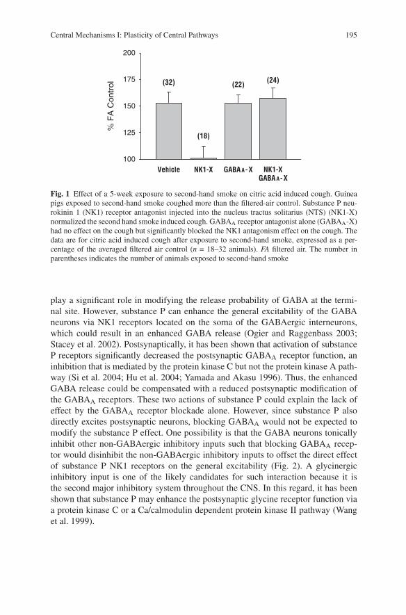
Central Mechanisms I: Plasticity of Central Pathways 195
% F
A C
ontr
ol
100
125
150
175
200
Vehicle NK1-X GABA A-XGABA A-X
NK1-X
(32)
(18)
(22) (24)
Fig. 1 Effect of a 5-week exposure to second-hand smoke on citric acid induced cough. Guineapigs exposed to second-hand smoke coughed more than the filtered-air control. Substance P neu-rokinin 1 (NK1) receptor antagonist injected into the nucleus tractus solitarius (NTS) (NK1-X)normalized the second hand smoke induced cough. GABAA receptor antagonist alone (GABAA-X)had no effect on the cough but significantly blocked the NK1 antagonism effect on the cough. Thedata are for citric acid induced cough after exposure to second-hand smoke, expressed as a per-centage of the averaged filtered air control (n = 18–32 animals). FA filtered air. The number inparentheses indicates the number of animals exposed to second-hand smoke
play a significant role in modifying the release probability of GABA at the termi-nal site. However, substance P can enhance the general excitability of the GABAneurons via NK1 receptors located on the soma of the GABAergic interneurons,which could result in an enhanced GABA release (Ogier and Raggenbass 2003;Stacey et al. 2002). Postsynaptically, it has been shown that activation of substanceP receptors significantly decreased the postsynaptic GABAA receptor function, aninhibition that is mediated by the protein kinase C but not the protein kinase A path-way (Si et al. 2004; Hu et al. 2004; Yamada and Akasu 1996). Thus, the enhancedGABA release could be compensated with a reduced postsynaptic modification ofthe GABAA receptors. These two actions of substance P could explain the lack ofeffect by the GABAA receptor blockade alone. However, since substance P alsodirectly excites postsynaptic neurons, blocking GABAA would not be expected tomodify the substance P effect. One possibility is that the GABA neurons tonicallyinhibit other non-GABAergic inhibitory inputs such that blocking GABAA recep-tor would disinhibit the non-GABAergic inhibitory inputs to offset the direct effectof substance P NK1 receptors on the general excitability (Fig. 2). A glycinergicinhibitory input is one of the likely candidates for such interaction because it isthe second major inhibitory system throughout the CNS. In this regard, it has beenshown that substance P may enhance the postsynaptic glycine receptor function viaa protein kinase C or a Ca/calmodulin dependent protein kinase II pathway (Wanget al. 1999).

196 C.-Y. Chen et al.
Non-GABAergicInhibitory neuron
Glutamate
GABA
Cation Channel
K ChannelSP
SP
SP
-
-
-
+
+
+
Fig. 2 Substance P induced NTS plasticity targets in the cough reflex. Plasticity targets include(1) increased intrinsic excitability via closure of potassium channels, activation of nonselectivecation channels, opening of L-type calcium channel, and intracellular calcium release. (2) increasedsynaptic excitability via an increased ionotropic glutamate receptor function, a decreased GABAAreceptor function. Other compensatory mechanisms may also occur, including an increased GABArelease and decreased glutamate release from the afferent terminals. AP area postrema
4 Summary: The Model
The source and conditions under which substance P is released may determinethe extent to which the presynaptic and postsynaptic mechanisms are integratedto shape the neuronal output from the NTS to distal synapses in the reflex path-ways. Multiple central mechanisms are likely to contribute to the plasticity ofcough reflex. In upregulated cough reflex, an increased substance P expressionin the vagal neurons may be released at the central terminal in the NTS (Fischeret al. 1996; Chuaychoo et al. 2005). This enhanced substance P release in the NTS

Central Mechanisms I: Plasticity of Central Pathways 197
could contribute to the enhanced substance P–NK1 receptor mechanism mediatinga heightened cough reflex response (Joad et al. 2007; Bonham et al. 2004). The in-creased substance P released from the primary sensory central terminals is likely tohave more focused effect on the NTS second-order neurons, including the inhibitoryinterneurons. Upon binding to the NK1 receptors, the released substance P can in-crease the general output of the NTS second-order neurons by (1) closing potassiumchannels via the protein kinase C pathway, (2) opening nonselective cation channels,(3) increasing intracellular calcium concentration via opening L-type calcium chan-nels, (4) increasing intracellular calcium concentration via the phospholipid C path-way, (5) increasing the glutamate synaptic transmission via enhanced NMDA andnon-NMDA receptor function, (6) inhibiting postsynaptic GABAA receptor func-tion, and (7) inhibiting other non-GABAergic inhibitory neurons via an enhancedGABA release (Fig. 2).
At the same time, several other mechanisms may be activated to compensate forsome of the excitatory effect, perhaps to keep the system from being hyperreactive.These mechanisms include (1) presynaptic inhibition of glutamate release from theprimary afferent terminals, (2) activation of the NK1 receptors on the soma/dendriteof GABAergic interneurons to enhance GABA release, and (3) an enhanced glycinesynaptic transmission via an enhanced glycine receptor function through a proteinkinase C or a Ca/calmodulin dependent protein kinase II pathway (Fig. 2).
5 Future Challenges
Another important consideration in examining the neuroplasticity in the NTS is thatalthough the gustatory, gastrointestinal, and cardiorespiratory sensory inputs are or-ganized somewhat viscerotopically in the NTS, neurons receiving different affer-ent inputs and serving different functions are intermingled throughout the nucleus(Loewy 1990). Functionally and anatomically identifying the second-order neuronby the use of fluorescent tracers is helpful in identifying the neurons receiving lungafferent inputs. However, challenges still exist in isolating the neurons specifically inthe cough pathway, whether they are input neurons, interneurons, or output neurons.Given the limitation of identifying the specific NTS neurons in the cough pathway,there are more unknown than known cellular mechanisms by which substance Penhances the cough pathway. More studies are needed to sorting out the proposedpotential sites for plasticity in different model of enhanced cough, whether acute orchronic.
References
Akasu T, Ishimatsu M, Yamada K (1996) Tachykinins cause inward current through NK1 receptorsin bullfrog sensory neurons. Brain Res 713:160–167
Artola A, Singer W (1987) Long-term potentiation and NMDA receptors in rat visual cortex.Nature 330:649–652

198 C.-Y. Chen et al.
Aylwin ML, Horowitz JM, Bonham AC (1998) Non-NMDA and NMDA receptors in the synap-tic pathway between area postrema and nucleus tractus solitarius. Am J Physiol 275:H1236–H1246
Bailey CP, Maubach KA, Jones RS (2004) Neurokinin-1 receptors in the rat nucleus tractus soli-tarius: Pre- and postsynaptic modulation of glutamate and GABA release. Neuroscience 127:467–479
Bisgaard H, Szefler S (2007) Prevalence of asthma-like symptoms in young children. Pediatr Pul-monol 42:723–728
Bolser DC, Davenport PW (2002) Functional organization of the central cough generation mecha-nism. Pulm Pharmacol Ther 15:221–225
Bonham AC, Sekizawa SI, Joad JP (2004) Plasticity of central mechanisms for cough. Pulm Phar-macol Ther 17:453–457
Bonham AC, Chen CY, Sekizawa S, Joad JP (2006) Plasticity in the nucleus tractus solitarius andits influence on lung and airway reflexes. J Appl Physiol 101:322–327
Budai D, Wilcox GL, Larson AA (1992) Modulation of N-methyl-D-aspartate and (R,S)-alpha-amino-3-hydroxy-5-methylisoxazole-4-propionate (AMPA) responses of spinal nociceptiveneurons by a N-terminal fragment of substance P. Eur J Pharmacol 216:441–444
Carr MJ, Lee LY(2006) Plasticity of peripheral mechanisms of cough. Respir Physiol Neurobiol152:298–311
Carr MJ, Undem BJ (2001) Inflammation-induced plasticity of the afferent innervation of the air-ways. Environ Health Perspect 109(Suppl 4):567–571
Chen CY, Bonham AC (1998) Non-NMDA and NMDA receptors transmit area postrema input toaortic baroreceptor neurons in NTS. Am J Physiol 275:H1695–H1706
Chen CY, Bonham AC, Schelegle ES, Gershwin LJ, Plopper CG, Joad JP (2001) Extended allergenexposure in asthmatic monkeys induces neuroplasticity in nucleus tractus solitarius. J AllergyClin Immunol 108:557–562
Chen CY, Bonham AC, Plopper CG, Joad JP (2003) Neuroplasticity in nucleus tractus solitariusneurons after episodic ozone exposure in infant primates. J Appl Physiol 94:819–827
Cherry DK, Woodwell DA, Rechtsteiner EA (2007) National ambulatory medical care survey:2005 summary. In: Advance data from vital and health statistics, vol 387. US Department ofhealth and human services, USA, pp 1–40
Chuaychoo B, Hunter DD, Myers AC, Kollarik M, Undem BJ (2005) Allergen-induced substanceP synthesis in large-diameter sensory neurons innervating the lungs. J Allergy Clin Immunol116:325–331
Chung KF (2007) Chronic cough: Future directions in chronic cough: Mechanisms and antitus-sives. Chron Respir Dis 4:159–165
Davies J, Dray A (1980) Depression and facilitation of synaptic responses in cat dorsal horn bysubstance P administered into substantia gelatinosa. Life Sci 27:2037–2042
Davis BJ, Smith HM (1999) Neurokinin-1 receptor immunoreactivity in the nucleus of the solitarytract in the hamster. Neuroreport 10:1003–1006
Dray A, Pinnock RD (1982) Effects of substance P on adult rat sensory ganglion neurones in vitro.Neurosci Lett 33:61–66
Drew GM, Mitchell VA, Vaughan CW (2005) Postsynaptic actions of substance P on rat periaque-ductal grey neurons in vitro. Neuropharmacology 49:587–595
Endoh T (2006) Dual effects of neurokinin on calcium channel currents and signal pathways inneonatal rat nucleus tractus solitarius. Brain Res 1110:116–127
Fischer A, McGregor GP, Saria A, Philippin B, Kummer W (1996) Induction of tachykinin geneand peptide expression in guinea pig nodose primary afferent neurons by allergic airway in-flammation. J Clin Invest 98:2284–2291
Gilbert R, Ryan JS, Horackova M, Smith FM, Kelly ME (1998) Actions of substance P onmembrane potential and ionic currents in guinea pig stellate ganglion neurons. Am J Physiol274:C892–C903
Helke CJ, Seagard JL (2004) Substance P in the baroreceptor reflex: 25 years. Peptides 25:413–423

Central Mechanisms I: Plasticity of Central Pathways 199
Hu WP, Li ZW, Ru LQ, Fan YZ (2004) Opposite modulatory effects of substance P on GABA-and5-HT-activated currents in the same sensory neurons. Sheng Li Xue Bao 56:703–707
Irwin RS (2006) Complications of cough: ACCP evidence-based clinical practice guidelines. Chest129:54S–58S
Irwin RS, Boulet LP, Cloutier MM, Fuller R, Gold PM, Hoffstein V, Ing AJ, McCool FD,O’Byrne P, Poe RH, Prakash UB, Pratter MR, Rubin BK (1998) Managing cough as a de-fense mechanism and as a symptom. A consensus panel report of the American college of chestphysicians. Chest 114:133S–181S
Irwin RS, French CT, Fletcher KE (2002) Quality of life in coughers. Pulm Pharmacol Ther15:283–286
Ishimatsu M (1994) Substance P produces an inward current by suppressing voltage-dependentand -independent K+ currents in bullfrog primary afferent neurons. Neurosci Res 19:9–20
Ito K, Rome C, Bouleau Y, Dulon D (2002) Substance P mobilizes intracellular calcium and ac-tivates a nonselective cation conductance in rat spiral ganglion neurons. Eur J Neurosci 16:2095–2102
Jafri MS, Weinreich D (1996) Substance P hyperpolarizes vagal sensory neurones of the ferret. JPhysiol 493(Part 1):157–166
Joad JP, Munch PA, Bric JM, Evans SJ, Pinkerton KE, Chen CY, Bonham AC (2004) Passivesmoke effects on cough and airways in young guinea pigs: Role of brainstem substance P. AmJ Respir Crit Care Med 169:499–504
Joad JP, Sekizawa S, Chen CY, Bonham AC (2007) Air pollutants and cough. Pulm PharmacolTher 20:347–354
Jun JY, Choi S, Yeum CH, Chang IY, You HJ, Park CK, Kim MY, Kong ID, Kim MJ, Lee KP, So I,Kim KW (2004) Substance P induces inward current and regulates pacemaker currents throughtachykinin NK1 receptor in cultured interstitial cells of Cajal of murine small intestine. Eur JPharmacol 495:35–42
Len WB, Chan JY (2001) GABAergic neurotransmission at the nucleus tractus solitarii in thesuppression of reflex bradycardia by parabrachial nucleus. Synapse 42:27–39
Lee LY, Undem BJ (2004) Mechanisms of chronic cough. Pulm Pharmacol Ther 17:463–464Lee HK, Shuttleworth CW, Sanders KM (1995) Tachykinins activate nonselective cation currents
in canine colonic myocytes. Am J Physiol 269:C1394–C1401Lepre M, Olpe HR, Brugger F (1996) The effects of neurokinin-1 receptor agonists on spinal
motoneurones of the neonatal rat. Neuropharmacology 35:511–522Lieberman DN, Mody I (1998) Substance P enhances NMDA channel function in hippocampal
dentate gyrus granule cells. J Neurophysiol 80:113–119Lindsey BG, Morris KF, Segers LS, Shannon R (2000) Respiratory neuronal assemblies. Respir
Physiol 122:183–196Liu X, Andre D, Puizillout JJ (1998) Substance P post-synaptically potentiates glutamate-induced
currents in dorsal vagal neurons. Brain Res 804:95–104Loewy AD (1990) Central autonomic pathways. In: Loewy AD, Spyer KM (eds) Central regulation
of autonomic functions. Oxford University Press, New York, pp 88–103Mazzone SB, Mori N, Canning BJ (2005) Synergistic interactions between airway afferent nerve
subtypes regulating the cough reflex in guinea-pigs. J Physiol 569:559–573Morice AH (2002) Epidemiology of cough. Pulm Pharmacol Ther 15:253–259Morris KF, Baekey DM, Nuding SC, Dick TE, Shannon R, Lindsey BG (2003) Invited review:
Neural network plasticity in respiratory control. J Appl Physiol 94:1242–1252Murase K, Nedeljkov V, Randic M (1982) The actions of neuropeptides on dorsal horn neurons in
the rat spinal cord slice preparation: An intracellular study. Brain Res 234:170–176Mutoh T, Bonham AC, Joad JP (2000) Substance P in the nucleus of the solitary tract augments
bronchopulmonary C fiber reflex output. Am J Physiol Regul Integr Comp Physiol 279:R1215–R1223
Mutolo D, Bongianni F, Fontana GA, Pantaleo T (2007) The role of excitatory amino acids andsubstance P in the mediation of the cough reflex within the nucleus tractus solitarii of the rabbit.Brain Res Bull 74:284–293

200 C.-Y. Chen et al.
Ogier R, Raggenbass M (2003) Action of tachykinins in the rat hippocampus: Modulation of in-hibitory synaptic transmission. Eur J Neurosci 17:2639–2647
Pieri M, Severini C, Amadoro G, Carunchio I, Barbato C, Ciotti MT, Zona C (2005) AMPA recep-tors are modulated by tachykinins in rat cerebellum neurons. J Neurophysiol 94:2484–2490
Poliacek I, Corrie LW, Wang C, Rose MJ, Bolser DC (2007) Microinjection of DLH into theregion of the caudal ventral respiratory column in the cat: Evidence for an endogenous cough-suppressant mechanism. J Appl Physiol 102:1014–1021
Pratter MR (2006) Overview of common causes of chronic cough: ACCP evidence-based clinicalpractice guidelines. Chest 129:59S–62S
Randic M, Hecimovic H, Ryu PD (1990) Substance P modulates glutamate-induced currents inacutely isolated rat spinal dorsal horn neurones. Neurosci Lett 117:74–80
Sculptoreanu A, de Groat WC (2007) Neurokinins enhance excitability in capsaicin-responsiveDRG neurons. Exp Neurol 205:92–100
Sekizawa S, Joad JP, Bonham AC (2003) Substance P presynaptically depresses the transmissionof sensory input to bronchopulmonary neurons in the guinea pig nucleus tractus solitarii. JPhysiol 552:547–559
Sekizawa S, Bramham CR, Joad JP (2005) Second hand smoke (SHS) exposure decreases transientoutward currents of nucleus tractus solitarius (NTS) in a subset of 2nd-order lung afferentneurons in young guinea pigs. ATS Annual Meeting C97, Poster 809
Shannon R, Baekey DM, Morris KF, Lindsey BG (1998) Ventrolateral medullary respiratory net-work and a model of cough motor pattern generation. J Appl Physiol 84:2020–2035
Si JQ, Zhang ZQ, Li CX, Wang LF, Yang YL, Li ZW (2004) Modulatory effect of substance P onGABA-activated currents from rat dorsal root ganglion. Acta Pharmacol Sin 25:623–629
Stacey AE, Woodhall GL, Jones RS (2002) Activation of neurokinin-1 receptors promotes GABArelease at synapses in the rat entorhinal cortex. Neuroscience 115:575–586
Sun W, Ding DL, Wang P, Sun J, Jin X, Salvi RJ (2004) Substance P inhibits potassium and calciumcurrents in inner ear spiral ganglion neurons. Brain Res 1012:82–92
Suwabe T, Bradley RM (2007) Effects of 5-hydroxytryptamine and substance P on neurons of theinferior salivatory nucleus. J Neurophysiol 97:2605–2611
Tell F, Jean A (1991) Bursting discharges evoked in vitro, by solitary tract stimulation or appli-cation of N-methyl-D-aspartate, in neurons of the rat nucleus tractus solitarii. Neurosci Lett124:221–224
Tell F, Jean A (1993) Ionic basis for endogenous rhythmic patterns induced by activation of N-methyl-D-aspartate receptors in neurons of the rat nucleus tractus solitarii. J Neurophysiol70:2379–2390
Thompson SW, King AE, Woolf CJ (1990) Activity-dependent changes in rat ventral horn neuronsin vitro; summation of prolonged afferent evoked postsynaptic depolarizations produce a d-2-amino-5-phosphonovaleric acid sensitive windup. Eur J Neurosci 2:638–649
Undem BJ, Hunter DD, Liu M, Haak-Frendscho M, Oakragly A, Fischer A (1999) Allergen-induced sensory neuroplasticity in airways. Int Arch Allergy Immunol 118:150–153
Undem BJ, Carr MJ, Kollarik M (2002) Physiology and plasticity of putative cough fibres in theGuinea pig. Pulm Pharmacol Ther 15:193–198
Wang DS, Xu TL, Li JS (1999) Modulation of glycine-activated chloride currents by substance Pin rat sacral dorsal commissural neurons. Sheng Li Xue Bao 51:361–370
Widdicombe JG, Singh V (2006) Physiological and pathophysiological down-regulation of cough.Respir Physiol Neurobiol 150:105–117
Woolf CJ, Thompson SW (1991) The induction and maintenance of central sensitization is depen-dent on N-methyl-D-aspartic acid receptor activation; implications for the treatment of post-injury pain hypersensitivity states. Pain 44:293–299
Wu ZZ, Guan BC, Li ZW, Yang Q, Liu CJ, Chen JG (2004) Sustained potentiation by substance Pof NMDA-activated current in rat primary sensory neurons. Brain Res 1010:117–126
Yamada K, Akasu T (1996) Substance P suppresses GABAA receptor function via protein kinaseC in primary sensory neurones of bullfrogs. J Physiol 496(Part 2):439–449

Central Mechanisms I: Plasticity of Central Pathways 201
Yang YL, Yao KH, Gu YZ, Guan BC, Li ZW (2003) Three kinds of current in response to substanceP in bullfrog DRG neurons. Brain Res 981:70–77
Yen JC, Chan SH (1997) Interchangeable discharge patterns of neurons in caudal nucleus tractussolitarii in rat slices: Role of GABA and NMDA. J Physiol 504(Part 3):611–627
Zhang J, Mifflin SW (1998) Receptor subtype specific effects of GABA agonists on neurons re-ceiving aortic depressor nerve inputs within the nucleus of the solitary tract. J Auton Nerv Syst73:170–181
Zieglgansberger W, Tulloch IF (1979) Effects of substance P on neurones in the dorsal horn of thespinal cord of the cat. Brain Res 166:273–282

Central Mechanisms II: Pharmacologyof Brainstem Pathways
D.C. Bolser
Contents
1 Introduction . . . . . . . . . . . . . . . . . . . . . . . . . . . . . . . . . . . . . . . . . . . . . . . . . . . . . . . . . . . . . . . . 2042 Location of Action of Antitussive Drugs . . . . . . . . . . . . . . . . . . . . . . . . . . . . . . . . . . . . . . . . . 2043 Identity of Neurones Affected by Antitussive Drugs . . . . . . . . . . . . . . . . . . . . . . . . . . . . . . . 205
3.1 Nucleus of the Tractus Solitarius . . . . . . . . . . . . . . . . . . . . . . . . . . . . . . . . . . . . . . . . . 2073.2 Ventrolateral Respiratory Column . . . . . . . . . . . . . . . . . . . . . . . . . . . . . . . . . . . . . . . . 208
4 Receptor Specificity . . . . . . . . . . . . . . . . . . . . . . . . . . . . . . . . . . . . . . . . . . . . . . . . . . . . . . . . . . 209References . . . . . . . . . . . . . . . . . . . . . . . . . . . . . . . . . . . . . . . . . . . . . . . . . . . . . . . . . . . . . . . . . . . . . 212
Abstract Following systemic administration, centrally acting antitussive drugs aregenerally assumed to act in the brainstem to inhibit cough. However, recent workin humans has raised the possibility of suprapontine sites of action for cough sup-pressants. For drugs that may act in the brainstem, the specific locations, types ofneurones affected, and receptor specificities of the compounds represent importantissues regarding their cough-suppressant actions. Two medullary areas that havereceived the most attention regarding the actions of antitussive drugs are the nu-cleus of the tractus solitarius (NTS) and the caudal ventrolateral respiratory col-umn. Studies that have implicated these two medullary areas have employed bothmicroinjection and in vitro recording methods to control the location of action ofthe antitussive drugs. Other brainstem regions contain neurones that participatein the production of cough and could represent potential sites of action of anti-tussive drugs. These regions include the raphe nuclei, pontine nuclei, and rostralventrolateral medulla. Specific receptor subtypes have been associated with the sup-pression of cough at central sites, including 5-HT1A, opioid (µ,κ, and δ), GABA-B,tachykinin neurokinin-1 (NK-1) and neurokinin-2, non-opioid (NOP-1), cannabi-noid, dopaminergic, and sigma receptors. Aside from tachykinin NK-1 receptorsin the NTS, relatively little is known regarding the receptor specificity of putative
D.C. BolserDepartment of Physiological Sciences, University of Florida, College of Veterinary Medicine,Gainesville, FL 32610-0144, [email protected]
K.F. Chung, J.G. Widdicombe (eds.), Pharmacology and Therapeutics of Cough, 203Handbook of Experimental Pharmacology 187,c© Springer-Verlag Berlin Heidelberg 2009

204 D.C. Bolser
antitussive drugs in particular brainstem regions. Our understanding of the mecha-nisms of action of antitussive drugs would be significantly advanced by further workin this area.
1 Introduction
The pharmacology of centrally active antitussive drugs is a multifactorial topic thatinvolves not only pharmacological and pharmacokinetic issues but neurophysiologyas well. This review will focus on three primary matters related to the brainstemactions of these drugs: (1) location of action, (2) identity of neurones affected bythe drugs, (3) receptor specificity. There are other informative reviews available(Reynolds et al. 2004).
2 Location of Action of Antitussive Drugs
It is widely accepted that several prominent drugs act in the central nervoussystem to inhibit cough, primarily by an action in the brainstem. The evidence sup-porting this concept is strong and is based largely on studies showing that decere-brate animals can cough and that antitussives will suppress coughing under thesecircumstances (Chou 1975; Wang et al. 1977; Domino et al. 1985; Lal et al. 1986;Bolser 1991; Bolser and DeGennaro 1994; Gestreau et al. 1997; Ohi et al. 2004,2005).
The central control of cough is complex and there may be many potential sitesin the brainstem at which a given drug may act to suppress this behavior. In thiscontext, an understanding of the brainstem regions that may be involved in the pro-duction of cough is an important component of any approach to the investigation ofthe actions of antitussive drugs. It is critical to know “where to look” to design stud-ies investigating the mechanisms of action of these agents. In this control systemthere may be many areas where antitussives could work, but only a few that are re-sponsible for the cough-suppressant effects that result from systemic administrationof these agents. It should be noted that the results of studies showing a brainstemaction of antitussives do not preclude an effect of these drugs on suprapontine orspinal pathways in animals that have an intact neuraxis.
Spinal motoneurones (and their antecedant interneuronal pathways) are an often-overlooked component of the cough-generation system, but represent an importantsite at which regulation of the behavior can occur. Several classes of compoundsthat have antitussive activity also suppress spinal motor activity in other systems.Baclofen is a well-known muscle relaxant and inhibits spinal motor activity in lowdoses after intrathecal administration (Penn 1992). Opioids also inhibit motor ac-tivity after topical administration to the spinal cord in spinal cats (Schomburg andSteffens 1995). Central nervous system penetrant drugs gain access to the entire

Central Mechanisms II: Pharmacology of Brainstem Pathways 205
neuraxis within 5 min after vascular administration and compounds that are deliv-ered to the cerebrospinal fluid (CSF) of the brain are rapidly transported to the spinalCSF (Xie and Hammarlund-Udenaes 1998). Therefore, centrally acting antitussivedrugs probably reach the spinal cord after systemic administration. Preliminary re-sults (Rose et al. 2004) have shown that intrathecal administration of baclofen hasno effect on expiratory muscle electromyographic activity during tracheobronchialcough. However, the same dose of baclofen almost completely inhibits cough whenadministered via the vertebral artery. Similar results were obtained in preliminarystudies with intrathecal administration of codeine. These preliminary findings areconsistent with disfacilitation of expiratory spinal motor pathways by antitussivedrugs acting in the brainstem.
The role of suprapontine pathways in the generation of cough and the effects ofantitussive drugs is not well understood. It is likely that the potential role of theseareas in the generation of cough may be much greater in conscious humans (andperhaps animals as well), given that humans can both initiate and suppress coughby voluntary means (Hutchings et al. 1993; Hutchings and Eccles 1994). Significantsensations also are associated with irritant-induced cough, indicating the involve-ment of suprapontine sensory systems during coughing. A model incorporating thepotential influence of suprapontine pathways in the production of cough has re-cently been published (Bolser 2006). However, codeine has no effect on sensationsduring irritant-induced cough in humans, but in that study this opioid did not alterthe musculomechanical aspects of cough (Davenport et al. 2007). In the absenceof an effective antitussive agent in humans, the role of suprapontine pathways andsensations in the mechanism of action of these drugs will remain obscure.
3 Identity of Neurones Affected by Antitussive Drugs
A great deal of information is available regarding the locations of neurones thatparticipate in the production of cough in the brainstem. Shannon and coworkers(Shannon et al. 1996, 1998, 2000; Baekey et al. 2001) have developed a modelof the neurogenic mechanism for coughing that includes columns of neurones inboth the ventrolateral and the dorsomedial regions of the medulla. Different classesof neurones in these regions interact with one another to control inspiratory- andexpiratory-phase durations during cough, the magnitude of motor drive to spinalmotoneurons, and the activation of laryngeal muscle motoneurones that determinethe caliber of the larynx (Shannon et al. 1996, 1998, 2000; Baekey et al. 2001).These neurones also participate in the control of breathing. Sensory input to thiscough pattern generation system is mediated by relay neurones in the nucleus of thetractus solitarius (NTS), located in the dorsomedial medulla.
Recent studies utilizing microinjection of a neurotoxin to induce chemical lesionshave implicated the pons (Poliacek et al. 2004), raphe nuclei (Jakus et al. 1998),and lateral tegmental field (Jakus et al. 2000) in the production of coughing. Themicroinjection method is best suited to the study of regions or nuclei of the brain

206 D.C. Bolser
Pons
rVRC
cVRC
NTS
Raphe
BRAINSTEMDORSAL VIEW
SPINAL CORD
NTSPUMPCELLS
RELAYNEURONS
VAGAL AFFERENTS
÷TachykininNK-1 receptors,µ-Opioid? GABA-B?5-HT receptor subtypes?
SARs COUGH RECEPTORS,RARS
Fig. 1 Brainstem regions that have been specifically associated with the actions of antitussivedrugs. Outlined areas on the left indicate regions of the brainstem in which cough-related neuroneshave been recorded. Shaded areas represent regions in which application of cough-suppressantdrugs has inhibited cough. The model on the right shows second-order interneurones in pulmonaryvagal afferent pathways. Pump cells mediate sensory information from pulmonary slowly adaptingreceptors and neurones designated “relay” mediate information from cough receptors and rapidlyadapting receptors. The notation shows that although a number of different drugs may act on thispopulation of neurones, receptor specificity only for tachykinin neurokinin-1 receptors has beendemonstrated (Chen et al. 2003; Sekizawa et al. 2003). NTS nucleus of the tractus solitarius, rVRCrostral ventral respiratory column, cVRC caudal ventral respiratory column
that are roughly spherical because the diffusion pattern of microinjected chemicalsapproximates a sphere (Lipski et al. 1988). Use of this method in the study of theactions of the raphe nuclei presents unique issues because these nuclei are restrictedto the midline in a “sheet” extending dorsoventrally. The lesions produced by Jakusand coworkers (Jakus et al. 1998) affected the raphe nuclei but extended into thenearby medial reticular formation as well, potentially implicating this region in theproduction of cough (Fig. 1).
Xu et al. (1997) have shown that one of the deep cerebellar nuclei, the nucleusinterpositus, has an important modulatory role in the production of cough. Further-more, the inferior olive, a brainstem nucleus that interacts with the deep cerebellarnuclei, was labeled in coughing animals in a cFos study (Gestreau et al. 1997).
From a theoretical standpoint, any of these areas could represent targets forthe action of antitussive drugs. Important considerations are the route and dose ofthe cough-suppressant drug in question. Intracerebroventricular or vascular admin-istration of antitussive agents lacks anatomic specificity (Bolser 1996). Vascularadministration can be coupled with electrophysiological recordings of single neu-rones to provide anatomic specificity, but the drug will still reach all the tissue sup-plied by the brainstem circulation (Jakus et al. 1987). Intravenous or intra-arterial

Central Mechanisms II: Pharmacology of Brainstem Pathways 207
administration of antitussive drugs does have the advantage that the effects can bedirectly related to concentrations of the agents that are restricted to the therapeuticrange. Effective concentrations of cough suppressants administered by the intrac-erebroventricular route also can be related to therapeutic doses obtained by vascularadministration but only by the use of ratios (Bolser 1996). However, a drawback ofintracerebroventricular studies is that doses are often not normalized to body weight.
The specific location at which an antitussive drug acts to suppress cough canbe studied with the microinjection method. Microinjection has the advantage of lo-calizing the drug to a relatively small region of the brain. However, the drug canstill diffuse 500−1,000µm away from the injection site depending on factors suchas volume injected, concentration, and the characteristics of the extracellular space(Lipski et al. 1988). An additional limitation of this method is that it is difficult torelate the dose of a drug that is delivered to therapeutic concentrations associatedwith systemic administration. Microinjection studies run the risk of delivering dosesof cough suppressants that result in tissue concentrations that are higher than thosethat would be achieved after systemic administration. As such, the risk of type Ierrors in these types of studies may be high.
3.1 Nucleus of the Tractus Solitarius
Antitussive drugs have been microinjected into the region of the NTS. Codeine anddextromethorphan have been injected into the NTS in relatively large quantities(0.5−10µg) in large volumes (0.5−10µL), resulting in suppression of cough in thecat and guinea pig. As stated already it is difficult to know whether these amounts ofantitussive drugs are consistent with local tissue concentrations that occur after sys-temic administration. Concentrations of microinjected drugs are usually restricted tothe nanomolar range with volumes of 100 nL or less to limit the local concentrationof the drug (Lipski et al. 1988). Gestreau and coworkers (Gestreau et al. 1997) haveshown that cFos labeling of NTS neurones is reduced after systemic administrationof a large dose of codeine (17mgkg−1), which they suggested occurred becausecodeine acted on this population of neurones. Recent work suggests that codeinepresynaptically inhibits glutaminergic excitatory neurotransmission from primaryafferent fibers to putative second-order interneurones in the NTS (Ohi et al. 2007).Furthermore, glycinergic currents in acutely dissociated guinea pig NTS neuroneswere inhibited by dextromethorphan (Takahama et al. 1997). Similar findings wereobtained for NTS neurones in slices of the guinea pig medulla for the effects of sub-stance P on excitatory postsynaptic potentials induced from electrical stimulation ofthe tractus solitarius (Sekizawa et al. 2003). In this study, the authors specificallyidentified second-order interneurones by labeling of primary afferents with retro-grade tracer. Another study by this group on NTS neurones from brainstem slicesof nonhuman primates showed that substance P in the micromolar range decreasedinput resistance of these cells, indicative of inhibition (Chen et al. 2003). This ef-fect was observed in control as well as ozone-challenged animals. Interestingly,

208 D.C. Bolser
tachykinin neurokinin-1 (NK-1) receptor antagonists also depressed the responsesof these neurones to current injection, but not the excitatory effect of electrical stim-ulation of the tractus solitarius (Chen et al. 2003). Mutolo et al. (2007) have shownin the rabbit that microinjection of picomolar quantities of substance P into thecommissural subnucleus of the NTS results in enhancement of coughing. Anotherstudy showed central suppression of cough by tachykinin NK-1 antagonists (Bolseret al. 1997) delivered to the brainstem circulation. The apparent conflicting findingsof some of these studies may simply be a manifestation of the inherent complex-ity of the neural network for cough in this region of the medulla. Antitussive drugsmay act presynatically, postsynaptically, and/or on multiple subpopulations of in-terneurones in this area, presenting significant challenges to the interpretation ofmicroinjection studies.
3.2 Ventrolateral Respiratory Column
Jakus and coworkers (Jakus et al. 1987) have shown that the tracheobronchialcough-related discharge of caudal ventral respiratory group expiratory neurones isreduced after systemic administration of codeine. However, this observation doesnot prove that antitussive drugs act directly on any of these neurone groups. Thefindings of Jakus and coworkers Jakus et al. (1987) could be explained either by aninhibitory action of codeine on medullary expiratory neurones or by an action of thisdrug on other neurones that provide excitatory input to medullary expiratory neu-rones. Jakus et al. (1987) observed that previously silent caudal expiratory neuroneswere recruited during cough in the cat. These neurones have expiratory dischargepatterns during cough that are very similar to abdominal motor discharge patterns.Merrill (1970, 1972) investigated spinal projections of both spontaneously activecaudal expiratory neurones and nearby silent neurones. Both groups of neuroneshad contralateral spinal axons and Merrill (1974) noted that antidromic latencies ofthe silent neurones fluctuated with the respiratory cycle, indicating that these neu-rones had respiratory-modulated membrane potentials. Indeed, he suggested thatthis group of silent neurones was recruited during expulsive movements, such ascough.
Microinjection of the excitatory amino acid agonist D,L-homocysteic acid (DLH)into the caudal portion of the ventral respiratory group elicited prolonged suppres-sion of coughing (more than 10min) but only brief (less than 1min) alterations inbreathing in the cat (Poliacek et al. 2007). Spontaneously active neurones in thisregion of the medulla are almost exclusively premotor expiratory with few, if any,axon collaterals to the rest of the brainstem. These neurones are unlikely to havemediated the cough-suppressant effects that were observed. Given that DLH is anexcitatory neurochemical, we have proposed that either silent or nonbreathing mod-ulated neurones in or near the caudal ventral respiratory group are responsible forthis cough suppression. We also have preliminary data showing that microinjectionof codeine into this area in doses as low as 100 pmol suppresses cough. Given that

Central Mechanisms II: Pharmacology of Brainstem Pathways 209
codeine and the well-known excitatory neurochemical DLH have similar effects, itis possible that codeine also is acting as an excitatory agent under these conditions.This proposal represents a departure from the long-held concept that centrally act-ing antitussives inhibit the activity of neural elements involved in the production ofcough. An alternative hypothesis is that these drugs may excite neurones in the cen-tral nervous system that in turn synaptically inhibit control elements in the coughnetwork.
4 Receptor Specificity
The presence of receptors for a drug in a given brain region does not indicate that thedrug will exert its effects by an action in that location. The receptor density and/oraffinity for the drug may be low in that region, requiring a dose of the drug to al-ter the behavior of these neurones that is out of the therapeutic range. Furthermore,many of the brain regions that contain neurones associated with the production ofcough also mediate other functions, such as breathing and cardiovascular control.The concept of receptor specificity is relevant not only to the receptor subtype ac-tuated by a given drug, but also to the physiological system that is affected. For ex-ample, both morphine (a specific µ-opioid receptor agonist) and baclofen (a specificGABA-B receptor agonist) are known to be respiratory depressant agents (Adcocket al. 1988; Hey et al. 1995). However, in the guinea pig the doses of these drugsnecessary for respiratory depression are greater than those required for inhibition ofcough (Adcock et al. 1988; Bolser et al. 1994; Hey et al. 1995). Presumably, eachdrug acts specifically at its respective receptor subtype to inhibit both cough andbreathing, but the potency of the drugs to inhibit these behaviors is different.
Studies to identify the receptor specificity of antitussive drugs have been re-stricted to animal models. In humans, investigations of the effects of antitussivedrugs are usually focused on demonstrating efficacy. Studies in animal models usu-ally involve the use of specific agonists and/or antagonists to demonstrate receptorspecificity.
Opioids, most notably codeine, are effective cough suppressants in animal mod-els (May and Widdicombe 1954; Chou 1975; Wang et al. 1977; Adcock et al. 1988;Bolser et al. 1993, 1999; Bolser and DeGennaro 1994; Reynolds et al. 2004), al-though the efficacy of codeine in humans has recently been questioned (Freestoneand Eccles 1997; Smith et al. 2006). In guinea pigs, the action of codeine as a coughsuppressant has been attributed to µ-opioid receptors (Kotzer et al. 2000). However,codeine does not appear to act through opioid receptors in the cat (Chau et al. 1983),because its antitussive action is not blocked by the opioid antagonist naloxone inthis species. The particular receptor that codeine acts through to inhibit cough in thecat is unknown. The µ-opioid receptor agonist morphine does inhibit cough in theguinea pig and cat by a naloxone-sensitive (or naltrexone-sensitive) mechanism(Chau et al. 1983; Adcock et al. 1988). Morphine has been shown to be active asan antitussive in humans (Fuller et al. 1988; Morice et al. 2007). Furthermore, both

210 D.C. Bolser
δ- and κ-opioid receptor agonists inhibit cough in the guinea pig. Opioid antagonistsdo not influence the cough reflex in either animals or humans (Chau et al. 1983;Freestone and Eccles 1997; Kotzer et al. 2000), suggesting that endogenous opioidsare not critical for the production of cough. These observations are consistent withthe existence of a regulatory mechanism of endogenous opioids on cough that isnormally quiescent. The conditions under which this putative mechanism could beactive are unknown. Moreover, the role(s) that the different opioid receptor subtypesmay have in the regulation of the expression of coughing in the human is not clear.
GABA-B receptor agonists can inhibit cough in animals and humans (Bolseret al. 1993, 1994, 1999; Dicpinigaitis 1996; Dicpinigaitis and Dobkin 1997;Dicpinigaitis et al. 1998). These effects are central in both guinea pigs and catsand the efficacy of baclofen, the prototypical centrally active GABA-B receptor ag-onist, is similar to that of opioids in animal models (Bolser et al. 1993). A specificreceptor antagonist for GABA-B receptors, SCH 50911, had no effect on its own toalter cough but did block the antitussive effects of baclofen in the cat and guinea pig(Bolser et al. 1995). This observation suggests that γ-aminobutyric acid (GABA),acting through the GABA-B receptor subtype, does not modulate the production ofcough under experimental conditions. The actions of other GABA receptor subtypeson the expression of cough are not well understood. The GABA-A receptor antag-onist bicuculline induces seizures (Wood 1975). This limits the usefulness of bicu-culline in the investigation of the effects of GABA-A receptors on cough as seizureactivity itself may modify the excitability of coughing. The effects of GABA-C re-ceptors on cough are unknown.
Serotonin receptor ligands can alter cough in animal models. Most of our cur-rent information indicates that 5-HT1A receptor agonists inhibit cough in smallanimals (Kamei et al. 1991a; Stone et al. 1997). Serotonin may modify the ef-fects of other antitussive drugs, suggesting more complex actions of these drugsthan simple suppression of a single type of neurone in the central nervous system.For example, in the rat depletion of serotonin attenuated the antitussive effect ofmorphine, dihydrocodeine, and dextromethorphan (Kamei et al. 1987b). The sero-tonin precursor L-tryptophan potentiated the antitussive action of dihydrocodeine(Kamei et al. 1990) and the serotonin antagonist methysergide reduced the antitus-sive effects of dextromethorphan and dihydrocodeine in rats (Kamei et al. 1986a,1996). L-Tryptophan treatment also prevented the development of tolerance in ratsto the antitussive effects of dihydrocodeine (Kamei et al. 1991b). Morphine depen-dence elicits a reduced sensitivity to opioid and non-opioid antitussives and theseeffects were associated with a reduction on serotonin receptors in the brainstemsof morphine-dependent rats (Kamei et al. 1989). In cats, the serotonin precursor5-hydroxytryptophan inhibited cough (Kamei et al. 1986b) and methysergide in-creased cough number and reduced the antitussive effects of dextromethorphan(Kamei et al. 1986a). In humans, serotonin and 5-hydroxytryptamine both inhib-ited cough due to inhalation of low-chloride solutions, but not coughing elicited bycapsaicin aerosols (Stone et al. 1993). Our understanding of the effects serotonin re-ceptor ligands on cough would be enhanced by further work in animal models otherthan the rat as well as in the human.

Central Mechanisms II: Pharmacology of Brainstem Pathways 211
The data from the cat on the effects of serotonin receptor agonists and antago-nists on cough appear to be consistent with the data from experiments conducted inrats. However, the rat model has been used by relatively few investigators owing tothe comparative difficulty to induce cough in this species. Furthermore, a detailedanalysis of the motor patterns induced by mechanical stimulation of the tracheo-bronchial region as well as inhalation of capsaicin in the rat revealed that the behav-iors produced by these interventions did not conform to the well-accepted criteriafor cough (Ohi et al. 2004). There also is a considerable amount of published in-formation on antitussives and cough in the mouse (Kamei et al. 1993a–f, 1994a–d,1995a, b, 1999, 2003, 2006, 2007; Morita and Kamei, 2003), but there is no cor-responding analysis of the motor patterns of putative coughing in this species toconfirm that the behaviors that were measured were coughs. Indeed, in their mono-graph, Korpas and Tomori (Korpas and Tomori 1979) indicate that mice do notcough in response to mechanical or chemical stimulation of airway afferents, butthey do readily express another airway defensive behavior, the expiration reflex, inresponse to mechanical stimulation of the larynx. The expiration reflex is regulateddifferently than cough (Korpas and Tomori 1979; Tatar et al. 2008), is insensitiveto antitussive drugs (first shown by May and Widdicombe 1954 and later confirmedby Korpas and Tomori 1979), and is generated by a brainstem neural network thatis not organized in the same manner as that for coughing (Baekey et al. 2004).
Tachykinin receptor antagonists have been shown to be effective cough suppres-sants in the cat, dog, and guinea pig (Advenier et al. 1993; Girard et al. 1995;Kudlacz et al. 1996; Bolser et al. 1997; Hay et al. 2002; Chapman et al. 2004; Joadet al. 2004). Both tachykinin NK-1 and tachykinin neurokinin-2 receptor antago-nists can inhibit cough by a central mechanism (Bolser et al. 1997; Joad et al. 2004;Mazzone et al. 2005). Although a tachykinin neurokinin-3 receptor antagonist caninhibit cough (Daoui et al. 1998), it is not known whether this effect is by a cen-tral site of action. There is evidence that substance P can enhance cough when mi-croinjected into the region of the NTS in the rabbit (Mazzone et al. 2005; Mutoloet al. 2007). However, substance P inhibited excitatory postsynaptic potentials inguinea pig NTS putative relay neurones that were produced by electrical stimulationof the tractus solitarius (Sekizawa et al. 2003). This effect of substance P was presy-naptic and blocked by a tachykinin NK-1 receptor antagonist (Sekizawa et al. 2003).These observations suggest that tachykinin NK-1 receptors are important in regulat-ing the excitability of cough in the region of the NTS. The exact role that tachykininreceptors have in this complex area is not clear at this time. The extent to whichtachykinin receptor antagonists act to regulate cough in the NTS and/or other brain-stem areas after systemic administration in animal models is unknown. Only onestudy on the effects of tachykinin receptor antagonists on cough in humans has beenpublished. Fahy et al. (1995) observed no effect of the tachykinin NK-1 receptor an-tagonist on hypertonic saline-induced cough in humans with mild asthma. Given thelimited amount of information in the human, further studies should be performed tofully evaluate the antitussive potential of tachykinin receptor antagonists.
Sigma receptor agonists have been associated with cough suppression. The mostprominent of these is the cough-suppressant drug dextromethorphan. Given that

212 D.C. Bolser
dextromethorphan binds to N-methyl-D-aspartate and sigma-1 receptors, the speci-ficity of its action to suppress cough has not been clear until recently. Brownet al. (2004) recently showed that this drug acts at sigma-1 receptors to inhibit coughin the guinea pig. Other sigma-1 receptor agonists can inhibit cough as well (Chauet al. 1983; Brown et al. 2004). Carbetapentane, a sigma-1 receptor agonist thatis approved for use in humans as an antitussive, inhibits cough in the cat (Talbottet al. 1975) and guinea pig (Brown et al. 2004). The role of sigma-2 receptors in thesuppression of cough is less clear.
Various other receptors have been associated with cough suppression in ani-mal models, such as non-opioid receptors (NOP-1) (Bolser et al. 2001; McLeodet al. 2001, 2004; Lee et al. 2006), cannabinoid receptors (Gordon et al. 1976; Jiaet al. 2002; Morita and Kamei 2003; Patel et al. 2003), and dopaminergic receptors(Kamei et al. 1987a; Li et al. 2002). In humans, dopaminergic receptors appear tohave no effect on irritant-induced cough in normal subjects (O’Connell 2002).
References
Adcock JJ, Schneider C, Smith TW (1988) Effects of codeine, morphine and a novel opioid pen-tapeptide BW443C, on cough, nociception and ventilation in the unanaesthetized guinea-pig.Br J Pharmacol 93:93–100
Advenier C, Girard V, Naline E, Vilain P, Emonds-Alt X (1993) Antitussive effect of SR 48968, anon-peptide tachykinin NK2 receptor antagonist. Eur J Pharmacol 250:169–171
Baekey DM, Morris KF, Gestreau C, Li Z, Lindsey BG, Shannon R (2001) Medullary respiratoryneurones and control of laryngeal motoneurones during fictive eupnoea and cough in the cat.J Physiol 534:565–581
Baekey DM, Morris KF, Nuding SC, Segers LS, Lindsey BG, Shannon R (2004) Ventrolateralmedullary respiratory network participation in the expiration reflex in the cat. J Appl Physiol96:2057–2072
Bolser DC (1991) Fictive cough in the cat. J Appl Physiol 71:2325–2331Bolser DC (1996) Mechanisms of action of central and peripheral antitussive drugs. Pulm Pharma-
col 9: 357–364Bolser DC (2006) Current and future centrally acting antitussives. Respir Physiol Neurobiol
152:349–355Bolser DC, DeGennaro FC (1994) Effect of codeine on the inspiratory and expiratory burst pattern
during fictive cough in cats. Brain Res 662:25–30Bolser DC, Aziz SM, DeGennaro FC, Kreutner W, Egan RW, Siegel MI, Chapman RW (1993)
Antitussive effects of GABAB agonists in the cat and guinea-pig. Br J Pharmacol 110:491–495Bolser DC, DeGennaro FC, O’Reilly S, Chapman RW, Kreutner W, Egan RW, Hey JA (1994)
Peripheral and central sites of action of GABA-B agonists to inhibit the cough reflex in the catand guinea pig. Br J Pharmacol 113:1344–1348
Bolser DC, Blythin DJ, Chapman RW, Egan RW, Hey JA, Rizzo C, Kuo SC, Kreutner W (1995)The pharmacology of SCH 50911: A novel, orally-active GABA-beta receptor antagonist.J Pharmacol Exp Ther 274:1393–1398
Bolser DC, DeGennaro FC, O’Reilly S, McLeod RL, Hey JA (1997) Central antitussive activity ofthe NK1 and NK2 tachykinin receptor antagonists, CP-99,994 and SR 48968, in the guinea-pigand cat. Br J Pharmacol 121:165–170
Bolser DC, Hey JA, Chapman RW (1999) Influence of central antitussive drugs on the cough motorpattern. J Appl Physiol 86:1017–1024

Central Mechanisms II: Pharmacology of Brainstem Pathways 213
Bolser DC, McLeod RL, Tulshian DB, Hey JA (2001) Antitussive action of nociceptin in the cat.Eur J Pharmacol 430:107–111
Brown C, Fezoui M, Selig WM, Schwartz CE, Ellis JL (2004) Antitussive activity of sigma-1receptor agonists in the guinea-pig. Br J Pharmacol 141:233–240
Chapman RW, House A, Liu F, Celly C, Mei H, Hey JA (2004) Antitussive activity of thetachykinin NK1 receptor antagonist, CP-99994, in dogs. Eur J Pharmacol 485:329–332
Chau TT, Carter FE, Harris LS (1983) Antitussive effect of the optical isomers of mu, kappa andsigma opiate agonists/antagonists in the cat. J Pharmacol Exp Ther 226:108–113
Chen CY, Bonham AC, Plopper CG, Joad JP (2003) Neuroplasticity in nucleus tractus solitariusneurons after episodic ozone exposure in infant primates. J Appl Physiol 94:819–827
Chou D, Wang, SC (1975) Studies on the localization of the central cough mechanism: Site ofaction of antitussive drugs. J Pharmacol Exp Ther 194:499–505
Daoui S, Cognon C, Naline E, Emonds-Alt X, Advenier C (1998) Involvement of tachykinin NK3receptors in citric acid-induced cough and bronchial responses in guinea pigs. Am J Respir CritCare Med 158:42–48
Davenport PW, Bolser DC, Vickroy T, Berry RB, Martin AD, Hey JA, Danzig M (2007) Theeffect of codeine on the Urge-to-Cough response to inhaled capsaicin. Pulm Pharmacol Ther20: 338–346
Dicpinigaitis PV (1996) Use of baclofen to suppress cough induced by angiotensin-convertingenzyme inhibitors. Ann Pharmacother 30:1242–1245
Dicpinigaitis PV, Dobkin JB (1997) Antitussive effect of the GABA-agonist baclofen. Chest111:996–999
Dicpinigaitis PV, Dobkin JB, Rauf K, Aldrich TK (1998) Inhibition of capsaicin-induced cough bythe gamma-aminobutyric acid agonist baclofen. J Clin Pharmacol 38:364–367
Domino EF, Krutak-Krol H, Lal J (1985) Evidence for a central site of action for the antitussiveeffects of caramiphen. J Pharmacol Exp Ther 233:249–253
Fahy JV, Wong HH, Geppetti P, Reis JM, Harris SC, Maclean DB, Nadel JA, Boushey HA (1995)Effect of an NK1 receptor antagonist (CP-99,994) on hypertonic saline-induced bronchocon-striction and cough in male asthmatic subjects. Am J Respir Crit Care Med 152:879–884
Freestone C, Eccles R (1997) Assessment of the antitussive efficacy of codeine in cough associatedwith common cold. J Pharm Pharmacol 49:1045–1049
Fuller RW, Karlsson JA, Choudry NB, Pride NB (1988) Effect of inhaled and systemic opiates onresponses to inhaled capsaicin in humans. J Appl Physiol 65:1125–1130
Gestreau C, Bianchi AL, Grelot L (1997) Differential brainstem Fos-like immunoreactivity afterlaryngeal-induced coughing and its reduction by codeine. J Neurosci 17:9340–9352
Girard V, Naline E, Vilain P, Emonds-Alt X, Advenier C (1995) Effect of the two tachykininantagonists, SR 48968 and SR 140333, on cough induced by citric acid in the unanaesthetizedguinea pig. Eur Respir J 8:1110–1114
Gordon R, Gordon RJ, Sofia D (1976) Antitussive activity of some naturally occurring cannabi-noids in anesthetized cats. Eur J Pharmacol 35:309–313
Hay DW, Giardina GA, Griswold DE, Underwood DC, Kotzer CJ, Bush B, Potts W, Sandhu P,Lundberg D, Foley JJ, Schmidt DB, Martin LD, Kilian D, Legos JJ, Barone FC, Luttmann MA,Grugni M, Raveglia LF, Sarau HM (2002) Nonpeptide tachykinin receptor antagonists. III.SB 235375, a low central nervous system-penetrant, potent and selective neurokinin-3 recep-tor antagonist, inhibits citric acid-induced cough and airways hyper-reactivity in guinea pigs.J Pharmacol Exp Ther 300:314–323
Hey JA, Mingo G, Bolser DC, Kreutner W, Krobatsch D, Chapman RW (1995) Respiratory effectsof baclofen and 3-aminopropylphosphinic acid in guinea-pigs. Br J Pharmacol 114:735–738
Hutchings HA, Eccles R (1994) The opioid agonist codeine and antagonist naltrexone do not affectvoluntary suppression of capsaicin induced cough in healthy subjects. Eur Respir J 7:715–719
Hutchings HA, Morris S, Eccles R, Jawad MS (1993) Voluntary suppression of cough induced byinhalation of capsaicin in healthy volunteers. Respir Med 87:379–382
Jakus J, Tomori Z, Stransky A, Boselova L (1987) Bulbar respiratory activity during defensiveairways reflexes in cats. Acta Physiol Hung 70:245–254

214 D.C. Bolser
Jakus J, Stransky A, Poliacek I, Barani H, Bosel’ova L (1998) Effects of medullary midline lesionson cough and other airway reflexes in anaesthetized cats. Physiol Res 47:203–213
Jakus J, Stransky A, Poliacek I, Barani H, Bosel’ova L (2000) Kainic acid lesions to the lateraltegmental field of medulla: Effects on cough, expiration and aspiration reflexes in anesthetizedcats. Physiol Res 49:387–398
Jia Y, McLeod RL, Wang X, Parra LE, Egan RW, Hey JA (2002) Anandamide induces cough inconscious guinea-pigs through VR1 receptors. Br J Pharmacol 137:831–836
Joad JP, Munch PA, Bric JM, Evans SJ, Pinkerton KE, Chen CY, Bonham AC (2004) Passivesmoke effects on cough and airways in young guinea pigs: Role of brainstem substance P. AmJ Respir Crit Care Med 169:499–504
Kamei J (1996) Role of opioidergic and serotonergic mechanisms in cough and antitussives. PulmPharmacol 9:349–356
Kamei J, Hosokawa T, Yanaura S, Hukuhara T (1986a) Effects of methysergide on the cough reflex.Jpn J Pharmacol 42:450–452
Kamei J, Hosokawa T, Yanaura S, Hukuhara T (1986b) Involvement of central serotonergic mech-anisms in the cough reflex. Jpn J Pharmacol 42:531–538
Kamei J, Hukuhara T, Kasuya Y (1987a) Dopaminergic control of the cough reflex as demonstratedby the effects of apomorphine. Eur J Pharmacol 141:511–513
Kamei J, Ogawa M, Kasuya Y (1987b) Monoamines and the mechanisms of action of antitussivedrugs in rats. Arch Int Pharmacodyn Ther 290:117–127
Kamei J, Mori T, Ogawa M, Kasuya Y (1989) Subsensitivity to the cough-depressant effects ofopioid and nonopioid antitussives in morphine-dependent rats: Relationship to central serotoninfunction. Pharmacol Biochem Behav 34:595–598
Kamei J, Mori T, Kasuya Y (1990) Effects of L-tryptophan on the effects of antitussives. J Phar-macobiodyn 13:231–237
Kamei J, Mori T, Igarashi H, Kasuya Y (1991a) Effects of 8-hydroxy-2-(di-n-propylamino)tetralin,a selective agonist of 5-HT1A receptors, on the cough reflex in rats. Eur J Pharmacol 203:253–258
Kamei J, Mori T, Kasuya Y (1991b) Effects of L-tryptophan on the development of tolerance tothe antitussive effects of dihydrocodeine. Jpn J Pharmacol 55:403–406
Kamei J, Iwamoto Y, Kawashima N, Suzuki T, Nagase H, Misawa M, Kasuya Y (1993a) Possibleinvolvement of mu 2-mediated mechanisms in mu-mediated antitussive activity in the mouse.Neurosci Lett 149:169–172
Kamei J, Iwamoto Y, Misawa M, Kasuya Y (1993b) Effects of rimcazole, a specific antagonistof sigma sites, on the antitussive effects of non-narcotic antitussive drugs. Eur J Pharmacol242:209–211
Kamei J, Iwamoto Y, Misawa M, Nagase H, Kasuya Y (1993c) Antitussive effect of beta-endorphinis mediated by mu-opioid receptors, but not by kappa- or epsilon-opioid receptors. Eur J Phar-macol 233:251–254
Kamei J, Iwamoto Y, Suzuki T, Misawa M, Nagase H, Kasuya Y (1993d) Antitussive effects ofnaltrindole, a selective delta-opioid receptor antagonist, in mice and rats. Eur J Pharmacol249:161–165
Kamei J, Iwamoto Y, Suzuki T, Misawa M, Nagase H, Kasuya Y (1993e) The role of the mu 2-opioid receptor in the antitussive effect of morphine in mu 1-opioid receptor-deficient CXBKmice. Eur J Pharmacol 240:99–101
Kamei J, Iwamoto Y, Suzuki T, Nagase H, Misawa M, Kasuya Y (1993f) Differential modulationof mu-opioid receptor-mediated antitussive activity by delta-opioid receptor agonists in mice.Eur J Pharmacol 234:117–120
Kamei J, Iwamoto Y, Misawa M, Nagase H, Kasuya Y (1994a) Antitussive effect of [Met5]enkephalin-Arg6-Phe7 in mice. Eur J Pharmacol 253:293–296
Kamei J, Iwamoto Y, Misawa M, Nagase H, Kasuya Y (1994b) Involvement of adenosine A1receptors in antitussive effect in mice. Life Sci 55:PL383–388
Kamei J, Iwamoto Y, Suzuki T, Misawa M, Nagase H, Kasuya Y (1994c) Antitussive effect ofdihydroetorphine in mice. Eur J Pharmacol 260:257–259

Central Mechanisms II: Pharmacology of Brainstem Pathways 215
Kamei J, Iwamoto Y, Suzuki T, Misawa M, Nagase H, Kasuya Y (1994d) Involvement of delta1-opioid receptor antagonism in the antitussive effect of delta-opioid receptor antagonists. EurJ Pharmacol 251:291–294
Kamei J, Saitoh A, Morita K, Nagase H (1995a) Antagonistic effect of buprenorphine on the an-titussive effect of morphine is mediated via the activation of mu 1-opioid receptors. Life Sci57:PL231–235
Kamei J, Saitoh A, Suzuki T, Misawa M, Nagase H, Kasuya Y (1995b) Buprenorphine exerts itsantinociceptive activity via mu 1-opioid receptors. Life Sci 56:PL285–290
Kamei J, Morita K, Ohsawa M, Onodera K (1999) Effects of epinastine on the antitussive andrewarding effects of dihydrocodeine in mice. Methods Find Exp Clin Pharmacol 21:663–668
Kamei J, Morita K, Saitoh A, Nagase H (2003) The antitussive effects of endomorphin-1 andendomorphin-2 in mice. Eur J Pharmacol 467:219–222
Kamei J, Yoshikawa Y, Saitoh A (2006) Effect of N-arachidonoyl-(2-methyl-4-hydroxyphenyl)amine (VDM11), an anandamide transporter inhibitor, on capsaicin-induced cough in mice.Cough 2:2
Kamei J, Hayashi SS, Takahashi Y, Nozaki C (2007) Role of cyclin-dependent kinase 5 incapsaicin-induced cough. Eur J Pharmacol 566:181–184
Korpas J, Tomori Z (1979) Cough and other respiratory reflexes. S. Karger, Basel, New YorkKotzer CJ, Hay DW, Dondio G, Giardina G, Petrillo P, Underwood DC (2000) The antitussive
activity of delta-opioid receptor stimulation in guinea pigs. J Pharmacol Exp Ther 292: 803–809Kudlacz EM, Knippenberg RW, Logan DE, Burkholder TP (1996) Effect of MDL 105,212, a non-
peptide NK-1/NK-2 receptor antagonist in an allergic guinea pig model. J Pharmacol Exp Ther279:732–739
Lal J, Krutak-Krol H, Domino EF (1986) Comparative antitussive effects of dextrorphan, dex-tromethorphan and phencyclidine. Arzneimittelforschung 36:1075–1078
Lee MG, Undem BJ, Brown C, Carr MJ (2006) Effect of nociceptin in acid-evoked cough andairway sensory nerve activation in guinea pigs. Am J Respir Crit Care Med 173:271–275
Li JQ, Jia YX, Yamaya M, Arai H, Ohrui T, Sekizawa K, Sasaki H (2002) Neurochemical regula-tion of cough response to capsaicin in guinea-pigs. Auton Autacoid Pharmacol 22:57–63
Lipski J, Bellingham MC, West MJ, Pilowsky P (1988) Limitations of the technique of pressuremicroinjection of excitatory amino acids for evoking responses from localized regions of theCNS. J Neurosci Meth 26:169–179
May AJ, Widdicombe JG (1954) Depression of the cough reflex by pentobarbitone and some opiumderivatives. Br J Pharmacol Chemother 9:335–340
Mazzone SB, Mori N, Canning BJ (2005) Synergistic interactions between airway afferent nervesubtypes regulating the cough reflex in guinea-pigs. J Physiol 569:559–573
McLeod RL, Parra LE, Mutter JC, Erickson CH, Carey GJ, Tulshian DB, Fawzi AB, Smith-TorhanA, Egan RW, Cuss FM, Hey JA (2001) Nociceptin inhibits cough in the guinea-pig by activationof ORL(1) receptors. Br J Pharmacol 132:1175–1178
McLeod RL, Jia Y, Fernandez X, Parra LE, Wang X, Tulshian DB, Kiselgof EJ, Tan Z, Fawzi AB,Smith-Torhan A, Zhang H, Hey JA (2004) Antitussive profile of the NOP agonist Ro-64–6198in the guinea pig. Pharmacology 71:143–149
Merrill EG (1970) The lateral respiratory neurones of the medulla: Their associations with nucleusambiguus, nucleus retroambigualis, the spinal accessory nucleus and the spinal cord. Brain Res24:11–28
Merrill EG (1972) Temporal patterns of antidromic invasion latencies for the respiratory neuronesof nucleus retroambigualis in cats. J Physiol 223:18P–20P
Merrill EG (1974) Proceedings: Antidromic activation of lateral respiratory neurones during theirsilent periods. J Physiol 241:118P–119P
Morice AH, Menon MS, Mulrennan SA, Everett CF, Wright C, Jackson J, Thompson R (2007)Opiate therapy in chronic cough. Am J Respir Crit Care Med 175:312–315
Morita K, Kamei J (2003) Antitussive effect of WIN 55212–2, a cannabinoid receptor agonist. EurJ Pharmacol 474:269–272

216 D.C. Bolser
Mutolo D, Bongianni F, Fontana GA, Pantaleo T (2007) The role of excitatory amino acids andsubstance P in the mediation of the cough reflex within the nucleus tractus solitarii of the rabbit.Brain Res Bull 74:284–293
O’Connell F (2002) Central pathways for cough in man–unanswered questions. Pulm PharmacolTher 15:295–301
Ohi Y, Yamazaki H, Takeda R, Haji A (2004) Phrenic and iliohypogastric nerve discharges dur-ing tussigenic stimulation in paralyzed and decerebrate guinea pigs and rats. Brain Res 1021:119–127
Ohi Y, Yamazaki H, Takeda R, Haji A (2005) Functional and morphological organization of thenucleus tractus solitarius in the fictive cough reflex of guinea pigs. Neurosci Res 53:201–209
Ohi Y, Kato F, Haji A (2007) Codeine presynaptically inhibits the glutamatergic synaptic transmis-sion in the nucleus tractus solitarius of the guinea pig. Neuroscience 146:1425–1433
Patel HJ, Birrell MA, Crispino N, Hele DJ, Venkatesan P, Barnes PJ, Yacoub MH, Belvisi MG(2003) Inhibition of guinea-pig and human sensory nerve activity and the cough reflex inguinea-pigs by cannabinoid (CB2) receptor activation. Br J Pharmacol 140:261–268
Penn RD (1992) Intrathecal baclofen for spasticity of spinal origin: Seven years of experience.J Neurosurg 77:236–240
Poliacek I, Jakus J, Stransky A, Barani H, Halasova E, Tomori Z (2004) Cough, expiration andaspiration reflexes following kainic acid lesions to the pontine respiratory group in anesthetizedcats. Physiol Res 53:155–163
Poliacek I, Corrie LW, Wang C, Rose MJ, Bolser DC (2007) Microinjection of DLH into theregion of the caudal ventral respiratory column in the cat: Evidence for an endogenous cough-suppressant mechanism. J Appl Physiol 102:1014–1021
Reynolds SM, Mackenzie AJ, Spina D, Page CP (2004) The pharmacology of cough. Trends Phar-macol Sci 25:569–576
Rose MJ, Wang C, Golder FJ, Hammond JA, Bolser DC (2004) Influence of intrathecal and intra-arterial baclofen on laryngeal and tracheobronchial cough in the cat. FASEB J 18:A718
Schomburg ED, Steffens H (1995) Influence of opioids and naloxone on rhythmic motor activityin spinal cats. Exp Brain Res 103:333–343
Sekizawa S, Joad JP, Bonham AC (2003) Substance P presynaptically depresses the transmission ofsensory input to bronchopulmonary neurons in the guinea pig nucleus tractus solitarii. J Physiol552:547–559
Shannon R, Baekey DM, Morris KF, Lindsey BG (1996) Brainstem respiratory networks andcough. Pulm Pharmacol 9:343–347
Shannon R, Baekey DM, Morris KF, Lindsey BG (1998) Ventrolateral medullary respiratory net-work and a model of cough motor pattern generation. J Appl Physiol 84:2020–2035
Shannon R, Baekey DM, Morris KF, Li Z, Lindsey BG (2000) Functional connectivity among ven-trolateral medullary respiratory neurones and responses during fictive cough in the cat. J Physiol525 Pt 1:207–224
Smith J, Owen E, Earis J, Woodcock A (2006) Effect of codeine on objective measurement ofcough in chronic obstructive pulmonary disease. J Allergy Clin Immunol 117:831–835
Stone RA, Worsdell YM, Fuller RW, Barnes PJ (1993) Effects of 5-hydroxytryptamine and 5-hydroxytryptophan infusion on the human cough reflex. J Appl Physiol 74:396–401
Stone RA, Barnes PJ, Chung KF (1997) Effect of 5-HT1A receptor agonist, 8-OH-DPAT, on coughresponses in the conscious guinea pig. Eur J Pharmacol 332:201–207
Takahama K, Fukushima H, Isohama Y, Kai H, Miyata T (1997) Inhibition of glycine currents bydextromethorphan in neurones dissociated from the guinea-pig nucleus tractus solitarii. Br JPharmacol 120:690–694
Talbott MW, Barch GK, Gabel LP (1975) A new method for evaluating antitussives in cats usingan electrode-cannula. Eur J Pharmacol 34:59–63
Tatar M, Hanacek J, Widdicombe JG (2008) The expiration reflex from the trachea and bronchi.Eur Respir J 31:385–390
Wang SC, Chou DT, Wallenstein MC (1977) Studies on the potency of various antitussive agents.Agents Actions 7:337–340

Central Mechanisms II: Pharmacology of Brainstem Pathways 217
Wood JD (1975) The role of gamma-aminobutyric acid in the mechanism of seizures. Prog Neu-robiol 5:77–95
Xie R, Hammarlund-Udenaes M (1998) Blood–brain barrier equilibration of codeine in rats studiedwith microdialysis. Pharm Res 15:570–575
Xu F, Frazier DT, Zhang Z, Baekey DM, Shannon R (1997) Cerebellar modulation of cough motorpattern in cats. J Appl Physiol 83:391–397

Central Mechanisms III: Neuronal Mechanismsof Action of Centrally Acting Antitussives UsingElectrophysiological and Neurochemical StudyApproaches
K. Takahama(�), T. Shirasaki, and F. Soeda
Contents
1 Introduction . . . . . . . . . . . . . . . . . . . . . . . . . . . . . . . . . . . . . . . . . . . . . . . . . . . . . . . . . . . . . . . . 2192 Codeine-Sensitive and Codeine-Insensitive Coughs . . . . . . . . . . . . . . . . . . . . . . . . . . . . . . . 2203 Mechanism of Induction of Codeine-Resistant Coughs and Opioid Receptors . . . . . . . . . . 2224 Mechanisms of Action of Nonnarcotic Centrally Acting Antitussives . . . . . . . . . . . . . . . . . 223
4.1 Cough Center and its Pharmacological Properties . . . . . . . . . . . . . . . . . . . . . . . . . . . 2244.2 Glycine and Its Receptor . . . . . . . . . . . . . . . . . . . . . . . . . . . . . . . . . . . . . . . . . . . . . . . . 2254.3 5-HT and Its Receptors . . . . . . . . . . . . . . . . . . . . . . . . . . . . . . . . . . . . . . . . . . . . . . . . . 2284.4 Inhibitory Action on GIRK Channel . . . . . . . . . . . . . . . . . . . . . . . . . . . . . . . . . . . . . . 2294.5 NMDA and NK1 Receptors . . . . . . . . . . . . . . . . . . . . . . . . . . . . . . . . . . . . . . . . . . . . . . 234
5 Conclusion . . . . . . . . . . . . . . . . . . . . . . . . . . . . . . . . . . . . . . . . . . . . . . . . . . . . . . . . . . . . . . . . . 235References . . . . . . . . . . . . . . . . . . . . . . . . . . . . . . . . . . . . . . . . . . . . . . . . . . . . . . . . . . . . . . . . . . . . . 237
1 Introduction
Much remains to be discussed about the mechanisms underlying cough production.Furthermore, there are various types of cough in humans with different susceptibil-ities to various antitussive drugs. Although codeine is reportedly one of the mosteffective narcotic antitussives, it has recently been demonstrated not to be com-pletely effective in suppressing cough, as shown from experimental results in guineapigs. Also, chronic coughs are often resistant to codeine treatment. Pharmacologi-cal diversity is also found in cough responses produced in experimental animals.Although many antitussives have been developed and used in clinics and as over-the-counter drugs, it remains difficult to describe specific mechanisms of action ofcentrally acting antitussives. Therefore, in this review, we first describe codeine-sensitive and codeine-insensitive coughs in experimental animals. According to our
K. TakahamaDepartment of Environmental and Molecular Health Sciences, Graduate School of PharmaceuticalSciences, Kumamoto University, 5–1 Oe-honmachi, Kumamoto 862–0973, [email protected]
K.F. Chung, J.G. Widdicombe (eds.), Pharmacology and Therapeutics of Cough, 219Handbook of Experimental Pharmacology 187,c© Springer-Verlag Berlin Heidelberg 2009

220 K. Takahama et al.
own data, codeine-sensitive cough is caused by activation of the afferent neurons,probably Aδ neurons, arising mainly from the upper trachea. This neuron is notdesensitized by capsaicin treatment. On the other hand, codeine-insensitive coughmay be produced by activation of the C-fibers arising mainly from the bifurcationof the trachea and the adjacent region. In addition, we review mechanisms of actionof centrally acting antitussives on codeine-sensitive cough in experimental animals,addressing their actions at the neuronal level.
2 Codeine-Sensitive and Codeine-Insensitive Coughs
Although coughing is a very simple phenomenon ending in explosive expirationpreceded by rapid inspiration, it is diverse in terms of not only clinical features butalso pharmacological properties. For example, in guinea pigs with airway inflam-mation such as in the case of bronchitis caused by SO2 gas exposure and cigarettesmoking, coughing caused by various stimuli is often hard to inhibit using centrallyacting antitussives. This is also the case of coughing augmented by treatment withan angiotensin-converting-enzyme inhibitor (ACEI).
In the course of our studies on antitussive effects of codeine, we observed thateven at very high doses the drug failed to completely suppress coughing inducedby inhalation of citric acid solution in guinea pigs (Takahama and Shirasaki 2007),although codeine is reportedly one of the most potent antitussives and is generallyvery effective against coughing caused by various stimuli. The result suggests thatthere is a type of cough that cannot be inhibited by codeine. Furthermore, we foundthat coughing caused by mechanical stimulation of the tracheal bifurcation was notresponsive to codeine in guinea pigs, compared with that induced by mechanicalstimulation of the trachea close to the larynx (Takahama et al. 1997a, b).
Among sensory receptors in airway vagal afferents, rapidly adapting receptors(RARs), Aδ-nociceptors, and bronchial C-fiber receptors appear to be involved incough responses. RARs are myelinated Aδ-fibers and have a low threshold formechanical stimuli, but are resistant to chemical stimuli. Aδ-nociceptors and un-myelinated C-fiber receptors have a high threshold for mechanical stimuli, but alow threshold for chemical stimuli such as bradykinin and capsaicin.
A classic study carried out by Widdicombe (1954) showed that RARs are dis-tributed mainly to the larynx and tracheal bifurcation. On the other hand, chemore-ceptors involved in cough responses are mainly distributed in the lower trachea,particularly around the tracheal bifurcation. In addition to these receptors, Canninget al. have recently found a “cough receptor” which is a subpopulation of myeli-nated, capsaicin-sensitive polymodal afferent neurons in guinea pigs (Canninget al. 2004). These “cough receptors” have properties similar to those of RARs,but have a slower conduction velocity and do not respond to stretching. These re-ceptors are distributed in the larynx and upper trachea, and appear to play a primaryrole in regulating the cough response (Canning et al. 2004). Taken together, it issuggested that chemoreceptors distributed in the lower trachea, particularly around

Mechanisms of Action of Centrally Acting Antitussives 221
Fig. 1 The method of topical chemical stimulation for producing cough and cough-like responsesin guinea pigs. Guinea pigs were lightly anesthetized with pentobarbital. A 10-µl aliquot of stim-ulant solution was ejected through a 21-gauge needle by applying 1.5kgcm−2 air pressure for30 ms. Switching of applying air pressure was done by an electromagnetic valve controlled by astimulator. (From Takahama et al. 1995)
the tracheal bifurcation, may be involved in the production of cough resistant tocodeine treatment. This suggestion is further supported by the following findings.ACEIs, which sensitize nociceptive fibers, induce codeine-resistant chronic coughin conscious guinea pigs (Fox et al. 1996), indicating that coughs mediated by no-ciceptive fibers are resistant to codeine. In our own preliminary study using a newlydeveloped method for topical chemical stimulation of the lower airway (Fig. 1),topical administration of capsaicin to the tracheal bifurcation induced a cough-likeresponse resistant to codeine, whereas when it was added to the laryngeal side ofthe trachea it did not induce a cough response (Takahama et al. 1995; Wakudaet al. 1994). Furthermore, our own preliminary study showed that substance P (SP)-like immunoreactivity is higher in the tracheal bifurcation than in the larynx sidein guinea pigs. A similar finding has been obtained in human subjects. The densityof SP-immunoreactive nerves is significantly higher in patients with cough-variantasthma than in normal subjects and patients with classic asthma (Lee et al. 2003).The abovementioned findings support the hypothesis that coughs mediated by noci-ceptive fibers may be resistant to codeine treatment.
In a guinea pig model of subacute bronchitis produced by SO2 gas exposure,cough responses induced by mechanical stimulation of the trachea were more diffi-cult to inhibit using codeine than those induced experimentally in normal guineapigs (Miyata et al. 1999). It has been reported that SP content is elevated inthe trachea of rats with bronchitis induced by SO2 gas exposure (Killingsworthet al. 1996), probably owing to reduction in the level of neutral endopeptidase(NEP), which degrades various peptides, including bradykinin, SP, and other tachy-kinins (Matsas et al. 1984). In bronchitic guinea pigs, NEP levels and activity inthe trachea and bronchus were significantly lower than those in normal guinea pigs,although no epithelial shedding was found in the lower airway. Administration ofhigh doses of a NEP inhibitor has been shown to induce a cough response in nor-mal guinea pigs (Takahama et al. 1995). Based on findings that bradykinin and

222 K. Takahama et al.
tachykinins are potent inflammatory mediators, and that neurokinins such as SP arereleased from C-fiber (eNANC nerve) terminals, one hypothesis is that coughinginduced by inflammatory peptides might be resistant to centrally acting antitussivessuch as codeine.
Codeine has, however, been found to significantly suppress the cough responseinduced or enhanced by NEP inhibitors (Takahama et al. 1995). In addition, opi-oids peripherally inhibit tachykinergic transmission in the guinea pig bronchus(Kamikawa and Shimo 1990; Frossard and Barnes 1987; Belvisi et al. 1989). Thus,it appears that NEP inhibition or tachykinin release from peripheral C-fiber termi-nals is not always sufficient to explain mechanisms underlying induction of codeine-resistant cough.
As described above, citric acid induced coughing was difficult to completelyinhibit even with high doses of codeine alone. Interestingly, coadministration ofcodeine and an antagonist of the neurokinin-2 receptor almost suppressed citricacid induced coughing in conscious guinea pigs (Takahama and Shirasaki 2007),suggesting that neurokinins may induce codeine-resistant coughs in guinea pigs.Therefore, it seems likely that RAR or “cough-receptor”-mediated coughs are sen-sitive to codeine, but coughs triggered by nocieceptive fibers containing tachykininssuch as neurokinin are resistant to the drug.
Recently, it has been reported that the expression of transient receptor potentialvanilloid-1 (TRPV-1) is enhanced in airway nerves of patients with chronic cough(Groneberg et al. 2004). In addition to TRPV-1, acid-sensing ion channels are lo-calized in Aδ-fibers of guinea pigs (Gu 2006). Therefore, differences in codeinesensitivity of acid-induced coughs may depend on the pH of the cough inductionsite. Further studies are needed to clarify such differences.
3 Mechanism of Induction of Codeine-Resistant Coughsand Opioid Receptors
Two putative mechanisms underlying the production of coughs resistant to centrallyacting antitussives, including narcotic antitussives such as codeine, are proposed.One possible mechanism is that µ-opioid receptors may not be expressed in neuronsof the cough reflex arc involved in the production of codeine-resistant coughs. An-other possible mechanism is that neurotransmission in the cough reflex arc may befacilitated when codeine-resistant cough is produced.
As described already, several lines of evidence suggest that tachykinin-containingvagal afferent fibers contribute to the production of codeine-resistant cough. Suchfibers spread from the airway and innervate the nucleus tractus solitarii (NTS)(Helke et al. 1981; Saria et al. 1988). Injection of a neurokinin-1 (NK1) receptorantagonist into the NTS produced an antitussive effect in animals exposed to ciga-rette smoke, but not in animals exposed to filtered air (Joad et al. 2004). It has alsobeen shown that an excitatory action of iontophoretically applied SP on NTS neu-rons is not inhibited by µ-agonists (Morin-Surun et al. 1984). These findings seem

Mechanisms of Action of Centrally Acting Antitussives 223
to suggest that µ-opioid receptors are not expressed in C-fibers such as tachykinin-containing fibers involved in the production of codeine-resistant cough.
µ-Opioid receptors are found in both postsynaptic and presynaptic sites in theNTS (Rhim et al. 1993; Cheng et al. 1996) as the predominant type of opi-oid receptor (Hassen et al. 1982; Xia and Haddad 1991). µ-Agonists activatethe G-protein-coupled inwardly rectifying potassium (GIRK) channel current andhyperpolarize postsynaptic membrane potential in about 60% of NTS neurons,whereas they inhibit glutamatergic excitatory postsynaptic currents in almost allNTS neurons via a presynaptic mechanism, which is much more sensitive to µ-agonists than postsynaptic mechanisms (Rhim et al. 1993; Glatzer and Smith 2005).µ-Agonists also inhibit high-voltage-activated Ca2+ currents in the nodose ganglion(Rusin and Moises 1998), the origin of RARs (Ricco et al. 1996), and glutamate isthe principal neurotransmitter in Aδ-fibers (Lawrence 1995; Meeley et al. 1989;Wilson et al. 1996). Excision of the nodose ganglion causes marked depletionof µ-opioid receptors in the dorsal and medial regions of the ipsilateral caudalNTS (Dashwood et al. 1988). Electron microscopy has shown that µ-opioid recep-tors localize in the plasma membrane of the terminals of vagal afferents derivedfrom nodose ganglia (Aicher et al. 2000), but not in nociceptive fibers (Undemet al. 2004). In relation to these findings, our own preliminary study using ac-fos-like immunoreactivity expression technique revealed that c-fos-like proteinsare expressed in the medial regions of the caudal NTS following mechanical stimu-lation of the larynx side of the airway.
Certainly, codeine only weakly inhibited coughs in animal models of allergic re-sponses (Kamei et al. 1998; Winter and Flataker 1955; Myou et al. 2001), as wellas of chronic bronchitis produced by SO2 gas exposure (Killingsworth et al. 1996).It has been reported that infection and inflammation of the airway lead to the pro-duction of neurokinins in nonnociceptive RAR nerve terminals in the cell bodiesof vagal sensory ganglia (Undem et al. 1999; Carr et al. 2002; Myers et al. 2002;Dinh et al. 2004, 2005). Therefore, it is intriguing that changes in the phenotype ofvagal nerves and tachykinin release from RAR fibers might facilitate glutamatergictransmission in the nucleus involved in the cough reflex under pathological condi-tions such as airway inflammation, infections, and cigarette smoking. These changesmight explain codeine-resistant cough.
4 Mechanisms of Action of Nonnarcotic Centrally ActingAntitussives
One strategy to determine the effects of centrally acting antitussives on single brainneurons forming the cough reflex arc seems to have an advantage for studying themechanism of action of the drugs, because evidence from in vivo pharmacologicaland electrophysiological studies indicates that the primary sites of action of cen-trally acting antitussives are brain neurons. In particular, evidence from comparisonof equieffective doses administered via different routes, such as the vein, common

224 K. Takahama et al.
carotid artery, vertebral artery and cerebellomedullary cistern (Kase 1980), suggeststhat the primary site of action of centrally acting antitussives is the so-called coughcenter located in the region that includes part of the NTS and adjacent regions. Thishypothesis is supported by data from analysis of actions of antitussives on evokeddischarges in afferent and efferent nerves of the cough reflex. In addition, there havebeen studies of neuronal circuitry involved in integration of the cough response inthe brain (Widdicombe 2002; Beakey et al. 2001). However, the precise mechanismsof action of centrally acting antitussives have yet to be clarified, particularly at theneuron level.
Various medicinal drugs show a particular characteristic based on their chemi-cal structure. In other words, the pharmacological actions of such drugs are basedon their chemical structure, as exemplified by various centrally acting drugs suchas antipsychotic drugs, antianxiety drugs, and antidepressants. There is usually arelationship between pharmacological action and chemical structure. However, thechemical structures of centrally acting antitussives are diverse, because these drugsare derived from various parent compounds possessing various pharmacological ac-tions. These facts bring us to the following issues: whether centrally acting anti-tussives have a common mechanism of antitussive action, and whether these drugshave a common effect on single brain neurons. To further clarify these issues, wemust prepare single neurons from the cough center and related regions of the brain.Although there have been some reports about the location of the cough center, thesestudies have been carried out in cats. However, it seemed difficult to obtain a freshsingle neuron from the cough center and related nuclei in cats. Therefore, prior tothe study on the actions of antitussives at the neuron level, we first tried to clarifythe location and pharmacological properties of the cough center in guinea pigs.
4.1 Cough Center and its Pharmacological Properties
In lightly anesthetized guinea pigs, we found an area responsible for producingcough-like responses when it was electrically stimulated. It is located within theregion from 500µm rostal to 100µm caudal to the obex, from 800 to 400µm fromthe midline, and from 1,000 to 600µm below the dorsal surface of the medulla ob-longata. This area corresponds to a region that includes the rostal part of the NTSand adjacent areas, and is nearly the same as that in cats reported by Kase (1980).The responses produced by electrical stimulation of this area were considered tobe coughing because (1) the responses were depressed by an antitussive dose ofcodeine and (2) the responses consisted of a brief inspiration followed by an explo-sive expiration with the sound of coughing.
By using a microinjection technique in guinea pigs, we also determined the phar-macological properties of an area producing cough-like responses when it was elec-trically stimulated. The injection of a small amount of 5-hydroxytryptamine (5-HT),atropine, and DL-2-amino-5-phosphonopentanoic acid inhibited cough responsescaused by mechanical stimulation of the mucosa of the tracheal bifurcation. Since

Mechanisms of Action of Centrally Acting Antitussives 225
an amount as small as 1µg was sufficient to potently inhibit the cough responses,various receptors such as 5-HT, acetylcholine, and N-methyl-D-aspartate (NMDA)receptors might be involved in cough responses in the NTS and adjacent regions.Surprisingly, direct administration of 1µg of glycine into the cough center potenti-ated cough responses (Honda et al 1990), as indicated by an increase in the ampli-tude of the respiration curves of the cough responses. This finding is of particularinterest, because enhancing cough responses is reported to prevent aspiration pneu-monia in elderly people (Yamaya et al. 2001).
4.2 Glycine and Its Receptor
4.2.1 Action on Glycine-Induced Currents in Single Neurons
In the course of our study on the pharmacological actions of antitussives, we noticedthat antitussives at toxic doses often produced convulsant activity in experimentalanimals, possibly due to blockade of receptors of inhibitory neurotransmitters suchas glycine and γ-aminobutyric acid (GABA). This phenomenon reminded us that an-titussives may have some affinity for inhibitory neurotransmitter receptors. Glycineand GABA are recognized as the main inhibitory neurotransmitters in the brain.In particular, glycine is mainly distributed in the lower brain stem and spinal cord,where the signaling pathway for cough responses is localized. Therefore, we firstattempted to determine whether antitussives affect glycine-mediated responses inthe CNS (Takahama et al. 1997a, b). In the experiment, we used acutely dissociatedsingle neurons of the NTS. Neurons were freshly dissociated from 7–10-day-oldHartley guinea pigs, using standard methods. In these neurons, all of which werevoltage-clamped to a holding potential (VH) of –50 mV, glycine induced an inwardcurrent (Igly) in a concentration-dependent manner at 3µM to 3mM. Electrophysio-logical and pharmacological analysis of the current showed that the current is causedby activation of glycine receptors.
Dextromethorphan did not induce membrane currents in NTS neurons at 0.1 mM;however, it depressed Igly induced by 30µM glycine in a concentration-dependentmanner (Takahama et al. 1997a, b). The half-maximal inhibitory concentration(IC50) of dextromethorphan for Igly was 3.3µM, 85 times the IC50 of strychnine. Ithas been reported that dextromethorphan inhibits voltage-dependent Ca2+ and Na+
channels (Netzer et al. 1993). However, it is unlikely that the effect of dextromethor-phan on Igly is due to its action on these channels, because Igly is carried by Cl− inneurons of the CNS, including NTS neurons of guinea pigs. On the basis of electro-physiological and pharmacological analyses of the action of dextromethorphan onIgly, we speculate that low concentrations of dextromethorphan act on the glycinereceptor–ionophore complex, but not on the Cl− channel of the complex. However,a relatively high concentration of dextromethorphan may affect the Cl− channel ofthe complex. Interestingly, 100µM dextromethorphan had no effect on the currentinduced by 30µM GABA. The fact that GABA-induced current (IGABA) in NTS

226 K. Takahama et al.
neurons was depressed by bicuculline indicates that it was mediated by the GABAAreceptor, further indicating that centrally acting antitussives have a negligible effecton GABAA receptors, although it has been reported that GABAB receptor agonistshave antitussive action in experimental animals (Bolser et al. 1994). We also foundthat other antitussives, both narcotic and nonnarcotic, have inhibitory action on Iglybut not on IGABA, but their action was less potent than that of dextromethorphan(Fukushima et al. 1998). These were the first documented examples indicating thatIgly inhibition is common to several centrally acting antitussives at the brain neuronallevel, and the findings raise the question as to whether Igly inhibition by antitussivesis part of their mechanism of action.
4.2.2 Strychnine-Sensitive Glycinergic Transmission
The results of direct administration of antitussives into the cough center suggestthe possibility of glycinergic transmission in the cough center; glycine receptorsare distributed in the NTS (Kubo and Kihara 1987). To confirm this possibility,we performed a patch-clamp experiment using brain-slice preparations. Coronalsections of the lower brain stem, including the NTS, 200–250µm in thickness,were prepared from 6–9-day-old male Hartley guinea pigs. Prior to preparingthe slices, we first confirmed that the region from where the slices were to betaken included sites innervated by afferent neurons of the cough reflex, usinga neuronal tracing technique with the fluorescent dye 1,1′-dioctadecyl-3,3,3′,3′-tetramethylindocarbocyanine perchlorate as the probe.
As shown in Fig. 2, three types of synaptic currents were recorded in the slicesincluding the NTS. Of 70 synaptic currents evoked by electrical stimulation of thesolitary tract, 13 were excitatory, 28 were inhibitory, and the remaining 29 wereexcitatory currents followed by inhibitory currents. Of the 28 inhibitory postsy-naptic currents, 21 were blocked by strychnine. The inhibitory currents precededby the excitatory ones were effectively reduced by bicuculline, a GABAA receptorantagonist. The excitatory currents were all blocked by combination of 2-amino-5-phosphonovalic acid and 6-cyano-7-nitroquinoxaline-2,3-dione, suggesting thatglutamatergic transmission exist in the NTS. Bicuculline-sensitive inhibitory cur-rents were found to be carried by Cl− on the basis of the results of the electrophysi-ological analysis. A higher concentration of glycine reduced or abolished the evokedinhibitory currents, suggesting glycinergic transmission in the slices examined in-cluding the NTS. The strychnine-sensitive glycinergic transmission was effectivelyblocked by dextromethorphan (Fig. 2).
4.2.3 Microdialysis Experiment
By using a new device we had developed for floating a microdialysis probe, westudied whether the glycine level in the NTS of guinea pigs increases during cough-ing. This device stably maintains the tip of a microdialysis probe within the selected

Mechanisms of Action of Centrally Acting Antitussives 227
Type I
Type II
Type III
100 pA
10 msec
Control
DM 10-4 M
Recovery
a b
100 pA
10 mseca
a
b
b
Stimulation
Fig. 2 Three types of waveforms of postsynaptic currents recorded from the slice including thenucleus tractus solitarii (NTS) (A). The postsynaptic currents were evoked by stimulation of thesolitary tract or the adjacent region. The neurons of the NTS were voltage-clamped at −65-mVholding potential. a: excitatory postsynaptic current, b: inhibitory postsynaptic current
region of the lower brain stem, preventing the probe from moving as a result ofrespiration- and cardiac-pulse-induced fluctuations of the brain stem. In the exper-iments performed under stereotaxic fixation using lightly anesthetized guinea pigs,the glycine level in the NTS was markedly elevated in the microdialysis samplescollected during a period of coughing caused by inhalation of an aerosol of 2%citric acid solution. The responses produced by citric acid under the condition de-scribed above were typical cough responses when estimated by the cough responsecurve and the sound of coughing. However, the levels of GABA, alanine, glutamicacid, and glutamine remained unchanged. The taurine level also significantly in-creased during coughing. Dimethylphenylpiperazinium (DMPP) produces a potentrespiratory excitation similar to a cough response. Coughing is associated with tran-sient increase in blood pressure, which is similar to that caused by bolus injection ofvasopressors such as phenylephrine. Therefore, we examined the effects of DMPPand phenylephrine on the levels of glycine and taurine in the NTS. Treatment withboth drugs did not cause any changes in the levels of glycine or taurine, suggesting

228 K. Takahama et al.
that the abovementioned changes associated with coughing in the levels of glycineand taurine may not be caused by respiratory excitation and increase in blood pres-sure observed during coughing.
Taken together, it is suggested that glycine and its receptor may be involved inthe cough response, although it remains unknown whether an increase in the glycinelevel causes coughing or is a result of coughing. However, it appears that inhibitionof Igly by centrally acting antitussives other than dextromethorphan does not neces-sarily contribute to antitussive action, because relatively high concentrations wereneeded to inhibit Igly. Further studies are needed to elucidate these actions.
4.3 5-HT and Its Receptors
There have been findings that 5-HT may be involved in the regulation of respira-tion. For example, morphine-induced respiratory depression was reduced by treat-ment with reserpine, a monoamine depleter, and p-chlorophenylalanine (PCPA), aninhibitor of 5-HT synthesis. It is probable that antitussive activity of some drugsmay be reduced by treatment with these pharmacological tools. Kamei et al. havereported that the 50% antitussive effective dose (AtD50) of some antitussives inreserpinized rats is twofold to fourfold higher than that in normal rats (Kameiet al. 1987). This increase in AtD50 has also been found in rats treated with PCPA,which depletes 5-HT content in the brain, compared with normal rats. In con-trast, treatment with α-methyl-p-tyrosine, which depletes catecholamines such asnorepinephrine and dopamine in the brain, has little effect on antitussive activity,suggesting that catecholamines are not involved in antitussive activity. That is, re-serpine and PCPA, which both deplete 5-HT content in the brain, reduce antitussiveactivity of centrally acting antitussives in rats. On the basis of these findings, Kameiet al. suggested that 5-HT but not catecholamines in the brain plays an important rolein manifestation of antitussive activity (Kamei et al. 1987); however, the suggestionshould be carefully verified because it is based on experimental data in anesthetizedrats, in which it is often hard to produce cough. It is also well known that activationof serotonergic neurons in the brain causes respiratory depression.
An important question is which subtype of 5-HT receptors is involved inantitussive activity. Of the 5-HT receptor subtypes, 5-HT1A receptors are consid-ered as receptors involved in cough responses because these receptors are dis-tributed throughout the medulla oblongata, and their receptors are involved inrespiration regulation. Interestingly, systemic administration of 8-hydroxy-2-(di-n-propylamino)tetralin (8-OH-DPAT) suppresses coughing in guinea pigs (Stone et al.1997) and rats (Kamei et al. 1991). Because 8-OH-DPAT is a 5-HT1A receptoragonist possessing high selectivity to 5-HT1A receptors, this finding suggests thatdirect activation of 5-HT1A receptors may suppress cough responses, which is sup-ported by the finding that the antitussive effects of dextromethorphan, a representa-tive nonnarcotic antitussive in rats, are inhibited by 5-HT1A receptor antagonistssuch as spiperone and methysergide, but not by the 5-HT2 receptor antagonist

Mechanisms of Action of Centrally Acting Antitussives 229
ketanserine (Kamei 1986). The same result was obtained from an experiment ondihydrocodeine, a narcotic antitussive. Although it remains unclear whether thesefindings can be extrapolated to coughing in humans, such findings strongly suggestthat activation of 5-HT1A receptors may induce an antitussive effect in experimentalanimals such as rodents.
From the abovementioned findings, the question remains whether antitussivedrugs directly or indirectly activate 5-HT1A receptors. Therefore, we first exam-ined the effects of dextromethorphan and other antitussives on single brain neuronsacutely dissociated from the dorsal raphe, which expresses 5-HT1A receptors, us-ing nystatin-perforated and conventional whole-cell recording techniques. In dorsalraphe neurons, voltage-clamped at VH = −80mV, 1 nM to 1µM 5-HT induced aninwardly rectifying K+ current (I5-HT) in a concentration-dependent manner. ThisI5-HT was blocked by spiperone, a 5-HT1A receptor antagonist, but not by ketanser-ine, a 5-HT2 receptor antagonist. We also confirmed that I5-HT was carried by K+, onthe basis of results of electrophysiological analysis. In these neurons, 8-OH-DPAT,a 5-HT1A receptor agonist, similar to 5-HT, induced I5-HT in dorsal raphe neurons.However, 10µM dextromethorphan did not cause any changes in the membranecurrent when administered alone. This suggests that dextromethorphan may not actagonistically on the 5-HT1A receptor. Surprisingly, dextromethorphan potently in-hibited I5-HT (Ishibashi et al. 2000), with an IC50 of 14.3µM and a Hill coefficient of1.0. The effect of dextromethorphan on the maximum 5-HT response and the resultsobtained from the Lineweaver–Burk plot indicate that dextromethorphan inhibitionwas noncompetitive (Ishibashi et al. 2000).
4.4 Inhibitory Action on GIRK Channel
When 5-HT1A receptor agonists were applied to the recorded dorsal raphe neuronsin the brain slice and acutely dissociated cell preparation, they caused hyperpolar-ization and cessation of spontaneous firing. 5-HT1A receptors are one of the G-protein-coupled receptors (GPCRs) and their activation induces inwardly rectifyingK+ channels through the direct action of G-protein subunits, Gβγ. This effect iscaused by the activation of GIRK channels. It has been shown that intracellular per-fusion with GTPγS, an irreversible activator of the G-protein subunit Gα, causesirreversible 5-HT1A-receptor-mediated activation of GIRK channels, in the conven-tional whole-cell recording mode. The irreversible activation of K+ current by intra-cellular perfusion with GTPγS reminded us of the direct activation of K+ channelsby G-protein, and enabled us to determine whether GIRK channels could be blockedby dextromethorphan even in the absence of an agonist. To carry out this protocol,we used a recording electrode filled with pipette solution containing GTPγS. Un-der this condition, a brief application of 5-HT induced a continuous and almostirreversible inward K+ current. Dextromethorphan markedly inhibited the currentactivated by intracellular GTPγS in the absence of 5-HT (Ishibashi et al. 2000),whereas spiperone, a 5-HT1A receptor antagonist, had no effect on this current. This

230 K. Takahama et al.
result confirmed that dextromethorphan had no effect on the 5-HT1A receptor, mak-ing it different from receptor antagonists such as spiperone. Here, there appeared tobe two possible explanations for the inhibition of GTPγS-activated currents by dex-tromethorphan, because it has been reported that the activation of GIRK channels ismediated by the G-protein βγ subunit. The first possible mechanism is blockade ofthe GIRK channel and the other is blockade of the G-protein-mediated activation ofthe channel by inhibiting the action of the G-protein βγ subunit. Although the siteof action of dextromethorphan is not clearly indicated by available evidence, thefact that the onset and the offset of the dextromethorphan response were relativelyrapid suggests that dextromethorphan may act at the level of the GIRK channel(Ishibashi et al. 2000). To clarify the site of inhibitory action of antitussives suchas dextromethorphan on GIRK-channel-activated currents at the molecular level,we further performed additional studies. Recently, our own preliminary study hassuggested that the site of action of dextromethorphan, one of the antitussives in-hibiting the GIRK-channel-activated current, might be in the intracellular region ofthe GIRK channel, because such a drug had more potent action in low-pH solu-tion, where it exists in the molecular form and not in the ionic form. Furthermore,in Xenopus oocytes expressing GIRK channels formed by GIRK1 and GIRK2 sub-units, in which GPCRs such as the 5-HT1A receptor are not expressed, the antitus-sive cloperastin inhibited the current activated by ethanol, which is known to activatethe GIRK channel directly, suggesting that cloperastine may directly inhibit GIRKchannel activity.
At this point, we felt it could be instructive to determine whether dextromethor-phan also inhibits currents caused by activation of other neurotransmitter recep-tors coupled to G-protein, because GIRK channels are coupled to various GPCRsand other receptors, including adrenergic α2 and GABAB receptors. Moreover, it isknown that GIRK channels are heterotetrameric channels with two or more sub-unit isoforms, although little is known about such isoforms coupled to GPCRssuch as 5-HT1A receptors and α2 adrenoceptors. In this situation, we felt that itwas worth determining whether dextromethorphan also inhibits currents resultingfrom activation of other neurotransmitter receptors coupled to G-protein. Therefore,we examined the effect of dextromethorphan on a norepinephrine-induced current,which is mediated by α2 adrenoceptors (Arima et al. 1998), in locus coeruleusneurons. We found that dextromethorphan reversibly inhibits the norepinephrine-induced current, with rapid onset and offset. The IC50 of dextromethorphan for thenorepinephrine-induced current was very similar to that for 5-HT-induced currentsin dorsal raphe neurons. Although it is known that GIRK channels are heterote-trameric channels with two or more subunit isoforms, little is known about GIRKchannels coupled to 5-HT1A receptors and α2 adrenoceptors. The abovementionedresults indicate that dextromethorphan has no specific effect on GIRK channels cou-pled to 5-HT1A receptors in dorsal raphe neurons.
An important question at this point is whether blockade of GIRK channels byantitussives causes inhibition of coughing. Interestingly, all centrally acting antitus-sives studied inhibited I5-HT caused by 5-HT1A receptor activation. IC50 values ofvarious antitussives for the current caused by 0.1µM 5-HT ranged from 1 to 10µM.

Mechanisms of Action of Centrally Acting Antitussives 231
Also, currents irreversibly activated by intracellular perfusion with GTPγS were alsoinhibited. Interestingly, inhibitory ratios of antitussives studied for I5-HT were com-parable to those for the current caused by 5-HT under intracellular perfusion withGTPγS. This finding seems to suggest that inhibitory action of these antitussiveson 5-HT-induced currents may be caused mostly by inhibition of GIRK channelactivity. Furthermore, it may be a significant finding that antitussives such as dex-tromethorphan inhibit GIRK channels at relatively low concentrations, because onlya few GIRK channel blockers are currently available.
Many antitussives have respiratory depressant action in experimental animals. Inaddition, it is well known that activation of serotonergic neurons in the brain de-presses respiration. Therefore, we thought it would be helpful to determine whethergeneral anesthetics such as pentobarbital sodium, which has potent respiratory de-pressant action, have inhibitory action on GIRK-channel-activated currents. Pento-barbital sodium had little effect on 5-HT-induced current, suggesting that it had anegligible effect on GIRK channels. Thus, it is likely that inhibition of GIRK chan-nels by antitussive drugs contributes to antitussive action, although the mechanismby which inhibition of GIRK channels affects coughing remains unclear.
As described above, the results obtained from pharmacological studies using 5-HT1A receptor agonists and antagonists suggest that direct activation of the 5-HT1Areceptor may inhibit coughing. However, a patch-clamp study using acutely dis-sociated single neurons revealed that centrally acting antitussives, including dex-tromethorphan, do not act directly on the 5-HT1A receptor but rather block GIRKchannels coupled to the 5-HT1A receptor and α2 adrenoceptor via G-protein. Thisfinding strongly indicates that centrally acting antitussives inhibit manifestation ofthe function of neurons induced by 5-HT1A receptor activation. Because activationof 5-HT1A receptors causes inhibition of neurons through activation of coupledGIRK channels, dextromethorphan should cause activation of neurons possessing5-HT1A receptors through inhibition of GIRK channels. Although these findings ofpharmacological and patch-clamp studies appear contradictory, the following dis-cussion and findings may resolve this dilemma.
4.4.1 Significance of Inhibitory Action on GIRK Channels
The results of a competitive binding assay indicate that dextromethorphan has noeffect on 5-HT1A receptors (Craviso and Musacchio 1983), and this finding is con-sistent with the results of our own patch-clamp study. It is well known that sero-tonergic neurons often have autoreceptors that may have an inhibitory effect onneurons. It has also been reported that the subtypes of autoreceptors in serotoner-gic neurons are 5-HT1A receptors. The agonist sensitivity of autoreceptors is alsoknown to be higher than that of postsynaptic receptors. Furthermore, activation of5-HT1A receptors causes hyperpolarization of these neurons. As shown in Fig. 3,we also demonstrated 5-HT-induced hyperpolarization in single dorsal raphe neu-rons. Therefore, a reasonable working hypothesis is that the inhibitory effect ofdextromethorphan on GIRK channels coupled to 5-HT1A receptors blocks feedback

232 K. Takahama et al.
Fig. 3 5-Hydroxytryptamine (5-HT)-induced inhibition and its reactivation by dextromethorphanin a single neuron acutely dissociated from the raphe nucleus. Note that 10−7 M 5-HT inducedhyperpolarization of the membrane potential followed by complete blocking of the occurrenceof action potentials. Dextromethorphan at 3× 10⁻⁵ M reactivated 5-HT-induced inhibition of theneuron
inhibition in serotonergic neurons, in turn augmenting the release of 5-HT. Certainly,dextromethorphan reactivated 5-HT-induced inhibition in dorsal raphe neurons con-taining 5-HT (Fig. 3). In fact, it has been reported that dextromethorphan and di-hydrocodeine enhance the release of 5-HT from slices prepared from the NTS, animportant relay center for modulating cough (Kamei et al. 1992b). Our preliminarystudy directly demonstrated that cloperastin, one of the centrally acting nonnarcoticantitussives, increases the 5-HT level in the cortex of rats as determined using amicrodialysis probe.
In addition to 5-HT1A receptor agonists, a few agonists for various pharmacolog-ical receptors have been reported to have antitussive effect in experimental animals.At this point, we will discuss the involvement of GIRK channels on the antitussiveeffect of these receptor agonists.
Bolser et al. have reported that GABAB receptor agonists such as baclofen and3-aminopropylphosphinic acid have antitussive activity in cats and guinea pigs. Onthe basis of their study of these antagonists and comparison of equieffective dosesadministered via veins and arteries, they concluded that baclofen inhibits cough-ing by activating CNS GABAB receptors, whereas 3-aminopropylphosphinic acidinhibits coughing by acting at peripheral sites (Bolser et al. 1994). GABAB ago-nists acting on presynaptic receptors are known to inhibit glutamate exocytosis fromsynaptosomes, primary neuronal cultures and brain slices (Nicholls and Sanchez-Prieto 1998). It is likely that the inhibition of glutamate exocytosis results in theantitussive effect of baclofen. Also, it is important to remember that NMDA recep-tor antagonists have antitussive action in experimental animals (Kamei et al. 1989),although not all centrally acting antitussives block NMDA receptors. On the otherhand, it has been reported that GABAB receptors couple to GIRK channels. If

Mechanisms of Action of Centrally Acting Antitussives 233
antitussives inhibit GIRK channels coupled to GABAB receptors, they should affectthe GABA level in the brain, because GABAB receptors are also presynaptic autore-ceptors. In turn, cough responses may be modulated via changes in the GABA levelin the brain. We have already found that antitussives inhibit the current activated bybaclofen, a GABAB receptor agonist, probably by inhibiting GIRK channels.
Recently, Kamei et al. reported that a δ-receptor antagonist exerts antitussive ac-tion via a CNS mechanism (Kamei et al. 1993a, b). However, Kotzer et al. recentlyreported that the selective δ-receptor antagonist SB 244525 has little antitussiveeffect in guinea pigs when administered alone (Kotzer et al. 2000). Surprisingly,Kotzer et al. have reported that the selective δ-receptor agonist SB 227122 has an-titussive effect in guinea pigs (Kotzer et al. 2000), which is not compatible withresults reported by Kamei et al. (1993a, b). As mentioned above, they have also re-ported that the selective SB 244525 has little antitussive effect in guinea pigs whenadministered alone. It has also been reported that the δ1-receptor agonist DPDPEhas a negligible effect on experimentally induced cough in animals, but has an in-hibitory action on the antitussive effect of the µ-receptor agonist DAMGO (Kameiet al. 1993a, b). In contrast, the δ2-receptor antagonist DELT-II potentiates the an-titussive effect of DAMGO (Kamei et al. 1993a, b). Studies of the involvement ofδ-receptors in antitussive activity have produced some conflicting results. On thebasis of our own findings regarding the relationship between antitussive drugs andGIRK channel activities, we hypothesized that the abovementioned conflicting re-sults concerning agonists and antagonists of δ-receptors might be resolved by deter-mining the effect of these substances on GIRK channel activities. As we expected,in our own study, the δ-receptor antagonists naltrindole and naltriben, known to haveantitussive actions, both inhibited GIRK channels in single brain neurons (Shirasakiet al. 2004), although relatively large concentrations were needed to inhibit neuronalcurrents.
There are also several reports suggesting the involvement of σ-receptors in themechanisms of action of antitussive drugs (Kamei et al. 1992a, b). This suggestionis supported by the finding that there is a high-affinity binding site for dextromethor-phan on σ-receptors (Klein et al. 1989). Other studies suggest that σ-receptorsare associated with K+ channels, because some σ-ligands modify K+ conduc-tance in some tissues. Aside from these findings, it has also been reported that σ-ligands inhibit ligand-induced hyperpolarization mediated by α2 adrenoceptors andµ-opioid receptors in locus coeruleus neurons (Bobker et al. 1989). As describedabove, dextromethorphan inhibits not only I5-HT in dorsal raphe neurons but also α2adrenoceptor-mediated currents in locus coeruleus neurons. α2-Adrenoceptors in lo-cus coeruleus neurons are coupled to GIRK channels (Arima et al. 1998); therefore,it is possible that σ-ligands exert antitussive action at least partly through inhibitionof GIRK channels. There is a report that some σ-ligands modify K+ conductance insome tissues. Moreover, a patch-clamp study by Nguyen et al. showed no correla-tion between binding affinities of antitussives at σ1- or σ2-binding sites and potencyof inhibition of K+ currents (Neuyen et al. 1998).
Coughing is regulated by higher brain areas, and is under voluntary control tosome extent. It may be reasonable to speculate that the action of antitussive drugs is

234 K. Takahama et al.
facilitated, at least in part, by a placebo effect (Eccles 2002), since low doses of someantitussives suppress cough with potency slightly greater than that of a placebo.
Recently, it has been reported that antitussives may act, at least in part, at the cor-tical level. Our microelectrophoretic study showed that all the antitussives studiedinhibit spontaneous unit activities recorded from the pyramidal layer of the cerebralcortex of guinea pigs in vivo. To the best of our knowledge, the mechanism of inhi-bition of unit activities in the cerebral cortex has not yet been studied. However, oneof its possible effects is the inhibition of GIRK channels, because GIRK channelsare distributed in various regions of the brain, including the cerebral cortex (Ponceet al. 1996).
4.5 NMDA and NK1 Receptors
4.5.1 NMDA Receptor
Glutamate often acts as an excitatory neurotransmitter in primary afferent neu-rons, including Aδ-fibers. Netzer et al. have reported that dextromethorphan in-hibits NMDA-induced currents in brain neurons (Netzer et al. 1993). We have con-firmed this finding, and in addition we found that dextromethorphan had no effecton kainate-induced current in single brain neurons. Codeine did not inhibit NMDA-induced current, although dextromethorphan and codeine both have a morphinanstructure. For other antitussives, eprazinone also had no effect on the NMDA-induced current. These findings indicate that blockade of NMDA receptors is notessential for inducing antitussive effects. However, it has been reported that MK-801, an NMDA receptor antagonist, has an antitussive effect in experimental ani-mals. Dextromethorphan showed a relatively potent inhibitory action on the NMDA-induced current. Sometimes it is said that among the centrally acting nonnarcoticantitussives dextromethorphan might be the most effective in inhibiting cough inthe clinical setting. If this is true, the effect may be caused by multiplex actions,that is, inhibitory actions on GIRK-channel-activated, NMDA-induced, and glycine-induced currents.
4.5.2 SP and Its Receptor
SP has been reported to stimulate coughing through activation of NK1 receptors inthe airway, since inhalation of aerosol of SP solution induced coughing in guineapigs. However, it is likely that SP may also be involved in signal transduction inthe central pathway of the cough reflex. SP-containing nerve terminals are found inthe NTS at high density (Kawai et al. 1989). SP receptors localize in postsynapticneurons of these nerve terminals in the NTS. Studies using guinea pigs have foundthat SP in the NTS augments bronchopulmonary C-fiber reflex output, causing bron-choconstriction and changes in respiration, including coughing (Mutoh et al. 2000).

Mechanisms of Action of Centrally Acting Antitussives 235
Although it is unknown whether clinically available centrally acting antitussives acton NK1 receptors in the NTS, it seems likely that drugs which block NK1 receptorsin the NTS have antitussive action.
An interesting finding has been reported by Sasaki’s group (Yamaya et al. 2001)regarding the relationship between SP and coughing. They first observed a high riskof pneumonia due to aspiration of oropharyngeal bacterial pathogens in older adults.Interestingly, an increase in the SP level significantly lowered the risk of pneumonia,apparently via recovery (or strengthening) of coughing and swallowing reflexes. SPis synthesized in nodose and jugular ganglia, and is transported peripherally andreleased in the airway. Increased release of SP is involved in the development ofcough and neurogenic inflammation of the airway, and production of SP is regulatedby dopaminergic neurons in the brain stem. It is reported that L-dopa, a precursorof dopamine, also inhibits manifestation of aspiration pneumonia, suggesting thatL-dopa also strengthens the coughing reflex, although treatment with α-methyl-p-tyrosine, which depletes catecholamine in neurons, had little effect on the activityof antitussive drugs.
There is considerable morphological and physiological evidence that SP is alsoreleased at central synapses in the NTS (Mutoh et al. 2000). This suggests that NK1receptors in the NTS can serve as target receptors for new cough-remedy drugs.Moreover, cough stimulants such as capsaicin may lower the incidence of pneu-monia in older people through improvement in upper protective respiratory reflexessuch as the swallowing reflex and cough reflex in older people with a high risk foraspiration (Ebihara et al. 2005). Finally, studies of NK1 receptors and the glycinereceptor may provide useful data for developing a “cough recovery (or strengthen-ing)”drug for older adults with decreased coughing and swallowing reflexes, whichincrease susceptibility to aspiration pneumonia.
5 Conclusion
At present, mechanisms of action of centrally acting antitussives still remain largelyunknown. As mentioned earlier, various substances (5-HT1A receptor agonist,δ-receptor agonist and antagonist, GABAB receptor agonist, and NMDA recep-tor antagonists, among others) show antitussive action in experimental animals. Inaddition, the chemical structures of centrally acting antitussives are very diverse,compared with those of other centrally acting drugs such as antianxiety drugs,antipsychotic drugs, and others. This seems to recall an idea that mechanisms ofaction of centrally acting antitussives are multiple and very complex. However, itshould be noted that nonnarcotic antitussives used in the clinic and various testsubstances possessing antitussive action have a common pharmacological feature,that is, inhibitory action on GIRK-channel-activated currents in single brain neurons(Table 1). Kase proposed a “piperidino group theory” for structure–activity relation-ship of antitussive drugs (Kase and Yuizono 1959; Kase et al. 1963). In the theory,introduction of a piperidino group to a chemical substance with existing antitussive

236 K. Takahama et al.
Table 1 Effects of antitussive drugs on receptor-mediated or ion-channel-activated currents in sin-gle brain neurons
Narcotic Nonnarcotic Othersa δ-Antagonist/Codeine Dextromethorphan Cloperastine σ -agonist
IGly ↓↓ ↓↓ → ↓ NDIGABA → → → → NDINMDA → ↓↓ → → NDIKainate ND → ND ND NDI5−HT ↓ ↓↓ ↓↓↓ ↓↓ ↓↓INE ND ↓↓ ↓↓↓ ND ↓↓INa ND ND ↓ ND NDIA ND ND ↓ ND NDIKD ND ND ↓ ND NDGIRK chan-nel
↓ ↓↓ ↓↓↓ ↓↓ ↓↓
Downward arrows indicate the magnitude of the inhibitory effect: the greater the number of ar-rows, the greater the inhibitory action on the currents. IGly, IGABA, INMDA, IKainate, I5−HT, andINE are the current induced by glycine, γ-aminobutyric acid, N-methyl-D-aspartate, kainate, 5-hydroxytryptamine, and norepinephrine, respectively. INa, IA, and IKD are Na+ current, fast tran-sient K+ current, and delayed rectified K+ current, respectively.GIRK G-protein-coupled inwardly rectifying potassium, ND not determinedaFor example, eprazinone (Takahama 2003 modified)
activity strengthens the activity, whereas introduction of the group to a substancewithout antitussive activity induces the activity. In our preliminary study, we foundthat introduction of a piperidino group to a chemical substance possessing antitus-sive activity strengthens its inhibitory action on GIRK-channel-activated currents.However, it seems insufficient to conclude that the mechanism of the action of cen-trally acting antitussives is based on its inhibitory action on GIRK-channel-activatedcurrents. In the course of the pharmacological study of antitussive drugs, we foundvery interesting findings that centrally acting antitussives have multiple pharma-cological effects in animal models of various diseases of the CNS. On the basis offindings from our own studies, we hypothesize that centrally acting antitussives withinhibitory action on GIRK-channel-activated currents may have a stabilizing actionon brain function disturbed by various factors. We considered coughing as a statein which the function of neurons involved in cough responses may be “disturbed”(or activated) by neuronal inputs from the airway. In this context, it is consideredthat centrally acting antitussives would be drugs that stabilize the “disturbed” (oractivated) function of neurons involved in cough responses, resulting in their an-titussive effect. Therefore, possibilities remain with regard to definitive moleculartargets for centrally acting antitussive drugs, although this idea is very speculative.If a drug has multi-inhibitory actions, one on GIRK-channel-activated currents as abasic action and another on a specific molecular target, it might produce a very ef-fective antitussive activity. Additional studies are needed to further clarify essentialmolecules involved in cough responses.
Mechanisms of Action of Centrally Acting Antitussives 237
Acknowledgements The authors thank T. Miyata, H. Kai, Y. Isohama, and H. Ishibashi, Gra-duate School of Pharmaceutical Sciences, Kumamoto University, for their collaboration. The au-thors also thank the following graduate students for their assistance: H. Honda, H. Fukushima,M. Otsuka, S. Nagayama, and Y. Terasako, Department of Pharmacology, and K. Kuwano, K. Abe,K. Yamasaki, K. Hashitani, and M. Kinoshita, Department of Environmental and Molecular HealthSciences, Graduate School of Pharmaceutical Sciences, Kumamoto University. The authors alsosincerely thank the late Dr. Y. Kase, Emeritus Professor of Kumamoto University, for his continu-ous encouragement during his lifetime.
References
Aicher SA, Goldberg A, Sharma S, Pickel VM (2000) Mu-opioid receptors are present in vagalafferents and their dendritic targets in the medial nucleus tractus solitarius. J Comp Neurol422:181–190
Arima J, Kubo C, Ishibashi H, Akaike N (1998) α2-Adrenoceptor-mediated potassium currents inacutely dissociated rat locus coeuleus neurones. J Physiol (Lond) 508:57–66
Beakey DM, Morris K, Gestreau C, Li Z, Lindsey B, Shannon R (2001) Medullary respiratoryneurones and control of laryngeal motoneurones during fictive eupnoea and cough in the cat.J Physiol 534:565–581
Belvisi MG, Ichinose M, Barnes PJ (1989) Modulation of non-adrenergic, non-cholinergicneural bronchoconstriction in guinea-pig airways via GABAB-receptors. Br J Pharmacol 97:1225–1231
Bobker DH, Shen K-Z, Surprenant A, William JT (1989) DTG and (+)-3PPP inhibit a ligand-activated hyperpolarization in mammalian neurons. J Pharmacol Exp Ther 251:840–845
Bolser DC, DeGennaro FC, O’Reilly S, Chapman RW, Kreutner W, Egan RW Hey JA (1994)Peripheral and central sites of action of GABA-B agonists to inhibit the cough reflex in the catand guinea pig. Br J Pharmacol 113:1344–1348
Canning BJ, Mazzone SB, Meeker SN, Mori N, Reynolds SM, Undem BJ (2004) Identification ofthe tracheal and laryngeal afferent neurones mediating cough in anaesthetized guinea-pigs. JPhysiol 557:543–558
Carr MJ, Hunter DD, Jacoby DB, Undem BJ (2002) Expression of tachykinins in nonnociceptivevagal afferent neurons during respiratory viral infection in guinea pigs. Am J Respir Crit CareMed 165:1071–1075
Cheng PY, Liu-Chen LY, Chen C, Pickel VM (1996) Immunolabeling of Mu opioid receptors inthe rat nucleus of the solitary tract: Extrasynaptic plasmalemmal localization and associationwith Leu5-enkephalin. J Comp Neurol 371:522–536
Craviso G, Musacchio JM (1983) High affinity dextromethorphan binding sites in guinea pig brain,competition experiments. Mol Pharmacol 23:629–640
Dashwood MR, Muddle JR, Spyer KM (1988) Opiate receptor subtypes in the nucleus tractussolitarii of the cat: The effect of vagal section. Eur J Pharmacol 155:85–92
Dinh QT, Groneberg DA, Peiser C, Springer J, Joachim RA, Arck PC, Klapp BF, Fischer A (2004)Nerve growth factor-induced substance P in capsaicin-insensitive vagal neurons innervating thelower mouse airway. Clin Exp Allergy 34:1474–1479
Dinh QT, Mingomataj E, Quarcoo D, Groneberg DA, Witt C, Klapp BF, Braun A, Fischer A (2005)Allergic airway inflammation induces tachykinin peptides expression in vagal sensory neuronsinnervating mouse airways. Clin Exp Allergy 35:820–825
Ebihara T, Takahashi T, Ebihara S, Okazaki T, Sasaki T, Watando A, Nemoto M, Sasaki H (2005)Capsaicin troche for swallowing dysfunction in older people. J Am Geriatr Soc 53:824–828
Eccles R (2002) The powerful placebo. Do we need antitussives? Pulm Pharmacol Ther 15:123–131

238 K. Takahama et al.
Fox AJ, Lalloo UG, Belvisi MG, Bernareggi M, Chung KF, Barnes PJ (1996) Bradykinin-evokedsensitization of airway sensory nerves: A mechanism for ACE-inhibitor cough. Nat Med 2:814–817
Frossard N, Barnes PJ (1987) Mu-opioid receptors modulate non-cholinergic constrictor nerves inguinea-pig airways. Eur J Pharmacol 141:519–522
Fukushima K, Nagayama S, Otsuka M, Takahama K, Isohama Y, Kai H, Miyata T (1998) Inhibitionof glycine-induced current by morphine in nucleus tractus solitarii neurones of guinea pigs.Meth Find Exp Clin Pharmacol 20:125–132
Glatzer NR, Smith BN (2005) Modulation of synaptic transmission in the rat nucleus of the solitarytract by endomorphin-1. J Neurophysiol 93:2530–2540
Groneberg DA, Niimi A, Dinh QT, Cosio B, Hew M, Fischer A, Chung KF (2004) Increasedexpression of transient receptor potential vanilloid-1 in airway nerves of chronic cough. Am JRespir Crit Care Med 170:1276–1280
Gu Q, Lee LY (2006) Characterization of acid signaling in rat vagal pulmonary sensory neurons.Am J Physiol Lung Cell Mol Physiol 291:L58–L65
Hassen AH, Feuerstein G, Faden AI (1982) µ Receptors and opioid cardiovascular effects in theNTS of rat. Peptides 3:1031–1037
Helke CJ, Jacobowitz DM, Thoa NB (1981) Capsaicin and potassium evoked substance P releasefrom the nucleus tractus solitarius and spinal trigeminal nucleus in vitro. Life Sci 29:1779–1785
Honda H, Takahama K, Kawaguchi T, Fuchikami J, Kai H, Miyata T (1990) Involvement of N-methyl-D-aspartic acid (NMDA) receptor in the central mechanism of cough reflex. Jpn J Phar-macol 52(Suppl. I):308
Ishibashi H, Kuwano K, Takahama K (2000) Inhibition of the 5-HT1Areceptor-mediated inwardlyrectifying K+current by dextromethorphan in rat dorsal raphe neurones. Neuropharmacology39:2302–2308
Joad JP, Munch PA, Bric JM, Evans SJ, Pinkerton KE, Chen CY, Bonham AC (2004) Passivesmoke effects on cough and airways in young guinea pigs: Role of brainstem substance P. AmJ Respir Crit Care Med 169:499–504
Kamei J, Hosokawa T, Yanaura S, Hukuhara T (1986) Effects of methysergide on the cough reflex.Jpn J Pharmacol 42:450–452
Kamei J, Ogawa M, Kasuya Y (1987) Monoamine and the mechanisms of action of antitussivedrugs in rats. Arch Int Pharmacodyn 290:117–127
Kamei J, Tanihara H, Igarashi H, Kasuya Y (1989) Effects of N-methyl-D-aspartate antagonists onthe cough reflex. Eur J Pharmacol 168:153–158
Kamei J, Mori T, Igarashi H, Kasuya Y (1991) Effects of 8-hydroxy-2-(di-n-propylamino)tetralin,a selective agonist of 5-HT1A receptors, on the cough reflex in rats. Eur J Pharamcol 203:253–258
Kamei J, Iwamoto Y, Misawa M, Kasuya Y (1992a) Involvement of haloperidol-sensitive σ -sitesin antitussive effects. Eur J Pharmacol 224:39–43
Kamei J, Mori T, Igarashi H, Kasuya Y (1992b) Serotonin release in the nucleus of the solitary tractand its modulation by antitussive drugs. Res Commun Chem Pathol Pharmacol 76:371–374
Kamei J, Iwamoto Y, Suzuki T, Misawa M, Nagase H, Kasuya Y (1993a) Antitussive effect ofnaltrindole, a selective δ-opioid receptor antagonist, in mice and rats. Eur J Pharmacol 249:161–165
Kamei J, Iwamoto Y, Suzuki T, Nagase H, Misawa M, Kasuya Y (1993b) Differential modulationof µ-opioid receptor-mediated antitussive activity by δ-opioid receptor agonists in mice. Eur JPharmacol 234:117–120
Kamei J, Morita K, Kashiwazaki T, Ohsawa M (1998) Antitussive effect of moguisteine on allergiccoughs in the guinea pig. Eur J Pharmacol 347:253–255
Kamikawa Y, Shimo Y (1990) Morphine and opioid peptides selectively inhibit the non-cholinergically mediated neurogenic contraction of guinea-pig isolated bronchial muscle. JPharm Pharmacol 42:214–216
Kase Y (1980) Antitussive agents and their sites of action. Trends Pharmacol Sci 1:237–239Kase Y, Yuizono T (1959) A contribution of piperidino group to manifestation of antitussive activ-
ity. Chem Pharm Bull 7:378–382

Mechanisms of Action of Centrally Acting Antitussives 239
Kase Y, Yuizono T, Muto M (1963) Piperidino groups in antitussive activity. J Med Chem 6:118–122
Kawai Y, Mori S, Takagi H (1989) Vagal afferents interact with substance P -immunoreactivestructures in the nucleus of the tractus solitarius: Immunoelectron microscopy combined withan anterograde degeneration study. Neurosci Lett 101:6–10
Killingsworth CR, Paulauskis JD, Shore SA (1996) Substance P content and preprotachykiningene-I mRNA expression in a rat model of chronic bronchitis. Am J Respir Cell Mol Biol14:334–340
Klein M, Paturzo JJ, Musacchio JM (1989) The effects of prototypic σ -ligands on the binding of[3H]dextromethorphan to guinea pig brain. Neurosci Lett 97:175–180
Kotzer CJ, Hay DWP, Dondio G, Giardina G, Petrillo P, Underwood DC (2000) The antitussiveactivity of δ-opioid receptor stimulation in guinea pigs. J Pharmacol Exp Ther 292:803–809
Kubo T, Kihara M (1987) Evidence for the presence of GABAergic and glycine-like systems re-sponsible for cardiovascular control in the nucleus tractus solitarii of the rat. Neurosci Lett74:331–336
Lawrence AJ (1995) Neurotransmitter mechanisms of rat vagal afferent neurons. Clin Exp Phar-macol Physiol 22:869–873
Lee SY, Kim MK, Shin C, Shim JJ, Kim HK, Kang KH, Yoo SH, In KH (2003) Substance P-immunoreactive nerves in endobronchial biopsies in cough-variant asthma and classic asthma.Respiration 70:49–53
Matsas R, Kenny AJ, Turner AJ (1984) The metabolism of neuropeptides. The hydrolysis ofpeptides, including enkephalins, tachykinins and their analogues, by endopeptidase-24.11.Biochem J 223:433–440
Meeley MP, Underwood MD, Talman WT, Reis DJ (1989) Content and in vitro release of en-dogenous amino acids in the area of the nucleus of the solitary tract of the rat. J Neurochem53:1807–1817
Miyata T, Isohama Y, Tai S, Kai H, Takahama K (1999) Pharmacological evaluation ofBakumondo-to (Mai-men-dong-tang) as a curative for chronic inflammatory airway disease.In: Watanabe H, Shibuya T (eds) Pharmacological research on traditional herbal medicine.Harwood academic Publishing, Amsterdam, The Netherlands, pp 121–147
Morin-Surun MP, Jordan D, Champagnat J, Spyer KM, Denavit-Saubie M (1984) Excitatory effectsof iontophoretically applied substance P on neurons in the nucleus tractus solitarius of the cat:Lack of interaction with opiates and opioids. Brain Res 307:388–392
Mutoh T, Bonham AC, Joad JP (2000) Substance P in the nucleus of the solitary tract augmentsbronchopulmonary C fiber reflex output. Am J Physiol Regul Integr Comp Physiol 279:R1215–R1223
Myers AC, Kajekar R, Undem BJ (2002) Allergic inflammation-induced neuropeptide productionin rapidly adapting afferent nerves in guinea pig airways. Am J Physiol Lung Cell Mol Physiol282:L775–L781
Myou S, Fujimura M, Kurashima K, Kita T, Tachibana H, Ishiura Y, Abo M, Nakao S (2001)Effects of suplatast tosilate, a new type of anti-allergic agent, on airway cough hypersensitivityinduced by airway allergy in guinea-pigs. Clin Exp Allergy 31:1939–1944
Netzer R, Pflimlin P, Trube G (1993) Dextromethorphan blocks N-methyl D-aspartate-inducedcurrents and voltage-operated inward currents in cultured cortical neurons. Eur J Pharmacol238:209–216
Neuyen VH, Ingram SL, Kassiou M, Christie MJ (1998) σ -Binding site ligands inhibit K+ currentsin rat locus coeruleus neurons in vitro. Eur J Pharmacol 361:157–163
Nicholls DG, Sanchez-Prieto J (1998) Neurotransmitter release mechanism. In: AnneStephenson F, Turner AJ (eds) Frontiers in neurobiology 3: Amino acid neurotransmission.Portland Press, London, pp 1–24
Ponce A, Bueno E, Kentros C, Vega-Saenz de Miera E, Chow A, Hillman D, Chen S, Zhu L,Wu MB, Wu X, Rudy B, Thornhill WB (1996) G-protein-gated inward rectifier K+ channelproteins (GIRK1) are present in the soma and dendrites as well as in nerve terminals of specificneurons in the brain. J Neurosci 16:1990–2001

240 K. Takahama et al.
Rhim H, Glaum SR, Miller RJ (1993) Selective opioid agonists modulate afferent transmission inthe rat nucleus tractus solitarius. J Pharmacol Exp Ther 264:795–800
Ricco MM, Kummer W, Biglari B, Myers AC, Undem BJ (1996) Interganglionic segregation ofdistinct vagal afferent fibre phenotypes in guinea-pig airways. J Physiol 496:521–530
Rusin KI, Moises HC (1998) Mu-opioid and GABAB receptors modulate different types of Ca2+
currents in rat nodose ganglion neurons. Neuroscience 85:939–956Saria A, Martling CR, Yan Z, Theodorsson-Norheim E, Gamse R, Lundberg JM (1988) Release
of multiple tachykinins from capsaicin-sensitive sensory nerves in the lung by bradykinin,histamine, dimethylphenyl piperazinium, and vagal nerve stimulation. Am Rev Respir Dis137:1330–1335
Shirasaki T, Abe K, Soeda F, Takahama K (2004) δ-Opiod receptor antagonists inhibit GIRK chan-nel currents in acutely dissociated brainstem neurons of rat. Brain Res 1006:190–197
Stone RA, Barnes PJ, Chung KF (1997) Effect of 5-HT1A receptor agonist, 8-OH-DPAT, on coughresponses in the conscious guinea pig. Eur J Pharmacol 332:201–207
Takahama K (2003) Mechanisms of actions of centrally acting antitussive – electrophysiologicaland neurochemical analysis. In: Chung KF, Widdicombe JG, Boushey HA (eds) Cough: Causes,mechanisms and therapy. Blackwell Publishing, Oxford, UK, pp 225–236
Takahama K, Miyata T (1995) Cough-diversity and the peripheral mechanisms of production. FoliaPharmacol Jpn 105:41–52
Takahama K, Shirasaki T (2007) Central and peripheral mechanisms of narcotic antitus-sives: Codeine-sensitive and -resistant coughs. Cough 3:8 (http://www.coughjournal.com/content/3/1/8)
Takahama K, Fuchikami J, Kai H, Isohama Y, Miyata T (1995) Inhalation of phosphoramidon, aneutral endopeptidase inhibitor, induces cough in awake guinea-pigs. Arch Int PharmacodynTher 330:241–250
Takahama K, Wakuda I, Fukushima H, Isohama Y, Kai H, Miyata T (1997a) Differential effectof codeine on coughs caused by mechanical stimulation of two different sites in the airway ofguinea pigs. Eur J Pharmacol 329:93–97
Takahama K, Fukushima H, Isohama Y, Kai H, Miyata T (1997b) Inhibition of glycine currentsby dextromethorphan in neurones dissociated from the guinea-pig nucleus tractus solitarii. BrJ Pharmacol 120:690–694
Undem BJ, Hunter DD, Liu M, Haak-Frendscho M, Oakragly A, Fischer A (1999) Allergen-induced sensory neuroplasticity in airways. Int Arch Allergy Immunol 118:150–153
Undem BJ, Chuaychoo B, Lee MG, Weinreich D, Myers AC, Kollarik M (2004) Subtypes of vagalafferent C-fibres in guinea-pig lungs. J Physiol 556:905–917
Wakuda I, Takahama K, Isohama Y, Miyata T (1994) Pharmacological discrimination of cough re-flexes induced by topically applied chemical stimuli to the guinea pig airways. Jpn J Pharmacol46:148
Widdicombe JG (1954) Respiratory reflexes from the trachea and bronchi of the cat. J Physiol123:55–70
Widdicombe JG (2002) Neuroregulation of cough: Implications for drug therapy. Curr Opin Phar-macol 2:256–263
Wilson CG, Zhang Z, Bonham AC (1996) Non-NMDA receptors transmit cardiopulmonary C fibreinput in nucleus tractus solitarii in rats. J Physiol 496:773–785
Winter CA, Flataker L (1955) The effects of drugs upon a graded cough response obtained insensitized guinea pigs exposed to aerosol of specific antigen. J Exp Med 101:17–24
Xia Y, Haddad GG (1991) Ontogeny and distribution of opioid receptors in the rat brainstem. BrainRes 549:181–193
Yamaya M, Yanai M, Ohrui T, Arai H, Sasaki H (2001) Interventions to prevent pneumonia amongolder adults. J Am Geriatr Soc 49:85–90

Central Mechanisms IV: Conscious Controlof Cough and the Placebo Effect
R. Eccles
Contents
1 Introduction . . . . . . . . . . . . . . . . . . . . . . . . . . . . . . . . . . . . . . . . . . . . . . . . . . . . . . . . . . . . . . . . 2422 Conscious and Unconscious Control of Cough . . . . . . . . . . . . . . . . . . . . . . . . . . . . . . . . . . . 243
2.1 Cough in Unconscious Conditions . . . . . . . . . . . . . . . . . . . . . . . . . . . . . . . . . . . . . . . . 2432.2 Cough and General Anaesthesia . . . . . . . . . . . . . . . . . . . . . . . . . . . . . . . . . . . . . . . . . . 2442.3 Cough in Sleep . . . . . . . . . . . . . . . . . . . . . . . . . . . . . . . . . . . . . . . . . . . . . . . . . . . . . . . . 2442.4 Voluntary Initiation and Inhibition of Cough . . . . . . . . . . . . . . . . . . . . . . . . . . . . . . . . 245
3 Airway Sensations and Cough . . . . . . . . . . . . . . . . . . . . . . . . . . . . . . . . . . . . . . . . . . . . . . . . . 2464 Placebo Effects on Cough . . . . . . . . . . . . . . . . . . . . . . . . . . . . . . . . . . . . . . . . . . . . . . . . . . . . . 2475 How Do Cough Medicines Work? . . . . . . . . . . . . . . . . . . . . . . . . . . . . . . . . . . . . . . . . . . . . . . 249
5.1 Pharmacological Effect . . . . . . . . . . . . . . . . . . . . . . . . . . . . . . . . . . . . . . . . . . . . . . . . . 2505.2 Physiological Effects . . . . . . . . . . . . . . . . . . . . . . . . . . . . . . . . . . . . . . . . . . . . . . . . . . . 2505.3 True Placebo Effect . . . . . . . . . . . . . . . . . . . . . . . . . . . . . . . . . . . . . . . . . . . . . . . . . . . . 2525.4 Non-specific Factors . . . . . . . . . . . . . . . . . . . . . . . . . . . . . . . . . . . . . . . . . . . . . . . . . . . . 2545.5 Psychological Factors . . . . . . . . . . . . . . . . . . . . . . . . . . . . . . . . . . . . . . . . . . . . . . . . . . 254
6 Cough Model . . . . . . . . . . . . . . . . . . . . . . . . . . . . . . . . . . . . . . . . . . . . . . . . . . . . . . . . . . . . . . . 2556.1 Reflex Cough (Type 1) . . . . . . . . . . . . . . . . . . . . . . . . . . . . . . . . . . . . . . . . . . . . . . . . . . 2566.2 Voluntary Cough (Type 2) . . . . . . . . . . . . . . . . . . . . . . . . . . . . . . . . . . . . . . . . . . . . . . . 2576.3 Urge To Cough (Type 3) . . . . . . . . . . . . . . . . . . . . . . . . . . . . . . . . . . . . . . . . . . . . . . . . 257
7 Upper- and Lower-Airway Cough . . . . . . . . . . . . . . . . . . . . . . . . . . . . . . . . . . . . . . . . . . . . . . 2588 Discussion . . . . . . . . . . . . . . . . . . . . . . . . . . . . . . . . . . . . . . . . . . . . . . . . . . . . . . . . . . . . . . . . . 259
8.1 Unconscious Reflex Cough Versus Conscious Control of Cough . . . . . . . . . . . . . . . 2598.2 Placebo Effect and Cough Control . . . . . . . . . . . . . . . . . . . . . . . . . . . . . . . . . . . . . . . . 260
9 Conclusions . . . . . . . . . . . . . . . . . . . . . . . . . . . . . . . . . . . . . . . . . . . . . . . . . . . . . . . . . . . . . . . . 260References . . . . . . . . . . . . . . . . . . . . . . . . . . . . . . . . . . . . . . . . . . . . . . . . . . . . . . . . . . . . . . . . . . . . . 260
Abstract Early animal experiments on cough developed the concept that cough wasan involuntary reflex controlled from areas in the brainstem and that cough couldbe inhibited by centrally acting medicines such as codeine. Studies on the voluntarycontrol of cough, the urge to cough and the placebo effect of cough medicines havedemonstrated that human cough is more complex than a brainstem reflex. The effi-cacy and mechanism of action of centrally acting cough medicines such as codeine
R. EcclesCommon Cold Centre, Cardiff School of Biosciences, Cardiff University, Cardiff CF10 3US, [email protected]
K.F. Chung, J.G. Widdicombe (eds.), Pharmacology and Therapeutics of Cough, 241Handbook of Experimental Pharmacology 187,c© Springer-Verlag Berlin Heidelberg 2009

242 R. Eccles
and dextromethorphan is now in dispute, and codeine is no longer accepted as agold-standard antitussive. This review puts forward a cough model that includesthree types of cough: (1) reflex cough, caused by the presence of food or fluid inthe airway – this type of cough is not under conscious control and can occur in theunconscious subject during general anaesthesia; (2) voluntary cough – under con-scious control that is abolished with general anaesthesia; (3) cough in response tosensation of airway irritation – this type of cough causes an urge to cough that initi-ates voluntary cough and may only be present in the conscious subject. The reviewproposes that human cough associated with respiratory disease is under consciouscontrol and is mainly related to a sensation of airway irritation and an urge to cough(type 3). The review discusses the summation of sensory input from the airway ina brainstem integrator that reaches a threshold to cause reflex cough. Subthresholdconditions in the cough integrator may be perceived as an urge to cough that is un-der voluntary control. The cough model presented in the review has implicationsfor the development of cough medicines as it indicates that the older view of coughmedicines acting in the brainstem area to inhibit the cough reflex may need to berevised to include conscious control of cough as an important mechanism of coughin man.
1 Introduction
Until relatively recently cough has been viewed as an involuntary reflex controlledfrom the medulla, with little interest in researching other areas of the brain that mayinfluence cough. The view that cough is a reflex mediated from areas in the brain-stem has developed from the early animal studies on cough using anaesthetised ani-mals to study cough physiology and pharmacology (May and Widdicombe 1954;Widdicombe 1954). The antitussive activity of codeine, was first established bystudies on anaesthetised animals, and these early classical studies in physiologyand pharmacology have had a dominant effect on our understanding of cough as abrainstem reflex (Eddy et al. 1969), right up to the present day.
The early animal experiments focussed on the control of cough from the so calledcough centre in the medulla, and the control of human cough was also explained asoriginating from the medulla, with antitussives such as codeine having a depres-sant action on the cough-control areas in the medulla (May and Widdicombe 1954).Some of the early reviews on the efficacy of codeine as an antitussive (Eddy et al.1969) did briefly acknowledge that the cough ‘centre’ must be subject to influencesfrom other areas of the brain apart from the brainstem, but the focus was clearly oncough as a reflex controlled from the medulla.
In the last decade there has been a gradual change in opinion on the con-trol of cough as the research focus has moved more towards the study of humancough. Studies on the voluntary control of cough (Lee et al. 2002), placebo effects(Eccles 2006) and the sensation of the urge to cough (Davenport et al. 2007) have

Central Mechanisms IV: Conscious Control of Cough and the Placebo Effect 243
helped to develop the idea that cough is much more than a reflex, and that highercentres in the brain play a major role in controlling cough.
This article will look at the conscious control of cough and the placebo effect,and will focus mainly on human cough. The article will conclude with a model ofcough control that attempts to incorporate recent developments in these areas.
2 Conscious and Unconscious Control of Cough
It is self-evident that cough is under conscious control as cough can be initiatedvoluntarily and can be suppressed (Lee et al. 2002) when it is inappropriate or dan-gerous to cough. The conscious control of cough may have a selective advantage forsurvival as “the perception of the urge to cough, which precedes cough itself, mayallow the animal time to activate inhibitory descending pathways to suppress coughin situations ranging from basic survival to social embarrassment in a concert hall”(Gracely et al. 2007). Our understanding of conscious cough can be increased bylooking at those conditions when conscious control may be inhibited or abolished,such as in general anaesthesia or sleep.
2.1 Cough in Unconscious Conditions
Cough can occur in the unconscious human, and most of the early animal studieson cough used anaesthetised or decerebrate animals to study cough. When dis-cussing cough in the unconscious condition it is important to distinguish those con-ditions where there is a general depression of the brain such as unconsciousnessinduced by pharmacological means, e.g. general anaesthesia and alcohol intoxica-tion, from those conditions where unconsciousness may be due to trauma or somespecific damage to the cerebral cortex and higher centres.
In a study on the relationship between the depth of coma and the cough reflexin 76 comatose patients undergoing intubation, the integrity of the cough reflex wasnot related to the depth of coma (Moulton and Pennycook 1994). The cough reflexwas depressed only in the group of patients with deep coma due to pharmacologicalcauses such as drug or alcohol intoxication, and those patients with coma due tohead injury often had a normal cough reflex. The results of this study may be ex-plained on the basis that coma due to pharmacological depression of the brain withdrug overdose would include depression of the cough-control areas in the brain-stem, but in cases of coma due to trauma, the brainstem area may not have been de-pressed, and reflex cough initiated on intubation would have been relatively normal.The presence of the cough reflex in the unconscious patient nicely demonstrates thatconsciousness is not necessary for cough to occur in response to airway stimulation.

244 R. Eccles
2.2 Cough and General Anaesthesia
General anaesthesia was classically divided by Guedel in 1920 into four stages ofincreasing depth of anaesthesia: I analgesia, II delirium, III surgical anaesthesiaand IV medullary depression (Kennedy and Longnecker 1996). This classical di-vision of anaesthesia was believed to be related to a descending depression of thebrain, with initial depression of the cerebral cortex and eventually depression of themedulla. The cough reflex is still present under light surgical general anaesthesia.Local anaesthesia of the airway is necessary in order to abolish the cough reflexrather than increase the depth of anaesthesia to a level that may also affect respi-ratory and cardiovascular control (Calvert et al. 1966). General anaesthetics havea very small margin of safety, and increasing concentrations of anaesthetic causemedullary depression and loss of cardiovascular and respiratory control (Kennedyand Longnecker 1996). The depression of the medulla with increasing depth of gen-eral anaesthesia causes apnoea due to depression of respiratory control centres and itis at this level of anaesthesia that abolition of the cough reflex usually occurs. How-ever, the depth of anaesthesia associated with inhibition of the cough reflex mayvary with the anaesthetic agent, as the cough reflex was one of the most sensitiverespiratory reflexes to general anaesthesia with enflurane (Nishino et al. 1988). Thesensitivity of the cough reflex to general anaesthesia may also depend on other prop-erties of the general anaesthetic, such as depression of neuromuscular transmissioncausing relaxation of respiratory muscles (Nishino et al. 1988).
Light general anaesthesia causes analgesia before loss of consciousness (stage Ianaesthesia) (Kennedy and Longnecker 1996) but it is not known if the sensation ofairway irritation associated with cough is also lost with light general anaesthesia.
2.3 Cough in Sleep
Cough in patients with asthma and other causes of chronic cough is reduced duringsleep (Power et al. 1984; Hsu et al. 1994). Like general anaesthesia, sleep is subjectto different levels or stages and there has been relatively little work on relatingthe control of cough to the stage of sleep. Patients with habit cough (cf. Tourette’ssyndrome) do not cough when asleep and, since their daytime cough is psychogenic,the cerebral cortex is almost certainly its site of origin (Widdicombe et al. 2006).The absence of habit cough during sleep indicates that this type of cough may notinvolve any component of reflex cough and that the urge to cough in these patientsmay not be related to any sensations derived from the airway.
There have been a few studies on induced cough during sleep. Jamal et al. (1983)showed that the citric acid threshold for cough was increased during human sleep.This confirmed the classical work by Sullivan et al. (1978, 1979) with sleepingdogs; they showed that during sleep, distilled water in the larynx or trachea was afar weaker stimulus to cough, and that the cough only occurred after arousal (i.e.in a sense only after the dogs were awakened). The studies on sleep and induced

Central Mechanisms IV: Conscious Control of Cough and the Placebo Effect 245
cough indicate that cough due to airway irritation is not abolished during sleep andthat the stimulus of airway irritation may be perceived as a noxious stimulus thatcauses arousal from sleep so that the higher centres can initiate conscious control ofthe cough.
2.4 Voluntary Initiation and Inhibition of Cough
As mentioned already, it is self-evident that cough can be initiated voluntarily andcan be suppressed (Lee et al. 2002), yet this voluntary and conscious control ofcough has only recently been subject to experimental studies in natural cough dueto common cold (Hutchings et al. 1993a) and chemically induced cough (Hutchingset al. 1993b; Hutchings and Eccles 1994).
It is a routine part of many cough studies to ask volunteers and patients to makeseveral voluntary coughs in order to test or calibrate the equipment used to recordcough (Hutchings et al. 1993b). These voluntary coughs cannot be distinguishedfrom any of the other coughs recorded during the experiments and this indicatesthat the cough pathways involved in conscious cough link up with those involvedin reflex cough to give a final common pathway for activation of respiratory mus-cles and cough. The voluntary initiation of cough is probably controlled from thecerebral cortex and recent studies in man support this view (Simonyan et al. 2007).
In a study on 15 healthy volunteers Simonyan et al. (2007) used functional mag-netic resonance imaging techniques to determine which areas of the brain wereactivated during voluntary cough and sniff. The study reported that a widespreadpattern of sensorimotor activation occurred along the Sylvian fissure of the cere-bral cortex during voluntary cough. The study reported that voluntary cough wasassociated with activation of brain regions similar to those activated in voluntarybreathing. The largest region of cerebral activation during voluntary coughing andbreathing involved the ventrolateral sensorimotor cortex of the orofacial represen-tation. Coughing was also associated with distinct activity in subcortical structures,with activation of areas of the midbrain–pons transition and parabrachial region. Alimitation of the design of the study is that it did not allow the investigators to dis-tinguish between motor activity during coughing and sensory feedback from airwayreceptors and respiratory muscle proprioceptors. Despite the limitations of this typeof study it provides some interesting support for the involvement of the cerebralcortex in the voluntary control of cough.
Voluntary inhibition of cough is possible when it would be embarrassing or dan-gerous to cough as mentioned before. Perhaps the most surprising example of vol-untary inhibition of cough is the voluntary suppression of capsaicin-induced coughdescribed by Hutchings et al. (1993b). Capsaicin is potent irritant of sensory nervesin the airway and cough induced on inhalation of capsaicin was presumed to be re-flex in origin (Fuller 1991), similar to cough reflex induced by inhalation of citricacid (Bickerman and Barach 1954). Cough challenge with citric acid or with cap-saicin was presumed to initiate a cough reflex by stimulation of sensory nerves in the

246 R. Eccles
Fig. 1 Mean number of coughs per 30 s from 24 subjects produced on inhalation of capsaicin,with standard error bars. Round symbols indicate coughs without any voluntary suppression.Square symbols indicate cough during voluntary suppression. (Results redrawn from Hutchingset al. 1993b)
airway. Most studies on cough challenge refer to a cough ‘reflex’ (Fuller 1991) andthis implies that the subjects have little or no voluntary control over the cough. Inthe study by Hutchings et al. (1993b) healthy subjects were challenged by increasingconcentrations of inhaled capsaicin whilst sitting in front of a monitor screen. Thescreen could be illuminated to read either ‘Please do not cough’ or ‘Just relax andcough if you wish’ and the results of the study are illustrated in Fig. 1. In the non-suppressed challenge 23 of 24 subjects coughed, whereas in the suppressed chal-lenge only three of 24 subjects coughed. This study clearly demonstrates that coughchallenge with capsaicin induces cough that can be almost completely suppressedby voluntary control and it puts into question what is meant by the term ‘reflex’ thatwas applied previously to cough induced by challenge with citric acid or capsaicin.The conclusion that may be drawn from the study by Hutchings et al. (1993b) isthat cough challenge creates a sensation of airway irritation and that subjects haveconsiderable control over whether or not they decide to cough in response to thesensation of airway irritation. But of course any voluntary suppression of coughwill depend on the strength of the stimulus and with strong stimuli it may not bepossible to suppress cough.
3 Airway Sensations and Cough
The lower airway from the larynx downwards is supplied with sensory nerves thatare supplied from branches of the vagus nerve (Canning et al. 2006). Cough hasbeen considered to be solely triggered by stimulation of vagal nerve endings in the

Central Mechanisms IV: Conscious Control of Cough and the Placebo Effect 247
lower airways as stimulation of other cranial nerves in the upper airways triggersgagging and sneezing rather than cough (Widdicombe 2002; Eccles 2005). The hy-pothesis has been proposed that cough can be initiated in response to a sensation ofairway irritation and that the vagal sensory nerves may provide this sensory inputthat is eventually processed at the level of the cerebral cortex to initiate cough undervoluntary control (Eccles 2003). There has been little research on the airway sen-sations that initiate voluntary cough, and apart from ‘a sensation of irritation’ it isdifficult to describe the qualities of the sensation. Much of the recent research hasconcentrated on the sensory transduction in cough-associated nerves (Kollarik andUndem 2006) rather than the sensations that these sensory pathways may mediate.The sensation of airway irritation may have an important role in the initiation ofcough and it has been proposed that cough induced during general anaesthesia maybe quite different from cough in the conscious state, as anaesthesia “may interferewith the conscious perception of airway irritation and the resultant urge to cough”(Canning et al. 2006).
Visceral sensations such as those related to stimulation of vagal airway recep-tors are difficult to describe and locate compared with somatic sensations such astouch. The sensation of airway irritation may initiate a vague sensation that is bet-ter described as an ‘urge to cough’ and this type of sensation can be readily mea-sured using standard scales such as the Borg scale (Davenport et al. 2007). In arecent study on the urge to cough induced by inhalation of capsaicin, Davenportet al. (2007) state “that there is a sensory process, which initiates a cognitive aware-ness of the need to cough; this cognitive awareness then can modulate the brainstemmotor cough pattern”. An interesting aspect of research on the urge to cough isthat the sensation was unaffected by administration of codeine in doses of 30 and60 mg and this supports the previously reported lack of effect of codeine on volun-tary control of cough (Hutchings and Eccles 1994) and the lack of effect of codeineon natural cough (Freestone and Eccles 1997).
The urge to cough can be initiated by inhalation of capsaicin and a recent studyon human volunteers using functional magnetic resonance imaging indicates thatthe sensation may involve a variety of brain regions and especially areas of the cere-bral cortex (Mazzone et al. 2007). A major issue with this type of study is that othersensations such as eye irritation or oral sensations may contribute to the brain acti-vation and this makes it difficult to be specific about the areas of the brain involvedin the sensation of the urge to cough.
4 Placebo Effects on Cough
Cough medicines are presently undergoing a revision similar to that seen in thetwentieth century when dozens of cough remedies were lost owing to lack of anyevidence of efficacy when the introduction of placebo-controlled trials was intro-duced as a standard way of assessing the efficacy of medicines. The 1899 edi-tion of Merck’s Manual of the Materia Medica, which lists all treatments for the

248 R. Eccles
practising physician, gives 61 treatments for cough, including carbolic acid, alcohol,cannabis indica, cod-liver oil, creosote, morphine, potassium bromide, sandalwoodoil and zinc sulphate. In contrast the current British National Formulary (2007) forBritish physicians lists only three cough suppressants, dextromethorphan, codeineand pholcodine, and briefly mentions sedating antihistamines. The efficacy of thesethree remaining antitussives has been questioned and as discussed above the efficacyof codeine as an antitussive is debatable, and its previous position as a gold-standardantitussive is now doubted (Bolser and Davenport 2007).
Despite the rather pessimistic opinion on the pharmacological activity of currentantitussives there is no doubt that the cough syrups available from the pharmacy andsupermarket do have a significant effect on cough and this can be shown in clinicaltrials. However, the issue is that almost all of the effect of the medicines is due to aplacebo effect.
In most placebo-controlled clinical trials on cough medicines the effect ofplacebo treatment on cough is almost as great as that caused by the active treatment.A literature survey on placebo-controlled clinical trials for cough associated withcommon cold has reported that the placebo response varied from 56% up to a maxi-mum of 105% of the active-treatment response, with a mean of 85% (Eccles 2002).The results from one of the studies (Lee et al. 2000) are illustrated in Fig. 2. In thisstudy the reduction in cough frequency following treatment with a single dose ofcough medicine (30 mg dextromethorphan in capsule form) appears impressive, un-til it is compared with the placebo response, which is almost identical in magnitudeand time course. This type of placebo response is typical of clinical trials on coughand it is often very difficult to demonstrate any benefit of a cough medicine above
10
20
30
40
50
60
coughfrequencyper 10 min
Baseline 90 135 180time minutes
Fig. 2 Median cough frequency (per 10 min) for patients with cough associated with commoncold. Immediately after the baseline measurement (0 min) patients were treated with either a singledose of 30 mg dextromethorphan powder in a hard gelatin capsule (round symbols, n = 21) or amatched placebo capsule containing lactose powder (square symbols, n = 22). (Lee et al. 2000)

Central Mechanisms IV: Conscious Control of Cough and the Placebo Effect 249
that of the placebo treatment. Cough frequency is only one parameter of the impactof cough, and cough can also be measured in terms of cough effort or intensity, andin one study using cough intensity as a measure of acute cough the placebo responsewas only 60% of the active-treatment response (Parvez et al. 1996).
5 How Do Cough Medicines Work?
Several factors contribute to the overall change in cough associated with treatmentwith a cough medicine. The changes in cough frequency and intensity associatedwith treatment with an antitussive medicine, such as a sweet cough syrup contain-ing dextromethorphan, can be attributed to at least five different factors: pharmaco-logical, physiological, true placebo, psychological and non-specific (Fig. 3). Thesefive factors are discussed in the following sections. This classification of the differ-ent components of the placebo response is an extension of the ideas put forward byErnst and Resch (1995). The classification was first described by the author in 2003(Eccles 2003). Ernst and Resch (1995) refer to the overall placebo effect measuredin clinical trials as the ‘perceived placebo effect’ and propose that the perceivedplacebo effect may be considered as two components: a ‘true placebo effect’ andother ‘non-specific effects’ such as natural recovery from the disease and regression
ACTIVEMEDICINE
PLACEBOCONTROL
pharmacological
NOTREATMENT
Perceivedplaceboeffect
physiological
true placebo
non-specific
psychological
physiological
true placebo
non-specific
psychological
non-specific
psychological
Fig. 3 Components of a cough medicine. The efficacy of a cough medicine can be attributed toat least five factors: pharmacological, physiological, true placebo, psychological and non-specificeffects. The overall placebo response measured in clinical trials can be considered as a ‘perceived’placebo effect that is made up of four factors: physiological, true placebo, psychological and non-specific effects

250 R. Eccles
of symptom measurements towards the mean. This way of classifying the placeboeffect has been reaffirmed by Ernst (2007). In this review the perceived placebo ef-fect is classified as consisting of four components and includes a ‘physiological’effect. The term ‘physiological effect’ is put forward for discussion. The term hasbeen described previously (Eccles 2003), but it is not generally used in the literatureon the placebo effect.
5.1 Pharmacological Effect
The pharmacological effect of treatment with a cough medicine is related to theactive ingredient of the medicine, such as codeine or dextromethorphan. The phar-macologically active ingredient has a high affinity for a specific pharmacologicalreceptor, such as the interaction of codeine with opioid receptors. Slight changesin the molecular structure of the active ingredient may have marked effects on itsaffinity with the receptor and its biological activity. The pharmacological effects ofthe opioids morphine and codeine are reported to be due to stimulation of µ-opioidreceptors in the cough control areas of the brainstem (Reynolds et al. 2004). Opioidreceptors have also been located in the periphery but their ability to inhibit coughby a peripheral mechanism is debatable (Reynolds et al. 2004).
In clinical trials on cough medicines, it is the pharmacological effect of the medi-cine that is under investigation, and any other effects of the treatment are controlledby comparison with the effects of a placebo medication that is identical in appear-ance, colour taste, etc. with the active medication but does not contain the pharma-cologically active ingredient. The pharmacological effect of a medicine is measuredby subtracting the effects of the placebo control from those of the active medicineas illustrated in Fig. 3.
Sedating antihistamines such as diphenhydramine are used in many cough medi-cines and the efficacy of these medicines as antitussives may be due to their generalsedative effects rather than any specific interaction with pharmacological receptors.
5.2 Physiological Effects
The physiological effects of a cough medicine are the effects of the treatment thatare initiated by the physical properties of the medicine that are perceived by the pa-tient, such as colour, taste, smell, viscosity, acidity, temperature, texture, etc. Theseproperties are related to the physical and chemical properties of the medicine andthey may influence the magnitude of the placebo effect in at least three ways: (1) bytriggering effects such as salivation and increased airway secretions, (2) by specificeffects of sweet substances on cough and (3) by making the patient aware of thetreatment by its sensory effects such as taste, smell, etc. The active pharmacologicalingredient of the cough medicine may also contribute to the physiological effects ofthe medicine by its physical and chemical properties such as having a bitter taste ora distinctive smell.

Central Mechanisms IV: Conscious Control of Cough and the Placebo Effect 251
A physiological effect on cough may be initiated by sensory stimuli such asthe sweet or bitter taste of the medicine triggering reflex salivation and the secre-tion of mucus in the airways. Cough syrups that contain sapid substances such assugar, honey, spicy substances such as capsicum and bitter-tasting substances suchas lemon and citric acid will readily cause reflex salivation and may also promotesecretion of airway mucus. In cases of dry unproductive cough the demulcent ef-fects of a cough medicine may lubricate the pharynx and larynx and help to reducecoughing. In cases of productive cough the increase in airway secretions caused bya sapid cough syrup may increase mucociliary clearance from the airway by an ex-pectorant effect. Gustatory rhinorroeah has been shown to occur after eating spicyfoods and this observation demonstrates a link between gustation and airway se-cretion of mucus (Choudry et al. 1992). Some cough medicines contain capiscum,which is a potent gustatory stimulus and which may also promote airway secretions.The sweet taste of cough syrups may have been traditionally used to mask the tasteof bitter-tasting plant extracts such as opium, but the fact that almost all moderncough medicines are formulated as sweet sapid syrups indicates that the physiologi-cal actions of the sweet syrup may contribute to the antitussive (cough suppressant)and expectorant activity of the treatment.
The sweet taste of a cough medicine may contribute to the placebo effect by trig-gering reward areas in the brain and this topic has recently been reviewed (Eccles2006). Most cough medicines are formulated as sweet viscous syrups and the sweettaste may influence cough in two ways: firstly by stimulation of airway secretionsas described above, and secondly by the generation of endogenous opioids (Jainet al. 2004; Eccles 2006). The hypothesis has been put forward that the sweet taste ofcough medicines may cause the generation of endogenous opioids in the nucleus ofthe tractus solitarius and that since this nucleus is also involved in the controlof cough there may be some inhibition of cough due to generation of endogenousopioids (Eccles 2006).
Cooling and warming agents are often added to give extra sensations to coughmedicines and these agents influence the activity of cold and warm receptors. Cool-ing agents such as menthol are sometimes included as flavouring agents in coughmedicines, although menthol may also have pharmacological activity as a localanaesthetic (Eccles 1994). The cooling properties of menthol and other coolingagents could also be considered as a pharmacological component of treatment, asthere is some evidence that cooling properties are determined by interaction with amenthol type of pharmacological receptor on sensory nerves (Eccles 1994) and withinteractions with transient receptor potential ion channels (McKemy et al. 2002). Al-though menthol is usually declared as a flavouring agent in cough medicines, thereis some evidence that it may have specific antitussive activity (Laude et al. 1994;Morice et al. 1994; Pavesi et al. 2001).
The third component of the physiological effect involves the ‘sensory impact’of the cough medicine. If the medicine could be administered without any sensoryimpact, i.e. without the patient perceiving that any treatment had been administered,then it is doubtful that there could be any true placebo effect as this is dependenton the conscious perception that a treatment has been administered. Consciousness

252 R. Eccles
physicalchemicalproperties
sensorynerves
sensationsperception
sweettaste
reflex effectssalivationairway secretions
MEANINGtrueplaceboeffect
inhibitionofcough
NTSendogenousopioids
Fig. 4 Components of the physiological effect. The sensory impact of the cough medicine maycause three different effects that lead to an inhibition of cough as described in the text
and perception of treatment must be necessary for any true placebo effect as it isgenerally accepted that the treatment must have not only have been perceived butthat it should also have some ‘meaning’ to the patient (Moerman 2002) and thislevel of cognition must involve consciousness.
The physiological effect of a cough syrup may exhibit similar characteristics to apharmacological effect, with a time course of action, peak effect, cumulative effectand carryover effect, but at present there is no information on the pharmacodynamicsof any physiological effect of treatment on cough. In the case of cough medicines,there is likely to be a large physiological effect with a cough syrup, but little if anyphysiological effect with a tablet or capsule formulation.
The three components of the physiological effect are illustrated in Fig. 4. Thephysical and chemical properties of the cough medicine stimulate sensory nervesand are responsible for the sensory impact of the treatment as discussed above. Thesensory impact is interpreted in terms of its ‘meaning’ to the subject and if it is inthe context of a positive belief about the treatment then this ‘meaning’ may be causea true placebo effect. In this respect, some components of the physiological effectsmay be responsible for the true placebo effect.
5.3 True Placebo Effect
The perceived placebo effect is defined as the total effect of the placebo medicine,which includes the true placebo effect and other effects, such as any physiologi-cal effect, and non-specific effects such as natural recovery from the disease. Theperceived placebo effect is normally measured in a placebo-controlled clinical trial,but it is not possible to estimate the contribution of the true placebo effect to anychanges in cough severity from this parameter, as the perceived placebo effect alsoincludes the physiological effect and non-specific effect of treatment as shown inFig. 3.

Central Mechanisms IV: Conscious Control of Cough and the Placebo Effect 253
0
10
20
30
40
pre post
cou
gh
fre
qu
ency
Fig. 5 Median cough frequency (per 15 min) before treatment and after treatment. Round symbolsrepresent the ‘no-treatment’ group and filled symbols the ‘placebo-treatment’ group. (Redrawnfrom Lee et al. 2005)
In clinical trials where a ‘no-treatment’ group is included in the design, it is possi-ble to control for any non-specific effects of treatment by subtracting any changes inthe no-treatment group from those changes observed in the placebo-treatment group(Lee et al. 2005). This leaves us with a measure of any true placebo effect plusany physiological effect. In a study on patients with cough associated with upperrespiratory tract infection, comparing the antitussive effects of ‘no treatment’ andplacebo treatment, the no-treatment group had a 7% decrease in cough frequencycompared with a 50% decrease in the placebo-treatment group as shown in Fig. 5(Lee et al. 2005). In this study the placebo medicine was a capsule, rather than asweet syrup, so the placebo effect cannot be explained by any physiological effectof sugar, or by rest, and it may be reasonably defined as a true placebo effect.
The true placebo effect of treatment with a cough medicine is related to thepatient’s belief about the efficacy of the medicine (Evans 2003) and the meaningthat the patient relates to the treatment (Moerman 2002). The degree of belief inthe treatment will depend on many factors, such as the healer–patient interaction,cultural beliefs about traditional treatments, the environment in which the medicineis administered, the properties of the medicine, such as taste, colour and smell, ad-vertising and claims made about the efficacy of the medicine, the brand name of themedicine, and side effects associated with treatment that may reinforce the belief ofefficacy. This list of factors that may influence the true placebo effect is not exhaus-tive and it illustrates how difficult it is to properly control and standardise studies onthe true placebo effect.
The true placebo effect may be explained in terms of psychoneuropharmacology(Eccles 2002), which means that the belief in the effects of the medicine triggers adistinct nervous pathway with its own neurotransmitters that can be influenced by

254 R. Eccles
pharmacological intervention. The true placebo effect of cough medicines may berelated to the generation of endogenous opioids, as a similar explanation for placeboanalgesia has been proposed (Fields and Price 1999). If the true placebo effect oncough is mediated by the generation of endogenous opioids then this effect shouldbe inhibited by pharmacological intervention with opioid antagonists. The analgesiceffect of placebo treatment has been reported to be inhibited by administration ofopioid antagonists such as naltrexone (Benedetti 1997) but at present there are nosimilar reports on the effects of opioid antagonists on natural cough, although thereis one report of negative findings for induced cough (Hutchings and Eccles 1994).
5.4 Non-specific Factors
“Sick people often get better”. In an acute illness natural recovery may occur andthis is not due to any effect of the treatment (Ernst and Resch 1995).
Patients recruited to a clinical trial to determine the efficacy of a cough medicineare screened to determine the severity of cough, and only those patients with a highsubjective score and/or objective measure of cough are recruited for the study. Byrecruiting only those patients with a severe or troublesome cough and excludingthose patients with a mild cough, one skews the population of patients in the trialtowards those with a severe cough. In these circumstances the cough severity of thepatients in the trial is unlikely to increase during the course of the study and it ismore likely that the cough severity will decrease owing to the process of naturalrecovery. The mean measure of cough severity is likely to decline during the courseof the clinical trial and this statistical effect is often referred to as ‘regression tomean’ (Kienle and Kiene 1996).
It is not possible to control for the effects of rest and spontaneous recovery incontrolled clinical trials that involve only placebo- and active-treatment groups, asboth these treatment groups will be affected by rest and recovery. However, if a no-treatment group is included in the trial design then this will allow direct comparisonwith the placebo-treatment group and any effect of placebo above the effect of notreatment can be deemed to be caused by a ‘true placebo effect’ less any physiolog-ical effect (Ernst and Resch 1995).
5.5 Psychological Factors
Reflexes such as the flexion reflex and the eye blink reflex are affected by psycholog-ical factors such as fear and arousal (Koh and Drummond 2006), and it is reasonableto assume that cough may also be influenced by these types of psychological factors.
In a study on the voluntary suppression of acute cough it was found that subjectswith high scores for the psychology parameter of obsessional symptoms had lesscontrol of their cough than those with lower scores, and the study concluded thatit was important to consider psychological characteristics in situations that involvevoluntary forms of symptomatic behaviour such as cough (Hutchings et al. 1993a).

Central Mechanisms IV: Conscious Control of Cough and the Placebo Effect 255
6 Cough Model
A model of cough is illustrated in Fig. 6. Cough is proposed to be initiated in threeways:
1. Reflex cough, caused by the presence of food or fluid in the airway. This type ofcough is not under conscious control and can occur in the unconscious subjectduring general anaesthesia.
respiratory area of brainstem
airway irritation
cerebral cortexconscious control
sensationofirritation
+ve
cough
vagusnerve
respiratorymuscles
cough controlcentre
−ve
spinalcord
conscious pathways
reflex pathways
voluntarycontrol
urgeto cough
(3)
(1)
(2)
Fig. 6 A model of cough. Cough may be initiated in three ways: (1) reflex cough, caused by thepresence of food or fluid in the airway – this type of cough is not under conscious control and canoccur in the unconscious subject during general anaesthesia; (2) voluntary cough, under consciouscontrol that is abolished with general anaesthesia; (3) cough in response to sensation of airwayirritation – this type of cough causes an urge to cough that initiates voluntary cough and may onlybe present in the conscious subject. See the text for further explanation of the model

256 R. Eccles
2. Voluntary cough, under conscious control that is abolished with general anaes-thesia.
3. Cough in response to sensation of airway irritation. This type of cough causesan urge to cough that initiates voluntary cough. This urge to cough may only bepresent in the conscious subject.
6.1 Reflex Cough (Type 1)
The type of cough that was discussed in the early animal studies on cough is bestdescribed by the term ‘reflex cough’ and this is illustrated as type 1 in the model.This type of cough could also be described as a laryngeal cough reflex, that is“a brainstem-mediated involuntary reflex that is protective to the upper airway andis essential to the survival of the individual” (Stephens et al. 2003). This type ofcough occurs owing to the sudden presentation of food or fluid to the airway, andresults in an explosive and uncontrollable bout of coughing to clear the airway. Thepresence of food or fluid in the airway causes stimulation of vagal sensory nervesthat relay directly in the brainstem to initiate a rapid and forceful type of cough. Itis probable that this type of cough is initiated either before or simultaneously with asensation of airway irritation.
In some respects there is some similarity between cough and pain reflexes.A powerful painful stimulus such as touching a very hot surface with the hand willinduce a withdrawal reflex that is not under conscious control, and this is similar tothe type 1 cough reflex.
Is reflex cough a component of human cough associated with respiratory dis-ease? Are some coughs associated with disease entirely reflex with no opportunityfor any voluntary inhibition? Is there a breaking point during suppression of coughwhen the urge to cough becomes uncontrollable and cough occurs despite a willto prevent cough? These types of questions were partly addressed in a study con-ducted on the ability of patients to suppress cough associated with common cold(Hutchings et al. 1993a). Patients were sat in front of a screen giving the instruction‘Please do not cough’ and 27 out of a group of 79 patients with cough associatedwith common cold could suppress their cough for over 20 min. Of the remaining52 patients, 32 could suppress cough for between 2.5 and 12.5 min and only 20subjects coughed in the first 2.5 min of cough suppression. The study found a sig-nificant correlation between the cough suppression time and the severity of cough.The study demonstrates that cough can occur even though the patient is attemptingto suppress the cough and therefore it provides some support for a type of reflexcough. The cough model proposed by the investigators to explain the results was acentral integrator of cough sensory information located in the brainstem. The inte-grator acted like a leaky capacitor so that if the sensory input from airway irritantreceptors was of a low level then it was possible to suppress cough indefinitely asthe cough threshold was never reached. However, if the sensory input was greater,then a threshold for cough would be reached within a set time and an involuntary or

Central Mechanisms IV: Conscious Control of Cough and the Placebo Effect 257
reflex-type cough would occur. With aspiration of food and fluid, the sensory inputto the cough integrator is so great that the cough threshold is reached almost im-mediately and there is no opportunity to suppress the cough. With airway irritationassociated with respiratory disease, such as common cold, the coughs may be sup-pressed at will, perhaps by raising the threshold for cough in the cough integrator(Hutchings et al. 1993a).
6.2 Voluntary Cough (Type 2)
Cough can be initiated without any sensory input from the airway. If subjects areinstructed to cough at intervals they can easily follow any protocol of voluntarycough. This type of cough occurs in the absence of any input from the airway andin the absence of any sensation of urge to cough. Voluntary cough may be relatedto psychological illness such as habit cough in Tourette’s syndrome (Widdicombeet al. 2006) or psychogenic cough disorders such as conversion syndrome (Bhatiaet al. 2002; Irwin et al. 2006). The cough model in Fig. 6 provides two pathwaysfor the initiation of voluntary cough, one from the cerebral cortex straight to thespinal control areas for respiratory muscles, and a second pathway via the brain-stem cough-control centre. At present it is not clear which of these pathways maybe the most important for voluntary control of cough. Cough initiated via the brain-stem may be similar to a reflex cough as it is likely that the cough is initiated fromsome sort of pattern generator. Cough initiated via the direct cortical pathway tothe spinal cord may be variable in pattern similar to any voluntary contraction ofskeletal muscles. Voluntary control of cough probably evolved with the voluntarycontrol of the respiratory muscles associated with the use of the respiratory systemfor vocalisation and communication.
6.3 Urge To Cough (Type 3)
Probably the most common type of cough is cough initiated by a sensation of airwayirritation (type 3 in the model). This type of cough may be involved in all types ofhuman respiratory disease. The sensation of airway irritation is detected by vagalsensory fibres in the airway, which are sensitive to mechanical and chemical stimuli.In cough associated with common cold or chronic cough the sensory nerves maydetect the presence of excess mucus in the airway or they may be in a conditionof hyperreactivity. With airway hyperreactivity, a sensation of irritation may resultfrom the presence of inflammatory mediators or in response to stimuli that would notnormally be sensed as irritation such as cold air or airway inflation. The sensationof airway irritation causes an ‘urge to cough’ as described by Davenport (2007) andthis can then initiate voluntary cough under conscious control. The urge to coughis discussed by Davenport in this volume. This urge to cough must be perceived by

258 R. Eccles
the conscious subject in order to initiate cough and it is likely to be abolished byloss of consciousness such as with general anaesthesia or sleep. In sleep the airwayirritation may reach a level that causes arousal of the subject and then the subjectwill perceive the urge to cough and may initiate voluntary coughs. The type 3 coughmay also stimulate brainstem pathways as with type 1 reflex cough, but it can beassumed that the level of stimulation is subthreshold as reflex cough does not occurand the cough is under voluntary control.
Type 3 cough may be processed in a cough integrator as described by Hutchingset al. (1993a). The integration of cough sensory input to the brainstem is similar tothe urge to cough. As described above, the summation of cough sensory input tothe brainstem areas may reach a threshold that initiates a cough that can no longerbe suppressed. Similarly one can envisage that on occasions the urge to cough mayreach a similar threshold and cough cannot be suppressed.
Type 1 and type 3 cough may be initiated by the same type of stimuli. For exam-ple, a little food or fluid in the airway may not cause a sensory response of sufficientmagnitude to reach the threshold of a reflex cough. In this case the subject may havethe control to clear the airway voluntarily in response to an urge to cough. However,if more food or fluid enters the airway the stimulus may reach the threshold for areflex type 1 cough that is not under voluntary control. The type 3 cough reflex un-der control of the cerebral cortex may have evolved with the emergence of speechin order to clear the vocal cords prior to speech (Stephens et al. 2003).
7 Upper- and Lower-Airway Cough
Cough is often referred to as either a ‘dry’ cough that is unproductive (in the sensethat coughing does not result in expectoration of airway mucus) and may be thoughtof as originating primarily at the level of the larynx and trachea, or as a ‘chesty’productive cough that originates in the bronchi and lower airways. The cough modelillustrated in Fig. 6 does not distinguish between these two types of cough as thesensory pathway for initiation of cough is shown merely as the vagus nerve with nodifferentiation into upper and lower airways. Both dry and chesty types of coughmay have the same mechanism and both may involve cough initiated by a sensationof airway irritation and an urge to cough as with type 3 cough. In the past there hasbeen a tendency to separate these two types of cough, and the chesty cough has beenthought to fit in the category of chronic cough where there is some chronic disease ofthe lower airways that results in airway inflammation and the production of airwaymucus. This mucus may combine with the inflammatory plasma exudates in theairway to form a viscous plug of mucus that causes some mechanical stimulationand physical stimulation of the airway to cause productive cough. However, thereis no reason to believe that the central pathways associated with chesty cough orchronic cough are different from those associated with dry cough. It is apparentthat both types of cough result from an urge to cough and although the peripheralmechanisms of cough may be different, the central pathways and mechanisms arelikely to be the same.
Central Mechanisms IV: Conscious Control of Cough and the Placebo Effect 259
When the first studies that reported the lack of efficacy of codeine and dex-tromethorphan in cough associated with common cold were published (Freestoneand Eccles 1997; Lee et al. 2000) it was assumed that these results were not transfer-able to chronic cough owing to differences in the mechanisms of acute and chroniccough. However, if the central mechanisms of acute and chronic cough are the same,one would not expect any difference in the effects of centrally acting cough medi-cines on acute and chronic cough.
8 Discussion
8.1 Unconscious Reflex Cough Versus Conscious Controlof Cough
This review puts forward the thesis that cough is not just a simple brainstem reflexbut that cough is also under conscious control. The early animal studies on coughused decerebrate or anaesthetised animals to study the cough reflex and the effects ofantitussive medicines, and up until quite recently cough was only discussed in termsof reflex cough. These early animal studies established morphine and codeine as agold-standard antitussive that inhibited the cough reflex before abolition of sponta-neous breathing (May and Widdicombe 1954). Later studies on man have adoptedthe concept that cough is a reflex controlled from the brainstem and that antitussivessuch as codeine and dextromethorphan inhibit cough by inhibiting the activity ofthe cough reflex at the level of the brainstem (Eddy et al. 1969; Braga and Allegra1989).
Some studies on cough associated with common cold have failed to show anyeffect of codeine (Eccles et al. 1992; Freestone and Eccles 1997) or dextromethor-phan (Lee et al. 2000) on cough. These results have been explained on the thesis thatcough associated with common cold consists of two types of cough, reflex coughand voluntary cough, and that most cough associated with common cold is undervoluntary control. The lack of effect of codeine on cough associated with com-mon cold may be explained on the basis that codeine only acts on reflex coughand has little effect on cough that is initiated by voluntary control (Freestone andEccles 1997). In relation to the present review, this would mean that codeine andother centrally acting antitussives would have an inhibitory effect on reflex cough(type 1) but would have no effect on voluntary cough (type 2) or the urge to cough(type 3). This concept is supported by a recent study on the urge to cough using cap-saicin challenge to induce cough that reported that codeine in single doses of 30 and60 mg had no effect on the urge to cough as perceived by the subjects (Davenportet al. 2007).

260 R. Eccles
8.2 Placebo Effect and Cough Control
Placebo treatments do cause a great reduction in cough frequency and severity(Eccles 2002) but it is not understood how the placebo effect influences the threetypes of cough illustrated in the cough model shown in Fig. 6. It has been proposedthat the placebo effect inhibits cough by the generation of endogenous opioids in thebrainstem area (Eccles 2006). This placebo effect could be mediated by the sweettaste of a cough syrup causing the generation of endogenous opioids in the brain-stem areas that control cough, or by the belief in the cough treatment activatingcortical areas that have descending inhibitory pathways to the cough-control area inthe brainstem (Eccles 2006).
If most human cough associated with respiratory disease is under conscious con-trol, then the placebo effect of cough medicines may be due to some change in theintensity of the urge to cough. The urge to cough is a drive that must be sensed athigher centres in the brain as the subject is aware of a sensation of airway irritationthat may eventually summate to cause a cough that is not under conscious control.The coughs that are under voluntary control may be influenced by treatment withany medicine that the subject believes may have an inhibitory effect on cough. It isdifficult to say whether this inhibition of cough is a cognitive process or whether itis due to release of endogenous opioids as discussed earlier.
9 Conclusions
Human cough is much more than a brainstem reflex that protects the airway from as-piration of food and fluid. Cough can be initiated and inhibited by voluntary controland is also subject to inhibition by placebo treatments. Cough may be classified intothree types: reflex cough that protects the airway and which is present even in thecomatose and unconscious subject; voluntary cough initiated at will; and voluntarycough initiated in response to airway irritation. Reflex cough has been reported tobe inhibited by opioids such as codeine, in animal studies, but codeine is no longeraccepted as a gold-standard treatment for human cough associated with respiratorydisease. Much cough associated with respiratory disease is under conscious con-trol and occurs in response to an urge to cough initiated by a sensation of airwayirritation.
References
Benedetti FAM (1997) The neurobiology of placebo analgesia: From endogenous opioids to chole-cystokinin. Prog Neurobiol 52:109
Bhatia MS, Chandra R et al (2002) Psychogenic cough: A profile of 32 cases. Int J Psychiatr Med32:353–360

Central Mechanisms IV: Conscious Control of Cough and the Placebo Effect 261
Bickerman HA, Barach AL (1954) The experimental production of cough in human subjects in-duced by citric acid aerosols. Preliminary studies on the evaluation of antitussive agents. ClinLab Sci 228:156–163
Bolser DC, Davenport PW (2007) Codeine and cough: An ineffective gold standard. Curr OpinAllergy Clin Immunol 7:32–36
Braga PC, Allegra L (1989) Centrally acting opioid drugs. In: Braga PC, Allegra L (eds) Cough.Raven Press, New York, pp 109–143
British National Formulary (2007) British Medical Association and Royal Pharmaceutical Societyof Great Britain, London
Calvert JR, Steinhaus JE et al (1966) Halothane as a depressant of cough reflex. Anesth Analg45:76–81
Canning BJ, Mori N et al (2006) Vagal afferent nerves regulating the cough reflex. Respir PhysiolNeurobiol 152:223–242
Choudry NB, Harrison AJ et al (1992) Inhibition of gustatory rhinorrhea by intranasal ipratropiumbromide. Eur J Clin Pharmacol 42:561–562
Davenport PW, Bolser DC et al (2007) The effect of codeine on the Urge-to-Cough response toinhaled capsaicin. Pulm Pharmacol Ther 20:338–346
Eccles R (1994) Menthol and related cooling compounds. J Pharm Pharmacol 46:618–630Eccles R (2002) The powerful placebo in cough studies. Pulm Pharmacol Ther 15:303–308Eccles R (2003) Placebo effects of antitussive treatments on cough associated with acute upper
respiratory tract infection. In: Chung KF, Widdicombe JG, Boushey HA (eds) Cough, causes,mechanisms and therapy. Blackwell, Massachusetts, pp 259–268
Eccles R (2005) Acute cough: Epidemiology, mechanisms, and treatment. In: Redington AE,Morice AH (eds) Acute and chronic cough, vol 205. Marcel Dekker, New York, pp 215–236
Eccles R (2006) Mechanisms of the placebo effect of sweet cough syrups. Respir Physiol Neurobiol152:340–348
Eccles R, Morris S et al (1992) Lack of effect of codeine in the treatment of cough associated withacute upper respiratory tract infection J Clin Pharm Ther 17:175–180
Eddy NB, Friebel H et al (1969) Codeine and its alternates for pain and cough relief. Bull WorldHealth Org 40:425–454
Ernst E (2007) Placebo: New insights into an old enigma. Drug Discov Today 12:413–418Ernst E, Resch KL (1995) Concept of true and perceived placebo effects. Br Med J 311:551–553Evans D (2003) Placebo. The belief effect. Harper Collins, LondonFields HL, Price DD (1999) Towards a neurobiology of placebo analgesia. In: Harrington A (ed)
The placebo effect. An interdisciplinary approach. Harvard University Press, Cambridge, USA,pp 93–116
Freestone C, Eccles R (1997) Assessment of the antitussive efficacy of codeine in cough associatedwith common cold. J Pharm Pharmacol 49:1045–1049
Fuller RW (1991) Pharmacology of inhaled capsaicin in humans. Respir Med 85:31–34Gracely RH, Undem BJ et al (2007) Cough, pain and dyspnoea: Similarities and differences. Pulm
Pharmacol Ther 20:433–437Hsu JY, Stone RA et al (1994) Coughing frequency in patients with persistent cough: Assessment
using a 24 hour ambulatory recorder. Eur Respir J 7:1246–1253Hutchings HA, Eccles R (1994) The opioid agonist codeine and antagonist naltrexone do not affect
voluntary suppression of capsaicin induced cough in healthy subjects. Eur Respir J 7:715–719Hutchings HA, Eccles R et al (1993a) Voluntary cough suppression as an indication of symptom
severity in upper respiratory tract infections. Eur Respir J 6:1449–1454Hutchings HA, Morris S et al (1993b) Voluntary suppression of cough induced by inhalation of
capsaicin in healthy volunteers. Respir Med 87:379–382Irwin RS, Glomb WB et al (2006) Habit cough, tic cough, and psychogenic cough in adult and pe-
diatric populations: ACCP evidence-based clinical practice guidelines. Chest 129:174S–179SJain R, Mukherjee K et al (2004) Influence of sweet tasting solutions on opioid withdrawal. Brain
Res Bull 64:319–322Jamal K, McMahon G et al (1983) Cough and arousal responses to inhaled citric-acid in sleeping
humans. Am Rev Respir Dis 127:237

262 R. Eccles
Kennedy S, Longnecker D (1996) History and principles of anesthesiology. In: Hardman JG,Limbird LE, Molinoff PB, Ruddon RW, Goodman Gilman A (eds) Goodman and Gilman’sthe pharmacological basis of therapeutics. McGraw Hill, New York, pp 295–306
Kienle G, Kiene H (1996) Placebo effect and placebo concept: A critical methodological and con-ceptual analysis of reports on the magnitude of the placebo effect. Altern Ther Health Med2:39–54
Koh CW, Drummond PD (2006) Dissociation between pain and the nociceptive blink reflex duringpsychological arousal. Clin Neurophysiol 117:851–854
Kollarik M, Undem BJ (2006) Sensory transduction in cough-associated nerves. Am Rev RespirDis 152:243–254
Laude EA, Morice AH et al (1994) The antitussive effects of menthol, camphor and cineole inconscious guinea-pigs. Pulm Pharmacol 7:179–184
Lee PCL, Jawad MSM et al (2000) Antitussive efficacy of dextromethorphan in cough associatedwith acute upper respiratory tract infection. J Pharm Pharmacol 52:1137–1142
Lee P, Cotterill-Jones C et al (2002) Voluntary control of cough. Pulm Pharmacol Ther 15:317–320Lee PC, Jawad MS et al (2005) The antitussive effect of placebo treatment on cough associated
with acute upper respiratory infection. Psychosom Med 67:314–317May AJ, Widdicombe JG (1954) Depression of the cough reflex by pentobarbitone and some opium
derivatives. Br J Pharmacol Chemother 9:335–340Mazzone SB, McLennan L et al (2007) Representation of capsaicin-evoked urge-to-cough in
the human brain using functional magnetic resonance imaging. Am J Respir Crit Care Med176:327–332
McKemy DD, Neuhausser WM et al (2002) Identification of a cold receptor reveals a general rolefor TRP channels in thermosensation. Nature 416:52–58
Moerman D (2002) Deconstructing the placebo effect and finding the meaning response. AnnIntern Med 136:471–476
Morice AH, Marshall AE et al (1994) Effect of inhaled menthol on citric acid induced cough innormal subjects. Thorax 49:1024–1026
Moulton C, Pennycook AG (1994) Relation between Glasgow coma score and cough reflex. Lancet343:1261–1262
Nishino T, Hiraga K et al (1988) Respiratory reflex responses to stimulation of tracheal mucosa inEnflurane-anesthetized humans. J Appl Physiol 65:1069–1074
Parvez L, Vaidya M et al (1996) Evaluation of antitussive agents in man. Pulm Pharmacol Ther9:299–308
Pavesi L, Subburaj S et al (2001) Application and validation of a computerized cough acquisitionsystem for objective monitoring of acute cough A meta-analysis. Chest 120:1121–1128
Power JT, Stewart IC et al (1984) Nocturnal cough in patients with chronic-bronchitis and emphy-sema. Am Rev Respir Dis 130:999–1001
Reynolds SM, Mackenzie AJ et al (2004) The pharmacology of cough. Trends Pharmacol Sci25:569–576
Simonyan K, Saad ZS et al (2007) Functional neuroanatomy of human voluntary cough and sniffproduction. NeuroImage 37:401–409
Stephens RE, Addington WR et al (2003) Effect of acute unilateral middle cerebral artery infarctson voluntary cough and the laryngeal cough reflex. Am J Phys Med Rehabil 82:379–383
Sullivan CE, Murphy E et al (1978) Waking and ventilatory responses to laryngeal stimulation insleeping dogs. J Appl Phys 4:681–689
Sullivan CE, Kozar LF et al (1979) Arousal, ventilatory, and airway responses to bronchopul-monary stimulation in sleeping dogs. J Appl Physiol.47:17–25
Widdicombe JG (1954) Respiratory reflexes from the trachea and bronchi of the cat. J Physiol123:55–70
Widdicombe JG (2002) Neuroregulation of cough: Implications for drug therapy. Curr Curr OpinPharmacol 2:256–263
Widdicombe JG, Eccles R et al (2006) Supramedullary influences on cough. Respir Physiol Neu-robiol 152:320–328

Clinical Cough I: The Urge-To-Cough:A Respiratory Sensation
P.W. Davenport
Contents
1 Introduction . . . . . . . . . . . . . . . . . . . . . . . . . . . . . . . . . . . . . . . . . . . . . . . . . . . . . . . . . . . . . . . . 2642 Respiratory Sensation . . . . . . . . . . . . . . . . . . . . . . . . . . . . . . . . . . . . . . . . . . . . . . . . . . . . . . . . 2653 Urge-To-Cough . . . . . . . . . . . . . . . . . . . . . . . . . . . . . . . . . . . . . . . . . . . . . . . . . . . . . . . . . . . . . 2684 Model of Urge-To-Cough and Cognitive Respiratory Sensory Information Processing . . 272References . . . . . . . . . . . . . . . . . . . . . . . . . . . . . . . . . . . . . . . . . . . . . . . . . . . . . . . . . . . . . . . . . . . . . 274
Abstract Cough is generated by a brainstem neural network. Chemical and mechan-ical stimulation of the airway can elicit a reflex cough and can elicit a cognitive sen-sation, the urge-to-cough. The sensation of an urge-to-cough is a respiratory-relatedsensation. The role of the respiratory sensation of an urge-to-cough is to engage be-havioral modulation of cough motor action. Respiratory sensations are elicited by acombination of modalities: central neural, chemical, and mechanical. Stimulation ofrespiratory afferents or changes in respiratory pattern resulting in a cognitive aware-ness of breathing are mediated by central neural processes that are the cognitiveneural basis for respiratory sensations, including the urge-to-cough. It is proposedthat the urge-to-cough is a component of the cough motivation-to-action system.The urge-to-cough is induced by stimuli that motivate subjects to protect their air-way by coughing. Cough receptor stimulation is gated into suprapontine brain sys-tems. In the proposed cough motivation system, the cough stimulus would producean urge-to-cough which then matches with the cognitive desire for a response to theurge. If a cough is produced by the motor action system, the descending cognitivedrive modulates the brainstem cough neural network. Receptors within the respira-tory system provide sensory feedback indicating if the cough occurred, the motorpattern, and the magnitude. The limbic system uses that information to determine ifthe coughing behavior satisfied the urge. Cough is stopped if the urge-to-cough issatisfied; if the urge has not been satisfied then the urge-to-cough will continue to
P.W. DavenportDepartment of Physiological Sciences, Box 100144, HSC, University of Florida, Gainesville,FL 32610, [email protected]
K.F. Chung, J.G. Widdicombe (eds.), Pharmacology and Therapeutics of Cough, 263Handbook of Experimental Pharmacology 187,c© Springer-Verlag Berlin Heidelberg 2009

264 P.W. Davenport
motivate the central nervous system. The central component within this cough mo-tivation system is the intrinsic brain mechanism which can be activated to start thecycle for motivating a cough, the urge-to-cough. Eliciting a cognitive urge-to-coughis dependent on the integration of respiratory afferent activity, respiratory motordrive, affective state, attention, experience, and learning.
1 Introduction
Cough is generated by a brainstem neural network (Shannon et al. 2000, 2004).Chemical and mechanical stimulation of the airway can elicit a cough (Canninget al. 2006; Dicpinigaitis 2003; Ho et al. 2001; Mazzone 2005). While cough doesnot require cognitive awareness or the cerebral cortex, cough can elicit a cognitivesensation. It has been reported that inhalation of aerosolized capsaicin stimulatescough and results in a sensation termed the “urge-to-cough” (Davenport et al. 2002).It was also reported that this cognitive sensation of an urge-to-cough increaseswith increasing capsaicin concentration. There are correlations between the urge-to-cough, the number of coughs, and the capsaicin concentration (Davenport et al.2002, 2007a). When subjects were asked to pay attention to their breathing, the urge-to-cough occurred at a capsaicin concentration less than the capsaicin concentrationeliciting the motor reflex cough (Davenport et al. 2002). This suggests that the coughcognitive sensory process can precede the cough motor event; hence, the stimula-tion of receptors that initiate irritant reflex cough projects to cognitive cortical cen-ters eliciting the perception of a need to cough and the discriminative sensation ofcough. The sensation of an urge-to-cough suggests that this cognitive awareness isa respiratory sensation. The role of reflex cough is to protect the airway. The roleof respiratory sensations is to engage the cognitive motivation system to modulaterespiratory motor behavior. The first component of the cognitive motivation systemis initiation of a biological urge which then initiates a cascade of neural events lead-ing to conscious behavioral modulation of motor behavior (Bradley 2000). Hence,the role of the respiratory sensation of an urge-to-cough is to engage behavioralmodulation of cough motor action.
Respiratory sensations are not a single sensory modality but rather a combina-tion of modalities: central neural, chemical, and mechanical (O’Donnell et al. 2007).Respiratory sensations usually include the various forms of dyspnea (air hunger,chest tightness, effort of breathing); however, other respiratory sensations are com-mon, such as sensations of an urge-to-breathe, urge-to-cough, urge-to-sneeze, senseof suffocation, airway irritation, laryngeal burning, and similar cognitive sensationsrelated to breathing. Disruption of breathing, stimulation of respiratory afferents,and changes in respiratory pattern result in a cognitive awareness of breathing whichis mediated by neural processes. Often this cognitive awareness of breathing leadsto distressing emotions and, as such, motivates and elicits behavioral breathing-related compensations. Respiratory sensations of sufficient magnitude can dominatecognitive awareness; hence, there has to be a cognitive neural basis for respiratory

Clinical Cough I: The Urge-To-Cough: A Respiratory Sensation 265
sensations. It follows that appropriate manipulation of these neural processes willprovide insight into the mechanisms mediating the specific forms of respiratory sen-sations, including the urge-to-cough.
Research on respiratory sensations and the urge-to-cough use physiologicalchanges to understand psychological processes and use psychological changes tounderstand physiological processes. In order to investigate the urge-to-cough andother respiratory sensations, several things must be considered: the modality medi-ating the sensation, threshold, magnitude of stimulation, neural processing mecha-nisms, and motor outcomes/compensations.
2 Respiratory Sensation
The significance of respiratory sensations is the fact that respiratory sensory in-formation processing is fundamental to the perception of ventilatory status. Whenventilation is obstructed, stimulated, challenged, or attended to, cognitive aware-ness of breathing occurs. The effects of respiratory sensations range from a sim-ple awareness of breathing to highly distressing fear and anxiety in both humansand animals. This is the result of neural gating into the cerebral cortex of respira-tory afferent input eliciting a somatosensory cognitive awareness of breathing andan affective response. It is hypothesized that (1) neural information from respiratoryafferents must pass through a subcortical threshold gate before sensory informationis transferred to the cerebral cortex, (2) respiratory sensory information simultane-ously projects to the discriminative (i.e., somatosensory cortex) and affective (i.e.,anterior cingulate, insular cortex, amygdala) neural centers, and (3) gating of cor-tical neural activity is modulated by convergent afferent input to the neural regionthat may be the gate mechanism for respiratory afferent information to the discrim-inative and the affective areas. Subsequent neural processing recruits neural centersin motor pathways to initiate respiratory compensatory behaviors.
Respiratory sensations can originate with the activation of multiple sensory re-ceptors associated with respiration, such as arterial chemoreceptors and mechanore-ceptors in the lung, and airway and respiratory muscles (O’Donnell et al. 2007).Respiratory stimuli known to elicit respiratory sensations are lung hyperinflation,lung deflation, inhaled irritants, hypercapnia, and respiratory mechanical loads(O’Donnell et al. 2007). These stimuli are produced by changes in lung volume,PaCO2,PaO2, bronchial smooth-muscle tone, airway pressure, pleural pressure, respi-ratory muscle force, respiratory muscle length, airflow, and lung mechanics.
It is known that respiratory stimuli elicit neural activity in suprapontine areas(O’Donnell et al. 2007). In human studies (Fig. 1) respiratory-related evoked po-tentials (RREP), positron emission tomography (PET), and functional magneticresonance imaging (fMRI) have been used to study human brain activity inducedby respiratory mechanical loads, reducing tidal volume, hypercapnia, and hypoxia(O’Donnell et al. 2007). Neural activation has been reported in the primary sensorycortex, thalamus, hypothalamus, anterior cingulate, amygdala, anterior insular

266 P.W. Davenport
Fig. 1 Neural areas identified as active in response to respiratory stimuli that elicit respiratorysensations (Banzett et al. 2000; Brannan et al. 2001; Davenport et al. 1996; Evans et al. 2002;Gozal et al. 1995, 1996; Isaev et al. 2002; Liotti et al. 2001; Logie et al. 1998; Mazzone et al. 2007;Peiffer et al. 2001; Simonyan et al. 2007)
cortex, basal ganglia, caudate, frontal operculum (Banzett et al. 2000; Davenportet al. 1996; Evans et al. 2002; Logie et al. 1998), supplementary motor area, puta-men, prefrontal cortex (Davenport et al. 1996; Gozal et al. 1995, 1996; Logie et al.1998; Peiffer et al. 2001), amygdala, posterior insular cortex, and gyri in the occipi-tal and temporal lobes (Brannan et al. 2001; Isaev et al. 2002; Liotti et al. 2001).

Clinical Cough I: The Urge-To-Cough: A Respiratory Sensation 267
While PET and fMRI methods allow identification of neural areas activated byrespiratory stimuli, these brain-imaging methods cannot discriminate between sen-sory and motor activation. The pattern of RREP peaks provides neural indicatorsof the temporal sequence of respiratory sensation. However, RREP studies providelimited information on the specific neural nuclei of the respiratory sensory pathwaysto the activated discriminative and affective cortices (Bloch-Salisbury and Harver1994; Bloch-Salisbury et al. 1998; Davenport et al. 1986, 1996; Davenport andHutchison 2002; Harver et al. 1995; Knafelc and Davenport 1997, 1999; Logie et al.1998; Revelette and Davenport 1990; Webster et al. 2002; Webster and Colrain2000a, b; Zhao et al. 2002).
The results of the RREP, PET, and fMRI studies indicate common areas are ac-tivated by multiple respiratory modalities, suggesting convergent neural pathwaysfor respiratory perception that can lead to specific respiratory sensations. The differ-ences in brain activations with the different respiratory sensory modalities are likelydue to the afferent systems activated. Each respiratory sensory modality has a uniqueconvergent and divergent afferent input to the central nervous system and a uniquetemporal pattern of afferent activity. Despite well-known afferent responses to res-piratory stimuli, it remains unknown which of the fMRI- and PET-imaged activatedcentral neural structures are essential to produce specific respiratory sensations.
Respiratory sensory information is continuously sent to the respiratory centersin the brainstem yet does not activate higher brain cognitive centers on a breath-by-breath basis. This suggests that respiratory afferent input is continually presentin the central nervous system but is normally gated-out of higher brain cognitivecenters, allowing individuals to breathe without conscious attention to their ventila-tion. However, changes in the respiratory system can occur that recruit or increaserespiratory afferent input. If a sensory threshold is exceeded (the detection thresh-old), respiratory sensory information passes through the gate, activating higher braincenters and resulting in cognitive awareness of breathing. This means that one com-ponent of the neural mechanism for initiation of cognitive respiratory sensation isthreshold gating (Fig. 2). The presence of a gated neural mechanism predicts that astimulus magnitude must exceed a threshold for respiratory afferents to activate thecerebral cortex (Chou and Davenport 2007; Davenport et al. 2007b). It is also wellknown that the threshold for load detection can be modulated with background loads(Chou and Davenport 2007; Wiley and Zechman 1966). Loads that were detectedwith minimal background resistance become undetectable when a background loadwas increased. Increasing the magnitude of the background stimulus of the same res-piratory sensory modality inhibits detection and cortical neural activity (Chou andDavenport 2007). Increasing nonrespiratory background stimuli may also modulaterespiratory perception. These results suggest that gating of cortical neural activityis modulated by the background sensory state. It is likely that the sensation of theurge-to-cough uses similar neural mechanisms for the generation of this form ofrespiratory sensation.

268 P.W. Davenport
Fig. 2 Motivation model of urge-to-cough gated into the brainstem and suprapontine neural controlsystems for discriminative and affective control of cough
3 Urge-To-Cough
Reflex cough has been the most extensively investigated type of cough. Reflex coughis initiated by sensory stimulation of peripheral airway afferents using chemical ir-ritants or mechanical stimulation. Chemical irritants, such as capsaicin, stimulateairway afferents, and the transduction of the cough-initiating stimuli has been wellstudied and characterized (Canning et al. 2006; Ho et al. 2001; Kollarik et al. 2007;Mazzone 2005). The brainstem mechanisms for generating respiratory cough motorbehavior have also been described (Bolser and Davenport 2002; Shannon et al. 2000,2004). It is well established that sensory afferents have neural connections into thebrainstem cough network (Canning et al. 2006; Kollarik et al. 2007; Mazzone 2005).Reflex cough often occurs in the absence of a cognitive awareness of the cough.However, it has been reported that activation of cough afferents elicits cognitivecough sensations which have been defined as the urge-to-cough (Davenport et al.2002). The cognitive awareness of an impending cough or urge-to-cough means af-ferents have been activated via cognitive neural pathways which have properties ofdetection, stimulus evaluation, and behavioral response (Davenport et al. 2007a).The magnitude of the sensory stimulation has been shown to be directly relatedto the stimulus evaluation (magnitude estimation), the number of coughs elicited,and the cough intensity (Davenport et al. 2007a; Vovk et al. 2007). The experi-mental methods used to investigate the urge-to-cough are critical for interpreting

Clinical Cough I: The Urge-To-Cough: A Respiratory Sensation 269
the cognitive results. Experiments need to be specifically designed to determine therelationship between the perceptual magnitude of the cough, the threshold for elic-iting the cough, and the cough intensity. Threshold evaluation can be done by usingthe doubling dose paradigm (Dicpinigaitis 2003; Dicpinigaitis and Dobkin 1997;Dicpinigaitis et al. 1999; Dicpinigaitis and Rauf 1998). As the cough stimulus doseincreases, the probability of eliciting a cough increases. At a specific magnitude ofcough stimulus (such as capsaicin concentration), coughs are initiated, hence iden-tifying the stimulus magnitude necessary for eliciting a motor cough (Dicpinigaitis2003; Dicpinigaitis and Dobkin 1997; Dicpinigaitis et al. 1999; Dicpinigaitis andRauf 1998). This is one measure of the motor threshold for a reflex cough. Forcognitive perception of the cough stimulus, it is essential, however, that the sub-ject cannot predict the stimulus magnitude that is to be presented. To determinethe magnitude estimation of the cough stimulus it is, thus, necessary to blind thesubject to the concentration of the cough stimulus. The stimulus range (capsaicinconcentration) has to be randomized so the subject cannot anticipate a continuouslyincreasing cough stimulus. It is also important to present the stimulus range multipletimes to eliminate an order effect in the magnitude estimation for individual stim-uli. The subject is blinded to the cough stimulus with a randomized-block methodwhich has the individual increments for the stimulus range randomized into a blockof stimulus presentations (Davenport et al. 2002, 2007a). Each stimulus incrementis repeated in a second independently randomized block. The stimulus-range blockscan be repeated as often as required by the experimental protocol, with each blockcontaining the full stimulus range increments independently randomized. Previousurge-to-cough experiments have used three randomized blocks, resulting in threepresentations of each capsaicin concentration (Davenport et al. 2002, 2007a). It iscritical, however, when performing repeated presentations of cough irritant stim-uli such as capsaicin to allow sufficient time between each presentation to avoidtachyphylaxis. Previous urge-to-cough experiments have found a recovery periodof a minimum of 1 min, and preferably 2 min, between each presentation to resultin no significant difference in the magnitude estimation or cough motor responsebetween three concentration blocks (Davenport et al. 2007a). It is also important touse a clearly defined cognitive scale for magnitude estimation of the cough stimulus(Davenport et al. 2002, 2007a). The question or statement provided to the subject ofwhat is to be evaluated is of greatest importance because subjects will answer thespecific question they are asked. The use of a modified Borg category scale has beensuccessful in providing stable, reproducible, within- and between-subject magnitudeestimations of capsaicin-elicited cough (Davenport et al. 2002, 2007a).
A linear relationship (using a log–log transform) between capsaicin dose and themagnitude estimation of the urge-to-cough has been reported (Davenport et al. 2002,2007a). This is consistent with Steven’s psychophysical law, i.e., the magnitude es-timation of the stimulus is exponentially related to the magnitude of the physicalstimulus. The slope of the line from the log–log relationship is a measure of the sen-sitivity of the subject to the cough stimulus. The capsaicin concentration that elicitsan magnitude estimation greater than zero is a measure of the cognitive thresholdfor the urge-to-cough. Recording the motor cough response and determining the

270 P.W. Davenport
minimum capsaicin concentration to elicit a cough, as measured either by auditoryor by respiratory mechanical responses, allows the determination of the cough mo-tor threshold (Davenport et al. 2007a; Vovk et al. 2007). The relationship betweenthe cognitive sensory and motor response to a cough stimulus allows the comparisonof the threshold for the initial awareness of an urge-to-cough and the threshold forproducing a cough. It has been reported that the cognitive threshold for eliciting anurge-to-cough occurs at a lower capsaicin concentration than the threshold for thecough motor response (Davenport et al. 2002, 2007a; Vovk et al. 2007). The func-tional significance of this difference in threshold is to allow the subject to include thedecision-making “desire” step in the motor response of producing a cough. For ex-ample, if the subject desires to suppress the cough because of the environmental cir-cumstances, then the cognitive awareness of an urge-to-cough preceding the motoraction of a cough allows the subject to behaviorally modify the motor response as afunction of the subject’s motivational system. This reflex cough experimental par-adigm also allows the determination of the relationship between the urge-to-coughand measures of cough intensity (Davenport et al. 2007a; Vovk et al. 2007). Thereis a direct relationship between the urge-to-cough, the number of coughs, and the ex-piratory muscle electromyographic response to increasing capsaicin concentrations(Vovk et al. 2007). It is clear from these results that increasing peripheral stimula-tion by a cough-eliciting irritant increases the perceived need to cough and in factmakes cough obligatory despite the motivational desire of the subject.
Voluntary cough uses a different perceptual paradigm for magnitude production(Hutchings et al. 1993; Lasserson et al. 2006). In each trial the subject is asked toproduce a cough. The cough the subject is asked to generate is based on a coughmagnitude that the experimenter wants the subject to produce. For example, a sub-ject may be asked to produce a cough rated as strong, moderate, or weak. The sub-ject will produce the cough on the basis of his/her internal cognitive perception ofwhat constitutes a cough intensity corresponding to each requested magnitude. Inthis paradigm the subject’s airway afferents are not stimulated and the sensory com-ponent is bypassed. This experiment was performed in a group for subjects asked toprovide three cough magnitudes (Sapienza et al. 1997). The result was a predictablerelationship for both inspiratory and expiratory airflows. There was a significantlyincreasing peak inspiratory airflow from weak to strong coughs, indicating that thesubjects took a larger inspiratory priming breath before generating the cough expi-ratory airflow. There was also a significant increase in peak expiratory airflow thatwas directly proportional to the requested magnitude of the cough (weak to strong).Similarly it was reported that there was an increasing peak abdominal electromyo-graphic response with increasing voluntary cough strength (Sapienza et al. 1997).This type of behavioral paradigm allows the investigation of cognitive cough motorbehavior that is independent of an airway sensory activation (Hutchings et al. 1993;Lee et al. 2002; Widdicombe et al. 2006). This also shows that voluntary cough canbe used to investigate the motor pattern related to anticipated cough strength in theabsence of cough-eliciting-afferent stimulation.
fMRI studies of voluntary cough (Simonyan et al. 2007) and capsaicin inhala-tion (Mazzone et al. 2007) have shown activations in the primary sensory cortex,

Clinical Cough I: The Urge-To-Cough: A Respiratory Sensation 271
the primary motor cortex, superior temporal gyri, orbitofrontal cortex, anteriormidcingulate cortex, insula, and cerebellum. Activations more specifically relatedto the magnitude estimation of the capsaicin stimulation were found in the ante-rior midcingulate cortex, right primary somatosensory cortex, and supplementarymotor area. These areas of the brain are similar to other respiratory sensory modali-ties (Table 1), suggesting the urge-to-cough may be using neural mechanisms com-mon to respiratory sensation (Fig. 1). The activation of the cingulate gyrus andinsular cortex is consistent with subjects experiencing an urge-to-swallow (Kernet al. 2001). However, the neural mechanisms mediating the urge-to-cough remainunknown.
Table 1 Suprapontine neural areas activated by stimuli that elicit respiratory sensations (Banzettet al. 2000; Brannan et al. 2001; Davenport et al. 1996; Evans et al. 2002; Gozal et al. 1995, 1996;Isaev et al. 2002; Liotti et al. 2001; Logie et al. 1998; Mazzone et al. 2007; Peiffer et al. 2001;Simonyan et al. 2007)
Respiratory sensation Cough
Thalamus Lateral thalamus (VC)HypothalamusAmygdala Amygdala (VC)Caudate nucleusRed nucleus
Pontomesencephalic junction (VC)
Basal ganglia PutamenPutamen claustrumGlubus pallidus
Cerebellum Cerebellum: dentate nucleus Cerebellum (UtC)Cerebellum: anterior lobe culme Cerebellar vermis (VC)Cerebellum: posterior lobe decliveCerebellum: posterior lobe pyramisCerebellum: posterior lobe uvulaCerebellum: posterior lobetonsil
Cerebral cortex Primary sensory cortex Primary sensory cortex (UtC + VC)Supplementary motor area Primary motor cortex (UtC + VC)Prefrontal cortex Orbitofrontal cortex (UtC)Inferolateral precentral gurusAnterior cingulate Anterior midcingulate (UtC)Anterior insular cortex Insular cortex (UtC)Posterior insular cortexGyri in the occipital lobeGyri in the temporal lobe Superior temporal gyri (UtC)Parietal cortex: supramarginal gyrusParietal cortex: inferior parietal lobe Lingual and inferior temporal gyri (VC)Frontal operculum Parieto-opercular cortex (VC)
UtC urge-to-cough, VC voluntary cough

272 P.W. Davenport
4 Model of Urge-To-Cough and Cognitive Respiratory SensoryInformation Processing
It is proposed that the urge-to-cough is a respiratory sensation that is a brain com-ponent of the cough motivation-to-action system (Fig. 2). The significance of theurge-to-cough is based on the brain mechanisms that have been described for otherbiological urges (Bradley 2000; Cameron 2002). Extensive studies have been madeon biological urges such as the urge-to-swallow and urge-to-eat and the motiva-tional urges involved in drug dependencies (Bradley 2000; Cameron 2002; Kernet al. 2001). Cortical control of motor regulation and action has been attributed toactivation of the primary sensorimotor cortex (Toogood et al. 2005). Higher-ordermotor processing, including attention, has been attributed to the nonprimary mo-tor regions such as the anterior cingulate cortex (Toogood et al. 2005). Perceptualand cognitive processing of interoceptive stimuli has been attributed to the parieto-occipital cortex (Toogood et al. 2005). A highly simplified model has been pro-duced which summarizes the brain pathways that are part of motivation-to-actionsystems. The motivation system includes the intrinsic biological urge as one of thefundamental components (Straker 2002). The urge-to-cough is a biological urge(Bradley 2000; Cameron 2002) which is induced by stimuli that motivate the sub-ject to protect his/her airway by coughing. In this simplified view, after the coughreceptor airway stimulation exceeds the threshold and is gated into suprapontinebrain systems, there are six stages for motivation-to-action (similar to other bio-logical urges) for the urge-to-cough (Fig. 2). The first stage is the stimulus. Coughsensory receptors are located throughout the upper and lower respiratory tract andare activated by chemical or mechanical stimuli. These afferents project to centralneural structures and trigger a sequence of intrinsic neural events which can begated into suprapontine neural networks and lead to activation of the “urge” neuralsystem (Davenport et al. 2007a; Davenport et al. 2002; Widdicombe et al. 2006).The second stage, activation of the neural urge, converts the physical stimuli intoa biological urge. For other sensory systems such as the urge-to-eat (Bradley 2000;Cameron 2002; Straker 2002), the thalamus has been implicated as the neural centerthat transfers the sensory initiated “urge” signal into the limbic system. This limbic-system-mediated urge-to-action then projects, in stage 3, to the cortex, where theurge is translated into a targeted desire, such as the desire to cough or desire tosuppress cough. The urge–desire cortical activation produces a conscious motiva-tion towards a particular goal which can be to either suppress or elicit a motoraction (cough). The strength of the urge is critically important in this process be-cause, regardless of the desire, if the urge is of sufficient strength, the cough maybe obligatory despite a desire to suppress cough. The fourth stage is the descend-ing neural drive for motor action. Eventually the urge–desire achieves its goal andthe subject makes the physical response to satisfy the urge–desire, which can bea suppression of cough, motor production of a cough, or behaviorally modulatedcough (such as covering their mouth). The internal motivational system can forcean uncomfortable (affective component) action despite the desire to behaviorally

Clinical Cough I: The Urge-To-Cough: A Respiratory Sensation 273
modify or suppress that action. The cough motor action phase is hypothesized toresult from a descending cortical motor drive activating the brainstem cough neuralnetwork (Bolser and Davenport 2002; Shannon et al. 2000, 2004). It is the brain-stem cough neural network that generates the cough. The cough action then stimu-lates a feedback system, the fifth stage, which provides the central nervous systemwith evidence of the motor action. The brain receives evidence or feedback on thepattern of the action and if that action was completed (discriminative component).The feedback must come from cough-activated sensory receptors and central neuraldrive centers. The feedback must be specific for the actions that occurred, i.e., coughmotor pattern. The feedback then projects via a limbic pathway which mediates theaffective sense of reward for the action, the sixth and final stage of this hypothesizedcough motivation-to-action system. The limbic-system-mediated sense of satisfac-tion of the urge (affective component) arises from the evidence provided by thefeedback generated by the motor cough action. The brain rewards the urge by pro-viding cognitive information to the subject that the right action occurred and rewardsthe continuance of the action. Alternatively the reward system signals that the actionis sufficient to stop further action, producing a feeling of fulfillment, well-being, orrelief. In the proposed cough motivation system, the airway stimulus would producean urge-to-cough which then matches with the cognitive desire for a response to theurge-to-cough. If a cough is produced by the motor action system, the descendingcognitive drive to cough activates the brainstem cough neural network. Receptorswithin the respiratory system provide sensory feedback to the brain indicating ifthe cough occurred, the cough motor pattern, and the magnitude of the cough. Thelimbic system uses that information to determine whether or not the coughing be-havior satisfied the urge. Cough is stopped if the urge-to-cough is satisfied; if theurge has not been satisfied then the urge-to-cough will continue to motivate the cen-tral nervous system. The central component within this cough motivation system isthe intrinsic brain mechanism (Fig. 1) which can be activated to start the cycle formotivating a cough, the urge-to-cough.
Respiratory sensations, including the urge-to-cough, thus, appear to be the resultof two cortical processes (1) discriminative processing – awareness of the spatial,temporal, and intensity components of the respiratory input (i.e., what is sensed)and (2) affective processing – evaluative and emotional components of the respi-ratory input (i.e., how it feels). Discriminative processing involves somatosensoryneural pathways resulting in somatosensory cortical activation. Affective processingincludes the amygdala and associated structures such as the periaqueductal gray, an-terior cingulate, and anterior insula (Fig. 1). The proposed model (Fig. 2) also sug-gests that respiratory sensation is a gated process. Cough motor drive is generatedin the brainstem respiratory neural network. This cough drive produces the mo-tor cough pattern. Cough is monitored by multiple sensory systems, which includeonly four major categories: muscle afferents, lung receptors, airway receptors, andchemoreceptors. These afferent systems provide sensory input to the brainstem res-piratory network and it is also known that these afferents project to higher braincenters. It is further known that respiratory sensations are produced by respiratory-related changes that preferentially activate one or more of these groups of afferents.
274 P.W. Davenport
However, these sensations do not occur with normal respiratory mechanics, ventila-tion and eupneic breathing patterns unless voluntary controlled attention is directedto breathing. This implies that a change of sufficient magnitude (threshold) in theserespiratory sensory systems changes central neural information processing (gating),resulting in a cognitive awareness of breathing. It is also known that changes inbreathing effort can be perceived. It has been reported that higher brain centers areactivated when ventilatory drive is increased and some neurons show a respiratoryrhythm during eupneic breathing. This suggests that respiratory motor drive is inte-grated with sensory input by gated comparator mechanisms into the cognitive sensethat is normally gated-out of cognitive centers (Fig. 2). The urge-to-cough is fur-ther modulated by attention, experience/learning, and affective state. Attending tobreathing results in cognitive awareness of breathing and can potentiate the sense ofan urge-to-cough.
Experience and learning are also important components of behaviorally modu-lated cough. Respiratory perception and urge-to-cough studies begin with a famil-iarization or training session to instruct the subject on the sensation elicited by thespecific ventilatory perturbation. This means the subject must experience the res-piratory change and learn to associate that change with the sensation it produces.The association cortex is the brain region that mediates attention, experience, andlearning; hence, it is likely that the urge-to-cough is modulated by the associationcortex. Anxiety and distress elicit profound changes in ventilation and strong res-piratory sensations. Thus, it is predicted that the urge-to-cough is modulated by theaffective neural control system. Other sensory modalities can interact to change thesensory threshold. Eliciting a cognitive urge-to-cough is dependent on the integra-tion of respiratory afferent activity, respiratory motor drive, affective state, attention,experience, and learning.
References
Banzett RB, Mulnier HE, Murphy K, Rosen SD, Wise RJ, Adams L (2000) Breathlessness inhumans activates insular cortex. Neuroreport 11(10):2117–2120
Bloch-Salisbury E, Harver A (1994) Effects of detection and classification of resistive and elasticloads on endogenous event-related potentials. J Appl Physiol 77(3):1246–1255
Bloch-Salisbury E, Harver A, Squires NK (1998) Event-related potentials to inspiratory flow-resistive loads in young adults: Stimulus magnitude effects. Biol Psychol 49(1–2):165–186
Bolser DC, Davenport PW (2002) Functional organization of the central cough generation mecha-nism. Pulm Pharmacol Ther 15(3):221–225
Bradley MM (2000) Emotion and motivation. In: Cacioppo JT, Tassinary LG, Bernston GG (eds)Handbook of psychophysiology, 2nd edn. Cambridge University Press, New York, pp 602–642
Brannan S, Liotti M, Egan G, Shade R, Madden L, Robillard R, Abplanalp B, Stofer K, Denton D,Fox PT (2001) Neuroimaging of cerebral activations and deactivations associated with hyper-capnia and hunger for air. Proc Natl Acad Sci USA 98(4):2029–2034
Cameron OG (2002) Visceral sensory neuroscience: Interoception. Oxford University Press,New York
Canning BJ, Mori N, Mazzone SB (2006) Vagal afferent nerves regulating the cough reflex. RespirPhysiol Neurobiol 152(3):223–242

Clinical Cough I: The Urge-To-Cough: A Respiratory Sensation 275
Chou YL, Davenport PW (2007) The effect of increased background resistance on the resistive loadthreshold for eliciting the respiratory related evoked potential. J Appl Physiol 103:2012–2017
Davenport PW, Hutchison AA (2002) Cerebral cortical respiratory-related evoked potentialselicited by inspiratory occlusion in lambs. J Appl Physiol 93(1):31–36
Davenport PW, Friedman WA, Thompson FJ, Franzen O (1986) Respiratory-related cortical po-tentials evoked by inspiratory occlusion in humans. J Appl Physiol 60(6):1843–1848
Davenport PW, Colrain IM, Hill PM (1996) Scalp topography of the short-latency components ofthe respiratory-related evoked potential in children. J Appl Physiol 80(5):1785–1791
Davenport PW, Sapeinza CM, Bolser DC (2002) Psychophysical assessment of the Urge-to-Cough.Eur Respir J 12(85):249–253
Davenport PW, Bolser DC, Vickroy T, Berry RB, Martin AD, Hey JA, Danzig M (2007a) Theeffect of codeine on the Urge-to-Cough response to inhaled capsaicin. Pulm Pharmacol Ther20(4):338–346
Davenport PW, Chan PY, Zhang W, Chou YL (2007b) Detection threshold for inspiratory resistiveloads and respiratory-related evoked potentials. J Appl Physiol 102(1):276–285
Dicpinigaitis PV (2003) Short- and long-term reproducibility of capsaicin cough challenge testing.Pulm Pharmacol Ther 16(1):61–65
Dicpinigaitis PV, Dobkin JB (1997) Antitussive effect of the GABA-agonist baclofen. Chest111(4):996–999
Dicpinigaitis PV, Rauf K (1998) The influence of gender on cough reflex sensitivity. Chest113(5):1319–1321
Dicpinigaitis PV, Grimm DR, Lesser M (1999) Cough reflex sensitivity in subjects with cervicalspinal cord injury. Am J Respir Crit Care Med 159(5 Part 1):1660–1662
Evans KC, Banzett RB, Adams L, McKay L, Frackowiak RS, Corfield DR (2002) BOLD fMRIidentifies limbic, paralimbic, and cerebellar activation during air hunger. J Neurophysiol88(3):1500–1511
Gozal D, Omidvar O, Kirlew KA, Hathout GM, Hamilton R, Lufkin RB, Harper RM (1995) Iden-tification of human brain regions underlying responses to resistive inspiratory loading withfunctional magnetic resonance imaging. Proc Natl Acad Sci USA 92(14):6607–6611
Gozal D, Omidvar O, Kirlew KA, Hathout GM, Lufkin RB, Harper RM (1996) Functional mag-netic resonance imaging reveals brain regions mediating the response to resistive expiratoryloads in humans. J Clin Invest 97(1):47–53
Harver A, Squires NK, Bloch-Salisbury E, Katkin ES (1995) Event-related potentials to airwayocclusion in young and old subjects. Psychophysiology 32(2):121–129
Ho CY, Gu Q, Lin YS, Lee LY (2001) Sensitivity of vagal afferent endings to chemical irritants inthe rat lung. Respir Physiol 127(2–3):113–124
Hutchings HA, Morris S, Eccles R, Jawad MS (1993) Voluntary suppression of cough induced byinhalation of capsaicin in healthy volunteers. Respir Med 87(5):379–382
Isaev G, Murphy K, Guz A, Adams L (2002) Areas of the brain concerned with ventilatory loadcompensation in awake man. J Physiol 539(Part 3):935–945
Kern MK, Jaradeh S, Arndorfer RC, Shaker R (2001) Cerebral cortical representation of reflex-ive and volitional swallowing in humans. Am J Physiol Gastrointest Liver Physiol 280(3):G354– G360
Knafelc M, Davenport PW (1997) Relationship between resistive loads and P1 peak of respiratory-related evoked potential. J Appl Physiol 83(3):918–926
Knafelc M, Davenport PW (1999) Relationship between magnitude estimation of resistive loads,inspiratory pressures, and the RREP P(1) peak. J Appl Physiol 87(2):516–522
Kollarik M, Ru F, Undem BJ (2007) Acid-sensitive vagal sensory pathways and cough. Pulm Phar-macol Ther 20(4):402–411
Lasserson D, Mills K, Arunachalam R, Polkey M, Moxham J, Kalra L (2006) Differences in motoractivation of voluntary and reflex cough in humans. Thorax 61(8):699–705
Lee PC, Cotterill-Jones C, Eccles R (2002) Voluntary control of cough. Pulm Pharmacol Ther15(3):317–320

276 P.W. Davenport
Liotti M, Brannan S, Egan G, Shade R, Madden L, Abplanalp B, Robillard R, Lancaster J,Zamarripa FE, Fox PT, Denton D (2001) Brain responses associated with consciousness ofbreathlessness (air hunger). Proc Natl Acad Sci USA 98(4):2035–2040
Logie ST, Colrain IM, Webster KE (1998) Source dipole analysis of the early components of theRREP. Brain Topogr 11(2):153–164
Mazzone SB (2005) An overview of the sensory receptors regulating cough. Cough 1:2Mazzone SB, McLennan L, McGovern AE, Egan GF, Farrell MJ (2007) Representation of
capsaicin-evoked urge-to-cough in the human brain using functional magnetic resonance imag-ing. Am J Respir Crit Care Med 176(4):327–332
O’Donnell DE, Banzett RB, Carrieri-Kohlman V, Casaburi R, Davenport PW, Gandevia SC, GelbAF, Mahler DA, Webb KA (2007) Pathophysiology of dyspnea in chronic obstructive pul-monary disease: A roundtable. Proc Am Thorac Soc 4(2):145–168
Peiffer C, Poline JB, Thivard L, Aubier M, Samson Y (2001) Neural substrates for the perceptionof acutely induced dyspnea. Am J Respir Crit Care Med 163(4):951–957
Revelette WR, Davenport PW (1990) Effects of timing of inspiratory occlusion on cerebral evokedpotentials in humans. J Appl Physiol 68(1):282–288
Sapienza CM, Bolser DC, Schulz GM, Scott K, Davenport PW (1997) Respiratory muscle andairflow pattern during graded voluntary cough in normal subjects. Am J Respir Crit Care Med(Abstracts)155:A577
Shannon R, Baekey DM, Morris KF, Li Z, Lindsey BG (2000) Functional connectivity among ven-trolateral medullary respiratory neurones and responses during fictive cough in the cat. J Physiol525(Part 1):207–224
Shannon R, Baekey DM, Morris KF, Nuding SC, Segers LS, Lindsey BG (2004) Production ofreflex cough by brainstem respiratory networks. Pulm Pharmacol Ther 17(6):369–376
Simonyan K, Saad ZS, Loucks TM, Poletto CJ, Ludlow CL (2007) Functional neuroanatomy ofhuman voluntary cough and sniff production. Neuroimage 37(2):401–409
Straker D (2002) The Brain’s Urge System. <http://changingminds.org/explanations/brain/urgesystem.htm>. Accessed 15 March, 2007
Toogood JA, Barr AM, Stevens TK, Gati JS, Menon RS, Martin RE (2005) Discrete functional con-tributions of cerebral cortical foci in voluntary swallowing: A functional magnetic resonanceimaging (fMRI) “Go, No-Go” study. Exp Brain Res 161(1):81–90
Vovk A, Bolser DC, Hey JA, Danzig M, Vickroy T, Berry R, Martin AD, Davenport PW (2007)Capsaicin exposure elicits complex airway defensive motor patterns in normal humans in aconcentration-dependent manner. Pulm Pharmacol Ther 20(4):423–432
Webster KE, Colrain IM (2000a) The relationship between respiratory-related evoked potentialsand the perception of inspiratory resistive loads. Psychophysiology 37(6):831–841
Webster KE, Colrain IM (2000b) The respiratory-related evoked potential: Effects of attention andocclusion duration. Psychophysiology 37(3):310–318
Webster KE, Adey SA, Colrain IM (2002) The effect of stimulus probability on P3 in therespiratory-related evoked potential. Psychophysiology 39(1):9–15
Widdicombe JG, Eccles R, Fontana G (2006) Supramedullary influences on cough. Respir PhysiolNeurobiol 152(3):320–328
Wiley RL, Zechman FW Jr (1966) Perception of added airflow resistance in humans. Respir Phys-iol 2(1):73–87
Zhao W, Martin AD, Davenport PW (2002) Respiratory-related evoked potentials elicited by in-spiratory occlusions in double-lung transplant recipients. J Appl Physiol 93(3):894–902

Clinical Cough II: Therapeutic Treatmentsand Management of Chronic Cough
A.H. Morice and L. McGarvey(�)
Contents
1 Introduction . . . . . . . . . . . . . . . . . . . . . . . . . . . . . . . . . . . . . . . . . . . . . . . . . . . . . . . . . . . . . . . . 2782 Definition of Chronic Cough . . . . . . . . . . . . . . . . . . . . . . . . . . . . . . . . . . . . . . . . . . . . . . . . . . 2783 How Common is Chronic Cough? . . . . . . . . . . . . . . . . . . . . . . . . . . . . . . . . . . . . . . . . . . . . . . 2794 Mechanisms of Chronic Cough . . . . . . . . . . . . . . . . . . . . . . . . . . . . . . . . . . . . . . . . . . . . . . . . 2795 General Principles on the Management of Cough . . . . . . . . . . . . . . . . . . . . . . . . . . . . . . . . . 281
5.1 Management Protocols . . . . . . . . . . . . . . . . . . . . . . . . . . . . . . . . . . . . . . . . . . . . . . . . . . 2825.2 Treatment of Asthma/Airway Eosinophilia Associated Cough Syndromes . . . . . . . 2825.3 Treatment of Reflux Cough . . . . . . . . . . . . . . . . . . . . . . . . . . . . . . . . . . . . . . . . . . . . . . 2865.4 Treatment of Upper-Airway Cough Syndrome . . . . . . . . . . . . . . . . . . . . . . . . . . . . . . 2885.5 Idiopathic Cough . . . . . . . . . . . . . . . . . . . . . . . . . . . . . . . . . . . . . . . . . . . . . . . . . . . . . . 289
6 Cough Suppression . . . . . . . . . . . . . . . . . . . . . . . . . . . . . . . . . . . . . . . . . . . . . . . . . . . . . . . . . . 2897 Non-pharmacological Treatments . . . . . . . . . . . . . . . . . . . . . . . . . . . . . . . . . . . . . . . . . . . . . . 2908 The Future . . . . . . . . . . . . . . . . . . . . . . . . . . . . . . . . . . . . . . . . . . . . . . . . . . . . . . . . . . . . . . . . . 2909 Conclusions . . . . . . . . . . . . . . . . . . . . . . . . . . . . . . . . . . . . . . . . . . . . . . . . . . . . . . . . . . . . . . . . 291References . . . . . . . . . . . . . . . . . . . . . . . . . . . . . . . . . . . . . . . . . . . . . . . . . . . . . . . . . . . . . . . . . . . . . 291
Abstract Chronic cough is a common and frequently disruptive symptom which canbe difficult to treat with currently available medicines. Asthma/eosinophilic airwaydisease and gastro-oesophageal reflux disease are most commonly associated withchronic cough but it may also trouble patients with chronic obstructive pulmonarydisease, pulmonary fibrosis and lung cancer. Over the last three decades there havebeen a number of key advances in the clinical approach to cough and a number ofinternational guidelines on the management of cough have been developed. Despitethe undoubted benefit of such initiatives, more effective treatments for cough are ur-gently needed. The precise pathophysiological mechanisms of chronic cough are un-known but central to the process is sensitization (upregulation) of the cough reflex.One well-recognized clinical consequence of this hypersensitive state is bouts of
L. McGarveyDepartment of Medicine, Institute of Clinical Science, The Queen’s University of Belfast,Grosvenor Road, Belfast, BT12 6BJ, [email protected]
K.F. Chung, J.G. Widdicombe (eds.), Pharmacology and Therapeutics of Cough, 277Handbook of Experimental Pharmacology 187,c© Springer-Verlag Berlin Heidelberg 2009

278 A.H. Morice, L. McGarvey
coughing triggered by apparently trivial provocation such as scents and odours andchanges in air temperature. The main objective of new treatments for cough wouldbe to identify ways to downregulate this heightened cough reflex but yet preserve itscrucial role in protecting the airway. The combined efforts of clinicians, scientistsand the pharmaceutical industry offer most hope for such a treatment breakthrough.The aim of this chapter is to provide some rationale for the current treatment rec-ommendations and to offer some reflections on the management of patients withchronic cough.
1 Introduction
Coughing is the most widely recognized clinical sign and symptom but probably theleast well understood. Despite notable advances over the last 50 years in our under-standing of the basic science and clinical aspects of cough, major gaps in knowl-edge remain and few if any effective treatments exist. It is not surprising thereforethat although patients with a troublesome cough frequently present to primary- andsecondary-care physicians, managing this symptom is often undertaken with littlehope for diagnostic or treatment success. However, there is now some consensuson the management of patients with cough based on both published evidence andthe expert opinion of clinicians with a specialist interest in this area. In fact the lastdecade has seen a number of published cough management guidelines endorsed byinternational respiratory societies (Irwin et al. 2006a; Morice et al. 2004; 2006).Although some have encountered criticism for a reliance on low-level evidence,there has been general support for the principles behind such efforts and a call forincreased endeavour in this area (Chung et al. 2006).
The purpose of this chapter is to provide some rationale for the current recom-mendations and to offer some personal reflections on the treatment of patients withchronic cough.
2 Definition of Chronic Cough
Coughing serves as a crucial protective reflex for the upper airway. A problem coughis one which appears to serve no useful physiological purpose. The term ‘chroniccough’ has been used to describe a cough that has persisted for more than 8 weeks(Morice et al. 2004).
The definition of ‘cough’ has been a subject of intense debate (Fontana andWiddicombe 2007). It is unlikely that any single definition exists which completelysatisfies the physician, the scientist and the worried patient. For the purpose of thischapter, we prefer the most relevant and accurate definition of a clinical cough(Morice et al. 2006), namely “Cough is a forced expulsive manoeuvre, usuallyagainst a closed glottis and which is associated with a characteristic sound andthis definition is accepted by the majority of those working in the field” (Moriceet al. 2007).

Clinical Cough II: Therapeutic Treatments and Management of Chronic Cough 279
3 How Common is Chronic Cough?
Chronic cough is one of the commonest syndromes to affect the general popula-tion. The prevalence of chronic cough in the general population reported in theliterature varies and depends very much on the questionnaire used and the pop-ulation studied. On the basis of North American, British and European surveys,estimates of between 11 and 20% of the general population have been reported(Barbee et al. 1991; Cullinan 1992; Lundback et al. 1991). Environmental factorsincluding smoking prevalence and exposure (including passive) and environmentalpollution all contribute to these figures (Cerveri et al. 2003; David et al. 2005; Viegiet al. 1999). A recent survey in Yorkshire, UK, showed that 12% of an unselectedpopulation complained of coughing bouts on a greater-than-weekly basis. Further-more, 7% of the respondents felt that cough interfered with activities of daily living(Ford et al. 2006). A survey of patients who had requested information followinga British radio programme on chronic cough reported that the average number ofphysicians consulted was six and that clearly inadequate treatment strategies hadbeen applied by these physicians (Everett et al. 2007). Cough as an isolated symp-tom accounts for between 10 and 38% of all new referrals to respiratory specialists(Irwin et al. 1990, McGarvey et al. 1998b). The most troubled patients often seekspecialist help and it is clear that the success rates in treating cough in special-ist clinics contrasts markedly with those seen in non-specialist clinics (McGarveyet al. 1998b). Many patients in primary care, once trials of asthma therapy havefailed, are told that they must live with their asthma. Yet chronic cough can be so-cially and physically debilitating, leading to major decrement in the patient’s qualityof life (Birring et al. 2003b; French et al. 1998).
4 Mechanisms of Chronic Cough
A detailed commentary of the physiological basis of cough and the pathophysiolo-gal processes involved in clinical cough has been covered elsewhere in this book.Coughing is a reflex act and in its simplest form is served by afferent receptorslocated on airway sensory nerves which when stimulated input via the vagus nerveto the brain stem, from where efferent fibres relay to both thoracic and abdominalmusculature and laryngeal structures responsible for coughing. The cerebral cor-tex also appears to exert higher voluntary control over these processes and may beinvolved in the sensation of the ‘need’ or ‘urge’ to cough (Davenport et al. 2007;Mazzone et al. 2007).
A number of receptors (nociceptors) located on vagal afferent sensory airwaynerves are responsive to a variety of noxious stimuli (chemical and mechanical)known to induce cough. In particular, the rapidly adapting receptor present on myeli-nated Aδ-type nerves, which appears polymodal (responding to both chemical andmechanical stimuli) and the small unmyelinated C-fibres, sensitive to the chem-ical capsaicin, the tussive extract from hot chilli peppers, are widely believed to

280 A.H. Morice, L. McGarvey
be important in the mediation of cough. However, this classification of nocicep-tors is based on the response of individual nerve fibres and their nerve conductionvelocities rather than the pharmacological and molecular characteristics of the in-dividual receptor molecules located on these neurones. More recently, evidence hasemerged for the existence of a dedicated cough receptor which has no specific fea-tures typical of the rapidly adapting receptor or C-fibre receptors which is relatedto a specific sodium ATPase (Canning 2006). Establishing the precise molecular re-ceptor(s) responsible for cough and their endogenous ligands or agonists is crucialnot least because they provide the greatest potential for future therapeutic mod-ulation. Most work currently evaluating this has come from experiments involvingcapsaicin, which also readily evokes cough in both animals and humans and is likelyto do so by stimulation of the transient receptor potential vanilloid 1 (TRPV-1) re-ceptor (Morice and Geppetti 2004), a member of the vanilloid family of ion chan-nels located on C-fibres (Caterina et al. 1997). This receptor is also activated byheat and low pH. There is much interest in the effect of alterations in airway pHwhich may evoke or trigger events including cough and bronchospasm (Canninget al. 2006; Kollarik and Undem 2002). Increased expression of TRPV-1 receptorshas been demonstrated in the airways of humans, including patients with chroniccough (Groneberg et al. 2004; Mitchell et al. 2005). Another receptor from the tran-sient receptor potential family, the noxious cold receptor TRPA1, is exciting muchinterest and since it is colocalized in neurones with TRPV-1 it may well have amodulatory role in cough reflex sensitivity. Like TRPV-1, another receptor which isresponsive to acidification is the acid-sensing ion channel. It appears to have an es-tablished role in nociceptive responses in particular pain (Krishtal 2003). Currentlyno information exists on its presence in the human airway and on any specific rolein mediating cough.
A key mechanistic principle underlying chronic cough is upregulation of thecough reflex. This sensitization process may occur peripherally and/or centrally(Carr et al. 2006; Bonham et al. 2004). The clinical consequence of this hyper-sensitive state is a cough triggered at low threshold by stimuli such as cold air,scents and odours. Inflammation occurs within the airways in patients with chroniccough and is likely to be responsible for the sensitization of the cough reflex. Thepathophysiological changes which typify chronic cough include disruption of theairway epithelial lining, basement membrane thickening and chronic inflamma-tory infiltrate (Boulet et al. 1994; Irwin et al. 2006b; Niimi et al. 2005). Thesechanges are mimicked by the profile of inflammatory cytokines in induced sputumand airway lavage samples (Birring et al. 2003a, 2004a, b; Jatakanon et al. 1999;McGarvey et al. 1999). Airway inflammation is a feature of asthma and chronicobstructive pulmonary disease, both of which are frequently characterized by both-ersome cough and which exhibit a heightened response to the tussive effects of cap-saicin and citric acid (Doherty et al. 2000; Wong and Morice 1999). Bradykinin andprostaglandin E2 (both elevated in the airways of these patients) seem particularlyimportant in sensitizing the cough reflex (Choudry et al. 1989). Bradykinin is alsosaid to have an important mechanistic role in the cough associated with angiotensin-converting-enzyme inhibitors (Fox et al. 1996). Others suggest that neuropeptides

Clinical Cough II: Therapeutic Treatments and Management of Chronic Cough 281
such as substance P (Morice et al. 1987) are the major precipitant. Neurokinin A andcacitonin gene related peptide, which are stored and released by C-fibres, are alsoconsidered to be important cough reflex sensitizers. Recently, a number of publishedfindings have suggested that neuropeptides may have an important role in humancough (O’Connell et al. 1995; Patterson et al. 2007). Other potentially importantinflammatory sensitizers include adenosine, or more specifically its metabolite ATP.Both inhalation and airway challenge with ATP induce vigorous coughing in healthyand asthmatic subjects (Basoglu et al. 2005; Crummy et al. 2005).
A number of the inflammatory mediators mentioned above have the abilityto activate the TRPV-1 receptor indirectly by activation of the protease-activatedreceptors and through protein kinase dependent pathways(Dai et al. 2004; Gattiet al. 2006; Ramachandran et al. 2007).
5 General Principles on the Management of Cough
The management of chronic cough relies on the application of a simple basic prin-ciple. Where possible the cause of the cough should be identified and treated andcough suppression should be only considered where such a strategy fails or is im-practical. Implicit in this idea is that there are clear-cut diagnostic categories for thecauses of cough. In certain circumstances, such as an inhaled foreign body, this isclearly true, with removal of the offending item producing an immediate and sus-tained improvement in cough. Even in these circumstances the diagnosis may beobscure with radiolucent hollow foreign bodies, such as a ballpoint pen top, or inthe example of a chicken vertebra illustrated in Fig. 1, the object may be lodgedwithout obvious sign for many years.
In clinical practice it is rare that the patient with the syndrome of chronic coughpresents with an obvious single diagnostic entity. Indeed, in a number of stud-ies multiple causes of chronic cough are described within a single patient (Irwinet al. 1981, 1990). This could have two explanations; firstly that there are genuinely
Fig. 1 A chicken vertebraremoved from the right mainbronchus 18 months after achicken dinner followed by anintractable cough

282 A.H. Morice, L. McGarvey
multiple causes of chronic cough, which frequently coexist, or secondly chroniccough may have one cause which presents with protean manifestations.
In this chapter we deliberately avoid such philosophical issues and concentrateon the therapeutic options facing the clinician in a patient with chronic cough.
5.1 Management Protocols
The algorithms recommended for the management of a patient with chronic coughhave recently been extensively reviewed in national and international guidelines.Despite differences in the methods of producing the guidelines, i.e. whether theywere mainly evidence-based or opinion-based, the European Respiratory Society’sguidelines (Morice et al. 2004), the British Thoracic Society’s guidelines (Moriceet al. 2006) and the American College of Chest Physicians’ guidelines (Irwinet al. 2006) all endorse a single management philosophy based around the clini-cal picture with limited simple investigations followed by therapeutic trials basedon the most likely cause. This approach has been tested and demonstrated to be thequickest and most cost effective strategy (Lin et al. 2001). It avoids unnecessary andfrequently unpleasant investigations (which themselves are of dubious accuracy)(Patterson et al. 2004) and achieves the effect desired by the patient: the decrease orabolition of the cough in the shortest possible time.
A schematic overview of the management of adults with chronic cough as rec-ommended by the British Thoracic Society is presented in Figs. 2 and 3. Figure 2outlines the initial clinical assessment, including baseline investigations, and a sug-gested sequence of trials of disease-specific treatment that could be initiated by thetreating physician in primary care. The persistence of symptoms and the need formore-specialist investigations such as bronchial challenge testing, oesophageal stud-ies and induced-sputum sampling and analysis should prompt referral to secondarycare and ideally to a unit with a specialist interest in the evaluation of cough.
In selected cases, a number of additional investigations, including bronchoscopy,high-resolution computed tomography of the chest, cardiac studies and psychologi-cal assessment, may be useful (see Fig. 3).
The approach described above is valid because in primary- and secondary-caresettings, airway diseases (including upper-airway conditions) and gastro-oesophagealreflux disease are the conditions most frequently associated with chronic cough inadults. Each will be considered in more detail in the following sections.
5.2 Treatment of Asthma/Airway Eosinophilia Associated CoughSyndromes
Cough is sometimes the only symptom in asthma. In such circumstances and in theabsence of airflow obstruction, the condition ‘cough variant asthma’ may be con-sidered. Like classic asthma, the nature of airway inflammation in cough-variant

Clinical Cough II: Therapeutic Treatments and Management of Chronic Cough 283
NO
Coughresolved
Cough stillpresent?
Any symptoms to suggestpotential aggravants?
Asthma(nocturnal cough, cough precipitatedby cold air, exercise, aerosols)Upper Airway disorders(throat clearing, sensation of postnasal drip, nasal discharge, previoussinusitis)GORD(dyspepsia, cough worse after meals,on stooping)Environment(cough associated with work / specific environmental irritant)
History and Physicalexamination
Is patient taking an ACE-I?
Stop ACE-I andconsider
alternative to ACE-IReview in 3 months
Chest radiographSpirometry + reversibility testing
Home PEF recording (spirometry unavailable)
Any obvious primary pulmonary pathology? Manage according torecommended treatment
guidelines
YES
NO
YES
Trial of therapy
Referral to secondary care / specialist cough clinic
Coughresolved
Cough stillpresent?
YES
NO
Fig. 2 Protocol for the evaluation of chronic cough in an adult (primary/secondary care) (modifiedwith permission from Morice et al. 2006)
asthma is eosinophilic and typified with raised sputum and lavage eosinophil countsand increased exhaled nitric oxide concentrations (Brightling and Pavord 2000;Chatkin et al. 1999). However, 20 years ago Gibson et al. described a clinical syn-drome of ‘eosinophilic bronchitis’ which also presents with isolated cough and islikewise associated with an airway eosinophilia (Gibson et al. 1989). What dis-tinguishes this syndrome from cough-variant asthma is the absence of airway hy-perresponsiveness (Pavord 2004). Finally, the condition of atopic cough describedlargely in the Japanese population is also typified by cough, normal lung functionand airway eosinophila (Fujimura et al. 2003). One opinion has been that all of theseconditions represent some part of the clinical spectrum of asthma (McGarvey andMorice 2003), although the view that they represent distinct clinical entities withsome ‘overlap’ features has been suggested (see Fig. 4) (McGarvey et al. 2007).What is clinically more important is the feature distinguishing all three conditionsfrom other forms of chronic cough, which is the treatment success with corticos-teroids. In patients with chronic cough and evidence of airway eosinophila, inhaledcorticosteroids may be dramatically effective. The time course to response has beenclaimed to be slower than that in classic asthma (Brightling et al. 2000). Patients

284 A.H. Morice, L. McGarvey
Stop therapy
Cough persists Cough worse?
Is patient currently taking any therapy for cough?
Symtoms suggestingpotential aggravants
(See figure 1)Trial of therapy
Cough stillpresent?
Coughresolved
Investigations
1. Asthma/eosinophilic airway syndromes Bronchoprovocation testing (methacholine, histamine)Induced sputum analysis
2. GORD 24 h ambulatory oesophageal manometryand pH testingBarium swallowImpedance testing
3. Upper airway cough syndromeSinus radiograph / CT scan sinuses
Partial resolution Completeresolution No resolution
Additional investigations
• High resolution CT scan thorax
• Fibreoptic bronchoscopy • Non invasive cardiac studies
(e.g. echocardiogram) • Psychiatric appraisal
YES
YES
NO
Consider an additional
diagnosis acting simultaneously
Reintroduce therapy, ensure
compliance, maximise dose and consider
additional diagnosis
NO
Referral to secondary care /specialist cough clinic
Positivetest
Trial of therapy
YES
NO
Fig. 3 Evaluation and management of an adult with chronic cough (secondary care/specialistclinic) (modified with permission from Morice et al. 2006)
with eosinophilic cough syndromes have a heightened cough reflex and with suc-cessful treatment cough reflex sensitivity returns to normal. This is illustrated inFig. 5, which shows the change in cough reflex sensitivity to citric acid followingadministration of budesonide to a patient with cough-variant asthma. This normal-ization of cough reflex sensitivity is particularly well illustrated in inflammatorymechanisms of cough where reduction in the inflammatory milieu around the coughreceptor returns sensitivity to normal.
On a practical note, for patients with cough and airway eosinophilia which failsto respond to inhaled corticosteroids, leukotriene antagonists are a much more ef-fective second-line agent than long-acting β-agonists, as are conventionally used in

Clinical Cough II: Therapeutic Treatments and Management of Chronic Cough 285
Fig. 4 Overlap between cough, airway dysfunction and eosinophilic inflammation (reproducedwith permission from McGarvey et al. 2007)
Cough Challenge to Citric Acid
0
2
4
6
8
10
12
14
1nM
3nM
10nM
30nM
100n
M
300n
M
1000
nM
Concentration of Citric Acid
Nu
mb
er o
f C
ou
gh
s
Number of
Coughs on
Budesonide
Number of
Coughs off
Budesonide
Fig. 5 Citric acid cough challenge in a patient with cough-variant asthma: the effect of inhaledcorticosteroids

286 A.H. Morice, L. McGarvey
classic asthma (Dicpinigaitis et al. 2002). This difference in pharmacological ac-tion is logical in that patients with cough-variant asthma and eosinophilic bronchitisby definition have little alteration in airway tone, and giving a long-acting bron-chodilator could only therefore alter bronchial hyperresponsiveness – a completelyseparate reflex from the cough reflex. Leukotriene antagonists have been demon-strated to ameliorate cough challenge in patients with cough-variant asthma but notin those with classic asthma (Dicpinigaitis and Dobkin 1999). This may be becausethe mediators released through the lipoxygenase pathway (1) are not responsive tocorticosteroids and (2) may be directly involved in sensitizing the cough reflex. Oneof the putative cough receptors, the capsaicin receptor or TRPV-1, has intracellu-lar binding sites which are known to be sensitive to lipoxygenase products (Hwanget al. 2000). Thus, leukotriene antagonists may have a dual role altering the pro-file of TH2-type inflammation within chronic cough and also directly affecting thesensitivity of one of the major cough receptors. In such instances a trial of steroidsvia the oral route may be indicted where administration is required to target al-ternative sources of inflammation. Failure of patients to respond to 30 mg per dayof prednisolone administered orally for 2 weeks excludes a significant eosinophilccomponent to their chronic cough and all forms of steroid therapy should be with-drawn (Morice et al. 2006). Alternative diagnoses and therefore therapeutic avenuesshould be considered.
5.3 Treatment of Reflux Cough
Reflux is an important cause of chronic cough, but it differs fundamentally fromthe conventional textbook description of gastro-oesophageal reflux disease, and thetreatment of its cardinal symptom, heartburn. Acid exposure is the major provoca-teur of the symptom of heartburn and the consequent oesophageal inflammation.In contrast, reflux cough is due to, at least in part, the volume effects of reflux,which may or may not be acidic in nature. Thus, the conventional wisdom in gastro-oesophageal reflux disease that adequate suppression of the acid alone should pro-duce a relief of symptoms may be incorrect in treating reflux cough.
However, it is worth considering the evidence for a mechanistic role of acid refluxin contributing to cough. Firstly, close temporal relationships between acid refluxevents and cough bouts have been observed (Irwin et al. 1993). Secondly, patientswith acid reflux have a heightened cough reflex to capsaicin (Ferrari et al. 1995) andthis can be heightened following direct stimulation of the lower oesophagus withacidic solutions (Javorkova et al. 2007). Finally, elevated airway levels of neuro-peptides in patients with acid reflux and coexistent cough and asthma suggest alink between acid reflux and neural activation (Patterson et al. 2007). The preciserelationship between acid and cough is likely to be complex as recent evidencesuggests direct acid stimulation of the upper airways may actually reduce sensitivityto subsequent mechanical stimulation (Phua et al. 2005).

Clinical Cough II: Therapeutic Treatments and Management of Chronic Cough 287
There is no doubt that some patients with reflux cough respond to acid suppres-sion. Surprisingly, however this has been difficult to show in clinical studies (Changet al. 2006). This is partly due to the poor design of the studies and is also dueto a placebo effect of treatment in the early stages of the disease. In particular, re-flux cough is subject to a number of psychological influences and relief of stressand dietary modifications may produce a pleasing non-pharmacological improve-ment. Fewer than 50% of patients with clinically diagnosed reflux cough sufferfrom heartburn, with the other classic symptom of reflux, regurgitation, being aprominent feature. It is therefore not surprising that acid suppression with once-daily proton-pump inhibitors is poorly efficacious in the treatment of reflux cough.Even twice-daily suppression, or even full acid suppression (Khoury et al. 1999)with twice-daily proton-pump inhibitor coupled with ranitidine at night (most noc-turnal acid secretion is histaminergic) produces a response of approximately 50%.However, this regime of full acid suppression is endorsed by various guideline state-ments and appears to be effective in a number of individuals. Since it is relativelycheap and safe there is some justification for its recommendation even in the absenceof strong evidence-based medicine support.
It is often recommended that acid suppression is given in chronic cough for amore prolonged period that it is in heartburn (Irwin et al. 2006a, b). This may repre-sent a genuine delayed relief from this therapy. The cynic might comment that thisgives a greater period of time for regression to the mean to take place!
If full acid suppression is ineffective or causes inadequate suppression of cough,then promotility agents are advocated (Poe et al. 2003). The main drugs which workon oesophageal motility are the dopamine agonists metoclopramide and domperi-done. Both are used initially at the dose of 10 mg three times daily. Unfortunately,the side-effect profile sometimes prevents long-term use, with drowsiness, galactor-rhoea and rarely dystonic movements a feature of their use. It is recommended thateach of these agents should be tried in turn for 1 month to assess efficacy.
Baclofen is a GABAA agonist and mimics the inhibitory effect of the vagus nerveon transient opening of the lower oesophageal sphincter. It can reduce cough dra-matically in selected patients particularly when vagal inhibition is reduced in anautonomic neuropathy (Menon et al. 2005; Dicpinigaitis et al. 1998). There may bea central component of activity in that baclofen inhibits capsaicin cough sensitiv-ity in normal subjects (Dicpinigaitis et al. 2000). Again side effects are a limitingfeature, particularly dysphoria.
Finally, two agents that have been relatively poorly researched can be useful inoesophageal dysmotility. Erythromycin at a low dose is frequently used in paedi-atric practice and acts as a motilin agonist. Our personal experience is that a numberof adult patients also respond to this as single-agent therapy. Magnesium, either inliquid magnesium preparations or as magnesium glycerophosphate, can be dramat-ically effective in some patients. Since this agent is very poorly absorbed from thegastrointestinal tract it is presumably working as a topical modulator of oesophagealsmooth-muscle function.
A considerable body of evidence supports the surgical intervention in patientswith intractable reflux cough (Allen and Anvari 1998; Chen and Thomas 2000;

288 A.H. Morice, L. McGarvey
Ekstrom and Johansson 2000; Fitzgerald et al. 1989; Novitsky et al. 2002; Thomanet al. 2002). The usual procedure, Nissan fundoplication, effectively blocks signif-icant reflux from the stomach into the oesophagus. A laparoscopic fundoplicationis relatively atraumatic but requires considerable surgical skill in order to achievethe correct lower-oesophageal sphincter pressure. The procedure is almost univer-sally successful in heartburn and therefore many surgeons are happy to operate onpatients who are suffering from this symptom in addition to reflux cough (Farrellet al. 2001). In those patients without heartburn the success rate is approximatelytwo thirds. It has recently been suggested that those patients who do not respondhave abnormal oesophageal motility with bolus arrest at the region of the aortic arch(Fox and Bredenoord 2007). This produces oesophagopharyngeal reflux and spasmof the lower oesophagus. In this case fundoplication may actually be harmful. Thelong-term success rate of this procedure is somewhat debated with insufficient evi-dence as to its long-term ‘cure’ rate. In obese patients other antireflux procedures,such a Rou-en-Y, may have the dual effect of being bariatric as well as antitussive(Stanford et al. 2003).
5.3.1 Treatment of Laryngopharyngeal Reflux
Gastric contents can reflux as far as the larynx and hypopharynx and contribute toa number of symptoms, including dysphonia, dysphagia, cough and throat clearing.Symptoms do not always correlate well with laryngeal findings and most patientsdo not report heartburn (Franco 2006; Khan et al. 2006). Intuitively, patients shouldrespond to antireflux therapy, but there is very little clinical trial evidence on whichto base recommendations.
5.4 Treatment of Upper-Airway Cough Syndrome
This is a new term introduced by the American College of Chest Physicians Con-sensus Panel 2006 and includes what was previously termed postnasal drip syn-drome (PNDS) (Irwin et al. 2006a, b). PNDS was previously considered the mostcommon cause of chronic cough (Pratter et al. 1993). In the European Respira-tory Society guidelines, PNDS is discounted as a common cause of cough and in-deed its very existence as a syndrome rather than a symptom has been questioned(Morice 2004). Rhinitis is the preferred European terminology. However, the sug-gestion the PNDS/upper-airway cough syndrome was an important cause of coughwas based on the assumption that improvement in cough following use of sedatingantihistamines suggested PNDS as the aggravating factor (Irwin et al. 1990). Thistreatment response is suggested to be due to a non-specific action of the antihista-mine on the central nervous system. Although an abundance of literature describesthe relationship between PNDS and cough, there is evidence that PNDS often coex-ists without cough (O’Hara and Jones 2006). Any relationship between cough and

Clinical Cough II: Therapeutic Treatments and Management of Chronic Cough 289
upper-airway symptoms is not likely to be unified by mechanical stimulation of thepharynx by mucus. A more plausible explanation for this symptom association isinflammation of the entire airway (O’Hara and Jones 2005). Current recommenda-tions on managing upper-airway cough syndrome include an empirical trial of sedat-ing antihistamine/decongestant (Pratter 2006) or intranasal corticosteroids (Moriceet al. 2006). Later we suggest that the response to antihistamines is not due to aneffect on PNDS but on the TRPV-1 receptor and is therefore non specific in nature.
5.5 Idiopathic Cough
Despite a meticulous protocol involving diagnostic testing and trials of empiricaltherapy, there may be no obvious cause for a chronic cough in up to 42% of pa-tients referred for specialist evaluation (Haque et al. 2005; McGarvey et al. 1998;Polley et al. 2005). In some cases, failure to consider causes including the asthma/eosinophilic airway syndromes such as eosinophilic bronchitis and atopic cough,or non-acid gastro-oesophageal reflux disease may explain diagnostic failure. How-ever, a distinct group of patients may be considered to have true idiopathic cough(McGarvey and Ing 2004). Current published evidence suggests a certain patientphenotype, namely middle-aged women with prolonged non-productive cough andcough reflex hypersensitivity (Haque et al. 2005). Recent studies have suggesteda relationship between idiopathic cough and organ-specific autoimmune disease(Birring et al. 2004b). Almost nothing else is known about this clinical entity andcurrently no therapy exists.
6 Cough Suppression
As implied earlier, for some patients ‘disease-specific’ cough therapy is ineffective.Strategies involving cough suppression can produce highly satisfactory ameliorationof symptoms with long-term benefit. Two basic strategies can be invoked. Firstly,the use of opiates in cough has been described for many hundreds of years. How-ever, until recently there were no randomized controlled trials demonstrating theefficacy of opiates in chronic cough. The opiate which is frequently used in an-imal studies and indeed is regarded as the archetypal opiate for the treatment ofcough is codeine. Codeine however is a very variable metabolism in humans de-pending on the cytochrome P4502D6 (Kathiramalainathan et al. 2000). In somesubjects very high blood levels of morphine are produced by oral codeine admin-istration, whereas in others who are poor metabolizers low circulating levels leadto a low-side-effect profile but also very low efficacy. A study therefore was under-taken of use of low-dose morphine (5 mg twice daily) in a slow-release formulationin chronic cough (Morice et al. 2007). There was a highly significant reduction indiary record card cough and Leicester Cough Questionnaire score but an insignif-icant change in cough reflex sensitivity. Approximately one third of patients with

290 A.H. Morice, L. McGarvey
intractable cough responded to this regime, with a further one third responding to10 mg twice daily in an open-label extension study. The major side effect was con-stipation in approximately half the patients. Cough suppression with opiates appearsto be a highly effective strategy when ‘disease-specific’ remedies have proven to beineffective.
Until recently first-generation antihistamine drugs were only advocated withinAmerican College of Chest Physicians’ cough guidelines (Irwin et al. 2006a, b). TheEuropean experience has been these drugs have relatively poor efficacy. However, anumber of first-generation antihistamines are not licensed in Europe and from thisdichotomy of experience the hypothesis was formed that certain specific antihis-tamines might have important additional effects in chronic cough. Dexbrompheni-ramine, an isomer of brompheniramine, is a first-generation antihistamine whichhas been demonstrated in a number of studies to suppress cough (Pratter 2006).Dexbrompheniramine was tested in an in vitro assay of cloned putative cough re-ceptor, the capsaicin TRPV-1 receptor. In this assay capsaicin response was highlyeffectively blocked by dexbrompheniramine but not by several other antihistamines(Sadofsky et al. 2005). It appears, therefore, that this class of drug has specific ac-tions on a putative cough receptor and our clinical experience is that it is not specificfor a particular type of cough. Approximately two thirds of patients with intractableotherwise unresponsive cough respond to this agent and it is a valuable addition tothe armamentarium of the cough specialist.
7 Non-pharmacological Treatments
Behavioural modification is a potential non-pharmacological treatment strategy.A psychological approach presumes some psychogenic component to the symp-tom and most evidence for its efficacy has come from case series reported in thepaediatric literature (Gay et al. 1987).
Recently the role of speech disorder management in chronic cough has beentested in a randomized controlled trial (Vertigan et al. 2006). In this study, whileboth the behavioural strategy and the placebo programme reduced cough scores, themagnitude of improvement was significantly greater in the speech disorder interven-tion. The improvement observed was maintained throughout the 2-month follow-upalthough complete resolution of cough was not seen in any case. It is possible thatsuch treatment may be a helpful adjunct to the management of some subjects withunresolved cough.
8 The Future
A number of agents are in development. As inferred earlier, suppression of theTRPV-1 capsaicin receptor is a very likely target for cough inhibition. A num-ber of pharmaceutical companies have developed TRPV-1 antagonists which are

Clinical Cough II: Therapeutic Treatments and Management of Chronic Cough 291
undergoing clinical trial although predominately in clinical areas of migraine, neu-ropathic and dental pain. Clinical studies in cough are yet to be undertaken, butwhatever the result it will be an important pharmacological tool to investigate thedifferent forms of cough reflex stimulation.
Nocioceptin in the guise of its receptor NOP1 may be an important modula-tory agent in sensory nerve activity. By decreasing the excitability of the afferentnerve, one reduces the stimulus to cough. Initial clinical studies are promising, butin general nerve inhibition, such as occurs with lignocaine, has proven to be a lesspromising route (Howard et al. 1977).
Finally, much neurogenic inflammation within the airways is a consequence ofrelease of the neuropeptides substance P and other neurokinins. Antagonists of theseagents have proven to be particularly poor in the treatment of asthma but have beendeveloped in therapy of chemotherapy-induced nausea and vomiting. Their efficacyin chronic cough remains to be determined.
9 Conclusions
The clinical pharmacological treatment of chronic cough relies on two strategies,specific treatment of aetiological factors and non-specific cough suppression. Manypatients have important psychological problems brought on by the severe decre-ment in the quality of life caused by chronic cough. Until recently, the treatmentof chronic cough was viewed by physicians with pessimism. In the last decade inparticular significant steps have been taken to focus scientific, clinical and phar-macological expertise to better understand how and why patients cough and whatevaluation and treatment strategies are most appropriate. It is hoped that the mo-mentum generated by these efforts will replace current nihilism with optimism forthe future management of this important symptom.
References
Allen CJ, Anvari M (1998) Gastro-oesophageal reflux related cough and its response to laparo-scopic fundoplication. Thorax 53:963–968
Barbee RA, Halonen M, Kaltenborn WT, Burrows B (1991) A longitudinal study of respiratorysymptoms in a community population sample. Correlations with smoking, allergen skin-testreactivity, and serum IgE. Chest 99:20–26
Basoglu OK, Pelleg A, Essilfie-Quaye S, Brindicci C, Barnes PJ, Kharitonov SA (2005) Effects ofaerosolized adenosine 5′-triphosphate vs adenosine 5′-monophosphate on dyspnea and airwaycaliber in healthy nonsmokers and patients with asthma. Chest 128:1905–1909
Birring SS, Brightling CE, Symon FA, Barlow SG, Wardlaw AJ, Pavord ID (2003a) Idiopathicchronic cough: Association with organ specific autoimmune disease and bronchoalveolar lym-phocytosis. Thorax 58:1066–1070
Birring SS, Prudon B, Carr AJ, Singh SJ, Morgan MD, Pavord ID (2003b) Development of a symp-tom specific health status measure for patients with chronic cough: Leicester Cough Question-naire (LCQ). Thorax 58:339–343

292 A.H. Morice, L. McGarvey
Birring SS, Parker D, Brightling CE, Bradding P, Wardlaw AJ, Pavord ID (2004a) Induced sputuminflammatory mediator concentrations in chronic cough. Am J Respir Crit Care Med 169:15–19
Birring SS, Murphy AC, Scullion JE, Brightling CE, Browning M, Pavord ID (2004b) Idiopathiccough and organ-specific autoimmune diseases: A case-control study. Respir Med 98:242–246
Bonham AC, Sekizawa SI, Joad JP (2004) Plasticity of central mechanisms for cough. Pulm Phar-macol Ther 17:453–457; discussion 469–470
Boulet LP, Milot J, Boutet M, St Georges F, Laviolette M (1994) Airway inflammation in nonasth-matic subjects with chronic cough. Am J Respir Crit Care Med 149:482–489
Brightling CE, Pavord ID (2000) Eosinophilic bronchitis: An important cause of prolonged cough.Ann Med 32:446–451
Brightling CE, Ward R, Wardlaw AJ, Pavord ID (2000) Airway inflammation, airway responsive-ness and cough before and after inhaled budesonide in patients with eosinophilic bronchitis.Eur Respir J 15:682–686
Canning BJ (2006) Anatomy and neurophysiology of the cough reflex: ACCP evidence-based clin-ical practice guidelines. Chest 129:33S–47S
Canning BJ, Farmer DG, Mori N (2006) Mechanistic studies of acid-evoked coughing in anes-thetized guinea pigs. Am J Physiol Regul Integr Comp Physiol 291:R454–463
Carr MJ, Lee LY (2006) Plasticity of peripheral mechanisms of cough. Respir Physiol Neurobiol152:298–311
Caterina MJ, Schumacher MA, Tominaga M, Rosen TA, Levine JD, Julius D (1997) The capsaicinreceptor: A heat-activated ion channel in the pain pathway. Nature 389:816–824
Cerveri I, Accordini S, Corsico A et al (2003) Chronic cough and phlegm in young adults. EurRespir J 22:413–417
Chang AB, Lasserson TJ, Kiljander TO, Connor FL, Gaffney JT, Garske LA (2006) Systematicreview and meta-analysis of randomised controlled trials of gastro-oesophageal reflux inter-ventions for chronic cough associated with gastro-oesophageal reflux. Br Med J 332:11–17
Chatkin JM, Ansarin K, Silkoff PE et al (1999) Exhaled nitric oxide as a noninvasive assessmentof chronic cough. Am J Respir Crit Care Med 159:1810–1813
Chen RY, Thomas RJ (2000) Results of laparoscopic fundoplication where atypical symptomscoexist with oesophageal reflux. Aust N Z J Surg 70:840–842
Choudry NB, Fuller RW, Pride NB (1989) Sensitivity of the human cough reflex: effect of in-flammatory mediators prostaglandin E2, bradykinin, and histamine. Am Rev Respir Dis 140:137–141
Chung KF, McGarvey L, Widdicombe JG (2006) American College of chest physicians’ coughguidelines. Lancet 367:981–982
Crummy F, Livingston M, Ardill JE, Adamson C, Ennis M, Heaney LG (2005) Endobronchialadenosine monophosphate challenge causes tachykinin release in the human airway. J AllergyClin Immunol 116:312–317
Cullinan P (1992) Persistent cough and sputum: Prevalence and clinical characteristics in southeast England. Respir Med 86:143–149
Dai Y, Moriyama T, Higashi T et al (2004) Proteinase-activated receptor 2-mediated potentiation oftransient receptor potential vanilloid subfamily 1 activity reveals a mechanism for proteinase-induced inflammatory pain. J Neurosci 24:4293–4299
Davenport PW, Bolser DC, Vickroy T et al (2007) The effect of codeine on the Urge-to-Coughresponse to inhaled capsaicin. Pulm Pharmacol Ther 20:338–346
David GL, Koh WP, Lee HP, Yu MC, London SJ (2005) Childhood exposure to environmental to-bacco smoke and chronic respiratory symptoms in non-smoking adults: The Singapore Chinesehealth study. Thorax 60:1052–1058
Dicpinigaitis PV, Dobkin JB (1999) Effect of zafirlukast on cough reflex sensitivity in asthmatics.J Asthma 36:265–270
Dicpinigaitis PV, Rauf K (1998) Treatment of chronic, refractory cough with baclofen. Respiration65:86–88
Dicpinigaitis PV, Grimm DR, Lesser M (2000) Baclofen-induced cough suppression in cervicalspinal cord injury. Arch Phys Med Rehabil 81:921–923

Clinical Cough II: Therapeutic Treatments and Management of Chronic Cough 293
Dicpinigaitis PV, Dobkin JB, Reichel J (2002) Antitussive effect of the leukotriene receptor antag-onist zafirlukast in subjects with cough-variant asthma. J Asthma 39:291–297
Doherty MJ, Mister R, Pearson MG, Calverley PM (2000) Capsaicin responsiveness and cough inasthma and chronic obstructive pulmonary disease. Thorax 55:643–649
Ekstrom T, Johansson KE (2000) Effects of anti-reflux surgery on chronic cough and asthma inpatients with gastro-oesophageal reflux disease. Respir Med 94:1166–1170
Everett CF, Kastelik JA, Thompson RH, Morice AH (2007) Chronic persistent cough in the com-munity: A questionnaire survey. Cough 3:5
Farrell TM, Richardson WS, Trus TL, Smith CD, Hunter JG (2001) Response of atypical symptomsof gastro-oesophageal reflux to antireflux surgery. Br J Surg 88:1649–1652
Ferrari M, Olivieri M, Sembenini C et al (1995) Tussive effect of capsaicin in patients with gas-troesophageal reflux without cough. Am J Respir Crit Care Med 151:557–561
Fitzgerald JM, Allen CJ, Craven MA, Newhouse MT (1989) Chronic cough and gastroesophagealreflux. Can Med Assoc J 140:520–524
Fontana GA, Widdicombe JG (2007) What is cough and what should be measured? Pulm Pharma-col Ther 20:307–312
Ford AC, Forman D, Moayyedi P, Morice AH (2006) Cough in the community: A cross sectionalsurvey and the relationship to gastrointestinal symptoms. Thorax 61:975–979
Fox MR, Bredenoord AJ (2007) Oesophageal high-resolution manometry – moving from researchinto clinical practice. Gut 57(3):405–423
Fox AJ, Lalloo UG, Belvisi MG, Bernareggi M, Chung KF, Barnes PJ (1996) Bradykinin-evokedsensitization of airway sensory nerves: A mechanism for ACE-inhibitor cough. Nat Med 2:814–817
Franco RA Jr (2006) Laryngopharyngeal reflux. Allergy Asthma Proc 27:21–25French CL, Irwin RS, Curley FJ, Krikorian CJ (1998) Impact of chronic cough on quality of life.
Arch Intern Med 158:1657–1661Fujimura M, Ogawa H, Nishizawa Y, Nishi K (2003) Comparison of atopic cough with cough
variant asthma: is atopic cough a precursor of asthma? Thorax 58:14–18Gatti R, Andre E, Amadesi S et al (2006) Protease-activated receptor-2 activation exaggerates
TRPV-1-mediated cough in guinea pigs. J Appl Physiol 101:506–511Gay M, Blager F, Bartsch K, Emery CF, Rosenstiel-Gross AK, Spears J (1987) Psychogenic habit
cough: Review and case reports. J Clin Psychiatr 48:483–486Gibson PG, Dolovich J, Denburg J, Ramsdale EH, Hargreave FE (1989) Chronic cough:
Eosinophilic bronchitis without asthma. Lancet 1:1346–1348Groneberg DA, Niimi A, Dinh QT et al (2004) Increased expression of transient receptor potential
vanilloid-1 in airway nerves of chronic cough. Am J Respir Crit Care Med 170:1276–1280Haque RA, Usmani OS, Barnes PJ (2005) Chronic idiopathic cough: A discrete clinical entity?
Chest 127:1710–1713Howard P, Cayton RM, Brennan SR, Anderson PB (1977) Lignocaine aerosol and persistent cough.
Br J Dis Chest 71:19–24Hwang SW, Cho H, Kwak J et al (2000) Direct activation of capsaicin receptors by products of
lipoxygenases: endogenous capsaicin-like substances. Proc Natl Acad Sci USA 97:6155–6160Irwin RS, Corrao WM, Pratter MR (1981) Chronic persistent cough in the adult: the spectrum
and frequency of causes and successful outcome of specific therapy. Am Rev Respir Dis 123:413–417
Irwin RS, Curley FJ, French CL (1990) Chronic cough. The spectrum and frequency of causes,key components of the diagnostic evaluation, and outcome of specific therapy. Am Rev RespirDis 141:640–647
Irwin RS, French CL, Curley FJ, Zawacki JK, Bennett FM (1993) Chronic cough due to gastroe-sophageal reflux. Clinical, diagnostic, and pathogenetic aspects. Chest 104:1511–1517
Irwin RS, Baumann MH, Bolser DC et al (2006a) Diagnosis and management of cough executivesummary: ACCP evidence-based clinical practice guidelines. Chest 129:1S–23S
Irwin RS, Ownbey R, Cagle PT, Baker S, Fraire AE (2006b) Interpreting the histopathology ofchronic cough: a prospective, controlled, comparative study. Chest 130:362–370

294 A.H. Morice, L. McGarvey
Jatakanon A, Lalloo UG, Lim S, Chung KF, Barnes PJ (1999) Increased neutrophils and cytokines,TNF-alpha and IL-8, in induced sputum of non-asthmatic patients with chronic dry cough.Thorax 54:234–237
Javorkova N, Varechova S, Pecova R et al (2007) Acidification of the oesophagus acutely increasesthe cough sensitivity in patients with gastro-oesophageal reflux and chronic cough. Neurogas-troenterol Motil 20:119–124
Kathiramalainathan K, Kaplan HL, Romach MK et al (2000) Inhibition of cytochrome P450 2D6modifies codeine abuse liability. J Clin Psychopharmacol 20:435–444
Khan AM, Hashmi SR, Elahi F, Tariq M, Ingrams DR (2006) Laryngopharyngeal reflux: A litera-ture review. Surgeon 4:221–225
Khoury RM, Katz PO, Hammod R, Castell DO (1999) Bedtime ranitidine does not eliminate theneed for a second daily dose of omeprazole to suppress nocturnal gastric pH. Aliment Pharma-col Ther 13:675–678
Kollarik M, Undem BJ (2002) Mechanisms of acid-induced activation of airway afferent nervefibres in guinea-pig. J Physiol 543:591–600
Krishtal O (2003) The ASICs: Signaling molecules? Modulators? Trends Neurosci 26:477–483Lin L, Poh KL, Lim TK (2001) Empirical treatment of chronic cough–a cost-effectiveness analysis.
Proc AMIA Symp :383–387Lundback B, Nystrom L, Rosenhall L, Stjernberg N (1991) Obstructive lung disease in northern
Sweden: respiratory symptoms assessed in a postal survey. Eur Respir J 4:257–266McGarvey LP, Ing AJ (2004) Idiopathic cough, prevalence and underlying mechanisms. Pulm
Pharmacol Ther 17:435–439Mazzone SB, McLennan L, McGovern AE, Egan GF, Farrell MJ (2007) Representation of
capsaicin-evoked urge-to-cough in the human brain using functional magnetic resonance imag-ing. Am J Respir Crit Care Med 176:327–332
McGarvey L, Morice AH (2003) Atopic cough: little evidence to support a new clinical entity.Thorax 58:736–737; author reply 737–738
McGarvey LP, Heaney LG, Lawson JT et al (1998a) Evaluation and outcome of patients withchronic non-productive cough using a comprehensive diagnostic protocol. Thorax 53:738–743
McGarvey LP, Heaney LG, MacMahon J (1998b) A retrospective survey of diagnosis and man-agement of patients presenting with chronic cough to a general chest clinic. Int J Clin Pract52:158–161
McGarvey LP, Forsythe P, Heaney LG, MacMahon J, Ennis M (1999) Bronchoalveolar lavagefindings in patients with chronic nonproductive cough. Eur Respir J 13:59–65
McGarvey LP, Polley L, Macmahon J (2007) Review Series: Chronic cough: Common causes andcurrent guidelines. Chron Respir Dis 4:215–223
Menon MS, Mulrennan SA, Everett CF, Thompson R, Morice AH (2005) Experience with baclofenin cough secondary to gastro-oesophageal reflux disease. Proc Am Thorac Soc 2:A323
Mitchell JE, Campbell AP, New NE et al (2005) Expression and characterization of the intracel-lular vanilloid receptor (TRPV-1) in bronchi from patients with chronic cough. Exp Lung Res31:295–306
Morice AH (2004) Post-nasal drip syndrome–a symptom to be sniffed at? Pulm Pharmacol Ther17:343–345
Morice AH (2007) Rebuttal: Cough is an expiratory sound. Lung 186:S7–S9Morice AH, Geppetti P (2004) Cough. 5: The type 1 vanilloid receptor: A sensory receptor for
cough. Thorax 59:257–258Morice AH, Lowry R, Brown MJ, Higenbottam T (1987) Angiotensin-converting enzyme and the
cough reflex. Lancet 2:1116–1118Morice AH, Fontana GA, Sovijarvi AR et al (2004) The diagnosis and management of chronic
cough. Eur Respir J 24:481–492Morice AH, McGarvey L, Pavord I, British Thoracic Society Cough Guideline Group (2006) Rec-
ommendations for the management of cough in adults. Thorax 61(Suppl 1):i1–i24Morice AH, Menon MS, Mulrennan SA et al (2007) Opiate therapy in chronic cough. Am J Respir
Crit Care Med 175:312–315

Clinical Cough II: Therapeutic Treatments and Management of Chronic Cough 295
Niimi A, Torrego A, Nicholson AG, Cosio BG, Oates TB, Chung KF (2005) Nature of airwayinflammation and remodeling in chronic cough. J Allergy Clin Immunol 116:565–570
Novitsky YW, Zawacki JK, Irwin RS, French CT, Hussey VM, Callery MP (2002) Chronic coughdue to gastroesophageal reflux disease: Efficacy of antireflux surgery. Surg Endosc 16:567–571
O’Connell F, Springall DR, Moradoghli-Haftvani A et al (1995) Abnormal intraepithelial airwaynerves in persistent unexplained cough? Am J Respir Crit Care Med 152:2068–2075
O’Hara J, Jones NS (2005) The aetiology of chronic cough: a review of current theories for theotorhinolaryngologist. J Laryngol Otol 119:507–514
O’Hara J, Jones NS (2006) “Post-nasal drip syndrome”: most patients with purulent nasal secre-tions do not complain of chronic cough. Rhinology 44:270–273
Patterson RN, Johnston BT, MacMahon J, Heaney LG, McGarvey LP (2004) Oesophageal pHmonitoring is of limited value in the diagnosis of “reflux-cough”. Eur Respir J 24:724–727
Patterson RN, Johnston BT, Ardill JE, Heaney LG, McGarvey LP (2007) Increased tachykinin lev-els in induced sputum from asthmatic and cough patients with acid reflux. Thorax 62: 491–495
Pavord ID (2004) Cough and asthma. Pulm Pharmacol Ther 17:399–402Phua SY, McGarvey LP, Ngu MC, Ing AJ (2005) Patients with gastro-oesophageal reflux disease
and cough have impaired laryngopharyngeal mechanosensitivity. Thorax 60:488–491Poe RH, Kallay MC (2003) Chronic cough and gastroesophageal reflux disease: experience with
specific therapy for diagnosis and treatment. Chest 123:679–684Polley L, Cardwell C, Costello R, McGarvey L (2005) Predictors of idiopathic cough: outcome of
a retrospective analysis. Thorax 60:107 (Abstract)Pratter MR (2006) Chronic upper airway cough syndrome secondary to rhinosinus diseases (pre-
viously referred to as postnasal drip syndrome): ACCP evidence-based clinical practice guide-lines. Chest 129:63S–71S
Pratter MR, Bartter T, Akers S, DuBois J (1993) An algorithmic approach to chronic cough. AnnIntern Med 119:977–983
Ramachandran R, Sadofsky LR, Xiao Y et al (2007) Inflammatory mediators modulate thrombinand cathepsin-G signaling in human bronchial fibroblasts by inducing expression of proteinase-activated receptor-4. Am J Physiol Lung Cell Mol Physiol 292:L788–798
Sadofsky LR, Compton SJ, Morice AH (2005) Dexbrompheniramine maleate inhibits capsaicinand resinferatoxin activation of TRPV-1. Thorax 60:ii108
Stanford A, Glascock JM, Eid GM et al (2003) Laparoscopic Roux-en-Y gastric bypass in morbidlyobese adolescents. J Pediatr Surg 38:430–433
Thoman DS, Hui TT, Spyrou M, Phillips EH (2002) Laparoscopic antireflux surgery and its effecton cough in patients with gastroesophageal reflux disease. J Gastrointest Surg 6:17–21
Vertigan AE, Theodoros DG, Gibson PG, Winkworth AL (2006) Efficacy of speech pathologymanagement for chronic cough: A randomised placebo controlled trial of treatment efficacy.Thorax 61:1065–1069
Viegi G, Pedreschi M, Baldacci S et al (1999) Prevalence rates of respiratory symptoms anddiseases in general population samples of North and Central Italy. Int J Tuberc Lung Dis 3:1034–1042
Wong CH, Morice AH (1999) Cough threshold in patients with chronic obstructive pulmonarydisease. Thorax 54:62–64

Clinical Cough III: Measuring the CoughResponse in the Laboratory
P.V. Dicpinigaitis
Contents
1 Inhalation Cough Challenge . . . . . . . . . . . . . . . . . . . . . . . . . . . . . . . . . . . . . . . . . . . . . . . . . . . 2982 Tussive Agents used in Inhalation Cough Challenge . . . . . . . . . . . . . . . . . . . . . . . . . . . . . . . 298
2.1 Capsaicin . . . . . . . . . . . . . . . . . . . . . . . . . . . . . . . . . . . . . . . . . . . . . . . . . . . . . . . . . . . . . 2982.2 Citric Acid and Other Acid Tussive Agents . . . . . . . . . . . . . . . . . . . . . . . . . . . . . . . . . 2992.3 Ultrasonically Nebulized Distilled Water . . . . . . . . . . . . . . . . . . . . . . . . . . . . . . . . . . . 299
3 Capsaicin Cough Challenge: Method . . . . . . . . . . . . . . . . . . . . . . . . . . . . . . . . . . . . . . . . . . . 3003.1 Preparation and Storage of Capsaicin Solutions . . . . . . . . . . . . . . . . . . . . . . . . . . . . . 3003.2 Administration of Capsaicin Aerosol . . . . . . . . . . . . . . . . . . . . . . . . . . . . . . . . . . . . . . 301
4 Optimizing Reproducibility of Cough Challenge Studies . . . . . . . . . . . . . . . . . . . . . . . . . . . 3014.1 Inspiratory Flow Rate . . . . . . . . . . . . . . . . . . . . . . . . . . . . . . . . . . . . . . . . . . . . . . . . . . . 3014.2 Nebulizer Characteristics . . . . . . . . . . . . . . . . . . . . . . . . . . . . . . . . . . . . . . . . . . . . . . . . 3024.3 Instructions to Subjects . . . . . . . . . . . . . . . . . . . . . . . . . . . . . . . . . . . . . . . . . . . . . . . . . 3034.4 Placebo Inhalations During Cough Challenge . . . . . . . . . . . . . . . . . . . . . . . . . . . . . . . 3034.5 Determination of Tussive Response . . . . . . . . . . . . . . . . . . . . . . . . . . . . . . . . . . . . . . . 3034.6 Published Data on Reproducibility of Inhalation Cough Challenge . . . . . . . . . . . . . 303
5 Interpretation of Cough Challenge Data . . . . . . . . . . . . . . . . . . . . . . . . . . . . . . . . . . . . . . . . . 3046 Safety of Inhalation Cough Challenge . . . . . . . . . . . . . . . . . . . . . . . . . . . . . . . . . . . . . . . . . . . 3067 Use of Inhalation Cough Challenge in Clinical Research . . . . . . . . . . . . . . . . . . . . . . . . . . . 306
7.1 Subject Population . . . . . . . . . . . . . . . . . . . . . . . . . . . . . . . . . . . . . . . . . . . . . . . . . . . . . 3077.2 Placebo Effect . . . . . . . . . . . . . . . . . . . . . . . . . . . . . . . . . . . . . . . . . . . . . . . . . . . . . . . . . 3077.3 Extrapolation of Subject Data to a Patient Population . . . . . . . . . . . . . . . . . . . . . . . . 308
References . . . . . . . . . . . . . . . . . . . . . . . . . . . . . . . . . . . . . . . . . . . . . . . . . . . . . . . . . . . . . . . . . . . . . 308
Abstract As interest in clinical cough research grows, measurement of cough re-flex sensitivity will assume an increasingly important role. With proper equipmentand meticulous attention to methodological details, cough reflex sensitivity can besafely, accurately, and reproducibly determined. Such precise measurement allowsthe evaluation of the effect of pharmacological or nonpharmacological interventionson the sensitivity of the cough reflex, or the comparison of cough reflex sensitivity
P.V. DicpinigaitisEinstein Division/Montefiore Medical Center, 1825 Eastchester Road, New York, NY 10461, [email protected]
K.F. Chung, J.G. Widdicombe (eds.), Pharmacology and Therapeutics of Cough, 297Handbook of Experimental Pharmacology 187,c© Springer-Verlag Berlin Heidelberg 2009

298 P.V. Dicpinigaitis
between different subject populations. In addition to the method, other vital compo-nents of optimal cough challenge testing include proper interpretation of data andappropriate selection of study populations.
1 Inhalation Cough Challenge
Inhalation cough challenge allows the measurement of cough reflex sensitivity,a vital component of clinical research studies in which differences between subjector patient groups are assessed, and effects of pharmacological or other interventionson cough reflex sensitivity are determined. Given the progressively increasing in-terest in cough-related research within the academic and pharmaceutical industrialcommunities, inhalation cough challenge will continue to assume an increasinglyimportant role. Reflecting the growing interest in high-quality cough research, theEuropean Respiratory Society (ERS) has recently published the first-ever guidelineson the assessment of cough, in which cough inhalation challenge plays a prominentrole (Morice et al. 2007).
2 Tussive Agents used in Inhalation Cough Challenge
A relatively small number of provocative agents has been employed in clini-cal cough research (Table 1). Experimentally induced cough in humans was ini-tially described over a half century ago, employing citric acid as the tussive agent(Bickerman et al. 1954). The past two decades have witnessed the emergence of cap-saicin as the provocative agent of choice. Cough induction employing ultrasonicallynebulized distilled water (UNDW), or fog, has a limited but growing clinical experi-ence. Each of these tussive agents is discussed in more detail in the following sections.
2.1 Capsaicin
The use of capsaicin to induce cough in humans was first described in 1984(Collier and Fuller 1984). Capsaicin induces cough through stimulation of the tran-
Table 1 Tussive agents used in cough inhalation challenge
Vanilloid compoundsCapsaicin
Acid tussive agentsCitric acidTartaric acidAcetic acid
Ultrasonically nebulized distilled water (fog)

Clinical Cough III: Measuring the Cough Response in the Laboratory 299
sient receptor potential vanilloid 1 (TRPV-1) receptor on cough sensory nerves(Geppetti et al. 2006). During the past two decades, capsaicin has earned therole of the experimental tussive agent of choice because of its ability to inducecough in a safe, reproducible, and dose-dependent manner (Midgren et al. 1992;Dicpinigaitis 2003a; Dicpinigaitis and Alva 2005). Further advantages of caps-aicin cough challenge include the lack of pharyngeal discomfort and choking sensa-tion that has been observed with citric acid challenge, and the absence of significanttachyphylaxis, especially if the interval between cough challenges is at least 4 h(Midgren et al. 1992; Morice et al. 1992; O’Connell et al. 1992).
2.2 Citric Acid and Other Acid Tussive Agents
The mechanism of acid-induced cough remains incompletely understood, but likelyinvolves the effect of lowered pH on vagal bronchopulmonary sensory nerves onwhich TRPV-1 and acid sensitive ion channel membrane receptors have been iden-tified (Kollarik et al. 2007). As mentioned already, citric acid was the first tussiveagent described in clinical research (Bickerman et al. 1954), and continues to beused by investigators at present. The occurrence of pharyngeal discomfort and achoking sensation is a more common occurrence with this agent than with capsaicin.Marked tachyphylaxis has been demonstrated if citric acid challenge is repeated atshort (10-min) intervals (Morice et al. 1992). Furthermore, cross-tachyphylaxis hasbeen described between capsaicin and citric acid (Morice et al. 1992).
Other acid tussives have been employed in human cough research, but experi-ence with these agents is limited. Tussive acids that have been used in clinical trialsinclude tartaric acid (Fujimura et al. 2000) and acetic acid (Mochizuki et al. 1995).
2.3 Ultrasonically Nebulized Distilled Water
Ultrasonically nebulized distilled water (UNDW) was initially employed over fourdecades ago to induce bronchoconstriction in subjects with hyperreactive airways.The ability of UNDW or “fog” challenge to provoke severe coughing in both healthysubjects and patients with bronchial hyperresponsiveness was noted by early inves-tigators (Cheney and Butler 1968). Since then, UNDW challenge has developed anindependent role as a useful tussive agent in basic and clinical research. The mecha-nism by which UNDW induces cough involves the provision to the respiratory tractof an aqueous solution with reduced concentration of permeant anion (Eschenbacheret al. 1984).
As opposed to the delivery of capsaicin and citric acid during experimental coughchallenge, which will be discussed in detail later, water aerosols must be providedby an ultrasonic nebulizer, which typically produces a much larger output per unitvolume of air than a conventional nebulizer (Mercer 1981). As experience with fog

300 P.V. Dicpinigaitis
challenge has increased, one limitation of this method that has been demonstrated isthe inability of UNDW, at maximal attainable output, to induce cough in a significantpercentage of subjects (15–20%) who nonetheless cough upon exposure to capsaicinand citric acid (Fontana et al. 1997). The topic of water aerosols and cough inductionhas recently been reviewed (Fontana et al. 2005).
3 Capsaicin Cough Challenge: Method
Recently, the need for standardization of cough challenge methods has been ad-dressed by the publication of the ERS guidelines on the assessment of cough(Morice et al. 2007). The author’s method of inhalation cough challenge testingusing capsaicin, which is the recommended method in the ERS guidelines, will bereviewed here in detail. Many aspects of the following discussion will be relevant tocough challenge with citric acid as well; certain points of similarity and distinctionbetween the two tussive agents will be mentioned. The topic of cough challengetesting employing citric acid has recently been reviewed (Kastelik 2005).
3.1 Preparation and Storage of Capsaicin Solutions
Historically, most investigators have used purified capsaicin (97–98% pure) sup-plied by the Sigma Chemical Company (St. Louis, MO, USA, and Europeanbranches). Recently, the US Food and Drug Administration has required Americaninvestigators to use a pharmaceutical-grade capsaicin product obtained from For-mosa Laboratories (Taiwan, China). The author has verified this product to be ap-proximately 99% pure, and to yield results in cough challenge studies similar to theSigma product (unpublished data).
The author prepares capsaicin solutions as previously described (Dicpinigaitis2003a). Capsaicin (30.5 mg) is dissolved in 1 ml of 100% ethanol and 1 ml poly-oxyethylene sorbitan (Tween 80), and further dissolved in 8 ml physiological salineto yield 10 ml of 0.01 M stock solution. If the detergent Tween 80 is not used, acloudy rather than crystal-clear solution is obtained. The stock solution is furtherdiluted with physiological saline to produce serial doubling concentrations rangingfrom 0.49 to 1,000µM. If healthy subjects rather than patients with cough are tobe tested, the lowest concentration of capsaicin prepared is 0.98µM since, in theauthor’s experience, induction of cough at this concentration is rare.
Stock solution is maintained at approximately −10◦C. Fresh dilutions are pre-pared on each day of testing. Although there is no consensus regarding how oftenfresh stock solution should be prepared, one study confirmed that capsaicin solu-tions of concentrations 4◦µM and greater remain stable for 1 year if stored at 4◦Cprotected from light (Kopec et al. 2002).

Clinical Cough III: Measuring the Cough Response in the Laboratory 301
3.2 Administration of Capsaicin Aerosol
The two main methods of aerosol delivery in capsaicin (and citric acid) cough chal-lenge testing are the single-dose and dose–response methods (Morice et al. 2001).In the single-dose method, one specific concentration of capsaicin is utilized. Typi-cally, the number of coughs induced by a fixed time period of tidal breathing wouldbe determined.
In the dose–response method, aerosol administration may occur either by sin-gle, vital-capacity inhalations of incremental concentrations of capsaicin via adosimeter-controlled nebulizer, or by the tidal-breath inhalation of ascending con-centrations of capsaicin, each over a fixed time period, typically 15–60 s. Typically,successive concentrations are administered at 1-min intervals (Dicpinigaitis 2003a).
The author prefers the single-breath dose–response method of capsaicin admin-istration because of the accuracy and reproducibility of the delivered dose, and theease with which a cough response can be determined. A potential concern with cap-saicin inhalation occurring over a prolonged (15–60-s) time period is that variationsin respiratory rate and tidal volume may cause significant variations in the amountand concentration of aerosol delivered to the airways, both from subject to subject,as well as from one concentration stage to another in an individual subject. Thiswould be particularly relevant during administration of concentrations that inducesubstantial coughing, thereby preventing the subject from inhaling the tussive agentfor a significant portion of the fixed-time period of aerosol delivery. A recent study,however, compared the single-breath and tidal-breathing (15 s) methods of capsaicinaerosol delivery and found both to be reproducible, with good agreement betweenthe two methods (Nejla et al. 2000).
4 Optimizing Reproducibility of Cough Challenge Studies
A high degree of reproducibility is essential for the performance of quality research,especially when serial cough challenges are required. Several aspects of inhalationcough challenge methods must be recognized and controlled to yield maximallyreproducible results.
4.1 Inspiratory Flow Rate
The rate of inspiratory flow will affect the deposition of aerosol within the respira-tory tract, as well as its concentration at the site of action, depending on the methodof delivery. Indeed, variations in inspiratory flow rate have been demonstrated toaffect the results of capsaicin (Barros et al. 1991) and citric acid (Barros et al. 1990)cough challenge, with higher rates of inspiratory flow in those studies being in-versely correlated with the number of coughs induced. Unless the inspiratory flow

302 P.V. Dicpinigaitis
rate is specifically controlled, variable amounts of tussive agent will be delivered todifferent subjects, and even breath-to-breath variations may occur within the samechallenge in a given subject. Consequently, such potential variability in aerosoldelivery may significantly affect the results of studies in which reproducibility ofcough challenge is essential, such as in epidemiological studies comparing differentsubject populations, and in pharmacological trials measuring cough reflex sensitiv-ity before and after drug therapy.
To ensure a consistent inspiratory flow rate during cough challenge studies, theauthor employs a compressed-air-driven nebulizer (model 646; De Vilbiss HealthCare, Somerset, PA, USA) controlled by a dosimeter (KoKo DigiDoser; nSpireHealth, Louisville, CO, USA) that is modified by the addition of an inspiratoryflow regulator valve (RIFR; nSpire Health). The valve limits inspiratory flow rate to0.5 l s−1 regardless of excessive inspiratory force, thus guaranteeing a consistent andreproducible inspiratory effort with each breath (see Fig. 1 in Dicpinigaitis 2005).Thus, with appropriate instruction to subjects to inhale with sufficient force, an iden-tical inspiratory flow rate is achieved with each inhalation of tussive agent.
4.2 Nebulizer Characteristics
With the use of a standard nebulizer, significant variation may occur in the amountof aerosol delivered per inhalation, even if inspiratory flow rate is controlled (asdiscussed earlier). A second important determinant of nebulizer output is related tothe actual structure of the nebulizer. In the De Vilbiss 646 model, for example, thestraw and baffle assembly is a detachable component of the nebulizer that is removedfor washing. When this component is reattached, variable distances may result thestraw and baffle assembly and the jet orifice, which is the source of pressurized air(see Fig. 2 in Dicpinigaitis 2005). This variation in distance, albeit minute, mayresult in variable nebulizer output. Thus, to optimize reproducibility, the author usesa nebulizer with an inspiratory flow regulator valve, as described earlier, and, withthe straw and baffle assembly welded in place, thereby eliminating the variations innebulizer output that may result when this component is detached and reattached,resulting in variable distances between the jet orifice and straw.
Once these modifications have been performed, the exact output (milliliters perminute) of the nebulizer is measured (characterized nebulizer; nSpire Health). Withthe exact nebulizer output known, modulation of the duration of aerosol deliveryby the dosimeter allows the determination of aerosol output per inhalation. Forexample, the author currently uses a characterized nebulizer with an output of1.007ml min−1, with a dosimeter programmed to deliver pressurized air for 1.2 s,thus allowing the nebulizer to deliver exactly 0.02 ml aerosol per inhalation. Sinceinvestigators worldwide will likely continue to use different equipment for the per-formance of cough challenge studies, one way to achieve a measure of standard-ization would be to control nebulizer output per breath, with the aforementioned0.02 ml being proposed here as a standard.

Clinical Cough III: Measuring the Cough Response in the Laboratory 303
4.3 Instructions to Subjects
Proper instruction to subjects before performance of the cough challenge is essentialto ensure optimal results. Subjects should specifically be instructed not to suppressany coughs, and not to speak for 15 s after inhalation of the tussive agent, as thismay potentially suppress cough. The author prefaces each challenge study with thefollowing instruction to the subject: “Allow yourself to cough if you need to, and asmuch as you need to.” Subjects should not be told that the induction of a specificnumber of coughs determines the end of the study (see the discussion of end pointsin Sect. 5).
4.4 Placebo Inhalations During Cough Challenge
To enhance cough challenge blindness, placebo breaths (inhalations of physiologicalsaline) should be randomly interspersed between incremental concentrations of cap-saicin (Dicpinigaitis 2003a; Morice et al. 2001; O’Connell et al. 1996). This tech-nique may reduce the effects of voluntary suppression or conditioned responses insubjects who otherwise would be anticipating progressively higher concentrationsof provocative agent.
4.5 Determination of Tussive Response
In the single-breath method of inhalation cough challenge, the tussive response (ifpresent) to each dose of aerosol is immediate and brief; therefore, it is suggested thatonly coughs occurring within 15 s of capsaicin (or citric acid) delivery be counted(Dicpinigaitis 2005). Coughs occurring beyond this time interval may not be theresult of the tussive agent.
4.6 Published Data on Reproducibility of Inhalation CoughChallenge
Numerous investigators have documented the high degree of reproducibility of cap-saicin cough challenge using the dose–response method with the single-breath tech-nique (Dicpinigaitis 2003a; Nejla et al. 2000; O’Connell et al. 1996), as well as withthe fixed time period of capsaicin inhalation (Collier and Fuller 1984; Fujimuraet al. 1992; Midgren et al. 1992; Morice et al. 1992; Nejla et al. 2000). Most ofthese studies, performed mainly in healthy subjects, examined short-term (20 minto 14 days) reproducibility of capsaicin-induced cough. Two studies that employed

304 P.V. Dicpinigaitis
the single-breath dose–response method have confirmed long-term reproducibilityof capsaicin cough challenge data over periods of 3 months (Nieto et al. 2003) and6 months or greater (Dicpinigaitis 2003a). The latter study evaluated 40 healthy sub-jects in whom between-study intervals ranged from 6 to 62 months (mean 16.7 ±2.4months).
Reproducibility of citric acid cough challenge has also been reported with boththe single-breath technique (Barber et al. 2005; Bickerman et al. 1954, 1957;Grattan et al. 1995; Morice et al. 1994; Rostami-Hodjegan et al. 2001; Schmidtet al. 1997) and fixed-time period of inhalation (Bickerman et al. 1954, 1957; Moriceet al. 1992). Recently, the reproducibility of citric acid cough challenge using theKoKo DigiDoser method described earlier has been reported (Wright et al. 2007).
The reproducibility of cough reflex sensitivity and cough intensity measure-ments with UNDW has also been documented in dose–response challenges (Fontanaet al. 1997; 1999; Morice et al. 1992).
5 Interpretation of Cough Challenge Data
The standard end points of cough challenge testing, particularly with the dose–response method, are C2 and C5, the concentrations of tussive agent inducingtwo or more and five or more coughs, respectively (Figs. 1, 2). Typically C2 andC5 have been defined as the first administered dose inducing two or more andfive or more coughs, respectively. It has been suggested that interpolation of log-transformed concentration–response data to determine C2 and C5 would be theoptimal (though more complicated) analysis, but differences in the values arrivedat by these two methods are small and unlikely to be of clinical significance(Prudon et al. 2005).
There is no current consensus on which is the more highly reproducible endpoint, C2 or C5. It appears from examination of the recent literature that most inves-tigators, including the author, prefer C5, though some studies have supported C2 asthe more reproducible end point (O’Connell et al. 1996).
A potential problem with the use of C2 as an end point, especially when serialcough challenges are performed, is the “startle phenomenon” (Dicpinigaitis 2003a).Occasionally, subjects undergoing their first-ever capsaicin cough challenge maycough, typically one to three times, at a particular concentration of tussive agent,but then will not cough (or cough less) after one or two subsequent incrementaldoses. As the cough challenge proceeds, a normal dose–response curve usually re-sults. Since the number of coughs representing the “startle phenomenon” is almostalways less than five, the end point of C5 is much less likely to succumb to thispotential pitfall. The performance of a preliminary familiarization challenge mayeliminate this concern, but would of course complicate study protocols by requiringan additional challenge.
Another potential issue relevant to cough challenge end points primarily affectsmeasurement of C5. In a small minority of subjects, mainly those with high cough

Clinical Cough III: Measuring the Cough Response in the Laboratory 305
Num
ber
of c
ough
s
Concentration of capsaicin (µM)
4
3
0
1
2
0.98 1.95 7.8 3.91
5
6
7
8
15.6 31.25 125 62.5 250 500 1000
C2 C5
Fig. 1 Dose–response curve of capsaicin cough challenge. In this study, C2, the concentration ofcapsaicin inducing two or more coughs, is 7.8µM and C5, the concentration of capsaicin inducingfive or more coughs, is 31.25µM
Num
ber
of c
ough
s
Concentration of capsaicin (µM)
4
3
0
1
2
0.98 1.95 7.8 3.91
5
6
7
8
15.6 31.25 125 62.5 250 500 1000
C2 and C5
Fig. 2 Dose–response curve of capsaicin cough challenge. In this study, C2 and C5 are identical,31.25µM (see the text for the definitions of C2 and C5)
306 P.V. Dicpinigaitis
thresholds, the inhalation of high concentrations of capsaicin (almost exclusivelyconcentrations greater than C2) is precluded by upper-airway discomfort due to thepungent nature of the tussive agent. Consequently, the subject is unable to performa full inhalation of capsaicin aerosol. If fewer than five coughs are induced by sucha partial inhalation, C5 cannot be determined. Such subjects would need to be ex-cluded from studies in which the determination of C5 is required. Given the potentialissues surrounding both end points, the author suggests that both C2 and C5 be re-ported in clinical trial results (Morice et al. 2007).
6 Safety of Inhalation Cough Challenge
The most rigorous analysis of safety in cough challenge testing has been with cap-saicin as the tussive agent. A review of the greater than 20 year human experiencewith inhaled capsaicin failed to document a single serious adverse event associatedwith cough challenge testing (Dicpinigaitis and Alva 2005). This review encom-passed 122 published studies describing close to 5,000 subjects undergoing cap-saicin cough challenge, including not only healthy adults and children, but alsopatients with pathological cough, asthma, chronic obstructive pulmonary disease,acute respiratory tract infection, heart–lung transplantation, cervical spinal cordinjury, and other conditions. Side effects typically reported with capsaicin coughchallenge consist almost exclusively of transient throat irritation in a minority ofsubjects. Capsaicin does not induce clinically significant bronchoconstriction inhealthy volunteers or asthmatics (Dicpinigaitis and Alva 2005).
The safety of citric acid cough challenge was reported during the initial devel-opment of the method over a half century ago (Bickerman et al. 1954, 1957), butmore recent data have not been published. Citric acid inhalation can induce a smalldecrease in the forced expiratory volume in 1 s (less than 5%) that is unlikely to beof clinical significance (Laude et al. 1993).
In contrast to inhalation of capsaicin and citric acid, inhalation of UNDW maybe associated with clinically-significant bronchoconstriction in susceptible subjects(Fontana et al. 2005). Thus, a heightened level of vigilance is required during theperformance of fog challenge studies.
7 Use of Inhalation Cough Challenge in Clinical Research
The performance of high-quality clinical cough research requires not only metic-ulous attention to the cough challenge procedure itself, but also an appreciationof issues relevant to study population, placebo effects, and proper interpretation ofstudy data.

Clinical Cough III: Measuring the Cough Response in the Laboratory 307
7.1 Subject Population
The significant gender difference in cough reflex sensitivity has been firmly estab-lished. Multiple studies have demonstrated that women have a more sensitive coughreflex than men, both in terms of healthy subjects (Dicpinigaitis and Rauf 1998;Fujimura et al. 1996) as well as in terms of patients with chronic cough (Kasteliket al. 2002). Thus, any study comparing cough reflex sensitivity between distinctpopulations must be matched for gender. Ethnic differences in cough reflex sensi-tivity, however, have not been demonstrated (Dicpinigaitis et al. 2001).
Studies in children have confirmed the experience in adult subjects that capsaicincough challenge is safe, reproducible, affected by rate of inspiratory flow, and de-void of significant short-term tachyphylaxis (Chang et al. 1996). Further mirroringconclusions of adult studies, pediatric trials have demonstrated enhanced cough re-flex sensitivity in subjects with pathological cough (Chang et al. 1997a) and asth-matic cough (Chang et al. 1997b).
Smoking status is an important consideration when designing clinical trials thatwill include cough challenge. Healthy cigarette smokers have a diminished coughreflex sensitivity relative to gender-matched nonsmokers, likely due to chroniccigarette smoke-induced desensitization of airway cough receptors (Dicpinigaitis2003b). This desensitization appears to be dynamic and reversible, since studieshave demonstrated enhancement of cough reflex sensitivity within 2 weeks of smok-ing cessation in former chronic smokers (Dicpinigaitis et al. 2006), and promptinhibition of cough reflex sensitivity after resumption of smoking (Sitkauskieneet al. 2007). Given these findings, clinical trials should control for smoking sta-tus, and subjects having recently discontinued smoking should not be enrolled sincethere may be an unstable baseline cough reflex sensitivity.
A viral upper respiratory tract infection (URTI) induces transient cough receptorhyperresponsiveness to capsaicin (O’Connell et al. 1996); thus, subjects in whombaseline cough reflex sensitivity measurements will be required should be excludedfrom enrolment in clinical trials if symptoms consistent with URTI have occurredwithin the previous 4 weeks.
7.2 Placebo Effect
In subjects with acute cough due to viral URTI, clinical trials have shown that a ma-jority of the reduction in subjectively or objectively measured cough is attributableto placebo, with only a minority of the effect being attributable to the active agent(Eccles 2002). Proposed explanations for this observation include an antitussive ef-fect of endogenous opioids, a demulcent effect if the placebo is administered in theform of a syrup, and the expected transient nature of acute URTI (Eccles 2002).A placebo effect has also been described in cough challenge studies employing cit-ric acid as the tussive agent (Rostami-Hodjegan et al. 2001).

308 P.V. Dicpinigaitis
7.3 Extrapolation of Subject Data to a Patient Population
Typically, a potential new pharmaceutical agent undergoes initial trials in a healthypopulation. In the setting of research with putative cough suppressants however, itmust be appreciated that absence of an antitussive effect in healthy volunteers doesnot preclude an effect in subjects with pathological cough, whose cough receptorsare presumably in a state of heightened sensitivity. For example, guaifenesin, com-monly known as an expectorant, has been shown to inhibit capsaicin-induced coughin subjects with URTI, but not in healthy volunteers (Dicpinigaitis and Gayle 2003).The leukotriene receptor antagonist zafirlukast suppressed both subjective coughseverity and cough reflex sensitivity in subjects with cough-variant asthma, butfailed to inhibit induced cough in healthy volunteers and stable asthmatics withoutcough (Dicpinigaitis et al. 2002).
Conversely, the demonstration of an antitussive effect in the laboratory does notimply that a pharmacological agent will prove efficacious in a patient population.The opioid narcotic codeine, for example, has been shown in some clinical trials toinhibit capsaicin-induced cough (Dicpinigaitis et al. 1997; Fuller et al. 1988), yetwas ineffective against cough due to URTI (Freestone and Eccles 1997).
References
Barber CM, Curran AD, Bradshaw LM, Morice AH, Rawbone R, Fishwick D (2005) Repro-ducibility and validity of a Yan-style portable citric acid cough challenge. Pulm PharmacolTher 18:177–180
Barros MJ, Zammattio SL, Rees PJ (1990) Importance of inspiratory flow rate in the cough re-sponse to citric acid inhalation in normal subjects. Clin Sci (London) 78:521–525
Barros MJ, Zammattio SL, Rees PJ (1991) Effect of changes in inspiratory flow rate on coughresponses to inhaled capsaicin. Clin Sci (London) 81:539–542
Bickerman HA, Barach AL, Itkin SE, Drimmler F (1954) The experimental production of coughin human subjects induced by citric acid aerosols. Preliminary studies on the evaluation ofantitussive agents. Am J Med Sci 228:156–163
Bickerman HA, German E, Cohen BM (1957) The cough response of healthy human subjectsstimulated by citric acid aerosol. Am J Med Sci 234:191–205
Chang AB, Phelan PD, Roberts RGD, Robertson F (1996) Capsaicin cough receptor sensitivity inchildren. Eur Respir J 9:2220–2223
Chang AB, Phelan PD, Sawyer SM, Robertson CF (1997a) Airway hyperresponsiveness andcough receptor sensitivity in children with recurrent cough. Am J Respir Crit Care Med 155:1935–1939
Chang AB, Phelan PD, Robertson CF (1997b) Cough receptor sensitivity in children with acuteand non-acute asthma. Thorax 52:770–774
Cheney FW, Butler J (1968) The effects of ultrasonically produced aerosols on airway resistancein man. Anesthesiology 29:1099–1106
Collier JG, Fuller RW (1984) Capsaicin inhalation in man and the effects of sodium cromoglycate.Br J Pharmacol 81:113–117
Dicpinigaitis PV (2003a) Short- and long-term reproducibility of capsaicin cough challenge test-ing. Pulm Pharmacol Ther 16:61–65
Dicpinigaitis PV (2003b) Cough reflex sensitivity in cigarette smokers. Chest 123:685–688

Clinical Cough III: Measuring the Cough Response in the Laboratory 309
Dicpinigaitis PV (2005) Capsaicin inhalation cough challenge. In: Redington AE, Morice AH (eds)Acute and chronic cough. Taylor & Francis, Boca Raton, USA, pp 161–175
Dicpinigaitis PV, Alva RV (2005) Safety of capsaicin cough challenge testing. Chest 128:196–202Dicpinigaitis PV, Gayle YE (2003) Effect of guaifenesin on cough reflex sensitivity. Chest
124:2178–2181Dicpinigaitis PV, Rauf K (1998) The influence of gender on cough reflex sensitivity. Chest
113:1319–1321Dicpinigaitis PV, Dobkin JB, Rauf K (1997) Comparison of the antitussive effects of codeine and
the GABA-agonist baclofen. Clin Drug Invest 14:326–329Dicpinigaitis PV, Allusson VRC, Baldanti A, Nalamati JR (2001) Ethnic and gender differences in
cough reflex sensitivity. Respiration 68:480–482Dicpinigaitis PV, Dobkin JB, Reichel J (2002) Antitussive effect of the leukotriene receptor antag-
onist zafirlukast in subjects with cough-variant asthma. J Asthma 39:291–297Dicpinigaitis PV, Sitkauskiene B, Stravinskaite K, Appel DW, Negassa A, Sakalauskas R (2006)
Effect of smoking cessation on cough reflex sensitivity. Eur Respir J 28:786–790Eccles R (2002) The powerful placebo in cough studies? Pulm Pharmacol Ther 15:303–308Eschenbacher WL, Boushey HA, Sheppard D (1984) Alteration in osmolarity of inhaled aerosols
causes bronchoconstriction and cough, but absence of a permeant anion causes cough alone.Am Rev Respir Dis 129:211–215
Fontana GA, Pantaleo T, Lavorini F, Boddi V, Panuccio P (1997) A noninvasive electromyographicstudy on threshold and cough intensity in humans. Eur Respir J 10:983–989
Fontana GA, Pantaleo T, Lavorini F, Maluccio NM, Mutolo D, Pistolesi M (1999) Repeatabil-ity of cough-related variables during fog challenges at threshold and suprathreshold stimulusintensity in humans. Eur Respir J 13:1447–1450
Fontana GA, Lavorini F, Pistolesi M (2005) water aerosols and cough. In: Redington AE, MoriceAH (eds) Acute and chronic cough. Taylor & Francis, Boca Raton, USA, pp 195–214
Freestone C, Eccles R (1997) Assessment of the antitussive efficacy of codeine in cough associatedwith the common cold. J Pharm Pharmacol 49:1045–1049
Fujimura M, Sakamoto S, Kamio Y, Matsuda T (1992) Effects of methacholine-induced bron-choconstriction and procaterol-induced bronchodilation on cough receptor sensitivity to inhaledcapsaicin and tartaric acid. Thorax 47:441–445
Fujimura M, Kasahara K, Kamio Y, Naruse M, Hashimoto T, Matsuda T (1996) Female gender asa determinant of cough threshold to inhaled capsaicin. Eur Respir J 9:1624–1626
Fujimura M, Kamio Y, Myou S, Hashimoto T (2000) Effect of oral mexiletine on the cough re-sponse to capsaicin and tartaric acid. Thorax 55:126–128
Fuller RW, Karlsson J-A, Choudry NB, Pride NB (1988) Effect of inhaled and systemic opiates onresponses to inhaled capsaicin in humans. J Appl Physiol 65:1125–1130
Geppetti P, Materazzi S, Nicoletti P (2006) The transient receptor potential vanilloid 1: Role inairway inflammation and disease. Eur J Pharmacol 533:207–214
Grattan TJ, Marshall AE, Higgins KS, Morice AH (1995) The effect of inhaled and oral dex-tromethorphan on citric acid induced cough in man. Br J Clin Pharmacol 39:261–263
Kastelik JA (2005) Acid inhalation cough challenge. In: Redington AE, Morice AH (eds) Acuteand chronic cough. Taylor & Francis, Boca Raton, USA, pp 177–194
Kastelik JA, Thompson RH, Aziz I, Ojoo JC, Redington AE, Morice AH (2002) Sex-related dif-ferences in cough reflex sensitivity in patients with chronic cough. Am J Respir Crit Care Med166:961–964
Kollarik M, Ru F, Undem BJ (2007) Acid-sensitive vagal sensory pathways and cough. Pulm Phar-macol Ther 20:402–411
Kopec SE, DeBellis RJ, Irwin RS (2002) Chemical analysis of freshly prepared and stored cap-saicin solutions: Implications for tussigenic challenges. Pulm Pharmacol Ther 15:529–534
Laude EA, Higgins KS, Morice AH (1993) A comparative study of the effects of citric acid, cap-saicin and resiniferatoxin on the cough challenge in guinea-pig and man. Pulm Pharmacol6:171–175

310 P.V. Dicpinigaitis
Mercer TT (1981) Production of therapeutic aerosols. Principles and techniques. Chest 80(suppl. 6):813–818
Midgren B, Hansson L, Karlsson J-A, Simonsson BG, Persson CG (1992) Capsaicin-inducedcough in humans. Am Rev Respir Dis 146:347–351
Mochizuki H, Shimizu T, Maeda S, Tokuyama K, Morikawa A, Kuruome T (1995) Relationshipbetween ultrasonically nebulized distilled water-induced bronchoconstriction and acetic acid-induced cough in asthmatic children. J Allergy Clin Immunol 96:193–199
Morice AH, Higgins KS, Yeo WW (1992) Adaptation of the cough reflex with different types ofstimulation. Eur Respir J 5:841–847
Morice AH, Marshall AE, Higgins KS, Grattan TJ (1994) Effect of inhaled menthol on citric acidinduced cough in normal subjects. Thorax 49:1024–1026
Morice AH, Kastelik JA, Thompson R (2001) Cough challenge in the assessment of cough reflex.Br J Clin Pharmacol 52:365–375
Morice AH, Fontana GA, Belvisi MG, Birring SS, Chung KF, Dicpinigaitis PV, Kastelik JA,McGarvey LP, Smith JA, Tatar M, Widdicombe JG (2007) ERS guidelines on the assessmentof cough. Eur Respir J 29:1256–1276
Nejla S, Fujimura M, Kamio Y (2000) Comparison between tidal breathing and dosimeter methodsin assessing cough receptor sensitivity to capsaicin. Respirology 5:337–342
Nieto L, DeDiego A, Perpina M, Compte L, Garrigues V, Martinez E, Ponce J (2003) Cough reflextesting with inhaled capsaicin in the study of chronic cough. Respir Med 97:393–400
O’Connell F, Thomas VE, Pride NB (1992) Adaptation of the cough reflex with different types ofstimulation(letter). Eur Respir J 5:1296–1297
O’Connell F, Thomas VE, Studham JM, Pride NB, Fuller RW (1996) Capsaicin cough sensitivityincreases during upper respiratory tract infection. Respir Med 90:279–286
Prudon B, Vara DD, Pavord ID, Birring SS (2005) Analysis of cough reflex sensitivity data. Thorax60(suppl 2):ii107
Rostami-Hodjegan A, Abdul-Manap R, Wright CE, Tucker GT, Morice AH (2001) The placeboresponse to citric acid-induced cough: Pharmacodynamics and gender differences. Pulm Phar-macol Ther 14:315–319
Schmidt D, Jorres RA, Magnussen H (1997) Citric acid-induced cough thresholds in normal sub-jects, patients with bronchial asthma, and smokers. Eur J Med Res 2:384–388
Sitkauskiene B, Stravinskaite K, Sakalauskas R, Dicpinigaitis PV (2007) Changes in cough re-flex sensitivity after cessation and resumption of cigarette smoking. Pulm Pharmacol Ther 20:240–243
Wright CE, Jackson J, Thompson RH, Morice AH (2007) Validation of the citric acid cough chal-lenge using the KoKo DigiDoser system in healthy adult volunteers (abstract). Am J RespirCrit Care Med 175:A344

Clinical Cough IV: What is the MinimalImportant Difference for the Leicester CoughQuestionnaire?
A.A. Raj, D.I. Pavord, and S.S. Birring(�)
Contents
1 Introduction . . . . . . . . . . . . . . . . . . . . . . . . . . . . . . . . . . . . . . . . . . . . . . . . . . . . . . . . . . . . . . . . 3122 Methods . . . . . . . . . . . . . . . . . . . . . . . . . . . . . . . . . . . . . . . . . . . . . . . . . . . . . . . . . . . . . . . . . . . 313
2.1 Phase 1 . . . . . . . . . . . . . . . . . . . . . . . . . . . . . . . . . . . . . . . . . . . . . . . . . . . . . . . . . . . . . . . 3132.2 Phase 2 . . . . . . . . . . . . . . . . . . . . . . . . . . . . . . . . . . . . . . . . . . . . . . . . . . . . . . . . . . . . . . . 3132.3 Subjects . . . . . . . . . . . . . . . . . . . . . . . . . . . . . . . . . . . . . . . . . . . . . . . . . . . . . . . . . . . . . . 3132.4 Protocol . . . . . . . . . . . . . . . . . . . . . . . . . . . . . . . . . . . . . . . . . . . . . . . . . . . . . . . . . . . . . . 3142.5 Leicester Cough Questionnaire . . . . . . . . . . . . . . . . . . . . . . . . . . . . . . . . . . . . . . . . . . . 3142.6 Global Rating of Change Questionnaire . . . . . . . . . . . . . . . . . . . . . . . . . . . . . . . . . . . 3142.7 Statistical Analysis . . . . . . . . . . . . . . . . . . . . . . . . . . . . . . . . . . . . . . . . . . . . . . . . . . . . . 314
3 Results . . . . . . . . . . . . . . . . . . . . . . . . . . . . . . . . . . . . . . . . . . . . . . . . . . . . . . . . . . . . . . . . . . . . . 3153.1 Phase 1 . . . . . . . . . . . . . . . . . . . . . . . . . . . . . . . . . . . . . . . . . . . . . . . . . . . . . . . . . . . . . . . 3153.2 Phase 2 . . . . . . . . . . . . . . . . . . . . . . . . . . . . . . . . . . . . . . . . . . . . . . . . . . . . . . . . . . . . . . . 315
4 Discussion . . . . . . . . . . . . . . . . . . . . . . . . . . . . . . . . . . . . . . . . . . . . . . . . . . . . . . . . . . . . . . . . . 317References . . . . . . . . . . . . . . . . . . . . . . . . . . . . . . . . . . . . . . . . . . . . . . . . . . . . . . . . . . . . . . . . . . . . . 319Appendix 1: Global Rating of Change Questionnaire . . . . . . . . . . . . . . . . . . . . . . . . . . . . . . . . . . 320
Abstract Background: The Leicester Cough Questionnaire (LCQ) is a valid, repro-ducible, responsive self-reported cough-specific health status measure. It has beenused to assess overall efficacy of treatments for cough, but its threshold for clinicalsignificance, or patient importance, is unknown. The aim of this study was to deter-mine the minimal important difference (MID) of the LCQ for patients with chroniccough; this is the smallest change in quality-of-life score considered to be clinicallymeaningful.
Methods: The LCQ MID was first estimated by a multidisciplinary panel of ex-perts who reviewed two cases of chronic cough. It was subsequently determinedusing a standardized method. Fifty-two patients with chronic cough of more than 8weeks’ duration attending a respiratory outpatient clinic were recruited. Participantscompleted the LCQ at initial evaluation and repeated the LCQ with four Global
S.S. BirringDepartment of Respiratory Medicine, King’s College Hospital, London SE5 9RS, [email protected]
K.F. Chung, J.G. Widdicombe (eds.), Pharmacology and Therapeutics of Cough, 311Handbook of Experimental Pharmacology 187,c© Springer-Verlag Berlin Heidelberg 2009

312 A.A. Raj et al.
Rating of Change Questionnaires (GRCQ) more than 2 months later. The LCQ totalscore ranges from 3 to 21 and from 1 to 7 for physical, psychological and social do-mains; a higher score indicates a better health-related quality of life. The GRCQ, a15-point scale scored between +7 (a great deal better) and −7 (a great deal worse),was used to record patient ratings of change in cough symptoms. The MID wasdefined as the change in LCQ health status corresponding to a small change in theGRCQ score.
Results: The mean (standard deviation) LCQ MID corresponding to a smallchange in the GRCQ score was 1.3 (3.2); the MIDs for domains were as follows:physical 0.2 (0.8), social 0.2 (1.1) and psychological 0.8 (1.5). This MID for LCQtotal score was similar to that determined by the expert panel. The global ratingof change scores correlated significantly with the change in LCQ total and domainscores (r = 0.4−0.5; p < 0.005).
Conclusion: We have demonstrated that the LCQ MID is 1.3. The LCQ MIDshould aid clinicians and researchers to make meaningful interpretations of health-related quality-of-life data relating to chronic cough.
1 Introduction
Chronic cough is a common condition that often leads to considerable physicaland psychological morbidity (McGarvey et al. 1998; French et al. 2002). We havepreviously reported the development of the Leicester Cough Questionnaire (LCQ)(Birring et al. 2003), a validated self-reported health-related quality-of-life (QOL)measure specifically for cough. The LCQ is increasingly being used as an outcomemeasure of cough in clinical trials to assess therapeutic response (Morice et al. 2007;Decalmer et al. 2007). Whilst statistically significant changes in health status areseen after specific therapy for cough (Birring et al. 2003; Morice et al. 2007), theclinical relevance and importance to the patient is not known. Ideally, changes inhealth status scores should be presented in a context that is meaningful to patientsand healthcare professionals. This can be facilitated by determining the minimal im-portant difference (MID) of health status questionnaires. The MID is defined as thesmallest change in the health domain of interest which patients perceive as beneficialand which would mandate, in the absence of troublesome side effects or excessivecost, a change in the patient’s management (Jaeschkle et al. 1989). A number ofmethods are available to determine the MID. These include MID ratings by an ex-pert multidisciplinary panel, anchor-based methods that compare changes in QOLwith other measures that assess change in health status, and distribution-based meth-ods such as the standard error of measurement that assess the effect size and thevariability of QOL measurements (Jaeschkle et al. 1989; Jones 2002; Wyrwich andTierry 2002; Guyatt et al. 2002b; Beaton et al. 2002). The aim of this study was todetermine the MID for the LCQ, so that clinicians can readily detect clinically sig-nificant changes in the health status of patients with chronic cough, both in clinicaland in research settings.

Clinical Cough IV: What is the MID for LCQ? 313
2 Methods
2.1 Phase 1
The LCQ MID was determined by two methods. Phase 1 consisted of an estimationof the LCQ MID by a multidisciplinary panel (n = 4) consisting of a respiratoryphysician, a respiratory physiotherapist, a respiratory researcher and a technician,all experienced in administering the LCQ in routine clinical practice to patients withchronic cough. Three members of the panel had been involved in the development ofthe LCQ. The panel members independently reviewed two hypothetical case scenar-ios of chronic cough associated with moderate and severely impaired health-relatedQOL assessed by the LCQ and were asked to indicate the minimal change (positiveor negative) in each LCQ item they considered clinically important, resulting froma 3-month trial of proton-pump-inhibitor therapy or inhaled corticosteroids. Thesechanges were used to calculate new LCQ scores for each case scenario; the changein the LCQ score from the baseline was then calculated.
Case 1: A 35-year-old man with a 6-month history of chronic cough that disturbssleep a little of the time, who has coughing bouts sometimes during the day, isembarrassed a little of the time, is concerned about what others think a little of thetime and has cough that annoys his partner occasionally.
Case 2: A 56-year-old woman with a 6-year history of chronic cough that resultsin chest or stomach pains a good bit of the time, who is bothered by sputum pro-duction most of the time, has coughing bouts several times a day, has ‘hardly anyenergy’, is anxious most of the time, is worried about serious illness a good bit ofthe time, is concerned about what others think most of the time and has cough thatinterferes with her job and enjoyment most of the time.
2.2 Phase 2
Phase 2 consisted of a prospective study to determine the LCQ MID by vali-dated methods described by other groups (Juniper et al. 1994; Jones 2002; Guyattet al. 1987). (see Sects. 2.3 to 2.6)
2.3 Subjects
The subjects were 52 consecutive patients with chronic cough referred to a respi-ratory outpatient clinic. Chronic cough was defined as a cough lasting more than 8weeks that remained unexplained after assessment by the primary-care physician.The patients underwent investigation and treatment according to a standardized di-agnostic protocol described previously (Brightling et al. 1999). Each subject gaveinformed consent. The study was approved by the local ethics research committee.

314 A.A. Raj et al.
2.4 Protocol
All patients completed the LCQ at the first clinic visit and a repeat LCQ and fourGlobal Rating of Change Questionnaires (GRCQ; Appendix 1) more than 2 monthslater. The repeat questionnaires were either completed at a follow-up clinic visit orposted to patients for completion.
2.5 Leicester Cough Questionnaire
The LCQ is a well-validated, self-completed, cough-specific health status question-naire that has been shown to be both repeatable and responsive in patients withchronic cough (Birring et al. 2003). The total score range for the LCQ is 3–21 and ahigher score indicates a better QOL. A seven-point Likert response scale is used forall 19 LCQ items. The LCQ has three domains: physical, psychological and social(range 1–7). Domain scores are calculated by averaging scores from items in eachdomain; the total score is the sum of the domain scores. The LCQ can be down-loaded from http://thorax.bmjjournals.com/cgi/content/full/58/4/339.
2.6 Global Rating of Change Questionnaire
The GRCQ is a 15-point scale widely used to determine the MID of health-relatedQOL questionnaires (Juniper et al. 1994). The GRCQ is used to ask patients tomake global ratings of changes in their cough over a time interval. The responsescale ranges from −7 (a great deal worse) to +7 (a great deal better). All subjectswere asked to complete four GRCQs, each relating to one of the LCQ domains andoverall health status. The score for each GRCQ was classified as unchanged (scores−1/0/1), a small change (−3, −2, 2, 3), a moderate change (−5, −4, 4, 5) or largechange (−7, −6, 6, 7) (Juniper et al. 1994). The MID was defined as the change inLCQ health status corresponding to a small change in GRCQ score as defined byJuniper et al. (1994) and Jaeschkle et al. (1989). The final accepted LCQ MID wasdetermined by phase 2 of this study.
2.7 Statistical Analysis
SPSS version 12 was used for data analysis. Data are presented as the mean (stan-dard deviation) or median (range). The mean change in LCQ score was calcu-lated for each domain. In accordance with previous studies we expressed changeof the global rating score as an absolute number, i.e. when the change was negative,

Clinical Cough IV: What is the MID for LCQ? 315
the sign was reversed as was the sign of the change in LCQ score (Jaeschkleet al. 1989; Juniper et al. 1994). Correlations between variables were analysedusing Pearson’s correlation coefficients (r). Paired t tests were used for groupcomparisons.
3 Results
3.1 Phase 1
A multidisciplinary panel of four individuals independently reviewed two case sce-narios of chronic cough. The mean (standard deviation) LCQ MID determined bythe expert multidisciplinary panel for total score was 1.49 (0.32) and for domainscores the MID were as follows: physical 0.52 (0.18), psychological 0.47 (0.14) andsocial 0.50 (0). There were no significant differences in MID scores between thetwo cases of chronic cough (Table 1).
3.2 Phase 2
Subject characteristics are shown in Table 2. The causes of cough were asthma(21.2%), eosinophilic bronchitis (9.6%), bronchiectasis (5.8%), gastro-oesophagealreflux disease (5.8%), chronic bronchitis (5.8%), chronic obstructive pulmonary dis-ease (3.8%), chronic enlarged tonsils (3.8%), rhinitis (1.9%), sarcoidosis (1.9%),interstitial lung disease (1.9%) and idiopathic chronic cough (36.5%). The clini-cal course of patients between visits were investigations (12%), diagnostic trial oftherapy (proton-pump inhibitors, inhaled corticosteroids and nasal corticosteroids)(31%), treatment of underlying condition causing cough (29%), chest physiotherapyand speech therapy (18%) and observation only (10%). The median time (range)from the first LCQ to completion of the second LCQ was 43 weeks (9–133 weeks).
Patients with chronic cough had reduced overall health status at baseline; themean (standard deviation) LCQ total score was 13.6 (4.1); (normal QOL scoreis 21). Health status was reduced in all domains of QOL (Table 2). The mean change
Table 1 Mean minimal important differences estimated by a multidisciplinary panel for two casesof chronic cough
Case 1 Case 2 Mean MID (SD) (cases 1 + 2)
MID physical domain 0.63 0.41 0.52 (0.18)MID psychological domain 0.57 0.37 0.47 (0.14)MID social domain 0.50 0.50 0.50 (0)MID LCQ total score 1.70 1.25 1.49 (0.32)
MID minimal important difference, SD standard deviation, LCQ Leicester Cough Questionnaire

316 A.A. Raj et al.
Table 2 Patient baseline characteristics (n = 52)
Age (years) 55 (13.9)
Female (n) (%) 29 (56)FEV1(% predicted) 95 (4)Duration of cough (months) 67 (77)
Smoking historyEx-smokers (n) (%) 17 (38)Non-smokers (n) (%) 27 (61)
LCQ scoresPhysical LCQ score 4.8 (1.1)Psychological LCQ score 4.4 (1.6)Social LCQ score 4.4 (1.6)Overall QOL 13.6 (4.1)
Data are presented as the mean (standard deviation).FEV1 forced expiratory volume in 1 s, QOL quality of life
Table 3 Mean change in Leicester Cough Questionnaire score per global rating category
Global rating of change categorySame (-1/0/1) Minimal impor-
tant difference(-3/–2/2/3)
Moderate(–5/–4/4/5)
Large(–7/–6/6/7)
LCQ: total −1.3 (3.1),n = 26
1.3 (3.3), n = 14 1.7 (2.3), n = 10 2.7 (4.6), n = 7
LCQ: physical −0.6 (0.9),n = 25
0.2 (0.8), n = 14 0.7 (1), n = 14 1.3 (1.2), n = 4
LCQ: social −0.3(1.2),n = 34
0.2 (1.1), n = 6 0.7 (0.9), n = 12 1.3 (2.2), n = 5
LCQ: psycholog-ical
−0.1(1.1),n = 28
0.8 (1.5), n = 11 0.5 (0.8), n = 12 1.2 (2.1), n = 6
Data are presented as the mean (standard deviation).n number of patients
in LCQ score for each GRCQ category at follow-up visit is given in Table 3. TheLCQ MID, corresponding to a small change in the GRCQ score was 1.3 and theMIDs of the health domains were as follows: physical 0.2(0.8), social 0.2(1.1) andpsychological 0.8(1.5) (Table 3). Patients reporting a positive change in global rating(+1 to +7) had improved health status (median change in LCQ score of 1.6; n = 17)compared with those reporting a negative change (median LCQ change of –1.3;n = 22; p = 0.5). The global rating of change scores correlated significantly withthe change in the LCQ total and domain scores (r = 0.4−0.5; all p < 0.005). Thiswas higher than correlation of global rating scores with post-LCQ scores (r = −0.2to −0.3) and pre-LCQ scores (r = 0 to 0.2). The post-LCQ total and domain scoreswere significantly higher than the baseline LCQ scores in those patients who in-dicated an improvement on the global rating scale (mean difference in the LCQ

Clinical Cough IV: What is the MID for LCQ? 317
total score of 2.7; p = 0.01; physical p = 0.007; psychological p = 0.04; socialp = 0.04). There were no statistically significant differences in the change in LCQscores when they were analysed per global rating category. The LCQ total scoreMID was 1.2 when patients with a global rating change of –1/+1 were included inthe MID group.
4 Discussion
The LCQ is a brief, well-validated self-completed cough-specific health status ques-tionnaire that has been shown to be repeatable and responsive. The purpose of thisstudy was not to validate the LCQ, which has previously been done, but to deter-mine the MID. We have demonstrated that the MID for the LCQ total health statusscore is 1.3 and the MID for LCQ health status domains ranges from 0.2 to 0.8.This study represents an advance in the clinical utility of cough severity outcomemeasures and should aid clinicians and researchers in making meaningful interpre-tations of health-related QOL data relating to chronic cough.
We determined the LCQ MID by two methods that assessed the patients’ andhealthcare professionals’ perception of change in health status and found them tobe very similar. The expert panel consensus method is simple and brief and pro-vides an estimation of the MID from a clinicians’ perspective (Jaeschkle et al. 1989;Jones et al. 1991). We chose the anchor-based method of Juniper et al. (Juniperet al. 1994) and Jaeschke et al. (Jaeschkle et al. 1989) as the main determinant ofthe LCQ MID, since this is the most widely used and published method and empha-sizes the patients’ opinion (Juniper et al. 1994). Distribution methods such as thosebased on the standard deviation are being used increasingly since they are easier togenerate than anchor based methods (Guyatt et al. 2002b). Their limitations are thedependence on heterogeneity of the population being studied and the arbitrary na-ture of the units of measure (Guyatt et al. 2002b). These problems are minimized byusing the standard error of measurement method, which has been found to correlatewell with anchor-based methods.(Wyrwich et al. 1999) We chose multiple meth-ods to determine the MID to enhance the interpretability of our QOL questionnaireand overcome the limitations of individual methods (Guyatt et al. 2002b; Beatonet al. 2002).
The GRCQ has been used to determine the MID for a wide range of QOL ques-tionnaires, in which patients quantify the magnitude of the change in health status.We chose to include patients with a global rating score of –1/+1 in the unchangedcategory to be consistent with others (Juniper et al. 1994). It is possible that somepatients in this group may have experienced a minimal important change in healthstatus but there was little change in the LCQ MID when patients with GRCQ scoresof –1/+1 were included in the MID group. The LCQ MID applies to patients whosehealth improved or deteriorated. We found a similar change in LCQ health statusscore in those scoring an improvement in their global rating of change to thosewith deterioration. It is possible that patients with deteriorating health may have a

318 A.A. Raj et al.
different LCQ MID from those experiencing an improvement. Studies with largernumbers of patients with deterioration in health status would be required to deter-mine a separate MID value for deteriorating health status.
The GRCQ has limitations that may lead to some imprecision in determiningthe MID; it is a subjective instrument with arbitrary scales and categories, and indi-vidual patients may interpret the wording of GRCQ questions differently, leading tovariability in GRCQ scoring. This may have contributed to the variability seen in theLCQ MID, but this is probably true for all QOL instruments (Decalmer et al. 2007).The GRCQ is subject to recall bias, particularly if patients are unable to recall theirprior health status and may reflect current health status in some subjects (Guyattet al. 2002b; Beaton et al. 2002). This may have been a particular problem for someof our patients who completed the repeat LCQ several months after the first. Thelong duration for repeat questionnaires for some patients reflects the prolonged treat-ment trials and follow-up that are often required for patients with chronic cough.The fact that the LCQ was administered only once before and patients were askedto recall this visit may have improved the performance of the GRCQ and mini-mized recall bias. Despite these limitations, the LCQ MID is similar to the MID ofother QOL questionnaires and is consistent with the observations by Juniper et al.that QOL questionnaires utilizing a seven-point Likert response scale have a MIDof approximately 0.5 points per item or question (Redelmeier et al. 1996; Guyattet al. 1987; Guyatt et al. 1989). A change of LCQ score of 0.5 per question equatesto a change in LCQ domain of 0.5 and LCQ overall score change of 1.5. We foundsignificant correlations between global rating of change scores and the change inLCQ scores and weaker correlations with post-LCQ and pre-LCQ scores, in keep-ing with findings of Guyatt et al. (2002a), supporting the use of the global rating ofchange scale. We generally found a stepwise increase in the change in LCQ scoresacross GRCQ categories (with the exception of the psychological domain), whichsuggests that this instrument was able to discriminate between patients with smalland large changes in health status related to chronic cough. Furthermore, the LCQMID of 1.3 is consistent with our clinical experience of using the LCQ in routineclinical practice over the past 5 years.
We found an overall reduction in LCQ score in patients with chronic cough whoindicated their cough remained unchanged on the GRCQ. This group included pa-tients scoring +1/-1 on the GRCQ (hardly any change), so it is possible that somepatients may have experienced more appreciable changes in health status in thiscategory. Another unexpected finding was a higher LCQ psychological domainMID score than that of patients indicating moderate change in their psychologi-cal domain. Further review of our data revealed that this was due to three patientswith large improvements in psychological domain scores who rated themselves asslightly changed in the global rating. This may reflect that for some patients percep-tion of overall psychological status is not easily captured with a single global ratingof change question and requires specific psychological questionnaires.
We were unable to perform a subanalysis to determine whether the LCQ MIDvaried according to age, gender or cause of cough owing to the small number ofpatients studied. The change in LCQ scores within individual global rating of change

Clinical Cough IV: What is the MID for LCQ? 319
categories was not statistically significant, a finding also likely to have been influ-enced by the small number of subjects in the subgroup analysis. Larger studies arerequired to address this and it is hoped that the findings of this study will be used todetermine the sample size of future studies to demonstrate both statistically andclinically significant differences. The findings of this study should be consideredpreliminary since the MID is likely to be refined by further studies and experiencewith the LCQ. Our study represents a useful starting point for interpreting QOL dataand should stimulate further studies. The MID for acute cough may differ from thatof chronic cough and needs to be determined. Objective cough assessment tools,such as 24-h ambulatory cough monitors, may allow further refinement of the MIDfor health status questionnaires.
Chronic cough frequently impacts on QOL and often profoundly. Health-relatedQOL questionnaires are increasingly being used as end points in clinical trials asthey provide a standardized method to quantify the impact of cough on health status(Morice et al. 2007). It is of critical importance that health status data are interpretedin the correct context. The LCQ MID will facilitate this and aid interpretation ofhealth status data when used in outpatient clinics as well as assessing risk–benefitprofiles of new medicines and their cost-effectiveness. The MIDs for other measuresof cough severity, such as cough reflex sensitivity, diary scores and cough frequencymeasurement, are not known and future studies should address this.
Acknowledgements We are very grateful to the outpatient, clinic preparation and secretarial stafffor their assistance in this study.
References
Beaton DE, Boers M, Wells GA (2002) Many faces of the minimal clinically important differ-ence (MCID): A literature review and directions for future research. Curr Opin Rheumatol 14:109–114
Birring SS, Prudon B, Carr AJ et al (2003) Development of a symptom specific health status mea-sure for patients with chronic cough: Leicester cough questionnaire (LCQ). Thorax 58:339–343
Brightling CE, Ward R, Goh VV et al (1999) Eosinophilic bronchitis is an important cause ofcough. Am J Respir Crit Care 160:406–410
Decalmer S, Webster D, Kelsall A, McGuinness K, Woodcock A, Smith A (2007) Chronic cough:How do cough reflex sensitivity and subjective assessments correlate with objective coughcounts during ambulatory monitoring? Thorax 62:329–334
French CT, Irwin RS, Fletcher KE et al (2002) Evaluation of a cough-specific quality-of-life ques-tionnaire. Chest 121:1123–1131
Guyatt GH, Berman LB, Townsend M et al (1987) A measure of quality of life for clinical trials inchronic lung disease. Thorax 42:773–778
Guyatt GH, Nogradi S, Halcrow VV et al (1989) Development and testing of a new measure ofhealth status for clinical trials in heart failure. J Gen Intern Med 4:101–107
Guyatt GH, Norman GR, Juniper EF, Griffith LE (2002a) A critical look at transition ratings. J ClinEpidem 55:900–908
Guyatt GH, Osoba D, Wu AW, Wyrwich KW, Norman GR (2002b) Methods to explain the clinicalsignificance of health status measures. Mayo Clin Proc 77:371–383

320 A.A. Raj et al.
Jaeschkle R, Singer J, Guyatt GH (1989) Measurements of health status: Ascertaining the mini-mally clinically important difference. Control Clin Trials 10:407–415
Jones PW (2002) Interpreting thresholds for a clinically significant change in health status inasthma and COPD. Eur Respir J 19:398–404
Jones PW, Quirk FH, Baveystock CM (1991) The St George’s Respiratory Questionnaire. RespirMed 85:25–31
Juniper EF, Guyatt GH, Willan A et al (1994) Determining a minimal important change in adisease-specific quality of life questionnaire. J Clin Epidemiol 47:81–87
McGarvey LPA, Heaney LG, MacMahon J (1998) A retrospective survey of diagnosis and man-agement of patients presenting with chronic cough to a general chest clinic. Int J Clin Pract52:158–161
Morice AH, Menon MS, Mulrennan SA et al (2007) Opiate therapy in chronic cough. Am J RespirCrit Care Med 175:312–315
Redelmeier DA, Guyatt GH, Goldstein RS (1996) Assessing the minimal important difference insymptoms: A comparison of two different techniques. J Clin Epidemiol 49:1215–1219
Wyrwich KW, Tierry WM (2002) Using the standard error of measurement to identify importantchanges in the asthma quality of life questionnaire. Qual Life Res 11:1–7
Wyrwich KW, Nienaber NA, Tierney WM, Wolinsky FP (1999) Linking clinical relevance andstatistical significance in evaluating intra-individual changes in health-related quality of life.Med Care 37:469–478
Appendix 1: Global Rating of Change Questionnaire
Question 1: Since your last clinic visit, has there been any change in the impact ofyour cough-related symptoms?Question 2: Since your last clinic visit, has there been any change in your feelings(e.g. anxiety, embarrassment) as a consequence of your cough?Question 3: Since your last clinic visit, has there been any change in the impactyour cough has had on your work or social life?Question 4: Since your last clinic visit, has there been any change in the impactyour cough has had on your overall quality of life?
Please circle the response that best applies to you, for each question.–7 A very great deal worse–6 A great deal worse–5 A good deal worse–4 Moderately worse–3 Somewhat worse–2 A little worse–1 Almost the same, hardly any worse at all0 No change1 Almost the same, hardly any better at all2 A little better3 Somewhat better4 Moderately better5 A good deal better6 A great deal better7 A very great deal better

Clinical Cough V: Complementaryand Alternative Medicine: Therapy of Cough
J.G. Widdicombe(�) and E. Ernst
Contents
1 Introduction . . . . . . . . . . . . . . . . . . . . . . . . . . . . . . . . . . . . . . . . . . . . . . . . . . . . . . . . . . . . . . . . 3221.1 Mechanisms of Action of Antitussive Agents . . . . . . . . . . . . . . . . . . . . . . . . . . . . . . . 3231.2 The Placebo Effect . . . . . . . . . . . . . . . . . . . . . . . . . . . . . . . . . . . . . . . . . . . . . . . . . . . . . 324
2 Herbal CAM Treatments . . . . . . . . . . . . . . . . . . . . . . . . . . . . . . . . . . . . . . . . . . . . . . . . . . . . . . 3242.1 Introduction . . . . . . . . . . . . . . . . . . . . . . . . . . . . . . . . . . . . . . . . . . . . . . . . . . . . . . . . . . . 3242.2 Antitussive Herbs . . . . . . . . . . . . . . . . . . . . . . . . . . . . . . . . . . . . . . . . . . . . . . . . . . . . . . 3272.3 Particular Herbal Cough Therapies . . . . . . . . . . . . . . . . . . . . . . . . . . . . . . . . . . . . . . . . 331
3 Nonherbal CAM Treatments . . . . . . . . . . . . . . . . . . . . . . . . . . . . . . . . . . . . . . . . . . . . . . . . . . 3343.1 Introduction . . . . . . . . . . . . . . . . . . . . . . . . . . . . . . . . . . . . . . . . . . . . . . . . . . . . . . . . . . . 3343.2 Acupuncture . . . . . . . . . . . . . . . . . . . . . . . . . . . . . . . . . . . . . . . . . . . . . . . . . . . . . . . . . . 3343.3 Aromatherapy, Inunctions . . . . . . . . . . . . . . . . . . . . . . . . . . . . . . . . . . . . . . . . . . . . . . . 3343.4 Breathing Exercises, Yoga . . . . . . . . . . . . . . . . . . . . . . . . . . . . . . . . . . . . . . . . . . . . . . . 3353.5 Diet and Nutrition . . . . . . . . . . . . . . . . . . . . . . . . . . . . . . . . . . . . . . . . . . . . . . . . . . . . . . 3353.6 Exercise . . . . . . . . . . . . . . . . . . . . . . . . . . . . . . . . . . . . . . . . . . . . . . . . . . . . . . . . . . . . . . 3353.7 Homeopathy . . . . . . . . . . . . . . . . . . . . . . . . . . . . . . . . . . . . . . . . . . . . . . . . . . . . . . . . . . 3363.8 Hypnosis . . . . . . . . . . . . . . . . . . . . . . . . . . . . . . . . . . . . . . . . . . . . . . . . . . . . . . . . . . . . . 3363.9 Other Nonherbal CAMs . . . . . . . . . . . . . . . . . . . . . . . . . . . . . . . . . . . . . . . . . . . . . . . . . 336
4 Conclusions . . . . . . . . . . . . . . . . . . . . . . . . . . . . . . . . . . . . . . . . . . . . . . . . . . . . . . . . . . . . . . . . 337References . . . . . . . . . . . . . . . . . . . . . . . . . . . . . . . . . . . . . . . . . . . . . . . . . . . . . . . . . . . . . . . . . . . . . 337
Abstract We review the actions of complementary and alternative medicines(CAMs) in the treatment of cough and of the conditions associated with it; in par-ticular asthma and upper respiratory tract infections. These therapies may work (1)peripherally, at the sites in the airways and lungs at which cough is being activated,(2) in the brainstem, where the neural “cough center” is situated, or (3) at the cere-bral cortex, where cough can be initiated, suppressed or modified by conscious orunconscious controls. Of the large number of trials of CAMs against cough, mostare inadequate in design. It may be difficult to randomize selection. Blinding is of-ten impossible both for the patient and the therapist, and adequate placebo controlsmay be difficult to devise. The patient can usually identify the “active” treatment
J.G. WiddicombeUniversity of London, 116 Pepys Road, London SW20 8NY, [email protected]
K.F. Chung, J.G. Widdicombe (eds.), Pharmacology and Therapeutics of Cough, 321Handbook of Experimental Pharmacology 187,c© Springer-Verlag Berlin Heidelberg 2009

322 J.G. Widdicombe, E. Ernst
by the taste or smell of a medicine, or from the approach and apparatus being used.Pure chemicals can be extracted from many of the herbs used as antitussives, andcan be shown to be effective in randomized, blind, and controlled trials, but it doesnot follow that the herb itself, used in the recommended formula and shown to beantitussive, acts by this agency unless a placebo effect is ruled out. A few herbsare identified where the evidence points to a true antitussive action. Of nonherbaltreatments, the few positive results are usually outweighed by the larger number ofnegative ones. Thus, in general, CAMs for cough are welcomed enthusiastically bythe patient but lack sound evidence for their efficacy. Antitussive chemicals can beextracted from many herbs, but it is no more than a reasonable hypothesis that theherb itself acts through this pathway.
1 Introduction
Cough is one of the commonest symptoms for patients to consult a physician, onlypain and physical disabilities surpassing it, and, with pain, it is the commonest rea-son to visit the pharmacist (Morice 2003). Over £300 million is spent each year bypatients in the UK and over $2,000 million in the USA on over-the-counter (OTC)treatments (including herbal) for cough (Morice 2002). Acute and subacute coughs(by definition lasting less than 3 and between 3 and 8 weeks, respectively), whichare predominantly caused by upper respiratory tract infections (URTIs), are usuallyviral (Higenbottam 2002; Morice et al. 2004; Irwin et al. 2006); the majority ofpatients with acute or subacute cough do not go to the community physician, localclinic, or hospital but instead go to the pharmacist and buy remedies on the basis oftheir experience or recommendations by others, or ask the pharmacist’s advice. Thetreatment is usually symptomatic (nonspecific), directed at the cough rather than atits cause. Chronic cough, lasting over 8 weeks, often sends patients to communitypractices and hospital clinics. Here, if the cause of the cough is identified, specific(disease-specific) treatment for the condition is preferred and, if successful, shouldcure the cough (Higenbottam 2002; McGarvey 2003).
In idiopathic cough, where the cause cannot be identified, symptomatic treat-ment is the only option. In the USA, up to 20% of chronic coughs are idiopathic(Irwin et al. 2006), and in the UK 20–40% (Morice et al. 2004). In Japan, idio-pathic chronic cough seems rather rare (Niimi 2007). These figures may point to thenumbers of patients who potentially are attracted to complementary and alternativemedicine (CAM): those with acute or subacute cough who do not go to a physi-cian, and those with chronic undiagnosed cough whose physician cannot provide asatisfactory diagnosis and treatment. It should also be noted that each of the onlythree antitussive drugs currently available on prescription in the UK, codeine, phol-codine, and dextromethorphan, has adverse effects, so the physician may be waryof prescribing and the patient of taking them. Interestingly, in 1899, there were 61antitussive drugs available on prescription (Eccles 2008); it is not clear whether thereduction in numbers is due to the efficiency of the three retained on the list or the

Clinical Cough V: Complementary and Alternative Medicine: Therapy of Cough 323
inefficiency of the other 58. By comparison, for 2003/2004 there were 60 approvedOTC treatments for cough, many of them herbal or herbal-based.
Conventional antitussive drugs and CAMs can be tested in five different ways: (1)in healthy (noncoughing) subjects using provocation of cough by an irritant aerosolsuch as citric acid, capsaicin or distilled water (fog) (Dicpinigaitis 2007, 2008;Morice et al. 2007); (2) in patients with cough due to airways’ disease (Irwinet al. 2006; Morice et al. 2007); (3) in the latter group but with cough induced byan irritant aerosol (Dicpinigaitis 2007,2008); in animals with either (4) spontaneouscough due to disease (Korpas and Tomori 1979) or (5) induced by an irritant (Belvisiand Hele 2003; Mackenzie et al. 2004). The distinction between these methods isimportant in conventional medicine because the sensitivity to an antitussive drug de-pends on the condition of the subject and the method chosen (Fuller 2003). Such aconsideration does not seem to have been applied to CAM and cough. Furthermorethere are different patterns of cough, depending primarily on the cause. “Wet” and“dry” coughs are an obvious example, and the balance between inspiratory and expi-ratory efforts within a cough bout is another (Korpas and Tomori 1979; Fontana andWiddicombe 2007; Tatar et al. 2008). Conventional antitussive agents and generalanesthetics have different potencies on different patterns of cough in animals andhumans (May and Widdicombe 1954; Nishino et al. 1996,2004; Tatar et al. 2008),but this approach does not seem to have been extended to CAM.
1.1 Mechanisms of Action of Antitussive Agents
Most recent extensive research has identified four probable peripheral nervous “sen-sors” for cough stimuli in the larynx and lower respiratory tract (Canning et al. 2006;Canning 2008). These send nerve impulses up the vagus nerves to the brainstem,where they enter a complex neural network, the respiratory rhythm generator (pre-viously called the “respiratory center”). The patterns of cough may depend on thenature of its stimulus and on the sensors activated. Different neural networks in thebrainstem (in the medullary “cough center”) have been delineated to explain thesepatterns (Shannon et al. 2004; Bolser 2008, Chen et al. 2008). The pharmacology ofthe cough sensors has been extensively studied, including their membrane receptorsthat can excite or inhibit the nerves (Geppetti 2008; Belvisi 2008; Lee and Gu 2008;Mazzone and Undem 2008). The application of these pharmacological results tothe mode of action of antitussive agents is an emerging field of study, and is veryrelevant to understanding the modes of action of herbal and other CAM treatments.
Most conventional antitussives are thought to act at brainstem level, and severalmediators and transmitters involved in cough have been identified here (Chenet al. 2008; Takahama et al. 2008), offering possible sites of action for antitus-sive agents. Other afferent inputs, for example, from the esophagus, nose, and ear,are known to modulate the cough reflex initiated from the lower airways (Hanaceket al. 2006), and some of these could be activated by herbal and other CAMtreatments.

324 J.G. Widdicombe, E. Ernst
Finally, the role of the cerebral cortex in cough must be considered. We can vol-untarily produce or inhibit cough, and there is growing evidence that the cortex mayplay a modulating role in the cough due to disease and its responses to conventionalantitussive agents (Widdicombe et al. 2006; Davenport 2008; Eccles 2008). This isanother possible site of action of CAM antitussive agents and treatments.
1.2 The Placebo Effect
The power of placebo is very relevant to antitussive treatments in CAM. Eccles(2002, 2006, 2008) and Parvesi et al. (2001) have shown that, with conventionalantitussive treatments for acute cough due to URTI, the placebo effect accountsfor about 80–85% of the suppression of cough caused by standard doses (e.g.,30 mg) of codeine or dextromethorphan. The meta-analysis by Parvesi et al. (2001)found that the residual nonplacebo effect (15–20% of total) was statistically signif-icant. However, with cough due to other causes, e.g., bronchitis, asthma, or chroniclung diseases, the placebo effect may be much smaller or even absent (Seveliuset al. 1971; Alyward et al. 1984; Morice et al. 2006). Many of the early experimentswith conventional treatments did not meet current standards; e.g., although theywere usually placebo-controlled and double-blind, patient selection and treatmentswere not randomized or not stated as such. However, as will be described, most ofthe assessments of antitussive actions with herbal medicines and other CAM treat-ments were neither placebo-controlled nor double-blind nor randomized.
It has been proposed that at least part of the action of placebos is the release ofendorphins in the cerebral cortex owing to, for example, sweet- or bitter-activatedsensory inputs from the mouth (Eccles 2006, 2008); this has been described as a“physiological” component of the placebo effect. It may be a semantic quibble tosuggest that this part of the placebo response to herbal agents is a “true” antitussiveaction. It might be said that for any drug that causes sensation, for example, taste orsmell, a true placebo would only be possible if the antitussive ingredient could beremoved from the preparation, without affecting the latter’s taste and smell. This isprobably impractical.
2 Herbal CAM Treatments
2.1 Introduction
CAM therapies are extremely popular. A Google search (4 February 2008) of“cough/alternative medicine” resulted in 421,000 hits and “common cold/alternativemedicine” generated 325,000 hits. A scan of seven lay books on complementarymedicine showed that, among them, they recommended 96 different treatments for

Clinical Cough V: Complementary and Alternative Medicine: Therapy of Cough 325
a common cold with embarrassingly little agreement among them (Ernst et al. 2006).For another example, Herbmed (the herbal equivalent of PubMed) lists 966 refer-ences under “cough, garlic,” while PubMed lists only eight. A similar search for“cough, onion” gives 385 and six references, respectively. These examples could bemultiplied.
There are several problems is assessing the efficacy of herbal treatments forcough:
1. It is often difficult to fully eliminate the placebo effect (see earlier), since mostherbal remedies have clear smells, tastes, and even appearances. This difficultyis well illustrated by a recent report (Paul et al. 2007), which received muchnewspaper publicity, concerning honey. (Is honey herbal – it is derived fromplants!) This report stated that honey, administered orally by syringe, was moreeffective than dextromethorphan in treating children with night cough, and thatdextromethorphan was little more effective then the “placebo” – an empty sy-ringe. The second result is not surprising since it confirmed much published ev-idence (Eccles 2006, 2008). However the empty syringe was not an adequateplacebo for honey, since it lacked the smell, taste, and physical nature of honey.Of course the latter qualities could have a “physiological action” as part of theplacebo effect (Eccles 2006, 2008). To test the placebo effect, the therapies couldhave been given in the form of swallowed capsules, but this would not permitthe claimed action of honey, which is said to be a demulcent or soothing actionon the pharyngeal mucosa. A better, but possibly impractical, approach wouldhave been to give the agents while the children were asleep, since the placeboeffect, almost by definition, depends on consciousness. But interpretation of theresults is even more complex. The honey, given while the children were awake,could exert a long-lasting “placebo effect” while the children were later asleep,for example, by the action of endorphins released in the cerebral cortex. Thus,we still cannot say whether honey, given as in this study, acted by a specific or bya placebo effect; the patient may not care, but the medical scientist should. Weprovide this example at some length, because it is rather typical of much researchon CAM and cough.
2. There are surprisingly few herbal treatments for cough itself. Far more exist forconditions that cause cough, such as URTIs (including colds) and acute bronchi-tis and asthma. In other words, the treatments are disease-specific and, as a result,little evidence is usually presented in these trials about changes in cough itself.If the disease is alleviated or cured so, presumably, is the cough. Symptomaticherbal remedies for cough are uncommon.
3. Many herbal remedies that may be “antitussive” could act indirectly on cough:for example, by expectorant, demulcent, bronchodilator, or local anesthetic ac-tions. Ziment (2002) lists 15 such indirect ways in which herbal “antitussives”may work (see also later) and lists over 120 herbs which are claimed, nearly al-ways with little or no evidence, to act via these indirect mechanisms. With theexception of bronchodilation, there are no effective means of assessing these in-direct actions, and the relationship between bronchodilation and cough is littleunderstood.

326 J.G. Widdicombe, E. Ernst
4. Nearly all herbal medicines consist of mixtures of ingredients each potentiallyactive against cough, and the relative role of the individual ingredients, if any,has seldom been assessed or discussed. Nor has the possibility that different in-gredients may have synergistic (or even occlusive) actions. The problem is com-pounded because many herbal treatments for cough are mixtures of several ormany different plant (and even animal) components that could have antitussiveor protussive actions.
5. A single active ingredient of a herb may have more then one action that couldrelieve cough. For example, menthol, extracted from Mentha species, is said tobe a local anesthetic, a Ca2+-channel blocker, a smooth-muscle relaxant, and acooling agent. Isolated purified menthol inhibits cough (see later), but which ofits modes of action are important has not been determined.
6. Analysis of a number of herbal extracts has identified chemicals later shownto have “true” antitussive actions in experimental animals and sometimes inhumans. To conclude that the herb itself is a remedy for cough is clearly un-justifiable on this evidence, although it is a reasonable hypothesis that could betested. To give an example, pure opioids such as morphine extracted from poppy(Papiver somniferum) are established antitussives in animals and humans, butthere seems to be no evidence that nonpurified extracts of the plant, as would beused in herbal medicine, are also effective; if they were, they could be workingby a placebo effect.
7. The use of nonhuman animal experiments in studying treatment of cough is im-portant. They can analyze mechanisms of action which may be relevant to treat-ing patients (Belvisi and Hele 2003; Mackenzie et al. 2004); they allow researchprocedures which might be considered inappropriate or unethical in humans;they may provide the basis and background for future important human stud-ies; and they may indicate the undesirability of proceeding with human studies.To cite one example: there have recently been extensive studies of the antitus-sive actions in cats of polysaccharides derived from a number of plant species,including Althaea officinalis, Articum lappa, Prunus persica, Paederia foetida,and Rudbeckia fulgida (Nosolova et al. 1989, 2000, 2007; Sutovska et al. 2007),some of which are used as antitussives in CAM; the positive results could lead toa valuable new approach to human antitussive therapy, either using the herbs orusing their purified extracts.
But interpretation of animal experiments should be done with caution. For example,animal work with conventional antitussives such as codeine and dextromethorphanusually requires doses many times (often 10–20) greater in terms of body weightthan those given to humans to be effective (Belvisi and Hele 2003; Usmani et al.2004). And in most animal experiments placebo controls have not been included,although there is no reason to suppose that only humans respond to a placebo. Thisproblem is sometimes circumvented when control tests, using some other agent andidentical procedures, give negative or far smaller responses than those for the ac-tive agent. The absence of blinding may not matter, at least as far as the animal

Clinical Cough V: Complementary and Alternative Medicine: Therapy of Cough 327
is concerned. (The experimenter seems never to be “blinded.”) Randomization isseldom or never mentioned. There seems to be little or no discussion as to whetherthese are matters that should be of real concern for animal experimentalists.
2.2 Antitussive Herbs
In the description that follows, we will initially divide herbal antitussives into theirproposed modes of action, with examples. With few exceptions, it is claimed thatindividual herbs have more than one type of action that will affect cough, but the rel-ative strengths of these actions have seldom been assessed. For brevity, we will notdiscuss safety concerns, which are undoubtedly important for many herbal antitus-sives (Ziment 2002; Ernst et al. 2006). Only herbs used in treatment of cough and itscauses, unlike those aimed at preventing a disease from starting, will be considered.
Many of the modes of action are not strictly “antitussive,” and some may even en-hance cough (e.g., expectorants). But since the patient with coughs benefits from thetreatment, we will use the term “antitussive” in a broad, if not always semanticallycorrect, sense.
2.2.1 Centrally Acting Antitussive Herbs
The most prescribed centrally acting antitussives of plant origin are codeine andmorphine, while the derivative diamorphine is even more effective; all originatefrom Papiver somniferum (poppy). It is therefore surprising that poppy itself doesnot feature strongly as a herbal treatment for cough, although it is widely used forhallucinogenic effects. This is presumably because the latter can dominate the re-sponse. Erythroxylum coca (cocaine) and Cannabis sativa (marijuana), both used ascough suppressants, may also have a central nervous system as well as peripheralantitussive actions, in view of their well-known hallucinogenic actions.
2.2.2 Demulcent Herbs
These act by a “soothing” effect mainly on the pharynx and larynx if the solid orliquid preparation is given orally, or on all the airways if it is a vapor. The patientshould feel better and experiences the cough as less irritating or painful. The prob-lem is that demulcent activity has never been measured. The only way to do it wouldbe by subjective patient scores (How soothed do you feel?), and even these do notseem to have been used. In addition, a placebo action is difficult to eliminate; indeedit seems inevitable in view of the relevant sensations aroused by the herb. To givethe solid or liquid agents by capsule would bypass their site of action, and if givenas a vapor an appropriate placebo would be hard to select.

328 J.G. Widdicombe, E. Ernst
Demulcent remedies for cough, such as guanefenesin and bromhexine, are pop-ular in OTC medications. In herbal medicine, those given orally include Althaeaofficinalis (marshmallow), Cerraria icelandica (Iceland moss), Glycyrrhiza glabra(licorice), Limon usitatissimum (flaxseed), Tilia cordata (linden), and Verbascumdensiflorium (mullein), as well as syrupy herb extracts and very many others. Al-though these treatments are undoubtedly popular, there seems to be no evidencethat their action on cough, if any, is other than by a placebo effect. Those inhaledinclude Mentha peperita (menthol) and Eucalyptus species (eucalyptus oil) vapors(see later).
2.2.3 Local Anesthetics
Some herbal treatments for cough contain local anesthetics that might act on ner-vous sensors in the mucosa of the pharynx and larynx before the medicine reachesthe stomach. These include Erythroxylum coca (cocaine), Cannabis sativa (mari-juana), and possibly Syzygium aromaticum (clove oil). However, there seems to beno evidence that these herbs have antitussive actions by this mechanism. Most localanesthetics also taste bitter, which could also have an antitussive action (see later).
Researchers in the field of conventional medicine are now searching intensivelyfor antitussive agents that act on the membrane receptors for cough sensors in theairways (peripherally acting antitussives) (Chung 2006, 2007,2008; Barnes 2007),but similar studies do not seem to have been conducted for herbal CAM therapies.
2.2.4 Surfactants, Saponins
Saponins and similar substances have surfactant-like properties and might be ex-pected to ease clearance of mucus from the pharyngeal and laryngeal airway epithe-lia, thus providing a demulcent-like response. Such herbs used to treat cough or itscauses include Hedera helix (ivy), Primula species (primrose), Quillaja saponaria(soap bark), Grindelia camporum (gumplant), and Polygana senega (senega); how-ever, their surfactant activities have been demonstrated only in vitro, and there isno evidence that this effect is present in vivo or that it is “antitussive.” In addition,many of them are bitter or even irritating and, if they are effective, this could be theirmechanism of action (see later).
2.2.5 Expectorant, Mucolytic, and Mucokinetic Herbs
Expectorants increase the production and/or removal of phlegm from the lower air-ways. The only practical way to measure this is to make the patient spit out phlegminto a pot (while talking precautions to avoid contamination with saliva) (Rubin2003). This is inaccurate and is only appropriate when the patient is producingcopious amounts of phlegm, as in chronic bronchitis and bronchorrhea. Increased

Clinical Cough V: Complementary and Alternative Medicine: Therapy of Cough 329
expectoration could be due to (1) increased production of mucus, (2) increased easeor rate of its clearance (e.g., by a surfactant action; see earlier), which can be mea-sured by the rate of movement of inhaled technitium-99m, a technique used in lab-oratory studies of cough, but rarely in herbal medicine (Hasani et al. 2003), or (3) achange in its physical properties – mucolysis – which can be measured in vitro butnot in vivo (Foster 2002; Rubin 2003). The terms “mucokinesis” and “mucokinet-ics” are sometimes used but are seldom or never defined, and they seem to mean nomore that the processes just described. “Mucoregulation” and “mucoregulator” areterms also in use, and may be useful general expressions in view of the fact that theprecise mode of action of drugs on mucus is usually unknown.
It is therefore not surprising that, in herbal medicine, an “expectorant” means atreatment that makes patients feel they can more easily “clear their chests,” and themechanisms are not defined and the response is not quantified. Herbs said to treatcough by improving “expectoration” include Capsicum species (paprika, peppers),Adhatoda vasica (malibar nut),Ascarium sieboldi (wild ginger), Fritillaria verticil-lata (fritillary), Eriobotrya japonica (loquat), Mulberry species (mulberry), Citrusspecies (orange peel), Pinellia ternate, Pinellia ternate bulbil (Zhi bau), Boerhaaviadiffuza (hogweed), Hypericum perforatum (St. John’s wort), and many others.
2.2.6 Antispasmodic Herbs
Many antitussive herbs are claimed to work by an antispasmodic or bronchodila-tor action (Ernst 1998). It is true that they cause either bronchial muscle relax-ation in vitro, or decreases in airways’ resistance and increases in the forced ex-piratory volume in 1 s (FEV1)in vivo. The relationship between bronchomotor toneand cough and its sensitivity has been much studied. In general bronchoconstrictioncauses or enhances the sensitivity of cough, while bronchodilation does the opposite(Pavord 2004). Thus, if cough is associated with bronchoconstriction, as in asthma,one would expect a herbal treatment causing bronchodilation to be more effectiveagainst the associated cough, than with a cough not related to bronchospasm. Inother words, bronchodilator herbs should be also antitussive, but not necessarily theopposite. This possibility does not seem to have been shown or tested.
Herbs claimed or shown to have antispasmodic effects, in vitro and/or in vivo,and to be antitussive, include Adhartoda vasica (malibar nut), Petasites hybridus(butterbur), Ephedra sinica (ephedra), Mentha peperita (peppermint), Atropa species(atropine), Curcuma longa (turmeric), Ocimum sanctum (basil), and Piper longum(pepper).
2.2.7 Anti-inflammatory, Antioxidant, and Antiallergic Herbs
Many antitussive herbs are claimed also to have anti-inflammatory, antioxidant, orantiallergic properties, or a combination, usually on the basis of in vitro studies.Presumably they would be especially effective against coughs due to URTI such

330 J.G. Widdicombe, E. Ernst
as common colds, acute bronchitis, and allergic asthma. They include Amni vis-naga (cromones), Glycyrrhiza glabra (licorice), Aloysia triphylla (lemon erbena),Cymboconon citrates (lemon grass), Commiphora mukul (myrrh), and Allium cepa(onion).
2.2.8 Strong Tasting Herbs
Many antitussive herbs taste bitter (including, paradoxically, local anesthetics) andit has been suggested that the afferent input from nerves responding to bitter-tastingdrugs may inhibit cough (Eccles 2006, 2008; Ziment 2002). Oddly enough, manyother antitussive drugs taste sweet or sugary, and the use of honey to treat cough hasbeen described already; others taste spicy or peppery. The cynic might say that allyou need for an effective herbal antitussive is a strong flavor, and that it would workby a placebo effect. For obvious reasons, it is difficult to test for a placebo effectwith these agents. If the subject can identify the “active” treatment there can be notrue placebo.
Strongly bitter herbal antitussives include Berberis vulgais (berberis), Sangui-naria canadensis (bloodroot), Marrubium vulgare (horehound), Hedera helix (ivy),Schisandra chinensis (schisandra),Citrus species (lemon juice),and Polygala senega(senega). Sugary herbal treatments are usually sugary owing to addition of sugaror honey to the preparation; of the 60 OTC antitussive preparations approved in2003/2004, 58 had added sugar or syrup, but some herbs contain much natural sugar.Spicy/peppery herbs used as antitussives include Capsicum species (capsaicin) andPiper longum (pepper).
2.2.9 Decongestant Herbs
Some herbs used to treat cough and its causes are said to act as decongestants,mainly because they are claimed to clear a blocked nose. There is no evidence thattheir action against cough is based on decongestion, although if the cough is re-lated to a congested nose or lower-airways’ mucosa, this is a theoretical possibility.Examples include Ephedra species (ma huang) and Magnolia species (magnolia).
2.2.10 Conclusions
Thus there are at least nine ways in which herbal medicines may act to treat cough,and each may have several subdivisions (such as mucokinetics). Examples of herbsare given for each mechanism, but the total list is many times greater (Ziment 2001,2002; Blumenthal 2000; Fetrow and Avila 2001; Skidmore-Roth 2001). Many herbsare thought to act by more than one mechanism. Many herbal treatments containfive to 12 different herbs (and even animal products). Even if it is shown that aparticular herbal therapy successfully treats a particular type of cough, it is very

Clinical Cough V: Complementary and Alternative Medicine: Therapy of Cough 331
difficult to define the exact mechanisms involved. Of course, the same is true ofsome conventional medical treatments of cough.
2.3 Particular Herbal Cough Therapies
In this section we select, inevitably rather arbitrarily, four herbal cough treatmentsfor which the effectiveness and mechanisms have been studied. With many otherherbs there are a few pointers in the literature that the herb may be objectivelyconsidered an effective antitussive in humans, but the evidence is so thin as tobe unconvincing (Table 1). In some instances, research only on animals or animalpreparations has been done.
2.3.1 Andrographis Paniculata (King of Bitters)
This ayurvedic herb contains andrographolides in its leaves and roots which, in ad-dition to tasting very bitter, have anti-inflammatory, antioxidant, and antiallergicproperties and act on the immune system of the body (Coon and Ernst 2004).
Extracts of this herb have been used to treat URTIs, although not specificallyfor cough (Poolsup et al. 2004; Spasov et al. 2004; Ernst et al. 2006). However,only subjective changes in cough were reported in most of the trials. Most of theseven clinical controlled trials showed significant superiority over placebo or antibi-otic treatment, and the method of the trials was generally acceptable (Coon andErnst 2004; Gabrielian et al. 2002; Caceres et al. 1999; Melchior et al. 2000).One trial showed improvement in most symptoms of the common cold, but notcough (Gabrielian et al. 2002). No results of experiments on animals or with cough-provocation tests in humans seem to have been published.
2.3.2 Ephedra Sinica (Ma Huang)
Ma huang is one of the most popular Chinese (and Western) herbs and is used to treata variety of respiratory conditions, such as asthma and rhinitis, and also cough. Itcontains ephedrine and pseudoephedrine. It is a sympathomimetic, a bronchodilator,and a mucosal decongestant; it is presumably these properties that give it antitussiveactions (Ziment 2002). It also has anorexiant and ergogenic properties, about whichthere is an extensive literature. It is officially banned in the USA because of itspopularity as a dietary supplement to lose weight, elevate mood, and improve ath-letic performance. (It has been called “one of the most potent and dangerous herbalmedications in common use” (Bent et al. 2003); one might add “and popular.”)
Although ma huang is effective in preventing induced cough in guinea pigs andmice (Miyagoshi et al. 1986; Minamizawa et al. 2006), there seem to be no ade-quately conducted and controlled studies in humans (Ernst et al. 2006).

332 J.G. Widdicombe, E. Ernst
Table 1 Some herbal treatments for cough
Species Common ‘Pure extract’ Action(s) Refsname
Adhatoda Malibar Vascinine Bronchodilator Dhuley (1999)vasica nut Expectorant Blumenthal (2000)
Bitter
Alliumsativum
Garlic Allicin Antibacterial Adeleye and Opiah(2003)Bielowy (2004)
Carumcopticum
Ajwain Carvacrol Bronchodilator Boskabady et al. (1998,2005)
Thymol VasodilatorAntihistamine
Crocussativus
Saffron Safronal Anti-inflammatory Hosseinzadeh andGhenaati (2006)Duke and Ayenau(1985)
Fritillaria Beimu Alkaloids Anti-inflammatory Saliba et al. (2004)sps Snake’s Expectorant Jiang et al. (2005)
head Antibacterial Lin et al. (2007)
Glaucumflavium
Glaucine AntitussiveBronchodilator
Nosalova et al. (1989)Dierckx et al. (1981)
Glycyrrhizae Licorice Liquiritin Anti-cAMP Kamei et al. (2003)glabra, radix Glycyrrhizinate Bielowy (2004)
Panex Ginseng Ginsenoside Antioxidant Bielowy et al. (2004)quinquefolium Antibacterial McElhaney (2006)
Pinene Pine oil Pinene Expectorant Medici et al. (1985)sps Sobrerol Blumenthal (2002)
Piper Pepper Piperine Spicy Bielory et al. (2004)longum Ziment (2004)
Theobroma Chocolate Theobromine Bronchodilator Usmani et al. (2004)cocoa Cocoa Epicatechin Sanno et al. (1993)
Sesamus Sesame Omega 9 Bronchodilator Akoachere et al. (2002)Indicum oil Sesamin Mucolytic Saab et al. (2006)
Surfactant
Zingiber Ginger Sesquit Anti-inflammatory Thomas et al. (2007)Officinale Penoids Hoffman (2007)
The list is selected, somewhat arbitrarily, to give examples of herbs popular in the treatment ofcough, especially in western countries. Those detailed in the text are not included. For muchlonger lists see Blumenthal (2000), Fetrow and Avila (2001), Skidmore-Roth (2001), and Ziment(2001, 2002).

Clinical Cough V: Complementary and Alternative Medicine: Therapy of Cough 333
2.3.3 Echinacea Species (Sesame Oil)
The roots and leaves, especially species Echinacea augustifolia, Echinacea pallida,and Echinacea purpurea, of these plants have been extensively used to treat respira-tory infections and many other medical conditions. Likewise, there is a large amountof literature on the subject. Extracts contain many potentially active constituents,including alkylamides, glycoproteins, and polysaccharides. Their main actions areanti-immune, anti-inflammatory, local anesthetic, antiviral, and antioxidant (Lindeet al. 2007). Several recent reviews of the literature relating to Echinacea and URTIshave been published (Barratt et al. 1999; Giles et al. 2000; Ernst et al. 2006; Carusoand Gwaltney 2005; Linde et al. 2007; Melchart et al. 1999). These include severalmeta-analyses, not all of them with positive conclusions. The most recent one in-cluded 14 randomized controlled trials. Its results show that Echinacea decreasedthe odds of developing a cold by 58% and the duration of a cold (and thereforepresumably the associated cough) by 1.4 days (Yale and Liu 2004). These findingswere robust in several subgroup analyses (Shah et al. 2007).
2.3.4 Mentha Species (Spearmint, Peppermint, Pennyroyal)
Mint extracts are used in many antitussive treatments, either orally or by injunc-tion (usually chest rub). They contain about 80 potentially active compounds, thosethought to be most active including menthol (peppermint camphor), pulegone, cine-ole, and limonene. Menthol acts as a calcium-channel antagonist and by spasmoly-sis. Its vapor is an expectorant in rabbits (Boyd and Sheppard 1968). Whether it is adecongestant is disputed. It stimulates nerve sensors in the respiratory tract (but notthose causing cough) and gives the sensation of coldness and may inhibit breath-ing (Eccles 1994). In guinea pigs it inhibits the cough caused by inhaled citric acid(Laude et al. 1994).
In healthy subjects, menthol vapor inhibits citric acid induced cough, unlikeother aromatic oils which were used to act as placebos (Packman and London 1980;Morice et al. 1994). Unfortunately no properly designed clinical trials of vaporizedmenthol against cough seem to have been done, possibly for lack of an adequateplacebo, but the basic studies suggest that it is potentially an active antitussive.
2.3.5 Conclusions
We have selected four herbs which seem to provide the most evidence for their an-titussive effectiveness and mechanisms. Table 1 lists a further 13 antitussive herbswith some published evidence as to their effectiveness and modes of action. Verymany more herbs, with virtually no good evidence for their efficacy, are listed else-where (Blumenthal 2000; Skidmore-Roth 2001; Fetrow and Avila 2001; Ziment2001, 2002).

334 J.G. Widdicombe, E. Ernst
3 Nonherbal CAM Treatments
3.1 Introduction
Few of the many types of nonherbal CAMs have been tested against cough as asymptom, although many have been tried against common causes of cough such asasthma and URTI. Changes in cough associated with these conditions have beenreported infrequently.
3.2 Acupuncture
Acupuncture has been tried in many studies as a treatment of asthma and otherrespiratory conditions associated with cough. The results are “highly inconsistent”(Ernst et al. 2006). Symptomatic treatment of cough itself has seldom been studied.One study showed that acupuncture worked significantly better than conventionalmedicine in asthma (Maa et al. 2003). However, none of the studies were blinded oradequately controlled to test for placebo effect.
3.3 Aromatherapy, Inunctions
Both aromatherapy and inunctions (rubs) have been used for centuries, but onlyrecently have their possible methods of action been studied. L-Menthol (Menthapiperita), discussed earlier as an oral treatment for cough, is the most used and moststudied, and is present in the popular Vick’s Vaporub R©. It is thought to act by sev-eral mechanisms: local anaesthetic, nerve stimulant (paradoxically), Ca2+-channelantagonist, vasodilator, and bronchodilator (Eccles 1994; Ziment 2002). Inhaled asa vapor (with eucalyptus oil), menthol blocks citric acid induced cough in humanswhen compared, for example, with pine oil as a “placebo” (Morice et al. 1994).When applied as an injunction, it also blocks citric acid induced cough, as doescamphor but not cineole (Packman and London 1980). Given as a vapor to unanes-thetized guinea pigs, it blocks citric acid induced cough (Laude et al. 1994); here aplacebo effect is less likely, although not impossible. Menthol inhibits the coughassociated with endotracheal intubation (Haidl et al. 2001); such coughs due tomechanical stimulation of the larynx and trachea (unlike those with chemical ir-ritants) are known to be resistant to antitussive agents (Korpas and Tomori 1979;Tatar et al. 2008). Hasani et al. (2003) showed that menthol significantly increasedlung mucociliary clearance in patients with chronic airways obstruction; interest-ingly it also increased cough frequency, perhaps by an “expectorant” action. Apartfrom this study, there seem to be no acceptable studies on the effect of menthol onnatural cough.

Clinical Cough V: Complementary and Alternative Medicine: Therapy of Cough 335
Although the abovementioned results may be suggestive, there seem to have beenno rigorous clinical trials of the commercial and popular Vick’s Vaporub R© or FriarsBalsam R©, both of which contain many potentially antitussive ingredients.
3.4 Breathing Exercises, Yoga
There have been many reports of breathing exercises, such as Buteyko and prana-yama and also yoga, as a treatment for asthma (Vedanthan et al. 1998; Manochaet al. 2002; Cooper et al. 2003) and one for pulmonary tuberculosis (Visweswaraiahand Telles 2004). Some have included “cough scores,” usually by the patient, butcough itself is seldom specified. Many of the studies were controlled, using a dif-ferent treatment procedure, and some randomized, but blinding was clearly impos-sible, at least by the patient. These studies have been reviewed (Ernst 2000; Huntleyet al. 2002; Steurer-Stey et al. 2002; Holloway and Ram 2004; Ernst et al. 2006) andthe general conclusion is that the treatments may improve the symptoms and needfor medication by the patients, sometimes significantly, but tests such as FEV1 andbronchial provocation show no significant change.
There seem to be no studies on URTI, or the cough related to it, and no studiesof treatments directed specifically at cough.
3.5 Diet and Nutrition
There have been trials of dietary supplements, especially vitamin C, omega 3, zinc,and selenium, in preventing and treating asthma and URTI (Ernst et al. 2006). Nearlyall the results have been inconclusive or at the best encouraging, except for thosewith vitamin C in the treatment of URTIs analyzed in a Cochrane review (Douglaset al. 2004); vitamin C significantly reduced the duration of the illness, and pre-sumably therefore reduced the duration of the coughs. Other prospective studies onphlegm-producing cough have shown that its incidence is significantly reduced bydiets rich in polysaccharides and isoflavones (Butler et al. 2004), and in mainly veg-etarian diets compared with meat-rich diets (Butler et al. 2006). The mechanismswere not analyzed, but could involve the relative contents of antioxidants.
3.6 Exercise
There are several papers that suggest that regular exercise has a tendency to pre-vent URTIs (Nieman et al. 1998; Ernst et al. 2006). It does not seem to have beenrecommended as a preventative or therapeutic approach to asthma, or as a symptom-directed treatment for cough. This is not perhaps surprising since exercise may causepostexercise hyperreactivity, which usually includes coughing.

336 J.G. Widdicombe, E. Ernst
3.7 Homeopathy
Most homeopathic trials have been directed to treat conditions associated withcough, such as URTI including colds, rather than cough itself. Few studies havereached acceptable scientific standards and, of those that have, the results have beenvarious.
A typical example is a study in 57 primary-care clinics in eight countries, with1,577 patients with upper respiratory tract and ear complaints that usually in-cluded cough (Haidvogl et al. 2007). The conclusion was that “homeopathic treat-ment. . . was not inferior to conventional treatment.” This is not surprising becausethe selection was not randomized and neither the patients nor the practitioners were“blinded.” A similar study, neither randomized nor blinded, with 485 patients foundthat “homeopathic therapy was effective to a similar degree or greater than the con-ventional therapies” (Rabe et al. 2004).
A Cochrane Database meta-analysis (Vickers and Smith 2004) of homeopathy oninfluenza and influenza-like conditions treated by Oscillococcinum (extracted fromthe viscera of wild duck) included four studies, assessing symptoms such as cough,which were judged scientifically appropriate for inclusion (Casanova 1984; Ferleyet al. 1989; Papp et al. 1998). “Most of the studies favor homeopathy, althoughnot all reach statistical significance.” A similar conclusion was reached in anotheranalysis of homeopathy and URTI (Ernst et al. 2006). It must be stressed that allpublished studies seem to deal with treatment of URTI with cough as a symptom,and none with homeopathic treatment of cough per se.
3.8 Hypnosis
Hypnotherapy (self-hypnosis) has been shown to be effective in treating “habitcough” in children (Anbar and Hall 2004; Anbar and Stothower 2006). Obviouslythe trials were not blinded or controlled, and the positive result is hardly surprising.It has been used in several trials to treat asthma with encouraging but not fully con-vincing positive results (Ernst et al. 2006; Maher-Loughnan et al. 1962); the trialsdid not address cough specifically. Hypnosis does not seem to have been tried withURTIs.
3.9 Other Nonherbal CAMs
These include massage, mediation, osteopathy, and reflexology, and have been triedas treatments for asthma and URTIs, but seldom or not at all for treatments of coughitself. They provide no convincing evidence for their effectiveness.

Clinical Cough V: Complementary and Alternative Medicine: Therapy of Cough 337
4 Conclusions
Herbal treatments for cough and its underlying causes are very popular and, in theopinion of the users, often successful. It is difficult to rule out a placebo effect, aproblem that also applies to conventional antitussive agents such as codeine. Theantitussive effects are said to be via many possible pharmacological routes. Someof these can be measured (“true” antitussive, bronchodilator actions), while oth-ers cannot or are not (demulcent, “mucoregulation,” expectorant, etc.). Many herbsclaimed to be antitussive contain pure chemicals which, when extracted, can beshown to have antitussive actions. It is a reasonable hypothesis, but not evidence,that the intact herb or its concoctions may act via this pure ingredient. Evidence thatother (nonherbal) forms of CAM might be effective against cough is less convinc-ing, again largely because of the difficulty in elimination of placebo effects. Theyhave usually been directed more to prevent cough-causing diseases such as asthmaand URTI than to treat the cough per se.
This rather negative conclusion may be counterbalanced by the positive adviceto consume the following herbal antitussive menu proposed by Ziment (2002):
Chicken soup with pepper, garlic, ginger and mint, to be followed by oystersflavored with Tabasco sauce. This should be succeeded with a salad containingonions, radish seedlings and guava leaves. The main course should consist simplyof water, following which the next dish should be a syrupy, demulcent, mucilagi-nous, scrumptious dessert, to be capped by mint tea flavored with eucalyptus andlicorice. Then, one may choose any of a number of herbs that offer central stimu-lation of the cerebral conversation center along with suppression of the cough cellsof the medulla. As long as it will please, such a meal can do no harm and, providedconventional post-prandial cigars are not lit up, all coughing should be abated. Atthis point in the menu of choice cough agents, it would be appropriate to debatethe antitussive potential of one of the most important herbal agents: the fermentedfruit of Vitis vinifera as prepared in selected regions of the world in accordance withtraditional oeniologic principles.
One might decide to add a spoonful of honey at some stage of the meal.
Acknowledgement We are grateful to Irwin Ziment for his expert assessment of the literature andfor permission to quote his “ideal antitussive diet.”
References
Adeleye IA, Opiah L (2003) Antimicrobial activity of extracts of local cough mixtures on upperrespiratory tract bacterial pathogens. West Indian Med J 52:188–190
Akoachere JF, Ndip RN, Chenwl EB et al (2002) Antibacterial effect of Zingibar officinale andGarcinia kola on respiratory tract pathogens. East Afr Med J 79:88–92
Anbar RD, Hall HR (2004) Childhood habit cough treated with self-hypnosis. J Pediatr 144:213–217

338 J.G. Widdicombe, E. Ernst
Anbar RD, Slothower MP (2006) Hypnosis for treatment of insomnia in school-age children:A retrospective chart survey. BMC Pediatr 6:23
Aylward M, Maddock J, Davies DE et al (1984) Dextromethorphan and codeine: Comparison ofplasma kinetics and antitussive effects. Eur J Respir Dis 65:283–291
Barnes PJ (2007) The problem of cough and development of novel antitussives. Pulm PharmacolTher 20:416–422
Barratt B, Vohmann M, Calabrese C (1999) Echinacea for upper respiratory infection. J Fam Pract48:628–637
Belvisi M (2008) Cough sensors II: Carrabinoid and opioid membrane receptors onvagal sensorynerves. In: Handbook of experimental pharmacology: Pharmacology and therapeutics of cough.Springer, Berlin, this volume
Belvisi MG, Hele DJ (2003) Animal models of cough. In: Chung KF, Widdicombe JG, BousheyHA (eds) Cough: Causes, mechanisms and therapy. Blackwell, Oxford, pp 217–224
Bent S, Tiedt T, Odden M, Shlipak M (2003) The relative safety of ephedra compared with otherherbal products. Ann Intern Med 138:468–471
Bielory L (2004) Complementary and alternative interventions in asthma, allergy and immunology.Ann Allergy Asthma Immunol 93:S45–S54
Bielory L, Russin J, Zuckermen GB (2004) Clinical efficacy, mechanisms of action, and adverseeffects of complementary and alternative medicine therapies for asthma. Allergy Asthma Proc25:283–291
Blumenthal M (ed) (2000) Herbal medicine. Expanded commission E monographs. Integrativemedicine Communications, Newton, MA
Bolser D (2008) Central mechanisms II: Pharmacology of brainstem pathways. In: Chung KF,Widdicombe JG (eds) Handbook of pharmacology: Pharmacology and therapeutics of cough.Springer, Berlin, this volume
Boskabady MH, Rakhshandah H, Moetamedshariati V (1998) Bronchodilatory and anticholinergiceffects of Carum copticum on isolated guinea pig tracheal chains. Med J I R Iran 11:329–334
Boskabady MH, Jandaghi P, Kiani S et al (2005) Antitussive effect of Carum copticum in guineapigs. J Ethnopharmacol 97:79–82
Boyd EM, Sheppard EP (1968) the effect of steam inhalation of volatile oils on the output andcomposition of respiratory tract fluid. J Pharmacol Exp Ther 163:250–256
Butler LM, Koh W-P, Lee MC et al (2004) Dietary fiber and reduced cough with phlegm. A cohortstudy in Singapore. Am J Respir Crit Care Med 170:279–287
Butler LM, Koh W-P, Lee H-P et al (2006) Prospective study of dietary patterns and persistentcough with phlegm among Chinese Singaporeans. Am J Respir Crit Care Med 173:264–270
Caceres DD, Hancke JL, Burgos RA et al (1999) Use of visual analogue measurements (VAS) toassess the effectiveness of standardized Andrographis paniculata extract SHA-10 in reducingthe symptoms of common cold. A randomized double-blind placebo-controlled study. Phy-tomedicine 6:217–213
Canning B (2008) Mechanisms II: Sensors and afferent pathways for cough. In: Chung KF,Widdicombe JG (eds) Handbook of Experimental pharmacology. Pharmacology and therapeu-tics of cough. Springer, Berlin, this volume
Canning BJ, Mori N, Mazzone SB (2006) Vagal afferent nerves regulating the cough reflex. RespirPhysiol Neurobiol 152:223–242
Caruso TJ, Gwaltney JM Jr (2005) Treatment of the common cold with Echinacea: A structuredreview. Clin Infect Dis 40:1237–1241
Casanova P (1984) Homeopathy, flu syndrome and double-blinding. Tonus 7:25–26Chen C-Y, Joad JP, Bric J et al (2008) Central mechanisms I: Plasticity of central pathways. In:
Chung KF, Widdicombe JG (eds) Handbook of pharmacology: Pharmacology and therapeuticsof cough. Springer, Berlin, this volume
Chung KF (2006) Drugs to suppress cough. Expert Opin Investig Drugs 14:19–27Chung KF (2007) Effective antitussives for the cough patient: An unmet need. Pulm Pharmacol
Therap 20:438–445

Clinical Cough V: Complementary and Alternative Medicine: Therapy of Cough 339
Chung KF (2008) Clinical cough V: The need for new therapies for cough: Disease-specific andsymptom-related antitussives. In: Chung KF, Widdicombe JG (eds) Handbook of pharmacol-ogy: Pharmacology and therapeutics of cough. Springer, Berlin, this volume
Coon JT, Ernst E (2004) Andrographis paniculata in the treatment of upper respiratory tract infec-tions: A systematic review of safety and efficacy. Planta Med 70:293–298
Cooper S, Oborne J, Newton S et al (2003) Effect of two breathing exercises (Buteyko andpranayama) in asthma: A randomized controlled trial. Thorax 58:674–679
Davenport P (2008) The urge-to-cough: A respiratory sensation. In: Chung KF, Widdicombe JG(eds) Handbook of experimental pharmacology: Pharmacology and therapeutics of cough.Springer, Berlin, this volume
Dhuley J (1999) Antitussive effect of Adhatoda vasica extract on mechanical or chemicalstimulation-induced coughing in animals. J Ethnopharm 67:361–365
Dicpinigaitis P (2007) Experimentally induced cough. Pulm Pharmacol Ther 20:319–324Dicpinigaitis P (2008) Clinical cough II: Measuring the cough response in the laboratory.In: Chung
KF, Widdicombe JG (eds) Handbook of experimental pharmacology: Pharmacology and ther-apeutics of cough. Springer, Berlin, this volume
Dierckx P, Leblanc G, Decoster A et al (1981) Double-blind study of glaucine in chronic cough.Int J Pharmacol Ther Toxicol 19:396–399
Douglas RM, Hemela H, D’Souza R et al (2004) Vitamin C for preventing and treating the commoncold. Cocrane Database Syst Rev, Issue 4, Art. No.: CD000980
Duke JA, Ayenau ES (1985) Medicinal plants of China. Algonac. Reference Publications, Michi-gan, p 363
Eccles R (1994) Menthol and related cooling compounds. J Pharm Pharmacol 46:618–630Eccles R (2002) The powerful placebo in cough studies. Pulm Pharmacol Ther 15:303–308Eccles R (2006) Mechanisms of the placebo effect of sweet cough syrups. Respir Physiol Neuro-
physiol 152:340–348Eccles R (2008) Central mechanisms IV: Conscious control of cough and the placebo effect. In:
Chung KF, Widdicombe JG (eds) Handbook of experimental pharmacology: Pharmacologyand therapeutics of cough, Springer, Berlin, this volume
Ernst E (1998) Complementary therapies for asthma: What patients use? J Asthma 35:667–671Ernst E (2000) Breathing techniques–adjunctive treatment modalities for asthma? A systematic
review. Eur Respir J 15:969–972Ernst E, Pittler MH, Wider B (eds) (2006) The desktop guide to complementary and alternative
medicine. Mosby, ElsevierFerley JP, Zmirou D, D’Adhemar D et al (1989) A controlled evaluation of a homeopathic prepa-
ration in the treatment of influenza-like symptoms. Br J Clin Pharmacol 27:329–335Fetrow CW, Avila JR (2001) Professional’s handbook of complementary and alternative medicines,
2nd edn. Springhouse Corporation, Springhouse, PAFontana GA, Widdicombe JG (2007) What is cough and what should be measured? Pulm Pharma-
col Ther 20:307–312Foster WM (2002) Mucociliary transport and cough in humans. Pulm Pharmacol Ther 15:277–282Fuller RW (2003) Cough sensitivity and the use of provocation tests. In: Chung KF, Widdicombe
JG, Boushey HA (eds) Cough: Causes, mechanisms and therapy. Blackwell, Oxford, pp 49–56Gabrielian ES, Shukarian AK, Goukasova GI et al (2002) A double-blind, placebo-controlled study
of Andrographis paniculata fixed combination Kan Jang in the treatment of acute upper respi-ratory tract infections including sinusitis. Phytomedicine 9:589–597
Geppetti P (2008) Cough sensors I: TRP membrane receptors on cough sensors. In: Chung KF,Widdicombe JG (eds) Handbook of experimental pharmacology: Pharmacology and therapeu-tics of cough. Springer, Berlin, this volume
Giles JT, Palat CT III, Chien SH et al (2000) Evaluation of Echinacea for treatment of the commoncold. Pharmacotherapy 20:690–697
Haidl P, Kemper P, Butnarasu SJ et al (2001) Does the inhalation off L-menthol solution in the pre-medication for fiberoptic bronchoscopy affect coughing and the sensation of dyspnoea? Pneu-mologie 55:115–119

340 J.G. Widdicombe, E. Ernst
Haidvogl M, Riley DS, Heger M et al (2007) Homeopathic and conventional treatment for acuterespiratory and ear complaints: A comparative study on outcome in the primary care setting.BMC Complement Altern Med 7:7
Hanacek J, Tatar M, Widdicombe JG (2006) Regulation of cough by secondary sensory inputs.Respir Physiol Neurobiol 152:282–297
Hasani A, Pavia D, Toms N et al (2003) Effect of aromatics on lung mucociliary clearance inpatients with chronic airways obstruction. J Altern Complement Med 9:243–249
Higenbottam T (2002) Chronic cough and the cough reflex in common lung diseases. Pulm Phar-macol Ther 15:241–248
Hoffman T (2007) Ginger: An ancient remedy and modern miracle drug. Hawaii Med J 66:326–327
Holloway E, Ram FS (2004) Breathing exercises for asthma. Cochrane Database Syst Rev Issue 1,Art No CD001277
Hosseinzadeh H, Ghenaati J (2006) Evaluation of the antitussive effect of stigma and petals ofsaffron (Crocus sativus) and its components, safranal and crocin in guinea pigs. Fitoterapia77:446–448
Huntley A, White AR, Ernst E (2002) Relaxation therapies for asthma – a systematic review. EurRespir J 57:127–131
Irwin RS, Baumann MH, Bolser DC et al (eds) (2006) Diagnosis and management of cough exec-utive summary. Chest 129:1S–23S
Jiang Y, Li H, Li P et al (2005) Steroidal alkaloids from the bulbs of Fritillaria puqiensis. J NatProd 68:264–267
Kamei J, Nakamura R, Ichiki H et al (2003) Antitussive principles of Glycyrrhizae radix, a maincomponent of preparations of Bakumondo-to (Mai-men-dong-tang). Eur J Pharmacol 469:159–163
Korpas J, Tomori Z (1979) (eds) Cough and Other Respiratory Reflexes. Karger, BaselLaude EA, Morice AH, Grattan TJ (1994) The antitussive effects of menthol, camphor andcineole in conscious guinea-pigs. Pulm Pharmacol 7:179–184
Lee L-Y, Gu Q (2008) Cough sensors III: Nicotinic membrane receptors on cough sensors. In:Chung KF, Widdicombe JG (eds) Handbook of experimental pharmacology: Pharmacologyand therapeutics of cough. Springer, Berlin, this volume
Lin G, Li P, Chan SW (2001) Chromatographic analysis of Fritillaria isosteroidal alkaloids, theactive ingredients of Beimu, the antitussive traditional Chinese medicinal drug. J ChromatogrA 935:321–338
Linde K, Barrett B, Wolkart K, Bauer R et al (2007) Echinacea for preventing and treating thecommon cold. Cochrane Database Syst Rev CD000530
Maa SH, Sun MF, Hsu KH et al (2003) Effect of acupuncture or acupressure on quality of life of pa-tients with chronic obstructive asthma: A pilot study. J Altern Complement Med 9: 1396–1400
Mackensie AJ, Spoina D, Page CP (2004) Models used in the development of antitussive drugs.Drug Discov Today: Dis Models 1:297–302
Maher-Loughnan GP, Macdonald N, Mason AA et al (1962) Controlled trial of hypnosis in thesymptomatic treatment of asthma. Br Med J 2:317–321
Manocha R, Marks GB, Kenchington P et al (2002) Sahaja yoga in the management of moderateto severe asthma: A randomized controlled trial. Thorax 57:110–115
May AJ, Widdicombe JG (1954) Depression of the cough reflex by pentobarbitone and some opioidderivatives. Br J Pharmacol 9:335–340
Mazzone S, Undem BJ (2008) Cough sensors V: Pharmacological modulation of cough sensors.In: Chung KF, Widdicombe JG (eds) Handbook of experimental pharmacology: Pharmacologyand therapeutics of cough. Springer, Berlin, this volume
McElhaney JE (2006) Efficacy of COLD-fX in the prevention of respiratory symptoms incommunity-dwelling adults: A randomized, double-blinded, placebo-controlled trial. J AlterComplement Med 12:153–157
McGarvey LPA (2003) Clinical assessment of cough. In: Chung KF, Widdicombe JG, BousheyHA (eds) Cough: Causes, mechanisms and therapy. Blackwell, Oxford, pp 27–38

Clinical Cough V: Complementary and Alternative Medicine: Therapy of Cough 341
Medici TC, Shang H, Grosgurin P et al (1985) No demonstrable effect of sobrerol as an expectorantin patients with stable chronic bronchial diseases. Bull Exp Physiopathol Respir 21: 477–83
Melchart D, Linde K, Fischer P et al (1999) Echinacea for preventing and treating the commoncold. Cochrane Database Syst Rev Issue 1, Art No. CD000530
Melchior J, Spasov AA, Ostrovskij OV et al (2000) Double-blind, placebo-controlled pilot andphase III study of activity of standardized Andrographis paniculata Herba Nees extract fixedcombination (Kan jang) in the treatment of uncomplicated upper respiratory tract infection.Phytomedicine 7:341–350
Minamizawa K, Goto H, Shimada Y et al (2006) Effects of eppikahangeto, a Kampo formula, andEphedra herba against laryngeal cough in guinea pigs. J Pharmacol Sci 101:118–125
Mitagoshi M, Amagaya S, Ogihara Y (1986) Antitussive effects of L-ephedrine, amygdalin, andmakyokansekito (Ch medicine) using a cough model induced by sulphur dioxide in mice. PlantaMed 4:275–278
Morice AH (2002) Epidemiology of cough. Pulm Pharmacol Therap 15:253–260Morice AH (2003) Epidemiology of cough. In: Chung KF, Widdicombe JG, Boushey HA (eds)
Cough: Causes mechanisms and therapy. Blackwell, Oxford, pp 1–18Morice AH, Marshall AE, Higgins KS et al (1994) Effect of inhaled menthol on citric acid induced
cough in normal subjects. Thorax 49:1024–1026Morice AH, Fontana GA, Sovijarvi ARA et al (2004) The diagnosis and management of chronic
cough. Eur Respir J 24:481–492Morice AH, Merton MS, Mulrennan SA et al (2006) Opiate therapy in chronic cough. Am J Respir
Crit Care Med 175:312–314Morice AH, Fontana GA, Belvisi MG et al (2007) ERS guidelines on the assessment of cough. Eur
Respir J 29:1–22Nieman DC, Nehlson-Cannarella SI, Henson DA et al (1998) Immune response to exercise training
and/or energy restriction in obese women. Med Sci Sports Exerc 30:679–686Niimi A (2007) Geography and cough aetiology. Pulm Pharmacol Therap 20:383–387Nishino T, Tagaito Y, Isono S (1996) Cough and other reflexes on irritation of airway mucosa in
man. Pulm Pharmacol 9:285–292Nishino T, Isono S, Tanaka A et al (2004). Laryngeal inputs in defensive airway reflexes in humans.
Pulm Pharmacol Therap 17:377–382Nosalova G, Strapkova A, Korpas J et al (1989) Objective assessment of cough suppressants under
normal and experimental conditions. Drugs Exp Clin Res 15:77–81Nosalova G, Kardosova A, Franova S (2000) Antitussive activity of a glucuronoxylan from Rud-
beckia fukgida compared to the potency of two polysaccharide complexes from the same herb.Pharmazie 55:65–68
Nosalova G, Mokry J, Ather A et al (2007) Antitusive activity of the ethanolic extract of Paederiafoetida (Rubiaceae family) in non-anaesthetized cats. Acta Vet Brno 76:27–33
Packman EW, London ST (1980) The utility of artificially induced cough as a clinical model forevaluating the antitussive effects of aromatics delivered by inunction. Eur Respir J 61:102–109
Papp R, Schuback G, Beck E et al (1998) Oscillacoccinum in patients with influenza-like syn-dromes: A placebo-controlled double-blind evaluation. Br Homeopath J 87:69–76
Paul IM, Beiler J, McMonagie et al (2007) Effect of honey, dextromethorphan, and no treatmenton nocturnal cough and sleep quality for coughing children and their parents. Arch PediatrAdolesc Med 161:1140–1146
Pavesi L, Subburaj S, Porter-Shaw K (2001) Application and validation of a computerized coughacquisition system for objective monitoring of acute cough–a meta-analysis. Chest 120:1121–1128
Pavord ID (2004) Cough and asthma. Pulm Pharmacol Ther 17:399–402Poolsup N, Suthisisang C, Prathanturarug S et al (2004) Andrographis paniculata in the symp-
tomatic treatment of uncomplicated upper respiratory tract infection: Systematic review of ran-domized controlled trials. J Clin Pharm Ther 29:37–45
Rabe A, Weiser M, Klein P (2004) Effectiveness and tolerability of a homeopathic remedy com-pared with conventional therapy for mild viral infections. Int J Clin Pract 58:827–832

342 J.G. Widdicombe, E. Ernst
Rubin BK (2003) Mucoactive agents in the treatment of cough. In: Chung KF, Widdicombe JG,Boushey HA (eds) Cough: Causes, mechanisms and therapy. Blackwell, Oxford, pp 269–283
Saab B-R, Pashayan N, El-Chemaly S et al (2006) Sesame oil use in ameliorating cough in chil-dren: A randomized controlled trial. Complement Ther Med 14:92–99
Saano V, Minker E, Joki S et al (1993) Influence of chinoin-170, a novel antitussive, on the mu-cociliary activity in respiratory airways of rats, rabbits, guinea-pigs and man. J Pharm Pharma-col 45:799–802
Sevelius H, McCoy JF, Colmore JP (1971) Dose response to codeine in patients with chroniccough. Clin Pharmacol Ther 12:449–455
Shah SA, Sander S, White CM et al (2007) Evaluation of Echinacea for the prevention and treat-ment of the common cold: A meta-analysis. Lancet 7:473–480
Shannon R, Baekey DM, Morris KF et al (2004) Production of reflex cough by brainstem respira-tory networks. Pulm Pharmacol Ther 17:369–376
Skidmore-Roth L (2001) Mosby’s handbook of herbs & natural supplements. Mosby, St. Louis,MO
Spasov AA, Ostrovskii OV, Chernikov MV et al (2004) Comparative controlled study of Andro-graphis paniculata fixed combination, Kan Jang and an Echinacea preparation as adjuvant, inthe treatment of uncomplicated respiratory disease in children. Phytother Res 18:47–53
Steurer-Stey C, Russi W, Steurer J (2002) Complementary and alternative medicine in asthma – dothey work? Swiss Med Wkly 132:338–344
Sutovska M, Nosalova G, Franova S et al (2007) The antitussive activity of polysaccharides fromAlthaea officinalis L., var. Robusta, Arctium lappa L., var Herkules, and prunus persica L.,Batsch. Bratisl Lek Listy 108:93–99
Takahama K, Shirasaki T, Soeda F (2008) Central mechanisms III: Neuronal mechanisms of actionof centrally-acting antitussives - electrophysiological and neurochemical study. In: Chung KF,Widdicombe JG (eds) Handbook of experimental physiology: Pharmacology and therapeuticsof cough. Springer, Berlin, this volume
Tatar M, Hanacek J, Widdicombe JG (2008) The expiration reflex from the trachea and bronchi.Eur Respir J 31:385–390
Thomas M, Sheran J, Smith Net al (2007) AKL1, a botanical mixture for the treatment of asthma:A randomized, blind, placebo-controlled, cross-over study. BMC Pulm Med 7:4
Usmani OS, Belvisi MG, Patel HJ et al (2004) Theobromine inhibits sensory nerve activation andcough. FASEB J 19(2):231–233
Vedanthan PK, Kesavalu IN, Murphy KC et al (1998) Clinical study of yoga techniques in univer-sity students with asthma: A controlled study. Allergy Asthma Proc 19:3–9
Vickers AJ, Smith C (2004) Homeopathic Oscillococcinum for preventing and treating influenzaand influenza-like syndromes. Cochrane Database Syst Rev: CD001957
Visweswaraiah NK, Telles S (2004) Randomized trial of yoga as a complementary therapy forpulmonary tuberculosis. Respirology 9:96–101
Widdicombe JG, Eccles R, Fontana G (2006) Supramedullary influences on cough. Respir PhysiolNeurobiol 152:320–328
Yale SH, Liu K (2004) Echinacea purpurea therapy for the treatment of the common cold:A randomized double-blind, placebo-controlled clinical trial. Arch Intern Med 164:1237–1241
Ziment I (2001) Alternative therapies in asthma. In: Gershwin ME, Albertson TE (eds) Bronchialasthma: Principles of diagnosis and treatment, 4th edn. Humana, Towata, NJ, pp 255–278
Ziment I (2002) Herbal antitussives. Pulm Pharmacol Ther 15:327–324

Clinical Cough VI: The Need for New Therapiesfor Cough: Disease-Specificand Symptom-Related Antitussives
K.F. Chung
Contents
1 Introduction . . . . . . . . . . . . . . . . . . . . . . . . . . . . . . . . . . . . . . . . . . . . . . . . . . . . . . . . . . . . . . . . 3442 Managing Chronic Cough with Disease-Specific Therapies . . . . . . . . . . . . . . . . . . . . . . . . . 345
2.1 Eosinophil-Associated Cough . . . . . . . . . . . . . . . . . . . . . . . . . . . . . . . . . . . . . . . . . . . . 3452.2 Gastroesophageal Reflux Disease . . . . . . . . . . . . . . . . . . . . . . . . . . . . . . . . . . . . . . . . . 3462.3 Postnasal Drip . . . . . . . . . . . . . . . . . . . . . . . . . . . . . . . . . . . . . . . . . . . . . . . . . . . . . . . . . 3462.4 Idiopathic Cough . . . . . . . . . . . . . . . . . . . . . . . . . . . . . . . . . . . . . . . . . . . . . . . . . . . . . . 347
3 Symptomatic Cough Suppressants . . . . . . . . . . . . . . . . . . . . . . . . . . . . . . . . . . . . . . . . . . . . . . 3473.1 Currently Available Narcotic Antitussives . . . . . . . . . . . . . . . . . . . . . . . . . . . . . . . . . . 3483.2 Currently Available Nonnarcotic Antitussives . . . . . . . . . . . . . . . . . . . . . . . . . . . . . . . 3493.3 Effectiveness of Currently Available Symptomatic
Antitussives Against Acute Cough . . . . . . . . . . . . . . . . . . . . . . . . . . . . . . . . . . . . . . . . 3493.4 Effectiveness of Currently Available Symptomatic
Antitussives Against Chronic Cough . . . . . . . . . . . . . . . . . . . . . . . . . . . . . . . . . . . . . . 3504 Symptomatic Cough Suppressants from the Literature . . . . . . . . . . . . . . . . . . . . . . . . . . . . . 352
4.1 Baclofen . . . . . . . . . . . . . . . . . . . . . . . . . . . . . . . . . . . . . . . . . . . . . . . . . . . . . . . . . . . . . 3524.2 Centrally Acting Drugs . . . . . . . . . . . . . . . . . . . . . . . . . . . . . . . . . . . . . . . . . . . . . . . . . 3534.3 Expectorants and Mucolytics . . . . . . . . . . . . . . . . . . . . . . . . . . . . . . . . . . . . . . . . . . . . 353
5 Potential Novel Classes of Nonspecific Antitussives . . . . . . . . . . . . . . . . . . . . . . . . . . . . . . . 3545.1 Opioid Receptor Agonists . . . . . . . . . . . . . . . . . . . . . . . . . . . . . . . . . . . . . . . . . . . . . . . 3545.2 Tachykinin Receptor Antagonists . . . . . . . . . . . . . . . . . . . . . . . . . . . . . . . . . . . . . . . . . 3555.3 Bradykinin B2 Receptor Antagonists . . . . . . . . . . . . . . . . . . . . . . . . . . . . . . . . . . . . . . 3565.4 Antagonists of Transient Receptor Potential Vanilloid Receptor-1 . . . . . . . . . . . . . . 3565.5 Cannabinoids . . . . . . . . . . . . . . . . . . . . . . . . . . . . . . . . . . . . . . . . . . . . . . . . . . . . . . . . . 3575.6 Ion Channel Modulators . . . . . . . . . . . . . . . . . . . . . . . . . . . . . . . . . . . . . . . . . . . . . . . . 3585.7 γ-Aminobutyric Acid Receptors . . . . . . . . . . . . . . . . . . . . . . . . . . . . . . . . . . . . . . . . . . 3585.8 Local Anesthetics . . . . . . . . . . . . . . . . . . . . . . . . . . . . . . . . . . . . . . . . . . . . . . . . . . . . . . 3595.9 5-Hydroxytryptamine . . . . . . . . . . . . . . . . . . . . . . . . . . . . . . . . . . . . . . . . . . . . . . . . . . . 3605.10 Other Potential Antitussives . . . . . . . . . . . . . . . . . . . . . . . . . . . . . . . . . . . . . . . . . . . . . 360
6 Evaluation and Clinical Use of Symptomatic Cough Suppressants . . . . . . . . . . . . . . . . . . . 360References . . . . . . . . . . . . . . . . . . . . . . . . . . . . . . . . . . . . . . . . . . . . . . . . . . . . . . . . . . . . . . . . . . . . . 361
K.F. ChungNational Heart and Lung Institute, Imperial College London and Royal Brompton and HarefieldNHS Trust, London SW3, [email protected]
K.F. Chung, J.G. Widdicombe (eds.), Pharmacology and Therapeutics of Cough, 343Handbook of Experimental Pharmacology 187,c© Springer-Verlag Berlin Heidelberg 2009

344 K.F. Chung
Abstract Cough is a common symptom that can be self-limiting or persistent. Ide-ally, treatment of the underlying cause(s) of cough with specific treatments shouldeliminate cough. This approach may not be successful if no cause can be establishedor if the treatment of the cause fails. Suppression of cough may be disease-specificor symptom-related. There has been a long tradition in acute cough usually due toupper respiratory tract infections to use symptom-related antitussives. In chroniccough, suppression of cough may be achieved by disease-specific therapies, but inmany patients it may be necessary to use symptomatic antitussives. The efficacyof some over-the-counter symptomatic antitussives is often no better than that ofa placebo. Currently available cough suppressants include the centrally acting opi-oids such as morphine, codeine, pholcodeine, and dextromethorphan. Early studiesreported success in reducing cough in patients with chronic bronchitis or chronic ob-structive pulmonary disease (COPD); however, a carefully conducted blinded con-trolled study showed no effect of codeine on cough of COPD. Success with thesecough suppressants may be achieved at high doses that are associated with side ef-fects. A slow-release preparation of morphine has been shown to have some degreeof efficacy, but this should be reserved for the most severe chronic cough patient,and for patients with terminal cancer who may also benefit from its analgesic effects.There are case reports of the success of centrally acting drugs such as amitriptyline,paroxetine, gabapentin, and carbamezepine in chronic cough. New agents derivedfrom basic research such as new opioids such as nociceptin or antagonists of tran-sient receptor potential vanniloid-1 may turn out to have antitussive effects. Efficacyof symptomatic cough suppressants must be tested in double-blind randomized tri-als using validated measures of cough in patients with chronic cough not respondingto specific treatments. Patients with chronic cough need effective antitussives thatcould be used either on demand or on a long-term basis.
1 Introduction
Cough is a very common symptom, and is the most frequent reason for seeking anambulatory health care visit in both adults and children (Chung and Pavord, 2008).Most commonly, a cough due to upper respiratory tract infection is transient, lastingfor just over 1 week, but during which time it is common for sufferers to use anti-tussives, often using over-the-counter cough medicines. Consumers spend billionsof dollars per year on over-the-counter medications for cough, and yet the evidencefor their antitussive efficacy is not strong at all.
Chronic cough which persists for more than 3 weeks is also a common symp-tom in the general population and its prevalence in the USA and Europe is around11–20% of the adult population (Barbee et al. 1991; Cullinan 1992; Lundbacket al. 1991; Janson et al. 2001; Ford et al. 2006). Recent surveys have confirmed thelink to cigarette smoking and also to environmental pollutants (Zemp et al. 1999;Cook and Strachan 1999; Pierse et al. 2006; Braun-Fahrlander et al. 1997; Gehringet al. 2002; Vedal et al. 1998). When patients with chronic cough present to a

Clinical Cough VI: New Therapies for Cough 345
medical doctor, the task of the latter is to investigate for any causes of the cough. Achronic cough is most commonly associated with asthma, gastroesophageal reflux,and postnasal drip, and treatment of these specific conditions often leads to improve-ment in cough. These treatments may be referred to as disease-specific antitussives,in contrast to symptomatic antitussives, which are aimed directly at cough suppres-sion. Often the underlying condition is not amenable to treatments or no underlyingcondition is diagnosed (idiopathic cough), and therefore symptomatic antitussivesare needed to control cough since this is often associated with impairment in qualityof life, depression, and physical symptoms.
While there are symptomatic antitussives that can be prescribed, the efficacy ofthese is only currently being accurately surmised using validated methods of coughmeasurement. The results of very few controlled clinical trials of antitussives havebeen published so far, and these are now addressing some of the inadequacies ofcurrently available antitussives.
This chapter will discuss the treatment approaches to acute and chronic cough,highlight the currently available disease-specific and symptomatic cough suppres-sants, and look into the development of more efficacious antitussives, particularlyfor chronic cough. The treatment of chronic cough has two aims: specific treatmentsof causative factors (disease-specific), and symptomatic cough suppression.
2 Managing Chronic Cough with Disease-Specific Therapies
One example of the great progress made in the management of cough is the system-atic diagnostic approach of finding the cause(s) of chronic cough, as advocated byall cough guidelines (Irwin 2006; Morice et al. 2004, 2006; Kohno et al. 2006). Themanagement of chronic cough is dealt with in Morice and McGarvey (2008) andhas been recently reviewed in detail (Pavord and Chung 2008). The physician needsto reach a diagnosis as to the cause(s) and apply specific treatment(s).
2.1 Eosinophil-Associated Cough
In conditions such as asthma or cough-variant asthma or eosinophilic bronchitis, in-haled corticosteroid therapy is used with great success in controlling cough. Theseairway conditions are all linked with an eosinophilic inflammation that is usuallyresponsive to corticosteroid therapies for which there is very good evidence. Ina study of patients with eosinophilic bronchitis, treatment with inhaled corticos-teroids controlled cough, and was associated with an improvement in capsaicincough sensitivity and a marked reduction in lung eosinophil counts (Brightlinget al. 2000). Leukotriene receptor antagonists can also be effective in inhibitingasthma-associated cough (Dicpinigaitis and Dobkin 1999). Sometimes more spe-cific treatments of eosinophilic inflammation may be used with success (Chunget al. 2006).

346 K.F. Chung
2.2 Gastroesophageal Reflux Disease
Gastroesophageal reflux disease (GORD) is commonly associated with chroniccough, and proton-pump inhibitors (PPI) or histamine H2-receptor antagonists arerecommended. Early case reports have hailed up to 80–100% success rate of thesetreatments in controlling cough (Waring et al. 1995; Irwin et al. 1981), but a recentCochrane meta-analysis of therapy in controlled blinded studies of chronic coughassociated with GORD with PPIs concluded that there is “insufficient evidence todefinitely conclude that GORD treatment with these agents is beneficial for coughassociated with GORD in adults” (Chang et al. 2006). Nevertheless, it remains log-ical to give a trial of PPI therapy to patients in whom there is evidence of GORD,and empirically guidelines recommend using a trial of high doses for a period of atleast 2–3 months (Morice et al. 2004), which remains to be tested in clinical trials.
The possibility of nonacid volume reflux (e.g., in patients treated with PPIs) orof abnormal upper esophageal motility problems as a cause of chronic cough hasbeen evoked, but these associations have yet to be established as cause and effect.The use of promotility agents such as metoclopramide and domperidone, which aredopamine agonists, is recommended, but has not been studied properly. Success ofsurgical correction of GORD in patients whose cough has not responded to PPItherapy has been reported (Novitsky et al. 2002), and in this group there may havebeen patients with nonacid volume reflux who have benefited. However, a properrandomized controlled study is needed to determine whether surgery provides ther-apeutic benefit for cough caused by GORD. Increasingly, there is a recognition thatreflux of gastric contents can also cause laryngeal abnormalities that in turn couldcause cough, a condition labeled as laryngopharyngeal reflux by otolaryngologists(Ford 2005). Clearly, the treatment of cough associated with GORD remains unsat-isfactory, and a lot of research is needed to establish the role and efficacy of specifictreatments. This is very much linked to the issue as to whether gastroesophagealreflux or dismotility is a cause of chronic cough. Such patients may also benefitfrom nonspecific therapies in addition to specific treatments they receive related toGORD.
2.3 Postnasal Drip
Postnasal drip syndrome (PNDS), or nasal catarrh associated with rhinosinusitis, hasbeen recognized as a cause of chronic cough mainly from clinical observations, al-though in the American College of Chest Physicians’ guidelines this is now groupedunder the heading of upper airway cough syndrome (Irwin 2006). This latter termis not ideal as it does not implicate the potential role of the larynx. Topical corticos-teroids often with a nasal decongestant or antibiotics or antihistamines are used fortreating rhinosinusitis, and first-generation antihistamines have been reported to bemore effective than second-generation antihistamines in controlling cough, althoughthis needs to be confirmed in controlled studies. Some otorhinolaryngologists do

Clinical Cough VI: New Therapies for Cough 347
not agree that cough is a predominant symptom of patients with PNDS (O’Haraand Jones 2005), and it has been suggested that PNDS may be caused by laryn-gopharyngeal reflux, i.e., gastric reflux reaching the laryngopharyngeal area (Wiseet al. 2006). There have been controlled studies that have provided evidence for thesuccess of rhinosinusitis treatments on chronic cough.
There is some controversy as to whether cough in conditions usually associatedwith the production of mucus such as COPD or bronchiectasis should be suppressedas this could lead to mucus accumulation with increased risk of lung infections. Inboth COPD and bronchiectasis, an enhanced cough reflex has been demonstrated(Doherty et al. 2000; Torrego et al. 2006), and therefore part of the excessive coughwhich has been reported in COPD patients with the use of cough counters (Coyleet al. 2005) may be related to an enhanced cough reflex. In these conditions, it isdifficult to reverse the previous condition with effective treatments.
2.4 Idiopathic Cough
More recent series, particularly those from the UK, have identified a significant pro-portion of patients labeled as having “idiopathic” cough, ranging from 7 to 46%,despite a thorough diagnostic workup (Chung and Pavord 2008). In these patients,all associated causes of cough had excluded and the cough had not responded toany specific treatments. Such patients tend to be middle-aged women and oftengive a history of cough onset around the menopause and may have organ-specificautoimmune disease, particularly autoimmune hypothyroidism (Mund et al. 2001,2005). An alternative explanation is that the initiating cause of the cough may havedisappeared, but its effect on enhancing the cough reflex may be more prolonged(Chung 2007). An example would be the transient appearance of an upper respi-ratory tract virus infection or an exposure to toxic fumes that results in prolongeddamage of the airways’ mucosa. The repetitive mechanical and physical effects ofcoughing bouts on airway cells could activate the release of various chemical medi-ators that could enhance chronic cough through inflammatory mechanisms (Niimiet al. 2005; Heino et al. 1990), providing a positive feed-forward system for coughpersistence. It is quite possible that there is an induction of changes in the upperairways of inflammation and tissue remodeling induced by various causes associ-ated with cough or by the act of coughing itself that could lead to an enhancedcough reflex, that in turn is responsible for maintaining cough. The cough becomes“idiopathic” when the primary inciting cause has resolved while cough is persistent.
3 Symptomatic Cough Suppressants
Cough suppressants have been traditionally divided into the central acting onesthat include the opiates morphine, diamorphine, pholcodeine, and codeine, as being

348 K.F. Chung
the most effective antitussives, and nonnarcotic synthetic derivatives of morphine,including dextromethorphan, which has no analgesic or sedative properties and isnot usually addictive.
3.1 Currently Available Narcotic Antitussives
Opiates including morphine, diamorphine, and codeine are the most effective anti-tussive agents. At their effective doses they may cause physical dependence, respi-ratory depression, and gastrointestinal colic. Morphine and diamorphine should beused for severe distressing cough which cannot be relieved by other less potent an-titussives, and are therefore usually confined to patients with terminal illness, suchas bronchial carcinoma. These opioids also relieve anxiety and pain. They cause se-dation, respiratory depression, and constipation. Opioids can exacerbate wheezingthrough the release of histamine, but this is rare. Diamorphine may be preferred tomorphine because of its lower incidence of nausea and vomiting. Morphine maybe given by mouth every 4 h, and also as a suppository. Diamorphine is preferablygiven by injection.
Codeine is the methylether of morphine and has long been considered thestandard centrally acting antitussive drug against which the pharmacological andclinical effects of newer drugs have been measured. Codeine is probably the mostcommonly prescribed antitussive (with dextromethorphan) and is a frequent com-ponent of over-the-counter cough and cold medicines combined with decongestantsand expectorants. Its antitussive activity when it is given orally has been reported inrather small studies dating back 30 years. Codeine has been reported to possess anti-tussive activity against pathological cough (Eddy et al. 1969b; Aylward et al. 1984)and against induced cough in normal volunteers (Empey et al. 1979).
Codeine should be used cautiously in patients with reduced hepatic function be-cause hepatic glucuronidation inactivates it, although 10% of the oral dose may bedemethylated to form morphine. It can be used without dose modification in patientswith renal failure. Codeine toxicity manifests itself as respiratory depression andobtundation. Drowsiness may be an incapacitating side effect, together with nausea,vomiting, and constipation. Rarely, allergic cutaneous reactions such as erythemamultiforme have been described. Codeine can cause physical dependence, but ona lesser scale than morphine. Adverse reactions in children have raised some con-cerns. Antitussive dosages of 3–5 mg/kg per day have caused somnolence, ataxia,vomiting, rash, and facial swelling and pruritis, while at a dosage above 5 mg/kgper day, respiratory depression requiring mechanical ventilation has been reportedin 3% of children, with two deaths (von Muhlendahl et al. 1976).
Dihydrocodeine has no particular advantage over codeine and may cause moreaddiction than codeine. Pholcodeine is also reported to be as effective as codeine buthas little or no analgesic effect. There seems little to choose between codeine andpholcodeine.

Clinical Cough VI: New Therapies for Cough 349
3.2 Currently Available Nonnarcotic Antitussives
Nonnarcotic antitussives include dextromethorphan, which is a synthetic derivativeof morphine with no analgesic or sedative properties and which is usually includedas a constituent of many compound cough preparations sold over the counter. Itis reported to be as effective as codeine in suppressing acute and chronic coughwhen given orally (Eddy et al. 1969a; Aylward et al. 1984), with one study show-ing superiority over codeine (Matthys et al. 1983). Antitussive efficacy of a sin-gle 30-mg dose has been reported against cough associated with upper respiratorytract infections (Pavesi et al. 2001). Side effects are few with the usual dose, but athigher doses dizziness, nausea and vomiting, and headaches may occur. It should beavoided in patients with hepatic insufficiency as it undergoes metabolic degradationin the liver. Dextromethorphan should be used with caution also in patients receiv-ing monoamine oxidase inhibitors as cases of central nervous system depression anddeaths have occurred.
Other nonnarcotic preparations include noscapine and levopropoxyphene, al-though their antitussive efficacy has not been proven. Levodropropizine, a non-opioid antitussive with peripheral inhibition of sensory cough receptors, has afavorable benefit–risk profile compared with dextromethorphan (Catena andDaffonchio 1997); this is currently available in several European countries.
3.3 Effectiveness of Currently Available Symptomatic AntitussivesAgainst Acute Cough
Antitussives have been tried in the control of acute cough due to the common cold(Freestone and Eccles 1997). A Cochrane review examined six trials of antitus-sives in which effects were compared with that of a placebo (Table 1) (Schroederand Fahey 2004). Codeine was no more effective than a placebo in reducing coughsymptoms (Eccles et al. 1992; Freestone and Eccles 1997). Two studies favoreddextromethorphan over a placebo (Parvez et al. 1996; Pavesi et al. 2001), whereas athird did not show an effect (Lee et al. 2000). In the two studies with positive find-ings, ambulatory cough counts were found to be reduced by 19–36% using a 30-mgdose of dextromethorphan, but clinical cough scores were not recorded. Some stud-ies have demonstrated an important effect of a placebo in inhibiting cough due to thecommon cold, and it is possible that there is an important placebo effect underlyingthe effects of antitussives (Eccles 2002).
The lack of effectiveness of dextromethorphan or the antihistamine diphenhy-dramine as compared with a placebo in children with upper respiratory tract in-fection is particularly important (Korppi et al. 1991; Taylor et al. 1993; Smith andFeldman, 1993; Schroeder and Fahey, 2004; West et al. 1975; Hutton et al. 1991;Clemens et al. 1997). This has been confirmed in a study of 100 children wheredextormethorphan or diphenhydramine given as a single dose before bedtime was

350 K.F. Chung
Table 1 Symptomatic cough suppressants for acute cough in adults
Drug Authors n Cough score Cough count
Codeine (30 mg twicedaily)
Eccles et al. (1992) 81 No effect ND
Codeine (60 mg once) Freestone andEccles (1997)
80 No effect No effect
Dextrometorphan (30mgonce)
Parvez et al. (1996) 451 ND Reduced by 19–36%(p < 0.05)
Dextrometorphan(30 mg once)
Lee et al. (2000) 44 Placebo = drugeffect
Placebo = drug effect(> 50% effect)
Dextrometorphan(30 mg once)
Pavesi et al. (2001) 710 ND Reduced by 12–17%(p < 0.01)
Moguisteine(600 mg per day)
Adamset al. (1993)
108 No effect (onlyin those withhigh scores)
ND
ND no data
found to be no better than a placebo in providing nocturnal symptom relief for chil-dren with cough and sleep difficulty from an upper respiratory tract infection (Paulet al. 2004). The American Academy of Pediatrics has highlighted the fact that theefficacy of antitussive preparations in children is lacking and that these medicationsmay be potentially harmful (Committee on Drugs et al. 1997). Dosage guidelines forcough and cold mixtures have been extrapolated from adult data and clinical experi-ence, and adverse effects and overdosage associated with these mixtures have beenreported. Their recommendation is that cough due to acute viral airway infections isself-limiting and may be treated with fluids and humidity. The beneficial effects ofbuckwheat honey in the symptomatic relief of children’s nocturnal cough and sleepdifficulty with an upper respiratory tract infection may make this preferable to theuse of antitussive mixtures (Paul et al. 2007).
3.4 Effectiveness of Currently Available Symptomatic AntitussivesAgainst Chronic Cough
There have been very few studies of these antitussives in patients with chroniccough or chronic idiopathic cough. In fact, most of these studies have been per-formed in patients with chronic bronchitis and are at least 20 years old (Table 2).Codeine and dextromethorphan were previously shown to have an inhibitory effecton chronic cough of bronchitis or COPD (Matthys et al. 1983; Aylward et al. 1984;Sevelius and Colmore 1966; Sevelius et al. 1971). In studies involving larger groupsof patients, both levodropropizine and moguisteine were reported to be effectivein reducing cough frequency in chronic cough of bronchitis and COPD (Aversaet al. 1993; Allegra and Bossi 1988). However, in a recent well-conducted study of

Clinical Cough VI: New Therapies for Cough 351
Table 2 Studies of symptomatic cough suppressants in chronic bronchitis
Drug Authors n Patientgroup
Effects
Codeine vsplacebo
Aylward et al. (1984) 8 Chronicbronchitis
Decrease in coughs by 40%at 30-mg dose (p < 0.05)
Codeine vsplacebo
Sevelius andColmore (1966)
10 Chronicbronchitis
Decrease in coughs by 47%(p < 0.01)
Codeine vsplacebo
Sevelius et al. (1971) 12 COPD Decrease in coughs by 60%(p < 0.001)
Dextrometorphanvs placebo
Aylward et al. (1984) 8 Chronicbronchitis
Decrease in coughs by 50%at 630-mg dose (p < 0.05),and by 28% at 30-mg dose
20 mg dextrome-torphan vs 20 mgcodeine
Matthys et al. (1983) 16 Chronicbronchitis
Decrease in cough intensitydextrometorphan> codeine(p < 0.0008)
Levodropropizinevs placebo
Allegra andBossi (1988)
194 Acute andchronicbronchitis
Decrease in cough severity,decrease in coughfrequency by 72%(p < 0.05)
Moguisteine vsplacebo
Aversa et al. (1993) 87 COPD,pul-monarycancer,fibrosis,unknown
Decrease in coughfrequency by 42%(p < 0.03)
Moguisteine vsplacebo
Adams et al. (1993) 108 Upper res-piratoryinfections
.I’ nighttime cough severity(p < 0.05)
Codeine vsplacebo
Smith et al. (2006) 26 COPD No effect on cough counts,capsaicin cough sensitivityand cough-specific qualityof life
COPD chronic obstructive pulmonary disease
cough in COPD patients where cough scores, visual analogue scales, cough fre-quency using an ambulatory cough recorder, and capsaicin cough sensitivity weremeasured, a high dose of codeine (60 mg) was not effective in reducing cough(Smith et al. 2006). This casts some doubt on the results of some of the earliersmaller studies that reported beneficial effects on cough. More controlled trials ofthese antitussives in patients with chronic cough are needed.
Moguisteine, a peripherally acting antitussive, was not more effective than aplacebo, apart from reduction of cough in a subgroup of participants with sev-erer cough at night (Adams et al. 1993). It is to be noted that no efficacy ofdextromethorphan or codeine on cough in children has been demonstrated. In arare placebo-controlled study of children with acute nocturnal cough, neither dex-tromethorphan nor codeine was significantly more effective than the placebo in re-ducing the cough (Taylor et al. 1993).

352 K.F. Chung
The main issue with the opioids such as morphine, diamorphine, and codeine isthat any potentially effective dose is usually associated with physical dependence,respiratory depression, and gastrointestinal colic. The use of morphine and diamor-phine has been restricted to the severe distressing cough (usually associated withpain and distress) that often occurs in advanced lung cancer and with doses thatcause sedation, respiratory depression, and constipation. In a randomized double-blind crossover study in 27 patients with severe chronic cough, slow-release mor-phine tablets (5 mg twice daily for 4 weeks) compared with a placebo (Moriceet al. 2007) attenuated cough scores significantly by approximately 40% with an im-provement in quality-of-life cough questionnaire scores, although citric acid coughresponse remained unchanged. Patients tolerated the treatment well despite 40%of them complaining of constipation and 25% of them complaining of drowsiness.This study dispels the notion that morphine would not be tolerable in chronic coughpatients. This approach nevertheless should be used only in patients with the mostdistressing cough.
4 Symptomatic Cough Suppressants from the Literature
Several potential antitussives for chronic cough have been reported in the medicalliterature as case reports or open uncontrolled studies (Table 3).
4.1 Baclofen
Baclofen used as an antispasticity agent and also an agonist of the GABAB receptorwas reported to have a small beneficial effect in two patients with chronic cough(Dicpinigaitis et al. 1998); the potential use of this compound is further supportedby its ability to inhibit capsaicin-induced cough in normal subjects (Dicpinigaitiset al. 1998).
Table 3 Potentially new symptomatic cough suppressants
Baclofen (GABAB receptor agonists)
AmitriptylineGabapentinCarbamezepineNew opioid receptor agonistsNociceptin agonistsTachykinin receptor antagonistsBradykinin B2 receptor antagaonistsAntagonists of transient receptor potential vanniloid-1CannabinoidsIon channel modulatorsLocal anesthetics

Clinical Cough VI: New Therapies for Cough 353
4.2 Centrally Acting Drugs
A range of centrally acting drugs have been reported to control cough in a groupof patients in whom other measures have been unsuccessful. In five patients withcancer complaining of intractable cough not responding to codeine or morphineand of pruritius, paroxetine, a serotonin-reuptake inhibitor used for treatment of de-pression, was particularly effective (Zylicz and Krajnik 2004). There are positivereports of the effects of the tricyclic antidepressant amitriptyline in 12 patients andof gabapentin and carbamazepine, which are antiepileptic drugs, in two patients andone patient, respectively (Bastian et al. 2006; Mintz and Lee 2006; Jacome 1985). Arandomized but open controlled study of amitriptyline (10 mg at nighttime) versus acombination of codeine/guaifenesin in patients with chronic cough “resulting frompostviral vagal neuropathy,” an entity that is recognized by otolaryngologists as trig-gered by an upper respiratory tract illness with injury to various branches of the va-gus nerves, showed significant suppression of cough with amitriptyline (Jeyakumaret al. 2006). It is perhaps time that a double-blind controlled trial of amitriptyline inchronic cough be undertaken.
The need for further studies in the antitussive effects of these agents such asamitryptiline, gabapentin, and carbamezepine is supported by their use in the treat-ment of neuropathic pain, and there are similar processing peripheral and centralmechanisms for chronic cough and chronic pain that could be controlled by thesedrugs (Gracely et al. 2007). In addition, it is also possible that similar processingmechanisms pertain to the sensation of dyspnoea, such that if cough and dyspneawere associated together as in COPD or in advanced lung cancer, a potential symp-tomatic antitussive might also improve dyspnoea (and pain). Capsaicin and vol-untary coughs activate supramedullary pathways (Mazzone et al. 2007; Simonyanet al. 2007) and agents such as amitriptyline, gabapentin, and carbamezepine mayhave antitussive effects through inhibition of these pathways.
4.3 Expectorants and Mucolytics
The basis for using these agents as antitussives lies in the possibility that alter-ing the volume of secretions or their composition will lead to suppression of thecough reflex. Mucolytic agents such as acetylcysteine, carbocisteine, bromhexine,and methylcysteine are often used to facilitate expectoration by reducing sputumviscosity in patients with chronic bronchitis. A small reduction in the exacerbationof bronchitis has been reported with orally administered acetylcysteine, accompa-nied by small improvement in cough, a decrease in volume of sputum, and someease of expectoration (Aylward et al. 1980); however, other studies have failed toshow beneficial effects on mucus clearance (Hautmeyers et al. 1999).
Aromatic agents such as eucalyptus and menthol have decongestant effectsin the nose and can be useful in short-term relief of cough. Menthol inhibitscapsaicin-induced cough in normal volunteers (Morice et al. 1994), and acts on a

354 K.F. Chung
cold-sensitive nervous sensor. Demulcents also form an important component ofmany proprietary cough preparations and may be useful because the thick sugarypreparation may act as a protective layer on the mucosal surface. The beneficial ef-fects of honey in improving cough of the common cold in children (Paul et al. 2007)may result from such a demulcent effect.
5 Potential Novel Classes of Nonspecific Antitussives
Research into the neuroanatomical and neurophysiological mechanisms of sensorycough pathways has led to the identification of specific airway afferent nerve sub-types and of receptors and channels involved and therefore to potential cough tar-gets. Importantly, this research is coupled with an understanding of the potentialabnormalities and dysfunction of these pathways in the hypertussive state, so thatthese can be reversed or normalized. Most of the potential antitussives arising fromthis research have yet to be tried in cough patients. While this list of potential novelclasses of nonspecific antitussives (Table 3) appears to focus on their effects at theperiphery, it should be remembered that they may also have central effects, whichremain more difficult to study. There have been recent reviews of the pharmacologyof the sensory neural pathway of cough and of potential targets (Chung 2005; Bolseret al. 2006b; Dicpinigaitis 2006; Kollarik and Undem 2006).
5.1 Opioid Receptor Agonists
Opioid receptor agonists are classified by their activities at the opioid receptors µ,κ,and δ. The current compounds, morphine and codeine, are mostly µ-receptor ago-nists and may possess central as well as peripheral actions. BW443C, a µ-opioidreceptor agonist and a pentapeptide polar agonist, as an aerosol acts on µ recep-tors on sensory receptors in the lung and has peripheral antitussive activity in thelungs (Adcock et al. 1988), but this has not been demonstrated in humans (Choudryet al. 1991).
Opioids may also act on κ-opioid receptors for their antitussive effects (Kameiet al. 1990). A δ-selective receptor agonist (SB221122) was shown to inhibit citricacid induced cough in the guinea pig (Kotzer et al. 2000), an effect prevented bya δ-receptor antagonist (SB244525). However, an orally selective δ-opioid receptorantagonist, TRK-851, with 100–250 times greater potency in inhibiting cough in ratand guinea pig than codeine, has been considered as a potential antitussive (Uenoet al. 2001).
Levodropropizine, a non-opioid antitussive and derivative of phenylpiperazino-propane, inhibits vagally conducted cough in the guinea pig by activating a reflexmediated by capsaicin-sensitive afferents, and not by a central mechanism of ac-tion (Lavezzo et al. 1992). It inhibits C-fiber activity induced by chemical stimuli

Clinical Cough VI: New Therapies for Cough 355
(Shams et al. 1996). It has been compared with dextromethorphan in patients withnonproductive cough and has been shown to have a more favorable benefit–riskprofile (Catena and Daffonchio 1997).
5.1.1 Nociceptin
Nociceptin/orphanin is the endogenous peptide ligand for the orphan “opioid-like”NOP1, which is a G-protein-coupled seven-transmembrane receptor. Nociceptindoes not stimulate opioid receptors. NOP1 receptors are widely distributed in thecentral nervous system and are also present in airway nerves in the guinea pig(Fischer et al. 1998), where nociceptin has been found to inhibit nonadrenergic,noncholinergic responses (Shah et al. 1998). Capsaicin-induced bronchoconstrictionis attenuated by nociceptin (Corboz et al. 2000), an action possibly due to inhibitionof tachykinin release from sensory C-fibers. Nociceptin administered intravenouslyor via the intracerebroventricular route suppresses capsaicin-induced and mechan-ically induced cough (McLeod et al. 2001; Bolser et al. 2001), effects blocked byan NOP1 antagonist, J113397, but not by an opioid receptor antagonist. This NOPantagonist does not appear to penetrate the blood–brain barrier in the guinea pigwhen administered orally and remains effective as an inhibitor of capsaicin-inducedcough, indicating that it also acts peripherally (McLeod et al. 2004).
Nociceptin inhibits the airway microvascular leakage induced in guinea-pig air-ways by intraesophageal hydrochloric acid infusion, acting probably at the prejunc-tional level by inhibiting tachykinin release (Rouget et al. 2004). Furthermore, theability of nociceptin to block capsaicin-induced tachykinin release and bronchocon-striction has been traced to the activation of an inward-rectifier potassium chan-nel (Jia et al. 2002). In the conscious guinea pig, nociceptin inhibited acid-inducedcough; this effect was shown to result from a direct inhibitory effect of nociceptinon peripheral C-fibers caused by selective inhibition of acid-induced transient re-ceptor potential vanniloid-1 (TRPV-1) activation (Lee et al. 2006). Many noci-ceptin agonists have been described (Chiou et al. 2007). A selective non-peptidenociceptin agonist is Ro-64–619, for which preclinical data have been summarized(Shoblock 2007). No data pertaining to humans have been published so far.
5.2 Tachykinin Receptor Antagonists
Tachykinins are present in capsaicin-sensitive primary afferent nerves and actthrough tachykinin receptor subtypes, NK1R, NK2R, NK3R (Geppetti et al. 1999).In rodents, capsaicin and other irritants can cause the release of tachykinins fromperipheral nerve endings in the lungs via a local axon reflex. Tachykinins arepotent bronchoconstrictors and increase microvascular permeability, and have vari-ous proinflammatory effects. These, together with a direct effect on myelinated Aδ-fibers, contribute to stimulation of cough. Tachykinins may enhance the responses

356 K.F. Chung
of rapidly adapting receptors (RARs) and have also been implicated in the central“sensitization” of cough (Mutoh et al. 2000).
In the guinea pig, an NK2 receptor antagonist, SR48968, inhibited citric acidinduced cough, while an NK1 receptor antagonist was ineffective (Advenier et al.1992; Girard et al. 1995). A study in asthmatic subjects found no effect of CP-99,994against bronchoconstriction and cough induced by hypertonic saline (Fahy et al.1995). A non-peptide NK3 receptor antagonist (SB235375), with low penetranceinto the central nervous system, inhibited citric acid induced cough and airways’hyperreactivity in the guinea pig (Hay et al. 2002; Daoui et al. 1998) but its devel-opment has been suspended.
5.3 Bradykinin B2 Receptor Antagonists
Bradykinin is a short peptide produced by the action of proteases, and can in-duce inflammation, and stimulate sensory nerve endings to induce the release ofneuropeptides. Bradykinin activates RARs, and also causes bronchoconstriction(Bergren 1997). It also activates airway C-fibers and can cause coughing in theguinea pig and in patients with asthma. B2 receptor antagonist HOE 140 inhib-ited citric acid induced cough in the guinea pig (Featherstone et al. 1996). In theguinea pig, an inhibitor of angiotensin-converting enzyme caused sensitization ofthe cough reflex, and this was inhibited by a B2 receptor antagonist (HOE 140,icatibant) (Dicpinigaitis 2006). A number of B2 receptor antagonists have been de-veloped (Bock and Longmore 2000), but these have not been tried for cough.
5.4 Antagonists of Transient Receptor Potential VanilloidReceptor-1
Capsaicin, the pungent ingredient of chilli peppers, stimulates airway C- and Aδ-fibers, and also causes in guinea pigs and rats the release of neuropeptides in theairways, leading to airway smooth-muscle contraction and plasma extravasation.These lead to an increase in the activity of RARs. Capsaicin activates TRPV1present on subpopulations of primary afferent neurones (Caterina et al. 1997). Cap-sazepine is a receptor antagonist of TRPV1, and blocks capsaicin-induced and cit-ric acid induced cough (Lalloo et al. 1995). The TRPV1 receptor is localized tosmall-diameter afferent neurones in dorsal root and vagal sensory ganglia (Szal-lasi and Blumberg 1999) and is stimulated by protons, the endogenous receptoragonist of TRPV1 an eicosanoid, anandamide, and inflammatory mediators such as12-hydroperoxyeicosatetraenoic acid and leukotriene B4. TRPV1 channel activity isstrongly modulated by the action of inflammatory mediators such as prostaglandinsand bradykinin, through a protein kinase A or protein kinase C mediated receptorphosphorylation (Szallasi and Blumberg 1999; Premkumar and Ahern 2000).

Clinical Cough VI: New Therapies for Cough 357
An increase in expression of TRPV1 in epithelial airway nerves of the airways’mucosa of patients with nonasthmatic chronic cough has been reported. The in-crease was correlated with the degree of capsaicin cough responsiveness (Groneberget al. 2004), supporting a role for TRPV1 in the enhanced cough reflex seen inchronic cough. Airway inflammation may modulate TRPV-1 sensitivity and anamplification of TRPV-1 may cause an increase in tussive sensitivity (McLeodet al. 2007a). Sensitization of TRPV-1 receptors can occur after ovalbumin sensi-tization of the guinea pig or after exposure to sulfur dioxide (McLeod et al. 2006,2007b).
Novel TRPV1 antagonists such as iodoresiniferatoxin are very potent blockers,being 450-fold more potent that capsazepine. Iodoresiniferatoxin has been shownto be effective in inhibiting cough induced by citric acid and capsaicin in theguinea pig (Trevisani et al. 2004). Noncompetitive TRPV1 antagonists consistingof the trimers of N-alkylglycines are also effective in reducing capsaicin-inducedneurogenic inflammation (Garcia-Martinez et al. 2002). McLeod et al. (2006)showed that N-(4-tert-butylphenyl)-4-(3-cholorphyridin-2-yl)-tetrahydropyrazine-1(2H)-carboxamide (BCTC) can inhibit allergen-induced cough in sensitized guineapigs and a derivative of BCTC inhibits capsaicin-induced and citric acid inducedcough (Leung et al. 2007). TRPV1 antagonists are currently undergoing trials inhumans for migraine and pain (McLeod et al. 2007a).
5.5 Cannabinoids
Interest in the cannabinoids as modulators of cough was first shown by the demon-stration that intravenous ∆9-tetrahydrocannabinol reduced the amplitude of thecough response induced by electrical stimulation of the superior laryngeal nerveor by mechanical stimulation of the tracheal mucosa in anesthetized cats (Gordonet al. 1976). However, there are conflicting data regarding the receptor mediatingthese effects on cough. A selective CB2 agonist, JWH133, reduced citric acid in-duced cough in conscious guinea pigs, an action that was associated with inhibitionof sensory nerve depolarization of the isolated vagus nerves induced by hypertonicsaline, capsaicin, and prostaglandin E2 (PGE2) (Patel et al. 2003). This was sug-gestive of a peripheral mode of action of cannabinioids. However, an endogenouscannabinoid, anandamide, inhibited cough and bronchospasm induced by capsaicinin rodents through activation of CB1 cannabinoid receptors present on airway nerves(Calignano et al. 2000). In another study, CB1 or CB2 antagonists had no effect onanandamide-induced cough. In mice, the antitussive effects of a cannabinoid ago-nist against capsaicin-induced cough were shown to be via activation of CB1 re-ceptors, probably acting centrally, as this was a µ2-opioid effect (Morita and Kamei2003).

358 K.F. Chung
5.6 Ion Channel Modulators
5.6.1 Potassium Channels
Several potassium channels are located on vagal sensory neurones. A benzimi-dazalone compound, NS1619, is an opener of a large conductance Ca2+-activated(BKCa) channel. It inhibits citric acid induced cough and the generation of actionpotentials in the guinea-pig tracheal Aδ- and C-fibers stimulated by hyperosmolality(Fox et al. 1997). These effects were prevented by iberiotoxin, a BKCa channel se-lective blocker. An ATP-sensitive K+ channel opener, pinacidil, also inhibits coughinduced by capsaicin in the guinea pig, an effect reversed by the ATP-sensitive K+
channel blocker glibenclamide (Morita and Kamei 2000). Moguistine may work asan ATP-sensitive K+ channel opener, underlying its antitussive activity (Morita andKamei 2000).
5.6.2 Chloride Channels and Diuretics
Isotonic solutions of low chloride concentrations can stimulate action potential dis-charge of a subpopulation of Aδ-fibers and C-fibers in guinea pigs (Fox et al. 1995),and activate afferent fibers in the dog (Sant’Ambrogio et al. 1993). Low-chloridesolutions induce cough in human, and the diuretic frusemide inhibits cough inducedby low-chloride solutions but not by capsaicin (Ventresca et al. 1990). Frusemideinhibits to some extent airway afferent action potential discharge, and sensitizesslowly adapting receptors and desensitizes RARs in rat airways (Sudo et al. 2000).The mechanism by which frusemide works is unknown (Bolser et al. 2006b), butit may inhibit a neuronally expressed chloride transporter in the airways (Mazzoneand McGovern 2006).
Frusemide-sensitive Na+/K+/2Cl− cotransporter (NKCC1) is expressed by themajority of neurones in the vagal sensory ganglia and by the peripheral termi-nals of low-threshold mechanosensors (cough receptors) in the guinea-pig trachea(Mazzone and McGovern 2006) The cotransporter allows the accumulation of intra-cellular chloride ions above the electrochemical equilibrium and opening of mem-brane chloride channels results in a depolarizing chloride current that contributesto the activation of sensory fibers. Lee et al. (Lee et al. 2005) have shown thatbradykinin-induced depolarization of airway afferent nerves is inhibited by niflumicacid, a selective inhibitor of calcium-activated chloride channels, which reduces cit-ric acid induced cough in anaesthetized guinea pigs (Mazzone and McGovern 2006).
5.7 γ-Aminobutyric Acid Receptors
γ-Aminobutyric acid (GABA) is an inhibitory neurotransmitter present in the centraland peripheral nervous system, and the development of selective GABA receptoragonists and antagonists has led to the discovery of potential antitussive effects

Clinical Cough VI: New Therapies for Cough 359
of these compounds. GABAB receptors modulate cholinergic and tachykininergicnerves (Chapman et al. 1993). Capsaicin-induced cough is inhibited in the consciousguinea pig by the GABAB receptor agonists, baclofen and 3-aminopropylphosphinicacid, an effect mediated through a central stimulation of GABAB receptors (Bolseret al. 1994). In human volunteers, there was an inhibitory effect of baclofen oncapsaicin-induced cough (Dicpinigaitis et al. 1998), but only a small beneficial ef-fect was shown in two patients with chronic cough (Dicpinigaitis and Dobkin 1997).
5.8 Local Anesthetics
Afferent nerves need voltage-gated sodium channels for action potential conduc-tion from the nerve terminals to the central nervous system. Lignocaine and bupivi-caine are local anesthetics by virtue of their sodium channel blocking activity, andblock the cough response when delivered to the upper and lower airways by aerosol(Hansson et al. 1994). These agents also dampen upper-airway protective reflexes,and may occasionally induce bronchoconstriction, and therefore need to be usedwith care. Lignocaine inhalation inhibits cough at doses that do not affect reflexbronchoconstriction (Choudry et al. 1990). Lignocaine aerosol was also shown tobe quickly effective in 62 patients with COPD (Chong et al. 2005); however, nebu-lized terbutaline was equally effective. Their duration of action is only of the orderof 30 min or less. At present, these are usually reserved for the severest persistentcoughers (Howard et al. 1977).
There has been some work in identifying specific sites of action of local anes-thetics and of novel anesthetic-like molecules with antitussive activity (Carr 2006).Mexilitine, which has been shown to inhibit capsaicin cough response modestlyin human (Fujimura et al. 2000), specifically inhibits action potential formation inguinea-pig tracheal mechanosensitive fibers which are cough receptors at concen-trations that do not block action potential conduction along the sensory nerve axon(Carr 2006). This suggests that cough receptor nerve terminals express Na+ chan-nels that have properties that are different from those of their axons. In addition,there is evidence that myelinated cough afferents may have nonmyelinated termi-nals (Coleridge and Coleridge 1986), which may also explain these observations.
The quartenary ammonium compound RSD931, which inhibits spontaneous andhistamine-evoked discharges from airway RARs but activates pulmonary C-fibers,reduces citric acid induced and capsaicin-induced cough in guinea pigs and rab-bits (Adcock et al. 2003). However, this profile of effect was dissimilar to that oflignocaine, which was more generally inhibitory.
Other subtypes of Na+ channels such as the tetrodotoxin (TTX)-resistant Na+
channels are also expressed by airway afferent nerves. Capsaicin-sensitive neuronesare sensitized by inflammatory mediators, such as PGE2, an effect partly medi-ated by an increase in TTX-resistant Na+ currents (Kwong and Lee 2005). Theseneurones are inactivated in the normal airways, but are recruited in inflamed airwaysto contribute to cough sensitization (Mazzone et al. 2005). Therefore, selective in-hibitors of TTX-resistant Na+ channels may be useful as cough suppressants inchronic cough.

360 K.F. Chung
5.9 5-Hydroxytryptamine
5-Hydroxytryptamine (5-HT), or serotonin, receptors are present in nodose gan-glia and facilitate neural transmission in visceral C-fiber afferents. Activation ofcentral 5-HT pathways mediates the antitussive activity of opiates in experimentsperformed in mice (Mazzone 2004). Infusion of 5-HT and of 5-hydroxytryptophanreduced cough responses to chloride-deficient solutions, but had no effect on cap-saicin responses (Stone et al. 1993). In guinea pigs, an agonist at the 5-HT1A recep-tor, 8-hydroxy-2-(di-n-propylamino)tetralin, showed both excitatory and inhibitoryinfluences on the cough reflex to capsaicin, which may be initiated via postsynap-tic stimulation of central serotoninergic neurones and/or coexisting peripheral sites(Undem and Carr 2001).
5.10 Other Potential Antitussives
Theobromine has been proposed as an antitussive after demonstration of an in-hibitory effect on nerve conduction in the vagus nerves and on capsaicin coughresponses in normal individuals (Usmani et al. 2005). No studies have been per-formed in cough patients. In a systematic review of methylxanthines in the treat-ment of cough in children, no conclusive evidence of an effect was found (Changet al. 2005).
Because of the inflammatory response one observes in chronic cough and theplausibility that inflammatory factors may enhance the cough reflex, it is possiblethat certain specific anti-inflammatories may be good antitussives.
6 Evaluation and Clinical Use of Symptomatic CoughSuppressants
The recent American College of Chest Physicians’ recommendations regardingthe use of cough suppressants are summarized in Table 4 (Bolser et al. 2006a).The recommendations concentrate on patients with chronic and/or acute bronchi-tis with an upper respiratory tract infection. Cough suppressants are not recom-mended for cough resulting from upper respiratory tract infections, but there is arecommendation for peripheral and central cough suppressants in patients with acuteor chronic bronchitis. Very little is mentioned regarding patients with chronic coughwithout bronchitis and the use of cough suppressants. Neither do the Europeanguidelines on cough (Morice et al. 2004) specifically broach this issue, when clearlypatients with “idiopathic” cough or patients who still cough despite primary treat-ment of the cause(s) and who suffer deterioration in quality of life would need relieffrom their cough. On the other hand, currently available cough suppressants are notvery effective and have a low therapeutic ratio, so it may not be possible to make aclear recommendation.

Clinical Cough VI: New Therapies for Cough 361
Table 4 Recommendations of the American College of Chest Physicians’ guidelines (2006) forsymptomatic cough suppressants
1. In patients with chronic or acute bronchitis, peripheral cough suppressants, such aslevodropropizine and moguisteine, are recommended for the short-term symptomatic re-lief of coughing.Level of evidence, good; benefit, substantial; grade of recommendation,A (STRONG)2. In patients with cough due to URI, peripheral cough suppressants have limited efficacyand are not recommended for this use. Level of evidence, good; benefit, none; grade of rec-ommendation, D (NEGATIVE)3. In patients with chronic bronchitis, central cough suppressants, such as codeine and dex-tromethorphan, are recommended for the short-term symptomatic relief of coughing. Levelof evidence, fair; benefit, intermediate; grade of recommendation, B (MODERATE)4. In patients with cough due to URI, central cough suppressants have limited efficacy forsymptomatic relief and are not recommended for this use. Level of evidence, good; benefit,none; grade of recommendation, D (NEGATIVE)
URI upper respiratory tract infection
With the advent of potential cough suppressants, randomized placebo-controlled,double-blind studies should be performed. So what about the type of cough patientfor clinical trials of symptomatic antitussives? These should include patients witha persistent cough despite adequate treatment of any associated causes or withoutassociated cause. Tools for the assessment of cough been developed and validatedincluding cough-specific quality-of-life questionnaires, objective cough counts, andcough challenges that can be used in clinical trial environments (Yousaf and Birring2008; Chung et al. 2006). Cough count is not the only aspect of cough that is impor-tant because the perceived severity by the patient is needed, and validated quality-of-life questionnaires are now available to assess this.
Although for many patients with chronic cough there may be adequate con-trol with a careful diagnosis and specific treatments directed towards associatedcause(s), for others, symptomatic cough suppressants may be needed. We needbetter treatments of the associated cause(s) and more efficacious symptomaticantitussives.
References
Adams R, Hosie J, James I, Khong T, Smith I et al (1993) Antitussive activity and tolerabilityof moguisteine in patients with acute cough: a randomized, double-blind, placebo-controlledstudy. Adv Ther 10:263–271
Adcock JJ, Schneider C, Smith TW (1988) Effects of codeine, morphine and a novel opioid pen-tapeptide BW443C, on cough, nociception and ventilation in the unanaesthetized guinea-pig.Br J Pharmacol 93:93–100
Adcock JJ, Douglas GJ, Garabette M, Gascoigne M, Beatch G, Walker M, Page CP (2003)RSD931, a novel anti-tussive agent acting on airway sensory nerves. Br J Pharmacol 138:407–416

362 K.F. Chung
Advenier C, Giraud V, Naline E, Villain P, Emonds-Alt X (1992) Antitussive effect of SR 48968,a non-peptide tachykinin NK2 receptor antagonist. Eur J Pharmacol 250:169–173
Allegra L, Bossi R (1988) Clinical trials with the new antitussive levodropropizine in adult bron-chitic patients. Arzneimittelforschung 38:1163–1166
Aversa C, Cazzola M, Clini , Dal Negro R, Maiorano V, Tana F, Allegra L (1993) Clinical trial ofthe efficacy and safety of moguisteine in patients with cough associated with chronic respiratorydiseases. Drugs Exp Clin Res 19:273–279
Aylward M, Maddock J, Dewland P (1980) Clinical evaluation of acetylcysteine in the treatment ofpatients with chronic obstructive bronchitis: a balanced double-blind trial with placebo control.Eur J Respir Dis Suppl 111:81–89
Aylward M, Maddock J, Davies DE, Protheroe DA, Leideman T (1984) Dextromethorphan andcodeine: comparison of plasma kinetics and antitussive effects. Eur J Respir Dis 65:283–291
Barbee RA, Halonen M, Kaltenborn WT, Burrows B (1991) A longitudinal study of respiratorysymptoms in a community population sample. Correlations with smoking, allergen skin-testreactivity, and serum IgE. Chest 99:20–26
Bastian RW, Vaidya AM, Delsupehe KG (2006) Sensory neuropathic cough: a common and treat-able cause of chronic cough. Otolaryngol Head Neck Surg 135:17–21
Bergren DR (1997) Sensory receptor activation by mediators of defense reflexes in guinea-piglungs. Respir Physiol 108:195–204
Bock MG, Longmore J (2000) Bradykinin antagonists: new opportunities. Curr Opin Chem Biol4:401–406
Bolser DC (2006a) Cough suppressant and pharmacologic protussive therapy: ACCP evidence-based clinical practice guidelines. Chest 129:238S–249S
Bolser DC (2006b) Current and future centrally acting antitussives. Respir Physiol Neurobiol152:349–355
Bolser DC, Degennaro FC, O’Reilly S, Chapman RW, Kreutner W, Egan RW, Hey JA (1994)Peripheral and central sites of action of GABA-B agonists to inhibit the cough reflex in the catand guinea pig. Br J Pharmacol 113:1344–1348
Bolser DC, McLeod RL, Tulshian DB, Hey JA (2001) Antitussive action of nociceptin in the cat.Eur J Pharmacol 430:107–111
Braun-Fahrlander C, Vuille JC, Sennhauser FH, Neu U, Kunzle T, Grize L, Gassner M, Minder C,Schindler C, Varonier HS, Wuthrich B (1997) Respiratory health and long-term exposure to airpollutants in Swiss schoolchildren. SCARPOL Team. Swiss Study on Childhood Allergy andRespiratory Symptoms with Respect to Air Pollution, Climate and Pollen. Am J Respir CritCare Med 155:1042–1049
Brightling CE, Ward R, Wardlaw AJ, Pavord ID (2000) Airway inflammation, airway responsive-ness and cough before and after inhaled budesonide in patients with eosinophilic bronchitis.Eur Respir J 15:682–686
Calignano A, Katona I, Desarnaud F, Giuffrida A, La Rana G, Mackie K, Freund TF, PiomelliD (2000) Bidirectional control of airway responsiveness by endogenous cannabinoids. Nature408:96–101
Carr MJ (2006) Influence of mexiletine on action potential discharge and conduction in nodoseAdelta afferent neurons innervating guinea pig isolated trachea. Pulm Pharmacol Ther 19:258–263
Catena E, Daffonchio L (1997) Efficacy and tolerability of levodropropizine in adult patients withnon-productive cough. Comparison with dextromethorphan. Pulm Pharmacol Ther 10:89–96
Caterina MJ, Schumacher MA, Tominaga M, Rosen TA, Levine JD, Julius D (1997) The capsaicinreceptor: A heat-activated ion channel in the pain pathway. Nature 389:816–824
Chang AB, Halstead RA, Petsky HL (2005) Methylxanthines for prolonged non-specific cough inchildren. Cochrane. Database Syst Rev CD005310
Chang AB, Lasserson TJ, Kiljander TO, Connor FL, Gaffney JT, Garske LA (2006) Systematicreview and meta-analysis of randomised controlled trials of gastro-oesophageal reflux inter-ventions for chronic cough associated with gastro-oesophageal reflux. Br Med J 332:11–17

Clinical Cough VI: New Therapies for Cough 363
Chapman RW, Hey JA, Rizzo CA, Bolser DC (1993) GABAB receptors in the lung. Trends Phar-macol Sci 14:26–29
Chiou LC, Liao YY, Fan PC, Kuo PH, Wang CH, Riemer C, Prinssen EP (2007) Noci-ceptin/orphanin FQ peptide receptors: pharmacology and clinical implications. Curr Drug Tar-gets 8:117–135
Chong CF, Chen CC, Ma HP, Wu YC, Chen YC, Wang TL (2005) Comparison of lidocaine andbronchodilator inhalation treatments for cough suppression in patients with chronic obstructivepulmonary disease. Emerg Med J 22:429–432
Choudry NB, Fuller RW, Anderson N, Karlsso JA (1990) Separation of cough and reflex bron-choconstriction by inhaled local anaesthetics. Eur Respir J 3:579–583
Choudry NB, Gray SJ, Posner J, Fuller RW (1991) The effect of 443C81, a mu opioid receptoragonist, on the response to inhaled capsaicin in healthy volunteers. Br J Clin Pharmacol 32:633–636
Chung KF (2005) Drugs to suppress cough. Expert Opin Investig Drugs 14:19–27Chung KF (2006) Measurement of cough. Respir Physiol Neurobiol 152:329–339Chung KF (2007) Effective antitussives for the cough patient: An unmet need. Pulm.Pharmacol
Ther 20:438–445Chung KF, Pavord ID (2008) Prevalence, pathogenesis and causes of chronic cough. Lancet
371:1364–74Chung KF, Hew M, Score J, Jones AV, Reiter A, Cross NC, Bain BJ (2006) Cough and hyper-
eosinophilia due to FIP1L1-PDGFRA fusion gene with tyrosine kinase activity. Eur Respir J27:230–232
Clemens CJ, Taylor JA, Almquist JR, Quinn HC, Mehta A, Naylor GS (1997) Is an antihistamine-decongestant combination effective in temporarily relieving symptoms of the common cold inpreschool children? J Pediatr 130:463–466
Coleridge HM, Coleridge JCG (1986) Reflexes evoked from the respiratory tract and lungs. In:Cherniack NS, Widdicombe JG (eds) Handbook of physiology section 3: The Respiratory Sys-tem, vol II, Control of Breathing Part 1. American Physiological Society, Bethesda, pp 395–429
Committee on Drugs (1997) Use of codeine- and dextromethorphan-containing cough remedies inchildren. Pediatrics 99:918–920
Cook DG, Strachan DP (1999) Health effects of passive smoking-10: Summary of effects ofparental smoking on the respiratory health of children and implications for research. Thorax54:357–366
Corboz MR, Rivelli MA, Egan RW, Tulshian D, Matasi J, Fawzi AB, Benbow L, Smith-Torhan A,Zhang H, Hey JA (2000) Nociceptin inhibits capsaicin-induced bronchoconstriction in isolatedguinea pig lung. Eur J Pharmacol 402:171–179
Coyle MA, Keenan DB, Henderson LS, Watkins ML, Haumann BK, Mayleben DW, Wilson MG(2005) Evaluation of an ambulatory system for the quantification of cough frequency in patientswith chronic obstructive pulmonary disease. Cough 1:3
Cullinan P (1992) Persistent cough and sputum: prevalence and clinical characteristics in southeast England. Respir Med 86:143–149
Daoui S, Cognon C, Naline E, Emonds-Alt X, Advenier C (1998) Involvement of tachykinin NK3receptors in citric acid-induced cough and bronchial responses in guinea pigs. Am J Respir CritCare Med 158:42–48
Dicpinigaitis PV (2006) Current and future peripherally-acting antitussives. Respir Physiol Neu-robiol 152:356–362
Dicpinigaitis PV, Dobkin JB (1997) Antitussive effect of the GABA-agonist baclofen. Chest111:996–999
Dicpinigaitis PV, Dobkin JB (1999) Effect of zafirlukast on cough reflex sensitivity in asthmatics.J Asthma 36:265–270
Dicpinigaitis PV, Rauf K (1998) Treatment of chronic, refractory cough with baclofen. Respiration65:86–88
Dicpinigaitis PV, Dobkin JB, Rauf K, Aldrich TK (1998) Inhibition of capsaicin-induced cough bythe gamma-aminobutyric acid agonist baclofen. J Clin Pharmacol 38:364–367

364 K.F. Chung
Doherty MJ, Mister R, Pearson MG, Calverley PM (2000) Capsaicin responsiveness and cough inasthma and chronic obstructive pulmonary disease. Thorax 55:643–649
Eccles R (2002) The powerful placebo in cough studies? Pulm Pharmacol Ther 15:303–308Eccles R, Morris S, Jawad M (1992) Lack of effect of codeine in the treatment of cough associated
with acute upper respiratory tract infection. J Clin Pharm Ther 17:175–180Eddy NB, Friebel H, Hahn KJ, Halbach H (1969a) Codeine and its alternates for pain and cough
relief. 4. Potential alternates for cough relief. Bull World Health Organ 40:639–719Eddy NB, Friebel H, Hahn KJ, Halbach H (1969b) Codeine and its alternates for pain and cough re-
lief. 3. The antitussive action of codeine–mechanism, methodology and evaluation. Bull WorldHealth Organ 40:425–454
Empey DW, Laitinen LA, Young GA, Bye CE, Hughes DT (1979) Comparison of the antitussiveeffects of codeine phosphate 20 mg, dextromethorphan 30 mg and noscapine 30 mg using citricacid-induced cough in normal subjects. Eur J Clin Pharmacol 16:393–397
Fahy JV, Wong HH, Geppetti P, Reis JM, Harris SC, Maclean DB, Nadel JA, Boushey HA (1995)Effect of an NK1 antagonist (CP-99,994) on hypertonis saline- induced bronchoconstrictionand cough in male asthmatic subjects. Am J Resp Crit Care Med 152:879–884
Featherstone RL, Parry JE, Evans DM, Jones DM, Olsson H, Szelke M, Church MK (1996) Mech-anism of irritant-induced cough: studies with a kinin antagonist and a kallikrein inhibitor. Lung174:269–275
Fischer A, Forssmann WG, Undem BJ (1998) Nociceptin-induced inhibition of tachykinergic neu-rotransmission in guinea pig bronchus. J Pharmacol Exp Ther 285:902–907
Ford AC, Forman D, Moayyedi P, Morice AH (2006) Cough in the community: A cross sectionalsurvey and the relationship to gastrointestinal symptoms. Thorax 61:975–979
Ford CN (2005) Evaluation and management of laryngopharyngeal reflux. J Am Med Assoc294:1534–1540
Fox AJ, Urban L, Barnes PJ, Dray A (1995) Effects of casazepine against capsaicin- and proton-evoked excitation of single airway c-fibres and vagus nerve from the guinea-pig. Neurosci Lett15:421–428
Fox AJ, Barnes PJ, Venkatesan P, Belvisi MG (1997) Activation of large conductance potassiumchannels inhibits the afferent and efferent function of airway sensory nerves in the guinea pig.J Clin Invest 99:513–519
Freestone C, Eccles R (1997) Assessment of the antitussive efficacy of codeine in cough associatedwith common cold. J Pharm Pharmacol 49:1045–1049
Fujimura M, Kamio Y, Myou S, Hashimoto T (2000) Effect of oral mexiletine on the cough re-sponse to capsaicin and tartaric acid. Thorax 55:126–128
Garcia-Martinez C, Humet M, Planells-Cases R, Gomis A, Caprini M, Viana F, De La PE, Sanchez-Baeza F, Carbonell T, De Felipe C, Perez-Paya E, Belmonte C, Messeguer A, Ferrer-Montiel A(2002) Attenuation of thermal nociception and hyperalgesia by VR1 blockers. Proc Natl AcadSci USA 99:2374–2379
Gehring U, Cyrys J, Sedlmeir G, Brunekreef B, Bellander T, Fischer P, Bauer CP, Reinhardt D,Wichmann HE, Heinrich J (2002) Traffic-related air pollution and respiratory health during thefirst 2 yrs of life. Eur Respir J 19:690–698
Geppetti P, Tognetto M, Trevisani M, Amadesi S, Bertrand C (1999) Tachykinins and kinins inairway allergy. Expert Opin Investig Drugs 8:947–956
Girard V, Naline E, Vilain P, Emonds-Alt X, Advenier C (1995) Effect of the two tachykininantagonists, SR 48968 and SR 140333, on cough induced by citric acid in the unanaesthetizedguinea pig. Eur Respir J 8:1110–1114
Gordon R, Gordon RJ, Sofia D (1976) Antitussive activity of some naturally occurring cannabi-noids in anesthetized cats. Eur J Pharmacol 35:309–313
Gracely RH, Undem BJ, Banzett RB (2007) Cough, pain and dyspnoea: Similarities and differ-ences. Pulm Pharmacol Ther 20:433–437
Groneberg DA, Niimi A, Dinh QT, Cosio B, Hew M, Fischer A, Chung KF (2004) Increasedexpression of transient receptor potential Vanilloid-1 in airway nerves of chronic cough. Am JRespir Crit Care Med 170(12):1276–1280

Clinical Cough VI: New Therapies for Cough 365
Hansson L, Midgren B, Karlsson JA (1994) Effects of inhaled lignocaine and adrenaline oncapsaicin-induced cough in humans. Thorax 49:1166–1168
Hautmeyers E, Gosselink R, Gayan-Ramirez G, Decramer M (1999) Effects of drugs on mucusclearance. Eur Respir J 14:452–467
Hay DW, Giardina GA, Griswold DE, Underwood DC, Kotzer CJ, Bush B, Potts W, Sandhu P,Lundberg D, Foley JJ, Schmidt DB, Martin LD, Kilian D, Legos JJ, Barone FC, Luttmann MA,Grugni M, Raveglia LF, Sarau HM (2002) Nonpeptide tachykinin receptor antagonists. III. SB235375, a low central nervous system-penetrant, potent and selective neurokinin-3 receptorantagonist, inhibits citric acid-induced cough and airways hyper-reactivity in guinea pigs. JPharmacol Exp Ther 300:314–323
Heino M, Juntunen-Backman K, Leijala M, Rapola J, Laitinen LA (1990) Bronchial epithelialinflammation in children with chronic cough after early lower respiratory tract illness. Am RevRespir Dis 141:428–432
Howard P, Cayton RM, Brennan SR, Anderson PB (1977) Lignocaine aerosol and persistent cough.Br J Dis Chest 71:19–24
Hutton N, Wilson MH, Mellits ED, Baumgardner R, Wissow LS, Bonuccelli C, Holtzman NA,DeAngelis C (1991) Effectiveness of an antihistamine-decongestant combination for youngchildren with the common cold: a randomized, controlled clinical trial. J Pediatr 118:125–130
Irwin RS (2006) Introduction to the diagnosis and management of cough: ACCP evidence-basedclinical practice guidelines. Chest 129:25S-27S
Irwin RS, Carrao WM, Pratter MR (1981) Chronic persistent cough in the adult: The spectrumand frequency of causes and successful outcome of specific therapy. Am Rev Respir Dis 123:413–417
Jacome DE (1985) La toux diabolique: neurogenic tussive crisis. Postgrad Med J 61:515–516Janson C, Chinn S, Jarvis D, Burney P (2001) Determinants of cough in young adults participating
in the European community respiratory health survey. Eur Respir J 18:647–654Jeyakumar A, Brickman TM, Haben M (2006) Effectiveness of amitriptyline versus cough sup-
pressants in the treatment of chronic cough resulting from postviral vagal neuropathy. Laryn-goscope 116:2108–2112
Jia Y, Wang X, Aponte SI, Rivelli MA, Yang R, Rizzo CA, Corboz MR, Priestley T, Hey JA (2002)Nociceptin/orphanin FQ inhibits capsaicin-induced guinea-pig airway contraction through aninward-rectifier potassium channel. Br J Pharmacol 135:764–770
Kamei J, Tanihara H, Kasuya Y (1990) Antitussive effects of two specific k-opioid agonists, U-50,448H and U-62, 066E, in rats. Eur J Pharmacol 199:281–286
Kohno S, Ishida T, Uchida Y, Kishimoto H, Sasaki H, Shioya T, Tokuyama K, Niimi A, NishiK, Fujimura M, Matsuse H, Suzaki H (2006) The Japanese respiratory society guidelines formanagement of cough. Respirology 11(Suppl 4):S135–S186
Kollarik M, Undem BJ (2006) Sensory transduction in cough-associated nerves. Respir PhysiolNeurobiol 152:243–254
Korppi M, Laurikainen K, Pietikainen M, Silvasti M (1991) Antitussives in the treatment of acutetransient cough in children. Acta Paediatr Scand 80:969–971
Kotzer CJ, Hay DW, Dondio G, Giardina G, Petrillo P, Underwood DC (2000) The antitussiveactivity of delta-opioid receptor stimulation in guinea pigs. J Pharmacol Exp Ther 292: 803–809
Kwong K, Lee LY (2005) Prostaglandin E2 potentiates a TTX-resistant sodium current in ratcapsaicin-sensitive vagal pulmonary sensory neurones. J Physiol 564:437–450
Lalloo UG, Fox AJ, Belvisi MG, Chung KF, Barnes PJ (1995) Inhibition by capsazepine of coughinduced by capsaicin and citric acid, but not by hypertonic saline in awake guinea-pigs. J ApplPhysiol 79:1082–1087
Lavezzo A, Melillo G, Clavenna G, Omini C (1992) Peripheral site of action of levodropropizinein experimentally-induced cough: role of sensory neuropeptides. Pulm Pharmacol 5:143–147
Lee MG, MacGlashan DW Jr, Undem BJ (2005) Role of chloride channels in bradykinin-inducedguinea pig airway vagal C-fibre activation. J Physiol 566:205–212
Lee MG, Undem BJ, Brown C, Carr MJ (2006) Effect of nociceptin in acid-evoked cough andairway sensory nerve activation in guinea pigs. Am J Respir Crit Care Med 173:271–275

366 K.F. Chung
Lee PCL, Jawad MS, Eccles R (2000) Antitussive efficacy of dextromethorphan in cough associ-ated with acute upper respiratory tract infection. J Pharm Pharmacol 52:1137–1142
Leung SY, Niimi A, Williams AS, Nath P, Blanc FX, Din QT, Chung KF (2007) Inhibition ofcitric acid- and capsaicin-induced cough by novel TRPV-1 antagonist, V112220, in guinea-pig.Cough 3:10
Lundback B, Nystrom L, Rosenhall L, Stjernberg N (1991) Obstructive lung disease in northernSweden: respiratory symptoms assessed in a postal survey. Eur Respir J 4:257–266
Matthys H, Bleicher B, Bleicher U (1983) Dextromethorphan and codeine: objective assessmentof antitussive activity in patients with chronic cough. J Int Med Res 11:92–100
Mazzone SB (2004) Sensory regulation of the cough reflex. Pulm Pharmacol Ther 17:361–368Mazzone SB, McGovern AE (2006). Na + -K + -2Cl- cotransporters and Cl- channels regulate
citric acid cough in guinea pigs. J Appl Physiol 101:635–643Mazzone SB, Mori N, Canning BJ (2005) Synergistic interactions between airway afferent nerve
subtypes regulating the cough reflex in guinea pigs. J Physiol 569:559–573Mazzone SB, McLennan L, McGovern AE, Egan GF, Farrell MJ (2007) Representation of
capsaicin-evoked urge-to-cough in the human brain using functional magnetic resonance imag-ing. Am J Respir Crit Care Med 176:327–332
McLeod RL, Parra LE, Mutter JC, Erickson CH, Carey GJ, Tulshian DB, Fawzi AB, Smith-TorhanA, Egan RW, Cuss FM, Hey JA (2001) Nociceptin inhibits cough in the guinea-pig by activationof ORL(1) receptors. Br J Pharmacol 132:1175–1178
McLeod RL, Jia Y, Fernandez X, Parra LE, Wang X, Tulshian DB, Kiselgof EJ, Tan Z, Fawzi AB,Smith-Torhan A, Zhang H, Hey JA (2004) Antitussive profile of the NOP agonist Ro-64–6198in the guinea pig. Pharmacology 71:143–149
McLeod RL, Fernandez X, Correll CC, Phelps TP, Jia Y, Wang X, Hey JA (2006) TRPV1 antago-nists attenuate antigen-provoked cough in ovalbumin sensitized guinea pigs. Cough 2:10
McLeod RL, Correll CC, Jia Y, Anthes JC (2007a) TRPV1 antagonists as potential antitussiveagents. Lung 186(Suppl 1):S59–65
McLeod RL, Jia Y, McHugh NA, Fernandez X, Mingo GG, Wang X, Parra LE, Chen J, Brown D,Bolser DC, Kreutner W, Hey JA (2007b) Sulfur-dioxide exposure increases TRPV1-mediatedresponses in nodose ganglia cells and augments cough in guinea pigs. Pulm.Pharmacol Ther20:750–757
Mintz S, Lee JK (2006) Gabapentin in the treatment of intractable idiopathic chronic cough: casereports. Am J Med 119:e13–e15
Morice AH, Marshall AE, Higgins KS, Grattan TJ (1994) Effect of inhaled menthol on citric acidinduced cough in normal subjects. Thorax 49:1024–1026
Morice AH, Fontana GA, Sovijarvi AR, Pistolesi M, Chung KF, Widdicombe JG, O’ConnellF, Geppetti P, Gronke L, De Jongste J, Belvisi M, Dicpinigaitis P, Fischer A, McGarvey L,Fokkens WJ, Kastelik J (2004) The diagnosis and management of chronic cough. Eur Respir J24: 481–492
Morice AH, McGarvey L, Pavord I (2006) Recommendations for the management of cough inadults. Thorax 61(Suppl 1):i1–i24
Morice AH, Menon MS, Mulrennan SA, Everett CF, Wright C, Jackson J, Thompson R (2007)Opiate therapy in chronic cough. Am J Respir Crit Care Med 175:312–315
Morice AH, McGarvey L (2008) Clinical cough 1: Therapeutic treatments and management ofchronic cough. In: Chung KF, Widdicombe JG (eds) Handbook of pharmacology. Pharmacol-ogy and therapeutics of cough. Springer, Berlin, this issue
Morita K, Kamei J (2000) Involvement of ATP-sensitive K(+) channels in the anti-tussive effect ofmoguisteine. Eur J Pharmacol 395:161–164
Morita K, Kamei J (2003) Antitussive effect of WIN 55212–2, a cannabinoid receptor agonist. EurJ Pharmacol 474:269–272
Mund E, Christensson B, Larsson K, Gronneberg R (2001) Sex dependent differences in physi-ological ageing in the immune system of lower airways in healthy non-smoking volunteers:study of lymphocyte subsets in bronchoalveolar lavage fluid and blood. Thorax 56:450–455

Clinical Cough VI: New Therapies for Cough 367
Mund E, Christensson B, Gronneberg R, Larsson K (2005) Noneosinophilic CD4 lymphocyticairway inflammation in menopausal women with chronic dry cough. Chest 127:1714–1721
Mutoh T, Bonham AC, Joad JP (2000) Substance P in the nucleus of the solitary tract augmentsbronchopulmonary C fiber reflex output. Am J Physiol Regul Integr Comp Physiol 279:R1215–R1223
Niimi A, Torrego A, Nicholson AG, Cosio BG, Oates TB, Chung KF (2005) Nature of airwayinflammation and remodeling in chronic cough. J Allergy Clin Immunol 116:565–570
Novitsky YW, Zawacki JK, Irwin RS, French CT, Hussey VM, Callery MP (2002) Chronic coughdue to gastroesophageal reflux disease: efficacy of antireflux surgery. Surg Endosc 16:567–571
O’Hara J, Jones NS (2005) The aetiology of chronic cough: a review of current theories for theotorhinolaryngologist. J Laryngol Otol 119:507–514
Parvez L, Vaidya M, Sakhardande A, Subburaj S, Rajagopalan TG (1996) Evaluation of antitussiveagents in man. Pulm Pharmacol 9:299–308
Patel HJ, Birrell MA, Crispino N, Hele DJ, Venkatesan P, Barnes PJ, Yacoub MH, Belvisi MG(2003) Inhibition of guinea-pig and human sensory nerve activity and the cough reflex inguinea-pigs by cannabinoid (CB2) receptor activation. Br J Pharmacol 140:261–268
Paul IM, Yoder KE, Crowell KR, Shaffer ML, McMillan HS, Carlson LC, Dilworth DA, BerlinCM Jr (2004) Effect of dextromethorphan, diphenhydramine, and placebo on nocturnal coughand sleep quality for coughing children and their parents. Pediatrics 114:e85–e90
Paul IM, Beiler J, McMonagle A, Shaffer ML, Duda L, Berlin CM Jr (2007) Effect of honey,dextromethorphan, and no treatment on nocturnal cough and sleep quality for coughing childrenand their parents. Arch Pediatr Adolesc Med 161:1140–1146
Pavesi L, Subburaj S, Porter-Shaw K (2001) Application and validation of a computerized coughacquisition system for objective monitoring of acute cough: a meta-analysis. Chest 120:1121–1128
Pavord IM, Chung KF (2008) Chronic cough: treatment and management. LancetPierse N, Rushton L, Harris RS, Kuehni CE, Silverman M, Grigg J (2006) Locally generated
particulate pollution and respiratory symptoms in young children. Thorax 61:216–220Premkumar LS, Ahern GP (2000) Induction of vanilloid receptor channel activity by protein kinase
C. Nature 408:985–990Rouget C, Cui YY, D’Agostino B, Faisy C, Naline E, Bardou M, Advenier C (2004) Nociceptin
inhibits airway microvascular leakage induced by HCl intra-oesophageal instillation. Br J Phar-macol 141:1077–1083
Sant’Ambrogio FB, Sant’Ambrogio G, Anderson JW (1993) Effect of furosemide on the responseof laryngeal receptors to low-chloride solutions. Eur Respir J 6:1151–1155
Schroeder K, Fahey T (2004) Over-the-counter medications for acute cough in children and adultsin ambulatory settings. Cochrane Database Syst Rev CD001831
Sevelius H, Colmore JP (1966) Objective assessment of antitussive agents in patients with chroniccough. J New Drugs 6:216–233
Sevelius H, McCoy JF, Colmore JP (1971) Dose response to codeine in patients with chroniccough. Clin Pharmacol Ther 12:449–455
Shah S, Page CP, Spina D (1998) Nociceptin inhibits non-adrenergic non-cholinergic contractionin guinea-pig airway. Br J Pharmacol 125:510–516
Shams H, Daffonchio L, Scheid P (1996) Effects of levodropropizine on vagal afferent C-fibres inthe cat. Br J Pharmacol 117:853–858
Shoblock JR (2007) The pharmacology of Ro 64–6198, a systemically active, nonpeptide NOPreceptor (opiate receptor-like 1, ORL-1) agonist with diverse preclinical therapeutic activity.CNS Drug Rev 13:107–136
Simonyan K, Saad ZS, Loucks TM, Poletto CJ, Ludlow CL (2007) Functional neuroanatomy ofhuman voluntary cough and sniff production. Neuroimage 37:401–409
Smith J, Owen E, Earis J, Woodcock A (2006) Effect of codeine on objective measurement ofcough in chronic obstructive pulmonary disease. J Allergy Clin Immunol 117:831–835
Smith MB, Feldman W (1993) Over-the-counter cold medications. A critical review of clinicaltrials between 1950 and 1991. J Am Med Assoc 269:2258–2263

368 K.F. Chung
Stone RA, Worsdell YM, Fuller RW, Barnes PJ (1993) Effects of 5-hydroxytryptamine and5-hydroxytryptophan infusion on the human cough reflex. J Appl Physiol 74:396–401
Sudo T, Hayashi F, Nishino T (2000) Responses of tracheobronchial receptors to inhaledfurosemide in anesthetized rats. Am J Respir Crit Care Med 162:971–975
Szallasi A, Blumberg PM (1999) Vanilloid (Capsaicin) receptors and mechanisms. Pharmacol Rev51:159–212
Taylor JA, Novack AH, Almquist JR, Rogers JE (1993) Efficacy of cough suppressants in children.J Pediatr 122:799–802
Torrego A, Haque RA, Nguyen LT, Hew M, Carr DH, Wilson R, Chung KF (2006) Capsaicincough sensitivity in bronchiectasis. Thorax 61:706–709
Trevisani M, Milan A, Gatti R, Zanasi A, Harrison S, Fontana G, Morice AH, Geppetti P (2004)Antitussive activity of iodo-resiniferatoxin in guinea pigs. Thorax 59:769–772
Ueno S, Saitoh A, Kawai K, Natsume K, Sakami S, Maeda M, Ito H, Ohtaki M, Morita K, KameiJ, Endoh T, Nagase H (2001) The rational drug design and antitussive effects of a novel opioiddelta receptor antagonist TRK-851. Jpn J Pharmacol 85:S32–S34
Undem BJ, Carr MJ (2001) Pharmacology of airway afferent nerve activity. Respir Res 2:234–244Usmani OS, Belvisi MG, Patel HJ, Crispino N, Birrell MA, Korbonits M, Korbonits D, Barnes PJ
(2005) Theobromine inhibits sensory nerve activation and cough. FASEB J 19:231–233Vedal S, Petkau J, White R, Blair J (1998) Acute effects of ambient inhalable particles in asthmatic
and nonasthmatic children. Am J Respir Crit Care Med 157:1034–1043Ventresca P, Nichol GM, Barnes PJ, Chung KF (1990) Inhaled furosemide inhibits cough induced
by low chloride content solutions and not by capsaicin. Am Rev Respir Dis 142:143–146von Muhlendahl KE, Scherf-Rahne B, Krienke EG, Baukloh G (1976) Codeine intoxication in
childhood. Lancet 2:303–305Waring JP, Lacayo L, Hunter J, Katz E, Suwak B (1995) Chronic cough and hoarseness in patients
with severe gastroesophageal reflux disease. Diagnosis and response to therapy. Dig Dis Sci40:1093–1097
West S, Brandon B, Stolley P, Rumrill R (1975) A review of antihistamines and the common cold.Pediatrics 56:100–107
Wise SK, Wise JC, DelGaudio JM (2006) Association of nasopharyngeal and laryngopharyngealreflux with postnasal drip symptomatology in patients with and without rhinosinusitis. Am JRhinol 20:283–289
Yousaf N, Birring SS (2008) Clinical cough III: measuring cough in the patient. In: Chung KF,Widdicombe JG (eds) Handbook of pharmacology: Pharmacology and therapeutics of cough.Springer, Berlin, this issue
Zemp E, Elsasser S, Schindler C, Kunzli N, Perruchoud AP, Domenighetti G, Medici T,Ackermann-Liebrich U, Leuenberger P, Monn C, Bolognini G, Bongard JP, Brandli O,Karrer W, Keller R, Schoni MH, Tschopp JM, Villiger B, Zellweger JP (1999) Long-term am-bient air pollution and respiratory symptoms in adults (SAPALDIA study). The SAPALDIATeam. Am J Respir Crit Care Med 159:1257–1266
Zylicz Z, Krajnik M (2004) What has dry cough in common with pruritus? Treatment of dry coughwith paroxetine. J Pain Symptom Manage 27:180–184

Index
AAcetic acid, 299Acetylcholine, 77, 86, 92Acetylcysteine, 353Acid, 5, 6Acid-sensing ion channels (ASICs), 163Acupuncture, 334Acute and subacute coughs, 322Adenosine, 32, 34, 36, 37Adenosine triphosphate (ATP), 102α,β-methylene, 103αsβ-methyl, 106Aδ-fibers, 100, 101, 159, 160, 355Aδ-nociceptors, 4, 5Adhatoda vasica (malibar nut), 329Affective processing, 273Afterspike hyperpolarization, 113Airway diseases, 73Airway hyperreactivity, 257Airway irritation, 78, 79, 244, 256Airway mucus, 251Alcohol intoxication, 243Allium cepa (onion), 330Allosteric model, 87Aloysia triphylla (lemon erbena), 330Althaea officinalis (marshmallow), 328American College of Chest Physicians, 346,
360Amitriptyline, 15, 353Amni visnaga (cromones), 330Analgesia, 244Anandamide, 71Andrographis Paniculata (King of Bitters),
331Andrographolides, 331Angiotensin-converting-enzyme inhibitor
(ACEI), 170, 220
Animal, 326Animal experiments, 242Antiallergic herbs, 329Antihistamines, 248Anti-inflammatory, 329Antioxidants, 329, 335Antispasmodic herbs, 329Antitussive drugs, 7, 65, 337
bradykinin antagonist, 170cannabinoids, 166developmental challenges in, 172–175GABA receptor, 171ion channel modulators, 169leukotriene antagonist, 169, 170location of action, 204, 205neuronal effects
microinjection method, 205–207nucleus of the tractus solitarius, 207, 208ventral respiratory column, 208, 209
NOP receptor antagonists, 165opioids, 170, 171receptor specificity
codeine opioids, 209GABA-B receptor, 210serotonin and tachykinin, 210, 211sigma receptor agonists, 211, 212
sigma-1 receptor, 171sodium channels blockers, 162–164tachykinins receptor antagonists, 164, 165TRP channels, 167–169
Antitussive herbs, 327, 330Antitussives, 15Apnoea, 244Aromatherapy, 334Ascarium sieboldi (wild ginger), 329ASICs, 101. See Acid-sensing ion channels
369

370 Index
Asthma, 10, 11, 244, 282, 283, 324, 325,329–331, 335, 345
Atropa species (atropine), 329Atussia, 3
BBaclofen, 352Benzonatate, 162Berberis vulgaris (berberis), 330Boerhaavia diffuza (hogweed), 329Bradykinin, 7, 24, 28, 33, 36, 37, 39, 356Bradykinin antagonist, 170Bradykinin-asthma/COPD, 55Brainstem neural network, 211Brainstem reflex, 242Breathing exercises, 335Bromhexine, 328, 353Bronchial C-fiber, 4Bronchiectasis, 347Bronchitis, 324, 325, 330Bronchoconstriction, 78, 84Bronchodilator, 325, 329, 331, 334, 337Bronchopulmonary, 100Bronchopulmonary C-fibers, 81, 82Bronchorrhea, 328Bronchospasm-evoked, 103Buteyko technique, 335
CC2, Capsaicin, 304, 305C5, Capsaicin, 304, 305Ca2+-channel antagonist, 334Ca2+-channel blocker, 326Calcium channels, 72Camphor, 334Cannabinoids, 63, 70, 166Cannabis sativa (marijuana), 327, 328Capsaicin, 6, 7, 9, 24, 28, 29, 32, 33, 35–37,
81–83, 88, 92, 220, 221, 235, 245, 246,298–308, 323, 356
Capsaicin-nocifensor-substance P-neurokininA-calcitonin gene-related peptide, 51
Capsicum, 251Capsicum species (capsaicin), 330Capsicum species (paprika, peppers), 329, 330Captopril, 170Carbetapentane, 212Carbocisteine, 353Carum copticum, 332Ca++ transient, 88CB1 cannabinoid receptors, 357Centrally acting antitussives
codeine-resistant coughs, 222, 223codeine sensitive and insensitive coughs
Angiotensin converting enzyme inhibitor(ACEI), 220
capsaicin, 221citric acid, 220, 222RARs, 220TRPV-1, 222
mechanisms of actioncough center, 224, 225GIRK channel, 229–234glycine and γ aminobutyric acid, 225, 2265-HT and receptors, 228, 229microdialysis probe, 226–228NMDA and NK1 receptors, 234, 235strychnine-sensitive glycinergic
transmission, 226opioid receptors, 222, 223
Central sensitization, 158, 159Cerebral cortex, 244, 324Cerebrospinal fluid (CSF), 205Cerraria icelandica (Iceland moss), 328C-fiber sensors, 4C-fibres, 64, 134, 160–162cFos labeling, 207Chloride channel blockers, 15Chronic bronchitis, 328Chronic cough, 244, 258, 259, 313, 322Chronic lung diseases, 324Cigarette smoking, 9, 77–79, 83–85Cineole, 333, 334Citric acid, 244, 245, 298–301, 304–306, 308,
323Citric acid induced cough, 333, 334Citrus species (lemon juice), 330Citrus species (orange peel), 329Cl− channels, 140Clinical trials, 14Codeine, 14, 64, 242, 247, 248, 250, 259, 322,
324, 326, 327, 337, 348–350, 352Cold air, 5, 257Colds, 336Coma, 243Commiphora mukul (myrrh), 330Common colds, 245, 257, 259, 324, 325, 330Complementary and alternative medicine, 322Conscious control, 243Conscious cough, 243Conversion syndrome, 257Cooling agent, 326COPD, 347, 351Cough, 129
impact, 188neuronal processing, 189prevalence, 279

Index 371
receptor, 279, 280reflex, 188, 189, 191, 196, 279, 280treatment, 282, 286, 288, 290
Cough center, 224, 225, 242Cough challenge, 246Cough epochs, 3Cough integrator, 258Cough model, 255Cough receptors, 4, 100Cough reflex, 14, 130
Aδ-fibres, 159, 160C-fibres, 160–162SARs, 162
Cough reflex sensitivity, 298, 302, 305effect of cigarette smoking, 307gender difference, 307
Cough sensors, 6, 7, 328Cough syrups, 249, 251, 252, 260Cough-variant asthma, 10, 345Crocus sativus, 332CSF. See Cerebrospinal fluidCurcuma longa (turmeric), 329Cymboconon citrates (lemon grass), 330Cysteinyl leukotriene antagonist, 169. See also
ZafirlukastCysteinyl leukotrienes (cys-LTs), 113
DDecongestant, 331Decongestant herbs, 330Degenerins (DEG), 164Delirium, 244Demulcent herbs, 327–328Demulcents, 251, 325, 337, 354δ−selective receptor agonist, 354∆9−tetrahydrocannabinol, 357Dextromethorphan, 14, 69, 248, 249, 259, 322,
324–326, 349, 350Diamorphine, 327, 348, 352Diet, 335Dihydrocodeine, 348Diphenhydramine, 250, 349Discriminative processing, 273Distilled water, 323D, L-homocysteic acid (DLH), 208Dogs, 244Domain scores, 314, LCQDorsal root ganglia (DRG), 104Dosimeter, 301, 302Drug overdose, 243‘Dry’ cough, 258Dyspnea, 264
EEar, 323Echinacea augustifolia, 333Echinacea pallida, 333Echinacea purpurea, 333Echinacea Species (Sesame oil), 333ENaC. See Epithelium Na+ channelEnalapril, 170Endocannabinoid, 71Endogenous opioids, 254, 260Endorphins, 324Enflurane, 244Environmental pollutants, 10Eosinophilic bronchitis, 10, 345Ephedra sinica (ephedra), 329Ephedra sinica (Ma Huang), 331Ephedra species (ma huang), 330Ephedrine, 331Epibranchial placodes, 104Epithelium Na+ channel (ENaC), 163Eriobotrya japonica (loquat), 329Erythroxylum coca (cocaine), 327, 328Esophagus, 323Eucalyptus, 353Eucalyptus oil, 334Eucalyptus species (eucalyptus oil), 328European Respiratory Society (ERS), 298, 300Exercise, 335Expectorant, 251, 325, 328, 329, 337Expiration reflex, 2–5, 7
FfMRI, 265, 267, 270Friars balsam R©, 335Fritillaria sps, 332Fritillaria verticillata (fritillary), 329Functional magnetic resonance imaging, 245,
247Furosemide, 117
GGABA-B receptor, 210GABA receptor ligand, 171Gagging, 247γ-aminobutyric acid (GABA), 358Gastroesophageal reflux disease (GORD), 10,
11, 346Gating, 265, 267, 274Gender difference, cough reflex sensitivity,
307General anaesthesia, 243, 244, 247, 258GIRK. See G-protein-coupled inwardly
rectifying potassium channel

372 Index
Global Rating of Change Questionnaires(GRCQ), 314
Glycyrrhizae glabra, radix, 332Glycyrrhiza glabra (licorice), 328, 330GORD. See Gastroesophageal reflux diseaseG-protein-coupled inwardly rectifying
potassium (GIRK) channeldextromethorphan, 229, 230inhibitory action, 231–234
G-protein-coupled receptors, 65Grindelia camporum (gumplant), 328Guanefenesin, 328
HHabit cough, 244, 257, 336Head injury, 243Hedera helix (ivy), 328, 330Herbmed, 325Hexamethonium, 77, 79, 83, 84, 87, 88Histamine, 12Homeopathy, 336Honey, 251, 325, 337, 3505-HT3 receptor, 10515-hydroxyeicosatetraenoic acid, 1075-hydroxytryptamine (5-HT), 224, 228, 229,
3605-hydroxytryptophan, 210Hypericum perforatum (St. John’s wort), 329Hyperpolarization-activated cyclic-nucleotide-
gated channels, 139Hyperreactivity, 257Hypertussia, 3Hypertussive cough states
central sensitization, 158, 159peripheral sensitization, 157, 158
Hypnosis, 336Hypotussia, 3
I“Idiopathic” cough, 11, 289, 322, 347Induced cough, 244Inflammatory lung diseases, 64Influenza, 336Inhalation cough challenge, 298Inspiratory flow rate, 301, 302Interleukin-8, 13Inunctions, 334Ion channels, 130, 192Ion channel modulators, 169Ionotropic and metabotropic transducers, 144Irritant receptors, 4, 81Irritation, 7Isoflavones, 335
JJMF2-1 analogue, 163J-receptors, 105Jugular ganglia, 6
KK+ channels, 142
LLaryngeal afferents, 85Larynx, 2, 7, 244, 327Leicester Cough Questionnaire (LCQ)
domain scores, 314minimal important difference
data analysis, 314, 315definition, 312discussion, 317–319GRCQ, 314method, 313protocol, 314result analysis, 315–317
subject for study, 313Leukotriene antagonist, 169, 170Levodropropizine, 354Levopropoxyphene, 349Lidocaine (Lignocaine), 162, 359Limonene, 333Limon usitatissimum (flaxseed), 32812-lipoxygenase, 107L-Menthol (Mentha piperita), 334Local an(a)esthetic, 325, 326, 328, 334Local anesthetics, 328
MMagnitude estimation, 268, 269, 271Magnolia species (magnolia), 330Management of cough, 345Marrubium vulgare (horehound), 330Massage, 336Mast cells, 12Medulla, 242, 244Membrane receptors, 323, 328Mentha, 326Mentha peperita (menthol), 330Mentha peperita (peppermint), 329Mentha species (spearmint, peppermint,
pennyroyal), 333Menthol, 251, 326, 333, 353Methylcysteine, 353Mexilitine, 117, 162, 359Microdialysis experiment, 226–228Microinjection method
brainstem regions, 205–207nucleus of the tractus solitarius, 207, 208ventral respiratory column, 208, 209

Index 373
Minimal important difference (MID) and LCQdata analysis, 314, 315definition, 312discussion, 317–319domain scores, 314GRCQ, 314method, 313protocol, 314result analysis
phase I method, 315phase II method, 315–317
subject for study, 313Moguisteine, 351Morphine, 115, 259, 326, 327, 348, 352Motivation system, 263, 264, 272, 273Mucociliary clearance, 251Mucokinesis, 329Mucokinetic herbs, 328Mucokinetics, 329Mucolysis, 329Mucolytic agents, 328, 353Mucoregulation, 329, 337Mucoregulator, 329Mucus, 257, 258Mulberry species (mulberry), 329
NNa+/K+/2Cl− cotransporter, 358Naltrexone, 254NaV1.7, 137NaV1.8, 137NaV1.9, 137NaV channels, 137Nebulizer, 299–302Nerve sensors, 333Nervous “sensors” for cough, 323Neural crest, 104Neuroepithelial bodies, 4, 5Neurogenic inflammatory reaction, 91Neurokinin-1 (NK1) receptor, 222, 234, 235Neurokinin-2 (NK2) receptor, 165Neurokinin-3 (NK3) receptor, 165Neuromuscular transmission, 244Neuronal excitability, 192Neuropeptides, 7, 91, 145Neurotrophins, 143Nicotine, 79, 83, 105NK1 receptor antagonist, 356NK2 receptor antagonist, 356NK1 receptor-NK2 receptor, 53NMDA, 39. See also N-methyl-D-aspartateN-methyl-D-aspartate (NMDA) receptor, 234NnAChRs, 86, 87, 89, 90, 92, 93Nociceptin/orphanin FQ, 66, 355
Nociceptin/orphanin FQ (NOP) receptor, 165Nociceptors, 138, 158, 165Nodose ganglia, 6Non-adrenergic non-cholinergic, 67Non-opioid receptors (NOP-1), 212NOP1 antagonist, 355NOP1 receptors, 69Noscapine, 349Nose, 323NS1619 inhibitor, 169NTS, 6, 8, 189, 191, 193NTS plasticity, 190–192, 196Nucleus tractus solitarius (NTS), 156, 207,
208, 222, 227, 251Nutrition, 335
OOcimum sanctum (basil), 329Omega 3, 335Opiates, 348Opioid receptors, 116, 222, 223, 354Opioids, 65, 170, 171Oscillococcinum, 336Osteopathy, 336OTC treatments for cough, 323
PPanex quinquefolium, 332Papiver somniferum (poppy), 326, 327PAR2-sensitization, 54Patch clamp, 88Perceived placebo effect, 252Peripheral sensitization, 12, 157Petasites hybridus (butterbur), 329Pharmacological effect, 250Pharynx, 327Pholcodine, 248, 322Phospholipase C, 107‘physiological’ effects, 250, 252Pinellia ternate, Pinellia ternate bulbil
(Zhi bau), 329Pinene sps, 332Pine oil, 334Piper longum (pepper), 329, 330, 332Placebo, 324–327, 330, 333, 334, 337Placebo effect, 247–251, 260
in cough challenge testing, 307Plasma, 103Plasticity, 129, 143PM10 particulates, 10Polygala senega (senega), 330Polygana senega (senega), 328Polysaccharides, 326, 333, 335Positron emission tomography (PET), 265, 267

374 Index
Postexercise hyperreactivity, 335Postnasal drip, 11Postnasal drip syndrome (PNDS), 346Potassium channels, 72, 358Pranayama, 335Primula species (primrose), 328Productive cough, 258Prostaglandins, 112Prostaglandins D2, 12Prostaglandin E2, 13Provocation of cough, 323Pseudoephedrine, 331Psychogenic, 244Psychogenic cough, 257Psychological factors, 254Psychoneuropharmacology, 253Pulegone, 333Purinergic mechanisms, 102P2X2, 102P2X3-selective, 106
QQuality-of-life (QOL), 312Quality-of-life questionnaires, 14Quillaja saponaria (soap bark), 328
RRapidly adapting receptors (RARs), 4, 5,
81, 100, 159, 220, 222, 223. See alsoAδ-fibres
RARs, 81, 84receptor, 81, 82, 86reflex, 78, 90, 92
Receptor, 104GABA-A, 194, 195glutamate, 193, 194neurokinin-1, 191
Reflex cough, 2, 3, 8, 245, 255, 256, 263, 264,268–270
Reflexology, 336Reflux gastro-oesophageal, 286–288Reproducibility of cough challenge, 301, 304Respiratory related evoked potential (RREP),
263, 265, 267Respiratory rhythm generator, 323Respiratory sensation, 263–267, 271–274Rhinitis, 331Ro 64-6198, 69RSD 931 ammoniun compound, 162, 163
SSanguinaria canadensis (bloodroot), 330Saponins, 328SARs. See Slowly adapting stretch receptors
Schisandra chinensis (schisandra), 330Second-order relay neurons, 158Selenium, 335Sensitization, 7, 140Sensory impact, 251Sensory nerve, 64Serotonin, 360Side effects, 63, 64Sigma-1 receptor, 171Sleep, 243, 258Slowly adapting pulmonary stretch receptors,
4, 5Slowly adapting stretch receptors (SARs), 100,
162Smooth-muscle relaxant, 326Sneezing, 247Sniff, 245Sodium channels blockers, 162–164SO2 gas, 220, 221, 223“Soothing” effect, 327Specific antitussives, 14Speech, 258Spinal motoneurones, 204Startle phenomenon, 304Strong tasting herbs, 330Strychnine-sensitive glycinergic transmission,
226Substance P, 8, 190–196Sugar, 251Sulfur dioxide. See SO2 gasSulfur dioxide-hypersensitivity, 56Suprapontine pathways, 205Surfactants, 328, 329Sylvian fissure, 245Sympathomimetic, 331Syzygium aromaticum (clove oil), 328
TTachykinin neurokinin-1, 208Tachykinin receptor antagonists, 15Tachykinin receptor subtypes, 355Tachykinins, 91, 164, 165, 355Tachyphylaxis
to capsaicin, 299, 307to citric acid, 299
Tartaric acid, 299Tetrodotoxin (TTX), 113Tetrodotoxin (TTX)-resistant Na+ channels,
359Thalamus, 265, 272Theobroma cocoa, 332Theobromine, 360Threshold, 265, 267, 269, 270, 272, 274Tilia cordata (linden), 328Tourette’s syndrome, 244, 257

Index 375
Trachea, 244Transient receptor potential (TRP) channels,
167–169Transient receptor potential vanilloid-1
(TRPV-1), 12, 222, 280, 281, 299, 355TRPA1, 107, 133TRPA1-4-hydroxy-2-nonenal, 52TRPM8, 119TRP-TRPV1-arachidonic acid, 50TRPV1, 92, 101, 133, 356, 357TRPV3, 119TRPV-1 antagonists, 15True placebo effect, 251–253Tuberculosis, 335Tumor necrosis factor α, 13
UUltrasonically nebulized distilled water
(UNDW), 298–300, 304, 306United States Food and Drug Administration
(FDA), 300Upper respiratory tract infections (URTIs),
322, 325, 331, 333Urge-to-cough, 7–9, 12, 242, 247, 256, 257,
260URTIs, 325, 329, 333, 334, 335, 336
VVagotomy, 6, 9Vagus nerves, 67, 246, 323Vasodilator, 334Verbascum densiflorium (mullein), 328Vick’s vaporub R©, 334, 335Visual analogue scores, 14Vitamin C, 335Voltage-gated Ca2+channels, 141Voltage-gated sodium channels, 359Voluntary control, 246Voluntary cough, 245, 247, 256, 257, 270
WWell-known hallucinogenic actions, 327“Wet” and “dry” coughs, 323
YYoga, 335
ZZafirlukast, 169, 170Zinc, 335Zingiber officinale, 332