Walking flexibility after hemispherectomy: split-belt treadmill adaptation and feedback control
12
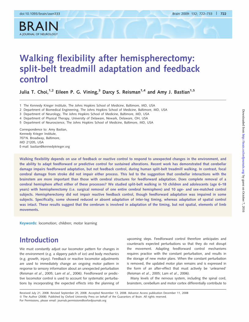
BRAIN A JOURNAL OF NEUROLOGY Walking flexibility after hemispherectomy: split-belt treadmill adaptation and feedback control Julia T. Choi, 1,2 Eileen P. G. Vining, 3 Darcy S. Reisman 1,4 and Amy J. Bastian 1,5 1 The Kennedy Krieger Institute, The Johns Hopkins School of Medicine, Baltimore, MD, USA 2 Department of Biomedical Engineering, The Johns Hopkins School of Medicine, Baltimore, MD, USA 3 Department of Neurology, The Johns Hopkins School of Medicine, Baltimore, MD, USA 4 Department of Physical Therapy, University of Delaware, Newark, Delaware, OH, USA 5 Department of Neuroscience, The Johns Hopkins School of Medicine, Baltimore, MD, USA Correspondence to: Amy Bastian, Kennedy Krieger Institute, 707 N. Broadway, Baltimore, MD 21205, USA E-mail: [email protected] Walking flexibility depends on use of feedback or reactive control to respond to unexpected changes in the environment, and the ability to adapt feedforward or predictive control for sustained alterations. Recent work has demonstrated that cerebellar damage impairs feedforward adaptation, but not feedback control, during human split-belt treadmill walking. In contrast, focal cerebral damage from stroke did not impair either process. This led to the suggestion that cerebellar interactions with the brainstem are more important than those with cerebral structures for feedforward adaptation. Does complete removal of a cerebral hemisphere affect either of these processes? We studied split-belt walking in 10 children and adolescents (age 6–18 years) with hemispherectomy (i.e. surgical removal of one entire cerebral hemisphere) and 10 age- and sex-matched control subjects. Hemispherectomy did not impair reactive feedback control, though feedforward adaptation was impaired in some subjects. Specifically, some showed reduced or absent adaptation of inter-leg timing, whereas adaptation of spatial control was intact. These results suggest that the cerebrum is involved in adaptation of the timing, but not spatial, elements of limb movements. Keywords: locomotion; children; motor learning Introduction We must constantly adjust our locomotor pattern for changes in the environment (e.g. a slippery patch of ice) and body mechanics (e.g. growth, injury). Feedback or reactive locomotor adjustments are used to immediately change an ongoing motor pattern in response to sensory information about an unexpected perturbation (Reisman et al., 2005; Lam et al., 2006). Feedforward or predic- tive locomotor control is used to account for systematic perturba- tions by incorporating the expected effects into the planning of upcoming steps. Feedforward control therefore anticipates and counteracts expected perturbations so that they do not disrupt the movement. Adapting feedforward control mechanisms requires practice with the constant perturbation, and results in the storage of new motor plans. When the constant perturbation is removed, the updated motor plan remains and is expressed in the form of an after-effect that must actively be ‘unlearned’ (Reisman et al., 2005; Lam et al., 2006). Many levels of the nervous system, including the spinal cord, brainstem, cerebellum and motor cortex differentially contribute to doi:10.1093/brain/awn333 Brain 2009: 132; 722–733 | 722 Received July 21, 2008. Revised September 25, 2008. Accepted November 13, 2008. Advance Access publication December 11, 2008 ß The Author (2008). Published by Oxford University Press on behalf of the Guarantors of Brain. All rights reserved. For Permissions, please email: [email protected] by guest on October 7, 2016 http://brain.oxfordjournals.org/ Downloaded from
-
Upload
independent -
Category
Documents
-
view
4 -
download
0
Transcript of Walking flexibility after hemispherectomy: split-belt treadmill adaptation and feedback control
BRAINA JOURNAL OF NEUROLOGY
Walking flexibility after hemispherectomy:split-belt treadmill adaptation and feedbackcontrolJulia T. Choi,1,2 Eileen P. G. Vining,3 Darcy S. Reisman1,4 and Amy J. Bastian1,5
1 The Kennedy Krieger Institute, The Johns Hopkins School of Medicine, Baltimore, MD, USA
2 Department of Biomedical Engineering, The Johns Hopkins School of Medicine, Baltimore, MD, USA
3 Department of Neurology, The Johns Hopkins School of Medicine, Baltimore, MD, USA
4 Department of Physical Therapy, University of Delaware, Newark, Delaware, OH, USA
5 Department of Neuroscience, The Johns Hopkins School of Medicine, Baltimore, MD, USA
Correspondence to: Amy Bastian,
Kennedy Krieger Institute,
707 N. Broadway, Baltimore,
MD 21205, USA
E-mail: [email protected]
Walking flexibility depends on use of feedback or reactive control to respond to unexpected changes in the environment, and
the ability to adapt feedforward or predictive control for sustained alterations. Recent work has demonstrated that cerebellar
damage impairs feedforward adaptation, but not feedback control, during human split-belt treadmill walking. In contrast, focal
cerebral damage from stroke did not impair either process. This led to the suggestion that cerebellar interactions with the
brainstem are more important than those with cerebral structures for feedforward adaptation. Does complete removal of a
cerebral hemisphere affect either of these processes? We studied split-belt walking in 10 children and adolescents (age 6–18
years) with hemispherectomy (i.e. surgical removal of one entire cerebral hemisphere) and 10 age- and sex-matched control
subjects. Hemispherectomy did not impair reactive feedback control, though feedforward adaptation was impaired in some
subjects. Specifically, some showed reduced or absent adaptation of inter-leg timing, whereas adaptation of spatial control
was intact. These results suggest that the cerebrum is involved in adaptation of the timing, but not spatial, elements of limb
movements.
Keywords: locomotion; children; motor learning
IntroductionWe must constantly adjust our locomotor pattern for changes in
the environment (e.g. a slippery patch of ice) and body mechanics
(e.g. growth, injury). Feedback or reactive locomotor adjustments
are used to immediately change an ongoing motor pattern in
response to sensory information about an unexpected perturbation
(Reisman et al., 2005; Lam et al., 2006). Feedforward or predic-
tive locomotor control is used to account for systematic perturba-
tions by incorporating the expected effects into the planning of
upcoming steps. Feedforward control therefore anticipates and
counteracts expected perturbations so that they do not disrupt
the movement. Adapting feedforward control mechanisms
requires practice with the constant perturbation, and results in
the storage of new motor plans. When the constant perturbation
is removed, the updated motor plan remains and is expressed in
the form of an after-effect that must actively be ‘unlearned’
(Reisman et al., 2005; Lam et al., 2006).
Many levels of the nervous system, including the spinal cord,
brainstem, cerebellum and motor cortex differentially contribute to
doi:10.1093/brain/awn333 Brain 2009: 132; 722–733 | 722
Received July 21, 2008. Revised September 25, 2008. Accepted November 13, 2008. Advance Access publication December 11, 2008
� The Author (2008). Published by Oxford University Press on behalf of the Guarantors of Brain. All rights reserved.
For Permissions, please email: [email protected]
by guest on October 7, 2016
http://brain.oxfordjournals.org/D
ownloaded from
these control processes during locomotion (Pearson, 2000;
Rossignol, 2006). A recent split-belt walking study demonstrated
that feedback control is intact in individuals with cerebellar
damage (Morton and Bastian, 2006). Subjects could immediately
react to the split treadmill by changing the stride length and
amount of time spent in stance on the faster and slower belts
in order to maintain one-to-one stepping. In contrast, cerebellar
damage disrupted feedforward adaptation: subjects were unable
to optimize spatial (e.g. step lengths) and temporal (e.g. phasing
between the legs) elements of the walking pattern in the face of
sustained split treadmill (Morton and Bastian, 2006). In contrast,
we found that adults with cerebral damage were able to make
both feedback adjustments and adapt feedforward control
(Reisman et al., 2007). Since cerebral damage did not impair the
tested locomotor capabilities (Reisman et al., 2007), we speculated
that cerebellar projections to the brainstem motor areas are more
important for feedforward adaptation than those to cerebral areas.
However, since the lesions in the cerebrum were often focal, over-
all involvement of the cerebrum in feedback control or feedfor-
ward adaptive process cannot be definitively ruled-out. Indeed,
studies have shown that neural activity in cerebral motor areas
relates to modifications in the locomotor pattern (Armstrong,
1986; Drew et al., 2002), and that lesions of the corticospinal
tract cause deficits in voluntary modifications (Drew, 1993; Drew
et al., 2002).
To further investigate cerebral contributions to locomotor adap-
tation, we studied split-belt walking adaptation in children and
adolescents who had undergone surgical removal of one entire
cerebral hemisphere to treat intractable epileptic seizures. We
tested whether cerebral structures are required for feedback
mediated locomotor adjustments that allow individuals to react
to split belts, and predictive feedforward locomotor adaptation
as measured by the presence of after-effects. Our results suggest
that hemispherectomized subjects have normal feedback control
because they were able to rapidly adjust walking to account for
split belts. Feedforward adaptation was partially impaired how-
ever; hemispherectomized subjects had some difficulty adapting
and storing a new timing pattern, even though they could adapt
and store the spatial pattern.
Methods
SubjectsWe studied 10 children and adolescents who had undergone hemi-
spherectomy for intractable epileptic seizures (Table 1). All of the
patients had hemidecortication, a procedure in which potentially epi-
leptogenic, unihemispheric, cortical grey matter is removed while the
underlying white matter and ventricles are left intact (Kossoff et al.,
2003). Post-operative brain scans show that the basal ganglia and
thalamus are partially preserved to different extents across patients.
Hemispherectomized subjects had hemiparesis of varying degree and
were excluded if they could not walk on the treadmill at a maximum
speed of 0.8 m/s. One of the patients tested (H5) had post-operative
seizures but did not experience any during testing. All patients,
except for H8, started walking before the surgery. We included
10 age- and sex-matched healthy control subjects (age range 7–18
years; median age 12.5 years; seven females, three males). All sub-
jects and parents gave informed consent prior to participating. The
protocol was approved by the Johns Hopkins Institutional Review
Board.
Clinical assessmentMotor function on the paretic leg was assessed using the Fugl-Meyer
scale, which measures reflexes, ability to move in and out of synergy
and movement coordination (Fugl-Meyer, 1975). Walking speed was
measured across a 25-feet walkway at their fastest speed. Touch sen-
sation was tested using Semmes Winstein graded monofilaments on
the great toe; the threshold was determined as the lowest gram fila-
ment that could be correctly detected on four out of five trials. The
behavioural inattention test (BIT) Star Cancellation was used to mea-
sure visuospatial neglect (Wilson et al., 1987).
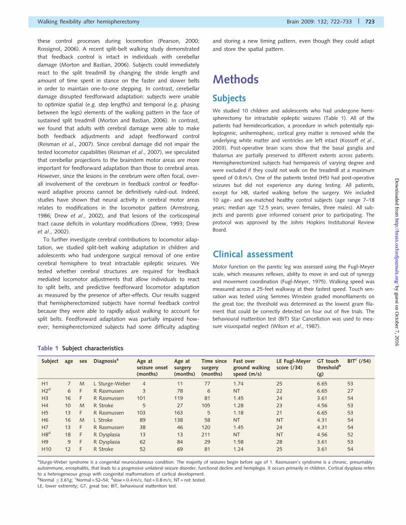
Table 1 Subject characteristics
Subject age sex Diagnosisa Age atseizure onset(months)
Age atsurgery(months)
Time sincesurgery(months)
Fast overground walkingspeed (m/s)
LE Fugl-Meyerscore (/34)
GT touchthresholdb
(g)
BITc (/54)
H1 7 M L Sturge-Weber 4 11 77 1.74 25 6.65 53
H2d 6 F R Rasmussen 3 78 6 NT 22 6.65 27
H3 16 F R Rasmussen 101 119 81 1.45 24 3.61 54
H4 10 M R Stroke 5 27 105 1.28 23 4.56 53
H5 13 F R Rasmussen 103 163 5 1.18 21 6.65 53
H6 16 M L Stroke 89 138 58 NT NT 4.31 54
H7 13 F R Rasmussen 38 46 120 1.45 24 4.31 54
H8d 18 F R Dysplasia 13 13 211 NT NT 4.56 52
H9 9 F R Dysplasia 62 84 29 1.58 28 3.61 53
H10 12 F R Stroke 52 69 81 1.24 25 3.61 54
aSturge-Weber syndrome is a congenital neurocutaneous condition. The majority of seizures begin before age of 1. Rasmussen’s syndrome is a chronic, presumablyautoimmune, encephalitis, that leads to a progressive unilateral seizure disorder, functional decline and hemiplegia. It occurs primarily in children. Cortical dysplasia refers
to a heterogeneous group with congenital malformations of cortical development.bNormal � 3.61g; cNormal = 52–54; dslow = 0.4 m/s; fast = 0.8 m/s; NT = not tested.LE, lower extremity; GT, great toe; BIT, behavioural inattention test.
Walking flexibility after hemispherectomy Brain 2009: 132; 722–733 | 723
by guest on October 7, 2016
http://brain.oxfordjournals.org/D
ownloaded from
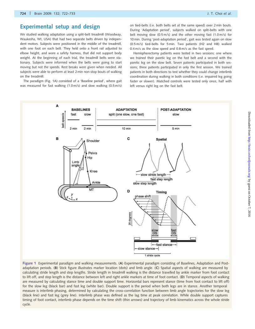
Experimental setup and designWe studied walking adaptation using a split-belt treadmill (Woodway,
Waukesha, WI, USA) that had two separate belts driven by indepen-
dent motors. Subjects were positioned in the middle of the treadmill,
with one foot on each belt. They held onto a front rail adjusted to
elbow height, and wore a safety harness, that did not support body
weight. At the beginning of each trial, the treadmill belts were sta-
tionary. Subjects were informed when the belts were going to start
moving but not the speeds. Rest breaks were given when needed. All
subjects were able to perform at least 2 min non-stop bouts of walking
on the treadmill.
The paradigm (Fig. 1A) consisted of a ‘Baseline period’, where gait
was measured for fast walking (1.0 m/s) and slow walking (0.5 m/s)
on tied-belts (i.e. both belts set at the same speed) over 2 min bouts.
During ‘Adaptation period’, subjects walked on split-belts with one
belt moving slow (0.5 m/s) and the other moving fast (1.0 m/s) for
10 min. During ‘post-adaptation period’, gait was tested again on slow
(0.5 m/s) tied-belts for 5 min. Two patients (H2 and H8) walked
0.4 m/s as the slow speed and 0.8 m/s as the fast speed.
Hemispherectomy patients were tested in two sessions: one where
we trained their paretic leg on the fast belt and a second with the
paretic leg on the slow belt. Seven patients participated in both ses-
sions; three patients participated in only the first session. We trained
patients in both directions to test whether they could change interlimb
coordination during walking in both conditions (i.e. impaired leg going
faster or slower). Matched controls were tested only once, half with
left versus right leg on the fast belt.
Figure 1 Experimental paradigm and walking measurements. (A) Experimental paradigm consisting of Baselines, Adaptation and Post-
adaptation periods. (B) Stick figure illustrates marker location (dots) and limb angle. (C) Spatial aspects of walking are measured by
calculating stride length and step lengths. Stride length in treadmill walking is the distance travelled by ankle marker from foot contact
to lift off, and step length is the distance between left and right ankle markers at time of foot contact. (D) Temporal aspects of walking
are measured by calculating stance time and double support time. Horizontal bars represent stance (time from foot contact to lift off)
for the slow leg (black bar) and fast leg (white bar). Double support is the period when both legs are in stance. Another temporal
measure is interlimb phasing, determined by calculating the cross-correlation function between limb angle trajectories for the slow leg
(black line) and fast leg (grey line). Interlimb phase was defined as the lag time at peak correlation. While double support captures
timing of foot contact, interlimb phase depends on the time shift (thin arrows) and trajectory of limb kinematics across the whole stride
cycle.
724 | Brain 2009: 132; 722–733 J. T. Choi et al.
by guest on October 7, 2016
http://brain.oxfordjournals.org/D
ownloaded from
Data collectionKinematic data were collected at 100 Hz using Optotrak (Northern
Digital, Inc.). Infrared-emitting markers were placed bilaterally over
the following joints (Fig. 1B): foot (fifth metatarsal head), ankle (lateral
malleolus), knee (lateral femoral epicondyle), hip (greater trochanter),
pelvis (iliac crest) and shoulder (acromion process). Foot-switches
placed on the bottom of shoes were used to record the times of
foot contact and lift off.
Data analysisA stride cycle is defined as the onset of foot contact to the onset of
the next foot contact on the same leg. We measured spatial charac-
teristics of walking (Fig. 1C) by calculating stride length (distance tra-
velled by ankle marker from foot contact to lift off) and step lengths
(distance between two ankle markers at time of foot contact). While
stride length is measured from a single leg, step length depends on the
spatial relationship between both legs. Our previous study (Reisman
et al., 2005) have shown that stride length is adjusted immediately
(within first few strides) in response to split-belt perturbation, which
reflects reactive feedback control. In contrast, step length is gradually
adapted over time and show after-effects when the perturbation is
removed, which suggests involvement of predictive feedforward
adaptation.
Temporal characteristics of walking (Fig. 1D) are measured by cal-
culating stance time (period from foot contact to lift off) as a percent
of stride cycle, and double support time (period toward end of stance
when both legs are in ground contact). Stance phase is determined
from foot contact of one leg, while double support phase describes the
temporal relationship of foot contact on both legs. Our previous study
(Reisman et al., 2005) have shown that stance time is adjusted imme-
diately (within first few strides) in response to split-belt perturbation,
which reflects reactive feedback control. In contrast, double support
time is gradually adapted over time and show after-effects when the
perturbation is removed, which suggests involvement of predictive
feedforward adaptation.
For all measures, a symmetry index was used to quantify the dif-
ference between two legs for each gait parameter: symmetry = (fast
leg – slow leg)/(fast leg + slow leg), where ‘slow leg’ and ‘fast leg’
refers to the leg on the slower and faster belt during split-belt condi-
tion, respectively. An index value of 0 would indicate that a gait
parameter is symmetric and equal for both legs. Positive values indi-
cate that the fast leg is taking a longer step, and negative values
indicate that the slow leg is taking a longer step.
We also measured interlimb coordination by calculating the cross-
correlation function (Signal Processing Toolbox, MATLAB) between
limb angle trajectories (Fig. 1D). Limb angle was defined as the
angle between the vertical and vector from hip to foot on the x–y
plane; positive values indicate that the foot is in front of the hip
(Fig. 1B). Each correlation function was calculated using limb angle
data spanning three consecutive foot contacts on the slow leg (i.e.
two stride cycles). The maximum time lag is limited to one stride
cycle. Interlimb phase was defined as the lag time at peak correlation,
and expressed as a fraction of the stride cycle. A phase of 0.5 cycle
means that the legs are moving out-of-phase, and a phase of 0 means
in-phase. By convention, positive lag times indicate a lead of the fast
leg relative to the slow leg. This method of calculating interlimb phase
was also used and described in our previous papers (Morton and
Bastian, 2006; Choi and Bastian, 2007; Reisman et al., 2007).
Double support time and interlimb phase both capture temporal
aspects of walking. While double support focuses on the discrete
timing of foot contact and lift off, interlimb phase considers limb kine-
matics across the whole stride cycle. The value for interlimb phase
depends on both the timing and the shape of limb trajectories. The
rate at which the hip rolls over the supporting foot shapes the limb
trajectory during stance. Hence, the two temporal parameters measure
different aspects of walking (foot contact versus limb kinematics), and
could vary separately to some extent.
Statistical analysisWe compared gait parameters across experiment periods using the last
five values in each baseline period, the first and last five values in each
adaptation period (early and late adaptation, respectively) and the first
and last five values in each post-adaptation period (early and late post-
adaptation, respectively). Repeated-measures ANOVA was used with
experimental groups (control, paretic leg fast, paretic leg slow) as the
between-subjects variable and experimental period as the within-sub-
jects variable; post hoc analyses were performed using the Tukey’s
significant different test. Watson’s U2 (circular statistic) was used to
compare phase across groups (Batschelet, 1981). We also conducted
specific comparisons between controls with the average of hemispher-
ectomy groups, and between paretic leg fast with paretic leg slow.
Planned comparisons were conducted to test components of interaction
for the adaptation period (A1 and A2) and post-adaptation period
(P1 and P2). We used �= 0.05 as the alpha level for each planned
comparison. Pearson product-moment correlations were used to test
for relationships between clinical and adaptation measures.
ResultsTable 1 shows demographic and clinical information for the sub-
jects with hemispherectomy. All subjects walked independently
without an assistive device, had voluntary active movement of
the paretic leg (i.e. Fugl-Meyer scores were in the twenties),
and had some tactile sensation of the paretic leg. One child
(H2) who had a right hemispherectomy 6 months prior showed
some evidence of neglect. Two subjects were tested 56 months
after their surgery; all others were 42 years post surgery. Fugl-
Meyer assessments and over ground walking measurements were
conducted on the second visit, and were not tested in three sub-
jects who only participated in the first session (H2, H6 and H8).
Feedback controlWe first asked whether the patients with hemispherectomy could
maintain alternating leg movements during split-belt walking,
which requires an immediate reaction to the treadmill. All children
and adolescents in this study produced alternating leg movements
similar to healthy adults (Reisman et al., 2005). None of the sub-
jects from either group produced walking patterns where the fast
leg took more steps then the slow leg on split-belts, which is more
common during split-belt stepping in younger infants (Yang et al.,
2005).
Alternating stepping was maintained by immediately adjusting
the stride length and stance time symmetry to match feedback
about the belt speeds. Figure 2A illustrates stride length asymme-
try from a typical control subject and hemispherectomy patient.
During baseline, the stride lengths are close to symmetric; during
Walking flexibility after hemispherectomy Brain 2009: 132; 722–733 | 725
by guest on October 7, 2016
http://brain.oxfordjournals.org/D
ownloaded from
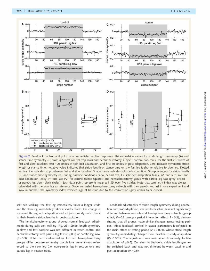
split-belt walking, the fast leg immediately takes a longer stride
and the slow leg immediately takes a shorter stride. The change is
sustained throughout adaptation and subjects quickly switch back
to their baseline stride lengths in post-adaptation.
The hemispherectomy group showed normal feedback adjust-
ments during split-belt walking (Fig. 2B). Stride length symmetry
in slow and fast baseline was not different between control and
hemispherectomy with paretic leg fast (P� 0.5) or paretic leg slow
(P = 0.3). Note that baseline values for two hemispherectomy
groups differ because symmetry calculations were always refer-
enced to the slow leg (i.e. non-paretic leg in session one and
paretic leg in session two).
Feedback adjustments of stride length symmetry during adapta-
tion and post-adaptation, relative to baseline, was not significantly
different between controls and hemispherectomy subjects (group
effect, P = 0.3; group�period interaction effect, P = 0.2), demon-
strating that all groups made similar changes across testing peri-
ods. Intact feedback control in spatial parameters is reflected in
the main effect of testing period (P50.001), where stride length
symmetry immediately changed from baseline to early adaptation
(P50.001). The adjustment was maintained from early to late
adaptation (P� 0.5). On return to tied-belts, stride length symme-
try switched back and was not different between baseline and
post-adaptation (P� 0.5).
Figure 2 Feedback control: ability to make immediate reactive responses. Stride-by-stride values for stride length symmetry (A) and
stance time symmetry (C) from a typical control (top row) and hemispherectomy subject (bottom two rows) for the first 20 strides of
fast and slow baselines, first 100 strides of split-belt adaptation, and first 60 strides of post-adaptation. Zero indicates symmetric stride
length or stance time, negative value indicates that stride length or stance time on the fast leg is shorter relative to slow leg. Dotted
vertical line indicates stop between fast and slow baseline. Shaded area indicates split-belts condition. Group averages for stride length
(B) and stance time symmetry (D) during baseline conditions (slow, S and fast, F), split-belt adaptation (early, A1 and late, A2) and
post-adaptation (early, P1 and late P2) for control (white squares) and hemispherectomy group with paretic leg fast (grey circles)
or paretic leg slow (black circles). Each data point represents mean� 1 SD over five strides. Note that symmetry index was always
calculated with the slow leg as reference. Since we tested hemispherectomy subjects with their paretic leg fast in one experiment and
slow in another, the symmetry index reversed sign at baseline due to this convention (grey versus black circles).
726 | Brain 2009: 132; 722–733 J. T. Choi et al.
by guest on October 7, 2016
http://brain.oxfordjournals.org/D
ownloaded from
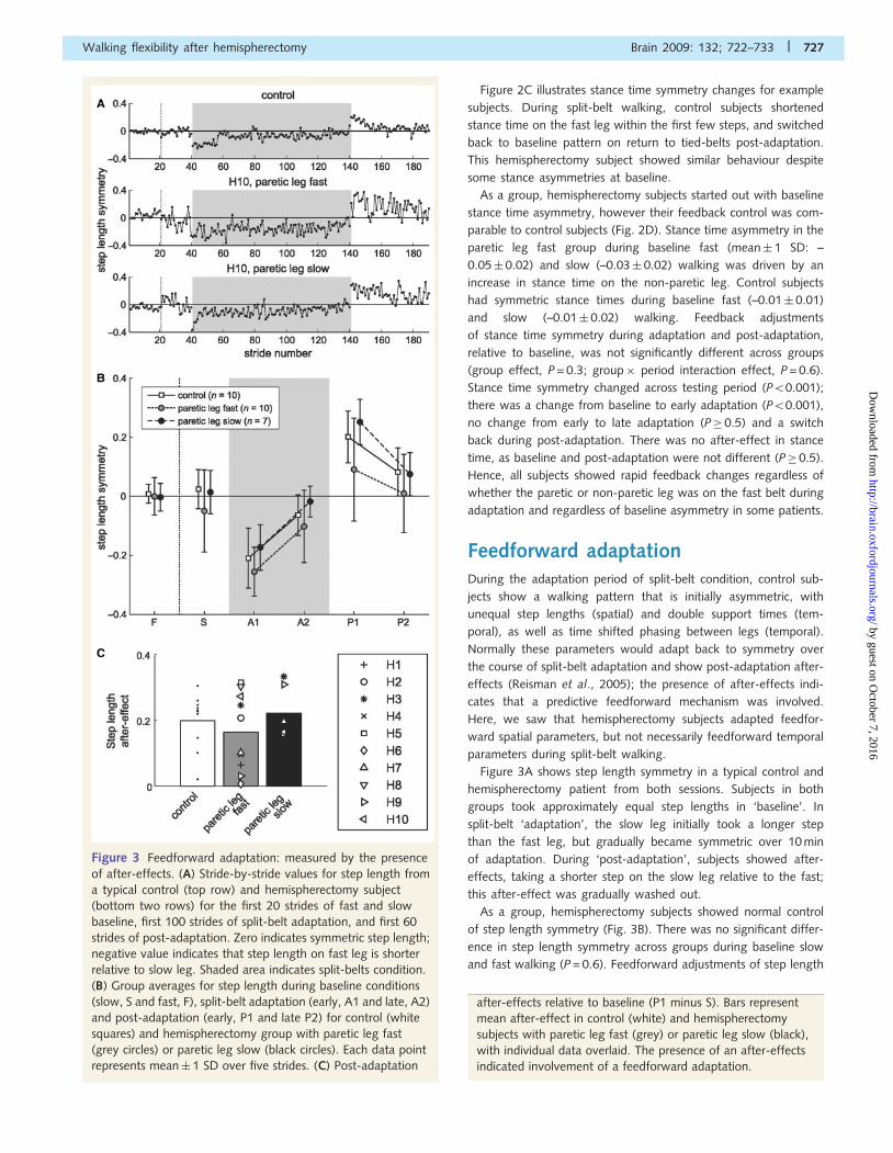
Figure 2C illustrates stance time symmetry changes for example
subjects. During split-belt walking, control subjects shortened
stance time on the fast leg within the first few steps, and switched
back to baseline pattern on return to tied-belts post-adaptation.
This hemispherectomy subject showed similar behaviour despite
some stance asymmetries at baseline.
As a group, hemispherectomy subjects started out with baseline
stance time asymmetry, however their feedback control was com-
parable to control subjects (Fig. 2D). Stance time asymmetry in the
paretic leg fast group during baseline fast (mean� 1 SD: –
0.05� 0.02) and slow (–0.03� 0.02) walking was driven by an
increase in stance time on the non-paretic leg. Control subjects
had symmetric stance times during baseline fast (–0.01� 0.01)
and slow (–0.01� 0.02) walking. Feedback adjustments
of stance time symmetry during adaptation and post-adaptation,
relative to baseline, was not significantly different across groups
(group effect, P = 0.3; group� period interaction effect, P = 0.6).
Stance time symmetry changed across testing period (P50.001);
there was a change from baseline to early adaptation (P50.001),
no change from early to late adaptation (P� 0.5) and a switch
back during post-adaptation. There was no after-effect in stance
time, as baseline and post-adaptation were not different (P�0.5).
Hence, all subjects showed rapid feedback changes regardless of
whether the paretic or non-paretic leg was on the fast belt during
adaptation and regardless of baseline asymmetry in some patients.
Feedforward adaptationDuring the adaptation period of split-belt condition, control sub-
jects show a walking pattern that is initially asymmetric, with
unequal step lengths (spatial) and double support times (tem-
poral), as well as time shifted phasing between legs (temporal).
Normally these parameters would adapt back to symmetry over
the course of split-belt adaptation and show post-adaptation after-
effects (Reisman et al., 2005); the presence of after-effects indi-
cates that a predictive feedforward mechanism was involved.
Here, we saw that hemispherectomy subjects adapted feedfor-
ward spatial parameters, but not necessarily feedforward temporal
parameters during split-belt walking.
Figure 3A shows step length symmetry in a typical control and
hemispherectomy patient from both sessions. Subjects in both
groups took approximately equal step lengths in ‘baseline’. In
split-belt ‘adaptation’, the slow leg initially took a longer step
than the fast leg, but gradually became symmetric over 10 min
of adaptation. During ‘post-adaptation’, subjects showed after-
effects, taking a shorter step on the slow leg relative to the fast;
this after-effect was gradually washed out.
As a group, hemispherectomy subjects showed normal control
of step length symmetry (Fig. 3B). There was no significant differ-
ence in step length symmetry across groups during baseline slow
and fast walking (P = 0.6). Feedforward adjustments of step length
Figure 3 Feedforward adaptation: measured by the presence
of after-effects. (A) Stride-by-stride values for step length from
a typical control (top row) and hemispherectomy subject
(bottom two rows) for the first 20 strides of fast and slow
baseline, first 100 strides of split-belt adaptation, and first 60
strides of post-adaptation. Zero indicates symmetric step length;
negative value indicates that step length on fast leg is shorter
relative to slow leg. Shaded area indicates split-belts condition.
(B) Group averages for step length during baseline conditions
(slow, S and fast, F), split-belt adaptation (early, A1 and late, A2)
and post-adaptation (early, P1 and late P2) for control (white
squares) and hemispherectomy group with paretic leg fast
(grey circles) or paretic leg slow (black circles). Each data point
represents mean� 1 SD over five strides. (C) Post-adaptation
after-effects relative to baseline (P1 minus S). Bars represent
mean after-effect in control (white) and hemispherectomy
subjects with paretic leg fast (grey) or paretic leg slow (black),
with individual data overlaid. The presence of an after-effects
indicated involvement of a feedforward adaptation.
Walking flexibility after hemispherectomy Brain 2009: 132; 722–733 | 727
by guest on October 7, 2016
http://brain.oxfordjournals.org/D
ownloaded from
symmetry during adaptation and post-adaptation, relative to base-
line, were not different between groups (group effect, P = 0.1;
group�period, P = 0.2), indicating that all subjects could recali-
brate step length over the split-belt period and subsequently
showed after-effects in tied-belts condition. Step length symmetry
was perturbed from baseline to early adaptation (P50.001), re-
established from early to late adaptation (P50.001), and showed
significant after-effects in post-adaptation (P50.001). Figure 3C
illustrates the mean after-effect magnitude relative to baseline and
individual subject data. Means were not statistically different
between control and hemispherectomy groups (P� 0.5), and not
different between paretic leg fast and paretic slow experiments
(P = 0.2). There was substantial variability in the after-effect size
of the control and hemispherectomy group with paretic leg fast,
with some subjects storing larger after-effects.
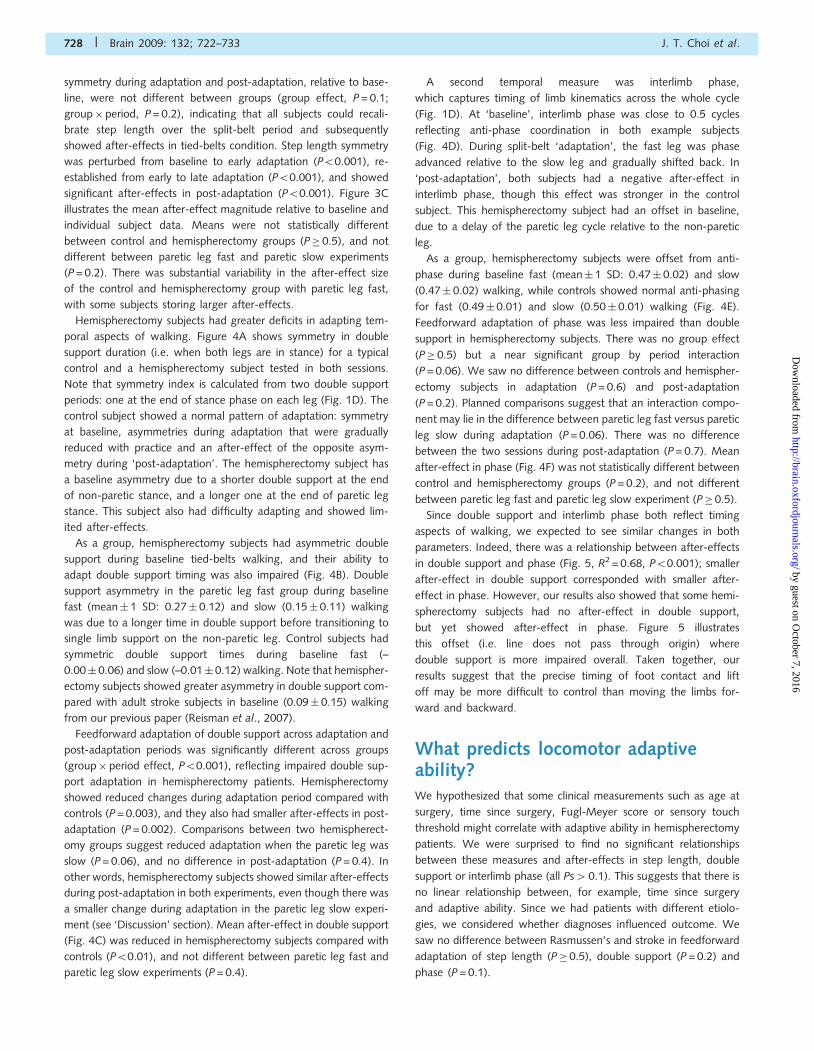
Hemispherectomy subjects had greater deficits in adapting tem-
poral aspects of walking. Figure 4A shows symmetry in double
support duration (i.e. when both legs are in stance) for a typical
control and a hemispherectomy subject tested in both sessions.
Note that symmetry index is calculated from two double support
periods: one at the end of stance phase on each leg (Fig. 1D). The
control subject showed a normal pattern of adaptation: symmetry
at baseline, asymmetries during adaptation that were gradually
reduced with practice and an after-effect of the opposite asym-
metry during ‘post-adaptation’. The hemispherectomy subject has
a baseline asymmetry due to a shorter double support at the end
of non-paretic stance, and a longer one at the end of paretic leg
stance. This subject also had difficulty adapting and showed lim-
ited after-effects.
As a group, hemispherectomy subjects had asymmetric double
support during baseline tied-belts walking, and their ability to
adapt double support timing was also impaired (Fig. 4B). Double
support asymmetry in the paretic leg fast group during baseline
fast (mean� 1 SD: 0.27� 0.12) and slow (0.15� 0.11) walking
was due to a longer time in double support before transitioning to
single limb support on the non-paretic leg. Control subjects had
symmetric double support times during baseline fast (–
0.00� 0.06) and slow (–0.01� 0.12) walking. Note that hemispher-
ectomy subjects showed greater asymmetry in double support com-
pared with adult stroke subjects in baseline (0.09� 0.15) walking
from our previous paper (Reisman et al., 2007).
Feedforward adaptation of double support across adaptation and
post-adaptation periods was significantly different across groups
(group� period effect, P50.001), reflecting impaired double sup-
port adaptation in hemispherectomy patients. Hemispherectomy
showed reduced changes during adaptation period compared with
controls (P = 0.003), and they also had smaller after-effects in post-
adaptation (P = 0.002). Comparisons between two hemispherect-
omy groups suggest reduced adaptation when the paretic leg was
slow (P = 0.06), and no difference in post-adaptation (P = 0.4). In
other words, hemispherectomy subjects showed similar after-effects
during post-adaptation in both experiments, even though there was
a smaller change during adaptation in the paretic leg slow experi-
ment (see ‘Discussion’ section). Mean after-effect in double support
(Fig. 4C) was reduced in hemispherectomy subjects compared with
controls (P50.01), and not different between paretic leg fast and
paretic leg slow experiments (P = 0.4).
A second temporal measure was interlimb phase,
which captures timing of limb kinematics across the whole cycle
(Fig. 1D). At ‘baseline’, interlimb phase was close to 0.5 cycles
reflecting anti-phase coordination in both example subjects
(Fig. 4D). During split-belt ‘adaptation’, the fast leg was phase
advanced relative to the slow leg and gradually shifted back. In
‘post-adaptation’, both subjects had a negative after-effect in
interlimb phase, though this effect was stronger in the control
subject. This hemispherectomy subject had an offset in baseline,
due to a delay of the paretic leg cycle relative to the non-paretic
leg.
As a group, hemispherectomy subjects were offset from anti-
phase during baseline fast (mean� 1 SD: 0.47� 0.02) and slow
(0.47� 0.02) walking, while controls showed normal anti-phasing
for fast (0.49� 0.01) and slow (0.50� 0.01) walking (Fig. 4E).
Feedforward adaptation of phase was less impaired than double
support in hemispherectomy subjects. There was no group effect
(P� 0.5) but a near significant group by period interaction
(P = 0.06). We saw no difference between controls and hemispher-
ectomy subjects in adaptation (P = 0.6) and post-adaptation
(P = 0.2). Planned comparisons suggest that an interaction compo-
nent may lie in the difference between paretic leg fast versus paretic
leg slow during adaptation (P = 0.06). There was no difference
between the two sessions during post-adaptation (P = 0.7). Mean
after-effect in phase (Fig. 4F) was not statistically different between
control and hemispherectomy groups (P = 0.2), and not different
between paretic leg fast and paretic leg slow experiment (P� 0.5).
Since double support and interlimb phase both reflect timing
aspects of walking, we expected to see similar changes in both
parameters. Indeed, there was a relationship between after-effects
in double support and phase (Fig. 5, R2 = 0.68, P50.001); smaller
after-effect in double support corresponded with smaller after-
effect in phase. However, our results also showed that some hemi-
spherectomy subjects had no after-effect in double support,
but yet showed after-effect in phase. Figure 5 illustrates
this offset (i.e. line does not pass through origin) where
double support is more impaired overall. Taken together, our
results suggest that the precise timing of foot contact and lift
off may be more difficult to control than moving the limbs for-
ward and backward.
What predicts locomotor adaptiveability?We hypothesized that some clinical measurements such as age at
surgery, time since surgery, Fugl-Meyer score or sensory touch
threshold might correlate with adaptive ability in hemispherectomy
patients. We were surprised to find no significant relationships
between these measures and after-effects in step length, double
support or interlimb phase (all Ps40.1). This suggests that there is
no linear relationship between, for example, time since surgery
and adaptive ability. Since we had patients with different etiolo-
gies, we considered whether diagnoses influenced outcome. We
saw no difference between Rasmussen’s and stroke in feedforward
adaptation of step length (P� 0.5), double support (P = 0.2) and
phase (P = 0.1).
728 | Brain 2009: 132; 722–733 J. T. Choi et al.
by guest on October 7, 2016
http://brain.oxfordjournals.org/D
ownloaded from
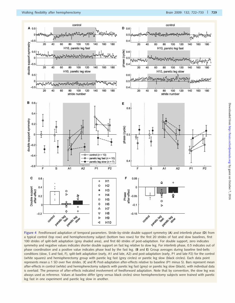
Figure 4 Feedforward adaptation of temporal parameters. Stride-by-stride double support symmetry (A) and interlimb phase (D) from
a typical control (top row) and hemispherectomy subject (bottom two rows) for the first 20 strides of fast and slow baselines, first
100 strides of split-belt adaptation (grey shaded area), and first 60 strides of post-adaptation. For double support, zero indicates
symmetry and negative values indicates shorter double support on fast leg relative to slow leg. For interlimb phase, 0.5 indicates out of
phase coordination and a positive value indicates phase lead by the fast leg. (B and E) Group averages during baseline tied-belts
conditions (slow, S and fast, F), split-belt adaptation (early, A1 and late, A2) and post-adaptation (early, P1 and late P2) for the control
(white squares) and hemispherectomy group with paretic leg fast (grey circles) or paretic leg slow (black circles). Each data point
represents mean� 1 SD over five strides. (C and F) Post-adaptation after-effects relative to baseline (P1 minus S). Bars represent mean
after-effects in control (white) and hemispherectomy subjects with paretic leg fast (grey) or paretic leg slow (black), with individual data
is overlaid. The presence of after-effects indicated involvement of feedforward adaptation. Note that by convention, the slow leg was
always used as reference. Values at baseline differ (grey versus black circles) since hemispherectomy subjects were trained with paretic
leg fast in one experiment and paretic leg slow in another.
Walking flexibility after hemispherectomy Brain 2009: 132; 722–733 | 729
by guest on October 7, 2016
http://brain.oxfordjournals.org/D
ownloaded from
The only relationship that we found was a negative correlation
(r = –0.59, P = 0.01) between baseline asymmetry and the after-
effect size for double support in patients. After-effects are nor-
mally due to the fast double support being lengthened relative to
the slow double support. At baseline hemispherectomy subjects’
paretic double support was longer than their non-paretic double
support. When patient’s paretic leg was the fast leg during adap-
tation, they did not show substantial after-effects; presumably
because they could not further lengthen the paretic leg double
support (i.e. asymmetry did not get worse). Conversely, hemi-
spherectomy subjects showed more substantial after-effects
when the non-paretic leg was the fast leg. Hence, the magnitude
of after-effect in double support seems to depend on initial asym-
metry and whether the paretic leg was adapted fast or slow. This
suggests a ceiling effect that limits the range of possible double
support asymmetry under experimental treadmill conditions.
DiscussionWe have demonstrated that a complete lesion of one cerebral
hemisphere does not impair many aspects of split-belt walking.
All hemispherectomized subjects were able to use feedback to
immediately change their locomotor pattern and maintain alter-
nating stepping during split-belt walking. Hemispherectomy
patients could also adapt feedforward control of spatial interlimb
coordination, but some subjects had difficulty adapting temporal
coordination of the legs.
Feedback control does not requirecerebral mechanismsOur results indicate that the ability to make quick, feedback-driven
adjustments to inter-limb coordination in walking do not depend on
cerebral mechanisms. This finding is consistent with other studies
showing that spinalized cats can also quickly adjust the stance and
swing time on each limb during split-belt walking (Forssberg et al.,
1980). Moreover, human infants whose descending pathways are
not fully myelinated (Yang et al., 2004) show reactive feedback
adjustments during split-belt walking (Yang et al., 2005). Subjects
with cerebellar damage can also make reactive feedback adjust-
ments, even though they are impaired in feedforward control
(Morton and Bastian, 2006). Hence, spinal circuits seem to be suffi-
cient for making quick reactive feedback adjustments on the split-
belt treadmill.
Cerebral versus cerebellar contributionsto feedforward adaptationOur previous studies have demonstrated that cerebellar lesions in
adults impair the ability to adapt feedforward locomotor coordina-
tion (Morton and Bastian, 2006). In contrast, cerebral strokes in
adults do not impair feedforward or feedback adaptive abilities
(Reisman et al., 2007). In the current study, subjects with hemi-
spherectomy showed partially disrupted feedforward adaptive abil-
ity (i.e. temporal but not spatial parameters) and intact feedback
control.
There are at least two possible explanations for impaired adap-
tation of timing parameters in these subjects. First, cerebral struc-
tures may play a role in adapting the timing of locomotor
activities. Studies of motor cortex in cats have suggested it has
exclusive capability (over brainstem structures) to influence the
timing of locomotor cycles (Rho et al., 1999; Bretzner and
Drew, 2005). The adults that we previously studied had only par-
tial lesions of cerebral motor areas, whereas the children and ado-
lescents studied here had complete lesions. Therefore, we may not
have seen the deficit in adults due to spared cerebral function.
Alternatively, the subjects with hemispherectomy might have
crossed cerebellar degeneration, or diaschisis (i.e. metabolic
depression, Baron et al., 1980). We would expect this to be
more substantial in hemispherectomized subjects versus adults
with stroke due to the difference in lesion size. In our prior
work, cerebellar damage caused deficits in adaptation of both
spatial and temporal parameters (Morton and Bastian, 2006).
Thus, if secondary cerebellar damage were the mechanism, we
would expect similar deficits; this did not occur. Anatomical studies
would also predict crossed degeneration to be localized to the
cerebellar hemisphere, whereas our previous study suggests that
midline cerebellar structures are more crucial for split-belt adapta-
tion (Morton and Bastian 2006). Taken together, these
Figure 5 Relationship between adaptation of double support
and interlimb phase. Each data point represents the after-effect
in double support (x-axis) and phase (y-axis) for individual
hemispherectomy subjects trained with paretic leg fast (grey
circles) or paretic leg slow (black circles). There is a clear
correlation (linear fit) between the two temporal parameters,
where smaller after-effect in double support corresponded with
smaller after-effect in phase. Notice offset in this relationship
(i.e. line shifted left of origin). Some hemispherectomy subjects
had small after-effect in double support, yet showed after-
effect in phase.
730 | Brain 2009: 132; 722–733 J. T. Choi et al.
by guest on October 7, 2016
http://brain.oxfordjournals.org/D
ownloaded from

observations suggest that crossed cerebellar degeneration is not
the cause of the deficit.
Separable feedforward adaptation ofspatial and temporal walkingparametersEven though spatial and temporal variables in walking are inher-
ently related, we found here that they can be recalibrated sepa-
rately. Most hemispherectomy subjects could adapt spatial
features, but had trouble adapting temporal coordination. One
possibility is that there are distinct neural mechanisms for modu-
lating spatial and temporal variables. There is evidence that loco-
motor cycle timing and cycle pattern (i.e. spatial features) are
controlled by different interneuronal populations in the spinal
cord (Koshland and Smith, 1989; Lafreniere-Roula and McCrea,
2005; McCrea and Rybak, 2008). While both the motor cortex
and red nucleus have access to the circuits controlling the cycle
structure, only the motor cortex has access to the circuits control-
ling cycle timing (Rho et al., 1999; Bretzner and Drew, 2005). This
may explain why timing adaptation may require additional cere-
bral involvement.
The fact that hemispherectomy subjects had baseline asymme-
tries in temporal features (i.e. double support time, interlimb phase
and stance time), but not in spatial features (i.e. step length and
stride length) provides additional evidence that there are separate
mechanisms for spatial verses temporal control. Given that the
subjects had some time to recover after surgery, it seems that
the nervous system is better at re-establishing spatial parameters
than temporal parameters in walking. It is possible that spatial
modification requires a simpler adjustment, such as gain modula-
tion, while temporal modification requires more complicated pro-
cesses involving cortical control.
It should also be noted that, we saw an unusual effect in adap-
tation of double support in hemispherectomy subjects. In the pare-
tic slow condition, there was no perturbation (i.e. error) in double
support symmetry from baseline to the split-belt condition, possi-
bly due to a ceiling effect, and yet a small after-effect occurred
post-adaptation (Fig. 4B). An after-effect resulting from small or
no errors suggest that adaptation was not used to merely cancel
‘performance errors’ in double support symmetry. Recent work
suggests that a sensory ‘prediction error’ might also be important
for driving adaptation. This error is the difference between an
internal prediction about the outcome of a motor command,
versus the actual sensed outcome—in other words, did the body
move where the brain thought it would (Miall et al., 1993;
Krakauer and Shadmehr, 2007; Tseng et al., 2007). Therefore,
one plausible explanation for this finding is that a sensory predic-
tion error, rather than a performance error, drove adaptation. It is
also possible that the error in another parameter (e.g. inter-limb
phase) drove adaptation.
Table 2 summarizes cerebellum and cerebrum involvement in
feedback control and feedforward adaptation during split-belt
walking. Taken together, these findings suggest that intact cere-
bellar function is required to adapt spatial parameters; additional
interactions between cerebellum and cerebral structures are
involved to adapt timing. While we cannot rule out extensive
plastic changes in many brain regions of these children and ado-
lescents that may underlie the pattern and compensations
observed, what is clear is that the structures remaining following
hemispherectomy are not always capable of recalibrating and stor-
ing interlimb timing mechanisms for walking.
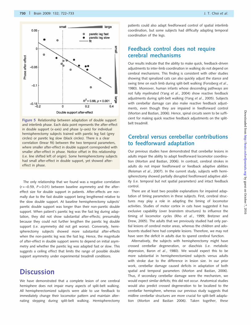
Comparison with adult stroke subjectsThere are two key differences in the hemispherectomy subjects
studied here and the stroke survivors studied in Reisman et al.
(2007): lesion size and age at which the lesion occurred. One
hypothesis is that hemispherectomy subjects would be more
impaired because their lesion is bigger. An alternate hypothesis
is that hemispherectomy subjects may have better recovery since
the lesion occurred at an earlier age when the brain might be
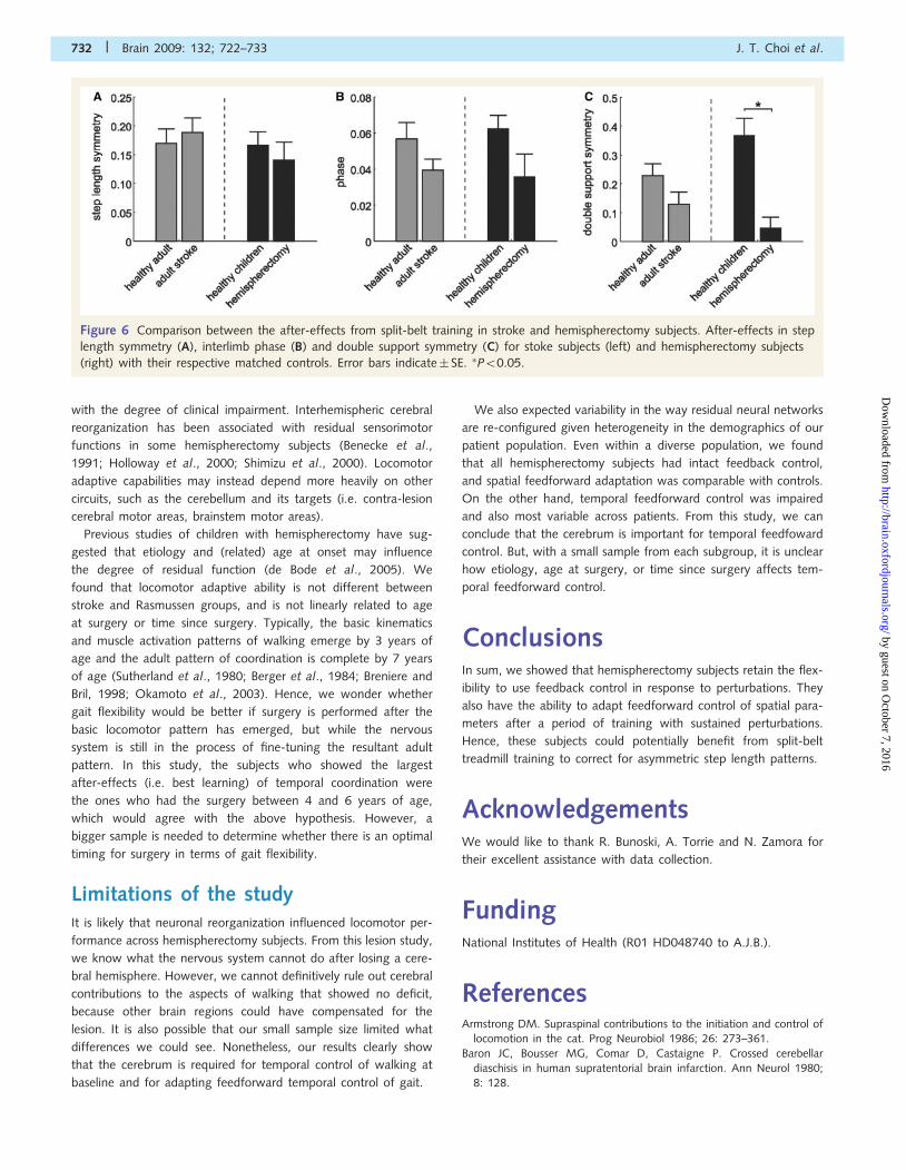
more plastic. Figure 6 shows a post hoc comparison of the
after-effects in step length, double support and phasing from
split-belt adaptation in adult stroke survivors (Reisman et al.,
2007) and children and adolescents with hemispherectomy with
their respective matched controls. Step length adaptation was
intact in both stroke (P� 0.5) and hemispherectomy (P� 0.5) sub-
jects compared with matched controls (Fig. 6A). The ability to
adapt interlimb phasing was also largely intact in both stroke
(P� 0.5) and hemispherectomy (P = 0.2) subjects compared with
controls (Fig. 6B). However, double support after-effects (Fig. 6C)
were reduced in hemispherectomy subjects compared with control
subjects (P50.001), while stroke subjects were comparable with
healthy adults (P = 0.1). These results suggest that the retention of
adaptive ability is worse in patients with larger cortical lesions, and
that the overall younger age at the time of the lesion was not the
more critical factor.
Clinical correlates and implications forfunctional recoveryWe were somewhat surprised to find no correlation between sen-
sorimotor functions (i.e. Fugl-Meyer scores, sensory thresholds)
and locomotor adaptive function. This suggests that the mechan-
isms underlying recovery of voluntary movement and sensory per-
ception are not related to those underlying locomotor recovery.
Studies in stroke survivors have also found that the magnitude of
after-effects from split-belt treadmill (Reisman et al., 2007) and
visuomotor (Patton et al., 2006) adaptation were not correlated
Table 2 Summary of feedback control and feedforwardadaptation during split-belt walking.
Lesion Feedback control Feedforward adaptation
Spatial(i.e.stridelength)
Temporal(i.e.stancetime)
Spatial(i.e. steplength)
Temporal(i.e.interlimbphase)
Cerebellum Good Good Bad Bad
Cerebrum (focal) Good Good Good Good
Cerebrum(entire hemisphere)
Good Good Good Bad
Walking flexibility after hemispherectomy Brain 2009: 132; 722–733 | 731
by guest on October 7, 2016
http://brain.oxfordjournals.org/D
ownloaded from
with the degree of clinical impairment. Interhemispheric cerebral
reorganization has been associated with residual sensorimotor
functions in some hemispherectomy subjects (Benecke et al.,
1991; Holloway et al., 2000; Shimizu et al., 2000). Locomotor
adaptive capabilities may instead depend more heavily on other
circuits, such as the cerebellum and its targets (i.e. contra-lesion
cerebral motor areas, brainstem motor areas).
Previous studies of children with hemispherectomy have sug-
gested that etiology and (related) age at onset may influence
the degree of residual function (de Bode et al., 2005). We
found that locomotor adaptive ability is not different between
stroke and Rasmussen groups, and is not linearly related to age
at surgery or time since surgery. Typically, the basic kinematics
and muscle activation patterns of walking emerge by 3 years of
age and the adult pattern of coordination is complete by 7 years
of age (Sutherland et al., 1980; Berger et al., 1984; Breniere and
Bril, 1998; Okamoto et al., 2003). Hence, we wonder whether
gait flexibility would be better if surgery is performed after the
basic locomotor pattern has emerged, but while the nervous
system is still in the process of fine-tuning the resultant adult
pattern. In this study, the subjects who showed the largest
after-effects (i.e. best learning) of temporal coordination were
the ones who had the surgery between 4 and 6 years of age,
which would agree with the above hypothesis. However, a
bigger sample is needed to determine whether there is an optimal
timing for surgery in terms of gait flexibility.
Limitations of the studyIt is likely that neuronal reorganization influenced locomotor per-
formance across hemispherectomy subjects. From this lesion study,
we know what the nervous system cannot do after losing a cere-
bral hemisphere. However, we cannot definitively rule out cerebral
contributions to the aspects of walking that showed no deficit,
because other brain regions could have compensated for the
lesion. It is also possible that our small sample size limited what
differences we could see. Nonetheless, our results clearly show
that the cerebrum is required for temporal control of walking at
baseline and for adapting feedforward temporal control of gait.
We also expected variability in the way residual neural networks
are re-configured given heterogeneity in the demographics of our
patient population. Even within a diverse population, we found
that all hemispherectomy subjects had intact feedback control,
and spatial feedforward adaptation was comparable with controls.
On the other hand, temporal feedforward control was impaired
and also most variable across patients. From this study, we can
conclude that the cerebrum is important for temporal feedfoward
control. But, with a small sample from each subgroup, it is unclear
how etiology, age at surgery, or time since surgery affects tem-
poral feedforward control.
ConclusionsIn sum, we showed that hemispherectomy subjects retain the flex-
ibility to use feedback control in response to perturbations. They
also have the ability to adapt feedforward control of spatial para-
meters after a period of training with sustained perturbations.
Hence, these subjects could potentially benefit from split-belt
treadmill training to correct for asymmetric step length patterns.
AcknowledgementsWe would like to thank R. Bunoski, A. Torrie and N. Zamora for
their excellent assistance with data collection.
FundingNational Institutes of Health (R01 HD048740 to A.J.B.).
ReferencesArmstrong DM. Supraspinal contributions to the initiation and control of
locomotion in the cat. Prog Neurobiol 1986; 26: 273–361.
Baron JC, Bousser MG, Comar D, Castaigne P. Crossed cerebellar
diaschisis in human supratentorial brain infarction. Ann Neurol 1980;
8: 128.
Figure 6 Comparison between the after-effects from split-belt training in stroke and hemispherectomy subjects. After-effects in step
length symmetry (A), interlimb phase (B) and double support symmetry (C) for stoke subjects (left) and hemispherectomy subjects
(right) with their respective matched controls. Error bars indicate� SE. �P50.05.
732 | Brain 2009: 132; 722–733 J. T. Choi et al.
by guest on October 7, 2016
http://brain.oxfordjournals.org/D
ownloaded from
Batschelet E. Circular Statistics in Biology. London: Academic Press; 1981.Benecke R, Meyer BU, Freund HJ. Reorganisation of descending motor
pathways in patients after hemispherectomy and severe hemispheric
lesions demonstrated by magnetic brain stimulation. Exp Brain Res
1991; 83: 419–26.Berger W, Altenmueller E, Dietz V. Normal and impaired development of
children’s gait. Hum Neurobiol 1984; 3: 163–70.
Breniere Y, Bril B. Development of postural control of gravity forces in
children during the first 5 years of walking. Exp Brain Res 1998; 121:255–62.
Bretzner F, Drew T. Contribution of the motor cortex to the structure
and the timing of hindlimb locomotion in the cat: a microstimulationstudy. J Neurophysiol 2005; 94: 657–72.
Choi JT, Bastian AJ. Adaptation reveals independent control networks for
human walking. Nat Neurosci 2007; 10: 1055–62.
de Bode S, Firestine A, Mathern GW, Dobkin B. Residual motor controland cortical representations of function following hemispherectomy:
effects of etiology. J Child Neurol 2005; 20: 64–75.
Drew T. Motor cortical activity during voluntary gait modifications in the
cat. I. Cells related to the forelimbs. J Neurophysiol 1993; 70: 179–99.Drew T, Jiang W, Widajewicz W. Contributions of the motor cortex to
the control of the hindlimbs during locomotion in the cat. Brain Res
Brain Res Rev 2002; 40: 178–91.
Forssberg H, Grillner S, Halbertsma J, Rossignol S. The locomotion of thelow spinal cat. II. Interlimb coordination. Acta Physiol Scand 1980;
108: 283–95.
Fugl-Meyer AR, Jaasko L, Leyman I, Olsson S, Steglind S. The post-stroke hemiplegic patient. 1. A method for evaluation of physical per-
formance. Scand J Rehabil Med 1975; 7: 13–31.
Holloway V, Gadian DG, Vargha-Khadem F, Porter DA, Boyd SG,
Connelly A. The reorganization of sensorimotor function in childrenafter hemispherectomy. A functional MRI and somatosensory evoked
potential study. Brain 2000; 123 (Pt 12): 2432–44.
Koshland GF, Smith JL. Mutable and immutable features of paw-shake
responses after hindlimb deafferentation in the cat. J Neurophysiol1989; 62: 162–73.
Kossoff EH, Vining EP, Pillas DJ, Pyzik PL, Avellino AM, Carson BS, et al.
Hemispherectomy for intractable unihemispheric epilepsy etiology vsoutcome. Neurology 2003; 61: 887–90.
Krakauer JW, Shadmehr R. Towards a computational neuropsychology of
action. Prog Brain Res 2007; 165: 383–94.
Lafreniere-Roula M, McCrea DA. Deletions of rhythmic motoneuronactivity during fictive locomotion and scratch provide clues to the
organization of the mammalian central pattern generator. J
Neurophysiol 2005; 94: 1120–32.
Lam T, Anderschitz M, Dietz V. Contribution of feedback and feedfor-ward strategies to locomotor adaptations. J Neurophysiol 2006; 95:
766–73.
McCrea DA, Rybak IA. Organization of mammalian locomotor rhythm
and pattern generation. Brain Res Rev 2008; 57: 134–46.Miall RC, Weir DJ, Wolpert DM, Stein JF. Is the cerebellum a smith
predictor? J Mot Behav 1993; 25: 203–16.
Morton SM, Bastian AJ. Cerebellar contributions to locomotor adapta-
tions during splitbelt treadmill walking. J Neurosci 2006; 26: 9107–16.Okamoto T, Okamoto K, Andrew PD. Electromyographic developmental
changes in one individual from newborn stepping to mature walking.
Gait Posture 2003; 17: 18–27.Patton JL, Stoykov ME, Kovic M, Mussa-Ivaldi FA. Evaluation of robotic
training forces that either enhance or reduce error in chronic hemi-
paretic stroke survivors. Exp Brain Res 2006; 168: 368–83.
Pearson KG. Neural adaptation in the generation of rhythmic behavior.Annu Rev Physiol 2000; 62: 723–53.
Reisman DS, Block HJ, Bastian AJ. Interlimb coordination during locomo-
tion: what can be adapted and stored? J Neurophysiol 2005; 94:
2403–15.Reisman DS, Wityk R, Silver K, Bastian AJ. Locomotor adaptation on a
split-belt treadmill can improve walking symmetry post-stroke. Brain
2007; 130: 1861–72.
Rho MJ, Lavoie S, Drew T. Effects of red nucleus microstimulation on thelocomotor pattern and timing in the intact cat: a comparison with the
motor cortex. J Neurophysiol 1999; 81: 2297–315.
Rossignol S. Plasticity of connections underlying locomotor recovery aftercentral and/or peripheral lesions in the adult mammals. Philos Trans R
Soc Lond B Biol Sci 2006; 361: 1647–71.
Shimizu T, Nariai T, Maehara T. Enhanced motor cortical excitability in
the unaffected hemisphere after hemispherectomy. Neuroreport 2000;11: 3077–84.
Sutherland DH, Olshen R, Cooper L, Woo SL. The development of
mature gait. J Bone Joint Surg Am 1980; 62: 336–53.
Tseng YW, Diedrichsen J, Krakauer JW, Shadmehr R, Bastian AJ. Sensoryprediction errors drive cerebellum-dependent adaptation of reaching. J
Neurophysiol 2007; 98: 54–62.
Wilson B, Cockburn J, Halligan P. Development of a behavioral-test ofvisuospatial neglect. Arch Phys Med Rehab 1987; 68: 98–102.
Yang JF, Lam T, Pang MY, Lamont E, Musselman K, Seinen E. Infant
stepping: a window to the behaviour of the human pattern generator
for walking. Can J Physiol Pharmacol 2004; 82: 662–74.Yang JF, Lamont EV, Pang MY. Split-belt treadmill stepping in infants
suggests autonomous pattern generators for the left and right leg in
humans. J Neurosci 2005; 25: 6869–76.
Walking flexibility after hemispherectomy Brain 2009: 132; 722–733 | 733
by guest on October 7, 2016
http://brain.oxfordjournals.org/D
ownloaded from