Von der Kammer critical review 2012
18
Nanomaterials in the Environment Critical Review ANALYSIS OF ENGINEERED NANOMATERIALS IN COMPLEX MATRICES (ENVIRONMENT AND BIOTA): GENERAL CONSIDERATIONS AND CONCEPTUAL CASE STUDIES FRANK VON DER KAMMER, y P. LEE FERGUSON, z PATRICIA A. HOLDEN,§ ARMAND MASION, k KIM R. ROGERS,# STEPHEN J. KLAINE, yy ALBERT A. KOELMANS, zz NINA HORNE,§§ and JASON M. UNRINE * kk yDepartment of Environmental Geosciences, University of Vienna, Vienna, Austria zDepartment of Civil and Environmental Engineering, Duke University, Durham, North Carolina, USA §Bren School of Environmental Sciences and Management, University of California, Santa Barbara, California, USA kCentre Europe ´en de Recherche et d’Enseignement des Ge ´osciences de l’Environnement, CNRS and Aix-Marseille University, Aix en Provence, France #Office of Research and Development, United States Environmental Protection Agency, Las Vegas, Nevada, USA yyInstitute of Environmental Toxicology, Clemson University, Pendleton, South Carolina, USA zzAquatic Ecology and Water Quality Management, Wageningen University, IJmuiden, The Netherlands §§Center for Integrated Nanoscale Materials, University of California, Berkeley, California, USA kkDepartment of Plant and Soil Sciences, University of Kentucky, Lexington, Kentucky, USA (Submitted 7 February 2011; Returned for Revision 30 March 2011; Accepted 29 June 2011) Abstract —Advances in the study of the environmental fate, transport, and ecotoxicological effects of engineered nanomaterials (ENMs) have been hampered by a lack of adequate techniques for the detection and quantification of ENMs at environmentally relevant concentrations in complex media. Analysis of ENMs differs from traditional chemical analysis because both chemical and physical forms must be considered. Because ENMs are present as colloidal systems, their physicochemical properties are dependent on their surroundings. Therefore, the simple act of trying to isolate, observe, and quantify ENMs may change their physicochemical properties, making analysis extremely susceptible to artifacts. Many analytical techniques applied in materials science and other chemical/ biological/physical disciplines may be applied to ENM analysis as well; however, environmental and biological studies may require that methods be adapted to work at low concentrations in complex matrices. The most pressing research needs are the development of techniques for extraction, cleanup, separation, and sample storage that introduce minimal artifacts to increase the speed, sensitivity, and specificity of analytical techniques, as well as the development of techniques that can differentiate between abundant, naturally occurring particles, and manufactured nanoparticles. Environ. Toxicol. Chem. 2012;31:32–49. # 2011 SETAC Keywords —Nanomaterial Carbon nanomaterial Metal oxide nanoparticle Characterization Quantum dot INTRODUCTION In the field of materials science, characterization approaches for engineered nanomaterial (ENM) properties are underdevel- oped, including the use of multiple complementary analytical methods [1]. By extension, quantifying and characterizing ENMs in complex matrices such as soils, sediments, and bio- logical tissues is a nascent endeavor. However, because of the biologically and environmentally relevant concentrations involved, and the inherent sample heterogeneity, development of techniques for detection and characterization in complex media is inherently more challenging than the development of basic materialcharacterization techniques. For example, basic dynamic light scattering is only applicable in fairly simple, homogeneous systems, at particle concentrations typically exceeding 1 mg/L, depending on the material. In many cases, new approaches must be developed or adapted. An area of great need is the development of rapid, sensitive techniques that can be applied in real time to support exposure characterization during toxicity testing. Some approaches used in traditional chemical analysis of environmental and biological samples may be adapted; how- ever, nanomaterials exist in colloidal systems with inherent discontinuities of properties, and therefore, detection and anal- ysis need to address not only chemical but also physical form. As a result, either traditional approaches used for analyzing organic molecules and trace elements in complex matrices must be modified or new approaches should be developed. Applica- tion of techniques used in traditional colloid science can serve as a good starting point [2]. Engineered nanomaterial behavior and toxicity are influenced by a wide variety of physical and chemical properties, such as chemical composition, surface functionality, particle size, surface area, redox state, crystal- linity, and solubility [3]. Future analytical strategies must be tailored to account for not only the sample and particle type, but also the specific question or hypothesis to be addressed, because characterizing every possible property for every sample would be impossible. This paper outlines approaches for detecting and character- izing nanomaterials in complex media and identifies areas in which additional research and development are needed as well as potential pathways forward in the development of appro- priate techniques. We discuss sample preparation, storage, and analysis. Current analytical approaches are also described, Environmental Toxicology and Chemistry, Vol. 31, No. 1, pp. 32–49, 2012 # 2011 SETAC Printed in the USA DOI: 10.1002/etc.723 This paper evolved from discussions held at a SETAC-endorsed Tech- nical Workshop held at Clemson University in August, 2010. The workshop was sponsored by the United States Environmental Protection Agency (U.S. EPA), Arcadis-US, and the Clemson University Institute of Environmental Toxicology. * To whom correspondence may be addressed ([email protected]). Published online 21 October 2011 in Wiley Online Library (wileyonlinelibrary.com). 32
Transcript of Von der Kammer critical review 2012
Nanomaterials in the Environment
Critical Review
ANALYSIS OF ENGINEERED NANOMATERIALS IN COMPLEX MATRICES (ENVIRONMENTAND BIOTA): GENERAL CONSIDERATIONS AND CONCEPTUAL CASE STUDIES
FRANK VON DER KAMMER,y P. LEE FERGUSON,z PATRICIA A. HOLDEN,§ ARMAND MASION,k KIM R. ROGERS,#
STEPHEN J. KLAINE,yy ALBERT A. KOELMANS,zz NINA HORNE,§§ and JASON M. UNRINE*kkyDepartment of Environmental Geosciences, University of Vienna, Vienna, Austria
zDepartment of Civil and Environmental Engineering, Duke University, Durham, North Carolina, USA
§Bren School of Environmental Sciences and Management, University of California, Santa Barbara, California, USA
kCentre Europeen de Recherche et d’Enseignement des Geosciences de l’Environnement, CNRS and Aix-Marseille University, Aix en Provence, France
#Office of Research and Development, United States Environmental Protection Agency, Las Vegas, Nevada, USA
yyInstitute of Environmental Toxicology, Clemson University, Pendleton, South Carolina, USA
zzAquatic Ecology and Water Quality Management, Wageningen University, IJmuiden, The Netherlands
§§Center for Integrated Nanoscale Materials, University of California, Berkeley, California, USA
kkDepartment of Plant and Soil Sciences, University of Kentucky, Lexington, Kentucky, USA
(Submitted 7 February 2011; Returned for Revision 30 March 2011; Accepted 29 June 2011)
Abstract—Advances in the study of the environmental fate, transport, and ecotoxicological effects of engineered nanomaterials (ENMs)have been hampered by a lack of adequate techniques for the detection and quantification of ENMs at environmentally relevantconcentrations in complex media. Analysis of ENMs differs from traditional chemical analysis because both chemical and physicalforms must be considered. Because ENMs are present as colloidal systems, their physicochemical properties are dependent on theirsurroundings. Therefore, the simple act of trying to isolate, observe, and quantify ENMs may change their physicochemical properties,making analysis extremely susceptible to artifacts. Many analytical techniques applied in materials science and other chemical/biological/physical disciplines may be applied to ENM analysis as well; however, environmental and biological studies may require thatmethods be adapted to work at low concentrations in complex matrices. The most pressing research needs are the development oftechniques for extraction, cleanup, separation, and sample storage that introduce minimal artifacts to increase the speed, sensitivity, andspecificity of analytical techniques, as well as the development of techniques that can differentiate between abundant, naturallyoccurring particles, and manufactured nanoparticles. Environ. Toxicol. Chem. 2012;31:32–49. # 2011 SETAC
Keywords—Nanomaterial Carbon nanomaterial Metal oxide nanoparticle Characterization Quantum dot
INTRODUCTION
In the field of materials science, characterization approachesfor engineered nanomaterial (ENM) properties are underdevel-oped, including the use of multiple complementary analyticalmethods [1]. By extension, quantifying and characterizingENMs in complex matrices such as soils, sediments, and bio-logical tissues is a nascent endeavor. However, because of thebiologically and environmentally relevant concentrationsinvolved, and the inherent sample heterogeneity, developmentof techniques for detection and characterization in complexmedia is inherently more challenging than the development ofbasic materialcharacterization techniques. For example, basicdynamic light scattering is only applicable in fairly simple,homogeneous systems, at particle concentrations typicallyexceeding 1mg/L, depending on the material. In many cases,new approaches must be developed or adapted. An area of greatneed is the development of rapid, sensitive techniques that can
be applied in real time to support exposure characterizationduring toxicity testing.
Some approaches used in traditional chemical analysis ofenvironmental and biological samples may be adapted; how-ever, nanomaterials exist in colloidal systems with inherentdiscontinuities of properties, and therefore, detection and anal-ysis need to address not only chemical but also physical form.As a result, either traditional approaches used for analyzingorganic molecules and trace elements in complex matrices mustbe modified or new approaches should be developed. Applica-tion of techniques used in traditional colloid science can serveas a good starting point [2]. Engineered nanomaterial behaviorand toxicity are influenced by a wide variety of physical andchemical properties, such as chemical composition, surfacefunctionality, particle size, surface area, redox state, crystal-linity, and solubility [3]. Future analytical strategies must betailored to account for not only the sample and particle type, butalso the specific question or hypothesis to be addressed, becausecharacterizing every possible property for every sample wouldbe impossible.
This paper outlines approaches for detecting and character-izing nanomaterials in complex media and identifies areas inwhich additional research and development are needed as wellas potential pathways forward in the development of appro-priate techniques. We discuss sample preparation, storage, andanalysis. Current analytical approaches are also described,
Environmental Toxicology and Chemistry, Vol. 31, No. 1, pp. 32–49, 2012# 2011 SETAC
Printed in the USADOI: 10.1002/etc.723
This paper evolved from discussions held at a SETAC-endorsed Tech-nical Workshop held at Clemson University in August, 2010. The workshopwas sponsored by the United States Environmental Protection Agency (U.S.EPA), Arcadis-US, and the Clemson University Institute of EnvironmentalToxicology.
* To whom correspondence may be addressed([email protected]).
Published online 21 October 2011 in Wiley Online Library(wileyonlinelibrary.com).
32

including traditional approaches based on separation and detec-tion; imaging-based approaches; and the possible use of bio-logical sensor-based approaches. Using four case studies, wehighlight how an intended analytical approach should be cus-tomized based on the nanomaterial in question, the samplematrix, and the hypothesis or objective. Finally, we outline themost critical and pressing research needs for detecting andanalyzing nanomaterials in complex matrices. The first casestudy addresses the analysis of carbon nanomaterials (CNMs).This case study highlights the difference between the analyticalworkflow for a carbonaceous particulate nanomaterial and theanalysis of a discreet organic molecule. The second case studydiscusses the analysis of quantum dots (QDs) and engineeredsilver nanoparticles (AgNP) in biological systems. This casestudy illustrates how imaging, separation, and spectroscopy canbe combined to address the bioavailability and toxicity ofmaterials that are both redox sensitive and soluble. The thirdcase study addresses detection of TiO2 and CeO2 nanoparticlesin sediments. This case study highlights the approach foranalyzing insoluble ENMs that are either redox insensitive(TiO2) or highly redox active (CeO2) in the presence ofnaturally high background geogenic sources of these materials.Finally, the fourth case study highlights potential approachesfor screening sewage for engineered AgNPs. This case studyshows how separations and spectroscopy might be combined tomonitor potential inputs of Ag materials to the environment.
OVERVIEW
Sample collection, preparation, preservation, and storage
Presently, there is a complete lack of research on techniquesfor collecting, preserving, and storing samples containingENMs. Which techniques are appropriate will depend largelyon the sample type, property of interest, and analytical methodto be used. Nanoparticulate systems are extremely sensitive toperturbation from factors such as pH, ionic strength, sunlight,bacterial growth, and temperature; and they are almost never inthermodynamic equilibrium [4]. In some cases, sample preser-vation may not be possible for a given property of interest thatmust be analyzed, such as aggregation state. In other cases,sample preparation steps may be taken to preserve samplefractions for subsequently measuring properties of interest,such as particle concentrations. For example, Ag ENMs areextremely redox active. Quantification of dissolved Ag ions inaquatic toxicity testing media does not allow for storage ofwhole unfractionated samples, because the oxidative dissolu-tion process may not be at equilibrium. [5]. Dissolved ions firstmust be separated from the system using techniques such asultracentrifugation or ultrafiltration, and then preserved forsubsequent Ag analysis using an appropriate technique. Sim-ilarly, determining Ag ENM aggregate sizes in a test systemmay require real-time kinetic measurements, because aggrega-tion rate may be rapid or aggregate size distribution may notreach equilibrium during the test. In some cases, sample storagemay be necessary, such as for imaging studies or for analysis atsynchrotron light source facilities. The case studies presentedhere highlight specific considerations for each situation.
Analytical strategies: The need for multiple lines of evidence
Nanoparticulate systems are complex; thus, multiple orthog-onal lines of evidence are needed to detect and physicochemi-cally characterize nanoparticles in complex media. Intraditional chemical analytical techniques, one must identifychemical species in at least two independent ways. For example,
in gas chromatography-mass spectrometry of volatile organiccompounds, both retention time and mass would be the mini-mum information needed to identify a compound. In morecomplex cases, retention time, mass, fragmentation pattern,and isotopic signature would be needed. Nanoparticulate sys-tems may have even more rigorous requirements for identifi-cation and quantification, relying on numerous techniquesbecause they are not discrete molecular species. For example,identification of CeO2 nanoparticles in a soil solution mayrequire separation based on particle size, verification ofseparation using light scattering techniques, chemical identi-fication using inductively coupled plasma-mass spectrometry(ICP-MS), and ultimately, examination of particle size distri-bution, crystal structure, and chemical composition using trans-mission electron microscopy (TEM). Each of these is subject tospecific artifacts that must be taken into account, and inde-pendent measures of each physicochemical property of interestare therefore desirable for validation purposes.
Of course, each technique must be validated using appro-priate traceable standards, quality control procedures, and ifpossible, standard reference materials. Presently, few standardreference materials are available for nanomaterials. TheNational Institute of Standards and Technology of the UnitedStates has recently made standard reference Au-engineerednanoparticles (ENPs). These materials are provided as aqueoussuspensions. Standard reference materials that consist of com-plex matrices with certified concentrations of analyte are notavailable for ENMs. Producing such standard reference materi-als is a major challenge, because of the inherent instability ofENMs. Analytes in standard reference materials typically mustbe stable for years.
Available analytical approaches
A wide range of analytical tools is available to examinesystems containing ENMs, and all carry great promise but alsolimitations inherent to either the physical, chemical, or evenbiological principles that they are based on, or the current stateof technology. One of the first challenges is the ability to detecta given type of material within a matrix. Element-specifictechniques provide invaluable help with this task. The fastestexpanding technique is most likely ICP-MS, which, within afew years, had its status changed from an advanced technique toa routine analytical method. A number of X-ray–based techni-ques, such as X-ray absorption and fluorescence, as well as theirmicrofocused declinations, are applicable, in theory, to theentire periodic table [6]. Although ICP-MS and laser ablationICP-MS are capable only of determining total elemental con-centrations on a bulk or spatially resolved basis, respectively,synchrotron-based X-ray absorption techniques also are able toprobe chemical speciation and the local electronic structure ofelements. Determining the local electronic structure of metalatoms can be used to identify ENMs in a sample [7]. Con-versely, although X-ray–based techniques have milligram-per-kilogram sensitivity, ICP-MS techniques have micro-gram-per-kilogram sensitivity and can discriminate betweendifferent isotopes of the same element [8]. Sensitivity of X-ray–based techniques can be enhanced by using spatially resolvedanalysis, which exploits the occurrence of foci of elevatedconcentrations relative to the bulk sample that can correspondto isolated or aggregated nanoparticles [7]. These techniques,which have been developed over the past three decades invarious fields (physics, material science, environmental scien-ces, and life sciences), are gaining popularity, especiallybecause benchtop instruments now allow analyses that required
Analysis of engineered nanomaterials in complex samples Environ. Toxicol. Chem. 31, 2012 33
synchrotron radiation 10 years ago [9]. X-ray photoelectronspectroscopy and related techniques are attractive in the sensethat they provide element-specific information while probingthe surface of nanoparticles or their aggregates. Nuclear mag-netic resonance tools also provide detailed speciation data [10].However, they cannot be applied to all elements, and thepresence of paramagnetic elements in the matrix (such as iron[III] in a soil sample) renders the analysis impossible.
Direct visualization of the sample content is probably themost satisfying way of observing nanomaterials in a matrix.Because of the addressed size range (a few A to approximately100 nm), only a few techniques, such as electron and atomicforce microscopy, can achieve sufficient spatial resolution todistinguish even the smallest individual particles, that is,approximately 1 nm (1 nm resolution in atomic force micro-scopy is limited to the Z dimension). Imaging-based techniquesoften suffer from tedious sample preparation, poor sensitivity,and an inability to provide quantitative data for a representativesample [3].
The most pressing challenge is to account for the expectedlow levels of nanoparticles in environmental systems [11]. Inthe absence of sample preconcentration, mainly mass spectrom-etry-based techniques may enable reliable and routine deter-mination of microgram-per-kilogram and sub–microgram-per-kilogram levels such as those expected or measured in naturalmedia. Most specific analytical tools presently have sensitivityin the milligram-per-kilogram or sub–milligram-per-kilogramconcentration at best and therefore require sample preparationwith associated potential artifacts. The relevance of the questfor improved sensitivity must be put in perspective withobserved beneficial or adverse effects of the nanoparticles,keeping in mind that assigning such effects to the presenceof ENPs is of course dependent on the detection method. Apossible approach to solving the problems occurring from lowmass concentrations lies in the use of the intrinsic discontinu-ities of nanoparticle/matrix systems. A mass concentration of0.1 ng Ti/L (which is at, or well below, the detection limits ofcurrent routine methods) 20 nm TiO2 particles will be present ina number concentration of approximately 5,000/ml. Possiblythe further development and adaption of single-particle analysismethods to the requirements of ENM analysis has huge poten-tial because they are more limited by the smallest detectableparticle size and volume of a sample than by total massconcentration (see case study III).
Use of biological sensors: Alternative approach
A potential alternative for detecting the presence of ENMswhen instrumental methods fail is through the use of biologicalsensors, although this approach has not been extensivelyexplored. Bioassays may be a useful tool because organismscan have sensitive and specific responses to substances. Indi-vidual components of living systems also have been used asanalytical tools for decades. For example, enzyme-linkedimmunosorbent assays, which use specific antibodies andenzymes, have been widely used for the low-level analysisof molecules. The specific association of AgNPs with cellsurface proteins [12] and the differential expression of bacterialstress response genes with AgNPs and TiO2 nanoparticles [13]indicate that biological reporter systems [14] that are highlyspecific to nanoparticle surfaces could be developed. Biosensorsare very sensitive and, because they report on interactions ofnanoparticles with living systems [15,16], should provideassessments of bioavailable fractions of nanoparticles, whichare highly relevant to understanding exposure. Key to the
development of biosensors is the understanding of specificnanoparticle–cell interactions that can be exploited for binding,and also identifying reporter genes that are selective to definednanoparticles or associated coatings. This will require theunderstanding of interactions and effects of cells with nano-particles, and engineering cells for binding specificity andsignal generation. Signal detection could be automated witheither flow cytometry or high throughput screening, or per-formed by either low-throughput fluorescence spectroscopy ormicroscopy. Issues to overcome in developing engineeredbiosensors include minimizing nanoparticle-induced interfer-ences of gene expression, such as global activation of disruptivebiochemical pathways, or protein production, or production ofcofactors or other biochemical precursors. Because nanopar-ticles are unlikely to diffuse freely, even in hydrated matricessuch as saturated sediments, destructive sampling is likelyrequired so that samples can be fluidized and exposed to abiosensor long enough for receptor-mediated interactions.
CASE STUDIES
Tailored analytical approaches are needed that depend on theproperties of the nanomaterial under study, the hypothesis to beaddressed, and the nature of the biological or environmentalsystem. Previous review articles have described many of theavailable analytical techniques [2,3]. What is lacking in theliterature is a description of how the basic approaches andanalytical workflows differ for nanomaterial detection andanalysis relative to traditional analytical techniques for mole-cules and trace elements. The case studies discussed in thefollowing sections highlight some of the considerationsrequired for carbonaceous materials (case study I), solublemetal-based materials (case studies II and IV), and insolublemetal oxides (case study III) that are either redox-sensitive(CeO2) or insensitive (TiO2). Case study III deals with thedifficulty of detecting CeO2 and TiO2 in sediments that arisefrom the abundance of naturally occurring materials that havesimilar chemical composition and size. The case studies involvebiological samples (case studies I and II), soils and sediments(case studies I, II, and III), and wastewater, that is, sewage (casestudy IV). The cases highlight approaches that are modifiedfrom traditional analytical techniques (case study I), imagingapproaches (case study II), techniques based on separation anddetection (case studies II and III), and spectroscopy (case studyIV). Taken together, these case studies highlight many consid-erations for detecting and analyzing ENMs and point to the mostpressing research needs as well as potential pathways to moveforward.
Case study I: Analysis of carbon nanoparticles
Carbon nanomaterials such as fullerene (nC60), graphene,single-walled nanotubes (SWNT), and multi-walled nanotubes(MWNT) present unique challenges for their detection andquantitation at trace levels in aquatic environments. Thesechallenges are born of the nanoparticulate (e.g., colloidal)nature of such materials and are in contrast to more commonlystudied molecular species, such as hydrophobic organic con-taminants. For the analytical chemist or environmental scientistwho is familiar with trace analysis of molecular organic con-taminants in solid environmental media (sediments and biota), auseful exercise for tackling the challenges of CNM analysismay be to examine parallels between detection and quantitationof nanoparticulate versus molecular carbon species. Here weoutline such a comparison, contrasting hypothetical analysis of
34 Environ. Toxicol. Chem. 31, 2012 F. von der Kammer et al.
fullerene and carbon nanotube species in sediments and biotawith routine analysis of polycyclic aromatic hydrocarbons(PAHs) in these same media. Fullerenes, carbon nanotubes,and PAHs share chemical similarities: all of these species areformed of sp2 hybridized carbon atoms. Fullerenes may beconsidered to be the shortest possible SWNT species, whereasPAH molecules are all essentially facets of fullerene or carbonnanotube structures. Determination methods for CNMs andPAHs in condensed media using instrumental techniques sharesome common strategies. In each case, three fundamental tasksexist: extraction of the analyte species from the solid-phasemedia, purification or separation of the analyte from co-extracted interferences, and selective detection of the analyteby spectroscopic or other means. We can consider each of thesetasks separately as modules of a full analytical technique.
Extraction of PAHs versus CNMs from sediment and tissues
One of the most important differences between molecularcontaminants such as PAHs and nanomaterials such as CNMs isthe difference in behavior of these species in condensed andliquid phases. As hydrophobic molecular species, PAHs adsorbstrongly to carbon-rich phases in sediments and associate withlipid phases in organisms. They are freely soluble in nonpolarorganic solvents such as hexane and methylene chloride.Carbon nanomaterials present as colloids in aqueous phasesand may undergo aggregation and deposition to solid phasesin condensed media such as sediment. The details of theirdisposition in tissues are still unclear, but evidently they arenot homogenously distributed. Solubilization of CNMs is verydifficult in most liquid phases; SWNT and MWNT may bestabilized and dispersed in aqueous solution by surfactants,whereas nanocrystalline C60 may be disaggregated and solubi-lized in aromatic solvents such as toluene. Despite these differ-ences in physicochemical characteristics, extraction of PAHsand CNMs from sediment and tissue may be accomplishedusing remarkably similar techniques. Specifically, ultrasonica-tion in extractive solvents is highly effective for isolating bothcompound classes. The U.S. Environmental Protection Agency(U.S. EPA) method 3550c is a well-established protocol forextracting PAHs and other semivolatile contaminants fromsolid phases. This method uses high-power ultrasonication toimpart energy to the sample and effect transfer of PAHs intononpolar organic solvents such as methylene chloride. Quanti-tative assays for fullerenes in environmental solid phases andtissues have also used ultrasonication for effective extraction ofthese materials into toluene [17–20]. Recoveries of fullerenesusing these methods exceed 90%. Very few reports detailmethods for extracting carbon nanotubes from sediments andbiota. Initial results indicate that ultrasonication using bile-saltsurfactant solutions is an effective strategy for isolating SWNTsfrom sediments and aquatic organism tissue at yields greaterthan 80% [21]. In this case, the extremely hydrophobic carbonnanotubes are not truly solubilized in the extractant solutionbut are instead stabilized and exfoliated, resulting in a hetero-geneous but analytically tractable liquid sample.
Sample purification and separation in sediments and tissues
After isolation of PAHs and CNMs from solid environmentalphases, sample purification is often necessary to reduce inter-ferences in subsequent instrumental analyses. For molecularPAHs, a range of standard methods, including silica or aluminachromatography and gel permeation chromatography, are avail-able (summarized in EPA SW846 [http://www.epa.gov/fedrgstr/EPA-WASTE/2008/January/Day-03/f25575.htm], method 3600).
These techniques take advantage of the molecular attributes ofPAHs, including hydrophobicity, diffusion coefficient, andaromaticity, to selectively isolate them from co-contaminants.These same strategies may be suitable for C60 fullerene puri-fication from environmental extracts, provided that these mate-rials are first dissolved in toluene or another aromatic organicsolvent. Such solubilization renders fullerene CNMs as truemolecules, after which they are amenable to chromatographicseparations for purification [19]. Purification of carbon nano-tubes from sediment or tissue extracts is a more dauntingproblem. After surfactant-mediated dispersion, the analyst isstill left with a suspension of particulates; thus, particle sepa-ration strategies are required for purification. Some of the mostpromising methods for separating carbon nanotubes from othermolecular and colloidal contaminants are density gradientultracentrifugation [22] and field-flow fractionation (FFF)[21,23]. Density gradient ultracentrifugation is capable ofenriching for specific carbon nanotube diameters and electronicstructures, while FFF seems to hold the most promise for length-separation of carbon nanotubes. Both methods are potentiallysuitable for isolating carbon nanotubes from, for example,natural organic matter, biomolecules, and clay particles inenvironmental extracts. A unique issue in CNM analysis thatis not shared with PAH analysis is the issue of impurities presentin the CNM formulations that also may be present in environ-mental media. Carbon nanomaterials are typically complexmixtures containing both the target CNM (nanotubes, fuller-enes) as well as byproducts such as amorphous carbon, metalcatalysts, and, in some cases, PAHs themselves [24]. Thus,sample purification and preparation strategies must be designedto be either inclusive of these impurities for a holistic assess-ment of CNM exposure or exclusive of the impurities if theobjective is a CNM type-selective analysis.
Instrumental and determinative methods for PAHs and CNMs
After efficient extraction and purification of PAHs andCNMs from environmental sediment and tissue samples, thesematerials must be detected and quantified selectively. ForPAHs, again several established and highly sensitive (micro-gram-per-liter or lower) methods are available for accomplish-ing this goal. Specifically, EPA SW846 method 8270d uses gaschromatography-mass spectrometry to identify and quantifythese molecular aromatic hydrocarbons. Gas chromatogra-phy-mass spectrometry is very well suited to analyze molecularcontaminants in complex environmental samples, because itprovides multiple orthogonal measures of analyte identity (forexample, chromatographic retention time and molecular massmeasurement), is compatible with established sample prepara-tion protocols, is tolerant of reasonably complex environmentalextracts, and has the sensitivity necessary for confident quanti-tation at environmentally realistic concentrations. These attrib-utes are appropriate targets for any candidate determinativemethod for CNM analysis in sediment and tissue extracts.For C60 fullerenes, such a method is currently available:high-performance liquid chromatography with atmosphericpressure ionization mass spectrometry [18,20]. This techniqueis analogous to gas chromatography-mass spectrometry forPAH analysis because it also allows orthogonal measurementof analyte attributes (retention time and molecular mass) andcan be made compatible with C60 fullerene sample preparationstrategies through adaptation of the high-performance liquidchromatography system to use a toluene mobile phase.Reported detection limits were sufficient for analysis of full-erene CNMs at ng/L to low microgram per liter concentrations
Analysis of engineered nanomaterials in complex samples Environ. Toxicol. Chem. 31, 2012 35
in environmental media [18,20]. These sensitivities are ex-pected to be sufficient for assessment of fullerene CNM con-centrations in contaminated environments and in laboratorybioassays.
Carbon nanotube quantitation in environmental and bio-logical media remains a significant challenge. The nanoparti-culate nature of these materials confounds application of theenvironmental analytical chemist’s most powerful tool—themass spectrometer. In addition, chromatographic separationtechniques are difficult to adapt for carbon nanotube analysis.Finally, few spectroscopic techniques are sufficiently selectiveor sensitive for the detection of carbon nanotubes at microgramper liter concentrations in the presence of a carbon-rich back-ground (for example, sediment extracts). For example, Ramanand optical absorbance spectroscopies are heavily used as toolsfor characterizing the quality of pure nanotube preparations,but both fail at low concentrations and in high complexitysamples. Several investigators have attempted to utilize black-carbon–specific wet chemical or thermal oxidation methods todetermine carbon nanotubes in the aquatic environment; how-ever, these techniques have not proved sufficiently sensitive forresolving CNMs against background black carbon in environ-mental media [25,26].
For SWNTs, the most promising trace detection technique isnear infrared fluorescence spectroscopy (NIRF), which is highlyspecific for semiconducting SWNT species [27,28]. This tech-nique features many of the desirable properties of molecularfluorescence spectroscopy (for example, high sensitivity andease of use with analyte solutions), with the added advantage ofvery low background signal; the NIR region is relatively darkfor photoluminescence spectroscopy of most natural and bio-logical organic materials. Near infrared fluorescence spectro-scopy also provides qualitative information about the diameterand chiral wrapping angle of SWNTs. This is advantageous intrace analysis, because this information provides additionallines of evidence for analyte identification. Very recently, ananalytical strategy combining NIRF spectroscopy with prese-paration by FFF was reported for analysis of SWNT in sedi-ments and tissues [21]. This method satisfies the requirement fororthogonal measurement of analyte attributes (migration time inthe FFF fractogram and fluorescence emission wavelength)necessary for confident SWNT identification in complexsamples (analogous to gas chromatography-mass spectrometryof PAHs or liquid chromatography atmospheric pressure ion-ization mass spectrometry of C60 fullerene CNMs), whileallowing quantitative analysis at the microgram per kilogramlevel in sediments and tissues [21].
Unfortunately, NIRF spectroscopy is limited to analysis ofpristine SWNTs. Analysis of MWNTs requires a differentstrategy. No reports have yet been made of specific analyticalmethods for determining MWNTs in natural sediment or tis-sues. Uptake of both SWNTs and MWNTs in sediment-dwell-ing organisms has been examined using radiolabeled carbonnanotubes [29,30]; however, this strategy is not useful forassessing exposure or occurrence of these analytes in fieldsettings. All carbon nanotube species contain metal catalystimpurities resulting from their production processes. One sug-gestion that may hold promise for detecting MWNTs andSWNTs in natural sediments and organisms is the use ofICP-MS or other elemental analysis techniques for quantifyingresidual metal catalyst impurities in environmentally occurringcarbon nanotubes [24]. Specifically, metal catalyst ratios, suchas Co:Mo for CoMoCat nanotubes, or Y:Ni for arc-dischargegenerated nanotubes, may be conservative markers of nano-
tubes isolated from sediments or tissues. Significant challengesmust be overcome before this strategy becomes useful, includ-ing resolution of CNM-derived signals from that of backgroundmetal contaminants and appropriate sample preparation.
Imaging of CNMs in sediments and biota
Although quantitative analysis of CNMs in sediments andtissue is extremely useful for assessing exposure and uptake ofthese materials, the capability of imaging these materials incomplex samples is also valuable for assessing microspatiallocalization in tissue and sediments. For CNMs, such local-ization is not trivial, because typical imaging microscopytechniques such as TEM and scanning electron microscopy(SEM) lack the specificity and contrast to visualize carbonnanotubes in a carbonaceous background (for example, againstsediment particles or in tissues). Hyperspectral reflectanceimaging such as CytoViva may be useful for tracking CNMsin tissues; however, the typically high absorbance of CNMsacross the visible spectrum may limit sensitivity. For SWNTs, ahighly sensitive and selective imaging microscopy technique isavailable based on NIRF microscopy [31]. This technique candetect single semiconductive SWNT particles in biologicalsamples and has been used to examine the biodistribution ofthese CNMs in Drosophila melanogaster [32]. Extension ofthis technique to aquatic organisms and sediments should bestraightforward and will provide a valuable tool for microspatiallocalization of SWNTs in solid environmental phases.
Effects of chemical transformation of CNMs
An important consideration in developing analytical meth-ods for detecting and quantifying CNMs in sediments and biotais modifying these materials by chemical transformation. Suchtransformation may occur through biologically mediated orchemical oxidation processes [33]. This may cause significantproblems for analytical detection of CNMs in complex media,because any modification, such as production of fullerolsfrom C60 or oxidation of carbon nanotubes, may shift thematerial out of the analytical window of the chosen method.For example, C60 oxidation may produce polyhydroxylatedfullerenes and these will likely be poorly extractable fromsediment and tissue into toluene. In addition, polydispersemodification will produce multiple discrete chemical species,which will spread the analytical signal among many partiallyresolvable signals in the high-performance liquid chromatog-raphy atmospheric pressure ionization mass spectrometry anal-ysis. The highly sensitive NIRF spectroscopy methods outlinedfor detecting SWNTs are susceptible to fluorescence quenchingon chemical modification of the nanotubes. Such modificationwill render SWNTs nearly undetectable by NIRF spectroscopy,with severe consequences for quantitative analyses. Presently,very few techniques are available for dealing with the analyticalchallenges introduced by chemical modification of CNMs. Thisis clearly an area in which further development will be required.
Case study II: Ag engineered nanoparticles and QDs
Presently, no consensus exists on how either engineeredAgNPs or CdSe QDs cause cellular and organismal toxicity.Engineered AgNPs and CdSe QDs both have propensity todissolve, for solutes to thus co-occur with nanoparticles, and—based partly on analogous substances—for solutes to possiblyreorganize into nanoparticles in situ [34,35]. The co-occurrenceof solutes and nanoparticles can affect nanoparticle bioavail-ability and uptake [12], which may inform dose and toxicitymechanism assessments. Conceptually, CdSe QDs [36] can
36 Environ. Toxicol. Chem. 31, 2012 F. von der Kammer et al.

dissolve externally to cells or can enter cells either intact afterdamaging membranes or accumulate in cells as highly reactivetoxicants [37]. If QDs remain intact, they are subject to trophictransfer and possible biomagnification in their predators [38].Assigning toxicity to either dissolved or intact nanoparticulatephases is an important endeavor, because it may influence thefuture safe design of ENPs. Thus, analytical methods to visual-ize and quantify ENPs in biological material have to account forall of the following forms: intact particles, agglomerates, andproducts of nanoparticle decomposition. This section regardsapproaches and issues in visualization and quantification ofengineered AgNPs and CdSe QDs in biological samples.
Electron, laser confocal, and atomic force microscopy
Visualizing engineered AgNPs or CdSe QDs in biologicalsamples is used to determine the spatial arrangements andintactness of nanoparticles in such samples. Many optionsare available for imaging, and each has its advantages, dis-advantages, and conditions of use. High-resolution microscopy,typically TEM, is used routinely to characterize as-synthesizednanomaterials according to their shape and size. Transmissionelectronic microscopy instruments with energy-dispersive spec-trometry (EDS), and electron diffraction can further determinethe elemental composition and crystal phase, respectively.Scanning transmission electron microscopy may be particularlyuseful at identifying ENM in specimens at low concentrations[37], especially when used in conjunction with a high-angleannular dark field detector, which has greater sensitivity withincreasing Z number for elements [39]. Electron energy lossspectroscopy can be used to obtain information about theelectronic configuration of elements and is also good forquantitative analyses of low Z elements such as oxygen.Electron energy loss spectroscopy also may be used in scanningtransmission electron microscopy elemental mapping modesimilarly to EDS elemental mapping [40]. Such methodstogether could be quite valuable for discerning atomic abun-dance and also intactness of nanoparticles by crystal phase.However, electron diffraction and electron energy loss spectro-scopy use very high accelerating voltages that are destructive tothe soft materials in biological specimens. Thus, these methodsare suitable for characterizing as-synthesized nanomaterials,but caution must be used when characterizing biological speci-mens.
Environmental scanning electron microscopy (ESEM)allows for imaging wet, uncoated specimens, thus avoidingat least some degree of desiccation-induced artifacts imposedby conventional SEM. The aggregation state of nanomaterialsin association with cells is, for example, resolved by ESEM butalso by cryogenic SEM [41]. Environmental scanning electronmicroscopy, EDS, and fluorescence microscopy have been usedtogether to resolve CdSe QDs and their integrity, in part fromCd and Se atomic ratios by EDS but also by QD fluorescence[42]. Both ESEM-EDS and fluorescence microscopy, likeconfocal scanning laser microscopy, are relatively easy meth-ods with potentially little preparation and thus minimal sampledisturbance. However, to resolve internalized ENMs, the spe-cific association with membranes or organelles, and the integ-rity of nanoparticles, high-resolution techniques are required.
Biological samples, such as biofilms, may be imaged intact[42] or subsampled and then subsamples analyzed, using avariety of techniques. For example, after a subsample is homo-genized, one may use approaches that avoid inducing nano-particle dissolution during sample handling, then divided againfor dry mass and ICP-MS analyses. Biological specimens,
similar to soil or sediment samples, are heterogeneous andvariable. Thus, several independent specimens should be devel-oped or sampled to enable quantifying variations in addition toaverage nanoparticle concentrations. Both TEM and scanningtransmission electron microscopy require specimen fixation; itmust be embedded in an organic resin to allow for ultrathinsectioning when cryogenic capabilities are not available.Embedding, perhaps through nanoparticle affinity to embeddingchemicals, could potentially reorganize particles and thus con-found discovering the native association between particles andcell surfaces. This outcome has not been sufficiently inves-tigated but could conceivably be studied, for example, throughside-by-side comparisons of cryogenic TEM versus conven-tional TEM. The former avoids embedding but may have itsown problems with artifact introduction during freezing byvarious modes, such as liquid nitrogen plunge versus propanejet or other similar methods, and thus comprehensivelycomparing a range of appropriate and available methods iswarranted.
Cryogenic SEM allows for imaging surface associations ofnanoparticles with cells without sample embedding or otherdamaging preparation [41]. Imaging internal associations isaccomplished by creating a random fracture plane using acoarse fracture knife. An exciting alternative is ion abrasion-SEM, which uses a dual beam instrument, that is, with a focusedion beam and field emission gun. Focused ion beam SEM isused to reveal subsurface layers revealed by atomic milling andthen acquire images at each newly exposed plane. This methodhas been used recently to show nano-Ag particles inside cells, inconjunction with EDS for chemical confirmation [43]. Theapproach strikes a good balance between providing nano-scaleresolution, avoiding embedding, using cryogenic techniques topreserve specimens, and enabling internal imaging plus three-dimensional (3D) reconstruction. Thus far, the approach has hadlittle use in nanotoxicology, but it holds great promise [44].
As discussed previously [45], confocal scanning laser micro-scopy is accessible to many laboratories and can reveal intra-cellularization of QDs across entire specimens. However, thescale of resolution is not sufficiently small to visualize indi-vidual nanoparticles. Furthermore, QD fluorescence can quenchinside cells [46], making fluorescence an unreliable measure ofnanoparticle integrity. However, because confocal scanninglaser microscopy is relatively simple and inexpensive to per-form, it may be used as a first-tier tool for assessing where nano-Ag or QDs are localized, for example, within organs or tissuesof whole organisms.
In most cases, microscope images are amenable to quanti-tative analysis, for example, by some analysis of feature fre-quency [37] or perhaps by quantitative image analysis usingGIS software tools [47,48]. To enable quantitation includingstatistical analysis, a significant number of images acquired atrandom locations within a specimen are needed, as well asimage acquisition from many independent replicates.
X-ray microscopy–based techniques
Synchrotron-based X-ray microscopy in combination withmicro- or nano-focused X-ray absorption spectroscopy andX-ray diffraction has been widely applied to determine thespatial distribution, structure, and chemical speciation of ele-ments in biological and geological samples [6,49]. Thesetechniques have gained recent application in determining thelocalization chemical and physical forms of nanomaterials incells tissues [7,50]. In these studies, the local electronic struc-ture of metal centers was used to differentiate between intact
Analysis of engineered nanomaterials in complex samples Environ. Toxicol. Chem. 31, 2012 37
metals or metal oxides and metal ions bound to receptor sites inthe tissues. Similarly, studies with QDs composed of Se and Cdcan trace nanoparticle integrity by interrogation of the oxidationstate of Se [37]. These techniques are often less invasive thanelectron microscopy (EM)-based techniques; however, atten-tion must be paid to the generation of artifacts. Becausesynchrotron X-rays are able to penetrate biological tissues,and fluorescing X-rays can escape from depths of up to 50 to100mm, traditional embedding techniques for production ofultrathin sections are not required. Ideally, sample preparationcan involve a range of options including the mounting of wholeorganisms [51] or cryogenic sectioning of tissues [52]. Element-specific X-ray microtomography techniques are also availablefor localization of trace elements in small samples without therequirement for sectioning [53]. Detection limits are generallyin the low microgram-per-gram range for detection and in thelow to mid microgram-per-gram range for spectroscopic meas-urements. Because the technique is spatially resolved, localizedareas with concentrations above the detection limits can oftenbe found in samples with much lower volume-averaged con-centrations. Redox-sensitive elements are susceptible to oxida-tion-reduction reactions, so determining electronic structureusing X-ray absorption must be carefully evaluated, particularlywhen using micro- or nano-focused techniques in which sam-ples are irradiated with high photon fluxes. Facilities areavailable for the cryogenic mounting of cells and tissues forthe minimization of artifacts from beam damage [54]. Thesetechniques could also be combined with other synchrotron-based microscopy techniques such as infrared microscopy forthe co-localization of biological molecules with particles. Thesemulti-method techniques have been applied to elucidation ofmechanisms related to neurodegenerative diseases [55]. Sim-ilarly, laser ablation ICP-MS–based elemental mapping may beused to determine the overall spatial distribution of elementswithin a sample very rapidly and may be used in conjunctionwith synchrotron-based techniques for the confirmation of thepresence of nanomaterials [50]. Although microfocused X-raymicroscopy techniques generally do not have the spatial reso-lution to image individual nanoparticles at subcellular scales, anumber of facilities are now available that provide beam spotsizes as small as 30 to 50 nm, which may allow for theidentification and structural spectroscopic analysis of singlenanoparticles or nanoparticle aggregates at the subcellular level[56]. With such highly focused beams, critical evaluation ofbeam damage effects and proper sample preservation andprocessing is of paramount importance. Although desirable,synchrotron radiation is not always required because more andmore laboratory instruments have become available (micro-X-ray florescence microscopes, tomography). These instru-ments, which have limitations in terms of sensitivity, are oftencapable of nearly the same spatial resolutions as their synchro-tron counterpart without causing beam damage, and shouldtherefore be regarded as efficient exploratory tools.
Chromatographic approaches
Techniques involving extracting, separating, and detectingnanoparticles in biological tissues have recently been suggested[57]. Traditional liquid chromatography techniques such as sizeexclusion chromatography or ion exchange chromatography arenot amenable to separating nanoparticles. Because of the lowcharge of nanoparticles relative to similarly sized biomolecules,nonspecific interactions with the stationary phase typicallyresult in strong interactions or irreversible binding [58]. Tech-niques that lack a stationary phase, such as FFF techniques or
capillary electrophoresis, are promising, especially when com-bined with a sensitive element-specific detector (ICP-MS)[57,59]. The main advantage of FFF techniques over capillaryelectrophoresis is greater sensitivity, because only very smallinjection volumes can be used for capillary electrophoresis [59].Also, because techniques based on electrophoretic mobilityseparate based on both hydrodynamic radius and charge, sep-aration patterns can be complex. Additionally, in both techni-ques, differentiating between nanoparticles (Ag or QDs) andproteins of the same size with bound metal atoms may bechallenging. Orthogonal methods such as TEM and singleparticle counting ICP-MS to resolve particles from protein-bound Ag are a possible path forward. Flow cytometry methodsmay be complementary to chromatographic techniques. Forexample, flow cytometry has been used to demonstrate theassociation of QDs with bacterial cells by correlating lightscattering from microbial cells to fluorescence of QDs [60].A major challenge for applying any of these techniques issample preparation. Ideally, methods need to be developedfor quantitatively extracting particles that do not introduceartifacts such as particle dissolution or aggregation. Preconcen-tration of the particles is desirable, because detection of traceamounts of particles in crude extracts may not be possible.Density gradient ultracentrifugation potentially could beapplied to perform this separation; however, the effect of sheerstrain on the particles needs to be taken into account.
Clearly, analysis of soluble metal-based nanoparticles suchas CdSe QDs and AgNPs in biological samples is a complextask that requires further development. Chromatographic tech-niques are presently underdeveloped; however, significant hur-dles need to be overcome with regards to sample preparation.Imaging-based techniques are better developed; however, greatcare must be taken to avoid artifact formation during samplepreparation, and these techniques are not easily used quantita-tively. The use of cryogenic techniques or techniques thatrequire little sample preparation (confocal scanning lasermicroscopy) offers great promise.
Case study III: Analysis of TiO2 or CeO2 in soil
Titanium is the ninth most abundant element in the earth’scrust. Concentrations in soil range from 0.3 to 6%, with higherconcentrations found near coal-fired power plants. Freshwatersin Canada have concentrations of 2 to 107mg/L, and filteredEuropean freshwaters have a range of less than 0.1 to 18mg/L,with an average at 1.5mg/L. Titanium minerals are highlyresistant to weathering, the most abundant forms in the environ-ment being ilmenite, brookite, anatase, and rutile [61]. Worldannual production capacity for titanium dioxide (TiO2) exceeds5.5 million metric tons per year (http://minerals.er.usgs.gov/minerals/pubs/commodity/titanium/). The great abundance ofTiO2 as natural particles creates an analytical challenge fordistinguishing nano- and non-nano particles as well as naturalfrom anthropogenic materials. Reports on the identification ofindustrial TiO2 ENPs are therefore rare [62,63]. They are basedon particle visualization with Ti detection in electron micro-scopes, taking the high Ti abundance in the particles togetherwith smooth homogeneous morphologies or the observation ofcoatings, which are thought to be indicative of a synthetic ratherthan natural process, as evidence of the anthropogenic origin ofthe particles. Attempts at quantification have been based on bulkwater analyses and transforming these values into particlenumber concentrations from the combination of EM-deter-mined particle size distributions and particle properties [62].Natural Ti-bearing nanoparticles have been identified in river
38 Environ. Toxicol. Chem. 31, 2012 F. von der Kammer et al.
sediment [64], and at the investigated site they mainly appear inthe brookite form.
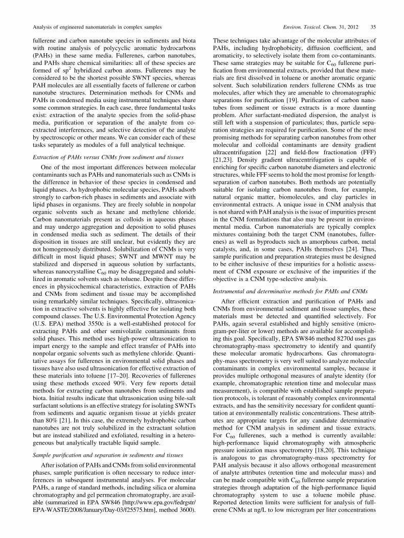
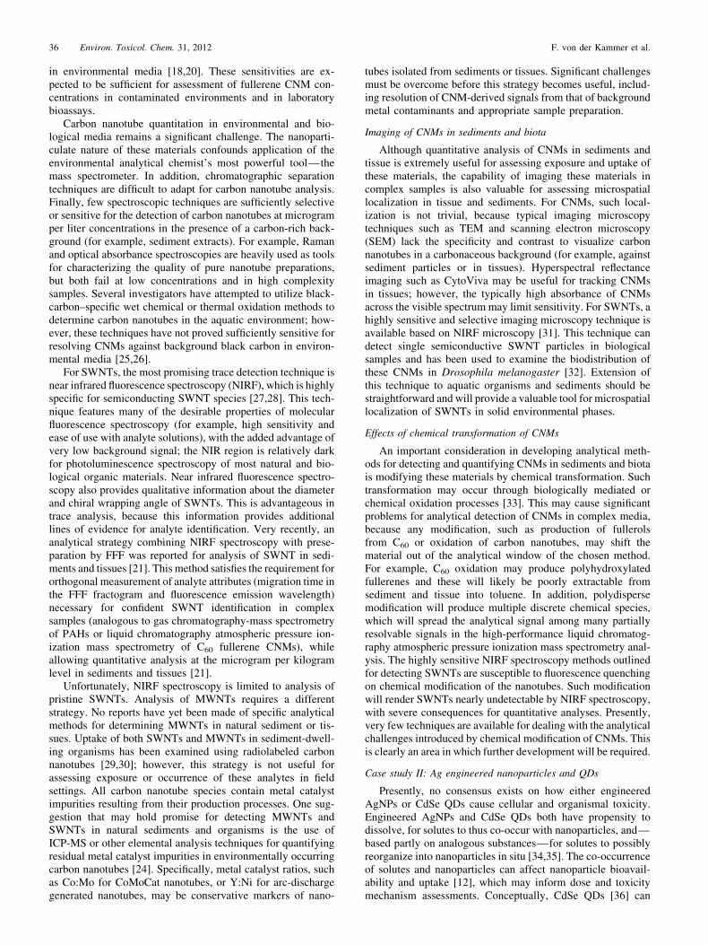
Cerium dioxide is the most insoluble oxide under ambientconditions and is used as an oxygen storage and combustioncatalyst, for optical polishing and in the form of ENPs as fueladditive. It is a potential replacement of zinc oxide and TiO2 insunscreens because of its similar UV filtering properties andlower photocatalytic activity. The average natural abundance ofCe in the earth’s crust is 46mg/kg, and the background con-centration in soil has great variation around this value. ForEurope, ranges of 1 to 270mg/kg (topsoil) and 2.2 to 1082mg/kg(stream sediment) have been reported ([65]; http://www.gtk.fi/publ/foregsatlas/index.php). Concentrations in plants rangebetween 0.1 and 0.55mg/kg, with maximum values for sphag-num and lichens of approximately 1.4mg/kg [66]. Because ofthe use of CeO2 in automotive catalysts as early as in 1994, Ceconcentrations significantly increased in roadside plants ofGerman motorways. Cerium (IV) is immobile in soils becauseof the low solubility of its compounds. Cerium can undergoreduction to the trivalent form under natural conditions, whichresults in increased solubility. If released in reduced form, Ce(III) is, however, strongly sorbed to iron oxides, which areubiquitous in the environment. In the course of TEM character-ization of river-sediment–derived natural nanoparticles, a cou-ple of Ce containing natural nanoparticles could be identified(Fig. 1). These natural nanoparticles showed association withthorium in the EDS analysis [64]. The challenge to identifyTiO2 and CeO2 ENPs as being of anthropogenic/engineeredorigin and to quantify further the amount of these ENPs arisesfrom several complicating factors. First, the abundance of ENPsis expected to be very low compared with the natural back-ground concentrations of Ti and Ce. Second, these elementsmay occur in nanoparticulate form in nature [64], and the ionic
forms of these elements tend to immediately adsorb to othernatural surfaces. Finally, there is great variation in the naturalbackground concentrations of these elements.
Detection/identification
The three most important requirements in the analyticalprocedure, which make ENM analysis essentially different fromclassical solute analysis, are first to detect Ce/Ti–containingparticles, second, to identify these particles as ENPs (theparticles are purposefully made material), and third, to quantifythe anthropogenic fraction.
Sampling
The following considerations for total nanoparticle samplingmethods for separating the ENPs from the sampled soil orsediment material for further analysis are in their infancy andare discussed in the sections about quantitative analysis. Sam-pling of soil and sediment may be carried out as for normal soil/sediment analysis. However, possible heterogeneity of thedistribution of ENP in soils and sediments must be consideredto determine the minimum sample volume needed for a repre-sentative sample. Contrary to classical mass concentrationapproaches, the particle number concentration as determinedby mass concentration, particle size, and density should beconsidered. Also, more information is needed on the behavior ofTi/Ce ENPs in soils and sediments, that is, to identify prefer-ential deposition/attachment of the Ti/Ce ENPs to certain grainsize fractions or certain mineral surfaces. Especially with sedi-ments, the proportion of solid material to pore water should bemaintained in the sample because the pore water may hold alarger fraction of well-dispersed nanoparticles because of ele-vated natural organic matter concentrations. Natural nanopar-ticles undergo intensive redox cycling in the upper layers of
Fig. 1. (A) Scanning electron micrograph of a subsurface sand (silty loam) after thorough (1 h) wet sieving over 20mm nylon cloth in an ultrasonic bath using10mmol/L sodiumdiphosphate as dispersing agent; the largenumber of still surface-attachednanoparticles is easily visible. (B) Transmissionelectronmicrographof a natural Ce-containing nanoparticle cluster from floodplain sediments of Clark Fork River, Montana, USA [64]. Scanning electron micrograph (SEM, FEIQuanta 3D) of soil nanoparticles (soil extract used in Plathe et al. [64]) mixed with 60 nm gold nanoparticles.
Analysis of engineered nanomaterials in complex samples Environ. Toxicol. Chem. 31, 2012 39
fresh water sediments. An individual porewater subsamplewould be beneficial to determine the pore water dispersedfraction [67]. Porewater extraction, however, must considerthe selective separation or loss of nanoparticles during centri-fugation or porewater extraction/filtration. To our knowledge,systematic studies of nanoparticle-related efficiencies andlosses during porewater extraction, filtration, and the use ofmicroporous suction cups are currently not available. The sameconsiderations hold true for the drying and sieving of a soil orsediment sample. Although the removal of the coarse fractionand enrichment of the fines may be desirable to increase thenanoparticulate fraction of the sample for further sample proc-essing, nanoparticles may be irreversibly attached to largergrains (Fig. 1). With removal of the larger grains, the targetENPs would then be removed from the sample as well. Evenduring wet sieving, parts of the nanoparticulate fraction may belost by the same process. However, the use of ultrasonic powerand dispersing agents may increase the recovery for the ENPfraction. Studies with labeled or easily tracked ENPs thataddress these issues of representativeness and recovery duringsampling and sample preparation are lacking.
SEM/TEM-EDS identification
Electron microscopy is presently the predominant techniqueto investigate the presence, aggregation state, location, andcomposition of ENPs in ecotoxicological tests and on/in organ-isms [68–70] (see case study II and also Dudkiewicz et al. [71],which gives a comprehensive overview of EM techniques forENP analysis in food matrices). Both the high-resolution TEMand the field emission gun-equipped SEM or scanning trans-mission electron microscopy systems provide the necessaryspatial resolution to visualize particles down to a few nano-meters in diameter. However, to provide this spatial resolution,the samples must be placed in an ultra-high vacuum (TEM) oreven need to be additionally coated (SEM) with a thin layer ofAu, Pt, or C. Coatings such as Au easily form artifact particles,which may look like Au nanoparticles.
Figure 1 shows the visualization of 60-nm gold-ENPs in asample containing natural soil nanoparticles in a field emissiongun-SEM (FEI Quanta 3D) at intermediate acceleration voltage(15 kV). The 60-nm gold-ENPs are clearly visible as bright dotsbecause of the large Z-contrast of gold to the low Z elements inthe natural soil NPs of the matrix. However, contrast alone is notalways specific for heavy elements, because charge buildup onparticles other than Au-ENPs produces similar brightness andmay be misleading. In general, the identification of TiO2 ENPsin soil or sediment samples with SEM/TEM just by their Z-contrast will not be possible because of the similar Z values forTi and natural nanoparticles. To our knowledge, the usefulnessof this technique for CeO2 ENPs has not yet been proven. Ingeneral, a detailed elemental analysis may be required toidentify Ti/Ce nanoparticles. To distinguish them from naturalor incidental nanoparticles, techniques on single particle level,described later in this section, may be of help. However, the lowsensitivity of EDS techniques will prevent most attempts basedon the recognition of trace impurities or naturally accompany-ing elements. Drying of samples and high vacuum artifacts maybe prevented by low vacuum SEM (environmental SEM;ESEM) and wet-SEM techniques; however, they do not providethe necessary resolution to image the smaller fraction of ENPs(approximately <30 nm) [71]. For cryogenic and embeddingtechniques, the reader is referred to Dudkiewicz et al. [71] andthe references therein.
Use of elemental or isotopic ratios to identify ENPs
The detection and identification of ENPs in environmentalmatrices requires that they can be distinguished from the naturalbackground of particles. For metal oxides such as TiO2 or CeO2,the background concentrations are relatively high, as shownpreviously, and both actually appear as natural nanoparticles insediments and soils (Plathe et al. [64] and Fig. 1). Other optionsfor discriminating between natural and engineered nanopar-ticles include the following: structural homogeneity or specificstructure of the ENP (for example, certain coatings [72]),compositional homogeneity or purity, rare or untypical ele-ments associated with one or the other type of ENP, shifts in theisotopic distribution of the core element, or deliberately intro-duced labels (barcoding). Using single particle analysis withTEM [62], one could detect TiO2 ENPs originating from facaderunoff and identify the particles to be of industrial origin byvisualizing a coating layer on the particles, which was notexpected for particles of natural origin. Little information isavailable regarding how the ENPs of Ce and Ti will be modifiedin the environment and how their ‘‘clean’’ and well-structuredappearance will change during aging. A wide variety of ele-ments and natural organic substances would be expected toadsorb to the surface of the clean particles and make it difficultto identify a particle as of industrial origin just because of itspurity. However, ENPs might be deliberately amended withother rare metals, which could be used as tracers if the method issensitive enough to detect those. Presently, little information isavailable to indicate whether isotopic ratios will be of any use toidentify ENPs. Because the conversion of the source material tothe final ENPs in the production process is fully quantitative, wedo expect very little isotopic fractionation as a result of themanufacturing process. The source material, however, mayoriginate from regions in the world where different isotopicpatterns of the core element are present, and this could be usedonce as an indicator [73]. Whether, in the case of Ti, the smallshifts in isotopic composition expected for different sourcematerials would enable the identification of trace amounts offoreign-engineered nanoparticulate material on a background ofgrams per kilogram of natural background is unknown. Foridentification based on the co-occurrence of certain otherelements with natural nanoparticles, which may be absent inENPs, little hope exists for unmodified TiO2 ENPs, because Tiis one of the most weathering-resistant elements. As a conse-quence, Ti concentration in soils is related to weathering status,and therefore no general correlation of Ti with any other rareelement exists. We used the FOREGS geochemical atlas ofEurope [65], which provides multi-element data for differentcompartments across Europe; with the help of this database, weanalyzed the co-occurrence of rare elements with Ti and Ce forseveral thousand samples collected from soils, sediments, flood-plains, and surface waters. For naturally occurring Ce, a clearand potentially usable relationship to La and Nd exists (Fig. 2).The slopes for the relationships between La and Nd with Ce areall approximately 0.45, with R2> 0.95 for most matrices. Theresults are similar for topsoils, subsoils, sediments, floodplainsoils, and water samples, underpinning the general character ofthe co-existence of the three elements even in industrializedregions. Unfortunately, Ce is the one element among the rareearths that is redox active under environmental conditions. Thisproduces deviations from the expected concentrations whencompared with the other rare earth elements. Another criticaldrawback is that the documented relationships are valid for bulksamples, and the validity of the correlations cannot be assessed
40 Environ. Toxicol. Chem. 31, 2012 F. von der Kammer et al.
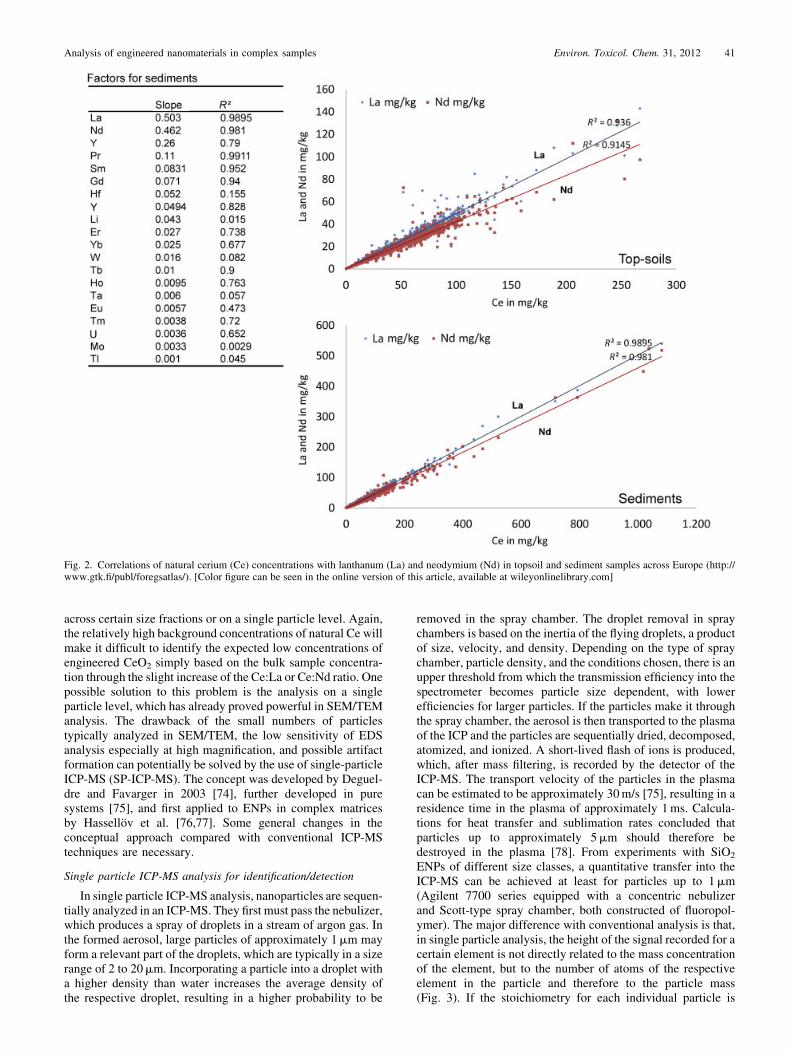
across certain size fractions or on a single particle level. Again,the relatively high background concentrations of natural Ce willmake it difficult to identify the expected low concentrations ofengineered CeO2 simply based on the bulk sample concentra-tion through the slight increase of the Ce:La or Ce:Nd ratio. Onepossible solution to this problem is the analysis on a singleparticle level, which has already proved powerful in SEM/TEManalysis. The drawback of the small numbers of particlestypically analyzed in SEM/TEM, the low sensitivity of EDSanalysis especially at high magnification, and possible artifactformation can potentially be solved by the use of single-particleICP-MS (SP-ICP-MS). The concept was developed by Deguel-dre and Favarger in 2003 [74], further developed in puresystems [75], and first applied to ENPs in complex matricesby Hassellov et al. [76,77]. Some general changes in theconceptual approach compared with conventional ICP-MStechniques are necessary.
Single particle ICP-MS analysis for identification/detection
In single particle ICP-MS analysis, nanoparticles are sequen-tially analyzed in an ICP-MS. They first must pass the nebulizer,which produces a spray of droplets in a stream of argon gas. Inthe formed aerosol, large particles of approximately 1mm mayform a relevant part of the droplets, which are typically in a sizerange of 2 to 20mm. Incorporating a particle into a droplet witha higher density than water increases the average density ofthe respective droplet, resulting in a higher probability to be
removed in the spray chamber. The droplet removal in spraychambers is based on the inertia of the flying droplets, a productof size, velocity, and density. Depending on the type of spraychamber, particle density, and the conditions chosen, there is anupper threshold from which the transmission efficiency into thespectrometer becomes particle size dependent, with lowerefficiencies for larger particles. If the particles make it throughthe spray chamber, the aerosol is then transported to the plasmaof the ICP and the particles are sequentially dried, decomposed,atomized, and ionized. A short-lived flash of ions is produced,which, after mass filtering, is recorded by the detector of theICP-MS. The transport velocity of the particles in the plasmacan be estimated to be approximately 30m/s [75], resulting in aresidence time in the plasma of approximately 1ms. Calcula-tions for heat transfer and sublimation rates concluded thatparticles up to approximately 5mm should therefore bedestroyed in the plasma [78]. From experiments with SiO2
ENPs of different size classes, a quantitative transfer into theICP-MS can be achieved at least for particles up to 1mm(Agilent 7700 series equipped with a concentric nebulizerand Scott-type spray chamber, both constructed of fluoropol-ymer). The major difference with conventional analysis is that,in single particle analysis, the height of the signal recorded for acertain element is not directly related to the mass concentrationof the element, but to the number of atoms of the respectiveelement in the particle and therefore to the particle mass(Fig. 3). If the stoichiometry for each individual particle is
Fig. 2. Correlations of natural cerium (Ce) concentrations with lanthanum (La) and neodymium (Nd) in topsoil and sediment samples across Europe (http://www.gtk.fi/publ/foregsatlas/). [Color figure can be seen in the online version of this article, available at wileyonlinelibrary.com]
Analysis of engineered nanomaterials in complex samples Environ. Toxicol. Chem. 31, 2012 41
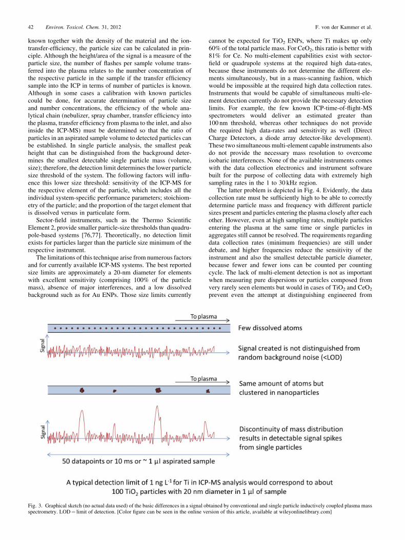
known together with the density of the material and the ion-transfer-efficiency, the particle size can be calculated in prin-ciple. Although the height/area of the signal is a measure of theparticle size, the number of flashes per sample volume trans-ferred into the plasma relates to the number concentration ofthe respective particle in the sample if the transfer efficiencysample into the ICP in terms of number of particles is known.Although in some cases a calibration with known particlescould be done, for accurate determination of particle sizeand number concentrations, the efficiency of the whole ana-lytical chain (nebulizer, spray chamber, transfer efficiency intothe plasma, transfer efficiency from plasma to the inlet, and alsoinside the ICP-MS) must be determined so that the ratio ofparticles in an aspirated sample volume to detected particles canbe established. In single particle analysis, the smallest peakheight that can be distinguished from the background deter-mines the smallest detectable single particle mass (volume,size); therefore, the detection limit determines the lower particlesize threshold of the system. The following factors will influ-ence this lower size threshold: sensitivity of the ICP-MS forthe respective element of the particle, which includes all theindividual system-specific performance parameters; stoichiom-etry of the particle; and the proportion of the target element thatis dissolved versus in particulate form.
Sector-field instruments, such as the Thermo ScientificElement 2, provide smaller particle-size thresholds than quadru-pole-based systems [76,77]. Theoretically, no detection limitexists for particles larger than the particle size minimum of therespective instrument.
The limitations of this technique arise from numerous factorsand for currently available ICP-MS systems. The best reportedsize limits are approximately a 20-nm diameter for elementswith excellent sensitivity (comprising 100% of the particlemass), absence of major interferences, and a low dissolvedbackground such as for Au ENPs. Those size limits currently
cannot be expected for TiO2 ENPs, where Ti makes up only60% of the total particle mass. For CeO2, this ratio is better with81% for Ce. No multi-element capabilities exist with sector-field or quadrupole systems at the required high data-rates,because these instruments do not determine the different ele-ments simultaneously, but in a mass-scanning fashion, whichwould be impossible at the required high data collection rates.Instruments that would be capable of simultaneous multi-ele-ment detection currently do not provide the necessary detectionlimits. For example, the few known ICP-time-of-flight-MSspectrometers would deliver an estimated greater than100 nm threshold, whereas other techniques do not providethe required high data-rates and sensitivity as well (DirectCharge Detectors, a diode array detector-like development).These two simultaneous multi-element capable instruments alsodo not provide the necessary mass resolution to overcomeisobaric interferences. None of the available instruments comeswith the data collection electronics and instrument softwarebuilt for the purpose of collecting data with extremely highsampling rates in the 1 to 30 kHz region.
The latter problem is depicted in Fig. 4. Evidently, the datacollection rate must be sufficiently high to be able to correctlydetermine particle mass and frequency with different particlesizes present and particles entering the plasma closely after eachother. However, even at high sampling rates, multiple particlesentering the plasma at the same time or single particles inaggregates still cannot be resolved. The requirements regardingdata collection rates (minimum frequencies) are still underdebate, and higher frequencies reduce the sensitivity of theinstrument and also the smallest detectable particle diameter,because fewer and fewer ions can be counted per countingcycle. The lack of multi-element detection is not as importantwhen measuring pure dispersions or particles composed fromvery rarely seen elements but would in cases of TiO2 and CeO2
prevent even the attempt at distinguishing engineered from
Fig. 3. Graphical sketch (no actual data used) of the basic differences in a signal obtained by conventional and single particle inductively coupled plasma massspectrometry. LOD¼ limit of detection. [Color figure can be seen in the online version of this article, available at wileyonlinelibrary.com]
42 Environ. Toxicol. Chem. 31, 2012 F. von der Kammer et al.

natural particles. Accepting the current limitations, but also notoverlooking the huge potential of the technique, a rigoroustesting and comparison of all possibly suitable systems andoptimizations is needed.
Quantification
After the qualitative analysis, in which the presence of theENP in the sample was proven, quantification of an ENP aims todetermine the concentration or abundance of this ENP in thesample. Apart from the mere ENP concentration (volume/mass,area, or even better, number concentration), other metrics coulddeserve a quantitative determination, such as surface area,shape, aggregation state, or surface chemistry. However, thisshould not lead scientists to the hasty conclusion that a multi-method approach is always required. The principle of the mostefficient use of resources should not be abandoned, becauseknowledge about all possible relationships is still limited. Eachadditional technique should be well chosen with potentials andlimitations in mind. In addition to the aforementioned, thedesired metrics may be different for the same type of particleunder different surrounding conditions, and those may bealtered by sampling/sample preparation. This makes the caseof ENPs different from the analysis of classical contaminants, inwhich the mass concentration is often the most importantparameter. The situation might be comparable with speciationanalysis or bioavailability analysis in that way: more than onemetric may be needed to assess the situation. Another importantdifference between ENPs and classical contaminants is the factthat no substance-specific approach seems possible. This meansthat although developing a method for a certain chemical(phenantrene) or group of substances (PAH) seems to be stand-ard, the considered TiO2 or CeO2 ENPs will be present indifferent particle sizes, mineral forms, surface chemistry, differ-ent shapes, aggregation states, and so forth. In this case study,we must consider Ti/Ce as the main analyte, but these willappear in many different industrially relevant forms with differ-ent core compositions and coatings, which may change overtime and consequently will show different recoveries in thesample preparation procedures and analysis. Not much system-atic information is available regarding how different particles ofsimilar composition behave in sample preparation processes if
they vary in one or more of their other properties, but, forexample, the aqueous dispersion stability of different TiO2 ENPproducts may be relatively similar over a wide range of hydro-chemical conditions [79]. One could use this information todeduce that at least on the basis of the particle type with similarcoatings, the recoveries in sample preparation should be similar.The consequence is that as long as a minimally invasivetechnique is not available and sample pretreatment is necessary,the development and use of internal standards may be unavoid-able to obtain fully quantitative results.
Extraction
The separation of the TiO2 and CeO2 particles from the soilmatrix may be achieved by using differences between the ENPand soil properties. For CeO2, the particles density of 7.65 g/cm
3
(may be slightly different for nano-CeO2) can be used in adensity gradient ultracentrifugation separation step usingsodium or lithium polytungstate heavy liquids. However, thenatural CeO2 ENPs, as well as some abundant higher densitynatural metal oxide nanoparticles, may be found in the sepa-rated heavy fraction as well, because of the maximum density of3 g/cm3 of most heavy liquids. Because of the even lowerdensity of TiO2, this separation approach seems to be inappli-cable for this material. The higher the fraction of low-densitycomponents in the sample (such as high organic content), themore successful this approach may be. Whether nanoparticleswith densities above the heavy liquid can be mechanicallydetached from surfaces of lighter particles by high g-forcesin an ultracentrifuge or whether they will float with the lighterparticles is not clear. Matrix decomposition would be an optionif the particles cannot be separated from the matrix. Titaniumdioxide is quite resistant to classical digestion techniques suchas concentrated nitric acid, which offers some freedom in theuse of digestive methods to destroy the matrix materials.Several digestion methods have been studied for the separationof TiO2 out of sunscreen and preparation of a target particledispersion for subsequent FFF analysis [80]; others performed adiluted direct injection or hexane liquid/liquid extraction ofhydrophobic compounds before injection [81]. Neither studyperformed a full quantitative analysis of the ENP fraction. Athoroughmethod evaluation and valid tracing of the results backto the original product are lacking. Other classical approachescan include filtration (which is hindered by the presence of othersimilar-sized particles, removal of aggregated structures con-taining ENP, and difficulties in providing reproducible, quanti-tative passage of filtrates through the membrane structure) andselective adsorption/deposition on stationary phases or extrac-tion into organic liquids. Both of the latter options may needfirst to specifically derivatize the surface of the target particle toachieve an acceptable yield and selectivity toward the secondextracting phase.
Single-particle analysis for quantification
The proper and complete separation of ENPs from naturalparticles of similar composition and size seems to be tediousand bears the risk of transforming and losing the target particles.Therefore, an analytical technique that ideally bridges thesingle-particle detection ICP-MS with an adapted, ideallylow invasive sample preparation technique is needed. Twopossible approaches would provide additional selectivity:FFF separates the particles according to their hydrodynamicdiameter or particle volume, while avoiding the application of astationary phase, and hydrodynamic chromatography (HDC)provides a similar separation but is still underdeveloped [82].
Fig. 4. Graphical sketch of the principal effect of the data collection rate fordetecting single particles and separation of particles appearing close to eachother. The increase of data rate (sampling frequency) comes with a reductionof sensitivity; thebaseline in the lower examplewouldbemuchnoisier than inthe upper case, an effect not shown in the graph. [Color figure can be seenin the online version of this article, available at wileyonlinelibrary.com]
Analysis of engineered nanomaterials in complex samples Environ. Toxicol. Chem. 31, 2012 43

The current potentials and limitations of FFF, as well as a guideto method development, are given in detail elsewhere [83]. Theanalytical size-based separation of the preconditioned sedimentor soil sample should enable the quantitative single-particleanalysis in ICP-MS, including the identification of ENP. Theseparation methods provide a presorting of the still complexsample composition, and based on the recorded retention timeof the sample, they will provide a good estimation of the size ofthe particle that enters the plasma of the SP-ICP-MS. Bothseparation methods dilute the sample during the separationprocess, which is a minor problem for SP-ICP-MS becausedilute samples are required. One critical drawback of thiscombination stems from the fact that detectors common formethod development and analysis in HDC and FFF, such asultraviolet visible spectroscopy, fluorescence, and light scatter-ing, are not applicable to the very dilute samples required bySP-ICP-MS. One solution to this would be a conventional,high-concentration separation in FFF and a subsequent dilutionin the FFF to an SP-ICP-MS interface. Here, the advantage ofSP-ICP-MS to provide exceptionally low detection limits interms of particle concentration would be wasted.
As a stand-alone or coupled to FFF, this technique ispresently being developed for ENPs by different groups, andfirst publications are out [77] or on the way. Even for the mostsensitive instruments, the smallest sizes that can be detected areapproximately 20 to 30 nm in diameter. The technique has hugepotential because it uses the principle of discontinuity ofparticles in a dispersion compared with ions/atoms/moleculesin solution, but years of detailed method development still maybe needed.
Single particle detection pushed to or beyond the limits
In the following section, we wish to develop a hypotheticalsituation that goes far beyond what is possible with currentlyavailable commercial devices. In fact, some of the performancespecifications needed for the following conceptual analysiscannot be met because of physical limitations. However, webelieve that instead of listing methods that cannot solve theproblem because of inherent limitations, we should show that—in principle—a combination of technologies, developed withinthe next five to 10 years to their physical limits, could be able tosolve many of the current analytical problems. A high data rate,extreme multi-element sensitivity, and a stretch down to par-ticles of approximately 2 to 5 nm for a solid one-elementparticle (as Ag- or Au-ENPs) must be integrated into one singleinstrument. This would require an instrument that makes use ofdedication to the lowest matrix content and background withhigh transfer efficiency instrumentation. Given an increase insensitivity of approximately 64-fold compared with the single-element sector field HR-ICP-MS and of approximately 64,000-fold compared with current multi-element ICP-time-of-flight-MS equipment required to enable the detection of a 5-nmdiameter particle, the combination with FFF or HDC couldbe used to identify and quantify ENPs in soil and sedimentsuspensions for many cases and for many types of particles,even hybrids. Figure 5 illustrates how different (hypothetical)situations could be addressed by this hypothetical instrumenta-tion. The left column describes typical situations for a complexenvironmental sample containing pristine, coated, or dopedCeO2 ENPs and natural nanoparticles of a similar size. Theelemental ratios determined for natural sediment bulk samplesare assumed identical to those in single natural nanoparticles(this might be unlikely and must be proven). Clearly thepreseparation and information from individual size measure-
ment (FFF, HDC) could be combined with multi-element, peakshape, ICP-MS–derived particle volume/size, and respectiveelement ratios to for once answer analytical questions that arecurrently far from being resolved.
Case study IV: Monitoring for AgNPs in sewage sludge
Although the concentrations of Ag in wastewater in theUnited States, as evidenced by discharges in urban wastewaterfacilities, has significantly decreased from 1989 to 2007, poten-tial releases resulting from manufacturing and using commer-cial engineered AgNP products may result in future increases oftotal Ag present in wastewater [84,85]. Past and current sourcesof Ag contamination in wastewater include photographic facili-ties, smelters, mines, and urban wastes. With the decline in theuse of Ag-based photographic film and potential increases in thenumber of commercial products containing engineered AgNP, asignificant shift may occur in the sources and possibly the formsof Ag present in both wastewater and biosolids generated fromthese wastes.
Commercial products containing engineered AgNPs areprimarily designed to inhibit microbial growth. These productsinclude the use of spherical nanoparticles immobilized tosurfaces using polymers or added to surface disinfecting spraysor soaps, colloidal suspensions used as dietary supplements, anddevices designed to release ionic silver into washing machines[85]. One of the challenges that face analytical chemists withrespect to the potential release of engineered AgNPs into wastestreams is the detection, identification, and quantification ofengineered AgNPs in these matrices.
Because of the variability and complexity of aqueous wastestreams, as well as the number of insoluble salts and Agcomplexes, the speciation of Ag in wastewater and resultingbiosolids is also expected to be variable and complex. Engi-neered AgNPs present in a wastewater stream are likely toencounter common ligands such as sulfate, sulfide, chloride,and phosphate, as well as carboxylic acids, polyalcohols, andamines found in humic substances [86]. Many of these ligandsthat are often associated with particulate matter are known toeither complex directly with engineered AgNPs or with Agþ,which may be released during oxidation. When engineeredAgNPs were introduced into complex sewage, they partitionedto a considerable extent (> 90%) into the particulate biomass[82]. Nevertheless, a portion of the total Ag that remained in thesupernatant was in the nanoparticle form. Although wastewateris complex, with several ligands that bind to metals and metalions, the fate of Ag in this matrix is likely to be dominated bysulfide chemistry. Inorganic sulfide in wastewater effluent andreceiving water has been shown to be present at concentrationsthat are 200 to 300 times in excess of the total Ag [87]. Silversulfide has been identified recently in the form of nanoparticles(5–20 nm ellipsoids) in final stage sewage sludge materials in afull-sized municipal wastewater treatment plant [88]. Kim et al.suggested, based on the results of Choi et al. [86], that the sourceof the silver sulfide nanoparticles may have been engineeredAgNPs. The reported change in the chemical composition of theENPs in the wastewater treatment system may have significantbiological consequences. Nitrifying bacteria are critical in theconversion of ammonia to nitrate in the wastewater treatmentprocess. In addition to forming nanoscale AgxSy complexes andprecipitates, the presence of sulfide also protects nitrifyingbacteria from the toxic effect of both Agþ and engineeredAgNPs [89]. Other inorganic ligands present in wastewatersuch as sulfate, chloride, and phosphate were significantlyless effective in reducing engineered AgNP toxicity [86].
44 Environ. Toxicol. Chem. 31, 2012 F. von der Kammer et al.

Consequently, one of the challenges to understand better thetransport, fate, and biological activity of silver nanoparticles inwastewater treatment systems involves Ag-S chemistry.
With respect to the possible persistence of engineeredAgNPs in a wastewater matrix, metallic Ag in the form ofnanoparticles (2–8 nm) would dissolve over a time frame offewer than 125 d to form Agþ in oxygen-saturated aqueousmedia at pH values and humic acid concentrations typical ofthese environments [5]. In this matrix, Ag complexes of halidesand sulfides would be expected to predominate. Nevertheless,colloidal Ag particles of larger diameter and with more durablecoating materials may be present in a wastewater stream if thetime frame from particle release to analysis was less thanseveral months.
Sample preparation
Because of the physical structures of engineered AgNPs,their presence cannot be determined simply by dissolution,extraction, cleanup, and analysis. For engineered AgNPs incomplex media, sample preparation will likely be the mostchallenging part of the analytical process. The task of separationand enrichment is in some ways similar to detecting complexbiological structures of similar size, such as viruses. Because ofthe presence of halides, sulfides, and a range of colloidalparticles, preparation and chemical analysis techniques maybe required that are not typically required for measurement ofENPs for laboratory toxicity experiments.
Techniques that might be used to separate nanoparticlesfrom ions and molecular components aqueous media includecentrifugation, dialysis, or continuous diafiltration. Limitationsfor centrifugation and dialysis include limited control over sizeseparation and long processing times, which may shift redoxconditions and increase particle dissolution. When diafiltration
was applied to Au nanoparticle purification, the techniquefacilitated the rapid separation of molecular contaminants with-out the typical problems associated with filter obstruction oftenassociated with ultrafiltration techniques [90].
Nanoparticle enrichment techniques that might be adaptedfrom biology and colloid science include density gradientcentrifugation. Using this technique, Au nanoparticles of sev-eral sizes were separated from each other and from particles ofdiffering composition [91]. Another centrifugation techniquetermed cloud point separation was used to separate and con-centrate trace amounts of engineered AgNPs [92].
Analytical techniques
Total Ag concentrations in wastewater are expected to be inthe nanograms per liter range [85]. If the engineered AgNPs,which would likely constitute only a small fraction of thisconcentration, could be separated from other colloidal materialsand enriched into the milligrams-per-liter range, then lightscattering techniques such as dynamic light scattering andnanoparticle tracking analysis could be applied for particle sizedetermination, and microscopic techniques such as TEM-EDScould be used to confirm size and elemental composition.
A technique that might be applied to the characterization oftransformations of engineered AgNPs extracted and preconcen-trated from wastewater or biosolid is X-ray absorption spectro-scopy. X-ray absorption spectroscopy in combination withlinear combination fitting analysis has been applied to deter-mine the fraction of silver metal present in soils [93,94]. In thesestudies, the oxidized silver fraction was correlated to theobserved reproductive toxicity in earthworms. This techniquegenerally is applicable only when more than 10mg/kg Ag arepresent, and synchrotron beamlines with high flux at the AgK-edge (25,514 eV) are required for the highest sensitivity, thus
Fig. 5. Conceptual approach to the question of how different possible combinations of engineered nanoparticle (ENP) (CeO2) and natural nanoparticles couldappear in the analysis with an ideal, nonexisting field flow fractionation-single particle-inductively coupled plasma-mass spectrometry (FFF-SP-ICP-MS)combination. The SP-ICP-MShere is in contrast to existing instruments capable of delivering high-frequency, multi-element datawith extremely high sensitivity.Still, the very low traces of Ce, the single mixed oxide particles, or clay minerals would be highly questionable, if they would be detectable at all.HDC¼ hydrodynamic chromatography; ENM¼ engineered nanomaterial; Ce¼ cerium, Fe¼ iron; Al¼ aluminum; Si¼ silicon; La¼ lanthanum;Nd¼ neodymium. [Color figure can be seen in the online version of this article, available at wileyonlinelibrary.com]
Analysis of engineered nanomaterials in complex samples Environ. Toxicol. Chem. 31, 2012 45
requiring engineered AgNP enrichment in the sample. Linearcombination analysis is critically dependent on the properselection of model compounds, proper sample preparation,and more than one plausible solution that fits the observed datacan be possible [95].
Small angle X-ray scattering (SAXS) is a somewhatneglected technique in environmental sciences, most likelybecause of the difficulty of extracting useful information fromthe scattering patterns of complex samples, but it may beamenable to use for characterization of extracted engineeredAgNPs. It is mainly used in biology (for example, when study-ing protein structure) and material science. Small angle X-rayscattering is one of the rare analytical tools providing a statisti-cally meaningful three-dimensional structural description ofnoncrystalline systems over an extended size range (typicallythree orders of magnitude; 0.1–100 nm). It is performed onsuspension gels and even solids and requires little or no samplepreparation. Silver is part of virtually every SAXS experimentbecause Ag-behenate is used for q calibration [96]. The heavierelements, such as Ag, are easy to observe with SAXS because oftheir high electron contrast, and a number of studies describesize and structure of AgNP-based materials [97]. Anomaloussmall-angle X-ray scattering uses an X-ray source tuned to justbelow the absorption energy of the element of interest and thushelps to obtain element-specific information. It has been used toobtain element-specific data in heterogeneous samples contain-ing Ag ENMs but has not been applied extensively to environ-mental samples containing ENMs [98]. The main issueconcerning SAXS is the detection limit; an attractive featureof SAXS is the time-resolved monitoring of the evolution of thestructure. With adequate sample, the time resolution can reachthe MS even with laboratory instruments [99], but it is typicallyon the order of a minute or longer using synchrotron sources formore environmentally relevant systems (0.1mM Zn) [100].
CONCLUSIONS AND RECOMMENDATIONS
Because the physicochemical properties of nanomaterialsare dependent on their surroundings, the simple act of isolatingand observing them can change these properties. Analyticaltechniques can be borrowed from materials and other chemicaland biological disciplines, but their application to low concen-trations and heterogeneous sample matrices requires someadditional development or the development of completelynew approaches. Approaches need to be tailored to addressspecific hypotheses in a given sample type for a given material,paying special attention to the generation of artifacts. In mostcases, multiple orthogonal lines of analytical evidence arerequired to identify and characterize ENMs in samples.
New analytical techniques are under development that willenable more rapid, sensitive, and specific detection of ENMs.Many of these techniques rely on chromatographic separationsthat lack stationary phases because of the surface reactivity ofparticles (for example, FFF techniques). Detectors such asNIRF and ICP-MS are being adapted to provide sensitiveand specific detection of ENMs. X-ray–based techniques pro-vide sensitivity and specificity. Current synchrotron microprobebeamlines do not provide sufficient spatial resolution forimaging of individual nanoparticles; however, their generaldistribution in samples can be determined, and they can bedifferentiated from metal ions or other materials based on theirlocal electronic structure. Light sources currently under devel-opment hope to achieve spatial resolution of less than 30 nm;however, beam damage is likely to be an important consider-
ation when using these next-generation beamlines. TraditionalEM-based approaches are being augmented by minimally inva-sive techniques such as ESEM, which are simpler to apply andless invasive. Focused ion beam-SEM holds promise for inter-nal imaging and 3D reconstruction with samples held underminimally destructive cryogenic conditions.
Differentiating nanomaterials that have naturally occurringcounterparts is a complex task, and techniques for this needconsiderable development. Some possible approaches involveexamining isotopic or elemental composition of particles, aswell as potentially labeling or barcoding materials as they areproduced.
Sample collection preservation and storage is likely theweakest link in the analytical workflow and has received littleattention in the literature. In many cases, properties cannot bemeasured after samples have been stored because of the tem-porally dynamic nature of colloidal systems. This presents aproblem for characterization in toxicological studies; therefore,some techniques need to be developed that allow for thesensitive and rapid determination of basic properties such asparticle size distribution. Current techniques that are rapid, suchas dynamic light scattering, may not be sensitive or specificenough to be applied at environmentally or toxicologicallyrelevant concentrations, depending on the material in question.
Despite the fact that techniques for identification, character-ization, and quantification in complex sample matrices havehampered studies on the environmental fate, transport, exposuremodeling, and ecotoxicological effects of ENMs, ample oppor-tunities exist for progress. The requirements for the develop-ment of these techniques in terms of properties of interest anddetection limits need to be guided by well-designed ecotoxico-logical studies that identify the relevant concentrations andproperties of interest so that focused analytical strategies canbe developed.
Acknowledgement—The assistance of K. Newton to the authors is gratefullyacknowledged.The authors receivedfinancial support from theU.S.EPAandNational Science Foundation (EF0830093, DBI 0830117, RD834574,RD83485701, RD833335, R833886, R834092, RD833859, and RD831716)and the Centre national de la recherche scientifique and the Commissariat al’energie atomique et aux energies alternatives of France. This manuscripthas been subjected toU.S. EPA administrative review and has been approvedfor publication. Mention of trade names or commercial products does notconstitute endorsement or recommendation for use. Any opinions, findings,and conclusions or recommendations expressed in this material are those ofthe authors and do not necessarily reflect the views of the National ScienceFoundation or the U.S. EPA.
REFERENCES
1. Richman EK, Hutchison JE. 2009. The nanomaterial characterizationbottleneck. ACS Nano 3:2441–2446.
2. Hassellov M, Readman JW, Ranville J, Tiede K. 2008. Nanoparticleanalysis and characterization methodology in environmental riskassessment of engineered nanoparticles. Ecotoxicology 17:344–361.
3. Hassellov M, Kaegi R. 2009. Analysis and characterization ofmanufactured nanoparticles in aquatic environments. In Lead JR,Smith E, eds, Nanoscience and Nanotechnology: Environmental andHuman Health Implications. Wiley, New York, USA, pp 211–266.
4. Christian P, Von der Kammer F, Baalousha M, Hofmann T. 2008.Nanoparticles: structure, properties, preparation and behaviour inenvironmental media. Ecotoxicology 17:326–343.
5. Liu J, Hurt R. 2010. Ion release kinetics and particle persistence inaqeuous nanosilver coloids. Environ Sci Technol 44:2169–2175.
6. Bertsch P, Hunter D. 2001. Applications of synchrotron bases x-raymicroprobes. Chem Rev 101:1809–1842.
7. Unrine J, Tsyusko O, Hunyadi S, Judy J, Bertsch P. 2010. Effects ofparticle size on chemical speciation and bioavailability of Cu toearthworms exposed to Cu nanoparticles. J Environ Qual 39:1942–1953.
46 Environ. Toxicol. Chem. 31, 2012 F. von der Kammer et al.

8. Unrine J, Bertsch P, Hunyadi S. 2008. Bioavailability, trophic transfer,and toxicity of manufactured metal and metal oxide nanoparticlesin terrestrial environments. In Grassian V, ed, Nanoscience andNanotechnology: Environmental and Health Impacts. John Wiley &Sons, Hoboken, NJ, USA, pp 345–366.
9. WegrzynekD,MarkowiczA, Bamford S, Chinea-CanoE, BogovacM.2005. Micro-beam X-ray fluorescence and absorption imagingtechniques at the IAEA Laboratories. Nucl Instrum Methods Phys231:176–182.
10. Parker DR, Bertsch PM. 1992. Identification and quantification of theAL13 tridecameric polycation using ferron. Environ Sci Technol26:908–914.
11. Gottschalk F, Sonderer T, Scholz RW, Nowack B. 2009. Modeledenvironmental concentrations of engineered nanomaterials (TiO2,ZnO, Ag, CNT, fullerenes) for different regions. Environ Sci Technol43:9216–9222.
12. Wigginton NS, De Titta A, Piccapietra F, Dobias J, Nesatty VJ, SuterMJF, Bernier-Latmani R. 2010. Binding of silver nanoparticles tobacterial proteins depends on surface modifications and inhibitsenzymatic activity. Environ Sci Technol 44:2163–2168.
13. Gou N, Onnis-Hayden A, Gu AZ. 2010. Mechanistic toxicity assess-ment of nanomaterials by whole-cell-array stress genes expressionanalysis. Environ Sci Technol 44:5964–5970.
14. van der Meer JR, Belkin S. 2010. Where microbiology meets micro-engineering: design and applications of reporter bacteria. Nat RevMicrobiol 8:511–522.
15. Senevirathna W, Kiro R, Rosen R, Popov I, Belkin S, Wells M. 2009.CdSe quantum dots induce superoxide stress in engineered biosensorbacteria. Nanotoxicology 3:98–108.
16. SongYZ, Li GH, Thornton SF, Thompson IP, Banwart SA, LernerDN,HuangWE. 2009. Optimization of bacterial whole cell bioreporters fortoxicity assayof environmental samples.EnvironSciTechnol43:7931–7938.
17. Farre M, Gajda-Schrantz K, Kantiani L, Barcelo D. 2009. Ecotoxicityand analysis of nanomaterials in the aquatic environment.Anal BioanalChem 393:81–95.
18. Farre M, Perez S, Gajda-Schrantz K, Osorio V, Kantiani L, GinebredaA, Barcelo D. 2010. First determination of C-60 and C(70) fullerenesand N-methylfulleropyrrolidine C-60 on the suspended material ofwastewater effluents by liquid chromatography hybrid quadrupolelinear ion trap tandem mass spectrometry. J Hydrol 383:44–51.
19. Isaacson CW, Kleber M, Field JA. 2009. Quantitative analysis offullerene nanomaterials in environmental systems: A critical review.Environ Sci Technol 43:6463–6474.
20. IsaacsonCW,UsenkoCY,TanguayRL, Field JA. 2007.Quantificationof fullerenes by LC/ESI-MS and its application to in vivo toxicityassays. Anal Chem 79:9091–9097.
21. Schierz PA, Parks AN, Ferguson PL. 2011. Detection of single-walledcarbon nanotubes in environmental samples byfield flow fractionation-NIR fluorescence spectroscopy. Abstracts, Papers of the AmericanChemical Society 241:306-ENVR.
22. ArnoldMS, Stupp SI, HersamMC. 2005. Enrichment of single-walledcarbon nanotubes by diameter in density gradients. Nano Lett 5:713–718.
23. Chun J, Fagan JA, Hobbie EK, Bauer BJ. 2008. Size separation ofsingle-wall carbon nanotubes by flow-field flow fractionation. AnalChem 80:2514–2523.
24. Plata DL, Gschwend PM, Reddy CM. 2008. Industrially synthesizedsingle-walled carbon nanotubes: compositional data for users, environ-mental risk assessments, and source apportionment. Nanotechnology19:185706.
25. Sobek A, Bucheli TD. 2009. Testing the resistance of single- andmulti-walled carbon nanotubes to chemothermal oxidation used toisolate soots from environmental samples. Environ Pollut 157:1065–1071.
26. Ziolkowski LA, Druffel ERM. 2009. The feasibility of isolation anddetection of fullerenes and carbon nanotubes using the benzenepolycarboxylic acid method. Mar Pollut Bull 59:213–218.
27. O’Connell MJ, Bachilo SM, Huffman CB, Moore VC, Strano MS,Haroz EH, Rialon KL, Boul PJ, Noon WH, Kittrell C, Ma JP, HaugeRH, Weisman RB, Smalley RE. 2002. Band gap fluorescencefrom individual single-walled carbon nanotubes. Science 297:593–596.
28. Weisman RB. 2005. Fluorescence spectroscopy of single-walledcarbonnanotubes. InRotkinSV,SubramoneyS, eds,AppliedPhysics ofCarbon Nanotubes: Fundamentals of Theory, Optics, and TransportDevices. Springer-Verlag, Berlin, Germany, pp 183–202.
29. Ferguson PL, Chandler GT, TempletonRC, DemarcoA, ScrivensWA,Englehart BA. 2008. Influence of sediment-amendment with single-walled carbon nanotubes and diesel soot on bioaccumulation ofhydrophobic organic contaminants by benthic invertebrates. EnvironSci Technol 42:3879–3885.
30. Petersen EJ, Huang QG, Weber WJ. 2008. Bioaccumulation of radio-labeled carbon nanotubes by Eisenia foetida. Environ Sci Technol42:3090–3095.
31. Cherukuri P, Bachilo SM, Litovsky SH, Weisman RB. 2004. Near-infrared fluorescencemicroscopy of single-walled carbon nanotubes inphagocytic cells. J Am Chem Soc 126:15638–15639.
32. Leeuw TK, Reith RM, Simonette RA, Harden ME, Cherukuri P,Tsyboulski DA, Beckingham KM, Weisman RB. 2007. Single-walledcarbon nanotubes in the intact organism: Near-IR imaging andbiocompatibility studies in Drosophila. Nano Lett 7:2650–2654.
33. Fortner JD, Kim DI, Boyd AM, Falkner JC, Moran S, Colvin VL,Hughes JB, Kim JH. 2007. Reaction of water-stable C-60 aggregateswith ozone. Environ Sci Technol 41:7497–7502.
34. Klaus T, Joerger R, Olsson E, Granqvist CG. 1999. Silver-basedcrystalline nanoparticles, microbially fabricated. Proc Natl Acad SciUSA 96:13611–13614.
35. Sweeney RY, Mao CB, Gao XX, Burt JL, Belcher AM, Georgiou G,Iverson BL. 2004. Bacterial biosynthesis of cadmium sulfide nano-crystals. Chem Biol 11:1553–1559.
36. Nadeau JL, Priester JH, Stucky GD, Holden PA. 2008. Bacterialinteractions with CdSe quantum dots and environmental implications.InGrassianVH, ed,Nanoscience andNanotechnology: Environmentaland Health Impacts. JohnWiley & Sons, Hoboken, NJ, USA, pp 197–221.
37. Priester JH, Stoimenov PK, Mielke RE, Webb SM, Ehrhardt C, ZhangJP, Stucky GD, Holden PA. 2009. Effects of soluble cadmium saltsversus CdSe quantum dots on the growth of planktonic Pseudomonasaeruginosa. Environ Sci Technol 43:2589–2594.
38. WerlinR, Priester JH,MielkeRE,Kramer S, JacksonS, StoimenovPK,Stucky GD, Cherr GN, Orias E, Holden PA. 2011. Biomagnification ofcadmium selenide quantum dots in a simple experimental microbialfood chain. Nat Nano 6:65–71.
39. Yokel RA, Florence RL, Unrine J, Tseng M, Graham UM, Sultana R,Hardis S, Butterfield DA, Wu P, Grulke E. 2009. Biodistribution andoxidative stress effects of a systemically-introduced commercial ceriaengineered nanomaterial. Nanotoxicology 3:234–248.
40. Hardas SS,ButterfieldDA, SultanaR, TsengMT,DanM, FlorenceRL,Unrine JM, Graham UM, Wu P, Grulke EA, Yokel RA. 2010. Braindistribution and toxicological evaluation of a systemically deliveredengineered nanoscale ceria. Toxicol Sci 116:562–576.
41. HorstAM,NealAC,MielkeRE,SislianPR,SuhWH,MadlerL, StuckyGD, Holden PA. 2010. Dispersion of TiO2 nanoparticle agglomeratesby Pseudomonas aeruginosa. Appl Environ Microbiol 76:7292–7298.
42. ClarkeS,MielkeRE,NealA,HoldenP,Nadeau JL. 2010.Bacterial andmineral elements in an Arctic biofilm: a correlative study usingfluorescence and electron microscopy. Microsc Microanal 16:153–165.
43. Greulich C, Diendorf J, Simon T, Eggeler G, EppleM, KollerM. 2011.Uptake and intracellular distribution of silver nanoparticles in humanmesenchymal stem cells. Acta Biomater 7:347–354.
44. Nel AE, Madler L, Velegol D, Xia T, Hoek E, Somarsundaran P,Klaessig F, Castranova V, Thompson M. 2009. Understandingbiophysicochemical interactions at the nano-bio interface. Nat Mat8:543–557.
45. Tantra R, Knight A. 2010. Cellular uptake and intracellular fate ofengineered nanoparticles: A review on the application of imagingtechniques. Nanotoxicology 0:1–12.
46. Crittenden SR, Sund CJ, Sumner JJ. 2006. Mediating electron transferfrom bacteria to a gold electrode via a self-assembled monolayer.Langmuir 22:9473–9476.
47. Priester JH, Olson SG, Webb SM, Neu MP, Hersman LE, Holden PA.2006. Enhanced exopolymer production and chromium stabilization inPseudomonas putida unsaturated biofilms. Appl Environ Microbiol72:1988–1996.
48. Priester JH, Horst AM, Van DeWerfhorst LC, Saleta JL, Mertes LAK,Holden PA. 2007. Enhanced visualization of microbial biofilms bystaining and environmental scanning electronmicroscopy. JMicrobiolMethods 68:577–587.
49. Punshon T, Jackson BP, Lanzirotti A, Hopkins WA, Bertsch PM,Burger J. 2005. Application of synchrotron x-ray microbeamspectroscopy to the determination of metal distribution and speciationin biological tissues. Spectrosc Lett 38:343–363.
Analysis of engineered nanomaterials in complex samples Environ. Toxicol. Chem. 31, 2012 47
50. Unrine J, Hunyadi S, TsyuskoO,RaoW,Bertsch P. 2010. Evidence forbioavailability of Au nanoparticles from soils and biodistributionwithin earthworms. Environ Sci Technol 44:8308–8313.
51. JacksonBP,WilliamsPL,LanzirottiA,BertschPM.2005.Evidence forbiogenic pyromorphite formation by the nematode Caenorhabditiselegans. Environ Sci Technol 39:5620–5625.
52. Linkous DH, Flinn JM, Koh JY, Lanzirotti A, Bertsch PM, Jones BF,Giblin LJ, Frederickson CJ. 2008. Evidence that the ZNT3 proteincontrols the total amount of elemental zinc in synaptic vesicles.J Histochem Cytochem 56:3–6.
53. KimSA, PunshonT, Lanzirotti A, Li LT,Alonso JM, Ecker JR,KaplanJ, GuerinotML. 2006. Localization of iron inArabidopsis seed requiresthe vacuolar membrane transporter VIT1. Science 314:1295–1298.
54. Matsuyama S, Shimura M, Fujii M, Maeshima K, Yumoto H, MimuraH, Sano Y, Yabashi M, Nishino Y, Tamasaku K, Ishizaka Y, IshikawaT, Yamauchi K. 2010. Elemental mapping of frozen-hydrated cellswith cryo-scanning X-ray fluorescence microscopy. X-Ray Spectrom39:260–266.
55. Dumas P, Miller L. 2003. The use of synchrotron infrared micro-spectroscopy in biological and biomedical investigations. VibSpectrosc 32:3–21.
56. SchroerCG,KurapovaO,Patommel J,BoyeP,FeldkampJ,LengelerB,BurghammerM, Riekel C, Vincze L, van der Hart A, KuchlerM. 2005.Hard x-ray nanoprobe based on refractive x-ray lenses. Appl Phys Lett87:124103.
57. Tadjiki S, Assemi S, DeeringC, Veranth JM,Miller J. 2008. Detection,separation and quantification of unlabeled silica nanoparticlesin biological media using sedimentation field-flow fractionation.J Nanopart Res 11:981–988.
58. Liu FK, Wei GT. 2004. Adding sodium dodecylsulfate to the runningelectrolyte enhances the separation of gold nanoparticles by capillaryelectrophoresis. Anal Chim Acta 510:77–83.
59. Pyell U. 2010. Characterization of nanoparticles by capillary electro-migration separation techniques. Electrophoresis 31:814–831.
60. Slaveykova V, Startchev K, Roberts J. 2009. Amine- and carboxyl-quantum dots affect membrane integrity of bacterium Cupriavidusmetallidurans CH34. Environ Sci Technol 43:5117–5122.
61. Nordberg G, Fowler BA, Nordberg M, Friberg L. 2007. Handbook onthe Toxicology of Metals, 3rd ed. Elsevier, Amsterdam, The Nether-lands.
62. KaegiR,UlrichA,SinnetB,VonbankR,WichserA,ZuleegS,SimmlerH, Brunner S, Vonmont H, Burkhardt M, Boller M. 2008. SyntheticTiO2 nanoparticle emission from exterior facades into the aquaticenvironment. Environ Pollut 156:233–239.
63. Kiser MA,Westerhoff P, Benn T, Wang Y, Perez-Rivera J, HristovskiK. 2009. Titanium Nanomaterial Removal and Release from Waste-water Treatment Plants. Environ Sci Technol 43:6757–6763.
64. Plathe KL, von der Kammer F, Hassellov M, Moore J, Murayama M,Hofmann T, Hochella MF. 2009. Using FlFFF and aTEM to determinetrace metal-nanoparticle associations in riverbed sediment. EnvironChem 7:82–93.
65. Salminen R. 2007. The geochemical atlas of Europe continent-widedistribution patterns of elements.GeochimCosmochimActa 71:A869–A869.
66. Markert B. 1991. Inorganic chemical investigations in the forestbiosphere reserve near Kalinin, Ussr. 1. Mosses and peat profiles asbioindicators for different chemical-elements. Vegetatio 95:127–135.
67. Von derKammer F, BaborowskiM, Tadjiki S,VonTumplingW. 2004.Colloidal particles in sediment pore waters: Particle size distributionsand associated element size distribution in anoxic and re-oxidizedsamples, obtained by FFF-ICP-MS coupling. Acta Hydrochim Hydro-biol 31:400–410.
68. Fabrega J, Fawcett SR,RenshawJC,Lead JR. 2009. Silver nanoparticleimpact on bacterial growth: Effect of pH, concentration, and organicmatter. Environ Sci Technol 43:7285–7290.
69. Liu JY. 2005. Scanning transmission electron microscopy and itsapplication to the study of nanoparticles and nanoparticle systems.J Electron Microscop 54:251–278.
70. Patri A,Umbreit T, Zheng J, NagashimaK,Goering P, Francke-CarrollS, Gordon E, Weaver J, Miller T, Sadrieh N, McNeil S, Stratmeyer M.2009. Energy dispersive X-ray analysis of titanium dioxide nano-particle distribution after intravenous and subcutaneous injection inmice. J Appl Toxicol 29:662–672.
71. Dudkiewicz A, Tiede K, Loeschner K, Jensen LHS, Jensen E,Wierzbicki R, Boxall ABA, Molhave K. 2011. Characterization ofnanomaterials in food by electron microscopy. Trends Anal Chem30:28–43.
72. Kaegi R, Wagner T, Hetzer B, Sinnet B, Tzuetkov G, Boller M. 2008.Size, number and chemical composition of nanosized particles indrinking water determined by analytical microscopy and LIBD.WaterRes 42:2778–2786.
73. MakishimaA, ZhuXK,BelshawNS,O’NionsRK. 2002. Separation oftitanium from silicates for isotopic ratio determination using multiplecollector ICP-MS. J Anal Atom Spectrom 17:1290–1294.
74. Degueldre C, Favarger PY. 2003. Colloid analysis by single particleinductively coupled plasma-mass spectroscopy: a feasibility study.Colloid Surface A 217:137–142.
75. Degueldre C, Favarger PY, Rosse R, Wold S. 2006. Uranium colloidanalysis by single particle inductively coupled plasma-mass spec-trometry. Talanta 68:623–628.
76. Hassellov M. 2007. Development of methods for single nanoparticledetection and monitoring in the aquatic environment exploitingFFF-ICPMS. Proceedings, 2nd conference on Environmental Effectsof Nanoparticles and Nanomaterials, September 24–25, London,UK.
77. Farkas J, Peter H, Christian P, Gallego Urrea JA, Hassellov M,Tuoriniemi J, Gustafsson S, Olsson E, Hylland K, Thomas KV. 2011.Characterization of the effluent from a nanosilver producing washingmachine. Environ Int 37:1057–1062.
78. Degueldre C, Favarger PY, Wold S. 2006. Gold colloid analysis byinductively coupled plasma-mass spectrometry in a single particlemode. Anal Chim Acta 555:263–268.
79. von der Kammer F, Ottofuelling S, Hofmann T. 2010. Assessment ofthe physico-chemical behavior of titanium dioxide nanoparticles inaquatic environments using multi-dimensional parameter testing.Environ Pollut 158:3472–3481.
80. Contado C, Pagnoni A. 2008. TiO2 in commercial sunscreen lotion:Flow field-flow fractionation and ICP-AES together for size analysis.Anal Chem 80:7594–7608.
81. Samontha A, Shiowatana J, Siripinyanond A. 2011. Particle sizecharacterization of titanium dioxide in sunscreen products usingsedimentation field-flow fractionation-inductively coupled plasma-mass spectrometry. Anal Bioanal Chem 399:973–978.
82. TiedeK,BoxallABA,WangXM,GoreD,TiedeD,BaxterM,DavidH,TearSP,Lewis J. 2010.Applicationofhydrodynamic chromatography-ICP-MS to investigate the fate of silver nanoparticles in activatedsludge. J Anal Atom Spectrom 25:1149–1154.
83. von der Kammer F, Legros S, Loeschner K, Larsen EH, Hofmann T.2011. Separation and characterization of nanoparticles in complex foodand environmental samples by field-flow fractionation. Trends AnalChem 30:425–436.
84. Hornberger M, Luoma S, Cain D, Parchaso F, Brown C, Bouse R,Wellise C, Thompson J. 2000. Linkage of bioaccumulation andbiological effects to changes in pollutant loads in South San FranciscoBay. Environ Sci Technol 34:2401–2409.
85. Luoma S. 2008. Silver Nanotechnologies and the Environment.WoodrowWilson International Center for Scholars. Washington, DC,USA. pp 72.
86. ChoiO,CleuengerTE,DengBL, Surampalli RY,RossL,HuZQ. 2009.Role of sulfide and ligand strength in controlling nanosilver toxicity.Water Res 43:1879–1886.
87. Adams NWH, Kramer JR. 1999. Silver speciation in wastewatereffluent, surface waters, and pore waters. Environ Toxicol Chem 18:2667–2673.
88. Kim B, Park CS, Murayama M, Hochella MF. 2010. Discovery andcharacterization of silver sulfide nanoparticles in final sewage sludgeproducts. Environ Sci Technol 44:7509–7514.
89. Luther GW, Rickard DT. 2005. Metal sulfide cluster complexes andtheir biogeochemical importance in the environment. J Nanopart Res7:389–407.
90. Hutchinson J. 2008. Greener nanoscience: A provocative approach toadvancing applications and reducing implications of nanotechnology.ACS Nano 2:395–402.
91. Sun X, Tabakman S, Seo W-S, Zhang L, Zhang G, Sherlock S, Bai L,Dai H. 2009. Separation of nanoparticles in a density gradient:FeCo@C and gold nanocrystals. Ang Chemie Int 48:939–942.
92. Liu J, Liu R, Yin Y, Jiang G. 2009. Triton X-114 based could pointextraction: A thermoreversible approach for separation/concentrationand dispersion of nanomaterials in the aqueous phase. Chem Comm28:1514–1516.
93. Shoults-Wilson WA, Reinsch BC, Tsyusko O, Bertsch P, Lowry GV,Unrine J. 2010. Toxicity of silver nanoparticles to the earthworm(Eisenia fetida): The role of particle size and soil type. Soil Sci SocAmJ75:365–377.
48 Environ. Toxicol. Chem. 31, 2012 F. von der Kammer et al.

94. Shoults-Wilson WA, Reinsch BC, Tsyusko O, Bertsch P, Lowry GV,Unrine J. 2011. Effects of surface coating on toxicity of silvernanoparticles to earthworms. Nanotoxicology 5:432–444.
95. Ravel B, Newville M. 2005. Athena, Artemis, Hephaestus: Dataanalysis for X-ray absorption spectroscopy using IFEFFIT.J Synchrotron Rad 12:537–541.
96. Blanton TN, Barnes CL, Lelental M. 2000. Preparation of silverbehenate coatings to provide low- to mid-angle diffraction calibration.J Appl Crystallog 33:172–173.
97. Manna A, Imae T, Iida M, Hisamatsu N. 2001. Formation of silvernanoparticles from a N-hexadecylethylenediamine silver nitratecomplex. Langmuir 17:6000–6004.
98. LeeB, LoCT, Seifert S, RagoNLD,WinansRE, Thiyagarajan P. 2007.Anomalous small-angle X-ray scattering characterization of bulkblock copolymer/nanoparticle composites.Macromolecules 40:4235–4243.
99. Polte J, Erler R, Thunemann AF, Sokolov S, Ahner TT, Rademann K,Emmerling F, Kraehnert R. 2010. Nucleation and growth of goldnanoparticles studied via in situ small angle X-ray scattering atmillisecond time resolution. ACS Nano 4:1076–1082.
100. GondikasA,DeonarineA,Hsu-KimH,AikenGR,Ryan JN,MasionA,Auffan M. 2010. Growth and aggregation of ZnS nanoparticles duringcoprecipitation with aquatic humic substances. Geochim CosmochimActa 74:A345–A345.
Analysis of engineered nanomaterials in complex samples Environ. Toxicol. Chem. 31, 2012 49