
Measuring and Visualizing Business Performance Indicators ...

Visualizing the ai5c group IIB intronSrinivas Somarowthu1, Michal Legiewicz1, Kevin S. Keating1 and Anna Marie Pyle1,2,3,*
1Department of Molecular, Cellular and Developmental Biology, Yale University, New Haven, CT 06511, USA,2Department of Chemistry, Yale University, New Haven, CT 06511, USA and 3Howard Hughes MedicalInstitute, Chevy Chase, MD 20815, USA
Received August 2, 2013; Revised October 2, 2013; Accepted October 12, 2013
ABSTRACT
It has become apparent that much of cellularmetabolism is controlled by large well-foldednoncoding RNA molecules. In addition to crystallo-graphic approaches, computational methods areneeded for visualizing the 3D structure of largeRNAs. Here, we modeled the molecular structureof the ai5c group IIB intron from yeast using thecrystal structure of a bacterial group IIC homolog.This was accomplished by adapting strategies forhomology and de novo modeling, and creating anew computational tool for RNA refinement. The re-sulting model was validated experimentally using acombination of structure-guided mutagenesis andRNA structure probing. The model provides majorinsights into the mechanism and regulation ofsplicing, such as the position of the branch-sitebefore and after the second step of splicing, andthe location of subdomains that control target spe-cificity, underscoring the feasibility of modelinglarge functional RNA molecules.
INTRODUCTION
Group II introns are self-splicing ribozymes and retroele-ments that are found in bacteria, archaea and eukaryotes(1,2). Understanding the structure and behavior of groupII introns is of great biological significance owing to theirimpact on gene expression and genomic organization inmodern organisms and their evolutionary relationshipwith the eukaryotic spliceosome (3). Group II intronsare also significant because they show great potential asgene targeting agents that may ultimately be applicable forgene therapy (4–6). From a biophysical perspective, theyserve as excellent model systems for exploring basic prin-ciples of RNA folding, structure and catalysis (7,8).
Group II introns are structurally classified into threedistinct groupings (Group IIA, IIB and IIC) (9,10). Thegroup IIB introns are of particular interest because they
are the most structurally complex, they are exceptionallysequence-specific and they are believed to represent alineage most closely related to the spliceosome (11).While there is no available structure of a group IIBintron, crystal structures of a group IIC intron havebecome available (12–14). Group IIC introns lack manyof the structural domains and enzymatic capabilities of theIIB introns (9,11,15). However, they represent a usefulstarting point for visualizing the basic architectural planof all group II introns (12). Much has been learned fromprevious attempts to model group IIA (16) and IIB (17,18)introns, but the availability of group IIC intron crystalstructures now makes it possible to use homologymodeling methods. In the field of protein structuralbiology, a growing trend is to solve one crystal structureof a protein family, and then obtain structures of the rela-tives by homology modeling (19). This trend is supportedby increasingly powerful tools, which yield accuratemodels for novel proteins that would be impossible toexpress or crystallize (20). Inspired by this growing trendin protein homology modeling, we have used a similarapproach to build an atomic structural model of theai5g group IIB intron (ai5gIIB) from yeast mitochondria(21). To make this possible on such a large multidomainRNA (800 nt), we adapted existing computationalapproaches and combined them with a new tool wedeveloped for structure building and refinement of excep-tionally large RNAs.A diversity of excellent computational tools has made it
increasingly possible to predict RNA structures fromsequence and prior knowledge of architectural organiza-tion [see recent reviews (22,23)]. The ability to model largeRNAs is of critical importance given the explosion ofinformation on large, highly structured noncodingRNAs that direct everything from chromatin remodelingto translational regulation (24,25). Homology modelingtools such as ModeRNA (26) use sequence alignmentand template structures to predict RNA structures.Fragment assembly based methods, such as FARNA(27) and MC-Sym (28), build RNA by assembling nucleo-tide fragments extracted from crystal structures. NAST
*To whom correspondence should be addressed. Tel: +1 203 436 4047; Fax: +1 203 432 5316; Email: [email protected] address:Kevin S. Keating, Schrodinger, Inc, NY 10036, USA.
Published online 6 November 2013 Nucleic Acids Research, 2014, Vol. 42, No. 3 1947–1958doi:10.1093/nar/gkt1051
The Author(s) 2013. Published by Oxford University Press.This is an Open Access article distributed under the terms of the Creative Commons Attribution Non-Commercial License (http://creativecommons.org/licenses/by-nc/3.0/), which permits non-commercial re-use, distribution, and reproduction in any medium, provided the original work is properly cited. For commercialre-use, please contact [email protected]

(29) and iFoldRNA (30) predict structures by performingcoarse-grained molecular dynamics simulations. Thesemethods use different input criteria, and there arevarious advantages and disadvantages associated witheach method. For example, homology modeling toolsare relatively fast and accurate, but they requiretemplate structures with good homology. On theother hand, de novo methods do not require templatestructures but are computationally expensive and there-fore limited to small RNAs. Methods for backboneoptimization and refinement are nearly nonexistent.Also, current tools are not fully automated and requiremanual manipulation at various levels, especially tomodel large RNAs.To facilitate structural modeling of large RNAs, we
expanded the capabilities of RCrane (31,32), which is aprogram that we originally developed for automatedmodeling of RNA into electron density, and which isnow used for building RNA crystal structures (32).Here, we adapted RCrane for correcting backbone con-formations within large RNA 3D models in the absence ofelectron density. Using this new tool, together withexisting homology and de novo building methods, webuilt an all-atom 3D model of a group IIB intron. Theresulting structure is consistent with known genetic andbiochemical data on IIB architecture, and it reveals fun-damentally important new insights into the mechanism ofself-splicing.
MATERIALS AND METHODS
Computational methods
A homology model of the core structure was constructedwith ModeRNA (26) using the OiIIC crystal structure(PDBID: 3IGI) (12) as a template. Sequence alignmentwas performed manually with BioEdit (33). Additionalsubdomains specific to the IIB intron were modeledusing MC-Sym (28) (see Supplementary Materials for anexample of MC-Sym script) and manually docked ontothe core structure with PyMol (Schrodinger, LLC) andDiscovery Studio (Accelrys Software Inc). All heliceswere rebuilt using MC-Sym, replaced using PyMol(Schrodinger, LLC) and Discovery Studio (AccelrysSoftware Inc) and finally, the backbone was refined withRCrane. The new application of RCrane is included in thecurrent version 1.1, which is available at http://www.pylelab.org/software/index.html, and also with Coot 0.7,which is available from http://www.biop.ox.ac.uk/coot/.The final model was subjected to energy minimizationusing the AMBER10 force field and generalized Bornimplicit solvent model available in the AMBER package(34). Minimization was started with 500 steps of steepestdescent followed by conjugate gradient minimization forat least 500 steps. The quality of resultant structure wasassessed using Molprobity (35) and the number of mini-mization cycles was increased in cases of high clash score.See supplementary Table S1 for complete list of softwareand additional details.
RNA constructs and purification
Plasmid pSS01 encoding the b-b0 variant (stem loop #252–264 replaced with UUCG tetraloop) was constructed frompQL71 using the QuickChange site-directed mutagenesiskit. Both plasmids pQL71 (wild type) and pSS01 (b-b0
variant) were linearized with HindIII before transcriptionand purified as described before (18).
Hydroxyl radical footprinting
RNA samples (4 pmoles) were incubated in a buffer con-taining 25mM potassium cacodylate, pH 7.0, 500mMKCl and 0.2mM EDTA at 90C for 2min and thencooled to room temperature. Folding was initiated byincubating RNA (100ml of final volume) at 42C for30min in a buffer containing 25mM potassium cacody-late, pH 7.0, 500mM KCl and 100mM MgCl2 (or waterinstead of MgCl2 for cleavage in the absence of Mg2+
ions). Footprinting reactions were initiated by applying2 ml of each 2.5mM (NH4)Fe(SO4)2, 2.75mM EDTA(pH 8.0), 1.5% H2O2 and 50mM sodium ascorbate tothe inside wall of the tube followed by vortexing thesample. After 15 s, the reactions were quenched with20 ml of stop mix (containing 100mM thiourea and200mM EDTA). RNA was precipitated with 2.5volumes ethanol (100%), 10% 3M sodium acetate (pH4.8) and 0.5 ml of glycogen (Roche).
SHAPE
For selective 20-hydroxyl acylation analyzed by primer ex-tension (SHAPE) experiments, 4 pmoles of RNA (in 40 mlof buffer containing 500mM KCl, 50mM HEPES pH 7.4,0.1mM EDTA) were denatured at 90C for 2min andthen cooled to room temperature. Folding was initiatedby incubating RNA (150 ml of final volume) at 42C for30min in a buffer containing 500mM KCl, 50mMHEPES pH 7.4, 0.1mM EDTA and 100mM MgCl2 (orwater instead of MgCl2 for probing in absence of Mg2+
ions). RNA was divided equally (72 ml) between two tubes,and 8 ml of Dimethyl sulfoxide (DMSO) (for control) or10mM 1M7 (1-methyl-7-nitroisatoic anhydride) (36) wereadded. The modification reaction was allowed to proceedfor 5min at 37C, and samples were precipitated with 2.5volumes ethanol (100%), 10% 3 M sodium acetate (pH4.8) and 0.5 ml of glycogen (Roche).
Fragment analysis
Cleavage products from both hydroxyl radical footprint-ing and SHAPE were analyzed by primer extension usingfluorescently labeled primers. The fluorescent dye codingand instrument precalibration was applied as described(37). Briefly, primers with dyes (Anaspec) 6-JOE(reagent present) and 5-FAM (reagent absent) were usedfor reverse transcription, and those with 6-TAMRA (ddT)and 5-ROX (ddC) were used for cycle sequencing(Affymetrix cycle sequencing kit). Each primer sequencewas corrected for mobility shift in ShapeFinder. Primersfor reverse transcription were annealed by incubating2 pmol RNA with 1 ml of 2 mM primer (12 ml of totalvolume) at 95C for 3min, 4C for 5min and 42C for
1948 Nucleic Acids Research, 2014, Vol. 42, No. 3

2min. Reverse transcription was initiated by adding 8 ml ofmixture containing 4 ml of 5 First strand buffer, 1 ml of100mM DTT, 1 ml of 10mM dNTPs, 0.5 ml of SuperscriptIII (U/ml) and water at 48C for 45min. The (+) and ()reagent reactions were quenched and combined followedby ethanol precipitation. Recovered cDNAs were resus-pended in deionized formamide and mixed withsequencing ladders and were analyzed on ABI 3730xlDNA analyzer. Data were analyzed with ShapeFinder(38) as described before (39).
RESULTS
Three-dimensional modeling of a group IIB intron
Homology modelingThe core region of the ai5gIIB (Figure 1) was modeledwith ModeRNA (26), using a published crystal structureof the Oceanobacillus iheyensis IIC intron (PDBID: 3IGI)(12) as the template (OiIIC). Sequence alignment was per-formed manually using BioEDIT (33). Before performingthe alignment, regions of the IIB intron that are not foundin the OiIIC template were removed, including D1c2,
D1d2a, D1d2b, D3a, D3b, D3c and D6. The lengths ofseveral helices and loops vary between the two introns,and these regions were left as gaps within the sequencealignment. All the helices were later rebuilt as idealizedA-form helices using MC-Sym (28). Among the varioushelices generated by MC-Sym, the helix that fits best withthe existing helix in the core homology model was selectedand replaced using PyMol and Discovery Studio (DS)Visualizer. Long-range interactions with different struc-tural forms were adjusted manually: For example, thez-z0 interaction (GAAA tetraloop-receptor) (40,41) wasmodeled from a similar motif taken from the PDB(PDBID: 2JYF). The a-a0 kissing-loop interaction wasmodeled as a short helix and replaced using PyMol andDS Visualizer. The resulting model consists of a full-length D5, the basal stem of D3, a truncated D2 and D4and all elements of D1 except D1c2a, D1d2a and D1d2b.The model coordinates are available in the SupplementaryMaterial.
De novo modelingD6 and various subdomains that are only found within theai5gIIB intron, such as D3a, D3b, D3c, D1c2a, D1d2a
Figure 1. Schematic secondary structure of ai5gIIB intron. Domains 1–6 are labeled in capital letters, subdomains are labeled in small letters. Exonsare colored in magenta. Exon and intron binding sites are labeled as EBS and IBS, respectively. Regions highlighted in light blue are common inai5gIIB and OiIIC introns (core region); they were built with homology modeling using OiIIC crystal structure as the template. Additional insertionsspecific to ai5gIIB (highlighted in orange) were built de novo and docked on to the homology model. D6 (highlighted in green), which is missing in allavailable crystal structures of OiIIC, was also modeled de novo. Long-range tertiary interactions are labeled in red.
Nucleic Acids Research, 2014, Vol. 42, No. 3 1949

and D1d2b, were modeled de novo using MC-Sym (28)and then docked manually onto the core model usingPyMol and DS visualizer (Figure 2). For a given second-ary structure, MC-Sym generates an ensemble of decoystructures using fragment-based libraries. As the inputfor MC-Sym, the domain to be modeled was appendedto a known region that was already part of the coremodel, and which served as an anchor for later dockingsteps. The ensemble of structures predicted by MC-Symwere clustered, docked onto the core model and finallyfiltered based on known biochemical data.To model D6 in the context of the intron core, our
strategy involved sampling several possible conformationsof D6 that are sterically allowed, but which do not signifi-cantly alter the structure of the intron core. D6 is cova-lently linked to D5 via a 3-nt linker, and both domains arepart of a higher-order six-way junction. Five helices of thissix-way junction are already present in the homologymodel. In the absence of the other helices, D5 and D6can obviously sample many conformations; however, rela-tively few of these are sterically allowed when they are partof a six-way junction. We predicted conformations of D5and D6 as a two-way junction using MC-Sym. The result-ing conformations were clustered and docked by aligningD5 with the corresponding region of the core model, andthen filtered to remove conformations with steric clashes.From the remaining conformations, we selected one con-formation where the bulged adenosine of D6 is proximalto the active site. We then manually adjusted D6 such thatthe 20-OH group of bulged adenosine occupies the position
of a nucleophilic water that had been visualized crystallo-graphically in structures of the OiIIC intron (42).Additional subdomains D3a, D3b, D3c, D1c2a, D1d2aand D1d2b were modeled in a similar fashion (seeSupplementary Results).
Model refinement
Backbone refinement: a novel application of RCraneThe RNA backbone is highly flexible, and given that seventorsion angles describe each individual nucleotide, compu-tational modeling can be a daunting task (43). Previously,we developed an alternative conformational descriptionthat reduces the dimensionality of RNA structure. Usingthis approach, each nucleotide is represented by twopseudotorsions Z and y, where Z represents torsionbetween C40i-1, Pi, C4
0i and Pi+1, and y represents torsion
between Pi, C40i, Pi+1 and C40i+1 (44). Further, using both
the RNA pseudotorsions and the RNA backbone confor-mers (45) we developed the program RCrane, which is acomputational tool for semiautomated model building ofRNA into electron-density maps (31,32). Here, we de-veloped a novel application of RCrane, using it to auto-matically correct the backbone configurations in themodels even before an electron-density map is available.In the absence of an electron-density map, RCrane firstpredicts the appropriate backbone conformer based on thepositions of phosphate and base, and then subsequentlyimproves the placement of both the phosphate andthe backbone sugar. This diversification of RCrane’scapabilities demonstrates that the approach has additional
Figure 2. Overview of model building. Modeling was performed in three steps, in the first step the core structure of the intron was generatedwith homology modeling using crystal structure from IIC as the template. Next, all additional regions specific to the IIB intron were modeled usingMC-Sym and docked onto the core structure. Finally, the model was refined using RCrane (see text) and AMBER.
1950 Nucleic Acids Research, 2014, Vol. 42, No. 3

applications in building, modeling and analyzing RNAstructures. This new application is included in thecurrent version of RCrane 1.1 available at http://www.pylelab.org/software/index.html and also with Coot 0.7available from http://www.biop.ox.ac.uk/coot/.
We used RCrane to refine the backbone of the ai5gIIBmodel at various stages of the modeling process (seeFigure 2). The resulting model was subjected to energyminimization with AMBER (34) to remove stericclashes, resulting in an overall clash score that is wellwithin the acceptable range (0.2, 99th percentile) (35).
Model validation
The resulting computational model was validated experi-mentally using hydroxyl radical footprinting. Hydroxylradical footprinting probes the solvent-accessible regionsof a molecular structure (46). Recent work has shown thatthere can be good correlation between hydroxyl radicalreactivity and number of neighboring nucleotides (47) inthe crystal structures of various RNAs. In this work, wecalculated the number of contacts of each nucleotide in themodel and correlated with hydroxyl radical reactivity.
First, we performed hydroxyl radical footprintingfollowed by primer extension and fragment analysis withcapillary electrophoresis on D135, which is an ai5gIIBconstruct that contains full length D1, D3 and D5, atruncated form of D2 and D4, while it lacks D6 andboth exons. To be consistent with the experiment, theexons and D6 were removed from the model beforecalculating the number of contacts. Numbers of contactsfor each nucleotide in the model were calculated asdescribed previously (47), using 20 A distance cutoff.There is good correlation between numbers of contactsin the model and hydroxyl radical reactivities(Figure 3A). The observed correlation coefficient(r=0.61) is as good as that observed for crystal struc-tures of other RNAs (47) and it is also statistically signifi-cant with P< 1024 (Figure 3A).
The locations of D1c2 and D1d2a (stems containing theloops that form the b-b0 interaction) were also validatedwith mutation and hydroxyl radical footprinting. Previousresults have shown that both D1c2 and D1d2a are exposedto solvent, and they are believed to lie at the periphery ofD1 (18). However, disrupting the kissing-loop interactionbetween these stems should disengage them and exposeregions that lie protected beneath these stems. To testthis hypothesis, and thereby test the model, we disruptedthe b-b0 kissing-loop by replacing the D1d2a stem loop(nucleotides 252–264) with a UUCG tetraloop (the b-b0
mutant).Hydroxyl radical footprinting was then performed on
both the WT D135 and the b-b0 mutant. In the case of theb-b0 mutant, both the D1c2 and the D1d2a stems showedsignificant decreases in hydroxyl radical protection(Figure 3B). Disruption of the b-b0 kissing-loop thereforecauses the two stems to become more dynamic in solutionand more solvent exposed relative to their position in WT.Significant decreases in hydroxyl radical protection werealso observed in regions of D1b (57–59), D1d’’ (232–236),D1d3’ (326–328), D3b (632–634, 638–644), D3c (655–657)
and D4 (808–813). Among these regions, D1b (57–59),D1d’’ (232–236) and D1d3’ (326-328) lie beneath the inter-acting D1c2 and D1d2a stems in the model, indicatingthat mutation has exposed them and that the model is ingood agreement with the experimental data (Figure 3C).Interestingly, there are a few regions that are more pro-
tected in the mutant construct: D1d’ (216–218), D1d2b(302–306), D1d3 (357–365), D1d (386–391, 394–398) andD1 (i) (404–408). Most of these regions are in the vicinityof D1c2 or D1d2a, suggesting that when the b-b0 kissing-loop is disrupted, D1c2 and D1d2a do not simply movearound randomly. Rather, they may form new types ofdiscrete stabilizing interactions.To ensure that the observed increases in reactivity for
the b-b0 mutant were not simply a result of localized RNAunfolding, we performed SHAPE experiments to evaluatewhether secondary structural elements are still intactin this mutant. As expected, nucleotides (145–149)involved in the b-b0 kissing-loop interaction showed a sig-nificant increase in SHAPE reactivities, as the other half ofthis kissing-loop pair was replaced with a tetraloop(Supplementary Figure S1). This local reactivity increasewas consistent and comparable with the SHAPE reactivitydifferences in WT when probed in the presence of 100mMMg2+(folded, b-b0 interactions formed) and without Mg2+
(not folded, b-b0 interaction not formed). For the remain-ing regions of the mutant, there were no significantchanges in SHAPE reactivity when compared with WT,suggesting that the secondary structure has not beenaffected by the mutations (Supplementary Figure S1).Taken together, these results support the location ofD1c2 and D1d2a in our model.Finally, to validate the location of D3, we compared
previously published experimental footprinting data onthe ai5gIIB D135 construct with data on a D15 constructthat lacks D3 (48). On deletion of D3, regions in contactwith D3 should become solvent exposed and are expectedto be less protected in D15 construct compared with D135construct. Again, there is good agreement between themodel and experimental data, as most of the regionsthat are less protected in the D15 construct are locatedbeneath D3 in the model (Supplementary Figure S2).
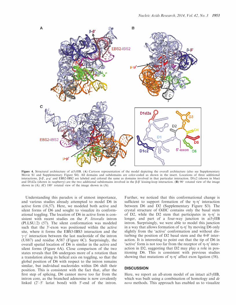
Structural architecture of ai5cIIB
As expected, the active-site region and the overall archi-tecture of the ai5gIIB model is similar to the known struc-ture of OiIIC. However, IIB introns possess additionalsubdomains and tertiary interactions (such as b-b0, m-m0
and EBS2-IBS2) that are important for both folding andcatalysis (11,49). Unlike IIC introns, IIB introns react withhigh sequence-specificity and undergo branching effi-ciently. The ai5gIIB model allowed us to visualize the add-itional subdomains and thereby provided valuable hintson functional roles of structural features specific to IIBintrons. For example, the b-b0and m-m0 tertiary interactionsform on opposite sides of D5 (Figure 4 andSupplementary Movie S1). D1c2 and D1d2a (the stemssupporting the b-b0 interaction) are located on the ‘cata-lytic face’, while D3 falls on the ‘binding face’ (50) of D5.D1c2 and D1d2a protrude like two extra arms from
Nucleic Acids Research, 2014, Vol. 42, No. 3 1951

opposite sides of D1, and the b-b0 kissing-loop interactionbetween these hairpin loops interlocks like a bridge that islikely to rigidify the scaffold of D1 (Figure 4A). On theother side of D5, D3a and D3b form a wall along the‘binding face’ that may mediate interactions between D5and the rest of the intron (Figure 4B and C).The second exon binding site, or EBS2, is a major dis-
tinguishing feature of the highly derived IIB and IIAintrons. The model shows that EBS2 is far more solventexposed than EBS1, which is consistent with footprintingdata on ai5gIIB intron (18). The model also shows that theEBS1-IBS1 and EBS2-IBS2 helices, which form on targetor exon binding, are not coaxial with each other, consist-ent with previous spectroscopic and mutational analysis ofthe ai5gIIB intron (51) and the model of Pylaiella littoralisintron (Pl.LSU/2) (17). We see that the EBS2-IBS2 helix islocated immediately adjacent to the b-b0 interaction(Figure 5), suggesting that function of the two motifsmay be linked. Importantly, the modeling processrevealed that, when no exon is bound, the EBS2 regionand adjacent helices are flexible. This indicates that exonbinding, and formation of the EBS2-IBS2 helix, rigidifiesEBS2 and the surrounding substructures. Thus, formationof the EBS2-IBS2 helix induces conformational rearrange-ments that are likely to considerably reduce overallentropy, with important implications for the known roleof EBS2-IBS2 in substrate specificity and reaction chem-istry (51,52).
Among the subdomains that are specific to IIB introns,D1d2b is the only substructure that appears to be pointedaway from the intron core and has no contacts with therest of the intron. However, D1d2b is coaxial with D1d2a,and it may therefore play an indirect role in the formationof b-b0 kissing-loop between D1d2a and D1c2.
The active and silent conformers of D6
D6 can exist in two different conformations (Figure 6A).In the conformation capable of branching, (the activeform) the 20-OH group of the branch-site adenosine ispositioned precisely to initiate the first step of splicingwithin the active site (Figure 6B). After the first step, thebranch-point nucleotide flips out of the active site, result-ing in an alternate conformation (the silent form) (11).The toggling of D6 between active and silent forms islikely to represent an important regulator of splicing ingroup II introns, and potentially, in the spliceosome aswell, for cognate domains (53). To prepare the activesite for second step of splicing, and to position the 30-exon appropriately, D6 will need to exit from the activesite. However, cross-linking studies (54,55) have shownthat nucleotides in D5 and D6 are located approximatelyin the same position through the entire cycle of splicing,which is consistent with studies showing that the sameactive-site elements play a role in both steps of group IIintron splicing (56).
Figure 3. Model Validation. (A) Correlation plot between numbers of contacts of each nucleotide in the model with hydroxyl radical reactivities. Thecorrelation coefficient (r=0.61) is as good as that observed for crystal structures of other RNAs such as group I introns, RNase P andriboswitches (47). (B) Difference plot of hydroxyl radical reactivities between WT D135 and b-b0 mutant. Nucleotides with significant increasesor decreases (greater than one unit of standard deviation) in reactivities are colored in red. (C) D1c2 and D1d2a stems are shown as a transparentsurface representation colored in yellow. Regions with significant increase in hydroxyl reactivities are colored in red. The rest of the intron is coloredin light blue. Most of the regions with significant increases in reactivities lie adjacent to D1c2 and D1d2a, indicating that mutation of the kissing-loopinteraction between D1c2 and D1d2a has exposed the regions beneath them and that the model is in good agreement with the experimental data.
1952 Nucleic Acids Research, 2014, Vol. 42, No. 3
Understanding this paradox is of utmost importance,and various studies already attempted to model D6 inactive form (16,57). Here, we modeled both active andsilent forms of D6 and sought to visualize its conform-ational toggling. The location of D6 in active form is con-sistent with recent studies on the P. littoralis intron(Pl.LSU/2) (57). The silent conformation was modeledsuch that the 30-exon was positioned within the activesite, where it forms the EBS3-IBS3 interaction and theg-g0 interaction between the last nucleotide of the intron(U887) and residue A587 (Figure 6C). Surprisingly, theoverall spatial location of D6 is similar in the active andsilent forms (Figure 6A). Close comparison of the twostates reveals that D6 undergoes more of a rotation thana translation along its helical axis on toggling, so that theglobal position of D6 with respect to the intron remainssimilar, but individual nucleotides within D6 shift theirposition. This is consistent with the fact that, after thefirst step of splicing, D6 cannot move too far from theintron core, as the branched adenosine is now covalentlylinked (20–50 lariat bond) with 50-end of the intron.
Further, we noticed that this conformational change issufficient to support formation of the Z-Z0 interactionbetween D6 and D2 (Supplementary Figure S3). Thecrystal structure of OiIIC contains only the basal stemof D2, while the D2 stem that participates in Z-Z0 islonger, and part of a four-way junction in ai5gIIBintron. Surprisingly, we were able to model this junctionin a way that allows formation of Z-Z0 by moving D6 onlyslightly from the ‘active’ conformation and without dis-turbing the position of D2 basal stem and the y-y0 inter-action. It is interesting to point out that the tip of D6 in‘active’ form is not too far from the receptor of Z-Z0 inter-action in D2, suggesting that D2 may play a role in pos-itioning D6. This is consistent with previous studiesshowing that mutations of Z-Z0 affect exon ligation (58).
DISCUSSION
Here, we report an all-atom model of an intact ai5gIIB,which was built using a combination of homology and denovo methods. This approach has enabled us to visualize
Figure 4. Structural architecture of ai5gIIB. (A) Cartoon representation of the model depicting the overall architecture (also see SupplementaryMovie S1 and Supplementary Figure S4). All domains and subdomains are color-coded as shown in the insert. Locations of three additionalinteractions, b-b0, m-m0 and EBS2-IBS2 are labeled and colored the same as domains involved in that particular interaction. D1c2 (shown in blue)and D1d2a (shown in raspberry) are the two additional subdomains involved in the b-b0 kissing-loop interaction. (B) 90 rotated view of the imageshown in (A). (C) 180 rotated view of the image shown in (A).
Nucleic Acids Research, 2014, Vol. 42, No. 3 1953

key group II intron structural features, such as the twoconformations of intron D6, and the EBS-IBS2 specificityhelix. The relative locations and orientations of thesesubdomains, along with structural innovations such asb-b0, explain many of the functional attributes unique togroup IIB introns, such as their extreme sequence-specifi-city and their propensity to splice through branching.
One of the features most readily apparent from themodel is that D5 is surrounded by a larger and moreprotective shell in the ai5gIIB intron relative to OiIIC(Figure 7). This well-packed IIB intron scaffold, alongwith added interactions such as b-b0 and m-m0, is likely toaffect the overall dynamics of the intron, influence theelectrostatic potential at the active site and provide add-itional stability to the active site as it assembles on thesurface of D5. Previous studies of ai5gIIB have shownthat D5 binds so tightly to rest of the intron (59,60) thatit can be added as a separate domain to truncated variantsof the intron (59–61).
Perhaps most importantly, the model provides insightsinto the branching mechanism of group II introns.Splicing through branching is one of the most notablefeatures of group II introns, but it has remained difficultto visualize because the existing IIC crystal structures lackelectron density for D6, and IIC introns do not readily
Figure 6. Active versus silent form of D6: (A) D6 can exist in two different conformations: the active (green) and silent forms (magenta). (B) In theconformation needed for the first step of splicing (the active form) the 20-OH group of branch-site adenosine (green) is positioned in the active site toinitiate the first step of splicing. Active-site elements (catalytic triad, 2-nt bulge and J2\3 junction) are shown in blue, 50-exon is shown in yellow, firstnucleotide of the intron G1 is shown in purple. (C) In the conformation that forms after the first step of splicing, and which is required for thesecond step (the silent-form), the branch-site adenosine flips out and positions the 30-exon in the active site and forms two new interactions (shown inred) IBS3-EBS3 and g-g0. Based on the model, toggling between the ‘active’ and ‘silent’ conformations of D6 involves a simple rotation of thedomain rather than a large translational movement away from the active site.
Figure 5. Location of the EBS2 binding site. The EBS2-IBS2 duplex isshown in orange, the EBS1-IBS1 duplex in shown in magenta. Thelinker between IBS1 and IBS2 is shown in red. The rest of the intronis shown as a transparent surface representation. The EBS2-IBS2 inter-action is located next to the kissing-loop (b-b0) interaction that joinsD1c2 (green) and D1d2a (blue), suggesting that function of the twomotifs may be linked.
1954 Nucleic Acids Research, 2014, Vol. 42, No. 3

branch (they splice only through hydrolysis in vitro)(62,63). Previous studies have proposed two candidate re-ceptors of D6 for branching: D1c1 (57) and the coordin-ation loop (55). In the model provided here, D6 (in theactive form) is located close to D1c1; however, the coord-ination loop is also in the vicinity of D6, and it is possiblethat D6 interacts with the coordination loop before thebranch-site adenosine is recruited into the active site.During the modeling process, we observed that thepresence of additional subdomains (D1c1 and D1d2a)restrict the possible conformations for D6 in ai5gIIBrelative to D6 in OiIIC, which is likely to enhancebranching relative to hydrolytic attack at the 50-splicesite. In addition, we observe that toggling between the
‘active’ and ‘silent’ conformations of D6 involve asimple rotation of the domain rather than a large transla-tional movement away from the active site as proposedpreviously (11), thereby explaining the many biochemicalstudies, showing that intron domains do not radically re-organize between the two steps of splicing (54) (Figure 6).The EBS2-IBS2 interaction is another distinguishing
feature of group IIB and IIA introns, and the modelhelps to show why this structural innovation has cometo play such an important role in intron function. Thereare introns, such as Clostridium beijerinckii I2 (64) andPelotomaculum thermopropionicum strain SI I2 (64),which contain an EBS2-IBS2 interaction with noapparent role in self-splicing (64,65). When we compared
Figure 7. Comparison of the ai5gIIB model with the crystal structure of OiIIC. (A) Front and side views of the ai5gIIB model. Regions common toboth introns are colored in light blue; D5 is colored in red. Long-range interactions that are not present in OiIIC are labeled in Greek letters. D1c2 isshown in purple, D1d2a is shown in magenta, D3a and D3b are shown in orange. EBS2-IBS2 helix is shown in green. For comparison with OiIIC,D6 is not shown in both views. (B) Front and side views of OiIIC crystal structure.
Nucleic Acids Research, 2014, Vol. 42, No. 3 1955
the secondary structures of those introns with ai5gIIB, wenoticed that the b-b0 interaction is absent in C. beijerinckiI2 (64), P. thermopropionicum strain SI I2 (64) andSinorhizobium meliloti RmInt1 (65). In the model, theb-b0 interaction lies in a cleft between the EBS1-IBS1and EBS2-IBS2 duplexes (Figure 5), consistent with aknown cross-link between IBS1 and D1c2 (66) (one ofthe stems involved in b-b0). Thus, a functional EBS2-IBS2 duplex may require the presence of b-b0 and thetwo may act synergistically to rigidify the overall intronstructure and promote specificity. Another important ob-servation is that EBS2 is more solvent-exposed than EBS1.This suggests that EBS2 may interact with its cognateIBS2 site first, triggering a conformational change thatmakes EBS1 more accessible, and then leading to EBS1-IBS1 formation. Such a mechanism might be importantduring specific retro-homing events, whereby the intronprotects EBS1 from binding to nonspecific sites, butallows binding at EBS1 to occur only after a specificEBS2-IBS2 duplex is formed. Finally, the lack of coaxialstacking between the EBS-IBS duplexes explains the extra-ordinary sequence-specificity of group IIB introns.Because each duplex is fully independent, and theoverall exon interaction energy is not supplemented bycoaxial stacking, the recognition helices are less tolerantof mismatches (51), leading to the large specificity indexfor target site recognition that is observed for group IIBintrons (51). The specificity index may also be enhancedby the insertion of b-b0 into the cleft between the EBS-IBSduplexes (Figure 5), as the resultant steric clashing maygive rise to the energetic penalty that is known to accom-pany binding of an oligonucleotide to both EBS1 andEBS2 simultaneously. In fact, it is known that bindingof long oligonucleotide targets (which bind both EBS1and EBS2) is 8 kcal/mol less favorable than binding ofshort oligos to EBS1 and EBS2 individually (52).Although the RNA computation field has significantly
progressed in the past decade, it was necessary for us tocreate new tools to complete and refine the model of theIIB intron. Here, we developed a broadly applicable com-putational tool for automatically correcting the backboneconfigurations in the models even before an electron-density map is available. This new tool, which is part ofthe RCrane program (32), will be helpful in modeling largeRNA structures in conjunction with existing RNAmodeling methods. Several features of RCrane as a 3Dmodeling tool are notable: First, existing, homologymodeling methods (including those used for protein struc-tures) derive models by copying coordinates from atemplate structure, which is a process that often leads toimproper backbone configurations and steric clashes, es-pecially in regions with low sequence similarity. RCranerebuilds the entire backbone while keeping the position ofthe bases constant, and thereby provides the uniqueadvantage of preserving secondary structure throughoutrefinement. In addition, large RNAs like group II intronsmay use different structural motifs but still share similarglobal structures, and RCrane preserves this informationcontent. For example, the tetraloop and receptor inter-action of D5 in IIC introns has a different structuralform compared with other group II introns (40) and
various long-range interactions also display slight differ-ences, such as the number of base pairs involved in akissing-loop. Currently, no methods exist to automaticallycorrect for such differences, and users would have tomanually adjust or rebuild these regions. By contrast,RCrane semiautomates this process, allowing the user toadjust or rebuild the nucleotide base, while RCranerebuilds the backbone accordingly and ensures that itconforms to allowed rotameric states. RCrane can alsobe used to enhance de novo modeling methods, such asMC-Sym, which build structures by assembling fragmentsfrom nucleotide libraries. This process often results instructures with poor backbone connectivity. Again,RCrane can be used to fix backbone inconsistencies inthese models without disrupting the secondary structure.In this work, we used RCrane at various stages to success-fully build the model of ai5gIIB intron.
The ability to create a detailed homology model of alarge multidomain RNA is of particular interest at thistime, as it is becoming apparent that large RNAs arecentral to most aspects of gene expression in biology(25). There is also increasing evidence that largenoncoding RNAs are, in many cases, highly structured(24). Since it is not currently feasible to experimentallydetermine structures for a majority of these RNAs, therewill be an increasing demand for computational methodsto predict RNA structures. The modeling strategies usedhere can be applied to other group II introns and to othermultidomain RNA molecules for which empirical struc-tural data are available.
In conclusion, we have shown that it is now possible tomodel large RNAs even from remote homologs. Wereport and test a 3D model of the ai5gIIB intronthat provides structural insights into the mechanisticbehavior of group II introns and explains functional dif-ferences between group IIC introns and more evolution-arily derived group II introns such as ai5gIIB.
SUPPLEMENTARY DATA
Supplementary Data are available at NAR Online,including [9,11,26,28,32–35,48,49, 67].
ACKNOWLEDGEMENTS
The authors thank all members of Pyle lab, in particularDr Isabel Chill0n Gazquez, Dr Marco Marcia, Dr OlgaFedorova, Dr Maximilian Bailor and Dr Laura Murrayfor many helpful discussions. They also thank DavidRawling, Dr Isabel Chill0n Gazquez and Dr MarcoMarcia for critical reading of this manuscript. A.M.P. isan investigator of Howard Hughes Medical Institute.
FUNDING
National Institutes of Health [RO1 GM50313]. Fundingfor open access charge: Howard Hughes Medical Institute.
Conflict of interest statement. None declared.
1956 Nucleic Acids Research, 2014, Vol. 42, No. 3

REFERENCES
1. Ferat,J.L. and Michel,F. (1993) Group II self-splicing introns inbacteria. Nature, 364, 358–361.
2. Valles,Y., Halanych,K.M. and Boore,J.L. (2008) Group II intronsbreak new boundaries: presence in a bilaterian’s genome. PLoSOne, 3, e1488.
3. Gordon,P.M., Sontheimer,E.J. and Piccirilli,J.A. (2000) Metal ioncatalysis during the exon-ligation step of nuclear pre-mRNAsplicing: extending the parallels between the spliceosome andgroup II introns. RNA, 6, 199–205.
4. Guo,H., Karberg,M., Long,M., Jones,J.P. 3rd, Sullenger,B. andLambowitz,A.M. (2000) Group II introns designed to insert intotherapeutically relevant DNA target sites in human cells. Science,289, 452–457.
5. Lambowitz,A.M. and Zimmerly,S. (2011) Group II introns:mobile ribozymes that invade DNA. Cold Spring Harb. Perspect.Biol., 3, a003616.
6. Garcıa-Rodrıguez,F.M., Barrientos-Duran,A., Dıaz-Prado,V.,Fernandez-Lopez,M. and Toro,N. (2011) Use of RmInt1, a groupIIB intron lacking the intron-encoded protein endonucleasedomain, in gene targeting. Appl. Environ. Microbiol., 77, 854–861.
7. Pyle,A.M., Fedorova,O. and Waldsich,C. (2007) Folding of groupII introns: a model system for large, multidomain RNAs? TrendsBiochem. Sci., 32, 138–145.
8. Donghi,D., Pechlaner,M., Finazzo,C., Knobloch,B. and Sigel,R.K.(2013) The structural stabilization of the kappa three-wayjunction by Mg(II) represents the first step in the folding of agroup II intron. Nucleic Acids Res., 41, 2489–2504.
9. Toor,N., Hausner,G. and Zimmerly,S. (2001) Coevolution ofgroup II intron RNA structures with their intron-encoded reversetranscriptases. RNA, 7, 1142–1152.
10. Michel,F., Kazuhiko,U. and Haruo,O. (1989) Comparative andfunctional anatomy of group II catalytic introns — a review.Gene, 82, 5–30.
11. Pyle,A.M. (2010) The tertiary structure of group II introns:implications for biological function and evolution. Crit. Rev.Biochem. Mol. Biol., 45, 215–232.
12. Toor,N., Keating,K.S., Taylor,S.D. and Pyle,A.M. (2008) Crystalstructure of a self-spliced group II intron. Science, 320, 77–82.
13. Marcia,M. and Pyle,A.M. (2012) Visualizing group II introncatalysis through the stages of splicing. Cell, 151, 497–507.
14. Marcia,M., Somarowthu,S. and Pyle,A.M. (2013) Now ondisplay: a gallery of group II intron structures at different stagesof catalysis. Mob. DNA, 4, 14.
15. Rest,J.S. and Mindell,D.P. (2003) Retroids in archaea: phylogenyand lateral origins. Mol. Biol. Evol., 20, 1134–1142.
16. Dai,L., Chai,D., Gu,S.-Q., Gabel,J., Noskov,S.Y., Blocker,F.J.H.,Lambowitz,A.M. and Zimmerly,S. (2008) A three-dimensionalmodel of a group II intron RNA and its interaction with theintron-encoded reverse transcriptase. Mol. Cell, 30, 472–485.
17. Costa,M., Michel,F. and Westhof,E. (2000) A three-dimensionalperspective on exon binding by a group II self-splicing intron.EMBO J., 19, 5007–5018.
18. Swisher,J., Duarte,D.M., Su,L.J. and Pyle,A.M. (2001) Visualizingthe solvent-inaccessible core of a group II intron ribozyme.EMBO J., 20, 2051–2061.
19. Chance,M.R., Bresnick,A.R., Burley,S.K., Jiang,J.S., Lima,C.D.,Sali,A., Almo,S.C., Bonanno,J.B., Buglino,J.A., Boulton,S. et al.(2002) Structural genomics: a pipeline for providing structures forthe biologist. Protein Sci., 11, 723–738.
20. Pieper,U., Webb,B.M., Barkan,D.T., Schneidman-Duhovny,D.,Schlessinger,A., Braberg,H., Yang,Z., Meng,E.C., Pettersen,E.F.,Huang,C.C. et al. (2011) ModBase, a database of annotatedcomparative protein structure models, and associated resources.Nucleic Acids Res., 39, D465–D474.
21. Peebles,C.L., Perlman,P.S., Mecklenburg,K.L., Petrillo,M.L.,Tabor,J.H., Jarrell,K.A. and Cheng,H.L. (1986) A self-splicingRNA excises an intron lariat. Cell, 44, 213–223.
22. Rother,K., Rother,M., Boniecki,M., Puton,T. and Bujnicki,J.M.(2011) RNA and protein 3D structure modeling: similarities anddifferences. J. Mol. Model., 17, 2325–2336.
23. Laing,C. and Schlick,T. (2010) Computational approaches to 3Dmodeling of RNA. J. Phys. Condens. Matter, 22, 283101.
24. Novikova,I.V., Hennelly,S.P., Tung,C.S. and Sanbonmatsu,K.Y.(2013) Rise of the RNA machines: exploring the structure of longnon-coding RNAs. J. Mol. Biol., 425, 3731–3746.
25. Ponting,C.P., Oliver,P.L. and Reik,W. (2009) Evolution andfunctions of long noncoding RNAs. Cell, 136, 629–641.
26. Rother,M., Rother,K., Puton,T. and Bujnicki,J.M. (2011)ModeRNA: a tool for comparative modeling of RNA 3Dstructure. Nucleic Acids Res., 39, 4007–4022.
27. Das,R. and Baker,D. (2007) Automated de novo prediction ofnative-like RNA tertiary structures. Proc. Natl Acad. Sci. USA,104, 14664–14669.
28. Parisien,M. and Major,F. (2008) The MC-Fold and MC-Sympipeline infers RNA structure from sequence data. Nature, 452,51–55.
29. Jonikas,M.A., Radmer,R.J., Laederach,A., Das,R., Pearlman,S.,Herschlag,D. and Altman,R.B. (2009) Coarse-grained modeling oflarge RNA molecules with knowledge-based potentials andstructural filters. RNA, 15, 189–199.
30. Sharma,S., Ding,F. and Dokholyan,N.V. (2008) iFoldRNA: three-dimensional RNA structure prediction and folding.Bioinformatics, 24, 1951–1952.
31. Keating,K.S. and Pyle,A.M. (2010) Semiautomated modelbuilding for RNA crystallography using a directed rotamericapproach. Proc. Natl Acad. Sci. USA, 107, 8177–8182.
32. Keating,K.S. and Pyle,A.M. (2012) RCrane: semi-automatedRNA model building. Acta Crystallogr. D Biol. Crystallogr., 68,985–995.
33. Hall,T.A. (1999) BioEdit: a user-friendly biological sequencealignment editor and analysis program for Windows 95/98/NT.Nucleic Acids Symp. Ser., 41, 95–98.
34. Pearlman,D.A., Case,D.A., Caldwell,J.W., Ross,W.S., CheathamIii,T.E., DeBolt,S., Ferguson,D., Seibel,G. and Kollman,P. (1995)AMBER, a package of computer programs for applyingmolecular mechanics, normal mode analysis, molecular dynamicsand free energy calculations to simulate the structural andenergetic properties of molecules. Comput. Phys. Commun., 91,1–41.
35. Chen,V.B., Arendall,W.B. 3rd, Headd,J.J., Keedy,D.A.,Immormino,R.M., Kapral,G.J., Murray,L.W., Richardson,J.S.and Richardson,D.C. (2010) MolProbity: all-atom structurevalidation for macromolecular crystallography. Acta Crystallogr.D Biol. Crystallogr., 66, 12–21.
36. Mortimer,S.A. and Weeks,K.M. (2007) A fast-acting reagent foraccurate analysis of RNA secondary and tertiary structure bySHAPE chemistry. J. Am. Chem. Soc., 129, 4144–4145.
37. Watts,J.M., Dang,K.K., Gorelick,R.J., Leonard,C.W.,Bess,J.W. Jr, Swanstrom,R., Burch,C.L. and Weeks,K.M. (2009)Architecture and secondary structure of an entire HIV-1 RNAgenome. Nature, 460, 711–716.
38. Vasa,S.M., Guex,N., Wilkinson,K.A., Weeks,K.M. andGiddings,M.C. (2008) ShapeFinder: a software system for high-throughput quantitative analysis of nucleic acid reactivityinformation resolved by capillary electrophoresis. RNA, 14,1979–1990.
39. McGinnis,J.L., Duncan,C.D. and Weeks,K.M. (2009) High-throughput SHAPE and hydroxyl radical analysis of RNAstructure and ribonucleoprotein assembly. Methods Enzymol., 468,67–89.
40. Keating,K.S., Toor,N. and Pyle,A.M. (2008) The GANCtetraloop: a novel motif in the group IIC intron structure.J. Mol. Biol., 383, 475–481.
41. Costa,M. and Michel,F. (1995) Frequent use of thesame tertiary motif by self-folding RNAs. EMBO J., 14,1276–1285.
42. Daniels,D.L., Michels,W.J. and Pyle,A.M. (1996) Two competingpathways for self-splicing by group II introns: a quantitativeanalysis of in vitro reaction rates and products. J. Mol. Biol.,256, 31–49.
43. Keating,K.S., Humphris,E.L. and Pyle,A.M. (2011) A new way tosee RNA. Q. Rev. Biophys., 44, 433–466.
44. Wadley,L.M., Keating,K.S., Duarte,C.M. and Pyle,A.M. (2007)Evaluating and learning from RNA pseudotorsional space:quantitative validation of a reduced representation for RNAstructure. J. Mol. Biol., 372, 942–957.
Nucleic Acids Research, 2014, Vol. 42, No. 3 1957

45. Richardson,J.S., Schneider,B., Murray,L.W., Kapral,G.J.,Immormino,R.M., Headd,J.J., Richardson,D.C., Ham,D.,Hershkovits,E., Williams,L.D. et al. (2008) RNA backbone:consensus all-angle conformers and modular string nomenclature(an RNA Ontology Consortium contribution). RNA, 14, 465–481.
46. Tullius,T.D. and Greenbaum,J.A. (2005) Mapping nucleic acidstructure by hydroxyl radical cleavage. Curr. Opin. Chem. Biol., 9,127–134.
47. Ding,F., Lavender,C.A., Weeks,K.M. and Dokholyan,N.V. (2012)Three-dimensional RNA structure refinement by hydroxyl radicalprobing. Nat. Methods, 9, 603–608.
48. Su,L.J., Waldsich,C. and Pyle,A.M. (2005) An obligateintermediate along the slow folding pathway of a group II intronribozyme. Nucleic Acids Res., 33, 6674–6687.
49. Fedorova,O. and Pyle,A.M. (2005) Linking the group II introncatalytic domains: tertiary contacts and structural features ofdomain 3. EMBO J., 24, 3906–3916.
50. Abramovitz,D.L., Friedman,R.A. and Pyle,A.M. (1996) Catalyticrole of 2’-hydroxyl groups within a group II intron active site.Science, 271, 1410–1413.
51. Xiang,Q., Qin,P.Z.F., Michels,W.J., Freeland,K. and Pyle,A.M.(1998) Sequence specificity of a group II intron ribozyme:multiple mechanisms for promoting unusually high discriminationagainst mismatched targets. Biochemistry, 37, 3839–3849.
52. Qin,P.Z. and Pyle,A.M. (1999) Antagonistic substrate binding bya group II intron ribozyme. J. Mol. Biol., 291, 15–27.
53. Abelson,J. (2013) Toggling in the spliceosome. Nat. Struct. Mol.Biol., 20, 645–647.
54. de Lencastre,A., Hamill,S. and Pyle,A.M. (2005) A single active-site region for a group II intron. Nat. Struct. Mol. Biol., 12,626–627.
55. Hamill,S. and Pyle,A.M. (2006) The receptor for within a groupII branch-site docking intron active site. Mol. Cell, 23, 831–840.
56. Chanfreau,G. and Jacquier,A. (1994) Catalytic site componentscommon to both splicing steps of a group II intron. Science, 266,1383–1387.
57. Li,C.F., Costa,M. and Michel,F. (2011) Linking the branchpointhelix to a newly found receptor allows lariat formation by agroup II intron. EMBO J., 30, 3040–3051.
58. Chanfreau,G. and Jacquier,A. (1996) An RNA conformationalchange between the two chemical steps of group II self-splicing.EMBO J., 15, 3466–3476.
59. Pyle,A.M. and Green,J.B. (1994) Building a kinetic framework forgroup-ii intron ribozyme activity - quantitation of interdomainbinding and reaction-rate. Biochemistry, 33, 2716–2725.
60. Jarrell,K.A., Dietrich,R.C. and Perlman,P.S. (1988) Group IIintron domain 5 facilitates a trans-splicing reaction. Mol. Cell.Biol., 8, 2361–2366.
61. Michels,W.J. and Pyle,A.M. (1995) Conversion of a group-iiintron into a new multiple-turnover ribozyme that selectivelycleaves oligonucleotides - elucidation of reaction-mechanism andstructure-function-relationships. Biochemistry, 34, 2965–2977.
62. Toor,N., Robart,A.R., Christianson,J. and Zimmerly,S. (2006)Self-splicing of a group IIC intron: 50 exon recognition andalternative 5’ splicing events implicate the stem-loop motif of atranscriptional terminator. Nucleic Acids Res., 34, 6461–6471.
63. Granlund,M., Michel,F. and Norgren,M. (2001) Mutuallyexclusive distribution of IS1548 and GBSi1, an active group IIintron identified in human isolates of group B streptococci.J. Bacteriol., 183, 2560–2569.
64. Nagy,V., Pirakitikulr,N., Zhou,K.I., Chillon,I., Luo,J. andPyle,A.M. (2013) Predicted group II intron lineages E and Fcomprise catalytically active ribozymes. RNA, 19, 1266–1278.
65. Barrientos-Duran,A., Chillon,I., Martinez-Abarca,F. and Toro,N.(2011) Exon sequence requirements for excision in vivo of thebacterial group II intron RmInt1. BMC Mol. Biol., 12, 24.
66. Noah,J.W. and Lambowitz,A.M. (2003) Effects of maturasebinding and Mg2+ concentration on group II intron RNAfolding investigated by UV cross-linking. Biochemistry, 42,12466–12480.
67. Laing,C. and Schlick,T. (2009) Analysis of four-way junctions inRNA structures. J. Mol. Biol., 390, 547–559.
1958 Nucleic Acids Research, 2014, Vol. 42, No. 3
Copyright © 2022 FDOKUMEN