Virtual microscopy in the undergraduate teaching of pathology
7
ISSN : 2229-5089 Vol 6 - Issue 1 - January 2015 Publication of the Association for Pathology Informatics J o u r n a l o f P a t h o l o g y I n f o r m a t i c s www.jpathinformatics.org
-
Upload
independent -
Category
Documents
-
view
1 -
download
0
Transcript of Virtual microscopy in the undergraduate teaching of pathology

ISSN : 2229-5089
Vol 6 - Issue 1 - January 2015
Publication of the Association for Pathology Informatics
Journal of Pathology Informatics
www.jpathinformatics.org
Jou
rna
l of P
ath
olo
gy
Info
rma
tics • V
olu
me 6
• Issue 1
• Jan
uary
-Feb
rua
ry 2
01
5 • P
age
s 1-?
?
J Pathol Inform Editor-in-Chief: Anil V. Parwani , Liron Pantanowitz, Pittsburgh, PA, USA Pittsburgh, PA, USA
OPEN ACCESS HTML format
For entire Editorial Board visit : www.jpathinformatics.org/editorialboard.asp
Research Article
Virtual microscopy in the undergraduate teaching of pathology
Oriol Ordi1, Josep Antoni Bombí1,2, Antonio Martínez1,2, Josep Ramírez1,2, Llúcia Alòs1,2, Adela Saco1, Teresa Ribalta1,2, Pedro L. Fernández1,2, Elias Campo1,2, Jaume Ordi1,2,3
1Department of Pathology, School of Medicine, University of Barcelona, 2Department of Pathology, Hospital Clínic, 3 ISGlobal, Barcelona Center for International Health Research (CRESIB), Barcelona, Spain
E-mail: *Prof. Dr. Jaume Ordi - [email protected] *Corresponding author
Received: 28 August 2014 Accepted: 24 November 2014 Published: 29 January 15
INTRODUCTION
In the last 20 years, web‑based resources developed to supplement or replace the traditional methodologies have expanded dramatically. These resources have shown clear benefits, as classes can be delivered to many students simultaneously, and this has helped medical schools to train in a more cost‑effective way.[1]
Histology and pathology play an essential role in education in undergraduate courses in medicine. The practical knowledge of these disciplines has classically been delivered using glass slides and conventional microscopes (CM), as web‑based resources were limited to static images, which were very different from real practice. Virtual microscopy (VM), also referred to as whole slide imaging, has recently started to change the
Copyright: © 2015 Ordi O. This is an open‑access article distributed under the terms of the Creative Commons Attribution License, which permits unrestricted use, distribution, and reproduction in any medium, provided the original author and source are credited.
This article may be cited as:Ordi O, Bombí JA, Martínez A, Ramírez J, Alòs L, Saco A, et al. Virtual microscopy in the undergraduate teaching of pathology. J Pathol Inform 2015;6:1.
Available FREE in open access from: http://www.jpathinformatics.org/text.asp?2015/6/1/1/150246
Abstract
Background: Little evidence is available concerning the impact of virtual microscopy (VM) in the undergraduate teaching of pathology. We aimed: (1) to determine the impact in student scores when moving from conventional microscopy (CM) to VM; (2) to assess the students’ impressions and changes in study habits regarding the impact of this tool. Methods: We evaluated two groups taking the discipline of pathology in the same course, one using CM and the other VM. The same set of slides used in the CM classes was digitized in a VENTANA iScan HT (Roche Diagnostics, Sant Cugat, Spain) at ×20 and observed by the students using the Virtuoso viewer (Roche Diagnostics). We evaluated the skill level reached by the students with an online test. A voluntary survey was undertaken by the VM group to assess the students’ impressions regarding the resource. The day and time of any accession to the viewer were registered. Results: There were no differences between the two groups in their marks in the online test (mean marks for the CM and the VM groups: 9.87 ± 0.34 and 9.86 ± 0.53, respectively; P = 0.880). 86.6% of the students found the software friendly, easy‑to‑use and effective. 71.6% of the students considered navigation easier with VM than with CM. The most appreciated feature of VM was the possibility to access the images anywhere and at any time (93.3%). 57.5% of the accesses were made on holidays and 41.9% later than 6:00 pm. Conclusions: Virtual microscopy can effectively replace the traditional methods of learning pathology, providing mobility and convenience to medical students.Key words: Pathology, undergraduate teaching, whole slide imaging
Access this article onlineWebsite: www.jpathinformatics.org
DOI: 10.4103/2153-3539.150246
Quick Response Code:

J Pathol Inform 2015, 1:1 http://www.jpathinformatics.org/content/6/1/1
way in which these disciplines can be delivered online by providing the ability to scan entire glass slides at diagnostic resolution. A digital slide of the tissue section is created and with the use of specific software can be viewed and magnified in real‑time across the web very much like using a CM. Currently, several commercially available systems can digitize glass slides containing tissue sections and produce virtual slides of excellent quality. The rapid progress of this technology and its many potential benefits will probably result in a progressive shift from conventional to VM in routine diagnostic pathology.[2‑4]
Several studies have documented the success of VM in graduate education in medical,[5‑9] dental,[10,11] and veterinary schools.[12‑14] However, although VM has been used for many years in the US, reports on the experience with this technology in the undergraduate teaching of pathology are still limited and there is little evidence about its true impact on students’ knowledge and study habits.
In this study, we report the experience in the transition of a General Pathology course in the medical school of the University of Barcelona from practical teaching based on CM to a new format in which this tool has been totally eliminated, and all microscopy work is conducted on the computer using VM. We specifically aimed: (1) To assess the students’ impressions regarding the impact of VM on their learning and to objectively assess the success of implementing this new technology in their curriculum; (2) to determine whether moving from glass to virtual slides has an impact on student scores in practical exams of pathology; and (3) to evaluate the changes in study habits associated with the introduction of this tool in undergraduate teaching.
METHODS
The study was conducted in the Department of Pathology of the School of Medicine in the University of Barcelona, Spain. This university implemented the open‑source course management system modular object‑oriented dynamic learning environment (Moodle) in 2004, and all the supporting information and most of the activities of each discipline are available to the students in this platform.
The action was conducted in General Pathology, which is delivered in the 3rd year as a 4‑month course and has 6 European Credit Transfer System credits.[15] Two different groups take the discipline each year, the first group from September to January and the second from February to May. All the students in both groups had previously had a whole year of experience using CM in the course of histology, but none had had any previous experience with VM. During the 2013–2014 course, one group studied anatomical pathology using CM whereas the second group used VM.
Characteristics of the Conventional Microscopy CourseThis group took the discipline from September 2013 to January 2014 using CM, following the same rules established in the previous 5 years. The group was divided into six smaller groups each composed of 15 students. Two practical classes were scheduled during the course, each of which included 16 histological slides stained with hematoxylin and eosin, being representative of basic pathological lesions. Eighteen sets of slides including consecutive sections of the 16 cases were available for the use of the students in each practical class. Practical classes of 2 h in duration were delivered in the microscopy room of the medical school and were conducted by a professor of pathology. The professor briefly showed the most relevant features of each particular slide with a microscope connected to a video camera and several screens. Thereafter, each student had a set of the slides for his/her use and had 90 min to observe the slides on his/her own single‑headed microscope with the support of the faculty member, who solved all the questions and problems brought up by the students.
Characteristics of the Virtual Microscopy CourseThis group took the discipline from February to May 2014 using only VM. The practical course included the same 32 cases used in the previously described group. The group was divided into six smaller groups each composed of 15–16 students. A single practical class was scheduled at the beginning of the course (February). The class was delivered in a room equipped with a computer with internet connection and a 52‑inch screen and was conducted by a faculty member, being of 1/2 h in duration. The professor briefly showed how to accede to the website, the general characteristics of the navigator and how to retrieve the supporting information. After this initial session, all the students were allowed to access the virtual slides any day and at any time from any computer connected to Internet. The students were given the opportunity to contact their tutors for any problem or doubt encountered when observing the slides on their computers.
Virtual Slides, Navigation and Supporting FilesAll the cases were digitized in a VENTANA iScan HT (Roche‑Ventana Medical Systems, Tucson, AZ, USA) at a magnification of ×20. The system creates high‑resolution digital images of tissue sections. All files were stored on a server hosted at the Spanish Division of Roche Diagnostics. The students access the virtual slides through a hyperlink on the Moodle platform, using their own computers as virtual microscopes. The images are viewed in the Virtuoso viewer (Roche‑Ventana Medical Systems, Tucson, AZ, USA), which works as a web browser and simulates a CM [Figure 1]. Virtuoso is designed to organize the images into different cases and the cases into groups. No specific

J Pathol Inform 2015, 1:1 http://www.jpathinformatics.org/content/6/1/1
software installation is required to visualize the virtual slides.
Two supporting pdf files were posted on the main page of the discipline in the Moodle platform. One included general information to guide student accession to the viewer and the username and password necessary to accede to the website. The second pdf file included educational text discussions for each particular case.
Online Evaluation of the Skill LevelAn online quiz was performed to evaluate the skill level reached by the students in the evaluation of the microscopic lesions. A question bank containing 200 multiple choice questions was created in the Moodle platform.[16] All questions were based on static microphotographs prepared by the faculty by selection of specific areas from the same 32 glass slides included in both practical classes and had 5 possible answers. All the questions only had one correct answer qualified with a mark of +1. Each wrong answer was qualified with a mark of –0.25. For the examination, 40 questions were randomly selected from the pool. The test was available on the Moodle platform during a 24 h period. Both the order of the questions, as well as that of the answers was automatically distributed randomly for each student, thus, questions and answers were presented in a different order to every student. There was a 20 min time limit to complete the exam.
Audit of Student Access to the Virtual Slide ViewerFor the VM course, the viewer registered any access to the virtual slides by any of the students. This registration was performed anonymously, as all the students logged in using the same login and password. The day and time of any single accession, as well as the time spent by the student on each slide, were registered, and a pdf file was created with all the information.
Voluntary Student SurveyAt the conclusion of the VM course, a voluntary survey was undertaken by the students to assess the students’
impressions regarding the impact of VM on their learning and the success of implementing this new technology in their curriculum. The survey was designed using the free website https://www.surveymonkey.com/(SurveyMonkey®, Menlo Park, CA, USA) and was posted as an online hyperlink on the Moodle platform, which remained open for a whole week after the online exam was completed. Questions were related to the quality and easiness‑of‑use of the software and navigation, VM versus CM, supporting information, introduction of the activity by the professor and an online quiz [Table 1]. The student survey was designed as a Likert‑scale questionnaire with a five‑point scale with the following options: Strongly agree, agree, undecided, disagree, and strongly disagree. Ethical clearance was granted with participation in evaluations being entirely voluntary and completely anonymous.
Data AnalysisStatistical analysis was performed using the SPSS (version 18.0; SPSS, Inc., Chicago, IL). The results are presented as absolute numbers and percentages or mean and standard deviation. The analysis was mostly descriptive and included Chi‑square tests.
RESULTS
Characteristics of the GroupsThe CM course had 88 students, 67.0% (59/88) females and 33.0% (29/88) males, with a mean age of 20.6 ± 1.4. The VM course had 93 students, 68.8% (64/93) females and 31.2% (29/93) males, with a mean age of 20.8 ± 1.3. No differences were observed between the two groups.
Characteristics of the Virtual SlidesThe size of the files ranged from 149,321 to 1,851,049 Kb (mean 751,562.7 ± 413,330.2 Kb). The total weight of the 32 files was 24,050,005 Kb. The scanned images can be viewed up to a magnification of ×400 and are always in focus, with optimized contrast and adjusted illumination. At high magnifications it is easy for the student to maintain orientation with respect to the entire section, because the system indicates the position of the slide on a thumbnail showing a small representation of the section [Figure 1].
Online Evaluation of the Skill LevelThe mean mark in the online test in the CM course was 9.87 ± 0.34 (range: 8.3–10), with all the students passing the exam. Seventy‑six out of 88 (86.4%) answered all the questions correctly. The mean mark in the VM course was 9.86 ± 0.53 (range: 6.7–10) with 91/93 students (97.8%) passing the online test. Eighty‑five out of 93 (91.4%) answered all the questions correctly. No differences were observed between the two groups (P = 0.880, Student’s t‑test).
Figure 1: Screen shot of the virtual microscope display. A thumbnail showing a small representation of the whole section makes easier for the student to maintain orientation with respect to the entire section

J Pathol Inform 2015, 1:1 http://www.jpathinformatics.org/content/6/1/1
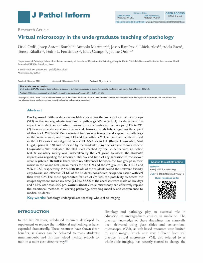
Audit of Student Accesses to the ViewerThe number of visits to the VM from the opening of the website to the day of the exam is shown in Figure 2; about 80.3% of the accesses (862/1073) were done in the week prior to the examination; 57.5% of the visits were made on holidays and 42.5% on working days. The times of access during the day are shown in Figure 3. The earliest access was at 8:33 am and the latest at 00:55 am, with 58.1% of the visits being observed between 8:00 am and 6:00 pm and 41.9% later than 6:00 pm. The mean length of the students’ accesses was 4 min 2 s. Thus, the overall time spent by the students on studying the 32 slides included in the course (including the 30‑min time of the initial presentation of the website) was 2 h 50 min.
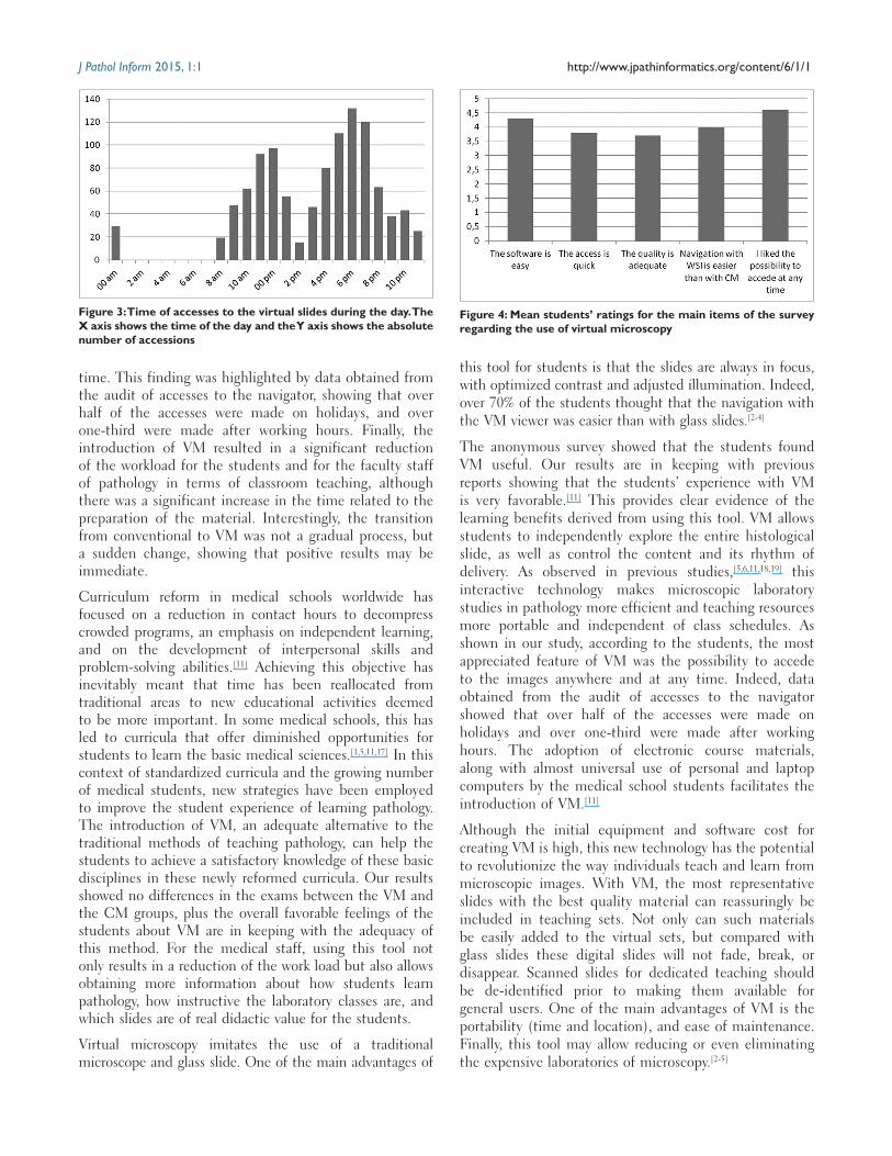
Student SurveySixty‑one out of 93 (65.6%) students participated in the survey. Table 1 shows the questions included in the survey and the students’ answers, and Figure 4 shows the mean students’ ratings for the main items of the survey concerning the use of VM; 86.6% of the students found the software friendly, easy‑to‑use and effective for the purposes of the course. The most appreciated feature of VM was the possibility to access the images anywhere and at any time (93.3%), and 71.6% of the students thought that navigation with the virtual was easier than with glass slides. Although a significant percentage of the students were neutral to both methods, most (50.8%) preferred VM to CM.
Consequences on the Workload for the Faculty StaffThe workload in terms of time in classroom teaching for the faculty staff was reduced from 20 h to 2.5. None of the students required faculty assistance. No questions to the tutors were registered in relation to problems or
doubts encountered when observing the slides on their computers.
CONCLUSIONS
The results of this study confirm that VM can effectively replace CM to teach pathology in undergraduate courses in medical schools and show that the microscopic skills acquired with VM are comparable to those acquired with CM, the classical tool for teaching pathology. The overall feedback from the students was highly positive. Students complemented the ease of use of the software. Students felt they worked faster with VM, and over 70% thought that the navigation with the VM was easier than with the CM. The most appreciated feature of VM was the possibility to access the images anywhere and at any
Table 1: Student’s responses to the survey regarding the use of virtual and the conventional microscope
Questions included in the student’s survey Strongly agree
Agree Neutral Disagree Strongly disagree
The software is friendly, easy‑to‑use and effective for the purposes of the course 43.3 43.3 10.0 3.4 0The access to the virtual slides is quick 21.7 53.3 13.4 8.3 3.3I liked the possibility to access the images anywhere and at any time 65.0 28.3 5.0 1.7 0The quality of the image of virtual slides is adequate 26.7 38.3 21.7 8.3 5.0VM allows time saving 31.2 44.3 21.3 1.6 1.6The identification of cells and structures with VM is easy 6.7 41.7 38.3 10.0 3.3Navigation with the VM viewer is easier than with glass slides 33.3 38.3 26.7 1.7 0I had problems with the navigation 3.3 13.3 15.0 31.7 36.7The presentation of the virtual viewer and the slides by the professor is useful 6.7 26.7 33.3 16.7 16.6The presentation of the virtual viewer by the professor is unnecessary 21.7 6.7 35.0 18.3 18.3The supporting material (pdf document) is useful and adequate 73.8 21.3 4.9 0 0The test based on image captures is a good way to evaluate the knowledge acquired with VM
14.7 57.4 18.1 4.9 4.9
I prefer virtual to CM 21.3 29.5 34.4 11.5 3.3I prefer conventional to VM 1.6 13.1 42.6 23.0 19.7
The figures indicate percentages, VM: Virtual microscopy, CM: Conventional microscopy
Figure 2: Number of accesses to the virtual slides from the opening of the website to the day of the exam. The X axis shows the day of accession in relation to the exam (day 0). The Y axis shows the absolute number of accessions. Red marks identify accessions on weekends/holidays (Saturday, Sunday or other holidays). Gray marks indicate work days
J Pathol Inform 2015, 1:1 http://www.jpathinformatics.org/content/6/1/1
time. This finding was highlighted by data obtained from the audit of accesses to the navigator, showing that over half of the accesses were made on holidays, and over one‑third were made after working hours. Finally, the introduction of VM resulted in a significant reduction of the workload for the students and for the faculty staff of pathology in terms of classroom teaching, although there was a significant increase in the time related to the preparation of the material. Interestingly, the transition from conventional to VM was not a gradual process, but a sudden change, showing that positive results may be immediate.
Curriculum reform in medical schools worldwide has focused on a reduction in contact hours to decompress crowded programs, an emphasis on independent learning, and on the development of interpersonal skills and problem‑solving abilities.[11] Achieving this objective has inevitably meant that time has been reallocated from traditional areas to new educational activities deemed to be more important. In some medical schools, this has led to curricula that offer diminished opportunities for students to learn the basic medical sciences.[1,5,11,17] In this context of standardized curricula and the growing number of medical students, new strategies have been employed to improve the student experience of learning pathology. The introduction of VM, an adequate alternative to the traditional methods of teaching pathology, can help the students to achieve a satisfactory knowledge of these basic disciplines in these newly reformed curricula. Our results showed no differences in the exams between the VM and the CM groups, plus the overall favorable feelings of the students about VM are in keeping with the adequacy of this method. For the medical staff, using this tool not only results in a reduction of the work load but also allows obtaining more information about how students learn pathology, how instructive the laboratory classes are, and which slides are of real didactic value for the students.
Virtual microscopy imitates the use of a traditional microscope and glass slide. One of the main advantages of
this tool for students is that the slides are always in focus, with optimized contrast and adjusted illumination. Indeed, over 70% of the students thought that the navigation with the VM viewer was easier than with glass slides.[2‑4]
The anonymous survey showed that the students found VM useful. Our results are in keeping with previous reports showing that the students’ experience with VM is very favorable.[11] This provides clear evidence of the learning benefits derived from using this tool. VM allows students to independently explore the entire histological slide, as well as control the content and its rhythm of delivery. As observed in previous studies,[5,6,11,18,19] this interactive technology makes microscopic laboratory studies in pathology more efficient and teaching resources more portable and independent of class schedules. As shown in our study, according to the students, the most appreciated feature of VM was the possibility to accede to the images anywhere and at any time. Indeed, data obtained from the audit of accesses to the navigator showed that over half of the accesses were made on holidays and over one‑third were made after working hours. The adoption of electronic course materials, along with almost universal use of personal and laptop computers by the medical school students facilitates the introduction of VM.[11]
Although the initial equipment and software cost for creating VM is high, this new technology has the potential to revolutionize the way individuals teach and learn from microscopic images. With VM, the most representative slides with the best quality material can reassuringly be included in teaching sets. Not only can such materials be easily added to the virtual sets, but compared with glass slides these digital slides will not fade, break, or disappear. Scanned slides for dedicated teaching should be de‑identified prior to making them available for general users. One of the main advantages of VM is the portability (time and location), and ease of maintenance. Finally, this tool may allow reducing or even eliminating the expensive laboratories of microscopy.[2‑5]
Figure 3: Time of accesses to the virtual slides during the day. The X axis shows the time of the day and the Y axis shows the absolute number of accessions
Figure 4: Mean students’ ratings for the main items of the survey regarding the use of virtual microscopy

J Pathol Inform 2015, 1:1 http://www.jpathinformatics.org/content/6/1/1
The main strength of our study is that it allows adequate comparison of two very similar groups from the same course working with the same material and that it provides objective data on how students learn pathology. A possible limitation of our study is the use of an examination system restricted to standard static images. However, this allowed comparison of the results with the CM group, as all the image captures were made from the same cases. Examinations that apply virtual technology require more sophisticated management software.[4] Finally, the very good results obtained in the examinations should be considered as the consequence of the extremely high marks required in Spain to accede to the medical schools.
In conclusion, evidence showing that the microscopic skills achieved by students with VM are comparable to these acquired with CM indicates that this technology can effectively replace the traditional methods of learning pathology. One of its main advantages is that it provides mobility and convenience to medical students.
REFERENCES
1. Williams G, Lau A. Reform of undergraduate medical teaching in the United Kingdom: A triumph of evangelism over common sense. BMJ 2004;329:92‑4.
2. Al‑Janabi S, Huisman A, Van Diest PJ. Digital pathology: Current status and future perspectives. Histopathology 2012;61:1‑9.
3. Pantanowitz L, Valenstein PN, Evans AJ, Kaplan KJ, Pfeifer JD, Wilbur DC, et al. Review of the current state of whole slide imaging in pathology. J Pathol Inform 2011;2:36.
4. Pantanowitz L, Szymas J, Yagi Y, Wilbur D. Whole slide imaging for educational purposes. J Pathol Inform 2012;3:46.
5. Blake CA, Lavoie HA, Millette CF. Teaching medical histology at the University of South Carolina School of Medicine: Transition to virtual slides
and virtual microscopes. Anat Rec B New Anat 2003;275:196‑206.6. Boutonnat J, Paulin C, Faure C, Colle PE, Ronot X, Seigneurin D. A pilot
study in two French medical schools for teaching histology using virtual microscopy. Morphologie 2006;90:21‑5.
7. Foster K. Medical education in the digital age: Digital whole slide imaging as an e‑learning tool. J Pathol Inform 2010;1.
8. Goldberg HR, Dintzis R. The positive impact of team‑based virtual microscopy on student learning in physiology and histology. Adv Physiol Educ 2007;31:261‑5.
9. Harris T, Leaven T, Heidger P, Kreiter C, Duncan J, Dick F. Comparison of a virtual microscope laboratory to a regular microscope laboratory for teaching histology. Anat Rec 2001;265:10‑4.
10. Chen YK, Hsue SS, Lin DC, Wang WC, Chen JY, Lin CC, et al. An application of virtual microscopy in the teaching of an oral and maxillofacial pathology laboratory course. Oral Surg Oral Med Oral Pathol Oral Radiol Endod 2008;105:342‑7.
11. Weaker FJ, Herbert DC. Transition of a dental histology course from light to virtual microscopy. J Dent Educ 2009;73:1213‑21.
12. Dee FR, Meyerholz DK. Teaching medical pathology in the twenty‑first century: Virtual microscopy applications. J Vet Med Educ 2007;34:431‑6.
13. Mills PC, Bradley AP, Woodall PF, Wildermoth M. Teaching histology to first‑year veterinary science students using virtual microscopy and traditional microscopy: A comparison of student responses. J Vet Med Educ 2007;34:177‑82.
14. Neel JA, Grindem CB, Bristol DG. Introduction and evaluation of virtual microscopy in teaching veterinary cytopathology. J Vet Med Educ 2007;34:437‑44.
15. Doménech Martínez E, Armas Ramos H, Castro Conde JR, González Díaz JP, Méndez Pérez A, Ormazábal Ramos C, et al. Study of the introduction of the European Credit Transfer System (ECTS) in pediatrics and modification of the teaching methodology. An Pediatr (Barc) 2006;65:415‑27.
16. Inuwa IM, Taranikanti V, Al‑Rawahy M, Habbal O. Anatomy practical examinations: How does student performance on computerized evaluation compare with the traditional format? Anat Sci Educ 2012;5:27‑32.
17. Bloodgood RA, Ogilvie RW. Trends in histology laboratory teaching in United States medical schools. Anat Rec B New Anat 2006;289:169‑75.
18. Sims MH, Mendis‑Handagama C, Moore RN. Virtual microscopy in a veterinary curriculum. J Vet Med Educ 2007;34:416‑22.
19. Szymas J, Lundin M. Five years of experience teaching pathology to dental students using the WebMicroscope. Diagn Pathol 2011;6 Suppl 1:S13.