A Holocaust Memorial or a Memorial to Germany's Vicarious Trauma?
Upload
independentCategory
view
0download
0
doi:10.1093/brain/awh456 Brain (2005), 128, 1122–1138
Vicarious function within the human primarymotor cortex?A longitudinal fMRI stroke study
Assia Jaillard,1,4 Chantal Delon Martin,4 Katia Garambois,1 Jean Francois Lebas2,4
and Marc Hommel1,3,4
Correspondence to: Dr Assia Jaillard, Departement de
Neurologie—Unite Neuro-vasculaire, Centre Hospitalier
Universitaire de Grenoble, BP 217-38043 Grenoble
Cedex 9, France
E-mail: [email protected]
1Department of Neurology, Stroke Unit, 2Unite de RMN
Service of Neuroradiology, 3Inserm, CIC 003, University
Hospital and 4Inserm, U594-University Joseph Fourier,
Grenoble, France
SummaryWhile experimental studies in the monkey have shown
that motor recovery after partial destruction of the handmotor cortex was based on adjacent motor reorganization,
functional MRI (fMRI) studies with isolated primary
motor cortical stroke have not yet been reported in
humans. Based on experimental data, we designed a study
to test if recovery after stroke within primary motor
cortex (M1) was associated with reorganization within
the surrounding motor cortex, i.e. the motor cortex was
able to vicariate. Since motor recovery is time-dependentand might be inflected according to the tested task, the
delay after stroke and two motor tasks were included in
our design. We examined four patients with one ischaemic
stroke limited to M1, and four sex- and age-matched
healthy controls in a temporally balanced prospective
longitudinal fMRI study over three sessions: <20 days,
4 months and 2 years after stroke. The paradigm included
two motor tasks, finger tapping (FT) and finger extension(FE). Distinct patterns of motor activation were observed
with time for FT and FE. At the first session, FT-related
activation was lateralized in the ipsilateral hemisphere
while FE-related activation was contralateral, involving
bilateral cerebellar regions for both tasks. From 4months,
skilled motor recovery was associated with contralateral
dorsal premotor and sensorimotor cortex and ipsilateralcerebellum motor-related activations, leading to lateral-
ized motor patterns for both tasks. For the left recovered
hand, FT and FE-related activations withinM1were more
dorsal in patients than in controls. This dorsal shift
progressively increased over 2 years, reflecting functional
reorganization in the motor cortex adjacent to the lesion.
In addition, patients showed a reverse representation of
FT and FE within M1, corresponding to a greater dorsalshift for FT than for FE. This functional dissociationmight
reflect the structural subdivision of M1 with two distinct
finger motor representations within M1. Recovery of FT,
located within the lesioned depth of the rolandic sulcus in
controls, might be related to the re-emergence of a new
representation in the intact dorsal M1, while FE, located
more dorsally, underwent minor reorganization. This
is the first fMRI study of humans presenting with iso-lated M1 stroke comparable with experimental lesions
in animals. Despite the small number of patients, our
findings showing the re-emergence of a fingers motor
task in the intact dorsal M1 instead of in ventral M1
are consistent with ‘vicariation’models of stroke recovery.
Keywords: reorganization; fMRI; primary motor cortical stroke; cerebral infarction; vicariation
Abbreviations: BA = Brodmann area; BOLD = blood oxygenation level-dependent; CBF = cerebral blood flow;
CBV = cerebral blood volume; FE = finger extension; fMRI = functional MRI; FT = finger tapping; MNI = Montreal
Neurological Institute; NIHSS = National Institutes of Health Stroke Scale; M1 = primary motor cortex or BA4;
PMd = dorsolateral premotor cortex or BA6; QCL = quadrangular cerebellar lobule; ROI = region of interest; SM1 = primary
sensori-motor area; SMA = supplementary motor area; S1 = primary somatosensory cortex or BA3a, 3b, 1, and 2;
TMS = transcranial magnetic stimulation
Received July 6, 2004. Revised January 20, 2005. Accepted January 31, 2005. Advance Access publication
February 23, 2005
# The Author (2005). Published by Oxford University Press on behalf of the Guarantors of Brain. All rights reserved. For Permissions, please email: [email protected]
by guest on Novem
ber 14, 2013http://brain.oxfordjournals.org/
Dow
nloaded from

IntroductionThere is considerable evidence that motor recovery observed
after lesions such as stroke, although highly variable, is based
on functional reorganization (Chen et al., 2002; Rijntjes and
Weiller, 2002). However, the underlying neurophysiological
mechanisms that mediate reorganization of functional maps
continue to be debated. Synaptogenesis, dendritric arboriza-
tion, unmasking of silent synaptic connections and vicariation
are the main neuroplasticity processes observed in primates
and in rodents following focal cerebral injury (Bioulac et al.,
1995; Jones et al., 1996; Xerri et al., 1998; Benton and
Tranel, 2000; Kolb et al., 2000; Rossini et al., 2003).
In the early 19th century, the principle of recovery of
function after brain injury was debated, with an ensuing con-
troversy between the concepts of equivalence and redundancy
supported by Flourens and the localizationist theory derived
from Gall’s scheme (Benton and Tranel, 2000). The general
idea of reorganization was often expressed as vicarious
functioning, i.e. the mobilization of a region connected to
the damaged substrate—such as the homologous area in
the opposite hemisphere or the immediately surrounding
area—to assume responsibility for mediating the lost or
impaired function (Soltman, 1876; Benton and Tranel,
2000; Finger et al., 2000). In view of the steadily increasing
evidence for functional specialization of cerebral regions, this
early concept regarding sparing and recovery was later
viewed with scepticism (Benton and Tranel, 2000).
The complete motor recovery observed in monkeys, treated
by motor re-education after serial destruction of bilateral
precentral gyri led Ogden and Franz to hypothesize that
the mechanisms underlying motor recovery were substitution
and vicarious function by other brain regions (Ogden and
Franz, 1917). In the 1950s, Glees and Coles (1950) reported
that, following destruction of the thumb motor cortex area in
macaques, the thumb representation reappeared in the non-
affected part of the motor cortex—also suggesting a vicarious
functioning. However, others have failed to observe any evid-
ence of vicariation, possibly because the capacity for vicari-
ous motor function in their studies may have been limited by
the large size of the lesions considered.
Small focal ischaemic lesions of the simian motor cortex
restricted to the partial destruction of the hand representation
have led the issue of substitution and vicariation to be
readdressed. Using microelectrode stimulation, Nudo et al.
(1996b) observed that the topographic reorganization in
squirrel monkeys depends on post-lesion training. In the
absence of training, the intact hand area surrounding the
infarct undergoes degenerative changes (Nudo and Milliken,
1996; Friel et al., 2000). In contrast, rehabilitative training of
the impaired hand was associated with complete recovery and
led to a displacement of the digit representation into former
elbow and shoulder territories (Friel et al., 2000; Nudo et al.,
2000, 2001b), suggesting that efficient motor recovery is
based on reorganization of the hand area in the primary
motor cortex (M1) adjacent to the lesion, and indeed on
the vicarious capacity of M1 (Nudo et al., 2001a).
While it is assumed that primate models of stroke and
recovery produce motor impairments comparable to those
seen in human patients, functional MRI (fMRI) studies of
patients presenting a pure primary motor cortical stroke have
not, to our knowledge, yet been reported. The aim of this
fMRI study was to test the hypothesis that the human motor
cortex was capable of vicarious functioning, i.e. to test
whether the recovery of motor function after restricted prim-
ary motor cortex stroke is associated with reorganization
within the surrounding motor cortex (similar to what happens
in monkeys and rodents).
Some issues have to be addressed regarding the current
advances in anatomical and stroke recovery domains.
(i) Arguments based on cytoarchitecture and the quantitat-
ive distribution of transmitter-binding sites have led
Brodmann area (BA) 4 in humans to be separated into
two sub-areas: ‘4a anterior’ or ‘4a dorso-rostral’; and ‘4p
posterior’ or ‘4p ventro-caudal’ (Geyer et al., 1996).
Despite an overlap, this structural subdivision of M1
may be related to a functional dissociation with two
finger motor representations: one within M1a, with
neurons responsive to joint manipulation and muscle
stimulation; and one within M1p, responsive to cutane-
ous stimulation, and located within the depth of the
rolandic sulcus (Geyer et al., 1996; Preuss and Kaas,
1996; Preuss et al., 1997). Whereas finger tapping
(FT) and finger extension (FE) of the five fingers involve
motor and proprioceptive sensory modalities, FT may
more strongly involve the proprioceptive and discrimin-
ative sensory modalities than FE, leading to stronger
activation of M1p with FT than with FE—the latter
task requiring mainly motor strength and joint move-
ments (Nudo et al., 1997). Such an anatomical–
functional dissociation led us to assume that, after
M1 lesion, the impairment of motor function may vary
according to the type of task. In the hypothesis of such a
dissociated recovery process in relation to motor func-
tion, partial damage to M1 resulting in graduated motor
impairment may lead to the decoupling of processes
underlying cortical reorganization and to distinct
patterns of motor activation.
(ii) The role of the undamaged hemispheres in recovery after
stroke lesion has been investigated in experimental
neuroscience studies (Jones and Schallert, 1992; Jones
et al., 1996) and in post-stroke studies using transcranial
magnetic stimulation (TMS) (Netz et al., 1997; Schallert
et al., 1997; Liepert et al., 2000b; Traversa et al., 2000;
Rossini et al., 2003). Parallel to growth-promoting
events in rats, hyperexcitability in motor regions has
been observed in the unaffected hemisphere of stroke
patients (Manganotti et al., 2002; Delvaux et al., 2003),
reflecting functional involvement of this cortex in relation
to plastic reorganization (Johansen-Berg et al., 2002b;
Shimizu et al., 2002; Rossini et al., 2003). Therefore,
Vicariation after M1 stroke 1123
by guest on Novem
ber 14, 2013http://brain.oxfordjournals.org/
Dow
nloaded from
it is important that cortical remodelling after stroke is
examined in both damaged and undamaged hemispheres.
(iii) The time course of recovery appears to be a key
parameter in experimental studies as well as in clinical
studies aimed at stroke recovery (Pantano et al., 1996;
Schallert et al., 1997, 2000; Traversa et al., 2000;
Binkofski et al., 2001; Delvaux et al., 2003; Ward
et al., 2004). Furthermore, whereas most compensatory
changes and motor recovery occur in the weeks to
months after the injury (Binkofski et al., 2001; Delvaux
et al., 2003), functional recovery in stroke patients is
lasting longer than 2 years (Bach-y-Rita, 2000; Nudo
et al., 2001a; Page et al., 2004).
In order to take into account both motor function type of
task and time course, we designed a controlled prospective
longitudinal fMRI study in patients presenting hand paresis
due to a single acute infarction limited to primary motor right
cortical cortex. These patients were compared with matched
controls performing the two motor tasks over three sessions:
the first 3 weeks, 4 months and 2 years after stroke. This
completely temporally balanced experimental design allow-
ing the closest comparison with animal experiments.
Material and methodsSubjectsWe studied four right-handed patients (three males, one female, age
range from 51 to 71 years) presenting with first-ever ischaemic stroke
affecting a part of the hand area of the right primary motor cortex.
Stroke topography and size were evaluated using diffusion weighted
imaging (DWI) and fluid attenuated inversion recovery (FLAIR)
sequences performed at the first fMRI session (Fig. 1). Inclusion
Fig. 1 Acute ischaemic stroke in the right primary motor area was revealed by diffusion weighted imaging, performed within 48 h after lefthand paresia onset in the four patients. The z MNI coordinates (mm) of the MRI hypersignal are 45–60 (Patient 1), 45–55 (Patient 2), 15–50(Patient 3) and 60–65 (Patient 4). Mean volume = 2.215 mm3. Sections are arranged in radiological orientation (i.e. right side of brainto viewer’s left).
1124 A. Jaillard et al.
by guest on Novem
ber 14, 2013http://brain.oxfordjournals.org/
Dow
nloaded from

criteria included: (i) right-handedness based on the Edinburgh
Handedness Inventory Score (Oldfield, 1971); (ii) a single first-
ever stroke within M1 area on the basis of MRI assessment; and
(iii) severe hand paresis at admission [National Institutes of Health
Stroke Scale (NIHSS) hand subscore = 2 (Lyden et al., 1994)], with
persistent deficit, yet allowing the complete flexion and extension of
all five fingers at the time of the first fMRI session. Patients with
mild subjective sensory impairment were included, since transient
sensory signs are often observed in pure motor strokes, despite the
lack of any evidence of injury within the sensory cortex. These signs
likely reflect the involvement of the direct primary sensorimotor
connections (Mesulam, 2000a; Nudo et al., 2000).
MRI included DWI, FLAIR and T2 perfusion and angiography
sequences for assessing acute stroke topography, old strokes, hemi-
spheric perfusion asymmetry and cerebral or cervical artery occlu-
sion. Exclusion criteria were infarction extending into subcortical or
non-motor structures, leukoaraiosis, previous stroke on FLAIR or T2
imaging, decreased blood flow or delayed time to the peak signal in
the affected hemisphere compared with the unaffected hemisphere,
and stenosis or occlusion of the carotid or cerebral arteries. Patients
who could not perform the first fMRI session within 20 days after
stroke onset, or who presented with history of any neurological,
psychiatric or malignant disorder, cognitive impairment, and non-
motor associated neurological signs were excluded. All patients
were admitted within 24 h following stroke onset to our Stroke
Unit at University Hospital, Grenoble.
Serial neurological, neuropsychological and physiotherapeutic
assessment (including NIHSS, neuropsychological evaluation,
Fugl-Meyer scale (Fugl-Meyer et al., 1975), local motor scale,
maximum FT rate and motor reaction time of the right hand using
Trail-making A (Reitan, 1971), FE and FT tasks were performed at
admission and during follow-up. The four patients received specific
daily rehabilitative training during their hospitalization (10 days)
and, subsequently, at home with 3-weekly physiotherapy by the
second session (three patients). Three fMRI sessions were per-
formed: within the first 3 weeks after stroke onset (session A),
at 4 months (63 weeks; session B) and at 2 years (63 months;
session C). Four normal subjects, matched for age (65 years) and
sex underwent the same fMRI protocols as the patients and thus also
had three fMRI sessions. Given the small sample size of our study,
the robustness of the hypothesis of a vicariant process has been
tested. Thus, we included eight additional normal subjects for both
motor tasks, resulting in a group of 12 normal controls for an extra
session.
Patients and controls were recruited within the context of a
longitudinal fMRI stroke study (CIRCE). Written consent according
to the Declaration of Helsinki was obtained for all subjects. The
study was performed in accordance with local Institutional Review
Board (IRB) regulations.
TasksSubjects were trained to perform two self-paced tasks at a fixed rate
with both hands: (i) finger tapping—sequential finger-to-thumb
opposition (forefinger, middle finger, third finger, little finger,
finger-to-thumb opposition); and (ii) flexion extension of the five
fingers together, without movement of the wrist. Because patients
had initially variable levels of performance, we chose a self-paced
rate associated with a pre-session training at fixed amplitude and rate
(0.66 Hz for FE and 1.3 Hz for FT) in order to promote the skill of
the movement. The choice of a rate of 1.3 Hz was guided by previous
studies, which have shown that the optimum signal in terms of
intensity and variation was obtained between 1 and 1.5 Hz, the blood
oxygenation level-dependent (BOLD) response decreasing dramat-
ically below 1 Hz.
The delay after stroke was related to the patient’s ability to per-
form correct FE and FT movements. Task training was performed
with a metronome before the session, until the rate could be main-
tained around 1.3 Hz (80/min) for FT and 0.66 Hz (40/min) for FE.
A block paradigm was used alternating two sequential conditions,
i.e. motor task (30 s) and motor rest (30 s) repeated three times.
Four functional runs were performed at each session in the fol-
lowing order: right FT; left FT; right FE; and left FE. In order to
avoid mirror movements, which could occur with long performance
of task, the task duration was limited to 30 s and the total duration of
each functional run to 3 min. Subjects read the instructions on a
screen (which indicated when the subjects had to switch between
control and task conditions) using a mirror placed in front of their
eyes. During the fMRI session, performance, FT and FE rates, mirror
movements, ipsilateral synkinesia, any other movement, errors in
relation to the instructions were observed and noticed by a certified
staff neurologist standing close to the patient. The runs were repeated
when the task was incorrectly performed. All subjects performed
in addition a fifth scan with a language task so that hemispheric
predominance for language could be assessed.
fMRIExperiments were performed at 1.5 Tesla (Gyroscan ACS-NT,
Philips, Eindhoven, The Netherlands). First, scout images were
acquired in the sagittal plane to locate anterior (AC) and posterior
(PC) commissures. Secondly, the four functional runs were per-
formed. A gradient-echo, echo-planar imaging sequence was
applied. The following were the major MRI acquisition parameters:
32 axial slices parallel to the AC–PC line, 5 mm thick (no interslice
gap), TR (repetition time) = 3000 ms, TE (echo time) = 45 ms, flip
angle = 90�, acquisition matrix 643 64, FOV (field of view) =
2563 256 mm2 leading to a spatial resolution of 43 43 5 mm3.
Thirdly, high-resolution T1-weighted, gradient-echo (fast field echo)
anatomical images of the whole brain were acquired in the same
orientation. The major acquisition parameters of this sequence were:
150 axial slices, slice thickness 1 mm (no interslice gap), TR = 22
ms, TE = 6 ms, flip angle = 30�, acquisition matrix 2563 256, FOV
= 2563 256 mm2.
Statistical analysisData processing was performed with a general linear model using
the Statistical Parametric Mapping software (SPM 99, Wellcome
Department of Cognitive Neurology, London, UK). Motion correc-
tion was first performed by realigning all the functional volumes on
the first volume of the functional series and by co-registration on the
anatomical volume. Anatomical and functional images were then
normalized to a standard T1 image template based on the Montreal
Neurological Institute (MNI) reference brain. Anatomical images
were resampled to 13 13 1 mm3 voxels. Functional images were
smoothed with a Gaussian kernel (full width at half maximum 8 mm).
For each subject, anatomical images were transformed into Talairach
space with a resolution of 13 13 1 mm, and used to calculate a
mean image for each group.
Data were first analysed individually and then, as a second step,
across subjects using conjunction analyses based on a fixed effect
model. The reason for using conjunction analyses was to establish
Vicariation after M1 stroke 1125
by guest on Novem
ber 14, 2013http://brain.oxfordjournals.org/
Dow
nloaded from

typical aspects of functional anatomy in a small number of subjects
(Friston et al., 1999a) and not to infer about the population from
which these subjects were selected (Friston et al., 1999b). Contrasts
were assessed first individually and then for each group (patients and
controls), for each task (FE and FT), for both hands, and per session.
All group analysis were performed using conjunction analysis with
P < 0.05 corrected for multiple comparisons and a 4 voxels extend
threshold. The comparison between patients and controls groups was
performed using a fixed effect analysis with 0.05 corrected P values.
For each group and each session, an anatomical mean image was
obtained from the individual T1 images. Anatomical identification
was performed first by superimposing functional maps of each group
on the corresponding mean T1 image and then determining the
accurate location of the activation foci according to the rolandic
sulcus with the aid of the Talairach atlas using a non-linear
transform of MNI to Talairach coordinates (Talairach and Tournoux,
1988) by means of appropriate converter software provided
with SPM extensions (mni2tal.m; http://www.mrc-cbu.cam.ac.uk/
Imaging/Common/mnispace.shtml).
To evaluate a possible vicarious process in M1, the geometric
centre of mass of the sensorimotor cortex activation was determined
and expressed in Talairach coordinates for each subject and each
session in the following way. The region of interest (ROI) was
functionally defined using MarsBaR ROI toolboxes (Brett et al.,
2002), and the mean of the x,y,z coordinates of the ROI was com-
puted using a home-made program (resulting in the geometric centre
of mass) and then converted into x,y,z Talairach coordinates. Each of
these coordinates was analysed using appropriate non-parametric
tests (Wilcoxon signed ranks, Mann–-Whitney and Friedman
tests) and plots with SPSS Graduate Pack 11.0 for Windows
(SPSS Inc., Chicago, IL, USA). Furthermore, the temporal evolution
of each set of x,y,z coordinates was examined separately for each
group, hand and task, using the Friedman two-way analysis of
variance by ranks which is the non-parametric equivalent of a
one-sample repeated measures design (Armitage and Berry, 1994;
Wayne, 1999). In addition, we compared patients for each session
with the extra session undertaken by the 12 controls.
To quantify the evolution of the intensity of the functional
response with recovery, we measured the amplitude estimate at
the location of the peak of activation for each subject, each task
and each hand. This amplitude estimate corresponds to the coeffi-
cient of the task effect in the GLM (the so-called ‘size effect’ in SPM
given by the b parameter). At each session, this amplitude was
measured both in the contralateral M1 cluster and in the ipsilateral
quadrangular cerebellar lobule (QCL) cluster. These amplitudes were
compared between patients and controls, and plotted for both hands
at each session using SPSS 11.0 software.
ResultsClinical and behavioural dataThe four patients were included consecutively in the study
from March 1999 to July 2003 out of the 1750 patients admit-
ted to the stroke unit during this period. They were the only
ones corresponding to the inclusion criteria. The character-
istics and performances of patients and controls are summar-
ized in Table 1. Three patients had partial damage of the M1
hand area. In the fourth patient, the ischaemic lesion extended
from the hand area up to the facial area in M1, inducing initial
facial paresis and dysarthria. Three patients had moderate
hypertension, two had hypercholesterolaemia and one had
diabetes mellitus. All four patients had severe left hand par-
esis at admission and showed incomplete recovery at the time
of the first session. At that time, the maximum FT rate tended
to be higher (P = 0.07) in controls than in patients. The
amplitude of the FT and fine movements of the paretic hand
were also qualitatively better in controls than in patients.
Patients 2 and 4 sometimes had incomplete thumb-to-little
finger opposition, and some mirror movements were observed
at the end of the run in Patients 1 and 2. At the time of the
second session, FT and FE rates were not significantly dif-
ferent between patients and controls. No ipsilateral proximal
synkinesia was observed in any patients. Three patients had
completely recovered (Rankin score = 0), while one patient
(Patient 2) underwent good recovery but was still less handy
with his left fingers (Rankin score = 1). However, he had
resumed at his professional activity at 3 months, which
required manual dexterity.
fMRI dataBrain activations related to the two motor tasks over the three
sessions are presented for both hands in Tables 2–5.
Left FT and FE main effects for patients andcontrols (Fig. 2)Controls left FT (Table 2)Two clusters were activated over the three sessions. The first
involved the right primary sensori-motor area (SM1) from the
dorsolateral premotor cortex (PMd) to primary somatosensory
cortex (S1). The second cluster corresponded to the left QCL.
The highest activation in the M1 cluster was located within
the ventral and caudal part of M1, the so-called M1p (Geyer
et al., 1996).
Patients left FT (Table 2)At the first session (A), patients activated the left ipsilateral
PMd, marginally M1, S1, the bilateral QCL and on the right
supplementary motor area (SMA). The reduction in k thresh-
old to 2 voxels revealed a small cluster in the right M1. The
hemispheric laterality index was �0.77. At the second session
(B), motor activation was observed only into the usual con-
tralateral right hemisphere, involving the right PMd and
dorsal part of the M1, a small cluster in the left PMd (k = 2)
and additionally the left posterior parietal cortex. In the same
way, the left cerebellum alone was activated. At the 2-year
session (C), lateralized activation was similarly observed,
involving the right dorsal M1, PMd, S1, and bilateral SMA.
Controls left FE (Table 3)Controls showed activation in right PMd, M1, and SM1 and
in the left QCL over the three sessions.
1126 A. Jaillard et al.
by guest on Novem
ber 14, 2013http://brain.oxfordjournals.org/
Dow
nloaded from
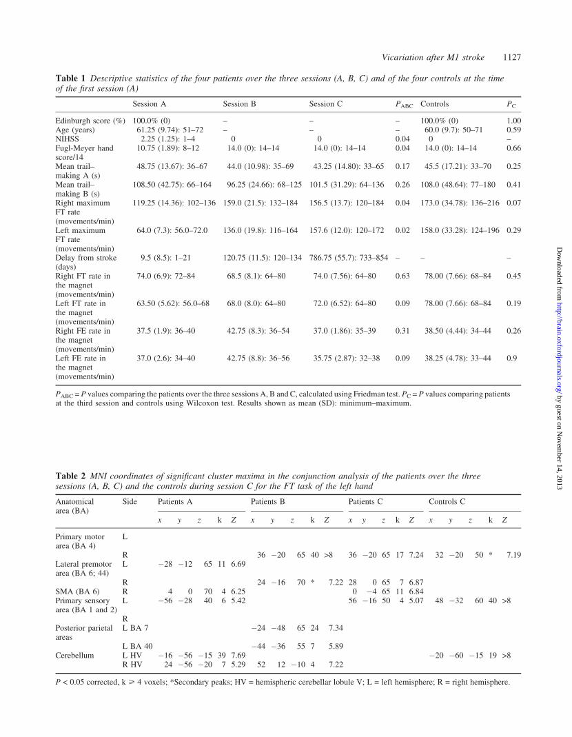
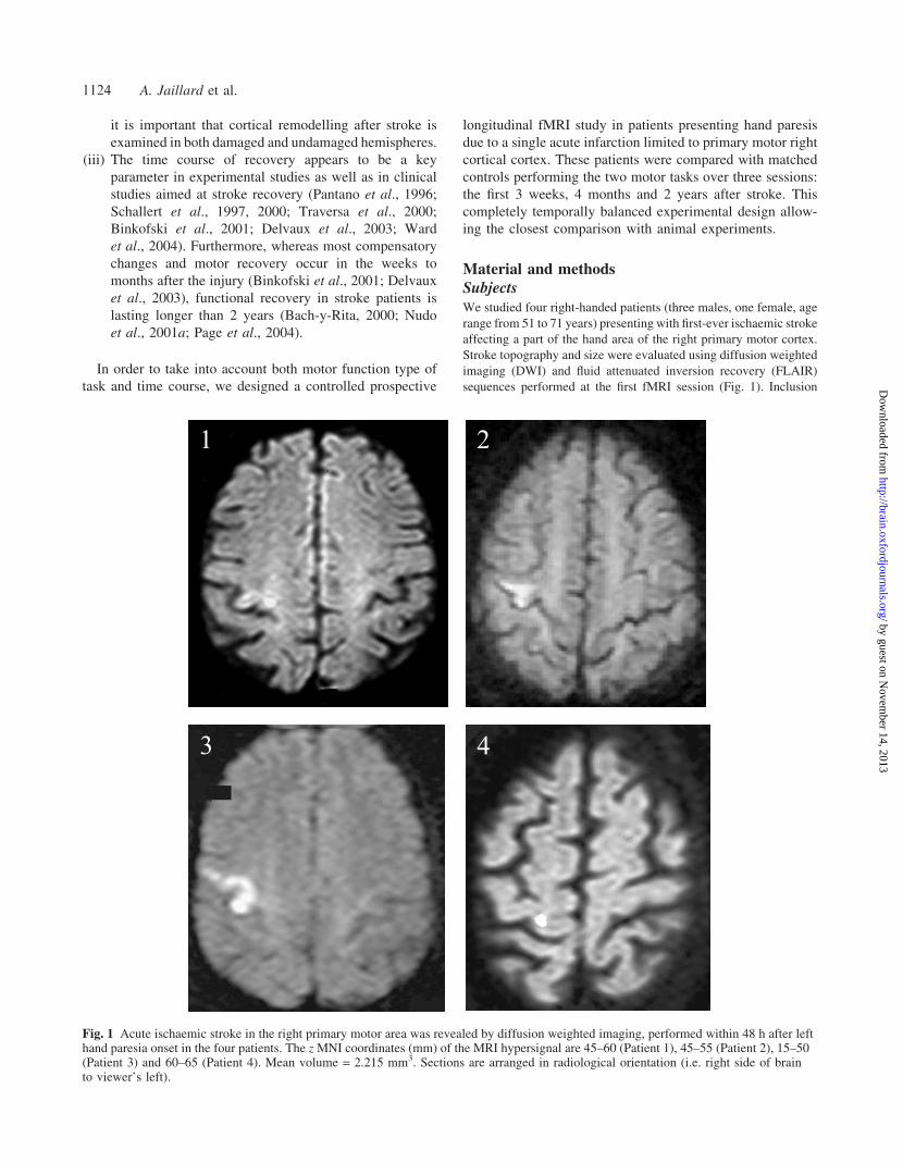
Table 1 Descriptive statistics of the four patients over the three sessions (A, B, C) and of the four controls at the timeof the first session (A)
Session A Session B Session C PABC Controls PC
Edinburgh score (%) 100.0% (0) – – – 100.0% (0) 1.00Age (years) 61.25 (9.74): 51–72 – – – 60.0 (9.7): 50–71 0.59NIHSS 2.25 (1.25): 1–4 0 0 0.04 0 –Fugl-Meyer handscore/14
10.75 (1.89): 8–12 14.0 (0): 14–14 14.0 (0): 14–14 0.04 14.0 (0): 14–14 0.66
Mean trail–making A (s)
48.75 (13.67): 36–67 44.0 (10.98): 35–69 43.25 (14.80): 33–65 0.17 45.5 (17.21): 33–70 0.25
Mean trail–making B (s)
108.50 (42.75): 66–164 96.25 (24.66): 68–125 101.5 (31.29): 64–136 0.26 108.0 (48.64): 77–180 0.41
Right maximumFT rate(movements/min)
119.25 (14.36): 102–136 159.0 (21.5): 132–184 156.5 (13.7): 120–184 0.04 173.0 (34.78): 136–216 0.07
Left maximumFT rate(movements/min)
64.0 (7.3): 56.0–72.0 136.0 (19.8): 116–164 157.6 (12.0): 120–172 0.02 158.0 (33.28): 124–196 0.29
Delay from stroke(days)
9.5 (8.5): 1–21 120.75 (11.5): 120–134 786.75 (55.7): 733–854 – – –
Right FT rate inthe magnet(movements/min)
74.0 (6.9): 72–84 68.5 (8.1): 64–80 74.0 (7.56): 64–80 0.63 78.00 (7.66): 68–84 0.45
Left FT rate inthe magnet(movements/min)
63.50 (5.62): 56.0–68 68.0 (8.0): 64–80 72.0 (6.52): 64–80 0.09 78.00 (7.66): 68–84 0.19
Right FE rate inthe magnet(movements/min)
37.5 (1.9): 36–40 42.75 (8.3): 36–54 37.0 (1.86): 35–39 0.31 38.50 (4.44): 34–44 0.26
Left FE rate inthe magnet(movements/min)
37.0 (2.6): 34–40 42.75 (8.8): 36–56 35.75 (2.87): 32–38 0.09 38.25 (4.78): 33–44 0.9
PABC = P values comparing the patients over the three sessions A, B and C, calculated using Friedman test. PC = P values comparing patientsat the third session and controls using Wilcoxon test. Results shown as mean (SD): minimum–maximum.
Table 2 MNI coordinates of significant cluster maxima in the conjunction analysis of the patients over the threesessions (A, B, C) and the controls during session C for the FT task of the left hand
Anatomicalarea (BA)
Side Patients A Patients B Patients C Controls C
x y z k Z x y z k Z x y z k Z x y z k Z
Primary motorarea (BA 4)
L
R 36 �20 65 40 >8 36 �20 65 17 7.24 32 �20 50 * 7.19Lateral premotorarea (BA 6; 44)
L �28 �12 65 11 6.69
R 24 �16 70 * 7.22 28 0 65 7 6.87SMA (BA 6) R 4 0 70 4 6.25 0 �4 65 11 6.84Primary sensoryarea (BA 1 and 2)
L �56 �28 40 6 5.42 56 �16 50 4 5.07 48 �32 60 40 >8
RPosterior parietalareas
L BA 7 �24 �48 65 24 7.34
L BA 40 �44 �36 55 7 5.89Cerebellum L HV �16 �56 �15 39 7.69 �20 �60 �15 19 >8
R HV 24 �56 �20 7 5.29 52 12 �10 4 7.22
P < 0.05 corrected, k > 4 voxels; *Secondary peaks; HV = hemispheric cerebellar lobule V; L = left hemisphere; R = right hemisphere.
Vicariation after M1 stroke 1127
by guest on Novem
ber 14, 2013http://brain.oxfordjournals.org/
Dow
nloaded from
Patients left FE (Table 3)At the first session (A), patients showed contralateral activa-
tion in right dorsal M1, PMd, S1, and bilaterally in SMA and
cerebellum. An additional small activation was detected in
the left PMd for k = 1 (Fig. 2). At the second session (B),
activation was limited to the right dorsal M1 extending
to PMd and to the left QCL. At 2 years (session C), activation
was observed in the PMd, the right dorsal M1, the S1 and
in the SMA. The hemispheric laterality index was equal
to 1.00.
Table 3 MNI coordinates of significant cluster maxima in the group analysis of the patients over the threesessions (A, B, C) and controls during session C for the FE task of the left hand
Anatomicalarea (BA)
Side Patients A Patients B Patients C Controls C
x y z k Z x y z k Z x y z k Z x y z k Z
M1 (BA 4) R 36 �24 60 4 5.54 32 �16 65 32 >8 40 �20 65 44 >8 32 �20 55 * 5.61Lateral premotorarea (BA 6)
R 28 �16 70 8 6.01 24 �12 �70 * >8 28 �16 �70 * >8
SMA (BA 6) R 0 0 65 4 4.56Primary sensoryarea
R 40 �32 60 6 5.26 32 �24 45 * 5.25 36 �32 65 26 7.44
Cerebellum L HV �28 �56 �25 23 6.58 �12 �60 �15 4 5.70 �20 �52 �20 35 >8L HVII �28 �64 �50 6 5.18R HV 20 �56 �20 19 6.58
P < 0.05 corrected, k > 4 voxels; *Secondary peaks; HV = hemispheric cerebellar lobule V; L = left hemisphere; R = right hemisphere.
Table 5 MNI coordinates of significant cluster maxima in the conjunction analysis of the patients over the three sessions(A, B, C) and the controls C during session C for the FE task of the right hand
Anatomicalarea (BA)
Side Patients A Patients B Patients C Controls C
x y z k Z x y z k Z x y z k Z x y z k Z
M1 (BA 4) L �52 �20 60 77 >8 �40 �20 65 48 >8 �40 �24 55 47 >8 �40 �20 65 50 >8Lateral premotorarea (BA 6; 44)
L �36 �20 70 * >8 �36 �20 70 * >8 �40 �20 70 * >8 �32 �12 65 * 5.14
SMA proper (BA 6) L �8 �4 55 3 5.38 �4 �4 60 5 5.24Primary sensoryarea
L �44 �24 55 * >8 �52 �20 55 * 6.97 �48 �24 55 * >8 �40 �24 55 * >8
Cerebellum R HV 16 �52 �20 47 6.91 20 �52 �20 45 7.05 20 �52 �20 45 >8 24 �56 �20 48 >8
P < 0.05 corrected, k > 4 voxels; *Secondary peaks; HV = hemispheric cerebellar lobule V; L = left hemisphere; R = right hemisphere.
Table 4 MNI coordinates of significant cluster maxima in the conjunction analysis of the patients over the threesessions (A, B, C) and the controls during session C for the FT task of the right hand
Anatomicalarea (BA)
Side Patients A Patients B Patients C Controls C
x y z k Z x y z k Z x y z k Z x y z k Z
Primary motorarea (BA 4)
L �40 �20 65 77 >8 �48 �16 60 49 >8 �40 �20 65 47 >8 �40 �24 60 * >8
Lateral premotorarea (BA 6; 44)
L �40 �16 65 * >8 �40 �16 65 * >8 �32 �4 60 * 5.40 32 �20 70 * 7.51
SMA (BA 6) L �12 �4 65 18 5.93 �4 0 60 29 7.11Cingulum (BA 32) L �8 8 45 13 5.75Primary sensoryarea
L �52 �20 60 * >8 �48 �32 60 5 6.18 �44 �24 50 * 7.38 �48 �36 60 90 >8
Posterior parietalareas (BA 40; 5)
L BA 40 �52 �40 60 6 >8
L BA 7 �44 �44 65 * 5.80Cerebellum R HV 20 �56 �20 45 >8 20 �56 �20 43 >8 20 �60 �20 12 6.36 16 �52 �20 48 >8
P < 0.05 corrected, k > 4 voxels; *Secondary peaks; HV = hemispheric cerebellar lobule V; L = left hemisphere; R = right hemisphere.
1128 A. Jaillard et al.
by guest on Novem
ber 14, 2013http://brain.oxfordjournals.org/
Dow
nloaded from

Right FT and FEThe FT and the FE led to the usual motor activation patterns
in the controls and the patients groups (see Tables 4 and 5).
Each group displayed PMd to SM1 and left QCL activations,
plus a small cluster in the left posterior parietal cortex during
the right FE.
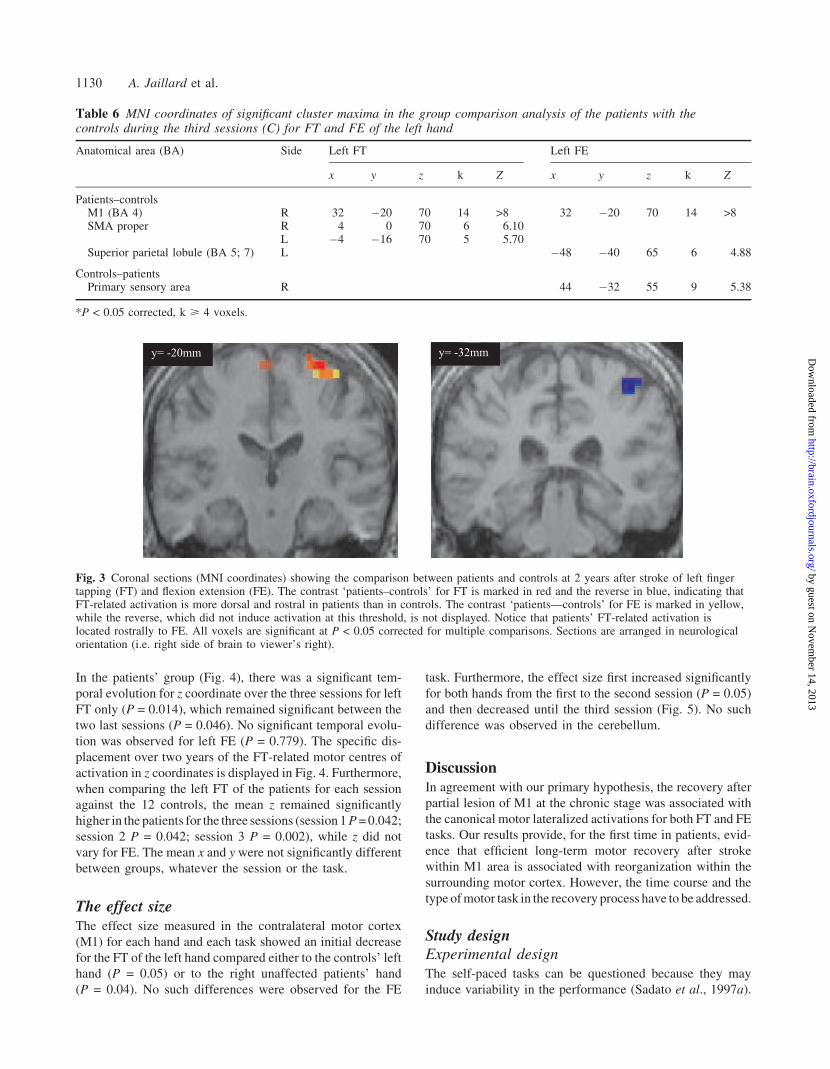
Comparison of the paretic hand of patientswith their matched controls at the chronicstage (session C)The comparison of left FT in patients with controls at 2 years
(patients C—controls C) revealed right PMd–dorsal M1 and
SMA activations, as shown in Table 6 and Fig. 3. The reverse
(controls C—patients C) displayed only S1 activation. For the
left FE, the contrast (patients C—controls C) showed the
same right PMd-dorsal M1 activation as for FT (more a small
cluster in the left posterior parietal cortex), while the reverse
(controls C—patients C) did not lead to any activation. The
comparison of controls and patients did not induce activation
for the right hand.
Left FT and FE of the 12 subjectscontrol groupBoth FT and FE tasks activated the M1 from right PMd to
SM1, the left SMA and the left QCL. The right QCL was
activated for FT alone. The peak of activation was more
dorsal and rostral for FE than for FT. Compared with this
control group, patients’ FT and FE related motor activations
were more dorsal and rostral (Fig. 2).
Motor centres of activationWhen the mean x,y,z Talairach coordinates of the four patients
and their four matched controls were analysed separately for
FT and FE and for each hand, there were significant differ-
ences between patients and controls in the mean x and z
Talairach coordinates for the left FT (P = 0.028 for x and
P < 0.001 for z), but not for the left FE (P = 0.319 for x and
P = 0.089 for z). For left FT, the mean x was lower and the
mean z was higher in the patients than in the controls, cor-
responding to a dorsal shift of the motor centres of activation.
No such significant difference was found for the right hand.
Fig. 2 Representative coronal sections from patient and control maps showing FT and FE-related activations for the left hand over time.(A) First session at 10 days after stroke. (B) Second session at 4 months after stroke. (C) Third session at 2 years after stroke.12 controls = patients’ third session and single session for 12 normal controls). MNI coordinates (mm) y = �16 in the lower row andy = �20 in the upper row for FT and FE. Red areas represent FT-related activation in patients; blue areas represent FT-related activation incontrols; yellow areas represent FE-related activation in patients; green areas represent FE-related activation in controls. All voxels aresignificant at P < 0.05 corrected for multiple comparisons. Sections are arranged in neurological orientation (i.e. right side of brain toviewer’s right). See Tables 2 and 3 for exact coordinates of voxels.
Vicariation after M1 stroke 1129
by guest on Novem
ber 14, 2013http://brain.oxfordjournals.org/
Dow
nloaded from
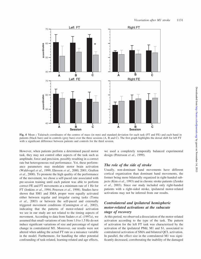
In the patients’ group (Fig. 4), there was a significant tem-
poral evolution for z coordinate over the three sessions for left
FT only (P = 0.014), which remained significant between the
two last sessions (P = 0.046). No significant temporal evolu-
tion was observed for left FE (P = 0.779). The specific dis-
placement over two years of the FT-related motor centres of
activation in z coordinates is displayed in Fig. 4. Furthermore,
when comparing the left FT of the patients for each session
against the 12 controls, the mean z remained significantly
higher in the patients for the three sessions (session 1 P = 0.042;
session 2 P = 0.042; session 3 P = 0.002), while z did not
vary for FE. The mean x and y were not significantly different
between groups, whatever the session or the task.
The effect sizeThe effect size measured in the contralateral motor cortex
(M1) for each hand and each task showed an initial decrease
for the FT of the left hand compared either to the controls’ left
hand (P = 0.05) or to the right unaffected patients’ hand
(P = 0.04). No such differences were observed for the FE
task. Furthermore, the effect size first increased significantly
for both hands from the first to the second session (P = 0.05)
and then decreased until the third session (Fig. 5). No such
difference was observed in the cerebellum.
DiscussionIn agreement with our primary hypothesis, the recovery after
partial lesion of M1 at the chronic stage was associated with
the canonical motor lateralized activations for both FT and FE
tasks. Our results provide, for the first time in patients, evid-
ence that efficient long-term motor recovery after stroke
within M1 area is associated with reorganization within the
surrounding motor cortex. However, the time course and the
type of motor task in the recovery process have to be addressed.
Study designExperimental designThe self-paced tasks can be questioned because they may
induce variability in the performance (Sadato et al., 1997a).
Table 6 MNI coordinates of significant cluster maxima in the group comparison analysis of the patients with thecontrols during the third sessions (C) for FT and FE of the left hand
Anatomical area (BA) Side Left FT Left FE
x y z k Z x y z k Z
Patients–controlsM1 (BA 4) R 32 �20 70 14 >8 32 �20 70 14 >8SMA proper R 4 0 70 6 6.10
L �4 �16 70 5 5.70Superior parietal lobule (BA 5; 7) L �48 �40 65 6 4.88
Controls–patientsPrimary sensory area R 44 �32 55 9 5.38
*P < 0.05 corrected, k > 4 voxels.
Fig. 3 Coronal sections (MNI coordinates) showing the comparison between patients and controls at 2 years after stroke of left fingertapping (FT) and flexion extension (FE). The contrast ‘patients–controls’ for FT is marked in red and the reverse in blue, indicating thatFT-related activation is more dorsal and rostral in patients than in controls. The contrast ‘patients—controls’ for FE is marked in yellow,while the reverse, which did not induce activation at this threshold, is not displayed. Notice that patients’ FT-related activation islocated rostrally to FE. All voxels are significant at P < 0.05 corrected for multiple comparisons. Sections are arranged in neurologicalorientation (i.e. right side of brain to viewer’s right).
1130 A. Jaillard et al.
by guest on Novem
ber 14, 2013http://brain.oxfordjournals.org/
Dow
nloaded from
However, when patients perform a determined paced motor
task, they may not control other aspects of the task such as
amplitude, force and precision, possibly resulting in a correct
rate but heterogeneous real performance. Yet, these perform-
ance parameters may modulate motor brain activation
(Waldvogel et al., 1999; Ehrsson et al., 2000, 2001; Grafton
et al., 2000). To promote the high quality of the performance
of the movement, we chose a self-paced rate associated with
pre-session training until each patient was able to perform
correct FE and FT movements at a minimum rate of 1 Hz for
FT (Jenkins et al., 1994; Petersen et al., 1998). Studies have
shown that SM1 and SMA proper were equally activated
either between regular and irregular cueing tasks (Toma
et al., 2003) or between the self-paced and externally
triggered movement conditions (Cunnington et al., 2002),
indicating that the patterns of motor-related activation
we see in our study are not related to the timing aspects of
movement. According to data from Sadato et al. (1997a), we
assumed that small variations of rate from 1.0 to 1.5 Hz do not
induce significant variations of size and percentage of signal
change in contralateral M1. Moreover, our results were not
altered when adding the actual FT rate as a nuisance variable
in the model. Furthermore, for handling the other potential
confounding of task-related, learning-related and age effects,
we used a completely temporally balanced experimental
design (Petersson et al., 1999).
The role of the side of strokeUsually, non-dominant hand movements have different
cortical organization than dominant hand movements, the
former being more bilaterally organized in right-handed sub-
jects (Kim et al., 1993) and in chronic stroke patients (Zemke
et al., 2003). Since our study included only right-handed
patients with a right-sided stroke, ipsilateral motor-related
activations may not be inferred from our results.
Contralateral and ipsilateral hemisphericmotor-related activations at the subacutestage of recoveryAt this period, we observed a dissociation of the motor-related
activation according to the type of the task. The pattern
of activation for the left FT task was characterized by the
activation of the ipsilateral PMd, M1 and S1, associated to
contralateral activation of SMA and bilateral QCL activation.
In parallel, the effect size in the contralateral M1 was signi-
ficantly decreased, corroborating the inability of the damaged
Fig. 4 Mean z Talairach coordinates of the centres of mass (in mm) and standard deviation for each task (FT and FE) and each hand inpatients (black bars) and in controls (grey bars) over the three sessions (A, B and C). The first graph highlights the dorsal shift for left FTwith a significant difference between patients and controls for the third session.
Vicariation after M1 stroke 1131
by guest on Novem
ber 14, 2013http://brain.oxfordjournals.org/
Dow
nloaded from

M1 cortex alone to provide an efficient motor performance
for FT. Unlike the FT, the left FE task induced a lateralized
pattern of activation involving contralateral right PMd, M1
and S1. Furthermore, the effect size in contralateral M1 did
not show significant decrease relative either to the controls or
to the healthy hand.
The decreased contralateral BOLD signal in the damaged
M1 may reflect changes in resting perfusion as it was shown
that the magnitude of the BOLD response can be affected
by the cerebral blood flow (CBF) increase in response to
acetazolamide administration (Brown et al., 2003). Similarly,
near exhaustion of cerebrovascular reserve (Bruhn et al.,
1994), an usual condition in acute stroke, may produce a
ceiling effect on the CBF response and affect the BOLD
response (Cohen et al., 2002; Brown et al., 2003). By the
time of the first session, the perfusion maps in our patients did
not show differences larger than 20% between symmetric
regions of both hemispheres, when determining the relative
CBF and cerebral blood volume (CBV), either within or
around the damaged area. The coupling of BOLD with neural
activity remains to be examined, although we cannot exclude
the possibility that BOLD signal decrease is related to
changes of relative CBF/CBV within the injured area. This
could not be detected in relation to its limited reliability to the
low spatial resolution of perfusion MRI that may under dia-
gnose ischaemia (Kiselev, 2001), or to abnormal cerebrovas-
cular reactivity (Brown et al., 2003) usually associated to
large infarcts and large artery occlusion (Yamamoto et al.,
2004). However, our patients had small local cortical infarcts
and CBF variations, which could not explain the increased
BOLD signal in the undamaged motor cortex.
In healthy subjects, the focal inhibition of M1 obtained
with TMS was mirrored by an fMRI BOLD signal decrease
(Hamzei et al., 2002). After an acute stroke, a decreased
Fig. 5 Mean effect size at the maxima of the contralateral M1 cluster for the FT and FE tasks (dark bars represent patients andgrey bars controls), showing: (i) the decreased effect size at the first session for the left FT in patients compared with the right FT and to theleft FT of controls; and (ii) the increased effect size for the right FT and FE in patients compared with the controls over thethree sessions.
1132 A. Jaillard et al.
by guest on Novem
ber 14, 2013http://brain.oxfordjournals.org/
Dow
nloaded from

cortical excitability has been shown using TMS in the affec-
ted hemisphere (Wassermann et al., 2000) with a progressive
tendency towards normality 3 months after stroke (Traversa
et al., 2000). Thus, the BOLD signal decrease in the damaged
cortex observed in our study is consistent with decreased
cortical excitability in M1. Moreover, experimental data
after motor cortex lesion in rodents have shown an initial
atrophy of the dendritic fields of the pyramidal neurons in
layer V in the adjacent motor cortex, followed by regrowth
and expansion of these dendritic fields over several months
(Kolb and Whishaw, 2000). Meanwhile, Dijkhuizen et al.
(2001) carried out an fMRI stroke study in rats and showed
that the forelimb impairment was associated with the loss of
stimulus induced activation in the ipsilesional sensorimotor
cortex at 3 days after stroke, with significant activation detec-
ted in the contralesional hemisphere. However, local tissue
and perfusion were only moderately affected and cerebrovas-
cular reactivity was preserved in the motor areas. At 14 days
after stroke, bilateral activation was detected. Similarly, the
decreased motor-related activation in the damaged hemi-
sphere observed in our study might be correlated to dendritic
dysfunction, while subsequent increased activation might
reflect functional motor recovery as a result of dendritic
regrowth and synaptic restructuring.
Changes observed in the intact hemisphereSignificant motor–premotor activation was induced in the
undamaged hemisphere by FT but not by FE, resulting in
a negative laterality index (�0.77) for FT and a positive
index for FE (1.00). In parallel, at the time of the first session,
patients’ motor performance was impaired for FT, but not for
FE. When analysing individual data, the only patient who had
predominant ipsilateral motor activation during left FE in the
acute period had the most extended M1 lesion, in contrast to
the other patients with infarction restricted to a limited part of
the hand motor area. This is consistent with TMS studies
(Wassermann et al., 2000) where the greater motor impair-
ment and the greater the interhemispheric asymmetry suggest
that more the function is impaired, the more it is assumed by
the undamaged hemisphere. After SM1 lesions in rats, Jones
et al. (1996) described pruning of dendritic overgrowth asso-
ciated with synaptic restructuring in the intact cortex occur-
ring around day 18 and persisting beyond 30 days. The
adaptability of the neural structure might facilitate spontan-
eous recovery by rewiring the brain (Schallert et al., 1997). In
combined TMS–fMRI studies, Foltys et al. (2003) showed
that forceful activation of homologous muscles of the hand
increased excitability of the ipsilateral corticospinal system,
which may be mediated by transcallosal inhibition rather than
ipsilateral uncrossed descending pathway (Kobayashi et al.,
2003) as a consequence of a decrease in GABAergic inhibi-
tion (Chen et al., 2002). Thus, the ipsilateral activation of
motor regions may be regarded as an increased utilization of
neural resources in our patients for whom the correct execu-
tion of a motor hand task was highly demanding and induced
paucity of activation in contralateral motor areas. Further-
more, several experimental and human fMRI, TMS and PET
stroke studies have underlined the role of the premotor cortex
in motor recovery (Aizawa et al., 1991; Chollet et al., 1991;
Cicinelli et al., 1997; Liepert et al., 2000a; Wassermann et al.,
2000; Nudo et al., 2001b; Shimizu et al., 2002; Rossini et al.,
2003) and provided evidences of the functional significance
of ipsilateral PMd activity, which could mediate an adaptat-
ive compensatory motor function (Johansen-Berg et al.,
2002a). The effective aptitude of the ipsilateral PMd and
M1 cortex to be responsible of the FT task performed by
the left hand can be questioned, since the exact relationship
between the measured fMRI signal and the underlying neural
activity remains unclear (Logothetis et al., 2001). Moreover,
the relative increased BOLD signal observed in our patients
for the healthy right hand emphasizes the hyperexcitability
of the unaffected motor cortex in relation to decreased
transcallosal inhibition from the damaged motor cortex.
We can speculate that the transient ipsilateral PMd activation
observed in the intact hemisphere of our patients for the FT
reflects short-term plasticity.
Functional dissociation between FT and FEIpsilateral activation for FT and decreased effect size in the
contralateral damaged M1 correlated with the impairment of
patients’ FT task performance at 10 days after stroke, while
both the pattern of activation and the effect size tend towards
normal for correctly performed FE. FT and FE differ by the
higher sensory component involved in the FT, but also by the
load or the complexity of this task. In this context, the func-
tional dissociation between FT and FE might be related to the
anatomic stroke topography. In our patients, the ischaemic
lesion affected the depth of the rolandic sulcus, devoted to
motor tasks involving sensory modalities such as FT compared
with FE (Geyer et al., 1996; Preuss and Kaas, 1996; Preuss
et al., 1997). Moreover, FT involving complex finger move-
ments possibly implies a higher subjective level of attention to
movement than FE (Binkofski et al., 2002). Our findings,
showing an anatomical–clinical correlation between the dis-
sociated performance of FT and FE, and the damage of the
ventral part of M1, emphasizes the hypothesis of a dynamic
organization of the motor systems rather than a somatotopic
output organization developed by Sanes and Donoghue
(1997). Their findings indicate that the connectional substrate
for reorganization was already present in M1 at the acute stage,
allowing new maps to emerge when the balance of excitatory
and inhibitory synaptic connections is changed.
Cerebral reorganization at the chronic stageof recovery: a vicariant process?At the chronic phase, i.e. over 4 months after stroke, our
patients showed a lateralized pattern of motor activation
involving contralateral PMd, M1, and SMA for both FT
and FE.
Vicariation after M1 stroke 1133
by guest on Novem
ber 14, 2013http://brain.oxfordjournals.org/
Dow
nloaded from

Other longitudinal stroke studies in patients who regain
most of their motor abilities have shown initial ipsilateral
motor cortical activations associated with the recruitment
of motor-connected regions such as prefrontal and parietal
regions followed by more lateralized patterns of activations
several months after stroke (Nelles et al., 1999; Marshall et al.,
2000; Calautti et al., 2001a, b; Loubinoux et al., 2003; Ward
et al., 2003). The more lateralized activation is maintained
over time, the better the recovery (Rossini et al., 2003). Other
studies have reported either a ventral expansion of M1 activa-
tion into the face area (Weiller et al., 1992) or a posterior shift
(Pineiro et al., 2001; Calautti et al., 2003) in patients with one
lacunar stroke. These plastic changes may correspond to the
recruitment of parallel projections from the intact pyramidal
tract. They are usually considered to reflect the large redund-
ancy in cortical connections (Rijntjes and Weiller, 2002)—an
assumption which is close to the concept of vicariance.
Another study reported one patient with several chronic stroke
lesions, of which one was restricted to the precentral gyrus
from PMd to M1 (Cramer et al., 2000). In this study,
FT-related brain activations were observed only in contralat-
eral postcentral gyrus. However, the comparison with our
results is limited due to the PMd damage and the association
of other cerebral infarcts, which might have led to iterative
modifications of cerebral networks.
As described above, M1 is subdivided into dorso-rostral
and ventro-caudal fields, where neurons responsive to joint
manipulation and muscle stimulation are located more ros-
trally (M1a) than neurons responsive to cutaneous stimulation
located in the depth of the rolandic sulcus (M1p) (Geyer et al.,
1996; Preuss and Kaas, 1996; Nudo et al., 1997; Preuss et al.,
1997). After FT-related brain activation involved the ipsilat-
eral motor area in the first few weeks, it further emerged in the
contralateral (or ipsilesional) motor area. Our findings indic-
ate that successful motor recovery process after limited lesion
of the hand motor cortex is based on adjacent motor reor-
ganization, suggesting a ‘vicariation’ (adaptive plasticity)
model of recovery. The question about the vicariance is
focused on the emergence of the representation of the FT
in novel locations within the M1 motor area. Both FT and
FE-related brain activations obtained for the recovered left
hand at the chronic phase have shown a dorso-medial shift
within M1 relative to the left hand of the controls. The robust-
ness of these findings was reinforced when the sample size of
the controls group was increased to 12 subjects (Fig. 2). The
comparison of the motor centres of activation also evidenced
a shift between the left hand of the patients and the controls,
and between the left and right hands of the patients (Fig. 4).
Interestingly, the left FT of the patients revealed a more
dorsal motor activation than FE (Fig. 3), resulting in an inver-
ted representation of FT and FE within M1. In addition, when
examining the stroke topography of our patients, the lesion
was located from 40 mm to 56 mm above the bicommissural
plane with a median of 52 mm, in the ventro-caudal part of
M1 (M1p) (Geyer et al., 1996). The ischaemic damage within
M1p might have resulted in a dorso-caudal shift towards M1a,
corresponding to a newly expanded representational territory
of the finger movements towards the intact motor cortex. The
emergence of a task-related motor representation in novel
locations in the intact area 4a instead of the damaged
area 4p, indicates a vicarious process within motor areas
in accordance with experimental studies (Xerri et al., 1998;
Nudo et al., 2000). Furthermore, we may assume that
FT-related plasticity has been rewired by the recruitment
of ipsilateral motor cortex inducing structural plasticity
within the M1 hand area surrounding the lesion, i.e. within
M1a. Meanwhile, FE movements, being mildly impaired,
underwent only limited neural reorganization. Long-term
plasticity changes including long-term potentiation, axonal
regeneration and sprouting may result in the emergence of
a new representation of the FT movements within M1a and
in the restitution/re-emergence of the representation of the FE
movements which were only transitorily impaired. The
dissociated recovery strategies that occurred for the re-
acquisition of pre-infarct FT and FT movements provide
more evidence of a vicarious process within motor area.
Potential biases responsible of this shift have to be
examined. Anatomical distortion secondary to infarct
shrinkage could have modified activation maps, resulting
in a misleading shift of activation between controls and
patients. However, the small size of infarct (mean volume =
2.21 cm3), and the relative greater shift of FT compared with
FE do not support such a bias. Furthermore, infarct shrinkage
should have reduced distances between the geometric centres
of activation and the bi-commissural plane, leading to a
ventral shift rather than to the observed dorsal shift. The
role of increased attention to movement (Johansen-Berg
and Matthews, 2002) and increased force (Cramer et al.,
2002) by modulating cortical activity within M1 could also
be confounders. However, the shift increased progressively
over 2 years while the patients recovered and consequently
need less effort.
A further point concerns the compensatory use of proximal
musculature during the motor tasks, leading to the activation
of dorsal motor cortex in relation to proximal arm somato-
topy, which could look like a false dorsal shift. The absence
of shoulder or elbow movements noticed by the observer
close to the patient during the fMRI scans, the slowness of
the FT and FE rate in the aim to obtain the best motor
performance, and the bilateral extensive activations usually
resulting from proximal movements allow us to rule out
compensatory movements. However, the representation of
joint movements of the fingers may have expanded in
the dorsal M1 region at the expense of wrist/forearm
representations, as has been demonstrated after training
in squirrel monkeys (Nudo et al., 1996a, b) and in rats
(Kleim et al., 1998).
Role of repetitive rehabilitative trainingRepetitive rehabilitative training played a crucial role in
the emergence of the modified representation of the hand
movements within M1. Experimental studies have shown
1134 A. Jaillard et al.
by guest on Novem
ber 14, 2013http://brain.oxfordjournals.org/
Dow
nloaded from

that retention of hand representational area within M1 after
cortical injury required repetitive use of the impaired hand,
while the size of the hand representation had decreased in
monkeys who did not receive rehabilitative training (Friel
et al., 2000). Increased use of the impaired limb appears
to have a modulatory effect on plasticity in the surrounding
tissue (Nudo et al., 1996b; Johansson, 2000). However, repet-
itive motor training alone does not produce functional reor-
ganization of cortical maps. Instead, motor skill acquisition
appears to be a prerequisite factor in driving representational
plasticity in the motor cortex (Nudo et al., 1997). However,
the reorganization of cortical networks and motor skill re-
acquisition may be compromised by the relative size of the
lesion (Friel and Nudo, 1998). Our four patients, who presen-
ted with a restricted lesion of M1, followed daily post-stroke
rehabilitation leading to motor skill re-acquisition. In these
conditions, it is likely the existence of a vicariant process
within M1 promoted by rehabilitative training could explain
the post-stroke fMRI dorsal shift. The pre-session training
was performed by all subjects in order to achieve a standard
good result regarding the motor performance and to avoid
learning effect-related activations.
Role of non-primary motor areasA salient feature of our study was the FT and FE motor-
related activation in the SMA proper at the post-acute and
the chronic phase of recovery. The modulatory effect of SMA
has been well-documented using functional imaging (Thaler
et al., 1995; Rizzolatti et al., 1996; Sadato et al., 1997b; Ball
et al., 1999; Cunnington et al., 2002; Toyokura et al., 2002).
The SMA appears crucial in sequential performance of move-
ments (Tanji, 1994), especially in the initiation of internally
generated movements (Thaler et al., 1995). More recently,
an fMRI study carried out in stroke patients has shown that
post-acute motor-related activation of SMA correlated with a
faster or better motor recovery (Loubinoux et al., 2003).
Studies in primates had already raised the importance of the
SMA in the re-acquisition of motor tasks after lesion of the
motor cortex (Aizawa et al., 1991). The SMA proper, while
receiving precentral and postcentral afferents, sends direct
corticospinal efferences and projects to MI (Rizzolatti et al.,
1996; Picard and Strick, 2001), suggesting that SMA might
have supported the impaired motor function of M1 since
the post-acute stage. Moreover, consistent with these data,
our results indicate that motor-related activation of SMA
may reflect an adaptative response to an increased demand
related to the need to relearn motor tasks after stroke. The
re-acquisition of a skilled performance at the chronic phase is
highlighted by the recruitment of SMA for FT but not for FE
when patients were compared with controls (Table 6). In
parallel, the functional contribution of the ipsilesional PMd
in motor recovery after lacunar stroke has been emphasized in
a recent TMS study (Friedman et al., 2004).
The additional FT-related activations of parietal posterior
areas we saw in our patients may reflect increased attentional
load after stroke (Bremmer et al., 2001; Haslinger et al.,
2002; Mesulam, 2000b). It also underscores the importance
of the posterior parietal cortex in processing length and com-
plexity of sequential finger movements (Ehrsson et al., 2000;
Bremmer et al., 2001; Buccino et al., 2001), since FT-related
posterior parietal activations were also found for the right
hand in both patients and controls.
The present study describes, for the first time, the neural
substrates of motor reorganization using fMRI in humans after
one single unilateral ischaemic lesion limited to a part of M1.
The small number of patients, related to the rarity of isolated
ischaemic stroke involving only M1 in patients, is a limitation
of this study. Therefore, if we were able to test the hypothesis
of vicariation within the human motor cortex, our results
might not be generalized as the usual mode of stroke recovery.
Our findings indicate that the impaired motor function was
initially promoted, at least partly, by the premotor areas of the
undamaged hemisphere. Then, as the patients recovered,
skilled motor functions were completely assumed by the
motor cortical areas adjacent to the lesion, leading to a dorsal
shift of fMRI activation within the M1 cortex, in relation to
plastic changes. Our results, consistent with previous experi-
mental studies, suggest that the human primary motor cortex
adjacent to a lesion is capable of vicarious function.
AcknowledgementsWe wish to thank the Clinical Research Centre, University
Hospital Grenoble, for its technical support. This study was
supported by the PHRC 1997 CIRCE, French Ministry of
Health.
References
Aizawa H, Inase M, Mushiake H, Shima K, Tanji J. Reorganization of activity
in the supplementary motor area associated with motor learning and
functional recovery. Exp Brain Res 1991; 84: 668–71.
Armitage P, Berry G. Distribution-free methods. Statistical methods in
medical research. Cambridge (UK): Blackwell Scientific Publications;
1994. p. 448–92.
Bach-y-Rita P. Conceptual issues relevant to present and future neurologic
rehabilitation. In: Levin HS, Grafman J, editors. Cerebral reorganization
of function after brain damage. New York: Oxford University Press; 2000.
p. 357–79.
Ball T, Schreiber A, Feige B, Wagner M, Lucking CH, Kristeva-Feige R. The
role of higher-order motor areas in voluntary movement as revealed by
high-resolution EEG and fMRI. Neuroimage 1999; 10: 682–94.
Benton A, Tranel D. Historical notes on reorganization of function and
neuroplasticity. In: Levin HS, Grafman J, editors. Cerebral reorganization
of function after brain damage. New York: Oxford University Press; 2000.
p. 3–23.
Binkofski F, Seitz RJ, Hacklander T, Pawelec D, Mau J, Freund HJ. Recovery
of motor functions following hemiparetic stroke: a clinical and magnetic
resonance-morphometric study. Cerebrovasc Dis 2001; 11: 273–81.
Binkofski F, Fink GR, Geyer S, Buccino G, Gruber O, Shah NJ, et al. Neural
activity in human primary motor cortex areas 4a and 4p is modulated
differentially by attention to action. J Neurophysiol 2002; 88: 514–9.
Bioulac B, Burbaud P, Varoqueaux D. Activity of area 5 neurons in monkeys
during arm movements: effects of dentate nucleus lesion and motor cortex
ablation. Neurosci Lett 1995; 192: 189–92.
Bremmer F, Schlack A, Shah NJ, Zafiris O, Kubischik M, Hoffmann K, et al.
Polymodal motion processing in posterior parietal and premotor
Vicariation after M1 stroke 1135
by guest on Novem
ber 14, 2013http://brain.oxfordjournals.org/
Dow
nloaded from
cortex: a human fMRI study strongly implies equivalencies between
humans and monkeys. Neuron 2001; 29: 287–96.
Brett M, Anton JL, Valabregue R, Poline JB. Region of interest using an SPM
toolbox. Neuroimage 2002; 16.
Brown GG, Eyler Zorrilla LT, Georgy B, Kindermann SS, Wong EC,
Buxton RB. BOLD and perfusion response to finger-thumb apposition
after acetazolamide administration: differential relationship to global
perfusion. J Cereb Blood Flow Metab 2003; 23: 829–37.
Bruhn H, Kleinschmidt A, Boecker H, Merboldt KD, Hanicke W, Frahm J.
The effect of acetazolamide on regional cerebral blood oxygenation at rest
and under stimulation as assessed by MRI. J Cereb Blood Flow Metab
1994; 14: 742–8.
Buccino G, Binkofski F, Fink GR, Fadiga L, Fogassi L, Gallese V, et al. Action
observation activates premotor and parietal areas in a somatotopic manner:
an fMRI study. Eur J Neurosci 2001; 13: 400–4.
Calautti C, Leroy F, Guincestre JY, Baron JC. Dynamics of motor network
overactivation after striatocapsular stroke: a longitudinal PET study using a
fixed-performance paradigm. Stroke 2001a; 32: 2534–42.
Calautti C, Leroy F, Guincestre JY, Marie RM, Baron JC. Sequential activa-
tion brain mapping after subcortical stroke: changes in hemispheric balance
and recovery. Neuroreport 2001b; 12: 3883–6.
Calautti C, Leroy F, Guincestre J-Y, Baron J-C. Displacement of
primary sensorimotor cortex activation after subcortical stroke: a longit-
udinal PET study with clinical correlation. Neuroimage 2003; 19:
1650–4.
Chen R, Cohen L, Hallett M. Nervous system reorganization following injury.
Neuroscience 2002; 111: 761–73.
Chollet F, DiPiero V, Wise RJ, Brooks DJ, Dolan RJ, Frackowiak RS. The
functional anatomy of motor recovery after stroke in humans: a study with
positron emission tomography. Ann Neurol 1991; 29: 63–71.
Cicinelli P, Traversa R, Rossini PM. Post-stroke reorganization of brain
motor output to the hand: a 2–4 month follow-up with focal magnetic
transcranial stimulation. Electroencephalogr Clin Neurophysiol 1997;
105: 438–50.
Cohen ER, Ugurbil K, Kim SG. Effect of basal conditions on the magnitude
and dynamics of the blood oxygenation level-dependent fMRI response.
J Cereb Blood Flow Metab 2002; 22: 1042–53.
Cramer SC, Moore CI, Finklestein SP, Rosen BR. A pilot study of
somatotopic mapping after cortical infarct. Stroke 2000; 31: 668–71.
Cramer SC, Weisskoff RM, Schaechter JD, Nelles G, Foley M, Finklestein SP,
et al. Motor cortex activation is related to force of squeezing. Hum Brain
Mapp 2002; 16: 197–205.
Cunnington R, Windischberger C, Deecke L, Moser E. The preparation and
execution of self-initiated and externally-triggered movement: a study of
event-related fMRI. Neuroimage 2002; 15: 373–85.
Delvaux V, Alagona G, Gerard P, De Pasqua V, Pennisi G, de Noordhout AM.
Post-stroke reorganization of hand motor area: a 1-year prospective follow-
up with focal transcranial magnetic stimulation. Clin Neurophysiol 2003;
114: 1217–25.
Dijkhuizen RM, Ren J, Mandeville JB, Wu O, Ozdag FM, Moskowitz MA,
et al. Functional magnetic resonance imaging of reorganization in rat brain
after stroke. Proc Natl Acad Sci USA 2001; 98: 12766–71.
Ehrsson HH, Fagergren A, Jonsson T, Westling G, Johansson RS, Forssberg H.
Cortical activity in precision- versus power-grip tasks: an fMRI study.
J Neurophysiol 2000; 83: 528–36.
Ehrsson HH, Fagergren E, Forssberg H. Differential fronto-parietal activation
depending on force used in a precision grip task: an fMRI study. J Neuro-
physiol 2001; 85: 2613–23.
Finger S, Beyer T, Koehler PJ. Dr Otto Soltmann (1876) on development of
the motor cortex and recovery after its removal in infancy. Brain Res Bull
2000; 53: 133–40.
Foltys H, Meister IG, Weidemann J, Sparing R, Thron A, Willmes K, et al.
Power grip disinhibits the ipsilateral sensorimotor cortex: a TMS and fMRI
study. Neuroimage 2003; 19: 332–40.
Friedman EA, Hanakawa T, Chung M, Hummel F, Leiguarda RC, Cohen LG.
Reorganization of the human ipsilesional premotor cortex after stroke.
Brain 2004; 127: 747–58.
Friel KM, Nudo RJ. Recovery of motor function after focal cortical injury in
primates: compensatory movement patterns used during rehabilitative
training. Somatosens Mot Res 1998; 15: 173–89.
Friel KM, Heddings AA, Nudo RJ. Effects of postlesion experience on beha-
vioral recovery and neurophysiologic reorganization after cortical injury
in primates. Neurorehabil Neural Repair 2000; 14: 187–98.
Friston KJ, Holmes AP, Price CJ, Buchel C, Worsley KJ. Multisubject fMRI
studies and conjunction analyses. Neuroimage 1999a; 10: 385–96.
Friston KJ, Holmes AP, Worsley KJ. How many subjects constitute a study?
Neuroimage 1999b; 10: 1–5.
Fugl-Meyer AR, Jaasko L, Leyman I, Olsson S, Steglind S. The post-stroke
hemiplegic patient. 1. A method for evaluation of physical performance.
Scand J Rehabil Med 1975; 7: 13–31.
Geyer S, Ledberg A, Schleicher A, Kinomura S, Schormann T, Burgel U, et al.
Two different areas within the primary motor cortex of man. Nature 1996;
382: 805–7.
Glees CD, Cole J. Recovery of skilled motor functions after small repeated
lesions in motor cortex in macaque. J Neurophysiol 1950; 13: 137–48.
Grafton ST, Hari R, Salenius S. The human motor system. In: Toga AW,
Maziotta JC, editors. Brain mapping: the systems. San Diego (CA):
Academic Press; 2000. p. 331–63.
Hamzei F, Dettmers C, Rzanny R, Liepert J, Buchel C, Weiller C. Reduction
of excitability (‘inhibition’) in the ipsilateral primary motor cortex is
mirrored by fMRI signal decreases. Neuroimage 2002; 17: 490–6.
Haslinger B, Erhard P, Weilke F, Ceballos-Baumann AO, Bartenstein P,
Grafin von Einsiedel H, et al. The role of lateral premotor-cerebellar-
parietal circuits in motor sequence control: a parametric fMRI study.
Brain Res Cogn Brain Res 2002; 13: 159–68.
Jenkins IH, Brooks DJ, Nixon PD, Frackowiak RS, Passingham RE. Motor
sequence learning: a study with positron emission tomography. J Neurosci
1994; 14: 3775–90.
Johansson RS. Brain plasticity and stroke rehabilitation. Stroke 2000; 31:
223–30.
Johansen-Berg H, Matthews PM. Attention to movement modulates activity
in sensori-motor areas, including primary motor cortex. Exp Brain Res
2002; 142: 13–24.
Johansen-Berg H, Dawes H, Guy C, Smith SM, Wade DT, Matthews PM.
Correlation between motor improvements and altered fMRI activity after
rehabilitative therapy. Brain 2002a; 125: 2731–42.
Johansen-Berg H, Rushworth MF, Bogdanovic MD, Kischka U,
Wimalaratna S, Matthews PM. The role of ipsilateral premotor cortex
in hand movement after stroke. Proc Natl Acad Sci USA 2002b; 99:
14518–23.
Jones RD, Schallert A. Overgrowth and pruning of dendrites in adult rats
recovering from neocortical damage. Brain Res 1992; 581: 156–60.
Jones TA, Kleim JA, Greenough WT. Synaptogenesis and dendritic growth
in the cortex opposite unilateral sensorimotor cortex damage in adult rats:
a quantitative electron microscopic examination. Brain Res 1996; 733:
142–8.
Kim SG, Ashe J, Hendrich K, Ellermann JM, Merkle H, Ugurbil K, et al.
Functional magnetic resonance imaging of motor cortex: hemispheric
asymmetry and handedness. Science 1993; 261: 615–7.
Kiselev VG. On the theoretical basis of perfusion measurements by dynamic
susceptibility contrast MRI. Magn Reson Med 2001; 46: 1113–22.
Kleim JA, Barbay S, Nudo RJ. Functional reorganization of the rat motor
cortex following motor skill learning. J Neurophysiol 1998; 80: 3321–5.
Kobayashi M, Hutchinson S, Schlaug G, Pascual-Leone A. Ipsilateral motor
cortex activation on functional magnetic resonance imaging during
unilateral hand movements is related to interhemispheric interactions.
Neuroimage 2003; 20: 2259–70.
Kolb B, Whishaw IQ. Reorganization of function after cortical lesions in rod-
ents. In: Levin HS, Grafman J, editors. Cerebral reorganization of function
after brain damage. New York: Oxford University Press; 2000. p. 168–97.
Kolb B, Gibb R, Gorny G. Cortical plasticity and the development of behavior
after early frontal cortical injury. Dev Neuropsychol 2000; 18: 423–44.
Liepert J, Hamzei F, Weiller C. Motor cortex disinhibition of the unaffected
hemisphere after acute stroke. Muscle Nerve 2000a; 23: 1761–3.
1136 A. Jaillard et al.
by guest on Novem
ber 14, 2013http://brain.oxfordjournals.org/
Dow
nloaded from

Liepert J, Storch P, Fritsch A, Weiller C. Motor cortex disinhibition in acute
stroke. Clin Neurophysiol 2000b; 111: 671–6.
Logothetis NK, Pauls J, Augath M, Trinath T, Oeltermann A. Neurophysio-
logical investigation of the basis of the fMRI signal. Nature 2001; 412:
150–7.
Loubinoux I, Carel C, Pariente J, Dechaumont S, Albucher JF, Marque P, et al.
Correlation between cerebral reorganization and motor recovery after
subcortical infarcts. Neuroimage 2003; 20: 2166–80.
Lyden PD, Zweifler R, Mahdavi Z, Lonzo L. A rapid, reliable, and valid
method for measuring infarct and brain compartment volumes from
computed tomographic scans. Stroke 1994; 25: 2421–8.
Manganotti P, Patuzzo S, Cortese F, Palermo A, Smania N, Fiaschi A. Motor
disinhibition in affected and unaffected hemisphere in the early period of
recovery after stroke. Clin Neurophysiol 2002; 113: 936–43.
Marshall RS, Perera GM, Lazar RM, Krakauer JW, Constantine RC,
DeLaPaz RL. Evolution of cortical activation during recovery from cor-
ticospinal tract infarction. Stroke 2000; 31: 656–61.
MesulamM.Behavioralneuroanatomy:III-Corticalorganization,connectivity
and transmodal areas. In:MesulamM-M,editor. Principles ofbehavioral and
cognitive neurology. Oxford: Oxford University Press; 2000a. p. 12–17.
Mesulam M-M. Attentional networks, confusional states and neglect syn-
dromes. In: Mesulam M-M, editor. Principles of behavioral and cognitive
neurology. Oxford: Oxford University Press; 2000b. p. 174–256.
Nelles G, Spiekramann G, Jueptner M, Leonhardt G, Muller S, Gerhard H,
et al. Evolution of functional reorganization in hemiplegic stroke: a serial
positron emission tomographic activation study. Ann Neurol 1999; 46:
901–9.
Netz J, Lammers T, Homberg V. Reorganization of motor output in the
non-affected hemisphere after stroke. Brain 1997; 120: 1579–86.
Nudo RJ, Milliken GW. Reorganization of movement representations in
primary motor cortex following focal ischaemic infarcts in adult squirrel
monkeys. J Neurophysiol 1996; 75: 2144–9.
Nudo RJ, Barbay S, Kleim JA. Role of neuroplasticity in functional recovery
after atroke. In: Levin HS, Grafman J, editors. Cerebral reorganization of
function after brain damage. New York: Oxford University Press; 2000.
p. 168–97.
Nudo RJ, Milliken GW, Jenkins WM, Merzenich MM. Use-dependent altera-
tions of movement representations in primary motor cortex of adult squirrel
monkeys. J Neurosci 1996a; 16: 785–807.
Nudo RJ, Wise BM, SiFuentes F, Milliken GW. Neural substrates for the
effects of rehabilitative training on motor recovery after ischaemic infarct.
Science 1996b; 272: 1791–4.
Nudo RJ, Plautz EJ, Milliken GW. Adaptative plasticity in primate motor
cortex as a consequence of behavioural experience and neuronal injury.
Semin Neurosci 1997; 9: 13–23.
Nudo RJ, Kleim JA, Friel KM. Functional remodeling of motor cortex after
stroke. In: Choi DW, Dacey RG, Hsu CY, Powers WJ, editors. Cerebrovas-
cular disease: momentum at the end of the second millennium. New York:
Futura Publishing; 2001a. p. 371–391.
Nudo RJ, Plautz EJ, Frost SB. Role of adaptive plasticity in recovery of
function after damage to motor cortex. Muscle Nerve 2001b; 24: 1000–19.
Ogden R, Franz SI. On cerebral motor control: the recovery from experi-
mentally produced hemiplegia. Psychobiologia 1917: 33–50.
Oldfield RC. The assessment and analysis of handedness: the Edinburgh
inventory. Neuropsychologia 1971; 9: 97–113.
Page SJ, Gater DR, Bach YRP. Reconsidering the motor recovery plateau in
stroke rehabilitation. Arch Phys Med Rehabil 2004; 85: 1377–81.
Pantano P, Formisano R, Ricci M, Di Piero V, Sabatini U, Di Pofi B, et al.
Motor recovery after stroke. Morphological and functional brain altera-
tions. Brain 1996; 119: 1849–57.
Petersen SE, van Mier H, Fiez JA, Raichle ME. The effects of practice on the
functional anatomy of task performance. Proc Natl Acad Sci USA 1998; 95:
853–60.
Petersson KM, Elfgren K, Ingvar M. Learning-related effects and functional
neuroimaging. Hum Brain Mapp 1999; 7: 234–43.
Picard N, Strick PL. Imaging the premotor areas. Curr Opin Neurobiol 2001;
11: 663–72.
Pineiro R, Pendlebury S, Johansen-Berg H, Matthews PM. Functional MRI
detects posterior shifts in primary sensorimotor cortex activation after
stroke: evidence of local adaptive reorganization? Stroke 2001; 32:
1134–9.
Preuss TM, Kaas JH. Parvalbumin-like immunoreactivity of layer V
pyramidal cells in the motor and somatosensory cortex of adult primates.
Brain Res 1996; 712: 353–7.
Preuss TM, Stepniewska I, Jain N, Kaas JH. Multiple divisions of macaque
precentral motor cortex identified with neurofilament antibody SMI-32.
Brain Res 1997; 767: 148–53.
Reitan RM. Trail-making test results for normal and brain-damaged children.
Percept Mot Skills 1971; 33: 575–81.
Rijntjes M, Weiller C. Recovery of motor and language abilities after stroke:
the contribution of functional imaging. Prog Neurobiol 2002; 66:
109–22.
Rizzolatti G, Lupino G, Matelli M. The classic supplementary motor
area is formed by two independent areas. In: Luders H, editor. Supple-
mentary sensorimotor area. Philadelphia (PA): Lippincott-Raven; 1996.
p. 45–56.
Rossini PM, Calautti C, Pauri F, Baron JC. Post-stroke plastic reorganization
in the adult brain. Lancet Neurol 2003; 2: 493–502.
Sadato N, Ibanez V, Campbell G, Deiber MP, Le Bihan D, Hallett M.
Frequency-dependent changes of regional cerebral blood flow during finger
movements: functional MRI compared with PET. J Cereb Blood Flow
Metab 1997a; 17: 670–9.
Sadato N, Yonekura Y, Waki A, Yamada H, Ishii Y. Role of the supple-
mentary motor area and the right premotor cortex in the coordination of
bimanual finger movements. J Neurosci 1997b; 17: 9667–74.
Sanes J, Donoghue J. Static and dynamic organization of motor cortex.
In: Freund HJ, Sabel BA, Witte OW, editors. Brain plasticity. Advances
in neurology. Vol. 73. Philadelphia (PA): Lippincott-Raven; 1997.
p. 277–96.
Schallert T, Kozlowski DA, Humm JL, Cocke RR. Use-dependent structural
events in recovery of function. In: Freund HJ, Sabel BA, Witte OW, editors.
Brain plasticity. Advances in neurology. Vol. 73. Philadelphia (PA):
Lippincott-Raven; 1997. p. 229–38.
Schallert T, Leasure JL, Kolb B. Experience-associated structural events,
subependymal cellular proliferative activity, and functional recovery
after injury of the central nervous system. J Cereb Blood Flow Metab
2000; 20: 1513–28.
Shimizu T, Hosaki A, Hino T, Sato M, Komori T, Hirai S, et al. Motor cortical
disinhibition in the unaffected hemisphere after unilateral cortical stroke.
Brain 2002; 125: 1896–907.
Soltman O. Experimentelle Studien uber die Functionen des Grosshirns der
Neugeborenen. Jahrbuch fur Kinderheilkunde und Physische Erziehung
1876; 9: 106–48.
Talairach J, Tournoux P. Coplanar stereotaxic atlas of the human brain: three-
dimensional proportional system. An approach to cerebral brain imaging.
New York: Thieme Medical; 1988.
Tanji J. The supplementary motor area in the cerebral cortex. Neurosci Res
1994; 19: 251–68.
Thaler D, Chen YC, Nixon PD, Stern CE, Passingham RE. The functions of
the medial premotor cortex. I. Simple learned movements. Exp Brain Res
1995; 102: 445–60.
Toma K, Ozawa M, Matsuo K, Nakai T, Fukuyama H, Sato S. The role of the
human supplementary motor area in reactive motor operation. Neurosci
Lett 2003; 344: 177–80.
Toyokura M, Muro I, Komiya T, Obara M. Activation of pre-supplementary
motor area (SMA) and SMA proper during unimanual and bimanual com-
plex sequences: an analysis using functional magnetic resonance imaging.
J Neuroimaging 2002; 12: 172–8.
Traversa R, Cicinelli P, Oliveri M, Giuseppina Palmieri M, Filippi MM,
Pasqualetti P, et al. Neurophysiological follow-up of motor cortical output
in stroke patients. Clin Neurophysiol 2000; 111: 1695–703.
Waldvogel D, van Gelderen P, Ishii K, Hallett M. The effect of movement
amplitude on activation in functional magnetic resonance imaging studies.
J Cereb Blood Flow Metab 1999; 19: 1209–12.
Vicariation after M1 stroke 1137
by guest on Novem
ber 14, 2013http://brain.oxfordjournals.org/
Dow
nloaded from
Ward NS, Brown MM, Thompson AJ, Frackowiak RS. Neural correlates of
motor recovery after stroke: a longitudinal fMRI study. Brain 2003; 126:
2476–96.
Ward NS, Brown MM, Thompson AJ, Frackowiak RS. The influence of time
after stroke on brain activations during a motor task. Ann Neurol 2004; 55:
829–34.
Wassermann EM, Cohen LG, Halett M. Transcranial magnetic stimulation as
a tool for detecting changes in the organization of the human motor system
after central and peripheral lesions. In: Levin HS, Grafman J, editors.
Cerebral reorganization of function after brain damage. New York: Oxford
University Press; 2000. p. 291–303.
Wayne WD. Parametric and distribution-free statistics. Biostatistics.
New York: Wiley; 1999. p. 658–736.
Weiller C, Chollet F, Friston KJ, Wise RJ, Frackowiak RS. Functional
reorganization of the brain in recovery from striatocapsular infarction in
man. Ann Neurol 1992; 31: 463–72.
Xerri C, Merzenich MM, Peterson BE, Jenkins W. Plasticity of primary
somatosensory cortex paralleling sensorimotor skill recovery from stroke
in adult monkeys. J Neurophysiol 1998; 79: 2119–48.
Yamamoto S, Watanabe M, Uematsu T, Takasawa K, Nukata M, Kinoshita N.
Correlation of angiographic circulation time and cerebrovascular reserve
by acetazolamide-challenged single photon emission CT. AJNR Am J
Neuroradiol 2004; 25: 242–7.
Zemke AC, Heagerty PJ, Lee C, Cramer SC. Motor cortex organization after
stroke is related to side of stroke and level of recovery. Stroke 2003; 34:
e23–8.
1138 A. Jaillard et al.
by guest on Novem
ber 14, 2013http://brain.oxfordjournals.org/
Dow
nloaded from
Copyright © 2022 FDOKUMEN