Thermodynamic model development for lithium intercalation electrodes
C A R B O N 8 1 ( 2 0 1 5 ) 1 9 3 – 2 0 2
.sc ienced i rec t .com
Avai lab le a t wwwScienceDirect
journal homepage: www.elsevier .com/ locate /carbon
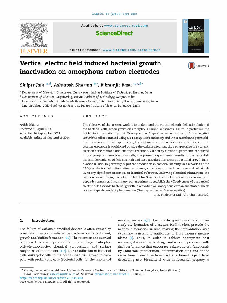
Vertical electric field induced bacterial growthinactivation on amorphous carbon electrodes
http://dx.doi.org/10.1016/j.carbon.2014.09.0480008-6223/� 2014 Elsevier Ltd. All rights reserved.
* Corresponding authors. Address: Materials Research Center, Indian Institute of Science, Bangalore, India (B. Basu).E-mail addresses: [email protected] (A. Sharma), [email protected] (B. Basu).
Shilpee Jain a,d, Ashutosh Sharma b,*, Bikramjit Basu a,c,d,*
a Department of Materials Science and Engineering, Indian Institute of Technology, Kanpur, Indiab Department of Chemical Engineering, Indian Institute of Technology, Kanpur, Indiac Laboratory for Biomaterials, Materials Research Centre, Indian Institute of Science, Bangalore, Indiad Interdisciplinary Bio-Engineering Program, Indian Institute of Science, Bangalore, India
A R T I C L E I N F O A B S T R A C T
Article history:
Received 29 April 2014
Accepted 16 September 2014
Available online 28 September 2014
The objective of the present work is to understand the vertical electric field stimulation of
the bacterial cells, when grown on amorphous carbon substrates in vitro. In particular, the
antibacterial activity against Gram-positive Staphylococcus aureus and Gram-negative
Escherichia coli are studied using MTT assay, live/dead assay and inner membrane permeabi-
lization assays. In our experiments, the carbon substrate acts as one electrode and the
counter electrode is positioned outside the culture medium, thus suppressing the current,
electrokinetic motions and chemical reactions. Guided by similar experiments conducted
in our group on neuroblastoma cells, the present experimental results further establish
the interdependence of field strength and exposure duration towards bacterial growth inac-
tivation in vitro. Importantly, significant reduction in bacterial viability was recorded at the
2.5 V/cm electric field stimulation conditions, which does not reduce the neural cell viabil-
ity to any significant extent on an identical substrate. Following electrical stimulation, the
bacterial growth is significantly inhibited for S. aureus bacterial strain in an exposure time
dependent manner. In summary, our experiments establish the effectiveness of the vertical
electric field towards bacterial growth inactivation on amorphous carbon substrates, which
is a cell type dependent phenomenon (Gram-positive vs. Gram-negative).
� 2014 Elsevier Ltd. All rights reserved.
1. Introduction
The failure of various biomedical devices is often caused by
prosthetic infection mediated by bacterial cell attachment,
growth and biofilm formation [1,2]. The retention and survival
of adhered bacteria depend on the surface charge, hydropho-
bicity/hydrophilicity, chemical composition and surface
roughness of the implant [3–5]. Due to adhesion of bacterial
cells, eukaryotic cells in the host human tissue need to com-
pete with prokaryotic cells (bacterial cells) for the implanted
material surface [6,7]. Due to faster growth rate (rate of divi-
sion), the formation of a mature biofilm often precede the
neotissue formation in vivo, making the implantation sites
extremely resistant to antibiotics or host defense mecha-
nisms [8]. Thus, in order to achieve appropriate host
response, it is essential to design surfaces and processes with
dual performance that encourage eukaryotic cell functional-
ity (adhesion, proliferation, differentiation etc.) and at the
same time prevent bacterial cell attachment. Apart from
developing new biomaterial with antibacterial property, a
194 C A R B O N 8 1 ( 2 0 1 5 ) 1 9 3 – 2 0 2
strikingly different approach of the application of external
electric field stimulation to cause bactericidal effect in vitro
has been attempted in the present work.
The direct current (DC) electric fields are present in all ani-
mals during development and regeneration [9]. The applica-
tion of DC electric fields to biological systems for recovery
of organs is widely being investigated [9–11]. The application
of external electric field to prevent bacterial cell adhesion or
survival however has been investigated rather to a limited
extent. The reversible or irreversible electrical breakdown
occurs on the application of external electric field, which
depends on the electric field intensity and exposure time
[12]. In one report, DC electric field has been used to manipu-
late bacterial cell attachment, their growth and viability [13].
It was reported that a lower current (less than 20 mA) did
not induce any significant change in the surface properties
of phenol-degrading bacteria. In contrast, an electric current
of 20 mA could increase the surface hydrophobicity and flat-
ten the cell shape. Importantly, a higher current (40 mA) could
increase the surface extracellular substances and the net neg-
ative surface electrostatic charge. Further, the manipulation
of cell behavior in the presence of electric field depends on
the size, shape, membrane thickness of the cells, strength
and duration of applied electric field [14–16]. It is reported that
the weak electric fields enhance the efficacy of antibiotics in
killing bacterial biofilm even on the surfaces that are not used
as electrodes [17]. The change in cell behavior in the presence
of external electric field also depends on the mode [alternate
current (AC) vs. direct current (DC)] of applied field. Liu et al.
[18] studied the effect of DC electric currents on the popula-
tion of a Staphylococcus epidermidis (S. epidermidis) biofilm
adhered to a catheter. They found that the population of a
S. epidermidis biofilm adhered to a catheter was reduced by
the application of 100 mA direct current.
An effective implantable biomaterial for the majority of
the applications should facilitate eukaryotic cell functionality
(proliferation, differentiation etc.) and simultaneously inhibit
bacterial growth. Recently, it was shown that the vertical
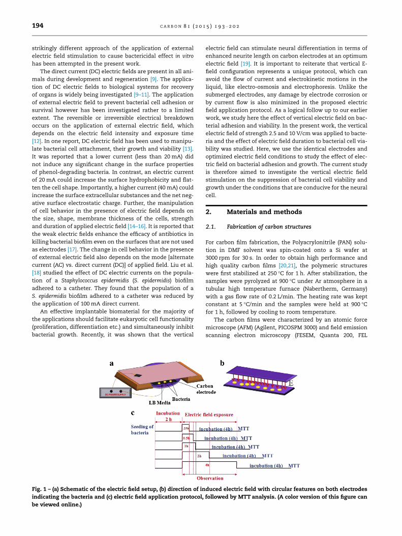
Fig. 1 – (a) Schematic of the electric field setup, (b) direction of in
indicating the bacteria and (c) electric field application protocol,
be viewed online.)
electric field can stimulate neural differentiation in terms of
enhanced neurite length on carbon electrodes at an optimum
electric field [19]. It is important to reiterate that vertical E-
field configuration represents a unique protocol, which can
avoid the flow of current and electrokinetic motions in the
liquid, like electro-osmosis and electrophoresis. Unlike the
submerged electrodes, any damage by electrode corrosion or
by current flow is also minimized in the proposed electric
field application protocol. As a logical follow up to our earlier
work, we study here the effect of vertical electric field on bac-
terial adhesion and viability. In the present work, the vertical
electric field of strength 2.5 and 10 V/cm was applied to bacte-
ria and the effect of electric field duration to bacterial cell via-
bility was studied. Here, we use the identical electrodes and
optimized electric field conditions to study the effect of elec-
tric field on bacterial adhesion and growth. The current study
is therefore aimed to investigate the vertical electric field
stimulation on the suppression of bacterial cell viability and
growth under the conditions that are conducive for the neural
cell.
2. Materials and methods
2.1. Fabrication of carbon structures
For carbon film fabrication, the Polyacrylonitrile (PAN) solu-
tion in DMF solvent was spin-coated onto a Si wafer at
3000 rpm for 30 s. In order to obtain high performance and
high quality carbon films [20,21], the polymeric structures
were first stabilized at 250 �C for 1 h. After stabilization, the
samples were pyrolyzed at 900 �C under Ar atmosphere in a
tubular high temperature furnace (Nabertherm, Germany)
with a gas flow rate of 0.2 L/min. The heating rate was kept
constant at 5 �C/min and the samples were held at 900 �Cfor 1 h, followed by cooling to room temperature.
The carbon films were characterized by an atomic force
microscope (AFM) (Agilent, PICOSPM 3000) and field emission
scanning electron microscopy (FESEM, Quanta 200, FEL
duced electric field with circular features on both electrodes
followed by MTT analysis. (A color version of this figure can
C A R B O N 8 1 ( 2 0 1 5 ) 1 9 3 – 2 0 2 195
Germany, SUPRA, Zeiss, Germany). The tapping mode AFM
accompanying Picoscan software was used to measure the
roughness of film substrates.
2.2. Electric field setup
The carbon film supporting the cells was used as an electrode
and a stainless steel plate, positioned outside the medium,
was used as the counter electrode. As shown in Fig. 1, both
the electrodes were kept parallel to ensure a uniform inten-
sity of the applied electric field. The external DC power sup-
ply, connected to the electrodes was used to apply the
electric field. On the application of 160 V/cm and 640 V/cm,
the electric field strength screened by a cell population in
Luria Broth (LB) medium was 2.5 V/cm and 10 V/cm, respec-
tively. More details of the calculations on the actual field
strength experienced by the cells is reported elsewhere [19].
2.3. Bacteria culture
Escherichia coli (E. coli; ATCC 25922) and Staphylococcus aureus (S.
aureus; ATCC 25923), bacterial strains were grown at 37 �C and
maintained on LB-Agar plates (Sigma–Aldrich). From the
plates, single colonies were used for suspension culture,
wherein cells were grown in LB overnight at 37 �C and then
100 lL of the cell suspension was incubated in fresh media
for 3 h. After incubation, the cell suspension was diluted
and the samples were seeded with 500 lL (0.5 optical density
at 670 nm wavelength). Before seeding, the samples were ster-
ilized with 70% ethanol, followed by 2 h UV exposure.
2.3.1. Live/dead assayFor live/dead assay, E. coli or S. aureus were stained with SYTO
9 (3.34 mM) and propidium iodide (PI, 20 mM). SYTO 9 is a
membrane-permeant dye, which labels live bacteria with
green fluorescence and membrane-impermeant propidium
iodide labels membrane-compromised bacteria with red fluo-
rescence [22]. For bacteria seeding, an OD value of 0.5 was
adjusted for the bacterial suspension at wavelength of
670 nm and carbon electrodes were seeded with 500 lL of bac-
terial suspension. After 2 h of initial seeding, the samples
were exposed to electric field for different time period viz.
0.25, 0.5, 1, 2 and 4 h, followed by 15 min incubation with
1:1 ratio of SYTO 9 (3.34 mM) and propidium iodide (PI,
20 mM) in dark, as suggested in ATCC protocol (Live/dead
assay kit, L7012, Invitrogen). All the samples were character-
ized using fluorescence microscope (Nikon LV100D).
2.3.2. MTT assayAfter electric field application for different time duration at
37 �C, all samples were washed carefully and then incubated
with 0.5 mg/ml 3(4,5-dimethylthiazol-2-yl)-2,5-diphenyl tetra-
zoliumbromide) (MTT, Amresco, Life Science Research Prod-
uct & Biochemicals, USA) in 1· PBS for 1 h at room
temperature. After 1 h, the formazan crystals were dissolved
in dimethyl sulfoxide (DMSO, Sigma–Aldrich, USA) and the
optical density was recorded at 540 nm with 630 nm as a ref-
erence in order to avoid any error using an ELISA microplate
reader (BioTek, USA). For positive control, bacteria were
incubated with 70% ethanol for different time points on tissue
culture polystyrene (TCPS). The MTT test was repeated for at
least four times.
2.3.3. Inner membrane permeabilization assayThe inner membrane permeabilization (IMP) of E. coli or
S. aureus was determined using o-nitrophenyl-b-D-galactopy-
ranoside (ONPG; Sigma). For IMP, the bacteria grown to
logarithmic phase in LB medium were washed and resus-
pended in LB. An OD value of 0.5 was adjusted at wavelength
of 670 nm and bacterial suspension (500 lL) was seeded on
carbon electrodes. After 2 h of seeding, LB was replaced with
1· PBS containing 0.2% glucose and 40 lL ONPG (30 mM) to
each sample. Subsequently, the electric field was applied for
different time period viz. 0.25, 0.5, 1, 2 and 4 h. After electric
field application for different time period, the cells were
removed from the samples followed by centrifugation and
the b-galactosidase activity was measured in the supernatant
using UV–Vis spectrophotometer (UV 10, Thermo Scientific) at
420 nm wavelength. Similarly, for positive control, bacteria
were incubated with 70% ethanol and 40 lL ONPG (30 mM)
after 2 h of initial seeding on TCPS.
2.3.4. Bacteria colony forming unitAfter electric field exposure, bacteria suspension was serially
diluted and vortexed for one min in the fresh LB. The bacteria
suspension was mixed with liquid LB-agar (maintained at
45 �C) and then LB-agar plates were incubated for 24 h at
37 �C. The formed bacteria colonies were counted. Bacterial
numbers were expressed as colony forming unit (cfu) per unit
area and were calculated by multiplying dilution factor.
2.4. Statistical analysis
All the experimental data were expressed as mean ± standard
deviation (SD) and were analyzed by one way ANOVA (SPSS
16.0) for the calculation of statistically significant difference
among the experimental data (n = 4). The differences were
considered as statistically significant, when p 6 0.05.
3. Results
XRD results (not shown) confirmed that the PAN derived car-
bon films are amorphous in nature, indicating absence of long
range order, as also observed previously for the same
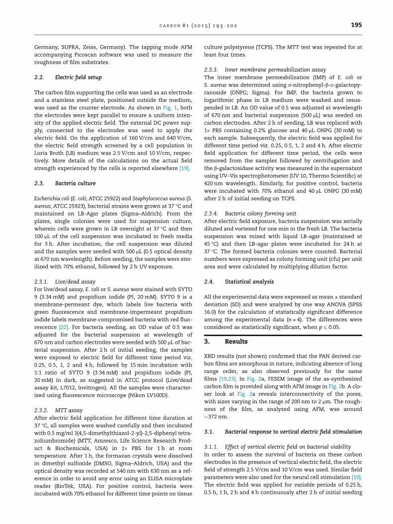
films [19,23]. In Fig. 2a, FESEM image of the as-synthesized
carbon film is provided along with AFM image in Fig. 2b. A clo-
ser look at Fig. 2a reveals interconnectivity of the pores,
with sizes varying in the range of 200 nm to 2 lm. The rough-
ness of the film, as analyzed using AFM, was around
�372 nm.
3.1. Bacterial response to vertical electric field stimulation
3.1.1. Effect of vertical electric field on bacterial viabilityIn order to assess the survival of bacteria on these carbon
electrodes in the presence of vertical electric field, the electric
field of strength 2.5 V/cm and 10 V/cm was used. Similar field
parameters were also used for the neural cell stimulation [19].
The electric field was applied for variable periods of 0.25 h,
0.5 h, 1 h, 2 h and 4 h continuously after 2 h of initial seeding
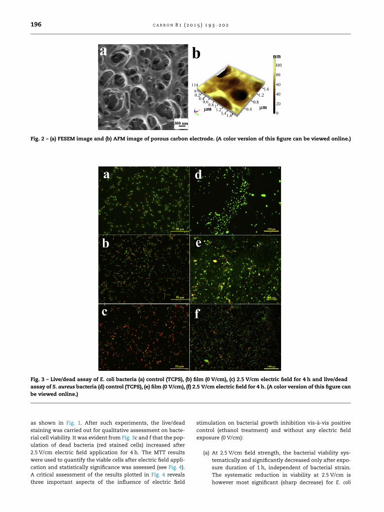
Fig. 2 – (a) FESEM image and (b) AFM image of porous carbon electrode. (A color version of this figure can be viewed online.)
Fig. 3 – Live/dead assay of E. coli bacteria (a) control (TCPS), (b) film (0 V/cm), (c) 2.5 V/cm electric field for 4 h and live/dead
assay of S. aureus bacteria (d) control (TCPS), (e) film (0 V/cm), (f) 2.5 V/cm electric field for 4 h. (A color version of this figure can
be viewed online.)
196 C A R B O N 8 1 ( 2 0 1 5 ) 1 9 3 – 2 0 2
as shown in Fig. 1. After such experiments, the live/dead
staining was carried out for qualitative assessment on bacte-
rial cell viability. It was evident from Fig. 3c and f that the pop-
ulation of dead bacteria (red stained cells) increased after
2.5 V/cm electric field application for 4 h. The MTT results
were used to quantify the viable cells after electric field appli-
cation and statistically significance was assessed (see Fig. 4).
A critical assessment of the results plotted in Fig. 4 reveals
three important aspects of the influence of electric field
stimulation on bacterial growth inhibition vis-a-vis positive
control (ethanol treatment) and without any electric field
exposure (0 V/cm):
(a) At 2.5 V/cm field strength, the bacterial viability sys-
tematically and significantly decreased only after expo-
sure duration of 1 h, independent of bacterial strain.
The systematic reduction in viability at 2.5 V/cm is
however most significant (sharp decrease) for E. coli
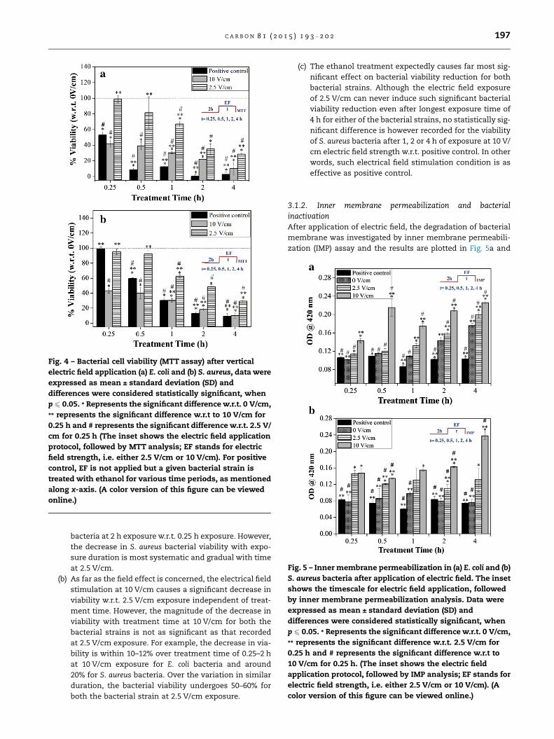
Fig. 4 – Bacterial cell viability (MTT assay) after vertical
electric field application (a) E. coli and (b) S. aureus, data were
expressed as mean ± standard deviation (SD) and
differences were considered statistically significant, when
p 6 0.05. * Represents the significant difference w.r.t. 0 V/cm,
** represents the significant difference w.r.t to 10 V/cm for
0.25 h and # represents the significant difference w.r.t. 2.5 V/
cm for 0.25 h (The inset shows the electric field application
protocol, followed by MTT analysis; EF stands for electric
field strength, i.e. either 2.5 V/cm or 10 V/cm). For positive
control, EF is not applied but a given bacterial strain is
treated with ethanol for various time periods, as mentioned
along x-axis. (A color version of this figure can be viewed
online.)
Fig. 5 – Inner membrane permeabilization in (a) E. coli and (b)
S. aureus bacteria after application of electric field. The inset
shows the timescale for electric field application, followed
by inner membrane permeabilization analysis. Data were
expressed as mean ± standard deviation (SD) and
differences were considered statistically significant, when
p 6 0.05. * Represents the significant difference w.r.t. 0 V/cm,
** represents the significant difference w.r.t. 2.5 V/cm for
0.25 h and # represents the significant difference w.r.t to
10 V/cm for 0.25 h. (The inset shows the electric field
application protocol, followed by IMP analysis; EF stands for
electric field strength, i.e. either 2.5 V/cm or 10 V/cm). (A
color version of this figure can be viewed online.)
C A R B O N 8 1 ( 2 0 1 5 ) 1 9 3 – 2 0 2 197
bacteria at 2 h exposure w.r.t. 0.25 h exposure. However,
the decrease in S. aureus bacterial viability with expo-
sure duration is most systematic and gradual with time
at 2.5 V/cm.
(b) As far as the field effect is concerned, the electrical field
stimulation at 10 V/cm causes a significant decrease in
viability w.r.t. 2.5 V/cm exposure independent of treat-
ment time. However, the magnitude of the decrease in
viability with treatment time at 10 V/cm for both the
bacterial strains is not as significant as that recorded
at 2.5 V/cm exposure. For example, the decrease in via-
bility is within 10–12% over treatment time of 0.25–2 h
at 10 V/cm exposure for E. coli bacteria and around
20% for S. aureus bacteria. Over the variation in similar
duration, the bacterial viability undergoes 50–60% for
both the bacterial strain at 2.5 V/cm exposure.
(c) The ethanol treatment expectedly causes far most sig-
nificant effect on bacterial viability reduction for both
bacterial strains. Although the electric field exposure
of 2.5 V/cm can never induce such significant bacterial
viability reduction even after longest exposure time of
4 h for either of the bacterial strains, no statistically sig-
nificant difference is however recorded for the viability
of S. aureus bacteria after 1, 2 or 4 h of exposure at 10 V/
cm electric field strength w.r.t. positive control. In other
words, such electrical field stimulation condition is as
effective as positive control.
3.1.2. Inner membrane permeabilization and bacterialinactivationAfter application of electric field, the degradation of bacterial
membrane was investigated by inner membrane permeabili-
zation (IMP) assay and the results are plotted in Fig. 5a and
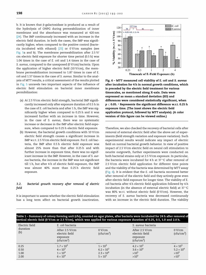
Fig. 6 – MTT measured cell viability of E. coli and S. aureus
after incubation for 4 h in normal growth conditions, which
is preceded by the electric field treatment for various
timescales, as mentioned along X-axis. Data were
expressed as mean ± standard deviation (SD) and
differences were considered statistically significant, when
p 6 0.05. * Represents the significant difference w.r.t. 0.25 h
exposure time. (The inset shows the electric field
application protocol, followed by MTT analysis). (A color
version of this figure can be viewed online.)
198 C A R B O N 8 1 ( 2 0 1 5 ) 1 9 3 – 2 0 2
b. It is known that b-galactosidase is produced as a result of
the hydrolysis of ONPG during permeabilization of inner
membrane and the absorbance was measured at 420 nm
[24]. The IMP continuously increased with an increase in the
electric field duration. In both the cases, the IMP was signifi-
cantly higher, when compared to the positive control (bacte-
ria incubated with ethanol) [25] or 0 V/cm samples (see
Fig. 5a and b). The membrane permeabilization after 2.5 V/
cm electric field exposure for shorter time period (0.5 h) was
1.04 times in the case of E. coli and 1.4 times in the case of
S. aureus, compared to the unexposed (0 V/cm) bacteria. Upon
the application of higher electric field (10 V/cm), the mem-
brane permeabilization increased to 1.87 times in case of E.
coli and 1.57 times in the case of S. aureus. Similar to the anal-
ysis of MTT results, a critical assessment of the results plotted
in Fig. 5 unravels two important aspects of the influence of
electric field stimulation on bacterial inner membrane
permibilization:
(a) At 2.5 V/cm electric field strength, bacterial IMP signifi-
cantly increased only after exposure duration of 0.5 h in
the case of E. coli bacteria and after 1 h, the IMP was sig-
nificantly higher when compared to 0.25 h and it was
increased further with an increase in time. However,
in the case of S. aureus, there was no systematic
increase or decrease in the IMP after electric field expo-
sure, when compared to 0.25 h electric field exposure.
(b) However, the bacterial growth conditions with 10 V/cm
electric field strength causes a significant increase in
IMP w.r.t. 2.5 V/cm electric field exposure. For E. coli bac-
teria, the IMP after 0.5 h electric field exposure was
almost 25% more than that after 0.25 h and with
further increase in exposure time, there was no signif-
icant increase in the IMP. However, in the case of S. aur-
eus bacteria, the increase in the IMP was not significant
till 1 h, but after 4 h of electric field exposure, the IMP
was almost 40% more than 0.25 h electric field
exposure.
3.2. Bacterial growth recovery after removal of electricfield
It is important to assess whether the electric field stimulation
has a long term affect on bacterial growth inactivation.
Table 1 – Summary of colony forming unit (cfu), counted on agarvertical electric field (0 V/cm or 2.5 V/cm), which was applied fo
Electric fieldduration(h)
E. coli bacteria
After 2.5 V/cmelectric fieldexposure(cfu/cm2)
0 V/c(cfu/
0.25 1.7 · 103 1 · 100.50 4 · 103 6.2 ·1.00 1 · 104 2.3 ·2.00 6 · 104 5 · 10
Therefore, we also checked the recovery of bacterial cells after
removal of external electric field after the above set of exper-
iments (field strength variation and exposure variation). Such
experimental results would indicate any impact of electric
field on normal bacterial growth behavior. In view of positive
impact of 2.5 V/cm electric field on neural cell stimulation to
neurite outgrowth, further experiments were conducted on
both bacterial strains only at 2.5 V/cm exposure. In particular,
the bacteria were incubated for 4 h at 37 �C after removal of
2.5 V/cm electric field application for different time points
and the viability of the bacteria was determined by MTT assay
(Fig. 6). It is evident that the E. coli bacteria recovered better
after removal of the electric field and they actively grow even
after electric field exposure for longer time. The viability of E.
coli bacteria after 4 h electric field application followed by 4 h
incubation (in the absence of external electric field) at 37 �Cwas 80% w.r.t. without electric field (0 V/cm). However, the
recovery of S. aureus bacteria was decreased continuously
with an increase in the electric field duration. The viability
plates, after bacteria were incubated for 24 h after removal ofr various exposure duration of 0.25, 0.5, 1.0 and 2.0 h.
S. aureus bacteria
mcm2)
After 2.5 V/cmelectric fieldexposure(cfu/cm2)
0 V/cm(cfu/cm2)
3 4.2 · 103 4 · 103
103 3.6 · 104 5.2 · 104
104 >105 >105
5 >105 >105
C A R B O N 8 1 ( 2 0 1 5 ) 1 9 3 – 2 0 2 199
of S. aureus bacteria after 4 h electric field exposure, followed
by 4 h incubation at 37 �C was only 50% w.r.t. without the elec-
tric field. Also, a sharp decrease in viability is recorded at 0.5 h
exposure w.r.t 0.25 h exposure, independent of bacterial
strain. Thereafter, a gradual/systematic increase or decrease
in bacterial viability is recorded in exposure time dependent
manner for E. coli or S. aureus, respectively. On the basis of
the data presented in Fig. 6, it can be therefore concluded that
E. coli bacteria can recover the growth pattern after the
removal of the electric field and when grown under normal
culture conditions for 4 h. In contrast, the influence of electric
field on bacterial growth inactivation is much more signifi-
cant for S. aureus bacteria, as the bacterial viability systemat-
ically decreases after the removal of electric field and no
recovery could be recorded.
The above aspects have been further analyzed by measur-
ing the colony forming unit (in terms of cfu/cm2), as summa-
rized in Table 1. In contrast to the data presented in Fig. 6, cfu
was counted after incubation for 24 h following electric field
application at 2.5 V/cm for various time points. For compari-
son, cfu measured after incubation without any electric field
(0 V/cm) are also presented in Table 1. The increase in cfu
with the exposure time is however cell type dependent. For
E. coli bacteria, the reduction in cfu after exposure to 2.5 V/
cm electric field w.r.t control (0 V/cm) can be well realized,
when the exposure duration is 0.5 h or more. A critical look
at Table 1 also reveals that the decrease in cfu of E. coli strain
becomes more prominent with the longer exposure time, e.g.,
1.5 times reduction at 0.5 h duration, 2.3 times at 1 h duration
and one order of magnitude reduction after 2 h of exposure.
Although such difference is not measurable at longer expo-
sure duration for S. aureus strain, almost 1.5 times decrease
in cfu was recorded after 0.5 h of exposure.
4. Discussion
The failure of the implanted devices due to bacterial infection
is the major concern in the field of biomedical application. In
this work, we have studied the inactivation of bacteria in the
presence of external vertical electric field on amorphous car-
bon electrodes. These amorphous carbon electrodes were por-
ous (pore size distribution 200–2000 nm), conducting (50 S/
cm) in nature with higher charge storage capacity (0.2 mC/
cm2) and lower electrochemical impedance (3.3 kX at 1 kHz)
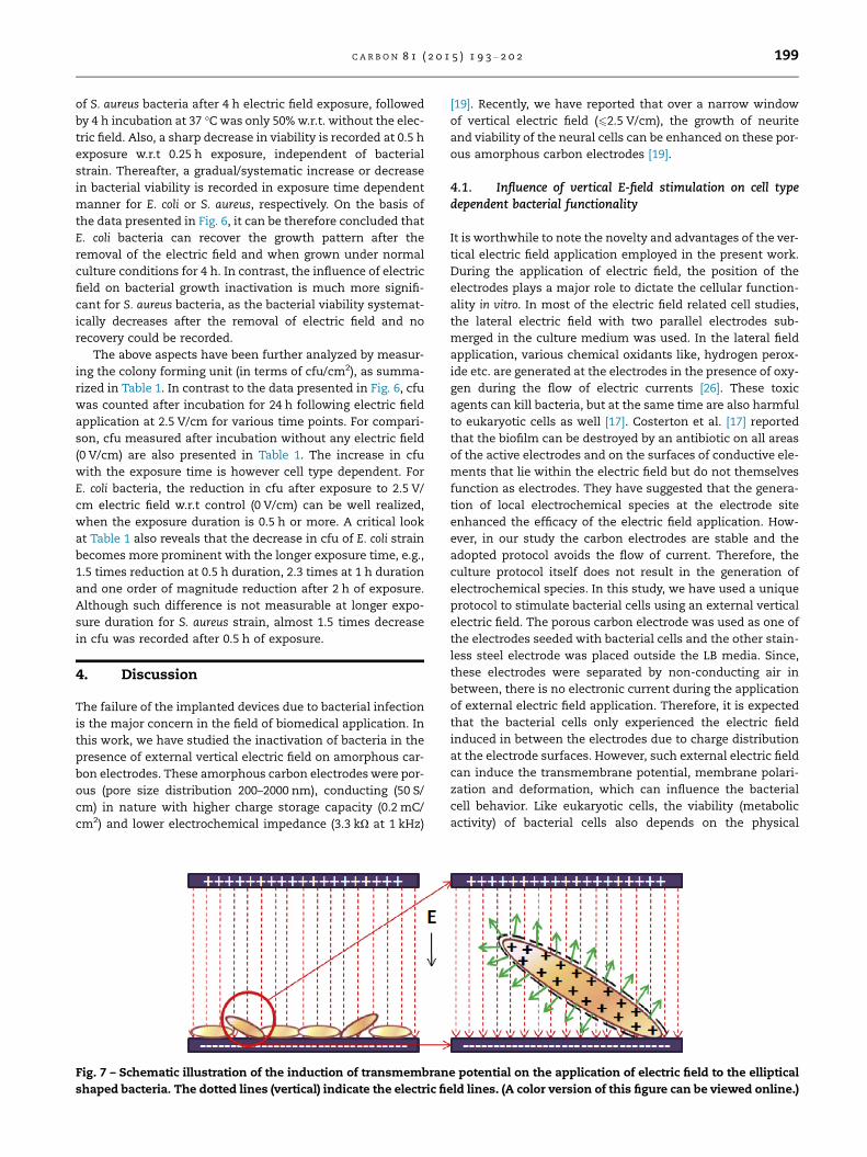
Fig. 7 – Schematic illustration of the induction of transmembran
shaped bacteria. The dotted lines (vertical) indicate the electric fi
[19]. Recently, we have reported that over a narrow window
of vertical electric field (62.5 V/cm), the growth of neurite
and viability of the neural cells can be enhanced on these por-
ous amorphous carbon electrodes [19].
4.1. Influence of vertical E-field stimulation on cell typedependent bacterial functionality
It is worthwhile to note the novelty and advantages of the ver-
tical electric field application employed in the present work.
During the application of electric field, the position of the
electrodes plays a major role to dictate the cellular function-
ality in vitro. In most of the electric field related cell studies,
the lateral electric field with two parallel electrodes sub-
merged in the culture medium was used. In the lateral field
application, various chemical oxidants like, hydrogen perox-
ide etc. are generated at the electrodes in the presence of oxy-
gen during the flow of electric currents [26]. These toxic
agents can kill bacteria, but at the same time are also harmful
to eukaryotic cells as well [17]. Costerton et al. [17] reported
that the biofilm can be destroyed by an antibiotic on all areas
of the active electrodes and on the surfaces of conductive ele-
ments that lie within the electric field but do not themselves
function as electrodes. They have suggested that the genera-
tion of local electrochemical species at the electrode site
enhanced the efficacy of the electric field application. How-
ever, in our study the carbon electrodes are stable and the
adopted protocol avoids the flow of current. Therefore, the
culture protocol itself does not result in the generation of
electrochemical species. In this study, we have used a unique
protocol to stimulate bacterial cells using an external vertical
electric field. The porous carbon electrode was used as one of
the electrodes seeded with bacterial cells and the other stain-
less steel electrode was placed outside the LB media. Since,
these electrodes were separated by non-conducting air in
between, there is no electronic current during the application
of external electric field application. Therefore, it is expected
that the bacterial cells only experienced the electric field
induced in between the electrodes due to charge distribution
at the electrode surfaces. However, such external electric field
can induce the transmembrane potential, membrane polari-
zation and deformation, which can influence the bacterial
cell behavior. Like eukaryotic cells, the viability (metabolic
activity) of bacterial cells also depends on the physical
e potential on the application of electric field to the elliptical
eld lines. (A color version of this figure can be viewed online.)
200 C A R B O N 8 1 ( 2 0 1 5 ) 1 9 3 – 2 0 2
phenomena such as membrane potential [27] and the cellular
electrical equilibria may be disturbed by the application of an
external electric field [17]. It has been shown that the external
fields can affect the a-helix content and the orientation of
membrane proteins in eukaryotic cells and the electropho-
retic mobilities of bacterial membrane proteins [27,28].
Electric fields can even be used to affect the electroinsertion
of specific proteins into the membranes of living cells. These
molecular perturbations of important membrane compo-
nents may affect the organization of membranes and we
expect that these structural changes would influence the
permeability of membranes in the present case [17].
In the present case, both Gram-negative and Gram-posi-
tive bacteria were exposed to vertical electric field with field
strength of up to 10 V/cm for various time points. It was
observed that both the bacteria could be killed up to 80% on
the application of higher electric field (10 V/cm) for short time
or on the application of low field (2.5 V/cm) for longer time
(Fig. 6a and b). Interestingly, the S. aureus bacteria responded
after longer exposure duration as compared to E. coli bacteria
in the presence of vertical electric field. It might be due to the
fact that S. aureus is a Gram-positive bacteria with thicker cell
wall compared to E. coli, which is a Gram-negative bacteria
[14,29].
The irreversible permeabilization of bacterial cell mem-
brane in the presence of electric field depends on the electric
field strength [26]. As shown schematically in Fig. 7, the
potential drop at the cell membrane on the application of
electric field was generated due to difference in conductivity
of cytoplasm (1S/m) and cell membrane (10�5 S/m) [14]. The
application of uniform electric field to the bacterial cell
induced transmembrane potential [14,30] and this can be cal-
culated by Schwan’s equation [31]:
D/ ¼ 32
Er cos h ð1Þ
where D/ is the induced transmembrane potential, E is the
external applied field, r is the radius of cell and h is the angle
between direction of applied field and cell orientation axis. It
is evident from the above equation that the application of
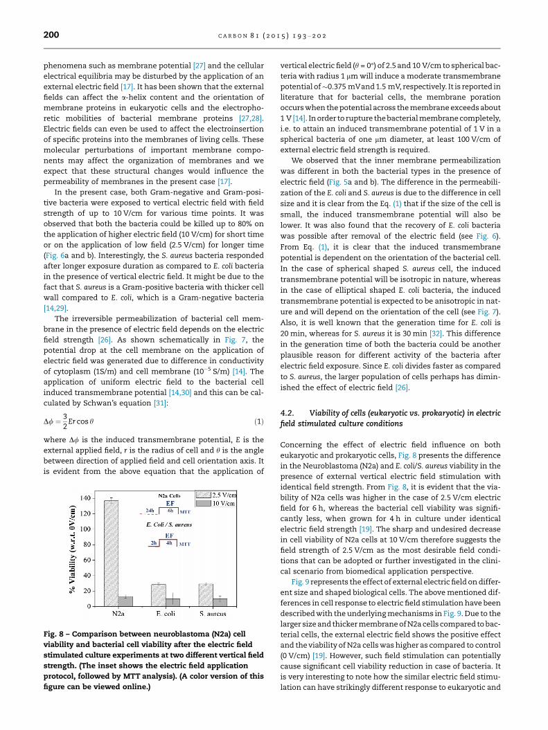
Fig. 8 – Comparison between neuroblastoma (N2a) cell
viability and bacterial cell viability after the electric field
stimulated culture experiments at two different vertical field
strength. (The inset shows the electric field application
protocol, followed by MTT analysis). (A color version of this
figure can be viewed online.)
vertical electric field (h = 0�) of 2.5 and 10 V/cm to spherical bac-
teria with radius 1 lm will induce a moderate transmembrane
potential of�0.375 mVand 1.5 mV, respectively. It is reported in
literature that for bacterial cells, the membrane poration
occurs when the potential across the membrane exceeds about
1 V [14]. In order to rupture the bacterial membrane completely,
i.e. to attain an induced transmembrane potential of 1 V in a
spherical bacteria of one lm diameter, at least 100 V/cm of
external electric field strength is required.
We observed that the inner membrane permeabilization
was different in both the bacterial types in the presence of
electric field (Fig. 5a and b). The difference in the permeabili-
zation of the E. coli and S. aureus is due to the difference in cell
size and it is clear from the Eq. (1) that if the size of the cell is
small, the induced transmembrane potential will also be
lower. It was also found that the recovery of E. coli bacteria
was possible after removal of the electric field (see Fig. 6).
From Eq. (1), it is clear that the induced transmembrane
potential is dependent on the orientation of the bacterial cell.
In the case of spherical shaped S. aureus cell, the induced
transmembrane potential will be isotropic in nature, whereas
in the case of elliptical shaped E. coli bacteria, the induced
transmembrane potential is expected to be anisotropic in nat-
ure and will depend on the orientation of the cell (see Fig. 7).
Also, it is well known that the generation time for E. coli is
20 min, whereas for S. aureus it is 30 min [32]. This difference
in the generation time of both the bacteria could be another
plausible reason for different activity of the bacteria after
electric field exposure. Since E. coli divides faster as compared
to S. aureus, the larger population of cells perhaps has dimin-
ished the effect of electric field [26].
4.2. Viability of cells (eukaryotic vs. prokaryotic) in electricfield stimulated culture conditions
Concerning the effect of electric field influence on both
eukaryotic and prokaryotic cells, Fig. 8 presents the difference
in the Neuroblastoma (N2a) and E. coli/S. aureus viability in the
presence of external vertical electric field stimulation with
identical field strength. From Fig. 8, it is evident that the via-
bility of N2a cells was higher in the case of 2.5 V/cm electric
field for 6 h, whereas the bacterial cell viability was signifi-
cantly less, when grown for 4 h in culture under identical
electric field strength [19]. The sharp and undesired decrease
in cell viability of N2a cells at 10 V/cm therefore suggests the
field strength of 2.5 V/cm as the most desirable field condi-
tions that can be adopted or further investigated in the clini-
cal scenario from biomedical application perspective.
Fig. 9 represents the effect of external electric field on differ-
ent size and shaped biological cells. The above mentioned dif-
ferences in cell response to electric field stimulation have been
described with the underlying mechanisms in Fig. 9. Due to the
larger size and thicker membrane of N2a cells compared to bac-
terial cells, the external electric field shows the positive effect
and the viability of N2a cells was higher as compared to control
(0 V/cm) [19]. However, such field stimulation can potentially
cause significant cell viability reduction in case of bacteria. It
is very interesting to note how the similar electric field stimu-
lation can have strikingly different response to eukaryotic and
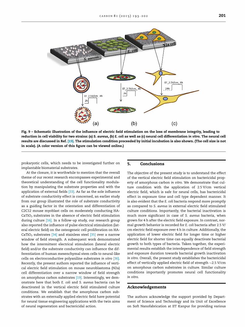
Fig. 9 – Schematic illustration of the influence of electric field stimulation on the loss of membrane integrity, leading to
reduction in cell viability for two strains: (a) S. aureus, (b) E. coli as well as (c) neural cell differentiation in vitro. The neural cell
results are discussed in Ref. [19]. The stimulation condition proceeded by initial incubation is also shown. (The cell size is not
in scale). (A color version of this figure can be viewed online.)
C A R B O N 8 1 ( 2 0 1 5 ) 1 9 3 – 2 0 2 201
prokaryotic cells, which needs to be investigated further on
implantable biomaterial substrates.
At the closure, it is worthwhile to mention that the overall
theme of our recent research encompasses experimental and
theoretical understanding of the cell functionality modula-
tion by manipulating the substrate properties and with the
application of external fields [33]. As far as the sole influence
of substrate conductivity effect is concerned, an earlier study
from our group illustrated the role of substrate conductivity
as a guiding factor in the orientation and differentiation of
C2C12 mouse myoblast cells on moderately conducting HA-
CaTiO3 substrates in the absence of electric field stimulation
during culture [16]. In a follow-up study, our research group
also reported the influence of pulse electrical stimulation (lat-
eral electric field) on the osteogeneic cell proliferation on HA-
CaTiO3 substrates [34] and stainless steel [35] over a narrow
window of field strength. A subsequent work demonstrated
how the intermittent electrical stimulation (lateral electric
field) and/or the substrate conductivity can influence the dif-
ferentiation of human mesenchymal stem cells to neural-like
cells on electroconductive polyaniline substrates in vitro [36].
Recently, the present authors reported the influence of verti-
cal electric field stimulation on mouse neuroblastoma (N2a)
cell differentiation over a narrow window of field strength
on amorphous carbon substrates [19]. Interestingly, we dem-
onstrate here that both E. coli and S. aureus bacteria can be
deactivated in the vertical electric field stimulated culture
conditions. We establish that the amorphous carbon sub-
strates with an externally applied electric field have potential
for neural tissue engineering applications with the twin aims
of neural regeneration and bactericidal action.
5. Conclusions
The objective of the present study is to understand the effect
of the vertical electric field stimulation on bactericidal prop-
erty of amorphous carbon in vitro. We demonstrate that cul-
ture condition with the application of 2.5 V/cm vertical
electric field, which is safe for neural cells, has bactericidal
effect in exposure time and cell type dependent manner. It
is also evident that the E. coli bacteria respond more promptly
as compared to S. aureus in external electric field stimulated
culture conditions. Importantly, the bacterial inactivation is
much more significant in case of S. aureus bacteria, when
grown for 4 h after the electric field exposure. In contrast, nor-
mal growth behavior is recorded for E. coli bacteria after 2.5 V/
cm electric field exposure over 4 h in culture. Additionally, the
application of lower electric field for longer time or higher
electric field for shorter time can equally deactivate bacterial
growth to both types of bacteria. Taken together, the experi-
mental results establish the interdependence of field strength
and exposure duration towards bacterial growth inactivation
in vitro. Overall, the present study establishes the bactericidal
effect of vertically applied electric field of strength �2.5 V/cm
on amorphous carbon substrates in culture. Similar culture
conditions importantly promotes neural cell functionality
in vitro.
Acknowledgements
The authors acknowledge the support provided by Depart-
ment of Science and Technology and its Unit of Excellence
on Soft Nanofabrication at IIT Kanpur for providing various
202 C A R B O N 8 1 ( 2 0 1 5 ) 1 9 3 – 2 0 2
research facilities. The financial support from Department of
Biotechnology (DBT) is also gratefully acknowledged.
R E F E R E N C E S
[1] Yu Q, Cho J, Shivapooja P, Ista LK, Lopez GP. Nanopatternedsmart polymer surfaces for controlled attachment, killing,and release of bacteria. ACS Appl Mater Interfaces2013;5:9295–304.
[2] Litzler P-Y, Benard L, Barbier-Frebourg N, Vilain S, Jouenne T,Beucher E, et al. Biofilm formation on pyrolytic carbon heartvalves: influence of surface free energy, roughness, andbacterial species. J Thorac Cardiovasc Surg 2007;134:1025–32.
[3] Fortunati E, Mattioli S, Visai L, Imbriani M, Fierro JLG, KennyJM, et al. Combined effects of Ag nanoparticles and oxygenplasma treatment on PLGA morphological, chemical, andantibacterial properties. Biomacromolecules 2013;14:626–36.
[4] Ivanova EP, Truong VK, Wang JY, Berndt CC, Jones RT, Yusuf II,et al. Impact of nanoscale roughness of titanium thin filmsurfaces on bacterial retention. Langmuir 2010;26:1973–82.
[5] An YH, Friedman RJ. Concise review of mechanisms ofbacterial adhesion to biomaterial surfaces. J Biomed MaterRes 1998;43:338–48.
[6] Ploux L, Anselme K, Dirani A, Ponche A, Soppera O, RoucoulesV. Opposite responses of cells and bacteria to micro/nanopatterned surfaces prepared by pulsed plasmapolymerization and UV-irradiation. Langmuir 2009;25:8161–9.
[7] Subbiahdoss G, Kuijer R, Grijpma DW, van der Mei HC,Busscher HJ. Microbial biofilm growth vs. tissue integration:‘‘the race for the surface’’ experimentally studied. ActaBiomater 2009;5:1399–404.
[8] Colon G, Ward BC, Webster TJ. Increased osteoblast anddecreased Staphylococcus epidermidis functions on nanophaseZnO and TiO2. J Biomed Mater Res A 2006;78:2006.
[9] McCaig CD, Rajnicek AM, Song B, Zhao M. Controlling cellbehavior electrically: current views and future potential.Physiol Rev 2005;85:943–78.
[10] Robinson KR. The response of cells to electrical fields: areview. JCB 1985;101:2023–7.
[11] Hofmann GA. Electronic genetic physical and biologicalaspects of cellular electromanipulation. IEEE Eng Med BiolMag 1986:6–25.
[12] Qin B-L, Zhang Q, Barbosa-Canovas GV, Swanson BG, PedrowPD. Inactivation of microorganisms by pulsed electric fieldsof different voltage waveforms. IEEE Trans Dielectr Insul1994;1:1047–57.
[13] Luo Q, Wang H, Zhang X, Qian Y. Effect of direct electriccurrent on the cell surface properties of phenol-degradingbacteria. Appl Environ Microbiol 2005;71:423–7.
[14] El-Hag AH, Jayaram SH, Gonzalez OR, Griffiths MW. Theinfluence of size and shape of microorganism on pulsedelectric field inactivation. IEEE Trans Nanobiosci2011;10:133–8.
[15] Khoury AE, Lam K, Ellis B, Costerton JW. Prevention andcontrol of bacterial infections associated with medicaldevices. ASAIO J 1992;38:M174–8.
[16] Thrivikraman G, Mallik PK, Basu B. Substrate conductivitydependent modulation of cell proliferation anddifferentiation in vitro. Biomaterials 2013;34:7073–85.
[17] Costerton JW, Ellis B, Lam K, Johnson F, Khoury AE.Mechanism of electrical enhancement of efficacy ofantibiotics in killing biofilm bacteria. Antimicrob AgentsChemother 1994;38:2803–9.
[18] Liu W-K, Browna MRW, Elliott TSJ. Mechanisms of thebactericidal activity of low amperage electric current (DC). JAntimicrob Chemother 1997;39.
[19] Jain S, Sharma A, Basu B. Vertical electric field stimulatedneural cell functionality on porous amorphous carbonelectrodes. Biomaterials 2013;34:9252–63.
[20] Rahaman MSA, Ismail AF, Mustafa A. A review of heattreatment on polyacrylonitrile fiber. Polym Degrad Stab2007;92:1421–32.
[21] Ko TH, Chen CY. Raman spectroscopic study of themicrostructure of carbon films developed from cobaltchloride-modified polyacrylonitrile. J Appl Polym Sci1999;71:2219–25.
[22] Popat KC, Eltgroth M, LaTempa TJ, Grimes CA, Desai TA.Decreased Staphylococcus epidermis adhesion and increasedosteoblast functionality on antibiotic-loaded titaniananotubes. Biomaterials 2007;28:4880–8.
[23] Mondal K, Kumar J, Sharma A. Self-organized macroporousthin carbon films for supported metal catalysis. Colloids SurfA 2013;427:83–94.
[24] Lui H, Du Y, Wang X, Sun L. Chitosan kills bacteria throughcell membrane damage. Int J Food Microbiol 2004;95:147–55.
[25] Huffer S, Clark ME, Ning JC, Blanch HW, Clark DS. Role ofalcohols in growth, lipid composition, and membrane fluidityof yeasts, bacteria, and archaea. Appl Environ Microbiol2011;77:6400–8.
[26] Drees KP, Abbaszadegan M, Maier RM. Comparativeelectrochemical inactivation of bacteria and bacteriophage.Water Res 2003;37:2291–300.
[27] McLaughlin S. The electrostatic properties of membranes.Annu Rev Biophys Biophys Chem 1989;18:113–36.
[28] Tsong TY. Electrical modulation of membrane proteins:enforced conformational oscillations and biological energyand signal transductions. Annu Rev Biophys Biophys Chem1990;19:83–106.
[29] Feng QL, Wu J, Chen GQ, Cui FZ, Kim TN, Kim JO. AMechanistic study of the antibacterial effect of silver ions onEscherichia coli and Staphylococcus aureus. J Biomed Mater Res2000;52:662–8.
[30] Cao H-B, Li X-G, Wu J-C, Yu K-T, Zhang Y. Simulation of theeffects of direct electric current on multispecies biofilms.Process Biochem 2003;38:1139–45.
[31] Valic B, Golzio M, Pavlin M, Schatz A, Faurie C, Gabriel B, et al.Effect of electric field induced transmembrane potential onspheroidal cells: theory and experiment. Eur Biophys J2003;32:519–28.
[32] Todar K. <http://textbookofbacteriology.net/growth_3.html>2008.
[33] Dubey A, Gupta SD, Basu B. Optimization of electricalstimulation conditions for enhanced fibroblast cellproliferation on biomaterial surfaces. J Biomed Mater Res B2011;98B:18–29.
[34] Dubey AK, Basu B. Pulsed electrical stimulation and surfacecharge induced cell growth on multistage spark plasmasintered hydroxyapatite-barium titanate piezobiocomposite.J Am Ceram Soc 2014;97:481–9.
[35] Dubey AK, Agrawal P, Misra RDK, Basu B. Pulsed electricfield mediated in vitro cellular response of fibroblast andosteoblast-like cells on conducting austenitic stainlesssteel substrate. J Mater Sci – Mater Med 2013;24:1789–98.
[36] Thrivikraman G, Madras G, Basu B. Intermittent electricalstimuli for guidance of human mesenchymal stem celllineage commitment towards neural-like cells onelectroactive substrates. Biomaterials 2014;35:6219–35.
Copyright © 2022 FDOKUMEN