Valvular Heart Disease Epidemiology - MDPI
12
Citation: Aluru, J.S.; Barsouk, A.; Saginala, K.; Rawla, P.; Barsouk, A. Valvular Heart Disease Epidemiology. Med. Sci. 2022, 10, 32. https:// doi.org/10.3390/medsci10020032 Received: 8 April 2022 Accepted: 13 June 2022 Published: 15 June 2022 Publisher’s Note: MDPI stays neutral with regard to jurisdictional claims in published maps and institutional affil- iations. Copyright: © 2022 by the authors. Licensee MDPI, Basel, Switzerland. This article is an open access article distributed under the terms and conditions of the Creative Commons Attribution (CC BY) license (https:// creativecommons.org/licenses/by/ 4.0/). medical sciences Review Valvular Heart Disease Epidemiology John Sukumar Aluru 1 , Adam Barsouk 2 , Kalyan Saginala 3 , Prashanth Rawla 4, * and Alexander Barsouk 5 1 Minneapolis Heart Institute Foundation, Allina Health, Minneapolis, MN 55407, USA; [email protected] 2 Hillman Cancer Center, University of Pittsburgh, Pittsburgh, PA 15232, USA; [email protected] 3 Plains Regional Medical Group Internal Medicine, Clovis, NM 88101, USA; [email protected] 4 Parrish Medical Center, Titusville, FL 32796, USA 5 Allegheny Health Network, Pittsburgh, PA 15212, USA; [email protected] * Correspondence: [email protected]; Tel.: +1-732-982-7357 Abstract: Valvular heart disease is a rapidly growing cause of global cardiovascular morbidity and mortality with diverse and evolving geographic distribution. The prevalence of rheumatic heart disease, the most common valvular heart disease (affecting approximately 41 million people), has been rising in developing nations, likely due to the expansion of the young adult population and the decrease in premature mortality that has resulted from improved access to antibiotics, microbiological testing, and echocardiography. Rheumatic heart disease has also been rising among the impoverished and, often, indigenous populations of developed nations, spurring public health initiatives that are aimed at alleviating healthcare disparities. Aortic valve stenotic disease is the most commonly occurring valvular pathology in developed nations (afflicting 9 million people worldwide) and its prevalence has been increasing with population aging and the increased prevalence of atherosclerosis. Aortic regurgitation is associated with diastolic, but not systolic, hypertension and it has likewise seen a rise in the developed world. Mitral regurgitation affects 24 million people worldwide, with great variability between and among nations. Primary mitral regurgitation arises as a consequence of myxomatous degeneration and mitral valve prolapse, which is largely due to genetic predispositions, while secondary mitral regurgitation accounts for 65% of cases and arises secondary to dilation and heart failure. Tricuspid regurgitation has become more prevalent in developed nations due to the increased usage of intracardiac pacemakers. Infective endocarditis prevalence has also grown in developed nations, likely due to population aging and the increased utilization of transcatheter valve replacement and prosthetic valves as interventions against the previously discussed valvular pathologies. Keywords: valvular heart disease; epidemiology; aortic valve stenotic disease; rheumatic heart disease; aortic regurgitation; tricuspid regurgitation; infective endocarditis 1. Introduction Valvular heart disease is a leading cause of cardiovascular morbidity and mortality worldwide and the resultant disease burden is only projected to increase in the coming decades. The heart contains four valves: the tricuspid, pulmonic, mitral, and aortic. These valves prevent backward flow between the four heart chambers and maintain the pressure gradients that are necessary for the hemodynamic circulation that is essential for life. The regurgitation from or the insufficiency of the valves are secondary to valvular heart dis- ease and both permit backward flow, resulting in an equalization of pressure that can be incompatible with cardiovascular function. Conversely, the stenosis of the valves, which is secondary to valvular heart disease, results in increased pressures behind the blockage (often resulting in cardiac remodeling) and insufficient pressure ahead of the blockage (e.g., syncope in the case of aortic stenosis) [1,2]. Understanding the geographical and temporal trends that are present in valve disease epidemiology is crucial for designing effective Med. Sci. 2022, 10, 32. https://doi.org/10.3390/medsci10020032 https://www.mdpi.com/journal/medsci
-
Upload
khangminh22 -
Category
Documents
-
view
4 -
download
0
Transcript of Valvular Heart Disease Epidemiology - MDPI

Citation: Aluru, J.S.; Barsouk, A.;
Saginala, K.; Rawla, P.; Barsouk, A.
Valvular Heart Disease Epidemiology.
Med. Sci. 2022, 10, 32. https://
doi.org/10.3390/medsci10020032
Received: 8 April 2022
Accepted: 13 June 2022
Published: 15 June 2022
Publisher’s Note: MDPI stays neutral
with regard to jurisdictional claims in
published maps and institutional affil-
iations.
Copyright: © 2022 by the authors.
Licensee MDPI, Basel, Switzerland.
This article is an open access article
distributed under the terms and
conditions of the Creative Commons
Attribution (CC BY) license (https://
creativecommons.org/licenses/by/
4.0/).
medicalsciences
Review
Valvular Heart Disease EpidemiologyJohn Sukumar Aluru 1, Adam Barsouk 2, Kalyan Saginala 3, Prashanth Rawla 4,* and Alexander Barsouk 5
1 Minneapolis Heart Institute Foundation, Allina Health, Minneapolis, MN 55407, USA; [email protected] Hillman Cancer Center, University of Pittsburgh, Pittsburgh, PA 15232, USA; [email protected] Plains Regional Medical Group Internal Medicine, Clovis, NM 88101, USA; [email protected] Parrish Medical Center, Titusville, FL 32796, USA5 Allegheny Health Network, Pittsburgh, PA 15212, USA; [email protected]* Correspondence: [email protected]; Tel.: +1-732-982-7357
Abstract: Valvular heart disease is a rapidly growing cause of global cardiovascular morbidityand mortality with diverse and evolving geographic distribution. The prevalence of rheumaticheart disease, the most common valvular heart disease (affecting approximately 41 million people),has been rising in developing nations, likely due to the expansion of the young adult populationand the decrease in premature mortality that has resulted from improved access to antibiotics,microbiological testing, and echocardiography. Rheumatic heart disease has also been rising amongthe impoverished and, often, indigenous populations of developed nations, spurring public healthinitiatives that are aimed at alleviating healthcare disparities. Aortic valve stenotic disease is the mostcommonly occurring valvular pathology in developed nations (afflicting 9 million people worldwide)and its prevalence has been increasing with population aging and the increased prevalence ofatherosclerosis. Aortic regurgitation is associated with diastolic, but not systolic, hypertension andit has likewise seen a rise in the developed world. Mitral regurgitation affects 24 million peopleworldwide, with great variability between and among nations. Primary mitral regurgitation arises asa consequence of myxomatous degeneration and mitral valve prolapse, which is largely due to geneticpredispositions, while secondary mitral regurgitation accounts for 65% of cases and arises secondaryto dilation and heart failure. Tricuspid regurgitation has become more prevalent in developednations due to the increased usage of intracardiac pacemakers. Infective endocarditis prevalencehas also grown in developed nations, likely due to population aging and the increased utilizationof transcatheter valve replacement and prosthetic valves as interventions against the previouslydiscussed valvular pathologies.
Keywords: valvular heart disease; epidemiology; aortic valve stenotic disease; rheumatic heartdisease; aortic regurgitation; tricuspid regurgitation; infective endocarditis
1. Introduction
Valvular heart disease is a leading cause of cardiovascular morbidity and mortalityworldwide and the resultant disease burden is only projected to increase in the comingdecades. The heart contains four valves: the tricuspid, pulmonic, mitral, and aortic. Thesevalves prevent backward flow between the four heart chambers and maintain the pressuregradients that are necessary for the hemodynamic circulation that is essential for life. Theregurgitation from or the insufficiency of the valves are secondary to valvular heart dis-ease and both permit backward flow, resulting in an equalization of pressure that can beincompatible with cardiovascular function. Conversely, the stenosis of the valves, whichis secondary to valvular heart disease, results in increased pressures behind the blockage(often resulting in cardiac remodeling) and insufficient pressure ahead of the blockage (e.g.,syncope in the case of aortic stenosis) [1,2]. Understanding the geographical and temporaltrends that are present in valve disease epidemiology is crucial for designing effective
Med. Sci. 2022, 10, 32. https://doi.org/10.3390/medsci10020032 https://www.mdpi.com/journal/medsci

Med. Sci. 2022, 10, 32 2 of 12
public health interventions for primary and secondary prevention. Global epidemiolog-ical data can be unreliable, as post-mortem analysis has revealed the true prevalence ofvalvular heart disease to be significantly greater than that which is clinically coded andreported [3]. Furthermore, limited access to echocardiography and even microbiologicaltesting likely results in the severe underreporting of valvular disease in developing nationsand indigenous and impoverished populations in developed nations [4].
The most prevalent valve pathologies, globally, are rheumatic heart disease, aorticvalve stenotic disease, mitral regurgitation, and aortic regurgitation, while in the developedworld, aortic valve stenotic disease is more prevalent [5]. Diseases of the aortic valveaccount for 61% of all valvular heart disease deaths, while diseases of the mitral valveaccount for 15% (Figure 1). Diseases of the aortic valve have a well-established associationwith old age and chronic cardiovascular disease, while rheumatic heart disease is aninfectious complication that is primarily associated with overcrowding and poor healthcareaccess. Although infective endocarditis is similarly bacterial in its pathogenesis, it is morecommon in the elderly and likewise more prevalent in developed nations [1,2]. Whilewomen account for a larger proportion of valvular heart disease cases worldwide, theyhave often been underrepresented in the landmark studies that have informed treatmentguidelines and have reported worse postoperative outcomes [6].
Med. Sci. 2022, 10, x 2 of 12
designing effective public health interventions for primary and secondary prevention.
Global epidemiological data can be unreliable, as post-mortem analysis has revealed the
true prevalence of valvular heart disease to be significantly greater than that which is clin-
ically coded and reported [3]. Furthermore, limited access to echocardiography and even
microbiological testing likely results in the severe underreporting of valvular disease in
developing nations and indigenous and impoverished populations in developed nations
[4].
The most prevalent valve pathologies, globally, are rheumatic heart disease, aortic
valve stenotic disease, mitral regurgitation, and aortic regurgitation, while in the devel-
oped world, aortic valve stenotic disease is more prevalent [5]. Diseases of the aortic valve
account for 61% of all valvular heart disease deaths, while diseases of the mitral valve
account for 15% (Figure 1). Diseases of the aortic valve have a well-established association
with old age and chronic cardiovascular disease, while rheumatic heart disease is an in-
fectious complication that is primarily associated with overcrowding and poor healthcare
access. Although infective endocarditis is similarly bacterial in its pathogenesis, it is more
common in the elderly and likewise more prevalent in developed nations [1,2]. While
women account for a larger proportion of valvular heart disease cases worldwide, they
have often been underrepresented in the landmark studies that have informed treatment
guidelines and have reported worse postoperative outcomes [6].
Figure 1. Percentages of deaths due to valvular heart disease, by valve, 2017—data obtained from
CDC, Atlanta, GA, USA. Available online: https://www.cdc.gov/heartdisease/valvular_disease.htm
(Accessed on 25 April 2022).
Echocardiography is the gold standard for the diagnosis of valvular disease and most
patients with progressive disease should be followed up with at least annually by a car-
diologist. Intervention is warranted in symptomatic patients or those with diminished
ventricular function, though the indications for intervention are disease-specific.
Figure 1. Percentages of deaths due to valvular heart disease, by valve, 2017—data obtained fromCDC, Atlanta, GA, USA. Available online: https://www.cdc.gov/heartdisease/valvular_disease.htm(Accessed on 25 April 2022).
Echocardiography is the gold standard for the diagnosis of valvular disease andmost patients with progressive disease should be followed up with at least annually bya cardiologist. Intervention is warranted in symptomatic patients or those with dimin-ished ventricular function, though the indications for intervention are disease-specific.Transcatheter valve replacement, which is often guided by echocardiography, has gained

Med. Sci. 2022, 10, 32 3 of 12
popularity over open-heart surgery in aortic valve replacement and mitral clip proceduresand has likely improved the prognosis for certain valvular heart diseases [7].
2. Aortic Valve Stenotic Disease2.1. Epidemiology
Aortic valve stenotic disease (AVSD) is the most prevalent valvular disease in thedeveloped world. The predominant form of AVSD is calcific AVSD, which occurs due tothe thickening and calcification of the aortic valve (i.e., “aortic stenosis”). Rarely, AVSDcan also be caused by infection (post-endocarditis scarring). The normal valve area of theaortic valve is 3 cm2; the symptoms of aortic stenosis do not typically develop until thevalve area is <1 cm2. The classic symptoms of AVSD include angina, dyspnea on exertion,syncope, and ultimate heart failure. A physical exam classically reveals a crescendo–decrescendo systolic murmur that is best heard at the base of the heart with radiation tothe carotid arteries. Other physical exam findings include delayed carotid upstroke (pulsusparvus et tardus), sustained point of maximal impulse (PMI), and a diminished A2 sound.Cardiac auscultation has limited accuracy for the detection of valvular heart disease inasymptomatic patients and is a poor diagnostic screening tool when it is used in primarycare [8].
The relative frequency of the causes of ASVD varies geographically. Worldwide,rheumatic valve disease is the most common cause of ASVD and mitral valve involvementinvariably accompanies rheumatic aortic valve disease. In Europe and North America,aortic valve disease primarily occurs due to a calcific disease of a native trileaflet valve or acongenitally bicuspid valve.
A prospective population-based study of 3273 participants, including 164 subjectswith ASVD, showed that the prevalence of AS increases with age. The prevalence of ASVDvaried from 0.2 percent at ages 50 to 59 years, 1.3 percent at ages 60 to 69, 3.9 percent atages 70 to 79 years, and 9.8 percent at ages 80 to 89 years [9].
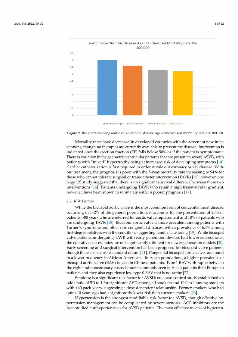
An estimated 9.4 million patients worldwide had calcific AVSD in 2019 [1,2]. Rates ofAVSD are highly correlated with old age and are the greatest in North America, Europe,and Australasia. The age-standardized AVSD mortality rate per 100,000 people is highest inWestern Europe (4.0, 95% CI 3.4–4.5), North America (3.6, 95% CI 3.0–4.0), South America(3.3, 95% CI 2.9–3.7), and Australasia (3.2, 95% CI 2.6–3.6) (Figure 2) [10]. These regionshave the highest rates of common AVSD risk factors such as hypertension, hyperlipidemia,and obesity and a greater life expectancy than countries in the developing world. A lack ofaccess to echocardiography among developing nations may also explain the discrepanciesin the reported AVSD rates.
Rheumatic aortic stenosis is most common in Asia, with a reported prevalence of4.54 in India, 1.86 in China, and 1.3 in Bangladesh per 1000 people. Several Asian countries,including Singapore, Japan, and South Korea mirror Western cohorts in having a very lowprevalence of rheumatic valvular disease. The prevalence of rheumatic aortic stenosis is0.4 in Singapore, 0.14 in Japan, and 0.5 in South Korea per 1000 people. Singapore, Japan,and South Korea have a high proportion of elderly citizens with degenerative aortic stenosisrather than rheumatic aortic stenosis [11].
Several studies have shown that the prevalence of severe aortic stenosis is lowerin African Americans than Caucasians [12]. AVSD was estimated to be responsible forapproximately 127,000 global deaths in 2019 and an associated loss of 1.8 million DALYs(disability-adjusted life-years) [1,2]. According to a large Australian registry, the 5-yearmortality rate is 56% for moderate AVD and 67% for severe AVSD [13]. Global deathshave increased by 138% between 1990 and 2019 likely due to global population agingand the “Westernization” of lifestyle, with the greatest increases having been observed intransitioning economies like China [10].
Med. Sci. 2022, 10, 32 4 of 12Med. Sci. 2022, 10, x 4 of 12
Figure 2. Bar chart showing aortic valve stenotic disease age-standardized mortality rate per
100,000.
AVSD was estimated to be responsible for approximately 127,000 global deaths in
2019 and an associated loss of 1.8 million DALYs (disability-adjusted life-years) [1,2]. Ac-
cording to a large Australian registry, the 5-year mortality rate is 56% for moderate AVD
and 67% for severe AVSD [13]. Global deaths have increased by 138% between 1990 and
2019 likely due to global population aging and the “Westernization” of lifestyle, with the
greatest increases having been observed in transitioning economies like China [10].
Mortality rates have decreased in developed countries with the advent of new inter-
ventions, though no therapies are currently available to prevent the disease. Intervention
is indicated once the ejection fraction (EF) falls below 50% or if the patient is symptomatic.
There is variation in the geometric ventricular patterns that are present in severe ASVD,
with patients with “mixed” hypertrophy being at increased risk of developing symptoms
[14]. Cardiac catheterization is first required in order to rule out coronary artery disease.
Without treatment, the prognosis is poor, with the 5-year mortality rate increasing to 94%
for those who cannot tolerate surgical or transcatheter intervention (TAVR) [15]; however,
one large US study suggested that there is no significant survival difference between these
two interventions [16]. Patients undergoing TAVR who retain a high transvalvular gradi-
ent, however, have been shown to ultimately suffer a poorer prognosis [17].
2.2. Risk Factors
While the bicuspid aortic valve is the most common form of congenital heart disease,
occurring in 1–2% of the general population, it accounts for the presentation of 25% of
patients >80 years who are referred for aortic valve replacement and 10% of patients who
are undergoing TAVR [18]. Bicuspid aortic valve is more prevalent among patients with
Turner’s syndrome and other rare congenital diseases, with a prevalence of 6.4% among
first-degree relatives with the condition, suggesting familial clustering [19]. While bicus-
pid valve patients undergoing TAVR with early-generation devices had lower success
rates, the operative success rates are not significantly different for newer-generation mod-
els [20]. Early screening and surgical intervention has been proposed for bicuspid valve
patients, though there is no current standard of care [21]. Congenital bicuspid aortic
valves are found in a lower frequency in African Americans. In Asian populations, a
higher prevalence of bicuspid aortic valve (BAV) is seen in Chinese patients. Type 1 BAV
with raphe between the right and noncoronary cusps is more commonly seen in Asian
Figure 2. Bar chart showing aortic valve stenotic disease age-standardized mortality rate per 100,000.
Mortality rates have decreased in developed countries with the advent of new inter-ventions, though no therapies are currently available to prevent the disease. Intervention isindicated once the ejection fraction (EF) falls below 50% or if the patient is symptomatic.There is variation in the geometric ventricular patterns that are present in severe ASVD, withpatients with “mixed” hypertrophy being at increased risk of developing symptoms [14].Cardiac catheterization is first required in order to rule out coronary artery disease. With-out treatment, the prognosis is poor, with the 5-year mortality rate increasing to 94% forthose who cannot tolerate surgical or transcatheter intervention (TAVR) [15]; however, onelarge US study suggested that there is no significant survival difference between these twointerventions [16]. Patients undergoing TAVR who retain a high transvalvular gradient,however, have been shown to ultimately suffer a poorer prognosis [17].
2.2. Risk Factors
While the bicuspid aortic valve is the most common form of congenital heart disease,occurring in 1–2% of the general population, it accounts for the presentation of 25% ofpatients >80 years who are referred for aortic valve replacement and 10% of patients whoare undergoing TAVR [18]. Bicuspid aortic valve is more prevalent among patients withTurner’s syndrome and other rare congenital diseases, with a prevalence of 6.4% amongfirst-degree relatives with the condition, suggesting familial clustering [19]. While bicuspidvalve patients undergoing TAVR with early-generation devices had lower success rates,the operative success rates are not significantly different for newer-generation models [20].Early screening and surgical intervention has been proposed for bicuspid valve patients,though there is no current standard of care [21]. Congenital bicuspid aortic valves are foundin a lower frequency in African Americans. In Asian populations, a higher prevalence ofbicuspid aortic valve (BAV) is seen in Chinese patients. Type 1 BAV with raphe betweenthe right and noncoronary cusps is more commonly seen in Asian patients than Europeanpatients and they also experience less type 0 BAV that is no-raphe [22].
Smoking is a significant risk factor for AVSD; one case-control study established anodds ratio of 5.3 to 1 for significant AVD among all smokers and 10.0 to 1 among smokerswith >40 pack-years, suggesting a dose-dependent relationship. Former smokers who hadquit >10 years ago had a significantly lower risk than current smokers [23].
Hypertension is the strongest modifiable risk factor for AVSD, though effective hy-pertension management can be complicated by severe stenosis. ACE inhibitors are thebest-studied antihypertensives for AVSD patients. The most effective means of hyperten-

Med. Sci. 2022, 10, 32 5 of 12
sion prevention include the Dietary Approaches to Stop Hypertension (DASH) diet andweight loss [24].
Chemotherapy and radiation therapy to the chest increase the risk of valvular heartdisease. Specifically, a case-control study of Hodgkin’s lymphoma patients found a dose-dependent increased risk from 1.4–11.8 depending on the radiation dose (<30–>40 Gy),with the aortic valve being that which was most frequently affected. Today, mediastinalradiation doses are typically 20–30 Gy, which increase the 30-year valve disease rates byonly 3% [25]. Exposure to anthracycline-based chemotherapy was shown to increase therisk of valvular heart disease (HR = 1.5) and heart failure (HR = 3.0) in Hodgkin’s diseasepatients independent of radiation, likely due to cardiac remodeling [26].
3. Rheumatic Heart Disease3.1. Epidemiology
Rheumatic heart disease (RHD) remains the most common cause of valvular diseaseworldwide. Data on RHD from hospital registers includes predominantly moderate–severeRHD, suggesting the true prevalence of RHD (mostly mild cases) is likely much higherthan is characterized. Mortality from RHD is multifactorial, with RHD predisposing toatrial fibrillation (which increases the risk for stroke), as well as heart failure, pulmonaryedema, and cardiorenal syndrome [27].
RHD follows pharyngitis by group A Streptococcus (Streptococcus pyogenes), result-ing from molecular mimicry between the bacterial antigens and cardiac “M-protein”. RHDmost commonly affects the mitral and aortic valves, presenting in children and youngadults (who are at the highest risk of streptococcal pharyngitis) with regurgitation. Incontrast, older adults tend to present with stenosis due to a decade-long process of commis-sural fusion, valvular fibrosis, and ultimate calcification. Mitral stenosis typically presentswith a long asymptomatic period followed by gradual dyspnea on exertion and findings ofright heart failure and pulmonary hypertension. The normal mitral valve area is 4–6 cm2;severe mitral stenosis occurs when the mitral valve is less than 1 cm2 in area. Late findingsinclude hemoptysis that is secondary to pulmonary hypertension and thromboembolicstroke, as well as hoarseness due to the compression of the laryngeal nerve and dysphagiadue to the compression of the esophagus by an enlarging left atrium [28].
RHD was estimated to affect 40.5 million people worldwide in 2019, with an annualincidence rate of 2.8 million. It also accounted for an estimated 306,000 global deaths,with a median age of 28.7. Because it affects younger adults, an estimated 8.7 millionlife-years and 10.7 DALYs are lost to RHD each year [1,2]. The highest mortality ratesfor RHD are observed in Oceania, South Asia, and sub-Saharan Africa. The global RHDprevalence continues to increase annually, although the age-standardized prevalence hasincreased much slower, reflecting the predomination of growing, young populations.RHD prevalence has also increased faster than its incidence rate, reflecting a reduction inpremature mortality. Of note, in every year since 1996, India has set and surpassed theworld record for RHD cases and deaths. Nonetheless, globally, age-standardized RHDmortality per 100,000 people fell from 9.2 in 1990 to 4.8 in 2015 [29].
3.2. Risk Factors
The lack of access to antibiotics, overcrowding, and poor hygiene that are associatedwith poverty are the predominant risk factors for RHD. It is more prevalent in the Indiansubcontinent, in sub-Saharan Africa, the Middle East, and in certain areas of South America.Increased prevalence has been seen in Oceania, especially among the indigenous popula-tions of New Zealand and Australia. Improved living conditions in transitioning nations,such as China and Russia, have led to decreasing national rates. However, impoverishedpopulations in those nations, such as their respective indigenous groups, continue to sufferhigh rates of RHD [1,2]. Similarly increased risk is seen among indigenous groups in devel-oped nations like Australia, New Zealand, and the US [30–32]. Public health initiatives likethe Rheumatic Fever Prevention Program in New Zealand, which focuses on improving

Med. Sci. 2022, 10, 32 6 of 12
disease awareness, reducing crowding, and administering antibiotics in a timely fashionamong indigenous groups that are at heightened risk, have not been successful, resultingin no overall improvement in hospitalization between 2012 and 2017 [33]. Research iscurrently focused on developing a group A streptococcus vaccine to reduce the growingglobal RHD burden.
Rheumatic fever does not have a markedly different incidence rate between the sexes,although certain clinical manifestations, such as Sydenham chorea and mitral stenosis,are more common in females who have gone through puberty. Acute rheumatic fever isrelatively rare in infants and uncommon in preschool-aged children. It is most commonamong children who are aged 5–15 years. Acute rheumatic fever occurs in young adultsas well, but the incidence rate of the occurrence of a first episode of rheumatic fever fallssteadily after adolescence and it is rare after age 35 years. Decreased risk of streptococcalinfections in adults may be the cause of the lower rate of acute Rheumatic fever in thispopulation cohort. Recurrent episodes, with their predisposition to cause or exacerbatevalvular damage, occur until middle age.
4. Mitral Regurgitation4.1. Epidemiology
Mitral regurgitation (MR) is the third most common form of valvular heart disease,affecting approximately 24.2 million people around the world. Since MR is largely a diseaseof older adults, it has resulted in an estimated 0.88 million DALY and 34,000 deaths in2019 [1,2]. MR is either equally prevalent between the sexes or slightly more prevalentin men than women. Mitral valve prolapse rates (MVP) are considerably higher in whitepatients than in black patients [34].
Whether primary or degenerative, MR is most commonly a sequela of myxomatousdegeneration and mitral valve prolapse. MVP is the most common cardiac mitral valvularpathology worldwide, accounting for 2% to 3% of the total population. RHD remains preva-lent in developing countries and is the most common cause of mitral valvular pathologyresulting in hospital admissions. Acute MR can present with sudden onset of dyspnea andflash pulmonary edema, frequently secondary to myocardial infarction. Chronic MR isoften asymptomatic, though treatment is recommended prior to symptom onset. Physicalexam reveals a soft S1 and a holosystolic, bowing murmur optimally appreciated at thecardiac apex with radiation to the axilla (in contrast to aortic stenosis, which radiates to thecarotids). The presence of S3 suggests severe MR and impending systolic heart failure. Theprevalence of primary MR has increased by 70% from 1990 to 2017, largely in developingnations, though age-standardized prevalence has not changed significantly, and mortalityhas fallen by about 32% [1,2].
4.2. Risk Factors
MVP, which underlies primary MR, is the most commonly diagnosed valvular heartdisease, affecting an estimated 3–5% of the general population [35]. It can predispose anindividual to MR, arrhythmias, endocarditis, and stroke [36]. MVP is an abnormally thick,redundant mitral valve leaflet than displaces in the left atrium during systole. MVP is oftencongenital and associated with connective tissue disorders such as Marfan syndrome, butit can also develop or exacerbate with hyperthyroidism, pregnancy, and other high-flowstates. MVP may be complicated by a chordal rupture or endocarditis, both of which canlead to severe MR. MVP is often appreciated as a midsystolic click followed by a midsys-tolic murmur that increases in intensity with handgrip or a Valsalva maneuver [36–38].Degenerative MR demonstrates less regional variation than RHD or ASVD as it is asso-ciated with neither poor living conditions nor atherosclerosis [10]. Improved access toechocardiography will likely result in an increased incidence in the developing world,while access to surgical and transcatheter interventions will continue to decrease mortalityrates [1,2].

Med. Sci. 2022, 10, 32 7 of 12
Secondary, or functional, MR accounts for 65% of cases of moderate–severe MRand affects 24% of patients with systolic congestive heart failure (CHF). It results fromthe morphological dilation of the left atrium or ventricle with no change to the mitralvalve leaflets. Secondary MR is most common in developed nations with high rates ofatherosclerosis, coronary artery disease (CAD), and CHF, all of which predispose to, andare associated with, secondary MR. Surgical and transcatheter interventions for secondaryMR are controversial, though the recent trial of transcatheter mitral valve repair in patientswith heart failure (COAPT) did demonstrate a prognostic benefit of the intervention. Theoutcomes are best in patients with an EF > 60% and a LV end-systolic diameter of lessthan 4.5 cm [39]. MitraClip is a procedure that is reserved for patients with primarydegenerative MR who are at too high a risk for surgery. For patients with EF under 30%,medical management and/or a left ventricle assist devices are evidence-based options.
5. Aortic Regurgitation5.1. Epidemiology
Aortic regurgitation (AR) is the fourth most common valvular disease in the world [1,2].Although global estimates are unavailable, AR was detected in 1.6% of UK eldersaged > 65 years [40], 1.8% of Swedes aged > 65 years old [41], and 1.1% of Chinese cit-izens aged > 60 years old [42]. In contrast, 4.9% of participants in the US-based prospectiveFramingham study had AR detected, though only 0.5% of them had moderate–severedisease [43]. These discrepant statistics imply that mild disease is likely under-detectedin the general population, especially in resource-poor developing nations with limitedaccess to echocardiography. Data on the AR rates in these nations are largely unavailable.The age-standardized incidence of AR in Denmark increased from 2000 to 2017, similarto ASVD [44], though, unlike ASVD, AR is not known to be significantly associated withatherosclerosis. Acute AR can present with rapid cardiogenic shock, while chronic AR hasa long asymptomatic period followed by gradual, progressive dyspnea. A physical examwill reveal a soft or nonexistent A2 and a decrescendo blowing diastolic murmur that isbest heard at the cardiac base, as well as a wide pulse pressure, which is often described asa water hammer peripheral pulse or a nail bed pulsation (Quincke pulse). One can alsoappreciate a popliteal brachial blood pressure difference of greater than 20 mm Hg.
5.2. Risk Factors
AR can result from primary valve pathologies, such as BAV, connective tissue disease,RHD, infective endocarditis or autoimmune disease, or as a secondary pathology to aorticroot dilation. On top of their significant risk of ASVD, 30% of patients with a bicuspidvalve are diagnosed with moderate–severe AR on their first presentation. Interestingly,women with bicuspid valves are more likely to develop AR while men are more likely todevelop ASVD [45]. Patients with RHD are likely to develop mild AR only in tandem withmitral valve disease. As for secondary AR, the aortic root dilation is most often associatedwith diastolic, but not systolic, essential hypertension and the risk increases with age [46].
The etiologies that are responsible for chronic AR may be extensive, they includerheumatic heart disease (the most common cause in the developing world), IE, myxomatousvalve degeneration, congenital valve abnormalities, age-related dilatation of the aorta, aorticdissection, aortitis/aortic root dilatation secondary to syphilis or giant cell arteritis, trauma,systemic hypertension, senile valvular calcifications, drug-induced valvulopathy, ectasiaof the aortic annulus, Crohn disease, Whipple disease, and osteogenesis imperfecta [47].Recent advances in the repair of aortic dissections have resulted in lower levels of mortalityand a reduced risk of complications like AR [48].
The other rheumatologic processes that are involved are systemic lupus erythematosus,rheumatoid arthritis, antiphospholipid syndrome, Reiter syndrome, ankylosing spondylitis,psoriatic arthritis, Takayasu vasculitis, relapsing polychondritis, Ehlers–Danlos syndrome,Behçet disease, and Marfan syndrome.

Med. Sci. 2022, 10, 32 8 of 12
Aortic root dilation is also known to have a strong genetic component, based on twinstudies [49], and to be associated with inherited pathologies such as homocystinuria [50],Marfan syndrome, and Turner’s syndrome [51]. Correlations have also been made betweenTurner’s syndrome and AR. In a study of 253 subjects with Turner’s syndrome who wereaged between 7 to 67 years (with 89 subjects being less than 18 years old), AR was trivialor less in 55%, mild in 30%, and moderate to severe in approximately 15% of the studiedpopulation, respectively [52].
6. Tricuspid Regurgitation6.1. Epidemiology
Tricuspid regurgitation (TR) is the least common primary valvular pathology, althoughit is associated with significantly increased mortality (up to 42% in 3 years, in one study) [53].TR typically presents with symptoms of isolated right heart failure, such as jugular venousdistension, hepatojugular reflex, peripheral edema, and ascites. No global data is available,though national screening studies have revealed disparate prevalence, with 2.7% of olderindividuals showing moderate–severe TR in the UK [40] and only 1.1% of similar-agepatients in China showing TR [42]. A US community cohort found a prevalence of only0.55% [54]. A study from the US from 2008 to 2018 revealed the age-adjusted mortality dueto TR to be unchanged until 2013, after which the mortality rate began to increase by about25% per year [55]. These findings may be attributable to the increased incidence of HF andsubsequent use of intracardiac devices [56].
6.2. Risk Factors
TR arises largely secondarily to right ventricle or atrial dilation, with one US studyreporting that 92% of cases were associated with another cardiac pathology, most commonlyleft heart failure [54]. Nonetheless, TR is an independent risk factor for morbidity andmortality, even in cases of moderate disease [54]. TR is a feared complication of intracardiacpacemaker placement, with patients with underlying right ventricle dilation sufferingan increased risk and poorer prognosis following placement [56]. TR is also classicallyassociated with infective endocarditis from Staphylococcus aureus, a condition which iscommon in IV drug users.
TR is rarely associated with carcinoid syndrome due to neuroendocrine tumors of theGI tract. Other symptoms may include flushing, diarrhea, wheezing, pellagra (Vitamin B3or niacin deficiency due to the excess consumption of tryptophan), cognitive impairment,and other right heart valve defects due to the excess production of serotonin (5-HT) bythe tumor; the left heart is not affected as serotonin is inactivated in the lungs. Carcinoidsyndrome is diagnosed by elevated urinary 5-HIAA, a byproduct of serotonin metabolism,and treated with formulations of somatostatin [57].
Tricuspid regurgitation that is secondary to rheumatic heart disease is usually associ-ated with aortic and mitral valve pathology. Deformation of the leaflets due to rheumaticdisease is the most common cause of pure TR. The congenital malformation of the tricuspidvalve is seen in Ebstein anomaly which is characterized by the apical displacement of theannular insertion of the posterior and septal leaflets and atrialization of a portion of theventricular myocardium [58].
Secondary or functional TR can occur mainly due to left-sided pathology with pul-monary hypertension (mitral valve and aortic valve pathology and left ventricular pathol-ogy), right-sided pathology with pulmonary hypertension (idiopathic pulmonary hyper-tension, acute, or chronic lung disease), and global or regional right ventricular dysfunction(right ventricular ischemia, arrhythmogenic right ventricular cardiomyopathy, and sar-coidosis). Isolated TR can be seen in patients with atrial fibrillation [59,60].

Med. Sci. 2022, 10, 32 9 of 12
7. Infective Endocarditis7.1. Epidemiology
There were an estimated 1.1 million global cases of infective endocarditis (IE) in 2019,claiming approximately 66,000 lives and resulting in the loss of 1.7 million DALYs [1,2].Developed nations have the greatest age-standardized prevalence, though the incidence ratevaries drastically within and between countries, from a low of 5.7 annual cases/100,000 toa high of 35.8/100,000. IE is among the most acute deadly valvular pathologies, withan in-hospital mortality rate of 22% and a 5-year mortality rate of 45% [61]. The globalprevalence of IE has increased by 44% and the age-standardized incidence has increasedby 39% since 1990, with the greatest growth having been found in transitioning nationslike China which had an initially lower prevalence [1,2]. This increase may be explained bythe increased access to cardiac imaging and microbiological testing. The age-standardizedmortality rate has not changed significantly over time, with the latest estimate being around0.9/100,000 in 2019 [1,2].
7.2. Risk Factors
IE is most commonly subacute and is associated with Viridans streptococci species, al-though culture-negative endocarditis accounts for an approximate 30% of patients—largelydue to the use of antibiotics before sampling, and it is rarely due to “HACEK” organisms(Haemophilus parainfluenzae, Haemophilus aphrophilus, Haemophilus paraphrophilus,Actinobacillus actinomycetemcomitans, Cardiobacterium hominis, Eikenella corrodens,and Kingella species) [62]. Other classical associations include Enterococcal IE followingGI and GU procedures, Staphylococcal IE of the tricuspid valve among IV drug users, Cox-iella and Brucella among farmers, and Streptococcus bovis IE in patients with underlyingcolorectal cancer. Organism-specific therapy is considered to be the gold standard [63].Non-bacterial thrombotic endocarditis, which is associated with systemic lupus erythe-matosus, granulomatosis with polyangiitis, antiphospholipid antibody syndrome, Behçetdisease, adult-onset Still disease, or malignancy-related (carcinoid) endocarditis, accountsfor only 2.2% of the patients with culture-negative endocarditis [64].
The growth in the rates of IE in developed and transitioning nations is likely secondaryto the prolonged life expectancy, increased prevalence of heart disease, higher number ofpatients with intracardiac devices and prosthetic valves, and higher rates of IV drug use.The incidence rate has particularly increased in the past 10 years, a change which has likelybeen catalyzed by the opioid addiction epidemic in the US and other developed nations, theemergence of Staphylococcus and Enterococcus as the predominant causative organisms,and greater rates of diagnosis of the condition [65]. Widespread antibiotic prophylaxis forIE has been challenged but is still recommended before dental procedures in select patients,such as those with prosthetic heart valves and congenital heart defects [66].
8. Conclusions
Valvular heart disease is a growing cause of global cardiovascular morbidity andmortality with a very disparate geographic distribution. Developed nations have seen arise in pathologies of the aortic valve and MR as their populations grow older and chronichypertension, atherosclerosis, and other forms of cardiovascular disease become moreprevalent. Meanwhile, developing nations have seen an increased prevalence of RHDas their young-adult populations expand and premature mortality decreases with thegreater availability of antibiotics, microbiological testing, and echocardiography. Thesepathologies are also overrepresented in the indigenous and impoverished populations ofdeveloped nations, leading to public health initiatives that are geared towards addressingthese disparities. Meanwhile, TR and IE have grown more prevalent in developed nationswith the advent and growing usage of intracardiac pacemakers and prosthetic valves,respectively. The burden of disease is likely to rise as diagnostic tools become moreavailable in developing nations and as transitioning economies, such as China, adopta more sedentary lifestyle and “Western” diet, thus increasing the global prevalence of

Med. Sci. 2022, 10, 32 10 of 12
cardiovascular disease. Primary care preventative guidance and screening, public healthinitiatives, improved living conditions, and earlier transcatheter valvular interventions willbe key in stemming the growing burden of valvular heart disease.
Author Contributions: Conception and design: J.S.A., A.B. (Adam Barsouk), P.R., and K.S. Analysisand interpretation, drafting and critical revision of the article: J.S.A., A.B. (Adam Barsouk), P.R., K.S.,and A.B. (Alexander Barsouk). Final approval of the article: J.S.A., A.B. (Adam Barsouk), P.R., K.S.,and A.B. (Alexander Barsouk). All authors have read and agreed to the published version of themanuscript.
Funding: This research received no external funding.
Institutional Review Board Statement: Not applicable.
Informed Consent Statement: Not applicable.
Conflicts of Interest: Alexander Barsouk served as a consultant for Bristol–Myers Squibb. The otherauthors declare no conflict of interest.
References1. Roth, G.A.; Mensah, G.A.; Fuster, V. The Global Burden of Cardiovascular Diseases and Risks: A Compass for Global Action. J.
Am. Coll. Cardiol. 2020, 76, 2980–2981. [CrossRef] [PubMed]2. Mensah, G.A.; Roth, G.A.; Fuster, V. The Global Burden of Cardiovascular Diseases and Risk Factors: 2020 and Beyond. J. Am.
Coll. Cardiol. 2019, 74, 2529–2532. [CrossRef] [PubMed]3. Coffey, S.; Harper, A.R.; Cairns, B.J.; Roberts, I.S.D.; Prendergast, B.D. Clinical information has low sensitivity for postmortem
diagnosis of heart valve disease. Heart 2017, 103, 1031–1035. [CrossRef]4. Marangou, J.; Beaton, A.; Aliku, T.O.; Nunes, M.C.P.; Kangaharan, N.; Reményi, B. Echocardiography in Indigenous Populations
and Resource Poor Settings. Heart Lung Circ. 2019, 28, 1427–1435. [CrossRef] [PubMed]5. Nkomo, V.T.; Gardin, J.M.; Skelton, T.N.; Gottdiener, J.S.; Scott, C.G.; Enriquez-Sarano, M. Burden of valvular heart diseases: A
population-based study. Lancet 2006, 368, 1005–1011. [CrossRef]6. DesJardin, J.T.; Chikwe, J.; Hahn, R.T.; Hung, J.W.; Delling, F.N. Sex Differences and Similarities in Valvular Heart Disease. Circ.
Res. 2022, 130, 455–473. [CrossRef]7. Mrsic, Z.; Hopkins, S.P.; Antevil, J.L.; Mullenix, P.S. Valvular Heart Disease. Prim. Care Clin. Off. Pract. 2018, 45, 81–94. [CrossRef]8. Gardezi, S.K.M.; Myerson, S.G.; Chambers, J.; Coffey, S.; d’Arcy, J.; Hobbs, F.D.R.; Holt, J.; Kennedy, A.; Loudon, M.; Prendergast,
A.; et al. Cardiac auscultation poorly predicts the presence of valvular heart disease in symptomatic primary care patients. Heart2018, 104, 1832–1835. [CrossRef]
9. Eveborn, G.W.; Schirmer, H.; Heggelund, G.; Lunde, P.; Rasmussen, K. The evolving epidemiology of valvular aortic stenosis.The Tromsø Study. Heart 2013, 99, 396–400. [CrossRef]
10. Yadgir, S.; Johnson, C.O.; Aboyans, V.; Adebayo, O.M.; Adedoyin, R.A.; Afarideh, M.; Alahdab, F.; Alashi, A.; Alipour, V.; Arabloo,J.; et al. Global, Regional, and National Burden of Calcific Aortic Valve and Degenerative Mitral Valve Diseases, 1990–2017.Circulation 2020, 26, 1670–1680. [CrossRef]
11. Tay, E.L.W.; Ngiam, J.N.; Kong, W.K.; Poh, K.-K. Management of severe aortic stenosis: The Singapore and Asian perspective.Singap. Med. J. 2018, 59, 452–454.
12. Beydoun, H.A.; Beydoun, M.A.; Liang, H.; Dore, G.A.; Shaked, D.; Zonderman, A.B.; Eid, S.M. Sex, Race, and SocioeconomicDisparities in Patients With Aortic Stenosis (from a Nationwide Inpatient Sample). Am. J. Cardiol. 2016, 118, 860–865. [CrossRef][PubMed]
13. Strange, G.; Stewart, S.; Celermajer, D.; Prior, D.; Scalia, G.M.; Marwick, T.; Ilton, M.; Joseph, M.; Codde, J.; Playford, D. PoorLong-Term Survival in Patients With Moderate Aortic Stenosis. J. Am. Coll. Cardiol. 2019, 74, 1851–1863. [CrossRef] [PubMed]
14. Di Nora, C.; Cervesato, E.; Cosei, I.; Ravasel, A.; Popescu, B.A.; Zito, C.; Carerj, S.; Antonini-Canterin, F.; Popescu, A.C. Newclassification of geometric ventricular patterns in severe aortic stenosis: Could it be clinically useful? Echocardiography 2018, 35,1077–1084. [CrossRef]
15. Young, M.N.; Inglessis, I. Transcatheter Aortic Valve Replacement: Outcomes, Indications, Complications, and Innovations. Curr.Treat. Options Cardiovasc. Med. 2017, 19, 81. [CrossRef]
16. Makkar, R.R.; Thourani, V.H.; Mack, M.J.; Kodali, S.K.; Kapadia, S.; Webb, J.G.; Yoon, S.-H.; Trento, A.; Svensson, L.G.; Herrmann,H.C.; et al. Five-Year Outcomes of Transcatheter or Surgical Aortic-Valve Replacement. N. Engl. J. Med. 2020, 382, 799–809.[CrossRef]
17. Playford, D.; Stewart, S.; Celermajer, D.; Prior, D.; Scalia, G.M.; Marwick, T.; Ilton, M.; Codde, J.; Strange, G. Poor Survival withImpaired Valvular Hemodynamics After Aortic Valve Replacement: The National Echo Database Australia Study. J. Am. Soc.Echocardiogr. 2020, 33, 1077–1086.e1. [CrossRef]
18. Vincent, F.; Ternacle, J.; Denimal, T.; Shen, M.; Redfors, B.; Delhaye, C.; Simonato, M.; Debry, N.; Verdier, B.; Shahim, B.; et al.Transcatheter Aortic Valve Replacement in Bicuspid Aortic Valve Stenosis. Circulation 2021, 143, 1043–1061. [CrossRef]

Med. Sci. 2022, 10, 32 11 of 12
19. Galian-Gay, L.; Carro Hevia, A.; Teixido-Turà, G.; Rodríguez Palomares, J.; Gutiérrez-Moreno, L.; Maldonado, G.; Gonzàlez-Alujas,M.T.; Sao-Aviles, A.; Gallego, P.; Calvo-Iglesias, F.; et al. Familial clustering of bicuspid aortic valve and its relationship withaortic dilation in first-degree relatives. Heart 2019, 105, 603–608. [CrossRef]
20. Yoon, S.H.; Bleiziffer, S.; De Backer, O.; Delgado, V.; Arai, T.; Ziegelmueller, J.; Barbanti, M.; Sharma, R.; Perlman, G.Y.; Khalique,O.K.; et al. Outcomes in Transcatheter Aortic Valve Replacement for Bicuspid Versus Tricuspid Aortic Valve Stenosis. J. Am. Coll.Cardiol. 2017, 69, 2579–2589. [CrossRef]
21. Liu, T.; Xie, M.; Lv, Q.; Li, Y.; Fang, L.; Zhang, L.; Deng, W.; Wang, J. Bicuspid aortic valve: An update in morphology, genetics,biomarker, complications, imaging diagnosis and treatment. Front. Physiol. 2019, 9, 1921. [CrossRef] [PubMed]
22. Kong, W.K.F.; Regeer, M.V.; Poh, K.K.; Yip, J.W.; van Rosendael, P.J.; Yeo, T.C.; Tay, E.; Kamperidis, V.; van der Velde, E.T.; Mertens,B.; et al. Inter-ethnic differences in valve morphology, valvular dysfunction, and aortopathy between Asian and Europeanpatients with bicuspid aortic valve. Eur. Heart J. 2018, 39, 1308–1313. [CrossRef]
23. Yamaura, Y.; Watanabe, N.; Shimaya, M.; Tomita, Y.; Fukaya, T.; Yoshida, K. Impact of cumulative smoking exposure on subclinicaldegenerative aortic valve disease in apparently healthy male workers. Circ. Cardiovasc. Imaging 2019, 12, e008901. [CrossRef]
24. Rassa, A.; Zahr, F. Hypertension and Aortic Stenosis: A Review. Curr. Hypertens. Rev. 2018, 14, 6–14. [CrossRef] [PubMed]25. Cutter, D.J.; Schaapveld, M.; Darby, S.C.; Hauptmann, M.; Van Nimwegen, F.A.; Krol, A.D.G.; Janus, C.P.M.; Van Leeuwen, F.E.;
Aleman, B.M.P. Risk for valvular heart disease after treatment for hodgkin lymphoma. J. Natl. Cancer Inst. 2015, 107, djv008.[CrossRef] [PubMed]
26. Van Nimwegen, F.A.; Schaapveld, M.; Janus, C.P.M.; Krol, A.D.G.; Petersen, E.J.; Raemaekers, J.M.M.; Kok, W.E.M.; Aleman,B.M.P.; van Leeuwen, F.E. Cardiovascular Disease After Hodgkin Lymphoma Treatment: 40-Year Disease Risk. JAMA Intern. Med.2015, 175, 1007–1017. [CrossRef] [PubMed]
27. Watkins, D.A.; Roth, G.A. Global Burden of Rheumatic Heart Disease. N. Engl. J. Med. 2018, 378, e2. [PubMed]28. Peters, F.; Karthikeyan, G.; Abrams, J.; Muhwava, L.; Zühlke, L. Rheumatic heart disease: Current status of diagnosis and therapy.
Cardiovasc. Diagn. Ther. 2020, 10, 305–315. [CrossRef]29. Watkins, D.A.; Johnson, C.O.; Colquhoun, S.M.; Karthikeyan, G.; Beaton, A.; Bukhman, G.; Forouzanfar, M.H.; Longenecker, C.T.;
Mayosi, B.M.; Mensah, G.A.; et al. Global, Regional, and National Burden of Rheumatic Heart Disease, 1990–2015. N. Engl. J. Med.2017, 377, 713–722. [CrossRef]
30. Wyber, R.; Noonan, K.; Halkon, C.; Enkel, S.; Cannon, J.; Haynes, E.; Mitchell, A.G.; Bessarab, D.C.; Katzenellenbogen, J.M.;Bond-Smith, D.; et al. Ending rheumatic heart disease in Australia: The evidence for a new approach. Med. J. Aust. 2020, 213,S3–S31. [CrossRef]
31. Alpert, J.S.; Goldberg, R.; Ockene, I.S.; Taylor, P. Heart disease in native americans. Cardiology 1991, 78, 3–12. [CrossRef] [PubMed]32. Katzenellenbogen, J.M.; Bond-Smith, D.; Seth, R.J.; Dempsey, K.; Cannon, J.; Stacey, I.; Wade, V.; de Klerk, N.; Greenland, M.;
Sanfilippo, F.M.; et al. Contemporary incidence and prevalence of rheumatic fever and rheumatic heart disease in australia usinglinked data: The case for policy change. J. Am. Heart Assoc. 2020, 9, e016851. [CrossRef] [PubMed]
33. Bennett, J.; Zhang, J.; Leung, W.; Jack, S.; Oliver, J.; Webb, R.; Wilson, N.; Sika-Paotonu, D.; Harwood, M.; Baker, M.G. Risingethnic inequalities in acute rheumatic fever and rheumatic heart disease, New Zealand, 2000–2018. Emerg. Infect. Dis. 2021, 27,36–46. [CrossRef] [PubMed]
34. Novaro, G.M.; Houghtaling, P.L.; Gillinov, A.M.; Blackstone, E.H.; Asher, C.R. Prevalence of mitral valve prolapse and congenitalbicuspid aortic valves in black and white patients undergoing cardiac valve operations. Am. J. Cardiol. 2013, 111, 898–901.[CrossRef] [PubMed]
35. Sakamoto, S. Mitral valve prolapse. Nippon Rinsho Jpn. J. Clin. Med. 2005, 63, 1195–1200.36. Althunayyan, A.; Petersen, S.E.; Lloyd, G.; Bhattacharyya, S. Mitral valve prolapse. Expert Rev. Cardiovasc. Ther. 2019, 17, 43–51.
[CrossRef]37. Miller, M.A.; Dukkipati, S.R.; Turagam, M.; Liao, S.L.; Adams, D.H.; Reddy, V.Y. Arrhythmic Mitral Valve Prolapse: JACC Review
Topic of the Week. J. Am. Coll. Cardiol. 2018, 72, 2904–2914. [CrossRef]38. Griffith, E.; Nunlist, E. Mitral valve prolapse in adolescent female with hyperthyroidism. Prog. Pediatr. Cardiol. 2020, 58, 101264.
[CrossRef]39. Chehab, O.; Roberts-Thomson, R.; Ng Yin Ling, C.; Marber, M.; Prendergast, B.D.; Rajani, R.; Redwood, S.R. Secondary mitral
regurgitation: Pathophysiology, proportionality and prognosis. Heart 2020, 106, 716–723. [CrossRef]40. D’Arcy, J.L.; Coffey, S.; Loudon, M.A.; Kennedy, A.; Pearson-Stuttard, J.; Birks, J.; Frangou, E.; Farmer, A.J.; Mant, D.; Wilson, J.;
et al. Large-scale community echocardiographic screening reveals a major burden of undiagnosed valvular heart disease in olderpeople: The OxVALVE Population Cohort Study. Eur. Heart J. 2016, 37, 3515–3522. [CrossRef]
41. Andell, P.; Li, X.; Martinsson, A.; Andersson, C.; Stagmo, M.; Zöller, B.; Sundquist, K.; Smith, J.G. Epidemiology of valvular heartdisease in a Swedish nationwide hospital-based register study. Heart 2017, 103, 1696–1703. [CrossRef] [PubMed]
42. Shu, C.; Chen, S.; Qin, T.; Fu, Z.; Sun, T.; Xie, M.; Zhang, L.; Dong, N.; Yin, P. Prevalence and correlates of valvular heart diseasesin the elderly population in Hubei, China. Sci. Rep. 2016, 6, 27253. [CrossRef] [PubMed]
43. Akinseye, O.A.; Pathak, A.; Ibebuogu, U.N. Aortic Valve Regurgitation: A Comprehensive Review. Curr. Probl. Cardiol. 2018, 43,315–334. [CrossRef] [PubMed]
44. Von Kappelgaard, L.; Gislason, G.; Davidsen, M.; Zwisler, A.D.; Juel, K. Temporal trends and socioeconomic differences in theincidence of left-sided valvular heart disease in Denmark. Eur. Heart J. Qual. Care Clin. Outcomes 2021, 7, 608–615. [CrossRef]

Med. Sci. 2022, 10, 32 12 of 12
45. Kong, W.K.F.; Bax, J.J.; Michelena, H.I.; Delgado, V. Sex differences in bicuspid aortic valve disease. Prog. Cardiovasc. Dis. 2020, 63,452–456. [CrossRef]
46. Canciello, G.; Mancusi, C.; Izzo, R.; Morisco, C.; Strisciuglio, T.; Barbato, E.; Trimarco, B.; De Luca, N.; De Simone, G.; Losi, M.A.Determinants of aortic root dilatation over time in patients with essential hypertension: The Campania Salute Network. Eur. J.Prev. Cardiol. 2021, 28, 1508–1514. [CrossRef]
47. Enriquez-Sarano, M.; Tajik, A.J. Aortic Regurgitation. N. Engl. J. Med. 2004, 351, 1539–1546. [CrossRef]48. Vendramin, I.; Lechiancole, A.; Piani, D.; Sponga, S.; Di Nora, C.; Muser, D.; Bortolotti, U.; Livi, U. An Integrated Approach for
Treatment of Acute Type A Aortic Dissection. Medicina 2021, 57, 1155. [CrossRef]49. Celeng, C.; Kolossváry, M.; Kovács, A.; Molnár, A.Á.; Szilveszter, B.; Horváth, T.; Károlyi, M.; Jermendy, Á.L.; Tárnoki, Á.D.;
Tárnoki, D.L.; et al. Aortic root dimensions are predominantly determined by genetic factors: A classical twin study. Eur. Radiol.2017, 27, 2419–2425. [CrossRef]
50. Lorenzini, M.; Guha, N.; Davison, J.E.; Pitcher, A.; Pandya, B.; Kemp, H.; Lachmann, R.; Elliott, P.M.; Murphy, E. Isolated aorticroot dilation in homocystinuria. J. Inherit. Metab. Dis. 2018, 41, 109–115. [CrossRef]
51. Groner, A.; Qadir, A. Aortic root dilation in a child with Marfan syndrome and mosaic Turner syndrome. Cardiol. Young 2020, 30,1976–1977. [CrossRef] [PubMed]
52. Sachdev, V.; Matura, L.A.; Sidenko, S.; Ho, V.B.; Arai, A.E.; Rosing, D.R.; Bondy, C.A. Aortic Valve Disease in Turner Syndrome. J.Am. Coll. Cardiol. 2008, 51, 1904–1909. [CrossRef] [PubMed]
53. Prihadi, E.A.; Van Der Bijl, P.; Gursoy, E.; Abou, R.; Mara Vollema, E.; Hahn, R.T.; Stone, G.W.; Leon, M.B.; Ajmone Marsan, N.;Delgado, V.; et al. Development of significant tricuspid regurgitation over time and prognostic implications: New insights intonatural history. Eur. Heart J. 2018, 39, 3574–3581. [CrossRef] [PubMed]
54. Topilsky, Y.; Maltais, S.; Medina Inojosa, J.; Oguz, D.; Michelena, H.; Maalouf, J.; Mahoney, D.W.; Enriquez-Sarano, M. Burden ofTricuspid Regurgitation in Patients Diagnosed in the Community Setting. JACC Cardiovasc. Imaging 2019, 12, 433–442. [CrossRef][PubMed]
55. Shariff, M.; Kumar, A.; Hirji, S.A.; Majmundar, M.; Adalja, D.; Doshi, R. Ten Years Mortality Trends of Tricuspid Regurgitation inthe United States, 2008 to 2018. Am. J. Cardiol. 2021, 140, 156–157. [CrossRef] [PubMed]
56. Riesenhuber, M.; Spannbauer, A.; Gwechenberger, M.; Pezawas, T.; Schukro, C.; Stix, G.; Schneider, M.; Goliasch, G.; Anvari,A.; Wrba, T.; et al. Pacemaker lead-associated tricuspid regurgitation in patients with or without pre-existing right ventriculardilatation. Clin. Res. Cardiol. 2021, 110, 884–894. [CrossRef]
57. Fox, D.J.; Khattar, R.S. Carcinoid heart disease: Presentation, diagnosis, and management. Heart 2004, 90, 1224–1228. [CrossRef]58. Khan, I.A. Ebstein’s anomaly of the tricuspid valve with associated mitral valve prolapse. Tex. Heart Inst. J. 2001, 28, 72.59. Dreyfus, G.D.; Martin, R.P.; Chan, K.M.J.; Dulguerov, F.; Alexandrescu, C. Functional Tricuspid Regurgitation: A Need to Revise
Our Understanding. J. Am. Coll. Cardiol. 2015, 65, 2331–2336. [CrossRef]60. Park, J.-H.; Shin, S.-H.; Lee, M.-J.; Lee, M.-D.; Shim, H.-I.; Yoon, J.; Oh, S.; Kim, D.-H.; Park, S.-D.; Kwon, S.-W.; et al. Clinical and
Echocardiographic Factors Affecting Tricuspid Regurgitation Severity in the Patients with Lone Atrial Fibrillation. J. Cardiovasc.Ultrasound 2015, 23, 136–142. [CrossRef]
61. Tleyjeh, I.M.; Abdulhak, A.A. Bin Epidemiology and global burden of infective endocarditis. In ESC CardioMed; Oxford UniversityPress: Oxford, UK, 2018.
62. Fournier, P.E.; Gouriet, F.; Casalta, J.P.; Lepidi, H.; Chaudet, H.; Thuny, F.; Collart, F.; Habib, G.; Raoult, D. Blood culture-negativeendocarditis. Medicine 2017, 96, e8392. [CrossRef] [PubMed]
63. Ahmed, S.H. Infective Endocarditis Organism-Specific Therapy: Specific Organisms and Therapeutic Regimens. Available online:https://emedicine.medscape.com/article/1976379 (accessed on 15 April 2022).
64. Hurrell, H.; Roberts-Thomson, R.; Prendergast, B.D. Non-infective endocarditis. Heart 2020, 106, 1023–1029. [CrossRef] [PubMed]65. Habib, G.; Erba, P.A.; Iung, B.; Donal, E.; Cosyns, B.; Laroche, C.; Popescu, B.A.; Prendergast, B.; Tornos, P.; Sadeghpour, A.;
et al. Clinical presentation, aetiology and outcome of infective endocarditis. Results of the ESC-EORP EURO-ENDO (Europeaninfective endocarditis) registry: A prospective cohort study. Eur. Heart J. 2019, 41, 2091. [CrossRef]
66. Sendi, P.; Hasse, B.; Frank, M.; Flückiger, U.; Boggian, K.; Guery, B.; Jeger, R.; Zbinden, S.; Agyeman, P.; Knirsch, W.; et al. Infectiveendocarditis: Prevention and antibiotic prophylaxis. Swiss Med. Wkly. 2021, 151, w20473. [CrossRef] [PubMed]