ultrasonic thickness measurement of ships legislation reason ...

ORIGINAL ARTICLE
Validation of FreeSurfer-Estimated Brain Cortical Thickness:Comparison with Histologic Measurements
Francesco Cardinale & Giuseppa Chinnici & Manuela Bramerio & Roberto Mai &Ivana Sartori & Massimo Cossu & Giorgio Lo Russo & Laura Castana & Nadia Colombo &
Chiara Caborni & Elena De Momi & Giancarlo Ferrigno
# Springer Science+Business Media New York 2014
Abstract FreeSurfer software package automatically esti-mates the cerebral cortical thickness. Its use is widely accept-ed, albeit this tool was validated against histologic measure-ments in only two post-mortem isolated brain MR scans.Indeed, a comparison between histologic measurements andFreeSurfer estimation from in vivo data was never performed.At the “Claudio Munari” Center for Epilepsy and ParkinsonSurgery we have included FreeSurfer in our presurgicalworkflow since 2008, mainly because the automatic recon-struction of the brain surface is useful for carefully planningthe surgical resection. We therefore compared cortical thick-ness values obtained by the automatic software pipeline withmanual histologic measurements performed on 27 histologicspecimens resected from the corresponding brain regions ofthe same epileptic subjects. This method-comparison study,including Passing–Bablok regression and Bland-Altman plotanalysis, showed a good agreement between FreeSurfer esti-mation and histologic measurements of cortical thickness. Themean cortical thickness values (±Standard Deviation) obtainedwith FreeSurfer and histologic measurements were 3.65 mm±0.44 and 3.72 mm±0.36, respectively (P value=0.32). Our
findings strengthen previous reports on cortical thicknesschanges as biomarkers of different neurological conditions.
Keywords Cortical thickness . FreeSurfer . Histologicmeasurement . Epilepsy surgery . Bland-Altman .
Passing–Bablok
Introduction
Manual or automatic cortical thickness measurement wasproposed for studying epilepsy (Antel et al. 2003; Colliotet al. 2006; McDonald et al. 2008; Oliveira et al. 2010;Labate et al. 2011; Thesen et al. 2011; Widjaja et al. 2011),as well as a number of other neurological and psychiatricdisorders.
FreeSurfer (Dale et al. 1999) is one of the most prominentpackages for post-processing computer-aided neuroimages. Itis “an array of image analysis tools designed to be automated,robust, accurate and relatively easy to use” (Fischl 2012).FreeSurfer’s automated pipeline, given a T1-weighted 3DMagnetic Resonance (MR) series as input, outputs a modelof hemispheric white and pial surfaces (Dale et al. 1999;Fischl et al. 2001), and an estimation of the cortical thickness,computed as the distance between the gray/white matterboundary and the pial surface for each pair of vertices of thetwo meshes (Fischl and Dale 2000). Rosas et al. (2002)validated the automated cortical thickness estimation withhistologic measures performed on two post-mortem brains,but a comparison between histologic measurements andFreeSurfer estimates obtained from in vivo MR was neverperformed.
Most of the surgical operations performed at the “ClaudioMunari” Center for Epilepsy and Parkinson Surgery are brainresections aimed at treating drug resistant epilepsy. Since 2008we have included FreeSurfer processing in our surgical
F. Cardinale (*) : R. Mai : I. Sartori :M. Cossu :G. Lo Russo :L. Castana“Claudio Munari” Center for Epilepsy and Parkinson Surgery,Niguarda Hospital, Piazza Ospedale Maggiore, 3, 20162 Milan, Italye-mail: [email protected]
G. Chinnici : C. Caborni : E. De Momi :G. FerrignoNearlab, Bioengineering Department, Politecnico di Milano,Via G. Colombo, 40, 20133 Milan, Italy
M. BramerioService of Pathology, Niguarda Hospital, Piazza Ospedale Maggiore,3, 20162 Milan, Italy
N. ColomboDepartment of Neuroradiology, Niguarda Hospital,Piazza Ospedale Maggiore 3, 20162 Milan, Italy
NeuroinformDOI 10.1007/s12021-014-9229-2

planning workflow (Cardinale et al. 2012, 2013). In fact, thereconstruction of the pial surface is helpful when planningboth stereotactic implantation of intracerebral electrodes andbrain resections. Moreover, cortical thickness maps are usefulfor localizing the central sulcus.
Given that all brain specimens are routinely sent for neuro-pathological examination, we had the unique opportunity ofmeasuring the cortical thickness in vivo using FreeSurfer andex vivo on the histologic specimen, after surgical resection.Thus, the goal of this method-comparison study was to validatethe FreeSurfer estimated cortical thickness against histologicmanual measurements in a series of brain tissue samplesresected for the treatment of drug-resistant epilepsy. We alsocorrelated cortical thickness with the patients’ age and comparedthe mean thickness of the pathological and non-pathologicalspecimens. Such ancillary analyses were performed in order totest the consistency of our data with previously published evi-dences (Salat et al. 2004; Colliot et al. 2006).
Materials and Methods
Subjects
Patients undergoing surgical treatment for drug-resistant epi-lepsy were considered for inclusion. Surgery was aimed atresecting the epileptogenic zone (EZ). EZ, as pre-surgicallydefined, included a MR-visible lesion in 20 of the 26 subjects.The planned resection included a large region free from anyMR-visible abnormalities in all subjects. The inclusion criterionwas the presence within the planned resection of areas ofcortex, visually recognizable both by intraoperative direct vi-sual inspection and by the 3D surface reconstruction, free fromany MR-visible abnormalities. Exclusion criteria were the timelapse between the specimen excision and the tissue fixationexceeding 15min, the presence of high-grade tumors andmajortechnical artifacts affecting the histologic specimens.
Between January 2011 and January 2012, 113 consecutivesubjects were operated on and considered for eligibility. Eighty-seven subjects were excluded according to the above-mentioned criteria. The study was thus performed on 27 histo-logic specimens from 26 patients (16 males; mean age (±Stan-dard Deviation—SD) 28±13 years, range 4–52). Ten patientswere affected by low-grade tumors (5 gangliogliomas, 2 DNT,1 glioneuronal hamartoma, 1 xanthoastrocytoma pleomorphic,1 pilocytic astrocytoma).
Data Acquisition
MR datasets were acquired with Achieva® 1.5 T magnet(Philips Healthcare; Best, The Netherlands) using eight chan-nel coil and SENSE® technology. All patients underwent 3D-volume fast field echo (FFE) T1-weighted (T1W) imaging.
This dataset (contiguous axial slices with 560×560 matrix,0.46×0.46×0.9 mm voxel, without inter-slice gap) was storedfor each patient on a Picture Archiving and CommunicationSystem. In our center, a 3D T1W sequence is routinely ac-quired for diagnostic evaluation in our center (Colombo et al.2012), thus no extra-time acquisition was needed for thepurpose of the present study. Only one 3D T1W sequenceper patient was obtained. All the other acquiredMR sequenceswere not relevant for the present study.
Data Processing
All patients were processed with automatic FreeSurfer “recon-all” pipeline. The surface-based stream implemented inFreeSurfer includes some preliminary steps, such as affineregistration to Talairach atlas, bias correction and skull strip-ping. Subsequently, cutting planes are used to separate thehemispheres and to remove the brain stem and the cerebellum.Then the white matter and the pial surfaces are estimated. Theaccuracy of white-gray matter boundary and pial reconstruc-tions was visually checked by the surgeons, particularly in theexamined region (Fig. 1). Because FreeSurfer routinely has ahard limit of 5 mm on the cortical thickness, we ran thecommand “mris_thickness” with an optional parameter thatallowed a maximum value of 10 mm. 3D brain surface recon-structions were obtained with different versions of FreeSurferat the time of surgery. For cortical thickness estimation andfinal analysis all data were reprocessed with the same versionof Freesurfer (5.1.0), according to the suggestions given byGronenschild et al. (2012). The analysis was performed onApple Mac Pro® workstations 2×2.26 GHz Quad-Core IntelXeon (Cupertino, Silicon Valley, CA, USA), running OS X®10.6.8. The mean time needed to compute the wholeFreeSurfer pipeline was 12 h:57 m±2 h:6 m (SD).
Specimen Identification
Every RegionOf Interest (ROI) was preliminarily identified inthe surgical field (the exposed area of brain surface after thedura mater is opened), and then finally identified on both thecorresponding resected specimen and on the FreeSurfer pialsurface reconstruction (Fig. 2) with a multistep process:
1. Once the specimenwas resected, it was visually comparedwith the FreeSurfer pial surface as shown by “tksurfer”,the surface viewer provided by the software package.
2. The ROI was visually identified on both the FreeSurferpial surface and on the surgical specimen surface.
3. The identified ROI was contoured as a manually user-defined label on the FreeSurfer pial surface with thedrawing tools available in “tksurfer”.
4. The same ROI was contoured with black ink on thesurgical specimen.
Neuroinform

The above-described process was completed as quickly aspossible.
It was possible to define multiple ROI on the same ana-tomical sample, for example on the gyral crown and on the
sulcal bottom. ROI were defined only on apparently normalbrain portions, according to MR and intra-operativemacroscopical inspection, to avoid interferences with histo-logic diagnosis on presumably altered samples.
Histologic Examination and Measurements
Specimens were fixed in buffered 4 % formaline solution andparaffine-embedded. Four-micron thick sections werecoronally cut from the ROI (marked with black ink), mountedon Superfrost Plus slides and counterstained withhematoxylin-eosin and Kluver-Barrera. The cortical thicknesswas measured using an Olympus BX40 microscope from thepial surface to the white/gray matter boundary with a 2×objective. The measurements were obtained ten times forevery ROI, on several artifact-free optical fields, and the meanvalues were compared with the FreeSurfer estimates. Thecortical thickness was measured drawing the rulers as perpen-dicular as possible to the pial surface as seen in each slice,along the direction of radial cortical vessels (Fig. 3).
Statistical Analysis
An agreement analysis between FreeSurfer estimation andhistologic examination for measuring the cortical thicknesswas performed using Passing–Bablok regression analysis(Passing and Bablok 1983, 1984) and Bland–Altman plot(Bland and Altman 1986).
Moreover, some related and ancillary analyses were per-formed. The Shapiro–Wilk method was used to test the nor-mality of thickness values. Mean values of cortical thicknesswere compared with two-tailed, paired-samples t-test. Thecorrelation between thickness and age was evaluated withthe Pearson’s product-moment correlation test.
Fig. 1 Quality of the surface reconstruction. a A coronal slice fromsubject 1 (see Table 1) at the level of the Region Of Interest (ROI) 1located on themiddle temporal gyrus (see Table 2). The white (white/graymatter interface) and the pial surface are outlined in yellow and red,respectively. The high quality of the segmentation is clearly evident,despite the use of only one MR dataset (FreeSurfer can compensate
motion artifacts when more than one dataset is processed). b The pialsurface of the left hemisphere, with the yellow outline of the ROI 1. Thesame vertex (same ID) is indicated in a (black arrow on the white/graymatter interface indicating a red vertex) and in b (white arrow on the pialsurface indicating a blue vertex). The quality of the surface reconstructionwas visually checked for all subjects
Fig. 2 ROI identification. The surgical specimen (external part of theright temporal lobe, including the middle temporal, the inferior temporaland the fusiform gyri) is oriented on a piece of paper (A = anterior; S =superior) and compared with the FreeSurfer reconstruction of the pialsurface of the right hemisphere. The corresponding ROI (including aportion of the middle and of the inferior temporal gyri) are drawn withblack ink on the surgical specimen and contoured with a white line on thereconstructed pial surface. The surgical specimens were usually posi-tioned close to the computer monitor for the visual comparison. ThisFreeSurfer reconstruction was printed on paper for editing the figure. Thevisual comparison is suboptimal because the specimen flattens whenlying on the table and because of small distortions introduced by thesoftware. The arrows indicate corresponding positions: black arrows onthe lateral temporo-occipital sulcus; brown arrows on the inferior tempo-ral sulcus; yellow arrows on a small sulcus inside the ROI; white arrowson a sulcus posterior to the ROI
Neuroinform

The statistical analysis was performed with R 2.15.1 (RDevelopment Core Team 2012).
Approvals and Informed Consent
The use of FreeSurfer as a complementary tool for clinicalpurposes (given that all conventional certified tools are used)was approved by the Niguarda Hospital Institutional ReviewBoard. The local Ethical Committee approved this study. Theanalysis of the brain specimens for research purposes wasperformed after a specific informed consent was obtainedfrom all patients or their guardians.
Results
Twenty-seven ROI from 26 subjects were analyzed. Demo-graphic and main clinical–pathological data are reported inTable 1. The position of the 27 ROI is depicted in Fig. 4.Seven of the 27 specimens examined presented pathological
Fig. 3 Manual measurement on the histologic specimen. The ruler (whiteline) is drawn between the pial surface (yellow arrows) and the gray/whitematter surface (red arrows). Radial cortical vessels (blue arrow) werehelpful for positioning the ruler perpendicularly to the pial surface
Table 1 Demographic and clinical–pathological data
Subject ID Sex Age MRI Side Surgery Histologic diagnosis
1 F 48 Positive L Antero-mesial temporal lobectomy Ganglioglioma
2 F 38 Positive L Antero-mesial temporal lobectomy DNT
3 M 26 HS L Antero-mesial temporal lobectomy FCD IIIa
4 M 29 Positive R Antero-mesial temporal lobectomy Ganglioglioma
5 F 17 Negative R Temporo-occipital cortectomy FCD IIb
6 F 9 HS L Antero-mesial temporal lobectomy HS
7 M 32 HS R Antero-mesial temporal lobectomy HS
8 F 39 HS L Antero-mesial temporal lobectomy HS
9 M 47 Positive R Temporo-occipital lobectomy Periventricular nodular heterotopia
10 M 30 HS R Antero-mesial temporal lobectomy HS
11 M 43 HS R Antero-mesial temporal lobectomy FCD IIIa
12 M 19 Positive R Frontal lobectomy FCD IIa
13 F 13 Positive L Temporo-occipital lobectomy FCD IIb
14 F 4 Negative R Central cortectomy Glioneuronal hamartoma
15 M 22 Negative R Temporo-opercular cortectomy FCD I
16 M 28 Negative L Antero-mesial temporal lobectomy Negative
17 F 46 Positive L Antero-mesial temporal lobectomy FCD IIIb (Xanthoastrocytoma pleomorphic)
18 M 19 Positive L Antero-mesial temporal lobectomy Ganglioglioma
19 F 19 Negative R Temporo-insular lobectomy Negative
20 M 14 Positive L Antero-mesial temporal lobectomy FCD IIIb (Ganglioglioma)
21 M 39 HS R Antero-mesial temporal lobectomy HS
22 M 10 Negative L Antero-mesial temporal lobectomy Negative
23 M 52 Positive R Antero-mesial temporal lobectomy FCD IIIb (DNT)
24 M 18 Positive R Antero-mesial temporal lobectomy Pilocytic astrocytoma
25 F 37 HS R Antero-mesial temporal lobectomy FCD IIIa
26 M 27 Positive L Antero-mesial temporal lobectomy Ganglioglioma
Histologic diagnosis is the result of the analysis of all resected specimens. In some cases the patient-specific histologic diagnosis is therefore not the sameas in Table 2, where the specimen-specific diagnosis is reported. Mean age (±SD) is 27.9 (±13.4) years. Focal Cortical Dysplasias (FCD) are classifiedaccording to Blümcke et al. (2011)
MRI findings: Negative = no abnormalities; Positive = evidence of structural abnormalities other than HS; HS = evidence of Hippocampal Sclerosis
DNT Dysembryoplastic Neuroepithelial Tumour
Neuroinform
abnormalities not recognizable on the corresponding MRimages. Six were focal cortical dysplasias (FCD), one was aglioneuronal hamartoma.
Measurements and related data from the 27 specimens arereported in Table 2.
The mean cortical thickness values (±SD) obtained withFreeSurfer and with the histologic measurements were3.65 mm±0.44 and 3.72 mm±0.36, respectively (P value(P)=0.32). Both series of values were normally distributed(P=0.67 and P=0.28, respectively). In 17 of 27 records thehistologic measurement was greater than the FreeSurfer esti-mation. The cortical thickness never exceeded the value of5 mm with both methods.
A scatter plot comparing the twomeasurements, along withthe Passing–Bablok regression line, is depicted in Fig. 5: theintercept and the slope values (with the 95 % ConfidenceInterval (CI)) are −1.37 (−5.36, 0.53) and 1.34 (0.83, 2.44),respectively.
The Bland–Altman plot is depicted in Fig. 6: the mean ofthe measurement differences is −0.07 mm, and the 95 % CIranges from −0.79 to 0.65 mm (range=1.44 mm). The distri-bution of circles shows the random nature of the errors,suggesting the absence of any bias.
Some subgroup analyses were also performed. In the sub-group of the 20 specimens without any histologic abnormali-ties, the mean cortical thickness values (±SD) obtained withFreeSurfer and histologic measurement were 3.63 mm±0.46and 3.72 mm±0.36, respectively (P=0.21). In the subgroup ofthe seven specimens with some histologic abnormalities, themean cortical thickness values (±SD) obtained with FreeSurferand histologic measurement were 3.72 mm±0.43 and 3.71 mm±0.41, respectively (P=0.96). FreeSurfer thickness estimationswere inversely correlated to the age of the patients (correlationindex c=−0.36), with a trend towards statistical significance(P=0.06). Moreover, the mean thickness values (±SD) of non-
pathological specimens and pathological specimens (6 FCDand 1 glioneuronal hamartoma) were 3.63±0.46 mm and3.72±0.43 mm, respectively (P=0.62). Similar results wereobtained with histologic measurements: the thickness valueswere inversely correlated to age (c=−0.25), but without anytrend towards statistical significance (P=0.2). The mean thick-ness values (±SD) of non-pathological specimens and of path-ological specimens were 3.72±0.36 mm and 3.71±0.41 mm,respectively (P=0.96).
Discussion
Cortical thickness measurements have been used to study awide variety of psychiatric and neurologic disorders rangingfrom schizophrenia to neuropathic trigeminal pain. The stud-ies on cortical thickness changes associated to epilepsy arenumerous and closer to the main field of interest of our center.Such morphometric studies were performed on the corticalmalformations of affected subjects (Antel et al. 2003; Colliotet al. 2006; Oliveira et al. 2010; Thesen et al. 2011), but also incases of temporal (McDonald et al. 2008; Labate et al. 2011)and frontal epilepsies (Widjaja et al. 2011).
FreeSurfer has been used in a number of studies for esti-mating the brain cortical thickness. These estimations wereproved to fall within expected range (Fischl and Dale 2000)and were compared with manual measurements performedboth in 32 healthy and 33 schizophrenic subjects (Kuperberget al. 2003), or in the context of a longitudinal study on age-related thinning (Salat et al. 2004). Moreover, Rosas et al.(2002) computed thickness values against histologic measuresin the same tissue samples: they used ten MR scans for bothone post-mortem healthy brain and one post-mortemHunting-ton Disease (HD) affected brain. The cortical thickness wasmeasured with both FreeSurfer and the traditional histologic
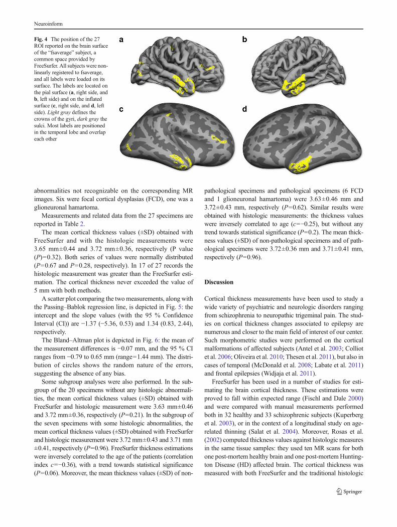
Fig. 4 The position of the 27ROI reported on the brain surfaceof the “fsaverage” subject, acommon space provided byFreeSurfer. All subjects were non-linearly registered to fsaverage,and all labels were loaded on itssurface. The labels are located onthe pial surface (a, right side, andb, left side) and on the inflatedsurface (c, right side, and d, leftside). Light gray defines thecrowns of the gyri, dark gray thesulci. Most labels are positionedin the temporal lobe and overlapeach other
Neuroinform

technique at different locations, and the results agreed towithin 0.2 mm (mean difference 0.077 mm). This study,performed in the context of a longitudinal study regardingcortical thinning in HD, confirmed the high accuracy of theautomated thickness estimation, but it relied on post-mortemand ten repeatedMR scans of two specimens from two brains.
The findings of the present study indicate a good agreementbetween FreeSurfer estimates and histologic measurements ofthe cortical thickness. These results derive from the statisticalanalysis performed with two methods specifically developedfor method-comparison studies. Passing–Bablok linear regres-sion is a non-parametric analysis, more appropriate thanPearson’s product-moment correlation test for method compar-ison studies (Passing and Bablok 1983, 1984). The intercept of
the regression line assesses the systematic bias: a CI below zerodenotes a constant underestimation trend whereas values overzero denote an overestimation trend. In the present study the CIincludes zero, suggesting that there is not any significant sys-tematic bias. The slope of the regression line assesses theproportional bias: in the present study its CI includes 1, indi-cating a good agreement between the two methods. We com-pared the two methods also computing the Bland–Altman plot.The mean of the differences is −0.07, indicating once more agood agreement between the two methods. This graphicaltechnique shows that the 95 % CI ranges from −0.79 to0.65 mm, including the perfect identity line (where the differ-ence is 0). The two methods are obviously not clinical alterna-tives, because histologic measurement cannot be part of the
Table 2 Measurements and related data
ROIID
SubjectID
Specimen topography Specimenpathology
Site ofmeasurement
Vertices Area(mm2)
FreeSurfer(mm ± SD)
Histology(mm ± SD)
Histology—FreeSurfer (mm)
1 1 Anterior T2 None Crown 332 400 3.44±0.51 3.82±0.41 0.39
2 2 Middle T2 None Crown 172 248 3.38±0.37 3.77±0.11 0.39
3 3 Anterior T2–T3 FCD I Crown 906 798 3.45±0.61 3.61±0.27 0.16
4 4 Anterior T2 None Crown 89 168 3.87±0.52 3.94±0.14 0.08
5 5 O2 FCD IIb Crown 165 143 3.06±0.42 3.18±0.23 0.12
6 6 Temporal pole None Crown 82 225 4.47±0.47 4.48±0.25 0.01
6 bis 6 Anterior T2 None Crown 70 167 4.60±0.39 4.38±0.25 −0.227 7 Anterior T2 None Crown 132 200 3.69±0.28 4.03±0.27 0.35
8 8 Temporal pole None Crown 360 355 3.26±0.42 3.49±0.20 0.23
9 9 Gyrus angularis None Sulcus 215 156 2.99±0.43 2.83±0.26 −0.1610 10 Middle T2 None Crown-Sulcus
Transition266 331 3.45±0.45 3.98±0.38 0.53
11 11 Temporal pole None Crown 221 182 3.38±0.39 3.58±0.27 0.21
12 12 Anterior/middle F2 None Crown-SulcusTransition
557 719 2.81±0.61 3.60±0.40 0.80
13 13 Middle T2 FCD IIb Crown 158 260 4.27±0.46 4.38±0.15 0.11
14 14 Middle parietal ascendinggyrus—anterior P2
Glioneuronalhamartoma
Crown-SulcusTransition
91 118 3.76±0.54 3.60±0.33 −0.16
15 15 Anterior T3 None Crown 88 125 3.49±0.23 3.58±0.21 0.10
16 16 Temporal pole None Crown 212 257 3.18±0.42 3.32±0.37 0.14
17 17 Anterior T2–T3 None Crown 146 188 3.81±0.40 3.69±0.20 −0.1218 18 Anterior T2–T3 None Crown 98 125 3.85±0.24 3.48±0.13 −0.3819 19 Anterior T3 None Crown 120 215 3.77±0.63 3.59±0.39 −0.1820 20 Middle T2 FCD I Crown 72 124 3.87±0.33 3.68±0.15 −0.1921 21 Anterior T3 None Crown 73 149 4.26±0.37 3.64±0.28 −0.6222 22 Anterior T3 None Crown 65 101 3.84±0.40 3.61±0.23 −0.2323 23 Anterior T1 FCD I Crown 212 332 3.48±0.47 4.13±0.25 0.66
24 24 Anterior T3 None Crown 58 84 3.58±0.26 3.88±0.39 0.30
25 25 Anterior T3–T4 FCD IIa Crown 66 126 4.17±0.49 3.42±0.62 −0.7526 26 Middle T3 None Crown 74 86 3.42±0.37 3.75±0.36 0.33
The median number of vertices per specimen (and relative measurements) is 132 (InterQuartile range 78–213.5)
FCD are classified according to Blümcke et al. (2011). T1 = Superior Temporal Gyrus. T2 =Middle Temporal Gyrus. T3 = Inferior Temporal Gyrus. T4= Fusiform Gyrus. O2 = Middle Occipital Gyrus. P2 = Inferior Parietal Lobule. The reported histological diagnosis is specifically related to thespecimens used for measurements. Histologic measurements were obtained ten times for each ROI, and the mean ± SD is reported. ROI 6 and 6bis wereobtained both from subject 6
Neuroinform

presurgical diagnostic evaluation. Nevertheless, our findingscontribute to validate the FreeSurfer estimates, especially ifwe consider certain specific pathological conditions, such asFCD. In fact, the 95 % CI range is 1.44 mm, a value that doesnot appear relevant when estimating regional thickness changesrelated to the presence of FCD. Colliot et al. reported in 2006the comparison of cortical thickness values (MR manual mea-surements) between 23 FCD affected subjects and 39 healthycontrols. They found that 56 % of FCD subjects had thicknessvalues greater than 6 mm, while normal subjects never
exceeded the value of 4.5 mm. In the present study the differ-ence between the two methods lies within the 1.44 mm range,probably small enough to avoid obscuring eventual true differ-ences between clinical groups. We therefore are encouraged touse FreeSurfer in the future to investigate the morphometricproperties of FCD and its usefulness for detecting dysplasticlesions that are difficult to see on conventional MR visualinspection.
Mean cortical thickness values obtained in our study weregenerally higher with histologic measurements. This findingagrees with our expectations because the manual measure-ments on histologic slices tend to give higher values due to thedifficulty of drawing the rulers perfectly orthogonal to the pialsurface. This source of error is likely partially compensated bythe tissue shrinkage after formaline fixation.
Seven of the 27 specimens analyzed in this study presentedunexpected, MR-occult pathologies in the area chosen formorphometric evaluation. The mean cortical thickness ofthese seven specimens did not differ from non-pathologicalspecimens, but it must be argued that the very small samplesize lowers the power of the analysis. Moreover, this analysiscompared normal specimens with subtle FCD-affected speci-mens, and there is still lack of information regarding morpho-metric properties of these MR-undetectable lesions.
The difference between FreeSurfer and histologicmeasurements is slightly greater than reported by Rosaset al. in 2002. This is mainly due to the different experimentalsetup: our measures were obtained in standard clinical condi-tions, while they performed ten MR scans on post-mortemfixed brains.
The major strength of our study is that, to the best of ourknowledge, it is the only one that validates FreeSurfer corticalthickness estimates computed from a single MR scan persubject, obtained in vivo and in conventional diagnostic con-ditions, against ex vivo histologic measurements obtainedfrom the same cases at same locations.
The major limitation of this study is the biased topographyof examined brain tissue, with most samples resected from thelateral aspect of the temporal lobe. Another potential limita-tion is that only one MR data set was obtained and processedfor every subject, due to time constraints during the acquisi-tion aimed at diagnostic evaluation. In fact, it is a generalrecommendation that more than one acquisition is used withFreeSurfer, increasing the signal-to-noise ratio and reducingthe effect of motion artifacts. Nevertheless, the processingresulted in high quality surface reconstructions (Fig. 1).Moreover, the FreeSurfer thickness was not verified athistology on a vertex-by-vertex basis, but it must be consid-ered that, given how small the ROI are, this does not invalidatethe conclusions.
To further explore the clinical usefulness in epilepsy sur-gery, we are planning to measure cortical thickness withFreeSurfer in a large series of histologically proven FCD.
3.0 3.5 4.0 4.5
3.0
3.5
4.0
4.5
Passing-Bablok Regression
FreeSurfer estimation (mm)
His
tolo
gic
mea
sure
(m
m)
Fig. 5 Scatter plot with Passing–Bablok regression line. The graphshows the good agreement between FreeSurfer estimates and histologicmeasurements of cortical thickness
3.0 3.5 4.0 4.5
-0.5
0.0
0.5
Bland-Altman Plot
(Histologic measure + FreeSurfer estimation)/2 (mm)
Fre
eSur
fer
estim
atio
n -
His
tolo
gic
mea
sure
(m
m)
MEAN-0.07
+ 2 SD0.65
- 2 SD-0.79
Fig. 6 Bland–Altman plot. The perfect identity line (black line) and allbut two plots are included in the 95 % Confidence Interval (dotted lines),suggesting a good agreement between the two methods
Neuroinform

Conclusions
There is a good agreement between FreeSurfer estimationsand histologic measurements of cortical thickness. FreeSurfercan be considered a reliable tool for this kind of computation,and it could be helpful in the presurgical workup of manypathological conditions such as FCD in epileptic patients.
Information Sharing Statement
FreeSurfer (RRID:nif-0000-00304) software is publicly andfreely available from the FreeSurferWiki resource (http://surfer.nmr.mgh.harvard.edu/fswiki/FreeSurferWiki), which isdeveloped and maintained at the Martinos Center forBiomedical Imaging (http://www.nmr.mgh.harvard.edu/martinos/noFlashHome.php). All software, information andsupport are provided online at the FreeSurferWiki webpage.
Acknowledgments We would like to thank Roberto Spreafico forhelping us in editing the Materials and Methods section, and Steve Gibbsfor reviewing the report. Moreover, we would like to thank GianfrancoDe Gregori and his coworkers for their invaluable contribution to biblio-graphic research.
Disclosures The Authors have nothing to disclose.
References
Antel, S. B., Collins, D. L., Bernasconi, N., Andermann, F., Shinghal, R.,Kearney, R. E., et al. (2003). Automated detection of focal corticaldysplasia lesions using computational models of their MRI charac-teristics and texture analysis. NeuroImage, 19(4), 1748–1759.
Bland, J. M., & Altman, D. G. (1986). Statistical methods for assessingagreement between two methods of clinical measurement. TheLancet, 327(8476), 307–310.
Blümcke, I., Thom, M., Aronica, E., Armstrong, D. D., Vinters, H. V.,Palmini, A., et al. (2011). The clinicopathologic spectrum of focalcortical dysplasias: a consensus classification proposed by an ad hocTask Force of the ILAE Diagnostic Methods Commission.Epilepsia, 52(1), 158–174.
Cardinale, F., Miserocchi, A., Moscato, A., Cossu, M., Castana, L.,Schiariti, M. P., et al. (2012). Talairach methodology in the multi-modal imaging and robotics era. In J.-M. Scarabin (Ed.), Stereotaxyand epilepsy surgery (pp. 245–272). Montrouge: John LibbeyEurotext.
Cardinale, F., Cossu, M., Castana, L., Casaceli, G., Schiariti, M. P.,Miserocchi, A., et al. (2013). Stereoelectroencephalography: surgi-cal methodology, safety, and stereotactic application accuracy in 500procedures. Neurosurgery, 72(3), 353–366.
Colliot, O., Antel, S. B., Naessens, V. B., Bernasconi, N., & Bernasconi,A. (2006). In vivo profiling of focal cortical dysplasia on high-resolution MRI with computational models. Epileptic Disorders:International Epilepsy Journal with Videotape, 47(1), 134–142.
Colombo, N., Tassi, L., Deleo, F., Citterio, A., Bramerio, M., Mai, R.,et al. (2012). Focal cortical dysplasia type IIa and IIb: MRI aspects
in 118 cases proven by histopathology. Neuroradiology, 54(10),1065–1077.
Dale, A. M., Fischl, B., & Sereno, M. I. (1999). Cortical surface-basedanalysis. I. Segmentation and surface reconstruction. NeuroImage,194, 179–194.
Fischl, B. (2012). FreeSurfer. NeuroImage, 62, 774–781.Fischl, B., & Dale, A. M. (2000). Measuring the thickness of the human
cerebral cortex frommagnetic resonance images. Proceedings of theNational Academy of Sciences of the United States of America,97(20), 11050–11055.
Fischl, B., Liu, A. K., & Dale, A. M. (2001). Automated manifoldsurgery: constructing geometrically accurate and topologically cor-rect models of the human cerebral cortex. IEEE Transactions onMedical Imaging, 20(1), 70–80.
Gronenschild, E. H. B. M., Habets, P., Jacobs, H. I. L., Mengelers, R.,Rozendaal, N., van Os, J., et al. (2012). The effects of FreeSurferVersion, Workstation Type, and Macintosh Operating SystemVersion on anatomical volume and cortical thickness measurements.PLoS ONE, 7(6), e38234.
Kuperberg, G. R., Broome, M. R., McGuire, P. K., David, A. S., Eddy,M., Ozawa, F., et al. (2003). Regionally localized thinning of thecerebral cortex in schizophrenia. Archives of General Psychiatry,60(9), 878–888.
Labate, A., Cerasa, A., Aguglia, U., Mumoli, L., Quattrone, A., &Gambardella, A. (2011). Neocortical thinning in “benign” mesialtemporal lobe epilepsy. Epilepsia, 52(4), 712–717.
McDonald, C. R., Hagler, D. J., Ahmadi, M. E., Tecoma, E., Iragui, V.,Gharapetian, L., et al. (2008). Regional neocortical thinning inmesial temporal lobe epilepsy. Epilepsia, 49(5), 794–803.
Oliveira, P. P. D. M., Valente, K. D., Shergill, S. S., Leite, C. D. C., &Amaro, E. (2010). Cortical thickness reduction of normal appearingcortex in patients with polymicrogyria. Journal of Neuroimaging:Official journal of the American Society of Neuroimaging, 20(1),46–52.
Passing, H., & Bablok, W. (1983). A new biometrical procedure fortesting the equality of measurements from two different analyticalmethods. Application of linear regression procedures for methodcomparison studies in clinical chemistry, part I. Journal of ClinicalChemistry and Clinical Biochemistry. Zeitschrift für KlinischeChemie und Klinische Biochemie, 21, 709–720.
Passing, H., & Bablok, W. (1984). A new biometrical procedure fortesting the equality of measurements from two different analyticalmethods. Application of linear regression procedures for methodcomparison studies in clinical chemistry, part II. Journal of ClinicalChemistry and Clinical Biochemistry. Zeitschrift für KlinischeChemie und Klinische Biochemie, 22, 431–445.
R Development Core Team (2012). R: A language and environment forstatistical computing. R Foundation for Statistical Computing,Vienna, Austria. ISBN 3-900051-07-0, URL http://www.R-project.org/. Accessed 01 Nov 2012.
Rosas, H. D., Liu, A. K., Hersch, S. M., Glessner, M., Ferrante, R. J.,Salat, D. H., et al. (2002). Regional and progressive thinning of thecortical ribbon in Huntington’s disease. Neurology, 58, 695–701.
Salat, D. H., Buckner, R. L., Snyder, A. Z., Greve, D. N., Desikan, R. S.R., Busa, E., et al. (2004). Thinning of the cerebral cortex in aging.Cerebral Cortex, 14(7), 721–730.
Thesen, T., Quinn, B. T., Carlson, C., Devinsky, O., DuBois, J.,McDonald, C. R., et al. (2011). Detection of epileptogenic corticalmalformations with surface-based MRI morphometry. PLoS ONE,6(2), 1–10.
Widjaja, E., Mahmoodabadi, S. Z., Snead, O. C., Almehdar, A., & Smith,M. L. (2011). Widespread cortical thinning in children with frontallobe epilepsy. Epilepsia, 52(9), 1685–1691.
Neuroinform
Copyright © 2022 FDOKUMEN