Hydrogen sulfide downregulates the aortic L-arginine/nitric oxide pathway in rats

v-Jun downregulates the SPARC target gene by binding to the proximal
promoter indirectly through Sp1/3
Sandrine Chamboredon1, Joseph Briggs2, Emmanuel Vial1, Julien Hurault1, Federico Galvagni3,Salvatore Oliviero3, Timothy Bos2 and Marc Castellazzi*,1
1Unite de Virologie Humaine, INSERM-U412, Ecole Normale Superieure, 46 allee d’ltalie, 69364 Lyon cedex 07, France;2Department of Microbiology and Molecular Cell Biology, Eastern Virginia Medical School, PO Box 1980, Norfolk, VA 23501,USA; 3Dipartimento di Biologia Molecolare, Universita degli Studi di Siena, via Fiorentina 1, 53100 Siena, Italy
Transformation of chick embryo fibroblasts by the v-Junoncoprotein correlates with a downregulation of theextracellular matrix protein SPARC and repression ofthe corresponding mRNA. Repression of SPARC con-tributes to the oncogenic process by facilitating tumordevelopment in vivo. A proximal promoter fragment,designated �124/þ 16, is responsible for high constitutiveactivity of the SPARC gene and is the target of repressionby v-Jun. In this paper, using electrophoretic mobilityshift and pull-down assays in vitro, and transienttransfections and chromatin immunoprecipitation assaysin Sp1/3-deficient Drosophila SL2 cells and in chickembryo fibroblasts, we show that (i) Sp1 and/or Sp3 isrequired for constitutive activation of SPARC transcrip-tion, by binding directly to the GGA-rich �92/�57fragment; and (ii) v-Jun does not bind �124/þ 16directly, but binds to the GGA-rich fragment indirectly,most likely through a physical interaction with Sp1/3.Moreover, a transactivation-proficient v-Jun derivative,designated v-Jun/cebp/glz, which cannot bind Jun DNAmotifs anymore and cannot heterodimerize, is still capableof downregulating SPARC efficiently. Taken together,these data strongly suggest that v-Jun downregulatesSPARC through the formation of a DNA–Sp1/3–v-Jun,chromatin-associated complex.Oncogene (2003) 22, 4047–4061. doi:10.1038/sj.onc.1206713
Keywords: cell transformation; v-Jun; Sp1; Sp3;SPARC; primary chick embryo fibroblasts
Introduction
Cell transformation by transcription factor Jun isgenerally assumed to take place through the activationor repression of a limited number of direct target genes.The characterization of these targets constitutes animportant issue in order to decipher the oncogenicpathways downstream of this transcription factor.Direct targets are expected to contain a functionalJun-binding DNA element in their promoter andconstitute a critical intermediate in cell transformation
and tumorigenesis (Vogt et al., 1999; van Dam andCastellazzi, 2001; Vogt, 2001).Studies from several laboratories led to the character-
ization of candidate targets in avian cells (Basso et al.,2000; Vial and Castellazzi, 2000; Bader et al., 2001) aswell as in murine (Mettouchi et al., 1994; Fu et al., 2000)and human (Rinehart-Kim et al., 2000) cells. Althoughdifferent techniques were used for screening, the samestatistics was observed, that is, 2/3 of the genes areactivated by Jun and 1/3 are repressed. Several genesalready satisfy the conditions of direct targets and areactivated during transformation, such as toj3, a geneencoding heparin-binding EGF-like growth factor, andjac (Fu et al., 2000; Bader et al., 2001; Hartl et al., 2001),or are repressed, such as c-jun and p53 (Hussain et al.,1998; Piu et al., 2002).We have recently characterized several putative
targets repressed by v-Jun in chick embryo fibroblasts(CEFs) (van Dam and Castellazzi, 2001). Among these,repression of the SPARC gene has been shown tofacilitate tumorigenesis in vivo (Vial and Castellazzi,2000). The encoded product SPARC (secreted protein,acidic, and rich in cysteine) is a minor component of theextracellular matrix. Its sequence is highly conservedduring evolution, from Caenorhabditis elegans to chick-en, mouse, cow, and man. Although its precise functionis poorly understood, SPARC is generally considered asa regulator of matrix–cell interactions, such as throm-bospondin and tenascin (Bornstein, 1995; Brekken andSage, 2000). SPARC has independently been isolated asa target of mammalian c-Jun in primary rat embryofibroblasts (Mettouchi et al., 1994) and in the humanbreast cancer cell line MCF7 (Rinehart-Kim et al.,2000). Its expression is altered in most human cancers,including breast (Bellahcene and Castronovo, 1995),prostate (Jacob et al., 1999), ovarian (Mok et al., 1996),and colorectal (Porte et al., 1995) cancer, and melanoma(Ledda et al., 1997).Previous studies from our laboratory (Vial et al.,
2000) also showed that in v-Jun-transformed CEFs,repression of SPARC takes place within a short,proximal promoter fragment, designated �124/þ 16.This minimal promoter reproduces the behavior of theendogenous SPARC, and constitutes a reliable modelfor analysis of transcriptional regulation by v-Jun.Received 24 January 2003; revised 28 April 2003; accepted 28 April 2003
*Correspondence: M Castellazzi; E-mail: [email protected]
Oncogene (2003) 22, 4047–4061& 2003 Nature Publishing Group All rights reserved 0950-9232/03 $25.00
www.nature.com/onc

Intriguingly, the minimal �124/þ 16 promoter displaysno consensus Jun-binding site, raising the interestingquestion of how this oncoprotein represses SPARCtranscription.
Results
Sp1/3(-like) proteins bind the minimal �124/þ 16SPARC promoter in vitro
We have previously shown (Vial et al., 2000) that inCEF cells, a short, proximal fragment of the avianSPARC promoter, designated �124/þ 16, displays over80% of the high basal activity and responds torepression by v-Jun and, more efficiently, by v-Jun-ml(a mutant that exhibits enhanced tumorigenicity in vivo;Huguier et al., 1998). The organization of this promoterfragment displays the following main features: (i) noTATA and CAAT box; (ii) a single major transcrip-tional start point; (iii) a purine-rich region with a coresequence of eight GGA direct repeats in a row (referredto as the ‘GGA box’ hereafter); and (iv) no potentialTPA-responsive (TRE) or cAMP-responsive (CRE)Jun-binding AP1 site (Figure 1a).A first series of experiments was designed to
characterize protein complexes that bind �124/þ 16 invitro. Uninfected CEF cultures as well as CEF cultureschronically infected by the nontransforming virus R, orthe transforming viruses R-v-Jun and R-v-Jun-ml weregenerated and nuclear extracts prepared for electro-phoresis mobility shift analysis. As shown in Figure 1b,using �124/þ 16 as a radioactive probe, a pattern offour discrete bands was found. The correspondingprotein complexes were designated Cl, C2, C3, andC4, according to their increasing mobilities. The closelylinked C2 and C3 corresponded to the most intensebands, whereas C4 was of much lower intensity, and C1a faint, sometimes barely visible band of the highestmolecular weight (Figure 1b–d).To determine which fragment(s) of �124/þ 16 bind(s)
the various complexes, a competition experiment wascarried out with an excess of cold probe 1–7 (Figure 1a)and nuclear extracts from R-v-Jun-infected CEFs. Asshown in Figure 1c, probe 3, which extends from �92 to�57 and encompasses the GGA box, eliminated all fourcomplexes. Probe 4 (�74/�38), which partially overlapsprobe 3, specifically retained C4. The other probesaffected only poorly, if not at all, the differentcomplexes. Thus, this experiment clearly indicated thatthe �92/�57 sequence is required for the binding of allfour complexes. The same result was also obtained withthe other nuclear extracts (data not shown).Recent reports (Ihn et al., 1997; Tone et al., 2000)
suggested that the purine-rich repeats of the GGA boxmight bind members of the Sp1 family of transcriptionfactors. Therefore, attempts to supershift the differentbands were performed using antibodies specificallydirected against avian proteins Sp1 and Sp3, and nuclearextracts from R-v-Jun-infected CEFs (Figure 1d). Re-markably, antibodies against Sp1 eliminated C1 and C3,
whereas antibodies against Sp3 eliminated C1 and C2.C4 was not affected by any of the anti-Sp antibodiestested. An identical result was obtained with the othernuclear extracts (data not shown). These data indicatedthat the ubiquitous transcription factors Sp1 and Sp3(or closely related family members) contribute to the C1,C2, and C3 complexes, but not C4. This conclusion wasconfirmed by a competition experiment with a cold Sp1consensus, GC-rich probe to which these transcriptionfactors bind with high affinity (Figure 1c).In a second series of experiments, in vitro prepared
avian Sp1 and Sp3 (isolated in the course of this work)were tested for their ability to bind to probe �124/þ 16.As shown in Figure 2a, a single retarded band wasobtained with Sp1 and Sp3, in a pattern that wasreminiscent of the C2 and C3 bands obtained with thenuclear extracts (Figure 1b–d). Interestingly, bandsequivalent to C1 and C4 were not observed, supportingthe hypothesis that C1 consists of a complex with Sp1/3and (an) additional protein(s) and that C4 does notcontain any Sp1/3(-like) protein. Finally, as expected,direct binding of in vitro prepared Sp1 and Sp3 wasdemonstrated on the GGA-rich probe 3 and not, forinstance, on probe 5. This binding was however reducedin comparison with that on the consensus GC-richprobe for Sp1 (Figure 2c and Discussion).In conclusion, the gel shift experiments presented
above allowed us to draw the following conclusions: (i)four retarded complexes, designated C1–C4, bind �124/þ 16 in vitro; (ii) the binding of all four complexesrequires the �92/�57 region which includes the GGAbox; (iii) three high-molecular weight complexes Cl, C2,and C3 contain Sp1/3(-like) transcription factors thatare likely to bind DNA directly.
v-Jun does not bind the minimal �124/þ 16 SPARCpromoter directly in vitro
As already stated, computer analysis revealed noconsensus or related Jun-binding motif in �124/þ 16.In agreement with this, we confirmed that in vitroprepared v-Jun and v-Jun-ml did not bind to this probedirectly (Figure 2a), even in the additional presence ofSp1 or/and Sp3 (or/and in the presence of dimerizationpartners like c-Fos and ATF2; Vial and Castellazzi,unpublished result). However, both Jun proteins wereshown to bind to a consensus AP1 probe efficiently(Figure 2b).The presence of v-Jun in the retarded complexes
described above (C1–C4) was also investigated. Competi-tion assays with a cold consensus AP1 probe (Figure 1c)as well as antibody supershift assays with anti-c/v-Junantibodies (Figure 1d) were performed. They revealed nogross change in the pattern of the retarded complexes.
Sp1 and Sp3 activate transcription of the minimal �124/þ 16 SPARC promoter in Drosophila SL2 cells
We next wanted to know whether avian Sp1 and Sp3 arerequired for transcriptional activation in vivo. To dothis, we chose to analyse their effect in the Drosophila
v-Jun-mediated repression through Sp1/3S Chamboredon et al
4048
Oncogene

embryonic SL2 cell line (Schneider, 1972), which lackshomologs of mammalian transcription factors Sp1 andSp3 (Courey and Tjian, 1988; Santoro et al., 1988).Transient transfection experiments were performed. The�124/þ 16 promoter was placed in front of theluciferase gene and the Sp1 and Sp3 genes wereexpressed under the control of the ubiquitous Drosophilaactin AC5 promoter.First, increasing amounts of Sp1 and Sp3 expression
vectors were cotransfected with �124/þ 16 (and �56/þ 16 as a control). As shown in Figure 3a, there was nodetectable activity in the absence of Sp1 and Sp3,
whereas, independently, these factors activated thepromoter in a dose-dependent manner. The reducedactivation at the highest concentration of Sp1 might bedue to the sequestration of a specific coactivator.Second, the activation of �124/þ 16 was compared
to that of �124 DGGA/þ 16 deleted from the �92/�57sequence containing the GGA box. As shown inFigure 3b, the activity of the deleted form was stronglyreduced. Taken together, these results indicated that (i)the avian Sp proteins efficiently activate the minimalSPARC promoter and that (ii) this activation requiresthe GGA box region of the promoter.
7
+1+16-124
12
34
56
AGCGAGGAGGAGGAGGAGGAGGAGGAGGAGAGGAAA
-92 -56 -21-57
GGA box
CCCAGCCCACACTTTGCCTTCCTGCTGCCTCCTTCT
a
1 2 3 4 5 6 7 Sp1 AP1 Ets
competition with cold probe
NI R vJ m1
C1
C2
C3
C4
Sp1 Sp3 Sp1+3 Jun
C1
C2
C3
C4
_
vJ nuclear extracts vJ nuclear extracts
- + - + - + - +
antibodies againstnuclear extracts
b c d
Figure 1 Electrophoretic mobility shift assays using nuclear extracts from nontransformed CEFs and CEFs stably transformed by v-Jun, and the 32P-labeled �124/þ 16minimal promoter fragment as a probe. (a) Schematic representation of the minimal �124/þ 16promoter fragment and overlapping probes 1–7 covering the entire promoter. The sequences of probes 3 and 5 are shown. (b) Nuclearextracts from noninfected (NI), R-infected (R), R-v-Jun-infected (vJ), and R-v-Jun-ml-infected (ml) CEFs were used. Arrowheadsshow the retarded bands that correspond to protein complexes C1–C4. (c) Competition experiments were carried out with cold probes1–7, cold consensus probe for Sp1/3 (sense: 50-ATTCGATCGGGGCGGGGCGAGC) (Sp1), cold consensus probe for AP1 (sense: 50-AGCTAGCATGAGTCAGACAC) (AP1), and cold nonrelevant, Ets probe (sense: 50-TCGGGCTCGAGATAACCAG-GAAGTGGGC) (Ets); � indicates no probe. (d) Supershift experiments were performed using rabbit polyclonal antibodies directedagainst avian Sp1, Sp3, and v/c-Jun. � and þ correspond to pre-immune serum and anti-serum of the same rabbit, respectively.Nuclear extracts in (c) and (d) were from R-v-Jun-infected CEFs
v-Jun-mediated repression through Sp1/3S Chamboredon et al
4049
Oncogene

v-Jun represses Sp1- and Sp3-induced transcription inSL2 cells
We next asked whether v-Jun can repress Sp1- and Sp3-mediated transcription in this heterologous system. Todo this, the activity of �124/þ 16 was assessed in SL2cells that were cotransfected by Sp1 or Sp3, and by v-Jun or v-Jun-ml at two different concentrations. Asshown in Figure 4a, in the absence of Sp proteins,neither v-Jun nor v-Jun-ml affected the promoter
activity significantly. In contrast, when Sp proteins werepresent, both Jun proteins repressed the promoter in adose-dependent manner.Two control experiments were also performed. First,
a transfection was carried out with Jun and the 1� coll-tata promoter in front of a luciferase gene. This artificialpromoter contains a single consensus Jun-binding sitefrom the human collagenase gene and a tata box (vanDam et al., 1998). As seen in Figure 4b, both Jun werestrong activators, showing unambiguously that theseoncoproteins were transcriptionally fully active and,consequently, that the absence of activation withoutSp1/3 and the repression with Sp1/3 were promoter-specific phenomena. Second, the accumulation of Sp1and Sp3 was tested by Western blotting followed byimmunodetection with specific antibodies. As shown inFigure 4f, no significant differences were observedbetween these proteins under the various cotransfectionconditions. This ruled out the possibility that theobserved repression might be due to a reducedaccumulation of the Sp transcription factors in thepresence of Jun. Taken together, the above data clearlyshowed that repression by v-Jun and v-Jun-ml, pre-viously reported in CEFs, can also be established inheterologous SL2 cells.
v-Jun-mediated repression does not require direct andspecific DNA binding in SL2 cells
Experiments were designed to rule out the possibilitythat v-Jun(-ml) requires direct and specific DNAbinding to establish repression in the presence of Sp1/3. To this aim, we took advantage of a v-Jun-derivative,
- vJ m1 - vJ m1 - vJ m1 - vJ m1
Sp1 Sp3 Sp1+Sp3
- vJ m1
probe AP1probe -124/+16
in vitro made proteins
- Sp1 Sp3 Sp1+Sp3
probe Sp1 probe 3 probe 5
- Sp1 Sp3 Sp1+Sp3
- Sp1 Sp3 Sp1+Sp3
a b
c
Figure 2 Electrophoretic mobility shift assays using in vitrosynthesized chicken proteins. (a) Binding of Sp1, Sp3, and a 1 : 1mixture of Sp1 and Sp3, in the presence or not of v-Jun or v-Jun-ml. The arrowheads indicate the retarded bands. (b) Binding of v-Jun (vJ) and v-Jun-ml (ml) on the consensus AP1 probe. (c)Binding of Sp1, Sp3, and a 1þ 1 mixture of Sp1 and Sp3 on theconsensus Sp1 probe, and on probes 3 and 5 (see Figure 1 andlegend)
0
10
20
30
40
- -
-124/+16
-124∆GGA/+16
b
Sp1 Sp3
0
10
20
30
40
50
60
70
Rel
ativ
e lu
cife
rase
act
ivity
-
a
.25 .5 1 2 .25 .5 1 2
Sp1 Sp3
-124/+16sparc
Figure 3 Sp-mediated transactivation of the minimal SPARCpromoter (5mg/plate) in Drosophila SL2 cells. (a) Effect of variousconcentrations (mg/plate) of Sp1 and Sp3, as indicated. (b)Comparison of the wild type �124/þ 16 and the deleted �124DGGA/þ 16 promoter lacking the GGA box (5mg/plate, each)with Sp1 and Sp3 (0.25 mg/plate, each)
v-Jun-mediated repression through Sp1/3S Chamboredon et al
4050
Oncogene

0
2
4
6
8
10
12
14
16
Rel
ativ
e l
ucife
rase
ac
tivity
- vJun m1 vJun m1-
Sp1
.25 .5 .25 .5
vJun m1-
Sp3
.25 .5 .25 .5.5 .50
2
4
6
8
10
12
14
16
18
.25 .5
Jun m1
.25 .5
-124/+16sparc
1x coll tata
-
a b
0
5
10
15
20
25
30
35
40
Rel
ativ
e l
ucife
rase
act
ivity
glz glzcebp/glz
cebp/glz
glz cebp/glz
Sp1 Sp3
.25.5 .5
-124/+16sparc
- -
0
5
10
15
20
25
30
35
40
45
1× colltata
0
5
10
15
20
25
30
35
40
45
1× cebptata
- glz cebp/glz
- glz cebp/glz
-
.5 .25 .5 .25 .5 .25 .5 .25 .5 .25 .5 .25 .5 .25 .5
c d e
- vJun m1 vJun m1 vJun m1 vJun(iv)
Sp1 Sp3
anti-Jun
40
28.4
kDa
40
28.4
anti-Jun
- glz glz cebp-glz
cebp-glz
glzglz(iv)
cebp-glzkDa
121.3
85.9
anti-Sp1
Sp1
- - vJun m1 glz cebp/glz Sp1(iv)kDa
121.3
85.9
anti-Sp3
Sp3
kDa- - vJun m1 glz cebp
/glz Sp3(iv)
Sp1 Sp3
f
Figure 4 Sp-mediated transactivation of the minimal SPARC promoter (5mg/plate) in Drosophila SL2 cells and repression by v-Junand derivatives. Sp1 and Sp3 were always transfected at 0.25mg/plate in each experiment and v-Jun and derivatives were transfected atthe indicated concentration (mg/plate). (a) Effect of v-Jun and v-Jun-ml alone, and in the presence of Sp1 or Sp3. (b) Controlexperiment: effect of v-Jun and v-Jun-ml on a 1� coll-tata minimal promoter containing a consensus AP1 site. (c) Same experiment asin (a) with v-Jun/glz and v-Jun/cebp/glz. (d) and (e) Control experiments: effect of v-Jun/glz and v-Jun/cebp/glz on 1� coll-tata and on1� cebp-tata containing a c/EBP consensus site. (f) Control experiment: Western blots showing the accumulation of Sp1, Sp3, and v-Jun and derivatives in the transfected extracts. Antibodies specifically directed against the different proteins were used, as indicated.Arrows indicate the corresponding proteins, (iv) In vitro prepared proteins. In this particular experiment and in the presence of Sp3, asignificantly higher accumulation of v-Jun-ml was observed in comparison with v-Jun and this might have contributed to theabnormally strong repression by v-Jun-ml at 0.25mg/plate in (a)
v-Jun-mediated repression through Sp1/3S Chamboredon et al
4051
Oncogene
named v-Jun/cebp (Basso et al., 2000), in which thebasic DNA-binding domain of Jun was replaced by theDNA-binding domain of c/EBP. Like v-Jun, c/EBPbelongs to the b-zip family of transcription factors butbinds a distinct, unrelated DNA motif (50-ATTGCG-CAAT instead of the consensus AP1 motif 50-TGACT-CA) (Johnson, 1993; Suckow et al., 1993). Interestingly,v-Jun/cebp has been shown to heterodimerize with avianJun and Fos to generate heterodimers that bind both toan AP1 and a c/EBP motif in vitro (Basso et al., 2000)and this might also happen with Drosophila Jun and Fos(Kockel et al., 2001). In fact, to eliminate this possibility,a v-Jun/cebp-derivative, designated v-Jun/cebp/glz (Bas-so et al., 2000), was used in which the naturaldimerization domain of v-Jun was replaced by thehomodimerization domain of GCN4 (glz for GCN4leucine zipper). This transcription factor is anothermember of b-zip family isolated in yeast that bindsDNA strictly as a homodimer and cannot formstable dimers with any known Jun and Fos proteins(Hughes et al., 1992; Oliviero et al., 1992; Castellazziet al., 1993).Transactivation experiments similar to those pre-
sented in Figure 4a, b were conducted with v-Jun/cebp/glz (and v-Jun/glz as a control). The followingresults were obtained: (i) in the absence of Sp proteins,neither v-Jun/glz nor v-Jun/cebp/glz activated theSPARC promoter; (ii) in the presence of Sp proteins,the two v-Jun derivatives repressed the promoter in adose-dependent manner, reaching values close tothose obtained with v-Jun (Figure 4c); (iii) the twov-Jun derivatives were still capable of activatingtranscription, however on distinct DNA-binding ele-ments; indeed, whereas v-Jun/glz specifically activated1� coll-tata (Figure 4d), v-Jun/cebp/glz activated1� cebp-tata, a minimal promoter with a single c/EBPmotif (Figure 4e); and finally, (iv) no obvious differencesin the accumulation of Sp1/3 were observed in thevarious cotransfections (Figure 4f). Taken together,these data strongly suggested that v-Jun-mediatedrepression does not require any specific DNA bindingof the oncoprotein. They further showed that, at leastin SL2 cells, heterodimerization with another b-zippartner is not required for this repression to takeplace.
v-Jun and v-Jun-ml physically interact with Sp1 and Sp3in vitro
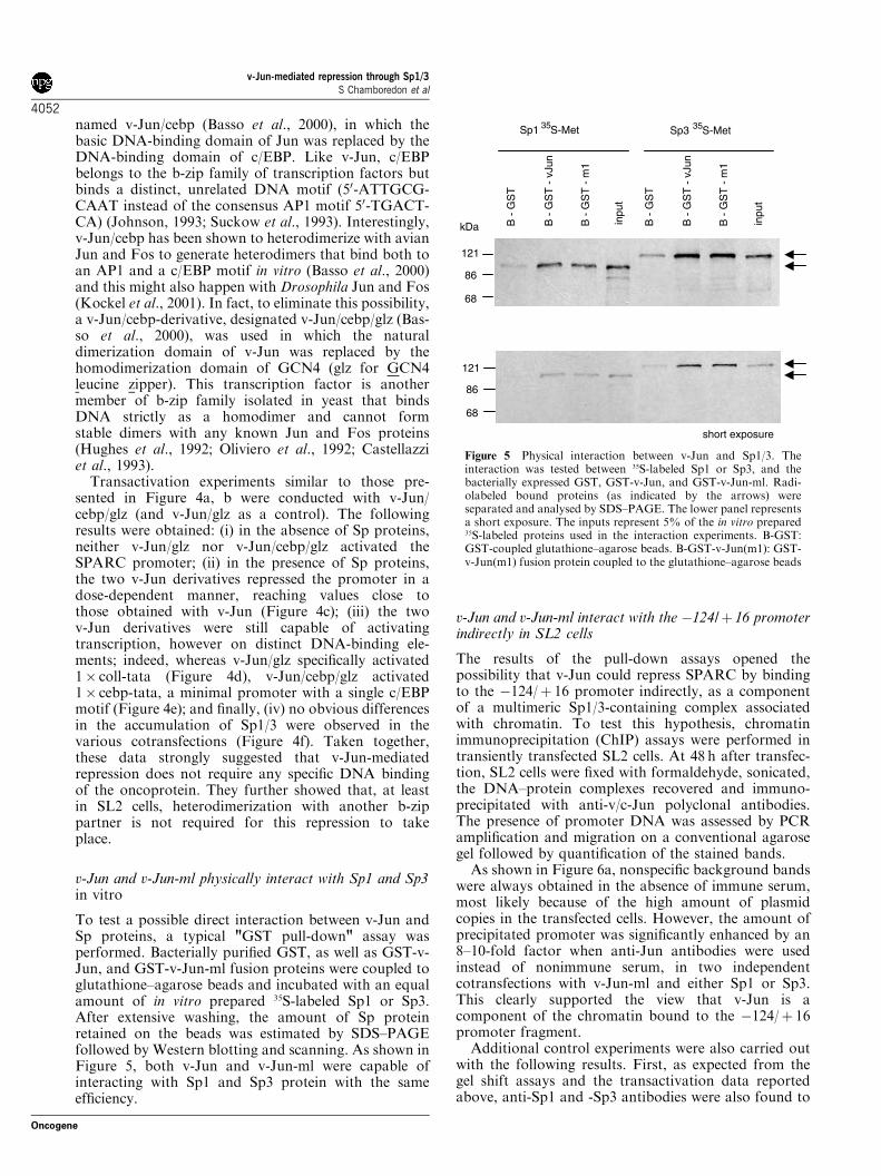
To test a possible direct interaction between v-Jun andSp proteins, a typical "GST pull-down" assay wasperformed. Bacterially purified GST, as well as GST-v-Jun, and GST-v-Jun-ml fusion proteins were coupled toglutathione–agarose beads and incubated with an equalamount of in vitro prepared 35S-labeled Sp1 or Sp3.After extensive washing, the amount of Sp proteinretained on the beads was estimated by SDS–PAGEfollowed by Western blotting and scanning. As shown inFigure 5, both v-Jun and v-Jun-ml were capable ofinteracting with Sp1 and Sp3 protein with the sameefficiency.
v-Jun and v-Jun-ml interact with the �124/þ 16 promoterindirectly in SL2 cells
The results of the pull-down assays opened thepossibility that v-Jun could repress SPARC by bindingto the �124/þ 16 promoter indirectly, as a componentof a multimeric Sp1/3-containing complex associatedwith chromatin. To test this hypothesis, chromatinimmunoprecipitation (ChIP) assays were performed intransiently transfected SL2 cells. At 48 h after transfec-tion, SL2 cells were fixed with formaldehyde, sonicated,the DNA–protein complexes recovered and immuno-precipitated with anti-v/c-Jun polyclonal antibodies.The presence of promoter DNA was assessed by PCRamplification and migration on a conventional agarosegel followed by quantification of the stained bands.As shown in Figure 6a, nonspecific background bands
were always obtained in the absence of immune serum,most likely because of the high amount of plasmidcopies in the transfected cells. However, the amount ofprecipitated promoter was significantly enhanced by an8–10-fold factor when anti-Jun antibodies were usedinstead of nonimmune serum, in two independentcotransfections with v-Jun-ml and either Sp1 or Sp3.This clearly supported the view that v-Jun is acomponent of the chromatin bound to the �124/þ 16promoter fragment.Additional control experiments were also carried out
with the following results. First, as expected from thegel shift assays and the transactivation data reportedabove, anti-Sp1 and -Sp3 antibodies were also found to
inpu
t
B -
GS
T -
vJu
n
B -
GS
T -
m1
B -
GS
T
inpu
t
B -
GS
T -
vJu
n
B -
GS
T -
m1
B -
GS
T
Sp1 35S-Met Sp3 35S-Met
kDa
121
86
68
short exposure
121
86
68
Figure 5 Physical interaction between v-Jun and Sp1/3. Theinteraction was tested between 35S-labeled Sp1 or Sp3, and thebacterially expressed GST, GST-v-Jun, and GST-v-Jun-ml. Radi-olabeled bound proteins (as indicated by the arrows) wereseparated and analysed by SDS–PAGE. The lower panel representsa short exposure. The inputs represent 5% of the in vitro prepared35S-labeled proteins used in the interaction experiments. B-GST:GST-coupled glutathione–agarose beads. B-GST-v-Jun(m1): GST-v-Jun(m1) fusion protein coupled to the glutathione–agarose beads
v-Jun-mediated repression through Sp1/3S Chamboredon et al
4052
Oncogene

precipitate the promoter. Second, an independent trans-fection with 1� coll-tata and v-Jun-ml also confirmedthe direct binding of Jun to this artificial promoter.Third, the total sonicated input DNA was estimated bythe amplification of a fraction (1/25) of the DNA beforeimmunoprecipitation. As expected, the total input wasfound to be the same in the different experiments. Bycomparing the intensities of the amplified bandsrecovered from the input and the immunoprecipitatedDNA, the amount of immunoprecipitated promoter wasestimated to be in the range of 1–4% of the total inputDNA, a value in agreement with independent data (EManet and A Sergeant, unpublished result).Finally, a ChIP assay was conducted with a C-
terminal, HA-tagged v-Jun, designated v-JunHA, in thepresence or not of transfected Sp1 or Sp3. As shown inFigure 6b, a significant increase of promoter recoverywas obtained with anti-HA antibodies, but only whenSp1 or Sp3 was cotransfected with v-JunHA.The results obtained so far, in particular those in the
SL2 system, invited us to propose a working hypothesisin which v-Jun and the various derivatives v-Jun-ml, v-Jun/glz, and v-Jun/cebp/glz repress SPARC by bindingto the �124/þ 16 promoter DNA indirectly throughSp1/3 (Figure 7 and Discussion).
Sp1/3-mediated transcriptional control, repression byv-Jun/cebp/glz, and indirect binding of v-Jun also takeplace in CEF cells
Experiments were conducted in CEFs to support themodel in Figure 7. First, cotransfections of �124/þ 16with Sp1, Sp3, or DN Sp1 were carried out. (DN Sp1 is atruncated form of Sp1, which lacks the serine/threonine-
input
Sp1 Sp3
-124/+16 sparc+ m1
1× coll tata+ m1
0.4
kb
1× coll tata + m1
kb
0.4
_ J0 J92anti-Jun
0.4
kbJ0 J0J74 J92
anti- Sp1 anti- Jun
-124/+16 sparc + Sp1 + m1
J0 J0J74 J92
anti- Sp3 anti- Jun
-124/+16 sparc + Sp3 + m1
0.4
kb
_ __+ + +
Sp1 Sp3
kb input
anti- HA
_
-124/+16 sparc + v-JunHA
Sp1 Sp3_
-124/+16 sparc + v-JunHA
kb
0.40.4
a
b
Figure 6 ChIP assays in SL2-transfected cells. A 380-base-pair plasmid fragment containing the entire �124/þ 16 SPARC promoterwas amplified by PCR from input chromatin, immunoprecipitated chromatin with specific antibodies or control antibodies, andvisualised by ethidium bromide staining on a 1.5% agarose gel. (a) The minimal promoter was cotransfected with an expressionplasmid for Sp1 or Sp3, and v-Jun-ml, as indicated. A control experiment with 1 x coll-tata and expression plasmid for v-Jun-ml is alsopresented; the amplified PCR product is 435-base-pair long and contains the entire minimal promoter. Anti-Sp/J0 and anti-Sp/J74 arepolyclonal sera at day 0 of immunization (control serum) and at day 74 (immune serum) from the same rabbit, respectively. Anti-v-Jun/J0 and anti-vJun/J92 are control serum and immune serum from the same rabbit, respectively. The same samples were used in thesupershift experiment in Figure 1c. (b) The SPARC promoter was cotransfected with expression plasmid for v-JunHA alone (�), v-JunHA and Sp1 (Sp1), or v-JunHA and Sp3 (Sp3), as indicated. Immunoprecipitation was performed with a mouse monoclonal anti-HA antibody diluted in PBS (þ ) and control sample was with PBS alone (�). In both panels, inputs represent PCR amplification from4% of the fragmented, nonimmunoprecipitated chromatin
+1+16-124
probe 3(GGA box)
-92 -57
Sp1 / Sp3
v-Jun
otherfactor(s)
repression
SPARC promoter
Figure 7 Working hypothesis. In this hypothesis, v-Jun acts as acorepressor on SPARC transcription by binding to the GGA boxindirectly via Sp1/3, and (an)other factor(s)
v-Jun-mediated repression through Sp1/3S Chamboredon et al
4053
Oncogene

and glutamine-rich transactivation domains, but retainsthe DNA-binding domain. It was found to bind Sp1sites efficiently in vitro and to counteract the transacti-vation induced by Sp1 and Sp3 in SL2 cells; data not
shown). As shown in Figure 8a, the constitutively highpromoter activity could be activated slightly by Sp1,more efficiently by Sp3, and repressed by the dominantnegative form of Sp1. This result reinforced the viewthat Sp1 and Sp3 are direct transcriptional regulators of�124/þ 16.Next, cotransfections of �124/þ 16 with v-Jun, v-
Jun-ml, and the mutated v-Jun/glz and v-Jun/cebp/glzwere also performed (Figure 8b). As expected, all fourconstructs repressed �124/þ 16, thus confirming thatthis repression phenomenon does not require specificDNA binding of Jun to an AP1 (-like) site (orheterodimerization with (an)other b-zip partner(s) pre-sent in CEFs). As already reported (Vial et al., 2000;Basso et al., 2000; Huguier et al., 1998), the differentmutants accumulated like v-Jun in CEFs. Moreover,they did not affect the accumulation of endogenous Sp1and Sp3 (data not shown; Figure 11).Indirect binding of v-Jun to �124/þ 16 was also
confirmed using the ChIP assay. Two physiologicalsituations were considered. In Figure 9a, a transientcotransfection of �124/þ 16 and v-Jun-ml was per-formed with normal CEFs. As expected, anti-Junantibodies (as well as anti-Sp1 and anti-Sp3) werecapable of precipitating �124/þ 16. In Figure 9b,transient transfection of �124/þ 16 was carried out inJun-transformed CEFs, chronically infected by either R-v-Jun or R-v-JunHA. Both viruses were shown toexpress the corresponding oncoprotein at an identicalhigh level, and were potent transforming retroviruses, asjudged from their capacity to establish a typicallyaltered, fusiform morphology and to stimulate cellularproliferation (data not shown) (Bos et al., 1990;Castellazzi et al., 1990). As expected, polyclonal anti-Jun antibodies could precipitate the promoter fromeither transformed cell line, whereas monoclonal anti-HA could specifically precipitate the promoter from R-v-JunHA-transformed CEFs. This latter result, togetherwith the fact that in v-Jun-transformed CEFs c-Jun wasdownregulated (Bos et al., 1990; Hussain et al., 1998)(Figure 11), demonstrated the predominant (if notexclusive) presence of the oncoprotein in the precipi-tated chromatin complex.As all ChIP assays presented up to now were based on
transient transfections, we wanted to confirm someresults with the endogenous SPARC gene. We thereforechose to reproduce the results in Figure 9a and b, usingthe same DNA samples, but a PCR amplifying the�203/þ 50 promoter sequence not present in the pGL3�124/þ 16 plasmid. As expected, antibodies againstJun, Sp1, and Sp3 precipitated the endogenous sequenceefficiently (Figure 10a, b). Curiously, anti-HA anti-bodies were poorly efficient, a result that might reflectsome minor change in the promoter-associated chroma-tin complexes between plasmid and cellular DNA.
Downregulation of SPARC takes place in R-v-Jun/cebp/glz-infected, nontransformed CEF cells
The model in Figure 7 predicted that repression of theendogenous SPARC promoter can be established by a
0
20
40
60
80
100
120
140
160
_ m1 glz cebp/glz
-124/ +16sparc
100%
29%33%
51%
38%
0
50
100
150
200
250
300
350
400
Rel
ativ
e lu
cife
rase
act
ivity
Rel
ativ
e lu
cife
rase
act
ivity
-
-124/+16sparc
162%
100%
475%
37%
Sp1 Sp3 ∆N Sp1
vJun
a
b
Figure 8 Transient transfections in CEF cells. (a) Sp-mediatedtransactivation of the SPARC promoter (3mg/plate), activation bySp1 (3mg/plate) and Sp3 (3mg/plate), and repression by thedominant negative DN Sp1 (3mg/plate). (b) Repression of theSPARC promoter (1mg/plate) by v-Jun, v-Jun-ml, v-Jun/glz, and v-Jun/cebp/glz (0.5mg/plate, in each case)
v-Jun-mediated repression through Sp1/3S Chamboredon et al
4054
Oncogene

direct interaction between v-Jun and Sp1/3. We there-fore wondered whether v-Jun/cebp/glz, the Jun doublemutant that cannot bind AP1 sites, cannot heterodimer-ize, and cannot transform (Basso et al., 2000) (data notshown), would be capable of repressing the endogenousSPARC in chronically infected CEFs. To answer thisquestion, cell extracts from either noninfected CEFs or
CEFs infected by R-, R-v-Jun, R-v-Jun-ml, and R-v-Jun/cebp/glz were analysed for the steady-state accu-mulation of SPARC. As shown in Figure 11, cellsexpressing v-Jun/cebp/glz indeed displayed a reducedaccumulation of SPARC. This reduction however wasnot as efficient as with v-Jun and v-Jun-ml. It isinteresting to note that in the same experiment, incontrast to v-Jun and v-Jun-ml, the double mutantcould not downregulate endogenous c-Jun. This is inagreement with the fact that, in this latter case,repression is established by a direct binding of the Junoncoprotein to the proximal Jun-binding site in the c-junpromoter (Hussain et al., 1998).
J0 J0J92 J92
R- vJun R- vJunHA
-124/+16 sparc in CEF cells transformed by
R- vJun R- vJunHA
input
0.4
kb
0.4
kb
anti-HA
R- vJun R- vJunHA
0.4
kb
anti-Jun
J0 J0 J0J92 J74 J74
anti- Sp3
-124/+16 sparc + m1
anti- Jun anti- Sp1
0.4
kb
input
0.4
kb
-124/+16 sparc + m1
a
b
Figure 9 ChIP assays in CEFs. A 380-base-pair plasmid fragmentcontaining the entire �124/þ 16 SPARC promoter was amplifiedby PCR from input chromatin, immunoprecipitated chromatinwith specific antibodies or control antibodies, and visualized byethidium bromide staining on a 1.5% agarose gel. For antibodiesand input, see legend of Figure 6. (a) The SPARC promoter wascotransfected with an expression plasmid for v-Jun-ml in primaryCEF cells. (b) The SPARC promoter was transfected intotransformed CEF cells stably infected by either Rcas-v-Jun orRcas-v-JunHA
J0 J0 J0J92 J74 J74
anti- Sp3
-124/+16 sparc + m1
anti- Jun anti- Sp1
J0 J0J92 J92
R- vJun R- vJunHA
-124/+16 sparc in CEF cells transformed by
anti-Jun
anti-HA
R- vJun R- vJunHA
R- vJun R- vJunHA
input
bp
bp
bp
bp
311
249
311
249
311
249
311
249
bp311
249
input
-124/+16 sparc + m1
a
b
Figure 10 ChIP assays in CEFs. A 253-base-pair fragmentcontaining the �203/þ 50 portion of the endogenous SPARCpromoter was amplified by PCR in the presence of traces of[a32P]dCTP. The radioactive PCR products were resolved on anondenaturing, 5% polyacrylamide gel. Experiments in panels aand b were performed with the DNA samples from experimentspresented in Figure 9a, b, respectively
v-Jun-mediated repression through Sp1/3S Chamboredon et al
4055
Oncogene

Discussion
The work presented in this paper was aimed at under-standing how the v-Jun oncoprotein and, more effi-ciently, the highly tumorigenic mutant v-Jun-ml, repressthe SPARC target gene in stably transformed CEF cells.Previous reports (Vial and Castellazzi, 2000; Vial et al.,2000) showed that both high constitutive activity innormal CEFs and reduced activity in v-Jun-transformedCEFs take place within a proximal promoter fragment,designated �124/þ 16, which otherwise does not seemto contain a classical Jun-binding element. We proposethat the data obtained from electrophoretic mobilityshift and pull-down assays in vitro and from transienttransfections and ChIP assays in SL2 and CEF cells arebest explained by a working hypothesis, illustrated inFigure 7, which stipulates the following points:
(i) Sp1 or Sp3 is required for constitutive activation ofthe �124/þ 16 promoter, by binding to the GGA-rich, �92/�57 fragment directly;
(ii) v-Jun and v-Jun-ml do not bind to the �124/þ 16promoter directly, but repress transcription bybinding to the �92/�57 sequence indirectly, andthis is likely to involve a direct physical interactionwith Sp1 or Sp3;
(iii) one or several additional, unknown factors con-tribute to the stabilization of the DNA–Sp1/3–v-Jun(-ml) chromatin-associate complex.This working hypothesis calls for several comments.
First, the GGA repeat constitutes an unusual DNA-binding motif for Sp1 and Sp3. Indeed, these transcrip-tion factors are generally considered as binding GC- andGT-rich motifs (Lania et al., 1997; Philipsen and Suske,1999). Moreover, gel shift assays with in vitro preparedavian Sp1 and Sp3 as well as nuclear extracts fromnormal and Jun-transformed CEFs indicated that theGGA-rich (�92/�57) probe 3 is less efficiently recog-nized in comparison to the Sp1 consensus probe(Figures 2c and 12). A reduced affinity for GGA motifshas also been reported with human Sp1 (Ihn et al., 1997;Tone et al., 2000) and is in agreement with the fact thatchicken Sp1 and Sp3 are highly homologous to thehuman factors, particularly in the zinc-finger, DNA-binding domain (Figure 13). Besides, it is interesting tonote that, as shown in Figure 12, nuclear extracts fromnontransformed R-infected, or from R-v-Jun(-ml)-transformed CEFs, displayed the same binding affinityto probe 3 (and this also is true for the Sp1 consensusprobe), showing that a reduced binding of Sp1/3 can-not explain the repression of SPARC mediated byv-Jun(-ml). In the same experiment, as expected, a slight,but clear increase of AP1 binding activity took place inthe Jun-transformed CEFs.Second, a purine-rich region with GGA repeats is
present in the TATA-less, homologous SPARC promo-ter from man. We have previously shown that a shortproximal fragment of the human promoter, designated�120/þ 28, homologous to the avian �124/þ 16 frag-ment and which does not contain any Jun consensus
anti-Jun
NI R vJ m1
anti-Sp1 86
121
NI R vJ m1
anti-Sp3
86
121
NI R vJ m1
40
cebp/glz(iv)
vJ(iv)
Sp3(iv)
Sp1(iv)
c-Jun
v-Jun
Sparc(iv)
49.5
37.4
anti-Sparc
NI R vJ m1
cebp/glz
cebp/glz
cebp/glz
cebp/glz
Figure 11 Western blots showing the steady-state level of variousproteins from cell extracts in noninfected CEF cultures (NI) and inCEF cultures chronically infected by R, R-vJun, R-v-Jun-ml, andR-v-Jun/cebp/glz. Antibodies directed against avian SPARC, c/v-Jun, Sp1, and Sp3 were used. Arrows indicate the correspondingproteins. (iv) In vitro prepared proteins. Note that vJ(iv) and cebp/glz(iv) are loaded equally but recognized differently by anti-Jun
R vJ m1R vJ m1R vJ m1R vJ m1
3 5 Sp1 AP1
probes
nuclear extracts
Figure 12 Electrophoretic mobility shift assays using nuclearextracts from R-infected nontransformed CEFs and CEFs stablytransformed by v-Jun or v-Jun-ml, and the 32P-labeled, probes 3and 5, consensus probe AP1, and consensus probe Sp1
v-Jun-mediated repression through Sp1/3S Chamboredon et al
4056
Oncogene

binding element, is responsible for repression bymammalian c-Jun in primary cultures of rat embryofibroblasts (Vial et al., 2000). Recent results in humanbreast cancer MCF7 cell line further showed that theactivity of the human promoter requires the purine-richsequence (within the �120/�83 fragment) and isdependent upon human Sp1 and Sp3 in DrosophilaSL2 cells (Briggs et al., 2002). These latter resultsstrongly suggest that constitutive transcription ofSPARC might be regulated by the same mechanism inavian and human cells. Furthermore, it is tempting tospeculate that the postulated ‘indirect’ activation of thehuman SPARC promoter by c-Jun (Briggs et al., 2002)might take place through a mechanism analogous tothat depicted in Figure 7.Third, indirect activation or repression of transcrip-
tion by a functional interaction between c-Jun and Sp1has been suggested to take place in mammalian cells, atthe level of Sp-controlled promoters regulating theexpression of the cell cycle inhibitor p21WAF1/Cip1 (Kar-dassis et al., 1999; Wang et al., 2000), the 12(S)-lipoxygenase (Chen and Chang, 2000), the neuronalnicotinic acetylcholine receptor (Melnikova and Gard-ner, 2001) and the cytosolic phospholipase A2 (Blaineet al., 2001). As also reported in the present study, noconvincing evidence of a DNA–Sp1(-like)–c-Jun com-plex in gel shift assays could be demonstrated, furthersupporting the existence of (an) additional, stabilizingcomponent(s) as suggested in the working hypothesis(Figure 7). In contrast to the present study, however, noChIP assay had been performed to demonstrate aphysical link between c-Jun, Sp1 (-like), and a specificpromoter segment in vivo.Fourth, positive versus negative control by Jun
through Sp1/3 might, among other parameters, dependupon the composition and activity of the pool of b-zipfactors in a given cell type. For example, in CEF cells, v-Jun-mediated repression of SPARC has been shown tobe exacerbated by excess Fra2 and antagonized byexcess ATF2; and most of the Fos, Jun, ATF, and Maffamily members known in CEFs have been shown toeither activate or repress SPARC transcription (Vialet al., 2000). Although we have shown in this paper thatv-Jun does not require heterodimerization with another
b-zip partner for repression and, thus, might function asa monomer or a homodimer in this process, one cannotexclude the possibility that a v-Jun-containing hetero-dimer influences SPARC transcription either directly, bybinding to Sp1/3, or indirectly, by a sequestrationmechanism. In this respect, it might be informative totest which of the avian AP1 components can physicallyinteract with Sp1/3.Finally, although SPARC does not contain any
functional Jun-binding DNA element in its promoter,it can nevertheless be considered – in a certain sense – asa ‘direct’ target of the v-Jun oncoprotein. This idea isbest documented in the present work by the down-regulation of the endogenous SPARC by the nontrans-forming derivative v-Jun/cebp/glz, in the absence of anyintermediate target product induced by v-Jun acting ona Jun-binding motif. In fact, this chimeric derivativeconstitutes an original tool for the characterization of afamily of target genes whose activity is modulated by adirect, physical interaction between v-Jun and Sp1/3family members some of which, at least, mightcontribute to the transformation process.
Materials and methods
Cell culture
Primary CEFs were prepared from 8-day old, virus-free, O-linechicken embryos (Institute for Animal Health, Compton,Berks, UK). They were grown in a regular medium made ofHam’s F10 medium (Eurobio) supplemented with 10%tryptose phosphate broth (Difco), 5% fetal calf serum(Eurobio) and 1% chicken serum (Sigma), 0.196% NaCO3,penicillin/streptomycin, and 1.25 g/ml amphotericin B at 371Cin a 5% CO2 atmosphere (Perez et al., 2001).v-Jun-expressing cultures were obtained by chronic infection
with the replication-competent retrovirus Rcas (Hugues et al.,1987). Rcas (denoted R), R-v-Jun and R-v-Jun-ml (denoted R-ml) (Vial and Castellazzi, 2000; Vial et al., 2000), and R-v-Jun/glz and R-v-Jun/cebp/glz (Hughes et al., 1992; Basso et al.,2000) have already been described. R-v-JunHA contains atagged v-Jun with the peptide sequence from the hemagglu-tinin protein of human influenza virus located downstream tothe last amino-acid Phe204 of the oncoprotein. The additionalsequence is as follows: GGC (Gly205) GCT(Ala) AGC(Ser)
Sp1(h) GKKKQHICHIQGCGKVYGKTSHLRAHLRWHTGER finger 1Sp1(ch) ----------P----------------------- Sp3(h) ----------P-------------------S--- Sp3(ch) ----------P-------------------S---
Sp1(h) PFMCTWSYCGKRFTRSDELQRHKRTHTGEK finger 2Sp1(ch) --I-G-ML---------------------- Sp3(h) --V-N-M---------------R------- Sp3(ch) --V-N-MF--------------R-------
Sp1(h) KFACPECPKRFMRSDHLSKHIKTHQNKK finger 3Sp1(ch) ---------------------------- Sp3(h) --V----S------------V------- Sp3(ch) --V----S------------V-------
Figure 13 Protein sequence alignment of the DNA binding, zinc-finger domains of Sp1 and Sp3 from human (h) and chicken (ch)origin. Dashes represent identical residues. The cystein and histidine residues that are involved in zinc coordination are in bold
v-Jun-mediated repression through Sp1/3S Chamboredon et al
4057
Oncogene
TAC(Tyr) CCT(Pro) TAT(Tyr) GAC(Asp) GTC(Val)CCC(Pro) GAT(Asp) TAC(Tyr) GCC(Ala) AGC(Ser)CTG(Leu) TCT(Ser) AGA(Arg220) TGA(stop).Routinely, transfections with R (no insert) and the various
R-v-Jun plasmids were performed after the first passage usingfugene-6 transfection reagent (Roche), and viruses wereallowed to spread through the entire population over thefollowing week. Transformation tests were carried out asalready described (Vial and Castellazzi, 2000).Drosophila SL2 cells were maintained in Schneider’s insect
medium (Invitrogen) supplemented with 10% fetal calf serum(Eurobio) and penicillin/streptomycin at 251C in a normalatmosphere (Schneider, 1972).
Isolation of the avian Sp1 and Sp3 cDNAs
The full-length cDNAs of chicken Sp1 and Sp3 were isolatedfrom an undifferentiated chicken embryonic stem cell cDNAlibrary by screening with a 32P-labeled human Sp1 and Sp3probe. The cDNA library was constructed by R Kunita in thelambda ZAP-II vector (Stratagene) (Acloque et al., 2001). TheSp1 and Sp3 avian cDNAs were excised from the phagemidvector as a pBluescript plasmid and sequenced. They weresubmitted to the EMBL nucleotide sequence database andgiven Accession numbers AJ317960 and AJ317961, respec-tively.The Sp1 and Sp3 cDNAs were recloned into pH, to generate
pH-Sp1 and pH-Sp3. pH is a pBluescript II SK(þ ) derivative(Stratagene) in which the SacI-to-SalI fragment from theoriginal polylinker has been modified. The new polylinkercontains the following primer/promoter sequences, restrictionsites, and poly(A) sequence in order: RP-BssHII-T3-SP6-NheI-ClaI-HindIII-PstI-SalI/AccI-XbaI-BamHI-SmaI-SacI-EcoRI-ClaI-poly(A)30-NotI-XhoI-ApaI-KpnI-T7-BssHII-M13. TheClaI–ClaI polylinker fragment is from the CLA12 adapterplasmid (Hugues et al., 1987). To allow efficient in vitro proteinsynthesis, the GC-rich 50 noncoding fragments present in Sp1and Sp3 cDNAs were removed and replaced by the sequenceAAGCTTCCG GGC ACC ATG (Met1) using a PCRexperiment nested between the upstream HindIII site and theunique site PflMI and NotI, respectively, located downstreamof the ATG codon. (The underlined sequences correspond tothe HindIII site downstream to the SP6 primer, and to theinitiation codon.) The new plasmid pH-DGC Sp1 contains a2368 nucleotide long sequence of avian Sp1 spanning from theinitiation codon ATG to the unique NotI site (blunt ended)present in the noncoding region, and cloned between HindIIIand SmaI in pH. The new plasmid pH-DGC Sp3 contains a2393 nucleotide long sequence of avian Sp3 spanning from theinitiation codon ATG to the unique EcoRV site present in the30 noncoding region, and cloned between HindIII and SmaI inpH.pH carrying the deleted, dominant negative form of Sp1
designated DN Sp1 was obtained by removing the HindIII-ApaLI N-terminal fragment by PCR, so that the sequencesurrounding the initiation site is AAGCTT CCG GGC ACCATG(Met1) AGC(Ser2) GTG(Val517) CAC(His518).
Electrophoretic mobility shift assay
To prepare nuclear extracts, nonconfluent CEF cells werewashed twice with ice cold PBS, collected by scraping, andcentrifuged at 1500 g for 5min at 41C in PBS. The pellet wasresuspended in four volumes of buffer I consisting of 10mm
HEPES pH 7.9, 10mm KC1, 1.5mm MgCl2, and antiproteases(Roche; cat n1 1 836 153). The cells were allowed to swell byincubation for 30min in ice, and the cell membranes disrupted
in a Dounce homogenizer (40 strokes). The resulting extractwas centrifuged at low speed (1500 g for 10min at 41C) and thesupernatant discarded. The pellet containing the nuclei waswashed with four volumes of buffer I and centrifuged oncemore at low speed. The pellet was then resuspended in 2.2volumes of buffer II (20mm HEPES pH 7.9, 420mm KC1,1.5mm MgCl2, 0.01mm ZnCl2, 0.2mm EDTA, 1mm DTT,25% glycerol, and antiproteases), incubated for 30min on arotary shaker at 41C to disrupt the nuclear membranes, andcentrifuged at high speed (15 000 g for 30min at 41C) toremove debris. The supernatant (¼ nuclear extract) wasdialyzed against buffer III (20mm HEPES pH 7.9, 120mm
KC1, 10mm MgCl2, 2mm EDTA, 35% glycerol, andantiproteases) using a Sephadex G50 column (Sigma),aliquoted, frozen in liquid nitrogen, and stored at �801C.[14C]leucine-labeled proteins were synthesized in vitro, using
a wheat germ extract coupled transcription/translation system(Promega) and dialyzed against buffer III.Radioactive nucleic probes were generated by [g32P]ATP end
labeling. The binding reaction was performed on ice for 45minin 25mm HEPES pH 7.9, 80mm KC1, 4.8mm MgCl2, 0.01mm
ZnCl2, 0.8mm EDTA, 22% glycerol, 6.6mm DTT, 0.66mg/mlpoly-dI-dC, 13.2% CHAPS, and antiproteases. Separation offree radiolabeled DNA from DNA–protein complexes wascarried out on 5% nondenaturating polyacrylamide gels in aTris-glycine-EDTA buffer supplemented with 2.5% glycerol.Gels were run for 6 h at 41C at a current of 7mA/gel.
Transient transfection and luciferase assay
SL2 cells were seeded at a density of 2� 106 in 2ml in a 35-mm-diameter plate and transfected 24 h later using a standardprotocol for calcium phosphate-mediated transfection foradherent cells (Sambrook et al., 1989) with HEPES-bufferedsaline adjusted to pH 7.0. The next day, the medium was gentlyremoved and replaced with fresh medium. Cell lysates wereprepared 48 h after transfection, and luciferase activitymeasured as for CEF cells (see below). In these Drosophilacells, the various sp and jun sequences were expressed from theactin AC5 promoter sequence in a pAc5.1/V5-HisA (Invitro-gen).Plasmids pGL3 carrying the avian SPARC promoter
fragments �124/þ 16, �124 DGGA/þ 16, and �56/þ 16 infront of the luciferase gene have been described previously(Vial and Castellazzi, 2000). pGL2 derivatives carrying the tataand 1� coll-tata promoters in front of the luciferase gene wereobtained from Hans van Dam (van Dam et al., 1993). ThepGL2 derivative carrying the 1� cebp-tata promoter fragmentwas constructed by replacing the Jun-binding element 50-TGACTCA in 1� coll-tata by the consensus DNA-bindingelement 50-ATTGCGCAAT of the c/EBP transcription factor(Johnson, 1993; Suckow et al., 1993). The various minimalpromoter fragments are inserted at the unique HindIII site inthe pGL2 derivatives. The sequence of the HindIII–HindIII1� cebp-tata fragment is as follows: AAGCTTGATT GCGCAATCTG GGATCCAGAT CTCTCTGAGC AATAGTA-TAA AACTCGAGAT CTAAGTAAGCTT.Typically, a transfection experiment in SL2 cells included
5mg of reporter plasmid per Petri dish and various amounts ofpAc5.1/V5-HisA expression vectors normalized to 7mg withthe empty expression vector. The relative luciferase activityrepresents the ratio SPARC-luciferase/�56/þ 16-luciferase or1� coll (or cebp)-tata/tata-luciferase, with each point corre-sponding to the average value of three independentlytransfected dishes in the same experiment.CEF cells were seeded at a density of 5� 105 per 60-mm-
diameter plate in normal medium and transfected 24 h later
v-Jun-mediated repression through Sp1/3S Chamboredon et al
4058
Oncogene

using fugene-6 transfection reagent (Roche). Cell lysates wereprepared 48 h after transfection, and luciferase activitymeasured by using the ‘luciferase assay system’ (Promega).In these avian primary cells, the various sp and jun sequenceswere expressed from the Rous sarcoma virus long terminalrepeat sequence in a pDP plasmid (Vandel et al., 1995).
Antibodies
To generate antibodies specifically directed against avian Sp1,the entire coding sequence was cloned in-frame into pGEX-3T-4 (Pharmacia). To generate antibodies specifically againstavian Sp3, a truncated sequence encoding the N-terminalportion of Sp3 and covering amino acids Met1 to Ser356 wasalso cloned in-frame into pGEX-3T-4. The GST-Sp fusionproteins were prepared as described hereafter in the ‘GST pull-down assay’ section. Rabbits were immunized by repeatedintradermal injection of the purified protein in accordance witha standard technique (Covalab, Lyon, France). Rabbitpolyclonal anti-v-Jun and anti-SPARC antibodies havealready been described (Vial and Castellazzi, 2000). Commer-cial mouse monoclonal anti-HA was purchased from RocheDiagnostics (cat no. 1583816) and was directed against the HApeptide sequence (YPYDVPDYA) from the hemagglutininprotein of human influenza virus.
Western blotting
Preparation of CEF extracts for Western blotting, proteinseparation on SDS–PAGE, and immunodetection wereperformed as previously described (Huguier et al., 1998; Vialand Castellazzi, 2000; Vial et al., 2000). For transfected SL2cells, extracts in ‘cell culture lysis reagent’ were directly usedfor SDS–PAGE (‘luciferase assay system’; Promega).
GST pull-down assay
The experiments were conducted according to Kardassis et al.(1999) with some modifications. The GST and GST fusionproteins were expressed in Escherichia coli BL 21. Bacteriawere grown overnight, diluted 1/50 in a final volume of 50ml,and after reaching an A600 of 0.5, were stimulated with 0.1mm
isopropyl-b-d-thiogalactopyranoside for 3 h at 371C. Bacteriawere harvested, washed and resuspended in 2ml of cold PBS,and sonicated for 4� 15 s on ice. The extracts were lysed by theaddition of Triton X-100 (1% final concentration) and anti-proteases, homogenized by a 30-min incubation at 41C on arotary shaker, aliquoted and stored at �201C. The recovery ofthe expressed GST proteins was monitored by SDS–PAGEand Coomassie blue staining.Glutathione–agarose beads (# G4510; Sigma) were equili-
brated overnight in distilled water on a rotary shaker at 41C.The beads were centrifuged (1500 g at 41C for 2min) andresuspended in cold PBS. The beads were also saturated by afurther incubation in PBS supplemented with 10% (w/v)defatted milk powder and 1% bovine serum albumin for 1 h,washed, resuspended as a 1 : 1 bead slurry in PBS, and kept at41C until use.For the GST interaction assay, the bead slurry was first
incubated with four volumes of bacterially expressed GST andGST fusion proteins on a rotary shaker for 2.5 h at 41C. Thebeads were then washed three times with 10 volumes of PBS,and equilibrated in washing buffer (20mm HEPES pH 7.9,0.1m KC1, 5mm MgCl2, 0.2% Nonidet P-40, and antipro-teases). 50ml of bead slurry was combined with 2–10ml of a[35S]methionine-labeled reticulocyte lysate (Promega) in a finalvolume of 200 ml of washing buffer on a rotary shaker for 2 h at41C. Finally, the beads were washed five times with 20 volumes
of washing buffer, and the bound proteins were eluted byboiling in Laemmli loading buffer and subjected to SDS/10%PAGE. Bound proteins were blotted onto a nitrocellulosemembrane and visualized by autoradiography.
Chromatin immunoprecipitation (ChIP) assay
The experiments were essentially conducted according toCastro-Rivera et al. (2001) with some modifications. For eachcondition, CEF cells (0.5� 106/60mm dish; six dishes) wereplated overnight and transiently transfected for 48 h by �124/þ 16 SPARC-luciferase (and by expression vectors for Sp1,Sp3, and v-Jun, as indicated in the text). Formaldehyde wasadded to the medium to give a 1% solution and plates werefurther incubated for 10min at 371C. The culture medium wasremoved and the cells washed with PBS supplemented withantiproteases, recovered by scraping, pooled, and pelleted bycentrifugation. (At this stage the pellets could be frozen inliquid nitrogen and stored at �801C).The cells were resuspended and homogenized in 300 ml of
ice-cold SDS-lysis buffer (1% SDS, 10mm EDTA, and 50mm
Tris-HC1 pH 8.0), and sonicated to obtain chromatin with thedesired fragment length (200–1000 base pairs). The extract wascentrifuged at 15 000 g for 10min at 41C to eliminate thedebris, the supernatant recovered and diluted 10-fold in ChIPdilution buffer (0.01% SDS, 1.1% triton X-100, 1.2mm
EDTA, 167mm NaCl, 16.7mm Tris-HCl pH 8.0, andantiproteases). Volumes of 1ml aliquots were made (as wellas a 40 ml sample, designated ‘input sample’, kept at 41C forlater use). Preclearing was done with 40ml protein A/G–agarose (sc#2003, Santa-Cruz; equilibrated in ChIP dilutionbuffer) for 1 h at 41C on a rotary shaker, followed bycentrifugation (1500 g for 5min at 41C). The supernatant(1ml) was incubated overnight with the appropriate antibodieson the rotary shaker at 41C. The immunoprecipitatedcomplexes were recovered by addition of another 40ml ofprotein A/G – agarose (equilibrated in the same ChIP dilutionbuffer supplemented with 100 mg/ml of sonicated salmonsperm DNA), and incubation for 1 h at 41C on a rotary shaker.The agarose beads were then centrifuged and successively
washed with 1ml of low-salt (150mm NaCl) and high-salt(500mm NaCl) immune complex buffer (0.1% SDS, 1%Triton X-100, 2mm EDTA, and 20mm Tris-HCl pH 8.0),followed by LiCl-containing buffer (0.25m LiCl, 1% NP40,1% sodium deoxycholate, 1mm EDTA, and 10mm Tris-HClpH 8.0), and by TE buffer (1mm EDTA, and 10mm Tris-HClpH 8.0). At this stage, the pelleted beads were resuspended in200 ml of elution buffer (1% SDS, 0.1m NaHCO3), incubatedfor 15min at room temperature on a rotary shaker,centrifuged, and the supernatant containing the crosslinkedchromatin complexes recovered. The elution was repeatedonce and the 2� 200ml supernatants pooled. The crosslinkedmaterial from the 400 ml supernatant (as well as the 40 ml‘input’ sample) was then dissociated by addition of NaCl(192mm, final concentration) and incubation for 4 h at 651C.Inactivation of DNAse and RNAse activities was carried outby addition of proteinase K (36 mg/ml), EDTA (10mm), andTris-HCl (36mm pH 6.5) and incubation for 1 h at 451C.Deproteinization was then carried out by phenol/chloroformextraction followed by chloroform extraction. DNA wasrecovered by ethanol precipitation in the presence of 13 mg/ml glycogen and 0.2m sodium acetate. After a final wash with70% ethanol, the pellet was resuspended in 50 ml of distilledwater. PCR was used to detect the presence of the �124/þ 16SPARC promoter region immunoprecipitated with theanti-Sp1, anti-Sp3, anti-v-Jun, or anti-HA antibodies. Theamount of fragmented input DNA was estimated by PCR
v-Jun-mediated repression through Sp1/3S Chamboredon et al
4059
Oncogene

amplification (23–25 cycles) of an aliquot representing 4% ofthe immunoprecipitated sample. The primers used were sequencesfrom the pGL3 plasmid flanking the promoter region, in thesense orientation: 50-pAGTGCAAGTGCAGGTGCCAGAAC,and in the antisense orientation: 50-pGCTCTCCAGCGGTT-CCATCTTCC. The 380-base-pair PCR products were visua-lized on conventional 1.5% agarose gels using a tris acetate/EDTA electrophoresis buffer and ethidium bromide staining.Quantification of the stained bands was performed using anImageQuant software from Molecular Dynamics.In some experiments, PCR (35 cycles in the presence of a
trace amount of [a32P] dCTP) was also used to detect theendogenous SPARC promoter. The input was estimated byamplification of an aliquot representing 0.4% of the immuno-precipitated samples. The primers used were sequences notpresent in the pGL3 �124/þ 16 plasmid, in the senseorientation: 50-pTCTGTCCGGTTGACTTCTCTGGC, andin the antisense orientation: 50-pACCTCGTAGTCCCGAG-CAGG. The 253-base-pair PCR products corresponding to the�203/þ 50 promoter fragment were run on a nondenaturing,5% polyacrylamide gel in a Tris acetate/EDTA buffer. The air-dried gel was then exposed for 2 h at �801C and quantificationwas carried out using a Storm 850 apparatus from MolecularDynamics.ChIP assays with Drosophila SL2 cells were conducted as for
CEF cells, with the following two changes: (i) 2� 106 cells/
35mm plates and six plates were used for a given condition; (ii)formaldehyde treatment for 15min at 251C and the scrapedcells pooled and resuspended in 400 ml of ice-cold lysis buffer.
Abbreviations
CEF, chicken embryo fibroblast; SPARC, secreted protein,acidic, and rich in cysteine; AP1, activating protein 1; ChIPassay, chromatin immunoprecipitation assay.
Acknowledgements
This work was supported by grants from the Association pourla Recherche sur le Cancer (ARC; no. 9707 and no. 4398 toMarc Castellazzi), from the European Economic CommunityBiomedicine and Health Program (Biomed II contract no.BMH4 CT98 3505 to Marc Castellazzi and SalvatoreOliviero), from the Associazione Italiana Ricerca sul Cancro(AIRC; to Salvatore Oliviero), and from the Thomas F andKate Miller Jeffress Foundation (to Timothy Bos). SandrineChamboredon was supported by fellowships from the BiomedII contract and the ARC. We thank Joel Baguet for theconstruction of the pBluescript II SK(þ )-derived plasmid pH,Bertrand Pain for the gift of the avian cDNA library used toscreen the Sp1 and Sp3 coding sequences, and Hans van Damfor the gift of the pGL2-derived tata and 1� coll-tataplasmids.
References
Acloque H, Risson V, Birot A, Kunita R, Pain B and SamarutJ. (2001). Mech. Dev., 103, 79–91.
Bader A, Schneider M, Bister K and Hartl M. (2001).Oncogene, 20, 7524–7535.
Basso J, Briggs J, Findlay C and Bos T. (2000). Oncogene, 19,4876–4885.
Bellahcene A and Castronovo V. (1995). Am. J. Pathol., 146,95–100.
Blaine S, Wick M, Dessev C and Nemenoff R. (2001). J. Biol.Chem., 276, 42737–42743.
Bornstein P. (1995). J. Cell. Biol., 130, 503–506.Bos TJ, Monteclaro FS, Mitsunobu F, Ball AR, Chang CHW,Nishimura T and Vogt PK. (1990). Genes Dev., 4, 1677–1687.
Brekken RA and Sage EH. (2000). Matrix Biol., 19, 816–827.Briggs J, Chamboredon S, Castellazzi M, Kerry J and Bos T.(2002). Oncogene, 21, 7077–7091.
Castellazzi M, Dangy JP, Mechta F, Hirai SI, Yaniv M,Samarut J, Lassailly A and Brun G. (1990). Oncogene, 5,1541–1547.
Castellazzi M, Loiseau L, Piu F and Sergeant A. (1993).Oncogene, 8, 1149–1160.
Castro-Rivera E, Samudio I and Safe S. (2001). J. Biol. Chem.,276, 30853–30861.
Chen B and Chang W. (2000). Proc. Natl. Acad. Sci. USA, 97,10406–10411.
Courey A and Tjian R. (1988). Cell, 55, 887–898.Fu S, Waha A and Vogt P. (2000). Oncogene, 19, 3537–3545.Hartl M, Reiter F, Bader A, Castellazzi M and Bister K.(2001). Proc. Natl. Acad. Sci. USA, 98, 13601–13606.
Hughes M, Sehgal A, Hadman M and Bos T. (1992). CellGrowth Differ., 3, 889–897.
Hughes SH, Greenhouse CJ, Petropoulos CJ and Sutrave PS.(1987). J. Virol., 61, 3004–3012.
Huguier S, Baguet J, Perez S, van Dam H and Castellazzi M.(1998). Mol. Cell. Biol., 18, 7020–7029.
Hussain S, Kilbey A and Gillespie D. (1998). Cell GrowthDiffer., 9, 677–686.
Ihn H, LeRoy E and Trojanowska M. (1997). J. Biol. Chem.,272, 24666–24672.
Jacob K, Webber M, Benayahu D and Kleinman H. (1999).Cancer Res., 59, 4453–4457.
Johnson P. (1993). Mol. Cell. Biol., 13, 6919–6930.Kardassis D, Papakosta P, Pardali K and Moustakas A.(1999). J. Biol. Chem., 274, 29572–29581.
Kockel L, Homsy J and Bohmann D. (2001). Oncogene, 20,2347–2364.
Lania L, Majello B and De Luca P. (1997). Int. J. Biochem.Cell. Biol., 29, 1313–1323.
Ledda F, Bravo AI, Adris S, Bover L, Mordoh J andPodhajcer OL. (1997). J. Invest. Dermatol., 108,
210–214.Melnikova I and Gardner P. (2001). J. Biol. Chem., 276,19040–19045.
Mettouchi A, Cabon F, Montreau N, Vernier P, Mercier G,Blangy D, Tricoire H, Vigier P and Binetruy B. (1994).EMBO J., 13, 5668–5678.
Mok SC, Chan WY, Wong KK, Muto MG and Berkowitz RS.(1996). Oncogene, 12, 1895–1901.
Oliviero S, Robinson G, Struhl K and Spiegelman B. (1992).Genes Dev., 6, 1799–1809.
Perez S, Vial E, van Dam H and Castellazzi M. (2001).Oncogene, 20, 1135–1141.
Philipsen S and Suske G. (1999). Nucleic Acids Res., 27, 2991–3000.
Piu F, Aronheim A, Katz S and Karin M. (2001). Mol Cell.Biol., 21, 3012–3024.
Porte H, Chastre E, Prevot S, Nordlinger B, Empereur S,Basset P, Chambon P and Gespach C. (1995). Int. J. Cancer,64, 70–75.
Rinehart-Kim J, Johnston M, Birrer M and Bos T. (2000). Int.J. Cancer, 88, 180–190.
v-Jun-mediated repression through Sp1/3S Chamboredon et al
4060
Oncogene
Sambrook J, Fritsch E and Maniatis T. (1989). A LaboratoryManual. Cold Spring Harbor Laboratory Press: New York.
Santoro C, Mermod N, Andrews P and Tjian R. (1988).Nature, 334, 218–224.
Schneider I. (1972). J. Embryol. Exp. Morphol., 27, 353–365.Suckow M, von Wilcken-Bergmann B and Muller-Hill B.(1993). EMBO J., 12, 1193–1200.
Tone M, Powell M, Tone Y, Thompson S and Waldmann H.(2000). J. Immunol., 165, 286–291.
van Dam H and Castellazzi M. (2001). Oncogene, 20, 2453–2464.
van Dam H, Duyndam M, Rottier R, Bosch A, de Vries-SmitsL, Herrlich P, Zantema A, Angel P and van der Eb AJ.(1993). EMBO J., 12, 479–487.
van Dam H, Huguier S, Kooistra K, Baguet J, Vial E, van derEb AJ, Herrlich P, Angel P and Castellazzi M. (1998). GenesDev., 12, 1227–1239.
Vandel L, Pfarr C, Huguier S, Loiseau L, Sergeant A andCastellazzi M. (1995). Oncogene, 10, 495–507.
Vial E and Castellazzi M. (2000). Oncogene, 19, 1772–1782.Vial E, Perez S and Castellazzi M. (2000). Oncogene, 19, 5020–5029.
Vogt P. (2001). Oncogene, 20, 2365–2377.Vogt P, Aoki M, Bottoli I, Chang H, Fu S, Hecht A, IacovoniJ, Jiang B and Kruse U. (1999). Cell Growth Differ., 10, 777–784.
Wang C, Tsao Y, Chen HJ, Chen HL, Wang H and Chen S.(2000). Biochem. Biophys. Res. Commun., 270, 303–310.
v-Jun-mediated repression through Sp1/3S Chamboredon et al
4061
Oncogene
Copyright © 2022 FDOKUMEN


![[sp1] oocyte collection from superstimulated disease-free ...](https://static.fdokumen.com/doc/165x107/631dd3361aedb9cd850f788f/sp1-oocyte-collection-from-superstimulated-disease-free-.jpg)