A conformational switch in PRP8 mediates metal ion coordination that promotes pre-mRNA exon ligation
Upload
independentCategory
view
4download
0
A Response Unit in the First Exon of the �-AmyloidPrecursor Protein Gene Containing ThyroidHormone Receptor and Sp1 Binding Sites MediatesNegative Regulation by 3,5,3�-Triiodothyronine
ANA VILLA, JORGE SANTIAGO, BORJA BELANDIA, AND ANGEL PASCUAL
Instituto de Investigaciones Biomedicas, Consejo Superior de Investigaciones Cientıficas, 28029Madrid, Spain
Thyroid hormones repress expression of APP (�-amyloid precursor protein) in cultured cells of neu-ronal origin. The effect involves binding to the nu-clear thyroid hormone receptor (TR) and is mediatedby DNA sequences located within the first exon of thegene. These sequences contain a thyroid hormoneresponse element that is necessary, but not suffi-cient, to mediate the inhibitory effect of the thyroidhormone T3. In this report, we show that repressionby T3 is mediated by a response unit composed bythe thyroid hormone response element and 5�-flank-
ing sequences that bind Sp1 and mediate stimulationby this transcription factor. In that unit, binding sitesfor TR and Sp1 overlap and a complex mechanismappears to account for the TR-mediated regulationof APP. Unliganded TR does not bind to DNA andallows Sp1 to bind to DNA and stimulate APP basalexpression. Binding of ligand T3, which increasesaffinity of TR by DNA, precludes binding of Sp1 toDNA and decreases the Sp1-dependent expressionof APP. (Molecular Endocrinology 18: 863–873, 2004)
ALZHEIMER’S DISEASE IS a degenerative disorderof the central nervous system, which causes men-
tal deterioration and progressive dementia, and is ac-companied by neuropathologic lesions including thepresence of senile plaques of which the �-amyloidprotein, a hydrophobic 39- to 43-residue amino acidpeptide, is the major component (for a review see Ref.1). The �-amyloid protein is proteolytically derivedfrom a set of alternatively spliced �-amyloid precursorproteins (APPs), which, at physiological levels, appearto be involved in neurotrophic events (2). In contrast,its overexpression might cause neuronal degenerationby a mechanism that likely involves an increased pro-duction of �-amyloid protein (2) and neurotoxicity (3).APP is ubiquitously expressed in mammalian tissues,and its expression can be modulated by a variety ofcompounds, among others the thyroid hormones.Data from our laboratory have demonstrated that T3
affects not only the splicing and secretion of APPisoforms (4), but also represses the expression of theAPP gene in neuroblastoma cells, by a mechanismthat requires binding of the nuclear T3 receptor (TR) toa specific sequence located in the first exon of the
gene (5). The unliganded receptor increases promoteractivity and T3 reverses that activity to basal levels.Gel-mobility shift assays, using in vitro synthesizednuclear receptors and nuclear extracts, led to the iden-tification of a potential negative thyroid hormoneresponse element (nTRE), 5�-GGGCAGAGCAAG-GACG-3�, between nucleotides �81/�96, which pref-erentially binds heterodimers of TR with the retinoid Xreceptor (RXR). Insertion of sequences (�55 to �102)containing this element conferred negative regulationby T3 to a heterologous tk (thymidine kinase) pro-moter, thus indicating its functionality.
Several mechanisms have been proposed to explainthe TR-mediated transcriptional control of negativelyregulated genes. In many cases, repression involvesbinding of TR to negative regulatory elements, whichin general are located close to, and often downstreamof, the transcriptional start site of the gene (6–10). TRscan bind to these sequences as monomers, ho-modimers, or heterodimers with RXR (6, 11, 12). How-ever, up to now, a consensus sequence for nTREs hasnot yet been established, and the precise mechanismsinvolved in the T3-induced repression remain unclear.In these genes unliganded TR stimulates promoteractivity, and the addition of T3 causes a significanttranscriptional repression. According to previous re-ports (13), corepressors (CoRs) could play a centralrole in this nTRE-mediated mechanism. In negativeregulated genes, TR retains the fundamental featuresof interactions with CoRs, but the functional conse-quences of these interactions are reversed in com-parison with positively regulated genes. It has beensuggested that unliganded TR recruits CoRs and with-
Abbreviations: APP, �-Amyloid precursor protein; CAT,chloramphenicol acetyl transferase; CoR, corepressor; CRE,CREB-response element; CREB, cAMP response element-binding protein; HDAC, histone deacetylase; nTRE, negativeTRE; RXR, retinoid X receptor; tk, thymidine kinase; TR, thy-roid hormone receptor; TRE, thyroid hormone response ele-ment; UAS, upstream activation sequence.
Molecular Endocrinology is published monthly by TheEndocrine Society (http://www.endo-society.org), theforemost professional society serving the endocrinecommunity.
0888-8809/04/$15.00/0 Molecular Endocrinology 18(4):863–873Printed in U.S.A. Copyright © 2004 by The Endocrine Society
doi: 10.1210/me.2003-0260
863 by on May 21, 2010 mend.endojournals.orgDownloaded from

draws histone deacetylases (HDACs) from the basalpromoter to cause activation (14). Upon binding of T3
to TR, the CoRs are released and the induced activa-tion reversed. In addition, other mechanisms that in-volve competition of TR for transcriptional cofactorssuch as cAMP response element binding protein(CREB)-binding protein, competition with other tran-scription factor-binding sites, or interaction of TR withother transcription factors such as Sp1, activator pro-tein 1, nuclear factor-�B, or v-erbA-related-2, havebeen also proposed to explain the transcriptional con-trol of genes by nuclear receptors (15). Moreover, ithas been also described that unliganded TR can ac-tivate transcription from a number of promoters, sev-eral of which lack a TRE, thus suggesting that consti-tutive activity mediated by TR in the absence of ligandnot always requires the presence of specific TREs andbinding to DNA (8, 13).
In this report we describe a TR binding site-contain-ing sequence in the first exon of the APP gene thatmediates transcriptional inhibition by T3 in N2a neuro-blastoma cells. However, whereas a fragment span-ning nucleotides �55/�101 was able to mediate thisresponse, the receptor binding site located betweenpositions �81 and �96 was unable by itself to mediateany response in the absence of the 5�-flanking se-quences. Therefore, in terms of functionality, the bind-ing site element would not be considered as a suitableresponsive element. Moreover, unliganded TR canstimulate the APP promoter activity without binding toDNA. These results strongly suggest a dual mecha-nism in which unbound TR allows the transcriptionalactivity of the gene and blocks transcription whenbound to the TR binding site, very likely by interferingwith the response induced by transcription factor/sthat recognizes the upstream and overlapping �64/�83 sequences.
RESULTS
Negative Regulation of APP Promoter Activity byT3 Is Equally Mediated by Both the �- and�-Isoforms of TR in N2a Cells
We have previously demonstrated that T3 repressesAPP promoter activity in N2a� neuroblastoma cells, asubclone of N2a cells that constitutively expresseshigh levels of TR�. To analyze whether the effect isequally mediated by the isoform TR�, N2a parentalcells were transiently cotransfected with an APP pro-moter-chloramphenicol acetyl transferase (CAT) con-struct and a vector expressing the � or � TR isoforms,and then incubated for 48 h in the presence or in theabsence of 200 nM T3. As shown in Fig. 1 the effectwas equally observed in TR�- or TR�-expressing N2acells. In both cases, CAT activity was induced by theunliganded receptor, and T3 effectively reversed thestimulation.
The TR Binding Site Located at NucleotidePosition �81/�96 Is Unable to Transmit by Itselfthe T3-Induced Inhibition of the APPPromoter Activity
We have previously reported (5) that insertion of the�55/�101 sequence of APP gene, which contains theTR binding site, confers negative regulation by T3 to aheterologous tk promoter, thus suggesting the func-tionality of the element. However, as illustrated in Fig.2, a further 5�-flanking deletion of this fragment dem-onstrates that a sequence �75/�101, which containsthe TR binding site, is unable to mediate repression byT3 (left panel). N2a cells were cotransfected with avector expressing the �-isoform of the TR, and a chi-meric plasmid containing either the �55 to �101 orthe �75 to �101 bp fragment of the human APP genelinked to the tk-CAT reporter gene, and then treatedfor an additional 48-h time period with or without 200nM T3. As shown in left panel, unliganded TR increasedbasal promoter activity, and T3 reversed this effect incells expressing the �55/�101-containing construct,but not in cells transfected with the �75/�101 plas-mid. These results show that sequences contained inthe deleted �55 to �74 bp fragment of the APP geneare involved in repression by T3. To determine whetherdeletion of this region could affect the affinity of TR byDNA, we conducted gel mobility shift assays usingboth in vitro translated receptors and nuclear extractsobtained from N2a� cells. Results obtained are illus-trated in right panel of Fig. 2. Bands that are compat-ible with mobility complexes containing the TR/RXRheterodimer were always observed when radiolabeledoligonucleotides �55/�101 and �75/�101 wereused. In contrast, a sequence (�55/�79) containingthe deleted fragment was unable to bind the het-
Fig. 1. Negative Regulation of APP Promoter Activity by T3
Is Equally Mediated by Both the �- and �-Isoforms of TR inN2a Cells
CAT activity was determined in N2a cells transiently co-transfected with a pBL-CAT plasmid containing, or not, the�1099 to �106 bp fragment of the APP promoter, and ex-pression vectors for the �- and �-isoforms of TR (or theempty noncoding vector pSG5-0). CAT activity was mea-sured after a 48-h period of incubation in the absence orpresence of 200 nM T3. Data are the mean � SD activitiesobtained from three independent experiments.
864 Mol Endocrinol, April 2004, 18(4):863–873 Villa et al. • Negative Regulation of APP by T3
by on May 21, 2010 mend.endojournals.orgDownloaded from

erodimer when either in vitro translated receptors ornuclear extracts were used.
The �75/�101 bp Fragment of the APP GeneContains a Low-Affinity Binding Site for TR
Several mechanisms have been proposed to explainthe hormone-dependent and -independent effects in-duced by TR on negatively regulated genes. Stimula-tion by unliganded receptor and ligand-dependentrepression generally require binding to hormone-responsive sequences located in the proximal pro-moter regions. However, promoter activity can be sim-ilarly regulated in other genes lacking a defined TRE (8,13), thus suggesting that negative regulation does notrequire binding of TR to DNA. To prove whether theeffects of unliganded and liganded TR on the APPgene require binding to DNA, we first analyzed theeffects of several mutations on the TRE on regulationof the reporter plasmid by TR. As shown in panel A ofFig. 3, the reporter activity was drastically diminishedby any of the mutations, and under these conditions,expression of unliganded TR and incubation with T3
had little, if any, effect. This suggests that the TRE iscontained in sequences that likely play a key role inbasal transcription.
To further analyze the requirement for TR binding toa nTRE on the observed regulation, we also examinedthe effects mediated by a mutant TR (C51G), in whichcysteine 51 has been changed to glycine. As illus-trated in panel B of Fig. 3, this substitution eliminatesbinding of TR to DNA. Heterodimers of wild-type TRwith RXR strongly bound to an oligonucleotide con-taining a consensus palindromic TRE and with lessintensity to the �55/�101 fragment of the APP gene,whereas incubation with heterodimers of the C51Gmutant did not result in the formation of stable DNA-protein complexes. However, as shown in right side of
panel B, T3-independent stimulation was equally in-duced by both the wild-type and the mutated receptor,indicating that ligand-independent stimulation of theAPP gene promoter by TR does not require binding toDNA. In contrast, the ligand-dependent repressionwas mediated by the wild-type TR, but not by theC51G mutant, hence proving that ligand-dependentinhibition requires binding to specific sequences lo-cated in the APP promoter. These results suggest amechanism that is not easily compatible with a high-affinity TR binding site. To further analyze whether inthe absence of T3 the receptor binds the APP pro-moter with low or high affinity, we examined the ef-fects induced by a VP16-TR construct, a constitutivelyactive receptor previously described by Tagami et al.(13). Results obtained after cotransfection with vectorsexpressing different CAT reporter genes and a vectorfor the VP16-TR construct are shown in Fig. 4. Asexpected, VP16-TR significantly stimulates the ex-pression of a TREpal containing reporter gene. In con-trast, VP16-TR did not stimulate the expression ofconstructs containing the �55/�101 or the �15/101fragments of the APP gene. As a control, the reporterUAS (upstream activation sequence), which containsGal4-binding sites and lacks a TRE, was not activatedby VP16-TR. As postulated by Tagami et al. (13), theseresults strongly suggest that the T3-responsive regionof the APP promoter contains a low-affinity TR bindingsite.
The Responsive Region of the APP GeneContains Functional Sp1 Binding Sites
Computer-assisted studies of the �55/�101 region ofthe APP gene reveal the existence of three potentialoverlapping Sp1 binding sites (Fig. 5A). Sp1 is a ubiq-uitous transcription factor that interacts with manyconstitutive promoters (16) and very likely plays a role
Fig. 2. The TR Binding Site Alone Is Unable to Mediate Repression of APP Promoter Activity by T3
Left panel, CAT activity in N2a cells cotransfected with a vector expressing the �-isoform of the TR and chimeric plasmidscontaining either the �55 to �101 or the �75 to �101 bp fragment of the human APP gene. CAT activities were determined aftera 48-h period of incubation with or without 200 nM T3 and expressed as mean � SD of data obtained from duplicates obtainedin two separate experiments. Right panel, Gel mobility shift assays using radiolabeled probes containing the �55/�101,�75/�101, or �55/�79 fragments of the APP gene and in vitro translated TR and RXR or nuclear extracts obtained from N2a�cells. The mobility of the TR/RXR heterodimer is indicated by an arrow.
Villa et al. • Negative Regulation of APP by T3 Mol Endocrinol, April 2004, 18(4):863–873 865
by on May 21, 2010 mend.endojournals.orgDownloaded from

in the basal activity of the APP promoter. Moreover,because one of these putative Sp1 binding sites over-laps with the TRE identified within this region, theeffects induced by T3 could be the result of a directcompetition between Sp1 and TR for binding to DNA.To prove whether those elements of the APP geneare able to bind Sp1, we first conducted gel mobilityshift assays with purified recombinant Sp1, and 32P-labeled double-stranded DNA oligonucleotides corre-sponding to sequences �55/�101, �75/�101, and�55/�79 of the APP gene. As shown in Fig. 5B (leftpanel), Sp1 specifically binds to the �55/�101 region.The low mobility of the retarded band could representoccupancy by multiple Sp1 proteins. However, Sp1was unable to bind the other two analyzed sequences,showing that the complete �64/�83 region is requiredfor binding of this factor. In addition, incubation of the�55/�101 oligonucleotide with nuclear extracts iso-lated from N2a� cells led to the formation of distinctprotein-DNA complexes that were further analyzed to
determine whether they contain Sp1. Results are illus-trated in Fig. 5B (right panel). The specificity of a band,which is marked by an arrow in the graph, was dem-onstrated by the ability of an excess of the sameunlabeled oligonucleotide (lane 2), or an unlabeled Sp1consensus oligonucleotide (lane 3), to compete withthe probe for binding to the nuclear protein. As acontrol, a nonrelated sequence (lane 4) was unable toaffect binding. Finally, the functional importance of theSp1 binding sites for the activity of the APP promoterwas further analyzed in N2a cells cotransfected withthe plasmid containing the �55 to �101 bp fragmentof the human APP gene linked to the tk-CAT reportergene, and an expression vector for Sp1. As illustratedin panel C of Fig. 5, the transient expression of Sp1significantly stimulates CAT activity, thus providingevidence that the Sp1 elements are functional.
In addition to Sp1 sites, a putative imperfect CRE(CREB response element) was also detected in theresponsive �55/�101 fragment, located between the
Fig. 3. Stimulation of APP Promoter Activity by Unliganded TR Does Not Require Binding to DNAA, N2a cells were cotransfected with an expression vector for TR� and chimeric plasmids containing the native �55/�101
fragment of the APP gene (WT) or mutants affecting the upstream hemisite (M1), the intervening sequences (M2), and thedownstream hemisite (M3) of the putative TRE. CAT activity was determined after 48 h of incubation in the presence and absenceof 200 nM T3 and is expressed as the mean � SD of values obtained from two independent experiments performed in duplicate.B (left panel), Gel retardation assays carried out with radiolabeled probes containing the consensus element TREpal (21nucleotides), or the �55/�101 (47 nucleotides) fragment of the APP promoter, and RXR together with wild-type or mutated(C51G) TR. Although the different size of the probes leads to different backgrounds, all lanes were run in the same gel andsubmitted to the same exposure time. The mobility of TR/RXR is indicated by an arrow. B (right panel), CAT activity determinedin N2a cells cotransfected with a tk-CAT reporter gene containing �55 to �101 bp fragment of the APP promoter, and expressionvectors for wild-type TR�, or the mutated receptor C51G. CAT activity was measured after a 48-h period of incubation in theabsence or presence of 200 nM T3. Data are the mean � SD obtained from three independent experiments, performed in duplicate.
866 Mol Endocrinol, April 2004, 18(4):863–873 Villa et al. • Negative Regulation of APP by T3
by on May 21, 2010 mend.endojournals.orgDownloaded from

nucleotide positions �56 and �67. However, in con-trast to the Sp1 elements, this potential CRE does notappear to participate in the regulation of APP expres-sion because activation of CREB by incubation of cellswith agents that activate adenylate cyclase was inca-pable of increasing expression of the CAT reporter
gene (data not shown). A possible role of this elementin the T3-induced inhibition of APP promoter activitywas further discarded by the results illustrated in Fig.6. N2a cells cotransfected with reporter plasmids con-taining the APP promoter fragments shown in panel Aof the figure, together with expression vectors codingfor TR (or the corresponding noncoding vector), wereincubated for 48 h in the presence or in the absence ofT3. As observed in panel B of the figure, the negativeeffect of T3 was CRE independent and was equallyobserved in the (�55/�101)- and (�63/�101)-containingconstructs. In contrast, removal of the Sp1-containing �63/�74 fragment completely abolished the inhibitory effectinduced by the hormone, as previously shown in Fig. 2.These results not only discard a role of CREB, but alsosupport a role for Sp1 in the inhibitory effect of T3 on APPgene expression.
Competition between Sp1 and TR/RXR forBinding to the T3-Responsive Fragment of theAPP Promoter
According to our results the only presence of the TR-binding site located at positions �81/�96 is not suf-ficient to mediate the inhibitory response of T3 on theAPP gene promoter. Moreover, the presence of a 5�-flanking sequence that contains binding sites for Sp1appears to be an indispensable requirement. This sug-gests the existence of a T3-responsive unit that con-
Fig. 4. The Responsive Region of the APP Gene Contains aLow-Affinity Binding Site for TR
VP16 or VP16-TR expression vectors were cotransfected,in the absence of T3, together with CAT reporter genes con-taining the consensus TREpal, or the �55/�101 and �15/�101 APP fragments. A plasmid containing the UAS-tk-CAT,which contains Gal4-binding sites (UAS) and lacks a TRE,was used as a negative control. Data, expressed as foldinduction, are the mean � SD of CAT activities obtained fromduplicates of two separate experiments.
Fig. 5. The TR-Responsive Region of the APP Gene Contains Functional Sp1 Binding SitesA, Diagram showing the nucleotide sequence of the �55 to �101 region of the APP promoter and potential binding sites for
different transcription factors. B, In the left panels, gel mobility shift assays were carried out with radiolabeled probes containingthe �55/�101, �75/�101, or �55/�79 fragments of the APP gene, and recombinant Sp1. In the right panel, the radiolabeled�55/�101 was incubated with nuclear extracts, obtained from N2a� cells, alone (lane 1) or in the presence of an excess of theunlabeled �55 to �101 oligonucleotide (lane 2), a Sp1 consensus oligonucleotide (lane 3), or a control nonrelated sequence (lane4). The mobility of the Sp1-containing complexes is indicated by an arrow. C, CAT activity determined in N2a cells transfectedwith the tk-CAT reporter gene containing the �55 to �101 bp fragment of the APP promoter, and expression vector for Sp1. CATactivity was determined 48 h after transfection and is expressed as fold induction. Data are the mean � SD of CAT activitiesobtained in two independent experiments.
Villa et al. • Negative Regulation of APP by T3 Mol Endocrinol, April 2004, 18(4):863–873 867
by on May 21, 2010 mend.endojournals.orgDownloaded from

tains not only the characteristic TRE but also Sp1motifs. Because, as previously mentioned, bindingsites for Sp1 and TR overlap in the fragment �55/�101 of the APP gene, a possible competition of bothproteins for binding to the APP gene promoter couldoccur. To test this hypothesis, we conducted gelmobility shift assays with the TR and RXR receptors,Sp1, and a labeled oligonucleotide containing the�55/�101 sequence of APP. As illustrated in the toppanel of Fig. 7, both Sp1 (lane 6) and the TR/RXRheterodimer (lane 5) bound to this region of the gene.In contrast, in agreement with our previous description(5), no detectable complexes were observed when TR(lane 3) or RXR (lane 4) was used separately. In addi-tion, as observed in the last lane of the autoradiogram,when the probe was simultaneously exposed to thereceptors and Sp1, only the heterodimer TR/RXR, butnot Sp1, was able to bind the labeled oligonucleotide,very likely indicating that, as proposed, binding ofTR/RXR precludes binding of Sp1 to this DNA frag-ment. The displacement of Sp1 from the APP DNAfragment by TR/RXR was further confirmed in addi-tional experiments carried out with increasing concen-tration of the heterodimer. As illustrated in the panel Bof Fig. 7, Sp1 binding was progressively inhibited byincreasing concentrations of TR/RXR. Moreover, theeffect was more evident in the presence of T3.
In addition, a reduction in the expression of Sp1could contribute to the inhibition of APP transcriptional
activity by the thyroid hormone. To exclude this pos-sibility, we also analyzed the effect of T3 on the nuclearlevels of Sp1. As shown in panel C of Fig. 7, treatmentof cells with T3 by 24 and 48 h did not reduce Sp1levels in N2a� cells.
Regulation of APP Promoter Activity by Sp1and TR
To analyze at the functional level the implication of thiscompetition in the regulation of APP gene expression,N2a cells were cotransfected with vectors expressingTR, Sp1, or both, and CAT reporter plasmids contain-ing the �15/�101 and �55/�101 fragments of theAPP gene. Cells were incubated in the presence or inthe absence of T3 for 48 h, and CAT activity wasmeasured as an indicator of the APP promoter activity.The results obtained are illustrated in Fig. 8. In the
Fig. 6. A CRE Located at Position �56/�67 Is Dispensablefor the T3-Induced Repression of APP
CAT activity in N2a cells transfected with reporter plasmidsincluding the fragments of APP gene indicated in panel A,together with a TR�-expressing vector. CAT activity wasdetermined after a 48-h period of incubation in the absenceor presence of 200 nM T3. Data are the mean � SD of CATactivities obtained from three independent experiments andrepresent fold induction over basal values obtained in cellsexpressing a noncoding expression vector in the absenceof T3.
Fig. 7. Competition of Sp1 and TR/RXR for Binding to theAPP Promoter
A, Gel mobility shift assays using the radiolabeled �55/�101 fragment of the APP gene, recombinant Sp1 (250 ng),and in vitro translated receptors (2 �l), as indicated. Thebands containing the TR/RXR heterodimer or Sp1 are iden-tified by arrows. B, Assays were performed with Sp1 andincreasing amounts (between 0.5 and 1.5 �l) of the TR/RXRheterodimer in the presence and absence of 200 nM T3, asindicated. C, Sp1 levels were determined by Western blot innuclear extracts from untreated N2a� cells and cells treatedfor 24 and 48 h with 200 nM T3.
868 Mol Endocrinol, April 2004, 18(4):863–873 Villa et al. • Negative Regulation of APP by T3
by on May 21, 2010 mend.endojournals.orgDownloaded from
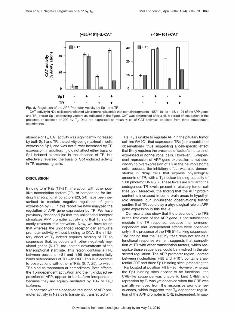
absence of T3, CAT activity was significantly increasedby both Sp1 and TR, the activity being maximal in cellsexpressing Sp1, and was not further increased by TRexpression. In addition, T3 did not affect either basal orSp1-induced expression in the absence of TR, buteffectively reversed the basal or Sp1-induced activityin TR-expressing cells.
DISCUSSION
Binding to nTREs (17–21), interaction with other pos-itive transcription factors (22), or competition for lim-iting transcriptional cofactors (23, 24) have been de-scribed to mediate negative regulation of geneexpression by T3. In this report we have analyzed theregulation of APP gene expression by TR. We havepreviously described (5) that the unliganded receptorstimulates APP promoter activity and that T3 signifi-cantly reverses this activation. Now, we have foundthat whereas the unliganded receptor can stimulatepromoter activity without binding to DNA, the inhibi-tory effect of T3 indeed requires binding of TR tosequences that, as occurs with other negatively reg-ulated genes (6–10), are located downstream of thetranscriptional start site. This region contains a TREbetween positions �81 and �96 that preferentiallybinds heterodimers of TR with RXR. This is in contrastto observations with other nTREs (6, 7, 25), to whichTRs bind as monomers or homodimers. Both effects,the T3-independent activation and the T3-induced re-pression of APP, appear to be isoform independent,because they are equally mediated by TR� or TR�isoforms.
In contrast with the observed reduction of APP pro-moter activity in N2a cells transiently transfected with
TRs, T3 is unable to regulate APP in the pituitary tumorcell line GH4C1 that expressess TRs (our unpublishedobservations), thus suggesting a cell-specific effectthat likely requires the presence of factor/s that are notexpressed in nonneuronal cells. However, T3-depen-dent repression of APP gene expression is not sec-ondary to overexpression of TR in the neuroblastomacells, because the inhibitory effect was also demon-strable in N2a� cells that express physiologicalamounts of TR, with a T3 nuclear binding capacity of1.68 pmol/mg DNA (26). These levels are similar to theendogenous TR levels present in pituitary tumor celllines (27). Moreover, the finding that the APP proteincontent is increased in some brain areas in hypothy-roid animals (our unpublished observations) furtherconfirm that TR could play a physiological role on APPgene expression in this tissue.
Our results also show that the presence of the TREin the first exon of the APP gene is not sufficient tomediate the TR response, because the hormone-dependent and -independent effects were observedonly in the presence of the TRE 5�-flanking sequences.The finding that the TRE by itself does not act as afunctional response element suggests that competi-tion of TR with other transcription factors, which rec-ognize those sequences, could be involved in the ob-served regulation. The APP promoter region, locatedbetween nucleotides �55 and �101, contains a po-tential CRE and three Sp1 binding sites, preceding theTRE located at position �81/�96. However, whereasthe Sp1 binding sites appear to be functional, theCRE-like sequence was unable to bind CREB, andrepression by T3 was yet observed when the CRE waspartially removed from the responsive promoter se-quences, which suggests that T3-dependent regula-tion of the APP promoter is CRE independent. In sup-
Fig. 8. Regulation of the APP Promoter Activity by Sp1 and TRCAT activity in N2a cells cotransfected with reporter plasmids that contain fragments �55/�101 or �15/�101 of the APP gene,
and TR- and/or Sp1-expressing vectors as indicated in the figure. CAT was determined after a 48-h period of incubation in thepresence or absence of 200 nM T3. Data are expressed as mean � SD of CAT activities obtained from three independentexperiments.
Villa et al. • Negative Regulation of APP by T3 Mol Endocrinol, April 2004, 18(4):863–873 869
by on May 21, 2010 mend.endojournals.orgDownloaded from

port of this conclusion we have also found thatstimulation of CREB activity, by treatment with fors-kolin or overexpression of protein kinase A, was un-able to stimulate APP promoter activity.
In contrast, our results are compatible with a mech-anism in which TR competes with Sp1 for binding toDNA. This mechanism would have a particular rele-vance in the APP gene, which lacks the TATA box andwhich is, therefore, largely dependent on the consti-tutive activation induced by basal factors such as Sp1(28). We have shown that expression of Sp1 stimulatesAPP promoter activity and that Sp1 binds to the �55/�101 region of the APP gene in EMSAs. However, thepresence of the TR-RXR heterodimer precludes bind-ing of Sp1 to this region of the APP gene. A similarmechanism involving overlapping DNA-binding siteshas been already proposed for T3 suppression of Sp1-dependent transcription of the epidermal growth fac-tor receptor promoter (29), for the regulation of thelactoferrin gene by estrogen, retinoic acid, andchicken ovalbumin upstream promoter receptors (30,31), and for the regulation of the chicken lysozymegene by TR and CCCTC-binding factor (CTCF) (32).The negative effect of T3 on the APP promoter couldbe also secondary to repression of Sp1 expression.However, T3 does not reduce Sp1 content in N2a�cells, and as shown in Fig. 8, the inhibitory effect of T3
is also observed in cells transfected with TR and anexpression vector for Sp1, thus indicating that inhibi-tion of APP promoter activity is independent of theeffects of T3 on Sp1 levels.
In addition to RXR/TR and Sp1, other proteins thatform slow migrating complexes on the �55/�101 T3-responsive region of the APP gene are present innuclear extracts of neuroblastoma cells. Characteriza-tion of these proteins is currently underway and will beimportant to analyze their possible contribution to theobserved regulation. Preliminary results, obtained us-ing mass spectroscopy, indicate that many of theseproteins possess sequence homology with differentmembers of the heterogeneous nuclear ribonucleo-protein family, a group of proteins that, according toprevious descriptions, could play a role as transcrip-tional regulators (33, 34).
Whereas binding of TR to DNA has been reported tobe essential for negative regulation of the TRH orTSH� genes by T3 (12, 35), a mechanism based onprotein-protein interactions, as opposed to TR bindingdirectly to DNA sequences, has been proposed fornegative regulation of the TSH� gene (13, 14). Wehave examined whether TR-mediated regulation ofAPP requires direct binding of TR to promoter se-quences. Using a TR mutant that cannot bind DNA, wehave found that the stimulatory effect induced by theunliganded receptor does not require binding of TR tothe APP promoter. In contrast, and compatible withthe competition hypothesis suggested above, the in-hibitory effect of T3 was observed only in cells ex-pressing the wild-type TR, which binds DNA. Theseresults suggest a mechanism in which occupancy of
TR by T3 would promote binding of the receptor to theAPP promoter. This model would be incompatible witha high-affinity binding of the unliganded receptor tothe promoter and, accordingly, we have proved that aconstitutively active receptor, obtained by fusion offull-length wild-type TR� to the transcriptional activa-tion domain of VP16 (13), is unable to activate CATreporter genes containing the �55/�101 or �15/�101 fragments of the APP gene. These resultsstrongly support the existence of a low-affinity-inter-acting site that in vivo likely binds TR only after T3
binding. In this respect, we have already indicated thatTR binds to the APP promoter as a heterodimer withRXR. Because heterodimerization with RXR augmentsthe DNA binding affinity of TR, and binding of T3 aug-ments the interaction between TR and RXR (36, 37), T3
could repress transcription by increasing the affinity ofTR-RXR heterodimers for DNA, which in turn woulddecrease the accessibility of other activating tran-scription factors (such as Sp1 or other still unidentifiedfactors), likely involved in the constitutive stimulationof the APP promoter. In agreement with this hypoth-esis, we have also observed that stimulation by unli-ganded TR, which remains mainly unbound in theabsence of ligand, is also reversed when its binding tothe APP promoter is facilitated by coexpression ofRXR (data not shown).
In addition, in the case of negative regulation of theTSH� gene by T3, it has been suggested that unligan-ded TR recruits CoRs and withdraws HDACs from thebasal promoter causing activation, whereas T3 bindingdissociates the CoRs/HDACs and recruits coactiva-tors to restrict access to histone acetylases to com-ponents of the basal promoter, thereby causingligand-dependent repression. Moreover, the stimula-tion induced by the unliganded receptor would involvecompetition for, and squelching of, a limiting amountof CoRs that are bound to other factors on the pro-moter (13, 14). We have observed that overexpressionof CoRs or HDACs reverses activation of the APPpromoter by unliganded TR (data not shown). Thesefindings strongly suggest a role for CoRs/HDAC inTR-mediated regulation of APP. However, the mech-anisms that mediate the effects of TR on the APP geneappear to be essentially different from those describedfor other negatively regulated genes, in which expres-sion of silencing mediator of retinoid and thyroid hor-mone receptor or nuclear receptor corepressor mark-edly enhances activation by unoccupied TR (14). Ifwithdrawal of CoRs is involved in stimulation by theunliganded TR, it is tempting to speculate that thisreceptor in the absence of T3 could have a moregeneral activator role in other housekeeping genes inwhich transcriptional activation is largely dependenton Sp1, particularly those having TR and Sp1 bindingsites.
On the other hand, it has been also proposed thatDNA binding of TR would be not necessary in TR-mediated control of negatively regulated genes (14).Using a TR mutant we have demonstrated that in the
870 Mol Endocrinol, April 2004, 18(4):863–873 Villa et al. • Negative Regulation of APP by T3
by on May 21, 2010 mend.endojournals.orgDownloaded from
absence of T3 the stimulation of basal promoter activ-ity does not require a TR bound to DNA, a circum-stance that likely contributes to the APP promoterstimulation by a double mechanism: 1) making possi-ble binding of other transcription factors to DNA; and2) by squelching of CoRs/HDAC that finally causesactivation. In contrast, the T3-induced repression re-quires binding of TR to DNA and likely involves dis-placement from the DNA of other general transcriptionfactors that also bind to that region of the APP pro-moter. However, because to be functional the pro-posed mechanism should allow the interchange ofthose factors from DNA, it would be incompatible witha high-affinity TR binding site, and these resultsstrongly support the existence of a low-affinity-inter-acting site that likely binds TR only under optimalconditions.
In summary, our results suggest a dual mechanismin which unliganded TR does not interact with DNA,allowing binding of Sp1 and the subsequent transcrip-tional activation by this factor. In addition, unligandedTR might also contribute to promoter activation bywithdrawal of CoRs and HDACs from the promoter.Binding of T3 would facilitate formation of TR-RXRheterodimers, which would then bind to the TRE anddisplace Sp1 from the overlapping sequences �64/�83, blocking activation by this transcription factor.Why TR/RXR complex fails to activate transcriptiononce bound to the APP element after T3 addition is stillunknown. However, again, displacement of strong ac-tivators such as Sp1 factors could be involved. Thismechanism could be particularly relevant in the caseof the APP gene, because Sp1 is required for basaltranscription of genes that, like the APP gene, lack aTATA box (28). The equilibrium between binding of twotranscription factors, TR and Sp1, to overlapping se-quences of DNA would allow a precise control of theAPP gene expression. Because binding of T3 to itsreceptors fits a sigmoidal kinetics (38, 39), small in-creases of T3 concentration would be sufficient tosignificantly enhance TR occupancy, which in turnwould facilitate binding of the TR/RXR heterodimer tothe low-affinity binding site of the APP gene, and tocause the displacement of Sp1 and the consequentreduction of promoter activity. Clearly, additional workwill be necessary to clarify the mechanisms of thefunctional interaction between TR and Sp1 and its rolein regulation of gene expression.
MATERIALS AND METHODS
Cell Culture
Murine N2a neuroblastoma cells were grown in DMEM sup-plemented with 10% fetal bovine serum as previously de-scribed by Ortiz-Caro et al. (39). Previous to the experiments,the culture medium was replaced with a similar mediumcontaining serum depleted of thyroid hormone by treatmentwith resin AG1X8 as described by Samuels et al. (40), and thecells were then incubated in this medium for an additional
24-h period before the beginning of the experiments. N2a-�cells, a subclone that constitutively expresses the �-isoformof TR (TR�), were obtained from Dr. Dussault’s laboratory.N2a-� cells were grown as previously described by Lebel etal. (26), and the experiments were carried out in the samemedium and conditions described for N2a cells.
Reporter Plasmids and Expression Vectors
The CAT reporter plasmid containing the �1099/�106 frag-ment of the human APP gene has been previously described(41). 5�-Deletions to �15, �55, or �75 bp were prepared byPCR from the original �1099/�105 bp fragment, kindly pro-vided by Dr. Lahiri’s laboratory, and subcloned into theBamH1 site of pBL-CAT8. The reporter constructs �55/�101-, �63/�101-, �75/�101-, or �55/�79-CAT consist ofa single copy of those APP fragments inserted in front of a tkpromoter driving the expression of the CAT gene. Mutant M1contains the mutations of the upstream hemisite of the TREindicated in boldface (CAACTAGACGAAGGACG). In M2,mutations are present in the intervening nucleotides of theresponse element (CGGGCAGACTCAGGACG), and M3 ismutated in the downstream hemisite (CGGGCAGAC-GAAAACTG), also affecting the downstream Sp1 bindingsite. These mutations were introduced in the �55/�101 frag-ment ligated to the tk promoter. The cDNAs encoding Sp1,TR�, and RXR�, as well as the TR mutant C51G, are insertedinto the EcoI site of the expression vector pSG5, which con-tains the simian virus 40 early promoter. The construct con-taining full-length TR coding sequences fused to the activa-tion domain of VP16 was a kind gift of Dr. J. L. Jameson, andit has been described previously (13).
DNA Transfection
N2a cells cultured in DMEM containing 10% of thyroidhormone-depleted fetal calf serum were transfected by thecalcium phosphate coprecipitation method with 300 ng ofreporter plasmids. A luciferase reference vector (100 ng) wassimultaneously used as an internal control of the transfectionefficiency. In cotransfection experiments, 300 ng of reporterplasmid and 1 �g of the corresponding receptor expressionvector were used. After 16 h of incubation in the presence ofcalcium phosphate, the medium was discarded and washedwith 5 ml PBS. A new medium containing 0.5% thyroidhormone-depleted serum was added, and the cells were thenincubated for an additional period of 48 h in the presence orabsence of 200 nM T3. Each treatment was performed induplicate cultures that normally showed less than 5–15%variation in CAT activity, which was determined by incubationof [14C]chloramphenicol with cell lysate protein. After auto-radiography, the nonacetylated and acetylated [14C]chloram-phenicol was quantified, and the data are expressed as themean � SD of the percent of acetylated forms after eachtreatment. Each experiment was repeated at least two tothree times with similar relative differences in regulatedexpression.
Mobility Shift Assays
Synthetic oligonucleotides containing the TR-binding se-quences of the human APP promoter (�55/�101), or theconsensus TREpal sequence, were end labeled with[�-32P]dCTP by using the Klenow fragment of Escherichia coliDNA polymerase and then incubated with in vitro translatedreceptors, recombinant Sp1 (obtained from Promega Corp.,Madison, WI), or with nuclear extracts obtained from N2a-�cells. cDNAs for TR and RXR in pSG5 were transcribed andtranslated in vitro with the TNT kit (Promega) following themanufacturer�s recommendations. The nuclear extracts wereobtained by the method of Andrews and Faller (42). For gel
Villa et al. • Negative Regulation of APP by T3 Mol Endocrinol, April 2004, 18(4):863–873 871
by on May 21, 2010 mend.endojournals.orgDownloaded from

retardation assays, purified Sp1 (250 ng), translated recep-tors (between 0.5 and 2 �l), or nuclear extracts (5 �g) wereincubated on ice for 15 min in a buffer (20 mM Tris HCl, pH7.5; 75 mM KCl; 1 mM dithiothreitol; 5 �g/ml BSA; 13%glycerol) containing 3 �g poly (dI-dC) and then were incu-bated for 15–20 min at room temperature with approximately70,000 cpm of the double-stranded labeled oligonucleotide.Unprogrammed reticulocyte lysate was used as a control fornonspecific binding. For competition experiments, increasingconcentrations of unlabeled double-stranded oligonucleo-tide, or an oligonucleotide containing the consensus se-quence TREpal (5�AGGTCATGACCT-3�), were added to thebinding reaction mixture. DNA-protein complexes were re-solved on 5% nondenaturing polyacrylamide gels containing0.5% Tris-borate-EDTA buffer. The gels were dried and au-toradiographed at �70 C.
Western Blot Analysis
Extracts from N2a� cells incubated in the presence or in theabsence of T3 were run in 10% acrylamide gels. The proteinswere transferred to a nitrocellulose membrane and identifiedby chemiluminescence after incubation with a 1:2000 dilutionof the specific antibody sc-59 (Santa Cruz BiotechnologyInc., Santa Cruz, CA) raised against the transcription factorSp1.
Acknowledgments
We thank Dr. A. Aranda for critical reading of the manu-script and Ana Pascual for valuable linguistic help.
Received July 2, 2003. Accepted December 31, 2003.Address all correspondence and requests for reprints to:
Dr. A. Pascual, Instituto de Investigaciones Biomedicas.(C.S.I.C.), Arturo Duperier 4, 28029 Madrid, Spain. E-mail:[email protected].
This work was supported by grants from the ComisionInterministerial de Ciencia y Tecnologıa (SAF2000-0177).A.V. was recipient of a fellowship from GlaxoSmithKline andConsejo Superior de Investigaciones Cientıficas. J.S. wassupported by a fellowship from the Comunidad Autonoma deMadrid.
REFERENCES
1. Selkoe DJ 1994 Cell biology of the amyloid �-proteinprecursor and the mechanism of Alzheimer’s disease.Annu Rev Cell Biol 10:373–403
2. Mattson MP, Cheng B, Culwell AR, Esch FS, LieberburgI, Rydel RE 1993 Evidence for excitoprotective and intra-neuronal calcium-regulating roles for secreted forms ofthe �-amyloid precursor protein. Neuron 10:243–254
3. Fukuchi K, Kamino K, Deeb SS, Smith AC, Dang T, MartinGM 1992 Overexpression of amyloid precursor proteinalters its normal processing and is associated with neu-rotoxicity. Biochem Biophys Res Commun 182:165–173
4. Latasa MJ, Belandia B, Pascual A 1998 Thyroid hor-mones regulate �-amyloid gene splicing and proteinsecretion in neuroblastoma cells. Endocrinology 139:2692–2698
5. Belandia B, Latasa MJ, Villa A, Pascual A 1998 Thyroidhormone negatively regulates the transcriptional activityof the �-amyloid precursor protein gene. J Biol Chem273:30366–30371
6. Carr FE, Wong NC 1994 Characteristics of a negativethyroid hormone response element. J Biol Chem 269:4175–4179
7. Hollenberg AN, Monden T, Flynn TR, Boers ME, CohenO, Wondisford FE 1995 The human thyrotropin-releasinghormone gene is regulated by thyroid hormone throughtwo distinct classes of negative thyroid hormone re-sponse elements. Mol Endocrinol 9:540–550
8. Madison LD, Ahlquist JA, Rogers SD, Jameson JL 1993Negative regulation of the glycoprotein hormone � genepromoter by thyroid hormone: mutagenesis of a proximalreceptor binding site preserves transcriptional repres-sion. Mol Cell Endocrinol 94:129–136
9. Wood WM, Kao MY, Gordon DF, Ridgway EC 1989 Thy-roid hormone regulates the mouse thyrotropin �-subunitgene promoter in transfected primary thyrotropes. J BiolChem 264:14840–14847
10. Perez-Juste G, Aranda A 1999 Differentiation of neuro-blastoma cells by phorbol esters and insulin-like growthfactor 1 is associated with induction of retinoic acidreceptor � gene expression. Oncogene 18:5393–5402
11. Cohen RN, Putney A, Wondisford FE, Hollenberg AN2000 The nuclear corepressors recognize distinct nu-clear receptor complexes. Mol Endocrinol 14:900–914
12. Shibusawa N, Hollenberg AN, Wondisford FE 2003 Thy-roid hormone receptor DNA binding is required for bothpositive and negative gene regulation. J Biol Chem 278:732–738
13. Tagami T, Park Y, Jameson JL 1999 Mechanisms thatmediate negative regulation of the thyroid-stimulatinghormone � gene by the thyroid hormone receptor. J BiolChem 274:22345–22353
14. Tagami T, Madison LD, Nagaya T, Jameson JL 1997Nuclear receptor corepressors activate rather than sup-press basal transcription of genes that are negativelyregulated by thyroid hormone. Mol Cell Biol 17:2642–2648
15. Karin M 1998 New twists in gene regulation by glucocor-ticoid receptor: is DNA binding dispensable? Cell 93:487–490
16. Suske G 1999 The Sp-family of transcription factors.Gene 238:291–300
17. Chatterjee VK, Lee JK, Rentoumis A, Jameson JL 1989Negative regulation of the thyroid-stimulating hormone �gene by thyroid hormone: receptor interaction adjacentto the TATA box. Proc Natl Acad Sci USA 86:9114–9118
18. Carr FE, Kaseem LL, Wong NC 1992 Thyroid hormoneinhibits thyrotropin gene expression via a position-inde-pendent negative L-triiodothyronine-responsive element.J Biol Chem 267:18689–18694
19. Pombo PM, Barettino D, Espliguero G, Metsis M, IglesiasT, Rodriguez-Pena A 2000 Transcriptional repression ofneurotrophin receptor trkB by thyroid hormone in thedeveloping rat brain. J Biol Chem 275:37510–37517
20. Perez-Juste G, Garcia-Silva S, Aranda A 2000 An ele-ment in the region responsible for premature terminationof transcription mediates repression of c-myc gene ex-pression by thyroid hormone in neuroblastoma cells.J Biol Chem 275:1307–1314
21. Satoh T, Yamada M, Iwasaki T, Mori M 1996 Negativeregulation of the gene for the preprothyrotropin-releasinghormone from the mouse by thyroid hormone requiresadditional factors in conjunction with thyroid hormonereceptors. J Biol Chem 271:27919–27926
22. Zhu YS, Yen PM, Chin WW, Pfaff DW 1996 Estrogen andthyroid hormone interaction on regulation of gene ex-pression. Proc Natl Acad Sci USA 93:12587–12592
23. Kamei Y, Xu L, Heinzel T, Torchia J, Kurokawa R, GlossB, Lin SC, Heyman RA, Rose DW, Glass CK, RosenfeldMG 1996 A CBP integrator complex mediates transcrip-tional activation and AP-1 inhibition by nuclear receptors.Cell 85:403–414
24. Zhu XG, Park KS, Kaneshige M, Bhat MK, Zhu Q, Mari-ash CN, McPhie P, Cheng SY 2000 The orphan nuclearreceptor Ear-2 is a negative coregulator for thyroid hor-
872 Mol Endocrinol, April 2004, 18(4):863–873 Villa et al. • Negative Regulation of APP by T3
by on May 21, 2010 mend.endojournals.orgDownloaded from

mone nuclear receptor function. Mol Cell Biol 20:2604–2618
25. Hallenbeck PL, Phyillaier M, Nikodem VM 1993 Divergenteffects of 9-cis-retinoic acid receptor on positive andnegative thyroid hormone receptor-dependent gene ex-pression. J Biol Chem 268:3825–3828
26. Lebel JM, Dussault JH, Puymirat J 1994 Overexpressionof the � 1 thyroid receptor induces differentiation inneuro-2a cells. Proc Natl Acad Sci USA 91:2644–2648
27. Samuels HH, Stanley F, Shapiro LE 1976 Dose-depen-dent depletion of nuclear receptors by L-triiodothyronine:evidence for a role in induction of growth hormone syn-thesis in cultured GH1 cells. Proc Natl Acad Sci USA73:3877–3881
28. Pugh BF, Tjian R 1990 Mechanism of transcriptionalactivation by Sp1: evidence for coactivators. Cell 61:1187–1197
29. Xu J, Thompson KL, Shephard LB, Hudson LG, Gill GN1993 T3 receptor suppression of Sp1-dependent tran-scription from the epidermal growth factor receptor pro-moter via overlapping DNA-binding sites. J Biol Chem268:16065–16073
30. Lee MO, Liu Y, Zhang XK 1995 A retinoic acid responseelement that overlaps an estrogen response element me-diates multihormonal sensitivity in transcriptional activa-tion of the lactoferrin gene. Mol Cell Biol 15:4194–4207
31. Liu Y, Teng CT 1992 Estrogen response module of themouse lactoferrin gene contains overlapping chickenovalbumin upstream promoter transcription factor andestrogen receptor-binding elements. Mol Endocrinol6:355–364
32. Lutz M, Burke LJ, LeFevre P, Myers FA, Thorne AW,Crane-Robinson C, Bonifer C, Filippova GN, LobanenkovV, Renkawitz R 2003 Thyroid hormone-regulated en-hancer blocking: cooperation of CTCF and thyroid hor-mone receptor. EMBO J 22:1579–1587
33. Chen H, Hu B, Allegretto EA, Adams JS 2000 The vitaminD response element-binding protein. A novel dominant-
negative regulator of vitamin D-directed transactivation.J Biol Chem 275:35557–35564
34. Kim MK, Nikodem VM 1999 hnRNP U inhibits carboxy-terminal domain phosphorylation by TFIIH and repressesRNA polymerase II elongation. Mol Cell Biol 19:6833–6844
35. Bodenner DL, Mroczynski MA, Weintraub BD, RadovickS, Wondisford FE 1991 A detailed functional and struc-tural analysis of a major thyroid hormone inhibitory ele-ment in the human thyrotropin �-subunit gene. J BiolChem 266:21666–21673
36. Collingwood TN, Butler A, Tone Y, Clifton-Bligh RJ,Parker MG, Chatterjee VK 1997 Thyroid hormone-medi-ated enhancement of heterodimer formation betweenthyroid hormone receptor � and retinoid X receptor.J Biol Chem 272:13060–13065
37. Zhang J, Zamir I, Lazar MA 1997 Differential recognitionof liganded and unliganded thyroid hormone receptor byretinoid X receptor regulates transcriptional repression.Mol Cell Biol 17:6887–6897
38. Ortiz-Caro J, Montiel F, Pascual A, Aranda A 1986 Mod-ulation of thyroid hormone nuclear receptors by short-chain fatty acids in glial C6 cells. Role of histone acety-lation. J Biol Chem 261:13997–14004
39. Ortiz-Caro J, Yusta B, Montiel F, Villa A, Aranda A, Pas-cual A 1986 Identification and characterization of L-triiodothyronine receptors in cells of glial and neuronalorigin. Endocrinology 119:2163–2167
40. Samuels HH, Stanley F, Casanova J 1979 Depletion ofL-3,5,3�-triiodothyronine and L-thyroxine in euthyroidcalf serum for use in cell culture studies of the action ofthyroid hormone. Endocrinology 105:80–85
41. Lahiri DK, Robakis NK 1991 The promoter activity of thegene encoding Alzheimer �-amyloid precursor protein(APP) is regulated by two blocks of upstream sequences.Brain Res Mol Brain Res 9:253–257
42. Andrews NC, Faller DV 1991 A rapid micropreparationtechnique for extraction of DNA-binding proteins fromlimiting numbers of mammalian cells. Nucleic Acids Res19:2499
Molecular Endocrinology is published monthly by The Endocrine Society (http://www.endo-society.org), the foremostprofessional society serving the endocrine community.
Villa et al. • Negative Regulation of APP by T3 Mol Endocrinol, April 2004, 18(4):863–873 873
by on May 21, 2010 mend.endojournals.orgDownloaded from
Copyright © 2022 FDOKUMEN


![[sp1] oocyte collection from superstimulated disease-free ...](https://static.fdokumen.com/doc/165x107/631dd3361aedb9cd850f788f/sp1-oocyte-collection-from-superstimulated-disease-free-.jpg)