Splicing of designer exons informs a biophysical model for exon definition
Upload
independentCategory
view
5download
0
1997, 17(7):4096. Mol. Cell. Biol.
K Du, Y Peng, L E Greenbaum, B A Haber and R Taub increased EIIIB expression in proliferating liver.fibronectin EIIIB exon, a possible cause of HRS/SRp40-mediated inclusion of the
http://mcb.asm.org/content/17/7/4096Updated information and services can be found at:
These include:
CONTENT ALERTS more»cite this article),
Receive: RSS Feeds, eTOCs, free email alerts (when new articles
http://journals.asm.org/site/misc/reprints.xhtmlInformation about commercial reprint orders: http://journals.asm.org/site/subscriptions/To subscribe to to another ASM Journal go to:
on Septem
ber 30, 2014 by guesthttp://m
cb.asm.org/
Dow
nloaded from
on Septem
ber 30, 2014 by guesthttp://m
cb.asm.org/
Dow
nloaded from

MOLECULAR AND CELLULAR BIOLOGY,0270-7306/97/$04.0010
July 1997, p. 4096–4104 Vol. 17, No. 7
Copyright © 1997, American Society for Microbiology
HRS/SRp40-Mediated Inclusion of the Fibronectin EIIIBExon, a Possible Cause of Increased EIIIB Expression
in Proliferating LiverKEYONG DU,1 YONG PENG,1 LINDA E. GREENBAUM,1,2 BARBARA A. HABER,3
AND REBECCA TAUB1*
Department of Genetics,1 Division of Gastroenterology, Department of Medicine,2 and Division of Gastroenterologyand Nutrition, Children’s Hospital of Philadelphia,3 University of Pennsylvania School of Medicine,
Philadelphia, Pennsylvania 19104
Received 23 December 1996/Returned for modification 24 February 1997/Accepted 2 April 1997
Serine-arginine (SR)-rich proteins are believed to be important in mediating alternative pre-mRNA splicing.HRS/SRp40 expression is elevated in liver cell proliferation during development, regeneration, and oncogen-esis. We tested whether HRS expression correlates with the appearance of alternatively spliced fibronectintranscripts during liver growth. HRS was highly expressed during the proliferative phase of liver development,correlating with expression of the fibronectin EIIIB alternative exon. In regenerating liver, HRS protein wasinduced in a time course consistent with the observed increase in fibronectin transcripts containing the EIIIBexon, particularly in nonparenchymal liver cells. Furthermore, in an in vivo assay, HRS, and not other SRproteins, directly mediated EIIIB exon inclusion in the fibronectin transcript. This alternative splicing wasdependent on a purine-rich region within the EIIIB exon to which HRS specifically bound. We have establishedthat HRS has the potential to contribute to the regulation of fibronectin pre-mRNA splicing during liver growth.Changes in fibronectin forms may be important in modifying liver architecture during the proliferativeresponse, thus providing a potential mechanism by which SR proteins may participate in cellular growthcontrol.
The splicing factor HRS (hepatic arginine-serine protein) isexpressed in proliferating liver cells. HRS is induced as animmediate-early gene in insulin-treated, mitogen-activated rathepatoma H35 cells and in our study represented 12% of allinsulin-induced cDNAs (7). HRS is a delayed-early gene inregenerating liver and is expressed at a very high level duringliver development when the liver is rapidly proliferating (7, 46;reviewed in reference 9). HRS belongs to the serine-arginine(SR) protein family, which is comprised of at least nine struc-turally related proteins, including SRp20, SRp30a (SF2/ASF),SRp30b (SC35), SRp40, SRp55, and SRp75 (11, 12, 27, 31, 39,52–54). Comparing the partial sequence of human SRp40 andHRS sequence revealed that HRS is rat SRp40 (7, 52). Re-cently, the human homolog of HRS was identified and shownto have properties of other splicing factors in both in vitro andin vivo splicing assays (39).
Pre-mRNA alternative splicing provides an important mech-anism of gene and protein expression regulation (19, 29, 31,40). Through alternative splicing, one primary transcript cangenerate multiple proteins which may have different functions.Alternative splicing occurs via several pathways, includingskipped exons, included introns, alternative 59 splice sites, al-ternative 39 splice sites, and mutually exclusive exons. SR pro-teins have been shown to play essential roles in both constitu-tive pre-mRNA splicing and regulation of pre-mRNAalternative splicing. In vitro and in vivo splicing assays indicatethat SR proteins regulate alternative splicing by promoting theuse of proximal 59 splice sites (10, 23, 26, 55), and different SR
proteins have distinct abilities to regulate alternative splicingof various model pre-mRNAs (2, 31, 53). Specificity may bemediated by differential binding to purine-rich sequences orsplicing enhancers that have been identified within severalalternatively spliced exons (31). Tissue-specific variations intotal and relative amounts of SR proteins or their mRNA havebeen described (39, 52). The genetics and control of specificsplicing pathways that are important in mammalian cell growthand development are largely unknown. It is likely that therelative amount of individual SR proteins in particular cells ortissues may determine the level of various spliced forms ofdifferent pre-mRNAs, thereby reflecting the specific role ofeach SR protein. The finding of uncompensated growth defectsin ASF gene-targeted cells suggests that each of these SRproteins plays a unique role in cellular processes (50).
Because HRS is induced at a high level in hepatic regener-ation, development, and oncogenesis, we reasoned that pre-mRNAs that are regulated by HRS would be alternativelyspliced in all three types of hepatic proliferation. Based on theliterature, fibronectin pre-mRNA has this property. During he-patic proliferation in development, regeneration, and oncogen-esis, changes in hepatocyte polarity occur (42). Among adhe-sion molecules, fibronectin mRNA has several spliced formswhich change in proliferative states of the liver (3, 8, 33–37,47). During liver development, EIIIB exon-containing(EIIIB1) transcripts represent a significant percentage offibronectin transcripts (37). In the adult liver, nonproliferativehepatocytes produce almost all of the plasma forms of fi-bronectin (24) which do not contain the EIIIA (EDA andED1) and EIIIB (EDB) exons. The fibronectin gene is inducedas a delayed-early gene, and its elevated expression is pro-longed up to 48 h posthepatectomy during liver regeneration(13, 32). EIIIA and EIIIB exon inclusion in the fibronectintranscript has not been examined before 12 h posthepatectomy
* Corresponding author. Mailing address: Department of Genetics,University of Pennsylvania School of Medicine, 422 Curie Blvd., 705AStellar Chance, Philadelphia, PA 19104. Phone: (215) 898-9131. Fax:(215) 573-2195. E-mail: [email protected].
4096
on Septem
ber 30, 2014 by guesthttp://m
cb.asm.org/
Dow
nloaded from

(3), when many of the early G1 events have already occurred.At 24 h posthepatectomy, elevated levels of the EIIIA exon aredetected. In a form of liver injury caused by bile duct ligation,an increase in EIIIA and to some extent EIIIB occurs partic-ularly in hepatic fat-storing cells at 12 to 24 h after the stimulus(24). The function of the alternative forms of fibronectin thatoccur in proliferating liver and other cell types is not known,but it is postulated that they play a role in organ structure (4).
Here, we assessed temporal and cell-type-specific changes inHRS/SRp40 protein levels and fibronectin pre-mRNA splicingforms in developing and regenerating liver and found thatinclusion of the EIIIB exon in fibronectin mRNA correlatedwith HRS expression. In in vivo splicing assays, HRS mediatedthe inclusion of the EIIIB exon in the mature fibronectin tran-script. This splicing depended on the presence of a purine-richsequence within the EIIIB exon to which HRS specificallybound. These findings suggest that HRS is at least in partresponsible for increased EIIIB containing fibronectin tran-scripts during liver growth.
MATERIALS AND METHODS
Animals and isolation of hepatocytes and nonparenchymal cells. For partialhepatectomy, female Fisher rats (160 to 200 g) were ether anesthetized andsubjected to midventral laparotomy with approximately 70% liver resection (32).To isolate hepatocyte and nonparenchymal cells either in the absence of orfollowing 70% partial hepatectomy, female Fisher rats were anesthetized andsubjected to ventral laparotomy, and the portal vein was cannulated. The liverwas perfused for 15 min with solution A (142 mM NaCl, 6.7 mM KCl, 10 mMHEPES) and 15 min with solution C (same as solution A but with 5 mM CaCl2and 0.5 mg of collagenase A per ml) (17). All solutions were pH 7.4 andprewarmed to 37°C. After perfusion, the liver was removed, minced, and filteredthrough 16-ply gauze. The hepatocytes were pelleted at 170 3 g for 1 min. Thesupernatant was spun at 2,000 3 g for 10 min to pellet the nonparenchymal cells.The purity of both cell populations was about 90 to 95% as determined bymicroscopy.
Preparation of whole nuclei. Whole liver nuclei were prepared from liver asdescribed previously (15). Briefly, at indicated times following 70% partial hep-atectomy, the regenerating liver or whole liver (time zero) was removed intohomogenization buffer (10 mM HEPES [pH 7.6], 10 mM KCl, 1 mM MgCl2, 0.1mM EDTA [pH 8.0], 0.1 mM EGTA [pH 8.0], 0.1 mM spermine, 0.1 mMspermidine, 0.3 M sucrose). After homogenization, the whole liver suspensionwas layered on cushion buffer (homogenization buffer plus 2.2 M sucrose), andnuclei were pelleted by centrifuging at 26,000 rpm for 50 min. The pelleted nucleiwere resuspended in nucleus suspension buffer (100 mM NaCl, 10 mM Tris [pH7.6], 1 mM EDTA). Following protein concentration determination (Bio-Rad),an equal volume of 23 Laemmli buffer was added. Then the nuclei were boiledfor 10 min, sheared three times by passage through a 23-gauge needle, and storedat 270°C. Whole H35 nuclei were prepared as described previously (1). Briefly,after insulin treatment, the cells were harvested in 200 ml of phosphate-bufferedsaline (PBS) at indicated times. The cells were lysed by adding 200 ml of 23 lysisbuffer, and the nuclei were pelleted and resuspended in nucleus suspensionbuffer.
Expression of HRS in bacteria. HRS proteins were expressed as full-length orRNA binding domains only. To express the full-length HRS, the HRS codingregion was amplified with primers 59-CTAGCCATGGGGATCATGAGTGGC-39 and 59-AGCTGGATCCTTCAATTCATTGATTT-39 by PCR and clonedinto the NcoI/BamHI sites of the pET expression vector. The pET-HRS insertsequence was confirmed by dideoxy sequencing using Sequenase (U.S. Biochem-ical). The resultant HRS protein was first purified over a nickel-agarose column(Qiagen, Inc.) into 8 M urea. To make native protein, HRS in urea was dialyzedagainst a urea gradient (6, 3, 1, and 0.5 M; 4 h each) and finally dialyzed intobuffer D (20 mM HEPES [pH 7.6], 0.1 M KCl, 2 mM EDTA, 1.5 mM MgCl2, 1.5mM dithiothreitol [DTT], 20% glycerol). To express the HRS RNA bindingdomain (amino acids [aa] 1 to 165), the cDNA fragment was amplified with59-CTAGCCATGGGGATCATGAGTGGC-39 and 59-CCTTGGTGGATCCACATCTACAAG-39 by PCR and cloned into the NcoI/BamHI sites of the pETexpression vector. The pET-HRS RNA binding domain insert sequence wasconfirmed by dideoxy sequencing using Sequenase (U.S. Biochemical). The re-sultant 19-kDa protein was purified over a nickel-agarose column (Qiagen).
Antibody production and immunoblots. Bacterially expressed HRS RNAbinding domain was used as an antigen to produce anti-HRS antibodies. Rabbitpolyclonal antibodies to the purified protein were prepared by Cocalico Biologi-cals (Reamstown, Pa.). To affinity purify the anti-HRS antiserum, 2 mg ofpurified 19-kDa HRS peptide (pI 5 4.3) was cross-linked on an Affi-15 gel resin(Bio-Rad). Then the anti-HRS antiserum, precipitated with 80% (NH4)2SO4from 10 ml of anti-HRS antiserum and resuspended in 3 ml of PBS, was passed
through the HRS column eight times and washed three times with PBS. Theanti-HRS antibodies were eluted four times with 2 ml of 0.05 M glycine z HCl(pH 2.3). The elution was monitored by measuring the optical density at 280 nm(OD280). The fractions with an OD280 of .0.08 were pooled and concentratedwith a Centricon 30 column. The final concentration of affinity-purified antibod-ies was 0.9 mg/ml. The mouse monoclonal antibody M104 was a gift from M.Roth (52).
Immunoblots made from either liver or H35 nuclei were incubated with 1:750anti-HRS antiserum or 1:10 M104. Horseradish peroxidase-conjugated goat anti-rabbit immunoglobulin (for anti-HRS antiserum), horseradish peroxidase-con-jugated goat anti-mouse immunoglobulin (for M104) secondary antibody, and achemiluminescence detection system (Amersham Corp.) were used.
Immunohistochemistry. Immunohistochemistry for detection of HRS was per-formed as described previously (15). Tissue sections were rehydrated in xylenefollowing graded ethanol. The Vectastain Elite ABC (Vector Laboratories)avidin-biotin-horseradish peroxidase detection system was used according to themanufacturer’s instructions, with some modifications. After the blocking step in1.5% goat serum in PBS (pH 7.4), avidin and biotin blocking steps were per-formed with the Vector Laboratories avidin-biotin blocking kit. Affinity-purifiedanti-HRS antibody was diluted 1:250 in 1.5% goat serum, and tissue sectionswere incubated for 30 min at room temperature in a humidified chamber. Aftersecondary antibody incubation, the sections were incubated with 0.3% hydrogenperoxide in methanol for 40 min to quench endogenous peroxidase activity. Thesections were then dehydrated in ethanol and xylene and mounted with Per-mount.
Plasmids. The fibronectin minigene pBAGHsv-7iBi89 (7iBi89) was a gift fromRichard Hynes (Massachusetts Institute of Technology) (21, 22). Plasmid pBAwas constructed by deletion of a portion of the EIIIB 39 intron with BglII andAflII in pBAGHsv-7iBi89. To overexpress HRS protein in eucaryotic cells, theHRS cDNA was cloned into EcoRI/HindIII sites in a pCMV expression vector(6).
To construct deletion 7iBi89d128-187, pBAGHsv-7iBi89 was digested withNotI (in the actin promoter) and BamHI. The resultant NotI-BamHI andBamHI-BamHI fragments were cloned into pBluescript-SK. The BamHI-BamHIfragment was subjected to Bal 31 nuclease digestion. The deleted fragment,which contained only one BamHI site, was ligated into NotI-BamHI fragments in7iBi89 after the BamHI site was blunted. To construct the deletions 7iBi89d95-147 and d95-160, the sequence between KpnI and BamHI was deleted from theNotI-BamHI fragment, blunted, and ligated to nuclease-digested BamHI-BamHIfragment. To delete the purine-rich region from the EIIIB exon, the overlappingextension PCR method (49) was used. The primers were FibI (59-AGCTCACTGACCTAAGCTTT-39), FibII (59-GAACAGCTCAAAGATCTAGA-39), FIBIII (59-ACTACAGTAGTTGCGTCCCCATTTTTGAAG-39), and FibIV (59-CTTCAAAAATGGGGACGCAACTACTGTGAT-39). 7iBi89 was first amplified withprimers FibI and -III or FibII and -IV. The resultant PCR products were puri-fied, mixed, and further amplified with FibI and -II. The final PCR product wasdigested with HindIII/BglII and cloned into pBAGHsv-7iBi89 between HindIIIand BglII sites. All deletion mutants were verified by DNA sequence analysis.
To construct pKS-FN520 to examine the expression of EIIIB1 and EIIIB2
fibronectin mRNA by RNase protection assay, the cDNA fragment which con-tains exons EIIIB and 8IIIa,b was amplified from fetal liver RNA by reversetranscription (RT)-PCR with primer FibI and 59-GTACACGCTGGAGACACTGAC-39 (in exon 8b), filled in with T4 DNA polymerase, and digested withHindIII. After digestion, the fragment was cloned into pBluescript-KS betweenthe HindIII and EcoRV sites. Plasmid pGEM-GA was prepared as follows: thesynthetic oligonucleotides 59-AGCTTGCGGCAGGAGAAGG-39 and 59-GATCCCTTCTCCTGCCGCA-39, which correspond to the purine-rich enhancer inthe EIIIB exon, were annealed and cloned into pGEM4 between the BamHI andHindIII sites. Plasmid pHRS350 for determining HRS expression by RNaseprotection was constructed as follows: the amplified cDNA fragment for encod-ing the unique HRS RNA binding domain was digested with XhoI and BamHIand cloned into pBluescript-KS between the XhoI and BamHI sites.
Cell culture and transfections. H35 cells were grown in low-glucose Dulbec-co’s modified Eagle’s medium (DMEM; Life Technologies, Inc.) supplementedwith 5% calf serum (Life Technologies) and 5% fetal bovine serum (Life Tech-nologies), 2 mM L-glutamine (Flow), and 100 U of penicillin and 50 U ofstreptomycin (Flow) as previously reported (7). To produce quiescence, themedium was changed to serum-free DMEM for 72 h, at which time the cells werebetween 40 and 60% confluent. Following serum deprivation, cells were treatedfor the indicated times with insulin (1028 M). For transient transfection, NIH3T3 cells were grown in high-glucose DMEM supplemented with 5% fetal bovineserum (Life Technologies), 2 mM L-glutamine (Flow), and 100 U of penicillinand 50 U of streptomycin (Flow). Then 40 to 60% confluent NIH 3T3 cells in100-mm-diameter dishes were transiently transfected by the calcium phosphatemethod (20, 30) with 5 mg each of pCMV-HRS (or pCG-SC35 or pCG-SRp55)or pCMV (control) and the 7iBi89 minigene. After 16 h of incubation, theprecipitate was removed, and the medium was changed to fresh medium; 20 to24 h later, the cells were harvested for RNA preparation.
RNA and RNA analysis. The RNAs from fetal liver and regenerating liverwere prepared and run on Northern blots as previously reported (18). The RNAfrom isolated hepatocytes, nonparenchymal cells, and transfected NIH 3T3 cellswas prepared essentially as described previously (5). For isolated hepatocytes
VOL. 17, 1997 HRS EXPRESSION DURING LIVER PROLIFERATION 4097
on Septem
ber 30, 2014 by guesthttp://m
cb.asm.org/
Dow
nloaded from

and nonparenchymal cells, the pelleted cells were directly dissolved into solutionD (4 mM guanidinium thiocyanate, 25 mM sodium citrate, 0.5% sarcosyl, 100mM 2-mercaptoethanol [pH 7.0]) followed by phenol-chloroform extraction andisopropanol precipitation. For transfected NIH 3T3 cells, the cells were washedtwice with cold PBS and lysed directly in solution D. Contaminating DNA wasremoved by incubating with RNase-free DNase I (Promega Co.) at 37°C for 30min. The RNA concentration was determined by measuring the OD260.
To make the RNA probe for the RNase protection assay, HindIII-linearizedpKS-FN520 or XhoI-linearized pHRS350 was in vitro transcribed with T7 DNApolymerase by adding [32P]UTP. After in vitro transcription, the radiolabeledprobe was purified on a urea-denatured 5% polyacrylamide gel. The RNaseprotection assay was performed by using an Ambion RPA II kit as instructed bythe manufacturer. Briefly, 10 mg of RNA was hybridized with 104 cpm of RNAprobe at 45°C overnight. After RNase A and T1 digestion, the protected RNAwas precipitated and separated on a denaturing 5% polyacrylamide gel. The gelwas dried and exposed to Kodak X-ray film.
RT-PCR was performed as follows (16). A 2.5-mg aliquot of total RNA and125 ng of random hexanucleotides in 20 ml of H2O were denatured at 70°C. Thetotal volume was brought to 50 ml containing 50 mM Tris (pH 8.3), 65 mM KCl,15 mM MgCl2, 0.2 mM deoxynucleoside triphosphate, 20 U of RNasin (Pro-mega), and 5 U of Superscript RNase H2 reverse transcriptase (Gibco BRL).The reaction mixture was incubated at 37°C for 60 min and then heat inactivated.For PCR, 2.5 ml of the RT mixture was used as a template in 25-ml reactionvolume containing 0.2 mM deoxynucleoside triphosphate, 2 mM MgCl2, 50 mMKCl, 1 mM DTT, 150 ng of upstream primer, 150 ng of downstream primer, 0.25ml of [a-32P]dCTP, and 2 U of Taq DNA polymerase. The reaction was run for24 cycles (94°C, 45 s; 55°C, 30 s; 72°C, 30 s). Two microliters of the final productwas separated on a denaturing 5% polyacrylamide gel. The gel was dried andexposed to Kodak X-ray film. The primers for PCR were as follows: fibronectinEIIIB2 59 primer, 59-CCTATTCTCTGAATACCGTCAT-39 (in exon 7b);fibronectin EIIIB1 59 primer, 59-GACTATGACATCAGGTTATC-39 (in exonEIIIB); and 7iBi89 minigene 59 primer, 59-GAGCACAGAGCCTCGCCTTTG-39 (actin exon). All amplifications of fibronectin mRNA used the same 39 primer,59-GTACACGCTGGAGACACTGAC-39 (in exon 8b).
RNA binding and UV cross-linking. To prepare the RNA probe, EcoRI-linearized pGEM-GA and pGEM empty vector were in vitro transcribed with T7DNA polymerase by adding [32P]UTP. After in vitro transcription, the radiola-beled probe was purified on a urea-denatured 15% polyacrylamide gel. For RNAbinding and UV cross-linking (55), either 1 mg of bacterially expressed full-lengthHRS or 10 mg of H35 nuclear extract was incubated with the 10 mg of tRNA inRNA binding buffer (10 mM HEPES [pH 7.6], 10% glycerol, 50 mM KCl, 0.1mM EDTA, 1 mM MgCl2, 0.25 mM DTT) in 25 ml at room temperature for 20min. Then 5 3 103 cpm of RNA probe was added, and the mixture was incubatedfor another 20 min. UV cross-linking was carried out at 4°C at 254 nm for 20 min.After UV cross-linking, 5 mg of RNase A was added, and the mixture wasincubated at 37°C for 30 min. Proteins were separated on a sodium dodecylsulfate–12% polyacrylamide gel. The gel was dried and exposed to Kodak X-rayfilm.
RESULTS
Correlation of HRS expression with the level of EIIIB1
fibronectin RNA in developing and regenerating liver. Prelim-inary data from Northern blot analyses indicated that HRSmRNA expression is high in developing liver (data not shown),particularly in the prenatal and immediately postnatal timeperiods, when liver proliferation is maximal. We confirmedthese observations by using an RNase protection assay whichspecifically detects the protein-encoding HRS transcript (HRSShort Form in reference 7) (Fig. 1A). HRS mRNA was ex-tremely high just prior to birth, 100-fold higher than in normaladult liver (21 days postbirth). The level of HRS mRNA de-clined rapidly in concert with other growth-regulated genes(18) and the diminishing growth rate of the liver. Previousreports indicate that the EIIIB exon is included in a highproportion of fibronectin transcripts in developing liver (37). Inagreement with these findings, we found that the level offibronectin transcripts containing the EIIIB exon as detected byan RNase protection assay represented a large fraction of totalfibronectin transcripts (up to 50% at embryonic day 18) andrapidly declined to the adult, nearly undetectable level at 21days after birth (Fig. 1).
Using Northern blot analyses, we had determined that HRSmRNA increases approximately fivefold in the early phases ofliver regeneration (7). This was confirmed by using the RNase
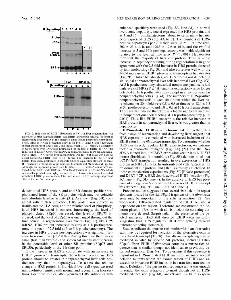
protection assay for HRS mRNA (Fig. 2A). Although the rel-ative increase and ultimate level of HRS transcripts are muchless in the regenerating than in the developing liver, we as-sessed whether there was any coordinate increase in the levelof EIIIB1 fibronectin transcripts in the posthepatectomy liver.Total fibronectin mRNA increased threefold in the first fewhours posthepatectomy. EIIIB1 fibronectin transcripts in-creased to 2% of fibronectin transcripts, an increase of approx-imately 20-fold compared to normal liver. The relative increasewas higher in nonparenchymal liver cells than in hepatocytes(Fig. 2B). In nonparenchymal liver cells, which includeKupffer, endothelial, and other sinusoidal cells, the increase inEIIIB1 transcripts posthepatectomy was approximately 35-fold, and in hepatocytes it was 2-fold. Using similar ap-proaches, we did not detect an increase in EIIIA fibronectintranscripts in this time frame in regenerating liver (data notshown).
Induction of HRS protein in regenerating liver, particularlyin nonparenchymal liver cells. By Northern blot and RNaseprotection assays, we observed an approximately fivefold in-duction of HRS mRNA in regenerating liver and insulin-treated H35 hepatoma cells (7) (Fig. 2A). To determine ifprotein levels increased as well, we prepared antibodies againstthe HRS RNA binding domain (Fig. 3A). Immunoblots ofregenerating liver and insulin-treated H35 cell extracts wereprobed with both anti-HRS and anti-SR antibodies. Anti-HRS
FIG. 1. Expression of HRS and EIIIB1 mRNAs during liver development.(A) Schematic diagram of the relevant portion of the HRS gene along with theRNase protection probe and of the fibronectin (FN) gene, used for detection ofthe EIIIB1 and EIIIB2 forms of fibronectin mRNA by RT-PCR (Fig. 2B) andRNase protection assays (panel B and Fig. 2A). (B) RNase protection assay ofdeveloping liver RNAs to detect HRS mRNA (top) and EIIIB1 and EIIIB2
mRNAs (bottom) (10 mg of total RNA per lane). The quantitation is indicatedfor each transcript (HRS, relative to 21-day [21d] liver; EIIIB1, as a percentageof total fibronectin transcripts.
4098 DU ET AL. MOL. CELL. BIOL.
on Septem
ber 30, 2014 by guesthttp://m
cb.asm.org/
Dow
nloaded from
detects total HRS protein, and anti-SR detects specific phos-phorylated forms of the SR proteins which may not coincidewith absolute level or activity (31). As shown (Fig. 3B), con-sistent with mRNA induction, HRS protein was induced ininsulin-treated H35 cells, and the relative level of phosphory-lated HRS increased in concert. Interestingly, the level ofphosphorylated SRp30 decreased, the level of SRp75 in-creased, and the level of SRp55 was unchanged throughout thetime course. In regenerating liver nuclei (Fig. 3C), like HRSmRNA, HRS protein increased as early as 1 h posthepatec-tomy to a peak of 2.5-fold at 3 to 5 h posthepatectomy). Theincrease in HRS protein posthepatectomy was significant rel-ative to normal liver (P , 0.02). During this time there was asmall (less than twofold) and somewhat inconsistent increasein the detectable level of other SR proteins (SRp75 andSRp30), particularly at the 1-h time point.
If the increase in HRS is coordinate with an increase inEIIIB1 fibronectin transcripts, the relative increase in HRSprotein should be greater in nonparenchymal liver cells pos-thepatectomy than in hepatocytes. To assess the relativechange in HRS protein in individual liver cell types, we usedimmunohistochemistry with normal and regenerating liver sec-tions. For these studies, affinity-purified HRS antibodies with
enhanced specificity were used (Fig. 3A, lane Af). In normalliver, some hepatocyte nuclei expressed the HRS protein, andat 3 and 16 h posthepatectomy, about twice as many hepato-cytes expressed HRS (Fig. 4A to F). The numbers of HRS-positive hepatocytes per 203 field were 96 6 13 at time zero,202 6 23 at 3 h, and 190.5 6 17.6 at 16 h, and the twofoldincrease at 3 and 16 h posthepatectomy was highly significantrelative to the level at time zero (P , 0.001). Hepatocytesrepresent the majority of liver cell protein. Thus, a 2-foldincrease in hepatocyte staining during regeneration is in goodagreement with the 2.5-fold increase in HRS protein detectedby immunoblotting (Fig. 3C) and also correlates well with the2-fold increase in EIIIB1 fibronectin transcripts in hepatocytes(Fig. 2B). Unlike hepatocytes, no HRS protein was detected insinusoidal nonparenchymal liver cells in normal liver (Fig. 4G).At 3 h posthepatectomy, sinusoidal nonparenchymal cells hadhigh levels of HRS (Fig. 4H), and this expression was no longerdetected at 16 h posthepatectomy except in a few perivascularnonparenchymal cells (Fig. 4I). The numbers of HRS-positivenonparenchymal cells at each time point within the liver pa-renchyma per 203 field were 0.8 6 0.8 at time zero, 12.4 6 3.5at 3 h posthepatectomy, and 0.8 6 0.8 at 16 h posthepatectomy.These results indicate that there is a highly significant increasein nonparenchymal cell labeling at 3 h posthepatectomy (P ,0.001). Thus, like EIIIB1 transcripts, the relative increase inHRS protein in nonparenchymal liver cells was greater than inhepatocytes.
HRS-mediated EIIIB exon inclusion. Taken together, datafrom assays of regenerating and developing liver suggest thatHRS expression is correlated with increased inclusion of theEIIIB exon in the fibronectin transcript. To determine whetherHRS can directly regulate EIIIB exon inclusion, we cotrans-fected a fibronectin minigene (Fig. 5A) (21) and the HRScDNA cloned into a pCMV5 expression vector into NIH 3T3mouse fibroblasts. Immunoblots (Fig. 5B) demonstrated thatpCMV-HRS transfection resulted in overexpression of HRSprotein in NIH 3T3 cells. In untransfected cells, SRp30 is thepredominant SR protein, and HRS is virtually undetected. Inthese cotransfection experiments (Fig. 5C [RNase protection]and D [RT-PCR]), HRS clearly activated EIIIB inclusion (Fig.5C, lane 4; Fig. 5D, lane 4). In the absence of HRS but pres-ence of endogenous SR proteins, little or no EIIIB1 transcriptwas detected (Fig. 5C, lane 3; Fig. 5D, lane 3).
Previous studies suggested that several six-nucleotide repeatelements located in the AflII/BglII fragment of the fibronectingene may be important for the splicing reaction (22). Wewondered if HRS-mediated regulation of EIIIB inclusion isdependent on this region. Therefore, we constructed the de-letion plasmid pBA, in which all six-nucleotide cis-acting ele-ments were deleted. Surprisingly, in the presence of the de-leted minigene, HRS still directed EIIIB exon inclusion,suggesting that HRS regulates EIIIB exon splicing throughdifferent cis-acting element(s).
Studies indicate that purine-rich motifs within an alternativeexon may be required for inclusion of the alternative exon inthe spliced transcript (31, 38). This alternative splicing may bemediated in vitro by specific SR proteins, including HRS/SRp40. Exon EIIIB of fibronectin contains a purine-rich se-quence that is similar though not identical to previously de-scribed sequences (Fig. 6A). To determine if this sequence isimportant in HRS-mediated EIIIB inclusion, we made severaldeletion mutants within the exonic region of EIIIB and as-sessed the impact on EIIIB inclusion in a transient-transfectionassay. Deletion of the purine-rich sequence alone was enoughto render the exon refractory to most though not all HRS-mediated inclusion (Fig. 6B, lanes 9 and 10). In this experi-
FIG. 2. Induction of EIIIB1 fibronectin mRNA in liver regeneration. (A)Detection of HRS (top) and EIIIB1 and EIIIB2 fibronectin mRNAs (bottom) inregenerating liver mRNA at the indicated times (hours posthepatectomy [post-hep]), using an RNase protection assay as for Fig. 1. Lanes 6 and 7 representshorter exposures of lanes 1 and 2 and indicate that EIIIB1 mRNA is detectablein regenerating liver RNA when normalized for the level of EIIIB2 mRNA. (B)Induction of EIIIB1 fibronectin mRNA in nonparenchymal (NP) cell and hepa-tocyte (Hep.) RNA during liver regeneration. An RT-PCR assay was used todetect fibronectin EIIIB1 and EIIIB2 forms. The reactions for EIIIB1 andEIIIB2 forms were performed in separate tubes on equal aliquots from the sameRT reaction. For locations of primers, see Materials and Methods and Fig. 1A.Although the upstream primer used to detect EIIIB2 mRNA could have de-tected both EIIIB1 and EIIIB2 mRNAs, the detection of EIIIB2 mRNA, whichis a smaller product, was highly favored. EIIIB1 transcripts were not detectedwith these EIIIB2 primers even in fetal liver, where EIIIB1 transcripts represent50% of fibronectin transcripts.
VOL. 17, 1997 HRS EXPRESSION DURING LIVER PROLIFERATION 4099
on Septem
ber 30, 2014 by guesthttp://m
cb.asm.org/
Dow
nloaded from

ment, which was confirmed by two similar experiments, HRSmediated a 51-fold increase in EIIIB inclusion in the presenceof the intact minigene (lanes 1 and 2) that was reduced by 80to 90% when the purine-rich sequence was deleted (5.2- to10.9-fold HRS-mediated inclusion [lanes 5 to 10]). When theregion of EIIIB exon just downstream of the purine sequencewas deleted (Fig. 6B, lanes 3 and 4, d128-187), EIIIB exoninclusion was high even in the absence of HRS. The level ofbasal splicing of the fibronectin minigene was unchanged for allof these constructs, as demonstrated by the consistently highlevel of total spliced products (EIIIB1 plus EIIIB2).
These experiments indicate that HRS is able to direct inclu-sion of EIIIB in the fibronectin transcript and that this splicingregulation depends on a cis-acting sequence that resides withinexon EIIIB. However, the relative ability of HRS to directEIIIB inclusion compared to other SR proteins was not di-rectly assessed. To begin to address this issue, pCG-SC35 andpCG-SRp55 (pCMV based), which had been previously usedto direct alternative splicing in vivo (39), were cotransfectedwith the fibronectin minigene (Fig. 7A). In three separatetransfections, neither SC35 nor SRp55 increased the level ofEIIIB1 transcripts significantly above the control level.
Finally, an RNA binding assay was used to show that HRScan directly bind to the purine-rich region in exon EIIIB (Fig.7B). Bacterially expressed HRS, which migrates at 29 kDa,
bound to the purine-rich region containing RNA (pGEM-GA)but not pGEM control RNA (lanes 1 and 2). A protein of 40kDa, the molecular size of HRS, was the only protein thatsignificantly bound pGEM-GA RNA (lane 4) when growingH35 cell nuclear extracts which contain abundant HRS (Fig.3B) were used. In a separate experiment in which the bindingreaction was allowed to proceed for twice as long, a smallamount of binding to 55- and 75-kDa proteins in addition tothe 40-kDa protein was detected (data not shown). Perhapsbecause the HRS antibodies are directed to the RNA bindingdomain which interacts with the RNA, it was not possible toeffectively immunoprecipitate even the bacterial HRS-RNAproduct (lane 3) by using anti-HRS antibodies (not shown).
DISCUSSION
Our initial studies resulted in the cloning of HRS/SRp40cDNA because HRS mRNA is strongly induced during prolif-eration of hepatic cells. Here we have shown that the expres-sion of HRS correlates with inclusion of the fibronectin EIIIBexon in developing and regenerating liver. Based on this cor-relation, we predicted that fibronectin pre-mRNA may be oneof the biological targets of HRS. Using an in vivo splicingassay, we showed that HRS mediates EIIIB exon inclusion and
FIG. 3. Relative increase in HRS in insulin-treated hepatoma cells and regenerating liver. (A) Top, Schematic diagram of the HRS protein showing RNA bindingdomains (R1 and R2), an arginine-serine-rich region (ARG-SER), and the region (aa 1 to 165) against which antibodies are directed. Bottom, immunoblot on which0.05 mg of purified bacterial HRS (aa 1 to 165)/lane was blotted with preimmune serum (PI), immune serum (Im), or affinity-purified serum (Af) against the HRSpeptide. (B) Immunoblot of insulin-treated H35 cell nuclear extracts with anti-HRS serum (a-HRS) or anti-SR (anti-SR monoclonal antibody M104) (a-SR). (C)Immunoblot of regenerating liver nuclei at indicated times posthepatectomy (posthep). Left, anti-SR antibody M104; middle, anti-HRS; right, Coomassie blue stainof the middle panel. The graph shows levels of HRS as detected by anti-HRS antibodies based on three separate determinations, three animals for each time point.Standard deviations were calculated by using Student’s t test (StatWorks software for the Macintosh).
4100 DU ET AL. MOL. CELL. BIOL.
on Septem
ber 30, 2014 by guesthttp://m
cb.asm.org/
Dow
nloaded from

that this activity depends on a purine-rich region within theEIIIB exon to which HRS binds.
Coordinate induction of HRS protein and fibronectin EIIIB1
transcripts. In developing liver, HRS mRNA correlates withthe presence of the EIIIB1 transcripts, as both are elevatedduring the major proliferative phase which ends about 1 monthafter birth. In developing liver, the level of HRS transcripts isapproximately 100-fold higher than in adult liver and corre-lates well with the level of EIIIB1 fibronectin transcripts, whichrepresent approximately 50% of fibronectin transcripts, com-pared to an almost undetectable level in the adult liver. Thelevel of induction of HRS in the regenerating liver is muchlower than in the developing liver. The level of EIIIB1 tran-scripts is also much lower in regenerating liver, representingonly 2% of the fibronectin transcripts but nonetheless a signif-icant increase relative to normal liver. The greatest relativeincrease in EIIIB1 transcripts in regenerating liver is seen innonparenchymal liver cells, which, unlike hepatocytes, containdetectable HRS only during regeneration. Thus, a reasonablecorrelation is seen between EIIIB1 fibronectin mRNA andHRS protein levels posthepatectomy, particularly in nonparen-chymal cells.
Inclusion of fibronectin EIIIB exon is mediated by HRS intransfected cells. Studies in which the expression of HRS andthat of exon EIIIB are correlated provided a rationale fordetermining if HRS can direct the inclusion of the EIIIB exonin fibronectin mRNA. Using an in vivo splicing assay, in whichfibronectin minigenes were transfected in the presence or ab-sence of HRS, we demonstrated that HRS can mediatefibronectin EIIIB exon inclusion. This transfection assayworked very well, perhaps because NIH 3T3 cells contain littleendogenous HRS. The increase in HRS relative to other SRproteins was substantial following the transfection and suffi-cient to change the ratio of EIIIB1 transcripts from undetect-able to approximately one-half of the spliced products (Fig.5C). When other SR proteins (SRp55 and SC35) were tested,no significant increase in EIIIB1 transcripts was observed.However, we have not established whether SR proteins whichwere not tested (e.g., SRp75) can direct the inclusion of theEIIIB exon. Moreover, the relative ratio of other SR factors inspecific cells may have an impact on the relative efficiency ofHRS in mediating EIIIB inclusion.
HRS-directed EIIIB inclusion does not depend on a cis-acting sequence within an intron of the fibronectin gene (22)
FIG. 4. Increase of HRS in hepatocytes and nonparenchymal cells in regenerating liver. Immunohistochemistry with anti-HRS antibodies demonstrates nuclearHRS levels in nonparenchymal cells (NP) and hepatocytes (H) at indicated times posthepatectomy (PH). (A to C) Portal triad; (D to F and G to I) central vein atmagnifications of 3380 and 3760. (Nomarsky optics). This experiment was repeated twice. Quantitation of nonparenchymal cell number was done by countingpositively stained cells in five 403 fields for each time point and hepatocytes in four 203 fields.
VOL. 17, 1997 HRS EXPRESSION DURING LIVER PROLIFERATION 4101
on Septem
ber 30, 2014 by guesthttp://m
cb.asm.org/
Dow
nloaded from

but clearly depends on a purine-rich sequence within the EIIIBexon to which HRS binds. In the absence of the purine-richsequence (Fig. 6), there is about 10 to 20% residual HRS-mediated EIIIB inclusion, which could be due to another pu-rine-rich sequence near the 39 end of the exon; alternatively,HRS may interact with the 59 or 39 site to promote exoninclusion. The sequence of the purine-rich stretch to whichHRS binds is very similar though not identical to sequences ofexonic splicing enhancers that have been found in the alterna-tive exons of troponin T, EIIIA, and other mRNAs (23, 38, 43,45, 48). It is believed that SR proteins, through binding toexonic splicing enhancers, recruit the basal splicing factorssuch as U2, U1, and other accessory splicing factors (26, 51). Inone model, SR proteins that bind to specific purine-rich se-quences allow for exon inclusion by facilitating the formationof a bridge between the splice acceptor proteins (U2AF) at the59 end of the exon and the splice donor proteins at the 39 endof the exon (31). In studies of troponin T, it was found thatSRp40, SRp55, SRp30a, and SRp75 bind to the purine-richregion in exon 5, whereas other SR proteins (e.g., SRp30b) donot (38). Binding correlates with the ability of these proteins to
direct alternative splicing. A similar sequence within the bo-vine growth hormone gene binds to a different set of SR pro-teins (43). Based on the data accumulated so far, SR proteinsbind to purine-rich sequences but may demonstrate specificityfor certain pre-mRNAs, thus determining the set of alternativeexons that can be regulated by each SR protein (44).
In our studies, removal of the sequence just downstream ofthe purine-rich sequence resulted in constitutive EIIIB inclu-sion that was not enhanced much by the presence of HRS.Potential explanations for this effect could be the loss of ad-verse secondary structure or removal of a splicing repressorprotein which normally binds to this downstream sequence.Our data do not directly address these two possibilities.
Significance of HRS to liver growth: other potential targets.It is clear that changes in hepatocyte polarity occur duringhepatic proliferation, and adhesion proteins may help mediatethese changes (42). Alternative forms of fibronectin producedin nonparenchymal liver cells like Ito and endothelial cells maybe deposited in the space of Disse. This may aid the prolifer-ating and postproliferative hepatocyte in moving into its newposition within the liver lobule, thereby ultimately restoring
FIG. 5. Inclusion of the EIIIB exon in fibronectin mRNA mediated by HRS. (A) Schematic diagram of fibronectin minigene and deletion and the methods ofdetection by RT-PCR and RNase protection. HGH, human growth hormone. (B) Immunoblot with anti-M104 (detects SR proteins) of NIH 3T3 cells transfected with5 mg of pCMV or pCMV-HRS (one plate of cells per lane; less protein loaded in lane 1). (C) Cotransfection of 5 mg of pCMV-HRS (1) or 5 mg of pCMV with theindicated minigene (5 mg). Lanes 1 and 2, pCMV and pCMV-HRS, respectively, in the absence of minigene showing that no endogenous fibronectin is detected. RNaseprotection analysis used a probe extending from EIIIB to III9 as shown (10 mg of total RNA/reaction). Reactions were run on the same gel, but spaces between samplelanes were eliminated in the figure. (D) Cotransfection as for panel C except that analysis was done by RT-PCR. Lanes 1 and 2 are as in panel C, in the absence ofminigene, showing that some endogenous fibronectin is detected in this assay. The relative amount of EIIIB1 spliced product was best estimated by the RNaseprotection assay, as RT-PCR underestimated the amount of the larger EIIIB1 PCR product. The 59 primer was the same as that used for the assay shown in Fig. 2B.Experiments with the intact minigene with or without pCMV-HRS were repeated several times with the same results.
4102 DU ET AL. MOL. CELL. BIOL.
on Septem
ber 30, 2014 by guesthttp://m
cb.asm.org/
Dow
nloaded from

normal polarity. However, as no specific function has beenascribed to different forms of fibronectin, the contribution ofalternatively spliced forms to restoration of hepatic architec-ture is not known (4). Our report does not attempt to addressthe biological function of exon EIIIB. However, the fact that
EIIIA does not appear in the early phases of normal liverregeneration lends support to the idea that exon EIIIA andexon EIIIB alternative splicing are regulated by differentmechanisms and may have different functions.
Although we presented evidence that fibronectin pre-mRNAis a potential target of HRS, a number of other messages maybe spliced alternatively during liver proliferation and could betargets of HRS and other SR proteins that increase in prolif-erative states of the liver. These mRNAs include those forprotein tyrosine phosphatases, cathepsin B, and cytochromeP450 as well as other liver-specific genes that are known tohave different spliced forms in normal liver (14, 25, 41). Inaddition to new RNA synthesis, which increases dramaticallyduring cellular proliferation, alternative splicing provides amechanism by which proliferation-specific protein forms maybe produced.
ACKNOWLEDGMENTS
We thank Mark Roth for M104 and Richard Hynes for fibronectinconstructs.
We thank Adrian Krainer for the SC35 and SRp55 expression plas-mids. This work was in part supported by a Juvenile Diabetes Foun-dation Award, NIH grants R01 DK44237 and P50 DK49210 to R.T.,and NRSA NIH K08 to L.E.G.
REFERENCES1. Ausubel, F. M., R. Brent, R. E. Kingston, D. D. Moore, J. G. Seidman, J. A.
Smith, and K. Struhl (ed.). 1994. Current protocols in molecular biology, vol.1, p. 4.10.1–4.10.5. John Wiley & Sons, Inc., New York, N.Y.
2. Caceres, J. F., S. Stamm, D. M. Helfman, and A. R. Krainer. 1994. Regu-lation of alternative splicing in vivo by overexpression of antagonistic splicingfactors. Science 265:1706–1709.
FIG. 6. Dependence of HRS-mediated inclusion on a purine-rich sequence in exon EIIIB. (A) Schematic diagram of exon EIIIB showing the location of thepurine-rich sequence and indicating the deletions of the original 7iBi89 construct that were used in the transfection experiment in panel B. The first base pair of exonEIIIB is designated 1. (B) Cotransfection of the various EIIIB deletion constructs in the presence or absence of 5 mg of pCMV-HRS. RT-PCR was used to detect thespliced products indicative of EIIIB inclusion (upper band) or exclusion (lower band). The different-size upper band corresponds to the size of the EIIIB deletion (2.5mg of total RNA/reaction). In the lower panel, a longer exposure of the EIIIB1 portion of the gel is shown, and percentages of EIIIB1 transcripts relative to total(EIIIB1 plus EIIIB2) transcripts are indicated. Similar experiments were performed three times with the same results.
FIG. 7. Specific interactions of HRS with the EIIIB purine-rich sequence.(A) Cotransfection of the 7iBi89 minigene with equal amounts of the controlplasmid pCMV (C; lane 1), pCMV-HRS (lane 2), pCG-SC35 (lane 3), andpCG-SRp55 (lane 4). Spliced products were detected by the RT-PCR (Fig. 5 and6). (B) Binding of HRS to the purine-rich sequence. Labeled RNA containingonly pGEM sequences (pGEM) or pGEM plus the purine-rich sequence(pGEM-GA) were allowed to interact with bacterially expressed HRS (HRS-bact) or nuclear extract from growing H35 cells (H35NE) as indicated. After UVcross-linking, the products were electrophoresed and exposed to autoradiogra-phy as described in Materials and Methods.
VOL. 17, 1997 HRS EXPRESSION DURING LIVER PROLIFERATION 4103
on Septem
ber 30, 2014 by guesthttp://m
cb.asm.org/
Dow
nloaded from

3. Caputi, M., C. A. Mela, and F. E. Baralle. 1995. Regulation of fibronectinexpression in rat regenerating liver. Nucleic Acids Res. 23:238–243.
4. Carnemolla, B., A. Leprini, G. Allemanni, M. Saginati, and L. Zardi. 1992.The inclusion of the type III repeat ED-B in the fibronectin moleculegenerates conformational modifications that unmask a cryptic sequence.J. Biol. Chem. 267:24689–24692.
5. Chomczynski, P., and N. Sacchi. 1987. Single-step method of RNA isolationby acid guanidium thiocyanate-phenol-chloroform extraction. Anal. Bio-chem. 162:156–159.
6. Diamond, R. H., D. E. Cressman, T. M. Laz, C. S. Abrams, and R. Taub.1994. PRL-1, a unique nuclear protein tyrosine phosphatase, affects cellgrowth. Mol. Cell. Biol. 14:3752–3762.
7. Diamond, R. H., K. Du, V. M. Lee, K. L. Mohn, B. A. Haber, D. S. Tewari,and R. Taub. 1993. Novel delayed-early and highly insulin-induced growthresponse genes. J. Biol. Chem. 268:15185–15192.
8. Enrich, C., W. H. Evans, and C. G. Gahmberg. 1988. Fibronectin isoforms inplasma membrane domains of normal and regenerating rat liver. FEBS Lett.228:135–138.
9. Fausto, N., and E. M. Webber. 1994. Liver regeneration, p. 1059–1084. InI. M. Arias, J. L. Boyer, N. Fausto, W. B. Jacoby, D. Schachter, and D. A.Shafritz (ed.), The liver: biology and pathobiology. Raven Press, New York,N.Y.
10. Fu, X.-D. 1993. Specific commitment of different pre-mRNAs to splicing bysingle RS proteins. Nature 365:82–85.
11. Fu, X.-D., and T. Maniatis. 1992. The 35-kDa mammalian splicing factorSC35 mediates specific interactions between U1 and U2 small nuclear ribo-nucleoprotein particles at the 39 splicing site. Proc. Natl. Acad. Sci. USA89:1725–1729.
12. Ge, H., P. Zuo, and J. L. Manley. 1991. Primary structure of the humansplicing factor ASF reveals similarities with Drosophila regulators. Cell 66:373–382.
13. Gluck, U., L. R. Fernandez, R. Pankov, and A. Ben-Ze’ev. 1992. Regulationof adherens junction protein expression in growth-activated 3T3 cells and inregenerating liver. Exp. Cell Res. 202:477–486.
14. Gong, Q., S. J. Chan, A. S. Bajkowski, D. F. Steiner, and A. Frankfater. 1993.Characterization of the cathepsin b gene and multiple mRNAs in humantissues: evidence for alternative splicing of cathepsin b pre-mRNA. DNACell Biol. 12:299–309.
15. Greenbaum, L. E., D. E. Cressman, B. A. Haber, and R. Taub. 1995. Coex-istence of C/EBP a,b growth-induced proteins and DNA synthesis in hepa-tocytes during liver regeneration: implications for maintenance of the dif-ferentiated state during liver growth. J. Clin. Invest. 96:1351–1365.
16. Haber, B. A., S. Chin, E. Chuang, W. Buikhuisen, A. Naji, and R. Taub. 1995.High levels of glucose-6-phosphatase gene and protein expression in prolif-erating liver and diabetes. J. Clin. Invest. 95:832–841.
17. Haber, B. A., K. L. Mohn, R. H. Diamond, and R. Taub. 1993. Inductionpatterns of 70 genes during nine days after hepatectomy define the temporalcourse of liver regeneration. J. Clin. Invest. 91:1319–1326.
18. Haber, B. A., L. Naji, D. E. Cressman, and R. Taub. 1995. Coexpression ofliver-specific and growth-induced genes in perinatal and regenerating liver:attainment and maintenance of the differentiated state during rapid prolif-eration. Hepatology 22:906–914.
19. Hodges, D., and S. I. Bernstein. 1994. Genetic and biochemical analysis ofalternative RNA splicing. Adv. Genet. 31:207–281.
20. Hsu, J.-C., R. Bravo, and R. Taub. 1992. Interactions among LRF-1, JunB,c-Jun, and c-Fos define a regulatory program in the G1 phase of liverregeneration. Mol. Cell. Biol. 12:4654–4665.
21. Huh, G. S., and R. O. Hynes. 1993. Elements regulating an alternativelyspliced exon of the rat fibronectin gene. Mol. Cell. Biol. 13:5301–5314.
22. Huh, G. S., and R. O. Hynes. 1994. Regulation of alternative pre-mRNAsplicing by a novel repeated hexanucleotide element. Genes Dev. 8:1561–1574.
23. Humphrey, M. B., J. Bryan, T. A. Cooper, and S. M. Berget. 1995. A32-nucleotide exon-splicing enhancer regulates usage of competing 59 splicesites in a differential internal exon. Mol. Cell. Biol. 15:3979–3998.
24. Jarnagin, W. R., D. C. Rockey, V. E. Koteliansky, S.-S. Wang, and D. M.Bissell. 1994. Expression of variant fibronectins in wound healing: cellularsource and biological activity of the EIIIA segment in rat hepatic fibrogen-esis. J. Cell Biol. 127:2037–2048.
25. Kimura, H., K. Sogawa, Y. Sakai, and Y. Fujii-Kuriyama. 1989. Alternativesplicing mechanism in a cytochrome P-450 (P-450PB-1) gene generates thetwo mRNAs coding for proteins of different functions. J. Biol. Chem. 264:2238–2342.
26. Kohtz, J., S. F. Jamison, C. L. Will, P. Zuo, R. Luhrmann, M. A. Garcia-Blanco, and J. L. Manley. 1994. Protein-protein interactions and 59-splicing-site recognition in mammalian mRNA precursors. Nature 368:119–124.
27. Krainer, A. R., A. Mayeda, D. Kozak, and G. Binns. 1991. Functional ex-pression of cloned human splicing factor SF2: homology to RNA-bindingproteins, U1 70K, and Drosophila splicing regulators. Cell 66:383–394.
28. Lavigueur, A., H. La Branche, A. R. Kornblihtt, and B. Chabot. 1993. A
splicing enhancer in the human fibronectin alternate ED1 exon interacts withSR proteins and stimulates U2 snRNP binding. Genes Dev. 7:2405–2417.
29. Legrain, P., and G. Chanfreau. 1994. Pre-mRNA splicing: from intron rec-ognition to catalysis. Bull. Inst. Pasteur 92:153–179.
30. Malim, M. H., and B. R. Cullen. 1993. Rev and the fate of pre-mRNA in thenucleus: implications for the regulation of RNA processing in eukaryotes.Mol. Cell. Biol. 13:6180–6189.
31. Manley, J. L., and R. Tacke. 1996. SR proteins and splicing. Genes Dev.10:1569–1579.
32. Mohn, K. L., T. M. Laz, J.-C. Hsu, A. E. Melby, R. Bravo, and R. Taub. 1991.The immediate-early growth response in regenerating liver and insulin-stim-ulated H-35 cells: comparison to serum-stimulated 3T3 cells and identifica-tion of 41 novel immediate-early genes. Mol. Cell. Biol. 11:381–390.
33. Norton, P. A., and R. O. Hynes. 1987. Alternative splicing of chicken fi-bronectin in embryos and in normal and transformed cells. Mol. Cell. Biol.7:4297–4307.
34. Oyama, F., S. Hirohashi, M. Sakamoto, K. Titani, and K. Sekiguchi. 1993.Coordinate oncodevelopmental modulation of alternative splicing offibronectin pre-messenger RNA at ED-A, ED-B, and CS1 regions in humanliver tumors. Cancer Res. 53:2005–2011.
35. Oyama, F., S. Hirohashi, Y. Shimosato, K. Titani, and K. Sekiguchi. 1989.Deregulation of alternative splicing of fibronectin pre-mRNA in malignanthuman liver tumors. J. Biol. Chem. 264:10331–10334.
36. Oyama, F., Y. Murata, N. Suganuma, T. Kimura, K. Titani, and K. Sekigu-chi. 1989. Patterns of alternative splicing of fibronectin pre-mRNA in humanadult and fetal tissues. Biochemistry 28:1428–1434.
37. Pagani, F., L. Zagato, C. Verganii, G. Casari, A. Sidoli, and F. E. Baralle.1991. Tissue-specific splicing pattern of fibronectin messenger RNA precur-sor during development and aging in rat. J. Cell Biol. 113:1223–1229.
38. Ramchatesingh, J., A. M. Zahler, K. M. Neugebauer, M. B. Roth, and T. A.Cooper. 1995. A subset of SR proteins activates splicing of the cardiactroponin T alternative exon by direct interactions with an exonic enhancer.Mol. Cell. Biol. 15:4898–4907.
39. Screaton, G. R., J. F. Caceres, A. Mayeda, M. V. Bell, M. Plebanski, D.Jackson, J. Bell, and A. R. Krainer. 1995. Identification and characterizationof three members of the human SR family of pre-mRNA splicing factors.EMBO J. 14:4336–4349.
40. Sharp, P. A. 1994. Split genes and RNA splicing. Cell 77:805–815.41. Shifrin, V. I., and B. G. Neel. 1993. Growth factor-inducible alternative
splicing of nontransmembrane phosphotyrosine phosphatase PTP-1B pre-mRNA. J. Biol. Chem. 268:25476–25484.
42. Stamatoglou, S. C., and R. C. Hughes. 1994. Cell adhesion molecules in liverfunction and pattern formation. FASEB J. 8:420–427.
43. Sun, Q., A. Mayeda, R. K. Hampson, A. R. Krainer, and R. M. Rottman.1993. General splicing factor SF2/ASF promotes alternative splicing by bind-ing to an exonic splicing enhancer. Genes Dev. 7:2598–2608.
44. Tacke, R., and J. L. Manley. 1995. The human splicing factors ASF/SF2 andSC35 possess distinct, functionally significant RNA binding specificities.EMBO J. 14:3540–3551.
45. Tanaka, K., A. Watakabe, and Y. Shimura. 1994. Polypurine sequenceswithin a downstream exon function as a splicing enhancer. Mol. Cell. Biol.14:1347–1354.
46. Taub, R. 1995. Transcriptional control of liver regeneration. 1996. FASEB J.10:413–427.
47. Tavian, D., G. DePetro, M. Colombi, N. Portalani, S. M. Giulini, R.Gardella, and S. Barlati. 1994. RT-PCR detection of fibronectin EIIIA andEIIIB isoforms; molecular markers for hepatocellular carcinoma. Int. J.Cancer 56:820–825.
48. Tian, M., and T. Maniatis. 1993. A splicing enhancer complex controlsalternative splicing of doublesex pre-mRNA. Cell 74:105–114.
49. Vallejo, A. N., R. J. Pogulis, and L. R. Pease. 1995. Mutagenesis and syn-thesis of novel recombinant genes using PCR, PCR primer. Cold SpringHarbor Laboratory, Cold Spring Harbor, N.Y.
50. Wang, J., Y. Takagaki, and J. L. Manley. 1996. Targeted disruption of anessential vertebrate gene: ASF/SF2 is required for cell viability. Genes Dev.10:2588–2599.
51. Wu, J. Y., and T. Maniatis. 1993. Specific interactions between proteinsimplicated in splicing site selection and regulated alternative splicing. Cell75:1061–1070.
52. Zahler, A. M., W. S. Lane, J. A. Stolk, and M. B. Roth. 1992. SR proteins: aconserved family of pre-mRNA splicing factors. Genes Dev. 6:837–847.
53. Zahler, A. M., K. M. Neugebauer, W. S. Lane, and M. B. Roth. 1993. Distinctfunctions of SR proteins in alternative pre-mRNA splicing. Science 260:219–222.
54. Zahler, A. M., K. M. Neugebauer, J. A. Stolk, and M. B. Roth. 1993. HumanSR proteins and isolation of a cDNA encoding SRp75. Mol. Cell. Biol.13:4023–4028.
55. Zuo, P., and J. L. Manley. 1994. The human splicing factor ASF/SF2 canspecifically recognize pre-mRNA 59 splice sites. Proc. Natl. Acad. Sci. USA91:3363–3367.
4104 DU ET AL. MOL. CELL. BIOL.
on Septem
ber 30, 2014 by guesthttp://m
cb.asm.org/
Dow
nloaded from
Copyright © 2022 FDOKUMEN