Std. X English (Third Language) Total marks: 80 Time: 3 hrs.
Upload
uni-leipzigCategory
view
0download
0
C
EADBc(
EJEPKFR1
UUCLEHPLU
PTsvTswooeltVmmtta
AA
1C
ONSENSUS DOCUMENT
HRA/HRS Expert Consensus on Catheter Ablation of Ventricularrrhythmias
eveloped in a partnership with the European Heart Rhythm Association (EHRA), a Registeredranch of the European Society of Cardiology (ESC), and the Heart Rhythm Society (HRS); inollaboration with the American College of Cardiology (ACC) and the American Heart AssociationAHA)
tienne M. Aliot, MD, FESC, FHRS,1 William G. Stevenson, MD, FHRS,2
esus Ma Almendral-Garrote, MD, PhD,3 Frank Bogun, MD,4 C. Hugh Calkins, MD, FHRS,5
tienne Delacretaz, MD, FESC,6 Paolo Della Bella, MD, PhD, FESC,7 Gerhard Hindricks, MD, PhD,8
ierre Jaïs, MD, PhD,9 Mark E. Josephson, MD,10 Josef Kautzner, MD, PhD,11 G. Neal Kay, MD,12
arl-Heinz Kuck, MD, PhD, FESC, FHRS,13 Bruce B. Lerman, MD, FHRS,14
rancis Marchlinski, MD, FHRS,15 Vivek Reddy, MD,16 Martin-Jan Schalij, MD, PhD,17
ichard Schilling, MD,18 Kyoko Soejima, MD,19 and David Wilber, MD20
CHU de Nancy, Hôpital de Brabois, Vandoeuvre-les-Nancy, France; 2Brigham and Women’s Hospital, Boston, MA,SA; 3Hospital General Gregorio Maranon, Madrid, Spain; 4University of Michigan Health System, Ann Arbor, MI,SA; 5Johns Hopkins Hospital, Baltimore, MD, USA; 6University Hospital, Bern, Switzerland; 7Universita degli Studi,entro Cardiologico F. Monzino, Milan, Italy; 8University of Leipzig, Heartcenter, Leipzig, Germany; 9Hôpital du Hauteveque, Bordeaux, France; 10Beth Israel Deaconess Medical Center, Boston, MA, USA; 11Institute For Clinical Andxperimental Medicine (Ikem), Prague, Czech Republic; 12University of Alabama, Birmingham, AL, USA; 13Asklepiosospital St Georg, Hamburg, Germany; 14Cornell University Medical Center, New York, NY, USA; 15University ofennsylvania, Philadelphia, PA, USA; 16University of Miami, Miami, FL, USA; 17Leiden University Medical Center,eiden, The Netherlands; 18Barts and the London NHS Trust, UK; 19University of Miami, Miami, FL, USA; and 20Loyola
niversity Medical Center, Maywood, IL, USAmtbaTFctpfbdacf
T
reamblehe purpose of this Consensus Statement is to provide atate-of-the-art review of the field of catheter ablation ofentricular tachycardia (VT), and to report the findings of aask Force, convened by the European Heart Rhythm As-ociation (EHRA) and the Heart Rhythm Society (HRS) thatas charged with defining the indications, techniques, andutcomes of this procedure. This statement summarizes thepinion of the Task Force members based on their ownxperience in treating patients, as well as a review of theiterature. It is directed to all healthcare professionals whoreat patients who are considered for catheter ablation ofT. This statement is not intended to recommend or pro-ote catheter ablation of VT. Rather, the ultimate judge-ent regarding care of a particular patient must be made by
he healthcare provider and the patient with consideration ofhe individual patient characteristics that impact on risksnd benefits of the therapy. In writing a ‘consensus’ docu-
Endorsed by the Heart Rhythm Society, the European Heart Rhythmssociation, a registered branch of the European Society of Cardiology, the
merican Heart Association and the American College of Cardiology.547-5271/$ -see front matter © 2009 Heart Rhythm Society and the European Hardiology. Published by Elsevier, Inc. All rights reserved.
ent, it is recognized that consensus does not mean thathere was complete agreement among all Task Force mem-ers. We identified those aspects of VT ablation for whichtrue ‘consensus’ could be identified. Surveys of the entireask Force were used to identify these areas of consensus.or the purposes of this Consensus Document, we defined aonsensus as 70% or greater agreement by the members ofhis task force. One objective of this document is to improveatient care by summarizing the foundation of knowledgeor those involved with catheter ablation of VT. All mem-ers of the Task Force, as well as peer reviewers of theocument, were asked to provide disclosure statements ofll relationships that might be perceived as real or potentialonflicts of interest. Disclosures for the members of the taskorce are given in the Appendix section.
ABLE OF CONTENTS
I. INTRODUCTION .................................................887II. VENTRICULAR TACHYCARDIA:
DEFINITIONS, MECHANISMS, AND
RATIONALE FOR ABLATION..........................887eart Rhythm Association, a registered branch of the European Society ofdoi:10.1016/j.hrthm.2009.04.030
V
ARFT
ICreadcadmuacpcp
ImDMtsisp
MvMscmtt(lc
(csrto
TTp(midtu
lcthecoidccfdbAu
tgsitbRadfWist
ndlob
ST
887E. M. Aliot and W. G. Stevenson, et al. Ablation of ventricular arrhythmias
III. INDICATIONS FOR CATHETER ABLATIONOF VENTRICULAR TACHYCARDIA...............891
IV. TECHNICAL ASPECTS.......................................891V. VENTRICULAR TACHYCARDIA IN
STRUCTURAL HEART DISEASE.....................899VI. ABLATION OUTCOMES AND
CONSIDERATIONS IN SPECIFICDISEASES.............................................................908
VII. IDIOPATHIC VENTRICULARTACHYCARDIAS ................................................913
III. TRAINING AND INSTITUTIONALREQUIREMENTS AND COMPETENCIES .......916
IX. CLINICAL TRIAL CONSIDERATIONS............918X. CONCLUSIONS....................................................921
PPENDIX......................................................................922EFERENCES ................................................................923IGURES........................................889, 890, 900, 906, 909ABLES..........................................................888, 891, 920
. Introductionatheter ablation is now an important option to control
ecurrent ventricular tachycardias (VTs). The field hasvolved rapidly and is a work in progress. Ablation is oftensole therapy of VT in patients without structural heart
isease and is commonly combined with an implantableardioverter-defibrillator (ICD) and/or antiarrhythmic ther-py for scar-related VTs associated with structural heartisease. As the field progresses, it is important that theedical profession play a significant role in critically eval-
ating therapies as they are introduced and evolve. Rigorousnd expert analysis of the available data documenting indi-ations, techniques, benefits and risks, and outcomes canroduce helpful guidance to improve the effectiveness ofare, optimize patient outcomes, and identify areas for im-rovement and future research.
I. Ventricular tachycardia: definitions,echanisms, and rationale for ablationefinitionsany terms have entered clinical usage to describe observa-
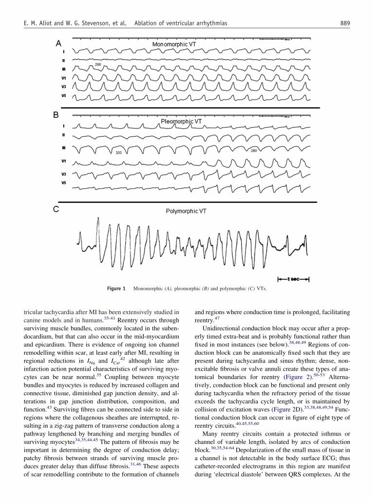
ions during mapping and ablation of VT. There has beenubstantial variation in the use of some terms by differentnvestigators. The committee felt that these terms should betandardized to facilitate better understanding of methods, end-oints, and outcomes across centres (Table 1 and Figure 1).1-4
echanisms and basis for catheter ablation ofentricular tachycardiaonomorphic VT can occur in individuals with or without
tructural heart disease. The underlying heart disease andlinical characteristics of the VT often suggest a potentialechanism and origin. Ventricular tachycardias that are due
o automaticity are expected to have a focal origin, makinghem susceptible to ablation with discrete radiofrequencyRF) lesions.5-12 Triggered activity or automaticity areikely causes of focal origin VTs, although small reentry
ircuits can often not be excluded. Idiopathic outflow tract pOT)-VTs have a focal origin. Relatively large reentry cir-uits are common in VT associated with ventricular scar,uch as prior infarction, but VT may appear focal if theeentry circuit is small, or due to a focal endocardial break-hrough from an epicardial reentry circuit. Automaticity canccur in some patients with ventricular scars.
riggered activity and automaticityriggered activity arises from oscillations in membraneotential during (early afterdepolarizations) or followingdelayed afterdepolarizations) an action potential. Experi-ental evidence implicates early afterdepolarizations in the
nitiation of polymorphic tachycardias in the long QT syn-romes.13 However, the mechanism of the premature ven-ricular beats targeted for ablation in these syndromes isnknown.14
Delayed afterdepolarizations can be caused by intracel-ular calcium overload which activates the Na�/Ca2� ex-hanger resulting in the transient inward current Iti.
1-4 Fac-ors that increase intracellular calcium include increases ineart rate, �-adrenergic stimulation, and digitalis. �-Adren-rgic effects are mediated through a cAMP-induced in-rease in intracellular calcium and are antagonized by aden-sine, which effects a decrease in cAMP. Termination ofdiopathic right ventricular outflow tract (RVOT) tachycar-ias by an intravenous bolus of adenosine or infusion ofalcium channel blockers, or by vagotonic manoeuvres isonsistent with triggered activity as the likely mechanismor some of these tachycardias.3 These tachycardias can beifficult to induce at electrophysiology (EP) testing; rapidurst pacing and/or isoproterenol infusion is often required.minophylline, calcium infusion, and atropine may also beseful.15
Less commonly, focal VT may be due to automaticityhat is provoked by adrenergic stimulation that is not trig-ered.1,15 This type of VT may become incessant undertress or during isoproterenol administration, but cannot benitiated or terminated by programmed electrical stimula-ion; it can sometimes be suppressed by calcium channellockers or �-blockers. In contrast to its effects on triggeredVOT tachycardia, adenosine transiently suppresses therrhythmia but does not terminate it.1,15 Automaticity fromamaged Purkinje fibres has been suggested as a mechanismor some catecholamine-sensitive, focal origin VTs.16,17
hether these VTs are due to abnormal automaticity, orig-nating from partially depolarized myocytes, as has beenhown for VTs during the early phase of myocardial infarc-ion (MI), is not clear.
Although automaticity is often associated as a mecha-ism of VT in the absence of overt structural heart disease,isease processes that diminish cell-to-cell coupling areikely to facilitate automaticity.18,19 Automatic VTs canccur in structural heart disease,17 and automatic prematureeats may initiate reentrant VTs.
car-related reentryhe majority of sustained monomorphic VTs (SMVTs) in
atients with structural heart disease are due to reentry
aritbeeatek
cdmchr
tca
T
ntry.
888 Heart Rhythm, Vol 6, No 6, June 2009
ssociated with areas of scar, designated as scar-relatedeentry (Table 1). Evidence supporting reentry includesnitiation and termination by programmed stimulation (al-hough this does not exclude triggered activity), demonstra-le entrainment or resetting with fusion, and continuouslectrical activity that cannot be dissociated from VT byxtrastimuli. Myocardial scar is identified from: low-volt-ge regions on ventricular voltage maps, areas with frac-ionated electrograms, unexcitability during pace mapping,vidence of scar on myocardial imaging, or from an area of
able 1 Definitions
Clinical characteristicsClinical ventricular tachycardia (VT): VT that has occurred spon
There are many potential problems and assumptions with thislaboratory (see Endpoints for ablation section).
Haemodynamically unstable VT causes haemodynamic compromiIdiopathic VT is a term that has been used to indicate VT that is
disease.Idioventricular rhythm is three or more consecutive beats at a r
atrial or AV nodal conduction.Incessant VT is continuous sustained VT that recurs promptly desNon-clinical VT is a term that has been used to indicate a VT ind
documented previously. This term is problematic because somespontaneously.262 It is recommended that this term can be avopreviously observed should be referred to as ‘undocumented VT
Non-sustained VT terminates spontaneously within 30 s.Presumptive clinical VT is similar to a spontaneous VT based on
but without the 12-lead ECG documentation of either the induRepetitive monomorphic VT: continuously repeating episodes ofSustained VT: continuous VT for �30 s or that requires an intervVentricular tachycardia: a tachycardia (rate �100/min) with thr
independent of atrial or AV nodal conduction.VT storm is considered three or more separate episodes of sustai
intervention.262,463
VT morphologiesMonomorphic VT has a similar QRS configuration from beat to be
uncommon, followed by stabilization of the QRS morphology.Multiple monomorphic VTs: refers to more than one morphologic
induced at different times.Polymorphic VT has a continuously changing QRS configuration f
sequence (Figure 1C).Pleomorphic VT has more than one morphologically distinct QRS
continuously changing (Figure 1B).Right and left bundle branch block-like—VT configurations: ter
R-wave described as ‘right bundle branch block-like’ and a domterminology is potentially misleading as the VT may not showmorphology in other leads.
Unmappable VT does not allow interrogation of multiple sites tomay be due to: haemodynamic intolerance that necessitates imother morphologies of VT, or repeated termination during map
Ventricular flutter is a term that has been applied to rapid VT ththe QRS morphology. It is preferable to avoid this term, in fav
MechanismsScar-related reentry describes arrhythmias that have characterist
identified from electrogram characteristics or myocardial imagiare commonly referred to as ‘macroreentry’.
Focal VT has a point source of earliest ventricular activation withmechanism can be automaticity, triggered activity, or microree
nown surgical incision. Prior MI is the most common b
ause, but scar-related VT also occurs in other myocardialiseases including arrhythmogenic right ventricular cardio-yopathy (ARVC), sarcoidosis, Chagas’ disease, dilated
ardiomyopathy, and after cardiac surgery for congenitaleart disease (particularly Tetralogy of Fallot) or valveeplacement.20-30
The substrate supporting scar-related reentry is charac-erized by (i) regions of slow conduction, (ii) unidirectionalonduction block at some point in the reentry path thatllows initiation of reentry, and (iii) areas of conduction
sly based on analysis of 12-lead ECG QRS morphology and rate.ation as it is applied to inducible VT in the electrophysiology
iring prompt termination.n to occur in the absence of clinically apparent structural heart
�100/min that originate from the ventricles independent of
peated intervention for termination over several hours.y programmed ventricular stimulation that has not beenat have not been previously observed will occurInduced VTs with a QRS morphology that has not beenology’.
d ECG or electrogram data available from ICD interrogation,spontaneous VT.rminating non-sustained VT.378,462
for termination (such as cardioversion).ore consecutive beats that originates from the ventricles
within 24 h, each requiring termination by an
ure 1A). Some variability in QRS morphology at initiation is not
stinct monomorphic VT, occurring as different episodes or
at to beat indicating a changing ventricular activation
x occurring during the same episode of VT, but the QRS is not
d to describe the dominant deflection in V1, with a dominantS-wave as ‘left bundle branch block-like’ configurations. Thiss characteristic of the same bundle branch block-like
the activation sequence or perform entrainment mapping; thiste VT termination, spontaneous or pacing-induced transition to
a sinusoidal QRS configuration that prevents identification ofmonomorphic VT with indeterminant QRS morphology.
reentry and originates from an area of myocardial scarrge reentry circuits that can be defined over several centimetres
ead of activation away in all directions from that site. The
taneoudesign
se requknow
ate of
pite reuced bVTs thided.morph
rate anced orself-teentionee or m
ned VT
at (Fig
ally di
rom be
comple
ms useinantfeature
definemedia
ping.at hasour of
ics ofng. La
a spr
lock that often define parts of the reentry path.31-34 Ven-
tcsdarricbctfrspsipdo
ar
efidpettdectr
cbac
omorph
889E. M. Aliot and W. G. Stevenson, et al. Ablation of ventricular arrhythmias
ricular tachycardia after MI has been extensively studied inanine models and in humans.35-41 Reentry occurs throughurviving muscle bundles, commonly located in the suben-ocardium, but that can also occur in the mid-myocardiumnd epicardium. There is evidence of ongoing ion channelemodelling within scar, at least early after MI, resulting inegional reductions in INa and ICa,
42 although late afternfarction action potential characteristics of surviving myo-ytes can be near normal.35 Coupling between myocyteundles and myocytes is reduced by increased collagen andonnective tissue, diminished gap junction density, and al-erations in gap junction distribution, composition, andunction.43 Surviving fibres can be connected side to side inegions where the collagenous sheathes are interrupted, re-ulting in a zig-zag pattern of transverse conduction along aathway lengthened by branching and merging bundles ofurviving myocytes34,35,44,45 The pattern of fibrosis may bemportant in determining the degree of conduction delay;atchy fibrosis between strands of surviving muscle pro-uces greater delay than diffuse fibrosis.31,46 These aspects
Figure 1 Monomorphic (A), ple
f scar remodelling contribute to the formation of channels d
nd regions where conduction time is prolonged, facilitatingeentry.47
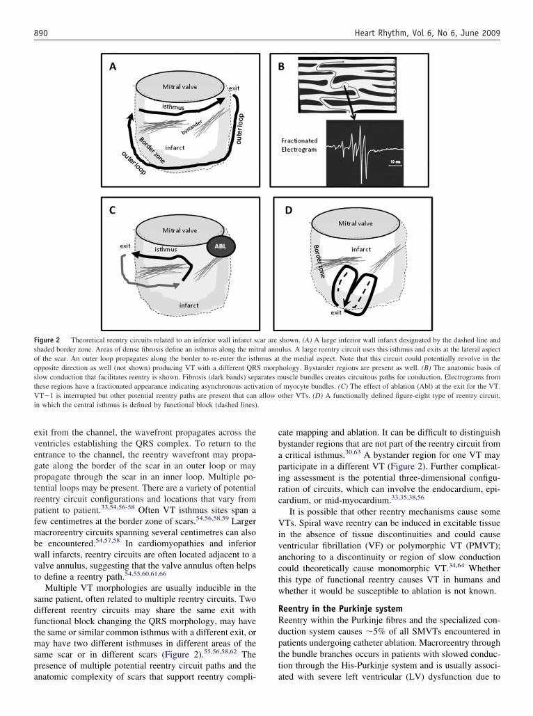
Unidirectional conduction block may occur after a prop-rly timed extra-beat and is probably functional rather thanxed in most instances (see below).38,48,49 Regions of con-uction block can be anatomically fixed such that they areresent during tachycardia and sinus rhythm; dense, non-xcitable fibrosis or valve annuli create these types of ana-omical boundaries for reentry (Figure 2).50-53 Alterna-ively, conduction block can be functional and present onlyuring tachycardia when the refractory period of the tissuexceeds the tachycardia cycle length, or is maintained byollision of excitation waves (Figure 2D).33,38,48,49,54 Func-ional conduction block can occur in figure of eight type ofeentry circuits.40,45,55,60
Many reentry circuits contain a protected isthmus orhannel of variable length, isolated by arcs of conductionlock.30,35,54-64 Depolarization of the small mass of tissue inchannel is not detectable in the body surface ECG; thus
atheter-recorded electrograms in this region are manifest
ic (B) and polymorphic (C) VTs.
uring ‘electrical diastole’ between QRS complexes. At the
evegptrpfmbwvt
sdftmspa
cbapirc
Vivactw
RRdptt
FsoostVi .
890 Heart Rhythm, Vol 6, No 6, June 2009
xit from the channel, the wavefront propagates across theentricles establishing the QRS complex. To return to thentrance to the channel, the reentry wavefront may propa-ate along the border of the scar in an outer loop or mayropagate through the scar in an inner loop. Multiple po-ential loops may be present. There are a variety of potentialeentry circuit configurations and locations that vary fromatient to patient.33,54,56-58 Often VT isthmus sites span aew centimetres at the border zone of scars.54,56,58,59 Largeracroreentry circuits spanning several centimetres can also
e encountered.54,57,58 In cardiomyopathies and inferiorall infarcts, reentry circuits are often located adjacent to aalve annulus, suggesting that the valve annulus often helpso define a reentry path.54,55,60,61,66
Multiple VT morphologies are usually inducible in theame patient, often related to multiple reentry circuits. Twoifferent reentry circuits may share the same exit withunctional block changing the QRS morphology, may havehe same or similar common isthmus with a different exit, oray have two different isthmuses in different areas of the
ame scar or in different scars (Figure 2).55,56,58,62 Theresence of multiple potential reentry circuit paths and the
igure 2 Theoretical reentry circuits related to an inferior wall infarct shaded border zone. Areas of dense fibrosis define an isthmus along the mitf the scar. An outer loop propagates along the border to re-enter the isthpposite direction as well (not shown) producing VT with a different QRlow conduction that facilitates reentry is shown. Fibrosis (dark bands) sephese regions have a fractionated appearance indicating asynchronous activT�1 is interrupted but other potential reentry paths are present that can
n which the central isthmus is defined by functional block (dashed lines)
natomic complexity of scars that support reentry compli- a
ate mapping and ablation. It can be difficult to distinguishystander regions that are not part of the reentry circuit fromcritical isthmus.30,63 A bystander region for one VT may
articipate in a different VT (Figure 2). Further complicat-ng assessment is the potential three-dimensional configu-ation of circuits, which can involve the endocardium, epi-ardium, or mid-myocardium.33,35,38,56
It is possible that other reentry mechanisms cause someTs. Spiral wave reentry can be induced in excitable tissue
n the absence of tissue discontinuities and could causeentricular fibrillation (VF) or polymorphic VT (PMVT);nchoring to a discontinuity or region of slow conductionould theoretically cause monomorphic VT.34,64 Whetherhis type of functional reentry causes VT in humans andhether it would be susceptible to ablation is not known.
eentry in the Purkinje systemeentry within the Purkinje fibres and the specialized con-uction system causes �5% of all SMVTs encountered inatients undergoing catheter ablation. Macroreentry throughhe bundle branches occurs in patients with slowed conduc-ion through the His-Purkinje system and is usually associ-
shown. (A) A large inferior wall infarct designated by the dashed line andulus. A large reentry circuit uses this isthmus and exits at the lateral aspectthe medial aspect. Note that this circuit could potentially revolve in the
hology. Bystander regions are present as well. (B) The anatomic basis ofuscle bundles creates circuitous paths for conduction. Electrograms from
f myocyte bundles. (C) The effect of ablation (Abl) at the exit for the VT.ther VTs. (D) A functionally defined figure-eight type of reentry circuit,
car areral annmus at
S morparates mation oallow o
ted with severe left ventricular (LV) dysfunction due to
do
omLpba
EToirttatlamcaipsl
a
IvSsppsmphteuteaosVfareiVs
tct
ITTm
Tt
Iv*s
891E. M. Aliot and W. G. Stevenson, et al. Ablation of ventricular arrhythmias
ilated cardiomyopathy, valvular heart disease, and lessften ischaemic heart disease (see below).65,67,68,70
Left ventricular intrafascicular verapamil-sensitive VTccurs in patients without structural heart disease. Theechanism is reentry that appears to involve a portion of theV Purkinje fibres, most often in the region of the leftosterior fascicle, giving rise to a characteristic right bundleranch block (RBBB) superior axis QRS configuration andQRS duration that is only slightly prolonged.69,70
lectrophysiological basis for catheter ablationhe mechanism of VT is a key determinant for selectionf mapping strategies to identify ablation target sites. Fordiopathic VT, the focal origin or critical portion of theeentry path is usually contained in a very small area suchhat discrete lesions can abolish VT; therefore, mappingargets a precise region. For scar-related VTs, ablation isimed at transecting the critical VT isthmus. Ventricularachycardia isthmuses may be narrow, allowing a discreteesion to abolish VT, or broad, requiring larger ablationreas. In addition, in patients with unmappable VTs andultiple VTs, larger ablation areas targeting putative criti-
al reentry sites, often in or near the border zone of scars,re often employed. In post-MI VT, most reentry circuitsthmuses can be transected using an endocardial ap-roach.50,52,54,57,59,62,71-73 However, critical reentry circuitites are intramural or subepicardial in some patients; theseocations are common in some cardiomypathies.71,74,75
In Purkinje reentry VT, specialized conduction fibres thatre part of the reentry path are targeted for ablation.17,65,67,68
II. Indications for catheter ablation ofentricular tachycardiaelection of catheter ablation for an individual patienthould consider risks and benefits that are determined byatient characteristics, as well as the availability of appro-riate facilities with technical expertise. In patients withtructural heart disease, episodes of sustained VT are aarker for increased mortality and reduce quality of life in
atients who have implanted defibrillators and structuraleart disease.76-80 Antiarrhythmic medications can reducehe frequency of ICD therapies, but have disappointingfficacy and side effects.81-83 Advances in technology andnderstanding of VT substrates now allow ablation of mul-iple and unstable VTs with acceptable safety and efficacy,ven in patients with advanced heart disease. In the past,blation was often not considered until pharmacologicalptions had been exhausted, often after the patient haduffered substantial morbidity from recurrent episodes ofT and ICD shocks. There was consensus among the task
orce members that catheter ablation for VT should gener-lly be considered early in the treatment of patients withecurrent VT. General recommendations for the use of cath-ter ablation are summarized in Table 2. More detailednformation regarding risks and benefits for specific types ofT and in specific types of heart disease is provided in
ections below. It should be recognized that the database for i
hese consensus recommendations consists largely of un-ontrolled trials and single-centre reports as summarized inhe discussion of individual diseases below.
V. Technical aspectsechnologies for mapping and ablationechnological advances have been critical to the develop-ent of the field and will continue to play an important role
able 2 Indications for catheter ablation of ventricularachycardia
Patients with structural heart disease (including prior MI,dilated cardiomyopathy, ARVC/D)
Catheter ablation of VT is recommended1. for symptomatic sustained monomorphic VT (SMVT),
including VT terminated by an ICD, that recurs despiteantiarrhythmic drug therapy or when antiarrhythmic drugsare not tolerated or not desired;*
2. for control of incessant SMVT or VT storm that is not due toa transient reversible cause;
3. for patients with frequent PVCs, NSVTs, or VT that ispresumed to cause ventricular dysfunction;
4. for bundle branch reentrant or interfascicular VTs;5. for recurrent sustained polymorphic VT and VF that is
refractory to antiarrhythmic therapy when there is asuspected trigger that can be targeted for ablation.
Catheter ablation should be considered1. in patients who have one or more episodes of SMVT despite
therapy with one of more Class I or III antiarrhythmic drugs;*2. in patients with recurrent SMVT due to prior MI who have LV
ejection fraction �0.30 and expectation for 1 year of survival,and is an acceptable alternative to amiodarone therapy;*
3. in patients with haemodynamically tolerated SMVT due to priorMI who have reasonably preserved LV ejection fraction (�0.35)even if they have not failed antiarrhythmic drug therapy.*
Patients without structural heart disease
Catheter ablation of VT is recommended for patients withidiopathic VT
1. for monomorphic VT that is causing severe symptoms.2. for monomorphic VT when antiarrhythmic drugs are not
effective, not tolerated, or not desired.3. for recurrent sustained polymorphic VT and VF (electrical storm)
that is refractory to antiarrhythmic therapy when there is asuspected trigger that can be targeted for ablation.
VT catheter ablation is contra-indicated1. in the presence of a mobile ventricular thrombus (epicardial
ablation may be considered);2. for asymptomatic PVCs and/or NSVT that are not suspected
of causing or contributing to ventricular dysfunction;3. for VT due to transient, reversible causes, such as acute
ischaemia, hyperkalaemia, or drug-induced torsade de pointes.
ARVC/D, arrhythmogenic right ventricular cardiomyopathy/dysplasia;CD, implantable cardioverter defibrillator; MI, myocardial infarction; VT,entricular tachycardia; VF, ventricular fibrillation.This recommendation for ablation stands regardless of whether VT istable or unstable, or multiple VTs are present.
n improving outcomes. The evaluation of new technologies
hlApt
otrIcenttc
MMtVp
EEclTcctteogss(
ubm
ammspctsocEtt
ms
wpmiLtstbmewiib
CPspgdntfbems
MMarog
sttdqttibesetdun
892 Heart Rhythm, Vol 6, No 6, June 2009
as generally been based on uncontrolled series. There isimited head-to-head comparison of different technologies.lthough new technologies generally increase the cost of arocedure when they are introduced, the costs may be jus-ified if they improve outcomes.
The process of evaluation and adoption of new technol-gies for clinical practice varies from country to country. Inhis document, the assessment of technologies is based oneview of the literature and consensus of the task force.ndividual technologies may not have been approved spe-ifically for catheter ablation of VT. It is important for thelectrophysiologist performing these procedures to recog-ize the value and limitations of each mapping system forheir effective use. As the field continues to evolve, adop-ion of new technologies should be based on well-designedlinical trials.
apping systemsapping systems that create chamber geometry and display
he ablation catheter position are often helpful in ablation ofT in structural heart disease and can be useful in selectedatients with idiopathic VT.
lectroanatomic mapping systemslectroanatomic mapping (EAM) refers to point by pointontact mapping combined with the ability to display theocation of each mapping point in three-dimensional space.his provides the opportunity to record intracardiac electri-al activation in relation to anatomical location in a cardiachamber of interest. Electroanatomic mapping systems in-egrate three main functions: (i) non-fluoroscopic localiza-ion of the ablation catheter within the heart; (ii) display oflectrogram characteristics, most commonly activation timer voltage, in relation to anatomic position; and (iii) inte-ration of electroanatomic information with three-dimen-ional images of the heart obtained from point by pointampling, intracardiac ultrasound, computed tomographyCT), or magnetic resonance imaging (MRI).
In patients with scar-related VTs, EAM systems areseful. In patients with idiopathic VTs, EAM systems cane useful, but are not required and are utilized by approxi-ately half of the task force members.One system utilizes low-level electromagnetic fields em-
nating from three separate coils beneath the patient that areeasured from a location sensor embedded in the tip of theapping catheter.84 This allows a three-dimensional recon-
truction of the chamber of interest and colour-coded dis-lay of various electrophysiological parameters for endo-ardial or epicardial mapping54,73,85-87 An alternativeechnology determines electrode position based on the mea-urement of a high-frequency current emitted by three pairsf orthogonally placed skin patches.62,88,89 Recently, intra-ardiac echocardiography (ICE) has been incorporated intoAM.90 The ICE probe, equipped with a location sensor and
racked by the mapping system, allows reconstruction of a
hree-dimensional shell of the chambers of interest before Vapping and may help define irregular anatomic features,uch as papillary muscles.90
The ability to continuously monitor catheter positionithout fluoroscopy is expected to reduce fluoroscopy ex-osure. Electroanatomic mapping systems allow activationapping that has been used to support catheter ablation of
diopathic focal and reentrant VTs originating in the RV orV, and/or aortic cusps.91-94 Electroanatomic mapping sys-
ems are used extensively in patients with VTs due totructural heart disease.28,53,54,59,73,85-87,95-107 Often the sys-em is used without performing detailed activation maps,ut to obtain an anatomical shell and enable annotation ofapping points of interest, as may be determined based on
ntrainment mapping or pace mapping. Combining anatomyith plots of electrogram amplitude in ‘voltage maps’ helps
dentify areas of ventricular scar, an innovation that wasmportant for the development of substrate mapping (seeelow).73
There are a number of limitations of these systems.ardiac and respiratory motion reduce anatomic accuracy.atient movement relative to the location signal or referenceources invalidates the anatomic maps and can be a majorroblem when procedures are done with sedation rather thaneneral anaesthesia. Algorithms for anatomic reconstructioniffer between systems and likely have different weak-esses. Data are acquired point by point, such that a stableachycardia or haemodynamic support is usually requiredor the definition of a complete activation sequence. Pointy point mapping is a tedious process that requires consid-rable skill with catheter manipulation. Incorporation ofultiple mapping catheters and electrodes may facilitate
patial sampling.108,109
ultielectrode arraysapping systems that allow for multielectrode mapping are
n alternative to point by point sampling that can facilitateeconstruction of the activation sequence and identificationf abnormal areas. There are currently two different strate-ies that allow multielectrode mapping.
A basket-shaped catheter composed of multiple thinplines, each of which has multiple electrodes distributedhroughout the length of the spline that can be advancedhrough a long introducer sheath into the ventricle, has beenescribed.110,111 In addition to recording the activation se-uence, pace mapping can be performed through the elec-rodes that are in contact with the endocardium.112 Limita-ions include mechanical trauma that can terminate VT ornduce ectopic beats, incomplete, irregular spatial samplingecause the splines do not deploy uniformly and some of thelectrodes are often not in contact with myocardium. Theplines may interfere with manipulation of an ablation cath-ter. The potential for clots to form on the splines necessi-ates careful attention to anticoagulation during the proce-ure. Small case series and anecdotal reports describe theirse to determine ventricular activation sequence during si-us rhythm and during VT in patients with scar-related
Ts110,111 and to guide ablation of idiopathic RVOT
VhTmn
wbbrgtma�ueec
s�btt
ehritcctmodtstt(it
tVV(a
RComaf
crrttbfrfaupraeacps
IAaiTira
t(idepsaa
ttrsrpntidmpmhisP
893E. M. Aliot and W. G. Stevenson, et al. Ablation of ventricular arrhythmias
T.112,113 Other multielectrode catheters are available, butave not been evaluated for guiding VT ablation.108,109
hese multielectrode arrays (MEAs) that allow contactapping can be useful in selected patients, but are generally
ot used by the task force members.A non-contact mapping system consists of a catheter
ith an MEA of 64 unipolar electrodes over an inflatablealloon.114-116 The MEA measures the potential generatedy far-field electrograms and also detects the location of aoving mapping catheter. Three-dimensional endocardialeometry is created by dragging the roving catheter aroundhe ventricle. From the sampled far-field potentials and theeasured distance between the array and the endocardium,
n ‘inverse solution’ is calculated for the potentials at3000 points on the endocardial surface to create ‘virtual
nipolar electrograms’. The virtual electrograms agree withlectrograms obtained by contact mapping provided that thendocardial surface of interest is within 4 cm from theentre of the MEA balloon.114,115,117
The system is best suited for activation mapping. Incar-related VTs, an endocardial exit region is identified in90% of cases, and a portion of the diastolic pathway can
e delineated in some.116,118,119 Because single beat activa-ion can be assessed, data can be acquired from non-sus-ained, poorly tolerated, or pleomorphic VTs.
An understanding of the limitations is important to avoidrrors in mapping and interpretation of mapping data. Careas to be taken to confirm that the virtual electrogram iselated to local activation and not baseline drift or repolar-zation. Because accuracy decreases as the distance betweenhe MEA and endocardium increases, it should be used withaution in large ventricles. Methods to detect scar based onharacteristics of virtual electrograms are under investiga-ion, but may be more difficult to achieve than with contactapping.117,120-122 Displacement of the MEA after creation
f endocardial geometry invalidates subsequent mappingata. At present, detection and display of activation fromwo adjacent structures, such as the papillary muscle andubjacent myocardium, is problematic. The potential forhrombus formation on the MEA requires careful attentiono anticoagulation, maintaining an activated clotting timeACT) �300 s is recommended. The mapping sheath is 9Fn diameter; femoral haematomas and pseudoaneurysms arehe most frequently encountered complications.
Single-centre case series and case reports have shownhat the system can be used to guide catheter ablation ofT in patients with idiopathic VT123-125 and scar-relatedTs due to arrhythmogenic right ventricular dysplasia
ARVD),126 congenital heart disease,127 cardiomyopathy,nd MI.116,118,119,128,129
obotic navigationatheter-based ablation of VT places significant demandsn the skill and experience of the electrophysiologist. Re-ote, robotic catheter manipulation seeks to achieve precise
nd stable catheter navigation, reduced radiation exposure
or patient and operator, and shorter procedure times. This soncept is appealing for the operator, for whom it mayeduce radiation exposure and the chronic physical stresselated to prolonged use of protective lead aprons. Twoechnologies are available, a robotic controlled sheath sys-em130,131 and a magnetic navigation system that is com-ined with an EAM system.132-134 Neither is FDA-approvedor ablation of VT. The large diameter and relatively shorteach of the robotically controlled sheath limit applicabilityor ventricular mapping and no clinical experience with VTblation has been published. Small case series reporting these of the magnetic navigation system indicate that point byoint mapping can be accomplished with very short fluo-oscopy exposure times.113,135-137 At present, studies are notvailable to demonstrate that either of these systems short-ns procedure times or improves efficacy or safety of VTblation. Such studies are needed to determine their role inatheter ablation of VT and to justify their expense. Theresent reported experience is not sufficient to form a conclu-ion as to the utility of these technologies for VT ablation.
magingn understanding of anatomy is important for mapping and
blation. There is increasing interest in cardiac imaging todentify anatomic correlates and obstacles to ablation of VT.here are no trial data to show that sophisticated imaging
mproves ablation outcomes, but there is substantial expe-ience with pre- and post-procedure echocardiographic im-ging, which is widely accepted in clinical practice.
Pre-procedural imaging is used to identify ventricularhrombi that could increase the risk of endocardial mappingsee below), and identify regions of wall motion abnormal-ties that may contain the potential VT substrate. Becauseefining the presence and severity of underlying heart dis-ase is an important part of the clinical evaluation of anyatient with ventricular arrhythmias, almost all patientshould have some type of pre-procedural imaging, suchs echocardiography, ventriculography, nuclear imaging,nd/or MRI or CT imaging.
When scar-related VT is suspected, imaging can be usedo characterize the location/extent of the myocardial scarhat is likely to contain the VT substrate.97,138-140 Magneticesonance imaging using delayed Gd enhancement pulseequences can be used to identify scar with good spatialesolution. Magnetic resonance imaging is limited in the VTopulation because many patients have implanted perma-ent pacemakers or defibrillators. Many institutions prohibithe use of MRI in these patients, although feasibility ofmaging with a 1.5 T magnet in patients with pacemakers orefibrillators (after changing the pacing mode to either ‘de-and’ only or ‘asynchronous’ for pacemaker-dependent
atients, and disabling magnet response and tachyarrhyth-ia functions) has been demonstrated.141-143 ‘Delayed en-
ancement’ CT imaging has been investigated for visualiz-ng scarred myocardium, but the reproducibility and trueensitivity of this imaging modality are still unclear.144,145
ositron emission tomography–CT (PET-CT) can provide
car location information, albeit with less spatial accuracy
ttsd
dstbpbtvrIsc
tPatt
mita
ERaVcllftprfe
SThrtcbectim
4
accoaodEeaaoattlihsi
t1sfsdftrltm
IIalcuadtsctpermclac
m
894 Heart Rhythm, Vol 6, No 6, June 2009
han MRI.144,145 It is likely that further advances in imagingechnologies will allow more precise imaging of myocardialcar in the future and further studies will be required toetermine their utility for facilitating VT ablation.146,147
Intracardiac echocardiography is increasingly employeduring procedures. It can be used to define three-dimen-ional ventricular chamber geometries and to observe con-act between the catheter tip and underlying tissue that cane helpful during ablation on irregular structures such asapillary muscles.148-150 Intracardiac echocardiography haseen used to visualize the proximity of the ablation catheterip to an adjacent coronary artery when ablating in the leftentricular outflow tract (LVOT) or aortic valve cusps, butequires skill and experience in obtaining and interpretingCE images.151,152 Coronary angiography to allow fluoro-copic visualization of coronary anatomy is presently moreommonly employed for this purpose.
Another emerging intra-procedural imaging strategy ishe incorporation of pre-acquired volumetric MRI, CT, orET-CT images into mapping systems to help to detect therrhythmia substrate as well as anatomic obstacles to abla-ion, such as epicardial fat.145,147,153 The clinical value ofhese methods has not yet been demonstrated.
Post-procedural imaging is indicated when there is hae-odynamic deterioration or instability. Most commonly,
maging with transthoracic echocardiography is performedo assess the presence of pericardial effusion and tampon-de, valve injury, or deterioration of ventricular function.
nergy sources for ablationadiofrequency energy is most commonly used for ablationnd is relatively simple and effective. Ablation of idiopathicTs can often be accomplished with relatively small lesions
reated by RF applied to a solid electrode 4 or 5 mm inength. Ablation of scar-related VTs can require ablation ofarge areas and regions deep to the endocardium that isacilitated by the use of larger electrodes or irrigated elec-rodes. There is consensus that irrigated RF electrodes arereferred for ablation of scar-related VT. There are noandomized trials comparing different RF ablation methodsor VT ablation. There is limited experience with othernergy sources.
tandard radiofrequency ablationhermal injury produced by RF ablation is due to resistiveeating of tissue with some conductive heating of the sur-ounding tissue.154-156 Permanent tissue injury occurs atemperatures exceeding 49°C. The RF electrode heats as aonsequence of its contact with the tissue. Heating is limitedy coagulation of proteins on the electrode that occurs atlectrode temperatures exceeding 70°C. Circulating bloodools the electrode such that measured catheter electrodeemperature is less than tissue temperature. Tissue heating isndicated by the increase in electrode temperature and fall ineasured impedance during ablation.For endocardial ablation of idiopathic VT, standard solid
or 5 mm electrode RF ablation catheters are usually rdequate. In addition to RF power and duration, which areontrolled by the operator, the effectiveness of RF lesionreation depends on electrode–tissue contact and the extentf cooling from circulating blood flow which are variablend not known by the operator.154-156 The optimal methodf energy application has not been defined and a variety ofifferent approaches to energy titration can be successful.nergy applications should be titrated to that required toliminate the arrhythmia and to avoid excessive tissue dam-ge. Power of 30–50 W is typically applied in the temper-ture control mode and titrated to an electrode temperaturef 55–70°C or an impedance fall of 10–15 ohm. Carefulttention to temperature, impedance, and power is impor-ant. Brisk heating at low power (e.g. �15 W) may indicatehe location of the electrode in a low flow area and limitedesion creation, despite electrode heating. A brisk fall inmpedance of �18 ohm may indicate substantial tissueeating and may warrant a reduction in power to avoidteam pops (see below), even though measured temperatures �60°C.155,156
Compared with standard solid 4 or 5 mm length elec-rodes, increasing ablation electrode size (typically to 8 or0 mm) allows greater energy delivery because the greaterurface area of the electrode increases electrode coolingrom circulating blood.155,156 Although this is relativelyimple and avoids the need for irrigation pumps, there areisadvantages of large electrodes. Greater power is requiredor lesion creation. There is a greater disparity in tempera-ures across the surface of the large electrode such that hotegions can lead to coagulum formation despite relativelyow temperatures recorded from the electrode. Increasinghe size of the electrode reduces the spatial resolution ofapping.
rrigated radiofrequency ablationrrigation of the ablation electrode allows more power to bepplied before temperature increases to the point of coagu-um formation, increasing the size of RF lesions that can bereated and facilitating interruption of scar-related ventric-lar reentry.157 Two different types of electrode irrigationre available. An internal irrigation catheter circulates 5%extrose solution at room temperature in a closed loophrough the tip electrode. Open irrigation catheters infusealine that emerges through pores in the ablation electrode,ooling the electrode and providing some cooling of theissue–electrode interface. There are no trials directly com-aring open vs. closed irrigation for RF ablation of VT. Inxperimental preparations, external irrigation has a lowerisk of coagulum and thrombus formation, possibly due toore effective electrode–tissue interface cooling, when
ompared with internal irrigation.158 A low risk of coagu-um formation with open irrigation is supported by thebsence of thromboembolic complications in three multi-entre trials involving 353 patients.102,105,106
External irrigation does result in intravascular saline ad-inistration, amounting to a median of 1 L of saline in a
ecent trial,105 which can potentially cause pulmonary oe-
dudtaw
tts‘bDw1me�rtptrmR6R(5tdpc
geapcppspsidv
afoefcpb
lbc
cVnat
OCtldcchsnca
iceiolac
AIaauaofutdfsh
RTfm
RoDri
895E. M. Aliot and W. G. Stevenson, et al. Ablation of ventricular arrhythmias
ema. Careful monitoring of fluid balance is required and arinary catheter and diuretic administration may be neededuring the procedure. Internal irrigation catheters or largeip catheters should be considered if intravascular volumedministration will be difficult to manage, as in patientsith renal failure and/or severe heart failure.Electrode irrigation increases the disparity between tissue
emperatures and temperatures recorded from within the elec-rode.158 If tissue temperatures exceed 100°C, an explosion ofteam within the tissue can occur often with an audiblepop’.158 These pops can cause perforation.159 This risk maye greater in thin-walled structures such as the RV and atria.uring ablation in areas of scar, pops are usually not associatedith perforation.159 Cardiac tamponade has been reported in% of patients in multicentre trials.102,105,106,160 In animalodels of atrial ablation, pops are more likely to occur when
lectrode temperature exceeds 40°C, particularly if power is40 W, and are often associated with only a small impedance
ise, dislocation of the catheter, and a sudden drop in electrodeemperature.161,162 It has also been suggested that the risk oferforation is greater if the electrode is oriented perpendicularo the tissue, rather than when it is parallel in orientation. In aetrospective study of RF ablation with external irrigation at 30L/min for VT ablation, steam pops occurred in 1.5% of 4107F ablation lesions.159 Tamponade occurred following 1 of the2 pops (2%); this RF application was at the free wall of theV. Applications with steam pops had greater impedance falls
22 � 7 vs. 18 � 8 ohm) and greater maximum power (45 �vs. 43 � 6 W), but did not differ in maximum catheter tip
emperature (40 � 4°C in both groups). The magnitude ofecrease in impedance during RF was the best predictor of aop, with 80% of pops occurring after impedance had de-reased by �18 ohm.
The optimal parameters to guide power titration for irri-ated RF ablation are not completely defined. The greatestxperience is with the 3.5 mm electrode with external irrigationt 10–25 mL/ min for power up to 30 W and 30 mL/min forower of �30 W. For endocardial ablation, initiation of RFurrent at 30–35 W and increasing power to achieve an im-edance fall of 10–15 ohm while maintaining electrode tem-erature �40–45°C is reasonable. The method employedhould also consider the risks of ablation at the site. Lowerower should be considered at sites where perforation is a risk,uch as the free wall of the RV and within venous structures orn the great vessels.163,164 Other irrigated RF systems are inevelopment, and specific operating parameters are likely toary with a particular system.
Irrigation may be especially important for epicardial RFblation.86,164-166 Although the risk of embolism due to charormation does not exist for epicardial ablation, the absencef electrode cooling from circulating blood markedly limitsnergy delivery. To increase the depth of epicardial lesionormation, irrigation is necessary. Internally irrigated cathetersan be effective. External irrigation infuses saline into theericardium, necessitating aspiration during the procedure (see
elow). The optimal infusion rates for epicardial irrigated ab- lation have not yet been defined, but successful ablation haseen performed using irrigation at rates ranging from 2 to 30c/min. Irrigation is not needed during mapping.
Irrigated RF ablation has been reported in four multi-entre trials and is preferred for ablation of scar-relatedT.86,102,105,106,160 Some task force members prefer exter-al irrigation, with careful power limitation and titration, forblation of LV idiopathic VT in the hope of reducing risk ofhrombus, although this benefit is not proven.
ther energy sourcesryoablation catheters are available for clinical use, but
here is no compelling data that these catheters improveesion depth over irrigated RF ablation catheters. Animalata suggest that coronary artery injury is less likely withryoablation than RF ablation when a coronary artery islose to the ablation target.167,168 Feasibility of cryoablationas been demonstrated in small case series and reports inelected clinical situations such as during epicardial ablationear a coronary artery or during ablation from within aoronary vein (which is typically near a coronary artery)nd in children.164,169
Inability to create a sufficiently deep lesion to ablate VTs an important cause of ablation failure and can occur whenritical portions of the reentry circuit are intramural orpicardial, when epicardial access cannot be achieved. An-mal studies have shown that laser, ultrasound, microwave,r intramural irrigated needle RF ablation can produce deepesions (and the first three have been employed for surgicalblation of VT), but they are not clinically available foratheter ablation.170-175
nticoagulationntravascular insertion and manipulation of catheters, cre-tion of ablation lesions, activation of coagulation factors,nd potential disruption of atherosclerotic plaques contrib-te to a risk of thromboembolism during and after catheterblation.176,177 The risk likely varies with the type and sitef ablation, and patient factors that influence the tendencyor thromboembolism. Patients with structural heart diseasendergoing left heart catheterization have a risk of stroke orhromboembolism of �1%.72,105,119,160,178-186 There are noata comparing different approaches to anticoagulation be-ore, during, or after the procedure. Antithrombotic mea-ures are recommended for all procedures involving lefteart catheterization.
ecommendations for anticoagulationhe guidelines for anticoagulation management should be
ollowed if a patient has atrial fibrillation and cardioversionay occur during the ablation procedure.
ight heart procedures that do not involve placementf a catheter into the left ventricle or atriumuring the procedure, systemic anticoagulation with hepa-
in is not necessary unless other factors are present thatncrease the risk for thromboembolic complications. Some
aboratories routinely anticoagulate with heparin due to con-
cApcuvfiFrf
LhArwwtfSwfa
LdIrtatattptbtd
PtmuasdcciUboaas
tn
Itmht
Faoowtetarwap
SMattmbfAbpurrhtthCtaa
nSs
udtetp
896 Heart Rhythm, Vol 6, No 6, June 2009
ern for deep venous thrombosis and pulmonary emboli.nticoagulation during the procedure may be considered,articularly for long procedures, when multiple venousatheters or when extensive ablation is required. Anticoag-lation is warranted for patients with a history of priorenous thromboemboli, and/or who have known risk factorsor thrombosis (e.g. Factor V Leiden) or have right to leftntracardiac shunts that pose a risk of paradoxical embolism.ollowing the procedure, long-term anticoagulation is notequired. Some centres administer aspirin 75–325 mg dailyor 3–12 weeks.
eft heart procedures in the absence of structuraleart diseasenticoagulation therapy prior to the intervention is not
equired. During the procedure, systemic anticoagulationith intravenous heparin is recommended as for patientsith structural heart disease. Ventricular tachycardia in
hese patients is usually ablated with a small number ofocal lesions. After ablation, anticoagulation is not required.ome laboratories administer aspirin 75–325 mg for 4–8eeks. Anticoagulation with warfarin may be considered
or patients who receive extensive areas of ablation or whore at increased risk of thromboemboli.
eft heart procedures in patients with structural heartiseasemaging to assess the presence of an LV thrombus is war-anted prior to endocardial LV mapping. A mobile LVhrombus is a contraindication to LV endocardial mappingnd ablation. Evidence of laminated thrombus is not a con-raindication to ablation; some laboratories prefer to antico-gulate with warfarin for 4–6 weeks prior to elective abla-ion when laminated thrombus is present. Anticoagulationherapy is not generally necessary prior to the ablationrocedure, but is often warranted for other indications inhis patient population. Anticoagulation with warfarin maye interrupted 3–5 days before the procedure. Bridgingherapy with heparin or low-molecular-weight heparin isictated by other indications for anticoagulation.
eri-procedure anticoagulation. After sheath insertion, sys-emic anticoagulation is warranted. Anticoagulation regi-ens differ among centres. Some do not administer heparin
ntil it is determined that the aorta can be navigated with theblation catheter, to preserve the option of performing trans-eptal access prior to anticoagulation if vascular access isifficult. The potential use of percutaneous pericardial ac-ess is also a consideration (see section on pericardial ac-ess). For adults, iv heparin is commonly administered as annitial bolus (empirical dose 5000–10 000 U or 50–100/kg) followed by a continuous infusion or intermittentoluses (usually 1000–1500 U/h) to maintain an ACT levelf �250 s with periodic measurement. Greater degrees ofnticoagulation may be considered if long vascular sheathsre inserted into the ventricle by retrograde aortic or trans-
eptal approaches. Some electrode arrays that may be ahrombogenic require an ACT �300 s. Anticoagulation isot needed for epicardial mapping and ablation (see below).
mmediate post-procedure anticoagulation. At the comple-ion of the procedure, anticoagulation with heparin or lowolecular weight heparin may be instituted 4–6 h after
aemostasis is achieved and continued for 12–48 h or untilhe target INR is reached when warfarin is administered.
ollow-up anticoagulation. During initial follow-up, antico-gulation is recommended with aspirin (75–325 mg daily)r warfarin for 6–12 weeks. Following extensive ablationver large areas (e.g. several centimetres), anticoagulationith warfarin rather than aspirin is reasonable. Determina-
ion of the anticoagulation regimen should consider thextent of ablation, patient factors that influence the risk ofhromboembolism, and the risks of anticoagulation. Antico-gulation regimens have not been tested in trials and theseegiments are based on consensus. Some centres administerarfarin when LV function is severely depressed. Antico-
gulation is not required if only epicardial ablation iserformed.
edation and analgesiaapping and ablation procedures are performed with vari-
ble degrees of sedation and analgesia, which depend uponhe extent of the procedure and the condition and charac-eristics of the patient. The purpose of sedation is to mini-ize anxiety and awareness and to relieve any pain caused
y the procedure. The depth of sedation required rangesrom minimal to deep and may include general anaesthesia.ssessment of the risks for sedation and anaesthesia muste performed prior to the procedure in order to preemptivelylan for patient needs so that the progress of the case isnimpeded. Consultation with an anaesthesiologist is war-anted for high-risk patients or those who are likely toequire deep sedation and/or general anaesthesia so thataemodynamic stability and respiratory function are main-ained at all times. Sedation/analgesia needs to be adminis-ered by trained individuals with concomitant monitoring ofeart rate, blood pressure, and oxygen saturation.188,189
apnography can be useful because it provides an indica-ion of respiration and cardiac function.188-190 Respiratoryrrest may escape detection for 30 s or more when onlyrterial O2 saturation is monitored.
The level of training required to safely administer intrave-ous sedation and analgesia has been outlined by the Americanociety of Anesthesiologists.188 Personnel must also meet in-titutional requirements that vary among centres.
Either conscious sedation or general anaesthesia may besed. The best sedation strategy for an individual patient isependent upon patient age, associated co-morbidities, andargeted arrhythmias. In adults, short-acting benzodiaz-pines and opioid analgesics are often sufficient to achievehe level of sedation/analgesia necessary to perform a map-ing and ablation procedure. Supplementation with short-
cting intravenous general anaesthetic agents such as propo-
fahfavmtap
craoeidspfaoeivdwappp
pnmtan
AuMwoldsibhAcp
wr
signatapmdttmtaomdecdedmiwhwc
RCfspohacVcipscccuitsab
VF
897E. M. Aliot and W. G. Stevenson, et al. Ablation of ventricular arrhythmias
ol, ketamine, or etomidate may also be helpful. Generalnaesthesia provides more complete relief of discomfort andas the advantage of avoiding patient movement, therebyacilitating the process of vascular access, epicardial access,nd catheter manipulation and ablation. Manipulation of theentilator cycle may also help to minimize catheter move-ent during ablation. Placement of an oral airway by a
rained anaesthesiologist or supervised registered nurse canlso be helpful in reducing respiratory motion in sedatedatients.
General anaesthesia is usually required for ablation pro-edures in children. It is also a safe alternative in adults atisk for airway obstruction, other respiratory compromisend haemodynamic instability, or in high-risk patients withther major co-morbidities. A major disadvantage of gen-ral anaesthesia is the potential for suppressing VT induc-bility. It is desirable to avoid general anaesthesia andeeper levels of sedation in patients with catecholamine-ensitive VTs, particularly if VT was not inducible at a priorrocedure. In children, propofol and isofurane do not inter-ere with inducibility of common supraventricular reentrantrrhythmias.191,192 One study found no impact of enfluranen VT inducibility in patients with structural heart dis-ase.193 Halothane decreased inducibility of VT in a post-nfarction dog model.194 General anaesthesia may causeasodilation and impair reflex responses to hypotensionuring induced VT, although this can often be counteredith volume administration and vasopressors. If general
naesthesia is used during epicardial mapping and ablationrocedures, paralytic agents should be avoided as these mayrevent identification of the phrenic nerve by high outputacing.
As an alternative to general anaesthesia throughout therocedure, a deeper level of sedation can be achieved aseeded with a drug such as propofol, when sudden move-ents of the patient need to be prevented in critical situa-
ions (epicardial puncture, for example). If short-actinggents are used, the depth of sedation can be reduced wheneeded to facilitate initiation of VT.
ntiarrhythmic drug management in patientsndergoing ablationost patients who undergo catheter ablation of VT or VFill have failed prior antiarrhythmic drug therapy becausef either drug inefficacy or intolerance. Since catheter ab-ation of idiopathic VT in the absence of structural heartisease is greatly facilitated by activation mapping duringpontaneous or induced VT, antiarrhythmic drugs (includ-ng �-blockers) should be discontinued for 4–5 half-livesefore the procedure. For patients with poorly tolerated VT,ospitalization may be required for drug discontinuation.fter ablation of idiopathic VT, most patients can be dis-
harged without antiarrhythmic drugs, although it may berudent to gradually taper �-blockers.
In contrast to patients with idiopathic VT, many of thoseith VT related to significant structural heart disease have
eceived prior treatment with Class III antiarrhythmic drugs u
uch as sotalol or amiodarone.82,83,101,105,160,195 If VT isncessant, catheter ablation is usually performed on an ur-ent basis, regardless of drug therapy. In addition, intrave-ous procainamide or amiodarone may slow the rate of VTllowing a haemodynamically unstable VT to become betterolerated and mapping to be performed. These drugs maylso convert pleomorphic VTs to a more stable monomor-hic VT. However, in order to facilitate the induction andapping of intermittent VT, Class III or I antiarrhythmic
rugs with short elimination half-lives are generally discon-inued for 4–5 half-lives prior to catheter ablation. Due tohe very prolonged elimination half-life of amiodarone,any patients will undergo catheter ablation of VT while
herapeutic effects are present. Thus, the results of catheterblation must be considered to reflect the combined effectsf ablation and amiodarone. There are no trials that addressanagement of amiodarone after ablation and recommen-
ations are based on consensus. Following successful cath-ter ablation, amiodarone may be discontinued but is oftenontinued at a reduced dose. If Class I or III antiarrhythmicrugs are added or discontinued after ablation, the potentialffects of these drugs on the rate of recurrent VT and theefibrillation threshold should be considered for program-ing of the patient’s ICD. Ventricular tachycardia induc-
bility and cycle length may warrant reassessment severaleeks after a change in amiodarone therapy, due to its longalf-life. In patients with structural heart disease treatedith �-adrenergic blockers, these drugs should usually be
ontinued after ablation.
isks of catheter ablationatheter ablation of VT is a complex intervention often per-
ormed in patients with advanced heart disease. There areignificant risks that require careful consideration.196 For theurpose of this document, a major complication is defined asne that leads to prolongation of hospital stay or to anotherospitalization, requires additional intervention for treatment,nd/or results in significant injury or death. Although seriousomplications are uncommon during ablation of idiopathicTs (see below), the incidence of major procedure-related
omplications reached 8% in a multicentre trial of VT ablationn patients with advanced structural heart disease, with a 3%rocedure-related mortality.160 Mortality is often due to inces-ant VT, which may simply reflect failure of the procedure toontrol VT in an ill patient, although a proarrhythmic effectannot be excluded in some patients. More recently, significantomplications have been reported in �5% of patients whonderwent prophylactic catheter ablation of post-infarction VTn randomized, multicentre study.102 It should be recognizedhat these reports are largely from experienced centres. Somepecific complications have been reported only anecdotallynd some are particular to epicardial catheter ablation (seeelow).
ascular injuryor VT ablation, standard vascular access techniques are
sed. For LV access, a retrograde transaortic approach is
maacpcpzatn�
TIeaohttse
AUaarabbb
CCdcllciw
VTopcm
DAittfqa
DTaartttar
MEd
HImoawrmg
hdytabcrtptttplomtfddodtatt
RAeA
898 Heart Rhythm, Vol 6, No 6, June 2009
ost commonly employed. In the presence of a mechanicalortic valve or aortic valve disease, an atrial transseptalpproach allowing access to the LV through the mitral valvean be considered. This approach can also be useful inatients with peripheral vascular disease to avoid arterialomplications of a retrograde aortic approach. Transseptaluncture also has risks, including perforation and emboli-ation. Venous injury may lead to deep venous thrombosisnd pulmonary embolism. Femoral haematomas, atrio-ven-ricular (AV) fistula, and pseudoaneurysms also occur. Sig-ificant vascular complications are estimated to occur in2% of procedures.72,105,119,160,178-186
hromboembolismn 13 published series reporting radio frequency (RF) cath-ter ablation in a total of 1079 patients, cerebrovascularccidents and transient ischaemic attacks occurred in 1.3%f patients.72,105,119,160,178-186 Open irrigated tip ablationas been suggested to reduce the risk of thrombus forma-ion, but marked differences in thromboembolic risk be-ween different ablation methods and with extensive sub-trate ablation have not been documented based on clinicalxperience or trials in VT ablation.110,161,165
ir embolismse of long sheaths to reach the LV, as with the transeptal
pproach, is associated with a risk of coronary or cerebralir embolism. Coronary embolism typically involves theight coronary artery and produces ST segment elevationnd transient chest pain. Urgent coronary angiography maye required to disintegrate the air bubble. Cerebral air em-olism is potentially more serious and may warrant hyper-aric oxygenation.197
ardiac tamponadeardiac tamponade is reported in �1% of proce-ures.72,105,119,160,178-186 Mechanical perforation duringatheter manipulation or tissue disruption during RF ab-ation and steam pops are potential causes. The risk isikely to be greater in the free wall of the RV, warrantingareful consideration in ablation energy titration. Limit-ng RF power to achieve impedance decrease of �18 ohmith open irrigation catheters has been suggested.159
alve injuryhe aortic valve can be damaged during retrograde crossingf the ablation catheter. Entrapment in the mitral or tricus-id valve is unlikely with an ablation catheter; however, itould occur with catheters with multiple splines or circularapping catheters.
amage to the conduction systemtrio-ventricular block can occur when ablating along the
nterventricular septum and/or when the ablation involveshe conduction system; it is often an anticipated complica-ion in these situations. In some patients, mechanical traumarom the catheter induces temporary AV block. The subse-uent necessity for ventricular pacing has the potential to
ggravate ventricular dysfunction in some patients. iamage to the coronary arterieshe ostia of the coronary arteries can be injured duringttempts for retrograde crossing of the aortic valve or duringblation in the region of coronary cusps. Coronary angiog-aphy is recommended prior to ablation in the aortic sinuseso ensure that distance is maintained between the catheterip and the coronary ostium. Alternatively, ICE could po-entially be used for this purpose if sufficient expertise isvailable.198 Avoiding high-power applications in the aorticoot is prudent.
yocardial ischaemiapisodes of induced VT can cause hypotension and myocar-ial ischaemia that may aggravate ventricular dysfunction.
eart failuren patients with LV dysfunction, the additional saline ad-inistration from open irrigation catheters increases the risk
f acute heart failure. Careful attention to fluid balance andbility to manage fluid administration with diuresis arearranted. Extensive ablation in viable myocardium and
epeated VT episodes with hypotension that may causeyocardial ischaemia can lead to heart failure or cardio-
enic shock in patients with impaired ventricular function.In patients with scar-related VT, particularly after MI,
eart failure accounts for more than one-third of mortalityuring follow-up late after ablation and exceeds 10% perear in some studies.72,105,119,160,178-186 As noted above,here are a number of complications by which catheterblation could increase heart failure. Extensive substrate-ased ablation has the potential to damage contracting myo-ardium and this consideration is a reason to attempt toestrict RF ablation lesions to regions identified as scar andhe scar border zone when extensive ablation is needed inatients with scar-related VT. Several observations suggesthat VT ablation is not a major cause of the heart failure inhis patient population. Severely depressed ventricular func-ion and a history of heart failure are common in theseatients such that the reported heart failure mortality is inine with expectations for this population. Furthermore, theccurrence of VT is a marker for increased heart failure andortality in patients with ICDs, suggesting that these pa-
ients are at increased risk.78,79 Assessment of LV ejectionraction (LVEF) before and after ablation in small seriesoes not suggest an adverse effect.73,102,199 Serial echocar-iographic assessment of LV function in a randomized trialf substrate-guided ablation did not find evidence of aetrimental effect of ablation on LV function.102 Despitehese reassurances, the frequency with which VT ablationggravates heart failure is not known. Attention to measureshat reduce injury to functioning myocardium, valves, andhe conduction system is prudent.
adiations with any ablation procedure, the patient and staff are
xposed to the radiation risks associated with fluoroscopy.ttention to measures that reduce radiation exposure is
mportant.200,457-460 The use of EAM systems may help

ra
VdTTcadomvrdacac(mr
tdappctfis
EPVtrwaidbd
octaernvsrpr
bnhimoc
cagaidecitistdarmmu
alhbtmmpep
mWbbeiibtma
MTars
899E. M. Aliot and W. G. Stevenson, et al. Ablation of ventricular arrhythmias
educe radiation exposure.91 Remote navigation systemslso have the potential to reduce radiation exposure.135
. Ventricular tachycardia in structural heartiseasehe most common cause of scar-related VT is a prior MI.he term non-ischaemic cardiomyopathy refers to all otherauses of ventricular dysfunction. Myocardial fibrosis islmost always present in patients with non-ischaemic car-iomyopathies. However, the extent, location, and patternf myocardial fibrosis vary greatly with the type of cardio-yopathy. Whereas most cardiomyopathic processes in-
olve the LV predominantly, some, notably arrhythmogenicight ventricular dysplasia/cardiomyopathy (ARVD/C), pre-ominantly affect the RV.85,201,202 In either ventricle, thereppears to be a predisposition for myocardial scars thatause VT to involve the perivalvular regions.22,61,203 Inddition to idiopathic cardiomyopathy, scar-related VT oc-urs in cardiomyopathies due to: (i) valvular heart disease,ii) myocarditis, (iii) sarcoidosis, (iv) hypertrophic cardio-yopathy, (v) ARVD/C, and (vi) Chagas’ disease and in
epaired congenital heart disease.22,27,61,85,201-205
Contrast-enhanced MRI has emerged as a robust tool forhe assessment, localization, and quantification of myocar-ial fibrosis, revealing it in virtually all types of ischaemicnd non-ischaemic cardiomyopathies.61,139,146,201-210 Theattern and extent of myocardial fibrosis was found to be aredictor of inducible VT in patients with a non-ischaemicardiomyopathy.139 Studies in animal models suggest a fu-ure role for analysis of the pattern and extent of myocardialbrosis in pre-procedural planning for catheter ablation ofcar-related VT.
valuation prior to catheter ablationatients who are being considered for catheter ablation ofT should undergo a careful cardiovascular evaluation prior
o the procedure. In addition to a physical examination,outine laboratory evaluation, and ECG, additional testing isarranted aimed at: (i) identifying obstructive coronary
rtery disease that may warrant revascularization, (ii) defin-ng the precise aetiology as well as the extent of myocardialisease, and (iii) identifying and quantifying the types andurden of sustained and non-sustained ventricular tachycar-ia (NSVT). Each of these issues is discussed further below.
There are several reasons for assessing the presence ofbstructive coronary artery disease. First, MI is a commonause of myocardial fibrosis. Second, during catheter abla-ion, it may be desirable for the patient to remain in VT forn extended period of time to allow for activation andntrainment mapping. An understanding of the potentialisk for ischaemia is important, particularly if haemody-amic instability develops during the procedure. Third, re-ascularization improves outcome of some patients withignificant coronary artery disease. Recurrent SMVT isarely, however, due to acute myocardial ischaemia. Foratients with scar-related VTs, a coronary angiogram or
ecent exercise or pharmacological stress evaluation should pe considered, but it is not required if the severity of coro-ary disease has been recently defined and symptoms andaemodynamic tolerance of VT do not suggest significantschaemia. For patients with frequent or incessant mono-orphic VT proceeding to catheter ablation, gaining control
f the arrhythmia may be warranted prior to assessment oforonary artery disease.
Patients should undergo evaluation to determine the pre-ise aetiology as well as the extent of myocardial disease. Atminimum, this evaluation should include an echocardio-
ram and evaluation for obstructive coronary artery diseases described above. For patients found to have a non-schaemic cardiomyopathy, further evaluation may be un-ertaken with a cardiac MRI, cardiac CT scan, and anndomyocardial biopsy. At the present time, there is noonsensus on which of these tests is required. This reflects,n large measure, the absence of evidence from clinical trialshat these diagnostic modalities improve outcome. It ismportant to identify and quantify the types and burden ofustained VT and NSVT prior to performing catheter abla-ion. Many patients with scar-related VT will have multipleistinct morphologies and types of inducible VT. Optimally,12-lead ECG of the clinical VTs will be available for
eview at the time of the EP procedure. This informationay be used to focus on the exit region suggested by the VTorphology to limit the extent of detailed mapping, partic-
larly when the clinical VT is haemodynamically unstable.In patients with ICDs, VT is usually terminated promptly
nd a 12-lead ECG is often not available. The VT cycleength and ICD electrogram morphology data can be used toelp identify presumptive clinical VTs, although it shoulde recognized that this information has the inherent limita-ions of a single- or two-lead recording system and can beisleading. Despite the limitations of electrogram assess-ent, some laboratories compare ICD electrogram mor-
hology and rate during spontaneous and induced VTvents particularly when trying to limit ablation to targetingresumptive clinical VT.
Assessment for peripheral vascular disease that mayake vascular access difficult is reasonable when suspected.hen identified, a transseptal approach to the left heart may
e considered. Imaging to assess the presence of LV throm-us is recommended prior to LV mapping. Transthoracicchocardiography is usually adequate. Magnetic resonancemaging can also be used.211 For operators experienced withts use, ICE performed from the right side of the heart at theeginning of the procedure can also potentially detect an LVhrombus. If atrial fibrillation is present and cardioversionay occur during the ablation procedure, the guidelines for
nticoagulation management should be followed.187
apping of scar-related ventricular tachycardiashe mapping technique used to identify potential sites forblation depends on the mechanism of the VT.212 Althougheentry is extremely common in patients with ventricularcars, focal mechanisms also occur. Activation mapping and
ace mapping are most useful for focal mechanisms,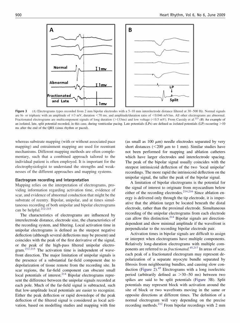
wmmmien
EMvsstc
itudcogftdslsetEdv
(snwTsru
teeaercdp
oRpepfidpspson
FaFam
900 Heart Rhythm, Vol 6, No 6, June 2009
hereas substrate mapping (with or without associated paceapping) and entrainment mapping are used for reentrantechanisms. Different mapping methods are often comple-entary, such that a combined approach tailored to the
ndividual patient is often employed. It is important for thelectrophysiologist to understand the strengths and weak-esses of the different approaches and mapping systems.
lectrogram recording and interpretationapping relies on the interpretation of electrograms, pro-
iding information regarding activation time, evidence ofcar, and evidence of abnormal conduction that might be theubstrate of reentry. Bipolar, unipolar, and at times simul-aneous recording of both unipolar and bipolar electrogramsan be helpful.212-215
The characteristics of electrograms are influenced bynterelectrode distance, electrode size, the characteristics ofhe recording system, and filtering. Local activation time innipolar electrograms is defined as the steepest negativeeflection (although several deflections may be present) andoincides with the peak of the first derivative of the signal,r the peak of the high-pass filtered unipolar electro-ram.212-215 The activation time is independent of wave-ront direction. The major limitation of unipolar signals ishe presence of a substantial far-field component due toepolarization of tissue remote from the recording site. Incar regions, the far-field component can obscure smallocal potentials of interest.214 Bipolar electrograms repre-ent the difference between the unipolar signals recorded atach pole. Much of the far-field signal is subtracted, suchhat low-amplitude local potentials are easier to recognize.ither the peak deflection or rapid downslope of the peakeflection of the filtered signal is considered as local acti-
igure 3 (A) Electrograms types recorded from 2 mm bipolar electrodere bi- or triphasic with an amplitude of �3 mV, duration �70 ms, and aractionated electrograms are multicomponent signals of long duration (�n isolated, late, split potential recorded, in this case, during ventricular pas after the end of the QRS (sinus rhythm or paced).
ation, based on modelling studies and mapping with fine r
as small as 100 �m) needle electrodes separated by veryhort distances (�200 �m to 1 mm). Similar studies haveot been performed for mapping and ablation cathetershich have larger electrodes and interelectrode spacing.he peak of the bipolar signal usually coincides with theteepest intrinsicoid deflection of the two ‘local unipolar’ecordings. The more rapid the intrinsicoid deflection on thenipolar signal, the taller the peak of the bipolar signal.
A limitation of bipolar electrograms is the potential forhe signal of interest to originate from myocardium belowither of the recording electrodes.214,216 Since ablation en-rgy is delivered only through the tip electrode, it is imper-tive that the ablation target be located beneath the distallectrode, rather than the proximal electrode. Simultaneousecording of the unipolar electrograms from each electrodean allow this distinction.214 Bipolar signals are direction-ependent and show minimal amplitude if the wavefront iserpendicular to the recording bipolar electrode pair.
Activation times in bipolar signals are difficult to assignr interpret when electrograms have multiple components.elatively long-duration electrograms with multiple com-onents are referred to as fractionated.45,217 In areas of scar,ach peak of a fractionated electrogram may represent de-olarization of a separate myocyte bundle separated bybrosis from neighbouring bundles, and causing slow con-uction (Figure 2).45 Electrograms with a long isoelectriceriod (arbitrarily defined as �30–50 ms) between twopikes are said to be split potentials (Figure 3B). Splitotentials may represent block with activation around theite of block or two wavefronts moving in the same orpposite directions at different times. The definition of aormal electrogram will vary depending on the precise
a 5–10 mm interelectrode distance filtered at 30–500 Hz. Normal signalse/duration ratio of �0.046 mV/ms. All other electrograms are abnormal.) and low voltage (�0.5 mV). From Cassidy et al.223 (B) An example ofate potentials (LPs) are defined as isolated potentials (LP) occurring �10
s withmplitud133mscing. L
ecording methods.212 From bipolar recordings with 2 mm

eea3cWinicsti
AMacabtmtpAcwAfTkrNb
tptttto
iVTTatprngatep
oiptrcsitsa
dtafm
PPumQnr
ppairtoopea1sstmopk
lpsvcfi
wrl
901E. M. Aliot and W. G. Stevenson, et al. Ablation of ventricular arrhythmias
lectrodes with 5–10 mm interelectrode distance, normallectrograms had amplitudes of �3 mV, width of �70 ms,nd amplitude/duration ratios of 0.046 mV/ms (FigureA);218 fractionated electrograms were defined as multi-omponent signals of �0.5 mV and duration of �133 ms.ith smaller (1 mm) electrodes with narrower (2 mm)
nterelectrode distances used for intraoperative mapping,ormal ventricular electrogram amplitude was �0.6 mV,llustrating the dramatic effect that the recording methodsan have electrogram characteristics.212 Studies of sub-trate mapping have defined normal electrogram ampli-ude of �1.55 mV using a 4 mm tip electrode with 1 mmnterelectrode distance and filtering at 10 – 400 Hz.73
ctivation mappingapping of the electrical activation sequence during VT isvaluable mapping strategy that can be used alone or in
onjunction with substrate mapping, entrainment mapping,nd/or pace mapping. Activation mapping can be performedy recording and analysing either unipolar or bipolar elec-rograms. The sequential point-by-point method involvesoving an electrode catheter to multiple ventricular loca-
ions and recording an electrogram at each site. This ap-roach is greatly facilitated by the use of an EAM system.lthough point-by-point mapping is tedious and requires
onsiderable skill with catheter manipulation, it has beenidely employed to facilitate catheter ablation of stable VT.lternatively, activation sequence mapping can be per-
ormed using a multielectrode mapping system (see theechniques section). There is limited experience with bas-et catheters and multielectrode catheters and these are notoutinely used by the members of the task force (see above).on-contact mapping systems are used in selected patientsy some task force members.
For focal VT, the earliest site of activation identifies theachycardia origin that is targeted for ablation. The QRSrovides a convenient fiducial point for mapping. Activa-ion at the origin precedes the QRS onset. In normal tissue,he unipolar signal (with high-pass filter setting �1 Hz)ypically demonstrates a QS configuration consistent withhe spread of activation away in all directions from the VTrigin.
In scar-related reentry, activation mapping is usually lim-ted. Although the term ‘site of origin’ makes sense for focalT, it is a misnomer in scar-related macroreentry (Figure 2).here is a continuous reentry path with no ‘earliest point’.he QRS onset typically occurs when the impulse reachesn exit along the border zone of the scar and propagates intohe surrounding myocardium. Electrograms immediatelyreceding the QRS are typically recorded from the exitegion.30,219-221 Exit regions can also be identified by theon-contact mapping system where depolarization propa-ates away from the scar.62,128 At reentry circuit sites thatre in a channel proximal to the exit, electrograms areypically earlier, between the QRS complexes (diastoliclectrograms). These are often isolated potentials or split
otentials.30,219,222,223 Sites that are outside the isthmus, in euter loops or remote from the scar, are typically depolar-zed during inscription of the QRS. However, sites that areroximal in a reentry circuit, isthmus can be depolarized athe end of the QRS complex.30,224 Sites that are outside theeentry circuit are bystanders (Figure 2). The extensiveonduction abnormalities in areas of scar can cause by-tander regions to depolarize during electrical diastole mim-cking the timing of an isthmus site.30 Thus, electrogramiming at an individual site is not a reliable indicator that theite is a desirable ablation target, and entrainment methodsre helpful for selecting ablation sites (see below).
For stable VT, complete activation sequences may beefined and used with entrainment mapping to guide abla-ion. More commonly, limited activation assessment, notingreas of pre-systolic activity and isolated potentials, is per-ormed, and this information is integrated with entrainmentapping data to identify target sites for ablation.
ace mappingace mapping refers to analysis of the activation sequence,sually assessed from the 12-lead ECG during pacing at aapping site in the absence of VT. The site where the pacedRS morphology replicates that of the VT is likely to beear the origin for a focal VT or an exit region for scar-elated reentry.51,225-228
Either bipolar or unipolar pacing may be used. Bipolaracing produces a smaller stimulus artefact; there is theossibility of capture at the proximal ring electrode as wells at the tip electrode that may reduce accuracy, particularlyf larger interelectrode distances (8–10 mm) and high-cur-ent strength (�8 mA) is used.227 Pacing slightly abovehreshold is likely to improve accuracy by limiting the sizef the virtual electrode and reducing the likelihood of an-dal capture at the proximal ring electrode during bipolaracing when there is good contact at the contact at the tiplectrode.229 However, repetitive threshold determinationsre often not practical. Some laboratories initially pace with0 mA, 2 ms stimuli and occasionally decrease stimulustrength if a broad area of capture is suspected, or increasetimulus strength (e.g. up to 20 mA) if required to captureissue in a scar region.53,230 Whether the use of body surfaceapping or automated algorithms to quantitate comparison
f QRS morphologies during VT and pace mapping im-roves the clinical utility of pace mapping is not yetnown.231,232
For focal VTs, pace mapping may be used to suggest theocation of the origin, although activation sequence map-ing is probably more accurate.10,227,233,234 The optimal sitehould exactly match the tachycardia QRS, including indi-idual notches as well as major deflections. Pace mappingan be particularly useful if the targeted arrhythmia is dif-cult to induce.
For scar-related VTs, pace mapping at the exit regionill match the VT QRS. However, the potential for large
eentry circuits and abnormal conduction creates severalimitations and caveats. Pacing in normal areas outside the
xit can produce a similar QRS morphology to VT. Thus,
pstdpmtppdr
mtpm
EEcVVQmfccd
tPmrarmsrssle
ttTtsifpttdc
w
tQQAcbvacevsitQtt(abl
tiQstpt�fa
tosctumt
SSscptdintpt
t
902 Heart Rhythm, Vol 6, No 6, June 2009
ace mapping is often combined with a voltage map duringubstrate mapping to define the potential exit.73,235 Al-hough pacing in the reentry circuit near the exit may pro-uce a QRS similar to that of VT, pacing at sites moreroximal in the reentry circuit isthmus during sinus rhythmay produce a markedly different QRS and ECG pattern, as
he stimulated wavefront propagates away from the site viaaths that are blocked during VT.225,226,228,235 Thus, whenacing in regions of scar, a QRS that does not match the VToes not reliably indicate that the site is outside of theeentry circuit.
Sites where the stimulus to QRS interval exceeds 40s are consistent with pacing in regions of slow conduc-
ion.53,228,235 In some reentry circuit isthmuses, pacingroduces a long S-QRS and a QRS morphology thatatches that of VT.51,53,235
ntrainment mappingntrainment mapping is useful for identifying reentryircuit sites and recognizing bystander sites in stableT. Entrainment is performed by pacing faster thanT.30,219,222,223,225,236-243 During reentrant VT, a stableRS morphology with resumption of tachycardia on ter-ination of pacing indicates that each stimulated wave-
ront has reset the reentry circuit, entraining the tachy-ardia. Entrainment can be confirmed by the presence ofonstant fusion, progressive fusion, or evidence of con-uction block terminating tachycardia.241
Entrainment mapping is critically dependent on the assump-ion that pacing is not altering conduction through the circuit.acing should be performed at a cycle length that is onlyodestly faster (10–30 ms shorter) than the VT cycle length to
educe the chance of terminating or altering the VT. Pacing atn output that is just above threshold to avoid capture ofemote myocardium is desirable, but repeated threshold deter-ination is not always practical. Pacing at 10 mA at 2 ms
timulus strength is reasonable and stimulus strength can beeduced if desired. At some sites in scar, greater stimulustrength is required to capture and identify reentry circuitites.230 Although some investigators use unipolar pacing toimit the potential for anodal capture, bipolar pacing is usedffectively by many laboratories.30,219,236-244
At sites in the reentry circuit, the post-pacing interval athe site of stimulation approximates the revolution timehrough the circuit, which is the tachycardia cycle length.he post-pacing interval increases with increasing conduc-
ion time between the pacing site and circuit. There areeveral potential sources of error. The post-pacing intervals measured to the local potential. In areas of scar withractionated and split potentials, distinguishing the localotential from far-field signals due to depolarization ofissue remote from the pacing site can be difficult.216 Elec-rical noise and stimulus artefact can obscure electrogramsuring entrainment.245,246 If pacing slows conduction in theircuit, the post-pacing interval prolongs.
The QRS morphology during entrainment indicates
hether the pacing site is likely to be in an isthmus. At sites ahat are remote from the VT circuit, pacing changes theRS morphology producing either QRS fusion or a pacedRS that resembles that which occurs in the absence of VT.t reentry circuit isthmus sites, pacing entrains VT without
hanging the ventricular activation remote from the circuitecause the paced wavefronts use the VT exit to activate theentricle. The QRS complex during entrainment is the sames that during VT, a finding that has been designated con-ealed entrainment, entrainment with concealed fusion, orxact entrainment. At these isthmus sites, the S-QRS inter-al, which indicates the conduction time from the pacingite to the exit, is the same as the electrogram to QRSnterval. In contrast, at bystander sites that are adjacent tohe reentry circuit isthmus, entrainment may occur withoutRS fusion, but the post-pacing interval (PPI) will exceed
he VT cycle length and the S-QRS will exceed the elec-rogram to QRS interval.30,247 Sites with long S-QRS�70% of the VT cycle length) with concealed fusion andPPI indicating that the site is in the circuit often seem to
e proximal to the isthmus region (inner loops), with aower likelihood of VT termination by RF ablation.
Sites that are in an outer loop of the reentry circuit alonghe border of the scar can be recognized by a post-pacingnterval that approximates the VT cycle length, but withRS fusion during entrainment. Because surface ECG fu-
ion can sometimes be difficult to recognize, careful atten-ion needs to be paid to analysing all 12-ECG leads duringacing and comparing the QRS morphology with pacing tohat of the VT. QRS fusion is often not detected when
22% of the QRS duration is due to the antidromic wave-ront.248 Therefore, some outer loop sites are misidentifieds exit sites or inner loop sites.
In patients with haemodynamically tolerated VT, abla-ion at isthmus sites identified by entrainment mappingften terminates VT.219,222,223,243,249 Ablation at isthmusites that also have an isolated mid-diastolic potentials mostonsistently terminates VT with limited RF energy applica-ion, with a low risk of recurrent VT during follow-p.219,223,238 In patients with unstable VT, limited entrain-ent mapping has been combined with substrate mapping
o target regions of the scar for ablation.28,59
ubstrate mappingubstrate mapping is the characterization of areas likely toupport reentry based on anatomy and electrophysiologicalharacteristics that can be determined during stable sinus oraced rhythm. This approach can facilitate ablation of mul-iple VTs, pleomorphic VTs, and VTs that are unmappableue to haemodynamic instability or that are not reliablynducible.50,53,55,59,73,95,99,101,250-255 Even for haemody-amically stable VTs, substrate mapping is commonly usedo limit activation sequence mapping or entrainment map-ing to a region of interest. In these cases, the voltage maphat identifies scar regions may be obtained during VT.104
Substrate mapping generally begins with identification ofhe region of ventricular scar, based on electrogram char-
cteristics (usually voltage) in an electroanatomic map of
tctaial
Ibamnabmeiviaite
E2pspdvm
tbtrataara
ddobsostsbv
rs
mdca
asspVa
ETSmtRtsvriiaarpItsdoTdgoVcsp
nVegv‘noac
903E. M. Aliot and W. G. Stevenson, et al. Ablation of ventricular arrhythmias
he ventricle of interest. Markers for reentry circuit exits,hannels (isthmuses), and regions of slow conduction, iden-ified based on pace mapping and electrogram properties,re then targeted for ablation. Given the less precise local-zation of the reentry circuit, a more extensive ablationpproach involving the delivery of energy over a relativelyarge area within the scar is commonly employed.
dentification of scar. Scar tissue can be identified based onipolar electrogram amplitude. Using a 4 mm tip electrodend 1 mm ring interelectrode spacing with a 2 mm ring (1m interelectrode distance) filtered at 10–400 Hz, 95% of
ormal LV endocardial electrograms have a peak-to-peakmplitude �1.53–1.55 mV.73 A good correlation has alsoeen found between low-voltage areas and scar in animalodels.256 Areas of extremely low voltage (�0.5 mV or
ven less) have been designated as ‘dense scar’, but it ismportant to recognize that these regions can still containiable myocytes and reentry circuit isthmuses.53 The area ofntermediate low voltage with signal amplitude between 0.5nd 1.5 mV designated as being within the border zone wasnitially targeted as being an area of particular interest givenhe success of the subendocardial resection for surgicallyliminating VT.73
xits and channels. The area of scar is often large, averaging1 cm in circumference in one study of post-infarctionatients.59 In order to limit the area of ablation within thecar, a region of the scar is targeted for ablation based onace mapping or additional electrogram characteristics. Ra-iofrequency lesions are generally confined within the low-oltage region to reduce the risk of damage to functioningyocardium.Exits can be identified based on pace mapping along
he border of the scar. Radiofrequency lesions can thene placed in the infarct border zone (where bipolar elec-rogram amplitude is typically between 0.5 and 1.0 mV)oughly parallel to the infarct border; additional lines ofblation lesions perpendicular to the border, extendinghrough the exit region into the dense scar region, maylso be added.73,101,252,253 Whether identifying a site with‘perfect’ pace-map QRS match with VT achieves better
esults than ablation through a region identified as having‘good’ pace-map match is not known.Several markers of potential isthmuses have been
escribed.50,51,55,95,99,250,250a Sites where pacing pro-uces a long S-QRS but matches the VT QRS morphol-gy are seen at some reentry circuit isthmus sites and cane targeted for ablation.51 Areas of late potentials andplit potentials with an isolated diastolic component arebserved at some reentry circuit isthmus sites duringinus or paced rhythm and have been targeted for abla-ion.50,95,99,263 Potential channels within the low-voltagecar have been identified by manipulating the upperoundary of the colour scale to expose relatively higher
oltage channels bordered on either side by low-voltage megions of dense scar. Targeting these channels has alsohown promising results in single-centre studies.55,250
Areas of electrically unexcitable scar can be defined andarked based on a high pacing threshold (�10 mA at 2 ms
uring unipolar pacing).53 In some patients, these unex-itable regions define channels that can be targeted forblation.
Substrate-guided ablation is a reasonable strategy for VTbla tion when mapping cannot be performed during atable VT. It can also be used to help direct ablation ofcar-related stable VTs. Whether it should be considered inatients with inducible VT following ablation of a clinicalT is not known. The different criteria for identifying exits
nd placing RF lesions have not been directly compared.
ndpoints for ablationhe endpoint of ablation in patients with scar-relatedMVT, and the acute success of the procedure, is assessedost commonly by programmed ventricular stimulation,
ypically employing up to three extrastimuli at one or moreV (and occasionally LV) sites. This endpoint is subject to
he limitations of programmed stimulation in reproducingpontaneous VT at the onset of the study. The utility ofarious endpoints has been judged by spontaneous recur-ence rates during long-term follow-up. These data are lim-ted by several factors, including (i) the intrinsic reproduc-bility of VT induction, (ii) short-term alterations inutonomic tone, loading conditions and fibre stretch, andcute ischaemia, (iii) alterations in post-ablation antiar-hythmic drug therapy, and (iv) long-term evolution androgression of underlying myocardial and coronary disease.n addition, temporal evolution of ablation lesions may alterhe immediate post-ablation substrate, producing either le-ion expansion (mediated in part by secondary myocyte lossue to disrupted microcirculation) or regression (resolutionf oedema and healing), thus altering susceptibility to VT.echniques and strategies have evolved over the past 2ecades, resulting in a greater diversity of patients under-oing ablation, including those with multiple QRS morphol-gies, unstable VT, and patients in whom a clinical (or any)T cannot be induced at baseline. The correlation of out-
ome to endpoints derived from early reports in highlyelected subgroups may not be applicable to contemporaryatients undergoing VT ablation.
Overall, three general endpoints have been evaluated: (i)on-inducibility of clinical VT, (ii) modification of inducedT cycle length (elimination of all VTs with cycle lengths
qual to or longer than spontaneously documented or tar-eted VT), and (iii) non-inducibility of any VT (excludingentricular flutter and fibrillation). Although a change in theintensity’ of stimulation required to induce VT (greaterumber of extrastimuli and alternative stimulation sites) hasccasionally been used to indicate a beneficial response toblation, there is little evidence to support this claim. Directomparisons of different endpoints are limited, and the
ajority of data derive from single-centre studies.
NMiVmmeFplc4iTddttr1lnm
piac8tcs
MWwklgVupodan‘aV
NStanimu
btpaiaVmpEtTro
cdSmrpopfiacwaisr
tcrthrl(trWdcgb
Wqospp
904 Heart Rhythm, Vol 6, No 6, June 2009
on-inducibility of clinical ventricular tachycardiaany studies have described outcomes in terms of induc-
bility of clinical VT, a term that has been used to refer toT that has occurred spontaneously (Table 1). There areany problems and assumptions with this designation. Theinimum number of ECG leads required to discern differ-
nces between different VT origins or circuits is not clear.urthermore, differences in ECG lead placement, patientosition, and antiarrhythmic drugs may influence the simi-arity between two episodes of VT that arise from the sameircuit. Some studies have used a frontal plane axis within5° and the same dominant deflection in V1 to designate annduced VT as similar to a spontaneously occurring VT.257
he VT cycle length alone is influenced by antiarrhythmicrugs and can be similar for different VTs or may beifferent for VTs originating from the same region, and isherefore unreliable as a sole indicator of clinical VT. Theask force members recommend that the term clinical VT beeserved for induced VTs that are known to have the same2-lead ECG QRS morphology and approximate cycleength as a spontaneous VT. Other VTs should be desig-ated as presumptive clinical or as previously undocu-ented VT morphology (Table 1).When clinical VT(s) can be documented prior to the
rocedure and induced at the onset of ablation, non-induc-bility of clinical VT immediately post-procedure is associ-ted with a lower risk of VT recurrence (up to 37%),ompared with persistent inducibility of clinical VT (up to0%).59,178,254,257,258 In patients with a single stable spon-aneous morphology, the persistent induction of ‘non-clini-al’ VT was reported to have little influence on subsequentpontaneous recurrence rates.259
odification of inducible ventricular tachycardiaith evolving trends in the management of VT, includingidespread use of ICDs, the clinical VT is most often un-nown, or at best inferred from a limited number of telemetryeads or ICD logs of stored rates and electrogram morpholo-ies. A large majority of patients have multiple monomorphicTs induced (mean of �3 per patient), including previouslyndocumented VT morphologies. As a result, a surrogate end-oint employed in many laboratories has been the eliminationf all VTs with cycle lengths equal to or longer than thoseocumented spontaneously. This approach is reported to bessociated with a lower risk of recurrent VT in some,59,260 butot all, laboratories.254,257,261 While persistently induciblenon-clinical’ VTs are often faster than those targeted forblation, the occasional spontaneous occurrence of these rapidTs during follow-up is well documented.119,1 86,254,257,261,262
on-inducibility of any ventricular tachycardiaingle-centre experience reflecting more diverse popula-
ions demonstrate that inability to induce any VT is associ-ted with the lower recurrence rates in most,257,261,262 butot all,119 studies. The prognostic significance of eliminat-ng all inducible VT is supported by the results of two recentulticentre trials. The Thermocool VT Ablation Trial eval-
ated endpoints in 231 patients with both stable and unsta- f
le post-infarction VT undergoing ablation by a combina-ion of mapping and substrate-based strategies,105 49% ofatients had no inducible VT post-procedure, whereas andditional 33% had inducible VT that was faster than thatnduced at baseline stimulation. Induction of any VT post-blation was associated with a significantly greater risk ofT recurrence (nearly 2-fold), while persistent induction ofore rapid VT only, as well as the cycle length of the
ost-ablation-induced VT, did not predict recurrence. Theuro-VT-Study evaluated outcome of ablation in 63 pa-
ients with both stable and unstable post-infarction VT.106
he endpoint of non-inducibility or induction of VT moreapid than clinically documented VT was achieved in 81%f patients and did not predict the risk of VT recurrence.
Although the majority of outcome studies focused on theorrelation of inducibility to even a single recurrence of VTuring follow-up, this criterion may be overly stringent.ubstantial reduction in VT episode frequency occurs in aajority of patients during follow-up, even if occasional
ecurrences of VT persist. In a multicentre study of 145atients with either ischaemic or non-ischaemic cardiomy-pathy who had at least one stable or mappable VT, 89% ofatients had at least one clinical VT identified.160 Duringollow-up, clinical success was defined as �75% reductionn VT episodes. Persistent induction of clinical VT post-blation was associated with a greater than four-fold in-reased risk of failure; induction of any VT post-ablationas not predictive of this outcome. A single-centre series
lso suggests that ablation of clinical VT (but not necessar-ly all VT) is associated with a lower incidence of electricaltorm and cardiac death, despite a high likelihood of spo-adic VT recurrence.262
Finally, newer substrate approaches and patient popula-ions may require additional and/or alternative endpointriteria. When isolated, late diastolic potentials in sinushythm are targeted for ablation; elimination of these po-entials post-ablation (or a subset with the greatest delays)as been advocated as useful endpoint.50,95 In patients withecurrent episodes of primarily PMVT or VF, ablation of aimited number of uniform premature ventricular complexPVC) morphologies that were documented to trigger spon-aneous episodes has been associated with a substantialeduction in recurrences during long-term follow-up.264,265
hether these endpoints are sufficient as predictors of free-om from long-term recurrences compared with the out-ome of programmed stimulation requires further investi-ation, but they may be particularly useful when VT cannote induced at the onset of the ablation session.
The consensus of the task force members is as follows.hen clinical or presumed clinical VTs have been ade-
uately documented previously and can be induced at theutset of the procedure, the minimum endpoint of ablationhould be to eliminate the induction of that VT duringost-procedure programmed stimulation. The stimulationrotocol should include up to three extrastimuli delivered
rom at least two ventricular sites (one of which may be
Lraiaregcseaaosaticr
cptdsgsec
A
ESctVttrcbaV
ccmramb
wps
BlVnch
alstaeaitt
caeoihtc
EViapetnfipcea
Tuoaccaupdgu
p
905E. M. Aliot and W. G. Stevenson, et al. Ablation of ventricular arrhythmias
V), with shortest coupling intervals of 180–200 ms or toefractoriness. If the initial stimulation protocol requireddditional LV pacing sites or catecholamine infusion tonduce VT, then that protocol should be repeated followingblation. In patients who present with incessant VT, resto-ation of stable sinus rhythm represents a reasonable clinicalndpoint, irrespective of the outcome of subsequent pro-rammed stimulation. The task force members did not reachonsensus on whether complete elimination of induced VThould be employed as the primary and routine proceduralndpoint, and acknowledges the need for additional datand further research to clarify optimal endpoints for VTblation in various clinical settings. Finally, the group rec-gnizes that some patients undergoing VT ablation haveignificant haemodynamic compromise and substantial isch-emic burden often aggravated by prolonged periods ofachycardia. Post-procedure stimulation to assess VT induc-bility should be avoided if in the judgement of the physi-ian, it places the patient at risk of cardiopulmonary dete-ioration.
It is strongly recommended that reports of ablation pro-edures include specific information regarding the extent ofost-procedure programmed stimulation, endpoints used,he cycle lengths, and morphologies of post-procedure-in-uced VTs, and their relationship to previously documentedpontaneous VT when known. If post-procedure pro-rammed stimulation is omitted or truncated, the reasonhould be specified. These data should also be explicitlylaborated in case series and multicentre trials of the out-ome of VT ablation submitted for publication.
blation strategies
ndocardial ablationelection of the strategies for endocardial ablation shouldonsider the type and severity of underlying heart disease,he underlying arrhythmia substrate, characteristics of theT, the mapping and ablation technologies available and
he optimal vascular access routes (retrograde aortic orransseptal approach to the LV). A variety of strategies areeasonable. In general, it is desirable to first induce VT, toonfirm the diagnosis, to evaluate the possibility of bundleranch reentry, to establish inducibility for interpretation asn endpoint, and to record the 12-lead ECG morphology ofT in the electrophysiology laboratory.When there is a clear clinical VT that is haemodynami-
ally tolerated, and particularly when the VT is incessant, aombination of activation sequence mapping and entrain-ent mapping to identify a critical isthmus for ablation are
easonable.54,180,223 Once VT is terminated by ablation,dditional lesions may be placed creating ablation lines thateet valve annuli or dense unexcitable scar that may form
orders of the reentry path.More commonly multiple VTs are present, some of
hich may be haemodynamically tolerated for a variableeriod, and others that are not tolerated. Many centres then
tart with substrate mapping during stable sinus rhythm. nrief episodes of VT can be induced to assess the potentialocation of the reentry circuit, with prompt termination ofT if required to help select the ablation region.59,99 Alter-atively, ablation can be performed guided by substrateharacteristics.73,95,99,101,250 These different approachesave not been directly compared.
The extent and pattern of RF lesions guided by thesepproaches also varies. Many centres place a line of RFesions parallel to and within the low-voltage border of thecar through presumptive exit regions.28,73 Ablation withinhe low-voltage area at sites with features of channels isnother strategy. These have not been compared. It is gen-rally recognized that channels are often located adjacentnd frequently parallel to a valve annulus, such that ablationn areas that are near a valve annulus is often extendedhrough the scar to the valve annulus, in the hope ofransecting the isthmus.28,55,57,73
The optimal RF ablation endpoints for assessing lesionreation are not known. Termination of VT during RFpplication with prevention of reinitiation, diminution oflectrogram amplitude, increase in unipolar pacing thresh-ld to �10 mA, and arbitrary duration of ablation withmpedance fall and temperature indications of adequateeating have all been used and are considered reasonable byhe task force.53,59,73 Endpoints for the procedure are dis-ussed above.
picardial mapping and ablationentricular tachycardia originating from the subepicardium
s an important cause of failure of endocardial ablationpproaches.56,266-268 A percutaneous epicardial mapping ap-roach allows many of these VTs to be ablated in thelectrophysiology laboratory.29,267 Catheter ablation fromhe epicardium is often required for ablation of VTs due toon-ischaemic cardiomyopathy and is occasionally usefulor VTs in a variety of other diseases, as well as somediopathic VTs. In a recent survey of three tertiary centreserforming VT ablation, 17% of procedures involved epi-ardial mapping, not all resulted in epicardial ablation, how-ver.269 Data are from a few experienced centres and therere significant technical challenges and risks.
echnical considerations. The procedure may be performedsing conscious sedation and local anaesthesia, but someperators feel that deep sedation with the support of annaesthesiologist or general anaesthesia may allow betterontrol of respiratory motion, which might reduce thehance of unintentional RV puncture. From the subxiphoidpproach, a 17 or 19 G Tuohy needle is gently advanced,nder fluoroscopic observation to the edge of the cardiacrofile.29,267,270 Directing the needle towards the left shoul-er with an angle of �45° relative to the skin surface isenerally used to reach the inferior wall. A shallow angle issed if an anterior approach is desired (Figure 4).
Entry into the pericardial space is indicated by negativeressure (concave meniscus at the proximal end of the
eedle) or by injection of a small amount of contrast that
dwCbtflcda
Pgpalts
etpetcst
ttelOa
so
a01eieuicmpdta
tism
Ciptplsup
for pe
906 Heart Rhythm, Vol 6, No 6, June 2009
istributes into the pericardial space. A soft-tipped guide-ire is then inserted through the needle into the space.onfirmation that the wire is intrapericardial and has noteen inadvertently inserted into a cardiac chamber is ob-ained by observing the course of the wire in multipleuoroscopic projections confirming that it crosses multipleardiac chambers, hugging the cardiac silhouette. An intro-ucer sheath is then advanced into the pericardial space,llowing free access to any mapping and ablation catheter.
Attempted epicardial access fails in �10% of patients.269
ericardial adhesions from prior pericarditis or cardiac sur-ery is usually the cause, although limited access may beossible in some of these patients.269,271 Surgical creation ofpericardial window and manual dissection and lysis of the
esions to allow catheter mapping and ablation in the elec-rophysiology laboratory have been shown to be feasible inmall series.251,272
The approach to mapping is essentially the same as forndocardial ablation, including activation mapping, en-rainment mapping, substrate mapping, and pace map-ing.29,86,87,251,272,273 At some sites, pace mapping andntrainment mapping may be limited by a high stimula-ion threshold due to epicardial fat.274 Epicardial fat canause low-amplitude electrograms, falsely suggestingcar in voltage maps.85,251 Fractionated and split poten-ials may be a more reliable indication of scar.
Reported series have generally used RF ablation. Due tohe lack of convective cooling of the ablation electrode byhe circulating blood, high electrode temperatures may bencountered at low-power settings (10 W or less), limitingesion creation.165 Epicardial fat can limit lesion creation.165
pen or closed loop irrigation allows the delivery of energy
Figure 4 A saggital anatomical cross section showing approaches
t power settings (25–50 W) capable of creating deep le- c
ions (up to 5 mm) that can be effective even in the presencef adipose tissue.274
Optimal parameters for RF power titration and irrigationre not completely defined. With external irrigation flow of–2 mL/min may be used during mapping, increasing to0–30 mL/min during ablation, as required to maintainlectrode temperature �50°C. Careful power titration start-ng at 20–30 W to a maximum of 50 W is commonlymployed. Monitoring for an impedance fall is commonlysed to assess likely adequate power delivery.165 With openrrigation, fluid must be periodically drained from the peri-ardium. This can be accomplished by intermittently re-oving the ablation catheter to allow aspiration from the
ericardial sheath, placing a second pericardial catheter forrainage purposes, or using a single sheath that is largerhan the ablation catheter, allowing aspiration of fluidround the catheter from the side port.
The absence of circulating blood in the pericardial spaceheoretically should facilitate cryoablation,166,270 but theres limited human experience. Cryoablation did not produceignificantly larger lesions than irrigated RF in an animalodel.270
omplications and risks. Some degree of pericardial bleed-ng is recognized in �30% of cases.270 Unintentional RVuncture and pericardial bleeding is a well-recognized po-ential complication and a reason to avoid performance ofericardial puncture in the presence of systemic anticoagu-ation. In some cases, puncture of the RV is not followed byignificant pericardial bleeding. Mild or modest bleedingsually resolves and can be carefully monitored with re-eated aspiration of the pericardial space, as the procedure
rcutaneous pericardial entry. RV, right ventricle; LV, left ventricle.
ontinues.135 Severe bleeding requiring surgical interven-
tsilbpabaspb
atecmrapadoflatm
idsaa
ai1mtvaanttAti
drtsp
IpcratpomiEm2
cele
dnecaocfbwao
aritf(orasp
iiirdsEd
seV
907E. M. Aliot and W. G. Stevenson, et al. Ablation of ventricular arrhythmias
ion can potentially occur.29,270 There is concern that abra-ion or laceration can occur if a stiff vascular sheath is leftn the pericardium without a catheter protruding from theumen. Precautions must be in place for managing severeleeding, including availability of appropriate surgical ex-ertise. When bleeding stops, or when pericardial access ischieved without pericardial bleeding, anticoagulation haseen administered to allow endocardial mapping withoutdverse consequences. Puncture of a subdiaphragmatic ves-el causing intraabdominal bleeding has occurred and canotentially require surgery.270,275 Hepatic laceration andleeding is also a potential risk.
Radiofrequency injury to coronary arteries can causecute thrombosis or may produce damage to the arterial wallhat is evident on histological examination in animal mod-ls, the significance of which is unknown.274,275 Severeoronary artery spasm has also occurred during epicardialapping.269 Prior to ablation, direct visualization of the
elation between the ablation site and adjacent coronaryrteries must be obtained, usually by coronary angiographyerformed with the ablation catheter on the target site. Anbsolute safe distance between the ablation site and epicar-ial artery has not been defined, and likely varies dependingn the RF energy applied, coronary artery diameter andow, and the presence of overlying fat.274,276 Based onvailable data and experience, a distance of �5 mm be-ween the ablation catheter and an epicardial artery is com-only accepted.274,275,277,278
Cryoablation appears to have less risk of coronary injuryn animal models, but can still create occlusion and intimalamage, when in close proximity, particularly to small ves-els.167,279 More data are needed regarding long-term safetynd potential effects of ablation in proximity to coronaryrteries.
The left phrenic nerve is potentially susceptible to injurylong its course adjacent to the lateral LV wall.280,281 Prox-mity to the nerve can be detected by pacing (typically at0 Ma or greater stimulus strength) to detect diaphrag-atic stimulation, allowing its course to be marked on a
hree-dimensional map.86 Detection by pacing is pre-ented by the use of paralytic agents during generalnaesthesia. Catheter ablation should be avoided at sitesdjacent to the phrenic nerve. Alternatively moving theerve away from the myocardium by injection of air intohe pericardium or placement of a balloon catheter be-ween the ablation site and nerve has been reported.282,283
ir in the pericardial space can increase the defibrillationhreshold, requiring emergent evacuation if defibrillations required.284
Symptoms of pericarditis are common after the proce-ure, occurring in �30% of cases.275 Symptoms usuallyesolve within a few days with anti-inflammatory medica-ions. Pericardial instillation of aglucocorticoid has beenhown to reduce inflammation in a porcine model, and this
ractice has been adopted by many centres.285 hndications and outcomes. Patients with scar-related or idio-athic VT may benefit from an epicardial approach if endo-ardial ablation fails. Patients reported have generally beeneferred to specialized centres, often after failing endocardialblation; thus, the incidence of epicardial VTs is unknown. Inhe majority of reported cases, epicardial ablation has beenerformed as a second, separate intervention following failuref an attempted endocardial approach.86,251,262,266 Endocardialapping in these cases may show a focal endocardial exit that
s actually breakthrough from an epicardial VT origin.74,86
lectrocardiographic criteria that suggest an epicardial originay help guide the procedure, although exceptions occur.286-
88 For VT with RBBB-like configuration, a slurred upstrokereating the appearance of a pseudo-delta wave favours anpicardial origin.288 In LV cardiomyopathies, a Q-wave ineads felt to reflect the VT exit region may favour an epicardialxit.287
Combined endocardial and epicardial mapping proce-ures are feasible and would be expected to reduce theumber of procedures required for patients with suspectedpicardial VTs.87,251,289,290 Most investigators feel that epi-ardial access should be achieved before systemic antico-gulation. If pericardial entry is achieved with no bleedingr modest pericardial bleeding that stops, anticoagulationan be administered to allow endocardial LV mapping,ollowed by continued monitoring for recurrent pericardialleeding. Experience is limited, however, and caution isarranted. Epicardial mapping and ablation may be useful
s a first procedure when endocardial mapping is not anption due to intraventricular thrombi.288,291
In patients with coronary artery disease, epicardialblation appears to be more frequently required for infe-ior wall infarct VTs than for those from anterior wallnfarcts.135,166,251,266,290,292 In one series of 14 consecu-ive patients with prior inferior wall infarction, a success-ul epicardial ablation site was found for 7 of 30 VTs23%).271 Epicardial VTs have been observed in �10%f post-infarction VTs in another series.290 The risk ofecurrent VT after acutely successful epicardial ablationppears to be low, although second procedures are occa-ionally required.266 No serious complications were re-orted in these series.
Epicardial mapping and ablation is needed more oftenn patients with SMVT associated with dilated, non-schaemic cardiomyopathy than in patients with priornfarction, although only a small number of patients areeported86,135,166,251,288,292 Voltage maps consistentlyemonstrate areas of scarring that are often more exten-ive on the epicardium than on the endocardium.86,87,251
picardial reentry circuits are frequent in Chagas’isease.29
Small series and anecdotal reports have also reporteduccessful ablation of epicardial VTs following a failedndocardial ablation in patients with idiopathic OT-VTs,Ts originating from the mitral annulus, the crux of the
eart, idiopathic ventricular aneurysms, ARVD, and non-
cVacs
SWectaqsgrwa
ITaefwsVu
fematlscsvcdmadbltpampc
ipvv
VsPVMsmi(nmhc3
f
pmdssolsrbfac
TmrvewFpa
OPsac9pPmwil
Adw
908 Heart Rhythm, Vol 6, No 6, June 2009
ompaction of the LV.86,135,166,251,288,292 Idiopathic OT-Ts that are epicardial in location may originate close to the
nterior interventricular vein, great cardiac vein, and middleardiac vein; and successful ablation from within thesetructures has been reported.164,293,294
urgical ablation of ventricular tachycardiaith the availability of implantable defibrillators and cath-
ter ablation, arrhythmia surgery for VT has become un-ommon. Surgery is a reasonable alternative, however, forreatment of recurrent VT, in patients with prior MI with LVneurysms, particularly if significant coronary disease re-uiring revascularization is present.295-297 Careful patientelection and a centre with experience are important. Sur-ery is also an option for selected patients with VT late afterepair of Tetralogy of Fallot and in some selected patientsith non-ischaemic heart disease and difficult to control
rrhythmia.298-300
ntracoronary ethanol ablationhe feasibility of intracoronary injection of ethanol for VTblation was demonstrated in the early 1990s,154,301-303 butndocardial or epicardial catheter ablation is currently pre-erred. Transcoronary ethanol ablation may be consideredhen both endocardial and epicardial ablation fail to control
everely symptomatic, drug refractory VT.304,305 In a largeT referral centre where this treatment was available, it wassed in only 1.4% of VT ablation procedures.304
Transcoronary ethanol ablation should only be per-ormed in centres with experience in both VT ablation andthanol septal reduction for hypertrophic obstructive cardio-yopathy. A cardiologist experienced in the technique of
lcohol ablation should perform the procedure with an elec-rophysiologist. Coronary anatomy must be carefully corre-ated with endocardial and epicardial mapping studies toelect the target branch likely to supply blood to the reentryircuit.304,306 If VT is haemodynamically tolerated, icedaline can be injected in an attempt to terminate VT, pro-iding evidence that the vessel supplies the VT circuit. If aoronary target can be identified, a technique similar to thatescribed for alcohol septal ablation in obstructive cardio-yopathy can been used.304,307-309 Injection of contrast
fter proximal balloon occlusion of the target branch allowselineation of the region receiving blood supply from theranch. The presence of collaterals should be carefully ana-ysed as they may allow spread of ethanol to unintendederritories. With the balloon inflated to occlude the branchroximally and prevent damage from backflow, absolutelcohol (96%) is slowly injected (typically 1–2 mL over 2–5in with balloon inflation during 10 min) and can be re-
eated if needed.304 Control angiography is performed toonfirm occlusion of the distal vessel.
Alcohol ablation has several limitations. There is signif-cant potential for extensive myocardial damage and com-lications including heart block, worsening heart failure,entricular rupture, and death.304,310 A satisfactory target
essel cannot always be identified or cannulated. aI. Ablation outcomes and considerations inpecific diseasesrior myocardial infarctionentricular tachycardia occurs in 1–2% of patients late afterI, often after an interval of several years. Early reperfu-
ion of infarct-related arteries results in less aneurysm for-ation, smaller scars, and less extensive electrophysiolog-
cal abnormalities,311 although a significant risk of late VToften with rapid cycle lengths) persists.312 In addition, theumber of at-risk patients with chronic ischaemic cardio-yopathy due to multiple infarctions and late remodelling
as increased due to improvements in long-term medicalare.313 The mechanism of VT is usually macroreentry;2,54,71 focal non-reentrant mechanisms are responsible forewer than 5–10% of VTs.58
Programmed stimulation induces VT in over 90% ofatients with a history of VT, although the rate and QRSorphology of induced VT may differ from that observed
uring spontaneous tachycardia.32,54,71 The link betweenpontaneous and inducible VT remains incompletely under-tood. However, the induction of VT signifies the presencef a fixed anatomic substrate associated with an increasedikelihood of future spontaneous events.32,54,71 Episodes ofustained VT are a marker for increased mortality andeduce quality of life in patients who have implanted defi-rillators.76-80 Antiarrhythmic medications can reduce therequency of ICD therapies, but have disappointing efficacynd side effects.81-83 Ablation has an important role forontrolling VT in this population.
Post-infarction VT poses several challenges for ablation.he majority of VTs are haemodynamically unstable andultiple VTs (an average of 3 per patient) due to separate
eentry circuits or shared areas of slow conduction withariable exits from the scar are common.32,57,71,105,314 Re-ntry circuit channels may be narrow and easily transectedith ablation, but can also be relatively broad (�2–3 cm).54
or these reasons, activation and entrainment mapping inost-infarction VT have been increasingly supplementednd/or replaced by substrate mapping approaches.
utcomesatient series reported from single centres vary in diseaseeverity, stability of VT for mapping, methods for mappingnd ablation, and ablation endpoints. Of 802 patients from 19entres reported since 2000, at least one VT was ablated in 72–6% of patients and all inducible VTs were ablated in 38–72% ofatients.53,54,59,62,95,98,99,101,119,129,178,219,250,251,257,258,261,315,316
rocedure-related mortality was 0.5%. For the 13 studies withean follow-up of 12 months or more, 50–88% of patientsere free of any VT, with 30–100% continuing on previously
neffective antiarrhythmic medications. Mortality during fol-ow-up ranged from 0 to 32%.
The Multicentre Thermocool Ventricular Tachycardiablation Trial enrolled 231 patients with recurrent VT (me-ian of 11 episodes in the previous 6 months) for ablationith open irrigation RF guided by an EAM using substrate
nd/or entrainment mapping approaches.105 Median LVEF

wfPpioropme�dtwpucdfalewch
ettaiVif
rmpTpuofbmtpr
cpftaspcw
Hs9IPcabIt�o
pt
tspsi
RIgidAatrT
F(asrsam
909E. M. Aliot and W. G. Stevenson, et al. Ablation of ventricular arrhythmias
as 0.25, age was 68 years, 62% had a history of heartailure, 70% had failed amiodarone, and 94% had an ICD.atients had a median of three inducible VTs and unmap-able VT was present in 69% of patients. Ablation abol-shed at least one VT in 81% of patients and all VTs in 49%f patients. During the following 6 months, 51% had recur-ent VT. Although recurrences were common, the frequencyf VT was markedly reduced in a substantial number ofatients (Figure 5). In 142 patients who had ICDs for 6onths before and after ablation, the median number of VT
pisodes was reduced from 11 to 0; 67% of patients had a75% reduction in the frequency of VT episodes. Proce-
ure mortality was 3% with 6 of 7 deaths related to uncon-rollable VT, and one due to tamponade with MI. Thereere no strokes or thromboembolic events. Non-fatal com-lications occurred in 7% of patients, including heart fail-re, increase in mitral regurgitation, and vascular accessomplications. The 1-year mortality was 15% with 38% ofeaths due to ventricular arrhythmias and 35% due to heartailure. In multivariable analysis, lower LVEF, history oftrial fibrillation, recurrence of VT, greater number of RFesions, and slower maximum VT cycle length were mark-rs for increased mortality. The presence of a mappable VTas associated with decreased mortality. Predictors of re-
urrent VT included history of atrial fibrillation, history ofeart failure, and greater number of inducible VTs.
The Euro-VT-Study performed catheter ablation usingxternal irrigation and EAM in 63 patients at eight cen-res.106 The population had a median of 17 VT episodes inhe 6 months prior to ablation, age of 63 years, LVEF 0.28,nd 67% had an ICD. An average of three VTs was induc-ble; 63% had unmappable VT and 37% had only mappableT. At least one VT was ablated in 81% of patients; all
nducible VTs were abolished in �50% of patients. During
igure 5 Plot showing the frequency of VT during the 6 months beforeblue lines) and after (red lines) ablation for 142 patients with ICDs beforend after ablation who survived 6 months (excluding patients with inces-ant VT). Each line represents an individual patient, arranged by recur-ences from greatest (bottom) to fewest (top). The frequency of VT epi-odes was reduced by �75% in 67% of patients. Upper boundaries arertificially truncated as indicated at 100 episodes. Reproduced with per-ission from Stevenson et al.105
ollow-up of 6 months, 51% of patients remained free of any l
ecurrent VT. Although in patients with recurrences, theean number of ICD therapies was decreased from 60 � 70
rior to ablation to 14 � 15 for the 6 months after ablation.here was no procedure mortality; one patient sufferedrolonged cardiac arrest and died of progressive heart fail-re 2 months later. Non-fatal adverse events occurred in 5%f patients included groin haematomas and pericardial ef-usions that did not require drainage. There were no throm-oembolic events. After an average follow-up of 12 months,ortality was 8% and was related to heart failure or cardiac
ransplantation in three patients, and incessant VT in oneatient. The electrophysiolgical findings did not predict VTecurrence.
In a multicentre trial of 146 patients (82% of whom hadoronary artery disease) who had mappable VT and ablationerformed with an internally irrigated catheter; 54% wereree of VT during an average follow-up of 8 months.160 Ofhose with at least 2 months of follow-up, 81% experienced�75% reduction in VT episodes. Complications included
troke in four (2.7%) patients, tamponade in four (2.7%)atients, and MI in one (0.7%) patient. Only one of theardiac perforations seemed to be temporally associatedith RF application. Procedure-related mortality was 2.7%.The Substrate Mapping and Ablation in Sinus Rhythm to
alt Ventricular Tachycardia (SMASH-VT) multicentretudy enrolled 128 patients with prior MI (average age 67 �
years; LVEF 0.30–0.32) who were receiving an initialCD for VF, unstable VT, or syncope with inducible VT.102
atients were randomized to substrate-based ablation vs. aontrol group. There was no procedure mortality. During anverage follow-up of 23 months, 33% of the control group,ut only 12% of the ablation group, received appropriateCD therapy for VT or VF. There was a non-significantrend to better mortality in the ablation group (9 vs. 17%, P
0.29). Serial assessments of LVEF showed no evidencef a detrimental effect of ablation on ventricular function.
Prophylactic VT ablation in patients with ICDs im-lanted for primary or secondary prevention is investiga-ional, and further trials are needed to assess this approach.
Catheter ablation can be immediately life-saving for pa-ients with VT storm or incessant VT.105,262,317-319 Inces-ant or frequent VT is controlled acutely in over 90% ofatients. During follow-up, 74–92% remain free of inces-ant VT or VT storm, although single episodes of VT recurn approximately a third of patients.
ecommendationsn patients with SMVT due to healed MI, methods foruiding ablation are now well defined. Even in the relativelyll populations referred after failing multiple antiarrhythmicrugs, procedure mortality and morbidity are acceptable.blation usually reduces the frequency of VT episodes,
lthough isolated recurrences remain a risk. Although mor-ality from heart failure is a concern, this appears to beelated largely to the severity of underlying heart disease.he use of mapping methods to maintain RF lesions in
ow-voltage regions of scar is recommended to reduce the

rcsta
NSmorgpetMajifdtue
lfptpwediefipiclswp
o
SC3e(Vsbem
Fe
stpdaeefip
rmnrrompRpimpc1ss
V
RRrp�cowtriiAccmt
rsuag
910 Heart Rhythm, Vol 6, No 6, June 2009
isk of damage to functioning myocardium. In view of theseonsiderations, the task force members felt that ablationhould generally be considered relatively early, before mul-iple recurrences of VT and repeated courses of drug ther-py. Recommendations are summarized in Table 2.
on-ischaemic-dilated cardiomyopathyustained monomorphic ventricular tachycardia is not com-on in non-ischaemic-dilated cardiomyopathies, but 80%
f those that occur are due to scar-related reentry, with theemainder due to bundle branch reentry or a focal ori-in.21,86,251,320 Scars are probably due to progressive re-lacement fibrosis. Compared with post-infarction VT, ar-as of scar are smaller, but patients who present with SMVTypically have multiple morphologies of VT inducible.
agnetic resonance imaging with delayed Gd enhancementnd voltage mapping demonstrates that scars are often ad-acent to a valve annulus.139,320 Transmural scar is rare andntramural scars are common. These features likely accountor the general perception that ablation of VT is moreifficult when compared with that in the post-MI popula-ion. When endocardial ablation fails, epicardial mappingsually shows that more extensive scar is present in thepicardium.21,86,139
Catheter ablation employs the approaches for scar-re-ated VT after exclusion of bundle branch reentry. Data arerom small single-centre series.21,86,139 In a series of 19atients with recurrent SMVT due to dilated cardiomyopa-hy, endocardial ablation abolished all inducible VT in 14atients.139 After a follow-up of 22 � months, five patientsere alive without VT recurrence. In a series of 22 patients,
picardial mapping and ablation was performed if endocar-ial ablation failed.86 Scar-related reentry circuits weredentified in the endocardium in 12 patients and in thepicardium in all 7 who underwent epicardial mapping afterailed endocardial ablation. At least one VT was abolishedn 16 of 22 patients and all VTs were abolished in 12 of 22atients. During a mean follow-up of 334 days, VT recurredn 46% of patients, one patient died of heart failure, andardiac transplantation was performed in two patients. Ab-ation can be life-saving in patients with incessant VT or VTtorm.262,266 In these small series, no serious complicationsere reported, although septal ablation produced antici-ated AV block in one patient.
Recommendations for ablation are as stated for ablationf VT associated with structural heart disease in Table 2.
urgically repaired congenital heart diseaseongenital heart defects occur in almost 1% of live births;0–50% require surgery during early childhood.321,322 Lifexpectancy has steadily improved, but sudden cardiac deathSCD) remains the most common cause of late mortality.323
entricular scars from ventriculotomy or patches create theubstrate for scar-related reentry.36,39,324 Focal VT has alsoeen reported.127 Myocardial hypertrophy occurs in a vari-ty of malformations and also likely contributes to arrhyth-
ia risk. The prevalence of VT after repair of Tetralogy of oallot ranges between 3 and 14%, with a risk for SCDstimated at 2% per decade.321,325-329
The approach to catheter ablation of SMVT is as de-cribed for scar-related VTs.330,331 Physicians undertakinghese procedures should be familiar with the anatomic com-lexities that are a major challenge. Knowledge of theetails of the surgical repair, usually available in the oper-tion report, is helpful. Imaging with angiography andchocardiography and integration of MRI or CT images tonhance the anatomic definition may be helpful.331 Identi-cation of the region of scar and a substrate mapping ap-roach to identify channels has been described.331
Data are from case reports and small patient se-ies.104,127,330-337 Initial studies demonstrated feasibility ofapping and ablation, but late recurrences of VTs that were
ot observed at the time of the procedure and high recur-ence in patients with unmappable VT were noted.330,336 Aeport using substrate mapping approaches after Tetralogyf Fallot or ventricular septal defect repairs described com-on isthmuses supporting reentry between the tricuspid or
ulmonic valve annuli and scar or patch in the septum orV free wall that could be targeted for ablation.331 In 11atients, 15 different VTs were inducible. Ablation abol-shed all inducible VTs. During an average follow-up of 30onths, 91% of patients were free of VT. In a series of 10
atients with Tetralogy of Fallot mapping with the non-ontact electrode array demonstrated macroreentry in 11 of3 induced VT.127 Ablation was performed in eight patients,ix of whom were free of VT during follow-up.127 In thesemall series, no serious complications were reported.
Recommendations for catheter ablation are as stated forT associated with structural heart disease in Table 2.
ight ventricular diseaseight ventricular diseases causing VT are infrequent. Ar-
hythmogenic right ventricular dysplasia/cardiomyopathy isrobably the most common cause and has a prevalence of1 in 5000.338 Arrhythmogenic right ventricular dysplasia/
ardiomyopathy is characterized by fibrofatty replacementf myocardium within the so-called triangle of dysplasiahich encompasses the RV inflow, outflow, and apex. Sep-
al and LV involvement has been described.338 Scar-relatedeentry is the most common cause of SMVT and is anmportant feature that helps distinguish these VTs fromdiopathic RVOT VT, which is focal. Focal VTs inRVD/C have been described,100 although some of these
ases may represent epicardial reentry with a focal endo-ardial breakthrough. Cardiac sarcoidosis with RV involve-ent can mimic ARVD/C.27 Non-ischaemic cardiomyopa-
hy and MI also cause some RV scar-related VTs.339
The methods for mapping and ablation are as for scar-elated VTs; activation mapping, entrainment mapping,ubstrate mapping, and combined approaches have beensed.23,28,100,201,315,340 As in infarct-related VTs, large scarsnd reentry circuits with extensive areas of abnormal electro-rams, evidence of slow conduction, and bystander regions
ccur. Reentry paths are commonly located along the tricuspid
vtaarliprnitacdVwo
otuwrcciewiet
scasFebh
DfVcodmTc�fTcPT
cimcfpcmp
uPrpspRf
qvdcfcfdoaddtesm
BB�st(chtrtgittagotn
911E. M. Aliot and W. G. Stevenson, et al. Ablation of ventricular arrhythmias
alve annulus or pulmonary annulus. A strategy aiming toransect an isthmus by extending ablation lesions from thennulus to a region of scar has been used.23,28,340 Irrigated RFblation catheters are favoured as trabeculated and annularegions where cooling from circulating blood flow is low canimit power application. Attention to power titration and mon-toring impedance is recommended due to concern for risk oferforation when ablating in the free wall of the RV. Theeentry substrate is epicardial in an undefined, but importantumber of patients, consistent with the fact that ARVD/Cnvolvement usually starts in the epicardium. Epicardial abla-ion has been successfully used after failure of endocardialblation.135,288,341 Electrocardiographic criteria suggesting epi-ardial VT locations for the LV do not apply to RV tachycar-ias and pace mapping suggests that epicardial locations of OTTs cannot be predicted from the QRS configuration, but Qaves or QS complexes suggest an epicardial origin for VTs inther locations.286
In reported series of RV scar-related VT, abolitionf inducible VT is achieved in 41– 88% of pa-ients.23,28,100,103,126,315,340,342,343 During average follow-ps of 11–24 months, VT recurs in 11– 83% of patients,ith some series observing a significant incidence of late
ecurrences increasing with time.315,340 The variable out-omes likely reflect progression of disease, and the in-lusion of different causes of myopathy, as most did notnclude genetic analysis, and different ablation strategiesmployed. In addition, epicardial mapping and ablationas generally not employed. Serious complications are
nfrequent, but the risk of perforation and tamponade isxpected to be increased in areas where the RV ishinned, and this complication has been reported.178
Catheter ablation in ARVC/D can reduce frequent epi-odesof VT but long-term follow-up has demonstrated aontinued risk of recurrence.315,340 Recommendations forblation are as stated for ablation for VT associated withtructural heart disease in the Indications section above.urther studies are needed to clarify the role and benefit forpicardial mapping and ablation. There is also a need toetter define the characteristics of patients who are likely toave long-term benefit from catheter ablation of VT.
epressed ventricular function associated withrequent ventricular ectopyery frequent PVCs can result in a reversible form of
ardiomyopathy, but cardiomyopathy can also cause PVCs;ften, it is not clear that the PVCs are the cause of LVysfunction. Negative inotropic effects of some antiarrhyth-ic drugs may also contribute to ventricular dysfunction.he critical number of PVCs and the time course required toause a cardiomyopathy are not known. A PVC burden of20% is present in most patients, but some patients have as
ew as 5% PVCs on a single 24 h Holter recording.344,345
he duration over which PVCs must be present to causeardiomyopathy is not clear and difficult to assess becauseVCs are asymptomatic in a substantial number of patients.
riggered activity and automaticity are probably the most sommon mechanisms of focal PVCs in this setting. Reentrys unlikely in patients without scar, but has been docu-ented in post-infarction models.346 Premature ventricular
omplexes in the setting of prior MI frequently originaterom scar areas and may share QRS morphology features ofost-infarction VT, suggesting that reentry might be theirause.347 Ablation is guided by activation mapping. Paceapping can be used if PVCs are not frequent, but is
robably less accurate.234,348,349
Data are from small case series. In one series of consec-tive patients with decreased LV function attributed toVCs from a variety of different origins, the overall successate of ablation was 82%.344 Premature ventricular com-lexes originated from the RVOT in half the patients; otherites of origin included the LV, the coronary cusps, and theulmonary artery. Success is likely to be greater when onlyVOT PVCs are present.345 Ventricular function improved
ollowing ablation.Catheter ablation is recommended for patients with fre-
uent PVCs or NSVTs when they are presumed to causeentricular dysfunction. At the time of the ablation proce-ure, it is often not clear whether the frequent PVCs areausing LV dysfunction. Assessment of LV function duringollow-up helps to clarify whether a cardiomyopathic pro-ess is present or whether PVCs resulted in a decline of LVunction. To assess the contribution of PVCs to ventricularysfunction, pharmacological suppression, such as by ami-darone therapy, may be considered, but the utility of thispproach and duration of therapy required have not beenefined. Multiple morphologies of PVCs are likely to re-uce procedural success. The role for pre-procedural iden-ification and quantification of a predominant PVC (forxample, by performing a 12-lead Holter) warrants furthertudy. In the absence of a predominant PVC morphology,edical management should be considered.
undle branch reentryundle branch reentrant tachycardia (BBRT) accounts for5% of SMVTs induced during programmed ventricular
timulation in patients with heart disease.17,65,350 The reen-ry wavefront circulates retrogradely up one bundle branchmost commonly the left bundle) and down the other (mostommonly the right bundle), giving rise to VT that usuallyas a typical left bundle branch block (LBBB) configurationhat can be the same as the QRS configuration during sinushythm.17,65,350-354 Right bundle branch block configurationachycardias occur when the left bundle is used antero-radely, or are due to interfascicular reentry tachycardianvolving both fascicles of the left-sided Purkinje systemhat may coexist with bundle branch reentry.17,67,355-357 Inhese RBBB tachycardias, right axis deviation indicatesntegrade activation over left anterior fascicle and retro-rade over the left posterior fascicle; and left axis deviationccurs when the circuit revolves in the opposite direc-ion.358 The tachycardia cycle length is usually �300 ms;ear syncope, syncope, or cardiac arrest are common pre-
entations.
ptmbt1ttTulsrbiafd
firpui
wdcvpBdfs
SiPptetPr1Imctmatasvt
tpetcbppaIsibsrvmirtlbtuaitmt
ctd(taopotdac
hHstttMetbr
912 Heart Rhythm, Vol 6, No 6, June 2009
Substantial His-Purkinje system disease is usuallyresent associated with ventricular dysfunction usually dueo coronary artery disease, valvular heart disease, or cardio-yopathy.22,65,351,357,359-364 Cases due to sodium channel
lockade with flecainide365,366 and isolated conduction sys-em disease have been described. During sinus rhythm, the2-lead ECG characteristically shows nonspecific interven-ricular conduction delay or LBBB and the HV interval isypically, but not always, prolonged.65,351,357,360,361,363,367
he VT can usually be induced by programmed RV stim-lation; induction may occur with LV stimulation, short–ong–short extra-stimuli sequences, isoproterenol infusion,odium channel blockade, atrial stimulation, or an irregularhythm from atrial fibrillation or flutter.65,357,368 Bundleranch reentrant tachycardia is confirmed by evidence link-ng the Purkinje system to the reentry circuit in His bundlend or right or left bundle recordings. During entrainmentrom the RV apex, the post-pacing interval–VT cycle lengthifference is typically 30 ms or less.369
Drug therapy is usually ineffective. Catheter ablation is arst-line therapy and successful in �100%.357,360,361 Theight bundle branch is the preferred ablation target. Even inatients with baseline LBBB, right bundle ablation does notsually produce heart block, but back-up bradycardia pac-ng is required in �30% of patients.357,360,361,367
Ablation of the left bundle branch may be consideredhen LBBB is present during sinus rhythm, but is moreifficult and has risks of left heart catheterization. There isoncern that LBBB may be more likely to adversely affectentricular function than RBBB. Long-term outcome de-ends on the underlying cardiac disease. Despite successfulBRT ablation, many patients remain at risk of suddeneath due to concomitant scar-related VTs and/or LV dys-unction. An ICD with or without cardiac resynchronizationhould be considered according to the current guidelines.
pecial considerations for patients withmplanted defibrillatorsatients undergoing catheter ablation of VT often have areviously implanted ICD or will undergo device implan-ation after ablation. For patients with incessant VT orlectrical storm, an ECG can often be obtained to identifyhe QRS morphology of clinical monomorphic VT or theVCs initiating PMVT. When the indication for ablation isecurrent ICD shocks or antitachycardia pacing therapies, a2-lead electrocardiogram of the VT may not be available.n such cases, stored intracardiac electrograms provide aeasurement of the VT cycle length, although only minimal
lues to the morphology of the clinical VT, limiting abilityo identify clinical VTs, particularly when multiple VTorphologies are inducible. It must be recognized that RF
blation may affect both the myocardial VT substrate andhe pacing and sensing functions of an ICD; both of thesespects must be considered. Because programmed electricaltimulation and RF current are sensed by ICDs, these de-ices must be reprogrammed prior to the ablation procedure
o prevent oversensing and unintended delivery of anti- iachycardia therapies. Tachycardia detection zones must berogrammed ‘off’ once the patient has been connected to anxternal defibrillator and personnel are available for moni-oring of the patient. The ICD can still be used to deliverommanded shocks to terminate VT that is not interruptedy overdrive pacing. For patients who are pacemaker-de-endent, the noise reversion response of the ICD should berogrammed to an asynchronous mode before RF current ispplied. For patients who are not pacemaker-dependent, theCD is usually programmed to asynchronous noise rever-ion mode at a low pacing rate. Stimulation thresholds andntracardiac electrogram amplitudes should be measuredefore and after ablation to ensure appropriate pacing andensing by the ICD. In addition, because RF current canesult in an increase in the stimulation threshold of a pre-iously implanted lead, the pacing thresholds should beeasured and the stimulus amplitude programmed to antic-
pate a rise in threshold. If the electrogram amplitude iselatively low (�3 mV), induction of VF may be indicatedo ensure effective sensing by the ICD after ablation. Fol-owing the ablation procedure, tachycardia detection muste reprogrammed ‘on’ and the antitachycardia therapiesailored to the results of final programmed electrical stim-lation. Fluoroscopic confirmation of stable lead positionfter ablation may also be warranted. Some centres admin-ster prophylactic antibiotics at the time of ablation in pa-ients who have an ICD, even long after implantation; butany centres do not use antibiotic prophylaxis and infec-
ions related to catheter ablation appear to be rare.Catheter ablation may affect several factors requiring
onsideration: (i) the indication for ICD implantation; (ii)he rate of VT that may require reprogramming of VTetection zones and maximum tracking rates for pacing;370
iii) the addition or discontinuation of antiarrhythmic drugshat will affect the rate of recurrent VT or other arrhythmiasnd the defibrillation threshold;82,83,371 (iv) sensing of VTr VF by the ICD; (v) the response of VT to antitachycardiaacing algorithms; (vi) the requirement for reprogrammingf ICD discrimination algorithms;370 (vii) the need for con-inued ICD therapy possibly allowing explantation of theevice; and (viii) the potential loss of pacing if, for example,pacing lead is dislodged or ablation in the vicinity in-
reases pacing threshold.In general, the results of catheter ablation of VT should
ave little influence on the indications for ICD implantation.owever, the function of the ICD may influence the deci-
ion to proceed with catheter ablation. For example, biven-ricular pacing may sometimes induce incessant VT.372 Inhis situation, the risks and benefits of resynchronizationherapy must be weighed against those of catheter ablation.
ost patients who have VT related to structural heart dis-ase will continue to have a standard indication for ICDherapy for primary prevention.373 Even when all VTs haveeen rendered non-inducible by ablation, the recurrence rateemains substantial so that secondary prophylaxis remains
ndicated.374,375 One potential exception is when an ICD has
bfdpassi
VIccwWfSltIs
ITop7OonvAtstTste
2harspyoEepcs
Olr
pdihtat
pfVVPtphd�plrpotses
spcSbdtagtgpmi
ORtgoflcats
913E. M. Aliot and W. G. Stevenson, et al. Ablation of ventricular arrhythmias
een implanted to treat idiopathic monomorphic VT such asrom the RVOT or LVOT in the absence of structural heartisease. In these patients, explantation or deactivation of areviously implanted ICD may be considered if catheterblation has successfully eliminated VT. However, an ob-ervation period is warranted after ablation and great carehould be taken to exclude structural heart disease in thesendividuals.
II. Idiopathic ventricular tachycardiasdiopathic VT refers to VTs that occur in the absence oflinically apparent structural heart disease. These VTs oc-asionally occur in patients with structural heart disease, inhom the structural heart disease is not related to the VT.hen frequent, they can be a cause of depressed ventricular
unction as a form of tachycardia-induced cardiomyopathy.udden death is rare, and therapy for idiopathic VTs is
argely guided by symptoms. Ablation is a relatively effec-ive option. Risks depend on the location of the VT origin.ndications for ablation are as summarized in the Indicationection above, with addition discussion below.
diopathic outflow tract ventricular tachycardiashe region of RVOT and LVOT is the most common originf idiopathic VT and PVCs, accounting for �10% of allatients referred for evaluation of VT.14,376-388 More than0–80% of these arrhythmias originate from the RVOT.389
ther origins include the pulmonary artery, near the bundlef His, the LVOT, the aortic sinuses of Valsalva, the coro-ary sinus (CS) and cardiac veins, the mitral and tricuspidalve annuli, and the epicardium.12,41,149,164,233,378,390-399
lthough idiopathic focal OT-VT usually occurs in pa-ients with no structural heart disease, MRI has shownubtle areas of diminished wall motion and suggestedhat mild structural abnormalities may be present.400-405
hese tachycardias have a focal origin, and findings con-istent with triggered activity (see the Mechanism sec-ion), including initiation by burst pacing and isoproter-nol administration.377,400,406
Idiopathic focal OT-VT typically presents between ages0 and 50 years and occurs more frequently in women;407
ormonal triggers may influence occurrences.381,408 Therere two typical forms: exercise- (stress-) induced VT andepetitive monomorphic VT occurring at rest. Repetitivealvos of NSVT are frequent, comprising 60–92% of re-orted series, but incessant VT occasionally occurs. Parox-smal sustained VT, separated by relatively long intervalsf infrequent premature ventricular beats is less common.409
pisodes tend to increase in frequency and duration duringxercise and emotional stress. Thus, an exercise test mayrovoke focal OT-VT either during exercise or in the re-overy phase. In a significant number of patients, exerciseuppresses the arrhythmia.5,410
The typical QRS morphology during idiopathic focalT-VT shows LBBB configuration with an inferior (right or
eft) axis. Premature beats or the first beat of VT usually has a
elatively long coupling interval to the preceding QRS-com- plex.411-413 The QRS duration is usually �140 ms and a QRSuration �120 ms in lead I suggests RV dysplasia rather thandiopathic VT.410 Ventricular tachycardia is monomorphic;owever, the QRS morphology may vary slightly duringachycardia. Multiple distinct VT morphologies are very rarend should raise the suspicion that a scar-related VT, ratherhan idiopathic VT, is present.342,379,382,414
Although idiopathic focal OT-VT has a benign course,otentially malignant forms of VT that resemble idiopathicocal OT-VT may also arise from the OT region, includingT in ARVC,342,376,415,416 polymorphic catecholminergicT,388 VT/VF in Brugada syndrome,14 and also idiopathicMVT/VF.380,387,400,413,417 It is important to differentiate
hese arrhythmias from idiopathic focal OT-VT. All patientsresenting with OT-VT require an evaluation for organiceart diseases or genetic syndromes associated with suddeneath. The ECG during sinus rhythm is usually normal, but10% of patients with OT-VT show complete or incom-
lete RBBB.410 T-wave inversion in the anterior precordialeads suggests ARVD.418 A Brugada type ECG, findings ofeduced LV or RV function, PMVT or multiple VT mor-hologies, a history of recurrent syncope, or a family historyf SCD mandate further detailed evaluation. An exerciseest and cardiac imaging (usually at least echocardiography)hould be done in all patients. Left and/or right heart cath-terization may be warranted. Invasive electrophysiologicaltudy is usually not necessary to establish a diagnosis.
The success of ablation depends on the presence ofpontaneous or inducible VT or PVCs at the time of therocedure. All antiarrhythmic medications should be dis-ontinued for at least five half-lives before the procedure.edation can decrease spontaneous or inducible arrhythmia,ut in many patients, mild sedation and adequate analgesiao not prevent spontaneous or provoked VT.381 In additiono diagnostic electrode catheters at the RV apex, assessingctivation on catheters at the His bundle position and in thereat cardiac vein via the CS can be helpful in suggestinghe origin.419 If spontaneous PVCs or VT are absent, pro-rammed stimulation and burst pacing with or without iso-roterenol infusion should be performed. Careful catheteranipulation during mapping should seek to avoid mechan-
cal trauma that may transiently abolish the arrhythmia.Analysis of the surface ECG morphology of PVCs or
T-VT is a cornerstone to localize the site of origin.5,385
ecording of spontaneous or induced OT-PVC or OT-VT inhe EP laboratory is important to direct the procedure anduide pace mapping. Electrocardiographic recordings fromutside the EP laboratory may not allow sufficient precisionor pace mapping due to differences in patient posture andead position during the recording. Premature ventricularomplexes with the same morphology as VT are an accept-ble target if VT is not inducible. In the absence of spon-aneous or inducible clinical arrhythmia, catheter ablationhould be avoided.
Precise localization of the VT origin from its QRS mor-
hology is limited by the close anatomic relations of RVOT,
LVRRAgdgo
mMnLntom
RTpmirIatpmhccissde
cibat1slsPtm
a�aea
aa
rorsiparRtawa
LLsfSmlti
fwtRciaFa
AVta11abaAeQ�mtd
lTc
914 Heart Rhythm, Vol 6, No 6, June 2009
VOT, and great vessels.2-4,6,41,349,384,389,391,396,398,410,420-424
entricular tachycardia arising from RVOT typically shows an/S transition zone in the precordial leads at V4, whereas an/S transition at V1 or V2 indicates an LV origin.6,384,391,410
n R/S transition at V3 is compatible with RV or LV ori-in.4,6,41,384,391,414,421,422,424-426 A longer time to maximumeflection in the precordial leads suggests an epicardial ori-in.293 A broad R-wave in V1 or V2 suggests an aortic cusprigin.41,427
Precise localization for ablation is guided by activationapping, pace mapping, or a combination of both methods.apping should begin in the RVOT (including the pulmo-
ary artery), followed by the great cardiac vein, and thenVOT and aortic cusps. Epicardial mapping is occasionallyecessary. Most epicardial OT-VTs originate at or close tohe superior CS and the cardiac veins.164,293,392,427 Mappingf the great cardiac vein is recommended before epicardialapping by pericardial puncture.
ight ventricular outflow tract tachycardiahe focal VT origin can be identified from activation and/orace mapping.4,381,385,410 Systematic point-by-point activationapping is the initial preferred technique.233,234,385,409 Some
nvestigators use three-dimensional EAM systems to assist inelating the anatomy to the mapping data.4,124,125,233,381,385,428-430
n the RVOT, bipolar electrograms typically have normalmplitude (e.g. �1.5 mV) and are sharp, without fragmen-ation. Activation at the site of successful catheter ablationrecedes the onset of the surface QRS complex by 10–60s.379,389,390,431,432 The bipolar electrogram may display
igh-frequency low-voltage activity preceding the main lo-al ventricular electrogram component. Ventricular tachy-ardia occasionally originates from muscle bundles extend-ng along the pulmonary artery, where low-amplitudeignals are recorded.4,396 Unipolar electrograms show aharp QS-morphology with a sharp intrinsic deflection in-icating the activation moving away from the mappinglectrode.433
Although activation mapping is the strategy of firsthoice, pace mapping can be used to identify a region ofnterest and then refine position based on activation, and cane the major guide to the ablation site when spontaneousrrhythmia is infrequent. Pace mapping should be done athe VT rate. A site where pacing exactly reproduces the2-lead ECG of the spontaneous or induced VT should beought.385,434 The spatial resolution of pace mapping isimited and in some patients a perfect pace map is seen atites up to 2cm away from the VT origin.233,234,349,381,385
acing at stimulus strengths only slightly greater thanhreshold is desirable to avoid capture over a large area thatay reduce accuracy.Radiofrequency ablation with solid 4 mm tip electrodes
t maximal temperatures of 50–70°C and power settings of50 W is usually sufficient for ablation when mapping is
dequate (see the Technologies section). Use of 8 mmlectrodes with higher power or irrigated electrodes is usu-
lly not necessary. Avoiding steam pops from high-power cblation is particularly desirable to avoid perforation whenblation is performed in the RVOT.
There are no controlled or multicentre trials of ablation. Ineported case series, acute success rate of RF catheter ablationf RVOT-VT is 65–97% and typically exceeds 80%; recur-ence of arrhythmia has been reported in up to 5% after acutelyuccessful ablation.2,6,178,342,349,381,390,394,395,406,435,436 Failures usually due to inability to induce the arrhythmia for map-ing. Complications are rare, but perforation and tamponadere reported and ablation of foci near the His bundle region canesult in heart block.398 The leftward posterior aspect of theVOT is only 4 mm from the left main coronary artery, such
hat there is a theoretical risk of coronary artery injury withblation in that aspect of the RVOT and pulmonary artery, thatould likely be increased with the use of high-power, irrigated
blation.152
eft ventricular outflow ventricular tachycardiaseft ventricular outflow tachycardias can originate from theuperior basal region of the left interventricular septum, LVree wall, aortic sinuses of Valsalva, or the LV epicardium.mall case series demonstrate feasibility of ablation forost of these VTs and suggest acceptable risk. However,
eft heart catheterization is required, introducing risks ofhromboembolism, coronary arterial injury, and aortic valvenjury.
Left ventricular outflow tract tachycardias that originaterom the endocardium near the aortic annulus have a QRSith an inferior axis and may have an earlier precordial
ransition to positive R-wave, sometimes with prominent-waves in V1 or V2, when compared with RVOT tachy-ardia.4,378,406,437-439 The approach to mapping and ablations similar to that for the RVOT tachycardias. Heart block,ortic valve, and coronary artery injury are potential risks.easibility of ablation has been shown in small case seriesnd anecdotal reports.4,378,406,437-439
ortic cusp ventricular tachycardiasentricular tachycardia originating from extension of ven-
ricular myocardium above the aortic annulus that requiredblation from within the sinuses of Valsalva accounted for7 and 21% of idiopathic OT-VTs in two recent series of55 and 352 patients, respectively.2,440 The majority areblated from the left coronary cusp, followed in frequencyy the right coronary cusp, the junction between the rightnd left coronary cusp, and rarely the non-coronary cusp.ctivation mapping typically shows a two-component
lectrogram with the earliest deflection preceding theRS complex by an average of 39 ms, but sometimes50 ms.41,427,440,441 Pace mapping in the aortic sinusay require high output and is less likely to reproduce
he VT QRS compared with pace mapping for endocar-ial VTs.
Small case series have shown feasibility of successful ab-ation without reported significant complications.41,427,440,441
he potential for acute occlusion of the left main or rightoronary arteries is a major concern. The proximity of the
oronary ostia to the ablation site must be defined by coronary
asabs
EPsitfmnrcwcadstr
MFiRiaqcn
OStos
ItIcaVcrbpfsaaatAd
fvipg
rcetl
PvPsfImgdapfdi
MktlcRPimptQpVQ
tMpffinaormtak
915E. M. Aliot and W. G. Stevenson, et al. Ablation of ventricular arrhythmias
ngiography or ICE by an experienced operator.422 Sites ofuccessful ablation are typically �8 mm below the coronaryrtery ostia. Injury to the aorticvalve is theoretically possi-le.163,421 Titration of energy beginning at low power is rea-onable.
picardial outflow tract ventricular tachycardiasercutaneous epicardial mapping and ablation is useful inelectedpatients. Epicardial foci cause VTs with a longernterval from QRS onset to earliest maximal deflection inhe precordial leads, consistent with late access of the wave-ront to the endocardial Purkinje system.293 The VT focusay be adjacent to the great cardiac vein that can be can-
ulated via the CS, allowing successful ablation via thisoute.164,392,442 These VTs have also been ablated via per-utaneous pericardial access.293 The risk of coronary injuryhen the focus is adjacent to a coronary artery is a major
oncern. The overlying left atrial appendage can also be annatomic obstacle to ablation.439 When proximity to epicar-ial coronary artery precludes catheter ablation, a directurgical approach can be an option.293 Feasibility of abla-ion has been shown in small case series and anecdotaleports, but risks and benefits are not well defined.
itral annulus ventricular tachycardiaocal VT from the mitral annulus accounted for 5% of
diopathic VTs in one series.2 Ventricular tachycardia hasBBB or RS pattern in V1 and monophasic-R or RS pattern
n leads V2–V6. Endocardial ablation is usually successful;blation from within the CS may occasionally be re-uired.2,395 Feasibility of ablation has been shown in smallase series and anecdotal reports, but risks and benefits areot well defined.
ther locationsuccessful ablation of focal idiopathic VTs originating from
he tricuspid annulus, papillary muscles, and occasionally atther ventricular sites has been described in case reports andmall series.3,149,394
ntrafascicular verapamil-sensitive reentrantachycardiantrafascicular verapamil-sensitive reentrant tachycardia typi-ally presents as exercise-related VT between the ages of 15nd 40 years; 60–80% of patients are male.2,69,123,383,443-446
entricular tachycardia has an RBBB configuration with rSomplexes in the mid-precordial leads. The mechanism iseentry in or near portions of Purkinje fascicles of the leftundle. More than 80% of these VTs have exits near theosterior fascicles of the left bundle, causing a superiorrontal plane axis. Occasionally, VT has an inferior axis,uggesting an exit in the vicinity of the anterior fascicle. Anrea of rounded-appearing potentials in diastole during VTnd sinus rhythm is present in the inferoseptal region thatppears to indicate a slow conduction region that is sensitiveo verapamil appears to be involved in the circuit.93,447
blation targets sharp potentials near the circuit exit or the
iastolic potentials.69,93,123,383,444-446,448 Mechanical trauma trom catheter manipulation often terminates VT and pre-ents reinitiation. Ablation then targets the site of mechan-cal termination, low amplitude, or diastolic sinus rhythmotentials, or creates a line of lesions through this re-ion.93,123,443
Of 103 patients reported in 10 series, the overall successate of ablation was �95%.69,123,383,443-446,448 Althoughomplications related to left heart catheterization and arexpected, no serious complications were reported. Left pos-erior hemiblock has been observed when a line of RFesions was placed through the posterior septal region.123
olymorphic ventricular tachycardia andentricular fibrillationolymorphic VT and VF may present as an arrhythmiatorm and, in this uncommon instance, may be consideredor catheter ablation. The prevalence of VT/VF storms inCD patients is �20%,449 whereas idiopathic VF is esti-ated to represent 5–10% of SCD cases.450-452 �-Adrener-
ic blockers, various antiarrhythmic drugs including amio-arone, procainamide, and lidocaine and even generalnaesthesia can be effective.453,454 Catheter ablation canlay a crucial role when drugs fail and has been describedor the following conditions: post-MI,317 Brugada syn-rome,380 long QT syndrome,380 RVOT ectopy,455 and id-opathic VF.455
These PMVTs likely have various mechanisms. AfterI, afterdepolarizations and triggered activity from Pur-
inje fibres in the infarct region may cause premature ven-ricular beats that initiate PMVT/VF.317 In the Brugada andong QT syndromes, and some rare idiopathic VF patients,losely coupled monomorphic ectopic beats from the LV orV Purkinje network or from the RVOT may lead toMVT/VF.380 Patients with idiopathic VF typically have
solated premature ventricular beats, best appreciated im-ediately following resuscitation. Premature beats with a
ositive QRS morphology in lead V1 are from the LV andend to be polymorphic but with a characteristically shortRS duration often in the range of 115 ms. Conversely,remature beats with a negative QRS morphology in lead1 are typically from the RV and have a significantly longerRS duration.Feasibility of ablation targeting the initiating PVCs that
rigger PMVT/VF has been demonstrated in small series.455
apping requires frequent ventricular ectopy and is besterformed at the time of arrhythmia storm. Ablation is noteasible when triggering ectopic sources cannot be identi-ed. In the absence of arrhythmia storm, provocative ma-oeuvres including the use of isoproterenol or extrastimulire not usually helpful. Pace mapping can be used in casesf monomorphic ventricular ectopy where a clear 12-leadecording of the ectopic beat has been recorded. Activationapping is the most widely used technique. Targeting ec-
opy from the Purkinje system, in the syndromes above orfter MI, a low-amplitude and high-frequency signal (Pur-inje potential), which precedes and is closely coupled to
he ventricular signal, is recorded at the successful site. The
eptaia
tcMfiwtadc
atTofcw
VaClpsrcdepsTddse
TIposdgcTsro
ci
(
SaAodmhatwa
TDccasfaPv
KTpimmlb
916 Heart Rhythm, Vol 6, No 6, June 2009
nd-point of ablation is the abolition of the local Purkinjeotentials and suppression of the targeted ventricular ec-opic beats including during provocation with isoprenalinend pacing. Mapping and ablation of triggers in the RVOTs identical to that for idiopathic OT tachycardias describedbove.380
The reported series demonstrate feasibility. However,hese results were obtained in small series from expertentres. The success rate for ablation of VF storm followingI was 100% in four patients at 5, 6, 14, and 33 months
ollow-up.317 In a multicentre study of 27 patients withdiopathic VF and malignant RVOT VT, 89% of patientsere free from recurrent PMVT/VF at 24 months (27 pa-
ients).455 In seven patients with PMVT related to Brugadand long QT syndromes, all remained free of PMVT/VFuring 17 months of follow-up.380 There were no seriousomplications.
Catheter ablation is recommended for recurrent PMVTnd VF that is refractory to antiarrhythmic therapy whenhere is a suspected trigger that can be targeted for ablation.his is a chal lenging procedure and should be performednly in experienced centres. Success is facilitated by veryrequent ventricular ectopy to facilitate mapping, often ne-essitating that the procedure be performed emergentlyhen the arrhythmia is active.
III. Training and institutional requirementsnd competenciesatheter mapping and ablation in patients with potentially
ife-threatening ventricular arrhythmias is a challengingrocedure that requires advanced technical and manualkills. Strategies, methods, and technology are evolvingapidly, and training and operator level requirements willhange over time. Ventricular tachycardia ablation proce-ures should be performed in experienced, adequatelyquipped centres that have a dedicated electrophysiologyrogramme that offers appropriate patient assessment andelection, treatment strategies, and follow-up after ablation.he procedure should be performed by a fully trained car-iac electrophysiologist. Technical skills are even moreemanding for epicardial VT ablation procedures, whichhould only be performed by physicians with considerablexperience with VT ablation.
raining requirementsn comparison with other ablation procedures, VT ablationrocedures are more often performed in patients with seri-us underlying heart disease and are associated with aignificant risk of complications and possibility for haemo-ynamic deterioration.105 Physicians entering training pro-rammes that include VT ablation must be familiar withurrent cardiopulmonary resuscitation (CPR) techniques.rainees should be familiar with ablation procedures forupraventricular tachycardias and should comply with theequirements specified in the American College of Cardiol-
gy/American Heart Association 2006 update of the clinical aompetence statement on invasive electrophysiology stud-es, catheter ablation, and cardioversion.456
Training for VT ablation should include the following:
(i) Selection of patients who may benefit from an abla-tion procedure.
(ii) Thorough knowledge of the anatomy of the ventri-cles, the valvular apparatus, and the coronary vascu-lature.
(iii) Knowledge of the different mechanisms potentiallycausing ventricular arrhythmias.
(iv) Knowledge of other treatment modalities (includingantiarrhythmic drugs, implantable defibrillators, andsurgery).
(v) Cardiac haemodynamics and the potential deleterioushaemodynamic effects of ventricular arrhythmias.
(vi) Recognition, prevention, and management of compli-cations.
(vii) Current strategies, methods, and technology for VTablation procedures, including state of the art imagingtechniques.
viii) How to perform structured patient screening, selec-tion, treatment, and follow-up.
(ix) Technical skills.
election of patients who may benefit from anblation procedurelthough VT ablation is an option for an increasing numberf patients with a variety of cardiac diseases and range ofisease severities, the procedure has significant risks andany patients are not candidates. The trainee must learn
ow to assess risks and benefits in selecting patients for VTblation procedures. Training must include counselling pa-ients about the risks and potential benefits of ablation asell as alternative therapies. Patients should only be treated
fter considering all possible treatment options.
horough knowledge of the anatomyetailed knowledge of the anatomy of the ventricles (in-
luding the conduction system), the valvular system, and theoronary arteries andveins is mandatory for performing VTblation procedures. This knowledge is necessary to obtainafe access to the area of interest, because VT may ariserom anatomically complex regions and because anatomy isn important determination of risks to the vasculature, His-urkinje system, coronary arteries, phrenic nerves, andalves.
nowledge of ventricular arrhythmia mechanismshe VT mechanism often determines the mapping ap-roach. Knowledge of VT mechanisms is necessary, includ-ng knowledge of the different pacing and pharmacologicalanoeuvres that help discriminate between the differentechanisms. Expert knowledge in interpretation of the 12-
ead ECG, not only to discriminate between SVT and VT,ut also to determine potential sites of VT origin and to be
ble to interpret pace-mapping data, is required.
KdMtFccrahscp
AdhHvhfbami
RcCabnkt
SaaUptadfeqtlp
PtFktcta
iVfcta
TVtfao
mecittmg
IAdfupcsdsaecclonondtra
porrccw
t
917E. M. Aliot and W. G. Stevenson, et al. Ablation of ventricular arrhythmias
nowledge of other therapies (including antiarrhythmicrugs, implantable defibrillators, and surgery)any patients will not be suitable candidates for VT abla-
ion or may be adequately treated with other therapies.urthermore, the patient’s course may evolve to requireombinations of therapies. Detailed knowledge of pharma-ological, surgical, and device-based therapies for VT isequired. Ventricular tachycardia ablation should be part ofn integrated treatment protocol for patients with structuraleart disease. The trainee should be taught to work withurgeons, interventional cardiologists, and heart failure spe-ialists with the goal of achieving the best outcome for theatient.
ssessment of cardiac performance and the potentialeleterious effects of ventricular arrhythmias onaemodynamic stabilityaemodynamic consequences of ventricular arrhythmiasary from minimal to catastrophic. The ability to assessaemodynamics and an understanding of the potential ef-ects of VT are required. Knowledge and experience withasic CPR techniques are required. The trainee should beble to monitor vital signs and to recognize signs of hae-odynamic or respiratory deterioration that may occur dur-
ng the mapping and ablation procedures.
ecognition, prevention, and management ofomplicationsomplications may occur unexpectedly during or after VTblation procedures, including cardiac tamponade, throm-oemboli, and electrical storm, and require prompt recog-ition and therapy. The trainee must acquire extensivenowledge of the potential complications, their presenta-ions, and treatments.
trategies, methods, and technology in current VTblation procedures, including the use of state of thert imaging techniquesnderstanding of the different systems for imaging, map-ing, and ablation (see the Technology section) is requiredo select among them and to plan and implement theirppropriate use according to the anticipated type of heartisease and VT. Knowledge regarding the risks of radiationrom fluoroscopic imaging, and methods to limit radiationxposure according to the ALARA principles are re-uired.457 Preferably, the trainee should follow a certifica-ion programme before starting the training (according toocal laws). Knowledge of the operational aspects of map-ing systems is required.
erformance of structured patient screening, selection,reatment, and follow-upor optimal patient treatment, the trainee must acquirenowledge for implementing a structured screening, selec-ion, treatment, and follow-up programme. This should in-lude knowledge of the evaluation required to assess theype and severity of heart disease and associated diseases,
nd factors that increase the risk of ablation procedures, cncluding the use of cardiac diagnostic imaging techniques.entricular tachycardia ablation procedures should be per-
ormed in a structured manner that seeks to limit the risk ofomplications and to achieve an optimal outcome. In ordero evaluate the results of the procedure, follow-up shouldlso be performed in a structured manner.
echnical skills and maintenance of experienceentricular tachycardia ablation procedure is a complex
ask requiring significant technical skills. It should be per-ormed as part of an integrated treatment plan by physiciansble to integrate anatomic and electrophysiological databtained from different mapping and imaging systems.
An ongoing experience with the procedure is required toaintain competence and incorporate advances in knowl-
dge as the procedure evolves. Electrophysiologists andentres should be discouraged from performing VT ablationf they do not maintain a significant experience with theechnique. Furthermore, optimal treatment of many VT pa-ients requires involvement of multidisciplinary group thatay also include interventionalists, cardiac or thoracic sur-
eons, imaging specialists, and heart failure specialists.
nstitutional requirementsfully functional laboratory for ablation of VT includes a
edicated work space, typically between 400 and 600 squareeet, and often an anteroom housing the programmed stim-lator, recording system, and dedicated mapping system. Aositive pressure ventilation system with air exchange cy-les meeting any mandated regulations for air exchange is atandard operating requirement for many laboratories. Stan-ard medical gases, accessible from an outlet console,hould include oxygen and compressed air (for a ventilator),s well as a vacuum line for suction. Laboratories should bequipped with single or biplane fluoroscopy system to fa-ilitate mapping; these systems should have capability forinefluoroscopy and cineangiography. The latter is particu-arly helpful for outlining the coronary ostia for VTs thatriginate from the aortic cusps or for delineating the coro-ary anatomy prior to epicardial ablation. The availabilityf digital-pulsed fluoroscopy at low pulse frequencies sig-ificantly reduces radiation exposure, as does a flat paneletector and the presence of exposure attenuation fil-ers.458,459 Radiation scatter to the operator may also beeduced by lead glass, lead curtains, whole-body shields,nd the use of sterile disposable x-ray shields.460
All laboratories should be equipped with at least one, andreferably two biphasic DC defibrillators. In the event thatne defibrillator fails, due to premature battery depletion orandom component failure, or that multiple rapid shocks areequired, immediate accessibility to a second defibrillatoran be life-saving. In many laboratories, one defibrillator isonnected to the patient via disposable defibrillation pads,hereas the other defibrillator serves as a backup device.A physiological recorder should be located adjacent to
he patient, for nursing or technician access, and have the
apability to monitor pulse oximetry, non-invasive, and
ic
stpenrrctc
rmde
ui
a
oacc
PVawtmripsafopin
ettsw
INWIk
gewisaViimbw
imtetbd
●
●
●
●
●
●
●
●
●
●
ITdq
(
918 Heart Rhythm, Vol 6, No 6, June 2009
ntra-arterial blood pressure, and to record one or more ECGhannels.
A programmed stimulator should have a constant currentource, at least two and preferably four output channels andhe capability of synchronizing its output to the native oraced rhythm. EP laboratory recording systems havevolved from analog to digital storage media, with DVD oretwork storage being the current preferred medium. Cur-ent digital recording systems are capable of continuouslyecording a 12-lead ECG simultaneously along with intra-ardiac electrograms from three or more catheters ECGhroughout the study. Availability of at least 32 recordinghannels is desirable.
Display monitors for real-time intracardiac and ECGecordings, and for review purposes are essential require-ents. Some laboratories may also prefer monitors for ad-
itional display of electroanatomical maps and intracardiacchocardiographic images.
Each laboratory should have a dedicated RF generatornit that is designed for catheter ablation. The capability forrrigated or cooled-tip RF ablation is also desirable.
An EAM system should be available for mapping andblation of scar-related VTs.
Intracardiac echocardiography can be helpful in ablationf VT by defining LV structures, such as papillary musclesnd false tendons, and monitoring in real-time, proximity tooronary ostia when ablation is performed in the coronaryusps.
ersonnelentricular tachycardia ablation procedures are complex
nd should be carried out with trained personnel, typicallyith either two nurses or a nurse and a technician present
hroughout the procedure.461 The credentialing require-ents for personnel depend on the institution’s specific
equirements. At least one person should be expert in mon-toring sedation and administering drugs. At least two peo-le in attendance should be proficient in assessing vitaligns, using a DC defibrillator and performing CPR. Annaesthesiologist or nurse anaesthetist should be availableor consultation or assistance if needed. Someone expert inperating the mapping systems that are used should beresent. The institution should have a biomedical engineer-ng department responsible for the integrity and mainte-ance of the equipment and available for troubleshooting.
A physician to perform coronary angiography to delin-ate coronary anatomy for epicardial and aortic sinus abla-ion procedures, and to insert an intraaortic balloon pump ifhe need arises, should be available. A cardiac or thoracicurgeon should be available in the event that sternotomyith cardiopulmonary bypass is required.
X. Clinical trial considerationseedshereas several large randomized multicentre trials for
CD therapy assessing mortality have been performed, most
nowledge of catheter ablation outcomes derives from sin- (le-centre studies. In most cases, these studies reflect thexperience of large academic centres, the outcomes ofhich may or may not be replicated by smaller centres. The
nherent design of such studies leave many questions unan-wered. This section will address clinical trial consider-tions for the population of patients with scar-related VT/F. The members of this task force feel that studies in
diopathic VT are difficult to conduct, since ablation ofdiopathic VT is well established as a standard therapy forany VTs.454 Furthermore, idiopathic VTs are relatively
enign and comparative trials on morbidity or mortalityould require large patient numbers.At present, very limited data establishing the long-term
mpact of catheter ablation of VT/VF on major morbidity andortality are available. There are no randomized multicentre
rials with mortality as the primary endpoint comparing cath-ter ablation with either best medical antiarrhythmic or ICDreatment. There are many questions that are left unansweredy the present data for ablation of VT in structural heartisease. Some of the major questions include:
What is the long-term efficacy of catheter ablation?What are the comparative success rates of various drugand ablative therapies?What are the outcomes of different ablation strategiessuch as ablation at critical isthmuses vs. ablation at exitsites and other targets?Should ablation target the epicardium first in some pa-tients and can these patients be identified?What is the impact of ablation on ventricular size, mor-phology, and function? Can ablation of VT slow or im-prove ventricular remodelling?Does the benefit of VT ablation vary for patients withdifferent underlying cardiac and non-cardiac diseases?Does ablation improve long-term morbidity and mortality?Can ablation eliminate the need for an ICD? If so, in whichpatients? For different diseases and severity of disease, whatdensity or frequency of VT warrants ablation? Are therepatients who should receive prophylactic VT ablation afterICD implantation, before recurrence of VT?Is ablative cost-effective relative to other arrhythmiatherapies?What are the safety and efficacy outcomes of other abla-tion technologies such as intracoronary alcohol injection,cryoablation, ultrasound, and laser energy?
nvestigational studies: current and futurehese unresolved issues provide a strong incentive to con-uct clinical trials specifically designed to answer criticaluestions. These include:
(i) sufficiently powered, randomized mortality studies;(ii) multicentre outcome trials;iii) industry-sponsored device approval studies; and
iv) carefully designed single and multicentre registry studies.
MAeqomotoctwdrwph
MTcwtqtTqecpwsost
ITetriaavprcroesspVsvt
ted
ATecortrsVqvq
SAiuaatpsFVsSoarVasl
popimsFesAc
sstrc
919E. M. Aliot and W. G. Stevenson, et al. Ablation of ventricular arrhythmias
ortality trialslthough large, multicentre randomized clinical trials are
xpensive and require years for completion, they are re-uired to determine the impact of therapy on mortality andther long-term outcomes. A randomized trial design isost likely to provide an unbiased understanding of the
utcomes of ablative intervention and to provide informa-ion that can be extrapolated to the largest possible numberf patients. These studies are appropriately held to a higherlinical trial standard or burden of proof and should requirehe comparison of ablative therapy against ICD therapy,hich has been shown to be superior to the best availablerug therapy. Because ICDs have not been demonstrated toeduce mortality in patients with SMVT and LVEF �0.35,e believe that a trial of catheter ablation vs. ICD in thisatient population should be considered. At publication,owever, no such studies have been conducted.
ulticentre outcomes trialshe disadvantage of mortality studies is the accompanyingost and length of time required for completion. Knowledgeill be more immediately advanced by smaller multicentre
rials that can provide answers to specific questions moreuickly. These trials have often been performed to supporthe applications of new catheter designs for clinical use.hese trials could be used to compare outcomes, includinguality of life, for different diseases, different ablation strat-gies, and according to the frequency of VT episodes. Ifonducted in consortium research groups, these trials couldrovide outcome data applicable to a wide range of patients,ithout the limitations of single-centre studies. Particularly,
ince episodes of VT are known to have a negative impactn quality of life and have been associated with markers ofudden death, trials that evaluate the impact of ablation onhe frequency of VT are clinically relevant.85-89
ndustry-sponsored device approval studieshe number of prospective, randomized, clinical trials tovaluate the safety and efficacy of VT ablation using inves-igational catheters and systems as part of FDA and otheregulatory agency approval processes is presently very lim-ted. Compared with AF ablation, the need for VT ablation,s perceived by industry, is considered to be rather small;nd there has been comparatively little technological inno-ation introduced in recent years. Studies conducted for thisurpose are often limited by short follow-up durations, andestrictive inclusion and exclusion criteria. Such studiesould be substantially streamlined by the elimination ofequisite randomized comparisons with drug therapy. Onef the major problems of such trials is defining a meaningfulndpoint, particularly if no control arm is included. A pre-pecified reduction in the number of VT episodes after isuch a potential endpoint, but requires definition of matchedre- and post-ablation time intervals. A 75% reduction ofT episodes as has been reported in post hoc analyses from
ome trials could be a clinically relevant endpoint.170 De-elopment of statistical methods to facilitate analysis of this
ype of data is of interest. bFurthermore, randomization against an accepted state ofhe art ablation catheter may be sufficient for assessingfficacy and safety in device approval studies. Sham proce-ures for control groups are not recommended.
blation registry studieshe use of registries to collect ablation data should bencouraged. Two former surveys of catheter ablation in-luding patients with VT have provided insight into ablationutcomes outside of the largest academic centres. In thisegard, the registry format discloses outcomes of ablationherapy as it is actually performed in practice. Importantly,egistries can be sufficiently large to provide efficacy andafety information in subgroups of rare diseases that causeT, such as ARVD, and more common diseases that infre-uently cause VT, such as hypertrophic cardiomyopathy oralvular heart disease. Registries can also clarify the fre-uency and nature of uncommon complications.
tandards for reporting outcomes in clinical trialsclear understanding of the safety and efficacy of VT ablation
s impeded by the highly variable definitions and endpointssed in the reports of single-centre clinical experience. Therere substantial differences in treatment modalities, endpoints ofcute and long-term success, follow-up, redo and cross-overreatments, and accounting of adverse events. To allow com-arisons between ablation technologies and/or techniques,tandards for data reporting should be defined and followed.urthermore, a standard for the minimum data required for allT ablation reports should be defined. Minimum data reported
hould include the procedural outcome and clinical outcome.tandardized reporting of these data will facilitate comparisonsf outcomes for different patient groups and centres. For ex-mple, the immediate procedural outcome is often variablyeported as inducibility or absence of inducibility of the clinicalT or any VT. Even if this information can be interpreted, vari-
tions in stimulation protocols (including the number of extra-timuli, pacing sites, and use of isoproterenol) among centresimit comparisons between different centres and reports.
Assessment of efficacy based on symptoms reported by theatient is limited as with all other arrhythmias. Standardizationf follow-up is facilitated by the presence of ICDs in mostatients with heart disease; however, follow-up times reportedn different studies range from 6 months to 2.5 years. Further-ore, consideration needs to be given to the capacity for data
torage in ICDs, which is limited and differs among devices.ull electrogram documentation may be retrievable for somevents, but for others, details may be erased or not recorded,uch that only the time, VT rate, and therapy can be obtained.
protocol should be in place for analysis of arrhythmia re-urrences in the event of ICD storage overload.
The definition of a successful outcome also has varied, withome studies requiring freedom from the clinical VT, from alow VT or from any VT in the absence of antiarrhythmic drugherapy, whereas other studies require freedom from VT or aeduction in VT/VF burden independent of drug therapy. Mostommonly, freedom from VT at any particular time point has
een analysed from time to first recurrence using life-table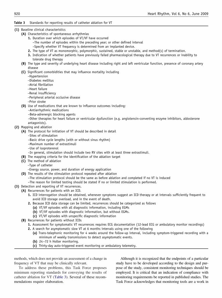
mf
mcm
spem
T
mbulat
920 Heart Rhythm, Vol 6, No 6, June 2009
ethods, which does not provide an assessment of a change inrequency of VT that may be clinically relevant.
To address these problems, this Task Force proposesinimum reporting standards for conveying the results of
atheter ablation for VT (Table 3). Several of these recom-
able 3 Standards for reporting results of catheter ablation for
(1) Baseline clinical characteristics(A) Characteristics of spontaneous arrhythmias
1. Duration over which episodes of VT/VF have occurred–The number of episodes within the preceding year;–Specify whether VT frequency is determined from a
2. The type of VT as monomorphic, polymorphic, sustai3. Indication of whether patients have previously failed
tolerate drug therapy(B) The type and severity of underlying heart disease includ
disease(C) Significant comorbidities that may influence mortality i
–Hypertension–Diabetes mellitus–Atrial fibrillation–Heart failure–Renal insufficiency–Peripheral arterial occlusive disease–Prior stroke
(D) Use of medications that are known to influence outcom–Antiarrhythmic medications–Beta-adrenergic blocking agents–Other therapies for heart failure or ventricular dysfuncantagonists).
(2) Mapping and ablation(A) The protocol for initiation of VT should be described in
–Sites of stimulation–Basic drive cycle lengths (with or without sinus rhythm–Maximum number of extrastimuli–Use of isoproterenol–In general, stimulation should include two RV sites wi
(B) The mapping criteria for the identification of the ablati(C) The method of ablation
–Type of catheter–Energy source, power, and duration of energy applicati
(D) The results of the stimulation protocol repeated after a–The stimulation protocol should be the same as before–The reason for limited testing should be stated if no o
(3) Detection and reporting of VT recurrences.(A) Recurrences for patients with an ICD.
1. ICD interrogation should be obtained, whenever symavoid ICD storage overload, and in the event of dea
2. Because ICD data storage can be limited, recurrence(a) VT/VF episodes with all diagnostic information,(b) VT/VF episodes with diagnostic information, bu(c) VT/VF episodes with unspecific diagnostic inform
(B) Recurrences for patients without ICDs1. Assessment for symptomatic VT recurrences requires2. A search for asymptomatic slow VT at 6 months inte
(a) Trans-telephonic monitoring for 4 weeks aroundminimum of weekly transmissions to detect asym
(b) 24–72 h Holter monitoring.(c) Thirty-day auto-triggered event monitoring or a
endations require elaboration. T
Although it is recognized that the endpoints of a particulartudy have to be developed according to the design and pur-ose of the study, consistent monitoring techniques should bemployed. It is critical that an indication of compliance withonitoring requirements be reported in published studies. The
er defined intervalnted device.
table or unstable, and method(s) of termination.acological therapy due to VT recurrences or inability to
ht and left ventricular function, presence of coronary artery
g
uding:
.g. angiotensin-converting enzyme inhibitors, aldosterone
east three extrastimuli.et
on and completed if no VT is induceded stimulation is performed.
suggest an ICD therapy or at intervals sufficiently frequent to
ld be categorized as followsing EGMsut EGMs.
cumentation (12-lead ECG or ambulatory monitor recordings)sing one of the followingllow-up interval, including symptom-triggered recording with a
atic events.
ory telemetry.
VT
or othn implaned, s
pharm
ing rig
ncludin
es incl
tion (e
detail
)
th at lon targ
onblationablatir limit
ptomsth.s shouincludt withoation
ECG dorvals uthe foptom
ask Force acknowledges that monitoring tools are a work in
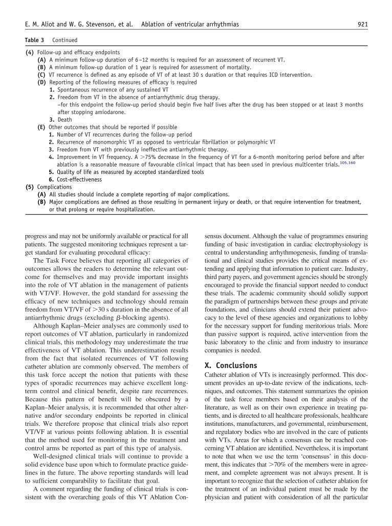
ppg
ociwefa
rcefctttBKntVtc
slt
s
sfctttettfcftbc
XCunoltiawctmmit
T
921E. M. Aliot and W. G. Stevenson, et al. Ablation of ventricular arrhythmias
rogress and may not be uniformly available or practical for allatients. The suggested monitoring techniques represent a tar-et standard for evaluating procedural efficacy:
The Task Force believes that reporting all categories ofutcomes allows the readers to determine the relevant out-ome for themselves and may provide important insightsnto the role of VT ablation in the management of patientsith VT/VF. However, the gold standard for assessing the
fficacy of new techniques and technology should remainreedom from VT/VF of �30 s duration in the absence of allntiarrhythmic drugs (excluding �-blocking agents).
Although Kaplan–Meier analyses are commonly used toeport outcomes of VT ablation, particularly in randomizedlinical trials, this methodology may underestimate the trueffectiveness of VT ablation. This underestimation resultsrom the fact that isolated recurrences of VT followingatheter ablation are commonly observed. The members ofhis task force accept the notion that patients with theseypes of sporadic recurrences may achieve excellent long-erm control and clinical benefit, despite rare recurrences.ecause this pattern of benefit will be obscured by aaplan–Meier analysis, it is recommended that other alter-ative and/or secondary endpoints be reported in clinicalrials. We therefore propose that clinical trials also reportT/VF at various points following ablation. It is essential
hat the method used for monitoring in the treatment andontrol arms be reported as part of this type of analysis.
Well-designed clinical trials will continue to provide aolid evidence base upon which to formulate practice guide-ines in the future. The above reporting standards will leado sufficient comparability to facilitate that goal.
A comment regarding the funding of clinical trials is con-
able 3 Continued
(4) Follow-up and efficacy endpoints(A) A minimum follow-up duration of 6–12 months is requi(B) A minimum follow-up duration of 1 year is required for(C) VT recurrence is defined as any episode of VT of at least(D) Reporting of the following measures of efficacy is requi
1. Spontaneous recurrence of any sustained VT2. Freedom from VT in the absence of antiarrhythmic d
–for this endpoint the follow-up period should beginafter stopping amiodarone.
3. Death(E) Other outcomes that should be reported if possible
1. Number of VT recurrences during the follow-up perio2. Recurrence of monomorphic VT as opposed to ventric3. Freedom from VT with previously ineffective antiarrh4. Improvement in VT frequency. A �75% decrease in t
ablation is a reasonable measure of favourable clinic5. Quality of life as measured by accepted standardized6. Cost-effectiveness
(5) Complications(A) All studies should include a complete reporting of majo(B) Major complications are defined as those resulting in pe
or that prolong or require hospitalization.
istent with the overarching goals of this VT Ablation Con- p
ensus document. Although the value of programmes ensuringunding of basic investigation in cardiac electrophysiology isentral to understanding arrhythmogenesis, funding of transla-ional and clinical studies provides the critical means of ex-ending and applying that information to patient care. Industry,hird party payers, and government agencies should be stronglyncouraged to provide the financial support needed to conducthese trials. The academic community should solidly supporthe paradigm of partnerships between these groups and privateoundations, and clinicians should extend their patient advo-acy to the level of these agencies and organizations to lobbyor the necessary support for funding meritorious trials. Morehan passive support is required, active intervention from theasic laboratory to the clinic and from industry to insuranceompanies is needed.
. Conclusionsatheter ablation of VTs is increasingly performed. This doc-ment provides an up-to-date review of the indications, tech-iques, and outcomes. This statement summarizes the opinionf the task force members based on their analysis of theiterature, as well as on their own experience in treating pa-ients, and is directed to all healthcare professionals, healthcarenstitutions, manufacturers, and governmental, reimbursement,nd regulatory bodies who are involved in the care of patientsith VTs. Areas for which a consensus can be reached con-
erning VT ablation are identified. Nevertheless, it is importanto note that when we use the term ‘consensus’ in this docu-ent, this indicates that �70% of the members were in agree-ent, and complete agreement was not always present. It is
mportant to recognize that the selection of catheter ablation forhe treatment of an individual patient must be made by the
an assessment of recurrent VT.ment of mortality.duration or that requires ICD intervention.
rapy.alf lives after the drug has been stopped or at least 3 months
brillation or polymorphic VTtherapy.
quency of VT for a 6-month monitoring period before and afteract that has been used in previous multicenter trials.105,160
lications.nt injury or death, or that require intervention for treatment,
red forassess30 s
red
rug thefive h
dular fiythmiche freal imptools
r comprmane
hysician and patient with consideration of all the particular

cdett
ATb
EdAADDDc
A
A
D
D
DD
DD
D
D
DD
D
D
pt
922 Heart Rhythm, Vol 6, No 6, June 2009
o-morbidities and circumstances relevant to that patient. Thisocument summarizes information to assist healthcare provid-rs in caring for patients with VT. Finally, it is recognized thathis field continues to evolve and that this document will needo be updated.
ppendixhe task force writing group would like to thank mem-
ers from EHRA, a Registered Branch of the ESC, HRS, vhe entity. If the amount is under the preceding definition it is considered ‘mod
SC, AHA, and ACC for their thoughtful reviews of theocument. Our thanks go to: Prof Martin Borggrefe, Prof.ntonio Berruezo, Prof. Christian de Chillou, Dr Shih-nn Chen, Dr L. Brent Mitchell, Dr N.A. Mark Estes III,r Alfred Buxton, Dr Usha Tedrow, Dr Claudio Schuger,r Paul Wang, Dr Andrea Russo, Dr Jennifer Cummings,r Wendy Gross, and Dr Jane Chen. All comments re-
eived were considered. Thank you for performing this
aluable service.uthor relationships with industry
uthorConsulting fees/honoraria Speaker’s Bureau
Ownership/partnership/principal Research grants
Fellowship supportand other benefit
r Etienne Aliot BiotronikCardiomeMeda PharmaMedtronicSanofi AventisSorin
None None None None
r Jesus Ma AlmendralGarrote
Boston ScientificSt Jude Medical
None None None None
r Frank Bogun None None None Biosense Webster Noner Hugh Calkins Biosense
Webster*Boston ScientificMedtronicSt Jude Medical
None None BiosenseWebster*
Boston ScientificMedtronic
r Etienne Delacretaz None None None None Noner Paolo Della Bella Biosense Webster
St Jude Medical*None None None None
r Gerhard Hindricks Biosense WebsterBiotronik*CyberheartMedtronicSt Jude Medical*Stereotaxis
None None None None
r Pierre Jaïs Biosense WebsterBoston ScientificSorinSt Jude MedicalVoyage Medical
None None Philips* None
r Mark E. Josephson Medtronic None None None Noner Josef Kautzner Biosense Webster
BiotroniBoston ScientificHansen MedicalSt Jude Medical
None None None None
r G. Neal Kay BiosenseWebster*
Boston Scientific*
None None None Biosense Webster*Boston Scientific*Medtronic*St Jude Medical*
r Karl-Heinz Kuck St Jude MedicalStereotaxis
None None Biosense Webster None
*Significant: a relationship is considered to be ‘significant’ if (1) the person receives $10 000 or more during any 12-month period or 5% or more of theerson’s gross income or (2) the person owns 5% or more of the voting stock or share of the entity or owns $10 000 or more of the fair market value of
est’.
A
D
D
D
D
D
D
D
D
pt
R
923E. M. Aliot and W. G. Stevenson, et al. Ablation of ventricular arrhythmias
uthorConsulting fees/honoraria Speaker’s Bureau
Ownership/partnership/principal Research grants
Fellowship supportand other benefit
r Bruce Lerman None None None Boston Scientific*Medtronic*St Jude Medical*
Boston Scientific*Medtronic*St Jude Medical*
r Francis Marchlinski BiosenseWebster*
Boston Scientific/Guidant*
GE HealthcareMedtronic*St Jude Medical*
None None BiosenseWebster*
St Jude Medical*
Biosense Webster*Biotronik*Boston Scientific/
Guidant*Medtronic*St Jude Medical*
r Vivek Reddy BiosenseWebster*
St Jude Medical*
None None BiosenseWebster*
St Jude Medical*
None
r Martin-Jan Schalij Boston Scientific BiotronikBoston Scientific
None Biotronik*Boston Scientific*Medtronic*St Jude Medical*
None
r Richard Schilling None Biosense WebsterSt Jude Medical
None None Boston Scientific*Medtronic*
r Kyoko Soejima Medtronic JapanToray Japan
None None None None
r William GregoryStevenson
Biosense WebsterBoston ScientificMedtronicSt Jude Medical
None None None None
r David Wilber Biosense WebsterBoston ScientificCardio InsightMedtronicSt Jude MedicalStereotaxis
None None None None
*Significant: a relationship is considered to be ‘significant’ if (1) the person receives $10 000 or more during any 12-month period or 5% or more of theerson’s gross income or (2) the person owns 5% or more of the voting stock or share of the entity or owns $10 000 or more of the fair market value of
he entity. If the amount is under the preceding definition it is considered ‘modest’.eferences1. Lerman BB. Mechanism of outflow tract tachycardia. Heart Rhythm 2007;4:
973–6.2. Tada H, Ito S, Naito S, Kurosaki K, Kubota S, Sugiyasu A et al. Idiopathic
ventricular arrhythmia arising from the mitral annulus: a distinct subgroup ofidiopathic ventricular arrhythmias. J Am Coll Cardiol 2005;45:877–86.
3. Tada H, Tadokoro K, Ito S, Naito S, Hashimoto T, Kaseno K et al. Idiopathicventricular arrhythmias originating from the tricuspid annulus: prevalence, elec-trocardiographic characteristics, and results of radiofrequency catheter ablation.Heart Rhythm 2007;4:7–16.
4. Tanner H, Hindricks G, Schirdewahn P, Kobza R, Dorszewski A, Piorkowski C etal. Outflow tract tachycardia with R/S transition in lead V3: six different anatomicapproaches for successful ablation. J Am Coll Cardiol 2005;45:418–23.
5. Buxton AE, Waxman HL, Marchlinski FE, Simson MB, Cassidy D, JosephsonME. Right ventricular tachycardia: clinical and electrophysiologic characteris-tics. Circulation 1983;68:917–27.
6. Ito S, Tada H, Naito S, Kurosaki K, Ueda M, Hoshizaki H et al. Developmentand validation of an ECG algorithm for identifying the optimal ablation site foridiopathic ventricular outflow tract tachycardia. J Cardiovasc Electrophysiol2003;14:1280–6.
7. Kamakura S, Shimizu W, Matsuo K, Taguchi A, Suyama K, Kurita T et al.Localization of optimal ablation site of idiopathic ventricular tachycardia fromright and left ventricular outflow tract by body surface ECG. Circulation 1998;98:1525–33.
8. Lerman BB, Belardinelli L, West GA, Berne RM, DiMarco JP. Adenosine-sensitive ventricular tachycardia: evidence suggesting cyclic AMP-mediated
triggered activity. Circulation 1986;74:270–80.9. Lerman BB, Stein K, Engelstein ED, Battleman DS, Lippman N, Bei D et al.Mechanism of repetitive monomorphic ventricular tachycardia. Circulation1995;92:421–9.
10. Morady F, Kadish AH, DiCarlo L, Kou WH, Winston S, deBuitlier M et al.Long-term results of catheter ablation of idiopathic right ventricular tachycardia.Circulation 1990;82:2093–9.
11. Sung RJ, Keung EC, Nguyen NX, Huycke EC. Effects of beta-adrenergicblockade on verapamil-responsive and verapamil-irresponsive sustained ventric-ular tachycardias. J Clin Invest 1988;81:688–99.
12. Tada H, Tadokoro K, Miyaji K, Ito S, Kurosaki K, Kaseno K et al. Idiopathicventricular arrhythmias arising from the pulmonary artery: prevalence, charac-teristics, and topography of the arrhythmia origin. Heart Rhythm 2008;5:419–26.
13. Antzelevitch C, Shimizu W. Cellular mechanisms underlying the long QTsyndrome. Curr Opin Cardiol 2002;17:43–51.
14. Haissaguerre M, Extramiana F, Hocini M, Cauchemez B, Jais P, Cabrera JA etal. Mapping and ablation of ventricular fibrillation associated with long-QT andBrugada syndromes. Circulation 2003;108:925–8.
15. Lerman BB. Response of nonreentrant catecholamine-mediated ventriculartachycardia to endogenous adenosine and acetylcholine. Evidence for myocar-dial receptor-mediated effects. Circulation 1993;87:382–90.
16. Damle RS, Landers M, Kelly PA, Reiter MJ, Mann DE. Radiofrequency catheterablation of idiopathic left ventricular tachycardia originating in the left anteriorfascicle. Pacing Clin Electrophysiol 1998;21:1155–8.
17. Lopera G, Stevenson WG, Soejima K, Maisel WH, Koplan B, Sapp JL et al.Identification and ablation of three types of ventricular tachycardia involving theHis-Purkinje system in patients with heart disease. J Cardiovasc Electrophysiol
2004; 15:52–8.
924 Heart Rhythm, Vol 6, No 6, June 2009
18. Huelsing DJ, Spitzer KW, Pollard AE. Spontaneous activity induced in rabbitPurkinje myocytes during coupling to a depolarized model cell. Cardiovasc Res2003; 59:620–7.
19. Spitzer KW, Pollard AE, Yang L, Zaniboni M, Cordeiro JM, Huelsing DJ.Cell-to-cell electrical interactions during early and late repolarization. J Cardio-vasc Electrophysiol 2006;17(Suppl. 1):S8–14.
20. Belhassen B, Caspi A, Miller H, Shapira I, Laniado S. Extensive endocardialmapping during sinus rhythm and ventricular tachycardia in a patient witharrhythmogenic right ventricular dysplasia. J Am Coll Cardiol 1984;4:1302–6.
21. Delacretaz E, Stevenson WG, Ellison KE, Maisel WH, Friedman PL. Mappingand radiofrequency catheter ablation of the three types of sustained monomor-phic ventricular tachycardia in nonischemic heart disease. J Cardiovasc Elec-trophysiol 2000;11:11–17.
22. Eckart RE, Hruczkowski TW, Tedrow UB, Koplan BA, Epstein LM, StevensonWG. Sustained ventricular tachycardia associated with corrective valve surgery.Circulation 2007;116:2005–11.
23. Ellison KE, Friedman PL, Ganz LI, Stevenson WG. Entrainment mapping andradiofrequency catheter ablation of ventricular tachycardia in right ventriculardysplasia. J Am Coll Cardiol 1998;32:724–8.
24. Hsia HH, Marchlinski FE. Electrophysiology studies in patients with dilatedcardiomyopathies. Card Electrophysiol Rev 2002;6:472–81.
25. Josephson ME, Horowitz LN, Farshidi A, Kastor JA. Recurrent sustained ven-tricular tachycardia. 1. Mechanisms. Circulation 1978;57:431–40.
26. Josephson ME, Almendral JM, Buxton AE, Marchlinski FE. Mechanisms ofventricular tachycardia. Circulation 1987;75(Pt 2):III41–7.
27. Koplan BA, Soejima K, Baughman K, Epstein LM, Stevenson WG. Refractoryventricular tachycardia secondary to cardiac sarcoid: electrophysiologic charac-teristics, mapping, and ablation. Heart Rhythm 2006;3:924–9.
28. Marchlinski FE, Zado E, Dixit S, Gerstenfeld E, Callans DJ, Hsia H et al.Electroanatomic substrate and outcome of catheter ablative therapy for ventric-ular tachycardia in setting of right ventricular cardiomyopathy. Circulation2004;110:2293–8.
29. Sosa E, Scanavacca M, D’Avila A, Piccioni J, Sanchez O, Velarde JL et al.Endocardial and epicardial ablation guided by nonsurgical transthoracic epicar-dial mapping to treat recurrent ventricular tachycardia. J Cardiovasc Electro-physiol 1998;9:229–39.
30. Stevenson WG, Khan H, Sager P, Saxon LA, Middlekauff HR, Natterson PD etal. Identification of reentry circuit sites during catheter mapping and radiofre-quency ablation of ventricular tachycardia late after myocardial infarction.Circulation 1993;88(Pt 1):1647–70.
31. Dillon SM, Allessie MA, Ursell PC, Wit AL. Influences of anisotropic tissuestructure on reentrant circuits in the epicardial border zone of subacute canineinfarcts. Circ Res 1988;63:182–206.
32. Downar E, Harris L, Mickleborough LL, Shaikh N, Parson ID. Endocardialmapping of ventricular tachycardia in the intact human ventricle: evidence forreentrant mechanisms. J Am Coll Cardiol 1988;11:783–91.
33. Downar E, Kimber S, Harris L, Mickleborough L, Sevaptsidis E, Masse S et al.Endocardial mapping of ventricular tachycardia in the intact human heart. II.Evidence for multiuse reentry in a functional sheet of surviving myocardium.J Am Coll Cardiol 1992;20:869–78.
34. Kleber AG, Rudy Y. Basic mechanisms of cardiac impulse propagation andassociated arrhythmias. Physiol Rev 2004;84:431–88.
35. de Bakker JM, van Capelle FJ, Janse MJ, Wilde AA, Coronel R, Becker AE etal. Reentry as a cause of ventricular tachycardia in patients with chronicischemic heart disease: electrophysiologic and anatomic correlation. Circulation1988;77:589–606.
36. Downar E, Harris L, Kimber S, Mickleborough L, Williams W, Sevaptsidis E etal. Ventricular tachycardia after surgical repair of tetralogy of Fallot: results ofintraoperative mapping studies. J Am Coll Cardiol 1992;20:648–55.
37. El-Sherif N, Smith RA, Evans K. Canine ventricular arrhythmias in the latemyocardial infarction period. 8. Epicardial mapping of reentrant circuits. CircRes 1981;49:255–65.
38. El-Sherif N, Mehra R, Gough WB, Zeiler RH. Reentrant ventricular arrhythmiasin the late myocardial infarction period. Interruption of reentrant circuits bycryothermal techniques. Circulation 1983;68:644–56.
39. Horowitz LN, Vetter VL, Harken AH, Josephson ME. Electrophysiologic char-acteristics of sustained ventricular tachycardia occurring after repair of tetralogyof fallot. Am J Cardiol 1980;46:446–52.
40. Kugler JD, Pinsky WW, Cheatham JP, Hofschire PJ, Mooring PK, Fleming WH.Sustained ventricular tachycardia after repair of tetralogy of Fallot: new elec-trophysiologic findings. Am J Cardiol 1983;51:1137–43.
41. Ouyang F, Fotuhi P, Ho SY, Hebe J, Volkmer M, Goya M et al. Repetitivemonomorphic ventricular tachycardia originating from the aortic sinus cusp:electrocardiographic characterization for guiding catheter ablation. J Am Coll
Cardiol 2002;39:500–8.42. Baba S, Dun W, Cabo C, Boyden PA. Remodeling in cells from different regionsof the reentrant circuit during ventricular tachycardia. Circulation 2005;112:2386–96.
43. Peters NS, Wit AL. Myocardial architecture and ventricular arrhythmogenesis.Circulation 1998;97:1746–54.
44. de Bakker JM, Coronel R, Tasseron S, Wilde AA, Opthof T, Janse MJ et al.Ventricular tachycardia in the infarcted, Langendorff-perfused human heart: roleof the arrangement of surviving cardiac fibers. J Am Coll Cardiol 1990;15:1594–607.
45. de Bakker JM, van Capelle FJ, Janse MJ, Tasseron S, Vermeulen JT, de JN etal. Slow conduction in the infarcted human heart. ‘Zigzag’ course of activation.Circulation 1993;88:915–26.
46. Kawara T, Derksen R, de Groot JR, Coronel R, Tasseron S, Linnenbank AC etal. Activation delay after premature stimulation in chronically diseased humanmyocardium relates to the architecture of interstitial fibrosis. Circulation 2001;104:3069–75.
47. de Bakker JM, Stein M, van Rijen HV. Three-dimensional anatomic structure assubstrate for ventricular tachycardia/ventricular fibrillation. Heart Rhythm 2005;2:777–9.
48. Josephson ME, Harken AH, Horowitz LN. Endocardial excision: a new surgicaltechnique for the treatment of recurrent ventricular tachycardia. Circulation1979;60:1430–9.
49. Klein GJ, Ideker RE, Smith WM, Harrison LA, Kasell J, Wallace AG et al.Epicardial mapping of the onset of ventricular tachycardia initiated by pro-grammed stimulation in the canine heart with chronic infarction. Circulation1979;60:1375–84.
50. Bogun F, Good E, Reich S, Elmouchi D, Igic P, Lemola K et al. Isolatedpotentials during sinus rhythm and pace-mapping within scars as guides forablation of post-infarction ventricular tachycardia. J Am Coll Cardiol 2006;47:2013–9.
51. Brunckhorst CB, Delacretaz E, Soejima K, Maisel WH, Friedman PL, StevensonWG. Identification of the ventricular tachycardia isthmus after infarction by pacemapping. Circulation 2004;110:652–9.
52. Miller JM, Tyson GS, Hargrove WC III, Vassallo JA, Rosenthal ME, JosephsonME. Effect of subendocardial resection on sinus rhythm endocardial electrogramabnormalities. Circulation 1995;91:2385–91.
53. Soejima K, Stevenson WG, Maisel WH, Sapp JL, Epstein LM. Electricallyunexcitable scar mapping based on pacing threshold for identification of thereentry circuit isthmus: feasibility for guiding ventricular tachycardia ablation.Circulation 2002;106:1678–83.
54. de Chillou C, Lacroix D, Klug D, Magnin-Poull I, Marquie C, Messier M et al.Isthmus characteristics of reentrant ventricular tachycardia after myocardialinfarction. Circulation 2002;105:726–31.
55. Hsia HH, Lin D, Sauer WH, Callans DJ, Marchlinski FE. Anatomic character-ization of endocardial substrate for hemodynamically stable reentrant ventriculartachycardia: identification of endocardial conducting channels. Heart Rhythm2006;3:503–12.
56. Littmann L, Svenson RH, Gallagher JJ, Selle JG, Zimmern SH, Fedor JM et al.Functional role of the epicardium in postinfarction ventricular tachycardia.Observations derived from computerized epicardial activation mapping, entrain-ment, and epicardial laser photoablation. Circulation 1991;83:1577–91.
57. Wilber DJ, Kopp DE, Glascock DN, Kinder CA, Kall JG. Catheter ablation ofthe mitral isthmus for ventricular tachycardia associated with inferior infarction.Circulation 1995;92:3481–89.
58. Pogwizd SM, Hoyt RH, Saffitz JE, Corr PB, Cox JL, Cain ME. Reentrant andfocal mechanisms underlying ventricular tachycardia in the human heart. Cir-culation 1992;86:1872–87.
59. Soejima K, Suzuki M, Maisel WH, Brunckhorst CB, Delacretaz E, Blier L et al.Catheter ablation in patients with multiple and unstable ventricular tachycardiasafter myocardial infarction: short ablation lines guided by reentry circuit isth-muses and sinus rhythm mapping. Circulation 2001;104:664–9.
60. El-Sherif N, Gough WB, Zeiler RH, Hariman R. Reentrant ventricular arrhyth-mias in the late myocardial infarction period. 12. Spontaneous versus inducedreentry and intramural versus epicardial circuits. J Am Coll Cardiol 1985;6:124–32.
61. Marchlinski FE. Perivalvular fibrosis and monomorphic ventricular tachycardia:toward a unifying hypothesis in nonischemic cardiomyopathy. Circulation 2007;116:1998–2001.
62. Klemm HU, Ventura R, Steven D, Johnsen C, Rostock T, Lutomsky B et al.Catheter ablation of multiple ventricular tachycardias after myocardial infarctionguided by combined contact and noncontact mapping. Circulation 2007;115:2697–704.
63. Brugada P, Abdollah H, Wellens HJ. Continuous electrical activity duringsustained monomorphic ventricular tachycardia. Observations on its dynamic
behavior during the arrhythmia. Am J Cardiol 1985;55:402–11.
1
1
1
1
1
1
1
925E. M. Aliot and W. G. Stevenson, et al. Ablation of ventricular arrhythmias
64. Lin JW, Garber L, Qi YR, Chang MG, Cysyk J, Tung L. Region [corrected] ofslowed conduction acts as core for spiral wave reentry in cardiac cell monolay-ers. Am J Physiol Heart Circ Physiol 2008;294:H58–65.
65. Caceres J, Jazayeri M, McKinnie J, Avitall B, Denker ST, Tchou P et al.Sustained bundle branch reentry as a mechanism of clinical tachycardia. Circu-lation 1989;79:256–70.
66. Hadjis TA, Stevenson WG, Harada T, Friedman PL, Sager P, Saxon LA.Preferential locations for critical reentry circuit sites causing ventricular tachy-cardia after inferior wall myocardial infarction. J Cardiovasc Electrophysiol1997;8:363–70.
67. Crijns HJ, Smeets JL, Rodriguez LM, Meijer A, Wellens HJ. Cure of interfas-cicular reentrant ventricular tachycardia by ablation of the anterior fascicle of theleft bundle branch. J Cardiovasc Electrophysiol 1995;6:486–92.
68. Tchou P, Jazayeri M, Denker S, Dongas J, Caceres J, Akhtar M. Transcatheterelectrical ablation of right bundle branch. A method of treating macroreentrantventricular tachycardia attributed to bundle branch reentry. Circulation 1988;78:246–57.
69. Nakagawa H, Beckman KJ, McClelland JH, Wang X, Arruda M, Santoro I et al.Radiofrequency catheter ablation of idiopathic left ventricular tachycardiaguided by a Purkinje potential. Circulation 1993;88:2607–17.
70. Rodriguez LM, Smeets JL, Timmermans C, Trappe HJ, Wellens HJ. Radiofre-quency catheter ablation of idiopathic ventricular tachycardia originating in theanterior fascicle of the left bundle branch. J Cardiovasc Electrophysiol 1996;7:1211–6.
71. Kaltenbrunner W, Cardinal R, Dubuc M, Shenasa M, Nadeau R et al. Epicardialand endocardial mapping of ventricular tachycardia in patients with myocardialinfarction. Is the origin of the tachycardia always subendocardially localized?Circulation 1991;84:1058–71.
72. Stevenson WG, Friedman PL, Kocovic D, Sager PT, Saxon LA, Pavri B.Radiofrequency catheter ablation of ventricular tachycardia after myocardialinfarction. Circulation 1998;98:308–14.
73. Marchlinski FE, Callans DJ, Gottlieb CD, Zado E. Linear ablation lesions forcontrol of unmappable ventricular tachycardia in patients with ischemic andnonischemic cardiomyopathy. Circulation 2000;101:1288–96.
74. Ouyang F, Antz M, Deger FT, Bansch D, Schaumann A, Ernst S et al. Anunderrecognized subepicardial reentrant ventricular tachycardia attributable toleft ventricular aneurysm in patients with normal coronary arteriograms. Circu-lation 2003;107:2702–9.
75. Sosa E, Scanavacca M, D’Avila A, Bellotti G, Pilleggi F. Radiofrequencycatheter ablation of ventricular tachycardia guided by nonsurgical epicardialmapping in chronic Chagasic heart disease. Pacing Clin Electrophysiol 1999;22(Pt 1):128–30.
76. Irvine J, Dorian P, Baker B, O’Brien BJ, Roberts R, Gent M et al. Quality of lifein the Canadian Implantable Defibrillator Study (CIDS). Am Heart J 2002;144:282–9.
77. Mark DB, Anstrom KJ, Sun JL, Clapp-Channing NE, Tsiatis AA, vidson-Ray Let al. Quality of life with defibrillator therapy or amiodarone in heart failure.N Engl J Med 2008;359:999–1008.
78. Moss AJ, Greenberg H, Case RB, Zareba W, Hall WJ, Brown MW et al.Long-term clinical course of patients after termination of ventricular tachyar-rhythmia by an implanted defibrillator. Circulation 2004;110:3760–5.
79. Poole JE, Johnson GW, Hellkamp AS, Anderson J, Callans DJ, Raitt MH et al.Prognostic importance of defibrillator shocks in patients with heart failure.N Engl J Med 2008;359:1009–17.
80. Schron EB, Exner DV, Yao Q, Jenkins LS, Steinberg JS, Cook JR et al. Qualityof life in the antiarrhythmics versus implantable defibrillators trial: impact oftherapy and influence of adverse symptoms and defibrillator shocks. Circulation2002;105:589–94.
81. Bollmann A, Husser D, Cannom DS. Antiarrhythmic drugs in patients withimplantable cardioverter-defibrillators. Am J Cardiovasc Drugs 2005;5:371–8.
82. Connolly SJ, Dorian P, Roberts RS, Gent M, Bailin S, Fain ES, Thorp K et al.Comparison of beta-blockers, amiodarone plus beta-blockers, or sotalol forprevention of shocks from implantable cardioverter defibrillators: the OPTICstudy: a randomized trial. J Am Med Assoc 2006;295:165–71.
83. Pacifico A, Hohnloser SH, Williams JH, Tao B, Saksena S, Henry PD et al.Prevention of implantable-defibrillator shocks by treatment with sotalol. d,l-Sotalol Implantable Cardioverter-Defibrillator Study Group. N Engl J Med1999;340:1855–62.
84. Gepstein L, Hayam G, Ben-Haim SA. A novel method for nonfluoroscopiccatheter-based electroanatomical mapping of the heart. In vitro and in vivoaccuracy results. Circulation 1997;95:1611–22.
85. Dixit S, Narula N, Callans DJ, Marchlinski FE. Electroanatomic mapping ofhuman heart: epicardial fat can mimic scar. J Cardiovasc Electrophysiol 2003;14:1128.
86. Soejima K, Stevenson WG, Sapp JL, Selwyn AP, Couper G, Epstein LM.
Endocardial and epicardial radiofrequency ablation of ventricular tachycardiaassociated with dilated cardiomyopathy: the importance of low-voltage scars.J Am Coll Cardiol 2004;43:1834–42.
87. Swarup V, Morton JB, Arruda M, Wilber DJ. Ablation of epicardial macroreen-trant ventricular tachycardia associated with idiopathic nonischemic dilatedcardiomyopathy by a percutaneous transthoracic approach. J Cardiovasc Elec-trophysiol 2002;13:1164–8.
88. Roux JF, Dubuc M, Pressacco J, Roy D, Thibault B, Talajic M et al. Concor-dance between an electroanatomic mapping system and cardiac MRI in arrhyth-mogenic right ventricular cardiomyopathy. Pacing Clin Electrophysiol 2006;29:109–12.
89. Wittkampf FH, Wever EF, Derksen R, Wilde AA, Ramanna H, Hauer RN et al.LocaLisa: new technique for real-time 3-dimensional localization of regularintracardiac electrodes. Circulation 1999;99:1312–7.
90. Khaykin Y, Skanes A, Whaley B, Hill C, Beardsall M, Seabrook C et al.Real-time integration of 2D intracardiac echocardiography and 3D electroana-tomical mapping to guide ventricular tachycardia ablation. Heart Rhythm 2008;5:1396–1402.
91. Khongphatthanayothin A, Kosar E, Nademanee K. Nonfluoroscopic three-di-mensional mapping for arrhythmia ablation: tool or toy? J Cardiovasc Electro-physiol 2000;11:239–43.
92. Nademanee K, Kosar EM. A nonfluoroscopic catheter-based mapping techniqueto ablate focal ventricular tachycardia. Pacing Clin Electrophysiol 1998;21:1442–7.
93. Ouyang F, Cappato R, Ernst S, Goya M, Volkmer M, Hebe J et al. Electroana-tomic substrate of idiopathic left ventricular tachycardia: unidirectional blockand macroreentry within the purkinje network. Circulation 2002;105:462–9.
94. Varanasi S, Dhala A, Blanck Z, Deshpande S, Akhtar M, Sra J. Electroanatomicmapping for radiofrequency ablation of cardiac arrhythmias. J Cardiovasc Elec-trophysiol 1999;10:538–44.
95. Arenal A, Glez-Torrecilla E, Ortiz M, Villacastin J, Fdez-Portales J, Sousa E etal. Ablation of electrograms with an isolated, delayed component as treatment ofunmappable monomorphic ventricular tachycardias in patients with structuralheart disease. J Am Coll Cardiol 2003;41:81–92.
96. Brunckhorst CB, Delacretaz E, Soejima K, Jackman WM, Nakagawa H, KuckKH et al. Ventricular mapping during atrial and right ventricular pacing: relationof electrogram parameters to ventricular tachycardia reentry circuits after myo-cardial infarction. J Interv Card Electrophysiol 2004;11:183–91.
97. Codreanu A, Odille F, Aliot E, Marie PY, Magnin-Poull I, Andronache M et al.Electroanatomic characterization of post-infarct scars comparison with 3-dimen-sional myocardial scar reconstruction based on magnetic resonance imaging.J Am Coll Cardiol 2008;52:839–42.
98. Kautzner J, Cihak R, Peichl P, Vancura V, Bytesnik J. Catheter ablation ofventricular tachycardia following myocardial infarction using three-dimensionalelectroanatomical mapping. Pacing Clin Electrophysiol 2003;26(Pt 2):342–7.
99. Kottkamp H, Wetzel U, Schirdewahn P, Dorszewski A, Gerds-Li JH, Carbucic-chio C et al. Catheter ablation of ventricular tachycardia in remote myocardialinfarction: substrate description guiding placement of individual linear lesionstargeting noninducibility. J Cardiovasc Electrophysiol 2003;14:675–81.
00. Miljoen H, State S, de Chillou C, Magnin-Poull I, Dotto P, Andronache M et al.Electroanatomic mapping characteristics of ventricular tachycardia in patientswith arrhythmogenic right ventricular cardiomyopathy/dysplasia. Europace2005;7:516–24.
01. Reddy VY, Neuzil P, Taborsky M, Ruskin JN. Short-term results of substratemapping and radiofrequency ablation of ischemic ventricular tachycardia usinga saline-irrigated catheter. J Am Coll Cardiol 2003;41:2228–36.
02. Reddy VY, Reynolds MR, Neuzil P, Richardson AW, Taborsky M, Jongnarang-sin K et al. Prophylactic catheter ablation for the prevention of defibrillatortherapy. N Engl J Med 2007;357:2657–65.
03. Reithmann C, Hahnefeld A, Remp T, Dorwarth U, Dugas M, Steinbeck G et al.Electroanatomic mapping of endocardial right ventricular activation as a guidefor catheter ablation in patients with arrhythmogenic right ventricular dysplasia.Pacing Clin Electrophysiol 2003;26:1308–16.
04. Stevenson WG, Delacretaz E, Friedman PL, Ellison KE. Identification andablation of macroreentrant ventricular tachycardia with the CARTO electroana-tomical mapping system. Pacing Clin Electrophysiol 1998;21:1448–56.
05. Stevenson WG, Wilber DJ, Natale A, Jackman WM, Marchlinski FE, Talbert Tet al. Irrigated radiofrequency catheter ablation guided by electroanatomic map-ping for recurrent ventricular tachycardia after myocardial infarction: the mul-ticenter thermocool ventricular tachycardia ablation trial. Circulation 2008;118:2773–82.
06. Tanner H, Hindricks G, Volkmer M, Furniss S, kuhlkamp V, Lacroix D et al.Catheter ablation of recurrent scar-related ventricular tachycardia using electro-anatomical mapping and irrigated ablation technology: Results of the prospec-
tive multicenter Euro-VT-Study. 2009 (in press).
1
1
1
1
1
1
1
1
1
1
1
1
1
1
1
1
1
1
1
1
1
1
1
1
1
1
1
1
1
1
1
1
1
1
1
1
1
1
1
1
1
1
1
1
1
926 Heart Rhythm, Vol 6, No 6, June 2009
07. Tomassoni G, Stanton M, Richey M, Leonelli FM, Beheiry S, Natale A.Epicardial mapping and radiofrequency catheter ablation of ischemic ventriculartachycardia using a three-dimensional nonfluoroscopic mapping system. J Car-diovasc Electrophysiol 1999;10:1643–8.
08. Chauhan VS, Nair GM, Sevaptisidis E, Downar E. Magnetoelectroanatomicmapping of arrhythmias in structural heart disease using a novel multielectrodecatheter. Pacing Clin Electrophysiol 2004;27:1077–84.
09. Patel AM, D’Avila A, Neuzil P, Kim SJ, Mela T, Singh J et al. Atrial tachycardiaafter ablation of persistent atrial fibrillation: identification of the critical isthmuswith a combination of multielectrode activation mapping and targeted entrain-ment mapping. Circ Arrhythmia Electrophysiol 2008;1:14–22.
10. Greenspon AJ, Hsu SS, Datorre S. Successful radiofrequency catheter ablationof sustained ventricular tachycardia postmyocardial infarction in man guided bya multielectrode “basket” catheter. J Cardiovasc Electrophysiol 1997;8:565–70.
11. Schalij MJ, van Rugge FP, Siezenga M, van der Velde ET. Endocardial activa-tion mapping of ventricular tachycardia in patients: first application of a 32-sitebipolar mapping electrode catheter. Circulation 1998;98:2168–79.
12. Yoshida Y, Hirai M, Murakami Y, Kondo T, Inden Y, Akahoshi M et al.Localization of precise origin of idiopathic ventricular tachycardia from the rightventricular outflow tract by a 12-lead ECG: a study of pace mapping using amultielectrode “basket” catheter. Pacing Clin Electrophysiol 1999;22:1760–8.
13. Thornton AS, Jordaens LJ. Remote magnetic navigation for mapping and ab-lating right ventricular outflow tract tachycardia. Heart Rhythm 2006;3:691–6.
14. Gornick CC, Adler SW, Pederson B, Hauck J, Budd J, Schweitzer J. Validationof a new noncontact catheter system for electroanatomic mapping of left ven-tricular endocardium. Circulation 1999;99:829–35.
15. Schilling RJ, Peters NS, Davies DW. Simultaneous endocardial mapping in thehuman left ventricle using a noncontact catheter: comparison of contact andreconstructed electrograms during sinus rhythm. Circulation 1998;98:887–98.
16. Schilling RJ, Peters NS, Davies DW. Feasibility of a noncontact catheter forendocardial mapping of human ventricular tachycardia. Circulation 1999;99:2543–52.
17. Sivagangabalan G, Pouliopoulos J, Huang K, Lu J, Barry M, Thiagalingam A etal. Comparison of electroanatomic contact and noncontact mapping of ventriularscar in postinfarct ovine model with intramural needle electrode recording andhistological validation. Circ Arrhythmia Electrophysiol 2008;1:363–9.
18. Della BP, Pappalardo A, Riva S, Tondo C, Fassini G, Trevisi N. Non-contactmapping to guide catheter ablation of untolerated ventricular tachycardia. EurHeart J 2002;23:742–52.
19. Segal OR, Chow AW, Markides V, Schilling RJ, Peters NS, Davies DW.Long-term results after ablation of infarct-related ventricular tachycardia. HeartRhythm 2005;2:474–82.
20. Abrams DJ, Earley MJ, Sporton SC, Kistler PM, Gatzoulis MA, Mullen MJ etal. Comparison of noncontact and electroanatomic mapping to identify scar andarrhythmia late after the Fontan procedure. Circulation 2007;115:1738–46.
21. Jacobson JT, Afonso VX, Eisenman G, Schultz JR, Lazar S, Michele JJ et al.Characterization of the infarct substrate and ventricular tachycardia circuits withnon-contact unipolar mapping in a porcine model of myocardial infarction.Heart Rhythm 2006;3:189–97.
22. Voss F, Steen H, Bauer A, Giannitsis E, Katus HA, Becker R. Determination ofmyocardial infarct size by noncontact mapping. Heart Rhythm 2008;5:308–14.
23. Chen M, Yang B, Zou J, Shan Q, Chen C, Xu D et al. Non-contact mapping andlinear ablation of the left posterior fascicle during sinus rhythm in the treatmentof idiopathic left ventricular tachycardia. Europace 2005;7:138–44.
24. Friedman PA, Asirvatham SJ, Grice S, Glikson M, Munger TM, Rea RF et al.Non-contact mapping to guide ablation of right ventricular outflow tract tachy-cardia. J Am Coll Cardiol 2002;39:1808–12.
25. Ribbing M, Wasmer K, Monnig G, Kirchhof P, Loh P, Breithardt G et al.Endocardial mapping of right ventricular outflow tract tachycardia using non-contact activation mapping. J Cardiovasc Electrophysiol 2003;14:602–8.
26. Yao Y, Zhang S, He DS, Zhang K, Hua W, Chu J et al. Radiofrequencyablation of the ventricular tachycardia with arrhythmogenic right ventricularcardiomyopathy using non-contact mapping. Pacing Clin Electrophysiol2007;30:526 –33.
27. Kriebel T, Saul JP, Schneider H, Sigler M, Paul T. Noncontact mapping andradiofrequency catheter ablation of fast and hemodynamically unstable ventric-ular tachycardia after surgical repair of tetralogy of Fallot. J Am Coll Cardiol2007; 50:2162–8.
28. Rajappan K, Schilling RJ. Non-contact mapping in the treatment of ventriculartachycardia after myocardial infarction. J Interv Card Electrophysiol 2007;19:9–18.
29. Strickberger SA, Knight BP, Michaud GF, Pelosi F, Morady F. Mapping andablation of ventricular tachycardia guided by virtual electrograms using anoncontact, computerized mapping system. J Am Coll Cardiol 2000;35:414 –
21. 130. Al-Ahmad A, Grossman JD, Wang PJ. Early experience with a computerizedrobotically controlled catheter system. J Interv Card Electrophysiol 2005;12:199–202.
31. Saliba W, Cummings JE, Oh S, Zhang Y, Mazgalev TN, Schweikert RA et al.Novel robotic catheter remote control system: feasibility and safety of transsep-tal puncture and endocardial catheter navigation. J Cardiovasc Electrophysiol2006; 17:1102–5.
32. Faddis MN, Blume W, Finney J, Hall A, Rauch J, Sell J et al. Novel, magnet-ically guided catheter for endocardial mapping and radiofrequency catheterablation. Circulation 2002;106:2980–5.
33. Greenberg S, Blume W, Faddis M, Finney J, Hall A, Talcott M et al. Remotecontrolled magnetically guided pulmonary vein isolation in canines. HeartRhythm 2006;3:71–6.
34. Pappone C, Vicedomini G, Manguso F, Gugliotta F, Mazzone P, Gulletta S et al.Robotic magnetic navigation for atrial fibrillation ablation. J Am Coll Cardiol2006; 47:1390–400.
35. Aryana A, D’Avila A, Heist EK, Mela T, Singh JP, Ruskin JN et al. Remotemagnetic navigation to guide endocardial and epicardial catheter mapping ofscar-related ventricular tachycardia. Circulation 2007;115:1191–200.
36. Burkhardt JD, Saliba WI, Schweikert RA, Cummings J, Natale A. Remotemagnetic navigation to map and ablate left coronary cusp ventricular tachycar-dia. J Cardiovasc Electrophysiol 2006;17:1142–4.
37. Thornton AS, Res J, Mekel JM, Jordaens LJ. Use of advanced mapping andremote magnetic navigation to ablate left ventricular fascicular tachycardia.Pacing Clin Electrophysiol 2006;29:685–8.
38. Bogun F, Morady F. Ablation of ventricular tachycardia in patients with non-ischemic cardiomyopathy. J Cardiovasc Electrophysiol 2008;19:1227–30.
39. Nazarian S, Bluemke DA, Lardo AC, Zviman MM, Watkins SP, Dickfeld TL etal. Magnetic resonance assessment of the substrate for inducible ventriculartachycardia in nonischemic cardiomyopathy. Circulation 2005;112:2821–5.
40. Schmidt A, Azevedo CF, Cheng A, Gupta SN, Bluemke DA, Foo TK et al.Infarct tissue heterogeneity by magnetic resonance imaging identifies enhancedcardiac arrhythmia susceptibility in patients with left ventricular dysfunction.Circulation 2007;115:2006–14.
41. Faris OP, Shein M. Food and Drug Administration perspective: magnetic reso-nance imaging of pacemaker and implantable cardioverter-defibrillator patients.Circulation 2006;114:1232–3.
42. Nazarian S, Roguin A, Zviman MM, Lardo AC, Dickfeld TL, Calkins H et al.Clinical utility and safety of a protocol for noncardiac and cardiac magneticresonance imaging of patients with permanent pacemakers and implantable-cardioverter defibrillators at 1.5 tesla. Circulation 2006;114:1277–84.
43. Roguin A, Schwitter J, Vahlhaus C, Lombardi M, Brugada J, Vardas P et al.Magnetic resonance imaging in individuals with cardiovascular implantableelectronic devices. Europace 2008;10:336–46.
44. Bello D, Kipper S, Valderrabano M, Shivkumar K. Catheter ablation of ven-tricular tachycardia guided by contrast-enhanced cardiac computed tomography.Heart Rhythm 2004;1:490–2.
45. Dickfeld T, Kocher C. The role of integrated PET-CT scar maps for guiding ventricular tachycardia ablations. Curr Cardiol Rep 2008;10:149–57.
46. Ashikaga H, Sasano T, Dong J, Zviman MM, Evers R, Hopenfeld B et al.Magnetic resonance-based anatomical analysis of scar-related ventricular tachy-cardia: implications for catheter ablation. Circ Res 2007;101:939–47.
47. Reddy VY, Malchano ZJ, Holmvang G, Schmidt EJ, D’Avila A, Houghtaling Cet al. Integration of cardiac magnetic resonance imaging with three-dimensionalelectroanatomic mapping to guide left ventricular catheter manipulation: feasi-bility in a porcine model of healed myocardial infarction. J Am Coll Cardiol2004;44:2202–13.
48. Doppalapudi H, Yamada T, McElderry T, Plumb V, Epstein A, Kay G. Ven-tricular tachycardia originating from the posterior papillary muscle in the leftventricle: a distinct clinical syndrome. Circ Arrhythmia Electrophysiol 2008;1:23–9.
49. Good E, Desjardins B, Jongnarangsin K, Oral H, Chugh A, Ebinger M, PelosiF et al. Ventricular arrhythmias originating from a papillary muscle in patientswithout prior infarction: a comparison with fascicular arrhythmias. HeartRhythm 2008;5:1530–7.
50. Okumura Y, Henz B, Johnson S, Bunch TJ, O’Brien C, Hodge D et al.Three-dimensional ultrasound for image-guided mapping and intervention:methods, quantitative validation, and clinical feasibility of a novel multimodalityimage mapping system. Circ Arrhythmia Electrophysiol 2008;1:110–9.
51. Srivathsan K, Bunch TJ, Asirvatham S, Edwards W, Friedman PA, Munger TMet al. Mechanisms and utility of discrete great arterial potentials in the ablationof outflow tract ventricular arrhythmias. Circ Arrhythmia Electrophysiol 2008;1:30–8.
52. Vaseghi M, Cesario DA, Mahajan A, Wiener I, Boyle NG, Fishbein MC et al.
1
1
1
1
1
1
1
1
1
1
1
1
1
1
1
1
1
1
1
1
1
1
1
1
1
1
1
1
1
1
1
1
1
1
1
1
1
1
1
1
927E. M. Aliot and W. G. Stevenson, et al. Ablation of ventricular arrhythmias
Catheter ablation of right ventricular outflow tract tachycardia: value of definingcoronary anatomy. J Cardiovasc Electrophysiol 2006;17:632–7.
53. Abbara S, Desai JC, Cury RC, Butler J, Nieman K, Reddy V. Mapping epicar-dial fat with multi-detector computed tomography to facilitate percutaneoustransepicardial arrhythmia ablation. Eur J Radiol 2006;57:417–22.
54. Haines DE. The biophysics of radiofrequency catheter ablation in the heart: theimportance of temperature monitoring. Pacing Clin Electrophysiol 1993;16:586–91.
55. Matsudaira K, Nakagawa H, Wittkampf FH, Yamanashi WS, Imai S, Pitha JVet al. High incidence of thrombus formation without impedance rise duringradiofrequency ablation using electrode temperature control. Pacing Clin Elec-trophysiol 2003;26:1227–37.
56. Nakagawa H, Yamanashi WS, Pitha JV, Arruda M, Wang X, Ohtomo K et al.Comparison of in vivo tissue temperature profile and lesion geometry forradiofrequency ablation with a saline-irrigated electrode versus temperaturecontrol in a canine thigh muscle preparation. Circulation 1995;91:2264–73.
57. Soejima K, Delacretaz E, Suzuki M, Brunckhorst CB, Maisel WH, Friedman PLet al. Saline-cooled versus standard radiofrequency catheter ablation for infarct-related ventricular tachycardias. Circulation 2001;103:1858–62.
58. Yokoyama K, Nakagawa H, Wittkampf FH, Pitha JV, Lazzara R, Jackman WM.Comparison of electrode cooling between internal and open irrigation in radiofrequency ablation lesion depth and incidence of thrombus and steam pop.Circulation 2006;113:11–9.
59. Seiler J, Roberts-Thomson KC, Raymond JM, Vest J, Delacretaz E, StevensonWG. Steam pops during irrigated radiofrequency ablation: feasibility of imped-ance monitoring for prevention. Heart Rhythm 2008;5:1411–6.
60. Calkins H, Epstein A, Packer D, Arria AM, Hummel J, Gilligan DM et al.Catheter ablation of ventricular tachycardia in patients with structural heartdisease using cooled radiofrequency energy: results of a prospective multicenterstudy. Cooled RF Multi-Center Investigators Group. J Am Coll Cardiol 2000;35:1905–14.
61. Cooper JM, Sapp JL, Tedrow U, Pellegrini CP, Robinson D, Epstein LM et al.Ablation with an internally irrigated radiofrequency catheter: learning how toavoid steam pops. Heart Rhythm 2004;1:329–33.
62. Hsu LF, Jais P, Hocini M, Sanders P, Scavee C, Sacher F et al. Incidence andprevention of cardiac tamponade complicating ablation for atrial fibrillation.Pacing Clin Electrophysiol 2005;28(Suppl. 1):S106–9.
63. D’Avila A, Thiagalingam A, Holmvang G, Houghtaling C, Ruskin JN, ReddyVY. What is the most appropriate energy source for aortic cusp ablation? Acomparison of standard RF, cooled-tip RF and cryothermal ablation. J IntervCard Electrophysiol 2006;16:31–8.
64. Obel OA, D’Avila A, Neuzil P, Saad EB, Ruskin JN, Reddy VY. Ablation of leftventricular epicardial outflow tract tachycardia from the distal great cardiac vein.J Am Coll Cardiol 2006;48:1813–7.
65. D’Avila A, Houghtaling C, Gutierrez P, Vragovic O, Ruskin JN, Josephson MEet al. Catheter ablation of ventricular epicardial tissue: a comparison of standardand cooled-tip radiofrequency energy. Circulation 2004;109:2363–9.
66. D’Avila A, Aryana A, Thiagalingam A, Holmvang G, Schmidt E, Gutierrez P etal. Focal and linear endocardial and epicardial catheter-based cryoablation ofnormal and infarcted ventricular tissue. Pacing Clin Electrophysiol 2008;31:1322–31.
67. Aoyama H, Nakagawa H, Pitha JV, Khammar GS, Chandrasekaran K, Matsu-daira K et al. Comparison of cryothermia and radiofrequency current in safetyand efficacy of catheter ablation within the canine coronary sinus close to the leftcircumflex coronary artery. J Cardiovasc Electrophysiol 2005;16:1218–26.
68. Lustgarten DL, Keane D, Ruskin J. Cryothermal ablation: mechanism of tissueinjury and current experience in the treatment of tachyarrhythmias. Prog Car-diovasc Dis 1999;41:481–98.
69. Kirsh JA, Gross GJ, O’Connor S, Hamilton RM. Transcatheter cryoablation oftachyarrhythmias in children: initial experience from an international registry.J Am Coll Cardiol 2005;45:133–6.
70. Bhavani SS, Tchou P, Chung M, Fahmy T, Gillinov AM. Intraoperative elec-troanatomical mapping and beating heart ablation of ventricular tachycardia.Ann Thorac Surg 2006;82:1091–3.
71. Braun MU, Knaut M, Rauwolf T, Strasser RH. Microwave ablation of anischemic sustained ventricular tachycardia during aortocoronary bypass, mitralvalve and tricuspid valve surgery guided by a three-dimensional non-fluoro-scopic mapping system (CARTO). J Interv Card Electrophysiol 2005;13:243–7.
72. Kovoor P, Daly M, Campbell C, Dewsnap B, Eipper V, Uther J et al. Intramuralradiofrequency ablation: effects of electrode temperature and length. Pacing ClinElectrophysiol 2004;27(6 Pt 1):719–25.
73. Pfeiffer D, Moosdorf R, Svenson RH, Littmann L, Grimm W, Kirchhoff PG etal. Epicardial neodymium. YAG laser photocoagulation of ventricular tachycar-dia without ventriculotomy in patients after myocardial infarction. Circulation
1996; 94:3221–5. 174. Sapp JL, Cooper JM, Zei P, Stevenson WG. Large radiofrequency ablationlesions can be created with a retractable infusion-needle catheter. J CardiovascElectrophysiol 2006;17:657–61.
75. Svenson RH, Littmann L, Colavita PG, Zimmern SH, Gallagher JJ, Fedor JM etal. Laser photoablation of ventricular tachycardia: correlation of diastolic acti-vation times and photoablation effects on cycle length and termination—obser-vations supporting a macroreentrant mechanism. J Am Coll Cardiol 1992;19:607–13.
76. Blanc JJ, Almendral J, Brignole M, Fatemi M, Gjesdal K, Gonzalez-TorrecillaE et al. Consensus document on antithrombotic therapy in the setting of elec-trophysiological procedures. Europace 2008;10:513–27.
77. Zhou L, Keane D, Reed G, Ruskin J. Thromboembolic complications of cardiacradiofrequency catheter ablation: a review of the reported incidence, pathogen-esis and current research directions. J Cardiovasc Electrophysiol 1999;10:611–20.
78. Borger van der Burg AE, de Groot NM, Van Erven L, Bootsma M, van der WallEE, Schalij MJ. Long-term follow-up after radiofrequency catheter ablation ofventricular tachycardia: a successful approach? J Cardiovasc Electrophysiol2002; 13:417–23.
79. Callans DJ, Zado E, Sarter BH, Schwartzman D, Gottlieb CD, Marchlinski FE.Efficacy of radiofrequency catheter ablation for ventricular tachycardia in healedmyocardial infarction. Am J Cardiol 1998;82:429–32.
80. Della BP, De Ponti R, Uriarte JA, Tondo C, Klersy C, Carbucicchio C et al.Catheter ablation and antiarrhythmic drugs for haemodynamically toleratedpost-infarction ventricular tachycardia; long-term outcome in relation to acuteelectrophysiological findings. Eur Heart J 2002;23:414–24.
81. Kim YH, Sosa-Suarez G, Trouton TG, O’Nunain SS, Osswald S, McGovern BAet al. Treatment of ventricular tachycardia by transcatheter radiofrequencyablation in patients with ischemic heart disease. Circulation 1994;89:1094–102.
82. Morady F, Harvey M, Kalbfleisch SJ, el-Atassi R, Calkins H, Langberg JJ.Radiofrequency catheter ablation of ventricular tachycardia in patients withcoronary artery disease. Circulation 1993;87:363–72.
83. O’Callaghan PA, Poloniecki J, Sosa-Suarez G, Ruskin JN, McGovern BA,Garan H. Long-term clinical outcome of patients with prior myocardial infarc-tion after palliative radiofrequency catheter ablation for frequent ventriculartachycardia. Am J Cardiol 2001;87:975–979.
84. O’Donnell D, Bourke JP, Anilkumar R, Simeonidou E, Furniss SS. Radiofre-quency ablation for post infarction ventricular tachycardia. Report of a singlecentre experience of 112 cases. Eur Heart J 2002;23:1699–705.
85. Ortiz M, Almendral J, Villacastin J, Arenal A, Martinez-Sande JL, Perez-Castellano N et al. Radiofrequency ablation of ventricular tachycardia in patientswith ischemic cardiopathy. Rev Esp Cardiol 1999;52:159–68.
86. Rothman SA, Hsia HH, Cossu SF, Chmielewski IL, Buxton AE, Miller JM.Radiofrequency catheter ablation of postinfarction ventricular tachycardia: long-term success and the significance of inducible nonclinical arrhythmias. Circu-lation 1997;96:3499–508.
87. Fuster V, Ryden LE, Cannom DS, Crijns HJ, Curtis AB, Ellenbogen KA et al.ACC/AHA/ESC 2006 Guidelines for the Management of Patients with AtrialFibrillation: a report of the American College of Cardiology/American HeartAssociation Task Force on Practice Guidelines and the European Society ofCardiology Committee for Practice Guidelines (Writing Committee to Revisethe 2001 Guidelines for the Management of Patients With Atrial Fibrillation):developed in collaboration with the European Heart Rhythm Association and theHeart Rhythm Society. Circulation 2006;114:e257–354.
88. Statement on Granting Privileges for Administration of Moderate Sedation toPractitioners Who Are Not Anesthesia Professionals. Approved by House ofDelegates on 25 October 2005, and amended on 18 October 2006, ASA Stan-dards, Guidelines and Statements; 2009. pp. 43–8.
89. Bubien RS, Fisher JD, Gentzel JA, Murphy EK, Irwin ME, Shea JB et al.NASPE expert consensus document: use of i.v. (conscious) sedation/analgesiaby nonanesthesia personnel in patients undergoing arrhythmia specific diagnos-tic, therapeutic, and surgical procedures. Pacing Clin Electrophysiol 1998;21:375–85.
90. Srinivasa V, Kodali BS. Capnometry in the spontaneously breathing patient.Curr Opin Anaesthesiol 2004;17:517–20.
91. Erb TO, Kanter RJ, Hall JM, Gan TJ, Kern FH, Schulman SR. Comparison ofelectrophysiologic effects of propofol and isoflurane-based anesthetics in chil-dren undergoing radiofrequency catheter ablation for supraventricular tachycar-dia. Anesthesiology 2002;96:1386–94.
92. Lavoie J, Walsh EP, Burrows FA, Laussen P, Lulu JA, Hansen DD. Effects ofpropofol or isoflurane anesthesia on cardiac conduction in children undergoingradiofrequency catheter ablation for tachydysrhythmias. Anesthesiology 1995;82:884–87.
93. Hief C, Borggrefe M, Chen X, Martinez-Rubio A, Hachenberg T, Lawin P et al.

1
1
1
1
1
1
2
2
2
2
2
2
2
2
2
2
2
2
2
2
2
2
2
2
2
2
2
2
2
2
2
2
2
2
2
2
2
2
2
2
2
2
2
2
2
928 Heart Rhythm, Vol 6, No 6, June 2009
Effects of enflurane on inducibility of ventricular tachycardia. Am J Cardiol1991;68:609–13.
94. Denniss AR, Richards DA, Taylor AT, Uther JB. Halothane anesthesia reducesinducibility of ventricular tachyarrhythmias in chronic canine myocardial in-farction. Basic Res Cardiol 1989;84:5–12.
95. Seidl K, Hauer B, Schwick NG, Zahn R, Senges J. Comparison of metoprololand sotalol in preventing ventricular tachyarrhythmias after the implantation ofa cardioverter/defibrillator. Am J Cardiol 1998;82:744–8.
96. Garcia FC, Valles E, Dhruvakumar S, Marchlinski FE. Ablation of ventriculartachycardia. Herzschrittmacherther Elektrophysiol 2007;18:225–33.
97. Hinkle DA, Raizen DM, McGarvey ML, Liu GT. Cerebral air embolism com-plicating cardiac ablation procedures. Neurology 2001;56:792–4.
98. Jongbloed MR, Bax JJ, van der Burg AE, van der Wall EE, Schalij MJ.Radiofrequency catheter ablation of ventricular tachycardia guided by intracar-diac echocardiography. Eur J Echocardiogr 2004;5:34–40.
99. Khan HH, Maisel WH, Ho C, Suzuki M, Solomon S, Stevenson WG. Effect ofradiofrequency catheter ablation of ventricular tachycardia on left ventricularfunction in patients with prior myocardial infarction. J Interv Card Electro-physiol 2002;7:243–7.
00. Scanavacca M, D’Avila A, Velarde JL, Reolao JB, Sosa E. Reduction ofradiation exposure time during catheter ablation with the use of pulsed fluoros-copy. Int J Cardiol 1998;63:71–4.
01. Corrado D, Basso C, Leoni L, Tokajuk B, Turrini P, Bauce B et al. Three-dimensional electroanatomical voltage mapping and histologic evaluation ofmyocardial substrate in right ventricular outflow tract tachycardia. J Am CollCardiol 2008;51:731–9.
02. Tandri H, Saranathan M, Rodriguez ER, Martinez C, Bomma C, Nasir K et al.Noninvasive detection of myocardial fibrosis in arrhythmogenic right ventricularcardiomyopathy using delayed-enhancement magnetic resonance imaging. J AmColl Cardiol 2005;45:98–103.
03. Hsia HH, Marchlinski FE. Characterization of the electroanatomic substrate formonomorphic ventricular tachycardia in patients with nonischemic cardiomy-opathy. Pacing Clin Electrophysiol 2002;25:1114–27.
04. Kwon DH, Setser RM, Popovic ZB, Thamilarasan M, Sola S, Schoenhagen P etal. Association of myocardial fibrosis, electrocardiography and ventriculartachyarrhythmia in hypertrophic cardiomyopathy: a delayed contrast enhancedMRI study. Int J Cardiovasc Imaging 2008;24:617–25.
05. Suk T, Edwards C, Hart H, Christiansen JP. Myocardial scar detected bycontrast-enhanced cardiac magnetic resonance imaging is associated with ven-tricular tachycardia in hypertrophic cardiomyopathy patients. Heart Lung Circ2008; 17:370–4.
06. de Roos A, van Rossum AC, van der Wall E, Postema S, Doornbos J, Mathei-jssen N et al. Reperfused and nonreperfused myocardial infarction: diagnosticpotential of Gd-DTPA–enhanced MR imaging. Radiology 1989;172:717–20.
07. Kim RJ, Wu E, Rafael A, Chen EL, Parker MA, Simonetti O et al. The use ofcontrast-enhanced magnetic resonance imaging to identify reversible myocardialdysfunction. N Engl J Med 2000;343:1445–53.
08. Lima JA, Judd RM, Bazille A, Schulman SP, Atalar E, Zerhouni EA. Regionalheterogeneity of human myocardial infarcts demonstrated by contrast-enhancedMRI. Potential mechanisms. Circulation 1995;92:1117–25.
09. Tscholakoff D, Higgins CB, Sechtem U, McNamara MT. Occlusive and reper-fused myocardial infarcts: effect of Gd-DTPA on ECG-gated MR imaging.Radiology 1986;160:515–9.
10. Vignaux O, Dhote R, Duboc D, Blanche P, Devaux J, Weber S et al. Detectionof myocardial involvement in patients with sarcoidosis applying T2-weighted,contrast-enhanced, and cine magnetic resonance imaging: initial results of aprospective study. J Comput Assist Tomogr 2002;26:762–7.
11. Weinsaft JW, Kim HW, Shah DJ, Klem I, Crowley AL, Brosnan R et al.Detection of left ventricular thrombus by delayed-enhancement cardiovascularmagnetic resonance prevalence and markers in patients with systolic dysfunc-tion. J Am Coll Cardiol 2008;52:148–157.
12. Josephson ME. Clinical Cardiac Electrophysiology: Techniques and Interpreta-tions. 4th ed. Philadelphia: Lippincott Williams and Wilkins; 2008.
13. de Bakker JM, Hauer RN, Simmers TA. Activation mapping: unipolar versusbipolar recording. In: Zipes DP, Jalife J eds. Cardiac Electrophysiology: FromCell to Bedside. 2nd ed. Philadelphia, PA: WB Saunders; 1995. 1068.
14. Delacretaz E, Soejima K, Gottipaty VK, Brunckhorst CB, Friedman PL, Steven-son WG. Single catheter determination of local electrogram prematurity usingsimultaneous unipolar and bipolar recordings to replace the surface ECG as atiming reference. Pacing Clin Electrophysiol 2001;24(Pt 1):441–9.
15. Stevenson WG, Soejima K. Recording techniques for clinical electrophysiology.J Cardiovasc Electrophysiol 2005;16:1017–22.
16. Tung S, Soejima K, Maisel WH, Suzuki M, Epstein L, Stevenson WG. Recog-nition of far-field electrograms during entrainment mapping of ventricular tachy-
cardia. J Am Coll Cardiol 2003;42:110–5. 217. Gardner PI, Ursell PC, Fenoglio JJ Jr, Wit AL. Electrophysiologic and anatomicbasis for fractionated electrograms recorded from healed myocardial infarcts.Circulation 1985;72:596–611.
18. Cassidy DM, Vassallo JA, Marchlinski FE, Buxton AE, Untereker WJ, Joseph-son ME. Endocardial mapping in humans in sinus rhythm with normal leftventricles: activation patterns and characteristics of electrograms. Circulation1984;70:37–42.
19. Bogun F, Kim HM, Han J, Tamirissa K, Tschopp D, Reich S et al. Comparisonof mapping criteria for hemodynamically tolerated, postinfarction ventriculartachycardia. Heart Rhythm 2006;3:20–26.
20. Miller JM, Harken AH, Hargrove WC, Josephson ME. Pattern of endocardialactivation during sustained ventricular tachycardia. J Am Coll Cardiol 1985;6:1280–7.
21. Miller JM, Marchlinski FE, Buxton AE, Josephson ME. Relationship betweenthe 12-lead electrocardiogram during ventricular tachycardia and endocardialsite of origin in patients with coronary artery disease. Circulation 1988;77:759–66.
22. Bogun F, Bahu M, Knight BP, Weiss R, Paladino W, Harvey M et al. Compar-ison of effective and ineffective target sites that demonstrate concealed entrain-ment in patients with coronary artery disease undergoing radiofrequency abla-tion of ventricular tachycardia. Circulation 1997;95:183–90.
23. El-Shalakany A, Hadjis T, Papageorgiou P, Monahan K, Epstein L, JosephsonME. Entrainment/mapping criteria for the prediction of termination of ventric-ular tachycardia by single radiofrequency lesion in patients with coronary arterydisease. Circulation 1999;99:2283–9.
24. Bogun F, Knight B, Goyal R, Michaud GF, Strickberger SA, Hohnloser SH etal. Discrete systolic potentials during ventricular tachycardia in patients withprior myocardial infarction. J Cardiovasc Electrophysiol 1999;10:364–9.
25. Bogun F, Bahu M, Knight BP, Weiss R, Goyal R, Daoud E et al. Response topacing at sites of isolated diastolic potentials during ventricular tachycardia inpatients with previous myocardial infarction. J Am Coll Cardiol 1997;30:505–13.
26. Josephson ME, Waxman HL, Cain ME, Gardner MJ, Buxton AE. Ventricularactivation during ventricular endocardial pacing. II. Role of pace-mapping tolocalize origin of ventricular tachycardia. Am J Cardiol 1982;50:11–22.
27. Kadish AH, Schmaltz S, Morady F. A comparison of QRS complexes resultingfrom unipolar and bipolar pacing: implications for pace-mapping. Pacing ClinElectrophysiol 1991;14(Pt 1):823–32.
28. Stevenson WG, Sager PT, Natterson PD, Saxon LA, Middlekauff HR, Wiener I.Relation of pace mapping QRS configuration and conduction delay to ventric-ular tachycardia reentry circuits in human infarct scars. J Am Coll Cardiol1995;26:481–8.
29. Meesmann M, Marchlinski FE. Effect of pacing current strength on indices ofmyocardial activation in man: influence of chronic infarction and polarity ofpacing. J Am Coll Cardiol 1990;15:393–401.
30. Sarrazin JF, Kuehne M, Wells D, Chalfoun N, Crawford T, Boonyapisit W et al.High-output pacing in mapping of postinfarction ventricular tachycardia. HeartRhythm 2008;5:1709–14.
31. Neil R, Brysiewicz B, Mark A. Initial experience with automated pacemappingfor localization of ventricular tachycardia. Heart Rhythm 2007;4:PO5–11.
32. SippensGroenewegen A, Spekhorst H, van Hemel NM, Kingma JH, Hauer RN,de Bakker JM et al. Localization of the site of origin of postinfarction ventriculartachycardia by endocardial pace mapping. Body surface mapping compared withthe 12-lead electrocardiogram. Circulation 1993;88(Pt 1):2290–306.
33. Azegami K, Wilber DJ, Arruda M, Lin AC, Denman RA. Spatial resolution ofpacemapping and activation mapping in patients with idiopathic right ventricularoutflow tract tachycardia. J Cardiovasc Electrophysiol 2005;16:823–9.
34. Bogun F, Taj M, Ting M, Kim HM, Reich S, Good E et al. Spatial resolution ofpace mapping of idiopathic ventricular tachycardia/ectopy originating in theright ventricular outflow tract. Heart Rhythm 2008;5:339–44.
35. Brunckhorst CB, Stevenson WG, Soejima K, Maisel WH, Delacretaz E, Fried-man PL et al. Relationship of slow conduction detected by pace-mapping toventricular tachycardia re-entry circuit sites after infarction. J Am Coll Cardiol2003;41:802–9.
36. Almendral JM, Gottlieb CD, Rosenthal ME, Stamato NJ, Buxton AE, March-linski FE et al. Entrainment of ventricular tachycardia: explanation for surfaceelectrocardiographic phenomena by analysis of electrograms recorded within thetachycardia circuit. Circulation 1988;77:569–80.
37. Callans DJ, Zardini M, Gottlieb CD, Josephson ME. The variable contributionof functional and anatomic barriers in human ventricular tachycardia: an analysiswith resetting from two sites. J Am Coll Cardiol 1996;27:1106–11.
38. Kocovic DZ, Harada T, Friedman PL, Stevenson WG. Characteristics of elec-trograms recorded at reentry circuit sites and bystanders during ventriculartachycardia after myocardial infarction. J Am Coll Cardiol 1999;34:381–8.
39. Morady F, Frank R, Kou WH, Tonet JL, Nelson SD, Kounde S et al. Identifi-
2
2
2
2
2
2
2
2
2
2
2
2
2
2
2
2
2
2
2
2
2
2
2
2
2
2
2
2
2
2
2
2
2
2
2
2
2
2
2
2
2
2
2
2
2
2
929E. M. Aliot and W. G. Stevenson, et al. Ablation of ventricular arrhythmias
cation and catheter ablation of a zone of slow conduction in the reentrant circuitof ventricular tachycardia in humans. J Am Coll Cardiol 1988;11:775–82.
40. Nitta T, Schuessler RB, Mitsuno M, Rokkas CK, Isobe F, Cronin CS et al.Return cycle mapping after entrainment of ventricular tachycardia. Circulation1998;97:1164–75.
41. Okumura K, Olshansky B, Henthorn RW, Epstein AE, Plumb VJ, Waldo AL.Demonstration of the presence of slow conduction during sustained ventriculartachycardia in man: use of transient entrainment of the tachycardia. Circulation1987;75:369–78.
42. Rosenthal ME, Stamato NJ, Almendral JM, Gottlieb CD, Josephson ME. Re-setting of ventricular tachycardia with electrocardiographic fusion: incidenceand significance. Circulation 1988;77:581–8.
43. Stevenson WG, Friedman PL, Sager PT, Saxon LA, Kocovic D, Harada T et al.Exploring postinfarction reentrant ventricular tachycardia with entrainmentmapping. J Am Coll Cardiol 1997;29:1180–9.
44. Khan HH, Stevenson WG. Activation times in and adjacent to reentry circuitsduring entrainment: implications for mapping ventricular tachycardia. AmHeart J 1994;127(Pt 1):833–42.
45. Hadjis TA, Harada T, Stevenson WG, Friedman PL. Effect of recording site onpostpacing interval measurement during catheter mapping and entrainment ofpostinfarction ventricular tachycardia. J Cardiovasc Electrophysiol 1997;8:398–404.
46. Soejima K, Stevenson WG, Maisel WH, Delacretaz E, Brunckhorst CB, EllisonKE et al. The N � 1 difference: a new measure for entrainment mapping. J AmColl Cardiol 2001;37:1386–94.
47. Fontaine G, Evans S, Frank R, Tonet J, Iwa T, Lascault G et al. Ventriculartachycardia overdrive and entrainment with and without fusion: its relevance tothe catheter ablation of ventricular tachycardia. Clin Cardiol 1990;13:797–803.
48. Ormaetxe JM, Almendral J, Martinez-Alday JD, Villacastin JP, Arenal A, PastorA et al. Analysis of the degree of QRS fusion necessary for its visual detection:importance for the recognition of transient entrainment. Circulation 1997;96:3509–16.
49. Morady F, Kadish A, Rosenheck S, Calkins H, Kou WH, de Buitler M et al.Concealed entrainment as a guide for catheter ablation of ventricular tachycardiain patients with prior myocardial infarction. J Am Coll Cardiol 1991;17:678–89.
50. Arenal A, del Castillo S, Gonzalez-Torrecilla E, Atienza F, Ortiz M, Jimenez Jet al. Tachycardia-related channel in the scar tissue in patients with sustainedmonomorphic ventricular tachycardias: influence of the voltage scar definition.Circulation 2004;110:2568–74.
50a.Ciaccio EJ, Chow AW, Kaba RA, Davies DW, Segal OR, Peters NS. Detectionof the diastolic pathway, circuit morphology, and inducibility of human postin-farction ventricular tachycardia from mapping in sinus rhythm. Heart Rhythm2008;5:981–91.
51. Cesario DA, Vaseghi M, Boyle NG, Fishbein MC, Valderrabano M, NarasimhanC et al. Value of high-density endocardial and epicardial mapping for catheterablation of hemodynamically unstable ventricular tachycardia. Heart Rhythm2006;3:1–10.
52. Li YG, Gronefeld G, Israel C, Lu SB, Wang QS, Meng S et al. Stepwiseapproach to substrate modification of ventricular tachycardia after myocardialinfarction. Chin Med J (Engl) 2006;119:1182–9.
53. Oza S, Wilber DJ. Substrate-based endocardial ablation of postinfarction ven-tricular tachycardia. Heart Rhythm 2006;3:607–9.
54. Volkmer M, Ouyang F, Deger F, Ernst S, Goya M, Bansch D et al. Substratemapping vs. tachycardia mapping using CARTO in patients with coronary arterydisease and ventricular tachycardia: impact on outcome of catheter ablation.Europace 2006;8:968–76.
55. Zeppenfeld K, Kies P, Wijffels MC, Bootsma M, van Erven L, Schalij MJ.Identification of successful catheter ablation sites in patients with ventriculartachycardia based on electrogram characteristics during sinus rhythm. HeartRhythm 2005;2:940–50.
56. Callans DJ, Ren JF, Michele J, Marchlinski FE, Dillon SM. Electroanatomic leftventricular mapping in the porcine model of healed anterior myocardial infarc-tion. Correlation with intracardiac echocardiography and pathological analysis.Circulation 1999;100:1744–50.
57. Della BP, Riva S, Fassini G, Giraldi F, Berti M, Klersy C et al. Incidence andsignificance of pleomorphism in patients with postmyocardial infarction ven-tricular tachycardia. Acute and long-term outcome of radiofrequency catheterablation. Eur Heart J 2004;25:1127–38.
58. O’Donnell D, Bourke JP, Furniss SS. Standardized stimulation protocol topredict the long-term success of radiofrequency ablation of postinfarction ven-tricular tachycardia. Pacing Clin Electrophysiol 2003;26(Pt 2):348–51.
59. Gonska BD, Cao K, Schaumann A, Dorszewski A, von zur MF, Kreuzer H.Catheter ablation of ventricular tachycardia in 136 patients with coronary arterydisease: results and long-term follow-up. J Am Coll Cardiol 1994;24:1506–14.
60. Ventura R, Klemm HU, Rostock T, Lutomsky B, Risius T, Steven D et al. Stable
and unstable ventricular tachycardias in patients with previous myocardialinfarction: a clinically oriented strategy for catheter ablation. Cardiology2008;109:52–61.
61. Deneke T, Grewe PH, Lawo T, Calcum B, Mugge A, Lemke B. Substrate-modification using electroanatomical mapping in sinus rhythm to treat ventric-ular tachycardia in patients with ischemic cardiomyopathy. Z Kardiol 2005;94:453–60.
62. Carbucicchio C, Santamaria M, Trevisi N, Maccabelli G, Giraldi F, Fassini G etal. Catheter ablation for the treatment of electrical storm in patients withimplantable cardioverter-defibrillators: short- and long-term outcomes in a pro-spective single-center study. Circulation 2008;117:462–9.
63. Harada T, Stevenson WG, Kocovic DZ, Friedman PL. Catheter ablation ofventricular tachycardia after myocardial infarction: relation of endocardial sinusrhythm late potentials to the reentry circuit. J Am Coll Cardiol 1997;30:1015–23.
64. Marrouche NF, Verma A, Wazni O, Schweikert R, Martin DO, Saliba W et al.Mode of initiation and ablation of ventricular fibrillation storms in patients withischemic cardiomyopathy. J Am Coll Cardiol 2004;43:1715–20.
65. Szumowski L, Sanders P, Walczak F, Hocini M, Jais P, Kepski R et al. Mappingand ablation of polymorphic ventricular tachycardia after myocardial infarction.J Am Coll Cardiol 2004;44:1700–6.
66. Brugada J, Berruezo A, Cuesta A, Osca J, Chueca E, Fosch X et al. Nonsurgicaltransthoracic epicardial radiofrequency ablation: an alternative in incessant ven-tricular tachycardia. J Am Coll Cardiol 2003;41:2036–43.
67. Sosa E, Scanavacca M, D’Avila A, Pilleggi F. A new technique to performepicardial mapping in the electrophysiology laboratory. J Cardiovasc Electro-physiol 1996; 7:531–6.
68. Spielman SR. Activation mapping in ventricular tachycardia: role of the epicar-dium. J Am Coll Cardiol 1990;15:171–3.
69. D’Avila A et al. Epicardial VT ablation. A multicenter safety study. 2009 (inpress).
70. D’Avila A. Epicardial catheter ablation of ventricular tachycardia. HeartRhythm 2008;5:S73–5.
71. Sosa E, Scanavacca M, D’Avila A, Antonio J, Ramires F. Nonsurgical trans-thoracic epicardial approach in patients with ventricular tachycardia and previ-ous cardiac surgery. J Interv Card Electrophysiol 2004;10:281–8.
72. Soejima K, Couper G, Cooper JM, Sapp JL, Epstein LM, Stevenson WG.Subxiphoid surgical approach for epicardial catheter-based mapping and abla-tion in patients with prior cardiac surgery or difficult pericardial access. Circu-lation 2004;110:1197–201.
73. Villacastin J, Perez CN, Moreno J, Alvarez L, Moreno M, Quintana J. [Percu-taneous epicardial radiofrequency ablation of idiopathic ventricular tachycar-dia]. Rev Esp Cardiol 2005;58:100–4.
74. D’Avila A, Gutierrez P, Scanavacca M, Reddy V, Lustgarten DL, Sosa E et al.Effects of radiofrequency pulses delivered in the vicinity of the coronary arter-ies: implications for nonsurgical transthoracic epicardial catheter ablation totreat ventricular tachycardia. Pacing Clin Electrophysiol 2002;25:1488–95.
75. Sosa E, Scanavacca M. Epicardial mapping and ablation techniques to controlventricular tachycardia. J Cardiovasc Electrophysiol 2005;16:449–52.
76. Sosa E, Scanavacca M, D’Avila A. Transthoracic epicardial catheter ablation totreat recurrent ventricular tachycardia. Curr Cardiol Rep 2001;3:451–8.
77. Demaria RG, Page P, Leung TK, Dubuc M, Malo O, Carrier M et al. Surgicalradiofrequency ablation induces coronary endothelial dysfunction in porcinecoronary arteries. Eur J Cardiothorac Surg 2003;23:277–82.
78. Thyer IA, Kovoor P, Barry MA, Pouliopoulos J, Ross DL, Thiagalingam A.Protection of the coronary arteries during epicardial radiofrequency ablationwith intracoronary chilled saline irrigation: assessment in an in vitro model.J Cardiovasc Electrophysiol 2006;17:544–9.
79. Lustgarten DL, Bell S, Hardin N, Calame J, Spector PS. Safety and efficacy ofepicardial cryoablation in a canine model. Heart Rhythm 2005;2:82–90.
80. Bai R, Patel D, Di BL, Fahmy TS, Kozeluhova M, Prasad S et al. Phrenic nerveinjury after catheter ablation: should we worry about this complication? J Car-diovasc Electrophysiol 2006;17:944–8.
81. Fan R, Cano O, Ho SY, Bala R, Callans DJ, Dixit S et al. Characterization of thephrenic nerve course within the epicardial substrate of patients with nonischemiccardiomyopathy and ventricular tachycardia. Heart Rhythm 2009;6:59–64.
82. Buch E, Vaseghi M, Cesario DA, Shivkumar K. A novel method for preventingphrenic nerve injury during catheter ablation. Heart Rhythm 2007;4:95–8.
83. Matsuo S, Jais P, Knecht S, Lim KT, Hocini M, Derval N et al. Images incardiovascular medicine. Novel technique to prevent left phrenic nerve injuryduring epicardial catheter ablation. Circulation 2008;117:e471.
84. Yamada T, McElderry HT, Platonov M, Doppalapudi H, Kay GN. Aspirated airin the pericardial space during epicardial catheterization may elevate the defi-brillation threshold. Int J Cardiol; Epub ahead of print 30 June 2008, doi:
10.1016/ j.ijcard.2008.03.074.
2
2
2
2
2
2
2
2
2
2
2
2
2
2
2
3
3
3
3
3
3
3
3
3
3
3
3
3
3
3
3
3
3
3
3
3
3
3
3
3
3
3
3
3
3
3
3
930 Heart Rhythm, Vol 6, No 6, June 2009
85. D’Avila A, Neuzil P, Thiagalingam A, Gutierrez P, Aleong R, Ruskin JN et al.Experimental efficacy of pericardial instillation of anti-inflammatory agentsduring percutaneous epicardial catheter ablation to prevent postprocedure peri-carditis. J Cardiovasc Electrophysiol 2007;18:1178–83.
86. Bazan V, Bala R, Garcia FC, Sussman JS, Gerstenfeld EP, Dixit S et al.Twelve-lead ECG features to identify ventricular tachycardia arising from theepicardial right ventricle. Heart Rhythm 2006;3:1132–9.
87. Bazan V, Gerstenfeld EP, Garcia FC, Bala R, Rivas N, Dixit S et al. Site-specifictwelve-lead ECG features to identify an epicardial origin for left ventriculartachycardia in the absence of myocardial infarction. Heart Rhythm 2007;4:1403–10.
88. Berruezo A, Mont L, Nava S, Chueca E, Bartholomay E, Brugada J. Electro-cardiographic recognition of the epicardial origin of ventricular tachycardias.Circulation 2004;109:1842–7.
89. Sosa E, Scanavacca M, D’Avila A, Oliveira F, Ramires JA. Nonsurgical trans-thoracic epicardial catheter ablation to treat recurrent ventricular tachycardiaoccurring late after myocardial infarction. J Am Coll Cardiol 2000;35:1442–9.
90. Verma A, Marrouche NF, Schweikert RA, Saliba W, Wazni O, Cummings J etal. Relationship between successful ablation sites and the scar border zonedefined by substrate mapping for ventricular tachycardia post-myocardial in-farction. J Cardiovasc Electrophysiol 2005;16:465–71.
91. Anh DJ, Hsia HH, Reitz B, Zei P. Epicardial ablation of postinfarction ventric-ular tachycardia with an externally irrigated catheter in a patient with mechanicalaortic and mitral valves. Heart Rhythm 2007;4:651–4.
92. Schweikert RA, Saliba WI, Tomassoni G, Marrouche NF, Cole CR, Dresing TJet al. Percutaneous pericardial instrumentation for endo-epicardial mapping ofpreviously failed ablations. Circulation 2003;108:1329–35.
93. Daniels DV, Lu YY, Morton JB, Santucci PA, Akar JG, Green A et al.Idiopathic epicardial left ventricular tachycardia originating remote from thesinus of valsalva: electrophysiological characteristics, catheter ablation, andidentification from the 12-lead electrocardiogram. Circulation 2006;113:1659–66.
94. Doppalapudi H, Yamada T, Ramaswamy K, Ahn J, Kay GN. Idiopathic focalepicardial ventricular tachycardia originating from the crux of the heart. HeartRhythm 2009;6:44–50.
95. Krishnan SC, Josephson ME. Surgery for postinfarction ventricular tachycardia:is it obsolete? Pacing Clin Electrophysiol 2000;23:1295–301.
96. Rastegar H, Link MS, Foote CB, Wang PJ, Manolis AS, Estes NA III. Periop-erative and long-term results with mapping-guided subendocardial resection andleft ventricular endoaneurysmorrhaphy. Circulation 1996;94:1041–8.
97. Page PL, Cardinal R, Shenasa M, Kaltenbrunner W, Cossette R, Nadeau R.Surgical treatment of ventricular tachycardia. Regional cryoablation guided bycomputerized epicardial and endocardial mapping. Circulation 1989;80(Pt 1):I124–34.
98. Iwa T, Misaki T, Kawasuji M, Matsunaga Y, Tsubota M, Matsumoto Y.Long-term results of surgery for non-ischemic ventricular tachycardia. EurJ Cardiothorac Surg 1991;5:191–97.
99. Karamlou T, Silber I, Lao R, McCrindle BW, Harris L, Downar E et al.Outcomes after late reoperation in patients with repaired tetralogy of Fallot: theimpact of arrhythmia and arrhythmia surgery. Ann Thorac Surg 2006;81:1786–93.
00. Lawrie GM, Pacifico A, Kaushik R. Results of direct surgical ablation ofventricular tachycardia not due to ischemic heart disease. Ann Surg 1989;209:716–27.
01. Brugada P, de Swart H, Smeets JL, Wellens HJ. Transcoronary chemicalablation of ventricular tachycardia. Circulation 1989;79:475–82.
02. Inoue H, Waller BF, Zipes DP. Intracoronary ethyl alcohol or phenol injectionablates aconitine-induced ventricular tachycardia in dogs. J Am Coll Cardiol1987;10:1342–9.
03. Kay GN, Epstein AE, Bubien RS, Anderson PG, Dailey SM, Plumb VJ. Intra-coronary ethanol ablation for the treatment of recurrent sustained ventriculartachycardia. J Am Coll Cardiol 1992;19:159–68.
04. Sacher F, Sobieszczyk P, Tedrow U, Eisenhauer AC, Field ME, Selwyn A et al.Transcoronary ethanol ventricular tachycardia ablation in the modern electro-physiology era. Heart Rhythm 2008;5:62–8.
05. Sosa E, Scanavacca M, D’Avila A, Tondato F, Kunyoshi R, Elias J. Nonsurgicaltransthoracic mapping and ablation in a child with incessant ventricular tachy-cardia. J Cardiovasc Electrophysiol 2000;11:208–10.
06. Delacretaz E, Seiler J, Tanner H, Hess OM. Ablation of ventricular tachycardia:neither inside nor out, thus back to alcohol. Heart Rhythm 2006;3:1230–1.
07. Hess OM, Sigwart U. New treatment strategies for hypertrophic obstructivecardiomyopathy: alcohol ablation of the septum: the new gold standard? J AmColl Cardiol 2004;44:2054–5.
08. Seggewiss H, Gleichmann U, Faber L, Fassbender D, Schmidt HK, Strick S.
Percutaneous transluminal septal myocardial ablation in hypertrophic obstruc- 3tive cardiomyopathy: acute results and 3-month follow-up in 25 patients. J AmColl Cardiol 1998;31:252–8.
09. Sigwart U. Non-surgical myocardial ablation: for hypertrophic obstructive car-diomyopathy. Cardiologia 1998;43:157–61.
10. Kim SS, Knight BP. Alcohol ablation for ventricular tachycardia: a distillationof modern cases. Heart Rhythm 2008;5:69–70.
11. Akiyama J, Aonuma K, Nogami A, Hiroe M, Marumo F, Iesaka Y. Thrombo-lytic therapy can reduce the arrhythmogenic substrate after acute myocardialinfarction: a study using the signal-averaged electrocardiogram, endocardialcatheter mapping and programmed ventricular stimulation. Jpn Circ J 1999;63:838–42.
12. Chong JJ, Ganesan AN, Eipper V, Kovoor P. Comparison of left ventricularejection fraction and inducible ventricular tachycardia in ST-elevation myocar-dial infarction treated by primary angioplasty versus thrombolysis. Am J Cardiol2008;101:153–7.
13. Wilber DJ, Zareba W, Hall WJ, Brown MW, Lin AC, Andrews ML et al. Timedependence of mortality risk and defibrillator benefit after myocardial infarction.Circulation 2004;109:1082–4.
14. Bogun F, Li YG, Groenefeld G, Hohnloser SH, Schuger C, Oral H et al.Prevalence of a shared isthmus in postinfarction patients with pleiomorphic,hemodynamically tolerated ventricular tachycardias. J Cardiovasc Electro-physiol 2002;13:237–41.
15. Dalal D, Jain R, Tandri H, Dong J, Eid SM, Prakasa K et al. Long-term efficacyof catheter ablation of ventricular tachycardia in patients with arrhythmogenicright ventricular dysplasia/cardiomyopathy. J Am Coll Cardiol 2007;50:432–40.
16. Sra J, Bhatia A, Dhala A, Blanck Z, Deshpande S, Cooley R et al. Electroana-tomically guided catheter ablation of ventricular tachycardias causing multipledefibrillator shocks. Pacing Clin Electrophysiol 2001;24:1645–52.
17. Bansch D, Oyang F, Antz M, Arentz T, Weber R, Val-Mejias JE et al. Suc-cessful catheter ablation of electrical storm after myocardial infarction. Circu-lation 2003; 108:3011–6.
18. Cao K, Gonska BD. Catheter ablation of incessant ventricular tachycardia: acuteand long-term results. Eur Heart J 1996;17:756–63.
19. Silva RM, Mont L, Nava S, Rojel U, Matas M, Brugada J. Radiofrequencycatheter ablation for arrhythmic storm in patients with an implantable cardio-verter defibrillator. Pacing Clin Electrophysiol 2004;27:971–5.
20. Hsia HH, Callans DJ, Marchlinski FE. Characterization of endocardial electro-physiological substrate in patients with nonischemic cardiomyopathy and mono-morphic ventricular tachycardia. Circulation 2003;108:704–10.
21. Hoffman JIE. Incidence, mortality, and natural history. In: Anderson RH, BakerEJ, McCarthy FJ, Rigby ML, Shinebourne EA, Tynan M eds. Paediatric Car-diology. 2nd ed. London, UK: Churchill Livingston; 2002. p111–40.
22. Walsh EP. Interventional electrophysiology in patients with congenital heartdisease. Circulation 2007;115:3224–34.
23. Steeds RP, Oakley D. Predicting late sudden death from ventricular arrhythmiain adults following surgical repair of tetralogy of Fallot. QJM 2004;97:7–13.
24. Harrison DA, Siu SC, Hussain F, MacLoghlin CJ, Webb GD, Harris L. Sus-tained atrial arrhythmias in adults late after repair of tetralogy of fallot. Am JCardiol 2001;87:584–8.
25. Deanfield JE, McKenna WJ, Presbitero P, England D, Graham GR, Hallidie-Smith K. Ventricular arrhythmia in unrepaired and repaired tetralogy of Fallot.Relation to age, timing of repair, and haemodynamic status. Br Heart J 1984;52:77–81.
26. Gatzoulis MA, Balaji S, Webber SA, Siu SC, Hokanson JS, Poile C et al. Riskfactors for arrhythmia and sudden cardiac death late after repair of tetralogy ofFallot: a multicentre study. Lancet 2000;356:975–81.
27. Khairy P, Harris L, Landzberg MJ, Viswanathan S, Barlow A, Gatzoulis MA etal. Implantable cardioverter-defibrillators in tetralogy of Fallot. Circulation2008;117:363–70.
28. Murphy JG, Gersh BJ, Mair DD, Fuster V, McGoon MD, Ilstrup DM et al.Long-term outcome in patients undergoing surgical repair of tetralogy of Fallot.N Engl J Med 1993;329:593–9.
29. Nollert G, Fischlein T, Bouterwek S, Bohmer C, Klinner W, Reichart B.Long-term survival in patients with repair of tetralogy of Fallot: 36-year fol-low-up of 490 survivors of the first year after surgical repair. J Am Coll Cardiol1997; 30:1374–83.
30. Morwood JG, Triedman JK, Berul CI, Khairy P, Alexander ME, Cecchin F et al.Radiofrequency catheter ablation of ventricular tachycardia in children andyoung adults with congenital heart disease. Heart Rhythm 2004;1:301–8.
31. Zeppenfeld K, Schalij MJ, Bartelings MM, Tedrow UB, Koplan BA, Soejima Ket al. Catheter ablation of ventricular tachycardia after repair of congenital heartdisease: electroanatomic identification of the critical right ventricular isthmus.Circulation 2007;116:2241–52.
32. Biblo LA, Carlson MD. Transcatheter radiofrequency ablation of ventricular

3
3
3
3
3
3
3
3
3
3
3
3
3
3
3
3
3
3
3
3
3
3
3
3
3
3
3
3
3
3
3
3
3
3
3
3
3
3
3
3
3
3
3
3
3
931E. M. Aliot and W. G. Stevenson, et al. Ablation of ventricular arrhythmias
tachycardia following surgical correction of tetralogy of Fallot. Pacing ClinElectrophysiol 1994;17:1556–60.
33. Burton ME, Leon AR. Radiofrequency catheter ablation of right ventricularoutflow tract tachycardia late after complete repair of tetralogy of Fallot usingthe pace mapping technique. Pacing Clin Electrophysiol 1993;16:2319–25.
34. Furushima H, Chinushi M, Sugiura H, Komura S, Tanabe Y, Watanabe H et al.Ventricular tachycardia late after repair of congenital heart disease: efficacy ofcombination therapy with radiofrequency catheter ablation and class III antiar-rhythmic agents and long-term outcome. J Electrocardiol 2006;39:219–24.
35. Goldner BG, Cooper R, Blau W, Cohen TJ. Radiofrequency catheter ablation asa primary therapy for treatment of ventricular tachycardia in a patient after repairof tetralogy of Fallot. Pacing Clin Electrophysiol 1994;17:1441–6.
36. Gonska BD, Cao K, Raab J, Eigster G, Kreuzer H. Radiofrequency catheterablation of right ventricular tachycardia late after repair of congenital heartdefects. Circulation 1996;94:1902–8.
37. Horton RP, Canby RC, Kessler DJ, Joglar JA, Hume A, Jessen ME et al.Ablation of ventricular tachycardia associated with tetralogy of Fallot: demon-stration of bidirectional block. J Cardiovasc Electrophysiol 1997;8:432–5.
38. Muthappan P, Calkins H. Arrhythmogenic right ventricular dysplasia. ProgCardiovasc Dis 2008;51:31–43.
39. Menz V, Duthinh V, Callans DJ, Schwartzman D, Gottlieb CD, Marchlinski FE.Right ventricular radiofrequency ablation of ventricular tachycardia after myo-cardial infarction. Pacing Clin Electrophysiol 1997;20:1727–31.
40. Verma A, Kilicaslan F, Schweikert RA, Tomassoni G, Rossillo A, MarroucheNF et al. Short- and long-term success of substrate-based mapping and ablationof ventricular tachycardia in arrhythmogenic right ventricular dysplasia. Circu-lation 2005;111:3209–16.
41. Bakir I, Brugada P, Sarkozy A, Vandepitte C, Wellens F. A novel treatmentstrategy for therapy refractory ventricular arrhythmias in the setting of arrhyth-mogenic right ventricular dysplasia. Europace 2007;9:267–9.
42. O’Donnell D, Cox D, Bourke J, Mitchell L, Furniss S. Clinical and electrophys-iological differences between patients with arrhythmogenic right ventriculardysplasia and right ventricular outflow tract tachycardia. Eur Heart J 2003;24:801–10.
43. Nogami A, Sugiyasu A, Tada H, Kurosaki K, Sakamaki M, Kowase S et al.Changes in the isolated delayed component as an endpoint of catheter ablationin arrhythmogenic right ventricular cardiomyopathy. Predictor of long-termsuccess. J Cardiovasc Electrophysiol 2008;19:681–8.
44. Bogun F, Crawford T, Reich S, Koelling TM, Armstrong W, Good E et al.Radiofrequency ablation of frequent, idiopathic premature ventricular com-plexes: comparison with a control group without intervention. Heart Rhythm2007;4:863–7.
45. Yarlagadda RK, Iwai S, Stein KM, Markowitz SM, Shah BK, Cheung JW et al.Reversal of cardiomyopathy in patients with repetitive monomorphic ventricularectopy originating from the right ventricular outflow tract. Circulation 2005;112:1092–7.
46. El-Sherif N. Reentrant ventricular arrhythmias in the late myocardial infarctionperiod. 6. Effect of the autonomic system. Circulation 1978;58:103–10.
47. Bogun F, Crawford T, Chalfoun N, Kuhne M, Sarrazin JF, Wells D et al.Relationship of frequent postinfarction premature ventricular complexes to thereentry circuit of scar-related ventricular tachycardia. Heart Rhythm 2008;5:367–74.
48. Yamada T, Murakami Y, Yoshida N, Okada T, Shimizu T, Toyama J et al.Preferential conduction across the ventricular outflow septum in ventriculararrhythmias originating from the aortic sinus cusp. J Am Coll Cardiol 2007;50:884–91.
49. Yamada T, McElderry HT, Doppalapudi H, Murakami Y, Yoshida Y, YoshidaN et al. Idiopathic ventricular arrhythmias originating from the aortic rootprevalence, electrocardiographic and electrophysiologic characteristics, and re-sults of radiofrequency catheter ablation. J Am Coll Cardiol 2008;52:139–47.
50. Blanck Z, Dhala A. Bundle branch reentry: mechanisms, diagnosis, and treat-ment. In: Zipes DP ed. Cardiac Electrophysiology: from Cell to Bedside.Philadelphia: Saunders; 1995. p875–85.
51. Merino JL, Carmona JR, Fernandez-Lozano I, Peinado R, Basterra N, SobrinoJA. Mechanisms of sustained ventricular tachycardia in myotonic dystrophy:implications for catheter ablation. Circulation 1998;98:541–6.
52. Oreto G, Smeets JL, Rodriguez LM, Timmermans C, Wellens HJ. Wide com-plex tachycardia with atrioventricular dissociation and QRS morphology iden-tical to that of sinus rhythm: a manifestation of bundle branch reentry. Heart1996;76:541–7.
53. Blanck Z, Jazayeri M, Dhala A, Deshpande S, Sra J, Akhtar M. Bundle branchreentry: a mechanism of ventricular tachycardia in the absence of myocardial orvalvular dysfunction. J Am Coll Cardiol 1993;22:1718–22.
54. Gupta AK, Vajifdar BU, Vora AM. Bundle branch re-entrant ventricular tachy-
cardia in a patient with structurally normal heart. Indian Heart J 1999;51:80–2.55. Simons GR, Sorrentino RA, Zimerman LI, Wharton JM, Natale A. Bundlebranch reentry tachycardia and possible sustained interfascicular reentry tachy-cardia with a shared unusual induction pattern. J Cardiovasc Electrophysiol1996;7:44–50.
56. Berger RD, Orias D, Kasper EK, Calkins H. Catheter ablation of coexistentbundle branch and interfascicular reentrant ventricular tachycardias. J Cardio-vasc Electrophysiol 1996;7:341–7.
57. Blanck Z, Dhala A, Deshpande S, Sra J, Jazayeri M, Akhtar M. Bundle branchreentrant ventricular tachycardia: cumulative experience in 48 patients. J Car-diovasc Electrophysiol 1993;4:253–62.
58. Josephson ME. Clinical Cardiac Electrophysiology: Technique and Interpreta-tion. 3rd ed. Philadelphia: Lippincott Williams and Wilkins; 2002.
59. Andress JD, Vander Salm TJ, Huang SK. Bidirectional bundle branch reentrytachycardia associated with Ebstein’s anomaly: cured by extensive cryoablationof the right bundle branch. Pacing Clin Electrophysiol 1991;14(Pt 1):1639–47.
60. Cohen TJ, Chien WW, Lurie KG, Young C, Goldberg HR, Wang YS et al.Radiofrequency catheter ablation for treatment of bundle branch reentrant ven-tricular tachycardia: results and long-term follow-up. J Am Coll Cardiol 1991;18:1767–73.
61. Mehdirad AA, Keim S, Rist K, Tchou P. Long-term clinical outcome of rightbundle branch radiofrequency catheter ablation for treatment of bundle branchreentrant ventricular tachycardia. Pacing Clin Electrophysiol 1995; 18(Pt 1):2135–43.
62. Mittal S, Coyne RF, Herling IM, Kocovic DZ, Pavri BB. Sustained bundlebranch reentry in a patient with hypertrophic cardiomyopathy and nondilated leftventricle. J Interv Card Electrophysiol 1997;1:73–7.
63. Narasimhan C, Jazayeri MR, Sra J, Dhala A, Deshpande S, Biehl M et al.Ventricular tachycardia in valvular heart disease: facilitation of sustained bun-dle-branch reentry by valve surgery. Circulation 1997;96:4307–13.
64. Negri SM, Cowan MD. Becker muscular dystrophy with bundle branch reentryventricular tachycardia. J Cardiovasc Electrophysiol 1998;9:652–4.
65. Chalvidan T, Cellarier G, Deharo JC, Colin R, Savon N, Barra N et al. His–Purkinje system reentry as a proarrhythmic effect of flecainide. Pacing ClinElectrophysiol 2000;23(Pt 1):530–3.
66. Saoudi N, Berland J, Cribier A, Letac B. [Bundle branch reentry tachycardia: apossible mechanism of flecainide proarrhythmia effect]. Arch Mal Coeur Vaiss1988;81:537–41.
67. Li YG, Gronefeld G, Israel C, Bogun F, Hohnloser SH. Bundle branch reentranttachycardia in patients with apparent normal His–Purkinje conduction: the roleof functional conduction impairment. J Cardiovasc Electrophysiol 2002;13:1233–9.
68. Blanck Z, Jazayeri M, Akhtar M. Facilitation of sustained bundle branch reentryby atrial fibrillation. J Cardiovasc Electrophysiol 1996;7:348–52.
69. Merino JL, Peinado R, Fernandez-Lozano I, Lopez-Gil M, Arribas F, RamirezLJ et al. Bundle-branch reentry and the postpacing interval after entrainment byright ventricular apex stimulation: a new approach to elucidate the mechanismof wide-QRS-complex tachycardia with atrioventricular dissociation. Circula-tion 2001;103:1102–8.
70. Wilkoff BL, Ousdigian KT, Sterns LD, Wang ZJ, Wilson RD, Morgan JM. Acomparison of empiric to physician-tailored programming of implantable car-dioverter-defibrillators: results from the prospective randomized multicenterEMPIRIC trial. J Am Coll Cardiol 2006;48:330–9.
71. Santini M, Pandozi C, Ricci R. Combining antiarrhythmic drugs and implantabledevices therapy: benefits and outcome. J Interv Card Electrophysiol 2000;4(Suppl. 1):65–8.
72. Nayak HM, Verdino RJ, Russo AM, Gerstenfeld EP, Hsia HH, Lin D et al.Ventricular tachycardia storm after initiation of biventricular pacing: incidence,clinical characteristics, management, and outcome. J Cardiovasc Electrophysiol2008;19:708–15.
73. Moss AJ, Zareba W, Hall WJ, Klein H, Wilber DJ, Cannom DS et al. Prophy-lactic implantation of a defibrillator in patients with myocardial infarction andreduced ejection fraction. N Engl J Med 2002;346:877–83.
74. Bardy GH, Lee KL, Mark DB, Poole JE, Packer DL, Boineau R et al. Amio-darone or an implantable cardioverter-defibrillator for congestive heart failure.N Engl J Med 2005;352:225–37.
75. Epstein AE, DiMarco JP, Ellenbogen KA, Estes NA III, Freedman RA, GettesLS et al. ACC/AHA/HRS 2008 Guidelines for device-based therapy of cardiacrhythm abnormalities. Heart Rhythm 2008;5:e1–62.
76. Ainsworth CD, Skanes AC, Klein GJ, Gula LJ, Yee R, Krahn AD. Differenti-ating arrhythmogenic right ventricular cardiomyopathy from right ventricularoutflow tract ventricular tachycardia using multilead QRS duration and axis.Heart Rhythm 2006;3:416–23.
77. Bunch TJ, Day JD. Right meets left: a common mechanism underlying right andleft ventricular outflow tract tachycardias. J Cardiovasc Electrophysiol 2006;17:
1059–61.
3
3
3
3
3
3
3
3
3
3
3
3
3
3
3
3
3
3
3
3
3
3
4
4
4
4
4
4
4
4
4
4
4
4
4
4
4
4
4
4
4
4
4
4
4
4
4
932 Heart Rhythm, Vol 6, No 6, June 2009
78. Callans DJ, Menz V, Schwartzman D, Gottlieb CD, Marchlinski FE. Repetitivemonomorphic tachycardia from the left ventricular outflow tract: electrocardio-graphic patterns consistent with a left ventricular site of origin. J Am CollCardiol 1997;29:1023–7.
79. Chinushi M, Aizawa Y, Takahashi K, Kitazawa H, Shibata A. Radiofrequencycatheter ablation for idiopathic right ventricular tachycardia with special refer-ence to morphological variation and long-term outcome. Heart 1997;78:255–61.
80. Haissaguerre M, Shoda M, Jais P, Nogami A, Shah DC, Kautzner J et al.Mapping and ablation of idiopathic ventricular fibrillation. Circulation 2002;106:962–7.
81. Joshi S, Wilber DJ. Ablation of idiopathic right ventricular outflow tract tachy-cardia: current perspectives. J Cardiovasc Electrophysiol 2005;16(Suppl. 1):S52–8.
82. Kaseno K, Tada H, Ito S, Tadokoro K, Hashimoto T, Miyaji K et al. Ablationof idiopathic ventricular tachycardia in two separate regions of the outflow tract:prevalence and electrocardiographic characteristics. Pacing Clin Electrophysiol2007;30(Suppl. 1):S88–93.
83. Kottkamp H, Chen X, Hindricks G, Willems S, Haverkamp W, Wichter T et al.Idiopathic left ventricular tachycardia: new insights into electrophysiologicalcharacteristics and radiofrequency catheter ablation. Pacing Clin Electrophysiol1995;18:1285–97.
84. Miles WM. Idiopathic ventricular outflow tract tachycardia: where does itoriginate? J Cardiovasc Electrophysiol 2001;12:536–7.
85. Miller JM, Pezeshkian NG, Yadav AV. Catheter mapping and ablation of rightventricular outflow tract ventricular tachycardia. J Cardiovasc Electrophysiol2006;17:800–2.
86. Proclemer A, Ciani R, Feruglio GA. Right ventricular tachycardia with leftbundle branch block and inferior axis morphology: clinical and arrhythmologicalcharacteristics in 15 patients. Pacing Clin Electrophysiol 1989;12:977–89.
87. Sanders P, Hsu LF, Hocini M, Jais P, Takahashi Y, Rotter M et al. Mapping andablation of ventricular fibrillation. Minerva Cardioangiol 2004;52:171–81.
88. Sumitomo N, Harada K, Nagashima M, Yasuda T, Nakamura Y, Aragaki Y etal. Catecholaminergic polymorphic ventricular tachycardia: electrocardio-graphic characteristics and optimal therapeutic strategies to prevent suddendeath. Heart 2003;89:66–70.
89. Movsowitz C, Schwartzman D, Callans DJ, Preminger M, Zado E, Gottlieb CDet al. Idiopathic right ventricular outflow tract tachycardia: narrowing the ana-tomic location for successful ablation. Am Heart J 1996;131:930–36.
90. Coggins DL, Lee RJ, Sweeney J, Chein WW, Van HG, Epstein L et al.Radiofrequency catheter ablation as a cure for idiopathic tachycardia of both leftand right ventricular origin. J Am Coll Cardiol 1994;23:1333–41.
91. Hachiya H, Aonuma K, Yamauchi Y, Harada T, Igawa M, Nogami A et al.Electrocardiographic characteristics of left ventricular outflow tract tachycardia.Pacing Clin Electrophysiol 2000;23(Pt 2):1930–4.
92. Hirasawa Y, Miyauchi Y, Iwasaki YK, Kobayashi Y. Successful radiofrequencycatheter ablation of epicardial left ventricular outflow tract tachycardia from theanterior interventricular coronary vein. J Cardiovasc Electrophysiol 2005;16:1378–80.
93. Ito S, Tada H, Naito S, Kurosaki K, Ueda M, Shinbo G et al. Simultaneousmapping in the left sinus of valsalva and coronary venous system predictssuccessful catheter ablation from the left sinus of valsalva. Pacing Clin Elec-trophysiol 2005;28(Suppl. 1):S150–4.
94. Kanagaratnam L, Tomassoni G, Schweikert R, Pavia S, Bash D, Beheiry S et al.Ventricular tachycardias arising from the aortic sinus of valsalva: an underrec-ognized variant of left outflow tract ventricular tachycardia. J Am Coll Cardiol2001;37:1408–14.
95. Kumagai K, Yamauchi Y, Takahashi A, Yokoyama Y, Sekiguchi Y, WatanabeJ et al. Idiopathic left ventricular tachycardia originating from the mitral annulus.J Cardiovasc Electrophysiol 2005;16:1029–36.
96. Sekiguchi Y, Aonuma K, Takahashi A, Yamauchi Y, Hachiya H, Yokoyama Yet al. Electrocardiographic and electrophysiologic characteristics of ventriculartachycardia originating within the pulmonary artery. J Am Coll Cardiol 2005;45:887–95.
97. Timmermans C, Rodriguez LM, Crijns HJ, Moorman AF, Wellens HJ. Idio-pathic left bundle-branch block-shaped ventricular tachycardia may originateabove the pulmonary valve. Circulation 2003;108:1960–7.
98. Yamauchi Y, Aonuma K, Takahashi A, Sekiguchi Y, Hachiya H, Yokoyama Yet al. Electrocardiographic characteristics of repetitive monomorphic right ven-tricular tachycardia originating near the His-bundle. J Cardiovasc Electrophysiol2005;16:1041–8.
99. Yamauchi Y, Aonuma K, Sekiguchi Y, Obayashi T, Kumagai K, Isobe M.Successful radiofrequency ablation of ventricular premature contractions withinthe coronary sinus. Pacing Clin Electrophysiol 2005;28:1250–2.
00. Farzaneh-Far A, Lerman BB. Idiopathic ventricular outflow tract tachycardia.
Heart 2005;91:136–8.01. Tandri H, Bluemke DA, Ferrari VA, Bomma C, Nasir K, Rutberg J et al.Findings on magnetic resonance imaging of idiopathic right ventricular outflowtachycardia. Am J Cardiol 2004;94:1441–5.
02. Globits S, Kreiner G, Frank H, Heinz G, Klaar U, Frey B et al. Significance ofmorphological abnormalities detected by MRI in patients undergoing successfulablation of right ventricular outflow tract tachycardia. Circulation 1997;96:2633–40.
03. Krittayaphong R, Saiviroonporn P, Boonyasirinant T, Nakyen S, Thanapiboon-pol P, Watanaprakarnchai W et al. Magnetic resonance imaging abnormalities inright ventricular outflow tract tachycardia and the prediction of radiofrequencyablation outcome. Pacing Clin Electrophysiol 2006;29:837–45.
04. Proclemer A, Basadonna PT, Slavich GA, Miani D, Fresco C, Fioretti PM.Cardiac magnetic resonance imaging findings in patients with right ventricularoutflow tract premature contractions. Eur Heart J 1997;18:2002–10.
05. Sinha S, Calkins H. Idiopathic right ventricular outflow tract tachycardia: nolonger idiopathic? J Cardiovasc Electrophysiol 2006;17:776.
06. Iwai S, Cantillon DJ, Kim RJ, Markowitz SM, Mittal S, Stein KM, Shah BK etal. Right and left ventricular outflow tract tachycardias: evidence for a commonelectrophysiologic mechanism. J Cardiovasc Electrophysiol 2006;17:1052–8.
07. Nakagawa M, Takahashi N, Nobe S, Ichinose M, Ooie T, Yufu F et al. Genderdifferences in various types of idiopathic ventricular tachycardia. J CardiovascElectrophysiol 2002;13:633–8.
08. Marchlinski FE, Deely MP, Zado ES. Sex-specific triggers for right ventricularoutflow tract tachycardia. Am Heart J 2000;139:1009–13.
09. Wilber DJ, Baerman J, Olshansky B, Kall J, Kopp D. Adenosine-sensitiveventricular tachycardia. Clinical characteristics and response to catheter abla-tion. Circulation 1993;87:126–34.
10. Lerman BB, Stein KM, Markowitz SM, Mittal S, Iwai S. Ventricular tachycardiain patients with structurally normal hearts. In: Zipes DP ed. Cardiac Electro-physiology: from Cell to Bedside. Philadelphia: Saunders; 2004. p668–82.
11. Noda T, Shimizu W, Taguchi A, Aiba T, Satomi K, Suyama K et al. Malignantentity of idiopathic ventricular fibrillation and polymorphic ventricular tachy-cardia initiated by premature extrasystoles originating from the right ventricularoutflow tract. J Am Coll Cardiol 2005;46:1288–94.
12. Viskin S, Rosso R, Rogowski O, Belhassen B. The “short-coupled” variant ofright ventricular outflow ventricular tachycardia: a not-so-benign form of benignventricular tachycardia? J Cardiovasc Electrophysiol 2005;16:912–6.
13. Viskin S, Antzelevitch C. The cardiologists’ worst nightmare sudden death from“benign” ventricular arrhythmias. J Am Coll Cardiol 2005;46:1295–7.
14. Tada H, Hiratsuji T, Naito S, Kurosaki K, Ueda M, Ito S et al. Prevalence andcharacteristics of idiopathic outflow tract tachycardia with QRS alteration fol-lowing catheter ablation requiring additional radiofrequency ablation at a dif-ferent point in the outflow tract. Pacing Clin Electrophysiol 2004;27:1240–9.
15. Boulos M, Lashevsky I, Gepstein L. Usefulness of electroanatomical mapping todifferentiate between right ventricular outflow tract tachycardia and arrhythmo-genic right ventricular dysplasia. Am J Cardiol 2005;95:935–40.
16. Sticherling C, Zabel M. Arrhythmogenic right ventricular dysplasia presentingas right ventricular outflow tract tachycardia. Europace 2005;7:345–7.
17. Weerasooriya R, Hsu LF, Scavee C, Sanders P, Hocini M, Cabrera JA et al.Catheter ablation of ventricular fibrillation in structurally normal hearts targetingthe RVOT and Purkinje ectopy. Herz 2003;28:598–606.
18. Nasir K, Bomma C, Tandri H, Roguin A, Dalal D, Prakasa K et al. Electrocar-diographic features of arrhythmogenic right ventricular dysplasia/cardiomyop-athy according to disease severity: a need to broaden diagnostic criteria. Circu-lation 2004;110:1527–34.
19. Arya A, Haghjoo M, Emkanjoo Z, Sadr-Ameli MA. Coronary sinus mapping todifferentiate left versus right ventricular outflow tract tachycardias. Europace2005;7:428–32.
20. Yamada T, McElderry HT, Doppalapudi H, Kay GN. Ventricular tachycardiawith a myocardial fibre travelling from the origin in the right aortic sinus cuspto the epicardial breakout site of the right ventricular outflow tract. Europace2008;10:469–70.
21. Hachiya H, Aonuma K, Yamauchi Y, Igawa M, Nogami A, Iesaka Y. How todiagnose, locate, and ablate coronary cusp ventricular tachycardia. J CardiovascElectrophysiol 2002;13:551–6.
22. Lin D, Ilkhanoff L, Gerstenfeld E, Dixit S, Beldner S, Bala R et al. Twelve-leadelectrocardiographic characteristics of the aortic cusp region guided by intra-cardiac echocardiography and electroanatomic mapping. Heart Rhythm 2008;5:663–9.
23. Tada H, Nogami A, Naito S, Fukazawa H, Horie Y, Kubota S et al. Leftventricular epicardial outflow tract tachycardia: a new distinct subgroup ofoutflow tract tachycardia. Jpn Circ J 2001;65:723–30.
24. Yang Y, Saenz LC, Varosy PD, Badhwar N, Tan JH, Kilicaslan F et al. Usingthe initial vector from surface electrocardiogram to distinguish the site of
outflow tract tachycardia. Pacing Clin Electrophysiol 2007;30:891–8.
4
4
4
4
4
4
4
4
4
4
4
4
4
4
4
4
4
4
4
4
4
4
4
4
4
4
4
4
4
4
4
4
4
4
4
4
4
4
4
933E. M. Aliot and W. G. Stevenson, et al. Ablation of ventricular arrhythmias
25. Dixit S, Gerstenfeld EP, Callans DJ, Marchlinski FE. Electrocardiographicpatterns of superior right ventricular outflow tract tachycardias: distinguishingseptal and free-wall sites of origin. J Cardiovasc Electrophysiol 2003;14:1–7.
26. Bala R, Marchlinski FE. Electrocardiographic recognition and ablation of out-flow tract ventricular tachycardia. Heart Rhythm 2007;4:366–70.
27. Chun KR, Satomi K, Kuck KH, Ouyang F, Antz M. Left ventricular outflowtract tachycardia including ventricular tachycardia from the aortic cusps andepicardial ventricular tachycardia. Herz 2007;32:226–32.
28. Aiba T, Shimizu W, Taguchi A, Suyama K, Kurita T, Aihara N et al. Clinicalusefulness of a multielectrode basket catheter for idiopathic ventricular tachy-cardia originating from right ventricular outflow tract. J Cardiovasc Electro-physiol 2001; 12:511–7.
29. Greil GF, Gass M, Kuehlkamp V, Botnar RM, Wolf I, Miller S et al. Radio-frequency ablation of right ventricular outflow tract tachycardia using a mag-netic resonance 3D model for interactive catheter guidance. Clin Res Cardiol2006; 95:610–3.
30. Saleem MA, Burkett S, Passman R, Dibs S, Engelstein ED, Kadish AH et al.New simplified technique for 3D mapping and ablation of right ventricularoutflow tract tachycardia. Pacing Clin Electrophysiol 2005;28:397–403.
31. Amerendral JPR. Radiofrequency Catheter Ablation of Idiopathic Right Ven-tricular Outflow Tract Tachycardia. Armonk, NY: Futura; 1998.
32. Rodriguez LM, Smeets JL, Timmermans C, Wellens HJ. Predictors for success-ful ablation of right- and left-sided idiopathic ventricular tachycardia. Am JCardiol 1997;79:309–14.
33. Soejima Y, Aonuma K, Iesaka Y, Isobe M. Ventricular unipolar potential inradiofrequency catheter ablation of idiopathic non-reentrant ventricular outflowtachycardia. Jpn Heart J 2004;45:749–60.
34. Gerstenfeld EP, Dixit S, Callans DJ, Rajawat Y, Rho R, Marchlinski FE.Quantitative comparison of spontaneous and paced 12-lead electrocardiogramduring right ventricular outflow tract ventricular tachycardia. J Am Coll Cardiol2003; 41:2046–53.
35. Krittayaphong R, Sriratanasathavorn C, Dumavibhat C, Pumprueg S, Boony-apisit W, Pooranawattanakul S et al. Electrocardiographic predictors of long-term outcomes after radiofrequency ablation in patients with right-ventricularoutflow tract tachycardia. Europace 2006;8:601–6.
36. Vestal M, Wen MS, Yeh SJ, Wang CC, Lin FC, Wu D. Electrocardiographicpredictors of failure and recurrence in patients with idiopathic right ventricularoutflow tract tachycardia and ectopy who underwent radiofrequency catheterablation. J Electrocardiol 2003;36:327–32.
37. Frey B, Kreiner G, Fritsch S, Veit F, Gossinger HD. Successful treatment ofidiopathic left ventricular outflow tract tachycardia by catheter ablation orminimally invasive surgical cryoablation. Pacing Clin Electrophysiol 2000;23:870–6.
38. Krebs ME, Krause PC, Engelstein ED, Zipes DP, Miles WM. Ventriculartachycardias mimicking those arising from the right ventricular outflow tract.J Cardiovasc Electrophysiol 2000;11:45–51.
39. Kumagai K, Fukuda K, Wakayama Y, Sugai Y, Hirose M et al. Electrocardio-graphic characteristics of the variants of idiopathic left ventricular outflow tractventricular tachyarrhythmias. J Cardiovasc Electrophysiol 2008;19:495–501.
40. Yamada T, Yoshida N, Murakami Y, Okada T, Muto M, Murohara T et al.Electrocardiographic characteristics of ventricular arrhythmias originating fromthe junction of the left and right coronary sinuses of valsalva in the aorta: theactivation pattern as a rationale for the electrocardiographic characteristics.Heart Rhythm 2008;5:184–92.
41. Tada H, Naito S, Ito S, Kurosaki K, Ueda M, Shinbo G et al. Significance of twopotentials for predicting successful catheter ablation from the left sinus ofvalsalva for left ventricular epicardial tachycardia. Pacing Clin Electrophysiol2004; 27:1053–9.
42. Kaseno K, Tada H, Tanaka S, Goto K, Yokokawa M, Hiramatsu S et al.Successful catheter ablation of left ventricular epicardial tachycardia originatingfrom the great cardiac vein: a case report and review of the literature. Circ J2007;71:1983–8.
43. Lin D, Hsia HH, Gerstenfeld EP, Dixit S, Callans DJ, Nayak H et al. Idiopathicfascicular left ventricular tachycardia: linear ablation lesion strategy for nonin-ducible or nonsustained tachycardia. Heart Rhythm 2005;2:934–9.
44. Tada H, Nogami A, Naito S, Tomita T, Oshima S, Taniguchi K et al. RetrogradePurkinje potential activation during sinus rhythm following catheter ablation ofidiopathic left ventricular tachycardia. J Cardiovasc Electrophysiol 1998;9:1218–24.
45. Tsuchiya T, Okumura K, Honda T, Honda T, Iwasa A, Yasue H et al. Signifi-
cance of late diastolic potential preceding Purkinje potential in verapamil-sensitive idiopathic left ventricular tachycardia. Circulation 1999;99:2408–13.46. Wen MS, Yeh SJ, Wang CC, Lin FC, Wu D. Successful radiofrequency ablationof idiopathic left ventricular tachycardia at a site away from the tachycardia exit.J Am Coll Cardiol 1997;30:1024–31.
47. Nogami A, Naito S, Tada H, Taniguchi K, Okamoto Y, Nishimura S et al.Demonstration of diastolic and presystolic Purkinje potentials as critical poten-tials in a macroreentry circuit of verapamil-sensitive idiopathic left ventriculartachycardia. J Am Coll Cardiol 2000;36:811–23.
48. Arya A, Haghjoo M, Emkanjoo Z, Fazelifar AF, Dehghani MR, Heydari A et al.Comparison of presystolic purkinje and late diastolic potentials for selection ofablation site in idiopathic verapamil sensitive left ventricular tachycardia. J In-terv Card Electrophysiol 2004;11:135–41.
49. Verma A, Kilicaslan F, Marrouche NF, Minor S, Khan M, Wazni O et al.Prevalence, predictors, and mortality significance of the causative arrhyth-mia in patients with electrical storm. J Cardiovasc Electrophysiol 2004;15:1265–70.
50. Survivors of out-of-hospital cardiac arrest with apparently normal heart. Needfor definition and standardized clinical evaluation. Consensus Statement of theJoint Steering Committees of the Unexplained Cardiac Arrest Registry of Eu-rope and of the Idiopathic Ventricular Fibrillation Registry of the United States.Circulation 1997;95:265–72.
51. Belhassen B, Viskin S. Idiopathic ventricular tachycardia and fibrillation. J Car-diovasc Electrophysiol 1993;4:356–68.
52. Meissner MD, Lehmann MH, Steinman RT, Mosteller RD, Akhtar M, CalkinsH et al. Ventricular fibrillation in patients without significant structural heartdisease: a multicenter experience with implantable cardioverter-defibrillatortherapy. J Am Coll Cardiol 1993;21:1406–12.
53. Nademanee K, Taylor R, Bailey WE, Rieders DE, Kosar EM. Treating electricalstorm: sympathetic blockade versus advanced cardiac life support-guided ther-apy. Circulation 2000;102:742–7.
54. Zipes DP, Camm AJ, Borggrefe M, Buxton AE, Chaitman B, Fromer M et al.ACC/AHA/ESC 2006 guidelines for management of patients with ventriculararrhythmias and the prevention of sudden cardiac death: a report of the Amer-ican College of Cardiology/American Heart Association Task Force and theEuropean Society of Cardiology Committee for Practice Guidelines (WritingCommittee to Develop Guidelines for Management of Patients With VentricularArrhythmias and the Prevention of Sudden Cardiac Death). J Am Coll Cardiol2006;48:e247–346.
55. Haissaguerre M, Shah DC, Jais P, Shoda M, Kautzner J, Arentz T et al. Role ofPurkinje conducting system in triggering of idiopathic ventricular fibrillation.Lancet 2002;359:677–8.
56. Tracy CM, Akhtar M, DiMarco JP, Packer DL, Weitz HH, Creager MA et al.American College of Cardiology/American Heart Association 2006 update ofthe clinical competence statement on invasive electrophysiology studies, cath-eterablation, and cardioversion: a report of the American College of Cardiology/American Heart Association/American College of Physicians Task Force onClinical Competence and Training developed in collaboration with the HeartRhythm Society. J Am Coll Cardiol 2006;48:1503–17.
57. Wittkampf FH, Wever EF, Vos K, Geleijns J, Schalij MJ, van der Tol J et al.Reduction of radiation exposure in the cardiac electrophysiology laboratory.Pacing Clin Electrophysiol 2000;23(Pt 1):1638–44.
58. Lickfett L, Mahesh M, Vasamreddy C, Bradley D, Jayam V, Eldadah Z et al.Radiation exposure during catheter ablation of atrial fibrillation. Circulation2004;110:3003–10.
59. Nickoloff EL, Lu ZF, Dutta A, So J, Balter S, Moses J. Influence of flat-panelfluoroscopic equipment variables on cardiac radiation doses. Cardiovasc Inter-vent Radiol 2007;30:169–76.
60. Dragusin O, Weerasooriya R, Jais P, Hocini M, Ector J, Takahashi Y et al.Evaluation of a radiation protection cabin for invasive electrophysiologicalprocedures. Eur Heart J 2007;28:183–9.
61. Fisher JD, Cain ME, Ferinand KC, Fisch C, Kennedy JW, Kutalek ST et al.Catheter ablation for cardiac arrhythmias: clinical applications, personnel andfacilities. J Am Coll Cardiol 1994;24:828–33.
62. Buxton AE, Marchlinski FE, Doherty JU, Cassidy DM, Vassallo JA, Flores BTet al. Repetitive, monomorphic ventricular tachycardia: clinical and electro-physiologic characteristics in patients with and patients without organic heartdisease. Am J Cardiol 1984;54:997–1002.
63. Exner DV, Pinski SL, Wyse DG, Renfroe EG, Follmann D, Gold M et al.Electrical storm presages nonsudden death: the antiarrhythmics versus implant-
able defibrillators (AVID) trial. Circulation 2001;103:2066–71.Copyright © 2022 FDOKUMEN