An intronic deletion leading to skipping of exon 21 of COL1A2 in a boy with mild osteogenesis...
10
Connecrive Tissue Research, 1993, Vol. 29, pp. 31-40 Reprints available directly from the publisher Photocopying permitted by license only 0 1993 Gordon and Breach Science Publishers S.A. Printed in the United States of America AN INTRONIC DELETION LEADING TO SKIPPING OF EXON 21 OF COLIA2 IN A BOY WITH MILD OSTEOGENESIS IMPERFECTA* A. SUPERTI-FURGA,’ M. RAGHUNATH,’ E M. PISTONE,i C. ROMANO,* and B. STEINMANN: ‘Department of Pediatrics, University of Zurich, CH-8032 Zurich, Switzerland; $Department of Pediatrics, University of Genova, I-16148 Genova, Italy (Received March 2, 1992; in revised form Muy 21, 1992; accepted June 2, 1992) A mild form of osteogenesis imperfecta was diagnosed in a 5-year-old boy with short stature, osteoporosis, blueish sclerae, dentinogenesis imperfecta, hyperextensible joints and bruisable skin. His skin fibroblasts synthesized both normal and shortened pro-a2(I) collagen chains. Procollagen I molecules containing the shortened p r o - d chains were unstable and thus virtually excluded from secretion at 37°C. Secretion was only partially restored at 30°C. Cyanogen bromide mapping confined the defect to peptide a2(I)CB4. PCR amplification of cDNA showed that the 108 nucleotides corresponding to exon 21 (coding for residues 274 to 309 of the helical domain) were missing in about half of the COLlAZ mRNA. Genomic DNA analysis showed that both exons 21 of COLlAZ were intact, but nucleotides f 2 to +40 in intron 21 were deleted on one allele. The intronic deletion altered the conserved nucleotides at position t2 and f5 of the splicing donor site and apparently caused skipping of exon 21 during mRNA splicing. The mild phenotype associated with this COLlAZ mutation may be explained by very poor secretion of the structurally defective procollagen I molecules, which minimizes their deleterious effects on extracellular matrix formation. KEYWORDS: genetic defects, collagen I, intronic deletion, genomic DNA Studies in the past few years have permitted the establishment of an overall frame of reference relating genetic defects in the collagen I genes, COLlAl and COLlA2, to the broad phenotypic spectrum of osteogenesis imperfecta. All known mutations affect the expres- sion, the assembly and/or the structure of the triple-helical procollagen molecule; the severity of the phenotype depends on the combination of several factors such as the affected chain [al(I) vs. a2(1)], the nature of the mutation (deletion or amino acid substitutions, size of the substituting amino acid), and the position of the mutation along the chain.1 Newly identified mutations and the associated biochemical and clinical phenotype are compared with this overall frame in order to “saturate” the mutation map and to better understand the genotype-phenotype correlations. We present the results of biochemical and molecular studies in a boy with a mild form of osteogenesis irnperfecta. *Dedicated to the memory of David W. Hollister Address for correspondence: Dr. A. Superti-Furga, University Children’s Hospital, Steinwiesstrasse 75, CH-8032 Zurich, Switzerland 31 Connect Tissue Res Downloaded from informahealthcare.com by University of Zuerich on 09/15/10 For personal use only.
-
Upload
independent -
Category
Documents
-
view
4 -
download
0
Transcript of An intronic deletion leading to skipping of exon 21 of COL1A2 in a boy with mild osteogenesis...

Connecrive Tissue Research, 1993, Vol. 29, pp. 31-40 Reprints available directly from the publisher Photocopying permitted by license only 0 1993 Gordon and Breach Science Publishers S.A. Printed in the United States of America
AN INTRONIC DELETION LEADING TO SKIPPING OF EXON 21 OF COLIA2 IN A BOY WITH MILD
OSTEOGENESIS IMPERFECTA*
A. SUPERTI-FURGA,’ M. RAGHUNATH,’ E M. PISTONE,i C. ROMANO,* and B. STEINMANN:
‘Department of Pediatrics, University of Zurich, CH-8032 Zurich, Switzerland; $Department of Pediatrics, University of Genova, I-16148 Genova, Italy
(Received March 2, 1992; in revised form Muy 21, 1992; accepted June 2 , 1992)
A mild form of osteogenesis imperfecta was diagnosed in a 5-year-old boy with short stature, osteoporosis, blueish sclerae, dentinogenesis imperfecta, hyperextensible joints and bruisable skin. His skin fibroblasts synthesized both normal and shortened pro-a2(I) collagen chains. Procollagen I molecules containing the shortened p r o - d chains were unstable and thus virtually excluded from secretion at 37°C. Secretion was only partially restored at 30°C. Cyanogen bromide mapping confined the defect to peptide a2(I)CB4. PCR amplification of cDNA showed that the 108 nucleotides corresponding to exon 21 (coding for residues 274 to 309 of the helical domain) were missing in about half of the COLlAZ mRNA. Genomic DNA analysis showed that both exons 21 of COLlAZ were intact, but nucleotides f 2 to +40 in intron 21 were deleted on one allele. The intronic deletion altered the conserved nucleotides at position t 2 and f 5 of the splicing donor site and apparently caused skipping of exon 21 during mRNA splicing. The mild phenotype associated with this COLlAZ mutation may be explained by very poor secretion of the structurally defective procollagen I molecules, which minimizes their deleterious effects on extracellular matrix formation.
KEYWORDS: genetic defects, collagen I, intronic deletion, genomic DNA
Studies in the past few years have permitted the establishment of an overall frame of reference relating genetic defects in the collagen I genes, COLlAl and COLlA2, to the broad phenotypic spectrum of osteogenesis imperfecta. All known mutations affect the expres- sion, the assembly and/or the structure of the triple-helical procollagen molecule; the severity of the phenotype depends on the combination of several factors such as the affected chain [al(I) vs. a2(1)], the nature of the mutation (deletion or amino acid substitutions, size of the substituting amino acid), and the position of the mutation along the chain.1 Newly identified mutations and the associated biochemical and clinical phenotype are compared with this overall frame in order to “saturate” the mutation map and to better understand the genotype-phenotype correlations. We present the results of biochemical and molecular studies in a boy with a mild form of osteogenesis irnperfecta.
*Dedicated to the memory of David W. Hollister Address for correspondence: Dr. A. Superti-Furga, University Children’s Hospital, Steinwiesstrasse 75, CH-8032 Zurich, Switzerland
31
Con
nect
Tis
sue
Res
Dow
nloa
ded
from
info
rmah
ealth
care
.com
by
Uni
vers
ity o
f Z
ueri
ch o
n 09
/15/
10Fo
r pe
rson
al u
se o
nly.

32 A. SUPERTI-FURGA et al
CASE REPORT
The patient (D. E, born 1985) was born after 36 weeks of gestation; his birth weight was 3 kg. At age 2 months X-ray analysis of the lower extremities was performed in a satellite hospital, because of apparent shortening of the legs. They are reported to be “suggestive of a chondrodystrophy.” At age 4 months he was hospitalized again with fever. An increased sedimentation rate was found, and there was periosteal cloaking of both femura but no signs of fracture. The diagnosis of Caffey’s cortical hyperostosis was considered. At age 2 years, stature was at the 3rd percentile. A brownish discoloration of his teeth was noticed. There was no history of bone pain or bone fractures. At age 4 year 7 months, he was referred to us because of short stature. Height was 94,9 cm (below the 3rd percentile), weight 17 kg (25th percentile), head circumference 50,5 cm (50 percentile). His sclerae were blueish. His skin was soft and slightly hyperelastic; the mother reported that he bruised easily. The anterior deciduous teeth were worn-out to the gingival border, the molars were brown-colored. His thorax was bell-shaped with slight sternal protrusion and lumbar hyperlordosis. Elbows and knees were mildly, fingers were moderately hyperextensible. X-ray examination showed mild generalized osteoporosis, Wormian bones, moderate platyspondyly, and lateral incur- vation of both femurs with medial cortical thickening. No definite signs of earlier fractures were seen. The patient’s father and mother were 168 cm and 154 cm tall, respectively, and in good health. Their dentition was normal. One elder brother, whom we could not examine, was also reported to be normal.
A diagnosis of osteogenesis imperfecta was made. Informed consent was obtained from the parents and a skin biopsy was performed.
METHODS
Fibroblast cultures were established from the biopsy with standard procedures. After metabolic labeling of cultures with 3H-glycine and 3H-proline in the presence of ascorbate at both 37°C and 30”C, procollagens and pepsin-purified collagens were obtained and an- alyzed by SDS-PAGE.2 Two-dimensional CNBr-peptide mapping of collagen chains was done as described? Thermal stability of pepsin-purified collagens was tested with the trypsin-sensitivity assay.3
For ultrastructural analysis, cells from the patient and from age-matched controls were plated at equal density on LabTek chamber slides, incubated for 24 hours at 37°C or at 30°C, in the presence of ascorbate to ensure proper posttranslational modification of procollagen, and thereafter processed for electron microscopy by routine methods.
Total RNA, isolated with the guanidinium thiocyanatekesium chloride method from cells incubated at 37”C, was transcribed into cDNA with random hexanucleotides as primers. Genomic DNA was extracted from peripheral blood leukocytes. Approx. 2.5 kg of total cDNA, or 1 pg of genomic DNA, was subjected to PCR amplification using the following primers, all specific for COLIA2:4
5’-CCTCAAGGTTTCCAAGGACC-3’ (exon 8, forward primer); 5’-GTGGAAGTGATGGAAGTGTG-3‘ (exon 15, forward primer); 5 ’-CTGGCTCCAAAGGAGAGAG-3 ’ (exon 20, forward primer); 5’-CATGACGCCAGCTCTGCCATC-3’ (exon 22, reverse primer);
Con
nect
Tis
sue
Res
Dow
nloa
ded
from
info
rmah
ealth
care
.com
by
Uni
vers
ity o
f Z
ueri
ch o
n 09
/15/
10Fo
r pe
rson
al u
se o
nly.
INTRONIC DELETION IN 01 33
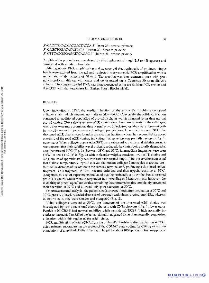
5'-CACTTGCACCACGACTACCA-3' (exon 23, reverse primer); 5'-CAGCTGGACATAGTAT-3' (intron 20, forward primer); 5'-CTTCAGGGGAGATACAGAC-3' (intron 21, reverse primer)
Amplification products were analyzed by electrophoresis through 2.5 to 4% agarose and visualized with ethidium bromide.
After genomic DNA amplification and agarose gel electrophoresis of products, single bands were excised from the gel and subjected to asymmetric PCR amplification with a molar ratio of the primers of 50 to 1. The reaction was then extracted once with phe- nol:chloroform, diluted with water and concentrated on a Centricon-30 spun dialysis column. The single-stranded DNA was then sequenced using the limiting PCR primer and 35S-dATP with the Sequenase kit (Unites States Biochemical).
RESULTS
Upon incubation at 37"C, the medium fraction of the proband's fibroblasts contained collagen chains which migrated normally on SDS-PAGE. Conversely, the cell-layer fraction contained an additional population of pro-a2(1) chains which migrated faster than normal pro-a2 chains. These shortened pro-a2(1) chains were found exclusively in the cell-layer, where they were more prominent than normal pro-a2(1) chains, and they were observed both in procollagen and in pepsin-treated collagen preparations. Upon incubation at 30"C, the shortened a2(I) chains were found in the medium fraction, where they accounted for about one-third of the total a2(I) chains, indicating that secretion was partially restored (Fig. 1, upper part). When collagens secreted at 30°C were subjected to the thermal stability assay, it was apparent that their stability was drastically reduced, the chains being totally degraded at a temperature of 36°C (Fig. 3). Between 31°C and 35"C, intermediate fragments were seen (TFal(1) and TFa2(I)* in Fig. 3) with molecular weights consistent with al(1) chains and a2(I) chains of approximately two-thirds of their normal length. This observation suggested that at these temperatures, trypsin cleaved the mutant collagen I molecules at around one- third of the distance of the amino to the carboxy terminal end, producing a shortened helical fragment. This fragment, in turn, became unfolded and thus trypsin-sensitive at 36°C. Altogether, this set of experiments indicated that the proband's cells synthesized shortened pro-a2(I) chains which were incorporated into procollagen I heterotrimers; however, the instability of procollagen I molecules containing the shortened chains completely prevented their secretion at 37°C and allowed only poor secretion at 30°C.
On ultrastructural analysis, the patient's cells showed, both after incubation at 37°C and 30"C, grossly dilated, rounded cisternae of the rough endoplasmic reticulum (rER), whereas in control cells they were slender and elongated (Fig. 2).
Using collagens secreted at 30"C, the structure of the shortened a2(I) chains was investigated by two-dimensional electrophoresis with CNBr-cleavage (Fig. 1, lower part). Peptide a2(I)CB3-5 had normal mobility, while peptide a2(I)CB4 (which normally in- cludes amino acids 7 to 327 of the helical domain) migrated faster than normally, suggesting a deletion within this region of the a2(I) chain.
PCR amplification of total cDNA from the proband's fibroblasts after incubation at 37"C, using primers encompassing the region of the COLlA2 gene coding for CB4, yielded two populations of amplified cDNA differing in length by about 100 bp. Restriction mapping of
Con
nect
Tis
sue
Res
Dow
nloa
ded
from
info
rmah
ealth
care
.com
by
Uni
vers
ity o
f Z
ueri
ch o
n 09
/15/
10Fo
r pe
rson
al u
se o
nly.

34 A. SUPERTI-FURGA et al.
FIGURE 1 Upper part: electrophoretic separation of pepsin-purified collagens in the culture medium after incubation at 37°C (upper lane) or 30°C (lower lane). Note that a population of shortened a2(I) chains [a2(1)*] is observed only in the lower lane. Lower part: CNBr-peptide mapping of pepsin-purified collagen chains secreted at 30°C. Note that the shortened a2(I)*-chains produce a faster-migrating CB4 peptide.
A B FIGURE 2 Electron micrographs of cultured fibroblasts from an age-matched control (A) and from the patient (B). Part of the cell nucleus (N) is evident in both pictures. The perinuclear zone in the control shows slender lamellar structures lined by ribosomes, corresponding to the rough endoplasmic reticulum. In the patient, the same structures are dilated, rounded, and filled with a finely granular, electrondense material. The cells were fixed with 2.5% glutaraldehyde, osmicated and embedded in EPON. The sections were counterstained with lead citratei uranyl acetate. Original magnification 7800 x ,
Con
nect
Tis
sue
Res
Dow
nloa
ded
from
info
rmah
ealth
care
.com
by
Uni
vers
ity o
f Z
ueri
ch o
n 09
/15/
10Fo
r pe
rson
al u
se o
nly.
INTRONIC DELETION IN 0 1 35
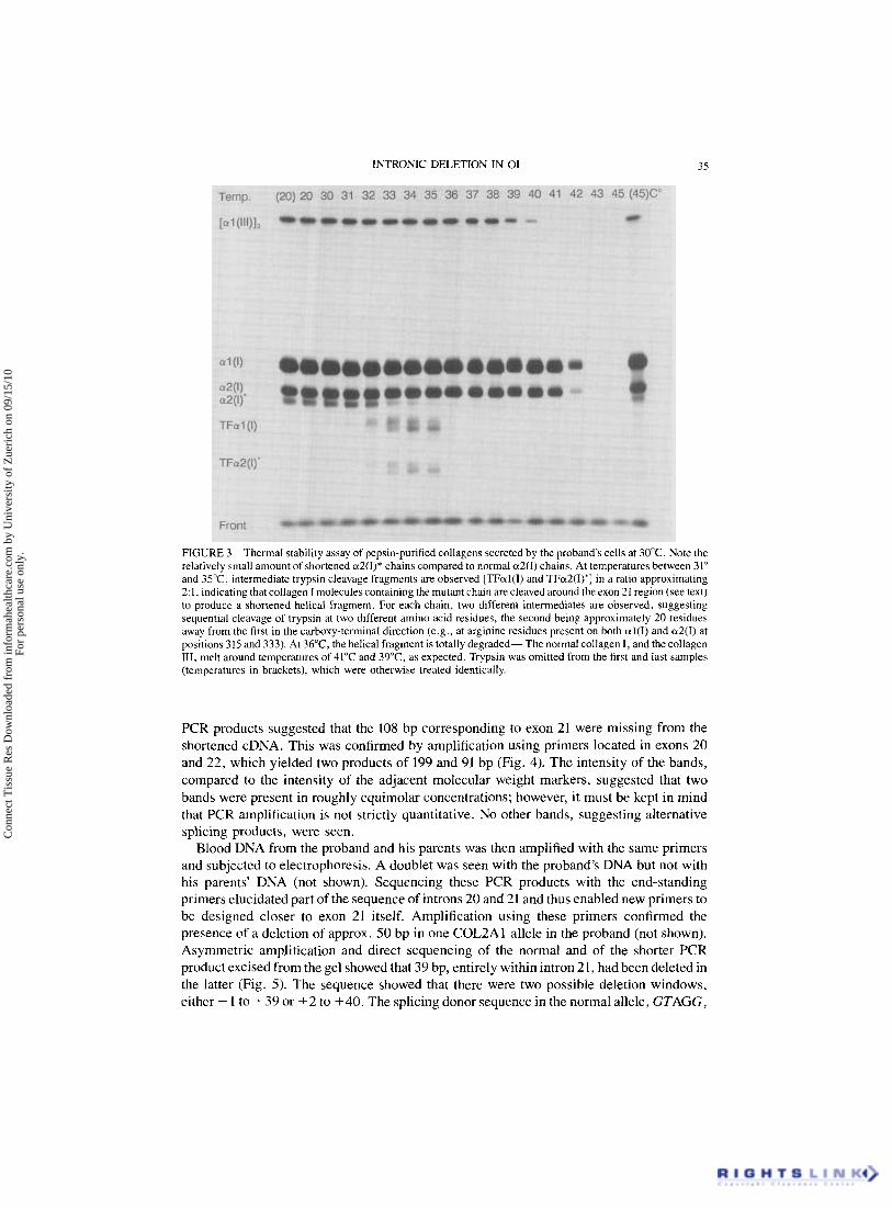
FIGURE 3 Thermal stability assay of pepsin-purified collagens secreted by the proband's cells at 30°C. Note the relatively small amount of shortened a2(I)* chains compared to normal a2(I) chains. At temperatures between 31" and 3 5 T , intermediate trypsin cleavage fragments are observed [TFal(I) and TFu2(I)*] in a ratio approximating 2.1, indicating that collagen I molecules containing the mutant chain are cleaved around the exon 21 region (see text) to produce a shortened helical fragment. For each chain, two different intermediates are observed, suggesting sequential cleavage of trypsin at two different amino acid residues, the second being approximately 20 residues away from the first in the carboxy-terminal direction (e.g., at arginine residues present on both al(1) and a2(I) at positions 315 and 333). At 36"C, the helical fragment is totally degraded- The normal collagen I , and the collagen 111, melt around temperatures of 41°C and 3 9 T , as expected. Trypsin was omitted from the first and last samples (temperatures in brackets), which were otherwise treated identically.
PCR products suggested that the 108 bp corresponding to exon 21 were missing from the shortened cDNA. This was confirmed by amplification using primers located in exons 20 and 22, which yielded two products of 199 and 91 bp (Fig. 4). The intensity of the bands, compared to the intensity of the adjacent molecular weight markers, suggested that two bands were present in roughly equimolar concentrations; however, it must be kept in mind that PCR amplification is not strictly quantitative. No other bands, suggesting alternative splicing products, were seen.
Blood DNA from the proband and his parents was then amplified with the same primers and subjected to electrophoresis. A doublet was seen with the proband's DNA but not with his parents' DNA (not shown). Sequencing these PCR products with the end-standing primers elucidated part of the sequence of introns 20 and 21 and thus enabled new primers to be designed closer to exon 21 itself. Amplification using these primers confirmed the presence of a deletion of approx. 50 bp in one COL2Al allele in the proband (not shown). Asymmetric amplification and direct sequencing of the normal and of the shorter PCR product excised from the gel showed that 39 bp, entirely within intron 21, had been deleted in the latter (Fig. 5). The sequence showed that there were two possible deletion windows, either + 1 to +39 or +2 to +40. The splicing donor sequence in the normal allele, GTAGG,
Con
nect
Tis
sue
Res
Dow
nloa
ded
from
info
rmah
ealth
care
.com
by
Uni
vers
ity o
f Z
ueri
ch o
n 09
/15/
10Fo
r pe
rson
al u
se o
nly.

36 A. SUPERTI-FURGA et a1
F’IGURE 4 PCR amplification of cDNA from the proband’s fibroblasts using primers located in exons 20 and 22 of COLlA2, showing two bands of 199bp and 91bp. Only the upper, 199 bp-band was seen in control preparations (not shown). Comparison with the molecular weight markers (HaelII-digested pBR322) on the left suggest that the bands are present in roughly equimolar concentrations.
INTRON 21
EXON 21
G A T C G A T C
FIGURE 5 Nucleotide sequence of the normal (left) and mutant (right) COLlA2 alleles of the proband at the boundary between exon 21 and intron 21 (5’-end is at bottom, 3’-end at top). The two G residues at position + 1 and +40 of intron 21 are marked with an arrow in the normal allele. The mutant allele has lost 39 nucleotides (+ 1 to f 3 9 , or t 2 to t40) which change the splicing donor site from GTAGG to GCAGA.
Con
nect
Tis
sue
Res
Dow
nloa
ded
from
info
rmah
ealth
care
.com
by
Uni
vers
ity o
f Z
ueri
ch o
n 09
/15/
10Fo
r pe
rson
al u
se o
nly.
INTRONIC DELETION IN 01 31
was thus changed to GCAGA in the shortened allele. No direct or inverted repeats could be identified around the breakpoints.
DISCUSSION
The Molecular Defect
The boy described in this paper was heterozygous for a 39 bp deletion just 3’ of exon 21 of the COLIA2 gene. The mechanism giving rise to the deletion is unclear, as no direct or inverted repeat was observed around the deletion boundaries. The deletion changed the splicing donor site, abolishing two of three conserved residues.5 Apparently, the change in the splicing donor sequence resulted in skipping of exon 21 during mRNA maturation.
Mutations leading to abnormal splicing appear to be relatively frequent in patients with molecular disorders of collagens such as 01 or Ehlers-Danlos syndromes type VII and type IV. The deletion of the whole normal splicing donor sequence has been observed,6 as well as point mutations abolishing one of the two or three conserved nucleotides of either the splice acceptor or the donor sequences.7-” Interestingly, point mutations of the last, 3 ’-nucleotide in the exon have also been implicated in aberrant splicing, suggesting that the sequence context may play an additional role in splicing.12.13 Some of the point mutations cause temperature-dependent aberrant splicing, 1 0 ~ 3 while other point mutations, as well as the deletion, are temperature-independent. The intronic deletion in our patient may be consid- ered novel as it alters two of three conserved nucleotides, thereby inducing exon skipping. There was no evidence of alternative splicing products, as has been observed with point mutations which changed only one of the three conserved nucleotides. At the protein level, lowering the temperature did not reduce the overall amount of the shortened pro-a2(1) chains, making a significant effect on splicing unlikely. The data indicate, not surprisingly, that altering residues + 2 and +5 of the splice donor sequence results in temperature- independent skipping of the preceding exon.
The Biochemical Phenotype
The proband’s cells synthesized shortened proa2(1) chains, but procollagen I molecules containing such chains were very efficiently excluded from secretion. Even after lowering the temperature to 30°C, the mutant molecules were secreted poorly and mostly retained within the cells. The cells apparently exert a strict “quality control” on procollagen I molecules on the secretory route and thus partially suppress the negative effect of the mutant COLlA2 allele. At the tissue level, the phenotypic expression of the mutant COLlAZ allele approaches to a certain extent that of a “null” allele.
At both temperatures, the patient’s cells had markedly dilated rough endoplasmic reticu- lum, filled with finely granular material. This feature has been observed previously in several cell strains with impaired secretion of procollagen I or procollagen 111, as a consequence of structural mutationsl4J5 or after inhibition of prolyl-hydroxylase activity16 and is in agreement with the biochemical data.
The molecular mechanisms involving the intracellular retention of unproperly folded proteins are still largely unknown. Endoplasmic reticulum-resident binding proteins (BiPs) or heat-shock proteins (HSPs) may interact with partially unfolded proteins and thus prevent
Con
nect
Tis
sue
Res
Dow
nloa
ded
from
info
rmah
ealth
care
.com
by
Uni
vers
ity o
f Z
ueri
ch o
n 09
/15/
10Fo
r pe
rson
al u
se o
nly.
38 A. SUPERTI-FURGA et al.
them from being secreted.17 It is unclear why certain collagen mutations result in very poor secretion at both normal and low temperature (present case, and ref. 15), while other similar mutations are associated with intracellular retention which can be reversed at low tempera- ture (e.g., a three-exon deletion in COLIAI [ref. 181) and still others have little effect on secretion (e.g., a deletion inducing skipping of exon 11 in COLlA2 [ref. 61). Possible explanations may be sequence-specificity of binding proteins, or individual differences in the expression of binding proteins; these possibilities remain to be investigated.
The Clinical Phenotype
Several clinical features observed in this boy were suggestive of osteogenesis imperfecta, namely, short stature, bone deformities, generalized osteoporosis, blueish sclerae and severe dentinogenesis imperfecta. The phenotype could be best classified as a mild variant of 01 type IV. Interestingly, there was no history of bone fractures and no radiological evidence of previous fractures (osteogenesis imperfecta sine fractura). This is unusual, but not surpris- ing: we have described a family with a splicing mutation in COLlA2 in which two affected individuals did not have pathological fractures but had dentinogenesis imperfecta. 19
Joint hyperextensibility and skin bruisability are a feature of 01 type I, a condition associated with a quantitative reduction of collagen I, rather than with structural defects (ref. 20, and own experience). Because of the very poor secretion of the abnormal chains in the proband’s cells, we suggest that, as stated above, this boy’s phenotype is determined partly by a quantitative reduction in collagen I, and partly by the presence of tiny amounts of structurally abnormal collagen I. The quantitative reduction may be responsible for osteo- porosis, joint laxity and bruisability; a tiny amount of structurally abnormal molecules reaching the extracellular matrix may be responsible for bone deformities and for den- tinogenesis imperfecta. 20
Mutations in COLlAl and COLlA2 display, as a group, a decreasing gradient in severity from the COOH-terminus to the NH,-terminus of the triple helix (reviewed in ref. 1). Mutations in COLlA2 causing skipping of either exons 9, 11, 12, or 13 have been associated with mild phenotypes, while skipping of exons 28 or 33 have been observed in combination with lethal disease.‘ Skipping of exon 21, as seen in our patient, might have been expected, because of its intermediate location, to produce severe disease, and even more so because exon 21 is twice as long (108 bp) as other exons (usually 54 bp): apparently, this is not the case. Skipping of exon 21 in COLIA2 was recently observed in two other, unrelated families (A. Nicholls and E M. Pope, Harrow; and F? Byers and B. Starman, Seattle; personal communications). The mild phenotype associated with mutations leading to skipping of exon 21 of COLIA2 is probably a consequence of the very poor secretion of the structurally abnormal collagen. Interestingly, a difference in cellular handing of abnormal molecules (secretion vs. intracellular retention of the abnormal molecules) has recently been invoked to explain the different phenotype of two patients with Ehlers-Danlos syndrome type IV who had deletions in the COL3AI gene causing the loss of a similar number of aminoacids in procollagen I11 but from the amino and carboxy terminal ends of the triple-helical domain, respectively.21.22 Thus, between the mutation at mRNA level on one side and the clinical phenotype on the other side, interaction between the abnormal collagen molecules and the cellular “quality control” system appears to play a pivotal role.
Con
nect
Tis
sue
Res
Dow
nloa
ded
from
info
rmah
ealth
care
.com
by
Uni
vers
ity o
f Z
ueri
ch o
n 09
/15/
10Fo
r pe
rson
al u
se o
nly.

INTRONIC DELETION IN 01 39
NOTE ADDED IN PROOF
Deletion of nucleotides +3 to + 13 in intron 9 of COLlA2 leading to skipping of exon 9 was recently identified in members of a family affected by mild osteogenesis imperfecta and joint hypermobility [Nicholls, A. C., Oliver, J., Renouf, D. V, Heath, D. A., and Pope, E M. (1992) Hum. Genet. 88, 627-633.1
ACKNOWLEDGMENTS
Dr. Jeff Bonadio (Ann Arbor) and Dr. B. Lee (New York) kindly provided some of the initial primers. Andi Bandi prepared manuscript and figures. This work was supported by the Swiss National Foundation (grants no. 32-27884.89 and no. 32-30148.90)
REFERENCES
1. Byers, P: H. (1992). Osteogenesis Imperfecta. In: Connective Tissue and its Heritable Disorders, edited by Royce, €? M., and Steinmann, B., Wiley-Liss, New York, pp. 317-350.
2. Steinmann, B., Rao, V H., Vogel, A, , Bruckner, I?, Gitzelmann, R., and Byers, I? H. (1984). Cysteine in the triple-helical domain of one allelic product of the al(1) gene of type I collagen produces a lethal form of osteogenesis imperfecta. J. Biol. Chem. 259, 11129-11138.
3. Superti-Furga, A,, and Steinmann, B. (1988). Impaired secretion of type 111 procollagen in Ehlers-Danlos type IV fibroblasts: correction of the defect by incubation at low temperature and demonstration of subtle structural alterations in the triple-helical region of the molecule. Biochem. Biophys. Res. Comm. 150, 140-147.
4. Kuivaniemi, H., Tromp, G., Chu, M. L., and Prockop, D. J. (1988). Structure of a full-length cDNA clone for the prepro-a2(I) chain of human type I procollagen. Biochem. J. 252, 633-640.
5 . Jacob, M., and Gallinaro, H. (1989). The 5’ splice site: phylogenetic evolution and variable geometry of association with UlRNA. Nucl Acids Res 17, 2159-2180.
6 . Kuivaniemi, H., Sabol, C., Tromp, G., Sippola-Thiele, M., and Prockop, D. J. (1988). A 19-base pairdeletion in the pro-a2(1) gene of type I procollagen that causes in-frame RNA splicing from exon 10 to exon 12 in a proband with atypical osteogenesis imperfecta and in his asymptomatic mother. J. Biol. Chem. 263, 11407- 11413.
7. Weil, D., Bernard, M., Combates, N., Wirtz, M. K., Hollister, D. W., Steinmann, B., and Ramirez, E (1988). Identification of a mutation that causes exon skipping during collagen pre-mRNA splicing in an Ehlers-Danlos syndrome variant. J. Biol. Chem. 263, 8651-8564.
8. Tromp, G., and Prockop, D. J. (1988). Single base mutation in the pro-a2(1) collagen gene that causes efficient splicing of RNA from exon 27 to exon 29 and synthesis of a shortened but in-frame pro-a2(1) chain. Proc. Natl. Acad. Sci. USA 85, 5254-5258.
9. Kuivaniemi, H., Kontusaari, S., Tromp, G., Zhao, M., Sabol, C., and Prockop, D. J. (1990). Identical G+1 to A mutations in three different introns of the type 111 procollagen gene (COL3Al) produce different patterns of RNA splicing in three variants of Ehlers-Danlos syndrome IV J. Biol. Chem. 265, 12067-12074.
10. Lee, B., Vitale, E., Superti-Furga, A , , Steinmann, B., and Ramirez, E (1991). G tG T transversion at position f 5 of a splice donor site causes skipping of the preceding exon in the type 111 procollagen transcripts of a patient with Ehlers-Danlos syndrome type IV. J. Biol. Chem. 266, 5256-5259.
11 . Ganguly, A. Baldwin, C. T., Strobel, D., Conway, D., Horton, W., Prockop, D. J. (1991). Heterozygous mutation in the G+5 position of intron 33 of the pro-a2(1) gene (COLIAZ) that causes aberrant RNA splicing and lethal osteogenesis imperfecta. J. Biol. Chem. 266, 12035-12040.
12. Weil, D., D’Alessio, M., Ramirez, E , de Wet W., Cole, W. G., Chan, D., and Bateman, J. E (1989). A base substitution in the exon of a collagen gene causes alternative splicing and generates a structurally abnormal polypeptide in a patient with Ehlers-Danlos syndrome type VII. EMBO Journal 8, 1705-1710.
13. Weil, D., D’Alessio, M., Ramirez, E, Steinmann, B., Wirtz, M. K . , Glanville, R. W., and Hollister, D. W. (1989). Temperature-dependent expression of a collagen spicing defect in the fibroblasts of a patient with Ehlers-Danlos syndrome type VII. J. B i d . Chem. 264, 16804-16809.
14. Holbrook, K . A,, and Byers, P: H. (1981). Ultrastructural characteristics of the skin in a form of the Ehlers- Danlos syndrome type IV Lab. Invest. 44, 342-350.
Con
nect
Tis
sue
Res
Dow
nloa
ded
from
info
rmah
ealth
care
.com
by
Uni
vers
ity o
f Z
ueri
ch o
n 09
/15/
10Fo
r pe
rson
al u
se o
nly.

40 A. SUPERTI-FURGA et al.
15. Willing, M. C., Cohn, D. H., Starman, B., Holbrook, K. A, , Greenberg, C. R., and Byers, F? H. (1988). Heterozygosity for a large deletion in the a2(I) collagen gene has a dramatic effect on type I collagen secretion and produces perinatal lethal osteogenesis imperfecta. J. Biol. Chem. 263, 8398-8404.
16. Tschank, G., Raghunath, M., Gunzler, V , andHanauske-Abel, H. M. (1987). F‘yridinedicarboxylates, the first mechanism-derived inhibitors for prolyl 4-hydroxylase, selectively suppress cellular hydroxyproline syn- thesis. Biochem. J. 248, 625-633.
17. Kotzuzumi, Y., Segal, N., Normington, K., Gething, J., and Sambrook, J. (1988). The presence of malfolded proteins in the endoplasmic reticulum signals the induction of glucose-regulated proteins. Nature 332, 462-464.
18. Steinmann, B., Superti-Furga, A,, and Royce, l? M. (1988). Imperfect collagenesis in osteogenesis imper- fecta: the consequences of glycine-cysteine substitutions on collagen structure and metabolism. Ann. N . Y. Acad. Sci. 543, 47-61, 1988.
19. Superti-Furga, A, , Pistone, E , Romano, C., and Steinmann, B. (1989). Clinical variability of osteogenesis imperfecta linked to COLIA2 and associated with a structural defect in the type I procollagen molecule. J. Med. Genet. 26, 358-362.
20. Wenstrup, R. J., Willing, M. C., Starman, B. J., and Byers, P: H. (1990). Distinct biochemical phenotypes predict clinical severity in nonlethal variants of osteogenesis imperfecta. Am. J. Hum. Genet. 46, 975-982.
21. Vissing H, D’Alessio, M., Lee, B., Ramirez, E, Byers, I? H., Steinmann, B., and Superti-Furga, A. (1991). Multi-exon deletion in the procollagen 111 gene is associated with mild Ehlers-Danlos syndrome type IV. J. Biol. Chem. 266, 52445248.
22. Lee, B., D’Alessio, M., Vissing, H., Ramirez, E , Steinmann, B., and Superti-Furga, A. (1991). Characteriza- tion of a large deletion associated with a polymorphic block of repeated dinucleotides in the type 111 procollagen gene (COL3Al) of a patient with Ehlers-Danlos syndrome type IV. Am. J. Hum. Genet. 48, 511-517.
Con
nect
Tis
sue
Res
Dow
nloa
ded
from
info
rmah
ealth
care
.com
by
Uni
vers
ity o
f Z
ueri
ch o
n 09
/15/
10Fo
r pe
rson
al u
se o
nly.