A Survey on Breakfast Habit and Skipping Breakfast among ...
Upload
independentCategory
view
1download
0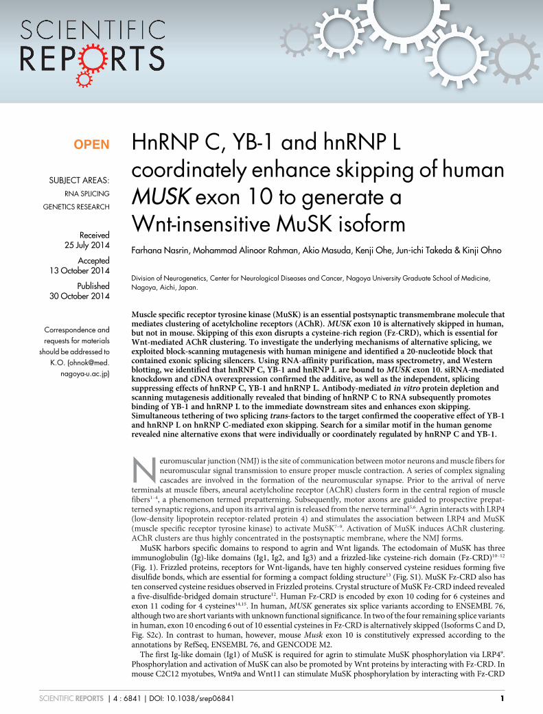
HnRNP C, YB-1 and hnRNP Lcoordinately enhance skipping of humanMUSK exon 10 to generate aWnt-insensitive MuSK isoformFarhana Nasrin, Mohammad Alinoor Rahman, Akio Masuda, Kenji Ohe, Jun-ichi Takeda & Kinji Ohno
Division of Neurogenetics, Center for Neurological Diseases and Cancer, Nagoya University Graduate School of Medicine,Nagoya, Aichi, Japan.
Muscle specific receptor tyrosine kinase (MuSK) is an essential postsynaptic transmembrane molecule thatmediates clustering of acetylcholine receptors (AChR). MUSK exon 10 is alternatively skipped in human,but not in mouse. Skipping of this exon disrupts a cysteine-rich region (Fz-CRD), which is essential forWnt-mediated AChR clustering. To investigate the underlying mechanisms of alternative splicing, weexploited block-scanning mutagenesis with human minigene and identified a 20-nucleotide block thatcontained exonic splicing silencers. Using RNA-affinity purification, mass spectrometry, and Westernblotting, we identified that hnRNP C, YB-1 and hnRNP L are bound to MUSK exon 10. siRNA-mediatedknockdown and cDNA overexpression confirmed the additive, as well as the independent, splicingsuppressing effects of hnRNP C, YB-1 and hnRNP L. Antibody-mediated in vitro protein depletion andscanning mutagenesis additionally revealed that binding of hnRNP C to RNA subsequently promotesbinding of YB-1 and hnRNP L to the immediate downstream sites and enhances exon skipping.Simultaneous tethering of two splicing trans-factors to the target confirmed the cooperative effect of YB-1and hnRNP L on hnRNP C-mediated exon skipping. Search for a similar motif in the human genomerevealed nine alternative exons that were individually or coordinately regulated by hnRNP C and YB-1.
Neuromuscular junction (NMJ) is the site of communication between motor neurons and muscle fibers forneuromuscular signal transmission to ensure proper muscle contraction. A series of complex signalingcascades are involved in the formation of the neuromuscular synapse. Prior to the arrival of nerve
terminals at muscle fibers, aneural acetylcholine receptor (AChR) clusters form in the central region of musclefibers1–4, a phenomenon termed prepatterning. Subsequently, motor axons are guided to prospective prepat-terned synaptic regions, and upon its arrival agrin is released from the nerve terminal5,6. Agrin interacts with LRP4(low-density lipoprotein receptor-related protein 4) and stimulates the association between LRP4 and MuSK(muscle specific receptor tyrosine kinase) to activate MuSK7–9. Activation of MuSK induces AChR clustering.AChR clusters are thus highly concentrated in the postsynaptic membrane, where the NMJ forms.
MuSK harbors specific domains to respond to agrin and Wnt ligands. The ectodomain of MuSK has threeimmunoglobulin (Ig)-like domains (Ig1, Ig2, and Ig3) and a frizzled-like cysteine-rich domain (Fz-CRD)10–12
(Fig. 1). Frizzled proteins, receptors for Wnt-ligands, have ten highly conserved cysteine residues forming fivedisulfide bonds, which are essential for forming a compact folding structure13 (Fig. S1). MuSK Fz-CRD also hasten conserved cysteine residues observed in Frizzled proteins. Crystal structure of MuSK Fz-CRD indeed revealeda five-disulfide-bridged domain structure12. Human Fz-CRD is encoded by exon 10 coding for 6 cysteines andexon 11 coding for 4 cysteines14,15. In human, MUSK generates six splice variants according to ENSEMBL 76,although two are short variants with unknown functional significance. In two of the four remaining splice variantsin human, exon 10 encoding 6 out of 10 essential cysteines in Fz-CRD is alternatively skipped (Isoforms C and D,Fig. S2c). In contrast to human, however, mouse Musk exon 10 is constitutively expressed according to theannotations by RefSeq, ENSEMBL 76, and GENCODE M2.
The first Ig-like domain (Ig1) of MuSK is required for agrin to stimulate MuSK phosphorylation via LRP49.Phosphorylation and activation of MuSK can also be promoted by Wnt proteins by interacting with Fz-CRD. Inmouse C2C12 myotubes, Wnt9a and Wnt11 can stimulate MuSK phosphorylation by interacting with Fz-CRD
OPEN
SUBJECT AREAS:RNA SPLICING
GENETICS RESEARCH
Received25 July 2014
Accepted13 October 2014
Published30 October 2014
Correspondence andrequests for materials
should be addressed toK.O. (ohnok@med.
nagoya-u.ac.jp)
SCIENTIFIC REPORTS | 4 : 6841 | DOI: 10.1038/srep06841 1

and induce AChR clustering, which requires LRP4, but not agrin16. Inanother study, Wnt4 has been shown to induce MuSK phosphoryla-tion by interacting with Fz-CRD in COS7 and HEK293T cells andWnt4 facilitates mouse NMJ formation in vivo17. Muscle prepattern-ing in zebrafish is also facilitated by interaction of Wnt11r withFz-CRD of a MuSK homolog, unplugged18, which is mediated byenhancing endocytosis of MuSK19. In zebrafish, unplugged/MuSKhas three splice variants: SV1 lacks Ig-like domains 1 to 3 but retainsFz-CRD, and is not responsive to agrin; SV2 lacks Fz-CRD and is notresponsive to Wnt; and the full-length isoform that can respond toboth agrin and Wnt18. Subsequent studies revealed that agrin-non-responsive SV1 is expressed in embryos, which is substituted for bythe full-length isoform in adults18,20,21. MuSK Fz-CRD additionallyplays an important role in motor nerve axon guidance in pathfindingin zebrafish20,21. Partial deletions of Fz-CRD have been analyzed inquail QT-6 fibroblasts22. Artificial deletion of 6 Fz-CRD cysteines or4 Fz-CRD cysteines caused lack of an activity for MuSK-rapsyn co-clustering22. Amino acids 760 to 820 in the cytoplasmic domain ofMuSK, however, have later been shown to be sufficient to conferinteraction with rapsyn23. As Fz-CRD is an important and obligatorydomain of MuSK for Wnt-mediated AChR clustering in zebra-fish18–21 and mouse C2C12 myotubes16,17, the two discordant reportsin zebrafish22,23 may indicate that Fz-CRD has an additional enhan-cing effect on MuSK-rapsyn interaction or plays another role in Wnt-mediated AChR clustering. Considering the functional significanceof 10 cysteines in Fz-CRD, we humans are likely to have acquired anevolutionally novel Wnt-insensitive MuSK isoform lacking 6 essen-tial cysteines, but the underlying mechanisms of alternative skippingof MUSK exon 10 remain unknown.
HnRNP C is a nuclear RNA-binding protein that associates withnascent mRNA transcripts, which plays roles in pre-mRNA splic-ing24, mRNA stability25, and translational modulation26. HnRNP Chas recently been identified as a molecular ruler to classify RNApolymerase II transcripts for export into two categories: a longmRNA and a short uridine-rich small nuclear RNA (U snRNA)27.The Y box-binding protein (YB-1) is a member of the cold shockdomain (CSD) protein family, which has binding specificity for bothDNA and RNA. YB-1 has multiple roles including transcriptionalregulation, translational control, DNA repair, and pre-mRNA splic-ing28,29. HnRNP L is another nuclear RNA-binding protein and aglobal splicing regulator30–38. It also functions in polyadenylationand mRNA stability39,40.
In the present study, we have dissected the underlying mechan-isms of alternative splicing of human MUSK exon 10. We first char-acterized splicing regulatory cis-elements by scanning mutagenesis.We then identified that the alternative skipping of MUSK exon 10 iscoordinately modulated by binding of three splicing suppressors(hnRNP C, YB-1, and hnRNP L) to an exonic splicing silencer(ESS) that is unique to human MUSK exon 10. Remarkably,hnRNP C is the master regulator in this regulatory process, andYB-1 and hnRNP L have additive effects to efficiently achieve splic-ing suppression.
ResultsAlternative splicing of MUSK exon 10 is unique to human. Sinceexons 9 (7 nucleotides) and 10 (264 nucleotides) of human MUSK arealternatively spliced according to the gene annotation databases, weinitially examined the differential selection of these two exons inhuman skeletal muscle. Using total RNA isolated from humanskeletal muscle (Clontech), fragments spanning exons 8 to 11 wereamplified by RT-PCR (Fig. S2d). Sequencing of the RT-PCRproducts revealed three splicing isoforms: (i) exons 9 and 10included; (ii) exon 10 skipped; and (iii) exons 9 and 10 skipped(Fig. S2f). We could not detect any transcript that skipped onlyexon 9. We also performed a similar experiment using total RNAisolated from immortalized human myogenic KD3 cells and primaryhuman myoblasts (SkMC), and obtained similar results (Fig. S2d andf). The three observed splicing isoforms are not correctly mapped tothe human genome in UCSC Genes, RefSeq, ENCODE/GENCODEVer. 19 and H-Inv ver. 8.3. As cDNA sequences registered in theseannotation databases are correct, a short exon 9 that is comprised ofonly 7 nucleotides is likely to have precluded correct mapping ofcDNAs to the human genome. We also confirmed lack ofalternative splicing of mouse Musk exon 10 in 10 different skeletalmuscle tissues as well as in C2C12 mouse myoblasts (Figs. S2e andS7c). Lack of alternative splicing of mouse Musk exon 10 is correctlyannotated in all of the gene annotation databases shown above. Inthis study, we investigated the underlying mechanisms of alternativeskipping of exon 10 unique to human.
Construction of minigenes for splicing analysis. We first con-structed a human MUSK minigene in pcDNA3.1D/V5/His-TOPOexpression vector (Invitrogen) spanning exons 8 to 11 (Fig. 2a). Exon10 in the pcDNA3.1 minigene was alternatively spliced in HeLa cells,
Figure 1 | Structures of human and mouse MuSK. (a) Genomic structures of human MUSK and mouse Musk genes. Constitutive and alternative
exons are shown in green and red boxes, respectively. Black boxes indicate untranslated regions (UTRs) and thin lines indicate introns. Alternative
skipping of exons annotated in ENSEMBL 76 are shown by blue connecting lines. (b) Domain structure of MuSK. Fz-CRD is a Wnt-responsive domain
encoded by exon 10 coding for 6 cysteines (light blue-colored region of Fz-CRD) and exon 11 coding for 4 cysteines (yellow-colored region of Fz-CRD) in
human. SS, signal sequence; TM, transmembrane domain.
www.nature.com/scientificreports
SCIENTIFIC REPORTS | 4 : 6841 | DOI: 10.1038/srep06841 2
as we observed in human skeletal muscle. We next inserted exon 10and flanking intronic sequences (245 nucleotides in the upstreamintron and 200 nucleotides in the downstream intron) into themodified exon-trapping vector pSPL341, which carried twoproprietary constitutive exons on each end (Fig. S3a). The pSPL3-human-MUSK minigene (H-iE10i) successfully recapitulatedalternative splicing of exon 10 in HeLa cells (Fig. S3a). Serialdeletions of intronic nucleotides from both ends of H-iE10irevealed that the shortest minigene (H-iE10i-D6) that carried 100nucleotides in the upstream intron and 60 nucleotides in thedownstream intron was still alternatively spliced like H-iE10i inHeLa cells (Fig. S3a). This minigene was termed pSPL3-human-MUSK (pH-wt) (Fig. 2b) and used in the subsequent experiments.
We also constructed a similar minigene harboring mouse exon 10and flanking intronic sequences, pSPL3-mouse-MuSK (pm-wt), and
found that exon 10 is constitutively included in this minigene inHeLa cells (Fig. 2b). As the mouse minigene was not alternativelyspliced even in human cells, we assumed that nucleotides unique tohuman enabled alternative splicing of exon 10. We confirmed thatour minigene was similarly alternatively spliced in KD3 cells andHeLa cells (Fig. 2a and b). Due to better transfection efficiency, weused HeLa cells and pH-wt/pm-wt in the following studies.
Identification of two exonic splicing silencer (ESS) blocks inhuman exon 10. We next searched for exonic/intronic segmentscarrying splicing cis-elements in pH-wt. To this end, weconstructed chimeric minigenes made of variable combinations ofhuman and mouse segments (Fig. 2c). We found that theintroduction of mouse exon 10 into pH-wt resulted in constitutivesplicing (pH-EC in Fig. 2c). We next introduced the first or second
a
b pSPL3-human-MUSK (pH-wt)
10CMV 5’ 3’
pSPL3-mouse-Musk (pm-wt)
CMV 5’ 3’ 10
10 118CMV
pcDNA-human-MUSK
9
c
CMV
pSPL3
NotI PacI
10
10
10
10
10
10
10
d
Block-2 Block-3 Block-4 Block-5 Block-6 Block-7 Block-8 Block-9 Block-10 Block-11 Block-12 Block-13Block-1
1
2
3
4
5
6
7
8
9
10
11
12
13
Human MUSK exon 10
Blo
ck-1
Blo
ck-2
Blo
ck-3
Blo
ck-4
Blo
ck-5
Blo
ck-6
Blo
ck-7
Blo
ck-8
Blo
ck-9
Blo
ck-1
0
Blo
ck-1
1
Blo
ck-1
2
Blo
ck-1
3
pH-w
t
Human (pH-wt)
mouse (pm-wt)
pH-DIC
pH-UIC
pH-EC
pH-FHEC
pH-SHEC
pH-D
IC
pH-U
IC
pH-E
C
pH-F
HE
C
pH-S
HE
C
pH-w
t
5’ 3’
HeL
a
KD
3
HeL
a
KD
3
inclusionskipping
pH-wt pm-wt
HeL
a
KD
3
inclusion skipping
mutant minigene
BGH pA
NotI PacI
NotI PacI
inclusion skipping
inclusion skipping
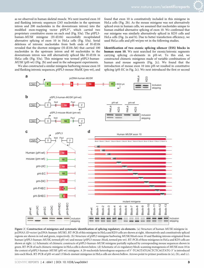
Figure 2 | Construction of minigenes and systematic identification of splicing regulatory cis-elements. (a) Structure of human MUSK minigene in
pcDNA3.1D vector (pcDNA-human-MUSK). RT-PCR of this minigene in HeLa and KD3 cells are shown at right. Alternatively and constitutively spliced
regions are shown in red and green, respectively. (b) Structure of pSPL3 minigene harboring MUSK/Musk exon 10 and flanking introns originated from
human (pSPL3-human-MUSK, termed pH-wt) and mouse (pSPL3-mouse-Musk, termed pm-wt). RT-PCR of these minigenes in HeLa and KD3 cells are
shown at right. (c) Schematic of chimeric constructs of pSPL3-human-MUSK minigene partially replaced by corresponding mouse sequences shown in
green. RT-PCR of each chimeric minigene in HeLa cells is shown below. (d) Schematic of cis-regulatory block-scanning mutagenesis of MUSK exon 10 in
the context of pSPL3-human-MUSK (pH-wt) minigene. A 20-nucleotide heterologous sequence of 59-TCAGTATGACTCTCAGTATG-39 is introduced
into each block. RT-PCR of pH-wt and 13 block-mutant minigenes in HeLa cells are shown bellow. Arrows point to primer positions in (a), (b), and (c).
www.nature.com/scientificreports
SCIENTIFIC REPORTS | 4 : 6841 | DOI: 10.1038/srep06841 3
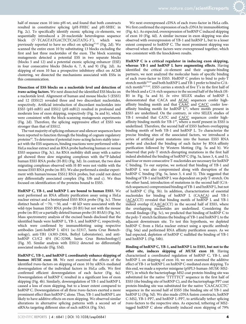
half of mouse exon 10 into pH-wt, and found that both constructsresulted in constitutive splicing (pH-FHEC and pH-SHEC inFig. 2c). To specifically identify exonic splicing cis-elements, wesequentially introduced a 20-nucleotide heterologous sequenceblock (59-TCAGTATGACTCTCAGTATG-39), which waspreviously reported to have no effect on splicing37,42 (Fig. 2d). Wescanned the entire exon 10 by substituting 13 blocks excluding thefirst and last three nucleotides of the exon. The block scanningmutagenesis detected a potential ESS in two separate blocks(blocks 5 and 12) and a potential exonic splicing enhancer (ESE)in four consecutive blocks (blocks 6, 7, 8, and 9) (Fig. 2d). Asskipping of exon 10 has a prospective inhibitory effect on AChRclustering, we dissected the mechanisms associated with ESSs inthis communication.
Dissection of ESS blocks on a nucleotide level and detection oftrans-acting factors. We next dissected the identified ESS blocks ona nucleotide level. Alignment of human and mouse blocks 5 (ESS5)and 12 (ESS12) revealed three and two discordant nucleotides,respectively. Artificial introduction of discordant nucleotides intoESS5 (pH-mB5) and ESS12 (pH-mB12) resulted in profound andmoderate loss of exon skipping, respectively (Fig. 3a and b), whichwere consistent with the block-scanning mutagenesis experiments(Fig. 2d). Therefore, the splicing suppressive effect of ESS5 wasstronger than that of ESS12.
The vast majority of splicing enhancer and silencer sequences havebeen reported to function through the binding of cognate regulatoryproteins43. To determine if potential trans-acting factors stably inter-act with the ESS sequences, binding reactions were performed with aHeLa nuclear extract and an RNA probe harboring human or mouseESS5 sequence (Fig. 3c). An RNA mobility shift assay with a nativegel showed three slow migrating complexes with the32P-labeledhuman ESS5 RNA probe (H-B5) (Fig. 3d). In contrast, the two slowmigrating complexes observed with H-B5 were not visible with themouse ESS5 RNA probe (m-B5). We also performed a similar experi-ment with human/mouse ESS12 RNA probes, but could not detectany differentially associated complex (Fig. S3b and c). We thusfocused on identification of the proteins bound to ESS5.
HnRNP C, YB-1, and hnRNP L are bound to human ESS5. Wenext performed an RNA affinity purification assay using a HeLanuclear extract and a biotinylated ESS5 RNA probe (Fig. 3c). Threedistinct bands of ,70, ,50, and ,40 kD were associated with thewild-type human ESS5 RNA probe (H-B5) but not with the mouseprobe (m-B5) or a partially deleted human probe (H-B5D5) (Fig. 3e).Mass spectrometry analysis of the excised bands disclosed that theidentified bands were hnRNP L, YB-1, and hnRNP C, respectively,which were confirmed by immunoblotting using respectiveantibodies [anti-hnRNP L 4D11 (sc-32317, Santa Cruz Biotech-nology), anti-YB1 (A303-230A, Bethyl Laboratories), and anti-hnRNP C1/C2 4F4 (SC-32308, Santa Cruz Biotechnology)](Fig. 3f). Similar analysis with ESS12 detected no differentiallyassociated molecule (Fig. S3d).
HnRNP C, YB-1, and hnRNP L coordinately enhance skipping ofhuman MUSK exon 10. We next examined the effects of theidentified trans-factors on skipping of exon 10 by siRNA-mediateddownregulation of the individual factors in HeLa cells. We firstconfirmed efficient downregulation of each factor (Fig. 4a).Downregulation of hnRNP C resulted in a significant loss of exonskipping (Fig. 4b). Similarly, downregulation of YB-1 and hnRNP Lcaused a loss of exon skipping, but to a lesser extent compared tohnRNP C. Downregulation of all three trans-factors exerted a moreprominent effect than hnRNP C alone. Thus, YB-1 and hnRNP L arelikely to have additive effects on exon skipping. We observed similaralterations in alternative splicing patterns with a second set ofsiRNAs targeting different sites of each mRNA (Fig. S3e).
We next overexpressed cDNA of each trans-factor in HeLa cells.We first confirmed the expression of each cDNA by immunoblotting(Fig. 4c). As expected, overexpression of hnRNP C induced skippingof exon 10 (Fig. 4d). A similar increase in exon skipping was alsoobserved with overexpression of YB-1 and hnRNP L, but to a lesserextent compared to hnRNP C. The most prominent skipping wasobserved when all three factors were overexpressed together, whichwas consistent with the knockdown results.
HnRNP C is a critical regulator in inducing exon skipping,whereas YB-1 and hnRNP L have augmenting effects. Havingidentified the critical cis-element and their cognate-bindingpartners, we next analyzed the molecular basis of specific bindingof each trans-factor to ESS5. HnRNP C prefers to bind to poly-Tstretch motifs24,44 and both hnRNP L and YB-1 prefer to bind to C/A-rich motifs29,32,45. ESS5 carries a stretch of five T’s in the first half ofthe block and a C/A-rich sequence in the second half of the block (H-B5 in Fig. 5a and b). In vitro SELEX studies of hnRNP Ldemonstrated that CACA and ACAC sequences confer high-affinity binding motifs and that CAAC and CACC confer low-affinity binding motifs for hnRNP L32, where motifs present inESS5 are underlined. On the contrary, in vitro SELEX studies ofYB-1 revealed that CATC and CACC sequences confer high-affinity binding motifs for YB-129, where a motif present in ESS5 isunderlined. Therefore, the second half of ESS5 harbors overlappingbinding motifs of both YB-1 and hnRNP L. To characterize theprecise binding sites of the associated factors, we introduced aseries of artificial point mutations into the human ESS5 RNAprobe and checked the binding of each factor by RNA-affinitypurification followed by Western blotting (Fig. 5a and b). Weobserved that poly-T stretch-disrupting mutations in the first halfindeed abolished the binding of hnRNP C (Fig. 5a, lanes 3, 4, and 5),and four or more consecutive T-nucleotides are necessary for hnRNPC binding. To our surprise, we noticed that binding of YB-1 andhnRNP L was also compromised along with disruption of thehnRNP C binding (Fig. 5a, lanes 3, 4 and 5). This suggested thatbinding of YB-1 and hnRNP L was dependent on poly T-stretch. Onthe other hand, introduction of mutations in the second half (C/A-rich sequences) compromised binding of YB-1 and hnRNP L, but notof hnRNP C (Fig. 5b). In addition, characterization of essentialnucleotides for binding of hnRNP L (CAACA) and YB-1(ACACCT) revealed that binding motifs of hnRNP L and YB-1indeed overlap (CAACACCT) in the second half of ESS5, wherethe overlapping nucleotides are underlined. Considering theoverall findings (Fig. 5c), we predicted that binding of hnRNP C tothe poly-T stretch facilitates the binding of YB-1 and hnRNP L to theadjacent downstream site. To test this hypothesis, we depletedhnRNP C from a HeLa nuclear extract using a specific antibody(Fig. S4a) and performed RNA affinity purification assays. As wehad expected, depletion of hnRNP C nullified the binding of YB-1and hnRNP L (Fig. S4b).
Binding of hnRNP C, YB-1, and hnRNP L to ESS5, but not to theother site, induces skipping of MUSK exon 10. Havingcharacterized a coordinated regulation of hnRNP C, YB-1, andhnRNP L on skipping of exon 10, we next examined the additiveeffect of YB-1 or hnRNP L on hnRNP C-mediated exon skipping. Tothis end, we made a reporter minigene (pSPL3-human-MUSK-MS2-PP7), in which the bacteriophage MS2 coat protein-binding site wassubstituted for the native ‘TTTTTCT’ sequence in the first half ofESS5 (the binding site of hnRNP C), and the bacteriophage PP7 coatprotein-binding site was substituted for the native ‘CAACACCTC’sequence in the second half of ESS5 (the binding site of YB-1 andhnRNP L) (Fig. 5d). We also made cDNA fusion constructs, hnRNPC-MS2, YB-1-PP7, and hnRNP L-PP7, to artificially tether splicingtrans-factors to the respective sites. As expected, tethering of MS2-tagged hnRNP C alone efficiently induced exon skipping of 79%
www.nature.com/scientificreports
SCIENTIFIC REPORTS | 4 : 6841 | DOI: 10.1038/srep06841 4

(Fig. 5e, lane 3). Tethering of PP7-tagged YB-1 and hnRNP L withouttethering MS2-tagged hnRNP C induced exon skipping of 52%(Fig. 5e, lane 5) and 36% (Fig. 5e, lane 8), respectively. In contrast,hnRNP C, YB-1, or hnRNP L without a tethering tag did not induceexon skipping (Fig. 5e, lanes 2, 4 and 7), suggesting that ESS5 was theonly site where these factors were able to bind and function. We alsoconfirmed that MS2- or PP7-tagged factor has no effect on aminigene lacking MS2- or PP7-binding site (Fig. S4d). Simul-taneous recruitment of hnRNP C with either YB-1 or hnRNP Lfurther induced exon skipping (Fig. 5e, lanes 6 and 9), whichreconfirmed the additive effects of YB-1 and hnRNP L on hnRNPC-mediated splicing suppression.
RNA-dependent interaction of hnRNP C, YB-1, and hnRNP L andsearch for similar targets in other human genes. We next examinedthe molecular interaction between the three trans-factors. Co-
immunoprecipitation revealed that HnRNP C and hnRNP L werebound in an RNA-dependent manner (Fig. S5a), whereas hnRNP Cand YB-1 were not (Fig. S5b). Similarly, hnRNP L and YB-1 werebound in an RNA-dependent manner (Fig. S5c), which wasconsistent with a previous report45.
We further asked if coordinated splicing regulation by hnRNP C,YB-1, hnRNP L is unique to MUSK exon 10. As hnRNP L has highlydegenerative SELEX motifs, which are overlapping with YB-1 motifs,we analyzed coordinated splicing only by hnRNP C and YB-1. Searchfor adjacent hnRNP C- and YB-1-binding motifs in human alterna-tive cassette exons and flanking introns detected 378 candidate sites.We randomly selected 37 exons and analyzed alternative splicing inHeLa cells in the presence of siCont, siC, siYB-1, and siC/siYB-1. Wefound that alternative splicing events of 9 exons were affected byhnRNP C and/or YB-1 in HeLa cells, whereas 13 exons were notexpressed and 15 exons were not alternatively spliced or affected
Figure 3 | HnRNP C, YB-1 and hnRNP L bind to exon 10 of human MUSK. (a, b) Mouse nucleotides are introduced into ESS5 (a) and ESS12 (b) of pH-
wt to generate pH-mB5 and pH-mB12, respectively. RT-PCR of each mutated minigene in HeLa cells is compared with that of pH-wt. Primer positions
are shown by arrows. (c) Sequences of ESS5 RNA probes carrying human (H-B5), mouse (m-B5), and partially deleted (H-B5D5) sequences.
(d)32P-labeled H-B5 or m-B5 RNA probe was incubated in the presence or absence of HeLa nuclear extract (NuEX) and resolved on a native
polyacrylamide gel to observe free and protein-bound RNA species (arrows). (e) Coomassie blue staining of RNA affinity-purified products from HeLa
nuclear extract using the indicated biotinylated RNA probes. Three proteins of ,70, ,50, and ,40 kDa (arrows) are differentially associated with
H-B5 compare to m-B5 and H-B5D5. Mass spectrometry analysis revealed that the three proteins are HnRNP L (L), YB-1, and hnRNP C (C).
(f) Immunoblotting (IB) of RNA affinity purified proteins in panel (e) with the indicated antibodies.
www.nature.com/scientificreports
SCIENTIFIC REPORTS | 4 : 6841 | DOI: 10.1038/srep06841 5

by knockdown (Fig. S6). Among the 9 exons, 3 exons were coordi-nately skipped by hnRNP C and YB-1 (Fig. S6a).
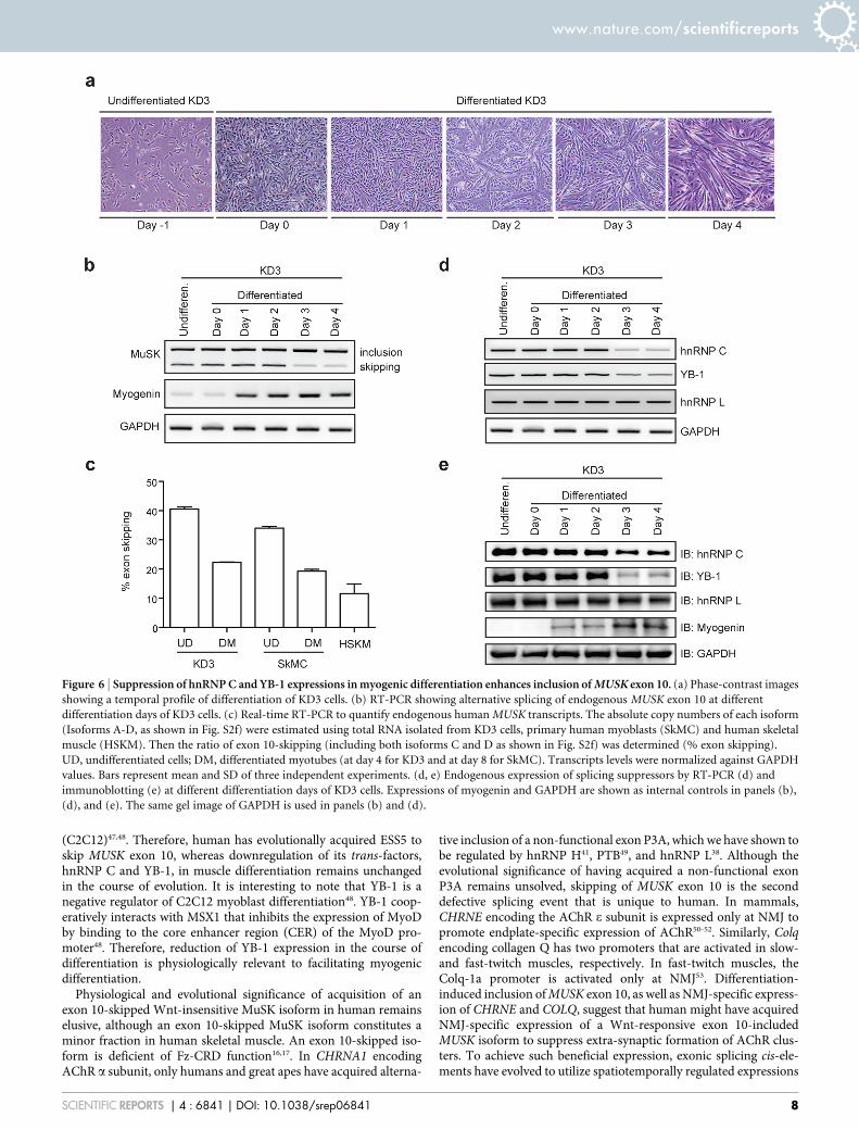
Expressions of splicing repressing hnRNP C and YB-1 are reducedwith muscle differentiation. As MuSK is a muscle-specific receptorprotein having an important role in muscle development andfunction, we next examined the splicing profile of human MUSKexon 10 in different stages of myogenic differentiation. We cul-tured immortalized human myogenic KD3 cells in differentiationmedium for four days to make myotubes (Fig. 6a). RT-PCRspanning endogenous MUSK exon 10 at different time pointsrevealed that skipping of exon 10 was suppressed on and afterdifferentiation day 3 (Fig. 6b, and Supplementary Table 1).Similarly, myotube differentiation suppressed skipping of MUSKexon 10 in primary human myoblasts (SkMC) (Figs. S7a, b, and6c), and skipping of exon 10 constitutes only 12% in humanskeletal muscle (Fig. 6c and Supplementary Table 1). Thus,skipping of exon 10 is a minor event in any differentiation stagesof myogenic and muscle cells.
In KD3 cells, we found that the expressions of hnRNP C and YB-1were indeed decreased on and after day 3, whereas the expressionlevel of hnRNP L was not significantly changed at the mRNA(Fig. 6d) and protein (Fig. 6e) levels. Therefore, reduction ofhnRNP C and YB-1 in the course of muscle differentiation causesreduction of skipping of human MUSK exon 10 to produce a Wnt-sensitive MuSK isoform.
DiscussionIn this study, we have identified splicing regulatory cis-elements andcognate trans-factors that drive alternative splicing of human MUSKexon 10. HnRNP C enhances exon skipping in coordination with YB-1 and hnRNP L by binding to a regulatory exonic splicing silencer,ESS5. Two splicing suppressive cis-elements, ESS5 and ESS12, arerecognized by block-scanning mutagenesis, although the ESS activityof ESS12 is not as conspicuous as that of ESS5. Most splicing regu-
latory ESSs and ESEs function through the binding of cognate reg-ulatory proteins43. Indeed, ESS5 is recognized by the three splicingtrans-factors, but no associated molecules are detected for ESS12. Incontrast, the splicing suppressive activity of ESS12 is likely to beregulated by a local secondary structure of pre-mRNA.Alternatively, a binding affinity of a splicing trans-factor to ESS12is too low to be detected by the RNA mobility shift assay and the RNAaffinity purification assay.
We have characterized the mutual coordination between the threetrans-factors. HnRNP C binds to a poly-T stretch in the first half ofESS5, whereas binding motifs of hnRNP L and YB-1 are overlappedin the second half of ESS5. In addition, binding of YB-1 and hnRNP Lis dependent on hnRNP C. One possible mechanism for this is thatoverlapping binding motifs between hnRNP L and YB-1 provokecompetitive binding of the two factors. A third molecule, hnRNPC, may rearrange RNA conformation to stabilize the binding ofeither hnRNP L or YB-1. Similar RNA-mediated stabilization ofbinding of another RNA-binding protein by a specific RNA-bindingprotein has been reported in other genes. In CD45, hnRNP L stabi-lizes the binding of hnRNP A1 in exon 4, in which binding of hnRNPA1 and the subsequent splicing suppression is dependent on hnRNPL34. The authors demonstrate that hnRNP L interacts with hnRNPA1, which is lost by RNase. In Tpm1, splicing of exon 3 is coordi-nately repressed by PTB and Raver 146. Interaction between PTB andRaver 1, which requires the target RNA, results in a conformationalchange of the tertiary complex to bring the repressor domain of bothmolecules in close apposition to synergistically promote exon skip-ping. Therefore RNA-dependent molecular interaction and coordi-nated splicing suppression is unlikely to be unique to ESS5 and islikely to be functional in many other alterative splicing events.
Binding of YB-1 and hnRNP L to the same target and the sub-sequent coordinated splicing regulation have been previouslyreported45. The authors report that overexpression and depletionof either YB-1 or hnRNP L is sufficient to repress and derepresssplicing, respectively. In their report, YB-1 exerts a stronger effect
Figure 4 | HnRNP C, YB-1, and hnRNP L coordinately promote skipping of MUSK exon 10. (a) Immunoblotting (IB) using the indicated antibodies
after gene knockdown with siRNA against control (siCont), hnRNP C (siC), YB-1 (siYB-1), and hnRNP L (siL) in HeLa cells. (b) RT-PCR of pSPL3-
human-MUSK (pH-wt) minigene in HeLa cells treated with the indicated siRNAs. The mean and SD (n 5 3) of the ratio of exon skipping in each
treatment is shown below the gel image. (c) Immunoblotting (IB) with the indicated antibodies after cDNA overexpression of hnRNP C, YB-1, and
hnRNP L in HeLa cells. Endogenous (endo) and exogenous (exon) YB-1 proteins are pointed by arrows. (d) RT-PCR of pH-wt minigene in HeLa cells co-
transfected with the indicated cDNAs. The mean and SD (n 5 3) of the ratio of exon skipping in each treatment is shown below the gel image.
www.nature.com/scientificreports
SCIENTIFIC REPORTS | 4 : 6841 | DOI: 10.1038/srep06841 6

than hnRNP L, as in our MUSK exon 10. To the best of our know-ledge, MUSK exon 10 is the first target where coordinated splicingregulation by YB-1 and hnRNP L is dependent on hnRNP C. RT-PCR analysis of 37 targets, where an hnRNP C-binding ‘TTTT’ motifand a YB-1-binding ‘CATC/CACC’ motif are adjacent to each otherin human alternative cassette exons and flanking introns, revealedthree alternative cassette exons that are coordinately skipped byhnRNP C and YB-1. Although detailed functional coordination ofhnRNP C and YB-1 on these targets, and involvement of hnRNP L,remain unknown, MUSK exon 10 is unlikely to be the only targetwhere hnRNP C, YB-1, and hnRNP L coordinately induce exonskipping.
Recent studies of alternative splicing in different cellular andphysiological states have broadened our understanding of themolecular basis for functional selection of a certain isoform in a
tissue-specific and developmental stage-specific manner. These stud-ies demonstrated multiple intriguing features of splicing regulatoryfactor(s) including cell-type specific expression, intracellular local-ization, post-translational modification on different cellular stimuli,etc. Precise regulation of cellular differentiation is indispensable forthe proper development of vertebrate embryo and deregulated dif-ferentiation results in diverse human congenital abnormalitiesincluding cancers. In human myogenic cells (KD3), skipping ofMUSK exon 10 is significantly reduced from ,41% to ,22% uponmyogenic differentiation, which is in parallel with the reducedexpression of hnRNP C and YB-1 (Fig. 6). We also observed a similarexpression profile of MUSK exon 10 in primary human myoblasts(SkMC) upon myogenic differentiation (Figs. S7a, b and 6c).Reduced expression of hnRNP C, YB-1 and even hnRNP L uponmyogenic differentiation is also reported in mouse myoblast cells
Figure 5 | Binding of hnRNP C, YB-1, and hnRNP L to specific motifs enhances coordinated skipping of MUSK exon 10. (a, b) Scanning mutagenesis to
map the binding motifs of hnRNP C, YB-1, and hnRNP L in ESS5 (H-B5). RNA probe sequences are shown in the upper panels, where discordant
nucleotides between human and mouse are underlined in H-B5 (ESS5). Artificial mutations are shown in red. RNA affinity-purified products are detected
by immunoblotting in the lower panels. The results are indicated on the right side by ‘‘1’’ and ‘‘2’’ for positive and negative binding to each probe,
respectively. (c) The resulting binding site of each factor from panels (a) and (b) is schematically shown. Essential binding nucleotides are indicated by
large green letters and are underlined. (d) Schematic of a reporter minigene (pSPL3-human-MUSK-MS2-PP7). MS2 coat protein-binding hairpin RNA
(blue) is substituted for the first half of ESS5 (binding site of hnRNP C). Similarly, PP7 coat protein-binding hairpin RNA (orange) is substituted for the
second half of ESS5 (binding sites of hnRNP L and YB-1). (e) RT-PCR of pSPL3-human-MUSK-MS2-PP7 minigene in HeLa cells that are co-transfected
with the indicated effectors. Blue and orange letters match to those in (d). The mean and SD (n 5 3) of the ratio of exon skipping in each treatment is
shown below the gel image.
www.nature.com/scientificreports
SCIENTIFIC REPORTS | 4 : 6841 | DOI: 10.1038/srep06841 7
(C2C12)47,48. Therefore, human has evolutionally acquired ESS5 toskip MUSK exon 10, whereas downregulation of its trans-factors,hnRNP C and YB-1, in muscle differentiation remains unchangedin the course of evolution. It is interesting to note that YB-1 is anegative regulator of C2C12 myoblast differentiation48. YB-1 coop-eratively interacts with MSX1 that inhibits the expression of MyoDby binding to the core enhancer region (CER) of the MyoD pro-moter48. Therefore, reduction of YB-1 expression in the course ofdifferentiation is physiologically relevant to facilitating myogenicdifferentiation.
Physiological and evolutional significance of acquisition of anexon 10-skipped Wnt-insensitive MuSK isoform in human remainselusive, although an exon 10-skipped MuSK isoform constitutes aminor fraction in human skeletal muscle. An exon 10-skipped iso-form is deficient of Fz-CRD function16,17. In CHRNA1 encodingAChR a subunit, only humans and great apes have acquired alterna-
tive inclusion of a non-functional exon P3A, which we have shown tobe regulated by hnRNP H41, PTB49, and hnRNP L38. Although theevolutional significance of having acquired a non-functional exonP3A remains unsolved, skipping of MUSK exon 10 is the seconddefective splicing event that is unique to human. In mammals,CHRNE encoding the AChR e subunit is expressed only at NMJ topromote endplate-specific expression of AChR50-52. Similarly, Colqencoding collagen Q has two promoters that are activated in slow-and fast-twitch muscles, respectively. In fast-twitch muscles, theColq-1a promoter is activated only at NMJ53. Differentiation-induced inclusion of MUSK exon 10, as well as NMJ-specific express-ion of CHRNE and COLQ, suggest that human might have acquiredNMJ-specific expression of a Wnt-responsive exon 10-includedMUSK isoform to suppress extra-synaptic formation of AChR clus-ters. To achieve such beneficial expression, exonic splicing cis-ele-ments have evolved to utilize spatiotemporally regulated expressions
Figure 6 | Suppression of hnRNP C and YB-1 expressions in myogenic differentiation enhances inclusion of MUSK exon 10. (a) Phase-contrast images
showing a temporal profile of differentiation of KD3 cells. (b) RT-PCR showing alternative splicing of endogenous MUSK exon 10 at different
differentiation days of KD3 cells. (c) Real-time RT-PCR to quantify endogenous human MUSK transcripts. The absolute copy numbers of each isoform
(Isoforms A-D, as shown in Fig. S2f) were estimated using total RNA isolated from KD3 cells, primary human myoblasts (SkMC) and human skeletal
muscle (HSKM). Then the ratio of exon 10-skipping (including both isoforms C and D as shown in Fig. S2f) was determined (% exon skipping).
UD, undifferentiated cells; DM, differentiated myotubes (at day 4 for KD3 and at day 8 for SkMC). Transcripts levels were normalized against GAPDH
values. Bars represent mean and SD of three independent experiments. (d, e) Endogenous expression of splicing suppressors by RT-PCR (d) and
immunoblotting (e) at different differentiation days of KD3 cells. Expressions of myogenin and GAPDH are shown as internal controls in panels (b),
(d), and (e). The same gel image of GAPDH is used in panels (b) and (d).
www.nature.com/scientificreports
SCIENTIFIC REPORTS | 4 : 6841 | DOI: 10.1038/srep06841 8

of splicing trans-factors that were already functional in lowermammals.
MethodsSources of human skeletal muscle RNA. To scrutinize MUSK isoforms in humanskeletal muscle, we purchased human skeletal muscle total RNA (Clontech). We alsopurchased primary human skeletal muscle cells (SkMC, Lonza). Immortalizedhuman myogenic cells (KD3) were kindly provided by Dr. Naohiro Hashimoto(National Center for Geriatrics and Gerontology, Japan)54–56.
Cell culture and transfection. HeLa and C2C12 cells were cultured in DMEM(Sigma-Aldrich) with 10% fetal bovine serum (FBS, Sigma-Aldrich). SkMC cells weremaintained in SkGM medium (Lonza). KD3 cells were grown in high-glucose(4.5 g/ml) DMEM (hDMEM) media containing 20% FCS and 2% Ultroser G serumsubstitute (PALL). To induce myogenic differentiation in C2C12 or SkMC, theculture medium of confluent cells was switched to DMEM supplemented with 2%horse serum. For myogenic differentiation of KD3 cells, we cultured confluent cells inhDMEM containing 5 mg/ml holo-transferrin (bovine), 10 mg/ml insulin, and 10 nMselenite (TIS, Gibco), as well as 2% FCS. HeLa and KD3 cells were transfected byFuGENE 6 (Roche) and Avalanche (EZT-HSKM-1, EZ Biosystems), respectively.
Construction of MUSK minigene for splicing analysis. We constructed humanMUSK minigene spanning exons 8 to 11 in pcDNA3.1D/ V5-HiS-TOPO vector(Invitrogen) using a proofreading DNA polymerase (PrimeSTAR, Takara). Four PCRproducts were first amplified: exon 8 to intron 8 (IVS81389); intron 8 (IVS8-148) tointron 9 (IVS91121); intron 9 (IVS9-243) to intron 10 (IVS101202); and intron 10(IVS10-77) to exon 11. The PCR primers carried additional 59 sequences thatmatched to the neighboring amplicons. Amplicons 1/2 and 3/4 were first ligated eachother, respectively, and the generated fragments 1/2 and 3/4 were ligated to generate asingle fragment to be cloned into pcDNA3.1D/ V5-HiS-TOPO vector. The finalproduct, pcDNA-human-MUSK, thus lacked 13865, 5248, and 7426 nucleotides inthe middle of introns 8, 9, and 10, respectively.
We also inserted MUSK/Musk exon 10 and flanking intronic sequences (245nucleotides of intron 9 and 200 nucleotides of intron 10) of both human and mouse inthe modified exon-trapping vector, pSPL341. The PCR product carried NotI and PacIsites on each for cloning into pSPL3. Artificial mutations and block replacement wereengineered into the pSPL3 minigenes using the QuikChange site-directed mutagen-esis kit (Stratagene) (Supplementary Table 2).
We made chimeric constructs of pSPL3-human-MUSK carrying human andmouse segments using the megaprimer method57. At first, we PCR-amplified thedesired mouse segment with primers that carried 20 to 25 complementary nucleotidesto pSPL3-human-MUSK at the 59 ends. The PCR amplicon was used as a megaprimerfor the QuikChange site-directed mutagenesis kit to make chimeric pSPL3-human-MUSK minigenes. The entire inserts were sequenced for all the generated clones toensure absence of PCR artifacts.
RT-PCR and real-time RT-PCR. Total RNA was extracted 40 h after transfectionusing Trizol (Invitrogen), followed by DNase I treatment (Qiagen). cDNA wassynthesized with an oligo-dT primer (Invitrogen) using ReverTra Ace reversetranscriptase (Toyobo). RT-PCR was performed using GoTaq (Promega)(Supplementary Table 3).
Real-time RT-PCR was performed using LightCycler 480 II (Roche) and the SYBRPremix Ex Taq II (Takara) to quantify endogenous human MUSK transcripts. Theabsolute copy numbers of each of MUSK isoforms were estimated using specificprimers (Supplementary Table 3) and cDNA fragments cloned into pGEM-T asreferences.
RNA-electrophoretic mobility shift assays (RNA-EMSA). 32P-labeled RNA probescomprised of 20 nucleotides were transcribed in vitro with the T7 RiboMAX large-scale RNA production system (Promega) using 32P-UTP as previously reported58. Thetemplate DNA for transcription was generated by overlap extension PCR using twooverlapping primers (Supplementary Table 4), where T7 promoter sequence wasintroduced at the 5’ end of the forward primer. Binding reactions were carried outusing HeLa nuclear extract (CilBiotech) and radiolabeled RNA probes at 30uC for15 min in a 15-ml reaction mixture, with a final concentration of 3.0 mM MgCl2,50 mM KCl, 20 mM Tris-HCl (pH 7.5), 0.1 mM EDTA, 0.1% (v/v) Triton X-100,10% (v/v) glycerol, 3 mg of BSA, 1 mg of tRNA, 4 U RNasin (Promega). RNA-proteincomplexes were analyzed by 5% polyacrylamide gel electrophoresis (PAGE) at 4uCusing 0.53 Tris-borate-EDTA (TBE) buffer. Dried gels were subjected toautoradiography.
RNA affinity purification assay. We synthesized 20-nucleotide biotinylated RNAprobes with the T7 RiboMAX large-scale RNA production system (Promega) using3.0 mM Biotin-14-CTP (Invitrogen) as described previously38. The template DNAwas generated as described for RNA-EMSA.
The RNA affinity purification method was modified from the previously adoptedprotocol38. Biotinylated RNAs (0.75 nmol) and HeLa nuclear extract (30 ml)(CilBiotech) were mixed in a 500-ml binding buffer [20 mM HEPES, pH 7.8, 150 mMKCl, 0.1 mM EDTA, 1 mM DTT, 1 mM PMSF, 0.05% Triton X, 13 ProteaseInhibitor Cocktail (Active Motif)], and were incubated at 30uC for 3 h with gentleagitation. In parallel, 50 ml streptavidin-conjugated beads (Streptavidin-sepharose,
GE Healthcare) were blocked with a 151 mixture of 1 ml binding buffer containingyeast tRNA (0.1 mg/ 100 ml of beads) and 1 ml PBS containing 4% BSA at 4uC withrotation for 1 h. The beads were mixed with the binding solution for 2 h at 4uC withgentle rotation. After washing the beads four times with 1 ml binding buffer, RNA-bound proteins were eluted in SDS loading buffer by boiling at 95uC for 5 min. Theisolated proteins were fractionated on a 10% SDS-polyacrylamide gel and stainedwith Coomassie blue or by immunoblotting.
Mass spectrometry. Mass spectrometry was performed as previously described38.
Depletion of hnRNP C from nuclear extract. Antibody-mediated depletion ofhnRNP C from HeLa cell nuclear extract was performed using Protein G HP spin trap(GE Healthcare) according to the manufacturer’s instructions.
siRNA knockdown and minigene splicing. We synthesized the following humansiRNAs (Sigma Genosys):
5’-CAACGGGACUAUUAUGAUATT-3’ for hnRNP C;5’-CCACGCAAUUACCAGCAAATT-3’ for YB-1; and5’-GAAUGGAGUUCAGGCGAUGTT-3’ for hnRNP L.We also synthesized a second set of human siRNAs:5’-GUAGAGAUGAAGAAUGAUATT-3’ for hnRNP C,5’-AGAAGGUCAUCGCAACGAATT -3’ for YB-1, and5’-CUACGAUGACCCGCACAAATT-3’ for hnRNP L.The control siRNA was AllStar Negative Control siRNA (1027281) by Qiagen.
Cells were plated 24 h before transfection in six-well culture plates (1.5 3 105 cells/well). The transfection reagent included each siRNA duplex at a final concentration of30 nM, 1 ml Lipofectamine 2000 (Invitrogen), and 500 ng minigene in 100 ml Opti-MEM medium. The cells were harvested three days after transfection for RT-PCR andimmunoblotting analyses.
cDNA overexpression and minigene splicing. Human hnRNP C cDNA wasamplified with total RNA of human skeletal muscle (Clontech), and cloned intopCDNA3.1D/V5-His TOPO (Invitrogen) to make pcDNA-hnRNP C. We previouslyconstructed pcDNA-hnRNP L38. The human YB-1 expression vector (pCMV-YB-1-myc-nuc) was kindly provided by Dr. Akira Yokomizo (Kyushu University, Japan)59.Cells were plated 24 h prior to transfection in a six-well culture plate (1.5 3 105 cells/well) and transfected with 1 mg of expression construct(s), 500 ng of the minigene,and 6.0 ml of FuGENE 6 (Roche) in 100 ml Opti-MEM medium. The cells wereharvested three days after transfection for RT-PCR and immunoblotting analyses.
Harvesting cells for immunoblotting. Cells were washed twice in PBS and harvestedin PBS with 13 Protease Inhibitor Cocktail. After centrifugation at 2,000 3 g for5 min, the pellets were resuspended in buffer A [10 mM HEPES-NaOH (pH 7.8),10 mM KCl, 0.1 mM EDTA, 1 mM DTT, 0.5 mM PMSF, 0.1% Nonidet P-40, 13
Protease Inhibitor Cocktail] and kept for 30 min on ice. After sonication, sampleswere centrifuged at 20,000 3 g for 5 min to remove cell debris. The total cell lysate wassubjected to immunoblotting.
Tethered function assay. Tethered function assay was performed by co-transfectionof a reporter minigene carrying MS2- and PP7-binding sites and effector construct(s)fused to either MS2 or PP7 coat protein. To make pcDNA-hnRNP C-MS2, an insertencoding MS2 was isolated from pcDNA-hnRNP L-MS238 using XhoI and XbaIrestriction enzymes, purified, and cloned into the respective sites of pcDNA-hnRNPC. We purchased a vector harboring PP7 cDNA (pET22HT-PP7delFG) fromAddgene. Using this vector, a PCR product spanning PP7 cDNA was amplified withprimers having XhoI and XbaI sites at the 59 ends. This was subsequently cloned intoXhoI/XbaI sites of pcDNA-hnRNP L38 to obtain pcDNA-hnRNP L-PP7. Using theIn-Fusion cloning kit (Clontech), we introduced PP7 cDNA into pCMV-YB-1-myc-nuc to produce pCMV-YB-1-PP7-myc-nuc. The absence of artifacts was confirmedby sequencing the entire inserts. We previously made pcDNA-MS2 harboring onlyMS2 cDNA38. We also cloned only PP7 cDNA into pcDNA3.1D/ V5-HiS-TOPO tomake pcDNA-PP7.
To construct a reporter minigene, we substituted the bacteriophage MS2 coatprotein-binding hairpin RNA sequence (5’-ACATGAGGATCACCCATGT-3’)38 forthe first half of the ESS5 sequence (5’-TTTTTCT-3’) and PP7 coat protein-bindinghairpin RNA sequence (5’-GGCACAGAAGATATGGCTTCGTGCC-3’)60 for thesecond half of ESS5 sequence (5’-CAACACCTC-3’) in MUSK exon 10 inpSPL3 minigene using the QuikChange site-directed mutagenesis kit.
Antibodies. Antibodies used in this study were anti-hnRNP C1/C2 4F4 (SC-32308,Santa Cruz Biotechnology), anti-YB1 (A303-230A, Bethyl Laboratories), anti-hnRNPL 4D11 (sc-32317, Santa Cruz Biotechnology), anti-His-tag (D291-3, Medical &Biological Laboratories), anti-GAPDH (G9545, Sigma-Aldrich), anti-b-actin C4 (sc-47778, Santa Cruz Biotechnology), anti-myogenin (M-225) (sc-576, Santa CruzBiotechnology), and anti-U2AF65 MC3 (sc-53942, Santa Cruz Biotechnology).
Co-immunoprecipitation. Protein-protein interactions were studied by the co-immunoprecipitation (Co-IP) experiment using the Nuclear Complex Co-IP kit(Active Motif) according to the manufacturer’s instructions, in the presence orabsence of RNase (RNase cocktail enzyme mix, Ambion). We incubated 100 mg ofHeLa nuclear extract with 2 mg of anti-hnRNP C or anti-YB-1 antibody. We includedisotype-matched normal IgG as a control. The binding buffer contained 150 mM
www.nature.com/scientificreports
SCIENTIFIC REPORTS | 4 : 6841 | DOI: 10.1038/srep06841 9

NaCl. The protein G beads with bound molecules were boiled and run on apolyacrylamide gel followed by immunoblotting.
In silico search for binding sites of hnRNP C and YB-1 at alternative cassette exonsin the human genome. We first searched for the hnRNP C motif (‘TTTT’) inhuman alternative cassette exons and flanking introns according to ENSEMBL 76.We then searched for the YB-1 SELEX motifs (CATC and CACC) within 5 to 16nucleotides upstream and downstream of the hnRNP C site. We similarlyidentified hnRNP C-binding sites in iCLIP data24,44. After filtering the iCLIP sitesto alternative cassette exons and flanking introns, we searched for the YB-1 SELEXmotifs (CATC and CACC) within 20 nucleotides upstream and downstream ofthe iCLIP sites.
1. Yang, X., Li, W., Prescott, E. D., Burden, S. J. & Wang, J. C. DNA topoisomerase IIbeta and neural development. Science 287, 131–134 (2000).
2. Yang, X. et al. Patterning of muscle acetylcholine receptor gene expression in theabsence of motor innervation. Neuron 30, 399–410 (2001).
3. Lin, W. et al. Distinct roles of nerve and muscle in postsynaptic differentiation ofthe neuromuscular synapse. Nature 410, 1057–1064 (2001).
4. Arber, S., Burden, S. J. & Harris, A. J. Patterning of skeletal muscle. Curr OpinNeurobiol 12, 100–103 (2002).
5. Panzer, J. A., Song, Y. & Balice-Gordon, R. J. In vivo imaging of preferentialmotor axon outgrowth to and synaptogenesis at prepatterned acetylcholinereceptor clusters in embryonic zebrafish skeletal muscle. J Neurosci 26,934–947 (2006).
6. Kim, N. & Burden, S. J. MuSK controls where motor axons grow and formsynapses. Nat Neurosci 11, 19–27 (2008).
7. Kim, N. et al. Lrp4 is a receptor for Agrin and forms a complex with MuSK. Cell135, 334–342 (2008).
8. Zhang, B. et al. LRP4 serves as a coreceptor of agrin. Neuron 60, 285–297 (2008).9. Zhang, W., Coldefy, A. S., Hubbard, S. R. & Burden, S. J. Agrin binds to the N-
terminal region of Lrp4 protein and stimulates association between Lrp4 and thefirst immunoglobulin-like domain in muscle-specific kinase (MuSK). J Biol Chem286, 40624–40630 (2011).
10. Masiakowski, P. & Yancopoulos, G. D. The Wnt receptor CRD domain is alsofound in MuSK and related orphan receptor tyrosine kinases. Curr Biol 8,R407–R407 (1998).
11. Xu, Y. K. & Nusse, R. The Frizzled CRD domain is conserved in diverse proteinsincluding several receptor tyrosine kinases. Curr Biol 8, R405–R406 (1998).
12. Stiegler, A. L., Burden, S. J. & Hubbard, S. R. Crystal structure of the frizzled-likecysteine-rich domain of the receptor tyrosine kinase MuSK. J Mol Biol 393, 1–9(2009).
13. Roszmusz, E., Patthy, A., Trexler, M. & Patthy, L. Localization of disulfide bondsin the frizzled module of Ror1 receptor tyrosine kinase. J Biol Chem 276,18485–18490 (2001).
14. Jennings, C. G., Dyer, S. M. & Burden, S. J. Muscle-specific trk-related receptorwith a kringle domain defines a distinct class of receptor tyrosine kinases. ProcNatl Acad Sci U S A 90, 2895–2899 (1993).
15. Valenzuela, D. M. et al. Receptor Tyrosine Kinase Specific for the Skeletal-MuscleLineage - Expression in Embryonic Muscle, at the Neuromuscular-Junction, andafter Injury. Neuron 15, 573–584 (1995).
16. Zhang, B. et al. Wnt proteins regulate acetylcholine receptor clustering in musclecells. Mol Brain 5, 7 (2012).
17. Strochlic, L. et al. Wnt4 participates in the formation of vertebrate neuromuscularjunction. PLoS One 7, e29976 (2012).
18. Jing, L., Lefebvre, J. L., Gordon, L. R. & Granato, M. Wnt signals organize synapticprepattern and axon guidance through the zebrafish unplugged/MuSK receptor.Neuron 61, 721–733 (2009).
19. Gordon, L. R., Gribble, K. D., Syrett, C. M. & Granato, M. Initiation of synapseformation by Wnt-induced MuSK endocytosis. Development 139, 1023–1033(2012).
20. Jing, L., Gordon, L. R., Shtibin, E. & Granato, M. Temporal and spatialrequirements of unplugged/MuSK function during zebrafish neuromusculardevelopment. PLoS One 5, e8843 (2010).
21. Zhang, J., Lefebvre, J. L., Zhao, S. & Granato, M. Zebrafish unplugged reveals a rolefor muscle-specific kinase homologs in axonal pathway choice. Nat Neurosci 7,1303–1309 (2004).
22. Zhou, H., Glass, D. J., Yancopoulos, G. D. & Sanes, J. R. Distinct domains of MuSKmediate its abilities to induce and to associate with postsynaptic specializations.J Cell Biol 146, 1133–1146 (1999).
23. Antolik, C., Catino, D. H., Resneck, W. G. & Bloch, R. J. The tetratricopeptiderepeat domains of rapsyn bind directly to cytoplasmic sequences of the muscle-specific kinase. Neuroscience 141, 87–100 (2006).
24. Zarnack, K. et al. Direct competition between hnRNP C and U2AF65 protects thetranscriptome from the exonization of Alu elements. Cell 152, 453–466 (2013).
25. Shetty, S. Regulation of urokinase receptor mRNA stability by hnRNP C in lungepithelial cells. Mol Cell Biochem 272, 107–118 (2005).
26. Lee, E. K. et al. hnRNP C promotes APP translation by competing with FMRP forAPP mRNA recruitment to P bodies. Nat Struct Mol Biol 17, 732–739 (2010).
27. McCloskey, A., Taniguchi, I., Shinmyozu, K. & Ohno, M. hnRNP C tetramermeasures RNA length to classify RNA polymerase II transcripts for export.Science 335, 1643–1646 (2012).
28. Kohno, K., Izumi, H., Uchiumi, T., Ashizuka, M. & Kuwano, M. The pleiotropicfunctions of the Y-box-binding protein, YB-1. Bioessays 25, 691–698 (2003).
29. Wei, W. J. et al. YB-1 binds to CAUC motifs and stimulates exon inclusion byenhancing the recruitment of U2AF to weak polypyrimidine tracts. Nucleic AcidsRes 40, 8622–8636 (2012).
30. Hung, L. H. et al. Diverse roles of hnRNP L in mammalian mRNA processing: acombined microarray and RNAi analysis. RNA 14, 284–296 (2008).
31. Rossbach, O. et al. Crosslinking-immunoprecipitation (iCLIP) analysis revealsglobal regulatory roles of hnRNP L. RNA Biol 11, 146–155 (2014).
32. Hui, J. et al. Intronic CA-repeat and CA-rich elements: a new class of regulators ofmammalian alternative splicing. EMBO J 24, 1988–1998 (2005).
33. Hui, J., Stangl, K., Lane, W. S. & Bindereif, A. HnRNP L stimulates splicing of theeNOS gene by binding to variable-length CA repeats. Nat Struct Biol 10, 33–37(2003).
34. Chiou, N. T., Shankarling, G. & Lynch, K. W. hnRNP L and hnRNP A1 induceextended U1 snRNA interactions with an exon to repress spliceosome assembly.Mol Cell 49, 972–982 (2013).
35. House, A. E. & Lynch, K. W. An exonic splicing silencer represses spliceosomeassembly after ATP-dependent exon recognition. Nat Struct Mol Biol 13, 937–944(2006).
36. Motta-Mena, L. B., Heyd, F. & Lynch, K. W. Context-dependent regulatorymechanism of the splicing factor hnRNP L. Mol Cell 37, 223–234 (2010).
37. Tong, A., Nguyen, J. & Lynch, K. W. Differential expression of CD45 isoforms iscontrolled by the combined activity of basal and inducible splicing-regulatoryelements in each of the variable exons. J Biol Chem 280, 38297–38304 (2005).
38. Rahman, M. A. et al. HnRNP L and hnRNP LL antagonistically modulatePTB-mediated splicing suppression of CHRNA1 pre-mRNA. Sci Rep 3, 2931(2013).
39. Guang, S., Felthauser, A. M. & Mertz, J. E. Binding of hnRNP L to the pre-mRNAprocessing enhancer of the herpes simplex virus thymidine kinase gene enhancesboth polyadenylation and nucleocytoplasmic export of intronless mRNAs. MolCell Biol 25, 6303–6313 (2005).
40. Hui, J., Reither, G. & Bindereif, A. Novel functional role of CA repeats and hnRNPL in RNA stability. RNA 9, 931–936 (2003).
41. Masuda, A. et al. hnRNP H enhances skipping of a nonfunctional exon P3A inCHRNA1 and a mutation disrupting its binding causes congenital myasthenicsyndrome. Hum Mol Genet 17, 4022–4035 (2008).
42. Schaal, T. D. & Maniatis, T. Multiple distinct splicing enhancers in the protein-coding sequences of a constitutively spliced pre-mRNA. Mol Cell Biol 19, 261–273(1999).
43. Black, D. L. Mechanisms of alternative pre-messenger RNA splicing. Annu RevBiochem 72, 291–336 (2003).
44. Konig, J. et al. iCLIP reveals the function of hnRNP particles in splicing atindividual nucleotide resolution. Nat Struct Mol Biol 17, 909–915 (2010).
45. Wang, Y. et al. A complex network of factors with overlapping affinities repressessplicing through intronic elements. Nat Struct Mol Biol 20, 36–45 (2013).
46. Rideau, A. P. et al. A peptide motif in Raver1 mediates splicing repression byinteraction with the PTB RRM2 domain. Nat Struct Mol Biol 13, 839–848 (2006).
47. Bland, C. S. et al. Global regulation of alternative splicing during myogenicdifferentiation. Nucleic Acids Res 38, 7651–7664 (2010).
48. Song, Y. J. & Lee, H. YB1/p32, a nuclear Y-box binding protein 1, is a novelregulator of myoblast differentiation that interacts with Msx1 homeoprotein. ExpCell Res 316, 517–529 (2010).
49. Bian, Y. et al. Tannic acid facilitates expression of the polypyrimidine tract bindingprotein and alleviates deleterious inclusion of CHRNA1 exon P3A due to anhnRNP H-disrupting mutation in congenital myasthenic syndrome. Hum MolGenet 18, 1229–1237 (2009).
50. Koike, S., Schaeffer, L. & Changeux, J. P. Identification of a DNA elementdetermining synaptic expression of the mouse acetylcholine receptor delta-subunit gene. Proc Natl Acad Sci U S A 92, 10624–10628 (1995).
51. Duclert, A., Savatier, N., Schaeffer, L. & Changeux, J. P. Identification of anelement crucial for the sub-synaptic expression of the acetylcholine receptorepsilon-subunit gene. J Biol Chem 271, 17433–17438 (1996).
52. Ohno, K., Anlar, B. & Engel, A. G. Congenital myasthenic syndrome caused by amutation in the Ets-binding site of the promoter region of the acetylcholinereceptor epsilon subunit gene. Neuromuscul Disord 9, 131–135 (1999).
53. Lee, H. H. et al. Transcriptional regulation of acetylcholinesterase-associatedcollagen ColQ: differential expression in fast and slow twitch muscle fibers isdriven by distinct promoters. J Biol Chem 279, 27098–27107 (2004).
54. Shiomi, K. et al. CDK4 and cyclin D1 allow human myogenic cells to recapturegrowth property without compromising differentiation potential. Gene Ther 18,857–866 (2011).
55. Wada, M. R., Inagawa-Ogashiwa, M., Shimizu, S., Yasumoto, S. & Hashimoto, N.Generation of different fates from multipotent muscle stem cells. Development129, 2987–2995 (2002).
56. Hashimoto, N. et al. Immortalization of human myogenic progenitor cell cloneretaining multipotentiality. Biochem Biophys Res Commun 348, 1383–1388(2006).
www.nature.com/scientificreports
SCIENTIFIC REPORTS | 4 : 6841 | DOI: 10.1038/srep06841 10
57. Ohno, K. et al. Myasthenic syndromes in Turkish kinships due to mutations in theacetylcholine receptor. Ann Neurol 44, 234–241 (1998).
58. Mayeda, A. & Krainer, A. R. Mammalian in vitro splicing assays. Methods Mol Biol118, 315–321 (1999).
59. Shiota, M. et al. Y-box binding protein-1 promotes castration-resistant prostatecancer growth via androgen receptor expression. Endocr Relat Cancer 18,505–517 (2011).
60. Chao, J. A., Patskovsky, Y., Almo, S. C. & Singer, R. H. Structural basis for thecoevolution of a viral RNA-protein complex. Nat Struct Mol Biol 15, 103–105(2008).
AcknowledgmentsWe are grateful to Dr. Akira Yokomizo (Kyushu University, Japan) for providingpCMV-YB-1-myc-nuc expression vector, to Dr. Naohiro Hashimoto (National Center forGeriatrics and Gerontology, Japan) for providing KD3 cells, and to Dr. Kentaro Taki(Nagoya University) for technical assistance on the mass spectrometry analysis. This workwas supported by Grants-in-Aid from the MEXT and MHLW of Japan.
Author contributionsKi.O. conceived the project. F.N., M.A.R. and A.M. designed experiments; F.N. performedmost of the experiments; M.A.R. contributed to RNA affinity purification assay and mass
spectrometry analysis; Ke.O. and J.T. contributed to RNA-EMSA and in silico analysis,respectively. F.N., M.A.R. and Ki.O. wrote the paper.
Additional informationSupplementary information accompanies this paper at http://www.nature.com/scientificreports
Competing financial interests: The authors declare no competing financial interests.
How to cite this article: Nasrin, F. et al. HnRNP C, YB-1 and hnRNP L coordinatelyenhance skipping of human MUSK exon 10 to generate a Wnt-insensitive MuSK isoform.Sci. Rep. 4, 6841; DOI:10.1038/srep06841 (2014).
This work is licensed under a Creative Commons Attribution-NonCommercial-ShareAlike 4.0 International License. The images or other third party material in thisarticle are included in the article’s Creative Commons license, unless indicatedotherwise in the credit line; if the material is not included under the CreativeCommons license, users will need to obtain permission from the license holderin order to reproduce the material. To view a copy of this license, visit http://creativecommons.org/licenses/by-nc-sa/4.0/
www.nature.com/scientificreports
SCIENTIFIC REPORTS | 4 : 6841 | DOI: 10.1038/srep06841 11
Copyright © 2022 FDOKUMEN