Organoclay Nanocomposites from Ethylene–Acrylic Acid Copolymers
Upload
independentCategory
view
1download
0
This journal is©The Royal Society of Chemistry 2015 J. Mater. Chem. B
Cite this:DOI: 10.1039/c5tb00243e
UV crosslinked poly(acrylic acid): a simple methodto bio-functionalize electrolyte-gated OFETbiosensors†
M. Y. Mulla,a P. Seshadri,a L. Torsi,a K. Manoli,a A. Mallardi,b N. Ditaranto,a
M. V. Santacroce,c C. Di Franco,c G. Scamarcioc and M. Magliulo*a
A simple and time-saving wet method to endow the surface of organic semiconductor films with
carboxyl functional groups is presented. A thin layer of poly(acrylic acid) (pAA) is spin-coated directly on
the electronic channel of an electrolyte-gated organic FET (EGOFET) device and cross-linked by UV
exposure without the need for any photo-initiator. The carboxyl functionalities are used to anchor
phospholipid bilayers through the reaction with the amino-groups of phosphatidyl-ethanolamine (PE).
By loading the membranes with phospholipids carrying specific functionalities, such a platform can be
easily implemented with recognition elements. Here the case of biotinylated phospholipids that allow
selective streptavidin electronic detection is described. The surface morphology and chemical
composition are monitored using SEM and XPS, respectively, during the whole process of bio-
functionalization. The electronic and sensing performance level of the EGOFET biosensing platform is
also evaluated. Selective analyte (streptavidin) detection in the low pM range is achieved, this being
orders of magnitude lower than the performance level obtained by the well assessed surface plasmon
resonance assay reaching the nM level, at most.
Introduction
Electrolyte-gated organic field-effect transistors (EGOFETs)have become the focus of intense research activity aiming atthe development of new sensing platforms in bioelectronics.1–3
EGOFETs operating in aqueous media, such as water and phosphatebuffered saline (PBS) solutions, have been proposed for labelfree detection of proteins,4,5 DNA6 and hormones7 as well as forin vitro stimulation of cells.8
An EGOFET structure comprises an organic semiconductor(OS), deposited between the source (S) and drain (D) electrodes,acting as the device channel. It is gated by means of anelectrode placed in contact with an electrolyte solution layingon the OS surface. Upon application of a gate bias (VG) betweenthe gate contact (G) and the grounded source, confined electricdouble layers (EDLs) are formed at the semiconductor/electrolyteand the electrolyte/gate interfaces. The amount of charges inducedat these interfaces, and consequently the current drifting in the
semiconducting channel, is controlled by the gate voltage. Due tothe high capacitance of the EDL (that can reach tens of mF cm�2)9
a significant field-effect current enhancement occurs uponapplication of a gate bias as low as 0.5 V.10,11 In addition totheir simple architecture, the possibility to operate in an aqueousenvironment and the inherent capability of acting as amplifyingtransducers make EGOFETs among the most promising devices forelectronic biosensing applications.
A crucial step, to endow EGOFET based biosensors with thenecessary recognition properties, is the immobilization of suitablebioreceptors at one of the transducer active interfaces.12 To date,few strategies have been proposed involving the anchoring ofthe bioprobes either on the gate electrode or on the organicsemiconductor surface. In the former case, the surface of thegate electrode is bio-functionalized by means of thiol-basedself-assembled monolayers (SAMs). With such a configuration acysteamine and 4-formylphenyl boronic acid SAM was used forthe detection of dopamine in the picomolar range.7 The chiraldifferential detection of carvone enantiomers has been recentlydemonstrated exploiting odorant binding protein (OBPs) mutantslinked to SAM on the gate electrode.13
Efforts have been also devoted towards the immobilization ofbioreceptors on the surface of the semiconductor forming theEGOFET electronic channel by covalent attachment. One approachinvolves the chemical tailoring of the organic semiconductor by
a Dipartimento di Chimica, Universita degli Studi di Bari ‘‘A. Moro’’, Via Orabona,
4-70126 Bari, Italy. E-mail: [email protected] CNR-IPCF, Istituto per i Processi Chimico-Fisici, Via Orabona, 4-70126 Bari, Italyc CNR-IFN and Dipartimento Interateneo di Fisica,
Universita degli Studi di Bari ‘‘A. Moro’’, Via Orabona, 4 70126, Italy
† Electronic supplementary information (ESI) available. See DOI: 10.1039/c5tb00243e
Received 3rd February 2015,Accepted 27th March 2015
DOI: 10.1039/c5tb00243e
www.rsc.org/MaterialsB
Journal ofMaterials Chemistry B
PAPER
Publ
ishe
d on
27
Mar
ch 2
015.
Dow
nloa
ded
by I
nstit
uto
Polit
ecni
co d
o Po
rto
(IPP
) on
22/
04/2
015
15:2
3:50
.
View Article OnlineView Journal

J. Mater. Chem. B This journal is©The Royal Society of Chemistry 2015
adding carboxyl moieties to the polymer backbone.14 Forinstance, a polythiophene derivative with carboxyl side chainswas used as an electronic layer in an EGOFET for DNA sensing.6
To this end, synthetic DNA strands functionalized with anamino group were covalently linked to the carboxyl groups ofthe polymer. Hybridization with complementary strands leadsto changes in the transistor output current. The lowest reacheddetection was in the nM concentration range. Similarly, anelectrolyte gated transistor bearing a synthesized co-polythiophenederivative with biotinylated side-chains has also been proposed forthe detection of avidin and streptavidin.5
The authors in both cases claim that DNA detection failedwhen the ionic strength of the gating solution was higher thanthat of pure water. This is a major drawback of this technologyin the detection of real samples. Moreover, approaches relyingon anchoring groups that are dispersed in the whole semiconductorstructure have some drawbacks. The presence of polar groups inthe bulk of an OSC affects the transistor electrical performances(even when blends are used)15 and often undesired OS electro-chemical doping occurs too.16
Alternatively, ultrathin coatings with a controllable surfacedensity of carboxyl groups can be obtained using plasma-enhancedchemical vapor deposition (PE-CVD).17 Using this strategy anchoringsites are confined on the surface of the OS. A PE-CVD process fedwith maleic anhydride vapors was successfully employed to coata 5,50-bis-(7-dodecyl-9H-fluoren-2-yl)-2,20-bithiophene (DDFTTF)OS with a thin –COOH film. The subsequent attachment ofpeptide nucleic acid (PNA) molecules allowed for the detection ofcomplementary DNA strands.18 The PE-CVD process was used tocoat a poly(3-hexylthiophene) (P3HT) surface too4 and, importantly,an optimized PE-CVD process has been shown to have a negligibleimpact on the electrical performance of the EGOFET devices.19
The –COOH groups can also successfully act as anchoring sitesto covalently attach the phosphatidylethanolamine (PE) presentin phospholipid (PL) bilayers to an OS surface. The deposited PLbilayer, due to the apolar nature of its inner structure, reducesthe ionic diffusion from the electrolyte solution to the OS,minimizing the undesired electrochemical doping.20 In the caseof membranes containing also biotinylated phospholipids (B-PL)s,the surface bound biotin molecules served as binding sites forstreptavidin. Such an EGOFET was demonstrated to sensitivelyprobe a protein binding event taking place at distances that are30 times the Debye’s length measured from the transistorelectronic channel.21 Particularly, it has been shown that themechanism of sensing is mainly capacitive and it is due to theformation of Donnan’s equilibria within the protein layer originatingan extra capacitance in series to the gating system. Thiscapacitance, being low enough to tune the EGOFET response,is virtually insensible to the Debye’s length value. Interestingly,EGOFETs were also demonstrated to successfully operate inhigh ionic strength solutions opening the way to sensingdirectly in body fluids.
Although plasma treatment is an efficient method for OSsurface bio-modification, a less demanding procedure (in termsof needed facilities and processing costs) would be of greatrelevance for high throughput biosensing device production.
Here we describe a method, UV-crosslinked poly(acrylic acid)PAA, to obtain a high density of anchoring sites confined on thesurface of an OS, that is based on low-cost and facile wet chemistryand is compatible with a printing-like fabrication approach. PAA is awater soluble biocompatible polyacid and has been successfully usedfor a number of different biological applications.22,23 CrosslinkedPAA structures are insoluble in organic solvents and water. PAA canbe crosslinked through the binding of metallic cations,24 uponexposure to UV,25 e-beam,26 and g-ray27 radiations or by esterificationusing poly(vinyl-alcohol) PVA.28 Among the others, UV radiationis a simple, low-cost dry-technique that is widely used to crosslinkor to chemically modify polymer structures. In addition, PAA canbe UV-crosslinked without the addition of a photo-initiator.25
The UV-crosslinked PAA layer is here proven to efficientlyserve as an anchoring system to achieve the covalent binding ofa B-PLs layer for streptavidin detection. As assessed by the X-rayphoto-electron spectroscopy (XPS) analysis, higher surface amountof carboxyl moieties is present compared to the previously reportedplasma treatments.19 Furthermore, the grafted PAA layer is provento be stable in aqueous solution, and it does not show significantimpact on the electronic transport of the EGOFET, too. Using thismethod we demonstrate the electronic detection of streptavidindown to 10 pM with EGOFET detection. This is the lowestdetection limit reached for SA with an organic electronic sensor.It is also comparable to the response of ultrasensitive inorganicnanowire FET sensors.29 The PAA functionalization of an OSsurface is also inspected as a sensing layer in a surface plasmonresonance (SPR) determination, a well-assessed bio-sensinganalytical technique, for comparison.
ExperimentalMaterials
Poly[2,5-bis(3-hexadecyllthiophen-2-yl)thieno[3,2-b]thiophene](PBTTT-C16) was purchased from Ossila Ltd and used withoutany further purification. Soy bean lecithin (Epikuron 200) waspurchased from Cargill. N-((6-(Biotinoyl) amino)hexanoyl)-1,2-dihexadecanoyl-sn-glycero-3-phosphoethanolamine (biotin-X DHPE)and phosphatidylethanolamine (PE) were purchased from Invitrogenand Avanti Polar, respectively. Poly(acrylic acid) (PAA) solution(35 wt% in H2O, average Mw 100 000), 1-ethyl-3-[3-dimethyl-aminopropyl] carbodiimide hydrochloride (EDC), N-hydroxy-succinimide (NHS), streptavidin (SA) from Streptomyces avidinii,bovine serum albumin (BSA) and all the other chemicals andsolvents were purchased from Sigma-Aldrich. Si/SiO2 substrateswere purchased from Silicon Materials Inc.
EGOFET device fabrication
Interdigitated gold source (S) and drain (D) electrodes (5 mm channellength and 10 mm channel width) were photo-lithographicallydefined on Si/SiO2 substrates using titanium as an adhesionpromoting layer. The substrates were first cleaned by ultra-sonication using solvents of increasing polarity.30 Then, thePBTTT organic semiconductor solution (3 mg mL�1), preparedin a mixture of 1,2-dichlorobenzene and chloroform (9 : 1 v/v),
Paper Journal of Materials Chemistry B
Publ
ishe
d on
27
Mar
ch 2
015.
Dow
nloa
ded
by I
nstit
uto
Polit
ecni
co d
o Po
rto
(IPP
) on
22/
04/2
015
15:2
3:50
. View Article Online

This journal is©The Royal Society of Chemistry 2015 J. Mater. Chem. B
was spin-coated (3000 rpm for 60 s) on the substrates (Fig. 1a).The devices were annealed on a hotplate at 150 1C for 10 minand cooled gradually.
The film thickness was evaluated by an optical profiler (BrukerContour GT-K) using the white light source and a magnification of20�. The thickness of PBTTT films was 50 � 5 nm.
Deposition of PAA on PBTTT and UV-induced crosslinking
The PAA solution (35 wt% in H2O) was diluted in 2-propanol(3% v/v) and spin-coated on the PBTTT film through a stepwiseprocedure. The solution was first spread at 1000 rpm for 30 sfollowed by a second step at 3000 rpm for 60 s. The PAA coatedsubstrates were annealed at 110 1C for 30 s to remove residualsolvents and then crosslinked by UV exposure (Fig. 1b), withoutincorporating any photo-initiator. A commercial germicidalUV-lamp, produced by Creator UV&IR Lighting Co. (electricalpower = 9 W, UV-C output = 23 mW cm�2, l = 253.7 nm), wasused. Specifically, the PAA coated substrates were placed into ahome-made polystyrene chamber at 5 cm distance from the UVlamp. The substrates were exposed to UV irradiation for 5 minunder nitrogen flow, to prevent the degradation of the PAAcoatings. Then, they were extensively rinsed with water (HPLCgrade) to remove un-crosslinked PAA molecules. The thicknessof the PAA coating before and after UV crosslinking was 180 � 5and 30 � 10 nm, respectively.
Biotinylated phospholipid vesicle preparation
4.5 mg of soybean lecithin, 0.5 mg of phosphatidylethanolamine and50 mg of biotin-X DHPE were dissolved in chloroform. The solventwas then evaporated under N2 flow and the phospholipids were keptfor 60 min under vacuum. Phospholipids were rehydrated in 1 mLPBS and sonicated for 10 min on ice. The multilamellar vesiclesuspension was repeatedly extruded through a polycarbonate filterwith 100 nm pore sizes using the Avanti mini-extruder (Avanti Polar),to obtain small unilamellar vesicles.
Immobilization of the biosensing probes on the PBTTT/PAAcoating
As depicted in Fig. 1c B-PL layers were anchored to the PAA coatingvia amine coupling chemistry using EDC/NHS. The PBTTT/PAAcoated substrates were incubated in a freshly prepared EDC/NHS(50 and 100 mM, respectively) aqueous solution for 1 h at roomtemperature. The samples were rinsed three times with water andwere incubated over-night under mild stirring in the biotinylatedphospholipids vesicles suspension. The substrates were then rinsedthree times with PBS (10 mM, pH 7) and dried under nitrogen flow.
SEM and XPS characterization
Bare PBTTT and PBTTT/PAA coated substrates were characterizedby scanning electron microscopy (SEM). The morphological
Fig. 1 Schematic illustration of the EGOFET biosensing platform fabrication process. After deposition of the PBTTT organic semiconductor on thesubstrate (a) a poly(acrylic acid) coating is spin-coated and crosslinked by UV radiation exposure (b). Biotinylated phospholipids are anchored asbioprobes on the PAA coating through the EDC/NHS amine coupling chemistry (c). The electrical and sensing measurement are performed with anelectrolyte gated organic filed-effect transistor comprising a gold gate electrode and a droplet of PBS as a dielectric medium (d).
Journal of Materials Chemistry B Paper
Publ
ishe
d on
27
Mar
ch 2
015.
Dow
nloa
ded
by I
nstit
uto
Polit
ecni
co d
o Po
rto
(IPP
) on
22/
04/2
015
15:2
3:50
. View Article Online

J. Mater. Chem. B This journal is©The Royal Society of Chemistry 2015
characterization was performed using a Sigma Zeiss fieldemission scanning electron microscope (FE-SEM). The probinge-beam was set at an acceleration voltage of 5–15 kV and a10 mm slit aperture was used. The SEM images were acquired byusing an in-lens detector and tilting the samples at 60 degrees.
The surface chemical composition of the bare PBTTT andPBTT/PAA coatings was investigated by XPS. XPS analysis wasalso used to investigate the surface chemical composition ofPBTT/PAA coatings upon conjugation of biotinylated phospholipid(B-PL) layers.
The XPS measurements were carried out using a Theta ProbeThermo-VG Scientific instrument with an Al Ka monochromaticsource (15 kV with 300 mm X-ray spot size, take-off angle of 371and base pressure of 10�9 mbar). Charge neutralization wasachieved by means of an electron flood gun operating at �1 eV.A wide-scan survey (0–1200 eV binding energy, BE) and high-resolution spectra were acquired at 150 eV and 100 eV passenergy, respectively. For each sample, three distinct points wereanalysed and the resulting atomic percentage values are reportedas a mean value with associated standard deviation for each set ofmeasurements. Data were analysed using the Thermo Avantagesoftware, version 4.75.
EGOFETs electrical and sensing measurements
A schematic view of the EGOFET device used in this study isreported in Fig. 1d. The EGOFET current–voltage (I–V) characteristicswere measured using an Agilent 4155C semiconductor parameteranalyzer. The electrical measurements were performed in a watervapor saturated environment using a gold plate (2 � 2 mm) as thegate electrode and a 2 mL droplet of PBS (10 mM, pH = 7.4) as theelectrolyte. The IDS–VDS output characteristics were recorded byvarying the gate voltage (VG) from 0 V to �0.7 V with steps of�0.1 V, while the source and drain bias (VDS) is swept from 0 to�0.5 V. The IDS–VG transfer characteristics were measured keepingthe drain voltage (VDS) constant at �0.5 V. The occurrence of the
hysteresis was evaluated by sweeping the I–V curves back and forth.The transistor electrical figures of merit, namely the field-effectmobility (mFET), the threshold voltage (VT) and the Ion/off ratio wereextracted from the characteristics in the saturation regime.31 A gate-channel capacitance per unit area of 3 mF cm�2 was assumed.9
The sensing measurements were performed by measuringthe IDS–VG transfer characteristics in PBS. The SA (or BSA)solution in PBS (10 mM, pH 7.4) was incubated on the EGOFETdevice for 10 minutes, followed by rinsing three times withbuffer to remove the unbound proteins. The EGOFET biosensorresponse was evaluated from the ID � VG transfer curves asDI/IO = [(ID(analyte)–IO)/IO], where ID(analyte) and IO are the ID
current at VG = �0.7 V upon exposure to the analyte solutionand to PBS buffer (baseline), respectively. The DI/IO allowsnormalizing the device to device variation in FET sensors.32
The SA (or BSA) calibration curve was obtained by measuringsolutions of increasing protein concentration (from 0.01 nM to1.6 mM) on the same EGOFET device. The EGOFET biosensorreproducibility was evaluated by comparing the calibrationcurves obtained from three different EGOFET devices.
SPR measurements
SPR measurements were performed using the BioNavis MP-SPR Navit 200 instrument. The SPR Navit Au/SiO2 sensorslide was coated with the same PBTTT/PAA/B-PLs multi-layersystem used for EGOFET devices. PBS (10 mM, pH 7.4) bufferwas first passed at a flow rate of 20 mL min�1 to obtain a stablebaseline, then SA solutions at increasing concentrations (from1.6 nM to 800 nM) were injected to measure the density ofSA molecules bounded at the PBTTT/PAA/B-PLs surface.The protein surface coverage (ng cm�2) was estimated onthe basis of the SPR signal shift (X) by using the conversionfactor (1000 ng cm�2) provided by the manufacturer for aprotein (i.e. X � 1000 ng cm�2).
Fig. 2 SEM images are taken on a spin-coated PBTTT film (a) and on a PBTTT film covered by a PAA layer (b). The samples were prepared by spinningPAA on PBTTT and crosslinking PAA by UV exposure. The images were acquired by using the in-lens detector and by tilting the sample by 601.
Paper Journal of Materials Chemistry B
Publ
ishe
d on
27
Mar
ch 2
015.
Dow
nloa
ded
by I
nstit
uto
Polit
ecni
co d
o Po
rto
(IPP
) on
22/
04/2
015
15:2
3:50
. View Article Online

This journal is©The Royal Society of Chemistry 2015 J. Mater. Chem. B
Results and discussionPBTTT/PAA coating characterization
The morphology and the surface chemical composition of thesurface were investigated by means of SEM and XPS analysis,respectively. The SEM image of the bare PBTTT film (Fig. 2a)shows an island-like discontinuous structure with crystallinedomains hundred nm large, in agreement with what has alreadybeen reported by Manoli et al.33 A more homogenous and smoothsurface was observed when the PBTTT polymer surface was coatedby the PAA layer (Fig. 2b), indicating that the PAA coating depositionplanarizes the PBTTT surface.
The actual presence of the PAA coating on the PBTTT surfacewas assessed by XPS measurements. The comparison amongthe C1s XP spectra (Fig. 3a) clearly show that the peak at BE =289.2 � 0.2 eV, representative of –COOH/R functionalities, wasfound only for the PBTTT/PAA (and BPTTT/PLA/B-PLs layers).Interestingly, the relative atomic percentage of the –COOH/Rfunctional groups (10� 3%) was higher than that obtained for PAAcoatings deposited on the organic semiconductor of EGOFETdevices by means of a plasma deposition process (5 � 1%).19
Fig. 3b shows that a comparable abundance of –COOH/Rfunctionalities was obtained for the crosslinked and un-crosslinkedPBTTT/PAA coatings. However, the crosslinked film shows a higher
Fig. 3 (a) C1s XPS region taken from bare PBTTT (dotted line), PBTTT/PAA (solid line) and PBTTT/PAA/B-PLs (dashed line) coatings. (b) Relativeabundances of –COOH/R functionalities for UV crosslinked and un-crosslinked PAA coatings obtained by XPS measurements immediately afterpreparation and after incubating the samples for 8 h in water.
Fig. 4 IDS–VDS output curves at different VG biases measured on EGOFETdevices including a bare PBTTT (a), a PBTTT/PAA coating (b) and PBTTT/PAA/B-PLs layer (c). All the measurements were performed by sweeping thesource–drain voltage back and forth to evaluate the occurrence of anyhysteresis. The transistor electrical figures of merit, namely the field-effectmobility (mFET), the threshold voltage (VT) and the on/off current ratio, extractedfrom the output characteristics in the saturation regime, are also indicated.
Table 1 Electrical figure of merits of EGOFET devices fabricated with barePBTT, PBTTT/PAA and PBTTT/PAA/PL. The average values over threemeasurements are reported
Bare PBTTT PBTTT/PAA PBTTT/PAA/BPLs
103 102 103
Ion/off
Mobility (cm2 V�1 s�1) 0.02 � 0.01 0.002 � 0.001 0.001 � 0.001VT (V) �0.02 � 0.05 �0.06 � 0.02 �0.04 � 0.02
Journal of Materials Chemistry B Paper
Publ
ishe
d on
27
Mar
ch 2
015.
Dow
nloa
ded
by I
nstit
uto
Polit
ecni
co d
o Po
rto
(IPP
) on
22/
04/2
015
15:2
3:50
. View Article Online

J. Mater. Chem. B This journal is©The Royal Society of Chemistry 2015
stability in aqueous solution. A lower impoverishment of the–COOH/R functionalities is observed after dipping the samplein water for a long time (eight hours). These findings areconsistent with previously reported data, which show that UVcrosslinked PAA fibers did not dissolve even when immersed inwater for one week. On the other hand un-crosslinked PAAfibers have been shown to rapidly dissolve when immersed inwater.25 Moreover, it was claimed that UV-crosslinked polymericfilms in the presence of no photo-initiator agents was possible onlyfor thin (few hundred nm) PAA coatings.25 The UV crosslinked PAAcoatings fabricated in this study also exhibited a lower thickness(30 � 10 nm) compared to un-crosslinked PAA films (180 � 5 nm),in agreement with what was observed by Chu et al.34
It is important to underline that un-crosslinked PAA coat-ings are not stable in water and in buffer solution at pH = 7.24
Therefore, the PAA UV crosslinking is a crucial step to allow theoperation of the EGOFET device in aqueous electrolyte. Theefficiency of the B-PL immobilization procedure was evaluatedby XPS measurements. As shown in Fig. 3a a reduction of thepeak area at a binding energy of 289.2 � 0.2 eV was obtainedupon conjugation of B-PLs to PAA. This showing that most of thecarboxylic groups present on the PAA surface were coupled tothe amino groups of the phosphatidylethanolamine molecules
contained in the B-PLS vesicles. Furthermore, as shown inTable 1, phosphorous (P) and nitrogen (N) elements weredetected only for PBTTT/PAA/B-PLS coatings, indicating thepresence of B-PLs.
EGOFET electrical performances
Fig. 4 compares the IDS–VDS output characteristics of theEGOFET devices based on bare PBTTT (Fig. 4a), on thePBTTT/PAA (Fig. 4b), and the PBTTT7PAA/B-PL (Fig. 4c), layers.A good field-effect current modulation was obtained in all thecases. However, as shown in Table 2, the field-effect mobility ofEGOFET devices fabricated with PBTTT/PAA and PBTTT/PAA/B-PLS was one order of magnitude lower than what was obtainedfor EGOFET devices with bare PBTTT (10�3 vs. 10�2 cm2 V�1 s�1).This is not surprising considering that a slight degradation ofthe PBTTT layer is expected to occur during the UV exposure, asPBTTT is a light-sensitive organic polymer.35 A reduction of theIon/off ratio was also observed for the PBTTT/PAA based EGOFET.This can be attributed to the hydrophilic nature of the coatingdeposited on the PBTTT, which makes the doping of the organicsemiconductor easier due to the ions penetrating from thesolution into the OS. This is also confirmed by the occurrencethat the high Ion/off ratio was observed when the hydrophobicB-PLs was laid on top of the PAA.20 The presence of hysteresisat high gate voltages for PBTTT/PAA/B-PLs EGOFET devices(Fig. 4c) is probably ascribable to the degradation that affectsthe PBTTT/PAA during the biomolecules functionalization pro-cess as the substrates are kept in PBS for long time (12–14 h).A similar effect was observed also for EGOFET devices with theB-PLs anchored on the organic semiconductor through a plasmadeposited PAA coating.4 Particularly, the I–V transfer curves(ESI†) measured on the same devices reported in Fig. 4 show adecrease of the back sweep current after the deposition of the PAAlayer (Fig. S1b and c, ESI†), compared to the bare semiconductor
Table 2 Relative atomic percentages for bare and functionalized PBTTTsurfaces
Element
Atomic % for each element
Bare PBTTT PBTTT/PAA PBTTT/PAA/BPLs
C 85.6 � 0.5 78 � 3 77.5 � 0.5O 6.8 � 0.5 20 � 3 18.7 � 0.5S 7.6 � 0.5 2.1 � 0.5 0.8 � 0.5N n.d. n.d. 1.2 � 0.5P n.d. n.d. 1.8 � 0.5
Fig. 5 (a) IDS–VG transfer curves measured with the EGOFET biosensing platform comprising the B-PL layer while exposed to PBS 10 mM, pH 7.4 (line),and streptavidin solutions at concentrations of 0.16 nM (circles) and 1.6 mM (triangles) in PBS. (b) Relative response of the EGOFET electronic responseas a function of the streptavidin concentration. The measurements were performed by adding streptavidin solutions with increasing concentration to thesame device. The data points are the average values over three replicates measured using different EGOFET devices along with the error barsrepresenting the standard deviations.
Paper Journal of Materials Chemistry B
Publ
ishe
d on
27
Mar
ch 2
015.
Dow
nloa
ded
by I
nstit
uto
Polit
ecni
co d
o Po
rto
(IPP
) on
22/
04/2
015
15:2
3:50
. View Article Online

This journal is©The Royal Society of Chemistry 2015 J. Mater. Chem. B
(Fig. S1a, ESI†). This decrease can be explained by consideringthat an easier penetration of mobile ions from the PBS gatingsolution into the organic semiconductor layer occurs, due tothe presence of the hydrophilic PAA coating.
EGOFET biosensing platforms
We have already demonstrated that EGOFET devices embeddingB-PLs can be successfully employed for biosensing purposes.4
Furthermore, these devices can be used to detect analytespresent in samples of the human serum.21 It was also previouslyreported that the SA binding to the B-PLs layer integrated inEGOFET devices induces a transistor current increase.4 As shownin Fig. 5a, the same response was measured by exposing thePBTTT/PAA/B-PLs EGOFET platform proposed in this study to SAsolutions. However, a label-free detection of SA in the 0.01 nM–1.6 mM range was possible (Fig. 5b). This result leads to animprovement of the detection limit (LOD) by three orders of
magnitude (10 pM vs. 10 nM) compared to what previously reportedusing an EGOFET sensor based on a supported biotinylatedphospholipid bilayer anchored on the organic semiconductorthrough a PAA layer deposited by PE-CVD.4 This is likely ascribableto the slightly better electrical performance exhibited by theEGOFET device proposed in this study as compared to the oneswith the anchoring group deposited by plasma processes.Particularly, in the latter case no improvement of the Ion/off
ratio is obtained by depositing the hydrophobic B-PLs on top ofthe PAA coating as observed in the present study.4,19
The improvement in the EGOFET electrical performancescan be ascribed to the occurrence that a more controlled andmost probably uniform deposition of the B-PLs on the PBTTTorganic semiconductor is achieved via the UV-crosslinked PAAcoating. The better functioning of PBTTT in water could alsoaccount for the observed improvement. The proposed EGOFETbiosensing platform was also highly selective as no response
Fig. 6 (a) IDS–VG transfer curves measured with the EGOFET biosensing platform comprising phospholipid layers containing no biotin exposed to PBSand to a 1.6 mM streptavidin solution in PBS. (b) IDS–VG transfer curves measured on the device comprising the B-PL layer, exposed to PBS and to a 1.6 mMsolution of bovine serum albumin in PBS.
Fig. 7 SPR signal response obtained after injecting streptavidin at concentrations ranging from 1.6 to 800 nM on the SPR sensor slide coated withPBTTT/PAA/B-PLs. The inset shows the streptavidin adsorption on the PBTTT/PAA/B-PLs surface as function of the protein concentration.
Journal of Materials Chemistry B Paper
Publ
ishe
d on
27
Mar
ch 2
015.
Dow
nloa
ded
by I
nstit
uto
Polit
ecni
co d
o Po
rto
(IPP
) on
22/
04/2
015
15:2
3:50
. View Article Online
J. Mater. Chem. B This journal is©The Royal Society of Chemistry 2015
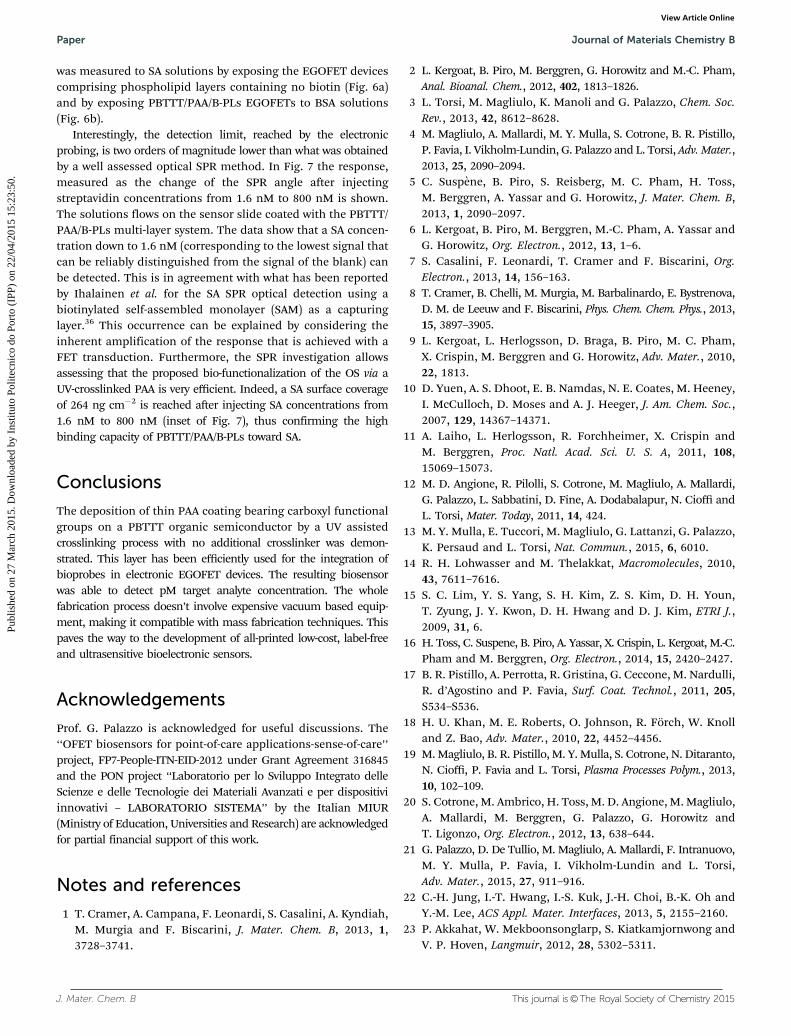
was measured to SA solutions by exposing the EGOFET devicescomprising phospholipid layers containing no biotin (Fig. 6a)and by exposing PBTTT/PAA/B-PLs EGOFETs to BSA solutions(Fig. 6b).
Interestingly, the detection limit, reached by the electronicprobing, is two orders of magnitude lower than what was obtainedby a well assessed optical SPR method. In Fig. 7 the response,measured as the change of the SPR angle after injectingstreptavidin concentrations from 1.6 nM to 800 nM is shown.The solutions flows on the sensor slide coated with the PBTTT/PAA/B-PLs multi-layer system. The data show that a SA concen-tration down to 1.6 nM (corresponding to the lowest signal thatcan be reliably distinguished from the signal of the blank) canbe detected. This is in agreement with what has been reportedby Ihalainen et al. for the SA SPR optical detection using abiotinylated self-assembled monolayer (SAM) as a capturinglayer.36 This occurrence can be explained by considering theinherent amplification of the response that is achieved with aFET transduction. Furthermore, the SPR investigation allowsassessing that the proposed bio-functionalization of the OS via aUV-crosslinked PAA is very efficient. Indeed, a SA surface coverageof 264 ng cm�2 is reached after injecting SA concentrations from1.6 nM to 800 nM (inset of Fig. 7), thus confirming the highbinding capacity of PBTTT/PAA/B-PLs toward SA.
Conclusions
The deposition of thin PAA coating bearing carboxyl functionalgroups on a PBTTT organic semiconductor by a UV assistedcrosslinking process with no additional crosslinker was demon-strated. This layer has been efficiently used for the integration ofbioprobes in electronic EGOFET devices. The resulting biosensorwas able to detect pM target analyte concentration. The wholefabrication process doesn’t involve expensive vacuum based equip-ment, making it compatible with mass fabrication techniques. Thispaves the way to the development of all-printed low-cost, label-freeand ultrasensitive bioelectronic sensors.
Acknowledgements
Prof. G. Palazzo is acknowledged for useful discussions. The‘‘OFET biosensors for point-of-care applications-sense-of-care’’project, FP7-People-ITN-EID-2012 under Grant Agreement 316845and the PON project ‘‘Laboratorio per lo Sviluppo Integrato delleScienze e delle Tecnologie dei Materiali Avanzati e per dispositiviinnovativi – LABORATORIO SISTEMA’’ by the Italian MIUR(Ministry of Education, Universities and Research) are acknowledgedfor partial financial support of this work.
Notes and references
1 T. Cramer, A. Campana, F. Leonardi, S. Casalini, A. Kyndiah,M. Murgia and F. Biscarini, J. Mater. Chem. B, 2013, 1,3728–3741.
2 L. Kergoat, B. Piro, M. Berggren, G. Horowitz and M.-C. Pham,Anal. Bioanal. Chem., 2012, 402, 1813–1826.
3 L. Torsi, M. Magliulo, K. Manoli and G. Palazzo, Chem. Soc.Rev., 2013, 42, 8612–8628.
4 M. Magliulo, A. Mallardi, M. Y. Mulla, S. Cotrone, B. R. Pistillo,P. Favia, I. Vikholm-Lundin, G. Palazzo and L. Torsi, Adv. Mater.,2013, 25, 2090–2094.
5 C. Suspene, B. Piro, S. Reisberg, M. C. Pham, H. Toss,M. Berggren, A. Yassar and G. Horowitz, J. Mater. Chem. B,2013, 1, 2090–2097.
6 L. Kergoat, B. Piro, M. Berggren, M.-C. Pham, A. Yassar andG. Horowitz, Org. Electron., 2012, 13, 1–6.
7 S. Casalini, F. Leonardi, T. Cramer and F. Biscarini, Org.Electron., 2013, 14, 156–163.
8 T. Cramer, B. Chelli, M. Murgia, M. Barbalinardo, E. Bystrenova,D. M. de Leeuw and F. Biscarini, Phys. Chem. Chem. Phys., 2013,15, 3897–3905.
9 L. Kergoat, L. Herlogsson, D. Braga, B. Piro, M. C. Pham,X. Crispin, M. Berggren and G. Horowitz, Adv. Mater., 2010,22, 1813.
10 D. Yuen, A. S. Dhoot, E. B. Namdas, N. E. Coates, M. Heeney,I. McCulloch, D. Moses and A. J. Heeger, J. Am. Chem. Soc.,2007, 129, 14367–14371.
11 A. Laiho, L. Herlogsson, R. Forchheimer, X. Crispin andM. Berggren, Proc. Natl. Acad. Sci. U. S. A, 2011, 108,15069–15073.
12 M. D. Angione, R. Pilolli, S. Cotrone, M. Magliulo, A. Mallardi,G. Palazzo, L. Sabbatini, D. Fine, A. Dodabalapur, N. Cioffi andL. Torsi, Mater. Today, 2011, 14, 424.
13 M. Y. Mulla, E. Tuccori, M. Magliulo, G. Lattanzi, G. Palazzo,K. Persaud and L. Torsi, Nat. Commun., 2015, 6, 6010.
14 R. H. Lohwasser and M. Thelakkat, Macromolecules, 2010,43, 7611–7616.
15 S. C. Lim, Y. S. Yang, S. H. Kim, Z. S. Kim, D. H. Youn,T. Zyung, J. Y. Kwon, D. H. Hwang and D. J. Kim, ETRI J.,2009, 31, 6.
16 H. Toss, C. Suspene, B. Piro, A. Yassar, X. Crispin, L. Kergoat, M.-C.Pham and M. Berggren, Org. Electron., 2014, 15, 2420–2427.
17 B. R. Pistillo, A. Perrotta, R. Gristina, G. Ceccone, M. Nardulli,R. d’Agostino and P. Favia, Surf. Coat. Technol., 2011, 205,S534–S536.
18 H. U. Khan, M. E. Roberts, O. Johnson, R. Forch, W. Knolland Z. Bao, Adv. Mater., 2010, 22, 4452–4456.
19 M. Magliulo, B. R. Pistillo, M. Y. Mulla, S. Cotrone, N. Ditaranto,N. Cioffi, P. Favia and L. Torsi, Plasma Processes Polym., 2013,10, 102–109.
20 S. Cotrone, M. Ambrico, H. Toss, M. D. Angione, M. Magliulo,A. Mallardi, M. Berggren, G. Palazzo, G. Horowitz andT. Ligonzo, Org. Electron., 2012, 13, 638–644.
21 G. Palazzo, D. De Tullio, M. Magliulo, A. Mallardi, F. Intranuovo,M. Y. Mulla, P. Favia, I. Vikholm-Lundin and L. Torsi,Adv. Mater., 2015, 27, 911–916.
22 C.-H. Jung, I.-T. Hwang, I.-S. Kuk, J.-H. Choi, B.-K. Oh andY.-M. Lee, ACS Appl. Mater. Interfaces, 2013, 5, 2155–2160.
23 P. Akkahat, W. Mekboonsonglarp, S. Kiatkamjornwong andV. P. Hoven, Langmuir, 2012, 28, 5302–5311.
Paper Journal of Materials Chemistry B
Publ
ishe
d on
27
Mar
ch 2
015.
Dow
nloa
ded
by I
nstit
uto
Polit
ecni
co d
o Po
rto
(IPP
) on
22/
04/2
015
15:2
3:50
. View Article Online

This journal is©The Royal Society of Chemistry 2015 J. Mater. Chem. B
24 M. Lahav, M. Narovlyansky, A. Winkleman, R. Perez-Castillejos,E. A. Weiss and G. M. Whitesides, Adv. Mater., 2006, 18,3174–3178.
25 A. Gestos, P. G. Whitten, G. M. Spinks and G. G. Wallace,Soft Matter, 2010, 6, 1045–1052.
26 Y.-C. Nho, J.-S. Park and Y.-M. Lim, Polymers, 2014, 6,890–898.
27 E. Jabbari and S. Nozari, Eur. Polym. J., 2000, 36, 2685–2692.28 K. Kumeta, I. Nagashima, S. Matsui and K. Mizoguchi,
J. Appl. Polym. Sci., 2003, 90, 2420–2427.29 Y. Cui, Q. Wei, H. Park and C. M. Lieber, Science, 2001,
293, 1289.30 F. Dinelli, J. F. Moulin, M. A. Loi, E. Da Como, M. Massi,
M. Murgia, M. Muccini and F. Biscarini, J. Phys. Chem. B,2006, 110, 258.
31 D. Braga and G. Horowitz, Adv. Mater., 2009, 21, 14.32 F. N. Ishikawa, M. Curreli, H. K. Chang, P. C. Chen,
R. Zhang, R. J. Cote, M. E. Thompson and C. Zhou, ACSNano, 2009, 3, 3969.
33 K. Manoli, L. M. Dumitru, M. Y. Mulla, M. Magliulo,C. Di Franco, M. V. Santacroce, G. Scamarcio and L. Torsi,Sensors, 2014, 14, 16869.
34 L. Q. Chu, W. J. Tan, H. Q. Mao and W. Knoll, Macromolecules,2006, 39, 8742.
35 J. E. Parmer, A. C. Mayer, B. E. Hardin, S. R. Scully, M. D.McGehee, M. M. Heeney and I. McCulloch, Appl. Phys. Lett.,2008, 92, 113309.
36 P. Ihalainen, H. Majumdar, T. Viitala, B. Torngren, T. Narjeoja,A. Maattanen, J. Sarfraz, H. Harma, M. Yliperttula, R. Osterbackaand J. Peltonen, Biosensors, 2013, 3, 1.
Journal of Materials Chemistry B Paper
Publ
ishe
d on
27
Mar
ch 2
015.
Dow
nloa
ded
by I
nstit
uto
Polit
ecni
co d
o Po
rto
(IPP
) on
22/
04/2
015
15:2
3:50
. View Article Online
Copyright © 2022 FDOKUMEN