UV and Sunlight Driven Photoligation of Quantum Dots: Understanding the Photochemical Transformation...
11
UV and Sunlight Driven Photoligation of Quantum Dots: Understanding the Photochemical Transformation of the Ligands Fadi Aldeek, † Dana Hawkins, † Valle Palomo, ‡ Malak Safi, † Goutam Palui, † Philip E. Dawson, ‡ Igor Alabugin, † and Hedi Mattoussi* ,† † Department of Chemistry and Biochemistry, Florida State University, 95 Chieftan Way, Tallahassee, Florida 32306, United States ‡ Department of Chemistry and Department of Cell Biology, The Scripps Research Institute, 10550 N. Torrey Pines Road, La Jolla, California 92037, United States * S Supporting Information ABSTRACT: We have recently reported that photoinduced ligation of ZnS-overcoated quantum dots (QDs) offers a promising strategy to promote the phase transfer of these materials to polar and aqueous media using multidentate lipoic acid (LA)-modified ligands. In this study we investigate the importance of the underlying parameters that control this process, in particular, whether or not photoexcited QDs play a direct role in the photoinduced ligation. We find that irradiation of the ligand alone prior to mixing with hydrophobic QDs is sufficient to promote ligand exchange. Furthermore, photoligation onto QDs can also be carried out simply by using sunlight. Combining the use of Ellman’s test with matrix-assisted laser desorption/ ionization and electrospray ionization mass spectrometry, we probe the nature of the photochemical transformation of the ligands. We find that irradiation (using either a UV photoreactor or sunlight) alters the nature of the disulfide groups in the lipoic acid, yielding a different product mixture than what is observed for chemically reduced ligands. Irradiation of the ligand in solution generates a mixture of monomeric and oligomeric compounds. Ligation onto the QDs selectively favors oligomers, presumably due to their higher coordination onto the metal-rich QD surfaces. We also show that photoligation using mixed ligands allows the preparation of reactive nanocrystals. The resulting QDs are coupled to proteins and peptides and tested for cellular staining. This optically controlled ligation of QDs combined with the availability of a variety of multidentate and multifunctional LA-modified ligands open new opportunities for developing fluorescent platforms with great promises for use in imaging and sensor design. ■ INTRODUCTION The unique and controllable photophysical properties of semiconductor nanocrystals (quantum dots, QDs) have generated great interest among physicists, chemists, engineers, and biologists alike, because they offer a set of interesting fundamental concepts to understand along with a great potential for applications in electronic devises, biological imaging, sensing, and clinical diagnostics. 1−5 These properties include tunable and narrow fluorescence emission, broad absorption profiles, high brightness and superior photo- and chemical stability. 6−12 High-quality QDs with crystalline cores, low size dispersity, and high fluorescence quantum yield are routinely prepared using high-temperature reduction of organometallic precursors in a mixture of coordinating solvents such as tri- n- octylphosphine/tri-n-octylphosphine oxide (TOP/TOPO), alkylamine, and alkylphosphonic acids. 13−18 The resulting materials are capped with hydrophobic ligands and are thus dispersible only in organic solvents. An effective phase-transfer strategy is therefore required to facilitate their integration with biological systems. One of the commonly explored strategies to achieve this goal relies on replacing the native cap with hydrophilic bifunctional ligands (cap/ligand exchange). To be effective, this approach requires the use of hydrophilic ligands with strong coordination onto the metal surface of the nanocrystals in order to achieve complete removal of the hydrophobic cap and produce stable nanocrystals in biological media. 3,19−21 Multidentate thiolated ligands provide enhanced stability to QDs in aqueous media, due to the higher ligand-to-nanocrystal affinity afforded by the simultaneous coordination of multiple thiols onto the same QD surface. In particular, dihydrolipoic acid (DHLA)-based ligands were shown to impart better colloidal stability to the QDs over a wide range of biological conditions than their monothiol-appended counterparts. 22,23 The biocompatibility and functionality of QDs stabilized with DHLA-based ligands have been enhanced through insertion of polyethylene glycol or/and zwitterionic moieties into the ligand structure. Further modification of the lipoic acid (LA) ligand has also allowed the controllable introduction of inert (OCH 3 ) and reactive functional groups (e.g., carboxyl, amine, azide) into the organic surface coating of the nanocrystal. 20,24−27 Cap exchange of hydrophobic QDs with LA-modified ligands has thus far required the use of the reduced form of the ligands, i.e., DHLA-based compounds. Reduction of the disulfide to Received: December 16, 2014 Article pubs.acs.org/JACS © XXXX American Chemical Society A DOI: 10.1021/ja512802x J. Am. Chem. Soc. XXXX, XXX, XXX−XXX
Transcript of UV and Sunlight Driven Photoligation of Quantum Dots: Understanding the Photochemical Transformation...

UV and Sunlight Driven Photoligation of Quantum Dots:Understanding the Photochemical Transformation of the LigandsFadi Aldeek,† Dana Hawkins,† Valle Palomo,‡ Malak Safi,† Goutam Palui,† Philip E. Dawson,‡
Igor Alabugin,† and Hedi Mattoussi*,†
†Department of Chemistry and Biochemistry, Florida State University, 95 Chieftan Way, Tallahassee, Florida 32306, United States‡Department of Chemistry and Department of Cell Biology, The Scripps Research Institute, 10550 N. Torrey Pines Road, La Jolla,California 92037, United States
*S Supporting Information
ABSTRACT: We have recently reported that photoinduced ligation of ZnS-overcoatedquantum dots (QDs) offers a promising strategy to promote the phase transfer of these materialsto polar and aqueous media using multidentate lipoic acid (LA)-modified ligands. In this studywe investigate the importance of the underlying parameters that control this process, inparticular, whether or not photoexcited QDs play a direct role in the photoinduced ligation. Wefind that irradiation of the ligand alone prior to mixing with hydrophobic QDs is sufficient topromote ligand exchange. Furthermore, photoligation onto QDs can also be carried out simplyby using sunlight. Combining the use of Ellman’s test with matrix-assisted laser desorption/ionization and electrospray ionization mass spectrometry, we probe the nature of thephotochemical transformation of the ligands. We find that irradiation (using either a UVphotoreactor or sunlight) alters the nature of the disulfide groups in the lipoic acid, yielding adifferent product mixture than what is observed for chemically reduced ligands. Irradiation of theligand in solution generates a mixture of monomeric and oligomeric compounds. Ligation ontothe QDs selectively favors oligomers, presumably due to their higher coordination onto the metal-rich QD surfaces. We alsoshow that photoligation using mixed ligands allows the preparation of reactive nanocrystals. The resulting QDs are coupled toproteins and peptides and tested for cellular staining. This optically controlled ligation of QDs combined with the availability of avariety of multidentate and multifunctional LA-modified ligands open new opportunities for developing fluorescent platformswith great promises for use in imaging and sensor design.
■ INTRODUCTION
The unique and controllable photophysical properties ofsemiconductor nanocrystals (quantum dots, QDs) havegenerated great interest among physicists, chemists, engineers,and biologists alike, because they offer a set of interestingfundamental concepts to understand along with a greatpotential for applications in electronic devises, biologicalimaging, sensing, and clinical diagnostics.1−5 These propertiesinclude tunable and narrow fluorescence emission, broadabsorption profiles, high brightness and superior photo- andchemical stability.6−12
High-quality QDs with crystalline cores, low size dispersity,and high fluorescence quantum yield are routinely preparedusing high-temperature reduction of organometallic precursorsin a mixture of coordinating solvents such as tri-n-octylphosphine/tri-n-octylphosphine oxide (TOP/TOPO),alkylamine, and alkylphosphonic acids.13−18 The resultingmaterials are capped with hydrophobic ligands and are thusdispersible only in organic solvents. An effective phase-transferstrategy is therefore required to facilitate their integration withbiological systems. One of the commonly explored strategies toachieve this goal relies on replacing the native cap withhydrophilic bifunctional ligands (cap/ligand exchange). To beeffective, this approach requires the use of hydrophilic ligands
with strong coordination onto the metal surface of thenanocrystals in order to achieve complete removal of thehydrophobic cap and produce stable nanocrystals in biologicalmedia.3,19−21
Multidentate thiolated ligands provide enhanced stability toQDs in aqueous media, due to the higher ligand-to-nanocrystalaffinity afforded by the simultaneous coordination of multiplethiols onto the same QD surface. In particular, dihydrolipoicacid (DHLA)-based ligands were shown to impart bettercolloidal stability to the QDs over a wide range of biologicalconditions than their monothiol-appended counterparts.22,23
The biocompatibility and functionality of QDs stabilized withDHLA-based ligands have been enhanced through insertion ofpolyethylene glycol or/and zwitterionic moieties into the ligandstructure. Further modification of the lipoic acid (LA) ligandhas also allowed the controllable introduction of inert (OCH3)and reactive functional groups (e.g., carboxyl, amine, azide) intothe organic surface coating of the nanocrystal.20,24−27
Cap exchange of hydrophobic QDs with LA-modified ligandshas thus far required the use of the reduced form of the ligands,i.e., DHLA-based compounds. Reduction of the disulfide to
Received: December 16, 2014
Article
pubs.acs.org/JACS
© XXXX American Chemical Society A DOI: 10.1021/ja512802xJ. Am. Chem. Soc. XXXX, XXX, XXX−XXX
dithiol has been carried out chemically, using NaBH4 as areducing agent.24 Though effective, this route requires multiplesteps and careful storage of the DHLA-based ligands. DHLA-modified compounds are not shelf stable and must begenerated through reduction of the 1,2-dithiolane moiety ofthe LA precursors. Furthermore, borohydride reduction altersthe integrity of certain sensitive but highly desirable functionalgroups for bioconjugation, such as azides and aldehydes.23,28,29
In order to address these problems, our group has taken intoconsideration the photosensitive nature of the cyclic disulfide inthe ligands. It has been shown that UV irradiation of LA canproduce a heterogeneous mixture of monomeric and polymericcomplexes.30,31 We have recently built on those findings andshowed that ligand exchange on QDs can be promotedphotochemically, starting from the oxidized form of the ligand.In particular, we have found that irradiation of the native TOP/TOPO-capped QDs in the presence of LA-PEG using a UVphotoreactor can readily transfer the nanocrystals to polarsolvents and, most importantly, to water.32 Nonetheless, severalquestions are still left unanswered, including whether or not thephotoexcited QDs play a role in the transformation of the LAgroups, what is the nature of the photochemical transformationof the ligands, and whether or not the LA derivatives arereduced. In addition, we hoped to evaluate whether the strategycould be applied using simple sunlight instead of a laboratoryUV photoreactor.In the present work, we expand on the earlier findings by
focusing on the role that the QDs may or may not play andseek a better understanding of the nature of the photochemicaltransformation of the ligands when irradiated in the absence ofthe QDs. We use a combination of UV−vis absorptionspectroscopy, Ellman’s assay/test along with matrix-assistedlaser desorption/ionization (MALDI) and electrospray ioniza-tion (ESI) mass spectrometry measurements to identify thenature of the photochemical transformation with the lipoic acid.We then propose a rationale for why this strategy is highlyeffective for the transfer of QDs to polar solvents and buffermedia.
■ RESULTS AND DISCUSSIONIn a previous report, we showed that cap exchange of LA-basedligands onto luminescent QDs can be induced photochemicallyusing in situ irradiation of the oxidized form of the ligand in thepresence of hydrophobic nanocrystals.32 Here, activation of thedisulfide, ligand exchange, and phase transfer of the nanocryst-als were all combined in the same step. The photoinducedphase transfer was implemented using either a single phase(e.g., polar methanol) or a two-phase (polar methanol andhexane) reaction, starting with the hydrophobic QDs. Thisprocedure has obviated the need for chemical reduction of theLA under harsh conditions using sodium borohydride prior toperforming the ligand exchange, along with the requirementsfor storage of the reduced ligand under inert atmosphere.23
This route has also been applied to various PEG- andzwitterion-modified mono- and bis(LA) ligands, producingdispersions of QDs with great colloidal stability and easy tointegrate with biology.32−34
We attributed the success of this strategy to the sensitivity ofthe LA to UV-excitation (at 320−350 nm) with potentialcontribution from electron transfer to LA (from photoexcitedQDs), facilitating the reduction of the disulfide and couplingonto the ZnS-overcoated QDs. Indeed, the cyclic disulfide ofLA exhibits a well-defined broad absorption band centered at
∼330 nm, which undergoes a progressive decrease to near zeroafter ∼30−40 min irradiation using a UV signal at 350 nm. Wesuggested that a homolytic “cleavage” of the S−S bond in theLA occurs under UV excitation, which promotes ligandcoordination onto the Zn-rich surface of the QDs. A side-by-side comparison of the 1H NMR spectra collected fromphotoirradiated and chemically reduced ligands indicated thatthe photochemical reaction results in a mixture of productswith chemical shifts similar to both the oxidized LA (thestarting material) and to the reduced DHLA. However, thesignatures of some of the protons close to the dithiolane ringobserved for the UV-irradiated ligands were different fromthose recorded for the borohydride-reduced compounds.32
In this study we wanted to develop an understanding of thephotochemical transformation by addressing the followingquestions. (1) Do the QDs play an active role in thephotochemical transformation of the LA groups and theensuing cap exchange? (2) Can the phase transfer beimplemented using TOP/TOPO-QDs mixed with ligandsirradiated in the absence of QDs? (3) Can the phase transferbe implemented simply using sunlight exposure? (4) What isthe chemical composition of the irradiated ligands and why isthis route more effective than cap exchange using borohydride-reduced ligands?
Irradiation of the Isolated Ligands Followed by QDPhase Transfer. In this route, the LA-based ligands were firstdissolved in methanol, irradiated for 30 min in a UV reactor(see Experimental Section), and then mixed with a hexanesolution containing TOP/TOPO-QDs (i.e., two-phase config-uration). After stirring the mixture at room temperature for 15min, the QDs were fully transferred from hexane to methanol(Figure 1). Following evaporation of the solvent(s), thenanocrystals were dispersed in ethanol and then precipitatedwith hexane to remove excess/free ligands. The resulting QDpellet fully dispersed in water. Applying 2−3 rounds ofpurification, using a centrifugal filtration device to remove theremaining soluble ligands, provided homogeneous dispersionsof QDs which could be stored for further use.Alternatively, the preirradiated solution of ligands in
methanol could be mixed with a solid precipitate of thehydrophobic QDs (i.e., one phase configuration). Stirring themixture also promoted cap exchange and dispersion of thenanocrystals in methanol. The solution was processed as above,producing a homogeneous QD dispersion in water. The phasetransfer using irradiated ligands was rapid, needing only ∼15min of mixing at room temperature. Importantly, this processrequired only about one-fifth of the excess ligand typically usedfor phase transfer with borohydride-reduced ligands. Inaddition, there was no need to heat the solutions in eitherone- or two-phase protocol, which contrasts with the need toheat the reaction mixture at 60 °C for several hours (6−12 h),when DHLA-PEG derivatives were used.23
We have applied this method to transfer four different sets ofQDs emitting at 540 nm (green), 570 nm (yellow), 590 nm(orange), and 630 nm (red) (Figure 1). Figure 2A,B shows theabsorption and emission spectra collected from these QDsbefore and after phase transfer. The hydrophilic QDs retainedthe photophysical properties of the starting nanocrystals, with aminimal spectroscopic shift in both absorption and photo-luminescence; ∼2−4 nm red shift was measured in some casesfor dispersions in buffer media. Such a small shift has beenoccasionally measured for QDs following transfer to watermedia.24 The photoluminescence (PL) quantum yields (QYs)
Journal of the American Chemical Society Article
DOI: 10.1021/ja512802xJ. Am. Chem. Soc. XXXX, XXX, XXX−XXX
B

of LA-PEG750-OCH3-capped QDs in water were overall smallerthan the native TOP/TOPO-capped nanocrystals dispersed inhexane (see Figure 2). For instance, starting with a QD samplehaving a QY of 40% in hexane produced water dispersion withQY of 20−30%. Such reduction in the PL emission iscommonly observed following phase transfer to water, thoughdecrease in the measured QYs for thiol-modified ligands isoften slightly larger.35 Importantly, the photophysical proper-ties of QDs generated with irradiated ligands are nearly
identical to those exhibited by QDs previously prepared usingchemically reduced DHLA.Taken together, these findings prove that the QDs do not
play a direct role in the photochemical transformation of theLA groups. They indicate that potential charge-transferinteractions from photoexcited QDs play no role in thephase-transfer process. As such, decreasing the QD size (i.e.,shifting the emission location farther to the blue), which widenthe band gap and shifts the exact location of the conductionand valence band energies, does not affect the ligation strategy.They also indicate that irradiating the ligand separately, thenproceeding with the phase-transfer step(s) may be morebeneficial, as this route consumes less ligands, does not evenrequire mild sample heating, and avoids extended exposure ofthe QDs to UV irradiation.
Sunlight-Mediated Ligation of the QDs. We also carriedout the phase transfer relying on the photochemical trans-formation of the ligand using sunlight exposure, instead of alaboratory UV reactor. Here, we tested the viability of this routeusing all three configurations: (1) sunlight irradiation ofprecipitated TOP/TOPO-QDs mixed with LA-PEG inmethanol (one phase); (2) sunlight irradiation of a two-phasemixture using ligands in methanol and QDs in hexane; and (3)sunlight irradiation of the ligand in methanol followed by two-phase transfer. The main question we wanted to address iswhether or not photoligation of QDs with LA-based ligandsrequires a laboratory UV reactor.We found that the photochemical transformation of the
ligands does not require a laboratory UV reactor and successfulcap exchange of the QDs could be achieved in all threeconfigurations mentioned above. Furthermore, the absorptionand PL emission properties of the final QDs dispersed in buffermedia using sunlight irradiation were very similar to thoseprepared using a UV reactor (see Supporting Information,Figure S1). Slight differences were observed, and in certaininstances, the final quantum yield of QDs photoligated undersunlight irradiation was slightly higher than those preparedusing a laboratory UV reactor (see Supporting Information,Figure S2). This enhanced QY may be attributed to the milderconditions of the photochemical transformation of the ligandsusing irradiation provided by simple sunlight exposure. Weshould also note that the observed decrease in the QY of theQDs following phase transfer with the ligands (compared totheir hydrophobic counterparts) is attributed to the nature ofthe thiol coordination onto the metal surface of thenanocrystals, not to a partial (or inefficient) ligand exchangewith the present strategy. In fact, independent preliminaryestimates indicate that the ligand density measured for QDscapped with the chemically reduced DHLA-PEG is comparableto that measured for QDs photoligated with LA-PEG ligands(data not shown).We expanded the use of the sunlight-mediated photoligation
and phase transfer using zwitterion-modified LA (LA-ZW)ligands. We carried out phase transfer with LA-ZW using theconfigurations introduced above; we tested this route using twodifferent sets of QDs (orange- and green-emitting) andapplying 30 min irradiation periods for all samples. Here too,sunlight irradiation produced homogeneous QD dispersionswith absorption and PL emission spectra that are similar tothose of the TOPO/TOP-capped QDs (see SupportingInformation, Figure S3). Phase transfer under sunlight exposurewas also carried out using commercially available LA, yielding
Figure 1. Schematic representation showing the cap exchange usingpreirradiated ligands. (A) Representation of the starting QDs andligands along with white light and fluorescence images of a two-phasemixture of TOP/TOPO-QDs in hexane and preirradiated LA-PEG750-OCH3 in methanol, immediately following mixing; four samples ofdistinct color QDs are shown. (B) Schematic representation of the QDligation combined with white light and fluorescence images of theabove two-phase solutions following phase transfer. (C) Schematics ofthe final hydrophilic PEG-capped QDs together with fluorescentimages of these QDs in water. The dispersions were excited usinghand-held UV lamp, with λexc = 365 nm. The QD samples shown emitat 540 nm (green), 570 nm (yellow), 590 nm (orange), and 630 nm(red).
Journal of the American Chemical Society Article
DOI: 10.1021/ja512802xJ. Am. Chem. Soc. XXXX, XXX, XXX−XXX
C
dispersions of QDs that are similar to those prepared usingborohydride-reduced DHLA (data not shown).We should note that sunlight irradiation experiments were
usually carried out using direct and full exposure to the sun.Experiments carried out during a sunny summer or fall dayyielded similar results. Furthermore, we found that experimentscarried out during the day but under cloudy conditions also
resulted in an efficient transformation of the ligand after 30−40min (as verified optically) and a complete ligand exchangewhen mixed with the QDs; irradiation of a two-phase samplealso produced full phase transfer. However, experiments carriedout using exposure to sunlight through a laboratory window(still full exposure to light) did not promote complete phasetransfer even after several days. Similarly, exposure of a ligand
Figure 2. (A) Normalized absorption (with respect to the band edge peak) and (B) PL spectra (normalized with respect to the peak value) of theQDs before and after cap exchange, using the preirradiated ligands. Green, yellow, orange and red lines correspond to the 540 nm-, 570 nm-, 590nm- and 630 nm-emitting QDs; the solid and dashed lines designate dispersions in hexane and in DI water, respectively. The inset in (A) shows thePL spectra of QDS after photoligation with LA-PEG750-OCH3 and transfer to water, normalized with respect to the spectra of TOP/TOPO-QDs inhexane; spectra were collected using dispersions having the same optical density at the excitation line λexc = 350 nm.
Figure 3. Progression of the UV−vis absorption spectra of LA-PEG750-OCH3 dissolved in methanol upon photoirradiation for varying times using(A) sunlight and (B) a UV photoreactor; the ligand concentration was ∼10 mM. (C) Evolution in the UV spectra of DTNB upon increasing theDHLA-PEG750-OCH3 concentration. (D) Calibration curve showing linear correlation between the absorbance collected at 412 nm and theconcentration of DHLA-PEG750-OCH3 used; the concentration of the DTNB reagent was fixed at ∼25 μM in phosphate buffer pH7.8.
Journal of the American Chemical Society Article
DOI: 10.1021/ja512802xJ. Am. Chem. Soc. XXXX, XXX, XXX−XXX
D
solution using such conditions did not yield full transformationof the dithiolane groups, where only a small decrease in theabsorption feature at 335 nm was measured. This result isattributed to the fact that the glass windows strongly attenuatethe UV region of the sun spectrum.Having demonstrated that the QDs play no direct role in the
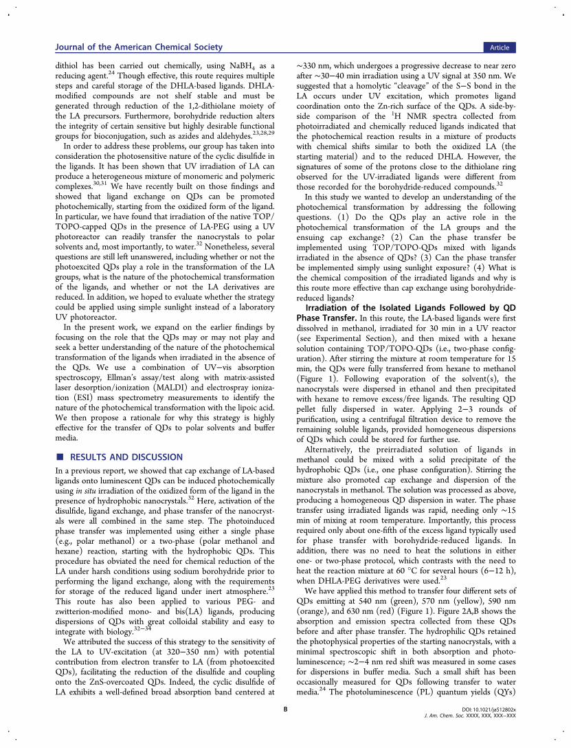
photochemical transformation of the LA groups and theensuing ligand exchange, we then centered our effort onmonitoring what happens to the LA-based ligands under UV orsunlight irradiation. For this, we systematically characterized theligands before and after irradiation using UV−vis absorptionspectroscopy, Ellman’s test/assay and mass spectrometry.These measurements combined allowed us to characterize thenature and relative proportions of some of the photochemicallygenerated species in a pure solution of LA-PEG (or LA)ligands. They also allowed us to gain additional insights intowhat exactly makes the phase transfer using this photochemicalroute effective. Figure 3A,B shows the progression of thedisulfide absorption band (at 335 nm) upon UV irradiation for30 min and sunlight exposure for 40 min. A progressivedecrease until near complete disappearance in the absorptionpeak is measured in both cases, a result similar to what wepreviously reported.32 This indicates that the photochemicaltransformation of the ligands is essentially the same, whether
irradiation of the solution is carried in a laboratory set using aUV reactor or simply relying on the UV signal provided by thesun.We then used the Ellman’s assay to quantify the number of
available thiol groups in a solution of LA-PEG-OCH3 followingirradiation (via UV or sunlight).36 Typically, this test relies onthe reaction of the reagent 5,5′-dithiobis-2-nitrobenzoic acid(DTNB) with thiol groups in the medium, producing 5-thio-2-nitrobenzoic acid (TNB) that has a distinct absorptionsignature at 412 nm, manifesting in a color change of thesolution to yellow. Quantifying the concentration of thiolgroups in the medium was done by comparing the optical datato those collected from a control solutions made of chemicallyreduced ligand. For this control, a calibration curve usingDHLA-PEG-OCH3 (NaBH4-reduced ligand) was generated. Asexpected, the concentration of thiol groups in a solution ofligand, as indicated by the progression of the signature at 412nm (ascribed to the TNB product), varied linearly with theconcentration of DHLA-PEG-OCH3 (Figure 3C,D). Further-more, the slope extracted from that curve should reflect thenumber of thiol per molecule (n) via the relation: Abs = ε × C× d × n, where ε is extinction coefficient of TNB at 412 nm, Cis the molar concentration of DHLA-PEG-OMe, and d is theoptical path of the cell used to collect the absorbance spectra (d
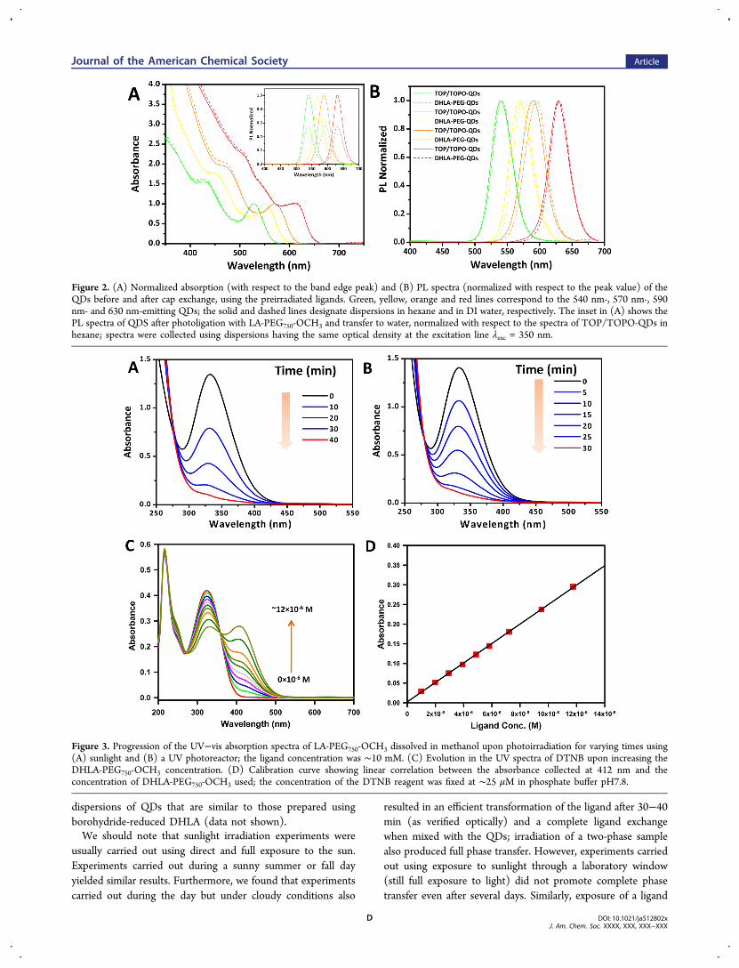
Figure 4. (A) UV−vis spectra of the DTNB reagent alone (red line), in the presence of LA-ligands (green line), in the presence of chemicallyreduced DHLA ligands (orange line), and in the presence of photoirradiated LA-ligands (dark yellow line); also shown are the absorption spectra ofpure DHLA-PEG-OMe and UV-irradiated LA-PEG-OMe. Note that the contribution of the LA absorption feature at 335 nm to the spectra isnegligible, due to a combination of lower extinction coefficient and smaller concentration of ligands used compared to those of DTNB. The DNTBand PEG-OMe ligand concentrations were ∼25 and ∼10 μM, respectively. (B) MALDI mass spectra of the LA-PEG750-OCH3 ligands before (blackline) and after UV-irradiation (red line). (C) A few representative UV-induced photochemical transformations of the dithiolane ring are providedbased on literature data.31,40,44,46 The R group can be a short alkyl-COOH, such as for LA, or alkyl-PEG such as the case of the ligands used here.(D) MALDI mass spectra of excess free UV-irradiated LA-PEG750-OCH3 ligands collected in the supernatant after cap exchange.
Journal of the American Chemical Society Article
DOI: 10.1021/ja512802xJ. Am. Chem. Soc. XXXX, XXX, XXX−XXX
E
= 1 cm). The experimental slope, 24,725 M−1 × cm−1, was veryclose to 2 × ε(TNB), using ε(TNB) ∼14,000 M−1 × cm−1.37
This indicates that the chemically reduced ligands essentiallyyield approximately two thiol groups per ligand, as expected.This curve was used to determine the fraction of thiol groupspresent in the LA-PEG solution following UV or sunlightirradiation. Figure 4A shows the absorption spectra for DTNBalone, DNTB mixed with LA-PEG750-OCH3, DNTB mixedwith DHLA-PEG750-OCH3, and DNTB mixed with photo-irradiated LA-PEG750-OCH3 at a concentration of 2 μM. Thespectrum of LA-PEG750-OCH3 solution shows no contributionat 412 nm, confirming the absence of thiol groups in theoxidized form of the ligand. The data indicate that in thepresence of the modified ligands, a new contribution to theabsorption at 412 nm appears for DHLA-PEG and photo-irradiated LA-PEG; these two solutions also turn yellow.However, the absorption value at 412 nm is ∼2-fold larger forDHLA-PEG750-OCH3 than that measured for the photo-irradiated ligands, suggesting that species other than DHLAare present. These data support the presence of a significantamount of Ellman’s reagent reactive species, presumably thiols,in the irradiated LA-PEG sample, but those species are lessabundant than what is measured for the chemically reducedcompound.Further characterization of the molecular species formed
during the UV irradiation relied on mass spectrometrymeasurements. Figure 4B shows the MALDI mass spectra ofthe LA-PEG750-OCH3 before and after UV-irradiation. Thespectrum measured for the oxidized form of the ligand showsonly one broad Gaussian peak centered at 900 Dacorresponding to the average mass of PEG750 plus LA; thewidth reflects the polydisperse nature of the PEG moieties. Theset of narrow peaks superposed on top of the main one arespaced by 44 Da, corresponding to the molar mass of ethyleneglycol units. The spectrum collected from photoirradiatedligands shows four distinct peaks centered approximately at900, 1800, 2700, and 3600 Da, indicating the presence ofphotochemically transformed monomers (as a large fraction),together with higher order oligomers, including dimers, trimers,and tetramers of the transformed ligand. This partialoligomerization upon photoirradiation is consistent with theabove data collected from the DTNB assay where lowerconcentration of thiol groups present in the medium (inferredfrom the absorption at 412 nm), since the LA-derivedmolecules in the polymer would not react with DTNB.To complement the above MALDI data collected from the
LA-PEG750-OCH3 ligand, we characterized the pure LA usingESI mass spectrometry. This experiment was carried out inorder to achieve better assignment of the mass peaks and avoidissues associated with peak broadening due to the polydisper-sity of the PEG chains. The ESI mass spectra of LA before andafter irradiation are shown in the Supporting Information(Figure S4). The spectrum collected from the compoundbefore irradiation shows two low molecular weight peaks of 229and 245, ascribed to mass 206 + 23 and 206 + 39corresponding to Na and K adducts of LA, respectively. Incomparison, the spectrum from the UV-irradiated compoundshows around 5 distinct peaks (in addition to the two discussedabove), including two centered around 435 and 467 Daascribed to Na+LA-LA and K+LA-LA-O (+16) dimers, twocentered around 641 and 673 Da ascribed to Na+LA-LA-LAand K+LA-LA-LA-O (+16) trimers, and two peaks centered at847 and 879 Da associated with Na+LA-LA-LA-LA and K+LA-
LA-LA-LA-O (+16) tetramers. The spectrum also shows thepresence of two peaks at 1053 and 1085 Da corresponding toNa+LA-LA-LA-LA-LA and K+LA-LA-LA-LA-LA-O (+16)pentamers, along with a weaker peak ascribed to hexamersaround 1300 Da; these are higher order complexes ofphotochemically activated LAs. Our data also confirm thepresence of oxidized S−O species (see scheme shown in Figure4C). They also show that the photochemical transformation ofthe dithiolane rings is the main promoter of linear or cyclicoligomer formation, independent of the PEG moieties. Weshould note that this experiment could not be performed usingthe MALDI-MS because of the interference from the matrix,which shows mass peaks around 300−600 Da.
Understanding the Photochemical Transformation ofthe LA-Based Ligands. LA has attracted great attention inchemistry and biology, due to a combination of photochemicalactivity, its antioxidant properties, and more recently its abilityto promote across cell membrane transport of unmodifiedsubstrates.38,39 The presence of a distorted dithiolane ring in itsstructure endows LA with a characteristic spectroscopicsignature, with the HUMO−LUMO energy difference fallingwithin the UV region of the optical spectrum.31,32 As a resultLA exhibits a well-defined absorption feature at ∼335 nm andcan thus be photochemically excited (and transformed) by UVirradiation.40,41 UV-induced transformation of LA was firststudied by Calvin and co-workers as a model system forprimary conversion in photosynthesis,42 and the chemicalreactivity of the compound was subsequently examined usingspectroscopic techniques.43 Cumulatively, these studies com-bined have shown that following irradiation photochemicaltransformation of the LA yields several products. For example,Murray and co-workers suggested that LA readily reacts withthe singlet oxygen to produce compounds containing S−O andS−(O)2 groups. The formation of these oxidized productsimplies that a direct reaction between LA and singlet oxygentakes place, indicating that LA can be a good quencher ofsinglet oxygen.44 Photochemical transformation of LA was alsostudied in various solvents, where the formation of severaloligomers byproducts was proposed and discussed.40,41 A morerecent study by Packer and co-workers have reported that UV-induced photodegradation of LA can generate DHLA.45
The complete reduction of LA to DHLA requires twoelectrons and thus cannot readily occur under UV excitationunless a certain reducing reagent is available in the medium.Bucher and Sander proposed three pathways for the diradicaldecay resulting from S−S homolytic cleavage: ring closureleading back to the precursor LA, 1,4-H shift givingmercaptothioaldehyde derivative, or 1,2-H shift givingmercaptoalkyl thiyl radical (see Figure 4C).31 They alsosuggested that these highly reactive dithiyl radicals can leadto DHLA formation following hydrogen abstraction. Based onthese photochemical properties, a ring-opening polymerizationand copolymerization of LA with 1,2-dithiane have also beenexamined by Endo and co-workers.46,47
Overall, the above studies indicate that irradiation of thedisulfide likely results in dithiyl radical formation, which in turncan lead to a variety of byproducts depending on the solventand concentration of the reactants used. One reaction pathwaycan lead to linear or cyclic oligomerization through S−Sbridging between distinct molecules and their oxidation to formoligomeric molecules in which S−O species are present. Figure4C summarizes schematically a few representative UV-inducedphotochemical transformations of the dithiolane ring discussed
Journal of the American Chemical Society Article
DOI: 10.1021/ja512802xJ. Am. Chem. Soc. XXXX, XXX, XXX−XXX
F

in the various literature studies. We should note that in additionto those byproducts, cyclic oligomeric disulfides (not depictedin Figure 4) can be formed but would not be reactive towardEllman’s reagent since they have no free thiols; it would thusnot contribute to the measured concentrations. However, alinear polymer could have one end reduced to a thiol and theother end oxidized to a sulfonic acid. When LA-compounds arephotoexcited and mixed with TOP/TOPO-QDs the abovedistinct monomeric and oligomeric species compete differentlyfor the nanocrystal surfaces, with stronger coordinationanticipated for the oligomers. This can explain why ligationof photochemically transformed LA-PEG ligands requires only15 min and no additional heat, compared to ligand exchangeusing the chemically reduced ligands. It is important to notethat during exchange, the LA compounds are added in largeexcess, so minor LA species following photoexcitation could beresponsible for the enhanced ligand exchange activity. Toconfirm this proposed rationale, we characterized the solutionof free ligands, collected once ligand exchange of the QDs wascomplete, using MALDI-MS as above. Briefly, following ligandexchange and solvent evaporation, DI water was added toprovide a water dispersion of QDs. The dispersion was purifiedfrom excess free ligands using a membrane filtration device withcutoff Mw = 50,000 Da. The filtrate solution containing excessligands was collected and characterized using MALDI-MS. Thespectrum in Figure 4D shows the presence of only monomersand dimers in the retrieved material, and no higher orderoligomers were found. In addition, characterization ofborohydride-reduced DHLA-PEG ligand showed that onlymonomers are present (see Supporting Information, FigureS5). This confirms that these polymers are only present uponUV-irradiation of the LA-based ligands.Together, these findings indicate that these photochemical
reactions alter the covalent structure of the dithiolane ring inthe LA. They also suggest that the cap exchange processconsumes the higher molecular LA species observed byMALDI. It is possible that the high Mw species are morereactive toward the QD surfaces and facilitate the cap exchange.Covalent Conjugation of Photoligated QDs. Our
synthetic scheme allows the in situ introduction of functionallyheterogeneous LA derivatives onto QD surfaces. This can be
achieved by introducing a small fraction of terminally reactiveligands (e.g., LA-PEG600-COOH or LA-PEG600-NH2) togetherwith the inert one (LA-PEG750-OCH3) during the photo-ligation and phase-transfer step.19 Combining the photoligationstrategy with mixed ligand exchange, we prepared twodispersions of reactive nanocrystals. One set was made ofQDs exchanged using 10% LA-PEG600-amine; the other set has50% of the surface ligands made of LA-PEG600-carboxy. Thesefunctionalized QDs were conjugated to the protein transferrin(Tf) and a cell penetrating peptide (CPP), respectively, viacarbodiimide coupling; see Experimental Section for moredetails. Transferrin is a plasma protein present in the blood ofmammals and is known to promote intracellular uptake viareceptor-mediated endocytosis.48−50 Conversely, cell penetrat-ing peptides are short amino acid sequences derived from viralproteins and often contain several arginines in their structures(CPP: Mw = 1994 Da).51 The CPP sequence utilized here (Lys-Trp-Leu-Ala-Aib-Ser-Gly-(Arg)8-CONH2) was synthesizedmanually using in situ neutralization cycles for Boc solid-phase peptide synthesis following procedures described in theliterature.52,53 These peptides are actively studied by severalgroups as a means of promoting intracellular uptake of genesand drugs.54 CPPs are believed to enter cells via directmembrane translocation, though several studies have shownthat uptake is also driven by endocytosis.55
Imaging of Cellular Uptake of Photoliagted QDs.Figure 5 shows the fluorescence confocal images of HeLa cellsincubated with QD-Tf conjugates at 37 °C along with cellsincubated with dispersions of QD-PEG-NH2 (control). Shownare side-by-side differential interference contrast (DIC) andfluorescence images corresponding to the distribution of DAPInuclear staining (blue), 540 nm-emitting QDs (green), alongwith the merged images. Significant uptake has been observedfor the QD-Tf (Figure 5, top). In comparison, weak but notnegligible fluorescence was measured for the control culture(Figure 5, bottom). Staining of cells incubated with NH2-PEG-coated QDs is not surprising since these amines can promoteelectrostatic adsorption on the cell membranes followed byendocytosis. This uptake is weaker than that measured for cellsincubated with QD-CPP conjugates.29 Regardless, attaching Tf
Figure 5. (Top) Fluorescence confocal microscopy images of intracellular delivery of green-emitting QD-Tf conjugates into HeLa cells. The panelsshow DIC image, fluorescence images of cell nuclei stained with DAPI, endosomes stained with QDs-Tf, along with the merged image. The cellswere incubated with 200 nM of QDs conjugates at 37 °C for 1 h. (Bottom) Images collected from control cultures incubated with unconjugatedQD-PEG-NH2. Scale bar = 15 μm.
Journal of the American Chemical Society Article
DOI: 10.1021/ja512802xJ. Am. Chem. Soc. XXXX, XXX, XXX−XXX
G
onto the nanocrystals promoted internalization of a muchlarger amount of QDs.The panels in Figure 6 show a series of confocal fluorescence
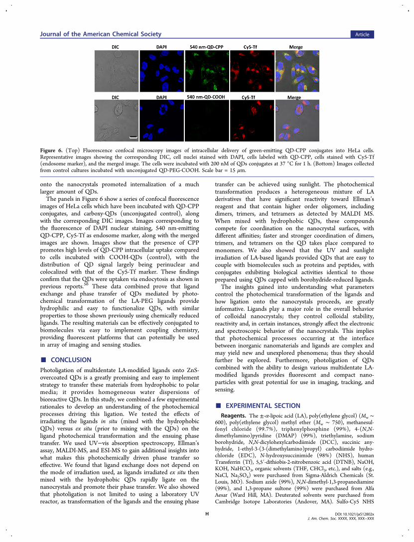
images of HeLa cells which have been incubated with QD-CPPconjugates, and carboxy-QDs (unconjugated control), alongwith the corresponding DIC images. Images corresponding tothe fluorescence of DAPI nuclear staining, 540 nm-emittingQD-CPP, Cy5-Tf as endosome marker, along with the mergedimages are shown. Images show that the presence of CPPpromotes high levels of QD-CPP intracellular uptake comparedto cells incubated with COOH-QDs (control), with thedistribution of QD signal largely being perinuclear andcolocalized with that of the Cy5-Tf marker. These findingsconfirm that the QDs were uptaken via endocytosis as shown inprevious reports.56 These data combined prove that ligandexchange and phase transfer of QDs mediated by photo-chemical transformation of the LA-PEG ligands providehydrophilic and easy to functionalize QDs, with similarproperties to those shown previously using chemically reducedligands. The resulting materials can be effectively conjugated tobiomolecules via easy to implement coupling chemistry,providing fluorescent platforms that can potentially be usedin array of imaging and sensing studies.
■ CONCLUSION
Photoligation of multidentate LA-modified ligands onto ZnS-overcoated QDs is a greatly promising and easy to implementstrategy to transfer these materials from hydrophobic to polarmedia; it provides homogeneous water dispersions ofbioreactive QDs. In this study, we combined a few experimentalrationales to develop an understanding of the photochemicalprocesses driving this ligation. We tested the effects ofirradiating the ligands in situ (mixed with the hydrophobicQDs) versus ex situ (prior to mixing with the QDs) on theligand photochemical transformation and the ensuing phasetransfer. We used UV−vis absorption spectroscopy, Ellman’sassay, MALDI-MS, and ESI-MS to gain additional insights intowhat makes this photochemically driven phase transfer soeffective. We found that ligand exchange does not depend onthe mode of irradiation used, as ligands irradiated ex situ thenmixed with the hydrophobic QDs rapidly ligate on thenanocrystals and promote their phase transfer. We also showedthat photoligation is not limited to using a laboratory UVreactor, as transformation of the ligands and the ensuing phase
transfer can be achieved using sunlight. The photochemicaltransformation produces a heterogeneous mixture of LAderivatives that have significant reactivity toward Ellman’sreagent and that contain higher order oligomers, includingdimers, trimers, and tetramers as detected by MALDI MS.When mixed with hydrophobic QDs, these compoundscompete for coordination on the nanocrystal surfaces, withdifferent affinities; faster and stronger coordination of dimers,trimers, and tetramers on the QD takes place compared tomonomers. We also showed that the UV and sunlightirradiation of LA-based ligands provided QDs that are easy tocouple with biomolecules such as proteins and peptides, withconjugates exhibiting biological activities identical to thoseprepared using QDs capped with borohydride-reduced ligands.The insights gained into understanding what parameters
control the photochemical transformation of the ligands andhow ligation onto the nanocrystals proceeds, are greatlyinformative. Ligands play a major role in the overall behaviorof colloidal nanocrystals; they control colloidal stability,reactivity and, in certain instances, strongly affect the electronicand spectroscopic behavior of the nanocrystals. This impliesthat photochemical processes occurring at the interfacebetween inorganic nanomaterials and ligands are complex andmay yield new and unexplored phenomena; thus they shouldfurther be explored. Furthermore, photoligation of QDscombined with the ability to design various multidentate LA-modified ligands provides fluorescent and compact nano-particles with great potential for use in imaging, tracking, andsensing.
■ EXPERIMENTAL SECTIONReagents. The ±-α-lipoic acid (LA), poly(ethylene glycol) (Mw ∼
600), poly(ethylene glycol) methyl ether (Mw ∼ 750), methanesul-fonyl chloride (99.7%), triphenylphosphine (99%), 4-(N,N-dimethylamino)pyridine (DMAP) (99%), triethylamine, sodiumborohydride, N,N-dicylohexylcarbodiimide (DCC), succinic any-hydride, 1-ethyl-3-(3-(dimethylamino)propyl) carbodiimide hydro-chloride (EDC), N-hydroxysuccinimide (98%) (NHS), humanTransferrin (Tf), 5,5′-dithiobis-2-nitrobenzoic acid (DTNB), NaOH,KOH, NaHCO3, organic solvents (THF, CHCl3, etc.), and salts (e.g.,NaCl, Na2SO4) were purchased from Sigma-Aldrich Chemicals (St.Louis, MO). Sodium azide (99%), N,N-dimethyl-1,3-propanediamine(99%), and 1,3-propane sultone (99%) were purchased from AlfaAesar (Ward Hill, MA). Deuterated solvents were purchased fromCambridge Isotope Laboratories (Andover, MA). Sulfo-Cy5 NHS
Figure 6. (Top) Fluorescence confocal microscopy images of intracellular delivery of green-emitting QD-CPP conjugates into HeLa cells.Representative images showing the corresponding DIC, cell nuclei stained with DAPI, cells labeled with QD-CPP, cells stained with Cy5-Tf(endosome marker), and the merged image. The cells were incubated with 200 nM of QDs conjugates at 37 °C for 1 h. (Bottom) Images collectedfrom control cultures incubated with unconjugated QD-PEG-COOH. Scale bar = 15 μm.
Journal of the American Chemical Society Article
DOI: 10.1021/ja512802xJ. Am. Chem. Soc. XXXX, XXX, XXX−XXX
H

ester was purchased from Lumiprobe (Hallandale Beach, FL). Thechemicals and solvents were used as purchased unless otherwisespecified. Column purification chromatography was performed usingsilica gel (60 Å, 230−400 mesh, from Bodman Industries, Aston, PA).PD10 columns were purchased from GE Healthcare (Piscataway, NJ).Instrumentation. The optical absorption measurements were
carried out using a Shimadzu UV−vis absorption spectrophotometer(UV 2450 model from Shimadzu). The emission and excitationspectra were collected on a Fluorolog-3 spectrometer (HORIBA JobinYvon Inc., Edison, NJ) equipped with PMT detector. The UV-irradiation experiments were performed using a photoreactor(Luzchem UV lamp, Model LZC- 4 V) containing 14 lamps, installedon top (6 lamps) and the two sides (4 lamps each). MALDI-MSexperiments were conducted using a Bruker MALDI-TOF massspectrometer. The ESI-MS experiments on LA before and after UVirradiation were carried out using an Exactive plus Orbitrap instrument(from Thermo Scientific).Ligand and CdSe-ZnS QD Synthesis. Three poly(ethylene
glycol)-appended lipoic acid (LA-PEG) ligands were prepared andused in this study. One was terminated with an inert OCH3, LA-PEG750-OCH3 (PEG Mw = 750 Da); one was terminated with aCOOH, LA-PEG600-COOH; and the third was terminated with NH2,LA-PEG600-NH2 (PEG Mw = 600 Da). They were synthesized andcharacterized, following previous reports.19,24,25 LA-Zwitterion (LA-ZW) was also prepared, purified and characterized following previousprotocols.20 Finally, four different sets of CdSe-ZnS core−shell QDsemitting at 540 nm (green), at 570 nm (yellow), at 590 nm (orange),and at 630 nm (red) were prepared and used. The QD growth wascarried out by reacting organometallic precursors at high temperaturein a coordinating solvent mixture made of tri-n-octylphosphine(TOP), tri-n-octylphosphine oxide (TOPO), and alkylamine, in twosteps: growth of CdSe cores followed by overcoating with 5−6monolayers of ZnS.6,13−15,18
Photochemical Ligation of QDs. In a typical reaction, 150 μL ofCdSe-ZnS QDs (20 μM) was precipitated with ethanol twice using ascintillation vial and then dispersed in 750 μL of n-hexane (the finalQD concentration = 3 μM). In a separate vial equipped with amagnetic stirring bar, 47 mg of LA-PEG750-OCH3 ligands weredissolved in 500 μL of MeOH (∼100 mM). The ligands wereirradiated in the UV photoreactor (λirr maximum peak at 350 nm and apower of 4.5 mW/cm2) or under sunlight for 30 min, followed by theaddition of a small amount of tetramethylammonium hydroxide (∼5mM). The QDs in hexane were added to the ligand solution andstirred for 15 min at room temperature. The organic solvents (hexaneand methanol) were removed under vacuum, followed by redispersionof the QDs in ethanol mixed with a small amount of chloroform.Hexane was slowly added until the solution became turbid. Followingcentrifugation, the supernatant was removed, the content mildly driedunder vacuum for ∼5 min, and buffer was added to disperse the QDs.The aqueous dispersion of QDs was filtered through a 0.45 μm syringefilter and further purified from free ligands by applying three rounds ofconcentration/dilution using a membrane filtration device (AmiconUltra 50,000 Mw, from Millipore). The QDs were finally dispersed inDI water or buffer and stored at 4 °C for later use. We should notethat the above procedure has also been applied to QDs photoligatedwith LA-ZW ligands.32 Here too the cap exchange could be carried outin situ (i.e., irradiation of the ligands in the presence of QDs) or ex situusing either a UV photoreactor or under sunlight exposure.The functionalization of QDs with carboxyl- or amine-terminated
ligands was achieved by introducing a small fraction of reactive ligands(LA-PEG600-COOH or LA-PEG600-NH2) along with LA-PEG750-OCH3 (inert ligand) at the desired molar ratios prior to the capexchange step. Here, we prepared two aliquots of green-emitting QDs;one was functionalized with 10% PEG-amine and the other had 50%PEG-carboxyl. These were used for further coupling to Tf and cellpenetrating peptides (see below).Quantification of Thiol Groups Using Ellman’s Test. Two
stock solutions in phosphate buffer (containing 1.0 mM EDTA, pH7.8) were prepared, one made of 1.0 mM DTNB and the other madeof 0.2 mM DHLA-PEG750-OCH3. Then, solutions of varying
concentrations of DHLA-PEG750-OCH3 and a fixed concentration ofDTNB were prepared by adding (to a total volume of 2 mL PBSbuffer) 50 μL of 1.0 mM DTNB (above) and various aliquots of 0.2mM DHLA-PEG750-OCH3 solution. The final concentration ofDHLA-PEG-OMe in the solutions varied from 1 × 10⁻⁶ to 11.8 ×10⁻⁶ M, while that of DTNB was maintained at ∼24−25 μM (i.e.,excess DTNB). The EDTA was added as bivalent metal scavenger toprevent impurity metal catalyzed oxidation of free (reduced)sulfhydryls in the medium, i.e., impurity metals can form chelateswith EDTA present in the solution (https://www.piercenet.com/instructions/2160311.pdf). The mixture was left reacting for 15 minbefore collecting an absorption spectrum for each concentration used.Plotting the absorption value at 412 versus ligand concentrationprovided a linear (standard) curve, with a slope that is proportional tothe anticipated number of thiol groups per molecule/ligand, n (i.e.,slope = ε(TNB) × d × n). In addition, we carried out side-by-sidecomparison of the absorption spectra collected from solutions ofDTNB mixed with either DHLA-PEG-OMe or UV-irradiated (for 30min) LA-PEG-OMe at the same molar concentration. Comparing theabsorption values at 412 nm collected from both solutions allowed usto extract an estimate for the concentration of free thiol groups in thesolution of irradiated ligands compared to the one prepared withborohydride-reduce ligands (DHLA-PEG-OMe).
Sample Preparation for MALDI Mass Spectrometry. Thematrix was prepared as follows: 10 mg of 3,5-dimethyl-4-hydroxycin-namic acid was dispersed in 0.7 mL of MeOH containing 0.7 μL ofTFA (trifluoroacetic acid). A separate solution containing 0.3 μL ofTFA in 0.3 mL of water was also prepared. The two solutions weremixed for 10 min, precipitated, and then centrifuged for ∼1−2 min at3600 rpm. The precipitate was discarded, and the supernatant wasused as the MALDI matrix. An aqueous dispersion (10 μL, 50 mM) ofLA-PEG750-OCH3 ligands, with or without UV-irradiation, was addedto 90 μL of the above MALDI matrix solution. Then, 2 μL of themixture was deposited on the MALDI target plate and air-dried. Thesample was irradiated at 337 nm using an N2 pulsed laser. In general,the data collected from 1000 laser pulses were averaged to obtain thefinal spectrum.
Preparation of QD-Transferrin (QD-Tf) Conjugates. Trans-ferrin (0.25 mg/mL) in PBS buffer (10 mM, pH = 7.4) was firstactivated using 10,000 equiv of EDC (1-ethyl-3-(3-(dimethylamino)-propyl) carbodiimide) and NHS (N-hydroxysuccinimide) for 30 min.QD-PEG-NH2 (200 μL of 3 μM stock solution) was added in 800 μLof PBS buffer (10 mM, pH = 8.4). Then, the QD solution was addeddropwise, and the mixture was stirred for 4 h at room temperature; thefinal QD:Tf molar ratio used was 1:5. The QD-Tf conjugates werepurified by size exclusion chromatography using a PD10 column (GEhealthcare), then characterized by UV−vis absorption spectroscopy.
Preparation of QD-Cell Penetrating Peptide (QD-CPP)Conjugates. COOH-PEG-capped QDs (300 μL of 3 μM stocksolution) were first activated using 10,000 equiv of EDC (1-ethyl-3-(3-(dimethylamino)propyl) carbodiimide) and NHS (N-hydroxysuccini-mide) in 700 μL PBS buffer (10 mM, pH = 7.4) for 30 min. Cellpenetrating peptide (3 μL, 7.2 mM in water) were diluted in 100 μL ofPBS buffer (pH = 8.4), and slowly added to the QD dispersion, andthe mixture was stirred for 4 h at room temperature; the final QD:CPPmolar ratio used was 1:25. The QD-CPP conjugates were purifiedusing PD10 column and characterized as done above for the QD-TfConjugates.
Cell Culture. HeLa cell lines were provided by the FSU cell culturefacility. The cells were cultured in complete growth medium(Dulbecco’s modified eagle’s medium, DMEM, Corning Cellgro)supplemented with 4.5 g/L glucose, L-glutamine, sodium pyruvate, 1%(v/v) antibiotic-antimycotic 100× (Gibco), 1% (v/v) nonessentialamino-acid solution 100× (Sigma), and 10% (v/v) fetal bovine serum(FBS, from Gibco). Cells were grown as a monolayer in T25-flasks at37 °C under a humidified 5% CO2 atmosphere. Subconfluent cells(∼60%) were detached every 2−4 days using trypsin-EDTA(Invitrogen).
Cellular Delivery of QD-CPP or QD-Tf Conjugates. The 8 ×10⁴
cells were seeded onto 18 mm circle microcover glasses placed into 24-
Journal of the American Chemical Society Article
DOI: 10.1021/ja512802xJ. Am. Chem. Soc. XXXX, XXX, XXX−XXX
I

well microtiter plates (CellStar, VWR), and the plates were placed inan incubator overnight to allow attachment and recovery. After 24 h,given amounts of of QD-PEG-NH2, QD-PEG-COOH, QD-Tf, or QD-CPP bioconjugates, diluted into culture medium (DMEM withoutphenol red, Invitrogen) to the desired concentration (200 nM), wereadded to the cell culture and then incubated for 1 h at 37 °C. Cy5-Tfmarker (at 40 μg/mL) was also added to the culture to label the lateendosomal compartments. Excess unbound QD reagents and Cy5-Tfwere removed by washing three times with phosphate-buffered saline(PBS, pH = 7.4). The cells were then fixed in 3.7% paraformaldehydefor 12 min at room temperature, washed, and mounted in ProLongAntifade mounting media containing DAPI dye (Invitrogen) fornuclear staining, then imaged using confocal microscopy.Cellular Imaging. Laser-scanning confocal microscopy images
shown in Figures 5 and 6 were collected using a Leica TCS SP2DM6000 microscope equipped with a Leica 63× oil immersionobjective (NA = 1.4), available at the FSU School of Medicine. BlueDAPI and green fluorescence of the QDs were excited using a 405 nmdiode laser, and the emissions were detected using an Acoustic OpticalTunable Filter (AOTF) and the ranges of 436−477 nm and 506−556nm, respectively. The red fluorescence from Cy5-Tf was excited usinga 633 nm HeNe laser, and the emission was detected in the range of663−705 nm also using an AOTF.
■ ASSOCIATED CONTENT
*S Supporting InformationAdditional experimental details on the UV−vis and PL spectraof QDs prepared using sunlight irradiation or the UVphotoreactor and using LA-ZW ligands, along with theMALDI-MS collected from chemically reduced LA-PEGligands and ESI-MS spectra collected from LA ligand beforeand after UV irradiation. This material is available free of chargevia the Internet at http://pubs.acs.org.
■ AUTHOR INFORMATION
Corresponding Author*[email protected]
NotesThe authors declare no competing financial interest.
■ ACKNOWLEDGMENTS
We thank FSU and the National Science Foundation (NSF-CHE, no. 1058957) for financial support. We would also like tothank Mr. Walter Hammack and Dr. Mark Crosswhite from theDepartment of Agriculture and Consumer Services (FDACS),Tallahassee, for their assistance with the ESI experiments. Weare grateful to Naiqian Zhan and Dinesh Mishra at FSU for thehelpful discussions and material support.
■ REFERENCES(1) Wu, X. Y.; Liu, H. J.; Liu, J. Q.; Haley, K. N.; Treadway, J. A.;Larson, J. P.; Ge, N. F.; Peale, F.; Bruchez, M. P. Nat. Biotechnol. 2003,21, 41.(2) Michalet, X.; Pinaud, F.; Bentolila, L.; Tsay, J.; Doose, S.; Li, J.;Sundaresan, G.; Wu, A.; Gambhir, S.; Weiss, S. Science 2005, 307, 538.(3) Mattoussi, H.; Palui, G.; Na, H. B. Adv. Drug Delivery Rev. 2012,64, 138.(4) Kay, E. R.; Lee, J.; Nocera, D. G.; Bawendi, M. G. Angew. Chem.,Int. Ed. 2013, 52, 1165.(5) Cassette, E.; Helle, M.; Bezdetnaya, L.; Marchal, F.; Dubertret, B.;Pons, T. Adv. Drug Delivery Rev. 2013, 65, 719.(6) Dabbousi, B. O.; RodriguezViejo, J.; Mikulec, F. V.; Heine, J. R.;Mattoussi, H.; Ober, R.; Jensen, K. F.; Bawendi, M. G. J. Phys. Chem. B1997, 101, 9463.
(7) Aldeek, F.; Balan, L.; Medjahdi, G.; Roques-Carmes, T.; Malval, J.P.; Mustin, C.; Ghanbaja, J.; Schneider, R. J. Phys. Chem. C 2009, 113,19458.(8) Medintz, I. L.; Uyeda, H. T.; Goldman, E. R.; Mattoussi, H. Nat.Mater. 2005, 4, 435.(9) Aldeek, F.; Mustin, C.; Balan, L.; Medjahdi, G.; Roques-Carmes,T.; Arnoux, P.; Schneider, R. Eur. J. Inorg. Chem. 2011, 794.(10) Reiss, P.; Protiere, M.; Li, L. Small 2009, 5, 154.(11) Alivisatos, A. P. Science 1996, 271, 933.(12) Leatherdale, C. A.; Woo, W. K.; Mikulec, F. V.; Bawendi, M. G.J. Phys. Chem. B 2002, 106, 7619.(13) Murray, C. B.; Norris, D. J.; Bawendi, M. G. J. Am. Chem. Soc.1993, 115, 8706.(14) Peng, Z. A.; Peng, X. G. J. Am. Chem. Soc. 2001, 123, 183.(15) Qu, L. H.; Peng, Z. A.; Peng, X. G. Nano Lett. 2001, 1, 333.(16) Talapin, D. V.; Rogach, A. L.; Kornowski, A.; Haase, M.; Weller,H. Nano Lett. 2001, 1, 207.(17) Hines, M. A.; Guyot-Sionnest, P. J. Phys. Chem. 1996, 100, 468.(18) Talapin, D. V.; Lee, J. S.; Kovalenko, M. V.; Shevchenko, E. V.Chem. Rev. 2010, 110, 389.(19) Mei, B. C.; Susumu, K.; Medintz, I. L.; Delehanty, J. B.;Mountziaris, T. J.; Mattoussi, H. J. Mater. Chem. 2008, 18, 4949.(20) Park, J.; Nam, J.; Won, N.; Jin, H.; Jung, S.; Jung, S.; Cho, S. H.;Kim, S. Adv. Funct. Mater. 2011, 21, 1558.(21) Liu, W. H.; Greytak, A. B.; Lee, J.; Wong, C. R.; Park, J.;Marshall, L. F.; Jiang, W.; Curtin, P. N.; Ting, A. Y.; Nocera, D. G.;Fukumura, D.; Jain, R. K.; Bawendi, M. G. J. Am. Chem. Soc. 2010, 132,472.(22) Mattoussi, H.; Mauro, J. M.; Goldman, E. R.; Anderson, G. P.;Sundar, V. C.; Mikulec, F. V.; Bawendi, M. G. J. Am. Chem. Soc. 2000,122, 12142.(23) Susumu, K.; Uyeda, H. T.; Medintz, I. L.; Pons, T.; Delehanty, J.B.; Mattoussi, H. J. Am. Chem. Soc. 2007, 129, 13987.(24) Uyeda, H. T.; Medintz, I. L.; Jaiswal, J. K.; Simon, S. M.;Mattoussi, H. J. Am. Chem. Soc. 2005, 127, 3870.(25) Mei, B. C.; Susumu, K.; Medintz, I. L.; Mattoussi, H. Nat. Protoc.2009, 4, 412.(26) Susumu, K.; Oh, E.; Delehanty, J. B.; Blanco-Canosa, J. B.;Johnson, B. J.; Jain, V.; Hervey, W. J.; Algar, W. R.; Boeneman, K.;Dawson, P. E.; Medintz, I. L. J. Am. Chem. Soc. 2011, 133, 9480.(27) Muro, E.; Pons, T.; Lequeux, N.; Fragola, A.; Sanson, N.;Lenkei, Z.; Dubertret, B. J. Am. Chem. Soc. 2010, 132, 4556.(28) Howie, J. K.; Houts, J. J.; Sawyer, D. T. J. Am. Chem. Soc. 1977,99, 6323.(29) Liu, W.; Howarth, M.; Greytak, A. B.; Zheng, Y.; Nocera, D. G.;Ting, A. Y.; Bawendi, M. G. J. Am. Chem. Soc. 2008, 130, 1274.(30) Smissman, E. E.; Sorenson, J. R. J. Org. Chem. 1965, 30, 4008.(31) Bucher, G.; Lu, C. Y.; Sander, W. ChemPhysChem 2005, 6, 2607.(32) Palui, G.; Avellini, T.; Zhan, N.; Pan, F.; Gray, D.; Alabugin, I.;Mattoussi, H. J. Am. Chem. Soc. 2012, 134, 16370.(33) Zhan, N. Q.; Palui, G.; Safi, M.; Ji, X.; Mattoussi, H. J. Am.Chem. Soc. 2013, 135, 13786.(34) Zhan, N.; Palui, G.; Grise, H.; Tang, H.; Alabugin, I.; Mattoussi,H. ACS Appl. Mater. Interfaces 2013, 5, 2861.(35) Liu, W. H.; Choi, H. S.; Zimmer, J. P.; Tanaka, E.; Frangioni, J.V.; Bawendi, M. J. Am. Chem. Soc. 2007, 129, 14530.(36) Ellman, G. L.; Courtney, K. D.; Andres, V.; Featherstone, R. M.Biochem. Pharmacol. 1961, 7, 88.(37) Riener, C. K.; Kada, G.; Gruber, H. J. Anal. Bioanal. Chem. 2002,373, 266.(38) Bang, E.-K.; Gasparini, G.; Molinard, G.; Roux, A.; Sakai, N.;Matile, S. J. Am. Chem. Soc. 2013, 135, 2088.(39) Gasparini, G.; Bang, E.-K.; Molinard, G.; Tulumello, D. V.;Ward, S.; Kelley, S. O.; Roux, A.; Sakai, N.; Matile, S. J. Am. Chem. Soc.2014, 136, 6069.(40) Brown, P. R.; Edwards, J. O. J. Org. Chem. 1969, 34, 3131.(41) Brown, P. R.; Edwards, J. O. J. Chromatogr. 1969, 43, 515.(42) Barltrop, J. A.; Hayes, P. M.; Calvin, M. J. Am. Chem. Soc. 1954,76, 4348.
Journal of the American Chemical Society Article
DOI: 10.1021/ja512802xJ. Am. Chem. Soc. XXXX, XXX, XXX−XXX
J
(43) Wagner, A. F.; Walton, E.; Boxer, G. E.; Pruss, M. P.; Holly, F.W.; Folkers, K. J. Am. Chem. Soc. 1956, 78, 5079.(44) Stary, F. E.; Jindal, S. L.; Murray, R. W. J. Org. Chem. 1975, 40,58.(45) Matsugo, S.; Han, D.; Tritschler, H. J.; Packer, L. Biochem Mol.Biol. Int. 1996, 38, 51.(46) Endo, K.; Yamanaka, T. Macromolecules 2006, 39, 4038.(47) Kisanuki, A.; Kimpara, Y.; Oikado, Y.; Kado, N.; Matsumoto,M.; Endo, K. J. Polym. Sci., Part A: Polym. Chem. 2010, 48, 5247.(48) Yang, P. H.; Sun, X. S.; Chiu, J. F.; Sun, H. Z.; He, Q. Y.Bioconjugate Chem. 2005, 16, 494.(49) Qian, Z. M.; Li, H. Y.; Sun, H. Z.; Ho, K. Pharmacol Rev. 2002,54, 561.(50) Kibel, A.; Belovari, T.; Drenjancevic-Peric, I. Med. Hypotheses2008, 70, 793.(51) Torchilin, V. P. Adv. Drug Delivery Rev. 2008, 60, 548.(52) Schnolzer, M.; Alewood, P.; Jones, A.; Alewood, D.; Kent, S. B.H. Int. J. Pept. Protein Res. 1992, 40, 180.(53) Delehanty, J. B.; Medintz, I. L.; Pons, T.; Brunel, F. M.; Dawson,P. E.; Mattoussi, H. Bioconjugate Chem. 2006, 17, 920.(54) Wadia, J. S.; Dowdy, S. F. Curr. Opin. Biotechnol. 2002, 13, 52.(55) Jiao, C. Y.; Delaroche, D.; Burlina, F.; Alves, I. D.; Chassaing,G.; Sagan, S. J. Biol. Chem. 2009, 284, 33957.(56) Delehanty, J. B.; Bradburne, C. E.; Boeneman, K.; Susumu, K.;Farrell, D.; Mei, B. C.; Blanco-Canosa, J. B.; Dawson, G.; Dawson, P.E.; Mattoussi, H.; Medintz, I. L. Integr. Biol. 2010, 2, 265.
Journal of the American Chemical Society Article
DOI: 10.1021/ja512802xJ. Am. Chem. Soc. XXXX, XXX, XXX−XXX
K