Use of Micro-Computed Tomography for Dental Studies in Modern and Fossil Odontocetes: Potential...
24
Online Early Version Loch C, Schwass D, Kieser JA and Fordyce RE (2013) Use of Micro-Computed Tomography for Dental Studies in Modern and Fossil Odontocetes: Potential Applications and Limitations. NAMMCO Scientific Publications. doi: http://dx.doi.org /10.7557.2616 Creative Commons License Use of Micro-Computed Tomography for Dental Studies in Modern and Fossil Odontocetes: Potential Applications and Limitations Carolina Loch¹ , ² * , Donald R. Schwass², Jules A. Kieser² and R. Ewan Fordyce¹ ¹Department of Geology, University of Otago, Dunedin 9054, New Zealand. 2 Sir John Walsh Research Institute, Faculty of Dentistry, University of Otago, Dunedin 9054, New Zealand. * Corresponding author: Email: [email protected] ABSTRACT Teeth are important elements in studies of modern and fossil Cetacea (whales, dolphins), providing information on feeding habits, estimations of age and phylogenetic relationships. The growth layer groups (GLGs) recorded in dentine have demonstrated application for aging studies, but also have the potential to elucidate life history phenomena such as metabolic or physiologic events. Micro-Computed Tomography (Micro-CT) is a non- invasive and non-destructive technique that allows 3-dimensional study of mineralized tissues, such as human teeth, and their physical properties. Teeth from extant dolphins (Cetacea: Odontoceti) and some fossil odontocetes were scanned in a Skyscan 1172 Micro- CT desktop system. X-rays were generated at 100 kV and 100 μA for extant samples, and at 80kV and 124 μA for fossils. 0.5 mm thick aluminum and copper filters were used in the beam. Reconstructed images were informative for most extant species, showing a good resolution of the enamel layer, dentine and pulp cavity. Greyscale changes in the dentinal layers were not resolved enough to show GLGs. Visualization of the internal structure in fossil cetacean teeth depended on the degree of diagenetic alteration in the specimen; undifferentiated enamel and dentine regions probably reflect secondary mineralization. However, internal details were finely resolved for one fossil specimen, showing the enamel, internal layers of dentine and the pulp cavity. Micro-CT has been proven to be a useful tool for resolving the internal morphology of fossil and extant teeth of cetaceans before they are sectioned for other morphological analyses; however some methodological refinements are still necessary to allow better resolution of dentine for potential application in non- destructive age determination studies. INTRODUCTION Teeth are a valuable tool in studies of fossil and extant animals, supplying data on the feeding habits, environmental influences, agonistic and display behaviours, phylogenetic relationships among species, and estimations of age (Ungar 2010). Teeth also form a prominent part of mammal remains in paleontological and archaeological sites because of their tough and resistant composition, thus becoming key elements in the study of biology, functional
Transcript of Use of Micro-Computed Tomography for Dental Studies in Modern and Fossil Odontocetes: Potential...

Online Early Version
Loch C, Schwass D, Kieser JA and Fordyce RE (2013) Use of Micro-Computed Tomography for Dental Studies in Modern and Fossil Odontocetes: Potential Applications and Limitations. NAMMCO Scientific Publications. doi: http://dx.doi.org/10.7557.2616
Creative Commons License
Use of Micro-Computed Tomography for
Dental Studies in Modern and Fossil
Odontocetes: Potential Applications and
Limitations
Carolina Loch¹,²*, Donald R. Schwass², Jules A. Kieser² and R. Ewan
Fordyce¹
¹Department of Geology, University of Otago, Dunedin 9054, New Zealand. 2Sir John Walsh Research Institute, Faculty of Dentistry, University of Otago, Dunedin
9054, New Zealand. *Corresponding author: Email: [email protected]
ABSTRACT
Teeth are important elements in studies of modern and fossil Cetacea (whales, dolphins),
providing information on feeding habits, estimations of age and phylogenetic relationships.
The growth layer groups (GLGs) recorded in dentine have demonstrated application for
aging studies, but also have the potential to elucidate life history phenomena such as
metabolic or physiologic events. Micro-Computed Tomography (Micro-CT) is a non-
invasive and non-destructive technique that allows 3-dimensional study of mineralized
tissues, such as human teeth, and their physical properties. Teeth from extant dolphins
(Cetacea: Odontoceti) and some fossil odontocetes were scanned in a Skyscan 1172 Micro-
CT desktop system. X-rays were generated at 100 kV and 100 µA for extant samples, and
at 80kV and 124 µA for fossils. 0.5 mm thick aluminum and copper filters were used in the
beam. Reconstructed images were informative for most extant species, showing a good
resolution of the enamel layer, dentine and pulp cavity. Greyscale changes in the dentinal
layers were not resolved enough to show GLGs. Visualization of the internal structure in
fossil cetacean teeth depended on the degree of diagenetic alteration in the specimen;
undifferentiated enamel and dentine regions probably reflect secondary mineralization.
However, internal details were finely resolved for one fossil specimen, showing the enamel,
internal layers of dentine and the pulp cavity. Micro-CT has been proven to be a useful tool
for resolving the internal morphology of fossil and extant teeth of cetaceans before they are
sectioned for other morphological analyses; however some methodological refinements are
still necessary to allow better resolution of dentine for potential application in non-
destructive age determination studies.
INTRODUCTION
Teeth are a valuable tool in studies of fossil and extant animals, supplying
data on the feeding habits, environmental influences, agonistic and display
behaviours, phylogenetic relationships among species, and estimations of
age (Ungar 2010). Teeth also form a prominent part of mammal remains in
paleontological and archaeological sites because of their tough and resistant
composition, thus becoming key elements in the study of biology, functional

Loch et al. (2013) Online Early Version
NAMMCO Scientific Publications, Volume 10
morphology, systematics and evolution of fossil and recent species
(Bergqvist 2003, Hillson 2005).
Cetaceans have a peculiar dentition when compared to most other mammals.
In contrast with the eutherian dental standard, dolphins produce a single set
of teeth that remain in place throughout their life (monophyodonty), the
teeth are undifferentiated and simplified in shape to cones or pegs
(homodonty) and they have a much-increased number of teeth (polydonty)
compared to most terrestrial mammals (Flower 1885, Myrick 1991, Ungar
2010). These simplified teeth are covered by a cap of enamel, which is
deposited before birth. The bulk of the tooth is composed of dentine, which
has a layered deposition cycle. The first layer, the prenatal dentine, is
deposited antenatally and represents a record of the foetal life of the animal.
Subsequent layers, called postnatal dentine, are accumulated throughout life
until the death of the animal or until the pulp cavity is closed. The
deposition of cementum, which covers the tooth root, also starts shortly after
birth and continues until death. By studying captive and known-age animals,
it was demonstrated that dentinal layers, or growth layer groups, correspond
to annual increments, thus having the potential to provide the age of the
animal (Myrick 1991).
The growth layer groups (GLGs) recorded in dentine have been routinely
applied in aging studies, but it is known that they also have the potential to
elucidate life history phenomena such as metabolic and physiologic events.
It has been shown that layers in dentine and cementum can provide
information regarding general health, life history events such as parturition,
weaning and achievement of sexual maturation, as well as environmental
conditions and other stressors. These conditions are often manifested as
mineralization anomalies within the layers (Luque et al. 2009). Dentinal
growth layer groups consist of alternating poorly- and more highly-
mineralized layers throughout postnatal dentine (Hohn 1980). The thickness
of growth layers is variable among species, but commonly the first two or
three layers are thicker than the following, which become increasingly
thinner. Previous studies have reported thicknesses for the first two growth
layers of about 700 µm for the bottlenose dolphin (Tursiops truncatus), 240
µm for the spinner dolphin (Stenella longirostris) and 400 µm for the
franciscana (Pontoporia blainvillei), while the subsequent layers measured
500-100 µm, 180-80 µm and 200-100 µm on average, respectively (Myrick
et al. 1984, Hohn et al. 1989, Pinedo and Hohn 2000).
Current techniques for age determination and internal morphological study
in cetaceans are laborious and involve destructive sectioning, decalcifying,
staining, and mounting tooth sections, followed by extensive microscope
analysis (e.g. Hohn et al. 1989). These techniques have been in use for more

Loch et al. (2013) Online Early Version
NAMMCO Scientific Publications, Volume 10
than 30 years and have been adapted to suit both species with larger and
smaller teeth (Hohn et al. 1989, Myrick 1991, Lockyer 1995). Besides the
potential financial and laboratory time constraints related to the use of
established sectioning techniques, they also have the disadvantage of being
destructive. This may not seem a problem when considering extant species
and their numerous teeth, but it becomes crucial when dealing with fossils.
Many fossil cetaceans are known only from the holotype, and commonly
skulls and teeth are the only skeletal elements available for describing the
species (Fordyce 2009). Thus, there is a clear limitation for studies using
destructive techniques when most fossil specimens are considered rare and
unique.
Micro-Computed Tomography (Micro-CT) is a non-invasive and non-
destructive technique that allows 3-dimensional (3-D) study of mineralized
tissues and their physical properties. Micro-CT scanners reconstruct digital
cross sections (slices) of an object, which can be stacked to create 3-D
volumes. The resulting 3-D volumes can be used to generate computerized
images of specimens that can be manipulated, sectioned, prepared, dissected
and measured to reveal both internal and external morphology. Such
methods allow access to internal morphological information of fragile, rare,
valuable or small specimens, including both extinct and extant species (Kim
et al. 2007, Swain and Xue 2009, Abel et al. 2012). Besides allowing
visualization of hidden structures and details, Micro-CT is also useful to
investigate fine morphological variation within specimens and to perform
advanced morphometric analyses (Rossi et al. 2004).
Much information can be obtained from Micro-CT, as the slices can be
recreated in any plane, and the data can be represented as 2-D or 3-D
images. The internal and external anatomy of the object can be
demonstrated simultaneously or separately, and the images can be assessed
both qualitatively and quantitatively (Kim et al. 2007). Recent technological
improvements have allowed Micro-CT systems to increase the spatial
resolution and slice thickness to the micron scale, which provides refined
detail (Plotino et al. 2006, Swain and Xue 2009).
Micro-CT has mostly been used in odontology for qualitative dental studies
in humans, although quantitative approaches have been developed in recent
years as research has expanded to consider other mammal groups. Besides
morphological study of rare and valuable fossil specimens (e.g. McErlain et
al. 2004, Abel et al. 2012, Davis 2012), Micro-CT has been used in diverse
studies of, for example, tooth morphometrics (e.g. Kim et al. 2007),
inference of mineral density and concentration (e.g. Clementino-Luedemann
and Kunzelmann 2006, Park et al. 2010), investigations on the development
of dental pathology and paleopathology (e.g. Rossi et al. 2004), root

Loch et al. (2013) Online Early Version
NAMMCO Scientific Publications, Volume 10
morphology (e.g. Plotino et al. 2006), and enamel thickness (e.g. Swain and
Xue 2009).
At present, Micro-CT applications in marine mammal dental studies are
virtually nonexistent and unexplored. This paper aims to demonstrate the
potential applications of Micro-CT in dental studies of fossil and living
cetaceans, outlining the methodological approach, the advantages, and also
the limitations of this technique in comparison to other methods.
MATERIAL AND METHODS
Dental samples of both extant and fossil cetaceans were used in this study
(Table 1). For extant species, materials come from deceased-stranded or
accidentally entangled animals, normally processed by water maceration
and preserved dry or stored in ethanol. Fossils were mostly collected in the
Waitaki Valley in Otago and South Canterbury, New Zealand, and represent
a variety of Oligocene cetaceans. One of the specimens was from Pliocene
sediments of Caldera, Chile. After preparation by physical removal of the
associated matrix, fossil specimens were preserved dry.
Table 1: Species and specimens analyzed and collection number.
Species Status Collection
number
Stenella coeruleoalba Extant UFSC 1344
Globicephala sp. Extant REF 6.5.76.1
Sotalia guianensis Extant MCN 060
Inia geoffrensis Extant IEPA 1899
Tursiops truncatus Extant UFSC 1349
Pontoporia blainvillei Extant UFSC 1310
Delphinoidea, Unnamed sp.1 Extinct SGO-PV-754
Delphinoidea, Unnamed sp.2 Extinct OU 22108
Squalodontidae, Unnamed sp.1 Extinct OU 22457
Squalodontidae, Unnamed sp. 2 Extinct OU 22257
Squalodelphinidae, Unnamed sp. Extinct OU 22306
Kekenodontidae, Unnamed sp. Extinct OU 22023
After surface-cleaning with ethanol, dental samples were mounted on metal
holders using modeling clay with their apices facing upwards. Samples were
scanned using a Skyscan 1172 Micro-CT desktop system (Skyscan,
Kontich, Belgium). X-rays were generated at 100 kV, 100 µA and 10W for

Loch et al. (2013) Online Early Version
NAMMCO Scientific Publications, Volume 10
extant samples, while fossils were scanned using 80 kV, 124 µA and 10W.
0.5 mm thick aluminum and copper filters were placed in the beam path.
The resolution was set at 8.6 µm pixel size. The rotation was set to 0.5-
degree steps, creating 393 two-dimensional projections over a 180-degree
rotation of the specimen. On average, 5 hours were required to complete
each scan at this resolution.
Images were reconstructed using the Skyscan NRecon software (NRecon,
version 1.4.4, Skyscan) in order to create cross-sectional slices of the
specimens. Reconstruction settings that better resolved the images were
previously tested and standardized for all specimens. Smoothing was set at
4, ring artifact correction at 68%, and beam hardening at 20. Reconstructed
images were then qualitatively analyzed and re-sliced in orthogonal views
using ImageJ (ImageJ 1.46, National Institutes of Health, Bethesda, USA).
For some specimens, a system of 2-phase resin-hydroxyapatite phantoms
was used to calibrate the greyscales and mineral densities of the dental
tissues (Schwass et al. 2009). Teeth were scanned with phantoms of known
mineral density and then analyzed with Skyscan CTAn (CTAn, version
1.5.0, Skyscan) after being reconstructed. The calibration of greyscales and
mineral densities followed Schwass et al. (2009).
RESULTS
Qualitative assessment
General morphology
Reconstructed images were well resolved for the major regions of the teeth
for all extant species, allowing visualization and individualization of the
enamel, dentine and pulp cavity regions (Fig. 1). Different stages of
obliteration of the pulp cavity were observed among specimens, suggesting
different stages of ontogenetic development. The dentinal region, although
well-defined and differentiated from enamel and cementum, was relatively
homogeneous and did not show any evidence of inner structure such as
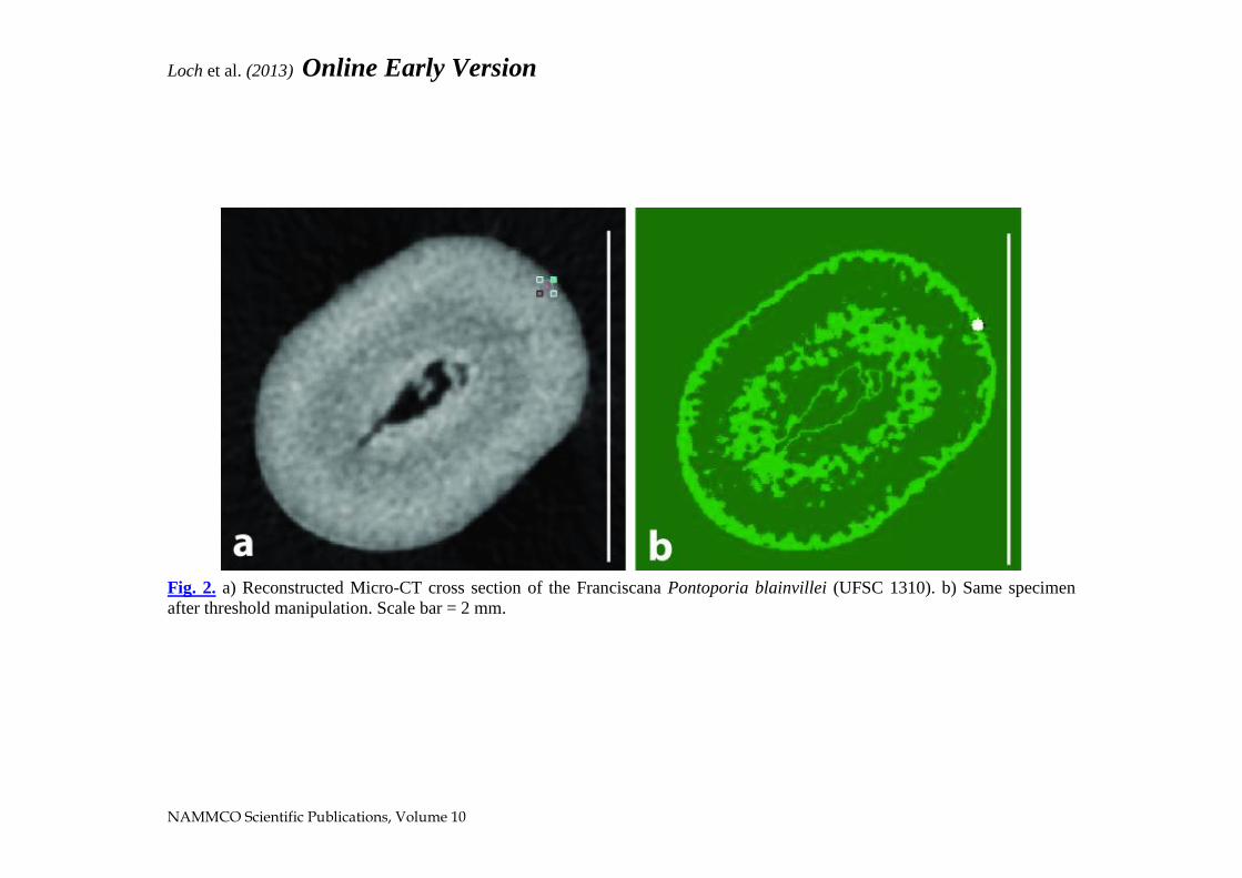
dentinal growth layers. Some of the specimens scanned showed a slight
variation in greyscale in the dentinal area, enhanced by manipulation of
thresholds using imaging software (Fig. 2). However the difference in
greyscale could not be related to GLGs with confidence.
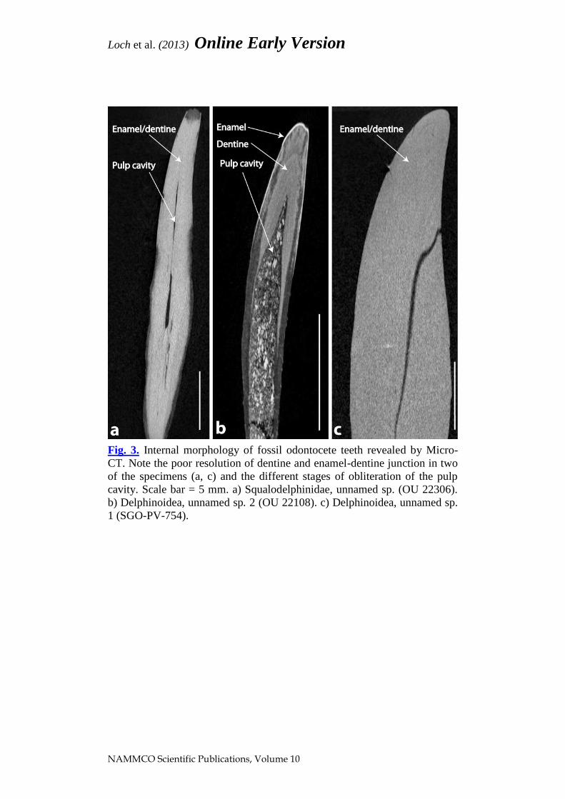
For the fossil cetaceans, mixed results were obtained. Some specimens
showed fine resolution, resolving the enamel, internal layers of dentine and
the pulp cavity (Fig. 3b), but other specimens revealed a poor resolution of
dentine, and in some cases the enamel-dentine junction was not visible (Fig.
3a and c). For the latter specimens, enamel and dentine had the same x-ray
contrast and were indistinguishable. Micro-CT also allowed the

Loch et al. (2013) Online Early Version
NAMMCO Scientific Publications, Volume 10
Fig. 1. Internal morphology of extant odontocete teeth revealed by Micro-CT. Note the resolution of enamel, dentine, cementum and the
different stages of obliteration of the pulp cavity, but no clear growth layer groups. Scale bar = 10 mm. a) Pilot whale Globicephala sp.
(REF 6.5.76.1). b) Amazon river dolphin Inia geoffrensis (IEPA 1899). c) Franciscana Pontoporia blainvillei (UFSC 1310).
Loch et al. (2013) Online Early Version
NAMMCO Scientific Publications, Volume 10
Fig. 2. a) Reconstructed Micro-CT cross section of the Franciscana Pontoporia blainvillei (UFSC 1310). b) Same specimen
after threshold manipulation. Scale bar = 2 mm.
Loch et al. (2013) Online Early Version
NAMMCO Scientific Publications, Volume 10
Fig. 3. Internal morphology of fossil odontocete teeth revealed by Micro-
CT. Note the poor resolution of dentine and enamel-dentine junction in two
of the specimens (a, c) and the different stages of obliteration of the pulp
cavity. Scale bar = 5 mm. a) Squalodelphinidae, unnamed sp. (OU 22306).
b) Delphinoidea, unnamed sp. 2 (OU 22108). c) Delphinoidea, unnamed sp.
1 (SGO-PV-754).

Loch et al. (2013) Online Early Version
NAMMCO Scientific Publications, Volume 10
visualization of the pulp cavity, which could furnish an indirect proxy for
ontogenetic development. Sometimes the pulp cavity was also infilled with
sedimentary matrix (Fig. 3b). Cracks provoked by desiccation or even due
to burial-related compaction and tectonic distortion were also apparent (Fig.
3c).
Reconstructed orthogonal Micro-CT images of the unnamed Delphinoidea
(OU 22108) showed a fine resolution of the enamel-dentine junction,
together with definition of internal layers in dentine (Fig. 4a). By changing
the appearance of the image using Lookup tables from ImageJ, it was
possible to resolve some details of these features (Fig. 4b). Lookup tables
attribute colors to the different greyscale values obtained, thus changing the
appearance but not the pixel values of the image. The neonatal line was
visible below (internal to) the enamel, followed by two growth layers of
dentine in the crown region. The pulp cavity was open and infilled with
sediment, which suggests that the dolphin was a young animal.
Pathology and wear
Apart from revealing the morphology and inner structure of dental tissues,
pathological processes and tooth wear were also evidenced in Micro-CT
slices. Cross-sections and orthogonal slices of reconstructed Micro-CT
images provided internal details that allowed the diagnosis of alterations
observed at the enamel surface. These include a potential case of a caries-
like lesion (sensu Loch et al. 2011) in the Amazon river dolphin (Inia
geoffrensis) (Fig. 5a) and wear and loss of enamel due to desiccation in the
bottlenose dolphin (Tursiops truncatus) (Fig. 5b). Potentially, Micro-CT
could also help reveal and characterize internal anomalies that could
interfere in age estimation techniques, such as resorption of dentine and
cement, and pulp stones.
Variation in greyscales
The assessment of the variation in greyscale values in Micro-CT cross-
sections permitted a qualitative approach to the differences in mineral
density among dental tissues and across tooth regions (Fig. 6). Greyscale
values were measured across distance profiles using 16 bit images, in which
the maximum grey value is 65,535 in contrast with the 255 grey values
possible with 8 bit images. Variations in greyscale were more evident in the
transition from enamel to dentine and in the transition from dentine to the
pulp cavity. In these two interfaces there was an abrupt change in grey
values. In cases where the pulp cavity was obliterated, no change was
evident in the grey values. Qualitative analysis of dentine and enamel
through Micro-CT showed that enamel has higher greyscale values than
dentine, reaching about 60,000 grey values, and that dentine averages about
40,000 or slightly less. For enamel, greyscale values seemed to be lower at
the outer and inner enamel reaching its maximum at mid enamel (Fig. 6a

Loch et al. (2013) Online Early Version
NAMMCO Scientific Publications, Volume 10
Fig. 4. Internal structure of fossil unnamed Delphinoidea tooth (OU 22108)
finely resolved by Micro-CT. Scale bar = 5 mm. a) Orthogonal view of the
specimen. b) Same specimen after colorizing treatment to enhance features.

Loch et al. (2013) Online Early Version
NAMMCO Scientific Publications, Volume 10
Fig. 5. Micro-CT cross-sections of odontocete teeth evidencing pathological conditions and abnormalities. Scale bar = 10 mm.
a) Caries-like lesion in the Amazon river dolphin Inia geoffrensis (IEPA 1899). b) Worn desiccated enamel of the bottlenose
dolphin Tursiops truncatus (UFSC 1349).

Loch et al. (2013) Online Early Version
NAMMCO Scientific Publications, Volume 10
Fig. 6. Reconstructed Micro-CT cross-sections of extant odontocete teeth
and greyscale value/distance profile. a) Striped dolphin Stenella
coeruleoalba (UFSC 1344). b) Amazon river dolphin Inia geoffrensis (IEPA
1899). c) Franciscana Pontoporia blainvillei (UFSC 1310).

Loch et al. (2013) Online Early Version
NAMMCO Scientific Publications, Volume 10
and c). However, the Amazon river dolphin (Fig. 6b) showed a different
trend, with greyscale values peaking at the extremities and decreasing
slightly at mid-enamel. Variation in greyscale values in dentine were
relatively homogeneous, with small peaks followed by small drops in
greyscale values.
For most of the fossil cetaceans (Fig. 7), the variation in greyscale values
was not consistent with the trends observed for extant odontocetes. Apart
from the abrupt change in greyscale observed in the pulp cavity area, no
other consistent or reliable trend could be identified. Greyscale values for
enamel were lower or similar to dentine values, not allowing the
characterization of the enamel-dentine junction. Grey values on dentine
were also relatively homogenous and followed the trend of small peaks
followed by abrupt drops as seen in extant odontocetes.
Quantitative assessment
Mineral concentration and density
To provide a comparative analysis of mineral concentrations and densities
in different regions of the tooth, greyscale measurements were taken in
enamel (outer, mid and inner region), dentine (outer — near the enamel-
dentine junction and inner — near the pulp) and cementum. Two extant
cetaceans were analyzed, the delphinoid Guiana dolphin (Sotalia
guianensis) and the inioid franciscana. Data similarly obtained from human
dental tissues were also included in the comparison. These data were
included because they were readily available and they could give breadth to
our interpretation, as dental studies in humans are much more common than
studies in other mammals. Greyscales were measured with a region of
interest (ROI) of 20X20 pixels for the human specimen and for the Guiana
dolphin. Due to the small tooth size of the franciscana, a ROI of 10X10
pixels was used. Greyscale measurements done with 20X20 and 10X10
ROIs for the Guiana dolphin showed that values obtained from different-
sized regions were consistent and statistically similar (Mann-Whitney test
for two independent samples, p > 0.05). Thus greyscale values measured for
the franciscana were comparable with datasets produced for the Guiana
dolphin and for human dental samples.
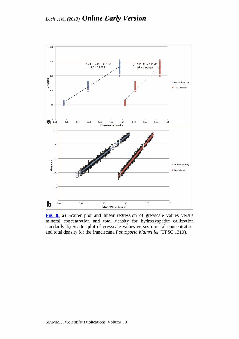
Mean greyscale values obtained from three hydroxyapatite calibration
standards showed a linear relationship with both mineral concentration and
total density values (Fig. 8a). By using the linear regression equations
obtained from the standards, it was possible to estimate the mineral
concentration and total density of dolphin and human enamel and dentine
based on their greyscale readings (Fig. 8b). Values for mineral
concentration and total density in different regions of the tooth in dolphins
and humans are summarized in Table 2.

Loch et al. (2013) Online Early Version
NAMMCO Scientific Publications, Volume 10
Fig. 7. Reconstructed Micro-CT cross-sections of fossil odontocete teeth
and greyscale value/distance profile. Note the preservation artifacts
(irregular dark lines) in b and c. a) Unnamed Squalodontidae sp.1 (OU
22457). b) Unnamed Delphinoidea sp.1 (SGO-PV-754). c) Unnamed
Squalodontidae sp.2 (OU22257).
Loch et al. (2013) Online Early Version
NAMMCO Scientific Publications, Volume 10
Fig. 8. a) Scatter plot and linear regression of greyscale values versus
mineral concentration and total density for hydroxyapatite calibration
standards. b) Scatter plot of greyscale values versus mineral concentration
and total density for the franciscana Pontoporia blainvillei (UFSC 1310).

Loch et al. (2013) Online Early Version
NAMMCO Scientific Publications, Volume 10
Table 2. Summary of average mineral concentration and total effective
density (in g/cm3) ± SE in human and odontocete dental samples. Maximum
and minimum values observed are in parenthesis.
Average Mineral
Concentration
Average Total Effective
Density
Human
Enamel Outer 2.50 +/- 0.07 (2.27-2.72) 2.54 +/- 0.04 (2.42-2.66)
Enamel Mid 2.59 +/- 0.06 (2.39-2.70) 2.60 +/- 0.04 (2.48-2.66)
Enamel Inner 2.59 +/- 0.09 (2.39-2.91) 2.59 +/- 0.09 (2.48-2.78)
Dentine Outer 1.51 +/- 0.09 (1.04-1.76) 1.96 +/- 0.08 (1.09-2.10)
Dentine Inner 1.53 +/- 0.08 (1.30-1.74) 1.97 +/- 0.05 (1.84-2.10)
Cementum 1.14 +/- 0.10 (0.86-1.44) 1.85 +/- 0.06 (1.68-2.02)
Guiana dolphin
Enamel Outer 2.62 +/- 0.12 (2.33-2.80) 2.81 +/- 0.08 (2.62-2.93)
Enamel Mid 2.59 +/- 0.13 (2.13-2.80) 2.79 +/- 0.09 (2.49-2.83)
Enamel Inner 2.56 +/- 0.16 (1.44-2.79) 2.77 +/- 0.10 (2.04-2.92)
Dentine Outer 1.42 +/- 0.08 (1.24-1.84) 2.03 +/- 0.05 (1.91-2.30)
Dentine Inner 1.42 +/- 0.06 (1.28-1.63) 2.03 +/- 0.04 (1.94-2.17)
Cementum 1.25 +/- 0.04 (1.14-1.36) 1.92 +/- 0.03 (1.85-1.99)
Franciscana
Enamel Outer 1.37 +/- 0.19 (0.88-1.58) 1.99 +/- 0.12 (1.67-2.13)
Enamel Mid 1.37 +/- 0.19 (0.88-1.60) 1.99 +/- 0.12 (1.67-2.14)
Enamel Inner 1.37 +/- 0.19 (0.86-1.58) 1.99 +/- 0.12 (1.66-2.13)
Dentine Outer 0.92 +/- 0.08 (0.73-1.12) 1.70 +/- 0.05 (1.57-1.83)
Dentine Inner 0.93 +/- 0.13 (0.54-1.16) 1.71 +/- 0.09 (1.46-1.86)
Cementum 0.64 +/- 0.09 (0.39-0.92) 1.52 +/- 0.06 (1.36-1.70)

Loch et al. (2013) Online Early Version
NAMMCO Scientific Publications, Volume 10
Using the non-parametric Kruskal-Wallis test for multiple independent
samples, it was shown that differences in mean values of mineral
concentration and total effective density were statistically significant among
the three species (p < 0.05). The franciscana had the lowest mineral
concentration and total effective density for all locations sampled, being
considerably lower than the Guiana dolphin. For enamel, the highest mean
values of both mineral concentration and total effective density were
observed in the outer enamel layer of the Guiana dolphin, which were
statistically different from human values (p= 0.000). The mineral
concentration in the mid and inner enamel regions were similar between
human and Guiana dolphin samples (non-parametric Mann-Whitney test for
two independent samples; p= 0.24 and p= 0.34, respectively). Dentine
mineral concentration values were higher for human samples, but total
densities were higher in the Guiana dolphin. For cementum, the Guiana
dolphin had the highest mineral concentration and total density of all
samples analyzed.
DISCUSSION
Early work involving Micro-CT and dental morphology of fossil and living
vertebrates has demonstrated this is a useful and versatile technique both in
terms of range of taxa studied and diversity of applications (e.g. McErlain et
al. 2004, Rossi et al. 2004, Plotino et al. 2006, Kim et al. 2007, Swain and
Xue 2009, Abel et al. 2012, Davis 2012). The specimen preparation and
handling for Micro-CT are quite easy compared to most other study
methods, and most importantly, Micro-CT allows the recording and study of
specimens by means of a non-destructive advanced analytical technique
(Rossi et al. 2004, Schwass et al. 2009). In addition, internal features can be
re-examined many times, as samples remain available after scanning (Swain
and Xue 2009).
Micro-CT analysis has been shown to accurately reproduce details of tooth
anatomy (Kim et al. 2007), having high correlation with histological data
(Plotino et al. 2006). Micro-CT can also allow basic linear and volumetric
measurements, being applicable to measurements of enamel thickness, in
turn a basic procedure in the understanding of tooth biomechanics and
function (Kim et al. 2007, Swain and Xue 2009). For the extant cetaceans
analyzed here, Micro-CT sections resolved internal details of tooth
morphology, including definition of enamel, dentine, cementum and the
pulp cavity. These regions were resolved both through reconstructed
sections and also by the assessment of the variation in greyscale levels.

Loch et al. (2013) Online Early Version
NAMMCO Scientific Publications, Volume 10
The dentinal region is known to be formed by layers of dentine deposited
with controlled periodicity, which are visible in histological sections after
decalcification (Lockyer 1995). These layers (growth layer groups, GLGs),
however, were not clearly visible in reconstructed Micro-CT sections in
extant odontocetes. The reconstruction settings used allowed images with a
pixel size resolution of 8.6 µm, which is considerably smaller than the
spatial resolution of GLGs reported for some dolphin species (Myrick et al.
1984, Hohn et al. 1989, Pinedo and Hohn 2000). Thus, it is believed that the
water and organic content of dentine, and the fact that we analyzed intact
and whole specimens, may have obscured the definition of the growth
layers, which are commonly evident in thin histological sections. Micro-CT
cross sections and projected greyscale values/distance profiles showed a
relatively uniform dentinal region with no clearly-defined dentinal layers.
Manipulation of greyscale thresholds using imaging software revealed
different zones identified by their greyscale/mineral densities. Nonetheless,
further developments are necessary to show if and how this zonation is
related to GLGs and if Micro-CT can be applied as a complementary
method for age estimation.
For most paleontological and archaeological dental specimens, Micro-CT is
the only non-destructive and non-invasive investigative method currently
available (Rossi et al. 2004). It allows ‘digital preparation’ of fossil
specimens, revealing material otherwise covered by layers of coating
material or matrix, and facilitates visualization of hidden structures and
observation of fine morphological variation (Rossi et al. 2004, Abel et al.
2012, Davis 2012). However, in heavily mineralized specimens (e.g.
specimens secondarily mineralized or with natural high mineral density),
Micro-CT may have clear limitations for reliably distinguishing dental
tissues (Swain and Xue 2009). This was the case in some of the fossil
specimens analyzed in this study, in which enamel and dentine were not
distinguishable through Micro-CT images. When two materials have similar
densities, the voxels that represent both tissues will have comparable grey
values due to insufficient density contrast between them (Abel et al. 2012).
Because of these issues, we had to adopt different equipment settings for
fossils in comparison to extant specimens (80 kV vs. 100 kV). A slight
decrease in equipment voltage was used to compensate the higher mineral
content of fossil specimens, while still producing images with good
resolution.
These fossil specimens could have been affected by the dissolution
processes that would be expected in geological settings, with significant
mobility of anions such as carbonate and phosphate, resulting in increased
secondary mineral content in the dental tissues. Such changes are part of
what is known as diagenesis, a postmortem early-burial process which

Loch et al. (2013) Online Early Version
NAMMCO Scientific Publications, Volume 10
results in a range of chemical and mineral changes to the organic and
inorganic constituents of fossil bone and teeth. Diagenetic processes can
include trace element enrichment in the sample, dissolution or precipitation
of minerals, and recrystallization of the original biological apatite, resulting
in modifications to fossil specimens from different sites and ages
(Schweitzer et al. 2008, Thomas et al. 2011). Diagenesis may also cause
increase in crystallite size and crystallinity in the samples, a possible
explanation for similar greyscale values for fossil enamel and dentine.
However, even for the specimens in which dental tissues could not be
reliably distinguished, Micro-CT revealed the internal morphology of the
pulp cavity and the presence of desiccation cracks and other post-mortem
alterations. Micro-CT images provide an important record of the internal
structure of fossil specimens before other destructive analyses are
performed, and may reveal diagenetic alteration that could interfere with
results from further geochemical analysis and detailed ultrastructural studies
(e.g. Schweitzer et al. 2008, Thomas et al. 2011). Chemical and mineral
changes caused by diagenesis may compromise the use of Micro-CT to
reveal the mineral density of dental tissues in fossils. For these specimens,
qualitative analyses offer more scope than quantitative approaches.
The unnamed delphinoid (OU 22108) was the only fossil analyzed here that
showed a clear definition of tooth regions, together with a good resolution
of the dentinal region. In this specimen the neonatal line and two additional
growth layers were observed. Of note, the neonatal line is the baseline for
age determination and the line that represents the beginning of life as an
individual (Lockyer 1995). In fossil tooth OU 22108, fossilization seemed
to have enhanced the layers to allow finer resolution by Micro-CT.
Fossilization commonly involves the dissolution and mobilization of
minerals in surrounding sediments, leading to re-deposition of minerals in
pore spaces within the fossil, as the saturated water and endogenous organic
constituents are concomitantly removed. Fossilization can also involve
actual replacement of original molecules of tooth minerals (Schweitzer et al.
2008). For the delphinoid OU 22108, the removal of the organic and water
fraction and substitution by other minerals seemed to have improved the
resolution of dentinal layers. Conversely, it is not apparent why only OU
22108 had a clear resolution of its dentinal layers, while 5 other fossils were
unrevealing, presumably because of diagenetic alteration. One of the latter
specimens, OU 22257, is from the same locality and sequence as OU 22108,
and would have had a similar burial history. It is known, however, that
fossils from the same strata may differ significantly in their degree of
secondary mineralization; for example, some teeth from the locality for OU
22108 are cemented with secondary calcium carbonate, while others are free
of adherent secondary minerals.

Loch et al. (2013) Online Early Version
NAMMCO Scientific Publications, Volume 10
Micro-CT slices also allowed the observation and internal characterization
of pathological lesions and processes of dental wear and post-mortem
enamel desiccation. Similar alterations were also observed and characterized
by McErlain et al. 2004, Rossi et al. 2004 and Schwass et al. 2009,
involving fossils, archaeological and extant specimens. In these studies,
Micro-CT allowed a complementary approach to the characterization of
dental caries and demineralization of dental tissues in contemporary humans
(e.g. Schwass et al. 2009), and also allowed the identification of similar
lesions in fossil and archaeological specimens (McErlain et al. 2004 and
Rossi et al. 2004), in which the study by destructive methods would be
undesirable due to the rarity of the samples.
Traditional methods for estimating the mineral concentration of dental
tissues involve destructive and time-consuming chemical analysis (Swain
and Xue 2009). Micro-CT analyses of teeth scanned together with resin-
hydroxyapatite calibration standards of known densities allowed the
quantification of mineral density of dental tissues in odontocetes without
having to section and destroy specimens. Average mineral concentrations
and effective densities were estimated for the Guiana dolphin and
franciscana, providing the first report of these values for cetaceans. The
overall low standard deviation observed in our results both for mineral
concentrations and effective densities suggest that although the regions of
interest were relatively small due to the small of size of teeth (for the
franciscana in particular), the results reliably indicate mean values for
dental tissues in these two species of dolphins.
The franciscana, with its elongated rostrum and slender, needle-like and
increased number of teeth (Flower 1867), is most likely capable of
performing a fast snap action but possibly not very powerful bite. This
species had the lowest values for mineral concentration and effective
density of all specimens sampled. Loch et al. (2013a) reported that the
franciscana also had the lowest values for mechanical properties among
several species of extant delphinoids and inioids, suggesting teeth have an
undemanding role in food processing. Although not phylogenetically
related, the dental samples of the Guiana dolphin and human had generally
similar values for mineral concentration. In the outer enamel layer, however,
the mean values for mineral density and total density were higher in the
Guiana dolphin than in the human sample. A prominent layer of prismless
enamel in the outer layer of dolphin enamel in contrast with humans (Loch
et al. 2013b) could explain why the Guiana dolphin had higher values of
mineral and total density. Higher values of total effective density in the
enamel of the Guiana dolphin suggest higher protein content in this species.
The mineral densities of dentine were higher in the human sample than in
the Guiana dolphin. Dentine, however, is deposited throughout the life of

Loch et al. (2013) Online Early Version
NAMMCO Scientific Publications, Volume 10
the tooth and can be deposited as secondary or tertiary dentine. Ontogenetic
development can certainly interfere with the mineral concentration and
density of dentine, making comparisons among species and sampling
locations not as reliable as in enamel. Future studies should determine if
there are prominent density changes across dentine, and if these changes
somehow are related to the GLG.
Apart from the obvious benefits of using Micro-CT for dental studies in
odontocetes and other mammals, this technique has some disadvantages.
The scanning time can be relatively long for bigger specimens scanned in
high resolution (about 5 hours for the specimens in this study), followed by
a similar amount of time for reconstruction. The time and high computer
power and expertise required are one of the main constraints in using Micro-
CT for routine analysis (Plotino et al. 2006). Equipment cost is reasonably
high (around US$225,000 to $375,000 for a desktop model), which favors
multidisciplinary research centers that use Micro-CT for both applied
studies in humans and basic research in other mammals. The main factors
that still keep Micro-CT away from mainstream research are affordability,
productivity and computing power. However, once the equipment is
purchased, the running and maintenance costs are quite low, allowing
research centers to charge a reasonably low hourly rate for its users (Abel et
al. 2012). Other limitations include physical dimensions of the equipment.
For the system used in this study (Skyscan 1172), the chamber allowed
objects with a maximum height of 70 mm and width of 55 mm, which could
be a limitation in studying cetaceans with bigger teeth such as sperm
whales, killer whales and some ziphiids.
Qualitative and quantitative exploratory analyses employed here showed
that Micro-CT is a feasible and useful complementary technique in dental
research in cetaceans. The report of strengths and weaknesses, above, may
help others who might consider using such methods. However, the use of
Micro-CT still needs refinement to allow resolution of internal structure and
potential application in non-destructive aging techniques. Factors such as
use of filters, voxel resolution and algorithm choice by the reconstruction
software, need to be further explored to improve resolution of the images
(Park et al. 2010). Similarly, advanced features of imaging software need to
be extensively tested to elucidate greyscale values and their relation to
GLGs. Paired studies should be run, to compare Micro-CT scans with the
physical and chemical structure revealed by study of destructive sections of
the previously-scanned teeth. Meanwhile, Micro-CT analyses have been
proven to be a potentially useful tool to document internal morphology of
unique fossil teeth and/or unique specimens that are either difficult to
section or must remain intact, before they are subject to other biological and
mechanical analyses which would affect the specimen integrity.

Loch et al. (2013) Online Early Version
NAMMCO Scientific Publications, Volume 10
ACKNOWLEDGMENTS
Thanks are extended to Christina Lockyer and Aleta Hohn for the invitation
to present our research at the Age Determination in Marine Mammals with
Emphasis in monodontids workshop (2011, Tampa FL). Simon Bober,
Michael V. Swain and an anonymous reviewer provided helpful suggestions
which improved this manuscript. Thanks also to Andrew McNaughton
(OCCM, University of Otago) for his technical assistance with the Micro-
CT equipment. C. Loch acknowledges the University of Otago for a PhD
scholarship. Fossils were collected with support from National Geographic
Society grant 4341-90 to R.E. Fordyce.
REFERENCES
Abel RL, Laurini CR and Richter M (2012) A palaeobiologist’s guide to
‘virtual’micro-CT preparation. Palaeontologia Electronica 15(2):6T.
Bergqvist LP (2003) The role of teeth in mammal history. Brazilian Journal
of Oral Sciences 2(6):249–257.
Clementino-Luedemann TNR and Kunzelmann KH (2006) Mineral
concentration of natural human teeth by a commercial micro-CT.
Dental Materials Journal 25(1):113–119. doi: http://dx.doi.org/
10.4012/dmj.25.113
Davis BM (2012) Micro-computed tomography reveals a diversity of
Peramuran mammals from the Purbeck Group (Berriasian) of
England. Palaeontology 55(4):789–817. doi: http://dx.doi.org/
10.1111/j.1475-4983.2012.01161.x
Flower WH (1867) Description of the skeleton of Inia geoffrensis and of the
skull of Pontoporia blainvillii, with remarks on the systematic
position of these animals in the order Cetacea. Transactions of the
Zoological Society of London 6(3):87–116. doi: http://dx.doi.org/
10.1111/j.1096-3642.1867.tb00572.x
Flower WH (1885) An Introduction to the Osteology of the Mammalia.
Macmillan, London, 382 pp.
Fordyce RE (2009) Cetacean fossil record. In: Perrin WF, Würsig B,
Thewissen JGM (eds.) Encyclopedia of Marine Mammals. Academic
Press, San Diego, 207-215.
Hillson S (2005) Teeth: Cambridge manuals in archaeology. Cambridge
University Press, Cambridge, 373 pp.
Hohn AA (1980) Analysis of growth layers in the teeth of Tursiops
truncatus using light microscopy, microradiography, and SEM. Report
of the International Whaling Commission 3: 155-160.
Hohn AA, Scott MD, Wells RS, Sweeney JC and Irvine AB (1989) Growth
layers in teeth from known-age, free-ranging bottlenose dolphins.

Loch et al. (2013) Online Early Version
NAMMCO Scientific Publications, Volume 10
Marine Mammal Science 5(4): 315-342. Doi: http://dx.doi.org/
10.1111/j.1748-7692.1989.tb00346.x
Kim I, Paik KS and Lee S-P (2007) Quantitative evaluation of the accuracy
of micro-computed tomography in tooth measurement. Clinical
Anatomy 20(1): 27-34. doi: http://dx.doi.org/10.1002/ca.20265
Loch C, Grando LJ, Kieser JA, Simões-Lopes PC (2011) Dental pathology
in dolphins (Cetacea: Delphinidae) from the southern coast of Brazil.
Diseases of Aquatic Organisms 94: 225-234. doi: http://dx.doi.org
/10.3354/dao02339
Loch C, Swain MV, Jansen van Vuuren L, Kieser JA, Fordyce RE (2013a)
Mechanical properties of dental tissues in dolphins (Cetacea:
Delphinoidea and Inioidea). Archives of Oral Biology 58(7): 773-779.
doi: http://dx.doi.org/10.1016/j.archoralbio.2012.12.003
Loch C, Duncan W, Simões-Lopes PC, Kieser JA, Fordyce RE (2013b)
Ultrastructure of enamel and dentine in extant dolphins (Cetacea:
Delphinoidea and Inioidea). Zoomorphology 132(2): 215-225. doi:
http://dx.doi.org/10.1007/s00435-012-0180-1
Lockyer C (1995) A review of factors involved in zonation in odontocete
teeth, and an investigation of the likely impact of environmental
factors and major life events on harbour porpoise tooth structure.
Reports of the International Whaling Commission, Special issue 16:
511-530.
Luque PL, Pierce GJ, Learmonth JA, Santos MB, López A, Reid RJ, Rogan
E, González AF, Boon J, Law RJ and Lockyer CH (2009) Dentinal
anomalies in teeth of Harbour porpoises (Phocoena phocoena) from
Scottish waters: are they linked to sexual maturation and
environmental events? Journal of the Marine Biological Association
of the United Kingdom 89(5): 893-902. doi: http://dx.doi.org/
10.1017/S0025315409001866
McErlain DD, Chhem RK, Bohay RN and Holdsworth DW (2004) Micro-
computed tomography of a 500-year-old tooth: technical note. Journal
of the Canadian Association of Radiologists 55(4): 242-245.
Myrick Jr AC (1991) Some new and potential uses of dental layers in
studying delphinid populations. In: Pryor K and Norris KS (eds.)
Dolphin Societies: discoveries and puzzles. University of California
Press, Los Angeles, 251-279.
Myrick Jr AC, Shallenberger EW, Kang I, and MacKay DB (1984)
Calibration of dental layers in seven captive Hawaiian spinner
dolphins, Stenella longirostris, based on tetracycline labeling. Fishery
Bulletin 82(1): 207-225.
Park C, Swain M and Duncan W (2010) Micro-computerised tomography
optimisation for the measurement of bone mineral density around
titanium dental implants. Journal of Biomechanical Science and
Engineering 5(1): 2-10. http://dx.doi.org/10.1299/jbse.5.2

Loch et al. (2013) Online Early Version
NAMMCO Scientific Publications, Volume 10
Pinedo MC and Hohn AA (2000) Growth layer patterns in teeth from the
franciscana, Pontoporia blainvillei: developing a model for precision
in age estimation. Marine Mammal Science 16(1): 1-27. doi:
http://dx.doi.org/10.1111/j.1748-7692.2000.tb00901.x
Plotino G, Grande NM, Pecci R, Bedini R, Pameijer CH and Somma F
(2006) Three-dimensional imaging using microcomputed tomography
for studying tooth macromorphology. Journal American Dental
Association 137(11): 1555-1561.
Rossi M, Casali F, Romani D, Bondioli L, Macchiarelli R and Rook L
(2004) MicroCT Scan in paleobiology: application to the study of
dental tissues. Nuclear Instruments and Methods in Physics Research
Section B: Beam Interactions with Materials and Atoms 213: 747-750.
doi: http://dx.doi.org/10.1016/S0168-583X(03)01697-5
Schwass DR, Swain MV, Purton DG and Leichter JW (2009) A system of
calibrating Microtomography for use in caries research. Caries
Research 43(4): 314-321. doi: http://dx.doi.org/10.1159/000226230
Schweitzer MH, Avci R, Collier T and Goodwin MB (2008) Microscopic,
chemical and molecular methods for examining fossil preservation.
Comptes Rendus Palevol 7(2): 159-184. doi: http://dx.doi.org
/10.1016/j.crpv.2008.02.005
Swain MV and Xue J (2009) State of the art of Micro-CT applications in
dental research. International Journal of Oral Science 1(4): 177-188.
doi: http://dx.doi.org/10.4248/IJOS09031
Thomas DB, McGoverin CM, Fordyce RE, Frew RD and Gordon KC
(2011) Raman spectroscopy of fossil bioapatite - A proxy for
diagenetic alteration of the oxygen isotope composition.
Palaeogeography, Palaeoclimatology, Palaeoecology 310(1): 62-70.
doi: http://dx.doi.org/10.1016/j.palaeo.2011.06.016
Ungar P (2010) Mammal teeth: origin, evolution and diversity. The Johns
Hopkins University Press, Baltimore, 304 pp.