Forensic entomology: Applications and limitations
16
1 23 Forensic Science, Medicine, and Pathology ISSN 1547-769X Volume 7 Number 4 Forensic Sci Med Pathol (2011) 7:379-392 DOI 10.1007/s12024-010-9209-2 Forensic entomology: applications and limitations J. Amendt, C. S. Richards, C. P. Campobasso, R. Zehner & M. J. R. Hall
-
Upload
independent -
Category
Documents
-
view
2 -
download
0
Transcript of Forensic entomology: Applications and limitations

1 23
Forensic Science, Medicine, andPathology ISSN 1547-769XVolume 7Number 4 Forensic Sci Med Pathol (2011)7:379-392DOI 10.1007/s12024-010-9209-2
Forensic entomology: applications andlimitations
J. Amendt, C. S. Richards,C. P. Campobasso, R. Zehner &M. J. R. Hall

1 23
Your article is protected by copyright and
all rights are held exclusively by Springer
Science+Business Media, LLC. This e-offprint
is for personal use only and shall not be self-
archived in electronic repositories. If you
wish to self-archive your work, please use the
accepted author’s version for posting to your
own website or your institution’s repository.
You may further deposit the accepted author’s
version on a funder’s repository at a funder’s
request, provided it is not made publicly
available until 12 months after publication.

CONTINUING MEDICAL EDUCATION REVIEW
Forensic entomology: applications and limitations
J. Amendt • C. S. Richards • C. P. Campobasso •
R. Zehner • M. J. R. Hall
Accepted: 29 November 2010 / Published online: 7 January 2011
� Springer Science+Business Media, LLC 2011
Abstract Forensic entomology is the science of collect-
ing and analysing insect evidence to aid in forensic
investigations. Its main application is in the determination
of the minimum time since death in cases of suspicious
death, either by estimating the age of the oldest necroph-
agous insects that developed on the corpse, or by analysing
the insect species composition on the corpse. In addition,
toxicological and molecular examinations of these insects
may help reveal the cause of death or even the identity of a
victim, by associating a larva with its last meal, for
example, in cases where insect evidence is left at a scene
after human remains have been deliberately removed.
Some fly species can develop not only on corpses but on
living bodies too, causing myiasis. Analysis of larvae in
such cases can demonstrate the period of neglect of humans
or animals. Without the appropriate professional collection
of insect evidence, an accurate and convincing presentation
of such evidence in court will be hampered or even
impossible. The present paper describes the principles and
methods of forensic entomology and the optimal tech-
niques for collecting insect evidence.
Keywords Forensic entomology � Post-mortem interval �DNA-analysis � Entomotoxicology � Myiasis � Collection
of entomological evidence
Definition and application
Forensic entomology is the analysis of insect evidence for
forensic and legal purposes [1]. The most important and
most frequently requested task is the estimation of the
minimum time since death [2]. Techniques devised recently
allow experts in the field to collect robust entomological
evidence that can provide vital information in a death
investigation, to answer questions concerning movement or
storage of the remains following death, submersion interval,
time of decapitation and/or dismemberment, identification
of specific sites of trauma, post-mortem artefacts on the
body, use of drugs (entomotoxicology), linking a suspect to
the scene of a crime, sexual molestations and the identifi-
cation of suspects [3]. It is also possible to demonstrate the
period of neglect of living humans and animals by exam-
ining the insects recovered from infested wounds.
Estimating the minimum post-mortem interval
Post-mortem interval (PMI) refers to the time between the
death and discovery of a corpse [2]. There are several
natural processes associated with decomposition, such as
rigor mortis or livor mortis, that can be used to estimate the
PMI [4], but many of these are reciprocal functions and
become inaccurate in application very quickly [5]. Fur-
thermore, they are limited to the first 72 h after death [4].
However, during that 72 h and well beyond, insects can be
a very powerful tool for estimating the minimum time since
J. Amendt (&) � R. Zehner
Institute of Forensic Medicine, Kennedyallee 104,
60596 Frankfurt am Main, Germany
e-mail: [email protected]
C. S. Richards � M. J. R. Hall
Natural History Museum, Cromwell Road,
London SW7 5BD, UK
C. P. Campobasso
Department of Health Sciences (SpeS), University of Molise,
via De Sanctis, snc, 86100 Campobasso, Italy
123
Forensic Sci Med Pathol (2011) 7:379–392
DOI 10.1007/s12024-010-9209-2
Author's personal copy

death. Depending on the level of accessibility and envi-
ronmental conditions, necrophagous insects will promptly
colonize a fresh corpse. Usually the first taxa to arrive on a
body are flies (Diptera), mainly blowflies (Calliphoridae),
which can locate an odor source with great spatial precision
and deposit their eggs on a corpse within minutes–hours of
death. Larvae (often called ‘‘maggots’’) hatch from the eggs
and feed on the underlying tissues. As they grow they shed
their cuticle twice, a process termed ‘‘ecdysis,’’ to enable
further growth. After each ecdysis (moult) a new larval
instar (stage) is formed. When third instar larvae finish
feeding they enter the post-feeding stage, most species
migrate away from the body to find shelter, either within
soil or underneath objects e.g. stones or leaves (outdoor
crime scenes), or furniture (indoor crime scenes). Here, they
form pupae within a protective outer case, the puparium (the
hardened cuticle of the third instar larva), from which adult
flies emerge at the completion of metamorphosis.
Decomposition as a result of insect activity in and on the
corpse is a continuous process that can be measured,
allowing accurate minimum PMI estimates to be made up
to several months after death depending on the circum-
stances [4]. The assumption behind these estimates is that
by calculating the age of developing insects on a body, it is
possible to calculate the time of colonization, which infers
a minimum PMI (PMImin) [2], i.e., the time when insects
first colonized the body, rather than the actual time of death
[1]. Because blowflies are usually the first group to colo-
nize a body, the focus of PMImin estimates is often on them
when using entomological evidence [2].
The rate of development of an insect is mainly governed
by temperature and can differ between even closely related
species [6]. Hence, a three-step process of, (1) accurately
identifying the species found on a corpse, (2) reconstruct-
ing crime scene temperatures, and (3) modelling the rate of
development of the immature insects found on a corpse, is
absolutely essential to enable a forensic entomologist to
calculate the age of a sampled insect.
Identifying entomological evidence
The identification of insects is a highly skilled procedure
and should always be conducted by an expert in insect
taxonomy. Museums and universities are generally the best
equipped organizations to process insect identifications and
should always be a first point of contact. For detailed
identification keys on forensically important insects see
e.g. Zumpt [7], Smith [8] and Szpila [9].
Modelling of crime scene temperatures
To obtain the temperatures of a crime scene while the body
was in situ, a record of ambient temperatures is gathered,
usually from the weather station nearest to the body dis-
covery site [1, 8, 10]. Unfortunately, there can be a sig-
nificant difference between the ambient temperatures of the
weather station and the crime scene, e.g., because they
might be at different altitudes to one another, or experience
different exposure to identical weather conditions [10]. For
this reason, ambient temperature is recorded on site for
several days after the body is discovered, and a regression
relationship is then derived between these temperature and
simultaneously recorded weather station temperatures [1].
This derived equation is then used to correct the weather
station temperature records to ambient site temperature for
the duration that the body was thought to be in situ [10].
Once ambient environmental temperature has been
estimated for the duration that the body was in situ, an
estimate of PMImin can be made using this information
together with reference development data for the particular
species, usually obtained experimentally in the laboratory.
Modelling insect development
Developmental data are simply data pertaining to the
duration of development of immature stages recorded at
different temperatures (usually constant but sometimes
fluctuating), which are then summarized in one or more of
three developmental models, namely isomorphen diagrams
[11], isomegalen diagrams [12] and thermal summation
models [13]. Size (e.g. length or weight) and develop-
mental event (e.g. 1st larval moult/ecdysis or pupariation),
are the only two available measures to calculate the age of
immature insects, and developmental models use one of
these two measures to estimate PMImin.
Isomorphen diagram
The most basic of these models is the isomorphen diagram
which is a simple scatterplot that models the duration
of developmental events (egg hatching, 1st ecdysis, 2nd
ecdysis, onset of wandering, onset of pupariation, adult
eclosion; X-axis) against temperature (Y-axis). Each con-
tour in this model represents one of these developmental
events (Fig. 1). By knowing the stage of development of
the oldest immature insects on the body, and the average
environmental temperature of the crime scene while the
body was in situ, it is possible to calculate an accurate
PMImin from this model, using the min/max error bars to
estimate the confidence window for PMImin.
The advantage of this model is that it is simple to use,
with concepts that are easily explained in court. However,
this simplicity compromises the accuracy of the output i.e.
in cases where environmental temperatures fluctuate, e.g.
outdoor scenarios, the average environmental temperature
380 Forensic Sci Med Pathol (2011) 7:379–392
123
Author's personal copy

of the crime scene used to calculate PMImin compromises
the accuracy of the estimate.
As the isomorphen diagram only models the timing of
developmental events, and shows no gradation between
events, the estimate is only accurate if derived from live
entomological evidence which is reared through to the next
developmental event at a known, constant temperature.
However, entomological evidence is not always collected
alive, and in some cases, live specimens may be killed and
preserved before being analyzed. In these cases the esti-
mated age of insect specimens modelled from an isomor-
phen diagram will always be less than their actual age,
simply because it has to be based on the last recorded
developmental stage (e.g. the moult from 2nd to 3rd instar
larvae for all 3rd instars, no matter how long ago that moult
was), resulting in an underestimate of PMImin.
Isomegalen diagram
The isomegalen diagram is a more sophisticated model that
can estimate PMImin using dead larvae, and has the capa-
bility of accounting for and analyzing fluctuating envi-
ronmental temperature data. The isomegalen diagram is a
3D contour plot that models larval size (length, weight or
width) (Z-axis) as a measure of age, against temperature
(Y-axis), and time (X-axis) (Fig. 2). Larval size is a mea-
sure of age with a higher resolution than developmental
event and shows detail of growth between two develop-
mental events.
This higher resolution input provides a more accurate
estimate of PMImin. The diagram details the full range of
larval sizes (from egg hatching to the onset of pupariation)
where each contour on the graph represents a particular
larval size (Fig. 2). The largest larvae from the body and
crime scene are collected, identified and measured, and
these data are then summarized and modelled, along with
related environmental temperatures, to estimate PMImin.
The process of estimating PMImin from fluctuating envi-
ronmental temperatures, using an isomegalen diagram, is
illustrated in Villet et al. [14].
Despite the fact that this model provides more accurate
PMImin estimates than the isomorphen diagram, it has
several criticisms. Firstly, it is well documented that size is
not always an accurate measure of age, particularly for
blowfly larvae [15]. Larvae are known to shrink in size
before entering the next developmental event i.e. 1st
ecdysis, 2nd ecdysis, and particularly before pupariation
when larvae migrate from the food source [11]. Therefore, it
is possible for larvae of a small size to be older than larvae
of a larger size. Additionally, stunted larval or puparial
forms, smaller than normal, can be commonly observed in
experimental cultures, possibly as a result of the scarcity of
the available food source, competition between larvae at
high densities [4] and slow developing ‘‘laggards’’ [16].
Secondly, this model is limited to larvae only, which only
accounts for approximately half the total duration of
development from egg to adult. The other half being egg
and pupariation. Thirdly, larval size can change signifi-
cantly during preservation, as different preservatives and
the duration of preservation causes shrinkage or expansion
of larvae [17]. This change in size directly influences the
calculation of age and subsequent estimate of PMImin.
Additional models are required to correct for this change in
10
15
20
30
35
0 100 200 300 400 500 600 700 800
Time from hatching (hrs)
Tem
per
atu
re (
˚C) 1st instar
2nd instar
Wandering
Pupariation
Eclosion
25
Fig. 1 Isomorphen diagram of hypothetical development data. The
contours of the graph represent the time taken for developing
immature blowflies to reach a particular developmental event (refer to
key on graph), at a range of constant temperatures. Error barsrepresent 95% confidence intervals for each developmental event.
(Note: ‘‘egg hatching’’ is not included in this graph as it is measured
in hours that are an order of magnitude smaller than those of
‘‘wandering,’’ ‘‘pupariation’’ and ‘‘eclosion.’’)
0 50 100 150 200 250 300 350 40010
12
14
16
18
20
22
24
26
28
30
32
34
Tem
pera
ture
(°C
)
2mm
8mm
12mm
16mm
0 100
Age (time from egg oviposition (hrs))
2mm
8mm
12mm
16mm
Fig. 2 Isomegalen diagram of hypothetical development data. Each
contour represents larval length (Z-axis) in relation to their age
(X-axis) and the temperature experienced during development
(Y-axis). The dimension of the Z axis (length) is not needed visually
to calculate a PMImin, and is therefore, not illustrated. (Note: no errorbars are presented in this diagram and the window for PMImin is
calculated from the standard deviation or min/max data of the
entomological evidence.)
Forensic Sci Med Pathol (2011) 7:379–392 381
123
Author's personal copy
size, which adds another source of error to the PMImin
calculation and reduces the precision of the estimate.
Lastly, it is labor intensive to collect the experimental data
for an isomegalen diagram and the model is not as simple to
build as the isomorphen diagram. This is evident by the
dearth of published studies reporting isomegalen diagrams
of forensically important blowfly species [6, 11, 12, 15].
Thermal summation models
Thermal summation models are the most sophisticated of the
three available models. They have the capability of pro-
cessing both size and developmental event data, as well as
fluctuating environmental temperatures. Because this model
can process more complex data sets, it is less affected by the
limitations imposed on the two iso-diagrams, and is there-
fore, the preferred model to use when estimating PMImin.
There is a recognized linear regression model for ana-
lyzing developmental data [13, 18]. Traditionally, it has
been used to calculate development thresholds for appli-
cation in biological control [19] and has been modified for
forensic entomology. This method is commonly referred to
as the thermal summation model, or accumulated degree–
hours (ADH) model, where the x-intercept and the inverse
of the slope of the linear regression are used to calculate
PMImin. These coefficients are termed thermal summation
constants and form the basic assumptions of linear models
of development. These assumptions are that there is a
positive linear relationship between development rate and
increasing temperature (slope), and that development cea-
ses below a minimum developmental threshold (intercept)
[20]. In these models, development is measured as thermal
time (temperature multiplied by time), hence the phrase
‘degree–hours’, and each developmental stage, e.g. egg,
2nd instar, or pupariation, requires a specific number of
accumulated degree–hours to complete development,
hence the name ‘ADH’ [13]. The principle behind esti-
mating PMImin is to calculate the time taken to reach the
ADH required for the species identified on or around the
body to have developed to the oldest recorded stage col-
lected at the environmental temperatures experienced while
the body was in situ.
A criticism of this model is the high variance in the
upper and lower developmental temperature extremes.
Because these values are the end points of the regression,
and are thus heavily weighted values, they significantly
influence the confidence of the regression coefficients,
which compromises the precision of a PMImin estimate.
Recently, a revised regression model [21] has been used to
model blowfly development [6]. It identifies the points
nearest the extremes of the linear approximation that
deviate significantly from it, which results in the calcula-
tion of more precise thermal summation parameters.
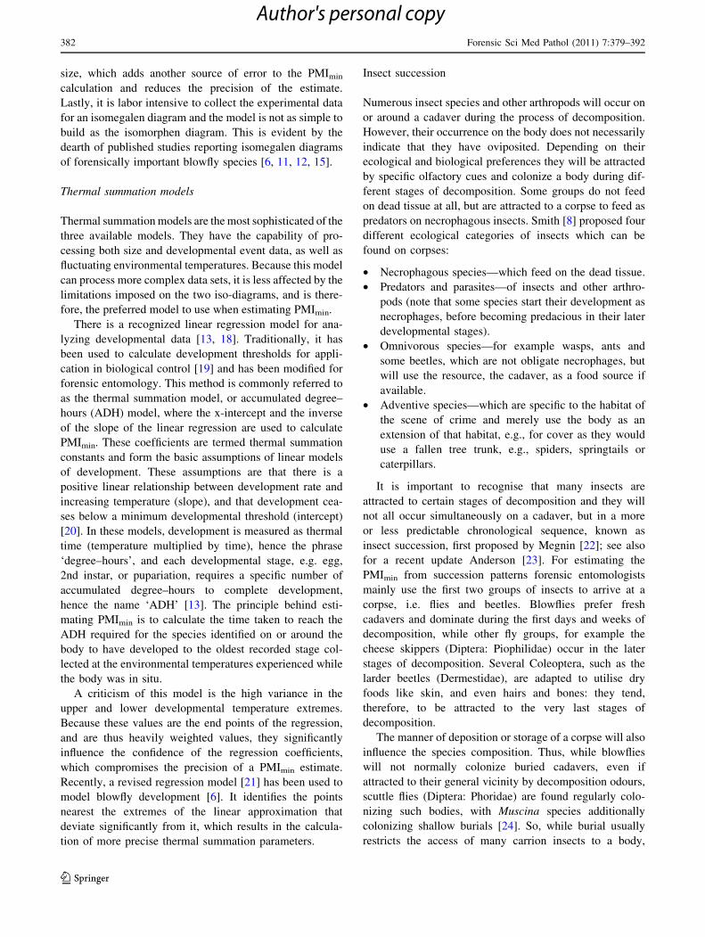
Insect succession
Numerous insect species and other arthropods will occur on
or around a cadaver during the process of decomposition.
However, their occurrence on the body does not necessarily
indicate that they have oviposited. Depending on their
ecological and biological preferences they will be attracted
by specific olfactory cues and colonize a body during dif-
ferent stages of decomposition. Some groups do not feed
on dead tissue at all, but are attracted to a corpse to feed as
predators on necrophagous insects. Smith [8] proposed four
different ecological categories of insects which can be
found on corpses:
• Necrophagous species—which feed on the dead tissue.
• Predators and parasites—of insects and other arthro-
pods (note that some species start their development as
necrophages, before becoming predacious in their later
developmental stages).
• Omnivorous species—for example wasps, ants and
some beetles, which are not obligate necrophages, but
will use the resource, the cadaver, as a food source if
available.
• Adventive species—which are specific to the habitat of
the scene of crime and merely use the body as an
extension of that habitat, e.g., for cover as they would
use a fallen tree trunk, e.g., spiders, springtails or
caterpillars.
It is important to recognise that many insects are
attracted to certain stages of decomposition and they will
not all occur simultaneously on a cadaver, but in a more
or less predictable chronological sequence, known as
insect succession, first proposed by Megnin [22]; see also
for a recent update Anderson [23]. For estimating the
PMImin from succession patterns forensic entomologists
mainly use the first two groups of insects to arrive at a
corpse, i.e. flies and beetles. Blowflies prefer fresh
cadavers and dominate during the first days and weeks of
decomposition, while other fly groups, for example the
cheese skippers (Diptera: Piophilidae) occur in the later
stages of decomposition. Several Coleoptera, such as the
larder beetles (Dermestidae), are adapted to utilise dry
foods like skin, and even hairs and bones: they tend,
therefore, to be attracted to the very last stages of
decomposition.
The manner of deposition or storage of a corpse will also
influence the species composition. Thus, while blowflies
will not normally colonize buried cadavers, even if
attracted to their general vicinity by decomposition odours,
scuttle flies (Diptera: Phoridae) are found regularly colo-
nizing such bodies, with Muscina species additionally
colonizing shallow burials [24]. So, while burial usually
restricts the access of many carrion insects to a body,
382 Forensic Sci Med Pathol (2011) 7:379–392
123
Author's personal copy

others are able to take advantage, such as Megaselia
scalaris (Loew) (Dipt., Phoridae) [25].
Several other parameters like, habitat, or time of year,
influence the composition of the species community of a
corpse. This may help to identify the season of the year
when death occurred, even months and years post-mortem,
or demonstrate the post-mortem transport of a corpse from,
for example, the city into a forest after first colonization.
Hence, succession patterns can provide important insight
into an investigation. However, as the speed of decompo-
sition is influenced by many internal and external param-
eters, the estimated PMImin window can be so large as to be
of limited value, lacking in statistical validation. Never-
theless, succession may be the only tool available to give
some idea of a PMImin, in cases where PMI is longer than
several months or even years. Anderson [23] gives an
excellent overview of the factors which influence insect
succession on carrion. As these factors are diverse (e.g.
wrapping, scavenging, burying, effects of season and sun
exposure) and because each scene of crime is unique, there
is a great need for further research to develop a better
understanding of these processes. To date, most studies
have simply demonstrated that patterns of insect assembly
do exist, and that these patterns change under different
conditions; however, according to VanLaerhoven [26],
‘‘asking if patterns exist in nature is akin to asking if bears
shit in the woods.’’ What we need is a better understanding
of the mechanisms that influence and modify community
assembly [26], otherwise a prediction of PMImin from
insect succession on carrion remains on shaky ground.
Recent studies [e.g. 27–29] have made significant advances
towards improving the forensic evaluation of insect
succession on carrion.
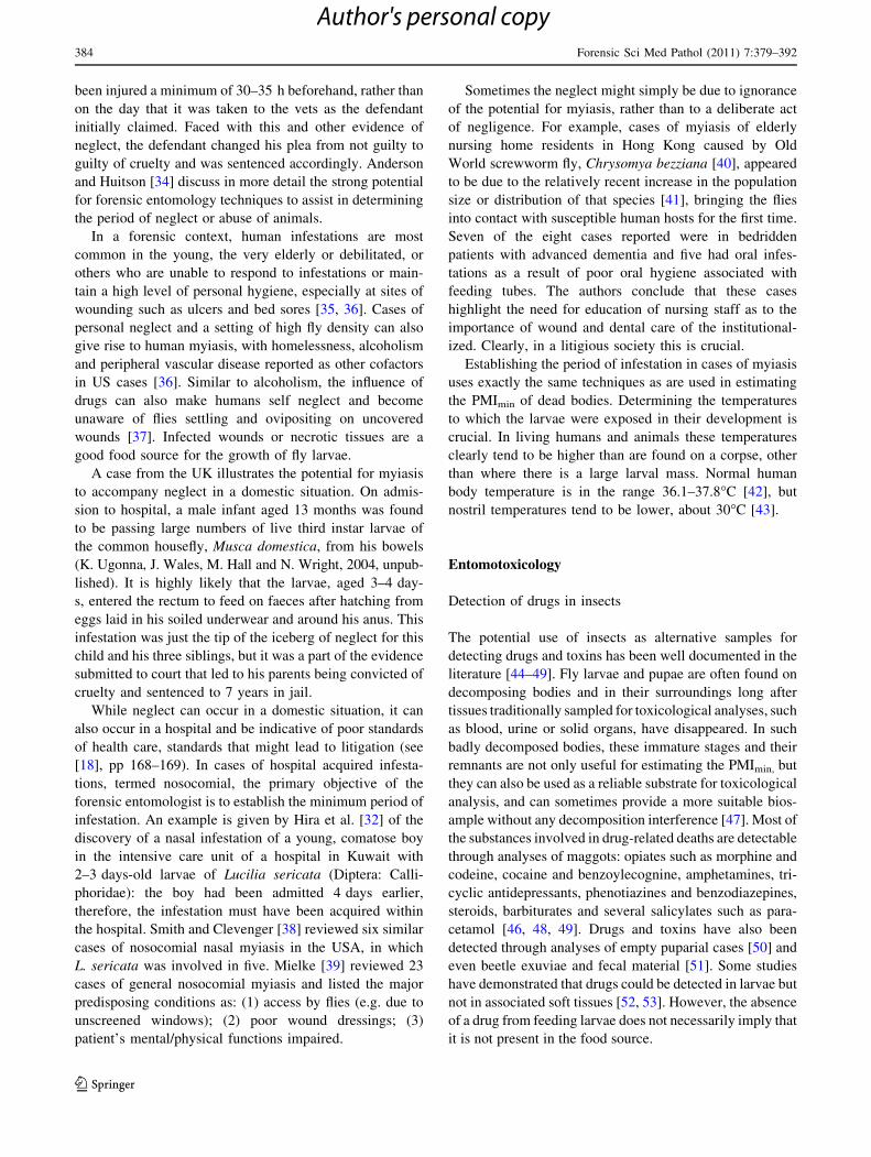
Myiasis
Myiasis has been defined as, ‘‘the infestation of live human
and vertebrate animals with dipterous larvae, which, at
least for a certain period, feed on the host’s dead or living
tissue, liquid body-substances, or ingested food’’ [7]. It can
be a cause of significant economic loses and a major animal
welfare issue for the livestock industry, affect wildlife and
also be a problem in human health, causing significant pain
and suffering and, in extreme cases, death [30, 31] (Fig. 3).
Myiasis can be considered in three main categories,
according to the degree of parasitism of the host:
• Obligate parasites—these can only develop on the living
tissues of living hosts and do not develop on carrion.
• Facultative parasites—these usually develop on carrion
but they can also develop on living hosts, in which
cases they usually require some predisposing condition,
such as neglected wounds with necrotic tissues, or
bacterial growth in soiled fur or fleece.
• Accidental parasites—these can cause minor health
problems but are not normally parasitic, being found in
host tissues by accident, e.g., after ingestion or inhalation.
The most significant group of myiasis-causing flies for
forensic entomology are the facultative parasites, which
can be associated with cases of extreme neglect, both of
humans and animals. The species involved are typically
blowflies (Calliphoridae) or fleshflies (Sarcophagidae),
usually the same species that are involved as the primary
indicators of PMImin. However, other fly families can also
be involved [32, 33].
In veterinary cases, the role of the forensic entomologist
is to determine the period of infestation of livestock or pet
animals in cases of cruelty or neglect. In some countries
farmers can be prosecuted on animal welfare grounds for
having livestock with cases of clearly neglected myiasis. It
can be difficult to produce conclusive evidence of neglect
based solely on clinical signs, but forensic entomology can
provide powerful, scientifically validated evidence of the
period of neglect or abuse to strengthen a prosecution. The
role of forensic entomology in the prosecution of neglect
of domestic animals is illustrated by a case from the UK
(M. Hall, 2007, unpublished). The stage of development of
Lucilia larvae removed by a veterinarian from a severe
wound on the left hind foot of a dog indicated that it had
Fig. 3 Collecting larvae from cases of interdigital myiasis in a Greek
sheep flock, predisposed to myiasis because of bacterial foot-rot.
Close-ups of two cases illustrate the ‘‘head-down’’ feeding behaviour
of larvae. Most larvae are of the fleshfly Wohlfahrtia magnifica(Diptera: Sarcophagidae), but arrowed in the lower-left close-up is a
secondary infesting larva of the blowfly, Chrysomya albiceps(Diptera: Calliphoridae). (Image Martin Hall, copyright Natural
History Museum, London)
Forensic Sci Med Pathol (2011) 7:379–392 383
123
Author's personal copy
been injured a minimum of 30–35 h beforehand, rather than
on the day that it was taken to the vets as the defendant
initially claimed. Faced with this and other evidence of
neglect, the defendant changed his plea from not guilty to
guilty of cruelty and was sentenced accordingly. Anderson
and Huitson [34] discuss in more detail the strong potential
for forensic entomology techniques to assist in determining
the period of neglect or abuse of animals.
In a forensic context, human infestations are most
common in the young, the very elderly or debilitated, or
others who are unable to respond to infestations or main-
tain a high level of personal hygiene, especially at sites of
wounding such as ulcers and bed sores [35, 36]. Cases of
personal neglect and a setting of high fly density can also
give rise to human myiasis, with homelessness, alcoholism
and peripheral vascular disease reported as other cofactors
in US cases [36]. Similar to alcoholism, the influence of
drugs can also make humans self neglect and become
unaware of flies settling and ovipositing on uncovered
wounds [37]. Infected wounds or necrotic tissues are a
good food source for the growth of fly larvae.
A case from the UK illustrates the potential for myiasis
to accompany neglect in a domestic situation. On admis-
sion to hospital, a male infant aged 13 months was found
to be passing large numbers of live third instar larvae of
the common housefly, Musca domestica, from his bowels
(K. Ugonna, J. Wales, M. Hall and N. Wright, 2004, unpub-
lished). It is highly likely that the larvae, aged 3–4 day-
s, entered the rectum to feed on faeces after hatching from
eggs laid in his soiled underwear and around his anus. This
infestation was just the tip of the iceberg of neglect for this
child and his three siblings, but it was a part of the evidence
submitted to court that led to his parents being convicted of
cruelty and sentenced to 7 years in jail.
While neglect can occur in a domestic situation, it can
also occur in a hospital and be indicative of poor standards
of health care, standards that might lead to litigation (see
[18], pp 168–169). In cases of hospital acquired infesta-
tions, termed nosocomial, the primary objective of the
forensic entomologist is to establish the minimum period of
infestation. An example is given by Hira et al. [32] of the
discovery of a nasal infestation of a young, comatose boy
in the intensive care unit of a hospital in Kuwait with
2–3 days-old larvae of Lucilia sericata (Diptera: Calli-
phoridae): the boy had been admitted 4 days earlier,
therefore, the infestation must have been acquired within
the hospital. Smith and Clevenger [38] reviewed six similar
cases of nosocomial nasal myiasis in the USA, in which
L. sericata was involved in five. Mielke [39] reviewed 23
cases of general nosocomial myiasis and listed the major
predisposing conditions as: (1) access by flies (e.g. due to
unscreened windows); (2) poor wound dressings; (3)
patient’s mental/physical functions impaired.
Sometimes the neglect might simply be due to ignorance
of the potential for myiasis, rather than to a deliberate act
of negligence. For example, cases of myiasis of elderly
nursing home residents in Hong Kong caused by Old
World screwworm fly, Chrysomya bezziana [40], appeared
to be due to the relatively recent increase in the population
size or distribution of that species [41], bringing the flies
into contact with susceptible human hosts for the first time.
Seven of the eight cases reported were in bedridden
patients with advanced dementia and five had oral infes-
tations as a result of poor oral hygiene associated with
feeding tubes. The authors conclude that these cases
highlight the need for education of nursing staff as to the
importance of wound and dental care of the institutional-
ized. Clearly, in a litigious society this is crucial.
Establishing the period of infestation in cases of myiasis
uses exactly the same techniques as are used in estimating
the PMImin of dead bodies. Determining the temperatures
to which the larvae were exposed in their development is
crucial. In living humans and animals these temperatures
clearly tend to be higher than are found on a corpse, other
than where there is a large larval mass. Normal human
body temperature is in the range 36.1–37.8�C [42], but
nostril temperatures tend to be lower, about 30�C [43].
Entomotoxicology
Detection of drugs in insects
The potential use of insects as alternative samples for
detecting drugs and toxins has been well documented in the
literature [44–49]. Fly larvae and pupae are often found on
decomposing bodies and in their surroundings long after
tissues traditionally sampled for toxicological analyses, such
as blood, urine or solid organs, have disappeared. In such
badly decomposed bodies, these immature stages and their
remnants are not only useful for estimating the PMImin, but
they can also be used as a reliable substrate for toxicological
analysis, and can sometimes provide a more suitable bios-
ample without any decomposition interference [47]. Most of
the substances involved in drug-related deaths are detectable
through analyses of maggots: opiates such as morphine and
codeine, cocaine and benzoylecognine, amphetamines, tri-
cyclic antidepressants, phenotiazines and benzodiazepines,
steroids, barbiturates and several salicylates such as para-
cetamol [46, 48, 49]. Drugs and toxins have also been
detected through analyses of empty puparial cases [50] and
even beetle exuviae and fecal material [51]. Some studies
have demonstrated that drugs could be detected in larvae but
not in associated soft tissues [52, 53]. However, the absence
of a drug from feeding larvae does not necessarily imply that
it is not present in the food source.
384 Forensic Sci Med Pathol (2011) 7:379–392
123
Author's personal copy

A drug or toxin can be detected in larvae when its rate of
absorption exceeds the rate of metabolism, but it is not yet
known exactly how larvae accumulate or metabolize drugs,
and how these affect larval development. Bourel et al. [54]
demonstrated morphine accumulation inside the cuticle of
Calliphora vicina maggots. In the same area, lying between
the endocuticle and exocuticle of Chrysomya albiceps
larvae, Alves et al. [55] found an intense immunoreaction
positive for cocaine. Based on such findings, Diptera larvae
are certainly useful as qualitative toxicological specimens,
but they are still of limited quantitative value. Several
earlier studies suggested that a correlation between con-
centrations of drugs and toxins in larvae and the tissues on
which the specimens had fed might exist, particularly for
opiates and cocaine [56], while other authors showed no
relevant correlation [47]. In all reports, the concentration of
xenobiotics was significantly lower in the larvae analysed
than in the tissues they used as a food source. Such a
toxicological finding was confirmed by a comparative
study [57], where concentrations in post-feeding larvae
were significantly lower than those in actively feeding
maggots. Several authors [56, 58, 59] have noted that
insects metabolize and eliminate the substances ingested
during their development. However, the wide variations
observed show that the accumulation of a drug by larvae is
unpredictable and quantitative extrapolations are unreliable
[58]. This may result from internal redistribution of the
drug through the body after death, also explaining why
drug concentration varies depending on the tissue and site-
to-site variability in the same organ [53]. Potentially, this
can have major implications on PMImin, even on non-
intoxicated tissues, e.g., Kaneshrajah and Turner [60]
found that the development rate of C. vicina maggots on
pork liver is faster than the rate on other tissues such as
lung, kidney, heart or brain.
Drug induced changes in the development rate of
necrophagous insects can be large enough to significantly
alter PMI estimates, leading to significant errors if over-
looked and not taken into account during a death investi-
gation. Some drugs can delay the colonization by insects
for several days as observed with malathion by Gunatilake
and Goff [61] and with flunitrazepam-ethanol-mix by
Fremdt et al. [62]. Drugs may also influence the develop-
ment of the necrophagous insects reared on contaminated
tissues and, therefore, any modification of growth rate
would bias the PMImin estimates [63]. For example, larvae
of the sarcophagid fleshfly, Boettcherisca peregrina,
developed more rapidly on tissues contaminated with
cocaine and heroin than on uncontaminated tissues [64,
65]. A significantly accelerated growth rate was also
observed by Bourel et al. [66] for Lucilia sericata larvae
feeding on tissues contaminated with morphine and by
O’Brien and Turner [67] for Calliphora vicina larvae
feeding on tissues contaminated with paracetamol. Diaze-
pam also appeared to affect the size and shape of Chry-
somya albiceps puparium [49]. Such studies demonstrate
the risk of calculating an incorrect PMImin due to the drug
modified rate of development of the immature stages [63]
which can vary from a minimum of 18 h up to 96 h
depending on the drug, the fly species, and the stage of
development. Therefore, all reasonable steps must be
undertaken to perform as comprehensive a drug screen as
possible in bodies where there is a suspicion that the death
may be drug-related [52].
It is noteworthy that remnants of insect evidence, such
as fly puparia, may remain at the scene for several years;
these can retain drug residues, detection of which could
help to resolve the history and identity of the dead body
which was the source of food for the larvae that gave rise to
the remnants.
Molecular analysis of insects
Species determination
Crucial for any evidence in forensic entomology is the
association of development data to a certain species [6].
However, species identification on a morphological basis
may be hampered by the lack of taxonomic experts who are
able to differentiate species using morphological charac-
ters, especially in the early larval stages. Within many
genera (e.g. Sarcophaga), discrimination of individual
species is not possible from larval morphology alone.
Rearing to adults would facilitate determination (at least of
male Sarcophaga), but this can be time consuming and
might not be possible due to rearing difficulties or because
the larval evidence is presented dead. Therefore, molecular
techniques as alternative tools for identification of foren-
sically important species have been established within the
last decade [68, 69], based on species specific nucleotide
sequences of certain genes. The gene for subunit I of the
mitochondrial encoded cytochrome oxidase (CO I) is well
established for this purpose and reference sequences are
available for a large variety of species, including blowflies
and other groups of forensic interest via Genebank [http://
www.ncbi.nlm.nih.gov/]. In addition to aiding with the
identification of whole organisms, an analysis of insect frag-
ments or empty puparia is possible using sensitive molec-
ular techniques [70].
The common way to analyze the nucleotide sequence is
a three step procedure: (1) extraction of DNA, (2) ampli-
fication of the gene of interest (in most cases CO I) and (3)
determining the nucleotide sequence of the generated PCR
product. Usually sequences of 350–650 bp are sufficient
for analysis.
Forensic Sci Med Pathol (2011) 7:379–392 385
123
Author's personal copy

The quality of the extracted DNA is a critical parameter,
heavily affected by the storage conditions of the insect
specimen. Good results can be expected using ethanol
(70–100%) preserved specimens, or completely dried
material, such as pinned adults. Storage conditions are
crucial because DNA may be degraded by the influence of
microorganisms that grow in humid conditions. The DNA
can be extracted after years of storage, as described using
classical methods like phenol–chloroform extraction or
commercially available extraction kits. However, ethyl-
acetate or formaldehyde must be avoided for killing and
preservation because these substances may damage DNA.
After PCR amplification of the target gene, analysis of
the nucleotide sequence can be easily achieved using
commercially available kits, followed by capillary elec-
trophoresis and subsequent data analysis. If the analyzed
nucleotide sequence of the specimen of interest matches a
reference sequence, usually this leads to identification of
the species in question. However, it cannot be excluded
that closely related species may exhibit a very similar
nucleotide sequence, which may hamper an unequivocal
species determination. Therefore, extensive knowledge of
all relevant species is mandatory. Examples for this are CO
I sequence data from Lucilia caesar/L. illustris or Chry-
somya megacephala/C. saffranea [71, 72]. In these cases
extended sequence analysis may be helpful.
Within this context the occurrence of intraspecific var-
iation of the nucleotide sequence must also be taken into
consideration, often associated with geographical isolation.
Variability of more than 1% can be observed in certain
species (e.g. L. illustris [71], and Zehner, unpublished
data). By assuming a reliable species differentiation in case
of more than 3% sequence differences even differences of
1–3% between the sequence of a specimen in question and
a reference sequence does not necessarily mean that two
different species are present.
Nevertheless, this technique leads to a maximum
amount of genetic information, while alternative tech-
niques like RFLP analysis presents only data concerning a
small area of the nucleotide sequence i.e. the restriction
site. Single nucleotide changes affecting these restriction
sites lead to altered restriction patterns and may end in a
false exclusion. Therefore, sequence analysis is highly
recommended to achieve a reliable species determination,
which is essential in a forensic context [71].
In addition, due to biological complexity unanticipated
observations can be made. Two examples are presented
below:
1. Blowflies originating from Hawaii which were mor-
phologically identified as Lucilia cuprina showed a
CO I sequence which assigned them to L. sericata.
However, the sequences of nuclear encoded 28 S
rRNA did show a L. cuprina type. This result may be
due to a hybridization event. This is not solely a
problem for island populations, as similar observations
have been made in South Africa [73, 74].
2. It has been demonstrated that cytochrome oxidase
sequences based classification of flies of the genus
Protocalliphora cannot be performed reliably in
Wolbachia infected individuals. Although this has
not been described as a phenomenon in forensically
relevant insects, it has to be kept in mind that a CO I
sequence has to be interpreted with caution [75].
Despite potential problems, sequence analysis as an
identification tool has become indispensable. Projects like
the international DNA barcoding project where a unique
CO I sequence, the so-called ‘‘barcode,’’ is assigned to
every species, emphasizes the general importance of this
method (http://www.boldsystems.org).
Detection and typing of human DNA within insect
larvae
Examination of human DNA from the gastrointestinal tract
of maggots is a further molecular approach in applying
forensic entomology to rare cases [76]. This kind of anal-
ysis is recommended if the food source of the larvae
sampled at the scene is in doubt, e.g. there may be an
alternative food source near the body which the larvae may
have fed on. Another scenario is that in which larvae are
found but no corpse is present. The detection of human
DNA in larval guts will indicate that a decomposing body
was previously in that location but has subsequently been
relocated, e.g., where larvae are found in the boot of a car
known to have been used to transport a body, but the
suspect claims they were just escaped fishing bait. If nec-
essary, the identity of the person can be determined by
forensic STR typing, by comparing it with existing genetic
profiles [77, 78]. This kind of analysis can also be under-
taken on non-feeding stages, e.g., sheep specific DNA were
detected within 2 days old pupae of Calliphora dubia [79].
Gene expression studies
In addition to DNA sequence analysis, a potential future
tool is indicated by gene expression studies, which can help
determine the age of developing individuals. Because the
gene expression pattern is dependent on the age of the
maturing insect, the monitoring of an increase or decrease
of certain gene products, which are essential in specific
development stages, can give a time scale for age estima-
tion. This is performed by determining the amount of
mRNA of specific genes through RNA extraction, reverse
386 Forensic Sci Med Pathol (2011) 7:379–392
123
Author's personal copy

transcription into cDNA and quantification of this cDNA,
e.g., using a real time PCR system.
Age estimation of premature individuals is of special
importance for pupae because an age dependent change in
size and weight cannot be used for age estimation—pupae
do not change in size during development, and further-
more, the pupal period lasts approximately 50% of the total
time span of development. However, puparia can be
opened to observe the physical changes associated with
development of the pupae within, which correlate with age.
After extensive and pioneering development studies on
Drosophila, necrophagous flies became a subject of this
research. Tarone et al. [80] investigated the expression
pattern of three genes by analyzing eggs of L. sericata and
could make predictions for their age due to altered
expression rates. Age dependency of gene expression had
also been demonstrated in pupae for several gene products
[81].
Sampling and evaluation of insect evidence
For a professional, high quality entomological examination
and analysis, sampling should follow strict standards or
guidelines [1, 82].
Where to collect
Colonization of different body regions can occur in a dif-
ferent sequence. The first areas of a body to be colonized
are the natural orifices (eyes, nose, mouth, anal or genital
area). Any other mass occurrence of larvae in the early
stages of decomposition may indicate a wound site. Dif-
ferent orifices and body regions of a single corpse could be
randomly infested by different species, which therefore,
should all be examined and sampled. If the corpse was
wrapped or enclosed (e.g. in a carpet or bag), these wrap-
ping materials, as well as the clothes of the victim, should
be checked for insects.
As immature insects generally disperse away from a
body after feeding (e.g. for pupation), it is necessary to
search the area around the body intensively. In the field, the
leaf litter and soil underneath and surrounding the body
should be searched for insects to a radius of between 2 and
10 m, depending on the soil type. In indoor scenarios,
migrating larvae or pupae may be found under and/or in
carpets, mats or rugs, and furniture e.g. couches, cup-
boards, chests and possibly under baseboards or floor-
boards. Any crime scene should be checked carefully for
nutrient sources other than the corpse (e.g. animal cadav-
ers, organic waste) as these may give rise to contaminating
insect evidence.
What to collect and how to store
Several soil samples should be taken to a depth of at least
10 cm. Storage at cool temperatures (e.g. in a fridge at
4�C) is recommended until they are examined, to prevent
further development of most species [1].
Insects of every type, size and shape (e.g. eggs, small
and large larvae, pupae and adults) should be collected,
using different vials for each type of insect and sampling
site. The sampling should not be restricted to just the
largest larvae or putative oldest stages: small larvae of one
species could be older then large larvae of another species.
It is also important to collect empty puparia, as they clearly
indicate the completed development of at least the first
‘‘wave’’ of colonization.
According to Amendt et al. [1], sample size will vary
depending on the number of larvae found, but as a rough
guide, it should range from all of the larvae, where fewer
than 100 are available, to 1–10% of the larvae, where
thousands are available. Adult or immature specimens that
are already dead on collection should be stored in 70–95%
ethanol.
If there is the possibility to collect specimens alive,
storage should take place in vials kept at cool temperatures
(e.g. in a fridge at 4�C). The vials should allow entry of air
but prevent the escape of maggots. Coarse sawdust or tis-
sue paper will help to absorb fluids produced by the
maggots. All living samples should be transferred to an
expert for rearing within 24 h.
Remaining specimens should be killed as soon as pos-
sible. It is recommended to kill fly larvae in very hot
([80�C) water [17] and beetle larvae at extremely cold
temperatures, i.e. in a freezer at -20�C [83]. The use of
70–95% ethanol for preserving dead specimens is recom-
mended. Formalin/formaldehyde should be avoided for
health and safety reasons and because storage of samples in
them will increase the chances of degraded DNA.
Additional data required
A general description (ideally photographic documenta-
tion) of the condition of the corpse as well as the ecology
of the scene will be helpful. It is essential to record the
ambient temperatures at the scene of crime and to obtain
weather data for the area of discovery of the body from the
nearest meteorological station for the period from last
sighting of the deceased up to discovery of the body. If
possible, the hourly temperature for the 5–10 days after
discovery should be collected at the position of the corpse
[1]. This will enable an estimation to be made of the
temperatures at the scene of crime prior to discovery of the
body, a necessary step for establishing the period of
Forensic Sci Med Pathol (2011) 7:379–392 387
123
Author's personal copy
development of the insects at the scene (see above Mod-
elling of crime scene temperatures).
Limits and pitfalls
Forensic entomology is still the most reliable method for
establishing the minimum time since death in the first
weeks post-mortem. Longer periods may be estimated up
to the season or even the month of the year, i.e. not as
precise as in the early weeks.
Several parameters can lead to a delay in colonization
(e.g. the wrapping of the corpse, low temperature, rain,
burial, or the inactivity of most flies at night) which can
lead to an underestimate of PMImin. Forensic entomologists
should be aware of the possibility that the scene of dis-
covery of the body might not be the scene of death, nor
even where the body was first placed after death. So the
thermal history of any corpse, and of the insects developing
on it, can be extremely complex.
Larval-generated heat, produced by tens of thousands of
larvae feeding gregariously on a cadaver, must be consid-
ered as a potential factor influencing larval development.
Values for larval masses of up to about 25�C above
ambient temperature can be measured in extreme scenarios
[14]. It is still under debate how to handle this fact. As the
development of ectothermic organisms like insects are
driven by the microclimate temperatures they experience,
there might be the need to reconsider larval-generated heat
when calculating the PMImin. There are several ways in
which larvae could regulate this thermal stress, e.g. by
migration or evaporative cooling; and it is almost impos-
sible to account for all these factors in determining a
PMImin. For reasons of clarity, Villet et al. [14] emphasize
the need to measure the temperature of the larval mass on
site, recording the location and temperature of each sample,
also sampling larvae from smaller aggregations that gen-
erate less heat. The simplest tools for non invasive surface
temperature measurements are hand-held digital infrared
thermometers. Forensic entomologists and pathologists
should also be aware that heat generated from larval
aggregations can reduce the cooling effect of mortuary
refrigeration units. Therefore, even in such mortuary
coolers, larval masses could continue to consume body
tissues and destroy other physical evidence. Therefore,
corpses heavily colonized by larval masses should be
considered a matter for urgent forensic pathology exami-
nation, and the autopsy should be performed as soon as
possible [3].
The effect of drugs and toxins on the rate of Diptera
development should also be considered when using mag-
gots for PMImin determination (see entomotoxicology
section).
As previously stated, the period of insect activity does not
always correspond to PMI [1]. In this respect, myiasis can be
a significant point of confusion, because the period of insect
activity could be far longer than the actual PMI [84]. The
possibility of a pre-mortem myiasis infestation must always
be borne in mind by forensic entomologists, as well as by
forensic pathologists faced with insect evidence collected
from a dead body. If the infestation on a body was initiated
before death, then assuming incorrectly that it started after
death would clearly lead to an inaccurate estimate of PMImin
(see [84] for examples). The difficulty with determining if
infestations are unequivocally pre- or post-mortem is high-
lighted by three cases discussed by Benecke et al. [85].
Intravital and post-mortem insect activity may modify the
corpse in ways that can be frequently misinterpreted as ante-
mortem inflicted injuries. Ants, for example, are opportunistic
feeders on fly eggs and maggots, but may feed on the cadaver
itself. Drying of the post-mortem ant bites can give the
impression of friction, cigarette or chemical burns [86].
Another potential source of error in determining PMImin
is the fact that laboratory derived data might just reflect the
developmental biology of the local population from where
the culture of flies was initially established. An important
question is ‘‘how well do those developmental data relate
to populations of the same species, but living hundreds or
thousands of miles away?.’’ Potential variation in devel-
opmental time for geographically distinct populations is
now receiving attention [15]. Reference data from different
studies present different developmental rates for the same
species [87, 88]. Differences in ADH/ADD calculations for
the most commonly encountered blowflies in forensic
entomology have been observed [1] as great within publi-
cations as between publications mainly due to geographical
population but also experimental methods.
Furthermore, Catts [2] and Greenberg [89] warned about
the false perception of accuracy each model can give, when
calculating PMImin, due to the complexity of integrating all
factors and parameters (diet, diapause, competition, mag-
got-generated heat, etc.) affecting insect development into
a single algorithm. According to Villet et al. [14],
‘‘assessing the interactions amongst their varying preci-
sions is even more of a Gordian knot.’’ These examples
highlight the need for a careful interpretation of the evi-
dence, keeping in mind that every case has its own, very
specific history. Determining the PMI is extremely diffi-
cult, and precision is impossible, even by using the ento-
mological method, due to a wide biological variability.
Outlook
The majority of published data dealing with the time of
development for different forensically relevant species
388 Forensic Sci Med Pathol (2011) 7:379–392
123
Author's personal copy

were produced in the laboratory. It might be questioned
whether these data sets always mirror the reality in the field.
Competition, stress and other, so far, neglected parameters
(e.g. humidity), could explain some of the observed varia-
tions or deviations in development patterns and should be
considered when designing further studies. There is still a
need for new tools in forensic entomology [81, 90], and
several taxa remain largely unexplored [91, 92].
Using development and succession studies in the court
room needs a serious statistical background; unfortunately,
forensic entomology still suffers from a lack of statistical
confidence. However, this is now slowly changing [14, 93,
94]. Accompanying efforts in quality assurance and accredi-
tation [26, 63] will lead to a higher level of acceptance of
forensic entomology in forensic science and the courtroom.
Key points
1. A Forensic Entomology analysis gives an estimate of
the period of insect activity on the body. This equates
to an estimate of minimum time since death or mini-
mum post-mortem interval.
2. A quantitative collection of insect evidence and climatic
data is mandatory for an accurate and reliable report.
3. Using insect development and succession studies in
court can provide a reliable estimate of minimum time
since death but needs rigorous statistical support.
4. DNA sequence analysis (e.g. by barcoding) is a very
powerful tool, especially for the youngest develop-
mental stages of insects which can be difficult to
identify to species level by morphological techniques.
5. Toxicological and molecular examinations of insects
found on a corpse may help reveal the cause of death, the
identity of a victim, or even link a suspect to a crime.
6. Some fly species can infest living humans and other
vertebrates, causing the disease myiasis. Analysis of
larvae in such cases can demonstrate the period of
neglect of humans or animals; this possibility of a pre-
mortem infestation by flies must always be borne in
mind when stating a minimum time since death in a
forensic report.
Appendix
CME questionnaire
1. The term ‘‘myiasis’’ means:
h maggot therapy
h period of insect activity
h post-mortem interval
h Dipteran parasitism of living vertebrates
h investigation of homicide
2. Forensic toxicologists can detect and identify all of the
following substances in immature insect specimens:
h barbiturates
h cocaine
h opiates
h benzodiazepines
h all of the above
3. Which development model/s is/are regarded as the
most sophisticated model when estimating PMImin?
h Thermal Summation Models
h Isomorphen diagram
h Isomegalen diagram
h Isomorphen diagram and Isomegalen diagram
h none of the above
4. Which development model/s is/are used to estimate
PMImin from the size (length or weight) of blowfly
larvae?
h Thermal Summation Models
h Isomorphen diagram
h Isomegalen diagram
h Thermal Summation Models and Isomegalen
diagram
h none of the above
5. Which factors may delay or prevent the insect
colonisation of a cadaver?
h pollen circulating in the air
h heavy rain
h cold temperatures
h bleeding wounds
h noise
6. What additional data (beside the insects itself) are NOT
required for an appropriate entomological report?
h temperatures from the scene of death
h temperatures from the nearest weatherstation
h stomach content of the deceased
h soil samples
h pictures/description of the scene of death
7. An old man, in need of care, died approx. 24 h before
his discovery. Which of the following findings could
indicate negligence?
h insect infestation of the anogenital-area
solely, maggots 10–15 mm in size
h insect infestation of the face (eyes, nose)
solely, just eggs
h no insect infestation
Forensic Sci Med Pathol (2011) 7:379–392 389
123
Author's personal copy

h several dead flies on the windowsill
h many living flies on the walls, windows and
the ceiling lamp
8. On the unused area of an old waste yard the skeletal
remains of an unknown man were discovered. In the
soil beneath the corpse numerous empty pupariae of
the blowfly Calliphora vicina were found. Which of
the following examinations are reasonable?
h a DNA-analysis of the empty puparia will help
to reveal the identity of the unknown man
h a toxicological analysis may help to answer
the question of a possible intoxication or drug
overdose
h as we know the species of the blowfly the
estimation of the time since death right to the
day is possible
h as we know the species of the blowfly it is
possible to answer the question of a post
mortal relocation of the body
h the distribution pattern of the puparia in the
soil clearly indicates a high loss of blood
9. During road works, roadmen discover one male and
one female corpse at a depth of approximately 1.5 m.
While the female body shows a heavy infestation by
blowfly larvae, the male cadaver is colonized just by
some scuttle flies and a few soil organisms like mites.
Which of the following assumptions is reliable from
an entomological point of view?
h as the insect colonisation of a corpse is sex-
related these differences are quite normal
h the female body was buried in spring, while
the male corpse was buried in late summer
h the female body was infested by maggots
prior to her burial
h the bodies were buried during night time
h the victims were still alive when burial startet
10. How long is the maximum storage time of insects
after collection to make successful DNA analysis?
h weeks to months if conserved in formaldehyde
h not longer than 3–4 weeks in general
h at least years if properly conserved
h at least years but only if killed with ethylacetate
h DNA is a very easily damaged and has to be
analyzed immediately
CME questionnaire answers
1. Dipteran parasitism of living vertebrates
2. all of the above
3. Thermal Summation Models
4. Thermal Summation Models and Isomegalen-diagram
5. heavy rain, cold temperatures
6. stomach content of the deceased
7. insect infestation of the anogenital-area solely, mag-
gots 10–15 mm in size
8. a toxicological analysis may help to answer the
question of a possible intoxication or drug overdose
9. the female body was infested by maggots prior to her
burial
10. at least years if properly conserved
References
1. Amendt J, Campobasso CP, Gaudry E, Reiter C, LeBlanc HN,
Hall MJR. Best practice in forensic entomology–standards and
guidelines. Int J Legal Med. 2007;121:90–104.
2. Catts EP. Problems in estimating the post-mortem interval in
death investigations. J Agric Entomol. 1992;9:245–55.
3. Campobasso CP, Introna F. The forensic entomologist in the
context of the forensic pathologist’s role. Forensic Sci Int. 2001;
120:132–9.
4. Campobasso CP, Di Vella G, Introna F. Factors affecting
decomposition and Diptera colonization. Forensic Sci Int. 2001;
120:18–27.
5. Bourel B, Callet B, Hedouni V, Gosset D. Flies eggs: a new
method for the estimation of short-term post-mortem interval?
Forensic Sci Int. 2003;135:27–34.
6. Richards CS, Crous KL, Villet MH. Models of development for
blowfly sister species Chrysomya chloropyga and Chrysomyaputoria. Med Vet Entomol. 2009;23:56–61.
7. Zumpt F. Myiasis in man and animals in the old world. London:
Butterworths; 1965. pp. xv ? 267.
8. Smith KGV. A manual of forensic entomology. London: The
Trustees, British Museum; 1986. p. 1–205.
9. Szpila K. Keys for the identification of third instars of European
blowflies (Diptera: Calliphoridae) of forensically importance. In:
Amendt J, Campobasso CP, Goff ML, Grassberger M, editors.
Current concepts in forensic entomology. Dordrecht: Springer;
2010. p. 109–37.
10. Archer MS. The effect of time after body discovery on the
accuracy of retrospective weather station ambient temperature
corrections in forensic entomology. J Forensic Sci. 2004;49:1–7.
11. Grassberger M, Reiter C. Effect of temperature on Lucilia seri-cata (Diptera: Calliphoridae) development with special reference
to the isomegalen- and isomorphen-diagram. Forensic Sci Int.
2001;120:32–6.
12. Reiter C. Zum Wachstumsverhalten der Maden der blauen Sch-
meißfliege Calliphora vicina. Z Rechtsmed. 1984;91:295–308.
13. Higley LG, Haskel NH. Insect development and forensic ento-
mology. In: Byrd JH, Castner JL, editors. Forensic entomology—
the utility of arthropods in legal investigations. Boca Raton: CRC
Press; 2010. p. 389–405.
14. Villet MH, Richards CS, Midgley JM. Contemporary precision,
bias and accuracy of minimum post-mortem intervals estimated
using development of carrion-feeding insects. In: Amendt J,
Campobasso CP, Goff ML, Grassberger M, editors. Current
concepts in forensic entomology. Dordrecht: Springer; 2010.
p. 109–37.
390 Forensic Sci Med Pathol (2011) 7:379–392
123
Author's personal copy
15. Richards CS, Paterson ID, Villet MH. Estimating the age of
immature Chrysomya albiceps (Diptera: Calliphoridae), correct-
ing for temperature and geographical latitude. Int J Legal Med.
2008;122:271–9.
16. Donovan SE, Hall MJR, Turner BD, Moncrieff CB. Larval
growth rates of the blowfly, Calliphora vicina, over a range of
temperatures. Med Vet Entomol. 2006;20:106–14.
17. Adams ZJO, Hall MJR. Methods used for the killing and pres-
ervation of blowfly larvae, and their effect on post-mortem larval
length. Forensic Sci Int. 2003;138:50–61.
18. Greenberg B, Kunich JC. Entomology and the law: flies as foren-
sic indicators. Cambridge: Cambridge University Press; 2002.
p. 1–306.
19. de Reaumur RAF. Day-degree methods for pest management.
Environ Entomol. 1735;12:613–9.
20. Higley LG, Pedigo LP, Ostlie KR. Degday: a program for cal-
culating degree-days, and assumptions behind the degree-day
approach. Environ Entomol. 1986;15:999–1016.
21. Ikemoto T, Takai K. A new linearized formula for the law of total
effective temperature and the evaluation of line-fitting methods
with both variables subject to error. Environ Entomol. 2000;29:
671–82.
22. Megnin JP. La faune des cadavres Encyclopedie Scientifique des
Aide-Memoire. Paris: Masson, Gauthier-Villars et Fils; 1894.
p. 1–224.
23. Anderson GS. Factors that influence insect succession on carrion.
In: Byrd JH, Castner JL, editors. Forensic entomology: the utility
of arthropods in legal investigations. Boca Raton: CRC Press;
2010. p. 201–50.
24. Gaudry E. The insects colonisation of buried remains. In: Amendt
J, Campobasso CP, Goff ML, Grassberger M, editors. Current
concepts in forensic entomology. Dordrecht: Springer; 2010.
p. 273–312.
25. Campobasso CP, Disney RHL, Introna F. A case of Megaseliascalaris (Loew) (Dipt., Phoridae) breeding in a human corpse.
Aggrawal’s Internet J Forensic Med Tox. 2004;5:3–5.
26. VanLaerhoven SL. Ecological theory and its application in
forensic entomology. In: Byrd JH, Castner JL, editors. Forensic
entomology–the utility of arthropods in legal investigations. Boca
Raton: CRC Press; 2010. p. 493–518.
27. Voss SC, Spafford H, Dadour IR. Annual and seasonal patterns of
insect succession on decomposing remains at two locations in
Western Australia. Forensic Sci Int. 2009;193:26–36.
28. Matuszewski S, Bajerlein D, Konwerski S, Szpila K. Insect
succession and carrion decomposition in selected forests of
Central Europe. Part 1: pattern and rate of decomposition.
Forensic Sci Int. 2010;194:85–93.
29. Matuszewski S, Bajerlein D, Konwerski S, Szpila K. Insect
succession and carrion decomposition in selected forests of
Central Europe. Part 2: composition and residency patterns of
carrion fauna. Forensic Sci Int. 2010;195:42–51.
30. Hall MJR, Smith KGV. Diptera causing myiasis in man. In: Lane
RP, Crosskey RW, editors. Medical insects and arachnids. Lon-
don: Chapman and Hall; 1993. p. 429.
31. Hall MJR, Wall R. Myiasis of humans and domestic animals.
Adv Parasit. 1995;35:257–334.
32. Hira PR, Assad R, Oshaka G, et al. Myiasis in Kuwait: nosoco-
mial infections caused by Lucilia and Megaselia species. Am J
Trop Med Hyg. 2004;70:386–9.
33. Huntington TE, Voigt DW, Higley LG. Not the usual suspects:
human wound myiasis by Phorids. J Med Entomol. 2008;45:
157–9.
34. Anderson GS, Huitson NR. Myiasis in pet animals in British
Columbia: the potential of forensic entomology for determining
duration of possible neglect. Can Vet J. 2004;45:993–8.
35. Hall MJR, Farkas R. Traumatic myiasis of humans and animals.
In: Papp L, Darvas B, editors. Contributions to a manual of
palaearctic Diptera. Budapest: Science Herald; 2000. p. 751–68.
36. Sherman RA. Wound myiasis in urban and suburban United
States. Arch Intern Med. 2000;160:2004–14.
37. Fotedar R, Banerjee U, Verma AK. Human cutaneous myiasis
due to mixed infestation in a drug addict. Ann Trop Med Parasit.
1991;85:339–40.
38. Smith DR, Clevenger RR. Nosocomial nasal myiasis. Arch Pathol
Lab Med. 1986;110:439–40.
39. Mielke U. Review of nosocomial myiasis. J Hosp Infect. 1997;37:1–5.
40. Chen JCM, Lee JSW, Dai DLK, Woo J. Unusual cases of human
myiasis due to old world screwworm fly acquired indoors in
Hong Kong. Trans R Soc Trop Med Hyg. 2005;99:914–8.
41. Chemonges-Nielsen S. Chrysomya bezziana in pet dogs in Hong
Kong: a potential threat to Australia. Aust Vet J. 2003;81:202–5.
42. Simmers L. Diversified health occupations. 2nd ed. Canada:
Delmar; 1988. p. 150–1.
43. Greenberg B. Two cases of human myiasis caused by Phaeniciasericata (Diptera: Calliphoridae) in Chicago area hospitals. J Med
Entomol. 1984;21:615.
44. Beyer JC, Enos WF, Stajic M. Drug identification through anal-
ysis of maggots. J Forensic Sci. 1980;25:411–2.
45. Kintz P, Godelar A, Tracqui A, Mangin P, Lugnier AA, Chau-
mont AJ. Fly larvae: a new toxicological method of investigation
in forensic medicine. J Forensic Sci. 1990;35:204–7.
46. Gagliano-Candela R, Aventaggiato L. The detection of toxic
substances in entomological specimens. Int J Legal Med. 2001;
114:197–203.
47. Nolte KB, Pinder RD, Lord WD. Insect larvae used to detect
cocaine poisoning in a decomposed body. J Forensic Sci. 1992;
37:1179–85.
48. Introna F, Campobasso CP, Goff ML. Entomotoxicology.
Forensic Sci Int. 2001;120:42–7.
49. Carvalho LML. Toxicology and forensic entomology. In: Amendt J,
Campobasso CP, Goff ML, Grassberger M, editors. Current concepts
in forensic entomology. Dordrecht: Springer; 2010. p. 163–78.
50. Pien K, Laloup M, Pipeleers-Marichal M, et al. Toxicological data
and growth characteristics of single post-feeding larvae and puparia
of Calliphora vicina (Diptera: Calliphoridae) obtained from a con-
trolled nordiazepam study. Int J Leg Med. 2004;118:190–3.
51. Miller ML, Lord WD, Goff ML, Donnelly B, McDonough ET,
Alexis JC. Isolation of amitrptyline and nortriptyline from fly
puparia (Phoridae) and beetle exuviae (Dermestidae) associated
with mummified human remains. J Forensic Sci. 1994;39:1305–13.
52. Levine B, Golle M, Smialek JE. An unusual drug death involving
maggots. Am J Forensic Med Pathol. 2000;21:59–61.
53. Williams KR, Pounder D. Site-to-site variability of drug con-
centrations in skeletal muscle. Am J Forensic Med Pathol. 1997;
18:246–50.
54. Bourel B, Fleurisse L, Hedouin V, Cailliez JC, Creusy C, Gosset
D, Goff ML. Immunohistochemical contribution to the study of
morphine metabolism in Calliphoridae larvae and implications in
forensic entomotoxicology. J Forensic Sci. 2001;46:596–9.
55. Alves JR, Thyssen GP, Giorgio S, Mello MMF, Linhares AX.
Detection of cocaine in Chrysomya albiceps (Diptera: Calli-
phoridae) larvae reared from a human corpse: report of a forensic
entomology case in southeastern Brazil. Ann Entomol Soc Am—
ESA, 55th ESA annual meeting, Denver (USA), 6–12 Dec 2007.
56. Introna F, Lo Dico C, Caplan YH, Smialek JE. Opiate analysis of
cadaveric blow fly larvae as an indicator of narcotic intoxication.
J Forensic Sci. 1990;35:118–22.
57. Campobasso CP, Gherardi M, Caligara M, Sironi L, Introna F.
Drug analysis in blowfly larvae and in human tissues: a com-
parative study. Int J Leg Med. 2004;118:210–4.
Forensic Sci Med Pathol (2011) 7:379–392 391
123
Author's personal copy

58. Sadler DW, Fuke C, Court F, Pounder DJ. Drug accumulation
and elimination in Calliphora vicina larvae. Forensic Sci Int.
1995;71:191–7.
59. Hedouin V, Bourel B, Martin-Bouyer L, Becart A, Tournel G,
Deveaux M, Gosset D. Determination of drug levels in larvae
of Lucilia sericata (Diptera: Calliphoridae) reared on rabbit
carcasses containing morphine. J Forensic Sci. 1999;44:351–3.
60. Kaneshrajah G, Turner B. Calliphora vicina larvae at different
rates on different body tissues. Int J Leg Med. 2004;118:242–4.
61. Gunatilake K, Goff ML. Detection of organophosphate poisoning
in a putrefying body by analyzing arthropod larvae. J Forensic
Sci. 1989;34:714–6.
62. Fremdt H, Kauert G, Zehner R, Pogoda W, Kettner M, Pape A,
Amendt J. Influence of rohypnol� and ethanol on succession and
development of necrophagous insects. In: Proceedings 6th EAFE
meeting, Crete (Greece) 2008.
63. Amendt J, Zehner R, Johnson DG, Wells JD. Future trends in
forensic entomology. In: Amendt J, Campobasso CP, Goff ML,
Grassberger M, editors. Current concepts in forensic entomology.
Dordrecht: Springer; 2010. p. 353–68.
64. Goff ML, Omori AI, Goodbrod JR. Effect of cocaine in tissues on
the rate of development of Boettcherisca peregrina (Diptera:
Sarcophagidae). J Med Entomol. 1989;26:91–3.
65. Goff ML, Brown WA, Hewadikaram KA, Omori AI. Effects of
heroin in decomposing tissues on the development rate of
Boettcherisca peregrina (Diptera: Sarcophagidae) and implica-
tions of this effect on estimation of post-mortem intervals using
arthropod development patterns. J Forensic Sci. 1991;36:537–42.
66. Bourel B, Hedouin V, Martin-Bouyer L, Becart A, Tournel G,
Deveaux M, Gosset D. Effects of morphine in decomposing
bodies on the development of Lucilia sericata (Diptera: Calli-
phoridae). J Forensic Sci. 1999;44:354–8.
67. O’Brien C, Turner B. Impact of paracetamol on Calliphora vicinalarval development. Int J Leg Med. 2004;118:188–9.
68. Zehner R, Amendt J, Schutt S, Sauer S, Krettek R, Povolny D.
Genetic identification of forensically important flesh flies
(Diptera : Sarcophagidae). Int J Leg Med. 2004;118:245–7.
69. Wells JD, Stevens JR. Application of DNA-based methods in
forensic entomology. Annu Rev Entomol. 2008;53:103–20.
70. Mazzanti M, Alessandrini F, Tagliabracci A, Wells J, Campob-
asso C. DNA degradation and genetic analysis of empty puparia:
genetic identification limits in forensic entomology. Forensic Sci
Int. 2010;195:99–102.
71. Wells JD, Wall R, Stevens JR. Phylogenetic analysis of foren-
sically important Lucilia flies based on cytochrome oxidase I
sequence: a cautionary tale for forensic species determination. Int
J Leg Med. 2007;121:229–33.
72. Nelson LA, Wallman JF, Dowton M. Identification of forensi-
cally important Chrysomya (Diptera: Calliphoridae) species using
the second ribosomal internal transcribed spacer (ITS2). Forensic
Sci Int. 2008;177:238–47.
73. Stevens JR, Wall R, Wells JD. Paraphyly in Hawaiian hybrid
blowfly populations and the evolutionary history of anthropo-
philic species. Insect Mol Biol. 2002;11:141–8.
74. Tourle R, Downie DA, Villet MH. Flies in the ointment: a
morphological and molecular comparison of Lucilia cuprina and
Lucilia sericata (Diptera: Calliphoridae) in South Africa. Med
Vet Entomol. 2009;23:6–14.
75. Whitworth TL, Dawson RD, Magalon H, Baudry E. DNA bar-
coding cannot reliably identify species of the blowfly genus
Protocalliphora (Diptera: Calliphoridae). Proc R Soc B. 2007;
274:1731–9.
76. Campobasso CP, Linville JG, Wells JD, Introna F. Forensic
genetic analysis of insect gut contents. Am J Forensic Med
Pathol. 2005;26:161–5.
77. Wells JD, Introna F, Di Vella G, Campobasso CP, Hayes J,
Sperling FA. Human and insect mitochondrial DNA analysis
from maggots. J Forensic Sci. 2001;46:685–7.
78. Zehner R, Amendt J, Krettek R. STR typing of human DNA from
fly larvae fed on decomposing bodies. J Forensic Sci. 2004;49:
337–40.
79. Carvalho F, Dadour IR, Groth DM, Harvey ML. Isolation and
detection of ingested DNA from the immature stages of Calli-phora dubia (Diptera: Calliphoridae) A forensically important
blowfly. Forensic Sci Med Pathol. 2005;1:261–5.
80. Tarone AM, Kimberley C, Jennings MS, Foran DR. Aging blow
fly eggs using gene expression: a feasibility study. J Forensic Sci.
2007;52:1350–4.
81. Zehner R, Amendt J. Boehme P: gene expression analysis as a
tool for age estimation of blowfly pupae. For Sci Int Genet Suppl.
2009;2:292–3.
82. Byrd JH, Lord WD, Wallace JR, Tomberlin JK. Collection of
entomological evidence during death investigations. In: Byrd JH,
Castner JL, editors. Forensic entomology: the utility of arthropods
in legal investigations. Boca Raton: CRC Press; 2010. p. 127–76.
83. Midgley JM, Villet MH. Effect of the killing method on post-
mortem change in length of larvae of Thanatophilus micans(Fabricius 1794) (Coleoptera: Silphidae) stored in 70% ethanol.
Int J Leg Med. 2009;123:285–92.
84. Goff LM, Campobasso CP, Gherardi M. Forensic implications of
myiasis. In: Amendt J, Campobasso CP, Goff ML, Grassberger
M, editors. Current concepts in forensic entomology. Dordrecht:
Springer; 2010. p. 313–25.
85. Benecke M, Josephi E, Zweihoff R. Neglect of the elderly:
forensic entomology cases and considerations. Forensic Sci Int.
2004;46:195–9.
86. Campobasso CP, Marchetti D, Introna F, Colonna MF. Post-
mortem artifacts made by ants and the effect of ant activity on
decompositional rates. Am J Forensic Med Pathol. 2009;30:84–7.
87. Hwang CC, Turner BD. Small-scaled geographical variation in
life-history traits of the blowfly Calliphora vicina between rural
and urban populations. Entomol Exp Appl. 2009;132:218–24.
88. Gallagher MB, Sandhu S, Kimsey R. Variation in developmentaltime for geographically distinct populations of the common green
bottle fly, Lucilia sericata (Meigen). J Forensic Sci. 2010;55:
438–42.
89. Greenberg B. Flies as forensic indicators. J Med Entomol. 1991;
28:565–77.
90. Drijhouft FP. Cuticular hydrocarbons: a new tool in forensic
entomology? In: Amendt J, Campobasso CP, Goff ML, Grass-
berger M, editors. Current concepts in forensic entomology.
Dordrecht: Springer; 2010. p. 179–203.
91. Midgley JM, Richards CS, Villet MH. The utility of Coleoptera
in forensic investigations. In: Amendt J, Campobasso CP, Goff
ML, Grassberger M, editors. Current concepts in forensic ento-
mology. Dordrecht: Springer; 2010. p. 57–68.
92. Perotti MA, Braig HR, Goff ML. Phoretic mites and carcasses:
Acari transported by organisms associated with animal and
human decomposition. In: Amendt J, Campobasso CP, Goff ML,
Grassberger M, editors. Current concepts in forensic entomology.
Dordrecht: Springer; 2010. p. 179–203.
93. Tarone AM, Foran DR. Components of developmental plasticity
in a Michigan population of Lucilia sericata (Diptera: Calli-
phoridae). J Forensic Sci. 2008;53:942–8.
94. Ieno EN, Amendt J, Fremdt H, Saveliev AA, Zuur AF. Analysing
forensic entomology data using additive mixed effects modelling.
In: Amendt J, Campobasso CP, Goff ML, Grassberger M, editors.
Current concepts in forensic entomology. Dordrecht: Springer;
2010. p. 139–62.
392 Forensic Sci Med Pathol (2011) 7:379–392
123
Author's personal copy