Forensic Medicine and Toxicology
679
Review of Forensic Medicine and Toxicology
-
Upload
khangminh22 -
Category
Documents
-
view
0 -
download
0
Transcript of Forensic Medicine and Toxicology

Review of
Forensic Medicine and Toxicology

MCQs of Previous Years PG Entrance Examinations Included
Gautam Biswas MD (UCMS)
Professor and HeadDepartment of Forensic Medicine and Toxicology
Dayanand Medical College and HospitalLudhiana, Punjab, India
Forewords
George PaulSatish K Verma
Including Clinical and Pathological Aspects
Third Edition
The Health Sciences PublisherNew Delhi | London | Philadelphia | Panama
Review of
Forensic Medicine and Toxicology
Jaypee Brothers Medical Publishers (P) LtdHeadquartersJaypee Brothers Medical Publishers (P) Ltd4838/24, Ansari Road, DaryaganjNew Delhi 110 002, IndiaPhone: +91-11-43574357Fax: +91-11-43574314Email: [email protected]
Overseas OfficesJ.P. Medical Ltd Jaypee-Highlights Medical Publishers Inc Jaypee Medical Inc83 Victoria Street, London City of Knowledge, Bld. 237, Clayton The BourseSW1H 0HW (UK) Panama City, Panama 111 South Independence Mall EastPhone: +44 20 3170 8910 Phone: +1 507-301-0496 Suite 835, Philadelphia, PA 19106, USAFax: +44 (0)20 3008 6180 Fax: +1 507-301-0499 Phone: +1 267-519-9789Email: [email protected] Email: [email protected] Email: [email protected]
Jaypee Brothers Medical Publishers (P) Ltd Jaypee Brothers Medical Publishers (P) Ltd17/1-B Babar Road, Block-B, Shaymali Bhotahity, Kathmandu, NepalMohammadpur, Dhaka-1207 Phone +977-9741283608Bangladesh Email: [email protected]: +08801912003485Email: [email protected]
Website: www.jaypeebrothers.com Website: www.jaypeedigital.com
© 2015, Jaypee Brothers Medical Publishers
The views and opinions expressed in this book are solely those of the original contributor(s)/author(s) and do not necessarily represent those of editor(s) of the book.All rights reserved. No part of this publication may be reproduced, stored or transmitted in any form or by any means, electronic, mechanical, photocopying, recording or otherwise, without the prior permission in writing of the publishers. All brand names and product names used in this book are trade names, service marks, trademarks or registered trademarks of their respective owners. The publisher is not associated with any product or vendor mentioned in this book.Medical knowledge and practice change constantly. This book is designed to provide accurate, authoritative information about the subject matter in question. However, readers are advised to check the most current information available on procedures included and check information from the manufacturer of each product to be administered, to verify the recommended dose, formula, method and duration of administration, adverse effects and contraindications. It is the responsibility of the practitioner to take all appropriate safety precautions. Neither the publisher nor the author(s)/editor(s) assume any liability for any injury and/or damage to persons or property arising from or related to use of material in this book.This book is sold on the understanding that the publisher is not engaged in providing professional medical services. If such advice or services are required, the services of a competent medical professional should be sought.Every effort has been made where necessary to contact holders of copyright to obtain permission to reproduce copyright material. If any have been inadvertently overlooked, the publisher will be pleased to make the necessary arrangements at the first opportunity.
Inquiries for bulk sales may be solicited at: [email protected]
Review of Forensic Medicine and Toxicology
First Edition: 2010Second Edition: 2012Third Edition: 2015
ISBN 978-93-5152-864-7
Printed at

With lots of love to my son Gaurav&
All my students—past, present and future


Foreword
This textbook, aimed for the medical undergraduate for preparing him/her for the various long and short questions on the subject of Forensic Medicine and Toxicology as taught to medical undergraduates all over India, as well as MCQs of nearly all the various entrance test exams for postgraduation, is an extensive labor of love, in an attempt to present the subject in a most systematic and organized manner. The approach is to make mining down to fine details—either for a long essay question, or to organize one’s answer for a short text answer, easier, and in that sense it has well succeeded. All the various headings coming under the broad chapter of Forensic Medicine and Toxicology have been broken down very clearly into sub-topics and subheadings. Where the subject leads to some important questions and answers often required of the medical witness, they are presented in addition, at the end of the chapter, as question and answers. The author has also put in a lot of effort to cull from all possible sources, MCQs that have been made in the past on the various subjects – itemized them with their source reference listed (i.e. the various entrance exams they have been used in), and given the most appropriate answer to the question, based on the construction of the sentences, or the stem or statement. However this book, being primarily a resource book for undergraduates and those graduates appearing in various postgraduate and recruiting commission’s exams, is tailored to what is expected of the student from the current set of forensic examiners, rather than updating all users of the textbook to the current concepts and recent advances and norms in practice, of some of these topics. And one can hardly blame the author for this, because, looking at the current MCQs listed at the end of chapters of toxicology and other sections, some of these exam setters are still in the practice of forensic medicine and the knowledge of it thereof of the 50’s and 60’s rather than the new millennium. Antidotes are still entrenched in outdated clinical concepts of ‘universal antidote’ and burnt toast for activated charcoal, and one cannot blame the author for it, for these various entrance exams extensively feature knowledge of this in their selection MCQs. While the chapters on sexual abuse cover the legal and medical features well, the emphasis in the chapter on detection of seminal stains for establishing sexual intercourse with the victim is still stuck with outdated tests, which have been given up in modern countries and replaced by their DNA and forensic labs test such as screening with PSA and Seminogelin jointly and then progress to DNA markers using single-locus-probes or multi-loci probes. Technology has advanced and some of it has found their place in Forensic Medicine. Forensic radiology—use of radiological techniques (not the ubiquitous ‘virtopsy’) in assisting forensic work has resulted in a quite a few clinical radiologists taking special interest and training in forensic radiology, as there are vast differences between imaging and techniques possible in the living and dead. At an undergraduate level, textbooks of quality such as these should incorporate key features where its techniques are now baseline for diagnosis or investigations in some forms of sudden death, identification parameters, deaths from barotraumas—especially diving deaths, etc. But I would not be surprised if the inclusion of these would get the candidates into trouble during their exams, as many of the examiners are still anachronistic in their understanding of many of these topics, and have never put any of them to use. Modern concepts such as brain death—related to organ harvesting, is an important concept which will feature quite a bit in clinical practice, as it is doing overseas. The young medical graduate should be brought onto a sound basis on these by textbooks such as this. Some of the well-presented chapters deserve mentioning. Thus the chapter on jurisprudence, injuries—their medico-legal importance, firearms, thermal injuries, identification, especially the medico-legal importance of age (which finds great significance in the MCQs—though in fact is just a legal interpretative part), pregnancy

viii Review of Forensic Medicine and Toxicology
and delivery, sexual offences, forensic psychiatry, toxicological chapters such as mercury, cannabis, cocaine, belladonna, cardiac poisons, carbon monoxide, agricultural poisons, aluminum phosphide, kerosene poisoning and food poisoning are quite adequate for an undergraduate level and are well presented with good coverage for even answering MCQs. There are good coverage of general concepts in the chapters on explosions and falls from height, starvation deaths, torture, decompression sickness, infanticide and child abuse, specific topics in toxicology such as corrosives, alcohol, opioids, medicinal drugs, snakebite, cyanide, drug dependence and war gases, such that the candidate has a good overview of these topics. All in all, this textbook is well organized. The layout makes breaking up and assimilating the various diverse topics that come under its ambit – easy, and systematic, with an approach which makes it easy and effective in organizing one’s knowledge and thoughts on each subject. For once, based on the chapters reviewed, I would recommend this book as a good basic reference book for undergraduates, to prepare them both for their university exams and entrance tests. I look forward to further amendments which would raise this textbook to one of great current relevance through revisions on some of the small deficiencies that have been observed. I wish Prof. Gautam Biswas great success in this 3rd edition of the Review of Forensic Medicine and Toxicology—Including Clinical and Pathological Aspects, and congratulate him for single-handedly maintaining great standards and depth of knowledge, as well as keeping up-to-date with the needs of the medical undergraduates all over India, for preparing them for their respective university’s undergraduate and various postgraduate entrance examinations.
George PaulSenior Consultant
Forensic Pathologist and Branch Director-Technical CapabilitiesForensic Medicine Division, Applied Sciences Group, Health Sciences
Authority, 11 Outram Road, Singapore 169078and Senior Lecturer-Yong Loo Lin School of Medicine
National University of Singapore

Foreword
It is indeed a moment of immense pleasure and sense of pride to write a foreword for a book authored by one of my most sincere, hardworking and brightest students to whom fortunately I introduced the art and science of the specialty of Forensic Medicine and Toxicology, both as undergraduate and postgraduate at UCMS. A teacher or a guide feels special and privileged, when his students excel in the field initiated by him, the words are too timid to describe this feeling. The current book is 3rd edition in the series of this title, Review of Forensic Medicine and Toxicology. I have no iota of doubt about the success of this title and this will be rather loved more than the earlier versions. The current title contains 63 chapters covering the entire MCI undergraduate curriculum, presented in a student friendly fashion. I have gone through, some of the chapters of this title and found them even more informative and attractive than previous ones with lots of new information being added. Major changes and updates have been provided in chapters such as: Medical jurisprudence and ethics (MCI, Declarations of WMA, informed consent, euthanasia), Acts (POCSO Act, Sexual Harassment of Women at Workplace Act, Protection of Women from Domestic Violence Act); and Identification, etc. A special feature of the book is MCQs drawn from various PG entrance and other competitive examinations at the end of each chapter making it more relevant to undergraduates even after passing 2nd Professional MBBS examination. By now Gautam (I usually call him by his first name due to my special love) has established himself as a prolific author and I am sure that this edition will add another feather in his success story. May God bless him…
Satish K VermaProfessor
Department of Forensic Medicine and ToxicologyUniversity College of Medical Sciences
Former HeadDepartment of Forensic Medicine (University of Delhi)


Preface to the Third Edition
Forensic medicine and toxicology is a broad and evolving field in which many changes occur because of new research in the field, new technology or new laws or regulations being implemented. The readers should be aware of the current laws and regulations that apply within their own country. This edition aims to provide a critical update of all the chapters that are affected by such changes. Since the publication of first edition of Review of Forensic Medicine and Toxicology in the year 2009, there has been considerable attention, and gradual recognition and liking by the students and faculty both. This book has now become a standard textbook in many colleges (medical and ayurveda) of India. There are considerable changes in content from previous edition, although the format and layout remains the same. Like previous editions, the text is presented in a concise and lucid form with line-diagrams, boxes, tables, differentiations and flow charts designed to make the book interesting-to-read, easy-to-comprehend, recollect and reproduce. Although all the chapters have been updated and recent advances/changes have been incorporated wherever needed, major changes and updates are provided in the following chapters—Medical jurisprudence and ethics (MCI, Declarations of WMA, informed consent, euthanasia), Acts (POCSO Act, Sexual Harassment of Women at Workplace Act, Protection of Women from Domestic Violence Act), Identification (Disorders of sexual development, concept of third sex, ridgeology, edgeoscopy), Autopsy (T-shaped incision, hazardous groups autopsies), Signs of death (Recent advances in estimating time since death), Asphyxia, Injuries (Bone contusion), Medico-legal aspects of injuries, Infanticide, Sexual offences (Criminal Law Amendment Act, MOHFW guidelines, battered wife syndrome), DNA fingerprinting (FTA card), Torture, General toxicology, Plant poisons (Oduvanthalai poisoning, hunan hand), Animal poisons (ASV antidote, scorpion bite treatment), Alcohol (Field impairment tests), Agricultural poisons (OPC, Alphos), and Drug abuse and date rape drugs (PCP, date rape drugs). There has been a demand for color photographs of poisons. In this regard, color plates comprising of common poisons discussed in Section II have been added in this edition. The most unique feature of this book—topic-wise MCQs from previous PG entrance examination (2006-15) are given at the end of each chapter. Answers can be referred in the text which are given as superscripts. This will not only make the subject interesting, but also help the reader to get insight of that topic and prepare for viva-voce and subsequent PG entrance examinations. Question banks I and II provide a list of important questions, which the students should prepare for the professional examination. There are two separate categories—must know and desirable to know, the student may prepare according to the time they can devote to the subject. It is my hope that this edition of the book will find favorable response from medical students like the previous two editions and also offer significant help to medical practitioners, in-service doctors and forensic pathologists. Any mistakes or misinterpretations are those of mine, and will happily receive comment and criticism on any aspect of the content. If the reader comes across any such error (including typographical errors) or wants to send any comment/suggestion, please do write or send an e-mail. It will be duly acknowledged in the next edition.
Gautam Biswase-mail: [email protected]

During my undergraduate days, I felt that textbooks should contain necessary information, not have too many details and should be understood easily, i.e. they should be comprehensive, clear and concise. Keeping this in mind, this book is written, especially for undergraduates and for those preparing for the PG entrance test. The entire concept of this book is to give information in as few words as possible without omitting necessary details.
Some topics (Identification, Injuries, Sexual Offences, Forensic Psychiatry and Toxicology) which are important from PG entrance point of view, are in more details. All topics are updated and recent advances/changes have been incorporated wherever needed.
Concise and lucid text (bullet’s format), line-diagrams, boxes, tables, differentiations and flow charts given at appropriate places, are designed to make the book interesting-to-read, easy-to-comprehend, recollect and reproduce.
The information given in boxes is ‘desirable to know’, that a student may skip if there is shortage of time or if preparing for the professional examination. Rest of the information is ‘must know’, i.e. one should go through it definitely.
In section two (Toxicology), all the poisons are given in the same format throughout so that the student is able to understand and reproduce them during the examination. The section is up-to-date and some additional topics have been added for the PG entrance test.
Topic-wise MCQs are given at the end of most of the chapters. They are based on the recall of students who appeared in these exams, and will help the reader to get insight of that topic and prepare for the PG entrance. It will also make preparation for viva-voce easy and interesting for the student.
Appendices I and II give a list of important questions, which the students should prepare for the professional examination and are based on the latest MBBS curriculum prepared by Directorate General of Health Services and Medical Council of India (MCI). There are two categories—must know and desirable to know, the student may prepare according to the time and can devote to the subject.
It is my hope that this new book will find favorable response from medical students and also offer significant help to medical practitioners, in-service doctors and forensic scientists.
It has been my endeavor to keep the book error-free, however, there may be some typographical errors. If the reader comes across any such error or wants to send any comment/suggestion, please do write or send an e-mail. It will be duly acknowledged in the subsequent edition.
Gautam Biswas
Preface to the First Edition

It is with immense gratitude that I acknowledge the blessings of my mentors and teachers, in particular late Prof (Maj. Gen.) Ajit Singh, Prof SK Verma, Prof NK Aggarwal, Prof KK Banerjee, Prof AK Tyagi and Dr Anil Kohli who taught me to inquire, think and persist; and late Prof BBL Aggarwal whose knowledge and humanity inspires me still. I express my deep gratitude to Dr George Paul (Senior Consultant Forensic Pathologist, Singapore) not only for writing the Foreword, but also for going through most of the text and suggesting changes wherever needed. I deeply appreciate the invaluable suggestions of reputed experts in the field, viz. Dr Anil Kohli, Reader, Forensic Medicine, UCMS and GTB Hospital and Dr Anil Aggrawal, Director-Professor, Forensic Medicine, MAMC, New Delhi, whose immeasurable help and wisdom can never be appropriately or adequately acknowledged. My colleague, Dr Virendar Pal Singh, deserves special mention for providing constant and friendly support in this venture. I sincerely acknowledge the positive feedback and changes suggested by Prof MB Rao, Sardar Vallabhbhai Patel National Academy, Hyderabad; Dr Viswakanth B, PKDIMS, Kerala; and Dr Manivasagam M, Tirunelveli Medical College, Tirunelveli. I also express my thanks to Prof JS Dalal and Dr Mukul Awasthi (CMC, Ludhiana), Prof AU Sheikh and Prof CS Gupta (ASCOMS, Jammu), Prof Bhupesh Khajuria (GMC, Jammu), Prof B Khurana (SGRD, Amritsar), Prof Farida Noor (GMC, Srinagar), Prof Rifat Fazili (SKIMS, Srinagar), Prof SK Dhattarwal (PGI, Rohtak), Prof PK Tiwari (GMC, Kota), Prof Dasari Harish (GMC, Chandigarh), Dr AD Aggarwal (GMC, Patiala), Prof Parmod Goel (AIMS, Bhatinda), Prof Mukesh Yadav (Siddhant Institute of Medical Sciences & Hospital), Prof Swapnil Agarwal (Pramukhswami Medical College and Shree Krishna Hospital, Gujarat), Prof Sobhan Das (RG Kar Medical College, Kolkata), Prof Uday Basu (MMC and Hospital, West Bengal), Prof TK Bose (Calcutta National Medical College, Kolkata), Dr(Col) Mrinal Jha (KPC Medical College, Kolkata), Prof Rajiv Joshi (GMC, Faridkot), Prof Ashok Chanana (GMC, Amritsar), Dr Bagga (FH Medical College, Tundla), Prof RK Bansal (SGRRI, Dehradun), Prof Vijay Arora (GMC, Tanda), Prof Anju Gupta (PIMS, Jalandhar), Prof Gaurav Jain (VMMC, New Delhi), Prof Pradeep Kumar MV (Rajarajeswari Medical College and Hospital, Bengaluru), Dr Sandeep Singh (LN Medical College, Bhopal) and Dr Prateek Rastogi (KMC, Mangalore) for their wholehearted support and valuable suggestions. Mr Prem Kumar Gupta, Secretary, Managing Society, DMCH and Prof Sandeep Puri, Principal, DMCH deserves special mention for their continuous support, inspiration, encouragement and invaluable suggestions. I would also like to express my thanks to Prof Praveen Sobti, Department of Pediatrics, CMC, Ludhiana for her help in first edition which ultimately shaped up this book. I am thankful to Dr Rahul Setia, Demonstrator, FMT, DMCH for going through the MCQs and Mr Ramesh Kumar for secretarial assistance. I cannot find words to express my gratitude to M/s Jaypee Brothers Medical Publishers (P) Ltd, New Delhi, India for their patience, encouragement and professionalism during the entire process. I am especially grateful to Shri Jitendar P Vij (Group Chairman), Mr Ankit Vij (Group President), Mr Tarun Duneja (Director–Publishing), Mr Mohit Bhargava (Production Executive), Mr Rajesh Sharma (Production Coordinator), Mr Ankush Sharma (Senior Graphic Designer) and Mr Gopal Singh (Typesetter) for shaping up of this book and making all the changes, without any complaints. This work would not have been possible without the blessings of my family. I would like to thank my parents and my in-laws for their unconditional love, support and encouragement throughout my life. I would like to express my earnest gratitude and love for my wife Anupama, for her constant support and encouragement. Last but not least, I wish to offer my apologies to all my colleagues and friends whose names have been omitted inadvertently, for without their constant support, encouragement and well-wishes, the book would not have been completed.
Acknowledgments


1. Medical Jurisprudence and Ethics .................3Medical Council of India (MCI) 3Functions of MCI 4State Medical Council (SMC) 5Duties of a Doctor 6Privileged Communication 10Medical Malpractice 11Unethical Acts 11Professional Misconduct (Infamous Conduct) 12Erasure of Name 13Red Cross Emblem 13Types of Physician-Patient Relationship 14Professional Negligence 14Preventing Medical Litigation 16Defenses Against Negligence 18Doctrine of Res ipsa loquitur 19Calculated Risk Doctrine 19Doctrine of Common Knowledge 19Doctrine of Avoidable Consequence Rule 19Novus Actus Interveniens
(Unrelated Intervening Action) 20Contributory Negligence 20Corporate Negligence 20Products Liability 20Medical Maloccurrence 20Therapeutic Misadventure/Hazard 20Vicarious Liability/Respondeat Superior 21Consent 22Medical Records 25Malingering (Shamming) 25Euthanasia (Mercy Killing) 26
2. Acts Related to Medical Practice ................. 30The Transplantation of Human Organs Act, 1994
(Amendment 2011, 2014) 30The Consumer Protection Act, 1986 (CPA)
(Amendment in 1991, 1993, 2002) 32The Workmen’s Compensation Act, 1923 34The Medical Termination of Pregnancy (MTP) Act,
1971 34
Section 1
The Pre-conception and Prenatal Diagnostic Techniques Act, 1994 (Amendment 2002) 36
The Protection of Children from Sexual Offences (POCSO) Act, 2012 37
The Mental Health Act, 1987 39
3. Legal Procedure ............................................. 43Inquest 43Police Inquest 44Magistrate Inquest 44Courts of Law 45Subpoena or Summons 47Conduct Money 48Medical Evidence 48Types of Witness 50Recording of Evidence 51Conduct and Duties of a Doctor in the Witness
Box 52
4. Identification I ................................................ 56Corpus Delicti 56Race and Religion 56Sex 58Nuclear Sexing 59Disorders of Sexual Development 60Sex from Skeletal Remains 63Age 64Age from Ossification of Bones 71Age Determination in Adults Over 25 Years 73Medico-legal Importance of Age 76Stature 77Scars 78Tattoo Marks 78Notes 80
5. Identification II ............................................... 84Anthropometry (Bertillon system/Bertillonage) 84Dactylography (Dactyloscopy) 84Poroscopy 88Lip Prints (Cheiloscopy) 88Hair 89
Contents
Jurisprudence and Forensic Medicine
xviii Review of Forensic Medicine and Toxicology
Medico-legal Questions 89Superimposition 93Forensic Odontology 93Miscellaneous Methods of Identification 96
6. Medico-legal Autopsy .................................... 98Purpose/Objectives of Autopsy 98Procedure for Medico-legal Autopsies 99Instruments for Autopsy Examination 100External Examination 100Internal Examination (Evisceration) 102Skin Incisions 102Evisceration Methods 103Examination Proper 104Chest 105Heart 106Neck 108Skull and Brain 108Description of an Organ 110Report 111Demonstration of Pneumothorax 111Demonstration of Air Embolus 112Collection of Samples 112Preservation of Viscera 113Preservation of Samples 114Samples for Laboratory Investigations 115Obscure and Negative Autopsy 116Second Autopsy 116Examination of Decomposed, Mutilated and
Skeletonized Remains 117Medico-legal Questions 117Exhumation 119
7. Autopsy Room Hazards .............................. 122Classification of Pathogens 122Commonly Acquired Infections 122Autopsy of Hazard Group 3 Patients 123Autopsy and Disposal of Radioactive Corpse 125
8. Thanatology ................................................. 126Brain/Brainstem Death 126Cause, Mechanism and Manner of Death 128Cause of Death 129Modes of Death (Proximate Causes of Death) 129Anoxia 130Sudden Death 131Coronary Atherosclerosis 132
9. Signs of Death .............................................. 136Immediate Changes (Somatic Death) 136Suspended Animation (Apparent Death) 137Early Changes (Molecular Death) 137Cooling of the Dead Body (Algor Mortis) 138Postmortem Staining (Livor Mortis) 140Rigor Mortis 141Cadaveric Spasm (Instantaneous Rigor/Rigidity,
Cataleptic Rigidity) 144
Heat Stiffening 145Cold Stiffening 145Decomposition/Putrefaction 147Decomposition of Submerged Body 150Floatation of a Dead Body on Water 150Entomology 150Adipocere (Saponification) 151Mummification 152Estimation of Time Since Death (TSD) or
Postmortem Interval (PMI) 152Preservation of Dead Bodies 156Presumption of Survivorship 156Presumption of Death 156
10. Asphyxia ....................................................... 160Etiology of Asphyxia 160Clinical Effects of Asphyxia 161Hanging 161Autopsy of Neck (Asphyxial Deaths) 163Postmortem Findings in Hanging 164Medico-legal Questions 166Lynching 167Judicial Hanging 167Strangulation 167Ligature Strangulation 168Postmortem Examination 168Medico-legal Questions 169Throttling or Manual Strangulation 171Postmortem Examination 171Medico-legal Questions 172Hyoid Bone Fractures 173Suffocation 173Café-coronary 174Drowning 177Postmortem Examination 179Medico-legal Questions 183Sexual Asphyxia (Autoerotic Asphyxia/
Hypoxyphilia, Asphyxiophilia) 184
11. Injuries .......................................................... 188Classification of Wounds/Injuries 188Abrasion 189Bruise/Contusion 191Lacerated Wound 195Incised Wound (Cut/Slash/Slice) 197Chop Wounds 200Stab Wound/Punctured Wound 200Defense Wounds 205Therapeutic or Diagnostic Wounds 205Fabricated/Fictitious/Forged Wounds 206
12. Firearm Injuries ............................................209Classification of Firearms 210Rifled Firearms 210Smooth Bore Firearms/Shotguns 211

xixContents
Bore (Gauge/Caliber) 211Bullet 212Cartridge 213Gunpowders (Propellant Charge) 215Mechanism of Discharge of Projectile 215Wound Ballistics and Mechanism of Injury 216Firearm Wounds 216Characteristics of Shotgun Wounds 218Characteristics of Rifled Firearms Wounds 219Firearm Wounds on Skull 221Exit Wounds 222Peculiar Effects of Firearms 222Postmortem Examination 224Preservation and Marking of Exhibits 225Medico-legal Questions 225Detection of Gunshot Residues (GSR) 229Notes 229
13. Regional Injuries .......................................... 232Craniocerebral Injuries 232Biomechanics of Head Injury 232Soft Tissue Injury 233Skull Fractures 234Brain Injury 237Cerebral Concussion 238Diffuse Axonal Injury (DAI) 239Cerebral Contusion and Laceration 239Coup and Contrecoup Injury 241Intracranial Hematoma 242Extradural/Epidural Hematoma (EDH) 243Subdural Hematoma (SDH) 244Subarachnoid Hematoma (SAH) 246Intracerebral Hematoma (ICH) 248Diffuse Injury to the Brain 250Facial Injuries 251Spinal Cord 251Neck 253Vertebral Column 253Chest 253Lungs 254Heart 254Abdomen 255Kidneys 256Bones and Joints 257
14. Thermal Injuries ........................................... 263Cold Injury 263Heat Injury 265Heat Hyperpyrexia / Heat Stroke 265Burns 267Postmortem Examination 270Medico-legal Questions 272Scalds 274Electrical Injuries (Electrocution) 275
Judicial Electrocution 277Lightning Stroke 277
15. Transportation Injuries ................................ 281Pedestrian Injuries 281Injuries Sustained by Vehicle Occupants 284Role of Seat Belts and Air Bags 286Motorcycle and Cycle Injuries 286Postmortem Examination 287Alcohol, Drugs and Trauma 287Railway Injuries 288
16. Explosion Injuries and Fall from Height .... 289Explosion Injuries 289Mechanism of Action 289Classification of Injuries 290Medico-legal Aspects 291Fall from Height 292Injury Patterns 292
17. Medico-legal Aspects of Injuries ................ 295Simple Hurt/Injury 296Grievous Hurt/Injury 296Punishments 298Causes of Death from Wounds 299Medico-legal Questions 302Injury Report 305
18. Decompression, Radiation and Altitude Sickness ....................................... 310Decompression Sickness 310Autopsy in Decompression Sickness 310Ionizing Radiation Reactions 310Altitude Illness 312
19. Starvation Deaths ........................................ 313Mode of Starvation 313Pathophysiology 313Signs and Symptoms 313Postmortem Findings 314Medico-legal Questions 315
20. Anesthetic Deaths ....................................... 316Death during Administration of Anesthesia
(not due to anesthesia) 316Deaths Directly Related to Administration
of an Anesthetic 316Postmortem Examination 318
21. Infanticide and Child Abuse ........................ 320Postmortem Examination of Infants 320Age of Fetus 322Rule of Hasse 322Demonstration of Centers of Ossification 324Viability of Fetus/Infant 324Live-Born/Dead-Born/Stillborn 324Postmortem Findings 324
xx Review of Forensic Medicine and Toxicology
Signs of Dead-Born Fetus 328Signs of Stillborn Fetus 328Infant Death 329Battered Baby Syndrome 331Sudden Infant Death Syndrome [SIDS, Cot Death
(UK) or Crib Death (US)] 333
22. Abortion ........................................................ 337Classification of Abortion 337Criminal Abortion 338Complications of Criminal Abortion 340Duties of a Doctor in Suspected Criminal
Abortion 341Examination of a Woman with Alleged History of
Abortion 341Postmortem Examination 342Trauma and Abortion 343
23. Impotence and Sterility ............................... 345Causes of Impotence and Sterility in Males 345Causes of Impotence and Sterility in Females 346Examination of a Person in an Alleged Case of
Impotence and Sterility 347Sterilization 348Artificial Insemination (AI) 350Surrogate Mother 351
24. Virginity, Pregnancy and Delivery .............. 353Normal Female Anatomy (in Virgins) 353Medico-legal Aspects 354Pregnancy 354Presumptive Signs/Symptoms 355Probable Signs of Pregnancy 356Positive/Conclusive Signs of Pregnancy 358Pseudocyesis (Spurious/False/Phantom
Pregnancy) 359Superfecundation 359Superfetation 359Legitimacy and Paternity 360Signs and Symptoms of Recent Delivery in
Living 360Signs of Remote Delivery in Living 362Medico-legal Aspects of Pregnancy and Delivery 363Nullity of Marriage and Divorce 363
25. Sexual Offences I ......................................... 366Rape 367Duties of a Doctor in Case of an Alleged Survivor/
Victim of Rape 369Examination of the Rape Survivor/Victim 370Examination 374Specimens Preserved for Laboratory
Examination 378Opinion 379Corroborative Signs of Rape 380Rape on Deflorate/Sexually Active Woman 381
Rape on Children 382Medico-legal Questions 382Indicators of Sexual Abuse 383Examination of Rape Accused 384Incest 385Adultery 385
26. Sexual Offences II ........................................ 388Sodomy 388Examination of Passive Agent of Sodomy 388Opinion 390Examination of Active Agent of Sodomy 390Tribadism/Lesbianism 391Bestiality/Zoophilia 391Buccal Coitus 392
27. Sexual Offences III ....................................... 394Sadism/Algolagnia 394Masochism/Passive Algolagnia 394Transvestic Fetishism/Eonism 395Voyeurism/Scoptophilia 395Exhibitionism 395Fetishism 396Frotteurism/Toucherism 396Pedophilia 396Masturbation/Onanism 396Indecent Assault 397
28. Postmortem Artifacts .................................. 399Artifacts due to Postmortem Changes 399Third Party Artifacts 400Environmental Artifacts 401Other Artifacts 402
29. Forensic Psychiatry ..................................... 403Delusion 403Hallucination 404Illusion 405Impulse 405Obsession 405Lucid Interval 406Role of Forensic Psychiatrist 407Psychiatric Assessment 407Classification of Mental and Behavioral Disorders
(ICD-10) 409Organic Mental Disorders 409Schizophrenia 410Mood (Affective) Disorders 411Neurotic and Somatoform Disorders 412Behavioral Syndromes 413Mental Retardation 414Mental Disorder and Responsibility 415
30. Bloodstain Analysis ..................................... 422Bloodstain Pattern Analysis 422Presumptive Tests for Blood 422

xxiContents
36. General Toxicology ...................................... 465Medico-legal Aspects of Poisons 465Classification of Poisons 466Factors Modifying the Action of Poisons 467Poisoning in the Living 467Diagnosis of Poisoning in Dead 468Failure to Detect Poison 470Duties of a Doctor in a Case of Suspected
Poisoning 470Management of Poisoning Cases 471Removal of Unabsorbed Poison 472Administration of Antidotes 475Elimination of Poison by Excretion 476Samples Preserved for Toxicological Analysis 477Notes 478
37. Corrosive Poisons ....................................... 482Mineral/Inorganic Acids 482Vitriolage (Vitriol Throwing) 484Chemical Colitis 484Oxalic Acid (Acid of Sugar, C2H2O4) 485Carbolic Acid (Phenol, C6H4OH) 486Strong Alkalis (Caustic Alkalis) 487
38. Inorganic Metallic Irritants—Arsenic ......... 489Signs and Symptoms (Acute Poisoning) 490
Treatment 490Postmortem Findings 491Chronic Arsenic Poisoning (Arsenicosis/
Arsenicism) 491Postmortem Findings 492Postmortem Imbibition of Arsenic 492
39. Inorganic Metallic Irritants—Mercury ........ 494Signs and Symptoms (Acute Poisoning) 495Treatment 495Postmortem Findings 496Chronic Mercury Poisoning (Hydrargyrism) 496
40. Inorganic Metallic Irritants—Lead .............. 499Chronic Lead Poisoning (Plumbism/Saturnism) 500Signs and Symptoms 500Treatment 503Postmortem Findings 503
41. Inorganic Metallic Irritants—Copper .......... 505Signs and Symptoms (Acute Poisoning) 505Treatment 506Postmortem Findings 506Chronic Copper Poisoning 506
42. Inorganic Metallic Irritants —Thallium ........ 508Signs and Symptoms (Acute Poisoning) 508
Confirmatory Tests for Blood 424Species Identification 425Genetic Markers in Blood 426Medico-legal Application of Blood (Groups) 427Medico-legal Questions 429
31. Seminal Stains and Other Biological Samples ...................................... 431Purpose of Seminal Identification 431Examination of Seminal Stains 432Confirmatory Tests 433Individualization of Seminal Stains 435Medico-legal Questions 436Identification of Biological Samples and Body
Fluids 436
32. DNA Fingerprinting ...................................... 438RFLP 438PCR 440Specimen Selection and Preservation 442
Uses of DNA Fingerprinting 443Limitations of DNA Testing 444
33. Torture and Custodial Deaths ..................... 446Types of Torture 446Medical Practitioner and Torture 449Custodial Deaths 449
34. Medico-legal Aspects of HIV ....................... 451HIV Testing Policy 451Health Care Workers and HIV Infection 451Partner Notification (Contact Tracing, Partner
Counseling) 452Clinical Trials and HIV 453Blood Donation and HIV 453
35. Newer Techniques and Recent Advances . 454Polygraph 454Brain Fingerprinting (Brain Mapping) 454Narco-Analysis 455
Question Bank-I....................................................................... 457
Section 2Toxicology

xxii Review of Forensic Medicine and Toxicology
Treatment 509Postmortem Findings 509
43. Other Inorganic Metallic Irritants ............... 510Cadmium 510Barium 511Zinc 512Metal Fume Fever (MFF) 512Methemoglobinemia Inducing Agents 513
44. Non-Metallic and Mechanical Irritants ....... 514Phosphorus 514Postmortem Findings 515Chronic Phosphorus Poisoning 516Mechanical Irritants 516
45. Organic Irritants—Plant .............................. 517Ricinus Communis (Castor) 517Croton Tiglium (Jamalgota) 518Abrus Precatorius (Rati, Gunchi, Jequirity) 519Suis 519Semecarpus Anacardium 520Capsicum Annuum 521Calotropis (‘Rubber Bush’) 521Ergot 522
46. Organic Irritants—Animal ........................... 524Snakes 524Signs and Symptoms of Ophitoxemia 527Management 529Postmortem Findings 532Cantharides (Spanish Fly) 533Scorpions 534Bees and Wasps 534
47. Somniferous Poisons (Narcotic Poisons) . 537Opium 537Signs and Symptoms 538Treatment 539Postmortem Findings 539Chronic Morphine Poisoning (Morphinism) 541
48. Inebriants—Alcohol ..................................... 542Signs and Symptoms (Acute Poisoning) 544Treatment 545Chronic Alcoholism (Systemic Effects) 546Delirium Tremens 547Alcoholic Hallucinosis 547Wernicke’s Encephalopathy 548Korsakoff’s Psychosis 548Drunkenness 548Diagnosing a Case of Drunkenness 549Laboratory Investigations 553Collection of Samples in Living 554Methyl Alcohol (Methanol) 554
Isopropyl Alcohol 556Ethylene Glycol 556
49. Sedative-hypnotic—Barbiturates ............... 559Signs and Symptoms 559Treatment 560Postmortem Findings 560Barbiturate Automatism (Self-poisoning) 561
50. Deliriants—Dhatura/Datura ......................... 562Dhatura/Datura 562Signs and Symptoms 563Treatment 563Postmortem Findings 564
51. Deliriants—Cannabis ................................... 566Signs and Symptoms 567Treatment 567Run-amok 567
52. Deliriants—Cocaine ..................................... 569Signs and Symptoms 569Treatment 570Cocainism (Cocainomania/Cocainophagia) 571
53. Spinal and Peripheral Nerve Poisons ........ 572Strychnos Nux-vomica (Kuchila) 572Peripheral Nerve Poisons 574Conium Maculatum (Hemlock) 575
54. Cardiac Poisons ........................................... 576Aconite 576Nicotiana Tabacum (Tobacco) 577Digitalis Purpurea (Foxglove) 578Nerium Odorum (White Oleander, Kaner) 579Cerbera Thevetia (Yellow Oleander, Pila Kaner) 579Quinine 580
55. Hydrocyanic Acid ......................................... 582Signs and Symptoms 582Treatment 583Postmortem Findings 584Judicial Execution 585
56. Asphyxiants ................................................. 586Carbon Monoxide (CO) 586Carbon Dioxide (CO2) 588Hydrogen Sulfide (H2S) 589
57. War Gases and Biological Weapons .......... 591War Gases 591Types of Chemical Warfare Agents (CWAs) 591Biological Weapons 592Types of Biological Warfare Agents 593
58. Agricultural Poisons .................................... 595Organophosphorus Compounds (OPCs) 595Signs and Symptoms 597

xxiiiContents
Superscripts in the text refer to answers of the MCQs given at the end of the chapters.
Treatment 598Postmortem Findings 600Endrin 601Naphthalene 602Paraquat 603Pyrethrins and Pyrethroids 604
59. Alphos (Aluminum Phosphide) .................. 606Signs and Symptoms 606Treatment 607Postmortem Findings 609
60. Medicinal Poisons ........................................611Paracetamol (Acetaminophen) 611Iron 612Antipsychotic Drugs (Tranquilizers) 612Antihistamines 613Tricyclic Antidepressants (TCAs) 614Benzodiazepines (BZDs) 614Acetylsalicylic Acid (Aspirin) 615Chloral Hydrate 616
61. Drug Dependence and Date Rape Drugs ... 618Patterns of Drug Use Disorders 618Psychoactive Substances 619Complications of Drug Abuse 623Postmortem Findings 623Date Rape Drugs 624
62. Kerosene Oil Poisoning ............................. 627Signs and Symptoms 627Treatment 627Postmortem Findings 628
63. Food Poisoning ............................................ 629Bacterial Food Poisoning 629Botulism (Allantiasis) 630Lathyrus Sativus (‘Kesari Dhal’) 631Mushrooms 632Argemone Mexicana (Prickly Poppy) 632
Question Bank—II ........................................................635
Index .............................................................................637
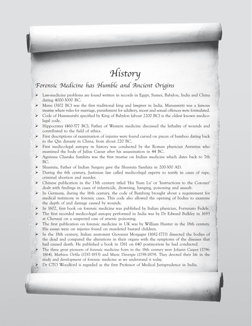
HistoryForensic Medicine has Humble and ancient origins Law-medicine problems are found written in records in Egypt, Sumer, Babylon, India and China
dating 4000-3000 BC. Manu (3102 BC) was the first traditional king and lawgiver in India. Manusmriti was a famous
treatise where rules for marriage, punishment for adultery, incest and sexual offences were formulated. Code of Hammurabi specified by King of Babylon (about 2200 BC) is the oldest known medico-
legal code. Hippocrates (460-377 BC), Father of Western medicine discussed the lethality of wounds and
contributed to the field of ethics. First descriptions of examination of injuries were found carved on pieces of bamboo dating back
to the Qin dynasty in China, from about 220 BC. First medico-legal autopsy in history was conducted by the Roman physician Antistius who
examined the body of Julius Caesar after his assassination in 44 BC. Agnivesa Charaka Samhita was the first treatise on Indian medicine which dates back to 7th
BC. Shusruta, Father of Indian Surgery gave the Shusruta Samhita in 200-300 AD. During the 6th century, Justinian law called medico-legal experts to testify in cases of rape,
criminal abortion and murder. Chinese publication in the 13th century titled 'Hsi Yuan Lu' or 'Instructions to the Coroner'
dealt with findings in cases of infanticide, drowning, hanging, poisoning and assault. In Germany, during the 16th century, the code of Bamburg brought about a requirement for
medical testimony in forensic cases. This code also allowed the opening of bodies to examine the depth of and damage caused by wounds.
In 1602, first book on forensic medicine was published by Italian physician, Fortunato Fedele. The first recorded medico-legal autopsy performed in India was by Dr Edward Bulkley in 1693
at Chennai on a suspected case of arsenic poisoning. The first publication on forensic medicine in UK was by William Hunter in the 18th century.
His essays were on injuries found on murdered bastard children. In the 18th century, Italian anatomist Giovanni Morgagni (1682-1771) dissected the bodies of
the dead and compared the alterations in their organs with the symptoms of the diseases that had caused death. He published a book in 1761 on 640 postmortem he had conducted.
The three great pioneers of forensic medicine born in the 18th century were Johann Casper (1796-1864), Mathieu Orfila (1787-1853) and Marie Devergie (1798-1879). They devoted their life in the study and development of forensic medicine as we understand it today.
Dr CTO Woodford is regarded as the first Professor of Medical Jurisprudence in India.
1. Medical Jurisprudence and Ethics 3 2. Acts Related to Medical Practice 30 3. Legal Procedure 43 4. Identification I 56 5. Identification II 84 6. Medico-legal Autopsy 98 7. Autopsy Room Hazards 122 8. Thanatology 126 9. Signs of Death 136 10. Asphyxia 160 11. Injuries 188 12. Firearm Injuries 209 13. Regional Injuries 232 14. Thermal Injuries 263 15. Transportation Injuries 281 16. Explosion Injuries and Fall from Height 289 17. Medico-legal Aspects of Injuries 295 18. Decompression, Radiation and Altitude Sickness 310
Jurisprudence and Forensic Medicine
19. Starvation Deaths 313 20. Anesthetic Deaths 316 21. Infanticide and Child Abuse 320 22. Abortion 337 23. Impotence and Sterility 345 24. Virginity, Pregnancy and Delivery 353 25. Sexual Offences I 366 26. Sexual Offences II 388 27. Sexual Offences III 394 28. Postmortem Artifacts 399 29. Forensic Psychiatry 403 30. Bloodstain Analysis 422 31. Seminal Stains and Other Biological Samples 431 32. DnA Fingerprinting 438 33. Torture and Custodial Deaths 446 34. Medico-legal Aspects of HIV 451 35. newer Techniques and Recent Advances 454Question Bank-I 457


Definitions
� Forensic medicine* (Legal medicine or State medicine): It is the application of principle and knowledge of medical sciences to legal purposes and legal proceedings so as to aid in the administration of justice.
� Medical jurisprudence (Latin juris: law, prudentia: knowledge or skill): It is the application of knowledge of law in relation to practice of medicine. It includes:
i. Doctor-patient relationship ii. Doctor-doctor relationship iii. Doctor-State relationship.
� Medical etiquette: These are the conventional laws and customs of courtesy which are followed between members of same profession.1 A doctor should behave with his colleagues, as he would like to have them behave with him, e.g. he should not charge another doctor or members of his family for professional service.
� Medical ethics: It is concerned with moral principles for the members of the medical profession in their dealings with each other, their patients and the State. It is a self-imposed code of conduct assumed voluntarily by medical professionals.
Forensic science refers to a group of scientific disciplines which are concerned with the application of their particular scientific area of expertise to law enforcement, criminal, civil, legal and judicial matters. Forensic scientists examine objects, substances (including blood/drug samples), chemicals (paints/explosives/toxins), tissue traces (hair/skin) or impressions (fingerprints/tyremarks) left at the scene of crime—a multidisciplinary subject.
Medical Council of India (MCI)
The Medical Council of India is a statutory body charged with the responsibility of establishing and maintaining uniform standards of medical education, and recognition of medical qualifications.
Indian Medical Degrees Act, 1916: This Act was passed to regulate the grant of titles implying qualification in Western Medical Science.
The Indian Medical Council (IMC) Act, 1956: The Medical Council of India was established in 1934 under the Indian Medical Council Act, 1933. In 1956, the old Act was repealed and a new one was enacted. This was further modified in 1964, 1993 and 2001. The government superseded the MCI by issuing an ordinance in May 2010. The Central Government constituted the board of Governors (BoG), comprising of not more than 7 members with one of them as Chairperson till the new council was to be elected (time frame given was of 2 years). The Government was liable to get the ordinance converted into a bill within 6 weeks from the date of the commencement of Parliament. Since then, the health Ministry sought extension of the tenure of BoG governing MCI four times till 2013. With the Government unable to get the Indian Medical Council (Amendment) Bill passed in Parliament, the old IMC Act that provided autonomy to the regulatory body was restored.
Constitution of IMC
i. One member from each State other than a Union Territory, nominated by the Central Government in consultation with the State Government concerned.
ii. One member from each University, to be elected from amongst the members of the medical faculty of the University.
iii. One member from each State in which a State Medical Register is maintained, to be elected from persons enrolled on such a register.
iv. Seven members to be elected by persons enrolled in any of the State Medical Registers.
v. Eight members are nominated by the Central Government.
The President and Vice-President are elected from amongst these members.
* Latin forensis: of or before the forum. In Rome, ‘forum’ was the meeting place, where civic and legal matters used to be discussed by those with public responsibility.
Medical Jurisprudence and Ethics
ChaptEr 1
4 Review of Forensic Medicine and Toxicology
Schedules
� First Schedule of the IMC Act contains recognized medical qualifications granted by Universities in India.2 Any medical institution which grants a qualification not included in the First Schedule may apply to the Central Government and after consulting the Council may amend the First Schedule, and the same is entered in the last column of the First Schedule.
� Second Schedule contains recognized medical qualifications granted outside India.3 The Council may enter into negotiations with the Authority in any country outside India for the scheme of reciprocity for the recognition of medical qualifications, and the Central Government may amend the Second Schedule, and the same is entered in the last column of the Second Schedule.
� Part I of the 3rd Schedule contains qualification granted by medical institutions not included in 1st Schedule, like Licensed Medical Practitioner (LMP) and diplomas which were granted before independence or with certain preconditions.
� Part II of the 3rd Schedule contains qualification granted outside India, but not included in 2nd schedule and certificates/diploma approved by the examining boards of the US.
The Council should: � Constitute an Executive Committee from amongst
its members. � Appoint a Registrar who will act as Secretary and
who may also act as Treasurer.
Functions of MCI
i. Maintenance of Indian Medical Register • It contains the names, addresses and qualifi
cations of the medical practitioners who are registered with any State Medical Council (SMC).
• Removal of the name from the register of the concerned SMC will lead to its removal from Indian Medical Register.
ii. Regulation of standard of undergraduate and postgraduate medical education
• The Council maintains the standards of under -graduate medical education. The Council prescribes courses and criteria which a medical institute should fulfill for a particular course of study.
• The Council sends inspectors to see that the college is adequately spaced, staffed and
equipped as per MCI stipulations. The inspector may also visit the institution during the examinations to assess the standard of education.
• On the basis of the reports of the inspectors, the MCI recommends the recognition or nonrecognition of the medical qualification to the Central Government.
• Such an inspection is held for every medical qualification when it is introduced and every 5 years thereafter.
• The Council has the authority to prescribe standards of postgraduate medical education for the guidance of the universities.
iii. Permission for establishment of new medical college, new course of study and increase in seats: Permission of the Central Government is obtained after the recommendations of the Council which may either approve or disapprove the scheme.
iv. Recognitionofmedicalqualificationgrantedbyuniversities in India: Any University which grants a medical qualification not included in the 1st Schedule may apply to the Central Government, to have such qualification recognized, and the Government, after consulting the Council, may amend the 1st Schedule.
v. De-recognition of medical qualification: It can make representation to the Central Government to withdraw recognition of a medical qualification of any college, if on receipt of report from inspectors it feels that the standards of resources, training/teaching are not satisfactory.
vi. Recognition of foreign medical qualificationsunder the scheme of reciprocity: The Council may enter into negotiations with the authority in any country outside India under a scheme of reciprocity for the recognition of medical qualifications. A separate examination may be conducted by the MCI to assess the standard of knowledge possessed by such individuals, before recognizing their degree.
vii. Appellate powers: It advises the Central Health Ministry when an appeal is made by a medical practitioner against the decision of the SMC on disciplinary matters. Its decision is binding on the appealing party as well as the SMC.
viii. Disciplinary control: The Council prescribes minimum standards of professional conduct, etiquette and a code of ethics for medical practitioners. It issues a warning notice periodically
5Medical Jurisprudence and Ethics
which is a list of offences constituting infamous conduct (professional misconduct). It can take actions against erring doctors and issue warning in relation to unethical practices which are regarded as disgraceful in a professional respect.*
ix. Certificates: It is empowered to issue certificates of good conduct and character to medical students/doctors going abroad for higher studies/service.
x. CME programmes: It sponsors and organizes continuing medical education (CME) programmes for medical practitioners.
xi. Faculty development programme: MCI has undertaken the task of training the medical college faculty upto the level of Associate Professors in MCI Basic Workshop in Medical Education Technologies. Faculty should undergo this training either before joining service or during probation period and once every 5 years thereafter.
MCI has asked the health Ministry to make it mandatory for all doctors to reregister with the SMCs and MCI every 5 years. This will help in tracking the number of doctors still alive and practicing in the country and registered with MCI.
There is no provision in the existing IMC Act for reregistration or revalidation of doctors. Medical Councils of certain States like Punjab, Delhi, Odisha, Rajasthan and Maharashtra have provision for reregistration of doctors under their respective statutes.
Doctors who have already got permanent registration/registration of additional qualifica tion with any SMC are not required/eligible for reregistration with the MCI.
State Medical Council (SMC)
Composition of the State Medical Council � Medical teachers from different Universities of the
State elected by the teachers of different medical institutions.
� Members elected by registered medical practitioners of the State.
� Some members are nominated by the State Government.
They elect a President and a Vice-President from amongst themselves.
Functions of SMC
i. Maintenance of Medical Register • Maintains a register of medical practitioners
within its jurisdiction.
• On payment of prescribed fees, the name, address and qualifications are entered in the register.
• A provisional registration is granted to a student who has passed the qualifying examination, but has to undergo a certain period of training (internship for 1 year) in an approved institution, and permanent registration is granted after that training period.
• Additional qualification obtained subsequent to registration or for any alteration may be done after payment of requisite fees to the SMC.
ii. Renewal of registration: Medical practitioners need to participate in CME programmes for at least 30 hours (h) to renew their registrations every 5 years. Several States are planning to bring legislation in order to make the process re-registration mandatory for doctors.
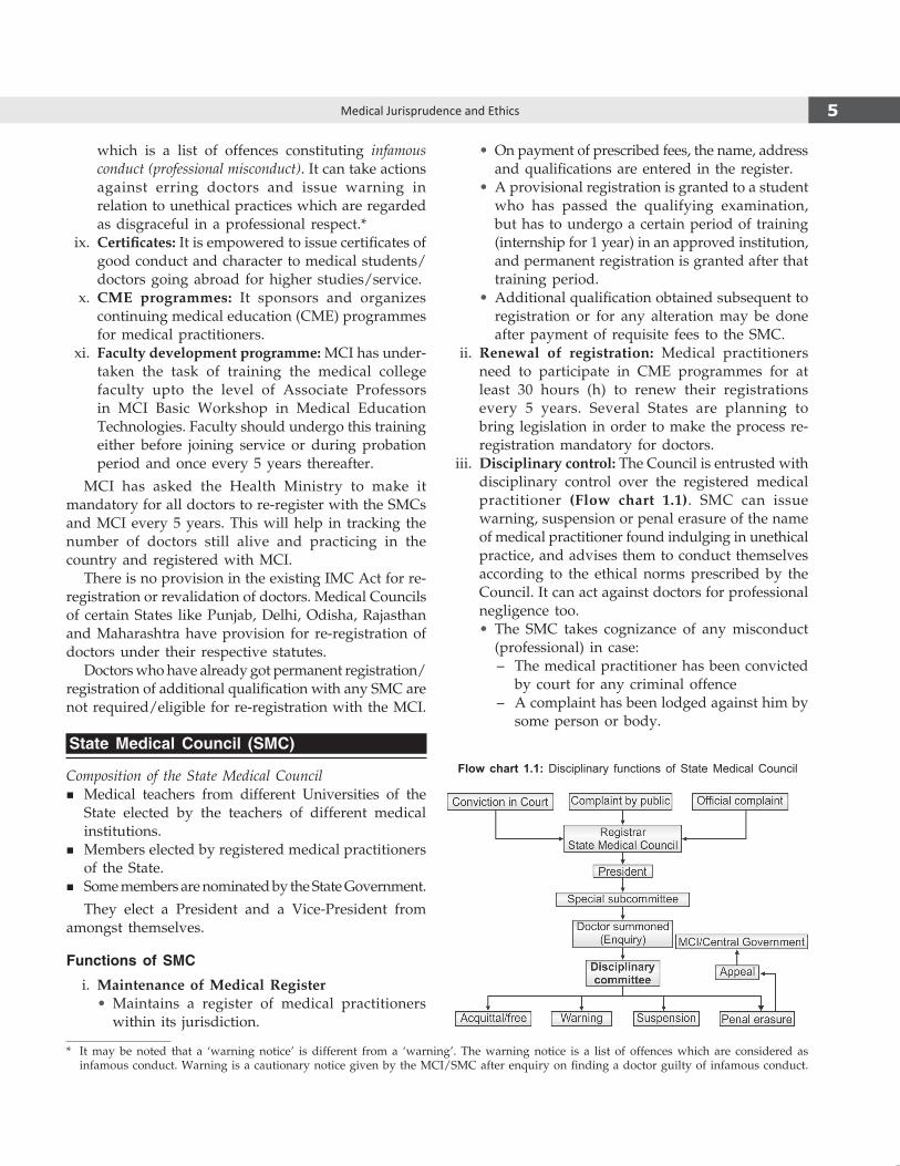
iii. Disciplinary control: The Council is entrusted with disciplinary control over the registered medical practitioner (Flow chart 1.1). SMC can issue warning, suspension or penal erasure of the name of medical practitioner found indulging in unethical practice, and advises them to conduct themselves according to the ethical norms prescribed by the Council. It can act against doctors for professional negligence too.
• The SMC takes cognizance of any misconduct (professional) in case:
– The medical practitioner has been convicted by court for any criminal offence
– A complaint has been lodged against him by some person or body.
Flow chart 1.1: Disciplinary functions of State Medical Council
* It may be noted that a ‘warning notice’ is different from a ‘warning’. The warning notice is a list of offences which are considered as infamous conduct. Warning is a cautionary notice given by the MCI/SMC after enquiry on finding a doctor guilty of infamous conduct.

6 Review of Forensic Medicine and Toxicology
• Upon receipt of any complaint, the SMC would hold an enquiry and give opportunity to the registered medical practitioner to be heard.
• If the doctor is found to be guilty of committing professional misconduct, the Council may punish as deemed necessary or may direct the removal of the name of the delinquent practitioner from the register, altogether or for a specified period.4
• Decision on complaint against delinquent physician is taken within a time limit of 6 months.
• An inquiry against a doctor should be initiated by SMC with which he/she is registered. The role of the MCI is only as an appellate authority to the Central Health Ministry to decide on an appeal against the decision of the SMC on disciplinary matters.5
iv. Removal of name of medical practitioner: SMC is empowered to erase from the register the name of any registered medical practitioner with whom it is unable to establish communication.
v. Restoration of name of medical practitioner: It can direct restoration of any name of registered medical practitioner so removed.
Duties of a Doctor (Flow chart 1.2)
Under the Indian Medical Council Act, 1956, the MCI, with the approval of the Central Government, made the following regulations which are called the Indian Medical Council (Professional Conduct, Etiquette and Ethics) Regulations, 2002 (amended in 2009).
Code of Medical Ethics: At the time of registration, all the doctors are self-warned about certain unethical practices (infamous conduct) and the disciplinary action by the SMC (also called as warning notice). The applicant should certify that he/she has read and agreed to abide by the same, and submit a declaration duly signed.
� Hippocratic Oath: The Hippocratic Oath is traditionally taken by physicians, in which certain ethical guidelines are laid out. Several parts of the Oath have been removed or re-worded over the years in various countries, schools and societies.
� Declaration of Geneva: The Declaration of Geneva was intended as a revision of the Hippocrates Oath to a formulation of that oaths’ moral truth that could be comprehended and acknowledged modernly. It was adopted by the General Assembly of the World Medical Association (WMA) at Geneva in 1948 and amended in 1968, 1984, 1994, 2005 and 2006.
� Declaration of Tokyo: This was adopted in 1975 (amended in 2005 and 2006) during the assembly of the WMA. It refers to the guidelines for doctors concerning torture, degradation or cruel treatment of prisoners.6
� Declaration of Helsinki: The WMA originally developed this declaration in 1964 and underwent major revision in 1975. It refers to the ethical principles for medical research involving human subjects, including research on identifiable human material and data.7
� Declaration of Oslo: It was a statement by the WMA in 1970 on therapeutic abortion and amended in 1983 and 2006.8
� Declaration of Malta: This was adopted by the WMA in 1991 (revised in 1992 and 2006) for hunger strikers. The principle of beneficence urges physicians to resuscitate them, but respect for individual autonomy restrains physicians from intervening when a valid and informed refusal has been made.
� Declaration of Lisbon: This was adopted by the WMA in 1981 (amended in 1995 and 2005). The declaration represents some of the principal rights of the patient that the medical profession endorses and promotes.
� Declaration of Ottawa: This declaration on child health was adopted by the WMA in 1998 (amended in 2009). Physicians along with parents, and with world leaders to advocate for healthy children.
Duties of a Doctor in General
i. Character of physician: A physician should uphold the dignity and honor of his profession and render service to humanity; reward or financial gain is a subordinate consideration.
ii. Maintaining good medical practice • The physician should try to improve medical
knowledge and skills, and should practice methods having scientific basis. he should participate in professional meetings, i.e. CME programmes for at least 30 h every 5 years.
• Membership in medical society: he should affiliate with associations and societies for the advancement of his profession.
iii. Maintenance of medical records • Physician should maintain the medical records of
his indoor patients for a period of 3 years from the date of commencement of the treatment.9
Flow chart 1.2: Duties of a medical practitioner
7Medical Jurisprudence and Ethics
In a case where medical records and consent obtained from a patient were not produced, negligence was established.
• On request for medical records, either by the patients or legal authorities, the same should be issued within the period of 72 h. This applies to a doctor in his private capacity, in case of indoor patients whom he/she might have treated/operated in hospital/nursing home.
• he should maintain a register of medical certificates issued. he should record the signature and/or thumb mark, address and at least one identification mark of the patient and keep a copy of the certificate.
iv. Display of registration numbers • Physician should display the registration number
accorded to him by the SMC in his clinic and in all his prescriptions, certificates, money receipts given to his patients. A doctor was held guilty for printing incorrect information about his qualification on the prescription paper.
• Physicians should display as suffix to their names only recognized medical degrees or such certificates/diplomas and memberships/honors which confer professional knowledge.
v. use of generic names of drugs: Physician should prescribe drugs with generic names, and ensure that there is a rational prescription and use of drugs.
vi. Highest quality assurance in patient care: he should not employ in connection with his profes-sional practice any attendant who is not registered or permit such persons to attend, treat or perform operations upon patients wherever professional discretion or skill is required.
vii. Exposure of unethical conduct: Physician should expose, without fear or favor, incompetent or corrupt, dishonest or unethical conduct on the part of members of the profession.
viii. Payment of professional services • Physician should clearly display his fees in his
chamber and/or hospitals he is visiting. • he should announce his fees before rendering
service and not after the operation or treatment is underway.
ix. Evasion of legal restrictions: Physician should observe the laws of the country in regulating the practice of medicine and should not assist others to evade such laws.
Duties of a Doctor towards the State i. Poisoning cases • he should assist the police in determining
whether the poisoning is accidental, suicidal or homicidal.
• In case of death, death certificate should mention about the poisoning with recommendation for postmortem examination.
ii. Notification: Doctor is bound to give information of communicable diseases (notifiable diseases), births, deaths and outbreak of an epidemic to public health authorities. Failing which he is not only liable for criminal penalties, but also negligence suits brought by affected persons.
A notifiable disease is any disease that is required by law to be reported to government authorities, e.g. cholera, plague, leprosy, diphtheria, typhoid fever, tetanus, measles, tuberculosis, chicken pox, acute poliomyelitis, encephalitis, influenza, dengue fever, hemorrhagic fevers, hepatitis, HIV, etc.
iii. Geneva Convention • In 1949, in Geneva, four conventions were agreed
upon. Each convention lays down the persons it protects.
• The wounded or sick of the armed forces (1st convention), ship-wrecked (2nd convention), prisoners of war (3rd convention) or civilians of enemy nationality (4th convention) are to be treated by the physician without any adverse distinction based on sex, race and nationality.
iv. Responding to emergency military service as and when required.
Duties of a Doctor towards Patients
i. Exercise reasonable degree of skill and knowledge • It begins the moment the physician-patient
relationship is established (i.e. when the physician agrees to treat the patient).
• he owes this duty even when the patient is treated free of charge.
• It neither guarantees cure nor an assured improvement.
• A practitioner (e.g. MBBS) is not liable because some other doctors of greater skill and knowledge (e.g. MD/MS) would have prescribed a better treatment or operated better in the same circumstances.
8 Review of Forensic Medicine and Toxicology
ii. Attendance and examination • When a doctor agrees to attend a patient, he
is under an obligation to attend to the case, as long it requires attention.
• he can withdraw after giving reasonable notice or when he is asked by the patient to withdraw.
• If the doctor is called by police to attend a case of road side accident, he may give first aid and advice, but no doctor-patient relationship is established.
iii. Furnish proper and suitable medicines • he should give a legible prescription. he should
write in capital letters—mistakes arising out of illegibly written names of medicines as opposed to other kinds of indecipherable documents—can be very dangerous.
• Doctor is held responsible for any temporary or permanent damage in health, caused to the patient due to wrong prescription.
iv. Instructions: Doctor should give full instructions to his patients or their attendants regarding use of medicines (quantities and timings), injections (whether to be given intramuscularly or intravenously) and diet.
v. Prognosis: The patient or his relatives should have such knowledge of the patient’s condition as will serve the best interests of the patient and the family.
vi. Control and warn • Doctor should warn patients of the side-effects
involved in the use of prescribed drug, otherwise it might amount to negligence.
• If the doctor fails to inform the known dangerous effects of a drug/device, he becomes liable not only for the harm suffered by the patient but also for injuries his patient may cause to third parties.
vii. Third parties: If a patient suffers from an infectious disease, the doctor should warn not only the patient, but also third parties who are close to the patient.
viii. Children and disabled persons being incapable of taking care of themselves, the doctor should arrange for their proper care, e.g. supervised application of hot water bottles.
ix. Consent: A mentally sound adult (> 18 years) must be told of all the relevant facts in non-medical terms and in a language he/she understands and then obtain consent.
x. Operations • Doctor should explain the nature and extent of
operation, and take consent of patient. • he should take proper care to avoid mistakes,
such as operating on the wrong patient or on wrong limb, or leave any instrument or swab inside a body cavity.
• he should not delegate his duty to operate a patient to another doctor.
• he should not experiment without valid reason or valid consent from the patient.
• He should avail the assistance of qualified and experienced anesthetists.
• Death on operation table should be followed by postmortem examination.
xi. Investigations • All cases of accident, unless they are minor,
should be X-rayed. • For proper diagnosis and to know the progress,
the doctor should advise investigations, like biopsy, Xrays, CT scan, etc.
• Wrong interpretation of Xray is liable to be held as negligent.
xii. Emergency cases • he has moral, ethical and humanitarian duty to
help the patient in saving his life. • In medico-legal injury cases, a doctor is obliged
to give medical aid and to save life of the patient. xiii. Professional secrecy/medical confidentiality Definition:The doctor is obliged to maintain the
secrets that he comes to know concerning the patient in the course of a professional relationship, except when he is required by the law to divulge the secrets or when the patient has consented for its disclosure.10
• It is a fundamental tenet that whatever a doctor sees or hears in the life of his patient must be treated as totally confidential. Disclosure would be failure of trust and confidence.
• The patient can sue the doctor for damages or face disciplinary action by the SMC, if the disclosure is voluntary and has resulted in harm to the patient and is not in the interest of public.
Following principles should be followed: i. Physician should not answer any query by third
parties, even when enquired by close relatives, either with regard to the nature of illness or any subsequent effect of such illness on the patient, without his/her consent.

9Medical Jurisprudence and Ethics
ii. If the patient is major (≥ 18 years), physician should not disclose any facts about the illness without his consent to parents or relatives even though they may be paying the doctor's fees. In case of minor or insane person, guardians or parents should be informed of the nature of illness.
iii. A doctor should not disclose the illness of his patient without his consent, even when requested by a public or statutory body, except in case of notifiable diseases. If the patient is minor or insane, consent of the guardian should be taken.
iv. Even in case of husband and wife, the facts relating to the nature of illness of one must not be disclosed to the other, without the consent of the concerned person. Particular caution is required over the disclosure of sexual matters, such as pregnancy, abortion or venereal disease, as disclosure might cause conflict between them.
v. In divorce and nullity cases, no information should be given without the consent of the concerned person.
vi. When a domestic servant is examined at the request of the master, the physician should not disclose any facts about the illness to the master without the consent of servant, even though the master is paying the fees. Similarly, the medical officer of firm or factory should not disclose without the patient’s consent.
vii. Medical officers in government service are also bound by code of professional secrecy, even when the patient is treated free.
viii. A person in police custody as an undertrial prisoner has the right not to permit the doctor who has examined him, to disclose the nature of his illness to any person. If convicted, he has no such right and physician can disclose the findings to the authorities.
ix. Any information regarding a dead person may be given only after obtaining the consent from a relative.
x. In examination of a dead body, certain facts may be found, the disclosure of which may affect the reputation of the deceased or cause mental torture to his relatives, and as such, the autopsy surgeon should maintain secrecy.
xi. The medical examination for life insurance policy is a voluntary act by the examinee, and consent to the disclosure of findings may be taken as implied.
Duties of a Doctor in Consultation i. Consultation for patient’s benefit is of foremost
importance. Unnecessary consultations should be avoided.
ii. Statement to patient after consultation should take place in the presence of the consulting physician, except if otherwise agreed. Differences of opinion should not be divulged unnecessarily.
iii. Treatment after consultation: The attending physician should make subsequent variations in the treatment, if any unexpected change occurs. The attending physician may prescribe medicine at any time for the patient, whereas the consultant may prescribe only in case of emergency or as an expert when called for.
iv. Patients referred to specialists: When a patient is referred to a specialist by the attending physician, a case summary of the patient should be given to the specialist, who should communicate his opinion in writing to the attending physician.
Consultation is advised with a specialist in the following conditions: i. In case of emergency. ii. If the patient requests consultation. iii. If quality of care or management can be considerably
enhanced. iv. In cases where diagnosis remains obscure. v. In case of homicidal poisoning. vi. In connection with organ transplantation. vii. When treatment or operation involves risk of life.viii. When operation affecting vitality, intellectual or
generative functions is to be performed. ix. When an operation involves mutilation or
destruction of an unborn child. x. When an operation is to be performed on a patient
who has received injuries in a criminal assault. xi. To take decision about termination of pregnancy
case, after 12 weeks and upto 20 weeks of pregnancy.
xii. While dealing with a criminal abortion or an attempted criminal abortion case.
� A referring physician is relieved of further responsibility when he completely transfers the patient to another physician.
� The referring physician may be held liable under the doctrine of negligent choice, if it can be proved that the consultant was incompetent or had a reputation as an errant physician.

10 Review of Forensic Medicine and Toxicology
Responsibility of Doctors towards Each Other i. Conduct in consultation: No insincerity, rivalry or
envy should be indulged in. All due respect should be observed towards the physician in-charge of the case, and no statement or remark be made, which would impair the confidence the patient has reposed in him.
ii. Consultant not to take charge of the case: Consultant should normally not take charge of the case, especially on the solicitation of the patient or friends.
iii. Appointment of substitute: A physician should accept to attend another physician’s patients during his temporary absence from his practice, only when he has the capacity to discharge the additional responsibility along with his other duties.
Privileged Communication
Definition: It is a statement, made bonafide upon any subject matter by a doctor to the concerned authority having corresponding interest, due to his legal, social or moral duty to protect the interests of the community or of the State.
� It is an exchange of information between two indi-viduals in a confidential relationship, and an exception to professional secrecy.
� To be privileged, it must be made to the person who has a duty towards it. If made to more than one person or to a person who has not got a direct interest in it, the plea of privilege fails.
� Doctor should first persuade the patient to obtain his consent before notifying the proper authority. however, disclosure can be done without consent (if consent is not forthcoming).
Examples i. Civicbenefit: If there is a potential threat of ‘grave
harm’ to the safety or health of the patient and the public, the doctor must decide whether to inform the authority about the condition.
• For example, a engine or bus driver, pilot or ship navigator may be suffering from epilepsy, hypertension, alcoholism, drug addiction, poor visual acuity or color blindness; or a teacher with tuberculosis or a person with infectious diseases (e.g. enteric infection) working as a cook. In all these cases, the proper course is for the doctor to explain the risks to the patient and to persuade him to allow the doctor to report the problem to his employers. If the patient refuses, then
it is always wise to seek the advice of senior colleagues before making any disclosure.
• A syphilitic taking bath in public pool or a patient with sexually transmitted disease is about to get married is a privileged communica-tion, but an impotent person getting married is not.
ii. Notifiableclauses: Doctor has a statutory duty to notify births, deaths, still births, infectious diseases, therapeutic abortions, drug addictions, epidemic and food poisoning to public health authorities.
iii. Suspected crime: If the physician learns of a crime, such as assault, terrorist activitiy, traffic offence or homicidal poisoning by treating the victim or assailant, he is bound to report it to the nearest Magistrate or police officer.
• but sometimes, the issue of confidentiality clashes with the need to protect some individual or the public from possible further danger (e.g. a below-age of consent girl came to a doctor with STD). The doctor is usually required to obtain a list of the patient’s sexual contacts to inform them that they need treatment. however, the patient may be reluctant to divulge the names of her older sexual partners, for fear that they will be charged with statutory rape. The same issue may arise where a doctor suspects a child or an elderly person, disabled or incompetent person is being abused, but here the overriding consideration is the safety of these individuals.
� It has been made mandatory to report to the police any case of sexual abuse in children (≤18 years) as per the Protection of Children from Sexual Offences Act, 2012.
• At times, assault may occur within a family, e.g. between spouses or close relatives, the victim may not wish to bring criminal charges, and so the doctor must not assume that consent for disclosure has been given.
• The doctor knowing or having reason to believe that an offence has been committed by a patient when he is treating, intentionally omits to inform the police, can be punished with imprisonment upto 6 months and with/without fine (Sec. 202 IPC).
iv. Patient’s own interest: Doctor may disclose patient’s condition to his relatives so that he may be properly treated, e.g. to warn parents/guardians of patient’s melancholia or suicidal tendencies.
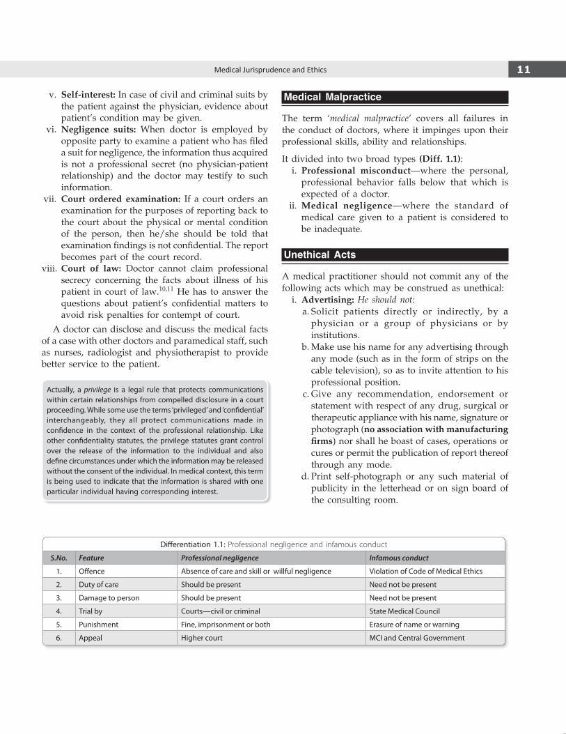
11Medical Jurisprudence and Ethics
v. Self-interest: In case of civil and criminal suits by the patient against the physician, evidence about patient’s condition may be given.
vi. Negligence suits: When doctor is employed by opposite party to examine a patient who has filed a suit for negligence, the information thus acquired is not a professional secret (no physician-patient relationship) and the doctor may testify to such information.
vii. Court ordered examination: If a court orders an examination for the purposes of reporting back to the court about the physical or mental condition of the person, then he/she should be told that examination findings is not confidential. The report becomes part of the court record.
viii. Court of law: Doctor cannot claim professional secrecy concerning the facts about illness of his patient in court of law.10,11 he has to answer the questions about patient’s confidential matters to avoid risk penalties for contempt of court.
A doctor can disclose and discuss the medical facts of a case with other doctors and paramedical staff, such as nurses, radiologist and physiotherapist to provide better service to the patient.
Actually, a privilege is a legal rule that protects communica tions within certain relationships from compelled disclosure in a court proceeding. While some use the terms ‘privileged’ and ‘confidential’ interchangeably, they all protect communications made in confidence in the context of the professional relationship. Like other confidentiality statutes, the privilege statutes grant control over the release of the information to the individual and also define circumstances under which the information may be released without the consent of the individual. In medical context, this term is being used to indicate that the information is shared with one particular individual having corresponding interest.
Medical Malpractice
The term ‘medical malpractice’ covers all failures in the conduct of doctors, where it impinges upon their professional skills, ability and relationships.
It divided into two broad types (Diff. 1.1): i. Professional misconduct —where the personal,
professional behavior falls below that which is expected of a doctor.
ii. Medical negligence—where the standard of medical care given to a patient is considered to be inadequate.
Unethical Acts
A medical practitioner should not commit any of the following acts which may be construed as unethical: i. Advertising: He should not: a. Solicit patients directly or indirectly, by a
physician or a group of physicians or by institutions.
b. Make use his name for any advertising through any mode (such as in the form of strips on the cable television), so as to invite attention to his professional position.
c. Give any recommendation, endorsement or statement with respect of any drug, surgical or therapeutic appliance with his name, signature or photograph (no association with manufacturing firms) nor shall he boast of cases, operations or cures or permit the publication of report thereof through any mode.
d. Print self-photograph or any such material of publicity in the letterhead or on sign board of the consulting room.
Differentiation 1.1: Professional negligence and infamous conduct
S.No. Feature Professional negligence Infamous conduct
1. Offence Absence of care and skill or willful negligence Violation of code of Medical Ethics
2. Duty of care Should be present Need not be present
3. Damage to person Should be present Need not be present
4. Trial by courts—civil or criminal State Medical council
5. Punishment Fine, imprisonment or both Erasure of name or warning
6. Appeal Higher court McI and central Government

12 Review of Forensic Medicine and Toxicology
A medical practitioner is, however, permitted to make a formal announcement in press regarding the following:
• On starting or resumption or change of type of practice.
• On changing address. • On temporary absence from duty. • Public declaration of charges. • Acquiring new equipment or starting a new
procedure or operation (as per Punjab Medical Council).
The advertisement in the press should be in black and white, and < 15 x 10 cm in size. It should not carry photograph of the doctor/building/equipment/procedure (as per Punjab Medical council).
ii. Patent and copyrights: he may patent surgical instruments, appliances, procedures and medicine. However, it is unethical, if the benefits of such patents are not made available in situations where the interest of large population is involved.
iii. he should not run an open shop for dispensing of drugs and appliances prescribed by other physicians.
iv. Rebates and commission (dichotomy/fee splitting/ ‘cut practice’): he should not give or receive any gift or commission in consideration of referring, recommending or procuring of patient for medical, surgical or other treatment, or for getting specimen or material for diagnostic purposes.
v. Secret remedies: he should not prescribe or dispense secret remedial agents of which he does not know the composition.
vi. Human rights: he should not aid or abet torture or be a party to either infliction of psychological or physical trauma.
vii. Euthanasia: he should not practice euthanasia. viii. Pharmaceutical and allied health sector industry:
A medical practitioner should not receive any gift, cash or monetary grants, travel facility or accept any hospitality, like hotel accommodation from any pharmaceutical industry for vacation or for attending conferences, seminars, workshops or CME programme as a delegate.
Recently, the McI has fixed the quantum of punishment: doctor taking bribe (gifts, cash or travel facility) worth ` 1000-5000 will receive a warning; taking ` 5000-10000: suspension from the SMc for 3 months, taking ` 10000-50,000: suspension of 6 months, and bribes ≥ ` 50,000: suspension for 1 year.
Professional Misconduct (Infamous Conduct)
Definition: Any conduct of the doctor which might reasonably be regarded as disgraceful or dishonorable as judged by professional men of good repute and competence. It involves abuse of professional position.
The following acts of commission or omission on the part of a physician constitutes professional mis conduct: i. Any unethical practice as outlined above. ii. If he does not maintain the medical records of his
indoor patients for a period of 3 years and refuses to provide the same within 72 h when the patient requests for it.
iii. If he does not display the registration number accorded to him by the SMC in his clinic, prescriptions and certificates issued by him.
iv. Physician posted in rural area is found absent on more than two occasions during inspection by the head of the District health Authority or the Chairman, Zila Parishad.
v. Physician posted in a medical college as teaching faculty or otherwise is found absent on more than two occasions; the same is construed as misconduct, if it is certified by the Principal/Medical Superintendent.
vi. Providing falsified and misleading information to the MCI via Form A. The form is filled by the doctor when he/she joins a medical college.
Further, he should NOT: i. Commit adultery or misbehave with a patient. ii. be drunk and disorderly so as to interfere with
proper practice of medicine. iii. be convicted by court of law for offences involving
moral turpitude/criminal acts. iv. Do sex determination tests with the intent to
terminate the life of a female fetus. v. Issuefalse,misleadingorimpropercertificates for
subsequent use in the courts or for administrative purposes.
vi. Violate the provisions of Drugs and Cosmetics Act. he should not:
• Sell Schedule ‘H’ and ‘L’ drugs and poisons to the public, except to his patient.
• Prescribe steroids/psychotropic drugs when there is no medical indication.
vii. Supply or sell addiction forming drugs to a patient other than medical grounds.
viii. Give cover, i.e. assist someone who has no medical qualification to attend, treat or perform an operation, in cases requiring professional discretion or skill.
ix. Perform an illegal abortion/operation for which there is no medical, surgical or psychological indication.

13Medical Jurisprudence and Ethics
x. Issuecertificatesofefficiencyinmodernmedicineto unqualified or nonmedical person.
xi. Disclose professional secrets. xii. Refuse on religious grounds sterilization, birth
control, circumcision and medical termination of pregnancy when it is indicated.
xiii. Publish photographs/case reports of his patients without their consent in any medical or other journal or social media in a manner by which their identity could be made out.
xiv. use touts or agents to entice patients. xv. Claim to be specialist when he has no special
qualification in that branch. xvi. undertake in-vitro fertilization or artificial
insemination without the informed consent of the female patient and her spouse as well as the donor.
xvii. Do clinical drug trials or other research involving patients or volunteers not abiding by the guidelines of ICMR.
xviii. Advertise a. Contribute to the lay press articles and give
interviews regarding diseases and treatments which may have the effect of advertising himself. he can write to the lay press under his own name on matters of public health, hygiene or deliver public lectures, give talks on the radio/FM/TV/internet for the same purpose.
b. Use an unusually large signboard and write on it anything other than his name, qualifications, title, name of his speciality and registration number.
c. Affix a signboard on a chemist’s shop or in places where he does not reside or work.
d. Give his name, address and speciality in the yellow pages of the telephone directory in bold letters.
The instances of offences and professional misconduct which are given above do not constitute a complete list of the infamous acts which calls for disciplinary action. Circumstances may arise from time to time in relation to which there may occur questions of professional misconduct which do not come within any of these categories.
Important offences can be described as 6 As12
1. Association with unqualified persons 4. Adultery2. Advertising 5. Addiction3. Abortion (criminal) 6. Alcohol
Erasure of NameThe name of the doctor is removed from the SMC register:
� After the death of registered medical practitioner. � When entries of the medical practitioner are
erroneous or fraudulent. � In case of professional misconduct which is known
as penal erasure. When the name is permanently removed, it is termed as professional death sentence.13
� When the registered medical practitioner is not traceable at the address recorded with the Council.
Rights and Privileges of Registered Medical Practitioners
i. Right to choose his patient—he may refuse any patient without reason, but he should not refuse emergency treatment required by the patient.
ii. Right to use title and description of the qualification to his name.
iii. Right to practice medicine. iv. Right to dispense medicine to his patient. v. Right to possess and supply dangerous drugs to
his patients. vi. Right to give evidence in the court of law, as an
expert witness. vii. Right to issue medical certificates and medicolegal
reports. viii. Right to recovery of fees—if the patient does not
pay the justified fees, help of court can be taken. ix. Right for appointment in public and local hospitals. x. Right to be exempted from acting as a juror in
course of holding an inquest (not applicable in India).
Red Cross Emblem
Red Cross is an emblem which is used only by those belonging to the Red Cross Movement and Army Medical Services involved in humanitarian work, mainly at times of armed conflicts and natural disasters, and it is not an emblem of medical professionals.
As specified by the Geneva Conventions, the emblem can be used only by the following:
� Facilities for the care of injured and sick armed forces members
� Armed forces medical personnel and equipment � Military chaplains � International Red Cross Organizations

14 Review of Forensic Medicine and Toxicology
The use of the emblem by Government medical institutions, like hospitals, clinics and blood banks, doctors, private nursing homes and also on ambulance vehicles is equivalent to abuse, and is punishable with a fine of ` 500 and forfeiture of the goods or vehicles on which the emblem has been used.14
Privileges and Rights of Patients
i. Access to health care facilities and emergency services regardless of age, sex, religion, social or economic status.
ii. Choice: To choose his own doctor freely. iii. Continuity: To receive continuous care for his
illness from doctor/institution. iv. Comfort: To be treated in comfort during illness
and follow-up. v. Complaint: Right to complain and redressal of
grievances. vi. Confidentiality: All information about his illness
should be kept confidential. vii. Dignity: To be treated with care, compassion,
respect without any discrimination. viii. Information: Should receive full information about
his diagnosis, investigations, treatment plans, alternative therapy, procedures, diagnosis, compli-cations and side-effects.
ix. Privacy: To be treated in privacy. x. Refusal: Can refuse any specific or all measures. xi. Records: Can have access to his records and
demand summary or other details.
Duties of a patient i. he should furnish the doctor with complete
information about the facts and circumstances of his illness.
ii. he should strictly follow the instructions of the doctor as regards diet, medicine and lifestyle.
iii. he should pay a reasonable fee to the doctor.
Types of Physician-Patient Relationship
It is of two types:1. Therapeutic relationship: A doctor is free to accept
or refuse to treat a patient, subject to constraint of his work, except in emergencies. he may refuse to treat the patient in following circumstances:
i. beyond his practicing hours. ii. Not belonging to his speciality. iii. Doctor or any other family member is ill. iv. Doctor having important social function in
family.
v. Illness beyond the competence and qualification of the doctor or beyond the facilities available in his setup.
vi. Doctor is having alcohol. vii. Patient is malingering. viii. Patient has been defaulting in payment. ix. Patient or his relatives are abusive/uncooperative. x. Patient refuses to give consent. xi. Patient demanding specific drugs, like amphe
tamine, steroids, etc. xii. Patient rejecting low-cost remedies in favor of
high cost alternatives. xiii. At night, on grounds of security, if patient is
not brought to him. xiv. An unaccompanied minor or female patient. xv. When doctor remains engaged with an
emergency or more serious case. xvi. Any new patient, if he is not the only doctor
available.2. Formal relationship: It pertains to the situation where
the third party has referred the person/patient for impartial medical examination; e.g.
i. Pre-employment. ii. Insurance policy. iii. Yearly medical checkups. iv. Cases of rape or victims of crimes. v. Intimate body searches and other medico-legal
cases. vi. In certain psychiatric illnesses referred by court/
police.
Doctor has to comply with the directive of the party demanding such examination.
Professional Negligence
Definition: The failure to exercise reasonable care and skill of an ordinary prudent medical practitioner in the circumstances; a breach of duty to act with care appropriate to the situation, which resulted in bodily injury (harm/loss) or death of the patient.
Negligence consists of two acts: Not doing something that a reasonable man, under the circumstances would do (act of omission); or doing something which a reasonable prudent man under the circumstances would not do (act of commission).
According to Black’s Law Dictionary, medical negligence requires that the plaintiff establish the following (4 Ds): i. Existence of the physician’s duty of care to the
plaintiff, based on the existence of the physician-patient relationship.

15Medical Jurisprudence and Ethics
ii. Applicable standard of care and its violation (dereliction of duty), i.e. a breach in the duty caused by the defendant’s negligent act or omission.
iii. Damage (a compensable injury), i.e. pain and suffering, disability and disfigurement, past and future medical bills, lost wages, wrongful death, etc.
iv. Causal connection between the violation of care and the harm complained of (direct causation), i.e. a direct link between the defendant’s negligent act or omission and an injury suffered by the plaintiff.
In a lawsuit for malpractice or negligence (civil), the ‘patient’ is known as the plaintiff and the ‘physician’ becomes the defendant.15 Malpractice requires the demonstration of negligence or substandard practice that caused of harm. To successfully sue a physician for malpractice, the plaintiff must prove damage has been caused by the doctor’s conduct (Flow chart 1.3).
‘Damage’ should be distinguished from ‘damages’. Damage (injury or harm) to the patient may be physical, mental or financial. Damages are assessed by the court based on parameters, like loss of earning, medical and surgical costs, or reduction of quality of life.Potential damages (financial compensation) in negligence suits fall into three categories:• Economic or the monetary costs of an injury (e.g. medical bills
or loss of income)• non-economic (e.g. pain and suffering, loss of ability to have sex)• Punitive or damages to punish a defendant for willful and
wanton conduct.
Types (Diff. 1.2)
i. Civil ii. Criminal.
Civil Negligence
Question of civil negligence arises:a. When a patient, or in case of death, any relative
brings suit in a civil court for realization of compensation from his doctor, if he has suffered injury due to negligence.
b. When doctor brings a civil suit for the realization of his fees from patient or his relatives, who refuse to pay the same, alleging professional negligence.
Civil negligence involves: � Such act on the part of the treating physician
which causes some suffering, harm or damage to the patient
� Damage is such, which can be compensated by paying money
� Does not come under the purview of CrPC and IPC � Does not demand legal punishment.
Criminal Negligence
� Criminal negligence is more serious than civil negligence.
� Practically limited to cases in which the patient has died.
� Mostly associated with drunkenness or impaired efficiency due to the use of drugs by doctors.
� Doctor shows gross incompetency and inattention in the selection and application of remedies, undue interference by him or criminal indifference to the patient’s safety.
� Sec. 304-A IPC deals with criminal negligence; `whoever causes the death of any person by doing
Flow chart 1.3: Basic principle of negligence (example)
� Tort: A wrong or harm other than breach of contract; breach of a noncontractual duty towards another person which caused harm or loss. The same action may be both a tort, for which a person may seek compensation, and a crime, punishable by the State.
� Degree of care: The level of caution, prudence or forethought legally required to avoid causing harm or loss to another person. In determining liability, a person may be required to exercise degrees of care variously described as ‘ordinary’, ‘due’, ‘reasonable’, ‘great’, or ‘utmost’.
� Gross negligence: Negligence beyond the ordinary; a reckless or wanton disregard of the duty of care toward others.
� Liability: An actual or potential legal obligation, duty or responsibility to another person; the obligation to compensate, in whole or in part, a person harmed by one’s acts or omissions.
� Chain of causation: In claims in tort, or prosecutions in criminal law, the causal relationship between the defendant’s wrong doing and the victim’s loss or injury should be obvious for successful outcome. For example, if A hits B over the head, and B sustains a concussion, A is responsible.
� Damages: Money awarded in a suit or legal settlement as compensation for an injury or loss caused by a wrongful or negligent act or a breach of contract.
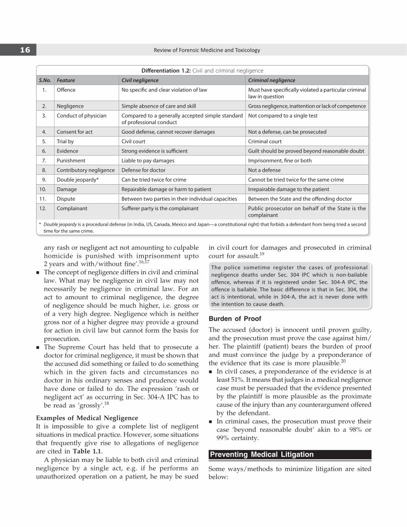
16 Review of Forensic Medicine and Toxicology
any rash or negligent act not amounting to culpable homicide is punished with imprisonment upto 2 years and with/without fine’.16,17
� The concept of negligence differs in civil and criminal law. What may be negligence in civil law may not necessarily be negligence in criminal law. For an act to amount to criminal negligence, the degree of negligence should be much higher, i.e. gross or of a very high degree. Negligence which is neither gross nor of a higher degree may provide a ground for action in civil law but cannot form the basis for prosecution.
� The Supreme Court has held that to prosecute a doctor for criminal negligence, it must be shown that the accused did something or failed to do something which in the given facts and circumstances no doctor in his ordinary senses and prudence would have done or failed to do. The expression ‘rash or negligent act’ as occurring in Sec. 304A IPC has to be read as ‘grossly’.18
Examples of Medical NegligenceIt is impossible to give a complete list of negligent situations in medical practice. however, some situations that frequently give rise to allegations of negligence are cited in Table 1.1.
A physician may be liable to both civil and criminal negligence by a single act, e.g. if he performs an unauthorized operation on a patient, he may be sued
in civil court for damages and prosecuted in criminal court for assault.19
The police sometime register the cases of professional negligence deaths under Sec. 304 IPc which is non-bailable offence, whereas if it is registered under Sec. 304-A IPc, the offence is bailable. The basic difference is that in Sec. 304, the act is intentional, while in 304-A, the act is never done with the intention to cause death.
Burden of Proof
The accused (doctor) is innocent until proven guilty, and the prosecution must prove the case against him/her. The plaintiff (patient) bears the burden of proof and must convince the judge by a preponderance of the evidence that its case is more plausible.20
� In civil cases, a preponderance of the evidence is at least 51%. It means that judges in a medical negligence case must be persuaded that the evidence presented by the plaintiff is more plausible as the proximate cause of the injury than any counterargument offered by the defendant.
� In criminal cases, the prosecution must prove their case ‘beyond reasonable doubt’ akin to a 98% or 99% certainty.
Preventing Medical Litigation
Some ways/methods to minimize litigation are sited below:
Differentiation 1.2: Civil and criminal negligence
S.No. Feature Civil negligence Criminal negligence
1. Offence No specific and clear violation of law Must have specifically violated a particular criminal law in question
2. Negligence Simple absence of care and skill Gross negligence, inattention or lack of competence
3. conduct of physician compared to a generally accepted simple standard of professional conduct
Not compared to a single test
4. consent for act Good defense, cannot recover damages Not a defense, can be prosecuted
5. Trial by civil court criminal court
6. Evidence Strong evidence is sufficient Guilt should be proved beyond reasonable doubt
7. Punishment Liable to pay damages Imprisonment, fine or both
8. contributory negligence Defense for doctor Not a defense
9. Double jeopardy* can be tried twice for crime cannot be tried twice for the same crime
10. Damage Repairable damage or harm to patient Irrepairable damage to the patient
11. Dispute Between two parties in their individual capacities Between the State and the offending doctor
12. complainant Sufferer party is the complainant Public prosecutor on behalf of the State is the complainant
* Double jeopardy is a procedural defense (in India, US, canada, Mexico and Japan—a constitutional right) that forbids a defendant from being tried a second time for the same crime.

17Medical Jurisprudence and Ethics
Table 1.1: Examples of medical negligence
General Errors
� Inadequate medical records and failure to examine the patient himself/herself.
� Failure to attend a patient with consequent damage.
� Failure to admit to hospital when necessary.
� Failure to obtain informed consent for any procedure.
� Making a wrong diagnosis in the absence of skill and knowledge.
� Administration of incorrect type/quantity of drugs, especially by injection.
� Failure to immunize and perform sensitivity tests.
� Failure to act on radiological or laboratory reports.
Medicine
� Failure to diagnose myocardial infarcts and other medical conditions.
� Failure to refer a patient to hospital or for specialist opinion.
� Toxic results of drug administration.
Surgery
� Delayed diagnosis of acute abdominal lesions.
� Retention of instruments, tubes, towels, sponges and swabs in operation sites.
� Operating on the wrong patient, wrong side of the body, wrong limb, digit or even organ.
� Failed vasectomy, without warning of lack of total certainty of consequent sterility.
� Diathermy burns.
Obstetrics and Gynecology
� Unwanted pregnancy due to failed tubal ligation.
� complications of hysterectomy—ureteric ligation and vesico-vaginal fistulae.
� Brain damage in the newborn due to hypoxia from prolonged labor.
� Mismanagement of delivery, especially under the influence of alcohol/drug.
� Performing abortion without indication (criminal abortion).
Orthopedics and Emergency Medicine
� Missed fractures, especially of the scaphoid, skull, femoral neck and cervical spine.
� Over-tight or prolonged use of plaster casts resulting in tissue and nerve damage.
� Undiagnosed intracranial hemorrhage.
� Missed foreign bodies in eyes and wounds, especially glass.
� Inadequately treated hand injuries, particularly tendons.
Anesthesiology
� Hypoxia resulting in brain damage.
� Neurological damage from spinal or epidural injections.
� Peripheral nerve damage from splinting during infusion.
� Incompatible blood transfusion.
� Incorrect or excessive use of anesthetic agents.
Dentistry
� Nerve injuries affecting a patient’s ability to taste or causing permanent numbness in the tongue.
� Failures to take a patient’s relevant medical history.
� Failure to detect oral cancer, periodontal disease or other diseases.
� Unnecessary extraction of multiple teeth.
� Extraction of wrong teeth.
� complications arising from negligently completed crowns and bridges.
� complications from anesthesia.
18 Review of Forensic Medicine and Toxicology
� Awareness of potential areas of litigation and medico-legal problems: Doctor should be aware of the risks involved in certain procedures and should have clear knowledge of the changes in legislation which might influence his practice.
� Good ‘doctor-patient’ relationship: Sympathy, good rapport and taking keen interest in the patient’s apprehensions and complaints are hallmarks in gaining the patient’s confidence. A suspicious patient who has no faith in the physician is a potential litigant.
� Appropriate training and maintenance of authorized protocol: Up-to-date and adequate training of medical and nursing staff is needed. It is dangerous to venture beyond one’s capability and qualifications. Maintaining a timetested, well accepted protocol is necessary. It is wise to seek a second opinion.
� Maintaining standard medical service: Limited work load and adequate infrastructure are needed to maintain good quality service. Minimum standard for nursing homes or hospitals, whether public or private, must be maintained.
� Proper counseling and informed consent: Counseling and informed consent is mandatory before each medical/investigative/operative procedure.
� Proper investigation: Any noninvasive/invasive procedures should be done, provided the risks and benefits are duly informed, and written consent has been taken.
� Adequate supervision and timely referral: Adequate supervision by a well organized graded system is recommended. Early detection of complications by resident doctors and timely notification of the consultant, especially in emergency cases, may prevent mishaps.
� Surgical intervention: Surgical procedures should always be performed in places where there is sufficient equipment and qualified staff. Junior doctors should be trained well and supervised in surgical care of the patient.
� Meticulous record keeping: Often proper record keeping can prove the doctor innocent in the court. however, fabrication of records after any mishap is dangerous.
� Morbidity and mortality audits: Discussions, analysis and constructive criticism of errors and omissions help in improving and maintaining standard of patient care.
� Medical indemnity insurance: The doctor must cover himself with indemnity insurance.
� Medical defense procedure: Efficient defense lawyer is important to defend one against a malpractice and negligence suit. The lawyer must be aware of the expected standard of patient care.
Defenses Against Negligence
In case of alleged negligence, following may be helpful for defense:
� No duty owed to patient, i.e. no doctor-patient relationship was established.
� Duty discharged according to prevailing standards. � Informed consent for the act: The patient was duly
informed of the consequences. � Patient was guilty of contributory negligence. � Therapeutic misadventure. � Medical maloccurrence. � Error of judgment. The court has held that the
error of judgment is not negligence. If, for e.g. one of the risks inherent in an operation takes place or some complication ensues which lessens the benefit that was hoped for, he makes an error of judgment. Moreover, doctor is not responsible if patient does not respond to the treatment.
� Mistake of fact is a situation where a person not intending to do unlawful act, does so because of wrong conclusion or understanding of fact. The guilty mind was never there while doing the act. It can be a factor in reducing civil liability but not criminal liability.
� Res judicata means ‘the things have been decided’. According to this principle, once the case is completed between two parties, it cannot be tried again between the same parties. Suppose a patient sues a hospital for any malpractice and the things are decided, he cannot subsequently sue the doctor again separately for the same negligence.
� Limitation: The case against the doctor should be filed within 2 years from the date of alleged negligence.
No fee was charged for the treatment cannot be a defense in cases of negligence.
Volenti non fit injuria (Latin, ‘to a willing person, injury is not done’) is a defense to an action in negligence. If a plaintiff (patient), with full knowledge, voluntarily consents the risk of injury, he will not recover any damages. The defendant (doctor) needs to prove not only that the plaintiff accepted the risk of injury but also accepted that if injury should happen, the plaintiff would accept the legal risk.

19Medical Jurisprudence and Ethics
Doctrine of Res ipsa loquitur
� Generally, professional negligence of a doctor must be proved in the court by expert evidence of another physician.
� The patient need not prove negligence in case where the rule of res ipsa loquitur applies, which means ‘the thing or fact speaks for itself.’21
� Applies to both civil and criminal negligence. � Error is so selfevident that the patient’s lawyer need
not prove the doctor’s guilt with medical evidence. The doctor has to prove his innocence.22
� Rule is applied when the following three conditions are satisfied:
i. In the absence of negligence, the injury would not have occurred, i.e. its occurrence ordinarily bespeaks negligence.
ii. Doctor had exclusive control over the injury producing instrument/treatment.
iii. Patient was not guilty of contributory negligence, i.e. injury was not the result of his own voluntary act or neglect.
Examples i. blood transfusion misadventure (e.g. infected
blood, blood group mismatch). ii. Failure to give tetanus toxoid vaccine in cases of
injury. iii. Prescribing an overdose of medicine producing ill
effects. iv. Wrongsite surgery (surgery on the wrong person,
wrong organ or limb, or wrong vertebral level), or wrong-procedure.
v. Leaving a pair of scissors in abdomen.23
vi. Failure to remove swabs during operation, causing complications/death.
vii. Loss of use of hand due to prolonged splinting.In such situations, the breach of duty is obvious, so
the strategy of the defense generally must be to show that the patient was not harmed by the breach.
Gossypiboma (Latin gossipium: cotton, Kiswahili boma: place of concealment) or textiloma denotes complications resulting from foreign materials accidentally left inside a patient’s body. The list of implements includes sponges (most common), swabs, towels, needles, instruments, catheters, metal clips, contraceptive coils, and retractors. The most common sites these incidents occur are operating rooms, labor and delivery rooms, ambulatory surgery centers or labs where invasive procedures such as catheters or colonoscopies take place.
Calculated Risk Doctrine
� The doctrine is that, res ipsa loquitur should not be applied when the injury complained is of type that
may occur even though reasonable care has been taken.
� It is an important defense to any doctor. � Doctor has to produce evidence/statistics that the
accepted method of treatment he employed had unavoidable risks.
� For example, when a patient undergoing coronary bypass dies during the surgery, it becomes a case of professional accident as there is already an inherent risk of 2–5% associated with it.
Doctrine of Common Knowledge
� It is based on the assumption that the issue of negligence in the particular case is not related to specialized knowledge or technical matters of the medical profession, but an act involving application of common knowledge.
� Experts may not provide evidence regarding matters of ‘common knowledge’.
� It is a variant of res ipsa loquitur.24
� here, the patient must prove the act of commission or omission, but he need not produce evidence to establish the standard of care.
Doctrine of Avoidable Consequence Rule
� Once plaintiff (patient) has been injured, he must take reasonable steps to lessen the consequences of his original injury. A defendant (accused) will not be liable of any further injury that the plaintiff could have reasonably avoided.
� The doctrine is different from contributory negligence, which is unreasonable conduct by plaintiff. It occurs before or simultaneously with the wrong committed by the defendant.25
� The doctrine refers to unreasonable conduct by the plaintiff after the defendant has wronged the plaintiff. The amount of recovery is reduced.
� Thus, if the plaintiff, after injury, unreasonably refuses to accept medical attention for a foot injury and as a result ultimately suffers amputation of the foot that otherwise would have healed, then the avoidable consequences rule would deny recovery for loss of foot but would not affect other damages.
The eggshell skull rule (thin-skull rule) is a legal doctrine used in both civil and criminal law that holds an individual liable for all consequences resulting from their activities leading to an injury to another person, even if the victim suffers unusual damages due to a pre-existing vulnerability or medical condition.
20 Review of Forensic Medicine and Toxicology
Novus Actus Interveniens (Unrelated Intervening Action)
� If a doctor is negligent, which results in deviation from the logical sequence of events, then the responsibility for the subsequent disability or death may pass from original incident to the negligent act of the doctor.
� A novus actus is something which `breaks the chain of causation’.
� For a plea of novus actus interveniens, an element of negligence is essential.
� It usually applies to cases of accidents and assaults, like leaving a swab or instrument in the abdomen after laparotomy.
� Plea is rarely accepted by the courts.
Contributory Negligence
Definition: Any unreasonable conduct, or absence of ordinary care on part of the patient or his attendant, which combined with doctor’s negligence contributed to the injury complained of, as a direct cause and without which the injury would not have occurred.26,27
� Good defense (called as ‘affirmative defense’) for the doctor in civil cases, but not in criminal cases.28
� Doctor has to prove patient’s negligence. But, doctor is expected to foresee that the patient may harm himself and to warn accordingly.
� For example, patient did not give proper history, failure to follow doctor’s instructions regarding drugs, tests and diet, or leaving the hospital against doctor’s advice.
� Damages awarded by the court may be reduced.
Composite negligence: Injury is caused to the person without any negligence on his part, but as a result of the combined effect of the negligence of some other persons (two or more). In such a case, each wrong doer is jointly and severally liable to the injured for payment of the entire damages, and the injured person has the choice of proceeding against all or any of them.
Corporate Negligence
Definition: It is the failure of those in hospital administration/management who are responsible for providing the treatment, accommodation and facilities necessary to carry out the purpose of the institution, to follow the established standard of conduct.
It occurs when the hospital: � Provides defective equipment or drugs. � Selects or retains incompetent employees including
doctors.
� Fails in some other manner to meet the accepted standard of care, and such failure results in injury to a patient to whom the hospital owes a duty.
Products Liability
Definition: It refers to the physical agent that caused the injury or death of the patient during treatment.
� The plaintiff must prove that: a. Manufacturer departed from standards of due care,
with respect to design, manufacture, assembly, packaging, failure to test and inspect for defects or failure to warn or give adequate instructions.
b. Defect was the proximate cause of injury/death. If it is proved, the manufacturer becomes responsible
for injury or death. � The burden of proving the safety and effectiveness
of a new drug/device/instrument lies with the manufacturer.
Medical Maloccurrence
� Medical maloccurrence is a legal term which defines a less than ideal outcome that is unrelated to the quality of medical care delivered by the health care team. This includes:
� Medical and surgical complications that can be anticipated, and represent unavoidable risks of appropriate medical care.
� Complications that arise unpredictably and are unavoidable.
� Complications that arise as a result of decisions made by patient and doctor with fully informed consent but appear, in retrospect, to have been a less appropriate choice.
� Maloccurrence is often unrelated to the reasonable risks of quality of care that was provided.
� In some cases, in spite of good medical attention and care, an individual fails to respond properly.
� For example, idiosyncratic response to drugs in some patients or damage to recurrent laryngeal nerve during thyroidectomy leading to vocal cord paralysis. Rupture of the posterior capsule is a well-known complication of cataract surgery. The surgeon is not necessarily negligent if this occurs during the procedure.
Therapeutic Misadventure/Hazard
Definition: It is a case in which an individual has been injured or had died due to some unintentional/inadvertent act by doctor or his agent or hospital (somewhat similar to medical maloccurrence).

21Medical Jurisprudence and Ethics
� The injury or an adverse event is caused by medical management rather than by an underlying disease.
� It includes medication errors, medical and surgical errors, surgical complications, iatrogenic or nosocomial infections, or postoperative complications.
� Such mishap does not provide ground for negligence, e.g. a. hypersensitivity reactions caused by penicillin,
tetracycline or aspirin. b. Radiological procedures for diagnostic purposes,
e.g. poisoning by barium enema, traumatic rupture of rectum or chemical peritonitis during barium enema.
c. Thyroid cancer with I131 therapy. d. Fatal complications from hemolytic reactions with
blood transfusion. e. Prolonged use of diethylstilbestrol, a synthetic
form of estrogen, may cause breast cancer.
Misadventure is mischance, accident or disaster.It is of three types:
i. Therapeutic: when treatment is being given. ii. Diagnostic: when diagnosis is the only objective at that time,
e.g. injection of radiopaque dye in radiological investigation, bronchoscopy and angiography.
iii. Experimental: where patient has agreed to serve as a subject in an experimental study (drug/operative procedure).
Vicarious Liability/Respondeat Superior
Definition: An employer is responsible not only for his own negligent act, but also for the negligent act of his employees by the principle of ‘respondeat superior’ (Latin, ‘let the master answer’), if three conditions are satisfied: i. There must be an employer-employee relationship ii. The employee’s conduct must occur within the
scope of his employment iii. Incident must occur while on the job.
� It also called the ‘Master-Servant Rule’. � In medical practice, usually, the principal doctor
becomes responsible for any negligence of his assistants (both medical and para-medical). both may be sued by the patient, even though the principal has no part in the negligent act.
� A doctor may be associated temporarily with another doctor with the establishment of an employee-employer relationship between them. Thus, if one surgeon assists another in the operating room for a fee, the assistant is considered as an employee of the principal surgeon.
� When two doctors practice as partners, each is liable for negligence of the other, even though one may have no part in the negligent act.
� If a swab, sponge or instrument is left in the patient’s body after the operation, the surgeon is liable for damage. A surgeon is not liable for the negligence of anesthetist, and the anesthetist is not liable for the negligence of the operating surgeon.
� `Borrowed servant doctrine’: An employee may serve more than one employer, e.g. the nurse employed by a hospital to assist in operations will be the ‘borrowed servant’ of the operating surgeon during the operation, and the servant of the hospital for all other purposes.
� Physicians and surgeons are not responsible for the negligent acts of competent nurse or other hospital personnel, unless such acts are carried out under their direct supervision and control.
� A hospital, as an employer, is responsible for negligence of its employees who are acting under its supervision and control.
� hospital management cannot be held responsible for the negligent acts of members of the senior medical staff in the treatment of patients, if it can be proved that the management exercised due care and skill, in selecting properly qualified and experienced staff.
� hospital management is held responsible for the mistakes of resident physicians and interns in training, who are considered employees when performing their normal duties. A physician is responsible for the acts of the interns and residents carried out under his direct supervision and control.29
� both the employer and employee are sued by the patient, because the employee may lack funds for paying the damages. Usually, liability will be fixed upon those actually at fault and those whose control over the negligence is demonstrable.
� To avoid vicarious liability, an employer must demonstrate either that the employee was not negligent or the employee was reasonably careful or that the employee had gone on a ‘detour’, wherein the employee was acting in his own right, rather than on the employer’s business.
In a case, Mr. Y was referred by the surgeon for preoperative assessment to a cardiologist who declared him fit for surgery. He developed cardiorespiratory arrest during surgery and died. The court observed that the cardiologist, in preoperative check, found BP 150/100 mmHg and ST changes in anterolateral lead in EcG. The anesthetist was also duty bound to assess the patient’s condition for anesthesia. The court found the surgeon vicariously liable in selecting the cardiologist and anesthetist of his choice. The sharing of the liability of the surgeon was 30%, cardiologist 60% and anesthetist 10% of the total compensation granted by the court. The hospital was acquitted.

22 Review of Forensic Medicine and Toxicology
Consent
Definition: Consent (Latin consentirez: ‘to feel or sense with’) means voluntary agreement, compliance or permission.
As per the Sec. 13 of the Indian Contract Act, 1872: `two or more persons are said to consent when they agree upon the same thing in the same sense (meeting of the minds).’
Types (Flow chart 1.4)
broadly, consent is of two types:1. Implied: When the patient presents himself at the
doctor’s clinic or outpatient, it is held to imply that he is agreeable to be examined. This does not imply to procedures more complex than inspection, palpation, percussion and auscultation. For other examinations, like rectal and vaginal, and withdrawal of blood for diagnostic purposes, expressed permission should be obtained.
2. Expressed: Specifically stated by the patient in distinct and explicit language. It can be:
i. Oral/verbal consent is obtained for relatively minor examinations or therapeutic procedures, preferably in presence of disinterested party, like patient’s attendant or nurse.
ii. Written consent is to be obtained for: – All major diagnostic procedures – General anesthesia – Operations.
Doctrine of Informed Consent30
This doctrine implies an understanding by the patient of: i. His/her condition or nature of illness ii. Purpose or necessity for further testing iii. Natural course of condition and possible
complications iv. Nature of procedure or treatment proposed v. Risks and benefits of treatment or procedure
vi. Risks and benefits of alternative treatment or procedure
vii. Prognosis in the absence of intervention viii. Duration and approximate cost of treatment ix. Expected outcome and follow-up.
� The information provided to patients should be simple, easy to understand language and list any possible major complications to enable the patient to determine whether to undergo or decline a procedure (informed refusal) (Box 1.1).
� There is no need to explain remote or theoretical risks involved which may frighten or confuse the patient and result in refusal to take treatment.
� There are no clear parameters laid down regarding the quantum of information to be given for informed consent. Therefore, it is reasonable information which a doctor deems fit considering best practices.
� The standard to which physicians are held in negligence suits is that of a ‘reasonable physician’ dealing with a ‘reasonable patient.’
Exceptions to Informed Consent
� Emergencies � Medical examination requested by a police officer of
an arrested accused under Sec. 53 (1) CrPC � Therapeutic privilege � Therapeutic waiver � Medicolegal postmortems (Sec. 174 CrPC) � Psychiatric examination or treatment by court order � Use of placebos � Prisoners
Box 1.1 Disclosure of information during informed consentName of the procedure: PCNL*Benefits: Removal of stone, amelioration of symptoms like pain, vomiting and blood in urine, small incision, shorter hospital stay, faster recovery.Risks and undesirable consequences: Infection and bleeding, retained stone, recurrence of stone, failure to remove the stone, need of ESWL,* injury to surrounding organs, gut and blood vessels, risk of anesthesia.Alternativesa. ESWL: May need multiple sessions, failure to adequately clear
stone, pain and discomfort, blocked ureter, need for ancillary procedures.
b. Open surgery: Large incision, severe bleeding, infection, risk of anesthesia, prolonged hospital stay, longer recovery.
* Percutaneous nephrolithotomy (PCNL) is a surgical procedure to remove stones from the kidney by a small puncture wound through the skin. Extracorporeal shock wave lithotripsy (ESWL) uses sound waves or shock waves to break stones into small fragments that can pass spontaneously in urine.
Flow chart 1.4: Types of consent

23Medical Jurisprudence and Ethics
� Person suffering from disease under ‘notified’ category (to notify the authorities only)
� Treatment of notifiable diseases for greater community interest.
Emergency cases: If a patient is unconscious and there is an imminent danger to the life of the patient and there is no designated surrogate or the surrogate is unavailable, then the law presumes that consent has been deemed to be given (implied consent, Sec. 92 IPC). For implied consent to apply, the injured must require emergency treatment to save life or limb and the treatment rendered must be so limited. Once the emer gency treatment is no longer required, the doctrine of im plied consent does not govern.
Therapeutic privilege: In case of full disclosure (e.g. presence of malignancy or unavoidable total results) in patients prone for anxiety, the doctor should use therapeutic privilege in the interest of the patient. however, he should disclose full information to the competent relative of the patient.
Therapeutic waiver: A competent person who is aware of being entitled to informed consent may give up his right by waiving it.
Placebo: The doctor may use placebos in certain self-limiting conditions or in patients with high psychological overlay or in those who insist for some particular medication (e.g., addiction forming drugs). Informed consent may be withheld since there are high chances of benefit to the patient with negligible risk.
Prisoners: Prisoners and persons released on bail can be treated without their consent in the interest of the society.
Consenting Ages for Treatment � The age of consent for medical examination and
treatment is legally accepted as > 12 years.31
� For any invasive and diagnostic procedures, general anesthesia and surgical operations, age of consent is > 18 years.
� For a child < 12 years of age, or a patient of unsound mind, his/her guardian or person in whose custody he/she is, can give consent. Nonetheless, individuals with mental illness and intellectual disabilities should also be informed of the examination procedure or treatment.
� For medicolegal examination, the consent should be signed by the person if he/she is > 12 years of age. Consent must be taken from the guardian/parent, if the individual is < 12 years.
� In accordance to the Indian contract Act, a person is generally competent to contract (i) if he has attained the age majority (18 years in India), (ii) is of sound mind, and (iii) is not disqualified by any law to which he is subject to.
� Impairments to reasoning and judgment which may make it impossible for someone to give informed consent include basic intellectual or emotional immaturity, high levels of stress such as PTSD, severe mental retardation or illness, senility, delirious intoxication, severe sleep deprivation, Alzheimer’s disease and coma.
Reasons for Obtaining Consent i. To examine, treat or operate upon a patient without
consent is assault (battery) in law, even if it is beneficial and done in good faith.32
ii. If a doctor fails to give the required information to the patient before taking consent to a particular operation/treatment, he may be charged for negligence.
iii. Not taking consent is considered as deficiency in medical services under the section 2(1) of the Consumer Protection Act.
Rules of Consent i. Consent should be free, voluntary, clear, intelligent,
informed, direct and personal. There should be no undue influence, fraud, misrepresentation of facts, compulsion, coercion or other consequences.
ii. Informed consent is legally not required to be in writing, but provides evidence that consent was in fact obtained, if necessity arises.
• It should be in a proper form and suitably drafted for the circumstances. The more specific the consent, the less likely it will be construed against the doctor or hospital in the court.
• The written consent should be witnessed by another person, present at the signing to prevent any allegation that the consent was forged or obtained under pressure. hence, it has to be signed by the patient (unless minor, unconscious or insane wherein a legal guardian should sign), doctor and an independent witness.
iii. Any procedure beyond routine physical exami-nation, such as operation, blood transfusion or collection of blood requires expressed consent.
iv. The doctor should explain the object of examin-ation to the patient, and patient should be informed that the findings would be included in the report.
v. Patient should be informed that he has right to refuse to submit to examination. If he refuses, he cannot be examined.
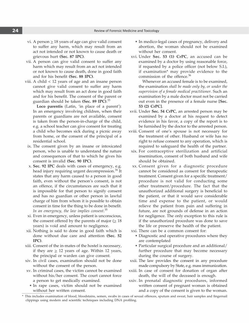
24 Review of Forensic Medicine and Toxicology
vi. A person > 18 years of age can give valid consent to suffer any harm, which may result from an act not intended or not known to cause death or grievous hurt (Sec. 87 IPC).
vii. A person can give valid consent to suffer any harm which may result from an act not intended or not known to cause death, done in good faith and for his benefit (Sec. 88 IPC).
viii. A child < 12 years of age and an insane person cannot give valid consent to suffer any harm which may result from an act done in good faith and for his benefit. The consent of the parent or guardian should be taken (Sec. 89 IPC).33
Loco parentis (Latin, ‘in place of a parent’): In an emergency involving children, when their parents or guardians are not available, consent is taken from the person-in-charge of the child, e.g. a school teacher can give consent for treating a child who becomes sick during a picnic away from home, or the consent of the principal of a residential school.
ix. The consent given by an insane or intoxicated person, who is unable to understand the nature and consequences of that to which he gives his consent is invalid (Sec. 90 IPC).
x. Sec. 92 IPC deals with cases of emergency, e.g. head injury requiring urgent decompression.34 It states that any harm caused to a person in good faith, even without the person’s consent, is not an offence, if the circumstances are such that it is impossible for that person to signify consent and has no guardian or other person in lawful charge of him from whom it is possible to obtain consent in time for the thing to be done in benefit. In an emergency, the law implies consent.35
xi. Even in emergency, unless patient is unconscious, the consent offered by the parents of major (> 18 years) is void and amount to negligence.
xii. Nothing is said to done in good faith which is done without due care and attention (Sec. 52 IPC).
xiii. Consent of the inmates of the hostel is necessary, if they are > 12 years of age. Within 12 years, the principal or warden can give consent.
xiv. In civil cases, examination should not be done without the consent of the person.
xv. In criminal cases, the victim cannot be examined without his/her consent. The court cannot force a person to get medically examined.
• In rape cases, victim should not be examined without her written consent.
• In medico-legal cases of pregnancy, delivery and abortion, the woman should not be examined without her consent.
xvi. Under Sec. 53 (1) CrPC, an accused can be examined by a doctor by using reasonable force, if requested by a police officer (not below S.I.), if examination* may provide evidence to the commission of the offence.36
Whenever an accused female is to be examined, the examination shall be made only by, or under the supervision of a female medical practitioner. Such an examination by a male doctor must not be carried out even in the presence of a female nurse [Sec. 53 (2) CrPC].
xvii. Under Sec. 54 CrPC, an arrested person may be examined by a doctor at his request to detect evidence in his favor, a copy of the report is to be furnished by the doctor to the arrested person.
xviii. Consent of one’s spouse is not necessary for the treatment of other. husband or wife has no right to refuse consent to any operation, which is required to safeguard the health of the partner.
xix. For contraceptive sterilization and artificial insemination, consent of both husband and wife should be obtained.
xx. Consent given for a diagnostic procedure cannot be considered as consent for therapeutic treatment. Consent given for a specific treatment/procedure is not valid for conducting some other treatment/procedure. The fact that the unauthorized additional surgery is beneficial to the patient, or that it would save considerable time and expense to the patient, or would relieve the patient from pain and suffering in future, are not grounds of defense in an action for negligence. The only exception to this rule is if the unauthorized procedure was done to save the life or preserve the health of the patient.
xxi. There can be a common consent for: • Diagnostic and operative procedures where they
are contemplated • Particular surgical procedure and an additional/
further procedure that may become necessary during the course of surgery.
xxii. The law provides the consent in any procedure made compulsory by State, e.g. mass immunization.
xxiii. In case of consent for donation of organ after death, the will of the deceased is enough.
xxiv. In prenatal diagnostic procedures, informed written consent of pregnant woman is obtained and a copy of the consent is given to the woman.
* This includes examination of blood, bloodstains, semen, swabs in cases of sexual offences, sputum and sweat, hair samples and fingernail clippings using modern and scientific techniques including DNA profiling.

25Medical Jurisprudence and Ethics
xxv. Pathological autopsy should not be carried out without the consent of next of kin of the deceased.37
xxvi. Medicolegal autopsy does not require any consent from the relatives of the deceased.
Consent is invalid if: � It is not an informed consent. � Given for committing a crime or an illegal act, such as criminal abortion.
� Obtained by misrepresentation or fraud. � Given by one who had no legal capacity to give it, e.g. a minor or an insane person.
� Substituted consent: If a person in need of treatment is incapable of giving informed consent, consent (proxy consent) must be obtained from next of kin. The order of succession is generally spouse, adult child, parent and sibling.
� Blanket (open) consent: The consent taken at the time of admission, and practiced in most hospitals that cover almost everything a doctor might do to a patient without mentioning anything specific. It is of questionable legal validity.38
� Presumed consent assumes that an individual agrees in principle to the said procedure; if not, he/she must withdraw his/her consent, i.e. ‘opt out’.
Medical Records
Medical records pertains to documents containing a chronological written account of the patient’s medical history and complaints, physical findings, results of diagnostic tests, medications, therapeutic procedures and day-wise progress notes recorded by a medical practitioner.
� It is a part of medical training and one must make a habit of keeping records, not only in the interest of medical science, but also for his own safety and interest.
� It serves as a documentary evidence of the patient’s illness, treatment and response to the treatment. This record may be used as evidence in malpractice suits, claims of the insurances and compensations in personal injury suits. The dictum is that ‘If it is not in the record—it did not occur’.
� Records are the property of the hospital and the personal data contained in the medical record is considered confidential information and the property of the patient.
� Original hospital record of the medico-legal case (MLC) including Xray/CT/MRI films should not be handed over to the police. however, if the investigating officer requests, a photocopy of the record (bed-head-ticket) may be supplied and a receipt of the same must be obtained.
� Medicolegal report (MLR) and postmortem report (PMR) belongs to the requestor, i.e. the police and the same is held by the doctor in fiduciary relationship.
� If affected party is asking for a record, then attested photocopy of the MLR can be handed over to the patient or his/her relative and after the requisite fee has been paid by applicant.
� Request for supply of copy of MLR or PMR under the RTI Act are not maintainable under Section 8(1) (e) & Section 8(1) (h). It should not be issued to third parties (including the accused) by the hospital authorities.
� Safe custody of the patient’s confidential records, whether kept in conventional manner or in a computer, is the responsibility of the doctor.
� Patient’s record cannot be used in clinics or conferences without the patient’s consent.
� hospitals have the right to use the records without consent for evaluating the quality of care and statistical purposes.
� Xray films are the property of the hospital/doctor as part of the record, the patient is entitled for the skill and treatment, but copies of records and X-ray films may be given.
� Under the Directorate General of Health Services guidelines published in ‘Hospital Manual’, the responsibility of hospital to keep medical records is upto 5 years for outpatient department, and for inpatient medical records (including case sheets of medico-legal cases) it is upto 10 years.
Malingering (Shamming)
Definition: It is a conscious planned feigning or pretence to having a disease in order to achieve a specific goal.39
Reasons
i. by soldiers or policemen to evade their duties ii. by prisoners to avoid hard work iii. by businessmen to avoid business contracts iv. by workmen to claim compensation v. by beggars to attract public sympathy vi. by criminals to avoid legal responsibility.
Diseases feigned: Ophthalmia, neurasthenia, dyspepsia, aphasia, intestinal colic, sciatica, diabetes, vertigo, spitting of blood, epilepsy, ulcers, insanity, burns, paralysis of limbs, rheumatism, artificial bruise, lumbago, etc.40
� Usually the signs and symptoms do not conform to any known disease.

26 Review of Forensic Medicine and Toxicology
� Patients can distort or exaggerate their symptoms, but true simulation is very rare.
� history of the case should be taken from the person himself and his relatives or friends, and any inconsistencies in this description of the symptoms are noted.
� A complete examination is essential after removing the bandages, if any, and washing the part.
� It can be diagnosed by keeping the patient under observation, and watching him without his knowledge.
Euthanasia (Mercy Killing)
Definition: Euthanasia (Greek, good death) denotes producing painless death of a person suffering from hopelessly incurable and painful disease.
Types: It can be of two types (Diff. 1.3) i. Active euthanasia ii. Passive euthanasia
It can also be classified into: i. Voluntary euthanasia: Wherein the individual
requests euthanasia, either during illness or before, if complete incapacitation is expected.
ii. Non-voluntary euthanasia: Where an individual is incapable of perception and feeling, and hence cannot decide or distinguish between life and death, such a person cannot give informed consent, e.g. when resuscitation is not expected after severe brain damage as in coma patients or severely defective infants.
iii. Involuntary euthanasia: Where an individual may distinguish between life and death, and any medical killing is involuntary, i.e. against the will of the person. It is ethically, morally and legally considered as murder. This is not to be confused with medical killing in cases of capital punishment.
Arguments against Euthanasia
i. It is against medical ethics. ii. Medical science is making rapid progress; a disease
which is incurable today may became curable tomorrow.
iii. It would not only be for people who are ‘terminally ill’, but may be used to commit murder. It could
be misused by doctors coming hand in glove with relatives.
iv. It can become a means of health care cost containment. v. It may become non-voluntary. vi. It is a rejection of the importance and value of
human life. vii. It is a crime against society and equivalent to
legalizing murder and suicide. It will encourage people to commit suicide.
Reasons for Euthanasia
i. unbearable pain: Patient should be allowed a dignified painless death, instead of prolonging the same through the torture of pain and disease.
ii. High cost of medical treatment: It may pose economic and psychological burden to the patient’s relative.
iii. Right to commit suicide. iv. Patient should not be forced to stay alive: Medical
science too has its limitation and cannot cure all diseases.
� Pediatric euthanasia: Euthanasia administered to seriously sick or deformed infants.
� Geriatric euthanasia: Euthanasia administered to seriously sick, aged individuals.
� Battlefield euthanasia: Euthanasia administered to severely wounded or handicapped individual.
� Supreme court has allowed passive euthanasia in patients with permanent vegetative state but rejected active euthanasia.41 The decision has to be taken to discontinue life support either by parents or spouse or other close relatives or in the absence of any of them—by a person or a body acting as a `next friend’. Recently, however, the government rejected the implementation of the ‘Medical Treatment of Terminally-ill Patients (Protection of Patients and Medical Practitioners) Bill, 2006’ because it is not in favor of ‘mercy killings’.
� Physician-assisted suicide (PAS) is the physician prescribing a drug or other action to facilitate a patient taking his/her own life, with the committed action taken by the patient. The terms ‘PAS’ and ‘euthanasia’ are often used interchangeably.
� Belgium, Netherlands, Luxembourg, Switzerland and the US States of Oregon, Washington, Montana, Vermont permit some forms of euthanasia.41
� By far, most reported cases of euthanasia concern cancer patients.
� Palliative care: The provision of reasonable medical and nursing procedures for the relief of physical pain, discomfort or emotional and psychological suffering as well as providing food and water in terminally ill patients.42

27Medical Jurisprudence and Ethics
1. Medical etiquette is related to: NEET 13 A. Legal responsibilities of doctors B. Rules to be followed by doctors C. Courtesy observed between doctors D. Moral principles followed by doctors 2. Schedule that recognizemedicalqualifications
awarded by institutions in India: AIIMS 11 A. Schedule I B. Schedule II C. Schedule III Part I D. Schedule III Part II 3. Medicalqualificationsawardedbyinstitutionsoutside
India and recognized by MCI are registered in: AI 06 A. First schedule of Indian Medical Council Act 1956 B. Second schedule of Indian Medical Council Act 1956 C. Part I of third schedule of Indian Medical Council
Act 1956 D. Part II of third schedule of Indian Medical Council
Act 1956 4. Professional death sentence is given by:
AFMC 11; NEET 14 A. Central Health Ministry B. Medical Council of India C. Indian Medical Association D. State Medical Council 5. Appeal against penal eraser can be done in: WB 11 A. State Medical Council B. Medical Council of India C. Central Health Ministry D. State Health Ministry 6. Prohibition of participation in torture by a doctor
comes under: MP 10; NEET 15; COMEDK 15 A. Declaration of Tokyo B. Declaration of helsinki C. Declaration of Oslo D. Declaration of Geneva
7. Declaration of Helsinki is about: Odisha 11; NEET 14 A. Organ transplantation B. human experimentation C. Torture D. Physician’s oath 8. Declaration of Oslo is related to which among the
following: DNB 10 A. Torture B. Capital Punishment C. Medical Termination of Pregnancy D. human experimentation 9. As per MCI, minimum period for which a doctor
should preserve his patient’s record: JIPMER 13 A. 2 years B. 3 years C. 4 years D. 5 years 10. In the court of law, professional secrecy can be divulged
under: MAHE 11 A. Doctrine of Common Knowledge B. Privileged communications C. Res ipsa loquitor D. Therapeutic privilege 11. Privileged communication is between: AI 09 A. Doctor-patient B. Doctor-medical council C. Doctor-court D. Doctor-police 12. Infamous conduct comprises of all, except: Delhi 11 A. Adultery B. Advertising C. Procuring criminal abortion D. Examining a patient without consent 13. Professional death sentence is: TN 07 A. Imprisonment for life B. Rigorous imprisonment C. Erasing of name from the medical register D. Death by hanging
Differentiation 1.3: Active and passive euthanasia41
S.No. Feature Active euthanasia Passive euthanasia
1. Definition Positive merciful act, to end useless suffering or a meaningless existence
Discontinuing or not using extraordinary life-sustaining measures to prolong life
2. Principle It is an act of commission It is an act of omission
3. Procedure Administration of lethal doses of opium/barbiturate/sodium thiopental and then a muscle relaxant
Allowing death by not resuscitating a terminally ill or incapacitated patient or defective newborn infant
4. characteristic feature Using measures that would hasten death Not using measures that would delay death
5. Followed in Netherlands and Belgium India and in some States of the US (e.g. with holding tube-feeding)
Multiple ChoiCe Questions
1. C 2. A 3. B 4. D 5. C 6. A 7. B 8. C 9. B 10. B 11. C 12. D 13. C
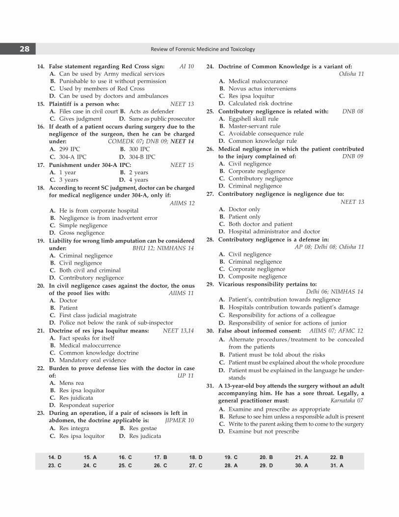
28 Review of Forensic Medicine and Toxicology
14. False statement regarding Red Cross sign: AI 10 A. Can be used by Army medical services B. Punishable to use it without permission C. Used by members of Red Cross D. Can be used by doctors and ambulances 15. Plaintiff is a person who: NEET 13 A. Files case in civil court B. Acts as defender C. Gives judgment D. Same as public prosecutor 16. If death of a patient occurs during surgery due to the
negligence of the surgeon, then he can be charged under: COMEDK 07; DNB 09; NEET 14
A. 299 IPC B. 300 IPC C. 304A IPC D. 304B IPC 17. Punishment under 304-A IPC: NEET 15 A. 1 year B. 2 years C. 3 years D. 4 years 18. According to recent SC judgment, doctor can be charged
for medical negligence under 304-A, only if:AIIMS 12
A. he is from corporate hospital B. Negligence is from inadvertent error C. Simple negligence D. Gross negligence 19. Liability for wrong limb amputation can be considered
under: BHU 12; NIMHANS 14 A. Criminal negligence B. Civil negligence C. both civil and criminal D. Contributory negligence 20. In civil negligence cases against the doctor, the onus
of the proof lies with: AIIMS 11 A. Doctor B. Patient C. First class judicial magistrate D. Police not below the rank of sub-inspector 21. Doctrine of res ipsa loquitur means: NEET 13,14 A. Fact speaks for itself B. Medical maloccurrence C. Common knowledge doctrine D. Mandatory oral evidence 22. Burden to prove defense lies with the doctor in case
of: UP 11 A. Mens rea B. Res ipsa loquitor C. Res juidicata D. Respondeat superior 23. During an operation, if a pair of scissors is left in
abdomen, the doctrine applicable is: JIPMER 10 A. Res integra B. Res gestae C. Res ipsa loquitor D. Res judicata
24. Doctrine of Common Knowledge is a variant of: Odisha 11
A. Medical maloccurance B. Novus actus interveniens C. Res ipsa loquitur D. Calculated risk doctrine 25. Contributory negligence is related with: DNB 08 A. Eggshell skull rule B. Masterservant rule C. Avoidable consequence rule D. Common knowledge rule 26. Medical negligence in which the patient contributed
to the injury complained of: DNB 09 A. Civil negligence B. Corporate negligence C. Contributory negligence D. Criminal negligence 27. Contributory negligence is negligence due to:
NEET 13 A. Doctor only B. Patient only C. both doctor and patient D. hospital administrator and doctor 28. Contributory negligence is a defense in:
AP 08; Delhi 08; Odisha 11 A. Civil negligence B. Criminal negligence C. Corporate negligence D. Composite negligence 29. Vicarious responsibility pertains to: Delhi 06; NIMHAS 14 A. Patient’s, contribution towards negligence B. Hospitals contribution towards patient’s damage C. Responsibility for actions of a colleague D. Responsibility of senior for actions of junior 30. False about informed consent: AIIMS 07; AFMC 12 A. Alternate procedures/treatment to be concealed
from the patients B. Patient must be told about the risks C. Patient must be explained about the whole procedure D. Patient must be explained in the language he under-
stands 31. A 13-year-old boy attends the surgery without an adult
accompanying him. He has a sore throat. Legally, a general practitioner must: Karnataka 07
A. Examine and prescribe as appropriate B. Refuse to see him unless a responsible adult is present C. Write to the parent asking them to come to the surgery D. Examine but not prescribe
14. D 15. A 16. C 17. B 18. D 19. C 20. B 21. A 22. B 23. C 24. C 25. C 26. C 27. C 28. A 29. D 30. A 31. A
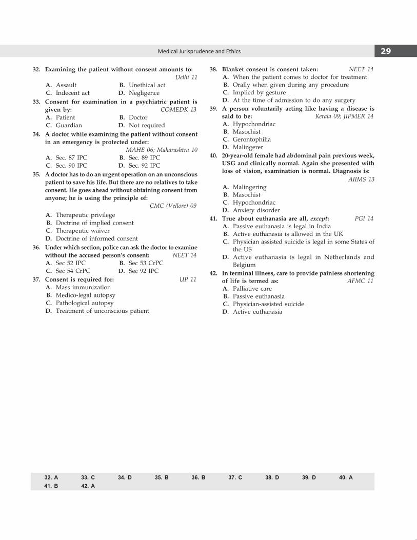
29Medical Jurisprudence and Ethics
32. Examining the patient without consent amounts to:Delhi 11
A. Assault B. Unethical act C. Indecent act D. Negligence 33. Consent for examination in a psychiatric patient is
given by: COMEDK 13 A. Patient B. Doctor C. Guardian D. Not required 34. A doctor while examining the patient without consent
in an emergency is protected under:MAHE 06; Maharashtra 10
A. Sec. 87 IPC B. Sec. 89 IPC C. Sec. 90 IPC D. Sec. 92 IPC 35. A doctor has to do an urgent operation on an unconscious
patient to save his life. But there are no relatives to take consent. He goes ahead without obtaining consent from anyone; he is using the principle of:
CMC (Vellore) 09
A. Therapeutic privilege B. Doctrine of implied consent C. Therapeutic waiver D. Doctrine of informed consent 36. under which section, police can ask the doctor to examine
without the accused person’s consent: NEET 14 A. Sec 52 IPC B. Sec 53 CrPC C. Sec 54 CrPC D. Sec 92 IPC 37. Consent is required for: UP 11 A. Mass immunization B. Medicolegal autopsy C. Pathological autopsy D. Treatment of unconscious patient
38. Blanket consent is consent taken: NEET 14 A. When the patient comes to doctor for treatment B. Orally when given during any procedure C. Implied by gesture D. At the time of admission to do any surgery 39. A person voluntarily acting like having a disease is
said to be: Kerala 09; JIPMER 14 A. hypochondriac B. Masochist C. Gerontophilia D. Malingerer 40. 20-year-old female had abdominal pain previous week,
uSG and clinically normal. Again she presented with loss of vision, examination is normal. Diagnosis is:
AIIMS 13 A. Malingering B. Masochist C. hypochondriac D. Anxiety disorder 41. True about euthanasia are all, except: PGI 14 A. Passive euthanasia is legal in India B. Active euthanasia is allowed in the UK C. Physician assisted suicide is legal in some States of
the US D. Active euthanasia is legal in Netherlands and
belgium 42. In terminal illness, care to provide painless shortening
of life is termed as: AFMC 11 A. Palliative care B. Passive euthanasia C. Physician-assisted suicide D. Active euthanasia
32. A 33. C 34. D 35. B 36. B 37. C 38. D 39. D 40. A 41. B 42. A
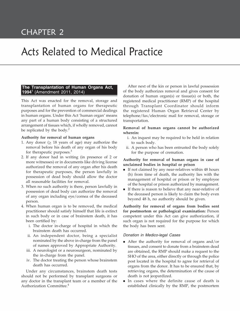
The Transplantation of Human Organs Act, 19941 (Amendment 2011, 2014)
This Act was enacted for the removal, storage and transplantation of human organs for therapeutic purposes and for the prevention of commercial dealings in human organs. Under this Act ‘human organ’ means any part of a human body consisting of a structured arrangement of tissues which, if wholly removed, cannot be replicated by the body.2
Authority for removal of human organs1. Any donor (> 18 years of age) may authorize the
removal before his death of any organ of his body for therapeutic purposes.3
2. If any donor had in writing (in presence of 2 or more witnesses) or in documents like driving license authorized the removal of any organ after his death for therapeutic purposes, the person lawfully in possession of dead body should allow the doctor all reasonable facilities for removal.
3. When no such authority is there, person lawfully in possession of dead body can authorize the removal of any organ including eye/cornea of the deceased person.
4. When human organ is to be removed, the medical practitioner should satisfy himself that life is extinct in such body or in case of brainstem death, it has been certified by:
i. The doctor in-charge of hospital in which the brainstem death has occurred.
ii. An independent doctor, being a specialist nominated by the above in-charge from the panel of names approved by Appropriate Authority.
iii. A neurologist or a neurosurgeon, nominated by the in-charge from the panel.
iv. The doctor treating the person whose brainstem death has occurred.
Under any circumstances, brainstem death tests should not be performed by transplant surgeons or any doctor in the transplant team or a member of the Authorization Committee.4
After next of the kin or person in lawful possession of the body authorizes removal and gives consent for donation of human organ(s) or tissue(s) or both, the registered medical practitioner (RMP) of the hospital through Transplant Coordinator should inform the registered Human Organ Retrieval Center by telephone/fax/electronic mail for removal, storage or transportation.
Removal of human organs cannot be authorized wherein: i. An inquest may be required to be held in relation
to such body. ii. A person who has been entrusted the body solely
for the purpose of cremation.
Authority for removal of human organs in case of unclaimed bodies in hospital or prison
� If not claimed by any near-relatives within 48 hours (h) from time of death, the authority lies with the management of hospital or prison or by employee of the hospital or prison authorized by management.
� If there is reason to believe that any near-relative of the deceased person is likely to claim the body even beyond 48 h, no authority should be given.
Authority for removal of organs from bodies sent for postmortem or pathological examination: Person competent under this Act can give authorization, if such organ is not required for the purpose for which the body has been sent.
Donation in medico-legal cases
� After the authority for removal of organs and/or tissues, and consent to donate from a brainstem dead are obtained, the RMP should make a request to the SHO of the area, either directly or through the police post located in the hospital to agree for retrieval of organs from the donor. It has to be ensured that, by retrieving organs, the determination of the cause of death is not jeopardized.
� In cases where the definite cause of death is established clinically by the RMP, the postmortem
Acts Related to Medical Practice
ChAPteR 2
31Acts Related to Medical Practice
The doctor is also prohibited from removal or transplantation of human organs for any purpose other than therapeutic purposes.
Punishment for doctor on removal of human organs without authority i. Punishable with imprisonment for 5 years and fine
upto ` 10,000.5
ii. Removal of his name from the register of State Medical Council for a period of 2 years for the 1st offence and permanently for the subsequent offence.
Punishment for commercial dealings in human organs Punishable with imprisonment for a term from 2–7 years and fine of ` 10,000–20,000.
Duties of the Medical Practitioner Regarding Organ Transplantation
I. In case of live donation, the doctor should satisfy himself before removing an organ from the donor that:
a. Donor has given his authorization. b. Donor is in proper state of health and fit to
donate the organ. c. Donor is a near-relative of the recipient and sign
a certificate after carrying out following tests on donor and recipient:
i. Tests for the antigenic products of HLA-A, HLA-B and HLA-DR using conventional serological techniques.6
ii. Tests to establish HLA-DR β and HLA-DQ β gene restriction fragment length polymorphisms.
iii. When the above tests do not establish genetic relationship, tests to establish DNA polymor-phisms using at least two multilocus genes probe.
iv. When (iii) do not establish genetic relation-ship, further tests to establish DNA polymorphisms using at least five single locus polymorphism probes.
d. In case recipient is a spouse of donor, record the statements of both and sign a certificate.
e. In case of a donor not being a near-relative, the permission from the Authorization Committee has been obtained.
f. If a donor and/or recipient is/are foreign nationals, the approval of the Authorization Committee has been obtained.
may be waived off by the competent officer on the request of the RMP and Investigating Officer (IO) of the case.
� The RMP who is designated to do the postmortem can do the organ retrieval also. Otherwise, he should be present at the time of retrieval of organs/tissues by the retrieval team. The postmortem report in respect of the organs/tissues being retrieved should be prepared at the time of retrieval. Rest of the postmortem procedure should take place at the autopsy room.
� For the purpose of organ(s)/tissue(s) retrieval, request for postmortem beyond specified timings, can be made by the RMP and IO of the case.
Restriction on removal and transplantation of human organs i. Human organ should not be removed from the
body of donor before his death and transplanted into recipient, unless the donor is a near-relative (spouse, son, daughter, father, mother, brother or sister, grandparents, grandchildren, uncles and aunts).
ii. When donor authorizes the removal of his organs after his death, these organs may be transplanted into the body of any recipient.
iii. If any donor authorizes the removal of his organs before his death to such recipient not being near-relative by reason of affection or attachment towards the recipient, the organs should not be removed and transplanted without prior approval of Authoriza-tion Committee.
The law made it legal: � Not-so-close relatives who have stayed with the
patient can denote organs, provided there is no commercial dealing.
� Swapping of organs between two unrelated families if the organs of the respective willing ‘near relative’ donors are found medically incompatible for the intended recipients.
Regulation of hospitals conducting the removal, storage or transplantation of human organs i. Hospital not registered under this Act should not
be engaged in transplantation activities. ii. Medical practitioner should not conduct transplanta-
tion at any unregistered place under this Act. iii. The eyes and the ears may be removed at any
place from dead body of any donor for therapeutic purposes by a doctor. Removal of eye can be done by a trained technician to facilitate eye donation.

32 Review of Forensic Medicine and Toxicology
II. In case of cadaveric donation, the doctor should satisfy himself that:
i. Donor has authorized before his death, the removal of his organ for therapeutic purpose, in presence of two or more witnesses, at least one of whom is a near-relative.
ii. Person lawfully in possession of dead body has signed a certificate as specified under the Act.
III. A doctor, before removing organ from a brainstem dead person, should satisfy that:
i. Certificate regarding the brainstem dead from the Board of medical experts is present. In case of non-availability of a neurosurgeon or a neurologist to certify brain death, an intensivist or anesthetist can be included on the medical board. It is mandatory for the ICU/treating medical staff to request relatives of a brain-dead patient for organ donation.
ii. In case of a person < 18 years, a certificate has been signed by either of the parents of such person. Living organ/tissue donation by minors is not permitted, except on exceptional medical grounds and with prior approval of the Appropriate Authority and the Government concerned.
Authorization Committee i The medical practitioner involved in organ
transplantation team should not be a member of the Authorization Committee.
ii. In case of foreigners, where the transplant is between a married couple, the Committee must evaluate the factum and duration of marriage and verify documents such as marriage certificate, or marriage photograph.
iii. Transplantation is not permitted if the recipient is a foreign national and donor is an Indian national, unless they are near-relatives.
iv. When the proposed donor and the recipient are not ‘near-relatives’, the Committee should evaluate that:
a. there is no commercial transaction between the recipient and the donor; the following is specifically assessed:
– link between them and the circumstances which led to the offer being made.
– reasons why the donor wishes to donate. – documentary evidence of the link, e.g. proof
that they have lived together. – old photographs showing the donor and the
recipient together.
b. there is no middleman or tout involved. c. financial status of the donor and the recipient,
evidence of their vocation and income for the previous three financial years.
d. donor is not a drug addict. e. next of the kin of unrelated donor is interviewed
regarding awareness about his/her intention to donate an organ and/or tissue, the authenticity of the link between the donor and the recipient and the reasons for donation.
� Organ donation is considered in case of brain-death, since ventilator supplies necessary oxygen to these organs functioning, whereas tissues like cornea, heart valves, bone and skin can be harvested after cardiac death as well.
� One set of corneas are given to two people needing sight. Heart valves are used in valve replacement surgery (common in children), skin grafts are used in burn patients, and bone, tendons and ligaments can be used in reconstructive surgeries.
� Organs and tissues that can be transplanted: Liver (one of the most difficult), kidney, pancreas, pancreatic islet cells, small intestine, lung, heart, corneas, skin graft, blood vessels, bone and hand.7
� Maximum time an organ can be stored before transplant: heart: 3 h; liver and pancreas: 12 h; kidneys: 24 h; cornea: 2 weeks; middle ear, skin, bone marrow: 5 years; heart valve: 10 years.
� Maximum time for organ retrieval in case of normal death: skin: 3 h; cornea: 6 h.
� Under Spanish law, every dead person can provide organs, unless the deceased person expressly rejected it (presumed consent). Nonetheless, doctors ask the family for permission.
� In the US, law requires that the donor made a statement during his lifetime that he is willing to be an organ donor. Many States have sought to encourage the donations to be made by allowing the consent to be noted on the driver’s license. Still, it remains an opt-in system rather than the Spanish opt-out system.
The Consumer Protection Act, 1986 (CPA) (Amendment in 1991, 1993, 2002)
Purpose: This Act was brought into existence for the protection of interests of the consumer and for settlement of consumer disputes, within a limited time frame and with fewer expenses. This enables a patient to make a complaint to a redressal forum in respect of a defective (negligent) service, if the service has been paid for.
Redressal Agencies (Flow chart 2.1)
It is established at three different levels:8
i. District forum headed by the District Judge, situated in each district of the State. The jurisdiction to entertain complaints is limited to those where the value of services is < ` 20 lakhs.
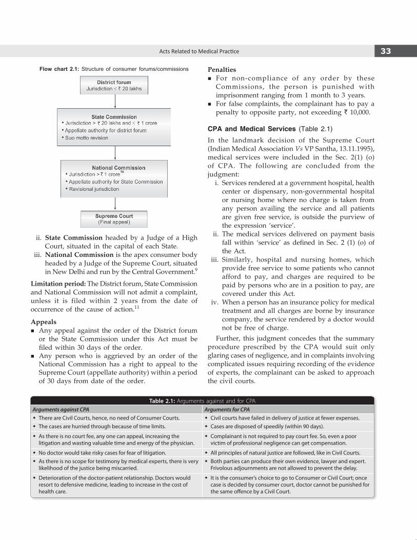
33Acts Related to Medical Practice
Penalties � For non-compliance of any order by these
Commissions, the person is punished with imprisonment ranging from 1 month to 3 years.
� For false complaints, the complainant has to pay a penalty to opposite party, not exceeding ` 10,000.
CPA and Medical Services (Table 2.1)
In the landmark decision of the Supreme Court (Indian Medical Association Vs VP Santha, 13.11.1995), medical services were included in the Sec. 2(1) (o) of CPA. The following are concluded from the judgment: i. Services rendered at a government hospital, health
center or dispensary, non-governmental hospital or nursing home where no charge is taken from any person availing the service and all patients are given free service, is outside the purview of the expression ‘service’.
ii. The medical services delivered on payment basis fall within ‘service’ as defined in Sec. 2 (1) (o) of the Act.
iii. Similarly, hospital and nursing homes, which provide free service to some patients who cannot afford to pay, and charges are required to be paid by persons who are in a position to pay, are covered under this Act.
iv. When a person has an insurance policy for medical treatment and all charges are borne by insurance company, the service rendered by a doctor would not be free of charge.
Further, this judgment concedes that the summary procedure prescribed by the CPA would suit only glaring cases of negligence, and in complaints involving complicated issues requiring recording of the evidence of experts, the complainant can be asked to approach the civil courts.
ii. State Commission headed by a Judge of a High Court, situated in the capital of each State.
iii. National Commission is the apex consumer body headed by a Judge of the Supreme Court, situated in New Delhi and run by the Central Government.9
Limitation period: The District forum, State Commission and National Commission will not admit a complaint, unless it is filed within 2 years from the date of occurrence of the cause of action.11
Appeals � Any appeal against the order of the District forum
or the State Commission under this Act must be filed within 30 days of the order.
� Any person who is aggrieved by an order of the National Commission has a right to appeal to the Supreme Court (appellate authority) within a period of 30 days from date of the order.
Flow chart 2.1: Structure of consumer forums/commissions
Table 2.1: Arguments against and for CPA Arguments against CPA Arguments for CPA
� There are Civil Courts, hence, no need of Consumer Courts. � Civil courts have failed in delivery of justice at fewer expenses. � The cases are hurried through because of time limits. � Cases are disposed of speedily (within 90 days).
� As there is no court fee, any one can appeal, increasing the litigation and wasting valuable time and energy of the physician.
� Complainant is not required to pay court fee. So, even a poor victim of professional negligence can get compensation.
� No doctor would take risky cases for fear of litigation. � All principles of natural justice are followed, like in Civil Courts. � As there is no scope for testimony by medical experts, there is very
likelihood of the justice being miscarried. � Both parties can produce their own evidence, lawyer and expert.
Frivolous adjournments are not allowed to prevent the delay.
� Deterioration of the doctor-patient relationship. Doctors would resort to defensive medicine, leading to increase in the cost of health care.
� It is the consumer’s choice to go to Consumer or Civil Court; once case is decided by consumer court, doctor cannot be punished for the same offence by a Civil Court.

34 Review of Forensic Medicine and Toxicology
• Cardiac disease (Grade III and IV) • Chronic glomerulonephritis • Intractable hyperemesis gravidarum • Malignant hypertension • Epilepsy/Insanity • Cervical or breast carcinoma • Diabetes with retinopathy • Toxemia of pregnancy. ii. Eugenic: Risk of the child being born with serious
physical or mental abnormalities. Indications are: • Mother exposed to teratogenic drugs (warfarin) or
radiation exposure (> 10 rads) in early pregnancy. • German measles (Rubella), chicken pox, viral
hepatitis or other viral infections, if contacted within 1st trimester.
• Structural (anencephaly), chromosomal (Down’s syndrome) or genetic abnormalities of the fetus.
• Parents have inheritable mental condition or chromosomal abnormalities.
iii. Socio-economic: Almost the sole indication, to prevent grave injury to the physical and mental health of the pregnant lady. Conditions include:
• Unplanned pregnancy with low socio-economic status (80% of cases).
• Pregnancy as a result of failure of contraception used by the married woman or her husband. All the pregnancies can be terminated using this criterion.
iv. Humanitarian: Pregnancy caused by rape.
MTP Act Rules
Emergency cases: Pregnancy can be terminated by any RMP, even without required experience at any place, irrespective of duration of pregnancy, if it is necessary to save the life of pregnant woman.
Length of pregnancy: Under MTP Act, pregnancy cannot be terminated after 20 weeks of pregnancy.14 Above 20 weeks, the pregnancy can be terminated only on therapeutic considerations, i.e. to save the life of the mother.* In such cases, decision can be taken by a single doctor.
� Upto 12 weeks of pregnancy, pregnancy can be terminated on the opinion of a single doctor.15
� Between 12–20 weeks, decision should be taken jointly by two doctors.16
Medical Indemnity InsuranceIt is a contract under which the insurance company agrees, in return for the payment of premiums, to indemnify (cover) the insured doctor as a result of his claimed professional negligence.
Objectives of Medical Indemnity Insurance i. To look after and protect the professional interests
of the insured doctor. ii. To arrange, conduct and pay for the defense of
such doctor. iii. To arrange all other professional assistance
including pre-litigation advice. iv. To indemnify the insured doctor in respect of
any loss or expense directly arising from actions, claims and demands against him on grounds of professional negligence.
The Workmen’s Compensation Act, 1923
This Act provides for the payment of compensation to workmen for injuries sustained by them in an accident, arising out of and in the course of employment.
� If a workman is killed, his dependants will be entitled to compensation for his death.
� The amount of compensation depends upon whether the injury has caused death, permanent total disablement or permanent partial disablement.
� The employer will not be liable to pay compensation in respect of any injury which results in death or permanent total disablement caused by an accident, if the workman at the time of sustaining injury was under the influence of drink or drugs or willfully disregarded or removed any safety guard or other device provided for his safety.
The Medical Termination of Pregnancy (MTP) Act, 1971
The original Act of 1971 came into force on 1st April 1972 and amended in 2002 to provide for the termination of certain pregnancies by the registered medical practitioners (RMP) for protection and preservation of the lives of women.12
Indications for Termination of Pregnancy13
i. Therapeutic: In order to prevent injury to the physical health of pregnant woman. Indications are:
* The Government is planning to increase the length of pregnancy upto 24 weeks with no age restriction in case of aborting ‘substantially abnormal’ fetus.

35Acts Related to Medical Practice
Place where MTP can be Performed
MTP’s can only be conducted at: i. A hospital established or maintained by
Government, or ii. A place approved by Government or a District
level Committee with the Chief Medical Officer or District Health Officer as the Chairperson of the said Committee.
Qualification and Experience of RMP
For RmP conducting mTP upto 12 weeks
The doctor should have the experience of assisting an RMP in conducting 25 cases of MTP, out of which at least 5 cases should have been performed independently, in an approved hospital by the Government.
For RMP’s conducting MTP between 12–20 weeks period
The doctor should have either: � Post-graduate degree/diploma in Obs & Gynae,17
� Six months of house surgency in Obs & Gynae, or � One year or more in the practice of Obs & Gynae
at any hospital.
Consent: Consent of woman is mandatory, except when she is minor (< 18 years) or mentally ill, where consent of the guardian is obtained. Consent of husband is not necessary.
Maintenance of register: The head of the hospital should maintain a register, recording the details of the patient undergoing termination of pregnancy for a period of 5 years, and professional secrecy should be maintained.
Contravention of the rules by the doctor: Liable to be punished with rigorous imprisonment of 2–7 years. A person who willfully contravenes or fails to comply
with the requirements of any regulation under this Act is punished with fine of 1000. It is a cognizable offence for which a police officer can arrest a doctor for violations without warrant. If he is a government servant, he will be liable to face disciplinary action including dismissal from service.
Methods to bring about abortion are given in Table 2.2.
� It is unrealistic to produce a definitive list of conditions that constitute ‘serious physical or mental abnormalities’ since accurate diagnostic techniques are as yet unavailable. Likewise, the consequences of abnormality are difficult to predict.
� In a landmark judgment, the Supreme Court struck down a high court order directing the MTP of an adult woman without her consent on grounds of ‘mental retardation’ (parens patriae jurisdiction). The MTP Act states that a guardian can make decisions on behalf of a ‘mentally ill person’, but this cannot be done on behalf of a person who is in a condition of ‘mental retardation’. The SC observed that the State must respect the personal autonomy of a mentally retarded woman with regard to decisions about MTP.
� A study commissioned by the UK Government found that a human fetus does not feel pain before 24 weeks.
� English common law considered abortions before ‘quickening’ to be morally and legally acceptable.
� In the UK, two doctors must first certify that an abortion is medically or socially necessary before it can be performed.
� Complications are much less in legal abortions done before 8 weeks (5%), but it is about five times more in mid-trimester termination, irrespective of the method employed.
� Deaths during legal abortions are rare. Such deaths are due to: i. Hemorrhage and shock due to trauma, atonic uterus or
incomplete abortion ii. Infection iii. Emboli (thrombotic, amniotic, or air) iv. Complications of anesthesia.
Table 2.2: Methods of inducing abortion under MTP Act
1st trimester (upto 12 weeks)18,19 2nd trimester (upto 20 weeks)
Medical � Dilatation and evacuation (13–14 weeks)
� Mifepristone (RU-486) � Intra-uterine instillation of hyperosmotic solution
� Mifepristone and misoprostol (PGE1) i. Intra-amniotic hypertonic urea (40%), saline (20%)
� Methotrexate and misoprostol ii. Extra-amniotic: Ethacrydine lactate, prostaglandins (PGE2, PGF2α)
� Tamoxifen and misoprostol � Prostaglandins [PGE1, PGE2, PGF2α]: Intravaginally, intra-muscularly or intra-amniotically
Surgical � Oxytocin infusion
� Manual vacuum aspiration (MVA) � Hysterotomy
� Dilatation and evacuation (D & E)
� Suction evacuation and/or curettage

36 Review of Forensic Medicine and Toxicology
� Deaths by method of abortion in developed countries (in decreasing rate of occurrence):
i. Hysterectomy/hysterotomy ii. Instillation methods (including saline) iii. Dilatation and evacuation iv. Dilatation and curettage.
� Curettage is the most common method of abortion used and results in the most deaths because of this, even though it has the lowest rate of death by type of procedure.
� Deaths due to hemorrhage and sepsis are complications of perforation of the uterus. While perforation is a recognized complication of any procedure involving instrumentation of the uterus, death due to sepsis/hemorrhage should not occur and strongly suggest the possibility of medical negligence.
� In India, contrary to the western countries, the mortality from saline method has been found be much higher as compared to termination by abdominal hysterectomy.
The Pre-conception and Prenatal Diagnostic Techniques Act, 1994 (Amendment 2002)
The Pre-conception and Prenatal Diagnostic Techniques (PCPNDT) (Regulation and Prevention of Misuse) Act, 1994, was enacted in order to check female feticide.20
The Act provides for the prohibition of sex selection, before or after conception.
� ‘Prenatal diagnostic procedures’ means any gynecological, obstetrical or medical procedures such as ultrasonography, fetoscopy, samples of amniotic fluid, chorionic villi, embryo, blood or any other tissue or fluid of a man, or of a woman before or after conception, for conducting any type of analysis or prenatal diagnostic tests for selection of sex before or after conception.
� ‘Prenatal diagnostic test’ means ultrasonography or any analysis of amniotic fluid, chorionic villi, blood or any tissue or fluid of a pregnant woman or conceptus conducted to detect any abnormalities or diseases as given in clause 2.
� ‘Prenatal diagnostic techniques’ includes all prenatal diagnostic procedures and prenatal diagnostic tests.Any medical practitioner or any other person should
not conduct or aid in conducting any prenatal diagnostic techniques at a place other than a place registered under this Act.
A recent amendment in the Act allows medical practitioners (MBBS doctors) to conduct sonography tests on pregnant women, provided they undergo 6 months training imparted within the well-defined syllabus prescribed by the Act at accredited institutions.
Regulation of Prenatal Diagnostic TechniquesClause 1: Any place including a registered genetic counseling center, laboratory or clinic should not be
used for conducting prenatal diagnostic techniques except for the purpose given in clause 2 and after satisfying any of the conditions in clause 3.Clause 2: Prenatal diagnostic techniques should be used for the detection of any of the following abnormalities: i. Chromosomal abnormalities ii. Genetic metabolic diseases iii. Hemoglobinopathies iv. Sex linked genetic diseases v. Congenital anomalies.Clause 3: Prenatal diagnostic techniques should be used in pregnant women, if any of the following conditions are satisfied: i. Age > 35 years. ii. Undergone two or more spontaneous abortions or
fetal loss. iii. Has been exposed to potentially teratogenic
agents, such as drugs, radiations, infections or chemicals.
iv. The pregnant woman or her spouse has a family history of mental retardation or physical deformities, such as spasticity or any other genetic disease.
Written consent of pregnant woman and prohibition of communicating the sex of fetus1. Prenatal diagnostic procedures should not be
conducted unless: a. The doctor has explained all known side-effects
and after-effects of such procedures to the patient. b. He has obtained her written consent in a language
which she understands. c. A copy of her written consent obtained above
is given to the pregnant woman.2. The person conducting prenatal diagnostic procedures
including ultrasonography should not communicate to the pregnant woman or her relative, the sex of the fetus by words, signs or in any other manner; and should keep a complete record of the patient. The person should give a declaration on each report on ultrasonography/image scanning that he has neither detected nor disclosed the sex of fetus of the pregnant woman to anybody.*
3. The pregnant woman before undergoing ultra-sonography/image scanning should declare that she does not want to know the sex of her fetus.
Maintenance and Preservation of Records � All such registered genetic/ultrasound/imaging
centers should maintain a register showing, in serial order, the names and addresses of the men or women
* However, sex can be disclosed in sex-linked disorders found at ultrasound and in metabolic disorders found in amniocentesis or chorionic villus sampling, e.g. Alport syndrome or Fabry’s disease.

37Acts Related to Medical Practice
given counseling, subjected to prenatal diagnostic procedures or tests, the names of their spouses or fathers and the date on which they first reported.
� All case related records, forms of consent, laboratory results, microscopic pictures, sonographic plates or slides should be preserved for a period of 2 years from the date of completion of counseling, prenatal diagnostic procedure or test, as the case may be. In the event of any legal proceedings, the records are to be preserved till the final disposal of the case.
� In case the records are maintained on computer or other electronic equipment, a printed copy of the record should be taken and preserved after authentication by a person responsible for such record.
Offences and Penalties
� Any person, organization, genetic counseling center/laboratory/clinic should not issue any advertisement in any manner regarding facilities of prenatal deter-mination of sex.
� Any medical person who contravenes the provisions of this Act is punished with imprisonment upto 3 years and fine upto ` 10,000, and on any subsequent conviction with imprisonment upto 5 years and fine upto ` 50,000.21 His name is removed from the register of the Council for a period of 5 years for the first offence and permanently for the subsequent offence.
� Any person who seeks the aid of genetic counseling laboratory/clinic or medical practitioner for purposes other than specified above, is punished with imprisonment upto 3 years and fine upto ` 50,000, and on any subsequent conviction with imprison-ment upto 5 years and fine upto ` 1 lakh.
� Every offence under this Act is cognizable, non-bailable and non-compoundable.
� Non-bailable: The Magistrate has the power to refuse bail and remand a person to judicial or police custody.
� Non-compoundable: Case (e.g. rape, 498-A) which cannot be withdrawn by the petitioner.
The Delhi Artificial Insemination (Human) Act, 1995
� This Act is applicable in the State of Delhi. It regulates the donation, sale and supply of human semen and ovum for the purpose of artificial insemination. It requires registration and yearly renewal by any person intending to carry on a semen bank.
� The semen bank before accepting the semen for artificial insemination should:
i. Test the donor for the presence of HIV 1 and 2 antibodies by ELISA.
ii. Screen for HIV surface antigen and if found negative, the donor shall be allowed to donate.
� The donated semen is stored either by cryopreservation for a minimum period of 3 months in order to exclude window period of HIV 1 and 2 infections in the donor.
� Second ELISA test is performed on the donor after 3 months, and if negative, the semen is then used.
� It is also required by the practitioner: i. Not to segregate the XX/XY chromosomes for artificial
insemination. ii. Seek the written consent of the recipient for using the semen
on the basis of only one ELISA test, being negative, where facilities for cryopreservation and liquid nitrogen for semen are not available.
The Protection of Children from Sexual Offences (POCSO) Act, 2012
This Act has been drafted to strengthen the legal provisions for the protection of children from sexual abuse and exploitation. Sexual offences are currently covered under various sections of IPC. The IPC does not provide for all types of sexual offences against children and, more importantly, does not distinguish between adult and child victims.
The POCSO Act defines a child as any person below the age of 18 years and provides protection to all children (both males and females) under the age of 18 years from sexual abuse.
Offences and Punishments I. Penetrative sexual assault: A person is said to
commit ‘penetrative sexual assault’ if he: a. penetrates his penis or inserts any object or a
part of the body to any extent, into the vagina, mouth, urethra or anus of a child or makes the child to do so with him or any other person; or
b. manipulates any part of the body of the child so as to cause penetration into the vagina, urethra, anus or any part of body of the child or makes the child to do so with him or any other person; or
c. applies his mouth to the penis, vagina, anus, urethra of the child or makes the child to do so to him or any other person.
Punishment: Imprisonment for ≥ 7 years which may extend for life and fine. For aggravated penetrative sexual assault: Imprisonment for ≥ 10 years which may extend to life imprisonment and fine.

38 Review of Forensic Medicine and Toxicology
II. Sexual assault: Any physical contact with sexual intent but without penetration like touching the vagina, penis, anus or breast of the child or making the child touch the vagina, penis, anus or breast of such person or any other person.
Punishment: Imprisonment for ≥ 3–5 years and fine. For aggravated sexual assault: Imprisonment for ≥ 5–7 years and fine.
III. Sexual harassment of the child: It is considered sexual harassment when a person with sexual intent:
a. utters any word/sound, or makes any gesture or exhibits any object or part of body with the intention to be heard or seen by the child; or
b. makes a child exhibit her body or any part of her body, so as it is seen by the person or any other person; or
c. shows any object to a child in any form or media for pornographic purposes; or
d. repeatedly or constantly follows or watches or contacts a child either directly or through electronic, digital or any other means; or
e. threatens to use, in any form of media, a real or fabricated depiction through electronic, film or digital or any other mode, of involvement of the child in a sexual act; or
f. entices a child for pornographic purposes.
Punishment: Imprisonment for 3 years and fine. IV. Use of child for pornographic purposes: A person
is guilty of the offence if he uses a child in any form of media, for the purposes of sexual gratification, which includes:
a. representation of the sexual organs of a child; b. usage of a child engaged in real or simulated
sexual acts (with or without penetration); c. indecent or obscene representation of a child.
Punishment: Imprisonment for 5 years and fine, and in subsequent conviction: 7 years and fine.
� An offence is treated as ‘aggravated’ when committed by a person in a position of trust or authority of child, such as a member of security forces, police officer, public servant, etc.
� There is provision for punishment even in abetment or an attempt to commit the offences defined in the Act. The punishment for the attempt to commit is upto half the punishment prescribed for the commission of the offence.
� It is mandatory to report to the police about the offence. Failure to report attracts punishment with
imprisonment of upto 6 months or fine or both. It is also mandatory for police to register an FIR in all cases of child abuse. A child’s statement can be recorded even at the child’s residence or a place of her choice and should be preferably done by a female police officer not below the rank of sub-inspector (if the victim is a female).
� The child’s medical examination can be conducted even prior to registration of an FIR. This discretion is left upto the Investigation Officer (IO). The IO has to get the child medically examined in a government hospital or local hospital within 24 h of receiving information about the offence. This is done with the consent of the child or parent or a competent person whom the child trusts and in their presence.
� For speedy trial, the evidence of the child has to be recorded within a period of 30 days. The Special Court has to complete the trial within 1 year.
� The burden of proof is shifted on the accused, keeping in view the vulnerability and innocence of children.
� To prevent misuse of the law, punishment is given for false complaints or false information with malicious intent.
� The media is barred from disclosing the identity of the child without the permission of the Special Court. The punishment for breaching this provision is imprisonment from 6 months to 1 year.
The Sexual Harassment of Women at Workplace (Prevention, Prohibition and Redressal) Act, 2013This Act has been drafted to address sexual harassment at the workplace and creates a mechanism for redressal of complaints. It also provides safeguards against false or malicious charges. Under the Act, which also covers students in schools and colleges as well as patients in hospitals, employers and local authorities will have to set up grievance committees to investigate all complaints.Salient Features
� Sexual harassment includes unwelcome acts or behavior like physical contact and advances, a demand or request for sexual favors or making sexually colored remarks or showing pornography and any other unwelcome physical, verbal or non-verbal conduct of sexual nature.
� Aggrieved woman includes all women, irrespective of her age or employment status, whether in the organized or unorganized sectors, public or private and covers clients, customers and domestic workers as well.
� Workplace include organizations, department, office or branch unit in the public and private sector, organized and unorganized, hospitals, nursing homes, educational institutions, sports institutes, stadiums, sports complex and any place visited by the employee during the course of employment including transportation (Vishaka guidelines was confined to the traditional office set-up where there is a clear employer-employee relationship).
39Acts Related to Medical Practice
The Protection of Women from Domestic Violence Act, 2005Salient Features
� The term ‘domestic violence’ covers all forms of physical, sexual, verbal, emotional and economic abuse that can harm, cause injury, endanger the health, safety, life, limb or well-being, either mental or physical of the aggrieved person.
� ‘Aggrieved’ person’ is not just the wife, but a woman who is the sexual partner of the male irrespective of whether she is his legal wife or not. It also includes daughter, mother, sister, child (male or female), widowed relative, or any woman residing in the household who is related in some way to the respondent.
� ‘Respondent’ is any male, adult person who is, or has been, in a domestic relationship with the aggrieved person, that includes his mother, sister and other relatives; the case can also be filed against relatives of the husband or male partner.
� Information to Protection Officer: The information regarding any acts of domestic violence does not necessarily have to be lodged by the aggrieved party but by any person who has reason to believe that such an act has been or is being committed. Any medical officer, neighbors, social workers or relatives can all take initiative on behalf of the victim.
� Duties of medical facilities: If an aggrieved person or a Protection Officer or a service provider requests the medical practitioner to provide any medical aid to the victim, the doctor should provide medical aid to the aggrieved person in the medical facility.
� Penalties: The Magistrate can impose a penalty of upto 1 year of imprisonment and/or a fine of upto 20,000/- for an offence under this Act. The offence is also considered cognizable and nonbailable. The decision can be taken under the sole testimony of the aggrieved person; the court may conclude that an offence has been committed by the accused.
� The Magistrate can impose monetary relief and monthly pay-ments of maintenance. The respondent can also be made to meet the expenses incurred and losses suffered by the aggrieved person as a result of domestic violence, and can also cover loss of earnings, medical expenses, loss or damage to property and can also cover the maintenance of the victim.
� The Act also allows the Magistrate to make the respondent pay compensation and damages for injuries including mental torture and emotional distress caused by acts of domestic violence.
� Employee covers regular, temporary, ad hoc, daily wage employees and also includes volunteers.
� Employer includes the head of the Government department, organisation, institution, office, branch or unit, the person responsible for management, supervisions or control of the workplace, and in relation to a domestic worker the person who benefits from that employment.
� The redressal mechanism is in the form of Internal Complaints Committee (ICC) and Local Complaints Committee (LCC). All workplaces employing ≥ 10 workers are mandated under the Act to constitute an ICC. The ICC will be a 4 member committee under the Chairpersonship of a senior woman employee and will include 2 members from amongst the employees preferably committed to the cause of women or has experience in social work/legal knowledge and includes a third party member (NGO, etc.) as well.
� Complaints from workplaces employing < 10 workers or when the complaint is against the employer will be looked into by the LCC. A District Officer notified under the Act will constitute the LCC at the district level. LCC will also look into complaints from domestic workers.
� A complaint of sexual harassment can be filed within a time limit of 3 months. This may be extended to another 3 months if the woman can prove that grave circumstances prevented her from doing the same.
� The Complaints Committees have the powers of civil courts for gathering evidence.
� The Committee is required to provide for conciliation before initiating an inquiry, if requested by the victim.
� The Committee is required to complete the inquiry within a time period of 90 days. On completion of the inquiry, the report is sent to the employer or the District Officer, as the case may be, they are mandated to take action on the report within 60 days.
� If found guilty, then the Committee can recommend action in accordance with the provision of service rules applicable to the accused. The Committee can also recommend deduction of an appropriate amount from the salary of the accused or asked to pay the amount. In case the accused fails to pay, District Officer may be asked to recover the amount as an arrear of land revenue.
� In case the allegation is not proved, then the Committee can write to the employer/District Officer that no action needs to be taken.
� In case of malicious or false complaint, the Act provides for penalty according to the Service Rules. However, this clause has a safeguard in the form of an enquiry prior to establishing the malicious intent. Also, mere inability to prove the case will not attract penalty under this provision.
� It prohibits disclosure of the identity and addresses of the aggrieved woman, respondent and witnesses. However, information regarding the justice secured to any victim of sexual harassment under this Act without disclosing the identity can be disseminated.
� Penalties have been prescribed for employers. Non-compliance with the provisions of the Act is punishable with a fine of upto
50,000. Repeated violations may lead to higher penalties and cancellation of license or registration to conduct business.
The Mental Health Act, 1987
The Mental Health Act, 1987 came into force in all the States and Union Territories from 1st April, 1993.22 The Act repealed the Indian Lunacy Act, 1912 and the Lunacy Act, 1977 (J & K Act of 1977). This Act has the following provisions:
� Guidelines for establishment and maintenance of psychiatric hospitals/nursing homes at the Center and State and authorities to regulate these centers.
� Procedure for admission and detention of mentally ill persons*, in psychiatric hospitals.
* ‘Mentally ill person’ means a person who is in need of medical treatment by reason of any mental disorder other than mental retardation [Section 2(b)].

40 Review of Forensic Medicine and Toxicology
Restraint of an Insane
It may be: � Immediate restraint � Admission in psychiatric hospital.
Immediate restraintIt is done in case of: i. An insane person who is dangerous to himself or
to others, or likely to injure, or wastefully spend his property or that of others.
ii. Person suffering from delirium due to disease. iii. Person suffering from delirium tremens.
Immediate restraint is done under the personal care of attendants, e.g. by safely locking-up in a room. The consent of the lawful guardian of the insane person has to be taken, but if there is no time to take the consent, he can be immediately restrained. Such restraint is lawful till the danger exists.
Admission in psychiatric hospitalThe following procedures are adopted: i. On voluntary basis: Any major person, who
considers himself to be mentally ill person, may request the doctor in-charge of psychiatric hospital for admission and treatment. In case of a minor, the guardian may make such request.
The doctor should make an inquiry as deem fit within 24 h, and if satisfied that the person requires treatment as an in-patient, he may admit such person.
ii. Under special circumstances: A mentally ill person who is unable to express his willingness for admission voluntarily, can be admitted in a psychiatric hospital for a period of 90 days, if an application is made by a relative/friend and accompanied by two medical certificates, one of which should be by a government medical officer.
iii. Reception order on application: The doctor in-charge of a psychiatric hospital, husband or wife or any relative can make an application to the Magistrate. The Magistrate must consider the allegations in the petition and the evidence of mental illness as disclosed by the medical certificates. If he is satisfied, he may pass a reception order.
iv. Admission of a mentally ill prisoner: He can be admitted into any psychiatric hospital by an order passed by an Appropriate Authority.
v. Admission of an escaped mentally ill person: He can be retaken by any police officer or worker of the psychiatric hospital and readmitted into the same hospital.
Observation of a mentally ill patient: For diagnosis and certification, an alleged mentally ill patient may be kept under observation (upto 10 days) of a psychiatrist with an order from a Magistrate. If diagnosis cannot be reached during this period, then the Magistrate can order for another 10 days observation (upto a maximum of 30 days).23
Content of Medical CertificatesThe two medical certificates issued by two doctors must contain a statement that each of the medical practitioner: i. Has independently examined the alleged mentally
ill person. ii. Formed his opinion on the basis of his own
observations and from the particulars communi-cated to him.
iii. In their opinion, the alleged mentally ill person is dangerous to self or others because of the mental disorder which warrants detention of such person in a psychiatric hospital.
Discharge of a Mentally Ill Person
i. Voluntary patients should be discharged within 24 h of the receipt of request for discharge made by the patient or the guardian. If medical board is of the opinion that such patient needs further treatment, the patient should not be discharged, and treatment should be continued for a period not exceeding 90 days at a time.
ii. A mentally ill person who is admitted on an application by a relative/friend can himself or a relative/friend can apply to the Magistrate for discharge. The Magistrate, after making an enquiry, can either allow or dismiss the application.
iii. A mentally ill person detained under ‘reception order’ may be discharged, if the person on whose application admission order was made, applies in writing to the doctor in-charge.
iv. Any relative/friend of a mentally ill person can make an application for discharge to the doctor in-charge, who should forward it with his remarks to the authority under whose order the person was detained. Such authority can pass an order of discharge.
� Provides for judicial inquisition regarding possession of property by alleged mentally ill person, custody of the person, and management of his property: If the court is of the opinion that the mentally ill person is not capable of looking after himself and property, a guardian is appointed, and if he is incapable of looking after his property only, a manager is appointed.

41Acts Related to Medical Practice
� Provides for the liability to meet cost of maintenance of a mentally ill person detained in psychiatric hospital.
� Provides for protection of human rights of mentally ill persons: The patient should be treated with dignity, without any cruelty and should not be involved in any research, if it is of no direct benefit to the patient and without consent.
� Provides for penalties and procedure for awarding the same, for violation of the provisions of the Act: If a mentally ill person is received or detained against the provisions of the Act, the punishment is imprisonment upto 2 years and/or fine.
The Clinical Establishments (Registration and Regulation) Act, 2010 (CEA)
The CEA lays down the requirement for a National and State Council for mandatory registration of clinical establishments and provides for penal consequences in case of non-registration. It is implemented in the Union Territories, Arunachal Pradesh, Mizoram, Himachal Pradesh and Sikkim. Rest of the States have to adopt the CEA by passing resolutions in the Assemblies since the health is a State subject.
Salient features � As per Sec. 12 (2) of CEA, medical practitioners will have to
provide ‘facilities to stabilize the emergency medical condition of any individual who is brought to his/her clinical establishment.’
� Clinical establishment means a place established as an independent entity—a hospital, maternity or nursing home, dispensary, clinic or an institution that offers services (diagnostic or investigative) in any recognized system of medicine. It includes a clinical establishment owned or controlled by the Government, trust (public or private), corporation (including a society), local authority or a single doctor, but does not include the clinical establishments of the Armed Forces.
� Penalties– Any person contravening any provision of this Act, if no
penalty is provided elsewhere, is fined upto ` 10,000 for the 1st offence, upto ` 50,000 for the 2nd offence and upto ` 5 lakh for any subsequent offence.
– Penalty for non-registration: Fine is upto ` 50,000 for 1st contravention, upto ` 2 lakh for 2nd contravention and upto ` 5 lakh for any subsequent contravention.
– Any person, who knowingly serves in a clinical establishment not registered under this Act is fined upto ` 25,000.
Multiple choice questions
1. Transplantation of Human Organs Act was passed in: AI 06; NEET 14
A. 1994 B. 1996 C. 2000 D. 2002 2. Which of the following is outside the purview of
Transplantation of Human Organs Act? Odisha 11 A. Eyes B. Eardrums C. Ear bones D. Bone marrow 3. Minimum age to give consent for organ donation for
therapeutic purposes: DNB 09 A. 12 years B. 18 years C. 21 years D. No such law in action 4. Allareinvolvedinbrainstemdeathcertificationunder
the Transplantation of Human Organs Act, except: MP 10 A. The doctor, in-charge of hospital in which the brain-
stem death occurred. B. A neurologist or a neurosurgeon, nominated by the
in-charge, from the panel. C. A doctor involved in the transplantation procedure. D. The doctor treating the person whose brainstem
death has occurred.
5. According to Transplantation of Human Organs Act, 1994, punishment for doctor if found guilty:
AIIMS 11 A. 2 years B. 5 years C. 7 years D. 2-5 years 6. What is matched for organ transplantation?
Jharkhand 11 A. mDNA B. HLA C. RNA D. Blood group 7. Which of the following organs obtained from cadaver
is not used for transplant? AI 11 A. Blood vessels B. Lung C. Liver D. Urinary bladder 8. Consider the following statements: UPSC 09 1. The Consumer Protection Act (CPA) applies to all
goods, but not any services. 2. The CPA provides for establishing four-tier consumer
dispute redressal machinery at the national, state, district and block levels.
Which of the statements given above is/are correct? A. 1 only B. 2 only C. Both 1 and 2 D. Neither 1 nor 2
1. A 2. d 3. B 4. c 5. B 6. B 7. d 8. d

42 Review of Forensic Medicine and Toxicology
9. Apex body dealing with medical negligence cases: Maharashtra 11
A. MCI B. State Medical Council C. Supreme Court D. National Consumer Commission 10. Maximum amount that can be received under the
Consumer Protection Act: MAHE 08 A. 25 lakhs B. 50 lakhs C. 75 lakhs D. > 100 lakhs 11. Doctor liable to get sued by patient till what time
limit from alleged negligence: JIPMER 10; Maharashtra 09, 11 A. 1 year B. 2 years C. 3 years D. 4 years 12. MTP Act was passed in the year: MAHE 09 A. 1971 B. 1991 C. 2001 D. 2002 13. Indications of MTP are all, except: UPSC 13; NEET 14 A. Pregnancy due to rape B. Contraceptive failure C. Pregnancy endangering mother’s life D. Maternal age ≥ 35 years 14. MTP Act in India does not permit termination of
pregnancy after: Delhi 06; AP 06; JIPMER 14 A. 12 weeks B. 16 weeks C. 20 weeks D. 24 weeks 15. As per the MTP Act, abortion can be done by one
doctor till: MAHE 12 A. 12 weeks B. 16 weeks C. 20 weeks D. 24 weeks 16. According to MTP Act, 2 doctors opinion is required
when pregnancy is: Maharashtra 09 A. 6 weeks B. 10 weeks C. > 12 weeks D. > 20 weeks 17. Under the MTP Act, termination of pregnancy can be
done by: Rohtak 08 A. Any registered medical practitioner (RMP) B. Any RMP with MD/MS degree in Obs and Gynae C. Any RMP with 6 months residency in any department D. Any RMP with a minimum of 3 months residency
in Obs and Gynae
9. d 10. d 11. B 12. A 13. d 14. c 15. A 16. c 17. B 18. B 19. A 20. B 21. c 22. c 23. c 24. d 25. c & d
18. First trimester abortion uses all, except: TN 09 A. Mifepristone B. Extra-amniotic ethacrydine lactate C. Dilatation and evacuation D. Suction evacuation 19. Abortion at 11 weeks of pregnancy can be done by:
AP 10; Jharkhand 11; JIPMER 15; COMEDk 14 A. Suction evacuation B. Hypertonic saline C. Ethacrydine lactate D. Oxytocin 20. PNDT Act was introduced in the year: Punjab 08 A. 1990 B. 1994 C. 2000 D. 2002 21. According to PCPNDT Act, 1994, punishment for doctor
if found guilty for sex determination: NEET 15 A. 1 year B. 2 years C. 3 years D. 5 years 22. Mental health Act was passed in the year: NEET 15 A. 1948 B. 1967 C. 1987 D. 2007 23. For diagnosis of insanity, maximum limit of
observation: NEET 14 A. 5 days B. 10 days C. 30 days D. 50 days 24. Acts passed before 1980, except: PGI 09 A. Factories Act B. Employees’ State Insurance Act C. Workmen’s Compensation Act D. Mental Health Act 25. Act passed after independence: PGI 14 A. Majority Act B. Child Marriage Restraint Act C. ESI Act D. MTP Act
� The Factories Act was enacted in 1948 which governs the health, safety and welfare of workers in factories.24
� The Employees’ State Insurance Act, 1948 envisaged to protect the interest of workers in contingencies such as sickness, maternity, temporary or permanent physical disablement or death due to employment injury.24,25
� The Majority Act, 1875 states that every person domiciled in India shall attain the age of majority on completion of 18 years and not before.25
� Child Marriage Restraint Act, 1929 popularly known as the Sarda Act after its sponsor Rai Sahib Harbilas Sarda which fixed the age of marriage for girls at 14 years and boys at 18 years.25

� Indian Penal Code (IPC) 1860: It is a comprehensive code that deals with substantive criminal law of India. It defines various offences and prescribes code for punishment in the court of law.1
� Criminal Procedure Code (CrPC) 1973: It deals with procedures of investigation and the mechanism for punishment of offences against the substantive criminal law.
� Indian Evidence Act (IEA) 1872: It relates to evidence on which the court come to conclusion regarding facts of the case. It is common to both the criminal and civil procedure.
Inquest
Definition: An inquest (Latin quaesitus: to seek) is an inquiry or investigation into the cause of death where death is apparently not due to natural causes.It is done in cases of: i. Sudden death. ii. Suicide, homicide and infanticide.
iii. Death from accident, poisoning, drug mishap or machinery.
iv. Unexplained death or death from burns or fall from height.
v. Death under anesthesia or on operation table or from postoperative shock.
vi. Death due to alleged medical negligence or within 24 hours (h) of admission in a hospital.
vii. Death of a convict in jail, police custody, mental hospital or correctional school.
viii. Dowry deaths (in India). ix. Death due to any industrial disease (not held in India).
Types of Inquest
Two types of inquests are held in India: (Diff. 3.1) i. Police inquest (most common) ii. Magistrate inquestOther types of inquests (not held in India):2
i. Coroner’s inquest ii. Medical examiner system iii. Procurator fiscal
Differentiation 3.1: Magistrate and police inquest
S.No. Feature Magistrate inquest Police inquest
1. Investigating officer Inquest conducted by DM, SDM or Magistrate who is qualified and experienced
Conducted by police officer who is not qualified in law or medicine
2. Informing Magistrate Need not inform anyone Needs to inform the Magistrate of the area
3. Types of cases handled Can hold inquest in all cases of suspicious deaths
Cannot hold inquest in cases of death in custody, jail, police firing or dowry deaths
4. Witnesses Police helps the Magistrate. Does not require signature of the witnesses
Panchas help, who are chosen at random to sign the report
5. Value of statements made by witness
No value, not admissible in court Valuable and admissible in court
6. Warrant for arrest Can issue arrest warrant of the accused Cannot issue warrant, but can arrest an accused in cognizable offence
7. Exhumation Can order a body to be exhumed Cannot order
8. Autopsy Does not send dead bodies for autopsy indiscriminately
Sends dead bodies for autopsy indiscriminately
9. Analysis of viscera Can order chemical analysis of viscera Cannot order
10. Quality of investigation Superior to police inquest Inferior to Magistrate inquest
Legal Procedure
ChaPter 3

44 Review of Forensic Medicine and Toxicology
by State Government, such as the Sub-Collector or Tehsildar.5
� Sec. 176 CrPC deals with inquiry by Magistrate into cause of death.6,7
� It is practiced all over India. � It is not held routinely, but only when especially
indicated.
Indications for Magistrate inquest8-11
i. Deaths due to police firing. ii. Disappearance or death of a person in police custody
or during police interrogation. iii. Death of a convict in jail. iv. Exhumation cases (where the body is dug out of a grave). v. Rape alleged to have been committed on any woman
in the custody of the police. vi. Dowry deaths (suicide/death of a woman within 7 years
of marriage). vii. Admission of a mentally ill person in a psychiatric
hospital under certain provisions of Mental Health Act, 1987.
� In addition to the above, the Magistrate reserves the right to hold an inquest in any other case of death which he deems fit.
� As per the National Human Rights Commission, inquiry by Judicial Magistrate or Metropolitan Magistrate is mandatory in cases of custodial death/disappearance or custodial rape where there is suspicion of foul play or allegation of commission of an offence. In all other cases, an Executive Magistrate can hold an inquiry.
� When such an inquiry is to be held, the Magistrate should inform the relatives, i.e., parents, children, brothers, sisters and spouse of the deceased and allow them to remain present at the inquiry.
� The Judicial Magistrate or the Metropolitan Magistrate holding the inquest should forward the body for examination by the Civil Surgeon or any other doctor appointed by the State Government within 24 h of the death of a person.
Purpose: The main intention behind the Magistrate inquest is to ensure that:
� No person is unjustly deprived of his liberty and his rights as citizen.
� No person, who is deprived of his liberty, can die as a result of neglect or brutality of the people who are in-charge of him.
� In case of a buried body, there is no doubt with regards to identity, cause of death or manner of death.
� The death is not a ‘dowry death’.
Police Inquest
� The provision for holding of inquest is outlined in Sec. 174 CrPC.
� Police inquest is held by a police officer (known as the Investigation Officer—IO) not below the rank of senior head constable in all cases of unnatural deaths with the exceptions mentioned under Magistrate inquest.
� An inquest is a fact finding inquiry, to establish reliable answers to four important questions. The first relates to the identity of the deceased, the second to the place of his death, the third to the time of death and the fourth question is related to his apparent cause of death (whether accidental, suicidal and homicidal or caused by animal).
� The rules of procedure forbid any expression of opinion on any other matter.
� It is not the requirement of law to mention the name of the accused, the weapon carried by them and who were the witnesses of the assault in the inquest report.
� Even if there is some discrepancy between the inquest report and the postmortem report, the list of injuries mentioned in the inquest report cannot prevail over the details of the postmortem report.
Procedure � Police officer, on receipt of information of death,
gives intimation to the nearest Executive Magistrate empowered to hold inquests.3
� He then proceeds to the place of occurrence and holds an inquiry into the matter, in the presence of two or more respectable inhabitants of the locality (witnesses). The witnesses are called panchas.
� The inquest report so prepared is known as panchnama. � If no foul play is suspected, the dead body is handed
over to the relatives for disposal. � In suspicious cases, the body is sent for postmortem
examination to the nearest authorized doctor with a requisition and a copy of the inquest.
� The report is then forwarded to the District Magistrate or Sub-Divisional Magistrate (SDM).The police officer may summon persons who appear
to know the facts of the case, and the person is bound to attend and answer questions put to him (Sec. 175 CrPC).4 Refusal to answer questions is punishable under Sec. 179 IPC with imprisonment upto 6 months and/or fine of ` 1000.
Magistrate Inquest
Inquest is conducted by District Magistrate, Judicial Magistrate, SDM or any Executive Magistrate empowered

45Legal Procedure
Coroner’s Inquest (Diff. 3.2) � Coroner was usually an advocate, attorney or
1st class Magistrate with 5 years experience or a Metropolitan Magistrate.
� Appointed by State Government to inquire into causes of unnatural or suspicious deaths.
� The coroners had quasi judicial power—powers resembling those of a court of law or Judge and with the ability to remedy a situation or impose legal penalties—to enquire death caused by accident, homicide, suicide, or by an unknown cause, under Sec. 174 of CrPC.
Open verdict means an announcement of the commission of crime without naming the criminal (when the perpetrator of crime is not identified).
In India, the coroner system was introduced by the British in 1902 in the ‘Presidency Towns’ of Kolkata and Mumbai. Later on, the system was removed from Kolkata, and still later from Mumbai (since 26th July 1999).
Medical Examiner System � This type of inquest is conducted in most of the States of
the US. A medical man (Board Certified or Board eligible forensic pathologist) is appointed to hold an inquest.
� He/she visits the scene of crime/accident to gather first hand evidence and interview people to obtain as much information as possible regarding circumstances of death.
� He/she performs autopsy and correlates autopsy findings with evidence, and determines the cause and manner of death.
� The system is superior to other inquest where non-medical men/coroner conducts the inquiry.
� But the medical examiner does not have any judicial powers, e.g. he cannot examine the witness under oath and cannot authorize the arrest of any person.
Procurator fiscal is a public prosecutor in Scotland and has powers in the investigation of criminal matters. Amongst his roles is the investigation of sudden, unexplained or suspicious deaths including fatal accidents. He can request an autopsy to be performed by a forensic pathologist and presents cases for the prosecution in the courts.
Courts of Law
Two types: i. Civil ii. Criminal.Criminal courts in India are of four types (Flow chart 3.1 and Table 3.1):1. Supreme Court is the highest judicial tribunal and
the highest court of appeal; located in New Delhi. It has the power of supervision over all courts in India (Article 134 of Constitution of India). The law declared by it is binding on all courts.
2. High Court is usually located in the capital of every State and is the highest court in the State. Judges in a High Court are appointed by the President of India in consultation with the Chief Justice of India and the Governor of the State.12 It deals with appeals from lower courts and writ petitions in terms of Article 226 of the Constitution.13 It may try any offence and pass any sentence authorized by law (Sec. 28 CrPC).
3. Sessions Court is usually located at the district headquarters and is also known as District Session Court, and presided over by a ‘District and Sessions Judge’.
z He is known as a District Judge when he presides over a civil case, and a Sessions Judge when he presides over a criminal case. The District Judge
Flow chart 3.1: Criminal courts in India
Differentiation 3.2: Coroner’s and Magistrate’s court
S.No. Feature Coroner’s court Magistrate’s court
1. Type of court Court of inquiry Court of trial
2. Accused Need not be present during trial Should be present during the trial
3. Punishment No power to impose fine/punish Can impose fine and punishment
4. Contempt of court Can punish a person for contempt, if committed within the premises of his court
Can punish whether offence is committed within or outside the premises of court
5. Status in India Not followed Followed

46 Review of Forensic Medicine and Toxicology
� As per the Act, ‘juvenile’ or ‘child’ means a person who has not completed 18th year of age.19,20 However, the government cleared a proposal to amend the Act that will empower the Board to decide whether 16–18 year olds involved in heinous crimes such as rape is to be sent to an observation home or tried in a regular court.
� Order that may be passed under the JJ Act if found guilty of an offence:
a. send home after advice or admonition and counseling to the parent/guardian and the juvenile.
b. participate in group counseling. c. perform community service. d. pay fine, either the parent or the juvenile, if he
is over 14 years of age and earns money. e. release on probation of good conduct and placed
under the care of any parent/guardian (after executing a bond, with/without surety) or any fit institution for the good behavior and well-being of the juvenile for any period not exceeding 3 years.
f. send to a special home (reformatory school, formerly called as Borstal) for a period of 3 years.
� In any case, the juvenile involved in a heinous crime should not be sentenced to death or life imprisonment.
� It may be understood that after attaining the age of 18 years, a juvenile who is found guilty of a heinous offence is not allowed to go free. If he attains the age of 18 years within a period of 1 year, he would still have to undergo a sentence of 3 years, which could spill beyond the period of 1 year when he attained majority.
Under the Code of Criminal Procedure (CrPC), offences are classified as:
is also called ‘Metropolitan Session Judge’ when he is presiding over a ‘Metropolitan area’.
z Appointment of District Judge is done either by the State Government in consultation with the High Court or by way of elevation of Judges from courts subordinate to district courts.
z It can try cases which have been committed to it by a Magistrate.
z It can pass any sentence authorized by law including death sentence which is subject to confirmation by the High Court (Sec. 28 CrPC).
4. Magistrates’ Courts are of three types: i. Chief Judicial Magistrate ii. First Class Judicial Magistrate iii. Second Class Judicial Magistrate.
z In metropolitan cities with more than 1 million population, the Chief Judicial Magistrate and First Class Judicial Magistrate are designated as Chief Metropolitan Magistrate and Metropolitan Magistrate respectively.
z The High Court appoints the Judicial Magistrate of first class to the Chief Judicial Magistrate (Sec. 12 CrPC).14
z Powers of Magistrate court is given in Sec. 29 CrPC. Higher court can enhance the sentence awarded by it.
Special Magistrates: They could be either Metropolitan Judicial or Executive Magistrates and may be appointed for a special purpose, e.g. to try cases of rioting.
� Under the Juvenile Justice Act, 2000, a Board to try juvenile offenders should consist of a Judicial Magistrate of first class (or Metropolitan Magistrate) and two social workers, out of whom at least one should be a woman.18 They will form a Bench and have the powers of Judicial Magistrate of the first class, and the Magistrate on the Board is designated as the Principal Magistrate.
Table 3.1: Powers of judge/Magistrate
S.No. Judge/Magistrate Punishment Amount of fine
1. Supreme Court Imprisonment for any period including death sentence Any amount
2. High Court Same as above Any amount
3. District and Session Same as above (death sentence needs confirmation by High Court) Any amount
4. Assistant Session15 Imprisonment for upto 10 years Any amount
5. Chief Judicial/Chief Metropolitan16 Imprisonment for upto 7 years Any amount
6. First Class Judicial/Metropolitan17 Imprisonment for upto 3 years Upto ` 10,000
7. Second Class Judicial Imprisonment for upto 1 year Upto ` 5,000
47Legal Procedure
� Bailable offences are those in which bail can be granted by the law. The court cannot refuse bail, and the police have no right to keep the person in custody. For example, causing death by rash or negligent act (Sec. 304-A IPC), causing miscarriage (Sec. 312 IPC), voluntarily causing hurt (Sec. 323 IPC) and grievous hurt (Sec. 325 IPC).
� Non-bailable offences are those in which bail cannot be granted. These are the serious offences and the decision is taken by a Judicial Magistrate only. The police must produce the accused before the Judge within 24 h of arrest. At that time, the accused has a right to apply for bail himself or through his lawyer, e.g. cases of murder (Sec. 302 IPC), attempt to murder (Sec. 307 IPC), dowry death (Sec. 304-B IPC), causing miscarriage without woman’s consent (Sec. 313 IPC) or voluntarily causing grievous hurt by dangerous weapons (Sec. 326 IPC).
� Warrant case is related to an offence punishable with death, life imprisonment or imprisonment for ≥ 2years, e.g. murder, dowry deaths, attempt to murder cases.21
� Cases other than warrant cases are summons cases, e.g. voluntarily causing hurt.
� Cognizable offence: It is an offence in which a police officer can arrest a person without warrant from the Magistrate, e.g. rape, murder, dowry death or attempt to murder [Sec. 2 (c) CrPC].22,23
� Non-cognizable offence is an offence in which the police officer cannot arrest without a warrant from the Magistrate, e.g. causing miscarriage or voluntarily causing hurt [Sec. 2 (l) CrPC].
Sentences authorized by the law (Sec. 53 IPC) i. Death (hanged by neck till death) ii. Imprisonment for life iii. Imprisonment—rigorous (hard labor) or simple iv. Forfeiture of property v. Monetary fine vi. Treatment, training and rehabilitation of juvenile
offenders
Capital Punishment
� Capital punishment (Latin capitalis: regarding head) or death penalty is the killing of a person by judicial process as a punishment for an offence.
� Various methods of carrying out death sentence are: hanging, electrocution, shooting, cyanide poisoning, lethal injection, garroting and guillotine.
� Sec. 354(5) CrPC, 1973 states that ‘When any person is sentenced to death, the sentence shall direct that
he be hanged by the neck till he is dead’. This is also provided under Air Force, Army and Navy that the execution has to be carried out either by hanging by neck till death or by being shot to death.
� The power of amnesty for capital punishment in India is vested with the President of India.
� Most democratic countries have abolished the death penalty, including Canada, Australia, New Zealand, almost all of Europe and much of Latin America. Among western countries, the first to abolish capital punishment was Portugal.
� Lethal injection is now virtually the universal method of execution in the US.
� Guillotine: Device used for carrying out executions by decapitation. It consists of a tall upright frame from which a heavy blade is suspended. The blade is raised with a rope and then allowed to drop, severing the victim’s head from his body. The device was used for execution in France and, more particularly, during the French Revolution.
Subpoena / Summons24
Definition: Subpoena (Latin, under punishment) is a document compelling the attendance of a witness in a court of law, under penalty, on a particular day, time and place for the purpose of giving evidence.
� Sec. 61–69 CrPC deals with summons. � It is issued by the court in writing, in duplicate and
signed by the Presiding officer of the court and bears the seal of the court (Sec. 61 CrPC).
� It is served on the witness by a police officer, by an officer of the court or any other public servant.
� The witness retains one copy (original) and returns the other one duly signed by him on the back, in acknowledgment of its receipt (Sec. 62 CrPC).
� Summon must be obeyed, and if the witness fails to attend the court, then:
i. In civil cases, he is liable to pay damages. ii. In criminal cases, the court may issue notice under
Sec. 350 CrPC. After hearing the witness, if it finds that the witness neglected to attend the court without justification, may sentence him to imprisonment and/or fine, or the court may issue bailable or non-bailable warrant to secure the presence of witness (Sec. 172–174 IPC and Sec. 87 CrPC).
� It may also require the witness to bring with him any books, documents or other things under his control, which he is bound by law to produce in evidence.
� The witness may be excused from attending the court, if he has valid and urgent reason.
48 Review of Forensic Medicine and Toxicology
� If a witness is summoned by two courts on the same day, one criminal and other civil, he should attend the criminal court (criminal courts have priority over civil courts).
� Higher court has priority over the lower. If summoned to two courts on the same day, either civil or criminal, he must first attend the higher court.
� If a witness receives two summons on the same date from the same type of court, he should attend the court from which he received the summon first and inform the other court.
� In ancient Persian law, if one failed to answer the summons of the King, the punishment was death.
� Subpoena can be of two types: i. Subpoena duces tecum: Person is required to bring certain
documents or other evidence to the court (usually the postmortem or the medico-legal report) specified in the subpoena.25
ii. Subpoena ad testificandum: Requires the individual to testify before the court.
Conduct Money
Definition: It is the fee offered or paid to a witness in civil cases at the time of serving the summons to meet the expenses towards attending the court.26
� If fee is not paid or if he feels that the amount is less, the doctor can bring this fact to the notice of the Judge before giving evidence in the court. The Judge will decide the amount to be paid.27
� In criminal cases, no fee is paid to the witness at the time of serving the summons.28 He must attend the court and give evidence because of the interest of the State in securing justice; otherwise he will be charged with contempt of court. However, conveyance charges and daily allowance are paid according to the Government rules.
Medical Evidence
Definition: It is defined as legal means to prove or disprove any medico-legal issue in question. It is of two types: i. Documentary ii. oral.
Documentary Evidence
Definition: It comprises of all documents, written or printed, to be produced before the court for inspection during the course of trial. It includes:
i. Medical certificates • Issued by a qualified registered medical
practitioner (RMP) in relation to ill health, death, insanity, age or sex.
• No fee is to be charged for issuing death certificates. Death certificate should not be issued without inspecting the body, and if the doctor is not sure of the cause of death, the matter should be reported to the police.
• Issuing or signing a false certificate is equivalent to giving false evidence (Sec. 197 IPC) and punished with imprisonment of upto 7 years and fine (Sec. 193 IPC).29,30
ii. Medico-legal reports • Reports prepared by a doctor at the request of
the investigating officer for his guidance, usually in criminal cases, e.g. injury, postmortem, rape, pregnancy, abortion or delivery.
• It may be prepared even when there is a requisition from the person himself or the Magistrate.
• Postmortem reports are made only when there is a requisition from the police officer or Magistrate.
• Reports are not admitted as evidence, unless the doctor attends the court and testifies to the facts under oath.
• Report should show competence, lack of bias and offer concrete professional advice. The doctor should avoid technical terms as far as possible.
iii. Dying declaration Definition: It is a written or oral statement of a
person, who is dying as a result of some unlawful act, relating to the material facts of the cause of death or the circumstances surrounding it.*
The law does not provide who can record a dying declaration, nor is there any prescribed form, format or procedure for the same. The dying declaration has been incorporated in Sec. 32 IEA.31 It must have corroborative evidence to support it before it can be accepted (Sec. 157 IEA).
Procedure and features of dying declaration • The doctor should certify that the person is
conscious and his mental faculties are normal, i.e. he is in compos mentis (Latin, compos: having mastery, mentis: mind).
• oath is not administered because of the belief that a dying person tells the truth.
• No leading questions are asked.
* Although, dying declaration is documentary in nature, legally it is considered as oral and hearsay evidence too.

49Legal Procedure
• Ideally, a Magistrate should be called to record the declaration.
• When death is imminent, the statement may be recorded, in the presence of two witnesses, by the doctor or the police officer without losing time in waiting for the Magistrate.
• Statement of the declarant should be recorded in the form of a simple narrative, without any alteration or phrases.
• While recording the statement, if the declarant becomes unconscious, the person recording it must record as much information as he has obtained and sign it himself.
• Fitness of the declarant to make statement is certified at the conclusion of the statement.
• Declaration is sent to the Magistrate in a sealed cover.
• If the declarant survives, the declaration is not admitted, but has corroborative value, and the person is called to give oral evidence.32
Dying deposition: The Magistrate records the evidence after administering oath in presence of the accused or his lawyer. There is no provision of dying deposition in IEA, so it is not followed in India (Diff. 3.3).
iv. Miscellaneous: Expert opinion from books, deposition in previous judicial proceedings, etc.
� Difference in dying declaration between Indian and British law: In the UK, it requires that the declarant should be under the expectation of immediate death and is restricted in cases of homicide only. But there is no such requirement in Indian law.
� Dying declaration made in a state of shock: Shock usually appears immediately after receiving the injuries, but it may supervene after some time, if the individual at the time of receiving the injuries was in a state of great excitement and mental preoccupation. Shock may be produced from exhaustion resulting from several
injuries combined, though each one of them separately may be very slight. After receiving mortal injuries involving a vital organ, a very guarded reply is required to be given by a medical witness as to whether a person is capable of speaking, walking or performing any other volitional act which would involve a bodily or mental power for some time after receiving the fatal injury.
� The declaration can be made to a police officer, public servant, village headman or any member of public, but its evidential value will be less. The only requirement in such cases is that the person recording it must be sure that the statement was made in a proper mental condition. A doctor’s certificate about the dying man’s mental condition is not necessary to make the declaration acceptable as evidence. The judgment can be made by the individual recording the statement.
� Recently, the Supreme Court acquitted two persons sentenced to life imprisonment, observing that the victim’s ‘dying declaration’ was unreliable (recorded by an assistant sub-inspector) and the prosecution could not produce any evidence against them. It also asked lower courts to ensure the veracity of a dying declaration, which should be free from tutoring, to inspire full confidence about its correctness for convicting an accused.
Oral Evidence
� It includes all statements which the court permits or which are required to be made before it by a witness, in relation to matters of fact under inquiry (Sec. 3 IEA).
� It must be direct—it must be evidence of an eyewitness (Sec. 60 IEA).
� oral evidence is more important than documentary evidence, as it permits cross-examination.35
� Documentary evidence is accepted by the court only after oral testimony by the person concerned.
Exceptions to oral evidence36
i. Dying declaration: Accepted in court as legal evidence in event of victim’s death (Sec. 32 IEA).
Differentiation 3.3: Dying declaration and dying deposition
S.No. Feature Dying declaration Dying deposition
1. Statement Recorded by anyone—Magistrate/doctor/village headman/police/any member of public33
Always recorded by a Magistrate
2. Oath Not required Must
3. Accused or his counsel Not present Always present
4. Cross-examination Not done Done
5. Legal value Comparatively less Much more
6. Admissibility, if declarant survives Not admitted, but has corroborative value Fully admitted
7. Nature Merely recording of statement Complete court procedure
8. Type of evidence Documentary34 Oral
9. Role of doctor � Assess compos mentis � Record the statement in absence of Magistrate,
but in presence of witnesses
� Assess compos mentis � Statement always recorded by the
Magistrate
10. Status in India Followed Not followed
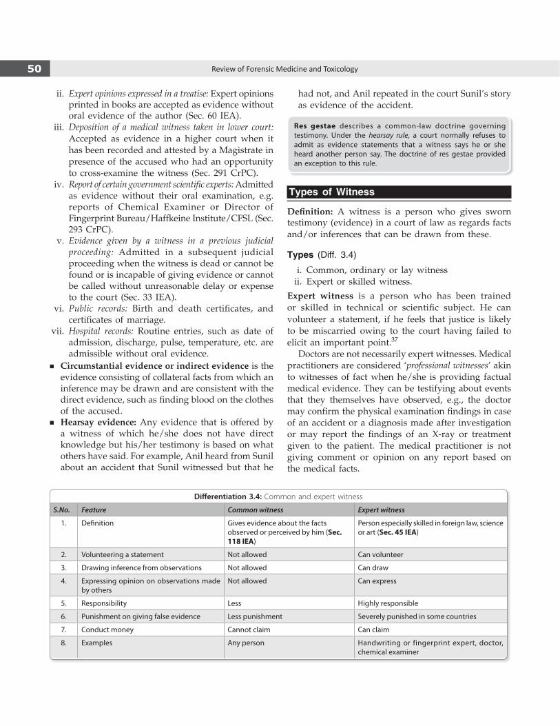
50 Review of Forensic Medicine and Toxicology
ii. Expert opinions expressed in a treatise: Expert opinions printed in books are accepted as evidence without oral evidence of the author (Sec. 60 IEA).
iii. Deposition of a medical witness taken in lower court: Accepted as evidence in a higher court when it has been recorded and attested by a Magistrate in presence of the accused who had an opportunity to cross-examine the witness (Sec. 291 CrPC).
iv. Report of certain government scientific experts: Admitted as evidence without their oral examination, e.g. reports of Chemical Examiner or Director of Fingerprint Bureau/Haffkeine Institute/CFSL (Sec. 293 CrPC).
v. Evidence given by a witness in a previous judicial proceeding: Admitted in a subsequent judicial proceeding when the witness is dead or cannot be found or is incapable of giving evidence or cannot be called without unreasonable delay or expense to the court (Sec. 33 IEA).
vi. Public records: Birth and death certificates, and certificates of marriage.
vii. Hospital records: Routine entries, such as date of admission, discharge, pulse, temperature, etc. are admissible without oral evidence.
� Circumstantial evidence or indirect evidence is the evidence consisting of collateral facts from which an inference may be drawn and are consistent with the direct evidence, such as finding blood on the clothes of the accused.
� Hearsay evidence: Any evidence that is offered by a witness of which he/she does not have direct knowledge but his/her testimony is based on what others have said. For example, Anil heard from Sunil about an accident that Sunil witnessed but that he
had not, and Anil repeated in the court Sunil’s story as evidence of the accident.
Res gestae describes a common-law doctrine governing testimony. Under the hearsay rule, a court normally refuses to admit as evidence statements that a witness says he or she heard another person say. The doctrine of res gestae provided an exception to this rule.
Types of Witness
Definition: A witness is a person who gives sworn testimony (evidence) in a court of law as regards facts and/or inferences that can be drawn from these.
Types (Diff. 3.4)
i. Common, ordinary or lay witness ii. Expert or skilled witness.
Expert witness is a person who has been trained or skilled in technical or scientific subject. He can volunteer a statement, if he feels that justice is likely to be miscarried owing to the court having failed to elicit an important point.37
Doctors are not necessarily expert witnesses. Medical practitioners are considered ‘professional witnesses’ akin to witnesses of fact when he/she is providing factual medical evidence. They can be testifying about events that they themselves have observed, e.g., the doctor may confirm the physical examination findings in case of an accident or a diagnosis made after investigation or may report the findings of an X-ray or treatment given to the patient. The medical practitioner is not giving comment or opinion on any report based on the medical facts.
Differentiation 3.4: Common and expert witness
S.No. Feature Common witness Expert witness
1. Definition Gives evidence about the facts observed or perceived by him (Sec. 118 IEA)
Person especially skilled in foreign law, science or art (Sec. 45 IEA)
2. Volunteering a statement Not allowed Can volunteer
3. Drawing inference from observations Not allowed Can draw
4. Expressing opinion on observations made by others
Not allowed Can express
5. Responsibility Less Highly responsible
6. Punishment on giving false evidence Less punishment Severely punished in some countries
7. Conduct money Cannot claim Can claim
8. Examples Any person Handwriting or fingerprint expert, doctor, chemical examiner

51Legal Procedure
The difference between a ‘professional witness’ and an ‘expert witness’ is that experts can offer opinions beyond the facts of what they have personally witnessed and can respond to questions about hypothetical situations posed to them.
When a doctor describes the dimensions of an injury, e.g. stab wound, he acts like an ordinary witness (fact witness), but when opines the cause of death as hemorrhage due to antemortem injury to the femoral artery, he is an expert witness.
Hostile witness is a person who willfully or with motive (bribe/intimidation) conceals part of the truth or tells a lie or gives completely false evidence in a court.38
� It is contradictory to the statement the witness made in the previous deposition (e.g. statement recorded by the police).
� Any of the above two witnesses can be declared hostile witness.
� A witness who has seen the event first-hand is known as an eyewitness.
� Testimony (Latin testimonium: from testis): In law and in religion and are consistent with the direct evidence, testimony is a solemn attestation as to the truth of a matter.
� The Supreme Court has defined a hostile witness as ‘one who is not desirous of telling the truth at the instance of the party calling him’ and an unfavorable witness is ‘one called by a party to prove a particular fact, who fails to prove such a fact or proves an opposite fact’ (Sat Pal vs Delhi Administration).
� Supreme Court held that the deposition of a hostile witness can be taken into consideration to the extent that the same is in consonance with the case of the prosecution and found to be reliable under careful judicial scrutiny. Evidence tendered by a prosecution witness would not get entirely erased merely because the prosecution has chosen to treat him as hostile.
Recording of Evidence
TestifyingA deposition is testimony of a witness. A deposition is a discovery device—lawyers gather information on what factual and expert witnesses says orally and assess the relative effectiveness of their testimony. The purpose of discovery is to identify all the facts related to the case. The words spoken by the witness are treated as courtroom testimony and the proceeding is conducted in accordance with the applicable court rules. Sec. 118 IEA states about the person who may testify as witness in a court of law.
Presentation of EvidenceAfter receiving subpoena, the witness must appear before the court at the appointed time with the
relevant documents. The evidence is probed for areas of uncertainty, inconsistency or any factors which may make the evidence appear unreliable. Evidence is presented in a systematic order (Sec. 138 IEA): i. oath (Sec. 51 IPC) ii. Examination-in-chief (Sec. 137 IEA) iii. Cross-examination (Sec. 141–146 IEA) iv. Re-examination (Sec. 137–138 IEA) v. Court questions (Sec. 165 IEA, Sec. 311 CrPC).
Oath
It is compulsory for the witness to take an oath in the witness box before he gives his evidence. He is required to swear by Almighty God that he will tell the truth, the whole truth and nothing but the truth. If the witness is an atheist, he makes a solemn affirmation in same terms, instead of swearing by God.
Perjury: A witness who after taking oath or making a solemn affirmation, willfully makes a false statement which he knows or believes to be the false (Sec. 191 IPC and Sec. 344 CrPC) is liable to be prosecuted for perjury under Sec. 193 IPC with imprisonment upto 7 years and fine.39
� In the US, punishment for perjury is imprisonment upto 5 years, while in the UK it is upto 7 years.
� In some countries, such as France, Italy and Germany, suspect’s evidence is not taken under oath or affirmation and thus cannot commit perjury, regardless of what they say during their trial.
Examination-in-Chief (Direct Examination)
� It is the examination of a witness by the party who calls him.
� In criminal cases, the public prosecutor commences this examination.
� Objectives are to place before the court all the facts that bear on the case, and if the witness is an expert, his interpretation of these facts.
� No leading questions are allowed except in those cases in which the Judge is satisfied that a witness is hostile.40
Leading question: Any question suggesting the answer which the person putting it wished or expects to receive (Sec. 141 IEA). It includes a material fact and admits of a conclusive answer by a simple ‘Yes’ or ‘No’. For example, “Was the length of the knife 15 cm?” Instead the question should be “What was the length of the knife?”
Leading questions must not be asked, if objected to by the adverse party, in an examination-in-chief or in re-examination, except with the permission of the court (Sec. 142 IEA).41

52 Review of Forensic Medicine and Toxicology
Cross-ExaminationIt is the examination of a witness by the adverse party (defense lawyer).
Objectives are: i. To elicit facts favorable to his case. ii. To test the accuracy of the statements made by
the witness. iii. To modify or explain what has been said. iv. To develop new or old facts. v. To discredit the witness. vi. To remove any overemphasis which may have
been given to any of fact in direct examination. � The lawyer tries to weaken the evidence of the
witness by showing that his details are inaccurate, conflicting, contradictory and untrustworthy.
� Leading questions are allowed (Sec. 143 IEA). � Cross-examination has no time limit, may last for
hours or even days. � The court has the power to disallow questions which
are indecent or scandalous (Sec. 151 IEA) or intended to insult or annoy, or offensive in form (Sec. 152 IEA).
During cross-examination, if any question is not understood, the witness should ask the lawyer to explain it better. Moreover, he should not volunteer any unrelated information.
Re-Examination (Re-Direct Examination)It is the examination of a witness subsequent to the cross-examination by the party who called him.Objectives are:
i. To clear any doubts that may have arisen during cross-examination.
ii. To explain some matter in its proper perspective, so that under-emphasis or possible misinterpretation may be avoided.
Leading questions are not allowed. opposing lawyer has the right of re-cross-examination on any new point which has been raised.
Court Questions/Questions by the JudgeA Judge may ask any question to the witness at any stage of the trial to clear any doubtful points.42
The deposition of the witness is handed over to him. The witness after carefully going through it, is required to sign at the bottom of each page and on the last page immediately below the last paragraph, and to initial any corrections (Sec. 278 CrPC). The witness should not leave the court without the permission of the Judge.
Conduct and Duties of a Doctor in the Witness Box
When summons is served, he must attend the court punctually. As a rule, his evidence is taken at the appointed time. Following are the do’s and do nots in the witness box: i. Take all records and relevant reports that may
have to be quoted in the box. ii. Be well dressed and modest. iii. Do not discuss the case with anyone in the court
except the lawyer by whom you were asked to testify.
iv. Stand up straight, be relaxed, calm and not be frightened or nervous. Look people in the eye when you speak, for it gives the impression of honesty.
v. Never attempt to memorize. The law allows refreshing your memory from copies of reports.
vi. Speak slowly, distinctly and audibly so that the typist can record your evidence.
vii. Use simple language, avoiding technical terms to the best of your ability.
viii. Address the Judge by his proper title such as ‘Sir’ or ‘Your honor’.
ix. Be polite, pleasant and courteous to the lawyer. Do not underestimate the medical knowledge of the lawyers.
x. Do not evade a question. Say ‘I don’t know’ if it is so, for no one can be expected to know everything.
xi. Do not loose your temper. An angry witness is often a poor witness.
xii. Retain independence of your mind. A biased expert is a useless expert.
xiii. Listen carefully to the questions. Do not hesitate to ask the questions to be repeated, if you do not understand it. Avoid long discussions.
xiv. If you believe the question is unfair, look at your lawyer before answering. If he fails to object, turn to Judge and ask whether you should answer the question.
xv. Do not over emphasize replies to questions from cross-examining lawyers.
xvi. Watch for double questions. The answer to each part of the question may be different.
xvii. When asked to comment upon the competence of a colleague, avoid any insulting remarks. If you do not wish to make any statement, say that you have ‘no opinion’ or ‘no comments’.
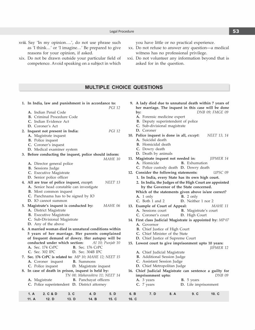
53Legal Procedure
xviii. Say ‘In my opinion….’, do not use phrase such as ‘I think…’ or ‘I imagine…’ Be prepared to give reasons for your opinion, if asked.
xix. Do not be drawn outside your particular field of competence. Avoid speaking on a subject in which
you have little or no practical experience. xx. Do not refuse to answer any question—a medical
witness has no professional privilege. xxi. Do not volunteer any information beyond that is
asked for in the question.
1. A 2. C & D 3. C 4. D 5. D 6. B 7. D 8. A 9. C 10. C 11. A 12. D 13. D 14. B 15. C 16. C
multiple ChoiCe questions
1. In India, law and punishment is in accordance to: PGI 12
A. Indian Penal Code B. Criminal Procedure Code C. Indian Evidence Act D. Coroner’s Act 2. Inquest not present in India: PGI 12 A. Magistrate inquest B. Police inquest C. Coroner’s inquest D. Medical examiner system 3. Before conducting the inquest, police should inform:
MAHE 10 A. Director general police B. Sessions Judge C. Executive Magistrate D. Senior police officer 4. All are true of police inquest, except: NEET 13 A. Senior head constable can investigate B. Most common inquest C. Panchnama has to be signed by Io D. Io cannot summon 5. Magistrate’s inquest is conducted by: MAHE 06 A. District Magistrate B. Executive Magistrate C. Sub-Divisional Magistrate D. Any of the above 6. A married woman died in unnatural conditions within
5 years of her marriage. Her parents complained of frequent demand of dowry. Her autopsy will be conducted under which section: AI 10; Punjab 10
A. Sec. 174 CrPC B. Sec. 176 CrPC C. Sec. 302 IPC D. Sec. 304B IPC 7. Sec. 176 CrPC is related to: MP 10; MAHE 12; NEET 15 A. Coroner inquest B. Summons C. Police inquest D. Magistrate inquest 8. In case of death in prison, inquest is held by:
TN 08; Maharashtra 11; NEET 14 A. Magistrate B. Panchayat officers C. Police superintendent D. District attorney
9. A lady died due to unnatural death within 7 years of her marriage. The inquest in this case will be done by: DNB 09; FMGE 09
A. Forensic medicine expert B. Deputy superintendent of police C. Sub-divisional magistrate D. Coroner 10. Police inquest is done in all, except: NEET 13, 14 A. Suicidal death B. Homicidal death C. Dowry death D. Death by animals 11. Magistrate inquest not needed in: JIPMER 14 A. Homicide B. Exhumation C. Police custody death D. Dowry death 12. Consider the following statements: UPSC 09 1. In India, every State has its own high court. 2. In India, the Judges of the High Court are appointed
by the Governor of the State concerned. Which of the statements given above is/are correct? A. 1 only B. 2 only C. Both 1 and 2 D. Neither 1 nor 2 13. Example of Court of Appeal: MAHE 11 A. Sessions court B. Magistrate’s court C. Coroner’s court D. High Court 14. First class Judicial Magistrate is appointed by: MP 07 A. Governor B. Chief Justice of High Court C. Chief Minister of the State D. Chief Justice of Supreme Court 15. Lowest court to give imprisonment upto 10 years:
JIPMER 12 A. Chief Judicial Magistrate B. Additional Session Judge C. Assistant Session Judge D. Chief Metropolitan Judge 16. Chief Judicial Magistrate can sentence a guilty for
imprisonment upto: DNB 09 A. 3 years B. 5 years C. 7 years D. Life imprisonment

54 Review of Forensic Medicine and Toxicology
17. A 18. A 19. C 20. D 21. B 22. A 23. B 24. A 25. A 26. A 27. B 28. D 29. A 30. B & C 31. B 32. C 33. D 34. C 35. B 36. B 37. B
17. Powers of a First Class Magistrate: PGI 06; AP 10; Jharkhand 11; NEET 14 A. Fine upto ` 10,000 and 3 years imprisonment B. Fine upto ` 10,000 and 5 years imprisonment C. Fine upto ` 3000 and 5 years imprisonment D. Unlimited fine and 7 years imprisonment 18. ‘Juvenile court’ is presided over by: DNB 08 A. I class woman Magistrate B. II class woman Magistrate C. II class male Magistrate D. None of the above 19. Juvenile court deals with cases of children upto the
age of: Maharashtra 09; DNB 09; FMGE 10 A. 15 years B. 16 years C. 18 years D. 21 years 20. According to JJ Act 2000, juvenile is boy and girl
respectively less than: NEET 13 A. 16 and l8 B. 18 and 21 C. 14 and 16 D. 18 and 18 21. Warrant cases are punishable with imprisonment:
DNB 09 A. More than 1 year B. More than 2 years C. Less than 1 year D. Less than 2 years 22. Cognizable offence comes under which section of
CrPC: NEET 13 A. 2 (c) B. 2 (b) D. 1 (a) D. 2 (l)23. Apersonbreakssomeone’smandibleinallegedfight.
Police can: AIIMS 12 A. Arrest with warrant B. Arrest without warrant C. Declare him hostile D. Put him in mental asylum 24. Subpoena is also called: MAHE 06 A. Summons B. Panchnama C. Requisition D. Inquest papers 25. Duces tecum is: WB 08 A. Summon B. Panchnama C. Conduct money D. Hostile witness 26. Conduct money is fee given to a: AP 07 A. Witness in civil court B. Witness in criminal court C. Doctor for good behavior in court D. Witness for good conduct 27. In civil cases, conduct money is decided by:
NEET 13 A. opposite party B. Judge C. Doctor D. Witness
28. In criminal cases, conduct money is paid by: NEET 13 A. Court B. opposite party C. Judge D. No conduct money is given29. Whenadoctor issuesa falsemedical certificate, then
he is liable under: AIIMS 11; Bihar 11; NEET 15 A. Sec. 197 IPC B. Sec. 87 IPC C. Sec. 304A IPC D. Sec. 338 IPC 30. IPC 197 is related to: NEET 14 A. Causing disappearance of evidence B. Issuing false certificate by doctor C. Giving false evidence D. Examination of accused 31. Dying declaration comes under: DNB 09; KCET 12 A. Sec. 30 CrPC B. Sec. 32 IEA C. Sec. 32 IPC D. Sec. 61 CrPC 32. If a patient survives after having given dying declara-
tion, then it stands as: UP 09 A. No value B. Valid for 48 h C. Corroborative evidence D. None 33. Dying declaration can be taken by:
PGI 12; NIMHANS 14 A. Village headman B. Doctor C. Magistrate D. Any one 34. Dying declaration is a: MAHE 10 A. Circumstantial evidence B. oral evidence C. Documentary evidence D. Hearsay evidence 35. Oral evidence is more important than written testimony
as: NEET 13 A. oral evidence cannot be cross-examined B. oral evidence can be cross-examined C. Documentary evidence requires no proof D. None 36. Documentary evidence along with oral evidence is
required: PGI 12 A. Dying declaration B. Postmortem report C. DNA fingerprinting D. Chemical examiner report 37. Volunteering a statement can be done by: Punjab 07 A. Eyewitness B. Medical witness C. Hostile witness D. Io

55Legal Procedure
38. Hostile witness is one who: Maharashtra 08 A. Threatens the Judge B. Threatens the prosecutor C. Refuses to answer D. Willfully gives false evidence 39. A witness, who after taking oath, willfully makes a
statement which he knows or believes to be false is guilty of offence under section: AIIMS 11; NEET 13, 15
A. 190 IPC B. 191 IPC C. 192 IPC D. 193 IPC 40. Leading questions are NOT permitted in: Delhi 08 A. Cross-examination B. Examination-in-Chief
C. Questions by the Judge D. Re-examination 41. Sec. 142 IEA is related to: BHU 08 A. Cross-questioning B. When leading question can be asked C. When leading question cannot be asked D. objective evidence 42. Judge can ask questions: Punjab 07 A. During cross-examination B. Examination-in-Chief C. Any time during trial D. Re-examination
38. D 39. D 40. B & D 41. C 42. C

56 Review of Forensic Medicine and Toxicology56 Review of Forensic Medicine and Toxicology
Definition: Identification is the determination of the individuality of a person based on certain physical char-acteristics.
It can be: i. Complete (absolute): Absolute fixation of the
individuality of a person. ii. Partial (incomplete): Ascertainment of only some
facts (e.g. race, sex, age or stature) about the identity, while the others remain unknown.
Identification is necessary in:Living persons pertaining to:
Criminal cases Civil cases
� Persons accused of assault, murder or rape � Marriage
� Interchange of newborn babies in hospitals � Passport/license
� Impersonation � Inheritance
� Absconding soldiers and criminals � Insurance claim
� Missing persons
� Disputed sex
Dead people i. In cases of fire, explosion and accidents. ii. When an unknown dead body is found on the
road, fields, railway compartment or water. iii. In cases of decomposed body. iv. In cases of mutilated body. v. Skeleton.
Before identifying the patient in the court, the doctor should verify the identification marks noted by him.
Corpus Delicti
Corpus delicti (‘body of offence’) refers to the principle that it must be proven that a crime has actually occurred before a person can be convicted of committing the crime.1 In a charge of homicide, it includes: i. Positive identification of the dead body (victim) ii. Proof of its death by criminal act of accused.
� The term includes body of the victim, bullet or clothing showing marks of the weapon or photographs showing fatal injures.
� The main part of corpus delecti is the establishment of identity of the body and infliction of violence in a particular way, at a particular time and place by the person or persons charged with crime and none other.
� The identification of a dead body and proof of corpus delecti is essential before a sentence is passed in murder trials, as unclaimed, decomposed bodies or portions of a dead body or bones are sometimes produced to support a false charge.
Identification data
In living and dead both 1. Race and religion 2. Sex 3. Age 4. Teeth 5. General development and stature 6. Anthropometric measurements 7. Fingerprints, lip prints and footprints 8. External peculiarities, like scar or tattoo 9. Hair 10. Personal effects: clothes, pocket contentsIn living only
� Handwriting � Speech and voice � Gait, manner and habit � Memory and education
Sex, age and stature are primary characteristics of identification—they are unaltered even afer death.
Race and Religion
Important in cases of mass disasters, e.g. in case of railway accidents or air crashes, when persons of different races are traveling together.
RaceIt is determined by: i. Clothing: Traditional Indian dress is different from
Western dress.
Identification I
Chapter 4
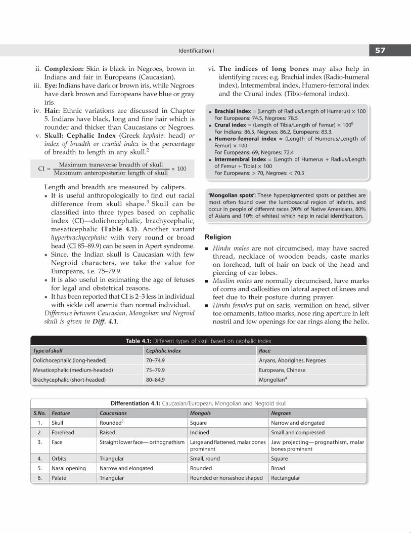
57Identification I
ii. Complexion: Skin is black in Negroes, brown in Indians and fair in Europeans (Caucasian).
iii. Eye: Indians have dark or brown iris, while Negroes have dark brown and Europeans have blue or gray iris.
iv. Hair: Ethnic variations are discussed in Chapter 5. Indians have black, long and fine hair which is rounder and thicker than Caucasians or Negroes.
v. Skull: Cephalic Index (Greek kephale: head) or index of breadth or cranial index is the percentage of breadth to length in any skull.2
Maximum transverse breadth of skullCI = --------------------------------------------------------------------------------------------------------------- × 100 Maximum anteroposterior length of skull
Length and breadth are measured by calipers. z It is useful anthropologically to find out racial
difference from skull shape.3 Skull can be classified into three types based on cephalic index (CI)—dolichocephalic, brachycephalic, mesaticephalic (Table 4.1). Another variant hyperbrachycephalic with very round or broad head (CI 85–89.9) can be seen in Apert syndrome.
z Since, the Indian skull is Caucasian with few Negroid characters, we take the value for Europeans, i.e. 75–79.9.
z It is also useful in estimating the age of fetuses for legal and obstetrical reasons.
z It has been reported that CI is 2–3 less in individual with sickle cell anemia than normal individual.
Difference between Caucasian, Mongolian and Negroid skull is given in Diff. 4.1.
vi. The indices of long bones may also help in identifying races; e.g. Brachial index (Radio-humeral index), Intermembral index, Humero-femoral index and the Crural index (Tibio-femoral index).
� Brachial index = (Length of Radius/Length of Humerus) × 100 For Europeans: 74.5, Negroes: 78.5
� Crural index = (Length of Tibia/Length of Femur) × 1006
For Indians: 86.5, Negroes: 86.2, Europeans: 83.3. � Humero-femoral index = (Length of Humerus/Length of
Femur) × 100 For Europeans: 69, Negroes: 72.4
� Intermembral index = (Length of Humerus + Radius/Length of Femur + Tibia) × 100
For Europeans: > 70, Negroes: < 70.5
‘Mongolian spots’: These hyperpigmented spots or patches are most often found over the lumbosacral region of infants, and occur in people of different races (90% of Native Americans, 80% of Asians and 10% of whites) which help in racial identification.
Religion � Hindu males are not circumcised, may have sacred
thread, necklace of wooden beads, caste marks on forehead, tuft of hair on back of the head and piercing of ear lobes.
� Muslim males are normally circumcised, have marks of corns and callosities on lateral aspect of knees and feet due to their posture during prayer.
� Hindu females put on saris, vermilion on head, silver toe ornaments, tattoo marks, nose ring aperture in left nostril and few openings for ear rings along the helix.
Table 4.1: Different types of skull based on cephalic index
Type of skull Cephalic index Race
Dolichocephalic (long-headed) 70–74.9 Aryans, Aborigines, Negroes
Mesaticephalic (medium-headed) 75–79.9 Europeans, Chinese
Brachycephalic (short-headed) 80–84.9 Mongolian4
Differentiation 4.1: Caucasian/European, Mongolian and Negroid skull
S.No. Feature Caucasians Mongols Negroes
1. Skull Rounded5 Square Narrow and elongated
2. Forehead Raised Inclined Small and compressed
3. Face Straight lower face— orthognathism Large and flattened, malar bones prominent
Jaw projecting—prognathism, malar bones prominent
4. Orbits Triangular Small, round Square
5. Nasal opening Narrow and elongated Rounded Broad
6. Palate Triangular Rounded or horseshoe shaped Rectangular
58 Review of Forensic Medicine and Toxicology
� Muslim females put on trousers, no vermilion mark, nose ring in the septum, several openings on the helix for ear rings and no tattoo marks.
Sex
� Sex: It is the biological term denoting the genetic, physiologic and anatomical characteristics of an individual, based on which we can identify ourselves into ‘males’ and ‘females’.
� Gender: Sociological construct that denotes how an individual identifies according to social norms (social roles, position and behavior), based on which an individual exhibits ‘masculine’ or ‘feminine’ qualities. It is the sexual identity of an individual from birth to puberty and adulthood.*
� Intersex: Intermingling of sexual characters of either sex in one individual to a varying degree including the physical form, reproductive organs and sexual behavior.
In normal cases, in the living: � Most certain evidence of sex: Possession of ovaries
in females, and testes in males. � Highly probable evidence of sex: Possession of
sexual structures, e.g. developed breasts and vagina in females, and male distribution of hair and penis in males.
� Presumptive evidence of sex: Outward appearance of individual features, contours of face, clothes, voice and figure.
Sex determination is required for following reasons: � Identification in living: Sex is important in any chain of
identity data and determination of the individuality of a person.
� Participation in sports: Sex segregation in sports is based on the long-term endogenous androgen exposure of men at puberty that lead to the physiological gap with women.
� For deciding whether an individual can exercise certain civil rights extended to one sex only.
� For deciding questions relating to legitimacy, divorce, paternity, marriage, impotence, rape and affiliation.
At present, there are three frequent circumstances wherein determination of sex has become necessary—sports, pre-employment and sex specific crimes. Routinely at birth, identifying biological sex of an individual is based on ‘external genitalia’ (phenotype
sex), i.e. whether it is penis and scrotum (in males) or vulva and vagina (in females). Identification of ‘sex’ of an individual may become problematic in: i. Intersex: They can be natural or acquired (‘hijras’
are castrated before puberty and ‘zenanas’ are castrated after puberty). They may have features resembling one sex and the internal gonads could be of other sex or of both sexes (true hermaphrodite).
ii. Transvestism: Individuals who cross dresses for sexual gratification.
iii. Transgender: It is denoting or relating to an individual whose self-identity does not conform unambiguously to conventional notions of male or female gender. It is a state of one’s gender identity or gender expression not matching one’s assigned sex.
iv. Transsexuals: Individuals who have undergone sex change surgeries or sex reassignment (male-to-female or female-to-male).
v. Concealed sex: Individuals who hide their real sex for a motive by cross dressing.
vi. Advanced decomposition and skeleton: Sex can be determined in decomposed body by identifying uterus or prostrate, which resist putrefaction.
Identification of biological sex in concealed sex and transvestism can be easily done by physical examination, but difficulty arises in cases of ambiguous genitalia wherein the external genitalia are a combination of both sexes.
Sex Verification Tests i. Physical morphology: External examination is done
to determine the sex (Diff. 4.2). ii. Nuclear sexing or sex chromatin or microscopic test:
Buccal epithelial cells or hair follicle cells are examined microscopically to detect the presence of ‘Barr body’.
iii. Gonadal biopsy: Detection of ‘internal gonads and sex chromosomes’ (genotype sex), i.e. the identification of testes and XY sex chromosomes (in males), or ovary and XX sex chromosomes (in females) is a confirmatory method of determining sex.
iv. Genebased test: Polymerase chain reaction (PCR) is used to detect SRY gene (sex-determining region of the Y-chromosome) and/or the DYZ1 region of Y chromosome which is diagnostic. SRY gene
* To identify whether an individual is ‘male’ or ‘female’, the correct word is ‘sex verification tests’ and not ‘gender verification tests’ or ‘femininity testing’.
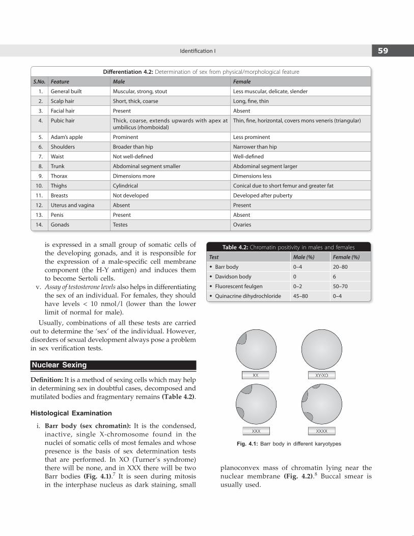
59Identification I
Fig. 4.1: Barr body in different karyotypes
is expressed in a small group of somatic cells of the developing gonads, and it is responsible for the expression of a male-specific cell membrane component (the H-Y antigen) and induces them to become Sertoli cells.
v. Assay of testosterone levels also helps in differentiating the sex of an individual. For females, they should have levels < 10 nmol/l (lower than the lower limit of normal for male).
Usually, combinations of all these tests are carried out to determine the ‘sex’ of the individual. However, disorders of sexual development always pose a problem in sex verification tests.
Nuclear Sexing
Definition: It is a method of sexing cells which may help in determining sex in doubtful cases, decomposed and mutilated bodies and fragmentary remains (Table 4.2).
Histological Examination
i. Barr body (sex chromatin): It is the condensed, inactive, single X-chromosome found in the nuclei of somatic cells of most females and whose presence is the basis of sex determination tests that are performed. In XO (Turner’s syndrome) there will be none, and in XXX there will be two Barr bodies (Fig. 4.1).7 It is seen during mitosis in the interphase nucleus as dark staining, small
planoconvex mass of chromatin lying near the nuclear membrane (Fig. 4.2).8 Buccal smear is usually used.
Table 4.2: Chromatin positivity in males and females
Test Male (%) Female (%)
� Barr body 0–4 20–80
� Davidson body 0 6
� Fluorescent feulgen 0–2 50–70
� Quinacrine dihydrochloride 45–80 0–4
Differentiation 4.2: Determination of sex from physical/morphological feature
S.No. Feature Male Female
1. General built Muscular, strong, stout Less muscular, delicate, slender
2. Scalp hair Short, thick, coarse Long, fine, thin
3. Facial hair Present Absent
4. Pubic hair Thick, coarse, extends upwards with apex at umbilicus (rhomboidal)
Thin, fine, horizontal, covers mons veneris (triangular)
5. Adam’s apple Prominent Less prominent
6. Shoulders Broader than hip Narrower than hip
7. Waist Not well-defined Well-defined
8. Trunk Abdominal segment smaller Abdominal segment larger
9. Thorax Dimensions more Dimensions less
10. Thighs Cylindrical Conical due to short femur and greater fat
11. Breasts Not developed Developed after puberty
12. Uterus and vagina Absent Present
13. Penis Present Absent
14. Gonads Testes Ovaries

60 Review of Forensic Medicine and Toxicology
Murray Barr and Edward Bertran while working on stained sections of nerve cells in cats noticed a tiny, dark staining blob that was always present only in the nucleus of the female cats and never in the males.8 It was subsequently determined to be inactive X-chromosome and came to be known as Barr body.
ii. Davidson body: In females, neutrophil leucocytes contain a small nuclear attachment of drumstick form (Fig. 4.3).
� Sex chromosomes (XX or XY) can be determined in the cells that are dividing, e.g. bloodstains, cartilage, bone marrow, teeth pulp and hair root.
� In decomposed bodies, sex chromatin is difficult to make out.
� Hair follicles are important for cell sexing since they resist putrefaction, and both Barr body and Y-chromosome can be demonstrated.
� Quinacrine dihydrochloride is used for staining Y-chromosome that is seen as bright fluorescent body.
� Fluorescent Feulgen reaction using Acriflavin Schiff reagent is used for staining X-chromosome that is seen as bright yellow spot in nuclei.
Disorders of Sexual Development
Disorder of sexual development (DSD) is a broad term encompassing any condition where external genitalia are atypical in relation to chromosome and gonads. It results from some defect in embryonic development.
Earlier, Davidson divided ‘intersex’ into four groups: i. Gonadal agenesis ii. Gonadal dysgenesis iii. True hermaphroditism iv. Pseudohermaphroditism
Currently, the term DSD is used to substitute the obsolete nomenclature of ‘intersex’, ‘hermaphrodite’ and ‘pseudohermaphrodite’, since they are based only on identifying the gonads and they fail to take into consideration whether those gonads are functioning or not. The new terms and possible diagnosis in such cases are given in Table 4.3.
Different kinds of DSD have been classified (Flow chart 4.1). Some of these are briefly described below:1. Ovotesticular DSD (true hermaphroditism): It
is a rare condition; also known as double-sex or bisexual. Both ovarian and testicular tissues are present. External genitalia of both sexes exist in one individual, but sex chromatin may be either male or female pattern (46XX or 46XY or mosaics). True hermaphroditism is very similar to mixed gonadal dysgenesis. The karyotype could be 46XX/46XY or 46XX/47XXY.
2. Gonadal dysgenesis: It refers to a defect in gonad formation that is characterized by a progressive loss of primordial germ cells in the developing gonads of an embryo with consequent formation of hypoplastic and dysfunctioning gonads composed mainly of fibrous tissue—streak gonads. External sexual characters are present, but testes or ovaries fail to develop at puberty. This is seen in Klinefelter syndrome, Turner syndrome, XX gonadal dysgenesis (pure gonadal dysgenesis 46XX), Swyer syndrome (pure gonadal dysgenesis 46XY), Perrault syndrome
Fig. 4.2: Barr body in buccal epithelium
Fig. 4.3: Davidson body (drumstick appearance) in neutrophil
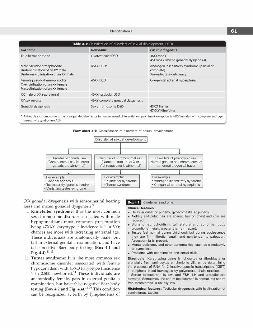
61Identification I
(XX gonadal dysgenesis with sensorineural hearing loss) and mixed gonadal dysgenesis.9
i. Klinefelter syndrome: It is the most common sex chromosome disorder associated with male hypogonadism, most common presentation being 47XXY karyotype.10 Incidence is 1 in 500; chances are more with increasing maternal age. These individuals are anatomically male, but fail in external genitalia examination, and have false positive Barr body testing (Box 4.1 and Fig. 4.4).11-13
ii. Turner syndrome: It is the most common sex chromosome disorder associated with female hypogonadism with 45XO karyotype (incidence 1 in 2,500 newborns).14 These individuals are anatomically female, pass in external genitalia examination, but have false negative Barr body testing (Box 4.2 and Fig. 4.4).15-19 This condition can be recognized at birth by lymphedema of
Table 4.3: Classification of disorders of sexual development (DSD)
Old name New name Possible diagnosis
True hermaphrodite Ovotesticular DSD 46XX/46XY45X/46XY (mixed gonadal dysgenesis)
Male pseudohermaphroditeUndervirilisation of an XY maleUndermasculinisation of an XY male
46XY DSD* Androgen insensitivity syndrome (partial or complete)5-α-reductase deficiency
Female pseudo-hermaphroditeOver virilisation of an XX femaleMasculinisation of an XX female
46XX DSD Congenital adrenal hyperplasia
XX male or XX sex reversal 46XX testicular DSD
XY sex reversal 46XY complete gonadal dysgenesis
Gonadal dysgenesis Sex chromosome DSD 45XO Turner47XXY Klinefelter
* Although Y chromosome is the principal decisive factor in human sexual differentiation, prominent exception is 46XY females with complete androgen insensitivity syndrome (cAIS).
Flow chart 4.1: Classification of disorders of sexual development
Box 4.1 Klinefelter syndrome
Clinical features � Delay in onset of puberty, gynecomastia at puberty. � Axillary and pubic hair are absent, hair on chest and chin are reduced.
� Signs of eunuchoidism, tall stature and abnormal body proportions (height greater than arm span).
� Testes feel normal during childhood, but during adolescence they are firm, fibrotic, small, and non -tender to palpation. Azoospermia is present.
� Mental deficiency and other abnormalities, such as clinodactyly or synostosis.
� Problems with coordination and social skills.
Diagnosis: Karyotyping using lymphocytes or fibroblasts or prenatally from aminocytes or chorionic villi, or by determining the presence of RNA for X-inactive-specific transcriptase (XIST) in peripheral blood leukocytes by polymerase chain reaction. Serum testosterone is low, and FSH, LH and estradiol are elevated. Sometimes, the serum testosterone is normal, but serum free testosterone is usually low.
Histological features: Testicular dysgenesis with hyalinization of seminiferous tubules.

62 Review of Forensic Medicine and Toxicology
dorsum of hands and feet, loose skin folds in the nape of neck, and low birth weight.
iii. Swyer syndrome: The individuals with pure gonadal dysplasia and a 46XY karyotype will display variable degrees of undermasculiniza-
tion, dependent upon the amount of testicular dysplasia. Both XX and XY gonadal dysgenesis are due to mutation or deletion of part of the sequence of the SRY gene.
iv. Mosaicism: It is a genetic abnormality with mixture of cells with XX and XY, or X and XY sex chromosomes. Accordingly, there may be false positive or false negative Barr body tests depending on number of X chromosomes.
v. Mixed gonadal dysgenesis: It is a partial gonadal dysgenesis variant of the Y chromosome mosaicism, and the subjects have 45XO/46XY karyotype being characterized by unilateral testis, a contralateral streak gonad, persistent müllerian ductal structures ipsilateral with the streak gonad and varying levels of external genitalia undervirilization.
Fig. 4.4: Features seen in Klinefelter and Turner syndrome
Box 4.2 Turner syndrome
Clinical features � Short stature, webbed neck � Micrognathia, high-arched palate � Ptosis with low-set ears � Low hairline, widely spaced nipples � Cubitus valgus, short fourth metacarpals, hyperconvex nails and recurrent otitis media
� Lymphedema of hands and feet, pigmented nevi and keloid formation
� Learning disability, often involving visual-spatial skills without typical mental retardation
� Cardiovascular anomalies: Coarctation of the aorta and aortic stenosis
� Renal abnormalities: Hydronephrosis, horseshoe kidney, hypertension
� Increased urinary gonadotrophin exertion � Sexual infantilism due to gonadal dysgenesis with primary amenorrhea
� High incidence of osteoporosis, type II diabetes
Diagnosis: Evaluation for childhood short stature often leads to the diagnosis. Hypogonadism is confirmed in girls who have high serum levels of FSH and LH. A karyotype showing 45XO establishes the diagnosis.
� Men with Klinefelter syndrome are at a higher risk of autoimmune diseases, diabetes mellitus, leg ulcers, osteopenia and osteoporosis, tumors (breast and germ cells), gonadotroph adenoma and gonadotroph hyperplasia liver adenoma, SLE, rheumatoid arthritis and Sjögren syndrome.
� Hypergonadotropic hypogonadism (defective development of testes or ovaries and associated with excess pituitary gonadotropin secretion) is seen in Klinefelter syndrome, Noonan
63Identification I
3. Androgen receptor deficiency: Androgen insensitivity is caused by receptors that are insensitive to androgens, particularly testosterone. The basic etiology is mutation in the androgen receptor gene. As a result, the individual even though genetically male (XY) and possess testes would fail to develop male characteristics. These individuals would develop as female but would fail the Barr body test as they have only one X chromosome. These individuals are born and raised as girls and have female gender identity. It can be:
i. Complete androgen insensitivity syndrome (cAIS), previously known as testicular feminization syndrome (most common form of male pseudohermaphroditism), is an X-linked recessive condition resulting in failure of normal masculinization of the external genitalia in genetically male (XY) individual. They have female external genitalia with normal labia, clitoris and vaginal introitus with normal size breasts but with primary amenorrhea and scanty or absent axillary and pubic hair. Internally, there is a short blind-pouch vagina with absence of uterus, fallopian tubes and ovaries.22,23
ii. Incomplete androgen insensitivity: The phenotype of individuals with partial androgen insensitivity syndrome may range from mildly virilized female external genitalia (clitorimegaly without other external anomalies) to mildly undervirilized male external genitalia (hypospadias and/or diminished penile size) with gynecomastia. They tend to be tall.
In either case, affected individuals have normal testes with normal production of testosterone and normal conversion to dihydrotestosterone (DHT), which differentiates this condition from 5-alpha reductase deficiency.
4. 5-a reductase deficiency (5-ARD): This is an autoso-mal recessive sex-limited condition resulting in the inability to convert testosterone to dihydrotestoster-one (DHT). Since, DHT is required for the normal masculinization of the external genitalia in utero,
genetic males with 5-ARD deficiency are born with ambiguous genitalia. The individual presents with a clitoral-like phallus, markedly bifid scrotum, pseu-dovaginal blind-ending introitus with perineoscrotal hypospadias and a rudimentary prostate. The uterus and fallopian tubes are absent. Testes are intact and are usually found in the inguinal canal or scrotum or occasionally in the abdomen. At puberty, mus-culature, body and facial hair develop owing to normal levels of testosterone. These individuals fail in external examination of genitals and fail in Barr body testing.
5. Congenital adrenal hyperplasia (CAH): It is a condition wherein adrenal glands produce excessive amounts of testosterone in females. These individuals develop secondary male characteristics but are genetically female (46XX, DSD), lacking testes and male reproductive organs (masculinized females). Among the various forms of CAH, the 21-hydroxylase deficiency resulting from mutations or deletions of CYP21A, is most common but sexual ambiguity can also be seen in defects in 17-hydroxylase, 3b-hydroxysteroid dehydrogenase, 17-ketosteroid reductase and 11b-hydroxylase.24-26 Females appear phenotypically female at birth, but do not develop breasts or menstruate in adolescence; they may present with hypertension. Genital anomalies range from complete fusion of the labioscrotal folds and a phallic urethra to clitoromegaly, partial fusion of the labioscrotal folds, or both. These individuals fail in external examination of genitals but would have positive Barr body testing.24
Recently, the Supreme Court directed the Government to include transgender people in all welfare programs for the poor, including education, health care and jobs. The court noted that it is the right of every human being to choose their gender while granting rights to those who identify themselves as neither male nor female. All documents (including voter/Aadhar card) will now have a third category marked ‘transgender’. It would only apply to transgender people but not to gays, lesbians or bisexuals.
Sex from Skeletal Remains
� Recognizable sex differences appear after puberty except in pelvis. In pelvis, sex features are independent of each other and one may even contradict the other in same pelvis.
syndrome, viral orchitis (mumps), cytotoxic drugs and testicular irradiation. In women with hypergonadotropic hypogonadism, the most common cause is Turner syndrome.13,20
� Cardiovascular disease is the most common cause of death in adult women with Turner syndrome. Most common heart defects are bicuspid aortic valve (30% of children), coarctation of the aorta (5-10%) and aortic dissection.21 Other anomalies seen are hypoplasic left-heart syndrome, partial anomalous pulmonary venous drainage and atrial septal defects.

64 Review of Forensic Medicine and Toxicology
� The sex of long bones can be determined on the basis of medullary index from tibia, humerus, ulna and radius. Sternum is least useful.
The accuracy in sexing from adult skeletal remains is given in Table 4.3 (as reported by Krogman).27
Table 4.3: Accuracy of sexing based on skeletal remains
Skeletal remains Accuracy in sexing (%)
Entire skeleton 100
Skull + Pelvis 98
Pelvis alone (best single bone)28 95
Skull alone 92
Long bones 80–85
Long bones + Pelvis 98
Traits diagnostic of sex from skeleton are given in Diff. 4.3 to 4.6 and Table 4.4.
The preauricular sulcus has been described as a characteristic of the female pelvis. The pelvic portion of the anterior sacroiliac ligament is attached to it. Its prominence results from obstetrical trauma during the course of delivery which allows for differentiation between nulliparous women and males vs females who have given birth.29
Kimura’s Base-wing index =
×Width of wing (ala of sacrum)
100Width of base (transverse diameter of body of S1)
Chilotic line: It is an anthropometric line extending from the posterior aspect of the iliopectineal eminence to the closest point on the anterior auricular margin (the pelvic segment) and then to the iliac crest (the sacral portion). Since the pelvic segment is predominant in females and the sacral segment is predominant in males, the chilotic index may be used to identify the sex of human skeletal remains.30
= ×Sacral part of chilotic line
Chilotic line index 100Pelvic part of the chilotic line
Age
Age determination can be done through many means, and an analysis of all possible age-related attributes is best for an overall estimate. Some of the utilized features include: i. Dental eruption ii. Epiphyseal unions iii. Pubic symphyseal morphology iv. Cranial suture closures v. Mandibular and sacral changes vi. Miscellaneous a. Secondary sexual characters b. Age-related degenerative conditions.
Differentiation 4.3: Male and female skull (Fig. 4.5)
S.No. Feature Male skull Female skull
1. General appearance Larger, heavier, rugged, marked muscular ridges Smaller, lighter, walls thinner, smoother
2. Forehead Receding, irregular, rough, less rounded Vertical, round, full, infantile, smooth
3. Cranial capacity More capacious (1450–1550 cc) Less capacious (1300–1350 cc)
4. Glabella Prominent Less prominent
5. Supraorbital/supraciliary ridge Prominent Less prominent
6. Frontonasal junction Distinct angulation Smoothly curved
7. Orbits Square, rounded margins, small Rounded, sharp margins, large
8. Frontal and parietal eminence Less prominent Prominent
9. Zygomatic arch Prominent Not prominent
10. Occipital area (muscle markings and protuberance)
Prominent Not prominent
11. Mastoid process Large, round, blunt Small, smooth, pointed
12. Digastric groove Deep Shallow
13. Condylar facet Long, narrow Short, broad
14. Palate Large, U-shaped, broad Small, parabolic
15. Foramen magnum Relatively large, long Small, round
16. External auditory meatus Bony ridge along upper border prominent Often absent

65Identification I
Differentiation 4.4: Male and female mandible (Fig. 4.6)
S.No. Feature Male mandible Female mandible
1. General appearance Larger, thicker Smaller, thinner
2. Chin (symphysis menti) Square or U-shaped Rounded
3. Angle of body with ramus Less obtuse (< 125°), prominent More obtuse, not prominent
4. Angle of mandible (gonion) Everted Inverted
5. Body height at symphysis Greater Smaller
6. Ascending ramus Greater breadth Smaller breadth
7. Ramus flexure Rearward angulation of the posterior border of ramus Straight ramus
8. Muscular markings Prominent Not prominent
Fig. 4.5: Male and female skull
Fig. 4.6: Male and female mandible

66 Review of Forensic Medicine and Toxicology
Differentiation 4.5: Male and female pelvis (Figs 4.7 and 4.8)
S.No. Feature Male pelvis Female pelvis
1. General appearance Massive, rougher, prominent muscular markings
Less massive, slender, smoother, muscular markings not prominent
2. Shape Deep funnel Flat bowl
3. Preauricular sulcus (attachment of anterior sacroiliac ligament)
Not frequent, narrow, shallow More frequent, broad, deep
4. Acetabulum Large, 52 mm diameter Small, 46 mm diameter
5. Obturator foramen (Fig. 4.9) Large, oval, base upwards Small, triangular, apex forwards
6. Greater sciatic notch (Fig. 4.10) Narrow, deep, small Broad, shallow, large
7. Iliopectineal line Well-marked, rough Rounded, smooth
8. Ischial tuberosity Inverted Everted
9. Body of pubis (Fig. 4.9) Narrow, triangular Broad, square, pits on posterior surface, if borne children
10. Subpubic angle V-shaped, sharp angle, 70°–75° U-shaped, rounded, broader angle, 90°–100°
11. Pelvic brim or inlet Heart-shaped Circular or elliptical shaped
12. Pelvic cavity Conical, funnel shaped Broad, round
13. Pelvic outlet Smaller Larger
14. Sacroiliac articulation Large, extends to 2½–3 vertebrae Small, extends to 2–2½ vertebrae
15. Auricular surface (Fig. 4.10) Raised Flat
16. Coccyx Less movable More movable
Fig. 4.7: Pelvis: superior view Fig. 4.8: Male and female pelvis

67Identification I
Fig. 4.9: Pelvis: Obturator foramen Fig. 4.10: Pelvis: Greater sciatic notch and auricular surface
Differentiation 4.6: Male and female sacrum (Fig. 4.11)S. No. Feature Male sacrum Female sacrum
1. General appearance Larger, heavier, rough, narrow Smaller, lighter, smooth, broad
2. Breadth of body of 1st sacral vertebra More than breadth of one side ala Less than breadth of one side ala
3. Inner curvature (Fig. 4.7) Uniformly curved anteriorly Abruptly curved at the last two segments
4. Sacroiliac articulation Extends upto 3rd segment Extends upto 2–2½ segment
5. Sacroiliac joint surface Large, less sharply angulated L-shaped, elevated anteriorly
Table 4.4: Diagnostic indexes for determination of sex
S.No. Index Formula Male Female
1. Washburn/Ischiopubic index Length of pubis100
Length of ischium×
73–94 91–115
2. Sciatic notch index Width of sciatic notch100
Depth of sciatic notch×
4–5 5–6
3. Sternal index Length of manubrium100
Length of body×
46.2 54.3
4. Corporobasal index31
×Breadth of body of 1st sacral vertebra
100Breadth of base of sacrum
> 42 < 42
5. Sacral index Transverse diameter of base of sacrum100
Anterior length of sacrum×
< 114 > 114
Fig. 4.11: Sacrum

68 Review of Forensic Medicine and Toxicology
Fig. 4.12: Parts of a tooth
Dentition in Determining AgeAge can be determined by using the criteria like eruption and calcification of teeth, Stack’s method, Miles method, Boyde’s method and Gustafson’s method.32
� Alveolar cavities which contain teeth are formed around the 3–4th month of intrauterine life (IUL).
� At birth, rudiments of all the temporary teeth and the 1st permanent molars may be found in jaw.
� Each tooth has a crown, neck and a root embedded in jaw bone (Fig. 4.12).
� Teeth are composed of dentin covered on the crown by enamel and on the root by cementum which is attached to the alveolar bone by periodontal membrane. Tooth enamel is the hardest substance in the body containing primarily hydroxypatite (crystalline calcium phosphate).33
� Tooth formation proceeds in an invariable sequence. The first radiographic evidence is formation of a bony crypt followed by mineralization of crown tips. Mineralization proceeds from crown tips down the sides of the tooth.34
� Root mineralization does not begin until crown formation is complete and root formation ceases with the reduction of apical foramen. As the root becomes longer, the crown erupts through the bone.
� Mineralization of deciduous dentition begins in utero, early in 2nd trimester, and root formation of third molar may not be complete until 20 years of age.
� During eruption of a permanent tooth, the overlying root of its deciduous predecessor simultaneously undergoes absorption, until only the crown remains. The unsupported crown then falls off.
� Age of eruption of teeth depends upon: i. Heredity ii. Environment iii. Nutrition iv. Endocrine factors.
Each individual has two sets of teeth (Diff. 4.7) i. Temporary/deciduous/milk teeth ii. Permanent teeth
Temporary teeth � 20 in number: 4 incisors, 2 canines and 4 molars in
each jaw (Table 4.5 and Fig. 4.13).35
� The eruption of the deciduous teeth commences at about 6–7 months after birth and is completed about 2nd-3rd year.37 the lower central incisors anteceding those of the upper.38,39
However, upper lateral incisor erupts earlier than its lower quadrant counterpart.
� In ill-nourished children, especially in rickets, denti-tion may be delayed.
� In congenital syphilis, teeth may be premature or even present at birth.
Permanent teeth: 32 in number—4 incisors, 4 pre-molars, 2 canines and 6 molars in each jaw (Table 4.6 and Fig. 4.14).
Differentiation 4.7: Temporary and permanent teeth
S.No. Feature Temporary teeth Permanent teeth
1. Size Smaller, lighter, narrower, except temporary molars which are longer than permanent premolars
Heavier, stronger, broader, except permanent premolars
2. Direction of anterior teeth Vertical Inclined forward
3. Crown color China-white Ivory-white
4. Neck More constricted Less constricted
5. Ridge36 Present at the junction of the crown and the root Not present
6. Root Roots of molars are smaller, more divergent Roots of molars are larger, less divergent
7. Incisors Smooth incisal edge Ridged, especially on incisal surface
8. Radiology Presence of tooth germ beneath tooth will suggest that tooth is temporary
No such thing visible in case of permanent teeth

69Identification I
Developmentally teeth are divided into 2 sets:a. Super-added permanent teeth: These teeth do not
have deciduous predecessors. All permanent molars belong to this category (6 in each jaw).
b. Successional permanent teeth: These teeth erupt in place of deciduous teeth, e.g. permanent premolars erupt in place of deciduous molars (10 in each jaw).
� Usually permanent tooth erupts first in lower jaw. � Permanent teeth appear few months earlier in girls
than in boys. � Eruption of teeth is useful in estimating age upto 15
years. The third molar (wisdom tooth) erupts after this time, but is so variable in eruption that it is not a reliable age indicator.
� The mandibular third molar is the most commonly impacted tooth in the mouth and is closely followed by maxillary third molar, maxillary canine and mandibular canine respectively.42
Fig. 4.13: Eruption of deciduous teeth (in months)
Fig. 4.14: Eruption of permanent teeth (in years)
� If, in the jaw all 3rd molars are present, then the age is over 18 years, but their absence gives no certain idea about age.
An impacted tooth is the one that fails to erupt into proper function in the dental arch within the expected time.
Spacing of jaw: After eruption of 2nd molars, the ramus of mandible grows behind to make room for the eruption of 3rd molar teeth which is known as spacing of the jaw.
Period of mixed dentition: Starting from the day of eruption of first permanent molar till before the eruption of last permanent canine—both temporary and permanent teeth are present in the jaw. This duration is known as period of mixed dentition (Table 4.7). Usually, it is between 6–11 years, but may persist until 12–13 years.43
� From 6–11 years, the number remains 24 because as and when a tooth erupts, it displaces another and the number remains constant.
� there is addition of teeth from the age of 12–14 years, when the second molar erupts and the total number
Table 4.6: Eruption of permanent teeth
S.No. Tooth Eruption (years)
1. First molar40 6–7
2. Central incisor 6–8
3. Lateral incisor41 7–9
4. First premolar 9–11
5. Second premolar 10–12
6. Canine 11–12
7. Second molar 12–14
8. Third molar 17–25
Table 4.5: Eruption of deciduous teeth
S.No. Tooth Eruption (months) No. of teeth
1. Central incisor � Lower32,33
� Upper6–87–9
24
2. Lateral incisor � Upper � Lower
7–910–12
68
3. First molar 12–14 12
4. Canine 17–18 16
5. Second molar 20–30 (2–2½ years)37 20

70 Review of Forensic Medicine and Toxicology
Estimation of age from teeth beyond 25 years � Miles’ method: The amount of wear on all three
permanent molars occurs at comparable rates which can be assessed, and age estimation made on the basis of comparison to the baseline. Miles also developed a method to determine age at death by measuring the thickness of enamel and dentin from neonatal line and divided it by appropriate daily rate of formation.
� Gustafson’s method � Aspartic acid racemization � Chemical method: Estimation of nitrogen content
of enamel (increases with age), carbonate content (decreases with age) and concentration of ions—Cu, Se and Fe (increases with age).
� Radiocarbon dating of tooth enamel: It may give a precise estimation of an individual’s date of birth.
Of the non-destructive methods (where tooth is not required to be taken out), assessing stages of development of the mineralization of the teeth using radiographs are more reliable than those using tooth counts. Amino acid racemization is considered to be most reliable destructive method of dental age estimation.
Gustafson’s Method � Age estimation consists of microscopic examination
of longitudinal section of central part of the tooth to assess changes in teeth as a result of wear and tear with advancing age.49
� Estimate age between 25–60 years.50
� Useful only while examining a dead body or skeletal remains, as teeth need to be extracted for examination.
� It is based on criteria given in Table 4.8 (Fig. 4.12).
becomes 28. Then, the number remains constant till 17 years and again 4 more teeth are added from 17–25 years and the number becomes 32.
� Stack’s method: Stack evolved a method to estimate the age from the weight of the erupting teeth of fetus and infant. He provided a regression line of weight of growing dental tissues against age (from 5 months in utero to postnatal age of 7 months).48
Age (weeks) Sum of teeth weight (mg)28 (prenatal) 6040 (prenatal) 4602 (postnatal) 53030 (postnatal) 1840
� Boyde’s method: This method, applicable mainly to estimate age of dead infants, is based on counting the number of cross striations in the enamel of teeth (incremental lines) from neonatal line onwards. Neonatal line is formed soon after birth and can be seen in about 3 weeks, or by electron microscopy 1–2 days after birth.
Table 4.7: Number of teeth with age
Age (years) Number of teeth
2–5 20 (All deciduous)
6 21–24 (Eruption of 1st permanent molars)
7–9 24 (12 permanent—8 incisors, 4 molars)44-45
(12 deciduous—4 canines, 8 molars)
10 24 (16 permanent—8 incisors, 4 molars,4 premolars)46
(8 deciduous—second molars and canines)
11 24 (20 permanent—8 incisors, 4 molars, 8 premolars)47
(4 deciduous—canines)
12–14 25–28 (Eruption of 2nd permanent molars)
14–17 28 (All permanent)
17–25 29–32 (Eruption of 3rd molars)
Table 4.8: Gustafson’s criteria
S.No. Changes Description
1. Attrition Wearing down of occlusal surface due to mastication, first involving enamel, then dentin and lastly pulp.
2. Periodontosis Retraction of gum margin and loosening of tooth exposing the neck and adjacent parts of roots.
3. Secondary dentin Progressive infilling of the dental pulp cavity, decreases the size of cavity and may completely obliterate it.
4. Cementum apposition
Cementum increase in thickness around the root due to changes in tooth position, continuously deposited throughout life and forms incremental lines.
5. Root resorption Involves both cementum and dentin. Starts at apex and extends upwards.
6. Root transparency Occurs in root from below upwards in lower jaw and above downwards in upper jaw due to rarefaction of the dentin tissue. Most reliable of all criteria.51
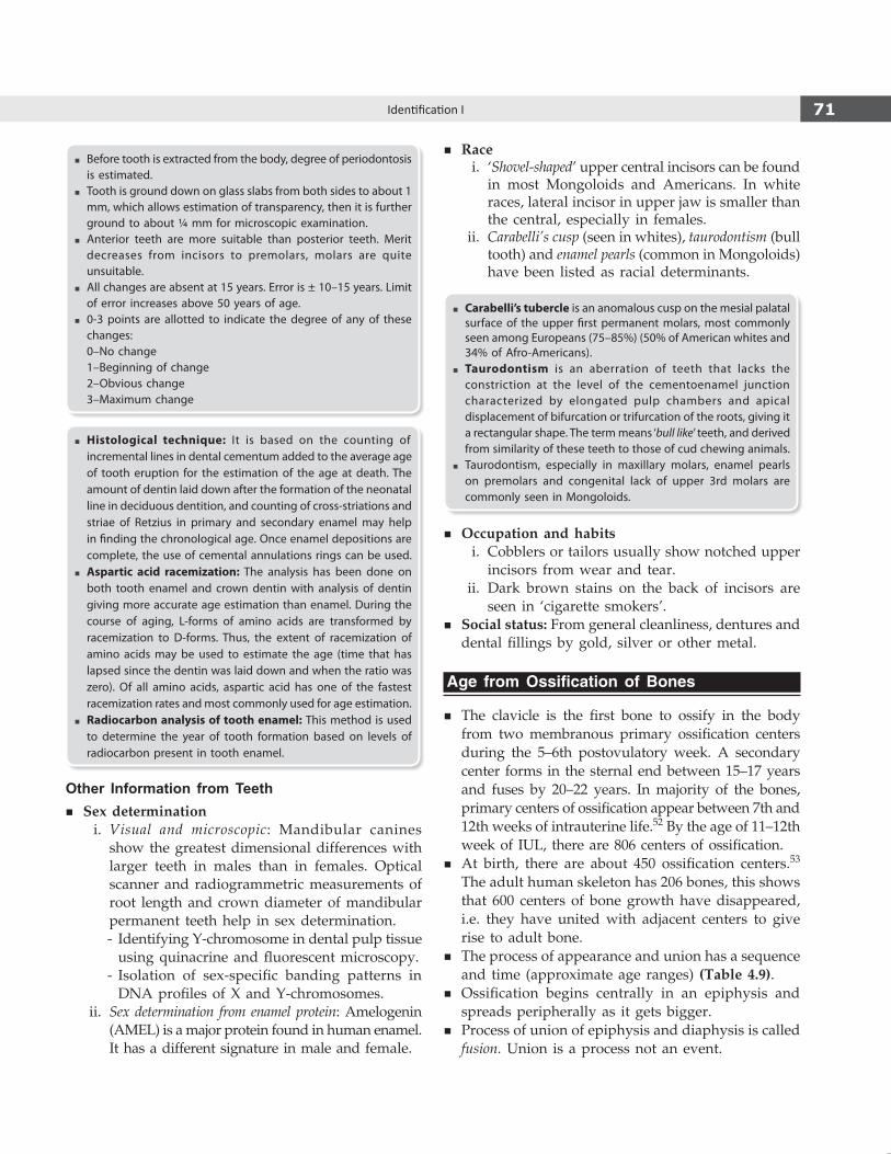
71Identification I
� Before tooth is extracted from the body, degree of periodontosis is estimated.
� Tooth is ground down on glass slabs from both sides to about 1 mm, which allows estimation of transparency, then it is further ground to about ¼ mm for microscopic examination.
� Anterior teeth are more suitable than posterior teeth. Merit decreases from incisors to premolars, molars are quite unsuitable.
� All changes are absent at 15 years. Error is ± 10–15 years. Limit of error increases above 50 years of age.
� 0-3 points are allotted to indicate the degree of any of these changes:
0–No change 1–Beginning of change 2–Obvious change 3–Maximum change
� Histological technique: It is based on the counting of incremental lines in dental cementum added to the average age of tooth eruption for the estimation of the age at death. The amount of dentin laid down after the formation of the neonatal line in deciduous dentition, and counting of cross-striations and striae of Retzius in primary and secondary enamel may help in finding the chronological age. Once enamel depositions are complete, the use of cemental annulations rings can be used.
� Aspartic acid racemization: The analysis has been done on both tooth enamel and crown dentin with analysis of dentin giving more accurate age estimation than enamel. During the course of aging, L-forms of amino acids are transformed by racemization to D-forms. Thus, the extent of racemization of amino acids may be used to estimate the age (time that has lapsed since the dentin was laid down and when the ratio was zero). Of all amino acids, aspartic acid has one of the fastest racemization rates and most commonly used for age estimation.
� Radiocarbon analysis of tooth enamel: This method is used to determine the year of tooth formation based on levels of radiocarbon present in tooth enamel.
Other Information from Teeth � Sex determination
i. Visual and microscopic: Mandibular canines show the greatest dimensional differences with larger teeth in males than in females. Optical scanner and radiogrammetric measurements of root length and crown diameter of mandibular permanent teeth help in sex determination.
- Identifying Y-chromosome in dental pulp tissue using quinacrine and fluorescent microscopy.
- Isolation of sex-specific banding patterns in DNA profiles of X and Y-chromosomes.
ii. Sex determination from enamel protein: Amelogenin (AMEL) is a major protein found in human enamel. It has a different signature in male and female.
� Race i. ‘Shovelshaped’ upper central incisors can be found
in most Mongoloids and Americans. In white races, lateral incisor in upper jaw is smaller than the central, especially in females.
ii. Carabelli’s cusp (seen in whites), taurodontism (bull tooth) and enamel pearls (common in Mongoloids) have been listed as racial determinants.
� Carabelli’s tubercle is an anomalous cusp on the mesial palatal surface of the upper first permanent molars, most commonly seen among Europeans (75–85%) (50% of American whites and 34% of Afro-Americans).
� Taurodontism is an aberration of teeth that lacks the constriction at the level of the cementoenamel junction characterized by elongated pulp chambers and apical displacement of bifurcation or trifurcation of the roots, giving it a rectangular shape. The term means ‘bull like’ teeth, and derived from similarity of these teeth to those of cud chewing animals.
� Taurodontism, especially in maxillary molars, enamel pearls on premolars and congenital lack of upper 3rd molars are commonly seen in Mongoloids.
� Occupation and habits i. Cobblers or tailors usually show notched upper
incisors from wear and tear. ii. Dark brown stains on the back of incisors are
seen in ‘cigarette smokers’. � Social status: From general cleanliness, dentures and
dental fillings by gold, silver or other metal.
Age from Ossification of Bones
� The clavicle is the first bone to ossify in the body from two membranous primary ossification centers during the 5–6th postovulatory week. A secondary center forms in the sternal end between 15–17 years and fuses by 20–22 years. In majority of the bones, primary centers of ossification appear between 7th and 12th weeks of intrauterine life.52 By the age of 11–12th week of IUL, there are 806 centers of ossification.
� At birth, there are about 450 ossification centers.53 The adult human skeleton has 206 bones, this shows that 600 centers of bone growth have disappeared, i.e. they have united with adjacent centers to give rise to adult bone.
� The process of appearance and union has a sequence and time (approximate age ranges) (Table 4.9).
� Ossification begins centrally in an epiphysis and spreads peripherally as it gets bigger.
� Process of union of epiphysis and diaphysis is called fusion. Union is a process not an event.

72 Review of Forensic Medicine and Toxicology
Table 4.9: Appearance of ossification centers (in males)
S.No. Bone Centers of ossification Age of appearance Age of union
1. Sternum � Manubrium � 1st sternebrae � 2nd and 3rd sternebrae � 4th sternebrae � Xiphisternum
5th month IUL5th month IUL7th month IUL10th month IUL3rd year postnatal
60–70 years (manubriosternal joint)14–25 years (from below upwards; 3rd and 4th–15 years; 2nd and 3rd–20 years; 1st and 2nd–25 years)40–45 years (with body)
2. Clavicle Medial end 15–17 years 20–22 years
3. Scapula � Coracoid base � Acromion process
10–11 years14–15 years
14–15 years17–18 years
Uppper Limb
4. Humerus � Head � Greater tubercle � Lesser tubercle � Capitulum � Trochlea � Lateral epicondyle � Medial epicondyle
1 year3 years5 years1 year9–10 years10–11 years5–6 years
At 5–6 years, the three fusestogether (conjoint epiphysis) and at 17–18 years, fuses with the shaftAt 14–15 years, all threefuses with the shaft*
16 years
5. Radius � Upper end � Lower end
5–6 years1–2 years
15–17 years17–19 years
6. Ulna � Upper end54
� Lower end8–9 years5–6 years
15–17 years17–19 years
7. Carpals Pisiform 9-12 years —
Lower Limb
8. Hip Bone � Ischiopubic rami � Triradiate cartilage � Iliac crest � Ischial tuberosity
——15–16 years16–17 years
7 years12–14 years19–21 years20–22 years
9. Femur � Head � Greater trochanter � Lesser trochanter � Lower end
1 year4 years14 years9 months IUL (at birth)
17–18 years14–15 years15–17 years 17–18 years
10. Tibia � Upper end � Lower end
At birth1 year
17–18 years16–17 years
11. Fibula � Upper end � Lower end
4 years2 years
17–18 years16–17 years
12. Tarsals � Calcaneum � Talus � Cuboid
5th month IUL7th month IUL9th month IUL
17–18 years
* Ossification centers of lateral epicondyle, capitulum and trochlea fuse with each other at about 13–14 years to form a conjoint epiphysis which fuses with shaft at about 14–15 years
}}
� Some researchers have used five grades of epiphyseal union: unobservable (0), beginning (1), active (2), recent (3) and complete (4), and these offer a possibly more accurate estimate of age.
� Capitate and hamate ossifies during infancy (1 year), the former preceding the later.55,56 Between 2–6 years, the number of carpal bones present on X-ray represents the approximate age in years, e.g. 3 carpal bones—3 years (Fig. 4.15).57-60
� If all the epiphyses of all the long bones are united, the person is most probably over 25 years of age.
� X-rays of elbow, wrist, clavicle and shoulder joints (upper extremity) and hip, knee and ankle joints (lower extremity) are usually recommended to determine the age before 25 years of age.
� Determination of age based on the union of epiphyses with a range of ± 6 months is given in Table 4.10. In females, epiphyseal union occurs 1–2 years earlier than males.
}
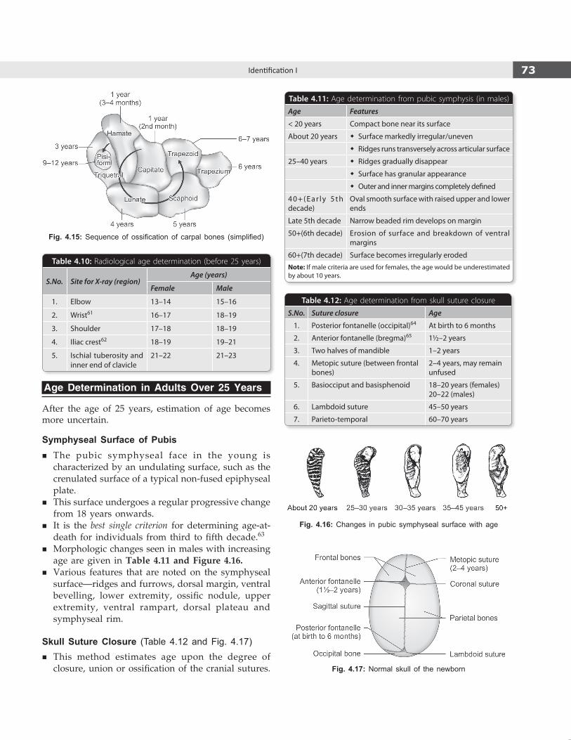
73Identification I
Fig. 4.15: Sequence of ossification of carpal bones (simplified)
Age Determination in Adults Over 25 years
After the age of 25 years, estimation of age becomes more uncertain.
Symphyseal Surface of Pubis � The pubic symphyseal face in the young is
characterized by an undulating surface, such as the crenulated surface of a typical non-fused epiphyseal plate.
� This surface undergoes a regular progressive change from 18 years onwards.
� It is the best single criterion for determining age-at-death for individuals from third to fifth decade.63
� Morphologic changes seen in males with increasing age are given in Table 4.11 and Figure 4.16.
� Various features that are noted on the symphyseal surface—ridges and furrows, dorsal margin, ventral bevelling, lower extremity, ossific nodule, upper extremity, ventral rampart, dorsal plateau and symphyseal rim.
Skull Suture Closure (Table 4.12 and Fig. 4.17) � This method estimates age upon the degree of
closure, union or ossification of the cranial sutures.
Fig. 4.16: Changes in pubic symphyseal surface with age
Fig. 4.17: Normal skull of the newborn
Table 4.10: Radiological age determination (before 25 years)
S.No. Site for X-ray (region)Age (years)
Female Male
1. Elbow 13–14 15–16
2. Wrist61 16–17 18–19
3. Shoulder 17–18 18–19
4. Iliac crest62 18–19 19–21
5. Ischial tuberosity and inner end of clavicle
21–22 21–23
Table 4.11: Age determination from pubic symphysis (in males)
Age Features
< 20 years Compact bone near its surface
About 20 years � Surface markedly irregular/uneven
� Ridges runs transversely across articular surface
25–40 years � Ridges gradually disappear
� Surface has granular appearance
� Outer and inner margins completely defined
4 0 + ( E a r l y 5 t h decade)
Oval smooth surface with raised upper and lower ends
Late 5th decade Narrow beaded rim develops on margin
50+(6th decade) Erosion of surface and breakdown of ventral margins
60+(7th decade) Surface becomes irregularly erodedNote: If male criteria are used for females, the age would be underestimated by about 10 years.
Table 4.12: Age determination from skull suture closure
S.No. Suture closure Age
1. Posterior fontanelle (occipital)64 At birth to 6 months
2. Anterior fontanelle (bregma)65 1½–2 years
3. Two halves of mandible 1–2 years
4. Metopic suture (between frontal bones)
2–4 years, may remain unfused
5. Basiocciput and basisphenoid 18–20 years (females) 20–22 (males)
6. Lambdoid suture 45–50 years
7. Parieto-temporal 60–70 years
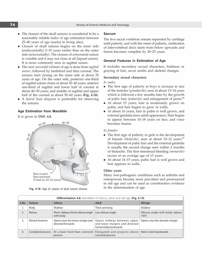
74 Review of Forensic Medicine and Toxicology
� The closure of the skull sutures is considered to be a reasonably reliable index of age estimation between 25–40 years of age (useful in living also).
� Closure of skull sutures begins on the inner side (endocranially) 5–10 years earlier than on the outer side (ectocranially). The closure of ectocranial suture is variable and it may not close at all (lapsed union). It is more commonly seen in sagittal suture.
� The most successful estimate of age is done from sagittal suture, followed by lambdoid and then coronal. The sutures start closing on the inner side at about 25 years of age. On the outer side, posterior one-third of sagittal suture closes at about 30–40 years; anterior one-third of sagittal and lower half of coronal at about 40–50 years; and middle of sagittal and upper half of the coronal at about 50–60 years (Fig. 4.18).
� A lateral head skiagram is preferable for observing the sutures.
Age Estimation from MandibleIt is given in Diff. 4.8.
Fig. 4.18: Age (in years) of skull suture closure
Differentiation 4.8: Mandibles of infancy, adult and old age (Fig. 4.19)
S.No. Feature Infancy Adult Old age
1. Body Shallow Thick and long Shallow
2. Ramus Short, oblique, forms obtuse angle with body
Less obtuse angle Obtuse angle with body (about 140°)
3. Mental foramen Opens near the lower margin and directed forwards
Opens midway between upper and lower margins and directed horizontally backwards
Opens near the alveolar margin
4. Condyloid process At a lower level than coronoid process
Elongated and projects above coronoid process
Neck is bent backwards
SacrumThe five sacral vertebrae remain separated by cartilage until puberty, and with the onset of puberty, ossification of intervertebral discs starts from below upwards and fusion becomes complete by 20–25 years.
General Features in Estimation of Age
It includes secondary sexual characters, baldness or graying of hair, arcus senilis and skeletal changes.
Secondary sexual charactersIn males
� The first sign of puberty in boys is increase in size of the testicles (gonadarche), seen at about 13–14 years which is followed a few months later by the growth of pubic hair (pubarche) and enlargement of penis.66
� At about 15 years, hair is moderately grown on pubis, and hair begins to grow in axilla.
� At about 16 years, hair in pubis is well grown, and external genitalia have adult appearance. Hair begins to appear between 16–18 years on face, and voice becomes hoarse.
In females � The first sign of puberty in girls is the development
of breasts (thelarche), seen at about 10–12 years.67 Development of pubic hair and the external genitalia is usually the second change seen within 2 months of thelarche. The first menstrual bleeding (menarche) occurs at an average age of 13 years.
� At about 14–15 years, pubic hair is well grown and hair appears in axilla.
Older yearsMany non-pathogenic conditions such as arthritis and osteoporosis become more prevalent and pronounced in old age and can be used as corroborative evidence in the determination of age.
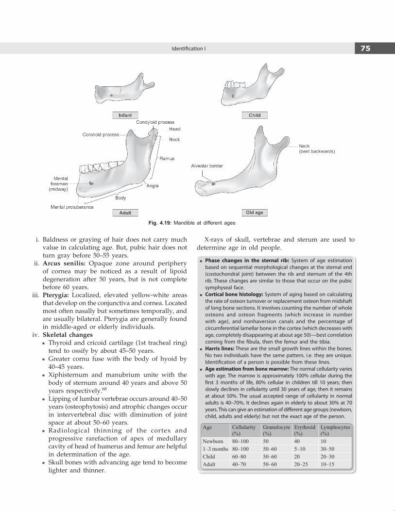
75Identification I
i. Baldness or graying of hair does not carry much value in calculating age. But, pubic hair does not turn gray before 50–55 years.
ii. Arcus senilis: Opaque zone around periphery of cornea may be noticed as a result of lipoid degeneration after 50 years, but is not complete before 60 years.
iii. Pterygia: Localized, elevated yellow-white areas that develop on the conjunctiva and cornea. Located most often nasally but sometimes temporally, and are usually bilateral. Pterygia are generally found in middle-aged or elderly individuals.
iv. Skeletal changes z Thyroid and cricoid cartilage (1st tracheal ring)
tend to ossify by about 45–50 years. z Greater cornu fuse with the body of hyoid by
40–45 years. z Xiphisternum and manubrium unite with the
body of sternum around 40 years and above 50 years respectively.68
z Lipping of lumbar vertebrae occurs around 40–50 years (osteophytosis) and atrophic changes occur in intervertebral disc with diminution of joint space at about 50–60 years.
z Radiological thinning of the cortex and progressive rarefaction of apex of medullary cavity of head of humerus and femur are helpful in determination of the age.
z Skull bones with advancing age tend to become lighter and thinner.
X-rays of skull, vertebrae and sterum are used to determine age in old people.
� Phase changes in the sternal rib: System of age estimation based on sequential morphological changes at the sternal end (costochondral joint) between the rib and sternum of the 4th rib. These changes are similar to those that occur on the pubic symphyseal face.
� Cortical bone histology: System of aging based on calculating the rate of osteon turnover or replacement osteon from midshaft of long bone sections. It involves counting the number of whole osteons and osteon fragments (which increase in number with age), and nonhaversion canals and the percentage of circumferential lamellar bone in the cortex (which decreases with age, completely disappearing at about age 50)—best correlation coming from the fibula, then the femur and the tibia.
� Harris lines: These are the small growth lines within the bones. No two individuals have the same pattern, i.e. they are unique. Identification of a person is possible from these lines.
� Age estimation from bone marrow: The normal cellularity varies with age. The marrow is approximately 100% cellular during the first 3 months of life, 80% cellular in children till 10 years; then slowly declines in cellularity until 30 years of age, then it remains at about 50%. The usual accepted range of cellularity in normal adults is 40–70%. It declines again in elderly to about 30% at 70 years. This can give an estimation of different age groups (newborn, child, adults and elderly) but not the exact age of the person.
Age Cellularity (%)
Granulocyte (%)
Erythroid (%)
Lymphocytes (%)
Newborn 80–100 50 40 10
1–3 months 80–100 50–60 5–10 30–50
Child 60–80 50–60 20 20–30
Adult 40–70 50–60 20–25 10–15
Fig. 4.19: Mandible at different ages
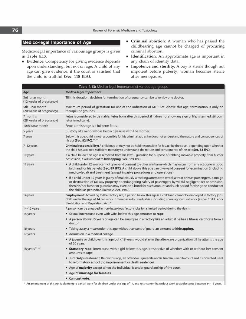
76 Review of Forensic Medicine and Toxicology
Table 4.13: Medico-legal importance of various age groups
Age Medico-legal importance
3rd lunar month(12 weeks of pregnancy)
Till this duration, decision for termination of pregnancy can be taken by one doctor.
5th lunar month(20 weeks of pregnancy)
Maximum period of gestation for use of the indication of MTP Act. Above this age, termination is only on therapeutic grounds.
7 months(28 weeks of pregnancy)
Fetus is considered to be viable. Fetus born after this period, if it does not show any sign of life, is termed stillborn fetus (medically).
10th lunar month Fetus at this stage is a full term fetus.
5 years Custody of a minor who is below 5 years is with the mother.
7 years Below this age, child is not responsible for his criminal act, as he does not understand the nature and consequences of his act (Sec. 82 IPC).69,70
7–12 years Criminal responsibility: A child may or may not be held responsible for his act by the court, depending upon whether the child has attained sufficient maturity to understand the nature and consequence of the act (Sec. 83 IPC).
10 years If a child below this age is removed from his lawful guardian for purpose of robbing movable property from his/her possession, it will amount to kidnapping (Sec. 369 IPC).
12 years � A child under 12 years cannot give valid consent to suffer any harm which may occur from any act done in good faith and for his benefit (Sec. 89 IPC). A child above this age can give valid consent for examination (including medico-legal) and treatment (except invasive procedures and operations).
� If a child under 12 years is guilty of maliciously wrecking/attempt to wreck a train or hurt passengers, damage or destruction of railway property or endangering safety of passengers by willful negligent act or omission, then his/her father or guardian may execute a bond for such amount and such period for the good conduct of the child (as per Indian Railways Act, 1989).
14 years Employment: According to the Factory Act, a person below this age is a child and cannot be employed in factory jobs. Child under the age of 14 can work in ‘non-hazardous industries’ including some agricultural work [as per Child Labor (Prohibition and Regulation) Act].*
14–15 years A person can be engaged in non-hazardous factory jobs for a limited period during the day h.
15 years � Sexual intercourse even with wife, below this age amounts to rape.
� A person above 15 years of age can be employed in a factory like an adult, if he has a fitness certificate from a doctor.
16 years � Taking away a male under this age without consent of guardian amount to kidnapping.
17 years � Admission in a medical college.
� A juvenile or child over this age but <18 years, would stay in the after-care organization till he attains the age of 20 years.
18 years71-73 � Statutory rape: Intercourse with a girl below this age, irrespective of whether with or without her consent amounts to rape.
� Judicial punishment: Below this age, an offender is juvenile and is tried in juvenile court and if convicted, sent to reformatory school (no imprisonment or death sentence).
� Age of majority except when the individual is under guardianship of the court.
� Age of marriage for females.
� Can cast vote. * An amendment of this Act is planning to ban all work for children under the age of 14, and restrict non-hazardous work to adolescents between 14–18 years.
Medico-legal Importance of Age
Medico-legal importance of various age groups is given in Table 4.13.
� Evidence: Competency for giving evidence depends upon understanding, but not on age. A child of any age can give evidence, if the court is satisfied that the child is truthful (Sec. 118 IEA).
� Criminal abortion: A woman who has passed the childbearing age cannot be charged of procuring criminal abortion.
� Identification: An approximate age is important in any chain of identity data.
� Impotence and sterility: A boy is sterile though not impotent before puberty; woman becomes sterile after menopause.

77Identification I
Stature
� Stature varies at different times of day by 1.5–2 cm. It is less in the afternoon and evening due to reduced elasticity of intervertebral discs and the longitudinal vertebral muscles.
� After the age of 30, the natural process of senile degeneration causes gradual decrease in stature by 0.6 mm/year on average.
� Stature is greater by 1–3 cm on lying down. � On an average, the body lengthens after death by
about 2 cm, due to complete loss of muscle tone, relaxation of large joints and loss of tensioning effect of paraspinal muscles on intervertebral discs.
� If the body has been dismembered or skeletonized, the approximate stature may be determined by:
i. Length of entire skeleton and 2.5–4 cm for thickness of soft parts.
ii. Length from tip of middle finger to the tip of the opposite finger when arms are fully extended.
iii. Twice the length of one arm with 30 cm added for two clavicles and 4 cm for sternum.
iv. Length from vertex to the symphysis pubis is roughly half of stature.
v. Length from sternal notch to symphysis pubis multiplied by 3.3.
Stature from BonesThe methods in use to determine the stature can be divided into:
� Least squares regression equation and other regression principles
� Stature: bone length ratios � Skeleton height and adjustment for missing soft
tissue.Sex and race of the individual should be taken into
account while applying these methods. � When whole skeleton is not available, but one or
the other long bones are available, then regression equations are used.
Age Medico-legal importance
� Mentally sound person can make a valid will (testamentary capacity).
� Taking out or enticement of a girl below this age from custody of her guardian amounts to kidnapping.
� Kidnapping a boy or girl below this age for purpose of begging is punishable with imprisonment of 10 years with or without fine.
� Can be employed in any authorized job in a factory.
� Can give valid consent to suffer any harm which may result from an act not intended or not known to cause death or grievous hurt (Sec. 87 IPC).
� Minimum age for entering a government service.
� A pregnant female can give valid consent for termination under the MTP Act.
� A person can authorize the removal of organ from his/her body for therapeutic purposes under the Transplantation of Human Organs Act.
� Abetment to suicide of a person below this age is punishable with imprisonment upto 10 years to life imprisonment and fine.
� If wife below this age brings a case of offence under Sec. 376 IPC (sexual intercourse by a man with his own wife), the court with not take cognizance if > 1 year has elapsed from the commission of offence.
21 years � Age of marriage for males.
� If a girl below this age is ‘imported’ to India from foreign country for the purpose of illicit intercourse, the act amounts to kidnapping (Sec. 366 B IPC).
� Person under the guardianship of the Court of Wards attains majority.
25 years � Age for contesting membership of Parliament and other legislative bodies.
� Age limit for entering in some government services.
� According to Punjab Excise Act, a person below this age cannot buy and consume liquor.
35 years � Minimum age for appointment as President, Vice-President and Governor of States in India.
� Prenatal diagnostic tests/procedures cannot be conducted if the pregnant female is below this age as per the PCPNDT Act.
55–65 years Age of retirement from services under the government, Statutory bodies, autonomous bodies/institutes or from judiciary services.
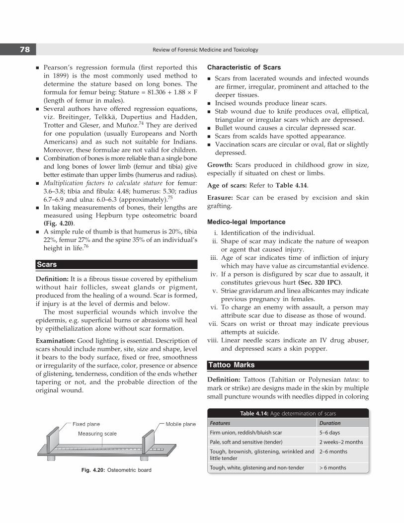
78 Review of Forensic Medicine and Toxicology
� Pearson’s regression formula (first reported this in 1899) is the most commonly used method to determine the stature based on long bones. The formula for femur being: Stature = 81.306 + 1.88 × F (length of femur in males).
� Several authors have offered regression equations, viz. Breitinger, Telkkä, Dupertius and Hadden, Trotter and Gleser, and Muñoz.74 They are derived for one population (usually Europeans and North Americans) and as such not suitable for Indians. Moreover, these formulae are not valid for children.
� Combination of bones is more reliable than a single bone and long bones of lower limb (femur and tibia) give better estimate than upper limbs (humerus and radius).
� Multiplication factors to calculate stature for femur: 3.6–3.8; tibia and fibula: 4.48; humerus: 5.30; radius 6.7–6.9 and ulna: 6.0–6.3 (approximately).75
� In taking measurements of bones, their lengths are measured using Hepburn type osteometric board (Fig. 4.20).
� A simple rule of thumb is that humerus is 20%, tibia 22%, femur 27% and the spine 35% of an individual’s height in life.76
Scars
Definition: It is a fibrous tissue covered by epithelium without hair follicles, sweat glands or pigment, produced from the healing of a wound. Scar is formed, if injury is at the level of dermis and below.
The most superficial wounds which involve the epidermis, e.g. superficial burns or abrasions will heal by epithelialization alone without scar formation.
Examination: Good lighting is essential. Description of scars should include number, site, size and shape, level it bears to the body surface, fixed or free, smoothness or irregularity of the surface, color, presence or absence of glistening, tenderness, condition of the ends whether tapering or not, and the probable direction of the original wound.
Characteristic of Scars � Scars from lacerated wounds and infected wounds
are firmer, irregular, prominent and attached to the deeper tissues.
� Incised wounds produce linear scars. � Stab wound due to knife produces oval, elliptical,
triangular or irregular scars which are depressed. � Bullet wound causes a circular depressed scar. � Scars from scalds have spotted appearance. � Vaccination scars are circular or oval, flat or slightly
depressed.
Growth: Scars produced in childhood grow in size, especially if situated on chest or limbs.
Age of scars: Refer to Table 4.14.
Erasure: Scar can be erased by excision and skin grafting.
Medico-legal Importance i. Identification of the individual. ii. Shape of scar may indicate the nature of weapon
or agent that caused injury. iii. Age of scar indicates time of infliction of injury
which may have value as circumstantial evidence. iv. If a person is disfigured by scar due to assault, it
constitutes grievous hurt (Sec. 320 IPC). v. Striae gravidarum and linea albicantes may indicate
previous pregnancy in females. vi. To charge an enemy with assault, a person may
attribute scar due to disease as those of wound. vii. Scars on wrist or throat may indicate previous
attempts at suicide. viii. Linear needle scars indicate an IV drug abuser,
and depressed scars a skin popper.
Tattoo Marks
Definition: Tattoos (Tahitian or Polynesian tatau: to mark or strike) are designs made in the skin by multiple small puncture wounds with needles dipped in coloring
Fig. 4.20: Osteometric board
Table 4.14: Age determination of scars
Features Duration
Firm union, reddish/bluish scar 5–6 days
Pale, soft and sensitive (tender) 2 weeks–2 months
Tough, brownish, glistening, wrinkled and little tender
2–6 months
Tough, white, glistening and non-tender > 6 months

79Identification I
matter which is attached to an oscillating unit. The unit rapidly and repeatedly drives the needles in and out of the skin, usually 50–3000 times a minute.
Dyes used: Indigo, cobalt, carbon, vermilion, cadmium, selenium, Prussian blue and Indian ink.
� Color, design, size and situation should be noted. � The permanency of tattoo marks depends upon the
type of dye used, the depth of its penetration and the part of body tattooed. Permanent tattoos are obtained if:
i. Black, blue and red dyes are employed. ii. The dye penetrates the dermis. iii. The part of body is protected by clothing.
� A latent (faded) tattoo mark becomes visible by rubbing that part and examining with magnifying lens. The use of high contrast photography, computer image enhancement, UV lamp or infra-red photo-graphy is also helpful for identifying faded tattoos.
� Tattoos are recognized even in decomposed bodies and bodies recovered from water when the epidermis is removed.
� Since some pigment migrates from the tattoo site to the body’s lymph nodes, pigmentations of the axillary lymph nodes in upper extremities tattoos could be identified with the naked eye during autopsy.77
Complications: Septic inflammation, abscess, gangrene, syphilis, hepatitis B, AIDS, leprosy and tuberculosis.
Classification of TattoosThe American Academy of Dermatology distinguishes five types of tattoos: i. Traumatic tattoos (‘natural tattoos’) resulting from
injuries (roadside injuries) or close range firearm (unburnt gunpowder) or pencil lead; these are unintentional and unwanted tattoos.
ii. Amateur tattoos: Tattoo applied by anyone at home, using a needle and a single color carbon based ink, e.g. India ink applied at varying depths.
iii. Professional tattoos (using both traditional methods and modern tattoo machines): This is created by a trained tattoo artist at a salon or tattoo parlor which contains several colors and applied uniformly beneath the skin.
iv. Cosmetic tattoos (also known as ‘permanent makeup’): They camouflage skin discolorations, such as birthmarks (hemangiomas) or scars, tattooing ‘hair follicles’ into bald areas or corneal tattooing in perforating injury. India ink is the most commonly used dye for corneal tattooing. Two other methods
exist: chemical dyeing with gold or platinum chloride and carbon impregnation.78
v. Medical tattoos: This type of tattoos is used for indicating a medically relevant condition or body location, e.g. medical alert tattoos (like insulin-dependent diabetes mellitus or drug allergy), blood group tattoo, reconstructive surgery (nipple-areola complex in mastectomy), delineating the radiation field, and endoscopic tattoos for directing endoscopic procedures.
Erasure of Tattoo i. Surgical methods
z Dermabrasion using dermabraders [e.g., tannic acid, silver nitrate (Variot’s method) or trichloroacetic acid (chemical peels)] or ‘salt abrasion’ wherein salts like zinc chloride are applied or Qswitched Nd:YAG laser. Laser beam vaporizes the particles of the dye and are expelled from tissues in gaseous form.
z Complete excision and skin grafting. z Production of burns by means of red hot iron. z Scarification. z Using carbon dioxide snow.
ii. Electrolysis. iii. Caustic or corrosive substances remove pigment by
producing inflammatory reaction and superficial scar, e.g. mixture of papain in glycerin.
Chronic eczema may cause the tattoo designs to disappear.
Medico-legal ImportanceIt helps in knowing the: i. Identity of a person, particularly the dead or
decomposed individual—his name or spouse’s or friend’s; date of birth or joining of service.
ii. Religion and nationality: Designs of Cross or Christ (in Christians), and Hanuman or Lord Krishna (in Hindus).
iii. Political affiliations, e.g. hammer and sickle, lotus or right hand.
iv. Race: Tattooing on the chest and limbs is common amongst the Japanese.
v. Profession/occupation: Some gangs have certain specific emblems of tattoo marks. Some occupations, e.g. coal miners leave visible tattoo marks on the hands and face.
vi. Behavioral characteristics: Tattoos have been associated with high-risk behaviors including alcohol and drug use, violence, carrying weapons, sexual activity, eating disorders and suicide.

80 Review of Forensic Medicine and Toxicology
z Erotic tattoos of the sexual fanatic, blue bird design on the extensor surface of the web of thumb of homosexuals, number 13 inside the lower lip of drug pushers, addict type of tattoo marks to conceal injection sites.
vii. It may also represent social status of that indi-vidual.
Notes
� Marfan syndrome is an inherited connective-tissue disorder transmitted as an autosomal dominant trait. Cardinal features include tall stature, ectopia lentis, mitral valve prolapse, aortic root dilatation and aortic dissection.
� Down syndrome is the most common chromosomal disorder and the most common cause of intellectual disability that result from having an extra copy of chromosome 21.19
� Mayer-Rokitansky-Küster-Hauser (MRKH) syndrome/Müllerian agenesis: It is a congenital malformation characterized by müllerian duct agenesis. Commonest presentation is congenital absence of the vagina, uterus or both. Patients usually present with primary amenorrhea in adolescence. After gonadal dysgenesis, MRKH is the second most common cause of primary amenorrhea. Patients have normal height, secondary sexual characteristics, body hair, external genitalia, karyotype (46XX) and hormonal profile. Ovaries are intact and ovulation usually occurs.79
� Kallmann syndrome is a rare hormonal condition of hypogonadotrophic hypogonadism. It characterized by delayed or absent puberty and an impaired sense of smell, and can affect both men and women. Most patients have gonadotropin-
releasing hormone deficiency. Males are often born with a small penis (micropenis) and cryptorchidism. At puberty, patients do not develop secondary sexual characteristics, such as the growth of facial hair and deepening of the voice in males. In affected females, there is amenorrhoea with little or no breast development.
� Noonan syndrome is a genetic disorder that affects men and women equally. It was thought to be a form of Turner syndrome, but patients with this syndrome have normal karyotype—important distinction between the Turner and Noonan syndromes. Cardinal features include hypertelorism, down-slanting eyes, webbed neck, congenital heart disease (in 50%), short stature, mental retardation (in 25%), bleeding diathesis and chest deformity and are predisposed to malignancies.16,19
� Sheehan syndrome occurs due to necrosis of the pituitary gland with associated hypopituitarism resulting from postpartum hemorrhage and hypovolemic shock.
� Virilizing ovarian tumor is a rare cause of hyperandrogenism in women, and account for < 5% of all ovarian neoplasms.
� Ovarian dysgenesis is a rare type of female hypogonadism in which no functional ovaries are present to induce puberty in an otherwise normal girl whose karyotype is found to be 46XX.
� Dentigerous cyst: The second most common odontogenic cyst is the dentigerous cyst (first being periapical cyst), which develops within the normal dental follicle that surrounds an unerupted tooth. It is most frequently found in areas where unerupted teeth are found: mandibular third molars, maxillary third molars and maxillary canines, in decreasing order of frequency.46
� As per Hindu Marriage Act, marriage can be declared null and void, if one of the parties, at the time of ceremony, was incapable of giving valid consent or was unfit for marriage and procreation of children due to unsoundness of mind.73
1. A 2. A 3. C 4. A 5. D 6. A 7. A 8. B
MulTIPlE CHOICE quESTIOnS
1. Corpus delicti means: TN 07; AIIMS 08 A. Proof of occurrence of crime B. Decomposed body C. Identification of dead person D. None 2. Whole skull is used to calculate: PGI 12 A. Cephalic index B. Nasal index C. Crural index D. Brachial index 3. Cephalic index is useful for the determination of:
UP 08; DNB 10; NEET 13, 14 A. Age B. Sex C. Race D. Sature 4. Cephalic index of 80–85 belongs to: JIPMER 08 A. Mongolians B. Indians C. Europeans D. Negroes
5. Caucasian skull is: BHU 10 A. Elongated B. Square C. Narrow D. Round 6. Crural index is: NEET 13 A. Length of tibia/femur × 100 B. Length of radius/humerus × 100 C. Length of fibula/tibia × 100 D. Length of radius/ulna × 100 7. Barr body is nOT seen in: PGI 07; WB 08 A. Turner syndrome B. Klinefelter syndrome C. Down’s syndrome D. Marfan’s syndrome 8. Barr body was first detected in: DNB 08 A. Buccal mucosa B. Brain C. Liver D. Skin

81Identification I
9. B 10. D 11. D 12. D 13. B 14. D 15. A 16. B 17. B 18. A 19. B 20. A 21. D 22. A 23. C 24. A 25. A 26. D 27. C 28. C 29. B
9. Streak ovaries are seen in: CMC (Vellore) 09 A. Klinefelter syndrome B. Turner syndrome C. Down syndrome D. Kallamann syndrome 10. Klinefelter syndrome is: MP 07, 10; Maharashtra 09; Gujarat 10; AfMC 12; CMC (Vellore)14 A. 45XO B. 46XY C. 45XY D. 47XXY 11. Klinefelter syndrome is associated with all, except:
JIPMER 08; AfMC 12; NIMHANS 14 A. Delayed puberty B. Male phenotype C. Azoospermia D. Barr body absent 12. All are seen in Klinefelter syndrome, except: WB 07 A. Mental retardation B. Male phenotype C. Azoospermia D. Low FSH level 13. False about Klinefelter’s syndrome: AP 08; MAHE 12 A. Most common syndrome of sex gene involvement B. Most common cause of hypothalamic hypogona-
dotropic failure in males C. Mental retardation may be seen D. Serum FSH levels are consistently high 14. Incidence of Turner syndrome: MAHE 10 A. 1: 500 B. 1: 1000 C. 1: 1500 D. 1: 2500 15. A 19-year-old woman presented with primary
amenorrhea, short stature, webbed neck and widely spaced nipples. Examination showed weak pulses in lower extremity and streak ovaries, raised FSH, no oocyte in histology of ovary. Karyotype most likely to be present: KCET 12; JIPMER 12; AIIMS 13; PGI 13
A. 45XO B. 47XXY C. 46XY D. 46XX 16. Webbing of neck, short stature, increased carrying angle,
low posterior hair line, primary amenorrhea and short fourth metacarpal are characteristics of: AIIMS 09
A. Klinefelter syndrome B. Turner syndrome C. Cri-du-chat syndrome D. Noonan syndrome 17. All of the following about Turner syndrome are true,
except: MAHE 09, 11; AP 08; UPSC 09, Bihar 11; CMC (Vellore) 14
A. Amenorrhea B. Mental retardation C. Short stature D. Coarctation of aorta 18. Patient with genotype XO will have following
phenotype, except: PGI 09; WB 09; MAHE 12 A. Tall stature B. Broad chest C. Webbed neck D. Lymphedema 19. not associated with malignancy: JIPMER 13 A. Down’s syndrome B. Turner syndrome C. Noonan syndrome D. Klinefelter syndrome
20. Most common cause of hyperthalamic hypogonado-trophic failure in men: AI 10
A. Klinefelter syndrome B. Noonan syndrome C. Viral orchitis D. Kallmann syndrome 21. Most common cardiac anomaly in Turner syndrome: CMC (Vellore) 07; CMC (Ludhiana) 13; UPSC 14 A. Bifurcation of aorta B. Coarctation of aorta C. Aortic stenosis D. Bicuspid aortic valve 22. A girl presents with primary amenorrhea, grade V
thelarche (mature breast), grade II pubarche (sparse growth of pubic hair) and no axillary hair. Likely diagnosis is: fMGE 09; AIIMS 11, 14
A. Testicular feminization B. Mullerian agenesis C. Turner syndrome D. Gonadal dysgenesis 23. True about androgen insensitivity syndrome are all,
except: PGI 12, 13; JIPMER 13 A. Testes present B. Absent uterus C. 46XX D. Scanty pubic hair 24. Most common effect of congenital adrenal hyper-
plasia: Maharashtra 09 A. Female pseudohermaphroditism B. Male pseudohermaphroditism C. True hermaphroditism D. Gonadal dysgenesis 25. Most common cause of congenital adrenal hyper-
plasia: Maharashtra 10 A. 21-Hydroxylase deficiency B. 11-Hydroxylase deficiency C. 17-α-Hydroxylase deficiency D. 3-β-Hydroxy dehydrogenase deficiency 26. Commonest cause of female pseudohermaphroditism
is: WB 10 A. Virilizing ovarian tumor B. Ovarian dysgenesis C. Exogenous androgen D. Congenital adrenal hyperplasia 27. Krogman’s formula is related to: Odisha 11 A. Race B. Age C. Sex D. Stature 28. Most useful for sex determination is: Kerala 08 A. Skull B. Femur C. Pelvis D. Tibia 29. Preauricular sulcus is useful for determination of:
NEET 14 A. Race B. Sex C. Stature D. Age

82 Review of Forensic Medicine and Toxicology
30. Chilotic line helps in determination of: DNB 08 A. Race B. Sex C. Age D. Stature 31. Corporobasal index is used to determine: AI 08 A. Age B. Sex C. Race D. Stature 32. Age estimation from teeth can be done by all the
following methods, except: NEET 14 A. Gustafson’s method B. Miles’ method C. Boyde’s method D. Frame’s method 33. Hardest calcified part of tooth: JIPMER 13 A. Enamel B. Dentin C. Cementum D. Pulp 34. Mineralization of the teeth begins at: COMEDK 07 A. Crown and progresses towards root B. Root and progresses towards crown C. Simultaneously at root and crown D. Begins in the center 35. number of milk teeth: NEET 13 A. 12 B. 16 C. 20 D. 24 36. All are true about permanent teeth, except: MP 11 A. Ridge is present between neck and body B. Anterior teeth are inclined forward C. Roots of molars are larger D. They are ivory white in color 37. Primary dentition is complete by: UPSC 08 A. 1.5 years B. 2.5 years C. 3.5 years D. 4.5 years 38. The first incisors to erupt in an infant: UPSC 07 A. Lower central B. Lower lateral C. Upper central D. Upper lateral 39. In the upper jaw, deciduous teeth erupt earlier than
those in the lower jaw, except: Odisha 11 A. Lateral incisors B. Central incisors C. Canines D. Second molars 40. First permanent tooth to arise: AP 10; UPSC 11; Delhi 11 A. Incisor B. Canine C. Premolar D. Molar 41. Age of appearance of permanent lateral incisor: WB 10 A. 5–6 years B. 6–7 years C. 8–9 years D. 9–10 years 42. The most frequent tooth to be impacted: UPSC 07 A. Lower third molar B. Upper third molar C. Lower canine D. Upper premolar 43. The period of mixed dentition is between the age
of: UP 11 A. 2–9 years B. 6–11 years C. 12–14 years D. 16 years
44. number of teeth at 7 years: Gujarat 07 A. 20 B. 24 C. 26 D. 28 45. number of permanent teeth at 8 years: JIPMER 12 A. 6 B. 8 C. 12 D. 16 46. False with regard to permanent teeth is: TN 08 A. Molars erupt around 6 years B. Dentigerous cyst is common in canines C. There are 16 permanent teeth at the age of 10 years D. 3rd molars eruption is variable 47. Age of child with 20 permanent teeth and 4 temporary
teeth is: Kerala 08, 09; Punjab 10; UP 10 A. 9 years B. 10 years C. 11 years D. 14 years 48. Stack method of dental age estimation is used for:
AIIMS 11 A. Infants B. Adults C. Elderly D. Adolescents 49. Gustafson’s method is useful for determination of:
MAHE 06; Gujarat 10 A. Age B. Stature C. Race D. Sex 50. Gustafson’s method is most useful for: AP 07 A. 16 years B. 18 years C. 21 years D. > 25 years 51. The most reliable dental change used in Gustafson’s
method for age estimation is: COMEDK 08; AIIMS 11 A. Attrition B. Cementum C. Secondary dentin deposition D. Transparency of root 52. All the primary ossification centers are appeared at
fetal age of: WB 11 A. 1 month B. 2 months C. 3 months D. 4 months 53. number of ossification centers present at birth:
Punjab 08 A. 206 B. 250 C. 350 D. 450 54. Ossification center of upper end of ulna is united
by: DNB 10 A. 9 years B. 11 years C. 14 years D. 16 years 55. Ossification at two months: JIPMER 12 A. Lunate B. Capitate C. Scaphoid D. Hamate 56. At 1 year of age, the number of carpal bones seen in
skiagram of the hand is: COMEDK 08 A. Nil B. One C. Two D. Three
30. B 31. B 32. D 33. A 34. A 35. C 36. A 37. B 38. A 39. B 40. D 41. C 42. A 43. B 44. B 45. C 46. B 47. C 48. A 49. A 50. D 51. D 52. D 53. A 54. D 55. B 56. C

83Identification I
57. Four carpal bones are present at what age: DNB 09 A. 3 years B. 4 years C. 5 years D. 6 years 58. All true about pisiform, except: Manipal 11 A. Last carpal bone to ossify B. Attached to flexor carpi ulnaris C. Can be seen on newborn X-ray D. It is a sesamoid bone 59. Best X-ray to determine age of 7 years child:
Maharashtra 10 A. Hand and wrist B. Foot and ankle C. Pelvis D. Shoulder 60. Best X-ray to determine age of 12 years child:
AP 11; Maharashtra 11 A. Hand and wrist B. Foot and ankle C. Pelvis D. Shoulder 61. For a girl of 18 years, site for X-ray to determine her
age is: PGI 07 A. Elbow B. Wrist C. Knee D. Ankle Joint 62. Average age of iliac crest fusion in Indian females:
AIIMS 14 A. 16 years B. 18 years C. 19 years D. 21 years 63. Best bone to assess age between 20–50 years: WB 07 A. Skull B. Ribs C. Sternum D. Symphysis pubis 64. False about posterior fontanelle: BHU 09 A. Helps in estimating time of birth B. Site for concealed trauma C. Closes at 16–18 months D. Formed by parietal and occipital bones 65. Anterior fontanelle closes by: AP 07 A. 6 months B. 1 year C. 2 years D. 3 years 66. In males, first pubertal sign is: fMGE 10; WB 11 A. Testicular enlargement B. Hoarseness of voice C. Pubic hair development D. Penis enlargement 67. First pubertal change seen in females: DNB 09; CMC (Vellore) 13 A. Thelarche B. Menarche C. Pubarche D. Gonadarche 68. Xiphoid process fuses with sternum after: DNB 09 A. 60 years B. 40 years C. 30 years D. 20 years
69. The minimum age at which an individual is responsible for his criminal act is:
COMEDK 07; Bihar 11; NEET 14 A. 7 years B. 12 years C. 16 years D. 21 years 70. IPC for criminal responsibility is given in Sec: NEET 13 A. 81 B. 80 C. 82 D. 84 71. Age at which one becomes major: PGI 09; MAHE 12 A. 18 years B. 21 years C. 25 years D. 35 years 72. Age of marriage age for women: Gujarat 10 A. 16 years B. 17 years C. 18 years D. 21 years 73. Age of marriage of mentally retarded girl is: UP 09 A. 16 years B. 18 years C. 21 years D. Cannot marry 74. Stature is determined by which formula: WB 10 A. Hasse B. Widmark C. Trotter and Gleser D. Locard 75. The principle of Pearson’s formula is to measure the
length of long bone and multiply it with a given factor; for radius such factor is: Himachal 10
A. 3.6 to 3.8 B. 4.2 to 4.5 C. 6.3 to 6.9 D. 5.0 to 5.3 76. Length of tibia is: NEET 14 A. 10% of height B. 20% of height C. 30% of height D. 40% of height 77. Patient’s relative gives a history of tattoo, however
it was not found during autopsy. What should be dissected to find it: JIPMER 14
A. Lymph node B. Skin C. Spleen D. Kidney 78. Corneal tattooing may be done with: NEET 13 A. Gold chloride B. Calcium chloride C. Copper sulfate D. Potassium permanganate 79. A 16-year-old female with normal pubic hair and breast
development, presented with complaints of primary amenorrhea. Investigation shows normal karyotype. Most likely diagnosis is: AfMC 12; JIPMER 12; AIIMS 12, 13
A. Mullerian agenesis B. Turner syndrome C. Testicular feminization D. Kallmann syndrome
57. B 58. C 59. A 60. A 61. B 62. C 63. D 64. C 65. C 66. A 67. A 68. B 69. A 70. C 71. A 72. C 73. D 74. C 75. C 76. B 77. A 78. A 79. A
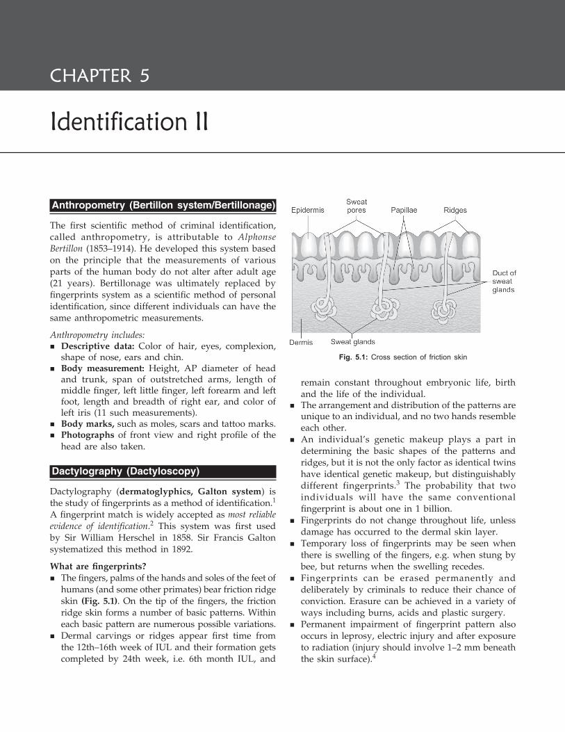
Anthropometry (Bertillon system/Bertillonage)
The first scientific method of criminal identification, called anthropometry, is attributable to Alphonse Bertillon (1853–1914). He developed this system based on the principle that the measurements of various parts of the human body do not alter after adult age (21 years). Bertillonage was ultimately replaced by fingerprints system as a scientific method of personal identification, since different individuals can have the same anthropometric measurements.
Anthropometry includes: � Descriptive data: Color of hair, eyes, complexion,
shape of nose, ears and chin. � Body measurement: Height, AP diameter of head
and trunk, span of outstretched arms, length of middle finger, left little finger, left forearm and left foot, length and breadth of right ear, and color of left iris (11 such measurements).
� Body marks, such as moles, scars and tattoo marks. � Photographs of front view and right profile of the
head are also taken.
Dactylography (Dactyloscopy)
Dactylography (dermatoglyphics, Galton system) is the study of fingerprints as a method of identification.1 A fingerprint match is widely accepted as most reliable evidence of identification.2 This system was first used by Sir William Herschel in 1858. Sir Francis Galton systema tized this method in 1892.
What are fingerprints? � The fingers, palms of the hands and soles of the feet of
humans (and some other primates) bear friction ridge skin (Fig. 5.1). On the tip of the fingers, the friction ridge skin forms a number of basic patterns. Within each basic pattern are numerous possible variations.
� Dermal carvings or ridges appear first time from the 12th–16th week of IUL and their formation gets completed by 24th week, i.e. 6th month IUL, and
remain constant throughout embryonic life, birth and the life of the individual.
� The arrangement and distribution of the patterns are unique to an individual, and no two hands resemble each other.
� An individual’s genetic makeup plays a part in determining the basic shapes of the patterns and ridges, but it is not the only factor as identical twins have identical genetic makeup, but distinguishably different fingerprints.3 The probability that two individuals will have the same conventional fingerprint is about one in 1 billion.
� Fingerprints do not change throughout life, unless damage has occurred to the dermal skin layer.
� Temporary loss of fingerprints may be seen when there is swelling of the fingers, e.g. when stung by bee, but returns when the swelling recedes.
� Fingerprints can be erased permanently and deliberately by criminals to reduce their chance of conviction. Erasure can be achieved in a variety of ways including burns, acids and plastic surgery.
� Permanent impairment of fingerprint pattern also occurs in leprosy, electric injury and after exposure to radiation (injury should involve 1–2 mm beneath the skin surface).4
Fig. 5.1: Cross section of friction skin
Identification II
Chapter 5

85Identification II
� Ridge atrophy with alteration of ridge pattern is seen in celiac disease, dermatitis, eczema, psoriasis, acanthosis nigricans, scleroderma, and dry and atrophic skin. Anti-cancer drug capecitabine may cause the loss of fingerprints. Elderly persons may have fingerprints that are difficult to capture, since the elasticity of skin decreases with age.
� People with certain genetic disorders like Baird syndrome (congenital milia), Zinsser-Cole-Engman syndrome (dyskeratosis congenita), Naegeli-Franceschetti-Jadassohn syndrome, dermatopathia pigmentosa reticularis, and adermatoglyphia are seen without fingerprints.
Fingerprint Patterns
There are four basic ridge patterns as given in Table 5.1 and Figure 5.2.5
� Composite contains at least two different patterns, other than the basic arch. Sometimes, it is also referred to as ‘accidental’.
� If a scar is formed, it will constitute a valuable addition to the identification process.
Recording of Fingerprints
Hands are washed, cleaned and dried to ensure clear prints. Print is taken using printer’s ink on an unglazed white paper. i. Plain or dab impression is obtained by gently press-
ing the inked surface of the tip of finger on paper. ii. Rolled impression is taken by rolling the inked
finger from side to side. � In case of criminals, impressions of all the ten digits
of both hands are taken.* � It is customary and conventional to take the left
thumb impression of male and right thumb impression of female in lieu of signature for illiterate person and on legal and other documents. The reason cited is that in earlier days, males being working class (and as most people are right handed) may have some injury/scar on their right thumb.
� In a dead body, if fingertips are dried up or shrivelled, the prints can be taken after soaking the fingers in an alkaline solution (e.g. KOH). The surface of the fingers can be rounded out and smoothened by injecting glycerin, melted paraffin, hot water or air into the tissues.
z If the prints obtained by the above methods are not found decipherable, then the palmer skin of the terminal phalanx of each finger may be removed from both the hands and placed in a labeled bottle containing 10% formalin or a solution of glycerin and alcohol for preservation, and transported to the Fingerprint Bureau.
z In case of advanced putrefaction and in drowning, the skin may come out like a glove which can be preserved in formalin for the development of fingerprints.
� Prints can be obtained from the dermis if epidermis is lost, histological section upto a depth of 0.6 mm from finger pad surface can give satisfactory results.
Limitations of manual recording � Impression not placed accurately in the designated space � Uneven or excessive recording medium (e.g. ink) � Pressure and twisting during recording may distort the print � Presence of permanent or temporary scars
Types of Evidentiary Fingerprints
Three types of fingerprint may be encountered: i. Patent (visible) print needs no processing to be
clearly recognizable as a fingerprint. It is often made from grease, dark oil, dirt or blood, rendering Fig. 5.2: Types of fingerprints
* Exemplar/known prints are fingerprints deliberately collected from a subject, whether for purposes of enrollment in a system or when under arrest for a suspected criminal offence.
Table 5.1: Types of fingerprint ridges
S. No. Type Percentage (%)
1. Loop6 (Ulnar/radial) 60–70
2. Whorl 30–35
3. Arch 5–10
4. Composite 2–3

86 Review of Forensic Medicine and Toxicology
it visible and recognizable, and even suitable for comparison without additional processing.
ii. Plastic (impression/indentation) print is a recog-nizable fingerprint indentation in a soft surface, such as butter, soap, cheese, paint, putty or tar. Such prints have a distinct three-dimensional character, immediately recognizable and require no further processing.
iii. Latent print requires additional processing to be rendered visible and suitable for comparison. Processing of latent prints to render them visible and suitable for comparison is called development, enhancement or visualization.
Development/Enhancement of Latent Prints
Latent prints are the most prominent example of Locard’s Principle of Exchange: ‘When two objects come into contact with each other, there is always some transfer of material from one to the other’; hence, the great importance of visualizing them onto useful evidence.
Composition of latent print residue: Palmer and planter surface is completely free from hair and sebaceous glands, but there is profusion of sweat glands (called eccrines), the composition of which forms the basis for latent fingerprint residue; contamination by sebaceous secretions is also quite common from people touching their faces (Fig. 5.1).
� The salts predominant in perspiration are sodium and potassium chlorides, with the organic fraction containing mainly amino acids, urea and lactic acid. Free fatty acids, triglycerides and wax esters prevail in sebaceous secretions.
� Fingerprints are stable compounds and unless they are exposed to extremes of heat or humidity and/or friction, they may persist indefinitely.
� Most methods for the development of latent prints were developed on the basis of knowledge about the latent print residue composition.
Fingerprint Development
A. For non-porous surfaces, e.g. glass, gloss-painted surfaces, metal and plastic.
� Visual examination: Oblique illumination may reveal latent fingerprints, particularly if the surface is smooth and clean.
� Fluorescence examination: High intensity light source or argon-ion laser or UV light may reveal latent fingerprints.
Development techniques i. Vacuum metal deposition (VMD): VMD is most
sensitive, being capable of detecting monolayer of fat by sequential deposition of a thin coating of thermally evaporated gold followed by zinc. It can develop fingerprints on surfaces that have previously been wet or even submerged in water for extended periods of time. If fingerprints are not revealed by VMD, superglue, powders or other techniques may be used subsequently.
ii. Fingerprint powders: Powdering is one of the oldest techniques for detecting fresh latent prints. It is widely used but insensitive. Many powders have been developed—microscopic flake like structure such as milled aluminum or brass, or molybdenum disulfide which are more sensitive and effective at developing fingerprints on smooth, clean surfaces than the more traditional black or white powders. In case of contaminated surfaces, granular black (chalk and mercury) or white powders (lead carbonate or French chalk) are more suitable. Rough or grained surfaces may be treated with iron, cobalt or nickel-based powders along with a magnetic applicator. There are many fluorescent powders which may also be used in conjunction with a suitable light source.
iii. Superglue fuming can be used on any nonporous surfaces, and is particularly useful on surfaces such as rough or grained plastic surfaces which cannot be easily treated using VMD. It is composed of methyl or ethyl cyanoacrylate which polymerize with latent prints.
iv. Small particle reagent (SPR): SPR consists of a suspension of molybdenum disulfide suspended in aqueous detergent solution which is applied by spraying or immersion. The molybdenum disulfide particles adhere to fats deposited in the fingerprints, producing a gray-black image.
v. Iodine fuming is one of the oldest and cheapest methods, and can develop recent prints on porous and non-porous surfaces. Iodine fumes are absorbed by fingerprints to form a brown image which is photographed immediately.
B. For porous surfaces, e.g. paper, wallpaper, card board and matt emulsion painted surfaces. The reagents used for these surfaces react either with amino acids, fats and lipids or chlorides absorbed into the surface.
� Visual examination is less likely to reveal fingerprints on porous surfaces.

87Identification II
� Fluorescence examination may sometimes detect fingerprints either by the fluorescence of naturally occurring components or fluorescence of some contaminants.
Development techniques i. DFO (1,8-diaza-9-fluorenone): It is the most
sensitive reagent available for detecting fingerprint on porous surfaces. DFO reacts with amino acids deposited in the fingerprints to produce a faintly colored but intensely fluorescent compound which can be easily photographed. Since, amino acids are soluble in water, DFO or ninhydrin cannot be used to treat porous surfaces which have been wet.
ii. Ninhydrin is a widely used chemical which reacts with amino acids and produces a deep blue or purple color known as Ruhemann’s purple. It can be very effectively used at scenes of crime with the same formulation being brushed onto the surfaces.
iii. Powders: Smooth papers may be treated with black or magnetic powder, although these will usually detect recent fingerprints.
iv. Superglue fuming may be used on some smooth surfaces such as cigarette packets.
v. Physical developer: It is the only available technique for detecting fingerprints on a wet porous surface. This reagent is an aqueous solution of silver nitrate containing Fe II/III redox couple and two detergents. The developed fingerprints are gray-black in color, and recorded using conventional photography.
Other methods for detecting latent prints � Radioactive sulfur dioxide: Useful for fabrics and adhesive tapes. � Sudan black: Useful for surfaces contaminated by grease or
foodstuffs. � Osmium tetraoxide: Useful for both porous and non-porous
surfaces. � Electronography: Graham and Gray first reported the use of
electronography and auto-electronography to recover latent prints from skin in 1966. This technique involved dusting the skin surface with lead (or iron) and then exposure to long wave X-rays (Grenz rays). Emission of radiation from the lead powder can be captured on a photographic film. The silver halides present in the film are darkened by the radiation and can produce an image of ridge detail present on the skin surface.
� Scanning electron microscopy with an energy dispersive X-ray spectrometer can be used for imaging of latent fingerprints. However, it is of little use for casework because its application often requires a small area to be cut from the exhibit and coated with a conductive material to prevent the sample charging.
Identification Protocol
� The unknown impression is examined, and all minutiae are analyzed and then compared to the known, to determine if a relationship exists.
� Weight is assigned in a comparison, not only to the number of minutiae in agreement, but also the rarity and clarity of those characteristics. Differences in appearance due to recording technique, pressure and other factors must be anticipated.
� The comparison can result in one of three possible conclusions: insufficient ridge detail to form a conclusion, exclusion or identification, i.e. that they were made by the same finger.
Fingerprint Classification
� The modified Henry system followed in the US is used for the classification of 10-print sets or a fingerprint card, for one individual.
� The development of computerized fingerprint storage and retrieval systems has made searching larger files for single and partial prints routine. It has also rendered classification largely unnecessary.
� For quite a few decades, a ‘minimum number of minutiae (points)’ rule was followed. In practice, 12 points of fine comparison were accepted as proof of identity (suggested by Locard).
� Resolution in 1973 discouraged this ‘static threshold’ identification ideology. This was replaced by ‘floating threshold’ that stated that no minimum number of features is required for making fingerprint identification.
� Experts use three levels of detailed evaluation of the friction ridge formations to come to a conclusion. The first level is the ridge flow (pattern type, ridge count and focal areas such as delta, core and orientation). Individualization or matching the print cannot occur at this level. Level two is ridge path (Galton details or ‘points’) includes ridge endings, bifurcations, dots, combinations of the above and their relationship to one another. Individualization can occur at this level. Poroscopy and edgeoscopy both involve third level detail and are used in latent prints by examiners even without their conscious knowledge.
Medico-legal Application i. Identification of criminals whose fingerprints were
found at scene. ii. Identification of fugitive through fingerprint
comparison. iii. Exchange of criminal identifying information with
identification bureau of foreign countries in cases of mutual interest.

88 Review of Forensic Medicine and Toxicology
� Ridges on fingers and hands are studded with microscopic pores formed by mouths of ducts of subepidermal sweat glands. Each millimeter of ridge contains 9–18 pores. There are about 550–950 sweat pores per square centimeter in finger ridges, and less (400) in the palms and soles (Fig. 5.3).
� Poroscopy is the further study of fingerprints, discovered and developed by Edmond Locard in 1912.8 He observed that like the ridge characteristics, the pores are also permanent, immutable and individual, and these are useful to establish the identity of individuals when available ridges do not provide sufficient ridge characteristics.
Edgeoscopy (term coined by Salil K Chatterjee in 1962) is the study of the characteristics formed by the sides or edges of papillary ridges as a means of identification. These characteristics are the result of the alignment and shape of the individual ridge units and the relationship between them, as well as the effects of pores that are close to the edge of the ridges—an extension of identification by ridge characteristics. However, it is impractical as sole means of identification.
Footprints: Skin patterns of toes and heels are as distinct and permanent as those of fingers. Footprints of newborn infants are used in maternity hospitals to prevent exchange or substitution of infants. Records are also kept for air force flying personnel.
Forensic podiatry: Specialty using clinical podiatric knowledge for the purpose of person identification. Techniques of forensic podiatry include identification from podiatry records, the human footprint, footwear, and the analysis of gait forms captured on CCTV cameras. The most valuable techniques relate to the comparison of the foot impressions seen inside the shoes.
Lip Prints (Cheiloscopy)
� The study of lip prints is called cheiloscopy.9
� It is said that a person’s lip prints are unique.
iv. Identification of unknown deceased person, persons suffering from amnesia, missing persons and unconscious patient.
v. Identification in disaster work. vi. Identification in case of accidental exchange of
newborn infants. vii. Identification of licensing procedure for automobile,
firearm, aircrafts, etc. viii. Problems of mistaken identity and detection of
bank forgeries. ix. Electronic fingerprint readers have been intro-
duced for security applications such as log-in authentication for the identification of computer users. Fingerprint sensors gained popularity in the notebook PC market.
x. Electronic registration and library access: Fingerprints can be used to validate electronic registration, cashless catering and library access. This ‘conventional biometrics’ is routinely practiced in some schools in the UK, US, Belgium, France and Italy replacing library cards. However, privacy issues are of concern since children are fingerprinted by schools, often without the knowledge or consent of their parents.
xi. The police department in Canada has advised parents to fingerprint their children, if they apprehend kidnapping.
� Ridgeology refers to friction ridge identification that is associated with all the ridges on the volar areas and not just on the fingertips.
� Sir Francis Galton published his book in 1892, ‘Finger Prints’ and is regarded as a classic work.
� The first ‘Fingerprint Bureau’ in the world was officially established in Kolkata on 12th June 1897 at Writers’ Building.7
� Sir Edward Henry devised a fingerprint-classification system that was adopted in British India. He presented it in the UK in 1899.
� Automated Fingerprint Identification System (AFIS) is a storage, search, retrieval and exchange system for finger and palm print electronic images and demographic data (biometric data). AFIS utilizes specialized computer software configurations to create unique algorithms based upon relationships between the characteristics present within the finger or palm friction ridge. To match a print, a fingerprint technician scans the print in question, and computer algorithms are utilized to mark all minutia points, cores and deltas detected on the print. This enables a fingerprint to be compared with millions of file prints within a matter of seconds.
Poroscopy
Poroscopy is the term applied to a specialized study of pore structure found on the papillary ridges of the fingers as a means of identification. Fig. 5.3: Sweat pores as they appear on ridges
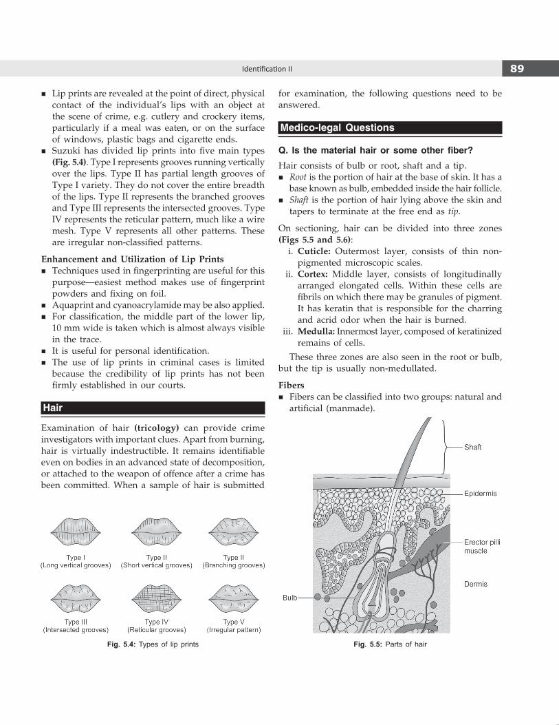
89Identification II
� Lip prints are revealed at the point of direct, physical contact of the individual’s lips with an object at the scene of crime, e.g. cutlery and crockery items, particularly if a meal was eaten, or on the surface of windows, plastic bags and cigarette ends.
� Suzuki has divided lip prints into five main types (Fig. 5.4). Type I represents grooves running vertically over the lips. Type II has partial length grooves of Type I variety. They do not cover the entire breadth of the lips. Type II represents the branched grooves and Type III represents the intersected grooves. Type IV represents the reticular pattern, much like a wire mesh. Type V represents all other patterns. These are irregular non-classified patterns.
Enhancement and Utilization of Lip Prints � Techniques used in fingerprinting are useful for this
purpose—easiest method makes use of fingerprint powders and fixing on foil.
� Aquaprint and cyanoacrylamide may be also applied. � For classification, the middle part of the lower lip,
10 mm wide is taken which is almost always visible in the trace.
� It is useful for personal identification. � The use of lip prints in criminal cases is limited
because the credibility of lip prints has not been firmly established in our courts.
Hair
Examination of hair (tricology) can provide crime investi gators with important clues. Apart from burning, hair is virtually indestructible. It remains identifiable even on bodies in an advanced state of decomposition, or attached to the weapon of offence after a crime has been committed. When a sample of hair is submitted
Fig. 5.4: Types of lip prints Fig. 5.5: Parts of hair
for examination, the following questions need to be answered.
Medico-legal Questions
Q. Is the material hair or some other fiber?
Hair consists of bulb or root, shaft and a tip. � Root is the portion of hair at the base of skin. It has a
base known as bulb, embedded inside the hair follicle. � Shaft is the portion of hair lying above the skin and
tapers to terminate at the free end as tip.
On sectioning, hair can be divided into three zones (Figs 5.5 and 5.6): i. Cuticle: Outermost layer, consists of thin non-
pigmented microscopic scales. ii. Cortex: Middle layer, consists of longitudinally
arranged elongated cells. Within these cells are fibrils on which there may be granules of pigment. It has keratin that is responsible for the charring and acrid odor when the hair is burned.
iii. Medulla: Innermost layer, composed of keratinized remains of cells.
These three zones are also seen in the root or bulb, but the tip is usually non-medullated.
Fibers � Fibers can be classified into two groups: natural and
artificial (manmade).
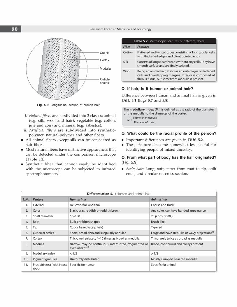
90 Review of Forensic Medicine and Toxicology
Table 5.2: Microscopic features of different fibers
Fiber Features
Cotton Flattened and twisted tubes consisting of long tubular cells with thickened edges and blunt pointed ends.
Silk Consists of long clear threads without any cells. They have smooth surface and are finely striated.
Wool Being an animal hair, it shows an outer layer of flattened cells and overlapping margins. Interior is composed of fibrous tissue, but sometimes medulla is present.
Q. If hair, is it human or animal hair?
Difference between human and animal hair is given in Diff. 5.1 (Figs 5.7 and 5.8).
The medullary index (MI) is defined as the ratio of the diameter of the medulla to the diameter of the cortex.
Diameter of medullaMI =
Diameter of cortex
Q. What could be the racial profile of the person?
� Important differences are given in Diff. 5.2. � These features become somewhat less useful for
identifying people of mixed ancestry.
Q. From what part of body has the hair originated? (Fig. 5.9)
� Scalp hair: Long, soft, taper from root to tip, split ends, and circular on cross section.
Fig. 5.6: Longitudinal section of human hair
i. Natural fibers are subdivided into 3 classes: animal (e.g. silk, wool and hair), vegetable (e.g. cotton, jute and coir) and mineral (e.g. asbestos).
ii. Artificial fibers are subdivided into synthetic-polymer, natural-polymer and other fibers.
� All animal fibers except silk can be considered as hair fibers.
� Most natural fibers have distinctive appearances that can be detected under the comparison microscope (Table 5.2).
� Synthetic fiber that cannot easily be identified with the microscope can be subjected to infrared spectrophotometry.
Differentiation 5.1: Human and animal hair
S. No. Feature Human hair Animal hair
1. External Delicate, fine and thin Coarse and thick
2. Color Black, gray, reddish or reddish brown Any color, can have banded appearance
3. Shaft diameter 50–150 µ 25 µ or > 3000 µ
4. Root Bulb or ribbon shaped Brush-like
5. Tip Cut or frayed (scalp hair) Tapered
6. Cuticular scales Short, broad, thin and irregularly annular Large and have step-like or wavy projections10
7. Cortex Thick, well striated, 4–10 times as broad as medulla Thin, rarely twice as broad as medulla
8. Medulla Narrow, may be continuous, interrupted, fragmented or even absent11
Broad, continuous and always present
9. Medullary index < 1/3 > 1/3
10. Pigment granules Uniformly distributed Mostly clumped near the medulla
11. Precipitin test (with intact root)
Specific for human Specific for animal

91Identification II
� Beard and moustache: Thicker, straight, blunted tip, and triangular on cross section.
� Axillary and pubic hair: Stout, short, lack of uniformity, and curly with frayed or split ends.
� Eyebrow, eyelashes and nostril hair: Short and stiff, thick, tapering abruptly, and triangular on cross section.
� Body hair: Soft, fine and flexible, lack of uniformity of medulla, milder pigmentation, and narrow tip.
Q. Is it male or female hair?
� Beard and moustache are specific for males. Male hair is usually thicker, coarser and darker as compared to females.
� Barr bodies can be detected in the hair follicles in about 20–80% of females and only about 0–4% of the males.
� Sexing can be done if root sheath is present, using DNA analysis.
Q. What could be the age of the person?Whether the hair is that of an infant or adult can be said.
� Lanugo hair of the newborn are fine, downy, soft, non-pigmented, non-medullated, and cuticular scales have smooth edges.
� Adult hair are coarser, pigmented and medullated having a complex cuticular pattern.
� Gray hair are apparent after the age of 40, and are devoid of any pigment.
Age is based on examination of the following which tend to increase with age: medullary index, size of pigment granules, streakiness of pigment distribution, darkness of color, amount of cortical fusi and size of unpigmented area above the root.
Fig. 5.7: Human hair Fig. 5.8: Animal hair
Differentiation 5.2: Ethnic differences in human hair
S.No. Feature Caucasians Mongolians Negroes
1. Color Light brown Black or dark brown Black or dark brown
2. Consistency Fine to medium Coarse Short, curly, finest
3. Shape Oval Round Flat
4. Shaft diameter Slight variation Constant diameter Wide variation
5. Pigmentation Uniform Coarse granules Irregular
6. Cuticle Thin Thick Medium
7. Medulla Fragmented or absent Unbroken Fragmented or absent
Fig. 5.9: Cross section of hair

92 Review of Forensic Medicine and Toxicology
Q. Has the hair being altered by dyeing, bleaching or diseased?
� Bleached or colored hair are dry, brittle, lusterless and rough. Abrupt color change to a very light color indicates bleaching.
� Color in cuticle indicates dyeing. Microscopical examination with incident fluorescence illumination may show whether hair is dyed or not.
� Curly appearance accompanied by constrictions in the shaft is indicative of permanent waving.
� Hair color is lighter in diseases, such as kwashiorkor, malnutrition and certain vitamin deficiencies.
� Tunneling of hair by fungal hypae can produce distinctive transverse lines—seen in hair exposed to fungi, and occur in buried bodies.
Q. Is the hair identical with hair of the victim or the suspect?
� Blood groups (ABO) can be determined from a single hair bulb.
� If some root structure is present, standard DNA profiling can be used.
� Even if the shaft is there, mitochondrial DNA testing can be tried.
Q. Did the hair fall naturally or was it forcibly removed? (Fig. 5.10)
� The bulb of naturally fallen hair is distorted, atrophied and the hair sheath is absent.
� In forcibly plucked hair, the hair sheath is ruptured, bulb is swollen, larger and irregular.
� If the root is not present, an even break with regular edges indicates that it was cut off, and an irregular break indicates the hair was broken off.
Q. What is the cause of injury? (Fig. 5.11)
� In uncut hair, the tip is pointed and non-medullated. � Sharp weapon produces a clean uniform cut surface. � Blunt force injury result in flattening and splitting
of hair shaft. � Singed hair due to burns or firearm injury are
swollen, fragile, curled, twisted and have a peculiar odor.
Postmortem root banding seen in decomposed body, wherein an opaque band about 0.5 mm above the root bulb can be observed with transmitted light microscope.
Medico-legal Application1. Identification: Hair remains identifiable long after the
commission of crime and provides valuable physical evidence. In homicidal cases, presence of hair in the grip of the hand of the deceased in cadaveric spasm may help in identification of the assailant.
2. Establish relationship between offence, offender and the victim.
i. It is an important clue when similar hair may be detected on the alleged weapon and on the body of the assailant.
ii. In rape and sodomy cases, pubic hair of the accused may be detected on the victim and vice versa.
iii. In bestiality, animal hair may be found around the genitalia, body and clothing of the accused.
iv. In road traffic accidents, hair of the victim may be found adhered to the offending car.
3. Nature of weapon: In head injury, the hair may be crushed or cut depending on whether blunt or sharp cutting weapon was used.
Fig. 5.11: Tip of hairFig. 5.10: (A) Root of a naturally fallen hair,
(B) Healthy hair bulb (plucked)
a b

93Identification II
4. Nature of assault: Various trace evidence, like stains may be attached with hair, so it must be carefully looked for (Table 5.3).
5. It helps in differentiating burns from scalds. Hair is brittle, singed or charred with large round vacuoles at the point of burning which is absent in scalds.
6. Singeing of hair indicates burns or close range firearm injury.
7. Alcohol testing: Hair alcohol testing is used to know whether or not a person has consumed alcohol at a frequent and excessive rate over a period of time. The concentration of ethyl glucuronide and fatty acid ethyl esters found in a hair sample reflects the consumption of alcohol over the period covered by the sample; it does not determine the number of times or the amount consumed on each occasion.
8. Time since death: This can be estimated from growth of scalp hair or beard (growth rate: 2.5 mm/week or 0.4 mm/day) if the date of last shave is known. It is possible to calculate for what period the deceased survived after his last shave.
9. Age and sex can be determined.10. Cause of death: Poisons such as arsenic, thallium
or lead can be determined from hair. Sometimes, accidental poisoning may occur with compounds like aniline derivatives including para-phenylene diamine (PPD) and resorcinol found in hair dye.
Table 5.3: Stains attached on the hair
Type of stains Suggestive information
� Seminal Sexual offence
� Blood Injury
� Salivary Asphyxial deaths
� Mud Struggle/road traffic accident
� Carbon particles Burns/firearm injury
� Dyes Concealment of natural color
Superimposition
� Technique applied to determine whether the re-covered skull is that of the person in the photograph.
� Technologically, skull-photo superimposition have passed through three phases—the photographic, video, and computer-assisted superimposition techniques.
� While performing photographic superimposition, the comparison photograph is enlarged to the size of the
unknown skull and then the skull is positioned in the same orientation as the facial photograph.
� Recently, computer-assisted superimposition has become a popular method which digitize the skull and facial photograph using a video computer with appropriate software, and then compare the two images morphologically by image processing.
� The complete skull is required to obtain positive identification. Without the mandible, it cannot be positively be identified as the presumed person, even if a good match is seen in skull-superimposition image.
� The coincidence of dentition between the skull and facial photograph (if incisors and canines are seen) could lead to positive identification.
� When evaluating anatomical consistency between the parts, special attention should be paid to their outline, the facial tissue thickness at various anthropometric points, and the positional relationships between skull and face-eyebrow to supraorbital margin, eye to orbit, nose to nasal aperture, lips to teeth and ear to external auditory meatus.
� If they are well matched with each other, it can only be stated that the skull could be that of the photographed person.
� Test is of a more negative value, because it can be definitely be stated that the skull and photograph are not those of the same person.
Restoration of facial features: When a dead body is unrec-ognizable due to decomposition, skeltalization or mutilation, a craniofacial reconstruction (CFR) may be done. The goal of CFR is to recreate a likeness with the face of missing individuals immediately prior to their death which may help to identify the missing person. Several 3-D manual methods for CFR are currently being used. One of these consists of physically modeling a face on a skull replica (the target skull) with clay or plasticine. However, this method requires a high degree of anatomical and sculptural expertise and hence, remains difficult and subjective. With the development of software for computer based CFR methods, automated creation of multiple reconstructions from the same skull using different modeling assumptions (like age, BMI, ancestry, gender) became possible. As a result, the computer generated CFR process becomes accessible to a wide range of people without the need for extensive expertise.
Forensic Odontology
Definition: It deals with the application of dentistry to aid in the administration of justice.
The work of a forensic odontologist includes: � Identification of unknown bodies through dental
records. � Identification of bite marks on the victims of attack.

94 Review of Forensic Medicine and Toxicology
� Comparison of bite marks with the teeth of a suspect and presentation of this evidence in the court as an expert witness.
� Identification of bite marks in other substances such as wood, leather and foodstuffs.
� Age estimation of skeletal remains.
Identification of Human Remains
Unidentified bodies due to violent crimes, road traffic accidents, natural and manmade disaster and in particular, the mass casualties normally associated with aviation disasters, drowning, burns, murder, suicide or dead from natural causes rely on dental evidence to positively identify the body.
The central dogma of dental identification is that postmortem dental remains can be compared with antemortem dental records, including written notes, study casts, radiographs, etc. to confirm identity. Clearly, individuals with numerous and complex dental treat ments are often easier to identify than those individuals with little or no restorative treatment.
Once the postmortem record is complete, a comparison with antemortem dental records can be carried out. A range of conclusions can be reached when reporting a dental identification.
Even if only a few teeth are available, one can still offer an age estimation, smoking habit, state of oral hygiene, and identification of individual features which may match with antemortem records. Where the subject has no teeth, useful information can still be obtained from the study of any dentures and by X-ray of mouth and skull.
In problem cases, a variety of techniques are used to assist in the identification issue. These include:
� Incremental line and other histology studies (Maples noted that 2nd molar was best for histological ageing techniques).
� Scanning electron microscopy with/without energy-dispersive X-ray analysis.
� Metal ratio analysis in bone and teeth, especially magnesium/zinc ratio.
� Serology studies for blood groups, serum proteins and polymorphic enzymes.
� DNA analyses: Dental pulp material is a good source for DNA analysis; two vital teeth (preferably molars, canines/premolars) are extracted and sent for examination.
Bite Marks
Bites are commonly seen in cases of: i. Sexual assault: Marks are usually seen on breasts,
neck, shoulders, thighs, abdomen, pubis or vulva.
ii. Child abuse: Marks are seen anywhere on the body, such as arms, hands, shoulders, cheeks, buttocks and trunk.
iii. Bite marks on foodstuffs (apples, cheese or chocolate), leather (key rings or belts) and wood (pencils) in cases where a perpetrator might have taken a bite out of something in the victim’s home and left it behind.
iv. Police officers may be bitten by the resisting offenders. v. In sporting events, like football, rugby or wrestling. vi. In assaults, where marks may be found anywhere
on the body.
Nature of bite marks: Comprise of a crop of punctate hemorrhages varying from small petechiae to large ecchymoses merging into a confluent central bruise. Human bite is semicircular or crescentic caused by the front teeth (incisors and canines) with a gap on either side due to separation of upper and lower jaw, whereas deep parabolic arch or U-shaped is characteristic of an animal bite. There may be abrasions, bruises and lacerations or a combination of all these.
� Self-inflicted bite marks are present on accessible parts of the body, e.g. shoulders or arms, usually seen in psychiatric patients or teenage girls.
� Accidental marks resulting from falls on to the face and during fits, biting of tongue and lips may also be there.
� In sexual assault, sucking action during bites reduces the air pressure in the center and produces multiple petechial hemorrhages due to rupture of small capillaries and venules.Identification from bite marks is possible, if incisors
and canines have some characteristic features.
Bite mark investigation1. Photograph: Bite mark is photographed from different angles. 2. Swabbing of saliva: To identify or exclude assailant from ‘secretor’
status who exude blood group substances in the saliva. 3. Impression of bite mark: Plastic substance (rubber or silicone
based) or plaster of Paris is laid over the bite mark that hardens and produces permanent negative cast of the lesion.
4. Skin carrying the bite is removed and preserved in formalin during autopsy.
Dental Profiling
When dental records are unavailable and other methods of identification are not possible, the forensic dentist often produces a ‘picture’ of the general features of the individual known as dental profiling. It will typically
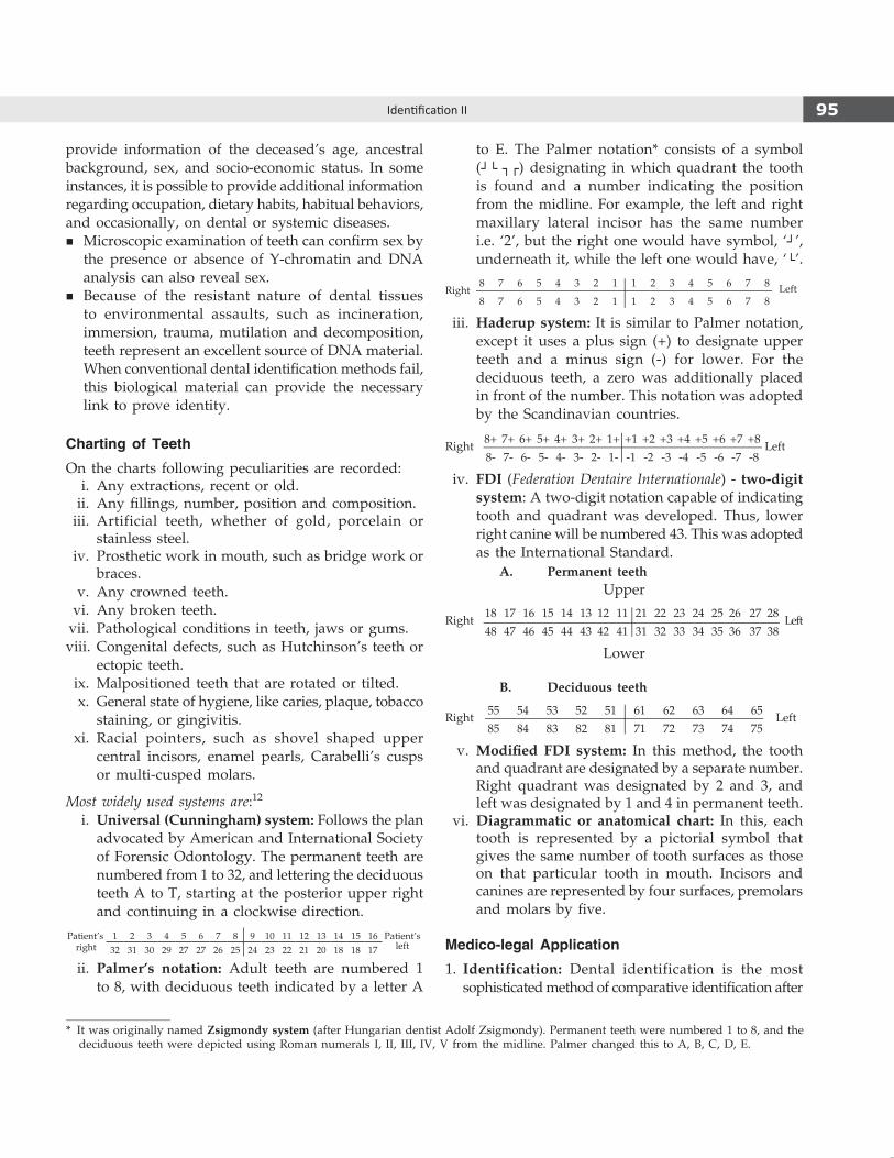
95Identification II
provide information of the deceased’s age, ancestral background, sex, and socio-economic status. In some instances, it is possible to provide additional information regarding occupation, dietary habits, habitual behaviors, and occasionally, on dental or systemic diseases.
� Microscopic examination of teeth can confirm sex by the presence or absence of Y-chromatin and DNA analysis can also reveal sex.
� Because of the resistant nature of dental tissues to environmental assaults, such as incineration, immersion, trauma, mutilation and decomposition, teeth represent an excellent source of DNA material. When conventional dental identification methods fail, this biological material can provide the necessary link to prove identity.
Charting of Teeth
On the charts following peculiarities are recorded: i. Any extractions, recent or old. ii. Any fillings, number, position and composition. iii. Artificial teeth, whether of gold, porcelain or
stainless steel. iv. Prosthetic work in mouth, such as bridge work or
braces. v. Any crowned teeth. vi. Any broken teeth. vii. Pathological conditions in teeth, jaws or gums. viii. Congenital defects, such as Hutchinson’s teeth or
ectopic teeth. ix. Malpositioned teeth that are rotated or tilted. x. General state of hygiene, like caries, plaque, tobacco
staining, or gingivitis. xi. Racial pointers, such as shovel shaped upper
central incisors, enamel pearls, Carabelli’s cusps or multi-cusped molars.
Most widely used systems are:12
i. Universal (Cunningham) system: Follows the plan advocated by American and International Society of Forensic Odontology. The permanent teeth are numbered from 1 to 32, and lettering the deciduous teeth A to T, starting at the posterior upper right and continuing in a clockwise direction.
Patient’s 1 2 3 4 5 6 7 8 9 10 11 12 13 14 15 16 Patient’sright 32 31 30 29 27 27 26 25 24 23 22 21 20 18 18 17 left
ii. Palmer’s notation: Adult teeth are numbered 1 to 8, with deciduous teeth indicated by a letter A
to E. The Palmer notation* consists of a symbol (┘└ ┐┌) designating in which quadrant the tooth is found and a number indicating the position from the midline. For example, the left and right maxillary lateral incisor has the same number i.e. ‘2’, but the right one would have symbol, ‘┘’, underneath it, while the left one would have, ‘└’.
Right 8 7 6 5 4 3 2 1 1 2 3 4 5 6 7 8 Left
8 7 6 5 4 3 2 1 1 2 3 4 5 6 7 8
iii. Haderup system: It is similar to Palmer notation, except it uses a plus sign (+) to designate upper teeth and a minus sign (-) for lower. For the deciduous teeth, a zero was additionally placed in front of the number. This notation was adopted by the Scandinavian countries.
Right 8+ 7+ 6+ 5+ 4+ 3+ 2+ 1+ +1 +2 +3 +4 +5 +6 +7 +8
Left 8- 7- 6- 5- 4- 3- 2- 1- -1 -2 -3 -4 -5 -6 -7 -8
iv. FDI (Federation Dentaire Internationale) - two-digit system: A two-digit notation capable of indicating tooth and quadrant was developed. Thus, lower right canine will be numbered 43. This was adopted as the International Standard.
A. Permanent teethUpper
Right 18 17 16 15 14 13 12 11 21 22 23 24 25 26 27 28
Left 48 47 46 45 44 43 42 41 31 32 33 34 35 36 37 38
Lower
B. Deciduous teeth
Right 55 54 53 52 51 61 62 63 64 65
Left 85 84 83 82 81 71 72 73 74 75
v. Modified FDI system: In this method, the tooth and quadrant are designated by a separate number. Right quadrant was designated by 2 and 3, and left was designated by 1 and 4 in permanent teeth.
vi. Diagrammatic or anatomical chart: In this, each tooth is represented by a pictorial symbol that gives the same number of tooth surfaces as those on that particular tooth in mouth. Incisors and canines are represented by four surfaces, premolars and molars by five.
Medico-legal Application
1. Identification: Dental identification is the most sophisticated method of comparative identification after
* It was originally named Zsigmondy system (after Hungarian dentist Adolf Zsigmondy). Permanent teeth were numbered 1 to 8, and the deciduous teeth were depicted using Roman numerals I, II, III, IV, V from the midline. Palmer changed this to A, B, C, D, E.

96 Review of Forensic Medicine and Toxicology
dactylography. It is of not much use in developing countries, as dentists often do not keep records.
� Identification of unknown dead bodies. � Identification of burnt, mutilated or decomposing
remains, as the teeth are very resistant to decomposition or destruction, particularly in mass disasters.
2. Age estimation of an individual.3. Identification of race, sex, occupation or habits (betel
nut chewing or smoking) of an individual.4. Grievous hurt: Fracture/dislocation of tooth amounts
to grievous hurt according to Sec. 320 IPC.5. Cause of death: Since, the teeth resist putrefaction,
deposition of metals can be detected after considerable time after death, e.g. lead poisoning, or phossy jaw.
6. Dentures (partial or complete) are useful in identification, if they have the patient’s name or code number in them.
7. Criminals can be identified through bite marks left either on human tissues or foodstuffs.
Miscellaneous Methods of Identification
Clothes and Personal Effects
They are helpful in establishing identity in case of mass disasters. It is necessary to preserve the clothes along with any articles, such as driving license, cell phone, watch, spec tacles, ornaments and wallet found on a dead body for the purpose of future identification. The clothes are exam ined for mark of the tailor, foreign material or any tear.
Occupational Marks
These are helpful in identifying unknown dead bodies, as certain occupation leave marks by which persons engaged in them may be identified, e.g. clerks may have callosity on the proximal part of right middle finger where the pen usually rests, or dyers/photographers may have there fingers stained with dyes or chemicals.
Handwriting
Opinion regarding the handwriting is usually given by the expert in this field, and doctors are seldom asked to testify. But, sometime, the doctor may have to examine a person so as to acertain whether he is able to write when a plea of paralysis or mental incapacity is put forward.
Speech, Voice, Ticks, Manner and Habit
Sometime, it is possible to identify a living person from certain peculiarities, like stammering, nasal twang and jerky movement of muscle of the face or shoulder.
Other methods of identification1. Palatoscopy/palato-print/rugoscopy: It is the study of palatal rugae
in order to establish identity. Rugae (‘plica palatine’) are anatomical fold or wrinkles formed 12–14th week of IU life. This irregular fibrous connective tissue is located on the anterior third of the palate, behind the incisive papilla.13 Palatine rugae are unique and can be used for identification in circumstances when it is difficult to identify a dead person through dental records or fingerprints (even in advanced decomposition). This method of identification is useful, if antemortem record of palatal rugae is available.
This method is useful since: � Rugae pattern remain stable throughout life and does not
change during growth. � It is protected from trauma due to its situation and from heat
by buccal pad of fat and tongue. � Even in twins, the pattern of rugae may be similar but not
identical.2. Fronal sinus print: It is unique to a particular individual, and
these are permanent and fixed (after 15 years of age) and rarely alter following infection or injury. For comparison, antemortem X-ray of skull taken on occipito-mental plane is compared with postmortem X-rays.
3. Vascular grooves and sutural pattern: The sutural pattern on the skull bone particularly of sagittal and lambdoid sutures are complex and are individualistic. Similarly, the vascular grooves over skull bone, particularly of middle meningeal vessels are individualistic. Rather, vascular grooves over skull are more helpful for identification as compared to suture lines, because these are well demonstrable in X-rays.
4. Ear print: It is the study of shapes of the ear lobules and tips of ears as well as the hardness or softness of the helix and lobules, and hairiness of the helix and tragus. These characters of the ears are considered to be individualistic.
5. Nose print: The lines on the nose and shape of the tip of nose are considered to be individualistic. Chance impressions may be found over door, wall and mirror at the scene of crime or even on the body of the victim or accused.
6. Nail print: It is the study of the depressions and elevations (striations), numbers, distribution and dimensions of the ridges on the surface of the nails which are considered to be individualistic. They remain unchanged throughout life, and with advancement of age the striations become more prominent. The longitudinal striations are present over both convex and concave surfaces of finger and toe nails.
7. EV method of identification: The electrocardiogram (ECG or EKG) and vector cardiogram (VCG) trace expresses cardiac features that are unique to an individual. As a biometric, heartbeat data are difficult to disguise, reducing the likelihood of successfully applying falsified credentials into an authentication system.
8. ‘Barium meal’ X-ray of stomach: It is also considered to be individualistic and may be helpful in identification, if previous record is available.

97Identification II
1. a 2. b 3. a 4. D 5. b 6. b 7. C 8. C 9. C 10. C 11. C 12. b 13. a
multiple ChoiCe questions
1. Galton’s system is used for: NEET 13 A. Dactylography B. Poroscopy C. Ridgeology D. Anthropometry 2. The most reliable method of identification of an
individual is: NEET 14 A. Gustafson’s method B. Galton system C. Bertillion system D. Scars 3. Two identical twins will not have same: NEET 13 A. Fingerprints B. Iris color D. DNA D. Blood group 4. The fingerprint pattern may be impaired permanently
in case of: AIIMS 06; AI 09 A. Eczema B. Scalds C. Scabies D. Leprosy 5. Not seen in fingerprints: NEET 13 A. Loop B. Circles C. Whorl D. Arch 6. Most common pattern of fingerprint is:
Delhi 07; Gujarat 07; UP 08; Manipal 10 A. Arches B. Loops C. Whorls D. Composite 7. Fingerprint Bureau was first established in:
AI 06, AIIMS 08; NEET 14 A. England B. China C. India D. Singapore
8. Locord’s system is: NEET 14 A. Podography B. Dactylography C. Poroscopy D. Cheiloscopy 9. Method of identification using lips: NEET 13 A. Dactylography B. Poroscopy C. Cheiloscopy D. Tricology 10. Animal hair are/have: NEET 14 A. Fine B. Thin C. Large scales D. Thick cortex 11. In humans, cortex of hair is usually: Delhi 11 A. Double that of medulla B. Same as medulla C. 4–10 times broader than the medulla D. Thin in comparison to medulla 12. Dental numbering can be done by all, except: AI 11 A. FDI two digit system B. Anatomic and diagrammatic charting C. Palmer notation D. Universal system 13. Palato-print is commonly taken from which part of
palate: AIIMS 11 A. Anterior part B. Lateral wall C. Medial wall D. Posterior part

Medico-legal Autopsy
CHAPTER 6
Definitions
� Forensic pathology deals with the investigation of sudden, unexpected and/or violent deaths that includes determining the cause of death and the circumstances of how the death occurred.
� Autopsy* refers to the systematic examina tion of a dead person for medical, legal and/or scientific purposes.
It is of three types: i. Academic autopsy: Dissection carried by students
of anatomy. ii. Pathological, hospital or clinical autopsy: Done
by patho logists to diagnose the cause of death or to confirm a diagnosis. Physicians cannot order these autopsies without the consent signed by the next of kin.
iii. Medico-legal or forensic autopsy: Type of scientific examination of a dead body carried out under the laws of the State for the protection of rights of citizens in cases of sudden, suspicious, obscure, unnatural, litigious or criminal deaths. The basic purpose of this autopsy is to establish the cause and manner of death.
� It is said ‘the only thing worse than no autopsy is a partial autopsy’. In every case, the autopsy must be complete, i.e. all the body cavities should be opened, and every organ must be examined. Partial autopsies have no place in forensic pathologic practice.
� The autopsy should be carried out by the registered medical practitioner, preferably with training in forensic medicine. The doctor should remove the organs himself. The attendant should prepare the body and help the doctor where required, such as sawing the skull cap, reconstructing the body, etc. As the autopsy is proceeded with, details of the examination should be taken down verbatim by an assistant.
� The person responsible for handling, moving and cleaning the body is often called a diener (German, servant).
� Virtopsy (combination of ‘virtual’ and ‘autopsy’) is a bloodless and minimally invasive procedure to examine a body for cause of death. It utilizes imaging techniques (CT and MRI), photogrammetry and 3-D optical measuring techniques to get a reliable, accurate geometric presentation of all findings (the body surface as well as the interior).
� Psychological autopsy is an investigative procedure of reconstructing a person’s state of mind prior to death. This is based upon information gathered from personal documents, police and medical records and interviews with survivors of the deceased-families, friends and others who had contact with the person. The typical case is one which there is some doubt as to whether death was accidental, self-inflicted or malicious, and whether the deceased played an active role in his or her own demise. Such matters can be especially important in life insurance claims that are void if death was suicidal.
Purpose/Objectives of Autopsy
Who, when, where, why, how and what are the questions that the autopsy assists in answering. The objectives of medico-legal autopsy are to determine: i. Identity of the deceased in case of decomposed,
burnt, mutilated or an unidentified body. ii. Cause of death, whether natural or unnatural,
and to interpret the significance and effect of the disease present in case of natural death.
iii. Approximate time of death, mode of death, age of injuries, and place of death.
iv. Manner of death, whether accidental, suicidal or homicidal.
v. Poison or weapon responsible for death in case of homicide.
vi. Volitional activity possible after receiving the trauma, and survival time.
vii. Extent of external and internal injuries present. viii. Whether the injury present is expected to cause
death in ordinary course of nature.
* Autopsy (Greek autos—self, opis —view)—to see for oneself; also called necropsy (Greek necros—dead, opis—view) or postmortem examination (post—after, mortem—death).

99Medico-legal Autopsy
ix. Whether deceased received any treatment before death.
x. In case of homicide, whether: z One or more person(s) was/were involved. z Any trace evidence was left behind on the body
that may help in identification of the assailant. z Any other offence was related with the death,
e.g. strangulation along with rape. z More than one method or weapon was involved
in the crime, e.g. firearm along with knife. z The body has been displaced from the original
place of disposal. z The relative positions of victim and the
assailant(s) can be deciphered. xi. In case of newborns, to determine the question of
live birth and viability of the baby. xii. In case of mutilated or skeletal remains, to
determine if they are human, and if human, whether they belong to one or more than one person, the probable cause of death and approximate time since death.
Procedure for Medico-legal Autopsies (Flow chart 6.1)
i. Visit to the scene of crime: If a visit to the scene of crime can be arranged, it is well worth undertaking in certain cases, such as murder, poisoning, traffic accidents, firearm injuries and sexual offences. In many cases, crime can be excluded in favor of accident, suicide or even natural causes.
ii. Authorization: It should be conducted only when there is an official order authorizing the autopsy, from the police or Magistrate.
iii. All registered medical practitioners (RMP) in government service can conduct the examination. Autopsy is conducted by two doctors where death of a female due to burns or other suspicious reasons has occurred within 7 years of her marriage. A panel of doctors is also constituted in case of custodial deaths, death in operation table, and second autopsy. Ideally, a board should have odd number of members so that in case of differences, a conclusion may be reached.
iv. No unauthorized person should be present at the autopsy (although, there are no written rules for the same). However, the Investigating Officer (IO) may be shown certain findings found during autopsy contrary to the inquest report.
v. The medico-legal autopsy should be conducted in an authorized center. The body should never be embalmed before autopsy since histopathological examination will be impossible. It may be necessary to do an autopsy at the site, when the body is in an advanced state of putrefaction.
vi. Even if the body is decomposed, autopsy should be performed, as certain important lesions may still be found. Help from a forensic entomologist can provide accurate estimation of victim’s death and other valuable information in such cases.
vii. It should be performed as soon as possible after receiving the requisition, without undue delay. The requisition is accompanied with a copy of the inquest or the preliminary investigation report, a dead-body challan which includes the name, age, sex, identification marks and religion of the deceased, apparent cause of death and any other paper of importance. Before starting the autopsy, the doctor should go through the inquest report and the requisition thoroughly, and put his signature on all the papers after marking them serially. However, it should be kept in mind that the inquest report may have scanty, misleading or incorrect history.
viii. The autopsy should be conducted in daylight since it is said that color changes, such as jaundice, changes in bruises and postmortem staining cannot be appreciated in the artificial light. Moreover, postmortem is not an emergency, and unless there is serious threat to law and order situation or instruction comes from District Magistrate, it should not be done after 5 PM. However, certain States (Gujarat and Tamil Nadu) allows postmortem at night in some special circumstances.
Flow chart 6.1: Components of a medico-legal autopsy

100 Review of Forensic Medicine and Toxicology
Box 6.1: Instruments and equipment useful for postmortem examinations (Fig. 6.1)
� Scalpel and disposable blades of 22 size.
� Toothed forceps: Teeth lend strength in gripping the skin and organs.
� Rib shear: Small pruning shears and are used to cut through the ribs prior to lifting off the sternum.
� Enterotome: Large scissors used for opening the intestines.
� Scissors used for opening hollow organs and trimming off tissues.
� Bone cutter: This is used to cut the ribs and has curved blades.
� Councilman rib shear/cutter: Small pruning shears used to cut through the ribs prior to lifting off the sternum.
� Vibrating saw (Stryker saw): Instrument of choice for most autopsy surgeons for removing the skull cap.
� Bone saw: The hand saw can be used to saw through the skull, but it’s very slow-going compared to the vibrating saw. Infections from aerosols being thrown up are other disadvantages.
� Virchow skull breaker or chisel: After scoring the calvarium with the vibrating saw, the chisel is used to separate the top of the calvarium from the lower skull, thus exposing the brain and the meninges.
� Hammer with hook is used with the chisel to separate the calvarium from the lower skull.
� Brain knife: Long knife used to smoothly cut solid organs into slices for examination.
� Hagedorn’s needle is used for sewing up the body after autopsy.
Other instruments that should be available: probe, small rule and plastic-coated measuring tape.
ix. If the body is received in the mortuary at night, it is preserved at 4°C after noting the date and exact time. A preliminary examination is done to note external appearances, body (rectal) temperature, extent of postmortem staining and rigor mortis. The actual postmortem is conducted on the next day.
x. Identification: A police officer or any other authorized person and two relatives should identify the dead body in front of the autopsy surgeon. The names of those who identified the body must be recorded. In unidentified bodies, the marks of identification, race, religion, sex, age, dental formula, photographs and fingerprints should be taken.
xi. Medico-legal autopsy does not require any consent from the relatives of the deceased.
xii. Both positive and negative findings should be recorded.
xiii. Nothing should be erased and all alterations should be initialed in the report.
xiv. Chain of evidence: It is absolutely essential to preserve the chain of evidence by identifying the body and maintaining absolute control of specimens removed at autopsy.
xv. List of articles: A list is made of all the articles removed from the body, e.g. clothes, jewelry, bullets, etc. They are labeled, sealed, mentioned in the report and handed over to the police constable after obtaining a receipt.
xvi. After completion of autopsy, the body is stitched, washed and restored to the best possible cosmetic appearance, and then handed over to the police constable/IO.1
Chain of evidence/custody requires that from the moment the evidence is collected, every transfer of evidence from person to person be documented and be provable that nobody else could have accessed or tampered that evidence which can compromise the case of the prosecution.
Instruments for Autopsy Examination
A list of instruments useful in various standard and specialized postmortem procedures is given in Box 6.1.
External Examination
The observation and documentation of various external characteristics of the decedent is the essence of the external examination.1. Clothing: They are listed and their number, labels
and laundry marks, design, stains, tears, loss of
buttons, cuts, holes or blackening from firearm discharges with their dimensions should be noted.
z Trace evidence like hair, fibers, paint chips, glass fragments, vegetation and insect are collected, labeled and preserved.
z Jewelry may provide evidence of identification, pockets may contain medication or drugs of abuse, and personal papers may help in identification and provide medical history.
z The clothes should be removed carefully without tearing them, to avoid confusion of signs of struggle. If they cannot be removed intact, they should be cut in an area away from any bullet hole or cuts, along the seam of the garment.
z After autopsy, wet clothing should be air-dried, packed, sealed in paper bags and handed over to the police.
2. The whole surface of the body should be carefully examined before and after washing from head to foot, and back and front, and the details noted.
3. Body length, weight, sex, race, dentition, general state, built, develop ment and nourishment is noted.

101Medico-legal Autopsy
Fig. 6.1: Autopsy instruments
It should include all surgical procedures, dressings and other diagnostic and therapeutic measures.
Following should be noted in external examination: 1. Skin: General condition (rash, petechiae, color,
looseness and turgor), asymmetry of any part of the body or muscular wasting. The presence of stains from blood, mud, vomit, feces, corrosive or other poisons, or gunpowder is noted.
2. General description includes deformities, scalp hair, beard, scars, tattoo marks, moles, skin disease, circumcision, amputations and vermilion mark.
3. Signs of disease: Edema of legs, dropsy, surgical emphysema about the chest, skin disease, eruptions.
4. Time since death: Rectal temperature, rigor mortis,
postmortem staining, putrefaction, maggots, stomach contents, etc. are required to estimate time since death.
5. Face: Cyanosis, petechial hemorrhages, pallor, protrusion or biting of the tongue, state of lips, gums, teeth, marks of corrosion or injuries inside the lips and cheeks.
6. Eyes: Condition of the eyelids, conjunctivae, softening of the eyeball, color of sclera, state and color of pupils, contact lenses, petechiae, opacity of the cornea, lens and artificial eyes (which may contri bute in road traffic accidents).
7. Natural orifices, i.e. nose, mouth, ears, urethra, vagina and anus should be observed for any discharges, injuries and foreign body. Leakage of blood or CSF from ears, mouth or nostrils is noted.

102 Review of Forensic Medicine and Toxicology
Samples of discharges should be taken on swabs or smears prepared on slides.
8. Neck: Bruises, fingernail abrasions, ligature marks or any other abnormalities.
9. Thorax: Symmetry, general outline, and injuries if any.
10. Abdomen: Presence or absence of distension or retraction, striae gravidarum.
11. Back: Bedsores, spinal deformity, or injuries. 12. External genitalia: General development, edema,
local infection, and position of testes. 13. Hands: Injuries, defense wounds, electric marks,
and in clenched hands if anything is grasped. 14. Fingernails: Presence of tissue, blood, dust or other
foreign matter may be indicative of struggle. 15. Limbs and other parts: Fracture and dislocation.
External Injuries
The final stage of external examination is the documentation of injuries, either by grouping them according to injury type and anatomical location, or by numbering them, without implying an order of infliction or ranking of severity. It is often from the outer evidence that inferences may be made about the nature of the weapon, the direction of attack and other vital aspects. Each injury is characterized by its: i. Type/nature of injury. ii. Size (length, breadth and depth). iii. Shape. iv. Site (in relation to two external anatomical landmarks). v. Direction of application of the force. vi. Margins, edges and base. vii. Distance of the wound from the heel. viii. Time of infliction of the injury should be studied
from inflammatory and color changes. ix. Vital reaction. x. Foreign materials, e.g. hair, grass, fibers, etc.
� If the injuries are obscured by hair, it should be shaved. � Deep or penetrating wounds should not be probed
until the body is opened. � In burns, their character, position, body surface area
involved, and degree should be mentioned. � Concealed punctured wounds, bruising of frenulum
of lips, and injection marks should be searched for, if indicated.
� The use of printed body sketches is very useful. The position of the injuries should be pictographically depicted on the skeleton diagrams.
� Photographic documentation of major injuries is now considered as standard practice. Identifying markers bearing the unique autopsy number, with a measure ment scale should be included to ensure that the photos correspond to the specific case.
� Special procedures utilized during external examination include photography for the purposes of identification and documentation. Infrared and UV photography will enhance trace materials, tattoos, bruises and patterned injuries.
� High contrast black-white photography or computer directed image enhancement can be used to enhance patterned injuries.
� Autopsy radiology: In well equipped hospital where radiographic facility is available, radiological examination should be done in select cases before starting the autopsy (Box 6.2). X-ray examination assists in identification, locating foreign objects such as projectiles and documenting old and recent bony injury.
Box 6.2 Indications of radiological examination
� Identification and dentistry � Mutilated/charred remains � Gunshot wounds � Air embolism � Sharp force wounds � Barotrauma � Explosives deaths � Child abuse � Decomposed body
Internal Examination (Evisceration)
It is convenient to start the examination with the cavity chiefly affected. All three major cavities of the body, i.e. skull, thorax and abdomen should be opened and examined as a routine. The choice as to which part of body is to be opened first—skull or the body cavities is left to the dissector.
� In suspected head injury, the skull is opened first and then the thorax and the abdomen, but some autospy surgeons are of the view that it should be opened after blood has been drained out by opening the heart.
� In suspected asphyxial deaths due to compression of neck, the skull and abdomen is opened first followed by dissection of the neck.2 The draining out of blood from neck vessels via the skull provides a compara -tively cleaner field for the study of neck structures.
� In all other cases, the thorax and abdomen are opened first, and then the skull.
Skin Incisions
Skin incisions are of three types (Fig. 6.2): i. I-shaped incision extending from the chin straight
down to the symphysis pubis and avoiding the

103Medico-legal Autopsy
umbilicus (because the dense fibrous tissue is difficult to penetrate with a needle, when the body is stitched after autopsy). Most common method followed.
ii. Y-shaped incision: Straight line of Y corresponding to the xiphisternum to pubis incision and forks of Y runs down medially to the chest and extending towards the acromion process. Some prefer to extend the upper incision in an arc around the inferior portion of the female breasts (inframammary incision), but there is a chance of fluids inadvertently leaking from the closed body after autopsy. This technique is commonly used in the US.
iii. Modified Y-shaped incision: An incision is made in midline from suprasternal notch to symphysis pubis. The incision extends from suprasternal notch over the clavicle to its center on both sides and then passes upwards over the neck behind the ears (1 cm behind external auditory meatus). It is used when a detailed study of neck organs is required, like in hanging or strangulation.
iv. T-shaped or ‘bucket handle’ incision: The neck is opened with a transverse incision which runs from acromion to acromion process (bisacromial) along the line of clavicles, creating a trough that is located at the jugular bifurcation. Then a single midline incision is made down the anterior body wall, avoiding the umbilicus, to pubis. The resulting incision is called a calyx since it creates a cup-like structure. This is sometimes called a ‘U-shaped’ incision.
Evisceration Methods (Flow chart 6.2)
i. En masse: This method, described by Letulle, involves removing most of the internal organs in one full swoop.3 It is a rapid technique for removing the organs from the body although the ensuing dissection is the lengthiest. It has the advantage of leaving all attachments intact.
ii. Virchow’s method: This method of evisceration is simply removal of individual organs one by one with subsequent dissection of that isolated organ.4 It is useful in assessing individual organ pathology, a quick and effective method, if the pathological interest is in a single organ.
iii. En bloc removal: It is a compromise between the above two methods and most widely used in the UK. Ghon developed this method, which is relatively quick, but preserves most of the important inter-organ relationships.
iv. In situ dissection: This method, developed by Rokitansky, is rarely performed which involves dissecting the organs in situ with little actual evisceration being performed prior to dissection. It may be the method of choice in patients with highly transmissible diseases.
No matter which dissection technique is utilized to eviscerate, the autopsy surgeon needs to perform a dissection specific to the organ in question.
� Hollow structures, such as blood vessels and GI tract (esophagus, stomach and intestines) is cut opened in order to reveal the pathology present inside.
� For solid organs, many parallel cuts, in a fashion similar to slicing a loaf of bread (‘bread-loafing’) is done.
Fig. 6.2: Incision for opening thoracic and abdominal cavities: (A) Sternal notch, (B) Symphysis pubis, (C) Right mastoid process, (D) Left mastoid process
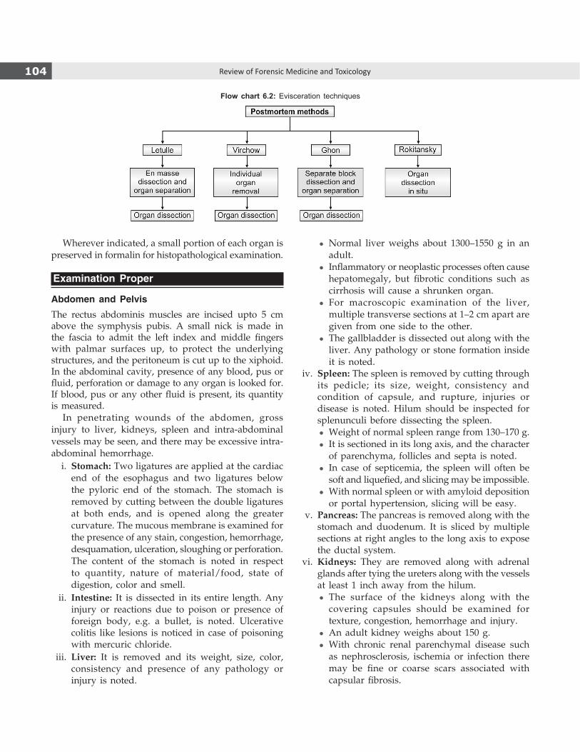
104 Review of Forensic Medicine and Toxicology
Wherever indicated, a small portion of each organ is preserved in formalin for histopathological examination.
Examination Proper
Abdomen and PelvisThe rectus abdominis muscles are incised upto 5 cm above the symphysis pubis. A small nick is made in the fascia to admit the left index and middle fingers with palmar surfaces up, to protect the underlying structures, and the peritoneum is cut up to the xiphoid. In the abdominal cavity, presence of any blood, pus or fluid, perforation or damage to any organ is looked for. If blood, pus or any other fluid is present, its quantity is measured.
In penetrating wounds of the abdomen, gross injury to liver, kidneys, spleen and intra-abdominal vessels may be seen, and there may be excessive intra-abdominal hemorrhage. i. Stomach: Two ligatures are applied at the cardiac
end of the esophagus and two ligatures below the pyloric end of the stomach. The stomach is removed by cutting between the double ligatures at both ends, and is opened along the greater curvature. The mucous membrane is examined for the presence of any stain, congestion, hemorrhage, desquamation, ulceration, sloughing or perforation. The content of the stomach is noted in respect to quantity, nature of material/food, state of digestion, color and smell.
ii. Intestine: It is dissected in its entire length. Any injury or reactions due to poison or presence of foreign body, e.g. a bullet, is noted. Ulcerative colitis like lesions is noticed in case of poisoning with mercuric chloride.
iii. Liver: It is removed and its weight, size, color, consistency and presence of any pathology or injury is noted.
z Normal liver weighs about 1300–1550 g in an adult.
z Inflammatory or neoplastic processes often cause hepatomegaly, but fibrotic conditions such as cirrhosis will cause a shrunken organ.
z For macroscopic examination of the liver, multiple transverse sections at 1–2 cm apart are given from one side to the other.
z The gallbladder is dissected out along with the liver. Any pathology or stone formation inside it is noted.
iv. Spleen: The spleen is removed by cutting through its pedicle; its size, weight, consistency and condition of capsule, and rupture, injuries or disease is noted. Hilum should be inspected for splenunculi before dissecting the spleen.
z Weight of normal spleen range from 130–170 g. z It is sectioned in its long axis, and the character
of parenchyma, follicles and septa is noted. z In case of septicemia, the spleen will often be
soft and liquefied, and slicing may be impossible. z With normal spleen or with amyloid deposition
or portal hypertension, slicing will be easy. v. Pancreas: The pancreas is removed along with the
stomach and duodenum. It is sliced by multiple sections at right angles to the long axis to expose the ductal system.
vi. Kidneys: They are removed along with adrenal glands after tying the ureters along with the vessels at least 1 inch away from the hilum.
z The surface of the kidneys along with the covering capsules should be examined for texture, congestion, hemorrhage and injury.
z An adult kidney weighs about 150 g. z With chronic renal parenchymal disease such
as nephrosclerosis, ischemia or infection there may be fine or coarse scars associated with capsular fibrosis.
Flow chart 6.2: Evisceration techniques

105Medico-legal Autopsy
z The kidney is sectioned longitudinally through the convex border into the hilum. The pelvis is examined for calculi and inflammation.
z Renal infarcts are pyramidal or wedge-shaped lesions with the base at the cortical surface and the apex pointing to the medullary origin of the arterial supply. Beginning as pale areas of necrosis with hyperemic borders, they progress to yellow-gray lesions that ultimately become depressed V-shaped gray-white furrows.
vii. Urinary bladder: It is examined in situ. If bladder contains urine, it is syringed out before opening to avoid any chances of contamination by blood or any other material. The bladder should be examined for any pathology, hemorrhage, congestion or injury. Both the ureters should be opened along their long axes.
viii. Female genitalia: The uterus and its appendages should first be examined in situ and then removed en masse along with the vagina by giving an incision externally on the labia upto the symphysis pubis above and the anus below. Internally, an incision is given around the pelvic brim and continued downwards to the pelvic outlet till it reaches the vaginal incision.
The uterus is examined and its dimensions, weight, whether gravid, parous or nulliparous, or any pathology present is noted. In case of gravid uterus, condition of the whole product of conception should be noted. In cases of abortion or attempted abortion, remains of any part of the product of conception inside the cavity, color of endometrial surface, erosion, any injury, ulceration or perforation of vaginal canal (particularly near the fornices) or of the uterine wall is noted. Foreign body may be present inside the uterine cavity. Smell and nature of the fluid present inside the uterine cavity is noted. Evidence of use of instruments may be present in the cervix or in the os.
The vagina is examined for any injury, foreign body, condition of hymen, mucous membrane and rugae. Any fluid present in the vagina is aspirated and preserved.
Ovaries should be examined for presence of corpus luteum. Fallopian tubes and ovaries have special medicolegal significance in cases of deaths due to their rupture in ectopic pregnancy.
ix. Prostate (in males): It is examined for enlargement or malignancy. In prostatitis, it is firm and in carcinoma, it is hard and granular.
Chest
The skin and muscles of the chest are dissected sidewise and carried back to the midaxillary line, down to the costal margin and up over the clavicles. The ribs and sternum are examined for fractures, and the chest is opened by cutting the costal cartilages close to the costochondral junctions and starting from the upper border of the second cartilage with a cartilage knife. Then, disarticulation of the sternoclavicular joint is done on each side by inserting the point of knife into the semicircular joint.
The pleural cavity is examined before complete removal of the sternum. In situ inspection is done before removal of thoracic organs which includes observation of the atrium and ventricle for air embolism, distension or collapse of lungs, the chest cavity for fluid, hemorrhage or pus, pleural adhesions, injuries including fracture of ribs.
Lungs
Both the lungs are separated from the mediastinal structures after tying the vessels and the bronchioles.
� The condition of pleura, any sign of pleuritis, petechial hemorrhages, injury, effusion, hemothorax, pneumothorax or pyothorax is noted.
� Normal lungs weigh 250–400 g each in an adult, but may weigh > 1 kg in cases of severe cardiac failure or diffuse alveolar damage.
� It is conventional to cut open from large to small airways, from medial to lateral to include all lobes and segments opening along the branches as they are encountered. Impression of the parenchymal appearance and texture is noted, and apical disease like old tuberculous cavities or fungal balls can also be demonstrated.
� The parenchyma is squeezed and any pus or fluid expressed is noted.
� After this, horizontal slicing through each lobe with a brain-knife is made to inspect the rest of the parenchyma.
� It is preferable to make large horizontal slices through the whole lung rather than opening the airways and vessels in cases of large mass lesion (e.g. carcinoma).
Dissection of the vessels
The course of pulmonary veins into the lung is traced, and thrombosis and atheroma is looked for, the latter being associated with pulmonary hypertension. An antemortem embolus may be coiled, and when straightened resembles a cast of the vessel from which the thrombus originated, usually in the leg. Massive
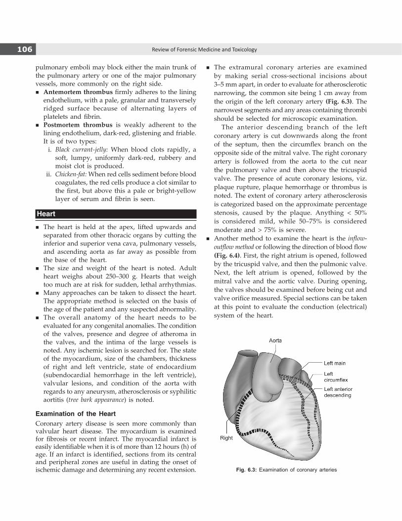
106 Review of Forensic Medicine and Toxicology
pulmonary emboli may block either the main trunk of the pulmonary artery or one of the major pulmonary vessels, more commonly on the right side.
� Antemortem thrombus firmly adheres to the lining endothelium, with a pale, granular and transversely ridged surface because of alternating layers of platelets and fibrin.
� Postmortem thrombus is weakly adherent to the lining endothelium, dark-red, glistening and friable. It is of two types:
i. Black currant-jelly: When blood clots rapidly, a soft, lumpy, uniformly dark-red, rubbery and moist clot is produced.
ii. Chicken-fat: When red cells sediment before blood coagulates, the red cells produce a clot similar to the first, but above this a pale or brightyellow layer of serum and fibrin is seen.
Heart
� The heart is held at the apex, lifted upwards and separated from other thoracic organs by cutting the inferior and superior vena cava, pulmonary vessels, and ascending aorta as far away as possible from the base of the heart.
� The size and weight of the heart is noted. Adult heart weighs about 250–300 g. Hearts that weigh too much are at risk for sudden, lethal arrhythmias.
� Many approaches can be taken to dissect the heart. The appropriate method is selected on the basis of the age of the patient and any suspected abnormality.
� The overall anatomy of the heart needs to be evaluated for any congenital anomalies. The condition of the valves, presence and degree of atheroma in the valves, and the intima of the large vessels is noted. Any ischemic lesion is searched for. The state of the myocardium, size of the chambers, thickness of right and left ventricle, state of endocardium (subendocardial hemorrhage in the left ventricle), valvular lesions, and condition of the aorta with regards to any aneurysm, atherosclerosis or syphilitic aortitis (tree bark appearance) is noted.
Examination of the HeartCoronary artery disease is seen more commonly than valvular heart disease. The myocardium is examined for fibrosis or recent infarct. The myocardial infarct is easily identifiable when it is of more than 12 hours (h) of age. If an infarct is identified, sections from its central and peripheral zones are useful in dating the onset of ischemic damage and determining any recent extension.
� The extramural coronary arteries are examined by making serial cross-sectional incisions about 3–5 mm apart, in order to evaluate for atherosclerotic narrowing, the common site being 1 cm away from the origin of the left coronary artery (Fig. 6.3). The narrowest segments and any areas containing thrombi should be selected for microscopic examination.
The anterior descending branch of the left coronary artery is cut downwards along the front of the septum, then the circumflex branch on the opposite side of the mitral valve. The right coronary artery is followed from the aorta to the cut near the pulmonary valve and then above the tricuspid valve. The presence of acute coronary lesions, viz. plaque rupture, plaque hemorrhage or thrombus is noted. The extent of coronary artery atherosclerosis is categorized based on the approximate percentage stenosis, caused by the plaque. Anything < 50% is considered mild, while 50–75% is considered moderate and > 75% is severe.
� Another method to examine the heart is the inflow-outflow method or following the direction of blood flow (Fig. 6.4). First, the right atrium is opened, followed by the tricuspid valve, and then the pulmonic valve. Next, the left atrium is opened, followed by the mitral valve and the aortic valve. During opening, the valves should be examined before being cut and valve orifice measured. Special sections can be taken at this point to evaluate the conduction (electrical) system of the heart.
Fig. 6.3: Examination of coronary arteries

107Medico-legal Autopsy
Fig. 6.4: Opening of the heart at autopsyIncision 1 —Through right atriumIncision 2—On the anterior wall of right ventricle parallel to interventricular septumIncision 3—Through tricuspid valveIncision 4—Through left atrium (after reversing the heart)Incision 5—Through mitral valve, parallel to the septum (on anterior wall)Incision 6—Through aortic valve
Fig. 6.5: Examination of myocardium
� Another lesser used method is the short axis or ventricular slicing method (Fig. 6.5). With the heart in the anatomical position, the first slice is made through the heart at a point about 3 cm from the apex separating it from the remainder of the heart. Further complete slices are then made in parallel to this slice, 1 cm apart, until reaching below the atrioventricular valves. The remainder is then examined by opening along the path of blood flow. It is useful if ischemic myocardial disease is suspected as it clearly demonstrates the distribution of infarction.
� The intramural or ‘sandwich’ technique can be used to cut through the thickness of the left ventricle. The heart is placed open on the cutting board, with the endocardium downwards. A knife is passed into the cut edge of the left ventricle and sliced right through the muscle, keeping equidistant between endocardium and epicardium. The myocardium can then be opened out like a book, showing the interior with any infarcts or fibrotic plaques.
Examination of coronary arteries should precede the examination of heart.

108 Review of Forensic Medicine and Toxicology
� Examination of valve: The circumference of the valve is measured. The circumference of mitral valve is 8–10.5 cm (mean 10 cm) and admits two fingers; tricuspid valve is 10–12.5 cm (12 cm) and admits three fingers; aortic valve is 6–8 cm (7.5 cm) and pulmonary valve is 7–9 cm (8.5 cm). The decrease in circumference is suggestive of stenosis whereas increased circumference could be due to regurgitation or incompetent valves.
� Ventricular hypertrophy: An estimate is made by measuring the thickness of the ventricular walls at a point about 1 cm below the atrioventricular valve. The upper limits of normal are: left ventricle: 1.5 cm, right ventricle: 0.5 cm and atrial muscle: 0.2 cm.
Subendocardial or Sheehan’s Hemorrhages
These are flameshaped, confluent hemorrhages and tend to occur in one continuous sheet rather than in patches, seen in the left ventricle, on the left side of the interventricular septum and on the opposing papillary muscles and adjacent columnae carnea.5
Subendocardial hemorrhages are seen in: � Severe loss of blood or shock � Intracranial damage, such as head injury, cerebral edema, surgical craniotomy or tumors
� Death due to ectopic pregnancy, ruptured uterus, abortion, antepartum or postpartum hemorrhage
� Poisoning, e.g. arsenic or oleander
Agonal thrombi: In case of a person dying slowly due to circulatory failure, a firm, stringy, tough, paleyellow thrombus forms in the cavities, usually on the right side of the heart.
The pericardium is examined for presence of any pathology or injury. The contents of the pericardial sac and quantity of fluid is noted. Pericardial effusion, cardiac tamponade, subpericardial hemorrhage and constrictive pericarditis are looked for.
Neck
The neck structures are examined before removal of the thoracic organs so that the tongue, larynx, trachea and esophagus can be taken out along with the lungs. This helps in examination of the whole of the upper respiratory tract in its continuity (Details are given in Chapter 10).
In case of death due to alleged constriction of the neck, there may be fracture of hyoid bone or thyroid cartilage with extravasation of blood into the tissues, and injury to carotid arteries, sternomastoid muscles or platysma. Compression of the neck with hard materials may cause injury to the cervical vertebrae and the corresponding part of the spinal cord. Level and extent
of other mechanical injuries on the neck are cautiously examined to know the type of injury, and organs or structures injured resulting in death.
Skull and Brain
Procedure: A wooden block is placed under the shoulders so that the neck is extended and the head fixed by a headrest. A coronal incision is made in the scalp, which starts from one mastoid to the opposite mastoid process just behind the ear and is continued over the vertex of the scalp. The incision should penetrate upto the periosteum. The scalp is reflected forwards to the superciliary ridges, and backwards to a point just below the occipital protuberance (Fig. 6.6A). Presence of hematoma, petechial hemorrhage, edema or fracture is noted.
The temporal and masseter muscles are incised on either side for sawing the skull. The saw-line is made in a slightly V-shaped direction (angle of 120°) so that the skull cap can fit back into the correct position on reconstruction of the body. Saw and remove the skull cap, the line of separation is just above the superciliary ridges in front, to the base of the mastoid process on either side, and just above the occipital protuberance behind (Fig. 6.6B).
Dura: The dura is examined from outside for extradural hemorrhage (weight and volume is noted, if present) and superior sagittal sinus for antemortem thrombus. It is cut along the line of detached skull cap and pulled gently from front to back while cutting falx cerebri, and examined for subdural and subarachnoid hemorrhage.
� The weight and volume of subdural hemorrhage, its effect on brain—flattening or any asymmetry is noted.
� Subdural hemorrhage can be washed under running water whereas subarachnoid hemorrhage cannot be washed.
A B
Fig. 6.6: (A) Incision for removal of scalp, (B) Saw-line for removal of skull cap

109Medico-legal Autopsy
Delivery of the Brain
Insert four fingers of the left hand between the frontal lobes and the skull. Draw them backward and then with the right hand, cut the nerves and vessels as they emerge from the skull. Cut the tentorium along the superior border of the petrous bone. Cut the cervical cord, first cervical nerves and vertebral arteries, as far below as possible. Support the brain throughout with the left hand. Remove the brain along with the cerebellum and brainstem which is supported by the right hand.
� Examine the remaining venous sinuses and the cranial cavity for antemortem thrombi. Remove the pituitary by chiseling the posterior clinoid processes and incising the diaphragm of the sella turcica around its periphery.
� Pull out the dura and examine the base of the skull and the rest of the cranial cavity for any fracture. Inspect the skull cap for fracture by holding it against the light.
� Remove a wedge shaped portion of the petrous temporal bone and examine the mastoid for any collection of pus, hemorrhage or fluid in the middle ear.
Examination of the Brain
The brain is weighed and then examined for any swelling, shrinkage or herniation, upper and lateral surfaces of the brain for asymmetry or flattening of the convolutions. The cerebral vessels is looked at for arteriosclerosis, embolism and aneurysms (especially the circle of Willis).
Berry aneurysms (size varies from few mm to few cm) are usually present at the junction of vessels especially at the junction of the posterior cerebral arteries, the posterior communicating vessels, and the middle cerebral arteries and the anterior communicating arteries. Cerebral infarction may occur due to a thrombus or atheroma.
In most medico-legal autopsies, the brain is examined in the fresh state; however, in select cases, the autopsy surgeon may need the brain ‘fixed’ prior to further evaluation. Fixation is an important step for proper examination of the brain and spinal cord.
Fixation of the Brain
The best routine fixative is 10% formalin and requires 2–3 weeks for satisfactory fixation. In fetuses and infants, the addition of acetic acid to the fixative solution increases the specific gravity of the fixative and allows
the brain to float in the solution; it also makes the tissue firmer without altering its histological characteristics.
Dissection of the Brain
The most utilized and reliable method of brain sectioning is the coronal cutting method, whether examination occurs in the fresh state or after formalin fixation. It involves serially sectioning of all parts, including cerebrum, cerebellum and brainstem.
First, the cerebellum and brainstem should be separated from the cerebrum. This is done as high as possible, and cut the surface in a horizontal plane with a large scalpel. The brainstem is then separated from the cerebellum at the cerebellar peduncles, as close to the brainstem as possible with a scalpel.
Cerebrum
The cerebrum is then sliced in a coronal plane at 1 cm intervals. If the brain is fresh, it is sliced from the frontal end and from the superior surface. The main aim with the fresh brain is to be as quick as possible, since the brain is so soft that it rapidly collapses.
With fixed brain, the first slice is done through the mammilary bodies (at the basal surface) which divide the brain into half in the exact coronal plane. Thereafter, each half is sliced 1 cm each, in turn, with the flat surface laid downwards. It should be done with a single sweep of a brain knife, to avoid a sawing motion and subsequent irregularities on the cut surface. These slices should then be laid out in order on a flat surface.
Other planes that can be used for special cases: � Cutting the brain in the plane of CT-scans, for
comparison with the radiology. � Single, midline sagittal section, particularly useful if
a third or fourth ventricle lesion is expected.
Features to look for: � The cortical ribbon, white matter, basal ganglia
and lateral ventricle should be examined for any asymmetry or brain shift that would indicate space occupying lesion—abscess, large hemorrhage, recent infarction or either metastatic or primary tumors.
� Old infarcts are cystic spaces which do not produce any brain shift.
� Small focal lesions may not cause any brain shift, e.g. small (lacunar) infarcts associated with hypertension and gray areas of demyelination (plaques) within the white matter.
� Dilatation of lateral ventricle may indicate atrophy.

110 Review of Forensic Medicine and Toxicology
� Shrinkage of cerebral cortex (gray matter) is common in chronic alcoholics.
� Cerebral fat emboli that have completely obstructed the small vessels of the brain may be visible to the naked eye as punctate hemorrhages in the white matter.
� Petechial hemorrhages in the white matter are commonly found in death from anaphylactic shock.
� In head injury, edema is seen in the white matter around or deep to contusions, lacerations or ischemic lesions. If there is any injury to the brain, successive sections parallel to the wounded surfaces should be made till the whole depth of the wound is revealed.
Cerebellum
The cerebellum is dissected on the horizontal plane with the two lobes being sliced in a ‘fan’ shape with the middle slice going through the dentate nucleus which gives best histological orientation of the structures.
Brainstem
The brainstem can be sliced at 5 mm intervals per-pendicular to its axis, and laid out in order on a flat surface.
In brainstem and cerebellum, any focal lesions must be identified, such as areas of hemorrhage, areas of softening or cystic degeneration that indicate recent or old infarction respectively, and primary or metastatic tumors.
Dissection of head in infants: Rokitansky’s method or Beneke’s technique is used to open the skull in infants (Details in Chapter 20).
Spinal Cord
� The spinal cord can be removed from an anterior or posterior approach and usually removed separately from the brain.
� If there is no indication, the spinal cord need not be exposed.
� The anterior approach is more difficult but has the advantages of not requiring the body to be turned (messy procedure with evisceration already taken place), and allowing the nerve roots and dorsal ganglia to be dissected.
� The posterior approach is both quicker and easier, but best performed before the full postmortem, to avoid the mess. It also allows the spinal cord and the brain to be removed in continuity, but does not allow examination of the nerve roots and basal ganglia.6
Posterior Approach to the Spinal Cord
i. A long midline incision is made and the skin, muscle and soft tissues are flapped out sidewise or laterally, 1 inch on either side from the vertebral column.
ii. The posterior arch is cut with the vibrating saw. This dissection can extend superiorly along the cervical vertebrae to the foramen magnum.
iii. The spinal processes and posterior portions of the laminae are removed.
iv. The dura is opened longitudinally to the uppermost part of the incision, where it is cut circumferentially.
v. The nerves are cut and the spinal cord is delivered by steady traction.
Description of an Organ
� Size: Measuring tape is used. A tense capsule indicates enlargement and loose capsule shrinkage.
� Shape: Note any deviation from normal. � Surface: Most organs have a delicate, smooth,
glistening and transparent capsule of serosa. Any thickening, roughening, dullness or opacity is noted.
� Consistency: The softness or firmness is appreciated by application of finger pressure.
� Cohesion: It is the strength within the tissue that holds an organ together. It is judged by the resistance of the cut surface to tearing, pressure or pulling.
� Cut surface: Note color and structural details.
After completion of autopsy, blood, fluid etc. are removed from the body cavities. The organs are replaced in the body and any excess space is packed with cotton or cloth, especially in the pelvis and the throat, where blood tends to leak. The dissected flaps are brought close together and sutured by using thin twine and large curved needle. The skull is filled with remaining portion of the brain along with cotton or other absorbent material and the skull cap fitted in place. The scalp is pulled back over the vault, and the scalp stitched with thin strong twine. The body is washed with water, dried and covered with clothes, and then handed over to the police constable accompanying it.1
Post-autopsy reconstruction plays a key role in the presentation of the autopsied body to the relatives. The reconstruction of body should be of a high standard so that it will not leak, and can be viewed after autopsy without distressing the next of kin. The following procedure may be undertaken to ensure a leak proof and contaminant-free reconstruction technique:
� Removal of accumulated fluids: After evisceration of organs from the thoracic, abdominal and pelvic cavities, residual fluids, tissues and bowel contents (blood, ascitic or pleural fluid,
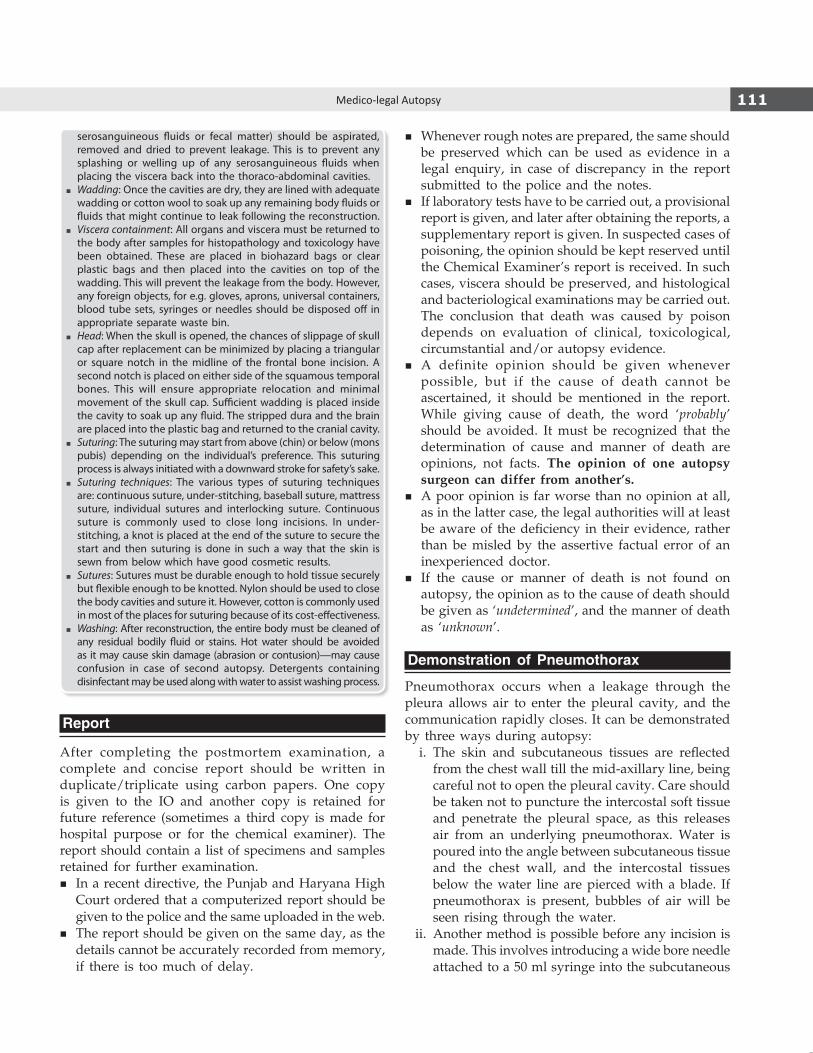
111Medico-legal Autopsy
serosanguineous fluids or fecal matter) should be aspirated, removed and dried to prevent leakage. This is to prevent any splashing or welling up of any serosanguineous fluids when placing the viscera back into the thoraco-abdominal cavities.
� Wadding: Once the cavities are dry, they are lined with adequate wadding or cotton wool to soak up any remaining body fluids or fluids that might continue to leak following the reconstruction.
� Viscera containment: All organs and viscera must be returned to the body after samples for histopathology and toxicology have been obtained. These are placed in biohazard bags or clear plastic bags and then placed into the cavities on top of the wadding. This will prevent the leakage from the body. However, any foreign objects, for e.g. gloves, aprons, universal containers, blood tube sets, syringes or needles should be disposed off in appropriate separate waste bin.
� Head: When the skull is opened, the chances of slippage of skull cap after replacement can be minimized by placing a triangular or square notch in the midline of the frontal bone incision. A second notch is placed on either side of the squamous temporal bones. This will ensure appropriate relocation and minimal movement of the skull cap. Sufficient wadding is placed inside the cavity to soak up any fluid. The stripped dura and the brain are placed into the plastic bag and returned to the cranial cavity.
� Suturing: The suturing may start from above (chin) or below (mons pubis) depending on the individual’s preference. This suturing process is always initiated with a downward stroke for safety’s sake.
� Suturing techniques: The various types of suturing techniques are: continuous suture, under-stitching, baseball suture, mattress suture, individual sutures and interlocking suture. Continuous suture is commonly used to close long incisions. In under-stitching, a knot is placed at the end of the suture to secure the start and then suturing is done in such a way that the skin is sewn from below which have good cosmetic results.
� Sutures: Sutures must be durable enough to hold tissue securely but flexible enough to be knotted. Nylon should be used to close the body cavities and suture it. However, cotton is commonly used in most of the places for suturing because of its cost-effectiveness.
� Washing: After reconstruction, the entire body must be cleaned of any residual bodily fluid or stains. Hot water should be avoided as it may cause skin damage (abrasion or contusion)—may cause confusion in case of second autopsy. Detergents containing disinfectant may be used along with water to assist washing process.
Report
After completing the postmortem examination, a complete and concise report should be written in duplicate/triplicate using carbon papers. One copy is given to the IO and another copy is retained for future reference (sometimes a third copy is made for hospital purpose or for the chemical examiner). The report should contain a list of specimens and samples retained for further examination.
� In a recent directive, the Punjab and Haryana High Court ordered that a computerized report should be given to the police and the same uploaded in the web.
� The report should be given on the same day, as the details cannot be accurately recorded from memory, if there is too much of delay.
� Whenever rough notes are prepared, the same should be preserved which can be used as evidence in a legal enquiry, in case of discrepancy in the report submitted to the police and the notes.
� If laboratory tests have to be carried out, a provisional report is given, and later after obtaining the reports, a supplementary report is given. In suspected cases of poisoning, the opinion should be kept reserved until the Chemical Examiner’s report is received. In such cases, viscera should be preserved, and histological and bacteriological examinations may be carried out. The conclusion that death was caused by poison depends on evaluation of clinical, toxicological, circumstantial and/or autopsy evidence.
� A definite opinion should be given whenever possible, but if the cause of death cannot be ascertained, it should be mentioned in the report. While giving cause of death, the word ‘probably’ should be avoided. It must be recognized that the determination of cause and manner of death are opinions, not facts. The opinion of one autopsy surgeon can differ from another’s.
� A poor opinion is far worse than no opinion at all, as in the latter case, the legal authorities will at least be aware of the deficiency in their evidence, rather than be misled by the assertive factual error of an inexperienced doctor.
� If the cause or manner of death is not found on autopsy, the opinion as to the cause of death should be given as ‘undetermined’, and the manner of death as ‘unknown’.
Demonstration of Pneumothorax
Pneumothorax occurs when a leakage through the pleura allows air to enter the pleural cavity, and the communication rapidly closes. It can be demonstrated by three ways during autopsy: i. The skin and subcutaneous tissues are reflected
from the chest wall till the mid-axillary line, being careful not to open the pleural cavity. Care should be taken not to puncture the intercostal soft tissue and penetrate the pleural space, as this releases air from an underlying pneumothorax. Water is poured into the angle between subcutaneous tissue and the chest wall, and the intercostal tissues below the water line are pierced with a blade. If pneumothorax is present, bubbles of air will be seen rising through the water.
ii. Another method is possible before any incision is made. This involves introducing a wide bore needle attached to a 50 ml syringe into the subcutaneous
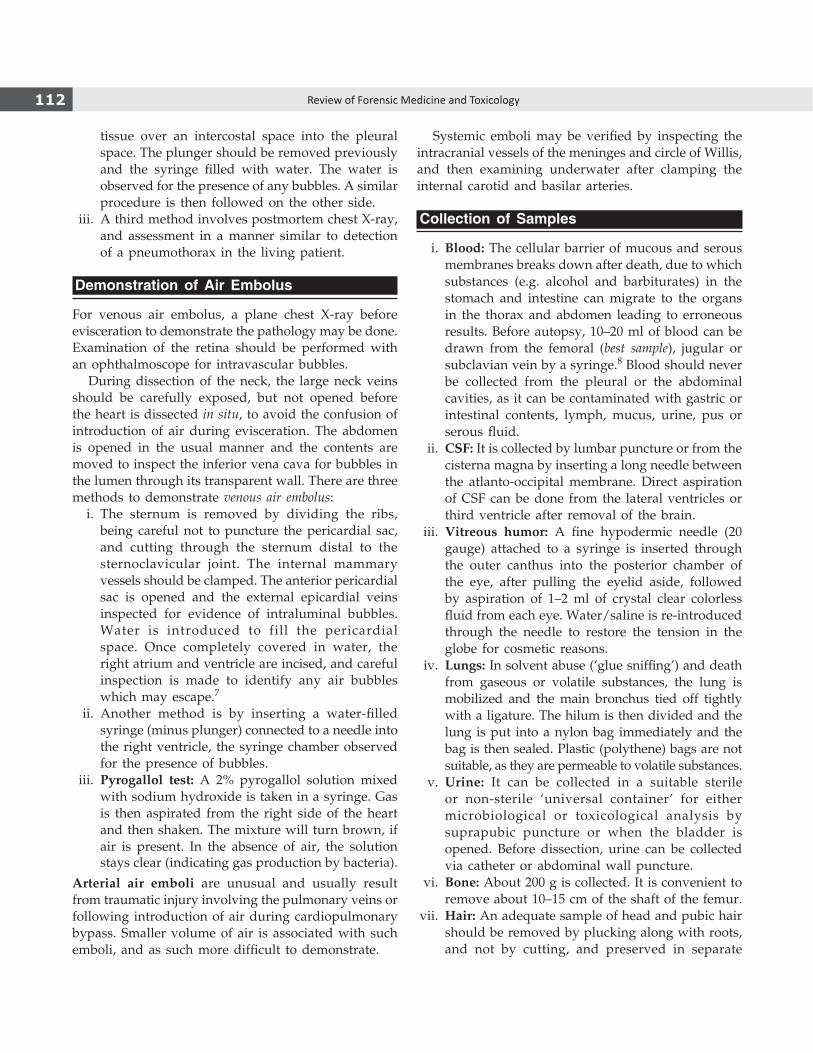
112 Review of Forensic Medicine and Toxicology
tissue over an intercostal space into the pleural space. The plunger should be removed previously and the syringe filled with water. The water is observed for the presence of any bubbles. A similar procedure is then followed on the other side.
iii. A third method involves postmortem chest X-ray, and assessment in a manner similar to detection of a pneumothorax in the living patient.
Demonstration of Air Embolus
For venous air embolus, a plane chest X-ray before evisceration to demonstrate the pathology may be done. Examination of the retina should be performed with an ophthalmoscope for intravascular bubbles.
During dissection of the neck, the large neck veins should be carefully exposed, but not opened before the heart is dissected in situ, to avoid the confusion of introduction of air during evisceration. The abdomen is opened in the usual manner and the contents are moved to inspect the inferior vena cava for bubbles in the lumen through its transparent wall. There are three methods to demonstrate venous air embolus: i. The sternum is removed by dividing the ribs,
being careful not to puncture the pericardial sac, and cutting through the sternum distal to the sternoclavicular joint. The internal mammary vessels should be clamped. The anterior pericardial sac is opened and the external epicardial veins inspected for evidence of intraluminal bubbles. Water is introduced to fill the pericardial space. Once completely covered in water, the right atrium and ventricle are incised, and careful inspection is made to identify any air bubbles which may escape.7
ii. Another method is by inserting a waterfilled syringe (minus plunger) connected to a needle into the right ventricle, the syringe chamber observed for the presence of bubbles.
iii. Pyrogallol test: A 2% pyrogallol solution mixed with sodium hydroxide is taken in a syringe. Gas is then aspirated from the right side of the heart and then shaken. The mixture will turn brown, if air is present. In the absence of air, the solution stays clear (indicating gas production by bacteria).
Arterial air emboli are unusual and usually result from traumatic injury involving the pulmonary veins or following introduction of air during cardiopulmonary bypass. Smaller volume of air is associated with such emboli, and as such more difficult to demonstrate.
Systemic emboli may be verified by inspecting the intracranial vessels of the meninges and circle of Willis, and then examining underwater after clamping the internal carotid and basilar arteries.
Collection of Samples
i. Blood: The cellular barrier of mucous and serous membranes breaks down after death, due to which substances (e.g. alcohol and barbiturates) in the stomach and intestine can migrate to the organs in the thorax and abdomen leading to erroneous results. Before autopsy, 10–20 ml of blood can be drawn from the femoral (best sample), jugular or subclavian vein by a syringe.8 Blood should never be collected from the pleural or the abdominal cavities, as it can be contaminated with gastric or intestinal contents, lymph, mucus, urine, pus or serous fluid.
ii. CSF: It is collected by lumbar puncture or from the cisterna magna by inserting a long needle between the atlanto-occipital membrane. Direct aspiration of CSF can be done from the lateral ventricles or third ventricle after removal of the brain.
iii. Vitreous humor: A fine hypodermic needle (20 gauge) attached to a syringe is inserted through the outer canthus into the posterior chamber of the eye, after pulling the eyelid aside, followed by aspiration of 1–2 ml of crystal clear colorless fluid from each eye. Water/saline is reintroduced through the needle to restore the tension in the globe for cosmetic reasons.
iv. Lungs: In solvent abuse (‘glue sniffing’) and death from gaseous or volatile substances, the lung is mobilized and the main bronchus tied off tightly with a ligature. The hilum is then divided and the lung is put into a nylon bag immediately and the bag is then sealed. Plastic (polythene) bags are not suitable, as they are permeable to volatile substances.
v. Urine: It can be collected in a suitable sterile or non-sterile ‘universal container’ for either microbiological or toxicological analysis by suprapubic puncture or when the bladder is opened. Before dissection, urine can be collected via catheter or abdominal wall puncture.
vi. Bone: About 200 g is collected. It is convenient to remove about 10–15 cm of the shaft of the femur.
vii. Hair: An adequate sample of head and pubic hair should be removed by plucking along with roots, and not by cutting, and preserved in separate

113Medico-legal Autopsy
containers (0.5 g for DNA analysis, upto 10 g for analysis of heavy metals).
viii. Maggots: These are dropped alive into boiling absolute alcohol or 10% hot formalin which kills them in an extended condition (to disclose the internal structure of the larvae). If time of death is an issue, some larvae/maggots should be preserved alive for examination by an entomologist. Maggots may reveal the presence of drugs/poisons in decomposed bodies.
ix. Nails: All the nails (fingers and/or toes) should be removed in their entirety and collected in separate envelopes.
x. Skin: If there is needle puncture, the whole needle track and surrounding tissue should be excised. Control specimens should be taken from same area on the opposite side of the body and preserved in a separate container. In firearm cases, a portion of skin around the entrance and exit wounds should be preserved.
� Fibroblasts for tissue culture: Karyotyping, metabolic assays, enzyme assays and diagnostic ultrastructural studies can be performed on cultured fibroblasts. Skin, fascia, lung, diaphragm, muscle and cartilage are useful for fibroblast cell cultures.
� Tissue for metabolic studies and nucleic acid analysis: Liver, kidney, cardiac and skeletal muscle, and peripheral nerve obtained at autopsy may be used for biochemical studies in the diagnosis of inborn errors of metabolism. The tissue should be frozen rapidly in liquid nitrogen or dry ice and stored at –70°C.
Preservation of Viscera
Viscera should be preserved in cases of: � Suspected death due to poisoning � Deceased was intoxicated or used to drugs � Cause of death could not be found after autopsy � Accidental death involving driver of a vehicle or machine operator
� Death due to burns (if needed) � Advanced decomposition* � Any case, if requested by the Magistrate.
The Supreme Court has ruled that in cases of death due to suspected poisoning, the prosecuting agency should send the viscera to a forensic science laboratory immediately after postmortem. Specimens that must be preserved in cases of suspected poisoning are given in Tables 6.1 to 6.3.
Some practical points need to be considered: � The preferred specimens collected at postmortem
will depend on the type of case/poison suspected.
� Blood is the most useful sample because toxins present in this can best be related to a physiological effect, and can be used to assess the likelihood of recent exposure to poisons/drugs.
� Urine is the second most important specimen collected. However, the disadvantages are: it is unavailable in half the cases (since it is voided during the dying process), poison may be metabolized so extensively that the parent compound may not
Table 6.2: Viscera preserved during autopsy (routine)
S.No. Material Quantity
1. Stomach and its contents Whole
2. Upper part of small intestine and its contents
About 15–30 cm length (some say 100 cm)
3. Liver (along with gallbladder) 300 g
4. Kidney Longitudinal half of each kidney
5. Spleen Whole
6. Blood 10 ml
7. Urine 100 ml
* When the body is too decomposed to collect any fluids, collect atleast 100 g of muscle from thigh, liver, brain, fat and kidneys.
Table 6.1: Samples preserved in living persons
Material Quantity
Vomit 300 ml (whole, if quantity is less)
Stomach washout 500 ml
Blood 10 ml
Urine 100 ml
Table 6.3: Additional viscera and materials required in certain cases
S.No. Material Poisoning/circumstances suspected1. Heart Strychnine, digitalis2. Brain Alkaloids, organophosphorus, opiates,
strychnine, carbon monoxide, cyanide, barbiturates and volatile organic poisons; hydrophobia/rabies (for negri bodies)9,10
3. Spinal cord Strychnine11
4. CSF Alcohol12
5. Vitreous humor Alcohol, chloroform13
6. Lung Gaseous poisons, hydrocyanic acid, alcohol, chloroform
7. Skin Injected poisons (insulin, morphine, heroin, cocaine and other illicit drugs), firearm injuries
8. Bone, hair and nails Heavy metals (arsenic, antimony, thallium)9. Fatty tissue Pesticides and insecticides
10. Uterus and its appendages
Criminal abortion
11. Muscle Decomposition

114 Review of Forensic Medicine and Toxicology
detected, and concentration of most poisons are difficult to interpret.
� Vitreous humor is the preferred specimen for postmortem confirmation of alcohol ingestion, since postmortem formation of ethanol does not occur to significant extent in vitreous, and hence useful even in decomposing bodies. It is recommended that this specimen is included routinely in sudden death investigations.
� Stomach content is invaluable in cases of suspected poisoning—establish actual content of poison, determination of route of administration, high concentration of toxins, and analysis is uncomplicated by metabolism. Whole stomach is preserved since it allows the analyst to dissolve any poison adhering to the sides of the walls even in cases where there was little or no fluid. However, detection of poison does not necessarily imply oral ingestion—basic drugs and metabolites in the blood can be secreted through the gastric juice (formed from extracellular fluid) and the juice may be contaminated with bile from retching and vomiting or may be agonal in nature.
� Although, spleen and kidneys are routinely preserved, liver is most important since large amount of tissue is available, ease of sample collection, high concentration of toxins, and availability of large database of liver drug concentrations.
z A 100 g of tissue is sufficient for most analysis. z The right lobe is preferred since chances of
postmortem diffusion of toxins from bowel contents and mesenteric circulation is negligible.
z With more sensitive analytical methods, the majority of drugs are detected readily in the blood, and it is not necessary to rely on the liver (or any other organ) for their detection.
z The major disadvantage of the liver as specimen is that it tends to be fatty and putrefy faster than blood.
� Bile has been collected historically, but its usefulness is limited. It may be show the presence of number of drugs including morphine/heroin, benzodiazepine, cocaine, methadone, glutathione, many antibiotics and tranquillizers and heavy metals (in chronic poisoning).With the widespread use of sensitive immunoassays and other techniques, the use of bile as a screening specimen is less valuable that it once was.
� Brain, kidney and spleen are used to determine and interpret the concentration of toxins, i.e. overall assessment of the body burden of a toxin.
� Spleen is useful as a specimen for toxins, such as carbon monoxide (CO) and cyanide that binds to
hemoglobin. If septicemia is suspected and the cause of it is not obvious, spleen should be cultured.
The viscera should be refrigerated at about 4°C, if not sent to the laboratory. They can be destroyed either after getting the permission from the Magistrate or when the IO informs that the case has been closed.
Preservation of Samples
� The ideal samples are the ones in which no preservative has been added and sent to CFSL within few hours. But, practically, it usually gets delayed.
� The specimen is preserved at 4°C until they are analyzed.14,15 For long-term storage, it has to be kept in freezer (–10°C).
� In order that putrefaction may not set in and render chemical analysis difficult, certain preservatives are used.
1. Viscera z The most commonly used preservative for
viscera is saturated solution of common salt.16 It is easily available, cheap and effective preservative. However, the best preservative for preservation of viscera is rectified spirit.
z In cases of suspected alkali or acid poisoning (except carbolic acid), only rectified spirit is used.17 It is not used in cases of suspected poisoning with:
� Alcohol � Chloroform� Kerosene � Ether� Chloral hydrate � Phosphorus� Formaldehyde � Formic acid� Paraldehyde � Acetic acid
2. Blood for toxicological analysis (for alcohol, cocaine, cyanide and CO) is preserved in sodium or potassium fluoride at the concentration of 10 mg/ml of blood and anticoagulant potassium oxalate, 30 mg/10 ml of blood.18
z Postmortem samples are liable to production of alcohol by microbiological action and higher concentrations of sodium fluoride are required to inhibit this.
z Heparin and EDTA should not be used as anticoagulants, since they interfere with detection of methanol.
z If blood is required only for grouping, no preservative is necessary and small amount of blood is well preserved by soaking in a blotter.
z In case of suspected CO poisoning, a layer of 1–2 cm of liquid paraffin is added immediately over the blood sample to avoid exposure to atmospheric oxygen.

115Medico-legal Autopsy
z If solvent abuse and anesthetic death is suspected, the glass container should have a foil-lined lid to prevent gas from escaping (as gas can permeate rubber) and the container is completely filled to prevent gas from escaping in ‘dead’ air space.
z Blood for hematological examination including glycosylated hemoglobin in diabetics should be sent in a clean glass container with anticoagulant (e.g. EDTA).
3. Urine is persevered by adding small amount of phenyl mercuric nitrate or thymol. Fluoride should be added to urine if alcohol, cyanide or cocaine is suspected in the sample.
4. Vitreous humor is preserved using sodium fluoride (10 mg/ml).19
5. For bones, hair and nails, preservative is not required. It has to be dried in normal temperature and sealed in plastic bag. But, bone marrow is preserved in a test tube containing 4–5 ml of 5% albumin-normal saline solution and stored at 4°C.
� Formalin is not used as preservative for chemical analysis because extraction of poison, especially nonvolatile organic compounds become difficult.
� The use of disposable, hard plastic (especially polypropylene) or glass containers are recommended for preservation.
� All samples should be properly sealed and labeled with the patient’s name, hospital number, nature of sample, collection site, preservative used, and date and time of collection. It should be handed over to the IO after obtaining proper receipt.
� Sodium fluoride is the most commonly used agent to prevent glycolysis. It inhibits the enzyme enolase and is also effective at inhibiting bacterial growth.20,21
� EDTA can effectively chelate the calcium ion of blood, therefore it can prevent the blood coagulation, does not affect the count and size of the leukocyte and keep erythrocyte invariable. Other anticoagulants are potassium oxalate, citrate or lithium heparin.
Procedure of PreservationFor preservation of viscera, a clean, transparent and preferably sterile glass jar (one liter capacity) with a wide mouth and stoppers should be used. The size of the jar should be such, that at least 1/3rd of the container remains empty after being filled with the preservative to allow for accommodation of the gas which will evolve out of the organs preserved. However, the preservative should completely immerse the viscera after the contents are well shaken.
� The stomach, small intestine and its contents are preserved in one bottle, part of liver along with gallbladder, spleen and kidneys in another bottle and urine in the third bottle. The stomach and intestines are opened before they are preserved. The liver and kidneys are cut into small pieces to ensure penetration of the preservative. Blood should be sent in a vial(s).
� A sample of the preservative used (sodium chloride or rectified spirit) is separately preserved and sent for analysis to rule out any poison being present as a contaminant.
� When additional material is required to be sent, it should be dispatched in separate bottles, like brain in one bottle and vomitus or stomach washout in another bottle. The bottles and vials required for preservation are normally supplied by the office of the Forensic Science Laboratory (FSL).
� The stoppers of the bottles should be well fitting, covered with a piece of cloth and tied by tape or string, and the ends sealed using a departmental seal. Each bottle should be suitably labeled with the autopsy number, name of the deceased, name of the organ, date, time and place of autopsy, followed by signature of the doctor who performed the autopsy.
� The sealed bottles are then put in a viscera box which is sealed. The viscera box along with a specimen of the seal used (put in a separate envelope and sealed) is handed over to the police constable, in return for a receipt. All these precautions are necessary to maintain the chain of evidence.
� Along with the viscera box, the following documents are also sent:
i. Copy of the inquest papers, brief facts of the case and the case sheet.
ii. Copy of autopsy report. iii. Letter requesting the chemical examiner to
examine the viscera and inform the medical officer of its findings.
Samples for Laboratory Investigations
� Histopathological examination: Sections of various internal organs (1.5 × 1.0 × 1.0 cm) in case of suspected abnormality are preserved in 10% formalin or 95% alcohol.22
� Bacteriological/serological examination: Blood should be kept in sterile container using sterile syringe from the right ventricle of the heart or from some large vessel, such as femoral vein or artery. It may also be used for biochemical examination.

116 Review of Forensic Medicine and Toxicology
� Virological examination: A piece of tissue is collected and preserved in 50% sterile glycerin.23
� Enzymatic studies: Small pieces of tissues are collected into a thermos containing liquid nitrogen.
� Smears: Vaginal/anal smears are needed in cases of alleged sexual assault. In suspected malaria, smears from cerebral cortex, spleen and liver may be taken and examined for malarial parasite.
Obscure and Negative Autopsy
Obscure autopsy: In about 20% of all postmortem examination cases, the cause of death may not be clear at the time of dissection of the body, and there are minimal or indeterminate findings or even no positive findings at all. These are a source of confusion to any forensic pathologist.
� In many of these cases, the cause of death can be made out after detailed clinical and laboratory investigations and interview with persons who had observed the deceased before he died.
� These ‘obscure autopsies’ are more common in the younger age group.
� Before tissue for histology is taken, a full review of the dissection should be undertaken— the coronary system, pulmonary arteries (pulmonary emboli in smaller branches), brain (particularly the basal arteries) and the carotid arteries in the neck.
� When a complete review of the gross pathology has proved unproductive, then a full histological examination is required, especially of the myocardium. Special stains such as phosphotungstic acid-hematoxylin, dehydrogenase enzyme histochemistry and acridineorange fluorescence stains may be used.
Causes of obscure autopsy i. Natural diseases: Epilepsy, paroxysmal fibrillation. ii. Concealed trauma: Concussion, blunt injury to the
heart, reflex vagal inhibition. iii. Poisoning: Anesthetic overdose, narcotic,
neurotoxic, cytotoxic or plant poisoning. iv. Biochemical disturbances: Uremia, diabetes. v. Endocrinal disturbances: Adrenal insufficiency,
thyrotoxicosis. vi. Miscellaneous: Allergy, drug idiosyncrasy.
Negative autopsy: In about 2–5% of all postmortem examination cases, the cause of death remains unknown, even after all laboratory examinations including biochemical, microbiological, virological, microscopic and toxicological examination.
Reasons of negative autopsy i. Inadequate history. ii. Inadequate external examination and internal
examination. iii. Insufficient laboratory examinations. iv. Lack of toxicological analysis. v. Lack of training of the doctor.
� If at the end of the process, no apparent cause of death is found, then the authorities must be informed that the cause of death cannot be determined and no opinion can be offered in the present state of medical and scientific knowledge.
� However, negative findings/evidence such as absence of injuries, no evidence of poisoning/lethal infection/well-recognized natural disease may confirm that the deceased did not die of, and in all probability he must have died due to natural causes, rather than some unnatural external event.
� The use of some meaningless terms such as ‘heart failure’ or ‘cardiorespiratory arrest’ is pointless and may cause confusion to the police/Magistrate.
� A mode of death is irrelevant in lieu of a cause of death, so is the use of some agonal event such as ‘aspiration of vomit’.
� It is also useless to use some unprovable process, such as ‘vagal inhibition’, ‘reflex cardiac arrest’ or ‘suffocation’, because these conditions are thought to leave no traces.
Second Autopsy
� Second autopsy or re-postmortem examination is the autopsy conducted on an already autopsied body.
� There is no provision in Indian law for a second autopsy. Instances where second autopsy is requested is given in Box 6.3.
� A Committee set up by the Nation Human Rights Commission recommended that the following rules be observed in respect of a second postmortem:
a. This procedure, ordinarily, should not be undertaken unless either the IO or the concerned authority is of the view that the first postmortem was wrongly done, or done with a view to help the accused to escape punishment.
b. The second postmortem may also be ordered by the Sub-Divisional Magistrate/Additional District Magistrate of the area concerned, after looking into all the facts.
c. It should be conducted by a ‘Board’ of two forensic medicine specialists at a teaching institution where postmortems are being conducted.
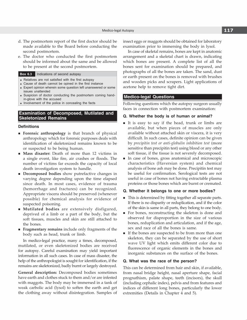
117Medico-legal Autopsy
d. The postmortem report of the first doctor should be made available to the Board before conducting the second postmortem.
e. The doctor who conducted the first postmortem should be informed about the same and be allowed to be present at the second postmortem.
Box 6.3 Indications of second autopsy
� Relatives are not satisfied with the first autopsy � Cause of death cannot be opined in the first instance � Expert opinion wherein some question left unanswered or some issues unattended
� Suspicion of doctor conducting the postmortem coming hand-in-glove with the accused
� Involvement of the police in concealing the facts
Examination of Decomposed, Mutilated and Skeletonized Remains
Definitions
� Forensic anthropology is that branch of physical anthropology which for forensic purposes deals with identification of skeletonized remains known to be or suspected to be being human.
� Mass disaster: Death of more than 12 victims in a single event, like fire, air crashes or floods. The number of victims far exceeds the capacity of local death investigation system to handle.
� Decomposed bodies show putrefactive changes in varying degree depending upon the time elapsed since death. In most cases, evidence of trauma (hemorrhage and fractures) can be recognized. Appropriate viscera should be preserved (whenever possible) for chemical analysis for evidence of suspected poisoning.
� Mutilated bodies are extensively disfigured, deprived of a limb or a part of the body, but the soft tissues, muscles and skin are still attached to the bones.
� Fragmentary remains include only fragments of the body such as head, trunk or limb.
In medico-legal practice, many a times, decomposed, mutilated, or even skeletonized bodies are received for autopsy. Careful examination may yield important information in all such cases. In case of mass disaster, the help of the anthropologist is sought for identification, if the remains are skeletonized, badly burnt or largely destroyed.
General description: Decomposed bodies sometimes have earth and clothes stuck to them and/or are infested with maggots. The body may be immersed in a tank of weak carbolic acid (lysol) to soften the earth and get the clothing away without disintegration. Samples of
insect eggs or maggots should be obtained for laboratory examination prior to immersing the body in lysol.
In case of skeletal remains, bones are kept in anatomic arrangement and a skeletal chart is drawn, indicating which bones are present. A complete list of all the bones sent for examination should be prepared, and photographs of all the bones are taken. The sand, dust or earth present on the bones is removed with brushes and wooden picks and scrapers. Light applications of acetone help to remove tight dirt.
Medico-legal QuestionsFollowing questions which the autopsy surgeon usually faces in connection with postmortem examination:
Q. Whether the body is of human or animal?
� It is easy to say if the head, trunk or limbs are available, but when pieces of muscles are only available without attached skin or viscera, it is very difficult. In such cases, definite opinion can be given by precipitin test or anti-globulin inhibition test (more sensitive than precipitin test) using blood or any other soft tissue, if the tissue is not severely decomposed.
� In case of bones, gross anatomical and microscopic characteristics (Haversian system) and chemical analysis of bone ash may be done. Precipitin test may be useful for confirmation. Serological tests are not useful in case of bones not having extractable plasma proteins or those bones which are burnt or cremated.
Q. Whether it belongs to one or more bodies?
� This is determined by fitting together all separate parts. If there is no disparity or reduplication, and if the color of the skin is same in all parts, they belong to one body.
� For bones, reconstructing the skeleton is done and observed for disproportion in the size of various bones, reduplication and articulation, and if the age, sex and race of all the bones is same.
� If the bones are suspected to be from more than one skeleton, they can be separated by the use of short wave UV light which emits different color due to fluorescence of organic elements in the bones and inorganic substances on the surface of the bones.
Q. What was the race of the person?
This can be determined from hair and skin, if available, from nasal bridge height, nasal aperture shape, facial prognathism, palate shape, teeth (incisors), the skull (including cephalic index), pelvis and from features and indices of different long bones, particularly the lower extremities (Details in Chapter 4 and 5).

118 Review of Forensic Medicine and Toxicology
Q. Whether it is male or female?
� It can be determined if the head or trunk is available, from the presence and distribution or absence of hair, configuration of the pelvis, skull, mandible, diameter of head of femur and humerus, and measurements of femur, tibia, humerus and radius. Recognizable sex differences are present only after puberty (Details in Chapter 4).
� It can also be determined from the recognition of prostate or uterus which can be identified even in advanced state of putrefaction. Microscopic examina-tion may be done for confirmation. Sex can also be determined by nuclear sexing or sexing root sheath cells of human head hair.
Q. What was the age of the individual at the time of death?
� Age can be estimated from general development, color of hair on the scalp, beard, moustache and pubis.
� Closure of the cranial sutures, eruption of teeth, ossification centers of bones, changes in the mandible, symphyseal surface of the pubis, sacrum, and margin of the glenoid cavity of the scapula; calcification of laryngeal and sternal cartilages and hyoid bone are also helpful. After the completion of bony union, exact age cannot be determined.
Q. What was the stature of the individual?
� Stature can be determined from long bones, such as femur, tibia, humerus or radius and using the formulae of Pearson, Dupertuis and Hadden; Trotter and Gleser for Americans; Breitinger for Germans or multiplication factors devised by Indian researchers.
� The principle of these formulae is to measure the length of long bone and multiply it with a given factor and then adding a fixed factor.
� The length of the humerus multiplied by five is a quick method of estimation of height.
Karl Pearson’s formula for stature from dried long bones (in cms)
S.No. Male Female
1. 81.306 + 1.880 × F 72.884 + 1.945 × F
2. 78.664 + 2.376 × T 74.774 + 2.352 × T
3. 70.641+ 2.894 × H 71.475 + 2.754 × H
4. 89.925 + 2.271 × R 81.224 + 3.343 × R
F: Length of femur, T: Length of tibia, H: Length of humerus, R: Length of radius
Q. What was the identity of the individual? � It can be determined from fingerprints, tattoo marks,
scars, moles, hair, teeth, flat feet, supernumerary ribs, congenital defects, deformities, articles of clothing and superimposition technique (if skull is available).
� An X-ray of any bone, if taken during life, may be compared with an X-ray of the same bone and it may help in identification. Malunited fractures, healed fractures or deformities of bone, if present, are helpful.
� Determination of blood group antigens from teeth pulp might also help in establishing identity, if the blood group is known.
� Other methods include X-ray comparison of trabecular patterns and neutron activation analysis to distinguish the relative mineral contents.
Q. What was the manner of separation of parts?It can be found out by examining the margins of the parts and the ends of the long bones, and to look for whether they had been cleanly cut, sawn, hacked, lacerated, disarticulated at the joints or gnawed through by animals.
Q. What was the mode and place of disposal? � The place of occurrence and disposal of the parts
can be found out from trace materials attached with the parts from the place of disposal.
� A body buried in deep grave skeletonizes compara-tively later. A body disposed off in open air dries up early. Bones of the bodies disposed in forest may be partly eaten by animals.
Q. Whether the injuries are antemortem or post-mortem in nature?
Evidence of vital reaction is looked for at margins of the injured parts.
Q. What was the cause of death? � The cause of death can be made out if there is
evidence of fatal injury to some vital organ or large blood vessel, or marks of burning or deep cuts or fractures of bones, especially the skull, cervical vertebrae, hyoid bone or ribs. Foreign body, such as a bullet, when present is helpful.
� Bones or their charred remains may be subjected to chemical analysis for the detection of metallic poisons, such as arsenic, as these are not destroyed by heat.
Q. What type of weapon was involved?In case of presence of antemortem injury, like fracture, or depending on the nature of injury of the bones, the

119Medico-legal Autopsy
weapon used to inflict the same and the type of weapon used to dismember the part, e.g. whether a hard blunt weapon, a light or heavy sharp cutting weapon, a pointed weapon or a firearm can be determined.
Q. What was the time of death?
The probable time since death may be determined from the condition of parts and decomposition changes. The appearance of bones, unless they are very recent, is much more dependent upon the environment in which they have lain, than the passage of time. Bones left in a dry environment, such as sand, will last far longer than bones in a damp, acidic situation.
� If soft tissues, like fascia and ligaments are still attached with the bone, then death might have occurred within about 2 weeks to 2 months back.
� If no soft tissue is attached, but the bone is still not completely dry then, death might have occurred about 1–3 months back.
� If the bone is completely dry, but has a putrid smell, death has occurred within the last 3 months.
� If the bone is dry with no putrid smell, but has retained its normal color, then the time passed after death is between 3 months to 1 year.
� After 30–40 years, the bone tends to become lighter as the organic matrix is lost and the softer parts of the bone begin to crumble.
Dating of skeletal remains � Total nitrogen content is > 4–5 g% in bones less than 50 years
old. Between 50–100 years, it is about 3.5 g%, and 2.5 g% when the bones are 350 years old.
� The number of amino-acids (initially about 15, glycine and analine are predominant) diminishes with age and hydroxyproline and proline tend to disappear after 50 years. A bone > 100 years old will contain 7 amino acids.24
� Blood pigment tests using bone dust remain positive for upto 100 years.
� Eluted bone dust solution tested for immunological activity against a human anti-Coombs serum, test positive for 5–10 years.
� UV fluorescence: The sawn shaft of a long bone such as a femur is examined under an UV lamp; fresh bone will fluoresce across the whole surface from periosteum to marrow cavity. As time lengthens, the fluorescent zone narrows, breaks up and finally vanishes between 150–300 years.
� Estimation of radioactive carbon (C-14): Radioactive carbon gets deposited in living tissue. After death, there is no further deposition and its concentration gradually decreases in the organic substances. Estimation of C-14 in bones may give some idea as to when the person died. There is no significant fall of C-14 during the first century after death.
Exhumation
Definition: It is the lawful digging out of an already buried body from the grave for postmortem examination.
� It is a situation where a previously-buried dead body is ‘dug up,’ ‘unearthed,’ or ‘disinterred’.
� Usually, it, involves a body (of any age group) that was not originally autopsied but which, for some reason, must be exhumed in order for an autopsy to be performed.25
� Exhumation per se does not include a second autopsy. The term ‘exhumation’ is applied to the removal of a body buried in a legitimate fashion and not a clandestinely buried victim of suspicious death.
� It is infrequently done in India because the bodies are disposed of by burning to ashes by most of the communities except few.
Reasons
i. Criminal cases z Establishing the cause and manner of death in
suspected homicide disguised as suicide. z Death as a result of criminal abortion and
criminal negligence. z Retrieving some vital object which may throw
light on the case, e.g. bullet from the dead body, if the person was killed by a firearm.
ii. Civil cases: Identification of the deceased for accidental death claim, insurance, workmen’s compensation claim, liability for professional negligence, survivorship and inheritance claims, disputed identity, separation overseas, and burial of the wrong body inadvertently or by fraud.
Authorization: The body is exhumed only when, there is a written order from the First Class Magistrate/District Magistrate/Sub-Divisional Magistrate/Executive Magistrate; police cannot order exhumation.26
Procedure i. It should be done and completed in broad daylight,
for which it should be started during the morning hours of the day.27
ii. The body is exhumed under the supervision of a medical officer and Magistrate, in the presence of a police officer.
iii. Before opening the grave, it should be positively identified from location of burial plot, headstone

120 Review of Forensic Medicine and Toxicology
and gravemarker, so that wrong body is not disinterred.
iv. Soil from above, below and two sides of the body or the coffin should be preserved in separate glass jars, with identification tags.
v. Disinfectants/pesticides should not be sprinkled on the body as it might interfere later with the determination of poison in the body.
vi. The doctor should note the position and appearance of the body inside the grave or the coffin. A drawing of the grave and body or skeleton should be made, noting all the details, whether the face is up, or to the side, arms are extended, or the lower limbs are flexed.
vii. The grave or the coffin with the body should be photographed.
viii. If decomposition is not advanced, a plank or a plastic sheet should then be lowered to the level of the earth on which the body rests.
ix. After this, the body is lifted and sent for postmortem examination, along with a requisition and a preliminary investigation report which contains the brief history of the case. In the mortuary, post mortem examination on the body is performed as in all other cases.
x. In highly putrefied bodies, an attempt should be made to establish the identity. Viscera should be preserved for chemical analysis. If the body is reduced to skeleton, the bones should be examined.
Time limitIn India, there is no time limit for ordering of the exhumation, but many Western countries have well-defined time limit upto which exhumation can be done.28 For example, in France, the time limit is 10 years and in Germany, the time limit is 30 years. Thus in France, after 10 years of death, if some facts are found which may reveal foul play, even then the body cannot be exhumed.
� In Europe, exhumation services are carried out not only for forensic purposes but also for changing of graves, repartition to a different country and changing the type of disposal from burial to cremation.
� In the US, laws governing exhumation is vague and varies from State to State.
� In Jewish and Islamic law, exhumation is forbidden except in certain circumstances.
� In Hong Kong, burial in government cemeteries are disinterred after 6 years under exhumation order. The remains are either collected privately or by the government for cremation and are reburied in an urn or niche.
MULTIPLE CHOICE QUESTIONS
1. Body after postmortem is handed over to: NIMHANS 14 A. Police constable B. Relatives C. Preserved in mortuary D. Magistrate 2. Last structure to be autopsied in asphyxial death:
AIIMS 07, 08, 12 A. Head B. Thorax C. Abdomen D. Neck 3. Method of autopsy in which various systems organs
are removed en masse: NEET 14 A. Rokitansky B. Virchow C. Ghon D. Lettulle 4. Virchow method of organ removal is: NEET 14 A. Organs removed en masse B. Organs removed one by one C. In situ dissection D. Organs removed en bloc
5. True about subendocardial hemorrhages are all, except: AIIMS 10; UPSC 11
A. May be seen after head injury B. Involves the right ventricular wall C. Continuous pattern D. Flame shaped hemorrhages 6. In autopsy, spinal cord is opened through which
approach: AIIMS 11 A. Anterior B. Posterior C. Lateral D. Anterolateral 7. Underwater autopsy of the heart is done in cases of:
DNB 09 A. Myocardial infarction B. Pulmonary embolism C. Air embolism D. Pneumothorax 8. Best site for blood collection for toxicology sampling: NEET 13 A. Abdominal aorta B. Femoral vein C. Carotid artery D. Heart
1. A 2. D 3. D 4. B 5. B 6. B 7. C 8. B
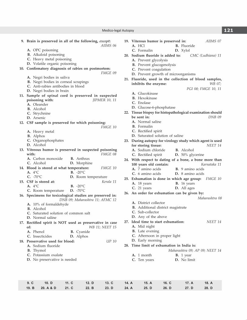
121Medico-legal Autopsy
9. Brain is preserved in all of the following, except: AIIMS 06
A. OPC poisoning B. Alkaloid poisoning C. Heavy metal poisoning D. Volatile organic poisoning 10. Confirmatory diagnosis of rabies on postmortem:
FMGE 09 A. Negri bodies in saliva B. Negri bodies in corneal scrapings C. Anti-rabies antibodies in blood D. Negri bodies in brain 11. Sample of spinal cord is preserved in suspected
poisoning with: JIPMER 10, 11 A. Oleander B. Alcohol C. Strychnine D. Arsenic 12. CSF sample is preserved for which poisoning:
FMGE 10 A. Heavy metal B. Alphos C. Organophosphates D. Alcohol 13. Vitreous humor is preserved in suspected poisoning
with: FMGE 08 A. Carbon monoxide B. Anthrax C. Alcohol D. Morphine 14. Blood is stored at what temperature: FMGE 10 A. 4°C B. -20°C C. -70°C D. Room temperature 15. CSF is stored at: Kerala 11 A. 4°C B. -20°C C. Room temperature D. -70°C 16. Specimens for toxicological studies are preserved in:
DNB 09; Maharashtra 11; AFMC 12 A. 10% of formaldehyde B. Alcohol C. Saturated solution of common salt D. Normal saline 17. Rectified spirit is NOT used as preservative in case
of: WB 11; NEET 15 A. Phenol B. Cyanide C. Insecticides D. Alphos 18. Preservative used for blood: UP 10 A. Sodium fluoride B. Thymol C. Potassium oxalate D. No preservative is needed
19. Viterous humor is preserved in: AIIMS 07 A. HCl B. Fluoride C. Formalin D. Xylol 20. Sodium fluoride is added to: CMC (Ludhiana) 11 A. Prevent glycolysis B. Prevent glucogenolysis C. Prevent coagulation D. Prevent growth of microorganisms 21. Fluoride, used in the collection of blood samples,
inhibits the enzyme: WB 07; PGI 08; FMGE 10, 11 A. Glucokinase B. Hexokinase C. Enolase D. Glucose-6-phosphatase 22. Tissue biopsy for histopathological examination should
be sent in: DNB 09 A. Normal saline B. Formalin C. Rectified spirit D. Saturated solution of saline 23. During autopsy for virology study which agent is used
for storing tissue: NEET 14 A. Sodium chloride B. Alcohol C. Rectified spirit D. 50% glycerine 24. With respect to dating of a bone, a bone more than
100 years old contain: Karnataka 11 A. 7 amino acids B. 9 amino acids C. 6 amino acids D. 8 amino acids 25. Exhumation is done in which age group: FMGE 10 A. 18 years B. 16 years C. 21 years D. All ages 26. An order for exhumation can be given by: Maharashtra 08 A. District collector B. Additional district magistrate C. Sub-collector D. Any of the above 27. Ideal time to start exhumation: NEET 14 A. Mid night B. Late evening C. Afternoon in proper light D. Early morning 28. Time limit of exhumation in India is: Maharashtra 09; AP 09; NEET 14 A. 1 month B. 1 year C. Ten years D. No limit
9. C 10. D 11. C 12. D 13. C 14. A 15. A 16. C 17. A 18. A 19. B 20. A & D 21. C 22. B 23. D 24. A 25. D 26. D 27. D 28. D
It has long been recognized that the autopsy room is a potential source of infection and that the forensic pathologists and other persons (technical assistants, potters, sweepers) in close proximity to an autopsy are at higher risk of contracting infectious diseases from the dead bodies.
� ‘Risk’ is the chance of acquiring a blood borne infection such as hepatitis B virus (HBV) or human immunodeficiency virus (HIV) from a sharp injury, whereas ‘hazard’ is the danger of injury posed by the blade of a knife or saw or the point of a needle.
� ‘High risk autopsy’ is the postmortem examination of a deceased person who has had, or is likely to have had, a serious infectious disease that can be transmitted to those present at the autopsy, thereby causing them serious illness and/or premature death.
Classification of Pathogens
Infectious agents have been categorized into four hazard groups (HG) depending on their virulence, transmissibility, ability to cause epidemics, preventability (e.g. by vaccine) and their treatability.
� Hazard Group 1: Biological agent that is unlikely to cause human disease.
� Hazard Group 2: Biological agent that can cause disease and a hazard to employees but is unlikely to spread to the community, and there is usually effective prophylaxis or treatment available. For example, antibiotic-resistant organisms like methicillin-resistant Staphylococcus aureus (MRSA), vancomycin-resistant Enterococcus faecium (VREF), food poisoning, Salmonella spp. and other enteric pathogens.
� Hazard Group 3: Biological agents that can cause severe disease; a serious hazard to employees and it may spread to the community, but there is usually effective prophylaxis or treatment available. The most frequent are Mycobacterium tuberculosis, HIV, HBV, hepatitis C virus (HCV) and Creutzfeldt-Jakob (CJD) prion. All these agents retain their infectivity after death.
� Hazard Group 4: Biological agents that cause severe disease, serious hazard to employees and it is likely to spread to the community and there is no effective prophylaxis or treatment available. This group includes virus causing hemorrhagic fevers—Ebola, Lassa, Marburg, and Congo-Crimea.
In the mortuary, the significant groups are HG #3 and #4, since most of the time the dead bodies coming for autopsy have no past medical records, or if at all present, contain inadequate information. Risk from these bodies is unknown and hidden. Moreover, some of these deaths may have occurred in the hospital and there is a possibility of hospital, acquired infections, which are more virulent and resistant to treatment.
Knowledge of the risks of infection is therefore essential. Accidental exposures to high risk pathogens are uncommon but not infrequent, and many could be prevented.
Infections in autopsy room may be acquired by any one of the following routes:
� A wound resulting from an object (e.g. scalpel) contaminated with blood or body fluids or needle-stick injury (percutaneous inoculation).
� Splash of infected blood or other body fluids onto an open wound or area of dermatitis.
� Contact of blood or other body fluids with mucous membranes of the eyes, nose or mouth.
� Inhalation and ingestion of aerosolized particles.
Penetrating injuries are the most common route of transmission for pathogens at autopsy.
Commonly Acquired Infections
� Hepatitis B is the most transmissible of the blood-borne viruses, but its transmission is preventable by vaccination. Increased risk of HBV infection has been found among health care workers, especially those having frequent contact with blood and/or exposure to needles or sharp instruments. Among the physicians, forensic pathologists have been
Autopsy Room Hazards
CHApteR 7
123Autopsy Room Hazards
recognized as a high-risk group for occupationally acquired HBV infection, because of their greater exposure to blood.
� Persons associated with postmortem examination and other health care workers experiencing needle stick injuries are at a considerable risk of acquiring hepatitis C infection (HCV).
� Autopsy is an efficient method of transmitting tuberculosis from the dead body to those present in the autopsy room. The risk for infection does not vary with the distance from the autopsy table. In our country, where tuberculosis is still the most fatal respiratory disease affecting the lower socioeconomic group and where unidentified vagabonds constitute a significant percentage of the autopsy population, the percentage of unrecognized tuberculosis cases is substantial. Airborne droplets, usually from sputum positive cases transmit tuberculosis. It is thought that the patient with tuberculosis may be more infectious at autopsy than during life. Embalming itself has been shown to produce active tuberculosis aerosols.
� The risk of HIV infection among medical and laboratory personnel, including mortuary workers, is considered low when compared with other blood-borne viruses, such as HBV and HCV, but resembles the rates for single contact heterosexual transmission. Studies have shown that viable HIV can be isolated from cranial bone, brain, cerebrospinal fluid, lymph node, spleen and blood upto 5 days after death, when stored at 6°C. Deep injury, visible blood on the device causing the injury, injury with a needle used in a vessel and injury with hollow-bore needle (compared to a solid needle)—all increases the likelihood of a larger innoculum of blood entering the recipient. HIV infection should be suspected, if the body is of:
i. Male homosexual ii. Intravenous drug abuser iii. Hemophiliac who has received repeated blood
transfusions iv. Female prostitute v. Victim of sexual abuse.
Risk of transmission from single percutaneous exposure to blood for:
� HBV: 6–30% � HIV: 0.1–0.36%1
� HCV: 2.7–10%
Transmissible spongiform encephalopathies (Tse)The risk of acquiring the prions* responsible for TSE (CJD) is considerably less. Prions are characterized
by extreme resistance to conventional inactivation procedures.
Miscellaneous infectionsIn addition, the dead body is a potential source of infection with other organisms, notably Streptococcus pyogenes, gastrointestinal organisms (including hepatitis A) and Neisseria meningitidis.
Autopsy of Hazard Group 3 Patients
One school maintains that all autopsies should be carried out with total precautions against infective risk. However, this is almost impracticable to achieve in the present set-up. The other school advocates pre-autopsy testing of blood-sample for HIV and other infective agents.
A simple and rapid test [10-minute (min) test carried out by a manual HIV test-kit] is available for mortuary use in the developed countries. This test is also applicable to urine—a more cost-effective specimen and safer than blood collection.
Pre-preparation: The body should be transported to the mortuary by duly plugging all the natural orifices and sites of the IV drip. It should be wrapped and tied in double layer, tough plastic bag, with a red color tag mentioning ‘Biologically Hazardous’. The label should mention the name, age, sex and registration number.
Universal Work Precautions � No unauthorized person should be admitted in the
autopsy room, so as to minimize exposures. Only experts and workers who are trained in handling the infected material should be allowed.
� Immunization: All staff should be vaccinated against tetanus, poliomyelitis, tuberculosis and hepatitis B.
� Immunosuppressed or immunodeficient individuals and individuals who have uncovered wounds, oozing skin lesions or dermatitis should not perform the autopsy.
Clothing: Autopsy personnel should wear protective clothing—full sleeves overalls, head cap that completely covers the hair, N95 particulate masks, goggles (if eye glasses are not worn), double gloves (heavy autopsy gloves over surgical gloves) and waterproof rubber gumboots of knee length with shoe covers. A plastic visor will protect the eyes and mucosal surfaces from splash injury.
* Prions are proteinaceous infective agents causing neurodegenerative diseases.

124 Review of Forensic Medicine and Toxicology
� Double gloving and frequent changing of gloves (whether or not they appear damaged) is recommended—in double gloving, the outer glove protects and reduces the frequency of perforations of the inner glove.
Handling sharp instruments: Minimum instruments as needed should be kept. Scissors with slightly blunt ends should be used, and sharp ones are used only if needed.
Wherever possible, the use of needles should be avoided. Needlestick injuries are entirely preventable, blunt needles and bulb syringes should be used to aspirate fluids.
Needlestick accidents occur during disposal of needles, they should never be recapped after use.
Accidental cuts, particularly on the palmer surface of the thumb, index and middle finger of non-dominant hand are most frequently seen in forensic pathologists.
Examination of organs: It is better to eviscerate organ by organ in situ in the cadaver rather than eviscerating en masse. Another method is to fix lungs and other organs as a whole after removal, rather than slicing them before fixation.
� To minimize aerosol splatter, cranium may be opened with an electrical oscillating saw attached to a vacuum dust exhaust and filter or with a handsaw under a transparent anti-splash cover.
� The ribs should be cut with the rib cutter thereby decreasing the bone dust aerosols.
� Handling and/or washing of organs should be done carefully so as to prevent splashing and aerosol formation.
� At the completion of the gross dissection, all tissues/organs are double bagged and placed in the body cavity.
� The most common method of exposure includes being pricked with a used needle or other contaminated material. To prevent this, body-sewing needles may be avoided and the mortician’s stitches may be replaced by suture-free closure using adhesive tapes, staples or even left unreconstructed, and sealed in a leak proof body bag.
� A tag should be attached for identification.
Handling specimens for laboratory examination: They should be properly labeled and fixed with 10% formalin solution, and should be handled with gloved hands.
Disposal of used instruments: They should be dipped in 2% glutaraldehyde (Cidex) for 30 min, washed with
soap and water, dried and then rinsed in methylated spirit and air dried or autoclaved.
� All soiled gauze and cotton should be collected in a double plastic bag for incineration.
� Disposable needles and syringes, scalpel blades and other sharp items should be placed in a puncture-resistant containers.
� Laundry material, e.g. aprons and towels should be soaked in 1% bleach for half hour, washed with detergent and hot water, and autoclaved.
Clean-up procedure: Small spatters and spills of blood and other body fluids should be wiped up with disposable tissues or towels which are discarded in special biohazard bags, and properly disposed. The autopsy table and floor should be cleaned with 1% bleach solution, followed by washing with soap and water.
The health care workers should then wash thoroughly with soap and water before dressing.
Disinfectants: 1:10 dilution of common household bleach or a freshly prepared sodium hypochlorite solution is recommended. HIV is inactivated by a wide range of disinfectants, including iodophor compounds (such as Betadine), 50% ethanol, 3% hydrogen peroxide, phenolic compounds (such as Lysol), 4% buffered formaldehyde solution and 0.5% sodium hypochlorite (household bleach).
Universal precautions apply to blood, semen and vaginal secretions, as well as to CSF, synovial, pleural, pericardial, peritoneal and amniotic fluid, but they do not apply to feces, nasal secretions, sputum, sweat, urine and vomitus, unless they contain visible blood.
In case of accidental injuries or cuts with instruments, contaminated or not with blood or body fluids, while working on a body, the wound should be immediately washed thoroughly under running water, bleeding encouraged and the wound disinfected.
It must be reported to the authorities and immediate, proper measures instituted. Blood sample should be taken from the source of exposure and tested for HIV and HBV. The individual should be advised that he/she could possibly have been infected by the needle-prick/cut and counseled appropriately. HIV-testing should be done there, and then after an interval of 3 and 6 months to trace the possible introduction of the virus to the related event, so as to evaluate the ensuing claims, if need be.

125Autopsy Room Hazards
1. A
Autopsy of suspected Tse PatientAutopsy of a patient with suspected TSE including Creutzfeldt-Jakob disease requires special precautions. Prions are resistant to 10% formalin, 70% alcohol, phenolic compounds, boiling, and UV radiation. It is inactivated by 5% sodium hypochlorite, 2N sodium hydroxide, 90% formic acid and autoclaving at 134°C for 20 min or longer.
� During the autopsy, all tissues and fluids, including running water, should be confined to the autopsy tables.
� Disposable instruments should be used wherever possible.
� The brain is removed while the head is in a plastic bag to reduce aerosolization and splatter.
� After the autopsy, any liquid on the autopsy tables should be disinfected with an equal volume of 5% sodium hypochlorite or 2N NaOH.
� All instruments are placed in a stainless steel dish and soaked for 1 hour (h) in 2N NaOH and then rinsed well in water before autoclaving at 134°C for at least 30 min.
� All gowns, gloves, plastic aprons and other disposable supplies should be incinerated.
� Any suspected areas of contamination of the room are decontaminated by repeated wetting over 1 h with 2N NaOH.
Autopsy of Hazard Group 4 PatientsAutopsies on patients with HG #4 pathogens (for e.g. the viral hemorrhagic fevers) pose even greater risk. It should only be performed where absolutely necessary.
In the UK, autopsy of such cases is prohibited unless performed in a designated mortuary.
Autopsy and Disposal of Radioactive Corpse
� If the amount of radioactivity is < 5 millicuries, no precautions are necessary.
� If the body contains between 5–30 millicuries of radioactive material, the autopsy surgeon should wear heavy rubber gloves, plastic aprons, shoe covers and spectacles to reduce radiation. Instruments with long handles should be used during the autopsy. Organs that are most radioactive should be removed first and placed in covered glass jars, labeled and examined for radioactivity from time to time. Fluid of the pleural and peritoneal cavity should be flushed copiously with running water and drained off directly into the sewer. Contaminated clothing should be thoroughly cleaned with soap and water, for suitable decay of the radioactive material before being sent to laundry. Instruments can be brought to a safe limit by soaking them in water with soap. Contamination of the floor of the autopsy room should be avoided.
Organs may be removed and detailed dissection is done away from the body, or placed in a glass jar and preserved in a fixative or kept in cold storage for later examination when radioactivity has fallen to a safer level.
� If the body contains more than 300 millicuries activity after autopsy, it should be embalmed in the hospital mortuary. The presence of a cardiac pacemaker must be recorded, especially if it is one which might contain a radioactive substance.
MUlTiPle choice qUesTion
1. Transmission rate of HIV by needlestick injury in health professionals is:
CMC (Vellore) 08; TN 09; FMGE 13 A. 0.3% B. 1% C. 5% D. 10%

Definition: Thanatology (Greek thanatos: death) is the scientific study of death in all its aspects including its cause and phenomena.1 It also includes bodily changes that accompany death (postmortem changes) and their medico-legal significance.
Death occurs in two stages (Diff. 8.1): i. Somatic, systemic or clinical. ii. Molecular or cellular.
Somatic death: The question of death is important in resuscitation and organ transplantation. Skin and bone remains metabolically active for many hours and these cells can be successfully cultured days after somatic death.
During early 20th century, irreversible cessation of circulatory and respiratory functions was sufficient basis for diagnosing death.
According to Bichat, life could be compared to a tripod with its three legs representing the three vital systems—the nervous, circulatory and respiratory system—tripod of life. It is thought that all systems would fail if any one of the vital systems fails, and that is why these systems are known as ‘atria mortis’ (death’s portal of entry or gateways of death).2
Molecular death � Molecular death occurs piecemeal. Initial changes
occur due to metabolic dysfunction and later from structural disintegration.
� Nervous tissues die rapidly, the vital centers of the brain in about 3–7 minutes (min), but muscles survive upto 1–2 hours (h).
Supravital reactions � Mechanical excitability of the skeletal muscle
i. Tendon reaction (Zsako’s phenomenon): Contraction of the whole muscle (e.g. quadriceps) due to propagated excitation following a mechanical stimulation, seen within 2–3 h after death.
ii. Localized idiomuscular contraction at the point of stimulation may be seen several hours after cessation of Zsako’s phenomenon.
� Electrical excitability of the skeletal muscles of the face may be observed for few hours after death.
� Pharmacological excitability of the iris muscle resulting in change of pupil diameter following the administration of miotic or mydriatic solutions can be seen during the first hours of the postmortem period.
Brain/Brainstem Death
As ventilator technology advanced, circulation and respiration could be maintained by means of a mechanical respirator, despite loss of all brain functions, and thus have brought the concept of brain death, i.e. irreversible loss of cerebral functioning.
� Brain death is the complete and irreversible cessation of functioning of the brain. Brain includes all the central nervous system (CNS) structures, except the spinal cord.
Differentiation 8.1: Somatic death and molecular death3,4
S.No. Feature Somatic death Molecular death
1. Definition Complete and irreversible cessation of function of brain, and stoppage of the circulation and respiration
Progressive disintegration of body tissues with death of individual tissues and cells
2. Onset Precedes molecular death Succeeds somatic death (1–2 hours after stoppage of vital functions)
3. Tissues and cells of body Alive and functioning Dead and non-functioning with no metabolic activity
4. Response to external stimuli Muscle responds to thermal, electrical or chemical stimulus
Does not respond
5. Confirmation Flat ECG and EEG, and absent breath sounds Rigor mortis, algor mortis, postmortem staining, putrefaction
6. Resemblance Suspended animation, coma, hypothermia Does not resemble any condition
Thanatology
ChapTer 8
127Thanatology
� Brain death is now accepted as brainstem death. The respiratory center which controls respiration lies within the brainstem. If this area is dead, the person is unable to breath spontaneously or regain consciousness.
� As the integrity of the reticular formation within the brainstem is essential for the proper functioning of the cortex, brainstem death can practically be considered to be sufficient for brain death. The crucial point in determining brain death is the demonstration of absence of all brainstem functions. Many countries, including India, now legally consider brainstem death as ‘brain death’.
It may be argued by the defense when life-support machines are disconnected in brainstem death that the assailant did not ‘kill’ the victim, since ‘death’ was caused by the action of the doctors switching off the machine. In such a situation, it should be understood that death was diagnosed by the doctors before discontinuing artificial ventilation and the criminal act that initiated the chain of events was not remote enough to avoid culpability for the death.
Mechanism of Brain Death
Brain injury has a number of causes, such as traumatic or cerebrovascular injury and generalized hypoxia, all of which produce brain edema.
Edema is accompanied by an increase in intracranial pressure leading to gradual decrease in cerebral circulation to the level of almost cessation, causing aseptic necrosis of the brain. Within 3–5 days, there
occurs widespread brain destruction or pannecrosis throughout the cerebrum and the brainstem, the brain becomes a liquefied mass, a condition known as ‘respirator brain’. Increase in the intracranial pressure compresses the entire brain including the brainstem and total brain infarction follows.
Diagnosing Brain Death (Box 8.1)The two essential requirements for the diagnosis of brain death are:1. Establishment of cessation of all brain functions, i.e. cerebral
and mainly brainstem functions using primarily the clinical criteria and partly by confirmatory paraclinical/laboratory tests which includes electro encephalogram (flat isoelectric EEG) and somatosensory evoked potentials (SSEP) and tests to measure cerebral blood flow.5
2. Demonstration that cessation of these functions is irreversible: Irreversibility is established by:
z Determination of the cause of loss of brain function z Exclusion of reversible conditions z Demonstration that the cessation of brain functions
persists for an appropriate period of observation.
Exclusion of Reversible ConditionsThe most important reversible conditions/confounding factors that must be excluded are: i. Hypothermia. ii. Severe electrolyte, acid-base or endocrine abnor-
malities.
Box 8.1 Diagnostic clinical brain death criteria5-7
A. Prerequisites. Brain death is the absence of clinical brain function when the proximate cause is known and demonstrably irreversible. i. Clinical or neuroimaging evidence of an acute CNS catastrophe that is compatible with the clinical diagnosis of brain death. ii. Exclusion of complicating medical conditions that may confound clinical assessment. iii. No drug intoxication or poisoning. iv. Core temperature > 32°C (90°F).
B. The three cardinal findings in brain death are coma, absence of brainstem reflexes and apnea.1. Coma or unresponsiveness—No cerebral motor response to pain in all extremities.2. Absence of brainstem reflexes a. Pupils i. Absent pupillary response to bright light. ii. Size: Mid position (4 mm) to dilated (9 mm). b. Ocular movement i. No oculocephalic reflex (Doll’s eye phenomenon). ii. Absent oculovestibular reflex (Caloric test): No deviation of eyes to irrigation in each ear with 50 ml of cold water. c. Facial sensation and facial motor response i. No corneal reflex to touch with a cotton swab. ii. No jaw reflex. iii. No grimacing to deep pressure on nail bed, supraorbital ridge, or temporomandibular joint. d. Pharyngeal and tracheal reflexes i. No gag reflex: No response after stimulation of the posterior pharynx with tongue blade. ii. No cough response to bronchial suction.3. Apnea test: It is based on the fact that loss of brainstem function definitively results in loss of centrally controlled breathing, with
resultant apnea.
128 Review of Forensic Medicine and Toxicology
� Isograft: Organs or tissues are transplanted from a donor to a genetically identical recipient (such as an identical twin).
� Xenograft: Organs or tissue transplanted from one species to another, e.g. grafting of animal tissue into humans.9
� Split transplants: Deceased-donor organ (specifically the liver) may be divided between two recipients, especially an adult and a child.
Cause, Mechanism and Manner of Death
Two of the most important functions of the forensic doctor are the determination of the cause and manner of death.
� Cause of death is any injury or disease producing physiological derangement, briefly or over a prolonged period and which results in the death of the individual, e.g. a gunshot wound to the abdomen, a stab wound to the chest, adenocarcinoma of the lung or coronary atherosclerosis.
� Mechanism of death is the physiological derange-ment produced by the cause of death that results in death, e.g. hemorrhage, septicemia, metabolic acidosis or alkalosis, ventricular fibrillation or respiratory paralysis. A particular mechanism of death can be produced by multiple causes of death and vice versa. Thus, if an individual dies of hemorrhage, it can be produced by a gunshot wound or a stab wound or a malignant tumor of the lung eroding into a blood vessel. A cause of death, e.g. a gunshot wound of the abdomen can result in many possible mechanisms of death, like hemorrhage or peritonitis.
� Manner of death explains how the cause of death came about. Manner of death can generally be categorized as natural (death due to disease), homicide, suicide, accident or undetermined (Flow chart 8.1 and Table 8.1).
z A cause of death may have multiple manners of death. An individual can die of massive hemorrhage (mechanism of death) due to stab wound of heart (cause of death), with the manner being homicide (someone stabbed him), suicide (stabbed himself), accident (fell over the weapon) or undetermined (not sure what happened).
z For some deaths, the manner may be undetermined because the circumstances are unclear; for e.g. whether drowning was accidental or suicidal.
z Deaths from alcohol and drug abuse are difficult to classify and are sometimes described as ‘unclassified’.
� Agonal period is the time between a lethal occurrence and death.10
iii. Drug intoxication: Presence of sedation, neuro-muscular blockade, or drugs causing CNS depression.
iv. Hypoxia, hypotension or shock. v. Other conditions: Brainstem encephalitis, severe
hypophosphatemia, encephalopathies associated with hepatic failure, uremia, or hyperosmolar coma of diabetes mellitus.
Observation period: The length of the observation period is still a matter of great controversy. Neurological examination must not be done within 30 min of cardiopulmonary resuscitation.
Brain Death Certification
The common consensus in this regard is as follows: i. Two medical practitioners must perform the
brainstem death tests. ii. The patient’s attending physician should partici pate
in the determination of death wherever possible. iii. The doctors involved should be experts in the
technique of brain death assessment. In any case, such tests should not be performed by transplant surgeons or any doctor in the transplant team.
iv. Each doctor should perform the tests twice.
Beating-heart donor or living cadavers: After brainstem death has been established, the retention of the patient on the ventilator facilitates a fully oxygenated cadaver transplant, the so-called beating-heart donor or living cadavers.
� The success of a homograft depends mainly upon the type of tissue involved and the rapidity of its removal after circulation has stopped in the donor.
� The best results are obtained if the organs are salvaged while circulation is present or immediately after cessation of the circulation.
� Cornea can be removed from the dead body within 6 h (opacity occurs within 2 h of death, but the changes are reversible), skin in 24 h, bone in 48 h and blood vessels within 72 h for transplantation. Kidneys within 45 min, heart within 1 h, lungs and liver within 15 min.8
Types of transplants
� Autograft: Tissue transplanted from one part of the body to another in the same individual. It is also called auto transplant or homologous transplantation.
� Allograft: Organ or tissue transplanted from one individual to another of the same species with a different genotype. It is also called allogeneic graft or homograft.
.

129Thanatology
Cause of Death
The International format of certifying the cause of death is defined by the WHO. The system divides the cause of death into two parts: i. Part I describes the condition(s) that led directly
to death (immediate cause). It is divided further into subsections and generally three—(a), (b) and (c). These are for disease processes that have led directly to death and that are causally related to one another, (a) being due to or consequent on (b), which in turn is due to or consequent on (c) (antecedent causes).11
ii. Part II is for other conditions, not related to those listed in Part I, that have also contributed to death (contributory cause), but should not be used as
a basket for all the minor pathologies found at autopsy.11
� It is important to realize that it is the disease lowest in the Part I list that is the most important, as it is the primary condition, the start of the events leading to death (Table 8.2).
� It is not necessary to complete parts Ib, Ic or II, if there are no predisposing conditions.
� If a patient died suddenly due to intracerebral hemorrhage due to hypertension, the cause of death will be:
i. Ia: Intracerebral hemorrhage. ii. II: Hypertension.
� And, if the same patient survived for few days or weeks and developed pneumonia, the death certificate should record both processes:
i. Ia: Bronchopneumonia. ii. Ib: Intracerebral hemorrhage. iii. II: Hypertension.
� Statistically, both certificates would record the primary cause as intracerebral hemorrhage.
Modes of Death (Proximate Causes of Death)
Definition: Mode of death refers to an abnormal physiological state that pertained at the time of death, e.g. coma, congestive cardiac failure, cardio-respiratory failure, cardiac arrest and pulmonary edema.
According to Xavier Bichat, a French physician, there are three modes of death depending upon the system most obviously affected, irrespective of what the remote cause of death may be: i. Coma. ii. Syncope. iii. Asphyxia.
Coma
Definition: It is a state of profound unconsciousness from which a person cannot be roused, with minimal or no detectable responsiveness to stimuli. This is death from failure of the function of the brain.
Flow chart 8.1: Manner of death
Table 8.1: Description of manners of death
Manner Definition
Natural Death resulting from disease
Homicide Death resulting from the deliberate action of another
Suicide Death intentionally self-inflicted
Accident Death as a result of an environmental influence
Table 8.2: Cause, mechanism and manner of death
Cause of death Mechanism of death Manner of death
� Hemoperitoneum, as a consequence of – Laceration of the aorta, as a consequence of – Blunt thoracic trauma
Hemorrhagic shock Accident
� Bronchopneumonia, as a consequence of – Stab wound of thorax
Septicemia Homicide
� Cardiac tamponade, as a consequence of – Gunshot of thorax
Cardiac dysrhythmia Homicide
Dyadic death (murder-suicide, homicide-suicide) refers to an incident where a homicide is committed followed by the perpetrator’s suicide almost immediately or soon after the homicide. Homicide-suicides are relatively uncommon and vary from region to region.
130 Review of Forensic Medicine and Toxicology
Causes: It is the mode of dying seen in: � Injury or disease of the brain. � Systemic disorders, such as diabetic ketoacidosis,
uremia, heat stroke or eclampsia. � Intoxication with alcohol, opium, cocaine, chloral
hydrate, anesthetics, atropine, cyanide or phenol. � Other conditions: Severe catatonic states.
Postmortem examination: It may reveal the cause, such as inflammation of the meninges, compression from hemorrhage, tumor or vascular lesion. In case of poisoning and metabolic disorders, a hyperemic condition of the brain and its covering membranes may be found.
Persistent vegetative state (PVS): The individual has lost cognitive neurological function and awareness of the environment, but does have noncognitive function and a preserved sleep-wake cycle.
� Spontaneous movements may occur and the eyes may open in response to external stimuli, but the patient does not speak or obey commands.
� Patients in a vegetative state may appear somewhat normal.
� It is usually seen in patients with diffuse, bilateral cerebral hemisphere disturbance with an intact brainstem, though it can occur with damage to the most rostral part of the brainstem.
Syncope
This is death from failure of the function of the heart resulting in hypoxia and hypoperfusion of the brain.
Causes � Heart disease � Pathological state of blood � Hemorrhage � Exhausting diseases � Vagal inhibition � Poisoning: Digitalis, tobacco, aconite and oleander
Postmortem examination: Non-specific findings. The cavities of the heart contain comparatively little blood, the organs are pale and capillaries are congested.
Asphyxia
This is death from failure of the function of the lungs. � It occurs in pathological conditions of the respiratory
system, such as pneumonia, paralysis of the respiratory center (as in opium poisoning), occlusion
of air passages, breathing of irrespirable gases and traumatic asphyxia.
� In all these conditions, respiratory function ceases before that of the heart.
Postmortem examination: Triad of asphyxial stigmata may be seen: i. Cyanosis: Bluish discoloration of skin, face
(particularly lips and ears), nailbeds, mucous membranes or internal organs.
ii. Petechial hemorrhages on the face, conjunctiva, subpleura or subepicardium (Tardieu spots).
iii. Congestion and edema of the face and visceral congestion due to raised venous pressure.
Other features: Pronounced lividity, cardiac dilatation, or pathological changes which are dependent upon the type of death, like local injuries to the neck in hanging, strangulation and throttling, and color of blood in carbon monoxide poisoning.*
� Doctors should not write the mode of death on the death certificate as these terms are cumbersome, immaterial and are not useful.
� Since mode offer no information as to the underlying pathological condition, it should not be used as the definitive cause of death, unless further qualified by the more fundamental etiological process. For example, it is not possible to certify that a person died of coma, syncope or asphyxia without mentioning the cause which has produced them, e.g. coma due to head injury, syncope due to tobacco poisoning, or asphyxia due to hanging.
Anoxia
According to Gordon, cessation of vital functions is brought about by tissue anoxia.12
� Anoxia means complete lack of oxygen, which ultimately leads to cardiac failure and death.
� The term ‘hypoxia’ is used commonly, which is shortage of oxygen in blood.
Anoxia is classified into four types: i. Anoxic anoxia: It occurs due to defective oxyge-
nation of blood in the lungs and may be due to: z Breathing in a rarefied atmosphere, as in high
altitude climbing or flying, or inhalation of carbon dioxide or sewer gas.
z Mechanical interference to the passage of air into the respiratory tract, e.g. smothering, hanging, strangula-tion, throttling, gagging, choking or drowning.
* Fluidity of blood and dilation of right ventricle are not considered as pathognomic of asphyxia.

131Thanatology
z Prevention of normal movements of the chest, e.g. strychnine poisoning or traumatic asphyxia.
z Cessation of the respiratory movements, as in paralysis of the respiratory center, e.g. electric shock and bulbar palsy, or poisoning with morphine or barbiturates.
ii. Anemic anoxia: It occurs due to reduced oxygen carrying capacity of the blood, e.g. hemorrhage, poisoning by carbon monoxide or nitrites.
iii. Histotoxic anoxia: It means inhibition of oxidative processes in the tissue which cannot make use of oxygen in the blood, e.g. cyanide poisoning.13
iv. Stagnant/ischemic anoxia: In this type, impaired circulation results in reduced oxygen delivery to the tissues, e.g. shock, congestive cardiac failure or heat stroke.
Sudden Death
Definition: Death occurring instantaneously or within 1 h of the onset of morbid symptoms (as per WHO, 24 h is the limitation period).
� It is the sudden and unexpected death of a person, who prior to death was not suffering from any dangerous disease, poisoning or injury.
� In such cases of sudden death, it is usually not possible to ascertain the cause of death from an external examination of the body. Therefore, in all such cases, an autopsy is necessary to obviate the possibility of death due to foul play.
� A doctor who issues a death certificate in such a case runs the risk of being accused as an accessory to the crime, should the death be found to be due to foul play eventually.
Causes
1. Cardiovascular (44–50% of cases): Cardiovascular disease, particularly coronary artery atherosclerosis is the most common cause of sudden death.
z Coronary artery disease z Valvular heart disease z Congenital heart disease z Hypertensive heart disease z Infection, e.g. myocarditis, pericarditis z Cardiac tamponade z Cardiomyopathies z Aortic aneurysm.
2. Respiratory system (15–23% of cases) z Pulmonary embolism z Lobar/bronchopneumonia z Massive hemoptysis
z Obstruction by foreign body z Air embolism z Edema of glottis/lungs z Pneumothorax z Neoplasm.
3. Central nervous system (10-18% of cases) z Intracerebral hemorrhage z Cerebral thrombosis z Subarachnoid hemorrhage z Embolism z Meningitis z Tumor z Idiopathic epilepsy z Abscess.
4. Gastrointestinal system (6-8% of cases) z Hemorrhage from peptic ulcer, esophageal varices
or malignancy z Strangulated hernia z Rupture of abdominal aneurysm z Ruptured diseased viscus z Acute hemorrhagic pancreatitis z Appendicitis z Fulminant hepatic failure z Ruptured liver abscess.
5. Genitourinary system (3-5%) z Chronic nephritis z Tuberculosis of kidney z Nephrolithiasis z Tumors of kidney/bladder.
6. Reproductive system z Toxemia of pregnancy z Rupture of ectopic pregnancy z Uterine hemorrhage due to fibroids z Carcinoma of vulva.
7. Endocrine z Adrenal insufficiency or hemorrhage z Myxedemic coma or crisis z Diabetic coma z Parathyroid crisis.
8. Iatrogenic z Abuse of drugs z Mismatched blood transfusion z Sudden withdrawal of steroids z Anesthesia.
9. Miscellaneous z Anaphylaxis z Cerebral malaria z Alcoholism z Shock from dread, fright or emotion z Sickle cell crisis z Bacteremic shock.
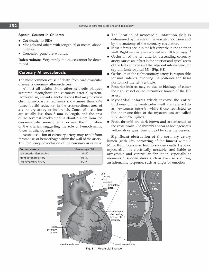
132 Review of Forensic Medicine and Toxicology
Special Causes in Children
� Cot deaths or SIDS � Mongols and others with congenital or mental abnor-
malities � Concealed puncture wounds.
Indeterminate: Very rarely the cause cannot be deter-mined.
Coronary Atherosclerosis
The most common cause of death from cardiovascular disease is coronary atherosclerosis.
Almost all adults show atherosclerotic plaques scattered throughout the coronary arterial system. However, significant stenotic lesions that may produce chronic myocardial ischemia show more than 75% (three-fourth) reduction in the cross-sectional area of a coronary artery or its branch. Zones of occlusion are usually less than 5 mm in length, and the area of the severest involvement is about 3–4 cm from the coronary ostia, more often at or near the bifurcation of the arteries, suggesting the role of hemodynamic forces in atherogenesis.
Acute occlusion of coronary artery may result from thrombosis or hemorrhage within the wall of the artery. The frequency of occlusion of the coronary arteries is:
Coronary artery Percentage (%)Left anterior descending 40–50Right coronary artery 30–40Left circumflex artery 15–20
� The location of myocardial infarction (MI) is determined by the site of the vascular occlusion and by the anatomy of the coronary circulation.
� Most infarcts occur in the left ventricle in the anterior wall. Right ventricle is involved in < 10% of cases.14
� Occlusion of the left anterior descending coronary artery causes an infarct in the anterior and apical areas of the left ventricle and the adjacent interventricular septum (anteroapical MI) (Fig. 8.1).
� Occlusion of the right coronary artery is responsible for most infarcts involving the posterior and basal portions of the left ventricle.
� Posterior infarcts may be due to blockage of either the right vessel or the circumflex branch of the left artery.
� Myocardial infarcts which involve the entire thickness of the ventricular wall are referred to as transmural infarcts, while those restricted to the inner one-third of the myocardium are called subendocardial infarcts.
� Fresh thrombi are dark-brown and are attached to the vessel walls. Old thrombi appear as homogeneous yellowish or gray, firm plugs blocking the vessels.
Significant obstruction of the coronary artery lumen (with 75% narrowing of the lumen) without MI or thrombosis may lead to sudden death. Hypoxic myocardium is electrically unstable, and liable to arrhythmia and ventricular fibrillation, especially at moments of sudden stress, such as exercise or during an adrenaline response, such as anger or emotion.
Fig. 8.1: Myocardial infarction

133Thanatology
Postmortem Examination
� No naked eye change is seen for the first 12–18 h. The appearance of a myocardial infarct is determined primarily by its age. It is generally accepted that at least 12–24 h of survival postinfarction must occur for the earliest recognizable change to evolve in the heart.
� The essential sequence of events consists of coagulation necrosis and inflammation, followed by the formation of granulation tissue, resorption of the necrotic myocardium, and finally organization of the granulation tissue to form a collagen-rich scar. These events occur in a fairly predictable pattern, allowing one to estimate the age of a given infarct from its gross and microscopic appearance (Table 8.3).
� Immersion of tissue slices in a solution of triphenyl tetrazolium chloride (TTC) gives red color to the healthy area (where dehydrogenase is preserved), but infarcted area appears pale if seen in about 4 h; the results are, however, inconsistent.
� Fresh thrombotic lesion is seen in less than 25% of the cases. Coronary artery spasm can cause death in patients suffering from angina without narrowing of the coronary arteries and without significant atherosclerosis or congenital anomalies.
� The lesions of the conducting system of the heart may sometimes cause arrhythmias and death.
� Any person with a heart in excess of 420 g is at risk of sudden death, even though the coronary arteries are normal.
� Enzyme histochemistry is the most reliable method of detecting early MI. Dehydrogenases—succinic, lactic, malic, hydroxybutyric and cytochrome oxidase are among those used. With malate dehydrogenase, normal myocardium stains dark blue-black and infarcted area is devoid of color.
� Periodic Acid-Schiff (PAS) stain: In early infarcts (at least 28 h), damaged myofibers stain a pale purple-blue with PAS, compared with the pink color of healthy fibers.
� Hematoxylin-Eosin (H&E) autofluorescence: Routine formalin-fixed H&E sections are examined under UV light. Early infarcted fibers show a shift of their secondary emission towards yellow, away from the usual olive-green of healthy fibers.
� Acridine-Orange fluorescent stain: Slides are examined under UV light; normal myocardium is golden-brown/yellowish-brown with damaged fibers showing a shift to green.
Anaphylactic Deaths
Most anaphylactic deaths seen by forensic pathologist are caused by insect bites, drugs or foods.
Signs and symptoms � A typical anaphylactic reaction results in acute
respiratory distress or circulatory collapse. � Faintness, itching of the skin, urticaria, tightness in the
chest, wheezing, respiratory difficulty and collapse. � In anaphylactic deaths, the onset of symptoms is
usually immediate or within the first 15–20 min. Beyond that time, one would need a well-documented medical history of gradually developing symptoms to implicate an anaphylactic reaction, e.g. the development of itching or wheals and flares. Death usually occurs within 1–2 h.
Table 8.3: Sequential pathologic changes in myocardial infarction (MI)
S.No. Duration Gross changes Microscopic changes
1. 0–6 h No change; triphenyl tetrazolium chloride (TTC) test negative
No change; stretching and waviness of fibers
2. 6–12 h No change or slight pallor Coagulative necrosis and neutrophilic infiltration begins, minimal hemorrhage
3. 12–24 h Slight pallor or mottling Continuing coagulation necrosis, ‘contraction band’ necrosis at the periphery of the infarct,* neutrophilic infiltrate
4. 24–72 h Pallor, hyperemic or alternate bands of red and pale areas— ‘tigroid appearance’
Complete coagulation necrosis of myofibers; neutrophilic infiltrate well developed with early fragmentation of neutrophil nuclei
5. 4–7 days Central pallor with hyperemic border, soft Macrophages appear, disintegration and phagocytosis of necrotic fibers, granulation tissue visible at edge of infarct
6. 10 days Maximally yellow, soft, shrunken, purple periphery
Well developed phagocytosis, prominent granulation tissue in peripheral areas of infarct, pigmented macrophages, eosinophils, lymphocytes and plasma cells present
7. 4–6 weeks Thin, gray-white, hard, shrunken fibrous scar Increased fibrocollagenic tissue, decreased vascularity, fewer pigmented macrophages, lymphocytes and plasma cells
* Contraction bands (myofibrillar degeneration, coagulative myocytolysis or Zenker necrosis) are characteristic necrosis pattern representing hypercontraction and lysis of small groups of myocardial cells. They are also found in coronary occlusion, resuscitation attempts, drowning, burning and hypothermia.
134 Review of Forensic Medicine and Toxicology
pharynx, glottis, pleura, peritoneum and cervix, which pass into the lateral tracts of spinal cord and finally to the brain.
Causes � Pressure on the carotid sinuses, as in hanging or
strangulation. � Unexpected blow to the larynx, chest, abdomen or
genital organs. � Impaction of food in the larynx or sudden inhalation
of fluid into the upper respiratory tract. � Sudden immersion of body in cold water. � The insertion of an instrument into the bronchus,
uterus, bladder or rectum. � Puncture of a pleural cavity producing a pneumo-
thorax. � Sudden evacuation of pathological fluids, e.g. ascitic
tap.
Postmortem examination: There are no characteristic postmortem findings. The cause of death can be inferred only by exclusion of other pathological conditions and from the observation of reliable witnesses, history and clinical findings concerning the circumstances of death.
Brugada syndrome is a disorder characterized by sudden nocturnal death of an apparently healthy young person with no history of any disease/drug abuse, and the findings at autopsy are those of asphyxia. In this syndrome, there is mutation in SCN 5A gene which makes the person prone to develop a range of arrhythmias and death occurs in sleep due to ventricular fibrillation. There has been extensive study in Japan and Philippines on this sudden nocturnal death syndrome. In North Eastern part of India, such cases are frequently found particularly in people of Mongoloid origin.
� Obstruction of the upper airway can be caused by pharyngeal or laryngeal edema; of the lower airway, by bronchospasm with contraction of the smooth muscle of the lungs, vasodilatation and increased capillary permeability.
� Cardiac arrest may be caused by respiratory failure.
Vagal Inhibition (Vasovagal Shock/Reflex Cardiac Arrest/Nervous Apoplexy)
� Sudden death occurring within seconds or minutes as a result of minor trauma or harmless peripheral stimulation may be caused by vagal inhibition.
� Pressure on the baroreceptors situated in the carotid sinuses, carotid sheaths and the carotid body (located in the internal carotid artery and situated near the angle of mandible) causes an increase in blood pressure in these sinuses with resultant slowing of the heart rate, dilatation of blood vessels and fall in blood pressure.
� Some individuals show marked hypersensitivity to stimulation of the carotid sinuses, characterized by bradycardia and cardiac arrhythmias ranging from ventricular arrhythmias to cardiac arrest.
Mechanism: It acts through a reflex arc in which the afferent (sensory) nerve impulses arise in the carotid complex of nerve endings, but not in the vagal nerve trunk itself. These impulses pass through glossopharyn-geal nerves to the tenth nucleus in the brainstem, then return through the vagus (efferent) supply to the heart and other organs. This reflex arc acts through the parasympathetic autonomic nervous system, and is independent of the main motor and sensory nerve pathways. Afferent fibers are present over the skin,
1. B 2. C 3. C 4. B 5. B
multiPle ChoiCe questions
1. Study of death in all its aspects is known as: AIIMS 08 A. Eugenics B. Thanatology C. Dactylography D. Tricology 2. Which of the following is not a postmortem change:
NEET 15 A. Algor mortis B. Rigor mortis C. Atria mortis D. Livor mortis 3. All are features of somatic death, except: NEET 14 A. Cessation of respiration B. Cessation of heart C. Non-responding muscles D. No response to external stimuli
4. Molecular death is: PGI 10; Kerala 11 A. Complete and irreversible cessation of brain, heart
and lungs function B. Death of individual tissues and cells after somatic
death C. Total loss of EEG activity, but heart is functioning D. Vitals functions are at low pitch that cannot be
detected by clinical examination 5. NOT important in brain death: PGI 07 A. EEG B. ECG C. Absence of brainstem reflex D. Body temperature

135Thanatology
6. Brainstem dead are all, except: JIPMER 08 A. Weaned off from ventilator, no respiration for 15 sec B. Absent pupillary response C. Absent nystagmus D. Absent corneal reflex 7. All the following are found in brain dead patients,
except: NIMHANS 07; Karnataka 11; AIIMS 14 A. Decreased deep tendon reflex B. Absent pupillary reflexes C. Complete apnea D. Heart unresponsive to atropine 8. In brain death which of the organs cannot be
transplanted: UPSC 14 A. Brain B. Heart C. Liver D. Kidney 9. Xenograft is transplantation of tissue: FMGE 10 A. From a different species B. From same species C. From genetically identical twins D. From one part of body to another 10. Agonal period is the duration between: Maharashtra 08 A. Traumatic event and information given to the
relatives B. Traumatic event and starting of the operation C. Lethal trauma upto death D. Death and postmortem examination
6. A 7. A & D 8. A 9. A 10. C 11. C 12. C 13. A 14. D
11. An old lady with mitral stenosis underwent hyster-ectomy for uterine fibroid and died after developing pulmonary edema. The order of cause of death in international certificate is: NIMS 11
A. Mitral stenosis, pulmonary edema, hysterectomy B. Pulmonary edema, mitral stenosis, hysterectomy C. Pulmonary edema, hysterectomy, mitral stenosis D. Hysterectomy, pulmonary edema, mitral stenosis 12. Gordon’s clarification of death signifies: Odisha 11 A. Mechanism of death B. Causes of death C. Modes of death D. Manner of death 13. Cyanide poisoning causes: AFMC 10 A. Histotoxic anoxia B. Anoxic anoxia C. Anemic anoxia D. Stagnant anoxia 14. All the statements regarding atherosclerosis are true,
except: AP 10; SGPGI 11 A. Naked eye changes are not visible for the first 12 h B. Triphenyl tetrazolium chloride can help in detecting
infracted area C. Most commonly involves the left coronary artery D. Common site is the anterior wall of right ventricle

Signs of Death
CHAPTER 9
The accurate determination of time of death is important due to its role in explaining possible criminal acts and determination of appropriate civil repercussions. The changes which occur after death that are helpful in estimation of the approximate time of death (and to differentiate death from suspended animation) can be classified into (Table 9.1):
� Immediate changes � Early changes � Late changes.
Immediate Changes (Somatic Death)
a. Irreversible cessation of the function of brain including brainstem: This is the earliest sign of death with stoppage of functions of the nervous system. There is insensibility, and loss of both sensory and motor functions. There is loss of reflexes, no response and no tonicity of the muscles. Pupils are widely dilated. This condition is sometimes seen in:
z Prolonged fainting attack z Vagal inhibitory phenomenon z Epilepsy, mesmeric trance, catalepsy, narcosis or
electrocution.b. Irreversible cessation of respiration: Complete stop-
page of respiration for > 4 minutes (min) usually causes death. The stoppage of respiration can be established by the following tests:
i. Inspection: No visible respiratory movement. ii. Palpation: No respiratory movement can be felt.
iii. Auscultation: Breath sounds cannot be heard from any part of the lungs.
iv. Feather test, mirror test and Winslow’s test are no longer utilized.
Respiration may stop briefly without death as in: z Voluntary breath holding z Drowning z Cheyne-Stokes respiration z Newborns.
c. Irreversible cessation of circulation: Stoppage of heart beat for > 3–5 min is irrecoverable and results in death. The following tests may be performed to test circulation:
i. Radial, brachial, femoral and carotid pulsations will be absent, if the circulation has stopped.
ii. Auscultation of heart: Absence of the heart beat over the whole precordial area and particularly over the area of the apex.
iii. ECG: In case of cessation of circulation, the ECG curve is absent and the tracing shows a flat line without any elevation or depression.
iv. Other tests: Various tests, like diaphanous, magnus, I-card, pressure, cut and heat tests are now obsolete.
Tests to detect stoppage of respiration (obsolete) � Winslow’s test: No movement of reflection of light shone on
mirror or surface of water in bowl kept on the chest.2
� Feather test: No movement is seen, if a feather or fine cotton fibers are held before the nostrils.
� Mirror test: No haziness is seen on the reflecting surface of the mirror held in front of mouth and nostrils.
Table 9.1: Changes after death
Immediate changes1 Early changes Late changesIrreversible cessation of: � Loss of elasticity of the skin, and facial pallor � Putrefaction
� Function of brain � Primary relaxation of the muscles � Adipocere formation � Circulation � Contact pallor and flattening � Mummification � Respiration � Changes in the eye
� Algor mortis � Livor mortis � Rigor mortis

137Signs of Death
Tests to detect stoppage of circulation (obsolete) � Magnus test (ligature test): Fingers fail to show bluish discoloration
and edema to a ligature applied at their base. � Diaphanous test (transillumination test): Failure to show redness
in the web-space between the fingers on transillumination from behind.
� I card’s test: Fluorescein dye on being injected at a given site in a dead body fail to produce yellowish-green discoloration as seen in a living person.
� Pressure test: Fingernails appear pale and fail to show reddish color on removal of firm pressure over it.
Suspended Animation (Apparent Death)
Definition: Suspended animation is a condition in which vital signs of life (heart beat and respiration) are not detected by routine clinical methods, as the functions are interrupted for some time or are reduced to a minimum.3
Mechanism: The metabolic rate is greatly reduced so that the requirement of the individual cell for oxygen is satisfied through the dissolved oxygen in body fluids.
TypesTwo types: i. Voluntary: Seen in practitioners of yoga or in
trance.3
ii. Involuntary: Seen in hypothermia, poisoning with barbiturates or opiates, newborns, drowning, electrocution, heatstroke, cholera, postanesthesia, shock, cerebral concussion or insanity.3,4
The patient can be resuscitated by cardiac massage or electric stimulator and artificial respiration. The death certificate should not be issued without an ECG or EEG record.
Early Changes (Molecular Death)
a. Changes in the skin and facial pallor: Skin becomes pale and ash-white due to stoppage of circulation and drainage of blood from the capillaries and the small vessels. The skin loses its elasticity, and the face looks younger due to loss of creases. The lips appear brownish, dry and hard due to drying.
b. Primary relaxation or flaccidity of the muscles: Muscles loose their tonicity and become flaccid, but the muscular tissues are still alive, their chemical reaction is alkaline and responds to electrical stimuli.
c. Contact flattening and pallor: The areas which remain in contact with the ground, become flat and the blood from vessels of these areas is pressed
out, this continues even after the formation of postmortem staining over the surrounding areas.
d. Changes in the eye � Loss of corneal and pupillary reflexes: It may be seen
in all cases of deep insensibility and therefore is not a reliable sign of death. However, the pupils react for sometime to miotic and mydriatic agents.
� Pupils: The pupils are dilated after death, because of the relaxation of muscles of the iris. Later, they are constricted with the onset of rigor mortis of the constrictor muscles and evaporation of fluid. As such, their state after death is not an indication of their antemortem appearance.
z Shape of the pupils: Pupils are circular during life, but due to loss of tone and elasticity of the ciliary muscles after death, the shape of the pupil can be changed, and the change may persist during the stage of rigor mortis of the muscles. Moreover, the sizes and shapes of the pupils of the two sides may be different.
� Opacity of the cornea: There is opacity and haziness of the cornea due to drying and deposition of dust and debris over it. This may be delayed, if the lids are closed after death. If the lids are closed, the cornea remains clear for about 2 hours (h). This haziness is transient and passes off, if a drop of water is poured on the cornea. But the cornea becomes permanently hazy after about 10–12 h of death due to decomposi- tion.
� Tache noire (French, black line): If the eyelids remain open for 3–4 h after death, there is formation of two yellow triangles (base on the limbus, apex at the lateral or medial canthus and sides are formed by the margins of the upper and lower eyelids) on the sclera at each side of the iris, which become brown and then black.
Cause: Drying/desiccation, and deposition of cellular debris, mucus and dust on the exposed conjunctiva and the sclera underneath.
� Loss of intraocular tension: Intraocular tension falls rapidly after death. It becomes zero in 4–8 h from 10–22 mmHg during life. The eyeballs look sunken in the orbit.
� Changes in the retina: The blood in retinal vessels appears fragmented or segmented (cattle trucking or shunting) within seconds to minutes after death, and persists for about an hour (Kevorkian sign). This occurs all over the body due to loss of blood pressure, but it can be seen only in retina by an ophthalmoscope.
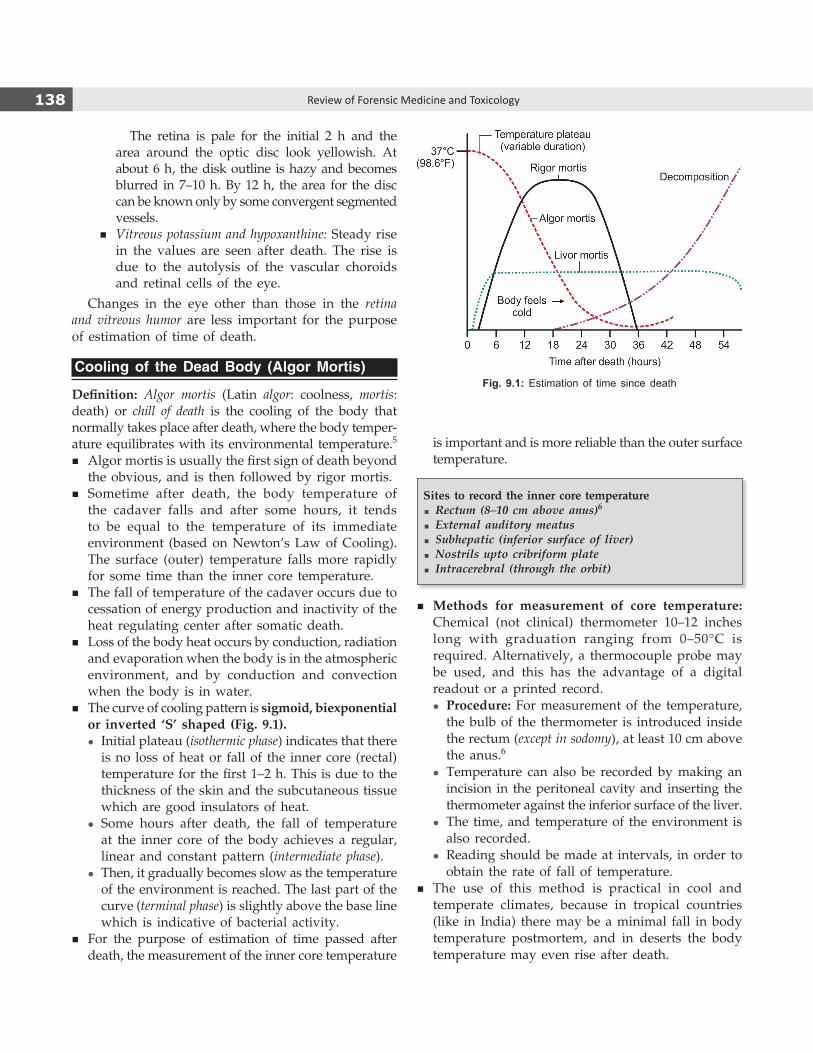
138 Review of Forensic Medicine and Toxicology
The retina is pale for the initial 2 h and the area around the optic disc look yellowish. At about 6 h, the disk outline is hazy and becomes blurred in 7–10 h. By 12 h, the area for the disc can be known only by some convergent segmented vessels.
� Vitreous potassium and hypoxanthine: Steady rise in the values are seen after death. The rise is due to the autolysis of the vascular choroids and retinal cells of the eye.
Changes in the eye other than those in the retina and vitreous humor are less important for the purpose of estimation of time of death.
Cooling of the Dead Body (Algor Mortis)
Definition: Algor mortis (Latin algor: coolness, mortis: death) or chill of death is the cooling of the body that normally takes place after death, where the body temper-ature equilibrates with its environmental temperature.5
� Algor mortis is usually the first sign of death beyond the obvious, and is then followed by rigor mortis.
� Sometime after death, the body temperature of the cadaver falls and after some hours, it tends to be equal to the temperature of its immediate environment (based on Newton’s Law of Cooling). The surface (outer) temperature falls more rapidly for some time than the inner core temperature.
� The fall of temperature of the cadaver occurs due to cessation of energy production and inactivity of the heat regulating center after somatic death.
� Loss of the body heat occurs by conduction, radiation and evaporation when the body is in the atmospheric environment, and by conduction and convection when the body is in water.
� The curve of cooling pattern is sigmoid, biexponential or inverted ‘S’ shaped (Fig. 9.1).
z Initial plateau (isothermic phase) indicates that there is no loss of heat or fall of the inner core (rectal) temperature for the first 1–2 h. This is due to the thickness of the skin and the subcutaneous tissue which are good insulators of heat.
z Some hours after death, the fall of temperature at the inner core of the body achieves a regular, linear and constant pattern (intermediate phase).
z Then, it gradually becomes slow as the temperature of the environment is reached. The last part of the curve (terminal phase) is slightly above the base line which is indicative of bacterial activity.
� For the purpose of estimation of time passed after death, the measurement of the inner core temperature
is important and is more reliable than the outer surface temperature.
Sites to record the inner core temperature � Rectum (8–10 cm above anus)6
� External auditory meatus � Subhepatic (inferior surface of liver) � Nostrils upto cribriform plate � Intracerebral (through the orbit)
� Methods for measurement of core temperature: Chemical (not clinical) thermometer 10–12 inches long with graduation ranging from 0–50°C is required. Alternatively, a thermocouple probe may be used, and this has the advantage of a digital readout or a printed record.
z Procedure: For measurement of the temperature, the bulb of the thermometer is introduced inside the rectum (except in sodomy), at least 10 cm above the anus.6
z Temperature can also be recorded by making an incision in the peritoneal cavity and inserting the thermometer against the inferior surface of the liver.
z The time, and temperature of the environment is also recorded.
z Reading should be made at intervals, in order to obtain the rate of fall of temperature.
� The use of this method is practical in cool and temperate climates, because in tropical countries (like in India) there may be a minimal fall in body temperature postmortem, and in deserts the body temperature may even rise after death.
Fig. 9.1: Estimation of time since death

139Signs of Death
� The average rate of fall of the body temperature is 0.4–0.7°C/h, and the body attains environmental temperature in 16–20 h after death.7
� It is assumed that the body temperature at the time of death was normal, which varies between 35.7–37.7°C orally and 36.7–37.5°C in the rectum (in males).
� A rough estimate of time since death (TSD) is obtained by the formula:
TSD (in hours) =
Normal rectal temperature Measured rectal temperature
Rate of fal
−ll of temperature/hour
� For temperate countries, Marshal and Hoare formula is used. The rates of fall of temperature in an average built person is 1°F upto 3 h, 2°F upto 9 h and 1.5°F upto 12 h. The rule of thumb is that the temperature falls at about 1.5°F/h.
� Rectal temperature is higher in case of struggle or exercise prior to death.
� Low temperature is seen in congestive cardiac failure, hemorrhage, collapse and secondary shock.
Various equations, algorithms and nomograms using rectal temperature have been developed. Examples of simple rule-of-thumb formulae (for temperate countries):
� TSD = (Rectal temperature at time of death – measured rectal temperature [°F]) ÷ 1.5
� TSD = (Rectal temperature at time of death – measured rectal temperature [°C]) + 3*
Presently, nomogram method devised by Henssge is used. This method is based on experimental data, which can be carried out by a simple computer program or by a nomogram. Adjustments are built in for the body weight, ambient temperature and body temperature. It provides a 95% accuracy of estimating the TSD during the first 15 h (with an error of ±2.8 h).
Factors Affecting Algor Mortis
i. Environmental temperature (major factor): Rate of fall of body temperature is directly proportional to the difference between the temperature of the dead body and the environmental temperature.
ii. Air movement: Air movement over the surface of the dead body causes a quick fall of temperature due to increased evaporation of body fluids. A body kept in a well-ventilated room will cool more rapidly than one in a closed room.
iii. Humidity: Cooling is more rapid in a humid rather than in a dry atmosphere, since moist air is better conductor of heat.
iv. Media of disposal: Cooling is earliest in water, and late in buried bodies. The ratio of the rates of fall of temperature in the three media, water:air: soil = 4:2:1. The rate is thus maximum in water, moderate in air and minimum in a buried body.
v. Built of cadaver: Obese bodies cool slowly, and lean bodies rapidly, since fat is a bad conductor of heat.
vi. Age and sex: Rate of loss of heat is more in children and the elderly, compared to adults because the surface area of the body is more in relation to the body volume. Females retain body heat for a comparatively longer period because of their subcutaneous fatty tissue.
vii. Clothing or coverings of the body: A well-covered body retains heat for a longer period as compared to a naked or thinly clothed body, as clothes are bad conductors of heat.
viii. Position and posture of the body: If the body lies in supine and extended position, the loss of heat is rapid because greater surface area of the body is exposed; whereas in curled fetal position, the loss will be slow.
ix. Mode of death: In case of sudden death in a healthy individual, the body tends to cool slowly, whereas in death due to long and wasting illness, the body cools rapidly.
Postmortem Caloricity
In this condition, instead of cooling, the temperature of the dead body remains high for the initial 2 h or so. This is due to: a. Postmortem glycogenolysis: Compulsory phenomenon
which occurs in all dead bodies, and which starts soon after death (produces upto 140 calories).
b. Cause of death:8,9
z In deaths occurring due to infectious diseases, septicemia or bacteremia, heat is produced by the action of the infective organisms.
z If death is preceded by a severe convulsion, as in tetanus and strychnine poisoning, it causes an increase in the body temperature.
z In case of death due to heat stroke or pontine hemorrhage, the heat regulation is severely disturbed before death.
c. High environmental temperature: In tropical countries, when the environmental temperature is higher than the body temperature, the dead body may absorb some heat.
* Compensatory number for possible initial delay in cooling of the body.
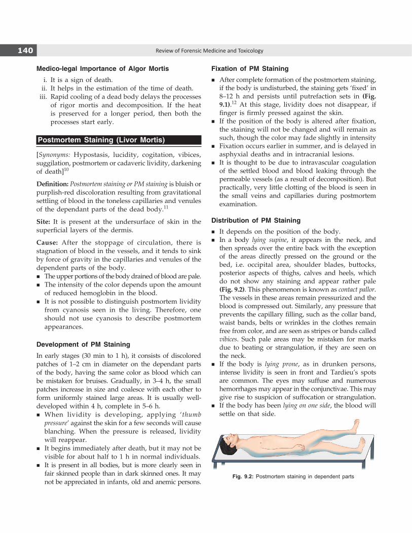
140 Review of Forensic Medicine and Toxicology
Fig. 9.2: Postmortem staining in dependent parts
Medico-legal Importance of Algor Mortis
i. It is a sign of death. ii. It helps in the estimation of the time of death. iii. Rapid cooling of a dead body delays the processes
of rigor mortis and decomposition. If the heat is preserved for a longer period, then both the processes start early.
Postmortem Staining (Livor Mortis)
[Synonyms: Hypostasis, lucidity, cogitation, vibices, suggilation, postmortem or cadaveric lividity, darkening of death]10
Definition: Postmortem staining or PM staining is bluish or purplish-red discoloration resulting from gravitational settling of blood in the toneless capillaries and venules of the dependant parts of the dead body.11
Site: It is present at the undersurface of skin in the superficial layers of the dermis.
Cause: After the stoppage of circulation, there is stagnation of blood in the vessels, and it tends to sink by force of gravity in the capillaries and venules of the dependent parts of the body.
� The upper portions of the body drained of blood are pale. � The intensity of the color depends upon the amount
of reduced hemoglobin in the blood. � It is not possible to distinguish postmortem lividity
from cyanosis seen in the living. Therefore, one should not use cyanosis to describe postmortem appearances.
Development of PM Staining
In early stages (30 min to 1 h), it consists of discolored patches of 1–2 cm in diameter on the dependant parts of the body, having the same color as blood which can be mistaken for bruises. Gradually, in 3–4 h, the small patches increase in size and coalesce with each other to form uniformly stained large areas. It is usually well-developed within 4 h, complete in 5–6 h.
� When lividity is developing, applying ‘thumb pressure’ against the skin for a few seconds will cause blanching. When the pressure is released, lividity will reappear.
� It begins immediately after death, but it may not be visible for about half to 1 h in normal individuals.
� It is present in all bodies, but is more clearly seen in fair skinned people than in dark skinned ones. It may not be appreciated in infants, old and anemic persons.
Fixation of PM Staining � After complete formation of the postmortem staining,
if the body is undisturbed, the staining gets ‘fixed’ in 8–12 h and persists until putrefaction sets in (Fig. 9.1).12 At this stage, lividity does not disappear, if finger is firmly pressed against the skin.
� If the position of the body is altered after fixation, the staining will not be changed and will remain as such, though the color may fade slightly in intensity
� Fixation occurs earlier in summer, and is delayed in asphyxial deaths and in intracranial lesions.
� It is thought to be due to intravascular coagulation of the settled blood and blood leaking through the permeable vessels (as a result of decomposition). But practically, very little clotting of the blood is seen in the small veins and capillaries during postmortem examination.
Distribution of PM Staining � It depends on the position of the body. � In a body lying supine, it appears in the neck, and
then spreads over the entire back with the exception of the areas directly pressed on the ground or the bed, i.e. occipital area, shoulder blades, buttocks, posterior aspects of thighs, calves and heels, which do not show any staining and appear rather pale (Fig. 9.2). This phenomenon is known as contact pallor. The vessels in these areas remain pressurized and the blood is compressed out. Similarly, any pressure that prevents the capillary filling, such as the collar band, waist bands, belts or wrinkles in the clothes remain free from color, and are seen as stripes or bands called vibices. Such pale areas may be mistaken for marks due to beating or strangulation, if they are seen on the neck.
� If the body is lying prone, as in drunken persons, intense lividity is seen in front and Tardieu’s spots are common. The eyes may suffuse and numerous hemorrhages may appear in the conjunctivae. This may give rise to suspicion of suffocation or strangulation.
� If the body has been lying on one side, the blood will settle on that side.
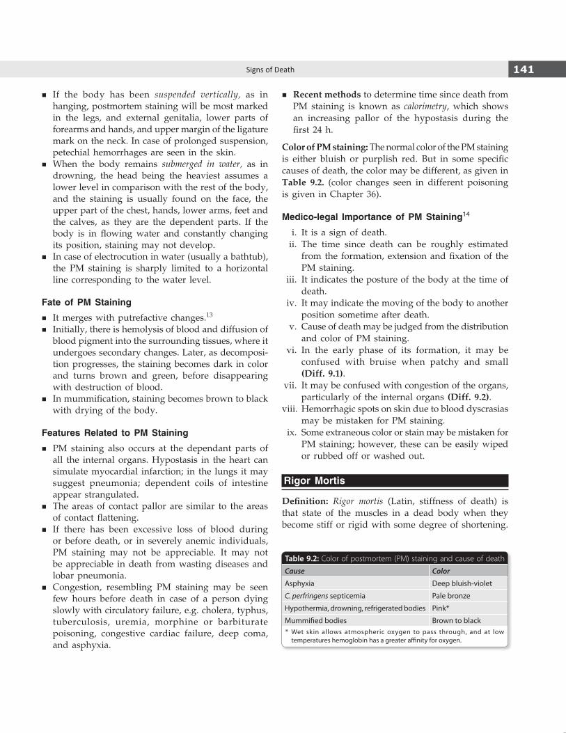
141Signs of Death
� If the body has been suspended vertically, as in hanging, postmortem staining will be most marked in the legs, and external genitalia, lower parts of forearms and hands, and upper margin of the ligature mark on the neck. In case of prolonged suspension, petechial hemorrhages are seen in the skin.
� When the body remains submerged in water, as in drowning, the head being the heaviest assumes a lower level in comparison with the rest of the body, and the staining is usually found on the face, the upper part of the chest, hands, lower arms, feet and the calves, as they are the dependent parts. If the body is in flowing water and constantly changing its position, staining may not develop.
� In case of electrocution in water (usually a bathtub), the PM staining is sharply limited to a horizontal line corresponding to the water level.
Fate of PM Staining
� It merges with putrefactive changes.13
� Initially, there is hemolysis of blood and diffusion of blood pigment into the surrounding tissues, where it undergoes secondary changes. Later, as decomposi-tion progresses, the staining becomes dark in color and turns brown and green, before disappearing with destruction of blood.
� In mummification, staining becomes brown to black with drying of the body.
Features Related to PM Staining
� PM staining also occurs at the dependant parts of all the internal organs. Hypostasis in the heart can simulate myocardial infarction; in the lungs it may suggest pneumonia; dependent coils of intestine appear strangulated.
� The areas of contact pallor are similar to the areas of contact flattening.
� If there has been excessive loss of blood during or before death, or in severely anemic individuals, PM staining may not be appreciable. It may not be appreciable in death from wasting diseases and lobar pneumonia.
� Congestion, resembling PM staining may be seen few hours before death in case of a person dying slowly with circulatory failure, e.g. cholera, typhus, tuberculosis, uremia, morphine or barbiturate poisoning, congestive cardiac failure, deep coma, and asphyxia.
� Recent methods to determine time since death from PM staining is known as calorimetry, which shows an increasing pallor of the hypostasis during the first 24 h.
Color of PM staining: The normal color of the PM staining is either bluish or purplish red. But in some specific causes of death, the color may be different, as given in Table 9.2. (color changes seen in different poisoning is given in Chapter 36).
Medico-legal Importance of PM Staining14
i. It is a sign of death. ii. The time since death can be roughly estimated
from the formation, extension and fixation of the PM staining.
iii. It indicates the posture of the body at the time of death.
iv. It may indicate the moving of the body to another position sometime after death.
v. Cause of death may be judged from the distribution and color of PM staining.
vi. In the early phase of its formation, it may be confused with bruise when patchy and small (Diff. 9.1).
vii. It may be confused with congestion of the organs, particularly of the internal organs (Diff. 9.2).
viii. Hemorrhagic spots on skin due to blood dyscrasias may be mistaken for PM staining.
ix. Some extraneous color or stain may be mistaken for PM staining; however, these can be easily wiped or rubbed off or washed out.
Rigor Mortis
Definition: Rigor mortis (Latin, stiffness of death) is that state of the muscles in a dead body when they become stiff or rigid with some degree of shortening.
Table 9.2: Color of postmortem (PM) staining and cause of death
Cause Color
Asphyxia Deep bluish-violet
C. perfringens septicemia Pale bronze
Hypothermia, drowning, refrigerated bodies Pink*
Mummified bodies Brown to black* Wet skin allows atmospheric oxygen to pass through, and at low
temperatures hemoglobin has a greater affinity for oxygen.

142 Review of Forensic Medicine and Toxicology
The phase of primary relaxation of the muscles continues for about an hour which is followed by stiffening or rigidity. It indicates molecular death of the concerned muscles.
Mechanism: Muscle fibers contain bundles of myofibrils which consist of two types of protein filaments—actin and myosin. At rest, actin filaments interdigitate myosin filaments only to a small extent and the muscle fibers also appear soft and supple. Maintenance of this condition of muscles is due to the presence of ATP (adenosine-triphosphate) above a certain level. On nervous stimulation, hydrolysis of ATP occurs to ADP (adenosine-diphosphate) and phosphate with the liberation of energy which causes contraction of the muscle fibers and extension of the actin filaments more inside the myosin filaments. After death, there is continuous hydrolysis of the ATP, and as long as glycogen is available in the muscle, there is resynthesis of ATP. In this process, once the muscle glycogen is exhausted, no further resynthesis of ATP is possible and the muscle loses softness,
elasticity and extensibility due to formation of viscid actomyosin complex giving rise to rigor mortis in the muscle (Fig. 9.3).16
Differentiation 9.1: Postmortem staining and bruise15
S.No. Feature Postmortem staining Bruise1. Situation On the dependant parts Anywhere2. Tissue level Undersurface of the skin Subcutaneous tissue level 3. Surface Not elevated May be slightly elevated4. Margin Sharp and clearly defined Diffuse5. Color Bluish or purplish red Reddish when fresh, change in color occurs with time6. Cause Capillo-venous distension with blood Extravasation of blood from capillaries7. Nature of change Postmortem Antemortem8. Effect of pressure Pressed spot appears pale No change9. Cut section Oozing of blood from the vessels which can be
cleaned by washingHemorrhage in the tissue which cannot be washed
10. Microscopically Engorgement of capillaries infiltration Extravasation of blood, cellular infiltration11. Enzymatic study No change Change in the level of certain enzymes12. Medico-legal importance Time since death and position of the body may
be knownNature of injury and weapon used may be known
Differentiation 9.2: Congestion and Postmortem staining
S.No. Feature Congestion Postmortem staining1. Situation Uniform, all over the organ Irregular and in dependant parts2. Exudate Exudate may be seen No inflammatory exudate 3. Mucous membranes Normal Dull and lusterless4. Swelling or edema May be seen None5. Hollow viscus Uniform staining Stomach and intestine when stretched show alternate areas of
discoloration and pallor6. Cause Due to some pathology in the organ Passive capillo-venous distension7. Nature of change Antemortem Postmortem
Fig. 9.3: Schematic representation of rigor mortis

143Signs of Death
After the pH of the muscle becomes 5.5, release of autolytic enzymes stored in lysosomes takes place. The major proteolytic enzymes are cathepsins and calpains. These enzymes act at the myofibrillar proteins and hydrolyze them. As a result, the actomyosin complex is broken down and muscles become soft again. This is known as resolution of rigor which occurs during the stage of secondary relaxation, due to decomposition.
Muscles Involved
� Rigor mortis occurs both in the voluntary and involuntary muscles.
� It occurs earlier in the involuntary or smooth muscles than in the voluntary or striated muscles.17
Onset and Duration
� In tropical countries like India, roughly, it commences in 1–2 h after death, takes about 9–12 h to develop from head to foot, persists for another 12 h and takes 12 h to pass off (Rule of 12) (Fig. 9.1).18,19
� In Northern India, the usual duration of rigor mortis is 18–36 h in summer and 24–48 h in winter.
Order of Appearance
� Rigor mortis first appears in the heart muscle within an hour after death.20
� Among the voluntary muscles, rigor mortis usually develops sequentially and follows a descending pattern, the so-called Nysten’s law: it first appears in the muscles of the eye lids (orbicularis oculi) [3–5 h], then in jaw, facial muscles [4–5 h], neck, thorax [5–7 h] upper limb (from shoulder to the hand) [7–9 h], abdomen, lower limb (from the hip to the foot) [9–11 h], and lastly in the small muscles of fingers and toes [11–12 h].21
� The rigidity disappears in the same order in which it has appeared. In the whole body, it stays for maximum duration in the muscles of the lower limbs.
� When rigor is fully established, the entire body is stiff; knees, hips, shoulders and elbows are slightly flexed, and fingers and toes often show a marked degree of flexion.
� It is independent of the integrity of the nervous system, though it is said to develop more slowly in paralyzed limbs.
Nysten’s law: Rigor mortis affects first the muscles of jaw, followed by those of the face and neck, then muscles of the trunk and arms, and lastly the legs and feet.
Testing of Rigor Mortis
� It is tested by lifting the eyelids, depressing the jaw, and gently bending the neck and joints of all four limbs (Table 9.3 and Fig. 9.4). The feeling of lower or higher degree of resistance is usually interpreted as rigor mortis.
� When rigor is developing and the extremities are moved (if death occurred < 8–12 h before), the rigor fixes the extremities in their new position. The rigidity will be less than in other symmetrical groups, which have not been disturbed.
Breaking of Rigor Mortis
� If rigidity is complete and rigor is broken by mechanical force, e.g. if a limb in rigor is flexed forcibly at a joint, the limb becomes flaccid and will remain so thereafter.
� Rigor mortis may be broken down partially due to mishandling during the transit of the body from the scene of crime to autopsy table, which may misled the autopsy surgeon in estimating the time since death.
Effects of rigor mortis � There is goose skin appearance of the body due to
rigor mortis of the erector pilae muscles.
Table 9.3: Interpretation of rigor mortis
Perception Interpretation
Moves with little force Present in moderate form
Moves with more force Present in strong form
Free movement or not present in the part tested
Not developed yet or disappeared
If only proximal parts show rigidity
Developing phase
If only distal parts show rigidity
Developed, disappearing phase
Fig. 9.4: Testing of rigor mortis

144 Review of Forensic Medicine and Toxicology
� Rigor in the muscles of the seminal vesicles may cause postmortem ejaculation of seminal fluid.
� The iris is also affected so that antemortem constriction or dilatation is modified. Hence, the postmortem position of pupil is an unreliable indicator of toxic or neurological conditions during life.
� Contraction of the heart muscle due to rigor mortis should not be mistaken for myocardial hypertrophy.
� Rigor mortis in the uterine muscle cannot expel the fetus from the womb.
Factors Affecting Rigor Mortis
The major factors that influence the onset and duration of rigor mortis are the environmental temperature and the degree of muscular activity before death. i. Environmental temperature: At high temperature,
rigor mortis comes early and passes off early. In cold temperature, it comes late and stays longer.22
ii. Muscular activity: Violent exercise prior to death may hasten the onset as well as disappearance of rigidity.
iii. Cause of death and condition of the body: Refer to Table 9.4.
iv. Built: It comes early and passes off early in emaciated and thinly built subjects with weak musculature. In well-built subjects with strong musculature, it is well-marked, comes late and stays longer.
v. Age: It is claimed that rigor mortis does not occur in stillborn fetuses of < 7 months old (there are only few muscle fibers, and cannot contribute to appreciable degree of stiffness).24 However, complete rigor has been reported in infants dying of sudden infant death syndrome (SIDS). In healthy
adults, rigor mortis develops slowly but is well- marked. It is weak, and comes early in children and elderly.22
Medico-legal Importance of Rigor Mortis
� It is a sign of death and indicates molecular death of the muscle involved.
� During the early phase after death, it helps in estimating the time since death. During summer, if rigor mortis has not set in, death might have occurred within 2 h. If rigor mortis has involved the whole body then death might have occurred between 12–24 h back. In winter season, the above timings are roughly doubled.
� It indicates the position of the body at the time of death. For example, if the body is lying on its back with its lower limbs raised in air, it indicates that the body reached full rigidity elsewhere while lying in a position where the legs were flexed.
� Some conditions occur in dead bodies which may imitate/simulate rigor mortis:25
z Cadaveric spasm or instantaneous rigor z Heat stiffening z Cold stiffening z Gas stiffening or putrefaction.
Cadaveric Spasm (Instantaneous Rigor /Rigidity, Cataleptic Rigidity)
Definition: Cadaveric spasm is a condition in which the muscles of the body which were in a state of contraction immediately before death, continue to be so after death without passing through the stage of primary relaxation.26,27
� It is a rare phenomenon of instantaneous rigor, which develops at the time of death with no period of postmortem flaccidity.
Predisposing conditions: It occurs especially in cases of sudden death, excitement, fear, severe pain, exhaustion, cerebral hemorrhage, electrocution, injury to the nervous system, firearm wound of the head, or convulsant poisons, like strychnine.
Muscles involved: The spasm is primarily a vital phenomenon; it originates by normal nervous stimulation of the muscles.
� It is usually limited to a single group of voluntary muscles, and frequently involve the hands.28,29
Table 9.4: Cause of death and condition of the bodyaffecting rigor mortis23
Early onset of rigor Late onset of rigor
� Exhaustive or wasting diseases, like cholera, typhoid, tuberculosis, cancer
� Asphyxia (CO, hanging)
� Violent deaths, like cut-throat, electro-cution, firearm and lightning injuries
� Hemorrhage
� Poisoning with strychnine, organophosphate, insulin or HCN
� Cold, refrigerated bodies
� Fatigue or exhaustion � Paralyzed muscles
� Heat stroke � Pneumonia
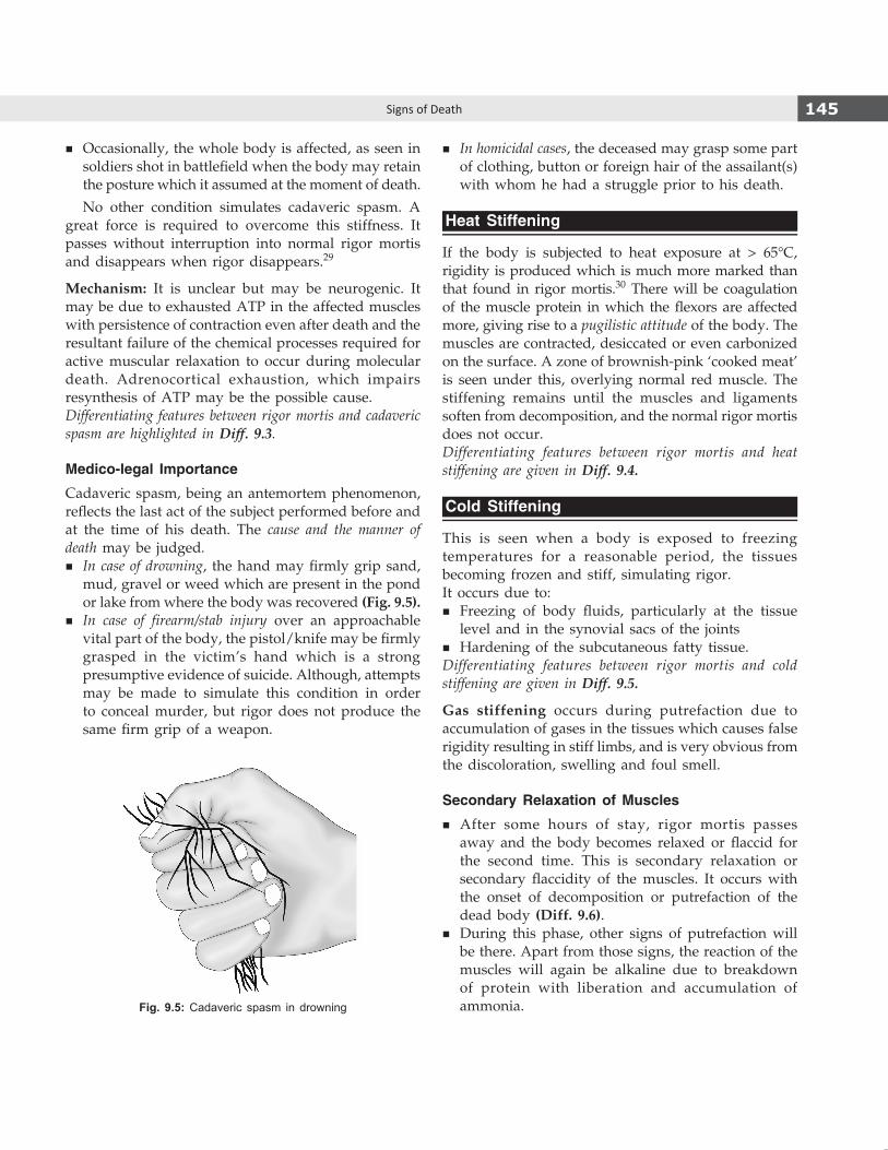
145Signs of Death
� Occasionally, the whole body is affected, as seen in soldiers shot in battlefield when the body may retain the posture which it assumed at the moment of death.
No other condition simulates cadaveric spasm. A great force is required to overcome this stiffness. It passes without interruption into normal rigor mortis and disappears when rigor disappears.29
Mechanism: It is unclear but may be neurogenic. It may be due to exhausted ATP in the affected muscles with persistence of contraction even after death and the resultant failure of the chemical processes required for active muscular relaxation to occur during molecular death. Adrenocortical exhaustion, which impairs resynthesis of ATP may be the possible cause.Differentiating features between rigor mortis and cadaveric spasm are highlighted in Diff. 9.3.
Medico-legal Importance
Cadaveric spasm, being an antemortem phenomenon, reflects the last act of the subject performed before and at the time of his death. The cause and the manner of death may be judged.
� In case of drowning, the hand may firmly grip sand, mud, gravel or weed which are present in the pond or lake from where the body was recovered (Fig. 9.5).
� In case of firearm/stab injury over an approachable vital part of the body, the pistol/knife may be firmly grasped in the victim’s hand which is a strong presumptive evidence of suicide. Although, attempts may be made to simulate this condition in order to conceal murder, but rigor does not produce the same firm grip of a weapon.
� In homicidal cases, the deceased may grasp some part of clothing, button or foreign hair of the assailant(s) with whom he had a struggle prior to his death.
Heat Stiffening
If the body is subjected to heat exposure at > 65°C, rigidity is produced which is much more marked than that found in rigor mortis.30 There will be coagulation of the muscle protein in which the flexors are affected more, giving rise to a pugilistic attitude of the body. The muscles are contracted, desiccated or even carbonized on the surface. A zone of brownish-pink ‘cooked meat’ is seen under this, overlying normal red muscle. The stiffening remains until the muscles and ligaments soften from decomposition, and the normal rigor mortis does not occur.Differentiating features between rigor mortis and heat stiffening are given in Diff. 9.4.
Cold Stiffening
This is seen when a body is exposed to freezing temperatures for a reasonable period, the tissues becoming frozen and stiff, simulating rigor.It occurs due to:
� Freezing of body fluids, particularly at the tissue level and in the synovial sacs of the joints
� Hardening of the subcutaneous fatty tissue.Differentiating features between rigor mortis and cold stiffening are given in Diff. 9.5.
Gas stiffening occurs during putrefaction due to accumulation of gases in the tissues which causes false rigidity resulting in stiff limbs, and is very obvious from the discoloration, swelling and foul smell.
Secondary Relaxation of Muscles
� After some hours of stay, rigor mortis passes away and the body becomes relaxed or flaccid for the second time. This is secondary relaxation or secondary flaccidity of the muscles. It occurs with the onset of decomposition or putrefaction of the dead body (Diff. 9.6).
� During this phase, other signs of putrefaction will be there. Apart from those signs, the reaction of the muscles will again be alkaline due to breakdown of protein with liberation and accumulation of ammonia.Fig. 9.5: Cadaveric spasm in drowning

146 Review of Forensic Medicine and Toxicology
Differentiation 9.3: Rigor mortis and cadaveric spasm
S.No. Feature Rigor mortis Cadaveric spasm1. Onset Within 1–2 h after death Instantaneous2. Production by other
methodsFreezing and exposure to temperature > 65°C can produce rigor
Cannot be produced by any method after death
3. Mechanism of formation Break down of ATP below critical level Not known exactly4. Molecular death Occurs Does not occur5. Muscles involved All the muscles of the body, both voluntary and
involuntaryUsually restricted to selected group of voluntary muscles
6. Seen in All deaths Rare phenomenon, in few cases only7. Primary flaccidity Precedes rigor mortis Not seen 8. Muscle stiffening Not marked Marked9. Duration of stay About 12–24 h Few hours, until replaced by rigor mortis
10. Predisposing factor Nil Sudden death, excitement, exhaustion, fear, fatigue11. Body temperature Cold Warm12. Muscle reaction Acidic Alkaline13. Reaction to electrical
stimulusDoes not respond Responds
14. Medico-legal significance Indicates time of death Indicates the cause and manner of death
Differentiation 9.4: Rigor mortis and heat stiffening
S.No. Feature Rigor mortis Heat stiffening1. Mechanism Due to breakdown of ATP of muscles Due to heat coagulation of muscle protein2. Time of formation 2–12 h after death Can be antemortem or postmortem3. Role of heat High temperature enhances the process Occur at a temperature > 65°C4. Onset In sequence Rapid and diffuse5. Degree of stiffness Moderate High6. Mechanical pull at joints Will revert to rigidity extension (if not fully
developed)Rupture of muscles may occur
7. External features Nothing specific Signs of exposure to heat (burning, blackening, blisters)
8. Disappearance In sequence, at various duration Uniform, with onset of putrefaction
Differentiation 9.5: Rigor mortis and cold stiffening
S.No. Feature Rigor mortis Cold stiffening1. Cause ATP loss Temperature < 0°C2. History Non-specific Exposure in ice caves, glaciers3. Body fluids Liquid Frozen4. Manipulation of joints Will revert to rigidity extension (if not fully developed) Crackling sound or crepitation is heard5. Disappearance In sequence, at various duration On thawing it goes, and rigor mortis appears
Differentiation 9.6: Primary and secondary relaxation of muscles
S.No. Feature Primary relaxation Secondary relaxation1. Time of occurrence Immediately after death After rigor mortis passes off2. Molecular death Has not occurred Has occurred3. Response to stimuli Responds Does not respond4. Body temperature Near normal Cold5. External features Nothing specific Signs of decomposition present

147Signs of Death
Decomposition/Putrefaction
Definition: Putrefaction is a process by which complex organic body tissue breaks down into simpler inorganic compounds or elements due to the action of saprophytic microorganisms or due to autolysis.
� Putrefaction usually follows the disappearance of rigor mortis (Fig. 9.1). During the hot season, it may commence before rigor mortis has completely disappeared from the lower extremities.
� After death, the body’s protective functions are absent and its defense barrier is lost. Saprophytic microorganisms, which cannot invade the body during life, physical and chemical agents which are present in the environment, all act on the dead body. Further, some body chemicals and enzymes which are helpful in different metabolic processes, in the absence of physiological control after death, start acting adversely.
Microorganisms involved: Clostridium welchii (lecithinase), Stap hylococcus, non-hemolytic Streptococcus, diptheroids, and Proteus are the important ones.31
Autolysis (‘auto’: self; ‘lysis’: breakdown) refers to the situation where a body’s own enzymes are acting on itself, causing tissue and cellular destruction.
� Immediately after death, cell membranes become permeable and break down, with release of cytoplasm containing enzymes.
� The proteolytic, glycolytic and lipolytic action of ferments causes autodigestion and disintegration of organs, and occurs without bacterial influence.
� The earliest autolytic changes occur in parenchymatous and glandular tissues, and in the brain.
� In adults, such digestion may start before death in cases of intracranial lesions and terminal pyrexias. Autodigestion of the gastric mucosa (gastromalacia) may occur from pepsinogen and HCl released which may even cause perforation of stomach.
� In dead born, maceration—an aseptic autolysis of dead fetus in utero is seen.
Gases produced: H2S, phosphoretted hydrogen, ammonia, CO2, CO, mercaptans and methane.
External Signs of Decomposition
Decomposition changes (‘4 Ds’) � Discoloration: Greenish discoloration in the lower abdominal quadrants.
� Distension: Various gases produced during decomposition permeate into skin, soft tissue and organs which manifests as crepitus and distension.
� Degradation: Decomposition causes a loss of anatomic integrity of skin and other tissues such as localized peeling of skin (‘skin slippage’), loosening of skin of hands and feet (‘degloving’) and loosening of hair and nails.
� Dissolution: Progressive decomposition leads to liquefaction and disappearance of tissues and organs, and eventual skeletonization.
Discoloration � The firstexternalsign of decomposition is usually
a greenish discoloration over the right iliac fossa over the region of the cecum which lies superficially, and the contents of the bowel are more fluid and full of bacteria.32 C. welchii are most abundant at the iliocecal zone of the intestinal tract.
� Internally, this is seen on the undersurface of the liver, where it is in contact with the transverse colon.
� After death, when the tissue barrier is lost, microorganisms can invade through the intestinal wall and reach the blood vessels and produce H2S gas. The gas combines with the hemoglobin of blood and forms sulphmethemoglobin which discolors the vessels and the surrounding tissue.
� Onset: In India, this change is seen by about 12 h after death in summer (or even earlier) and by 36–48 h in winter. The discoloration gradually spreads all over the abdomen, external genitalia, face, neck and thorax, and lastly on the limbs. In temperate conditions, these changes are seen in 24–48 h after death.
‘Marbling’ of skin � The blood vessels provide an important route
through which the bacteria can spread with ease throughout the body.
� Their passage is marked by the decomposition of hemoglobin to sulphmethemoglobin in the blood vessels, which causes a greenish or reddish-brown staining of the inner walls of the superficial vessels.
� This is seen as linear branching patterns, which gives a ‘marbled’ (‘road map’) appearance of the skin (Fig. 9.6)
� Areas where visible: It appears first in the shoulder, roots of the limbs, thighs, sides of abdomen, chest and neck.
� Onset: In summers, ‘marbling’ is seen in 36–48 h after death.33
Further changes seen are given in Table 9.5. The changes describe the features seen in summer.
Postmortem luminescence is usually due to contami-nation by bacteria, like Photobacterium fischeri, the light comes from them and not from putrefying material. Luminescent fungi, Armillaria mellea, are other sources of light.

148 Review of Forensic Medicine and Toxicology
of the stomach and intestine, which decompose rapidly because of their contents at the time of death.
� Liver softens and becomes flabby in 12–24 h, and blisters appear on its surface in 24–36 h. The liver assumes a ‘honey comb’ (‘foamy’ or ‘Swiss cheese’) appearance due to formation of air bubbles.34 It becomes greenish in color, and later changes to coal-black.
� Brain becomes soft, discolored pinkish-gray within 72 h, and liquefies in 5–10 days. Meningeal hemorrhage, hematoma and tumors may still be appreciated.
� Heart is moderately resistant, becomes soft and flabby with dilatation of cardiac chambers and thinning of the walls making diagnosis of dilated cardiomyopathy impossible. Atheromatous stenosis in coronary arteries is possible. Heart may show white granularity consisting of calcium and soapy material on epicardial and endo cardial surfaces known as ‘miliary plaques’ (nodules are 1 mm or less in size).
� Prostate and uterus being the last organs to decompose, they help to identify the sex of the dead bodies in advanced state of decomposition.35,36
As a general rule, the organs show putrefactive changes in the following order as given in Table 9.6.
Pink teeth: In putrefied bodies, the teeth are sometimes seen to be pink in color, especially near the gum line as a result of hemolysis of extravasated blood in the dentinal tubules.
� Cephalic congestion from a head-down position and a moist environment (e.g. drowning) promote the development of pink teeth.
� It is independent of the cause of death and production of carboxyhemoglobin.
� Usually seen within 1–2 weeks after death.
Factors Affecting Putrefaction
The factors can be divided into external and internal factors. The temperature of the environment where the body is exposed to after death is the most important factor determining the rate of putrefaction.
Table 9.5: Putrefactive changes (in summer)
Duration Changes12–24 h Gas accumulates inside the abdominal cavity making it
tense. Blood-tinged froth comes out through the nostrils and mouth (postmortem purge). Eyes become soft and collapsed, cornea becomes white and flattened.
24–48 h Subcutaneous tissue becomes emphysematous. Breasts in females, scrotum and penis in males are swollen. Tongue is swollen and protruded. Blisters are formed on the lower surfaces of trunk and thigh which contain fluid. Epidermis gets denuded.
48–72 h There is prolapse of uterus and anus. Postmortem delivery of fetus may take place. Postmortem staining gets displaced from the original stained areas. Eyes protrude. Face is swollen and discolored dark green to black with swelling of eyelids and lips which take a ‘fish-mouth-like’ appearance so that visual identification is difficult. Hair and nails become loose and may be taken out easily.
3–5 days Teeth (anterior and premolars) become loose. Skull sutures separate and the liquefied brain matter comes out, especially in children. Skin of hands and feet may come off in a ‘glove and stocking’ manner, making identification by fingerprint difficult. Skin slippage also make tattoos more visible until the moist underlying dermis itself decomposes. Heavy maggot infestation will supervene with destruction of skin by innumerable holes and sinuses.
5–10 days Colliquative putrefaction (liquefaction) occurs during this period. Abdomen may burst open. Puffiness of the body passes over due to escape of gas through the damaged body parts. Soft, firm tissues change to thick, semisolid black mass. Finally, cartilages and ligaments are softened.
Fig. 9.6: ‘Marbling’ of the skin of lower limbs
Internal Changes due to Putrefaction
The organs composed of muscular tissue and those containing large amount of fibrous tissue resist putrefaction longer than the parenchymatous organs, with the exception
Table 9.6: Order of putrefaction37
Early putrefaction Late putrefaction
i. Larynx and trachea i. Heart, lungs, kidneys
ii. Stomach, intestines ii. Esophagus, diaphragm
iii. Spleen iii. Blood vessels
iv. Liver iv. Bladder
v. Brain v. Prostate, uterus (non-gravid)
vi. Gravid uterus vi. Skin, muscle, tendon

149Signs of Death
External factors i. Environmental temperature: High temperature
promotes early decomposition. z The optimum temperature for decomposition
is 21–38°C. Beyond this range, decomposition occurs at a slow rate (delayed when the temperature is < 10°C and > 38°C).
z Decomposition nearly stops at < 0°C and > 48°C. z The rate of decomposition is about twice as
rapid in summer as in winter. z Optimum temperature helps in:
a. Chemical breakdown of the tissuesb. Promoting the growth of microorganisms
responsible for decomposition. ii. Moisture: Presence of moisture promotes
decomposition by promoting the growth of the organisms.38
z If the body dries up quickly, putrefaction ceases and mummification occurs.
z Bodies recovered from water, if left in the air, decompose rapidly.
iii. Air: Free access of air hastens putrefaction, because the air conveys organisms to the body.
z Stagnant air promotes decomposition, whereas movement of air retards the process by evaporating the body fluids and cooling the dead body.
iv. Clothing: Clothing may reduce the rate of decomposition by preventing invasion of the body by airborne organisms.
z In winter, clothing hastens putrefaction by maintaining body temperature for a longer period and helping the growth of the microorganisms.
v. Manner of burial: If the body is buried soon after death, putrefaction is less.
z In buried dead bodies, the rate of decomposition varies according to the depth of the grave.– In surface burial, the rate of decomposition
is more than in the deep burial, because of abundance of bacteria in surface soil in com-parison to deep soil.
z Putrefaction is delayed if body is buried in dry, sandy soil or the body is placed in a coffin, because there is exclusion of water, air and action of insects and animals.
Internal factors i. Age: In stillborn fetuses or infants who are unfed
or have not breathed, the process of decomposition is slow, since it occurs from outside as their bodies are sterile. Bodies of children putrefy rapidly, and of old people slowly.
ii. Sex: Sex does not have much to influence, but occurs faster in females, because of its abundant subcutaneous fatty tissue that contains moisture and retains body heat for a longer period.
iii. Condition of the body: Emaciated body decomposes later than a well nourished bulky, fatty body due to more fluid content in the latter which promotes growth of microorganisms.
iv. Cause of death: When death is due to infection or septicemia, decomposition is rapid. Putrefaction is delayed in death due to wasting disease, anemia, poisoning by carbolic acid, zinc chloride, strychnine or heavy metal due to the preservative action of these substances on the tissues or their destructive/inhibitive effects on microorganisms.38,39
v. External injury on the body: Dead body having external injuries (either antemortem or postmortem) will decompose earlier, because the injured areas will allow invasion of the body by bacteria.
Medico-legal Importance of Putrefaction
i. It is the surest sign of death. ii. From decomposition changes, time since death can
be assessed. iii. In advanced decomposition, the identity of the
deceased may be impossible. iv. In advanced putrefaction, no opinion can be given
as to the cause of death, except in case of poisoning, fractures and firearm injuries.
� After few weeks to months, the softer tissues and viscera progressively disintegrate, leaving the more solid organs, such as uterus and prostate, together with the ligamentous and tendinous tissues attached to the skeleton.
� Often some areas of skin persist, especially where protected by clothing or under the body against the supporting surface.
� Eventually, the body will be reduced to a skeleton, but for some time, ligaments, cartilage and periosteal-tags will survive.
Skeletonization of the body: Skeletonization of the dead body takes varying time depending on several factors (season of the year and location). In buried dead bodies, total skeletonization may take 1 year. When disposed off carelessly on land or water, skeletonization may occur within a few days to few months (may extend to 12–18 months). Destruction of bones ordinarily take several years.

150 Review of Forensic Medicine and Toxicology
Decomposition of Submerged Body
Casper’s dictum states that the rate of decomposition in air is twice as rapidly as in water, and eight times as rapidly in deeply buried bodies, i.e. 1 week of putrefaction in air = 2 weeks in water = 8 weeks in soil at similar tempe rature, but this dictum is not useful practically.40-42 The deeper a body is buried, the better its preservation during an elapsed period of time.The process of decomposition in water is slow due to: i. Exclusion of air ii. Protection by clothes iii. Early cooling of the body.
� After the body is removed from the water, the rate is rapid due to imbibition of water and optimum temperature for the growth of microorganisms.
� In submerged dead bodies, decomposition starts early in the head and face, because being heavy they assume the lowest level in the water and their blood content is maximum.
� As submerged cadavers float with the head lower than the trunk, gaseous distension and postmortem discoloration are first seen on the face and then spread to the neck, upper extremities, chest, abdomen and the lower extremities.
Factors Influencing Decomposition in Water i. Water, temperature and salinity: Putrefaction is
more in warm, fresh water than in cold, salt water. ii. Water current: In stagnant water, decomposition
is more rapid than in flowing water, since flowing water washes out the microorganisms from the surface of the body.
iii. Quality of water: Decomposition is slow in fresh water, and rapid in polluted water.
iv. Aquatic animals: Presence of aquatic animals including fish may cause mutilation of the dead body which accelerates the process of decom-position due to invasion by microorganisms.
Floatation of a Dead Body on Water
In India, floatation of a dead body on water occurs usually by 24 h after death in summer.43 In winter, it takes about 2–3 days to float. In cold or temperate countries, time required for floatation is about 2–3 days in summer and 1–2 weeks in winter.
Factors Influencing Floatation
i. Decomposition: Early decomposition causes early floatation of the dead body, because accumulation
of gas in the tissue increases the buoyancy of the body.
ii. Salinity of water: Floatation occurs early in salty water due to higher specific gravity.
iii. Stagnant water: Promotes early floatation by way of causing early decomposition.
iv. Clothing: It causes early floatation, as it is lighter than water due to air bubbles in between the spaces of the fabrics.
v. Age: Bodies of mature newly born float earlier than stillborn or immature ones.
vi. Sex: Female bodies are lighter, because of more fat content, so female bodies float early.
vii. Season: Floatation is early in summer than in winter, warm temperature being favorable for decomposition.
Entomology
Forensic entomology: It is the branch of science which deals with study of insects and other arthropods found in dead bodies that can shed light on time since death, the length of body’s exposure, and whether the body was moved.44
� The forensic entomologist can use a number of different techniques including species succession, larval weight, larval length, and a more technical method—accumulated degree hour technique which can be very precise, if the necessary data is available.
� Invasion of the dead body by maggots is an important cause of early decomposition and destruction of the dead body. Maggots are larvae of flies. The most important insects that are typically involved in the process include the flesh flies (Sarcophagidae) and blowflies (Calliphoridae). The green-bottle fly seen in the summer is a blowfly.
� Usually, three types of flies deposit or lay eggs, e.g. common house fly (Musca domestica), green bottle fly (Lucilia sericata) and blue bottle fly (Calliphora vomitoria). They lay eggs near the moist areas of the body, like the nose, mouth, near the canthuses of the eyes or axillary folds. Laying of eggs may be as early as 8–9 h after death.
� In case of common house fly, hatching of the eggs occurs after about 8–12 h (first instar). The first change in the larva or the maggot occurs in 1–2 days (second instar), the second in another 1–2 days (3rd instar). They are pale-whitish, 3–9 mm long, thinner at the mouth end, and have no legs. The larva continues in this stage for 2–3 days. Then, it moulds into a pupa

151Signs of Death
(reddish or brown in color) and takes about a week to change to an adult fly (Fig. 9.7).
� Thus, the maturation of these insects (egg-larva-pupa-adult insect) serves as a biological clock which takes 1–2 weeks following death (but depends on the species and on ambient temperature).
Hence, to determine time of death, one has to identify the variety of the maggot and the stage in which it is present in the body.
Under certain specific environmental conditions, modified decomposition of the body occurs, wherein instead of total destruction, the dead body is preserved for a pretty long period. The two varieties of modified decomposition are known as adipocere and mummifi-cation (Diff. 9.7).
Adipocere (Saponification)
Definition: Adipocere (Latin adipo: fat, cire: wax) is formation of an offensive, sweet rancid smelling, soft, whitish or grayish white, crumbly, waxy and greasy material (similar to soap) occurring in fatty tissues of a dead body. It is a modification of decomposition.
Time required for formation: In hot and moist environment, it may occur by the end of 1 week (earliest
recorded—3 days). In temperate countries, it starts in 3 weeks and completes in about 3 months.
Mechanism of formation: Adipocere consists mainly of fatty acids formed due to postmortem hydrolysis and hydrogenation of body fats. The process needs water which is provided by the body fluid of soft tissues. The chemical reaction essentially involves conversion of unsaturated liquid fats (oleic acid) to saturated solid higher fatty acids, like palmitic, stearic and hydro-xystearic acid, mostly palmitic acid.
Distribution: It forms at any site where fatty tissue is present.
� The face, buttocks, breasts and abdomen are the usual sites. In case of a female body, this change will be seen almost all over the body due to presence of a good amount of subcutaneous fat.
� Internally, small muscles are dehydrated and become very thin, and have a uniform grayish color. The depths of large muscles have a pink/red color with complete conversion of the fat to adipocere.
� The intestines and lungs are usually parchment-like in consistency and thinness.
� The liver is prominent and retains its shape.
Fate of the body: Usual decomposition is prevented when the body remains submerged in water or buried in moist graves or damp soil as the process of adipocere formation utilizes most of the fluid, and hence, the body is not invaded by microorganisms.45 However, dry concealment may also led to adipocere formation; the internal body water providing for the hydrolysis.
Factors Influencing Adipocere Formation
A warm, moist and anaerobic environment favors adipocere formation. i. Environmental temperature: Heat accelerates, and
cold retards adipocere formation in a body.46
ii. Moisture: Moisture is essential for chemical reactions to occur.46 It occurs rapidly in bodies submerged in water than in damp soil.
iii. Bacterial infection: Early activity by anerobes such as Clostridium perfringens assist in the reaction, as
Differentiation 9.7: Adipocere and mummification
S.No. Feature Adipocere Mummification1. Characteristic feature Conversion of fatty tissues into fatty acids Dehydration or desiccation2. Smell Rancid smell Odorless3. Moisture Gains moisture and undergo hydrolysis Looses moisture4. Ideal conditions Warm temperature, moisture, less air, bacteria, and fat
splitting enzymesHigh temperature, dry condition, and free circulation of air
Fig. 9.7: Life cycle of house fly

152 Review of Forensic Medicine and Toxicology
the bacteria produce lecithinase which facilitates hydrolysis and hydrogenation.
iv. Built: In obese people and mature newborn, it is formed quickly.
v. Age: Fetuses < 7 months do not show adipocere formation.
vi. Air current: It retards adipocere formation by evaporation of the body fluid, and by reducing the body temperature.
vii. Running water: Adipocere formation is retarded as the electrolytes are washed away from the surface of the body which is necessary for the change.
Medico-legal Importance
i. Sign of death: It is the surest sign of death. ii. Time since death: It gives a rough estimate about
the time since death. iii. Personalidentification:When the process involves
the face, the features are well-preserved, which helps in identification.
iv. Recognition of injuries: The cause of death may be determined, since injuries can be recognized.
v. Place of disposable of body: Some idea about the place of disposal of the body can be made, since its formation requires a warm place with high humidity or presence of moisture or water.
Mummification
Definition: It is the rapid dehydration/desiccation and shriveling of the dead body from evaporation of water, with preservation of natural appearances and features of the body.
� It is a modification of putrefaction (dry decomposition) � The entire body loses weight, becomes thin, stiff,
brittle and odorless � The process of normal decomposition of the dead
body is prevented, as the growth of the micro-organisms is retarded.
Salient Features
� It begins in the exposed parts of the body, like face (lips, tip of nose), hands and feet, and then extends to the entire body including the internal organs.
� The skin may be translucent due to absorption of the liquefied subcutaneous fat. It is usually shrunken and contracted, dry, brittle, leathery and rusty-brown in color. The skin is stretched tightly across anatomical
prominences, such as the cheek bones, chin, costal margins and hips, adheres closely to the bones, and often covered with fungal growths.
� The internal organs become shrunken, hard, dark-brown and black, and become a single mass, and may not be identifiable.
� Collagen, elastic tissues, cardiac and skeletal muscle, cartilage and bone are usually demonstrable histologically in the mummified material.
� Occasionally, a body may show evidence of mummification in certain parts and adipocere changes in others. Thus, there may be adipocere in cheeks, abdomen and buttocks with mummification of the arms and legs.
Time required formummification: It varies between 3–12 months or longer.
Factors Favoring Mummification
i. Hot environment: As in the deserts. ii. Dry atmosphere: Mummification cannot occur in
humid conditions. iii. Free air movement: It helps in rapid evaporation
of body fluids. iv. Contact of the body with absorbing media: A
dead body lying in shallow grave, in dry sandy soils mummifies early due to absorption of body fluid rapidly.
v. Poisoning: Chronic arsenic or antimony poisoning favors the process of mummification.
Medico-legal importance: They are same as adipocere.
Estimation of Time Since Death (TSD) or Postmortem Interval (PMI)
� Postmortem interval (PMI): It is the time that has elapsed since a person has died, i.e. it is the time interval between death and the examination of the body.
� Thanatochemistry: Techniques used to determine the time since death by chemical means.
Postmortem interval is important:� To know when crime was committed� It gives the police a starting point for their inquiries,
and allows them to deal with the information available more efficiently
� It might enable to exclude some suspects� To confirm or disprove an alibi� To check the suspect’s statements.

153Signs of Death
� Determination of the time of death is important in both criminal and civil cases.
z In civil cases, the time of death might determine who inherits property or whether an insurance policy was in force.
z Estimation of time since death is a part of medico-legal inferences drawn after postmortem examination of dead bodies. It can directly or indirectly help to find out the time of assault which helps the investigating officer to locate an accused and to verify any alibi of the accused.
� The various methods include study of physical, chemical, biochemical, histological and enzymatic changes which occur progressively in a dead body. But in reality, there is no dependable method to narrow down the range of the estimated time lapsed after death, since the biological processes never follow a fixed rule. They vary during life and at death, from place to place and person to person. The longer the postmortem interval, i.e. the time between death and the attempt to determine time of death, the less precise the estimate of the interval.
� For all practical purposes, in many cases only gross estimation of this time interval may be possible. In most cases in this country, time of death is usually estimated from the physical changes noticeable in the dead body. This necessitates use of a range for the estimated time of death, giving due consideration to the biological variable factors.
� The range of time provided is at best an educated guess, based on knowledge and experience, and is subject to error.
First of all history should be taken, and then local physical or environmental factors at the scene of crime, such as presence of fires, open windows, or atmospheric temperature must be noted.
Physical Changes Useful for Estimation of TSD
Evidence for estimating the time of death may come from three sources (Box 9.1): i. Corporal (physical) evidence: present in the body. ii. Environmental and associated evidence: present in the
vicinity of the body. iii. Anamnestic evidence: based on the deceased’s
ordinary habits, movements and day-to-day activities.
i. Changes in the eye: Already described earlier. ii. Algor mortis: It is the most useful single indicator
of the postmortem interval during the first 24 h after death. The body attains environmental temperature in about 16–20 h after death in temperate countries.
iii. PM staining: The extent of appearance and its fixation give some idea about the time which has passed after death. Mottled patches over the dependent parts occur within about 1–3 h. These patches coalesce in 4–6 h. The lividity is fully developed and fixed in about 8–12 h.
iv. Rigor mortis: Appearance, distribution or its passing away are the most important physical changes which are taken into account for estimation of time since death.
z In tropical countries, it commences in 1–2 h after death, takes about 9–12 h, to develop from head to foot, persists for another 12 h, and gradually passes off in the same order as it appeared.
z In temperate countries, rigor mortis begins in 3–4 h, becomes fully established after 8–12 h, remains unchanged for upto 36 h, and then disappears in 2–3 days.
In temperate countries, rough guide to estimate TSD is as follows: � If the body feels warm and is flaccid, death is within < 3 h � If the body feels warm and is stiff, death is 3–8 h back � If the body feels cold and is stiff, death is 8–36 h back � If the body feels cold and is flaccid, death has occurred > 36 h
back.
v. Putrefaction: Among the delayed changes after death (and after rigor mortis), this change is the single best one for the purpose of estimation of time since death.
z In India, greenish discoloration of the abdomen over the cecum and the flanks appears in about 12–24 h after death in summer.
Box 9.1 Changes useful for determining the TSD (Fig. 9.1) i. Changes in the eye ii. Algor mortis iii. PM staining iv. Rigor mortis v. Putrefaction vi. Insect activity vii. Stomach and intestinal contents viii. Contents of urinary bladder ix. Bone marrow changes x. Biochemical changes xi. Circumstantial evidence

154 Review of Forensic Medicine and Toxicology
z It spreads over the whole of the abdomen and the rest of the body within the next 24 h.
z Marbling commences after 24 h. Putrefactive odor is noticed at about the same time. By 36–48 h, marbling is prominent.
z In 12–18 h after death, gases collect in the intestines and distend the abdomen. From 18–36 or 48 h, gas formation is abundant.
z In about 36 h, in summer, the female genitalis appear pendulous and in about 48–72 h, the rectum and the uterus protrude.
Adipocereandmummification: The time required for adipocere formation in our country is 5–15 days. The time required for the complete mummification of a body varies greatly from 3–12 months or longer.
vi. Insect activity: By about 18–36 h, flies lay their eggs. The eggs hatch into maggots or larvae in about 12-24 h. In the course of 4–5 days, maggots develop into pupae, and in another 4–7 days pupae into adult flies. Lice usually die within 3–6 days after the death of the individual.
vii. Stomach contents: From the state of digestion of food and the quantity of food substance in the stomach, it can be estimated for what period the person survived after taking his last meal. If the quality, quantity and the time of the last meal taken can be known, the approximate time of his death can be made out indirectly.
z Diet rich in carbohydrates leaves the stomach earliest, a protein meal leaves the stomach more rapidly than a fatty meal. Milk leaves rapidly, whereas meat and pulses are retained longer.
z A light meal usually leaves the stomach within 1–2 h, a medium-sized meal in 3–4 h, and a heavy meal within 5–8 h.
z If the stomach is full and contains undigested food, it can be said that death occurred within 2–4 h of eating of the last meal, and if the food is digested (indistinguishable), then > 4 h.
Factors influencing the emptying time of stomach � Motility of stomach � Consistency of gastric contents � Osmotic pressure of gastric contents � Quantity of material in the duodenum
Factors decreasing the emptying time of stomach � Head injury � Fatty meal � Pyloric stenosis � Gastrojejunostomy operation � Sleep � Physical exhaustion, mental depression � Coma � Illness, emotional stress, like fear or worry
viii. Intestinal contents: The head of the digested meal reaches the hepatic flexure in about 6–8 h, splenic flexure in 9–12 h, and pelvic colon in 12–18 h. In the pelvic colon, it may stay as feces for upto 12 h.
From the content of the pelvic colon and the rectum, it can be said if the person attended the nature’s call within last few hours or not. If it contains feces, death may have occurred in the night and if empty, sometime after evacuation in the morning (depending upon the person’s habit).
ix. Contents of urinary bladder: The amount of urine in the bladder may give some indication of the time of death in some cases. If a body is found in the morning with the bladder full, then inference may be drawn that he might have died before the usual time of leaving his bed, since the first activity in the morning after leaving the bed is evacuating the bladder.
x. Changes in the bone marrow: Within 1 h of death, nuclei of the neutrophils in marrow start swelling, and by 4–5 h the nuclei become round. By 10–12 h, the outline of the neutrophils is lost.
xi. Histological changes in kidney: Within 12 h, architecture is maintained, there is mild cloudy swelling and disruption of tubular epithelium. Severe cloudy swelling, swollen/collapsed glomeruli, and disturbed architecture are seen by 24 h. By 36 h, these changes are marked and diffuse throughout the kidney parenchyma, and by 72 h, severe autolytic changes are seen.
xii. Biochemical and enzymatic changes: These changes are dependent on the cooling of the body. These are to some extent helpful and more suitable for cold or temperate countries.
z Cerebrospinal fluid: Cisternal fluid is examined. Lactic acid, non-protein nitrogen (NPN) and amino acid content increase in the first 15 h after death, but the rate is not uniform. Potassium, ammonia, creatine and uric acid increase, and glucose values decrease after death.
z Blood: Potassium and magnesium levels rise, whereas sodium and chloride fall after death. Lactic acid, creatine, NPN and amino acid nitrogen content increases after death. By about 12 h after death, the level of amino acid nitrogen is about 10 mg%, NPN is about 40 mg%, and that of creatine is about 10 mg%.
Blood Initial values (mg%)
After 15 h of death (mg%)
Lactic acid 15 200NPN 15 40Amino acid 1 12

155Signs of Death
z The enzymes acid phosphatase, amylase, serum glutamate-oxalate transaminase (SGOT) and lactate dehydrogenase (LDH) increase after death.
z Vitreous humor: Steady rise in vitreous potassium values occur upto 100 h after death, and linear rise of hypoxanthine upto 120 h.47 A combination of the two generate the greatest accuracy with respect to estimation of TSD.
– The main advantage of vitreous potassium method is that it may be carried out upto 4–5 days after death, whereas most other estimators are useful within the first 24 h.48
– Unfortunately, the levels of potassium in the vitreous are determined by the degree and rapidity of putrefaction rather than the time interval from death. Thus, anything that accelerates putrefaction, raises the level of vitreous potassium.
– Levels of ascorbic acid and pyruvic acid in the vitreous fall after death.
Formulae used for vitreous potassium � Madea’s formula: TSD = 5.26 × [K+]–30.9 � Sturner’s formula: TSD = 7.14 × [K+]–39.1
where [K+] is the potassium concentration (mmol/l) With these methods, 95% confidence limits vary from ±9.5 h to ±40 h but with the elimination of subjects with electrolyte imbalance, accuracy is ±22 h.
z Pericardial fluid: Constituents are not helpful in estimating time since death.
z Synovial fluid: There is linear rise of potassium which doubles within 2 days.
xiii. Facial hair growth: Rate of growth of hair after shaving is 0.4 mm/day. Hair does not grow after death. If the time of his last shave is known, then survival time can be calculated, and the time since death can be estimated indirectly.
xiv. Circumstantial evidences: Pocket articles like letters, diary, cinema-show ticket, etc. may indicate in some way the date and time upto which the person survived.
z Degree of coagulation of milk, staleness of food on a table, and when the neighbor saw the person, etc. may be valuable.
z The dress should be noted as regards to whether the person is fully dressed or in the night dress.
z In some cases, the wrist watch may stop, and thus may indicate the date and exact time of death.
z Some idea about the earliest period of death can be made from the newspaper present by the side of the dead body.
z If a body is lying on the grass, it becomes pale due to non-exposure to sun for about 5 days.
When a dead body is still warm, not rigid, without any permanent haziness of cornea, the death of the person possibly has occurred within the last 1 h in summer and within last 2 h in winter.
Recent Advances for Estimating TSD (Methods are still experimental) a. After death, internal nucleases within the cells cause DNA to
degrade into smaller fragments over time. If these fragments can be isolated and visualized, and if the fragmentation is proved to be measurable and quantifiable, it can be a good indicator of the PMI. Hence, it has been suggested that the degree of DNA degradation reflects the length of time since death.
i. Flow-cytometry: In flow-cytometry, one correlates the degree of DNA degradation in tissue from the deceased with tissue from other individuals whose time of death is known, i.e. controls. Present analysis involves use of splenic tissue.
ii. Single cell gel electrophoresis (comet assay) method is used to detect the relationship between the amount of degraded DNA and PMI in different tissues. It has been found that there is a linear relationship between the degradation rate of nuclear DNA and PMI in brain and liver cells.
iii. Fluorescence tissue spectroscopy using time-dependent variations on the fluorescence spectrum and its correlation with the time elapsed after regular metabolic activity cessation has been investigated. The skin alterations occurring after death result in fluorescence changes (skin autofluorescence) that may be detected by spectroscopic measurement and correlated to the PMI.
b. Cell death proteins (mRNA expression of proteins): During decomposition, the cells are progressively destroyed and there is release and damage of cellular components and metabolites. Analysis is done of mRNA expression of Fas Ligand (FasL) and phosphatase and tensin homologue deleted on chromosome 10 (PTEN) by quantitative-PCR. A time-dependant increase in the mRNA levels of PTEN and FasL proteins (implicated in signaling pathways) and PMI upto 6 h after death has been found.
c. Degradation of cardiac Troponin I (cTnI): Cardiac Troponin I is a basic regulatory protein found as part of a ternary complex responsible for calcium dependent muscle contraction. Analyzing the degradation-banding pattern of cTnI in postmortem tissue is useful in the determination of PMI (0–5 days). The analysis involves extraction of the protein, separation by denaturing gel electrophoresis (SDS-PAGE) and visualization by Western blot using cTnI specific monoclonal antibodies.
d. High-mobility group box-1 protein: High mobility group box-1 (HMGB1), a nonhistone DNA-binding protein is released by eukaryotic cells upon necrosis, so its detection in serum by enzyme-linked immunosorbent assay (ELISA) may be related to PMI. The preliminary results indicate a time-dependent increase in the levels of HMGB-1 protein after death.
156 Review of Forensic Medicine and Toxicology
Preservation of Dead Bodies
Preservation of dead bodies may occur naturally, if disposed off in favorable environmental condition. Dead bodies may also be preserved artificially.
The methods are: i. Freezing the body below 0°C, and at –17/–18°C
the body may be preserved for years. ii. The body is treated with chemical agents, like lead
sulfide, arsenic and potassium carbonate which prevent bacterial action and autolysis.
iii. Embalming.
Embalming or Thanatopraxia � It is the art and science of preserving the dead body
with antiseptics and preservatives to delay putrefaction. � The three goals of embalming are sanitization,
presentation and preservation (or restoration). � This treatment results in coagulation of proteins,
fixation of tissues, bleaching and hardening of organs and conversion of blood into a brownish mass. It produces a chemical stiffening similar to rigor mortis, and normal rigor does not develop.
� Reason: This may be required for public display at a funeral, religious reasons, for using as anatomical specimens, and legal requirements for international repatriation of human remains.
� Anatomical embalming is performed into a closed circulatory system. The fluid is usually injected with an embalming machine into an artery under high pressure. Then it is allowed to swell and saturate the tissues. These cadavers have a typically uniform gray coloration due to high formaldehyde concentration mixed with the blood known as ‘formaldehyde gray’ or ‘embalmer’s gray’.
� Autopsy cases embalming differ from standard embalming since postmortem irrevocably disrupts the circulatory system, due to removal of the organs and viscera.
� Procedure: The content of the intestine is syringed out or taken out by suction. Then, a six-point injection is made through the two iliac or femoral arteries, subclavian or axillary vessels, and common carotids, with the viscera treated separately with cavity fluid.
The evacuation of the intestine clears out the prevailing microorganisms, and the formalin fixes the tissue protein and renders it unsuitable for bacterial invasion. Autolysis is also prevented due to chemical fixation of the tissue.
� Typical embalming fluid contains a mixture of formaldehyde, methanol, phenol, glycerin, oil of wintergreen (eucalyptus oil), eosin and water.49 Other chemicals, like glutaraldehyde, sodium borate/citrate may be used.
Disadvantages of embalming � Difficult to interpret any injury or disease. � Determination of cyanide, alcohol, alkaloids, organic poisons and drugs become difficult.
� Blood grouping may not be possible. � Thrombi and emboli are dislocated/disloged.
Presumption of Survivorship
Two persons of one family may die in a common circumstance. In such cases, for the purpose of succession of properties of one or both of the deceased persons, it may be necessary to know who died earlier and who died later, i.e. who survived whom. By postmortem examination, it may not be possible to say who died earlier and who later, if the deaths have occurred within a gap of a few minutes only. In such a case, it has to be presumed who might have survived whom, so that the problem of succession of property can be solved on that basis. The case is decided by the facts and evidence available. In this regard, age (adults withstand better than young and elderly), sex (males withstand more than females), constitution (strong and robust withstand better than weak, debilitated and diseased), nature and severity of injuries (extent of hemorrhage, involvement of vital organs), and the mode of death (swimmer survives a nonswimmer) should be taken into consideration.
Presumption of Death
This is a legal issue which does not have any medical implication or involvement. It is in connection with inheritance or succession of property of a person, missing for a long period, or for claiming insurance money when the individual is alleged to be dead and body is not found. Sec. 107 IEA states that a person is presumed being alive, if there is nothing to suggest the probability of death within 30 years. Sec. 108 IEA states that, if it is proved that the said person has not been heard of for 7 years by them, who are expected to hear about him, if he would be alive, then death is presumed.

157Signs of Death
1. Immediate sign of death is: DNB 10 A. Fall in body temperature B. Dilatation of pupil C. Changes in skin D. Cessation of respiration and circulation 2. Test of historical importance to detect respiration:
MAHE 09 A. I-card test B. Winslow test C. Magnus test D. Diaphanous test 3. True about suspended animation: PGI 10 A. Common phenomenon B. Can be voluntary C. Similar to molecular death D. Resembles brain death 4. Suspended animation may be seen: BHU 11 A. Electrocution B. Hanging C. Drowning D. Burn 5. Algor mortis is: Punjab 11; NEET 13 A. Cadaveric spasm B. Hypostasis C. Cooling of body D. Rigor mortis 6. The ideal place to record body temperature in dead
body is: AIIMS 06; BHU 10; NEET 14 A. Rectum B. Axilla C. Mouth D. Groin 7. The rate of cooling down of dead bodies in tropical
climate is: Delhi 06; Kerala 06; UP 07 A. 0.2°C/h B. 0.5°C/h C. 1.5°C/h D. 2°C/h 8. Postmortem caloricity may be seen in all the following
causes of death, except: MP 07; NEET 13 A. Septicemia B. Barbiturates poisoning C. Strychnine poisoning D. Tetanus 9. Postmortem caloricity is seen in all, except: AP 06; Bihar 12; BHU 12 A. Burns B. Sunstroke C. Tetanus D. Septicemia 10. All are synonyms for PM staining seen in dependant
parts of the body during postmortem, except: PGI 13 A. Cadaveric lividity B. Suggilation C. Livor mortis D. Algor mortis 11. Color of postmortem staining: NIMHANS 14 A. Greenish yellow B. Blackish C. Reddish brown D. Bluish purple12. Postmortemstaininggetsfixedwithin: BHU 10 A. 1 h after death B. 4–6 h after death C. 8–12 h after death D. 24 h after death
13. True statement about hypostasis: PGI 12 A. Surface elevated B. Disappears with putrefaction C. Not seen in internal organs D. Occurs immediately after death 14. Postmortem lividity is useful to access: TN 06 A. Time since death B. To know the weapon used C. Position of the body after death D. All of the above 15. Difference between postmortem staining and contusion
are all, except: PGI 10; NEET 14 A. Bluish in color B. Disappear on pressure C. Margins are regular D. Extravasation is found 16. Stiffening of body muscles after death due to ATP
depletion: COMEDK 13 A. Cadaveric spasm B. Cadaveric rigidity C. Hypostasis D. Gas stiffening 17. All are true regarding muscular changes after death,
except: NEET 14 A. Rigor mortis occurs 1–2 h after the death B. Cadaveric spasm involves involuntary muscles C. Rigor mortis involves voluntary muscles D. Rigor mortis involves involuntary muscles 18. True about rigor mortis are all, except: FMGE 08 A. Seen immediately after death B. It last 18–36 h in summer C. It disappears in the sequence as it appears D. It last 24–48 h in winter 19. Rigor mortis completely develops in: Bihar 10 A. 1–2 h B. 3–6 h C. 6–8 h D. 10–12 h 20. Rigor mortis starts in: UP 08 A. Eyelids B. Heart C. Voluntary muscle D. Limbs 21. Rigormortisoccursfirstinwhichvoluntarymuscle:
Delhi 06, 07; COMEDK 08; SGPGI 11 A. Muscles of eyelids B. Small muscles of hands C. Neck muscles D. Face muscles 22. Elderly individual living alone in a temperate zone is
found dead one morning. The electric heater is found to be damaged. The rigor mortis will set in:
NEET 14 A. Earlier than expected B. Later than expected C. Will not set in D. Will set in as expected
1. D 2. B 3. B & D 4. A & C 5. C 6. A 7. B 8. B 9. A 10. D 11. D 12. C 13. B 14. A & C 15. D 16. B 17. B 18. A 19. D 20. B 21. A 22. A
MULTIPLE CHOICE QUESTIONS

158 Review of Forensic Medicine and Toxicology
23. Rigor mortis come early in all, except: PGI 14 A. Cholera B. Heat stroke C. OPC poisoning D. Pneumonia 24. Rigor mortis not seen in: AIIMS 13 A. Fetus < 7 months of age B. Full term fetus C. Elderly D. Anemic person 25. Rigor mortis is simulated by all, except: PGI 06 A. Cold stiffness B. Heat stiffness C. Tetanus D. Putrefaction 26. Immediate reaction after death is:
Kerala 08; Punjab 09 A. Cadaveric spasm B. Pugilistic attitude C. Rigor mortis D. Algor mortis 27. Instantaneous rigor is seen in: NEET 13 A. Cadaveric spasm B. Rigor mortis C. Cold stiffening D. Gas stiffening 28. Statement NOT true about cadaveric spasm is:
Maharashtra 08; Kerala 11; UP 12; PGI 12; NIMHANS 14
A. It indicates the mode of death B. It appears instantaneously after death C. All muscles of the body are involved D. Great force is required to overcome it 29. All are true about cadaveric spasm, except: PGI 12; NEET 14 A. Some particular group of muscles are involved B. Disappears with rigor mortis C. Occurs 2–3 h after death D. Involves involuntary muscles 30. Heat stiffening occurs when body is exposed to: AIIMS 12 A. 30°C B. 40°C C. 50°C D. > 60°C 31. Major bacterial enzyme responsible for putrefaction: NEET 14 A. Hyaluronidase B. Lecithinase C. Metalloproteinase D. Collagenase 32. First external sign of decomposition in a dead body:
NEET 13 A. Decomposition of liver and intestine B. Greenish discoloration over right iliac fossa C. Greenish discoloration over dependent parts D. Bloodstained froth from mouth 33. Marbling in summer occurs within: WB 11 A. 18 h B. 36 h C. 48 h D. 72 h
23. D 24. A 25. C 26. A 27. A 28. C 29. D 30. D 31. B 32. B 33. B 34. D 35. B 36. B 37. B 38. A 39. C 40. D 41. D 42. D 43. C 44. A
34. Foamy liver is seen in: TN 08; UP 08; NIMS 11; NEET 13 A. Arsenic poisoning B. Electrocution C. Hanging D. Putrefaction35. Whichoneof the tissuesputrefies late: Gujarat 07; WB 09; Jharkhand 11;
NEET 13; JIPMER 14 A. Brain B. Prostate C. Liver D. Stomach 36. Last organ to putrefy in females is: BHU 12 A. Kidney B. Uterus C. Brain D. Spleen 37. Sequence of putrefaction: WB 07 A. Heart-brain-uterus-spleen B. Spleen-brain-heart-uterus C. Heart-spleen-brain-uterus D. Heart-brain-spleen-uterus 38. Putrefaction is delayed in all, except: NEET 14 A. Warm moist atmosphere B. Carbolic acid poisoning C. Anemia D. Heavy metal poisoning 39. Following poisoning retards putrefaction: NEET 13 A. Aluminium phosphide B. Lead C. Arsenic D. Copper 40. Casper’s dictum for rate of decomposition in air: water:
buried bodies is: TN 09 A. 8:4:1 B. 4:2:1 C. 1:2:4 D. 1:4:8 41. Casper’s dictum is related to: AI 09; MP 11 A. Identification of dead body B. Calculation of time since death C. Floatation of a dead body D. Rate of putrefaction 42. Putrefaction of body in air compared to earth is: AP 07 A. Same B. Two times C. Four times D. Eight times43. Deadbodyfloat in India in summer in: NEET 13 A. 6 h B. 12 h C. 24 h D. 48 h44. Entomologyof cadaverhelps infinding: TN 09 A. Time since death B. Mode of death C. Manner of death D. Identify the disease

159Signs of Death
45. All are features of adipocere, except:
MAHE 11; PGI 13 A. It consists of fatty acids B. Takes place in bodies buried in dry sandy soil C. Takes about 3 weeks to form D. Bacterial enzymes are necessary for its formation
46. Environmental condition needed for adipocere forma-tion: AIIMS 11; MP 11
A. Hot and dry B. Humid and cold C. Hot and humid D. Cold and dry
47. From vitreous humor, estimation of time since death is done by: WB 11; NEET 13
A. K+ level B. Na+ level C. Glucose level D. Urea level 48. Best medium to estimate time since death:
Maharashtra 09 A. Blood B. Vitreous humor C. CSF D. Pericardial fluid49. NOTa constituentofembalmingfluid: AIIMS 08 A. Formalin B. Methanol C. Ethanol D. Glycerin
45. B 46. C 47. A 48. B 49. C

Definition: Asphyxia (Greek, ‘pulselessness’ or ‘absence of pulse’) is a condition caused by interference with the exchange of oxygen and carbon dioxide in the body.
� Asphyxia literally means ‘defective aeration of blood’ due to any cause.
Pathophysiology of Asphyxia
Pathophysiology of asphyxia is depicted in Flow chart 10.1.
Etiology of Asphyxia (Table 10.1)
It can be: i. Mechanical/violent: Mechanical interference to
the passage of air into the respiratory tract by (Fig. 10.1):
z Closure of the external respiratory orifices by closing the nose and the mouth (e.g. smothering).
z Closure of the air passages by external pressure on the neck (e.g. hanging, strangulation and throttling), or impaction of foreign bodies (e.g. gagging and choking).
z Occlusion of the respiratory tract and lungs by fluid (e.g. drowning).
z Pressure on the chest in a stampede or collapse of a building (e.g. traumatic asphyxia).
ii. Pathological: Entry of oxygen to the lungs is preven ted by disease of the upper respiratory tract or lungs, e.g. laryngeal edema, spasm, tumors or abscess.
iii. Toxic or chemical: Cessation of the respiratory movements due to paralysis of the respiratory center in poisoning with morphine, barbiturates or strychnine. Inhibition of oxidative processes in the tissue preventing the use of oxygen in the blood, e.g. cyanide poisoning.
iv. Environmental: Breathing in vitiated atmosphere, as in high altitude, climbing or flying, or inhalation of carbon monoxide (CO), sewer gas or pure helium.
Table 10.1: Asphyxial conditions
Classification Level of obstruction CauseStrangulation/hanging Neck (including larynx, trachea and
major blood vessels)Occlusion of the internal airways by external pressure
Smothering Mouth and nose Blockage of the external orifices Gagging Nasopharynx Blockage of the internal airways Choking Larynx Blockage of the internal airwaysOverlaying Mouth, nose, chest Occlusion of mouth and nose, and blockage of the internal airways
by external pressure Traumatic asphyxia Chest Restriction of chest movement due to external mechanical fixationWedging Neck and chest Occlusion of the internal airways by external pressureDrowning Upper and lower respiratory tract Occlusion of the internal airways by fluidToxic asphyxia Lung Failure of oxygen transportation/utilization; CO or cyanide poisoning
Flow chart 10.1: Pathophysiology of asphyxia (vicious cycle)
Asphyxia
ChApter 10

161Asphyxia
v. Traumatic: Blunt trauma to the thorax may result in pneumothorax, hemothorax or pulmonary embolism that will interfere with oxygenation and ventilation by compressing otherwise healthy parenchyma.
vi. Positional/postural: Positional asphyxia is due to abnormal body position that prevents adequate gas exchange.
z In alcoholics or addicts, where the person is unconscious and the upper portion of the body is lower than rest, or neck is forcibly flexed on the chest which prevents normal respiratory move ments. Deaths in such cases are diagnosed based on circumstantial evidence in combination with excluding other significant underlying causes of death.
z Positional/restraint asphyxia may occur in hogtying (individual is placed in a prone position, their hands are cuffed together behind their back, and their ankles are bound and tied to their wrists).
vii. Iatrogenic: It is seen during anesthesia.
Clinical Effects of Asphyxia
Clinical effects of asphyxia are shown in Flow chart 10.2.
Tardieu’s or Bayard’s ecchymoses/spots: They are usually round, dark-red, well-defined, pin-head sized spots, found in those parts where capillaries are least supported, e.g. conjunctiva, face, epiglottis, subpleural surface of lungs, heart, meninges and thymus.
� They tend to be better made out in fair skinned persons, readily visible in fresh bodies, and disappear with putrefaction.
� They are not pathognomic of asphyxia, and their absence does not exclude asphyxia (rarely seen in drowning).
� It can be seen in other forms of death—electrocution, poisoning, coronary thrombosis, in persons on anticoagulants, with bleeding disorders such as scurvy, leukemia and thrombocytopenia, but distribution is more generalized.
Cyanosis (Greek, dark blue): It is due to diminished O2 tension in blood and increase in reduced hemoglobin (≥ 5 g/dL). Blood appears purple or dark in color; usually seen in the lips, tip of nose, and ears lobules, and internally in the lungs, meninges, liver, spleen and kidneys.
Hanging
Definition: Hanging is a form of asphyxia caused by suspension of the body by a ligature which encircles
Fig. 10.1: Mechanism of asphyxial deaths of common occurrence in the medico-legal field
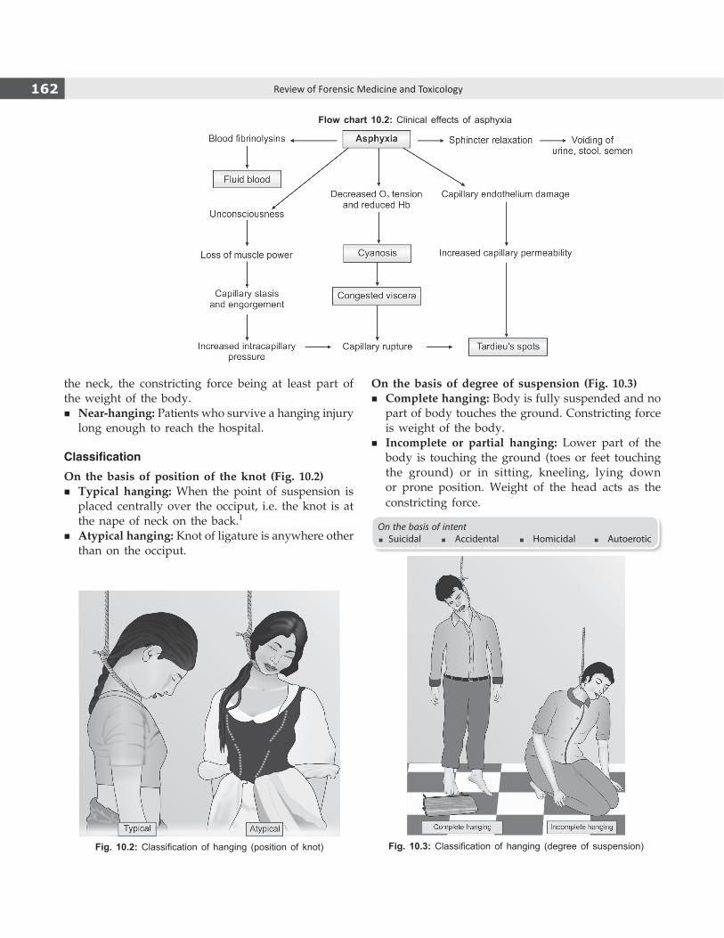
162 Review of Forensic Medicine and Toxicology
Fig. 10.2: Classification of hanging (position of knot) Fig. 10.3: Classification of hanging (degree of suspension)
the neck, the constricting force being at least part of the weight of the body.
� Near-hanging: Patients who survive a hanging injury long enough to reach the hospital.
Classification
On the basis of position of the knot (Fig. 10.2) � Typical hanging: When the point of suspension is
placed centrally over the occiput, i.e. the knot is at the nape of neck on the back.1
� Atypical hanging: Knot of ligature is anywhere other than on the occiput.
On the basis of degree of suspension (Fig. 10.3) � Complete hanging: Body is fully suspended and no
part of body touches the ground. Constricting force is weight of the body.
� Incomplete or partial hanging: Lower part of the body is touching the ground (toes or feet touching the ground) or in sitting, kneeling, lying down or prone position. Weight of the head acts as the constricting force.
On the basis of intent�Suicidal � Accidental � Homicidal � Autoerotic
Flow chart 10.2: Clinical effects of asphyxia
163Asphyxia
Cause of Death
i. Asphyxia: Constricting force of ligature causes compressive narrowing of laryngeal and tracheal lumina, leading to asphyxia.
ii. Venous congestion: Jugular veins are blocked by the ligature which results in stoppage of cerebral circulation; occurs if ligature is made up of broad and soft material.
iii. Combined asphyxia and venous congestion: Commonest cause.
iv. Cerebral anemia: It occurs when ligature is made of thin cord.
v. Reflex vagal inhibition leading to sudden cardiac arrest.
vi. Fracture/dislocation of cervical vertebrae: It is seen in judicial hanging.
Delayed deaths are rare which may be due to: � Aspiration pneumonia � Edema of lungs, larynx � Infections � Infarction of brain � Hypoxic encephalopathy � Abscess of brain.
Secondary effects of hanging in persons who have recovered (nearhanging) are:
� Hemiplegia � Epileptiform convulsions � Amnesia � Cervical cellulitis � Parotitis � Retropharyngeal abscess.
Fatal period: Death is immediate, if cervical vertebrae are fractured or if the heart is inhibited, rapid if cause is asphyxia and least rapid if coma is responsible. Usual period is 3–5 min which may extend to 5–8 min of suspension leading to death.
Autopsy of Neck (Asphyxial Deaths)
Photograph of the victim along with ligature (if present) is recommended.
External Examination
General features � Clothing and personal effects. � Distribution of lividity, rigor mortis, and algor mortis. � Bleeding from any sites, discharge of semen, urine
or fecal matter.
� Ocular or facial petechiae, congestion, cyanosis. � Tongue protrusion between clenched teeth, and
dribbling of saliva. � Evidence of any other trauma.
LigatureIs ligature present in situ or removed?
Knot: If in situ, note knot position, number of loops.
Ligature description � Type of material � Circumference of noose � Width � Nature of knot (slip-knot or fixed).
Frequently, the knot is in the form of a simple slipknot to produce a running noose, or fixed by granny or reefknot; occasionally a simple loop is used. Usually, it is present on the right or left side. If, in situ, it should be cut away from knot and reconstructed by joining cut ends with tape or another cord (Fig. 10.4).
Description of ligature mark or furrow � Course (angled or straight) � Width � Associated skin changes or trauma � Relation to thyroid cartilage � Pattern � Neck circumference at level of furrow (to determine
degree of neck constriction) � Transfer of ligature material.
Internal Examination
i. Anterior neck structures are examined at the end of autopsy—following removal of tissues and organs, and collection of toxicology samples—to allow drainage of blood and reduce the possibility of artifactual hemorrhage.
ii. Modified Y-shaped incision is preferable to expose the neck structures (Fig. 10.5).
iii. Anterior neck structures (tongue, larynx, trachea with thyroid gland, attached strap muscles including sternocleidomastoid muscles and submandibular glands) are inspected before removing them.
iv. Tongue is inspected and cut through (tip to base) to observe hemorrhage.
v. It is noted whether hemorrhages are present in the submandibular glands and strap muscles.
vi. The thyroid gland is removed and sectioned.

164 Review of Forensic Medicine and Toxicology
vii. Any hemorrhage or fracture is noted in the muscle around the cricoid, laminae of the thyroid cartilage and superior horns.
viii. Hyoid bone is palpated, and hemorrhages adjacent to the hyoid or thyrohyoid ligament are also noted. Dissect away the hyoid (note that the lesser cornua are variably long and may be inadvertently cut).
ix. Longitudinal sections through the larynx may be done to note intracartilaginous hemorrhages—in suspected hanging cases.
x. The esophagus and larynxtrachea are dissected posteriorly to observe any submucosal hemorrhage or petechiae, mucosal injuries and aspiration.
Postmortem Findings in Hanging
External Findings
1. Face i. Swollen, cyanosed face due to impaired venous
return and accumulation of blood. ii. Prominent eyeballs due to increased pressure
resulting from passive accumulation of blood. iii. Dilated pupils: If the knot presses on cervical
sympathetic, eye of the same side may remain open and pupil is dilated (la facie sympathet-ique).2 It indicates antemortem hanging.
iv. Subconjunctival hemorrhages. v. Protrusion of tongue due to pressure on floor
of the mouth by ligature. It is usually swollen and blue. Injuries include bite marks with or without underlying small hemorrhages (‘marginal’ hemorrhages).
vi. Bleeding from nose/ears due to impaired venous return and increase in pressure resulting in passive flow of blood.
vii. Lips and mucous membrane of mouth are blue. viii. Dribbling of saliva: Surest sign of antemortem
hanging.3,4 Excessive salivation occurs when the person is alive, due to pressure and friction caused by ligature material on the submandibular glands. Dribbling of saliva occurs from the angle of mouth which is at a lower level, i.e. from angle opposite to the side of knot. When the knot is on the nape of the neck, it occurs across the middle of lower lip.
Figs 10.4A to C: (A) Fixed noose, (B) Running noose, (C) Method of cutting the noose and preserving the cut ends and the knot
A B C
Fig. 10.5: Modified Y-shaped incision

165Asphyxia
2. Neck i. Ligature mark (‘furrow’)
z Site: Usually above the hyoid bone. z Size/shape: Depends on the type of material used. z Direction: It runs obliquely, backwards,
noncontinuous, upwards and towards the point of suspension. Mark is noncontinuous because of a gap at the nape of neck, and hair intervening between ligature material and the skin underneath. When the knot is in contact with the skin, it is usually inverted ‘V’ shaped, due to extension of ligature material downward on both sides from the knot above (Fig. 10.6).
z Skin at the site: Usually depressed/grooved, pale in color, but later becomes yellowish brown, dry, hard and parchmentlike with small abrasions at its edges, corresponding to the thickness and edges of the rope. These abrasions, known as rope burns, are due to frictional force.
z The pattern of ligature may be repro duced in the furrow.
z Postmortem blisters may be seen on skin squeezed adjacent to the furrow.
z An abraded area below the furrow may indicate upward slippage of the ligature, usually seen when suspension is complete.
z Neck veins above the furrow may be distended. ii. Dimension of neck: Due to prolonged sus pension,
the neck becomes slender and increases in length. iii. Bending of neck: Neck gets flexed to the side
opposite to the knot.
3. Other parts of body i. Tardieu’s spots: May be present on forehead, over
the eyelids, under the conjunctiva and near the temple.
ii. Cyanosis of fingernails. iii. Purple colored postmortem staining in the lower
limbs and lower regions of upper limbs (hands/forearms)—glove and stocking PM staining.
iv. Hands are usually clenched. v. In males, there may be penile turgidity and
involuntary discharge of semen. vi. In both sexes, there may be an involuntary
discharge of fecal matter and urine. � Signs of asphyxia may be lacking in case of complete
hanging as death occurs almost instantaneously by vagal stimulation.
� Florid asphyxial changes can be seen in cases where a fixed knot was used or in incomplete hanging.
Based on the ligature mark in the neck, the diagnosis of antemortem hanging can be made if the following triad of characteristics is present: i. Streaks or bands of reddened or pink tissue ii. Imprint of the pattern of the ligature in the furrow iii. Sloping or upward angle towards the suspension point.Microscopically, engorgement in the reddened and pinkish area in contrast to the adjacent nonengorged and nonhemorrhagic areas may be demonstrated.
Internal Findings
� Neck i. Subcutaneous tissue underneath the ligature mark
is dry, white, firm and glistening. Platysma and sternomastoid may show hemorrhages, and are sometimes ruptured.
ii. Hyoid bone may be fractured in persons, more commonly above the age of 40 years. The fracture is usually due to ligature forcing the hyoid bone backwards, which results in increased divergence of greater horns (anteroposterior compression fracture), but it can be a traction fracture.
iii. Transverse carotid intimal tears may be seen in obese victims, long drops and posteriorly placed knots (Amussat’s sign).5
iv. Vertebral artery injuries—rupture, intimal tear and subintimal hemorrhage (most frequent) may be present.
v. Larynx and trachea are congested. vi. Fracture of superior horn of the larynx may be
present.Fig. 10.6: Ligature mark in hanging. Point of
suspension is posterior to the left ear

166 Review of Forensic Medicine and Toxicology
� Lungs: They are congested, distended and emphysematous with plenty of Tardieu’s spots subpleurally, particularly at the interfaces of the lobes.
� Brain: Congested and shows multiple petechiae. � Viscera: All the abdominal organs are congested. � Blood: Fluid and purplish in color.
There may be hemorrhages on ventral surface of the intervertebral disks beneath the anterior longitudinal ligament in the lumbar spine (Simon’s sign–a vital sign of hanging). It may also be seen in other traumatic elongation or overextension of spinal column (e.g. traffic accidents), drowning and putrefaction (‘false positive’).
Medico-legal Questions
Q. Whether the hanging was suicidal, homicidal or accidental?
� Suicide: Hanging is a common method of asphyxial suicide in many countries. Person can be between 10 and 80 years of age, more common in males. Point of suspension remains approachable to the suicider. Partial hanging is almost always suicidal in nature. A history of a previous attempt may be present, and generally committed in a secluded place (victim’s home is the most frequent site). Suicidal note may be
Differentiation 10.1: Antemortem and postmortem hanging
S.No. Feature Antemortem hanging Postmortem hanging1. Salivary dribbling mark Present Absent2. Fecal/urinary stains May be present Absent3. Ligature mark
� Direction � Continuity � Level in the neck � Parchmentization � Vital reaction
ObliqueNon-continuousAbove thyroidPresentPresent
CircularContinuousAt or below thyroid AbsentAbsent
4. Knot Single, simple, on one side of neck Multiple, granny or reef type on occiput/chin5. PM staining
� Above ligature mark � In lower limbs � Glove-stocking like
PresentPresentPresent
AbsentAbsentAbsent
6. Evidence of injury � Self-inflicted � Struggle � Tear of carotid artery intima � Imprint abrasion
PresentAbsentPresentPresent
AbsentPresentAbsentMay/may not be present
7. Elongation of neck Present Absent8. Cyanosis Deeply positive Absent or faintly present9. Emphysematous bullae on lungs Absent Present
10. Point of suspension Compatible with self-suspension Not so11. Histochemistry of ligature mark Increased serotonin and histamine Not so
left behind. There should be a motive for committing suicide. Fibers of ligature material may be present in the clenched hand.
� Homicide: Very rare. Not ordinarily possible in an adult victim, unless intoxicated or made unconscious or the victim is either a child or a debilitated person.
Homicide should be suspected where: i. There are signs of violence/disorder of furniture ii. Clothing of deceased is torn or disarranged iii. There are injuries, either offensive or defensive.
� Postmortem hanging/postmortem suspension: Person may be murdered and the body suspended to simulate suicide. Look for signs of dragging to the place of suspension. Beam or branch of tree shows evidence of the rope having moved from below upwards, as the body has been pulled up. In true suicidal hanging, the rope moves from above downwards (Diff. 10.1).
� Accidental hanging: Hanging deaths in children < 6 years are usually accidental. It has been reported among children while ‘playing hanging’ (e.g. pretending to be a cowboy) or playing ‘Lasso’ or getting suspended from playground equipment, and sometimes even in adults (e.g. autoerotic hanging).
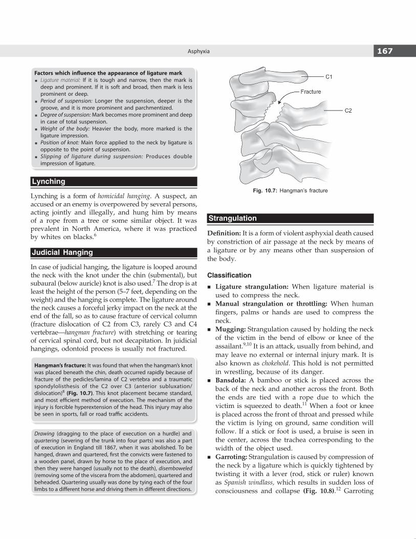
167Asphyxia
Factors which influence the appearance of ligature mark � Ligature material: If it is tough and narrow, then the mark is
deep and prominent. If it is soft and broad, then mark is less prominent or deep.
� Period of suspension: Longer the suspension, deeper is the groove, and it is more prominent and parchmentized.
� Degree of suspension: Mark becomes more prominent and deep in case of total suspension.
� Weight of the body: Heavier the body, more marked is the ligature impression.
� Position of knot: Main force applied to the neck by ligature is opposite to the point of suspension.
� Slipping of ligature during suspension: Produces double impression of ligature.
Lynching
Lynching is a form of homicidal hanging. A suspect, an accused or an enemy is overpowered by several persons, acting jointly and illegally, and hung him by means of a rope from a tree or some similar object. It was prevalent in North America, where it was practiced by whites on blacks.6
Judicial Hanging
In case of judicial hanging, the ligature is looped around the neck with the knot under the chin (submental), but subaural (below auricle) knot is also used.7 The drop is at least the height of the person (5–7 feet, depending on the weight) and the hanging is complete. The ligature around the neck causes a forceful jerky impact on the neck at the end of the fall, so as to cause fracture of cervical column (fracture dislocation of C2 from C3, rarely C3 and C4 vertebrae—hangman fracture) with stretching or tearing of cervical spinal cord, but not decapitation. In juidicial hangings, odontoid process is usually not fractured.
Hangman’s fracture: It was found that when the hangman’s knot was placed beneath the chin, death occurred rapidly because of fracture of the pedicles/lamina of C2 vertebra and a traumatic spondylolisthesis of the C2 over C3 (anterior subluxation/dislocation)8 (Fig. 10.7). This knot placement became standard, and most efficient method of execution. The mechanism of the injury is forcible hyperextension of the head. This injury may also be seen in sports, fall or road traffic accidents.
Drawing (dragging to the place of execution on a hurdle) and quartering (severing of the trunk into four parts) was also a part of execution in England till 1867, when it was abolished. To be hanged, drawn and quartered, first the convicts were fastened to a wooden panel, drawn by horse to the place of execution, and then they were hanged (usually not to the death), disemboweled (removing some of the viscera from the abdomen), quartered and beheaded. Quartering usually was done by tying each of the four limbs to a different horse and driving them in different directions.
Strangulation
Definition: It is a form of violent asphyxial death caused by constriction of air passage at the neck by means of a ligature or by any means other than suspension of the body.
Classification
� Ligature strangulation: When ligature material is used to compress the neck.
� Manual strangulation or throttling: When human fingers, palms or hands are used to compress the neck.
� Mugging: Strangulation caused by holding the neck of the victim in the bend of elbow or knee of the assailant.9,10 It is an attack, usually from behind, and may leave no external or internal injury mark. It is also known as chokehold. This hold is not permitted in wrestling, because of its danger.
� Bansdola: A bamboo or stick is placed across the back of the neck and another across the front. Both the ends are tied with a rope due to which the victim is squeezed to death.11 When a foot or knee is placed across the front of throat and pressed while the victim is lying on ground, same condition will follow. If a stick or foot is used, a bruise is seen in the center, across the trachea corresponding to the width of the object used.
� Garroting: Strangulation is caused by compression of the neck by a ligature which is quickly tightened by twisting it with a lever (rod, stick or ruler) known as Spanish windlass, which results in sudden loss of consciousness and collapse (Fig. 10.8).12 Garroting
Fig. 10.7: Hangman’s fracture

168 Review of Forensic Medicine and Toxicology
as a mode of execution was practiced in Spain, Portugal and Turkey. An iron collar was tightened by a screw for strangulation.
Ligature Strangulation
Cause of death
� Asphyxia due to elevation of the larynx and tongue closing the airway at pharyngeal level.
� Cerebral anoxia due to venous congestion. � Vagal inhibition. � Rarely, fracture dislocation of cervical vertebrae.
Postmortem Examination
External Findings (Fig. 10.9)
1. Face i. Face is congested, swollen and cyanosed. Tardieu’s
spots are present on the forehead, temples, eyelids and conjunctiva; more abundant than in hanging.
ii. Eyes are prominent, wide open, conjunctiva congested, pupils dilated and subconjunctival hemorrhages are present.
iii. Lips, fingernails and ear lobules are cyanosed; postmortem staining marked on the skin above the ligature.
iv. Tongue is swollen, dark colored, may protrude out of mouth, and bitten by teeth.
v. Bloodstained frothy fluid and mucus may escape from mouth and nostrils.
2. Neck Ligature mark (‘furrow’)
z Ligature mark is a well-defined groove, which is slightly depressed and of same width as that of ligature material. Groove may be narrow at parts due to folding of ligature.
z The furrow is usually horizontally placed across the middle or lower part of neck, at or below the level of thyroid cartilage. The mark is transverse, circular and continuous.13
z Mark, though completely encircling the neck horizontally, is more prominent on the front and sides, than on the back of the neck (as the underneath skin is thick).14
z The base of the furrow is usually red, accompanied with congested or ecchymosed margins. Base may show imprint pattern of the ligature material used.
z It may be very indistinct or altogether absent, if the ligature was soft and broad, and was removed soon after death, and may need to be examined under UV light.
z Sometimes, a narrow cord or electric wire may be used, the so called ‘cheese cutter method’, the ligature mark may appear deeply embedded, and on removal, a deep groove may be seen in the skin.
z Mark may be oblique as in hanging, if the victim has been dragged by a cord, after being strangled in a recumbent posture.
3. Other parts of body i. Postmortem staining is deep and prominent. ii. There may be involuntary discharge of urine
and fecal matter—more common than in case
Fig. 10.8: A garrote
Fig. 10.9: Postmortem findings in strangulation

169Asphyxia
of hanging. Seminal ejaculation is less common than in case of hanging.
iii. Hands are usually clenched and genitals turgid. iv. In case of struggle, there may be evidence
of abrasions, fingernail scratch marks, and contusions over the face, arms and other parts of the body.
v. Scratches may be found on the skin of the neck near the ligature. They are usually horizontal, may be irregular or crescentic abrasions, consequent of the victim’s attempts to pull the ligature away from the neck. Fingernails of the deceased should be examined for fragments of skin and blood.
Internal Findings
Neck i. Bruising of the subcutaneous tissue and muscles
of neck, especially underneath the ligature and knot. There may be bruising or laceration of the sheath of carotid arteries.
ii. Injury of hyoid bone is not commonly noticed, because the level of constriction is well below, and traction on the thyrohyoid ligament is negligible.
iii. Fracture of thyroid cartilage, one or both the superior horns may be seen.
iv. Subcapsular and interstitial thyroid hemorrhages are common.
v. Fracture of cricoid cartilage is less common. vi. Rings of trachea may sustain fracture when
considerable force is applied. vii. Bruising of the root of the tongue and floor of the
mouth may occur. viii. Lymphoid follicles at the base of the tongue and
the palatine tonsils are congested. ix. Mucous membrane of the pharynx, pyriform
sinuses, epiglottis and larynx usually show areas of hemorrhagic infiltration.
x. Larynx, trachea and bronchi are congested, and contain frothy, often bloodstained mucus.
xi. Fracture/dislocation of cervical vertebrae is not common, may occur in infants if associated with twisting of the neck.
Other Findings i. Lungs are congested, edematous with numerous
subpleural petechial hemorrhages. ii. Brain is congested with petechiae in white matter. iii. All other organs are congested.
Medico-legal Questions
Q. Whether death was caused by strangulation?
� General asphyxial features of death are present. The findings in the head and neck are strongly presumptive of strangulation, which is confirmed by ligature mark on the neck.
� In absence of ligature mark in neck or deeper injury, it will be difficult to form an opinion, except from circumstantial evidence.
� The mere presence of cord or ligature around the neck of a dead body does not confirm the diagnosis, for it may be put around the neck for a malicious purpose.
� Strangulation by ligature has to be differentiated from hanging (Diff. 10.2).
Q. Whether the strangulation was suicidal, homi-cidal or accidental?
Suicidal strangulation � Suicide by strangulation is rare. The victims employ
various methods of tightening the ligature, but the person can apply a single or double knot before consciousness is lost.
� In suicidal strangulation, signs of venous congestion are very welldeveloped above the ligature and are especially prominent at the root of tongue.
� The ligature should be found in situ; body should not show any signs of violence or marks of struggle. Laryngeal fractures are rare, and injuries are mild and often confined to the single ligature mark around the neck.
� Detailed examination of the scene and of the deceased person, along with circumstances leading to the death should be investigated.
Homicidal strangulation: Strangulation should be assumed to be homicidal, until the contrary is proved. Many of the victims are women, and frequently, strangulation in them is associated with sexual intercourse.
Homicide is suspected when: � There are two or more firm knots, each on separate
turns of the ligature. � Abrasions and fingernail marks are seen. � The clothing of the victim is torn or disarranged,
indicating that a struggle has taken place. � The ligature when removed is loose.
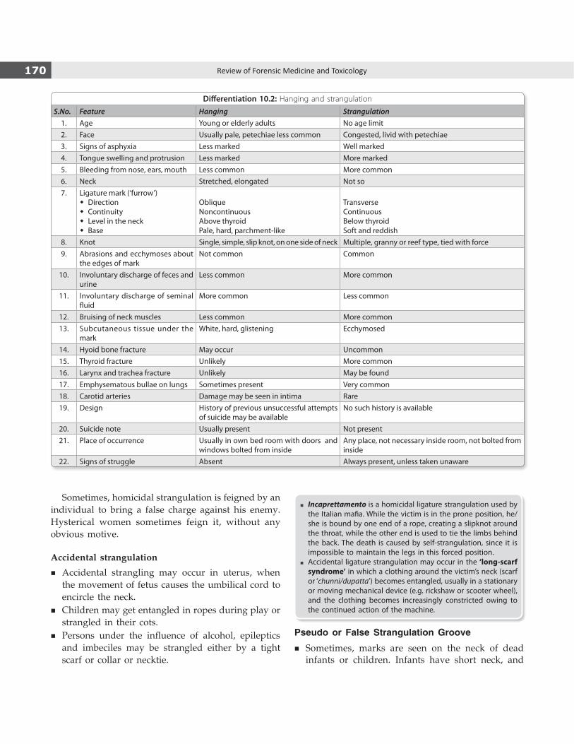
170 Review of Forensic Medicine and Toxicology
Sometimes, homicidal strangulation is feigned by an individual to bring a false charge against his enemy. Hysterical women sometimes feign it, without any obvious motive.
Accidental strangulation
� Accidental strangling may occur in uterus, when the movement of fetus causes the umbilical cord to encircle the neck.
� Children may get entangled in ropes during play or strangled in their cots.
� Persons under the influence of alcohol, epileptics and imbeciles may be strangled either by a tight scarf or collar or necktie.
� Incaprettamento is a homicidal ligature strangulation used by the Italian mafia. While the victim is in the prone position, he/she is bound by one end of a rope, creating a slipknot around the throat, while the other end is used to tie the limbs behind the back. The death is caused by self-strangulation, since it is impossible to maintain the legs in this forced position.
� Accidental ligature strangulation may occur in the ‘long-scarf syndrome’ in which a clothing around the victim’s neck (scarf or ‘chunni/dupatta’) becomes entangled, usually in a stationary or moving mechanical device (e.g. rickshaw or scooter wheel), and the clothing becomes increasingly constricted owing to the continued action of the machine.
Pseudo or False Strangulation Groove
� Sometimes, marks are seen on the neck of dead infants or children. Infants have short neck, and
Differentiation 10.2: Hanging and strangulation
S.No. Feature Hanging Strangulation1. Age Young or elderly adults No age limit2. Face Usually pale, petechiae less common Congested, livid with petechiae 3. Signs of asphyxia Less marked Well marked4. Tongue swelling and protrusion Less marked More marked5. Bleeding from nose, ears, mouth Less common More common6. Neck Stretched, elongated Not so7. Ligature mark (‘furrow’)
� Direction � Continuity � Level in the neck � Base
ObliqueNoncontinuousAbove thyroidPale, hard, parchment-like
TransverseContinuousBelow thyroidSoft and reddish
8. Knot Single, simple, slip knot, on one side of neck Multiple, granny or reef type, tied with force9. Abrasions and ecchymoses about
the edges of markNot common Common
10. Involuntary discharge of feces and urine
Less common More common
11. Involuntary discharge of seminal fluid
More common Less common
12. Bruising of neck muscles Less common More common13. Subcutaneous tissue under the
markWhite, hard, glistening Ecchymosed
14. Hyoid bone fracture May occur Uncommon15. Thyroid fracture Unlikely More common16. Larynx and trachea fracture Unlikely May be found17. Emphysematous bullae on lungs Sometimes present Very common18. Carotid arteries Damage may be seen in intima Rare19. Design History of previous unsuccessful attempts
of suicide may be availableNo such history is available
20. Suicide note Usually present Not present21. Place of occurrence Usually in own bed room with doors and
windows bolted from insideAny place, not necessary inside room, not bolted from inside
22. Signs of struggle Absent Always present, unless taken unaware

171Asphyxia
these marks are produced from folds in the skin due to bending of the head.
� They are also seen in decomposed bodies with tight collars, buttoned shirt at the neck or a necklace around the neck.
Throttling or Manual Strangulation
Definition: Asphyxia produced by compression of the neck by human hands.
Cause of death i. Asphyxia from obstruction of respiration. ii. Cerebral anoxia from interference with cerebral
circulation. iii. Vagal inhibition from pressure on carotid nerve
plexus consisting of fibers of vagus, sympathetic and glossopharyngeal nerves. About half of the deaths are due to vagal inhibition.
Pressure must be applied for 2 minutes (min) or more to cause death.
Postmortem Examination
� The external signs are abrasions and bruises on the front and sides of the neck, and are commonly at each side of the laryngeal prominence and just below the jawline. The injuries may extend onto the upper part of the sternal area.
� When pressure is prolonged, the classical signs of asphyxia may be seen—cyanosis, edema and congestion of the face, Tardieu’s spot in the eyes and face, and sometimes bleeding from nose and ears.
� The tips of the fingers produce bruises. They may be oval or round and 1.5–2 cm in size (may be more in case of continued bleeding). Presence and extent
of fingertip bruising and nail scratch abrasions will depend upon:
i. Relative position of victim and assailant. ii. Manner of grasping of neck, whether from front,
back or sides. iii. Amount of pressure exerted. iv. Whether single or both hands have been used. v. Sex, age, condition of vessels, and nutrition of
individual. vi. Condition of nails of assailant.
Important to note that: � Bruises made by tips of thumbs are more prominent
than with other fingers. � Multiple abrasions on the neck may also result from
use of victim’s hands in an effort to dislodge the assailant’s grip. These curvilinear marks commonly lie close to areas of bruising and are often horizontally orientated. If these are from the assailant, they are usually vertical (Fig. 10.10).
External Findings i. If the assailant uses single hand from front:
Thumb will be applied on one side and other fingers on opposite side of neck. A grip from right hand produces a bruising due to bulb of pressing thumb over the cornue of hyoid/thyroid on anterolateral surface of right side of victim’s neck and several fingertip bruising marks and overlying nail scratch abrasions over left side; being directed obliquely downwards and outwards, usually one below the other (Fig. 10.10). Concavities of nail markings and their direction will indicate the relative position of victim and assailant.
ii. If the assailant uses both hands: When both hands are used, evidence of pressure of thumb mark of one
Figs 10.10A and B: (A) Compression of neck with single hand and (B) External findings in neck
A B

172 Review of Forensic Medicine and Toxicology
hand and finger marks of other hand are usually found on either side of throat. In case of grip from behind, the pressure is applied all around the neck, but some areas of bruising will be more prominent than others, because of pressing fingertips.
� Because of struggle and resistance, marks of bruising and abrasions may be found over the face, nostrils, lips, chin, cheeks, forehead and lower jaw of the victim. These can also be caused in an effort to stop the victim from shouting or crying for help.
� It is, therefore, important to examine the nails of the victim and fingernail scrapings of the alleged assailant when possible, so that these can be compared with tissue type of the victim.
Internal Findings (Fig. 10.11)
i. Extravasation of blood in subcutaneous tissues underneath the external marks of bruising and abrasions is the most significant internal sign.15
ii. Tear/laceration of platysma or sternomastoid muscles may be seen.
iii. Tongue may be bruised/lacerated, may protrude out and bitten by teeth.
iv. Hemorrhages, varying from pinpoint ecchymosis to extensive extravasation may be found in mucous membrane of larynx, epiglottis, pharynx and peritonsillar region.
v. Inward compression fracture of hyoid bone is the most diagnostic finding of throttling.15,16
vi. Fracture of superior horns of thyroid cartilage is common, though both horns do not get fractured simultaneously.
vii. Ribs may be fractured, if murderer, kneels on the chest of the victim.
viii. There may be laceration of carotid sheath and tear of inner coat of carotid artery.
ix. Cricoid is usually not fractured.
Fractures of the superior horn of thyroid cartilage are not limited to fatal neck compression. Direct blunt trauma (e.g. motor vehicle impact or fall from height), resuscitation and poor autopsy technique can lead to this injury.
Medico-legal Questions
Q. Whether death was due to throttling?Diagnostic signs are: i. Bruising and abrasions on face and neck with or
without rupture of neck muscles. ii. Engorgement of tissues at and above the level of
compression. iii. Fracture of thyroid cartilages and hyoid bone. iv. General signs of asphyxia. v. Fracture of cricoid is almost pathognomic of throttling.
Q. Whether throttling was suicidal, homicidal or accidental?
� Self-throttling is impossible, because as soon as unconsciousness supervenes, the hand will relax and the grip will be released.
� Homicidal throttling: Common mode of homicide as the hand is immediately available, and method of choice in infants. Victims are usually infants, children or women (associated with sexual assault). In adults, signs of struggle are usually present, but if throat is seized firmly and compressed, victim cannot struggle. Adults can be throttled when under the influence of drugs/drinks or stunned or taken unaware. If contusions and fingernail abrasions are present on neck, the presumption must be of homicide.
� Accidental throttling: Sudden application of one or both hands on a person’s throat as demonstration of affection, in joke or as a part of physiological experiment may cause death due to vagal inhibition.
Q. How much force an assailant could have used? � If there is damage to neck structures, it indicates
use of considerable force and is indicative of intent to injure, if not to kill.
� It there is fracture of hyoid bone/larynx, it indicates use of appreciable force and is homicidal in nature.
� Minor damage or absence of damage to the neck structures can kill, e.g. karate blow.
Fig. 10.11: Larynx: Sites of fracture from lateral compression (throttling)
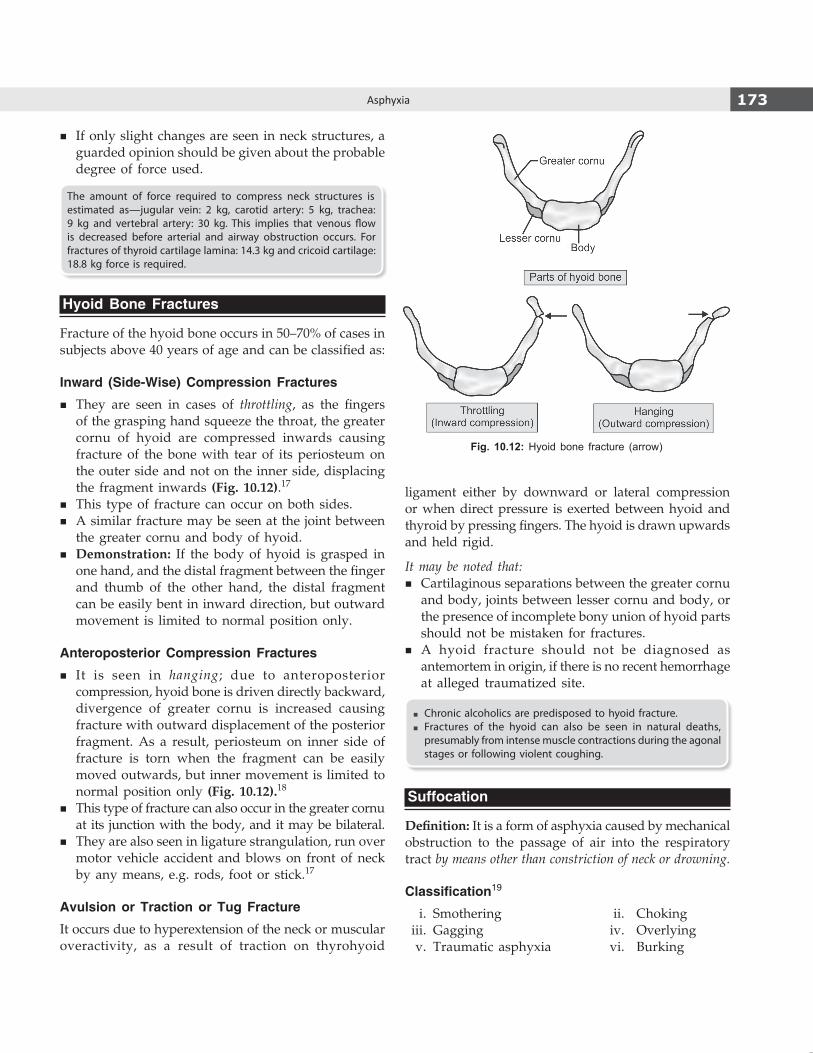
173Asphyxia
� If only slight changes are seen in neck structures, a guarded opinion should be given about the probable degree of force used.
The amount of force required to compress neck structures is estimated as—jugular vein: 2 kg, carotid artery: 5 kg, trachea: 9 kg and vertebral artery: 30 kg. This implies that venous flow is decreased before arterial and airway obstruction occurs. For fractures of thyroid cartilage lamina: 14.3 kg and cricoid cartilage: 18.8 kg force is required.
Hyoid Bone Fractures
Fracture of the hyoid bone occurs in 50–70% of cases in subjects above 40 years of age and can be classified as:
Inward (Side-Wise) Compression Fractures
� They are seen in cases of throttling, as the fingers of the grasping hand squeeze the throat, the greater cornu of hyoid are compressed inwards causing fracture of the bone with tear of its periosteum on the outer side and not on the inner side, displacing the fragment inwards (Fig. 10.12).17
� This type of fracture can occur on both sides. � A similar fracture may be seen at the joint between
the greater cornu and body of hyoid. � Demonstration: If the body of hyoid is grasped in
one hand, and the distal fragment between the finger and thumb of the other hand, the distal fragment can be easily bent in inward direction, but outward movement is limited to normal position only.
Anteroposterior Compression Fractures
� It is seen in hanging; due to anteroposterior compression, hyoid bone is driven directly backward, divergence of greater cornu is increased causing fracture with outward displacement of the posterior fragment. As a result, periosteum on inner side of fracture is torn when the fragment can be easily moved outwards, but inner movement is limited to normal position only (Fig. 10.12).18
� This type of fracture can also occur in the greater cornu at its junction with the body, and it may be bilateral.
� They are also seen in ligature strangulation, run over motor vehicle accident and blows on front of neck by any means, e.g. rods, foot or stick.17
Avulsion or Traction or Tug Fracture
It occurs due to hyperextension of the neck or muscular overactivity, as a result of traction on thyrohyoid
ligament either by downward or lateral compression or when direct pressure is exerted between hyoid and thyroid by pressing fingers. The hyoid is drawn upwards and held rigid.
It may be noted that: � Cartilaginous separations between the greater cornu
and body, joints between lesser cornu and body, or the presence of incomplete bony union of hyoid parts should not be mistaken for fractures.
� A hyoid fracture should not be diagnosed as antemortem in origin, if there is no recent hemorrhage at alleged traumatized site.
� Chronic alcoholics are predisposed to hyoid fracture. � Fractures of the hyoid can also be seen in natural deaths,
presumably from intense muscle contractions during the agonal stages or following violent coughing.
Suffocation
Definition: It is a form of asphyxia caused by mechanical obstruction to the passage of air into the respiratory tract by means other than constriction of neck or drowning.
Classification19
i. Smothering ii. Choking iii. Gagging iv. Overlying v. Traumatic asphyxia vi. Burking
Fig. 10.12: Hyoid bone fracture (arrow)

174 Review of Forensic Medicine and Toxicology
i. Smothering
Definition: It is a form of asphyxia caused by mechanical occlusion of external air passages, i.e. the nose and mouth by hand, cloth, plastic bag or other material.
Postmortem findings i. Abrasions and bruises around the mouth and
nostrils. These may not be seen, if soft materials, like cloth or pillow has been used.
ii. Injuries on the inside of the lips from pressure of teeth are seen.
iii. Bruising of gums or sometimes tears of delicate tissues are seen.
These findings may be missed, unless looked for.
Medico-legal aspects � Accidental smothering is common in alcoholics or
epileptics who may fall or roll over in a heap of mud or such other material.
� After birth, an infant may die from smothering, if he is born with membranes covering the nose and mouth (cul-de-sac).
� Children may get smothered while playing with plastic bags over the face or head.
ii. Choking
Definition: It is a form of asphyxia caused by an obstruction within the airpassages by a foreign object, like coin, fruit seed, toffees, candies, fish or any other material.20
In an epileptic attack, tongue may fall back on to posterior pharyngeal wall causing choking.
The phases of acute fatal airway obstruction are: i. Penetration of the object into the airway. ii. Obstruction of the airway. iii. Failure to expel once the obstruction has occurred.
Mechanism: Initially, there is stridor, respiratory distress, coughing and the inability of the victim to speak. This is followed by a rapid, deep inhalation, which causes the foreign object to pass further down the airway. Laryngospasm occurs, followed by vagal stimulation, leading to arrhythmia, apnea and death.
Cause of death i. Asphyxia. ii. Vagal inhibition. iii. Laryngeal spasm. iv. Delayed death from pneumonia, lung abscess or
bronchiectasis.
Postmortem findings i. Signs of asphyxial death. Subconjunctival hemorrhages
without cutaneous petechiae may be seen. ii. Presence of food items or foreign body in respiratory
tract. The food items are usually round and firm, yet pliable to allow molding in the airway.
iii. In an epileptic, tongue may show bite marks or bruising.
Medico-legal aspectsMost choking deaths are accidental; suicide and homicide are rare.21
� Accidental choking deaths are common in children < 1 year of age. Ninety percent of choking deaths happen before the age of 5 years.
� Homicidal choking usually involves the aged, individuals debilitated by disease, alcohol or drugs, and infants. When objects are forced into the mouth, signs of a struggle, if the individual was conscious, may be noted. Perioral, teeth, tongue and other intraoral injuries can result.
� Suicidal choking is uncommon, and may occur in psychiatric patients and prisoners.
Café-coronary
This is a condition of accidental choking wherein a bolus of food produces complete obstruction of the larynx.22
� It is called so, because it mimics a heart attack and is usually seen in an intoxicated restaurant patron.23
� The term, ‘cafe coronary’, was coined by Dr Roger Haugen (Medical Examiner, Broward County, Florida) in 1963.24
Causes � Predisposing factors include a decreased protective
airway reflex, resulting from aging, poor dentition, tendency to swallow food whole, alcohol consumption, and ingestion of large doses of tranquilizers and other CNS depressants impairing the gag reflex.
� Reflex cardiac arrest from ‘vagal inhibition’ as a consequence of stimulation of laryngeal nerve endings.
Clinical findings: Victim who was apparently healthy, collapses suddenly turning blue while eating at a dining table.
Treatment (for choking) i. If there is difficulty in breathing and cyanosis, give
first aid by application of pressure on the abdomen (Heimlich maneuver) till the patient recovers or loses consciousness.25

175Asphyxia
ii. A blow on the back or on the sternum may cause coughing and expel the foreign body.
iii. The victim is placed in a supine position and the mouth is opened to perform a finger sweep.
iv. If this is not successful, the foreign body should be removed from hypopharynx with the middle and index fingers or with forceps.
v. If the object cannot be removed, the person may need a tracheotomy/cricothyrotomy.
Postmortem Findings
Bolus of unchewed food or such material is found impacted in larynx or trachea. A litmus paper test of the bolus can be made to determine the acidity to ascertain its origin (mouth or vomitus).
Medico-legal Aspects
It is a case of accidental death (asphyxia) as opposed to natural, so additional insurance claims can be made.26
‘Creche coronary’: Choking occurring in children aged 1–3 years as they are more vulnerable because of their increased mobility, putting inappropriate small objects in their mouth or appreciate the size of a piece of food, small airways, inadequate dentition for chewing and weaker cough reflex.
iii. Gagging
Definition: Gagging is a form of asphyxia which results from pushing a gag (rolled up cloth or paper balls) into the mouth, sufficiently deep to block the pharynx. It combines the features of smothering and choking. Initially, the airway may be patent through nose, but collections of saliva, excessive mucus with edema of pharynx and nasal mucosa causes complete obstruction.
Postmortem findings i. Same as choking. ii. Injuries to nose and mouth with seepage of blood
into the back of throat.
Medico-legal aspects � Almost always homicidal, and the victim is usually
an infant or an elderly person. � Gagging is usually resorted to prevent the victim
from shouting for help; death is usually unintended. � Gags have been used to suppress screams by
victims using a painful method of suicide (e.g. selfimmolation).
iv. OverlayingDefinition: Overlaying or compression suffocation results from compression of the chest, nose and mouth, so as to prevent breathing.27
� It is a form of accidental smothering of an infant by a nursing mother, sharing a bed with her child who may roll over during sleep and occlude the air passages.
� Ethanol intoxication or a medical condition can be a factor depressing an arousal response in the older bedsharer.
Postmortem findings: Face, nose and chest of victim child may appear compressed and pale. Pressure marks from bedding or clothing may be seen on the victim, but these can happen postmortem. Usual findings of asphyxia will be seen along with intrathoracic petechiae.
Medico-legal aspects � Purely accidental in nature. � It may also be a case of infanticide. � These cases are likely to be victims of sudden infant
death syndrome (SIDS).
Traumatic or Crush Asphyxia / Perthes SyndromeDefinition: Asphyxia resulting from respiratory arrest due to mechanical fixation of chest, so that the normal movements of chest wall are prevented.
Causes28
i. Due to house collapse, accidentally or in wars/earthquake.
ii. Stampede by crowd, running in panic, e.g. due to outbreak of fire in a movie hall/mall/public gathering.
iii. Run over by a vehicle or overturned vehicle (especially tractors).
iv. Collapse of wall inside a mine or trenches (cavein), in bunkers of sand or grain.
v. When held between the buffers of two bogies of a train.
vi. Restraint of suspects by hogtying practiced in some States in the US by police.
Mechanism: The essential feature is fixation of the thorax by severe compression or external pressure that prevents respiratory movements. An individual can die in seconds if there is considerable weight, but usually at least 2–5 min elapse before death ensues.
Postmortem examinationExternal findingsCharacteristic features seen are: i. Masque ecchymotique refers to the classical
appearance of:

176 Review of Forensic Medicine and Toxicology
a. Florid red or blue congestion of face and neck with variable involvement of the upper thorax, back and arms.
b. Deep cyanosis of face. c. Numerous petechial hemorrhages or ecchymoses. d. Demarcation line: Level of compression is
indicated by a well-defined demarcating line between the discolored upper portion of body and the lower normal part.
ii. Areas of pallor seen at the level of collar of shirts, folds or creases in the garments.
iii. Facial edema. iv. External blunt trauma injuries can be seen on the
head, neck and chest along with mud or other foreign material.
Internal findings � Eyes: Purtscher’s retinopathy (retinal hemorrhages). � Face: Nose, ear or pharyngeal petechiae/ecchymoses
that may result in external bleeding—mimic a basal skull fracture.
� Bones: Rib and clavicle fractures are common; extremity and pelvic fractures may be seen.
� Upper respiratory tract: Edema, epiglottic and laryngeal petechiae.
� Lungs: Congested, heavy, subpleural petechiae; contusions/lacerations and hemo/pneumothorax may be present.
� Heart: Right heart and veins above aorta may be distended, injuries are rare.
� Abdomen: Hepatic and splenic lacerations may be found.
� CNS: Edema and petechiae can be seen.
Medico-legal aspectsMostly accidental, but fallen appliances or furniture particularly over children has been described as a means of homicide.
� In survivors, discoloration disappears within a few weeks and does not undergo the color changes seen with the healing of bruises. The color is not altered by the administration of oxygen. Petechiae disappear within days, but subconjunctival ecchymoses can persist for weeks, eventually fading to yellow and disappearing.
� Mechanism of masque ecchymotique: Retrograde displacement of blood from superior vena cava into the subclavian veins and veins of the head and neck results from sudden compression of the chest or abdomen. Valves in the subclavian veins prevent the spread of hydrostatic force to the veins of upper limbs. But, the displacement of the blood into the valve-less veins of the head and neck causes rupture of distal capillaries. Therefore, face and neck of the victim are deeply cyanosed; eyes bloodshot and numerous petechiae over scalp, face, neck and shoulders are seen.
vi. Burking
� It is a combination of homicidal smothering and traumatic asphyxia (Fig. 10.13).29
� William Burke and William Hare killed 16 persons during 1927–28 in Scotland and sold their bodies to Dr Robert Knox for use as specimens in his anatomy classes in Edinburgh Medical School, in what became known as the case of the Body Snatchers (West Port murders).
� Method: A victim was invited to their house and given alcohol. When drunk, he was thrown on the ground. Burke would kneel or sit on the chest and close the nose and mouth with his hands, and Hare used to pull him around the room by the feet till he is dead.
� Plastic bag asphyxia results from decreased oxygen concentration in the available inspired air, and physical obstruction of the mouth and nose. The plastic bag becomes electrically charged and adheres to the face, aided by condensation. It is a common method of suicide among the elderly and debilitated individuals. It can also be seen in autoerotic asphyxia, drug misadventure—volatile inhalants (e.g. chloroform or propane), inhalation of volatile hydrocarbons (e.g. trichloroethane) or accidental deaths in children.
� Wedging is a form of mechanical asphyxia in which the face, neck or thorax is compressed between two firm structures. It is common in 3–6 months old children when they start to move to the corners of beds and cribs, but they do not have the muscle development to free themselves out of a wedged position. They become wedged between the mattress and either the wall, bed frame, a piece of furniture, mesh or another mattress.
� Confined space entrapment: It occurs when there is inadequate oxygen in the enclosed space due to consumption or displacement by other gases. The mechanism of death is usually attributed to asphyxia from oxygen deprivation. Injuries identified at autopsy and damage to the inside of the structure indicate struggle to exit the cabinet. There are no specific autopsy findings or significant natural disease processes, and toxicology studies are negative.
Fig. 10.13: Burking

177Asphyxia
Drowning
Definition: Drowning is the process of experiencing respiratory impairment from submersion/immersion in liquid.
� Implicit in this definition is that a liquid-air interface is present at the entrance to the victim’s airway, which prevents the individual from breathing oxygen.
� Outcome may include delayed morbidity, delayed or rapid death, or life without morbidity.
� Terms wet or dry drowning, active or passive drowning, neardrowning and secondary drowning would be discarded (World Congress on Drowning, Amsterdam, 2002).
Drowning was previously defined as immediate death secondary to asphyxia while immersed in a liquid, usually water, or within 24 h of submersion. The definition excluded aspiration of vomit, blood, saliva, bile or meconium.
Classification (Flow chart 10.3)
I. TyPICAL Or WET DrOWNING
Water is inhaled into the lungs and the victim has severe chest pain (seen in 80–90% of cases). It is also known as primary drowning.
� In fresh water and brackish water drowning (0.5–0.6% NaCl), the aspirated water is rapidly absorbed from the alveoli into the circulation leading to hemodilution and hemolysis. Circulatory overload, hyponatremia,
hyperkalemia, together with myocardial hypoxia result in fall of systolic blood pressure followed by ventricular fibrillation (Flow chart 10.4 and Fig. 10.14).30
� In sea (salt) water drowning (3–4% salinity), the aspiration of water results in withdrawal of water from the pulmonary circulation into the alveolar spaces as a result of the osmotic differential, while at the same time electrolytes (sodium, chloride, magnesium from sea water) pass into the blood. There is hemoconcentration with crenation of RBCs, but not hemolysis and little change in the sodium/potassium balance.31,32 The pulse pressure decreases slowly and is followed by AV dissociation, but not ventricular fibrillation (Flow chart 10.4 and Fig. 10.15).
In both fresh water and salt water drowning, there is terminal pulmonary edema.
Flow chart 10.3: Classification of drowning
Flow chart 10.4: Mechanism of fresh and sea water drowning

178 Review of Forensic Medicine and Toxicology
II. ATyPICAL DrOWNING
1. Dry drowning z In dry drowning, water does not enter the lungs
due to laryngeal spasm induced by small amounts of water entering the larynx.33
z Seen in 1–2% of cases. z Death may be extremely rapid and time elapsed
is insufficient for typical drowning to occur. Two mechanisms have been postulated:
i. Reflex cardiac arrest due to vasovagal stimulation.
ii. Laryngeal spasm and airway closure causing lethal hypoxemia.
z In these cases, autopsy findings and tests for drowning are negative, and the lung fields are dry.
2. Immersion syndrome (Hydrocution, submersion inhibition or cold water drowning): It refers to syncope resulting from cardiac dysrhythymias on sudden contact with water that is at least 5°C lower than body temperature.34
z The syndrome occurs as a result of: i. Cold water stimulating the nerve endings of
the surface of the body.
Fig. 10.14: Mechanism of death in fresh water drowning
Fig. 10.15: Mechanism of death in sea water drowning
179Asphyxia
ii. Water striking the epigastrium. iii. Cold water entering eardrums, nasal passages,
pharynx and larynx. iv. Falling or diving into water with feet first or
duck diving by the inexperienced. z Mechanism: Vagal stimulation leading to asystolic
cardiac arrest (‘diving reflex’), or ventricular fibrillation secondary to QT prolongation after a massive release of catecholamine on contact with cold water.35 The resultant loss of consciousness leads to secondary drowning.
z The findings of typical drowning are absent, and diagnosis of hydrocution is difficult because aspiration of water into the lungs does not occur.
z The syndrome particularly affects the middleaged or elderly men who have ingested some amounts of ethanol. Underlying cardiac disease could increase the risk of sudden collapse.
3. Near drowning (post-immersion syndrome or secondary drowning)
z Near drowning refers to survival beyond 24 h after a submersion episode.
z Death is caused by complications or sequelae (e.g. ARDS, pneumonia, sepsis, hypoxicischemic encephalopathy, cerebral edema and DIC).
z Secondary drowning sometimes refers to a victim who initially responds well to resuscitation but then suffers respiratory decompensation.
4. Shallow water drowning (submersion of the un-conscious): Alcoholics, drugged, epileptics, infants, children and unconscious persons may die due to drowning in shallow water in a pit or drain.
Epidemiology � Drowning victims are predominantly male (> 65%).
It occurs in the summer months, more frequently seen in rivers, lakes, ponds and creeks.
� The age groups affected are the children (< 4 years) and young adults (15–24 years). Drugs and alcohol abuse among the teenagers are other associated factors.
Cause of Death i. Asphyxia: Most common cause of death. ii. In fresh water drowning, death results from
ventricular fibrillation. While in salt water, it is due to cardiac arrest from fulminant pulmonary edema and associated changes.
iii. Vagal inhibition due to impact with water. iv. Laryngeal spasm. v. Concussion/head injury.
vi. Apoplexy: Subarachnoid hemorrhage from rupture of berry aneurysm or cerebral hemorrhage by rupture of cerebral vessels from sudden onrush of blood to the brain due to excitement or sudden fall from height into cold water.
vii. Secondary causes z Septic aspiration pneumonia z Sudden bursting of aneurysm.
Symptoms: Apart from recalling of memory of past events, there may be mental confusion along with auditory and visual hallucinations, tinnitus and vertigo. In wet drowning, there is chest pain.
Treatment: First and immediate step consists of application of artificial respiration with closed chest cardiac massage, even in absence of pulse and respiration and irrespective of injuries sustained during drowning. Defibrillator should be used when there is ventricular fibrillation.
Fatal period � Fresh water drowning: 4–5 min.36
� Sea water drowning: 8–12 min.
Postmortem Examination
� The diagnosis of drowning is one of exclusion. � Most of the signs are not specific of death due to
drowning and are rather signs of submersion of body under water for some period. Any dead body, whatever the cause of death, will develop signs of immersion if left for a sufficient time in water.
� Moreover, some of the signs are not appreciable in case of putrefaction.
When freshly removed from water, the body and clothes will be wet. There will be sand and mud particles on the body, hair and clothes. This finding is not specific of antemortem drowning or death due to drowning.
External Findings
i. Face is pale, becomes bloated and discolored with putrefaction. Cyanosis is present.
ii. Eyes are found half open or closed, conjunctiva suffused and pupils are dilated. Subconjunctival hemorrhages may be present in lower eyelids.
iii. Tongue may be swollen and protruded. iv. Postmortem staining: Light pink in color, present
over face, neck, front of upper part of chest, upper and lower limbs as the body usually floats with

180 Review of Forensic Medicine and Toxicology
face down, buttocks up, legs and arms hanging down in front of the body (Fig. 10.16). With onset of putrefaction, skin of head and neck become dark with ‘tête de négre’ appearance.*
v. Froth: Presence of fine, copious white ‘shaving-lather’ like froth at the mouth and nostrils is the most characteristic antemortem external finding.37
Production of this tenacious, fine, lathery foam is a vital phenomenon.
z The mass of foam, consisting of fine bubbles, does not collapse when touched with the point of a knife.
z It may be absent when wiped off, but reappears again by itself or by applying simple pressure on chest.
Mechanism of production of froth: The inhalation of water irritates the mucous membrane of air passages due to which the tracheal and bronchial glands secrete large quantities of tenacious mucus, and the alveolar lining cell irritation produces edema fluid. Vigorous agitation of the seromucoid secretion, surfactant, aspirated water and retained air converts the mixture of endogenous and drowning medium into froth.
Other conditions in which froth can be seen:� Strangulation � Electric shock� Putrefaction � Acute pulmonary edema� Epileptic fit � Opium/OPC poisoning
In all these cases, froth is not fine, not of such large quantity or tenacious in nature as in drowning.
vi. Cutis anserina (goose skin/goose flesh/goose bumps/horripilation) is a state of puckered and granular appearance of skin of the extremities immersed in cold water due to contraction of erector pilorum
muscles. It can occur on submersion of the body in cold water immediately after death while the muscles are still warm and irritable, and also produced by rigor mortis of erector muscles.
vii. Washerwomen’s hand is the wrinkled, sodden, bleached appearance of palms, palmer aspect of fingers and soles of feet including plantar surface of toes due to submersion of the body. Maceration of skin occurs due to imbibition of water into its outer layers. It is first seen in the fingertips by 3–4 h and whole hand by 24 h.
viii. Scrotum and penis get retracted in contact with cold water in winter months.
ix. Grass, gravel, mud, sand, weeds or aquatic vegetations held firmly in clenched hands due to cadaveric spasm is a vital proof of antemortem drowning.38 The material clenched in the hands indicates the place of submersion.
x. Rigor mortis appears early, especially when a violent struggle for life has taken place before death.
xi. Antemortem injuries might be sustained during fall into water, along the tank or by striking against a hard object while diving in shallow water. Examination of the skin for blunt injuries should be delayed until the body is dry. Abrasions are easily seen after drying, which becomes brownish in color.
Internal Findings
1. Lungs i. Lungs are voluminous, distended and show balloon-
ing, i.e. bulge out of chest on removal of sternum (Diff. 10.3). Tenacious, lathery froth in trachea and bronchi is present. In case of laryngeal spasm, there will be no ballooning.
ii. Distended lungs will show indentations of ribs on the pleural surface because of pressure on increased volume of lungs.
iii. Lungs feel heavy, boggy and doughy; will easily indent on pressure by fingers because of water logging and edematous condition.
iv. Lungs may be congested, but are often pale gray in appearance because of forcing out of blood from lungs and compression of vessels in the interalveolar septa by the trapped air and water in lung alveoli.Fig. 10.16: Position of a submerged dead body
* Tête de négre is the French name for a dessert, a pastry covered with black chocolate, which literally means ‘niggerbrown’ color.
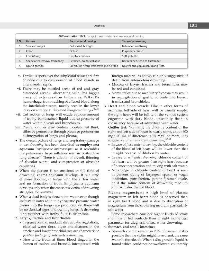
181Asphyxia
v. Tardieu’s spots over the subpleural tissues are few or none due to compression of blood vessels in interalveolar septa.
vi. There may be mottled areas of red and gray distended alveoli, alternating with few bigger areas of extravasation known as Paltauf’s hemorrhage, from tracking of effused blood along the interlobular septa; mostly seen in the lower lobes on anterior surface and margins of lungs.39,40
vii. Cut section of lungs will exude copious amount of frothy bloodstained liquid due to presence of water within alveoli and bronchioles.
viii. Pleural cavities may contain bloodstained fluid, either by permeation through pleura or postmortem disintegration of lungs and pleurae.
� The overall picture of lungs and respiratory passage in wet drowning has been described as emphysema aquosum (emphyseme hydroaerique) as it resembles the pulmonary hyperinflation seen in obstructive lung disease.41 There is dilation of alveoli, thinning of alveolar septae and compression of alveolar capillaries.
� When the person is unconscious at the time of drowning, edema aquosum develops. It is a state of mere flooding of lungs with the airless water and no formation of froth. Emphysema aquosum develops only when the conscious victim of drowning struggles for survival.
� When a dead body is thrown into water, even though hydrostatic lungs (due to hydrostatic pressure water passes into the lungs) are produced, yet there will be no classical signs of drowning lungs. A drowning lung together with frothy fluid is diagnostic.
2. Larynx, trachea and bronchioles z Presence of sand, mud, slit, dirt, aquatic vegetations,
classical water flora, algae and diatoms in the trachea and lower bronchial tree are characteristic positive findings of antemortem drowning.
z Fine white froth, at times blood tinged in the lumen of trachea and bronchi, interspersed with
foreign material as above, is highly suggestive of death from antemortem drowning.
z Mucosa of larynx, trachea and bronchioles may be red and congested.
z Vomit reflex due to medullary hypoxia may result in regurgitation of gastric contents into larynx, trachea and bronchioles.
3. Heart and blood vessels: Like in other forms of asphyxia, left side of heart will be usually empty; the right heart will be full with the venous system engorged with dark blood, unusually fluid in consistency because of admixture with water.
Gettler test: Normally, the chloride content of the right and left side of heart is nearly same, about 600 mg/100 ml. If difference is 25 mg% or more, it is suggestive of antemortem drowning.42,43
z In case of fresh water drowning, the chloride content of the blood of left heart will be lower than that in right because of dilution by water.
z In case of salt water drowning, chloride content of left heart will be greater than right heart because of hemoconcentration and mixing with salt water.
z No change in chloride content of heart is seen in persons dying of laryngeal spasm or vagal inhibition, putrefaction, patent foramen ovale, or if the saline content of drowning medium approximates that of blood.
Plasma magnesium: A high level of plasma magnesium in left heart blood is observed than in right heart blood and is due to absorption of magnesium from the drowning medium, particularly salt water.
Some researchers consider higher levels of serum strontium in left ventricle than in right as the best parameter for diagnosis of sea water drowning.
4. Stomach and small intestines z Stomach contains water in 70% of cases, but it is
possible that the victim might have drunk the same water before death. When a disagreeable liquid is found which could not be swallowed voluntarily
Differentiation 10.3: Lungs in fresh water and sea water drowning
S.No. Feature Fresh water drowning Sea water drowning
1. Size and weight Ballooned, but light Ballooned and heavy
2. Color Pinkish Purplish or bluish
3. Consistency Emphysematous Soft, jelly-like
4. Shape after removal from body Retained, do not collapse Not retained, tend to flatten out
5. On cut section Crepitus is heard, little froth and no fluid No crepitus, copious fluid and froth

182 Review of Forensic Medicine and Toxicology
and which corresponds to drowning medium, like muddy water, it is a valuable indication of ante mortem drowning.
z Water is not found in the stomach, if the person died from shock, syncope, putrefaction, or was already dead (postmortem submersion).
z Small intestine may contain water in about 20% cases. This is regarded as positive evidence of death by drowning as it depends on peristaltic movement, which is a vital phenomenon.
Water may enter the mouth and pass down into the stomach passively if the water is turbulent, rather that the victim actively swallowing it. It may also be due to the postmortem relaxation of the gastroesophageal sphincter, which allows water to enter the stomach.
5. Brain: Congested gray matter, softening and loss of graywhite junction.
6. Liver, spleen and kidneys are congested.7. Middle ear: Presence of water and hemorrhage in
middle ear is claimed to be one of the positive proof of antemortem drowning. Hemorrhages in petrous temporal bone or in mastoid air cells may be seen. Temporal bone hemorrhages are also seen in death due to hanging, head injury or carbon monoxide poisoning.
8. Ethmoid and sphenoid sinuses: Water may enter the respiratory sinuses; the jugum sphenoidale may be removed to expose the contents of the sphenoid sinus.
9. Diatom test: Diatoms belong to the class Bacillario-phyceae, and are microscopic unicellular algae which secrete silicon skeletons called frustules (Fig. 10.17).44 They are chemically inert and almost indestructible, being resistant to strong acids. During drowning, diatoms (size upto 60 µ) enter the circulation via the lungs through the ruptured alveolar walls, lymph channels and pulmonary veins into left heart and then into general circulation, when the person is alive (Fig. 10.18).
z Presence of diatoms in the lung substance, blood stream, brain, liver, kidneys, bone marrow of femur (best site for analysis) or humerus or in the skeletal muscle has been claimed to be suggestive proof of antemortem drowning.45
z Since diatoms resist putrefaction, diatom test may have some value in examination of decomposed bodies.
z The test is negative in dead bodies thrown in water and in dry drowning.
Procedure: A sample of tissue is carefully retrieved to avoid surface contamination. Approximately 50 g of tissue is taken and placed in 50 ml of concentrated nitric acid in a boiling flask. The flask is heated for 48 h, cooled and the liquid is centrifuged for 20–30 min. The supernatant is discarded and the sediment is recentrifuged. The final residue is aspirated, placed on a clean glass slide and air dried. It is then examined for silica skeletons of diatoms, which are birefringent, using phasecontrast microscopy or dark ground illumination. A water sample is collected at the time of body retrieval in a clean container and similarity of different species of diatom is compared.
Interpretation The presence of diatoms supports the diagnosis of
drowning, while the absence of diatoms does not exclude it as a cause. The diatom test is valid only if it can be shown that:
z Deceased did not drink this water immediately before submersion or exposed to longterm repeated contact with the same source of diatom containing water.
z Species recovered from specimen are present in the sample from site of drowning.
z The various species are present in same order of dominance for the admissible size range and in approximately same proportions.
Fig. 10.17: Different types of diatoms

183Asphyxia
The test is limited by the difficulty of excluding the possibility of environmental contamination. Diatoms are ubiquitous in the environment and may enter the circulation via the GIT (as contaminants of foods, such as salads, watercress and shellfish) or via the respiratory tract (diatoms are normally present in small numbers in the air, in some paints, building plasters and dusts).
Still, the diatom test is considered as the ‘gold standard’ for diagnosis of ‘typical drowning’. The ideal diagnostic test as definite proof for drowning still needs to be established. At present, the combination of the autopsy findings and the diatom test is a good compromise in arriving at a conclusion.
Medico-legal Questions
Q. Whether death was due to drowning?Drowning is one of the most difficult cause of death to prove at postmortem, especially when the body is not examined in a fresh condition (Diff. 10.4). In doubtful cases, where definite opinion cannot be given, viscera and body fluids should be preserved for
chemical analysis. Sometimes, the cause of death may have to be given as ‘consistent with drowning’.
Q. Whether drowning was accidental, suicidal or homicidal?
� Accidental drowning is most common, and seen in children, bathers, fishermen, dockworkers, intoxicated and epileptic subjects.46 Women may fall accidentally in a well, while drawing water.Accidental drowning may occur in precipitate labor, when the baby may fall into a bathtub or lavatory pan and die.
z Information regarding inability to swim, trauma, seizure disorder, heart disease, exhaustion, and alcohol or drug abuse should be sought.
� Suicide by drowning is fairly common in India, especially among females.
z Women usually make sure to tie up their clothes in such manner that their private parts are not exposed after death. Sometimes, a woman takes her child with her.
z A determined suicider may tie his hands and legs together or attach weights to his body before immersion. Likewise, he may take poison, cut his throat and jump into a well.
Fig. 10.18: Principle of diatom test

184 Review of Forensic Medicine and Toxicology
z If an adult is found drowned in shallow water, the presumption is usually suicide, unless proved otherwise.
z Information/findings that may assist in the determination of suicide: Witnesses, clothes and personal effects found stacked by the water, a suicide note and suicidal ideation, a history of cancer or terminal illness, recent bizarre behavior or depression, and associated self-inflicted wounds.
� Homicidal drowning is not very common, though it is one of the methods of choice in infanticide, especially of newborns.
z While injuries may be found in a case of homicide, it is very easy to drown a person without leaving any suspicious mark behind, especially if the person is nonswimmer, intoxicated or already inside water taking a bath.
z Victims of homicidal violence may be placed in the water after death in order to dispose off the body.
Hyperventilation DeathsFor long underwater swimming, the swimmer may hyperventilate before going down. While swimming, the oxygen gets utilized and the CO2 produced, being low in tension is not sufficient to stimulate the respiratory center and the swimmer may then suddenly become unconscious and get drowned. In skin diving, a mask and fins are used, and it is an extension of swimming with similar hazards.
Sexual Asphyxia (Autoerotic Asphyxia /Hypoxyphilia / Asphyxiophilia)
Definition: Autoerotic asphyxia is a paraphilia in which sexual arousal and orgasm depend on selfinduced asphyxia upto, but not including loss of consciousness.
� Partial asphyxia caused by pressure on carotid vessels or obstruction of air passages causes cerebral ischemia, and may lead to hallucinations of an erotic nature in some men.
� The degree of asphyxia produced by mechanical means is controlled, i.e. the victim is in a position that allows selfrelease, but in some cases, death occurs accidentally.47
� These cases are associated with some form of abnormal sexual behavior, usually masochism, cisvestism and transvestism.
� Victims are usually young males, scene is usually the victim’s own house, bedroom, bathroom, basement, and the door is locked from inside.
Methods
i. Hanging: Most frequent method. The presence of padding under the noose, nakedness of the victim, feminine attire and exposed genitalia are the hallmarks of these deaths. Frequently, the person ties his arms, legs and sometimes waist and genitalia (bondage) with a rope, string or chain.
ii. Sexual gratification may be obtained by electrical stimulation. For this, electrodes are applied to the genitals or on abdomen; usually a low voltage supply from a battery is used.
iii. Other methods include covering the head in plastic or some impervious bag, which may be secured around the neck by an elastic band to achieve partial anoxia. It is sometimes combined with the inhalation of volatile solvents (‘glue sniffing’). Carbon tetrachloride, paint thinners, petrol or amylacetate are either directly inhaled from container or rebreathed after placing in a plastic bag.
Differentiation 10.4: Antemortem drowning and postmortem submersion
S.No. Feature Antemortem drowning Postmortem submersion1. Froth over mouth and nostrils Fine, lathery froth, appears spontaneously Absent, even if present, it is coarse, not spon taneous2. Cadaveric spasm in hands Aquatic vegetations, mud may be present Not observed3. Trachea and bronchioles Presence of algae, mud along with frothy
mucusAbsent
4. Lungs Ballooned up, bulky, edematous, bear indentations of ribs
Collapsed, decomposed
5. Mud and algae in stomach and small intestine
May be present Absent
6. Diatom and Gettler tests Positive Negative7. Injuries If present, need to be consistent with
drowningInjuries inconsistent with drowning
8. Other suggestive signs Water in middle ear, retracted genitals, cutis anserina, washerwoman’s hands, wet clothing, mud and sand
Water is never present in middle ear; others are not valuable and corroborative findings
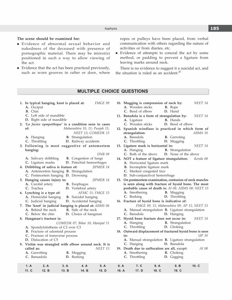
185Asphyxia
The scene should be examined for: � Evidence of abnormal sexual behavior and
nakedness of the deceased with presence of pornographic material. There may be mirror(s) positioned in such a way to allow viewing of the act.
� Evidence that the act has been practiced previously, such as worn grooves in rafter or door, where
ropes or pulleys have been placed, from verbal communication with others regarding the nature of activities or from diaries, etc.
� Evidence of attempts to conceal the act by some method, or padding to prevent a ligature from leaving marks around neck.
There is no evidence to suggest it a suicidal act, and the situation is ruled as an accident.47
multiple ChoiCe questions
1. In typical hanging, knot is placed at: FMGE 09 A. Occiput B. Chin C. Left side of mandible D. Right side of mandible 2. ‘La facies sympathique’ is a condition seen in cases
of: Maharashtra 10, 11; Punjab 11; NEET 13; COMEDK 13 A. Hanging B. Strangulation C. Throttling D. Railway accidents 3. Following is most suggestive of antemortem
hanging: DNB 09
A. Salivary dribbling B. Congestion of lungs C. Ligature marks D. Petechial hemorrhages 4. Dribbling of saliva is feature of: JIPMER 14 A. Antemortem hanging B. Strangulation C. Postmortem hanging D. Drowning 5. Hanging causes injury to: JIPMER 14 A. Carotid artery B. Esophagus C. Trachea D. Vertebral artery 6. Lynching is a type of: AFMC 11; FMGE 13 A. Homicidal hanging B. Suicidal hanging C. Judicial hanging D. Accidental hanging 7. The ‘knot’ in judicial hanging is placed at: AIIMS 06 A. Behind the neck B. Side of the neck C. Below the chin D. Choice of hangman 8. Hangman’s fracture is: COMEDK 07; Bihar 10; Manipal 11 A. Spondylolisthesis of C2 over C3 B. Fracture of odontoid process C. Fracture of transverse process D. Dislocation of C5 9. Victim was strangled with elbow around neck. It is
called as: NEET 13 A. Garrotting B. Mugging C. Bansadola D. Burking
10. Mugging is compression of neck by: NEET 14 A. Wooden sticks B. Rope C. Bend of elbow D. Hand 11. Bansdola is a form of strangulation by: NEET 14 A. Ligature B. Hands C. Wooden sticks D. Bend of elbow 12. Spanish windlass is practiced in which form of
strangulation: AIIMS 10 A. Bansdola B. Garroting C. Throttling D. Mugging 13. Ligature mark is horizontal in: NEET 14 A. Hanging B. Strangulation C. Both of the above D. None of the above 14. NOT a feature of ligature strangulation: Kerala 08 A. Horizontal ligature mark B. Incomplete ligature mark C. Marked congested face D. Subconjunctival hemorrhage 15. On postmortem examination, contusion of neck muscles
is seen along with fracture of hyoid bone. The most probable cause of death is: AI 08; AIIMS 08; NEET 15
A. Smothering B. Mugging C. Burking D. Throttling 16. Fracture of hyoid bone is indicative of: FMGE 09, 11; Maharashtra 09; AP 11; NEET 13 A. Manual strangulation B. Ligature strangulation C. Bansdola D. Hanging 17. Hyoid bone fracture does not occur in: NEET 14 A. Hanging B. Strangulation C. Throttling D. Choking 18. Outward displacement of fractured hyoid bone is seen
in: UP 10 A. Manual strangulation B. Ligature strangulation C. Hanging D. Bansdola 19. Death due to suffocation are all, except: AI 08 A. Smothering B. Choking C. Throttling D. Gagging
1. A 2. A 3. A 4. A 5. A 6. A 7. C 8. A 9. B 10. C 11. C 12. B 13. B 14. B 15. D 16. A 17. D 18. C 19. C

186 Review of Forensic Medicine and Toxicology
20. A person was brought to the emergency with a history of something stuck in his throat during his dinner which progressed to dyspnea, the probable diagnosis is: AI 09
A. Myocardial infarction B. Choking C. Pulmonary embolism D. Aortic dissection 21. During autopsy, foreign body is found in respiratory
tract; manner of death is: MP 07 A. Homicide B. Suicide C. Accident D. Natural 22. Accidental choking of respiratory passage by bolus of
food: NEET 14 A. Gagging B. Overlying C. Cafe coronary D. Burking 23. Cafe coronary commonly occurs when a person is: NEET 14 A. Intoxicated B. Eating fatty food C. Eating meat D. Eating fish 24. Term cafe coronary was coined by: NEET 14 A. Roger Haugen B. Marie FX Bichat C. Alphonse Bertillon D. Joseph Bell 25. A 5-year old boy while having dinner suddenly
becomes aphonic and is brought to the casualty with the complaint of respiratory distress. Immediate management should be: AI 11
A. Cricothyroidotomy B. Emergency tracheostomy C. Humidified oxygen D. Heimlich maneuver 26. Cause of death in cafe coronary: AFMC 12; NEET 14 A. Pulmonary edema B. Laryngeal edema C. Cardiac arrest D. Asphyxia 27. Overlaying is an example of: UP 12 A. Suffocation B. Strangulation C. Hanging D. Drowning 28. All may cause traumatic asphyxia, except: AI 09 A. Railway accident B. Road traffic accident C. Accidental strangulation D. Stampede 29. Burking includes: NEET 13 A. Choking B. Ligature C. Overlaying D. Traumatic asphyxia 30. NOT true about fresh water drowning: Kerala 09; KCET 13 A. Hyperkalemia B. Hypovolemia C. Ventricular fibrillation D. Hemolysis 31. In case of drowning in sea water: TN 06 A. Hemoglobin increases B. Hemoglobin decreases C. No change D. Either may occur
20. B 21. C 22. C 23. A 24. A 25. D 26. D 27. A 28. C 29. D 30. B 31. A 32. A 33. C 34. B 35. A 36. A 37. A 38. B 39. B 40. C 41. A 42. B
32. NOT seen in salt water drowning: Maharashtra 11 A. Hyperkalemia B. Progressive hypovolemia C. Circulatory collapse D. Acute pulmonary edema 33. In dry drowning: FMGE 09; AP 11 A. Death occurs in few days of submersion episode B. Death occurs due to sudden immersion in cold water C. Water does not enter lungs because of laryngeal
spasm D. Seen in alcoholics due to drowning in shallow pool 34. Hydrocution is: AIIMS 08; NEET 14,15 A. Wet drowning B. Immersion syndrome C. Near drowning D. Dry drowning 35. Cause of death for drowning in cold water: NEET 13 A. Vagal inhibition B. Asphyxia C. Loss of consciousness D. Ventricular fibrillation 36. Death occurs faster in: MP 08 A. Fresh water drowning B. Salt water drowning C. Near drowning D. Warm water drowning 37. Best indicator of antemortem drowning is: Delhi 07; TN 11 A. Froth in nostrils B. Cutis anserina C. Washerwoman’s hand D. Water in stomach 38. Diagnostic of antemortem drowning: NEET 14 A. Paltaufs hemorrhage B. Weeds and grass in clenched hands C. Emphysema aquosum D. Water in esophagus 39. Paltauf’s hemorrhages are seen in: AP 11; NEET 13 A. Brain B. Lungs C. Heart D. Liver 40. Incorrect about Paltauf’s hemorrhage: NEET 14 A. Sign of drowning B. Subpleural hemorrhage C. Mostly seen in middle lobe D. All are true 41. Emysema aquosum is found in: NEET 13 A. Dry drowning B. Wet drowning C. Immersion syndrome D. Secondary drowning 42. Gettler test detects: NEET 14 A. Diatoms in drowning B. Chloride content of blood in drowning C. Weight of lungs in drowning D. Magnesium content of blood in drowning
187Asphyxia
43. D 44. B 45. B 46. C 47. D
43. Gettler’s test is used to diagnose death due to: Delhi 06; NEET 14 A. Hanging B. Strangulation C. Burns D. Drowning 44. The outer covering of diatom is made of: NEET 14 A. Magnesium B. Silica C. Hydrocarbons D. None 45. Diatoms are seen in death due to: Jharkhand 11 A. Strangulation B. Drowning C. Electrocution D. Asphyxia
46. Most common form of drowning in India: NEET 14 A. Suicidal B. Homicidal C. Accidental D. Infanticide 47. Sexual asphyxia is: Maharashtra 08; Gujarat 10 A. Suicidal death B. Homicidal death C. Natural death D. Accidental death

Injuries
Chapter 11
Definitions � Injury: Any harm, whatever illegally, caused to any
person in body, mind, reputation or property (Sec. 44 IPC).1
� Wound: Clinically, it means any injury where there is breach of natural continuity of skin or mucous membrane. In medico-legal practice, the terms ‘wound’ and ‘injury’ are synonymous, but strictly wound will include any lesion, external or internal, caused by violence, with or without breach of continuity of skin.
Whether or not injury occurs following the application of energy, in whatever form, depends on physical (degree, area, duration and direction of force applied) and biological factors (mobility of the body part, anticipation and coordination and biomechanical properties of tissue).
� Different tissues have varying properties of elasticity (tendency of stressed material to regain its unstressed condition), plasticity (tendency to remain in stressed condition), and viscosity (resistance to change in shape when stressed).
� Different tissues, therefore, have different elastic limits (tolerance) and are vulnerable to different stresses. Injury occurs when energy applied exceeds the elastic limits of the tissues.
Classification of Wounds / Injuries
Injuries can be classified in many ways:
Based on Causative Factors1. Mechanical or physical injuries (produced by physical
violence, Fig. 11.1) i. Abrasion ii. Bruise or contusion iii. Lacerated wound iv. Incised wound v. Stab wound vi. Firearm wound vii. Fracture/dislocation of bone, tooth or joint.
� Blunt force trauma is caused when an object, usually without a sharp or cutting edge, impacts the body or the body impacts the object. Abrasion, contusion, laceration and fracture/dislocation of bone of tooth result from such an impact.2
� Sharp force trauma occurs when an object with a sharp or sharpened edge impacts the body. Incised and stab wounds result from such trauma.
� For any given amount of force, the greater the area over which it is delivered, the less severe the wound (as applicable to blunt and sharp trauma).
Fig. 11.1: Mechanical injuries caused by blunt and sharp objects
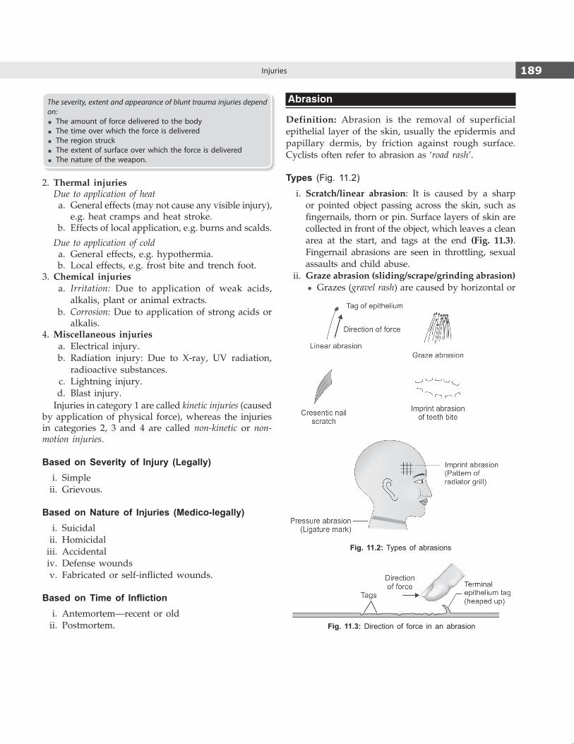
189Injuries
The severity, extent and appearance of blunt trauma injuries depend on:
� The amount of force delivered to the body � The time over which the force is delivered � The region struck � The extent of surface over which the force is delivered � The nature of the weapon.
2. Thermal injuries Due to application of heat a. General effects (may not cause any visible injury),
e.g. heat cramps and heat stroke. b. Effects of local application, e.g. burns and scalds.
Due to application of cold a. General effects, e.g. hypothermia. b. Local effects, e.g. frost bite and trench foot.3. Chemical injuries a. Irritation: Due to application of weak acids,
alkalis, plant or animal extracts. b. Corrosion: Due to application of strong acids or
alkalis.4. Miscellaneous injuries a. Electrical injury. b. Radiation injury: Due to X-ray, UV radiation,
radioactive substances. c. Lightning injury. d. Blast injury.
Injuries in category 1 are called kinetic injuries (caused by application of physical force), whereas the injuries in categories 2, 3 and 4 are called non-kinetic or non-motion injuries.
Based on Severity of Injury (Legally) i. Simple ii. Grievous.
Based on Nature of Injuries (Medico-legally) i. Suicidal ii. Homicidal iii. Accidental iv. Defense wounds v. Fabricated or self-inflicted wounds.
Based on Time of Infliction i. Antemortem—recent or old ii. Postmortem.
Abrasion
Definition: Abrasion is the removal of superficial epithelial layer of the skin, usually the epidermis and papillary dermis, by friction against rough surface. Cyclists often refer to abrasion as ‘road rash’.
Types (Fig. 11.2) i. Scratch/linear abrasion: It is caused by a sharp
or pointed object passing across the skin, such as fingernails, thorn or pin. Surface layers of skin are collected in front of the object, which leaves a clean area at the start, and tags at the end (Fig. 11.3). Fingernail abrasions are seen in throttling, sexual assaults and child abuse.
ii. Graze abrasion (sliding/scrape/grinding abrasion) z Grazes (gravel rash) are caused by horizontal or
Fig. 11.2: Types of abrasions
Fig. 11.3: Direction of force in an abrasion

190 Review of Forensic Medicine and Toxicology
tangential friction between the skin and the hard rough surface.3 They show uneven, longitudinal parallel lines, which indicate the direction in which the force was applied (epidermis being heaped up at the opposite end) (Fig. 11.3).
z Most common type of abrasion, and commonly seen in road traffic accidents.4 Particles of glass, gravel or dirt may be embedded in such wounds.
z Brush burn: Graze abrasion involving wider area such as the back, caused by violent rubbing against a surface, as in dragging along over the ground. Such injuries, when dry, become firm, even though no true ‘scab’ is present.5,6
z Friction burn: An extensive, superficial, reddened excoriated area with little or no linear mark, occurs when the skin is covered by clothing (element of thermal damage is present).
iii. Pressure abrasion (crushing/friction abrasion): It is caused by direct impact or linear pressure of a rough object over the skin. The slight movement directed inwards results in crushing the superficial layers of the cuticle and bruising underneath, e.g. nooses or ligatures in hanging and strangulation.7
iv. Imprint abrasion (impact/contact/patterned abrasion): It is caused when the force is applied perpendicular to the skin, the cuticle gets crushed at the point of impact and bears the imprint of the object causing it.
z The abrasion in slightly depressed below the surface.
z It tends to be focal, and is commonly seen over bony prominences, where a thin layer of skin covers the bone.
z Imprint abrasion becomes more defined when injured cuticle dries up and becomes brownish and parchmentized, in contrast with the surrounding uninjured skin surface.
z Pattern abrasion is a variation of pressure abrasion.
z When a person is knocked down by car, pattern of the radiator grill, headlamp rim or tyre-tread mark may be seen on the skin.8 Imprint of bicycle chain, serrated knife are other examples.
z Teeth bite marks are included in this category, though they may produce contusion or laceration, depending upon the force applied.9
z UV light may be used to visualize the pattern injuries not apparent with visible light.
Human bite can occur during sexual behavior/assault, child abuse, self-defense, self-inflicted or a child biting another child. Bite may tear or crush, resulting in two U-shaped marks, corresponding to the upper and lower anterior six teeth (canine to canine) and separated by an open space of about 2.5–4 cm. Most victims of a criminal act are women, and breast is the most common location. Male victims are more frequently bitten on the arms.
Age of AbrasionIt produces minimum bleeding, heals rapidly and leaves no permanent scarring on healing (Table 11.1).
Table 11.1: Age of abrasion
Duration Features
Fresh2–24 h
Bright red, oozing of serum and some blood.Exudation dries to form a reddish scab, comprising blood, lymph and epithelial cells. Polymorphonuclear cells infiltrate (scab formation).
2–3 days Reddish-brown scab, less tender.10
4–5 days Scab is dark brown in color.
5–7 days Scab is brownish black and starts falling off from the margins. Epithelium grows and covers defect under the scab (epithelial regeneration).
7–12 days Scab dries, shrinks and falls off, leaving depigmented area underneath. It gradually gets pigmented in due course of time (subepidermal granulation).
> 12 days Epithelium becomes thinner and atrophic. New collagen fibers are prominent. Basement membrane is present and vascularity of the dermis decreases (regression).
Differential Diagnosis i. Postmortem insect bites of the skin caused by ants
or cockroaches produce dry, pale brown lesions with irregular margins and are arranged in a linear pattern. Most commonly found at mucocutaneous junctions—around the eyelids, nose, mouth, ears, axilla, groins and genitalia. Vital reaction is absent.
z It may also resemble powder stippling (firearm injury).
ii. Excoriation of skin by excreta and diaper rash may be misinterpreted as abrasions.
iii. Dry skin of scrotum and vulva gives a reddish brown or yellow coloration when exposed to the open air.
iv. Decubitus/pressure ulcers (bed sores): These are due to pressure necrosis of the skin in a bedridden, caused by prolonged compression of soft tissue between bony prominence and external surface.
v. Postmortem abrasions (Diff. 11.1): In doubtful cases, a histopathological examination may be needed.

191Injuries
Circumstances of Abrasions i. Usually, it is seen in accidents and assaults. ii. Abrasions on the face or body of the assailant
indicate a struggle. iii. Person collapsing due to a heart attack may fall
forward and receive abrasions on the forehead, nose and cheek, but there will be no injuries on the upper limbs.
iv. Abrasions may be produced on the palmer surface of hands in a conscious person, who while falling puts out his hands to save himself.
v. Alcoholics tend to fall backwards and strike the occiput on the ground.
vi. Hysterical women may produce abrasions over accessible areas, like the front of forearm or over the face, to fabricate charge of assault.
Medico-legal Importance � Abrasions give an idea about the site of impact and
direction of force. � Nature of injury: Abrasions are superficial injuries
and are mostly simple in nature. However, they may be the only external signs of serious internal injury. Abrasions over the cornea may cause corneal opacity, which may restrict vision permanently, amounting to grievous hurt (Sec. 320 IPC).
� Patterned abrasions are helpful in connecting the wound with the causative weapon.
� Age of injury can be determined, which helps to corroborate with alleged time of assault.
� In open wounds, dirt, dust, grease or sand is usually present which helps to connect the injuries to the scene of crime.
� Character and manner of injury may be known from its distribution:
i. In throttling, crescentic abrasions made by finger-nails are found on the neck.11
ii. Abrasions on the victim may show whether the fingernails of assailant were long, irregular or broken.
iii. In smothering, abrasions may be seen around the mouth and nose.
iv. In sexual assaults, abrasions may be found on the breasts, genitals, inside of the thigh and around the arms.
Patterned injuries can be subdivided according to the type of force involved: i. Blunt force injuries: These are the most commonly seen group.
Abrasions may preserve patterns well, especially if the force is applied perpendicular to the skin surface. Bruises may also reproduce patterns well, particularly if they are intradermal. Lacerations less frequently show a well-defined reproduction of the shape of the causative agent.
ii. Sharp force injuries: Stab wounds may show characteristics of a specific type of blade. Distinctive patterns may be seen with the hilt, or a stab wound with screwdrivers or scissors.
iii. Gunshot wounds: Contact entry wounds (may have sight marks) and shotgun wounds (e.g. wad marks) may produce distinct patterned injuries.
iv. Other miscellaneous wounds and marks e.g. fern like pattern with lightning strikes, tool marks on internal structures (such as cartilage).
Medico-legal importance: Connect a particular weapon or object to an injury, which may allow a perpetrator to be linked to the crime and/or enable better understanding of the events surrounding a death.
Bruise / Contusion
Definition: Bruise is the extravasation of blood in the subcutaneous/subepithelial tissues due to rupture of blood vessels, usually capillaries, as a result of blunt force injury or pressure.
� ‘Bruise’ is derived from old English word ‘brysan’, which means ‘to crush’.
� Usually, there is no loss of continuity of the overlying skin.
� ‘Bruise’ implies that the lesion is observed through the overlying intact skin as bluish purple discoloration and swelling of the involved area, while a ‘contusion’ is a bruise within an organ or tissues, such as muscles, liver or mesentery.
Differentiation 11.1: Antemortem and postmortem abrasion
S.No. Feature Antemortem abrasion Postmortem abrasion1. Site Anywhere on the body Usually, over bony prominences 2. Color Bright red Yellowish, translucent and parchment-like3. Exudation More, scab slightly raised Less, no scab4. Vital reaction Present Absent5. Healing process May be evident Not seen
Note: Abrasions produced slightly before or after death cannot be differentiated even by microscopic examination.
192 Review of Forensic Medicine and Toxicology
Causes i. By application of blunt force viz. blow with fists,
sticks, iron-bar, cane, whip or chain. ii. From compression, like pressing fingers. iii. Medical intervention sometimes produces bruise—
sternal and cardiac bruising, bruising around needle puncture marks and pinching skin to test conscious level (butterfly bruise).
ClassificationBruise is classified into three types depending on its situation: i. Intradermal bruise: Bruise lies in the immediate
subepidermal layer. It is made by impact with a patterned object, and hemorrhage is sharply defined.
ii. Subcutaneous bruise: It is situated in subcutaneous tissue, often in the fatty layer, and the edges are blurred. Most common type of bruise caused by a blunt object, and appears soon after injury as dark red swelling.
iii. Deep bruise: Bleeding deeper to the subcutaneous tissues. It may take hours to 1–2 days to appear at the surface (delayed bruising). Therefore, one more examination should be carried out 24–48 h after first examination. Infrared photography may demonstrate such bruises, if suspected initially.
Factors Influencing the Bruise i. Type of tissue/site involved
z Soft, lax and vascular tissues, such as face, scrotum and eyelids develop large bruises even with little force.12
z In tissues which are strongly supported, contain firm fibrous tissue and are covered by thick dermis, e.g. abdomen, back, scalp, palms and soles, even a moderate violence may produce only a small bruise.
z Bruising of scalp is better felt than seen. z Bruising is more marked on tissues overlying
bone. z In boxers and athletes, bruising is much less,
because of good muscle tone. z Chronic alcoholics with cirrhosis and individuals
taking aspirin, bruise easily. ii. Age: Children and elderly bruise more easily
because of softer tissue and delicate skin in the former, and loss of subcutaneous supportive tissue and cardiovascular changes in the latter.
iii. Sex: Women tend to bruise more easily than men because tissues are more delicate and subcutaneous fat is more. Obese people bruise more easily than lean because tissues are more delicate.
iv. Color of skin: Bruising is more clearly seen and recognized in fair skinned persons than those with dark skin, in whom they may be better felt than seen.
v. Natural diseases: Prominent bruising following minor trauma is seen in persons suffering from atherosclerosis, purpura hemorrhagica, leukemia, hemophilia, scurvy, bleeding diathesis, vitamin K and prothrombin deficiency, and in phosphorus poisoning.
vi. Gravity shifting of blood (ectopic/migratory bruise): It is responsible for the appearance of bruises at a site other than the site of injury, e.g. black eyes.13,14 Blood will track along the fascial planes (or between muscle layers) along the path of least resistance and may appear where the tissue layers become superficial. Thus, site of bruise does not always indicate the site of injury.
� Grey Turner’s sign: Ecchymosis seen over flank or side of abdomen, occurring due to extensive retroperitoneal hemorrhage. This sign takes 24–48 h to develop.
� Cullen’s sign: Bluish-black discoloration of the periumbilical skin due to extensive retroperitoneal or intraabdominal hemorrhage. This may be caused by ruptured ectopic pregnancy or acute pancreatitis.
Patterned BruisePatterned intradermal bruise is due to impact with a hard, patterned object with ridges and grooves. Bruise may indicate the nature of the weapon, especially when death occurs soon after infliction of injury.
Mechanism: When the weapon sinks into the skin, there is little or no damage to the blood vessels over ridges where it compresses the skin. However, traction causes marginal dermal vessels to rupture in the skin forced into grooves. The resulting accumulation of a small amount of blood, near the epidermis may demonstrate the obvious pattern of the causal surface (tyre, rod, shoe tread, car bumper, clothing, or gun muzzle).
� A blow from a solid body, such as hammer or a closed fist produces a rounded bruise. Doughnut bruise is due to a spherical object (such as cricket ball).
� Blows with a rod, stick or a whip produce two parallel, linear hemorrhages (railway line or tram-line type). The intervening skin appears unchanged (Figs 11.4 and 11.5).

193Injuries
Dating/Age of BruiseConsistent, reliable microscopic dating is not possible and color changes in resolution of a contusion is not always a reliable indicator of its age. However, methods used to date a bruise are: i. Histology (only in postmortem situation) ii. Color changes (visual examination) iii. Calorimetry iv. Spectrophotometry.
� Bruises heal by destruction and removal of extravasated blood.
� The extravasation of blood is followed by an inflammatory reaction that causes vasodilation and attracts macrophages, which breaks down hemoglobin to biliverdin. Biliverdin is then broken down by the enzyme biliverdin reductase to yellow color bilirubin. As hemoglobin is broken down, some of its iron is released and combines with ferritin, which gives rise to hemosiderin.
� Color change starts at the periphery and extends inwards to the center.
� The time required for bruising to clear is extremely variable and is only a general guideline in interpreting the age of the bruise (Table 11.2). It should only be stated whether the bruise is recent or old.
* Love bite (hickey) is not actually a true bite since there are no teeth marks. Bruise is caused by firm application of the lips against the skin, forming an air-tight seal, and oral suction cause a shower of petechial bruises from rupture of numerous small vessels.
Fig. 11.4: Patterned bruise
Fig. 11.5: Formation of ‘tram-line contusion’
� A woven, spiral or plaited ligature may produce a patterned bruise. A solid stick bruise is limited to the convexity of the body surface. A flexible strap or flex will wrap around the convexity producing a longer and often curved tram-line bruise.
� Suction or biting on the sides of the neck or the breasts during love making/sexual intercourse produces elliptical patterned bruises (Fig. 11.6).*
Deep tissue and organ contusion � Internal organs can also get contused; contusion of
the brain may cause confusion, coma and death. � Contusion in vital centers, e.g. which controls
respiration and blood pressure can be fatal even when very small.
� Small contusions of heart can cause serious disturbances of normal rhythm or stoppage of cardiac action and death.
Fig. 11.6: Suction bruise
Table 11.2: Age of bruise
Duration Color
Fresh Red (oxygenated blood)
Few hours to 3 days Blue (deoxyhemoglobin)15
4–5 days Bluish black to brown (hemosiderin)16,17
5–6 days Green (biliverdin)18
7–12 days Yellow (bilirubin)
2 weeks Normal

194 Review of Forensic Medicine and Toxicology
� Subconjunctival hemorrhage does not show similar color changes owing to hemoglobin being kept oxygenated by air. It is red at first, then becomes yellow and finally disappears.19 Similar changes are seen in meningeal hemorrhages owing to O2 supplied from CSF.
� Healthier the individual, the more rapid will be the healing. A bruise takes a much longer duration to heal in the old than in the young. In old age, it may remain for 4–5 weeks. Bruises of soft loose tissues, like those surrounding the eye resolve faster.
� Environmental lighting may slightly alter the color of the bruise. Drugs, such as steroids may change the rate of bruise dispersion, and interventions, such as ice packs or heat treatment may add to variability.
� Bruises of the same age may show different color progression, so that variation in color does not necessarily mean that there have been multiple episodes of injury.
� Not all bruises pass through a yellow phase before they resolve.
� Dating a bruise may be helpful in determining the veracity of the informant and together with other data may justify further investigation into a particular case.
� Hemosiderin is a granular brown iron-storage complex composed of ferric oxide, commonly found in macrophages and derived from breakdown of hemoglobin.
� Biliverdin is a green pigment formed as a byproduct of heme breakdown.
� Bilirubin was discovered by Virchow in 1849, who called the yellow pigment ‘hematoidin’.
Complications i. Multiple contusions can cause death from shock
and internal hemorrhage. ii. Gangrene and death of tissue can result. iii. Bacterial infections, especially by Clostridia can
occur. iv. Pulmonary fat embolism may occur.
Medico-legal Importance � It is advisable that a medical officer should re-
examine the patient after 24 h, as by this time the bruises are clearly visible.
� Age of injury can be determined by the color changes. � Degree of violence may be determined from their size.
Since, the appearance of bruise depends upon many factors, great caution must be used before giving any opinion regarding its appearance.
� Patterned bruises may connect the victim and the object/weapon, e.g. whip, chain, cane or ligature.
� Nature of injury: Bruises are generally simple injury. However, the impact responsible may result in injury to the vital organs underneath the bruised area. In such a situation, the bruise is grievous in nature.
� Contusions can be produced postmortem, if a severe blow is given to the body within few hours after death. To confirm at postmortem examination, deep incisions are made at suspected sites, which show ecchymosis (Diff. 11.2).
� Sometimes, the autopsy surgeon needs to differentiate bruise from PM staining (Diff. 11.3). Since in early phases of development of PM staining, it may look like a bruise which may led to misinterpretation (assault/trauma).
� Bruises may be fabricated by applying juices of marking nut or calotropis to incriminate others, or in defense of a crime.
� Surgical removal of cornea can result in hemorrhage into the eyelids, identical with antemortem trauma.
� Character and manner of injury may be known from its distribution:
i. Bruising of the arm may be a sign of restraining a person. When arms are grasped, there may be 3–4 bruises on one side (corresponding to fingers) and one larger bruise on the opposite side (thumb).
ii. Small bruises along with nail marks on the inner aspect of thighs of a woman may indicate sexual assault.
Differentiation 11.2: Antemortem and postmortem bruise20,21
S.No. Feature Antemortem bruise Postmortem bruise1. Swelling Present Absent2. Damage to epithelium Present Absent3. Extravasation of blood Present Absent4. Coagulation Present Absent5. Infiltration of the tissues with blood Present Absent6. Color changes Seen Uniform color7. Margins Merge with surrounding area Sharply demarcated8. Appearance More marked in victims who survive for sometime Less marked
195Injuries
iii. Typical small bruises (six-penny bruises) are produced by forcible poking or pressure of fingertips.
iv. Bruising of the shoulder blades indicates firm pressure on the body against the ground or other resisting surface.
v. In manual strangulation, position, number of bruises and nail marks give an indication of the position of the assailant.
vi. Bruises found in ‘soft’ sites in a child such as cheeks or trunk, and multiple bruises in various stages of healing suggest abuse.
Bruises are of lesser value than abrasions because: � Their size may not correspond to the size of the weapon.* � They do not indicate the direction in which the force was applied.
� They may become visible after few hours or even 1–2 days after injury.
� They may appear at a distance away from the actual site of injury. It may not indicate the point of trauma.
Bone contusion refers to trabecular microfractures due to impaction of bone. It may occur due to blunt force from outside the body or more commonly from two bones striking each other after ligament injuries. Conventional X-rays may not detect bone contusion and MRI is needed in acute stages.22
Lacerated Wound
Definition: Laceration is the tearing or splitting of skin, mucous membranes, muscles or internal organs caused by either a shearing or a crushing force, and produced by application of a blunt force to a broad area of the body. If the blunt force produces extensive bruising and laceration of deeper tissue, it is called crush injury.
Types i. Split lacerations: Occur when soft tissues are
sandwiched between a hard unyielding deeper structure and the agent applying the force. Scalp lacerations occur due to the tissues being crushed between the skull and some hard object.23,24
Incised-looking lacerated wounds: When the skin is closely applied to the bone and the subcutaneous tissue is scanty, blunt force may produce a wound which by linear splitting of the tissues resembles an incised wound.24,25
Sites: Scalp, forehead, eyebrows, zygoma, iliac crest, lower jaw, perineum and shin.26
ii. Stretch lacerations: Result from a heavy forceful frictional impact of blunt forces exercising localized ‘pressure with pull’. Overstretching of the skin
Differentiation 11.3: PM staining and bruise21
S.No. Feature PM staining Bruise1. Cause Distension of vessels with blood in dermis Rupture of vessels which may be superficial or deep
2. Cuticle Not abraded May be abraded
3. Site Occurs over extensive area of the most dependent parts
Occurs at the site of and surrounding the injury, may appear anywhere on the body
4. Appearance No elevation of involved area Often swollen, because of extravasated blood and edema
5. Margins Clearly defined Merge with the surrounding area
6. Color Uniform bluish-purple color Different colors, depending on the age of bruise
7. On incision Blood is seen in blood vessels which can be easily washed away, subcutaneous tissues are pale
Extravasation of blood into the surrounding tissues, cannot be washed by water, subcutaneous tissues are deep reddish-black
8. Effect of pressure Absent in areas of the body which are under even slight pressure
Lighter over the area of pressure or support
9. Superimposed abrasion Not present May be present
10. Microscopically Blood cells are found within the blood vessels and there is no evidence of inflammation
Blood cells are found outside the blood vessels, evidence of inflammation present
* The size of a bruise is not necessarily related to the size of the object. Thus, one cannot state that a bruise, 9 × 3 cm, was caused by an object of similar dimensions.

196 Review of Forensic Medicine and Toxicology
and subcutaneous tissues may cause lacerations with flapping of the skin, which may indicate the direction of application of force.
They are seen in run over by motor vehicle, kicking and in compound fractures.
iii. Avulsion or grinding compression: Produced by shearing force delivered at an oblique or tangential angle to detach (tear off) a portion of traumatized skin surface or viscus (tissue/organ) from their attachment (Fig. 11.7).
z Commonly seen in road traffic accidents where the rotating force of a wheel tears off the skin over a large area. This is called flaying, and most frequently occurs on the legs.27
z Amputation injuries are a type of avulsion injury in which an entire extremity or portion thereof is severed from the body.
z The most severe is a decapitation injury, in which the head separates from the body.
iv. Tears: Tearing of skin and subcutaneous tissue can occur from localized impact by or against some hard, irregular object like car door handle, radiator mascot or from blows with broken glass bottles.
v. Cut lacerations: Sometimes, a heavy sharp edged weapon causes a deep and wide cut over the body tissues.
Characteristics (Fig. 11.8) � Margins: Ragged, irregular and uneven; may show
tearing of the extremities at angles diverging from the main laceration, the so-called ‘shallow tails’; pieces of tissue are attached in between called tissue tags.
� Site: Occurs most commonly over bony prominences, such as the head where the skin is fixed, and easily stretched and torn.
� Bruising and abrasion: Seen around the margin. � Edges: May give an indication of direction in which
the blow or force was applied. � Depth of wound: Shows bridges of irregularly torn
fibrous tissue, blood vessels and nerves across the interior of the wound.*28
� Soiling of wound: Mud, wood splinters, sand, glass fragments or paint material of the vehicle involved, hair or fibers may get embedded in the wound, and are of great medico-legal importance.
� Hair bulbs: Crushed. � Hemorrhage: Less, because the arteries are crushed
and torn across irregularly; they retract and blood clots readily, except in the scalp where the temporal arteries bleed freely as they are firmly bound and unable to contract.
� Shape: May correspond with the weapon or object which produced them.
� Gaping: Seen due to pull of elastic and muscular tissues.
� Beveling: Laceration caused by a blow directed tangentially or at an angle will produce undermining of the tissue on one side (indicates the direction of blow) and abrasion and beveling on the other (direction from which the blow was coming).
� On healing: Produces permanent scar.
Antemortem lacerations show bruising of margins, vital reaction, eversion and gaping of margins.
Fig. 11.7: Avulsed lacerated wound Fig. 11.8: Characteristics of lacerated wound
* Tissue bridging results from the application of blunt force that overcomes the elasticity of the skin; as a result, the skin splits while underlying structures such as vessels, nerves, and connective tissue visibly span the wound.

197Injuries
Dating of LacerationThe gross findings are summarized in Table 11.3 when healing occurs by first intention without any secondary infection.
due to laceration on the face resulting in disfiguration is also a grievous injury.
� Whether the laceration is accidental/homicidal/suicidal? a. Accidental laceration: Commonly seen anywhere
on exposed parts of body. b. Homicidal laceration: Noticed on nonaccessible
parts of the body, especially in assault cases. It is usually seen on the head.
c. Suicidal lacerations are rarely seen, as they are painful to produce, and if present, they are seen on exposed parts of body and on same side.
� Sometimes, human bites can be a combination of deep lacerations and crushing and are associated with a high incidence of infection. It may be associated with avulsion of pieces of the nose or ear.*
� Foreign matter in the wound may give clues about the object causing it, e.g. paint material of vehicle may be transferred to the lacerated wound.
� Skin flap which overhangs the cut margin (avulsion cases) can indicate the direction of force applied.
Wounds caused by sharp edged and pointed weapons are of four types:� Incised wound � Chop wound� Stab wound � Therapeutic/diagnostic wound
Incised Wound (Cut / Slash / Slice)
Definition: Incision is a clean cut wound through the tissues (usually the skin and subcutaneous tissues including blood vessels), which is more long than deep, and caused by a sharp-edged instrument. It is produced by pressure and friction against the tissue by an object having a sharp cutting edge or point, such as knife, box cutter, razor or scalpel.
Description of a Sharp WoundOn the skin surface, the edges of a stab, incised, or chop wound are referred to as the wound’s ‘margins’, whereas the ends, or tips, of the wound are referred to as ‘angles’. The ‘length’ of a wound is measured from one angle to the other. The ‘width’ of the wound is the widest measurement between the two margins. An imaginary line drawn between the two angles defines the ‘long/longitudinal axis’ of the wound (Fig. 11.9). The depth of the wound is measured from the skin surface to the deepest point of penetration.
Table 11.3: Healing of a lacerated wound
Duration Gross findings
Fresh Bleeding or fresh clot is attached; margins are red, swollen and tender
12–24 h Margins swollen, red and covered by dried blood clots and lymph
3–5 days Margins strongly adherent with each other and covered by dried crust
6–7 days Crust/scab falls off or can easily be taken off, soft reddish tender scar
Few weeks Scar is whitish, firm and painless
* An accidental type of injury results from an attacker striking the victim’s incisor teeth with his knuckles (metacarpophalangeal joint is usually involved).
Complications i. Lacerations may cause severe and fatal bleeding
leading to shock and death. ii. Infection. iii. Pulmonary/systemic fat embolism may occur due
to crushing of subcutaneous tissue. iv. If located where skin stretches or is wrinkled,
e.g. over joints, repeated and continued oozing of tissue fluids and blood may cause irritation, pain and dysfunction.
Medico-legal Importance � The type of laceration may indicate the cause of injury
and shape of blunt weapon, e.g. i. Blunt round end (hammer) may cause a stellate
laceration. ii. Blunt object with an edge, such as hammer
head, may cause crescentic laceration (patterned laceration).
iii Long, thin objects, like pipes or sticks produce linear or elongated lacerations, while objects with a flat surface produce irregular, ragged or Y-shaped lacerations.
� Nature of injury: A laceration may be a simple injury. Underlying fracture or injury to the vessels, nerves, muscles and organs should be ruled out before giving the opinion. Extensive scar formation during healing of a laceration over the joint leading to restriction of joint movement is a grievous injury. Similarly, scar

198 Review of Forensic Medicine and Toxicology
Characteristics29
� Margins: Edges are clean cut, well-defined and usually everted. They may be inverted, if a thin layer of muscle fibers is adherent to the skin as in the scrotum (due to the attached dartos muscle to the skin). The edges are free from contusions and abrasions. Wrinkled wounds are produced where the skin is wrinkled (i.e. folds) and more than one incised wound is seen.
� Width/breadth: Width is greater than the edge of the weapon causing it due to retraction of the divided tissues.
� Length: Length is greater than its width and depth and has no relation to the cutting edge of the weapon, for it may be drawn to any distance.
� Shape: Usually spindle-shaped due to greater retraction of the edges in the center (Fig. 11.10). Gaping is more, if the underlying elastic fibers in the skin (Langer’s lines) have been cut transversely or obliquely and is less when cut longitudinally.
� Depth and direction: Usually deeper at the commencement, except in case of suicidal cut throat injuries, with hesitation cuts at the beginning. This is known as head of the wound. Towards termination, the cut becomes progressively shallow, known as tailing of the wound (Fig. 11.10). Consequently, depth of the incised wound with tailing will suggest the direction in which the force was applied.
� Hemorrhage: As vessels are cut clean, hemorrhage is more.
� Beveled cuts: If the blade of the weapon enters obliquely, tissues will be visible at one margin and other margin will be undermined; if the blade is nearly horizontal, a flap wound is caused.
Dating of Incised WoundRefer to Table 11.4 for dating of incised wound.
Table 11.4: Dating of an incised wound
Duration Gross findings Microscopic findings
Fresh Red with clotted blood
Capillary dilatation, margination and emigration of neutrophils, reactive changes in tissue histiocytes
12 h Margins red, swollen and adherent with blood and lymph
Reactive changes in fibroblast, monocytes in exudates
24 h Continuous layer of endothelial cells cover the surface with a scab of dried clot
Endothelium begins to grow at edges, vascular buds begin to form
2–3 days __ Vascularized granulation tissue formation (fibroblasts)
4–6 days __ Formation of new fibrils
7 days Scar formation Scar formation
Medico-legal importance � Indicates the nature of weapon (sharp-edged).
Although, the appearance of stab wounds may indicate the shape of the weapon used, this is not possible with incised wounds. However, rarely, the appearance of incised wounds may provide some indication as to the weapon type.
� Give an idea about the direction of force. � Age of injury can be determined. � Nature of injury: Incised wounds are rarely life-
threatening because they seldom penetrate deeply enough to damage a blood vessel of significant size. However, incised wounds over the wrist or neck, where major arteries lie in the superficial tissues, can prove fatal.
� Position and character of wound may indicate manner of production, i.e. suicide, accident, or homicide (Diff. 11.4 and refer to Fig. 12.22).
Fig. 11.10: Incised wound
Fig. 11.9: Characteristics of a sharp wound

199Injuries
a. Suicide: Multiple incised wounds of varying depths on the neck or wrists suggest a suicide. Some features of suicidal wounds are:
z Fatal wounds are present over limited accessible areas of the body, such as front of neck, groin, chest or back of legs. Cutting of wrist is rarely fatal. Suiciders usually do not injure the face.
z Hesitation cuts/marks or tentative cuts or trial wound: These cuts are multiple, small and superficial often involving only the skin, and are seen at the beginning of the incised wound, presumably hesitating while gaining courage to make a final decisive cut.*
z A person who commits suicide exposes his body by opening his clothes and then inflicts the wounds.
z When a safety razor blade is used, uninten-tional cuts are found on the fingers where the blade has been gripped.
z Most people have a vague knowledge of the anatomy and do not know where to cut a major blood vessel, and may cut their forearms vertically, rather than horizontally.
b. Homicidal wounds: They are deep and deliberate in character and are seen on the head and front of neck, and sometimes on the trunk. Incised wounds on nose, ears and genitals are usually homicidal, and may result from sexual jealousy, caused by a jilted lover, husband or wife.31
c. Accidental wounds: Commonly seen around the hands.
d. Defense wounds: Injuries are seen on the forearm and palm, when the victim may try to ward off an attack by raising hands and arms in defense or by grabbing the weapon.
e. Self-mutilation: Sometimes, injuries may be caused by an individual with a mental disorder as a form of self-mutilation or by one who deliberately harms oneself for motives of
Differentiation 11.4: Suicidal and homicidal cut-throat wounds
S.No. Feature Suicidal cut-throat Homicidal cut-throat 1. Situation Left side of the neck and passing across the front of
the throatUsually on the sides
2. Level High, above the thyroid cartilage Low, on or below the thyroid cartilage3. Direction Obliquely, above downwards and from left to right in
right handed personsTransverse or from below upwards
4. Number of wounds Multiple, may be 20–30, superficial, parallel and merge with main wound
Multiple, cross each other at a deep level
5. Edges Usually ragged due to overlapping of multiple superficial incisions
Sharp and clean cut, beveling may be seen
6. Hesitation cuts Present30 Absent7. Tailing Present Absent8. Severity Less severe, one wound is severe, but sometimes,
there may be 2–3More severe, all tissues including vertebrae may be cut
9. Wounds in other parts of body
May be present across wrists, groin and thighs No wounds on wrists, but severe injuries on head may be present
10. Defense wounds Absent, unintentional cuts may be found Present, unless taken unaware11. Hands Weapon may be firmly grasped due to cadaveric
spasmFragments of clothing or hair of the assailant may be grasped
12. Weapon at site Usually present Usually absent13. Vessels As head is thrown back, carotid artery escapes injury Jugular vein and carotid artery are likely to be cut 14. Clothes Not cut or damaged May be cut, corresponding to injuries in the body15. Blood stains If standing in front of mirror, then splashing on the
mirror; stains running downwards on the clothes, front of body and feet
Found in both palms in an effort to cover the wound; if lying down, stains collect behind the neck and shoulder
16. Circumstantial evidence Quiet place, such as bedroom or bathroom; suicidal note
Disturbance at scene, footprints outside
* Multiple, superficial, roughly parallel incised wounds on the neck, adjacent to a deep, lethal incised wound, can be seen in victims of homicidal sharp force injury, particularly if torture was employed.
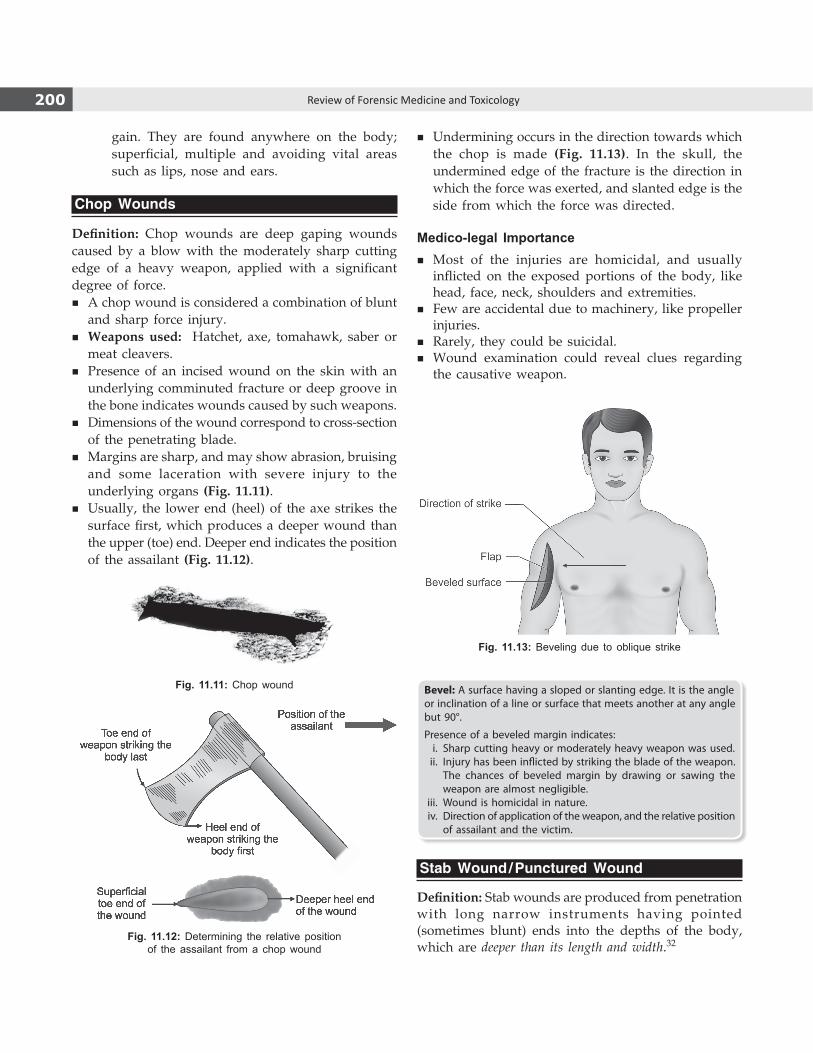
200 Review of Forensic Medicine and Toxicology
gain. They are found anywhere on the body; superficial, multiple and avoiding vital areas such as lips, nose and ears.
Chop Wounds
Definition: Chop wounds are deep gaping wounds caused by a blow with the moderately sharp cutting edge of a heavy weapon, applied with a significant degree of force.
� A chop wound is considered a combination of blunt and sharp force injury.
� Weapons used: Hatchet, axe, tomahawk, saber or meat cleavers.
� Presence of an incised wound on the skin with an underlying comminuted fracture or deep groove in the bone indicates wounds caused by such weapons.
� Dimensions of the wound correspond to cross-section of the penetrating blade.
� Margins are sharp, and may show abrasion, bruising and some laceration with severe injury to the underlying organs (Fig. 11.11).
� Usually, the lower end (heel) of the axe strikes the surface first, which produces a deeper wound than the upper (toe) end. Deeper end indicates the position of the assailant (Fig. 11.12).
Bevel: A surface having a sloped or slanting edge. It is the angle or inclination of a line or surface that meets another at any angle but 90°.
Presence of a beveled margin indicates: i. Sharp cutting heavy or moderately heavy weapon was used. ii. Injury has been inflicted by striking the blade of the weapon.
The chances of beveled margin by drawing or sawing the weapon are almost negligible.
iii. Wound is homicidal in nature. iv. Direction of application of the weapon, and the relative position
of assailant and the victim.
Stab Wound / Punctured Wound
Definition: Stab wounds are produced from penetration with long narrow instruments having pointed (sometimes blunt) ends into the depths of the body, which are deeper than its length and width.32
Fig. 11.11: Chop wound
Fig. 11.12: Determining the relative position of the assailant from a chop wound
Fig. 11.13: Beveling due to oblique strike
� Undermining occurs in the direction towards which the chop is made (Fig. 11.13). In the skull, the undermined edge of the fracture is the direction in which the force was exerted, and slanted edge is the side from which the force was directed.
Medico-legal Importance � Most of the injuries are homicidal, and usually
inflicted on the exposed portions of the body, like head, face, neck, shoulders and extremities.
� Few are accidental due to machinery, like propeller injuries.
� Rarely, they could be suicidal. � Wound examination could reveal clues regarding
the causative weapon.
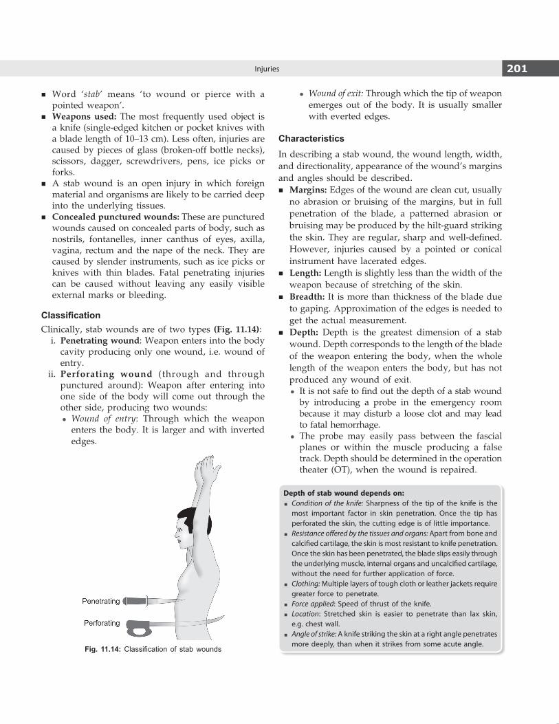
201Injuries
� Word ‘stab’ means ‘to wound or pierce with a pointed weapon’.
� Weapons used: The most frequently used object is a knife (single-edged kitchen or pocket knives with a blade length of 10–13 cm). Less often, injuries are caused by pieces of glass (broken-off bottle necks), scissors, dagger, screwdrivers, pens, ice picks or forks.
� A stab wound is an open injury in which foreign material and organisms are likely to be carried deep into the underlying tissues.
� Concealed punctured wounds: These are punctured wounds caused on concealed parts of body, such as nostrils, fontanelles, inner canthus of eyes, axilla, vagina, rectum and the nape of the neck. They are caused by slender instruments, such as ice picks or knives with thin blades. Fatal penetrating injuries can be caused without leaving any easily visible external marks or bleeding.
ClassificationClinically, stab wounds are of two types (Fig. 11.14): i. Penetrating wound: Weapon enters into the body
cavity producing only one wound, i.e. wound of entry.
ii. Perforating wound (through and through punctured around): Weapon after entering into one side of the body will come out through the other side, producing two wounds:
z Wound of entry: Through which the weapon enters the body. It is larger and with inverted edges.
z Wound of exit: Through which the tip of weapon emerges out of the body. It is usually smaller with everted edges.
CharacteristicsIn describing a stab wound, the wound length, width, and directionality, appearance of the wound’s margins and angles should be described.
� Margins: Edges of the wound are clean cut, usually no abrasion or bruising of the margins, but in full penetration of the blade, a patterned abrasion or bruising may be produced by the hilt-guard striking the skin. They are regular, sharp and well-defined. However, injuries caused by a pointed or conical instrument have lacerated edges.
� Length: Length is slightly less than the width of the weapon because of stretching of the skin.
� Breadth: It is more than thickness of the blade due to gaping. Approximation of the edges is needed to get the actual measurement.
� Depth: Depth is the greatest dimension of a stab wound. Depth corresponds to the length of the blade of the weapon entering the body, when the whole length of the weapon enters the body, but has not produced any wound of exit.
z It is not safe to find out the depth of a stab wound by introducing a probe in the emergency room because it may disturb a loose clot and may lead to fatal hemorrhage.
z The probe may easily pass between the fascial planes or within the muscle producing a false track. Depth should be determined in the operation theater (OT), when the wound is repaired.
Depth of stab wound depends on: � Condition of the knife: Sharpness of the tip of the knife is the
most important factor in skin penetration. Once the tip has perforated the skin, the cutting edge is of little importance.
� Resistance offered by the tissues and organs: Apart from bone and calcified cartilage, the skin is most resistant to knife penetration. Once the skin has been penetrated, the blade slips easily through the underlying muscle, internal organs and uncalcified cartilage, without the need for further application of force.
� Clothing: Multiple layers of tough cloth or leather jackets require greater force to penetrate.
� Force applied: Speed of thrust of the knife. � Location: Stretched skin is easier to penetrate than lax skin,
e.g. chest wall. � Angle of strike: A knife striking the skin at a right angle penetrates
more deeply, than when it strikes from some acute angle.Fig. 11.14: Classification of stab wounds

202 Review of Forensic Medicine and Toxicology
� Direction z The directionality of a stab wound can be described
as ‘vertical’, ‘horizontal’ or ‘oblique’, with a general or specific measurement of the angulation. One method is to describe the directionality based on a clock-face configuration. For example, the long axis of the stab wound is between the 10 and 4 O’clock positions.
z Direction of the track of the wound should be determined in the OT. It should be described in three terms of description, for e.g., when a weapon has been used from above, back and left side, the direction of the track of the wound will be downward, forward and from left to right.
z When a knife penetrates at an angle, the wound will have a beveled margin on one side with undermining (undercut) on the other, so that subcutaneous tissue is visible, indicating the direction from which the knife entered (Fig. 11.15). In solid organs, like liver, the track made by the weapon is seen well.
� Shape: It is slit-shaped or gape depending on their location and their orientation, with regard to the cleavage lines of Langer.33,34
z A stab wound which runs parallel to the cleavage lines will remain slit-shaped and narrow, and the dimensions of the blade will be represented with considerable accuracy.
z A stab wound which enters through the cleavage lines transversely will gape.
z Some specific patterns seen with common weapons are described below (Fig. 11.16):33
i. If a single-edged weapon is used, the surface wound will be triangular or wedge-shaped, and one angle of the wound will be sharp and the other rounded, blunt or squared off. Blunt end of the wound may have small splits in the skin, so-called ‘fish-tailing’. Virtually all
stab wounds are made with single-edged weapons. Sometimes, this is not always the case, as the blunt edge of the knife may split the skin and resemble a double-edged knife wound.
ii. If a double-edged weapon is used, the wound will be elliptical or slit-like, and both angles will be sharp or pointed.
iii. A round object, like a spear may produce a circular wound.
iv. A pointed square weapon may produce a cross-shaped injury, each of the four edges tearing their way through the tissues (stellate shaped).
v. Stabs produced with a broken bottle appear as clusters of wounds of different sizes, shapes and depths with irregular margins.
vi. A screwdriver will produce a slit-like stab wound with squared ends and abraded margins.
vii. Skin wound made by closed scissors produces a flat ‘Z’ shaped wound. If the blades were open, the injuries may look similar to those produced by a knife.
viii. Ice picks produce small, round or slit-like wounds, which may look like 0.22-calibre bullets or shotgun pellets.
ix. A fork will produce clusters of 3–4 wounds depending on the number of prongs on the fork.
Fig. 11.15: Beveling of a wound in case of tangential entry of a weapon
Fig. 11.16: Shape of stab wounds
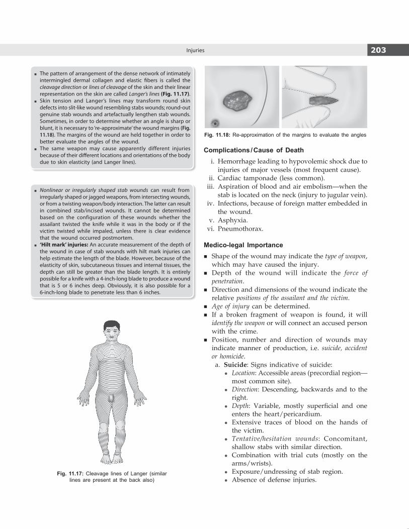
203Injuries
� The pattern of arrangement of the dense network of intimately intermingled dermal collagen and elastic fibers is called the cleavage direction or lines of cleavage of the skin and their linear representation on the skin are called Langer’s lines (Fig. 11.17).
� Skin tension and Langer’s lines may transform round skin defects into slit-like wound resembling stabs wounds; round-out genuine stab wounds and artefactually lengthen stab wounds. Sometimes, in order to determine whether an angle is sharp or blunt, it is necessary to ‘re-approximate’ the wound margins (Fig. 11.18). The margins of the wound are held together in order to better evaluate the angles of the wound.
� The same weapon may cause apparently different injuries because of their different locations and orientations of the body due to skin elasticity (and Langer lines).
� Nonlinear or irregularly shaped stab wounds can result from irregularly shaped or jagged weapons, from intersecting wounds, or from a twisting weapon/body interaction. The latter can result in combined stab/incised wounds. It cannot be determined based on the configuration of these wounds whether the assailant twisted the knife while it was in the body or if the victim twisted while impaled, unless there is clear evidence that the wound occurred postmortem.
� ‘Hilt mark’ injuries: An accurate measurement of the depth of the wound in case of stab wounds with hilt mark injuries can help estimate the length of the blade. However, because of the elasticity of skin, subcutaneous tissues and internal tissues, the depth can still be greater than the blade length. It is entirely possible for a knife with a 4-inch-long blade to produce a wound that is 5 or 6 inches deep. Obviously, it is also possible for a 6-inch-long blade to penetrate less than 6 inches.
Complications / Cause of Death i. Hemorrhage leading to hypovolemic shock due to
injuries of major vessels (most frequent cause). ii. Cardiac tamponade (less common). iii. Aspiration of blood and air embolism—when the
stab is located on the neck (injury to jugular vein). iv. Infections, because of foreign matter embedded in
the wound. v. Asphyxia. vi. Pneumothorax.
Medico-legal Importance � Shape of the wound may indicate the type of weapon,
which may have caused the injury. � Depth of the wound will indicate the force of
penetration. � Direction and dimensions of the wound indicate the
relative positions of the assailant and the victim. � Age of injury can be determined. � If a broken fragment of weapon is found, it will
identify the weapon or will connect an accused person with the crime.
� Position, number and direction of wounds may indicate manner of production, i.e. suicide, accident or homicide.
a. Suicide: Signs indicative of suicide: z Location: Accessible areas (precordial region—
most common site). z Direction: Descending, backwards and to the
right. z Depth: Variable, mostly superficial and one
enters the heart/pericardium. z Extensive traces of blood on the hands of
the victim. z Tentative/hesitation wounds: Concomitant,
shallow stabs with similar direction. z Combination with trial cuts (mostly on the
arms/wrists). z Exposure/undressing of stab region. z Absence of defense injuries.
Fig. 11.17: Cleavage lines of Langer (similar lines are present at the back also)
Fig. 11.18: Re-approximation of the margins to evaluate the angles

204 Review of Forensic Medicine and Toxicology
Examination of the weaponWhen examining a weapon, such as a knife, it should be noted whether the blade is ‘single-edged’ (having a single sharp edge, with the opposite edge being ‘blunt’ or squared-off), ‘double-edged’ (having 2 sharp edges), or a combination of single and double edges, and whether or not the blade is serrated (having teeth). The same knife can produce differing wounds at different levels of penetration (Fig. 11.19). It is important to note that the terminology used to describe the dimensions of the knife (length, width and the thickness of the blade) and the wound do not correspond with one another (Fig. 11.9). When comparing a stab wound to the weapon, the thickness of the blade produces the ‘width’ of the wound, the width of the blade produces the ‘length’ of the wound, and the length of the blade produces the ‘depth’ of the wound. It should also be noted that wound width does not necessarily equal blade thickness,
Death is due to hemopericardium if heart is involved, but cardiac tamponade can occur (accumulation > 150 ml of blood is fatal).
b. Homicide z Most deaths from stab wounds are homicidal,
especially if found in an inaccessible area, such as back (most common mode of homicide in the UK).
z Stabs are most often located on the thorax and the neck.
z Stab wound of the chest may have any direction, but the most common direction is at an angle from left-to-right and from above downwards.
z The absence of weapon at the scene of incident suggests homicide since the assailant usually does not leave the weapon at the scene of death.
z The number of stabs shows a correlation with gender of the perpetrator. In homicide committed by female perpetrators, the victims have fewer stab wounds than in homicides committed by male perpetrators.
c. Accident: Rare. It is caused by falling against any projecting sharp objects, like glass or nails.
� Physical activity following fatal stab wound: Whether a victim after receiving fatal stab can perform any physical activity, like running away from the assailant or shouting for help depends on the organs injured, extent of the injury, the amount and rapidity of blood lost. When bleeding is profused, physical activity is limited, and with slow bleeding, the victim may be able to run a few meters from the assailant.- After stab injuries to the heart, the ability to act is maintained
at least for a short period of time. A stab wound through the left ventricle may almost completely seal itself by contraction of the cardiac muscle around the defect. Death will only occur if continued leakage of blood into the pericardial sac interferes with the pumping action of the heart (cardiac tamponade). A stab wound through the thin-walled right ventricle, atrium or coronary artery is unlikely to re-seal itself and will bleed out into the pericardium with fatal results.
- Wounds involving the great vessels of the thorax (aorta, vena cava, pulmonary arteries and veins) bleed profusely and offer no chance of closure. In lesion of the abdominal aorta, the ability to act may be maintained over prolonged periods of time, whereas in injuries of the thoracic aorta, incapacitation generally occurs within seconds.
- Injuries of the lungs or abdominal organs do not lead to immediate incapacitation.
� Degree of force involved in the stabbing incident: Forensic experts are sometimes asked to estimate or quantify the degree of force used so as to determine an alleged assailant’s intent
to cause harm. Quantifying the exact force is considered to be a difficult area due to the large number of variables present, such as sharpness of the weapon, the area of the body and alignment with cleavage lines of the skin, the angle of attack and the relative movement of the person stabbing relative to the victim being stabbed. The radius of the blunt edge at the tip is important for controlling the penetration ability of a kitchen knife. It is generally not possible for the expert to determine this with any reliable degree of certainty.
� The amount of blood loss necessary to cause death is variable from seconds to hours, and depends on the rate of bleeding, amount of blood loss, nature of the injury and body’s physiological response.- Arteries carry blood at higher pressure than veins of similar
size and, therefore, bleed more rapidly when cut. A partially transected vessel is less likely to seal off than one which is cleanly cut.
- Arterial hemorrhages from major vessels may lead to death relatively fast. A loss of > 1 liter of blood from a major vessel may be fatal.
- Sudden blood loss causes interference with activity when it exceeds 20–25% of the total blood supply. A person can lose over a third of his blood volume before progressing to irreversible hemorrhagic shock.
- A person who is elderly or frail has little reserve to withstand blood loss may succumb quickly.
� Harakiri (seppuku): It is an unusual type of suicidal disembowel-ment connected with Japanese Samurai warriors.35 The victim with a short sword inflicts a single large abdominal stab wound into the left side, drawing the blade across to the right side and then turning it upwards producing an L-shaped cut. The sudden evisceration of the internal organs causes immediate decrease of intra-abdominal pressure and cardiac return resulting in collapse and death.- The jigai ritual is a traditional method of female suicide, carried
out by cutting the jugular vein using a knife called a tantō. It is the feminine counterpart of seppuku.
� The term ‘overkill’ refers to the infliction of massive injuries by a perpetrator by exceeding the extent necessary to kill the victim. Personal conflict between the perpetrator and the victim, history of sex or drugs are associated factors.
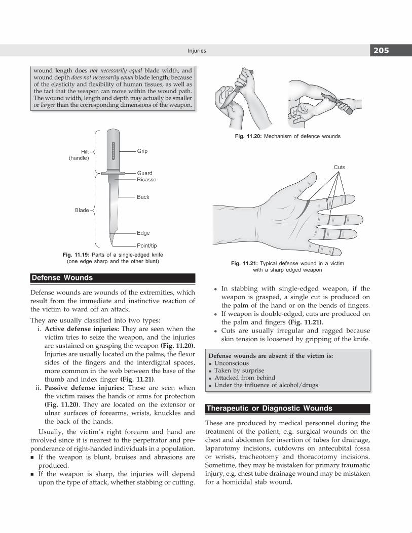
205Injuries
wound length does not necessarily equal blade width, and wound depth does not necessarily equal blade length; because of the elasticity and flexibility of human tissues, as well as the fact that the weapon can move within the wound path. The wound width, length and depth may actually be smaller or larger than the corresponding dimensions of the weapon.
Fig. 11.19: Parts of a single-edged knife (one edge sharp and the other blunt)
Defense Wounds
Defense wounds are wounds of the extremities, which result from the immediate and instinctive reaction of the victim to ward off an attack.
They are usually classified into two types: i. Active defense injuries: They are seen when the
victim tries to seize the weapon, and the injuries are sustained on grasping the weapon (Fig. 11.20). Injuries are usually located on the palms, the flexor sides of the fingers and the interdigital spaces, more common in the web between the base of the thumb and index finger (Fig. 11.21).
ii. Passive defense injuries: These are seen when the victim raises the hands or arms for protection (Fig. 11.20). They are located on the extensor or ulnar surfaces of forearms, wrists, knuckles and the back of the hands.
Usually, the victim’s right forearm and hand are involved since it is nearest to the perpetrator and pre-ponderance of right-handed individuals in a population.
� If the weapon is blunt, bruises and abrasions are produced.
� If the weapon is sharp, the injuries will depend upon the type of attack, whether stabbing or cutting.
z In stabbing with single-edged weapon, if the weapon is grasped, a single cut is produced on the palm of the hand or on the bends of fingers.
z If weapon is double-edged, cuts are produced on the palm and fingers (Fig. 11.21).
z Cuts are usually irregular and ragged because skin tension is loosened by gripping of the knife.
Defense wounds are absent if the victim is: � Unconscious � Taken by surprise � Attacked from behind � Under the influence of alcohol/drugs
Therapeutic or Diagnostic Wounds
These are produced by medical personnel during the treatment of the patient, e.g. surgical wounds on the chest and abdomen for insertion of tubes for drainage, laparotomy incisions, cutdowns on antecubital fossa or wrists, tracheotomy and thoracotomy incisions. Sometime, they may be mistaken for primary traumatic injury, e.g. chest tube drainage wound may be mistaken for a homicidal stab wound.
Fig. 11.20: Mechanism of defence wounds
Fig. 11.21: Typical defense wound in a victim with a sharp edged weapon
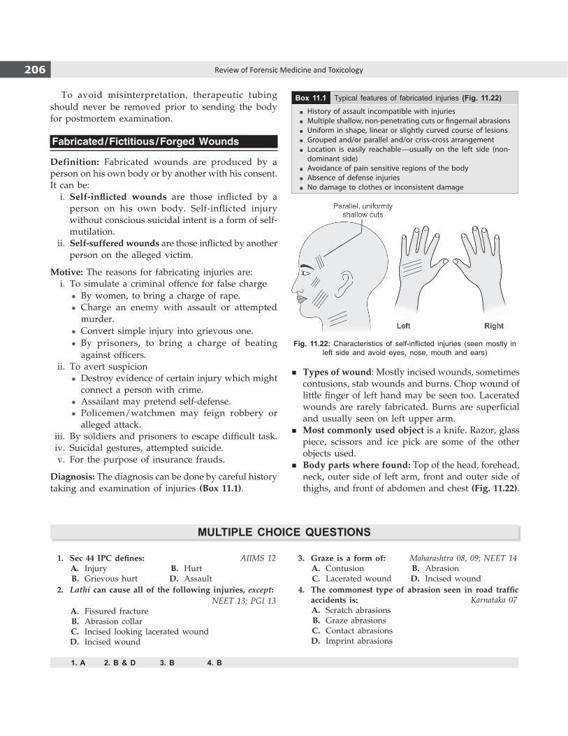
206 Review of Forensic Medicine and Toxicology
To avoid misinterpretation, therapeutic tubing should never be removed prior to sending the body for postmortem examination.
Fabricated / Fictitious / Forged Wounds
Definition: Fabricated wounds are produced by a person on his own body or by another with his consent. It can be: i. Self-inflicted wounds are those inflicted by a
person on his own body. Self-inflicted injury without conscious suicidal intent is a form of self-mutilation.
ii. Self-suffered wounds are those inflicted by another person on the alleged victim.
Motive: The reasons for fabricating injuries are: i. To simulate a criminal offence for false charge
z By women, to bring a charge of rape. z Charge an enemy with assault or attempted
murder. z Convert simple injury into grievous one. z By prisoners, to bring a charge of beating
against officers. ii. To avert suspicion
z Destroy evidence of certain injury which might connect a person with crime.
z Assailant may pretend self-defense. z Policemen/watchmen may feign robbery or
alleged attack. iii. By soldiers and prisoners to escape difficult task. iv. Suicidal gestures, attempted suicide. v. For the purpose of insurance frauds.
Diagnosis: The diagnosis can be done by careful history taking and examination of injuries (Box 11.1).
Box 11.1 Typical features of fabricated injuries (Fig. 11.22)
� History of assault incompatible with injuries � Multiple shallow, non-penetrating cuts or fingernail abrasions � Uniform in shape, linear or slightly curved course of lesions � Grouped and/or parallel and/or criss-cross arrangement � Location is easily reachable—usually on the left side (non-
dominant side) � Avoidance of pain sensitive regions of the body � Absence of defense injuries � No damage to clothes or inconsistent damage
Fig. 11.22: Characteristics of self-inflicted injuries (seen mostly in left side and avoid eyes, nose, mouth and ears)
MuLTIPLe ChoICe queSTIoNS
1. Sec 44 IPC defines: AIIMS 12 A. Injury B. Hurt B. Grievous hurt D. Assault 2. Lathi can cause all of the following injuries, except: NEET 13; PGI 13 A. Fissured fracture B. Abrasion collar C. Incised looking lacerated wound D. Incised wound
3. Graze is a form of: Maharashtra 08, 09; NEET 14 A. Contusion B. Abrasion C. Lacerated wound D. Incised wound 4. The commonest type of abrasion seen in road traffic
accidents is: Karnataka 07 A. Scratch abrasions B. Graze abrasions C. Contact abrasions D. Imprint abrasions
1. A 2. B & D 3. B 4. B
� Types of wound: Mostly incised wounds, sometimes contusions, stab wounds and burns. Chop wound of little finger of left hand may be seen too. Lacerated wounds are rarely fabricated. Burns are superficial and usually seen on left upper arm.
� Most commonly used object is a knife. Razor, glass piece, scissors and ice pick are some of the other objects used.
� Body parts where found: Top of the head, forehead, neck, outer side of left arm, front and outer side of thighs, and front of abdomen and chest (Fig. 11.22).

207Injuries
5. Graze abrasions mimic: AI 09 A. Eczema B. Pressure sore C. Burns D. Scalds 6. Brush burn is: AP 09 A. Graze abrasion B. Imprint abrasion C. Electric burn D. Arborescent burn 7. In a case of hanging, ligature mark is an example
of: NEET 13 A. Contusion B. Pressure abrasion C. Laceration D. Burn 8. An auto rickshaw ran over a child’s thigh, there is a mark
of the tyre tracks, it is an example of: AIIMS 10 A. Patterned bruise B. Patterned abrasion
C. Pressure abrasion D. Graze abrasion 9. Bite mark is an example of: FMGE 11 A. Pressure abrasion B. Graze abrasion C. Scratch abrasion D. Pattern abrasion 10. Scab or crust of abrasion appears brown in: PGI 11 A. 12–24 h B. 2–3 days C. 4–5 days D. 5–7 days 11. Crescent shaped abrasion is seen in injury with:
AFMC 12 A. Wood cane B. Nail scratch C. Bicycle chain D. Ligature 12. Prominent bruise with minimum force is seen in:
AIIMS 09 A. Scalp B. Soles C. Palm D. Face 13. Ectopic bruise is most commonly seen in: NEET 13 A. Leg B. Eye C. Pinna D. Scalp 14. Blackening of eye most common because of: NEET 13 A. Friction abrasion B. Patterned abrasion C. Imprint abrasion D. Contusion 15. Blue color of contusion is due to: Gujarat 07 A. Bilirubin B. Hemosiderin C. Hematoidin D. Deoxyhemoglobin 16. Color of hemosiderin is: DNB 09 A. Brown B. Green C. Yellow D. Red 17. Brown color of contusion is due to: NEET 13 A. Biliverdin B. Reduced hemoglobin C. Hemosiderin D. Bilirubin 18. Green color of contusion is due to: TN 08 A. Bilirubin B. Hemosiderin C. Hematoidin D. Biliverdin
19. No color change is seen in subconjunctival hemorrhage due to: WB 09
A. Continuous CO2 supply B. Little amount of blood is present C. Continuous O2 supply D. Color change occurs but not visible to naked eye 20. Antemortem bruise is differentiated from postmortem
bruise by: AIIMS 09 A. Well-defined margin B. Capillary rupture with extravasation of blood C. Yellow color D. Gaping 21. All are true about antemortem contusion, except:
NEET 14 A. Sequential color change B. No inflammation C. Raised enzyme levels D. Blood cells in surrounding tissue 22. A bone bruise or contusion is best identified using:
COMEDK 14 A. Conventional X-ray B. CT scan C. MRI scan D. PET scan 23. Split lacerations are due to: DNB 09 A. Blunt object B. Sharp object C. Sharp heavy object D. Pointed object 24. True about incised looking wounds are all, except:
PGI 12 A. Type of split lacerated wound B. Avulsion lacerated wound with blunt object C. Produced by blunt object D. Commonly seen on scalp 25. Split laceration resembles: Jharkhand 11 A. Incised wound B. Abrasion C. Gunshot wound D. Contusion 26. Incised looking laceration is seen in all, except:
AFMC 11; NEET 13 A. Iliac crest B. Zygomatic bone C. Shin D. Chest 27. Flaying is seen in which type of lacerated wound: AIIMS 11 A. Split B. Stretch C. Avulsion D. Cut 28. Tissue bridges are seen in: DNB 10 A. Abrasion B. Contusion C. Laceration D. Stab wound 29. In an incised wound, all of the following are true,
except: COMEDK 07; NEET 14, l5 A. It has clean-cut margins B. Bleeding is generally less than in lacerations C. Tailing is often present D. Length of injury does not correspond with length
of blade
5. C 6. A 7. B 8. B 9. D 10. B 11. B 12. D 13. B 14. D 15. D 16. A 17. C 18. D 19. C 20. B 21. B 22. C 23. A 24. B 25. A 26. D 27. C 28. C 29. B
208 Review of Forensic Medicine and Toxicology
30. Hesitation cuts are seen in a case of: PGI 09; Maharashtra 09; Bihar 10; MP 11
A. Homicide B. Suicide C. Accident D. Fall from height 31. Incised wounds on genitalia: AIIMS 09, 10 A. Homicidal B. Suicidal C. Accidental D. Self-inflicted 32. True about stab wounds: NEET 13 A. Depth is greater than breadth B. Breadth is greater than depth C. Length is greater than breadth D. It has wound of entry and exit
33. Shape of stab wound depends on: NEET 14 A. Edge of weapon B. Shape of weapon C. Width D. All of the above 34. In stab wounds, Langer’s lines determine:
NEET 14 A. Direction B. Gaping C. Shelving D. Healing 35. Harakiri method of suicide involves stabbing into:
AIIMS 14 A. Chest B. Wrist C. Abdomen D. Neck
30. B 31. A 32. A 33. D 34. B 35. C

Firearm Injuries
CHAPTER 12
Definitions � Ballistics (Greek ba’llein: throw): It is the science
of projectile motion, and conditions affecting that motion.
� Forensic ballistics: Science which deals with the investigation of firearms, ammunition and the problems arising from their use.
Ballistics is subdivided into: � External ballistics: Study of the passage of the projectile through
space or the air. � Internal ballistics: Study of the projectile in the gun. � Terminal ballistics: Study of the interaction of a projectile with
its target. � Wound ballistics: It is concerned with the motion and effects of
the projectile in tissue.
� Firearm: Any instrument or device that discharges a missile by the expansive force of gases produced by burning of an explosive substance.
It consists of:1 (Fig. 12.1) i. Barrel: A hollow metal cylinder in which the
propellant charge is placed. It is long in rifles and shotguns, and short in pistols and revolvers. The lumen is known as bore. The rear end where the cartridge is inserted is known as the breech end, and the front end where the bullet/shots comes out is the muzzle end.
ii. Action: It consists of a bolt, a striker or hammer and a trigger.
iii. Extractor is a part of a gun’s action which serves to remove brass cases of fired ammunition after it has been fired from the chamber of the gun.
iv. Butt/grip: Rear portion of stock in a shoulder arm or bottom of a handgun containing a magazine.
v. Magazine: The receptacle for the cartridges in a repeating type of weapon from which the cartridges are fed automatically into the chamber by the action of mechanism.
Fig. 12.1: Parts of rifled weapon

210 Review of Forensic Medicine and Toxicology
Velocity � Muzzle velocity: The velocity of the projectile as it
emerges from the muzzle end. Depending on it, firearms can be of low, medium and high velocity.
� Striking velocity: Velocity of the projectile at the point of impact. The velocity diminishes as the missile travels ahead to strike the target.
Trajectory: Path traced by the projectile during flight.
Classification of Firearms
Firearms are broadly classified into two categories depending on the type of barrel: i. Rifled weapons
z Rifles: 0.22, single shot, lever action, bolt-action, pump action, auto-loading
z Revolvers: Swing-out, break-top, solid-frame z Single shot pistols z Auto-loading pistols z Submachine guns z Machine guns
ii. Smooth bore weapons (shotguns) z Single-shot z Bolt action z Double barrel z Pump-action z Lever action z Auto-loading
� Broadly, single-shot pistols, derringers (variant of single-shot pistols), revolvers and auto-loading pistols are considered as handguns.
� Country made firearms (katta or improvised firearms) are mostly 12 bore smooth bore weapons.
Rifled Firearms (Fig. 12.2)
The bore is scored internally with number of shallow spiral grooves varying from 2 to 22, most common are 4, 5 or 6, which run parallel to each other, but twisted spirally from breech to muzzle end. These grooves are called ‘rifling’ and the projecting ridges between the grooves are called ‘lands’ (Fig. 12.3).
� Rifles, pistols, revolvers, submachine guns and machine guns—all have rifled barrels.
� The direction of rifling can be either right (clockwise) or left (counter-clockwise)—majority of handguns have a right-hand twist.
When the bullet passes through the bore, its surface comes in contact with the projecting spirals which gives the bullet a rotational spinning or spiraling motion along
Fig. 12.2: Rifle and shotgun barrel
Fig. 12.3: Caliber of rifled firearm (distance between two lands)
its longitudinal axis. Rifling gives the bullet a signature marking that is unique to the weapon that fired it.
Advantages of rifling � Imparts gyroscopic stability � Increases accuracy and range � Prevents wobbling or tumbling end-over-end � Gives greater power of penetration
� Fully-automatic: Small firearm which, after first cartridge is manually loaded and fired, will eject the fired case, load the next cartridge from the magazine, fire and eject that fired case and repeats the process indefinitely as long as trigger is held pressed or until cartridge supply from magazine is exhausted.
� Semi-automatic: A weapon which fires, eject and reloads on trigger being pressed, but does not fire again until the trigger has been released and pressed again. Auto-loading pistols are semi-automatic wherein the empty cartridge is ejected after firing.
� Revolver has a revolving cylinder that contains several chambers, each of which holds one cartridge.
� Rifle is a firearm with a rifled barrel which is designated to be fired from the shoulder.
� Handgun is a firearm capable of being carried and used by one hand, such as a pistol or revolver.
� Zip guns: Crude home made single shot rifled firearm.

211Firearm Injuries
� Stud guns: Tools used to fire metal studs into wood, concrete or steel.
� Dragon’s breath is an incendiary round of 12 bore shotgun. It contains exothermic pyrophoric misch metal (primarily magnesium pellets/shards) as the projectile. It is designed for the purpose of throwing or spewing a flame or fireball to simulate a flamethrower.
� Bolo shell: Any shell that can be fired in a firearm that expels as projectiles two or more metal balls connected by solid metal wire.
Smooth Bore Firearms / Shotguns (Fig. 12.2)
In smooth bore firearms, the bore or the inner surface of the barrel is uniformly smooth. It is intended to be fired from the shoulder, and is designated to fire multiple pellets from the barrel.
� Barrel lengths of shotgun range from 18–36 inches; 26 and 28 inch being most common.
� Shotgun barrel is divided into three sections: chamber, forcing cone and bore.
� Musket is a muzzle-loaded, smooth bore long gun. It is usually fired from the shoulder.
Choking (Fig. 12.4)
Cylinder bore: Entire barrel from breech to muzzle is of same diameter.
Choke bore: A shotgun slightly constricted at the muzzle, usually distal 7–10 cm of the barrel is narrow.2
� Usual degrees of choke in descending order are full, modified, improved cylinder and cylinder.
� Advantages of choking: Lessens the rate of spread of shot, increases the explosive force and increases the velocity, and thus increases the range.
� Dispersion of pellets in fully choked is about half that of cylinder bore.
� For cylinder bore, range of discharge can be obtained by measuring the diameter of the wound from the outermost individual pellet wound, in cm and dividing it by three, giving the result in meters.
Balling or welding of shot: In shotguns, there may be conversion of shots (pellets) into compact mass which may cause a circular or oval large entry wound of 5–10 mm and several small circular punctures, suggesting use of two weapons—one rifle and the other a shotgun.
Balling occurs due to: � Faulty manufacture � Old ammunition � Too much powder � Wads of incorrect kind � High sealing pressure of the wads � Pouring of paraffin into cartridge case after removing the
outer wad
Paradox guns: Some shotguns which have small portion of their bore near the muzzle end rifled.Shotgun pellets fall into two categories depending on the size: birdshot and buckshot (larger shot). There are three types of lead shot depending on the composition: i. Drop/soft shot: Made with pure lead. ii. Chilled/hard shot: Lead is hardened by the
addition of antimony. iii. Plated shot: It is coated with a thin coat of copper or
nickel to minimize distortion on firing—maintains good aerodynamic shape and increase the range.
Bore (Gauge / Caliber)
It is the diameter of the interior of the barrel of a rifled firearm measured between diagonally opposite lands, expressed in inches or millimeters, e.g. .22”, .32”, .38” (Fig. 12.3).3 It represents the diameter of the barrel before the rifling grooves are cut. For shotguns, bore is the number of spherical lead balls of size fitting the barrel of a shotgun which can be made from one pound of lead (454 g), e.g. 12, 16, 20 bore (Fig. 12.5).*4 Barrel of 12 bore gun will pass a ball that weighs 1/12 lb.
* The only exception to this nomenclature is the .410 bore which has a bore .410 inch in diameter (smallest gauge of shotgun shell).
Fig. 12.4: Choking of shotgun

212 Review of Forensic Medicine and Toxicology
The European system of cartridge designation which uses metric system is more thorough and logical than the Indian system. It specifically identifies a cartridge by giving the bullet diameter and the case length in millimeters, as well the type of cartridge case, e.g. 7.62 × 54 mm R, indicates the diameter, length and rimmed bullet respectively.
Bullet
Bullet (French boulette: little ball) is the projectile of a rifled firearm that leaves the muzzle when it discharges.5 Traditional bullet is made of soft metal and has a rounded nose. The metal used is lead with varying amounts of antimony and/or tin added to provide hardness. This is known as round nose soft bullet.
� Caliber of a bullet is the cross-sectional diameter. � In revolver and pistol, the bullet is short, and the
point is usually round (Fig. 12.6). � In a rifle, the bullet is elongated with a pointed end
(Fig. 12.6).
Modern bullets fall in two categories: lead- and metal-jacketed.
Jacketed bullets: A tough metal envelope (made of copper and zinc, or copper and nickel with steel) covering the outside of the bullet—thickness ranges from 0.0165–0.030 inches.
It is of two types: i. Full metal jacket: Covers all, but the base. ii. Partial metal-jacketed bullet: Covers the base and
cylinder portion of the bullet, leaving the nose partly or fully exposed; designed to expand or mushroom.
Advantages of jacket: It prevents: � Deformation of the bullet in the barrel from dirt
overpressures or damage outside the gun, and reduces misfires.
� Fragmentation or melting. � Damage to the gun barrel from ‘leading’—lead
fouling of the barrel.
Types of Bullet i. Dum-dum bullet: Jacketed bullet (.303 centerfire
rifle) which does not cover the entire bullet, an area near the nose is left uncovered to expose the core (Fig. 12.7).
ii. Hollow-point bullet: Modern version of dum-dum bullet. There is a pit present in front of the nose. When the bullet strikes a target, the pressure in the pit forces the ring of lead around it to expand into a mushroom-shape. This causes more soft tissue damage, and higher incapacitation index on the target (Fig. 12.7).
Fig. 12.5: Caliber of a shotgun Fig. 12.6: Cartridges of rifled and shotgun weapons

213Firearm Injuries
iii. Soft-point bullet: Jacketed bullet that is left open at the tip, exposing some of the lead inside. They are designed to expand upon impact, but slowly, as compared to the hollow-point bullets (Fig. 12.7).
iv. Tandem bullet (Piggyback bullet): Bullets ejected one after the other, when the first bullet having been struck in the barrel, fails to leave the barrel and is ejected by a subsequently fired bullet.6
Cause: Faulty ammunition or loaded firearm unused for years.
v. Duplex bullet: Contains two projectiles by design, used in military rifles and enter a target at different points.
vi. Frangible bullet: Designed to fragment upon impact, often to the point of disintegration, made mostly of copper, powdered tungsten, lead or iron.7 They do not ricochet.
vii. Souvenir bullet: Bullet present in the body for long time with no fresh bleeding around it and surrounded by a dense fibrous tissue capsule. It was not removed as it was not causing any harm or it was located in an area from where its retrieval could cause more damage to the body, e.g. bullet in the spine.
viii. Armor piercing bullet: A jacketed bullet which has inner core of hard, high-density metal such as tungsten, tungsten carbide, depleted uranium or steel, and a truncated cone. This type of ammunition is designed to penetrate armor.
ix. Incendiary bullets: Type of army bullet used to cause fire in the target.
x. Tracer bullet: It leaves a visible mark or ‘trace’ while in flight, so that the gunner can observe the strike of the shot.8
xi. Exploding bullet: A bullet that is designed to detonate or forcibly break up through the use of an explosive or deflagrant contained within
or attached to such bullet. These bullets, apart from causing extensive damage in the victim, pose considerable danger to the doctor because the bullet may explode during any procedure (ultrasono graphy/autopsy), if it had failed to detonate in the body.
xii. Nontoxic shot: This is a new lead-free projectile wherein the copper-jacketed lead core is replaced with copper-jacketed tungsten, steel, bismuth, tin or nylon core. It is known as the ‘green bullet’ since toxic lead is not released into the environment.
xiii. Boat tail bullet: The rear end (base) of the cartridge is tapered instead of squaring off to stabilize the projectile in flight (Fig. 12.7). The boat shape reduces drag on a bullet, helping it to retain velocity, and resist deflection from crosswinds.
xiv. Rubber bullets are usually non-lethal rubber-coated projectiles fired from guns, often used in riot control and to disperse protests. The British use the term baton round.
xv. Plastic bullets: These ‘baton round’ was developed as a replacement for rubber bullets in the UK against rioters in Northern Ireland. It is made of polyvinyl chloride. However, if hit from near distance, it can cause fatal injury.
xvi. Poisoned bullets are usually 0.177 caliber bullets (the smallest in general use) which carry curare, ricin or aflatoxin.
xvii. Wadcutter is a bullet specially designed for shooting paper targets, usually at close range and with significant velocities.
� Originally, dum-dum bullet referred to a new type of ammunition produced at the Dum-Dum arsenal in British India (near Kolkata) in the early 1890s.
� In frangible bullets, class characteristics can be demonstrated but individual markings are lost which is necessary for bullet-to-gun comparison. X-ray picture produced will be similar to the ‘lead snowstorm’ seen with hunting bullets.
Cartridge
Cartridge is one unit of ammunition.
Cartridge consists of: i. Cartridge case with percussion cap containing primer ii. Propellant charge (gunpowder) iii. Projectile (bullets/pellets) iv. Wads (in smooth bore weapons only)9
Fig. 12.7: Different types of bullets

214 Review of Forensic Medicine and Toxicology
Use of cartridge case � Keeps various components together � Prevents backward escape of gases � Provides waterproofing for gunpowder
Cartridge of shotgun and rifle consists of the following as shown in Figures 12.8 and 12.9.
Percussion cap: It is made of either zinc or copper or a compound of both, so as to be malleable and deformable under the blow of the firing pin.
Cartridge cases in rifled firearms are usually made of brass. Sometimes, they are made of steel or aluminum. Cartridge cases are classified into five types depending on the configuration of their bases: rimmed, rimless, semi-rimmed, rebated and belted.
Rimmed cartridge has a base with projecting rim and used in revolvers. Rimless cartridge is used in pistols.Small-arms cartridges are classified as centerfire or rimfire, depending on the location of primer.
� Centerfire: The primer is located in the center of the base of the cartridge case.
� Rimfire: Cartridges with priming mixture inserted in the hollow rim. Firing pin gives a hit mark on the circumference.
Blank cartridge: Cartridge with primer, gunpowder and wadding, but without any bullet. It is used in:
� Starter pistol in sports � Stage/movie performance � Army maneuvers. Fig. 12.9: Parts of a rifled cartridge
Wad: Wad is made of some soft material, like disc of felt, cardboard, plastic, cork or straw (Fig. 12.10). It is placed between powder and shot or over the shot. The cardboard disc behind the shot charge prevents the pellets from getting lodged in the felt wad (Fig. 12.8).
Advantages of wad � Allows optimum pressure to develop � Seals the bore effectively � Helps in lubrication � Prevents the escape of gas from the breech end � Separates propellant from the projectiles
Fig. 12.8: Parts of a shotgun cartridge

215Firearm Injuries
The wad consists of three parts, powder wad, cushion and shot cup, which may be in separate pieces or in one part (Fig. 12.10). The powder wad acts as the gas seal (known as obturation) and is placed firmly over the powder; it may be a paper or plastic. The cushion comes next, and it is designed to compress under pressure, to act as a shock absorber and minimize the deformation of the shot; it also serves to take up as much space as is needed between the powder wad and the shot. Cushions are almost universally made of plastic with crumple zones. The shot cup is the last part, and it serves to hold the shot together as it moves down the barrel. Shot cups have slits on the sides, so that they peel open after leaving the barrel, allowing the shot to continue on in flight undisturbed.
Gunpowders (Propellant Charge)
i. Black powder: It produces flame, smoke and heat, and consists of granular ingredients, like sulfur, charcoal and saltpeter (potassium nitrate).10
The optimum proportions for gunpowder and its function is given in Table 12.1.
ii. Smokeless powder: It is more effective than black powder as it burns more efficiently and produces much less smoke, resulting in less blackening and tattooing around the entry wound.
Types a. Single base powder consists of nitrocellulose
(gun cotton).11 It is the most common type of commercial powder, because of its simplicity, adequate power and low flame temperature.
b. Double base powder consists of nitrocellulose and nitroglycerin.12 It is more powerful than single base because of nitroglycerin, but has a flame temperature that may melt the steel of the barrel.
c. Triple base powder consists of nitrocellulose, nitroglycerin and nitroguanidine. The quantity of nitroglycerin is small, but sufficient to give power; the nitroguanidine lowers the flame temperature while adding an active explosive constituent.
iii. Semi-smokeless powder: It consists of mixture of 80% black and 20% smokeless type.
� When black powder burns properly, it produces 44% of its original weight in gases and 56% in solid residues (potassium carbonate and potassium sulfate). These residues appear as a dense, white smoke. A grain of black powder gives rise to 200–250 ml of gas composed of CO2, CO, N2, hydrogen, H2S and traces of methane and O2, whereas a grain of smokeless powder forms 800–900 ml of gas with nearly 100% conversion of powder to gases. [about 15.1 grains = 1 gram and 7,000 grains = 1 pound (approx)]13
� Grading of black powder: The term grading refers to grain size. There are two separate categories of gunpowder grades, ‘C’ and ‘F’. ‘C’ grade is for cannon and large capacity explosive devices. Powder meant for small arms uses the letter ‘F’ to denote the grain size. It correlates to the size of the screen mesh which it falls through for sorting. Ranges are course FG (used in large bore rifles), FFG (used in medium and small bore arms, such as muskets), FFFG (used in small bore rifles and pistols) and FFFFG (used in short pistols) which is very fine. Small particles, higher FG numbers burn much faster.14
Primer: The primer is a small charge of impact-sensitive chemical that may be located at the center of the case head (centerfire ammunition) or at its rim (rimfire ammunition). It ignites the powder or propellant charge by impact of gun’s firing pin.
Composition of primer i. Lead styphnate ii. Barium nitrate iii. Antimony sulfide iv. Lead peroxide v. Potassium chlorate vi. Tetrazine
Mechanism of Discharge of Projectile
A firearm is fired when the trigger is pulled. The trigger releases a pin or hammer whose tip strikes the percussion cap at the base of the cartridges. The primer contained in it explodes by the heat created by the firing pin. This sends a flash through a tiny hole into the main body of powder filled case, and powder charge or propellant is set on fire instantaneously producing a large amount of gas and heat under pressure. The cartridge case swells outwards, due to which the hold on the bullet (missile, pellets) is released; the bullet is forced into the barrel and passed out.
Table 12.1: Composition of gunpowder with its function
Chemical Percentage (%) Function(s)
Potassium nitrate 75 Oxygen supplier or oxidizer
Sulfur 10 Makes the mixture more denser and readily ignitable
Charcoal 15 Fuel
Fig. 12.10: Various types of wads

216 Review of Forensic Medicine and Toxicology
� When the bullet emerges from the barrel, it is accompanied by:
i. Unburnt propellant particles ii. Partially burnt propellant particles iii. Soot from combustion of propellant iv. Nitrates and nitrites from combustion of
propellants v. Particles of primer residue (oxides of lead,
antimony and barium) vi. Vaporized metal and metallic particles stripped
from the bullet and cartridge case. � Varying the length of the barrel also affects how
much powder exits the muzzle. Shortening the barrel causes more unburned powder to emerge and vice-versa.
� The confined gas left behind gives recoil thrust to the gun.
� Noise of gun firing is caused by the muzzle blast or sudden release of gases disturbing the air.
� The blast has the shape of a cone, the apex of which is located at the muzzle.
� Muzzle blast is the release of gases under high temperature and pressure from the muzzle of a firearm when it is discharged.
� Muzzle flash is the visible light of the muzzle blast.
Wound Ballistics and Mechanism of Injury
As a missile traverses the body, it causes injury by transferring some or all of its energy. This is manifested as laceration and crushing of tissues in its path, and sometimes away from its path. The amount of energy transferred is given by the formula:15
KE = ½ M (V12–V2
2) where KE = Kinetic energy M = Mass of the bullet V1 and V2 = Velocities at entry and exit
It shows that velocity rather than its weight plays a greater role in determining the amount of kinetic energy possessed by a bullet. Doubling the weight doubles the kinetic energy, but doubling the velocity quadruples the kinetic energy.16
In general, bullets fired from handguns are propelled at a low velocity, have low energy (50–100 J) and result in low energy transfer wounds which are characterized by injuries confined to the wound track. Missiles with high available energy (2000–3000 J) include high-velocity assault rifle bullets and have the potential to cause high energy transfer wounds. As a bullet moves
through the body, it crushes and shreds the tissue in its path, while at the same time flinging outward (radially) the surrounding tissue from the path of the bullet, producing a temporary cavity considerably larger than the diameter of the bullet (Fig. 12.11). Cavitation in solid organs, like liver, kidney and spleen is often fatal, but in the bones, it creates secondary missiles.
Fig. 12.11: Shock waves from a gunshot result in cavitation
Firearm Wounds
Gunshot wounds are either penetrating or perforating. � Penetrating wounds: The bullet enters an object and
does not exit. � Perforating wounds: The bullet passes completely
through an object.
For example, a bullet striking the head may pass through the skull and brain before coming to rest under the scalp, producing a penetrating wound of the head, but perforating wound of the skull and brain.
Characteristics of firearm wounds depend upon: � Nature of the firearm, whether shotgun or rifle � Shape and composition of the missiles � Range (distance) of firing � Part of the body struck (head or trunk) � Direction of firing
� Tattooing: It consists of unburnt or partially burnt powder particles that are embedded in and under the skin through the force of their impact (when the weapon is near enough for the powder grains to strike).17
z Tattooing is an antemortem phenomenon and indicates that the individual was alive; and it cannot be wiped away with a wet cotton.
z It consists of numerous reddish-brown punctate abrasions surrounding the wound of entrance.
z The greater the range, the larger and less dense the powder tattooing.
z Marks usually heal completely if the individual survives (involves the superficial layer of the epidermis).

217Firearm Injuries
� Stippling: It is the visible mark powder grains leave, when it does not get embedded on the skin (when the range increases). It may also be produced by other material, e.g. shotgun filler or fragments of intermediary targets. The term ‘stippling’ is sometimes used synonymously with ‘powder tattooing’.
� Blackening (soot or smoke soiling/smudging): Deposi tion of powder soot (carbon) produced by combus tion of gunpowder.18
z As the range increases, the size of the zone of blackening will increase, whereas the density will decrease.
z It can be easily removed with a wet cotton. � Fouling: Tiny lesions around the entry wound caused
by fragments of metal expelled by the discharge. These fragments may come either from the surface of the bullet or from the interior of the barrel, and cannot be wiped off from the skin.
� Abrasion collar/ring: As the bullet strikes the skin, it first indents and then stretches the skin surface so that perforation takes place through a tense area which produces a rim of flattened reddish-brown zone of abraded epidermis, surrounding the entrance wound.* The abrasion ring can vary in width, depending on the caliber of the weapon, the angle and site at which the bullet entered.
z A bullet striking perpendicularly will produce a concentric ring, and if the bullet penetrates at an oblique angle, the zone will be eccentric with the wider zone on the side from which the bullet came (Fig. 12.12).
z Entrance wounds in the skin overlying the clavicle have a wider abrasion ring than those in the other parts of the body.
z The entry wound on the palms and soles usually lack abrasion collar due to the thickness and keratinization of the epidermis in these areas, and may be stellate instead of round.
z Some contusion is present in abraded collar and as such, it is also called ‘contusion collar’.
� Grease/dirt collar (bullet wipe): A black/gray colored ring is seen lining the defect, sharply outlined, caused from removal of substances from bullet as it passes through the skin. It consists of bullet lubrication, paraffin, lead from surface of bullet, barrel debris and gun oil from interior of the barrel.
z The ring of dirt is more pronounced in shots from freshly oiled weapons than in shots from lubricant-free barrels.
z By contrast, soot is dark in the center and fades towards the periphery.
z Abrasion collar surrounds the dirt collar. z This gray rim is more prominent in clothing, where
it is called ‘bullet wipe’. z Abrasion and dirt collars are proof of an entry
wound.19,20
� Muzzle/recoil imprint mark (described by Werk-gartner in 1922) is regarded as a patterned abrasion/intradermal contusion caused by the expansive power of the gases lifting the skin forcibly up against the muzzle.
z This is a sign of a contact shot. z Its shape depends on the firearm, the ammunition
and the anatomical conditions, but does not require a bullet.
z Characteristic imprint marks can provide clues to the type of the firearm and its position at the time of discharge.
� Back spatter: In a contact shot, the muzzle blast and negative pressure in the barrel may suck blood, hair, fragments of tissues and cloth fibers back into the barrel.
� Blowback phenomenon: Cruciate, stellate or ragged laceration is seen, especially if there is a thick bone immediately under the skin, such as the skull. This occurs as a result of expansion of gases beneath the skin and their exit through the entry wound (in contact wounds).
� Point blank: When the range is very close to, or in contact with the surface of the skin.
* According to German researchers, abrasion collar is not caused by friction or overstretching, but by superficial tissue particles being thrown back against the direction of the fire, since the collar is seen even when shots are fired at non-biological, layered objects.
Fig. 12.12: Abraded collar when bullet struck (A) Perpendi cularly (B) At an angle. The wider side determines the direction of fire
A B

218 Review of Forensic Medicine and Toxicology
Characteristics of Shotgun Wounds (Fig. 12.13)
At close range, the shotgun is the most formidable and destructive of small arms. When a shotgun is fired, the projectile travels as a compact mass. As the range increases, the individual pellets spread out, collectively travel in a cone-like manner and their velocity decreases with distance. A rough estimate of the rate of spread is about 1 inch/yard from the muzzle of a full choke gun.
Contact or Near Contact ShotContact wound: Muzzle of the weapon is held against the body at the time of discharge. Contact wound can be hard (muzzle held tightly against the skin), loose, angled or incomplete.
� Contact wounds differ in appearances, depending upon the site, whether it is the head or the non-resisting parts, e.g. chest or abdomen.
� Contact shotgun wounds of the head are the most mutilating firearms wounds. Extensive destruction of bone and soft tissue structures occurs with bursting rupture of the head and evisceration of the brain, since the gases have restricted space for expansion. Soot is seen around the entrance in most contact wounds of head.21
� Contact wound of the trunk appear circular in shape, and have diameter usually equal to that of the bore of the weapon as shot enters as a mass.
� If the muzzle is pressed firmly, soiling or burning is absent, but the edges of the wound is seared and blackened by the hot gases, and a muzzle impression may be found.
� If the muzzle is not pressed firmly or is loosened by recoil; flame, gas and soot may escape sideways and soil the adjoining skin.
� If clothing interposes between the muzzle and skin, soot will be found on the clothing, as well as the skin. Clothing may be singed, and there may be burning around the skin wound.
� The gases cause laceration of deeper tissues and even fragmentation of bone.
� Wad is often found in the wound, and this may prove to be an important clue to the type of cartridge used.
� Wound track and adjacent tissues appear cherry-red due to carboxyhemoglobin and carboxymyoglobin from absorption of carbon monoxide (CO) formed from combustion of the gunpowder (can spread upto 15 cm or more from the entrance).
� Usually, shotgun projectiles do not exit out of the body.
Close Range (Between contact and 3 ft) � Close range shotgun wounds of the head are almost
as mutilating as contact wounds, because the pellets are still traveling in a single mass. Large gaping tears of the scalp are present.
� When clothing is present, it traps most of the soot and powder grains and may reduce the flame effect.
� Depending on the angle of firing, the wound is circular or elliptical. There are no separate pellet holes.
� Singeing of hair, scorching, blackening and tattooing (less with smokeless powder) of skin is seen.22 Blackening and tattooing can be demonstrated by infrared photography.
Fig. 12.13: Shotgun wounds at varying distances
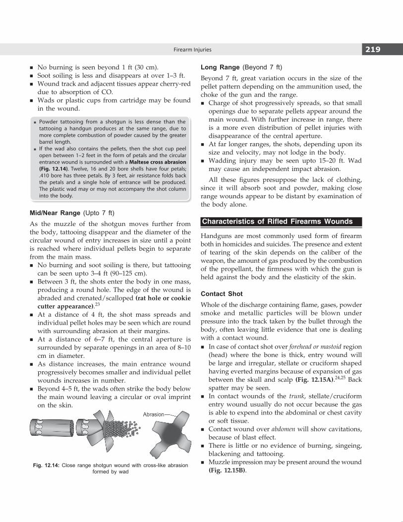
219Firearm Injuries
� No burning is seen beyond 1 ft (30 cm). � Soot soiling is less and disappears at over 1–3 ft. � Wound track and adjacent tissues appear cherry-red
due to absorption of CO. � Wads or plastic cups from cartridge may be found
in the wound.
� Powder tattooing from a shotgun is less dense than the tattooing a handgun produces at the same range, due to more complete combustion of powder caused by the greater barrel length.
� If the wad also contains the pellets, then the shot cup peel open between 1–2 feet in the form of petals and the circular entrance wound is surrounded with a Maltese cross abrasion (Fig. 12.14). Twelve, 16 and 20 bore shells have four petals; .410 bore has three petals. By 3 feet, air resistance folds back the petals and a single hole of entrance will be produced. The plastic wad may or may not accompany the shot column into the body.
Mid/Near Range (Upto 7 ft)As the muzzle of the shotgun moves further from the body, tattooing disappear and the diameter of the circular wound of entry increases in size until a point is reached where individual pellets begin to separate from the main mass.
� No burning and soot soiling is there, but tattooing can be seen upto 3–4 ft (90–125 cm).
� Between 3 ft, the shots enter the body in one mass, producing a round hole. The edge of the wound is abraded and crenated/scalloped (rat hole or cookie cutter appearance).23
� At a distance of 4 ft, the shot mass spreads and individual pellet holes may be seen which are round with surrounding abrasion at their margins.
� At a distance of 6–7 ft, the central aperture is surrounded by separate openings in an area of 8–10 cm in diameter.
� As distance increases, the main entrance wound progressively becomes smaller and individual pellet wounds increases in number.
� Beyond 4–5 ft, the wads often strike the body below the main wound leaving a circular or oval imprint on the skin.
Long Range (Beyond 7 ft)Beyond 7 ft, great variation occurs in the size of the pellet pattern depending on the ammunition used, the choke of the gun and the range.
� Charge of shot progressively spreads, so that small openings due to separate pellets appear around the main wound. With further increase in range, there is a more even distribution of pellet injuries with disappearance of the central aperture.
� At far longer ranges, the shots, depending upon its size and velocity, may not lodge in the body.
� Wadding injury may be seen upto 15–20 ft. Wad may cause an independent impact abrasion.
All these figures presuppose the lack of clothing, since it will absorb soot and powder, making close range wounds appear to be distant by examination of the body alone.
Characteristics of Rifled Firearms Wounds
Handguns are most commonly used form of firearm both in homicides and suicides. The presence and extent of tearing of the skin depends on the caliber of the weapon, the amount of gas produced by the combustion of the propellant, the firmness with which the gun is held against the body and the elasticity of the skin.
Contact ShotWhole of the discharge containing flame, gases, powder smoke and metallic particles will be blown under pressure into the track taken by the bullet through the body, often leaving little evidence that one is dealing with a contact wound.
� In case of contact shot over forehead or mastoid region (head) where the bone is thick, entry wound will be large and irregular, stellate or cruciform shaped having everted margins because of expansion of gas between the skull and scalp (Fig. 12.15A).24,25 Back spatter may be seen.
� In contact wounds of the trunk, stellate/cruciform entry wound usually do not occur because the gas is able to expend into the abdominal or chest cavity or soft tissue.
� Contact wound over abdomen will show cavitations, because of blast effect.
� There is little or no evidence of burning, singeing, blackening and tattooing.
� Muzzle impression may be present around the wound (Fig. 12.15B).
Fig. 12.14: Close range shotgun wound with cross-like abrasion formed by wad

220 Review of Forensic Medicine and Toxicology
� Muscles around the track taken by bullet will be cherry-red due to presence of CO.
� Burning, blackening and powder grains deposits will be found in the depths of the wound (examination of the wounds with dissecting microscope is of value).
� Hair nearby may get burnt or clubbed by fire/heat.
Close Shot (Flame Range) (Fig. 12.16)26
Body lies within the range of flame, smoke and powder blast, i.e. within 2–3 inches (5–8 cm).
� Entry wound is small and circular in shape having inverted and contused lacerated margins.
� Skin adjacent to the entry wound shows evidence of grease/dirt collar on the inner zone, and abraded-contused collar on the outer zone.
� Evidence of burning, singeing, blackening and tattooing of the skin in and around the entry wound.
� Clothings over the part will be burnt from flame of discharge.
� Hair, in and around, show singeing and will look clubbed, shriveled and swollen at intervals (rarely seen, because the gas emerging from the barrel blows it away).
� The length of the barrel of a firearm has considerable effect on the pattern of smoke produced on the target, e.g. a pistol with a 10 cm barrel will spread the smoke over a much larger area than a rifle having a 2 feet barrel.
� The blood and injured soft tissues in the track will be cherry-red due to CO.
Near Shot (Medium-Range or Intermediate Range)Gunshot entry wounds with powder tattooing, but no soot, are commonly referred to as ‘near-shot’ or ‘intermediate range’ wounds, i.e. when the range is within 24 inches (60 cm).
� Entry wound will be circular in shape, approximately the same size as the bullet, with lacerated, inverted edges surrounded by a narrow zone of grease and abrasion collar, with no evidence of any burning and singeing.
� Entry wound looks like a distant shot when the range is beyond 6 inches (15 cm). Beyond 15 cm, the burning effects of gases and singeing of hair is absent.
� Zone of blackening will be present when the range is within 6–8 inches (15–20 cm), and zone of tattooing will be present around it. In case of handguns, soot is absent beyond 30 cm.
� Tattooing becomes discrete as the range increases, no trace of powder marks will be found when the range is beyond 24 inches, i.e. normally beyond arm’s length. For handguns, powder tattooing extends to a maximum distance of 18–24 inches (45–60 cm).
Distant ShotGunshot entry wounds with no associated soot or gunpowder stippling are referred to as ‘distant’ wounds, i.e. range is beyond 2 feet.
� Entry wound is usually circular in shape, smaller than the bullet, because of elasticity of skin, with lacerated, inverted skin margins, a bigger dirt collar and usual zone of abraded collar.
� Distant gunshot wounds of the head may have a stellate or irregular appearance simulating a contact wound.
� There will be no evidence of any burning, singeing, blackening and tattooing.
� Wound of exit will be slightly bigger than the wound of entry.
� Sometimes, the term ‘indeterminate’ is used since closer range shots where the soot and gunpowder is totally blocked by an interposed target may produce identical appearing wounds.
Fig. 12.15: Contact wounds: (A) Stellate wound, (B) Muzzle imprint
A BFig. 12.16: Close shot entry wound of a bullet

221Firearm Injuries
Irregular, cruciform or stellate entry wounds can occur in individuals shot at intermediate or distant range when the bullet perforates the skin underlying a bony prominence. Head is the most common site—forehead, top and back of the head, supraorbital ridges and cheek bone. Uncommon site is the elbow.
Firearm Wounds on Skull (Fig. 12.17)
� The entry wound shows a punched out hole (clean cut) on the outer table and beveled appearance on the inner table (as it remains unsupported, chipping of the bone occurs).27 Fissured fracture may radiate from the hole. The piece may be driven inside causing injury to the brain. Dura shows irregular tear. In contact wounds, shattering of skull wound may occur.
Fig. 12.17: Bullet wounds in skull
Fig. 12.18: ‘Key-hole’ defect on the outer bone table when a bullet strikes it tangentially
Fig. 12.19: Puppe’s rule: (A) First (initial) fracture, (B) Sub sequent (later) fracture
� The exit wound on the inner table shows clean cut hole and beveling on the outer table. The wound is larger than the entry wound due to the deformity and tumbling of the bullet on entering the skull. The beveling helps to assess the angle of fire.
Tangential entrance wounds (gutter wound) into bone may produce ‘keyhole’ defects with entrance and exit side-by-side; the skin is torn or lacerated by the bullet (Fig. 12.18).
Puppe’s rule: This rule states that when two fracture lines intersect each other, the second fracture line never crosses the first one (Fig. 12.19). It determines the sequence of shots when several bullets have struck the cranium, and is also applicable to the multiple blunt force impact on the skull.

222 Review of Forensic Medicine and Toxicology
Exit Wounds
Shotguns � The appearance of shotgun exit wound depends
upon the part involved and the nature of tissues encountered during its passage in the body.
� Exit wound with shotguns are uncommon, especially when they involve the chest or abdomen.
� When present, it is in the form of serrated, irregular laceration with everted margins through which some tissues or bone fragments may be seen protruding.
� Sometimes, the pellets may accumulate immediately beneath the skin opposite the entry wound, after they have traveled through the body and trapped by the skin.
Rifled FirearmsExit wounds, whether they are from contact, intermediate or distant firing, all have the same general characteristics.
� Bullet which pass through the body causes exit wound, sometimes called as ‘outshoot wound’.
� It may be stellate, circular, elliptical, cruciate, star shaped or slit-like as bullet gets deformed when struck by a bone (common in head and shoulders)—may be confused with contact wounds.
� In contact wounds and very close range, exit wound is smaller than entry wound due to elastic nature of the skin. However, as range increases, the size of exit wound also increases.
� Exit wounds do not show burning, blackening, tattooing, abrasion or contusion collar. The edges are everted, torn or puckered with pieces of contused,
hemorrhagic subcutaneous fat or muscle protruding out of the defect.
Variations in shape and size of exit wounds occur when: � Bullet tumbles in the body and fails to exit nose-end first � Bullet exits as multiple pieces after breaking up � Bullet is deformed � Unsupported skin tends to tear and break into pieces � Fragments of bone are blown out along with the bullet
(secondary missiles).
Shored or supported exit wound: If the skin at the exit wound is supported by firm objects or tight garments, e.g. belt, waist band, bra or tie, or body leaning against a hard object, such as wall or floor, the exit wound appears as a circular defect sur rounded by a margin of abrasion resembling an entry wound.28 The pattern of the material overlying the shored exit may be imprinted on the edges of the wound.
Diff. 12.1 tabulates the differences between entry and exit wound of a rifled firearm (Fig. 12.20).
Peculiar Effects of Firearms
Atypical gunshot entrance wounds are created when the bullet is destabilized prior to entering the body and consequently does not enter the body nose first but sideways or at an angle. The most common cause is bullet ricochet.
� Ricochet bullet: It is a rebound, deviation or deflection of a bullet from its course by striking an intermediate surface. Sometimes, the bullet may strike the surface, but fail to penetrate and glance
Differentiation 12.1: Entry and exit wound (Fig. 12.20)S.No. Feature Entry wound Exit wound
1. Size Smaller than the diameter of the bullet (except contact shot)
Bigger than the bullet
2. Edges Inverted Everted, puckered3. Skull Clean cut on outer table and beveled in the
inner tableBeveled in the outer table and clean cut on inner table
4. Bruising, abrasion and grease collar Present Absent5. Burning, blackening, tattooing May be seen Absent6. Bleeding Less More7. Fat No protrusion May protrude8. Wound track May be cherry-red due to carboxyhemoglobin
and/or carboxymyoglobinNo color change
9. Fibers of clothes Turned in Turned out10. Radiological/micro-chemical
examinationLead ring may be seen Absent
11. Spectrograph More metal is found Less metal
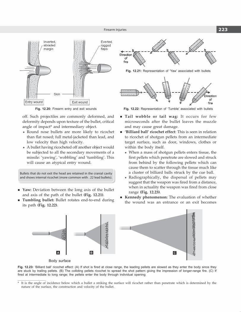
223Firearm Injuries
off. Such projectiles are commonly deformed, and deformity depends upon texture of the bullet, critical angle of impact* and intermediary object.
z Round nose bullets are more likely to ricochet than flat nosed; full metal-jacketed than lead, and low velocity than high velocity.
z A bullet having ricocheted off another object would be subjected to all the secondary movements of a missile: ‘yawing’, ‘wobbling’ and ‘tumbling’. This will cause an atypical entry wound.
Bullets that do not exit the head are retained in the cranial cavity and shows internal ricochet (more common with . 22 lead bullets).
� Yaw: Deviation between the long axis of the bullet and axis of the path of the bullet (Fig. 12.21).
� Tumbling bullet: Bullet rotates end-to-end during its path (Fig. 12.22).
� Tail wobble or tail wag: It occurs for few microseconds after the bullet leaves the muzzle and may cause great damage.
� ‘Billiard ball’ ricochet effect: This is seen in relation to ricochet of shotgun pellets from an intermediate target surface, such as door, windows, clothes or within the body itself.
z When a mass of shotgun pellets enters tissue, the first pellets which penetrate are slowed and struck from behind by the following pellets which can cause them to scatter through the tissue much like a cluster of billiard balls struck by the cue ball.
z Radiographically, the dispersal of pellets may suggest that the weapon was fired from a distance, when in actuality the weapon was fired from close range (Fig. 12.23).
� Kennedy phenomenon: The evaluation of whether the wound was an entrance or an exit becomes
Fig. 12.20: Firearm entry and exit wounds
* It is the angle of incidence below which a bullet a striking the surface will ricochet rather than penetrate which is determined by the nature of the surface, the construction and velocity of the bullet.
Fig. 12.21: Representation of ‘Yaw’ associated with bullets
Fig. 12.22: Representation of ‘Tumble’ associated with bullets
Fig. 12.23: ‘Billiard ball’ ricochet effect: (A) If shot is fired at close range, the leading pellets are slowed as they enter the body since they are stuck by trailing pellets. (B) The colliding pellets ricochet to spread the shot pattern giving the impression of longer-range fire. (C) If fired at intermediate to long range; the pellets enter the body through individual opening
A B C
Body surface

224 Review of Forensic Medicine and Toxicology
difficult due to surgical alteration. It is in reference to the late US President John F Kennedy who was shot, and during autopsy the doctor directly opened the entry wound to remove the bullet.29,30
� The single bullet theory (magic bullet) was introduced by the Warren Commission in its investigation of the assassination to explain what happened to the bullet which struck Kennedy in the back and exited through his throat. Purportedly, it passed through President’s neck, Governor Connally’s chest and wrist and embedded itself in the Governor’s thigh.
� Graze/slap wound: Bullet strikes the skin at so acute angle that it produces an elongated area of abrasion without actually perforating or tearing the skin.
� Superficial perforating wound: Shallow through-and-through wounds in which the entry and exit are close together.
� Krönlein shot is a very rare injury of the skull caused by a high-velocity bullet. In this close-range shot, there is bursting of the skull and laceration of the dura mater with complete evisceration of the brain. The autopsy could not determine with certainty the appearance of the primary gunshot wound, and the entrance and exit wounds.
� Rayalaseema phenomenon: It is an artifact. Sometimes, to fake a firearm injury, a bullet is planted in an individual killed initially by stab injury to mislead the investigating agency. These cases were reported from the district of Rayalseema in Andhra Pradesh.
Postmortem Examination
The appearance of gunshot entrance wounds on the body depends upon many factors—type of firearm, type of ammunition, location of the wound on the body, and circumstances of how a wound was sustained. Missile injuries are broadly described as penetrating, perforating and avulsing. Some gunshot wounds are through-and-through injuries, but in many patients the bullet enters with no visible exit wound.
Scene of crime: Before any object is removed, the following photographs must be taken with identifying labels and rulers:
� Bullet holes in the walls, floor, ceiling or in the furniture.
� Body of the victim before and after undressing. � After removing the clothes, entrance and exit bullet
holes along with bullets, pellets or wads found in the body.
X-ray examination should be done before autopsy to avoid prolonged search for the bullet in the body.
Usefulness of X-rays � To see whether the bullet/any part of if it is still inside the body � To locate the bullet � To retrieve small fragments deposited in the body by a exited
bullet � To identify the type of ammunition/weapon used prior to autopsy � To document the path of the bullet
ClothingAll the clothing is removed, the condition and the extent of blood staining is noted. Location, number, size of the bullet hole, the extent of soot and powder distribution, and the density of tattooing around the bullet hole is noted. Sometimes, a single bullet may produce several holes due to the presence of folds in the garment, and simulate more than one shot. Note whether the fibers of the clothing are turned inwards or outwards. Clothing may be forced into the tissues in shotgun wounds.
� The holes in the clothing should be connected to those in the body to determine the direction of fire. The clothes should be air dried, and then sealed in clean brown paper.
� If exit wound is present in the body but no corresponding tear in the clothes, then either the clothing did not cover the area of the exit or the bullet may be in the clothing or has fallen away.
� The powder grains adherent to clothing should be carefully removed with forceps and preserved in a glass vial as they may be lost from the clothes due to rough handling.
� Infrared photography can be used to find out soot deposit on dark colored or black fabrics. X-ray can be used to search for larger metallic fragments.
Bullet wounds: Multiple wounds should be numbered. On the body diagrams, wounds are drawn as they appear on the body including burning, blackening, tattooing and abrasion collar.
Description of Bullet Wounds (Entry or Exit) i. The exact location of each wound in relation to
its distance from: z The top of the head or the sole of the foot, as
it gives the direction of the track and also the height above the ground at which the bullet entered and left the body, if the person was in standing position when struck.
z Midline of the body. z A fixed anatomical landmark.
225Firearm Injuries
ii. The shape (stellate, round, slit-like or irregular) and size, abrasion collar and powder markings surroun-ding the borders of the wound. The difference in the width of the abrasion collar at different points is noted, as they indicate the angle at which the bullet struck the skin.
iii. The presence or absence of blackening and tattooing should be noted. If the entrance wound is soiled with blood, it should be sponged carefully, so that any tattooing of the skin is not disturbed.
iv. Muzzle imprint. v. Metal deposition, if any.
Alteration by medical care personnel: If surgical alterations are made on entry or exit wound of the victim, the surgeon should make adequate documentation of their location and nature in the hospital records. This prevents confusion, if subsequently the patient dies and an autopsy is performed.
Track taken by the bullet through the body: It is advisable to record the wound in the skin and the wound track through the body in one section. Probes should not be introduced through the track. The path taken by the bullet through the body should be carefully traced by dissection with the organs in situ. The bullet track should be described in relation to the planes of the body:
� From front to back or from back to front � From left to right or from right to left � From above downwards or from below upwards � Angular estimates, i.e. vertical, horizontal and sagittal
planes of the body.
Frequently, the track of the bullet is unpredictable due to its deflection by bone, and the bullet may be found in an unexpected situation. Next to bone, the skin offers the greatest resistance to the penetration of a bullet. A bullet passing through the body may come to rest just underneath the skin on the opposite side.
Preservation and Marking of Exhibits
After the wound has been examined, the skin around the entrance and exit wounds should be cut out including at least 2.5 cm of the skin around and 5 mm beneath the wound. They should be sealed separately in rectified spirit. i. Bullet: All bullets and recognizable parts of
bullets in the victim must be recovered from the body and preserved with correct labeling of each bullet to the corresponding wound and placed in
a separate envelope. Marks due to artefacts such as, scratches should not be produced on the bullet while removing it from the body. Such markings may make subsequent identification of the bullet difficult. It should be removed with fingers or with a forceps protected with rubber tubing. The recovered bullet should be dried and not washed, since washing removes the powder residue.
Description of the bullet should include its: z Weight z Caliber (diameter of the base) z Whether intact, deformed, fragmented or jacketed
ii. Pellets: In a shotgun injury, the forensic pathologist need not recover every pellet. A few pellets and all wadding should be recovered for the ballistic expert to determine the shot size, gauge and type of ammunition. The size of pellets is difficult to measure after the shot is fired, as they become deformed.
iii. Fired cartridge cases: Identification mark should be scratched on the inside of the open end. It should be wrapped in cotton and packed in cardboard boxes.
iv. Fired bullets: Identification marks should be scratched on the base, or just above the riflings on the ogive, but not on the end of the nose, for the nose may pick up trace evidence. It is wrapped in cotton and packed in cardboard box.
v. Pellets, slugs and wads: They may be packed in a cardboard box with cotton, after drying.
vi. Clothes: The area of the powder tattooing should be preserved by fastening a cellophane paper over it and packed in a box.
Collection of evidence i. Clothes with trace evidence.ii. Victim’shair, clothing, fibersandblood. iii. Gunpowder and other evidence on the hands. iv. Unspent ammunition and empty cartridges. v. Gun used in the crime.
Medico-legal Questions
Q. Is the injury caused by firearm? � It is recognized by the appearance of clothing, entry
and exit wounds, track of bullet and presence of bullet or pellets and residual matter in the clothing or around the entry wound and in tissues.
� Stud guns injuries may mimic firearm injuries, and have been responsible for both accidental and even homicidal deaths. Similarly, injuries with air gun (not a firearm in the strict sense of the word) may also mimic firearm injury (abraded collar is seen in pellet injuries) and can be fatal (rarely).

226 Review of Forensic Medicine and Toxicology
Q. From what distance was the shot fired?The distance can be determined by (Table 12.2):
� Presence or absence of marks of soot, burning, singeing and tattooing on the body of the victim.
� Effect of wads. � Diameter of dispersion of pellets over the body. � Muzzle impression.
When the range is greater, the distance can be determined only approximately. Test firing with the suspect weapon using the same ammunition is useful for estimating the range.
Q. From which direction was the shot fired?The direction of the firing depends upon the posture of the body at the time of impact.It can be determined by:
� The position of entrance and exit wounds and the track, bearing in mind the possibility of deflection of bullet and the different relationships of the parts of the body in movement.
� Pattern of dispersion of pellets in case of shotguns, and from abrasion/contusion and grease collars in cases of rifled firearms.
� Pellets disperse over a wider area as they travel more. Hence, the victim is shot from the side opposite to the side of wider dispersion.
� From the direction of the track of the wound inside the body; useful only in cases of bullet injury, but the track may change on striking against a bone. In a shotgun, individual pellets take divergent routes, which will not help in finding the direction.
Q. What kind of weapon fired the shot?The kind of weapon can be determined by the size, shape and composition of the bullet, and examination of the cartridge, shots and wads left in the body or found at the scene of the crime and from the appearance of the wounds.
In case of shotguns, appearance of wound is charac-teristic.
� The wad consists of either plug of paper/cloth, plastic or circular discs of felt/cardboard from which the bore of the gun can be determined.
� Stains on the clothes or skin may show whether black or smokeless powder was used.
� Evidence of recent fire can be made by examination of weapon under mercury vapor.
The rifled firearm leaves its signature on the cartridge case and on the bullet. With all rifled firearms, the bullet is slightly larger than the barrel, and as it passes through the barrel, its sides are marked by the rifling of the barrel (primary marking/class characteristics).
� Class characteristics are determined by manufacturing specifications, design and dimensions. They are most useful in identifying the make and model of the gun involved. Class characteristics in fired bullet identification would be:
i. Number, diameter and width of lands and grooves ii. Depth of grooves iii. Direction and degree of rifling twist
� Secondary markings or individual/accidental characteri-stics are produced on the surface of the bullet by imperfection on the inner surface of the barrel. These irregularities are produced by sticking of particles of the bullet to the bore when shots are fired and is known as metallic fouling. They also result accidentally during the manufacturing process, usually microscopic in nature and have random distribution. They are useful in identifying the specific gun which was fired.
� Examination of a fired cartridge having primary and secondary markings may make it possible to identify the weapon in terms of type, make and model.31
� Class characteristics may be identical on bullets fired by two different weapons; the individual characteristics will be different.
� Individual characteristics are more pronounced where the grooves score the lead bullet, but for jacketed bullets, the land marking are more pronounced.
� In addition to markings on the bullets, the appearance of the firing pin imprint from centerfire weapons may indicate the make of weapon used (most important identifying mark).
� Bullets recovered from decomposed bodies may show partial or complete loss of individual characteristics, depending upon the tissue from which the bullet was recovered and the construction of the bullet.
� The individual characteristics may also be destroyed by rust, corrosion, accumulation of dirt and grease from multiple firings or firing of thousands of rounds of jacketed ammunition.
Table 12.2: Estimation of range from effects produced by firearms
Effects produced Shotguns Handguns Rifles
Flame 1 ft 2–3”* (5–8 cm) 6–8”
Smoke (soot) 1–3 ft 12” (30 cm) 12”
Tattooing 3–4 ft 18–24” (45-60 cm)
24–30”
Cardboord 6–7 ft — —
Wads 15–20 ft — —
* (“) Indicates inches
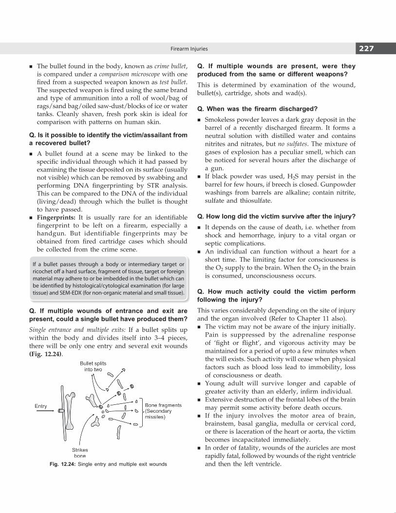
227Firearm Injuries
Fig. 12.24: Single entry and multiple exit wounds
� The bullet found in the body, known as crime bullet, is compared under a comparison microscope with one fired from a suspected weapon known as test bullet. The suspected weapon is fired using the same brand and type of ammunition into a roll of wool/bag of rags/sand bag/oiled saw-dust/blocks of ice or water tanks. Cleanly shaven, fresh pork skin is ideal for comparison with patterns on human skin.
Q. Is it possible to identify the victim/assailant from a recovered bullet?
� A bullet found at a scene may be linked to the specific individual through which it had passed by examining the tissue deposited on its surface (usually not visible) which can be removed by swabbing and performing DNA fingerprinting by STR analysis. This can be compared to the DNA of the individual (living/dead) through which the bullet is thought to have passed.
� Fingerprints: It is usually rare for an identifiable fingerprint to be left on a firearm, especially a handgun. But identifiable fingerprints may be obtained from fired cartridge cases which should be collected from the crime scene.
If a bullet passes through a body or intermediary target or ricochet off a hard surface, fragment of tissue, target or foreign material may adhere to or be imbedded in the bullet which can be identified by histological/cytological examination (for large tissue) and SEM-EDX (for non-organic material and small tissue).
Q. If multiple wounds of entrance and exit are present, could a single bullet have produced them?Single entrance and multiple exits: If a bullet splits up within the body and divides itself into 3–4 pieces, there will be only one entry and several exit wounds (Fig. 12.24).
Q. If multiple wounds are present, were they produced from the same or different weapons?This is determined by examination of the wound, bullet(s), cartridge, shots and wad(s).
Q. When was the firearm discharged? � Smokeless powder leaves a dark gray deposit in the
barrel of a recently discharged firearm. It forms a neutral solution with distilled water and contains nitrites and nitrates, but no sulfates. The mixture of gases of explosion has a peculiar smell, which can be noticed for several hours after the discharge of a gun.
� If black powder was used, H2S may persist in the barrel for few hours, if breech is closed. Gunpowder washings from barrels are alkaline; contain nitrite, sulfate and thiosulfate.
Q. How long did the victim survive after the injury? � It depends on the cause of death, i.e. whether from
shock and hemorrhage, injury to a vital organ or septic complications.
� An individual can function without a heart for a short time. The limiting factor for consciousness is the O2 supply to the brain. When the O2 in the brain is consumed, unconsciousness occurs.
Q. How much activity could the victim perform following the injury?This varies considerably depending on the site of injury and the organ involved (Refer to Chapter 11 also).
� The victim may not be aware of the injury initially. Pain is suppressed by the adrenaline response of ‘fight or flight’, and vigorous activity may be maintained for a period of upto a few minutes when the will exists. Such activity will cease when physical factors such as blood loss lead to immobility, loss of consciousness or death.
� Young adult will survive longer and capable of greater activity than an elderly, infirm individual.
� Extensive destruction of the frontal lobes of the brain may permit some activity before death occurs.
� If the injury involves the motor area of brain, brainstem, basal ganglia, medulla or cervical cord, or there is laceration of the heart or aorta, the victim becomes incapacitated immediately.
� In order of fatality, wounds of the auricles are most rapidly fatal, followed by wounds of the right ventricle and then the left ventricle.

228 Review of Forensic Medicine and Toxicology
Fig. 12.25: Sites of election in suicide with firearm and knife
� The amount and rapidity of blood loss will also help to form an opinion about the extent of physical activity possible.
� In injury to other parts of the body, the victim may be able to walk about.
Q. Is it a case of suicide, accident or homicide? � The differentiating features are tabulated in Diff. 12.2. � Most contact shotgun wounds of the head are
suicidal in origin. The individual tends to use his dominant hand to press the trigger, steadying the muzzle against the head with the non-dominant hand. So, powder soot may be visible on the non-dominant hand.
For suicides, the sites of preference are (in decreasing order of occurrence) (Fig. 12.25):
� Temple (60% of cases) � Center of forehead � Roof of mouth � Midline behind the chin � Left side or front of chest (precordium)
A suicider using a revolver or pistol usually shoots himself in the right temple region, the bullet passing almost horizontally or upwards and backwards through the head, and making its exit in the left parietal region. If the individual is left handed, left temporal region is selected.
Suicidal/accidental deaths in adolescents/young adults may sometimes occur from playing Russian roulette. It is a lethal game of chance in which a player places a single round in a revolver, spins the cylinder, places the muzzle against his head, and pulls the trigger. Since only one chamber is loaded, the player has only one in ‘n’ chance of hitting the loaded chamber, where n is the total number of chambers in the cylinder.
Q. Can there be multiple wounds of entry and exit from a single shot?Yes, it may be seen in re-entry wounds where the bullet passes through one part of the body and then re-enters another part. For example, a bullet may pass through and through:
� an arm and the chest, so that four wounds would result (most common).
� the chest or abdomen and thigh and lower leg, producing six wounds. This occurs when the person is running or sitting in an unusual position (thigh and leg flexed).
Differentiation 12.2: Suicidal, accidental and homicidal firearm injury
S.No. Feature Suicide Accident Homicide1. Site of entry wound Head or heart Any area Any area2. Shot distance Contact or very close range Close or very close Any range, usually distant3. Direction Upward or backward Any direction Usually upward/back-front4. Number of wounds Usually one One Any number5. Powder residue on hand pressing
triggerPresent Present Absent
6. Cadaveric spasm May be seen with the weapon firmly grasped
Not so Not so
7. Weapon at scene Found Found Not found 8. Scene Usually his house, suicide note,
previous psychiatric illnessIn his house or while hunting/handling
Any place, evidence of struggle
9. Sex Usually males Usually males Any sex10. Motive Insanity, illness, financial loss Nil Gang feuds, robbery, revenge
229Firearm Injuries
Q. Is it possible that entry wound is present but the bullet is not found in the body?It may occur when the bullet entering the:
� Stomach, may be vomited out. � Windpipe, may be coughed up. � Mouth, may be spat out. � Body and coming in contact with the bone, and
exiting by the same wound from where it entered.
Detection of Gunshot Residues (GSR)
It is done by: i. Dermal nitrate or paraffin test: It is an obsolete
and non-specific test. It detects GSR (nitrates and nitrites) from the suspect’s hand by removing it in a paraffin cast or cotton swab and treating with diphenylamine reagent. A positive test is indicated by blue flecks in the paraffin.32
ii. Harrison and Gilroy test: It is a qualitative calorimetric chemical test, and not specific for firearm discharges residues, but detects the presence of antimony, barium and lead.
A cotton swab moistened with HCl is used to swab the hand and then treated with triphenylmethylarsonium iodide for detection of antimony, and sodium rhodizonate for the detection of barium and lead.
iii. Neutron activation analysis: This chemical method is useful in identifying minute traces of elements present in the hair, nails, soil, glass pieces, paints, GSR and drugs. It is based on the detection and measurement of characteristic radioisotopes formed by irradiation in a nuclear reactor. Antimony and copper residues (from the primer) are detected from the suspect’s hand.
iv. Atomic absorption spectroscopy (AAS) and Flameless atomic absorption spectrophotometry (FAAS): This analytical method utilizes high temperatures to vaporize the metallic elements of the primer residues, to detect and quantify them. Measuring the antimony, barium and lead from
the primer and copper vaporized from either the cartridge case or the bullet jacketing helps in determining:
z Holes in clothing and tissues as bullet holes. z Range of fire. z Common origin of bullet fragments or shotgun
pellets found at different places. z Whether or not a person has fired a gun.
v. Scanning electron microscope-energy dispersive X-ray spectrometry (SEM-EDX): It is the most sophisticated tool which can detect minute traces of GSR found on the body of suspect. It is a qualitative, not a quantitative analysis.
Clothing may be tested for presence of GSR � Modified Greiss test is specific for nitrites. It uses chemicals
(alpha-naphthol) to produce visual display of GSR pattern and density of particles.
� Sodium rhodizonate reaction detects lead from primer or bullet wipe.
� Energy dispersive X-ray (EDX) detects barium, antimony, lead and copper around the entry wound in clothing. It can be used to determine the range of fire.33
Notes
� Bullet emboli/Wandering bullet: Vascular embolization is usually associated with small caliber, low velocity bullet, and usually involves the arterial system. It should be suspected whenever there is a penetrating bullet wound with failure to discover the bullet in the expected region or to visualize the bullet on routine X-ray. The most common sites of entry for a bullet are the aorta, right atrium and ventricle, pulmonary artery, and the inferior vena cava.
� Lead snowstorm: This is seen in radiograph of an individual shot with centerfire ammunition. Fragments of lead break off the lead core as the bullet moves through the body and gets lodged into surrounding tissue. X-ray shows small radiopaque bullet fragments scattered along the wound track—‘lead snowstorm’. A rifle bullet need not have to hit bone for this phenomenon to occur.
1. Not a part of firearm: NEET 13 A. Bolt B. Piston C. Extractor D. Muzzle
2. Choking is seen in: AIIMS 12; COMEDK 15 A. Revolver B. Pistol C. Shotgun D. Rifle
MULTIPLE CHOICE QUESTIONS
1. B 2. C

230 Review of Forensic Medicine and Toxicology
3. B 4. C 5. C 6. B 7. C 8. B 9. D 10. B 11. C 12. D 13. C 14. B 15. D 16. D 17. C 18. B 19. C 20. A & B 21. A 22. B 23. B 24. A 25. A
3. Caliber of a rifled gun is calculated: DNB 09; TN 11; AIIMS 14 A. Distance between a land and groove B. Distance between two diagonally opposite lands C. Distance between two diagonally opposite grooves D. Number of spherical lead balls that can be made
from one pound of lead 4. Spherical lead balls that can be made from one pound
of lead for a 12 bore shotgun: Manipal 09 A. 6 B. 8 C. 12 D. 24 5. Shotgun does not contain use: NEET 13 A. Barrel B. Choke bore C. Bullets D. Muzzle 6. Number of bullets fired in a tandem bullet: AIIMS 07 A. 1 B. 2 C. 3 D. 4 7. Bullet that fragments on impact is called: Kerala 06 A. Duplex bullet B. Dum-dum bullet C. Frangible bullet D. Soft-point bullet 8. Bullet that leaves a visible mark in its flight so that
person can see the path is: AI 10 A. Tandem bullet B. Tracer bullet C. Dum-dum bullet D. Incendiary bullet 9. Associated with shotguns only: AFMC 12 A. Gunpowder B. Primer C. Projectile D. Wads 10. Black gunpowder contains all of the following,
except: PGI 06; Punjab 09 A. Potassium nitrate B. Lead peroxide C. Charcoal D. Sulfur 11. Smokeless gunpowder is composed of: Delhi 05, 07 A. KMnO4 B. HCN C. Nitrocellulose D. Sulfur 12. Double base smokeless gun powder contains:
AIIMS 12 A. Nitrocellulose and nitroguanidine B. Potassium nitrate and sulphur C. Potassium nitrate and charcoal D. Nitrocellulose and nitroglycerin 13. One gram of smokeless gunpowder produces: Karnataka 11 A. 3000-4000 cc of gas B. 9000-10,000 cc of gas C. 12,000-13,000 cc of gas D. 15,000-16,000 cc of gas 14. FG, FFG, FFFG is used to indicate: PGI 08, 11 A. Cartridge B. Black powder size C. Base of gun D. Wadding of cartridge
15. Damage produced by a bullet is in direct proportion to its: AIIMS 09
A. Size B. Shape C. Velocity D. Weight 16. The capacity of a bullet to cause maximum destruction
lies in its: AIIMS 10 A. Size B. Shape C. Weight D. Velocity 17. Tattooing in entry wound of a firearm injury is due
to: NEET 14 A. Burns B. Smoke C. Gunpowder D. Wads 18. In a firearm injury, blackening seen around the entry
wound is due to: DNB 09 A. Flame B. Smoke C. Unburnt powder D. Hot gases 19. Dirt collar or grease collar is seen in: NEET 13 A. Punctured wound by sharp weapon B. Lacerated wound C. Firearm entry wound D. Stab wound 20. Abrasion collar is seen in: PGI 13 A. Gunshot injury B. Entry wound C. Drowning D. Railway track accident 21. A man was found with suicidal gunshot on right temple
with the gun in his right hand. The skull was burst open. There was charring and cherry red coloration in the track inside. Range of the shot is: AIIMS 12
A. Contact shot B. Close shot at a distance of 1 feet C. Shot within range of 2 feet D. Shot within range of 3 feet 22. Tattooing around the entry wound is seen in: NEET 14 A. Contact shot B. Close shot C. Distant shot D. All of the above 23. ‘Rat hole’ appearance of entry wound of firearm is
seen in: COMEDK 15 A. Close range of shotguns B. Near range of shotguns C. Close range of rifles D. Near range of rifles 24. Stellate wound is produced with firearm in: AI 09; NEET 13 A. Contact shot B. Close shot C. Range within 60 cm D. Distant shot 25. Contact wound shows: AP 06 A. Cruciate splitting B. Tattooing C. Singeing of hair D. Abrasion collar
231Firearm Injuries
26. In firearm entry wound, arrangement of abrasion collar, dirt collar and tattooing from inside to outside:
NIMS 11 A. Dirt collar, abrasion collar, tattooing B. Abrasion collar, dirt collar, tattooing C. Tattooing, dirt collar, abrasion collar D. Dirt collar, tattooing, abrasion collar 27. Following are features of gunshot wound, except:
NIMHANS 14; PGI 14 A. Entry wound is beveled in the outer table of skull B. Abrasion collar is seen in entry wound C. Harrison-Gilroy test can detect gunshot residue D. Stellate shaped wound is seen in contact shot 28. Following may be seen in the exit wound:
DNB 10; NEET 14 A. Dirt collar B. Abrasion collar C. Tattooing D. Inverted edges 29. Kennedy phenomenon is seen in: NEET 13 A. Road traffic accident B. Gunshot injury C. Burns D. Contusion
30. Surgical alteration or suturing of gunshot wounds may create problems in distinguishing entry from exit. This is called as: AIIMS 13
A. Formication phenomenon B. Gordon phenomenon C. Cookie cutter phenomenon D. Kennedy phenomenon 31. A case of homicide with gunshot was reported and
bullet was recovered from the body. Primary and secondary markings on the bullet can be used for: AIIMS 12
A. Identification of weapon B. Range of firing C. Severity of tissue damage D. Time of crime 32. Gunshot residue on hands can be detected by: DNB 10; NEET 13 A. Phenolphthalein test B. Dermal nitrate test C. Benzidine test D. H2 activation test 33. Gunpowder on clothing can be visualized by:
AI 11 A. Magnifying lens B. UV rays C. Infrared rays D. Energy dispersive X-ray
26. A 27. A 28. B 29. B 30. D 31. A 32. B 33. D

Definitions
� Traumatic brain injury (TBI): Traumatically induced structural injury and/or physiological disruption of brain function as a result of an external force.
� Closed head injury (non-penetrating): Damage to the brain without any fracture of the skull and/or penetration of dura; most often results from blunt trauma.
� Open head injury (penetrating): Disruption of cranial vault with opening through skin and cranial bones to expose damaged brain; most often associated missile wounds, stab/chop wounds, and motor vehicle or occupational accidents.
� Missile injury: Injury produced by moving object striking cranium; most often refers to bullet injury.
� Acceleration/deceleration injury: Damage produced by movement of brain within confines of cranial vault (tearing during violent movement or from impact of striking interior of skull or dural folds).
Craniocerebral Injuries
� There are three main components of the head: scalp, skull and brain (Fig. 13.1).
� The term ‘craniocerebral injuries’ can be used to describe the presence of skull (‘cranio’) and brain (‘cerebral’) injury.
� Craniocerebral injuries to the head can be grouped into two types: ‘focal’ or ‘diffuse’ in the sense that they are localized or widespread (Table 13.1).
Biomechanics of Head Injury
Head injury can be broadly classified into two types based on mechanical loading: (Flow chart 13.1) i. Static ii. Dynamic
� Static/Contact injuries: Contact injuries occur both at the site and remote from the point of impact on the head. It results in skull fractures and contusions. The length and direction of a skull fracture depends on the amount of contact energy absorbed by the skull and its thickness at the impact site.
� Dynamic/Inertial injuries: Dynamic loading is usually the result of an impact, such as the head being struck a blow by a moving object or the head itself striking a relatively stationary surface. Inertial injuries are commonly called ‘acceleration’ or ‘deceleration’ injuries which biomechanically can be considered as the same phenomenon. The brain is damaged by one or both mechanisms: through strains produced within the brain tissue itself, and through differential movements between the brain and the skull. Three types of acceleration are recognized:
i. Angular acceleration is most common and most serious in which rotation is usually centered in the low cervical region. Angular acceleration
Fig. 13.1: Cross section of the scalp, skull and meninges
Table 13.1: Classification of craniocerebral injury
Focal Diffuse
� Scalp lacerations � Axonal injury (DAI)
� Skull fractures � Ischemic injury
� Contusions/lacerations � Vascular injury
� Intracranial hemorrhage � Brain swelling
� Lesions secondary to raised intracranial pressure
Regional Injuries
CHAPTER 13

233Regional Injuries
alone is capable of producing almost every known traumatic brain lesion, except for a skull fracture or an epidural hematoma.
ii Translational acceleration (e.g. anterior to posterior) does not produce diffuse brain damage but may cause a variety of focal surface lesions.
iii. Rotational acceleration in the horizontal plane is a very damaging mechanism, producing both high surface forces and intracerebral damage, but it usually occurs in association with angular acceleration.
Strain within the brain tissue is the deformation that it undergoes when a mechanical force is applied. Tensile strain refers to elongation of neural tissues and is the most important mechanism in head injury. Compression wherein constituent units are being forced together and shear or sliding strains which move adjacent strata of tissue laterally also occur. The brain is virtually incompressible, and has a low tolerance to tensile and shear strains.
Soft Tissue Injury
� Injuries: Scalp wounds are caused by falls, blows, sharp cutting instruments or discharge of a firearm. It can be:
i. Abrasion: It caused by a lateral rubbing action by a blow, a fall on a rough surface or by being dragged in a vehicular accident. They are simple injuries, bleed slightly, and heal rapidly.
ii. Contusions: Presence of scalp contusion is indicative of contact injury. In superficial fascia,
they appear as localized swelling due to the dense fibro fatty tissues, but contusions deep into the galea aponeurotica are diffuse on account of loose aponeurotic tissues, and difficult to make out on examination.
z The easiest way to detect scalp injuries is by palpation, but shaving is necessary for proper documentation and photography.
z During postmortem examination, autopsy surgeons are able to visualize this subscalpular (subgaleal) hemorrhage after reflection of the scalp.
z Scalp hematoma may be associated with an underlying linear skull fracture.
iii. Lacerations: They resemble incised wounds (incised looking lacerations). Careful examination in the depths of the wound will reveal the bridging fibers with surrounding band of abrasion.
iv. Incised wounds: They have clean cut margins; hair bulbs are cleanly cut.
� An effusion of blood over the top of the head or forehead may gravitate down to the loose tissues causing black eyes (periorbital hematoma).
Causes of black eyes (spectacle hematoma or raccoon eyes) (Fig. 13.2) i. Most commonly due to local violence causing
subcutaneous extravasation of blood into the lids, occurs soon after injury to upper and lower eyelids.
ii. Bleeding into the layer of loose connective tissue after a blow on the skull; the blood gravitates under the frontalis muscle and appears first in the upper eyelid and then the lower eyelid over the course of a couple of days.
Flow chart 13.1: Pathological changes depending on mechanism of injury

234 Review of Forensic Medicine and Toxicology
iii. Fracture of the orbital plate of the frontal bone results in hemorrhage into the orbit, the blood tracks under the conjunctiva, appearing as a triangular, flame-shaped hemorrhage, the apex of which is at the margin of the cornea and the posterior limit cannot be seen, which distinguishes it from the subconjunc tival hemorrhage.
� Wounds of the scalp bleed freely (blood vessels in the fibrous layer, superficial to galea aponeurotica, being held open once cut), but heal rapidly. If an injury extending through the galea gets infected, it may spread through the emissary veins to involve the sagittal, lateral and cavernous sinuses causing septic complications, like meningitis and brain abscess.
Skull Fractures
Motor vehicle accidents and falls are the most common causes of skull fractures.
Mechanism: The skull is not a completely rigid structure, and it is able to bend and distort when force is applied to it. A blow to the skull causes ‘inbending’ of the bone at the site of impact, and asymmetric and variably localized ‘outbending’ at a distance from the impact. If the forces applied exceed the elastic properties the skull, fracture will occur.
� In general, if force is applied over a small area (e.g. a blow from a weapon) a fracture occurs at the site of inbending, whereas an impact over a larger skull area leads to fractures at the site of outbending.
� Fractures also represent the point of maximum stress upon the skull, which may not be at the immediate site of impact.
� Skulls of infants and children are thinner than those of adults and may be able to distort more before fracturing.
� The fracture of skull can occur either by direct or indirect violence.
i. Direct violence: The forces act directly on the bone to produce a fracture, e.g. head crushed under the wheels in road traffic accidents or an object like stick/rod/bullet striking the head.
ii. Indirect violence: The forces act indirectly on the skull through some other structure, which receives primary impact, e.g. fall on buttocks from height which transmits the force to occipital bone through vertebral column or a blow to the chin resulting in fracture of base of the skull.
During autopsy, prior to cutting the skull cap off, the skull should be visually inspected and palpated for the presence of fractures.
Types (Fig. 13.3)1
1. Fissure/linear fractures: These are linear cracks passing over the vertex or across the skull base without any displacement of the fragments, and either involves the whole thickness of the bone or the inner or outer table alone.
z These fractures are usually caused by: i. Forcible impact against a broad resisting
surface, like hard ground surface, as in road traffic accidents.
ii. When knocked to the ground by the blow of a fist.
iii. Blows with a hard blunt object having a relatively broad striking surface.
z The fracture line tends to follow an irregular course, and is no more than a hair’s breadth.
z They are difficult to detect, may not be seen on X-ray, and can only be detected at autopsy.
z The line of fracture runs parallel with the axis of compression.
z Depression of bone fragments is not seen. z An injury of the head sustained by a fall is
mostly situated at the level of the margin of the hat, while an injury due to blow is commonly situated above this level.
2. Depressed fracture: When a portion of fractured bone is driven inwards to a distance equivalent to the thickness of the skull table, it is known as depressed fracture. It is also called ‘fracture a la signature’ (signature fracture), as the shape often points towards the shape of the offending weapon.2
z They are caused by blows with a heavy weapon having small striking surface, such as hammer, axe, brick or chopper.Fig. 13.2: Causes of black eyes

235Regional Injuries
z The part of the skull which is first struck shows maximum depression; usually seen in the left frontal region.
z This type of fracture often suggests the probable manner of application of violence, and also the relative position of the victim and the assailant at that moment.
z Depressed fracture is considered to be compound if an associated scalp laceration extending through the pericranium is present, and penetrating if a dural laceration exits.
z The soft fluctuant centers of scalp hematomas can masquerade as depressed skull fractures (Diff. 13.1).
z The risk of post-traumatic epilepsy following depressed skull fracture and cortical laceration is about 15%.
Some variants: a. Elevated fracture: One end of fractured
fragment is elevated above the surface of skull, while the other end may dip down into cranial cavity resulting in injury to the brain.
– It is caused by a blow from sharp, heavy object (e.g. an axe) which elevates the skull fracture by lateral pull of the weapon while retrieving it.
– These fractures are rare, and are usually associated with injury to the dura also.
b. Pond or indented fracture: This is a smooth concave depression without a fracture line, resulting from in-buckling of skull, occurring only in the elastic skull of infants and children (prior to 4 years of age).3,4
– Inner table is not fractured, meninges and brain are not damaged.4
– It may also be caused by forcible compression with an obstetric forceps or impact against some protruding objects, e.g. sacral promontery during delivery.
– It is also known as ping-pong fracture, as it looks similar to a dent in ping-pong ball.
3. Comminuted fracture (spider-web/mosaic fracture): Two or more intersecting lines of fracture divide the bone into three or more fragments resembling a spider web or mosaic pattern.
z Skull bone gets broken into multiple pieces by fracture lines, which are haphazardly or concentrically arranged, or stellate if they radiate from the site of impact.
z It is caused by vehicular accidents, fall from height on a hard surface or by blows with weapons having large striking surface, such as heavy iron bar, or from a bullet.
Fig. 13.3: Types of skull fractures
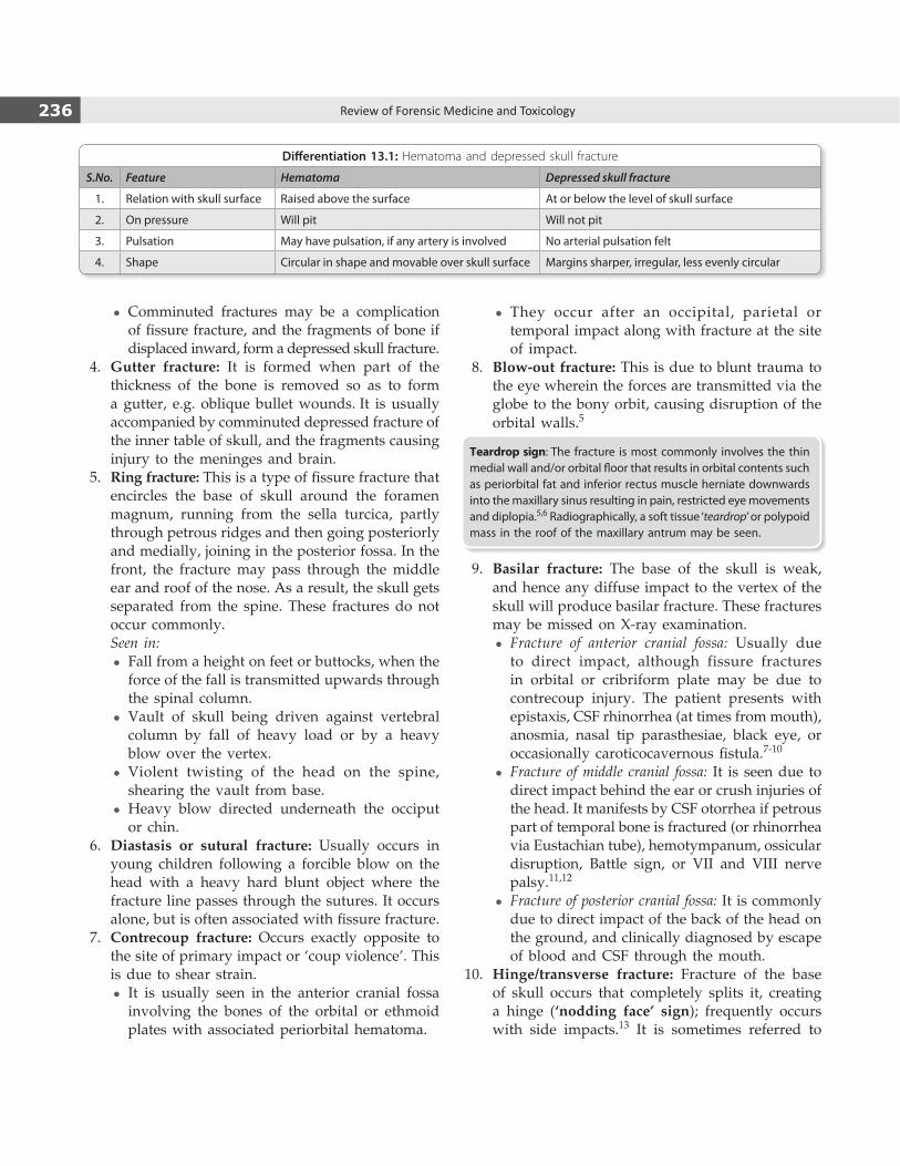
236 Review of Forensic Medicine and Toxicology
z They occur after an occipital, parietal or temporal impact along with fracture at the site of impact.
8. Blow-out fracture: This is due to blunt trauma to the eye wherein the forces are transmitted via the globe to the bony orbit, causing disruption of the orbital walls.5
Teardrop sign: The fracture is most commonly involves the thin medial wall and/or orbital floor that results in orbital contents such as periorbital fat and inferior rectus muscle herniate downwards into the maxillary sinus resulting in pain, restricted eye movements and diplopia.5,6 Radiographically, a soft tissue ‘teardrop’ or polypoid mass in the roof of the maxillary antrum may be seen.
9. Basilar fracture: The base of the skull is weak, and hence any diffuse impact to the vertex of the skull will produce basilar fracture. These fractures may be missed on X-ray examination.
z Fracture of anterior cranial fossa: Usually due to direct impact, although fissure fractures in orbital or cribriform plate may be due to contrecoup injury. The patient presents with epistaxis, CSF rhinorrhea (at times from mouth), anosmia, nasal tip parasthesiae, black eye, or occasionally caroticocavernous fistula.7-10
z Fracture of middle cranial fossa: It is seen due to direct impact behind the ear or crush injuries of the head. It manifests by CSF otorrhea if petrous part of temporal bone is fractured (or rhinorrhea via Eustachian tube), hemotympanum, ossicular disruption, Battle sign, or VII and VIII nerve palsy.11,12
z Fracture of posterior cranial fossa: It is commonly due to direct impact of the back of the head on the ground, and clinically diagnosed by escape of blood and CSF through the mouth.
10. Hinge/transverse fracture: Fracture of the base of skull occurs that completely splits it, creating a hinge (‘nodding face’ sign); frequently occurs with side impacts.13 It is sometimes referred to
z Comminuted fractures may be a complication of fissure fracture, and the fragments of bone if displaced inward, form a depressed skull fracture.
4. Gutter fracture: It is formed when part of the thickness of the bone is removed so as to form a gutter, e.g. oblique bullet wounds. It is usually accompanied by comminuted depressed fracture of the inner table of skull, and the fragments causing injury to the meninges and brain.
5. Ring fracture: This is a type of fissure fracture that encircles the base of skull around the foramen magnum, running from the sella turcica, partly through petrous ridges and then going posteriorly and medially, joining in the posterior fossa. In the front, the fracture may pass through the middle ear and roof of the nose. As a result, the skull gets separated from the spine. These fractures do not occur commonly.
Seen in: z Fall from a height on feet or buttocks, when the
force of the fall is transmitted upwards through the spinal column.
z Vault of skull being driven against vertebral column by fall of heavy load or by a heavy blow over the vertex.
z Violent twisting of the head on the spine, shearing the vault from base.
z Heavy blow directed underneath the occiput or chin.
6. Diastasis or sutural fracture: Usually occurs in young children following a forcible blow on the head with a heavy hard blunt object where the fracture line passes through the sutures. It occurs alone, but is often associated with fissure fracture.
7. Contrecoup fracture: Occurs exactly opposite to the site of primary impact or ‘coup violence’. This is due to shear strain.
z It is usually seen in the anterior cranial fossa involving the bones of the orbital or ethmoid plates with associated periorbital hematoma.
Differentiation 13.1: Hematoma and depressed skull fracture
S.No. Feature Hematoma Depressed skull fracture
1. Relation with skull surface Raised above the surface At or below the level of skull surface
2. On pressure Will pit Will not pit
3. Pulsation May have pulsation, if any artery is involved No arterial pulsation felt
4. Shape Circular in shape and movable over skull surface Margins sharper, irregular, less evenly circular

237Regional Injuries
as ‘motorcyclists fracture’. Most common form is the one which extends from the petrous bony ridge through sella turcica to lateral end of the contralateral petrous ridge.
� Compound skull fracture: Laceration of scalp associated with skull fracture.
� Growing skull fracture: Expanding linear fracture (usually in young children) associated with dural tear which allow leptomeninges to herniate into fracture site and expand with cerebrospinal fluid pressure (leptomeningeal cyst).
� Halo or ring sign: Blood from head injured patients may mix with CSF and mask the recognition of a leak. CSF will separate from blood when the mixture is placed on filter paper resulting in a central area of blood with an outer ring or halo.14
� Glucose estimation: CSF has a greater concentration of glucose than mucus or lacrimal secretions. The quantitative determination of a glucose level in nasal fluid not contaminated by blood can be diagnostic of CSF rhinorrhea, if the nasal fluid contains > 30 mg/dl.10,14
� Immunoelectrophoretic identification: β-2-transferrin assay is the most widely used test and is considered the standard criterion for diagnosis of CSF rhinorrhea.14,15
� Battle’s sign: Bruising behind the ear appearing 36 h after head injury; may be confused with retro-auricular scalp bruise (Fig. 13.4).16
� With basilar fracture, intracranial passage of a nasogastric tube or nasopharyngeal airway can happen. These fractures may be visible on plain radiographs or on bone window axial CT scans, but confirmed radiologically by pneumocranium or air-fluid levels in the sinuses.
� Skull X-rays have a limited role in head injuries, since they do not identify intracranial injury. However, it may be helpful when CT scan is not available. Plain X-rays can detect a skull fracture, and its presence is helpful in predicting the presence of intracranial injury. However, a normal skull series does not rule out a brain injury.
Complications of Skull Fractures
i. Injury to the brain which may be dangerous to life.
ii. Hemorrhage: If middle meningeal artery is ruptured, fatal hemorrhage may occur.
iii. Infections: It may be direct spread from compound fracture or spread from fracture of paranasal sinuses, like frontal or ethmoidal.
iv. Traumatic epilepsy: More common with open head injuries. Usually seen 1–2 years after the episode, and manifests as tonic or clonic fits.
Brain Injury
� Traumatic brain injury (TBI) is usually caused by motor vehicle accidents, falls and assaults.
� TBI can be classified based on severity (mild, moderate or severe, Table 13.2) mechanism (missile or blunt) and pathology (primary or secondary, Flow chart 13.2).
Fig. 13.4: Battle’s sign: Blood in soft tissue over mastoid
Flow chart 13.2: Classification of brain injury
� Primary brain injury is the injury caused at the time of impact (e.g. contusion, laceration).18
� Secondary brain injury is brain damage arising from events developing subsequent to primary injury.
z Some secondary injuries occur almost instantane-ously (e.g. hemorrhage as a consequence of tear-ing of vessel), whereas others evolve over hours to days (e.g. delayed hemorrhage, inflamma tion, brain swelling, and axonal swelling secondary to paralysis of axonal transport or tearing of axons).19
z Excitatory neuropeptides, cytokines, free radicals, metabolic and oxygenation insufficiencies cause this injury.
z Little can be done about primary injury once it has occurred. However, medical management attempts to minimize the damage caused by secondary injury.
z Acute injury to the brain causes increase in glumatate, potassium and calcium levels with decrease in glucose metabolism and level of magnesium.

238 Review of Forensic Medicine and Toxicology
Cerebral Concussion
Definition: Concussion (Latin concutere: to shake) is physiological disruption of brain function as a result of a traumatic event which is manifested by at least one of the following: alteration of mental state, loss of memory or focal neurological deficit, that may or may not be transient.20
� Concussion, also known as mild traumatic brain injury is a clinical diagnosis (Table 13.2).
� It results from acceleration/deceleration of the head. � Violent head movement causes shearing or stretching
of nerve fibers and axonal damage. � It may resemble drunkenness (Diff. 13.2). � Concussion is common among contact and collision
sports participants. Football players and boxers are particularly exposed to repetitive concussions,
leading to the condition known as chronic traumatic encephalopathy syndrome.
Signs and SymptomsUnconsciousness, bradycardia, hypotension and sweat-ing, and is always followed by retrograde or post-traumatic (antegrade) amnesia, temporary lethargy, irritability and cognitive dysfunction.21 Muscles are flaccid, pupils are dilated and unreacting, pulse is weak and slow, and respiration is shallow.
Severity of concussion: It is given in Table 13.3.
Findings: Gross and light microscopic changes in the brain are usually absent, but biochemical and ultrastructural changes—mitochondrial ATP depletion, local disruption of blood-brain barrier occur. CT and MRI scans are usually normal.
Table 13.2: Severity of brain injury stratification
Criteria Mild/concussion Moderate Severe
Structural imaging Normal Normal or abnormal Normal or abnormal
Loss of consciousness 0–30 min > 30 min and < 24 h > 24 h
Alteration of consciousness/mental state A moment upto 24 h > 24 h Severity based on other criteria
Post-traumatic amnesia ≤ 1 day > 1 and < 7 days > 7 days
Glasgow Coma Scale17
(best available score in first 24 h)13–15 9–12 3–8
Differentiation 13.2: Drunkenness and concussion
S. No. Feature Drunkenness Concussion
1. Skin Flushed, congested and warm Pale, cold and sweating
2. Pulse Rapid and bounding Slow and feeble
3. Pupils Dilated; contracted in coma Contracted in pontine hemorrhage
4. Light reflex Sluggish May be brisk
5. Respiration Sighs, puffs, eructates Shallow, irregular, slow
6. Memory Confused, disoriented Retrograde amnesia, unrelieved by time
7. Behavior Uncooperative, abusive, talkative, sulky Quiet and retracted, curled up in bed, photophobia
8. Urine/blood Examination will be helpful Retention of urine, urine may contain albumin
9. History History of having consumed alcohol, smell of alcohol History of head injury with features of concussion
Table 13.3: Concussion grading scale
Grade 1 Grade 2 Grade 3
Transient confusion Transient confusion —
No loss of consciousness No loss of consciousness Brief or prolonged loss of consciousness
Concussion symptoms or mental status change resolves in ≤ 15 min
Concussion symptoms or mental status change resolves in > 15 min
—
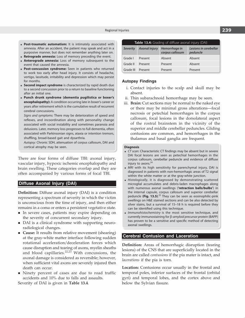
239Regional Injuries
� Post-traumatic automatism: It is intimately associated with amnesia. After an accident, the patient may speak and act in a purposive manner, but does not remember anything later on.
� Retrograde amnesia: Loss of memory preceding the event. � Anterograde amnesia: Loss of memory subsequent to the
event that caused the amnesia. � Post-concussion syndrome: Seen in patients who returned
to work too early after head injury. It consists of headache, vertigo, lassitude, irritability and depression which may persist for months.
� Second impact syndrome is characterized by rapid death due to a second concussion prior to a return to baseline functioning after an initial one.
� Punch drunk syndrome (dementia pugilistica or boxer’s encephalopathy): A condition occurring late in boxer’s career or years after retirement which is the cumulative result of recurrent cerebral concussions.
Signs and symptoms: There may be deterioration of speed and reflexes, and incoordination along with personality change associated with social instability and sometimes paranoia and delusions. Later, memory loss progresses to full dementia, often associated with Parkinsonian signs, ataxia or intention tremors, shuffling, broad-based gait and dysarthria.
Autopsy: Chronic SDH, attenuation of corpus callosum, DAI and cortical atrophy may be seen.
There are four forms of diffuse TBI: axonal injury, vascular injury, hypoxic ischemic encephalopathy and brain swelling. These categories overlap and they are often accompanied by various forms of focal TBI.
Diffuse Axonal Injury (DAI)
Definition: Diffuse axonal injury (DAI) is a condition representing a spectrum of severity in which the victim is unconscious from the time of injury, and then either remains in a coma or enters a persistent vegetative state.
� In severe cases, patients may expire depending on the severity of concurrent secondary injury.
� DAI is a clinical syndrome with supporting neuro-radiological changes.
� Cause: It results from relative movement (shearing) at the gray-white matter interface following sudden rotational acceleration/deceleration forces which cause disruption and tearing of axons, myelin sheaths and blood capillaries.22,23 With concussions, the axonal damage is considered as reversible; however, when sufficient vital axons are severely injured then death can occur.
� Ninety percent of cases are due to road traffic accidents and 10% due to falls and assaults.
Severity of DAI is given in Table 13.4.
Autopsy Findings
i. Contact injuries to the scalp and skull may be absent.
ii. Thin subarachnoid hemorrhage may be seen. iii. Brain: Cut sections may be normal to the naked eye
or there may be minimal gross alterations—focal necrosis or petechial hemorrhages in the corpus callosum, focal lesions in the dorsolateral aspect of the rostral brainstem in the vicinity of the superior and middle cerebellar peduncles. Gliding contusions are common, and hemorrhages in the thalamus and basal ganglia are frequent.
Diagnosis � CT scan: Characteristic CT findings may be absent but in severe
DAI focal lesions are seen as petechial hemorrhages in the corpus callosum, cerebellar peduncle and evidence of diffuse injury to axons.24
� MRI with its high sensitivity for parenchymal injury, DAI is diagnosed in patients with non-hemorrhagic areas of T2 signal within the white matter or at the gray-white junction.
� Histologically, it is diagnosed by demonstrating scattered microglial accumulates and debris-laden macrophages along with numerous axonal swellings (‘retraction balls/bulbs’) in the internal capsule, corpus callosum and superior cerebellar peduncle (Fig. 13.5).25 They can be seen as eosinophilic-pink swellings on H&E stained sections and can be also detected by silver stains, but a survival of 15–18 h is required before they can be identified using this technique.
� Immunohistochemistry is the most sensitive technique, and currently immunostaining for β-amyloid precursor protein (BAPP) has proven to be a sensitive and specific method of detecting axonal swellings.
Cerebral Contusion and Laceration
Definition: Areas of hemorrhagic disruption (tearing lesions) of the CNS that are superficially located in the brain are called contusions if the pia mater is intact, and lacerations if the pia is torn.
Location: Contusions occur usually in the frontal and temporal poles, inferior surfaces of the frontal (orbital gyri) and temporal lobes, and the cortex above and below the Sylvian fissure.
Table 13.4: Grading of diffuse axonal injury (DAI)
Severity Axonal injury Hemorrhage in corpus callosum
Lesions in cerebellar peduncle
Grade I Present Absent Absent
Grade II Present Present Absent
Grade III Present Present Present

240 Review of Forensic Medicine and Toxicology
� Typically, it involve the crests of gyri and often involve the gray matter only; may extend into underlying white matter and form a hematoma.
� In severe cases, extensive laceration with underlying parenchymal hemorrhage may be associated with subdural hemorrhage, forming a so-called burst lobe—often seen in the temporal lobes.
Pathogenesis: Contusions are the sites where the brain tissue comes in contact with the bony protuberances and dural coverings, sites of forcible separation of the brain from the dura, and sites of differential movement between the brain and dura and between adjacent areas of the brain.
� Contusion produces focal neurological deficits that persist for > 24 h. Since, the damage is focal, patients may recover uneventfully, provided that they did not develop complications leading to other types of brain damage, and did not sustain DAI at the time of injury.
� Intoxication by alcohol is associated with unduly large contusions as they tend to fall more heavily because of their blunted protective locomotor reflexes. Moreover, associated liver disease and acute alcohol intoxication hinder hemostasis.
Types: There are several types of brain contusions based on location and/or mechanism of injury: i. Coup contusion: Occurs at the site of impact due
to inbending bone rebounding and injuring the brain. They have a wedge-shaped appearance whose base is at the pial surface and the tip pointing towards the white matter.
ii. Contrecoup contusion: It is associated with falls and occurs at a site diametrically opposite to the point of impact. It is due to the brain rebounding backward from the skull following impact. It is seen most commonly in the frontal (orbital gyri) and temporal lobes (Fig. 13.6).
iii. Fracture contusion: Related to fractures of the skull and bears no relation with the point of impact.
iv. Intermediary coup contusion: Present in deeper structures of the brain, like the white matter or basal ganglia. It is present along the line of impact between coup and contrecoup points.
v. Gliding contusion: Usually associated with DAI, and independent of site and direction of impact. It is a focal hemorrhage in the cortex and underlying white matter of the dorsal surface of cerebrum, particularly the frontal region. It is seen in falls and road traffic accidents.
vi. Herniation contusion: It is due to the impact of medial side of temporal lobe with the edge of the tentorium, or the cerebellar tonsils against the foramen magnum. It is independent of the site and direction of impact.
Clinical features: The GCS is often low, and focal neurologic symptoms, loss of consciousness and visual changes are present. Secondary injury may further complicate the clinical picture by producing infarcts due to local vasospasm. Prognosis is usually poor.
Diagnostic tool: CT scan shows a mixture of hypo- and hyperdense lesions within the brain parenchyma.
Fig. 13.5: Axonal swelling/ballooning Fig. 13.6: Location of contrecoup contusions on undersurface of frontal and temporal lobes (shaded areas)
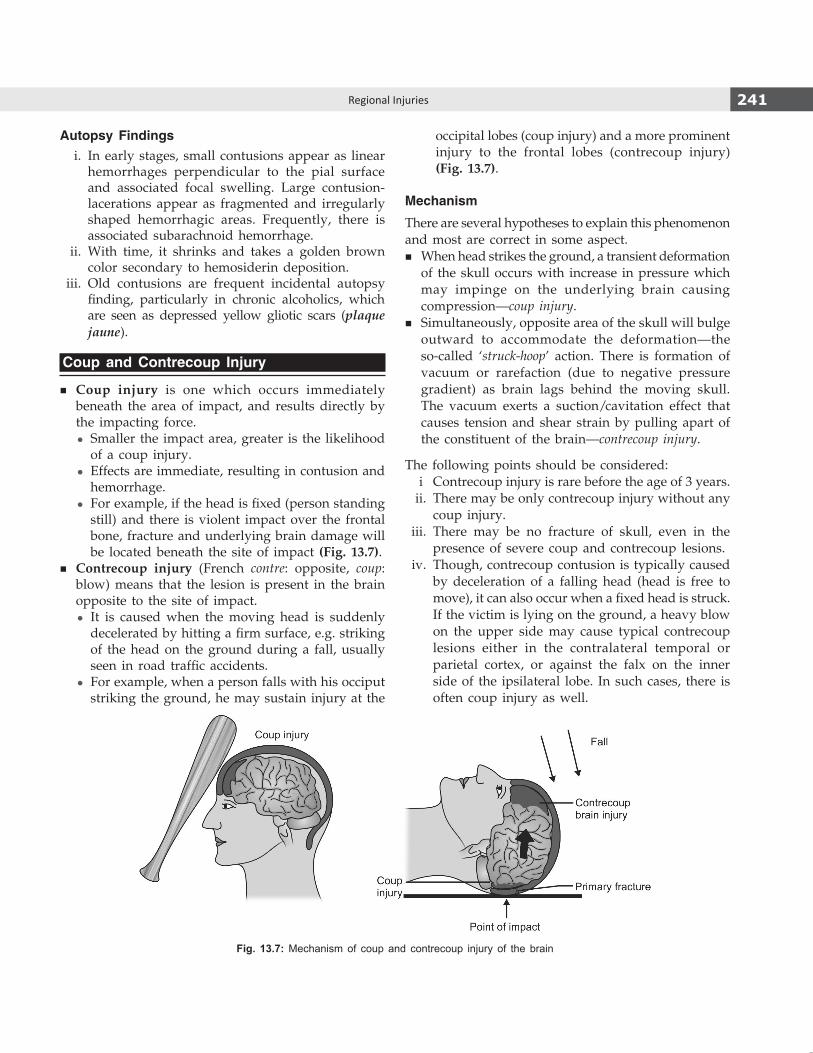
241Regional Injuries
Autopsy Findings i. In early stages, small contusions appear as linear
hemorrhages perpendicular to the pial surface and associated focal swelling. Large contusion-lacerations appear as fragmented and irregularly shaped hemorrhagic areas. Frequently, there is associated subarachnoid hemorrhage.
ii. With time, it shrinks and takes a golden brown color secondary to hemosiderin deposition.
iii. Old contusions are frequent incidental autopsy finding, particularly in chronic alcoholics, which are seen as depressed yellow gliotic scars (plaque jaune).
Coup and Contrecoup Injury
� Coup injury is one which occurs immediately beneath the area of impact, and results directly by the impacting force.
z Smaller the impact area, greater is the likelihood of a coup injury.
z Effects are immediate, resulting in contusion and hemorrhage.
z For example, if the head is fixed (person standing still) and there is violent impact over the frontal bone, fracture and underlying brain damage will be located beneath the site of impact (Fig. 13.7).
� Contrecoup injury (French contre: opposite, coup: blow) means that the lesion is present in the brain opposite to the site of impact.
z It is caused when the moving head is suddenly decelerated by hitting a firm surface, e.g. striking of the head on the ground during a fall, usually seen in road traffic accidents.
z For example, when a person falls with his occiput striking the ground, he may sustain injury at the
occipital lobes (coup injury) and a more prominent injury to the frontal lobes (contrecoup injury) (Fig. 13.7).
Mechanism
There are several hypotheses to explain this phenomenon and most are correct in some aspect.
� When head strikes the ground, a transient deformation of the skull occurs with increase in pressure which may impinge on the underlying brain causing compression—coup injury.
� Simultaneously, opposite area of the skull will bulge outward to accommodate the deformation—the so-called ‘struck-hoop’ action. There is formation of vacuum or rarefaction (due to negative pressure gradient) as brain lags behind the moving skull. The vacuum exerts a suction/cavitation effect that causes tension and shear strain by pulling apart of the constituent of the brain—contrecoup injury.
The following points should be considered: i Contrecoup injury is rare before the age of 3 years. ii. There may be only contrecoup injury without any
coup injury. iii. There may be no fracture of skull, even in the
presence of severe coup and contrecoup lesions. iv. Though, contrecoup contusion is typically caused
by deceleration of a falling head (head is free to move), it can also occur when a fixed head is struck. If the victim is lying on the ground, a heavy blow on the upper side may cause typical contrecoup lesions either in the contralateral temporal or parietal cortex, or against the falx on the inner side of the ipsilateral lobe. In such cases, there is often coup injury as well.
Fig. 13.7: Mechanism of coup and contrecoup injury of the brain
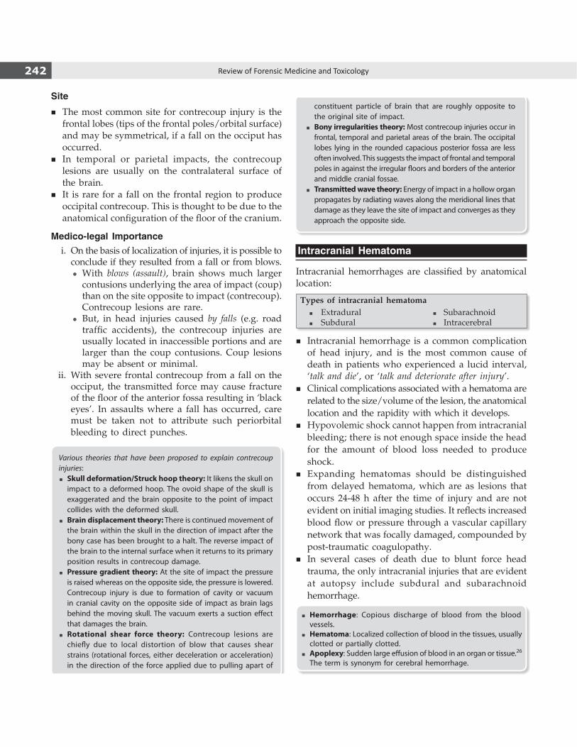
242 Review of Forensic Medicine and Toxicology
Site
� The most common site for contrecoup injury is the frontal lobes (tips of the frontal poles/orbital surface) and may be symmetrical, if a fall on the occiput has occurred.
� In temporal or parietal impacts, the contrecoup lesions are usually on the contralateral surface of the brain.
� It is rare for a fall on the frontal region to produce occipital contrecoup. This is thought to be due to the anatomical configuration of the floor of the cranium.
Medico-legal Importance i. On the basis of localization of injuries, it is possible to
conclude if they resulted from a fall or from blows. z With blows (assault), brain shows much larger
contusions underlying the area of impact (coup) than on the site opposite to impact (contrecoup). Contrecoup lesions are rare.
z But, in head injuries caused by falls (e.g. road traffic accidents), the contrecoup injuries are usually located in inaccessible portions and are larger than the coup contusions. Coup lesions may be absent or minimal.
ii. With severe frontal contrecoup from a fall on the occiput, the transmitted force may cause fracture of the floor of the anterior fossa resulting in ‘black eyes’. In assaults where a fall has occurred, care must be taken not to attribute such periorbital bleeding to direct punches.
Intracranial Hematoma
Intracranial hemorrhages are classified by anatomical location:
Types of intracranial hematoma� Extradural � Subarachnoid� Subdural � Intracerebral
� Intracranial hemorrhage is a common complication of head injury, and is the most common cause of death in patients who experienced a lucid interval, ‘talk and die’, or ‘talk and deteriorate after injury’.
� Clinical complications associated with a hematoma are related to the size/volume of the lesion, the anatomical location and the rapidity with which it develops.
� Hypovolemic shock cannot happen from intracranial bleeding; there is not enough space inside the head for the amount of blood loss needed to produce shock.
� Expanding hematomas should be distinguished from delayed hematoma, which are as lesions that occurs 24-48 h after the time of injury and are not evident on initial imaging studies. It reflects increased blood flow or pressure through a vascular capillary network that was focally damaged, compounded by post-traumatic coagulopathy.
� In several cases of death due to blunt force head trauma, the only intracranial injuries that are evident at autopsy include subdural and subarachnoid hemorrhage.
� Hemorrhage: Copious discharge of blood from the blood vessels.
� Hematoma: Localized collection of blood in the tissues, usually clotted or partially clotted.
� Apoplexy: Sudden large effusion of blood in an organ or tissue.26 The term is synonym for cerebral hemorrhage.
Various theories that have been proposed to explain contrecoup injuries:
� Skull deformation/Struck hoop theory: It likens the skull on impact to a deformed hoop. The ovoid shape of the skull is exaggerated and the brain opposite to the point of impact collides with the deformed skull.
� Brain displacement theory: There is continued movement of the brain within the skull in the direction of impact after the bony case has been brought to a halt. The reverse impact of the brain to the internal surface when it returns to its primary position results in contrecoup damage.
� Pressure gradient theory: At the site of impact the pressure is raised whereas on the opposite side, the pressure is lowered. Contrecoup injury is due to formation of cavity or vacuum in cranial cavity on the opposite side of impact as brain lags behind the moving skull. The vacuum exerts a suction effect that damages the brain.
� Rotational shear force theory: Contrecoup lesions are chiefly due to local distortion of blow that causes shear strains (rotational forces, either deceleration or acceleration) in the direction of the force applied due to pulling apart of
constituent particle of brain that are roughly opposite to the original site of impact.
� Bony irregularities theory: Most contrecoup injuries occur in frontal, temporal and parietal areas of the brain. The occipital lobes lying in the rounded capacious posterior fossa are less often involved. This suggests the impact of frontal and temporal poles in against the irregular floors and borders of the anterior and middle cranial fossae.
� Transmitted wave theory: Energy of impact in a hollow organ propagates by radiating waves along the meridional lines that damage as they leave the site of impact and converges as they approach the opposite side.

243Regional Injuries
Extradural/Epidural Hematoma (EDH)
Definition: It is the bleeding occurring between the inner table of the skull and meninges (dura) (Fig. 13.8A).
CausesMostly traumatic in origin, and unilateral. It is seen in falls and road traffic accidents (upto 10% of severe head injury cases).
Salient Features � It occurs usually on the side of the impact, and
common in adults between 20–40 years as the dura is able to strip more readily off the underlying bone.
� It is infrequent in the elderly and young (< 2 years) due to greater adherence of dura to the skull in both these age groups, and absence of a bony canal for the artery in the young.
� It shows typical limitation due to the dural attach-ments at the suture lines.27
� Fracture (fissure type) is present in most of the cases (90–95%).
� In children, EDH may be seen even without skull fracture.
� It forms a circumscribed ovoid or disk-shaped blood clot that progressively indents and flattens the adjacent brain.
� Size and extent of an EDH is determined by the source of bleeding (venous or arterial) and the strength of attachment between the outer layer of the dura and the cranium.
� Artifactual epidural hematomas can occur in fire victims, related to heat-induced postmortem skull fractures.
Site and Vessels Involved
i. It may be due to impact over: z Lateral convexity of head, resulting in fissure
fracture of squamous temporal bone with rupture of underlying middle meningeal artery which is a direct branch of internal maxillary artery (commonest cause).28-30
z Forehead that may tear the anterior ethmoidal artery.
z Occiput that may tear the transverse sigmoid sinus.
z Vertex that may cause hemorrhage from sagittal sinus.
Fig. 13.8: Intracranial hematomas

244 Review of Forensic Medicine and Toxicology
ii. Fracture of skull with tear of diploic veins and middle meningeal veins.
Types
i. Acute onset is within few minutes to few hours or even a day (arterial bleeding).
ii. Chronic: Symptoms are slower in onset (48–72 h) after trauma. It is rare, and commonly associated with tears of venous structures.
Clinical Features31,32
i. Loss of consciousness due to concussion. ii. Dilation of pupil on the side of hemorrhage with
conjugate deviation of eyes to opposite side. iii. Bilateral fixation of pupils. iv. Lucid or latent interval is seen.* It is a state of
consciousness between two episodes of uncon-sciousness.33
Mechanism: Since, the initial brain injury is only a concussion, subsequent middle meningeal bleed cause the ensuing decompensation from the expanding blood collection, causing increased intracranial pressure and a reduction in cerebral perfusion (a secondary injury).
v. Features of cerebral compression supervene and may lead to coma.
vi. Decerebrate rigidity, and death due to respiratory failure.
Frequently, patient presents in coma and requires an urgent craniotomy. It is a surgical emergency, and early diagnosis and intervention usually saves the patient, since the brain itself is not significantly injured, and the bleeding originated from outside the brain parenchyma.
Diagnostic tool: CT scan. � It produces a biconvex lenticular-shaped hemorrhage,
due to adherence of the dura to the inside of the cranium.
� Isolated EDHs of ≥ 2 cm or about 30 ml in volume may cause an alteration in the level of consciousness or a focal neurologic deficit.
Autopsy Findings
i. Temporal scalp contusion on the side of the hematoma.
ii. Hematoma in the epidural space on removal of the skull cap along with fissure fracture of the temporal
bone and a small thrombus on the surface of the middle meningeal artery may be seen.
iii. Diffuse brain swelling and cerebral contusions may be seen.
iv. Subfalcine herniation extending from the side of the hematoma to the opposite side, and transtentorial herniation which is usually more marked on the side of the hematoma (effects of intracranial ‘space occupancy’).
v. Swelling of the cerebral hemisphere under the hematoma causes effacement of sulci and flatness of the crests of the gyri, which gives a smooth appearance of the brain.
Medico-legal Aspects
� Prognosis is good with proper treatment. Hematoma on the contralateral side should be carefully excluded.
� Patient may be discharged from hospital during lucid interval and die at home; doctor may be charged with negligence.
� Most complications occur within the first 24 h following the injury. Patient’s attendants should be instructed on what signs to look for and when to return for further care.
� The condition may resemble drunkenness and patient may die in police custody.
� Presence of an EDH may or may not cause death—the possibility increasing with increasing volume of blood, duration of injury and the presence of herniation phenomenon.
Non-traumatic spontaneous EDH may be seen in sickle cell anemia, coagulopathies, infectious diseases of the skull like sinusitis, vascular malformations of the dura, metastasis to the dura or skull, and chronic kidney disease. In this category of patients, typically there is no evidence of head trauma, skull fracture or lucid interval.
Subdural Hematoma (SDH)
Definition: It is the bleeding occuring between the under surface of dura and outer surface of arachnoid mater (Fig. 13.8B). It is essentially a venous or capillary bleeding, and not an arterial bleeding.
Cause
It is usually traumatic, following an assault or fall (70–75%), accidents account for another 20–25% of
* Patients with lucid interval are often not associated with other types of brain injury. If the patient is in coma from the time of injury, other types of brain injury are likely to be present.

245Regional Injuries
Salient Features � One of the most common head injuries ending fatally.
SDH is caused due to acceleration-deceleration injury in which there is significant primary damage to brain parenchyma.
� Hematoma often not associated with a fracture of the skull.
� Commonly seen in elderly and alcoholics. � Location of a SDH does not necessarily correlate to
the location of the blunt force impact site. � In infants < 1 year, the subdural space is narrower
and less tolerant of space occupying lesions.
Vessels Involved
i. Rupture of bridging or communicating veins traver-sing the subdural space to drain into parasagittal sinus.34
ii. Tears in the dural venous sinuses. iii. Cerebral contusions/lacerations after a fall. iv. Fresh tear of old adhesion between dura and brain
with bleeding.
Site
� It is commonly seen over the upper lateral surface of cerebral hemispheres and most commonly supra-tentorial (frontotemporal region).
� The blood presses on both the crests and depths of the gyri, hence the cerebral convolutions retain their normal contours.
� It causes displacement of the cerebral hemispheres with flattening of the convolutions of the opposite hemispheres.
Types
SDHs are classified in clinical terms as acute, subacute, chronic or acute on chronic depending on the length of history, the neuroimaging findings and the appearance of the blood when the hematoma is drained. i. Acute SDH: Signs are evident within 3 days of injury.
It occurs due to rupture of large bridging veins or the cortical artery or due to cerebral laceration.
z It is mostly unilateral, may be bilateral with mortality—90%.
z Clinical features: Drowsy or comatose (one-third patients have a lucid interval) from the moment of injury. Unilateral headache, hemiparesis, enlarged pupil on the same side are frequent.35
z It is a rapidly evolving lesion and burr (drainage) holes or emergency craniotomy is mandatory.
z Blood tends to accumulate in the base of skull, especially in the middle cranial fossa, is reddish in color and clotted.
ii. Subacute SDH: The signs are evident between 4–21 days.
z It is due to rupture of smaller bridging veins. z It is associated with minor cerebral contusions
or swelling. z Clinical features: Drowsiness, headache, confusion,
forgetfulness or mild hemiparesis. z It may be mistaken in the young for schizophrenia
and in the old for presenile and senile dementia. z Mortality is less. z Blood is partly clotted and partly fluid due to
hemolysis or dilution with CSF. iii. Chronic SDH or Pachymeningitis hemorrhagica
interna chronica z Signs and symptoms of alteration in mental state
or progressive focal neurological deficits (usually headache, cognitive decline, gait abnormalities and hemiparesis) appear > 3 weeks after trauma.
z It is most common in infants (< 6 months) and in the elderly (> 60 years). A history of head trauma may be elicited.
z Blood is liquefied, mixed with proteins and CSF. z Risk factors: Cerebral atrophy, alcohol abuse,
seizures, coagulopathies, subdural structural abnormalities, intraventricular shunts, CSF fistulae and dehydration.
Table 13.5: Causes of SDH
Causes Comment
Trauma Blunt force head injury (accident/assault)
Neurosurgical complication
Neurosurgical management of hydrocephalus
Perinatal Following labor (rarely significant)
Vascular Aneurysms, a rare childhood cause
Hematological disorders
Inherited and acquired coagulation disorders, and hematological malignancies
Metabolic disorders
Glutaric aciduria, Menkes disease, and galacto-semia
Hypernatremia Associated with intracerebral hemorrhages
Raised central venous pressure
Intradural bleeding may lead to subdural collec-tions
cases, but it can be due to secondary causes including alcoholism and anticoagulant therapy (Table 13.5).
246 Review of Forensic Medicine and Toxicology
z It is usually seen over the parietal lobe and near the midline.
z It is frequently an incidental finding during autopsy in old persons.
Diagnostic tool: CT scan. It appears as concavoconvex crescentic opacity.36
� Acute SDH appears hyperdense to brain tissue, subacute appears isodense and chronic appears hypodense on noncontrast CT.
� Acute SDH > 120 ml is invariably fatal, between 50–120 ml is likely to cause death (particularly if there is significant subfalcine herniation and uncal herniation), and < 50 ml is unlikely to be fatal. Usually 50 ml of rapidly accumulating subdural blood is sufficient to be life-threatening.
� ‘Acute on chronic’ SDHs are chronic SDHs into which there has been recent bleeding to the extent that new neurological symptoms are precipitated in a patient who previously had no symptoms or trivial symptoms.
� When a child presents with unexplained vomiting, lethargy and/or head trauma, and subdural hematoma is found, the possibility of non-accidental injury (child abuse) must be explored.
Age of Subdural Hematoma
� Grossly, during the first 4 days, hematoma undergoes clotting, and gradually becomes dark red to brownish in color by 5–10 days. Discrete fragile membrane becomes obvious by 2nd week. Liquefaction of clot occurs by 3 weeks. After 1 month, a firm capsule containing a dark brown watery fluid is formed.
� Histologically, age of subdural hematoma can be estimated as given in Table 13.6.
Autopsy Findings i. Externally, evidence of blunt force injury, more
commonly to the face than to the head may be seen.
ii. Skull fractures may be present. iii. Clotted or partly liquefied hematoma extending
for a considerable distance in the subdural space, producing an accentuation of the gyral pattern on the affected side with flattening of the opposite side (in contrast to the smooth brain surface under an epidural hematoma).
iv. In acute SDH, there are no enclosing membranes. Chronic SDH may present ‘classically’ as typical hematoma surrounded by a clearly defined membrane that includes original dura, and an inner and outer ‘neomembrane’ which contains bloodstained fluid of variable color (usually yellow).
v. Transtentorial herniation (more marked on the same side of the hematoma), and tonsillar herniation and subfalcine herniation directed away from the side of the hematoma may be seen.
Medico-legal Aspects � The presence of any amount of SDH is usually
interpreted by forensic experts as an indicator that the amount of force sustained by the individual was likely sufficient to cause lethal brain injuries. However, it is possible for individuals to survive a SDH.
� Histopathology of SDH, both acute and chronic, is used as a basis for estimating the period between injury and death which helps in correlating the events prior to death.
Subdural HygromaIt is an accumulation of CSF in subdural space. When arachnoid is torn, CSF may pass from subarachnoid space into subdural space. A large collection of fluid may accumulate and cause cerebral compression.
� It is usually seen in infants and children. � This chronic lesion has all the features of subdural
hematoma, except trauma is not recorded and amount of blood is minimal.
� It may develop as a complication of meningitis, hydro cephalus and head trauma with/without skull fracture.
Subarachnoid Hematoma (SAH)
Definition: It is the hemorrhage in the subarachnoid space between the arachnoid and pia mater, mixed with CSF (Fig. 13.8C).
Table 13.6: Histological timing of subdural hematoma
Interval Features
36 h Intact RBCs, fibroblastic activity at the margins.
4–5 days Loss of RBC contour, neomembrane adjacent to dura is 2–5 layers of thickness.
6–10 days Laked RBCs, clot liquefies, 12–14 layers of fibroblasts, hemosiderin laden phagocytes seen, neomembrane visible grossly.
11–14 days Fibroblasts, capillaries and fibrin subdivide the clot, fibroblasts migrate around the edges of clot, siderophages present on arachnoid side.
15–20 days Capillary formation, original RBCs lysed, membrane 1/3 to ½ dural thickness on the side of dura, but variably thin on arachnoid side.
3–4 weeks Liquefied clot, membrane same thickness as dura on dural side and ½ dural thickness on arachnoid side. Siderophages in membranes.
1–3 months Hyalinization of membranes, more of collagen and of same thickness as dura on arachnoid side.
3–6 months Hyalinized neomembrane resembling dura.

247Regional Injuries
CausesIt is mostly venous in origin.a. Non-traumatic/natural causes i. Rupture of a developmental aneurysm of the
vessels in the Circle of Willis [Berry (saccular) aneurysm].37 Excluding head trauma, it is the most common cause (70% of cases) of SAH especially in young adults (Fig. 13.9).38 Aneurysm size and site are important in predicting risk of rupture. Rupture is likely with aneurysms that are large (7–10 mm in diameter).
ii. Arteriovenous malformations (10%). iii. Atherosclerotic changes in blood vessels
associated with hypertension in elderly subjects. iv. Leaking intracerebral hemorrhage. v. Disease conditions, like purpuric states and
leukemia.b. Traumatic causes i. Cerebral contusions or lacerations. ii. Explosive blast. iii. Asphyxia by strangulation. iv. Traumatic asphyxia. v. Blows to the neck, accidents, falls, and cervical
manipulations causing damage to the vertebral or basilar arteries.
vi. Rupture of a traumatic ICH into the subarachnoid space or into the cerebral ventricles with flowing of the blood through the foramina of Magendie and Luschka into the subarachnoid space.
vii. Prolonged hyperextension of the head during bronchoscopy, bleeding originating from rents in basilar or vertebral arteries (may lead to a charge of negligence).
In acute alcoholism, traumatic SAH is common due to: � Loss of muscular coordination resulting in excessive rotational
forces within the head � Increased bleeding from congested vessels � Bounding pulse of the drunken person
Salient Features
� SAH is common in TBI. Even in minor head trauma, small amount of localized SAH over the cerebral convexities is almost invariably seen.
� Like SDH, the location does not correlate with the site of impact in blunt force trauma, but usually SAH is most prominent close to its source.
� SAH is extensive because CSF and unclotted sub-arachnoid blood flow freely in the subarachnoid space.
� When the brain is removed at autopsy, the arachnoid membrane remains covering the brain.
Site
� SAH has a predominantly basal distribution. � It is usually found over the orbital surface of the
frontal lobe, parietal lobe and anterior third of the temporal lobes.
� It can be unilateral or bilateral, localized or diffuse.
Types
i. Immediate. ii. Delayed/reactionary hemorrhage—until the initial
contraction and retraction of vessels has subsided (delayed post-traumatic SAH).
Clinical Features39
i. Sudden onset of severe, unusual headache (‘thunderclap headache’).
ii. Nausea and vomiting. iii. Neck stiffness, photophobia, drowsiness or
agitation. iv. Depressed consciousness.
Physical findings: Meningism and a positive Kernig’s sign.
Diagnosis: Non-contrast CT scan.40 It shows hyperdense blood collections along the falx, and in the basilar cisterns. Lumbar puncture (LP) should be performed, if CT scan is not yielding sufficient information. LP will reveal CSF intimately mixed with blood coming under increased pressure.
Differential diagnosis: Bacterial meningitis.Fig. 13.9: Common sites of Berry aneurysms in circle of Willis

248 Review of Forensic Medicine and Toxicology
Medico-legal Aspects
� Atherosclerotic vessels in older persons with high blood pressure rupture more easily than normal ones. The condition of blood vessels must therefore receive most careful consideration.
� It is possible to testify that trauma has caused or precipitated the rupture of developmental Berry aneurysm when head injury is followed at once by symptoms of unexplained acute neurologic deficit (headache, hemiparesis, stupor or confusion).
� SAH can be produced postmortem, secondary to decomposition, with lysis of blood cells, loss of vascular integrity and leakage of blood into subarachnoid space. It can also be produced during the process of removing the brain.
� Berry aneurysms usually occur at a point where an artery is branching from a parent artery close to the circle of Willis, and develop where the vessel wall is abnormal due to congenital defect or a degenerative change producing a thin-walled out-pouching. The wall of an aneurysm lacks an internal elastic lamia and muscularis layer. Only the intimal layer and adventitia of the artery form the dome of the aneurysm.41
� Thunderclap headache: Headache that reaches its maximum intensity in < 1 min. SAH is the most common cause.42 Other causes: Sentinel headache, cerebral venous sinus thrombosis, unruptured cerebral aneurysm, cervical artery dissection, pituitary apoplexy and ischemic stroke.
Important differentiating features of extradural, subdural and subarachnoid hemorrhages are given in Diff. 13.3.
Intracerebral Hematoma (ICH)
Definition: Hemorrhage found within the cerebral parenchyma that is not in contact with the surface of the brain (Fig. 13.8D).
Causes
� Hypertension, trauma and cerebral amyloid angiopathy cause the majority of these hemorrhages.43
� Advanced age, and heavy alcohol and cocaine use increase the risk.
� Usually, it is due to disease of cerebral vessels; hypertension is often a contributory cause.
� Common causes are: i. Spontaneous hemorrhage in the region of basal
ganglia by rupture of lenticulo-striate artery (Charcot’s artery) which is a branch of middle cerebral artery.
ii. Capillary hemorrhage in anoxia, arterial throm bosis, blood dyscrasias, fat embolism and asphyxial states.
iii. Angioma or malignant tumor of the brain. iv. Hypertensive cerebral vascular disease. v. Laceration of the brain. vi. Puerperal toxemia.
Differentiation 13.3: Extradural, subdural and subarachnoid hemorrhage
S. No. Feature EDH SDH SAH
1. Location Between skull and dura Between dura and arachnoid Between arachnoid and pia
2. Cause Always due to head injury Mostly due to injury but not always Both natural and traumatic
3. Incidence 2% of head injuries 5% of all head injuries; 50% of fatal head injuries
Extremely common in head injuries
4. Vessel involved Middle meningeal artery Bridging veins, cortical contusions Leakage from vessels on brain surface
5. Externally Often swelling under the scalp Often no external manifestation No external manifestation
6. Confusion with other condition
Can be confused with heat artifact
Seldom confused with other bleeding Can be artifact from opening the skull
7. Space occupying Can be space occupying Often space occupying Space occupying, if it is arterial
8. Effect on brain Brain surface ironed out by dura (ruler straight appearance)
Brain compressed, but less ironed out (undulating appearance)
Brain surface not distorted
9. Situation Usually on one side, but can be bilateral
Unilateral or bilateral Focal, diffuse, or bilateral
10. Clinical course Classic lucid interval Less well-defined Depends on cause, location, vessel
11. Autopsy Save a portion for alcohol and drugs
If fresh, save a portion for alcohol and drugs
Blood seldom sufficient or helpful for analysis
249Regional Injuries
Traumatic and Nontraumatic ICH (Diff. 13.4)The cause of ICH is at times remains uncertain, and a coincidental hypertensive hemorrhage or a hemorrhage associated with cerebral amyloid angiopathy may be difficult to exclude.
� The exclusion of a hypertensive hemorrhage is presumptive, based on the lack of a history of hypertension and the absence of gross (e.g., cardiomegaly and renal tubular atrophy) and microscopic features (hypertensive vascular changes in the basal ganglia and dentate nucleus) of hypertension.
� Hemorrhage owing to cerebral amyloid angiopathy is excluded when microscope sections stained with Congo red do not show amyloid in the cerebral vessels.
Salient Features � Traumatic ICH is seen in 15% of all patients who
sustain fatal head injuries. � Most likely result from a direct rupture of intrinsic
cerebral blood vessel in relation to contusions at the time of injury.
� May be single or multiple.
Sites
ICH are well demarcated homogenous collection of blood seen most frequently in the white matter of the frontotemporal lobes when superficially located and are most likely related to extensive contusional injury; more deeply seated hematomas are seen in impacts of greater force, such as road traffic accidents.
Clinical Features i. Abrupt onset of focal neurologic deficit. ii. Diminished level of consciousness. iii. Signs of increased intracranial pressure, such as
vomiting and headache. iv. Seizures are uncommon. v. Contralateral hemiparesis.
Large intracerebral hematomas should be evacuated, unless the patient’s neurological state is improving. Small multiple hematomas need not be removed.
Diagnostic tool: CT scan. ICH appear as hyperdense lesions (small foci, typically at gray/white matter interface or more centrally in the white matter) and are associated with mass effect and midline shift.
Intraventricular Hemorrhage (IVH)The presence of copious blood in the fourth ventricle, seen through the foramina of Luschka and Magendie before the brain is sectioned, can be taken as indirect evidence of IVH which is confirmed when the brain is sectioned. Traumatic IVH can be primary or secondary.
� Primary traumatic IVH is rare, but occurs after motor vehicle accidents and assaults.
� Nontraumatic primary IVH originates from a ruptured Berry aneurysm or vascular malformation, or can be associated with hypertension, anticoagulant therapy or methamphetamine abuse.
� Secondary IVH is common after trauma which is usually self-evident when the brain is sectioned and a hematoma is found in continuity with the ventricles.
Differentiation 13.4: Post-traumatic intracerebral hemorrhage (PTICH) and spontaneous cerebral hemorrhage (apoplexy)
S.No. Feature PTICH Apoplexy
1. Cause Head injury Hypertension, atherosclerosis or aneurysm
2. Age Young individuals Adults (past middle age)
3. Onset Distinct interval after injury Sudden
4. Position of head In motion Any position
5. Mechanism Blunt force injury, coup and contrecoup Rupture due to disease
6. Site/location White matter of frontal or tempero-occipital region Ganglionic region
7. Concussion May be seen Not present
8. Coma Variable; coma from beginning, or concussion → consciousness → coma
Deep unconsciousness and no such sequence
� Most common type of intracranial hemorrhage: Intra-cerebral hemorrhage.
� Least common type of intracranial hemorrhage: Epidural hemorrhage.
� Most common non-traumatic intracranial hemorrhage: Intra cerebral hemorrhage.
� Most common intracranial hemorrhage following head trauma: Subdural hemorrhage.44
� Most common cause of subarachnoid hemorrhage: Head trauma.
� Second most common cause of subarachnoid hemor rhage: Rupture of Berry (saccular) aneurysm.
� Most common cause of intraparenchymal hemorrhage: Hypertension.
� Most common artery involved in intraparenchymal hemorrhage: Lenticulostriate artery.
250 Review of Forensic Medicine and Toxicology
� Most common sites of hypertensive hemorrhage: Basal ganglia [putamen (most common site, 55%), thalamus (15%) and adjacent white matter (10%)], deep cerebellum (10%), and pons (10%).45
� Sites of hemorrhage following head trauma: Intra-parenchymal (inferior frontal lobes, anterior temporal lobes), subarachnoid, subdural and epidural spaces.
� Commonest sites of rupture of Berry aneurysm: Anterior circulation (85%), with the most common locations at the origin of the anterior communicating artery, origin of the posterior communicating artery and the bifurcation of the middle cerebral artery (Fig. 13.9). Vertebral artery is the least common site.46
Diffuse Injury to the Brain
DAI has already been discussed. Ischemic and hypoxic brain damage, and an increase in the volume of all or part of the brain are common pathology seen in autopsy of fatal TBI.
Diffuse Ischemic Injury
� Diffuse ischemia injury can develop as a consequence of increasing cerebral swelling secondary to cardi-orespiratory arrest, or as a consequence of profound hypotension due to other injuries, particularly frac-ture of long bones.
� More common in patients with high intracranial pressure [(ICP), > 60 mmHg is fatal].
� Ischemic damage is another cause of traumatic coma in the absence of an intracranial mass lesion.
� Histologically, neuronal ischemic injury can be identified using H&E stain: neuronal nucleus is shrunken and the cytoplasm undergoes eosinophilic change, appearing red.
Diffuse Vascular Injury
� Diffuse vascular injury is caused by the same type of forces that cause DAI, but the force is more severe and produces extensive disruption of neuronal function, so that death occurs before axonal swellings can develop.
� Autopsy findings: Contact head injuries may not be apparent. Brain reveals thin SAH and widely scattered petechial hemorrhages. The hemorrhages are prominent in subependymal regions, lateral pons and midbrain, and midline of the hypothalamus and rostral brainstem.
Differential diagnosis of multiple brain petechiaeThe petechiae of diffuse vascular injury may be confused with vascular congestion which is common and often marked in the brain after fatal TBI.
� Petechiae in vascular congestion can be identified by its preference for dependent areas of the brain, its localization to the walls of the third ventricle, and its tendency to be absent or inconspicuous in the brainstem.
� Widespread petechiae are also seen in many non-traumatic conditions including DIC, thrombotic thrombocytopenic purpura, air and fat embolism, and cerebral malaria.
Brain Swelling � Swelling may be severe enough to raise the ICP
and cause death from brain shift, herniation and secondary damage to the brainstem.
� The unmyelinated infant brain with its higher water content more rapidly produces life-threatening cerebral edema.
� Types: It can be classified into three types: a. Swelling adjacent to contusions (focal). b. Diffuse swelling in one cerebral hemisphere seen
in association with ipsilateral acute SDH which becomes evident after surgical removal of the hematoma.
c. Diffuse swelling involving both cerebral hemispheres due to global ischemic injury which tends to occur in young patients.
� Pathogenesis: It is caused by vasodilation secondary to loss of cerebrovascular autoregulation causing increase in the cerebral blood volume (i.e. congestive) or an increase in water content of the brain tissue (cerebral edema).
� Features: Flattening of the surface of the gyri and narrowing, effacement of the sulci causing a smooth, flat outline on the normal undulations of the surface of the cerebral hemisphere. Brain swelling also causes narrowness of the cerebral ventricles, and when it is localized, it may cause herniation (Fig. 13.10).
� In rare cases, the only intracranial injury identified at autopsy is a markedly swollen brain. The swelling may be diffuse or it may be localized to a single side with an associated ‘midline shift’ which is referred to as ‘malignant cerebral edema’.
� Cushing ulcer is one of the complications seen in 50–75% of patients with TBI. It is a form of gastroduodenal stress ulceration similar to Curling’s ulcer seen in severe burns. The ulcers are usually small and multiple.
� Brain herniation may extend under the falx cerebri damaging the cingulate gyrus (subfalcine or supracallosal hernia), under the tentorium cerebelli damaging the parahippocampal gyrus/medial temporal lobe (tentorial or uncal hernia), and through the foramen magnum damaging the tonsil of the cerebellum (tonsillar hernia).47
251Regional Injuries
� An intracranial mass lesion is usually associated with a contralateral hemiplegia due to either cortical dysfunction or compression of the ipsilateral cerebral peduncle. However, a supratentorial mass lesion can cause shift of the midbrain to the opposite side. The contralateral cerebral peduncle can then impinge on the tentorium cerbelli, causing the unexpected finding of an ipsilateral hemiplegia. At autopsy, this can be seen as the Kernohan-Woltman notch indentating the midbrain (Fig. 13.11).47 This can be seen in SDH, EDH and cerebral tumors with midline shift. The process explains how clinical signs may appear on the same side of damaged cerebral hemisphere and has been referred to as false localizing sign.
� Duret hemorrhages are delayed, secondary brainstem hemorrhages (seen in midbrain and pons) (Fig. 13.11). They occur in cranio-cerebral trauma victims with rapidly evolving descending transtentorial herniation.48 Diagnosis: CT brain.
Evaluation of head injury case � Initial neurological assessment should evaluate the patient’s
level of consciousness and symmetry of neurologic function from head to toe. This should include a determination of the patient’s GCS, cranial nerve examination that evaluates pupillary function, extraocular movements, facial symmetry, and vital cranial nerve reflexes, as well as motor examination.
� Noncontrast head CT scan.49
Surgical management � Patients with an EDH > 30 ml in volume, or EDH in coma (GCS
< 9) with pupillary asymmetry, or an acute SDH with thickness > 10 mm or a midline shift > 5 mm on CT should be surgically evacuated.
� A comatose patient with SDH < 10 mm thickness and midline shift < 5 mm should undergo evacuation, if GCS score decreases since admission.
� Patients with parenchymal mass lesions (contusions and intracerebral hematomas) and signs of progressive neurological deterioration due to the lesion or signs of mass effect on CT scan should be treated operatively.
� Patients with GCS scores of 6–8 with frontal or temporal lesions < 20 ml in volume with a midline shift of at least 5 mm and/or cisternal compression on CT scan, and patients with any lesion > 50 cc in volume should be treated operatively.
Facial Injuries
Facial injuries are also seen along with head injury. However, they are rarely fatal by themselves, unless the victim has asphyxiated from the blood entering into the air passages. The face has many prominences with complex contours such as chin, nose, cheekbones, eyebrows, ears and lips. They may be the first ones to receive the blows directed at the face producing characteristic injuries.
� Eyebrows are injured during falls and blows producing abrasions, lacerations and fractures of the frontal bone and orbital margin.
� Although the cartilaginous part of the nose escapes from being damaged, nasal bone is frequently fractured causing excessive and serious bleeding into the nasal passages, if the victim is unconscious.
� Direct impacts on the face can cause fractures of the mandible and maxilla. This also can cause dangerous bleeding into the air passages. Severe impacts like kicking and road traffic accidents may totally detach the maxilla from the face.
� Injuries to the mouth and lips are very common in physical assaults, including child abuse and wife battering. Punching or kicking on the mouth injures the lips when compressed between the inflicting object and teeth.
Spinal Cord
Spinal cord may be injured by penetrating wounds; common sites involved in order of frequency are:
� Lower cervical � Thoracolumbar � Upper cervical
Fig. 13.10: Features of brain swelling Fig. 13.11: Effect of brain herniation and midline shift
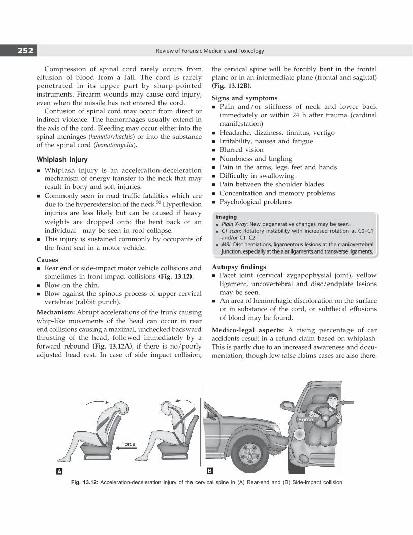
252 Review of Forensic Medicine and Toxicology
Compression of spinal cord rarely occurs from effusion of blood from a fall. The cord is rarely penetrated in its upper part by sharp-pointed instruments. Firearm wounds may cause cord injury, even when the missile has not entered the cord.
Contusion of spinal cord may occur from direct or indirect violence. The hemorrhages usually extend in the axis of the cord. Bleeding may occur either into the spinal meninges (hematorrhachis) or into the substance of the spinal cord (hematomyelia).
Whiplash Injury � Whiplash injury is an acceleration-deceleration
mechanism of energy transfer to the neck that may result in bony and soft injuries.
� Commonly seen in road traffic fatalities which are due to the hyperextension of the neck.50 Hyperflexion injuries are less likely but can be caused if heavy weights are dropped onto the bent back of an individual—may be seen in roof collapse.
� This injury is sustained commonly by occupants of the front seat in a motor vehicle.
Causes � Rear end or side-impact motor vehicle collisions and
sometimes in front impact collisions (Fig. 13.12). � Blow on the chin. � Blow against the spinous process of upper cervical
vertebrae (rabbit punch).
Mechanism: Abrupt accelerations of the trunk causing whip-like movements of the head can occur in rear end collisions causing a maximal, unchecked backward thrusting of the head, followed immediately by a forward rebound (Fig. 13.12A), if there is no/poorly adjusted head rest. In case of side impact collision,
the cervical spine will be forcibly bent in the frontal plane or in an intermediate plane (frontal and sagittal) (Fig. 13.12B).
Signs and symptoms � Pain and/or stiffness of neck and lower back
immediately or within 24 h after trauma (cardinal manifestation)
� Headache, dizziness, tinnitus, vertigo � Irritability, nausea and fatigue � Blurred vision � Numbness and tingling � Pain in the arms, legs, feet and hands � Difficulty in swallowing � Pain between the shoulder blades � Concentration and memory problems � Psychological problems
Imaging � Plain X-ray: New degenerative changes may be seen. � CT scan: Rotatory instability with increased rotation at C0–C1
and/or C1–C2. � MRI: Disc herniations, ligamentous lesions at the craniovertebral
junction, especially at the alar ligaments and transverse ligaments.
Autopsy findings � Facet joint (cervical zygapophysial joint), yellow
ligament, uncovertebral and disc/endplate lesions may be seen.
� An area of hemorrhagic discoloration on the surface or in substance of the cord, or subthecal effusions of blood may be found.
Medico-legal aspects: A rising percentage of car accidents result in a refund claim based on whiplash. This is partly due to an increased awareness and docu-mentation, though few false claims cases are also there.
Fig. 13.12: Acceleration-deceleration injury of the cervical spine in (A) Rear-end and (B) Side-impact collision
A B

253Regional Injuries
Concussion of Spinal Cord (Railway Spine)Causes i. In railway and motor vehicle collisions (most
common). ii. Severe blow to the back. iii. Compression from dislocation/fracture of vertebrae. iv. Damage by effusion of blood. v. Fall from height. vi. Bullet injury.
Symptoms appear immediately or after some hours and includes headache, giddiness, restlessness, sleeplessness, neurasthenia, weakness in limbs, amnesia, loss of sexual power and derangement of special senses.
� It produces paralysis, affecting the arms and hands or bladder, rectum or lower extremities.
� Paralysis is temporary and recovery occurs in about 48 h.
Neck
The neck can be the site of many different types of injury. Its importance in forensic medicine is due to the presence of a large number of vital structures, and the fact that it is of a size that can be grasped and easily held.
� Fractures of the hyoid bone or thyroid cartilage may occur due to fall injuring the neck, or when the neck comes in forcible contact with the handlebar of a cycle or the dashboard of a motor car.
� A blow to the side of the head or face, with a resultant abrupt twisting or sideways flexion motion of the neck can result in a laceration of the vertebral artery.
� A blow on the front of the neck may cause uncons-ciousness or even death due to vagal inhibition or by fracture of the larynx, usually involving the thyroid and cricoid cartilages, and resultant suffocation from hemorrhage or edema of the larynx.
� The mucous membrane of the trachea or larynx may be torn producing surgical emphysema and cause death by asphyxia.
� Suicidal incised wounds are more common than homicidal, but punctured wounds are usually homicidal.
� In wounds of the trachea and of the larynx below the vocal cords, speech is not possible. Wounds of the larynx and trachea are not fatal, if the large blood vessels are not damaged.
� Wounds of the sympathetic and vagus nerves may be fatal, those of the recurrent laryngeal nerves cause aphonia.
� Fractured neck by blunt force can cause spinal cord contusion, laceration or transection. Disruption of the atlanto-occipital junction can result in similar injuries.
Vertebral Column
� The spine is commonly injured in major trauma such as road traffic accidents or falls from a height.
� The type of injury will depend upon the degree of force and the angle at which the spine is struck. Vertebral column is strong in compression and vertically applied forces will result in little damage if the spine is straight. Angulation of the spine will alter the transmission of force and the spine becomes susceptible to injury, particularly at the site of the angulation.
� Force applied to the spine may result in damage to the discs or to the vertebral bodies.
� Fractures of the vertebral column are caused by direct violence or by indirect violence, as by forcible bending of the body or by a fall on buttocks or feet.
� Hyperflexion is the most common mechanism of fracture of spine. Falling from a height, diving and being thrown from automobile are the common causes.
� The common sites of fracture are upper and lower cervical regions and the junction of thoracic and lumbar segments. Fracture-dislocation and fracture of the laminae can damage the spinal cord.
Fracture of transverse processes: These are common in the region of the lumbar spine, where the quadratus lumborum muscle is attached.
Fracture of vertebral bodies: Compression (wedging) of vertebral body is the commonest fracture of the thoracic, thoracolumbar or lumbar spine. It may occur with a fall from a height. Injuries to the atlas and axis are more dangerous than lesions in the lower cervical vertebrae, because of involvement of the respiratory center.
Chest
Injuries of the chest can be: � Non-penetrating or closed, i.e. they do not open
up any part of the thoracic cavity. Usually caused by blunt force.
� Penetrating or open. If an injury damages the parietal pleura, it will produce an open pneumothorax, communicating directly with the external air.
254 Review of Forensic Medicine and Toxicology
Children and young adults whose chest is elastic, may sustain severe injuries to the intrathoracic viscera without fractures of sternum or ribcage.
In some cases, absence of injuries may be due to clothing worn by the victim.
RibsBlunt injury may result in fractures of the ribs. The fracture of a few ribs is unlikely to have much effect, other than causing pain in a healthy adult. In children, rib fractures are more resilient and they are able to cope better.
� If compression is front to back, lateral rib fractures may occur, and if back to front, the ribs tend to fracture near the spine.
� If compression is from side to side, the ribs may fracture near the spine and sternum. The middle ribs from 4–8th are usually fractured. In fractures due to direct violence, the fragments are often driven inwards and lacerate the underlying structures.
� In case of run over by a motor vehicle, the ribs are fractured symmetrically on both sides, in front near the costal cartilages and at the back near the angles.
� Multiple unilateral or bilateral rib fractures give rise to a flail or ‘stove-in’ chest, with consequent paradoxical respiration (the area of chest around the fractures may be seen to move inwards on inspiration) which interferes with respiratory exchange and also with return of the blood to the right atrium, resulting in severe dyspnea. Flail chest occurs when at least three successive ribs are fractured at two points.51
Sternum: Fractures of the sternum are not common.
Complications of rib fracture: Flail chest, lacerations of intercostal blood vessels with hemothorax, laceration of lungs with pneumothorax or hemopneumothorax, impaling wounds of heart, pleurisy and pneumonia. Rib fractures can also be artifactual due to cardiopulmonary resuscitation which may result in sternal and parasternal fractures.
z They are usually identified by their symmetrical, parasternal pattern and relative lack of hemorrhage at fracture site which indicates postmortem origin.
z Sometimes, fractures are seen in the left side only and may involve the first six ribs and sternal fracture may occur at the level of third or fourth intercostal space.
Lungs
� Compression of the chest or blunt weapon trauma produces contusions or lacerations.
� After severe head injury, where victim has been maintained for some time in a respirator, areas of collapse and hemorrhage with the formation of hyaline membrane is seen—’respirator lung’.
� A wound of the lung causes frothiness of blood, which issues from the mouth and nose or during coughing.
� Sudden compression of the chest may produce contrecoup contusions due to violent displacement of air in the lungs to the posterior surfaces near the angles of the ribs. The contusions may extend laterally or forwards into the substance of the lungs.
� Stab wounds of the lungs are usually not fatal, unless a major pulmonary blood vessel has been severed.
� Spontaneous pneumothorax may occur following rupture of an emphysematous bulla. Tension pneumothorax is seen when the leak in pleura has a valve-like action, air is sucked into the chest wall at each inspiration, but cannot escape on expiration. Iatrogenic pneumothorax may occur by external cardiac massage, percutaneously introduced subclavian catheters and continuous ventilatory support.
Complications of chest injuries� Pneumothorax � Chylothorax� Hemothorax � Interstitial emphysema
� Air embolism � Cardiac tamponade � Intraparenchymal hemorrhage � Infection
Diaphragm: Traumatic rupture of diaphragm is seen with blunt trauma of the lower anterior chest and is more common on the left (right side is protected by liver).
Heart
� Contusions and lacerations of the heart may be caused by direct violence to the chest or by compression of the thorax, or when a driver is forcibly thrown against the steering wheel. Cardiac contusions are usually seen on the anterior surface of either ventricle or the interventricular septum. Recent cardiac contusions are dark-red, hemorrhagic areas which are usually subepicardial.
� The commonest pincer lesion is a contusion of the right atrium at the entrance of the inferior vena cava. This is seen in compression injuries. It may cause sudden death, several days after the injury.
� Contrecoup contusions of the heart are seen over the posterior wall of the left ventricle. They are seen in traffic accidents in which the driver is thrown forward against the steering wheel and the heart is compressed against the vertebrae. Contusions may
255Regional Injuries
cause sudden death from ventricular fibrillation, or they may cause progressive circulatory failure and death after few hours or days.
� Foreign bodies, e.g. bullet, may remain embedded in the myocardium for years without producing any symptoms.
� The common sites of traumatic cardiac rupture in order of diminishing frequency are: right auricle, right ventricle, left auricle, ventricular septum and valves.
� The only natural cause of rupture of the heart is softening or thinning by infarction, which invariably occurs in the left ventricle.
� Stab wounds of the heart are dangerous. If the left ventricle is pierced, the thickness of the muscle wall may restrict the bleeding, allowing time for surgical treatment. A stab of the right ventricle is more rapidly fatal, blood escaping through the wound to cause hemopericardium and cardiac tamponade. Even 150 ml (average 400–500 ml) of blood can cause death by increasing intrapericardial pressure and producing mechanical interference with ventricular contractility. The right ventricle is more likely to be wounded, as it exposes its widest area towards the front of the chest.
Cardiac tamponade presents with three signs (Beck’s triad). They are—low arterial blood pressure, increased central venous pressure and distant heart sounds. Hypotension occurs because of decreased stroke volume, jugular-venous distension due to impaired venous return to the heart, and muffled heart sounds due to fluid inside the pericardium.
Cardiac concussion or commotio cordis: It refers to sudden cardiac death following a blunt trauma to the chest. It is often associated with sports and in young athletes.
Mechanism: The impact occurs at an electrically vulnerable phase of the cardiac cycle (during early ventricular repolarization; 15–30 milliseconds before the peak of the T wave).
Aorta: Wounds of the aorta or the pulmonary artery are rapidly fatal. The rupture of the aorta commonly occurs at the junction of the arch and the descending parts, just beyond the origin of the left subclavian artery, and is due to violent compression of the chest. It is common in traffic accidents, and less common in fall from height and crushing chest injuries. Spontaneous rupture of the aorta may occur from local disease.
Abdomen
Abdominal organs are vulnerable to a variety of injuries from blunt trauma because lax and compressible
abdominal walls can transmit the force to the abdominal viscera.
Injuries of the abdomen can be classified into: � Non-penetrating or closed, i.e. peritoneum is intact.
It is caused by blunt force; seen in falls, traffic accidents and assault by blunt weapons.
� Penetrating or open, i.e. when peritoneum is ruptured, it is open to infections.
Profuse subcutaneous or deep-seated bleeding of the abdominal wall may track along the muscular and fascial plane to become more diffuse, and may cover a large area of abdominal wall, especially in the lower segment. Blood may track down the inguinal canal and appear in the scrotum or labia.
In order of frequency, the structures most likely to be damaged in blunt abdominal trauma are: spleen, liver, kidneys, intestines, abdominal wall, mesentery, pancreas and diaphragm.
Injuries of the stomach and intestines may be caused by: i. Compression or crushing forces which produce
contusions or lacerations. ii. Traction or tearing forces. iii. Disruption or bursting forces.
� Hollow visceral injuries are less common in blunt trauma compared to penetrating injuries.
� Children have proportionally larger solid organs, less subcutaneous fat, and less protective abdominal musculature than adults. They suffer relatively more solid organ injury from both blunt and penetrating mechanisms.
� Small intestine is more commonly injured by forces of compression than the stomach and the large intestine.
� The proximal jejunum is the commonest site of rupture, followed by the ileum, duodenum, cecum, and large intestine. Transverse colon is usually involved in case of large intestinal rupture.
� The small bowel is most common intra-abdominal organ involved on penetrating trauma (e.g. stab or gunshot wounds) followed by colorectal injury and duodenal and gastric perforations.
� The intestinal wound may be situated at some distance from the external wound due to the compression and mobility of the intestines, and the depth of the wound is greater than the length of the penetrating object.
Pancreas: Wounds of the pancreas are very rare. The pancreas may be injured by compression forces usually where it overlies the second lumbar vertebra.52

256 Review of Forensic Medicine and Toxicology
Spleen
� Most common organ to be injured in blunt abdominal trauma.53
� Penetrating wounds of the spleen are less common than those of liver, but bleeding is more profuse. The spleen may be injured by forces of compression or traction forces. Compression forces produce lacerations. Traction forces may tear the spleen from its pedicle.
� The spleen is ruptured usually in its concave surface, and is generally associated with injuries to other organs and rib fractures. Lacerations are usually transcapsular and may occur at the hilar or convex surfaces. They are often multiple and may simulate the alphabetical figures, Y, H or L. Death from rupture of spleen is usually rapid due to profuse hemorrhage.
� A relatively mild trauma or even the contraction of the abdominal muscle may predispose the spleen to rupture, when it is diseased and enlarged, e.g. infectious mononucleosis, malaria, kala azar or leukemia.
Liver
� It is the second most frequently damaged abdominal organ in blunt trauma. The liver is commonly ruptured by motor accidents, blow, kick or by a sudden contraction of the abdominal muscles.
� The liver is more susceptible than spleen to pen-etrating injury.54
Blunt force to the abdomen may produce the following lacerations: i. Transcapsular laceration: Both capsule and
parenchyma are torn, and the laceration is present over the convex surface of the liver. It may cause rapid death from hemorrhage and shock.
ii. Subcapsular laceration: Capsule is intact and injury is beneath the capsule or intraparenchymal, and present over the convex surface of the liver. It may rupture few days after the injury and cause fatal delayed intraperitoneal hemorrhage.
iii. Non-communicating or central lacerations are seen in the substance of the liver.
iv. Coronal lacerations are seen over the superior surface due to distortion.
v. Lacerations of the inferior surface are due to distortion.
vi. Contrecoup laceration involve the posterior surface of the right lobe, at the point where it rests against the vertebral column.
� The right lobe is five times more commonly affected than the left.
� Convex surface and inferior border are commonly involved.
� Mild degree of external violence may rupture the liver, if it is diseased, e.g. fatty change, abscess formation, malaria or bilharziasis.
Complications of abdominal injuries � Laceration of the liver produces slow, but considerable bleeding over a period of time.
� Laceration of the spleen produces rapid and profuse hemorrhage leading to hypotension.
� Peritonitis is more common in rupture of the large intestine than with rupture of the small intestine due to the presence of pathogenic organisms in the colon.
� Chemical peritonitis is caused by leakage of gastric contents or pancreatic juice into the peritoneal cavity.
� Multiple contusions of the intestines may produce paralytic ileus.
Kidneys
Injuries to the kidneys are uncommon as they are situated in relatively well-protected part of the body. Contusions and lacerations usually result from blunt force applied directly to the posterior or lateral aspect of the kidneys, such as blows to the loins or in motor vehicle accidents and fall from a height.
� Lacerations of the kidneys may be transcapsular, subcapsular and transrenal (tear extending from the capsule to the renal pelvis). These may cause hemorrhage into the perinephric fat and form a large perirenal hematoma.
� Penetrating wounds are produced by bullets or pointed weapons, usually through the loin, and other viscera are also injured with retroperitoneal hemorrhage.
� Complications may be sepsis and the extra vasation of urine into the surrounding tissues with the development of urinary fistula.
Bladder
The bladder may be lacerated from a fall, a kick or a blow on the abdomen.Ruptures are of two types: i. Extraperitoneal: It occurs when the bladder is empty
or contains little urine and lies within the pelvis. It is usually associated with pelvic fractures. The urine may extravasate upwards to the level of the kidneys or downwards along the spermatic cord into the scrotum which may produce cellulitis and death.
257Regional Injuries
ii. Intraperitoneal: It occurs when the bladder is full of urine. Any blunt trauma to the lower abdominal wall can compress the bladder against the sacrum, resulting in rupture due to increased pressure with the urine entering the abdominal cavity.
Stab wounds of the lower abdomen may penetrate the bladder and cause rapid death from hemorrhage. There may be extraperitoneal extravasation of urine.
The male urethra may be ruptured usually under the pubic arch by a kick in the perineum, fall on a projecting substance, fracture of pubic bone or a foreign body. Forcible catheterization or cystoscopy, especially in the presence of some obstruction can cause rupture of urethra from within.
Reproductive OrgansFemale genital organs: Contusions and lacerations of the vulva and vagina may be due to kicks during assault or fall on a projecting substance. Wounds of vulva caused by a blunt weapon may resemble incised wounds. Lacerated wounds of the vulva may bleed profusely.
� The non-gravid uterus is usually not injured. � The gravid uterus may be ruptured by a blow
or kick on the abdominal wall, by instrumental criminal abortion, or in obstructed labor. Placenta may separate from uterus causing death of fetus.
Male genital organs � The penis may be injured by a squeeze or crush,
and the engorged erected penis may be completely avulsed from the pubes by forceful pull.
� Accidental injuries are rare, but the penis may be injured or amputated in revenge.
� Penile strangulation may occur by application of a constricting apparatus around the penis.
� Compression or crushing of the testes may cause sudden death from cardiac inhibition.
Bones and Joints
Fractures may occur from falls, blows or by muscular hyperactivity.
� In simple or closed fracture, there is no communi-cation between the bone and the air. A fall on the outstretched hand will cause Colles fracture (fracture of the distal end of radius).
� In compound or open fracture, there is a communi-cation between the bone and the air through a wound.
� Comminuted fractures: The bone breaks into fragments which may impact into each other or separate and become displaced.
� Partial or green-stick fractures: These occur because bones in children are very flexible and bend or partially break, instead of breaking cleanly when overloaded. There may be discontinuity in one cortex of the bone, but not in the other.55
� In childhood, slipping of an epiphysis is common, e.g. in distal end of the radius, medial epicondyle of the humerus, capitulum and distal end of the tibia.
� Fracture at the neck of the fifth metacarpal bone occurs, usually by striking the closed hand (fist) against a firm surface (boxer’s/brawler’s fracture).56
� Fractures of the mandible, maxilla, zygoma and zygomatic arch are produced by assaults and motor vehicle accidents.
� The frequency of fracture of different parts of mandible in decreasing order is: condyle (36%), body (21%), angle (20%), parasymphyseal (14%), alveolar (3%), ramus (3%), coronoid (2%) and symphysis (1%).57
� Maxillary fractures can be divided into five categories:58
i. Dentoalveolar: Separation of fragment of maxilla containing number of teeth.
ii. LeFort I: Transverse fracture of maxilla, above the apices of the teeth, through nasal septum and maxillary sinuses, the palatine bone and the sphenoid bone.
iii. LeFort II: Fracture has same track posteriorly, anteriorly it curves upwards near the zygomatic-maxillary suture, through the inferior orbit rim onto the orbital floor, across the nasal bones and septum.
iv. LeFort III: High transverse fracture of the maxilla that goes through the nasofrontal suture, through the medial orbital wall and fronto-zygomatic suture, across the arch and through the sphenoid.
v. Sagittal: Fracture line runs through a sagittal plane through the maxilla.
Fracture of the extremities caused by direct application of force (Fig. 13.13): i. Penetrating fractures are caused by large force acting
on a small area; seen in gunshot wounds. ii. Focal fractures are transverse fractures results from a
small force applied over a small area. It is usually seen in the forearms produced by weapons, like rods, when the person tries to ward off blows. Overlying soft tissue injury is relatively minor.
iii. Crush fractures result from large force applied over a large area with extensive soft tissue injuries and often comminuted fractures of the bone. Mostly seen on the legs in motor vehicle-pedestrian accidents.
Indirect fractures result from a force acting at a distance from the site of fracture, e.g. a fracture of the head of the radius or of the lower end of the humerus caused by a fall on the extended palm.
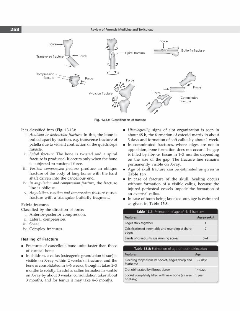
258 Review of Forensic Medicine and Toxicology
It is classified into (Fig. 13.13): i. Avulsion or distraction fracture: In this, the bone is
pulled apart by traction, e.g. transverse fracture of patella due to violent contraction of the quadriceps muscle.
ii. Spiral fracture: The bone is twisted and a spiral fracture is produced. It occurs only when the bone is subjected to torsional force.
iii. Vertical compression fracture produce an oblique fracture of the body of long bones with the hard shaft driven into the cancellous end.
iv. In angulation and compression fracture, the fracture line is oblique.
v. Angulation, rotation and compression fracture causes fracture with a triangular butterfly fragment.
Pelvic fracturesClassified by the direction of force: i. Anterior-posterior compression. ii. Lateral compression. iii. Shear. iv. Complex fractures.
Healing of Fracture
� Fractures of cancellous bone unite faster than those of cortical bone.
� In children, a callus (osteogenic granulation tissue) is visible on X-ray within 2 weeks of fracture, and the bone is consolidated in 4–6 weeks, though it takes 2–3 months to solidly. In adults, callus formation is visible on X-ray by about 3 weeks, consolidation takes about 3 months, and for femur it may take 4–5 months.
� Histologically, signs of clot organization is seen in about 48 h, the formation of osteoid matrix in about 3 days and formation of soft callus by about 1 week.
� In comminuted fractures, where edges are not in apposition, bone formation does not occur. The gap is filled by fibrous tissue in 1–3 months depending on the size of the gap. The fracture line remains permanently visible on X-ray.
� Age of skull fracture can be estimated as given in Table 13.7.
� In case of fracture of the skull, healing occurs without formation of a visible callus, because the injured periosteal vessels impede the formation of an external callus.
� In case of tooth being knocked out, age is estimated as given in Table 13.8.
Fig. 13.13: Classification of fracture
Table 13.8: Estimation of age of tooth dislocation
Features Age
Bleeding stops from its socket, edges sharp and feathered
1–2 days
Clot obliterated by fibrous tissue 14 days
Socket completely filled with new bone (as seen on X-ray)
1 year
Table 13.7: Estimation of age of skull fractures
Features Age (weeks)
Edges stick together 1
Calcification of inner table and rounding of sharp edges
2
Bands of osseous tissue running across 3–4
259Regional Injuries
Complications of fracture� Shock � Crush syndrome
� Hemorrhage � Fat embolism � Infection � Venous thrombosis
with pulmonary embolism
� Teeth most often affected by trauma in order of decreased frequency: Maxillary central incisors (60%), maxillary lateral incisors (22%), mandibular central incisors and mandibular lateral incisors.
� In permanent dentition, tooth fractures result from trauma whereas dislocation is common in primary dentition (due to elastic structures of the alveolar process).
At autopsy, a fracture may be suspected when there is extensive swelling and discoloration of the skin, or when there is abnormal mobility or crepitus is found. The tissues surrounding a suspected fracture should be dissected to determine injuries to the soft parts.
Antemortem or Postmortem Fracture
� Fracture produced just before death or just after death will have similar characteristics, except in the former there may be comparatively greater effusion of blood which will infiltrate the surrounding tissues.
� Antemortem fracture few hours prior to death will show edema and active cellular infiltration into the adjacent tissues and between the fractured edges of the bones.
� Antemortem fracture of long bones may result in fat emboli traveling to distant parts of the body producing characteristic lesions (punctate hemorrhages in skin, eyelids, conjunctiva) which are seen grossly and microscopically.
These changes are not seen in postmortem fractures.
MULTIPLE CHOICE QUESTIONS
1. NOT a type of skull fracture: NIMHANS 08 A. Linear B. Depressed C. Diffuse axonal D. Basal 2. Fracture-a-la signature is: AI 11; MAHE 12; AIIMS 13 A. Gutter fracture B. Depressed fracture C. Ring fracture D. Sutural separation 3. Pond’s fractures are common in: AP 09 A. Children B. Adolescent C. Adult D. Old age 4. All are true of pond’s fracture, except: CMC (Vellore) 13 A. No brain damage B. Seen in infants C. Depressed fracture of the skull D. Shearing of the dura is not seen 5. Orbital blow-out fracture involves: AIIMS 09;
FMGE 10; MP 10; JIPMER 11; Bihar 11; PGI 11 A. Lateral wall and floor of orbit B. Medial wall and floor or orbit C. Lateral wall and roof or orbit D. Medial wall and roof or orbit 6. Teardrop sign is seen in:
Kerala 08; JIPMER 08; Maharashtra 09 A. Fracture medial wall of orbit B. Fracture lateral wall of orbit C. Fracture floor of orbit D. Fracture roof of orbit
7. Roof of orbit is fractured due to: AIIMS 09 A. Blow on forehead B. Blow on lower jaw C. Fall on back D. Blow in parietal region 8. CSF rhinorrhea is due to fracture of:
UP 08, 09; DNB 10; Bihar 10; AIIMS 10; CMC (Ludhiana) 13
A. Cribriform plate B. Sella turcica C. Petrous temporal bone D. Mastoid 9. Characteristic of anterior cranial fossa fracture:
WB 07 A. Black eye B. Pupillary dilatation C. CSF otorrhea D. Hemotympanum 10. True about CSF rhinorrhea: UP 09 A. Commonly occurs due to break in cribriform plate B. Contains less amount of proteins C. Decreased glucose content confirms diagnosis D. Immediate surgery is required 11. CSF otorrhea is caused by: Bihar 10; WB 11;
CMC (Vellore) 13 A. Fracture of cribriform plate B. Fracture of parietal bone C. Fracture of petrous temporal bone D. Fracture of tympanic membrane 12. Battle sign indicates: NIMHANS 13 A. Basilar fracture B. Sutural fracture C. Orbital fracture D. Depressed fracture 13. Hinge fracture is: JIPMER 14 A. Depressed fracture B. Sutural fracture C. Orbital fracture D. Basilar fracture
1. C 2. B 3. A 4. C 5. B 6. C 7. C 8. A 9. A 10. A 11. C 12. A 13. D
260 Review of Forensic Medicine and Toxicology
14. D 15. D 16. B 17. A 18. A 19. D 20. B 21. A 22. C 23. A 24. D 25. A 26. C 27. A 28. A 29. B 30. A 31. A 32. A
14. CSF rhinorrhea is diagnosed by: MP 07 A. Glucose estimation B. Halo sign C. Immunoelectrophoresis D. All 15. Specific for CSF in rhinorrhea: AI 10 A. b-2 microglobulin B. Albumin C. Macroglobulin D. b-2 transferrin 16. Battle’s sign is: TN 11 A. Hemorrhage around eyes B. Mastoid ecchymosis C. Umbilical ecchymosis D. Vaginal ecchymosis 17. Best prognostic indicator for head injured patients:
AIIMS 10 A. GCS B. CT findings C. Age of the patient D. History 18. Primary impact injury to brain: DNB 10 A. Concussion B. Cerebral edema C. Hypoxic injury D. Intracerebral hematoma 19. Secondary brain injury is: JIPMER 10 A. Concussion B. Diffuse axonal surgery C. Depressed skull fracture D. Intracerebral hematoma 20. Concussion causes: Karnataka 07 A. Small hemorrhages and swelling of brain tissues B. Momentary interruption of brain function with/
without loss of consciousness C. Tearing or shearing of brain structures D. Bruising of the brain 21. Antegrade amnesia is seen in: AIIMS 10 A. Post-traumatic head injury B. Drug induced C. Electroconvulsive therapy D. Stroke 22. Shearing damage is seen in: CMC (Vellore) 07 A. Heart B. Liver C. Brain D. Spinal cord 23. Diffuse axonal injury is characterized by lesion at:
AI 08; FMGE 11 A. Junction of gray and white matter B. White matter C. Basal ganglia D. Corpus callosum 24. A male was brought unconscious with external injuries.
CT brain showed no midline shift, but basal cistern were compressed with multiple small hemorrhages. Diagnosis is: AIIMS 06
A. Cerebral contusion B. Cerebral laceration
C. Multiple infarcts D. Diffuse axonal injuries 25. ‘Retraction balls’ after trauma are seen in:
PGI 08, 11; UP 12 A. Brain B. Spleen C. Liver D. Lung 26. Apoplexy is: DNB 08 A. Learning disability B. Insanity leading to commitment of a crime C. Sudden onset of bleeding in the brain D. Injury to the brain due to trauma 27. Brain hemorrhage limited by sutures:
BHU 09; Punjab 12 A. EDH B. SAH C. SDH D. ICH 28. Commonest source of extradural hemorrhage: UP 09 A. Middle meningeal artery B. Basilar artery C. Charcot’s artery D. Middle cerebral artery 29. Middle meningeal artery is a direct branch of:
AI 06; Odisha 09; Punjab 12 A. External carotid artery B. Internal maxillary artery C. Superficial temporal artery D. Middle cerebral artery 30. Artery usually torn in temporal bone fracture is:
Odisha 11 A. Middle meningeal artery B. Posterior auricular artery C. Transverse facial artery D. Deep temporal artery 31. Most common manifestation of increased intracranial
pressure in a patient with head injury: UPSC 07 A. Change in the level of consciousness B. Ipsilateral pupillary dilatation C. Retching and vomiting D. Bradycardia 32. A rugby player hit his head on the post whilst involved
in a tackle. He was unconscious for 5 min but regained full consciousness and sat on the sideline until the end of the game. He was then noted to be drowsy and over the past 30 min became confused and no longer obeyed commands. Most likely diagnosis is:
Himachal 10; AIIMS 13; NIMHANS 14 A. Extradural hematoma B. Subdural hematoma C. Subarachnoid hematoma D. Cerebral edema

261Regional Injuries
33. Lucid interval is classically seen in: COMEDK 07; PGI 07; Kerala 08; WB 09; FMGE 10; DNB 10;
NEET 13, 14 A. Intracerebral hematoma B. Acute subdural hematoma C. Chronic subdural hematoma D. Extradural hematoma 34. Subdural hemorrhage is due to rupture of: Bihar 11 A. Middle meningeal artery B. Dural venous sinus C. Cortical bridging veins D. Rupture of intracranial aneurysms 35. A 14-year-old boy was hit on the side of the head
with a baseball bat during practice. A laceration with palpable bone fragment was found in the wound. After 5 h the boy died. Most likely cause of death is: Himachal 10
A. Subarachnoid hemorrhage B. Epidural hemorrhage C. Subdural hemorrhage D. Intracranial hemorrhage 36. CT of subdural hematoma will show: Odisha 09; NIMHANS 11; Bihar 12 A. Biconvex hyperdense opacity B. Biconcave hyperdense opacity C. Concavoconvex opacity D. Hyperdense diffuse lesion 37. Circle of Willis is not formed by: TN 11 A. Anterior choroidal artery B. Anterior cerebral artery C. Posterior cerebral artery D. Anterior communicating artery 38. A 18-year-old female presented with severe headache of
sudden onset. On CT scan, a diagnosis of subarachnoid hemorrhage is made. Most common cause is:
AI 06; UP 10, 12 A. Hypertension B. Berry aneurysm rupture C. Basilar artery rupture D. Subdural venous sinuses rupture 39. A 40-year-old hypertensive lady is brought to the
emergency room after being unresponsive following a sudden bout of severe headache, vomiting and neck rigidity at work. O/E her BP is 180/100 mmHg and her respiration is irregular and of Cheyne-Stokes type. She is agitated and doesn’t follow commands, but moves her extremities spontaneously. Most likely diagnosis will be: JIPMER 08; AIIMS 13
A. Subarachnoid hemorrhage due to rupture cerebral aneurysm
B. Hypoglycemic coma C. Conversion reaction D. Addisonian crisis
40. Investigation of choice in SAH: NIMHANS 07; FMGE 11
A. CT scan B. MRI C. X-ray skull D. Radionuclide scan 41. Cause of Berry aneurysm: AIIMS 11 A. Degeneration of internal elastic lamina B. Degeneration of media/muscle cell layer C. Deposition of mucoid material in media D. Low grade inflammation of vessel wall 42. Commonest cause of thunderclap headache:
AIIMS 10; NIMHANS 14 A. Extradural hemorrhage B. Aneursymal SAH C. Subdural hemorrhage D. Basilar migraine 43. Traumatic bleeding may include all, except: Kerala 09 A. EDH B. SDH C. SAH D. ICH 44. Commonest hemorrhage following head injury:
KCET 13 A. Extradural hemorrhage B. Subdural hemorrhage C. Intracranial hemorrhage D. Subarachnoid hemorrhage 45. Most common location of hypertensive intracranial
hemorrhage is: AI 06; NIMHANS 08; DNB 10; WB 11 A. Subarachnoid space B. Basal ganglia C. Cerebellum D. Brainstem 46. NOT true about Berry aneurysms: AP 08 A. Rupture leading to SAH B. Most common in posterior circulation C. Developmental anomaly D. Common in anterior circulation 47. Kernohan-Woltman sign (notch) is seen in:
COMEDK 15 A. Uncal herniation B. Central herniation C. Transfalcial herniation D. Foraminal herniation 48. Duret hemorrhages are found in: PGI 08,10,11; Kerala 11 A. Brain B. Heart C. Kidney D. Liver 49. Investigation of choice for evaluation of acute head
injury: AIIMS 14 A. CECT Head B. NCCT Head C. MRI Brain D. CT Angiography 50. ‘Whip-lash’ injury is caused due to: AIIMS 06; Karnataka 07; BHU 12; NEET 15 A. Fall from a height B. Acute hyperextension of the spine C. Blow on top to head D. Acute hyperflexion of the spine
33. D 34. C 35. C 36. C 37. A 38. B 39. A 40. A 41. B 42. B 43. D 44. B 45. B 46. B 47. A 48. A 49. B 50. B

262 Review of Forensic Medicine and Toxicology
51. Flail chest means fracture of: PGI 12 A. Two ribs on same side B. Two ribs on opposite side C. Four ribs on two sides D. Multiple ribs on both sides 52. Organ that does not show countercoup injuries:
SGPGI 12 A. Heart B. Brain C. Lung D. Pancreas 53. True about blunt trauma abdomen are all, except:
PGI 14; JIPMER 14 A. Solid organ injury more common in children than
adults in blunt trauma B. Liver injuries are more common than splenic injury C. Diaphragmatic injuries are rare D. Intraperitoneal gas shadows are pathognomonic of
bowel perforation 54. Most common organ injured in penetrating injury of
the abdomen: AFMC 10 A. Liver B. Spleen C. Stomach D. Small intestine
55. A green-stick fracture is:TN 06; UPSC 07; BHU 09; NIMHANS 10
A. Seen mostly in the elderly B. Fatigue fracture C. Spiral fracture of long bone D. Part of cortex is intact and part is crumpled 56. Boxer’s fracture is: MP 11 A. Fracture of first metacarpal base B. Fracture of fifth metacarpal neck C. Fracture of third metacarpal neck D. Fracture of first metacarpal neck 57. Most common site for fracture mandible: TN 11 A. Condyle B. Angle C. Body D. Symphysis 58. LeFort’s fracture would include all of the following,
except: Manipal 10; WB 09; TN 09; Bihar 10; NIMHANS 14
A. Maxilla B. Mandible C. Zygoma D. Nasal bones
51. C & D 52. D 53. B 54. A 55. D 56. B 57. A 58. B

Definition: Tissue injury due to application of heat or cold in any form to the external or the internal body surfaces.
Classification: Refer to Flow chart 14.1.
Cold Injury
Hypothermia
� Exposure to cold produce hypothermia which is defined as core temperature below 35ºC (95ºF).
� An esophageal or rectal probe that measures temperatures as low as 25ºC is required; oral or axillary thermometers are inaccurate.
� Risk factors: z Low environmental temperature. z Extremes of age (infants, children and elderly
> 60 years). z Immersion in water and wet clothing. z Pre-existing diseases, such as hypothyroidism,
atherosclerosis, dementia, or inadequate nutrition. z Intoxicated persons (alcohol, tranquilizers or
opiates) or persons engaged in activities like mountaineering and sailing.
� Effects of hypothermia: z Direct effects are prominent in fatty tissues and
myelinated nerve fibers. z Indirect effects are mostly ischemic.
Clinical features
� When the temperature falls below 32ºC to 24ºC, there is disorientation, dulling of consciousness, loss of reflex, and fall in respiration, heart rate and blood pressure.1
� Red patches and pallor of the skin, edema of the face, and stiffness of neck muscles may be seen.Death is common due to ventricular fibrillation or
asystole.2
Complications: Patient who survives for a short time may develop hemorrhagic pancreatitis, pneumonia, ulcers or focal hemorrhages in the GIT, acute tubular necrosis and myocardial fiber necrosis.
Postmortem findings: There are no definitive autopsy findings of hypothermia. However, there are several features which are taken together, and in the presence of history (scene and circumstances) allow a reasonably confident diagnosis.
Flow chart 14.1: Classification of thermal injuries
Thermal Injuries
ChapTer 14

264 Review of Forensic Medicine and Toxicology
� Moreover, the autopsy is helpful to rule out other causes of death, collect evidence as necessary, and contribute to the identification process.
� Viscera should be sent for blood alcohol and toxicological analysis.
External features � Patches of pink to brownish-pink discoloration may be seen on the
external surface, most often present over the extensor surfaces of large joints (usually on knees, elbows or outside of the hip joint).
� Postmortem staining is pink/bright red due to antemortem binding of oxygen to hemoglobin and its postmortem diffusion through skin.3
� The extremities may be cyanosed or white (white deaths). � Edema of feet and blistering of skin may be seen.
Internal features � Blood: Bright red in color. � Trachea: Frothy and sanguineous fluid. � Lungs: Congested, edematous and shows hemorrhages. � Heart: Dilatation of right atrium and ventricle. � Stomach: Wischnewsky spots* may be seen. � Kidneys: Acute tubular necrosis with accumulation of lipid in
epithelial cells of proximal renal tubules. � Liver and spleen: Congested. Fatty changes in liver and contracted
spleen. � Muscles: Hemorrhages into core muscles like iliopsoas. � Pancreas: Hemorrhages in parenchyma and mucosa of the
pancreatic duct. � Intestines: Ulceration of the colon and ileum, and hemorrhagic
infarction of the colon.
� Paradoxical undressing: In deaths due to hypothermia, the body is found either partially or fully undressed. During hypothermia, the victim becomes disoriented, confused and combative, and may begin discarding the clothing he/she has been wearing, which in turn increases the rate of temperature loss. This sometimes results in the assumption that the deaths are associated with sexual assault (homicide).4
� ‘Hide and die’ syndrome: In some hypothermic deaths, bodies are found in some strange places—under a bed or bench, on a shelf or behind a wardrobe, or alternatively may pull down household articles into a heap on top, which may give the impression of their attempt to ‘hide’ (protective ‘burrow-like’ or ‘cave-like’ situation). This may also lead to the assumption of a homicide or robbery. It is due to mental confusion from hypothermia and may be related to hibernation reflex.
Cold or freezing temperature can produce localized effects, e.g. chilblain, trench foot/immersion foot, and frostbite which are phases of the same process.
Chilblain (Erythema Pernio)These are red, itching, skin lesions, usually in the extre mities, caused by exposure to cold. They may be associated with edema and blistering, and are
aggravated by warmth. On continued exposure, ulcerative or hemor rhagic lesions may develop.
Treatment: Elevation of the affected part and allowing it to warm gradually at room temperature. The areas should not be rubbed or massaged or subjected to heat application.
Immersion Syndrome (Trench / Immersion foot) � Immersion foot, trench foot and trench hand are
types of immersion syndrome injuries. � Immersion foot/hand results from prolonged
exposure to severe cold (< 10ºC) and dampness; seen in soldiers during warfare, especially in trenches, and in persons exposed to prolonged immersion or exposure at sea.
� Extremities are affected in these conditions.
Clinically, it is divided into: i. Pre-hyperemic stage: The affected parts are cold
and anesthetic. ii. Hyperemic stage: The parts are hot with intense
burning and shooting pains. iii. Post-hyperemic stage: Area is pale or cyanotic
with diminished pulsations.
Treatment: Air drying, protecting the extremities from trauma and secondary infection, and gradual rewarming by exposure to air at room temperature (not heat) without massaging or moistening the skin or immersing it in water.
frostbite
� Frostbite (congelatio) is injury due to freezing and formation of ice crystals and obstruction of blood supply within tissues.
� It occurs due to exposure to great extremes of cold (–2.5ºC).
� This is typically evident as blue-black discoloration of fingers, toes or other susceptible body parts such as the nose, ears and face (Fig. 14.1).
� In mild cases, only the skin and subcutaneous tissues are involved, and symptoms are numbness, prickling and itching.
� Deep frostbite involves deeper structures, and there may be paresthesia and stiffness. Thawing causes tenderness and burning pain. The skin is white or yellow, looses its elasticity and becomes immobile. Edema, blisters, necrosis and gangrene may appear beyond the line of inflammatory demarcation.
* Wischnewsky spots are blackish-brownish color gastric mucosal erosions/ulcerations seen in hypothermia (vary from 1 mm to 2 cm in size and from few to > 100). Similar changes are seen in drug/alcohol abuse and in stress/shock.
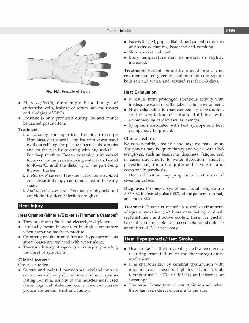
265Thermal Injuries
� Microscopically, there might be a damage of endothelial cells, leakage of serum into the tissues and sludging of RBCs.
� Frostbite is only produced during life and cannot be caused postmortem.
Treatment i. Rewarming: For superficial frostbite (frostnip):
Firm steady pressure is applied with warm hand (without rubbing), by placing fingers in the armpits and for the feet, by covering with dry socks.5
For deep frostbite: Frozen extremity is immersed for several minutes in a moving water bath, heated to 40–42°C, until the distal tip of the part being thawed, flushes.
ii. Protection of the part: Pressure or friction is avoided and physical therapy contraindicated in the early stage.
iii. Anti-infective measures: Tetanus prophylaxis and antibiotics for deep infection are given.
Heat Injury
Heat Cramps (Miner’s / Stoker’s / fireman’s Cramps)6
� They are due to fluid and electrolyte depletion. � It usually occur in workers in high temperature
when sweating has been profuse. � Cramping results from dilutional hyponatremia, as
sweat losses are replaced with water alone. � There is a history of vigorous activity just preceding
the onset of symptoms.
Clinical featuresOnset is sudden.
� Severe and painful paroxysmal skeletal muscle contractions (‘cramps’) and severe muscle spasms lasting 1–3 min, usually of the muscles most used (arms, legs and abdomen) occur. Involved muscle groups are tender, hard and lumpy.
� Face is flushed, pupils dilated, and patient complains of dizziness, tinnitus, headache and vomiting.
� Skin is moist and cool. � Body temperature may be normal or slightly
increased.
Treatment: Patient should be moved into a cool environment and given oral saline solution to replace both salt and water, and advised rest for 1–3 days.
Heat Exhaustion
� It results from prolonged strenuous activity with inadequate water or salt intake in a hot environment.
� Heat exhaustion is characterized by dehydration, sodium depletion or isotonic fluid loss with accompanying cardiovascular changes.
� Symptoms associated with heat syncope and heat cramps may be present.
Clinical featuresNausea, vomiting, malaise and myalgia may occur. The patient may be quite thirsty and weak with CNS symptoms, such as headache, dizziness, fatigue, and in cases due chiefly to water depletion—anxiety, paresthesias, impaired judgment, hysteria and occasionally psychosis.
Heat exhaustion may progress to heat stroke, if sweating ceases.
Diagnosis: Prolonged symptoms, rectal temperature > 37.8°C, increased pulse (150% of the patient’s normal) and moist skin.
Treatment: Patient is treated in a cool environment, adequate hydration (1–2 liters over 2–4 h), oral salt replenishment and active cooling (fans, ice packs). Normal saline or isotonic glucose solution should be administered IV, if necessary.
Heat Hyperpyrexia / Heat Stroke
� Heat stroke is a life-threatening medical emergency resulting from failure of the thermoregulatory mechanism.
� It is characterized by cerebral dysfunction with impaired consciousness, high fever [core (rectal) temperature ≥ 41ºC (≥ 105ºF)] and absence of sweating.7,8
� The term thermic fever or sun stroke is used when there has been direct exposure to the sun.
Fig. 14.1: Frostbite of fingers

266 Review of Forensic Medicine and Toxicology
� It presents in one of the two forms: i. Classic: Seen in patients with compromised
homeostatic mechanisms. ii. Exertional: Seen in healthy persons undergoing
strenuous exertion in a thermally stressful environment.
Predisposing factors
� Environmental causes: High temperature, increased humidity, lack of acclimatization and physical exertion.
� Non-environmental causes: Extremes of age (infants, and elderly > 65 years), obesity, alcoholism, brain hemorrhage, malignant hyperthermia, chronically infirm, underlying medical conditions like thyrotoxi-cosis or sepsis, salicylate overdose, patients receiving medications like anticholinergics, antihistamines or phenothiazines, and reactions to certain drugs of abuse such as cocaine.
Clinical features (Fig. 14.2)Onset is sudden, with sudden collapse and loss of consciousness.
� Prodromal symptoms include dizziness, weakness, nausea, vomiting, confusion, faintness, staggering gait, purposeless movements, disorientation, drowsiness and irrational behavior.
� Skin is hot, and initially covered with perspiration, later it dries. Pulse is strong initially (160–180/min).
� Tachycardia and hyperventilation (with subsequent respiratory alkalosis) occur.
� Blood pressure may be elevated in early stages, but later hypotension develops.
� The core temperature is usually > 40°C. � Pupils are contracted. � Delirium, blurred vision, convulsions, collapse and
unconsciousness occur.
Morbidity or even death can result from cerebral, cardiovascular, hepatic or renal damage.
TreatmentImmediate measures should be taken to lower the core temperature. i. The patient is unclothed to the minimum and
sprayed with water (20°C) while air is passed across the patient’s body. Immersion in an ice-water bath is very effective. Treatment should be continued until the rectal temperature drops to 39°C.
ii. Chlorpromazine (25–50 mg IV) or diazepam (5–10 mg IV) is given every 4 hourly to control shivering.
iii. Fluid administration to ensure high urinary output (> 50 ml/h), mannitol administration (0.25 mg/kg) and alkalinization of urine (IV bicarbonate administration, 250 ml of 4%) are recommended.
Complications: Patients who survive > 24 h may show lobar pneumonia, acute tubular necrosis of kidneys, hepatic necrosis, myocardial fiber necrosis, disseminated intravascular coagulation, pancreatitis, adrenal hemorrhage and myoglobinuria (due to rhabdomyolysis).9
Postmortem findingsDeaths related to hyperthermia have no specific autopsy findings. When the body temperature is not available, but the circumstances of the death suggest hyper -thermia, then it can be listed as the cause of death. i. Lungs: Congested. Intrathoracic petechiae may
be present, particularly in infants and children. ii. Heart: Subendocardial hemorrhages may be seen. iii. Brain: Congested and edematous. Convolutions
are flattened, and scattered petechiae are found in the walls of the third ventricle and floor of the fourth ventricle.
In brain, nonspecific degenerative changes in cortical neurons may be visible in light microscopy in individual who survives for some period. There may also be marked changes in the cerebellum, including necrosis of Purkinje cells (which appear dense, red and eosinophilic) and a marked decrease in the number of Purkinje cells.
Medico-legal aspects: Deaths are usually accidental. Postmortem is done to rule out any other cause of death or contributory cause of death.
Heat Prostration (Heat Syncope / Collapse) � It results from salt depletion and dehydration due to
excess of sweating and cutaneous vasodilation with consequent systemic and cerebral hypotension, but without any rise of temperature, despite exposure to excessive heat.Fig. 14.2: Difference between heat syncope and heat stroke

267Thermal Injuries
� The condition is usually seen in the tropics and in the deserts.
� Precipitating factors are overexertion, heavy muscular work and use of unsuitable clothing.
Clinical features (Fig. 14.2) � Patient suddenly feels weak, giddy and sick. � Nausea, dizziness, flushing of face, throbbing
headache in temples, dimness of vision may occur. � Face is pale, pulse is weak and feeble, respiration
sighing, skin cool and moist, and temperature sub-normal.
� Systolic BP is usually < 100 mmHg.
Patient usually recovers and consciousness is never lost.
Treatment consists of rest and recumbency in a cool place and rehydration by mouth (or IV, if necessary).
Anhidrotic heat exhaustion (tropical anhidrotic asthenia or thermogenic anhidrosis): It is characterized by depression of sweating, exhaustion and vesicular skin lesions on the trunk (miliaria profunda). The illness can occur in newcomers or even in normal acclimatized persons on prolonged exposure to heat. It involves failure of the normal sweat mechanism due to occlusion of the sweat ducts. There is tachycardia, tachypnea, mild pyrexia, anhidrosis and polyuria. The patient is shifted to cool surroundings and recovery is generally rapid. This syndrome must be differentiated from heat stroke and heat exhaustion, and malingering should be ruled out.
Burns
Definition: Burn is an injury caused by heat, or by a chemical or physical agent having an effect similar to heat.
Characteristics / Types of Burns
i. Contact burns: There is physical contact between the body and a hot object, like heated solid or molten metal. When applied momentarily, it produces a blister with erythema corresponding to the shape and size of the agent.
ii. Flame burns: There is actual contact of body with flame. It may produce vesication, singeing of the hair and blackening of the skin. Hair singed by flame becomes curled, twisted and blackish, breaks off or is totally destroyed. If kerosene oil or petrol is used, it will produce sooty blackening of the parts and have a characteristic odor.
Flash burns are a variant of flame burns which are due to initial ignition from flash fires (sudden ignition or explosion of gases or petrochemicals). It burns the exposed surfaces to the flash, and not the folds of skin and other protected areas.
iii. Scalds: They are caused by contact with hot liquids, most commonly water, and usually occur on exposed skin.
iv. Radiant heat burns: They are caused by heat waves, a type of electromagnetic wave. There is no contact between the body and flame or hot surface. Initially, the skin appears erythematous and blistered, and later it is light brown and leathery.
v. Ionizing radiation burns (X-rays, radium, UV rays): It can be localized or may involve the whole body depending on radiation exposure. The burn varies from redness of skin to dermatitis with shedding of hair and epidermis, and pigmentation of the sur-rounding skin. Fingernails may show de generative changes and wart-like growth.
vi. Chemical burns: Classified into acids, alkalis and vesicants (blister forming). Characteristically, there are ulcerated patches, no blisters, hair is not singed and the red line of demarcation is absent. Sometimes, the burn shows distinct coloration, and is usually uniform in character.
vii. Electric and lightning burns.viii. Microwave burns: The waves create heat through
molecular agitation. The greater the water content of a particular tissue, the greater the heat produced, e.g. muscle tends to be heated more than fat. Burns caused by microwave ovens tend to be indirect, like the person ingests liquid without realizing how hot it is. Medico-legal implications are rare.
� Most burns are produced by dry heat, and result from contact with a flame or a heated solid object or exposure to radiant heat of an object.
� The majority of burns in children are scalds caused by accidents, and most electrical and chemical injuries occur in adults.
� Cold and radiation are very rare cases of burns.
Classification
Burns can be classified in many ways, but two classifications are given in Table 14.1 (Fig. 14.3).
Other types of classification: Heba’s (similar to Wilson’s, but uses the symbol of degree); and Evan’s which categorizes burns into superficial, partial and full thickness.
Clinically, burns are classified as first degree (superficial) burns, second degree (partial and deep partial) burns and third degree (full thickness) burns (Diff. 14.1).10–12
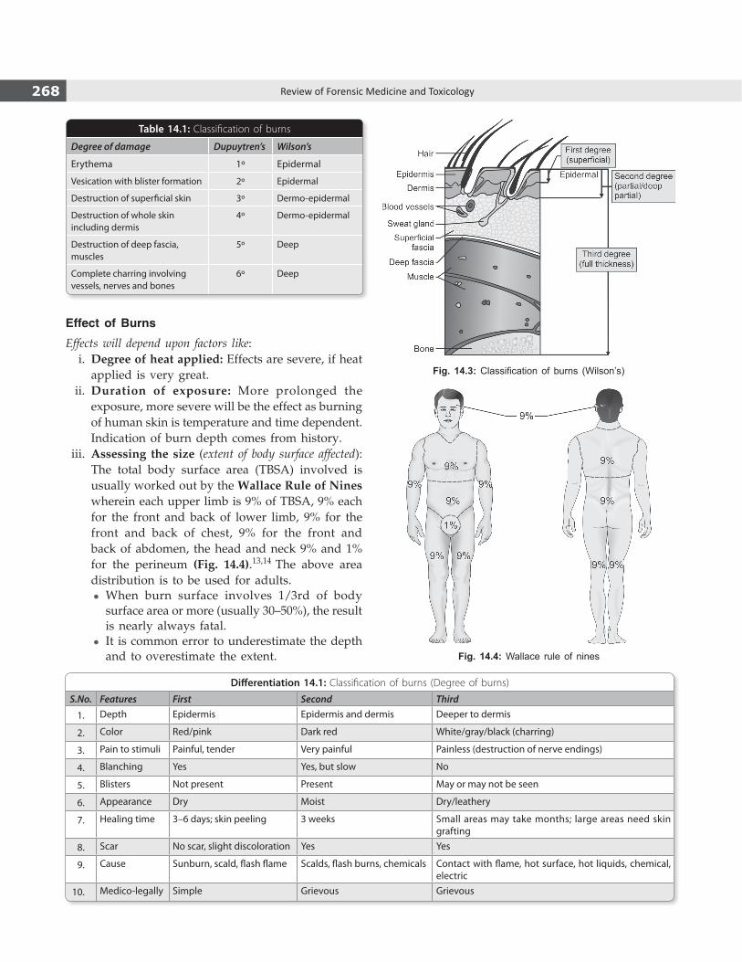
268 Review of Forensic Medicine and Toxicology
Effect of Burns
Effects will depend upon factors like: i. Degree of heat applied: Effects are severe, if heat
applied is very great. ii. Duration of exposure: More prolonged the
exposure, more severe will be the effect as burning of human skin is temperature and time dependent. Indication of burn depth comes from history.
iii. Assessing the size (extent of body surface affected): The total body surface area (TBSA) involved is usually worked out by the Wallace Rule of Nines wherein each upper limb is 9% of TBSA, 9% each for the front and back of lower limb, 9% for the front and back of chest, 9% for the front and back of abdomen, the head and neck 9% and 1% for the perineum (Fig. 14.4).13,14 The above area distribution is to be used for adults.
z When burn surface involves 1/3rd of body surface area or more (usually 30–50%), the result is nearly always fatal.
z It is common error to underestimate the depth and to overestimate the extent.
Fig. 14.3: Classification of burns (Wilson’s)
Fig. 14.4: Wallace rule of nines
Table 14.1: Classification of burns
Degree of damage Dupuytren’s Wilson’s
Erythema 1º Epidermal
Vesication with blister formation 2º Epidermal
Destruction of superficial skin 3º Dermo-epidermal
Destruction of whole skin including dermis
4º Dermo-epidermal
Destruction of deep fascia, muscles
5º Deep
Complete charring involving vessels, nerves and bones
6º Deep
Differentiation 14.1: Classification of burns (Degree of burns)
S.No. Features First Second Third
1. Depth Epidermis Epidermis and dermis Deeper to dermis
2. Color Red/pink Dark red White/gray/black (charring)
3. Pain to stimuli Painful, tender Very painful Painless (destruction of nerve endings)
4. Blanching Yes Yes, but slow No
5. Blisters Not present Present May or may not be seen
6. Appearance Dry Moist Dry/leathery
7. Healing time 3–6 days; skin peeling 3 weeks Small areas may take months; large areas need skin grafting
8. Scar No scar, slight discoloration Yes Yes
9. Cause Sunburn, scald, flash flame Scalds, flash burns, chemicals Contact with flame, hot surface, hot liquids, chemical, electric
10. Medico-legally Simple Grievous Grievous

269Thermal Injuries
Admission criteria for burn patients26
� Partial thickness burns involving >10% of the TBSA (> 5% in children < 10 years and adults > 50 years).
� Full thickness burns involving > 2% of the TBSA in any age group or circumferential burns.
� Partial or full thickness burns involving the face, hands, feet, genitalia, perineum or major joints.
� Electrical burns, including lightning injury. � Chemical burns or inhalation injury. � Patients with pre-existing medical disorders (e.g. diabetes),
pregnancy or concomitant trauma (e.g. fractures).
Cause of Death
Burn individuals develop a host of complications; one or more can contribute to the cause of death. Following successful fluid resuscitation, sepsis is the leading cause of mortality.Immediate causes i. Primary or neurogenic shock: Due to pain or fright. ii. Asphyxia: Suffocation may result from inhalation
of CO, CO2, or cyanide (produced by burning of materials containing nitrogen compounds such as poly urethane in vinyl, wool or nylon) or falling of the building on the body during attempt to escape.
z CO poisoning is an important cause in most fire deaths (COHb > 50% is confirmatory).
iii. Smoke- or heat-induced laryngospasm, respiratory arrest, and/or a vagal reflex-caused cardiac arrest are other proposed mechanisms of rapid death.
Delayed causes i. Hypovolemic, burns or secondary shock: More than half
of the deaths occur due to secondary shock within 24–48 h due to loss of fluid and protein, causing decrease in cardiac output and multiorgan failure.
ii. Acute edema of glottis occurs from inhalation of irritant smoke or hot gases with or without pulmonary edema. Respiratory failure (inhalation injury, pneumonia or ARDS) is also a significant cause of death within 3 days.
iii. Toxemia due to absorption of toxic products from the burnt surface. Death occurs in about 3–4 days.
iv. Sepsis: Most important cause of death, occurring in 4–5 days or longer after burn. Septicemia can be caused by burn wound infections (e.g. Pseudomonas aeruginosa and other Gram-negative bacteria, Staphylococcus aureus), pneumonia, urinary tract infection following catheterization, infected IV lines and infection of skin donor sites.
v. Infective complications: Bronchitis, bronchopneu-monia, enteritis may cause delayed death.
z Estimation of the TBSA using the Wallace’s Rule of Nines is not accurate in children because of the relatively larger head surface area. Lund and Browder described a method for compensating for the differences.15
- In children < 1 year, head is 18% of TBSA and each leg is 14% of TBSA.16 Trunk and arms represent the same percentages as in adults.
- For each year above 1 year old, add 0.5% to each leg and reduce 1.0% to the head until adult values are reached.17,18
z Rule of palms: The surface area of a patient’s palm (including fingers) is roughly 1% of TBSA. Palmar surface can be used to estimate small burns (< 15% TBSA) or very large burns (> 85%, when unburnt skin is counted).19
iv. Site: Burns of head and neck, chest and abdomen, especially anterior abdominal wall including genitals and perineum, even when superficial are more dangerous than deep burns involving the extremities or back.
v. Age: Children < 2 years and elderly (> 60 years) are more susceptible (> 20% surface area involve-ment carries poor prognosis).
vi. Sex: Women are more susceptible. vii. History of natural disease or concomitant trauma,
electrical injury or inhalation injury also results in poor outcome.
� Fluid resuscitation: In children with burns over 10% and in adults over 15% TBSA, IV fluids may be needed to prevent circulatory shock.20,21 Volume of fluid lost is directly proportional to the area of burn. If fluids are given orally, they should not be salt free. The key is to monitor urine output (50–60 ml/h).
� The ideal fluid for resuscitation in burn is the one that restores plasma volume without any adverse effects. Isotonic crystalloids, hypertonic solutions and colloids have been used for this purpose, e.g. Ringer lactate (RL), plasma, human albumin solution, dextran and Hartmann’s solution.22
� Most commonly used fluid for burn resuscitation in India and the UK is Hartmann’s solution, and RL is mostly used in the US and Canada.
� Commonly used formulas: – Parkland formula: It calculates the fluid (Ringer lactate) to
be given in the first 24 h (4 ml/kg/% burn for adults and 3 ml/kg/% burn for children). Half of this is given in the first 8 h, and the second half in the next 16 h.23,24
TBSA (%) × Weight (kg) × 4 = Volume (ml) to be given – Muir and Barclay formula: Amount of fluid that needs to be
infused during the first 36 h. Initially, freeze-dried plasma and 5% dextrose were used which was replaced by human albumin solution [TBSA (%) × Weight (kg)/2 = Volume (ml) per period].25
– Brooke formula: RL solution 1.5 ml/kg/% burn plus colloids 0.5 ml/kg/% burn plus 2000 ml glucose in water for initial 24 h.
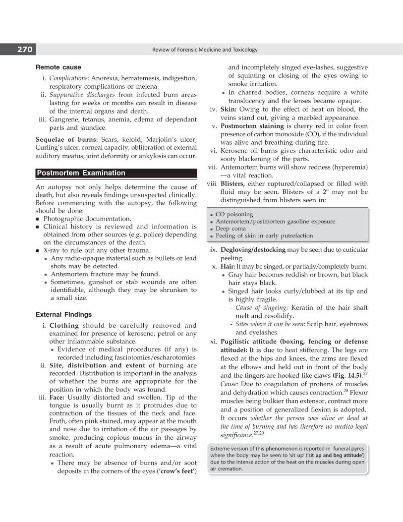
270 Review of Forensic Medicine and Toxicology
and incompletely singed eye-lashes, suggestive of squinting or closing of the eyes owing to smoke irritation.
z In charred bodies, corneas acquire a white translucency and the lenses became opaque.
iv. Skin: Owing to the effect of heat on blood, the veins stand out, giving a marbled appearance.
v. Postmortem staining is cherry red in color from presence of carbon monoxide (CO), if the individual was alive and breathing during fire.
vi. Kerosene oil burns gives characteristic odor and sooty blackening of the parts.
vii. Antemortem burns will show redness (hyperemia) —a vital reaction.
viii. Blisters, either ruptured/collapsed or filled with fluid may be seen. Blisters of a 2º may not be distinguished from blisters seen in:
� CO poisoning � Antemortem/postmortem gasoline exposure � Deep coma � Peeling of skin in early putrefaction
ix. Degloving/destocking may be seen due to cuticular peeling.
x. Hair: It may be singed, or partially/completely burnt. z Gray hair becomes reddish or brown, but black
hair stays black. z Singed hair looks curly/clubbed at its tip and
is highly fragile. - Cause of singeing: Keratin of the hair shaft
melt and resolidify. - Sites where it can be seen: Scalp hair, eyebrows
and eyelashes. xi. Pugilistic attitude (boxing, fencing or defense
attitude): It is due to heat stiffening. The legs are flexed at the hips and knees, the arms are flexed at the elbows and held out in front of the body and the fingers are hooked like claws (Fig. 14.5).27
Cause: Due to coagulation of proteins of muscles and dehydration which causes contraction.28 Flexor muscles being bulkier than extensor, contract more and a position of generalized flexion is adopted.
It occurs whether the person was alive or dead at the time of burning and has therefore no medico-legal significance.27,29
Extreme version of this phenomenon is reported in funeral pyres where the body may be seen to ‘sit up’ (‘sit up and beg attitude’) due to the intense action of the heat on the muscles during open air cremation.
Remote cause
i. Complications: Anorexia, hematemesis, indigestion, respiratory complications or melena.
ii. Suppurative discharges from infected burn areas lasting for weeks or months can result in disease of the internal organs and death.
iii. Gangrene, tetanus, anemia, edema of dependant parts and jaundice.
Sequelae of burns: Scars, keloid, Marjolin’s ulcer, Curling’s ulcer, corneal capacity, obliteration of external auditory meatus, joint deformity or ankylosis can occur.
Postmortem Examination
An autopsy not only helps determine the cause of death, but also reveals findings unsuspected clinically. Before commencing with the autopsy, the following should be done:
� Photographic documentation. � Clinical history is reviewed and information is
obtained from other sources (e.g. police) depending on the circumstances of the death.
� X-ray to rule out any other trauma. z Any radio-opaque material such as bullets or lead
shots may be detected. z Antemortem fracture may be found. z Sometimes, gunshot or stab wounds are often
identifiable, although they may be shrunken to a small size.
External findings
i. Clothing should be carefully removed and examined for presence of kerosene, petrol or any other inflammable substance.
z Evidence of medical procedures (if any) is recorded including fasciotomies/escharotomies.
ii. Site, distribution and extent of burning are recorded. Distribution is important in the analysis of whether the burns are appropriate for the position in which the body was found.
iii. Face: Usually distorted and swollen. Tip of the tongue is usually burnt as it protrudes due to contraction of the tissues of the neck and face. Froth, often pink stained, may appear at the mouth and nose due to irritation of the air passages by smoke, producing copious mucus in the airway as a result of acute pulmonary edema—a vital reaction.
z There may be absence of burns and/or soot deposits in the corners of the eyes (‘crow’s feet’)

271Thermal Injuries
xii. Heat ruptures: These are splits occurring in the skin due to contraction of the heated and coagulated tissue, and the resultant breaches may simulate incised or lacerated wounds (Diff. 14.2). It is usually seen over the area of severe burning, over fleshy areas, like calves and thighs, and over extensor surfaces and joints.
Heat ruptures may be distinguished from the effects of violence by:30,31
� Presence of nerves, blood vessels and connective tissue running across the split from side to side.
� There is no clotted blood in these fissures and no extravasation of blood in the surrounding tissues, since heat coagulates the blood in the vessels.
� Presence of irregular margins. � Absence of bruising or other signs of vital reaction in the margins.
Internal findings
i. Skull z Heat hematoma is an artifact and has the
appearance of extradural hematoma (Diff. 14.3).32
– It is large, thick (about 1.5 cm) and contains 100–120 ml of blood.
– Cause: The blood may come from the longitudinal venous sinuses or the diploic veins. The heat may force blood out of the marrow of the calvarium through veins and out over the surface of the dura.
z Skull bones may be fractured and burst open along the sutures due to intense heat.
ii. Brain: Congested, and appears swollen with widening and flattening of the gyri and obliteration of the sulci due to the contraction of the coagulating dura against the surface of the brain. Subdural hemorrhage may be present.
iii. Neck: Hemorrhage in the root of the tongue and neck muscles —considered vital reactions in burn victims.
iv. Larynx, trachea and bronchioles: Contain carbon and soot particles, and the mucosa is congested with frothy mucus secretions. This is the surest sign of antemortem burns, which is due to inhalation of gases.
z However, soot usually disappears by the 2nd day of hospitalization.
Fig. 14.5: Pugilistic attitude: Heat flexures of the limbs
Differentiation 14.2: Heat rupture and lacerated wound
S.No. Feature Heat rupture Lacerated wound
1. Cause Exposure to heat Blunt force
2. Site Fatty tissue Anywhere
3. Vessels and nerves Intact Torn
4. Bruising around the margins
Absent Present
Differentiation 14.3: Epidural hematoma (EDH) due to burns and blunt force
S.No. Feature EDH due to burns EDH due to blunt force
1. Cause Charring of the skull due to intense heat Blunt force to the head
2. Situation Anywhere Usually adjacent to Sylvian fissure
3. Position Usually bilateral Usually unilateral
4. Distribution Diffuse Localized
5. Characteristics Evenly distributed or sickle-shaped; honeycomb appearance; soft, granular, foamy, friable clot; chocolate brown in color (pink, if CO is present)33
Disc shaped; uniform, smooth, rubbery; reddish-purple color
6. Skull fracture Eggshell fracture—elliptical or circular defect without radiating fracture lines, seen above the temple
Fracture line radiating from a skull defect present in temporal area
7. Crossing of suture lines
It may cross suture lines and overlie the frontal, parietal and temporal area
Hematoma do not cross sutures as the dura is anchored at the suture lines
8. Injury to CNS Absent May be present
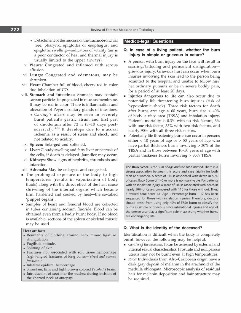
272 Review of Forensic Medicine and Toxicology
z Detachment of the mucosa of the tracheobronchial tree, pharynx, epiglottis or esophagus; and epiglottic swelling—indicators of vitality (air is a poor conductor of heat and thermal injury is usually limited to the upper airways).
v. Pleura: Congested and inflamed with serous effusion.
vi. Lungs: Congested and edematous, may be shrunken.
vii. Heart: Chamber full of blood, cherry red in color due inhalation of CO.
viii. Stomach and intestines: Stomach may contain carbon particles impregnated in mucous membrane. It may be red in color. There is infla mmation and ulceration of Peyer’s solitary glands of intestines.
z Curling’s ulcers may be seen in severely burnt patient’s gastric atrum and first part of duodenum after 72 h (3–10 days post-survival).34-36 It develops due to mucosal ischemia as a result of stress and shock, and not related to acidity.
ix. Spleen: Enlarged and softened. x. Liver: Cloudy swelling and fatty liver or necrosis of
the cells, if death is delayed. Jaundice may occur. xi. Kidneys: Show signs of nephritis, thrombosis and
infarction. xii. Adrenals: May be enlarged and congested.
� The prolonged exposure of the body to high temperatures (results in vaporization of body fluids) along with the direct effect of the heat cause shriveling of the internal organs which became firm, hardened and cooked by heat—the so-called ‘puppet organs’.
� Samples of heart and femoral blood are collected in tubes containing sodium fluoride. Blood can be obtained even from a badly burnt body. If no blood is available, sections of the spleen or skeletal muscle may be used.
Heat artifacts � Remnants of clothing around neck mimic ligature strangulation.
� Pugilistic attitude. � Splitting of skin. � Fractures not associated with soft tissue hemorrhage (right-angled fractures of long bones—‘street and avenue fractures’).
� Bilateral epidural hemorrhage. � Shrunken, firm and light brown colored (‘cooked’) brain. � Introduction of soot into the trachea during incision of the charred neck at autopsy.
Medico-legal Questions
Q. In case of a living patient, whether the burn injury is simple or grievous in nature?
� A person with burn injury on the face will result in scarring/tattooing and permanent disfiguration—grievous injury. Grievous hurt can occur when burn injuries involving the skin lead to the person being admitted to the hospital and unable to follow his/her ordinary pursuits or be in severe bodily pain, for a period of at least 20 days.
� Injuries dangerous to life can also occur due to potentially life threatening burn injuries (risk of hypovolemic shock). Three risk factors for death after burns are: age > 60 years, burn size > 40% of body-surface area (TBSA) and inhalation injury. Patient’s mortality is 0.3% with no risk factors, 3% with one risk factor, 33% with two risk factors, and nearly 90% with all three risk factors.
� Potentially life threatening burns can occur in persons either < 10 years of age or > 50 years of age who have partial thickness burns involving > 30% of the TBSA and in those between 10–50 years of age with partial thickness burns involving > 35% TBSA.
The Baux Score is the sum of age and the TBSA burned. There is a strong association between this score and case fatality for both men and women. A score of 110 is associated with death in 50% of cases. Baux Score of 140 or more is non-survivable. For patients with an inhalation injury, a score of 100 is associated with death in nearly 50% of cases, compared with 110 for those without. Thus, a revised Baux Score, i.e. Age + Percentage burn + 17 has been suggested for those with inhalation injuries. Therefore, doctors should desist from using only 40% of TBSA burnt to classify the burns as simple or grievous, since inhalational injuries and age of the person also play a significant role in assessing whether burns are endangering life.
Q. What is the identity of the deceased?
Identification is difficult when the body is completely burnt, however the following may be helpful:
� Gender of the deceased: It can be assessed by external and internal sexual characteristics. Prostrate and nulliparous uterus may not be burnt even at high temperatures.
� Race: Individuals from Afro-Caribbean origin have a dark gray deposit of melanin in the arachnoid of the medulla oblongata. Microscopic analysis of residual hair for melanin deposition and hair structure may be required.
273Thermal Injuries
� Age: It is usually established by teeth and ossification of bones.
� Dental identification: Dental charts should be prepared and X-rays of the jaws obtained, which can be compared with the dental X-rays and charts of the individual who is believed to be deceased.
� Clothing* (it is retained in body folds where the fire has not reached) and personal effects like watches, spectacles, dentures, hearing aid, jewelry and keys, and nonspecific characters like scars, tattoos (may show up well, despite the loss of the epidermis) or absence of organs can help in identification. A clenched hand resulting from heat contracture preserves fingerprints.
� X-ray examination of a charred body (e.g. evidence of prior surgery, old fracture) can assist in identification by comparison of postmortem X-rays with antemortem X-rays of the individual the deceased is suspected of being.
� If conventional comparison methods are not possible, teeth or bone can be used for DNA analysis.
Q. When did the victim sustain the burn injury?
The question arises as to when the burns were caused and whether all the burns were caused simultaneously. Features which help in estimating the age of burns is given in Table 14.2.
Q. Whether the burns are antemortem or post -mortem?
Refer to Diff. 14.4.
Q. Whether the burns are the cause of death?
� Presence of carbonaceous or soot particles in the respiratory tract.
� Cherry red discoloration of blood due to CO confirm burns as cause of death.
If the hemoglobin saturation is >10% CO, then the person was alive and inhaled the air during the fire. If death occurs due to the toxicity of CO, the blood carboxyhemoglobin (COHb) saturation is in the range of 50–80%. The COHb saturation level will not be artificially elevated in a dead person by being in or near a fire, i.e. CO will not diffuse through the skin or otherwise be absorbed by a dead body.
Q. Whether the burns are suicidal / accidental /homicidal / self-inflicted?
� Suicidal burns are common among Indian women. They pour kerosene on their heads and clothes before setting fire to themselves. Some women stuff clothes inside the mouth to prevent their shouts from being heard by others. Classic religious examples were seen in certain Buddhist sects or the rite of ‘sati’ performed in some parts of India (now prohibited).
� Accidental burns are common among children and elderly people. Accidental kerosene stove bursting is also reported. Accidents may result from smoking in bed, especially under the influence of alcohol or drugs, using faulty equipments and playing with fire.
� Homicidal burns are quite common in India. Custom of dowry leads to young brides being murdered by pouring kerosene on them and setting them on fire
Table 14.2: Age of burns
Features Age
Redness Immediate
Vesication 1–2 h
Exudates begins to dry 12–24 h
Dry brown crust formation and pus formation 48–72 h
Superficial slough separates 4–6th day
Deep slough separates 15th day
Granulation tissue begins to cover >15 days
Formation of cicatrix and deformity Several weeks
Differentiation 14.4: Antemortem and postmortem burnsS.No. Feature Antemortem burns Postmortem burns
1. Line of redness Present Absent2. Vesicles Contain serous fluid, rich in albumin, chloride
and some polymorphsContain air; if fluid is present, it contain little albumin and no chloride
3. Base of vesicles Red and inflamed Dull, dry, hard and yellow4. Soot in upper respiratory tract May be present Absent5. Inflammation and repair Present along with pus and slough Absent6. Healing Granulation tissue seen in old cases Absent7. Carboxyhemoglobin Present Absent8. Enzyme reaction Increase in enzymes in the periphery of burns No such increase
* It helps to determine the race of the individual from underlying intact skin and also useful for accelerant analysis.

274 Review of Forensic Medicine and Toxicology
by the husband and in-laws, and later claimed to be accidental burns.
z Sometimes, a homicide victim may be burned to conceal murder by other means in an attempt to cover up or destroy the evidence.
� Self-inflicted burns for false accusation: These burns are usually seen on accessible parts of body.
� Necklacing is a method of homicidal burning which involves placing a vehicle tyre around the neck of the victim and setting it alight. It was followed in South African black townships during the apartheid period as a form of punishment for political opponents.
� Arson is the willful and malicious burning of the dwelling of another or burning of one’s own property for an improper purpose, e.g. to collect insurance. The presence of several points of ignition and liquid fire accelerants, such as petrol or paraffin provides strong evidence of fire has been ignited deliberately.
Scalds
Definition: A scald is a form of thermal injury which results from application of liquid > 60ºC or from steam, and involves only the superficial layers of skin.
TypesIt is of three types: i. Immersion burns: Accidental or deliberate
immersion in hot liquid, usually water. ii. Splash or spill burns: Usually accidental. iii. Steam burns: Exposure to superheated steam.
� Hot water accounts for most of the immersion or splash burns.
� Scalds show sharp demarcation with tickle marks, soddening and bleaching, but do not singe the hair or blacken/char the skin.
� With inhalation, there is laryngeal, tracheal and respiratory burns that may progress to adult respiratory distress syndrome.
Clinically, it is classified into three degrees: i. Erythema or reddening by vasoparalysis. ii. Vesication or blister formation due to increased
permeability of the capillaries. iii. Necrosis of the dermis when deeper layer of skin
is involved.
Medico-legal Aspects � It is usually accidental due to splashing or pouring
of fluid during cooking. � Accidents are common in children and in the elderly. � Boiling water may be thrown intentionally, usually
domestic homicide intent with the husband being the victim.
� Deliberate scalding by hot water is common form of child abuse. Dipping injuries of the limbs appear as well-demarcated ‘glove and stocking’ distribution of scalds reflecting the flow of hot liquid under the influence of gravity. Areas of scalding round the buttocks with clear, unaffected areas on the upper thighs occur when the child is forcibly made to sit in a hot liquid (Fig. 14.6).
Important differentiating features of dry and moist heat and chemical burns are given in Diff. 14.5.
Fig. 14.6: Scalding (child abuse)
Differentiation 14.5: Dry heat, moist heat and chemical burns
S.No. Feature Dry heat burn Moist heat burn Chemical burn
1. Cause Flame, heated body or X-rays Solid steam or liquid > 60ºC Corrosives
2. Site At or above the site of contact At and below the site of contact At or below the site of contact
3. Splashing Absent Present Present
4. Skin Dry, wrinkled and may be charred Sodden, bleached Corroded and devitalized
5. Vesicles At the circumference of burnt area Over the burnt area Usually not present
6. Red line Present Present Absent
7. Color Black Bleached Distinctive coloration
8. Charring Present Absent Absent
9. Singeing Present Absent Absent
10. Ulceration Absent Absent Present
11. Scar Thick, contracted Thin, less contracted Thick, contracted
12. Clothes Burnt Wet, not burnt May be burnt, with characteristic stains
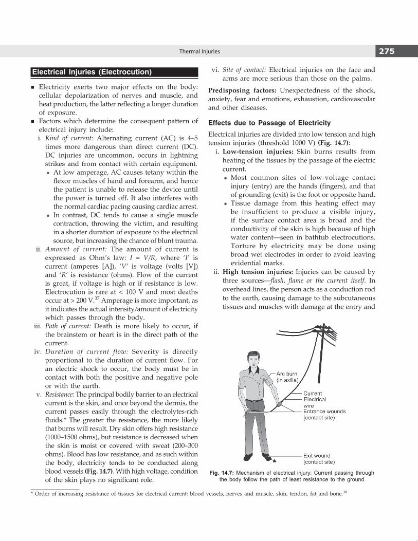
275Thermal Injuries
Electrical Injuries (Electrocution)
� Electricity exerts two major effects on the body: cellular depolarization of nerves and muscle, and heat production, the latter reflecting a longer duration of exposure.
� Factors which determine the consequent pattern of electrical injury include:
i. Kind of current: Alternating current (AC) is 4–5 times more dangerous than direct current (DC).
DC injuries are uncommon, occurs in lightning strikes and from contact with certain equipment.
z At low amperage, AC causes tetany within the flexor muscles of hand and forearm, and hence the patient is unable to release the device until the power is turned off. It also interferes with the normal cardiac pacing causing cardiac arrest.
z In contrast, DC tends to cause a single muscle contraction, throwing the victim, and resulting in a shorter duration of exposure to the electrical source, but increasing the chance of blunt trauma.
ii. Amount of current: The amount of current is expressed as Ohm’s law: I = V/R, where ‘I’ is current (amperes [A]), ‘V’ is voltage (volts [V]) and ‘R’ is resistance (ohms). Flow of the current is great, if voltage is high or if resistance is low. Electrocution is rare at < 100 V and most deaths occur at > 200 V.37 Amperage is more important, as it indicates the actual intensity/amount of electricity which passes through the body.
iii. Path of current: Death is more likely to occur, if the brainstem or heart is in the direct path of the current.
iv. Duration of current flow: Severity is directly proportional to the duration of current flow. For an electric shock to occur, the body must be in contact with both the positive and negative pole or with the earth.
v. Resistance: The principal bodily barrier to an electrical current is the skin, and once beyond the dermis, the current passes easily through the electrolytes-rich fluids.* The greater the resistance, the more likely that burns will result. Dry skin offers high resistance (1000–1500 ohms), but resistance is decreased when the skin is moist or covered with sweat (200–300 ohms). Blood has low resistance, and as such within the body, electricity tends to be conducted along blood vessels (Fig. 14.7). With high voltage, condition of the skin plays no significant role.
vi. Site of contact: Electrical injuries on the face and arms are more serious than those on the palms.
Predisposing factors: Unexpectedness of the shock, anxiety, fear and emotions, exhaustion, cardiovascular and other diseases.
Effects due to Passage of Electricity
Electrical injuries are divided into low tension and high tension injuries (threshold 1000 V) (Fig. 14.7): i. Low-tension injuries: Skin burns results from
heating of the tissues by the passage of the electric current.
z Most common sites of low-voltage contact injury (entry) are the hands (fingers), and that of grounding (exit) is the foot or opposite hand.
z Tissue damage from this heating effect may be insufficient to produce a visible injury, if the surface contact area is broad and the conductivity of the skin is high because of high water content—seen in bathtub electrocutions. Torture by electricity may be done using broad wet electrodes in order to avoid leaving evidential marks.
ii. High tension injuries: Injuries can be caused by three sources—flash, flame or the current itself. In overhead lines, the person acts as a conduction rod to the earth, causing damage to the subcutaneous tissues and muscles with damage at the entry and
* Order of increasing resistance of tissues for electrical current: blood vessels, nerves and muscle, skin, tendon, fat and bone.38
Fig. 14.7: Mechanism of electrical injury: Current passing through the body follow the path of least resistance to the ground
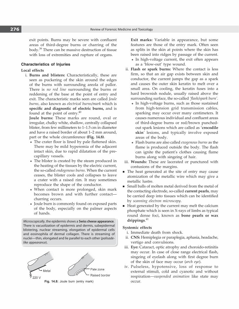
276 Review of Forensic Medicine and Toxicology
Fig. 14.8: Joule burn (entry mark)
exit points. Burns may be severe with confluent areas of third-degree burns or charring of the body.39 There can be massive destruction of tissue with loss of extremities and rupture of organs.
Characteristics of InjuriesLocal effects i. Burns and blisters: Characteristically, these are
seen as puckering of the skin around the edges of the burns with surrounding areola of pallor. There is no red line surrounding the burns or reddening of the base at the point of entry and exit. The characteristic marks seen are called Joule burns, also known as electrical burns/mark which is specific and diagnostic of electric burns, and is found at the point of entry.40
Joule burns: These marks are round, oval or irregular, chalky white, shallow, centrally collapsed blister, from few millimeters to 1–1.5 cm in diameter and have a raised border of about 1–2 mm around, part or the whole circumference (Fig. 14.8).
z The crater floor is lined by pale flattened skin. There may be mild hyperemia of the adjacent intact skin, due to rapid dilatation of the pre-capillary vessels.
z The blister is created by the steam produced in the heating of the tissues by the electric current, the so-called endogenous burns. When the current ceases, the blister cools and collapses to leave a crater with a raised rim. It may sometimes reproduce the shape of the conductor.
z When contact is more prolonged, skin mark becomes brown and with further contact—charring occurs.
z Joule burn is commonly found on exposed parts of the body, especially on the palmer aspects of hands.
Microscopically, the epidermis shows a Swiss cheese appearance. There is vacuolization of epidermis and dermis, subepidermal blistering, nuclear streaming, elongation of epidermal cells and eosinophila of dermal collagen. There is streaming of nuclei—thin, elongated and lie parallel to each other (palisade-like appearance).
Exit marks: Variable in appearance, but some features are those of the entry mark. Often seen as splits in the skin at points where the skin has been raised into ridges by passage of the current.
z In high-voltage current, the exit often appears as a ‘blow-out’ type wound.
ii. Flash or spark burns: Where the contact is less firm, so that an air gap exists between skin and conductor, the current jumps the gap as a spark and causes the outer skin keratin to melt over a small area. On cooling, the keratin fuses into a hard brownish nodule, usually raised above the surrounding surface, the so-called ‘flash/spark burn’.
z In high-voltage burns, such as those sustained from high-tension grid transmission cables, sparking may occur over many centimeters. It causes numerous individual and confluent areas of third-degree burns or red/brown punched-out spark lesions which are called as ‘crocodile skin’ lesions, and typically involve exposed areas of the body.
z Flash burns are also called exogenous burns as the flame is produced outside the body. The flash can ignite the patient’s clothes causing flame burns along with singeing of hair.
iii. Wounds: These are lacerated or punctured with contusions of the margins.
� The heat generated at the site of entry may cause atomization of the metallic wire which may give a metallic lustre.
� Small balls of molten metal derived from the metal of the contacting electrode, so-called current pearls, may be carried deep into tissues which can be identified by scanning electron microscopy.
� Heat generated by the current may melt the calcium phosphate which is seen in X-rays of limbs as typical round dense foci, known as bone pearls or wax drippings.41
Systemic effects i. Immediate death from shock. ii. CNS: Hemiplegia or paraplegia, aphasia, headache,
vertigo and convulsions. iii. Eye: Cataract, optic atrophy and choroido-retinitis
may occur. In case of close range electrical flash, singeing of eyelash along with first degree burn of the skin of face may occur (arch eye).
iv. Pulseless, hypotensive, loss of response to external stimuli, cold and cyanotic and without respiration —suspended animation like state may occur.
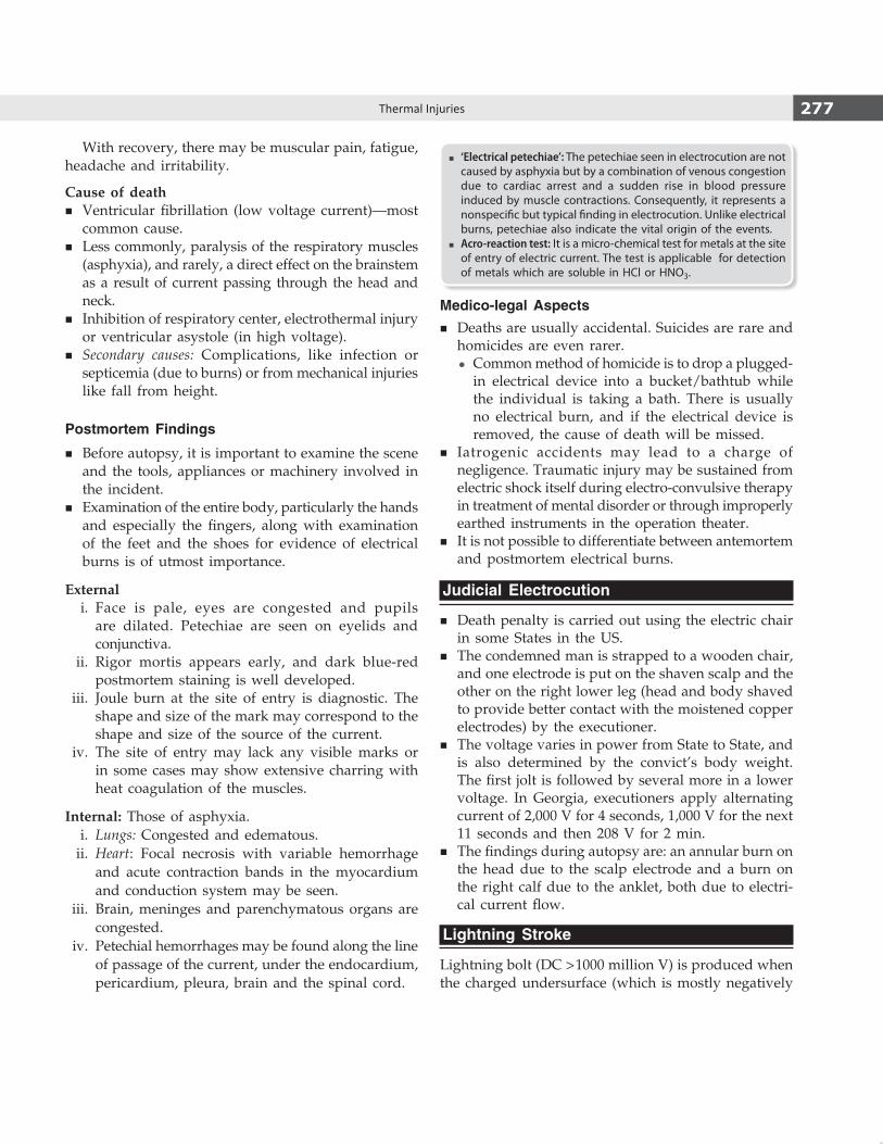
277Thermal Injuries
With recovery, there may be muscular pain, fatigue, headache and irritability.
Cause of death � Ventricular fibrillation (low voltage current)—most
common cause. � Less commonly, paralysis of the respiratory muscles
(asphyxia), and rarely, a direct effect on the brainstem as a result of current passing through the head and neck.
� Inhibition of respiratory center, electrothermal injury or ventricular asystole (in high voltage).
� Secondary causes: Complications, like infection or septicemia (due to burns) or from mechanical injuries like fall from height.
Postmortem findings
� Before autopsy, it is important to examine the scene and the tools, appliances or machinery involved in the incident.
� Examination of the entire body, particularly the hands and especially the fingers, along with examination of the feet and the shoes for evidence of electrical burns is of utmost importance.
External i. Face is pale, eyes are congested and pupils
are dilated. Petechiae are seen on eyelids and conjunctiva.
ii. Rigor mortis appears early, and dark blue-red postmortem staining is well developed.
iii. Joule burn at the site of entry is diagnostic. The shape and size of the mark may correspond to the shape and size of the source of the current.
iv. The site of entry may lack any visible marks or in some cases may show extensive charring with heat coagulation of the muscles.
Internal: Those of asphyxia. i. Lungs: Congested and edematous. ii. Heart: Focal necrosis with variable hemorrhage
and acute contraction bands in the myocardium and conduction system may be seen.
iii. Brain, meninges and parenchymatous organs are congested.
iv. Petechial hemorrhages may be found along the line of passage of the current, under the endo cardium, pericardium, pleura, brain and the spinal cord.
� ‘Electrical petechiae’: The petechiae seen in electrocution are not caused by asphyxia but by a combination of venous congestion due to cardiac arrest and a sudden rise in blood pressure induced by muscle contractions. Consequently, it represents a nonspecific but typical finding in electrocution. Unlike electrical burns, petechiae also indicate the vital origin of the events.
� Acro-reaction test: It is a micro-chemical test for metals at the site of entry of electric current. The test is applicable for detection of metals which are soluble in HCl or HNO3.
Medico-legal Aspects � Deaths are usually accidental. Suicides are rare and
homicides are even rarer. z Common method of homicide is to drop a plugged-
in electrical device into a bucket/bathtub while the individual is taking a bath. There is usually no electrical burn, and if the electrical device is removed, the cause of death will be missed.
� Iatrogenic accidents may lead to a charge of negligence. Traumatic injury may be sustained from electric shock itself during electro-convulsive therapy in treatment of mental disorder or through improperly earthed instruments in the operation theater.
� It is not possible to differentiate between antemortem and postmortem electrical burns.
Judicial Electrocution
� Death penalty is carried out using the electric chair in some States in the US.
� The condemned man is strapped to a wooden chair, and one electrode is put on the shaven scalp and the other on the right lower leg (head and body shaved to provide better contact with the moistened copper electrodes) by the executioner.
� The voltage varies in power from State to State, and is also determined by the convict’s body weight. The first jolt is followed by several more in a lower voltage. In Georgia, executioners apply alternating current of 2,000 V for 4 seconds, 1,000 V for the next 11 seconds and then 208 V for 2 min.
� The findings during autopsy are: an annular burn on the head due to the scalp electrode and a burn on the right calf due to the anklet, both due to electri-cal current flow.
Lightning Stroke
Lightning bolt (DC >1000 million V) is produced when the charged undersurface (which is mostly negatively

278 Review of Forensic Medicine and Toxicology
Fig. 14.9: Lichtenberg flowers/Filigree burns
charged) of a thundercloud sends its electrical charge to the ground. It may injure or kill an individual by direct strike, a side flash or conduction through another object. Death is caused by high-voltage direct current due to cardiopulmonary arrest or electrothermal injuries.
Lichtenberg flowers/Arborescent Markings
� Lichtenberg flowers are pathognomonic of lightning strike, but are rare.
� These are superficial, several inches long, thin, irregular, tortuous, dendritic red marks on the skin. These marks have a resemblance to the branches of a tree.42,43
� This fern-like pattern of erythema in the skin is usually found over the shoulders or flanks (Fig. 14.9).
� It is not associated with burning. � They indicate the path taken by the discharge and tend
to follow skin creases and the long axis of the body. It appears within 1 h, and disappears in 24–48 h, if the person survive.
Cause: The exact mechanism has not yet been deter-mined. There are various theories:
� Static electricity discharges along superficial vascu-lature (or perhaps nerves).
� Hemoglobin staining the tissues in the pattern of a tree due to break down of RBCs within the capillaries of the skin.
� Electron showers eliciting an inflammatory response in the skin.
� Current following lines of perspiration and skin moisture.
� Minute deposits of copper in the skin.
Medico-legal aspects � Its recognition may be lifesaving in the unaccom-
panied comatose patient, and is important because even delayed resuscitation of lightning victims can be very successful.
� Death is accidental. � It can help differentiate a natural death from murder.
� Appearance is pathognomonic for injury by lightning, but may closely resemble those produced by criminal violence.
Other features of lightning
� Development of edema of skin at the point of entry wound in those who survive due to paralysis of capillary and lymphatic vessels.
� As the exit is often in the feet, shoe may be ripped apart or blown off the foot. Articles of clothing may be found some distance from the body with the body partly stripped which may be suggest sexual assault, particularly when the body is found in the open.
� Linear burns and surface burns. � Fusing and magnetization of metallic articles, such as
rings, spectacle frames, pen-knives, keys and watches due to tremendous heat liberated by the electrical discharge. These are useful signs for eliminating suspicion of foul play.
� Injuries like contusions, lacerations, rupture of tympanic membrane and organs, and spinal cord damage.
� Additional findings—singed hair and patterned skin burns marks underneath metal article of jewelry.
Lichtenberg flowers: It is known by different names like ‘arborization’, ‘feathering’, ‘ferning’, ‘filigree burns’ or ‘keraunographic markings’.44 The phenomenon is also known as ‘keraunographism’ from Greek keraunos, a thunder bolt.

279Thermal Injuries
1. D 2. B 3. D 4. B 5. C 6. B 7. C 8. C 9. A 10. D 11. B 12. D 13. B 14. A 15. B 16. B 17. B 18. B 19. A 20. A 21. C 22. B 23. A
multiple ChoiCe questions
1. Depression of consciousness level in hypothermia starts when the core body temperature falls below:
COMEDK 14 A. 35°C B. 34°C C. 33°C D. 32°C 2. In hypothermia, cause of death is: NEET 14 A. Stroke B. Cardiac arrest C. Pulmonary embolism D. Asphyxia 3. Color of postmortem lividity in hypothermic deaths: NEET 13 A. Purple B. Deep red C. Cherry red D. Bright pink 4. Paradoxical undressing is seen in:
MAHE 06, 10; CMC (Vellore) 10 A. Hyperthermia B. Hypothermia C. Transvestism D. Immersion syndrome 5. True about frostbite are all, except: PGI 14 A. Occurs due to extreme cold B. Formation of ice crystals within tissues C. Rewarming is not done D. Seen in fingers and toes 6. Miner’s cramps are also called: NEET 13 A. Heat collapse B. Heat cramps C. Sunstroke D. Heat exhaustion 7. Heatstroke is defined as a body temperature higher
than: AFMC 12 A. 37°C B. 40°C C. 41°C D. 45°C 8. Sweating is absent in: DNB 10 A. Heat syncope B. Heat exhaustion C. Heat stroke D. Heat cramps 9. NOT seen in heat stroke: Kerala 06 A. Hypovolemic shock B. Rhabdomyolysis C. Pancreatitis D. Cerebral edema 10. Characteristic features of superficial burns are all,
except: TN 06; WB 09 A. Damage no deeper than papillary dermis B. Blisters absent C. Loss of epidermis D. Pinprick is not painful 11. Blister formation in burn is classified as:
DNB 09; Bihar 11 A. First degree B. Second degree superficial C. Second degree deep D. Third degree 12. A lady with burns, skin appears waxy and dry with
little pain, the degree of burn is: CMC (Ludhiana) 11 A. First degree B. Second degree superficial C. Second degree deep D. Third degree
13. Rule of 9 in burns is used to denote: NEET 14 A. Depth of burns B. Total body surface area involved C. Severity of burns D. Type of burns 14. According to ‘rule of 9’, perineum burns constitute:
FMGE 09 A. 1% burns B. 9% burns C. 18% burns D. 27% burns 15. Burns in children assessed by: AIIMS 13 A. Rule of nine B. Lund and Browder C. Palmer surface method D. Hasse’s rule 16. Head and neck burns in infant constitute _______%
of burns: Kerala 08 A. 9 B. 18 C. 24 D. 36 17. A 2-year-old child had burns on buttocks, both legs,
face, neck and singeing of hair. Total surface area burnt: JIPMER 14
A. 27% B. 37% C. 45% D. 55% 18. In a 6-year-old child with burns involving whole
of head and trunk, the estimated body surface area involved: JIPMER 09
A. 44% B. 48% C. 55% D. 58% 19. Percentage of surface area of palm of a burn patient:
FMGE 10; AI 11 A. 1% B. 9% C. 18% D. 27% 20. Most important aspect of management of burn injury
in the first 24 h: UPSC 07 A. Fluid resuscitation B. Dressing C. Escharotomy D. Antibiotics 21. In adults, circulatory collapse occurs after a minimum
of what percentage burns of total body surface area:NIMHANS 08; TN 11
A. 5% B. 10% C. 15% D. 20% 22. Best fluid for resuscitation in burns:
FMGE 11; Punjab 12 A. Dextran B. Ringer lactate C. Albumin D. Hartmann’s solution 23. Parkland formula for burns is for: Maharashtra 09; UP 09; Bihar 12 A. Ringer lactate B. Glucose saline C. Normal saline D. 25% dextrose

280 Review of Forensic Medicine and Toxicology
24. A 65 years male weighing 50 kg was admitted with 80% burn and RL was transfused with Parkland method. How much fluid should be infused in first 8 h: UP 11
A. 200 ml/h B. 500 ml/h C. 1000 ml/h D. 8000 ml/h 25. Muirs and Barclays formula is for: AFMC 11 A. Colloid resuscitation in burns B. Polytrauma fluid resuscitation C. Crystalloid in trauma D. Dextran in burns 26. Not an indication for admission in burns ward:
Bihar 10; Maharashtra 11 A. Acid burns B. Inhalational injury C. 5% partial thickness burns in an unmarried female D. 10% deep burns in a male 27. True of puglistic attitude: PGI 14 A. Flexion at hip and knee B. Seen in extensive burns C. Seen in rigor mortis D. Seen in antemortem burns only 28. Pugilistic attitude is due to: Delhi 07; FMGE 10 A. Lipolysis B. Protein coagulation C. Carbohydrate coagulation D. Lipogenesis 29. True about pugilistic attitude: MP 09; PGI 10 A. Indicate antemortem burns B. Indicate postmortem burns C. Cannot differentiate between ante- or postmortem D. Indicate defense by victim 30. Difference between incised wound and heat rupture
favoring heat rupture are: AI 09 A. Margins well-defined B. Small and multiple wounds C. Nerves and vessels are visible in floor D. Seen only over scalp 31. Feature of ruptured skin caused by excessive heat:
AIIMS 10 A. Irregular margin B. Clear regular margin C. Contused margin D. Abraded margin 32. True about heat hematoma: PGI 12 A. Inside the brain B. Between skull and dura mater C. Between skull and pericranium D. Between scalp and pericranium
24. C 25. A 26. C 27. A & B 28. B 29. C 30. C 31. A 32. B 33. A 34. B 35. D 36. A 37. A 38. A 39. B 40. B 41. D 42. B 43. B 44. A
33. Appearance of burn hematoma: DNB 09; Punjab 10 A. Honeycomb like B. Disc shaped C. Stellate shaped D. Smooth and rubbery 34. Curling ulcer is seen in: UP 04; WB 10; FMGE 13 A. Head injury B. Burn C. Corticosteroids D. TPN 35. Curling’s ulcer in burns is seen in: AIIMS 08 A. Esophagus B. Stomach C. Colon D. Duodenum 36. Curling’s ulcer common in which part of duodenum:
DNB 09; AIIMS 13 A. 1st part B. 2nd part C. 3rd part D. Between 1st and 2nd part 37. Electrocution is rare below: MP 09 A. 100 volt B. 150 V C. 200 V D. 240 V 38. Most resistant organ to electric current: UP 12 A. Bone B. Skin C. Blood vessel D. Muscle 39. High voltage contact burn is: UPSC 14 A. Deep partial thickness B. Full thickness C. Superficial burn D. All of the above 40. Joule burn is seen in: Bihar 11 A. Blast injuries B. Electrocution C. Firearm wounds D. Lightning stroke 41. Bone pearl’s or wax drippings is pathogonomic of:
Odisha 11 A. Burns B. Scalds C. Lightening D. Electrocution 42. A dead body is found to have marks like branching
of a tree on front of the chest. Most likely cause of death could be: FMGE 10
A. Firearm injury B. Lightning injury C. Bomb blast injury D. Road traffic accident 43. Lichtenberg flowers are seen in: CMC (Vellore) 14 A. Thermal burns B. Electrocution C. Vitriolage D. Lightning 44. Filigree burn occur in: FMGE 09; NJI 10; JIPMER 11 A. Lightning B. Electrocution C. Vitriolage D. Infanticide

Transportation Injuries
ChapTer 15
Definitions � Transportation injuries are blunt force injuries that
occur from travel on the ground, in the air and on water. The most frequent of these are motor vehicle collision and pedestrian injuries. Less common cases are associated with railway accidents and aircraft crashes.
� Motor vehicle collision or road traffic accident occurs when a vehicle collides with another vehicle, pedestrian, animal, road debris or other stationary barrier, such as a tree or utility pole.
� Hit-and-run: Failure to stop at scene of accident by the driver of a motor vehicle without giving assistance or informing the police.
� Those injured by accidents can be divided into three broad groups: pedestrians, cyclists (pedal or motor) and the drivers and passengers of vehicles.
� Pedestrians (most common), cyclists, children and the elderly are among the most vulnerable of road users.
Pedestrian Injuries
Three patterns of injuries are seen (Fig. 15.1): i. Primary impact injuries ii. Secondary impact injuries iii. Secondary injuries
Fig. 15.1: Dynamics of pedestrian injuries and sites of primary impact, secondary impact and secondary injuries

282 Review of Forensic Medicine and Toxicology
Primary Impact Injuries � Primary impact injuries indicate that part of the body
which has been struck first by the vehicle, and often form recognizable patterns.
� Injuries include abrasions, contusion (sometimes patterned) and lacerations on legs, thighs or buttocks, along with fractures of the tibia and fibula, and rarely, of the femur and pelvis.
� When an adult is hit by the front of a car, the front bumper or radiator usually strikes the victim at about knee level.1 The exact point of contact, whether on the front, side or back of the leg(s), will depend on the orientation of the victim. It also depends on the type of car, possible lowering of the car’s front end (from braking), and pedestrian height.
� They help to establish the position of the victim at the material moment when struck, and help towards identification of the offending vehicle.
Behavior of the body and disposition of injuries will be modified by factors like:
� Whether both the feet were firmly placed on the ground or one of them was raised at the time of impact.
� Speed of the vehicle: At low speeds (e.g. 20 kph), the victim is usually thrown off the bonnet either forwards or to one side. Between 20 and 60 kph, the victim may be tipped onto the bonnet, and the head may strike the windscreen or the metal frame that surrounds it. At higher speeds (60–100 kph), the victim may be projected into the air (‘scooped-up’); sometimes pass completely over the vehicle and avoid hitting the windscreen and other points on the vehicle.
� Nature of road surface: Smooth, rough, full of gravel or mud and its skidding resistance.
� Point of impact in relation to the center of gravity.
When the pedestrian is knocked down from behind with both feet fixed to the ground: There will be fracture of the bones of the lower limbs, the buttocks and back of the pedestrian on being hit by head lamps or the radiator of the car. It may result in fracture dislocation of the lumber or thoracic spine, and this injury may drive the femoral head through the acetabulum.
� Stretch-type lacerations are frequent in the inguinal (groin) regions.
� Where the vehicle is relatively larger than the victim–adults impacted by a truck or a bus and children impacted by cars—the point of contact is higher up
the victim, and it is likely that the victim will make contact with more of the front of the vehicle. This pattern of contact may be result in primary injuries to the pelvis, abdomen, chest and head. Usually, the victim is projected along the line of travel of the vehicle, which may increase the risk of ‘run-over’ injuries.
Waddell’s triad is a classic pattern of injury seen in pedestrian children who are struck by motor vehicles. It comprises of fractured femoral shaft, intra-thoracic or intra-abdominal injuries and contralateral head injury (Fig. 15.2). Mechanism of injury is an initial impact causing injury to the pelvis and femur (bumper injury) instead of the knees and tibias; followed by the chest and abdomen (grill, fender or hood). Then the child is thrown on the ground and sustaining injury to the opposite side of the head.
On being struck from behind and feet not firmly on the ground: The victim’s feet will fall backward and may be propelled upwards and backwards, so that the head may sustain secondary impact injury by striking against the windscreen. The victim can also be ‘scooped-up’ or fall to one side and may sustain head injuries by striking the ground on falling.
If the victim is struck from front, he may sustain injuries to the chest and abdomen with fracture of ribs or vertebrae. Victim can also sustain fracture of pelvis or fracture dislocation of sacroiliac joint from the impact of a mudguard, and fracture of tibia and fibula of one or both legs can be sustained from impact by a bumper.
� Bumper impacts usually cause soft tissue damage and comminuted wedge shaped fractures (bumper fracture) of the tibia and fibula with forward displacement of the bony fragments.2 Base of the triangular fractured fragment will suggest the site of impact and its apex will point to the direction of the moving vehicle (Fig. 15.3). Bumper injuries at different levels in two legs or when absent on one leg, will suggest that the victim was walking or running while struck.
� Bumper fracture when present, the measurement of the distance from the heel to the fracture site will give an idea about the height of the bumper of the offending vehicle. When brakes are applied before the accident, the distance from heel to the fracture is less than the height of the bumper (presence or absence of braking may help to determine the driver’s intent).
� The lack of ‘bumper injuries’ and the presence of tyre marks could indicate the pedestrian was already prone or supine on the road when ‘run over’.
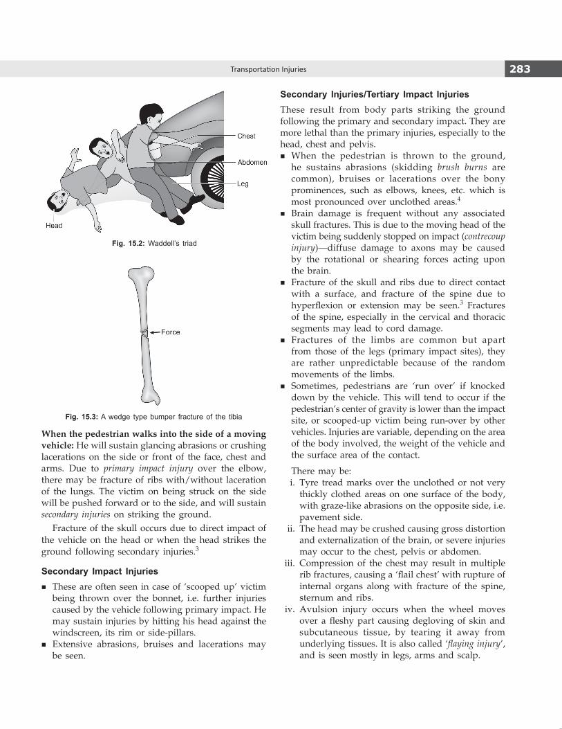
283Transportation Injuries
When the pedestrian walks into the side of a moving vehicle: He will sustain glancing abrasions or crushing lacerations on the side or front of the face, chest and arms. Due to primary impact injury over the elbow, there may be fracture of ribs with/without laceration of the lungs. The victim on being struck on the side will be pushed forward or to the side, and will sustain secondary injuries on striking the ground.
Fracture of the skull occurs due to direct impact of the vehicle on the head or when the head strikes the ground following secondary injuries.3
Secondary Impact Injuries � These are often seen in case of ‘scooped up’ victim
being thrown over the bonnet, i.e. further injuries caused by the vehicle following primary impact. He may sustain injuries by hitting his head against the windscreen, its rim or side-pillars.
� Extensive abrasions, bruises and lacerations may be seen.
Secondary Injuries/Tertiary Impact InjuriesThese result from body parts striking the ground following the primary and secondary impact. They are more lethal than the primary injuries, especially to the head, chest and pelvis.
� When the pedestrian is thrown to the ground, he sustains abrasions (skidding brush burns are common), bruises or lacerations over the bony prominences, such as elbows, knees, etc. which is most pronounced over unclothed areas.4
� Brain damage is frequent without any associated skull fractures. This is due to the moving head of the victim being suddenly stopped on impact (contrecoup injury)—diffuse damage to axons may be caused by the rotational or shearing forces acting upon the brain.
� Fracture of the skull and ribs due to direct contact with a surface, and fracture of the spine due to hyperflexion or extension may be seen.3 Fractures of the spine, especially in the cervical and thoracic segments may lead to cord damage.
� Fractures of the limbs are common but apart from those of the legs (primary impact sites), they are rather unpredictable because of the random movements of the limbs.
� Sometimes, pedestrians are ‘run over’ if knocked down by the vehicle. This will tend to occur if the pedestrian’s center of gravity is lower than the impact site, or scooped-up victim being run-over by other vehicles. Injuries are variable, depending on the area of the body involved, the weight of the vehicle and the surface area of the contact.
There may be: i. Tyre tread marks over the unclothed or not very
thickly clothed areas on one surface of the body, with graze-like abrasions on the opposite side, i.e. pavement side.
ii. The head may be crushed causing gross distortion and externalization of the brain, or severe injuries may occur to the chest, pelvis or abdomen.
iii. Compression of the chest may result in multiple rib fractures, causing a ‘flail chest’ with rupture of internal organs along with fracture of the spine, sternum and ribs.
iv. Avulsion injury occurs when the wheel moves over a fleshy part causing degloving of skin and subcutaneous tissue, by tearing it away from underlying tissues. It is also called ‘flaying injury’, and is seen mostly in legs, arms and scalp.
Fig. 15.2: Waddell’s triad
Fig. 15.3: A wedge type bumper fracture of the tibia

284 Review of Forensic Medicine and Toxicology
Table 15.1: Typical injuries in car vs. adult pedestrian collision
Phase Site of impact Injuries
Primary impact Bumper Lower extremity
Secondary impact Vehicle hood, pillar, windscreen Head and torso
Secondary Ground and fixed object Head and torso
Run-over Crushing, car or another vehicle Abdominal and thoracic
v. Burning and singeing of skin and hair resulting from discharge of hot exhaust.
� Usually, it is very difficult to classify the injuries as primary impact, secondary impact or secondary injuries.
� Typical injuries seen in car vs. adult pedestrian collision at moderate speed is given in Table 15.1.
� In pedestrian accidents, the common cause of death is head injuries and fracture dislocations of cervical spine, mainly at the atlanto-occipital joint. Injuries to the chest and abdomen are minimal or absent.
Injuries Sustained by Vehicle Occupants
� After pedestrians, the driver is the most frequent casualty in road traffic accidents as a high proportion of vehicles are occupied only by a driver. Next in frequency is the front seat passenger, followed by rear seat passengers.
� Ejection of both driver and passenger from a vehicle is associated with significantly severe injuries or fatality as the doors often burst open.
� Unbelted rear seat occupants are also at increased risk of serious injury in motor vehicle accidents; they may be ejected or thrown forward against the front seat.
� The driver and passenger injuries depend upon the type of impact crash.
Types of impact i. Front impact ii. Rear impact iii. Side impact iv. Roll-over
Front Impact CrashThis happens when one car strikes another car head-on or strikes a stationary object, like an electric pole/tree (approx. 80% of impacts). While the vehicle rapidly decelerates and stops, the occupants continue to move forward striking against the interior of the vehicle, unless they are restrained. If the head impacts against the windshield, the victim does not sustain severe cuts from the fragments of glass which used to happen
when it was made exclusively of glass. Windshields, nowadays, are made of a thin outer and inner layer of glass with thick plastic core.
The driver tends to receive a different pattern of injury as compared to either the front seat or rear seat passenger. The driver may receive a momentary warning of the impending collision and brace himself against the steering wheel. Fractures of the wrists and arms may thus occur, as well as fractures or dislocation of tibia, fibula and pelvis may occur from transmission of the force of impact from pressing on the brake and clutch pedals.
If the driver is unaware, his knees will impact against the dashboard, his chest against the steering wheel, and his head against the windshield. An impact of the knees against the dashboard commonly causes fractures of the tibia, fibula, femur and pelvis. Severe impact against the windshield pillar may cause avulsion of the skin of the forehead, basilar skull fractures, closed head injury and fracture or dislocation of the atlanto-occipital junction (Fig. 15.4A).
Steering wheel impact injury: The circular rim of the steering wheel may cause fractures of the jaws and facial bones, as well as imprint abrasions, minor bruises and contusions of the chest or bilateral rib fractures. Transverse fracture of sternum is usually seen at 3rd intercostal space. Damaged steering wheel spokes may penetrate the chest and lacerate the heart and lungs. Flail chest may occur.
With severe thoracic compression, partial or complete transection of aorta may occur usually at the junction of the aortic arch with descending aorta—classical injury. Lacerations of liver and spleen may be seen. Serious steering wheel injuries are less frequent, if the car is fitted with energy absorbing compressible steering wheel column.
Front seat passenger: The most dangerous place in the car is the front passenger seat. He may not get the momentary warning of the impending collision. Without a seat belt, he is at risk of severe impaction of his head against the windshield with its consequences

285Transportation Injuries
(Fig. 15.4B). The occupant may be ejected out of the vehicle through the windscreen, increasing the risks of secondary injuries or running over. There may be peculiar facial lacerations due to contact with the shattered windscreen known as ‘sparrow foot marks’ (similar to dicing injuries mentioned below).5 Contact with the dashboard may cause injuries to the knee (Fig. 15.4B).
Passengers of the rear seat often escape such injuries because of the absence of impact against the windshield and dashboard, and of the cushioning effect of the front seat. However, they may be injured against internal fittings, like door handles or ejected through burst-open doors.
Rear Impact CrashLow velocity rear impacts are relatively common. Usually, they cause whiplash injury. Neck fractures are rare. A high velocity rear impact crash can deform and rupture the gas tank with ignition of the fuel.
Side Impact CrashThe vehicle strikes on the side of another vehicle or skids sideways into a fixed object. This is a common pattern in an intersection, and is therefore a frequent occurrence in urban areas.
Injuries are often severe, because the side of a car has a thin metal wall door and no other components to absorb the force of impact. Since, the occupants of the vehicle move toward the side of impact, the persons sitting on that side run the greatest risk.
Dicing injuries may occur which are superficial cuts of the skin caused by fragments of tempered glass (designed to shatter into small glass cubes on violent impact). They are produced when the side and the back windows of a car shatter. They are linear, right angled or V-shaped laceration seen typically on the face, forehead and arm on the right side of the driver and left or right side of passengers. Fragments of tempered glass embedded in the wound may be seen. They help to locate the position occupied by the victim in the automobile.
Cervical spine fracture, fractured ribs, contusions, lacerations and explosive tearing of the lungs on the side of the impact are common. External injuries tend to be on the right side of the driver, the right arm and leg may be fractured. Internally, fractures of ribs on the right side are seen. In the abdomen, a lateral impact on the right side commonly causes lacerations of the right lobe of the liver and right kidney. An impact on the left frequently lacerates the spleen, left kidney and left lobe of the liver. The pelvis may be fractured from impact on either side.
Roll-over CrashAlthough, the automobile may suffer severe damage in a roll-over crash, the occupants receive surprisingly moderate impact, if the vehicle is not brought to a sudden stop and the impact is spread over a period of time. It is usually less lethal than front or side impact collision. The crashing of different sides of the vehicle absorbs the forces of impact, if the passenger compartment remains intact, the belted occupants
Figs 15.4A and B: Major sites of injury (black) in (A) Unrestrained driver and (B) Front seat passenger of a car
A B

286 Review of Forensic Medicine and Toxicology
frequently survive the crash (anything that prevents ejection of occupants). Non-belted occupants are involved in two types of injury:
� Tumbling around inside and striking the interior of the vehicle
� Ejection out from the vehicle.
There is no specific injury pattern.
Role of Seat Belts and Air Bags
Numerous safety features such as safety belts, airbags, collapsible steering columns, softened interior dashboards and antilock brakes have contributed to the saving of lives.
The air bag system has reduced the gravity and incidence of chest and facial trauma, especially in those individuals not using seat belts. These are intended to provide protection only in frontal crashes and to be used in conjunction with seat belts. Compared to three-point seat belts, air bags are significantly less effective. Seat belts offer the greatest benefits in frontal and roll-over crashes. Wearing seat belts reduces the risk of fatalities to front seat occupants by 45%, since:
� Injuries are of less severity, except whiplash injury. � Probability of severe head injury is lower. � Probability of being ejected from the vehicle is lower. � There are fewer fatal/major injuries to head, neck,
chest and abdomen.Although, seat belts reduce mortality, they cause a
specific pattern of internal injuries. Patients with seat belt marks on their body have been found to have a four-fold increase in thoracic trauma and an eight-fold increase in intra-abdominal trauma compared with those without seat belt marks.
Lap belts can produce tears of the mesentery, omentum and laceration of the bowel.6 Shoulder belt may produce a linear abrasion running downward and medially on the right side of the driver and left side of front seat passenger.
There are three forms of automobile belt restraints: Lap belts, shoulder (diagonal) belts and three-point belts (lap plus shoulder). Lap belts were the first form of restraint used in automobiles. The most popular and efficient seat belt is the three-point belt which consists of both a diagonal and transverse strap set in inertia recoil housing.
Motorcycle and Cycle Injuries
� An accident that might result in minor injuries with an automobile, can result in death with a motorcycle.
� The common causes of motorcycle accidents are alcohol, drugs, environmental factors (bumps or
potholes), reckless driving and failure by drivers of cars to see the motorcycle. The most common cause of motorcycle fatality is running off the road.
� Most injuries are due to ejection from the vehicle into the roads, due to high speed and instability of the vehicle. In a high speed impact of a motorcycle, there may be primary injuries due to the initial impact, followed by secondary injuries from striking the ground. Head and leg injuries are common. Primary injuries are mostly open fractures of the tibia and fibula. Secondary injuries are mostly fractures of the skull, ribs and cervical spine, as well as contusions of the brain. There are graze abrasions due to sliding across the road.
� Fracture of the skull: Transverse fracture of the base of the skull—the hinge fracture is common, sometimes referred to as ‘motorcyclists fracture’.7 Temporo-parietal fractures are also quite common. Ring fracture around the foramen magnum may be seen in some cases by an impact of the crown of the head.
� Passengers falling off the backs of the motorcycle will have lacerations of the back of the head, fractures of posterior cranial fossa, contrecoup contusions of frontal lobes of the brain, and abrasions of back and elbows. If they fall forwards, there will be abrasions of the face.
� A unique injury is seen wherein the motorcyclist drives under the rear of the truck, causing head injuries and even decapitation, which is known as ‘under-running’ or ‘tail-gating’.*
Pedal cycle injuries are common in India, but severity is less due to slow speeds. Primary injuries may occur from impact by cars and trucks, but secondary injuries involving the head and chest are common from falling. A unique injury seen among bicyclists is stripping of the skin from the leg due to limb being forced between the wheel spokes.
� Motorcyclists experience a death rate 35 times greater than occupants of cars. Helmets reduce the risk of fatal head injury by 1/3rd and reduce the risk of facial injury by 2/3rd. Fractures of the lower extremities are common, occurring in approximately 40% of motorcyclists hospitalized for non-fatal injuries.
� Injuries to bicyclists: Children aged 5–14 years have the highest rates of injury and head injury accounts for 75% of the deaths. Helmets have been shown to reduce the risk of brain injury for bicyclists by 88%.
� Injuries to pedestrians occur dispropor tionately among school going children, the elderly and the intoxicated.
* This injury has been reduced by the presence of bars at the sides and rear of trucks to prevent both bikes and cars passing under the vehicle.

287Transportation Injuries
Postmortem Examination
Photographs of the scene, clothing and injuries should be taken routinely. Since, some countries limit the damages to be recovered if the victim was not wearing a seat belt, any injuries consistent with seat belt injuries should be noted. The role of the automobile to commit homicide is also postulated.
HistoryThe history should include the condition of the eyes (corneal opacities), blindness, if the victim was suffering from any disease, e.g. heart, epilepsy or diabetes, drugs that he was using (or abusing), and if he was depressed or under unusual stress.
ClothingThe clothing should be described with special attention to tyre imprint marks, tears, amount of bleeding and foreign bodies, especially glass particles, metal, grease marks or oil stains and paint which may indicate the part of the vehicle that struck the victim and provide valuable evidence with respect to the suspected vehicle (hit and run cases). Similarly, hair, blood and other tissues can be transferred from the pedestrian to the vehicle. For this reason, autopsy surgeon should preserve hair and blood samples for comparison.
InjuriesExternal injuries: It should include: i. The nature of the wound, i.e. whether it is a bruise,
abrasion or laceration. ii. The wound dimensions, e.g. length, width and
depth. It is helpful to take a photograph of the wound with an indication of dimension (e.g. a tape measure placed next to the wound).
iii. The position of the wound in relation to fixed anatomical landmarks, e.g. distance from the midline or below the clavicle.
iv. The height of the wound from the heel (i.e. ground level)—this is important in cases where pedestrians have been struck by motor vehicles, so that the height of an impact point can be compared with any suspect vehicle.
Internal injuries: The distribution of fatal injuries is mostly related to the head and chest. Due to extraordinary resilience of the skin, serious internal injuries may be present without any evidence of corresponding external injury. It is therefore necessary to incise suspected areas of impact.
Laboratory SpecimensA blood sample (of the driver or pedestrian) should be analyzed for the presence and amount of alcohol (taken from peripheral vein and not from heart or viscera, if death occurred within 12–24 h of accident) and drugs, since the question of contributory negligence may subsequently arise. If sufficient blood is not obtainable, vitreous fluid from the eye can be analyzed for alcohol. The urine should be screened for commonly abused drugs.
Medico-legal IssuesWhether the victim was the driver or a passenger?Sometimes, it is necessary to know who was driving the vehicle for insurance purpose. Following can assist the autopsy surgeon in determining if a particular occupant was the driver:
� Steering wheel impact abrasions may be seen on the chest.
� Dicing injuries on the right side of the body. � Patterned seat belt abrasion is seen on the right side of
shoulder going diagonally across the chest to the left. � Imprint marks of the brake and clutch pedals on
the soles of shoe if pressed at the time of impact (patterns on the accelerator and brake pedals are purposefully different from one another).
Whether it is a case of accident, suicide or homicide?In different jurisdictions, autopsy surgeons may rule the manner of death in hit-and-run pedestrian fatalities as ‘homicide, ‘accident’, ‘suicide’ or ‘undetermined’ depending on the existing protocol.
Suicide with a car is difficult to prove. However, following maybe helpful: i. History of previous suicidal attempts/depression/
domestic quarrels/financial crisis. ii. Evidence of over speeding. iii. Impact on a tree/bridge usually on the front of
vehicle in its center. iv. Single occupancy of the car. v. Imprint of accelerator pedal pattern on the sole. vi. Absence of evidence of applying brakes with absence
of skid marks leading to the site of collision. vii. Recovery of suicide note.
Alcohol, Drugs and Trauma
Alcohol and substance abuse are major associated factors in all forms of trauma. About 10% of the drivers with blood alcohol level higher than the legal limit account for nearly 1/3rd of non-fatal and half of fatal driver deaths. Injury to drunken pedestrians shows
288 Review of Forensic Medicine and Toxicology
1. C 2. A 3. C 4. C 5. D 6. A 7. C
even greater association, as pedestrian accidents account for nearly 3/4th of adult traffic accidents. There is a strong association with alcohol, drug dependency and dangerous driving, violent and aggressive behavior.
Drugs tested for should include alcohol, carbon monoxide, acid, basic and neutral drugs. Marijuana and opiates testing are indicated in select cases. Blood used for testing should be the one which has been drawn prior to starting of IV fluids and blood transfusion. In case of death, analysis of vitreous fluid is valuable as it reflects the alcohol and drug levels 1–2 h prior to death.
Railway Injuries
These are common in India and China because of a wide network and unprotected crossings. It is a common mode of suicide, but accidents are common in children. There is nothing specific about railway accidents, except the frequency of severe mutilation. The body may be
severed into many pieces and soiled by axle grease and dirt from the wheels and track. When passengers fall off from the train, multiple injuries along with abrasions are seen due to contact with coarse gravel along the line ballast.
Suiciders either jump in front of a moving train from a platform, bridge or other structure near to the track, or place their head across a rail causing transected neck, either partial or complete with black soiling at the crushed decapitation or amputation site. There may be ‘flail chest’ along with traumatic asphyxia when the victim is crushed between the buffers of two bogies.
Furthermore, a careful search for unusual injuries (stabs, gunshots), and for vital reaction to the severe blunt force injuries should be made, as there many occasions when the victim of a homicide has been placed onto the rail track in an attempt to make it appear like an accident.
MuLTIPLe CHoICe queSTIonS
1. Primary impact injury (1°) most commonly seen in: AIIMS 07; AI 10 A. Head B. Thorax C. Legs D. Abdomen 2. Bumper fracture is: Rohtak 06 A. Primary impact injury B. Secondary impact injury C. Tertiary impact injury D. Secondary injury 3. Arun was hit by a car and thrown up. He hits the road
divider and falls on the ground and sustains head injury then run over by another car. The head injury is classified as: JIPMER 14
A. Primary B. Primary impact C. Secondary D. Secondary impact injury
4. Extensive abrasions are found on the body of a pedestrian. The cause is: AI 09
A. Primary impact injury B. Secondary impact injury C. Secondary injury D. Postmortem artifact 5. Sparrow foot marks are associated with which type
of injury: Odisha 11; AI 11; NEET 14 A. Motor cyclist’s fracture B. Under-running or tail gating C. Steering wheel impact D. Wind screen impact 6. When a seat belt is worn, if an accident occurs, sudden
deceleration can result in: CMC (Ludhiana) 10, 13; AIIMS 13; NEET 15
A. Rupture of mesentery B. Liver injury C. Spleen injury D. Vertebral injury 7. Motor cyclists fracture is: WB 09; AIIMS 10 A. Ring fracture B. Comminuted fracture of the vault C. Skull base divided into two halves D. Gutter fracture

Explosion Injuries
Definitions
� Bomb is a container filled with an explosive mixture and missiles which is fired either by detonator or a fuse.
z Incendiary bombs, e.g. napalm bombs primarily cause burns. Usually phosphorus and magnesium are added. Temperature of 1000ºC is produced.
z Molotov cocktail is an incendiary bomb which is thrown by hand. In its crude form, a bottle is filled with gasoline, and a rag to serve as a wick. The wick is lit and thrown at the target.1
� Blast injury is a complex type of physical trauma resulting from direct or indirect exposure to an explosion causing sudden increase or decrease in atmospheric pressure.
If the force of the explosion is transmitted through the air, the term ‘air blast’ is used; if through water, ‘immersion blast’, if through solids it is called ‘solid blast’.
� An air blast produces compression of the body towards the side of the explosion. The injuries are less severe compared to immersion blast as force is dissipated in all directions and the molecules of the medium are less densely packed.
� In immersion blast, the body is compressed from all direction as the molecules of the medium are closely situated and the damage produced will be diffuse. If the liquid is in closed space, the shock wave from the air outside the liquid surface amplifies the shock waves in the medium and the injury will be severe.
� In case of solid blasts, the force is transmitted through part of the body in contact with the vibrating solid structure. Effects of primary and/or secondary impacts and secondary injuries may be seen on the body.
Mechanism of Action
The explosive pressure that accompanies the bursting of bombs or shells, ruptures their casing and imparts
a high velocity to the resulting fragments. These fragments have the potential to cause more devastating injury to tissues than bullets. In addition, all explosives are accompanied by a complex wave. The two main components of this wave are a blast wave (known as dynamic overpressure) with a positive and negative phase, and the blast wind (mass movement of air) (Fig. 16.1). Injuries are mainly due to the initial shock wave, but are aggravated by the subatmospheric phase.
� The positive pressure phase of the blast wave lasts a few milliseconds, but close to an explosion it may rise to over 7000 kN/m2. As the tympanic membrane ruptures at about 150 kN/m2, the effects on the human body of such an explosion can be devastating. Like sound waves, the blast pressure waves flow around an obstruction and affect anyone sheltering behind a wall or a trench. Also, any person standing in front of a wall or any surface facing an explosion is subjected to the added effect of a reflected pressure.
� The mass movement of air (blast wind) disrupts the environment, throwing debris and people. This phenomenon results in injuries ranging from traumatic amputation to disruption.
� When the body is impacted by a blast pressure wave, it couples into the body and sets up a series of stress waves which are capable of injury, particularly at
Fig. 16.1: Pressure changes occurring in bomb explosion
Explosion Injuries and Fall from Height
CHaptEr 16

290 Review of Forensic Medicine and Toxicology
airfluid interfaces. Thus, injury to the ear, lungs, heart and the gastrointestinal tract (GIT) is notable.
Classification of explosives (based on material used) i. High-order explosives (HEs) undergo detonation
producing an instantaneous blast wave under extremely high pressure causing severe primary blast injury, e.g. TNT, dynamite, ammonium nitrate and C4 ‘plastic’ explosives.
ii. Low-order explosives (LEs) undergo deflagration rather than detonation, and thus lacking in blast wave—uncommonly to cause the pulmonary and central nervous system injuries unique to primary blast injury. They are composed of propellants, such as black powder and pyrotechnics, such as fireworks and oil or petroleumbased explosives such as Molotov cocktails.
Classification of Injuries
Blast injuries are divided into four categories: primary, secondary, tertiary and quaternary (Fig. 16.2). i. Primary: Primary injuries are caused by blast
waves, and characterized by the absence of external injuries.2 They are usually internal injuries which are often unrecognized and their severity underestimated. The ears are most often affected by the overpressure, followed by the lungs and the hollow organs of the GIT.3,4 GIT injuries may present after a delay of hours or even days. Primary blast injuries are:
z Acoustic barotrauma commonly consists of rupture of the tympanic membrane, dislocation of the ossicles or widespread disruption of the inner ear leading to permanent deafness.4
z Lungs: Considerable disruption at the alveolarcapillary membrane (airfluid interface) leads to capillary leakage, resulting in extensive hemorrhage in both lobes of lung. There is pulmonary
contusion, systemic air embolism and free radicalassociated injuries such as thrombosis and DIC, or a combination of all these—blast lung. ARDS may be a result of direct lung injury or of shock from other body injuries.– Blast lung is the most common cause of
death among people who initially survive an explosion.
– Clinically characterized by the triad of dyspnea, bradycardia and hypotension, and the patient may present with dyspnea, cough, hemoptysis or chest pain.
– Chest radiographs in the initial stages may show localized contusion injury, but as the time passes, the effect becomes generalized with bilateral fluffy infiltrates spreading out from the hilum of both lungs—‘butterfly’ pattern.
z GIT: Injury to gasfilled viscera is more common in underwater explosions than in air blasts.5 Although the colon is most commonly affected, perforation of the stomach, small intestine and cecum are also seen.
z Brain: It can cause concussion or mild traumatic brain injury, without a direct blow to the head. There may be headache, fatigue, poor concentration, lethargy, depression, anxiety, insomnia or other constitutional symptoms.
ii. Secondary injuries are due to people being injured by shrapnel and other objects propelled by the explosion. These injuries may affect any part of the body, and sometimes result in penetrating trauma. Most casualties are caused by secondary injuries. Some explosives, such as nail bombs, are purposely designed to increase the likelihood of secondary injuries.
z Penetrating thoracic trauma, including lacerations of the heart and great vessels is a common cause of death.
Fig. 16.2: Blast injuries

291Explosion Injuries and Fall from Height
* When the bomb explodes, the temperature of the explosive gases can exceed 2000°C, and the heat radiated momentarily can cause flash burns.
iii. Tertiary injuries: These are the injuries resulting from blast wind that can throw victims against solid objects. Tertiary injuries may present as some combination of blunt and penetrating trauma, including bone fractures and coup contrecoup injuries. Children are at particular risk because of their lesser weight.
iv. Quaternary (miscellaneous) injuries: Injuries not included in the first three categories. These include flash burns,* crush injuries, fall resulting from the explosion, and respiratory injuries (toxic dust, gas) or radiation exposure. Psychiatric injury (due to neurological damage sustained during the blast) is most common, and posttraumatic stress disorder (PTSD) may affect people who are otherwise completely uninjured.
Sequelae of traumatic injuries: � Crush syndrome and acute renal failure may occur
in patients rescued from collapsed structures. � Increasing extremity pain after an explosion may
indicate developing compartment syndrome.
Work up
The most common urgent clinical problem in survivors is usually the penetrating injury caused by blastenergized debris, and fragments from the casing of the exploding device. Many of those exposed will have blunt, blast and thermal injuries, in addition to more obvious penetrating wounds (referred to as combined injury). The softtissue wounds are heavily contaminated with dirt, clothing and secondary missiles, such as wood, masonry and other materials from the environment (flying missiles).
Medico-legal Aspects
Forensic pathologist may encounter blast injuries in both routine case work and as part of mass casualty events. Therefore, recognition, proper interpretation and documentation of these types of injuries would assist with reconstruction of the incident. a. Whether a bomb explosion has caused the injuries?
z Full body photographs and complete Xrays of the whole body are indicated before the clothes are removed. Any radiopaque fragments and radiolucent material (paper fragments, wood and plastic) may be components of an explosive device.
z 3D CT bony reconstruction and even soft tissue injuries from projectiles with foreign bodies in them may help reconstruct the explosion.
z Residues are either burnt (black or gray) or unburnt (yellow, brown, gray) material. Swab the soiled skin and hands. Collect hair and fingernail scrapings.
z Foreign body (shrapnel or empty shell) may be found during autopsy.
z Toxicological analysis may also help. z Extensive burns are usually not caused by
localized bomb explosion. b. Number of dead persons: A major initial problem,
correct fragments are to be allocated to the right individuals.
c. Identification of the dead: The injuries can be extreme, and thus make identification and interpretation difficult for the autopsy surgeon. All body parts and clothing are recovered (clothing is submitted in airtight containers).
z Dentition, dentures and artificial teeth also help in identification.
z Fingerprinting may also help.
Recognizing the ‘suicide bomber’ may be difficult. The nature of suicide bomber injuries is vital in locating and identifying these types of offenders. The hands are examined to determine whether he was holding the explosive.
d. Enlisting the injuries. External and internal injuries are described in detail.
External injuries Total body disintegration indicates highorder
condensed explosive at close range. z There may be mangling of body near explosion
with parts of extremities amputated; craniofacial injuries are seen in case of suicide. Lower limb amputation is typical of standing or seated individual. Hand injuries are seen, if explosive device was held.
z There may be projectile injury. z Punctate lacerations, dust tattooing and black
soiling from explosive materials may be seen. Marshall’s triad of bruises, abrasions and punctate lacerations with tattooing of the body indicates bomb explosion.6
z Injuries may be seen due to fallen rubble. z Burns (flash burns and singed hair seen on
victims in immediate vicinity).

292 Review of Forensic Medicine and Toxicology
Internal injuries have been described earlier. e. Cause of death: Death may result from variety
of causes, viz. complete disintegration of body, blast shock, burns, blunt force injuries and crush asphyxia.
f. Circumstances of death need to be looked for.
Fall from Height
Introduction
� Deaths due to fall from height are common in urban settings. In occupational settings, it is the most common type of accident. Builders, electricians, miners and painters are particularly at risk. It is also a major cause of personal injury, especially for the children and the elderly.
� Factors contributing to falls from heights include faulty equipment, such as ladders and scaffold structures and human factors, such as intoxication and inattention.
The evaluation of injuries alone during autopsy is not sufficient to assess whether the manner of death is suicide, accident or homicide. Findings at the scene of death, and medical, psychiatric, social history and toxicology results of the victim should also be taken into account to determine the manner of death.
Investigation of the Scene
� Falls or jumps from places where people normally do not go are highly suspicious of suicide. Suicide notes are also indicative of a suicidal fall.
� Dangerous workplaces like building sites—most falls are usually accidental.
� Signs of a fight at the death scene always suggest homicide. Distance of the body from the jumping site can be used as an additional tool to determine the manner of death. In intentional jumps, the distance to the jumping site is likely to be higher than in accidental falls.
� There are now established mathematical formulae for computing distance from superstructure to site of impact, and thus the question of fall vs. pushed can be determined. CT reconstruction of the pattern of fall and animation of the 3D bony window images may help in understanding the mechanism of fall and impact.
Psychiatric history: A history of psychiatric illness is most frequently found in suicidal falls from height which often includes depression, schizophrenia and/or substance abuse.
Injury Patterns
It is dependent on the part of the body that hits the ground first, the height of fall, ground composition, and age, clothing and body weight of the victim (Fig. 16.3).
External Examination
Examination of the clothing can provide some clues about the nature of a fall from a height. In feetfirst impacts, longitudinal tears in the loin region of trousers may be seen due to inguinal stretching. i. Postmortem staining is sparse due to loss of blood. ii. In feetfirst impacts, longitudinal tears of the
inguinal regions may be seen. Plantar injuries with open fractures of the ankle joint or calcaneus are characteristic (Fig. 16.4). Bruising in the perineal region is sometimes misinterpreted as a sign of sexual abuse prior to the fall.
iii. Palmar skin tears and open comminuted fractures of the wrists and knees are common in free falls
Fig. 16.3: Factors affecting injury patterns
Fig. 16.4: Feet-first impact

293Explosion Injuries and Fall from Height
wherein the victim may have attempted to cushion the impact.
iv. Blunt injuries such as abrasions and hematoma at the site of primary impact (plantar impacts) are a frequent finding.
v. Depending on the impact surface, the ground texture might be reflected as patterned injuries.
vi. Palmar injuries such as abrasions (‘rope burns’), resulting from the attempt of the victim to hold on to objects preventing a fall, suggest a homicidal or an accidental fall, or fresh wrist incisions (‘hesitation marks’) are indicative of a suicidal intention.
Internal Examination
Severe injuries of the internal organs and/or the musculoskeletal system can be found in all fatal falls from height.
Head InjuriesAll types of brain hemorrhages—subarachnoid, subdural, epidural and intracerebral, and brain contusion, as well as severe disruption and complete or partial loss of brain structures may be seen.
� In headfirst impacts, there is usually open comminuted skull fractures with additional facial bone fractures and externalization of the brain over wide areas, and rarely severe internal organ injuries.
� If feetfirst impact, forces transferred upward can result in significant pelvic trauma, as well as a ‘ring fracture’ of the skull, as forces drive the spinal column upward into the cranial cavity (Fig. 16.4). Brainstem injuries such as laceration, contusion or transection are frequent.
� Traumatic subarachnoid hemorrhage can be seen, where there is no evidence of direct head trauma is present.
Neck injuries: If neck injuries along with subconjunctival hemorrhages are present, then possibility of strangulation prior to the fall should be considered. However, blunt force neck injuries directly related to the fall are frequent. Mild to moderate hemorrhage in subcutaneous and muscular layers, thyroid hematoma along with fractures of hyoid bone may be seen in falls from > 10 meters.
Chest InjuriesThoracic cage injures like abrasions and bruises of the chest wall, and rib fractures are found in all fatal falls. Rib fractures are mostly bilateral; multiple fractures of the whole thoracic cage, including the sternum and thoracic spine are found when height of fall is > 25 meters.
� Heart: Cardiac injuries are frequently seen in fatal falls from height.
z Pericardial tears are most common, and occur in the right posterior part of the pericardium and tend to be of longitudinal orientation. Endocardial tears are more likely to be found in falls from greater heights.
z Complete or incomplete transmural tears of the heart affect the right heart (atrial posterior wall) more often than the left heart. Tears of the interatrial septum are more common than interventricular septal tears.
z In falls from great heights, the heart can be completely or subtotally torn off from the great vessels which usually results in immediate death.
� Thoracic blood vessels: Ruptures of the thoracic aorta are a common finding in free fall victims and are mostly located in the isthmus area (aortic arch). The frequency of aortic rupture increases with the increase of height of fall.
� Lungs: Contusions of the lungs can be found in almost all fatal falls. With greater falling heights, pulmonary ruptures or complete hilus rupture can be found. Penetrating rib fractures with associated pulmonary injury are common.
� Diaphragm: Diaphragm rupture is relatively rare.
Abdominal Injuries � Liver: Liver ruptures are more frequent in falls from
height than in other mechanism of blunt abdominal trauma. The right lobe of the liver is involved more often than the left lobe. Tears are often irregular in nature, but have been shown to be almost parallel in many cases.
� Spleen: Multiple splenic rupture is common. � GIT: Ruptures or bruises of the intestinal root are
a common finding in greater falling heights, but traumatic ruptures of the esophagus, stomach and bowel are relatively rare—due to their compliance and relative mobility within the abdominal cavity.
� Retroperitoneal organs: Rupture of the abdominal aorta, in contrast to thoracic aortic rupture, is relatively rare. Psoas muscle bleeding may result from inguinal stretching especially in feetfirst impacts. Renal injuries are seen rarely.
Cause of Death � The majority of victims die instantaneously at the
scene or within minutes, the cause of it is polytrauma, followed by head trauma and blood loss.

294 Review of Forensic Medicine and Toxicology
1. B 2. C 3. A 4. A 5. B 6. A
� In freefall victims who survive for few hours to days, head trauma is most common cause of death.
� In victims who survive for few days, causes of death include septicemia, multiple organ failure and pulmonary embolism.
Medico-legal aspects: The questions of medicolegal importance in fatal falls concern the manner of death and the toxicology. The determination of manner of death is quite difficult in some cases, and it may remain ‘undetermined’ even after complete autopsy.
� Most cases of fatal falls from height are suicidal.
� Accidents may occur at work, domestic settings and during recreational sports activity.
� Homicide is rare. There may be additional injuries that cannot be explained by the fall alone like defense or offence injuries. However, injuries inflicted prior to the fall might well be masked by the impact injuries.
� In cases of custodial deaths, where the detainee was later found below a high rise building’s window, the allegation has always been that he/she was pushed, while the legal authorities maintaining that the person jumped when left unattended.
multiple ChoiCe questions
1. Molotov cocktail is: Kerala 06; MAHE 06; UP 07; Bihar 11 A. Mixture device of bomb B. Simple petrol bomb thrown by hand C. Molotov, foreign minister of Russia died after having
the cocktail D. Type of tank 2. Primary injury is: NEET 14 A. Due to flying debris B. Due to blast wind C. Due to blast wave D. Due to complication 3. Organs first to be injured in air blast: NEET 13 A. Ear, lung B. Kidney, spleen C. Pancreas, duodenum D. Liver, muscle
4. In air blast injury, most common organ affected: CMC (Vellore) 07; AI 09; AIIMS 10; NEET 14 A. Eardrum B. Stomach C. Lungs D. Liver 5. Most common site of injury in under water blast: NEET 15 A. Tympanic membrane B. GIT C. Liver D. Lung 6. Marshall’s triad is seen in: NEET 13 A. Explosive injury B. Gunshot injury C. Drowning injury D. Lightning injury

Definitions � Trauma: A body wound or shock produced by
sudden physical injury; from violence or an accident. � Assault: An offer of threat or attempt to apply force
to the body of another in a hostile manner (Sec. 351 IPC).1 It does not matter whether it injures him physically or not. Shaking of head or showing of fist at a person in hostile manner will constitute an assault.
� Battery: It is the actual application of force to the body. It is an assault brought to execution. Beating or wounding will constitute battery. Battery need not require body-to-body contact. Any volitional movement, such as throwing an object towards another can constitute battery. Additionally, an individual can consent to battery in some situations, e.g. in boxing.
� Homicide: Killing of a human being as a result of conduct of the other. It may be lawful or unlawful.
I. Lawful homicide: It can be justifiable or excusable.a. Justifiable: Homicide which is justified by the
circumstances that led to killing of the person, for e.g.:2
i. Judicial execution. ii. Maintenance of justice, like suppressing riots
or executing arrest. iii. In self-defense. iv. In preventing some forcible and atrocious
act, such as rape, murder or burglary.b. Excusable: Homicide caused unintentionally,
for e.g.: i. In defense of one’s home/family. ii. Causing death by accident/misadventure. iii. Death following lawful operation. iv. Homicide committed by an insane person. v. In sports, such as boxing. II. Unlawful homicide: Implies both, the fact of
death and an accompanying state of mind known as ‘malice aforethought’ on the part of the killer. Without such a state of mind, the act is known as culpable homicide not amounting to murder.
� Culpablehomicide(Sec.299IPC,‘manslaughter’): It is an offence wherein an individual by his/her act intentionally or knowingly causes death or causes such bodily injury which is likely to cause death.
A person is liable under this section if he causes such injury to another person who is laboring under a disorder, disease or bodily infirmity, and thereby accelerating his death; or if he causes such injury which results in his death, although proper remedies and treatment might have been prevented his death.
� Murder (Sec. 300 IPC): Killing of a person with malice aforethought.
If the act by which death is caused: i. With the intention of causing death. ii. With the intention of causing such bodily injury
which is likely to cause death of the person or sufficient in ordinary course of nature to cause death.
All ‘murder’ is ‘culpable homicide’, but not vice-versa. It does not include acts by which death is caused:
i. Under grave and sudden provocation. ii. When there is no intention to kill, but death
results from unlawful conduct by the person responsible.
iii. Without premeditation. iv. In a person > 18 years of age, suffers death or
takes the risk of death with his own consent. � Punishmentforculpablehomicidenotamounting
to murder (Sec. 304 IPC): If an individual commits culpable homicide not amounting to murder then he/she is punished with imprisonment from 10 years to life imprisonment and fine; and if the act was done with the knowledge of possibility to cause death, but without any intention to cause death, then punishment is imprisonment for 10 years and/or fine.3
� Attempt to murder (Sec. 307 IPC): Any individual who does any act with intention or knowledge and under such circumstances that it (might have) caused death, he would be guilty of murder, and he/she is
Medico-legal Aspects of Injuries
ChApter 17

296 Review of Forensic Medicine and Toxicology
punished with imprisonment for 10 years and fine; and if hurt is (actually) caused to any person by such act, then punishment is imprisonment from 10 years to life imprisonment and fine.4
� Capital murder is murder which is punishable by death. � The law of India, differing from the law of the UK, does not regard
every case of homicide as prima facie murder; it throws on the prosecution the burden of proving a certain intent or knowledge.
� In the US, murder or ‘homicide’ is normally a crime only under State law and a murder suspect will be arrested and held by local officials and tried in a local court on behalf of the State. For murders that are federal crimes (e.g. a killing of a federal official or on federal property), the trial would occur in a federal court.
� In the UK, homicide can be divided into several offences, including: i. Murder—Killing of another person whilst having either the
intention to kill or to cause grievous bodily harm. ii. Manslaughter—Unintentional and unlawful killing of another
person. iii. Infanticide—Intentional killing of an infant under 1 year, by
a mother.
� Dowry death: Where the death of a woman is caused by any burn or bodily injury, or occurs in a manner other than under normal circumstances within 7 years of her marriage, and it is shown that she was subjected to cruelty or harassment by her husband or any relative of her husband for, or in connection with any demand for dowry, such death is called dowry death (Sec. 304-B IPC).5
z It is a cognizable and non-bailable offence and punished with imprisonment from 7 years to life imprisonment.6
� Sec. 498-A IPC: Whoever, being the husband or the relative of the husband of a woman, subjects her to cruelty, is punished with imprisonment for a term which may extend to 3 years and also fine.7 The offence is non-bailable and non-compoundable.
z ‘Cruelty’ is any conduct likely to drive the woman to commit suicide or to cause injury or danger to life, limb or health (mental or physical); or harassment with a view to coercing her or any person related to her to meet any unlawful demand for any property or valuable security.
z To prevent the misuse of Sec. 498-A IPC, Supreme Court directed the State governments to ensure that the police would have to give reasons and proof to Magistrate before making an arrest in dowry harassment cases.
� Hurt: Hurt means any bodily pain, disease or infirmity caused to any person (Sec. 319 IPC).8
It is of two types: i. Simple ii. grievous.
Simple Hurt / Injury
Simple hurt is not defined in law. However, an injury which is neither extensive nor serious, and which heals rapidly without leaving any permanent deformity or disfiguration is considered as simple hurt.
Grievous Hurt / Injury
Sec.320 IPC defines the grievous hurt and comprises of eight clauses (Table17.1):9,10
1. Emasculation: Deprivation of a male of his masculine vigor by castration, or by causing injury to testes or spinal cord at the level of L2–L4 vertebrae resulting in impotence.11 It covers both sterility and potency in a male. Impotency caused must be permanent (whether treatable or untreatable is immaterial) for the injury to be called grievous.
z Only male castration comes under this clause. Female castration can however be a grievous hurt under clause 4 or 8.
z If only one testis gets damaged or removed and the other testis with intact male organ is present, then it is not considered as emasculation. However, it is still a grievous hurt under clause 4, which is, privation of any member or joint.
z Erectile dysfunction may occur following treatment for lower limb fractures (due to perineal neurovascular traction injury), and spinal cord injury with complete upper/lower motor lesions.
2. Permanentprivationofsightofeithereye:gravity lies in its permanency as it deprives the use of organ of sight and also disfigures him.12,13 It includes deep abrasions (involving the corneal stromal layer) within the central visual axis, dislocation of lens, breaking of zonules, retinal or choroidal tears and optic disc lacerations.
Table 17.1: Clauses of grievous hurt
Clause Kinds of hurt
First Emasculation.
Second Permanent privation of the sight of either eye.
Third Permanent privation of the hearing of either ear.
Fourth Privation of any member or joint.
Fifth Destruction or permanent impairing of the powers of any member or joint.
Sixth Permanent disfiguration of the head or face.
Seventh Fracture or dislocation of a bone or tooth.
Eighth Any hurt which endangers life, or which causes the sufferer to be during the space of 20 days in severe bodily pain or unable to follow his ordinary pursuits.
297Medico-legal Aspects of Injuries
3. Permanent privation of hearing of either ear: It should be permanent deafness.12,14 It can be due to blow on the head or ears, or blows which injure the tympanum, ear ossicles or auditory nerves, or injury by foreign body. It may be noted that tympanic membrane perforations may heal spontaneously (especially central perforations).
4. Privation of anymember or joint: Privation is an act, condition or result of deprivation or loss. It is a state of being deprived.
z ‘Member’ means any organ or limb of a subject responsible for performance of distinct function.12 It includes eyes, ears, nostrils, mouth, hands or feet.
z ‘Joint’ may be both small or big ones. z Loss of hair/nails would not come under this
clause.5. Destructionorpermanentimpairingofthepowersof any member or joint: Use of limbs and joints are vital for discharge of normal functions of the body. It includes cutting (severing) of any tendon, anywhere along its route—at its origin, in between or at its insertion. If it is not repaired, its function is permanently lost. This may cause deformity, loss of movement and weakness.
6. Permanent disfiguration of the head or face:‘Disfiguration’ means change of configuration and personal appearance of the subject by some external injury which does not weaken him/her.11 A person is ‘disfigured’ when a reasonable observer would find the altered appearance distressing or objectionable.
z For example, chopping off an individual’s ear or nose which would cause disfigurement, without consequential disability, so as to constitute grievous hurt under this clause.
z A large cut on the face or branding may leave a permanent scar causing disfigurement.
z Permanent disfiguration is seen when injuries to the eyes leave residual defects after healing like ptosis, entropion or squint.
z Opinion of disfigurement should be given after complete healing, since the doctor can judge whether disability is permanent or not.
7. Fractureordislocationofaboneor tooth: If there is a break by cutting or splintering of the bone, or there is a rupture or fissure in it, then it would amount to a fracture.11,15,16
z For the meaning of ‘fracture’ under this clause it is not necessary that a bone should be cut through and through or that the crack must extend from
the outer to the inner surface or there should be displacement of any fragment of the bone.
z It should be seen whether the cuts in the bones noticed are only superficial or have affected a break in them.
z Even if the extent of the cut is not mentioned, it would amount to grievous hurt, if there has been a break in the bone.
Dislocation implies traumatic displacement of the position of the members of the joint along with injury of tissues. Mere looseness of a tooth due to disease or old age will not amount to dislocation.
8. Any hurt which: a. Endangers life. b. Causes the victim to be in severe bodily pain
for20days. c.Unablethevictimtofollowhisordinarypursuits
foraperiodof20days.11
z Any hurt which endangers life’ means that the life is only endangered and not taken away, i.e. placing a person in danger of death.
z A mere stay in hospital for 20 days will not constitute grievous hurt.
z Ordinary pursuits signify day-to-day personal acts of an individual, like going to the toilet, having food or taking bath or wearing clothes. It does not include going to work, running, jumping or driving a vehicle.
� Dangerousinjury has not been defined in the IPC. Dangerous injuries are those which cause imminent danger to life by its direct or imminent effects because of being extensive in nature, involving important structures or organs of the body, and also being likely to prove fatal in absence of medical/surgical aid. Any tear in dura mater, intracerebral hemorrhages, cerebral edema, laceration of lungs resulting in hemothorax, rupture/perforation of gIT, any rupture of large arteries/veins are examples of dangerous injuries.
� The courts at times have considered an injury described as dangerous to life as an injury envisaged in clause 8 of Sec. 320 IPC (injury which endangers life).
� It is recommended that the medical expert should desist from differentiating injuries endangering life and dangerous injuries.
� Dangerous weapon or means: Any instrument used for shooting, stabbing or cutting, or any instrument which if used as a weapon of offence is likely to cause death; or by means of fire or any

298 Review of Forensic Medicine and Toxicology
heated substance, poison or any corrosive substance, explosive or any substance which is harmful to the human body to inhale, to swallow or to receive into the blood or by means of any animal (Sec. 324 and326 IPC).
Commonweaponsofoffencearegrouped into: i. Hard blunt objects, e.g. sticks or stones. ii. Light weapons with a sharp cutting edge, e.g. knife
or razor. iii. Heavy weapons with a sharp cutting edge, e.g.
hatchet or axe. iv. Pointed weapons, e.g. dagger or needle. v. Firearms, e.g. shotgun or rifle.
Injuries sufficient to causedeath in ordinary courseof nature i. Injuries to the brain (intracranial hemorrhages)
and spinal cord.
ii. Injuries to heart or large blood vessels. iii. Injuries to respiratory organs. iv. Injuries involving gIT, e.g. rupture of liver, spleen,
perforation of intestines, etc. v. Injuries (wounds) to highly vascular organs, like
liver/spleen. vi. Extensive burns or scalds (affecting > 1/3rd of the
body surface area). vii. Combined effect of number of injuries, none of
which by itself may be sufficient to cause death, but together may cause it.
viii. Squeezing of testes.
Punishments
Punishment for various offences is given in Table17.2.
Table 17.2: Punishment for some offences committed by any individual17,18
S.No. Section of IPC Offence Punishment (Imprisonment) Fine
1. 302 Murder Death or life imprisonment Yes
2. 304 Culpable homicide not amounting to murder 10 years to life imprisonment Yes
3. 304-A Death by rash and negligent act Upto 2 years With/without fine
4. 304-B Dowry death 7 years to life imprisonment ---
5. 306 Abetment of suicide* Upto 10 years Yes
6. 307 Attempt to murder 10 years to life imprisonment Yes
7. 309 Attempt to commit suicide¥ Not punishable No
8. 323 Voluntarily causing simple hurt Upto 1 year With/without fine (upto `1000)
9. 324 Voluntarily causing simple hurt by dangerous weapons/means
Upto 3 years With/without fine
10. 325 Voluntarily causing grievous hurt Upto 7 years Yes
11. 326 Voluntarily causing grievous hurt by dangerous weapons/means
Upto 10 years Yes
12. 326-A Voluntarily causing grievous hurt by use of acids
10 years to life imprisonment Yes (paid to the victim)
13. 326-B Voluntarily throwing or attempting to throw acid
5–7 years Yes
14. 331 Voluntarily causing grievous hurt to extort confession, or to compel restoration of property
Upto 10 years Yes
* Suicide (Latin sui caedere: to kill oneself ) is the act of intentionally ending one’s own life. Shooting, hanging and stabbing are ‘hard’ ways of committing suicide and typically male choice; poisoning and drowning are ‘soft’ ways of committing suicide.
¥ The government has recently decriminalized attempt to commit suicide. Earlier it was punishable with upto 1 year simple imprisonment and with/without fine.
299Medico-legal Aspects of Injuries
Causes of Death from Wounds
Immediate Causes1. Shock: Types of shock
� Hypovolemic � Traumatic� Vasovagal � Cardiogenic� Neurogenic � Septic� Burns � Psychogenic� Anaphylactic
i. Hypovolemic shock: It is due to loss of intra-vascular volume by hemorrhage, dehydration, vomiting and diarrhea.
z Rapid loss of 1.5–2 liters of blood (25–30% or 1/3rd of blood in an adult) is sufficient to cause death due to irreversible hypovolemic shock (blood volume in normal adult is 8–8.5% of body weight or 65–75 ml/kg).19
z Methods to determine blood loss: A clot, the size of a clenched fist is roughly equal to 500 ml.20 Loss of blood in closed fractures of long bones are given in Table17.3.
z External blood loss: Each 1 square feet of blood (on clothing or floor) represents approximately 100 ml of blood.
z Men resist hemorrhage better than women, although the latter can sustain enormous loss of blood during childbirth without a fatal result.
� Petechiae are minute hemorrhage spots, usually of capillary origin.
� Ecchymoses are larger areas of extravasated blood. � Traumatic hemorrhage: Bleeding occurring due to wounds. � Spontaneous hemorrhage: Bleeding occurring in the absence of trauma.
� Primary hemorrhage: Hemorrhage occurring at the time of injury.
� Reactionary hemorrhage occurs from the same site as primary hemorrhage, but is seen within 24 hours (h) (usually 4–6 h) after injury.21 It may occur due to:
i. Rise in blood pressure (which accom panies the recovery from shock).
ii. Muscular movements, which may dislodge the blood clot or cause ‘slippage’ of the ligature from the vessel.
� Secondary hemorrhage may occur as a result of erosion of vessel wall due to infection (7–14th day).22
Table 17.3: Blood loss in fracturesFracture Blood lossHumerus 200–500 mlTibia with/without fibula 500–700 mlFemur 1000–1500 mlPelvis > 2000 mlRibs Variable, may be major
� External hemorrhage: If large vessels are involved, death is rapid. Sudden loss of blood is more dangerous than the same quantity lost slowly.
� Internal hemorrhage: The amount of blood loss can be judged accurately and can often be measured at autopsy.
ii. Traumatic shock: It occurs due to hypovolemia from bleeding externally (open wounds), from bleeding internally (torn vessels in the mediastinal or peritoneal cavities, ruptured liver, spleen or fractured bones), or loss of fluid into contused tissue or into distended bowel. Traumatic contu-sion of heart itself may cause pump failure and shock.
iii. Vasovagal shock: Pooling of blood in the larger vascular reservoirs (limb or muscles) and dilata-tion of the splanchnic vascular bed cause reduced venous return to the heart, low cardiac output and reflex bradycardia. Consequently, the reduced cerebral perfusion causes cerebral hypoxia and unconsciousness. Cause of death is arrived at from negative findings.
iv. Neurogenic shock: It is caused by traumatic or pharmacological blockade of the sympathetic nervous system, producing dilatation of resist-ance arterioles and capacitance veins leading to hypovolemia, bradycardia and hypotension.23,24
v. Burn shock: Secondary shock results from rapid plasma loss from the area of burn causing hy-povolemia. When > 25% of the body surface is burnt, a generalized capillary leakage may cause hypovolemia in the first 24 h.
vi. Anaphylactic shock: Penicillin administration is a common cause. Other causes include serum injections, anesthetics, dextrans, stings and con-sumption of shellfish. The antigen combines with IgE with the release of histamine and substance of anaphylaxis (SRA-A) causing bronchospasm, laryngeal edema, respiratory distress, vasodila-tation, hypotension and shock (mortality is about 10%).25
vii. Cardiogenicshock: It results from interference in the action of the heart as in the case of:a. Deficiency of filling, e.g. cardiac tamponade.b. Deficiency of emptying, e.g. myocardial infarction
(when > 50% of left ventricle is involved).c. Acute pulmonary embolism from a thrombus
originating in a deep vein or due to air emboli (> 50 ml) causing obstruction of more than 50% of the pulmonary vasculature, resulting in right ventricular failure and sudden death or shock.

300 Review of Forensic Medicine and Toxicology
d. Fluid overload, particularly with colloids can lead to overdistension of the left ventricle and pump failure.
viii. Psychogenicshock: It immediately follows sudden fright or severe pain, like blow to the testes.
ix. Septic (endotoxic) shock z Hyperdynamic (warm) septic shock: It occurs
usually in the case of gram-negative infections. Initially, there is an increased cardiac output with tachycardia and warm skin, but the blood is shunted past tissues which become damaged by anaerobic metabolism. The capillary membranes start to leak, and endotoxin is absorbed into the blood stream cause generalized systemic inflammatory state.26
z Hypovolemic hypodynamic (cold) septic shock: It develops in the presence of sepsis or endotoxemia which may produce circulatory failure from generalized increased vascular permeability and peripheral vasodilatation. The systemic infection induces cardiac depression, pulmonary edema, hypoxia and decreased cardiac output. The patient becomes cold, clammy, drowsy and tachypneic.
2. Deathduetoinjuryofanorgan:Extensive damage to vital organs, like brain, heart and lungs may be sufficient by itself to cause rapid death when even the quantity of blood loss may not be so important.
Remote Causes
i. Infection: Wound infection may be caused by: z Organisms present on body surface, e.g. Strepto
coccus, Staphylococcus or Proteus. z Organisms may invade the injured area from the
environment, e.g. Streptococcus, Staphylococcus, Clostridium welchii or Clostridium tetani.
Infection may be:a. Primary: Caused by organisms which are carried
into the wounds at the time of injury, e.g. from skin, clothing or dirt.
b. Secondary: Caused by organisms which invade the wound after injury, e.g. airborne droplet infection or contaminated dressings.
c. Direct: Infection at the site of an open wound, such as a stab or gunshot wound with exposure to outside contamination.
d. Remote: Local sepsis can cause septicemia or pyemia; septic endometritis following criminal abortion can cause meningitis.
e. Traumatic: Trauma over an area of pre-existing infected lesion can cause dissemination of infection.
f. Debilitating: In case of poor vitality and debilitating disease, infection easily occurs in form of hypostatic pneumonia or ulcers.
ii. Gangreneornecrosis: It implies death, often with putrefaction of macroscopic portions of tissue.
z Traumatic gangrene may have a direct cause (crushes, pressure sores and constriction groove of strangulated bowel) or indirect cause from injury of vessels at some distance from the site of gangrene, e.g. pressure on popliteal artery by the lower end of a fractured femur.
z A gangrenous part lacks arterial pulsation, venous return, capillary response to pressure, sensation, warmth and function.
z Typical signs and symptoms include severe pain and tenderness, edema, skin discoloration with hemorrhagic blebs and bullae, nonodorous or sweet mousy odor, crepitus, fever, tachycardia, and altered mental status.27
z It is usually dark brown, greenish black or black in appearance due to disintegration of hemoglobin and formation of iron sulfide.
iii. Crush syndrome: Traumatic tubular necrosis occur in case of crushing of muscles, especially those that involve the lower limbs, e.g. under fallen masonry, industrial and vehicular accidents, extensive burns and certain poisons, like mercuric salts and CCl4.
Cause: Disturbance of renal blood flow and ischemia.
iv. Neglectofinjuredperson: Death may occur from complications arising from a simple injury due to improper treatment/negligence on part of doctor/nurse.
v. Surgicaloperation: Assaulted person is not bound to submit himself for operation. If death occurs due to this omission, assailant becomes responsible. If death follows surgical operation for the treatment of injury, the assailant is responsible for the result, if it is proved that the victim would have died even without the operation.
vi. Natural disease: Some natural disease may be present which was the cause of death, but death was accelerated by assault, e.g. person with fatty degeneration of heart may die with slight violence.
vii. Superveningofdisease from traumatic lesion z Head injury followed by meningitis may result
in death.

301Medico-legal Aspects of Injuries
z Abdominal injury on healing may be followed by strangulated hernia/stricture and obstruction.
viii. Thrombosisandembolism (thromboembolism) z It is a common complication of traumatic lesions
of lower extremities. z Most common sites of thrombosis are: Deep femoral,
popliteal and posterior tibial veins. z Factors which predispose to leg vein thrombosis
after injury are: a. Local tissue damage, causing injury to veins. b. An increase in clotting time, which is
maximum at about 2 weeks after injury. c. Immobility and bed rest. d. general debility, especially in old age,
leading to poor general circulation and cardiac output.
Thrombus usually develops in 10–20 days after injury, gets detached in part or whole and can cause pulmonary embolism (saddle embolism).28
Embolism means partial or complete obstruction of some part of the vascular system by any mass transported through circulation. The transported mass is known as embolus which can be:
i. Solid, e.g. detached thrombi (pulmonary embolism) ii. Liquid, e.g. fat globules iii. Gaseous, e.g. air
The embolus can be bland or septic; venous, arterial or lymphatic.
Pulmonaryembolism z Pulmonary embolism is a complication of venous
thromboembolism, most commonly deep venous thrombosis (DVT) of the legs.29,30 Less common causes include air, fat droplets, amniotic fluid, clumps of parasites or tumor cells and talc in drugs of IV drug abusers.
z It is present in 60–80% of patients with DVT. z As a cause of sudden death, it is 2nd only to
sudden cardiac death. Most patients die within the first few hours of the event.
z Risk factors: Venous stasis, hypercoagulable states, immobilization, surgery and trauma, pregnancy, oral contraceptives and estrogen replacement, malignancy, hereditary factors and acute medical illness.
z Types– Acute: If the embolus is situated centrally
within the vascular lumen and occludes a vessel.
– Chronic: If it is eccentric and contiguous with the vessel wall, and reduces the arterial diameter > 50%.
z Signsandsymptoms– Classical presentation includes abrupt onset
of pleuritic chest pain, shortness of breath and palpitation. Severe cases can lead to collapse, hypotension and sudden death.
– Signs: Tachypnea, tachycardia, fever, accentuated second heart sound, diaphoresis, lower extremity edema, cyanosis and signs of thrombophlebitis.
z Diagnosis: Pulmonary angiography is diagnostic, but with the improved sensitivity and specificity of CT angiography, it is now rarely performed.
Fatembolism: Causes of fat embolism are:a. Fracture of long bones, especially of femur.31
b. Injury to adipose tissue which forces fat into damaged blood vessels.
c. Injecting oil into circulation, e.g. criminal abortion.
d. Natural disease without any trauma, as in sickle cell anemia, diabetes, blood transfusion or in chronic alcoholics.
e. Burns and septicemia. z Fat embolism is rare in children, since bone
marrow fat is scanty. z About 12–120 ml of free fat causes embolic death. z Clinical features: Cyanosis, precordial pain,
rapid pulse and respiration, tachycardia, thrombocytopenia, hyperpyrexia, and petechial hemorrhages in the axillae and neck may develop in 8–20 h. Later, the patient will have respiratory distress with hypoxemia and bilateral patchy infiltrates on chest X-ray. Fat globulin may be seen in urine.32,33
z Death usually occurs in about 10 days, but may be delayed upto 3 weeks.
z Cerebral fat embolism causes death in about 1–2 days.
z Diagnosis is confirmed by frozen section using Sudan dyes, oil red O and osmic acid.34
z Microscopic sections of lungs show massive intravascular fat droplets, as well as free fat in the alveoli. In addition, lungs show hyperemia, edema, petechial hemorrhages and changes of ARDS.
Air embolism: Its causes are:a. Incised wounds of lower cervical region
involving jugular/subclavian vein. It may also happen when the subclavian vein is open to the air, e.g. in supraclavicular node biopsies, central venous line placement or lines that become disconnected.

302 Review of Forensic Medicine and Toxicology
b. Wounds of sagittal sinus inside the skull.c. Injection of fluid mixed with soap and air
into pregnant uterus for procuring abortion.d. Cesarean section, version or manual
extraction of placenta.e. Injection of air under pressure in fallopian
tube to test its patency.f. Faulty technique in giving IV injection with
gravity.g. Crush injuries of chest.h. Positive pressure ventilation in newborn infant.i. Artificial pneumothorax and pneumo-
peritoneum.j. Air encephalography.k. Caisson’s disease.
z About 100 ml of air introduced under pressure produce fatal pulmonary air embolism.
z Detection: X-ray examination of whole body. Air bubbles in retinal arteries can be seen by ophthal moscope.
z For systemic air embolism, 1–2 ml of air may be enough to produce death.
z Death from air embolism occurs within few minutes, and usually not delayed beyond 45 minutes (min).35
ix. Adult respiratory distress syndrome (ARDS) occurs due to heavy impact on the thorax, blast injuries, injections, toxins, shock, irritant gases, aspiration of gastric contents or near drowning, in which there may be diffuse alveolar damage. Lungs become stiff, edematous and retain their shape after removal and may be double their weight.
x. Disseminated intravascular coagulation (DIC): It occurs due to trauma, infection and other acute events. It is a consumption coagulopathy associated with blood clotting mechanism. There is an abnormal activation of the coagulation process within the blood vessels. Fibrin is consumed and precipitated in vessels, causing both vascular obstructive effects and a hemorrhagic diathesis from depletion of coagulative system.
Complications are microvascular destruction leading to infarction and bleeding.
Martius Scarlet Blue (MSB) stain: This trichrome stain is useful for examining thrombi and emboli and for seeking fibrin in DIC.
Medico-legal Questions
Q. Whether the injuries are antemortem or post-mortem in nature?Refer to Diff. 17.1.
Histochemical changesIn trauma to the living tissue, two zones are seen around the wound:
i. Central (superficial) zone: Close to the edge of the wound, there is a zone, 0.2–0.5 mm wide which becomes necrotic and has decreased enzyme activity—zone of negative vital reaction (Fig. 17.1).
ii. Peripheral zone: Immediately beyond this layer, there is a 0.1–0.3 mm zone where enzymes become increased in concentration during reparative process—zone of positive vital reaction, compared to the normal level in the area outside the wound (Fig. 17.1).
� In postmortem wounds, positive vital reaction does not develop. � It is demonstrable as a diminishing stainability, and becomes
visible in 1–4 h after wounding.
Differentiation 17.1: Antemortem and postmortem wounds
S.No. Feature Antemortem wounds Postmortem wounds1. Hemorrhage Abundant, copious Slight or absent
2. Nature Arterial Venous3. Signs of spurting Present on body and clothes No evidence4. Coagulation Firmly coagulated blood No clotting or soft clot5. Extravasated blood Infiltrate in and around injured tissues and resist
washingTissues are not deeply stained, can be easily washed with water
6. Wound edges Swollen, everted and retracted Do not gape and edges are closely approximated
7. Vital reaction Present Absent8. Histological examination Evidence of infiltration by leucocytes,
macrophages, formation of new capillaries, fibroblasts
No sign of cellular infiltration or proliferation
9. Histochemical examination Increased activity of adenosine triphosphatase, esterase, amino-peptidase, acid and alkaline phospatase. Increase in serotonin and free histamine
No enzyme activity
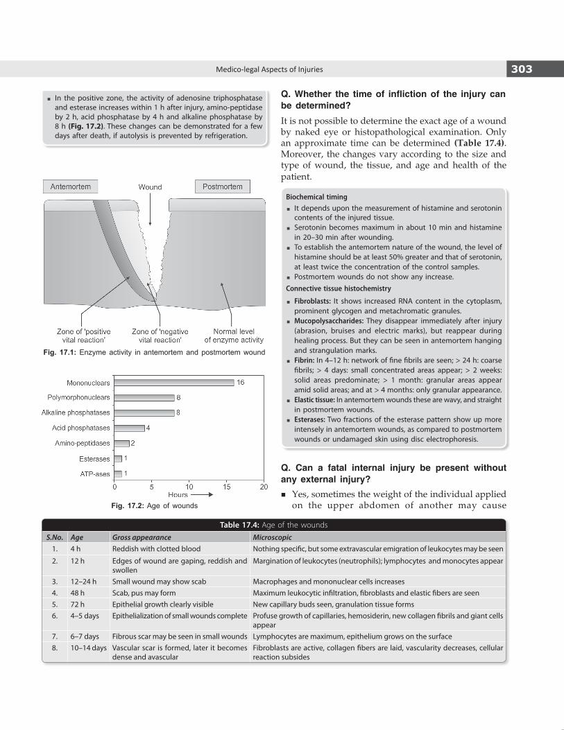
303Medico-legal Aspects of Injuries
Table 17.4: Age of the wounds
S.No. Age Gross appearance Microscopic1. 4 h Reddish with clotted blood Nothing specific, but some extravascular emigration of leukocytes may be seen
2. 12 h Edges of wound are gaping, reddish and swollen
Margination of leukocytes (neutrophils); lymphocytes and monocytes appear
3. 12–24 h Small wound may show scab Macrophages and mononuclear cells increases4. 48 h Scab, pus may form Maximum leukocytic infiltration, fibroblasts and elastic fibers are seen5. 72 h Epithelial growth clearly visible New capillary buds seen, granulation tissue forms6. 4–5 days Epithelialization of small wounds complete Profuse growth of capillaries, hemosiderin, new collagen fibrils and giant cells
appear 7. 6–7 days Fibrous scar may be seen in small wounds Lymphocytes are maximum, epithelium grows on the surface8. 10–14 days Vascular scar is formed, later it becomes
dense and avascularFibroblasts are active, collagen fibers are laid, vascularity decreases, cellular reaction subsides
Fig. 17.1: Enzyme activity in antemortem and postmortem wound
Fig. 17.2: Age of wounds
Q. Whether the time of infliction of the injury can be determined?
It is not possible to determine the exact age of a wound by naked eye or histopathological examination. Only an approximate time can be determined (Table 17.4). Moreover, the changes vary according to the size and type of wound, the tissue, and age and health of the patient.
Biochemical timing � It depends upon the measurement of histamine and serotonin
contents of the injured tissue. � Serotonin becomes maximum in about 10 min and histamine
in 20–30 min after wounding. � To establish the antemortem nature of the wound, the level of
histamine should be at least 50% greater and that of serotonin, at least twice the concentration of the control samples.
� Postmortem wounds do not show any increase.Connective tissue histochemistry
� Fibroblasts: It shows increased RNA content in the cyto plasm, prominent glycogen and metachromatic granules.
� Mucopolysaccharides: They disappear immediately after injury (abrasion, bruises and electric marks), but reappear during healing process. But they can be seen in ante mortem hanging and strangulation marks.
� Fibrin: In 4–12 h: network of fine fibrils are seen; > 24 h: coarse fibrils; > 4 days: small concentrated areas appear; > 2 weeks: solid areas predominate; > 1 month: granular areas appear amid solid areas; and at > 4 months: only granular appearance.
� Elastic tissue: In antemortem wounds these are wavy, and straight in postmortem wounds.
� Esterases: Two fractions of the esterase pattern show up more intensely in antemortem wounds, as compared to postmortem wounds or undamaged skin using disc electrophoresis.
Q. Can a fatal internal injury be present without any external injury?
� Yes, sometimes the weight of the individual applied on the upper abdomen of another may cause
� In the positive zone, the activity of adenosine tripho sphatase and esterase increases within 1 h after injury, amino-peptidase by 2 h, acid phosphatase by 4 h and alkaline phosphatase by 8 h (Fig. 17.2). These changes can be demonstrated for a few days after death, if autolysis is prevented by refrigeration.

304 Review of Forensic Medicine and Toxicology
laceration of the liver and death without leaving any visible injury mark.
� Manual strangulation and smothering may not leave any external signs of trauma.
� Fractures of ribs, vertebrae or pelvis with accompany-ing fatal visceral injuries can occur without external indications of serious violence.
Q. Which of the injuries caused death?
When there is more than one wound, it is necessary to determine which one of them caused death, since the wounds may not have been made by the same assailant or at the same time.
Q. How long did the victim survive, and could he have carried out any voluntary acts after receiving the injury?
� It is usually not possible to opine from an examination of wounds in a dead body as to how long the person might have lived or how much voluntary activity he might have performed before death, after receiving the injury.
� Unless, it can be proved that a particular injury would immediately be incompatible with life, it is rarely possible to state that the deceased could not have performed some activity (speaking, staggering few paces, walking or running) after receiving the injury. Most injuries do not cause sudden death or rapid loss of function (details in Chapter 12).
� A person may remain conscious for several minutes before dying from a severe intracranial injury.
� Muscular powers are retained in ruptures of liver, spleen or kidneys, unless there is marked immediate blood loss.
Q. Would the victim have survived, had he been given immediate medical care?
It depends on the nature and extent of injuries, as there is individual variation.
Q. Can the wounds be altered from their original appearance?
The wounds may be altered in many ways. � In the living, the wound may be altered by surgical
procedures and healing. � In the dead person, the wound might have been
deliberately altered by the assailant to mislead the investigators or by resuscitative measures applied or by insects, animals and decomposition.
Q. Whether the injuries can be produced by more than one type of weapon?
Several persons with different types of weapon may attack the victim producing diverse types of injuries.
Q. What is the relationship of trauma and natural disease?
Relationship of trauma and disease is important mainly for two reasons: compensation and insurance. i. Trauma and myocardial infarction: Heart attack
may occur while working, either incidentally (nor mal progression of chronic disease) or due to unusual physical/mental strain.
z Physical effort can damage a diseased heart due to unusual work or doing unfamiliar or un-accustomed work or accidents.
z Causal connection can be established with certainty only in direct trauma to the heart during work. A blow or physical trauma may precipitate myocardial infarction or arrhythmia.
z If the attack occurs within minutes after unusual effort, the causal connection can be established. It may occur few days later, due to subintimal hemorrhage in coronary artery leading to coronary thrombosis.
ii. Trauma and neoplasia: In some cases, there is apparent relationship between tumor and some preceding trauma to the part, e.g. development of osteogenic sarcoma and osteoclastoma after injury, malignancy in burn scars or on the skin adjacent to a chronic osteomyelitic sinus.
z Since, trauma disrupts tissue, it might activate a pre-existing tumor to grow and spread more rapidly.
z In accepting a relationship between trauma and malignancy, following Ewing’s postulates should be satisfied:
i. The tumor site prior to injury was normal. ii. Undeniable and adequate trauma to disrupt
the continuity must be proved. iii. The tumor followed the injury within a
reasonable time interval (between a minimum of 3–4 weeks and maximum of 3 years after receipt of injury).
iv. The tumor must have originated in the part of the body that has sustained the injury.
v. The tumor must be of histological type that could originate from the cells that have been disrupted by the trauma.
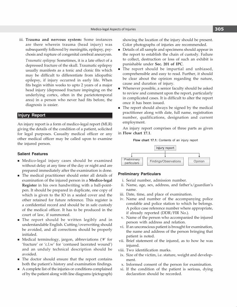
305Medico-legal Aspects of Injuries
iii. Trauma and nervous system: Some instances are there wherein trauma (head injury) was subse quently followed by meningitis, epilepsy, psy-chosis and rupture of congenital cerebral aneurysm.
Traumatic epilepsy: Sometimes, it is a late effect of a depressed fracture of the skull. Traumatic epilepsy usually manifests as a tonic and clonic fits which may be difficult to differentiate from idiopathic epilepsy, if injury occurred in early life. When fits begin within weeks to upto 2 years of a major head injury (depressed fracture impinging on the underlying cortex, often in the parietotemporal area) in a person who never had fits before, the diagnosis is easier.
Injury Report
An injury report is a form of medico-legal report (MLR) giving the details of the condition of a patient, solicited for legal purposes. Casualty medical officer or any other medical officer may be called upon to examine the injured person.
Salient Features
� Medico-legal injury cases should be examined without delay at any time of the day or night and are prepared immediately after the examination is done.
� The medical practitioner should enter all details of examination of the injured person in a Medico-legalRegister in his own handwriting with a ball-point-pen. It should be prepared in duplicate, one copy of which is given to the IO in a sealed cover and the other retained for future reference. This register is a confidential record and should be in safe custody of the medical officer. It has to be produced in the court of law, if summoned.
� The report should be written legibly and in understandable English. Cutting/overwriting should be avoided, and all corrections should be properly initialed.
� Medical terminology, jargon, abbreviations (‘#’ for ‘fracture’ or ‘c.l.w’ for ‘contused lacerated wound’) and an unduly technical description should be avoided.
� The doctor should ensure that the report contains both the patient’s history and examination findings.
� A complete list of the injuries or conditions complained of by the patient along with line diagrams (pictograph)
showing the location of the injury should be present. Color photographs of injuries are recommended.
� Details of all sample and specimens should appear in the report to establish the chain of custody. Failure to collect, destruction or loss of such an exhibit is punishable under Sec. 201of IPC.
� The report should be impartial and unbiased, comprehensible and easy to read. Further, it should be clear about the opinion regarding the nature, cause and duration of injury.
� Whenever possible, a senior faculty should be asked to review and comment upon the report, particularly in complicated cases. It is difficult to alter the report once it has been issued.
� The report should always be signed by the medical practitioner along with date, full name, registration number, qualifications, designation and current employment.
An injury report comprises of three parts as given in Flow chart 17.1.
Flow chart 17.1: Contents of an injury report
Preliminary Particulars i. Serial number, admission number. ii. Name, age, sex, address, and father’s/guardian’s
name. iii. Date, time, and place of examination. iv. Name and number of the accompanying police
constable and police station to which he belongs. A police case reference number where appropriate, if already reported (DDR/FIR No.).
v. Name of the person who accompanied the injured person with address and relation.
vi. If an unconscious patient is brought for examination, the name and address of the person bringing that patient is noted.
vii. Brief statement of the injured, as to how he was injured.
viii. Two identification marks. ix. Size of the victim, i.e. stature, weight and develop-
ment. x. Informed consent of the person for examination. xi. If the condition of the patient is serious, dying
declaration should be recorded.

306 Review of Forensic Medicine and Toxicology
Findings/Observations
General physical examination: Consciousness, orientation, pulse, temperature, blood pressure and reaction of pupils to light are to be noted.
Followingarethevariousentriesintheinjuryreport i. Type of each injury:All injuries observed, even
insignificant should be noted. Type of injuries, i.e. abrasion, contusion, laceration, incised wound, etc. should be noted. Multiple injuries can be grouped anatomically, e.g. injuries of the head, of the trunk or of limb. A lens should be used to get an accurate idea of the nature of edges, ends and floor of the wound. Presence of any foreign material in wound, e.g. glass, hair or dirt should be noted and preserved.
Features that may help in differentiating the common injuries are given in Diff. 17.2.
ii. Size, shape and direction of each injury: All injuries should be measured with a measuring tape and never guessed and amount of blood extravasated should be measured, and photographs (wherever possible) or sketches showing the position and size of the wound are desirable.
z Shape of the wound, e.g. circular, oval or triangular should be noted and also the beveling of the edges.
z Direction of the wound, i.e. horizontal, vertical or oblique should be noted with regard to anatomical position of the body.
iii. Location: Exact situation of wound with reference to some anatomical landmark, e.g. midline, bony structure or umbilicus should be mentioned. Technical terms should be avoided as far as possible.
For example, a stab wound of the thorax might be described as follows, using ordinary language to explain any medical terms.An elliptical shaped stab wound of size 2.5 × 0.5 cm present on the right upper chest, placed obliquely, with the inner end lower than the upper outer end (Depth to be assessed in the OT). The center of the wound was just above the line joining the nipples, being 5 cm from the midline, 7.5 cm from the right nipple and 15 cm below the center of the right clavicle (collar bone). The wound was 140 cm above heel with the inner lower end sharply cut than the blunt upper end and was sloped in a downwards direction, with subcutaneous tissue visible along the inside of the upper edge.
Opinion
i. Nature of each injury: Against each injury, it should be noted whether it is simple or grievous. Injured person must be kept ‘under observation’, if nature of particular injury cannot be made out at the time of examination, e.g. head injury or abdominal injury. In all injuries, when fracture of a bone is suspected, an X-ray should be done for confirmation.
z Whether an injury is simple or grievous, is decided on the basis of status of injury at the time of infliction and not after medical/surgical intervention. When deciding the question, one has to only regard the nature of the injury itself. If left untreated, would the injury have led to the defined result?
ii. Weaponusedtoinflicttheinjury: In many cases, examination of the wound and clothing give fairly definite information about the kind of weapon. With stab and incised wound, there is not much difficulty.
Differentiation 17.2: Lacerated, incised and stab wound
S.No. Feature Lacerated wound Incised wound Stab wound1. Causative object Blunt Sharp edged Pointed sharp2. Site Usually over bony prominences Anywhere Usually over chest, abdomen or
neck3. Shape Irregular Spindle shaped Spindle shaped, but depends on
the weapon4. Margins Irregular Clean cut and everted Clean cut5. Dimensions Variable Length greater than depth, gaping Depth greater than length6. Hair and blood vessels Crushed Clean cut Variable7. Hemorrhage Not pronounced, except in scalp Profuse Variable, may be concealed internally8. Surrounding abrasion
and bruiseUsually present Absent May be seen (hilt mark)
9. Foreign bodies Present Absent May or may not be present

307Medico-legal Aspects of Injuries
z Any weapon sent by the police which is alleged to have been used in producing injuries should be examined for marks of bloodstains, hair or pieces of cloth adherent to it, and should be returned to the police after it is sealed.
z Clothes should be examined for the presence of cuts, tears or burns, and it should be seen whether these correspond to the injuries on the body.
iii. Durationofinjuries: Opinion is based on the state of healing of the injuries as was recorded in the column of examination of the injuries.* However, the Supreme Court has stated that a doctor can never be absolutely certain on the point of the time of infliction of injuries.
iv. Cause of the patient’s condition: The court usually wants to know whether the injury for
which damages are claimed or punishment sought was caused, aggravated or accelerated by the accident or events complained of. Opinion on the precipitation factor or cause of the patient’s condition is based on the history and the nature of injuries on his/her person.
v. Whether theweaponwasdangerousornot? Doctor is guided by Sec. 324and326 IPC.
Handing Over the Report
The initial or provisional report should be made available immediately. A subsequent report (supple-mentary report) may be given, once the investigation results (reports of blood examination, X-rays and CT scans) become available which reflects the final conclusions drawn from the examination findings that was available at the time of the initial consultation.
* When opining on the duration of the injuries, undue and complete dependence is placed on the history given by the patient or his/her relatives; while the doctor’s own observations regarding the features of the injuries are often not taken into consideration or overlooked.
1. Rahul had a fightwith a neighbor and he assaultedhim.Therewasnoinjury,stillhecanbebookedunder:
AIIMS 12 A. Sec. 44 IPC B. Sec. 351 IPC C. Sec. 319 IPC D. Sec. 320 IPC 2. Acceptableor justifiablehomicidesareall,except:
NIMHANS 14 A. Judicial execution B. Self-defense C. Preventing rape D. Taking revenge 3. Punishment for culpablehomicidenot amounting to
murder is dealt under: TN 11 A. Sec. 299 IPC B. Sec. 300 IPC C. Sec. 302 IPC D. Sec. 304 IPC 4. Accordingtowhichsection,apersoncanbepunished
for attempt to murder with imprisonment for 10 years: NEET 14
A. Sec. 301 IPC B. Sec. 300 IPC C. Sec. 302 IPC D. Sec. 307 IPC 5. IPCsectiondealingwithdowrydeath:
Punjab 08; DNB 10; Odisha 11; NEET 14, 15 A. 307 IPC B. 304 IPC C. 304-A IPC D. 304-B IPC
6. As per the Dowry Prohibition Act 1961, penaltyawarded in case of death is imprisonment for: NEET 14
A. < 5 years B. Upto 7 years C. 7 years to life imprisonment D. Upto 10 years 7. If a woman is assaulted by her husband then he is
chargedunder: FMGE 11 A. Sec. 498-A IPC B. Sec. 304-A IPC C. Sec. 304-B IPC D. Sec. 504 IPC 8. Hurt isdefinedunder section: NEET 14 A. 319 IPC B. 320 IPC C. 321 IPC D. 323 IPC 9. Sec. 320 IPC is for: NEET 14 A. Defines hurt B. Defines grievous hurt C. Attempt to murder D. Defines murder 10. IPCsection forgrievous injury:
Kerala 06, 08; Punjab 09; NEET 13 A. Sec. 420 B. Sec. 320 C. Sec. 299 D. Sec. 351
multiple choice questions
1. B 2. D 3. D 4. D 5. D 6. c 7. A 8. A 9. B 10. B
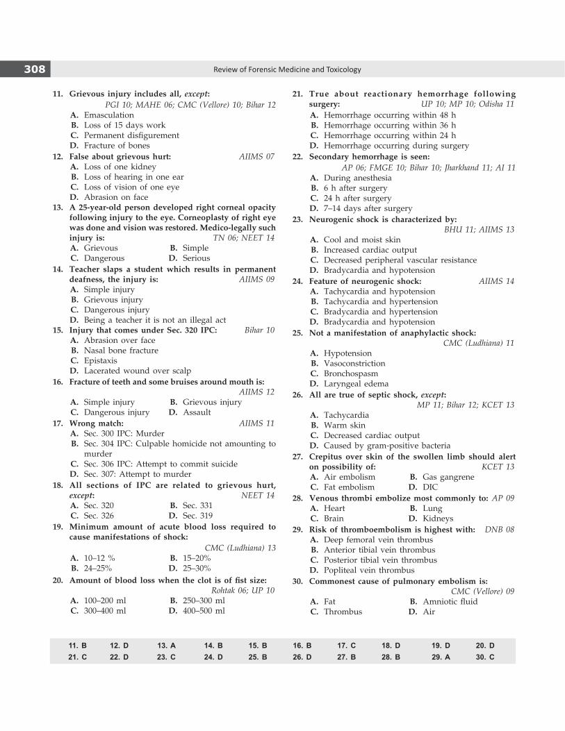
308 Review of Forensic Medicine and Toxicology
21. True about reactionary hemorrhage followingsurgery: UP 10; MP 10; Odisha 11
A. Hemorrhage occurring within 48 h B. Hemorrhage occurring within 36 h C. Hemorrhage occurring within 24 h D. Hemorrhage occurring during surgery22. Secondaryhemorrhage is seen:
AP 06; FMGE 10; Bihar 10; Jharkhand 11; AI 11 A. During anesthesia B. 6 h after surgery C. 24 h after surgery D. 7–14 days after surgery23. Neurogenic shock is characterizedby:
BHU 11; AIIMS 13 A. Cool and moist skin B. Increased cardiac output C. Decreased peripheral vascular resistance D. Bradycardia and hypotension24. Featureofneurogenic shock: AIIMS 14 A. Tachycardia and hypotension B. Tachycardia and hypertension C. Bradycardia and hypertension D. Bradycardia and hypotension25. Notamanifestationofanaphylactic shock:
CMC (Ludhiana) 11 A. Hypotension B. Vasoconstriction C. Bronchospasm D. Laryngeal edema 26. All are trueof septic shock,except:
MP 11; Bihar 12; KCET 13 A. Tachycardia B. Warm skin C. Decreased cardiac output D. Caused by gram-positive bacteria27. Crepitus over skin of the swollen limb should alert
onpossibilityof: KCET 13 A. Air embolism B. gas gangrene C. Fat embolism D. DIC28. Venous thrombiembolizemost commonly to:AP 09 A. Heart B. Lung C. Brain D. Kidneys29. Riskof thromboembolism ishighestwith: DNB 08 A. Deep femoral vein thrombus B. Anterior tibial vein thrombus C. Posterior tibial vein thrombus D. Popliteal vein thrombus30. Commonest causeofpulmonaryembolism is:
CMC (Vellore) 09 A. Fat B. Amniotic fluid C. Thrombus D. Air
11. Grievous injury includesall,except:PGI 10; MAHE 06; CMC (Vellore) 10; Bihar 12
A. Emasculation B. Loss of 15 days work C. Permanent disfigurement D. Fracture of bones12. Falseaboutgrievoushurt: AIIMS 07 A. Loss of one kidney B. Loss of hearing in one ear C. Loss of vision of one eye D. Abrasion on face13. A25-year-oldpersondevelopedrightcornealopacity
followinginjurytotheeye.Corneoplastyofrighteyewasdoneandvisionwasrestored.Medico-legallysuchinjury is: TN 06; NEET 14
A. grievous B. Simple C. Dangerous D. Serious 14. Teacher slaps a student which results in permanent
deafness, the injury is: AIIMS 09 A. Simple injury B. grievous injury C. Dangerous injury D. Being a teacher it is not an illegal act 15. Injury that comesunderSec. 320 IPC: Bihar 10 A. Abrasion over face B. Nasal bone fracture C. Epistaxis D. Lacerated wound over scalp16. Fractureofteethandsomebruisesaroundmouthis:
AIIMS 12 A. Simple injury B. grievous injury C. Dangerous injury D. Assault 17. Wrongmatch: AIIMS 11 A. Sec. 300 IPC: Murder B. Sec. 304 IPC: Culpable homicide not amounting to
murder C. Sec. 306 IPC: Attempt to commit suicide D. Sec. 307: Attempt to murder18. All sections of IPC are related to grievous hurt,
except: NEET 14 A. Sec. 320 B. Sec. 331 C. Sec. 326 D. Sec. 319 19. Minimum amount of acute blood loss required to
cause manifestations of shock:CMC (Ludhiana) 13
A. 10–12 % B. 15–20% B. 24–25% D. 25–30%20. Amountofblood losswhen the clot isoffist size:
Rohtak 06; UP 10 A. 100–200 ml B. 250–300 ml C. 300–400 ml D. 400–500 ml
11. B 12. D 13. A 14. B 15. B 16. B 17. c 18. D 19. D 20. D 21. c 22. D 23. c 24. D 25. B 26. D 27. B 28. B 29. A 30. c
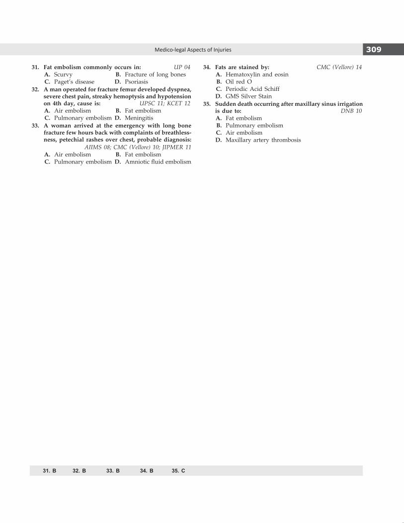
309Medico-legal Aspects of Injuries
31. Fat embolismcommonlyoccurs in: UP 04 A. Scurvy B. Fracture of long bones C. Paget’s disease D. Psoriasis32. Amanoperatedforfracturefemurdevelopeddyspnea,
severechestpain,streakyhemoptysisandhypotensionon4thday, cause is: UPSC 11; KCET 12
A. Air embolism B. Fat embolism C. Pulmonary embolism D. Meningitis33. A woman arrived at the emergency with long bone
fracturefewhoursbackwithcomplaintsofbreathless-ness,petechial rashesoverchest,probablediagnosis:
AIIMS 08; CMC (Vellore) 10; JIPMER 11 A. Air embolism B. Fat embolism C. Pulmonary embolism D. Amniotic fluid embolism
34. Fatsare stainedby: CMC (Vellore) 14 A. Hematoxylin and eosin B. Oil red O C. Periodic Acid Schiff D. gMS Silver Stain35. Suddendeathoccurringaftermaxillarysinusirrigation
is due to: DNB 10 A. Fat embolism B. Pulmonary embolism C. Air embolism D. Maxillary artery thrombosis
31. B 32. B 33. B 34. B 35. c
310
Decompression Sickness
Definitions � Decompression sickness (diver’s or Caisson disease,
‘bends’) is a disorder in which nitrogen (main inert gas) dissolved in the blood and tissues by high pressure forms bubbles as pressure decreases.1
� Dysbarism is a term that covers all the adverse effects of pressure.
� Barotrauma describes the mechanical damage from gas released into the tissues.
Decompression sickness is hazardous for fliers and divers who are involved in recreational diving (e.g. scuba diving), deep-water exploration and rescue or salvage operations. At low depths, the greatly increased pressure (e.g. at 100 feet, the pressure is four times greater than at the surface) compresses the respiratory gases into the blood and other tissues. During ascent from depths > 9 meters (30 feet), gases dissolved in the blood and other tissues escape as the external pressure decreases.
Predisposing factors: Exercise, injury, right to left cardiac shunt, obesity, dehydration, alcoholic excess, hypoxia, medications (e.g. narcotics, or antihistaminics) and cold.
Signs and Symptoms
The onset occurs within 30 minutes (min) to 6 hours (h). � Symptoms are pain in the joints (‘bends’ in 60–70%
cases due to gas bubble formation) with shoulder being the most common site; neurological symptoms (‘staggers’ in 10–15% cases) with headache and visual disturbances; skin manifestations (10–15% of cases) like itching, sensation of tiny insects crawling over the skin (formication) and pruritic rash, and pulmonary decom-pression sickness (‘chokes’) with pleuritic substernal pain, persistent cough and dyspnea (rare in divers).
� Other symptoms include numbness, confusion, nausea, vomiting, loss of hearing, weakness, para lysis, dizziness, vertigo, paresthesias, aphasia and coma.
� Sequelae include hemiparesis, neurologic dysfunction and bone damage.
Treatment
i. Administration of 100% oxygen. ii. Aspirin may be given for pain, but narcotics should
be used cautiously. iii. Rapid transportation to a treatment facility for
recompression, hyperbaric oxygen, hydration treat-ment of plasma deficits, and supportive measures is necessary.
Autopsy in Decompression Sickness
Skin diving fatalities are usually caused by drowning. Head and cervical injuries may be responsible for loss of consciousness and drowning in individuals drowning in shallow water. With scuba diving fatalities, investigation of the equipment and circumstances is usually more important than the autopsy since, drowning is the terminal event. Some of the important features that may be found during autopsy are given in Table 18.1.
� Photograph the victim as recovered and after removal of suit and other diving gear.
� Take X-ray chest, elbows, hips and knees. � CT findings of air in all major and minor blood
vessels. � Complete toxicological sampling should be carried
out. The air should be collected properly and sampled since there may be wrong gases in the compressed air tanks in scuba deaths—suggesting either a homicidal or accidental cause.
Ionizing Radiation Reactions
The extent of damage due to radiation exposure depends on the quantity of radiation delivered to the body, dose rate, organs exposed, type of radiation (X-rays, neutrons, γ rays, α or β particles), duration of exposure and energy transfer from the radioactive wave to the exposed tissue.
Decompression, Radiation and Altitude Sickness
ChApteR 18

311Decompression, Radiation and Altitude Sickness
Table 18.1: Autopsy changes in decompression sickness (diving accidents)
Region Possible and expected findings
External examination Mask, fins, weight belt, life vest, scuba tank may be missing.Clothing may be torn. Mask, mouthpiece or exhalation hose may contain vomitus.Cyanosis, cherry-red coloration, marbling. Facial edema, froth in mouth and nostrils. Mottled pallor of tongue and bite marks. Crepitation from subcutaneous emphysema. Antemortem and postmortem abrasions, contusions, lacerations, bites or puncture wounds may be seen.
Head and neck Fracture of skull and cervical spine. Gas bubbles in cerebral arteries, nitrogen bubbles in cerebral vessels. Subdural and subarachnoid hemorrhages, cerebral edema with ischemic necrosis and focal hemorrhages.
Eyes and ears Rupture of tympanic membrane, gas in retinal vessels (air embolism).
Chest, tracheobronchial tree andlungs
Foam, aspirated vomitus, pneumothorax and pneumomediastinum. Lacerations, bullae and atelectasis of lungs. Pulmonary edema and petechial hemorrhages. Nitrogen bubbles in precapillary pulmonary arteries and pulmonary fat embolism in decompression sickness.
Blood (from heart) Air embolism, alcohol intoxication and CO poisoning.
Heart Air embolism, ischemic heart disease, patent foramen ovale.
Other organs Fatty change of liver, ischemic infarction of many organs.
Spinal cord Nitrogen bubbles in spinal cord arteries.
Bone and joints Aseptic necrosis (dysbaric osteonecrosis) most often in head of femur,* distal femur and proximal tibia. Nitrogen bubbles in and about joints, and in periosteal vessels.
* Aseptic necrosis of femur needs time to develop; it is not seen in immediate death. It points to a previous non-fatal event or series of events.
In the US, the National Committee on Radiation Protection has established the maximum permissible radiation exposure for occupationally exposed workers (≥ 18 years) as 0.1 rad/week for the whole body (but not to exceed 5 rad/year) and 1.5 rad/week for the hands (routine chest X-rays deliver 0.1–0.2 rad).
The acute radiation syndrome may be dominated by CNS, GIT or hematologic manifestations depending on dose and survival.
� Fatigue, weakness and anorexia can occur following exposures exceeding 50 cGy [1 rad = 0.01 gray (Gy) = 1 cGy].
� Hematopoietic effects consisting of anemia, platelet loss and bone marrow suppression can occur 1–3 weeks after exposures exceeding 100 cGy.
� Whole body exposure levels of 1000–3000 cGy destroy GIT mucosa which may lead to toxemia, and death within 2 weeks.
� Total body doses > 3000 cGy cause widespread vascular damage, cerebral anoxia, hypotensive shock and death within 48 h.
Acute (Immediate) Ionizing Radiation Effects
� Skin and mucous membranes: Erythema, epilation, destruction of fingernails or epidermolysis.
� Hematopoietic tissues: Bone marrow suppression. � CVS: Pericarditis with effusion. � Reproductive system: Aspermatogenesis, sterility,
cessation of menses or abortion.
� RS: Pneumonitis. � GIT: Mucositis. � Liver: Hepatitis. � Renal: Nephritis.
Systemic Reactions (Radiation Sickness)
Radiation sickness occurs when X-ray therapy is given over the abdomen, less often with thorax, and rarely when given on the extremities. The basic mechanism is not known.
Symptoms include anorexia, nausea, vomiting, weakness, exhaustion, lassitude, and in some cases, prostration may occur. Dehydration, anemia and infection may follow.
Death after whole body acute lethal radiation exposure is usually due to hematopoietic failure, GIT mucosal damage, CNS damage, widespread vascular injury or secondary infection may occur
Prevention: Persons handling radiation sources can minimize exposure to radiation by decreasing the time of exposure, maintaining distance and shielding. Special protective clothing is necessary to protect against contamination with radioisotopes.
Nuclear Terrorism
The proliferation of radiation equipment and nuclear energy plants, terrorism and the increasing need for transportation of radioactive materials have made

312 Review of Forensic Medicine and Toxicology
1. B 2. B
necessary hospital plans for managing patients who are accidentally exposed to ionizing radiation or are contaminated with radioisotopes. The threat of nuclear terrorism is raising the level of awareness about medical aspects of ionizing radiation exposure.
Treatment
The success of treatment of local radiation effects depends on the extent, degree and location of tissue injury. i. Particulate or radioisotope exposures should be
decontaminated in designated confined areas. ii. Ondansetron, 8 mg orally twice or thrice daily, is
given for nausea and vomiting. iii. Blood and platelet transfusions, blood stem cell
transplantation, bone marrow transplants, anti- biotics, fluid and electrolyte maintenance, and other supportive measures may be useful.
iv. Recombinant hematopoietic growth factors have been effective in accelerating hematopoietic recovery.
Altitude Illness
Five manifestations of altitude illness are: i. Acute mountain sickness ii. High-altitude pulmonary edema iii. High-altitude encephalopathy iv. Subacute mountain sickness v. Chronic mountain sickness (Monge’s disease)
Lack of sufficient time for acclimatization, increased physical activity and varying degrees of health may be responsible for the acute, subacute and chronic disturbances that result from (hyperbaric) hypoxia at altitudes > 2000 meters (6560 feet).
Acute Mountain Sickness (AMS)
The severity of acute mountain sickness correlates with altitude and rate of ascent.
� Initial manifestations include headache (most severe and persistent symptom), lethargy, drowsiness, dizzi ness, chilliness, nausea, vomiting, facial pallor, dyspnea and cyanosis.
� Later, there is facial flushing, irritability, difficulty in concentrating, vertigo, tinnitus, visual and auditory disturbances, anorexia, insomnia, dyspnea and weakness on exertion, increased headaches (due to cerebral edema), palpitations, tachycardia, Cheyne-Stokes breathing and weight loss.2 More severe manifestations include pulmonary edema and encephalopathy.
Voluntary periodic hyperventilation may relieve symptoms. In most individuals, symptoms clear within 24–48 h, but in some instances, if the symptoms are sufficiently persistent or severe, the patient must return to lower altitudes.
Treatment
� Definitive treatment is immediate descent, which is essential, if reduced consciousness, ataxia or pulmonary edema occurs.
� Administration of oxygen, 1–2 l/min, often relieve acute symptoms. If immediate descent is not possible, portable hyperbaric chambers can provide symptomatic relief depending on altitude and severity.
� Acetazolamide, 250 mg every 8–12 h or dexa-methasone, 8 mg initially, followed by 4 mg every 6 h for as long as symptoms persist is recom-mended.
multiple choice questions
1. Caisson disease is due to: AIIMS 10 A. Fat embolism B. Air embolism C. Foreign body embolism D. Amniotic fluid embolism
2. The following are associated with high altitude, except: Karnataka 11 A. Cerebral edema B. Hypoventilation C. Visual disturbances D. Dyspnea
Definitions
� Starvation is the result of irregular or continuous deprivation of nutrients (food alone, or food and drink both) necessary for the maintenance of the body.
� Inanition: It refers to the exhausted state due to prolonged undernutrition caused by lack of assimilation of food by the tissues.
Starvation can be: � Acute starvation or total fasting which results from
sudden and total withholding of food, or food and drink.
� Chronic starvation or malnutrition which results from prolonged, but gradual and continuous deficiency in the intake of food and nutrients.
Mode of Starvation
Failure of Taking Food
i. Ignorance: Lack of knowledge of gross nutrition value of foodstuff, particularly among uneducated masses.
ii. Diseased conditions: Low intake (e.g. diabetes), loss of appetite (e.g. major depressive disorder), deficient absorption (e.g. celiac disease) or inability to eat (e.g. carcinoma esophagus).
iii. Deliberate: Deliberate improper feeding, or withholding of food in case of unwanted baby, old, invalid or diseased family member.
iv. Circumstantial: Accidents (e.g. shipwreck, air crash or colliery entombment) or famine.
Refusal to Take Food
i. In observance of religious rituals which is common in India.
ii. Intentional fasting as a form of protest against some alleged injustice—hunger strike or fast-into-death.
iii. Mental illnesses, like schizophrenia or anorexia nervosa.
� Historically, starvation has been used as a death sentence. From the beginning of civilization to the Middle Ages, people were immured or walled in, and would die for want of food. In ancient Greco-Roman societies, starvation was sometimes used to dispose of guilty upper class citizens, especially erring female members of patrician families.
� Force-feeding of hunger strikers is considered to be a form of torture. The WMA’s Tokyo Declaration prohibits doctors’ involvement in force-feeding. Due to some untoward incidents in which hunger strikers died in several countries, like in Ireland, Turkey and South Africa, the WMA formulated the Declaration of Malta, dedicated in its entirety to the role and responsibility of doctors caring for hunger strikers.
Pathophysiology
Individuals experiencing starvation lose adipose tissue and muscle mass as the body breaks down these tissues for energy. Initially, the body’s glycogen stores are used up in about 24 hours (h). After that, the main means of energy production is lipolysis. Adipose tissue releases free fatty acids in starvation and these are used by many as fuel. Furthermore, in the liver they are the substrate for synthesis of ketone bodies (which are major metabolic fuels for skeletal and heart muscle, and the brain). There is an increase in plasma free fatty acids and ketone bodies as starvation progresses which can be detected in urine.1,2
ConstituentStarvation
40 h (mmol/l) 7 days (mmol/l)Glucose 3.6 3.5Free fatty acids 1.15 1.19Ketone bodies 2.9 4.5
Signs and Symptoms
Acute Starvation
In the beginning, there is initial feeling of hunger and hunger pains for first 2 days with craving for food
Starvation Deaths
Chapter 19
314 Review of Forensic Medicine and Toxicology
wearing off very rapidly. Intense thirst is felt along with epigastric pain and subsequent loss of sense of thirst. This is followed by both mental and physical lethargy, fatigue, irritability, loss of libido and progressive loss of weight. Later, emaciation sets in and the body emits an offensive odor. As the starvation continues, the lethargy becomes extreme, with mental impairment, loss of self-respect and interest in everything.
Characteristic Findings
� Skin: Dry, dirty, lusterless, loose, cracked and inelastic with increase of pigmentation, creases and wrinkles.
� Face: Eyes—shruken, pupils—dilated, lips—dry and cracked, cheek—shallow with prominent malar bones (loss of Bichat’s fat of pad is among the last subcutaneous adipose depots to disappear).3
� Tongue: Dry, furred and coated, foul smelling breath. � Temperature: Hypothermia with sensitivity to cold. � Blood pressure: Hypotension. � Pulse: Quick, weak and feeble. � Abdomen concave, prominent ribs and hip bones. � Bowel: Constipated in early phase, followed by
diarrhea and dysentery. � Renal: Oliguria with concentrated, highly acidic urine. � Muscle atrophy leading to weakness. � All bony joints and bones look prominent.
Progressive cardiac insufficiency leads to death. Loss of 40–50% of original body weight usually leads to death.
Chronic Starvation
� Anemia (first sign), hypoproteinemia, emaciation, weak pulse and blood pressure, cyanosis, and edema of feet, legs and face with ascitis, hepatitis, diarrhea or dysentery.
� Reduced resistance to infections in general, and development of bronchopneumonia, tuberculosis and enterits along with poor wound healing.
� In females, irregular menstruation can occur. � Loss of weight is very rapid in the first place, but
becomes slower after 3 months. � In the terminal stages, adults may experience a
variety of neurological and psychiatric symptoms, including hallucinations and convulsions, as well as severe muscle pain and disturbances in heart rhythm.
Fatal period
� Total withholding of food and water: 14–21 days.4
� With total deprivation of food only: 3–6 weeks (8–12 weeks in some cases).
Factors influencing the fatality i. Age: Children and infants are most vulnerable. Old person
stands starvation better. ii. Sex: Women stand starvation better than men due to their
body fat. iii. Body condition: Fatty and healthy individual stands starvation
better. iv. Environmental factors: Exposure to cold and extreme heat
shortens life. v. Intercurrent infection: It may cause early death. vi. Physical exertion: It will enhance the effects of starvation.
Postmortem Findings
Typical picture of emaciation and exclusion of any other coexisting cause of death are prerequisite for a definite diagnosis of death due to of starvation. The main autopsy finding is emaciation with loss of body weight and organ weights. i. Complete lack of fat in the subcutaneous and deep
fat depots. ii. Skin is pale and cadaverous in most of the cases,
and dark brown in few. iii. There is severe atrophy of skeletal muscles,
lungs, heart, liver, spleen, kidneys, endocrine and reproductive organs (ovaries or testes), except for the brain.5 In infants, complete atrophy of thymus is pathognomonic of starvation.
iv. GIT: Stomach and small bowel are empty along with presence of dry stools in the colon. Even foreign bodies may be found in the colon (starving person may try to eat everything accessible prior to death).
z There is atrophy of the GIT with thin parchment-like translucent walls and loss of mucosal folds.
z Gallbladder bigger in size and distended with bile (food acts as the natural stimulant of bile excretion).6
z The small intestinal wall appears swollen with reddish discolored mucosa and ulcerations of the mucosa of the colon, described as ‘pseudo-dysentery’.
v. Edema and peritoneal effusions may occur. vi. Liver: It may show centrilobular necrosis due to
protein deficiency. vii. Kidneys: It may show atrophy of nephrons.
Typical autopsy findings in starvation7
� Emaciation with sunken eyes and loss of Bichat’s fat pad. � Complete disappearance of body fat with pronounced rib cage.
� Loss of adipose tissue of the mesentery. � Disuse atrophy of the GIT with translucent small intestinal walls.
� Distention of the gallbladder.
315Starvation Deaths
Medico-legal Questions
Q. Whether the death was caused by starvation?
The diagnosis of starvation is done on the basis of history and postmortem findings. Before opining on starvation as cause of death, the doctor should rule out tuberculosis, carcinoma, stricture of esophagus, anorexia nervosa, radiation sickness, pernicious anemia, inflammatory bowel disease and Addison’s disease (chronic adrenocortical insufficiency).
Q. Whether it was suicidal/homicidal/accidental starvation?
If the diagnosis of death as a result of starvation is established, the underlying cause of starvation has to be determined: any pre-existing disease or deliberate withholding of food or neglect.
� Suicidal: Some individuals starve voluntarily for the fulfillment of their grievances. Sometimes, prisoners, mentally ill or hysterical women may refuse to take food. Fasting may also be undertaken to attract public attention. Right to life is guaranteed under the Constitution of India, so forcible feeding in these individuals is lawful.
� Homicidal: These cases are related to elderly person or victims of child abuse. It is mostly seen in illegitimate children who are starved to death, by depriving them of food and exposing to severe cold.
� Accidental: It may occur during famine, shipwreck or trapped in mines or landslides during earthquakes.
� Deaths caused by starvation are mostly natural deaths in India, accidental cases are also common.
� It relatively rare in the US/industralized countries. For the most part, they occur either as a result of child abuse, fasting or in mentally ill person.
1. A person on fasting for 7 days, the source of energy is: NIMS 11
A. Acetone B. Acetoacetate C. Glucose D. Alanine
2. Brain in starvation uses: TN 11; UP 12 A. Amino acids B. Cellulose C. Ketone bodies D. Glycerol
3. In starvation, last to disappear: JIPMER 10 A. Buccal fat B. Fat around the abdomen C. Fat around the eyes D. Fat in the mesentery
4. If food and water is withheld then the person will die after: WB 11
A. 1–2 days B. 2–5 days C. 7–10 days D. 15–20 days
1. A 2. C 3. A 4. D 5. D 6. B 7. A
5. In starvation death, atrophy is seen in all, except: Maharashtra 08
A. Heart B. Kidney C. Liver D. Brain 6. Gallbladder in starvation death is:
TN 08; NEET 14 A. Contracted B. Distended C. Mummified D. Not affected 7. In starvation, all are true, except: FMGE 09 A. Shrunken gallbladder B. Translucent intestine C. Lack of fat in mesentery D. Prominent rib cage
multiple ChoiCe questions

316
Morbidity and mortality during anesthesia has been markedly reduced due to better understanding of human physiology and pathology of disease processes. Introduction of improvised drugs, devices, techniques and previous experience have also contributed to safety during anesthesia.
Few examples of deaths in operation theater setting: � A patient dies on table during anesthesia (the cause
of death, i.e. whether it is related to the pathology itself, to the surgery or the anesthesia, is left to be determined by the investigation).
� A patient has not fully recovered from effects of general anesthesia, develops upper airway obstruction in recovery room, has a hypoxic arrest and dies.
� A patient aspirates on table, develops pneumonia and dies 2 weeks later from subsequent complications.
Deaths during anesthesia may be broadly classified into two groups:1. Death during administration of anesthesia, but not
due to anesthesia.2. Deaths which are the direct result of administration
of an anesthetic.
Death during Administration of Anesthesia (not due to anesthesia)
i. The injury or disease process which necessitated surgical intervention is serious enough, the anesthetic may have only precipitated the death.
ii. Patient may be suffering from a severe systemic illness, e.g. valvular heart disease or severe coronary disease and undergoes surgery for another disease or problem—the operation or anesthetic may have precipitated death.
iii. Patient may be having some undiagnosed serious lesions, e.g. major vascular aneurysm or severe coronary artery disease which could have been an important contributory factor in causing death.
iv. Surgical shock and exhaustion: When surgery has been unduly delayed and preoperative condition
of patient is poor, shock and exhaustion may be major factors responsible for causing the death of the patient, or the patient has been unable to bear the stress of anesthesia and surgery.
Deaths Directly Related to Administration of an Anesthetic
i. Inexperience: Lack of adequate experience is the most common cause of death. Inability to take precautions and corrective measures when required is commonly observed, e.g. death during endotracheal intubation is due to:
z Inability to place the tube in the trachea. z Esophageal intubation. z Inability to protect the airway against aspiration
of foreign bodies, including regurgitant gastric content, tooth and blood.
z Disconnection of circuit. ii. Equipment/device failure due to:
z Faulty connections or mislabeling of anesthetic gases and drugs.
z Explosion and fire in operation theater. This problem is now rare due to advent of newer anesthetic agents which do not form explosive mixtures.
iii. Respiratory failure: Death occurs due to an inadequate supply of oxygen to tissues. It may be due to:
z Depression of respiratory center by overdose of drugs used for pre-medication and pain relief or overdose of anesthetic agent used.
z Inadequate reversal of muscle relaxant leading to inefficient ventilation of lungs.
z Obstruction of the respiratory tract from la-ryngeal spasm, impaction of loose material, like swabs or dentures in larynx, trachea and bronchi, or tongue falling back leading to airway obstruction. Regurgitant matter aspirated into lungs may affect gaseous exchange in the lungs.
z Large tidal volumes used during intermittent positive pressure ventilation may result in lung
Anesthetic Deaths
ChApter 20

317Anesthetic Deaths
trauma leading to pneumothorax or tension pneumothorax. Nitrous oxide used during general anesthesia leads to a rapid expansion of the pneumothorax. If pneumothorax is signi-ficant, gaseous exchange is affected leading to hypoxic injury and death.
iv. Neurogenic cardiovascular failure: It is the most common cause of sudden death under general anesthesia. It usually occurs when some inter-vention is done at a time when the depth of anesthesia is still inadequate, e.g. traction on viscera or peritoneum, laryngoscopy and endotracheal intubation, and dilatation.
v. Malignant hyperthermia: When it occurs, it is usually seen with halogenated anesthetics and succinylcholine.
z Individual involved usually has a genetic pre-disposition to the syndrome.
z Signs and symptoms: Rapid rise in body temperature and a two-to three-fold increase in total body oxygen consumption, arrhythmias, tachycardia and skeletal muscle rigidity.
z May be fulminant or insidious; may or may not occur every time anesthesia is administered.
z Complications: Rhabdomyolysis, electrolyte abnormalities (especially hyperkalemia) and disseminated intravascular coagulopathy (DIC).
vi. Local anesthetics: Toxicity results from overdose or allergic reactions, hypersensitivity and idio-syncrasy. Important factors influencing toxicity are:
z General condition and susceptibility of the patient.
z Total dose administered. z Rate of administration of anesthetic agent. z Vascularity of the area injected. z Accidental intravascular injection. z Concomitant use of adrenaline: Adrenaline used
along with local anesthetic agent can cause tachycardia, palpitation, sweating, hypertension and ventricular fibrillation.
There may be general effect on CNS which can be:a. Excitatory: Causing convulsions, orb. Depressive: Causing respiratory paralysis.
Very rarely, the heart may be affected directly, or when an abnormally high concentration is injected into a nerve, permanent loss of function may occur.
vii. Spinal anesthesia: During spinal anesthesia (block), sympathetic blockade occurs along with sensory
and motor blockade. This sympathetic blockade leads to varying degrees of hypotension which may be fatal, if not detected and corrected early.
z Marked hypotension is observed in elderly, in fluid deficit states, like hemorrhage and dehydration, and whenever there is a pre-existing decompensating heart disease.
z Cardiac activity may be inhibited leading to death due to vagus stimulation.
z The vital centers in the brainstem may be affected by diffusion of drug upward. Cardiac or respiratory arrest may occur.
z Post-lumbar puncture headache occurs when a large bore needle is used for lumbar puncture.
z Contamination of the needle, syringe and ampoules with sterilizing and cleansing agent may lead to arachnoiditis, and may cause bladder-bowel dysfunction and paraplegia. Sepsis can also occur.
Complications of Anesthesia
Minor complications are not uncommon in anesthesia. These include—hypoxemia, atelectasis of lungs, pneumonia, pulmonary edema, pneumothorax, broncho spasm, oxygen toxicity and aspiration of gastric contents, blood or foreign bodies. Neurological sequelae of these complications can be blindness, paraplegia, paresthesia, vegetative state and death.
Medico-legal Issues of Death in Operation Theater
� Any death considered unnatural must be reported for medico-legal investigation including a postmortem and formal inquest. The death of a person whilst under the influence of a general anesthetic or local anesthetic, or of which the administration of an anesthetic has been a contributory cause, is not considered to be a death from natural causes. Hence, in all such deaths, the surgical team must inform the hospital authorities, who in turn, should inform the police and must insist on an autopsy, for their own safety and defense.
� The Karnataka High Court has held that in case of death on operation table, in the absence of postmortem and/or histopathology reports, the possibility of other causes of death cannot be ruled out. The death on the operation table by itself is not sufficient to prove rashness or negligence against the accused.
318 Review of Forensic Medicine and Toxicology
Anesthetists per se are likely to experience intra- operative death more than surgeons, the consequences of which can be extremely stressful. It is reasonable for medical staff not to take part in operations for 24 h after an intra-operative death. Moreover, when a death occurs on the operating table, the anesthetist may become inclined to hypercritical self-examination and subjected to prolonged, judgmental investigation by their peers.
Postmortem Examination
Most deaths concerning anesthesia are unlikely to be evident at autopsy. Surgical mistakes being anatomical, may be observable at the postmortem, and anesthetic mistakes being physiological, are usually not appreciable after death, except where overdose with specific drug is involved. Findings of the autopsy surgeon alone will not be sufficient to explain death and therefore, it is advisable to hold a discussion across the autopsy table involving forensic expert, anesthetist and the surgeon/clinician concerned. In case of death following anesthesia/surgery, the forensic pathologist must answer the following questions: i. Was the death due to the effects of the operation
or anesthesia or is it due to the disease for which operation was being carried out?
ii. Would the patient have died, if he has not undergone through the anesthesia or operation?
iii. Was there any defect in anesthetic or surgical technique?
iv. Was the patient suffering from any predisposing condition that made him more susceptible to death from anesthetic or operative procedure?
v. Was the death due to some unsuspected natural disease, directly unrelated to the disease for which surgery was being performed?
During postmortem examination, the following are to be taken into consideration: i. Detailed hospital record of the patient, including
full clinical and pre-anesthetic checkup. ii. Surgical intervention and its sequelae, like sepsis,
hemorrhage or edema. iii. Postmortem changes need to be differentiated from
abnormalities existing during life (e.g. resuscitative artifacts and agonal regurgitation).
iv. Instances of surgical mishap which may not be negligence, if the operating conditions were difficult, like ligation of arteries and veins, ureters,
bile ducts and perforation of large blood vessels, should be looked for.
v. Presence of pre-existing natural disease, such as heart disease or respiratory insufficiency and their contribution to the cause of death must be evaluated.
vi. Pneumothorax, air embolism or surgical emphysema should be clearly evaluated.
vii. Surgical and anesthetic devices, such as airways, endotracheal tubes, needles or catheters should not be removed prior to autopsy. In esophageal intubation, a radiograph will show a ring of edema of esophageal mucosa at the level of the tube along with distention of stomach and intestines.
viii. All the organs should be dissected, and surgical sutures should be inspected.
ix. Chloroform and halothane are hepatotoxic, and chloroform may cause ventricular fibrillation sometimes. Halogenated hydrocarbons cause cardiac irritability.
x. A full range of specimens for histological, toxicological and bacteriological examinations, and those required to exclude hazards associated with blood or fluid transfusions must be collected.
Histological examination of the brain is vital which is primarily intended to demonstrate the effects of hypoxia, particularly in the region of Sommer’s area of the hippocampal gyrus and the cerebellum where changes are expected, even if the victim suffers hypoxia for a short period.
Toxicological examination: Following samples should be collected:
� Blood 10 ml (under liquid paraffin)
� One lung sealed in nylon bag
� Liver 100 g � Skeletal muscle 10 g
� Fat from mesentery 2 g � Cerebrum 100 g � Kidney 100 g or half of each kidney
� Urine
In case of inhaled anesthetic, specimens should be kept in containers of appropriate size to avoid empty space, and are sealed and refrigerated/frozen. Alveolar air should be collected with needle and syringe by puncturing the lung underwater before the chest is opened.
Anesthetic Drugs and Suicide
� Mostly doctors and paramedics misuse anesthetic drugs. There are instances when these have been used for suicidal purpose.

319Anesthetic Deaths
� Opioids, like morphine, pethidine and pentazocine are administered along with muscle relaxants for painless death. While opioids produce analgesia, muscle relaxants cause paralysis of muscles including those of the diaphragm. Due to failure
of ventilation of lungs, hypoxia results, leading to death.
� Death can be averted, if detected early, by institu- ting positive pressure ventilation of lungs till there is recovery from effects of muscle relaxants and opioids.
320
Definitions
� Infanticide is killing of an infant at any time from birth upto the age of 12 months.
� Feticide is the killing of fetus at any time prior to birth.
� Female feticide: It is the act of aborting a fetus because it is female.
� Filicide (Latin filius: son) is deliberate act of killing of a child by the parents.
Legal Aspects
� Infanticide is charged under Sec. 302 IPC which is punishable by death or imprisonment for life and fine.
� The causing of the death of living child in the mother’s womb may amount to culpable homicide, if any part of that child has been brought forth, though the child may not have breathed or completely born (Sec. 299 IPC).
� Any person who does an act with intent to prevent the child being born alive or to cause it to die after birth (except done in good faith for the purpose of saving the life of mother) is punished with imprisonment upto 10 years with/without fine (Sec. 315 IPC).
� Any person who does an act causing death of quick unborn child would be guilty of culpable homicide, and punished with imprisonment upto 10 years and fine (Sec. 316 IPC). For example, a person knowingly injures a pregnant woman that causes the death of an unborn quick child; he is guilty of the offence defined in this section.
Infanticide does not include the death of fetus during labor, when it is destroyed by craniotomy or decapitation.
� Parricide is the act of murdering one’s father (patricide), mother (matricide) or other close relative, but usually not children.
� In Canada, Italy, UK and Australia, murder of a child < 1 year of age by his/her own mother is not considered homicide. Instead, the mother is charged with the offence of infanticide, for which the punishment is lesser. This is because such murders could be due to ‘post-partum depression’ or ‘baby-blues’.
� In India, there is no such special Act and there is no distinction between the murder of a newborn infant and that of any other individual.
Postmortem Examination of Infants
The relatives should identify the body, and radiological examination should be done prior to autopsy.
� Whole-body radiographs (anteroposterior and lateral) are taken.
� Photographs of the external features—frontal pictures of the entire body and close-ups of the face and side of the head, as well as, any other unusual aspects are taken.
The procedure for autopsy is nearly the same as in adults, except for certain variations. The presence of malformations is often the major consideration, and the dissection should be made to preserve anatomic relationships in order to define the abnormal anatomy.
External Examination
� Clothings and wrappings should be examined and retained for identification of the mother.
� Measurements: Head, chest and abdominal circumferences, length (crown-rump, crown-heel, and foot for fetuses) and weight of the body helps to assess the gestational age.
� General features: The presence of dysmorphic features should be documented, and karyotyping should be considered, if significant abnormal features are noted.
� Head: The distribution and quality of hair over the head and rest of the body are noted. Abnormalities of the shape of the head related to molding, trauma, soft tissue edema, hemorrhage or autolysis are noted.
� Face: The facial features are examined and abnormalities recorded. Configuration of the ear is examined, and plasticity (indicating amount of cartilage) evaluated as an index to developmental stage. By late intrauterine development, the crest of the external ear should be superior to the level of the lateral canthus.
� Extremities: The position of the hands and feet, as well as the fingers and nails must be noted.
Infanticide and Child Abuse
CHAPTER 21
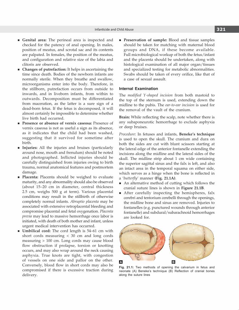
321Infanticide and Child Abuse
� Genital area: The perineal area is inspected and checked for the patency of anal opening. In males, position of meatus, and scrotal sac and its contents are palpated. In females, the position of the meatus, and configuration and relative size of the labia and clitoris are observed.
� Changes of putrefaction: It helps in ascertaining the time since death. Bodies of the newborn infants are normally sterile. When they breathe and swallow, microorganisms enter into the body. Therefore, in the stillborn, putrefaction occurs from outside to inwards, and in liveborn infants, from within to outwards. Decomposition must be differentiated from maceration, as the latter is a sure sign of a dead-born fetus. If the fetus is decomposed, it will almost certainly be impossible to determine whether live birth had occurred.
� Presence or absence of vernix caseosa: Presence of vernix caseosa is not as useful a sign as its absence, as it indicates that the child had been washed, suggesting that it survived for sometime after birth.
� Injuries: All the injuries and bruises (particularly around nose, mouth and frenulum) should be noted and photographed. Inflicted injuries should be carefully distinguished from injuries owing to birth trauma, normal anatomical features and postmortem damage.
� Placenta: Placenta should be weighed to evaluate maturity, and any abnormality should also be observed (about 15–20 cm in diameter, central thickness 2.5 cm, weighs 500 g at term). Various placental conditions may result in the stillbirth of otherwise completely normal infants. Abruptio placenta may be associated with extensive retroplacental bleeding and compromise placental and fetal oxygenation. Placenta previa may lead to massive hemorrhage once labor is initiated, with death of both mother and infant, unless urgent medical intervention has occurred.
� Umbilical cord: The cord length is 54–61 cm with short cords measuring < 30 cm and long cords measuring > 100 cm. Long cords may cause blood flow obstruction if prolapse, torsion or knotting occurs, and may also wrap around the neck causing asphyxia. True knots are tight, with congestion of vessels on one side and pallor on the other. Conversely, blood flow in short cords may also be compromised if there is excessive traction during delivery.
� Preservation of sample: Blood and tissue samples should be taken for matching with maternal blood groups and DNA, if these become available. Full microbiological workup of both the fetus/infant and the placenta should be undertaken, along with histological examination of all major organ/tissues and specialized testing for metabolic abnormalities. Swabs should be taken of every orifice, like that of a case of sexual assault.
Internal Examination
The modified Y-shaped incision from both mastoid to the top of the sternum is used, extending down the midline to the pubis. The ear-to-ear incision is used for the removal of the vault of the cranium.
Brain: While reflecting the scalp, note whether there is any subaponeurotic hemorrhage to exclude asphyxia or deep bruises.
Procedure: In fetuses and infants, Beneke’s technique is used to open the skull. The cranium and dura on both the sides are cut with blunt scissors starting at the lateral edge of the anterior fontanelle extending the incisions along the midline and the lateral sides of the skull. The midline strip about 1 cm wide containing the superior sagittal sinus and the falx is left, and also an intact area in the temporal squama on either side, which serves as a hinge when the bone is reflected in a ‘butterfly’ manner (Fig. 21.1A).
� An alternative method of cutting which follows the cranial suture lines is shown in Figure 21.1B.
� After carefully inspecting the hemispheres, falx cerebri and tentorium cerebelli through the openings, the midline bone and sinus are removed. Injuries to fontanelles (e.g. punctured wounds through anterior fontanelle) and subdural/subarachnoid hemorrhages are looked for.
Fig. 21.1: Two methods of opening the calvarium in fetus and neonate (A) Beneke’s technique (B) Reflection of cranial bones along the suture lines
A B
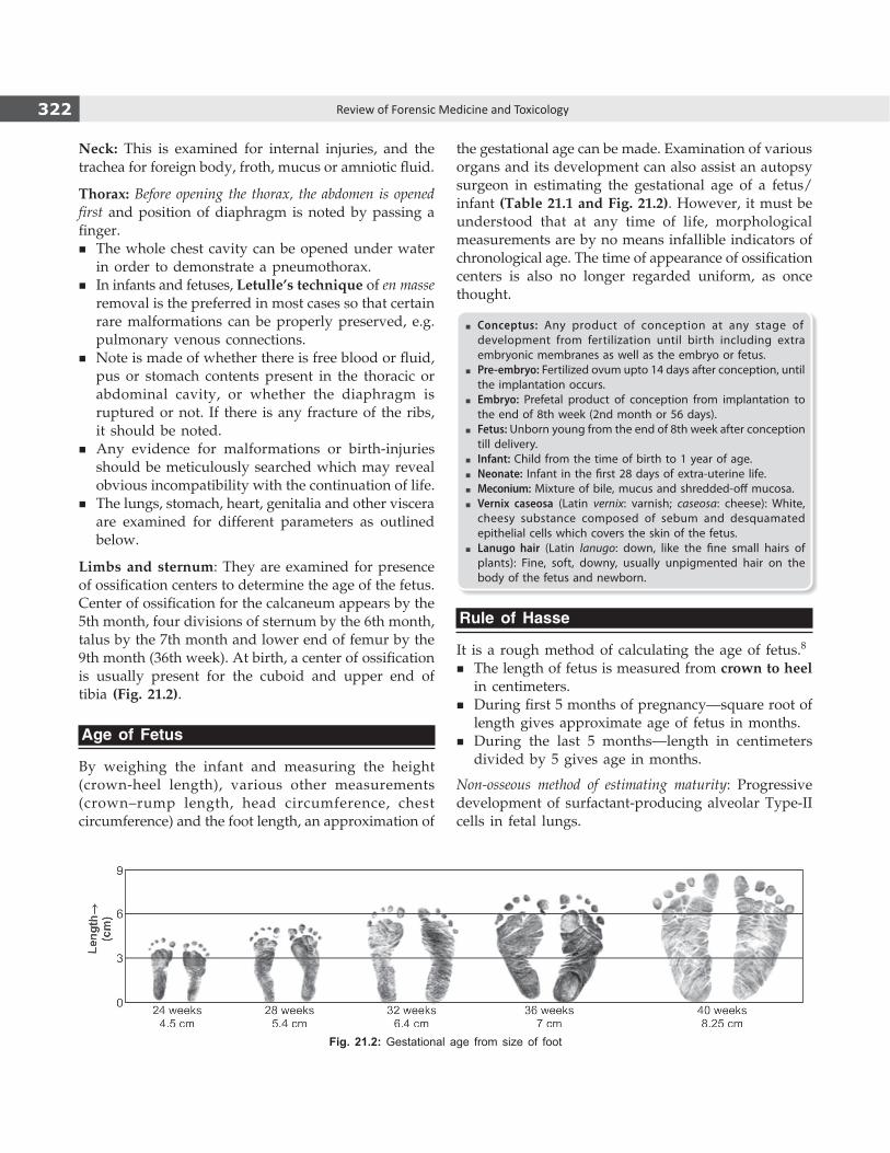
322 Review of Forensic Medicine and Toxicology
Neck: This is examined for internal injuries, and the trachea for foreign body, froth, mucus or amniotic fluid.
Thorax: Before opening the thorax, the abdomen is opened first and position of diaphragm is noted by passing a finger.
� The whole chest cavity can be opened under water in order to demonstrate a pneumothorax.
� In infants and fetuses, Letulle’s technique of en masse removal is the preferred in most cases so that certain rare malformations can be properly preserved, e.g. pulmonary venous connections.
� Note is made of whether there is free blood or fluid, pus or stomach contents present in the thoracic or abdominal cavity, or whether the diaphragm is ruptured or not. If there is any fracture of the ribs, it should be noted.
� Any evidence for malformations or birth-injuries should be meticulously searched which may reveal obvious incompatibility with the continuation of life.
� The lungs, stomach, heart, genitalia and other viscera are examined for different parameters as outlined below.
Limbs and sternum: They are examined for presence of ossification centers to determine the age of the fetus. Center of ossification for the calcaneum appears by the 5th month, four divisions of sternum by the 6th month, talus by the 7th month and lower end of femur by the 9th month (36th week). At birth, a center of ossification is usually present for the cuboid and upper end of tibia (Fig. 21.2).
Age of Fetus
By weighing the infant and measuring the height (crown-heel length), various other measurements (crown–rump length, head circumference, chest circumference) and the foot length, an approximation of
the gestational age can be made. Examination of various organs and its development can also assist an autopsy surgeon in estimating the gestational age of a fetus/infant (Table 21.1 and Fig. 21.2). However, it must be understood that at any time of life, morphological measurements are by no means infallible indicators of chronological age. The time of appearance of ossification centers is also no longer regarded uniform, as once thought.
� Conceptus: Any product of conception at any stage of development from fertilization until birth including extra embryonic membranes as well as the embryo or fetus.
� Pre-embryo: Fertilized ovum upto 14 days after conception, until the implantation occurs.
� Embryo: Prefetal product of conception from implantation to the end of 8th week (2nd month or 56 days).
� Fetus: Unborn young from the end of 8th week after conception till delivery.
� Infant: Child from the time of birth to 1 year of age. � Neonate: Infant in the first 28 days of extra-uterine life. � Meconium: Mixture of bile, mucus and shredded-off mucosa. � Vernix caseosa (Latin vernix: varnish; caseosa: cheese): White,
cheesy substance composed of sebum and desqua mated epithelial cells which covers the skin of the fetus.
� Lanugo hair (Latin lanugo: down, like the fine small hairs of plants): Fine, soft, downy, usually unpigmented hair on the body of the fetus and newborn.
Rule of Hasse
It is a rough method of calculating the age of fetus.8 � The length of fetus is measured from crown to heel
in centimeters. � During first 5 months of pregnancy—square root of
length gives approximate age of fetus in months. � During the last 5 months—length in centimeters
divided by 5 gives age in months.
Non-osseous method of estimating maturity: Progressive development of surfactant-producing alveolar Type-II cells in fetal lungs.
Fig. 21.2: Gestational age from size of foot
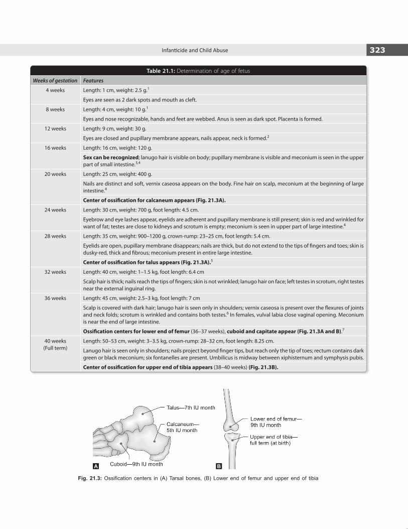
323Infanticide and Child Abuse
Table 21.1: Determination of age of fetus
Weeks of gestation Features
4 weeks Length: 1 cm, weight: 2.5 g.1
Eyes are seen as 2 dark spots and mouth as cleft.
8 weeks Length: 4 cm, weight: 10 g.1
Eyes and nose recognizable, hands and feet are webbed. Anus is seen as dark spot. Placenta is formed.
12 weeks Length: 9 cm, weight: 30 g.
Eyes are closed and pupillary membrane appears, nails appear, neck is formed.2
16 weeks Length: 16 cm, weight: 120 g.
Sex can be recognized; lanugo hair is visible on body; pupillary membrane is visible and meconium is seen in the upper part of small intestine.3,4
20 weeks Length: 25 cm, weight: 400 g.
Nails are distinct and soft, vernix caseosa appears on the body. Fine hair on scalp, meconium at the beginning of large intestine.4
Center of ossification for calcaneum appears (Fig. 21.3A).
24 weeks Length: 30 cm, weight: 700 g, foot length: 4.5 cm.
Eyebrow and eye lashes appear, eyelids are adherent and pupillary membrane is still present; skin is red and wrinkled for want of fat; testes are close to kidneys and scrotum is empty; meconium is seen in upper part of large intestine.4
28 weeks Length: 35 cm, weight: 900–1200 g, crown-rump: 23–25 cm, foot length: 5.4 cm.
Eyelids are open, pupillary membrane disappears; nails are thick, but do not extend to the tips of fingers and toes; skin is dusky-red, thick and fibrous; meconium present in entire large intestine.
Center of ossification for talus appears (Fig. 21.3A).5
32 weeks Length: 40 cm, weight: 1–1.5 kg, foot length: 6.4 cm
Scalp hair is thick; nails reach the tips of fingers; skin is not wrinkled; lanugo hair on face; left testes in scrotum, right testes near the external inguinal ring.
36 weeks Length: 45 cm, weight: 2.5–3 kg, foot length: 7 cm
Scalp is covered with dark hair; lanugo hair is seen only in shoulders; vernix caseosa is present over the flexures of joints and neck folds; scrotum is wrinkled and contains both testes.6 In females, vulval labia close vaginal opening. Meconium is near the end of large intestine.
Ossification centers for lower end of femur (36–37 weeks), cuboid and capitate appear (Fig. 21.3A and B).7
40 weeks (Full term)
Length: 50–53 cm, weight: 3–3.5 kg, crown-rump: 28–32 cm, foot length: 8.25 cm.
Lanugo hair is seen only in shoulders; nails project beyond finger tips, but reach only the tip of toes; rectum contains dark green or black meconium; six fontanelles are present. Umbilicus is midway between xiphisternum and symphysis pubis.
Center of ossification for upper end of tibia appears (38–40 weeks) (Fig. 21.3B).
Fig. 21.3: Ossification centers in (A) Tarsal bones, (B) Lower end of femur and upper end of tibia
A B
324 Review of Forensic Medicine and Toxicology
After birth, increase in the length of the child is given in Table 21.2. Length is measured in children before they are able to stand; height is measured once the child can stand. Birth weight doubles by about 5–6 months of age, triples by about 1 year.10
Table 21.2: Length/height of infant/child
Age Length (cm)
At birth 50
6 months 68
1 year 75
4 years9 100
Demonstration of Centers of Ossification
� Sternum: The bone is placed on a wooden board and sectioned in its long axis with a cartilage knife which exposes the centers of ossification.
� Lower end of the femur and the upper end of tibia: The leg is flexed against the thigh and a horizontal incision made into the knee joint and the patella is removed. A number of cross-sections are made through the epiphysis starting from the articular surface and continuing until the largest cross- section of the ossification center is reached. In the lower end of the femur, this is seen as brownish-red nucleus which is surrounded by a bluish-white cartilage.
� Bones of the foot: The heel of the foot is held by one hand and with the other hand an incision is made through the interspace between the 3rd–4th toes and carried downwards through the sole of the foot and heel.
Diagnosis of Fetal Death
Ultrasonography: On ultrasonography, absence of all fetal movements for 10 minutes (min) is taken as evidence of fetal death.
Viability of Fetus/Infant
Viability of infant: It means physical ability of fetus to lead a separate existence after birth, apart from its mother by virtue of a certain degree of development which depends on biological, physiological and extrinsic factors.
� The age of viability varies among countries with 24 and 28 weeks being cited as the lower limits of potential survival.
� Medically, the age of viability in India is taken as 28 weeks of gestation.
� Till date, there is no legally defined cut-off limit of intrauterine development, age or weight at which a baby automatically becomes viable.
� Any newborn infant, whatever is the length of gestation, can be a victim of infanticide, if born alive.
� A premature baby in a rural area in a developing country is unlikely to survive.
� In the UK, a baby is stillborn, if after 24 weeks of gestation it did not at any time after being completely expelled from its mother, breathe or show any other sign of life. Hence, a period of 24 weeks is fixed for the legal age of viability. However, there is no law or Section of IPC or CrPC in India which stipulates the age of viability.
� In other developed countries, fetal death occurring ≥ 20 weeks of fetal life or a birth weight of at least 400–500 g is considered as ‘stillbirth.’
Live-Born / Dead-Born / Stillborn
The question as to whether or not a fetus/infant was born alive is a contentious issue. When decomposition is not present, a variety of features are taken into consideration in attempting to answer this question. There are essentially three possibilities: i. the baby was born alive. ii. the baby died in utero. iii. the baby died during the birth process.
Signs of Live Birth
In India, live birth means the fetus was alive after complete birth or when at least one part of its body comes out of mother’s womb. In the UK, it means the baby should be alive after complete birth. Any sign of life after complete birth of child is accepted as proof of live birth. Following are considered as signs of live birth:
� Baby’s cry—Strong evidence in favor of live birth and respiration having taken place. Fetus may inhale air and cry when the head is inside the vagina—vagitus vaginalis, or inside the uterus—vagitus uterinus.
� Muscle twitching/movements of limbs. � Sneezing and yawning.
Postmortem Findings
External Findings � General findings. � Changes in the chest, umbilical cord and skin. � Caput succedaneum and cephalhematoma.

325Infanticide and Child Abuse
i. Generalfindings: Presence of clothing and absence of vernix caseosa—suggestive of live birth.
ii. Changes in the chest: Chest is more flat antero-posteriorly in still/dead born. The circumference of the chest is about 2–3 cm less than that of the abdomen at the level of the umbilicus. After respiration, the chest expands and becomes drum-shaped.
iii. Changes in umbilical cord: The appearance of the cut end of the umbilical cord may help to decide whether the birth was one where medical, nursing or midwife, or only amateur person was available. The cut end of the cord should be looked for vital reaction. Even where early putrefaction renders evaluation of breathing impossible, vital signs in the cord may indicate live birth, if survival reached 24–48 hours (h) (Table 21.3).
Table 21.3: Time since birth by umbilical cord changes
Changes observed Time since birth
Drying up of cut margin 2 h
Drying up of cord 1 day
Inflammatory line at the base of stump 2 days
Obliteration and mummification changes 3 days
Detach (falls off) 5–6 days
Complete healing (scar) 10–12 days
iv. Changes in skin: Vernix caseosa is present on axilla, inguinal region, folds of neck and buttocks. It is either cleaned or gets removed in 1–2 days. Skin of abdomen exfoliates during the first 3 days after birth (Table 21.4).
Table 21.4: Changes in skin color
Color of skin Time since birth
Bright red Just born
Darker 2–3 days
Brick red→yellow→normal 1 week
v. Cephalhematoma and caput succedaneum (Diff. 21.1, Figs 21.4 and 21.5).
Internal Findings
i. Changes in the lungs: They are considered with reference to volume, consistence, color and weight (Diff. 21.2).
z Volume– Before respiration, the lungs are small with
sharp margins, wrinkled loose pleura, lie in the back of the chest on either side of the vertebral column and are hardly seen on opening the chest, as the cavity is filled up by the heart and thymus.
– After respiration, the lungs increases in volume, thin tense pleura, rounded margins and occupy the cavity, the medial edges overlapping the mediastinum and part of the pericardium, though not as fully as in the older neonate.
z Consistence– Before respiration, the lungs are dense,
uniform, rubbery, firm and liver-like. On rubbing a small piece between the fingers close to ear, no crepitation is heard (non-crepitant lungs).
Differentiation 21.1: Cephalhematoma and caput succedaneum11-14
S.No. Feature Cephalhematoma Caput succedaneum
1. Definition Collection of blood in between the periosteum and the skull due to rupture of a small emissary vein from the skull, and may be caused by forceps delivery
Swelling due to stagnation of fluid between the layers of scalp beneath the girdle of contact (dilated cervix or vulval ring)
2. Situation Usually unilateral, and present over parietal bone May be bilateral
3. Impulse on crying No impulse No impulse
4. Limitation by suture line Yes Not limited
5. Underlying pathology May be associated with fracture of skull bone Not pathological
6. Occurrence It is never present at birth It is present at birth
7. Development and disappearance
Develops 12–24 h after birth and decreases in 6–8 weeks
Disappears spontaneously within 24 h
8. Medico-legal importance Regression process help to conclude about the separate existence and how many days the infant survived after birth
Definite evidence of fetus being alive in uterus. But in prolonged labor, the fetus may die before birth
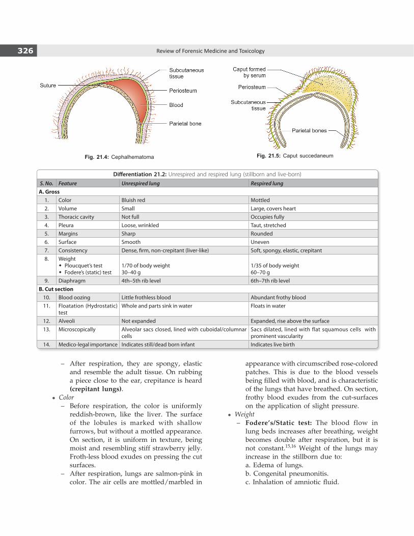
326 Review of Forensic Medicine and Toxicology
Fig. 21.4: Cephalhematoma Fig. 21.5: Caput succedaneum
– After respiration, they are spongy, elastic and resemble the adult tissue. On rubbing a piece close to the ear, crepitance is heard (crepitant lungs).
z Color– Before respiration, the color is uniformly
reddish-brown, like the liver. The surface of the lobules is marked with shallow furrows, but without a mottled appearance. On section, it is uniform in texture, being moist and resembling stiff strawberry jelly. Froth-less blood exudes on pressing the cut surfaces.
– After respiration, lungs are salmon-pink in color. The air cells are mottled/marbled in
appearance with circumscribed rose-colored patches. This is due to the blood vessels being filled with blood, and is characteristic of the lungs that have breathed. On section, frothy blood exudes from the cut-surfaces on the application of slight pressure.
z Weight– Fodere’s/Static test: The blood flow in
lung beds increases after breathing, weight becomes double after respiration, but it is not constant.15,16 Weight of the lungs may increase in the stillborn due to:
a. Edema of lungs. b. Congenital pneumonitis. c. Inhalation of amniotic fluid.
Differentiation 21.2: Unrespired and respired lung (stillborn and live-born)
S. No. Feature Unrespired lung Respired lungA. Gross
1. Color Bluish red Mottled 2. Volume Small Large, covers heart3. Thoracic cavity Not full Occupies fully4. Pleura Loose, wrinkled Taut, stretched5. Margins Sharp Rounded6. Surface Smooth Uneven7. Consistency Dense, firm, non-crepitant (liver-like) Soft, spongy, elastic, crepitant8. Weight
� Ploucquet’s test � Fodere’s (static) test
1/70 of body weight30–40 g
1/35 of body weight60–70 g
9. Diaphragm 4th–5th rib level 6th–7th rib levelB. Cut section
10. Blood oozing Little frothless blood Abundant frothy blood11. Floatation (Hydrostatic)
testWhole and parts sink in water Floats in water
12. Alveoli Not expanded Expanded, rise above the surface13. Microscopically Alveolar sacs closed, lined with cuboidal/columnar
cellsSacs dilated, lined with flat squamous cells with prominent vascularity
14. Medico-legal importance Indicates still/dead born infant Indicates live birth
327Infanticide and Child Abuse
– Ploucquet’s test: This test helps to demonstrate establishment of respiration. The ratio of the weight of the lungs and the whole body is reduced to half (1/35 of body weight) as compared to the said ratio before respiration (1/70 of body weight).16,17
ii. Position of the diaphragm: The position of the diaphragm is at the level of the 4th–5th rib, if respiration has not taken place. The arch becomes flattened and depressed, and descends to 6th–7th rib after respiration. The position of the diaphragm may be affected by pressure of the gases of putrefaction.
iii. Hydrostatic test Hydrostatic test is also called floatation test or
Raygat’s test.16,18
Principle: It is based on the fact that specific gravity of lung before respiration is 1040–1050 and becomes 940-950 after respiration which is less than that of water. This makes the respired lung to float.
Procedure: Dissect out the fetal lungs. Put both the lungs (tied at their hilar region) into a trough of water and observe.
Inference z If they sink—unrespired lung. z If they float—remove them from water, cut into
small pieces and then squeeze or compress firmly between sponges, and again put into water.
z If they sink—unrespired lung. z If they float—respired lung.
Explanation: Floatation observed for second time is because of residual air that remains in the lungs which cannot be squeezed out by pressing, if the fetus has breathed after birth.
z The test is of limited value and it can at best be a suggestive pointer, but never a definitive test in itself.
z The slightest degree of putrefaction immediately negates any interpretation of this test. In such cases, the lungs will float in water, but so are the solid organs, such as the liver.
z However, assuming body is fresh, the floating of lungs and heart en bloc increases the sensitivity of the test.
Fallacies z False positive is seen in accumulation of
putrefying gases or artificial inflation (by intubation and forced air insufflation expanding the lungs with air after delivery).
z False negative is seen in atelectasis (non-expansion of lungs), obstruction by alveolar duct
membrane, edema, feeble respiration, pneumonia and congenital syphilis.19
Hydrostatic test is not necessary if: i. Fetus shows congenital anomaly, like anencephaly. ii. Fetus is macerated or mummified. iii. Umbilical cord has separated and a scar has formed. iv. Stomach contains milk. v. Bruises on lungs indicating efforts to artificially
respirate the child. vi. Fetus is born before 180 days of gestation.*
iv. Histology z Unrespired lung looks like the parotid gland
with closed alveolar sacs lined with cuboidal/columnar cells, and less vascularity.
z Respired lung cells get flattened with dilatation—pavement (squamous) epithelium with increased vascularization.
v. Changes in middle ear (Wreden’s test): Absence of gelatinous embryonic connective tissue which was present during fetal life and presence of air in middle ear is seen after live birth.20 It is also called Wreden-Wendt tympanic cavity or middle ear test.
vi. Changes in stomach and intestines: Live born infant swallows air into the stomach during respiration, and if present in small intestine it further confirms live birth. But air may be present in the stomach after decomposition, or in the stillborn attempting to free the air passages of fluid obstruction.
Demonstration: The stomach and intestines are removed after tying double ligatures at each end. They are kept under water and incision is given between the ligatures. Air bubbles will come out, if respiration has taken place—Breslau’s second life test or stomach bowel test.21
If milk is present in the stomach, it is a positive evidence of live birth.
vii. Meconium: In case of live birth, the large intestine is completely free of meconium within 24 h after birth, but in stillbirths it will be present in the intestine. In case of breech presentation and hypoxia, meconium may be completely expelled before birth, and thus may be absent even in such stillborn fetuses.
viii. Changes in the blood vessels: Umbilical arteries are obliterated within 12 h to 3 days. Oblitera tion of umbilical vein and ductus venosus is complete by 4th day. The ductus arteriosus obliterates in about 10 days.
* Not a criteria in India, since there is no legally defined cut-off limit of viability.

328 Review of Forensic Medicine and Toxicology
ix. Changes in heart: Closure of foramen ovale occurs by 2–3 months after birth. In few cases, the foramen may not completely close.
x. Changes in the blood: Nucleated RBCs are absent in peripheral circulation within 24 h after live birth. Fetal hemoglobin may be present in the blood upto 6 months or more.
xi. Incremental line in enamel of teeth: Neonatal incremental line in the enamel of the teeth is formed at birth which is one of the surest sign of live birth.
xii. Ossificationcenters: Presence of ossification centers at the lower end of radius, heads of humerus and femur and capitulum of humerus may also be taken as signs of separate existence for few months.
xiii. Closure of fontanelle: Closure of different fontanelle occurs at different periods after birth. Closure of posterior fontanelle may occur at birth.
Signs of Dead-Born Fetus
� Maceration: It is a process of aseptic autolysis.22 This occurs when the dead child remains in the uterus for about 3–4 days surrounded with liquor amnii with exclusion of air.
z Earliest sign of maceration is skin slippage of face, back or abdomen which may be seen in 12 h after death in utero. By 24 h, skin is brown or purplish in color.
z The dead fetus is soft, flaccid with emission of sweetish disagreeable smell, but no gases are formed.
z Internal organs show autolytic decomposition, but the lungs and uterus remain unchanged for a long time.
z Cranial compression is seen in > 36 h, desquamation over 75% of body surface is seen in 72 h, overlapping of cranial sutures in > 96 h and the mouth is widely open in > 1 week.
Putrefaction is characterized by an unpleasant odor, greenish discoloration of skin and formation of foul smelling gases. Rarely, the fetus may show adipocere formation.23
� Spalding’s sign: A pathogonomic sign of intra-uterine death. There is loss of alignment and overlapping of fetal skull bones on X-ray, occurs due to liquefaction of cerebrum and softening of ligamentous structures supporting the vault. It appears in about 7 days after death.24,25
� Rigor mortis: It may be seen in dead-born fetus. � Mummification: It results from deficient supply of
blood or scanty liquor amnii. Fetus is dried up and shriveled in ≥ 2 weeks.
� Robert’s sign: Appearance of gas shadow in chambers of heart and great vessels, may appear by 12 h, but difficult to interpret.26
� Hyperflexionof spine is more common. � Crowding of the ribs shadow with loss of normal
parallelism.
Two cardinal signs to identify a dead born fetus are the presence of maceration along with lack of lung aeration (‘primary atelectasis’).
Signs of Stillborn Fetus
Another possibility for deaths occurring in infants whose births are unattended or complicated is that the fetus/infant died during the birth process.
� The findings which suggest that a death is an intrapartum death are lack of maceration and lack of lung aeration.27
� The difference between stillborn and dead-born fetus is given in Diff. 21.3.
� In most instances, it is not possible by autopsy alone to differentiate these cases from deaths that occur prior to birth but have not yet developed maceration or from deaths that occur after birth where there is little or no aeration of the lungs.
� When a precipitate birth occurs, air can enter the lungs, via the chest compression followed by rapid chest expansion that occurs during passage through the birth canal, even if the infant does not actively inhale.
� Iatrogenic deaths may result from improper body positioning of the mother, use of medications (such as epidurals), use of various maneuvers, instrumentation, and surgical interventions which can result in prolongation of the labor and birth process. Prolongation of labor might contribute to birth asphyxia.
� Question on negligence: Charges for wrongful or negligent act may be brought against the medical practitioner if the act results in a miscarriage or stillbirth of the fetus. The medical practitioner become liable to pay damages, if the harm suffered is the result of his/her tortious conduct.
The law in the US and the UK presumes that every newborn child found dead was born dead, until the contrary is proved. When a woman is charged with infanticide, the burden of proof is upon the prosecution to demonstrate that the child had a separate existence. Unless the autopsy surgeon has absolute reasons to document post-natal survival, for e.g., well-expanded lungs or food in the stomach, he is legally bound not to diagnose live birth.
329Infanticide and Child Abuse
Infant Death (Flow chart 21.1)
a. Natural Causes
� Prematurity � Congenital malformation � Birth trauma � Intrapartum asphyxia � Neonatal infection � ABO and Rh-incompatibility
� Post-maturity � Early separation of placenta
� Preeclamptic toxemia � Sudden infant death syndrome (SIDS)
Some common causes are: � Immaturity: A prematurely born child generally dies
immediately after birth. In the case of the premature birth of a child, the question may arise as to whether the birth was criminally induced or not, for under the IPC, the criminal induction of premature labour is an offence.
� Debility: Due to lack of general development, even a full term child may die after birth from debility. In these cases, no disease, except atelectasis of some portions of the lungs due to feeble respiration is detected.
� Congenital diseases and malformations: Syphilis and some fevers may cause death from the toxaemic condition. Of the diseases of the internal organs, pulmonary infections and hyaline membrane of the lungs are seen. Certain conditions such as anencephaly, spina bifida and congenital diaphragmatic hernia are readily identifiable, although subtle cardiovascular or metabolic abnormalities may be difficult to diagnose. It is, however, unsafe to assume that actual live birth could not have taken place. Moreover, monstrosity or malformation is no justification for taking the life of an infant.
� Spasm of the larynx may occur from mucus or meconium being aspirated into the larynx, or from the enlargement of thymus gland.
� Erythroblastosis fetalis due to iso-immunization, when an Rh-negative woman is carrying an Rh-positive fetus may result in death of the fetus.
� Birth asphyxia can occur in preeclampsia/eclampsia, placenta abruption, cephalo-pelvic disproportion and shoulder dystocia. Evidence of asphyxia at autopsy includes thymic, pleural and epicardial petechiae with intra-alveolar hemorrhage, and meconium on the skin, and shed fetal skin within distal air passages.
b. Unnatural Causes
I. Accidental causes
Perinatal
i. Injuries to the mother: It may cause premature separation of the placenta or injury to the fetus (concussion of brain/fracture/rupture of blood vessels) and lead to death of the baby.
ii. Prolonged labor: It causes death of the fetus due to injury to the brain because of compression of the head or due to asphyxia.
Flow chart 21.1: Causes of infant death
Differentiation 21.3: Stillborn and dead-born fetus
S. No. Feature Stillborn fetus Dead-born fetus1. Definition Fetus which is born after 28 weeks of pregnancy (24 weeks in
the UK), and which did not breath or show any other signs of life at any time after being completely born (WHO)
Fetus which has died in utero
2. Condition in utero Fetus was alive in utero, but dies during the process of delivery Dead in utero3. Predominance Seen mostly among illegitimate and immature male children
in primiparaeNo such predominance
4. Findings Signs of prolonged labor, like edema, bleeding into scalp, caput succedaneum and severe moulding of head may be seen
� Maceration � Spalding’s sign � Robert’s sign � Rigor mortis at delivery � Mummification
5. Cause Anoxia, prematurity, birth trauma or toxemia Congenital anomaly, ABO and Rh incompatibility
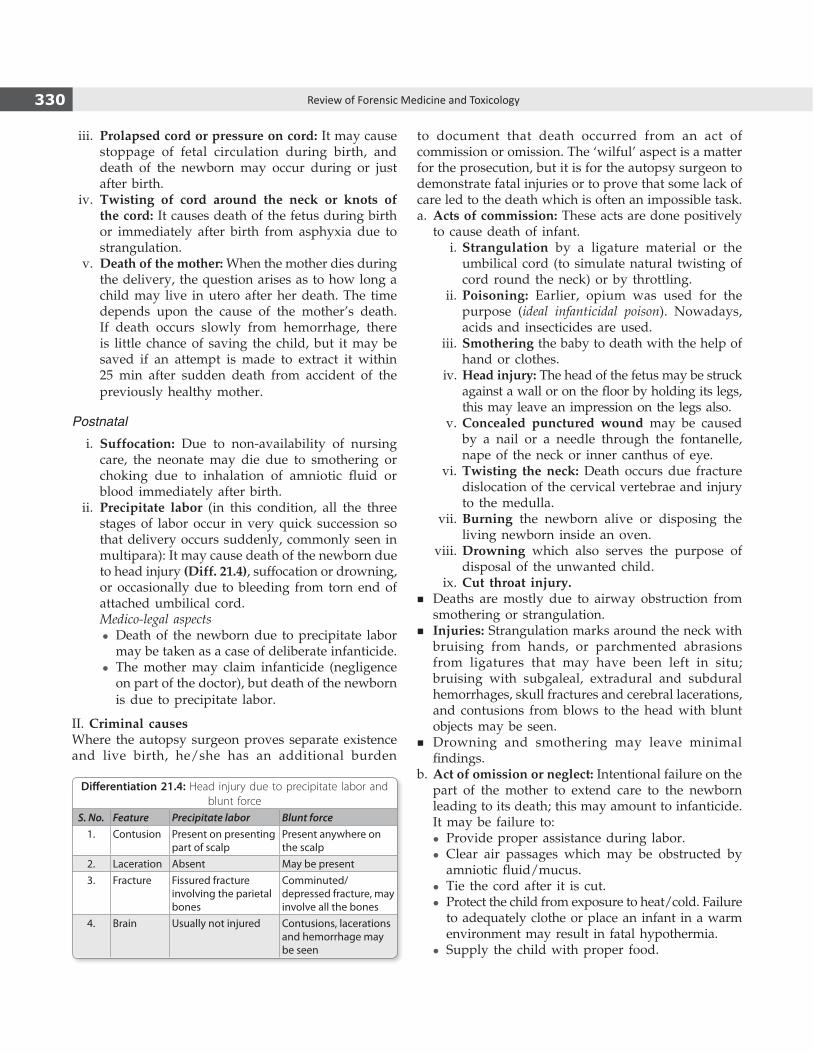
330 Review of Forensic Medicine and Toxicology
iii. Prolapsed cord or pressure on cord: It may cause stoppage of fetal circulation during birth, and death of the newborn may occur during or just after birth.
iv. Twisting of cord around the neck or knots of the cord: It causes death of the fetus during birth or immediately after birth from asphyxia due to strangulation.
v. Death of the mother: When the mother dies during the delivery, the question arises as to how long a child may live in utero after her death. The time depends upon the cause of the mother’s death. If death occurs slowly from hemorrhage, there is little chance of saving the child, but it may be saved if an attempt is made to extract it within 25 min after sudden death from accident of the previously healthy mother.
Postnatal
i. Suffocation: Due to non-availability of nursing care, the neonate may die due to smothering or choking due to inhalation of amniotic fluid or blood immediately after birth.
ii. Precipitate labor (in this condition, all the three stages of labor occur in very quick succession so that delivery occurs suddenly, commonly seen in multipara): It may cause death of the newborn due to head injury (Diff. 21.4), suffocation or drowning, or occasionally due to bleeding from torn end of attached umbilical cord.
Medico-legal aspects z Death of the newborn due to precipitate labor
may be taken as a case of deliberate infanticide. z The mother may claim infanticide (negligence
on part of the doctor), but death of the newborn is due to precipitate labor.
II. Criminal causesWhere the autopsy surgeon proves separate existence and live birth, he/she has an additional burden
to document that death occurred from an act of commission or omission. The ‘wilful’ aspect is a matter for the prosecution, but it is for the autopsy surgeon to demonstrate fatal injuries or to prove that some lack of care led to the death which is often an impossible task.a. Acts of commission: These acts are done positively
to cause death of infant. i. Strangulation by a ligature material or the
umbilical cord (to simulate natural twisting of cord round the neck) or by throttling.
ii. Poisoning: Earlier, opium was used for the purpose (ideal infanticidal poison). Nowadays, acids and insecticides are used.
iii. Smothering the baby to death with the help of hand or clothes.
iv. Head injury: The head of the fetus may be struck against a wall or on the floor by holding its legs, this may leave an impression on the legs also.
v. Concealed punctured wound may be caused by a nail or a needle through the fontanelle, nape of the neck or inner canthus of eye.
vi. Twisting the neck: Death occurs due fracture dislocation of the cervical vertebrae and injury to the medulla.
vii. Burning the newborn alive or disposing the living newborn inside an oven.
viii. Drowning which also serves the purpose of disposal of the unwanted child.
ix. Cut throat injury. � Deaths are mostly due to airway obstruction from
smothering or strangulation. � Injuries: Strangulation marks around the neck with
bruising from hands, or parchmented abrasions from ligatures that may have been left in situ; bruising with subgaleal, extradural and subdural hemorrhages, skull fractures and cerebral lacerations, and contusions from blows to the head with blunt objects may be seen.
� Drowning and smothering may leave minimal findings.
b. Act of omission or neglect: Intentional failure on the part of the mother to extend care to the newborn leading to its death; this may amount to infanticide. It may be failure to:
z Provide proper assistance during labor. z Clear air passages which may be obstructed by
amniotic fluid/mucus. z Tie the cord after it is cut. z Protect the child from exposure to heat/cold. Failure
to adequately clothe or place an infant in a warm environment may result in fatal hypothermia.
z Supply the child with proper food.
Differentiation 21.4: Head injury due to precipitate labor and blunt force
S. No. Feature Precipitate labor Blunt force1. Contusion Present on presenting
part of scalpPresent anywhere on the scalp
2. Laceration Absent May be present3. Fracture Fissured fracture
involving the parietal bones
Comminuted/depressed fracture, may involve all the bones
4. Brain Usually not injured Contusions, lacerations and hemorrhage may be seen

331Infanticide and Child Abuse
Abandoning of Children
Sec. 317 IPC deals with abandoning by the father or mother of the child under the age of 12 years with imprisonment upto 7 years and with/without fine.
Concealment of Birth by Secret Disposal of Dead Body
Any person who secretly buries or disposes of the dead body of a child and intentionally conceals the birth of such child is punished with imprisonment of 2 years and with/without fine (Sec. 318 IPC).28 It does not matter whether the child died before or after or during its birth. In a case where infanticide is not proved, the person is usually charged under this section.
Battered Baby Syndrome
Definition: A battered child is one who has received repetitive physical injuries as a result of non-accidental violence produced by a parent or a guardian.
It is also called Caffey syndrome, Caffey-Kempe syndrome, maltreatment syndrome or parent-infant traumatic stress syndrome.
Features
Related to the Child
i. Age: The majority is below 3 years of age. ii. Sex: More common with male children (M:F ratio
2:1). iii. Status of the child: Usually, illegitimate and
unwanted children—pregnancy before marriage or failure of contraception.
iv. Position in family: Commonly, the eldest or the youngest. The child may be a mentally abnormal one.
Related to the Parent/Guardian
i. Marital status: Unmarried couple, commonly seen in some Western societies.
ii. Age of parents: Usually, the parents are young. iii. Educational status: Lower level of education. iv. Addiction: Reckless life style, often indulging in
drugs. v. Childhood history: Often the parents themselves
were the victims of battering during their childhood. vi. Psychological factors: Low tolerance threshold,
impulsive nature, aggressive personality and imbalanced temperament.
Socio-familial factors i. Low social background. ii. Lack of equality between members of the family
with lack of family harmony. iii. Long-standing emotional problem. iv. Financial hardship. v. Trouble at the place of work.
Precipitating factors i. Act of disobedience by the child. ii. Frequent crying may create annoyance. iii. Refusal to take food. iv. Soiling of napkin or bedclothes. v. At times, any trifle act of the child may annoy the
mentally challenged father or mother.
Features Arising Suspicion of Abuse
� Parents give vague history of accident to be the cause of the injuries, like fall from stairs or cot which does not appear consistent with the type of injuries or time narrated by the parents (Fig. 21.6). Often the parents’ gives a history of tendency of the child to bruise easily.
� The parents of the child seek medical aid rather late or when the condition of the children becomes serious.
� Often injuries in different stages of healing are found in the child.
Fig. 21.6: Sites of non-accidental injury

332 Review of Forensic Medicine and Toxicology
� In many cases, the parents later admit to have assaulted their children, but ‘only mildly’ for punishment.
Accidental injuries typically involve bony prominences [head (forehead, occipital or parietal region), nose, chin, palm, elbows, knees and shin], match the history given by the parents and are keeping with the development of the child.
Injuries
The injuries may be caused by hand, foot, teeth, stick, belt, shoe, hot water, lighted cigarette, hot frying pan or any household article. i. Surface injuries: Bruises, abrasions and lacerations
may be seen. Laceration of the oral mucosa along with labial frenulum of the lower lip is a characteristic lesion. Slap marks, lash mark, knuckle punches, pinch mark [butterfly-shaped bruise with one wing (caused by thumb) larger than other], bald patches on scalp due to pulling out the hair (traumatic alopecia) may be seen (Fig. 21.7).
ii. CNS: Injuries are inflicted by throwing the child, striking the child with fist or object or against a wall, dropping the child or vigorous shaking of the infant (shaken baby syndrome or infantile whiplash syndrome) leading to intracranial hemorrhage (Fig. 21.8).
z A strong suspicion of child abuse should be made in a child presenting with altered mental status, unresponsiveness, coma, convulsions or with focal neurologic deficit.
z Shaken baby syndrome can occur from as little as 5 seconds of shaking.
z The triad of injuries includes encephalopathy, retinal hemorrhages and SDH.29,30 SDH is the most consistent component of the triad and may be the first clinical sign identified on CT scan. Additional traumatic injuries of the cord, brainstem and even skull may be produced.
iii. Eyes: Retinal hemorrhages and lens displacement may be seen.
iv. Visceral injuries: Injury to spleen, liver or hollow viscera can occur resulting in massive hemorrhage, shock and death of the child.
v. Burns: Small circular pitted burns may indicate deliberate stubbing of cigarette ends on skin. Scalds are also common (Fig. 21.7).
vi. Skeletal injuries: Bony injuries include transverse fractures, impacted fractures, spiral fractures, meta-physeal chip fractures, subperiosteal hematoma, and multiple deformities of the long bones and rib cage of the body due to multiple healed fractures and callus formation.
Fig. 21.7: Surface injuries

333Infanticide and Child Abuse
z Fractures of long bones, ribs, skull and vertebral bodies are highly suggestive of abuse.
z Antero-posterior compression of chest causes fractures in midaxillary line (Fig. 21.8).
z Multiple rib fractures also occur along posterior angle of ribs on side-to-side squeezing (Fig. 21.8). The fractured ribs heal by callus formation in 1–2 weeks, giving characteristic appearance of a knob (knob fractures), and on X-ray ‘string of beads’ appearance is seen in paravertebral gutter.
z In whiplash movement of arms and legs, typical ‘corner’ or ‘bucket-handle’ fractures in the metaphyseal region may be seen (Fig. 21.8).
vii. CVS: Blunt trauma to chest may cause multiple rib fractures leading to lung and heart contusions, pneumothorax, hemothorax, rupture of diaphragm and cardiac tamponade.
viii. Genitourinary system: Physical and sexual abuse should be considered in a child presenting with hematuria, dysuria, increased frequency of urination and enuresis.
Diagnosis
i. Nature of injuries. ii. Delay in seeking medical treatment. iii. Recurrent injuries. iv. Radiological manifestations, especially those
involving the ribs, metaphyseal-epiphyseal injuries, and avulsive fractures of the clavicle and acromium process.
Head injury with or without skull fracture is the leading cause of death in child abuse followed by rupture of an abdominal viscus.
Child abuse can be defined as causing or permitting of any harmful or offensive contact to a child’s body and/or any communication or transaction which humiliates, shames, or frightens a child.Major types of abuse i. Physical abuse ii. Sexual abuse iii. Emotional abuse iv. Neglect
i. Physical abuse of children includes any non-accidental physical injury caused by the child’s caretaker. It can be beating or battering of a child, and has been described above.
ii. Sexual abuse refers to inappropriate sexual behavior with a child. It includes fondling a child’s genitals, making the child fondle the adult’s genitals, intercourse, incest, rape, sodomy, exhibitionism, indecent exposure and commercial exploitation through prostitution or the production of pornographic materials.
iii. Emotional abuse (verbal/mental abuse or psychological maltreatment): Acts of commission and omission which can be potentially damaging psychologically. This can include parents/caretakers using extreme and/or bizarre forms of punishment, such as confinement in a closet or dark room or being tied to a chair for long periods.
iv. Neglect is the failure to provide for the child’s basic needs. Neglect can be physical, educational or emotional. In general, neglect is an act of omission.
Differential diagnosis of childhood fractures should be made from the several ‘brittle bone diseases’ that can cause abnormal skeletal fragility—congenital syphilis, rickets, scurvy, leukemia, osteogenesis imperfecta, copper deficiency, Menke’s syndrome, infantile cortical hyperostosis (Caffey’s disease) and juvenile osteoporosis.
It can be defense in a criminal trial of alleged child abuse on the grounds that such fractures can be observed within normal parental handling or spontaneous movements of the child.Shaken baby syndrome: Infants are susceptible to subdural/subarachnoid hematoma and retinal hemorrhages due to vigorous shaking of the baby as a method of punishment.
� Predisposing factors: Infant’s relatively large head, weak neck muscles and delicate subarachnoid bridging vessels.
� Signs and symptoms: Seizures, irritability, meningismus and focal or general neurologic deficit.
� Diagnosis: Confirmation by CT/MRI scan, bloody spinal or subdural fluid and normal skull X-rays.
Reporting of suspected child abuse: It is mandatory to report any suspected child abuse case in the US, Argentina, Finland, Israel, Korea and Spain. In other countries such as Croatia, Japan, Netherlands and Romania reporting is voluntary.
� In India, it is mandatory to report to the police about sexual abuse under the Protection of Children from Sexual Offences Act, 2012.
Sudden Infant Death Syndrome [SIDS, Cot Death (UK) or Crib Death (US)]
Definition: Sudden and unexpected death of seemingly healthy infant whose death remains unexplained even after complete autopsy. It is an autopsy diagnosis, and not a clinical diagnosis.Fig. 21.8: Internal injuries

334 Review of Forensic Medicine and Toxicology
Features
i. Incidence: 0.2–0.4% of all live births. ii. Geographical distribution: Worldwide. iii. Age: Between 2 weeks to 2 years. Mid infancy is
the most vulnerable age (peak 2–4 months). iv. Sex: Male infants have a proportionately higher
death rate (M:F ratio 3:2). v. Socio-economic status: Low and middle class
family with poor housing condition, large family and lack of health consciousness.
vi. Time of death: In most cases, the infant is discovered dead, either in the early morning (death possibly occurring at late night) or sometime after first feed in the morning.
vii. Season: In most occasions, deaths are seen to occur commonly in rainy and winter seasons in temperate zones, but no clear pattern in tropical zones.
viii. Twins: More among twins (two-fold) as opposed to singletons. Prematurity and low birth weights which are often present in twins increases the risk of SIDS.
ix. Addiction: Smoking (pre-or postnatal) and drug abuse by pregnant women increases risk.
Cause
No definite cause is known. i. Prolonged sleep apnea is presently accepted as
the most acceptable of the suggested causes. A periodic failure to breath during sleep makes them susceptible to hypoxia. Hypoxic state may be promoted by many allied factors, e.g. some infective condition of the respiratory tract.
ii. Respiratory infection may cause viremia which leads to sleep depression of respiratory center and death.
iii. Nasal edema and mucus secretion may narrow upper respiratory passages, a flaccid pharynx and neck posture may reduce airway.
iv. Local hypersensitivity of the respiratory tract lumen to cow’s milk was thought to cause laryngeal spasm.
v. Bedclothes and pillow falling accidentally over the nose and mouth by the movement of the child.
vi. Overlying of the baby by a sleeping or intoxicated mother. Infants placed to sleep prone or on their side increases the risk of SIDS.
vii. Miscellaneous causes: Conduction system anomalies; hypoparathyroidism; deficiency of selenium, antibodies, calcium, magnesium and vitamins B, C, D and E; house-mite allergy; sodium overload in feeds and hypothermia.
There is an increased risk of SIDS as well as other causes of death in families that have one SIDS death.
Postmortem Findings � Postmortem findings are negative. � Trachea contains milky vomit, sometimes blood-
stained with shed epithelial cells. � Multiple petechial hemorrhages on heart (posterior epi-
cardial surface), lungs and thymus—agonal in nature. � Pulmonary edema is common. � Milk or bloodstained froth on child’s mouth or
bedding. Hands are often clenched around fibers from bedclothes.
Medico-legal Aspects � SIDS is a natural death in which the parents may be
wrongfully linked for having criminal involvement or negligence.
� Some infanticide cases may be presented as cot death cases.
Munchausen syndrome by proxy [MSBP or Factitious disorder (Latin facticious: made by art)]:31 MSBP is a form of abuse in which parent or guardian fabricates or produces symptoms of an illness in a child in order to gain sympathy or attention for themselves.
� The parents frequently have abnormal or borderline personality disorder.
� Diagnosis may require a high level of suspicion and may be met with considerable resistance from family.
Featuresi. The child may be brought with vague complaints such as
vomiting, diarrhea, fever or seizures inflicted by the parent intentionally and repeatively, for e.g. bleeding may be caused by anticoagulants and simulated by exogenous blood, seizures can be caused by suffocations, shaking or intoxications, vomiting can be caused by giving ipecac syrup and fever triggered by injecting contaminants into IV lines while the child is in the hospital.
ii. The parent or guardian derives some non-economic benefit at the expense of the victim.
iii. Some perpetrators ‘doctor shop’ while some maintain a constant relationship with one or more health care providers.
iv. When confronted, the parent or guardian usually denies any allegations of causing the victim’s condition.32
Diagnosis i. The illness does not conform to the expected presen tation or
follow the usual course. ii. Signs and symptoms are not substantiated by labora tory or
imaging findings. iii. Failure of wounds to heal. iv. The child becomes ill or worsens when the parent or guardian
is present, with recovery when separated. v. Positive drug or toxicological analysis for something not
prescribed for the patient. vi. Finding that the patient has been admitted to multiple hospitals
and has been seen by multiple physicians.
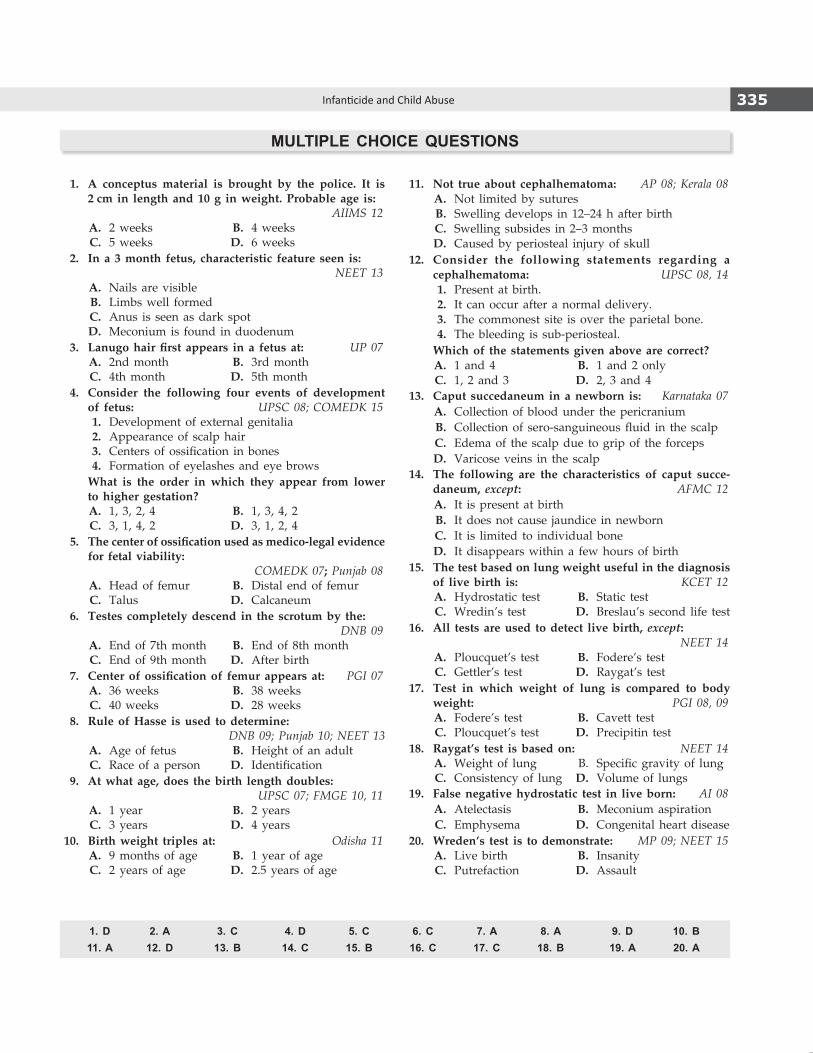
335Infanticide and Child Abuse
1. A conceptus material is brought by the police. It is 2 cm in length and 10 g in weight. Probable age is: AIIMS 12
A. 2 weeks B. 4 weeks C. 5 weeks D. 6 weeks 2. In a 3 month fetus, characteristic feature seen is:
NEET 13 A. Nails are visible B. Limbs well formed C. Anus is seen as dark spot D. Meconium is found in duodenum 3. Lanugohairfirst appears ina fetusat: UP 07 A. 2nd month B. 3rd month C. 4th month D. 5th month 4. Consider the following four events of development
of fetus: UPSC 08; COMEDK 15 1. Development of external genitalia 2. Appearance of scalp hair 3. Centers of ossification in bones 4. Formation of eyelashes and eye brows What is the order in which they appear from lower
to higher gestation? A. 1, 3, 2, 4 B. 1, 3, 4, 2 C. 3, 1, 4, 2 D. 3, 1, 2, 4 5. Thecenterofossificationusedasmedico-legalevidence
for fetal viability: COMEDK 07; Punjab 08
A. Head of femur B. Distal end of femur C. Talus D. Calcaneum 6. Testes completely descend in the scrotum by the:
DNB 09 A. End of 7th month B. End of 8th month C. End of 9th month D. After birth 7. Centerofossificationof femurappearsat: PGI 07 A. 36 weeks B. 38 weeks C. 40 weeks D. 28 weeks 8. Rule of Hasse is used to determine:
DNB 09; Punjab 10; NEET 13 A. Age of fetus B. Height of an adult C. Race of a person D. Identification 9. At what age, does the birth length doubles:
UPSC 07; FMGE 10, 11 A. 1 year B. 2 years C. 3 years D. 4 years 10. Birth weight triples at: Odisha 11 A. 9 months of age B. 1 year of age C. 2 years of age D. 2.5 years of age
11. Not true about cephalhematoma: AP 08; Kerala 08 A. Not limited by sutures B. Swelling develops in 12–24 h after birth C. Swelling subsides in 2–3 months D. Caused by periosteal injury of skull 12. Consider the following statements regarding a
cephalhematoma: UPSC 08, 14 1. Present at birth. 2. It can occur after a normal delivery. 3. The commonest site is over the parietal bone. 4. The bleeding is sub-periosteal. Which of the statements given above are correct? A. 1 and 4 B. 1 and 2 only C. 1, 2 and 3 D. 2, 3 and 4 13. Caput succedaneum in a newborn is: Karnataka 07 A. Collection of blood under the pericranium B. Collection of sero-sanguineous fluid in the scalp C. Edema of the scalp due to grip of the forceps D. Varicose veins in the scalp 14. The following are the characteristics of caput succe-
daneum, except: AFMC 12 A. It is present at birth B. It does not cause jaundice in newborn C. It is limited to individual bone D. It disappears within a few hours of birth 15. The test based on lung weight useful in the diagnosis
of live birth is: KCET 12 A. Hydrostatic test B. Static test C. Wredin’s test D. Breslau’s second life test 16. All tests are used to detect live birth, except:
NEET 14 A. Ploucquet’s test B. Fodere’s test C. Gettler’s test D. Raygat’s test 17. Test in which weight of lung is compared to body
weight: PGI 08, 09 A. Fodere’s test B. Cavett test C. Ploucquet’s test D. Precipitin test 18. Raygat’s test is based on: NEET 14 A. Weight of lung B. Specific gravity of lung C. Consistency of lung D. Volume of lungs 19. False negative hydrostatic test in live born: AI 08 A. Atelectasis B. Meconium aspiration C. Emphysema D. Congenital heart disease 20. Wreden’s test is to demonstrate: MP 09; NEET 15 A. Live birth B. Insanity C. Putrefaction D. Assault
MULTIPLE CHOICE QUESTIONS
1. D 2. A 3. C 4. D 5. C 6. C 7. A 8. A 9. D 10. B 11. A 12. D 13. B 14. C 15. B 16. C 17. C 18. B 19. A 20. A

336 Review of Forensic Medicine and Toxicology
21. Breslau’s second life test utilizes: MAHE 09; AIIMS 12 A. Liver B. Stomach C. Ear D. Lungs 22. Aseptic autolysis is seen in: AP 08 A. Adipocere B. Maceration C. Putrefaction D. Mummification 23. Dead-born fetus does not show: Kerala 11 A. Rigor mortis at birth B. Adipocere formation C. Maceration D. Mummification 24. Spalding sign is seen in:
AI 07; BHU 09; AFMC 11; CMC (Vellore) 14 A. Maceration B. Mummification C. Putrefaction D. Saponification 25. Spalding sign is seen in: AI 06; CMC (Ludhiana) 10 A. Abortion B. Stillbirth C. Intrauterine death D. Infanticide 26. Presence of gas shadow in the heart and great vessels
suggestive of intrauterine death. This is called: KCET 12 A. Chadwick’s sign B. Osiander’s sign C. Robert’s sign D. Spalding sign 27. All are true about stillbirth, except: Maharashtra 08, 09, 11 A. Fetus was alive in utero B. Birth weight < 1000 g C. Diaphragm at 4–5th rib level D. Hydrostatic test is negative
28. Concealment of birth is punishable under: NEET 13 A. Sec. 320 IPC B. Sec. 312 IPC C. Sec. 317 IPC D. Sec. 318 IPC 29. Not the signs of accidental injury in a child:
CMC (Vellore) 10 A. Subdural hematoma B. Abrasion on the knees C. Swelling in the occiput D. Bleeding from the nose 30. An infant is brought to casualty with reports of violent
shaking by parents. Most characteristic injury is: AI 11 A. Long bone fracture B. Ruptured spleen C. Subdural hematoma D. Skull bone fracture 31. Munchausen syndrome by proxy is:
NIMHANS 10 A. Factitious disorder B. Malingering C. Hysteria D. Conversion disorder 32. Munchausen by proxy includes all, except:
Maharashtra 11 A. Admission of abuse by parents B. Illness does not suggest particular disease C. Child becomes ill in presence of the caregiver D. Laboratory and X-ray findings are negative
21. B 22. B 23. B 24. A 25. C 26. C 27. B 28. D 29. A 30. C 31. A 32. A

Abortion
CHAPTER 22
Definitions
� Medically, abortion (Latin aboriri: to get detached from the proper site) is expulsion or extraction from its mother of an embryo or fetus weighing 500 g or less, when it is not capable of independent survival (WHO). This 500 g of fetal development is attained at about 22 weeks of gestation.
� Legally, abortion is defind as expulsion of products of conception from the uterus at any period before full term.1
� Criminal abortion: It is the termination of a pregnancy in violation of the legal regulations in force.
� Abortus: The non-viable product of abortion. � Abortifacient: Any agent that induces abortion.
Some authors use the term abortion as expulsion of ovum within first 3 months of pregnancy; miscarriage for the expulsion of fetus from 4th–7th months; and premature delivery as the delivery of baby after 7 months of pregnancy and before full-term. The term miscarriage is synonymous with spon taneous abortion.
Classification of Abortion (Flow chart 22.1)
Abortion procedures, whether performed legally by trained professionals using modern technology or illegally using ‘traditional’ methods are subject to
substantial underreporting. There is no valid data on the incidence of abortion in India.
Natural or Spontaneous Abortion
� Incidence: 10–20% of all pregnancies (approx). � Most frequent within first 3 months, owing to weak
attachment of ovum to uterine wall (75% abortions occur before 16th week, and out of these, 75% before 8th week of gestation).
� Abortion occurs without any induction procedures and usually coincides with menstrual flow.
Causes
i. Genetic (50%) ii. Anatomic (10–15%) iii. Endocrine (10–15%) iv. Infections (15%) v. Immunological (5–10%) vi. Others
i. Genetic: Majority of early abortions are due to chromosomal abnormality.2
z Autosomal trisomy is the commonest cause (50%) and most common is trisomy 16 (30%).
z Monosomy and chromosomal aberration (including deletion, duplication, translocation and inversion) constitutes 20% and 2–4% of all abortions respectively.
ii. Anatomic: Cervico-uterine factors usually cause second trimester abortions.
z Cervical incompetence.3
z Congenital malformation of uterus, e.g. hypo-plasia, bicornuate/septate uterus or dupli cation of upper part of uterus.
z Uterine fibroid. iii. Endocrine and metabolic abnormalities
z Diabetes mellitus. z Hypo- or hyperthyroidism. z Luteal phase defect. z Deficient progesterone secretion from corpus
luteum. iv. Infections
z Viral: Rubella, cytomegalovirus, vaccinia, variola or HIV.
Flow chart 22.1: Classification of abortion

338 Review of Forensic Medicine and Toxicology
z Bacterial: Ureaplasma, Chlamydia or Brucella. z Parasitic: Toxoplasma or malaria.
v. Immunological: Both autoimmune and allo immune factors can cause miscarriage.
vi. Others z Maternal illness: Cyanotic heart disease or hemo-
glo binopathies. z Antifetal antibodies. z Blood group incompatibility: Incompatible ABO
and Rh group. z Premature rupture of the membranes. z Environmental factors: Cigarette smoking, drugs,
chemicals, noxious agents, in-situ contraceptive agents, X-ray exposure and antineoplastic drugs.
Unexplained (40%): In spite of the numerous factors mentioned, it is sometimes difficult to pinpoint exact cause of abortion.
Common causes of abortion � First trimester: Genetic factors, endocrine disorders, immunological disorders, infections and unexplained.
� Second trimester: Anatomic abnormalities, maternal medical illness and unexplained.
Artificial or Induced Abortion
It means willful termination of pregnancy before viability. It can be:
� Legal or justifiable: When it is done in good faith to save the life of the woman, and performed within the legal provisions of the MTP Act (Details in Chapter 2).
� Criminal or illegal: Induced destruction and expulsion of fetus from womb unlawfully. It is usually induced before the 3rd month, and causes infection and inflammation of the endometrium.4
Criminal Abortion
Legal aspects: Dealt under Section 312–316 IPC.5
� Sec. 312 IPC: Whoever (including the pregnant women herself) voluntarily causes criminal abortion with the consent of the patient is liable for imprisonment upto 3 years and with/without fine, and if the woman is quick with child, then imprisonment may extend upto 7 years and fine.6
� Sec. 313 IPC: If miscarriage is caused without the consent of the woman, whether the woman is quick or not, then the person is punished with life imprisonment or imprisonment upto 10 years and fine.
� Sec. 314 IPC: If pregnant woman dies from the act done with the intent to cause miscarriage, then
imprisonment is upto 10 years and fine. If the act is done without the consent of the woman, then the person is punished with life imprisonment or upto 10 years and fine.
Methods for Inducing Criminal Abortion (Fig. 22.1)
i. Abortifacient drugs ii. General violence iii. Local violence I. Abortifacient drugs: Most of them have no effect
on the uterus or fetus, unless given in toxic doses, and often sold to exploit distressed woman. Usually used in the 2nd month of pregnancy.
i. Ecbolics: They increase uterine contractions, e.g. ergot preparations, synthetic estrogens, pituitary extract, strychnine or quinine.
ii. Emmenagogues: These drugs initiate or increase menstrual flow, e.g. estrogen, savin, borax or sanguinarin.7
iii. GIT irritants: These causes irritation of uterus, e.g. purgatives, like castor or croton oil, julap, senna or MgSO4.
iv. Genitourinary irritants: They produce reflex uterine contraction, e.g. cantharides, oil of turpentine or tansy or pennyroyal.
v. Drugs having systemic toxicity z Inorganic irritants, e.g. lead, copper, iron or
mercury. z Organic irritants, e.g. Abrus precatorius, Calotropis,
seeds of custard apple and carrots, and unripe fruit of papaya or pineapple.
vi. Abortion pills made of lead (diachylon) or diphenyl-ethylene.
In De Materia Medica Libri Quinque, the Greek pharmacologist Dioscorides listed the ingredients of a drink called ‘abortion wine’– hellebore, squirting cucumber and scammony. Hellebore (‘Christmas rose’), in particular, is known to be abortifacient.
Fig. 22.1: Various sites of action of methods designed to induce an abortion

339Abortion
Table 22.1: Different methods of interference
Unskilled interference Semi-skilled interference Skilled interference
� Self-instrumentation � Instrumentation � Dilatation and evacuation
� Abortion stick � Abortion paste—Utus paste � Vacuum aspiration
� Slippery elm bark � Laminaria tent
� Syringing � Prostaglandins
� Electric current
� Intrauterine instillation of hyperosmotic solution
II. General violence z Any act directly on the uterus or indirectly
to produce congestion of pelvic organs or hemorrhages between uterus and membranes.
z Resorted to upto end of 1st month. z It is more likely to cause injury than abortion. z It can be intentional or accidental.
Intentional
i. Severe pressure on abdomen by kneeling, blows, kick, tight bandage and massage of uterus through abdominal wall.
ii. Violent exercise, like horse riding, cycling, skipping, rolling downstairs, or jumping from height.
iii. Cupping: A mug is turned upside down over a lighted wick and placed on the hypogastria. Air escapes due to heat and the mug sets tightly on the abdomen. The mug is then pulled which may result in partial separation of placenta.
iv. Very hot and cold hip bath alternately.
Accidental: A general shake-up in advanced pregnancy can produce abortion, but if the fetus is healthy, abortion will not occur.
III. Local violence (Table 22.1 and Fig. 22.2) z Usually employed in 3rd–4th month when other
methods have failed. z Interference may be skilled, semi-skilled or
unskilled.
Various methods are: i. Syringing: Ordinary enema syringe with a
hand bulb is commonly used to inject fluid into uterus, the hard nozzle being inserted into cervix. Higginson’s syringe can also be used. Soap water is often used as injection material. Irritating substances are added to water, such as lysol, cresol, alum, KMnO4 or formalin.
ii. Syringe aspiration: Large syringe with a plastic cannula is inserted into cervix; develops suction which ruptures early gestational sac, and leads to aspiration and expulsion of contents.
iii. Vacuum aspiration: The cervix is dilated and a tube attached to a suction pump extracts the fetus (Fig. 22.3).
iv. Rupturing of membranes: The membranes are ruptured by introduction of an instrument, like probe, stick, uterine sound, umbrella ribs, catheter, pencil, pen holder, knitting needle or hairpin.
v. Abortion stick: It is a wooden or bamboo stick, 12–18 cm long, wrapped at one end with cotton, wool or piece of cloth and soaked with juice of marking nut, calotropis or paste made of arsenious oxide or lead.
z It is introduced into the vagina or os by dais (traditional birth attendants) and retain there, till contraction starts (Fig. 22.3).8
z Instead of this stick, a twig of some irritant plant, like Plumbago rosea, Calotropis or Nerium odorum may be used.
vi. Dilation of cervix: Foreign bodies are introduced and left in cervical canal, like pessaries, laminaria (a dried seaweed) or sea tangle tent which dilate the cervix, irritate uterine mucosa and produce marked congestion and uterine contractions with expulsion of fetus.
z Cervical canal may be dilated by introducing a compressed sponge into the cervix and leaving it there. Sponge swells from moisture in the uterine segment with expulsion of fetus.Fig. 22.2: Common methods used to procure criminal abortion

340 Review of Forensic Medicine and Toxicology
Table 22.2: Cause of death and complications of criminal abortion
Immediate Delayed Systemic complications Remote complications
� Vagal inhibition � Septicemia � Jaundice, hepatitis � Chronic debility
� Air embolism � Generalized peritonitis � Acute renal failure � Chronic pelvic pain
� Fat embolism � Pyemia � Endocarditis � Dyspareunia
� Hemorrhagic shock � Toxemia � Pneumonitis � Ectopic pregnancy
� Amniotic fluid embolism � Local infection � Pulmonary embolism � Secondary infertility
� Poisoning (rare) � Tetanus � Endotoxic shock � Depression
z Slippery elm bark (Ulmus fulva) obtained from tree in Central America, is inserted into cervical canal in portions of 1–3 inches long. It absorbs moisture, and on each side of the bark, a jelly like layer is produced that is as thick as the bark itself, due to which the cervical canal is dilated.
vii. Airinsufflations: Air is introduced into vagina and uterus by various means, like pumps or syringes leading to abortion.
viii. Electric current: An electric current of 110 V with negative pole applied to posterior vaginal cul-de-sac and positive pole to lumbosacral region, leads to contraction of uterus and expulsion of contents.
ix. Pastes: Utus paste (semi-solid soap mixed with potassium iodide, thymol and mercury) or Fetex paste is introduced in the extra-ovular space for abortion.
� Other orally ingested abortifacients include indigenous and homeopathic medicines, chloroquine tablets, prostaglandins, high dose progesterones and estrogens and liquor before distillation.
� Chloroquine is given intramuscularly as an abortifacient.
Complications of Criminal Abortion
Most of the complications develop as a result of incomplete evacuation (retained products of conception) of the uterus, infection and injury due to instruments used during the procedure which may cause cervical laceration, uterine perforation with associated bowel and bladder injury (Fig. 22.4). Complications that may occur due to criminal abortion are given in Table 22.2.
Figs 22.3A and B: Methods to bring about abortion: (A) Vacuum aspiration; (B) Abortion stick
A B
Fig. 22.4: Uterine perforation with small bowel prolapse
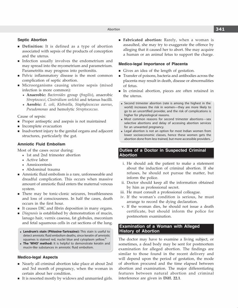
341Abortion
Septic Abortion
� Definition: It is defined as a type of abortion associated with sepsis of the products of conception and the uterus.
� Infection usually involves the endometrium and may spread into the myometrium and parametrium. Parametritis may progress into peritonitis.
� Pelvic inflammatory disease is the most common complication of septic abortion.
� Microorganisms causing uterine sepsis (mixed infection is more common):
z Anaerobic: Bacteroides group (fragilis), anaerobic Streptococci, Clostridium welchii and tetanus bacilli.
z Aerobic: E. coli, Klebsiella, Staphylococcus aureus, Pseudomonas and hemolytic Streptococcus.
Cause of sepsis: � Proper antiseptic and asepsis is not maintained � Incomplete evacuation � Inadvertent injury to the genital organs and adjacent
structures, particularly the gut.
Amniotic Fluid EmbolismMost of the cases occur during:
z 1st and 2nd trimester abortion z Active labor z Amniocentesis z Abdominal trauma
� Amniotic fluid embolism is a rare, unforeseeable and dreadful complication. This occurs when massive amount of amniotic fluid enters the maternal venous system.
� There may be tonic-clonic seizures, breathlessness and loss of consciousness. In half the cases, death occurs in the first hour.
� It causes DIC and fibrin deposition in many organs. � Diagnosis is established by demonstration of mucin,
lanugo hair, vernix caseosa, fat globules, meconium and fetal squamous cells in cut sections of the lung.
� Lendrum’s stain (Phloxine-Tartrazine): This stain is useful to detect amniotic fluid embolism deaths, since keratin of amniotic squames is stained red, nuclei blue and cytoplasm yellow.9
� The ‘WHO’ method: It is helpful to demonstrate keratin and mucin-like substances in amniotic fluid embolism.
Medico-legal Aspects
� Nearly all criminal abortion take place at about 2nd and 3rd month of pregnancy, when the woman in certain about her condition.
� It is resorted mostly by widows and unmarried girls.
� Fabricated abortion: Rarely, when a woman is assaulted, she may try to exaggerate the offence by alleging that it caused her to abort. She may acquire a human or an animal fetus to support the charge.
Medico-legal Importance of Placenta
� Gives an idea of the length of gestation. � Transfer of poisons, bacteria and antibodies across the
placenta may result in death, disease or abnormalities of fetus.
� In criminal abortion, pieces are often retained in the uterus.
� Second trimester abortion (rate is among the highest in the world) increases the risk in women—they are more likely to go to an uncertified provider, and the risk of complications is higher for physiological reasons.
� Most common reasons for second trimester abortions—sex selective abortions and delay of accessing abortion services for an unwanted pregnancy.
� Legal abortion is not an option for most Indian women from lower socioeconomic classes, hence these women gets the abortion done from less trained, but more accessible providers.
Duties of a Doctor in Suspected Criminal Abortion
i. He should ask the patient to make a statement about the induction of criminal abortion. If she refuses, he should not pursue the matter, but inform the police.
ii. Doctor should keep all the information obtained by him as professional secret.
iii. He must consult a professional colleague. iv. If the woman’s condition is serious, he must
arrange to record the dying declaration. v. If the woman dies, he should not issue a death
certificate, but should inform the police for postmortem examination.
Examination of a Woman with Alleged History of Abortion
The doctor may have to examine a living subject, or sometimes, a dead body may be sent for postmortem examination for alleged abortion. The findings are similar to those found in the recent delivery and will depend upon the period of gestation, the mode of abortion procured and the time elapsed between abortion and examination. The major differentiating features between natural abortion and criminal interference are given in Diff. 22.1.
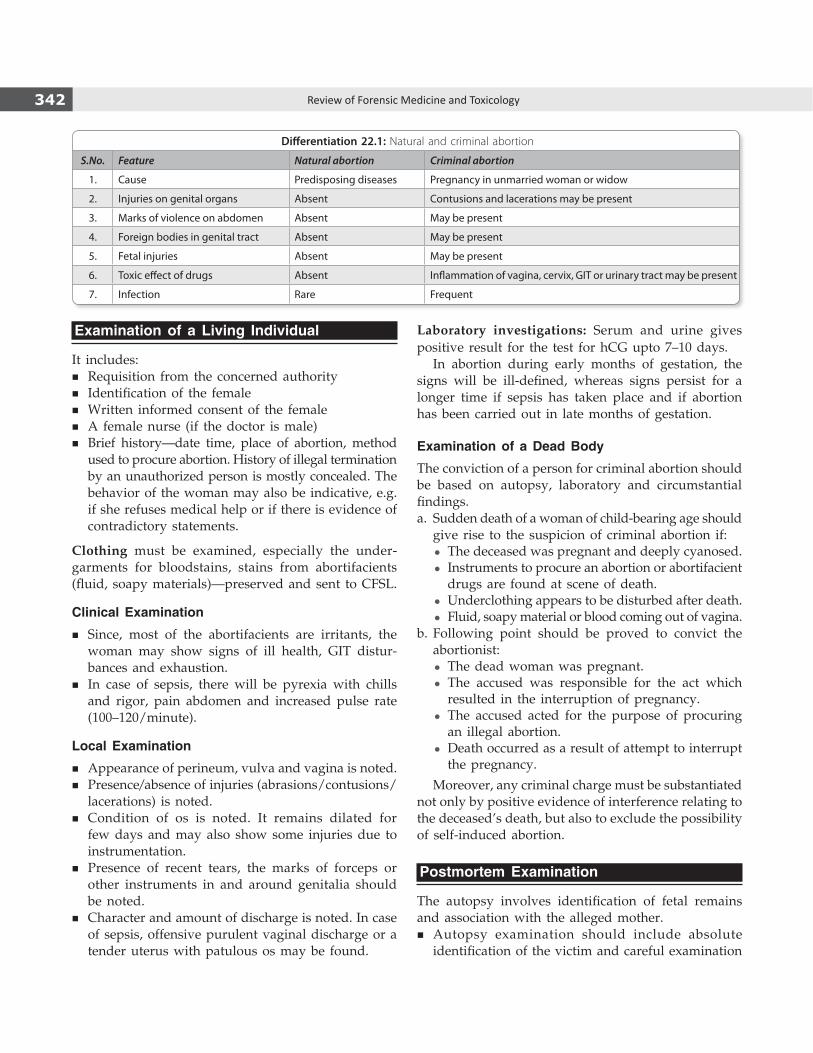
342 Review of Forensic Medicine and Toxicology
Differentiation 22.1: Natural and criminal abortion
S.No. Feature Natural abortion Criminal abortion
1. Cause Predisposing diseases Pregnancy in unmarried woman or widow
2. Injuries on genital organs Absent Contusions and lacerations may be present
3. Marks of violence on abdomen Absent May be present
4. Foreign bodies in genital tract Absent May be present
5. Fetal injuries Absent May be present
6. Toxic effect of drugs Absent Inflammation of vagina, cervix, GIT or urinary tract may be present
7. Infection Rare Frequent
Examination of a Living Individual
It includes: � Requisition from the concerned authority � Identification of the female � Written informed consent of the female � A female nurse (if the doctor is male) � Brief history—date time, place of abortion, method
used to procure abortion. History of illegal termination by an unauthorized person is mostly concealed. The behavior of the woman may also be indicative, e.g. if she refuses medical help or if there is evidence of contradictory statements.
Clothing must be examined, especially the under-garments for bloodstains, stains from abortifacients (fluid, soapy materials)—preserved and sent to CFSL.
Clinical Examination
� Since, most of the abortifacients are irritants, the woman may show signs of ill health, GIT distur-bances and exhaustion.
� In case of sepsis, there will be pyrexia with chills and rigor, pain abdomen and increased pulse rate (100–120/minute).
Local Examination
� Appearance of perineum, vulva and vagina is noted. � Presence/absence of injuries (abrasions/contusions/
lacerations) is noted. � Condition of os is noted. It remains dilated for
few days and may also show some injuries due to instrumentation.
� Presence of recent tears, the marks of forceps or other instruments in and around genitalia should be noted.
� Character and amount of discharge is noted. In case of sepsis, offensive purulent vaginal discharge or a tender uterus with patulous os may be found.
Laboratory investigations: Serum and urine gives positive result for the test for hCG upto 7–10 days.
In abortion during early months of gestation, the signs will be ill-defined, whereas signs persist for a longer time if sepsis has taken place and if abortion has been carried out in late months of gestation.
Examination of a Dead Body
The conviction of a person for criminal abortion should be based on autopsy, laboratory and circumstantial findings.a. Sudden death of a woman of child-bearing age should
give rise to the suspicion of criminal abortion if: z The deceased was pregnant and deeply cyanosed. z Instruments to procure an abortion or abortifacient
drugs are found at scene of death. z Underclothing appears to be disturbed after death. z Fluid, soapy material or blood coming out of vagina.
b. Following point should be proved to convict the abortionist:
z The dead woman was pregnant. z The accused was responsible for the act which
resulted in the interruption of pregnancy. z The accused acted for the purpose of procuring
an illegal abortion. z Death occurred as a result of attempt to interrupt
the pregnancy.
Moreover, any criminal charge must be substantiated not only by positive evidence of interference relating to the deceased’s death, but also to exclude the possibility of self-induced abortion.
Postmortem Examination
The autopsy involves identification of fetal remains and association with the alleged mother.
� Autopsy examination should include absolute identification of the victim and careful examination
343Abortion
of the clothing including undergarments which must be preserved for any traces of foreign solutions.
� External features of pregnancy should be looked for. If death is due to hemorrhage, body will look pale.
� Presence of injuries (general or local) is noted. If abortifacient drug was injected, then the injection mark(s) can be detected over usual sites.
� Local examination: Labia majora, minora, vagina, cervix may show injuries and may be congested. It may be stained by locally used abortifacient agents.
� To confirm or exclude air embolism, the body must be opened after radiological examination as it may show translucency of the right ventricle and pulmonary artery (details in Chapter 6).
� The abdominal cavity is opened and may be full of blood, if there is perforation of uterus. Uterine and adnexal tissues are assessed for crepitation due to gas formation in the uterine wall, and venous channels and the inferior vena cava is inspected for air or soap embolism bubbles.
� The skull vault must then be carefully removed, avoiding puncture of the meninges and vessels over the brain surface which allows air to enter these vessels; a detailed examination of the basal sinuses, veins and arteries is made for the presence of air embolism.
� Following removal of the thoracic and abdominal organs in the usual manner, the pelvic organs are excised en-masse following separation of the symphysis pubis and a circular dissection to include vagina, vulva and rectum with adjacent skin, taking care to collect any foreign fluid or material for chemical and bacteriological examination. The vagina and uterus are opened along their anterior surface because injuries are more likely to occur on the posterior vaginal wall following criminal interference.
� Findings in the uterus: Cavity may show presence of products of conception in full or in parts. It may be enlarged, soft and congested. Wall may show thickening in longitudinal section.
� Samples to be collected are given in Box 22.1.
Trauma and Abortion
Allegation may be leveled against a person that because of the alleged assault, the pregnant female suffered an abortion. It may be a case of a mother who is the victim of an assault, which results in premature labor, delivery of an extremely premature infant who survives a few hours, but then dies because of prematurity. Such a case could be considered a homicide, and criminal charges could well be pursued. In similar cases, where the fetus dies in-utero, criminal charges are framed under various sections of IPC.
� Travel, in the absence of trauma, does not increase the incidence of abortion.
� Trauma may rarely cause an abortion, in the absence of serious or life-threatening injury to mother.
� Following criteria suggests a causal relationship between trauma and abortion:a. The traumatic event was followed within 24 hours
by processes that ultimately lead to abortion.b. Appearance of the fetus and placenta should be
compatible with the period of pregnancy at which the traumatic event occurred.
c. The fetus and placenta should be normal.d. Factors known to cause abortion should be absent,
such as: i. History of repeated abortion without any cause
or exposure to abortifacients, e.g. X-ray or lead. ii. Chronic infections in mother, e.g. syphilis,
toxoplasmosis or tuberculosis. iii. Abnormalities of uterus including congenital
defect of uterine development, leiomyomas, endometrial polyps and incompetent os.
iv. Physical attempt to induce abortion.
Box 22.1 Samples to be collected in criminal abortion � Vaginal contents pipetted in a clean sterile container for chemicals, drugs or soap.
� Pubic hair. � Blood, urine and stomach contents. � Blood from the inferior vena cava and both cardiac ventricles. � Any fluid from the uterine cavity. � Swabs of the uterine wall. � Tissues for histology from all organs.

344 Review of Forensic Medicine and Toxicology
MULTIPLE CHOICE QUESTIONS
1. C 2. A 3. B 4. A 5. D 6. C 7. D 8. B 9. C
1. Abortion isdefinedasexpulsionof fetus: TN 06 A. Before viability B. Before 28 weeks C. Before full-term D. None of the above 2. Most commoncauseoffirst trimesterabortion is:
UP 09; JIPMER 10; Kerala 11; AFMC 12; CMC (Vellore) 14; COMEDK 15
A. Chromosomal defect B. Endocrine disturbances C. Anatomic abnormality of uterus D. Infections 3. Most common cause of second trimester abortion: CMC (Vellore) 14 A. Chromosomal defect B. Cervical incompetence C. Abnormality of uterus D. Infections 4. Mechanism of criminal abortion: AIIMS 06 A. Infection and inflammation of endometrium B. Uterine contraction C. Placental separation D. Stimulation of nerve 5. Sections 312 to 316 deal with: NEET 14 A. Kidnapping and abduction B. Abetment to suicide
C. Causing grievous hurt D. Causing miscarriage 6. Miscarriage is punishable under which IPC: AIIMS 13; NEET 14 A. Sec 320 IPC B. Sec 311 IPC C. Sec 312 IPC D. Sec 314 IPC 7. Borax is: NEET 13 A. Gastrointestinal irritants B. Genitourinary irritants C. Ecbolics D. Emmenagogues 8. Mechanism of action of abortion stick used in criminal
abortion: AIIMS 06; AI 08 A. Necrosis of endometrium causing infection B. Uterine contraction C. Stimulation of uterine nerves D. Inducing uterine relaxation 9. Lendrum’s stain is done for: NEET 13 A. Air embolism B. Fat embolism C. Amniotic fluid embolism D. Pulmonary embolism
Definitions
� Impotence: It is the inability of a person to perform sexual intercourse and achieve gratification (unable to copulate).
� Erectile dysfunction: Inability to develop and maintain an erection for satisfactory sexual inter-course in the absence of an ejaculatory disorder such as premature ejaculation.
� Quod (impotence quode hanc, ‘as regards’): A male may be impotent with one particular female, but not with another.1
� Frigidity (Latin, coldness): It is the inability to initiate or maintain the sexual arousal pattern in female (absence of desire for sexual intercourse or incapacity to achieve orgasm).2
� Sterility: It is the absolute inability of either a male or a female to procreate. In male, it is inability to make a female conceive, and in females, it is inability to conceive children.
� Fertility: Capacity to reproduce or the state of being fertile.
� Infertility: Failure to conceive (regardless of cause) after 1 year of unprotected and regular intercourse.3
Question of impotence and sterility arises in:
� Civil cases, like divorce, adultery, nullity of marriage, disputed paternity and legitimacy, claims for damages where loss of sexual function is claimed.
� Criminal cases, like adultery, rape, or unnatural offences where impotence is cited as defense.
Causes of Impotence and Sterility in Males
i. Psychological: Most important and frequent cause, though transient in nature.4 Absence of desire for sexual intercourse may result from dislike of partner, fear of failure, anxiety or mood disorder, guilt, aversion, low self-esteem, hypo chon driacs, childhood sexual abuse, masturbatory anxiety (‘dhat syndrome’—passage of whitish discharge in urine
and believed to be semen), widower syndrome, post-traumatic stress disorder or over-indulgence. Excessive masturbation may also lead to impotence.
ii. Age: Before puberty, boys are usually impotent and sterile with certain exceptions, like precocious puberty. Poor physical development of penis is common cause of impotence—examination depends more on its development than the age. In advanced age, libido diminishes, but they are not impotent or sterile. As long as live spermatozoa are present in seminal fluid, individual is presumed to be fertile.
iii. Developmental and acquired abnormalities: Absence of penis, intersexuality, malformations, e.g. hypospadias, epispadias, absence of testicles, Klinefelter syndrome, retrograde ejaculation and cryptorchidism (Fig. 23.1).
iv. Local diseases: Priapism, hydrocele, elephantiasis, phimosis, Peyronie disease, adherent prepuce, orchitis following mumps, syphilis and tuberculosis (Fig. 23.1). Mumps may cause sterility, not impotence. Exposure to X-rays may cause sterility.
v. General diseases: Impotence is common during acute illness and in any severe or debilitating illnesses.
z Neurological conditions, like tabes dorsalis, multiple sclerosis, paraplegia, hemiplegia, syrin gomyelia, temporal lobe damage and 3rd ventricle tumors; endocrine disorders, e.g. diabetes, hypothyroidism, hyperprolactinemia and testicular atrophy following renal failure, hemochromatosis or cirrhosis; blood vessel and nerve trauma (e.g. long-distance bicycle riding), CVS disorders, e.g. Leriche syndrome, and diseases like tuberculosis and nephritis may cause impotence and sterility.
z Malnutrition, vitamin C and zinc deficiency may cause erectile dysfunction.
vi. Injuries: Infertility is a significant problem after spinal cord injury. The two major causes are poor semen quality and ejaculatory dysfunction.
Impotence and Sterility
CHAPTER 23

346 Review of Forensic Medicine and Toxicology
z Erectile dysfunction may occur following treatment for lower limb fractures due to perineal neurovascular traction injury acquired during surgery.
z Fracture of the penis (rupture of both corpora cavernosa with urethral rupture) may result in impotence. The commonest causes of fracture of penis are coitus and penile manipulations, especially masturbation.
vii. Chronic poisoning: Exposure to poisons, e.g. lead, arsenic, pesticides or aphrodisiac agents may lead to impotence and/or sterility.
viii. Medications: Antidepressants (e.g. SSRIs), an-tipsychotics, anti-hypertensives, antiulcer agents (e.g. cimetidine), cholesterol-lowering agents and finasteride may cause impotence.
ix. Behavioral factors: Lifestyle choices—chronic alcoholism, smoking, being overweight and avoiding exercise are possible causes of impotence. Tight-fitting underwear causes increase in scrotal temperature that may result in decreased sperm count.
x. Addictions: Certain drugs, e.g. morphine, heroin, opium, cannabis, cocaine and tobacco (smoking) may cause impotence and sometimes sterility.
� Penile erection is a complex process involving psychogenic and hormonal input, and a neurovascular nonadrenergic, noncholinergic mechanism. Nitric oxide (NO) is considered as the main vasoactive neurotransmitter and chemical mediator of penile erection. Impaired NO bioactivity is a major pathogenic mechanism of erectile dysfunction.
� Treatment of erectile dysfunction often requires combinations of psychogenic and medical therapies. Oral phosphodiesterase type 5 (PDE-5) inhibitors are useful in this respect.
Causes of Impotence and Sterility in Females
i. Age: Being passive partners in intercourse, age has no effect on potency. Women are fertile from puberty to menopause, but may become pregnant before menarche and after menopause.
z Kraurosis vulvae in old women may cause narrowing of the vagina.
z The occurrence of infertility rises significantly as age increases.
ii. Developmental and acquired abnormalities z Impotence may result from total occlusion of
vagina, adhesion of labia, imperforate hymen—can be cured by surgery (Fig. 23.2).
z Injury or operation of vagina may cause stricture which can lead to impotence.
z Absence/abnormal uterus, ovaries or fallopian tubes produces sterility, but not impotence.
Fig. 23.1: Causes of impotence and sterility in males
Fig. 23.2: Causes of impotence in females

347Impotence and Sterility
iii. Local diseases z Bartholin cyst, chancre of vulva, stricture due
to perineal tear during previous pregnancy, prolapse of uterus/urinary bladder and dyspareunia causes impotence, but not sterility.
z Pelvic inflammatory disease, peritoneal adhesions secondary to previous pelvic surgery, endometriosis, and ovarian cyst rupture may produce blockage of fallopian tubes and sterility.
z Diseases of the genital organs (e.g. gonorrhea), leukorrhea, acidic vaginal secretions and recto-vaginal fistula do not cause impotence but may produce sterility.
iv. General disease: General infective, metabolic and hormonal conditions may cause sterility, but not impotence.
z Physiologic sexual dysfunction can be the result of impaired neurovascular tone to the clitoris and vagina.
v. Chronic poisoning: Exposure to poisons, e.g. lead and arsenic may lead to sterility, but not impotence.
vi. Environmental factors and addictions: Occu-pational exposure to excessive heat, lead, microwave radiation or X-rays lead to sterility. Drug dependence (alcohol, opium) may lead to sterility.
vii. Medications: Chemotherapy, cessation of oral contraceptives—hormonal imbalance may remain for some time after stopping the pill.
viii. Psychological: In males, psychological factors lead to non-erection (passive), but in females it is active in nature. Fear, pain, disgust or apprehension for intercourse may give rise to vaginismus [severe spasm of the lower one-third of vagina involving the paravaginal muscles (levator ani and adductor femoris muscle)].5 The spastic contraction of vaginal outlet is an involuntary reflex which replaces the rhythmic contraction associated with anticipated or actual attempt of vaginal penetration.
z It may occur with equal severity in the women who has borne children, as in virgins.
z Etiological factors: Male sexual dysfunction, psychosexually inhibiting influence due to religious orthodoxy, incidents of prior sexual trauma, secondary to dyspareunia or personal dislike/disgust for coitus.
Examination of a Person in an Alleged Case of Impotence and Sterility
� A sterile person may or may not be impotent and an impotent person may or may not be sterile.
� A simple way to distinguish between organic and psychological impotence is to determine whether the patient ‘ever’ had an erection. If never, the problem is likely to be organic; if sometimes, it could be organic or psychological.
� Permanent impotence is a ground for nullity of marriage/divorce as he is incapable of fulfilling the rights of consummation of marriage (physical union by coitus), but sterility is not.
� The person is examined only when asked by the court or by the police. Informed consent of the person should be taken and the consequences of the examination should be explained.
History: Complete history of previous illness (including surgery), mental condition and sexual history is taken. History of smoking, dietary habits, obesity and the use of various medications are also evaluated.
Psychosocial examination: A psychosocial examination using an interview and a questionnaire reveals psy-chological factors. A man’s sexual partner may also be interviewed to determine expectations and perceptions during sexual intercourse.
Examination of a Male � Complete medical examination including CNS is done,
especially if there is history of CNS illness, peripheral neuropathy, diabetes or penile sensory deficit.
� It includes pulse, blood pressure, any abnormal secondary sexual characteristics (hair pattern or breast enlargement), site of urethral meatus, urethral stenosis, sensitivity of the penis to touch or if there is any deformity in the penis itself—whether it is bent or curved when erect, or any other congenital anomalies of the genitalia.
� Testicular size, epididymis, spermatic cord and presence of varicocele are also noted.
� Bulbocavernosus reflex test is done to determine if there is adequate nerve sensation in the penis. The doctor squeezes the glans of the penis which immediately causes the anus to contract, if nerve function is intact.
Laboratory ExaminationIt will vary depending upon the history and clinical findings.
� Examination of semen is essential in cases of infertility. � Tests for systemic diseases include blood counts,
blood sugar (evaluation of diabetes), urinalysis, lipid and thyroid profiles, creatinine, liver enzymes and prostate-specific antigen.
� Serum testosterone, LH and serum prolactin.

348 Review of Forensic Medicine and Toxicology
Other tests � Evaluation of penile function can be done by direct injection
of PGE1 into the corpora. If the penile vasculature is adequate, an erection will develop.
� Duplex ultrasonography: Vascular function within the penis including signs of atherosclerosis and scarring or calcification can be evaluated.
� Ultrasonography of testes: Detect abnormalities in testes and epididymides. Transrectal ultrasonography can disclose abnormalities in the prostate and pelvis.
� Nocturnal penile tumescence testing: Normally, a man has 5–6 erections during sleep, especially during REM—their absence may indicate defect in nerve function or blood supply in the penis. It may be useful in distinguishing psychogenic from organic impotence.6
� Penile biothesiometry: This test uses electromagnetic vibration to evaluate sensitivity and nerve function in the glans and shaft of the penis.
Examination of a Female � Gynecologic examination should include an
evaluation of hair distribution, clitoris size, Bartholin glands, labia majora and minora, and any lesion that could indicate the existence of venereal disease.
� In case of impotency in females, the defect usually lies in vagina and can be clearly observed. The inspection of the vaginal mucosa may also indicate a deficiency of estrogens or the presence of infection.
� The evaluation of the cervix should include a Papanico-laou test and cultures for sexually transmitted diseases.
� The postcoital test (Sims-Huhner test) consists of evaluating the amount of spermatozoa and its motility within the cervical mucus during the pre-ovulatory period.
� Bimanual examination should be performed to establish the direction of the cervix, and the size and position of the uterus to exclude the presence of uterine fibroids, adnexal masses, tenderness or pelvic nodules indicative of infection or endometriosis.
Laboratory tests: Besides routine blood and urine analysis, HSG, pelvic ultrasonography, hysterosonogram and MRI are required.
Opinion
� An opinion of impotence (in males) cannot be given, unless there is gross deviation from normal.
� The opinion should be given in double negative form—stating that from examination of the male, there is nothing to suggest that the person is incapable of sexual intercourse.
� In case of infertility, opinion can be given with certainty depending on clinical and laboratory findings.
Sterilization
Definition: It is the process to cause a person sterile without affecting his/her potency or sexual functions.
Classification: Sterilization can be classified as given in Flow chart 23.1.
� Compulsory: It is performed on a person, compul-sorily by an order of the State, carried out on mentally or physically defective person, or as punishment to sexual criminals, or for the purpose of eugenics. It is not done in India.
� Voluntary: It is carried on married persons with consent of both the husband and wife. It can be:
i. Therapeutic: It is done to prevent danger to health or life of women due to future pregnancy.
ii. Eugenic: It is carried out to prevent conception of the children who are likely to be physically or mentally defective.
iii. Contraceptive: It is done as a family planning measure.
Flow chart 23.1: Classification of sterilization
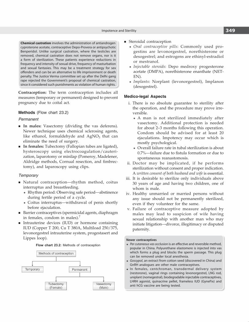
349Impotence and Sterility
Chemical castration involves the administration of antiandrogen cyproterone acetate, contraceptive Depo-Provera or antipsychotic Benperidol. Unlike surgical castration, where the testicles are removed, chemical castration does not remove organs, nor is it a form of sterilization. These patients experience reductions in frequency and intensity of sexual drive, frequency of masturbation and sexual fantasies. This may be a treatment strategy for sex offenders and can be an alternative to life imprisonment or death penalty. The Justice Verma committee set up after the Delhi gang rape rejected the Government’s proposal of chemical castration, since it considered such punishments as violation of human rights.
Contraception: The term contraception includes all measures (temporary or permanent) designed to prevent pregnancy due to coital act.
Methods (Flow chart 23.2)
Permanent
� In males: Vasectomy (dividing the vas deferens). Newer technique uses chemical sclerosing agents, like ethanol, formaldehyde and AgNO3 that can eliminate the need of surgery.
� In females: Tubectomy (Fallopian tubes are ligated), hysteroscopy using electrocoagulation/cauteri-zation, laparotomy or minilap (Pomeroy, Madelener, Aldridge methods, Cornual resection, and fimbrec-tomy), and laparoscopy using clips.
Temporary
� Natural contraception—rhythm method, coitus interruptus and breastfeeding.
z Rhythm period: Observing safe period—abstinence during fertile period of a cycle.
z Coitus interruptus—withdrawal of penis shortly before ejaculation.
� Barrier contraceptives (spermicidal agents, diaphragm in females, condom in males).7
� Intrauterine devices (IUD) or hormone containing IUD (Copper T 200, Cu T 380A, Multiload 250/375, levonorgestrel intrauterine system, progestasert and Lippes loop).
� Steroidal contraception z Oral contraceptive pills: Commonly used pro-
gestins are levonorgestrel, norethisterone or desogestrel; and estrogens are ethinyl-estradiol or mestranol.
z Injectable steroids: Depo medroxy progesterone acetate (DMPA), norethisterone enanthate (NET-EN).
z Implants: Norplant (levonorgestrel), Implanon (desogestrel).
Medico-legal Aspects i. There is no absolute guarantee to sterility after
the operation, and the procedure may prove irre-versible.
z A man is not sterilized immediately after vasectomy. Additional protection is needed for about 2–3 months following this operation. Condom should be advised for at least 20 ejaculations. Impotency may occur which is mostly psychological.
z Overall failure rate in tubal sterilization is about 0.7%—failure due to fistula formation or due to spontaneous reanastomosis.
ii. Doctor may be implicated, if he performs sterilization without consent and proper indication. A written consent of both husband and wife is essential.
iii. It is desirable to sterilize only individuals above 30 years of age and having two children, one of whom is male.
iv. Healthy unmarried or married persons without any issue should not be permanently sterilized, even if they volunteer for the same.
v. Failure of contraceptive measure adopted by males may lead to suspicion of wife having sexual relationship with another man who may initiate litigation—divorce, illegitimacy or disputed paternity.
Newer contraceptives � Per cutaneous vas occlusion is an effective and reversible method,
popular in China. Polyurethane elastomere is injected into vas which forms a plug and blocks the sperm passage. This plug can be removed under local anesthesia.
� Gossypol, an extract from cotton seed (discovered in China) and GnRH analogues are other male contraceptives.
� In females, centchroman, transdermal delivery system (nestorone), vaginal rings containing levonorgestrel, LNG rod, uniplant (nomegestral), biodegradable injectable contraceptives, LHRH agonist, quinacrine pellet, frameless IUD (GyneFix) and anti hCG vaccine are being tested.
Flow chart 23.2: Methods of contraception

350 Review of Forensic Medicine and Toxicology
Artificial Insemination (AI)
Definition: It is the process of introduction of semen from the husband or a donor by instruments into the vagina or uterus of a female to bring about pregnancy which is not attainable by sexual intercourse.
� Semen can be introduced into the vagina (intra-vaginal insemination—IVI), cervix (intracervical—ICI), fallopian tube (intratubal—ITI) or uterine cavity (intrauterine—IUI) of the recipient.
� IUI is the most commonly used method of AI (higher success rate); and IVI (low success rate) and ITI (more invasive, greater risk of infection and higher costs) are the least commonly done AI.
Female infertility accounts for one third of infertility cases, male infertility for another third, combined male and female infertility for another 15%, and the remainder of cases is ‘unexplained’.
Types (Diff. 23.1)
i. AIH (artificial insemination homologous/husband) ii. AID (artificial insemination donor)
iii. AIHD: ‘Pooled’ donor semen to which semen from husband has been added. There is a technical possibility of husband being father of the child.
Procedure: Semen is obtained by masturbation after a week’s abstinence and 1 ml is deposited by means of a sterile needleless syringe just above the internal os, at the time of ovulation (14th day after menstruation) (Fig. 23.3).
� The semen to be implanted is ‘washed’ in a laboratory and concentrated in Hams F10 media without L-glutamine, warmed to 37°C. This ‘washing’ increases the chances of fertilization while removing mucus and non-motile sperms in the semen.
� A more efficient method of AI is to insert semen directly into the woman’s uterus. When this method is employed, it is important that only ‘washed’ semen is used and inserted by means of a catheter.
The success rates of AI vary depending on the type of insemination used, but typically the success rate varies between 5–30%. The success rate can be affected by factors such as stress, and quality of the egg and sperm.
Differentiation 23.1: AIH and AID
S.No. Feature AIH AID
1. Principle Semen used is derived from woman’s husband8 Semen of person other than husband is used
2. Indications Male factor � Impotency � Defects of the penis, e.g. hypospadias � Retrograde ejaculation � Decreased sperm counts, motility or quality
Female factor � Scant/unreceptive mucus � Persistent cervicitis � Cervical stenosis
� Husband sterile � Husband suffering from hereditary disease � Widows/unmarried women desiring children � Rh incompatibility
3. Consent Needed from both husband and wife Needed from husband, wife, donor and donor’s wife
4. Pre-condition None Donor should have his own child
5. Relation with recipient Husband Must not be a related to either spouses
6. Donor characteristics Nothing specific Must be < 40 years, should resemble closely to the husband in race
7. Medical tests Routine tests Tuberculosis, diabetes, epilepsy, Rh grouping, psychosis, endocrine dysfunction, hereditary or familial disorders and HIV are ruled out
8. Disclosure of identity Not a problem, wife knows Donor and recipient should not know
9. Outcome of AI Known to the husband Donor should not know
10. Confidentiality None Strictly maintained
11. Doctor’s role May deliver the child who administered the AI Should avoid delivering the child, as it would lead disclosing the identity of father in birth record
12. Legal problems No legal complications, except for divorce Legal problems, like litigation against the doctor, illegitimacy, inheritance claims, divorce, incest and mental trauma may arise

351Impotence and Sterility
Medico-legal Aspects i. Danger of litigation: The doctor may be sued
following the birth of a defective child. To avoid this, the donor must be screened for any genetic defects.
ii. Nullity of marriage and divorce: It is not a ground for divorce, if AI is done for sterility. If AI is due to impotence, it is a ground. If AID is done without the consent of the husband, then he can file for divorce and sue the doctor (regarded as an act of cruelty for the purpose of divorce).
iii. Legitimacy: The artificiality of the process would make no difference in legitimacy in case of AIH, and the child would be legitimate child. Since, the husband is not the actual father of the child in AID, child is illegitimate and cannot inherit property, but for all practical purpose, the husband is accepted as father of the child and treated as legitimate and can inherit property.
iv. Adultery: Recipient cannot be held guilty of adultery because there is no physical union by coitus. Moreover, the Indian law specifically provides that the woman cannot be punished for adultery in any case.
v. Incest: Risk of incestuous relationship between the offspring born by AI and children of donor is possible.
vi. Natural birth: Status remains legitimate, but that of AID remains illegitimate.
vii. Unmarried women or widow: There is no legal bar on an unmarried woman/widow going for AID. A child born to a single woman through AID would be deemed to be legitimate. However, AID should be performed only on a married woman with the written consent of her husband. A child born through AIH with the stored sperms of her deceased husband is considered to be legitimate, despite the existing law of presumptions under the Indian Evidence Act.
viii. Psychosocial aspect: If it is known that the husband consented to AID and the husband was not capable of consummating the marriage, difficulties may arise. The identity of the donor is kept secret; nevertheless, it is not uncommon for such secrets to be leaked out with adverse consequences.
ix. Rights of sperm donors are debatable issue nowadays.
The artificial insemination with donor’s semen has not been legalized in India, and should only be under-taken at infertility centers after appropriate counseling and explanation of its implications to both partners.
Assisted reproductive technology (ART)Definition: Any fertility treatment in which the gametes (sperms and eggs) are manipulated outside of the body. The gametes or embryos are replaced back into the body to establish pregnancy.
� Surgical removal of eggs is known as egg retrieval. � In vitro fertilization is the most common ART procedure.
Types of ART procedures1. In vitro fertilization: IVF involves controlled ovarian
hyperstimula tion with exogenous gonadotropins, oocyte retrieval via transvaginal ultrasonographic-guided aspiration, fertilization of oocytes with sperm in culture (or intracytoplasmic injection of sperm into the oocyte), and subsequent transfer of the resultant zygotes (3–5 days later) transcervically under ultrasound guidance into the uterine cavity.8
2. Gamete intrafallopian transfer (GIFT): This involves ovarian stimulation; egg retrieval, followed by laparoscopically guided transfer of a mixture of unfertilized eggs and sperms into the fallopian tube (fertilization takes place inside the female’s body).9
3. Zygote intrafallopian transfer (ZIFT): Eggs are removed, day 1 fertilized eggs (zygotes) are laparoscopically transferred into the fallopian tube, rather than uterus.
4. Intracytoplasmic sperm injection (ICSI): Indicated in male factor infertility. One sperm is directly injected into an egg prior to intrauterine transfer of the fertilized eggs.
5. Ovum donation: Donor egg IVF is used for patients with poor egg numbers or quality. After inducing super ovulation in an egg donor and followed by egg retrieval; eggs are fertilized by the sperms of the patient’s husband and the embryos transferred to the patient’s uterus.
6. Micromanipulation techniques include zona drilling and partial zona drilling.
Oocyte freezing: This is a technique wherein the ovum from a healthy woman is taken and preserved at -196° C for future use. The process takes 2–4 weeks from injecting hormones to stimulate ovulation and egg retrieval. This is being used by working women—both single and married, who wants to delay pregnancy and focus on their careers. Initially, egg freezing was used for medical reasons where women suffering from diseases like cancer used to freeze their eggs before chemotherapy.
Surrogate Mother
Definition: A surrogate (Latin subrõgare: to substitute) mother is a woman who carries a child for a couple or a single person with the intention of giving that child up, once it is born (also called surrogate pregnancy).
Fig. 23.3: Artificial insemination (intracervical)
352 Review of Forensic Medicine and Toxicology
The surrogate mother may be the baby’s biological mother (traditional surrogacy) or she may be implanted with someone else’s fertilized egg (gestational surrogacy). She accepts pregnancy either by AI or by implantation of in vitro fertilized ova at the blastocyst stage, till delivery, for the woman who is incapable to bear child.
Salient features of the Indian Council of Medical Research Guidelines
� Surrogate mother can be known, unknown or a relative of the couple. In the case of a relative, she should belong to the same generation as the woman desiring the surrogate.
� Surrogacy should normally be considered only for parents for whom it would be physically or medically impossible or undesirable to carry a baby to term.
� The genetic (biological) parents must adopt a child born through surrogacy.
� The payment provided to the surrogate mother must include all expenses related to the pregnancy which must be documented through an agreement between the two.
� The ART clinic cannot advertise to find a surrogate mother or be a party to any commercial dealing in
gestational surrogacy. The responsibility of finding a surrogate mother rests completely with the couple.
� The surrogate mother should be < 45 years of age. It is the responsibility of the ART clinic to ensure that the candidate chosen for surrogacy passes all treatable criteria to ensure full-term pregnancy.
� No individual can be a surrogate mother more than thrice in a lifetime.
� Since there are no laws to protect the couples seeking surrogacy, the ART clinic is responsible for guiding the couples through the processes of egg and embryo donation, and surrogacy.
� Surrogate parenting involves a woman bearing the child of another woman, who is not in a position to bear children as a result of blocked Fallopian tubes or lack of a uterus. It is the reverse of donor insemination.
� The most common reason for using a surrogate mother is infertility. Gay male couples have also used surrogate mothers in order to have children that at least one partner is biologically related to.
� Surrogacy and posthumous reproduction are the extensions and ramifications arising out of ART. However ethical, legal, religious and social issues surrounding these procedures need to be clarified and understood. These are gray areas to be cautious about.
MULTIPLE CHOICE QUESTIONS
1. Quod hanc means: NEET 14 A. Medically impotent B. Legally impotent C. Impotent towards all women D. Impotent towards a particular woman 2. Frigidity is: NEET 13 A. Inability to initiate sexual arousal in female B. Inability to initiate sexual arousal in male C. Ejaculation occurring immediately after penetration D. Inability to conceive with particular male 3. Infertility can be defined as: UP 11; KCET 13 A. Not conceiving after 3 years of marriage B. Not conceiving after 2 years of unprotected
intercourse C. Not conceiving after 1 year of unprotected intercourse D. Not conceiving after 1 year of marriage 4. Most common cause of erectile dysfunction: FMGE 10 A. Psychological B. Drug induced C. Alcohol D. Diabetes 5. Impotent female is having: NEET 14 A. Gonadal dysgenesis B. Hermaphrodite C. Vaginismus D. Absence of ovary
6. Test to differentiate between psychological and organic erectile dysfunction: NEET 13
A. Pharmacologically induced penile erection therapy
B. Nocturnal penile tumescence C. Sildenafil induced erection D. Squeeze technique 7. Barrier method is: JIPMER 13 A. Oral contraceptive pill B. Intrauterine devices C. Spermicidal D. Tubectomy 8. Homologous sperm in IVF is: AFMC 12 A. Between donor and wife B. Between husband and wife C. Between husband and surrogate D. Between donor and surrogate 9. All are steps of GIFT, except: NIMHANS 11 A. Ovulation stimulation B. Oocyte retrieval C. Fertilization of oocyte in lab D. Transfer of unfertilized egg into the fallopian tube
1. D 2. A 3. C 4. A 5. C 6. B 7. C 8. B 9. C
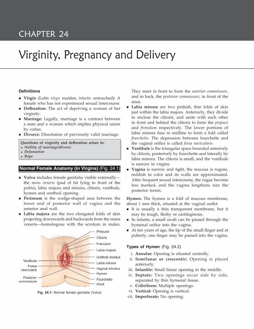
Definitions
� Virgin (Latin virgo: maiden, intacta: untouched): A female who has not experienced sexual intercourse.
� Defloration: The act of depriving a woman of her virginity.
� Marriage: Legally, marriage is a contract between a man and a woman which implies physical union by coitus.
� Divorce: Dissolution of previously valid marriage.
Questions of virginity and defloration arises in: � Nullity of marriage/divorce � Defamation � Rape
Normal Female Anatomy (in Virgins) (Fig. 24.1)
� Vulva includes female genitalia visible externally—the mons veneris (pad of fat lying in front of the pubis), labia majora and minora, clitoris, vestibule, hymen and urethral opening.
� Perineum is the wedge-shaped area between the lower end of posterior wall of vagina and the anterior anal wall.
� Labia majora are the two elongated folds of skin projecting downwards and backwards from the mons veneris—homologous with the scrotum in males.
They meet in front to form the anterior commissure, and in back, the posterior commissure, in front of the anus.
� Labia minora are two pinkish, thin folds of skin just within the labia majora. Anteriorly, they divide to enclose the clitoris, and unite with each other in front and behind the clitoris to form the prepuce and frenulum respectively. The lower portions of labia minora fuse in midline to form a fold called fourchette. The depression between fourchette and the vaginal orifice is called fossa navicularis.
� Vestibule is the triangular space bounded anteriorly by clitoris, posteriorly by fourchette and laterally by labia minora. The clitoris is small, and the vestibule is narrow in virgins.
� Vagina is narrow and tight, the mucosa is rugose, reddish in color and its walls are approximated. After frequent sexual intercourse, the rugae become less marked, and the vagina lengthens into the posterior fornix.
Hymen: The hymen is a fold of mucous membrane, about 1 mm thick, situated at the vaginal outlet.
� It is usually a thin transparent membrane, but it may be tough, fleshy or cartilaginous.
� In infants, a small swab can be passed through the hymenal orifice into the vagina.
� At ten years of age, the tip of the small finger and at puberty, one finger may be passed into the vagina.
Types of Hymen (Fig. 24.2)
i. Annular: Opening is situated centrally. ii. Semilunar or crescentic: Opening is placed
anteriorly. iii. Infantile: Small linear opening in the middle. iv. Septate: Two openings occur side by side,
separated by thin hymenal tissue. v. Cribriform: Multiple openings. vi. Vertical: Opening is vertical. vii. Imperforate: No opening.
Fig. 24.1: Normal female genitalia (Vulva)
Virginity, Pregnancy and Delivery
CHAPTER 24

354 Review of Forensic Medicine and Toxicology
The margin of the hymen is sometimes fimbriated and shows multiple notches which may be mistaken for artificial tears.*
Causes of Rupture of Hymen i. Sexual intercourse: Commonest cause of defloration. ii. Masturbation, especially with some large foreign
body. Hymen is not injured in most cases, as manipulation is usually limited to parts anterior to the hymen.
iii. An accident, like fall on a projecting substance or by slipping on the furniture or fence. It does not rupture by jumping, riding, vigorous exercise and dancing.
iv. Gynecological examination or surgical operation. v. Foreign body insertion for rendering minors fit
for sexual intercourse. vi. Sanitary tampons.
Medico-legal Aspects
Presence of intact hymen is a presumption, but is not an absolute proof of virginity. With an intact hymen, there can be true and false virgins (Diff. 24.1).
� The features will be same for a deflorate woman and a false virgin with the exception of presence of hymen in the latter.
� After the birth of a child, hymen is completely lost and the remnants are represented by cicatrized nodules of varying sizes called the carunculae hymenales or myrtiformes. On both sides, it is lined by stratified squamous epithelium.
When a virgin is placed in lithotomy position with legs wide apart, the vagina remains closed and only the edges of labia minora are seen slightly protruding from between the closed labia majora. A single intercourse does not alter the parts much, except rupture of the hymen.1
Principal signs of virginity i. An intact hymen ii. Normal condition of fourchette and posterior com-
missure iii. Narrow vagina with rugose walls
PREGNANCY
Definition: It is a condition which occurs in the female when she carries a fertilized ovum within the uterus.
Diagnosis of Pregnancy in the Living (Flow chart 24.1)
Fig. 24.2: Types of hymen
* The notches are usually symmetrical, occur anteriorly, do not extend to the vaginal wall, mucous membrane over the notches is intact, and with no signs of inflammation.
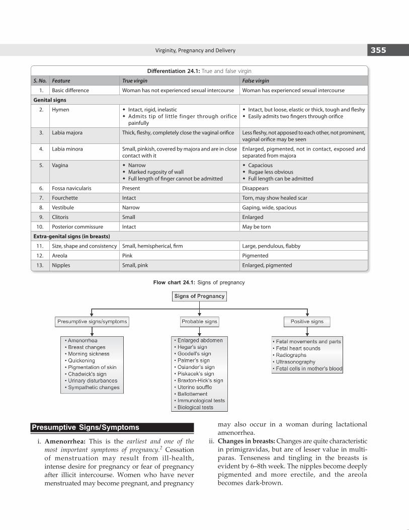
355Virginity, Pregnancy and Delivery
Presumptive Signs/Symptoms
i. Amenorrhea: This is the earliest and one of the most important symptoms of pregnancy.2 Cessation of menstruation may result from ill-health, intense desire for pregnancy or fear of pregnancy after illicit intercourse. Women who have never menstruated may become pregnant, and pregnancy
may also occur in a woman during lactational amenorrhea.
ii. Changes in breasts: Changes are quite characteristic in primigravidas, but are of lesser value in multi-paras. Tenseness and tingling in the breasts is evident by 6–8th week. The nipples become deeply pigmented and more erectile, and the areola becomes dark-brown.
Differentiation 24.1: True and false virgin
S. No. Feature True virgin False virgin
1. Basic difference Woman has not experienced sexual intercourse Woman has experienced sexual intercourse
Genital signs
2. Hymen � Intact, rigid, inelastic � Admits tip of little finger through orifice
painfully
� Intact, but loose, elastic or thick, tough and fleshy � Easily admits two fingers through orifice
3. Labia majora Thick, fleshy, completely close the vaginal orifice Less fleshy, not apposed to each other, not prominent, vaginal orifice may be seen
4. Labia minora Small, pinkish, covered by majora and are in close contact with it
Enlarged, pigmented, not in contact, exposed and separated from majora
5. Vagina � Narrow � Marked rugosity of wall � Full length of finger cannot be admitted
� Capacious � Rugae less obvious � Full length can be admitted
6. Fossa navicularis Present Disappears
7. Fourchette Intact Torn, may show healed scar
8. Vestibule Narrow Gaping, wide, spacious
9. Clitoris Small Enlarged
10. Posterior commissure Intact May be torn
Extra-genital signs (in breasts)
11. Size, shape and consistency Small, hemispherical, firm Large, pendulous, flabby
12. Areola Pink Pigmented
13. Nipples Small, pink Enlarged, pigmented
Flow chart 24.1: Signs of pregnancy

356 Review of Forensic Medicine and Toxicology
z Around the nipple, the sebaceous glands become enlarged (Montgomery’s tubercles) by the end of 3rd month. Colostrum (thin, yellowish fluid) is secreted as early as 12th week, which becomes thick and yellow by 16th week.
z Secondary areola, especially in primigravida usually appears by 20th week.
z After 6th month, silvery lines or striae are seen, especially in primiparae due to the stretching of the skin.
iii. Morning sickness: It usually appears about the end of the 1st month and disappears by end of 3rd month. Nausea and vomiting are usually present in the morning and pass off in a few hours. It more prominent in primigravidas.
iv. Quickening: Near about 18th week (16th week in multipara), the pregnant woman feels slight fetal movements in her abdomen (their first appearance is known as ‘quickening’), which gradually increase in intensity.3
v. Pigmentation of the skin: The vulva, abdomen and axillae become darker due to the deposition of pigment, and a dark line extends from the pubis to beyond the umbilicus which is called the linea nigra (Latin, black line; seen by 20th week).
vi. Chloasma: Pigmentation over forehead and cheek may appear at about 24th week.
vii. Jacquemier’s or Chadwick’s sign: The mucous membrane of the vagina changes from pink to violet, deepening to blue as a result of venous obstruction at about 8th week of pregnancy.4
viii. Urinary disturbances: During 8–12th week of pregnancy, the enlarging uterus exerts pressure on the bladder and produces frequent micturition. This gradually disappears after 12th week as the uterus straightens up into the abdomen, and reappears a few weeks before term when the head descends into the pelvis.
ix. Fatigue: Easy fatigue is very frequent. x. Sympathetic disturbances: Salivation, altered
appetite and irritable temper are common.
Probable Signs of Pregnancy
i. Enlargement of the abdomen (fundal height): During pregnancy, abdomen gradually enlarges in size after the 12th week as shown in Figure 24.3. During the last two months, the uterus sinks into the pelvis and tends to fall forward due to its weight.5
z Uterus feels soft and elastic, and becomes ovoid in shape which changes to spherical shape beyond 36th week.
z The umbilicus becomes level with the skin by about the 7th month.
ii. Hegar’s sign is positive between 6–10th week. Demonstration: If one hand is placed on the abdomen
and two fingers of other hand in the vagina, the firm hard cervix is felt and above it the elastic body of the uterus, while between the two, the isthmus is felt as a soft compressible area (Fig. 24.4).6 This is the most valuable physical sign of early pregnancy.
iii. Goodell’s sign: As early as 6th week, the cervix progressively softens from below upward.7 Pregnant woman’s cervix feels like lips and non-pregnant woman’s like the tip of the nose. The cervical orifice, during the last months of pregnancy, becomes circular instead of being transverse and admits the point of finger to a greater depth.
iv. Palmer’s sign: Regular rhythmic contractions of uterus can be elicited by bimanual examination as early as 4–8th week.
v. Osiander’s sign: There is an increased pulsation felt through the lateral fornices at about 8th week.
vi. Piskacek’s sign: Asymmetrical enlargement of uterus occurs, if there is lateral implantation. Here one half of uterus is more firm that the other.
vii. Braxton-Hick’s contractions: Intermittent, spasmodic, painless uterine contractions are
Fig. 24.3: The level of fundus uteri at different weeks
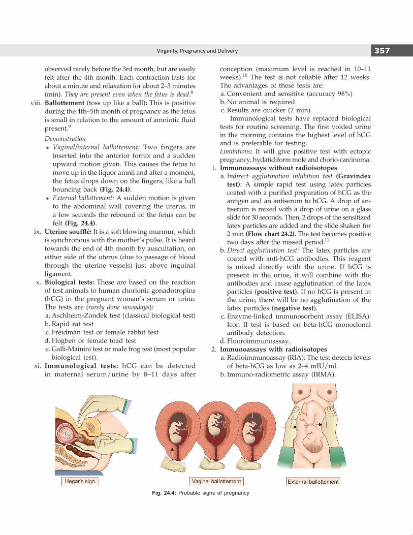
357Virginity, Pregnancy and Delivery
observed rarely before the 3rd month, but are easily felt after the 4th month. Each contraction lasts for about a minute and relaxation for about 2–3 minutes (min). They are present even when the fetus is dead.8
viii. Ballottement (toss up like a ball): This is positive during the 4th–5th month of pregnancy as the fetus is small in relation to the amount of amniotic fluid present.9
Demonstration z Vaginal/internal ballottement: Two fingers are
inserted into the anterior fornix and a sudden upward motion given. This causes the fetus to move up in the liquor amnii and after a moment, the fetus drops down on the fingers, like a ball bouncing back (Fig. 24.4).
z External ballottement: A sudden motion is given to the abdominal wall covering the uterus, in a few seconds the rebound of the fetus can be felt (Fig. 24.4).
ix. Uterine soufflé: It is a soft blowing murmur, which is synchronous with the mother’s pulse. It is heard towards the end of 4th month by auscultation, on either side of the uterus (due to passage of blood through the uterine vessels) just above inguinal ligament.
x. Biological tests: These are based on the reaction of test animals to human chorionic gonadotropins (hCG) in the pregnant woman’s serum or urine. The tests are (rarely done nowadays):
a. Aschheim-Zondek test (classical biological test) b. Rapid rat test c. Freidman test or female rabbit test d. Hogben or female toad test e. Galli-Mainini test or male frog test (most popular
biological test). xi. Immunological tests: hCG can be detected
in maternal serum/urine by 8–11 days after
conception (maximum level is reached in 10–11 weeks).10 The test is not reliable after 12 weeks. The advantages of these tests are:
a. Convenient and sensitive (accuracy 98%) b. No animal is required c. Results are quicker (2 min). Immunological tests have replaced biological
tests for routine screening. The first voided urine in the morning contains the highest level of hCG and is preferable for testing.
Limitations: It will give positive test with ectopic pregnancy, hydatidiform mole and chorio-carcinoma.
1. Immunoassays without radioisotopes a. Indirect agglutination inhibition test (Gravindex
test): A simple rapid test using latex particles coated with a purified preparation of hCG as the antigen and an antiserum to hCG. A drop of an-tiserum is mixed with a drop of urine on a glass slide for 30 seconds. Then, 2 drops of the sensitized latex particles are added and the slide shaken for 2 min (Flow chart 24.2). The test becomes positive two days after the missed period.11
b. Direct agglutination test: The latex particles are coated with anti-hCG antibodies. This reagent is mixed directly with the urine. If hCG is present in the urine, it will combine with the antibodies and cause agglutination of the latex particles (positive test). If no hCG is present in the urine, there will be no agglutination of the latex particles (negative test).
c. Enzyme-linked immunosorbent assay (ELISA): Icon II test is based on beta-hCG monoclonal antibody detection.
d. Fluoroimmunoassay. 2. Immunoassays with radioisotopes a. Radioimmunoassay (RIA): The test detects levels
of beta-hCG as low as 2–4 mIU/ml. b. Immuno-radiometric assay (IRMA).
Fig. 24.4: Probable signs of pregnancy
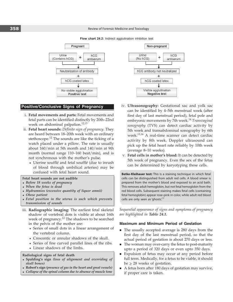
358 Review of Forensic Medicine and Toxicology
Positive/Conclusive Signs of Pregnancy
i. Fetal movements and parts: Fetal movements and fetal parts can be identified distinctly by 20th–22nd week on abdominal palpation.12,13
ii. Fetal heart sounds: Definite sign of pregnancy. They are heard between 18–20th week with an ordinary stethoscope.12 The sounds are like the ticking of a watch placed under a pillow. The rate is usually about 160/min at 5th month and 140/min at 9th month (normal range 110–160 beat/min), and is not synchronous with the mother’s pulse.
z Uterine soufflé and fetal soufflé (due to inrush of blood through umbilical arteries) may be confused with fetal heart sound.
Fetal heart sounds are not audible � Before 18 weeks of pregnancy � When the fetus is dead � Hydramnios (excessive quantity of liquor amnii) � Obese patient � Fetal position in the uterus is such which prevents transmission of sounds
iii. Radiographic imaging: The earliest fetal skeletal shadow of vertebral dots is visible at about 16th week of pregnancy.12 The shadows to be searched in the pelvis of the mother are:
z Series of small dots in a linear arrangement of the vertebral column.
z Crescentic or annular shadows of the skull. z Series of fine curved parallel lines of the ribs. z Linear shadows of the limbs.
Radiological signs of fetal death � Spalding’s sign (loss of alignment and overriding of skull bones)
� Robert’s sign (presence of gas in the heart and great vessels) � Collapse of the spinal column due to absence of muscle tone
iv. Ultrasonography: Gestational sac and yolk sac can be identified by 4–5th menstrual week (after first day of last menstrual period), fetal pole and embryonic movements by 7th week.14 Transvaginal sonography (TVS) can detect cardiac activity by 5th week and transabdominal sonography by 6th week.15,16 A real-time scanner can detect cardiac activity by 8th week. Doppler ultrasound can pick up the fetal heart rate reliably by 10th week (average 8–10 weeks).
v. Fetal cells in mother’s blood: It can be detected by 5th week of pregnancy. Even the sex of the fetus can be determined by karyotyping these cells.
Betke-Kleihauer test: This is a staining technique in which fetal cells can be distinguished from adult red cells. A blood smear is prepared from the mother’s blood and exposed to an acid bath. This removes adult hemoglobin, but not fetal hemoglobin from the red blood cells. Subsequent staining makes fetal cells (containing fetal hemoglobin) appear rose-pink in color, while adult red blood cells are only seen as ‘ghosts’.17
Sequential appearance of signs and symptoms of pregnancy are highlighted in Table 24.1.
Maximum and Minimum Period of Gestation
� The usually accepted average is 280 days from the first day of the last menstrual period, so that the actual period of gestation is about 270 days or less.
� The woman may over-carry the fetus to post-maturity upto a period of 320 days or even upto 350 days.
� Expulsion of fetus may occur at any period before full term. Medically, for a fetus to be viable, it should be > 28 weeks of gestation.
� A fetus born after 180 days of gestation may survive, if proper care is taken.
Flow chart 24.2: Indirect agglutination inhibition test

359Virginity, Pregnancy and Delivery
Diagnosis of Pregnancy in the Dead
External physical changes should be noted. In the internal examination, the following should be looked for: i. Presence of embryo, fetus, placental tissue or
membranes—positive proof of pregnancy ii. Enlarged and thickened uterus iii. Corpus luteum in ovary—corroborative evidence.
Pseudocyesis (Spurious/False/Phantom Pregnancy)
Definition: It is a psychological disorder where the woman has a false but firm belief that she is pregnant, although no pregnancy exists.
� It is generally observed in infertile females or women nearing menopause, who desire a child intensely.
� Most of these women suffer from some form of psychic or hormonal disorder.
� Such patients may present with all the subjective symptoms of pregnancy including cessation of menstruation and associated with a considerable increase in the size of the abdomen which may be due to abnormal deposition of fat or due to pathological conditions, like ovarian tumor or ascites.
� The woman may have secretions from the breasts and intestinal movements which she imagines as fetal movements and may have false labor pains.
� Obstetrical examination along with ultrasonography and/or immunological tests for pregnancy will clear the patient of her imagination.
Superfecundation
Definition: Fertilization of two ova discharged from the ovary at the same period of ovulation by two different acts of coitus committed at short intervals.
� The term is also used to refer to instances of two different males fathering fraternal twins, though this is more accurately known as heteropaternal super-fecundation.19 This leads to the possibility of twins also being half-siblings, classic example being one baby is white and the other black.
� Medico-legal aspect: Gross variations may occur in the complexion and features of the two babies and may give rise to the doubt of adultery and infidelity.
Superfetation
Definition: Fertilization of two ova discharged from ovary at different periods of ovulation.
� It is fertilization of second ovum in a pregnant woman.
� In this, one fetus always remains more developed than the other, and may be born either at the same time showing different maturation or may born at different periods, varying from 1–3 months.
� Possibility is more with septate or double uterus.
Fetus compressus or papyraceus: In a twin pregnancy, one fetus may grow at the cost of the other. The latter may die, flattened by pressure into a ‘mummified’ parchment-like state known as fetus papyraceus and may not be recognizable. It is retained till labor expels it.
Table 24.1: Signs and symptoms of pregnancy
Duration Signs and Symptoms
At 6–8 weeks
� Symptoms Amenorrhea, morning sickness, frequent micturition, fatigue and breast discomfort.
� Signs Breast enlargement. Signs—Jacquemier’s, Osiander’s, Goodell’s, Hegar’s and Palmer’s.18 Immunological tests positive. Sonography: Cardiac activity and embryonic movements.
At 16–18 weeks
� Symptoms Amenorrhea, quickening, other symptoms disappear.
� Signs Breast—pigmentation of areola, prominence of Montgomery’s tubercles, colostrum. Uterus—midway between pubis and umbilicus, Braxton-Hick’s contractions, uterine soufflé and internal ballottement. X-ray: Fetal shadow.
At 20 weeks
� Symptoms Amenorrhea, quickening.
� Signs Breast—appearance of secondary areola, linea nigra. Uterus—at level of umbilicus (24 weeks), Braxton-Hick’s contractions, external ballottement and internal ballottement (16–28 weeks). Fetus—parts, movements and heart sounds.
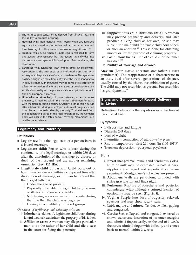
360 Review of Forensic Medicine and Toxicology
� The term superfecundation is derived from fecund, meaning the ability to produce offspring.
� Fraternal twins (non-identical twins) occur when two fertilized eggs are implanted in the uterine wall at the same time and form two zygotes. They are also known as dizygotic twins.20
� Identical twins occur when a single egg is fertilized to form one zygote (monozygotic), but the zygote then divides into two separate embryos which develop into fetuses sharing the same womb.
� Vanishing twin syndrome (twin embolisation syndrome/fetal resorption) is the presence of a multifetal gestation with subsequent disappearance of one or more fetuses. This syndrome has been diagnosed more frequently since the use of sonography in early pregnancy. In this, there may be complete resorption of a fetus or formation of a fetus papyraceus or development of a subtle abnormality on the placenta such as a cyst, subchorionic fibrin or amorphous material.
� Lithopedion or ‘stone baby’: In rare instances, an extrauterine pregnancy is retained within the mother’s abdomen for years, with the fetus becoming calcified. Usually, a lithopedion occurs after a fetus dies during an ectopic abdominal pregnancy and is too large to be reabsorbed by the body. To shield itself from the degenerating tissue of the fetal foreign body, the woman’s body will encase the fetus and/or covering membranes in a calciferous substance.
Legitimacy and Paternity
Definitions � Legitimacy: It is the legal state of a person born in
a lawful marriage. � Legitimate child: Person who is born during the
continuance of a legal marriage or within 280 days after the dissolution of the marriage by divorce or death of the husband and the mother remaining unmarried (Sec. 112 IEA).
� Illegitimate child or bastard: Child born out of lawful wedlock or not within a competent time after dissolution of marriage, or if it can be proved that the alleged father is:
i. Under the age of puberty. ii. Physically incapable to beget children, because
of illness, impotence or sterility. iii. Not having access sexually to his wife during
the time that the child was begotten. iv. Having incompatibility of blood groups.
Questions of legitimacy and paternity arise in: i. Inheritance claims: A legitimate child born during
lawful wedlock can inherit the property of his father. ii. Affiliation cases: A woman may allege a particular
man to be the father of her child and file a case in the court for fixing the paternity.
iii. Supposititious child (fictitious child): A woman may pretend pregnancy and delivery, and later produce a living child as her own, or she may substitute a male child for female child born of her, or after an abortion.21 This is done for obtaining money or for the purpose of claiming property.
iv. Posthumous births: Birth of a child after the father has died.22
v. Nullity of marriage and divorce.
Atavism (Latin atavus: ancestor; atta: father + avus: grandfather): The reappearance of a characteristic in an individual after several generations of absence, usually caused by the chance recombination of genes. The child may not resemble his parents, but resembles his grandparents.23
Signs and Symptoms of Recent Delivery in Living
Definition: Delivery is the expulsion or extraction of the child at birth.
Symptoms
� Indisposition and fatigue � Diuresis: 2–5 days � Loss of weight � Intermittent contraction of uterus—after pains � Rise in temperature—first 24 hours (h) (100–101ºF) � Transient depression—puerperal psychosis.
Signs
i. Breast changes: Voluminous and pendulous. Colos-trum or milk may be expressed. Areola is dark, nipples are enlarged and superficial veins are prominent. Montgomery’s tubercles are present.
ii. Abdomen: Walls are pendulous, wrinkled with striae gravidarum and linea nigra.
iii. Perineum: Rupture of fourchette and posterior commissure with/without a sutured incision of episiotomy may be seen (Fig. 24.5).
iv. Vagina: Purple hue, loss of rugosity, relaxed, spacious and may show recent tears.
v. Labia majora and minora: Tender, swollen, gaping and congested.
vi. Cervix: Soft, collapsed and congested; external os shows transverse laceration of its outer margins and admits 2 fingers easily. At the end of 1 week, the cervix admits 1 finger with difficulty and comes back to normal within 2 weeks.

361Virginity, Pregnancy and Delivery
vii. Uterus: The uterus decreases over the first few weeks which is called involution (apoptosis). This can be observed by palpating the height of the uterine fundus (Fig. 24.6).
z Fundus is midway between the umbilicus and symphysis pubis: Immediately after delivery.24
z Fundus at the level of umbilicus: About 1–12 h after delivery.
z Upper border lies 1 cm below umbilicus: 1st day after delivery.
z Fundus midway between umbilicus and symphysis pubis: 6th day (steady decrease in height by one fingerbreadth or 1 cm/day).25
z At the level of symphysis pubis: 10th day. z Descends within true pelvis: 2 weeks.26
z Returns to parous size: 5–6 weeks.viii. Laboratory investigations: Immunological tests
are positive for about 7–10 days after delivery. ix. Lochia (Greek lokhia: of childbirth): It is an alkaline
discharge from uterus, cervix and vagina with peculiar, disagree able fishy odor.
z It lasts for 2–3 weeks after delivery.
Types27
a. Lochia rubra (1–4 days) is bright red in color and consists of blood, shreds of fetal membranes and deciduas, vernix caseosa, lanugo hair and meconium.
b. Lochia serosa (5–9 days) is watery and pale, and consists of less RBC but more leucocytes, wound exudates, mucus from the cervix and microorganisms (anaerobic Streptococci and Staphylococci).
c. Lochia alba (10–15 days) is scanty, thicker, grayish yellow and then whitish till final disappearance. It contains decidual cells, leucocytes, mucus, cholesterol crystals, fatty and granular epithelial cells, and microorganisms.
Significance of lochia: The average amount of discharge for first 4–5 days is about 250 ml. If it smells offensive, then it indicates infection. If scanty or absent or excessive—infection; persistence of red color beyond normal—subinvolution or retained bits of conceptus; and duration beyond 3 weeks suggest local genital lesion.
Signs of Recent Delivery in Dead
All the local signs mentioned above may be present. � The size of uterus will vary with the time after
delivery at which death occurred (Table 24.2). � The size of the area where the placenta has been
attached to the uterus is about 3–4 inches (8–10 cm) in diameter. A tissue layer remains attached here from placenta.
� The ovaries and fallopian tubes are congested and become normal in few days. A large corpus luteum is present in one of the ovaries.
Fig. 24.6: Level of upper border of uterus (in days) post delivery
Fig. 24.5: Signs of recent delivery

362 Review of Forensic Medicine and Toxicology
Signs of recent delivery (both living and dead) � Engorged breasts � Pink striae on the abdomen � Enlarged uterus � Fresh tears of the vulva, vagina or cervix � Lochia from the uterus
Signs of Remote Delivery in Living
The only sign which proves delivery is the appearance of the external os.
� Breasts: Flabby, dark areola with Montgomery’s tubercles, nipples are prominent and white striae.
� Abdominal wall: Lax, loose, presence of striae gravidarum and linea alba.
� Perineum: Lax, old scarring from previous perineal laceration or episiotomy may be seen.
� Introitus: Gaping; labia majora are not in close apposition, and labia minora is pigmented and protrude out; presence of carunculae myrtiformes.
� Uterine wall: Less rigid, contour of uterus is broad and round rather than ovoid.
� Vagina: Roomy with loss of rugosity. � Cervix: Cylindrical, external os is transverse, patulous
slit and may admit tip of finger (Fig. 24.7).
Signs of Remote Delivery in Dead
In addition to the signs seen in the living subjects, there will be findings in the uterus as mentioned in Diff. 24.2 and shown in Fig. 24.7.
Table 24.2: Size of uterus after delivery
Time after delivery
Dimension (cm)
Weight (g)
Placental site diameter (cm)
Immediate 20 × 15 × 5 1000 10–15
1st week 14 × 8 × 4 500 4
2nd week 12 × 7 × 3 300 2.5
3rd week 9 × 5 × 2 100 1.5
Differentiation 24.2: Nulliparous and parous uterus (Fig. 24.7)
S. No. Feature Nulliparous uterus Parous uterus
1. Size Small (7 × 5 × 2 cm 3) Large (10 × 6 × 2.5 cm3)
2. Weight 40–50 g 80–100 g
3. Length Body and cervix have same length Body twice the length of cervix
4. External os Circular, dimple like Transverse patulous slit
5. Internal os Circular, well defined Ill-defined, margin wrinkled
6. Shape of cervix Conical28 Cylindrical
7. Upper surface of fundus Less convex and in same line as broad ligament More convex and at higher level than the line of broad ligament
8. Uterine cavity Inner walls convex, smaller and triangular cavity Inner walls concave, spacious and rounded cavity
9. Arbor vitae* Present Disappears (absent)
10. Scar for placental attachment Absent Present
* Mucosal folds in the cervical canal which extends from internal to external os.
Fig. 24.7: Shape of uterine cavity in (A) Nulliparous, (B) Parous woman
A
B

363Virginity, Pregnancy and Delivery
Medico-legal Aspects of Pregnancy and Delivery
Questions of pregnancy and/or delivery may arise in the following cases: i. Execution of judicial death sentence: When a
woman sentenced to death, pleads that she is pregnant to avoid execution. If a woman sentenced to death is found to be pregnant, the High Court should commute the sentence to life imprisonment [Sec. 416 CrPC and CrPC (Amendment) Act, 2008]. Post delivery, if the mother is put to death, the child will be orphaned and punished for no fault of his/her.
ii. Deferring trial of a case: When a woman pleads pregnancy (delivery is imminent) to avoid attendance as witness in the court.
iii. Feigned pregnancy and delivery: When a woman feigns pregnancy soon after death of her husband, and later produces a child to claim greater share of property and compensation.
iv. Criminal breach of trust/rape: When pregnancy is claimed to be the result of rape, kidnapping and seduction or breach of promise of marriage.
v. Blackmail: When a woman blackmails a man and claim’s that she is pregnant by him to compel marriage. She may produce a suppositious child to extort money.
vi. Disputed chastity: In allegations of an unmarried woman, widow, or a wife living apart from her husband that she is pregnant or delivered a child.
vii. Homicide or suicide: When pregnancy is alleged to be the motive for murder or suicide of an unmarried woman or widow.
viii. Affiliation cases: The woman may claim a child fathered by her husband who has subsequently divorced her or by a person who is not her legally wedded spouse and force him to adopt the child as his own and pay maintenance allowance.
ix. Concealment of birth: In cases of alleged concealment of birth or pregnancy in an unmarried woman or widow or out of wedlock.
x. Criminal abortion and infanticide: When there is an allegation of sex selective abortion or killing of an infant.
xi. Nullity of marriage and divorce: When there is allegation of the woman becoming pregnant when the husband was not having access physically, or delivery occurring before the minimum period of gestation, the issue may be brought to the court for nullity of marriage.
xii. Maternity/Paternity leave: For claiming benefit of leave facility for working women or men.
xiii. Legitimacy: For such claims, it must be proved that the woman indeed delivered a child at the time claimed by her.
Written informed consent needs to be taken before examination after explaining reasons and possible consequences.
Nullity of Marriage and Divorce
Sec. 11, 12 and 13 of the Hindu Marriage Act, 1955 deals with grounds for void and voidable marriages, and grounds for divorce respectively. i. Grounds for void and voidable marriage
a. Void marriage, i.e. null from the time of inception
z Bigamy (another marriage without dissolution of earlier marriage)
z Prohibited degree of relationship (related by blood) unless custom permits such marriage
z Sapinda relationship (relationship extending to 3rd generation in the line of ascent through mother and 5th generation through father).
b. Voidable marriage, i.e. it remains valid until annulled by the court29
z Impotence z Unsoundness of mind of either party at the
time of marriage z Consent of either party was obtained by force,
fraud or misconception of facts z Pregnancy of the female by some other person
and the husband was ignorant of the fact at the time of marriage.
ii. Grounds for divorce z Adultery: Voluntary sexual intercourse with any
person other than his/her spouse. z Cruelty: Willful and unjustifiable conduct so as
to cause danger to life, limb or heath of another (including mental health).
z Desertion: Abandonment of one spouse without reasonable cause and without consent or against the wish of other.
z Apostasy: Change of religion. z Unsoundness of mind. z Virulent leprosy and sexually transmitted diseases
including AIDS. z Renouncing the world. z Additional grounds for woman: Husband convicted
of rape, sodomy or bestiality.
364 Review of Forensic Medicine and Toxicology
1. Definitive finding in deflorate woman: Maharashtra 10 A. Pigmented labia minora B. Roomy vagina C. Large clitoris D. Torn hymen 2. First symptom of pregnancy is: Kerala 07 A. Tingling in the breasts B. Amenorrhea C. Morning sickness D. Quickening 3. Quickening appears at about: PGI 09 A. 6 weeks B. 8–10 weeks C. 16–20 weeks D. 20–24 weeks 4. Bluish discolouration of the vagina seen in pregnancy
is known as: KCET 12 A. Chadwick’s sign B. Goodell’s sign C. Hegar’s sign D. Palmer’s sign 5. Wrong statement about pregnancy is: UP 08 A. Amenorrhea is the earliest symptom B. Fetal heart sounds heard between 18-20th weeks C. Fetal parts are palpable at 20 weeks of gestation D. At 40th week, fundal height is at xiphisternum 6. Softening of uterine isthmus and lower segment in
early pregnancy is known as: UP 07; MAHE 11 A. Hegar’s sign B. Braxton Hick’s sign C. Goodell’s sign D. Osiander’s sign 7. Goodell’s sign means: JIPMER 07 A. Pulsation in the lateral vaginal fornix B. Bluish color change in the vagina C. Softening of the cervix from below upward D. On bimanual palpation, the fingers can be
approximated, as if nothing is in between 8. True about Braxton-Hick’s contraction are all, except:
Maharashtra 09 A. Felt at 4th month B. Painful C. Contraction last for 1 min D. Present even when fetus is dead
9. External ballottement can be done after how many weeks of gestation: Manipal 10
A. 6 weeks B. 16 weeks C. 20 weeks D. 24 weeks 10. In a normal pregnancy, maternal hCG level is maximum
at gestational age of: UPSC 07; 14 A. 8 to 10 weeks B. 12 to 14 weeks C. 16 to 18 weeks D. after 20 weeks 11. Gravindrex test can detect pregnancy in: MAHE 12 A. 2 weeks B. 4 weeks C. 8 weeks D. 12 weeks 12. Definite diagnosis of pregnancy include all, except:
Kerala 09; 11 A. Fetal heart sound B. Palpation of fetal parts C. Fetal skeleton on X-ray D. hCG in blood 13. Fetal parts are palpable at the earliest by:
Maharashtra 08 A. 16 weeks B. 18 weeks C. 20 weeks D. 28 weeks 14. Gestational sac can be seen using ultrasonography at
the earliest by: Gujarat 07 A. 3rd week B. 4th week C. 5th week D. 8th week 15. Transvaginal USG can detect fetal cardiac activity
in: DNB 10; MAHE 12 A. 5 weeks B. 6 weeks C. 7 weeks D. 8 weeks 16. Most accurate method of diagnosis of pregnancy at 6
weeks: AIIMS 13; JIPMER 14 A. Hegar’s sign B. X-ray examination C. Palpation of fetal parts D. Fetal heart sound by USG 17. Fetomaternal transfusion of fetal RBCs in mother can
be detected by: UPSC 08; TN 08; AIIMS 10 A. Direct Coomb’s test B. Betke-Kleihauer test C. Electrophoresis D. Indirect Coomb’s test
1. D 2. B 3. C 4. A 5. D 6. A 7. C 8. B 9. C 10. A 11. A 12. D 13. C 14. C 15. A 16. D 17. B
MULTIPLE CHOICE QUESTIONS
� Impotence is inability to consummate the marriage (and not merely incapacity for procreation), and to be a ground for nullity, such inability must exist at the time of marriage and continue to exist at the time of the institution of the suit. For this purpose, sexual intercourse has been defined as ordinary and complete intercourse, not partial and imperfect intercourse.
� The birth of a child is not conclusive evidence that the marriage has been consummated since fecundation ab extra (a rare occurrence) can take place. Fecundatio ab extra means pregnancy that occurs by mere deposition of semen on the vulva and there is no penile penetration into the vagina.30
365Virginity, Pregnancy and Delivery
18. NOT a sign of early pregnancy: UPSC 07 A. Goodell’s sign B. Hegar’s sign C. Cullen’s sign D. Palmer’s sign 19. Twin pregnancy, but due to two different men is
called: NEET 14 A. Superfetation B. Superfecundation C. Both of the above D. Not a realistic situation 20. True about fraternal twins are: UP 11 A. Dizygotic twins B. Comes from single egg C. Two eggs fertilized at different period of gestation D. Unrelated by birth 21. True about suppositious child: PGI 07, 08; MAHE 11 A. Child who is born after father dies B. Child born through artificial insemination C. Woman claim the child as her own D. Child born out of wedlock 22. ‘Posthumous child’ is one who: KCET 12 A. Does not belong to the women claiming to be its
mother B. Has been abandoned by its parents C. Is born after the death of its father D. Is illegitimate 23. Atavism is inheritance of features of: Kerala 11 A. Father B. Mother C. Grandfather D. Uncle
24. Immediately after delivery, uterus is at the level of: MP 07; UPSC 14
A. Midway between the umbilicus and symphysis pubis
B. Just at the level of umbilicus C. Midway between xiphisternum and umbilicus D. Descends into true pelvis 25. Rate of involution uterus following delivery:
FMGE 09, 11 A. 1 cm/day B. 1.25 cm/day C. 2.25 cm/day D. 2.5 cm/day 26. Following delivery, uterus becomes a pelvic organ
after: UPSC 07; MAHE 11 A. 2 weeks B. 4 weeks C. 6 weeks D. 8 weeks 27. Order in lochia: AIIMS 13 A. Serosa, rubra, alba B. Rubra, serosa, alba C. Alba, rubra, serosa D. Rubra, alba, serosa 28. Shape of nulliparous cervix is: AI 07 A. Conical B. Circular C. Longitudinal D. Cylindrical 29. Divorce can be given if there is: PGI 14 A. Impotence B. Sterility C. Pre-existing mental illness D. Premature ejaculation 30. Fecundation ab extra means: KCET 13 A. Child having the characteristic of grandparents B. Birth of a child after the death of father C. Insemination without penetration of vagina by penis D. Sexual intercourse with blood relations
18. C 19. B 20. A 21. C 22. C 23. C 24. A 25. A 26. A 27. B 28. A 29. A & C 30. C
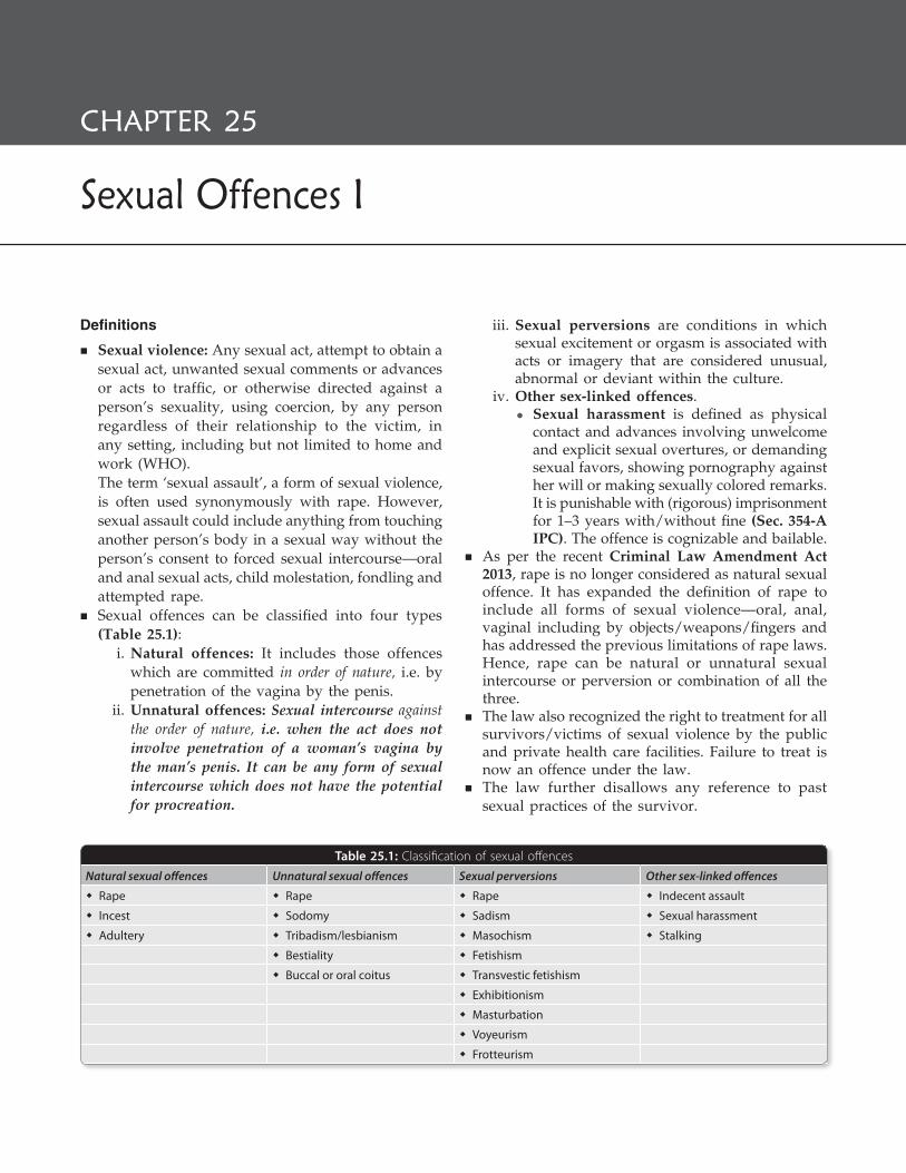
Definitions
� Sexual violence: Any sexual act, attempt to obtain a sexual act, unwanted sexual comments or advances or acts to traffic, or otherwise directed against a person’s sexuality, using coercion, by any person regardless of their relationship to the victim, in any setting, including but not limited to home and work (WHO).
The term ‘sexual assault’, a form of sexual violence, is often used synonymously with rape. However, sexual assault could include anything from touching another person’s body in a sexual way without the person’s consent to forced sexual intercourse—oral and anal sexual acts, child molestation, fondling and attempted rape.
� Sexual offences can be classified into four types (Table 25.1):
i. Natural offences: It includes those offences which are committed in order of nature, i.e. by penetration of the vagina by the penis.
ii. Unnatural offences: Sexual intercourse against the order of nature, i.e. when the act does not involve penetration of a woman’s vagina by the man’s penis. It can be any form of sexual intercourse which does not have the potential for procreation.
iii. Sexual perversions are conditions in which sexual excitement or orgasm is associated with acts or imagery that are considered unusual, abnormal or deviant within the culture.
iv. Other sex-linked offences. z Sexual harassment is defined as physical
contact and advances involving unwelcome and explicit sexual overtures, or demanding sexual favors, showing pornography against her will or making sexually colored remarks. It is punishable with (rigorous) imprisonment for 1–3 years with/without fine (Sec. 354-A IPC). The offence is cognizable and bailable.
� As per the recent Criminal Law Amendment Act 2013, rape is no longer considered as natural sexual offence. It has expanded the definition of rape to include all forms of sexual violence—oral, anal, vaginal including by objects/weapons/fingers and has addressed the previous limitations of rape laws. Hence, rape can be natural or unnatural sexual intercourse or perversion or combination of all the three.
� The law also recognized the right to treatment for all survivors/victims of sexual violence by the public and private health care facilities. Failure to treat is now an offence under the law.
� The law further disallows any reference to past sexual practices of the survivor.
Table 25.1: Classification of sexual offences
Natural sexual offences Unnatural sexual offences Sexual perversions Other sex-linked offences
� Rape � Rape � Rape � Indecent assault
� Incest � Sodomy � Sadism � Sexual harassment
� Adultery � Tribadism/lesbianism � Masochism � Stalking
� Bestiality � Fetishism
� Buccal or oral coitus � Transvestic fetishism
� Exhibitionism
� Masturbation
� Voyeurism
� Frotteurism
Sexual Offences I
CHAPTER 25

367Sexual Offences I
Rape
Definition: Rape (Latin rapere: to seize or take by force) is an unlawful sexual intercourse by a man with a woman, and is defined under Sec. 375 IPC.1 A man is said to commit ‘rape’ if he himself or makes a woman to do so with him or any other person the following:a. penetrates his penis into the vagina, mouth, urethra
or anus; orb. inserts any object or any part of his body (not being
his penis), or applies his mouth into the vagina, mouth, urethra or anus; or
c. manipulates any part of her body so as to cause penetration into the vagina, urethra or anus, under the following circumstances:
i. Against her will ii. Without her consent iii. With her consent, when:
z It has been obtained by putting her or any person in whom she is interested, in fear of death or hurt.
z The man knows that he is not her husband, but she consents believing him as the man to whom she is lawfully married (impersonation).
z At the time of giving such consent by reason of unsoundness of mind or intoxication or the administration by him or through another of any stupefying substance, she is unable to understand the nature of consequences of that to which she gives consent.
iv. With or without her consent, when she is under 18 years of age–statutory rape.2
v. When she is unable to communicate consent.
Exceptions
i. Medical intervention or procedure will not constitute rape.
ii. Sexual intercourse by a man with his wife not being under 15 years of age is not rape.3
Explanations
� ‘Penetration’ or ‘insertion’ can be any extent. � ‘Vagina’ is labia majora. � ‘Consent’ is voluntary agreement by the woman
by words, gesture or any form of verbal or non-verbal communication—communicates willingness to participate in the specific sexual act.
� Custodial rape: Rape of a woman by persons who are in position of authority, e.g. police officers, jail warden or hospital staff and who abuse their position to commit the offence, when the woman is under their custody/care.
� Gang rape (pack rape): When more than one person constituting a group or acting in furtherance of a common intention rapes a woman, each one is deemed to have committed rape.4
� Statutory rape: It is the crime of having sexual intercourse with a girl under the age of consent. In India, the age of consent is 18 years (not being his wife).5
Punishment for Rape
� Sec. 376 (1) IPC: A man committing rape, except in cases given below, is punished with rigorous imprisonment for a term ≥ 7 years which may extend to life imprisonment and fine.6,7
� Sec. 376 (2) IPC: Punishment is rigorous imprisonment for ≥10 years or life imprisonment (remainder of natural life) and fine, if rape is committed on an woman:a. By a police officer, member of armed forces,
public servant, management or on the staff of jail, remand home, women’s or children’s institution or hospital while under his custody.
b. By a relative, guardian, teacher or a person of trust or authority, or in a position of control or dominance over the woman.
c. During communal or sectarian violence.d. When she is under 16 years of age, pregnant
(knowingly) or incapable of giving consent.e. Who is physically or mentally disable.f. Repeatedly on the same woman.g. Causes grievous injury, mutilate or disfigures or
endangers her life during the act. � Sec 376-A IPC: In cases where the person committing
rape inflicts injuries on the woman which causes death or leads to a persistent vegetative state, punishment is rigorous imprisonment for ≥ 20 years which may extend to remainder of his natural life or with death.
� Sec 376-B IPC: Sexual intercourse by husband upon his wife during separation without her consent is punished with imprisonment for 2–7 years and fine.8
� Sec 376-C IPC: Sexual intercourse (not amounting to the offence of rape) by a person of authority or in a fiduciary relationship, public servant, management or on the staff of jail, remand home, women’s or
368 Review of Forensic Medicine and Toxicology
children’s institution or hospital is punished with rigorous imprisonment for 5–10 years and fine.9
� Sec 376-D IPC: In case of gang rape, punishment is rigorous imprisonment for ≥ 20 years which may extend to remainder of the person’s natural life along with fine paid to the victim (for medical expenses and rehabilitation).
� Sec 376-E IPC: In case of repeat offenders, punishment is imprisonment for remainder of his natural life or with death.
All the offences are cognizable and non-bailable, except under Sec 376-B which is cognizable but bailable (only on the complaint of the victim).
� Carnal knowledge (Latin carnalis: fleshly, sexual relations): The act of a man having sexual relation with a woman and includes even ‘slight penile penetration of the labia minora’.
� Sexual battery: It means non-consensual oral, anal or vaginal penetration by or union with the sexual organ of another, or the anal or vaginal penetration of another by any other object; however, sexual battery shall not include acts done for bona fide medical purposes.
� Under the British Sexual Offences Act 2003, rape was redefined from non-consensual vaginal or anal intercourse, and is now defined as non-consensual penile penetration of the vagina, anus or mouth of another person. The changes also made rape punishable by a maximum sentence of life imprisonment.
� Drug-facilitated rape: Drugs, such as flunitrazepam (Rohypnol) and gamma-hydroxybutyrate are referred to as ‘date rape drugs’ have been used by people to render the victims unconscious, before raping them.
ConsentA woman of 18 years and above can give valid consent for sexual intercourse. The consent must be free and voluntary, and given while she is of sound mind and not intoxicated. The consent should be obtained prior to the act.
Presumption and absence of consentAbsence of consent can be presumed from the attendant circumstances of each case.
� The foremost circumstance is the evidence of resistance (tearing of clothes or infliction of personal injuries on the body and even on the genitalia) from a woman unwilling to yield to sexual intercourse forced upon her.
� The resistance offered depends upon the type of woman, her age, development and on her social status.
� The absence of signs of struggle or injuries does not mean the victim has consented to sexual activity. As per law, resistance was not offered does not mean the person has consented.
� The woman may yield from fear or exhaustion in which case it is regarded as rape. A woman may faint due to fear and suddenness of the situation or may have been drugged or may get unconscious from any cause, and children may not be able to resist.
Consent is invalid when: i. Obtained by fraud as by impersonation of the husband
or by misrepresentation of facts. ii. Obtained by putting her or any person whom she is
close, in fear of death or hurt. iii. Obtained from a woman who is of unsound mind,
insensible, asleep, unconscious or in a state of drunkenness. iv. The woman is < 18 years of age. v. Obtained after the act.
The age at which individuals are considered competent to give consent for sexual intercourse is called the age of consent. The age set by each country/State vary in accordance with local standards.
Medico-legal Aspects of Definition of RapeWill and consent are different: Every act done against the will is done without her consent, but an act done without the consent of a person is not necessarily against her will. Sexual intercourse with an unconscious woman cannot be said to be against her ‘will’, but it will be ‘without her consent’. But an act against her will is necessarily ‘without her consent’.
� A woman may have the will for sexual intercourse, but she may not give consent for shyness, fear of detection and social stigma of getting pregnant.
� Women may be raped during sleep, thus being unable to give prior consent. But rape is usually not possible without waking up the lady.
� A man can impersonate as the husband of the victim in the darkness, or in case of twins one may impersonate the other.
� A woman may give her consent suppressing her unwillingness due to some other factor, e.g. for mone tary benefit.
� Sometimes, a girl may give her consent for intercourse, and then later deny that she agreed and accuses the man of rape. This may be due to fear of pregnancy, venereal disease or breakdown of relationship where motive of revenge is present.
� Ordinarily, the burden to prove unwillingness and absence of consent lies with the prosecution. But in rape case, under Sec. 376 IPC, if the victim states in the court of trial that she did not give consent, it then lies with the accused to prove that she consented for the intercourse.

369Sexual Offences I
� The law provides the same protection to a prostitute against sexual assault, as it does for chaste woman (i.e. consent is required for intercourse). But when a prostitute makes a charge of rape, the case must be more closely scrutinized, something more than medical evidence would be required to establish such a charge.
� Medical proof of intercourse is not legal proof of rape. In short, rape is not a medical diagnosis, but a legal definition.
By a man: In India, the law does not presume any limit of age under which a boy is considered physically incapable of committing rape. In a charge of rape brought against a boy, the court decides the question of his potency from evidence of the case and is guided by Sec. 82 and 83 IPC in awarding punishment. Likewise, there is no upper limit and even old people have committed rape. In England and Wales, a boy under 14 years of age cannot be charged of rape.
Of a woman: Only a man can rape a woman as per law on rape in most countries, except in France where just like a man, a woman can be charged for committing rape on a man.
� In India, a woman may be charged for committing an indecent assault on a man.
� There is no age limit of a female, below or above which a man cannot commit rape.
What constitutes rape? � The slightest penetration of penis within the vulva (passage
of glans between the labia) with or without emission of semen or rupture of hymen constitutes rape. There need not be intercourse and the act may not be completed.
� Rape can be committed even when there is inability to produce an erection or ejaculation.
� Rape can occur without causing any injury, and hence, negative evidence does not exclude rape. The doctor should mention only the negative facts, but should not give his opinion that rape has not been committed.
Legal sections related to rape
� Treatment and information to police: All hospitals, public or private, should immediately provide first-aid or medical treatment, free of cost, to the survivor/victim of rape or acid attack, and should immediately inform the police [Sec. 357-C CrPC (Criminal Law Amendment Act 2013)]. Denial of treatment of such victims is punishable under Sec. 166-B IPC with imprisonment upto 1 year and with/without fine. The offence is non-cognizable and bailable.
� Punishment of revealing the identity of rape victim: If anyone prints or publishes the name or any matter which may reveal the identity of victim of rape, then he is punished with imprisonment for a term upto 2 years and fine (Sec. 228-A IPC).10
� Presumption of consent: In a prosecution for rape under Sec. 376 IPC when sexual intercourse by the accused is proved, and the question is whether it was without the consent of the woman and she states in her evidence before the court that she did not consent, the court shall presume that she did not consent (Sec. 114 IEA).
� Cross-examination in rape trial: It is not permissible to put questions in cross-examination of victim about her general immoral character, and court should not describe her to be of loose character (Sec. 146 IEA).
� Courts in which rape offences to be tried: The offence under Sec. 376 should be tried as far as practicable by a court presided over by a woman [Sec. 26 (a) CrPC].
� Recording of statement: The statement of the survivor/victim should be recorded and video-graphed by a woman police officer, and the officer should get the statement recorded by a Judicial Magistrate as soon as possible (Sec. 154 CrPC).
� Time period of trial of rape cases: The inquiry/trial of an offence under Sec. 376 should be completed within a period of 2 months from the date of commencement of the examination of witnesses and without any adjournment on frivolous grounds (Sec. 309 CrPC).
� Trial of rape case are to be held in-camera by a woman Judge/Magistrate if available, and allowed the printing or publication of proceedings in rape cases subject to maintaining anonymity of the parties [Sec. 327 (2) & (3) CrPC].
� In-camera: ‘In a room’. In-camera proceedings are heard in a Judge’s private chamber or in a courtroom which has been cleared of all spectators.
� The Supreme Court has held that there is no need for corroborating evidence, if the victim’s version inspires confidence and appears credible since Indian girls will not lie about sexual assault. At the same time, the Court has stated that rape victim’s testimony cannot be considered to be the gospel truth. Although, the statement of victim must be given primary consideration, there can be no presumption that she is telling the ultimate truth as the charge has to be proved beyond reasonable doubt as in any other criminal case.
Duties of a Doctor in Case of an Alleged Survivor/Victim of Rape12
� Survivor: The term ‘survivor’ is preferably used instead of ‘victim’ since it recognizes that the person is capable of taking decisions despite being victimized, humiliated and traumatized due to the assault.
� Victim: A person suffering harm including those who are subjected to non-consensual sexual act which could be sexual assault. It also means a person in need of compassion, care, validation and support, and is not fully capable of comprehending situation at hand because of the victimhood faced.

370 Review of Forensic Medicine and Toxicology
i. Any female of any age (including any child) who claims to be a survivor/victim of rape/sexual abuse should always be treated as a possible rape victim. She must be treated as a priority case by all staff and doctors (although life-threatening cases may be given priority over a rape victim who is not in immediate danger).
ii. Survivor/victim should be seen within all health facilities, such as clinics, nursing homes and hospitals.
iii. Under Sec. 164-A CrPC (medical examination of the victim of rape), the examination should be conducted without delay by a registered medical practitioner (RMP) employed in a Govt. hospital or any other RMP with the consent of the victim or person competent to give consent on her behalf, and she should be sent to the RMP within 24 hours (h) from the time of receiving the information relating to the commission of such offence.
iv. Senior medical staff, if possible, should examine the suspected rape case. This is especially necessary to ensure that the doctor is seen as a reliable expert witness.
v. Parents/guardians can request medico-legal examination and treatment on behalf of a rape/sexual abuse victim, if the victim is:
� under 12 years � under the influence of alcohol � mentally retarded � unconscious
vi. Victims of rape should at all times be treated with dignity and respect by the medical staff. The examiner must be reassuring, empathetic and nonjudgmental and should not rush the patient.
vii. Privacy should be ensured like by allowing her to be brought into the examining room through a separate entrance. The history taking and examination should be carried out in privacy in a special room in the hospital.
viii. Forensic evidence should be collected as soon as possible during the process of examination. However, the serious injuries of the victim must be treated and are more important than forensic needs.
ix. The doctor should prepare a detailed report and describe the material taken from the person of the woman for DNA profiling.
x. The RMP should give a provisional opinion based on basis of history and findings of clinical examination, and hand over the report without
delay to the investigation officer who shall forward it to the Magistrate.
xi. Even if the rape/sexual abuse occurred outside the jurisdiction of the hospital, the victim must first be examined and treated, before referring her to the hospital in the appropriate area.
Examination of the Rape Survivor/Victim
Doctors are legally bound to examine and provide treatment to survivors of sexual violence. The timely reporting, documentation and collection of forensic evidence may assist the investigation of this crime. The Ministry of Health and Family Welfare (MOHFW) has issued a uniform protocol and guidelines for medical practitioners that highlight the medical and forensic responsibilities including collecting relevant evidence, so that the culprit could be brought to the book. The guidelines describe in detail the stepwise approach to be used for a comprehensive response to the sexual violence survivor (Flow chart 25.1):11
i. Initial resuscitation/first aid. ii. Establish a rapport with the survivor and informed
consent. iii. Detailed history taking. iv. Medical examination—general physical and local. v. Age estimation (physical/dental/radiological)—if
requested by the investigating agency.
Flow chart 25.1: Stepwise approach to a rape survivor

371Sexual Offences I
vi. Documentation. vii. Treatment of injuries. viii. Evidence collection. ix. Packing, sealing and handing over the collected
evidence to police. x. Testing/prophylaxis for sexually transmitted
disease, HIV, Hepatitis B and pregnancy. xi. Psychological support and counseling.
The purpose is: � Establish a uniform method of examination and
evidence collection by following the protocols using the Sexual Assault Forensic Evidence (SAFE) kit.
� Search for physical signs that will corroborate the history given by the victim.
� Search for, collect and preserve all trace evidence for laboratory examination.
� Treat the victim for injuries, to prevent/treat venereal disease (STDs) or pregnancy, and to prevent or alleviate psychological damage.
� Maintain a clear and fool-proof chain of custody of medical evidence collected.
This will help in forming an opinion on: � Whether a sexual act has been attempted or
completed? � Whether such a sexual act is recent, and whether
any harm has been caused to the survivor’s body? � The age of the survivor needs to be verified in the
case of adolescent girls/boys. � Whether alcohol or drugs have been administered
to the survivor?
Rape Kit
It is a set of items used by medical personnel for gathering and preserving physical evidence following an allegation of sexual assault. It is also called sexual assault evidence collection kit, sexual assault forensic evidence (SAFE) kit or physical evidence recovery kit (PERK). The kit was developed by Louis Vitullo and was referred to as the Vitullo kit. The MOHFW guidelines strongly advocate the use of SAFE kit for collecting and preserving physical evidence (Box 25.1 and Fig. 25.1).
Facilitating Procedures
� The police should advise the survivor not to change clothes or have a bath—to prevent the loss of physical evidence and to ensure that medical attention is not delayed.
� A visit to the scene of alleged offence may be desirable.
Box 25.1 Sexual assault forensic evidence kit
� Detailed instructions for the examiner
� Large sheet of paper for patient to undress over
� Forms for documentation � Paper bags for clothing collection
� Catchment paper � Disposable gloves
� Nail cutter, comb, scissor � Sterile/distilled water
� Glass slides � Urine sample container
� Sealing wax, labels � Unwaxed dental floss
� Wooden stick for fingernail scrapings
� Tubes/vacutainers for blood sample (EDTA, plain, NaF)
� Cotton swabs for biological evidence collection
� Syringes and needles for drawing blood
� Envelopes or boxes for individual evidence samples
� Clean clothing and shower/ hygiene items (for the survivor’s use after examination)
Fig. 25.1: Contents of SAFE kit
� It is important that the RMP should be sensitive to the survivor as she has experienced a traumatic episode and she may not be able to provide all the details. An environment of trust should be created so that she is able to speak out.

372 Review of Forensic Medicine and Toxicology
� The doctor should explain to the survivor in simple and understandable language the rationale for history taking and various procedures, and details of how they will be performed.
� Specific steps when dealing with a survivor from marginalized groups such as children, persons with disability, LGBTI (lesbian, gay, bisexual, transsexual and intersex) persons, sex workers or persons from minority community, may be required.
� Ensure confidentiality and explain to the survivor that she must reveal the entire history to health professional without fear.
� The fact that genital examination may be uncom-fortable but is necessary for legal purposes should be explained to the survivor. The survivor should be informed about the need to carry out additional procedures, such as X-rays which may require her to visit others departments.
Examination Procedure
i. A requisition for examination of the victim should come from an authorized person, either a Magistrate or in-charge of a police station. If the victim has approached the doctor herself to have a medical examination, the doctor is bound to conduct her medico-legal examination without any delay. A police requisition is not required for this. Information is sent to the police for recording her statement and lodging of complaint.
ii. Informed consent: The survivor being examined should be informed about the nature and purpose of examination (Box 25.2). Only in life threatening situation, the doctor may initiate treatment without consent (Sec. 92 IPC).
z The consent form should be signed by the survivor if she is ≥12 years of age, and the guardian/parent if she is < 12 years.
z In case of persons with mental disability, their informed consent should be sought and obtained after providing the necessary information and adequate time. Assistance of a friend/colleague/care-giver can be taken in forming the decision.
z Consent should be obtained before the examination, collection of specimens, release of information to authorities and taking of photographs. The form should be signed by the survivor, a witness and the examining doctor. Any major ‘disinterested’, person may be considered a witness.
Box 25.2 Information given to the survivor
� The medicolegal examination may involve an examination of the mouth, breasts, vagina, anus and rectum depending on the particular circumstances.
� Forensic evidence may be collected which may include removing and isolating clothing, scalp hair, foreign substances from the body, saliva, pubic hair, samples from the vagina, anus, rectum, mouth and collecting a blood sample.
� She has the right to refuse either a medicolegal examination or collection of evidence or both, but that refusal will not be used to deny treatment. The court or the police have no power to compel a woman for medicolegal examination against her will [Sec. 164-A (7) CrPC]. She has a right for partial examination—she may also decide on whether she wants to undergo a physical examination and/or genital examination, and allow collection of bodily evidence.
� The hospital/examining doctor is required/duty bound to inform the police about the incidence. However, if she does not wish to participate in the police investigation, she has the right to refuse to file FIR and it would not result in denial of medical examination and treatment.
� Any evidence obtained may be used in court, and that she will then be exposed to publicity and crossexamination.
z The survivor may refuse to give consent for any part of examination. In this case the doctor should explain the importance of examination and evidence collection; however, the refusal should be respected and documented. Even if there is informed refusal for police intimation, the doctor is bound to inform the police. At the time of intimation being sent to the police, a clear note stating ‘informed refusal for police intimation’ should be made.
iii. The victim should be identified by the escorting police constable (whose name and number should be recorded), relatives or attendants accompanying her. Police officers, regardless of their sex, should never be in the examining room.
iv. If possible, the victim is examined by or under the supervision of a female doctor. If a board of doctors is examining the victim, at least one doctor must be a female. Otherwise, a female nurse/attendant should be there, if the victim is examined by a male doctor. If the survivor requests, her relative may be present while the examination is done.
v. The examination should be carried out without delay. Minor degrees of injury may fade rapidly, and swelling and tenderness of vulva may dis-appear in few hours. Chances of detection of spermatozoa from the genital tract diminish with delay.

373Sexual Offences I
vi. Statement of the victim and others accompanying her are recorded separately. This is particularly important in cases of children wherein she may be accompanied by the abuser. In such situations, a female person appointed by the head of the hospital may be present during the examination.
vii. The inadvertent discovery during history or examination that a person is transgender/intersex should not be treated with ridicule, surprise or shock. There should be no judgment on the person’s sexual orientation in general or as a cause of the assault. In the case of a transgender/intersex person, the survivor should be given a choice as to whether she/he wants to be examined by a female or male doctor. Transgender male individuals who still have ovaries and uterus or intersex women can become pregnant.
viii. The Supreme Court has acknowledged that a woman who is a sex worker has the right to decide with whom she will have sex, and so any non-consensual intercourse with her would therefore amount to rape. Only information of the current episode of violence that the survivor is reporting must be documented. Any information of past sexual encounters is irrelevant to the current incident of sexual violence and should not be noted.
ix. Persons with disability include those who have long term physical, mental, intellectual or sensory impairments. Women and children with disability are particularly vulnerable to sexual violence. Since, abuse by near and dear ones is common, it is important not to let the history be dictated by the person accompanying the survivor. History must be sought independently, directly from the survivor.
Preliminary Data
� The details of history will guide the examination, treatment and evidence collection, and therefore seeking a complete history is critical to the medical examination process, sample collection, treatment and police intimation. A clear differentiation should be made between a ‘negative’ and ‘not sure’ history. If the survivor does not know if a particular act occurred, it should be recorded as ‘did not know’.
� It is noted who is narrating the incident—survivor or an informant. If history is narrated by a person other than the survivor herself, his/her name should be noted. Especially, if the identity of assailants is
revealed it is better to have a countersignature of the informant. The doctor should record the complete history of the incident, in survivor’s own words as it has evidentiary value in the court of law.
Following details should be noted (Sec. 164-A CrPC): i. Name of the victim, age, height, marital status,
residence, occupation and social status. ii. Date, time (commencement and completion) and
place of examination. Date and time is important, because the interval between the alleged incident and the examination is important. If there was any undue delay, the reason for such a delay.
iii. Two identification marks such as moles, scars or tattoos, preferably from the exposed parts of the body should be documented. Left thumb impression is to be taken in the space provided.
iv. Whether any drug or alcohol was taken (it may help establish lack of consent).
v. Circumstances of attack including date, time and place of alleged offence, description of the perpetrator(s) [name (if known) and number of persons], use of threats or restraints, exact relative positions of the partners, details of struggle or resistance, calls for help, sensation as to penetration and emission (whether emission was within the vagina or outside), any condom used during the act, and any bleeding or pain during or after the incident. Information about emission of semen outside the orifices should be elicited as swabs taken from such sites can have evidentiary value. Information regarding use of condom during the assault is relevant because in such cases, vaginal swabs and smears would be negative for sperm/semen. Information regarding attempted or completed penetration by penis/finger/object in vagina/anus/mouth should be recorded.
vi. Physical violence: Use of any physical violence is recorded with description of the type of violence and its location on the body (e.g. beating on the legs, biting cheeks, pulling hair, or kicking the abdomen). History of injury inflicted by the survivor on the assailant’s body is noted so that it can be matched eventually with the findings of the assailant’s examination.
vii. Details of the events after the alleged assault, such as douching or bathing, cleaning or changing clothes, using tampon or sanitary napkin, urination or defecation, eating or drinking, and use of toothpaste, mouthwash, enemas or drugs.
374 Review of Forensic Medicine and Toxicology
viii. Whether consciousness was lost at any time during the attack.
ix. Date and time of the last consensual intercourse (because sperm from this encounter may still be present in the vaginal canal and cervix, and confuse the issue). While seeking such history, explain to the survivor why this information is being sought, because the survivor may not want to disclose such history as it may seem invasive.
x. History of menarche, last menstrual period, gravi-dity, parity and the method of contraception. If the survivor is menstruating at the time of examination, then a second examination is required on a later date in order to record the injuries clearly. Some amount of evidence is lost because of menstruation.
xi. Medical and surgical history: Relevant medical history in relation to sexually transmitted infections (gonorrhea, HIV or HBV) can be elicited by asking about discharge per urethra, warts, ulcers, burning micturition and lower abdominal pain. History in relation to treatment of fissures/injuries/scars of ano-genital area should be noted. Vaccination history with regard to tetanus and hepatitis B, so as to ascertain if prophylaxis is required.
Examination
Physical Examination
Before beginning, the examiner should ask for the patient’s permission. When feasible, photographs of injuries are taken.
� Orientation in time and space, pulse, blood pressure, respiration, temperature and state of pupils are noted.
� General: Stature and weight (for children, and if age appropriate for adult), nutritional status and gait. Whether the victim is anxious, fearful, tearful, happy or withdrawn is noted. Any signs of intoxication by ingestion/injection of drug/alcohol are noted. Oral cavity should also be examined for any evidence of bleeding, discharge, tear, edema or tenderness.
� Clothes: It should be ascertained whether the clothes are those which were worn at the time of the attack or changed. The patient, in the presence of the doctor, should remove each item of clothing herself. She should be standing on a clean sheet of paper and anything that falls, e.g. mud, buttons, hair and fibers should be preserved.
z Clothing should be examined for stains (blood, seminal, sand or grass), soiling, tears and loss of buttons, and the site and type of damage.
z It should be air dried at room temperature and stored in a clean paper bag and sent to the laboratory. Clothes are very important in corroborating or contradicting her story.
z If the offence has been committed outside, corroboration can sometimes be obtained by finding grass, leaves or mud on the buttock or on the back.
� Breasts: Sexual maturation (described by Tanner stage 1–5 or as ‘pre-pubertal,’ ‘pubertal,’ ‘mature’) is noted.
� Examination with Wood’s lamp (filtered UV light): Examination using a Wood’s lamp may detect semen or foreign debris on the skin. Dried seminal stains on the skin appear as pale yellow glistening areas and will fluoresce under a Wood’s lamp.
Rape may result in the following: � Extragenital injury � Genital injury � Psychologic symptoms � Sexually transmitted diseases (STDs) � Pregnancy.
Extragenital InjuryFrequent sites for extragenital trauma include breasts, extremities, neck, buttocks and oropharynx. They represent residual features to the use of force and restraint. Ligature marks and traction alopecia are additional signs of use of restraint and force. The victim’s entire body must be thoroughly examined for areas of tenderness, soft-tissue swelling, abrasions, contusions, bite marks, lacerations, fractures and other evidence of violence—their appearance, extent, situation and approximate age (whether they correspond to the alleged time of infliction) should be noted (Fig. 25.2). Injuries are best represented on body charts. They must be numbered on the body charts and each must be described in detail.
� The back of the head may be banged against the ground resulting in soft tissue swelling and lacerations.
� Facial injuries including fracture of mandible and nose, and broken or loose teeth are often present.
� If the assailant pulls and twists the victim’s clothing, petechial hemorrhages or a line of punctuate bruising may occur on the skin, commonly in the area of the bra-strap or near the axilla.

375Sexual Offences I
� Marks of violence, especially contusions and abrasions, particularly fingernail abrasions may be found (Fig. 25.2):
i. Around the mouth and throat, inflicted while preventing her from calling for help. Contusion of the lips and even tearing of the inner aspect may be found due to blows or rough handling.
ii. About the wrists and arms where the man gripped her in restraint.
iii. Around the medial aspects of thighs and knees caused by forcing her thighs wide apart.
iv. On the back from pressure on gravel or hard ground on being held down on rough surface.
v. On the breasts because of manual squeezing and manipulation.
vi. True bite marks and love bites (suction petechiae result from rupture of small vessels due to reduced pressure) may be found on the breasts, neck, chest wall and also on the lower abdomen and upper part of the thighs. The nipples may be bitten off.
The extent and nature of the general injuries should correspond to the victim’s description of the assault. If the throat has been gripped or if a severe blow is struck on the head, the victim’s capacity for resistance becomes greatly impaired. Injuries found on the body must be described specially with reference to the possibility of self-infliction or corroboration of victim’s tale.
Local Examination
� Genitals: The patient is laid in the lithotomy position on the examination table, in good light with the parts fully exposed (Fig. 25.3). The examination of genitalia is done using a speculum or a glass globe (Glaister-keen globe), sometimes transilluminated to stretch the hymen around for inspection of the edges.12
� Stains: The presence or absence of bloodstains about the legs or vagina should be looked for and preserved.
Fig. 25.2: General physical examination and specimens to be preserved in a victim of rape
Fig. 25.3: Lithotomy position for genital examination

376 Review of Forensic Medicine and Toxicology
� Pubic hair: The pubic hair should be examined for matting from seminal fluid or blood and for foreign hair. If the hair are matted together, a portion must be cut off and kept for examination. The pubic hair should also be combed out to collect loose foreign pubic hair and a comparison sample (15–20 hair) of cut/plucked hair is preserved. A catchment paper is used to collect and preserve the specimens. If pubic hair is shaved, it should be noted.
Genital Injury � Acute findings of injury, whether in the genital or
anal area include abrasions, bruising, edema and lacerations [acronym is TEARS: tears (T), ecchymosis (E), abrasions (A), redness (R) and swelling(S)].
� In case of sexual assault, the victim’s vagina is not lubricated, physical constraints may place the pelvis in an awkward position and insertion of penis into the vagina is usually by excessive force which results in injuries to the vulva, hymen, vagina and the perineum (Fig. 25.4). Genital findings must also be marked on body charts and numbered accordingly.
i. Vulva: The vulva is inspected systematically for any signs of recent injury such as bleeding, tears, bruises, abrasions, swelling, or discharge and infection. Women with unclean habits often have superficial areas of erythema, irritation, and occasionally abrasions on their genital region. Therefore, any superficial injuries found in this area must be carefully assessed.
ii. Labia: Injury to labia is not common, but fingernail scratches may be present on the labia, particularly the labia minora. Swelling and
tenderness of the labia minora may be indicative of sexual activity. Swelling and engorgement of the vulva at the introitus, clitoris and labia minora are caused by penile stimulation, but they may be caused by digital stimulation or masturbation. These signs normally fade in 1–2 h.
iii. Hymen: Laceration of hymen occurs with the first intercourse, and in a virgin, this is the principal evidence of the same. Tearing of hymen usually occurs posterio-laterally or in the middle (5 to 7 O’clock position)13,14 (Fig. 25.5).
z The semilunar hymen often ruptures on both sides. The annular hymen which nearly closes the vaginal orifice may suffer several tears.
z Soon after the act, the torn margins are sharp, red and bleed on touch. Even when examined after 3–4 days of offence, the edges are swollen, congested and smaller.
Fig. 25.5: Face of clock orientation with patient in lithotomy position
Fig. 25.4: Local examination and specimens to be preserved in a victim of rape

377Sexual Offences I
z Signs of recent rupture of hymen are ragged tears in the hymen with lack of epithelial healing, edema and hemorrhage.
z The status of hymen is irrelevant because it can be torn due to cycling, riding or masturbation. An intact hymen does not rule out sexual violence, and a torn hymen does not prove previous sexual intercourse. Only those that are relevant to the episode of assault (such as fresh tears, bleeding or edema) are to be documented.
Hymen may not rupture after rape if: � Penetration was not full � Hymen is tough, fleshy and elastic � In young child, full penetration may not occur � In deflorated woman
iv. Posterior commissure: The posterior commissure may be ruptured, especially if there is disparity in size between the male and the female organs.
v. Fourchette: The fourchette is fragile and often tears during first intercourse.
vi. Fossa navicularis: Fossa navicularis is obliterated. vii. Vagina
z Per-vaginum examination, commonly referred to as ‘two-finger test’, must not be conducted for establishing rape/sexual violence. The size of the vaginal introitus (in virgins, tip of index finger can be inserted which is felt like constricting ring, whereas in deflorated woman, two fingers can be easily admitted) has no bearing on a case of sexual violence. Per-vaginum examination can be done only in adult women when medically indicated.
z Vaginal examination of an adult female is done with the help of a sterile speculum lubricated with warm saline/sterile water. Per speculum examination is not a must in the case of children/young girls when there is no history of penetration and no visible injuries. The cervix, vaginal walls and vault is inspected, and any secretions or injury is noted. If there is vaginal discharge, note its texture, color and odor.
z Contusions of the vagina are seen as dark red areas against the overall redness of the vaginal mucosa, and within 24 h the color becomes deep red or purple. They are more frequently seen on the anterior vaginal wall in lower third and posterior vaginal wall in upper third.
z In case injuries are not visible, suspected injuries are looked for using a magnifying glass/colposcope (whatever is available). If 1%
toluidine blue is available, it is sprayed and excess is wiped out. Subtle injuries will stand out in blue. Care should be taken that this test is done only after swabs for trace evidence has been collected.
z In rape or digital penetration without consent, initial lubrication is lacking due to which more severe local bruising or abrasion can result.
z With violent intercourse or where there has been considerable disproportion between the penis and the vagina, laceration of the vaginal wall occurs posteriorly. The gait is broad based and painful. The examination may have to be performed under general anesthesia.
viii. Cervix: Abrasion of the cervix occurs almost invariably due to vaginal penetration, and usually due to digital rather than penile penetration. The abrasion is found away from the external os and the margins are not clearly defined.
ix. Bleeding/swelling/tears/discharge/stains/warts around the anus and anal orifice must be docu-mented. Per-rectal examination to detect tears/stains/fissures/hemorrhoids in the anal canal must be carried out, and relevant swabs from these sites should be collected.
‘Two-finger test’: The Supreme Court has described this test to determine the ‘laxity’ of vagina as ‘unscientific, inhuman and degrading’. It cannot be used against a woman, and that a rape survivor’s ‘habituation to sexual intercourse’ is immaterial. The test is often used by defendant’s lawyer to label victims as ‘loose women’, and identified as being ‘habituated to sex’.Colposcopic examination: Colposcopy is particularly sensitive for subtle genital injuries. Some colposcopes have cameras attached, making it possible to detect and photograph injuries simultaneously. Using colposcopy, it has been found that the injury to the posterior fourchette is the most commonly seen in women after rape.Hymeneal examination
� The hymen is examined by application of gentle traction outwards and downwards at posterior edge of labia majora. The patient is asked to ‘push against’ the fingers which will open up the hymeneal orifice if not visible on traction. A cotton swab inserted through the hymeneal orifice may also be used to look at the hymeneal rim. It can then be used as a specimen for laboratory examination.
� Glaister-keen globes are glass rods (diameter of 0.6 mm with one end of the rod being expanded into globe from 1-2.5 cm in diameter) which can be inserted gently behind the hymen to display its edges over the glass. In this way, apparent folds and indentations smooth out and small nicks and tears can be easily identified.12
� Hymeneal swelling is often difficult to document at the time of initial examination.
� A statement about the state of the hymen should be made: words such as intact or nonviolated, remnants, parous and old scarring are preferable; marital should be avoided.

378 Review of Forensic Medicine and Toxicology
Specimens Preserved for Laboratory Examination
Investigations and collection of samples for hospital laboratory
� Age estimation: If requested by the police, radiographs of wrist, elbow, shoulders and pelvis along with dental examination can be advised for age estimation.
� For any suspected fracture, X-rays for the relevant part of the body is advised.
� Urine pregnancy test should be done. � Blood is collected for evidence of baseline HIV status,
VDRL and HbsAg.
Collection of Samples for Forensic Science Laboratory (FSL)After assessment of the case, evidence is collected and preserved (Box 25.3, Fig. 25.2 and 25.3). The nature of swabs and samples is determined by the history, nature of assault, and time lapse between incident and examination, and if she has bathed/washed herself since the assault. The likelihood of finding evidence after 72 h (3 days) is greatly reduced; however it is better to collect evidence upto 96 h in case the survivor may be unsure of the number of hours lapsed since the assault. Evidence on the outside of the body and on materials such as clothing can be collected even after 96 h.
� Clothes that the survivor was wearing at the time of the incident. Pack each piece of clothing in a separate bag, seal and label it duly. The sheet of paper on which she removed her clothes is folded carefully and preserved in a bag for trace evidence detection.
� Swabs are used to collect bloodstains on the body, foreign material on the body surfaces, seminal stains on the skin surfaces and other stains.
� Hair: Detection of scalp hair and pubic hair of the accused on the survivor’s body (and vice-versa) has evidentiary value. All hair must be collected in the catchment paper which is then folded and sealed.
� Nail scrapings: Nail clippings and scrapings are taken from both hands, and packed separately. In case of struggle, the accused and the survivor may have scratched each other, and epithelial cells of one may be present under the nails of the other that can be used for DNA detection.
� Blood sample is collected for grouping and also helps in comparing and matching bloodstains at the scene of crime. Venous blood is collected with a sterile syringe and needle and transferred to 3 sterile vials/vacutainers for the following purposes: plain vial/vacutainer - blood grouping and drug estimation, sodium fluoride - alcohol estimation, EDTA - DNA analysis.
� Urine sample is collected to test for drugs and alcohol levels as required.
If drug/alcohol is found in the blood/urine, the validity of consent is called into question. There may not be any physical or genital injuries, since this may have affected the survivor’s ability to offer resistance.
� Oral swab is collected for detection of semen and spermatozoa. Oral swabs are taken from the posterior parts of the buccal cavity, behind the last molars where the chances of finding any evidence are highest.
Genital and Anal Evidence
If a woman reports within 96 h (4 days) of the assault, all swabs based on the nature of assault are collected. For example, if the survivor is certain that there is no anal intercourse; anal swabs need not be taken. The spermatozoa can be identified till 72 h after assault. If she reports after 3 days, swabs for spermatozoa are useless. In such cases, swabs should only be sent for identifying semen.
� Take two swabs from the vulva, vagina and anal opening for ano-genital evidence depending on the history and examination. Swabs from orifices should be collected only if there is a history of penetration.
� Two vaginal smears are to be prepared on the glass slides provided, air-dried in the shade and sent for seminal fluid/spermatozoa examination.
� Often lubricants are used in penetration with finger or object, so swabs must be taken for detection of lubricant. Other pieces of evidence such as tampons (may be available as well), which should be preserved.
Box 25.3 Specimens preserved for laboratory examination
i. Clothing: stained, torn, foreign material. ii. Scraping of dried bloodstains: grouping, DNA characteristics. iii. Scraping of dried seminal stains: grouping, sperms, acid
phosphatase, semen specific glycoprotein (P30), DNA profiling. iv. Hair: matted pubic hair, foreign hair, plucked/cut hair from
pubis and scalp. v. Broken nails and scraping from under the nails. vi. Bite mark examination: Bite marks can be as individual as
fingerprints. vii. Blood: grouping, alcohol, drugs, VDRL, HIV, DNA profiling. viii. Saliva: secretor status. ix. Swabs from any soiled area of skin, bite marks and swabs
from mouth, pharynx, vagina and anus for spermatozoa, microorganisms, p30 glycoprotein and sexually transmitted diseases.

379Sexual Offences I
� Vaginal washing is collected using a syringe and a small rubber catheter. Two millilitre of normal saline is instilled into the posterior fornix of vagina and fluid is aspirated. Fluid filled syringe is sent to FSL for motile sperms after putting a knot over the rubber catheter. Spermatozoa are best recovered from the posterior fornix. Detection of spermatozoa is thus possible in cases where a speculum examination is denied. The presence of spermatozoa serves as proof of sexual intercourse and may give the identity of the alleged perpetrator through DNA-profiling.
� Oral and rectal smears and swabs should be kept in all autopsy cases.
Swab sticks for collecting samples should be moistened with distilled water provided. Swabs must be air dried, but not dried in direct sunlight. Drying of swabs is absolutely mandatory as there may be decomposition/degradation of evidence which can render it un-usable.
The collected samples for evidence are preserved in the hospital till such time that police are able to complete their paper work for dispatch to FSL. Vaginal swab samples need to be refrigerated if not sent immediately for testing. While handing over the samples, a requisition letter addressed to the FSL, stating what all samples are being sent and what each sample needs to be tested for should be stated.
This form must be signed by the examining doctor as well as the officer to whom the evidence is handed over. A chain of custody must be maintained.
� After completion of examination, she is allowed to wash-up using the toiletries provided by the hospital, change clothing, use mouthwash, and urinate or defecate, if needed.
� Survivors should receive all services free of cost. This includes OPD/inpatient registration, lab and radiology investigations, urine pregnancy test and medicines.
� A copy of all documentation (including that pertain-ing to medico-legal examination and treatment) must be provided to her free of cost.
Opinion
� The medical practitioner should write the report and forward it without delay to the IO who in turn forwards it to the Magistrate. The report must state precisely the reasons for each conclusion arrived at (Sec. 164-A CrPC).
� The provisional opinion must, in brief, mention relevant aspects of the history of sexual violence, clinical findings and samples which are sent for analysis to FSL. The report should contain negative as well as positive findings. An inference must be drawn in the opinion, correlating the history and clinical findings (Table 25.2).
Table 25.2: Drafting of opinion based on examination findings and FSL report
Genital injuries
Physical injuries
Provisional opinion FSL report Final opinion
Present Present There are signs suggestive of recent forceful penetration of vagina/anus. Sexual violence cannot be ruled out.
Positive for presence of semen
There are signs suggestive of forceful vaginal/anal intercourse.
Negative for presence of semen/lubricant
There are no signs suggestive vaginal/anal intercourse, but evidence of physical and genital assault present.
Present Absent There are signs suggestive of recent forceful penetration of vagina/anus.
Positive for presence of semen
There are signs suggestive of forceful vaginal/anal intercourse.
Negative for presence of semen/lubricant
There are no signs suggestive of vaginal/anal intercourse, but there is evidence of genital assault.
Absent Present There are signs of use of force, however, vaginal/anal/oral penetration cannot be ruled out.
Positive for semen There are signs suggestive of forceful vaginal/anal intercourse.
Negative for semen/lubricant
There are no signs suggestive of vaginal/anal intercourse, but there is evidence of physical assault.
Absent Absent There are no signs of use of force; however final opinion is reserved pending availability of FSL reports. Sexual violence cannot be ruled out.
Positive for semen There are signs suggestive of vaginal/anal intercourse.
Positive for semen and alcohol
There are signs suggestive of vaginal/anal intercourse under the influence of alcohol.
Positive for lubricant There is a possibility of vaginal/anal penetration by lubricated object.
Negative for semen/alcohol/lubricant
There are no signs suggestive of penetration of vagina/anus.

380 Review of Forensic Medicine and Toxicology
� The final opinion of whether sexual intercourse has taken place or not is based on a consideration of (Table 25.2):
i. Signs of struggle. ii. Presence of blood and/or seminal stains on
clothes and body. iii. Presence of seminal matter in the vagina. iv. Transmission of venereal disease. v. Forensic science laboratory reports.
� It should be always kept in mind that normal examination findings neither refute nor confirm the forceful sexual intercourse. Hence circumstantial/other evidence may be taken into consideration.
� Doctors must not entertain questions from the police such as ‘whether rape has occurred?’, or ‘whether survivor is capable of sexual intercourse?’ They should explain the nature of medico-legal evidence and its limitations.
� The doctor should never make a diagnosis of ‘rape’ because it is a legal term. He may give opinion that there are signs of recent vaginal penetration, general physical injury and/or intoxication and that the signs are consistent with the history given. In short, the opinion should be regarding sexual intercourse and not regarding rape which will be decided in the court. Rape is an allegation easy to make, hard to prove and still harder to disprove.
Follow-up: It involves: i. Treatment of injuries. ii. Tetanus prophylaxis. iii. Prevention and termination of pregnancy. iv. Prevention and treatment of any venereal disease. v. Psychiatric consultation to regain dignity and
self-respect, and prevention of development of post-traumatic stress disorder (PTSD).
Corroborative Signs of Rape
Based on Locard’s exchange principle ‘every contact leaves a trace’; evidence is collected during and soon after the examination is completed.15
Evidence from Seminal Fluid
The thighs, pubic hair and vagina of the victim should be examined. The presence of spermatozoa in the vagina is proof of connection, but not of rape; their absence is no proof that connection has not taken place. Sometimes, the history and examination suggests sexual intercourse, but evidence is often absent or inconclusive. There may be number of explanations
besides the obvious suggestion of a false complaint (Table 25.3). Evidence becomes weaker or disappears as time passes, particularly after > 36 h; mechanical elimination (drainage, hygiene), biological degradation and physiologic dilution may yield negative results. Swabbing of mouth, vagina and anus for sperm detection should always be performed on rape victims. The presence of smegma bacilli is suggestive of coitus. Its absence is without any significance.
Evidence from Vaginal Discharge
Vaginal discharge may arise from local infection, worms or uncleanliness. If the assailant is suffering from venereal disease such as hepatitis, syphilis, gonorrhea, chlamydial infection, trichomoniasis or HIV infection, he may transmit it to his victim, which is a strong corroborative evidence of intercourse.
� In gonorrhea, an inflammation with abundant micropuru lent discharge will be seen in 2–4 days (occasionally a week), while in syphilis, an indurated ulcer on the external genitals may appear in about 3 weeks.
� An initial negative smear may be of value, if a positive smear is obtained within a few days of the assault.
� A blood sample should be taken for serological examination for syphilis. An initial negative reaction may be of value, if a positive reaction is obtained after 6 weeks.
� Sometimes, the sores on the genitals may be due to chancroid. Smears from sores or bubo fluid, when stained show the Ducerey’s bacillus.
Table 25.3: Factors resulting in failure to detect semen from the victim
No seminal constituents recovered No spermatozoa recovered
� Time delay between assault and examination (drainage and degradation)
� Impaired delivery (vasectomy, trauma, congenital anomalies)
� Victim’s hygiene (douching, bathing, gargling)
� Depleted stores (due to frequent ejaculation)
� Condom use � Very old age
� Physiologic activity (urination, defecation, menstruation)
� Impaired spermatogenesis (azoospermia)
� Sexual dysfunction in the assailant
� Poor technique of the examining doctor
381Sexual Offences I
Evidence of Struggle
Signs of active resistance may be present. The fingernails may be broken due to scratching the accused. Under the nails, debris may be present, e.g. blood, fibers, hair and skin fragment from the accused. Other signs of defense may also be present.
Time of Assault
i. Wounds: Age of abrasions and contusions should corroborate with the alleged time of assault.
ii. Seminal fluid: Survival time of spermatozoa in vagina of living individual is quite variable.
z Normally, sperms remain motile in the vagina for about 6–8 h, and occasionally upto 12 h, and very rarely upto 24 h. In the later case, it is probable that the specimen was obtained from cervical mucus.
z Non-motile forms are detectable for about 24 h with occasional reports of 48–72 h.
z If motile sperms were seen in wet smears on a slide, it would mean that intercourse has taken place within about 12 h.16 If the sperms are not motile, it is not possible to say exactly when intercourse took place, except that it may be over 12 h and within about 24–48 h and occasionally upto 72 h.
iii. Venereal disease: Development of venereal disease may be helpful in estimating the time of assault.
� Motile sperms: The technique requires the preparation of a ‘wet mount’ slide (vaginal or cervical swab sample placed with a drop of saline plus cover slip) and examined with a phase-contrast microscope.
� Swabs should be taken from the vaginal pool and not the cervix because sperm can survive in cervical mucus much longer than in the vagina. It is important when searching for motile sperms in an individual allegedly raped only few hours before, to obtain the specimen from the vaginal pool and not from the cervix, since sperm seen on a cervical swab may not be caused by the rape, but by sexual intercourse 2–3 days before (if history of consensual intercourse is present).
� Sperms have been identified in the vagina of dead individuals 1–2 weeks after death. In dead, the sperm are destroyed by decomposition and not by drainage or by the action of vaginal secretions. Sperms that are deposited on materials like cotton, cloth or paper and air dried can be identified years after the event.
� When no sperm are observed, part of each of the swabs from the vagina, rectum and mouth can be used for presumptive tests for acid phosphatase. If however, sexual intercourse is still strongly suspected or if acid phosphatase test was weakly positive, an assay for prostate specific antigen (p30) should be performed. Occasionally, p30 is positive in the face of a negative acid phosphatase.
� Acid phosphatase: It is usually present in the vagina for upto 18–24 h after sexual intercourse and occasionally upto 72 h. The highest levels are within the first 12 h with gradual disappearance by 48–72 h. Because it usually disappears in the first 24 h after intercourse, it is most useful as an indicator of recent intercourse, compared with non-motile sperm which can be identified upto 2–3 days after intercourse.
Rape on Deflorate/Sexually Active Woman
� In deflorate women, even without childbirth, the hymen is completely destroyed, the vaginal orifice is dilated and the mucous membrane wrinkled and thickened with complete loss of rugosity. Complete penetration can occur in such women and leaves no evidence, except for semen. The only proof that the penetration has occurred is presence of spermatozoa in the vagina.
� The absence of injury under certain circumstances, therefore, does not exclude even complete penetration. However, mark of genital injury should be looked for, as rape is generally associated with greater violence than consensual sexual intercourse.
� The majority of adult rapes are associated with a sudden forcible dilation of vagina resulting in some degree of local or general injury. Bruising, abrasion or lacerations are at all times consistent with forcible intercourse with a consenting woman, and do not necessarily indicate rape.
� A second examination of the victim would be made, for bruising may take a little time to come to the surface, especially in the lower vagina.
� The vagina may show laceration or bruising with effusion of blood, and swelling and inflammation of the vulva, even when no marks of violence indicating a struggle may be found externally. Tearing or perforation of the vagina may occur when it is thin or friable.
� In case of older women, senile atrophy and friability of their genitalia results in extensive vaginal lacerations and perineal trauma.
� In women who have been used to sexual intercourse, injuries from rape normally disappear or become obscure in 3–4 days. When there has been much violence, the signs may persist longer. The presence of violence in other parts of the body is the chief evidence of the crime.
� All injuries of the labia and vagina found in cases of sexual assault are not due to rough manual and penile contact. Tears in the deeper part of vagina

382 Review of Forensic Medicine and Toxicology
and gross lacerating wounds of the vault are not likely to occur during sexual intercourse, but are often caused by sexual perverts using instruments. The acts may be separate incidents or they may follow coitus.
Rape on Children
Medical examination and treatment for children is similar as that for adults. However, it is important to follow some specific rules:
� In case the child is < 12 years of age, consent for examination is taken from the parent/guardian.
� It should not be assumed that because of tender age, the child will not be able to provide a history. History seeking can be facilitated by assuring confidentiality and providing privacy, and use of dolls and body charts.
� What the child is reporting should be believed. There is mistaken belief that children lie or that they are tutored by parents to make false complaints against others.
� A few indicators for routine enquiry are pain on urination and/or defecation, abdominal pain, inability to sleep, sudden withdrawal from peers/adults, feelings of anxiety, nervousness, helplessness, weight loss, and feelings of ending one’s life.
ExaminationA small child must never be held down during examination of the genital area, this is equivalent to sexually assaulting the child and will intensify the trauma. When indicated, the child should be taken to the operating room and anesthetized so that proper assessment and treatment can be done.
� In a young child, there are few or no signs of general violence, for the child usually has no idea of what is happening and also incapable of resisting.
� As the hymen is deeply situated and the vagina is less capacious, it is impossible for penetration of the penis to take place.17 Usually, the penis is placed either within the vulva or between the thighs. As such, the hymen is usually intact, and there may be little redness and tenderness of the vulva.
� During forceful penetration, the penis can compress the labia both anteriorly and laterally, producing bruising of both the labia majora and minora. Further penetration forces the penis backwards (symphysis pubis prevents its anterior movement) and the hymen is torn posteriorly. If the penis advances into the vagina, the hymenal tear extends into or through
the perineal body and often involves the anterior wall of the ano-rectal canal.
� The younger the child, the more widespread are the injuries. Full penile penetration produces bruising of the vaginal walls and frequently tears of the anterior and posterior vaginal walls. Anterior tears can involve the bladder and posteriorly the ano-rectal canal. Vaginal vault may rupture and there may be vaginal herniation of abdominal viscera.
� In digital penetration of the infant vagina, there is frequently some scratching or bruising of the labia and vestibule, but circumferential tears are absent. The hymen shows a linear tear in the posterior or posterio-lateral quadrant which may extend into the posterior vagina and on to the skin of the perineum. Ano-rectal canal is rarely torn.
Any attempt to separate the thighs for examination causes great pain because of the local inflammation. The child walks with difficulty due to pain. The absence of marks of violence on the genitals of the child when an early examination is made, is strong evidence that sexual intercourse has not taken place.
Medico-legal Questions
Q. Whether resistance was offered by the victim?
� In ordinary conditions, it is not possible for a male to have sexual intercourse with a healthy adult female in full possession of her senses and against her will.
� The victim may not be able to offer marked resistance from terror or from an overwhelming feeling of helplessness or when her movements may have been obstructed by her clothing.
� The social status, physical development and type of woman should also be considered—a woman used to look after herself is less likely to be terrified than a woman who has led a sheltered life.
� When a woman is overpowered by two or more men, she cannot resist much, and marks of violence may not be marked.
� Absence of injuries may be due to inability of survivor to offer resistance to the assailant because of intoxication or threats, or delay in reporting for examination.
Q. Whether any drug/narcotic was given before the act?
� Rape may be committed without the knowledge of the woman while she is under the influence of drugs, such as opium, cocaine, hyoscine, alcohol, anesthetic or in a coma and in a hypnotic trance.
383Sexual Offences I
� When a woman takes alcohol voluntarily in order to encourage caressing or increase sexual feeling and becomes a victim of sexual intercourse, the question of consent depends on the extent to which she had become affected. If she is conscious, she can refuse consent. In such cases, complete history should be taken, and blood and urine should be preserved for examination.
� The use of anesthetic agent for surgical or dental operations may result in a charge of rape, especially in neurotic women, who in their anesthetic flights of imagination believe themselves to have been sexually assaulted.
� It is difficult to put a woman under the influence of chloroform, ether or halothane by force so as to rape her. There is no drug which can produce immediate unconsciousness when placed in front of the nostrils.
False Allegations
The possibility of accusation and false allegation must be suspected when: i. Statement of the victim which is neither convincing
nor consistent with relation to the description of assailant, time of assault, scene, consent, clothing and circumstances.
ii. Injuries—the dating of which does not correspond to the time of the alleged incident.
iii. Doubtful story about administration of drugs. iv. Injuries are not serious and are made either by
fingernails, instruments or irritants. v. Injuries do not involve sensitive areas, such as
face, genitals, nipples and lips. vi. Confirmatory laboratory findings are absent.
Indicators of Sexual Abuse
Sometimes, survivors may not reveal a history of sexual violence; the following signs and symptoms may lead to suspect the possibility of sexual abuse/assault:
� Physical health consequences: Abdominal pain, burning micturition, sexual dysfunction, dyspareunia, menstrual disorders, urinary tract infections, unwanted pregnancy, miscarriage of an existing fetus, exposure to sexually transmitted infections (including HIV/AIDS), pelvic inflammatory disease, infertility, and mutilated genitalia.
� Psychological health consequences z Short-term psychological effects: Fear and shock,
physical and emotional pain, intense self-disgust, powerlessness, worthlessness, apathy, denial,
numbing, withdrawal, inability to function normally in their daily lives.
z Long-term psychological effects: Depression and chronic anxiety, feelings of vulnerability, loss of control/self-esteem, emotional distress, nightmares, self-blame, mistrust, avoidance and post-traumatic stress disorder, chronic mental disorders, committing suicide or endangering their lives.
Rape Trauma Syndrome
It is a psychological trauma and is regarded as post- traumatic stress disorder (PTSD). PTSD is an anxiety disorder marked by biological changes as well as psychological symptoms.
It is characterized by two phases:18
i. Phase of disorganization where there is headache, GIT complaints, immune system problems, dizzi -ness, chest pain, discomfort, emotional imbalance, depression and feeling of guilt.
It is followed by: ii. Phase of reorganization in which there is gradual
adjustment with occasional phobia and fear state (nightmares), avoidance of thoughts, feelings and situations related to the assault, and increased arousal (e.g. difficulty in sleeping and concen-trating, jumpiness, and irritability).
Symptoms last for > 1 month, and significantly impair social and occupational functioning.
Treatment: PTSD is treated by psychotherapy and drug therapy (selective serotonin reuptake inhibitors). At present, cognitive-behavioral therapy appears to be somewhat more effective than drug therapy.
Intra-marital RapeLegally, it is assumed that consent for sexual intercourse is implicit in the contract of marriage. So, it has been assumed that husband cannot rape his wife. But now, the concept of marital rape is being considered in modern law. The common law must take prevailing social attitudes into account. Marriage is regarded as a partnership of equals, and females are no longer considered as a weaker sex and subordinate to the husband. Husband has got no extra privilege or an absolute right to enjoy his wife’s body even against her will and less so, by the use of force causing pain or injury. Husband may be charged with cruelty and assault on wife. On the other hand, if the wife continuously and unreasonably refuses sexual intercourse, he may plead for divorce.
384 Review of Forensic Medicine and Toxicology
Findings: Anal and rectal injuries are known as markers for marital rape. In married couples, the most frequent type of forced sex is vaginal intercourse followed by forced anal intercourse. Rectal penetration can also be associated with an increased risk of genitorectal injury.
Battered Wife Syndrome
Battered wife syndrome is a symptom complex of repeated unwanted violent acts of physical, sexual and psychological abuse of a woman (partner) by her husband.
� Presenting complaints: They often present with vague somatic complaints, such as headache, insomnia, lower back pain, abdominal pain and dyspareunia (Box 25.4). The diagnosis is usually made by asking nonthreatening open-ended questions.
� Characteristics: Battering men and battered women are found in all levels of society, although younger, lower income, less-educated men who have observed parental violence in their own home are at higher risk of abusing their spouses. Additionally, antisocial personality disorder, depression, and/or alcohol and drug abuse increases the risk.
� This violence is usually motivated by his need to control her by inducing fear and pain.
� In most cases, battering occurs in cycles comprising of a tension building phase of unpredictable length, a violent explosion, and then calm and loving respite. These contradictory behaviors cause confusion and ambivalence in battered woman; they develop a pattern of ‘learned helplessness’.
Examination of Rape Accused
It is better to examine the accused after the victim, and to look specifically for any injuries which she says, she has inflicted. The procedure of examination of the accused is similar to the victim.
The medical practitioner should without delay, examine and prepare the report giving the following particulars (Sec. 53-A CrPC):
Preliminary Data
i. Name, age, occupation, address, brought by whom, identification marks, date, place and time of examination should be noted.
ii. Development of genital organs and physical built of the accused is noted.
iii. Consent should be asked for. But if refused, then he can be examined without consent and necessary evidence, e.g. blood, swabs, etc. can be collected with application of reasonal force.
iv. Presence of attendant is not necessary. v. History of his version of the case is recorded. vi. Mental state and behavior should be noted.
Clothes should be examined for tears, loss of buttons, foreign matter, stains—blood, seminal, mud and cosmetic stains.
Marks of injury (bruises, scratches or bite marks) on the body should be noted. A thorough examination should be done of fingers and nails, as well as knees and elbows for any abrasions. Age of the injuries should be determined.
Local Examination
Genitals1. Pubic hair: Any foreign hair, matted hair and female
pubic hair to be preserved. The person’s pubic hair is also preserved.
2. Development of genital organs with special reference to the potency. Any injury to the genital organs is to be noted. Forceful penetration against the resistance into a hymen may produce tears or bruising of the frenulum of the prepuce in uncircumcised penis, and abrasion of the glans in both the uncircumcised and circum cised penis.
3. The penis should be examined for: i. Smegma (thick cheesy secretion along with
desquamated epithelial cells and smegma bacilli), if present under the prepuce and corona glandis is inconsistent with recent sexual intercourse. The smegma is rubbed off during intercourse which takes about 24 h for re-deposition.
ii. Lugol’s iodine test: It is now redundant. Iodine solution painted on the glans would reveal the presence vaginal epithelial cells by turning brown due to the glycogen present in them.19
iii. Suspect penis is washed with saline and the material is stained with Papanicolaou’s stain.
Box 25.4 Symptoms of battered wife syndrome
� Intrusive recollections of the trauma event(s) � Hyperarousal and high levels of anxiety � Avoidance behavior and emotional numbing (usually expressed as depression, dissociation, minimization, repression, and denial)
� Disrupted interpersonal relationships from batterer’s power and control measures
� Body image distortion and/or somatic or physical complaints � Sexual intimacy issues
385Sexual Offences I
Vaginal and cervical cells, and Barr body identification suggest recent intercourse, unless the assailant has used a condom.
iv. Presence of venereal discharge or syphilitic chancre.
Specimens to be preserved
� Clothing: stained, torn, missing buttons, foreign matter. � Scrapings of blood and seminal stains: grouping, DNA characteristics.
� Hair: matted pubic hair, foreign hair and control hair sample from the scalp (minimum of 20 hair).
� Saliva: secretor status � Debris under the nail. � Blood: grouping, alcohol, drugs, VDRL, ELISA for HIV.
If samples from both the victim and the suspect are packed on the same table or surface, contamination of the samples can occur. Therefore, care must be taken to ensure that accidental contamination of samples does not occur.
Examination of rape accused (Sec. 53-A CrPC) � If a person is arrested on a charge of committing rape and
an examination may afford evidence, then a RMP working in a Govt. hospital or local authority or any other RMP (in the absence of such a doctor) within the radius of 16 kms from the place where the offence has been committed, at the request of a police officer (not below S.I.) may examine the person using reasonable force as necessary.
� The doctor should prepare a report without delay, giving reasons for each conclusion arrived at, and document the specimens taken from the accused.
� This may include the examination of blood, bloodstains, semen, swabs in case of sexual assault, sputum and sweat, hair samples and fingernail clippings by use of modern techniques including DNA profiling.
� The report is handed over to the IO who then forwards it to the Magistrate.
Incest
Definition: Sexual intercourse by a man with a woman who is closely related to him by blood or by marriage (prohibited degrees of relationship), e.g. a daughter, grand-daughter, sister, stepsister or aunt.20,21
Examples are: � Between father and daughter (e.g. the Electra
complex). � Between mother and son (e.g. Oedipus complex). � Between brother and sister (e.g. Pharaonic incest).
Circumstances of incest can be both social and environmental: i. Family strife and disorganization. ii. In low socioeconomic groups. iii. Overcrowding.
iv. Lack of parental supervision. v. Low morality and delinquency. vi. Where alcohol removes natural inhibitions. vii. In case of cerebral diseases—general paralysis or
senile degeneration. viii. Where brother and sister have been separated in
childhood and meet later as strangers.
Medico-legal Aspects
� It may lead to progression of genetic defects arising from mating of close relations.
� In India and many Asian countries, incest is not a criminal offence (unless it amounts to rape/adultery, i.e. if it comes under Sec. 376 and 497 IPC) because of social acceptability of intra-caste marriage.22,23
� It is punishable by legislation and constitutes a valid ground for divorce, and is prohibited by religious laws in many developed countries. In the UK, the law forbids marriage between a man and his close relatives. In Romania, all forms of incest are punishable by upto 7 years of imprisonment.
� Three European Union nations—France, Spain and Portugal—do not prosecute consenting adults for incest.
Adultery
Definition: Voluntary sexual intercourse between a married man and someone other than his wife or between a married woman and someone other than her husband, i.e. having sexual intercourse with someone who is not his/her legally wedded spouse. Sec. 497 IPC (imprisonment upto 5 years and with/without fine) and Sec. 498 IPC (imprisonment upto 2 years and with/without fine) deals with adultery.23
The differences between rape and adultery are summarized in Diff. 25.1.
Legal Aspects
� If proven, adultery is a valid ground for divorce and nullity of marriage.
� In Indian law, only an aggrieved husband can charge another man with adultery and the adulterous wife is not considered as an abettor of the offence, i.e. like rape, women cannot be charged with the offence of adultery.
� Many Muslim nations practicing Sharia Islamic law, retain the death penalty for adultery.

386 Review of Forensic Medicine and Toxicology
Differentiation 25.1: Rape and adultery
S.No. Feature Rape Adultery
1. Basic difference Offence against body Offence against marriage
2. Consent Offence is committed without the consent of woman Offence is committed with the consent of woman but lacks the consent of her husband
3. Aggrieved party The woman Husband of the woman
4. Offence by husband Woman can be raped by her husband if she is < 15 years Husband cannot commit this offence against his wife
5. Marital status Committed against married or unmarried woman Committed with married woman only
6. Punishment 7 years to life imprisonment or death penalty and fine Upto 5 years and fine
MULTIPLE CHOICE QUESTIONS
1. Rape is defined under: TN 09; FMGE 10; NEET 14 A. Sec. 320 IPC B. Sec. 375 IPC C. Sec. 376 IPC D. Sec. 351 IPC 2. It is considered rape even after consent if: PGI 10 A. Age < 16 years B. Age < 18 years C. Age < 21 years D. Age < 25 years 3. A 24-year-old man gets married with 14-year-old
female. Having sex with her will be considered rape because: JIPMER 08
A. No consent taken from wife B. Wife age < 18 years C. Wife age < 15 years D. Wife age < 17 year 4. All are true about gang rape, except: Bihar 11 A. Sexual intercourse by one person B. Sexual intercourse by two persons C. Sexual intercourse by three persons D. Sexual intercourse by many persons 5. Statutory rape is: TN 10 A. Rape of an insane woman B. Rape of another person’s wife C. Rape in police custody D. Rape of < 18 years old girl 6. Punishment for rape is given under which section of
IPC: TN 09; FMGE 10; AI 10; Kerala 11 A. Sec. 320 B. Sec. 375 C. Sec. 376 D. Sec. 351 7. Punishment for rape under Sec. 376(1) IPC:
Maharashtra 09 A. 3 years imprisonment + fine B. 5 years imprisonment + fine C. 7 years imprisonment + fine D. Death sentence + fine 8. Husband had intercourse with wife during separation
without consent. Section which deals with it: NEET 13; PGI 14 A. 376-A IPC B. 376-B IPC C. 376-C IPC D. 376-D IPC
9. Punishment for sexual intercourse not amounting to rape by attendant in a hospital is under: Punjab 10
A. Sec. 376-A IPC B. Sec. 376-B IPC C. Sec. 376-C IPC D. Sec. 376-D IPC 10. Disclosure of name of rape victim punishable under: AI 12; AIIMS 13 A. Sec. 304-A IPC B. Sec. 354 IPC C. Sec. 376 IPC D. Sec. 228-A IPC 11. 12-year-old girl with sexual abuse presented with
bleeding from genitals and fracture pelvis. Appropriate sequence of management is: AIIMS 14
A. Internal iliac artery ligation, blood transfusion, inform police, medico-legal report
B. Blood transfusion, internal iliac artery ligation, inform police, medico-legal report
C. Blood transfusion, medico-legal report, internal iliac artery ligation, inform police
D. Inform police, medico-legal report, blood transfusion, internal iliac artery ligation
12. Following is used to document tear of hymen in a victim of rape: AI 12; NEET 15
A. Vaginal speculum B. Glaister-keen rod C. Cylinder rod D. Hegar’s rod 13. Position of rupture of hymen in a virgin after rape: PGI 09; AP 09 A. 3’O clock B. 5’O clock C. 11’O clock D. 12’O clock 14. Most common position of hymen rupture in a virgin is: NEET 13, 14 A. Anterior B. Anterio-lateral C. Posterio-lateral D. Posterior 15. When two objects come in contact, an exchange of
trace material takes place. This is known as: PGI 08 A. Galton exchange B. Locard exchange principle C. Bertillion system D. Hasse rule
1. B 2. B 3.C 4. A 5. D 6. C 7. C 8. B 9. C 10. D 11. C 12. B 13. B 14. C 15. B
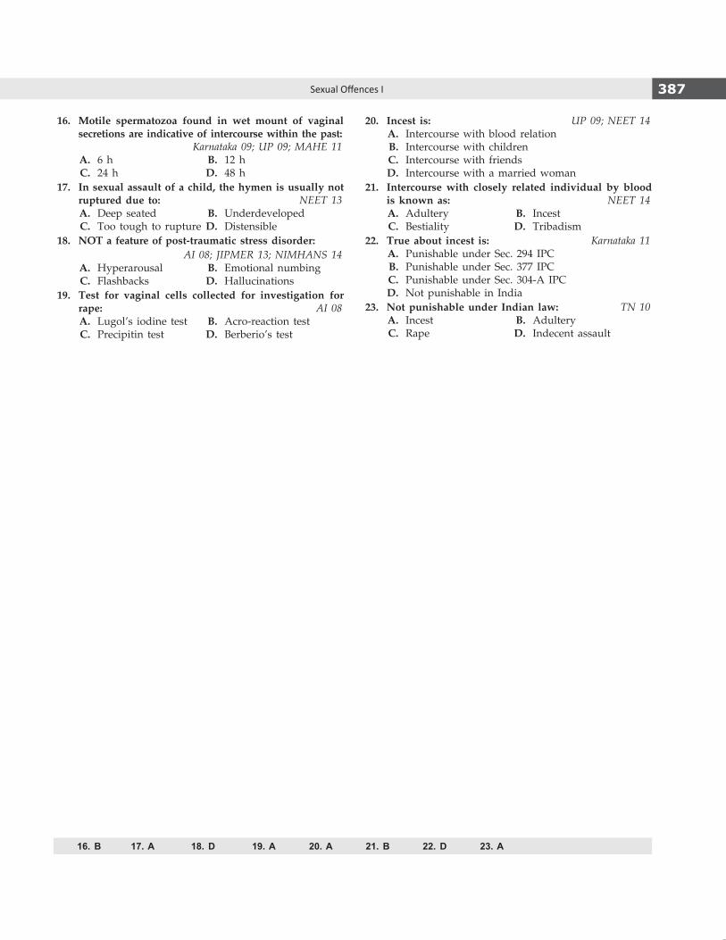
387Sexual Offences I
16. B 17. A 18. D 19. A 20. A 21. B 22. D 23. A
16. Motile spermatozoa found in wet mount of vaginal secretions are indicative of intercourse within the past: Karnataka 09; UP 09; MAHE 11
A. 6 h B. 12 h C. 24 h D. 48 h 17. In sexual assault of a child, the hymen is usually not
ruptured due to: NEET 13 A. Deep seated B. Underdeveloped C. Too tough to rupture D. Distensible 18. NOT a feature of post-traumatic stress disorder: AI 08; JIPMER 13; NIMHANS 14 A. Hyperarousal B. Emotional numbing C. Flashbacks D. Hallucinations 19. Test for vaginal cells collected for investigation for
rape: AI 08 A. Lugol’s iodine test B. Acro-reaction test C. Precipitin test D. Berberio’s test
20. Incest is: UP 09; NEET 14 A. Intercourse with blood relation B. Intercourse with children C. Intercourse with friends D. Intercourse with a married woman 21. Intercourse with closely related individual by blood
is known as: NEET 14 A. Adultery B. Incest C. Bestiality D. Tribadism 22. True about incest is: Karnataka 11 A. Punishable under Sec. 294 IPC B. Punishable under Sec. 377 IPC C. Punishable under Sec. 304-A IPC D. Not punishable in India 23. Not punishable under Indian law: TN 10 A. Incest B. Adultery C. Rape D. Indecent assault
Unnatural sexual offences include: i. Sodomy ii. Tribadism/Lesbianism iii. Bestiality iv. Buccal/Oral coitus
� Unnatural sexual offence is punishable under Sec. 377 IPC.1,2 It states that ‘if an individual voluntarily has sexual intercourse against the order of nature with any man, woman or animal, he is punishable with imprisonment for life or with imprisonment upto 10 years and fine.’ Penetration is sufficient to constitute offence under this section.
� Sec. 377 IPC is meant to criminalize homosexual intercourse and extends to any sexual union involving penile insertion—even consensual heterosexual acts such as fellatio or intercourse between the thighs of another.
� Furthermore, the offence is cognizable, non-bailable, non-compoundable, and tried by a Magistrate of First Class.
In 2009, the Delhi High Court decriminalized consensual homosexual activities between adults. The judgment was overturned by the Supreme Court in 2013, with the Court holding that amending or repealing this section should be a matter left to Parliament, not the judiciary.
Sodomy
Definition: It is the anal intercourse between two males (homosexual sodomy) or between a male and a female (heterosexual sodomy). It is also called buggery.3
� Pederasty is intimate sexual relations, especially anal intercourse with a boy outside his immediate family as the passive partner (the boy is known as catamite, and the man as pederast).4,5
� Habitual passive agents are called fairies, gays or queens in the West, and in India they are called hijras (castrated males) and zenanas (male transvestites).
� The Greeks of Golden Age were said to practice it and is also called ‘Greek Love.’
� It is frequently seen among sailors, prisoners and in military barracks, and prevails at all levels of society.
Brief anatomy of anal canalNormally, the anal orifice is slit-like and running antero-posteriorly with marked ridges (folds) due to the action of corrugator cutis ani muscle. The perianal skin is pigmented and keratinized and has skin appendages (e.g. hair, sweat glands and sebaceous glands). The external anal sphincter has the ability to dilate significantly without any obvious injury to the sphincter or anal canal.
Examination of Passive Agent of Sodomy
Pre-requisites and Preliminary Particulars
� Written authorization from Magistrate or in-charge of a police station is a must before undertaking an examination. If the passive agent is a victim (non-consenting), he can also request for an examination, but the doctor should inform the police.
� General information—name, age, sex, address, occupation, time, date and place of examination.
� Two identification marks are noted. � Written informed consent should be obtained in
case of non-consenting victim. Consent in case of accused and consensual partner is guided by Sec. 53 (1) CrPC.
� History, date and time of the incident, defecation, change of clothing, bathing or washing the anal area after the alleged act, use of lubricant and degree of penetration is specifically asked for.
� Any history of pain/burning sensation associated with defecation or walking is specifically asked for.
� Gait of the victim is noted.
Clothings: Clothings are examined for damage, loose pubic hair, stains of blood/semen/lubricant/feces.
General examination: General physical examination including development of secondary sexual characters is noted. Any injuries, like abrasions and bruises indicating resistance should be noted.
Sexual Offences II
CHAPTER 26

389Sexual Offences II
Local Examination (in knee-elbow position) (Fig. 26.1)
A number of variables may affect the possibility of finding physical evidence of anal intercourse:
� Frequency of the acts � Time interval between intercourse and examination � Age, built and size of the orifice in the individual � Degree of force applied during the act � Size of the penile organ � Cooperativeness of the partner � Use of lubricants
Non-habitual Passive VictimLesions are marked in children because of great disproportion in size between anal orifice of victim and penis of the accused. A perianal and rectal swab should be taken first and any matted (anal/pubic) or foreign hair should be preserved for examination. i. There is pain/tenderness during examination. ii. Smears of lubricant and loose foreign pubic hair
around/in the anus. iii. Fresh/dried semen may be present around/in the
anus. iv. Injuries: Superficial injuries include perianal
abrasions, bruising, erythema, hematoma, edema and anal fissures. Deep injuries include anal lacerations/tears extending onto the perineum, complete transection of the external anal sphincter and perforation of the rectosigmoid (more common in children).
z Linear abrasions may be seen around the anal opening—produced by frictional shearing of the penetrating penis, but may be caused by fingernails, severe constipation or due to poor hygiene. Extensive abrasions are seen when there is disproportion between anal orifice and the penis.
z Anal fissures (splits in the skin of anal margin) may involve the external skin or may extend within anal canal to mucocutaneous junction and are usually present in the posterior quadrant. It
is generally wedged shaped (triangular), directed radially towards the anal canal (Fig. 26.2).
z Hematoma may be present which is diffuse and present circumferentially around anal margin with obliteration of normal anal skin folds giving an appearance of a tyre (‘tyre sign’) or appears as localized swelling.6 The anus opening appears blue and there may be some edema around the anus which may last up to 2 days after the assault—this may be mistaken for hemorrhoids.
z There may be anal prolapse. z First intercourse may result in overt tearing of
anal skin and underlying sphincter muscle or splitting of skin and production of anal fissure or mere abrasion/contusion of the opening.
v. Digital examination is extremely painful, may show loss of elasticity and tone.
vi. At the end, anal canal and lower rectum is examined with the help of proctoscope (if there is spasm of the sphincter, it may be carried under anesthesia).
Habitual Passive Agent (Fig. 26.2) i. There may be shaving of anal hair. ii. Bloodstains are usually not observed. iii. Loose foreign hair and smears of lubricant may
be present. iv. Perianal skin may be thickened and keratinized
with mucocutaneous eversion. Shiny silvery hyperkeratinized skin may also be due to scratching from chronic irritation associated with hemorrhoids, threadworms or viral infections.
v. Person does not experience any pain or tenderness during digital examination. Anal sphincter is lax, opening is patulous, canal is dilated and there may be loss of fine symmetric rugal pattern, along with congested or dilated veins.
vi. Lateral traction test: External anal sphincter relaxes reflexly when bimanual traction is applied to the buttocks.7
Fig. 26.1: Knee-elbow (genupectoral) position Fig. 26.2: Findings in a habitual passive agent of sodomy

390 Review of Forensic Medicine and Toxicology
vii. Anal opening is more deeply situated than usual due to absorption of subcutaneous fat, giving a funnel-shaped depression of buttocks. The ‘funnel shaped’ anus is rarely seen.
viii. Rectum: Thickned, congested and prolapse of mucosa with disappearence of radial fold.
ix. Other signs: Venereal disease, cryptitis, piles, fissures, anal scars from healed injuries and homosexual mannerism regarding dress, gait, manner of speaking and cosmetics.
Opinion
Opinions should be restrained, but not vague, especially on matters where lack of experience makes it dangerous to be assertive. The opinion is based on (Table 26.1):
z Presence of semen/seminal stains in and/or around the anus.
z Soiling of the anal region with lubricants. z Smearing of clothes with semen, blood, lubricants
or any other material. z Injuries in and around the anus. z Foreign hair. z Changes in the general anatomy of the anal
opening and the surrounding area. � There may or may not be any residual findings from
either the single or repeated acts of anal intercourse, since anus is anatomically designed for passage of stools, it is able to expand to a large extent in both adults and children.
� Signs may be minimal when lubricant has been used or the organ been introduced slowly into the anus without using undue force.
� It has a good blood supply and, the acute signs of penetration get healed in about 24–48 hours. Hence, time interval between alleged offence and examination is vital in documentation of the findings.
� The presence of semen, feces, soft paraffin and pubic hair on clothes is almost diagnostic of sodomy.
� The only absolute proof of sodomy is the presence of semen in the anus.
Examination of Active Agent of Sodomy
Pre-requisites and preliminary particulars � General information—name, age, sex, address,
occupation, time, date and place of examination. � Two identification marks are noted. � Consent in this case is guided by Sec. 53 (1) CrPC. � History of his version is noted.
Examination
i. Clothes are examined for the presence of stains— blood, fecal, seminal or mud.
ii. The accused is examined for abrasions and contusions on glans or tearing of the frenulum. Forceful penetration against resistance may produce tears or bruising of frenulum or prepuce and abrasion of glans penis.
iii. There may be traces of feces and lubricant about his genitalia and the peculiar smell of anal glands.
iv. There may be presence of blood, seminal stains, venereal disease and foreign hair.
v. In habitual active agent, the penis is usually twisted with constriction at some distance from glans due to constriction force of the sphincter ani.
Specimens to be preserved for passive and active agent
Passive agent Active agent
� Clothing � Clothing
� Swab from anal canal � Swab from glans
� Swab from bite mark � Urethral discharge
� Blood � Blood
� Nail scrapings � Pubic hair
� Matted and foreign pubic hair and his own for comparison
� Nail scrapings � Urine
Table 26.1: Perianal signs of abuse
Signs Findings
Non-specific acute signs
Erythema, perianal abrasions, edema, fissures, venous congestion, bruising
Signs supportive of abuse
� Anal laxity � Reproducible reflex anal dilatation >15 mm � Chronic changes, i.e. thickening of anal
opening, increased elasticity and reduced anal sphincter tone
� Bite marks
Diagnostic signs � Fresh laceration � Transection of the anus � Perforation of the rectosigmoid colon � Healed scar extending beyond anal margin
on to perianal skin � Recovery of seminal products from the
anorectal canal

391Sexual Offences II
Medico-legal Aspects
i. Both active and passive partners are guilty of the offence under Sec. 377 IPC, even if the act has been committed with consent.
ii. Marriage contract gives implied consent for sexual intercourse per vaginum, not per anum. Under Sec. 13 of Hindu Marriage Act, conviction for natural or unnatural sexual act is a valid ground for divorce.
iii. Penetrative anal sex is legal in the UK between consenting adults who are over the age of consent, i.e. at least 16 years of age. The sexual act had to take place in private, and members of the Armed Forces and merchant seamen are excluded, whatever their age.
� The term sodomy is derived from the name of the ancient city of Sodom, which according to the Bible was destroyed by God for its misdeeds. Traditionally, the misdeeds of Sodom have been understood to be male homosexual anal intercourse.
� Sin of Gomorrah: According to the Bible, the men of Sodom and Gomorrah desired to perform homosexual gang rape on the angels. Homosexuality was the reason God poured fiery sulfur on the cities, completely destroying them and all of their inhabitants
� At the extreme, homosexuality remains punishable by death in Afghanistan, Iran, Nigeria, Pakistan, Saudi Arabia, Sudan, United Arab Emirates and Yemen.
� Intragluteal coitus occurs when the penis is placed between the gluteal fold which may result in edema, contusion and abrasions involving the natal cleft, perinal and anal tissues due to friction. There may be presence of seminal stains on the back or buttocks, and pubic hair and other trace elements (e.g. fibers) may also be found on the body.
Tribadism/Lesbianism
Definition: It is female homosexuality in which two women by mutual acts of sexual indulgence achieve gratification.8
Features � Many lesbians are masculine in type, possibly due to
endocrine disturbances and are indifferent towards men.
� Active and passive partners are usually exchanged, although one partner may habitually play as active sex partner and the other as passive. A preferentially active lesbian is known as a ‘butch’ or ‘dyke’, while the passive partner is known as ‘femme’.9
� Lesbians who are morbidly jealous of one another when rejected may commit homicide, suicide or both.
� It is usually indulged by women who have repulsion for men or who suffer from nymphomania.
� The predominant forms of sexual activity to achieve orgasm are oral-genital and manual genital stimulation. Self-stimulation of clitoris is frequently the preferred method. Use of artificial phallus, anal stimulation and other practices are infrequently used.
� The acts include lip kissing, massaging the breasts and private parts, generalized body contact and mutual rubbing of private parts.
� On examination, the external genitalia may show scratch marks and/or bite marks.
� The word ‘tribadism’ is derived from the obsolete word tribade, meaning ‘lesbian’.10
� The word ‘lesbian’ originally referred to an inhabitant of the island of Lesbos, in ancient Greece. The term has come to have its current meaning due to the ancient Greek poet Sappho, who lived on the island; some of her poems concerned love between women. This led to the term ‘sapphism’ being used for lesbianism.
� Nymphomania: Abnormal, excessive, insatiable desire in a woman for sexual intercourse.
� Satyriasis: Morbid, insatiable sexual need or desire in a man.11
� Anilingus: The practice of oral stimulation of the anus. � Urningism: Sexual practice in which sexual desire is only for
one of the same sex (obsolete word for male homosexuality).
Homosexuality: It is the phenomenon wherein an individual (male or female) prefer a partner of the same sex for sexual activity and intimate bonding.
� The most frequent form of male homosexual activity is fellatio and masturbation; anal intercourse occurs much less often.
� In the past, homosexual couples often lived together but downplayed their relationship in public to avoid discrimination. Many couples now assert the legitimacy of their relationship though marriage (recognized by religious and political institutions) and parenthood.
� Lesbian couples are conceiving and bearing children through various artificial methods like infertile heterosexual couples. Adoption is another means to parenthood for gay and lesbian couples.
Bestiality/Zoophilia
Definition: It is sexual intercourse with animal, either vaginal, anal or oral. This includes all animals, including birds, the usual victims being pets and farm animals.
� Generally, sheep are used by males, and dogs or cats by females as they are easily available and relatively docile.
392 Review of Forensic Medicine and Toxicology
� Doctor may sometimes be asked to examine genital injuries or infections in a man acquired during such episodes. The sure evidence of bestiality is finding of human spermatozoa in the genital tract of the animal. The penis may be contaminated with fecal matter, vaginal secretion or hair of the animal. There may be injury to the penis, dung stains, general body injuries or bloodstains.
In the UK, under the ‘Sexual Offences Act 2003’, the sentence was reduced to a maximum of 2 years imprisonment for penile penetration of or by an animal.
Buccal Coitus
Definition: It denotes penile or vaginal oral sexual intercourse, and can be performed by both males and females.
� It is also called the ‘Sin of Gomorrah’, because it is alleged that buccal coitus was prevalent in Gomorrah, the Biblical twin city of Sodom.
� Fellatio (Latin fellare: to suck) means oral stimulation of the penis either by the female or male.12
� Cunnilingus means oral stimulation of female genitalia.
� Earlier buccal coitus was considered as a sexual deviation, but nowadays it is considered normal sexual foreplay.
� Injuries: A person who is forced to perform fellatio may have trauma in the oral cavity, such as petechiae of the palate and/or posterior pharynx. Tears to the labial frenulum may result from forceful traction on the upper lip. If a fellator’s scalp hair is grasped forcibly during the act, traction alopecia may be seen.
z If the victim has fellatio or cunnilingus performed on him/her, acute signs include petechiae, abrasions or bite marks to the genitalia.
� The only material evidence of buccal coitus is the presence of seminal products including spermatozoa in oral cavity and nasopharynx of the fellator (dependent upon time since contact and a history of ejaculation) and buccal mucosal cells on the external genitalia of the subject.
� The mouth and pharynx should be swabbed with nonabsorbent cotton swabs and a smear should be made similar to that made of the vaginal material. A culture for gonorrhea should be taken from the nasopharynx.
Medico-legal Aspects
� In India, under the Hindu Marriage Act, insistence on buccal coitus, if it is non-consensual and repetitive, constitutes a valid ground for divorce.
� Buccal coitus performed by consenting adults over 21 years of age is permitted by law in the UK.
1. Sec. 377 IPC deals with: AIIMS 09 A. Rape B. Adultery C. Sadism D. Sodomy 2. Sodomy is punishable under: NEET 14 A. Sec. 354 IPC B. Sec. 375 IPC C. Sec. 377 IPC D. Sec. 378 IPC 3. Buggery is: Maharashtra 10 A. Anal intercourse between man and woman B. Anal intercourse between man and animal C. Sexual intercourse between two women D. Passive victim of anal intercourse 4. Catamite is: AP 08 A. Any passive victim of sodomy B. Young passive victim of sodomy C. Elderly passive victim of sodomy D. Female passive victim of sodomy
5. Passive partner in a sexual relationship where a young boy and man are involved: NEET 15
A. Catamite B. Dyke C. Pedarast D. Sodomast 6. Tyre sign is seen in: COMEDK 14 A. Bite mark B. Anal abuse C. Buccal coitus D. Vulvovaginal abuse 7. Lateral traction test is used for: AIIMS 13 A. Habitual passive agent of sodomy B. Active agent of sodomy C. Non-habitual passive agent of sodomy D. Rape accused 8. Tribadism is: AP 06 A. Man having anal sex with man B. Woman having sex with woman C. Man having anal sex with women D. Women having sex with animal
1. D 2. C 3. A 4. B 5. A 6. B 7. A 8. B
MULTIPLE CHOICE QUESTIONS

393Sexual Offences II
9. B 10. A 11. C 12. C
9. In an act of tribadism, the active partner is known as AIIMS 12; COMEDK 14
A. Bugger B. Butch C. Catamite D. Femme 10. Lesbianism is also called as: TN 06 A. Tribadism B. Eonism C. Sodomy D. Onanism
11. Irresistible sexual desire in a male is known as: AIIMS 08 A. Nymphomania B. Tribadism C. Satyriasis D. Sadism 12. Penile buccal coitus is: NEET 14,15 A. Sadism B. Fetishism C. Fellatio D. Cunnilingus

Definitions
� Paraphilia (Greek para: beside, philos: loving) is used to indicate sexual arousal in response to sexual objects or situations that are not part of societal normative arousal/activity patterns or which may interfere with the capacity for reciprocal affectionate sexual activity.
z It is characterized by a 6-month period of intense, recurrent sexual urges or behaviors that involve nonhuman objects, or causing pain/suffering to the individual or sexual partner, or involve harmful sexual contact with non-consenting children or adults.
z The most common paraphilias as described in Diagnostic and Statistical Manual of Mental Disorders, Fourth Edition (DSM-IV) are:1
i. Sadism ii. Masochism iii. Transvestic fetishism iv. Voyeurism v. Exhibitionism vi. Fetishism vii. Frotteurism viii. Pedophilia
� Paraphilia not otherwise specified: This include paraphilias that do not meet the criteria for any of the other specific categories, e.g. masturbation, urophilia, coprophilia, scatologia, partialism, zoophilia and klismaphilia.
Sadism/Algolagnia
Definition: Person gets sexual gratification by infliction of pain or physical cruelty, like beating, biting, whipping, cigarette burns or ill-treating the partner.
� Multiple injuries are inflicted on any body parts, but breasts and external genitalia are generally selected. In extreme cases, even a murder is committed (lust murder).2
� This perversion is more common in males. � The name sadism is derived from the French
writer Marquis de Sade (1740–1814) who regarded sexually deviant acts as being natural, and which was apparent in both his writings and actions. His life consisted of numerous acts of extremely violent
physical and sexual abuse; most of his victims were female prostitutes, and male and female employees of his estate. He wrote pornographic and erotic books in which characters enjoyed being cruel.
Lust MurderDefinition: It is a homicide in which the offender stabs, pierces, slashes or otherwise mutilates the sexual organs or areas of the victim’s body. With torturing the partner, sexual arousal starts, and with death of the partner, full gratification is obtained.
� The mutilation of the victim may include evisceration and/or displacement of the genitalia.
� After murder, the sadist may have sexual intercourse with her (necrophilia). He may tear out the genitalia or other organs, may suck or lick the wounds or eat the flesh of his victim to derive sexual pleasure (necrophagia/anthropophagy).
� It is the consequence of extreme sadist practice. � A lust murder begins with the obsessive compulsions
of the offender. Generally, they have a sexual obsession with their victims, and organized lust murderers may stalk their victims for months or weeks before the actual killing.
� The signature component of the crime that which names it a ‘lust murder’ is the killer acting out their fantasies with their victims and the bodies of those victims.
Masochism/Passive Algolagnia
Definition: Sexual gratification is obtained only when they receive painful stimulus from opposite partner.3,4
� It is the reverse of sadism. � More commonly seen in males. � The term is derived from the 19th century author
Leopard Von Sacher Masoch, an Austrian novelist who portrayed his principle male character suffering from this perversion. His story appeared to parallel his relationship with his wife as he used to plead her to treat him as a slave and whip him, and his eccentric requests gradually became more demeaning to satisfy his sexual appetite.
Sexual Offences III
Chapter 27

395Sexual Offences III
� Masochistic asphyxial death may occur due to acci-dental hanging or strangulation (autoerotic death).5
Sadism and masochism are rarely found in pure state (combined entity is known as sadomasochism). They are usually found together with one type dominant over the other. The combination is known as bondage. This is found in all age groups and among all socio-economic strata. These acts of cruelty may completely substitute the sexual intercourse.
Transvestic Fetishism/Eonism
Definition: It is a disorder characterized by recurrent, intense, sexually arousing fantasies, sexual urges or behaviors involving cross-dressing.
� Sexual gratification is obtained by wearing the dress of opposite sex. In contrast, transsexuals wear clothes of other sex because they feel a part of the other sex and not for sexual excitement.
� It is usually seen in males. They collect items of distinct feminine look and feel, like nightgowns, slips, bras, lingerie, stockings and pantyhose, and may dress in these feminine garments and take photographs of themselves while living out their secret fantasies.
� Magnus Hirschfeld coined the term ‘transvestism’ (Latin trans: across, over; vestitus: dressed) to refer to the sexual interest in cross-dressing.6 The term has undergone several changes of meaning since it was first coined. Hirschfeld’s group of transvestites consisted of both males and females with heterosexual, homosexual, bisexual and asexual orientations.
� The term ‘eonism’ is derived from the Frenchman, Chevalier d’Eon de Beaumont, who practiced this perversion.
Cisvestism: It is a disorder characterized by obtaining sexual pleasure from dressing up in clothes typical of one’s own sex but inappropriate to the individual’s position or status, e.g. biker’s ‘leathers’ or cowboy’s outfit.
Voyeurism/Scoptophilia7
Definition: There is a morbid desire of the individual to observe unsuspecting people undress or naked, taking bath, see the genitalia or watch intercourse to get erotic excitement and sexual gratification.
� It is commonly seen in males. � Voyeurs (French voir: to see, observer) frequently
peep into the bedrooms of others, and are called as ‘Peeping Toms’.
� Some voyeurs prefer to observe their own wives being seduced by other men. The act of observation may be accompanied by exhibitionism or masturb-ation.
� It occurs in case of sociopathic personality disorder, and such individuals may commit sexual crimes.
� Psychoanalysts postulate that voyeurism may be attributed to a child witnessing episodes of his/her parents engaged in sexual intercourse.
� Legal aspect: Under Sec. 354-C IPC, voyeurism is a punishable offence. It states that ‘any man who watches or captures the images of a woman engaged in a private act (changing clothes, using lavatory or doing a sexual act) in circumstances where it is expected of not being observed or circulate such image is punishable with imprisonment from 1–3 years and fine (cognizable and bailable), and for 2nd offence, imprisonment is for 3–7 years and fine (cognizable and non-bailable)’.8
� In the UK, non-consensual voyeurism is a criminal offence, and in the US video voyeurism is criminalized in many States.
� Troilism: The pervert gets sexual gratification by inducing his wife to have sexual intercourse with another man and by observing the same.
� Stalking involves following a woman or contacting such woman in spite of clear disinterest or monitors her through internet, e-mail or any other form of electronic communication. It is punishable under Sec. 354-D IPC with imprisonment for upto 3 years and fine (cognizable and bailable) and imprisonment upto 5 years and fine for subsequent offence (cognizable and non-bailable).9
Exhibitionism
Definition: It is a desire and intentional exposure of genitalia in public places while in presence of others (mostly in front of unsuspecting children or females) to obtain sexual pleasure.
� This perversion is mostly seen in males, and are called flashers. Occasionally, women may expose themselves in public.
� Most of them are psychopathic or suffer from compulsive neurosis.
� Narcissism, the extreme form of self-admiration is also believed to contribute to exhibitionism.
� Legal aspect: It is an obscene act punishable under Sec. 294 IPC with imprisonment upto 3 months and/or fine.
396 Review of Forensic Medicine and Toxicology
Fetishism
Definition: It is a fixation on an inanimate object or body part that is not primarily sexual in nature and the compulsive need for its use in order to obtain sexual gratification.10
� Alfred Binet, the French psychologist coined the term ‘erotic fetishism’ (French fétiche: attribution of mystical qualities to inanimate objects).
� It is mostly seen in males. � Fetish objects: Although, the list of objects is
inexhaustible, more commonly fetish objects are hand-kerchief, dress, particularly the undergarments— panties, bras, slips, stockings, pantyhose or negligees.
� Essential feature is recurrent intense sexual urges and sexually arousing fantasies involving specific objects. They cannot suppress their desire to steal the fetish object.
� Diagnosis is made if an individual has acted on these urges and is markedly distressed by them or the fetish object is required for gratification.
Frotteurism/Toucherism
Definition: It is the act of obtaining sexual arousal and gratification by rubbing of one’s genitals against a non-consenting person in public places.11
� It is usually seen in males. � Frotteurism occurs in crowded trains, buses, elevators
and at bicycle stands (where people bent over for unlocking locks).
� It is prevalent in Japan, where it is known as chikan and is regarded as a public safety problem.
� Fondling (groping) the victim may be part of the condition and is called toucherism.
� Legal aspect: This is an offence and punishable under Sec. 290 IPC (fine of ` 200) and Sec. 291 IPC (imprisonment for 6 months and/or fine) for creating public nuisance.
Pedophilia
Definition: It is the recurrent, intense sexual fantasies, urges or behaviors involving sexual activity with a prepubescent child or children (≤ 13 years) by a person who is ≥ 16 years old and at least 5 years older than the child.
� Pedophiles are usually men and can be attracted to either or both sexes.
� Typical activities vary from just looking at a child undressing and fondling, to acts like oral-genital contact, rubbing the penis between orifice or thighs and actual penetration.
� Usually, the child is not able to understand the nature and consequences of the act, and the perpetrator is influential (elder), i.e. having parental or other position of authority with respect to the child (incestuous or non-incestuous relationship).
� It is one of the few psychiatric diagnoses for which the symptom behavior constitutes a criminal act. If a person is having pedophilia, it is not illegal but an adult having sexual contact with a child (≤ 18 years) is illegal.
� Infantophilia is a subcategory of pedophilia in which the victims are < 5 years.
� Ephebophilia, also known as hebephilia, is the sexual attraction of an adult to pubescent or post-pubescent adolescents.
� Gerontophilia refers to the sexual preference for the elderly.
Masturbation/Onanism
Definition: Deliberate self-stimulation which results in sexual arousal.12
� Masturbation is common in both men and women. � In males, methods are mostly manual—by moving the
penis with hand or against a bed or other object. Anal stimulation and insertions are rare. Hollow articles, like bottles or test tubes or articles made of rubber and plastic which stimulate female genitalia are sometimes used.
� In females, a finger or a hand is gently and rhythmically moved over clitoris or labia minora. The genitalia may be rubbed against a pillow, bed or some other object. She may insert fingers, wooden rods, test tubes, metallic bars, bananas or artificial phallus made of rubber or plastic into the vagina.
� It is an offence when practiced openly, e.g. in telephone booths, bus or toilets.
The word onanism was formerly used as a synonym because in biblical times under Jewish law, a brother was required to procreate with his brother’s widow. Onan of Judah refused and ejaculated on the ground instead. This is the origin of the term onanism (The Sin of Onan) which is incorrectly used in place of masturbation.
Uranism
The pervert gets sexual gratification by fingering, fondling or licking (homosexuality in males).

397Sexual Offences III
1. A 2. B 3. C 4. D 5. A 6. C 7. A
multiple ChoiCe questions
Urolagnia (Urophilia, Undinism) � The pervert gets sexual gratification by sight or odor
of urine and/or by urination. � Those who enjoy urolagnia (Greek ouron: urine,
lagneia: lust) may enjoy urinating on another person or being urinated upon (golden showers).
� In New Zealand, publishing anything promoting or supporting urolagnia, whether in print or online, is punished with imprisonment upto 10 years.
CoprophiliaCoprophilia (Greek koprós: excrement, filía: fondness) is a morbid attraction to, and sexual gratification obtained from feces (liking the smell, taste or feel). Eating of feces is known as coprophagia.
� Scatologia involves making obscene phone calls. � Partialism is sexual interest exclusively focused on a particular
body part. � Klismaphilia is sexual activity involving enemas.
Indecent Assault
Definition: Any unwanted sexual behavior or touching of a female without her consent, with the intention or knowledge to outrage her modesty.
� Males usually do it often to females or adolescents. � The meaning of indecency depends upon prevailing
views of what is unacceptable behavior. � This can mean many things, from an unproven
rape to merely touching the buttocks in a crowded bus.
� Forcing someone to watch pornography or masturbation, disrobing or compelling a female to get naked in public place, fondling the breasts, thighs, perineum, kissing a woman forcefully, or putting a hand up a woman’s skirt constitute indecent assault.
Allegations Against Doctors
One particular risk in medical practice is the vulner-ability of male doctors to the allegations by women patients of indecent assault during consultation or treatment session which may vary from intimate touching, to kissing, fondling the breasts or pudenda and even actual intercourse (which may amount to rape). Stripping naked a female patient for medical examination (without her consent) is regarded as an assault.
Legal Aspect � It is a punished under Sec. 354 IPC with 1–5
years imprisonment and/or fine. The offence is cognizable, non-bailable, non-compoundable and can be tried by any Magistrate.
� Any man who assaults or uses criminal force on any woman or abets such act with the intention of disrobing or compelling her to be naked is punishable with imprisonment for 3–7 years and fine (Sec. 354-B IPC). It is a cognizable and non-bailable offence.
� In the UK, indecent assault is an offence under Sec. 3 of Sexual Offences Act, 2003.
1. Following is not a paraphilia: DNB 10 A. Lesbianism B. Fetishism C. Frotteurism D. Voyeurism 2. Lust murder is an extreme form of: MP 09 A. Troilism B. Algolagnia C. Masochism D. Frotteurism 3. Masochism means: TN 09 A. Sexual intercourse with dead body B. Sexual pleasure by contact with articles of opposite
sex C. Sexual pleasure by suffering of pain D. Sexual pleasure by self-stimulation
4. Perversion with pain to self: TN 09; Maharashtra 11 A. Transvestism B. Fetishism C. Sadism D. Masochism 5. Sexual asphyxia is seen in cases of:
AIIMS 06; NEET 13, 15 A. Masochism B. Voyeurism C. Sadism D. Fetichism 6. Liking to wear clothes of opposite sex: NEET 13 A. Fetishism B. Masochism C. Transvestism D. Sadism 7. Voyeurism is also known as: NEET 13 A. Scoptophilia B. Eonism C. Frotteurism D. Onanism

398 Review of Forensic Medicine and Toxicology
8. Not true about voyeurism: PGI 14 A. Sec. 354-C IPC defines punishment B. Cognizable offence C. Non-bailable offence D. 1-3 years punishment 9. First offence of stalking is: NEET 15 A. Cognizable and bailable B. Non-cognizable and bailable C. Cognizable and non-bailable D. Non-cognizable and non-bailable 10. Fetishism is a sexual perversion characterized by:
COMEDK 08; PGI 12 A. Sexual focus on children B. Sexual focus on genital rubbing
C. Sexual pleasure for pain D. Sexual pleasure derived from inanimate objects 11. Frotteurism is: NEET 13 A. Sexual pleasure is obtained by witnessing the act
of urination B. Sexual gratification by rubbing private parts C. Sexual practise involving three people D. Sexual pleasure in watching females getting
undressed 12. Onanism is: TN 07 A. Natural sexual offence B. Unnatural sexual offence C. Perversion D. Indecent assault
8. C 9. A 10. D 11. B 12. C

Definition: Postmortem artifacts (Latin arte: art, factum: something made) are any changes caused or features introduced in a body after death which may lead to misinterpretationoffindings. Ignorance and misinterpretation of such postmortem artifacts leads to:
� Wrong cause/manner of death � Undue suspicion of criminal offence � A halt in the investigation of criminal death � Unnecessary wastage of time and effort, as a result ofmisleadingfindings
� Miscarriageof justice.
Postmortem artifacts can be classified into: i. Artifactsdue topostmortemchanges ii. Thirdpartyartifacts iii. Environmental artifacts iv. Otherartifacts.
Artifacts due to Postmortem Changes
These artifacts are due to rigor mortis, postmortemstaining, autolysis,putrefactionandheat. i. Rigor mortis:Existingrigormortismaybebroken
down while removing the body from the scene of crime to the mortuary which may cause error in interpretationof timesincedeath.Rigoraffectingtheheartmaysimulatehypertrophyof theheart.
ii. Postmortem staining: Isolated patches of postmortemlividitymaybemistakenforbruises.Suchpatchesonthefrontandsidesoftheneckmaybemistakenforbruisingduetothrottling.Lividityoftheinternalorgansmaybemistakenforcongestiondue todisease.
z Postmortem staining in the posterior left ventricle of the heart in an individual lying supine after death may cause confusion of ischemic myocardial damage, in the lungs—pneumonia, and in theGIT—irritationdue topoisoning.
z Certainpoisons, likeCO,HCNornitritesmaychange the colorof thehypostatic area.
Prinsloo Gordon artifact: A common artifact seen in all types of autopsy. It represents hemorrhage on the anterior aspect of the cervical spine, posterior to the trachea and esophagus which happens due to hypostasis. Hence, caution must be used in interpreting bleeding into the posterior neck tissues.
iii. Autolysis: Autolysis leads to discoloration of skin and viscera, like gallbladder, pancreas,liver, kidney, GIT mucosa and brain, where itmay simulate injury or disease. Pancreas is oneof the first organs to undergo autolysis becauseof proteolytic enzymes within it, which can be mistaken for acute hemorrhagic pancreatitis.Perforation of the stomach due to autolysis have to be disting uished from that due to corrosive acid orpepticulceration.Absenceofcellularresponsein discolo red areas establishes the postmortem originof these changes.
iv. Putrefaction External
z Swellingof lips, nose, eyelids and extremities,distension of the chest and the abdomen may occur, giving a false impression of antemortem obesity.
z Large quantities of sanguineous fluid may escape from the mouth and nose in case of pulmonary edema, giving the impression of hemorrhage.
z A deep groove simulating ligature mark ofstrangulationmay be seen around the neck ifthe deceased has been wearing buttoned shirt or beadedthreadsorornamentsaroundtheneck.
z Thebulgingof eyes,protrusionof tongueanddischarge of red stained froth from mouth and nosemaybemistaken for signsof throttling.
z Owingtopressureeffectsofputrefactivegases,postmortem staining may be displaced in any directionandmaysimulateantemortembruises.
z Putrefactive blisters may be confused with blisters from burns and contact with petroleum products.Theskinfromthehandmaypeellikeagloveas inburns.
Postmortem Artifacts
ChAPter 28

400 Review of Forensic Medicine and Toxicology
z Froth and stomach contents coming out from the nose and mouth may tan the facial skin,simulatingantemortemburning.
z Splitting of skin may give a false impressionof antemortem lacerations, incised wounds or thermal injuries.
z Thefemalegenitaliaappearpendulousandmaysimulateantemortemsexual assault.
Internal z Softeningof ‘synchondrosis’betweenthebody
and greater cornu of the hyoid bone may produce abnormal mobility which may be confusedasa fracture.
z Gas bubbles in the blood and air in the right side oftheheartmaybemistakenforairembolism.Oxygeninrightheartwillindicateantemortemair embolism.
z Internal lividity with hemolysis of red cells may resemble hemorrhage, especially in the meninges,kidneysand retroperitoneal tissues.
z Thebloodbecomesdarker,andthebrain,heartand lungs appear congested which may be mistaken forasphyxia.
z Bluish discoloration of the loop of bowels, especially in the pelvic cavity may be confused asan infarctedbowel.
z Ifabodyliesonitsback,bloodgetaccumulatedintheposteriorpartofthescalpduetogravity.Lysisofredbloodcellsandbreakdownofvesselscause the blood to seep into the soft tissues of thescalpgiving theappearanceofabruise.
z Postmortem separation of the sutures of child’s skull and bursting of the abdomenwith protrusion of the abdominal viscera due to advanced decomposition may be mistakenfor trauma.
Third Party Artifacts
Artifacts due to Animal and Insect Activity
� Thebitesbydogsareclear-cut,withdeepimpressionof teeth in a small area. Individual puncturesmayresemble stabwounds.
� Insect bites (ants or roaches) are dry, brown with irregular margins, and usually seen in moist parts of thebody, e.g. armpits,groin, scrotumandanus,thesemay resembleantemortemabrasions.
� Rodentsgnawawaythetissueoverlocalizedareas.Theyproduceshallowcraterswithirregularbordersand leave longgrooves.
� Bodies recovered from water may show gnawing by fish, crabsandotheraquatic animals.
� Flies,maggotsand larvaemayalter thewounds.
Insects can also cause misinterpretation of blood spatter pattern analysis. Roaches walking throughpooled and splattered blood will produce trailing.Specksofbloodinuniqueandunusualareas(suchason ceilings)maymislead forensic scientists.
Therapeutic Artifacts
� External cardiac massage, especially in elderlypatients is associated with the fracture of ribs (3rd5th) and sometime fracture of the sternum along with laceration of the lungs, liver, spleen and diaphragm which can create an impression of a crushing force applied to the chest.1
� Use of defibrillator may leave an impression ofcircular contusion over the pericardium. Multipleintracardiac injections may result in bruising of heart andhemopericardium.
� Gastric contents are aspirated in the windpipe due to the handling of the body or as a terminal agonal event innaturaldeathsordue to resuscitation.
� Investigative procedures, like carotid angiographymayresultinbruisingoftheneckmusclesgivingafalse impressionof constrictionof theneck.
� Surgeonsmayoftentakelaparatomyincisionthroughincised or stab wounds leading to misinterpretation ofwounds.
� Endotracheal intubation, positive pressure andartificialrespirationmayleadtosurgicalemphysemaandpneumothorax.
� Washing may alter the appearance of entrance and exitgunshotwoundsandtheirdimensionschangedby suturingor excision.
� Drainagewoundsmaybemistakenforfirearmwounds.
Deliberate Mutilation, Dismemberment
� Sometimes, criminals may inflict injuries, mutilateor dismember the body after death to mislead the investigation.
� Some mutilations are produced in a ritualisticsense displaying significant psychopathology of theassailant.Itmayincluderemovalofthebreasts,genital mutilation such as removal of the penis, and scarification type injuries.
� Persons may be killed and thrown into water orset on fire. Careful examination for violence willhelp in the correctdiagnosis of the causeofdeath.
401Postmortem Artifacts
Chemical analysis of the viscera for poisons may be necessary.
� Occasionally, a person may be beaten to death orpoisonedand thenhanged tomisleadpeople.
Embalming Artifacts
� Trocar woundsmay be mistaken for stab woundsorbulletwounds.
� Bruises may be markedly accentuated due toincreasedtransparencyoftheoverlyingskinresultingfrom theembalmingprocess.
� Embalming fluid used may pose problems intoxicologicalanalysisoftheviscera,ashighlevelsofmethanol, anticoagulants and various other dyes are oftendetectedby sophisticated screeningmethods.
Autopsy Surgeon Induced Artifacts
i. Skull fractures: During the opening of the skullby forceful sawing or by using a chisel and a hammer, an existing fracture of the skull maybecome extensive or fresh fractures may be caused.
ii. Air in blood vessels: During pulling of the dura, air may enter the blood vessels. This may leadtoanerroneousdiagnosisofairembolism.Whenneckstructuresarepulledforcefully,airmayentertheneckvesselsortheremaybeseepageofbloodaround the neck structures leading to erroneoustraumaticneckpathology.
iii. Visceral damage: The liver, if pulled insteadof being dissected out, may cause tears in the diaphragm and laceration in the bare area of the liver.Whiletheabdomenandtheperitoneumarebeing cutopen,bowel coilsmaybe cut.
iv. Extravasation of blood z When viscera are pulled apart in toto, as in
evisceration, there would be profuse bleeding into the pleural and peritoneal cavities that may bemistakenasantemortemhemorrhage.
z Thehandlingoforgansandthe incisionof thevessels may result in extravasation of bloodinto the tissues.
z The removal of theneck structures enblock asin routine autopsies may produce artifacts in the neck tissueswhichresemblebruises (asseen inthrottling).
z Rough handling of the brain during removalmay damage the dura and the dural venous sinuses that may lead to an escape of blood into
the subdural space, simulating an antemortem subduralhemorrhage.
v. Fracture of hyoid bone: While removing neckstructures, the hyoid bone and thyroid cartilage may be fractured, especially in old persons which maybemistakenforbeingantemorteminorigin.
vi. Toxicological artifacts z Faulty technique in collecting a sample or faulty storageoruseofpreservatives.
z While collecting blood from the heart, the blood maygetdilutedduetopericardialfluid.Useofanticoagulants,e.g.EDTA,formalin,heparinormethenamine may give a false positive result foralcoholormethanol.
z Collection of the viscera in a single bottle or use of contaminated bottles/instruments/preservatives may result in wrong analysis of poisons.
z Decomposition of the tissues after death producesethylalcoholandsignificantamountsof cyanide. Decomposition also causes anincrease in concentrationofCO in theblood.
z In cases of death due to burns, significant amounts of cyanide may be found in blood, possiblyduetoinhalationofhydrogencyanide.
z In buried bodies, arsenic may be imbibed from the surroundingearth.
Environmental Artifacts
Heat Effects
� Heat applied to the skin of a dead body mayloosen the epidermis from the dermis and produce apostmortemblister.
� Heathematomamaysimulateextraduralhemorrhage. � Anunburntgroovearound theneckdue toa tightcollarmay resemblea ligaturemark.
� Fat droplets may be found in the pulmonary vessels whichmaybemistakenforantemortempulmonaryfat embolism.
� Heat ruptures may resemble lacerated or incisedwounds.
Postmortem Corrosion
Dead bodies exposed or lying in kerosene, water orgasolineshowchemicalinjuries.Theepitheliumdetacheswhile handling the body, and then the underlying dermis turns yellow to brown which may be misinterpreted as antemortemchemical injuryorabrasionorburns.

402 Review of Forensic Medicine and Toxicology
Postmortem Maceration
Physical contact of the body with water, soil or air may causemarked changes, dependingupon the chemicalconstituentsofearth,waterandthedurationofcontact.Thebodymaybetotallyskeletonizedleavingdecalcifiedanddeformedbones.
Other Artifacts
Artifacts due to Refrigeration
Pink postmortem staining is seen in bodies kept incold storage. If the bodies are kept in a cold storageimmediatelyafterdeath,gooseskinmaydevelop.
Artifacts due to Mishandling of the Body
� During the process of transfer of the body from the scene of crime to the mortuary, abrasions may be producedoverthebackorbonyprominences,clothesmaygetbloodstainedor torn.
� Sometimes, fractures of the ribs or long bones orcervical spine may occur by rough handling of the bodies, especially in the elderly or debilitated, during
attempts to straighten limbs which are contracted due to rigormortis.
� Contusion may occur over occiput due to bumping of theheadonhard surface.
� Undertaker’s fracture may be seen which is a subluxationofthelowercervicalspineduetotearingof the intervertebraldiscat aboutC6-C7.2
Exhumation Artifacts
� Gravedigger’s tools can produce postmortemfracture, abrasionsand lacerations.
� Thediscolorationoftheskinbeneathfungusgrowthsimulates contusion.
� Postmortem imbibition of toxicological elementsfrom the earth may result in inaccurate toxicolo-gical analysis.
Artifacts due to Delay in Autopsy
Uncal grooving, seen in cerebral edema, tends to be more prominent when there is a delay in removing thebrain.
1. During CPR, artifacts due to rib fracture most commonly seen in: AIIMS 14
A. 2nd–3rd ribs B. 3rd–5th ribs C. 5th–6th ribs D. 6th–8th ribs
2. Undertaker’s fracture is seen at the level of cervical vertebra: NEET 15
A. C1–C2 B. C3–C4 C. C5–C6 D. C6–C7
multiple choice questions
1. B 2. D

Definitions
� Psychiatry: It is that branch of medical science, which deals with the study, diagnosis, treatment and prevention of mental illness and behavioral disorders.
� Forensic psychiatry: It deals with the application of knowledge of psychiatry in the administration of justice.
� Insanity or unsoundness of mind: Disease of the mind, which affects the personality, mental status, critical faculties, emotional processes and interaction with the social environment.
� Mentally ill person: Any person who is in need of treatment by reason of any mental disorder other than mental retardation [as per Persons with Disabilities (Equal Opportunities, Protection of Rights and Full Participation) Act, 1995].
� Mental retardation: A condition of arrested or incomplete development of mind of a person which is specially characterized by sub-normality of intelligence (Sec. 2(r) of the Act).
Some of the important symptoms commonly associated with psychiatric disorders are:
i. Delusion ii. Hallucination iii. Illusion iv. Impulse v. Obsession
Delusion
Definition: False belief, based on incorrect inference about external reality that is firmly held, despite objective and obvious contradictory proof or evidence.1
� A thought disorder is not unusual in normal persons, but he is capable of correcting it by reasoning power, arguments or when convinced by others.2,3
� Delusion is a symptom of schizophrenia. Delusions are not seen in neurotic illnesses, like anxiety neurosis or obsessive compulsive disorder (OCD).
Types
i. Delusion of grandeur or exaltation: The patient imagines himself to be very rich, while in reality he may be a pauper. It is usually seen in mania, and may be associated with delusion of persecution.4
ii. Delusion of poverty: The patient is convinced that he is, or will be, bereft of all material possessions.
iii. Delusionofinfidelity/jealousy(Othellosyndrome):Person holds a delusional belief that his spouse is unfaithful. It is named after the character in Shakespeare’s play Othello, who murders his wife based on his false belief that she has been disloyal. Males are more affected.
iv. Delusion of reference: The person believes that he is being referred to by all agencies, media and persons around him in all matters (usually of negative nature) concerning him or others.
v. Delusion of persecution: The patient imagines that he is going to be poisoned by his relatives (wife, sons or parents) or someone is going to rob his property. He may even commit suicide or kill his own family members or innocent person thinking him to be his enemy. Persecutory delusions are most common.
vi. Delusion of influence/control: The patient complains that his thought processes, feelings and actions are being influenced and controlled by some external power, like radio, hypnotism or telepathy. On the basis of this imaginary ‘command’, he may commit an unlawful act.
vii. Hypochondriacal delusion: Persistent concern with a fear or belief of having one or more serious disease (like cancer) based on patient’s own unrealistic interpretations of physical signs and symptoms.
viii. Delusion of self-reproach or self-criticism: The person criticizes himself for some imaginary offence or misdeed committed by him in the past.
Forensic Psychiatry
ChaPter 29
404 Review of Forensic Medicine and Toxicology
In extreme cases, the person may punish himself by committing suicide.
ix. Nihilistic delusion: The patient does not believe in his existence or the existence of earthly matters or that there is any world. It is commonly seen in depression.5
x. Delusion of doubles (doppelganger): Patients believe that another person has been physically transformed into themselves.
Medico-legal importance: The doctrine of diminished responsibility is applicable to an insane person who does an unlawful act due to delusion, which reduces his power of reasoning and understanding capacity, e.g. if he commits some act which is not directly related with the effect of the delusion, but has an indirect bearing, such person cannot be regarded as fully responsible for his illegal acts.
Erotomania (de Clérambault’s syndrome): It is a condition in which a person holds a delusional belief that another person, usually of a higher social status, is in love with him/her.6 The erotomanic tries to get close to the person through telephone calls, e-mails, letters, gifts and visits. It is more common in women than in men. It is seen during psychosis, especially in patients with schizophrenia, delusional disorders and bipolar mania.7
Other types of delusions
� Delusion of replacement of significant others (Capgras syndrome): Patient believes that someone close to him has been replaced by an exact double.8
� Delusion of disguise (Fregoli’s phenomenon): Strangers are identified as familiar people in the patient’s life.
� Ekbom syndrome (delusions of parasitosis): A form of psychosis wherein the patients acquire a delusional belief that they are infested with parasites, whereas in reality no such parasites are present. Often they report of ‘bugs’ or insects crawling on or under the skin (formication) and try to remove it with tweezers.
� Folie á deux: Mental illness shared by two persons, usually involving a common delusional system. For example, another person (typically a spouse, child or other household member) may develop Ekbom syndrome as well.
� Cotard delusion/syndrome: Person holds a delusional belief that he is dead, does not exist, is putrefying or has lost his blood or internal organs.
Hallucination
Definition: Hallucination (Latin hallucinere: to wander in mind) is false perception by senses without any external object or stimulus.9,10 They are seen in insanity and in conditions, like high fever, drug intoxication and during withdrawal from drug addiction.
Types i. Visual hallucination: It involves the sense of sight.
In this condition, the sufferer experiences (visua-lizes) non-existent sights. He observes something without anything being present. A person seeing a plane flying in the sky or an oasis at a distance in a desert when there is none are quite common experience.
z In a mentally ill person, hallucinations experienced may be located outside the field of vision (e.g. behind the head) or beyond the sensory range (e.g. able to look out of the window and see someone in distant city).
z Visual hallucinations are seen in dissociation and conversion disorder, severe affective disorder, organic mental conditions, substance abuse and schizophrenia.
ii. Auditory hallucination: False perception of sound, usually noises, but also music. The patient hears voices or sounds without any source. In schizophrenia, the patient may talk, whisper or mutter to himself incomprehensively, or talk normally or shout out loudly in response to the voice.
iii. Olfactory hallucination: Hallucination primarily involving smell or odors. There is a false sense of smell (pleasant/unpleasant) without any source. They are felt in schizophrenia and temporal lobe epilepsy.11,12
iv. Gustatory hallucination: Hallucination involving taste. Without any food or drink, the patient experiences different tastes (sweet/sour/bitter).
v. Tactile/haptic hallucination: Hallucination of touch. The sufferer experiences crawling of insects or rats over his body without any such thing happening in reality.
vi. Psychomotor hallucination: There is a feeling of movement of a part of the body, say a limb, though in reality, there is no such movement.
vii. Lilliputian hallucination (micropsia): Visual sensation that persons or objects are reduced in size; more properly regarded as an illusion.
� Auditory hallucinations are the most common, followed by visual.
� Visual hallucinations are the commonest in organic mental disorders (delirium tremens), auditory in functional (non-organic) disorders (schizophrenia), gustatory in temporal lobe epilepsy, olfactory in medical disorders (especially in the temporal lobe), and tactile in cocainism and occasionally in schizophrenia.
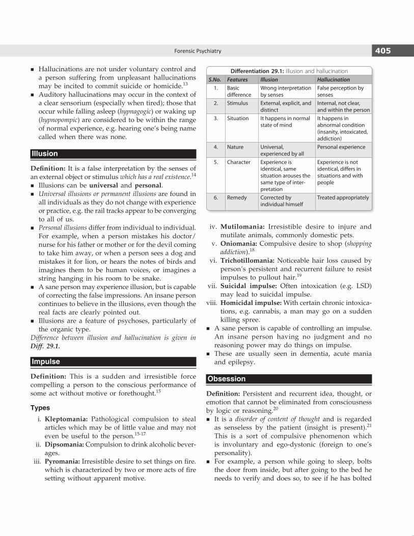
405Forensic Psychiatry
� Hallucinations are not under voluntary control and a person suffering from unpleasant hallucinations may be incited to commit suicide or homicide.13
� Auditory hallucinations may occur in the context of a clear sensorium (especially when tired); those that occur while falling asleep (hypnagogic) or waking up (hypnopompic) are considered to be within the range of normal experience, e.g. hearing one’s being name called when there was none.
Illusion
Definition: It is a false interpretation by the senses of an external object or stimulus which has a real existence.14
� Illusions can be universal and personal. � Universal illusions or permanent illusions are found in
all individuals as they do not change with experience or practice, e.g. the rail tracks appear to be converging to all of us.
� Personal illusions differ from individual to individual. For example, when a person mistakes his doctor/nurse for his father or mother or for the devil coming to take him away, or when a person sees a dog and mistakes it for lion, or hears the notes of birds and imagines them to be human voices, or imagines a string hanging in his room to be snake.
� A sane person may experience illusion, but is capable of correcting the false impressions. An insane person continues to believe in the illusions, even though the real facts are clearly pointed out.
� Illusions are a feature of psychoses, particularly of the organic type.
Difference between illusion and hallucination is given in Diff. 29.1.
Impulse
Definition: This is a sudden and irresistible force compelling a person to the conscious performance of some act without motive or forethought.15
Types
i. Kleptomania: Pathological compulsion to steal articles which may be of little value and may not even be useful to the person.15-17
ii. Dipsomania: Compulsion to drink alcoholic bever-ages.
iii. Pyromania: Irresistible desire to set things on fire.which is characterized by two or more acts of fire setting without apparent motive.
iv. Mutilomania: Irresistible desire to injure and mutilate animals, commonly domestic pets.
v. Oniomania:Compulsive desire to shop (shopping addiction).18
vi. Trichotillomania: Noticeable hair loss caused by person’s persistent and recurrent failure to resist impulses to pullout hair.19
vii. Suicidal impulse: Often intoxication (e.g. LSD) may lead to suicidal impulse.
viii. Homicidal impulse: With certain chronic intoxica-tions, e.g. cannabis, a man may go on a sudden killing spree.
� A sane person is capable of controlling an impulse. An insane person having no judgment and no reasoning power may do things on impulse.
� These are usually seen in dementia, acute mania and epilepsy.
Obsession
Definition: Persistent and recurrent idea, thought, or emotion that cannot be eliminated from consciousness by logic or reasoning.20
� It is a disorder of content of thought and is regarded as senseless by the patient (insight is present).21 This is a sort of compulsive phenomenon which is involuntary and ego-dystonic (foreign to one’s personality).
� For example, a person while going to sleep, bolts the door from inside, but after going to the bed he needs to verify and does so, to see if he has bolted
Differentiation 29.1: Illusion and hallucinationS.No. Features Illusion Hallucination
1. Basic difference
Wrong interpretation by senses
False perception by senses
2. Stimulus External, explicit, and distinct
Internal, not clear, and within the person
3. Situation It happens in normal state of mind
It happens in abnormal condition (insanity, intoxicated, addiction)
4. Nature Universal, experienced by all
Personal experience
5. Character Experience is identical, same situation arouses the same type of inter- pretation
Experience is not identical, differs in situations and with people
6. Remedy Corrected by individual himself
Treated appropriately
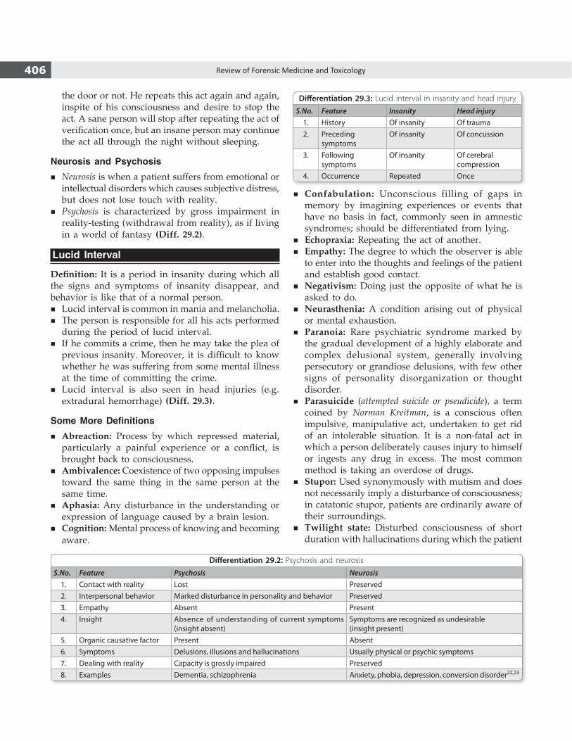
406 Review of Forensic Medicine and Toxicology
Differentiation 29.2: Psychosis and neurosis
S.No. Feature Psychosis Neurosis1. Contact with reality Lost Preserved2. Interpersonal behavior Marked disturbance in personality and behavior Preserved3. Empathy Absent Present4. Insight Absence of understanding of current symptoms
(insight absent)Symptoms are recognized as undesirable (insight present)
5. Organic causative factor Present Absent6. Symptoms Delusions, illusions and hallucinations Usually physical or psychic symptoms7. Dealing with reality Capacity is grossly impaired Preserved8. Examples Dementia, schizophrenia Anxiety, phobia, depression, conversion disorder22,23
Differentiation 29.3: Lucid interval in insanity and head injury
S.No. Feature Insanity Head injury1. History Of insanity Of trauma2. Preceding
symptomsOf insanity Of concussion
3. Following symptoms
Of insanity Of cerebral compression
4. Occurrence Repeated Once
the door or not. He repeats this act again and again, inspite of his consciousness and desire to stop the act. A sane person will stop after repeating the act of verification once, but an insane person may continue the act all through the night without sleeping.
Neurosis and Psychosis � Neurosis is when a patient suffers from emotional or
intellectual disorders which causes subjective distress, but does not lose touch with reality.
� Psychosis is characterized by gross impairment in reality-testing (withdrawal from reality), as if living in a world of fantasy (Diff. 29.2).
Lucid Interval
Definition: It is a period in insanity during which all the signs and symptoms of insanity disappear, and behavior is like that of a normal person.
� Lucid interval is common in mania and melancholia. � The person is responsible for all his acts performed
during the period of lucid interval. � If he commits a crime, then he may take the plea of
previous insanity. Moreover, it is difficult to know whether he was suffering from some mental illness at the time of committing the crime.
� Lucid interval is also seen in head injuries (e.g. extradural hemorrhage) (Diff. 29.3).
Some More Definitions � Abreaction: Process by which repressed material,
particularly a painful experience or a conflict, is brought back to consciousness.
� Ambivalence: Coexistence of two opposing impulses toward the same thing in the same person at the same time.
� Aphasia: Any disturbance in the understanding or expression of language caused by a brain lesion.
� Cognition: Mental process of knowing and becoming aware.
� Confabulation: Unconscious filling of gaps in memory by imagining experiences or events that have no basis in fact, commonly seen in amnestic syndromes; should be differentiated from lying.
� Echopraxia: Repeating the act of another. � Empathy: The degree to which the observer is able
to enter into the thoughts and feelings of the patient and establish good contact.
� Negativism: Doing just the opposite of what he is asked to do.
� Neurasthenia: A condition arising out of physical or mental exhaustion.
� Paranoia: Rare psychiatric syndrome marked by the gradual development of a highly elaborate and complex delusional system, generally involving persecutory or grandiose delusions, with few other signs of personality disorganization or thought disorder.
� Parasuicide (attempted suicide or pseudicide), a term coined by Norman Kreitman, is a conscious often impulsive, manipulative act, undertaken to get rid of an intolerable situation. It is a non-fatal act in which a person deliberately causes injury to himself or ingests any drug in excess. The most common method is taking an overdose of drugs.
� Stupor: Used synonymously with mutism and does not necessarily imply a disturbance of consciousness; in catatonic stupor, patients are ordinarily aware of their surroundings.
� Twilight state: Disturbed consciousness of short duration with hallucinations during which the patient
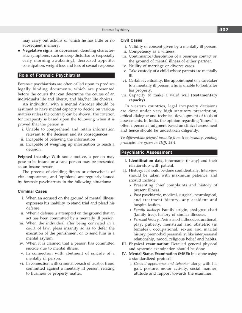
407Forensic Psychiatry
may carry out actions of which he has little or no subsequent memory.
� Vegetative signs: In depression, denoting character-istic symptoms, such as sleep disturbance (especially early morning awakening), decreased appetite, constipation, weight loss and loss of sexual response.
Role of Forensic Psychiatrist
Forensic psychiatrists are often called upon to produce legally binding documents, which are presented before the courts that can determine the course of an individual’s life and liberty, and his/her life choices. An individual with a mental disorder should be assumed to have mental capacity to decide on various matters unless the contrary can be shown. The criterion for incapacity is based upon the following when it is proved that the person is: i. Unable to comprehend and retain information
relevant to the decision and its consequences ii. Incapable of believing the information iii. Incapable of weighing up information to reach a
decision.
Feigned insanity: With some motive, a person may pose to be insane or a sane person may be presented as an insane person. The process of deciding fitness or otherwise is of vital importance, and ‘opinions’ are regularly issued by forensic psychiatrists in the following situations:
Criminal Cases
i. When an accused on the ground of mental illness, expresses his inability to stand trial and plead his defense.
ii. When a defense is attempted on the ground that an act has been committed by a mentally ill person.
iii. When the individual after being convicted in a court of law, pleas insanity so as to defer the execution of the punishment or to send him in a mental asylum.
iv. When it is claimed that a person has committed suicide due to mental illness.
v. In connection with abetment of suicide of a mentally ill person.
vi. In connection with criminal breach of trust or fraud committed against a mentally ill person, relating to business or property matter.
Civil Cases i. Validity of consent given by a mentally ill person. ii. Competency as a witness. iii. Continuance/dissolution of a business contact on
the ground of mental illness of either partner. iv. Nullity of marriage or divorce cases. v. Take custody of a child whose parents are mentally
ill. vi. Certain eventuality, like appointment of a caretaker
to a mentally ill person who is unable to look after his property.
vii. Capacity to make a valid will (testamentary capacity).
In western countries, legal incapacity decisions are done under very high statutory prescription, ethical dialogue and technical development of tools of assessments. In India, the opinion regarding ‘fitness’ is often a personal judgment based on clinical assessment and hence should be undertaken diligently.
To differentiate feigned insanity from true insanity, guiding principles are given in Diff. 29.4.
Psychiatric Assessment
I. Identification data, informants (if any) and their relationship with patient.
II. History: It should be done confidentially. Interview should be taken with maximum patience, and should include:
z Presenting chief complaints and history of present illness.
z Past psychiatric, medical, surgical, neurological, and treatment history, any accident and hospitali zation.
z Family history: Family origin, pedigree chart (family tree), history of similar illnesses.
z Personal history: Perinatal, childhood, educational, play, puberty, menstrual and obstetric (in females), occupational, sexual and marital history, premorbid personality, like interpersonal relationship, mood, religious belief and habits.
III. Physical examination: Detailed general physical and systemic examination should be done.
IV. MentalStatusExamination(MSE): It is done using a standardized protocol:
i. General appearance and behavior along with his gait, posture, motor activity, social manner, attitude and rapport towards the examiner.
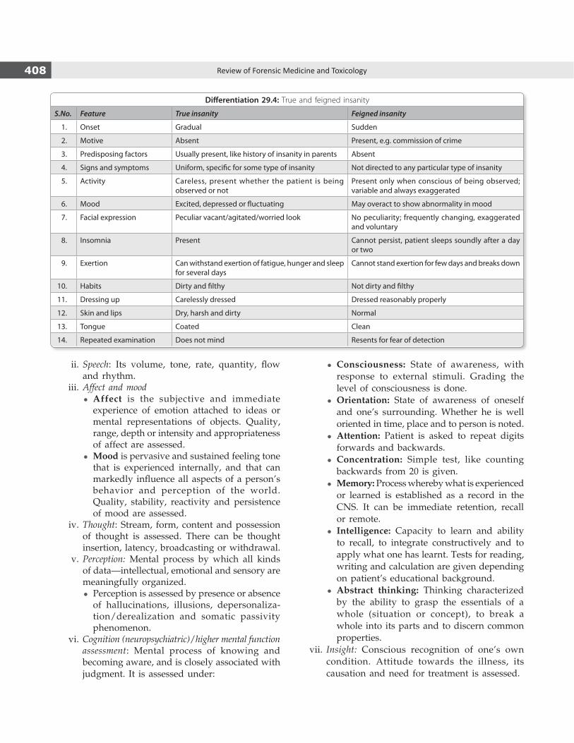
408 Review of Forensic Medicine and Toxicology
ii. Speech: Its volume, tone, rate, quantity, flow and rhythm.
iii. Affect and mood z Affect is the subjective and immediate
experience of emotion attached to ideas or mental represen tations of objects. Quality, range, depth or intensity and appropriateness of affect are assessed.
z Mood is pervasive and sustained feeling tone that is experienced internally, and that can markedly influence all aspects of a person’s behavior and perception of the world. Quality, stability, reactivity and persistence of mood are assessed.
iv. Thought: Stream, form, content and possession of thought is assessed. There can be thought insertion, latency, broadcasting or withdrawal.
v. Perception: Mental process by which all kinds of data—intellectual, emotional and sensory are meaningfully organized.
z Perception is assessed by presence or absence of hallucinations, illusions, depersonaliza-tion/derealization and somatic passivity phenomenon.
vi. Cognition (neuropsychiatric)/higher mental function assessment: Mental process of knowing and becoming aware, and is closely associated with judgment. It is assessed under:
z Consciousness: State of awareness, with response to external stimuli. Grading the level of consciousness is done.
z Orientation: State of awareness of oneself and one’s surrounding. Whether he is well oriented in time, place and to person is noted.
z Attention: Patient is asked to repeat digits forwards and backwards.
z Concentration: Simple test, like counting backwards from 20 is given.
z Memory: Process whereby what is experienced or learned is established as a record in the CNS. It can be immediate retention, recall or remote.
z Intelligence: Capacity to learn and ability to recall, to integrate constructively and to apply what one has learnt. Tests for reading, writing and calculation are given depending on patient’s educational background.
z Abstract thinking: Thinking characterized by the ability to grasp the essentials of a whole (situation or concept), to break a whole into its parts and to discern common properties.
vii. Insight: Conscious recognition of one’s own condition. Attitude towards the illness, its causation and need for treatment is assessed.
Differentiation 29.4: True and feigned insanity
S.No. Feature True insanity Feigned insanity
1. Onset Gradual Sudden
2. Motive Absent Present, e.g. commission of crime
3. Predisposing factors Usually present, like history of insanity in parents Absent
4. Signs and symptoms Uniform, specific for some type of insanity Not directed to any particular type of insanity
5. Activity Careless, present whether the patient is being observed or not
Present only when conscious of being observed; variable and always exaggerated
6. Mood Excited, depressed or fluctuating May overact to show abnormality in mood
7. Facial expression Peculiar vacant/agitated/worried look No peculiarity; frequently changing, exaggerated and voluntary
8. Insomnia Present Cannot persist, patient sleeps soundly after a day or two
9. Exertion Can withstand exertion of fatigue, hunger and sleep for several days
Cannot stand exertion for few days and breaks down
10. Habits Dirty and filthy Not dirty and filthy
11. Dressing up Carelessly dressed Dressed reasonably properly
12. Skin and lips Dry, harsh and dirty Normal
13. Tongue Coated Clean
14. Repeated examination Does not mind Resents for fear of detection

409Forensic Psychiatry
Table 29.1: Classification of mental and behavioral disorders
S.No. Classification Types
1. Organic mental disorders Delirium, dementia, organic amnestic syndrome
2. Psychoactive substance use disorders Acute intoxication, harmful use, dependence syndrome, withdrawal state, amnestic syndrome
3. Schizophrenia and delusional disorders Schizophrenia, schizotypal disorder, persistent delusional disorder, acute and transient psychotic disorder, schizo-affective disorder
4. Mood disorders Manic, depressive, bipolar affective, recurrent depressive and persistent mood disorders
5. Neurotic and somatoform disorders Anxiety, phobic, obsessive compulsive, dissociative, reaction to stress and adjustment disorders
6. Behavioral syndromes (earlier psychosomatic disorders) Eating disorders, non-organic sleep disorders, sexual dysfunctions; disorders associated with puerperium
7. Adult personality and behavior disorders Personality, habit and impulse disorders, gender-identity disorders; disorders of sexual preference and orientation
8. Mental retardation Mild, moderate severe and profound mental retardation
9. Disorders of psychological development Speech, language, scholastic skills and motor function disorders; and pervasive developmental disorders
10. Behavioral and emotional disorders in children Hyperkinetic, conduct and tic disorders
11. Unspecified mental disorders
viii. Judgment: Ability to assess a situation correctly and act appropriately within that situation. Judgment is assessed by social (assessed during the interview) and test situation (certain situation is given, like house on fire).
Classification of Mental and Behavioral Disorders (ICD-10)
At present there are two major classification—ICD-10 (1992) and DSM-IV-TR (2000). ICD-10 is easy to follow and has been tested extensively. The disorders are classified into following categories as given in Table29.1.
Organic Mental Disorders
These disorders are associated with transient or permanent brain dysfunction and include those with demonstrable cerebral disease which may be either primary brain pathology or secondary to systemic diseases. It can be: i. Delirium ii. Dementia iii. Organic amnestic syndrome.
Delirium
Definition: Acute reversible mental disorder characte-rized by confusion and impairment of consciousness; disorientation (most commonly in time), emotional
lability, hallucination or illusion, and inappropriate, impulsive, irrational or violent behavior.24-27
� It usually occurs in physical diseases, in which there is continuous high temperature or due to overwork, mental stress, acute poisoning (dhatura), chronic alcoholics or drug intoxication.27
� Commonest organic disorder (5-15%). � A delirious person may become impulsive and
violent, and may commit suicide. Such person is not responsible for his criminal acts.
DementiaIt is characterized by:28-30
� Impairment in intellectual functioning � Disturbance of orientation (late) � Failing memory � Reduced facility with language � Alterations in mood and affect � Impaired judgment and abstraction � Distractibility � No impairment of consciousness.
Usually irreversible, and impairment of all functions occurs globally, causing interference of day-to-day activities and interpersonal relationships. The sufferer may lead a vegetative life. At some phase, the person may become agitated, aggressive and violent. Sometimes, the syndromes of delirium and dementia may overlap; differentiating features are given in Diff. 29.5.

410 Review of Forensic Medicine and Toxicology
Differentiation 29.5: Delirium and dementia
S.No. Feature Delirium Dementia1. Onset Acute Insidious2. Course Recovery in 1 week-1 month Protracted
Clinical features3. Consciousness Clouded Normal4. Orientation Grossly disturbed Normal, disturbed in late stages5. Memory Immediate retention and recall disturbed Normal6. Comprehension Impaired Impaired only in late stages7. Sleep-wake cycle Grossly disturbed Normal8. Attention and concentration Grossly disturbed Normal9. Diurnal variation Marked Absent
10. Perception Illusions and hallucinations are common25 Hallucinations may occur
Organic Amnestic Syndrome
Impairment of memory due to underlying organic cause with no disturbance of consciousness and no distur-bance of intellectual function or personality.
Psychoactive Substance use DisordersPsychoactive substance is one that is capable of altering the mental functioning. Major dependence producing drugs are: alcohol, opioids, cannabis, cocaine, amphetamine, hallucinogens (LSD, phencyclidine), barbiturates, nicotine, inhalants and caffeine.
Four patterns of drug use disorders are observed: � Acute intoxication � Dependence syndrome � Withdrawal state � Harmful use.
Details are given in Chapter 61.
Schizophrenia
Major non-organic psychotic disorders are schizo phrenia and mood disorders.
Schizophrenia is characterized by:31-39
i. Thought and speech disorders (hallmark feature)36
z Autistic thinking: Thoughts are narcissistic (all source of pleasure are recognized as coming from within self), egocentric (self-centered, lacking interest in others) and without regard for reality.
z Thought blocking (sudden interruption of stream of speech before the thought is completed) to loosening of association and incoherence.
z Neologisms: New word or phrase whose derivation cannot be understood (headshoe for hat).37
z Delusions (persecution, reference, grandeur, influence and hypochondriacal are common),38 mutism (absence of the faculty of speech), poverty of speech (restriction of amount of speech), echolalia (repeating of words or phrases of the examiner), perseveration (persistent repetition of specific words or concepts in speech),40 verbigeration (meaningless or stereotype repetition of words or phrases) are other features of schizophrenia.
ii. Disorders of perception: Hallucinations (common-est—auditory) are frequent.38 Visual hallucination can also occur, usually along with auditory hal-lucinations.
iii. Disorders of affect includes apathy, emotional blunting, anhedonia (loss of interest and withdrawal from all regular and pleasurable activities),41 and inappropriate emotion.
iv. Disorders of motor behavior: There may be decrease (inertia or stupor) or increase (excitement, restlessness or agitation) in psychomotor activity. Stereotypy (continuous mechanical repetition of speech or physical activity) and grimacing are seen in catatonic schizophrenia.
v. Negative symptoms include attention impairment, anhedonia, alogia (inability to speak) and affective flattening.39,42
Types of Schizophrenia i. Simple: Early onset and insidious, difficult to
diagnose and poor prognosis. There is gradual deterioration of the condition and presence of characteristic ‘negative symptoms’. Delusion and hallucination are usually absent.
ii. Disorganized/hebephrenic: Onset is insidious with worst prognosis, and usually seen in early 2nd decade. It is characterized by wild or silly behavior or mannerisms (constant repetition of a trick of

411Forensic Psychiatry
gesture or speech), inappropriate affect, ‘mirror-gazing’, poor self-care and hygiene, markedly impaired social and occupational functioning, extreme social withdrawal, delusions and auditory hallucinations that are transient and unsystematized.
iii. Catatonic(cata: disturbed, tonic: tone): Onset is acute and in adolescent age or early adulthood. There is disturbance in motor functions with extreme alteration of behavior from stupor (catatonic stupor) to excitement (catatonic excitement).
Automatic obedience, negativism and postures for long periods may be seen.43
iv. Paranoid: Onset is late. Features include delusions (grandeur, reference, persecution or infidelity), auditory hallucinations, but no prominent disturbance of affect, speech and motor behavior.44
v. Residual and latent: In addition to other features of schizophrenia, prominent negative symptoms are present with past one episode of psychotic attack.
vi. Undifferentiated: Very common, general criteria of schizophrenia present, but does not fit into any type or mixed features are present.
vii. Post-schizophrenic depression: Depression occurring within 1 year of an acute attack of schizophrenia; increased risk of suicide is present.
� Prognosis is best with acute onset.45
� Common age group affected is late adolescence and early second decade.
� Pfropf schizophrenia (German, ‘pfropf’: a stopper or plug): Schizophrenia occurring in presence of mental retardation. There is poverty of ideations, and delusions are usually not well systematized.46
� Van Gogh syndrome: Repetitive self mutilation under the influence of hallucination may be seen in schizophrenia. It is named after the famous painter who cut off his ear to dedicate it to his beloved one.
Recently, schizophrenia has been categorized into two types: a. Type I or positive schizophrenia: Acute onset of positive
symptoms—hallucinations, delusions, bizarre behavior and confused thinking.
� The patient functioned well before appearance of symptoms and responds to anti-psychotic drugs. During clarity, social behavior is reasonably intact.
� It is believed to be due to problems in dopamine neurotransmission.
b. Type II or negative schizophrenia: Negative symptoms—poverty of speech, emotional unresponsiveness, seclusive ness and impaired attention are predominant.
� Usually, they have a poor history of social and educational functioning prior to onset, and are unresponsive to antipsychotic drugs.
� It is believed to be due to structural brain abnormalities, but CT is usually normal.50
Mood (Affective) Disorders51
Mood disorders are classified as: i. Manic episode: Symptoms should last for at least
1 week for its diagnosis (usually last 3-4 months), and cause disruption in social and occupational activities.52
It is characterized by:53
z Elevated or irritable mood: Usually pass through 4 stages—euphoria (exaggerated sense of wellbeing), elation, exaltation and ecstasy. Sometimes, irritable mood may predominant.
z Psychomotor activity: Increased psychomotor activity (overactiveness, restlessness and excite-ment).
z Speech and thought: More talkative (joking, teasing or rhyming), flight of ideas (rapid speech with shift in topics), delusions (grandeur) and distractable.54
z Goal-directed activity: Patient is unusually alert and try to do many things at one time.
z Other features: Insomnia and increase appetite may be present. Insight is absent (in mania).
� The term ‘schizophrenia’ (Greek schizo: split, phren: mind) was coined by Eugene Bleuler (1911) to refer to the lack of interaction between thought processes and perception.47
Bleuler described the fundamental symptoms of schizophrenia as 4 A’s—Ambivalence, Autism, Affect disturbances and Association disturbances.48
� Despite its etymology, schizophrenia is not synonymous with dissociative identity disorder, also known as multiple personality disorder or ‘split personality’; the two are often confused and misunderstood.
� First rank symptoms were given by Kurt Schneider for diagnosis of schizopherina.* It includes:49
i. Auditory hallucinations (thought echo—patient hears his thoughts spoken aloud)
ii. Thought broadcasting iii. Somatic passivity iv. Thought withdrawal v. Voices heard arguing vi. Delusional perception vii. Thought insertion viii. Voices commenting on one’s action ix. ‘Made’ feelings, impulses and acts (delusion of being
controlled).
* Mnemonic is ABCD: Auditory hallucinations, Broadcasting of thought, Controlled thought (delusions of control), Delusional perception.
412 Review of Forensic Medicine and Toxicology
a. In hypomania (mood abnormality of lesser intensity than mania), ability to perform becomes better, and there is marked increase in productivity and creativity.
b. In mania, there is striking increase in activity and execution of multiple activities with distractibility and decrease in functioning ability. Patient may become hypersexual, impulsive, drive recklessly and be involved in buying sprees.
ii. Depressive episode: Lifetime risk of depression is more in middle-aged females.
It is characterized by following features (should last for at least 2 weeks for its diagnosis):55
z Depressed mood: Sadness of mood or loss of interest in all activities and throughout the day which results in social withdrawal, impaired occupational activity and interpersonal relationship.
z Depressive ideation/cognition: Patient becomes pessimistic and feels hopeless, helpless and worthless. He may have guilt feelings, indecisiveness, poor memory, lack of initiation and suicidal ideation.
z Psychomotor activity: In young patients (< 40 years), retardation is seen (decreased energy, slowed thinking and stuporous), but in older patients, agitation is common (hand-wriggling or inability to sit still). Anxiety, irritability and frustration are common.
z Physical symptoms: Headache, heaviness of head, bodyache, easy fatigability and decreased energy are common.
z Disturbance of biological functions: Insomnia, loss of appetite and weight, and loss of sexual drive.
z Psychotic features: Delusions (nihilistic, poverty or guilt), hallucinations, inappropriate behavior and stupor may be seen.56
iii. Bipolar mood disorder: Earlier called manic depressive psychosis (MDP), it is characterized by recurrent episodes of mania and depression in the same patient at different times with period of normalcy in between the episodes.57
iv. Recurrent depressive disorder: It is characterized by recurrent (at least two) depressive episodes (unipolar depression).
v. Persistent mood disorder: It is characterized by persistent mood symptoms which last for 2 years (1 year in children and adolescents), but not severe enough to be called hypomania/mild depression.
Involutional melancholia: It is a form of severe depression which occurs during involutional period (40-65 years of age). It is characterized by agitation, hallucinations (auditory or tactile) and delusions (persecution or hypochondrial).
Other Psychotic DisordersDelusional disorder (earlier paranoid disorder) is characterized by persistent delusions (persecution, grandeur, jealousy, hypochondriacal or erotomanic) which are more prominent and most important clinical feature, and present for at least 3 months without any significant hallucinations, organic mental disorder, schizophrenia and mood disorders.
Neurotic and Somatoform Disorders
i. Anxiety disorder: It is the commonest symptom and commonest disorder in psychiatry. Anxiety is normal and defined as feeling of apprehension caused by anticipation of danger. It becomes pathological when it causes significant distress and impairment in functioning of the person.
The symptoms are: z Physical: Restlessness, tremors, muscle twitchings,
palpitations, sweating, dyspnea, dry mouth, diarrhea and dizziness.
z Psychological: Apprehension, fearfulness, insomnia, irritability, depersonalization and poor concentration.
The symptoms of anxiety can be classified into two groups:a. Generalized anxiety disorder: Insidious onset
in the third decade, usually chronic which may or may not be punctuated by repeated panic attacks (episodes of acute anxiety).
b. Panic disorder: It is characterized by discrete episodes of acute anxiety; onset is usually in third decade, seen more often in females. The symptoms usually sudden in onset, unexpected or out-of-the-blue, last for few minutes and characterized by very severe anxiety.58,59
ii. Phobic disorder: Persistent, pathological, unrealistic and intense fear of an object or situation. The phobic person (usually seen in women) may realize that the fear is irrational (insight present) but, nonetheless, cannot dispel it.60
Types of phobiaa. Agoraphobia: Morbid fear of open places, public
places, crowded places or leaving the familiar

413Forensic Psychiatry
setting of the home. It may be present with or without panic attacks; commonest type of phobia and common in women.61-64
b. Social phobia: Irrational fear of social activities and interaction. For example, abnormal fear of blushing (erythrophobia), public speaking, stage performance or speaking to authority figures.
c. Specific (simple) phobia: Irrational fear of a specified object or situation. For example, dread of high places (acrophobia),65 abnormal fear of mites or insects (acarophobia), abnormal fear of closed or confining spaces (claustrophobia), persistent, intense fear of receiving an injection (needle phobia), dread of pain (algophobia), abnormal fear of strangers (xenophobia), fear of getting married or being in a relationship (gamophobia), and abnormal fear of animals (zoophobia).
iii. Obsessive-compulsivedisorder(OCD): It can be predominantly obsessive thoughts or compulsive acts or mixed. Depression is common, and insight is present.66,67
There are:68
a. Washers: Commonest type; obsession is of conta-mination, and compulsion is washing of hands or body, repeated many times in a day.69
b. Checkers: Patient has multiple doubts (whether door has been locked or proper counting of money), and compulsion is checking repeatedly to remove doubt.
c. Pure obsessions: This is characterized by repetitive intrusive thoughts (usually sexual or aggressive in nature), impulses or images which are not associated with compulsive acts.
d. Primary obsessive slowness: It is characterized by severe obsessive ideas and/or extensive compulsive rituals in the relative absence of manifested anxiety which leads to marked slowness of daily activities.
iv. Dissociative and conversion disorder: Hysteria comprises of conversion, dissociation and somati-zation components.a.Conversion disorder: It is characterized by
sudden onset of deficits, affecting motor (paralysis or abnormal movements) and sensory functions (blindness, deafness, tubular vision and ‘glove and stocking’ anesthesia), and dissociative convulsions (hysterical fits—convulsive movements and partial loss of consciousness).
b. Dissociative disorder: It is characterized by sudden onset with: - Dissociative amnesia: Inability (total or partial)
to recall past experiences, usually following traumatic or stressful event.
- Fugue: It is characterized by a period of almost complete amnesia during which a person actually flees from an immediate life situation (usually from home) and begins a different life pattern.70
- Dissociative identity (Multiple personality disorder): Patient is dominated by two or more personalities, and one is manifest at a time and the other is not aware of its existence.
v. Somatoform disorders: It is characterized by repeated physical symptoms (abdominal pain, nausea, vomiting, numbness, itching, tingling, dysmenorrhea or dyspareunia) which do not have adequate physical basis and not explained by the presence of other psychiatric disorders.
It includes somatization disorder (multiple somatic symptoms in the absence of any physical disorder), hypochondriasis (fear or belief of having a serious disease based on patient’s own interpretation of normal body function).71,72
vi. Other neurotic disorders include neurasthenia (persistent complaints of fatigue after mental effort, bodily weakness, exhaustion, muscular aches, sleep disturbances, irritability and tension headaches).
vii. Reactiontostressandadjustmentdisorders include acute stress reaction (characterized by anxiety, depression, anger, despair and constriction of the field of consciousness), post-traumatic stress disorders (PTSD) and adjustment disorders.
Behavioral Syndromes
It includes eating disorders, non-organic sleep disorders and sexual dysfunctions, and behavioral disorders associated with peurperium.1. Eating disorders include: a. Anorexia nervosa: Intense fear of becoming obese.
Appetite may be preserved, but the patient refuses to eat.
b. Bulima nervosa: Similar to anorexia nervosa, except there are episodes of overeating with attempts to counteract it by vomiting or purgatives.
c. Binge eating disorders: Large amount of food is consumed in short period, followed by severe discomfort.

414 Review of Forensic Medicine and Toxicology
2. Postpartum psychiatric disorders include ‘postnatal blues’ (mild depression and irritability) and postpartum psychosis (severe psychiatric symptoms including depressive episode, schizophrenia, manic episode or delirium).
3. Sleep disorders: It is classified into dysomnias and parasomnias.
A. Dysomnias can be: z Insomnia: Difficulty in falling asleep or difficulty in
staying asleep, and includes frequent awakenings during night and early morning awakening.
z Hypersomnia: Excessive time spent asleep. It can be:
a. Excessive daytime sleepiness.b. ‘Sleep attacks’ during daytime (falling asleep
unintentionally).c. Somnolentia or semisomnolence: It is the
condition when a person is in between sleep and wakefulness. The person needs much more time to awaken, and during this period he is confused or disoriented. This is often termed as sleep-drunkenness. When suddenly awaken from a deep sleep, such person may perform some violent act without awareness and understanding. He is not responsible for any criminal act performed by him during such a state of mind.
d. Narcolepsy (synonym: Gelineau’s syndrome, narcoleptic syndrome and paroxysmal sleep): Common cause of hypersomnia; chara-cterized by excessive daytime sleepi ness, often diminished night-time sleep and dis turbances in REM sleep. Hallmark is decreased REM latency. The classical tetrad of symptoms are:73
i. Sleep attacks (most common) from which he awakens refreshed, and can occur at any time of day, even while driving.
ii. Cataplexy: Temporary sudden loss of muscle tone causing weakness and immobilization which may result in a fall.74
iii. Hypnagogic hallucinations: Vivid percep-tions, usually dream-like which occur at the onset of sleep (if occurring at awakening—hypnopompic hallucinations) and associated with fearfulness.
iv. Sleep paralysis (least common) usually occurs at awakening in morning. The individual is conscious but unable to move his body for 30 seconds to few minutes.
Kleine-Levin syndrome or ‘Sleeping Beauty syndrome’ is a rare sleep disorder characterized by persistent episodic hypersomnolence, behavioral and cognitive disturbances, hyperphagia, and in some cases hypersexuality. The disease predominantly affects adolescent males.75
B. Parasomnias: Dysfunctions and episodic nocturnal events occurring during sleep, sleep stages or partial arousals. It can be stage IV sleep disorders occurring during deep sleep (stage III and IV of NREM sleep).
Common parasomnias are: a. Somnambulism (sleep walking): The patient
walks during sleep and carries out automatic motor activity. He may get up from the bed, open the door, walk out a distance and return to his bed to sleep again, and remember nothing on awakening. During the whole episode, the subject is in a state of dissociated consciousness, and arousal is difficult.
If, in a fit of somnambulistic automatism, a person commits a criminal act, he will not be held responsible for the same.
b. Somniloquy(sleep-talking): Patient talks during this stage, but does not remember about it in the morning on awakening.
c. Bruxism(tooth-grinding): Patient forcefully and involuntarily grind teeth during this phase, and is unaware of it on awakening.76
d. Sleep-terrors (pavor nocturnus): Patient gets up screaming with tachycardia, sweating and hyperventilation, but rarely recalls anything in the morning.
Mental Retardation
Subaverage general intellectual functioning that originates in the developmental period, and is associated with impaired maturation, learning and social maladjustment. Retardation is commonly defined in terms of IQ.
Retardation can be: i. Mild (IQ: 50-70): Commonest type, 85-90% of
all cases, can achieve vocational and social self-sufficiency with little support.
ii. Moderate (IQ: 35-50):10% of all cases, can learn to speak, drop out of school after 2nd grade, can be trained to perform semi-skilled or unskilled work under supervision.77
415Forensic Psychiatry
iii. Severe (IQ: 20-35): Recognized early in life with poor motor development (delayed milestones) and absent speech. Later in life, elementary training in personal health care can be given and taught to talk.78
iv. Profound(IQ:<20): 1-2% of cases, developmental milestones markedly delayed, associated physical disorders are present, and often need nursing care or life-support.
Disorders of Adult Personality and Behavior
It includes specific personality disorder, habit and impulse disorders, disorder of sexual preference, and behavioral disorder associated with sexual development and orientation.
Disorders of Psychological Development
It includes disorders of speech and language, and developmental disorders of motor function, scholastic skills, etc.
Behavioral and Emotional Disorders in Childhood and Adolescence
It includes hyperkinetic disorders, conduct disorders and tic disorders (characterized by involuntary, spasmodic, stereotyped movement of small groups of muscles).
Mental Disorder and Responsibility
Responsibility, in the legal sense, means the liability of a person for his acts or omissions, and if these are against the law, the liability to be punished for them. The law presumes that every person is mentally sound, until the opposite is proved.
Civil Responsibility
The question of civil responsibility arises in the following conditions: i. Management of property: The court may appoint
a guardian to take care of the mentally ill, and may appoint a manager to manage the property. The court may order the sale of the mentally ill person’s property for the payment of his debts and expenses. Only persons competent to contract are authorized to transfer property.
ii. Contracts: A contract is invalid, if one of the parties at the time of making it was, by reason of mental illness, incapable of understanding it and
forming a rational judgment as to its effect upon his interests. However, a mentally ill person is liable for contracts entered into during lucid intervals.
iii. Marriage and divorce: As per Hindu Marriage Act, marriage can be declared null and void, if one of the parties, at the time of ceremony, was incapable of giving valid consent or was unfit for marriage due to unsoundness of mind. As per Muslim Marriages Act, a woman can obtain a divorce on ground of husband’s insanity within 2 years of marriage, but a man can get divorce by pronouncing ‘talak’ at any time, without assigning any reason.
iv. Adoption: Under Hindu Adoption and Maintenance Act, taking/giving adoption of a child is not allowed, if either of the parents is mentally ill.
v. Competency as a witness: Under Sec. 118 IEA, a mentally ill person is not competent to give evidence, if he is prevented by his illness from understanding the questions put to him and giving rational answers to them.
vi. Validity of consent: The consent given by an insane or intoxicated person, who is unable to understand the nature and consequences of that to which he gives his consent is invalid (Sec. 90 IPC).
vii. Testamentary capacity: This means the capacity of a person to make a valid will.79 The law defines it as the possession of a sound disposing mind (compos mentis) which must be certified by a doctor.
Holograph will: Will written by the testator in his own handwriting.Valid will must fulfill the following conditions: The testator must be a major, should understand the nature of
the will, have knowledge of the property to be disposed and the ability to recognize those who have justifiable claims on his property.
It should be executed voluntarily without any undue influence of any person.
The testator must sign it in presence of two witnesses.� Will is valid under special circumstances: a. Made by deaf, dumb or blind persons. b. Made during lucid intervals of mental illness. c. Suicide by testator immediately after making the will, in the
absence of any mental illness.� Will is invalid when made by imbecile or drunk persons or
under insane delusions, because the testator was incapable of rational views and judgment.
Criminal ResponsibilitySec. 84 IPC deals with the criminal responsibility of insane persons.80
416 Review of Forensic Medicine and Toxicology
It states that: “Nothing is an offence which is done by a person who, at the time of doing it, by reason of unsoundness of mind, is incapable of knowing the nature of the act, or what he is doing is either wrong or contrary to the law”.
There is a minor difference between Sec.84IPC and the McNaughten’s rule, which is the guideline followed in British Courts for consideration of the liability of a mentally ill person who commits a crime.81,82
McNaughtenrule(s): In 1843, Mr. Edward Drummond, the private secretary of the then Prime Minister of England, Sir Robert Peel, was shot dead by a young Scotsman Daniel McNaughten. McNaughten was suffering from delusion of persecution and believed that his life was in danger due to the acts of persecution by the Tory Party on him. He shot dead Mr. Drummond on the belief that he was going to kill the Tory Party Prime Minister Mr. Peel. It was established that McNaughten suffered from paranoid delusions and was acquitted on the ground of insanity. Upon this development, the Supreme Court Judges of the UK were summoned by the House of Lords to know the position of the law of England regarding crime and insanity. From the answers given by them, rules were framed for criminal responsibility of the insane, and they have been named after McNaughten. The most important and relevant part of the McNaughten rules states: ‘Every man is to be presumed to be sane, and to possess a sufficient degree of reason to be responsible for his crimes, until the contrary be proved; and that to establish a defense on the ground of insanity, it must be clearly proved that at the time of committing the act, the party accused was laboring under such a defect of reason from disease of the mind, so as not to know the nature and quality of the act he was doing, or if he did know it, that he did not know that what he was doing was wrong’. These rules are given the status of ‘legal test’ for insanity.
TheLegaltestofInsanity(The‘RightorWrong’test)Under this test, a person was not criminally responsible, if at the time of the crime, he did not know the nature of the act or that it was wrong. It excludes responsibility of the insane for the commission of crime, and has the following requirements: i. There should be evidence of mental disease. ii. This mental disease or defect must exist at the
time of commission of crime.
iii. It should be of such degree that the person is unable to understand that the act is wrong and/or contrary to the law.
Comments
� The insanity must be directly related to the offence in such a way as to satisfy the court that the mental abnormality had a direct causative relationship to the offence, and that the offence would not have occurred, if there was no mental abnormality.
� The law recognizes as ‘abnormality of the mind’ as any disease which is capable of producing mental dysfunction. The law is not concerned with the brain, but with the mind, as the term means reason, memory and understanding. However, when mental dysfunction is attributable to external factors (e.g. alcohol and drugs consumed voluntarily), this is not called as the abnormality of the mind. It is usually assumed to mean one of the major functional or organic psychoses.
� It must be clearly established that the reasoning powers of the accused were not functioning normally due to defect in intellectual and cognitive faculties.
� The rule concerns itself with the ability of the accused to distinguish between ‘right’ and ‘wrong’ with reference to the particular crime. If at the time of the commission of the crime, the accused had the capacity to know that his act was wrong, he will be fully responsible, even if he was mentally ill and unable to refrain from doing the act at that time. If a person commits a crime under the influence of an insane delusion, he is judged as though the delusionary facts were real.
Examples i. If under the influence of an insane delusion,
a person thinks another man is attempting to kill him and he kills that man in self-defense, he will not be held criminally responsible.
ii. If under the influence of an insane delusion, a person thinks another man to be a wild animal and kills him, he will not be held criminally responsible.
iii. If under an insane delusion, a person thinks that another person has caused serious injury to his character, family or property and kills him, he becomes responsible because under the law, no one can kill a person in revenge.
� The defect of McNaughten rule is that, from deciding that a person is insane, only cognitive (intellectual) faculties are taken into consideration, whereas
417Forensic Psychiatry
emotional factors, hallucinations and the ability of the individual to control the impulse (resistible impulse) are not considered.
To assess the criminal responsibility of insane persons, certain other rules have come into use in subsequent periods in different countries at different times.82
� Durham’s rule (1954): An accused person is not criminally responsible, if his unlawful act is the product of mental disease or mental defect.
� Curren’s rule (1964): This rule states that an accused person is not criminally responsible, if at the time of committing the act, he did not have the capacity to regulate his conduct to the requirements of the law as a result of mental disease or defect.
� American Law Institute test (1970): A person is not responsible for criminal conduct, if at the time of such conduct as a result of mental disease or defect, the person lacked adequate capacity either to appreciate the wrongfulness of his conduct or to conform his conduct to the requirements of law.
� The Brawner rule (1972) argues that insanity should be decided by a Jury. Under this proposal, Juries are allowed to decide the ‘insanity question’ as they see fit.
� The Irresistible Impulse: It argues that a person may have known an act was illegal, but because of a mental impairment, he couldn’t control his actions. In 1994, Lorena Bobbitt was found not guilty of a crime, when her defense argued that an irresistible impulse led her to cut off her husband’s penis.
Procedure of examination of a mentally ill person (Secs. 328 & 329 CrPC)
� If during trial, the Magistrate finds the accused to be of unsound mind and incapable of making his defense, then he should order such person to be examined by the civil surgeon or any medical officer as the State Government may direct, and postpone further proceedings in the case.
� If the civil surgeon finds the accused to be of unsound mind, he should refer such person to a psychiatrist/clinical psychologist for treatment and prognosis of the condition, and should inform the Magistrate regarding the same.
� If the accused is aggrieved by the report given to the Magistrate, he can make an appeal before the Medical Board consisting of:
i. Head of psychiatry unit in the nearest government hospital; and
ii. A faculty member in psychiatry in the nearest medical college.
Release of person of unsound mind pending investigation or trial (Sec. 330 CrPC): On the basis of medical opinion, whenever a person if found incapable of entering defense by reason of unsoundness of mind or mental retardation, the Magistrate may decide to order release of such person on bail or kept in such a place where regular psychiatric treatment can be provided.Resumption of trial/inquiry (Sec. 331 CrPC): Whenever a trial is postponed under Sec. 328 or 329, the Magistrate may resume it at any time after the person concerned has ceased to be of unsound mind.
multiple choice questions
1. Falsebutfirmbeliefabout somethingwhich isnotafact: WB 10; MP 11
A. Illusion B. Delusion D. Hallucination D. Obsession 2. Delusion is adisorderof:
AI 07; Gujarat 07; MP 11; Bihar 12 A. Thought B. Perception C. Insight D. Cognition 3. Notaperceptionaldisorder: NEET 13 A. Hallucinations B. Illusion C. Delusion D. Delirium 4. Delusionofgrandiosity is commonly seen in:
PGI 11 A. Schizophrenia B. Depression C. Mania D. Dementia 5. Depressive delusions that the world and everything
related to it cease to exist is called: COMEDK 07; NIMHANS 11
A. Persecutory delusion B. Delusion of infidelity C. Nihilistic delusion D. Delusion of reference
6. Syndromecharacterizedbyanelaboratedelusionthatthe patient is passionately loved by another person is also known as: COMEDK 15
A. Ekbom’s syndrome B. De Clerambault’s syndrome C. Querulous paranoia D. Othello syndrome 7. Erotomania is seen in: NEET 13 A. Bipolar mania B. Unipolar mania C. Neurosis D. Obsessive compulsive disorder 8. A person falsely perceives that his close friend has
beenreplacedbyanexactdouble.Thisphenomenonis referred to as: COMEDK 14
A. Cotard syndrome B. Fregoli syndrome C. Capgras syndrome D. Delusional perception 9. Falseperceptionwithoutanyexternal stimulus is: AIIMS 06; Kerala 08, 09; COMEDK 13 A. Illusion B. Hallucination C. Delirium D. Delusion
1. B 2. A 3. c 4. c 5. c 6. B 7. A 8. c 9. B

418 Review of Forensic Medicine and Toxicology
10. Hallucination isdisorderof: Maharashtra 09; BHU 12; NEET 13
A. Perception B. Thought C. Memory D. Intelligence11. Amentally illpersonsmellsunpleasantodorwhen
none ispresent.This is anexampleof: COMEDK 07; KCET 13
A. Illusion B. Delusion C. Obsession D. Hallucination12. Olfactoryhallucinationsare seen in: PGI 11; NIMHANS 14 A. Temporal lobe epilepsy B. Schizophrenia C. Mania D. OCD13. All are true regardinghallucinations,except:
AIIMS 09 A. Represents a state of inner mind’s spatial orientation B. Independent of the observer C. Under voluntary control D. Perception which occurs in the absence of stimulus14. Illusion is: AIIMS 07 A. Misinterpretation of real objects B. False firm belief C. Absence of sensory stimulus D. Hearing of voices 15. Trueaboutkleptomania: JIPMER 13 A. Irresistible desire B. Common in females C. Steal costly objects D. Fear before stealing16. Kleptomaniameans: UP 08; NEET 13 A. Irresistible desire to steal things B. Irresistible desire to drink C. Irresistible desire to dress like opposite sex D. Irresistible desire to set fire things17. Kleptomania/pyromania is a: PGI 07, 11; AP 09 A. Conduct disorder B. Impulse disorder C. Personality disorder D. Conversion disorder18. Excessivebuying is termedas: DNB 08 A. Kleptomania B. Oniomania C. Trichotillomania D. Pyromania19. Compulsive hair pulling that produces bald spots is
called: Orissa 11 A. Trichotillomania B. Kleptomania C. Pyromania D. Diposomania 20. A person has recurrent intrusive thoughts which he
thinks are irrational and wants to stop is: MAHA 12 A. Compulsion B. Obsession C. Delusion D. Hallucination 21. Obsession is adisorderof: MP 09 A. Perception B. Thinking C. Memory D. Judgment
22. Psychosis isNOTassociatedwith: DNB 08; TN 08 A. Delusion B. Depression C. Phobia D. Mania23. Anxiety is: NEET 13 A. Neurosis B. Psychosis C. Personality disorder D. Phobic disorder24. Delirium isadisorderof: UP 08 A. Thought B. Perception C. Insight D. Cognition25. Visualhallucinationsaremost commonly seen in: PGI 09; FMGE 11 A. Delusional syndrome B. Delirium C. Mania D. OCD26. Trueaboutdeliriumareall,except: NEET 13 A. Preserved attention B. Disturbed sleep C. Disorientation D. Hallucination27. A 20-year-old boy presented with fever along with
hearing of voices, aggressive behavior, muttering toself since2days.Diagnosis is: AIIMS 10; KCET 13; COMEDK 13
A. Acute schizophrenia B. Acute psychosis C. Delirium D. Delusional disorder28. NOT a feature of dementics: WB 09 A. Loss of sensorium B. Wearing of dirty clothes C. Forgetfulness D. Loss of neurons in brain matter29. Trueaboutdementia is: Karnataka 07 A. Alzheimer’s disease is due to multiple small strokes
in the cerebral cortex B. Dementia is the loss of distant memory C. Dementia due to atherosclerosis does not progress
like Alzheimer’s D. Alzheimer’s disease is associated with an increase
in ACh release in the cerebral cortex30. Consider the following features: UPSC 07 i. Impaired judgment ii. Impaired memory iii. Alteration of mood iv. Clouding of consciousness Whichof theaboveare characteristicofdementia? A. i. and ii B. i, ii and iii C. iii and iv D. i, ii, iii, iv31. Schizophrenia is characterizedbyall,except: Punjab 09 A. Elation B. Auditory hallucination C. Catatonia D. Delusion32. NOT included in schizophrenia: AIIMS 07 A. Formal thought disorder B. Third person hallucination C. Mood swings D. Persistent depressive disorder
10. A 11. D 12. A & B 13. c 14. A 15. A 16. A 17. B 18. B 19. A 20. B 21. B 22. c 23. A 24. D 25. B 26. A 27. c 28. A 29. B 30. B 31. A 32. D
419Forensic Psychiatry
33. A 23-year-old student is brought to the hospitalwith history of gradual onset of suspiciousness,mutteringandsmilingwithoutclearreason,decreasedsocialization, violent outbursts and lack of interestin studies for 8 months. Mental status examinationrevealedablunteffect,thoughtbroadcast,arelativelypreserved cognition, impaired judgment and insight.He is likely to be suffering from: AIIMS 06; MP 09
A. Delusional disorder B. Depression C. Schizophrenia D. Anxiety disorder34. Mohan, 40 years, has recently startedwriting books.
But the matter in this book could not be understood by anybody, since it containedwordswhich are notthere in dictionary and the themewas very disjoint.Likely diagnosis is: AP 11
A. Mania B. Schizophrenia C. Genius writer D. Delusional disorder35. Notdiagnosticof schizophrenia: WB 10 A. Disorganized behavior B. Suicidal attempt C. Delusions D. Catatonia36. Schizophrenia is adisorderof: TN 06; Delhi 06;
DNB 09; JIPMER 10 A. Thought B. Mood C. Perception D. Cognition37. Neologism is characteristically seen in: Gujarat 07 A. Depression B. Mania C. Schizophrenia D. Delirium38. Schizophrenia is characterizedby: Kerala 06 A. Delusion and hallucination B. Tremor and delusion C. Obsession and delusion D. Autonomic disturbance39. Schizophrenia is characterized by all the following
positive symptoms,except: Kerala 09 A. Hallucinations B. Delusions C. Conceptual disorganization D. Anhedonia40. Perseveration is: Maharashtra 08 A. Persistent and inappropriate repetition of the same
thoughts B. Characteristic of depression C. Characteristic of schizophrenia D. Characteristic of obsessive compulsive disorder41. Anhedonia is: MP 08 A. Abnormal lack of activity B. Coexistence of two opposing impulses toward the
same thing in the same person C. Disturbance in the understanding or expression of
language D. Inability to experience pleasure from normally
pleasurable life events
42. All arenegative symptomsof schizophrenia,except: NIMHANS 08; UPSC 11; COMEDK 14
A. Anhedonia B. Hallucination C. Alogia D. Affective flattening43. One of the following is not a feature of catatonic
schizophrenia: UPSC 07; AP 10; MAHE 12 A. Negativism B. Automatic obedience C. Catalepsy D. Cataplexy44. Delusion of grandeur, persecution and reference is
seen in: PGI 09; UP 11 A. Catatonic schizophrenia B. Paranoid schizophrenia C. Simple schizophrenia D. Disorganized schizophrenia45. Prognosisof schizophrenia isbest, if:
Kerala 09, 11; NEET 13 A. Acute onset B. Insidious onset C. Family history is positive D. Negative symptoms46 Typeof schizophreniawithmental retardation:
NEET 13 A. Von-Gogh syndrome B. Paranoid schizophrenia C. Catatonic schizophrenia D. Pfropf schizophrenia47. Term ‘schizophrenia’was coinedby:
TN 06; Ranchi 10; NEET 13 A. Eugene Bleuler B. Kraepelin C. Freud D. Schneider48. FourA’sof schizophreniawasdescribedby: MP 09 A. Kurt Schneider B. Eugene Bleuler C. Karl Jaspers D. Emil Krapellin49. All arefirst ranksymptomsof schizophrenia,except: TN 06; UPSC 08; Karnataka 11; NIMHANS 11;
NEET 13; PGI 14 A. Audible thoughts B. Thought broadcasting C. Voice arguing or discussing or both D. Perplexity50. All are trueof type IISchizophrenia,except:
AIIMS 08; Bihar 11 A. Negative symptoms more B. Disorganization C. Abnormal CT D. Poor prognosis51. Mooddisorder is: MP 07 A. Psychosis B. Disturbance in affect C. Anxiety D. Neurosis52. Minimumtimeofdiagnosis formanic illness:
DNB 10; AIIMS 14 A. 1 week B. 1 month C. 1 year D. 2 years
33. c 34. B 35. B 36. A 37. c 38. A 39. D 40. A & c 41. D 42. B 43. D 44. B 45. A 46. D 47. A 48. B 49. D 50. c 51. B 52. A
420 Review of Forensic Medicine and Toxicology
53. All are featuresofmania,except: MP 07; Maharashtra 11; PGI 14
A. Flight of ideas B. Psychomotor agitation C. Low self-esteem D. Pressure to talk54. Duringamanicepisode,apatienttypicallyexhibits:
FMGE 10 A. Delusion of persecution B. Low self-esteem C. Crying spells D. Grandiosity 55. Majordepression isdiagnosedafterminimumof:
Maharashtra 09 A. 1 week B. 2 weeks C. 3 weeks D. 4 weeks56. Nihilistic ideasare seen in: PGI 08 A. Depression B. Schizophrenia C. OCD D. Anxiety disorders57. Bipolardisorder is a: TN 11 A. Mood disorder B. Neurotic disorder C. Behavior disorder D. Personality disorder58. A30year-old-femalepresentedintheemergencywith
sudden onset tachycardia and sense of impending dooms.Possiblediagnosis is: AIIMS 10
A. Conversion reaction B. Anxiety disorder C. Acute psychosis D. Panic attack59. Trueofpanicdisorder: COMEDK 13 A. It is characterized by recurrent and unpredictable
panic attacks B. The panic attacks always occur in specific situation C. Automatic symptoms are rare in panic disorder D. The panic attacks evolve very slowly60. Trueaboutphobiaareall,except: NIMHANS 08;
NEET 13 A. Generalized anxiety B. Avoiding particular situation C. Fear and anxiety of specific thing D. Insight is present61. Agoraphobiaassociatedwith: NIMHANS 14 A. Social phobia B. Obsessive compulsive disorder C. Panic disorder D. Anxiety disorder62. Fearofopenspaces is: NIMHANS 07; Kerala 11 A. Agoraphobia B. Acrophobia C. Claustrophobia D. Algophobia63. Agoraphobia is: Kerala 08; UP 09 A. Fear of closed spaces B. Fear to be in public C. Fear of arachnids D. Fear of open spaces
64. A middle aged person presented with the complaints of fearofleavinghome,fearoftravellingaloneandfearofbeinginacrowd.Hedevelopsmarkedanxietywithpalpitationsandsweating,ifheisinthesesituations.The most likely diagnosis is:
AIIMS 06; NIMHANS 11 A. Generalized anxiety disorder B. Schizophrenia C. Personality disorder D. Agoraphobia65. Dread (or fear)ofhighplaces is called: Orissa 11 A. Abreaction B. Acrophobia C. Agoraphobia D. Acting out66. All are trueaboutobsession,except: PGI 10, 14 A. Recurrent foolish thoughts B. Associated with dim light C. Attempts to resist intrusive ideas D. Associated depression67. A 15-year-old boy feels that the dirt has hung onto
himwheneverhepasses through thedirty street.Heknows that there is actually no such thing after he has cleanedonce,butheisnotsatisfiedandiscompelledto thinkso.Themost likelydiagnosis is:
Gujarat 07 A. OCD B. Conduct disorder C. Agoraphobia D. Adjustment disorder68. Fearofcontamination,countingbehaviorsandhaving
to check and recheck are features characteristic of: UPSC 09
A. Panic attacks B. Agoraphobia C. Obsessive-compulsive disorder D. Generalized anxiety disorder69. Repetitivehandwashing is a symptomof: NIMHANS 07; AFMC 11 A. Post-traumatic stress disorder B. Depression C. Anorexia nervosa D. Obsessive compulsive disorder 70. Dissociative fugue is: Maharashtra 08 A. Person has sudden onset of paralysis B. Person is fearful of a specified object C. Person has multiple identities D. Person flees from an immediate life situation71. Apatientisalwayspreoccupiedwithfeelingofillness.
Diagnosis is: Kerala 06; NIMHANS 08; UP 08; Punjab 11 A. Hypochondriasis B. Somatization disorder C. Conversion disorder D. Obsession
53. c 54. D 55. B 56 A 57. A 58. D 59. A 60. A 61. c 62. A 63. D 64. D 65. B 66. B 67. A 68. c 69. D 70. D 71. A

421Forensic Psychiatry
72. A patient presents with a history of continuousheadacheforthepast8years.Repeatedexaminationshadfailedtorevealanylesion.Thepatientisconvincedthathehasa tumor inhisbrain.Thediagnosis is: NIMHANS 08
A. Hypochondriasis B. Somatization C. Somatoform pain disorder D. Obsessive Compulsive Disorder73. Classic tetradofnarcolepsy includesall,except:
CMC (Vellore) 07; NIMHANS 08; Kerala 11; NEET 13; PGI 14
A. Hypnagogic hallucination B. Sleep attacks C. Sleep paralysis D. Catalepsy74. A person laughs to a joke, then suddenly loses tone
ofallmuscles.Diagnosis is: DNB 09; JIPMER 13 A. Cataplexy B. Catalepsy C. Sleep attack D. Sleep paralysis75. Kleine-Levin syndrome is associatedwith: NEET 13 A. Insomnia B. Anxiety C. Depression D. Hypersomnia76. Bruxism is: NEET 13 A. Walking during sleep B. Nocturnal enuresis
C. Grinding of teeth during sleep D. Sleep apnea77. IQ–40 levelofmental retardation: NEET 13 A. Mild B. Moderate C. Severe D. Profound78. Severemental retardation is: NEET 13 A. 50-70 B. 35-50 C. 20-35 D. < 2079. Testamentary capacity refers to: AP 10; NEET 15 A. Ability to make a valid will B. Criminal liability C. Right to vote D. Ability to give evidence80. Toplead for insanity ina courtof law, the IPC is:
AP 06; UP 09; Rajasthan 11 A. Sec. 84 B. Sec. 85 C. Sec. 88 D. Sec. 9081. McNaughten rule is concernedwith:
PGI 06; NIMS 11 A. Civil responsibility in drunken person B. Criminal responsibility in insane person C. Professional misconduct by doctors D. Capacity of a person to make a valid will82. Rulesforcriminalresponsibilityoftheinsaneareall,
except: MP 08; Kerala 08, 09; PGI 09; NEET 15 A. Hasse’s rule B. McNaughten’s rule C. Durham’s rule D. Curren’s rule
72. A 73. D 74. A 75. D 76. c 77. B 78. c 79. A 80. A 81. B 82. A

Forensic serology involves the examination and analysis of a variety of body fluids which includes blood, saliva, semen and urine. Uncontrolled exposure to heat and humidity can destroy much of the biochemical information contained in a stain by enhancing degradation of the chemical substances of importance to the analyst.
Analysis of Blood
Blood is a complex fluid with pH-7.4, cells about 45% and plasma about 55%. Legal requirements state that identification of the stain should be established to a scientific certainty, before it can be presented in the court. The protocol applied to blood as regards forensic serology is given in Flow chart 30.1. A visual observation of an untested stain, coupled with positive chemical presumptive and confirmatory tests will provide sound data to support the identification.
Bloodstain Pattern Analysis
Interpreting bloodstain patterns can yield information on the manner in which a bloodstain was deposited and helps in the reconstruction of crime scenes.
� The distance from the impact origin, the object that may have been responsible for the impact, the direction of the impact, the number of impacts (e.g. shots, blows) or the movement of an individual after injury may be determined by studying blood deposition.
� Bloodstain shapes are determined by the angle of impact. When a drop of blood strikes a horizontal surface at an angle of 90°, the resulting bloodstain will be round with spiked edges giving a ‘crown’ appearance. The bloodstain becomes longer and narrower as the angle decreases and a tapering or ‘tear-drop’ stain is formed, the sharp end points to the direction the droplet was travelling in when it impacted on the surface (Fig. 30.1). Sometimes, a small separate spot may be present in front of the sharp end of the stain resembling ‘exclamation mark’ (lance-shaped).
Flow chart 30.1: Approaches to bloodstain analysis Fig. 30.1: The angles of impact of bloodstains against a target surface
Bloodstain Analysis
CHAPTER 30
� Smearing indicates movement of the bloodstained object across the surface. Sometimes, a pattern may be left which may help to indicate the shape of a weapon, or fingerprints in blood may help in identification.
Presumptive Tests for Blood
Presumptive or screening test, when positive, leads to the conclusion that blood is present, and further tests are usually undertaken to confirm the presence of blood, since no single test is absolutely specific. When negative, stains need not receive further consideration. The screening tests are exceedingly sensitive (1:100,000), specificity is not very satisfactory.
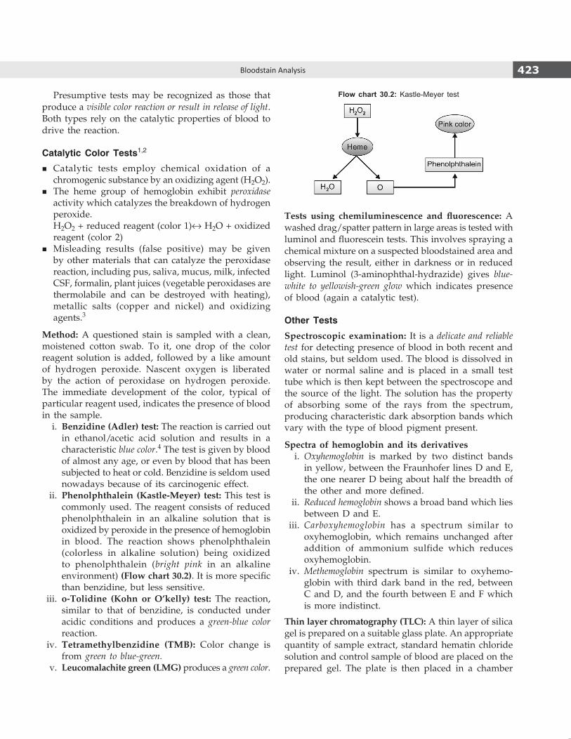
423Bloodstain Analysis
Presumptive tests may be recognized as those that produce a visible color reaction or result in release of light. Both types rely on the catalytic properties of blood to drive the reaction.
Catalytic Color Tests1,2
� Catalytic tests employ chemical oxidation of a chromogenic substance by an oxidizing agent (H2O2).
� The heme group of hemoglobin exhibit peroxidase activity which catalyzes the breakdown of hydrogen peroxide.
H2O2 + reduced reagent (color 1)↔ H2O + oxidized reagent (color 2)
� Misleading results (false positive) may be given by other materials that can catalyze the peroxidase reaction, including pus, saliva, mucus, milk, infected CSF, formalin, plant juices (vegetable peroxidases are thermolabile and can be destroyed with heating), metallic salts (copper and nickel) and oxidizing agents.3
Method: A questioned stain is sampled with a clean, moistened cotton swab. To it, one drop of the color reagent solution is added, followed by a like amount of hydrogen peroxide. Nascent oxygen is liberated by the action of peroxidase on hydrogen peroxide. The immediate development of the color, typical of particular reagent used, indicates the presence of blood in the sample. i. Benzidine (Adler) test: The reaction is carried out
in ethanol/acetic acid solution and results in a characteristic blue color.4 The test is given by blood of almost any age, or even by blood that has been subjected to heat or cold. Benzidine is seldom used nowadays because of its carcinogenic effect.
ii. Phenolphthalein (Kastle-Meyer) test: This test is commonly used. The reagent consists of reduced phenolphthalein in an alkaline solution that is oxidized by peroxide in the presence of hemoglobin in blood. The reaction shows phenolphthalein (colorless in alkaline solution) being oxidized to phenolphthalein (bright pink in an alkaline environment) (Flow chart 30.2). It is more specific than benzidine, but less sensitive.
iii. o-Tolidine (Kohn or O’kelly) test: The reaction, similar to that of benzidine, is conducted under acidic conditions and produces a green-blue color reaction.
iv. Tetramethylbenzidine (TMB): Color change is from green to blue-green.
v. Leucomalachite green (LMG) produces a green color.
Tests using chemiluminescence and fluorescence: A washed drag/spatter pattern in large areas is tested with luminol and fluorescein tests. This involves spraying a chemical mixture on a suspected bloodstained area and observing the result, either in darkness or in reduced light. Luminol (3-amino phthal -hydrazide) gives blue-white to yellowish-green glow which indicates presence of blood (again a catalytic test).
Other Tests
Spectroscopic examination: It is a delicate and reliable test for detecting presence of blood in both recent and old stains, but seldom used. The blood is dissolved in water or normal saline and is placed in a small test tube which is then kept between the spectroscope and the source of the light. The solution has the property of absorbing some of the rays from the spectrum, producing characteristic dark absorption bands which vary with the type of blood pigment present.
Spectra of hemoglobin and its derivatives i. Oxyhemoglobin is marked by two distinct bands
in yellow, between the Fraunhofer lines D and E, the one nearer D being about half the breadth of the other and more defined.
ii. Reduced hemoglobin shows a broad band which lies between D and E.
iii. Carboxyhemoglobin has a spectrum similar to oxyhemo globin, which remains unchanged after addition of ammonium sulfide which reduces oxyhemoglobin.
iv. Methemoglobin spectrum is similar to oxyhemo-globin with third dark band in the red, between C and D, and the fourth between E and F which is more indistinct.
Thin layer chromatography (TLC): A thin layer of silica gel is prepared on a suitable glass plate. An appropriate quantity of sample extract, standard hematin chloride solution and control sample of blood are placed on the prepared gel. The plate is then placed in a chamber
Flow chart 30.2: Kastle-Meyer test

424 Review of Forensic Medicine and Toxicology
having a convenient solvent system. After the desired run of the solvent to a certain height (front), it is removed from the chamber. When the plate is dry, benzidine and hydrogen peroxide are sprayed on it. If the stain contains blood, the sample extract gives a blue spot at the same height.
Microscopic Examination
Aging, environmental factors or heating can alter blood cells (erythrocytes and leukocytes) and make it difficult to produce reliable results. Intact red blood cells (RBCs) are observed only when the stain is fresh or when a clot is available, but become unrecognizable when the stain has dried. Sometimes, microscopic appearance of RBCs may reveal additional information—sickle shaped erythrocytes may indicate that the sample originated from a person having sickle cell disease.
Procedure: The stained piece is cut and dipped and teased in a watch glass with 2–3 drops of Vibert’s fluid (sodium chloride, mercuric chloride and distilled water or normal saline) for half an hour and then examined under a microscope.
� Non-mammalian RBCs, e.g. bird, fish, reptile and amphi-bian are oval, biconvex and nucleated (Fig. 30.2).
� RBCs of humans and mammals are circular, biconcave and non-nucleated with the exception of camel and llama which are oval and biconvex, but non-nucleated (Fig. 30.3). In primates, nucleated RBCs may be found.
Confirmatory Tests for Blood
Crystal Tests
Crystal tests are regarded as confirmatory tests. These tests involve the non-protein heme group of hemoglobin, called porphyrins. i. Teichmann or Hemin crystal test (described in
1853): Place a sample of suspected blood on a glass slide, add few crystals of sodium chloride and a few drops of glacial acetic acid from the side of the cover slip and heat it to form a hematin derivative.1,5
z These hemin or hematin chloride crystals are brownish rhombic shaped, arranged singly or in clusters (Fig. 30.4).
z The reaction is negative, if the stain is old, is washed or treated with chemicals, too much salt is added, if there is moisture in the acid or by over heating.
ii. Takayama or Hemochromogen crystal test (described in 1912): Place a small stain sample
Fig. 30.2: Animal RBCs
Fig. 30.3: Human RBCs
Fig. 30.4: Teichmann test (hemin crystals)
under a coverslip and allow the Takayama reagent (sodium hydroxide, pyridine and glucose) to flow under and saturate the sample. After a brief heating, the crystals are viewed microscopically.
z Pink feathery crystals of reduced alkaline hematin (hemochromogen—pyridine ferriprotoporphyrin) arranged in clusters are seen (Fig. 30.5).
z It can be carried out on a small stain quantity, is effective on aged stains and is more dependable.
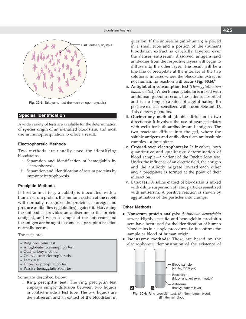
425Bloodstain Analysis
Species Identification
A wide variety of tests are available for the determination of species origin of an identified bloodstain, and most use immunoprecipitation to effect a result.
Electrophoretic Methods
Two methods are usually used for identifying bloodstains: i. Separation and identification of hemoglobin by
electrophoresis. ii. Separation and identification of serum proteins by
immunoelectrophoresis.
Precipitin Methods
If host animal (e.g. a rabbit) is inoculated with a human serum protein, the immune system of the rabbit will normally recognize the protein as foreign and produce antibodies (γ globulins) against it. Harvesting the antibodies provides an antiserum to the protein (antigen), and when a sample of the antiserum and the antigen are brought in contact, a precipitin reaction normally occurs.
The tests are:
� Ring precipitin test � Antiglobulin consumption test � Ouchterlony method � Crossed-over electrophoresis � Latex test � Diffusion precipitation test � Passive hemagglutination test.
Some are described below: i. Ring precipitin test: The ring precipitin test
employs simple diffusion between two liquids in contact inside a test tube. The two liquids are the antiserum and an extract of the bloodstain in
question. If the antiserum (anti-human) is placed in a small tube and a portion of the (human) bloodstain extract is carefully layered over the denser antiserum, dissolved antigens and antibodies from the respective layers will begin to diffuse into the other layer. The result will be a fine line of precipitate at the interface of the two solutions. In cases where the bloodstain extract is not human, no reaction will occur (Fig. 30.6).6
ii. Antiglobulin consumption test (Hemagglutination inhibition test): When human globulin is mixed with antihuman globulin serum, the latter is absorbed and is no longer capable of agglutinating Rh positive red cells sensitized with incomplete anti-D. This detects globulins.
iii. Ouchterlony method (double diffusion in two directions): It involves the use of agar gel plates with wells for both antibodies and antigens. The two reactants diffuse into the gel, where the soluble antigens and antibodies form an insoluble complex—a precipitate.
iv. Crossed-over electrophoresis: It involves both quantitative and qualitative determination of blood sample—a variant of the Ouchterlony test. Under the influence of an electric field, the antigen and the antibody migrate toward each other and a precipitate is formed at the point of their interaction.
v. Latex test: A saline extract of bloodstain is mixed with dilute suspension of latex particles sensitized with antiserum. A positive reaction is shown by agglutination of the particles into clumps.
Other Methods
� Nonserum protein analysis: Antihuman hemoglobin serum: Highly specific anti-hemoglobin precipitin sera have been used for the identification of human bloodstains in a single procedure, i.e. it confirms the sample as blood of human origin.
� Isoenzyme methods: These are based on the electrophoretic demonstration of the existence of
Fig. 30.5: Takayama test (hemochromogen crystals)
Fig. 30.6: Ring precipitin test: (A) Non-human blood; (B) Human blood
A B
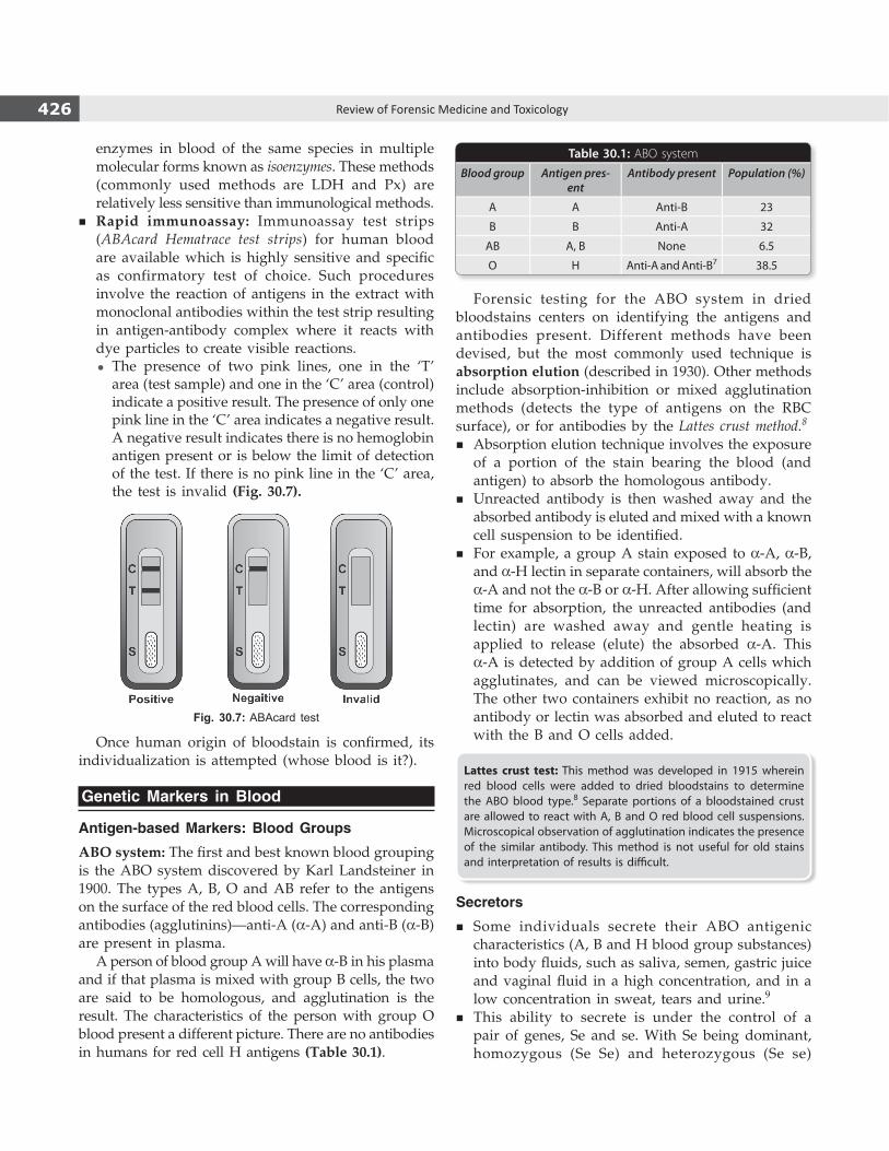
426 Review of Forensic Medicine and Toxicology
enzymes in blood of the same species in multiple molecular forms known as isoenzymes. These methods (commonly used methods are LDH and Px) are relatively less sensitive than immunological methods.
� Rapid immunoassay: Immunoassay test strips (ABAcard Hematrace test strips) for human blood are available which is highly sensitive and specific as confirmatory test of choice. Such procedures involve the reaction of antigens in the extract with monoclonal antibodies within the test strip resulting in antigen-antibody complex where it reacts with dye particles to create visible reactions.
z The presence of two pink lines, one in the ‘T’ area (test sample) and one in the ‘C’ area (control) indicate a positive result. The presence of only one pink line in the ‘C’ area indicates a negative result. A negative result indicates there is no hemoglobin antigen present or is below the limit of detection of the test. If there is no pink line in the ‘C’ area, the test is invalid (Fig. 30.7).
Fig. 30.7: ABAcard test
Once human origin of bloodstain is confirmed, its individualization is attempted (whose blood is it?).
Genetic Markers in Blood
Antigen-based Markers: Blood Groups
ABO system: The first and best known blood grouping is the ABO system discovered by Karl Landsteiner in 1900. The types A, B, O and AB refer to the antigens on the surface of the red blood cells. The corresponding antibodies (agglutinins)—anti-A (α-A) and anti-B (α-B) are present in plasma. A person of blood group A will have α-B in his plasma and if that plasma is mixed with group B cells, the two are said to be homologous, and agglutination is the result. The characteristics of the person with group O blood present a different picture. There are no antibodies in humans for red cell H antigens (Table 30.1).
Forensic testing for the ABO system in dried bloodstains centers on identifying the antigens and antibodies present. Different methods have been devised, but the most commonly used technique is absorption elution (described in 1930). Other methods include absorption-inhibition or mixed agglutination methods (detects the type of antigens on the RBC surface), or for antibodies by the Lattes crust method.8
� Absorption elution technique involves the exposure of a portion of the stain bearing the blood (and antigen) to absorb the homologous antibody.
� Unreacted antibody is then washed away and the absorbed antibody is eluted and mixed with a known cell suspension to be identified.
� For example, a group A stain exposed to α-A, α-B, and α-H lectin in separate containers, will absorb the α-A and not the α-B or α-H. After allowing sufficient time for absorption, the unreacted antibodies (and lectin) are washed away and gentle heating is applied to release (elute) the absorbed α-A. This α-A is detected by addition of group A cells which agglutinates, and can be viewed microscopically. The other two containers exhibit no reaction, as no antibody or lectin was absorbed and eluted to react with the B and O cells added.
Lattes crust test: This method was developed in 1915 wherein red blood cells were added to dried bloodstains to determine the ABO blood type.8 Separate portions of a bloodstained crust are allowed to react with A, B and O red blood cell suspensions. Microscopical observation of agglutination indicates the presence of the similar antibody. This method is not useful for old stains and interpretation of results is difficult.
Secretors
� Some individuals secrete their ABO antigenic characteristics (A, B and H blood group substances) into body fluids, such as saliva, semen, gastric juice and vaginal fluid in a high concentration, and in a low concentration in sweat, tears and urine.9
� This ability to secrete is under the control of a pair of genes, Se and se. With Se being dominant, homozygous (Se Se) and heterozygous (Se se)
Table 30.1: ABO system
Blood group Antigen pres-ent
Antibody present Population (%)
A A Anti-B 23
B B Anti-A 32
AB A, B None 6.5
O H Anti-A and Anti-B7 38.5
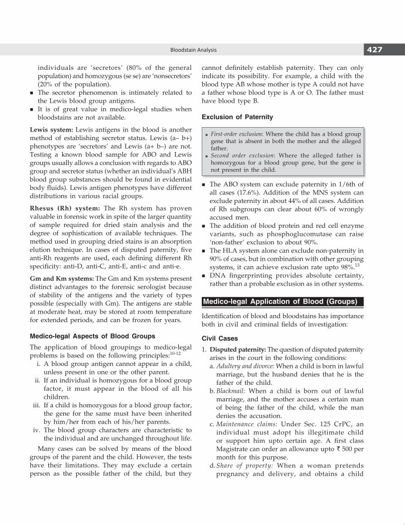
427Bloodstain Analysis
individuals are ‘secretors’ (80% of the general population) and homozygous (se se) are ‘nonsecretors’ (20% of the population).
� The secretor phenomenon is intimately related to the Lewis blood group antigens.
� It is of great value in medico-legal studies when bloodstains are not available.
Lewis system: Lewis antigens in the blood is another method of establishing secretor status. Lewis (a– b+) phenotypes are ‘secretors’ and Lewis (a+ b–) are not. Testing a known blood sample for ABO and Lewis groups usually allows a conclusion with regards to ABO group and secretor status (whether an individual’s ABH blood group substances should be found in evidential body fluids). Lewis antigen phenotypes have different distributions in various racial groups.
Rhesus (Rh) system: The Rh system has proven valuable in forensic work in spite of the larger quantity of sample required for dried stain analysis and the degree of sophistication of available techniques. The method used in grouping dried stains is an absorption elution technique. In cases of disputed paternity, five anti-Rh reagents are used, each defining different Rh specificity: anti-D, anti-C, anti-E, anti-c and anti-e.
Gm and Km systems: The Gm and Km systems present distinct advantages to the forensic serologist because of stability of the antigens and the variety of types possible (especially with Gm). The antigens are stable at moderate heat, may be stored at room temperature for extended periods, and can be frozen for years.
Medico-legal Aspects of Blood Groups
The application of blood groupings to medico-legal problems is based on the following principles:10-12
i. A blood group antigen cannot appear in a child, unless present in one or the other parent.
ii. If an individual is homozygous for a blood group factor, it must appear in the blood of all his children.
iii. If a child is homozygous for a blood group factor, the gene for the same must have been inherited by him/her from each of his/her parents.
iv. The blood group characters are characteristic to the individual and are unchanged throughout life.
Many cases can be solved by means of the blood groups of the parent and the child. However, the tests have their limitations. They may exclude a certain person as the possible father of the child, but they
cannot definitely establish paternity. They can only indicate its possibility. For example, a child with the blood type AB whose mother is type A could not have a father whose blood type is A or O. The father must have blood type B.
Exclusion of Paternity
� First-order exclusion: Where the child has a blood group gene that is absent in both the mother and the alleged father.
� Second order exclusion: Where the alleged father is homozygous for a blood group gene, but the gene is not present in the child.
� The ABO system can exclude paternity in 1/6th of all cases (17.6%). Addition of the MNS system can exclude paternity in about 44% of all cases. Addition of Rh subgroups can clear about 60% of wrongly accused men.
� The addition of blood protein and red cell enzyme variants, such as phosphoglucomutase can raise ‘non-father’ exclusion to about 90%.
� The HLA system alone can exclude non-paternity in 90% of cases, but in combination with other grouping systems, it can achieve exclusion rate upto 98%.13
� DNA fingerprinting provides absolute certainty, rather than a probable exclusion as in other systems.
Medico-legal Application of Blood (Groups)
Identification of blood and bloodstains has importance both in civil and criminal fields of investigation:
Civil Cases
1. Disputed paternity: The question of disputed paternity arises in the court in the following conditions:a. Adultery and divorce: When a child is born in lawful
marriage, but the husband denies that he is the father of the child.
b. Blackmail: When a child is born out of lawful marriage, and the mother accuses a certain man of being the father of the child, while the man denies the accusation.
c. Maintenance claims: Under Sec. 125 CrPC, an individual must adopt his illegitimate child or support him upto certain age. A first class Magistrate can order an allowance upto ` 500 per month for this purpose.
d. Share of property: When a woman pretends pregnancy and delivery, and obtains a child
428 Review of Forensic Medicine and Toxicology
claiming him/her as her own, in order to obtain a share in her husband’s property.
2. Disputed maternity: The question of disputed maternity arises in the following circumstances:a. When the same child is claimed by two women. b. When there has been an allegation of interchange
of a child with another in the maternity hospital, either purposely or accidentally.
c. In case of a kidnapped child, when the woman who has kidnapped the child claims to be the mother.
d. In case of a suppositious child, when a woman pretends pregnancy and delivery, and brings forth a child to pass it off as her own.
3. Inheritance claims: The question of legitimacy arises, since a legitimate child only can inherit the parent’s property.
4. Divorce and nullity of marriage cases, e.g. question of intersex and some forbidden diseases.
5. Civil negligence cases arising in hospital or medical practices, for example,a. Incompatible blood transfusion.b. Neglect of expiry dates leading to transfusion
reactions.c. Presence of pathogenic organisms, such as malaria,
syphilis, hepatitis B and HIV in the transfused blood.
Criminal Cases
1. Identification of victim or offenders of crime in circumstances, such as murder, wounding, rape and vehicular accidents.
z Bloodstains may be found on the clothing and person of the suspect. If the character of these stains is similar to that of blood of the victim, it establishes association.
z Bloodstains may be present under the fingernails of assailant in a case of throttling.
z If there has been a struggle, bloodstains derived from the accused may be found under the fingernails of the victim due to scratching.
z Vehicles which have caused injury can be identified when they show bloodstains resembling that of the victim.
2. Stains due to body fluids: The blood group antigens can be demonstrated in stains on clothes due to semen, sweat or saliva (‘secretors’). This may be a corro borative evidence of the accused.
3. Crime scene reconstruction: Blood spatter inter-pretation can be valuable in determining how blood was deposited on an item or at a scene.
4. Corroborate or refute an individual’s allegation: It can substantiate a complainant’s or suspect’s account of alleged events of an assault, and can be critical in establishing guilt or innocence during criminal proceedings.
5. Cases of malingering: The specificity of various blood group combinations is like that of the fingerprints. When an individual has some rare blood group, he can be identified with certainty.
6. Cause of death, for example, detection of poison in the blood.
7. Time since death can be estimated by use of different chemical or biochemical tests.
� Sample collection for paternity testing: In the case of the adults, 5 ml of venous blood is taken and placed in plain tube. Neither party should have had a blood transfusion within 3 months, before taking the sample. The infants should preferably be 6 months of age, but not < 2 months before testing is performed; one ml of blood is obtained by a heel or ear prick or venepuncture into a plain tube. The same person should do the testing of mother, child and alleged father in the same laboratory, on the same day, and using the same batch of reagents and antisera.
� The blood groups in current use in the investigation of cases of doubtful paternity are ABO, MNS, Rh, Kell, Lutheran, Duffy, and Kidd.
� Grouping based on white cell antigens: The Human Leukocyte Antigen (HLA) system consists of protein substance on the surface of a wide variety of tissues and organs, tumors, white cells and platelets. They are reported to be present on spermatozoa, but not on ovum or trophoblast. They are found both on lymphocytes and granulocytes. The major human leucocyte antigens HLA-A, B, C, D, and DR are determined by a single chromosomal segment, the major histocompatibility complex (MHC), which is situated on the short arm of chromosome 6.
� Protein markers: Hemoglobin exhibits 180 or more variants, but only four (Hb A, Hb F, Hb S and Hb C) are readily distinguished forensically with electrophoresis or IEF based on the positioning of bands.
� Enzyme markers: Phosphoglucomutase (PGM) is found in many tissues of plants, animals, and microorganisms. In humans, the enzyme exists in significant concentrations in blood and semen, and in small amounts in vaginal secretion and cervical mucus. Electrophoresis of samples for PGM analysis using IEF methods can detect 10 phenotypes. With this, it is possible to place the subtypes of PGM in 10 different population groups, and thus presents the highest discrimination probability of any enzyme system used in forensic serology.
� Human DNA quantitation: A sample can be determined to be blood of human origin with a probe specific for human DNA. Probes complementary to primate specific DNA sequences, such as those found at the locus D17Z1 are used primarily to determine the amount of human DNA extracted from the sample prior to DNA typing.

429Bloodstain Analysis
Medico-legal Questions
Q. Whether the stain is due to blood or some other material?
It is essential to establish positively that stain is in fact blood before conducting further analyses. Presumptive tests (color tests) and confirmatory tests such as, chemical methods, spectrophotometric analysis are done for this purpose. Some substances that may resemble bloodstains are: i. Rust stains, tests are positive for iron. ii. Synthetic dyes stain changes to yellow with nitric
acid, blood remains unchanged. iii. Mineral stains contain oxides of iron or red lead. iv. Vegetable stains: Fruits, like mulberry, gooseberry
and currants produce stains resembling bloodstains. Tests are negative for blood, and ammonia turns the vegetable stain green.
v. Other stains, like grease or tar on dark fabrics may resemble bloodstains.
Q. If it is blood, is it human?
In some cases, it may be necessary to confirm the presence of human blood in questioned stain before obtaining a known sample from a suspect or a victim. Confirmation is done by immunological methods.
Q. If it is human, what information towards indi-vidualization is possible? (victim or assailant)
� Blood groups (ABO) may be different. Stains on the inner side of the clothes usually belong to the victim, while those on the outer side may be of the victim or assailant. Bloodstains may bear marks of fingerprints or footprints of the assailant.
� Sometimes, there may be some disease, like leukemia, filariasis or sickle cell anemia either in the victim or the assailant which may provide valuable information.
� Traces of blood may be found underneath the fingernails of the victim as a result of struggle or of the assailant in case of throttling; these can be typed and grouped.
� An individual bloodstain can also be identified by using DNA typing.
Q. Whether the sex of the person can be determined from the bloodstain? (male or female)
Sex can be determined from presence of sex chromatin in leukocytes.
� Barr body count in WBC can be done using orcein and acriflavine reagent.
� Y chromosome is fluorescent to quinacrine when examined under fluorescent microscope.
� Fluorescent in situ hybridization (FISH) technique can also be used for sex determination. Probes specific for X and Y chromosomes are applied.
Q. Whether the age of the person can be determined?
� In infants, the red blood cells exhibit more fragility, the hemoglobin is of the fetal type (fetus specific hemoglobin α2γ2 detectable upto about 6 months), and the blood when shed forms a thin and soft coagulum.
� In adults, the red blood cells are non-nucleated, their fragility is within certain limits, hemoglobin is of the adult type, and the blood when shed forms a thick and firm coagulum.
Q. How long the stain has been on the object/surface? (recent or old)
� Recent stains on white cloth are at first red due to conversion of hemoglobin to methemoglobin and hematin.
� The color changes to dull red within hours, reddish brown within 24 h, dark brown or even blackish within few days and remains so for several years.
So, it is only possible to state that the stain is recent or not very recent.
Q. What could be the source of the blood?
Bleeding due to disease, accident, menstruation, parturition, abortion, hematemesis or hemoptysis may cause stains.
� Most common defense in cases of assault on females is that the stain is from menstruation. It has a disagreeable smell, being mixed with urine and vaginal mucus, it is more fluid and dark in color. Menstrual blood does not clot, due to extensive degradation of the clotting factor fibrinogen. Fibrinogen degradation products are present in high concentration that can be detected immun-ologically.
� Hemoptysis blood is bright red, frothy and alkaline in reaction.
� Hematemesis blood is dark in color, not frothy and is acidic in reaction.

430 Review of Forensic Medicine and Toxicology
� Parturition and abortion blood may have some clot or products of conception, like decidual tissue, fetal parts, chorionic villi, vernix caseosa or lanugo hair. Color may be yellowish or greenish from admixture with meconium.
Q. What could be the site of bleeding (arterial or venous)? (Fig. 30.8)
Damage to different types and sizes of blood vessels will result in different degrees of bleeding, but it is difficult to predict the exact type of flow of blood from an injury.
� Arterial bleeding has a spurting effect (jet-like ejection) from the wound. It is bright red in color when fresh.
� Venous bleeding occurs passively in drops without any projectile force, has stellate appearance and dark in color. It may ooze and produce a pool, if the victim falls down.
1. A 2. D 3. A & B 4. B 5. D 6. B 7. A & B 8. C 9. A 10. C 11. C 12. D 13. A & C
Q. Whether the bleeding was antemortem or postmortem?
� Blood which has effused during life can be peeled off in scales on drying, due to presence of fibrin and the clot can be taken en masse.
� Blood which has flowed after death tends to break up into powder on drying, and the clot cannot be taken en masse.
MULTIPLE CHOICE QUESTIONS
1. All are tests done on blood, except: TN 11 A. Acid phosphatase test B. Benzidine test C. Hemochromogen test D. Teichmann’s test 2. Hydrogen peroxide is used in all of the following
chemical tests for blood except: NEET 13 A. Benzidine test B. Kastle Mayer test C. Ortho toludine test D. Teichmann test 3. Positive benzidine test is/are seen in: PGI 12 A. Hemoglobin B. Myoglobin C. Porphobilinogen D. Bilirubiln 4. Benzidine test is done to detect: PGI 07 A. Semen B. Blood C. Bile D. Saliva 5. Confirmatory test for blood stain: PGI 13 A. Benzidine test B. Spectroscopic study C. Ortho tolidine test D. Tecihmann test 6. Species identification is done by: COMEDK 08; Punjab 11; NEET 13 A. Takayama test B. Precipitin test C. Benzidine test D. Spectroscopy 7. Antibodies found in type O individual: PGI 13 A. Anti A B. Anti B B. Anti AB D. Neither A or B antibody
8. Latte’s crust of blood stain is used to detect: NEET 14 A. Nature of stain B. Detection species C. Blood group D. Secretor status 9. ABO antigens is not found in: AIIMS 09; NEET 14 A. CSF B. Semen C. Sweat D. Saliva 10. An Rh-negative woman married to a heterozygous
Rh-positive man has three children. The probability that all three of their children are Rh-positive is:
Himachal 10 A. 1: 2 B. 1: 4 C. 1: 8 D. Zero 11. A baby’s blood group was determined as O Rh
negative. Select the blood group, the baby’s mother or father will not have: WB 07
A. A positive B. B positive C. AB negative D. A negative 12. In a case of disputed paternity, father’s blood group is
A, mother’s blood group is B, the child’s blood group may be: Karnataka 07
A. A only B. B only C. AB only D. A, AB, B or O 13. HLA typing is useful in: PGI 06 A. Disputed paternity B. Thanatology C. Organ transplant D. Dactylography
Fig. 30.8: (A) Arterial bleeding; (B) Venous bleeding
A B

Introduction
Semen is the fluid discharged from the penis during ejaculation, usually at the time of orgasm. Like blood, semen consists of two compartments, the cellular compart ment (spermatozoa) and noncellular compartment (seminal plasma) which is secreted from the prostate gland, seminal vesicles, Cowper’s glands (bulbouretral gland) and the glands of Littre (Fig. 31.1). The normal quantity of seminal fluid in a single emission is 2–5 ml and contains about 60–150 million sperms/ml of which 90% are motile at time of ejaculation. Spermatozoa constitute about 10% of the volume of the semen which contains water and small amounts of salt, protein and fructose. Prostatic secretions in humans contain high levels of citric acid, acid phosphatase and zinc. The secretion is alkaline with a pH of 7.4.
Purpose of Seminal Identification
Seminal stains may have to be detected/examined in criminal and civil cases.
Criminal cases Civil cases
� Rape/attempted rape � Disputed paternity
� Sodomy � Legitimacy
� Bestiality � Artificial insemination
� Sexual murder of the female � Divorce
� Compensation on grounds of acquired sterility/failure of vasectomy cases
Collection of Material
The stains are usually found on the clothing, but may be found on the person of either the victim or
Fig. 31.1: Male reproductive system
Seminal Stains and Other Biological Samples
CHAPTER 31

432 Review of Forensic Medicine and Toxicology
Presumptive Chemical Examination
Presumptive tests for semen are based on colorimetry, and are qualitative in nature. Positive presumptive tests must be followed by a confirmatory test, such as microscopic examination, quantitative acid phosphatase test or detection of p30. i. Florence test: The stain is extracted, dried on a
glass slide and covered with a coverslip and a drop of Florence solution (8% w/v of iodine in water containing 5% w/v of potassium iodide) is allowed to run under the coverslip. If semen is present, dark brown rhombic crystals resembling hemin (but larger) arranged in clusters or rosettes of choline periodide appear immediately (Fig. 31.2).2,3 Choline originates from the seminal vesicles. A positive test is not proof of seminal fluid, but confirms the
the accused. They may also be found on bedclothes, furniture, vehicles, carpet, floor or grass, where the offence was committed or any item the victim may have used to clean up after the assault (tissue or washcloth). Seminal stains have to be differentiated from those due to starch, pus, leucorrhoeal discharge and egg albumen. Before proceeding with the examination, stains may have to be collected and preserved from different sources: i. Clothing: Portion of cloth with the stain is cut,
dried in shade (not heated) and preserved. ii. Vaginal fluid: Fluid from the vagina is collected
with a pipette or vaginal washing is done which is concentrated by centrifugation. Swabs are taken with sterile gauze and smears are prepared on sterile slides.
iii. Driedstainsonotherpartsofbody:Dried seminal fluid on the perineum or thighs is collected with a wet swab.
iv. Matted pubic hair: It is plucked/cut and placed in a small container.
v. Stainsonsmoothsurface:These are gently scraped off into a glass container and preserved.
Samples must be packed in containers that allow air circulation; never in plastic bags or sealed nonporous tubes, jars or boxes. Chain of custody must be documented and adhered to the prevailing policies.
Examination of Seminal Stains
The sequence of analyzing the evidence (as described above) is given in Flow chart 31.1.
Screening Tests
PhysicalExamination � When fresh, semen is a whitish or yellowishwhite
in color, slightly viscous, jellylike, sticky and has a characteristic odor. On standing, viscosity is lost due to prostatic fibrolysin, and it becomes thin.
� Dried seminal stains on clothes are grayishwhite or yellowishgray in color, show an irregular outline and starchy hard in feeling. When examined under filtered UV light, they fluoresce with a bluishwhite color (due to choline in semen) which is not specific, as other albuminous materials, such as nasal or leukorrheal discharges and detergents also fluoresce.1
� A fresh stain on a nonabsorbent material appears translucent. After a month, it becomes yellow to brown. Fig. 31.2: Florence test (choline iodide crystals)
Flow chart 31.1: Evaluation of a suspected seminal stain in sexual assault

433Seminal Stains and Other Biological Samples
presence of some vegetable or animal substance. A negative reaction proves that the stain is not semen, but may occur if choline content is low or the stain is decomposed.
ii. Barberio’s test: A saturated aqueous or alcoholic solution of picric acid when added to dried stain extract on a glass slide covered with a coverslip, produces yellow needle-shaped crystals of spermine picrate (Fig. 31.3). The reaction depends on the presence of spermine from prostatic secretions.4,5 This test is positive without the presence of spermatozoa.
z Acid phosphatase catalyzes the removal of the phosphate residue on the substrate 4methylumbelliferone phosphate (MUP), which generates fluorescence under UV light.
z It can produce false positives because similar enzyme activity is found in other body fluids (e.g. vaginal secretions and fecal stains), human red cells, semen of higher apes, as well as in presence of fungi, bacteria and even plants (juice of cauliflower). Moreover, pregnancy, menstruation, bacterial vaginosis may also elevate its level.
z Dried and old seminal stains which have not undergone putrefaction give positive reaction.
Confirmatory Tests
� The presence of spermatozoa under light microscopy is considered as the ‘gold standard’.
� Confirmatory testing involves solubilization of sample followed by centrifugation which yields a supernatant and a cell pellet. The cell pellet is used to detect spermatozoa and for DNA analysis, whereas the supernatant is useful to detect noncellular markers when sperms are not detected and for grouping or genetic profiling.
� Sometimes, the sample is contaminated by other bodily fluids (saliva, vaginal secretions), epithelial cells, cellular debris wherein selective degradation may be done by treating the cell extract with a mixture of proteinase K and sodium dodecyl sulfate before staining and microscopic examination.
� Most commonly used confirmatory test for semen is visualization of one or more intact spermatozoa after staining with dyes such as hematoxylin and eosin or ‘Christmas tree’ stain.
Microscopic Examination
Procedure: A small piece of the stained fabric is moistened with a few drops of 1% HCl in a watch glass for half to one hour if the stain is fresh, or 2–4 hours (h) if it is old. Slides are prepared by dabbing the fabric gently on them. Films are dried in the air without heat fixed and then stained.
� Slide is stained either with Harris’s hematoxylin for 2–5 minutes (min) and eosin for 3 min, or methylene blue for 15–30 min and counterstained with eosin for 2 min.
� Posterior half to onethird of head and the tail takes eosin and is stained deep red or pink, while anterior
Fig. 31.3: Barberio’s test (spermine picrate crystals)
Flow chart 31.2: Brentamine fast blue test
iii. Brentamine fastblue test z It is the most common presumptive test for
seminal acid phosphatase. Acid phosphatase activity is 500–1000 times greater in human semen than in any other bodily fluid. The levels remain high until till 40 years of age with gradual decrease thereafter; levels are not related to sperm count.
z The reaction is based on the hydrolysis of phosphate esters and detection of the liberated organic moiety by production of a color complex. An enzyme substrate, sodium αnaphthyl phosphate is converted to sodium phosphate and naphthol by the acid phosphatase enzyme in the semen and a coupled reaction with brentamine fast blue dye takes place, forming a purple color (Flow chart 31.2).

434 Review of Forensic Medicine and Toxicology
Fig. 31.4: Spermatozoa under light microscope
half to twothirds takes very light or faint basic or blue stain or may appear unstained (Fig. 31.4).
� Size of human spermatozoa is about 55 µ in length. Head is ovoid and flattened, 5 × 3.5 µ in dimensions. It has a short neck and a long filamentous tail (50 µ) which tapers to a fine point (Fig. 31.5).
� Bacteria, fungi, trichomonas, yeast, monilia and naked nuclei from vaginal epithelial cells give false positive test.
� Older the stain, lesser is the chance of finding intact sperms.
� Single photon fluorimetry has been used to differentiate between different semen.
Others staining methods � Christmas tree stain: This staining technique was developed by
Oppitz which consist of nuclear fast red (red stain for sperm head) and picroindigocarmine (green counter-stain for the tail and other cytoplasmic material) and are sometimes referred to as ‘Christmas tree’ stain because of the red-green combination.
� Papanicolaou staining: In Pap smear, acrosome is stained pink, postacrosome—dark blue, and tail—pink.
� Giemsa stain: Acrosome stained pink, postacrosome— dark blue. � Ziehl-Neelsen’s method: Smear is stained and examined for the
presence of spermatozoa and smegma bacilli (Mycobacterium smegmatis). It is an acid fast, rod shaped bacillus and thicker than the tubercle bacillus.
� Other stains: Bryan-Leishman stain, May-Grunwald Giemsa stains (MGG), supra vital stain, Shorr stain and alkaline fuchsin.
Motility of Sperms
� At room temperature, motility depends on time elapsed since ejaculation.
� At body temperature (in living victims), sperms retains full motility in vagina between 6–12 h. The sperms remain motile in the uterine cavity for 3–7 days. Later, the sperms disintegrate into head and tails which may be recovered from the vagina upto 7–10 days and 12–14 days in the cervix and uterus.
� Complete motile sperms may be seen upto 28 h in vagina after ejaculation (nonmotile sperms may be found upto 10 days).
� Nonmotile sperms may be seen in the oral cavity from 2–31 h, in the rectum from 4–113 h, and in the anus from 2–44 h.
Fluorescence in situ hybridization (FISH): This cytogenic analysis uses a Y chromosome specific DNA probe to identify Ybearing (male) cells. This technique identifies not only spermatozoa, but also cells of male origin and confirms malefemale contact.
Non-cellular Semen Markers
Markers are specific and unique to seminal plasma but independent of spermatozoa. The two most commonly employed constituents are acid phosphatase and the prostatespecific glycoprotein p30 (PSA).These tests are conclusive even in the absence of demonstrable sperms, azoospermia or vasectomized individuals.6
i. Acid phosphatase test (quantitative): Finding a significantly elevated acid phosphatase level is consistent with the presence of semen. Undiluted semen has an acid phosphatase level of 340–360 Bodansky units/ml. A value of > 100 Bodansky
Fig. 31.5: Morphological appearance of spermatozoon
435Seminal Stains and Other Biological Samples
units with/without motile sperms indicate that ejaculation occurred within 12 h of examination. Level decreases with time after intercourse, and there is little chance of identifying it after 48 h.
z Isoenzymes can be detected using polyacrylamide gel electrophoresis followed by staining with MUP reagent which can distinguish the acid phosphatase present in other substances.
ii. Prostate specific antigen or PSA (p30): The glycoprotein p30 is derived from prostrate and is found in seminal plasma, male urine and blood, and has not been found in any female body tissue or fluid.
z p30 in sample reliably identifies semen regardless of whether acid phosphatase is elevated or spermatozoa are detected. Some laboratories even use p30 testing in place of microscopical examination for semen identification.
z It is determined serologically using antiserum that is specific for the p30 antigen.– Traditional detection tests utilize electro
phoretic methods such as crossover electrophoresis or diffusion methods, such as Ouchterlony double diffusion wherein a precipitation band is formed due to the formation of antibodyantigen complex.
– It can also be done using immunochromatographic strip test using antibodies raised against the human PSA and enzymelinked immunoassay (ELISA).
– The currently accepted method of choice for identification of semen is detection of p30 using the ABAcard test strips. The strips work in the same way as described earlier for confirmation of blood, except that they use antip30 monoclonal and polyclonal antisera, and a pink dye.
z Its normal range in semen is 300–4200 µg/ml with mean of 1200 µg/ml, and is detectable in vaginal fluid upto 27 h (range 13–47 h) after intercourse as compared to 12–14 h for acid phosphatase.
z p30 can be detected in dried and old stains (> 10 years in material stored at room temperature) and in cadavers.
z Semenogelin (Sg), a protein originating in the seminal vesicles and a substrate for PSA, is also a useful marker for the identification of semen. Detection of Sg with immunochromatographic test strip is rapid and simple. It is useful for the identification of seminal plasma, an alternative to the method for PSA detection.
Using these three tests together (acid phosphatase, PSA and spermatozoa detection) the presence of semen can be conclusively determined in vaginal swabs of the sexual assault victims. DNA laboratories now screen first with PSA and semenogelin (as there are false positive reports of both of these individually), before going further with DNA markers and profiling.
Other tests � Creatine phosphokinase test: Spermatozoa contain a high
concentration of creatine phosphokinase, which is more than double than that found in any other body fluid. Values > 400 units/ml are diagnostic of seminal stains. The enzyme is stable and can be demonstrated even in old stains. The test will be negative in case of aspermia.
� Choline and spermine test: Fresh and dried seminal stains can be identified by a thin layer chromatographic technique due to the combination of choline and spermine which is present only in semen.
� Ammonium molybdate test: It gives a yellow color (due to presence of phosphorus) when the reagent is added to the seminal stain extract.
� Lactate dehydrogenase (LDH) isoenzyme: Polyacrylamide gel electrophoresis is used to separate the various isoenzymes. This method gives a specific biochemical detection of spermatozoa in semen in the presence of vaginal fluid, blood, urine and saliva.
� Monoclonal antibody mouse antihuman semen-5 (MHS-5) is produced in the seminal vesicle, and is not found in any other bodily fluid besides semen. It is not widely used is forensic evaluation of seminal stain.
� mRNA analysis: Another confirmatory test is detecting the semen-specific protamine 1 and 2, and kallikrein 3 gene.
Identification of Species Origin � Confirmation of species is done by precipitin test.
Specific antihumansemen serum may be used in place of antihuman serum which is commonly used.
� LDH isoenzyme pattern may be used for detection of human origin of semen as it is different in animals.
� Detection of Y bodies in spermatozoa heads using fluorescent microscope which is not seen in animals.
Individualization of Seminal Stains
The genetic profile can be compared with the genetic profiles of the victim and the suspect(s). The subject can thus be included as possible assailant or excluded from consideration. Conventional serology is limited to blood group antigens (ABO and Lewis antigens) that are secreted into bodily fluids such as semen and vaginal secretions; phosphoglucomutase (PGM) and peptidase A (Pep A).a. Blood group typing: If the seminal stain is from
secretor, absorptionelution method is used to determine the ABO blood group. In sexual assault cases, it requires the comparison of blood group substances recovered in the evidence material with those of the victim and the suspect (Table31.1).

436 Review of Forensic Medicine and Toxicology
z Traditional grouping is cheap, fast and universally available.
z ABO blood grouping is superior to DNA analysis for typing semen that contains few or no sperm.
z Seminal blood groups have been detected in the vagina upto 21 h after deposition.
b. Enzyme typing: PGM and Pep A are two enzyme markers commonly used in the genetic profiling of semen. These enzymes are found in semen and vaginal secretions regardless of ABO type or secretor status. Pep A is most commonly used as a discriminator in cases in which the perpetrator is thought to be black. PGM can be detected till 6 h and Pep A till 3 h.
c. DNA profiling: The primary advantage of DNA profiling is its ability to accurately individualize semen that contains only minimal numbers of spermatozoa. It has a high degree of sensitivity and discrimination.
Medico-legal Questions
Q. Did sexual assault occur?
Positive recovery of any component of semen (especially intact spermatozoa) from the victim is considered conclusive proof of sexual contact. Recovery of spermatozoa from anal swabs of a male or a female sodomy victim is consistent with anal intercourse.
Q. When did the sexual contact occur?
The interval between semen deposition and sample collection may be estimated by comparing the specific findings in the case with the published normal and maximum recovery intervals.
Q. Can a specific suspect be included or excluded?
Genetic profiles (from the evidence material) of the victim and the suspect can be developed using conventional serology and DNA.
Q. Was the sexual contact consensual or non -consensual?
If the victim is beyond the age of consent, finding of semen is not helpful, but if the victim is underage, then consent is invalid and the recovery of semen is consistent with the commission of the crime.
Identification of Biological Samples and Body Fluids
1. Saliva: Identification of saliva on bite marks, cigarette or ‘bidi’ ends and on clothes, and determination of secretor status is important in criminal offences.
The salivary stains are identified from the presence of enzyme αamylase and buccal epithelial cells. The two most commonly used methods for αamylase detection are radial diffusion and dyed starch substrates.
a. Radial diffusion utilizes agar gel containing starch. The αamylase activity is detected by the classical starchiodine reaction that gives a characteristic purple reaction.
b. Dyed starch substrates: Starch is covalently linked to a dye such as cibachron blue or procion red to form insoluble complex. Subsequently to αamylase activity, the dye is released from the complex and becomes soluble causing change of color which can be measured by spectro photometry. This forms the basis of the Phadebastest which uses starchcibachron blue tablets as the substrate.
� Precipitin test is used for species identification and absorptionelution technique is preferred for blood grouping.
� As with blood, antibody tests using lateral flow strips have been developed that are specific for saliva. mRNA can also be isolated from saliva.
2. Fecal matter: Identification may be necessary in cases of sodomy and bestiality. The stains can be identified from odor, and presence of undigested muscle fibers, plant cells, starch, bacteria, stercobilin and urobilinogen.
z Urobilin: Urobilinogen (a precursor of urobilin) is oxidized to urobilin by alcoholic mercuric chloride. Subsequent addition to alcoholic zinc chloride produces a green fluorescence which is due to the formation of a stable zincurobilin complex.
3. Urine: The stains may have to be identified in cases of murder and sexual assault. It is identified from the presence of urea, uric acid and creatinine.
Table 31.1: ABO blood types and antigens useful in forensic evaluation
Victim’s phenotype Expected antigens from the victim
Foreign antigens from the assailant
O H A and/or B
A A and H B
B B and H A
AB A, B and H None
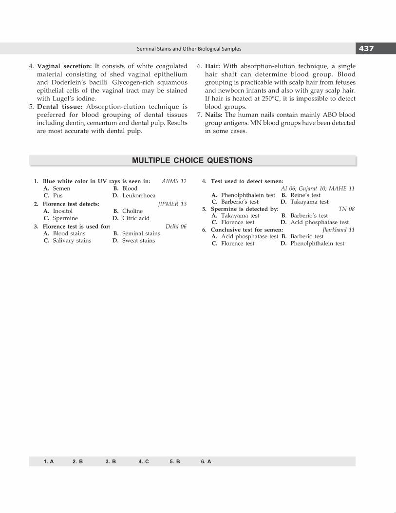
437Seminal Stains and Other Biological Samples
4. Vaginal secretion: It consists of white coagulated material consisting of shed vaginal epithelium and Doderlein’s bacilli. Glycogenrich squamous epithelial cells of the vaginal tract may be stained with Lugol’s iodine.
5. Dental tissue: Absorptionelution technique is preferred for blood grouping of dental tissues including dentin, cementum and dental pulp. Results are most accurate with dental pulp.
1. A 2. B 3. B 4. C 5. B 6. A
6. Hair: With absorptionelution technique, a single hair shaft can determine blood group. Blood grouping is practicable with scalp hair from fetuses and newborn infants and also with gray scalp hair. If hair is heated at 250°C, it is impossible to detect blood groups.
7. Nails: The human nails contain mainly ABO blood group antigens. MN blood groups have been detected in some cases.
1. Bluewhite color inUVrays is seen in: AIIMS 12 A. Semen B. Blood C. Pus D. Leukorrhoea 2. Florence testdetects: JIPMER 13 A. Inositol B. Choline C. Spermine D. Citric acid 3. Florence test isused for: Delhi 06 A. Blood stains B. Seminal stains C. Salivary stains D. Sweat stains
4. Testused todetect semen: AI 06; Gujarat 10; MAHE 11 A. Phenolphthalein test B. Reine’s test C. Barberio’s test D. Takayama test 5. Spermine isdetectedby: TN 08 A. Takayama test B. Barberio’s test C. Florence test D. Acid phosphatase test 6. Conclusive test for semen: Jharkhand 11 A. Acid phosphatase test B. Barberio test C. Florence test D. Phenolphthalein test
MULTIPLE CHOICE QUESTIONS
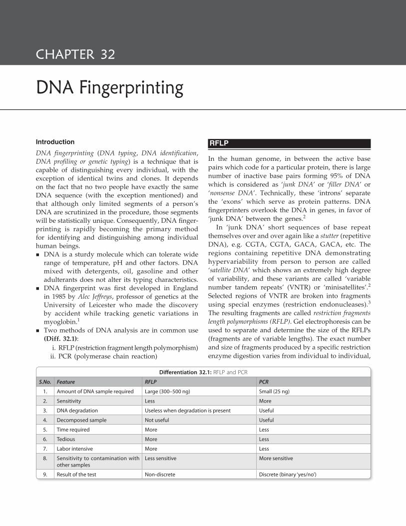
Introduction
DNA fingerprinting (DNA typing, DNA identification, DNA profiling or genetic typing) is a technique that is capable of distinguishing every individual, with the exception of identical twins and clones. It depends on the fact that no two people have exactly the same DNA sequence (with the exception mentioned) and that although only limited segments of a person’s DNA are scrutinized in the procedure, those segments will be statistically unique. Consequently, DNA finger- printing is rapidly becoming the primary method for identifying and distinguishing among individual human beings.
� DNA is a sturdy molecule which can tolerate wide range of temperature, pH and other factors. DNA mixed with detergents, oil, gasoline and other adulterants does not alter its typing characteristics.
� DNA fingerprint was first developed in England in 1985 by Alec Jeffreys, professor of genetics at the University of Leicester who made the discovery by accident while tracking genetic variations in myoglobin.1
� Two methods of DNA analysis are in common use (Diff. 32.1):
i. RFLP (restriction fragment length polymorphism) ii. PCR (polymerase chain reaction)
RFLP
In the human genome, in between the active base pairs which code for a particular protein, there is large number of inactive base pairs forming 95% of DNA which is considered as ‘junk DNA’ or ‘filler DNA’ or ‘nonsense DNA’. Technically, these ‘introns’ separate the ‘exons’ which serve as protein patterns. DNA fingerprinters overlook the DNA in genes, in favor of ‘junk DNA’ between the genes.2
In ‘junk DNA’ short sequences of base repeat themselves over and over again like a stutter (repetitive DNA), e.g. CGTA, CGTA, GACA, GACA, etc. The regions containing repetitive DNA demonstrating hyper variability from person to person are called ‘satellite DNA’ which shows an extremely high degree of variability, and these variants are called ‘variable number tandem repeats’ (VNTR) or ‘minisatellites’.2 Selected regions of VNTR are broken into fragments using special enzymes (restriction endonucleases).3 The resulting fragments are called restriction fragments length polymorphisms (RFLP). Gel electrophoresis can be used to separate and determine the size of the RFLPs (fragments are of variable lengths). The exact number and size of fragments produced by a specific restriction enzyme digestion varies from individual to individual,
Differentiation 32.1: RFLP and PCR
S.No. Feature RFLP PCR
1. Amount of DNA sample required Large (300–500 ng) Small (25 ng)
2. Sensitivity Less More
3. DNA degradation Useless when degradation is present Useful
4. Decomposed sample Not useful Useful
5. Time required More Less
6. Tedious More Less
7. Labor intensive More Less
8. Sensitivity to contamination with other samples
Less sensitive More sensitive
9. Result of the test Non-discrete Discrete (binary ‘yes/no’)
DNA Fingerprinting
ChApter 32
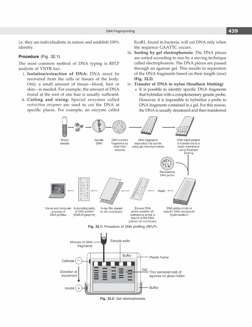
439DNA Fingerprinting
i.e. they are individualistic in nature and establish 100% identity.
Procedure (Fig. 32.1)
The most common method of DNA typing is RFLP analysis of VNTR loci. i. Isolation/extraction of DNA: DNA must be
recovered from the cells or tissues of the body. Only a small amount of tissue—blood, hair or skin—is needed. For example, the amount of DNA found at the root of one hair is usually sufficient.
ii. Cutting and sizing: Special enzymes called restriction enzymes are used to cut the DNA at specific places. For example, an enzyme called
EcoR1, found in bacteria, will cut DNA only when the sequence GAATTC occurs.
iii. Sorting by gel electrophoresis: The DNA pieces are sorted according to size by a sieving technique called electrophoresis. The DNA pieces are passed through an agarose gel. This results in separation of the DNA fragments based on their length (size) (Fig. 32.2).
iv. Transfer of DNA to nylon (Southern blotting) z It is possible to identify specific DNA fragments
that hybridize with a complementary genetic probe. However, it is impossible to hybridize a probe to DNA fragments contained in a gel. For this reason, the DNA is usually denatured and then transferred
Fig. 32.1: Procedure of DNA profiling (RFLP)
Fig. 32.2: Gel electrophoresis
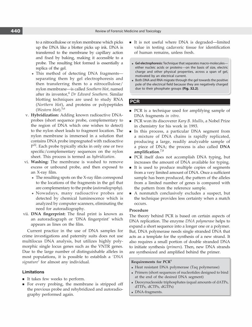
440 Review of Forensic Medicine and Toxicology
to a nitrocellulose or nylon membrane which picks up the DNA like a blotter picks up ink. DNA is transferred to the membrane by capillary action and fixed by baking, making it accessible to a probe. The resulting blot formed is essentially a replica of the gel.
z This method of detecting DNA fragments—separating them by gel electrophoresis and then transferring them to a nitrocellulose/nylon membrane—is called Southern blot, named after its inventor,4 Dr Edward Southern. Similar blotting techniques are used to study RNA (Northern blot), and proteins or polypeptides (Western blot).5,6
v. Hybridization: Adding known radioactive DNA-probes (short sequence probe, complementary to the region of DNA which one wishes to detect) to the nylon sheet leads to fragment location. The nylon membrane is immersed in a solution that contains DNA probe impregnated with radioactive P32. Each probe typically sticks in only one or two specific/complementary sequences on the nylon sheet. This process is termed as hybridization.
vi. Washing: The membrane is washed to remove excess or unbound probe, and then exposed to an X-ray film.
z The resulting spots on the X-ray film correspond to the locations of the fragments in the gel that are complementary to the probe (autoradiography).
z Nowadays, many radioactive probes are detected by chemical luminescence which is analyzed by computer scanners, eliminating the need for autoradiography.
vii. DNA fingerprint: The final print is known as an autoradiograph or ‘DNA fingerprint’ which appears as lines on the film.
Current practice in the use of DNA samples for crime investigations and paternity suits does not use multilocus DNA analysis, but utilizes highly poly -morphic single locus genes such as the VNTR genes. Due to the large number of distinguishable alleles in most populations, it is possible to establish a ‘DNA signature’ for almost any individual.
Limitations
� It takes few weeks to perform. � For every probing, the membrane is stripped off
the previous probe and rehybridized and autoradio-graphy performed again.
� It is not useful where DNA is degraded—limited value in testing cadaveric tissue for identification of human remains, unless fresh.
� Gel electrophoresis: Technique that separates macro-molecules—either nucleic acids or proteins—on the basis of size, electric charge and other physical properties, across a span of gel, motivated by an electrical current.
� Both DNA and RNA migrate through the gel towards the positive pole of the electrical field because they are negatively charged due to their phosphate groups (Fig. 32.2).
PCR
� PCR is a technique used for amplifying sample of DNA fragments in vitro.
� PCR won its discoverer Kary B. Mullis, a Nobel Prize in chemistry for his work in 1993.
� In this process, a particular DNA segment from a mixture of DNA chains is rapidly replicated, producing a large, readily analyzable sample of a piece of DNA; the process is also called DNA amplification.7,8
� PCR itself does not accomplish DNA typing, but increases the amount of DNA available for typing.
� It is used to produce multiple copies of segments from a very limited amount of DNA. Once a sufficient sample has been produced, the pattern of the alleles from a limited number of genes is compared with the pattern from the reference sample.
� A nonmatch conclusively excludes a suspect, but the technique provides less certainty when a match occurs.
ProcedureThe theory behind PCR is based on certain aspects of DNA replication. The enzyme DNA polymerase helps to expand a short sequence into a longer one or a polymer. But, DNA polymerase needs single stranded DNA that acts as a template for the synthesis of a new strand. It also requires a small portion of double stranded DNA to initiate synthesis (primers). Then, new DNA strands are synthesized and amplified behind the primer.
Requirements for PCR9
� Heat resistant DNA polymerase (Taq polymerase) � Primers (short sequences of nucleotides designed to bind at the end of the desired DNA segment)
� Deoxynucleoside triphosphates (equal amounts of dATPs, dTTPs, dCTPs, dGTPs)
� DNA-fragments.

441DNA Fingerprinting
Three steps are involved in this process (Fig. 32.3): i. Denaturation: Heating the double stranded DNA
to almost boiling will dissociate it and will become single stranded.
ii. Annealing: Cooling the reaction will cause the primers to pair up with the single-stranded template (annealing). On the small length of double-stranded DNA (the joined primer and template), the polymerase attaches and starts copying the template.
iii. Extension: DNA building blocks complementary to the template are coupled to the primer, making a double stranded DNA molecule.
� Each separated strand can serve as a template for synthesis as long as primer is provided for each strand, and the reaction is cooled to cause the primers to bind. The primers are chosen to flank the region of DNA that is to be amplified. New primer binding sites are generated on each synthesized DNA strand.
� This cycle of DNA denaturing, primer annealing and strand synthesis is repeated multiple times, thereby amplification of the target DNA.
� After 20 heating and cooling cycles, this exponential process yields 220, or more than a million copies of the target sequence.
� The process is completely automated with thermocyclers that contain a heating block and microprocessors. The time and temperature can be programmed for repetitive cycles of heating and cooling, alleviating manual intervention.
Other PCR based methods � Dot blots, involving a series of DNA probes to detect
target sequences such as the HLA DQa locus in chromosome 6, producing a pattern of colored dots.
� Amplified fragment length polymorphism (AmpFLP)
� Short tandem repeats (STRs) in which the core repeat unit are 3–7 bp length.
� System utilizing mitochondrial DNA. � Digital DNA typing.
STR analyzes the DNA segments for the number of repeats at 13 core loci. STR analysis is less susceptible to DNA degradation than other AmpFLPs. The chance of misidentification in this procedure is one in several billion.
Applications: PCR technique has virtually limitless applications. It enables researchers to amplify and analyze tiny DNA samples from variety of sources—ranging from crimes scenes to archeological remains.
Limitation: It is too sensitive; a tiny amount of contaminant DNA in a sample may become amplified if it includes a DNA sequence complimentary to the primers, leading to an erroneous conclusion.
� PCR can be carried out efficiently if the DNA polymerase can remain stable through many heating cycles since denaturation and primer extension often occur at approximately 92°C and 72°C respectively, so a heat resistant enzyme known as Taq polymerase (derived from Thermus aquaticus, a bacterium) is used.
� Criteria to determine the source: DNA testing laboratories use a two-step process to determine if two samples arose from one source. First, DNA-banding patterns are compared visually. If banding patterns of a sample in question do not match a known DNA sample, exclusion is declared and no further analysis is required. Second, a visualized match is verified by a technique called computer assisted allele sizing which is done by computer software. Basically, the calculated sizes of an apparent match should fall within 2.5% of each other. When samples fall outside of the 2.5% window, they should be considered ‘nonmatching.’ If the DNA-banding pattern of a sample cannot be positively determined due to technical problems, the results should be considered ‘inconclusive.’
Fig. 32.3: Steps in PCR

442 Review of Forensic Medicine and Toxicology
Specimen Selection and Preservation
Samples Collected from Living Subjects
i. Blood (most common sample). 5 ml of venous blood is collected in a purple stoppered vacutainer (EDTA tube) and mixed thoroughly without shaking. Heparin is not used as an anticoagulant since it interferes with PCR. Sample can be preserved at 2–8°C (not frozen).
ii. Buccal epithelial cells (buccal swabs) are considered a convenient alternative for collecting genetic material, as they are relatively easy to collect, inexpensive and noninvasive. The swab is removed from the packing and without handling the tip, immediately swabbed the subject’s mouth for 10 seconds (Fig. 32.4). After swabbing, it placed directly into the receptacle (packaging) to dry.
iii. Hair follicles with roots (plucked hair), about 10–20, from the head is used as a reference standard. The root of the hair contains nuclear DNA, and the shaft contains mitochondrial DNA. The root contains keratinocytes which are ideal for the extraction of nuclear DNA. Cut or naturally shed hairs (without follicle) may not be entirely unhelpful, since some nucleated corneocytes (keratinocytes in their last stage of differentiation) are present which make it possible to extract a DNA profile.
Samples Collected from Dead Bodies (Flow chart 32.1)
DNA can be isolated and tested from practically any postmortem tissue, although after death it will undergo progressive degradation. DNA is broken down into
Flow chart 32.1: Sources of DNA
fragments by autolytic and bacterial enzymes, especially DNases. i. In relatively fresh dead bodies, unclotted 10 ml
of blood (EDTA anticoagulated in a sterile tube) is the preferable source of DNA.10,11 Buffers (e.g. those containing EDTA) are designed to inhibit the activity of nucleases that can breakdown DNA. Due to settling out of WBCs, clotted blood is not a good source of DNA.12
ii. Brain tissue is a good source of DNA in intermediate postmortem intervals.
iii. Hard tissue (bone and vascular pulp of teeth) is the best source of DNA in cases of advanced decomposition.
� Best material is said to be muscle or spleen if decomposition is establishing; bone marrow (from femur) and teeth (usually molars) are also recommended.
� Postmortem material is inferior to live blood and tissue for DNA testing. Any disturbance to nuclear chromatin due to putrefaction is dangerous.
Samples Encountered in Forensic Practice
� Blood (EDTA/heparinized/clotted/stain on cloth, newspaper, wood or tiles).
� Semen (stain on cloth/paper/floor). � Hair (head/body/pubic). � Tissue (bone marrow/muscle/spleen/fingernail
scrapings). � Mouth swabs, and saliva stain on cigarette buds/
licked envelope/glass.
Fig. 32.4: Method of taking buccal swab

443DNA Fingerprinting
DNA Storage and FTA Card
In general, there are four broad strategies for long-term DNA preservation:z Room temperature on a ‘dry’ solid matrixz –20°Cz –80°Cz –196°C (storage in liquid nitrogen)
In a laboratory setting, DNA is most commonly stored at 4°C, –20°C or –80°C. Blood and other biological samples can be stored on FTA cards at room temperature with subsequent amplification by PCR.
� FTA is acronym for fast technology for analysis of nucleic acids developed as a means of protecting nucleic acid samples from degradation by nucleases and other processes.
� The concept was to apply a weak base, chelating agent, anionic surfactant or detergent, and uric acid (or a urate salt) to a cellulose based matrix (filter paper). A sample containing DNA could then be applied to the treated filter paper for preservation and long term storage. The expected storage length is over 50 years.
� Biological samples adhere to the paper through the mechanism of entanglement, while the mixture of chemicals lyses cells and denatures proteins.
� Nucleic acid damage from nucleases, oxidation, UV damage, microbes and fungus is reduced when samples are stored on the FTA card. Since, nucleases are inactivated; DNA is essentially stable when the sample is properly dried and stored.
� For analysis, a small disc is punched from the card containing the DNA, washed, dried and used for PCR amplification, or restriction enzyme digestion can be performed directly on the treated paper without the need for extensive extraction procedures.
� Since, the cards are small in size (approximately 5” × 3.5”), they can be easily packaged, shipped and stored for data basing (Box 32.1).
Uses of DNA Fingerprinting
i. Identification: It is used to link suspects to biological evidence—blood or semen stains, hair or items of clothing—found at the scene of a crime. It is used to establish identity of an assailant in sexual assaults, like rape, incest and bestiality.
ii. Diagnosis of inherited disorders in adults, children, newborn and prenatal babies. It includes cystic fibrosis, hemophilia, Huntington’s disease, familial Alzheimer’s, sickle cell anemia and thalassemia. Genetic counselors use DNA fingerprint information to help prospective parents understand the risk of having an affected child or decisions concerning affected pregnancies.
iii. Developing cures for inherited disorders: By studying the DNA fingerprints of relatives who have a history of some particular disorder or by comparing large groups of people with and without the disorder, it is possible to identify DNA patterns associated with the disease in question.
iv. Establish paternity in custody and child support litigation. In these applications, DNA fingerprints bring a nearly perfect accuracy to the determination.
In this, DNA from both the children and their parents is cut by the same restriction enzyme. The DNA fragment is separated by gel electrophoresis for each individual in a separate lane. The patterns of bands thus formed (DNA fingerprints) is then compared between the children and his/her parents (Fig. 32.5). It should be noted that each band present in one of the children is also found in at least one of the parents.
v. Identifying the remains of soldiers: In the US armed services, a program is there to collect DNA fingerprints from all personnel for use later, in case
Fig. 32.5: DNA fingerprints for two children (C1 and C2) and their parents; father (F) and mother (M)
Box 32.1 Features of the FTA card
Rapid isolation of pure DNA.
Designed to kill pathogens and prevent future colonization by bacteria or fungi.
Protects DNA from microbial and environmental degradation.
Archive samples at room temperature.
Reduces potential for cross-contamination between samples.
Eliminates shearing forces associated with conventional extraction methods.

444 Review of Forensic Medicine and Toxicology
they are needed to identify casualties or persons missing in action.
vi. Biologists routinely use it, particularly to protect endangered species.
vii. Accidents/mass disaster investigations and postmortem identification of skeletal remains/mutilated bodies.
viii. Pedigree analysis of pets.
Limitations of DNA Testing
Generally, the courts have accepted the reliability of DNA testing and admitted DNA test results into evidence. But, DNA fingerprinting is controversial in a number of areas: i. UniquenessofDNAfingerprinting: DNA segments
rather than complete DNA strands are ‘finger-printed’, a DNA fingerprint may not be unique.
ii. Time constraints: The process is lengthy, with each of four or five loci exposed sequentially, it usually takes 10 weeks.
iii. Accuracy of the results: Tests are often performed in private laboratories that may not follow uniform testing standards and quality controls. Ambiguity in interpretation of the bands may arise from scientist’s own misinterpretation of band-pattern and on the other hand, it may occur from actual shifts, degradation, missing bands, extra bands—all due to technical problems.
Moreover, forensic specimens are often contaminated, making the extraction of pure DNA difficult, and cross contamination of DNA between two specimens, or aerosol DNA from previous reactions on one’s hands at very small concentrations can alter results.
iv. Cost: Testing is expensive. v. Invasion of privacy and ethical concerns: In
the US, the FBI has created a national database
of genetic information called the Combined DNA Index System (CODIS). Similar database is present in the UK also. The database contains DNA obtained from convicted criminals and from evidence found at crime scenes. Some experts fear misuse of the database, such as identifying individuals with stigmatizing illnesses such as AIDS.
vi. Suspects who are unable to provide their own DNA experts may not be able to adequately defend themselves against charges based on DNA evidence.
vii. Unlike fingerprints, DNA profile cannot be enlarged and shown in the court of law.
� In 1986, DNA fingerprinting was used by Jeffreys for the first time in the UK, clearing a suspect of two rapes and murders and helping convict the culprit Colin Pitchfork.13 The first criminal conviction based on DNA evidence in the US occurred in 1988. It was only in 1996 that the DNA evidence was accepted in a court in India (in the ‘tandoor murder case’ reported in New Delhi).
� Mitochondrial DNA (mtDNA), a small circular genome located in the mitochondria, has provided forensic scientists with a valuable tool for determining the source of DNA recovered from damaged, degraded or very small biological samples. Cells contain hundreds of copies of mtDNA genomes, as compared to two copies of the DNA located in the nucleus. This increases the likelihood of recovering sufficient DNA from compromised DNA samples, and for this reason mtDNA can play an important role in missing persons investigations, mass disasters and other investigations involving samples with limited biological material. Additionally, mtDNA is maternally inherited.14 Therefore, barring a mutation, an individual’s mother, siblings, as well as all other maternally-related family members will have identical mtDNA sequences. As a result, comparisons can be made using a reference sample from any maternal relative, even if the unknown and reference sample are separated by many generations.
� Nucleic Acid Sequence Based Amplification (NASBA), is used to amplify RNA sequences.8
� Ligase chain reaction: Method of DNA amplification similar to PCR.8
� DNA sequencing: Biochemical methods for determining the order of the nucleotide bases in a DNA oligonucleotide.8
1. DNAfingerprintingwasdiscoveredby: PGI 06 A. Southern B. Galton C. Crick D. Jeffery 2. What ismatched inDNAfingerprinting:
PGI 08; WB 11 A. Introns B. Exons
C. Constant number of tandem repeats D. Minisatellites 3. Restriction endonuclease is: Maharashtra 08 A. Break single stranded DNA B. Break double stranded DNA C. Break peptide chain D. Break RNA
multiple choice questions
1. D 2. A & D 3. B
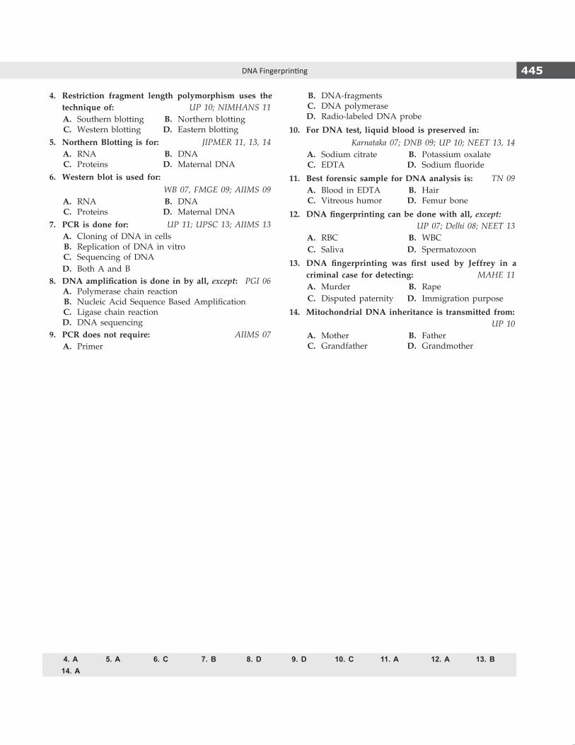
445DNA Fingerprinting
4. A 5. A 6. c 7. B 8. D 9. D 10. c 11. A 12. A 13. B 14. A
4. Restriction fragment length polymorphism uses the technique of: UP 10; NIMHANS 11
A. Southern blotting B. Northern blotting C. Western blotting D. Eastern blotting 5. Northern Blotting is for: JIPMER 11, 13, 14 A. RNA B. DNA C. Proteins D. Maternal DNA 6. Western blot is used for:
WB 07, FMGE 09; AIIMS 09 A. RNA B. DNA C. Proteins D. Maternal DNA 7. PCR is done for: UP 11; UPSC 13; AIIMS 13 A. Cloning of DNA in cells B. Replication of DNA in vitro C. Sequencing of DNA D. Both A and B 8. DNAamplification isdone inbyall,except: PGI 06 A. Polymerase chain reaction B. Nucleic Acid Sequence Based Amplification C. Ligase chain reaction D. DNA sequencing 9. PCR does not require: AIIMS 07 A. Primer
B. DNA-fragments C. DNA polymerase D. Radio-labeled DNA probe
10. ForDNA test, liquidblood ispreserved in:
Karnataka 07; DNB 09; UP 10; NEET 13, 14 A. Sodium citrate B. Potassium oxalate C. EDTA D. Sodium fluoride
11. Best forensic sample for DNA analysis is: TN 09 A. Blood in EDTA B. Hair C. Vitreous humor D. Femur bone
12. DNAfingerprinting canbedonewithall,except: UP 07; Delhi 08; NEET 13
A. RBC B. WBC C. Saliva D. Spermatozoon
13. DNA fingerprinting was first used by Jeffrey in acriminal case for detecting: MAHE 11
A. Murder B. Rape C. Disputed paternity D. Immigration purpose
14. Mitochondrial DNA inheritance is transmitted from: UP 10
A. Mother B. Father C. Grandfather D. Grandmother

446 Review of Forensic Medicine and Toxicology
Definition: Torture (Latin tortura: act of twisting) is ‘a deliberate, systemic or wanton infliction of physical or mental suffering by one or more persons acting alone or on the orders of any authority, to force another person to yield information, to make a confession, or for any other reason’ (as per WMA’s Declaration of Tokyo).
� This declaration states that doctors should refuse to participate in, condone or give permission for torture, degradation or cruel treatment of prisoners or detainees.
� Throughout history, torture has often been used as a method of effecting political re-education. Neverthe less in the 21st century, torture is almost universally considered to be an extreme violation of human rights, as stated by the Universal Declaration of Human Rights.
� Torture medicine is one of the recent branches of medicine dealing with various aspects of torture from medical point of view.
Ethical Issue
Organizations, like Amnesty International argue that the universal legal prohibition is based on a universal philosophical consensus that torture and ill-treatment are repugnant, abhorrent and immoral. But, to obtain information from suspected terrorists by using torture methods to save innocent civilians, is debatable.
Reasons for Torture
UN Convention identifies four reasons for torture: i. To obtain a confession ii. To obtain information iii. To punish iv. To coerce the sufferer or others to act in certain
ways.
Types of Torture
Four types i. Physical ii. Psychological iii. Pharmacological iv. Sexual
Physical Torture
It is the inflicting of pain on the body.
Various methods are: i. Beating: It is the most common form of torture.
Beating can be from head to toe using fist, shoe, wooden bars, rods, whips or similar objects.
z The victim may be suspended upside down, and then beaten on the soles by blunt instruments such as sticks, clubs or rifle butts (bastinado or falanga).1
z Poking the victim with a baton or rod is common.
z Beating buttocks: Literally, this is called ‘passing the board’. A victim is forced to the ground and beaten viciously with a board, club or baton.
z Beating on the abdomen while lying on a table with the upper half of the body unsupported (‘operating table’ or ‘el quirofano’).
z A pole is placed on the back of victim and is pressed down with great weight and then rolled up over the legs and the body (‘the roller’ or ‘belena’).
z Ear torture: Repeated and simultaneous slapping of both the ears by open hands of the torturer (‘telefono’).2
Beating/flogging results in scars, bruises, lacerations and fractures at different stages of healing and which have not been treated medically. Many of these weapons leave a characteristic patterned bruising (including tramline bruising, finger grip or slap mark) on the skin.
ii. Forcing the person in abormal positions: It can be: z Prolonged standing (‘el planton’) like in the
‘Army Corps’ posture or body-folding or on one leg.
z Sitting (iron chair sitting) or forced to squat for a long time.
z Forced straddling on a bar (‘saw horse’ or ‘el cabellete’) or tied up in an abnormal position for hours (‘hogtie’).
Torture and Custodial Deaths
ChapTer 33

447Torture and Custodial Deaths
iii. Suspension z Victim may be suspended by his wrists (‘la
bandera’), ankles (‘murcielago’), arms or hair. z Person may be suspended head down from a
horizontal pole placed under the knees with the wrists bound to the ankles (‘parrot’s perch’, ‘jack’ or ‘pau de arara’).
iv. Burns: Torturers use hot iron, cigars and lighters to burn fingers, toes, faces, nipples, vagina and other parts of victim’s bodies.
z In black slave, a hot metal skewer is inserted up the anus.
v. Suffocation: Near-suffocation may occur by means of a plastic bag placed over the head of the victim (‘dry submarine’).
vi. Waterboarding (simulated drowning) consists of submerging the victim’s head repeatedly under water or in foul liquid, like sewerage water. Forced suffocation and water inhalation cause the subject to experience the sensation of drowning. It has been referred to as ‘water torture,’ ‘water cure,’ ‘el submarino’ (‘wet submarine’), ‘pileta’ or ‘dunking’.
vii. Pulling/twisting of nail/hair/teeth/genitalia or forcing bamboo sticks under the fingernails: One or combination may be used.
z Victim is dragged by hair or the hair is cut short or shaven.
z Teeth may be pulled out or broken down by clips or forceps causing hemorrhage and fracture.
z Pencil or similar objects are placed in between two fingers and then squeezed hard or twisted.
viii. Electric baton shock: Electric shock is given by putting the charged batons over the sensitive parts of the body, like nipples, genitalia or anal canal (buzzer or chicharra).
z Prior to the development of stun batons and the taser, electric cattle prods (‘picana’) were used to torture wherein the current is applied with a pointed object.
ix. Irritant torture: Irritating chemicals (e.g. chillies) may be inserted into the rectum or vagina, or applied on the eyes or external genitalia.
z Victims are slapped, or stripped naked and thrown onto a pile of hemp (a plant). After contact with it, the skin immediately becomes inflamed, extremely itchy and painful.
x. Cold torture: Icy water is poured over head or forced to strip naked and stand outside on a winter
night or forced to stand in snow or on ice with bare feet or submersing in ice-cold water.
xi. Force-feeding of saturated salt-water, vinegar, liquor, pepper, mustard oil, boiling water, urine or feces.
xii. Shooting as means of execution or nonfatal punishment is carried out in some parts of the world.
z Victims are sometimes shot either through knee joint or thigh (knee-capping).
Some other forms of torture � Iron maiden was used mostly in Germany (iron maiden of
Nuremberg). It was a large container shaped like a woman, equipped with two doors with adjustable iron spikes.
� Judas cradle: The victim was first stripped and then suspended over a jagged or pointed pyramid. The torturer, using ropes and pulleys, used to raise and lower the victim so that the point would penetrate the anus, vagina or scrotum.
� Interrogation chair (‘confession chair’): This chair was built of iron and equipped with either wooden or iron spikes which could be heated up from below.
� Branks (scold’s bridle) were metal face masks/iron muzzle that some women were forced to wear as a form of torture.
� Bell (campana): The victim’s head is placed within a pail or other metal container which is then struck repeatedly, causing sudden loud sounds and reverberations
� Flying airplane (‘aeroplane method’): The victim is forced to bend his body at a right angle to the legs, and raise his arms behind his back and head is bent down until it cannot go down any further.
� Helicopter trip: The victim is hung upright or upside-down from one of the large blades of a ceiling fan and is struck repeatedly as the blade revolves.
� Chepuwa: The thigh of victim is placed under two wooden rods and pressed.3
� Torment of the sticks (supplice de baguettes): Two sticks are placed through a wire encircling the victim’s head, and are slowly turned which results in tightening the wire.
Some acute findings in physical torture are highlighted in Table 33.1. Determining bone lesion by scintigraphy is considered a valuable non-invasive diagnostic method to assess and document long term torture practices where beating was a common denominator with no detectable marks upon physical or radiological examination.
Cause of death: Deaths usually result from severe closed blunt force head injuries with cerebral contusion and laceration, with or without skull fracture. Blunt trauma to the abdomen is the second most common cause of death due to tearing of the mesentery or laceration of internal organs.
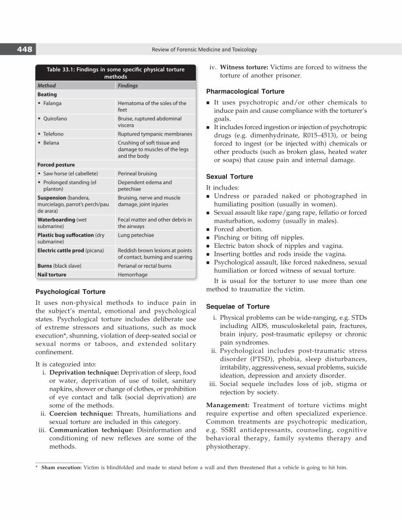
448 Review of Forensic Medicine and Toxicology
Table 33.1: Findings in some specific physical torture methods
Method Findings
Beating
� Falanga Hematoma of the soles of the feet
� Quirofano Bruise, ruptured abdominal viscera
� Telefono Ruptured tympanic membranes
� Belana Crushing of soft tissue and damage to muscles of the legs and the body
Forced posture
� Saw horse (el cabellete) Perineal bruising
� Prolonged standing (el planton)
Dependent edema and petechiae
Suspension (bandera, murcielago, parrot’s perch/pau de arara)
Bruising, nerve and muscle damage, joint injuries
Waterboarding (wet submarine)
Fecal matter and other debris in the airways
Plastic bug suffocation (dry submarine)
Lung petechiae
Electric cattle prod (picana) Reddish brown lesions at points of contact, burning and scarring
Burns (black slave) Perianal or rectal burns
Nail torture Hemorrhage
Psychological Torture
It uses non-physical methods to induce pain in the subject’s mental, emotional and psychological states. Psychological torture includes deliberate use of extreme stressors and situations, such as mock execution*, shunning, violation of deep-seated social or sexual norms or taboos, and extended solitary confinement.
It is categozied into: i. Deprivation technique: Deprivation of sleep, food
or water, deprivation of use of toilet, sanitary napkins, shower or change of clothes, or prohibition of eye contact and talk (social deprivation) are some of the methods.
ii. Coercion technique: Threats, humiliations and sexual torture are included in this category.
iii. Communication technique: Disinformation and conditioning of new reflexes are some of the methods.
iv. Witness torture: Victims are forced to witness the torture of another prisoner.
Pharmacological Torture
� It uses psychotropic and/or other chemicals to induce pain and cause compliance with the torturer’s goals.
� It includes forced ingestion or injection of psychotropic drugs (e.g. dimenhydrinate, R015–4513), or being forced to ingest (or be injected with) chemicals or other products (such as broken glass, heated water or soaps) that cause pain and internal damage.
Sexual Torture
It includes: � Undress or paraded naked or photographed in
humiliating position (usually in women). � Sexual assault like rape/gang rape, fellatio or forced
masturbation, sodomy (usually in males). � Forced abortion. � Pinching or biting off nipples. � Electric baton shock of nipples and vagina. � Inserting bottles and rods inside the vagina. � Psychological assault, like forced nakedness, sexual
humiliation or forced witness of sexual torture.
It is usual for the torturer to use more than one method to traumatize the victim.
Sequelae of Torture
i. Physical problems can be wide-ranging, e.g. STDs including AIDS, musculoskeletal pain, fractures, brain injury, post-traumatic epilepsy or chronic pain syndromes.
ii. Psychological includes post-traumatic stress disorder (PTSD), phobia, sleep disturbances, irritability, aggressiveness, sexual problems, suicide ideation, depression and anxiety disorder.
iii. Social sequele includes loss of job, stigma or rejection by society.
Management: Treatment of torture victims might require expertise and often specialized experience. Common treat ments are psychotropic medication, e.g. SSRI anti depressants, counseling, cognitive behavioral therapy, family systems therapy and physiotherapy.
* Sham execution: Victim is blindfolded and made to stand before a wall and then threatened that a vehicle is going to hit him.

449Torture and Custodial Deaths
Medical Practitioner and Torture
At times, medicine and medical practitioners have been drawn into the ranks of torturers, either to judge what victims can endure, to apply treatments which will enhance torture, or as torturers in their own right. Medical torture may involve the use of their expert knowledge to facilitate interrogation or corporal punishment, in the conduct of unethical human experimentation, or in providing professional medical sanction and approval for the torture of prisoners. Torture is often difficult to prove, particularly when some time has passed between the event and a medical examination. Many torturers around the world use methods designed to have a maximum psychological impact, while leaving only minimal physical traces. Medical and Human Rights Organizations worldwide have collaborated to produce the Istanbul Protocol, a document designed to outline common torture methods, consequences of torture and medico-legal examination techniques. Typically, deaths due to torture are shown in an autopsy as being due to ‘natural causes’ like heart attack due to extreme stress.
Legal aspects: Apart from various sections relating to injury, assault and homicide, Secs. 330 and 331 IPC respectively deals with voluntarily causing hurt and grievous hurt for the purpose of extorting confession or any information which may lead to the detection of an offence or misconduct.
� Offences under these sections are cognizable and non-compoundable; punishment for hurt is imprisonment for 7 years and fine, and for grievous hurt imprisonment for 10 years and fine.
Custodial Deaths
Definition: Death in custody (Latin custosodis: guardian) should include the following categories: i. The death of a person in prison/police custody. ii. The death of a person is caused or contributed by
traumatic injuries sustained, or by lack of proper care while in custody or detention.
iii. The death of a person in the process of police or prison officers attempting to detain that person.
iv. The death of a person in the process of escaping or attempting to escape from prison/police custody.
� The postmortem report is the most valuable record and considerable importance is placed on this document in drawing conclusions about the death, and hence should be carried out properly without inordinate delay in writing.
� A meticulous autopsy is needed to confirm or dispel the allegations of custodial deaths.
� Exhaustive notes must be made, including the description of how the deceased was identified.
� In examination of a case of torture (assaults), the skin is dissected from all the limbs and whole of back. Front is dissected by the modified Y incision, and the front and back of scalp brought down over the face and back of head by joining that coronal incision with the modified Y incision coming up from the sides of neck.
Guidelines issued by the National Human Rights Commission (NHRC) in cases of custodial death/rapes and encounter deaths:
� Reporting: In every case of custodial death/rape, a magisterial inquiry has to be conducted, and to report within 24 hours of occurrence.
� In order to help in proper assessment of ‘time since death’, determination of rectal temperature and development of rigor mortis at the time of first examination at the scene is essential by a medical officer or a trained police officer.
� Precautions to be taken: Both the hands of the deceased should be wrapped in white paper bags and the body should be covered in ‘special body bags’ having zip pouches for proper transportation. Clothing should not be removed by the police or any other person as it must be examined, preserved and sealed by the doctor conducting the autopsy. It should be sent for further examination at the FSL, a detailed note regarding examination of the clothing is to be incorporated in the postmortem examination by the concerned doctor.
� Autopsy form: To ensure better quality of postmortem and to plug the loopholes, a ‘model autopsy form’ has been prepared by the Commission with its recommendation to circulate to the States and UTs.
� Video filming: To prevent tampering and to supplement the postmortem report, video recording of postmortem examination is to be done. This will rule out any undue influence or suppression of material information and to facilitate an independent review of the examination at a later stage, if required. At the time of video recording, the voice of the doctor should be recorded and must narrate his prima-facie observation while conducting the postmortem examination.
� In case of alleged firearms death, the body should be subjected to radiological examination (X-ray/CT scan) prior to autopsy. While describing the injuries, the distance from heel as well as midline must be taken in respect of each injury which will help later in reconstruction of events.
� Photographs: A total of 20–25 colored photographs of the whole body should be taken and some photographs should be without removing the clothes. The photographs should include profile photo—face (front, right lateral, left lateral views), back of head, front of body (upto torso—chest and abdomen) and back, upper and lower extremity—front and back, focusing on each injury/lesion—zoomed in after properly numbering the
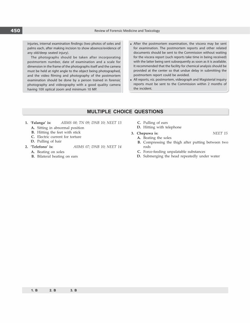
450 Review of Forensic Medicine and Toxicology
injuries, internal examination findings (two photos of soles and palms each, after making incision to show absence/evidence of any old/deep seated injury).
The photographs should be taken after incorporating postmortem number, date of examination and a scale for dimension in the frame of the photographs itself and the camera must be held at right angle to the object being photographed, and the video filming and photography of the postmortem examination should be done by a person trained in forensic photography and videography with a good quality camera having 10X optical zoom and minimum 10 mP.
� After the postmortem examination, the viscera may be sent for examination. The postmortem reports and other related documents should be sent to the Commission without waiting for the viscera report (such reports take time in being received) with the latter being sent subsequently as soon as it is available. It recommended that the facility for chemical analysis should be provided at the center so that undue delay in submitting the postmortem report could be avoided.
� All reports, viz. postmortem, videograph and magisterial inquiry reports must be sent to the Commission within 2 months of the incident.
1. ‘Falanga’ is: AIIMS 08; TN 09; DNB 10; NEET 13 A. Sitting in abnormal position B. Hitting the feet with stick C. Electric current for torture D. Pulling of hair 2. ‘Telefono’ is: AIIMS 07; DNB 10; NEET 14 A. Beating on soles B. Bilateral beating on ears
C. Pulling of ears D. Hitting with telephone
3. Chepuwa is: NEET 15 A. Beating the soles B. Compressing the thigh after putting between two
rods C. Force-feeding unpalatable substances D. Submerging the head repeatedly under water
multiple choice questions
1. B 2. B 3. B

Hospital and health care workers are concerned about the transmission of AIDS from the patient’s blood and body fluids. Patients are concerned that they may be exposed to AIDS from healthcare workers or other patients in the hospital-setting.
Body fluids responsible for transmitting HIV include blood, semen, vaginal secretions, breast milk, and cerebrospinal, peritoneal, amniotic, pericardial and synovial fluids. Other fluids, such as saliva, tears and urine are not implicated in the transmission of HIV, unless they contain visible blood.1,2
HIV Testing Policy
Testing can be: i. Compulsory ii. Mandatory iii. Voluntary.
� For compulsory testing to be legally acceptable, there must be a strong public interest that overrides the individual’s right to privacy, e.g. HIV testing of all military recruits, compulsory screening of prison inmates or applicants for immigration.
� Mandatory testing is recommended only for screening donors of blood, semen, organs or tissues in order to prevent transmission of HIV to the recipient of the biological products. In Andhra Pradesh, legislation was passed, making the AIDS/HIV test mandatory for all persons of marriageable age.
National AIDS Control Organization (NACO) Guidelines
The American Medical Association advises against mandatory testing, and recommends voluntary informed consent testing of patients in the high-risk groups undergoing surgery or other invasive procedures. Some States in the US allow nonconsensual antibody testing of hospital patients, when health care providers are immediately threatened by exposure to disease.
As per NACO policy, HIV testing is to be carried out on a voluntary basis with appropriate pre-test and post-test counseling. Moreover, the disclosure of HIV status of the person should not in any way affect his rights to employment, position at the workplace, right to medical care and other fundamental rights.
� HIV positive women should have the complete choice to make decisions about pregnancy and childbirth, and proper counseling should be given to them to enable them to decide whether to continue or terminate the pregnancy.
� They should be advocated to avoid pregnancy as there is a one in three chance of having an infected child.
� There should be no forcible abortion or even sterilization.
� As far as the breast-feeding is concerned, it may result in transmission of HIV from mother to child.
HIV Testing
� The result of the HIV test must be kept confidential, and even health care workers who are not directly involved in the care of the patient should not be told about the result.
� Surveillance of HIV positive cases in the country does not require reporting of the identification data of the patient.
� Purpose of HIV surveillance is to measure the level and trends of HIV infection in a given geographical area over a period of time.
Health Care Workers and HIV Infection
A sensitive question is whether an employer, particularly a health care employer may screen employees for HIV infection and refuse to employ, terminate employment or limit employment of people who are seropositive. The Government of India has issued a comprehensive HIV testing policy and the following issues are reiterated here:
Medico-legal Aspects of HIV
CHAPTER 34

452 Review of Forensic Medicine and Toxicology
� No individual should be made to undergo a mandatory testing for HIV.
� No mandatory HIV testing should be imposed as a precondition for employment or for providing health care facilities during employment.
� Equal rights to education and employment for HIV positive persons.
� HIV status to be kept confidential.
Health care workers who are known to have antibodies to the virus might be advised to refrain from participating in certain surgical procedures.
� When a health care provider is seropositive or develops AIDS, the hospital should review the staff’s privileges and determine whether or not the medical condition interferes with the person’s ability to perform the job, and whether the condition creates a health risk to the patients.
� The health care provider’s performance must be continuously monitored and evaluated with the goal of protecting the patient.
� It has been recognized that certain direct patient care areas, such as surgery, may create an increased risk of transmission of HIV from the doctor to the patient. Although, NACO does not recommend that HIV-positive individuals be routinely restricted from performing surgery, it does recommend that the restrictions be determined on a case by case basis.
� There is no generally accepted medical evidence that HIV can be transmitted through normal day-to-day contact in typical private workplace settings. Since, present medical evidence indicates that the HIV infected individuals pose virtually no threat of infection to fellow workers, HIV-positive persons in most settings may be permitted to continue their employment as long as they are able to perform their job.
The Centers for Disease Control (CDC), estimates that 5.5% of all HIV positive persons are employed in the health care field. According to the guidelines issued by CDC, with the exception of health care workers and personal service workers who use instruments that pierce the skin, no testing or restriction is indicated for workers known to be infected with HIV, but otherwise able to perform their jobs.
Partner Notification (Contact Tracing, Partner Counseling)
It refers to activities aimed at identifying, notifying and counseling the sexual and needle sharing partners
of an individual with HIV (‘index person’) about their exposure, and offering services.
There are two approaches to partner notification: i. Patient referral: HIV-positive persons are
encouraged to notify partners of their possible exposure to HIV, without the direct involvement of health care providers.
ii. Provider referral: HIV-positive persons give partners’ names to health care providers or other health workers, who then confidentially notify the partners directly.
There are two approaches to informing third parties: i. Contact tracing: The contact tracing approach
emerged from sexually transmitted disease programmes. It is based on the patient’s voluntary cooperation in providing the names of contacts, this entailed protecting the absolute confidentiality of the entire notification process, without disclosure of the identity of the index case.
ii. Duty to warn: This approach came out of the clinical situation where the physician knew the identity of the person deemed to be at risk, e.g. sexual partner of an HIV positive individual. It argued for disclo sure to endangered persons without consent of the patient, due to his moral ‘duty to warn’. It could also involve the revelation of the patient’s identity.
Patient confidentiality continues to be a central issue, even in those subjects in whom the ‘duty to warn’ tradition has been invoked.
� Persons unknowingly placed at risk from an ethical perspective of a clinical relationship, have a moral right to information in order to protect themselves, seek testing and commence treatment, if necessary.
� Since, most public health strategies for dealing with HIV are based on individuals coming forward voluntarily for testing, counseling and treatment, failure to maintain confidentiality could threaten the continued cooperation of people with HIV.
Neither the principle of confidentiality nor the value attached to professional autonomy is absolute. Early identification of HIV infection in asymptomatic individuals has become increasingly beneficial with the availability of antiviral therapy and prophylactic antimicrobial agents. Today, however, it is almost universally recognized that partner notification programs can make a positive contribution to a successful HIV/AIDS public health and prevention program, particularly with regard to

453Medico-legal Aspects of HIV
persons who may be unaware that they are at any increased risk of HIV infection, and as a result are not informed or aware of any need to practice risk-reducing behavior. Partner notification programs can encourage these persons to seek HIV testing.
The Supreme Court of India has recently ruled for disclosure of the HIV positive status by the doctor to his patient’s wife/spouse. Though, the decision has correctly dealt with the legal position of when confidentiality of a patient should be breached by the doctor, it has not laid down the parameters around which such disclosure should or should not be made. The judgment went on to state that persons with HIV infection who knowingly expose others to health risk are guilty of an offence punishable under law.
Clinical Trials and HIV
The highest ethical standards must be upheld when collecting behavioral or biological data on sexually transmitted infections, including HIV/AIDS. Because of the stigma and human rights issues around HIV/AIDS, study participants may experience psychological, social, physical or economic harm, even when precautions are taken. Data collection protocols or procedures should include an explicit description of the measures that will be taken to protect the subjects.
Blood Donation and HIV
It is mandatory for every unit of blood collected at blood banks in India to undergo screening and test negative
for HIV-1 and HIV-2 prior to being declared fit for transfusion and/or further processing for preparation of blood products and blood components. The result of such testing must be clearly indicated on the label. According to guidelines laid by the Government of India, the status of HIV should not be disclosed to blood donor. The intention is to spare him of the agony of knowing the helplessness of his situation. If the blood drawn is positive, it should be discarded. Once blood sample is drawn, the register of patient identities should be kept separate and samples identified only with a code number. If the donor wants to know the result of HIV test, he should be referred to an accessible HIV testing center where supplemental tests with counseling will be offered to him.
Legal Aspects
HIV positive person has the right to marry, but only after obtaining informed consent from their prospective spouse prior to marriage. A person who knowingly communicates the disease of AIDS to other person by sexual relations or otherwise, will be guilty of an offence under Sec. 269 IPC (imprisonment for 6 months and with/without fine) or Sec. 270 IPC (imprisonment for 2 years and with/without fine). The conduct in order to be punishable must be malicious or negligent, so as to cause the spread of an infectious disease dangerous to life. A civil suit may be filed to claim compensation for violation of the fundamental rights to personal liberty.
1. Body fluidNOT responsible for the transmission of HIV: Kerala 06
A. Semen B. CSF C. Tears D. Breast milk
1. C 2. C
2. Which of the following has more HIV load: JIPMER 09; UP 12
A. Urine B. Sweat C. Breast milk D. Saliva
MULTIPLE CHOICE QUESTIONS

Polygraph
A polygraph (‘lie detector’) is a device which makes a continuous record of several physiological variables, such as blood pressure, heart rate, respiration and electrodermal reaction,* while a series of questions are being asked, in an attempt to detect lies (Fig. 35.1). The above measurements are believed to be indicators of anxiety due to sympathetic stimulation that accom panies the telling of lies. However, if the subject exhibits anxiety for other reasons, a measured response can result in unreliable conclusions.
� A polygraph test is also known as a psychophysiological detection of deception (PDD) examination.
Procedure
There are two major testing techniques in use—the Relevant/Irrelevant Technique (RIT) and the Control Question Technique (CQT). Polygraph test starts with a pre-test interview to gain some preliminary information which will later be used for ‘control questions’ (CQ). Some of the questions asked are ‘irrelevant’ or IR, others
are ‘probable-lie’ control questions that most people will lie about, and the remainder are the ‘relevant questions’, or RQ, that the tester is really interested in. The different types of questions may alternate.
Accuracy
Examiners maintain that the accuracy is 90% and the errors tend to be false negative rather than false positive, i.e. a person who actually lied is reported as ‘truthful’.
Admissibility of Polygraphs in the Court
� While lie detector tests are commonly used in police investigations in the US, no defendant or witness can be forced to undergo the test. The US Supreme Court left it up to individual jurisdictions as to whether polygraph results could be admitted as evidence in court cases.
� In most European jurisdictions, polygraphs are not considered reliable evidence and are not generally used by police forces.
� In Canada, the use of a polygraph is sometimes employed in screening employees for government organizations. However, in the 1987, the Supreme Court of Canada rejected the use of polygraph results as evidence in the court.
� The Australian High Court has not yet considered the admissibility of polygraph evidence.
Brain Fingerprinting (Brain Mapping)
Brain mapping is a group of neuroscience techniques based on the mapping of quantities or properties (biological) onto spatial representations of the brain. While various brain imaging techniques (e.g. CT, MRI, PET, SPECT) measures properties such as cerebral blood flow, metabolism or structural integrity, QEEG (quantitative EEG) measures electrical activity of the brain which is usually known as brain mapping.2
* It is also known as galvanic skin response (GSR), electrodermal response (EDR) or skin conductance response (SCR)—a method of measuring the electrical conductance of the skin, which varies with its moisture level.1
Fig. 35.1: Polygraph test
Newer Techniques and Recent Advances
ChApTeR 35
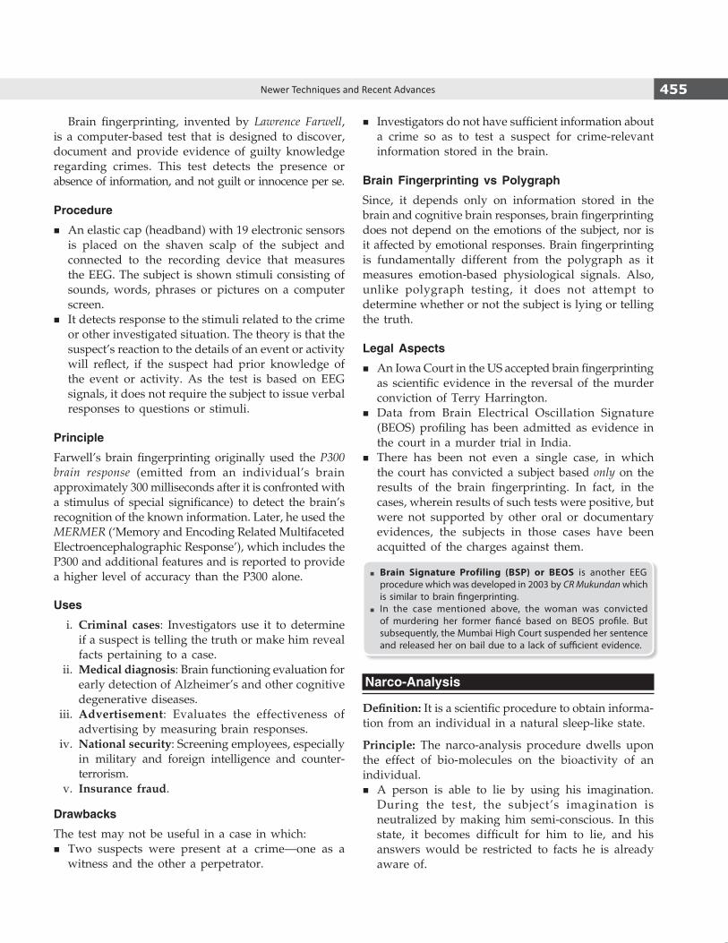
455Newer Techniques and Recent Advances
Brain fingerprinting, invented by Lawrence Farwell, is a computer-based test that is designed to discover, document and provide evidence of guilty knowledge regarding crimes. This test detects the presence or absence of information, and not guilt or innocence per se.
Procedure
� An elastic cap (headband) with 19 electronic sensors is placed on the shaven scalp of the subject and connected to the recording device that measures the EEG. The subject is shown stimuli consisting of sounds, words, phrases or pictures on a computer screen.
� It detects response to the stimuli related to the crime or other investigated situation. The theory is that the suspect’s reaction to the details of an event or activity will reflect, if the suspect had prior knowledge of the event or activity. As the test is based on EEG signals, it does not require the subject to issue verbal responses to questions or stimuli.
Principle
Farwell’s brain fingerprinting originally used the P300 brain response (emitted from an individual’s brain approximately 300 milliseconds after it is confronted with a stimulus of special significance) to detect the brain’s recognition of the known information. Later, he used the MERMER (‘Memory and Encoding Related Multifaceted Electroencephalographic Response’), which includes the P300 and additional features and is reported to provide a higher level of accuracy than the P300 alone.
Uses
i. Criminal cases: Investigators use it to determine if a suspect is telling the truth or make him reveal facts pertaining to a case.
ii. Medical diagnosis: Brain functioning evaluation for early detection of Alzheimer’s and other cognitive degenerative diseases.
iii. Advertisement: Evaluates the effectiveness of advertising by measuring brain responses.
iv. National security: Screening employees, especially in military and foreign intelligence and counter-terrorism.
v. Insurance fraud.
Drawbacks
The test may not be useful in a case in which: � Two suspects were present at a crime—one as a
witness and the other a perpetrator.
� Investigators do not have sufficient information about a crime so as to test a suspect for crime-relevant information stored in the brain.
Brain Fingerprinting vs Polygraph
Since, it depends only on information stored in the brain and cognitive brain responses, brain fingerprinting does not depend on the emotions of the subject, nor is it affected by emotional responses. Brain fingerprinting is fundamentally different from the polygraph as it measures emotion-based physiological signals. Also, unlike polygraph testing, it does not attempt to determine whether or not the subject is lying or telling the truth.
Legal Aspects
� An Iowa Court in the US accepted brain fingerprinting as scientific evidence in the reversal of the murder conviction of Terry Harrington.
� Data from Brain Electrical Oscillation Signature (BEOS) profiling has been admitted as evidence in the court in a murder trial in India.
� There has been not even a single case, in which the court has convicted a subject based only on the results of the brain fingerprinting. In fact, in the cases, wherein results of such tests were positive, but were not supported by other oral or documentary evidences, the subjects in those cases have been acquitted of the charges against them.
� Brain Signature Profiling (BSP) or BEOS is another EEG procedure which was developed in 2003 by CR Mukundan which is similar to brain fingerprinting.
� In the case mentioned above, the woman was convicted of murdering her former fiancé based on BEOS profile. But subsequently, the Mumbai High Court suspended her sentence and released her on bail due to a lack of sufficient evidence.
Narco-Analysis
Definition: It is a scientific procedure to obtain information from an individual in a natural sleep-like state.
Principle: The narco-analysis procedure dwells upon the effect of bio-molecules on the bioactivity of an individual.
� A person is able to lie by using his imagination. During the test, the subject’s imagination is neutralized by making him semi-conscious. In this state, it becomes difficult for him to lie, and his answers would be restricted to facts he is already aware of.
456 Review of Forensic Medicine and Toxicology
� The subject is not in a position to speak up on his own, but can answer specific and simple questions.
� In such sleep-like state, efforts are made to obtain ‘probative truth’ about the crime.
Procedure: The individual is put to trance-like state and loses all his inhibitions by administering sodium amytal or thiopentone sodium, (known as ‘truth drug’ or ‘truth serum’) 2.5–5% solution, slow IV.
Other Methods � 0.5 mg scopolamine hydrobromide (commonly
used) subcutaneously, followed by 0.25 mg every 20 minutes (average 3-6 injections), till proper stage of questioning is reached.3
� 100 mg sodium seconal, 15 mg morphine and 0.5 mg of scopolamine hydrobromide may be given IV.
The dose is dependent on the person’s sex, age, health and physical condition. A wrong dose can result in a person going into a coma or even death.
Team required: A team comprising of an anesthetist, psychiatrist, clinical/forensic psychologist, audio-videographer and supporting nursing staff does the test. The forensic psychologist will prepare the report about the revelations which will be accompanied by a compact disc of audio-video recordings.
Legal Aspects � Supreme Court has recently declared that narco-
analysis, polygraph tests and brain-mapping cannot be done without the consent of the individual. If the person consents for such methods, then any information obtained can be used for further probe. Results of such tests will not be admissible as evidence, even if done with consent.
� Use of such methods are illegal and as against constitution. As per Article 20(3) of the Constitution ‘No person accused of any offence shall be com-pelled to be a witness against himself’. Therefore, a suspect of the crime cannot be compelled to disclose facts which he can recall from his memory, and likely to implicate him in a crime in which he was involved.
Recent advances and development � A non-surgical postmortem technique has been pioneered by
forensic pathologists and radiologists for natural and unnatural deaths, for either single cases or mass fatalities. They have developed a quick and simple technique of ‘minimally invasive targeted coronary angiography’ where they inject contrast into the body of a deceased person through a small incision in the neck and then perform a full body CT scan. Using this method they were able to determine the cause of death in upto 80% of cases (in the series analyzed).
� There has been an increase in the use of condoms by sexual offenders either to avoid contacting STDs or to prevent transfer of DNA evidence. However, they are less likely to consider the possibility of condom lubricant transferring onto their fingertips and then into fingerprints left at the scene. Researchers have developed a method wherein they can detect this condom lubricant. They used MALDI-MSI (matrix-assisted laser desorption/ionization mass spectrometry imaging), a powerful technology that can be used to map fingerprint ridge patterns.
� Forensic researchers in the UK have devised a method to detect smoking from the chemicals left behind in the fingerprints. The technique involves dusting the prints with a solution of gold nanoparticles, attached to which are antibodies that bind to cotinine—a metabolite of nicotine. Then the print is soaked in a fluorescent dye that binds to the antibodies.
� A modified syringe ‘XStat’ has been designed that injects sponges measuring 1 cm in diameter into a gunshot wound. Like foam, these sponges expand upon contact with blood to fill the wound resulting in immediate hemostatic effect (in 15 seconds). They also stick to moist surfaces and create enough pressure to stem the bleeding—useful in battleground.
1. B 2. B 3. B
1. In polygraph test ‘GSR’ stands for: Karnataka 11 A. Guilt Score Reaction B. Galvanic Skin Reaction C. Galvanic Sensor Reaction D. Guilt Sensitivity Reaction 2. Brain fingerprinting: PGI 06 A. Used as lie detector B. Uses EEG on lead
C. Used for quantative measurement of sulci and gyri D. Uses DNA 3. Commonly used in narco-analysis: AI 10; JIPMER 13 A. Atropine sulfate B. Scopolamine hydrobromide C. Opium compounds D. Phenobarbital
multiple choice questions

457Question Bank-I
Question Bank-I
All questions are must know and are either short notes or viva, if not mentioned otherwise. Desirable to know questions are given in the boxes. (LQ—long question, Diff.—differentiation)
Medical Jurisprudence and Acts
1. Forensic medicine, medical jurisprudence, ethics and etiquette
2. Functions of MCI and State Medical Council 3. Professional misconduct, penal erasure, warning
notice 4. Professional secrecy 5. Privileged communication 6. Unethical acts 7. Duties, rights and privileges of a RMP 8. Prevention of medical negligence and defenses in
medical negligence suits 9. Medical maloccurrence, therapeutic misadventure 10. Professional negligence, Res ipsa loquitor 11. Examples of medical negligence 12. Contributory negligence 13. Consent, types, informed consent, rules 14. Vicarious responsibility 15. Malingering 16. Euthanasia, types 17. Acts—CPA, PCPNDT, MTP and Mental Health 18. Civil and criminal negligence (Diff.) 19. Professional misconduct and negligence (Diff.)
Desirable to know1. Active and passive euthanasia (Diff.)2. Human Organ Transplantation Act3. POCSO Act
Legal Procedure
1. Inquest; Police and Magistrate inquest 2. Courts in India and their powers 3. Subpoena/summons 4. Conduct money 5. Recording of evidence: Oath, examination-in-chief,
leading question, cross-examination, perjury
6. Documentary evidence, dying declaration 7. Types of witness: Expert, common, hostile 8. Police and Magistrate inquest (Diff.) 9. Dying declaration and dying deposition (Diff.)
Desirable to know1. Medical examiner system, coroner’s inquest2. Common and expert witness (Diff.)3. Conduct of a doctor in the witness box
Identification 1. Identification, types, corpusdelecti 2. Cephalic index, types of skull 3. Nuclear sexing, Barr body, Davidson body 4. Disorders of sexual development with examples 5. Intersex, Klinefelter and Turner syndrome 6. Eruption of temporary and permanent teeth 7. Gustafson’s method 8. Spacing of jaw, superadded and successional teeth,
period of mixed dentition 9. Ageestimation:Ossificationof longbones 10. Fusion of skull sutures 11. Tattoo marks, scars, MLI 12. Dactylography, types, MLI 13. Medico-legal importance of age (LQ) 14. Male and female skull, pelvis and mandible (Diff.) 15. Human and animal hair (Diff.) 16. Age changes in mandible (Diff.)
Desirable to know1. Stack, Boyde and Miles’ method of age estimation2. Age changes in symphysis pubis3. Anthropometry4. Poroscopy5. Examination of hair, MLI6. Odontology
Medico-legal Autopsy
1. Autopsy, types, objectives/purpose 2. Procedures, formalities of medico-legal autopsies 3. Types of incisions 4. Antemortem and postmortem thrombus

458 Review of Forensic Medicine and Toxicology
5. Subendocardial hemorrhage 6. Preservation of viscera and preservatives used 7. Exhumation
Desirable to know 1. Evisceration techniques 2. Instruments used in postmortem examination 3. Dissection of the heart 4. Delivery of the brain 5. Demonstration of pneumothorax and air embolus 6. Obscure and negative autopsy 7. Second autopsy 8. Chain of evidence 9. Examination of bundle of bones 10. Autopsy room hazards
Thanatology
1. Thanatology, death 2. Braindeathand its certification 3. Clinical and molecular death 4. Cause, manner and mechanism of death 5. Writing cause of death (WHO) 6. Agonal period 7. Sudden death, causes 8. Coronary atherosclerosis 9. Signs of death: Immediate, early and late changes 10. Suspended animation 11. Changes in eye 12. Algor mortis 13. Postmortem staining, color of PM staining in
different conditions 14. Cadaveric spasm, cold and heat stiffening 15. Putrefaction, early and late putrefaction of organs 16. Rigor mortis, factors affecting and conditions
simulating rigor mortis, rule of 12 (LQ) 17. Time since death (LQ) 18. PM staining and bruise (Diff.) 19. Rigor mortis and cadaveric spasm (Diff.)20. Adipocereandmummification (Diff.)
Desirable to know 1. Modes of death: Coma, syncope, asphyxia 2. Persistent vegetative state 3. Anoxia, types 4. Congestion and PM staining (Diff.) 5. Nysten’s law 6. Rigor mortis and heat stiffening (Diff.) 7. Rigor mortis and cold stiffening (Diff.) 8. Casper’s dictum 9. Entomology10. Adipocereandmummification,MLI 11. Embalming
Asphyxia 1. Asphyxia, etiology, Tardieu spots 2. Judicial hanging, lynching 3. Classificationofstrangulation;mugging,garroting,
bansdola 4. Throttling, PM changes 5. Smothering 6. Choking, gagging, café coronary, burking 7. Traumatic asphyxia,PMfindings 8. Gettler’s and Diatom test 9. Sexual asphyxia10. Hanging, classification,PMchanges (LQ) 11. Ligature strangulation, PM changes (LQ)12. Drowning, classification,PMchanges (LQ) 13. Hanging and strangulation (Diff.) 14. Antemortem and postmortem hanging (Diff.)
Desirable to know1. Hyoid bone fractures2. Antemortem drowning and postmortem submersion
(Diff.)
Injuries
1. Injury (Sec. 44 IPC),Classification 2. Abrasion, types, age, medico-legal importance
(MLI) 3. Bruise, types, color changes, ectopic bruise,
patterned bruise, MLI 4. Which is more important medico-legally—abrasions
or bruise? 5. Laceration, types, characteristics, MLI 6. Incised wound, characteristics, hesitation cuts,
tailing, beveling, MLI 7. Langer’s lines 8. Chop wounds 9. Stabwound, classification, characteristics,MLI 10. Defense wounds, fabricated wounds 11. Suicidal and homicidal cut throat wounds (Diff.) 12. PM staining and bruise (Diff.)
Desirable to know1. Antemortem and postmortem bruise (Diff.)2. Age of incised wound and lacerated wound3. Bevelling
Firearm Injuries 1. Ballistics, bore/calibre, cartridge, classification of
firearms 2. Bullet: Dum-dum, hollow point, incendiary, green,
armor piercing, boat tail, ricochet, tandem, duplex, frangible, souvenir, ricochet.

459Question Bank-I
3. Tattooing, blackening; abrasion and grease collar 4. Choke and cylinder bore, paradox guns, wad 5. Advantagesof rifling, chokingandwads 6. Types of gunpowder 7. Puppe’s rule 8. ‘Billiard ball’ ricochet effect, Kennedy phenomenon 9. Multiple exit wounds from single bullet 10. Entry and exit wound of a bullet (Diff.) 11. Bullet and shotgun cartridge (Diagram)
Desirable to know1. Characteristic of shotgun and rifle injuries at varying
ranges, MLI2. Shored exit wound3. Yawing and tumbling of a bullet4.PMexamination infirearm injuries5. Class characteristics6. Dermal nitrate test, Harrison Gilroy test, neutron
activation analysis, AAS7.Accidental,suicidalandhomicidalfirearminjury(Diff.)
Regional Injuries
1. Black eyes 2. Types of fracture of skull 3. Closed and open head injury 4. Coup and contre-coup injury 5. Berry aneurysm 6. Whiplash injury 7. Railway spine 8. Flail chest 9. Cardiac concussion 10. Greenstick fracture 11. Intracranial hematomas, causes, features (LQ and
Diff.) 12. Drunk and concussion (Diff.)
Desirable to know1. Hematoma and depressed skull fracture (Diff.)2. Diffuse axonal injury, concussion3. Age of subdural hematoma4. Post-traumatic intracerebral hemorrhage and apoplexy
(Diff.)5. Cerebral edema6. Complications of abdominal injury7. Open, closed and comminuted fracture8. Healing of fracture9. Punch drunk syndrome
Thermal Injuries
1. Frost bite, trench foot (immersion syndrome) 2. Heat cramps, heat stroke, heat syncope 3. Scalds, classification, features 4. Joule burn, crocodile skin, current pearls, wax
drippings
5. Arborescentmarks (Litchenberg’sflowers) 6. Burns, types, rule of nine, classification, cause of
death, PM changes, pugilistic attitude (LQ) 7. Antemortem and postmortem burns (Diff.) 8. Dry heat, moist heat and chemicals burns (Diff.)
Desirable to know1. Heat exhaustion2. Heat hematoma3. Heat ruptures4. EDH due to burns and blunt force (Diff.)5. Judicial electrocution
Transportation Injuries
1. Pedestrian injuries (primary and secondary impact and secondary injuries)
2. Front impact injuries for vehicle occupants 3. Steering wheel impact injury 4. Role of seat belt, seat belt injuries
Explosion Injuries
Desirable to know1. Blast lung2. Injuries sustained in bomb blast3. Characteristics of bomb blast injuries4. Injuries sustained in fall from height
Medico-legal Aspects of Injuries 1. Assault, battery, manslaughter, murder, dowry
death 2. Hurt (Sec. 319 IPC); simple, and dangerous injuries 3. Injuriessufficienttocausedeathinordinarycourse
of nature 4. Common weapons of offence, dangerous weapons 5. Punishments for various offences 6. Cause of death due to mechanical injuries 7. Thrombosis and embolism 8. Clauses of grievous hurt (Sec. 320 IPC) (LQ) 9. Antemortem and postmortem wounds (Diff.) 10. Lacerated, incised and stab wounds (Diff.)
Desirable to know1. Shock2. Histochemical changes in injured tissue3. Age of wounds4. Relationship of trauma with natural disease
Decompression, Radiation and Altitude Sickness
Desirable to know1. Decompression sickness2. Acute radiation syndrome, radiation sickness3. Acute mountain sickness

460 Review of Forensic Medicine and Toxicology
Starvation Deaths
Deathduetoacuteandchronicstarvation(PMfindings)
Infanticide and Child Abuse 1. Infanticide, feticide 2. Estimation of age of fetus from its features 3. Appearance of center of ossification—calcaneum,
talus, femur, tibia 4. Rule of Hasse 5. Features of dead-born fetus, Spalding’s sign,
maceration 6. Age of viability 7. Vagitus vaginalis and uterinus 8. Signs of live birth 9. Hydrostatic test 10. Wreden’s test, Breslau’s second life test 11. Precipitate labor (medico-legal aspects) 12. Battered baby syndrome 13. SIDS (cot/crib death) 14. Stillborn and dead-born (Diff.) 15. Stillborn and liveborn infant (Diff.) 16. Cephalhematoma and caput succedaneum (Diff.)
Desirable to know1. Changes in umbilical cord after birth2. Causes of infant death3. Munchausen syndrome by proxy4. Head injury due to precipitate labor and blunt force
(Diff.)
Anesthetic Deaths
Desirable to knowCauses of death due to anesthesia
Abortion
1. Abortion,classification,causesofnaturalabortion 2. Methods of criminal abortion 3. Abortifacients drugs, cupping, abortion stick 4. Complications of criminal abortion 5. Natural and criminal abortion (Diff.)
Desirable to knowRelation of trauma and abortion
Impotence and Sterility
1. Impotence, sterility, frigidity, quod 2. Causes of impotency and sterility in males and
females, and examination of a patient 3. Vaginismus 4. Sterilization, classification,methods 5. Artificial insemination,medico-legal aspects
6. Surrogate mother 7. AIH and AID (Diff.)
Virginity, Pregnancy and Delivery 1. Virgin, defloration, marriage, legitimate and
illegitimate child 2. Normal female anatomy 3. Causes of rupture of hymen, signs of virginity 4. Presumptive, probable and positive (conclusive)
signs of pregnancy 5. Medico-legal aspects of pregnancy 6. Pseudocyesis 7. Superfetation and superfecundation 8. Supposititious child, posthumous birth, atavism 9. Signs and symptoms of recent delivery in living 10. Signs and symptoms of remote delivery in dead 11. Lochia 12. True and false virgin (Diff.) 13. Nulliparous and parous uterus (Diff.)
Desirable to know1. Types of hymen2. Medico-legal aspects of delivery, legitimacy3. Fetus compressus
Sexual Offences I 1. Sexual violence, natural sexual offence 2. Classificationof sexualoffences 3. Rape, gang rape, statutory rape, custodial rape,
invalid consent, punishment for rape (Sec. 375 and 376 IPC)
4. Duties of a doctor in a case of rape 5. Locard’s principle of exchange 6. Examinationfindings in theaccused 7. Incest 8. Procedure and examination findings of a rape
victim (virgin) (LQ)
Desirable to know1. Carnal knowledge, adultery2.Medico-legal aspectsofdefinitionof rape3. Contents of a ‘Rape kit’4. Examinationfindingsofrapeinadefloratewomanand
in a child5. Rape trauma syndrome6. Battered wife syndrome7. Specimen collection of rape victim and accused
Sexual Offences II
1. Unnatural sexual offence, buccal coitus, lesbianism, bestiality
2. Sodomy, examination findings of habitual andnon-habitual passive agent, MLI (LQ)

461Question Bank-I
Desirable to knowSpecimens to be preserved from active and passive agent involved in sodomy
Sexual Offences III
1. Sexual perversions, paraphilia, sadism, masochism, fetishism, exhibitionism, transvestic fetishism, voyeurism, frotteurism, masturbation
2. Indecent assault
Postmortem Artifacts
Desirable to knowPostmortem artifacts
Forensic Psychiatry
1. Forensic psychiatry, delusion, hallucination, illusion, obsession, impulse (including types)
2. Lucid interval, delirium, dementia, fugue, twilight state, insight, cataplexy
3. Schizophrenia, types 4. Phobic disorder 5. Obsessive compulsive disorder (OCD) 6. Somnanbulism and somnolentia, narcolepsy,
cataplexy 7. Civil and criminal responsibility (Sec. 84 IPC) of
an insane 8. McNaughten’s rule (legal test of insanity) 9. Psychosis and neurosis (Diff.) 10. True and feigned insanity (Diff.)
Desirable to know1. Lucid interval of head injury and insanity (Diff.)2. Hallucination and illusion (Diff.)3. Role of forensic psychiatrist4. First rank symptoms of schizophrenia5. Mood disorders (manic and depressive episode)
Bloodstain Analysis
1. Chemical examination (color and crystal tests) 2. Microscopicfindings
3. ABO system 4. Secretors 5. Precipitin test 6. Medico-legal aspects and application of blood
groups
Desirable to know1. Source and origin of bleeding2. Antemortem or postmortem bleeding
Seminal Stains and Other Biological Samples 1. Chemical tests 2. Microscopicfindings 3. Proof of semen 4. Medico-legal importance
DNA Fingerprinting
1. RFLP technique 2. PCR 3. Collectionof samples forDNAfingerprinting 4. FTA card 5. UsesofDNAfingerprinting
Torture and Custodial Deaths
Desirable to know1. Torture, types2. Falanga, dry submarine, wet submarine, telefona3. Custodial deaths (PM examination)
Medico-legal Aspects of HIV
Desirable to know1. Health care personnel and HIV2.Partnernotification
Newer Techniques and Recent Advances
Desirable to know1. Polygraph 2.Brainfingerprinting3. Narco-analysis

Toxicology
36. General Toxicology 465 37. Corrosive Poisons 482 38. Inorganic Metallic Irritants—Arsenic 489 39. Inorganic Metallic Irritants—Mercury 494 40. Inorganic Metallic Irritants—Lead 499 41. Inorganic Metallic Irritants—Copper 505 42. Inorganic Metallic Irritants—Thallium 508 43. Other Inorganic Metallic Irritants 510 44. Non-Metallic and Mechanical Irritants 514 45. Organic Irritants—Plant 517 46. Organic Irritants—Animal 524 47. Somniferous Poisons (Narcotic Poisons) 537 48. Inebriants—Alcohol 542 49. Sedative-hypnotic—Barbiturates 559
50. Deliriants—Dhatura/Datura 562 51. Deliriants—Cannabis 566 52. Deliriants—Cocaine 569 53. Spinal and Peripheral Nerve Poisons 572 54. Cardiac Poisons 576 55. Hydrocyanic Acid 582 56. Asphyxiants 586 57. War Gases and Biological Weapons 591 58. Agricultural Poisons 595 59. Alphos (Aluminum Phosphide) 606 60. Medicinal Poisons 611 61. Drug Dependence and Date Rape Drugs 618 62. Kerosene Oil Poisoning 627 63. Food Poisoning 629Question Bank-II 635

‘The dose makes the poison...’ Paracelsus once said.
Definitions
� Toxicology*: Science dealing with properties, actions, toxicity, fatal dose, detection, estimation, treatment and autopsy findings (in case of death) in relation to the poisonous substances.¥
� Forensic toxicology: It deals with the medical and legal aspects of the harmful effects of chemicals on human beings. It involves not only the identification and quantifying of a drug, poison or substance in human tissue, but also the ability to interpret the results of one’s findings.
� Poison: It is a substance (solid, liquid or gaseous) which if introduced in the living body or brought into contact with any part thereof will produce ill-health or death by its constitutional or local effects or both. Thus, almost anything is a poison.
� Clinical toxicology: It deals with human diseases caused by or associated with abnormal exposure to chemical substances.
� Toxinology: It is the science which deals with toxins produced by plants, animals, bacteria and fungi which are harmful to human beings.
� Acute poisoning is caused by an excessive single dose or several dose of a poison taken over a short interval of time.
� Chronic poisoning is caused by smaller doses over a period of time, resulting in gradual worsening.
� Subacute poisoning shows features of both acute and chronic poisoning.
� Fulminant poisoning is caused by massive dose of poison where death occurs rapidly, sometimes without preceding symptoms.
Medico-legal Aspects of Poisons
� In law, the real difference between a medicine and a poison is the intent with which it is given. If the substance is given with the intention to save life, it is medicine, but if it is given with intention to cause bodily harm, it is a poison. The law does not make any difference between murder by means of poisons and murder by any other means.
� Sec. 284 IPC states that whoever causes hurt/injury with rash or negligent conduct with respect to poison-ous substance shall be punished with imprison ment upto 6 months and with/without fine (upto ` 1000).
� Sec. 328 IPC deals with administering of any poison, stupefying or intoxicating agent with the intent to cause hurt and facilitate the commission of an offence. Punishment is imprisonment upto 10 years and also fine.
Narcotics Drugs and Psychotropic Substances Act, 1985 � ‘Narcotic drug’ means coca leaf, cannabis (hemp), opium, poppy
and all drugs manufactured from them.2
� ‘Psychotropic substance’ means any substance, natural or synthetic, or any salt or preparation of such substance or material included in the list of psychotropic substances specified in the Schedule (76 drugs and their derivatives are listed), e.g. amphetamine, pentobarbital, psilocybine and diazepam.2
Punishment � If any person produces, possesses, transports, imports, sells,
purchases or uses any narcotic drug/psychotropic substance (except ‘ganga’), he is punished with rigorous imprisonment (RI) for ≥ 10 years (which may extend to 20 years), and a fine ≥ ` 1 lakh (which may extend to ` 2 lakh). Punishment for a repeat offense is RI for ≥ 15 years (which may extend to 30 years) and a fine of ≥ ` 1.5 lakh (which may extend to ` 3 lakh).
* Toxic (Greek ‘tox’: bow): This is thought to be associated with their use of poisoned arrows in warfare.¥ The origin of modern toxicology is attributed to a Spanish-born French toxicologist and chemist Mathieu Orfila (known as the ‘Father of
Toxicology’) who wrote ‘Traité des poisons’ or ‘Treatise of General Toxicology’ in 1814.1
General Toxicology
ChapTer 36

466 Review of Forensic Medicine and Toxicology
� Punishment for ganja handling is a RI for 5 years and/or a fine of ` 0.5 lakh. For a repeat offense, the imprisonment may extend to 10 years and the fine to ` 1 lakh.
� However, if a person is carrying ‘small quantities’ (e.g. 250 mg of heroin, 5 g of charas, 5 g of opium, or 125 mg of cocaine), then the punishment is a simple imprisonment which may extend
Classification of Poisons
According to their mode of action, poisons are classified as:
I. Corrosives: They produce inflammation and ulceration of the tissues; symptoms are commonly manifested immediately.
Strong acids Strong alkalis Metallic salts
� Mineral or inorganic acids, e.g. HCl, HNO3, H2SO4 Caustic soda, caustic potash, carbonates of sodium, potassium and ammonium
Zinc chloride, ferric chloride, AgNO3
� Organic acids, e.g. carbolic, oxalic and acetic acid
to 1 year or fine (unspecified) or both. For ganja (< 500 g), imprisonment is upto 6 months.
� In a later enactment, the Prevention of Illicit Traffic in NDPS Act, 1988, there is a provision for preventive detention and seizure of property. The maximum punishment is death penalty, if a person is found to be trafficking, for example, ≥ 1 kg of pure heroin twice (despite conviction and warning on the first attempt).
II. Irritants: A group of poisons which by their specific action causes inflammation of the gastrointestinal tract (GIT) and others symptoms; symptoms are usually manifested slowly.
Inorganic Organic Mechanical
� Metallic, e.g. arsenic, antimony, copper, lead, mercury, zinc
� Plant, e.g. abrus, castor, croton, calotropis Powdered glass, hair, diamond dust, needles
� Non-metallic, e.g. phosphorus, chlorine, iodine, CCl4
� Animal, e.g. snakes, cantharides, scorpions, spiders
III. Neurotics: They act mainly on the CNS, though some have local irritant action.
Cerebral Spinal Peripheral
� Somniferous, e.g. opioids Nux vomica, gelsemium Curare, conium
� Inebriants, e.g. alcohol, anesthetics, ether
� Deliriants, e.g. dhatura, cannabis, cocaine
IV. Cardiac: Digitalis, oleander, aconite, nicotine, hydrocyanic acid.
V. Asphyxiants: CO, CO2, H2S, war gases.
VI. Miscellaneous: It includes poisons having widely different pharmacological actions. i. Agrochemicals
Pesticides Fumigants Rodenticides Herbicides
Organophosphates, organochlorines
Aluminum phosphide (alphos), ethylene dibromide
Thallium sulfate, zinc phosphide Paraquat, bromoxynil
ii. Drugs of dependence: Tranquilizers, anti depres sants, hallucinogens. iii. Petroleum products: Kerosene, petrol, naphtha. iv. Food poisoning: Bacterial, chemical (botulism). v. Others: Analgesics and antipyretics.

467General Toxicology
Factors Modifying the Action of Poisons
i. Quantity/dose: More the quantity, more severe will be the toxic effects.
ii. Form z Physical state: Gases and vapors act more rapidly
than liquid. Liquid poisons act more rapidly than solid ones, of which fine powders act more quickly than coarse ones.
z Chemical combination: Action of poison depends upon the solubility or insolubility resulting from chemical combination, e.g. AgNO3 and HCl are both strong poisons, but when combined, an insoluble salt of AgCl is formed which is harmless.
z Mechanical combination: Action of poison is altered when combined mechanically with inert subs tances. Corrosives when sufficiently diluted with water act as irritants.
iii. Mode of administration: In order of rapidity of action:
Inhaled in gaseous/vaporous form > Intravenous injection (IV) > Intramuscular (IM), subcutaneous and intradermal injection > Application to a wound > Application to serous surface > Ingestion > Introduction into the natural orifices, e.g. rectum, vagina, urethra and sublingual > Application to unbroken skin (e.g. nicotine patch).
(‘>’ indicates more rapidly acting) As a rough estimate, if the active dose by mouth
is considered as one unit, the rectal dose about 1½–2 times and the hypodermic dose is about ¼.
iv. Condition of the patient z Age: Poisons have greater effect at the two
extremes of age. A child does not have fully developed drug metabolizing enzymes and effective blood-brain barrier, and as such more susceptible to the effect of most drugs.
z State of health: A healthy person tolerates poisons better than a diseased person. General debility, senility, chronic or disabling disease may cause death of a person to a dose that is ordinarily safe, e.g. opium in bronchial asthma.
z Sleep and intoxication: Action of poison is delayed, if a person goes to sleep soon after taking it. Action is also delayed, if one takes a poison in an intoxicated state.
z Tolerance and idiosyncrasy: People have widely varying susceptibility, but tolerance can build up to a substance, so that same dose no longer has the effect that originally it had, e.g. alcohol,
barbiturates, amphetamines, benzodiazepines, tobacco and the morphine-heroin-methadone group.
The opposite situation is that of idiosyncrasy, where there is an inherent hypersensitivity towards drugs or food resulting in symptoms, like dyspnea, rigors, fever, diarrhea, hemorrhage from bowel and albuminuria, e.g. penicillin, aspirin, cocaine, sulfonamides, sera, certain articles of food, e.g. mushrooms, eggs, shell-fish, fruits, and heroin.
z State of stomach: Presence of food in stomach delays the action of the poison in most cases. It also dilutes the concentration of the ingested poison.
z Cumulative action: Poisons which are not excreted readily tend to accumulate in the body when given in repeated doses, and produce symptoms when their concentration reaches the threshold.
Routes of elimination: The absorbed poison is mainly excreted by the kidneys and to some extent by the skin. Other routes are bile, milk, saliva, mucous and serous secretions. Unabsorbed portion is excreted in the vomit and feces.
Action of Poison
� Local i. Chemical destruction by corrosives. ii. Congestion and inflammation by irritants. iii. Effect on motor and sensory nerves, e.g. tingling
of skin and tongue by aconite or dilatation of pupils by atropine.
� Remote: Remote action produced are either by shock, acting reflexly through severe pain, or exerting a specific action on certain organs and tissues.
� Combined: Substances, like carbolic acid, oxalic acid and phosphorus have local and remote actions.
Poisoning in the Living
There is no single symptom and no definite group of symptoms which are absolutely characteristic of poisoning.
Following should arouse suspicion of poisoning i. Symptoms appear immediately or within a short
period after food or drink. ii. Symptoms are uniform in character and increase
rapidly in severity. iii. When several persons eat or drink from the same
source of food or drink at the same time, all suffer from similar symptoms at or about the same time.

468 Review of Forensic Medicine and Toxicology
iv. Discovery of poison in food taken, in the vomitus or in the excreta is strong proof of poisoning.
Symptoms Suggestive of Poisoning
i. Sudden onset of abdominal pain, nausea, vomiting, diarrhea and collapse.
ii. Sudden onset of coma with constriction of pupils. iii. Unexplained coma, especially in children. iv. Coma in an adult, known to have a depressive
illness. v. Rapid onset of a peripheral neuropathy, such as
wrist-drop. vi. Rapid onset of a neurological or GIT illness in
persons known to be occupationally exposed to chemicals.
vii. Sudden onset of convulsions. viii. Delirium with dilated pupils. ix. Paralysis, especially of lower motor neuron type. x. Jaundice and hepatocellular failure. xi. Oliguria with proteinuria and hematuria. xii. Persistent cyanosis.
Qualities of ideal homicidal and suicidal poison are given in Diff. 36.1.
Features indicative of Chronic Poisoning
i. Symptoms are exaggerated after the administration of suspected food, fluid or medicine.
ii. Malaise, cachexia, depression, and gradual deterioration of general condition of the patient.
iii. Repeated attacks of diarrhea and vomiting.
iv. Removal of patient from his usual surroundings causes the symptoms to disappear.
v. Traces of poison found in the urine, blood, stool or vomit.
Diagnosis of Poisoning in Dead
Evidence of poisoning will depend on postmortem examination, chemical analysis, experiments on suitable animals and circumstantial evidence.
Postmortem Examination
External Findings
i. The color changes in the corroded skin and mucous membrane is given in Table 36.1.
ii. Color of PM staining in poisoning (Table 36.2).
Differentiation 36.1: Ideal homicidal and ideal suicidal poison
S.No. Feature Ideal homicidal poison Ideal suicidal poison
1. Cost Immaterial Cheap
2. Availability Easily available Easily available
3. Physical characteristics Colorless, odorless and tasteless Tasteless or pleasant taste, no repulsive smell
4. Toxicity Highly toxic Highly toxic
5. Antidote None None
6. Solubility in food/drink Soluble without producing any obvious change Should be easily taken in food or drink
7. Signs and symptoms Should resemble a natural disease, or delayed for the accused to escape suspicion
Capable of producing painless death
8. Metabolism Must be rapidly destroyed, or undetectable in urine/blood Not particularly so
9. Detection Should not be detected by chemical tests or other methods
Not particularly so
10. Postmortem changes Should be none Not particularly so
11. Examples Arsenic, aconite, thallium, oleander, insulin and other drugs
KCN, HCN, opium, barbiturates, alphos or organophosphorus
Table 36.1: Color changes in skin and mucous membrane due to poisoning
S.No. Poison Color observed
1. Sulphuric and hydrochloric acid
Gray, becoming black from blood
2. Nitric acid Brown or yellow
3. Hydrofluoric acid Reddish-brown
4. Carbolic acid Grayish-white
5. Oxalic acid Gray, blackened by blood
6. Caustic alkalis Grayish white
7. Mercuric chloride Bluish white
8. Zinc chloride Whitish
9. Chromic acid, potassium chromate
Orange and leathery

469General Toxicology
iii. Smell present about the mouth and nose is given in Table 36.3.
� The natural orifices, e.g. mouth, nostrils, rectum and vagina may show presence of poisonous material or the signs of it.
� Injection marks should be looked for with care. � Skin should be examined for lesions, like hyper-
keratosis and pigmentation, seen in chronic arsenic poisoning.
� Jaundice may occur in poisoning with phosphorus and potassium chlorate.
Internal Findings
i. Smell: The skull should be opened first to detect unusual odors in the brain, since the opening of the body masks such odors.
ii. Mouth and throat: Examine for any evidence of inflammation, erosion or staining. Areas of necrosis of the pharynx may be seen in death associated with agranulocytosis caused by amidopyrine, thiouracil, dinitrophenol, sulfonamide and barbiturates.
iii. Respiratory system: Corrosives may cause edema of glottis, and congestion and desquamation of mucous membrane of trachea and bronchi due to trickling of acid or alkali into the respiratory tract.
iv. Esophagus: Corrosive alkalis produce marked softening and desquamation of the mucous membrane.
v. Heart: Subendocardial hemorrhage in left ventricle is seen in poisoning with arsenic, antimony, barium, mercury, phosphorus, and viper bite and in certain conditions, like heat stroke, acute infectious disease, e.g. influenza, and traumatic asphyxia.
vi. Stomach: Hyperemia of mucous membrane (ridges are more involved) is caused by irritant poison, usually at the cardiac end and greater curvature of stomach (empty stomach).
z Redness of mucosa is also found during digestion, in asphyxial deaths, venous congestion, and when exposed to the atmosphere.
z Hyperemia due to disease is spread uniformly over the whole surface and not in patches.
z Color changes of mucous membrane of stomach seen in different poisoning are given in Table 36.4.
z Softening: Softening of mucous membrane of the stomach, especially at cardiac end and greater curvature is usually caused by corrosives, especially alkaline corrosives.
z Ulcers: Ulceration due to corrosives or irritants is usually found at greater curvature, ulcer from disease is usually seen on the lesser curvature —margins are well-defined, thickened and indurated.
z Perforation: Usually observed when strong mineral acids, especially H2SO4 has been taken. The stomach in such cases is blackened and extensively destroyed. Acid escapes into the abdomen and causes peritonitis.
vii. Duodenum and intestines: A strong acid reaction from its constituents is of greater significance than that of stomach contents. Normal GIT rules out poisoning by corrosives (acids, alkalis and phenol), mercury and arsenic.
viii. Liver (hepatotoxic poisons): Arsenic, phosphorus, CCl4, alcohol, chlorpromazine, chloroform, trinitrotoluene (TNT), paracetamol, thallium, alphos and zinc phosphide.
z Liver necrosis is caused by phosphorus, chloro-form, TNT and CCl4.
z Fatty liver is caused by arsenic, CCl4, amanita phalloides and FeSO4.
z Jaundice: Phosphorus and potassium chlorate.
Table 36.2: Color of PM staining in some poisons
S.No. Poison Color of PM staining
1. Carbon monoxide (CO) Cherry red
2. Carbon dioxide (CO2) Deep blue (reduced hemoglobin)
3. Cyanide Bright red/pink
4. Phosphorus or copper Dark brown/yellow
5. Hydrogen sulfide Bluish green3
6. Opiates Black
7. Nitrites, aniline, nitrobenzene, chlorates (methemoglobin formation)
Chocolate or coffee-brown4
Table 36.3: Smell due to various poisons
S.No. Poison Odor
1. Phosphorus, heavy metal poisoning (arsenic, selenium, thallium), para-thion, malathion, alphos
Garlic-like
2. Ethanol, methyl or propyl alcohol, chloroform, nitrites, acetone
Sweet and fruity
3. Paraldehyde, chloral hydrate Acrid
4. H2S, mercaptans, disulfirum Rotten eggs
5. HCN Bitter almond
6. Carbolic acid Phenolic
7. Organophosphates Kerosene-like
8. Zinc phosphide Fishy
9. Methyl salicylates Oil of wintergreen
10. Marijuana5 Burnt rope, hemp

470 Review of Forensic Medicine and Toxicology
ix. Kidneys (nephrotoxic poisons): Arsenic, mercury, oxalic acid, carbolic acid, CCl4, cantharides, turpentine, thallium, alphos and zinc phosphide.
z Parenchymatous degenerative changes are seen in metal and cantharidin poisoning.
z Necrosis of proximal convoluted tubules (PCT) is observed in mercuric chloride, phenol, lysol and CCl4 poisoning.6
Chemical Analysis
The most important proof of poisoning is the analytical detection of poison in the parenchyma of the organs of the body. The finding of poison in the food, medicine or fluid alleged to have been taken is corroborative.
Experiments on Animals
The suspected food, medicine or fluid or poison extracted from viscera can be fed to domestic animals, such as dogs or cats. The poison affects these animals in the same way as human beings.
Circumstantial Evidence
Clues regarding recent purchase of poison by the victim or accused, his behavior, the conduct of those living with the victim, suicide note and history of quarrel or financial problems may provide valuable information.
Failure to Detect Poison
In some cases, no trace of poison is found on analysis, although from other circumstances, it is almost or quite certain that poison was the cause of illness or death.
Possible explanations for negative findings: i. Poison may have been eliminated by vomiting and
diarrhea, e.g. irritant poison.
ii. Poison has disappeared from the lungs by evaporation or oxidation.
iii. Poison after absorption may be detoxified, conjugated and eliminated from the system.
iv. Some alkaloidal poisons cannot be definitely detected by chemical methods.
v. Some drugs are rapidly metabolized, making extraction difficult.
vi. Biological toxins and venoms which may be protein in nature, cannot be separated from body tissues.
vii. Some organic poisons, especially alkaloids and glucosides may detoxify by oxidation during life or due to faulty preservation or from decomposition of the body, and cannot be detected chemically.
viii. In a slow acting poison, death may be delayed and by then the poison may have been completely excreted following production of irreversible changes.
ix. Many drugs may be present in small amount and these may require considerable amount of viscera for their identification.
x. Wrong or insufficient material may have been sent for analysis.
Duties of a Doctor in a Case of Suspected Poisoning
Medical: Care and treatment to save the life of the patient is first and foremost duty.
Legal: Assist the police to determine the manner of poisoning. 1. Note preliminary particulars of the patient, viz.
name, age, sex, occupation, address, date and time, brought by whom, identification marks, and history.
2. In case of suspected homicidal poisoning, the doctor must confirm his suspicion before expressing an opinion. For this he must:
i. Collect vomitus and urine and submit it for analysis.
ii. Carefully observe and record the symptoms in relation to food, any change in color, taste or smell of food/drink, and other persons affected at the same time.
iii. Consult in strict confidence a senior practitioner and keep him informed about the case.
iv. Remove the patient to the hospital. If the patient refuses, the doctor should engage nurses of his confidence who should administer the medicine and food and allow no one to be with the patient alone.
Table 36.4: Color of mucous membrane of stomach due to poisoning
S.No. Poison Color
1. Copper sulfate, amytal capsule Blue
2. Ferrous sulfate Green
3. Sulphuric, hydrochloric or acetic acid Black/charred
4. Nitric acid Yellow
5. Carbolic acid Buff/white
6. Arsenic White particles
7. Mercury Slate
8. Cresols Brown

471General Toxicology
3. Once the suspicion is confirmed, he should request the removal of the patient to the hospital. If the victim is an adult, it desirable to seek his consent.
4. Any suspected articles of food, excreta, and stomach wash samples should be preserved. Non-compliance is punishable under Sec. 201 IPC, if it is proved that the doctor did it with the intention of protecting the accused (imprisonment upto 7 years depending upon the nature of offence). In this case, the onus of proving a non-deliberate omission to collect and preserve the samples would lie on the medical practitioner.
5. A government medical officer is required to report to police all cases of suspected poisoning, whether accidental, suicidal or homicidal, attended in the hospital.
6. If a private practitioner is convinced that the patient is suffering from homicidal poisoning, he is bound under Sec. 39 CrPC to inform the police or Magistrate.7 Non-compliance is punishable under Sec. 176 IPC (simple imprisonment of 1 month or fine of ` 500/- or both). Giving false information on such matters is punishable under Sec. 177 IPC (simple imprisonment for 6 months or fine of ` 1000 or both).
7. If the private practitioner is sure that the patient is suffering from suicidal/accidental poisoning, he is not bound to inform the police, since Sec. 309 of IPC (attempt to commit suicide) is not included in the section of IPC for which information has to be given under Sec. 39 CrPC.*
z Sec. 43 IPC describes the word ‘illegal’ and ‘legally bound to do’. The word ‘illegal’ is applicable to everything which is an offence, prohibited by law or furnishes ground for a civil action. A person is said to be ‘legally bound to do’ whatever it is illegal in him to omit. Thus, it is the duty of the medical practitioner not to do anything illegal or hide illegal acts.
z In an alleged case of attempted suicide, possibility of abetment to suicide, or homicide cannot be ruled out. It is the duty of the police, not doctors, to decide whether the case was actually of attempted suicide or not, not even if the patient was successfully cured. So, it is better to inform the police. Subsequently, the patient may inform the police whether to pursue the case any further or not.
8. If the condition of the patient is serious, he must make arrangement to record the dying declara-tion.
9. If the patient dies, he should not issue a death certificate, but should inform the police.
10. Any opinion about the nature of poison should be given only after getting the report from the forensic science laboratory.
11. If the practitioner is summoned by the investigating officer (IO), he is bound to give all information regarding the case that has come to his notice (Sec. 175 CrPC). If he conceals the information, he is liable to be prosecuted under Sec. 202 IPC (imprisonment upto 6 months or fine or both). If he gives false information during judicial proceedings, he is liable to be charged under Sec. 193 IPC.
Management of Poisoning Cases
If the poison is known, specific treatment must be started. If not, treatment is given on general lines (Flow chart 36.1).
Main aim of treatment: Help the patient to stay alive by attention to respiration and circulation, while he is assisted in getting rid of the poison by metabolism or excretion.
Emergency Management of Symptomatic Patient
In symptomatic patients, treatment of life-threatening complications takes precedence over diagnostic evaluation. i. Coma: The initial management can be remembered
by the mnemonic ABCD, for airway, breathing, circulation and drugs respectively.
z Airway: Establish a patent airway by positioning, suctioning or insertion of an artificial nasal or oropharyngeal airway or endotracheal intubation.
z Breathing: Provide assistance, if necessary, with a bag-valve-mask device or mechanical ventilator. Provide supplemental oxygen.
z Circulation: Measure the pulse and blood pressure, and estimate tissue perfusion (e.g. by measurement of urinary output, skin signs and arterial blood pH). Place the patient on continuous ECG monitoring.
* Recently, government has decriminalized attempt to commit suicide by deleting Sec. 309 IPC.

472 Review of Forensic Medicine and Toxicology
z Drugs a. Dextrose 50%: 50–100 ml IV (unless bedside
glucose is normal). b. Thiamine: 100 mg IM or IV. c. Naloxone: 0.45–2 mg IV. d. Consider flumazenil: 0.2–0.5 mg IV. ii. Hypothermia: Gradual rewarming is preferred,
unless the patient is in cardiac arrest. iii. Hypotension: Most patients respond to empiric
treatment (200 ml IV bolus of 0.9% saline or other isotonic crystalloid upto a total of 1–2 l). If unsuc-cessful, give dopamine, 5–15 µg/kg/minute (min) by infusion.
iv. Hypertension: Treat hypertension, if the patient is symptomatic or if the diastolic pressure is > 105–110 mmHg. Hypertensive patients who are agitated or anxious may benefit from a sedative, such as lorazepam 2–3 g IV. For persistent hypertension, administer phentolamine 2–5 mg IV, or nitroprusside sodium 0.25–8 µg/kg/min IV.
v. Convulsions/seizures: Administer lorazepam 2–3 mg IV over 1–2 min, or if IV access is not
imme diately available—midazolam 5–10 mg IM. If convulsions continue, administer phenobarbital 15–20 mg/kg slow IV over 30 min, or phenytoin 15 mg/kg IV over 30 min.
vi. Hyperthermia: It is treated aggressively by removing all clothing, spraying with tepid water, and fanning the patient. If this is ineffective, induce neuro-muscular paralysis with a nondepolarizing neuro-muscular blocker (e.g. pancuronium, vecuronium).
Dantrolene (2–5 mg/kg IV) may be effective for hyperthermia that does not respond to neuro -muscular blockade (i.e. malignant hyperthermia).
Removal of Unabsorbed Poison
� Inhaled poisons: In case of inhalation of gaseous poisons, the patient should be removed into fresh air, artificial respiration and O2 (6-8 l/min) should be given. Air passages should be kept free from mucus by postural drainage or by suction.
� Injected poisons: If the poison has been injected subcutaneous, a tourniquet may be applied imme-
Flow chart 36.1: Management of a case of poisoning

473General Toxicology
diately above the point of injection, which must be loosened for 1 min after every 10 min to prevent gangrene. Immersion of the extremity in water at 10°C or below, slows capillary blood flow and limits absorption.
� Contact poisons: Immediate, copious flushing with water, saline or any other available clear liquid is the initial treatment for topical exposures (except alkali, metals, calcium oxide and phosphorus). Saline is preferred for eye irrigation. A triple wash (water, soap and water) is best for dermal deconta mination. The removal of liquids from body cavities, such as the vagina or rectum is done by irrigation.
Ingested Poisons (Gastric Decontamination)
i. Gastric lavage (stomach washing) is most useful within 1 hour (h) after ingestion of any poison (can be done till 4–6 h after ingestion). It is performed by sequentially administering and aspirating about 5 ml fluid/kg of body weight with a 36–40 French orogastric tube (22–28 French tube for children). It is repeated, till clear and odorless fluid comes out. If there is any bleeding, the procedure is abandoned.
z Procedure: The patient is placed in Trendelenburg (mouth is at lower level than larynx so as to aid respiratory drainage and prevent aspiration) and in left lateral decubitus position (pylorus points upward in this orientation and helps prevent the poison from passing through the pylorus during
the procedure), even if an endotracheal tube is in place for ventilatory support (Fig. 36.1).
z Confirmation of tip in the stomach: For confirmation, a little air in a syringe is forced down the tube, bubbling sounds are heard through the stethoscope applied over the stomach. If the tube has entered the trachea, a hissing noise is heard at the other end, and if the patient is conscious, reflex coughing takes place and bubbles of air will be found coming out, if outer end is dipped in water.
z After testing, about 250 ml of water is injected. Allow few minutes for fluid to act in the stomach. The fluid is then taken out and preserved for chemical analysis (Fig. 36.1).
z Fluid for gastric lavage: Except for infants, where normal saline is recommended, tap water is acceptable.
Others agents used: 1:5000 KMnO4, 5% NaHCO3, 4% tannic acid, 1% NaI/KI, 1–3% calcium lactate, saturated lime water or starch solution.
z When the poison has been removed, a small quantity of the fluid is left behind in the stomach, so that it may neutralize whatever small quantity of the poison is left behind.
z Complications: Aspiration is a common complica tion (10% of patients) and serious complications (like esophageal and gastric perforation, tube misplacement in the trachea) occur in about 1% of patients.
Fig. 36.1: Gastric lavage
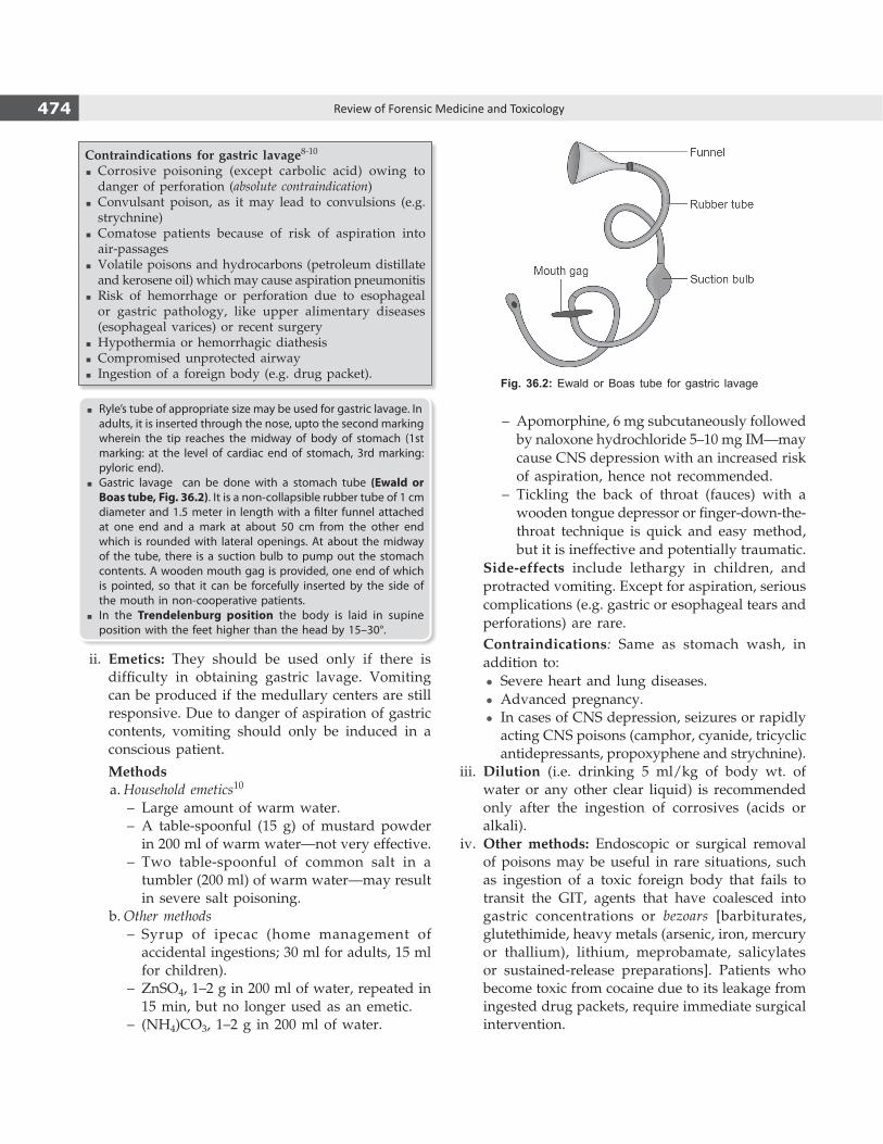
474 Review of Forensic Medicine and Toxicology
Contraindications for gastric lavage8-10
� Corrosive poisoning (except carbolic acid) owing to danger of perforation (absolute contraindication)
� Convulsant poison, as it may lead to convulsions (e.g. strychnine)
� Comatose patients because of risk of aspiration into air-passages
� Volatile poisons and hydrocarbons (petroleum distillate and kerosene oil) which may cause aspiration pneumonitis
� Risk of hemorrhage or perforation due to esophageal or gastric pathology, like upper alimentary diseases (esopha geal varices) or recent surgery
� Hypothermia or hemorrhagic diathesis � Compromised unprotected airway � Ingestion of a foreign body (e.g. drug packet).
� Ryle’s tube of appropriate size may be used for gastric lavage. In adults, it is inserted through the nose, upto the second marking wherein the tip reaches the midway of body of stomach (1st marking: at the level of cardiac end of stomach, 3rd marking: pyloric end).
� Gastric lavage can be done with a stomach tube (Ewald or Boas tube, Fig. 36.2). It is a non-collapsible rubber tube of 1 cm diameter and 1.5 meter in length with a filter funnel attached at one end and a mark at about 50 cm from the other end which is rounded with lateral openings. At about the midway of the tube, there is a suction bulb to pump out the stomach contents. A wooden mouth gag is provided, one end of which is pointed, so that it can be forcefully inserted by the side of the mouth in non-cooperative patients.
� In the Trendelenburg position the body is laid in supine position with the feet higher than the head by 15–30°.
ii. Emetics: They should be used only if there is difficulty in obtaining gastric lavage. Vomiting can be produced if the medullary centers are still responsive. Due to danger of aspiration of gastric contents, vomiting should only be induced in a conscious patient.
Methods a. Household emetics10
– Large amount of warm water.– A table-spoonful (15 g) of mustard powder
in 200 ml of warm water—not very effective.– Two table-spoonful of common salt in a
tumbler (200 ml) of warm water—may result in severe salt poisoning.
b. Other methods– Syrup of ipecac (home management of
accidental ingestions; 30 ml for adults, 15 ml for children).
– ZnSO4, 1–2 g in 200 ml of water, repeated in 15 min, but no longer used as an emetic.
– (NH4)CO3, 1–2 g in 200 ml of water.
– Apomorphine, 6 mg subcutaneously followed by naloxone hydrochloride 5–10 mg IM—may cause CNS depression with an increased risk of aspiration, hence not recommended.
– Tickling the back of throat (fauces) with a wooden tongue depressor or finger-down-the-throat technique is quick and easy method, but it is ineffective and potentially traumatic.
Side-effects include lethargy in children, and protracted vomiting. Except for aspiration, serious complications (e.g. gastric or esophageal tears and perforations) are rare.
Contraindications: Same as stomach wash, in addition to:
z Severe heart and lung diseases. z Advanced pregnancy. z In cases of CNS depression, seizures or rapidly
acting CNS poisons (camphor, cyanide, tricyclic antidepressants, propoxyphene and strychnine).
iii. Dilution (i.e. drinking 5 ml/kg of body wt. of water or any other clear liquid) is recommended only after the ingestion of corrosives (acids or alkali).
iv. Other methods: Endoscopic or surgical removal of poisons may be useful in rare situations, such as ingestion of a toxic foreign body that fails to transit the GIT, agents that have coalesced into gastric concentrations or bezoars [barbiturates, glutethimide, heavy metals (arsenic, iron, mercury or thallium), lithium, meprobamate, salicylates or sustained-release preparations]. Patients who become toxic from cocaine due to its leakage from ingested drug packets, require immediate surgical intervention.
Fig. 36.2: Ewald or Boas tube for gastric lavage

475General Toxicology
Administration of Antidotes
Definition: Antidotes are substances that act specifically to prevent, inhibit, inactivate, counteract, reverse or relieve the action or poisonous effects of a toxic agent, i.e. they are remedies used to counteract the action of poisons.
Mechanical / Physical Antidotes
It neutralize poisons by mechanical action or prevent their absorption. i. Activated charcoal: Fine, black, odorless powder
produced by destructive distillation of various organic materials, usually wood pulp and then treating at high temperature with a variety of activating agents, such as steam or CO2, to increase its adsorptive capacity.
z Dose: 40–80 g (dose: 0.5–1 g/kg body wt.) is mixed with water to form a soup-like mixture and given orally. Palatability may be increased by adding a sweetener (sorbitol) or a flavoring agent (cherry, chocolate or cola syrup) to the suspension.
z Action: It acts mechanically by adsorbing and retaining within its pores, especially alkaloid poisons, allowing the charcoal-toxin complex to be evacuated with stool.The network of pores adsorbs 100–1000 mg of drug/g of charcoal.
z Uses: It is used in cases of poisoning with strych-nine, morphine, atropine, nicotine, barbiturates, alcohol, salicylic acid, KCN and phenol. Charged (ionized) chemicals, such as mineral acids, alkalis and highly dissociated salts of cyanide, fluoride, iron and lithium are not well adsorbed by charcoal. Activated charcoal does not bind metals and thus is of limited usefulness in cases of acute metal ingestion.
z Contraindications: Ingestion of caustic acid/alkali or aliphatic hydrocarbons like kerosene/gasoline, unprotected airway, depressed level of consciousness, and functional or mechanical bowel obstruction (absent bowel sounds/ileus).
z Side-effects: Nausea, vomiting and diarrhea or constipation. Charcoal may also prevent the absorption of orally administered therapeutic agents.
z Complications include mechanical obstruction of the airway, aspiration, bowel obstruction and infarction caused by inspissated charcoal.
ii. Demulcents are substances which form protective coating on the gastric mucous membrane, e.g. milk, starch, egg-white, mineral oil, aluminum hydroxide and milk of magnesia.
Contraindications: Fats and oils should not be used for oil-soluble poisons, such as kerosene, phos-phorus, OPC, DDT, phenol, turpentine, aniline and CCl4.
iii. Bulky foods acts as mechanical antidote to glass powder by imprisoning its particles within its meshes.
Chemical Antidotes
They counteract the action of poison by forming harmless or insoluble compounds, or by oxidizing poison when brought into contact with them. i. Potassium permanganate has oxidizing properties,
1:5000 solution is used. The wash must be continued till the solution coming out of stomach is pink in color. It is effective against most of the alkaloids (opium, strychnine or atropine), barbiturates, phosphorus and cyanide.
ii. Tannic acid (4%) in the form of strong tea preci-pitates alkaloids, lead, silver, aluminum, cobalt and copper.
iii. Dilute alkalis, e.g. milk of magnesia, alkaline hydroxide or ammonia will neutralize acid; bicarbonates should not be given because of risk of rupture of stomach due to liberated CO2.
iv. Tincture iodine or Lugol’s iodine precipitates alkaloids, lead, mercury, silver and quinine.
v. Common salt reacts with AgNO3 by direct chemical action forming insoluble AgCl.
vi. Albumin precipitates HgCl2 and CuSO4 precipitates phosphorus.
vii. Chemical action: Canned fruit juice and lemon juice are other alternatives.
Universal Antidote: It is a combination of physical and chemical antidotes; used in those cases where the nature of ingested poisons is unknown or when it is suspected that two or more poisons were taken.11,12
Constituents Quantity Purpose/Action
� Powdered charcoal (burnt toast)
2 parts Adsorbs alkaloids
� Magnesium oxide (milk of magnesia)
1 part Neutralizes acids
� Tannic acid (strong tea)
1 part Precipitates alkaloids,glycosides and metals

476 Review of Forensic Medicine and Toxicology
The use of universal antidote declined by the mid-1980s and is no longer available. Activated charcoal was found superior to the universal antidote in decreasing absorption, and that the decreased efficacy of the universal antidote was caused by tannic acid interfering with activated charcoal’s absorbance of other toxins. Moreover, tannic acid was found to be hepatotoxic in nature.
Physiological / Pharmacological Antidotes
These agents produce effects which are opposite to that of poison. They are used after some of the poison is absorbed into the circulation. The antagonism is usually not complete and it may itself produce undesirable side effects. For example, atropine for pilocarpine, diazepam for strychnine, naloxone for morphine, amyl nitrite for cyanide, N-acetyl cysteine for acetaminophen, atropine and oximes for OPC, and anti-snake venom for snake bite poisoning (serological antidote).*
Chelating Agents
They are widely used as specific antidotes against some heavy metal poisoning, as they have greater affinity for the metals as compared to the endogenous enzymes. i. BAL (British anti-lewisite, dimercaprol): It is used
in arsenic, lead, bismuth, copper, mercury, gold and other heavy metal poisoning.13 Many heavy metals have affinity for sulfhydryl (–SH) radicals, combine with them in tissues and deprive the body of the use of respiratory enzymes. BAL has two unsaturated –SH groups which combine with the metal and thus prevent the union of the metal with the –SH group of the respiratory enzyme system.14
z Dose: 10% solution in oil, 3–5 mg/kg IM 4 hourly for 2 days, 6 hourly on 3rd day and then 12 hourly for next 10 days.
z Side effects: Nausea, vomiting, headache and hypertension.
z Contraindicated in liver damage, G-6-PD deficient individuals, and cadmium and iron poisoning (since dimercaprol-cadmium and dimercaprol-iron complex is itself toxic).
ii. EDTA (Ethylenediaminetetraacetic acid, calcium disodium versenate): Effective in lead, copper, cobalt, cadmium, iron and nickel poisoning; superior to BAL for treatment of poisoning with arsenic and mercury.15 It is highly ionized, therefore distributed only extracellularly and
rapidly excreted in urine by glomerular filtration carrying the toxic metal along. Since CaNa2EDTA is ionized, it is not absorbed from GIT—must be given IV (IM is painful).
z Dose: 25–35 mg/kg body wt in 250–500 ml of 5% glucose or normal saline IV over a 1–2 h period, twice daily for 5 days and may be repeated after 2–3 days.
z Contraindication: Renal damage. iii. Penicillamine (cuprimine): It is a hydrolysis
product of penicillin, has got a stable –SH group. Treatment of choice for copper, lead and mercury poisoning.16 It is also useful in hepatolenticular degeneration (Wilson’s disease which is due to disorder of copper metabolism), cystinuria and scleroderma. The d-isomer is used, because the l-isomer and the racemate produce optic neuritis.
Dose: 30 mg/kg body wt. upto a total of 2 g/day in 4 divided doses, orally for about 7 days.
iv. Desferrioxamine is useful in acute iron poisoning, hemochromatosis (characterized by excessive retention of iron in the tissues) and transfusional chronic iron overload.
z Dose: 2 g in 5% of laevulose solution given IV and repeated after 12 h.
z Recently, deferiprone and deferasirox (20–30 mg/kg, once daily) has been developed which are orally effective iron chelator.
v. Succimer or DMSA (dimercaptosuccinic acid): It is similar to dimercaprol in chelating properties, water soluble and orally effective. Succimer is useful in lead, mercury and arsenic poisoning. It is superior to EDTA in the treatment of lead poisoning, as it is less toxic to the kidneys and can be given in G-6-PD deficient patients.
z Dose: 10 mg/kg orally, every 8 hourly for 10 days.
Elimination of Poison by Excretion
Indications
� Severe poisoning. � Progressive deterioration, in spite of full supportive
care. � When there is high risk of morbidity and mortality. � When normal route of excretion of poison is impaired. � When poison produces delayed, but serious toxic
effects.
* Studies have shown that the antitoxic sera do not act as chemical antidotes in destroying the venom, but as physiological antidotes.
477General Toxicology
Methods
i. Renal excretion may be improved by giving large amounts of fluid or tea orally.
ii. Forced diuresis and alteration of urinary pH: z Saline diuresis can enhance the renal excretion
of alcohol, fluoride and thallium. z Alkaline diuresis (producing a urine pH > 7.5
and a urine output of 3–6 ml/kg body wt/h by adding sodium bicarbonate to an IV solution) enhances the excretion of chlorpropamide, 2,4 dichloro -phenoxyacetic acid, diflunisal, fluoride, mecoprop, methotrexate, phenobarbital and salicylate.17
z While acid diuresis can enhance the excretion of amphetamines, cocaine, local anesthetics, phencyclidine, quinidine, quinine, strychnine, sympathomimetics and tricyclic antidepressants, such therapy is not recommended because of potential cardio vascular and renal (myoglobinuric renal failure) complica tions and lack of clinical efficacy.
iii. Whole-bowel irrigation is performed by adminis-tering a bowel-cleansing solution contain ing electrolytes and polyethylene glycol orally or by gastric tube at a rate of 2 l/h (0.5 l/h in children), until rectal effluent is clear.
iv. Cathartics are salts (disodium phosphate, magne-sium citrate/sulfate or sodium sulfate) or saccha-rides (mannitol or sorbitol) that promote the rectal evacuation of GIT contents.
z Most effective cathartic is sorbitol in a dose of 1–2 g/kg of body wt.
z Contraindications: Ingestion of corrosives and pre-existing diarrhea.
z Magnesium-containing cathartics should not be used in patients with renal failure.
v. Diaphoretics (sudorifics): Application of heat (blankets or hot water bottles) and administration of warm beverages—alcohol, ipecac, pilocarpine, opium, sweet spirits of nitre and salicylates will cause increased perspiration and speeds up the excretion of toxic agents, but its usefulness is doubtful.18
vi. Extracorporeal removal: Peritoneal dialysis, hemodialysis, charcoal or resin hemoperfusion, hemofiltration, plasmapheresis and exchange trans-fusion are capable of removing any toxin from the bloodstream.
Dialysis is useful in poisoning with:� Acetone � Barbiturates� Bromide � Chloral hydrate� Cocaine � Cannabis� Ethanol � Methanol� Ethylene glycol � Isopropyl alcohol� Salicylates � Heavy metals (possibly)Dialysis is NOT useful in poisoning with:19
� Kerosene oil � Copper sulfate� Organophosphorus � Digitalis� Benzodiazepines � Digoxin� Amphetamine
Hemoperfusion should be considered in cases of severe poisoning due to caffeine, CCl4, hypnotic sedatives (barbiturates, meprobamate or metha-qualone), mushrooms (amatoxin-containing) and paraquat.20
Symptomatic Treatment
It should be applied as indications arise. Morphine is given to relieve pain, O2 or artificial respiration for respiratory failure, and anesthetic, barbiturates or diazepam for convulsions, sodium bicarbonate to treat acidosis, glucose infusion for hypoglycemia, and restoration of electrolyte imbalance.
Maintenance of Patient’s General Condition
Patient should be kept warm and comfortable, prevent development of urinary tract infection, particularly those unconscious, prophylactic antibiotics, and physio-therapy to prevent bed sores are indicated.
Samples Preserved for Toxicological Analysis
Usually, toxicological procedures require the collection of blood, urine, stomach contents, liver and ‘scene residues’—material found at the scene of incident, like tablets or empty containers (Table 36.5). However, other specimens such as hair, sweat, saliva, spleen, kidneys, brain and exhaled air have also been used to determine poisoning and/or drug use (details in Chapter 6). The samples must be meticulously labeled with the patient’s name, address, hospital number and the date of collection. The doctor’s signature should also be placed on the label. The sample should be handed to a specific person, often a police officer, whose name is noted, and who will take the sample to the laboratory—maintaining the chain of evidence.

478 Review of Forensic Medicine and Toxicology
Collection of Specimens
i. Blood: It is important to obtain blood samples from the correct site, when postmortem analysis is to be carried out. During life, any venous sample is usually satisfactory, except in unusual circum-stances where arterial blood is required. However, at autopsy, the results of analysis can be distorted by an incorrect sample.
It should not be taken from the heart or great vessels in the chest, as postmortem contamination can occur from the stomach or even from aspirated vomit in the air passages. The best place to obtain blood is from the femoral or iliac veins, or from the axillary veins.
ii. Vomit and stomach contents: Vomit is placed either in a clean glass jar or a plastic tub with a tight-fitting lid.
iii. Feces: The contents of the rectum are not required for analysis, except in suspected heavy metal poisoning, such as arsenic, mercury or lead. A sample of 20–30 g should be taken in a plain screw-topped jar or in a plastic container with a snap-on-lid.
iv. Liver and other organs: Liver concentrates many drugs, making them identifiable when the blood and urine concentrations may have declined to very low levels. After examination, half of the liver along with gallbladder should be placed in a clean container. Sometimes, bile may be required for analysis, it is particularly useful for seeking presence of chlor promazine and morphine.
v. Hair and nail clippings: If a heavy metal poison is suspected, such as antimony, arsenic or thallium, some hair, cut or pulled at the roots, together with nail clippings, should be submitted for analysis.
These metals are laid down in keratin in a sequence depending on the time of administration, and their detection may be possible by neutron-activation analysis.
Toxicological analysis of urine and blood (and occasionally of gastric contents and chemical samples) can confirm or rule out suspected poisoning. A negative result means that substance is not detectable by the test used or that its concentration is too low for detection at the time of sampling. In the latter case, repeating the test at a later time may yield a positive result.
Quantitative analysis is useful for poisoning with acetaminophen, alcohols, barbiturates, heavy metals, paraquat, salicylate, carboxyhemoglobin, and methe-moglobin.
� ‘Screening tests’ are said to be ‘qualitative,’ where a test is either positive (indicating that the drug/toxin is present) or negative (indicating that the drug/toxin is not present).
� When specific levels of drugs or toxins are determined, the tests are said to be ‘quantitative’.
Notes
Diagnostic ECG � Bradycardia and atrioventricular block: Cholinergic agents
(carbamate and OPC insecticides), cardiac glycosides, and tricyclic antidepressants.
� Ventricular tachyarrhythmia: Cardiac glycosides, fluorides, methylxanthines, sympathomimetics, chloral hydrate, aliphatic and halogenated hydrocarbons.
Diagnostic X-ray � Pulmonary edema (ARDS): CO, cyanide, opioid, paraquat,
phencyclidine, sedative-hypnotic, salicylate, inhalation of irritant gases, fumes, vapors (acids and alkali, ammonia, aldehydes, chlorine, hydrogen sulfide, isocyanates, metal oxides, mercury, phosgene).
� Aspiration pneumonia: Petroleum distillate ingestion. � Presence of radiopaque densities on abdominal X-rays: Calcium salts,
chloral hydrate, chlorinated hydro carbons, heavy metals, illicit drug packets, iodinated compounds, potassium salts, psycho-therapeutic agents, lithium, enteric-coated tablets, or salicylates.
Response to antidotes useful for diagnostic purposes: Resolution of altered mental status and abnormal vital signs within minutes of Iv administration of dextrose, naloxone, or flumazenil is diagnostic of hypoglycemia, narcotic poisoning and benzodiazepines respectively, and of anticholinergic poisoning by physostigmine.
Table 36.5: Sample preservation
Sample Quantity Preservative
Whole blood 10 ml Lithium heparin or EDTA tube, fluoride/oxalate, if alcohol is suspected
Urine 20–50 ml No preser vative, sodium fluoride is added, if alcohol is suspected
Gastric contents 25–50 ml No preservative
Scalp hair About 100–200 No preservative
Exhaled air As required No preservative
Scene residues As appropriate No preservative

479General Toxicology
Signs associated with poisons
System Signs Poisons suspected
Eyes Miosis Narcotics (opium), phenol, organophosphorus, carbamates, muscarinic type mushrooms, physostigmine, neostigmine, pilocarpine, ethanol, nicotine, barbiturates, benzodiazepines, caffeine, clonidine.21
Mydriasis Dhatura, atropine, belladonna, cannabis, ergot, endrin, strychnine, oleanders, HCN, anticholinergics, antihistamines, amphetamine, cocaine, methanol, LSD.
Nystagmus Sedatives, hypnotics, barbiturates, ethanol.
Pulse Bradycardia Digoxin, narcotics, OPC, petroleum products, cyanide.
Tachycardia Alcohol, amphetamine, sympathomimetics, substances containing atropine, tricyclic antidepressants, salicylates, cocaine.
Cyanosis Aniline dyes, nitrites, phenacetin—causing methemoglobinemia.
Respiration Slow and depressed Alcohol, barbiturates (late), narcotics, sedatives, hypnotics.
Tachypnea Barbiturates (early), methanol, salicylates, CO, amphetamines.
Blood pressure Hypotension Narcotics, barbiturates, iron, antidepressants, phenothiazines, disulfiram, cyanide, CO, H2S, arsenic, certain mushrooms, nitrites, nitrates.
Hypertension Antihistaminics, anticholinergics (atropine), amphetamines, phenyl propanolamine, cocaine, monoamine oxidase (MAO) inhibitors.
Temperature Hypothermia Ethanol, opioids, barbiturates, sedatives, hypnotics, phenothiazines, hypo glycemic agents, benzodiazepines, tricyclic antidepressants, CO.
Hyperpyrexia Amphetamines, atropine, quinine, cocaine, dinitrophenol, phencyclidine (PCP), salicylates, strychnine, tricyclic antidepressants, marking nut, dhatura, cocaine, aspirin, strychnine, antihistaminic, pethidine, nicotine.
CNS Altered consciousness Narcotics, sedatives, hypnotics, alcohol, ethylene glycol, CO, OPC, insecticides.
Restless, delirious Dhatura, alcohol, marijuana, cocaine, heroin, methaqualone, sympatho mimetics, anticholinergics, heavy metals.
Ataxia Alcohol, barbiturates, sedatives, benzodiazepines, CO, insulin.
Paralysis Botulin, heavy metals, poison hemlock.
Coma Antihistamines, barbiturates, benzodiazepines, ethanol, opioids, pheno thiazines, antidepressants.
Arrhythmias Chlorinated solvents, chloral hydrate, digitalis glycosides, OPC, opioids, sedative-hypnotics, tricyclic antidepressants, amphetamines, anticholinergics, caffeine, cocaine, phenothiazine, arsenic, methadone.
Seizures Amphetamines, antidepressants (especially tricyclic antidepressants), cocaine, PCP, withdrawal from alcohol or sedative-hypnotics.
Antidotes at a glanceS.No. Toxic agent Specific antidote
1. Acetaminophen N-acetyl cysteine2. Anticholinergics (e.g. dhatura, atropine) Physostigmine3. Anticholinesterases (e.g. OPCs) Atropine and pralidoxime (2-PAM)4. Benzodiazepines Flumazenil5. CO Oxygen, hyperbaric oxygen6. Cyanide Sodium nitrite, sodium thiosulfate7. Heavy metals (lead, mercury, iron) and arsenic Specific chelating agents8. Methanol, ethylene glycol Ethanol or fomepizole9. Opioids Naloxone
10. Snake venom Specific antivenin

480 Review of Forensic Medicine and Toxicology
Classification of poisons based on their effect/outcomeS.No. Category Poisons
1. Stupefying poisons Alcohol, dhatura, cannabis, chloral hydrate.2. Abortifacients Calotropis, aconite, lead, arsenic, mercury, KMnO4, croton, marking nut, cantharides.3. Cattle poisons Rati, oleander, calotropis, aconite, arsenic, OPC, strychnine.4. Arrow poisons Rati, croton, calotropis, aconite, strychnine, curare, snake venom.5. Poisons resisting putrefaction Arsenic, antimony, mercury, thallium, cyanide, phosphorus, fluoride, alphos, ZnP,
barbiturates, OPC, strychnine, yellow oleander, dhatura, hyoscine, nicotine, CO.6. Poisons rapidly destroyed in body Chloral hydrate, sodium nitrite, volatile poisons, thiopental sodium, cocaine, aconite.7. Knock-out agents Potassium bromide, chloral hydrate, dhatura, cannabis (bhang).8. Froth producing Barbiturates, opium, Tik-20, endrin, copper sulfate, kerosene, OPC.9. Hallucinogens LSD, mescaline, alcohol, cannabis, cocaine, amphetamine.
10. Artificial bruise producing Calotropis, marking nut, plumbago.11. Blister forming Barbiturates, meprobamate, marking nut, plumbago, calotropis, croton, CO, tricyclic
antidepressants.12. Curiosity poisons Castor, borax paste, iodine, rati, poisonous mushrooms.13. Formication (as if ants creeping under skin) Cocaine, phosphorus, ergot14. Acidic drugs secreted into the stomach Salicylic acid, probenecid, phenylbutazone, thiopental, barbital. 15. Basic drugs secreted into the stomach Theophylline, quinine, aniline, antipyrine, phencyclidine, dextromorphan, tolazoline.
multiple choice questions
1. Father of toxicology is: NEET 13 A. Paracelsus B. Galen C. Galton D. Orfila 2. Under the ‘NDPS Act’ following drugs are included,
except: NIMHANS 08 A. Opium/heroin B. Hashish C. Amphetamine D. Alcohol 3. A dead body is having cadaveric lividity of bluish green
color. The most likely cause of death is by poisoning due to: AIIMS 06; DNB 10; NEET 13
A. Hydrocyanic acid B. Hydrogen sulfide C. Oleander D. Sodium nitrite 4. A dead body with suspected poisoning is having
hypostasis of red brown in color. It is suggestive of poisoning due to: Maharashtra 08; DNB 10; PGI 07, 08, 10, 11
A. Nitrites/Aniline B. CO C. Cyanides D. Hydrogen sulfide 5. Smell of burnt rope due to poisoning with:
AIIMS 14 A. Aluminum phosphide B. Cannabis C. Arsenic D. Cyanide 6. Mercury will affect which part of kidneys: AI 07 A. PCT B. DCT C. Collecting duct D. Loop of Henle
7. Doctor suspects homicide poisoning. Section under which he needs to inform police: AIIMS 14
A. Sec. 174 CrPC B. Sec. 176 CrPC B. Sec. 37 CrPC D. Sec. 39 CrPC 8. Gastric lavage can be done in poisoning with:
WB 10; Odisha 11 A. Carbolic acid B. Oxalic acid C. Sulfuric acid D. Caustic potash 9. Gastric lavage is contraindicated in the following:
UPSC 09; PGI 08, 11; FMGE 11; Kerala 11; UP 12; NEET 13
A. Barbiturate poisoning B. Kerosene poisoning C. Paracetamol poisoning D. Carbolic acid 10. True about household emetics are all, except:
DNB 08 A. Ipecac syrup is potent and safe B. NaCl solution in warm water is the safest C. Apomorphine is effective orally D. Tickling the fauces with a tongue depressor is the
best method 11. Universal antidote consists of: Jharkhand 11 A. Activated charcoal B. Copper sulfate C. Egg white D. Starch
1. D 2. D 3. B 4. A 5. B 6. A 7. D 8. A 9. B 10. c 11. A
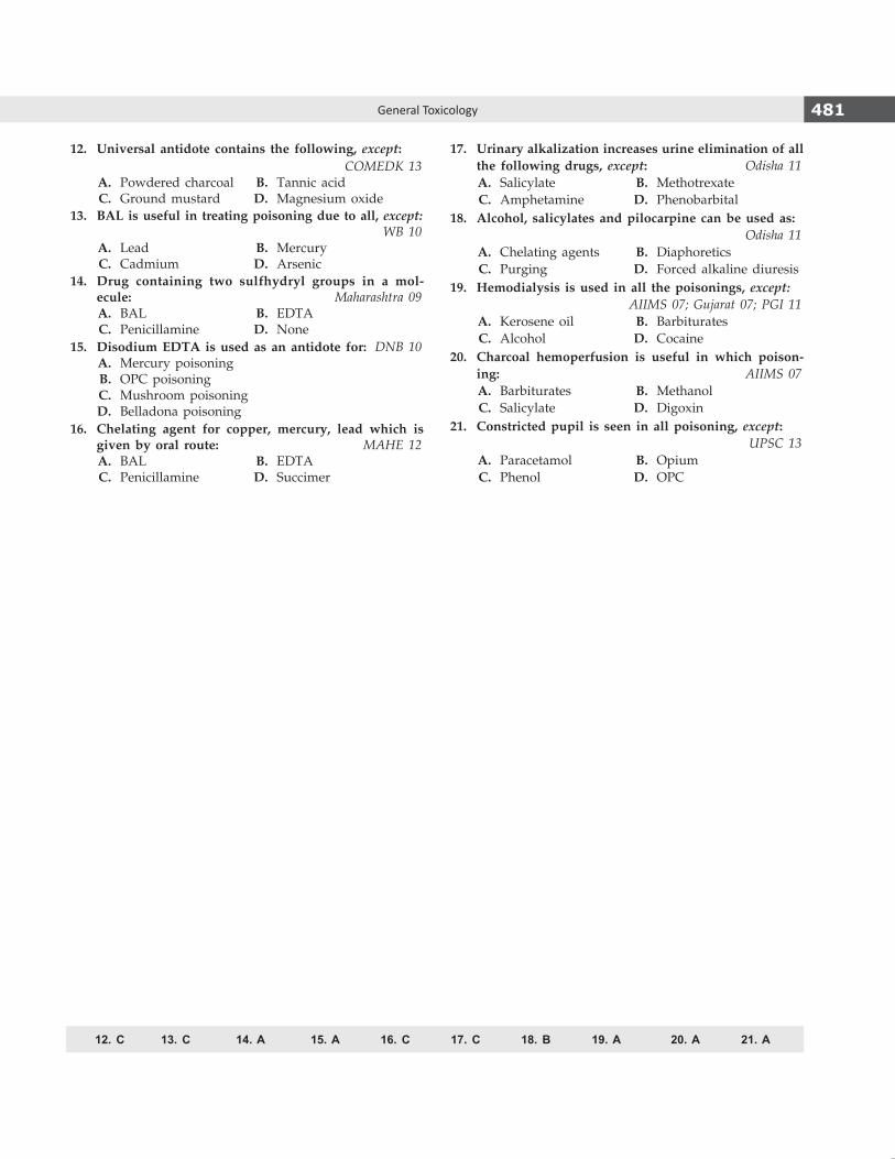
481General Toxicology
12. c 13. c 14. A 15. A 16. c 17. c 18. B 19. A 20. A 21. A
12. Universal antidote contains the following, except: COMEDK 13 A. Powdered charcoal B. Tannic acid C. Ground mustard D. Magnesium oxide 13. BAL is useful in treating poisoning due to all, except:
WB 10 A. Lead B. Mercury C. Cadmium D. Arsenic 14. Drug containing two sulfhydryl groups in a mol-
ecule: Maharashtra 09 A. BAL B. EDTA C. Penicillamine D. None 15. Disodium EDTA is used as an antidote for: DNB 10 A. Mercury poisoning B. OPC poisoning C. Mushroom poisoning D. Belladona poisoning 16. Chelating agent for copper, mercury, lead which is
given by oral route: MAHE 12 A. BAL B. EDTA C. Penicillamine D. Succimer
17. Urinary alkalization increases urine elimination of all the following drugs, except: Odisha 11
A. Salicylate B. Methotrexate C. Amphetamine D. Phenobarbital 18. Alcohol, salicylates and pilocarpine can be used as:
Odisha 11 A. Chelating agents B. Diaphoretics C. Purging D. Forced alkaline diuresis 19. Hemodialysis is used in all the poisonings, except:
AIIMS 07; Gujarat 07; PGI 11 A. Kerosene oil B. Barbiturates C. Alcohol D. Cocaine 20. Charcoal hemoperfusion is useful in which poison-
ing: AIIMS 07 A. Barbiturates B. Methanol C. Salicylate D. Digoxin 21. Constricted pupil is seen in all poisoning, except:
UPSC 13 A. Paracetamol B. Opium C. Phenol D. OPC

482 Review of Forensic Medicine and Toxicology
Mineral / Inorganic Acids
Introduction
� Mineral acids produce coagulative necrosis, precipitate proteins with resultant hard eschar or scab (which may protect the underlying tissue from further damage), have no remote action and act as irritants when slightly diluted, but as stimulants when well diluted.1
� Acids usually causes second degree, deep partial thickness burns, tend to be clearly demarcated and are dry, hard and mildly edematous.
� The stomach is the most commonly involved organ following an acid ingestion. This may due to some natural protection of the esophageal squamous epithelium.
Common acids are:1
� Sulphuric acid (oil of vitriol, H2SO4) � Nitric acid (aqua fortis, HNO3) � Hydrochloric acid (HCl)
The signs and symptoms, fatal dose, fatal period, postmortem appearances and medico-legal aspects of these acids are given below in tabulated form.
Contd...
Features H2SO4 HNO3 HCl
Physical properties Colorless, odorless, burning taste, oily, non-fuming
Colorless, pungent, choking, burning taste, fuming
Colorless, pungent, sour, burning taste, fuming
Action
� Local Corrosive CorrosiveRespiratory distress (inhalation)
CorrosiveRespiratory tract inflammation
� Indirect � Shock, asphyxia � Perforation of stomach � Chemical peritonitis � Esophageal stricture
Pain → circulatory failure Pain → circulatory failure
Fatal dose (concentrated) 5–10 ml 10–15 ml 15–20 ml
Fatal period 12–18 hours (h) 12–24 h 18–30 h
Signs and symptoms
� Oropharyngeal burns and burning pain in throat, epigastrium
Present Present Present
� Dysphagia, dysphonia and dyspnea Present Present Present
� Eructation, vomiting Present Present Present
� Thirst Present, drinking causes more vomiting
Same Same
� Vomitus Strongly acidic, with altered blood and mucous shreds
Same Same
� Teeth Chalky white, brittle Yellowish coating, not brittle No change
� Constipation Usually present Same Same
� Urination Suppressed Same Same
� Tenesmus Present Present May be present
� Nature of stool Mucus, altered blood Same Same
Corrosive Poisons
ChaPter 37

483Corrosive Poisons
Treatment
i. Avoid gastric lavage or emetics. ii. Acid should be immediately diluted by giving a
glass of milk or water to drink, and 4 tablespoonfuls of aluminum hydroxide gel.
iii. Give a demulcent, like olive oil, milk, egg white, starch water or butter.
iv. Do not give bicarbonate or other neutralizing agents.
v. Prednisolone 60 mg/day may be given in divided doses.
vi. Correct circulatory shock, IV fluids and bloodproducts are administered in the event of significantbleedingorvomiting.Antibioticsshouldbe given, if evidence of perforation exists.
vii. Tracheostomy, if there is edema of glottis.
viii. Give nothing by mouth. Nutrient substances are given by IV route for about a week. Then, try liquids, soft foodandfinallya regulardiet.
ix. Morphine to relieve pain. x. Symptomatic treatment.
Skin burns are washed with large amounts of water for 15 minutes (min). No chemical antidotes are used as the heat of the reaction may cause additional injury. For hydrofluoricacidburn,soaktheareainbenzalkoniumchloride solution or apply 2.5% calcium gluconate gel.
Complications: Delayed perforation may occur as many as 4 days after an acid exposure. Delayed upper GI bleeding may occur in acid burns 3-4 days after exposure as the eschar sloughs. Gastric outlet obstruction may develop 3-4 weeks after an acid exposure.
Contd...
Features H2SO4 HNO3 HCl
� Tenderness over abdomen Present Present Present
� Stiffness of abdomen Present, due to peritonitis Present, due to distension Present, due to distension
� Abdominal distension Not usual Present, due to gas in stomach Present, due to gas in stomach
� Erosion of skin, mucous membrane of mouth and tongue
Over angles of mouth, lips, fingers with blackening, excoriation
Over angles of mouth, lips, fingers with yellow discoloration
Usually no erosion, epidermis may fall off after few days
� Perforation of stomach Common Less common Less common
Cause of death � Shock � Perforation of stomach � Peritonitis � Laryngeal spasm � Malnutrition (due to esophageal
stricture)
� Shock � Perforation of stomach � Peritonitis � Laryngeal spasm � Respiratory distress
� Shock � Laryngeal spasm � Pulmonary edema (due to
inhalation of vapor)
Postmortem findings � Erosion of skin, angles of mouth, lips
� Corrosion of the trachea and larynx
� Blackish charring of the stomach, peppery feel
� Perforation of the stomach � Toxic swelling of the liver and
kidneys
� Yellow discoloration of skin2
� Corrosion of skin � Larynx and trachea: congested � Stomach wall is soft, friable and
ulcerated
� Not much corrosion of skin � Brownish parchmentization � Inflammation of respiratory
passages � Stomach contains brownish
fluid
Medico-legal aspects � Accidental, mistaking it for glycerin
� Suicidal: common
� Homicide is rare
� Abortifacient
� Vitriolage
� Accidental or suicidal
� Homicide is rare
� Mostly suicidal � Accidental: few � Homicide and abortifacient:
rare

484 Review of Forensic Medicine and Toxicology
Preservative: Viscera and skin are preserved in absolute alcoholorrectifiedspiritandtheclothesaresentwithoutany preservatives.
� Abandonment of neutralizing agents for caustic ingestion: Earlier, recommendations for the treatment of acid ingestion included the use of magnesium hydroxide, lime water or calcium carbonate, and for alkali ingestions included vinegar (acetic acid), lemon juice or dilute HCl. However, due to the rapid onset of action of corrosive agents, it may be too late to reverse the caustic process. Furthermore, the addition of neutralizing agents could increase the potential for a consequential exothermic reaction and/or gas production. Such reaction in an already weakened hollow viscus may lead to extension of the tissue injury or perforation. Hence, the use of neutralizing agents is no longer recommended.
� Magenstrasse (street of the stomach) is the term applied to the pathway corrosive agents follow in stomach (Figs 37.1A to C).3 The pathway in food-filled stomach is along the lesser curvature and leads to pylorus which explains the location of greatest damage. Stomach without food has significant injury in the lower half or two thirds, and may spare the fundus.
Vitriolage (Vitriol Throwing)
Definition: It is the throwing of any corrosive, not necessarily sulphuric acid, on a person with malicious intent. Sulphuric acid is most commonly used for this purpose, hence it is called vitriolage.
Other substances used: Nitric acid, carbolic acid, caustic soda, caustic potash, iodine, marking nut juice or calotropis.
Characteristics of Burns
i. Discoloration and staining of the skin and clothings (brown or black in sulphuric acid, and yellow in nitric acid).
ii. Trickle marks. iii. Painless burns with absence of vesication and red
line of demarcation. iv. Presence of chemical substance in the stains. v. Repair is slow, and scar tissue causes contractures.
Treatment
i. Wash the parts with plenty of water and soap. ii. Apply thick paste of MgO or carbonate.4
iii. Cover raw surface with antibiotic ointment. iv. For eye burns, the conjunctiva and corneal surfaces
are anesthetized with topical anesthetic drops (e.g. proparacaine) and irrigated with water for 15 min holding the eyelids open. Repeat irrigation using 0.9% saline, till pH is near 7.0. Eye drops containing antibiotics and steroids are helpful.
Medico-legal Aspects
� These fluids are usually thrown on the face withthe object of destroying vision or causing facial disfigurement, and this results in grievous hurt (Sec. 320 IPC).
� It is punishable under Sec. 326-A IPC (voluntarily causing grievous hurt by use of acids) for 10 years to life imprisonment and fine paid to the victim,and under Sec 326-B IPC (voluntarily throwing or attempting to throw acid) with imprisonment for 5-7yearsandfine.5
Chemical Colitis
Definition: Chemical colitis is characterized by an inflammationofthecolonduetotheexposureofcolonicmucosa to various toxic chemicals.
Agents involved: Sulphuric/hydrochloric acid, acetic acid, sodium hydroxide, hydrogen peroxide, alcohol, radiocontrast agents, glutaraldehyde, formalin, ergotamine,hydrofluoricacid,householddisinfectants,ammonia, soap, herbal medicines and potassium permanganate.
Signs and symptoms: Intermittent abdominal pain, fecal incontinence, severe diarrhea, and hematochezia (passage of fresh blood in stools). However, severe mucosal injury may be associated with peritonitis, ischemic colitis, colonic strictures, and rectovaginalfistulas.
Treatment: Discontinuation of exposure to the toxic agent, fluid resuscitation, broad-spectrum antibiotics,steroids, bowel rest, if there is evidence of necrosis—resection.
Medico-legal Aspects � Chemical colitis may be accidental, deliberate
(suicidal, homicidal or sexual), or iatrogenic (prescribed medication, contamination of endoscopic instruments).
Figs 37.1A to C: A. Pathway of corrosive agent in stomach, B. Damage in filled stomach (shaded area), C. Damage in empty stomach
A B C

485Corrosive Poisons
� High risk individual include those with mental illness, depression, Munchausen’s syndrome, learning difficulties, and certain groups who use enemasregularly.
Boric acid (Hydrogen borate/Orthoboric acid)Boric acid is a weak acid of boron which is used as an antiseptic, insecticide (especially for cockroaches), flame retardant, as a neutron absorber and as a precursor of other chemical compounds. Boric acid crystals are white, odorless, nearly tasteless and dissolves in water.Metabolism: Boric acid is not metabolized; it is eliminated unchanged in the urine.Signs and symptoms
System Signs and symptomsGIT Nausea, vomiting, diarrhea and occasionally crampy
abdominal pain—may be confused with acute gastroenteritis. Emesis and diarrhea may be bluish-green.
Dermal Generalized erythema creating a ‘boiled lobster’ appearance with massive areas of desqua mation indistinguishable from toxic epidermal necrolysis or staphylococcal scaled skin syndrome in the neonate. Rash is particularly seen on the palms, soles and buttocks.6
CNS Irritability, seizures, delirium and coma may occur.Renal Oliguria, renal tubular damage and elevated serum
creatinine.
CVS Tachycardia, hypotension.Other Hepatic injury, hyperthermia.
Death results from circulatory collapse.Fatal dose: 15-20 g in adults; 5-6 g in children and 2-3 g for infants.Treatment: Supportive treatment. Activated charcoal is not recommended because of its poor adsorptive capacity for boric acid. Hemodialysis and exchange transfusion may be helpful.Medico-legal: Because of the wide availability of boric acid, accidental intake by children occurs frequently. It may be taken by mistake and suicidal purposes. The abandonment of boric acid as an irritant and particularly its removal from nursery setting (for treatment of napkin dermatitis) have led to a marked decrease in the incidence of significant boric acid poisoning.
OrgAnIC ACIds
Oxalic Acid (Acid of Sugar, C2H2O4)
Physical properties: Colorless, transparent, prismatic crystals and resembles MgSO4 and ZnSO4. Oxalic acid is a naturally occurring component of plants and is found in relatively high levels in dark-green leafy foods, e.g. beet leaves, purslane, spinach, rhubarb and parsley.7
Uses: Oxalic acid forms soluble chelates with iron. This property makes it useful for removing blood and rust stains, cleaning metals other than iron and flushingcar radiators. It is used in many chemical processes like bleaching and dyeing. It may be used to remove signatures/writings from documents in cases of forgery.
Action: It acts locally as a corrosive on both skin and mucosa, and remotely affects several systems after being absorbed.
� CVS → shock → death � Electrolyte system → extracts tissue calcium →
hypocalcemia � Renal system → tubular necrosis → uremia → death
signs and symptoms
Poisoning presents in three forms: i. Fulminating poisoning: Intake of large dose
(> 15 g) produces immediate symptoms and death within minutes. There is a burning, sour or bitter taste in the mouth with a sense of constriction around the throat and burning pain from the mouth to stomach, radiating all over the abdomen. Nausea and eructation are immediately followed by vomiting which contains altered blood, mucus and has a coffee-ground appearance (black in color). Severe thirst, diarrhea, electrolyte imbalance, and ultimately death occurs.
ii. Acute poisoning: All findings are due to hypocalcemia—tinglingandnumbnessoffingersand limbs, weakness, parasthesia, carpopedal spasm*, hyperirritability of peripheral nerves (Chvostek/Weiss sign), tetany, convulsions, coma and death. There may be dilated pupils, metabolic acidosis, ventricular fibrillation, andrenal failure.8,9
iii. Delayed poisoning: It is characterized by nephri-tis—uremia, scanty urine, hematuria, albuminuria, oxaluria (envelope-shaped calcium oxalate crystals in the urine is seen under microscope).
Fatal dose: 15–20 g.
Fatal period: 1–2 h. In case of renal failure, death may occur between 2 days to 2 weeks.
Treatment
i. Gastric lavage with calcium lactate (2 teaspoons/lavage).
* Thehands areflexed at thewrist, fingers areflexed at themetacarpophalangeal joints and the thumbadducted to thepalm—so calledAccoucher’s hand (Trousseau’s sign).9
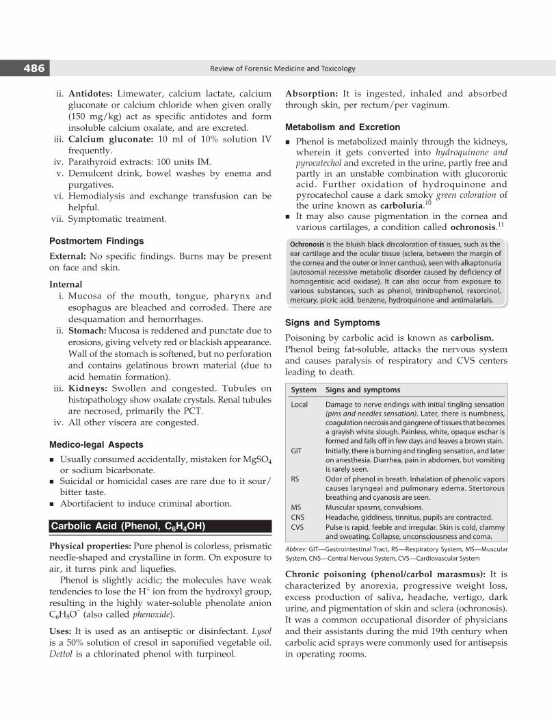
486 Review of Forensic Medicine and Toxicology
Absorption: It is ingested, inhaled and absorbed through skin, per rectum/per vaginum.
Metabolism and Excretion � Phenol is metabolized mainly through the kidneys,
wherein it gets converted into hydroquinone and pyrocatechol and excreted in the urine, partly free and partly in an unstable combination with glucoronic acid. Further oxidation of hydroquinone and pyrocatechol cause a dark smoky green coloration of the urine known as carboluria.10
� It may also cause pigmentation in the cornea and various cartilages, a condition called ochronosis.11
Ochronosis is the bluish black discoloration of tissues, such as the ear cartilage and the ocular tissue (sclera, between the margin of the cornea and the outer or inner canthus), seen with alkaptonuria (autosomal recessive metabolic disorder caused by deficiency of homogentisic acid oxidase). It can also occur from exposure to various substances, such as phenol, trinitrophenol, resorcinol, mercury, picric acid, benzene, hydroquinone and antimalarials.
signs and symptoms
Poisoning by carbolic acid is known as carbolism.Phenol being fat-soluble, attacks the nervous system and causes paralysis of respiratory and CVS centers leading to death.
Abbrev: GIT—Gastrointestinal Tract, RS—Respiratory System, MS—Muscular System, CNS—Central Nervous System, CVS—Cardiovascular System
System Signs and symptoms
Local Damage to nerve endings with initial tingling sensation (pins and needles sensation). Later, there is numbness, coagulation necrosis and gangrene of tissues that becomes a grayish white slough. Painless, white, opaque eschar is formed and falls off in few days and leaves a brown stain.
GIT Initially, there is burning and tingling sensation, and later on anesthesia. Diarrhea, pain in abdomen, but vomiting is rarely seen.
RS Odor of phenol in breath. Inhalation of phenolic vapors causes laryngeal and pulmonary edema. Stertorous breathing and cyanosis are seen.
MS Muscular spasms, convulsions.CNS Headache, giddiness, tinnitus, pupils are contracted.CVS Pulse is rapid, feeble and irregular. Skin is cold, clammy
and sweating. Collapse, unconsciousness and coma.
Chronic poisoning (phenol/carbol marasmus): It is characterized by anorexia, progressive weight loss, excess production of saliva, headache, vertigo, dark urine, and pigmentation of skin and sclera (ochronosis). It was a common occupational disorder of physicians and their assistants during the mid 19th century when carbolic acid sprays were commonly used for antisepsis in operating rooms.
ii. Antidotes: Limewater, calcium lactate, calcium gluconate or calcium chloride when given orally (150 mg/kg) act as specific antidotes and forminsoluble calcium oxalate, and are excreted.
iii. Calcium gluconate: 10 ml of 10% solution IV frequently.
iv. Parathyroid extracts: 100 units IM. v. Demulcent drink, bowel washes by enema and
purgatives. vi. Hemodialysis and exchange transfusion can be
helpful. vii. Symptomatic treatment.
Postmortem Findings
External: No specific findings. Burnsmay be presenton face and skin.
Internal i. Mucosa of the mouth, tongue, pharynx and
esophagus are bleached and corroded. There are desquamation and hemorrhages.
ii. Stomach: Mucosa is reddened and punctate due to erosions, giving velvety red or blackish appearance. Wall of the stomach is softened, but no perforation and contains gelatinous brown material (due to acid hematin formation).
iii. Kidneys: Swollen and congested. Tubules on histopathology show oxalate crystals. Renal tubules are necrosed, primarily the PCT.
iv. All other viscera are congested.
Medico-legal Aspects � Usually consumed accidentally, mistaken for MgSO4
or sodium bicarbonate. � Suicidal or homicidal cases are rare due to it sour/
bitter taste. � Abortifacient to induce criminal abortion.
Carbolic Acid (Phenol, C6H4OH)
Physical properties: Pure phenol is colorless, prismatic needle-shaped and crystalline in form. On exposure to air, it turnspinkand liquefies.
Phenol is slightly acidic; the molecules have weak tendencies to lose the H+ ion from the hydroxyl group, resulting in the highly water-soluble phenolate anion C6H5O
_ (also called phenoxide).
Uses: It is used as an antiseptic or disinfectant. Lysol isa50%solutionofcresol insaponifiedvegetableoil.Dettol is a chlorinated phenol with turpineol.
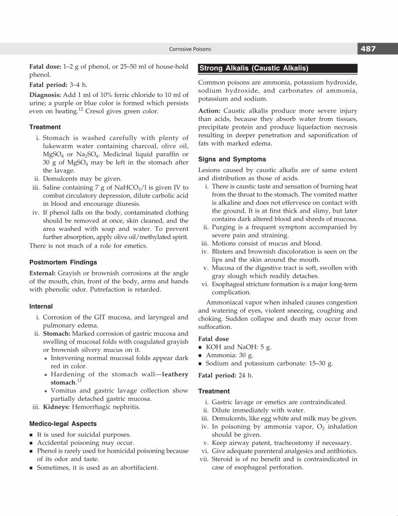
487Corrosive Poisons
Fatal dose: 1–2 g of phenol, or 25–50 ml of house-hold phenol.
Fatal period: 3–4 h.
Diagnosis: Add 1 ml of 10% ferric chloride to 10 ml of urine; a purple or blue color is formed which persists even on heating.12 Cresol gives green color.
Treatment
i. Stomach is washed carefully with plenty of lukewarm water containing charcoal, olive oil, MgSO4 or Na2SO4. Medicinal liquid paraffin or30 g of MgSO4 may be left in the stomach after the lavage.
ii. Demulcents may be given. iii. Saline containing 7 g of NaHCO3/l is given IV to
combat circulatory depression, dilute carbolic acid in blood and encourage diuresis.
iv. If phenol falls on the body, contaminated clothing should be removed at once, skin cleaned, and the area washed with soap and water. To prevent further absorption, apply olive oil/methylated spirit.
There is not much of a role for emetics.
Postmortem Findings
External: Grayish or brownish corrosions at the angle of the mouth, chin, front of the body, arms and hands with phenolic odor. Putrefaction is retarded.
Internal
i. Corrosion of the GIT mucosa, and laryngeal and pulmonary edema.
ii. Stomach: Marked corrosion of gastric mucosa and swelling of mucosal folds with coagulated grayish or brownish silvery mucus on it.
z Intervening normal mucosal folds appear dark red in color.
z Hardening of the stomach wall—leathery stomach.13
z Vomitus and gastric lavage collection show partially detached gastric mucosa.
iii. Kidneys: Hemorrhagic nephritis.
Medico-legal Aspects
� It is used for suicidal purposes. � Accidental poisoning may occur. � Phenol is rarely used for homicidal poisoning because
of its odor and taste. � Sometimes, it is used as an abortifacient.
strong Alkalis (Caustic Alkalis)
Common poisons are ammonia, potassium hydroxide, sodium hydroxide, and carbonates of ammonia, potassium and sodium.
Action: Caustic alkalis produce more severe injury than acids, because they absorb water from tissues, precipitate protein and produce liquefaction necrosis resulting in deeper penetration and saponification offats with marked edema.
signs and symptoms
Lesions caused by caustic alkalis are of same extent and distribution as those of acids. i. There is caustic taste and sensation of burning heat
from the throat to the stomach. The vomited matter is alkaline and does not effervesce on contact with theground.It isatfirstthickandslimy,butlatercontains dark altered blood and shreds of mucosa.
ii. Purging is a frequent symptom accompanied by severe pain and straining.
iii. Motions consist of mucus and blood. iv. Blistersandbrownishdiscolorationisseenonthe
lips and the skin around the mouth. v. Mucosa of the digestive tract is soft, swollen with
gray slough which readily detaches. vi. Esophageal stricture formation is a major long-term
complication.
Ammoniacal vapor when inhaled causes congestion and watering of eyes, violent sneezing, coughing and choking. Sudden collapse and death may occur from suffocation.
Fatal dose � KOH and NaOH: 5 g. � Ammonia: 30 g. � Sodium and potassium carbonate: 15–30 g.
Fatal period: 24 h.
Treatment
i. Gastric lavage or emetics are contraindicated. ii. Dilute immediately with water. iii. Demulcents, like egg white and milk may be given. iv. In poisoning by ammonia vapor, O2 inhalation
should be given. v. Keep airway patent, tracheostomy if necessary. vi. Give adequate parenteral analgesics and antibiotics.vii. Steroid is of no benefit and is contraindicated in
case of esophageal perforation.
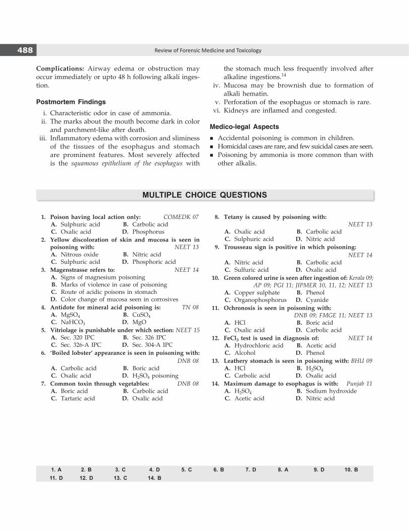
488 Review of Forensic Medicine and Toxicology
1. A 2. B 3. C 4. D 5. C 6. B 7. D 8. A 9. D 10. B 11. D 12. D 13. C 14. B
Complications: Airway edema or obstruction may occur immediately or upto 48 h following alkali inges- tion.
Postmortem Findings
i. Characteristic odor in case of ammonia. ii. The marks about the mouth become dark in color
and parchment-like after death. iii. Inflammatoryedemawithcorrosionandsliminess
of the tissues of the esophagus and stomach are prominent features. Most severely affected is the squamous epithelium of the esophagus with
the stomach much less frequently involved after alkaline ingestions.14
iv. Mucosa may be brownish due to formation of alkali hematin.
v. Perforation of the esophagus or stomach is rare. vi. Kidneysare inflamedandcongested.
Medico-legal Aspects
� Accidental poisoning is common in children. � Homicidal cases are rare, and few suicidal cases are seen. � Poisoning by ammonia is more common than with
other alkalis.
1. Poison having local action only: COMEDK 07 A. Sulphuric acid B. Carbolic acid C. Oxalic acid D. Phosphorus 2. Yellow discoloration of skin and mucosa is seen in
poisoning with: NEET 13 A. Nitrous oxide B. Nitric acid C. Sulphuric acid D. Phosphoric acid 3. Magenstrasse refers to: NEET 14 A. Signs of magnesium poisoning B. Marks of violence in case of poisoning C. Route of acidic poisons in stomach D. Color change of mucosa seen in corrosives 4. Antidote for mineral acid poisoning is: TN 08 A. MgSO4 B. CuSO4 C. NaHCO3 D. MgO 5. Vitriolage is punishable under which section: NEET 15 A. Sec. 320 IPC B. Sec. 326 IPC C. Sec. 326-A IPC D. Sec. 304-A IPC 6. ‘Boiled lobster’ appearance is seen in poisoning with: DNB 08 A. Carbolic acid B. Boric acid C. Oxalic acid D. H2SO4 poisoning 7. Common toxin through vegetables: DNB 08 A. Boric acid B. Carbolic acid C. Tartaric acid D. Oxalic acid
8. Tetany is caused by poisoning with: NEET 13
A. Oxalic acid B. Carbolic acid C. Sulphuric acid D. Nitric acid 9. Trousseau sign is positive in which poisoning: NEET 14 A. Nitric acid B. Carbolic acid C. Sulfuric acid D. Oxalic acid 10. Green colored urine is seen after ingestion of: Kerala 09;
AP 09; PGI 11; JIPMER 10, 11, 12; NEET 13 A. Copper sulphate B. Phenol C. Organophosphorus D. Cyanide 11. Ochronosis is seen in poisoning with:
DNB 09; FMGE 11; NEET 13 A. HCl B. Boric acid C. Oxalic acid D. Carbolic acid 12. FeCl3 test is used in diagnosis of: NEET 14 A. Hydrochloric acid B. Acetic acid C. Alcohol D. Phenol 13. Leathery stomach is seen in poisoning with: BHU 09 A. HCl B. H2SO4 C. Carbolic acid D. Oxalic acid 14. Maximum damage to esophagus is with: Punjab 11 A. H2SO4 B. Sodium hydroxide C. Acetic acid D. Nitric acid
multiple ChoiCe questions

Introduction
Physical properties: Metallic arsenic (black in color) is not poisonous, as it is not absorbed from the GIT. It is a normal constituent of all animal tissues, in minute amounts.
Toxic Compounds and its Uses
i. Arsenious oxide or arsenic trioxide (Fig. 38.1A) (sankhya, somalkhar, white arsenic or arsenic): Most toxic form of arsenic. It has no taste or smell and is sparingly soluble in water. It is used in fruit sprays, sheep-dips, weed-killers, insecticides, rat poisons, flypapers, calico-printing, wallpapers, artificial flowers and as mordant in dyeing.
ii. Copper arsenite (Scheele’s green) and copper acetoarsenite (Paris green or emerald green) (Fig. 38.1B): It is used as coloring agent for substances including confectionary.
iii. Sodium and potassium arsenate. iv. Arsenic sulfide: Yellow orpiment (hartal) or arsenic
trisulfide, and red realgar or arsenic disulfide are used as depilatory, coloring pigment and in flypaper.
v. Arseniuretted hydrogen or arsine is a colorless gas with garlic-like odor.
vi. The natural sources of arsenic are soil, water and some sea fish (mussels, prawns). High arsenic content of soil and subsoil water of some places is the cause of endemic toxicity (from shallow tube-wells inserted for drinking water).
vii. Tobacco smoke, particularly cigars also contain arsenic, and in some beers as impurities.
Action
� Arsenic interferes with cellular respiration by uncoupling mitochondrial oxidative phosphorylation by combining with the sulfhydryl groups of mitochondrial enzymes, especially pyruvate dehydro -genase and certain phosphatases. Conse quently, conversion of pyruvate to acetyl CoA is decreased, citric acid cycle activity is decreased and production of cellular ATP is decreased.
� It inhibits cellular glucose uptake, gluconeogenesis, fatty acid oxidation and further production of acetyl CoA.
� Locally, it causes irritation of the mucous membranes, and remotely, depression of the nervous system.
� Arsenic is a carcinogenic substance since lung, skin and bladder (transitional cell) carcinoma has been observed in populations with multiple exposures.1
Absorption and Excretion
It is absorbed orally through the GIT, skin and lungs (arsine) or parenterally.
� It is present in almost all tissues, and found in the greatest quantity in the liver, followed by kidneys and spleen.2
� In cases, where the patient survives, it is found in the muscles (for months), bones, hair, nails and skin (sulfhydryl groups in keratin) for years. Normally, the hair contains < 2 parts/million arsenic.
It is excreted mainly by the kidneys (urine), but some part through feces, bile, sweat, milk, nails and hair.
� The arsenic is excreted in the hair and nails within few hours of ingestion, and in cases of intermittent chronic poisoning there will be successive deposits of arsenic in the hair and nails.
� Arsenic is secreted into the stomach and intestines after absorption, even when given by routes other than mouth.
Inorganic Metallic Irritants—Arsenic
CHAPTER 38
Figs 38.1A and B: (A) Arsenious oxide, (B) Copper acetoarsenite
A B
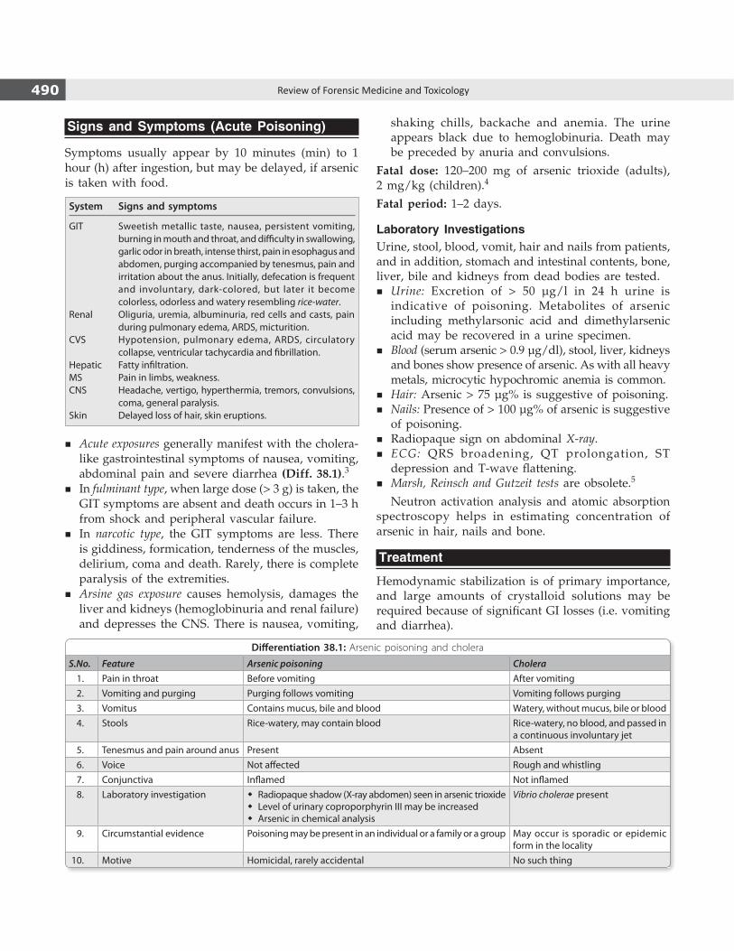
490 Review of Forensic Medicine and Toxicology
Signs and Symptoms (Acute Poisoning)
Symptoms usually appear by 10 minutes (min) to 1 hour (h) after ingestion, but may be delayed, if arsenic is taken with food.
System Signs and symptoms
GIT Sweetish metallic taste, nausea, persistent vomiting, burning in mouth and throat, and difficulty in swallowing, garlic odor in breath, intense thirst, pain in esophagus and abdomen, purging accompanied by tenesmus, pain and irritation about the anus. Initially, defecation is frequent and involuntary, dark-colored, but later it become colorless, odorless and watery resembling rice-water.
Renal Oliguria, uremia, albuminuria, red cells and casts, pain during pulmonary edema, ARDS, micturition.
CVS Hypotension, pulmonary edema, ARDS, circulatory collapse, ventricular tachycardia and fibrillation.
Hepatic Fatty infiltration.MS Pain in limbs, weakness.CNS Headache, vertigo, hyperthermia, tremors, convulsions,
coma, general paralysis.Skin Delayed loss of hair, skin eruptions.
� Acute exposures generally manifest with the cholera-like gastrointestinal symptoms of nausea, vomiting, abdominal pain and severe diarrhea (Diff. 38.1).3
� In fulminant type, when large dose (> 3 g) is taken, the GIT symptoms are absent and death occurs in 1–3 h from shock and peripheral vascular failure.
� In narcotic type, the GIT symptoms are less. There is giddiness, formication, tenderness of the muscles, delirium, coma and death. Rarely, there is complete paralysis of the extremities.
� Arsine gas exposure causes hemolysis, damages the liver and kidneys (hemoglobinuria and renal failure) and depresses the CNS. There is nausea, vomiting,
shaking chills, backache and anemia. The urine appears black due to hemoglobinuria. Death may be preceded by anuria and convulsions.
Fatal dose: 120–200 mg of arsenic trioxide (adults), 2 mg/kg (children).4
Fatal period: 1–2 days.
Laboratory InvestigationsUrine, stool, blood, vomit, hair and nails from patients, and in addition, stomach and intestinal contents, bone, liver, bile and kidneys from dead bodies are tested.
� Urine: Excretion of > 50 µg/l in 24 h urine is indicative of poisoning. Metabolites of arsenic including methylarsonic acid and dimethylarsenic acid may be recovered in a urine specimen.
� Blood (serum arsenic > 0.9 µg/dl), stool, liver, kidneys and bones show presence of arsenic. As with all heavy metals, microcytic hypochromic anemia is common.
� Hair: Arsenic > 75 µg% is suggestive of poisoning. � Nails: Presence of > 100 µg% of arsenic is suggestive
of poisoning. � Radiopaque sign on abdominal X-ray. � ECG: QRS broadening, QT prolongation, ST
depression and T-wave flattening. � Marsh, Reinsch and Gutzeit tests are obsolete.5
Neutron activation analysis and atomic absorption spectroscopy helps in estimating concentration of arsenic in hair, nails and bone.
Treatment
Hemodynamic stabilization is of primary importance, and large amounts of crystalloid solutions may be required because of significant GI losses (i.e. vomiting and diarrhea).
Differentiation 38.1: Arsenic poisoning and cholera
S.No. Feature Arsenic poisoning Cholera1. Pain in throat Before vomiting After vomiting2. Vomiting and purging Purging follows vomiting Vomiting follows purging3. Vomitus Contains mucus, bile and blood Watery, without mucus, bile or blood4. Stools Rice-watery, may contain blood Rice-watery, no blood, and passed in
a continuous involuntary jet5. Tenesmus and pain around anus Present Absent6. Voice Not affected Rough and whistling7. Conjunctiva Inflamed Not inflamed8. Laboratory investigation � Radiopaque shadow (X-ray abdomen) seen in arsenic trioxide
� Level of urinary coproporphyrin III may be increased � Arsenic in chemical analysis
Vibrio cholerae present
9. Circumstantial evidence Poisoning may be present in an individual or a family or a group May occur is sporadic or epidemic form in the locality
10. Motive Homicidal, rarely accidental No such thing
491Inorganic Metallic Irritants—Arsenic
i. Gastric lavage is done repeatedly with large amount of warm water and milk; activated charcoal does not adsorb arsenic appreciably and is not recommended in patients whom co-ingestants are not suspected.
ii. Demulcents (butter or greasy substances) prevent absorption.
iii. Whole bowel irrigation with polyethylene glycol may be effective to prevent GIT absorption of arsenic.
iv. Antidote is BAL or dimercaprol, given in a dose of 3–5 mg/kg IM 4 hourly for 2 days, 6 hourly for 1 day and then 12 hourly for 10 days. Oral succimer (DMSA), 10 mg/kg every 8 hourly for 10 days or dimerval (DMPS, drug of choice for treating most heavy metal poisonings), 200 mg IV 4 hourly until oral product can be given in a dose of 100 mg TDS or QID may be used instead of BAL.
v. Alkalis should not be given by mouth as they increase the solubility of arsenic.
vi. Purgatives (castor oil/magnesium sulfate) are given to remove unabsorbed poison from intestine.
vii. Glucose-saline with sodium bicarbonate is helpful to combat shock and improve alkali reserve.
viii. Hemodialysis or exchange transfusion may be done.
Earlier, freshly precipitated ferric hydroxide (antidotum arsenici) was used for stomach wash in the treatment of arsenic poisoning which formed ferric arsenite, is no longer recommended.
Postmortem Findings
External i. The body looks emaciated due to dehydration. ii. Rigor mortis appears early. iii. Putrefaction is delayed due to anti-bacterial action
of arsenic and partly due to dehydration.6
iv. The eyeballs are sunken and the skin is cyanosed. v. Blood tinged vomitus and fecal matter may be
present on body and clothes.
Internal i. The mucous membrane of the mouth, pharynx and
esophagus may show inflammation or ulceration. ii. Hemorrhages may be found in the abdominal
organs and mesentery, and occasionally in the larynx, trachea and lungs.
iii. Lungs: Congested, pulmonary edema with subpleural ecchymoses.
iv. Heart: Subendocardial petechial hemorrhages of the ventricle may be found, even when the stomach shows little signs of irritation.
v. Stomach: Mucosa is swollen, edematous, desqua mated and red, either generally or in patches, especially in the pyloric region. Usually, groups of petechiae are seen scattered over the mucosa, but sometimes large submucosal and subperitoneal hemorrhages may be seen—red velvety appearance.7 A mass of sticky mucus covers the mucosa in which particles of arsenic may be seen. Congestion is most marked along the crest of the rugae. Inflammation is more marked at the greater curvature, posterior part and the cardiac end of the stomach.
vi. Small intestine: It contains large flakes of mucus with very little fecal matter. The mucosa is pale-violet and shows signs of inflammation with submucous hemorrhages along its whole length.
vii. Cecum and rectum show slight inflammation. viii. Liver, spleen and kidneys: Congested, enlarged
and show cloudy swelling, and occasionally fatty degeneration. Nephritis, and scarring of renal cortices are seen.
ix. Brain: Edema with patchy necrosis or hemorrhagic encephalitis. The meninges are congested.
Chronic Arsenic Poisoning (Arsenicosis /Arsenicism)
Chronic arsenic poisoning may occur due to: � Recovery from an acute poisoning. � Accidental ingestion of small doses repeatedly by
those working with the metal. � Intake of food/drink in which there are traces of
arsenic (may be homicidal in nature).
Tolerance: Some people take arsenic daily as a tonic or as an aphrodisiac and they acquire tolerance to 250–300 mg or more in one dose. Such people are known as arsenophagists.
Signs and Symptoms
It is divided into four stages (Table 38.1): i. GIT disturbances ii. Catarrhal changes iii. Skin rashes iv. Nervous disturbances.
� A metallic taste, excessive salivation, and garlic odor of breath and sweat may indicate chronic arsenic poisoning.
� Melanosis and leucomelanosis with or without keratosis are the earliest symptoms of arsenicosis.
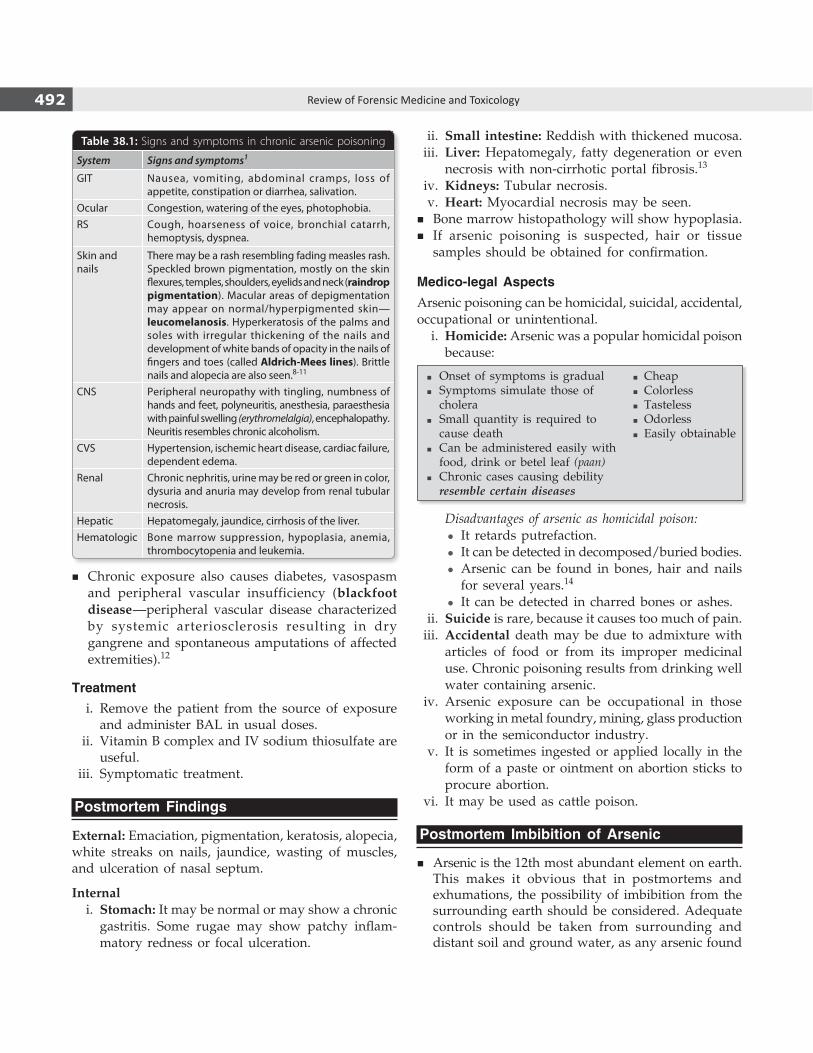
492 Review of Forensic Medicine and Toxicology
Table 38.1: Signs and symptoms in chronic arsenic poisoning
System Signs and symptoms1
GIT Nausea, vomiting, abdominal cramps, loss of appetite, constipation or diarrhea, salivation.
Ocular Congestion, watering of the eyes, photophobia. RS Cough, hoarseness of voice, bronchial catarrh,
hemoptysis, dyspnea.
Skin and nails
There may be a rash resembling fading measles rash. Speckled brown pigmentation, mostly on the skin flexures, temples, shoulders, eyelids and neck (raindrop pigmentation). Macular areas of depigmentation may appear on normal/hyperpigmented skin—leucomelanosis. Hyperkeratosis of the palms and soles with irregular thickening of the nails and development of white bands of opacity in the nails of fingers and toes (called Aldrich-Mees lines). Brittle nails and alopecia are also seen.8-11
CNS Peripheral neuropathy with tingling, numbness of hands and feet, polyneuritis, anesthesia, paraesthesia with painful swelling (erythromelalgia), encephalopathy. Neuritis resembles chronic alcoholism.
CVS Hypertension, ischemic heart disease, cardiac failure, dependent edema.
Renal Chronic nephritis, urine may be red or green in color, dysuria and anuria may develop from renal tubular necrosis.
Hepatic Hepatomegaly, jaundice, cirrhosis of the liver.Hematologic Bone marrow suppression, hypoplasia, anemia,
thrombocytopenia and leukemia.
� Chronic exposure also causes diabetes, vasospasm and peripheral vascular insufficiency (blackfoot disease—peripheral vascular disease characterized by systemic arteriosclerosis resulting in dry gangrene and spontaneous amputations of affected extremities).12
Treatment i. Remove the patient from the source of exposure
and administer BAL in usual doses. ii. Vitamin B complex and IV sodium thiosulfate are
useful. iii. Symptomatic treatment.
Postmortem Findings
External: Emaciation, pigmentation, keratosis, alopecia, white streaks on nails, jaundice, wasting of muscles, and ulceration of nasal septum.
Internal i. Stomach: It may be normal or may show a chronic
gastritis. Some rugae may show patchy inflam-matory redness or focal ulceration.
ii. Small intestine: Reddish with thickened mucosa. iii. Liver: Hepatomegaly, fatty degeneration or even
necrosis with non-cirrhotic portal fibrosis.13
iv. Kidneys: Tubular necrosis. v. Heart: Myocardial necrosis may be seen.
� Bone marrow histopathology will show hypoplasia. � If arsenic poisoning is suspected, hair or tissue
samples should be obtained for confirmation.
Medico-legal AspectsArsenic poisoning can be homicidal, suicidal, accidental, occupational or unintentional. i. Homicide: Arsenic was a popular homicidal poison
because:
� Onset of symptoms is gradual � Cheap� Symptoms simulate those of � Colorless cholera � Tasteless� Small quantity is required to � Odorless cause death � Easily obtainable� Can be administered easily with food, drink or betel leaf (paan)� Chronic cases causing debility resemble certain diseases
Disadvantages of arsenic as homicidal poison: z It retards putrefaction. z It can be detected in decomposed/buried bodies. z Arsenic can be found in bones, hair and nails
for several years.14
z It can be detected in charred bones or ashes. ii. Suicide is rare, because it causes too much of pain. iii. Accidental death may be due to admixture with
articles of food or from its improper medicinal use. Chronic poisoning results from drinking well water containing arsenic.
iv. Arsenic exposure can be occupational in those working in metal foundry, mining, glass production or in the semiconductor industry.
v. It is sometimes ingested or applied locally in the form of a paste or ointment on abortion sticks to procure abortion.
vi. It may be used as cattle poison.
Postmortem Imbibition of Arsenic
� Arsenic is the 12th most abundant element on earth. This makes it obvious that in postmortems and exhumations, the possibility of imbibition from the surrounding earth should be considered. Adequate controls should be taken from surrounding and distant soil and ground water, as any arsenic found

493Inorganic Metallic Irritants—Arsenic
1. Chronic arsenic poisoning does not cause: NEET 13,15 A. Mixed sensory and motor neuropathy B. Mesothelioma C. Hyperkeratosis of skin D. Anemia 2. In arsenic poisoning, greatest amount is found in:
PGI 06 A. Muscle B. Kidney C. Liver D. Brain 3. Cholera presents with symptoms mimicking: TN 10 A. Arsenic poisoning B. Dhatura poisoning C. Barbiturate poisoning D. Morphine poisoning 4. Fatal dose of arsenic trioxide in adults: AIIMS 14 A. 20–30 mg B. 50–60 mg C. 60–80 mg D. 120–200 mg 5. Reinsch test is used in diagnosis of poisoning due
to: UP 11; COMEDK 12 A. Arsenic B. Lead C. Iron D. Copper sulphate 6. In a suspected case of death due to poisoning where
cadaveric rigidity is lasting longer than usual, it may be a case of poisoning due to: DNB 10; Bihar 10
A. Lead B. Arsenic C. Mercury D. Copper 7. ‘Red velvety’ stomach mucosa is seen in poisoning
with: Gujarat 07; JIPMER 14 A. Mercury B. Arsenic C. Lead D. Copper
1. B 2. C 3. A 4. D 5. A 6. B 7. B 8. D 9. B 10. A 11. B 12. A 13. A 14. C
8. Arsenic causes all, except: WB 09 A. Raindrop pigmentation B. Alopecia C. Palmar hyperkeratosis D. Blue line in gums 9. A middle aged man presented with paraesthesia of
hands and feet. Examination revealed presence of ‘Mees’ lines in the nails and raindrop pigmentation in the hands. The most likely diagnosis is: AIIMS 08, 09, 11
A. Lead poisoning B. Arsenic poisoning C. Thallium poisoning D. Mercury poisoning 10. Raindrop pigmentation is seen in:
Kerala 07; BHU 10; NEET 13 A. Arsenic poisoning B. Phosphorous poisoning C. Mercury poisoning D. Thallium poisoning 11. Mees’s lines are characteristic of:
PGI 09; CMC (Ludhiana) 10,13; NEET 14 A. Mercury poisoning B. Arsenic poisoning C. Lead poisoning D. Copper poisoning 12. Blackfoot disease is caused by: NEET 14 A. Arsenic B. Cadmium C. Lead D. Mercury 13. Fatty yellow liver is seen in poisoning with: AI 08 A. Arsenic B. Aconite C. Oxalic acid D. Mercury 14. The poison that can be detected in hair/bones long
after death is: Gujarat 07; COMEDK 08; FMGE 08 A. Lead B. Mercury C. Arsenic D. Cannabis
MULTIPLE CHOICE QUESTIONS
in the body may found its way by percolation from natural sources.
� Keratin tissues absorb arsenic by contamination from outside. The concentration in hair and nails thus
contaminated is likely to be much greater than the concentration of arsenic in the contaminating fluid.
� In poisoning cases, the concentration of arsenic should be more than in the soil/ground water.
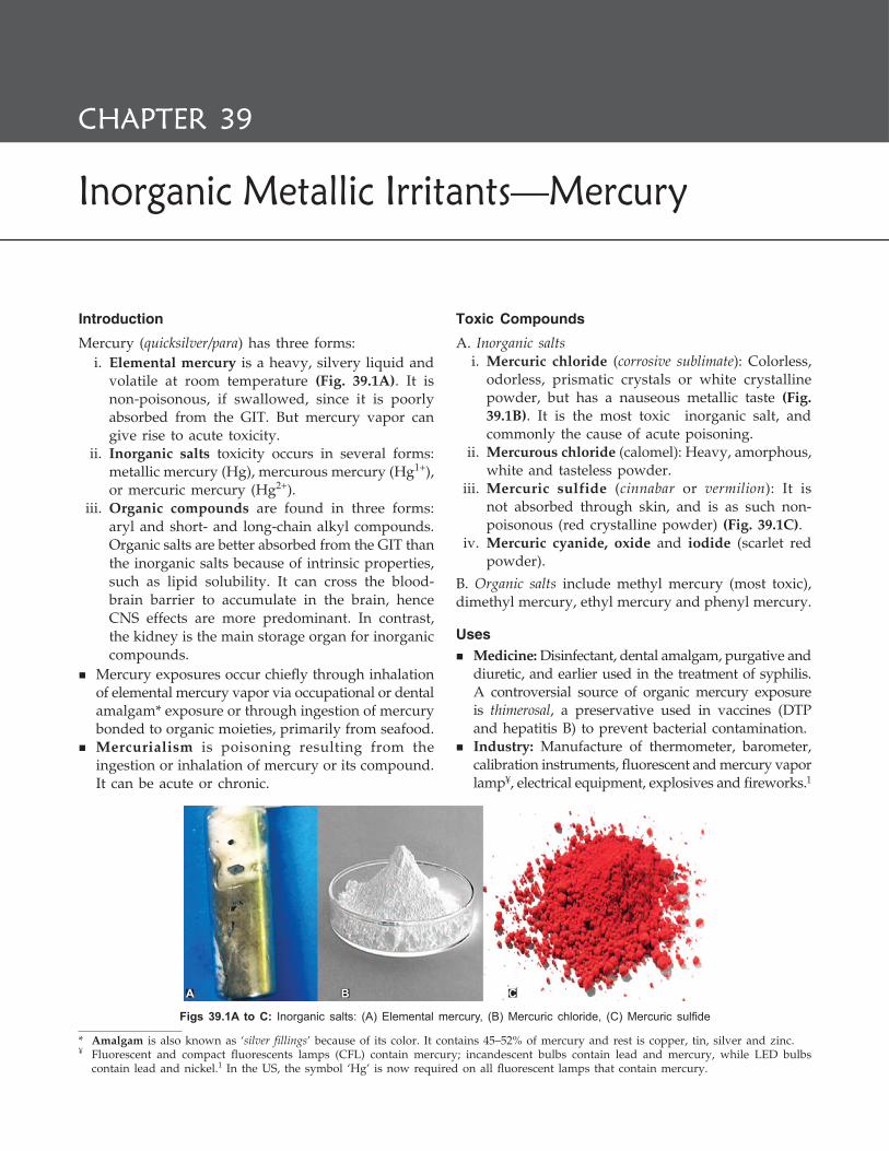
Introduction
Mercury (quicksilver/para) has three forms: i. Elemental mercury is a heavy, silvery liquid and
volatile at room temperature (Fig. 39.1A). It is non-poisonous, if swallowed, since it is poorly absorbed from the GIT. But mercury vapor can give rise to acute toxicity.
ii. Inorganic salts toxicity occurs in several forms: metallic mercury (Hg), mercurous mercury (Hg1+), or mercuric mercury (Hg2+).
iii. Organic compounds are found in three forms: aryl and short- and long-chain alkyl compounds. Organic salts are better absorbed from the GIT than the inorganic salts because of intrinsic properties, such as lipid solubility. It can cross the blood-brain barrier to accumulate in the brain, hence CNS effects are more predominant. In contrast, the kidney is the main storage organ for inorganic compounds.
� Mercury exposures occur chiefly through inhalation of elemental mercury vapor via occupational or dental amalgam* exposure or through ingestion of mercury bonded to organic moieties, primarily from seafood.
� Mercurialism is poisoning resulting from the ingestion or inhalation of mercury or its compound. It can be acute or chronic.
Toxic Compounds
A. Inorganic salts i. Mercuric chloride (corrosive sublimate): Colorless,
odorless, prismatic crystals or white crystalline powder, but has a nauseous metallic taste (Fig. 39.1B). It is the most toxic inorganic salt, and commonly the cause of acute poisoning.
ii. Mercurous chloride (calomel): Heavy, amorphous, white and tasteless powder.
iii. Mercuric sulfide (cinnabar or vermilion): It is not absorbed through skin, and is as such non-poisonous (red crystalline powder) (Fig. 39.1C).
iv. Mercuric cyanide, oxide and iodide (scarlet red powder).
B. Organic salts include methyl mercury (most toxic), dimethyl mercury, ethyl mercury and phenyl mercury.
Uses � Medicine: Disinfectant, dental amalgam, purgative and
diuretic, and earlier used in the treatment of syphilis. A controversial source of organic mercury exposure is thimerosal, a preservative used in vaccines (DTP and hepatitis B) to prevent bacterial contamination.
� Industry: Manufacture of thermometer, barometer, calibration instruments, fluorescent and mercury vapor lamp¥, electrical equipment, explosives and fireworks.1
* Amalgam is also known as ‘silver fillings’ because of its color. It contains 45–52% of mercury and rest is copper, tin, silver and zinc.¥ Fluorescent and compact fluorescents lamps (CFL) contain mercury; incandescent bulbs contain lead and mercury, while LED bulbs
contain lead and nickel.1 In the US, the symbol ‘Hg’ is now required on all fluorescent lamps that contain mercury.
Inorganic Metallic Irritants—Mercury
CHAPTER 39
Figs 39.1A to C: Inorganic salts: (A) Elemental mercury, (B) Mercuric chloride, (C) Mercuric sulfide
A B C

495Inorganic Metallic Irritants—Mercury
� Miscellaneous: Electroplating, photography, insec-ticide, germicide, fingerprint powder, paints and embalming fluid.
Action
� Mercury binds with sulfhydryl groups resulting in enzyme inhibition and pathological alteration of cellular membranes.
� Elemental mercury and methyl mercury are toxic to the CNS. Metallic mercury vapor is also a pulmonary irritant. Inorganic mercury salts are corrosive to the skin, eyes and GIT, and nephrotoxic. Inorganic and organic forms may cause contact dermatitis.
Absorption and Excretion
� It is absorbed through the GIT and respiratory tract. � After absorption, mercury gets deposited in all
tissues, particularly in the liver, kidneys, spleen and bones. When inhaled, the maximum concentration occurs in the brain.
� Mainly excreted through the kidneys (urine), liver (bile) and colonic mucous membrane (feces). It passes rapidly to the fetus through placental circula- tion.
Signs and Symptoms (Acute Poisoning)
The three types of mercury have different manifes-tations: I. Elemental (metallic) mercury: As a vapor, it is
rapidly absorbed through the lungs, reaching the blood and entering the brain. The clinical picture can be divided into three phases—initial phase manifests itself as metal fume fever, intermediate phase in which severe multiorgan symptoms are seen, and late phase when only CNS symptoms persist.
� Inhalation causes headache, nausea, cough, chest pain, bronchitis, chemical pneumonitis, pulmonary edema, gingivostomatitis, fine tremor punctuated by coarse shaking, and CNS symptoms like insomnia, ataxia, restric-tion of visual field, paresis, delirium and polyneuropathy.
� Subcutaneous nodules or granulomas are seen, if injected.
II. Ingestion of inorganic mercuric salts produces extensive precipitation of intestinal mucosal
proteins and mucosal necrosis causing bloody diarrhea and shock. If the patient survives, acute renal failure may follow.
System Signs and symptoms
GIT Metallic taste, feeling of constriction in the throat, hoarse voice.
� Mouth, tongue and fauces become corroded, swollen, and mucous membrane appears grayish white.
� Hot burning pain from the mouth to the stomach, and pain radiating over the abdomen followed by nausea, retching and vomiting. Vomitus contains grayish, slimy, mucoid material with blood and shreds of mucous membrane.
� This is followed by diarrhea, often bloody with tenesmus (gastroenteritis).
Renal Oliguria, albuminuria and hematuria ending in renal failure or nephrotic syndrome.
CVS Hypertension, tachycardia, difficulty in breathing and circulatory collapse.
III. Organic mercury: Acute exposures tend to have a latency period of one or more weeks. Symptoms typically involves the CNS such as: visual field constriction, ataxia, paresthesias, hearing loss, dysarthria, tremor, neurobehavioral impairment, paralysis and death.
Fatal dose: 1–4 g of mercuric chloride; 10–60 mg/kg of methyl mercury and 10 mg/m³ of mercury vapor.
Fatal period: 3–5 days.
Diagnosis: Acute mercury poisoning can be detected by measuring blood levels, whereas urine and hair analysis help confirming chronic exposure.
� The DMPS provoked urine challenge test is sometimes performed for chronic exposure.
� Blood mercury level > 10 µg/dl, and 24 hours (h) uri-nary excretion of mercury > 20 µg/l indicates toxicity. A hair mercury level > 5 ppm indicates chronic toxicity.
� Urine and blood mercury levels are assessed by atomic absorption spectrophotometer. Mercury concentration of hair is best assessed by neutron activation analysis.
Treatment
i. In case of inhalation, the victim is immediately removed from source of exposure and supplemental oxygen is given, and observed for the development of acute pneumonitis and pulmonary edema.
ii. Egg whites, milk or animal charcoal to precipitate mercury. Emesis is not induced because of the risk of serious corrosive injury.

496 Review of Forensic Medicine and Toxicology
iii. Gastric lavage with 250 ml of 5% sodium formal-dehyde sulfoxylate. About 100 ml of this solution is left in the stomach. Lavage can be done with egg-white solution or 2–5% solution of sodium bicarbonate.
iv. Polythiol resins helps in binding mercury in the GIT.
v. High colonic lavage with 1:1000 solution of sulpho-xylate twice daily. Whole bowel irrigation may be done.
vi. BAL is the traditional chelator of choice (10% solution in oil, 3–5 mg/kg IM every 4 h for 2 days, tapered to 6 hourly for 1 day and then 12 hourly for 7 days), but oral agents are preferable.
vii. DMSA or succimer (10 mg/kg orally every 8 h for 5 days and then 12 hourly for 2 weeks) is a good oral chelator with increased mercury excretion.
viii. D-penicillamine is an alternative oral treatment, but it may be associated with more side-effects and less efficient Hg excretion.
ix. There is no role of for dialysis, hemoperfusion or repeat dose charcoal in removing metallic mercury or inorganic salts. However, hemodialysis/peritoneal dialysis may be required in case of renal failure.
x. Maintain electrolyte and fluid balance. xi. Symptomatic treatment.
Postmortem Findings
i. Body looks emaciated. ii. GIT: Mucosa shows inflammation, congestion and
grayish corrosion. Ulceration or even gangrene of large intestine may be seen.
iii. Kidneys: Acute proximal tubular damage and glomerular degeneration or glomerular nephritis (membranous glomerulopathy) may be seen.
iv. Liver: Congested and shows cloudy swelling or fatty change.
v. Heart: Fatty degeneration and subendocardial hemorrhage.
Chronic Mercury Poisoning (Hydrargyrism)
Chronic poisoning results from: � Continuous accidental absorption by workers. � Excessive therapeutic use. � Recovery from a large dose. � If an ointment is used as an external application for
a long time.
In the US, exposure to organic mercury is primarily through ingestion of contaminated fish (seafood).
Signs and Symptoms
� Chronic intoxication from inhalation of mercury vapor produces a triad of tremors, neuropsychiatric disturbances and gingivostomatitis.
� Chronic poisoning with inorganic mercury compounds is characterized by non-specific early symptoms such as anorexia, insomnia, abnormal sweating, headache, lassitude, increased excitability, tremor, gingivitis, hypersalivation, loosening of teeth with blue line in the gum, jaundice, increased urination, personality changes, and memory and intellectual deterioration.2 Glomerular and tubular damage may occur in chronic exposure.
� Exposure to organic mercuric compounds is characterized by paresthesia of lips, hands and feet, ataxia, tremor, dysarthria, constriction of visual fields, deafness, and impairment of motor speed, memory and coordination.
Specific Features / Diseases
i. Intention tremors (Danbury tremors/shaking palsy)
z It occurs first in the hands, then progresses to the lips and tongue, and finally involves the arms and legs.
z Tremor is moderately coarse and is interspersed by jerky movements. The patient may not display much tremor during an accustomed job, but if he is being observed, he may begin to shake violently.
z In the advanced stage, the person is unable to dress himself, write legibly or walk properly. They are also called hatter’s shakes or glass blower’s shakes, as they are common in persons working with mercury in glass-blowing and hat industries.3 The most severe form of tremors is known as concussion mercurilis.
Other compounds causing tremors:� Alcohol � Phosphorus � Phenothiazines � Carbon monoxide� Antidepressants (tricyclic) � Caffeine and theophylline
ii. Mercurial erethism: Erethism is seen in the chronic phase of the inorganic mercury toxicity. This cluster of symptoms was first described by Kussmahl in persons working with mercury in
497Inorganic Metallic Irritants—Mercury
mirror manufacturing firms, and the term is used to refer to the neuropsychiatric effects of mercury toxicity. These include:
� Insomnia � Depression � Anxiety � Amnesia � Timidity and shyness � Frequent blushing � Explosive irritability
� Loss of confidence � Feeling of embarrassment � Suicidal melancholia � Emotional instability, e.g. sudden attacks of anger
� Delusions � Hallucinations
iii. Mercurialentis: It is a peculiar eye change due to exposure to mercury vapor.
z It is due to brownish deposit of mercury through the cornea on the anterior lens capsule.
z Slit-lamp examination gives a malt-brown reflex from the anterior lens capsule.4
z It is bilateral and has no effect on visual acuity. iv. Acrodynia or Pink disease: It is seen mostly in
children due to idiosyncratic hypersensitivity reaction to repeated ingestion or contact with mercury (allergic reaction to inorganic mercury).5
Signs and symptoms: There is pinkish morbilliform/acral rashes, desquamation of palms and soles, pain in the extremities, flushing, itching, swelling, tachycardia, hypertension, excessive salivation or perspiration, weakness, irritability, photophobia, anorexia, insomnia, and constipation or diarrhea.
v. Minamata disease is due to chronic organic mercury intoxication caused by eating contaminated fish and shellfish.6
Symptoms include disturbances in hand coordi-nation, gait and speech, chewing and swallowing difficulties, visual blurring, tremors, rigidity, sei-zures and clouding of consciousness.
� Hatter’s shake: In the UK, during 18–19th century, mad hatter syndrome was seen due to occupational exposure of mercury among people making felt hats. They developed neurotoxic effects including tremor, and shyness and irritability characteristic of erethism.
� Minamata disease: In Minamata Bay (Japan), a factory discharged inorganic mercury into the water. The mercury was methylated by bacteria and subsequently ingested by fish. Local villagers ate the fish and began to exhibit signs of chronic mercury poisoning.
Treatment
i. Remove the patient from the source of exposure.
ii. N-acetyl penicillamine is the chelator of choice. However, DMPS may improve the neurological features.
iii. Oral hygiene. iv. Demulcent drinks. v. Saline purgatives.
Postmortem Findings
External: Emaciated body with pale skin. Erosions of oral mucosa, gum of lower jaw may show bluish gray lines of pigment deposition, along with loosening of teeth.
Internal � Brain: In organic mercury poisoning, the brain is
predominantly affected. The gyri of both hemispheres are usually atrophic and the sulci widened. This is more prominent in the calcarine cortex and pre- and post-central gyri which probably reflect the three characteristic manifestations seen: constriction of visual fields, ataxia and sensory disturbance.
Histopathology: Neuronal loss and gliosis in calcarine, temporal, pre- and postcentral cortices, and diffuse degeneration in the deep white matter.
� Inorganic mercury poisoning may cause cerebral infarctions, pneumonia, renal cortical necrosis and disseminated intravascular coagulopathy.
Medico-legal Aspects
a. Suicidal and homicidal poisoning is rare. However, cases of deliberate intravenous or subcutaneous metallic mercury injection have been reported.
b. Accidental poisoning may occur from: z Accidental ingestion may occur from broken
thermometers. z Accidental ingestion of antiseptic solutions
containing mercuric chloride/cyanide. z Soluble salts employed as vaginal douches. z Absorption of mercurial preparations applied to
the skin. z Intravenous administration of organic mercurials,
such as diuretics. z In children, swallowing the sulfocyanide of
mercury tablet, the constituent of Pharaoh’s serpents, or elemental mercury because of its bright gray appearance.

498 Review of Forensic Medicine and Toxicology
MULTIPLE CHOICE QUESTIONS
1. Mercury pollution is caused by all, except: UPSC 14 A. Compact fluorescent lamp B. Incandescent bulb C. LED bulb D. Fluorescent lamp 2. A factory worker presented with tremors, personality
change and a blue line in gums. Probable diagnosis is chronic poisoning with: AI 10
A. Lead B. Mercury C. Arsenic D. Thallium 3. Hatter’s shakes are seen in: AIIMS 09; AP 10 A. Lead poisoning B. Mercury poisoning C. Arsenic poisoning D. Copper poisoning
4. Inmercurypoisoning,brown reflex is from: AP 08 A. Anterior cornea B. Posterior cornea C. Anterior lens capsule D. Posterior lens capsule
5. Acrodynia/Pink disease occurs in poisoning with: AI 07; WB 07; TN 08; Rajasthan 11;
Odisha 11; NEET 13,14 A. Mercury B. Arsenic C. Lead D. Thallium
6. Minamata Bay disease refers to chronic toxicity with: TN 09
A. Ergot B. Dhatura C. Organophosphorus D. Mercury
1. C 2. B 3. B 4. C 5. A 6. D

Introduction
Lead (shisha) is ubiquitous in our environment but has no physiologic role in biological systems. It is the commonest of heavy metals as far as chronic poisoning is concerned. The effects of lead are insidious with consequences ranging from cognitive impairment in children to peripheral neuropathy in adults.
Physical properties: Heavy, steel-gray metal. Salts are variously colored. Contrary to many other pure metals, metallic lead is absorbed through GIT, being soluble in gastric juice.
Toxic compoundsIt can be inorganic (lead oxides, metallic lead and lead salts) or organic (tetraethyl lead and tetramethyl lead). Common toxic compounds and their uses are given in Table 40.1.
� Features of poisoning differ depending on whether the agent is an organic or an inorganic one.
� Organic lead is usually more toxic than inorganic lead; symptoms appear rapidly (due to its lipid solubility), and primarily affects the CNS.
Action
i. Lead combines with sulfydryl groups and interferes with mitochondrial oxidative phosphorylation,
Table 40.1: Toxic compounds and its uses
Compounds Uses
i. Lead acetate (sugar of lead) (Fig. 40.1)
Earlier used as an astringent and local sedative for sprains
ii. Lead tetraoxide (red lead or vermilion) (Fig. 40.2)
Used as sindoor
iii. Tetraethyl lead Antiknock for petrol
iv. Lead sulfide (surma; least toxic) Applied on the eyes
v. Lead carbonate (white lead) Manufacture of paints
Inorganic Metallic Irritants—Lead
CHAPTER 40
Fig. 40.1: Lead acetate Fig. 40.2: Lead tetraoxide
ATPases, calcium-dependent messengers, and enhances oxidation and cell apoptosis. This causes defective heme synthesis, proximal renal tubular and osteoblast dysfunction.
ii. In the CNS, it has deleterious effects on the nerve cells and myelin sheaths, and also causes cerebral edema. Since developing, immature brain is more susceptible to toxic effects, neuropsychiatric effects are predominantly seen in children.
Absorption and Excretion � Lead is absorbed through the GIT, respiratory tract
(dust and fumes) and skin (lead tetraoxide). In blood, 95–99% of lead is sequestered in RBCs.
� Absorption of lead compounds is directly pro-portional to solubility and inversely proportional to
500 Review of Forensic Medicine and Toxicology
particle size. GIT lead absorption is increased by iron deficiency and low dietary calcium, and decreased by coingestion with food.
� It is a cumulative poison. In chronic exposure, it dep osits in tissues, mostly in the bones (90%), liver and kidneys.
� It is mainly excreted through the urine (70%), but rate of excretion is low; smaller amounts are eliminated via feces, and scant amounts via the hair, nails and sweat.
Signs and Symptoms (Acute Poisoning)
It manifests as GIT and CNS disturbances. � GIT: Metallic taste, dry throat, thirst, vomiting,
nausea, burning abdominal pain (colic) and blood-stained diarrhea leading to circulatory collapse.
� CNS: Headache, lethargy, arthralgia, myalgia, ano-rexia, insomnia, paresthesia, depression, coma and death.
Fatal dose � Lead carbonate: 40 g. � Lead acetate: 20 g.
Fatal period: 1–2 days.
Laboratory diagnosis i. Porphyrinuria due to coproporphyrin III. ii. Blood lead level > 70–100 µg/dl. Protoporphyrin
> 35 µg/dl. iii. Urine lead level > 0.15–0.3 mg/l.
Treatment
i. Gastric lavage with 1% solution of sodium or magnesium sulfate (forms insoluble lead sulfate), above salts are also given in the purgative dose.
ii. Whole-bowel irrigation with a polyethylene glycol electrolyte solution at 1–2 l/hour (h) for adults (25– 40 ml/kg/h for children), if lead chips are visible on abdominal X-ray.
iii. Demulcents and repeated cathartics, as indicated. iv. Calcium chloride 5 mg as 10% solution IV or
calcium gluconate 10 ml of 10% solution IV causes deposition of lead in bones from blood (to combat acute crisis).
v. Calcium disodium ethylenediamine tetra-acetic acid (CaNa2EDTA) 50 mg/kg/day in 4–6 divided doses or as a continuous infusion for 5 days. Some add BAL, 4–5 mg/kg IM every 4 h for 5 days.
vi. Vitamin C (weak, but natural chelating agent) may be given.
vii. Peritoneal or hemodialysis in patients with renal failure.
viii. Symptomatic treatment.
Postmortem Findings
i. Body appears emaciated, rigor mortis appears early. ii. Stomach wall is swollen, mucous membrane is
congested, grayish in color and softened with eroded patches.
Chronic Lead Poisoning (Plumbism/Saturnism)1
� It was also called colica pictorum (the colic of painters), painter’s colic or Devonshire colic.
� Lead is a cumulative poison, remains accumulated in bones as phosphate and carbonate.
� High calcium level favors storage, while calcium deficiency causes lead to be released into the blood stream.
� Other factors promoting release of stored lead: acidosis, fever, sweating, consumption of alcohol and exposure to sunlight.
Causes
� Lead is ingested or inhaled. The most common source is ingestion of lead-containing dust.
� Lead paint dust is the most common source of lead exposure for children. Children < 3 years are at the greatest risk for lead poisoning as they are more likely to put things containing lead into their mouths (pica—persistent eating of non-nutritive material for 1 month or more) and their brains are rapidly developing.2
� Inhalation of lead dust and fumes by makers of white lead, lead paints, plumbers, glass polishers, printers and glass blowers.
� Absorption from drinking water stored in lead cisterns, from tinned food contaminated with lead from solder, and use of hair dyes and cosmetics containing lead.
� Percutaneous absorption of tetraethyl lead in persons who handle petrol and gasoline.
� Absorption of vermilion applied to scalp.
Chronic lead poisoning results from daily intake of 1–2 mg of lead.
Signs and Symptoms
Chronic poisoning is insidious with fatigue, sleep distur bance, headache, irritability, slurred speech, stupor, ataxia, convulsions, anemia and renal failure.

501Inorganic Metallic Irritants—Lead
CNS symptoms such as delirium, insomnia, cognitive deficits, tremors, hallucinations and convulsions are seen commonly with organic lead compounds.3
Characteristic features are given below (Fig. 40.3): 1. Anemia: In early stages, there may be polycy themia
with polychromatophilia, but later there is anemia with karyorrhexis and dyserythropoiesis (punctate basophilia, reticulocytosis, poikilocytosis, anisocytosis), nucleated red cells and increase in mononuclear cells in peripheral blood and ringed sideroblasts in bone marrow.4 However, polymorphonuclear cells and platelets are decreased. RBC count comes down to 3.5 million/dl and hemoglobin level to 6.5 g%.
Cause of anemia z Impairment in heme synthesis from protopor-
phyrin and of porphobilinogen from δ-amino-levulinic acid.
z Increased fragility of RBCs due to loss of intracellular potassium (there is an increased permeability of cell membrane to K+).
� Lead inhibits heme synthesis through inhibition of delta ALA-dehydratase, ferrochelatase, porphobilinogen synthase, co-proporphyrinogen oxidase and other enzymes, resulting in the buildup of aminolevulinic acid, coproporphyrins and free erythrocyte protoporphyrin.5 It also inhibits enzyme pyrimidine 5’-nucleotidase, thus increasing erythrocyte fragility.
Fig. 40.3: Signs and symptoms of chronic lead poisoning
� Karyorrhexis: Rupture of the RBC cell nucleus with chromatin disintegration into granules that are extruded from the cell.
� Punctate basophilia/basophilic stippling: Presence of dark blue colored pinhead sized spots in the cytoplasm of the RBCs representing aggregated ribosomes, due to the toxic action of lead on porphyrin metabolism (seen in 25% of patients) (Fig. 40.4).6
� Ringed sideroblasts: Erythroblasts with large iron granules (iron-laden mitochondria) forming a partial or complete ring around the nucleus.
� Anisocytosis: Presence of abnormal size erythrocytes. � Poikilocytosis: Presence of abnormal shaped erythrocytes.
2. Burton’s / Burtonian line: A stippled blue line is seen on the gingival surface in 50–70% cases.7,8
z It is due to subepithelial deposit of granules at the junction of teeth, especially near dirty or carious teeth of the upper jaw, within a week of exposure.
z It is due to formation of lead sulphide by the H2S formed from decomposed protein in the mouth.
z A similar blue line may be seen in cases of poisoning by:
� Mercury � Iron � Copper � Silver � Bismuth
3. Colic: It is usually a late symptom, involving both large and small intestines, ureters and blood vessels.9
z Seen in 85% of cases.

502 Review of Forensic Medicine and Toxicology
z This may be due to inactivation of MAO as a result of combination of lead with –SH radical of the enzyme.
z Symptoms include changes in personality, rest-lessness, hyperkinetic and aggressive behavior, fatigability, mental dullness, learning disorders, refusal to play, headache, insomnia, vomiting, raised intracranial pressure, papilledema, visual disturbances and irritability. In others, there may be acute conditions, like persistent vomiting, hallucinations, delirium, convulsions, ataxia, coma and death (due to cerebral edema).
7. Facial pallor: Earliest sign; seen around the mouth. It is due to vasospasm and produced by contrac-tion of the capillaries at the arterial side.
8. Effects on reproductive system: Lead may cause sterility in both male and female patients. In males, there may be loss of libido and erectile dysfunc-tion. In females, there may be infertility, menstrual irregularities, such as amenorrhea, dysmenorrhea and menorrhagia. It may result in abortion in preg-nant females due to chronic atrophy or spasmodic contraction of uterus.
9. Optic atrophy: Few patients may develop blind-ness due to optic atrophy.
10. Retinal stippling is noticed by ophthalmoscope with presence of grayish glistening lead particles in the early phase of chronic lead poisoning.
11. Lead osteopathy: In children and young adults, lead is deposited beyond the epiphysis of growing long bones. The deposition is promoted by calcium and vitamin D, and is detectable by radiological examination. Deposition of lead at the growing ends may lead to their abnormal development.
12. Effects on circulatory system: Lead causes vascular constriction leading to hypertension and arteriolar degeneration.
z The pain is spasmodic, paroxysmal, occurs at night and may be very severe (saturnine colic). During pain, the abdomen is tense.
z Individual attacks last only for few minutes, but may recur after several days and weeks.
z Pain is slightly relieved by application of pressure over the abdomen.
4. Constipation: Common feature and usually precedes colic.9 During pain, there is a desire for defecation. Diarrhea and vomiting may occur.
5. Lead palsy (Drops): It is a late and uncommon phenomenon, seen in < 10% of cases.9
z It is common in adults than in children, and males are particularly affected.
z It occurs due to degeneration of nerves and atrophy of muscles as a result of interference with phosphocreatine metabolism.
z The muscle groups affected are those most prone to fatigue.
z Sensory nerves are not clinically affected. z There may be tremors, numbness, hyperesthesia
and cramps before the actual muscle weakness. z Later, the extensor muscles of wrist (wrist drop)
are affected (Fig. 40.5), but the deltoid, biceps, anterior tibial (foot drop) and rarely muscles of eye or intrinsic muscles of hand and foot are also affected.
z Peripheral neuropathy is also seen in thallium, bismuth and arsenic poisoning.
6. Lead encephalopathy: Minor degree of involvement of brain function, commonly in children is present in almost every case.
Fig. 40.5: Wrist drop
Fig. 40.4: Basophilic stippling of RBCs (arrow)

503Inorganic Metallic Irritants—Lead
13. Effect on kidneys: Atherosclerotic nephritis and interstitial nephritis may occur.
14. Effects on liver: Acute or chronic degeneration leading to dyspepsia, anorexia, emaciation, general weakness and foul breath.
15. Effect on peripheral nerves: In addition to me-ningoencephalitis, it may cause degeneration of anterior horn cells and demyelination leading to peripheral neuritis.
16. Hair: There may be alopecia.
Diagnosis
� History. � Clinical features: Abdominal pain, irritability,
lethargy, anorexia, anemia, Fanconi’s syndrome, peripheral neuropathy, pyuria and azotemia in children. Neurodevelopmental delays, convulsions and coma may be seen.
z In adults, additionally, there are headaches, arthalgias, myalgias, depression, impaired short-term memory, and loss of libido. The blue line on the gums is a valuable but variable clue to diagnosis.
� Laboratory tests i. Normocytic and normochromic, a microcytic,
hypochromic anemia may be seen with mixed etiology.
ii. Punctate basophilia: > 200 cells/cu mm. iii. Elevated free erythrocyte protoporphyrin or zinc
protoporphyrin (> 35 µg/dl) level and azotemia. iv. Urine lead level > 80 µg/dl (in 24 h sample). v. Whole blood lead level is the most useful
indicator of lead exposure. Blood lead > 70 µg/dl (severe toxicity) and > 50–70 µg/dl (moderate toxicity). In children, > 10 µg/dl of lead in the blood is abnormal (some consider ≥ 5 μg/dl as diagnostic).
vi. Coproporphyrin in urine > 15 µg/dl. vii. δ-amino levulinic acid in urine > 5 mg/l.10
viii. Plasma lead > 0.1 mg/ml. ix. X-ray: Radio-opaque bands or ‘lead lines’
(metaphyseal sclerosis)* at the metaphyseal plate of long bones are seen in children. These growth arrest lines are not pathognomonic, but are associated with lead levels > 40 µg/dl over long period of time. With recovery, the lead
line becomes broader and less dense and may eventually disappear.
x. Opaque material may be seen in X-ray of stomach and intestines.
Treatment
i. Remove the patient from the source of exposure. ii. Potassium or sodium iodide 1–2 g TDS orally. iii. Sodium bicarbonate 20–30 g in 4 or 5 divided
doses orally. iv. MgSO4 or sodium sulfate 8–12 g orally. v. CaNa2EDTA IV in usual doses. Chelation therapy
is indicted for adults with blood lead > 70 µg/dl and for children with encephalopathy or blood lead > 45 µg/dl.
vi. BAL: Chelator of choice in case of renal impairment. Succimer (DMSA) is given in mild to moderate toxicity in a dose of 10 mg/kg orally every 8 h for 5 days, then every 12 h for 2 weeks.
vii. Correction of dietary deficiencies in iron, calcium, magnesium and zinc lowers lead absorption. Vitamin C may be added. Iron supplementation is withheld during chelation therapy.
viii. Ammonium chloride 1 g, 3–4 times given daily. By this, lead deposited in the bones is mobilized into the blood and excreted.
ix. Mannitol for cerebral edema, and diazepam IV for seizures associated with lead encephalopathy; hemodilaysis in cases of renal failure.
x. Symptomatic treatment.
Postmortem Findings
i. A blue line may be seen on the gums in patients with poor oral hygiene, but it is not a constant feature.
ii. Paralyzed muscles show fatty degeneration. iii. Heart: It may be hypertrophied and there may be
atherosclerosis of aorta. iv. Stomach and intestines: It may show ulcerative or
hemorrhagic changes with contraction and thick-ening.
v. Liver and kidneys: Contracted and hard. vi. Brain: Pale (almost white), and swollen with
flattening of gyri.
* This is due to increased density along transverse lines in the metaphyses of growing long bones, representing increased mineralization owing to interference with metabolism of the bony matrix—implies chronic lead exposure.

504 Review of Forensic Medicine and Toxicology
Histopathology � RBC: Basophilic stippling of erythrocytes. � Liver and kidneys: Characteristic eosinophilic intra-nuclear
inclusions may be seen in hepatocytes and cells of the proximal tubules of kidneys. Chronic nephritis with tubular degeneration.
� Brain: PAS-positive pink homogenous material in the perivascular spaces along with cell necrosis.
� Bone marrow shows hyperplasia of leucoblasts and erythroblasts (‘immature’ white and red blood cells).
Medico-legal Aspects
� Acute and homicidal poisoning is rare. � Chronic poisoning is common. There is a risk of
failure to recognize the possibility of lead poisoning as the symptoms and signs are subtle and easily overlooked.
� Accidental chronic poisoning occurs in people working with lead.
� Lead oleate or red lead is used as a local application for abortion. It is also used alone or mixed with arsenic as cattle poison.
� A person can develop lead poisoning from retained lead bullets or projectiles.
� Spinal tap performed on the patients with lead encephalopathy and increased intracranial pressure can precipitate cerebral herniation and death.
Mnemonics for signs and symptoms of chronic lead poisoning i. Anemia/Anorexia/Arthralgia/Abortion/Atrophy of
optic nerve ii. Basophilic stippling/Burton’s line iii. Colic/Constipation/Coproporphyrin excess in urine/
Cerebral edema iv. Drop (wrist, foot) v. Encephalopathy/Emaciation vi. Facial pallor/Foul smell of breath/Failure of kidneys/
Fanconi syndrome vii. Gonadal dysfunction/Gout-like picture (Saturnine gout) viii. Hypertension/Headache/Hallucination/Hyperesthesia ix. Impotence/Infertility/Insomnia/Irritability
� In 1968, a group of European experts recommended that the following criteria should be used as border values for ‘safe exposure’: blood lead—80 µg/dl (30 in some countries), urinary lead—150 µg/l, urinary coproporphyrin—500 µg/l, and urinary ALA—20 mg/l.
� The 1st, 2nd, 3rd and 6th hazards on the list in Toxic Substances and Diseases Registry of the US are heavy metals: lead, mercury, arsenic and cadmium.
� Methods for measuring lead in biological media were developed in the late 1960s. First, the dithizone method and later atomic absorption spectrophotometry.
� L-line-X-ray fluorescence (LXRF) is being used to make in vivo measurements of lead levels in cortical bone which reflect cumulative exposure over many years in contrast to blood levels, which reflect recent exposure.
1. Plumbism is due to chronic poisoning with: PGI 09 A. Arsenic B. Lead C. Mercury D. Copper 2. Pica is associated with poisoning: NEET 14 A. Mercury B. Arsenic C. Lead D. Phosphorus 3. Not a symptom of inorganic chronic lead poisoning:
JIPMER 12 A. Constipation B. Insomnia C. Colic D. Anorexia 4. Seen in lead poisoning: JIPMER 14 A. Lymphoblasts B. Normoblasts C. Myeloblasts D. Sideroblasts 5. Lead inhibits which enzymes in the heme synthesis
pathway: CMC (Vellore) 07 A. Aminolevulinate synthase B. Ferrochelatase and δ-ALA dehydratase C. Porphobilinogen deaminase D. Uroporphyrinogen decarboxylase 6. Basophilic stippling is seen which of the following
cells: UPSC 11; UP 11; NEET 14 A. Neutrophils B. RBC’s C. Basophils D. Eosinophils
1. B 2. C 3. B 4. D 5. B 6. B 7. A 8. B 9. A 10. B
7. Burton’s line is seen in: AI 07; Rajasthan 11; NEET 13; JIPMER 13 A. Lead poisoning B. Arsenic poisoning C. Phosphorus poisoning D. Zinc poisoning 8. Industrial worker with blue lines on gums. Most
probably due to: JIPMER 13 A. Arsenic poisoning B. Lead poisoning C. Mercury poisoning D. Copper poisoning 9. A car repair worker presented with abdominal pain,
weakness in hand and constipation since 2 years. He has anemia and neurological deficits. Probable diagnosis: AIIMS 12; CMC (Vellore) 14
A. Lead toxicity B. Gastric carcinoma C. Chronic pancreatitis D. Mercury poisoning 10. In case of chronic lead poisoning, the levels of which
of the following is elevated: NIMHANS 11 A. Porphobilinogen B. δ-amino levulinic acid C. Bilirubin D. Urobilinogen
MULTIPLE CHOICE QUESTIONS

IntroductionCopper (tamba) as a metal is not poisonous. In human body, the copper content is about 100-150 mg which is present as an integral and functional moiety of proteins and enzyme systems including catalase, cytochrome C oxidase, dopamine β-hydroxylase and serum ceruloplasmin. However, as the body cannot synthesize copper, the human diet must supply regular amounts for absorption.
Toxic Compounds and its Uses i. Copper sulfate (blue vitriol, bluestone, nila tutia,
CuSO4): It occurs as large blue crystals, freely soluble in water and having a styptic taste (Fig. 41.1A). It is used as algicide, molluscicide and plant fungicide, as mordant in electroplating, as an agent for leather tanning and hide preservation and can be used as an emetic. It was being used as a precipitator in heavy metal poisoning, and treat gastric and topical exposure to phosphorous.1
ii. Copper subacetate (verdigris): It occurs as a powder or as bluish-green masses, and is frequently used in the field of arts and external medicine (Fig. 41.1B).
iii. Copper carbonate is a blue-green compound forming part of the verdigris patina that is found on weathered brass, bronze and copper. It is used as fungicide.
ActionToxicity of copper is exerted on enzymes whose activities depend on sulfhydryl and amino groups, because it has
high affinity for ligands containing nitrogen and sulfur donors (as in other heavy metals). Besides, nucleic acid may also be targets of copper toxicity.
� Copper ions can oxidize heme iron to form methemoglobin which may cause cyanosis (clinically) and chocolate brown color blood.
Absorption and Excretion
� The principal route of exposure is through ingestion, but inhalation of copper dust and fumes occurs in industrial settings and in miners.
� After ingestion, maximum absorption of copper occurs in the stomach and jejunum. Absorbed copper is initially bound to albumin and is transported from the GIT to the liver where it is transferred to ceruloplasmin.
� Copper is eliminated mostly through the feces after excretion into the bile. Urinary excretion of copper is low in humans. Adults have urinary excretion of 25 µg/24 hours (h).
� Copper toxicity affects the following in the order of severity—erythrocytes, liver and kidneys.
Signs and Symptoms (Acute Poisoning)
Acute ingestion: Symptoms appear in 15–30 minutes (min) after swallowing.
System Signs and symptoms
GIT Metallic taste, ptyalism (increased salivation), burning pain in stomach, thirst, colicky abdominal pain, nausea, eructation and repeated vomiting. Vomited matter is blue or green. Diarrhea with much straining, hemorrhagic gastroenteritis.
Renal Oliguria, hematuria, hemoglobinuria, albuminuria and uremia.2
Hepatic Jaundice, tender hepatomegaly, hepatic encephalopathy.MS Cramps or spasms of legs, paralysis of limbs, rhabdomy-
olysis.CVS Breathing difficulty, cold perspiration, methemoglobinemia,
hypotension, tachycardia, circulatory collapse and shock.CNS Frontal headache, lethargy, drowsiness, insensibility,
irreversible coma and death occurs.
Inorganic Metallic Irritants—Copper
CHAPTER 41
Figs 41.1A and B: (A) Copper sulfate, (B) Copper subacetate
A B
506 Review of Forensic Medicine and Toxicology
� Hemolysis and hemoglobinuria are present in severe cases. Individuals with G-6-phosphate deficiency may be at increased risk of hematologic effects of copper.
� Multiorgan dysfunction syndrome may occur. � Early death is attributed to hypotension and shock,
late death to hepatic and/or renal failure.
Acute inhalation of large doses of copper dusts or fumes can cause metal fume fever, nausea, gastric pain and diarrhea.
� Upper respiratory tract irritation may result in sore throat and cough.
� Conjunctivitis, palpebral edema and sinus irritation may occur.
� Nasal mucous membrane may show atrophy with perforation.
Exposure of skin to copper compounds may cause irritant contact dermatitis, and severe exposure may cause a greenish-blue discoloration of skin.
Fatal dose � Copper subacetate: 15 g. � Copper sulphate: 20 g (0.15-0.3 g/kg).
Fatal period: 18–24 h, but it may extend to 1–3 days.
Diagnosis: In acute poisoning, whole blood copper levels correlate better with the severity of poisoning than do serum copper. Normal serum copper level range is 12–20 μmol/l.
Treatment
i. No need to use emetics as vomiting occurs in 5-10 min after ingestion. Moreover, emetics should be avoided to prevent re-exposure of the esophagus to the corrosive agent.
ii. Gastric lavage with 1% potassium ferrocyanide, which acts as antidote by forming cupric ferrocyanide (insoluble). If not available, plain water can be used.
iii. Demulcents: Egg white or milk (form insoluble albuminate of copper). Sucralfate may help to relieve the symptoms of mucosal injury.
iv. Castor oil is given to remove poison from the intestines.
v. Patients with methemoglobinemia should be given methylene blue (dose is 1–2 mg/kg of 1% solution IV over 5 min).
vi. Chelating agents: D-penicillamine given in usual doses is very effective. The hydrophilic dithiol
chelators DMSA and DMPS are more efficient and suitable alternatives. EDTA or BAL in usual doses are other alternatives.3
vii. Allay pain by injecting morphine, and use diuretics, if urine is suppressed.
viii. Hypotension is treated with fluids, dopamine and noradrenaline.
ix. Symptomatic treatment to maintain electrolyte and fluid balance.
x. For severe cases associated with anorexia and hematuria, hydrocortisone 50-100 mg IM thrice daily is recommended. However, routine use of steroids is doubtful.
xi. Hemodialysis is ineffective, but may be indicated in patients with renal failure secondary to copper poisoning.
Postmortem Findings
i. Skin may be yellow due to jaundice. ii. Greenish-blue froth from the mouth and nostrils.4
iii. Mucous membrane of the mouth and tongue may have bluish or greenish-blue tinge.
iv. Internally, some discoloration is present in the mucous membrane of the esophagus and stomach. Caustic burns of esophagus, superficial and deep ulcers in the stomach and small intestine may be seen.
v. Stomach: Gastric mucosa is congested with desquamation and hemorrhages at places.
vi. Small intestine: Mucosa (upper part) may show necrosis.
vii. Liver: Soft and fatty. It shows centrilobular necrosis and biliary stasis.
viii. Kidneys: It may show acute proximal tubular necrosis. Hemoglobin casts may be seen in the tubules.
Chronic Copper Poisoning
Cause
� Chronic copper toxicity may occur from eating acidic foods cooked in uncoated copper cookware, or from exposure to excess copper in drinking water or food contaminated with verdigris, or other environmental sources.
� It may also occur among workers using copper and its salts due to inhalation of copper dust or fumes—welders may develop metal fume fever.

507Inorganic Metallic Irritants—Copper
Signs and Symptoms
i. Green or purple line on the gums, a constant metallic taste, nausea, dyspepsia, vomiting and diarrhea with colicky pain.
ii. Giddiness and headache. iii. Laryngitis and bronchitis. iv. Renal damage. v. General signs of progressive emaciation, viz.
anemia, malaise and debility. vi. Peripheral neuritis with wrist drop or foot drop
and atrophy of muscles. vii. Copper dust may cause inflammation of the
conjunctiva and ulceration of the cornea. viii. Skin becomes jaundiced. Urine and perspira-
tion become green. ix. Bronzed diabetes may be present.
Treatment
i. After removing the cause, patient should be given a massage and a warm bath. Patient should be exposed to fresh air.
ii. Attention should be paid to his diet and dyspe-psia.
iii. Symptomatic treatment.
Postmortem Findings
� Liver: Fatty degeneration. � Kidneys: Degeneration of the epithelial cells.
Medico-legal Aspects
� Suicidal cases are common. � Accidental poisoning results from eating food
contaminated with verdigris (formed from action of vegetable acids on copper cooking vessels).
� Toxicity may develop from the copper absorbed systemically from the wire used in certain intra-uterine contraceptive devices, or from the tubing used in hemodialysis equipment.
� Rarely, it is used for homicide because of its color and taste.
� Poisoning may be caused by ingestion of food to which copper has been added to keep the color of vegetables green.
� Children may swallow copper sulfate crystals attracted by its color.
� Rarely, it is used as cattle poison. � Copper occurs in small medicinal doses in tablets
with sulfate of iron and manganese. � Copper sulfate was used as an antidote in phosphorus
poisoning and in wound debridement.1
� Tetrathiomolybdate is suggested to be useful as chelating agent in case of acute copper poisoning as urinary excretion is enhanced by increased molybdenum intake.
� Chalcosis oculi (Greek chalkos: copper): Chronic ophthalmic exposure to particulate elemental copper or one of its alloys may result in its deposition in the cornea, lens, vitreous and retina. Copper deposits in the cornea (chalcosis corneae) appear as golden brown, ruby red or green pigment ring in the peripheral Descemet’s membrane (Kayser-Fleischer ring). Lens opacities (chalcosis lentis) occur in the form of anterior subcapsular cataract (‘sunflower’ cataract and typically greenish in color).
� Vineyard sprayer’s lung: It is an occupational disease seen in Portuguese vineyard workers due to chronic exposure to Bordeaux solution (1-2% copper sulfate solution neutralized with lime). The patients develop interstitial pulmonary fibrosis and histiocytic granulomas containing copper. The radiographic picture resembles that of silicosis with micronodular disease in the early stages and progressive massive fibrosis in later stages. Besides Bordeaux mixture, paraquat and organophosphates can cause significant pulmonary fibrosis.
1. A 2. A 3. A & C 4. B
1. Acts both as poison and antidote: NEET 13 A. Copper sulfate B. Mercuric chloride C. Silver chloride D. Thallium arsenate 2. Copper sulfate poisoning manifests with:
COMEDK 08, 13 A. Acute hemolysis B. High anion gap acidosis C. Peripheral neuropathy D. Rhabdomyolysis
3. Instead of penicillamine, following can be used in copper poisoning: DNB 08; FMGE 09
A. EDTA B. Desferrioxamine C. Succimer D. KMnO4
4. A person was found dead with bluish green frothy discharge at the angle of mouth and nostrils. Probable cause can be: AIIMS 12
A. Arsenic poisoning B. Copper poisoning C. Mercury poisoning D. Lead poisoning
MULTIPLE CHOICE QUESTIONS
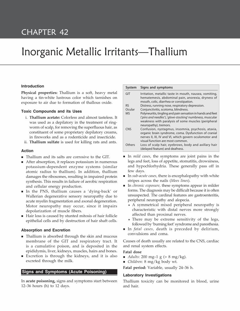
Inorganic Metallic Irritants —Thallium
ChapTer 42
Introduction
Physical properties: Thallium is a soft, heavy metal having a tin-white lustrous color which tarnishes on exposure to air due to formation of thallous oxide.
Toxic Compounds and its Uses i. Thallium acetate: Colorless and almost tasteless. It
was used as a depilatory in the treatment of ring-worm of scalp, for removing the superfluous hair, as constituent of some proprietary depilatory creams, in fireworks and as a rodenticide and insecticide.
ii. Thallium sulfate is used for killing rats and ants.
Action � Thallium and its salts are corrosive to the GIT. � After absorption, it replaces potassium in numerous
potassium-dependent enzyme systems (similar atomic radius to thallium). In addition, thallium damages the ribosomes, resulting in impaired protein synthesis. This results in failure of aerobic respiration and cellular energy production.
� In the PNS, thallium causes a ‘dying-back’ or Wallerian degenerative sensory neuropathy due to acute myelin fragmentation and axonal degeneration. Motor neuropathy may occur, since it impairs depolarization of muscle fibers.
� Hair loss is caused by stunted mitosis of hair follicle epithelial cells and by destruction of hair shaft cells.
Absorption and Excretion � Thallium is absorbed through the skin and mucous
membrane of the GIT and respiratory tract. It is a cumulative poison, and is deposited in the epididymis, liver, kidneys, muscles, hairs and bones.
� Excretion is through the kidneys, and it is also excreted through the milk.
Signs and Symptoms (Acute poisoning)
In acute poisoning, signs and symptoms start between 12–36 hours (h) to 12 days.
� In mild cases, the symptoms are joint pains in the legs and feet, loss of appetite, stomatitis, drowsiness, and hypochlorhydria. These generally pass off in few days.
� In sub-acute cases, there is encephalopathy with white stripes across the nails (Mees lines).
� In chronic exposure, these symptoms appear in milder forms. The diagnosis may be difficult because it is often unsuspected. The cardinal features are gastroenteritis, peripheral neuropathy and alopecia.
z A symmetrical mixed peripheral neuropathy is characteristic with distal nerves more strongly affected than proximal nerves.
z There may be extreme sensitivity of the legs, followed by ‘burning feet’ syndrome and paresthesia.
� In fatal cases, death is preceded by delirium, convulsions and coma.
Causes of death usually are related to the CNS, cardiac and renal system effects.
Fatal dose � Adults: 200 mg–1 g (> 8 mg/kg). � Children: 8 mg/kg body wt.
Fatal period: Variable, usually 24–36 h.
Laboratory InvestigationsThallium toxicity can be monitored in blood, urine and hair.
System Signs and symptoms
GIT Irritation, metallic taste in mouth, nausea, vomiting, hematemesis, abdominal pain, anorexia, dryness of mouth, colic, diarrhea or constipation.
RS Distress, running nose, respiratory depression.Ocular Conjunctivitis, scotoma, blindness.MS Polyneuritis, tingling and pain sensation in hands and feet
(‘pins and needles’), ‘glove-stocking’ numbness, muscular weakness with paralysis of some muscles (peripheral neuropathy), tremors.
CNS Confusion, nystagmus, insomnia, psychosis, ataxia, organic brain syndrome, coma. Dysfunction of cranial nerves II, III, IV and VI, which govern oculomotor and visual function are most common.
Others Loss of scalp hair, eyebrows, body and axillary hair (delayed feature) and deafness.

509Inorganic Metallic Irritants—Thallium
� Eosinophilia is a common phenomenon. � Thallium > 40 µg% in blood, and > 150 µg/l in
urine (levels up to 20 µg/l is considered normal) is significant. Hair levels < 15 ng/g are considered normal.
� Urine may be green, with proteinuria, diminished creatinine clearance, elevated blood urea nitrogen.
Diagnosis � GIT and polyneuritic symptoms together with the
falling of hair from head, eyebrows and axilla should lead to suspicion of thallium poisoning.
� A brownish black pigmentation close to the hair root is characteristic of thallium exposure and may appear as early as 3rd–4th day.
� Opacity in the liver on X-rays has been reported.
Treatment
i. Patient should be kept warm. ii. Emesis is indicated within 4–6 h of ingestion. iii. Multiple-dose of activated charcoal may be
given, followed by saline purgative. Whole bowel irrigation with polyethylene glycol electrolyte lavage solution may be useful.
iv. Stomach wash is performed with 1% sodium or potassium iodide solution. It forms insoluble iodide salts of thallium. Iodide also acts as a systemic antidote.
v. Prussian blue or Berlin blue (potassium ferric hexacyanoferrate)* which acts to sequester the ions in the intestine and preventing their absorption is given in a dose of 250 mg/kg/day in 2–4 divided doses orally.
vi. Although chelating agents including BAL and EDTA are contraindicated in the treatment, sodium-diethyl-dithio-carbamate 25 mg/kg body wt in 500 ml of 5% glucose IV once daily may be given.
vii. Pilocarpine in usual doses is also a physiological antidote.
viii. Potassium chloride promotes renal excretion of thallium. Administration of sodium polystyrene sulfonate as sodium-thallium exchange resin may be helpful.
ix. Hemodialysis/peritoneal dialysis may be useful within 48 h of ingestion.
x. Stimulants, dextrose and calcium salts are used according to necessity.
postmortem Findings
i. There is anemia and loss of hair. ii. Stomach: Mucous membrane may be inflamed and
there may be submucous petechial hemorrhages. iii. Spleen: Congested. iv. Liver: Congested, and shows centrilobular necrosis
and fatty degeneration. v. Kidneys: Congested, glomeruli are swollen, convo-
luted tubules show cloudy swelling and necrosis of the cells.
vi. Trachea and bronchi: Congested. vii. Lungs: Congested with subpleural hemorrhages. viii. Heart: Fatty degeneration. ix. Brain: Meningeal vessels may be congested. x. Cells of adrenal cortex, thyroid and hair follicles
show vacuolization and degenerative changes.
Medico-legal Aspects
� Poisoning by thallium is rare in contrast to poisoning by lead or mercury, probably due to its infrequent use.
� Thallium was used as an ideal homicidal poisoning in some European countries and Australia, where it was used as rodenticide.
� Accidental intoxication may result from its therapeutic use as a depilatory or abortifacient, or from its accidental ingestion when used as a rodenticide.
� Chronic poisoning occurs from industrial exposure. � Suicidal cases are also seen sometimes.
� Thallium stress test: In clinical practice, thallium 201 is used as a radioactive tracer in heart scintigraphy to detect myocardial ischemia.
� Accidental poisoning has become rare in the domestic setting since the 1970s, when thallium-based rodenticides were banned in many countries. The majority of reported cases of thallium poisoning in the last two decades have been caused by deliberate poisoning.
� Thallium may be detected in the urine 1 h after ingestion (normal level, < 0.003 µmol/l; a level of > 0.98 µmol/l is toxic), but most clinical laboratories may not have the facilities to quantitatively analyze the thallium content.
� A rapid quantitative urine test can be done by mixing urine with 0.4% sodium bismuth in 20% nitric acid and 10% sodium iodide. A red precipitate indicates that thallium is present.
� Microscopic examination of hair after application of 10% sodium hydroxide may reveal dark bands of pigmented material characteristic of presence of thallium.
* Potassium is exchanged preferentially for thallium entering the enterohepatic circulation. As Prussian blue sequesters thallium, a concentra-tion gradient is established for the continued movement of thallium into the gut.
Cadmium
Cadmium is a soft, white metal, used in welding, metal-plating, battery and plastic industries. Poisoning may occur from the inhalation of cadmium dust or fumes or from the ingestion of a cadmium salt.
Action
� It binds to sulfhydryl groups, denaturing proteins and/or inactivating enzymes. The mitochondria are severely affected by this process which may result in increase susceptibility to oxidative stress.
� It also interferes with calcium transport mechanisms leading to intracellular hypercalcemia and ultimately apoptosis.
Absorption and Metabolism
� Cadmium (2–7%) is absorbed through the GIT and its absorption is enhanced when the diet is deficient in calcium, iron or protein. Absorption through the respiratory tract is more efficient, ranging from 15–50% of an inhaled dose.
� It binds to RBCs, plasma albumin and metallothionein, which is synthesized in the liver. Cadmium is initially detoxified in the liver through the formation of a metallothionein-cadmium complex which is slowly released from that organ.
Signs and Symptoms
Toxicity by inhalation is far greater than by ingestion. � On inhalation: Symptoms develop usually after
4–8 hours (h). They are influenza-like and similar to those seen in metal fume fever. It is characterized by sneezing, sore throat, irritant cough, nausea, excessive salivation, metallic taste, headache and cyanosis (‘cadmium blues’). After a latent period of 24–36 h, chemical pneumonitis develops which is characterized by fever, dyspnea, bronchospasm and pleuritic chest pain, along with tachycardia, oliguria and noncardiogenic pulmonary edema.
� On ingestion: Symptoms occur in 1 h. These are increased salivation, nausea, vomiting, cramps in the abdomen, diarrhea, myalgia, collapse, and rarely death.
� Chronic exposure causes anosmia, yellowing of teeth (cadmium ring formation), emphysema, bone pain, fractures with osteomalacia and chronic renal failure (hypercalciuria, proteinuria, azotemia). Hypertension and hypochromic anemia may be seen.
� Cadmium is said to be carcinogenic, and increased incidence of lung, prostrate, pancreas and bladder carcinoma has been reported.
Fatal dose: > 100 mg. Symptoms are seen with serum cadmium > 5 ng/dl (normal range 0.2–6.0 ng/ml) and urinary cadmium > 100 nmol/l.
Fatal period: 5–7 days.
Diagnosis: Blood cadmium levels are a reflection of acute cadmium exposure; urine levels appear to provide a better measure of chronic exposure. Urinary beta-2 microglobulin test is an indirect method of measuring cadmium exposure.
Treatment
i. Avoid further exposure, O2 and steroids may be given in case of inhalation of fumes.
ii. Stomach is washed with tannin or egg albumin, and activated charcoal may be given.
iii. Sodium sulfate as a purgative is given. iv. Succimer (10 mg/kg/dose TID) may be given
(decreases the GIT absorption and improves survival without increasing cadmium burden in target organs) in case of acute poisoning, and dithio carbamates in chronic poisoning.
v. Vitamin D is given for osteomalacia.
Postmortem Findings
i. GIT: Mucous membranes of the esophagus, stomach and intestines are congested and inflamed.
Other Inorganic Metallic Irritants
Chapter 43

511Other Inorganic Metallic Irritants
ii. Lungs: Pulmonary edema and emphysema. There may be degeneration and/or loss of bronchial and bronchiolar epithelial cells.
iii. Heart and liver: Fatty degeneration. iv. Kidneys: Proximal tubular necrosis.1
v. Brain: Congested.
Medico-legal aspects: Poisoning with cadmium is rare, but may occur as an industrial disease.
� Cadmium poisoning occurred in 1946 from the contamination of food and water by mining effluents in Japan resulting in outbreak of ‘itai-itai’ (‘ouch-ouch’) disease, so named as cadmium-induced bone toxicity led to painful bone fractures.
� In chronic exposure, cadmium is bound to intracellular metallothionein, which greatly reduces its toxicity. Any attempt to remove cadmium from these deposits risks redistributing cadmium to other organs, thus exacerbating toxicity, as is known to occur with BAL therapy (exacerbate nephrotoxicity).
Barium
Physical properties: It is a heavy, white, tasteless, odorless powder and insoluble in water. Barium sulfate is used for the X-ray examination of the GIT (‘barium-meal’).
Toxic compounds: Soluble salts are most toxic. These are barium chloride, barium nitrate, barium carbonate (rodenticide) and barium sulphide (used as a depilatory). In Kiating, China, a subacute form of barium poisoning (pa-ping) was endemic because of use of contaminated table salt.
Action: It acts locally as an irritant poison. After absorption it acts both on voluntary and involuntary muscles. Barium seems to act as potassium antagonist and calcium agonist.
Absorption
Toxicity of barium compounds depends on their solubility. The free ion is absorbed from the lungs and GIT, but barium sulfate remains unabsorbed. After absorption, it accumulates in the skeleton and in pigmented parts of the eye.
Signs and Symptoms
On ingestion2
The most characteristic features are areflexia and paralysis (Ba2+ ion is a muscle poison).3
System Signs and symptoms
GIT Nausea, vomiting, salivation, abdominal pain and diarrhea.
MS Tingling sensation, tremors, cramps, stiffness of the muscles, paralysis of the tongue and larynx, myoclonus, myalgia, flaccid quadriplegia.
CVS Hypertension, arrhythmia, ectopic beats, ventricular fibrillation, irregular pulse, shock, cardiac arrest.
RS Pulmonary edema, respiratory failure.CNS Mydriasis, vertigo, headache, confusion, convulsions.
Inhalation of barium sulfate dust causes a benign pneumoconiosis (‘baritosis’) with conspicuous radio-graphic manifestation, but no impairment of pulmonary functions.
Fatal dose: About 0.8–1 g of barium chloride/sulfide/nitrate.
Fatal period: Usually within 12 h.
Treatment
i. Gastric lavage with sodium or magnesium sulfate (5–10 g) solution to precipitate the barium as insoluble sulfate.
ii. Administer 0.45% of NaCl in D5W and a diuretic (furosemide 1 mg/kg) to obtain a urine flow of 3–6 ml/kg/h to flush out barium by diuresis.
iii. Administration of large amounts of potassium parentally (KCl 20–40 mEq/l) is indicated (causes severe hypokalemia, potassium infusion is an effective antidote).
iv. 10 ml of 10% sodium sulfate Iv every 15 min to convert barium into insoluble sulfate.
v. Purgation with magnesium sulfate and repeated bowel washes.
vi. Sodium nitrite 30–60 mg for hypertension. vii. Procainamide 500 mg slow Iv for ventricular
fibrillation. viii. Hemodialysis is effective in patients with severe
poisoning. ix. Symptomatic treatment.
Postmortem findings: Non-specific. Submucosal hemorrhages may be seen in the GIT.
Medico-legal Aspects
� Suicidal cases may be seen. � Homicidal cases are rare.
512 Review of Forensic Medicine and Toxicology
� Accidental poisoning with barium sulfide may occur, if taken by mistake as barium sulfate for X-ray examination.
Zinc
Zinc is normally present in our body. Poisonous salts are compounds of chloride, phosphide, sulfate (white vitriol), oxide and stearate.
Uses: Zinc chloride is used to clean metals before soldering. Zinc phosphide is used as rodenticide. Zinc stearate is used as a cosmetic (baby powder).
Action: Salts of zinc are locally irritating, and after absorption cause metabolic acidosis, hypocalcemia, damage to the liver and kidneys, and affects the CNS.
Signs and Symptoms
On ingestion, there is a metallic taste, nausea, vomiting, pain in the abdomen and diarrhea. The vomitus and the stool may contain blood. There is ulceration of the mucous membrane of mouth, esophagus and stomach wall with occasional perforation. Collapse due to shock may occur.
� Zinc phosphide releases phosphine gas under acidic conditions in the stomach (similar to aluminum phosphide). In addition to the above features, the vomitus gives the smell of garlic. Dyspnea, lethargy, hypotension, cardiac arrhythmias, pulmonary edema, metabolic acidosis, convulsions, circulatory collapse, coma and death may be occur.
Inhalation of zinc oxide vapor in industries causes chill and fever, a condition known as ‘metal fume fever’ or ‘zinc shakes’ (when exposed daily to concentrations of > 8–12 mg/m3).4
� Inhalation of zinc stearate used in baby powder may cause pneumonitis.
Fatal dose � Zinc chloride: 40–70 mg/kg; and zinc phosphide:
20–40 mg/kg. � Zinc sulfate: 15 g (10–30 g). � Zinc oxide fumes: 500 mg/m3 (recommended
exposure limit: 5 mg/m3).1
Fatal period: 3–5 h to few days.
Treatment
i. Gastric lavage is done with alkaline solution. Demulcents may be given.
ii. Sodium bicarbonate with water is given orally. iii. Purgatives are given for elimination. iv. Symptomatic treatment.
Postmortem Findings
i. Non-specific external signs may be seen. Garlicky odor from the mouth and on opening the stomach may be observed in case of zinc phosphide poisoning.
ii. Signs of irritation of the GIT with degenerative changes in the stomach wall and occasional perforation may be there.
iii. Degenerative changes in the liver, kidneys and heart may occur.
iv. visceral organs are congested.
Medico-legal Aspects
� Suicidal poisoning is seen with the phosphide. � Accidental cases occur with chronic exposure in
industries, acute poisoning may occur with consum-ption of food stored and cooked in zinc galvanized metal containers.
� Homicidal cases are rare. � It may be used as an abortifacient.
Metal Fume Fever (MFF)
� MFF is a self-limiting acute febrile illness associated with inhalation of metal oxide fumes. It is also called smelter’s shakes, brass chills or Monday morning fever.
� Signs and symptoms: This influenza-like syndrome starts 4–8 h after exposure of fumes, which is characterized by headache, fever, chills, cough, dyspnea, cyanosis, myalgia, metallic taste, salivation, sweating and tachycardia. Symptoms subside within 24–36 h, only to return on repeated exposure.5
� Metals involved: It is caused by acute exposure to fumes/smoke of oxides of zinc (commonest), copper, magnesium, nickel, mercury, lead, iron, chromium, cadmium, cobalt, antimony, tungsten, titanium, manganese and silver.
� A proper occupational history (those involved in welding, melting or flame cutting galvanized metal or in brass foundry operations) should make the diagnosis evident. WBC count may be elevated, chest X-ray is usually normal.
� Treatment: Supplemental oxygen, bronchodilators (if there is wheezing) and symptomatic treatment.

513Other Inorganic Metallic Irritants
1. A 2. A 3. C 4. B 5. C 6. A
Methemoglobinemia inducing Agents
� A large number of chemical agents are capable of oxidizing ferrous hemoglobin to its ferric state (methemoglobin), a form that cannot carry oxygen and thus inducing a functional anemia. In addition, the shape of oxygen-hemoglobin dissociation curve is altered, aggravating cellular hypoxia.
� Drugs and chemicals known to cause methemo– globinemia include benzocaine, antimalarial agents, dapsone, aniline dyes, nitrites, nitrates, nitrogen oxide gas, nitrobenzene and many others.
Signs and Symptoms
� The severity of symptoms depends on the percentage of hemoglobin oxidized to methemoglobin, severe poisoning is usually present when methemoglobin fractions are > 40–50%.
� Even at low levels (15–20%), victims appear cyanotic (especially of the nails, lips and ears), because of the
‘chocolate brown’ color of methemoglobin (‘chocolate cyanosis’), but they have normal PO2 results on arterial blood gas determinations.6
� It may cause dizziness, nausea, headache, dyspnea, confusion, seizures and coma.
� Severe metabolic acidosis may be present. Hemolysis may occur, especially in patients susceptible to oxidant stress (i.e. those with G-6-PD deficiency).
Diagnosis is suggested by finding of chocolate brown blood (dry a drop of blood and compare with normal blood).6
Treatment � Administer high-flow oxygen. � Administer activated charcoal. � Methylene blue, 1–2 mg/kg (0.1–0.2 ml/kg of 1%
solution) Iv enhances the conversion of methemo-globin to hemoglobin by increasing the activity of the enzyme methemoglobin reductase. Dose may be repeated once in 15–20 minutes, if necessary.
multiple ChoiCe questions
1. Cadmium causes: NEET 14 A. Proximal tubular necrosis B. Distal tubular necrosis C. Polyneuritis D. Cirrhosis 2. A housewife ingests a rodenticide white powder
accidentally. Her examination showed generalized flaccid paralysis and an irregular pulse. ECG shows multiple ventricular ectopics, generalized changes within ST-T. Serum potassium is 2.5 mEq/l. The most likely ingested poison is: AIIMS 06
A. Barium carbonate B. Superwarfarins C. Zinc phosphide D. Aluminum phosphide 3. Barium carbonate poisoning causes: NEET 13 A. Respiratory distress B. Gastrointestinal irritation C. Muscular weakness D. Cyanosis
4. A person presents with acute poisoning, with chills and rigors similar to malaria. Most likely poisoning is with: AIIMS 06
A. Mercury B. Zinc C. Red phosphorus D. Arsenic 5. Symptoms of metal fume fever resolve spontaneously
within: Himachal 10 A. 6–12 h B. 12–24 h C. 24–36 h D. 36–48 h 6. Patient with BP 90/60 mmHg, lips and peripheries are
cyanosed; blood drawn was chocolate color. Diagnosis is: DNB 10
A. Methemoglobinemia B. Hypovolumic shock C. Metal fume fever D. Alphos poisoning

Non-Metallic and Mechanical Irritants
CHAPTER 44
Phosphorus
Introduction: Phosphorus (Greek ‘phos’: light, ‘phorus’: bringing) exists in two varieties (Diff. 44.1): i. White or crystalline: Samples of white phosphorus
always contain some red phosphorus and therefore appear yellow (also called ‘yellow phosphorus’).1
ii. Red or amorphous: White phosphorus gradually changes to red phosphorus. This transformation is accelerated by heat and light.
Action: It is a protoplasmic poison and affects cellular oxidation. The metabolism of cells reduces, leading to necrobiosis which is predominantly seen in the liver.
Signs and Symptoms (Acute Poisoning)
Contact injury: White phosphorus exposure results in painful penetrating second and third degree burn injuries. The burn typically appears as a necrotic area with a yellowish color and characteristic garlic-like odor. White phosphorus is lipid soluble, and hence results in rapid dermal penetration and delayed wound healing.
Acute ingestion has two phases, initially local action on the GIT [within ½–6 hours (h)] and later, action of the absorbed poison (after 2–3 days).
First Stage: It produces burning pain in the throat and abdomen with intense thirst, nausea, vomiting, diarrhea and abdominal pain. Breath, vomitus and feces have garlic-like odor. Luminescent ‘smoking’ vomit and feces are diagnostic. A symptom-free period lasting for 2–3 days is seen after the acute attack.
Second Stage
System Signs and symptoms
GIT Nausea, vomiting, hematemesis, diarrhea.Hepatic Tender hepatomegaly, jaundice, pruritus.Renal Oliguria, hematuria, casts, albuminuria, sometimes
anuria.CNS Restlessness, anxiety, insomnia, headache, confusion,
hallucinations, convulsions, delirium, coma.PNS Paresthesia, carpopedal spasm, tetany, laryngeal
stridor, opisthotonus (because of hypocalcemia).Hematologic Purpura, epistaxis, hemorrhage in mucous membrane
and viscera.
Differentiation 44.1: White and red phosphorous (Fig. 44.1)
S.No. Feature White phosphorus Red phosphorus
1. Color White or yellow Reddish-brown
2. Appearance Crystalline, waxy, translucent1 Amorphous or crystalline, opaque
3. Solubility � Organic solvents � Water
YesVery low (~ 3 mg/l)
NoInsoluble
4. Odor Characteristic garlicky odor Odorless
5. Taste Garlicky Tasteless
6. Chemiluminescence Luminous in dark Non-luminous
7. Ignitability Inflammable, spontaneous ignition in air at room temperature (emits white fumes) and in chlorine
Non-inflammable, ignites only at > 260°C; heat is necessary for ignition in chlorine
8. Reaction with aqueous alkali
Produces phosphine None
9. Toxicity Highly toxic Low toxicity
10. Uses Fertilizers, insecticides, rodenticide, incendiary bombs, smoke screens and fireworks
On sides of match box

515Non-Metallic and Mechanical Irritants
Fulminating poisoning (death within 12 h) may be seen when the patient takes a large dose.
� Early death is due to cardiac dysrhythmias, secondary to electrolyte abnormalities, such as hypocalcemia and hyperkalemia.
� Death after the first 24 h is due to hepatic failure.
Fatal dose: 60–120 mg of white phosphorus.
Fatal period: Within 24 h, but may be delayed by 5–7 days.
Diagnosis: Oral and skin burns, luminescent ‘smoking’ vomitus and stools with a garlic odor, and GIT and biliary damage characterize white phosphorus poisoning.
Treatment
i. Life support measures—airway support and fluid maintenance should be provided.
ii. External burns should be washed and cleaned with mild disinfectant soap and water, and covered with antibiotic ointment.
iii. Gastric lavage using 1:5000 solution of KMnO4 (chemical antidote) oxidizes phosphorus into harmless phosphoric acid and phosphates.
iv. Activated charcoal is given. v. Demulcents (oily or fatty substances) are contra -
indicated, as phosphorus gets dissolved and gets absorbed.
vi. Purgatives (magnesium sulfate) may be given. vii. Vitamin K 20 mg IV in repeated doses, or blood
transfusion. viii. Transfusion of glucose-saline and plasma with
vitamins is useful to protect the liver and to correct shock and dehydration.
ix. Peritoneal or hemodialysis may be required (for correction of hyperphosphatemia, hyperkalemia and hypocalcemia).
x. N-acetylcysteine, ubiquinone and sulfate have been tried to prevent liver damage.
� Identifiable phosphorous particles from the skin are removed by thorough debridement and the area covered with saline-soaked gauze to prevent further combustion. Copper sulfate solution is sometimes recommended for conversion of phosphorous particles to blue-black cupric phosphide.2
� Alternatively, application of silver nitrate may prevent ignition of phosphorus by depositing a film of silver over the phosphorous.
� Stomach can also be washed with 0.2% copper sulfate solution. Since, copper sulfate itself can cause acute copper poisoning and inhibit G-6-PD leading to lethal hemolysis, it is not recommended.2
� There is a danger of explosion and fire because of entry of oxygen into the stomach or exit of phosphorous through the nasogastric tube. This is minimized by connecting the external end of the tube to a syringe filled with water; confirmation of placement is done by instillating water rather than air or by withdrawing gastric contents.
Postmortem Findings
External i. Emaciation, purpuric hemorrhages in the skin,
jaundice, and smell of garlic may be present.3
ii. Mucous membrane of the mouth is corroded. iii. Hypostasis is dark brown in color.3
Internal i. Multiple hemorrhages are seen in the muscles, serosal
and mucosal membranes of the GIT and respira tory tract, liver, kidneys, endocardium, pericardium, epicardium, peritoneum, lungs and brain.
ii. Stomach and intestines: Mucous membranes are yellowish or grayish-white in color, softened, thickened, inflamed and corroded in patches; luminous material may be found in the stomach. Contents may smell of garlic.
iii. Liver: Swollen, yellow, soft, fatty and easily ruptured.4
iv. Kidneys: Enlarged, greasy, yellow. v. Heart: Flabby, pale and shows fatty degeneration. vi. Lungs: Fat emboli may be found in the pulmonary
arterioles and capillaries.
Medico-legal Aspects � Accidental poisoning in children due to chewing of
fireworks or by eating rat paste may occur. � It is not preferred for suicide because of painful
symptoms and prolonged suffering. � It may be used for homicide purpose by mixing
with alcohol or coffee to mask the taste and smell and administered, since:a. Symptoms resemble acute liver disease.b. There is delay in the appearance of symptoms.c. The poison is oxidized in the body, hence cannot
be detected.d. Death occurs after few days.
� Sometimes, it is taken by mouth or introduced into the vagina to procure abortion.
Figs 44.1A and B: (A) White phosphorous, (B) Red phosphorous
A B
516 Review of Forensic Medicine and Toxicology
� Cases of poisoning may occur during war when phosphorus enters the body with fragments of hand grenades, smoke screens, bombs or bullets.
� For arson, white phosphorus covered with dung or wet cloth is thrown on huts. When the covering becomes dry, the roof catches fire.
Chronic Phosphorus Poisoning
� The frequent inhalation of fumes over a period of years causes necrosis of the lower jaw in the region of a decayed tooth.
� Initially, there is toothache which is followed by swelling of the jaw, loosening of the teeth, necrosis of the gums and sequestration of bone in the mandible with multiple sinuses discharging foul-smelling pus. This is known as ‘phossy jaw’ (glass jaw or Lucifer’s jaw) in which osteomyelitis and necrosis of jaw occurs.5
� Constitutional symptoms include nausea, vomiting, anorexia, pain in the abdomen, indigestion, purging, pain in the joints, weakness, loss of weight, bronchitis, cirrhosis, jaundice, ascitis and anemia.
Mechanical Irritants
Of all the mechanical irritants, glass and diamond powder, and pointed metallic chips may be of real danger in most cases. Others, like pins, needles and hair also acts as mechanical irritants. Hair may cause intestinal obstruction.
Signs and Symptoms � There may be nausea, vomiting (bloodstained),
burning pain in throat and abdomen with constipation.
� The sharp margins or the pointed ends of broken pieces of glass/diamond may cause injury and hemorrhage in the GIT when ingested.
� They do not usually adhere to the wall of the GIT, but rather pass out the whole length of the tract by peristaltic movement, longitudinally in relation to the length of the intestines. However, perforation peritonitis or even serious injury to the intestinal tract may occur.
� If death occurs, it is due to shock as a result of injury and internal hemorrhage.
Treatment i. Bulky foods, like bananas or rice for easy passage
of the glass pieces. ii. Laxatives may be given. iii. Symptomatic treatment.
Postmortem Findings i. There may be inflammation and hemorrhage of the
GIT. ii. Perforation peritonitis is uncommon. iii. Fragments of glass particles may be found adhered
to the stomach wall. iv. Signs of anemia, intestinal hemorrhage and
inflammation are more common.
Medico-legal Aspects � In ancient period, mechanical irritants have been
used as homicidal agents. Women, to kill their husbands, have administered finely powdered glass bangles in food.
� Ingestion is usually accidental from contamination occurring from the broken pieces of the glass containers.
� They may be used to kill the cattle.
1. D 2. C 3. D 4. C 5. B
MULTIPLE CHOICE QUESTIONS
1. A poison which is luminescent and waxy and have a garlic smell: AI 11
A. Alphos B. Ammonium bromide C. Opium D. Yellow phosphorous 2. CuSO4 was used as an antidote for: Maharashtra 11 A. Dhatura poisoning B. Cocaine poisoning C. Phosphorus poisoning D. Opium poisoning 3. A body is brought for autopsy. On postmortem, there
is dark brown postmortem staining and garlic odor in stomach. The poisoning is most likely due to:
BHU 10
A. Hydrocyanic acid B. Carbon dioxide C. Aniline dye D. Phosphorus 4. Yellow/fatty liver is characteristically seen in:
Jharkhand 11 A. Datura poisoning B. Cocaine poisoning C. Phosphorus poisoning D. Opium poisoning 5. ‘Phossy jaw’ is seen in chronic poisoning with:
AFMC 11; NEET 13 A. Datura B. Phosphorus C. Arsenic D. Thallium

Organic Irritants—Plant
CHAPTER 45
Ricinus Communis (Castor)
Distribution: It grows all over India, especially in wastelands.
Identification of Seeds (Fig. 45.1)
� Seeds are variable, smooth, flattened-oval, mottled with light and dark brown markings, bright and polished.
� They are of 2 sizes, small and big. � Small seeds are about 1.2 × 0.8 cm in dimensions
and resemble croton seeds.
Active Principle � The entire plant is poisonous, containing toxalbumin
ricin, a water-soluble glycoprotein and a powerful allergen. Seeds contain the highest level.
� They are also rich in purgative oil. Castor oil is not poisonous as it does not contain ricin.
Unbroken seeds are not poisonous when swallowed or cooked. Toxicity is caused when castor beans are thoroughly chewed or blenderized, even though the quantity of ricin so produced is small and is poorly absorbed from the GIT.
Toxalbumin or phytotoxin is a toxic protein that disable ribosomes and thereby inhibit protein synthesis, and present in the plants like in castor, croton or rati.
� It is antigenic in nature, agglutinates red cells, causes hemolysis and cell destruction.
� Toxalbumins are similar in structure to the toxins found in cholera, tetanus, diphtheria, pseudomonas and botulinum; and their physiological and toxic properties are similar to those of viperine snake venom.
Action
� Ricin blocks protein synthesis through inhibition of RNA polymerase. It belongs to a group of poisons known as A-B toxins.
� Ricin has a special binding protein that gains access to the endoplasmic reticulum in the GIT mucosal cells causing diarrhea.
Signs and Symptoms
Dust of seeds may cause: � Watering of eyes and � Acute nasal inflamma-
conjunctivitis tion and sneezing � Headache, pharyngitis � Asthmatic bronchitis � Gastric upset � Dermatitis
On ingestion [seen within 10 hours (h) of ingestion] � GIT: Burning pain in throat, colicky abdominal pain/
cramping, nausea, thirst, vomiting and diarrhea (often bloody).
� CNS: Vertigo, drowsiness, delirium, convulsions and coma.
� Uremia, jaundice, rapid feeble pulse, cold clammy skin and dehydration.
Consciousness is retained till death in some cases.
Fatal dose: 1–10 µg/kg body wt (by inhalation or injection). Oral exposure to ricin is far less toxic, and lethal dose is about 2 mg/kg (10–20 seeds).
Fatal period: 3–5 days.
Treatment
No known antidote or other specific treatment, although a vaccine has been developed by the US military.Fig. 45.1: Ricinus communis (seeds)
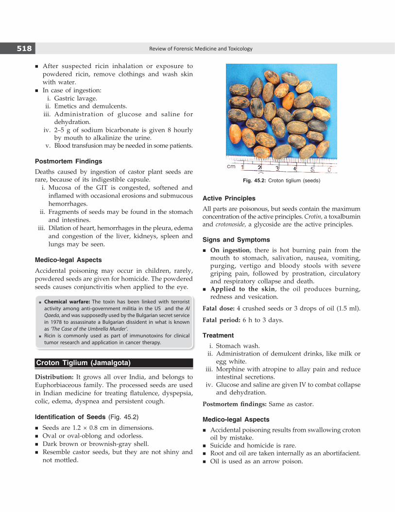
518 Review of Forensic Medicine and Toxicology
� After suspected ricin inhalation or exposure to powdered ricin, remove clothings and wash skin with water.
� In case of ingestion: i. Gastric lavage. ii. Emetics and demulcents. iii. Administration of glucose and saline for
dehydration. iv. 2–5 g of sodium bicarbonate is given 8 hourly
by mouth to alkalinize the urine. v. Blood transfusion may be needed in some patients.
Postmortem FindingsDeaths caused by ingestion of castor plant seeds are rare, because of its indigestible capsule. i. Mucosa of the GIT is congested, softened and
inflamed with occasional erosions and submucous hemorrhages.
ii. Fragments of seeds may be found in the stomach and intestines.
iii. Dilation of heart, hemorrhages in the pleura, edema and congestion of the liver, kidneys, spleen and lungs may be seen.
Medico-legal Aspects
Accidental poisoning may occur in children, rarely, powdered seeds are given for homicide. The powdered seeds causes conjunctivitis when applied to the eye.
� Chemical warfare: The toxin has been linked with terrorist activity among anti-government militia in the US and the Al Qaeda, and was supposedly used by the Bulgarian secret service in 1978 to assassinate a Bulgarian dissident in what is known as ‘The Case of the Umbrella Murder’.
� Ricin is commonly used as part of immunotoxins for clinical tumor research and application in cancer therapy.
Croton Tiglium (Jamalgota)
Distribution: It grows all over India, and belongs to Euphorbiaceous family. The processed seeds are used in Indian medicine for treating flatulence, dyspepsia, colic, edema, dyspnea and persistent cough.
Identification of Seeds (Fig. 45.2)
� Seeds are 1.2 × 0.8 cm in dimensions. � Oval or oval-oblong and odorless. � Dark brown or brownish-gray shell. � Resemble castor seeds, but they are not shiny and
not mottled.
Active PrinciplesAll parts are poisonous, but seeds contain the maximum concentration of the active principles. Crotin, a toxal bumin and crotonoside, a glycoside are the active principles.
Signs and Symptoms � On ingestion, there is hot burning pain from the
mouth to stomach, salivation, nausea, vomiting, purging, vertigo and bloody stools with severe griping pain, followed by prostration, circulatory and respiratory collapse and death.
� Applied to the skin, the oil produces burning, redness and vesication.
Fatal dose: 4 crushed seeds or 3 drops of oil (1.5 ml).
Fatal period: 6 h to 3 days.
Treatment i. Stomach wash. ii. Administration of demulcent drinks, like milk or
egg white. iii. Morphine with atropine to allay pain and reduce
intestinal secretions. iv. Glucose and saline are given IV to combat collapse
and dehydration.
Postmortem findings: Same as castor.
Medico-legal Aspects � Accidental poisoning results from swallowing croton
oil by mistake. � Suicide and homicide is rare. � Root and oil are taken internally as an abortifacient. � Oil is used as an arrow poison.
Fig. 45.2: Croton tiglium (seeds)

519Organic Irritants—Plant
Abrus Precatorius (Rati, Gunchi, Jequirity)
Distribution: It is found all over India, and belongs to Leguminosae family. All parts of the plant are poisonous.
Identification of Seeds (Fig. 45.3)
� Seeds are egg shaped and scarlet in color with a black spot at one end. White seeds are also found.
� 0.83 × 0.62 cm in dimensions; having a weight of 105 mg.
� Seeds are tasteless and odorless. � It was used by Indian goldsmiths for weighing
silver and gold.
Active PrinciplesSeeds contain active principles, abrin, a thermolabile toxalbumin; abrine, an amino acid; hemagglutinin, a lipolytic enzyme; and abralin, a glycoside.
Signs and Symptoms � On ingestion, there is abdominal pain, nausea,
vomiting, bloody diarrhea, weakness, cold perspi-ration, trembling of hands, weak pulse, rectal bleeding, tachycardia, headache, dilated pupils, hallucinations drowsiness, tetany and circulatory collapse, seen in 6 h but may be delayed to 1–3 days.
� When extract of seeds is injected under the skin, symptoms resemble viperine snakebite and as such poisoning is not suspected.1,2 There is inflammation, edema, oozing of hemorrhagic fluid from site of puncture and necrosis may occur. The animal drops down and does not take feed and dies in 3–4 days. Tetanic convulsions occur or the animal becomes cold, drowsy or comatose and dies.
Fatal dose: 90–120 mg of abrin injected or 1–2 crushed seeds orally (0.1–1 µg/kg body wt.).
Fatal period: 3–5 days.
Treatment
i. Injection of anti-abrin. ii. Needle should be dissected out. iii. Hydrochloric-pepsin mixture orally. iv. Urine is maintained at an alkaline pH.
Postmortem Findings
� Fragments of needle may be found. Edema at the site of injection.
� Petechial hemorrhages may be seen under the skin, pleura, pericardium and peritoneum.
� GIT: Hemorrhages, edema and congestion (commonly affected on ingestion).
� Internal organs are congested and show focal hemor-rhages in the intestines, brain, myocardium and pleura (on parenteral exposure).
� Necrosis, hemorrhages and edema are also seen in lymph nodes, kidneys and intestines.
Medico-legal Aspects
� Accidental cases—on account of the attractive color of seeds, children may ingest them.
� Commonly used as cattle poison in Indian villages to get the hide or for taking revenge. The toxic principle is injected into the animal in the form of fine needle-shaped structures called ‘suis’.3
� Powdered seeds are used by malingerers to produce conjunctivitis.
� Seeds are also used as abortifacient and as arrow poison.
Suis
The seeds of Abrus precatorius are decocted (boiling down to extract an essence; resulting liquid) and mixed with dhatura, opium and onion, and made into paste with spirit and water, and from this paste, small sharp pointed spikes or needles or ‘suis’ are made which are dried in the sun.3
� The needles are 15 mm long and weigh 90-120 mg. Two needles are inserted by their base into holes in a wooden handle and a blow is struck to the animal with great force which drives the needle into the flesh (so as to resemble snakebite).
� For homicide, two needles are kept between the fingers and the person is slapped which drives the needle into the body.
� Signs and symptoms: At the site of injection, painful swelling and ecchymosis develops with inflammation and necrosis of muscle and regional lymph nodes. Faintness, vertigo, vomiting, anorexia, Fig. 45.3: Abrus precatorius (red & white seeds)

520 Review of Forensic Medicine and Toxicology
fever, headache, dyspnea and prostration are seen. Convulsions may occur before death. Weakness may develop within 5 h after injection, but onset of symptoms may be delayed by 10–12 h.
Semecarpus Anacardium
The fruit of this plant is known as ‘marking nut’ or ‘bhilawa’ as its juice is used by washerman/laundries to inscribe identification number on the clothes.4
Identification of Seeds (Fig. 45.4)
Seeds are black, cone or heart-shaped with a rough projection at the base. They have a thick, pericarp containing the irritant juice which is brownish, oily and acrid, but turns black on exposure to air.
Active Principles
Semecarpol and bhilawanol.5
Signs and Symptoms
� When the juice is applied to the skin, it causes irritation, itching and a painful blister containing
acrid serum and eczematous eruptions of the surrounding skin. The lesion resembles a bruise. Later on, an ulcer may be produced with sloughing. Constitutional symptoms such as fever, painful micturition with brown color urine may be seen.
� Orally, if a large dose of juice is taken, blisters in mouth and throat, severe GIT irritation, dyspnea, tachycardia, hypotension, cyanosis, loss of reflexes, delirium, coma and death may result.
Fatal dose: 5–10 g/5–8 seeds.
Fatal period: 12–24 h.
Treatment: Wash the contaminated part of the skin with soap and water. Bland liniments are applied. Demulcents drinks and symptomatic treatment are given.
Postmortem Findings
� Bruise-like lesion with small blisters may be seen near the angle of the mouth or lips. Blisters are also seen in the mouth and throat.
� Stomach: Congested and inflamed. � Liver: It may show degenerative changes. � Other organs: Congested.
Medico-legal Aspects
� Accidental poisoning may result from the adminis-tration of juice by quacks for treatment of rheumatic pain and worm infestation.
� Juice may be introduced into the vagina, as a punish-ment for infidelity.
� For criminal abortion, juice is applied to the cervical os by means of abortion stick.
� It may be used by malingerers to produce conjunc-tivitis or to support a false charge of assault; lesions produced simulate bruises (Diff. 45.1).6
Differentiation 45.1: Artificial and true bruise
S.No. Feature Artificial bruise True bruise
1. Cause Juice of marking nut, Calotropis or Plumbago rosea Trauma
2. Color Dark brown Typical color changes
3. Shape Irregular Round
4. Site Exposed accessible parts Anywhere
5. Margins Well-defined and regular, covered with small vesicles Not well-defined, diffuse and irregular, no vesicles
6. Redness and inflammation Seen in surrounding skin Seen at the site
7. Itching Present Absent
8. Vesicles May be found on fingertips due to scratching Negative
9. Contents Acrid serum Extravasated blood
10. Chemical tests Positive for the chemical Negative
Fig. 45.4: Semecarpus anacardium (seeds)
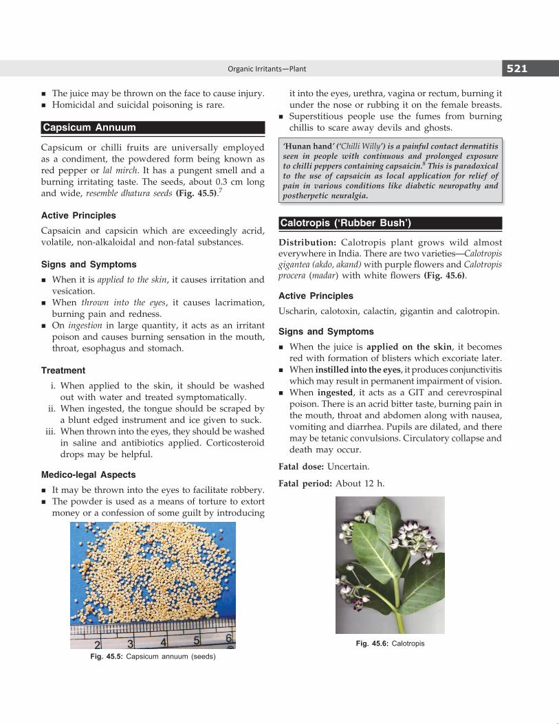
521Organic Irritants—Plant
� The juice may be thrown on the face to cause injury. � Homicidal and suicidal poisoning is rare.
Capsicum Annuum
Capsicum or chilli fruits are universally employed as a condiment, the powdered form being known as red pepper or lal mirch. It has a pungent smell and a burning irritating taste. The seeds, about 0.3 cm long and wide, resemble dhatura seeds (Fig. 45.5).7
Active Principles
Capsaicin and capsicin which are exceedingly acrid, volatile, non-alkaloidal and non-fatal substances.
Signs and Symptoms
� When it is applied to the skin, it causes irritation and vesication.
� When thrown into the eyes, it causes lacrimation, burning pain and redness.
� On ingestion in large quantity, it acts as an irritant poison and causes burning sensation in the mouth, throat, esophagus and stomach.
Treatment
i. When applied to the skin, it should be washed out with water and treated symptomatically.
ii. When ingested, the tongue should be scraped by a blunt edged instrument and ice given to suck.
iii. When thrown into the eyes, they should be washed in saline and antibiotics applied. Corticosteroid drops may be helpful.
Medico-legal Aspects
� It may be thrown into the eyes to facilitate robbery. � The powder is used as a means of torture to extort
money or a confession of some guilt by introducing
it into the eyes, urethra, vagina or rectum, burning it under the nose or rubbing it on the female breasts.
� Superstitious people use the fumes from burning chillis to scare away devils and ghosts.
‘Hunan hand’ (‘Chilli Willy’) is a painful contact dermatitis seen in people with continuous and prolonged exposure to chilli peppers containing capsaicin.8 This is paradoxical to the use of capsaicin as local application for relief of pain in various conditions like diabetic neuropathy and postherpetic neuralgia.
Calotropis (‘Rubber Bush’)
Distribution: Calotropis plant grows wild almost everywhere in India. There are two varieties—Calotropis gigantea (akdo, akand) with purple flowers and Calotropis procera (madar) with white flowers (Fig. 45.6).
Active Principles
Uscharin, calotoxin, calactin, gigantin and calotropin.
Signs and Symptoms
� When the juice is applied on the skin, it becomes red with formation of blisters which excoriate later.
� When instilled into the eyes, it produces conjunctivitis which may result in permanent impairment of vision.
� When ingested, it acts as a GIT and cerevrospinal poison. There is an acrid bitter taste, burning pain in the mouth, throat and abdomen along with nausea, vomiting and diarrhea. Pupils are dilated, and there may be tetanic convulsions. Circulatory collapse and death may occur.
Fatal dose: Uncertain.
Fatal period: About 12 h.
Fig. 45.5: Capsicum annuum (seeds)
Fig. 45.6: Calotropis
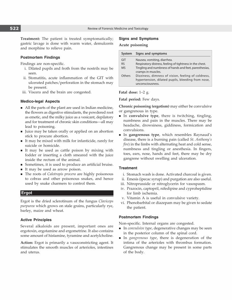
522 Review of Forensic Medicine and Toxicology
Treatment: The patient is treated symptomatically; gastric lavage is done with warm water, demulcents and morphine to relieve pain.
Postmortem Findings
Findings are non-specific. i. Dilated pupils and froth from the nostrils may be
seen. ii. Stomatitis, acute inflammation of the GIT with
ulcerated patches/perforation in the stomach may be present.
iii. Viscera and the brain are congested.
Medico-legal Aspects
� All the parts of the plant are used in Indian medicine, the flowers as digestive stimulants, the powdered root as emetic, and the milky juice as a vesicant, depilatory and for treatment of chronic skin conditions—all may lead to poisoning.
� Juice may be taken orally or applied on an abortion stick to procure abortion.
� It may be mixed with milk for infanticide, rarely for suicide or homicide.
� It may be used as cattle poison by mixing with fodder or inserting a cloth smeared with the juice inside the rectum of the animal.
� Sometimes, it is used to produce an artificial bruise. � It may be used as arrow poison. � The roots of Calotropis procera are highly poisonous
to cobras and other poisonous snakes, and hence used by snake charmers to control them.
Ergot
Ergot is the dried sclerotinum of the fungus Claviceps purpurea which grows on stale grains, particularly rye, barley, maize and wheat.
Active Principles
Several alkaloids are present, important ones are ergotoxin, ergotamine and ergometrine. It also contains some amount of histamine, tyramine and acetylcholine.
Action: Ergot is primarily a vasoconstricting agent. It stimulates the smooth muscles of arterioles, intestines and uterus.
Signs and Symptoms
Acute poisoning
System Signs and symptoms
GIT Nausea, vomiting, diarrhea.RS Respiratory distress, feeling of tightness in the chest.MS Tingling and numbness of hands and feet, paresthesias,
cramps in muscles.Others Dizziness, dimness of vision, feeling of coldness,
hypertension, dilated pupils, bleeding from nose, unconsciousness.
Fatal dose: 1–2 g.
Fatal period: Few days.
Chronic poisoning (ergotism) may either be convulsive or gangrenous in type.
� In convulsive type, there is twitching, tingling, numbness and pain in the muscles. There may be headache, drowsiness, giddiness, formication and convulsions.
� In gangrenous type, which resembles Raynaud’s disease, there is a burning pain (called St. Anthony’s fire) in the limbs with alternating heat and cold sense, numbness and tingling or anesthesia. In fingers, toes, ears, nose, hands and feet, there may be dry gangrene without swelling and ulceration.
Treatment
i. Stomach wash is done. Activated charcoal is given. ii. Emesis (ipecac syrup) and purgation are also useful. iii. Nitroprusside or nitroglycerin for vasospasm. iv. Prazocin, captopril, nifedipine and cyproheptidine
for limb ischemia. v. Vitamin A is useful in convulsive variety. vi. Phenobarbital or diazepam may be given to sedate
the patient.
Postmortem Findings
Non-specific. Internal organs are congested. � In convulsive type, degenerative changes may be seen
in the posterior column of the spinal cord. � In gangrenous type, there is degeneration of the
intima of the arterioles with thrombus formation. Gangrenous change may be present in some parts of the body.
523Organic Irritants—Plant
Medico-legal Aspects
� Poisoning is mostly accidental. It may occur due to consumption of bread prepared with affected rye or grain. This may cause mass poisoning in an area.
� Ergot is used as an abortifacient. Systemic poisoning may occur.
� Chronic poisoning used to occur when ergot preparation was used in the treatment of migraine or prolonged uterine hemorrhage.
The most common types of plant poisons consumed in South India are Cleistanthus collinus and Thevetia peruviana (yellow oleander). Cleistanthus collinus (common name: oduvanthalai), a toxic shrub, is used for suicidal purposes by young women in rural South India.
All parts of this plant are toxic. Mechanisms of toxin-mediated injury and the pathogenesis of organ dysfunction are not clearly known. � Toxic principles in the leaf: Aryl-naphthalene lignan lactones
Diphyllin and its glycoside derivatives Cleistanthin A and B.� Signs and symptoms: It results in renal tubular dysfunction,
with resultant hypokalemia and normal anion gap metabolic acidosis. ARDS is seen in severe cases. Cardiac dysrhythmia may be seen. Hypokalemic metabolic acidosis and cardiotoxicity are described as the cardinal features of oduvanthalai poisoning.9 Mortality is due to cardiac arrhythmias, acute renal failure, shock and respiratory failure.
� Detection: Enzyme-linked immunosorbent assay (ELISA) for Cleistanthin A and B.
� Treatment: Monitoring and correction of electrolyte imbalances and symptomatic treatemnt. N-acetylcysteine, L-cysteine, melatonin and thiol-containing compounds have all been suggested as possible antidotes.
1. A 2. C 3. B 4. C 5. A 6. B 7. B 8. B 9. A
MULTIPLE CHOICE QUESTIONS
1. A toxalbumin similar to viperine snake venom is present in the seeds of: UP 08; MAHE 09
A. Abrus precatorius B. Dhatura C. Ergot D. Croton tiglium 2. Abrus precatorius poisoning resembles which poison:
NEET 14 A. Sea snake B. Cobra C. Viper D. Krait 3. ‘Sui’ needle used to kill animals are made of: PGI 06 A. Dhatura seeds B. Rati seeds C. Lead peroxide D. Arsenic 4. Toxic substance commonly used by washermen to put
marks on clothes: Delhi 06 A. Calotropis procera B. Plumbago rosea C. Semecarpus anacardium D. Croton tiglium
5. Active ingredient of marking nut is: NEET 14 A. Semecarpol B. Croton C. Abrin D. Ricin 6. Artificial bruises are produced by: NEET 14 A. Capsicum B. Marking nut C. Croton D. Rati 7. Capsicum seed can be confused with: NEET 14 A. Strychnine B. Dhatura C. Ricinus D. Opium 8. Hunan hand occurs due to: NEET 14 A. Abrus prectorius B. Capsicum C. Dhatura D. Strychnine 9. Oduvanthalai poisoning is associated with: CMC (Vellore) 14 A. Hypokalemia B. Hyponatremia C. Respiratory acidosis D. Metabolic alkalosis

524 Review of Forensic Medicine and Toxicology
Snakes
Nomenclature
Phylum ChordataClass ReptiliaOrder SquamotaSuborder Serpentes
Classification of Snakes (Diff. 46.1)
Snakes are classified into two types: i. Poisonous snakes, e.g. King cobra (Ophiophagus
hannah), common cobra (Naja naja), Russell’s viper (Daboia rusellii) (Diff. 46.2), saw-scaled viper (Echis carinatae), pit viper and krait (Bungarus caeruleus).
ii. Non-poisonous snakes, e.g. rat snake, vine snake, sand boa and mud snake.
� In tropical regions of the world including India, most of the snakebite cases are caused by four venomous snakes often referred to as ‘big four’ snakes—common cobra, common krait, Russell’s viper and saw scaled viper.
� The Russell’s viper is considered one of the most deadly land dwelling snakes on earth.
Features of Common Poisonous Snakes
Some features of common poisonous snakes are given in Table 46.1.
Differentiation 46.1: Poisonous and non-poisonous snakes2,3
S.No. Feature Poisonous snakes Non-poisonous snakes
1. Head scales(Fig. 46.1)
Small (vipers). Large scales are seen with: � Heat-sensing pit anteroinferior to the eye (pit viper)
(Fig. 46.2) � 3rd labial touches eye and nasal shields (cobra) (Fig. 46.3) � Central row of scales on back enlarged; under surface
of mouth has only four infralabials, 4th being largest (kraits) (Fig. 46.4)
Large with exception as mentioned under the poisonous snakes
2. Belly scales
(Fig. 46.5)Large and cover the entire breadth of belly Small, like those on back and do not cover
the entire breadth
3. Fangs Long and canalized, like hypodermic needle Short and solid
4. Scales distal to anal plate (Fig. 46.6)
Single row Double row
5. Tail Compressed Not markedly compressed
6. Habits Nocturnal Not so
7. Bite marks Two fang marks, with or without small marks of other teeth. Number of small teeth marks in a row
Organic Irritants—Animal
CHAPTER 46
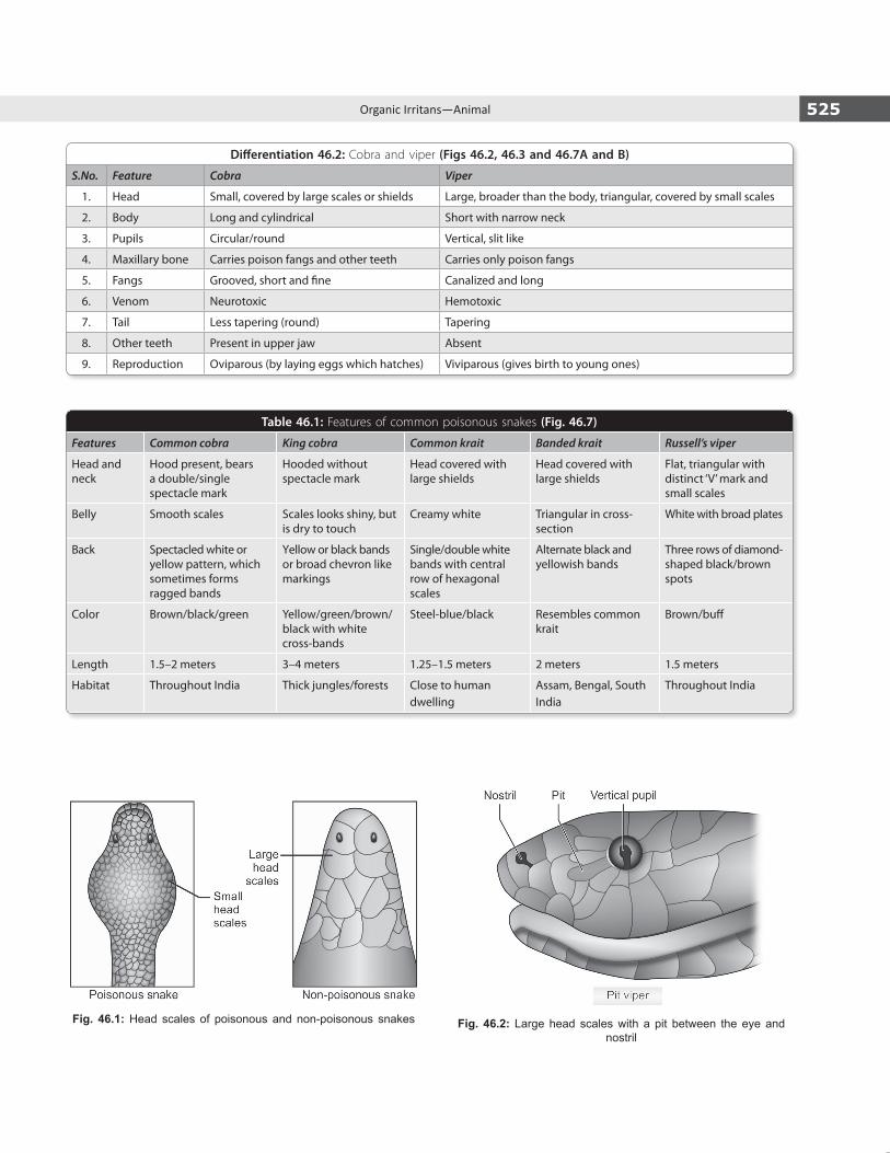
525Organic Irritans—Animal
Fig. 46.1: Head scales of poisonous and non-poisonous snakes Fig. 46.2: Large head scales with a pit between the eye and nostril
Differentiation 46.2: Cobra and viper (Figs 46.2, 46.3 and 46.7A and B)
S.No. Feature Cobra Viper
1. Head Small, covered by large scales or shields Large, broader than the body, triangular, covered by small scales
2. Body Long and cylindrical Short with narrow neck
3. Pupils Circular/round Vertical, slit like
4. Maxillary bone Carries poison fangs and other teeth Carries only poison fangs
5. Fangs Grooved, short and fine Canalized and long
6. Venom Neurotoxic Hemotoxic
7. Tail Less tapering (round) Tapering
8. Other teeth Present in upper jaw Absent
9. Reproduction Oviparous (by laying eggs which hatches) Viviparous (gives birth to young ones)
Table 46.1: Features of common poisonous snakes (Fig. 46.7)
Features Common cobra King cobra Common krait Banded krait Russell’s viper
Head and neck
Hood present, bears a double/single spectacle mark
Hooded without spectacle mark
Head covered with large shields
Head covered with large shields
Flat, triangular with distinct ‘V’ mark and small scales
Belly Smooth scales Scales looks shiny, but is dry to touch
Creamy white Triangular in cross-section
White with broad plates
Back Spectacled white or yellow pattern, which sometimes forms ragged bands
Yellow or black bands or broad chevron like markings
Single/double white bands with central row of hexagonal scales
Alternate black and yellowish bands
Three rows of diamond-shaped black/brown spots
Color Brown/black/green Yellow/green/brown/ black with white cross-bands
Steel-blue/black Resembles common krait
Brown/buff
Length 1.5–2 meters 3–4 meters 1.25–1.5 meters 2 meters 1.5 meters
Habitat Throughout India Thick jungles/forests Close to human dwelling
Assam, Bengal, South India
Throughout India

526 Review of Forensic Medicine and Toxicology
Fig. 46.3: Large head scales and third labial touches the eye and nasal shields
Fig. 46.4: (A) Central hexagonal scales on the middle of back; (B) Fourth infralabial is the largest
Fig. 46.5: Belly scales of poisonous and non-poisonous snakes
Fig. 46.6: (A) Poisonous; (B) Non-poisonous snakes
A B
A B

527Organic Irritans—Animal
Note: Myotoxic venom produces generalized muscular pain ending in respiratory failure in fatal cases, e.g. sea snakes.
Differentiation 46.3: Neurotoxic and vasculotoxic venom
S.No. Feature Neurotoxic venom Vasculotoxic venom
1. Action It causes muscular weakness of legs and paralysis of muscles of face, throat and respiration
It causes enzymatic destruction of cell walls and coagulation disorders
2. Site Acts on motor nerve cells and resembles curare Acts on endothelial cells of blood vessels, and red cells are lysed—hemolysis
3. Local symptoms Minimum Severe—swelling, oozing of blood and spreading cellulitis
4. Symptoms Cobra venom produces both convulsions and paralysis, while krait causes only paralysis4
Hemorrhage from external orifices is common7
5. Examples Elapids, like cobra or kraits5,6 Vipers7,8
Figs 46.7A to C: (A) Cobra; (B) Viper; (C) Common Krait
A
B
C
Poison apparatus: It is a modified salivary gland consisting of:
� Gland: Lies just below and behind the eyes, one on each side of the head, above the upper jaw.
� Duct: Arises from the gland to carry the poisonous venom from gland to the fangs.
� Fangs: Two in number. These are curved teeth situated on the maxillary bones and lie along the jaws. They are like hollow hypodermic needles (solid in non-poisonous snakes).
Snake venom: Venom is the saliva of snake, ejected from the poison apparatus (modified parotid gland) during the act of biting. It can be neurotoxic, vasculotoxic or myotoxic in its action (Diff. 46.3).
� Physical appearance: Faint transparent yellow and viscous, when fresh.
� Toxic principles: Proteinous in nature, most of which are glycopolypeptides and are enzymatic in action. About 80–90% of viperidae and 25–70% of elapidae venom consists of enzymes.
� Fibrinolysins � Neurotoxins (elapid venom)� Proteolysins � Cholinesterase (elapid venom)9
� Hemolysins � Thromboplastin (viper venom) (viper venom)� Agglutinins � Cardiotoxins� Coagulase � Hyaluronidase� Phospholipase � Lecithinase
Fatal dose
Snake Fatal dose (dried form)� Cobra 15 mg� King cobra 12 mg� Common krait10 2.5–6 mg� Banded krait 10 mg� Russell’s viper 40 mg� Saw-scaled viper 8 mg
Russell’s viper injects 63 mg of venom on an average. The range of venom injected is 5–147 mg.
Fatal period: Death may occur immediately from shock due to fright.
� Cobra: ½–24 hours (h). � Viper: 1–4 days.
Signs and Symptoms of Ophitoxemia
Ophitoxemia characterizes the clinical spectrum of snakebite envenomation.11
� Epidemiology: Snakebite is more prevalent in rural than urban areas, commonly seen in summers, in males, farmers and mostly at night. Most of the bites in tropical countries are on lower extremities since

528 Review of Forensic Medicine and Toxicology
the victims are bitten by treading on or near the snake, while in non-tropical countries most bites are on fingers and hands because of deliberate handling of the snake.
� History: The time elapsed since the bite is important to determine if the process is confined locally or if systemic signs have developed. Obtain a description of the snake to determine its species.
� Onset of symptoms and sudden progression are more common with elapidae bite rather than viperidae. Most cobra, krait and sea snake bites would show symptoms within the first 6 h, the shortest time is for the sea snakes.
� Many bites by the poisonous snakes are dry bites implying that the snakes fail to inject the venom. In general, about 70% of bites are due to non-poisonous snakes, and of the rest, 15% are dry bites and only 15% bites cause envenomation.
� About 80% of venomous snake bite in India is by saw-scaled viper. The likelihood of a ‘dry bite’ is most common with a cobra.
� Early and intense pain implies significant enven-omation.
� Local signs and symptoms: Fang marks, pain, bleeding, brui sing, lymphangitis, lymph node enlargement, inflamma tion, blistering, local infection, abscess formation and necrosis.
Cobra (Diff. 46.4)
Local symptoms start within 6–8 minutes (min). � A small reddish wheal develops at the site of the
bite. Bitten area is tender with a burning pain. The
wheal turns putrid in 1–2 days, sometimes purplish and sloughing occurs.
� Early symptoms include vomiting, heaviness of eyelids, blurred vision, paresthesia around mouth, hyper acusis, headache, dizziness, vertigo, hyper-salivation, congested conjunctiva and gooseflesh.
� Muscles of the extremities become weak. Paralysis starts in the lower limbs, which ascends gradually affecting the respiratory muscles, including the diaphragm.12,13 Respiratory muscle paralysis is indicated by poor neck lift, falling single breath count, falling SpO2, hypoxic symptoms such as cyanosis, altered sensorium and coma.
� Drooping of the head, lower lip and eyelids with blurring of vision and external ophthalmoplegia. Ptosis is one of the commonest and earliest manifestations of neuroparalytic snake bite.13
Krait: Signs and symptoms are similar to cobra poisoning, but less rapid.
� Abdominal pain, ptosis, dysarthria, dysphagia, chest pain, quadriparesis, respiratory paralysis and death may occur.
� There is no nausea and froth, but drowsiness is more. � Common krait hunt nocturnally and are quick to bite
people sleeping on the floor, often without waking their victims since the venom is painless. Victims wake up later, paralyzed or die in their sleep.14
Viper (Diff. 46.4)
� More local reaction is seen along with pain and oozing. � Local necrosis is extensive which may lead to gangrene. � Serous and serosanginous blisters sometimes appear.
Differentiation 46.4: Signs and symptoms of elapinae and viperine bite
S.No. Feature Elapinae bite Viperine bite
1. Local reaction Minimal Extensive
2. Speech and deglutition Affected Not so
3. Tongue Paralyzed Not affected
4. Saliva Hypersalivation Not so
5. Pupils Normal Dilated
6. Gait Staggering Not so
7. Gangrene Wet type, early onset Dry type, late onset
8. Blood pressure Normal Hypotension
9. Pulse Initially normal, later irregular Weak, irregular
10. Respiration Slow, weak and labored Quick and labored
11. Coagulation Not affected Greatly affected
12. Hemorrhagic manifestations Absent Prominent feature
13. Cause of death Respiratory paralysis Circulatory failure

529Organic Irritans—Animal
� Bilateral parotid swelling (‘viper head’), conjunctival edema and subconjunctival hemorrhage.
� Petechial hemorrhages, epistaxis, gum bleeding, hemoptysis, hemetemesis, hematuria, fundal hemorrhage, and bleeding from the bite site and rectum are common.
� Acute renal failure evidenced by oliguria, anuria and rising serum creatinine due to DIC or circulatory collapse and shock may be seen.Death is due to circulatory failure in early phase,
and hemorrhagic complications later.
Sea Snake � The bite is usually painless with minimal or no local
swelling or involvement of local lymph nodes. � Early symptoms include headache, a thick feeling of
the tongue, thirst, sweating and vomiting. � Generalized rhabdomyolysis: Muscles, especially of the
neck, trunk and proximal part of the limbs may become tender and painful on active or passive movement, and later may become paralyzed with ptosis as in elapid envenoming.15
� Myoglobinuria may be seen within 3 h after the bite.
Snake Venom OphthalmiaIf the ‘spat’ venom enters the eyes, there is immediate and intense burning, stinging pain, followed by profuse watering of the eyes with production of whitish discharge, congested conjunctiva, spasm and swelling of the eyelids, photophobia and clouding of vision.
Signs and symptoms of ophitoxemia depend upon:i. Patient characteristics
� Age, size, sex and health of the patient: Men have more resistance than women. Children are more vulnerable as they have lesser body mass and fat, and more rapid circulation.
� Location of bite: Bites on the extremities, bony part, through clothing or foot wear or in adipose tissue are less dangerous (absorption delayed due to poor blood supply and loculation) than those on the head (richly vascular and proximity to systemic circulation), trunk or directly into blood vessel. Nearly 98% of snakebites occur over extremities.
ii. Snake characteristics � The length of time the snake holds on. � The extent of anger or fear that motivates the snake, and
nature of bite. � The condition of its fangs and venom glands. � Species and size of the snake. � The amount of venom injected. It is poisonous when injected
subcutaneously, IV or IM, and has no ill effects when taken by mouth as it is not absorbed from gastric mucosa.
Diagnosis20 min whole blood clotting test (20 WBCT): This is very useful and informative bedside test.
� Place a few ml of freshly sampled venous blood in a clean, dry, glass tube/bottle.
� Leave it undisturbed for 20 min at room temperature. � Gently invert the tube. � If the blood is still unclotted and runs out, the patient
has hypofibrinogenemia (‘incoagulable blood’) as a result of venom-induced consumption coagulopathy.A normal 20 WBCT and clot lysis would exclude viperidae
species.Simultaneously, a single breath counting test* is done
in suspected elapidae bites, and the same is repeated at 15 min interval over the first 2 h.
Clinical examination � To exclude early neurotoxic envenoming, the patient is asked
to look up, and observe whether the upper lids retract fully. � Eye movements are tested for evidence of early external oph-
thalmoplegia. � The patient is asked to open his mouth and protrude his tongue;
early restriction in mouth opening may indicate trismus (sea snake envenoming) or more often paralysis of pterygoid muscles.
� Other muscles innervated by the cranial nerves (facial muscles, tongue and gag reflex) are checked. The muscles flexing the neck may be paralyzed, giving the ‘broken neck sign.’
Management
� All patients with a history of snake bite should be observed for 8–12 h after the bite, if the skin is broken and the offending snake cannot be positively identified as non-poisonous.
� The Latin maxim ‘primum non nocere’ (first, do no harm) has significant meaning here because many traditional and popular, but poorly substantiate treatments may cause more harm than good. These methods include making an incision over the bite, mouth suctioning, tourniquet around the limb, use of snake stones, ice packs or electric shock.16
Prevention of Spread of VenomSpread of venom through the body is mostly by diffusion through lymph circulation.
� Reassurance: The victim is reassured since most bites are non-venomous.
� Immobilization: The bitten limb should be immobi-lized with a splint or sling (any movement or muscular contraction hastens systemic absorption of venom), and should be kept below the level of the heart.
* Single breath counting is how far an individual can count in normal speaking voice after a maximal inspiration.

530 Review of Forensic Medicine and Toxicology
� Pressure-immobilization for elapid bites is recom-mended, as it may delay systemic absorption of venom (indicated if the patient is > 1 h from medical care).
� Avoid manipulation: Any interference with the bite wound may introduce infection, increase absorption of venom and increase local bleeding.16,17 Any constriction bands, pressure bandages, jewelry, watches and rings adjacent to the bite site should be removed.
� Pressure immobilization technique is recommended for elapid bites, including sea snakes but should not be used for viper bites (may cause local necrosis). A compression bandage (e.g. elastic/crepe bandage or torn clothing, and not a tourniquet) should be wrapped firmly (maintaining a pressure of 50–70 mmHg) from the bite site upwards (Fig. 46.8).18 This procedure (Sutherland wrap) is to occlude the lymphatic circulation without impeding the arterial or deep venous flow (if occluded, it could result in gangrene or necrosis). The bandage should allow for the insinuation of one finger, and peripheral pulse (radial, posterior tibial, dorsalis pedis) is palpable.
� Pressure pad or ‘Monash technique’: In this method a hard pad of rubber or cloth is applied directly to the wound in an attempt to reduce venom entering the system.
� Tourniquets: Tight rope, belt, string, cloth has been traditionally used to stop venom flow into the body following snake bite.
� Washing increases the flow of venom into the system by stimulating the lymphatic system.17
� If there is an ulcer or wound in the mouth, sucking may allow the venom to get into bloodstream.
The issue which confronts the doctor when attending to a patient with snakebite is assessment of the degree of systemic envenomation and decision on dose of ASV. Using the snakebite envenomation severity scale (SESS), the severity of envenomation should be graded (Table 46.2). Antivenom is indicated for patients with clinical manifestations attributable to snake venom who are graded as moderate and severe according to the SESS (Flow chart 46.1).
Flow chart 46.1: Management of snake bite poisoning
Fig. 46.8: (A) Pressure immobilization technique for lower limbs and (B) ForearmBA
Table 46.2: Assessment of severity of envenomationCategory FindingsNo envenomation Absence of local or systemic reactions,
fang marks +/-.Mild envenomation Fang marks, moderate pain, minimal
local edema (0–15 cm), erythema +, ecchymosis +/-, no systemic reactions.
Moderate envenomation Fang marks +, severe pain, moderate local edema (15–30 cm), erythema +, ecchymosis +, systemic weakness, sweating, syncope, nausea, vomiting, or thrombocytopenia and anemia.
Severe envenomation Fang marks +, severe pain, severe local edema (> 30 cm), erythema +, ecchymosis +, hypotension, parasthesia, coma and respiratory failure.

531Organic Irritans—Animal
Antivenom Treatment
� The lyophilized polyvalent antisnake venom (ASV) serum available in India is effective against common poisonous snakes (cobra, common krait, saw scaled viper and Russell’s viper).19
� Dose: Freeze-dried (lyophilized) antivenom serum is dissolved in water (10 ml vial). About 80–100 ml serum should be diluted in 200–500 ml of isotonic saline and given slow IV.
� The recommended initial dose of ASV is 8–10 vials administered via IV route over a period of 1 h.20 Repeat doses for vasculotoxic species is based on the 6 h rule (depending on the coagulation profile); maximum recommended dose is 30 vials. Repeat doses for neurotoxic is based on the 1 –2 h rule (depending on whether patients have not improved, or worsened); maximum dose is 20 vials. However, evidence-based medicine found no difference in the outcome in low-dose ASV regimen to standard recommended dose.
Indications for antivenom administrationSystemic envenoming
� Hemostatic abnormalities: Spontaneous systemic bleeding, coagulopathy or thrombocytopenia
� CNS: Ptosis, external ophthalmoplegia, paralysis � CVS: Hypotension, shock, cardiac arrhythmia, abnormal ECG � Acute renal failure: Oliguria/anuria, elevated creatinine/urea � Hemoglobin/myoglobinuria, other evidence of intravascular
hemolysis or generalized rhabdomyolysis
Local envenoming � Local swelling involving more than half of the bitten limb (in
the absence of a tourniquet) � Swelling after bites on the digits (toes, and especially fingers) � Rapid extension of swelling � Enlarged tender lymph node draining the bitten limb.
� Antivenom treatment should be given as soon as it is indicated.
� Contraindications: There is no absolute contraindication to ASV treatment.
� ASV should not be used indiscriminately because it carries a risk of severe adverse reactions, and is costly and may be in limited supply.
� Patients must be closely observed for at least 1 h after starting IV antivenom, so that early anaphylactic reactions can be detected.
� Snakes inject the same dose of venom into children and adults. Children must therefore be given exactly the same dose of ASV as adults.
Antivenom reactions � Anaphylactic or type I (immediate) hypersensitivity reactions may
develop (itching, urticaria, glottis edema, wheezing, cough, nausea, vomiting, fever and tachycardia). Adrenaline, 0.5–1.0 ml of 0.1% solution (1 in 1000, 1 mg/ml) is given subcutaneously in adults; in children the dose is 0.01 ml/kg.
� Serum sickness is a type III (delayed) hypersensitivity reaction which is characterized by fever, urticaria, lymphadenopathy and arthritis, and may develop in 3 days to 3 weeks.21 Serum sickness is dose-related, as it occurs when > 8 vials of polyvalent ASV are administered.
Supportive Treatment
Ventilatory care � Patient should be nursed in lateral position, and
salivation should be cleaned timely to prevent aspiration. � Endotracheal intubation, O2 supplementation and
tracheostomy may be needed in neuroparalytic bite.
Antibiotics � Broad spectrum antibiotics should be given, if there
is wound infection. � Tetanus toxoid or tetanus immunoglobulin of human
origin is given.
Surgical excision: Early surgical intervention is needed to prevent extension of infection and development of gangrene. Surgical debridement of necrotic tissue is helpful, but the use of fasciotomy is highly questionable. Fasciotomy does not remove or reduce any envenomation. It is indicated only for compartment syndrome.
Anticholinesterase (ACE) (‘Tensilon’/edrophonium test) � ACE is effective and safe in elapid bite. � Atropine (0.6 mg in adults and 50 µg/kg in children)
is given IV (to prevent undesirable muscarinic effects of acetylcholine, such as increased secretions, sweating, bradycardia and colic) followed by an IV injection of edrophonium chloride (10 mg in adults, 0.25 mg/kg in children) or neostigamine (0.01–0.04 mg/kg every 1–3 h).22
� Patient can be then maintained on neostigmine (50-100 mg/kg) and atropine (4 hourly continuous infusions).
Hypotension and shock � Fluid resuscitation with normal saline or Ringer’s
lactate should be initiated. � Plasma expander, 5% albumin (10–20 ml/kg), fresh
whole blood or fresh frozen plasma should be infused, if CVP is low.

532 Review of Forensic Medicine and Toxicology
� Dopamine (starting dose 2.5–5 µg/kg/min IV) can be given.
Oliguria and renal failure � Cautious rehydration, diuretics (furosemide) or
dopamine should be tried in case urine output drops to < 400 ml/24 h.
� Hemofiltration or peritoneal or hemodialysis, as indicated (acute renal failure seen in vasculotoxic bite).
Hemostatic disturbances � Fresh blood, fresh frozen plasma, cryoprecipitate
or platelet concentrates, as needed in viperine snakebites.
� Heparin 1000–5000 IV may be given, if there are clotting abnormalities (e.g. DIC). Use of heparin should be weighed against risk of bleeding, and hence caution is advocated.
Corticosteroid therapy: No beneficial effects.
Snake venom ophthalmia: The eye or mucous membrane should be washed immediately using large volumes of water or other bland fluid.
Antivenom is immunoglobulin [usually the enzyme refined F(ab)2 fragment of IgG] purified from the serum/plasma of a horse or sheep that has been immunized with the venoms of one or more species of snake. It is of two types:i. Monovalent (specific) antivenom: ASV has been raised
against the venom of the snake that has bitten the patient and it contains specific antibody that will neutralize that particular venom.
ii. Polyvalent (polyspecific) antivenom: It neutralizes the venoms of several different species of snakes, usually the mostes in a particular geographical area. It is less potent, less
immunogenic and less effective than movovalent, and has more adverse effects (caused particularly by non-neutralized part of the polyvalent ASV).
� In India, polyvalent ASV is raised in horses using the venoms of the four most important venomous species (cobra, krait, Russell’s viper and saw-scaled viper).
� The most commonly used ASV in the US for pit viper (rattlesnake, copperhead and water moccasin) is CroFab, which has a much lower incidence of acute or delayed allergic reactions compared to the older ASV.
� Species identification: DNA swab taken from fang marks on people bitten by snakes can correctly identify the species of the biting snake. Most of the time the snake is not brought to the hospital, and hence, the species cannot be identified. Positive identification of the species of snake is critical for effective treatment.
� Radio-immunoassay (RIA) and enzyme-linked immunosorbent assay (ELISA) can identify the nature of venom from the bite site.
� Recent studies have reported the beneficial effects of IV immunoglobulin (IVlg) which may improve coagulopathy, though its effect on neurotoxicity is doubtful.
� Snake venom metalloproteases (SVMPs) are responsible for hemorrhage and tissue degradation at viperine bitten site. Antivenom’s inability to offset viper venom-induced local toxicity has been a basis for an insistent search for SVMP inhibitors. In this context, N-acetyl cysteine (NAC), compound 5d and Cassia auriculata leaf methanol extracts are potent agents and its results are encouraging.
� A compound extracted from the plant Hemidesmus indicus (2-hydroxy-4 methoxy benzoic acid) has been found to have potent anti-inflammatory, antipyretic and anti-oxidant properties, particularly against Russell’s viper venom.
� An aqueous extract of Mimosa pudica root possesses compounds which can neutralize the toxic effects of the cobra and krait venoms.
Complications: Compartment syndrome, tissue necrosis and bleeding diathesis. CVS and hematologic complications and pulmonary collapse may occur.
Postmortem Findings
� Poisonous snakes leave two fang marks (occasionally one) slightly separated from each other and also small marks of other teeth (Fig. 46.9A).*
� Non-poisonous snakes leave a semicircular set of teeth-marks (Fig. 46.9B).
� The bite marks are 1–1.5 cm deep in colubrine and 2.5 cm deep in viperine bites. These should be searched for with a magnifying lens, if not visible to the naked eye.
� In viperine bite, there is discoloration, swelling and cellulitis about the mark, and hemorrhages occur from the puncture site and mucous membranes. Petechiae are also found in mucosa of the urinary bladder, stomach and intestines. The regional lymph nodes are swollen and hemorrhagic. Hemorrhages into the bowel and lungs, and endocardial hemorrhages may be seen. Kidneys are inflamed, and may show medullary hemorrhages, tubular necrosis, cortical necrosis and interstitial nephritis. Internal organs are congested.
� In elapidae bite, the site of bite contains fluid and hemolyzed blood causing staining of vessels, and there are no definite appearances indicating the cause of death, except the signs of asphyxia.
� Neostigmine appears to have no useful role in confirmed presynaptic envenoming (kraits and Russell’s viper). It is effective in postsynaptic neurotoxins (cobra). Their use helps reduce the consumption of ASV, which is generally limited.
� Oral ASV: Alginate coated polyvalent ASV administered by oral route has been found to be effective in entrapping all the structural components of ASV, which on release and intestinal absorption effectively reconstituted the function of antivenom in neutralizing viper and cobra venom.
* The bite may not show the characteristic arched row of teeth, and sometimes it is hard to demonstrate even a single fang bite mark in the skin, especially in kraits.

533Organic Irritans—Animal
Cantharides (Spanish Fly)
Introduction: The Spanish fly (Cantharis vesicatoria, blister beetle) is 2 × 0.6 cm in dimensions. The powder of the dried body is grayish-brown and contains shiny green particles.
Active principle: Cantharidin.
Action: It is locally irritant and nephrotoxic agent.
Absorption: Cantharidin is readily absorbed from all surfaces, including the skin.
Signs and Symptoms
Externally, on application to the skin, redness and burning pain are produced which is followed by formation of vesicles.
On ingestion, there is burning sensation in the mouth, throat and abdomen, nausea and vomiting of blood-stained material, pain in abdomen, severe thirst, tenesmus and difficulty in swallowing and speech. Later, a dull pain is felt in the loins, desire to micturate, but urine is scanty and bloodstained. Priapism in males and abortion in pregnant females may occur.23 The patient becomes prostrated with convulsions, and coma preceding death.
Fatal dose: 15–30 mg of cantharidin or 1.5 g of powder.
Fatal period: 24 h.
Treatment
Gastric lavage, demulcents (but not fat) and symptomatic treatment.
Postmortem FindingsExternal: Inflammation and vesicles are seen in the mouth.
Internal i. GIT: The mucous membrane of the esophagus
and stomach is often swollen and engorged, and may show patches of ulceration and hemorrhages. Stomach may contain shiny greenish particles of the insect.
ii. Kidneys: Congested with hemorrhage in the pelvic calices.
iii. Lungs: Edematous and congested with subpleural hemorrhagic spots.
Figs 46.9A and B: External features of snake bite: (A) Poisonous snake; (B) Non-poisonous snake
Medico-legal Aspects
� Whether or not antivenom is given, any patient with signs of envenomation should be observed in hospital for at least 24 h.
� Allegation of negligence may be leveled against the doctor in failure to authenticate and identify the snake (when killed snake is brought along with the patient) with subsequent development of envenomation and death, especially when the snake was initially claimed to be a non-venomous snake.
� Poisoning is usually accidental. � Occasionally, a murder is committed by throwing
a poisonous snake on the bed of sleeping person. � It is very rarely used for suicide. Queen Cleopatra
is said to have committed suicide after her forces were defeated in battle. She chose to submit to the bite of an asp (an exotic variety of viper), rather than humiliation by her enemies.
� The bodies of animals killed by snake poisoning may be eaten without ill-effects, but their blood is poisonous and is fatal, if injected into the human body.
� Cattle are sometimes poisoned by snake venom.
A
B

534 Review of Forensic Medicine and Toxicology
Medico-legal Aspects
� It is used as counterirritant to the skin in the blistering plaster, as aphrodisiac, and as hair oils to promote growth. So, accidental poisoning may occur.
� It is used as an abortifacient � Suicide/homicide is rare.
Scorpions
Introduction: About 100 species of scorpions are found in India. These are eight-legged arthropods and the end part of tail has two poisonous glands and a sting. Fatal cases have been reported from Maharashtra and Bihar due to acute pulmonary edema caused by Indian red scorpion (Mesobuthus tamulus).
Physical properties: The venom is a clear, colorless, proteinous toxalbumen, having hemolytic and neurotoxic effect.24 Its toxicity is more than that of snakes, but only a small quantity is injected.
Action: The venom is a potent autonomic stimulator, resulting in the release of massive amounts of catecholamines from the adrenals. It has also some direct effect on the myocardium.
Signs and Symptoms
Dysfunction of cranial nerve and hyperexcitability of skeletal muscles develop within hours.
Local: Little swelling, but prominent radiating pain, reddening, paresthesia, and hyperesthesia which is accentuated by tapping on the affected area (tap test).
Systemic effects are nausea, vomiting, restlessness, fever, headache, giddiness, blurred vision, abnormal eye movement, profuse sweating and salivation, lacrimation, rhinorrhea, slurred speech, muscular fasciculations, jerking and shaking (may be mistaken for a seizure), slow pulse, cyanosis, convulsions, coma and respiratory depression, and death may occur from pulmonary edema or cardiac failure in children.25
Complications: Tachycardia, hypertension, arrhythmias, hyperthermia, rhabdomyolysis and acidosis.25
Treatment
i. The limb is immobilized and a pressure bandage is applied proximal to the site of sting.
ii. The site may be incised and washed with water or weak solution of ammonia, borax or KMnO4.
iii. Prazosin therapy: Prazosin 30 µg/kg/dose (1 mg for adult, 500 µg for children) is given orally and
then after every 3 h till extremities are warm, dry and peripheral veins are visible.26
iv. Scorpion antivenom (SAV) is specific antidote to scorpion venom. SAV against Indian red scorpion is available. Recovery is better by simultaneous administration of SAV and prazosin compared with prazosin alone.
v. Calcium gluconate 10 ml of 10% solution slow IV is given for pains, cramps and edema.
vi. Barbiturates/chlorpromazine is given to sedate and control convulsion.
vii. Atropine to prevent pulmonary edema. viii. Symptomatic treatment.
Postmortem Findings
Affected site is swollen. Sting may be found at the site. The area may show ecchymosis. Pulmonary edema and myocardial infarction may be seen.
Medico-legal aspects: Poisoning is usually accidental.
Bees and Wasps
Introduction � A bee sting is strictly a sting from a bee (honey bee,
bumblebee or sweat bee). In common parlance, it can mean a sting of a bee, wasp, hornet or yellow jacket.
� When a honey bee stings a person, it cannot pull the barbed stinger back out. It leaves behind not only the stinger, but also part of its abdomen and digestive tract, muscles and nerves. This massive abdominal rupture kills the honey bee. Honey bees are the only species of bees to die after stinging.
� Painful and sometime, fatal reactions occur in humans.
Active principles � Bee venom contains dopamine, histamine, neurotoxin
enzymes and toxic peptides. � Wasp venom, in addition, contains serotonin and
kinins. � Ant venom mainly contains alkaloids, solenopsin-A
and proteins.
Signs and Symptoms
Locally, there is pain, itching, redness and slight swelling at the site of the sting. Stings of the mouth, throat, and sometimes of the face, neck or upper limbs may cause edema of the larynx or pharynx and obstruction.
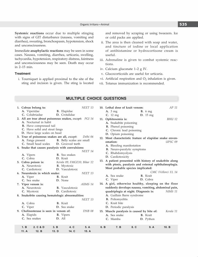
535Organic Irritans—Animal
Systemic reactions occur due to multiple stinging with signs of GIT disturbance (nausea, vomiting and diarrhea), sweating, bronchospasm, hypotension, shock and unconsciousness.
Immediate anaphylactic reactions may be seen in some cases. Nausea, vomiting, diarrhea, urticaria, swelling, tachycardia, hypotension, respiratory distress, faintness and uncon sciousness may be seen. Death may occur in 2–15 min.
Treatment
i. Tourniquet is applied proximal to the site of the sting and incision is given. The sting is located
and removed by scraping or using tweezers. Ice or cold packs are applied.
ii. The area is then cleaned with soap and water, and tincture of iodine or local application of antihistamine or hydrocortisone cream is useful.
iii. Adrenaline is given to combat systemic reac-tions.
iv. Calcium gluconate 1–2 g IV. v. Glucocorticoids are useful for urticaria. vi. Artificial respiration and O2 inhalation is given. vii. Tetanus immunization is recommended.
MULTIPLE CHOICE QUESTIONS
1. Cobras belong to: NEET 13 A. Viperidae B. Elapidae C. Colubriadae D. Crotalidae 2. All are true about poisonous snakes, except: PGI 14 A. Nocturnal in habit B. Have compressed tail C. Have solid and stout fangs D. Have large scales on head 3. True of poisonous snakes are all, except: Delhi 06 A. Fangs present B. Belly scales are small C. Small head scales D. Grooved teeth 4. Snake that causes paralysis with convulsions:
NEET 14 A. Vipers B. Sea snakes C. Cobra D. Krait 5. Cobra poison is: Kerala 09; FMGE10; Bihar 12 A. Neurotoxic B. Myotoxic C. Cardiotoxic D. Vasculotoxic 6. Neurotoxin in which snake: NEET 13 A. Viper B. Krait C. Sea snake D. None 7. Viper venom is: AIIMS 14 A. Neurotoxic B. Vasculotoxic C. Myotoxic D. Cardiotoxic 8. Snakebite causing hematologic abnormalities: NEET 13 A. Cobra B. Krait C. Viper D. Sea snake 9. Cholinesterase is seen in venom of: DNB 08 A. Elapids B. Vipers C. Sea snakes D. All
10. Lethal dose of krait venom: AP 11 A. 3 mg B. 6 mg C. 12 mg D. 15 mg 11. Ophitoxemia is: BHU 12 A. Snakebite poisoning B. Phenol poisoning C. Chronic lead poisoning D. Opium poisoning 12. Most characteristic feature of elapidae snake enven-
omation: UPSC 09 A. Bleeding manifestation B. Neuro-paralytic symptoms C. Rhabdomyolysis D. Cardiotoxicity 13. A patient presented with history of snakebite along
with ptosis, paralysis and external ophthalmoplegia. Most probable species implicated:
CMC (Vellore) 13, 14 A. Sea snake B. Krait C. Viper D. Cobra 14. A girl, otherwise healthy, sleeping on the floor
suddenly develops nausea, vomiting, abdominal pain, quadriplegia at night. Diagnosis is: NIMS 11
A. Guillain Barre syndrome B. Poliomyelitis C. Krait bite D. Periodic paralysis 15. Muscle paralysis is caused by bite of: Kerala 11 A. Sea snake B. Krait C. Mamba D. Python
1. B 2. C & D 3. B 4. C 5. A 6. B 7. B 8. C 9. A 10. B 11. A 12. B 13. D 14. C 15. A

536 Review of Forensic Medicine and Toxicology
16. B 17. C 18. C 19. A 20. C 21. B 22. A 23. C 24. D 25. B 26. D
16. Treatment of snakebite all, except: AFMC 11 A. Firm bandage to occlude lymphatic B. Incision over wound C. Reassure the patient D. Immobilization of bitten part 17. Following is/are recommended primary management
of a patient with snake bite, except: PGI 11; NEET 13 A. Splinting and immobilization B. Keep the site of bite below heart C. Wash with soap and water D. Reassure the patient 18. Ligature pressure that should be used to resist spread
of poison in elapidae poisoning: WB 11; MAHE 12 A. < 10 mm Hg B. 20–30 mm Hg C. 50–70 mm Hg D. > 100 mm Hg 19. Polyvalent snake vaccines contains immunoglobins
against all, except: PGI 11 A. Ophiophagus hannah B. Naja naja C. Daboia rusellii D. Bungarus caeruleus 20. In a snake envenomation, antivenom is started by
giving a dose of: NEET 14 A. 2 vials B. 4 vials C. 10 vials D. 20 vials 21. Antisnake venom may cause: CMC (Vellore) 13 A. Type II hypersensitivity reactions B. Type III hypersensitivity reactions
C. Type IV hypersensitivity reactions D. Type V hypersensitivity reactions 22. Drug used for muscarinic symptoms seen in cobra
envenomation: CMC (Vellore) 13 A. Neostigmine B. Pralidoxime C. Prazocin D. Naloxone 23. Priapism occurs in: AIIMS 06; 13 A. Snake bite B. Rati poisoning C. Cantharide poisoning D. Arsenic poisoning 24. Scorpion venom resembles venom of: NEET 14 A. Cobra B. Viper C. Krait D. All of the above 25. A 3-year-old child sleeping in a hut woke up in the
middle of the night screaming. Her mother thought the child had a nightmare and tried to pacify her. After sometime, she noticed that the child was sweating profusely and the hands were becoming cold. She vomited a couple of times. The mother immediately rushed her to the emergency services. Her pulse was 150/minute and her BP 90/60 mmHg. This child is likely to have: COMEDK 14
A. Snake bite B. Scorpion bite C. Septic shock D. Food poisoning 26. Drug used in scorpion bite:
CMC (Vellore) 13, 14; COMEDK 14 A. EDTA B. Neostigmine C. N-acetylcysteine D. Prazosin

Definitions
� Narcotic: It refers to a sleep inducing agent, and initially used to mean the opioids. Currently, the term is used by law enforcement agencies to indicate any illicit psychoactive substance.
� Opiate: It refers to natural alkaloids derived directly from the poppy plant.
� Opioids: They are broader class of agents that are capable of producing opium-like effects on binding to opioid receptors. Endogenous neural polypeptides such as endorphins and enkephalins are natural opioids.
� Toxidrome: A constellation of clinical examination findings that assists in the diagnosis and treatment of the patient who presents with an exposure to an unknown agent.
Opium
Introduction: Opium (poppy, afim, kasoomba or madak chandu) is derived from Papaver somniferum, an annual plant with white or red flowers growing on a central bulbous pod (Fig. 47.1). Crude opium has a characteristic odor and bitter taste.
Distribution: Worldwide.
Toxic part: Unripe fruit capsule, latex juice.1
� Latex is obtained by lacerating (‘scoring’) the immature seed pods; the latex leaks out and dries
to a sticky brown residue (Fig. 47.1). This is scraped off the fruit.
� Seeds are non-poisonous and are called ‘khaskhas’ which constitutes a condiment in cooking (Fig. 47.2).
Active Principles
The latex juice of opium has about 25 alkaloids, divided into two groups.
a. Phenanthrene derivatives (main narcotic constituents) i. Natural alkaloids
z Morphine (10%): White powder/crystals, bitter taste and alkaline in reaction.
z Codeine (0.5%). z Thebaine (0.3%).
ii. Semi-synthetic opioids: They are produced by chemical modification of an opiate and include hydromorphone, diacetylmorphine (heroin, brown sugar or smack), oxymorphone and oxycodone.
iii. Synthetic opioids: These compounds are not derived from an opiate, but binds to an opioid receptor and produce opioid effects clinically. It includes methadone, fentanyl, pentazocine, tramadol and meperidine (pethidine).2
b. Benzyl-isoquinolone derivatives (no significant CNS effects)3
i. Papaverine (1%) ii. Noscapine (6%).
Somniferous Poisons (Narcotic Poisons)
CHAPTER 47
Fig. 47.1: Papaver somniferum Fig. 47.2: Poppy seeds (Non-poisonous)

538 Review of Forensic Medicine and Toxicology
� Alkaloids are complex substance having nitrogenous base, and is found in various plants. Chemically, it behaves like an alkali as it unites with acids to form salts. Its basic quality depends on the pyridine nucleus. In nature, they are usually combined with certain acids to from salts. They act mainly on the CNS, each compound having its own individual action.
� Important alkaloids: Atropine, hyoscine, morphine, quinine, strychnine, cocaine and codeine. Some synthetic substances, such as amphetamine, heroin, pethidine and methadone also behave chemically like alkaloids.4
Action
� Opioids act by binding to opioid receptors on neurons distributed throughout the nervous system and immune system.
� Four major types of opioid receptors have been identified: mu, kappa, delta and the recently recognized OFQ/N. These receptors are the binding sites for endogenous peptides.
� Activation of opioid receptors results in inhibition of synaptic neurotransmission in the CNS and PNS.
Routes of administration: It can be taken by snorting, smoking or chasing (chasing the dragon), intravenously (mainlining) and subcutaneously (skin popping). It can
be mixed with cocaine (known as speed balling) and then taken by addicts.5
Metabolism � Most opioids are metabolized by hepatic conjugation
to inactive compounds that are excreted readily in the urine.
� Certain opioids (e.g. propoxyphene, fentanyl and buprenorphine) are more soluble in lipids and can be stored in the fatty tissues of the body.
CLINICAL TOXIDROME
Some poisons may produce a collection of symptoms (toxidromes) that can assist in making diagnosis and are also useful for anticipating other symptoms that may occur (Table 47.1). The symptoms of an opiate/narcotic toxidrome include the classic triad of respiratory depression, pin-point pupils and impairment of sensorium.
Signs and Symptoms
Peak effects are seen in 10 minutes (min) with IV route, 10–15 min after nasal insufflations, 30–45 min with IM, 90 min after taking orally and 2–4 hours (h) after dermal application. i. Stage of Excitement: It is of short duration. There
is euphoria, increased sense of well-being, freedom
Table 47.1: Clinical toxidromes
Toxidrome Clinical features Common poisons Treatment Site of action
Opiate Respiratory depression and oxygen desaturations, miosis, decreased GI motility, bradycardia, hypothermia and coma
Morphine, codeine, oxycodone, fentanyl
Naloxone Opioid receptor
Anticholinergic
Tachycardia, hyperthermia (mild to severe), agitation, delirium, seizures, mydriasis, dry, flushed skin, urinary retention, decreased intestinal motility
Datura, atropine, scopolamine, antihistamines
Physostigmine Muscarinic acetylcholine receptors
Sympathomimetic
Hypertension, tachycardia, pyrexia, pupillary dilatation, diaphoresis, altered mental status
Cocaine, amphetamines Benzodiazepines α and β adrenergic receptors
Cholinergic (Organophosphate and carbamates)
Bradycardia, respiratory depression, miosis, SLUDGE (salivation, lacrimation, urination, defecation, GIT distress, emesis) CNS: Seizures, comaMuscle: Fasiculations, paralysis
DDT, parathion, malathion, diazinon
Atropine Pralidoxime (for OP insecticides)
Nicotinic and muscarinic acetylcholine receptors
Sedative hypnotic (Benzodiazepines)
Sedation or coma, normal vital signs, diplopia, ataxia, impaired motor function, slurred speech, anterograde amnesia, anxiety, hallucinations, delirium
Alprazolam, flunitrazepam, oxazepam
Flumazenil ϒ-aminobutyric acid receptors

539Somniferous Poisons (Narcotic Poisons)
from anxiety, talkativeness and laughter. Halluci-nations, flushing of face, conjunctival injection and rapid heart rate are seen.
ii. Stage of Stupor: Headache, nausea, vomiting, weak-ness, heaviness in limbs, giddiness, drowsiness, diminished sensibility and strong tendency to sleep from which the patient can be aroused by painful stimuli.
z Pupils are contracted, and face and lips are cyanosed.
z Pulse and respiration: Almost normal. iii. Stage of Narcosis/Coma: Patient passes into deep
coma from which he cannot be aroused. In this stage:6,7
z Muscles: Flaccid and relaxed z Face: Pale z Reflexes: Absent z Conjunctiva: Congested z Skin: Cold with profuse perspiration, all other
secretions are suspended z Pupils: Constricted to pin-point (Fig. 47.3), non-
reacting8,9
z Blood pressure: Hypotension z Temperature: Hypothermia z Pulse: Weak, feeble z Respiration: Slow, steatorous (4–6 breaths/
min)10
z Sphincter tone: Increased (can lead to urinary retention).
During the terminal stages, pink froth comes from the mouth, pulse is slow, irregular and imperceptible, respiration becomes Cheyne-Stokes, and ultimately deep coma and death due to respiratory depression and cardiorespiratory arrest.
Fatal dose � Opium: 2 g. � Morphine: 200 mg.11
� Codeine: 50 mg.
Fatal period: 6–12 h.
Differential Diagnosis
� Intracranial hemorrhage: Cerebrovascular accidents or brain trauma.
� Poisoning: Alcohol, barbiturates, benzodiazepine, carbolic acid, carbon monoxide or organo phosphorus.
� Metabolic conditions: Diabetic and uremic coma. � CNS infections: Meningitis, encephalitis, encephalo-
pathy or cerebral malaria. � Others: Epileptic or hysterical coma, or heat
hyperpyrexia.
Treatment
i. Support vitals through respirator and other emergency procedures.
ii. Decontamination: Stomach wash frequently with 1:5000 KMnO4 leaving some solution in stomach to oxidize the alkaloid that might be secreted in stomach after absorption. Lavage should be carried out even after IV/IM injection of drug, as it is secreted in the stomach.
iii. Administer activated charcoal—method of choice for decontamination following ingestion.
iv. Enema with 30 g of sodium sulfate twice daily. v. Whole-bowel irrigation in body packers. vi. Antidote: Narcotic antagonist naloxone in an initial
dose of 0.4–2 mg IV/IM repeated every 2–3 min upto 10 mg, if no response occurs.12
If there is little response to naloxone alone, possibility of an overdose with a benzodiazepine should be considered, and a challenge with IV flumazenil, 0.2 mg/min upto maximum of 3 mg in an hour might be used.
Detection
Marquis test: It is a simple spot-test to presumptively identify alkaloids. It can be used to test cocaine, opiates and phenethylamines.13
Three ml of concentrated H2SO4 + 3 drops of formalin are added to the suspected sample. Purple-red color is observed which gradually changes to violet.
Postmortem Findings
External i. Smell of opium. ii. Face/body is bluish, deeply cyanosed or blackish. iii. Postmortem staining is purple or blackish. iv. Froth at the nostrils. v. Pupils are constricted, can be dilated also.Figs 47.3: (A) Normal pupil, (B) Pinpoint pupil
A B

540 Review of Forensic Medicine and Toxicology
vi. Allergic reactions to IV heroin may be seen. vii. Needle tracks are found occasionally, depending
on the route of intake.
Internal i. Diffuse cerebral edema. ii. All organs are congested, trachea contains frothy
secretions. iii. Blood is dark and fluid. iv. Stomach may show presence of small, soft brownish
lumps of opium, and smell of drug may be perceived.
Medico-legal Aspects � Negligence may be alleged in cases of prehospital
discharge-on-scene after naloxone treatment followed in most western countries, since this practice is sometimes associated with risk of death due to rebound toxicity after such episodes.
� Opioid poisoning nowadays is from street drugs which not only have brown sugar, but also therapeutic opioids like hydrocodone or oxycodone, codeine taken with glutethemide, and abuse of Subutex (buprenorphine hydrochloride).
� It is a poison of choice to commit suicide (ideal suicidal poison).
� Homicide is rare, because of bitter taste and charact-eristic odor.
� Infanticide by breastfeeding an infant by a woman who had smeared her nipple with tincture opium.
� Accidental opium poisoning is also common. Drugging of children by opium to keep them quiet, and overdose of medicines may result in accidental poisoning.*
� Various nonproprietary formulations, folk remedies, and herbs may contain opium, and administration of these results in unintentional poisoning.
� Sometimes, opium is used for doping racehorses. � It is said to increase libido, hence used as an aphrodisiac. � Some criminals take opium to build courage before
committing a crime. � Opium disappears with putrefaction, so it may not
be detected in putrefied bodies.
� Earlier opioid overdose was treated with analeptics. In 1950s, specific antidotes were introduced: nalorphine and levallorphan which were capable of reversing the respiratory effects by blocking the opioid receptors. However, they have a mixed agonist-antagonist properties that significantly limited their usefulness. Naloxone with its pure opioid antagonistic properties completely replaced nalorphine and levallorphan in the treatment of opioid overdose.
� Nalmefene and naltrexone are newer opioid antagonists that have longer half-lives than naloxone (4–8 h and 8–12 h vs 1 h). The routine use of a long-acting antagonist in the patient who is unconscious for unknown reasons is not recommended.
Body Packers
� Multiple-wrapped packets of illicit drugs (cocaine or heroin) may be ingested or inserted into body cavities by ‘swallowers,’ ‘internal carriers,’ ‘couriers,’ or ‘mules’ to intentionally transport drugs from one country to another. After they arrive at their destination, cathartics are administered so that the packets can be passed and delivered.
� When the authorities discover such individuals or when individuals in custody become ill, they may be brought to a nearby hospital for evaluation and management. Although, these patients generally are asymptomatic on arrival, they are at risk for delayed, prolonged or lethal poisoning as a consequence of packet rupture.
� If there is a suspicion of body packing or body stuffing, careful cavity searches of the rectum and vagina are done. An abdominal X-ray can confirm the diagnosis. Ultrasonography and CT are other recommended diagnostic modalities.
� Polyethylene glycol electrolyte lavage solution is used to flush out the packets. Intestinal perforation or obstruction by the packets may require surgical intervention.
Chasing the Dragon
� Intravenous injection and insufflation are the preferred means of heroin self-administration in the US. In the other countries, including the Netherlands, UK and Spain, the prevalent method is ‘chasing the dragon’.
� In this, users inhale a thick, white pyrolyste that is generated by heating heroin base on aluminum foil using a hand-hold flame. This means of admini-stration produces heroin concentration similar to those observed following IV administration.
* In the early 18th century in the UK, with industrial revolution, 18 h workdays, extra cost in feeding and loss of sleep due to infant’s cries, various concoctions of opium were specifically designed for the purpose of quieting unruly children.
� It should be noted that opioid exposure does not always result in pupillary constriction, and respiratory depression is the most specific sign.10 Other important presenting signs are ventricular arrhythmias, acute mental status changes and seizures.
� Acute lung injury is known sequelae of heroin, propoxyphene and methadone overdose, and present in fatal cases of opioid overdose. The findings are cyanosis, dyspnea, pink frothy sputum, rales, tachypnea and tachycardia.
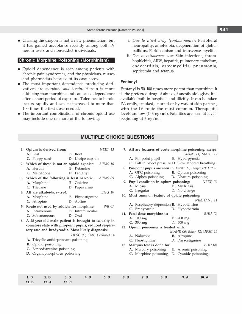
541Somniferous Poisons (Narcotic Poisons)
1. D 2. B 3. D 4. D 5. D 6. B 7. B 8. B 9. A 10. A 11. B 12. A 13. C
MULTIPLE CHOICE QUESTIONS
7. All are features of acute morphine poisoning, except:Kerala 11; MAHE 12
A. Pin-point pupil B. Hyperpyrexia C. Fall in blood pressure D. Slow labored breathing 8. Pin-point pupils are seen in: Kerala 09; Punjab 09; UP 10 A. OPC poisoning B. Opium poisoning C. Alphos poisoning D. Dhatura poisoning 9. Pupil condition in opium poisoning: NEET 13 A. Miosis B. Mydriasis C. Irregular D. No change 10. Most common feature of opiate poisoning:
NIMHANS 11 A. Respiratory depression B. Hypotension C. Bradycardia D. Hypothermia 11. Fatal dose morphine is: BHU 12 A. 100 mg B. 200 mg C. 300 mg D. 500 mg 12. Opium poisoning is treated with:
MAHE 06; Bihar 12; UPSC 13 A. Naloxone B. Atropine C. Neostigmine D. Physostigmine 13. Marquis test is done for: BHU 08 A. Mercury poisoning B. Arsenic poisoning C. Morphine poisoning D. Cyanide poisoning
1. Opium is derived from: NEET 13 A. Leaf B. Root C. Poppy seed D. Unripe capsule 2. Which of these is not an opioid agonist: AIIMS 10 A. Heroin B. Ketamine C. Methadone D. Fentanyl 3. Which of the following is least narcotic: AIIMS 09 A. Morphine B. Codeine C. Thebane D. Papaverine 4. All are alkaloids, except: BHU 10 A. Morphine B. Physostigmine C. Atropine D. Abrine 5. Route not used by addicts for morphine: WB 07 A. Intravenous B. Intramuscular C. Subcutaneous D. Oral 6. A 28-year-old male patient is brought to casualty in
comatose state with pin-point pupils, reduced respira-tory rate and bradycardia. Most likely diagnosis:
UPSC 09; CMC (Vellore) 14 A. Tricyclic antidepressant poisoning B. Opioid poisoning C. Benzodiazepine poisoning D. Organophosphorus poisoning
� Chasing the dragon is not a new phenomenon, but it has gained acceptance recently among both IV heroin users and non-addict individuals.
Chronic Morphine Poisoning (Morphinism)
� Opioid dependence is seen among patients with chronic pain syndromes, and the physicians, nurses and pharmacists because of its easy access.
� The most important dependence producing deri-vatives are morphine and heroin. Heroin is more addicting than morphine and can cause dependence after a short period of exposure. Tolerance to heroin occurs rapidly and can be increased to more than 100 times the first dose needed.
� The important complications of chronic opioid use may include one or more of the following:
i. Due to illicit drug (contaminants): Peripheral neuro pathy, amblyopia, degeneration of globus pallidus, Parkinsonism and transverse myelitis.
ii. Due to intravenous use: Skin infections, throm-bophlebitis, AIDS, hepatitis, pulmonary embolism, endocarditis, osteomyelitis, pneumonia, septicemia and tetanus.
Fentanyl
Fentanyl is 50–l00 times more potent than morphine. It is the preferred drug of abuse of anesthesiologists. It is available both in hospitals and illicitly. It can be taken IV, orally, smoked, snorted or by way of skin patches, with the IV route the most common. Therapeutic levels are low (1–3 ng/ml). Fatalities are seen at levels beginning at 3 ng/ml.
542
Review of Forensic Medicine and Toxicology
Introduction
� Ethanol (ethyl alcohol) is a transparent, colorless, volatile liquid having a characteristic odor and a burning taste with a specific gravity of 0.79.
� Ethanol is produced by the enzymatic action of yeasts on vegetable substrate containing sugars. Direct fermentation cannot raise the concentration to ≥ 12–15% as the yeast is killed, but distillation of primary fermentation can concentrate the alcohol to 40–60% strength. Different types of beverages with percentage of alcohol are given in Table 48.1.
� At low doses, alcohol is said to have beneficial effects, such as decreased rates of myocardial infarction, diabetes, stroke, gallstones and possibly Alzheimer’s dementia, but consumption of two standard drinks per day increases the risk of health problems in many organ systems. Regular consumption of 20–32 g/day for men and 14–27.2 g/day for women are considered as safe limits for drinking, if liver damage is to be avoided (difference between the sexes was due to the lower weight and water-to-body-mass ratio of women).
� Earlier, weekly safe limit was recommended [168–210 g/week (≤ 21 units) of alcohol for men and 98–140 g for women (≤ 14 units)].1 This is not advised, since a study showed that many people were in effect ‘saving up’ their units and using them at the end of the week for binge drinking where the primary intention is to become intoxicated by heavy consumption of alcohol over a short period of time.
� An international study found that persons who reported drinking > 2 units/day had an increased risk of fractures compared to non-drinkers.
� Units of alcohol are a measure of the volume of pure alcohol in alcoholic beverages used as a guideline in some countries. One unit of alcohol is defined as 10 ml in the UK and as 10 g (12.7 ml) in Australia. A standard drink is 30 ml of spirits; 330 ml can of beer or 100 ml glass of wine.
� To calculate standard drinks, the following formula is used: Volume of container (liters) × % alcohol by volume (ml/100 ml) × 0.789 = Number of standard drinks.
Commercial Preparations of Alcohol � Absolute alcohol contains 99.95% ethanol.
� Mineralized methylated spirit consists of 90% ethanol, 9.5% wood naphtha (methanol) and 0.5% pyridine, and is colored pink for easy identification.
� Industrial methylated spirit contains 95% ethanol and 5% methanol, with no coloring agent.
� Surgical spirit consists of 95% of ethanol and 5% methanol with oil of wintergreen to give it a sweetish flavor.
Proof of spirit indicates a mixture containing 57.1% by volume or 49.28% by weight of absolute alcohol. In the US, the term proof refers to twice the percentage of alcohol by volume. So, the common 80-proof whisky sold in the US contains 40% alcohol by volume. In India, the spirit (whisky, rum or brandy) is usually 42.8% by volume and 75-proof.
The concentration written on the labels of most bottles is v/v, i.e. volume of alcohol per volume of drink.
Various country liquors � Mahua: Traditional tribal drink in central and eastern India. It
is made from dried flower of mahua tree (Madhuca longifolia) and chhowa gud (granular mollasses).
� Toddy (palm wine) is made from sap of various species of palm tree. It is common across Asia and Africa.
� Feni: Goan spirit, made from coconut or juice of cashew apple. � Arrack (Arabic araq—sweet liquor usually made from raisins in
those regions) is distilled from coco-palm, rice, sugar or jaggery and has strength of 40–50%. It may be mixed with chloral hydrate or potassium bromide.
� Tharra is made by fermenting the mash of sugarcane juice/pulp in ceramic containers and distilling to high alcohol content.
Inebriants—Alcohol
ChApter 48
Table 48.1: Approximate percentage of alcoholby volume in beverages
Beverage Alcohol by volume
Spirits (whisky, brandy, rum, gin, vodka) 35–50%
Port (fortified with brandy), sherry 17–21%
Wine 10–15%
Champagne 10–13%
Beers, stout, cider 4–8%
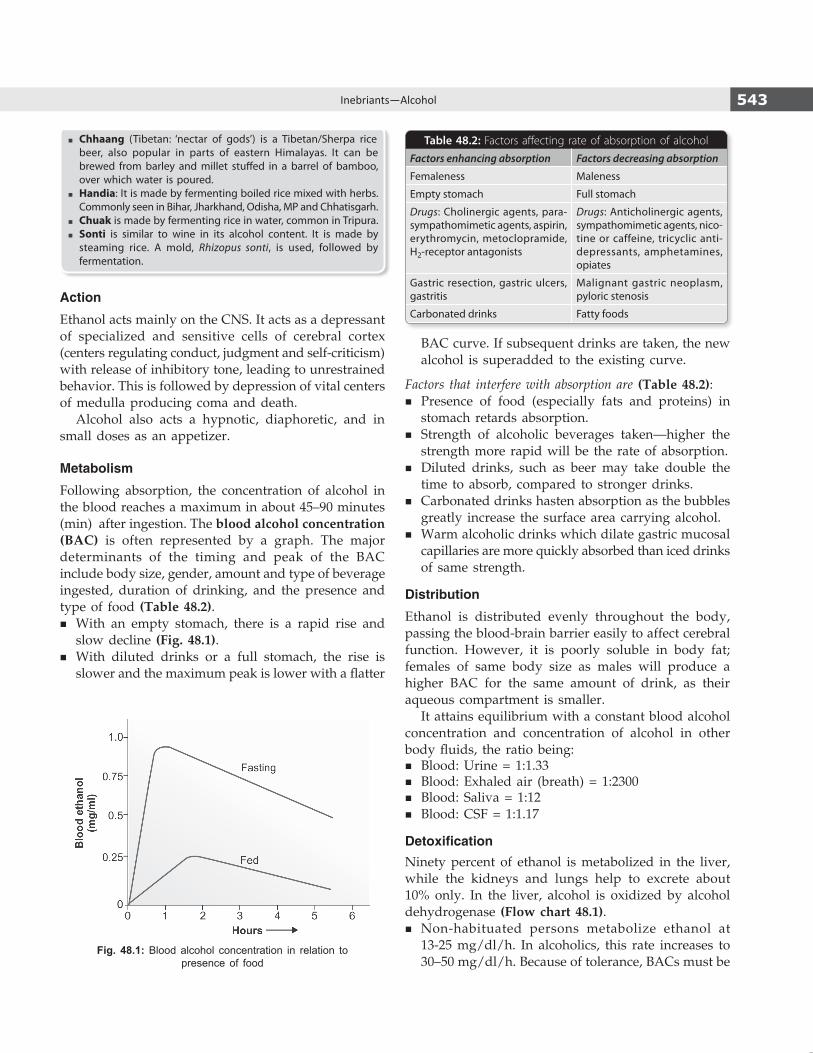
543Inebriants—Alcohol
� Chhaang (Tibetan: ‘nectar of gods’) is a Tibetan/Sherpa rice beer, also popular in parts of eastern Himalayas. It can be brewed from barley and millet stuffed in a barrel of bamboo, over which water is poured.
� Handia: It is made by fermenting boiled rice mixed with herbs. Commonly seen in Bihar, Jharkhand, Odisha, MP and Chhatisgarh.
� Chuak is made by fermenting rice in water, common in Tripura. � Sonti is similar to wine in its alcohol content. It is made by
steaming rice. A mold, Rhizopus sonti, is used, followed by fermentation.
Action
Ethanol acts mainly on the CNS. It acts as a depressant of specialized and sensitive cells of cerebral cortex (centers regulating conduct, judgment and self-criticism) with release of inhibitory tone, leading to unrestrained behavior. This is followed by depression of vital centers of medulla producing coma and death.
Alcohol also acts a hypnotic, diaphoretic, and in small doses as an appetizer.
Metabolism
Following absorption, the concentration of alcohol in the blood reaches a maximum in about 45–90 minutes (min) after ingestion. The blood alcohol concentration (BAC) is often represented by a graph. The major determinants of the timing and peak of the BAC include body size, gender, amount and type of beverage ingested, duration of drinking, and the presence and type of food (Table 48.2).
� With an empty stomach, there is a rapid rise and slow decline (Fig. 48.1).
� With diluted drinks or a full stomach, the rise is slower and the maximum peak is lower with a flatter
BAC curve. If subsequent drinks are taken, the new alcohol is superadded to the existing curve.
Factors that interfere with absorption are (Table 48.2): � Presence of food (especially fats and proteins) in
stomach retards absorption. � Strength of alcoholic beverages taken—higher the
strength more rapid will be the rate of absorption. � Diluted drinks, such as beer may take double the
time to absorb, compared to stronger drinks. � Carbonated drinks hasten absorption as the bubbles
greatly increase the surface area carrying alcohol. � Warm alcoholic drinks which dilate gastric mucosal
capillaries are more quickly absorbed than iced drinks of same strength.
Distribution
Ethanol is distributed evenly throughout the body, passing the blood-brain barrier easily to affect cerebral function. However, it is poorly soluble in body fat; females of same body size as males will produce a higher BAC for the same amount of drink, as their aqueous compartment is smaller.
It attains equilibrium with a constant blood alcohol concentration and concentration of alcohol in other body fluids, the ratio being:
� Blood: Urine = 1:1.33 � Blood: Exhaled air (breath) = 1:2300 � Blood: Saliva = 1:12 � Blood: CSF = 1:1.17
DetoxificationNinety percent of ethanol is metabolized in the liver, while the kidneys and lungs help to excrete about 10% only. In the liver, alcohol is oxidized by alcohol dehydrogenase (Flow chart 48.1).
� Non-habituated persons metabolize ethanol at 13-25 mg/dl/h. In alcoholics, this rate increases to 30–50 mg/dl/h. Because of tolerance, BACs must be
Fig. 48.1: Blood alcohol concentration in relation to presence of food
Table 48.2: Factors affecting rate of absorption of alcohol
Factors enhancing absorption Factors decreasing absorption
Femaleness Maleness
Empty stomach Full stomach
Drugs: Cholinergic agents, para-sympathomimetic agents, aspirin, erythromycin, metoclopramide, H2-receptor antagonists
Drugs: Anticholinergic agents, sympathomimetic agents, nico-tine or caffeine, tricyclic anti-depressants, amphetamines, opiates
Gastric resection, gastric ulcers, gastritis
Malignant gastric neoplasm, pyloric stenosis
Carbonated drinks Fatty foods

544 Review of Forensic Medicine and Toxicology
interpreted in conjunction with history and clinical presentation.
� Excretion of alcohol is mainly by the kidneys, lungs and skin through urine, breath and sweat respectively. If is also secreted in saliva and milk.
A number of metabolic effects from alcohol are directly linked to the production of an excess of both NADH and acetaldehyde.
� NADH is utilized in the conversion of pyruvic acid (intended for conversion into glucose by gluconeogenesis) to lactic acid. The final result may be acidosis from lactic acid build-up and hypoglycemia from lack of glucose synthesis.
� Excess NADH may be used as a reducing agent in two pathways—one to synthesize glycerol (from a glycolysis intermediate) and the other to synthesis fatty acids. As a result, heavy drinkers may initially be overweight (‘beer belly’).
� The accumulated acetaldehyde acts by inhibiting the mitochondrial reactions and functions. There is a vicious cycle—high acetaldehyde level impairs mitochondria function, metabolism of acetaldehyde to acetic acid decreases, more acetaldehyde accumulates and causes further liver damage—hepatitis and cirrhosis.
� Acetaldehyde may be responsible for the development of alcohol addiction.
Signs and Symptoms (Acute Poisoning)
i. Stage of Excitement (Blood level: 50–150 mg%) z Person will be euphoric (sense of well-being).
Actions, speech and emotions are less restrained due to lowering of the inhibition normally exercised by the higher centers of the brain. It alters time and space perception.
z He may perform dancing, thrilling shows, carelessly and fearlessly.
z He might disclose secrets (‘in vino veritas’—in wine there is truth).
z Person might show increase in confidence, but lack of self-control.
z There is lowering of visual acuity. Nystagmus present.
z Mental concentration is poor and judgment impaired.
z Faculty of attention deteriorates. z Recall memory is disturbed, person cannot
accurately recall certain situations or names of individuals.
z It increases the desire for sex, but markedly impairs performance resulting in prolonged intercourse without ejaculation.
ii. Stage of In-coordination (Blood level: 150–250 mg%) z Due to further depression of higher centers,
the person may be morose/cheerful/irritable/ill-tempered/excitable/sleepy, depending on the dominant impulses released.
z Centers of perception and skilled movements are involved—there is clumsiness, in-coordination of fine and skilled movements, and alterations in speech and fine finger movements.
z Nausea and vomiting. z Face: Flushed. z Pulse: Rapid. z Sense of touch, taste, smell and hearing are
diminished. z Hypothermia. z Breath smells of alcohol. z Pupils are dilated and react sluggishly to light.
iii. Stage of Coma (Blood level > 250 mg%) z Thick, slurred speech. z Coordination is markedly affected—becomes
giddy, stagger and fall. z Pulse is rapid. z Hypothermia.
Flow chart 48.1: Metabolism of ethanol in liver [(A) causes increased blood acetaldehyde levels and (B) causes increased dopamine levels]
B
A

545Inebriants—Alcohol
z Pupils are contracted, but on stimulation of the person, e.g. by pinching or slapping causes them to dilate with slow return (McEwan’s sign).
z Patient passes into coma with steatorous breathing.
The physiologic effects of alcohol are more pronounced when the blood level is rising, as compared to levels attained at peak or plateau, or when the level is falling. This is known as the Mellanby effect and is believed to result from an acute tolerance to alcohol that develops during intoxication.
Recovery: Unless a large quantity of alcohol is consumed in a short time, recovery is the rule.
� About 35% of drinkers may experience a blackout, an episode of temporary anterograde amnesia in which the person forgets all or part of what occurred during a drinking session.2
� At times, a small dose of alcohol may produce acute intoxication in some persons which is known as pathological intoxication.
With recovery, coma gradually lightens into deep sleep. Person will wake up in 8–10 hours (h) with acute depression of mood, nausea and headache—alcohol hangover.
Death: If the victim does not recover from coma within 5 h, prognosis is bad and may result in death due to shock, depression of respiratory center or aspiration of vomit.
Fatal dose (non-addict) � 150–250 ml of absolute alcohol consumed in 1 h. � Risk of death is increased if BAC > 200 mg/dl,
and death is typical if the BAC is between 300- 400 mg/dl.3
Fatal period: 12–24 h.
Diagnosis: The distinctive aroma of alcohol may assist in diagnosis. Confirmation is done by analysis of blood (serum glucose level should be done along with it). Possibility of intoxication with other drugs should be considered and a blood or urine sample is indicated to screen for opioid and other CNS depressants, particularly benzodiazepines and barbiturates.
Treatment
Outline of management is given in Flow chart 48.2. i. Patient must be kept warm and placed in a quiet
environment, and made to lie on the side to minimize risk of aspiration.
ii. Gastric lavage with alkaline solution within 2 h of ingestion.
iii. One liter of normal saline with 10% glucose and 15 units of insulin or 50% dextrose (50 in 100 ml) is given IV.
iv. Thiamine 100 mg in 500 ml glucose solution IV. v. Respiratory support and O2 therapy. vi. Hemodialysis and peritoneal dialysis may be
used.
Flow chart 48.2: Management of an intoxicated patient
546 Review of Forensic Medicine and Toxicology
vii. In case of aggressive behavior, non-threatening force by intervention team or short-acting benzodia zepine, such as lorazepam 1 mg orally may be used.
Complications
Patient may exhibit holiday heart syndrome in which cardiac dysrhythmias (especially atrial fibrillation and ventricular arrhythmias) are often observed after a heavy drinking episode.4 Hypoglycemia, gastritis, pancreatitis and toxic psychosis may also occur. In teenagers, it may lead to anemia, macrocytosis, and elevation of enzymes, bilirubin and uric acid.
Postmortem Findings
i. Odor of alcohol around the mouth and nose. ii. Congestion of conjunctiva. iii. Rigor mortis is prolonged and decomposition is
retarded. iv. Acute inflammation of the stomach with coating
of mucus. v. All viscera are congested and smells of alcohol. vi. Blood is fluid and dark.
Medico-legal Aspects
� Routine use of BAC is controversial because it is unlikely to affect management in a patient who is awake and alert. It is safe to discharge the patient once he/she is clinically (not numerically) no longer intoxicated.
� Patients with alcohol intoxication should be evaluated for coexisting injuries and metabolic disorders. A patient with altered mental status is simply considered intoxicated without consideration of other possible causes. Hypoglycemia should always be sought in such cases.
� Hemodialysis should be used, especially in the presence of metabolic abnormalities.
� There should not be any delay in waiting for laboratory tests (to confirm the presence of alcohol) before starting the treatment.
Chronic Alcoholism (Systemic Effects)
It is characterized by a gradual physical, mental and moral deterioration.1. Physical: There is lack of personal hygiene, loss of
appetite, chronic gastroenteritis, wasting, peripheral neuropathies, impotence, sterility, fatty changes in liver and heart, cirrhosis, tremors, insomnia, red eyes and intermittent infections.
2. Mental: There is loss of memory, impaired power of judgment and dementia.
Clinical syndromes associated with chronic alcoholism5
� Delirium tremens � Alcoholic hallucinosis � Korsakoff’s psychosis � Wernicke’s encephalopathy � Marchiafava-Bignami syndrome � Alcoholic paranoia � Alcoholic seizures
3. Moral: It manifests as crimes which the addict commits to get his drink. He becomes morbidly jealous and suspicious of his wife’s fidelity, and may assault her.
Treatment
i. Sudden withdrawal of alcoholic drinks. ii. Antabuse (disulfiram) is given as an aversion
technique.5 Disulfiram (tetraethyl thiuram disulfide) blocks metabolism of alcohol at the acetaldehyde stage (see Flow chart 48.1).6 Acetaldehyde accumulates in blood causing disulfiram-ethanol reaction (aldehyde syndrome).
z Symptoms: Flushing, palpitation, nausea, vomiting, anxiety, tightness of chest, hypotension, sweating, throbbing headache, giddiness, sense of impending doom and abdominal cramps appear due to which patient dislikes alcohol.
Duration of the syndrome (1–4 h) depends on the amount of alcohol consumed.
z Dose: The initial dose is 250–500 mg for 1–2 weeks (taken before bedtime) followed by a maintenance dose of 250 mg/day (range 125–500 mg). The total daily dosage should not exceed 500 mg.
z Contraindications: Coronary artery disease, liver failure, chronic renal failure, peripheral neuropathy, muscular disease, history of psychosis and pregnancy (1st trimester).
iii. Citrated calcium carbimide (Temposil): 100 mg/day in 2 divided doses instead of antabuse may be given.
iv. Metronidazole, nitrafezole and methyltetrazolethiol are other alternatives.7
v. Nutrients, vitamins and gradual return to a normal balanced diet.
vi. Symptomatic treatment.
547Inebriants—Alcohol
Withdrawal Symptoms
Tremulousness or shakes or jitters (most common sign), weakness, pain in muscle, cold sweat, insomnia, loss of appetite, vomiting, diarrhea, restlessness, exaggerated reflexes, raised temperature, fluctuating BP, hallucina-tions, loss of memory and delirium tremens.8
Many alcoholics experience ‘the shakes’ approximately 12–24 h after their last drink. The shakes are tremors caused by over excitation of the CNS. Tremors may be accompanied by tachycardia, diaphoresis, anorexia, and insomnia. After 24–72 h, the alcoholic may have ‘rum fits’ (i.e. generalized seizures).
� Disulfiram action was discovered accidentally, as the substance was intended to provide a remedy for parasitic infestations. However, workers testing the substance on themselves reported severe symptoms after alcohol consumption.
It is also being studied as a treatment for cocaine depen dence, as it prevents the breakdown of dopamine (neurotransmitter whose release is stimulated by cocaine), the excess dopamine results in increased anxiety, higher blood pressure, restlessness and other unpleasant symptoms (Flow chart 48.1).
� Animal charcoal, fungus (Coprinus atramentarius), sulfonylureas and certain cephalosporins also cause a disulfirum-like action
� CAGE questionnaire: Developed by Dr John Ewing, CAGE is an internationally used assessment instrument for identifying alcoholics.9
i. Have you ever felt you should Cut down on your drinking? ii. Have people Annoyed you, by criticizing your drinking? iii. Have you ever felt bad or Guilty about your drinking? iv. Have you ever had a drink, first thing in the morning, to
steady your nerves or to get rid of a hangover (Eye opener)? Scoring: Item responses on the CAGE are scored 0 or 1, with a
higher score an indication of alcohol problems. A total score of 2 or greater is considered clinically significant.
Delirium Tremens
This is an acute organic brain syndrome, usually seen within 2–4 days of complete absence from heavy alcohol drinking in chronic alcoholics, and most severe alcohol withdrawal syndrome.10
Causes
� Sudden excess or sudden withdrawal of alcohol. � Long continual ingestion of alcohol. � Shock due to severe trauma, e.g. fracture in a chronic
alcoholic. � Acute infections, like pneumonia or influenza in a
chronic alcoholic.
Signs and Symptoms11
There is an acute attack of insanity in which there is: i. Clouding of consciousness with disorientation in
time and space. ii. Coarse muscular tremors of face, tongue and hands. iii. Insomnia with reversal of sleep-wake cycle, and
loss of memory. iv. Psychomotor agitation, ataxia, uncontrollable fear,
and tendency to commit suicide/homicide/violent assault or cause damage to property.
v. Marked autonomic disturbances with tachycardia, fever, sweating, hypertension and pupillary dilatation.
vi. Peculiar type of delirium of horrors due to halluci-nations of sight and hearing. Tactile hallucinations of insects and ants crawling under the skin or on the beds may occur.
Treatment(For both withdrawal symptoms and delirium tremens) i. Diazepam (40–80 mg/day in divided doses) is used.12
ii. Oral multi-B vitamins, including thiamine 50–100 mg is given daily for a week or more.
iii. Chlordiazepoxide (80–200 mg/day in divided doses) or haloperidol 20 mg or more/day may be used.
iv. Intravenous fluids are avoided, unless there is evidence of bleeding, vomiting or diarrhea.
v. In some withdrawal symptom cases, only restoration of alcoholic drinks helps.
vi. In cases of urgent sedation as in delirium tremens— phenobarbitone or chlorpromazine injection can be given, and then detoxification and maintenance of nutrition is carried on with 5% dextrose solution IV and thiamine.
vii. Symptomatic treatment.
Medico-legal Aspects � It is a medical emergency and should be treated on
an inpatient basis. � When a person in delirium tremens commits any
illegal act, he is not held responsible by the reason that he/she is considered to be mentally unsound during this state (Sec. 84 IPC).13
Alcoholic Hallucinosis
� It is a state of hallucination, mainly auditory with systematized delusions of persecution lasting from weeks to months.
548 Review of Forensic Medicine and Toxicology
� Occurs during abstinence in 2% of patients who have been on regular alcohol till then.
� It is a psychiatric emergency, requiring hospitalization, sedation and close monitoring. Usually, recovery occurs in a month.
� Patient may become homicidal or suicidal in response to his hallucination.
� Treatment: Same as delirium tremens.
Wernicke’s Encephalopathy
This is an acute reaction due to severe thiamine deficiency (Vit. B-1), the commonest cause being chronic alcohol abuse. Characteristically, the onset occurs after a period of persistent vomiting.
Signs and Symptoms14
i. Ocular: Coarse nystagmus and opthalmoparesis (usually the VIth cranial nerve is involved). Pupillary irregularity, retinal hemorrhages, papilledema and impairment of vision.
ii. CNS: Disorientation, confusion, recent memory disturbances, poor attention span and distracti- bility. Apathy and ataxia are early symptoms.
iii. Peripheral neuropathy and serious malnutrition are often coexistent.
Pathologically, neuronal degeneration and hemorrhage is seen in the thalamus, hypothalamus (mammillary bodies) and midbrain.
Korsakoff’s Psychosis
Korsakoff first identified this condition in 1887. Korsakoff’s psychosis often follows Wernicke’s encepha-lopathy, so they are referred to as Wernicke-Korsakoff syndrome.
Cause: Severe, untreated thiamine deficiency, secondary to chronic alcohol abuse.15-17
Signs and SymptomsIt presents as an organic amnestic syndrome, characterized by inability to learn new information, impairment of short-term memory and compensatory confabulation. Insight is often impaired.15,18
The pathological lesion is usually widespread, but changes are seen in bilateral dorsomedial nuclei of thalamus and mammillary bodies. The changes are also seen in periventricular and periaqueductal gray matter, cerebellum and parts of brainstem.19
Sometimes, alcohol dementia may be associated with Wernicke-Korsakoff syndrome, which is caused by long-term or excessive drinking resulting in neurological damage and impairment of memory.17
Treatment
i. Intravenous thiamine (in the form of Pabrinex, two vials 8 hourly for 48 h) initially, followed by oral (100 mg 8 hourly).
ii. Supplementation of electrolytes, particularly mag-nesium and potassium, may be required in addition to thiamine.
� Alcoholic peripheral neuropathy: Symptoms of alcoholic polyneuritis are weakness, pain in extremities, wrist and foot drop, unsteady gait, loss of deep reflexes and tenderness of muscles of arms and legs.
� Alcoholic paranoia: In this, there is a fixed delusion, but no hallucinations.20 Patient becomes suspicious of the motives and actions of those he meets and of his family members.
� Marchiafava-Bignami syndrome: This is a rare disorder characterized by disorientation, epilepsy, ataxia, dysarthria, hallucinations, spastic limb paralysis, and personality and intellectual deteriora tion. There is a widespread demyelination of corpus callosum, optic tracts and cerebellar peduncles. The cause is probably some alcohol-related nutritional deficiency.
� Alcoholic seizures (‘rum fits’): In alcohol dependence persons, generalized tonic clonic seizure may occur after 12–48 h of heavy bout of drinking alcohol. Multiple seizures are more common than single seizure. Sometimes, status epilepticus and delirium tremens may be precipitated.
� Thiamine is absorbed from the duodenum; alcohol interferes with its active transport, and chronic liver disease causes decreased capacity of the liver to store thiamine.
� Thiamine is converted to its active form thiamine pyrophosphate in neuronal and glial cells which serves as a cofactor for several enzymes (transketolase, pyruvate dehydrogenase and alpha ketoglutarate). The main function of these enzymes in the brain is lipid (myelin sheath) and carbohydrate metabolism, production of amino acids and production of glucose-derived neurotransmitters.
� Within 2–3 weeks of decreased intake and thiamine depletion, areas of the brain with the highest thiamine content and turnover (thalamus and the mammillary bodies) will demonstrate cellular impairment and injury.
Drunkenness
Definition: It is a condition which results from excessive intake of alcohol. The person under its influence shows the following: i. Loss of control over his mental faculties. ii. Inability to perform the duties in which he is
engaged. iii. Dangerous to himself or to others.
549Inebriants—Alcohol
Consent for Examination
� The detained person should not be examined and blood, urine or breath should not be collected without his written consent.
� If the person becomes unconscious or incapable of giving consent, examination and treatment can be carried out, but the doctor should not disclose any information obtained during examination and wait for his consent, till he regains consciousness.
� Under Sec. 53 (1) CrPC, examination of an accused can be carried out by a doctor at the request of the police, even without his consent and by use of force, if necessary. Such examination may include taking of body fluids in cases of suspected intoxication.
Diagnosing a Case of Drunkenness
Preliminary data such as name, age, sex, address, time of examination, two identification marks and person escorting the patient should be noted.
History: The history of relevant events should be obtained from the person while observing him. Enquire about past illnesses and drug treatment. Note, if he admits having taken alcoholic drinks. If so, the nature, quantity and time of consumption should be recorded.
Exclusion of Injuries and Pathological Conditions
Before diagnosing the case as drunkenness, it is better to rule out the following conditions which simulate alcoholic intoxication:
� Head injury. � Metabolic disorders: Hypoglycemia, diabetic precoma,
uremia and hyperthyroidism. � Neurological conditions: Intracranial tumors, epilepsy,
Parkinsonism and disseminated sclerosis. � Drug overdose: Insulin, barbiturates, antihistamines,
cocaine, morphine, atropine, hyoscine and tranquilizers. � Psychological disorders: Hypomania and general
paresis. � Febrile illnesses. � Exposure to carbon monoxide.
Clinical ExaminationGeneral appearance
� Manner of dressing—properly dressed or not, and soiling of clothes.
� Posture—whether over-erect and over smart, can stand steady or not, leans to a side or stoops forward, and can stand without support or not.
General Examination
The scalp should be inspected and palpated for evidence of any head injury. Any other injury present is noted. Injuries could have been sustained in a motor vehicle accident or as a result of resistance to arrest. Careful documentation of these injuries needs to be done.
Specific Physical Examination
i. Gait is observed for any unsteadiness, staggering, bumping into people or furniture. The best way is to watch him approaching the examination facility, or to ask the person to go to the weighing machine, and while doing so, watch the gait. Gait on turning (normal, unsteady, stumbling) is also noted.
ii. Orientation and memory: Ask him about incidents which have occurred few hours prior to examination to check his memory (clear, vague or confused) and mental alertness. Ask him about the date, time and place where he is at present (good, moderate, bad or indefinite).
iii. Behavior: Whether noisy, jovial, boastful, rude, emotional, talkative, excited, nervous or uncontrollable. If the subject is cooperative, state it.
iv. Face: Note his face, whether normal, flushed or pale. Alcohol is vasodilatatory, and redness of the face is indicative of this.
v. Speech: Record whether the patient can understand, and whether his speech is normal, thick and slurred, stuttering or confused. Speech is also observed for incoherence, unintelligible, aggressive, offensive or over precise.
vi. Tongue: Examine the tongue, whether dry, moist and clean or furred. Dry tongue is seen in thirst, and waning phase of BAC. Moist tongue may be indicative of having taken any drink including water.
vii. Signs of vomiting and salivation: As soon as the alcohol reaches a concentration of about 20% in the intestines, an ileus follows which is responsible for vomiting. Nausea is responsible for abnormal salivation. Salivation and drooling may also be the result of suppression of swallowing reflex. It is, therefore, only found in the severely intoxicated.
viii. Smell of alcohol: Strong, moderate, faint or none. The smell is dependent on the capabilities of the examiner, and smell acuity differs from person to person. The smell of the breath may confirm that
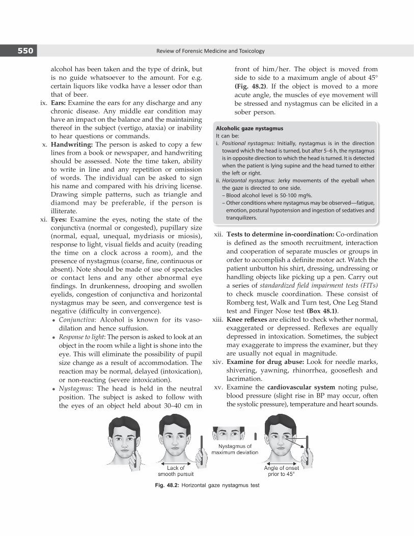
550 Review of Forensic Medicine and Toxicology
alcohol has been taken and the type of drink, but is no guide whatsoever to the amount. For e.g. certain liquors like vodka have a lesser odor than that of beer.
ix. Ears: Examine the ears for any discharge and any chronic disease. Any middle ear condition may have an impact on the balance and the maintaining thereof in the subject (vertigo, ataxia) or inability to hear questions or commands.
x. Handwriting: The person is asked to copy a few lines from a book or newspaper, and handwriting should be assessed. Note the time taken, ability to write in line and any repetition or omission of words. The individual can be asked to sign his name and compared with his driving license. Drawing simple patterns, such as triangle and diamond may be preferable, if the person is illiterate.
xi. Eyes: Examine the eyes, noting the state of the conjunctiva (normal or congested), pupillary size (normal, equal, unequal, mydriasis or miosis), response to light, visual fields and acuity (reading the time on a clock across a room), and the presence of nystagmus (coarse, fine, continuous or absent). Note should be made of use of spectacles or contact lens and any other abnormal eye findings. In drunkenness, drooping and swollen eyelids, congestion of conjunctiva and horizontal nystagmus may be seen, and convergence test is negative (difficulty in convergence).
z Conjunctiva: Alcohol is known for its vaso-dilation and hence suffusion.
z Response to light: The person is asked to look at an object in the room while a light is shone into the eye. This will eliminate the possibility of pupil size change as a result of accommodation. The reaction may be normal, delayed (intoxication), or non-reacting (severe intoxication).
z Nystagmus: The head is held in the neutral position. The subject is asked to follow with the eyes of an object held about 30–40 cm in
front of him/her. The object is moved from side to side to a maximum angle of about 45° (Fig. 48.2). If the object is moved to a more acute angle, the muscles of eye movement will be stressed and nystagmus can be elicited in a sober person.
Alcoholic gaze nystagmusIt can be:i. Positional nystagmus: Initially, nystagmus is in the direction
toward which the head is turned, but after 5–6 h, the nystagmus is in opposite direction to which the head is turned. It is detected when the patient is lying supine and the head turned to either the left or right.
ii. Horizontal nystagmus: Jerky movements of the eyeball when the gaze is directed to one side.
– Blood alcohol level is 50-100 mg%. – Other conditions where nystagmus may be observed—fatigue,
emotion, postural hypotension and ingestion of sedatives and tranquilizers.
xii. Tests to determine in-coordination: Co-ordination is defined as the smooth recruitment, interaction and cooperation of separate muscles or groups in order to accomplish a definite motor act. Watch the patient unbutton his shirt, dressing, undressing or handling objects like picking up a pen. Carry out a series of standardized field impairment tests (FITs) to check muscle coordination. These consist of Romberg test, Walk and Turn test, One Leg Stand test and Finger Nose test (Box 48.1).
xiii. Knee reflexes are elicited to check whether normal, exaggerated or depressed. Reflexes are equally depressed in intoxication. Sometimes, the subject may exaggerate to impress the examiner, but they are usually not equal in magnitude.
xiv. Examine for drug abuse: Look for needle marks, shivering, yawning, rhinorrhea, gooseflesh and lacrimation.
xv. Examine the cardiovascular system noting pulse, blood pressure (slight rise in BP may occur, often the systolic pressure), temperature and heart sounds.
Fig. 48.2: Horizontal gaze nystagmus test
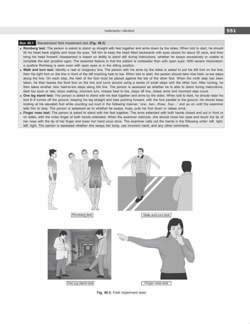
551Inebriants—Alcohol
Box 48.1 Standardized field impairment test (Fig. 48.3) � Romberg test: The person is asked to stand up straight with feet together and arms down by the sides. When told to start, he should tilt his head back slightly and close his eyes. Tell him to keep the head tilted backwards with eyes closed for about 30 secs, and then bring his head forward. Assessment is based on ability to stand still during instructions, whether he sways excessively or unable to complete the test (positive sign). The essential feature is that the patient is unsteadier than with open eyes. With severe intoxication, a positive Romberg is seen even with open eyes or in the sitting position.
� Walk and turn test: Identify a real or imaginary line. The person with his arms by the sides is asked to put his left foot on the line, then his right foot on the line in front of the left touching heel to toe. When told to start, the person should take nine heel- to-toe steps along the line. On each step, the heel of the foot must be placed against the toe of the other foot. When the ninth step has been taken, he then leaves the front foot on the line and turns around using a series of small steps with the other foot. After turning, he then takes another nine heel-to-toe steps along the line. The person is assessed as whether he is able to stand during instructions, start too soon or late, stops walking, incorrect turn, misses heel to toe, steps off line, raises arms and incorrect step count.
� One leg stand test: The person is asked to stand with his feet together and arms by the sides. When told to start, he should raise his foot 6–8 inches off the ground, keeping his leg straight and toes pointing forward, with the foot parallel to the ground. He should keep looking at his elevated foot while counting out loud in the following manner, ‘one…two…three…four…’ and so on until the examiner tells him to stop. The person is assessed as to whether he sways, hops, puts his foot down or raises arms.
� Finger nose test: The person is asked to stand with her feet together. The arms extended with both hands closed and out in front or on sides, with the index finger of both hands extended. When the examiner instructs, she should close her eyes and touch the tip of her nose with the tip of her finger and lower her hand once done. The examiner calls out the hands in the following order: left, right, left, right. The person is assessed whether she sways her body, use incorrect hand, and any other comments.
Fig. 48.3: Field impairment tests

552 Review of Forensic Medicine and Toxicology
xvi. Respiratory (hurried, slow, shallow, deep, sterto-rous, sighing or gasping, any added sounds) and gastrointestinal system (soft, tender, bowel sounds, enlarged liver or spleen, ascites) examination are done both to exclude other diagnosis and to find out complications related to acute or chronic alcohol consumption.
OpinionThe report should be written at that time, and at the end the police informed about the doctor’s opinion (based on examination and laboratory findings). The opinion can be drafted with any one of the following statements: i. He/she has not consumed alcohol. ii. He/she has consumed alcohol, but is not under
the influence of it. iii. He/she has consumed alcohol and is under its
influence.
Diagnosis of Intoxication
Ethanol concentrations are not predictive of intoxication, despite the legal limit of 30 mg/dl for driving.23
Intoxication is a clinical diagnosis. The terms alcohol intoxication and drunkenness are often used interchangeably. The diagnostic features of alcoholic intoxication developed by the American Psychiatric Association include a requirement that there must have been recent ingestion of alcohol. The diagnostic criterion for alcohol intoxication as per DSM-IV is given in Box 48.2.
Medico-legal Aspects
� The DSM-IV criteria require that any medical or drug-related (non-alcohol) conditions must be excluded
before the diagnosis of alcohol intoxication is made. This is important since allegation of negligence can be brought against the doctor in assessing an intoxicated individual in police custody when death may occur because of failure to exclude any pathological conditions.
� Sec. 85 IPC: Nothing is an offence which is done by a person who at the time of doing it, by reason of intoxication, is incapable of knowing the nature of the act, or what he is doing is either wrong or contrary to law; provided that thing which intoxicated him was administered to him without his knowledge or against his will.21
� Voluntary drunkenness is not an excuse for commis-sion of crime.
� Sec. 510 IPC: Misconduct by a drunken person in public is punishable with imprisonment upto 24 h.
� Driving under the influence of alcohol or drunk driving: Operating a motor vehicle after having consumed alcohol or other drugs to the degree that mental and motor skills are impaired.
� Authorities around the world have laid down their own standards for permissible maximum BAC (Table 48.3).
� In India, according to Motor Vehicles Act 1988, for the first offence, punishment is imprisonment of 6 months and/or fine of ` 2000. If a second offence is committed within 3 years, the punishment is 2 years and/or fine of ` 3000. Under this Act, there can be arrest without warrant, a breath test and a laboratory test can also be carried out.
� The government has cleared a proposal to amend the Act. Drunk driving will be graded according to blood alcohol level. The penalty remains unchanged till BAC of 60 mg/dl (as mentioned above). In case of BAC 60–150 mg/dl, imprisonment is for 1 year and/or fine ` 4,000. If offence is repeated within 3 years, imprisonment is for 3 years and/or fine ` 8000. In case of BAC > 150 mg/dl, imprisonment is for 2 years and/or fine ` 5,000. If the offence is repeated, imprisonment is for 4 years and fine of ` 10,000, besides cancellation of license.
� The age for possession and consumption of alcoholic beverages in Australia and Canada is 18 years, in European countries it is between 16–18 years, in the US it is > 21 years, and in India it is between 18–25 (varies between States).
Box 48.2 Diagnostic criteria for alcohol intoxication (as per DSM-IV)
� Recent ingestion of alcohol. � Clinically significant maladaptive behavioral or psychological changes (e.g. inappropriate sexual or aggressive behavior, mood lability, impaired judgment, impaired social or occupational functioning) that developed during or shortly after alcohol ingestion.
� One (or more) of the following signs, developing during or shortly after alcohol use:
– Slurred speech – Unsteady gait – Incoordination – Impairment in attention or memory – Nystagmus – Stupor or coma
� The symptoms are not due to a general medical condition and are not better accounted for by another mental disorder.
Table 48.3: Permissible BAC in different countries
PermissibleBAC (mg/dl)
Countries
0 Hungary, Pakistan, Saudi Arabia, UAE
20 Norway, Poland, Sweden
30 India, China, Japan
50 Australia, France, Germany, Italy, Spain Netherlands, Russia, Denmark
80 Canada, UK, US (100 mg/dl in some States)

553Inebriants—Alcohol
Laboratory Investigations
The common laboratory tests include estimation of alcohol from: i. Blood ii. Urine iii. Breath iv. Vitreous fluid, bile and other tissues (during
autopsy).
Measurement of Alcohol
� The BAC is the most useful measure, as there is rapid equilibration across the blood-brain barrier, therefore BAC reflects the concentration of alcohol currently affecting the brain.
� Urine alcohol: BAC is a direct method for the determination of a blood alcohol level whereas urine analysis is an indirect method. The pool of urine in the bladder at any given time is an accumulation of secreted urine since the last emptying of the bladder.
z At any given point in time, urine alcohol concentration (UAC) will be different from the BAC. After the cessation of drinking, the BAC may rise for a period of time. At this point, the UAC will be less than the BAC because of absorption and distribution throughout the body fluids. Thereafter, the BAC and UAC curves will cross. For some period of time, the UAC will continue to rise, whereas the BAC will remain constant or begin to decrease. In the post-absorptive state, the UAC will always exceed the BAC. Hence, a urine level in bladder is a combination of continuously changing BAC.
z UAC is reliable when two urine samples are collected about half to 1 h apart, and the bladder is completely emptied at the first void. The urine sample collected second time is formed within the period of the first and second void samples. The difference in UAC values also provides information concerning the state of the UAC curve (rising or falling). In addition, the alcohol content of the second void represents the average alcohol concentration of the urine formed between the first and second void. An average 1.33:1 ratio of urine alcohol to blood alcohol is generally used.
� Breath alcohol, unlike urine, is in equilibrium with blood, even though in a very small concentration of about 1: 2300. At 37°C, a level of 1 mg/dl in blood
will be equivalent to about 0.43 µg/dl in breath. The exact ratio of blood/breath alcohol is temperature dependent and varies slightly with other factors, such as the depth of respiration and concentration of alcohol.
Widmark’s formula is used to estimate blood alcohol level.22
a = cpr
where, a – the total amount of alcohol (in grams) absorbed in the body
c – the concentration of alcohol in blood (in g/kg) p – the weight of the person (in kg) r – constant (0.68 in men and 0.5 in women)Alcohol level from urine is estimated with the formula:
a = 3/4 qpr
where, q – concentration of alcohol in urine (in g/l) and ‘a’, ‘p’ and ‘r’ are same as above.
Kozelka and Hine method or Cavett method: It involves aeration/distillation or diffusion of alcohol under low pressure. It utilizes the principle that alcohol is easily oxidized to acetic acid by oxidizing agents, such as potassium dichromate and sulfuric acid.
Other methods
i. Gas liquid chromatography (GLC): Most reliable method.23 It is extremely sensitive and produces accurate quantitative results. In high performance liquid chromatography (HPLC), the sample is in liquid state at the time of analysis, rather than in volatile state as in GLC.*
ii. Alcohol dehydrogenase (ADH) method: It is highly specific and accurate.
* Gas chromatography-Mass spectrometry (GC-MS) is the only method of analysis that is 100% specific.
Any method to determine ethanol in blood and urine can be classified into:
� Macro (requiring 1–2 ml of specimen) � Micro (requiring 0.1–0.2 ml of specimen or less)
The methods are: i. Chemical: Widmark method, Cavett method ii. Biochemical: ADH method iii. Physical: GLC
� Chemical methods: It depends on reduction of a dichromate/sulfuric acid by alcohol vapor.
a. Widmark method (original): Micro-method, alcohol from sample distills slowly into dichromate in a small closed vessel.
b. Cavett method: Modification of Widmark, using different chemical reaction to determine the amount of alcohol.
554 Review of Forensic Medicine and Toxicology
c. Nickolls method: Macro-method, similar to Cavett.d. Southgate and Carter method: Macro-method, sample is
evaporated in an air stream, and the air passing through the hot dichromate reagent which absorbs the alcohol vapor.
e. Kozelka and Hine method: Macro-method, alcohol is distilled in a current of steam. The steam is condensed after passing through a reagent which traps interfering substances and the alcohol in the condensate is determined by reduction of dichromate. It is more specific than other chemical methods as interfering substances are removed.
� Biochemical method: ADH method: Micro-method, alcohol is oxidized by the ADH enzyme in the presence of a coenzyme; the reduced coenzyme is then determined calorimetrically by a separate method, from the result of which the alcohol content of the original sample is calculated.
� Breathalyzer (or breath analyzer) is a device for estimating BAC from a breath sample. In 1954, Dr Robert Borkenstein invented the breathalyzer which used chemical oxidation and photometry to determine alcohol concentration (breath passes through a solution of potassium dichromate, which oxidizes ethanol to acetic acid, changing color in the process).24 Subsequent breathalyzers have converted primarily to infrared spectroscopy.
The invention of the breathalyzer provided law enforcement with a non-invasive test with immediate results to determine an individual’s BAC at the time of testing.
Collection of Samples in the Living
� Blood: Soap and water is used to clean the site to be venepunctured. The blood is collected from antecubital or femoral vein using a disposable syringe. Blood container should be tightly stoppered to prevent loss of alcohol by evaporation, and labeled with name, date, time of taking the specimen and signature of the medical officer.
� Urine: Full quantity of urine passed must be collected. The patient is asked to pass urine in a toilet where there is no water source (preventing him to dilute alcohol concentration by adding water). It is collected in a large clean, sterilized, screw capped bottle. The urine is preserved, and labeled with name, date, time of taking the specimen and signature of the medical officer.
� Breath: The patient is asked to blow into a rubber balloon. A breathanalyzer then analyzes the breath.
Postmortem Samples
Details are given in Chapter 6.In temperate climates, postmortem blood alcohol determination is completely valid for 36 h after death.
Erroneous BAC results can be obtained due to:� Postmortem diffusion � Improperly preserved from other body fluids sample and tissues � Hemolysis� Samples stored at room � Clot formation temperature for more than � Putrefaction a week
Methyl Alcohol (Methanol)
Introduction: Methanol (carbinol, wood alcohol, wood naphtha or wood spirits) is found in cleaning materials, solvents, paints, varnishes, formaldehyde solutions, antifreeze, windshield washer fluid (30–40% methanol), and duplicating fluids.
Physical properties: Colorless, volatile liquid with odor similar to ethyl alcohol and a burning taste.
Uses: In industries as solvent, in laboratories with ethanol as an antiseptic spirit.
Absorption and ExcretionIt is rapidly absorbed from the stomach, intestines, lungs and skin, and achieves a maximal concentration 30–90 min after ingestion.
� Oxidation is slow, 15% of that of ethyl alcohol, and acts as a cumulative poison.
� During metabolism, it is converted into formaldehyde and formic acid which is metabolized to folic acid, folinic acid, carbon dioxide and water (Flow chart 48.3).
� Eighty percent is excreted unchanged from lungs, and 3–5% in urine.
� Without competition for alcohol dehydrogenase, methanol undergoes zero-order metabolism and is excreted at a rate of 8.5–20 mg/dl/h. Once methanol experiences competitive inhibition from ethanol or fomepizole, the metabolism changes to first order.
ActionIt causes ethanol-like CNS depression and increased serum osmolality. Formic acid causes an high anion-gap metabolic acidosis and retinal toxicity.25,26
Signs and Symptoms � Symptoms occur 12–24 h after ingestion. Unlike
ethanol or isopropanol, methanol does not cause much of an inebriated state.
� Initially, the symptoms from methanol intoxication are similar to those of ethanol, often with disinhibition and ataxia.
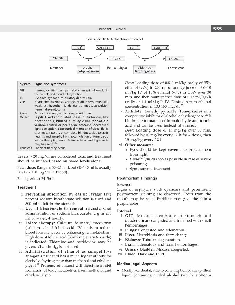
555Inebriants—Alcohol
System Signs and symptoms
GIT Nausea, vomiting, cramps in abdomen, spirit- like odor in the nostrils and mouth, dehydra tion.
RS Dyspnea, cyanosis, respiratory depression.CNS Headache, dizziness, vertigo, restlessness, muscular
weakness, hypothermia, delirium, amnesia, convulsion (terminal event), coma.
Renal Acidosis, strongly acidic urine, scant urine.Ocular Pupils: Fixed and dilated. Visual disturbances, like
photophobia, blurred or misty vision (snowfield vision), central or peripheral scotoma, decreased light perception, concentric diminution of visual fields causing temporary or complete blindness due to optic neuritis and atrophy from accumulation of formic acid within the optic nerve. Retinal edema and hyperemia may be seen.25,26
Pancreas Pancreatitis may occur.
Levels > 20 mg/dl are considered toxic and treatment should be initiated based on blood levels alone.
Fatal dose: Range is 30–240 ml, but 60–140 ml is usually fatal (> 150 mg/dl in blood).
Fatal period: 24–36 h.
Treatment
i. Preventing absorption by gastric lavage: Five percent sodium bicarbonate solution is used and 500 ml is left in the stomach.
ii. Use of bicarbonate to combat acidosis: Oral adminis tration of sodium bicarbonate, 2 g in 250 ml of water, 4 hourly.
iii. Folate therapy: Calcium folinate/leucovorin (calcium salt of folinic acid) IV tends to reduce blood formate levels by enhancing its metabolism. High dose of folinic acid (50–75 mg every 6 hourly) is indicated. Thiamine and pyridoxine may be given. Vitamin B12 is not used.
iv. Administration of ethanol as competitive antagonist: Ethanol has a much higher affinity for alcohol dehydrogenase than methanol and ethylene glycol.27 Presence of ethanol will therefore inhibit formation of toxic metabolites from methanol and ethylene glycol.
Dose: Loading dose of 0.8–1 ml/kg orally of 95% ethanol (v/v) in 200 ml of orange juice or 7.6–10 ml/kg IV of 10% ethanol (v/v) in D5W over 30 min, and then maintenance dose of 0.15 ml/kg/h orally or 1.4 ml/kg/h IV. Desired serum ethanol concentration is 100-150 mg/dl.25
v. Antidote: 4-methylpyrazole (fomepizole) is a competitive inhibitor of alcohol dehydrogenase.28 It blocks the formation of formaldehyde and formic acid and can be used instead of ethanol.
Dose: Loading dose of 15 mg/kg over 30 min, followed by 10 mg/kg every 12 h for 4 doses, then 15 mg/kg every 12 h.
vi. Other measures z Eyes should be kept covered to protect them
from light. z Hemodialysis as soon as possible in case of severe
poisoning. z Symptomatic treatment.
Postmortem FindingsExternalSigns of asphyxia with cyanosis and prominent postmortem staining are observed. Froth from the mouth may be seen. Pyridine may give the skin a purple color.
Internal i. GIT: Mucous membrane of stomach and
duodenum are congested and inflamed with small hemorrhages.
ii. Lungs: Congested and edematous. iii. Liver: Necrobiosis and fatty change. iv. Kidneys: Tubular degeneration. v. Brain: Edematous and focal hemorrhages. vi. Urinary bladder: Mucosa congested. vii. Blood: Dark and fluid.
Medico-legal Aspects
� Mostly accidental, due to consumption of cheap illicit liquor containing methyl alcohol (which is often a
Flow chart 48.3: Metabolism of menthol
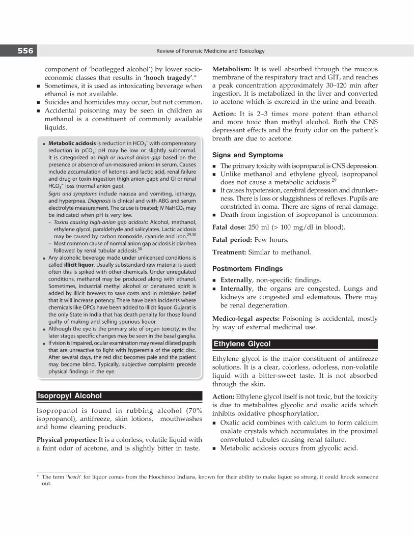
556 Review of Forensic Medicine and Toxicology
component of ‘bootlegged alcohol’) by lower socio-economic classes that results in ‘hooch tragedy’.*
� Sometimes, it is used as intoxicating beverage when ethanol is not available.
� Suicides and homicides may occur, but not common. � Accidental poisoning may be seen in children as
methanol is a constituent of commonly available liquids.
� Metabolic acidosis is reduction in HCO3– with compensatory
reduction in pCO2; pH may be low or slightly subnormal. It is categorized as high or normal anion gap based on the presence or absence of un -measured anions in serum. Causes include accumulation of ketones and lactic acid, renal failure and drug or toxin ingestion (high anion gap); and GI or renal HCO3
– loss (normal anion gap). Signs and symptoms include nausea and vomiting, lethargy,
and hyperpnea. Diagnosis is clinical and with ABG and serum electrolyte measurement. The cause is treated; IV NaHCO3 may be indicated when pH is very low.– Toxins causing high-anion gap acidosis: Alcohol, methanol,
ethylene glycol, paraldehyde and salicylates. Lactic acidosis may be caused by carbon monoxide, cyanide and iron.29,30
– Most common cause of normal anion gap acidosis is diarrhea followed by renal tubular acidosis.30
� Any alcoholic beverage made under unlicensed conditions is called illicit liquor. Usually substandard raw material is used; often this is spiked with other chemicals. Under unregulated conditions, methanol may be produced along with ethanol. Sometimes, industrial methyl alcohol or denatured spirit is added by illicit brewers to save costs and in mistaken belief that it will increase potency. There have been incidents where chemicals like OPCs have been added to illicit liquor. Gujarat is the only State in India that has death penalty for those found guilty of making and selling spurious liquor.
� Although the eye is the primary site of organ toxicity, in the later stages specific changes may be seen in the basal ganglia.
� If vision is impaired, ocular examination may reveal dilated pupils that are unreactive to light with hyperemia of the optic disc. After several days, the red disc becomes pale and the patient may become blind. Typically, subjective complaints precede physical findings in the eye.
Isopropyl Alcohol
Isopropanol is found in rubbing alcohol (70% isopropanol), antifreeze, skin lotions, mouthwashes and home cleaning products.
Physical properties: It is a colorless, volatile liquid with a faint odor of acetone, and is slightly bitter in taste.
Metabolism: It is well absorbed through the mucous membrane of the respiratory tract and GIT, and reaches a peak concentration approximately 30–120 min after ingestion. It is metabolized in the liver and converted to acetone which is excreted in the urine and breath.
Action: It is 2–3 times more potent than ethanol and more toxic than methyl alcohol. Both the CNS depressant effects and the fruity odor on the patient’s breath are due to acetone.
Signs and Symptoms � The primary toxicity with isopropanol is CNS depression. � Unlike methanol and ethylene glycol, isopropanol
does not cause a metabolic acidosis.29
� It causes hypotension, cerebral depression and drunken-ness. There is loss or sluggishness of reflexes. Pupils are constricted in coma. There are signs of renal damage.
� Death from ingestion of isopropanol is uncommon.
Fatal dose: 250 ml (> 100 mg/dl in blood).
Fatal period: Few hours.
Treatment: Similar to methanol.
Postmortem Findings
� Externally, non-specific findings. � Internally, the organs are congested. Lungs and
kidneys are congested and edematous. There may be renal degeneration.
Medico-legal aspects: Poisoning is accidental, mostly by way of external medicinal use.
Ethylene Glycol
Ethylene glycol is the major constituent of antifreeze solutions. It is a clear, colorless, odorless, non-volatile liquid with a bitter-sweet taste. It is not absorbed through the skin.
Action: Ethylene glycol itself is not toxic, but the toxicity is due to metabolites glycolic and oxalic acids which inhibits oxidative phosphorylation.
� Oxalic acid combines with calcium to form calcium oxalate crystals which accumulates in the proximal convoluted tubules causing renal failure.
� Metabolic acidosis occurs from glycolic acid.
* The term ‘hooch’ for liquor comes from the Hoochinoo Indians, known for their ability to make liquor so strong, it could knock someone out.

557Inebriants—Alcohol
Signs and Symptoms
It can be divided into neurological, cardiorespiratory and renal.
� CNS symptoms usually develop within half hour to 12 h after ingestion. The individual develops nausea, vomiting, slurred speech, tipsy sensation, severe headache, delusions, dizziness, feeling of breathlessness, convulsions and coma.
� Cardiorespiratory symptoms usually appear 12–24 h after ingestion. Tachycardia, tachypnea and congestive heart failure are present.
� Renal: Acute tubular necrosis. This usually is seen 24–72 h after ingestion. Oxalate crystals are seen in the urine.
Death occurs from renal failure or heart attack.
Fatal dose: 100-200 ml.
Fatal period: Few hours to 3 days.
Treatment: Gastric lavage is done. Charcoal is not very effective. Treatment is similar as for methanol.31
Postmortem Findings
Non-specific findings. i. Organs are congested. ii. Mucous membrane of the GIT is congested and
inflamed. iii. Cerebral edema, chemical meningoencephalitis,
liver and kidney damage may be seen. iv. Oxalate crystals are seen in the brain, spinal cord
and kidneys.32
Medico-legal aspects: Poisoning is accidental or suicidal in nature.
multiple chOice questiOns
1. c 2. A 3. c 4. A 5. A 6. B 7. B 8. B 9. A 10. A 11. A 12. A 13. D
1. Safe limit of alcohol consumption in males and females are: AFMC 11
A. 15 and 10 units/week B. 18 and 15 units/week C. 21 and 14 units/week D. 25 and 18 units/week 2. Blackout is due to: Maharashtra 11 A. Alcohol intoxication B. Cocaine toxicity C. LSD toxicity D. Cyanide poisoning 3. Fatal level of ethanol in blood: NIMHAS 13 A. 100–200 mg/dl B. 200–300 mg/dl C. 300–400 mg/dl D. > 500 mg/dl 4. In holiday heart syndrome, most common feature seen
is: Odisha 09; DNB 10 A. Atrial fibrillation B. Atrial flutter C. Ventricular fibrillation D. Ventricular flutter 5. Disulfiram is useful in: TN 11 A. Alcohol dependence B. Heroin dependence C. Cocaine dependence D. Cannabis dependence 6. Disulfiram: JIPMER 10 A. Inhibits alcohol dehydrogenase B. Inhibits aldehyde dehydrogenase C. Both A and B D. Inhibits phosphodiesterase 7. Disulfiram-like reaction is caused by: Kerala 11 A. Acamprostate B. Metronidazole C. Tetracycline D. Digitalis
8. Most common symptom of alcohol withdrawal is:AI 07; FMGE 08
A. Bodyache B. Tremor C. Diarrhea D. Rhinorrhea 9. CAGE questionnaire is used in:
AP 07; MP 09; NEET 13 A. Alcohol dependence B. Opiate poisoning C. Dhatura poisoning D. Barbiturate poisoning 10. Delirium tremens seen in: DNB 10; NEET 13 A. Alcohol withdrawal B. Alcohol intoxication C. Opioid intoxication D. Opioid withdrawal 11. All are true about delirium tremens, except:
NIMHANS 10; AI 11; FMGE 11 A. Normal sleep wake cycle B. Visual hallucinations C. Coarse tremors D. Clouding of consciousness 12. A male did not have alcohol for 2 days presented with
seizures treatment given: AIIMS 13 A. Diazepam B. Phenobarbital C. Disulfiram D. Thiamine 13. Chronic alcoholic assaulted his neighbor. He had quit
drinking 4 days back but now has delirium tremens. He is: AIIMS 11
A. Fully responsible B. Family is responsible C. Partially responsible D. Not responsible
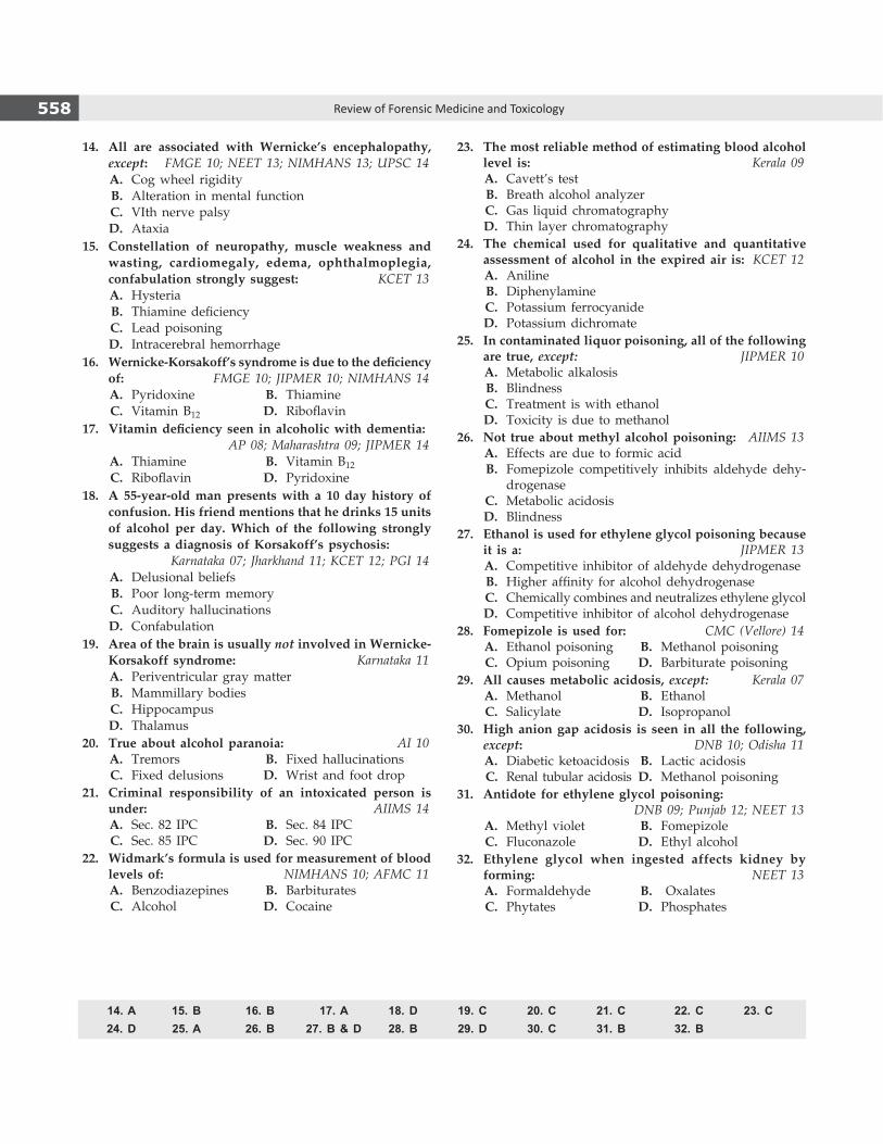
558 Review of Forensic Medicine and Toxicology
14. All are associated with Wernicke’s encephalopathy, except: FMGE 10; NEET 13; NIMHANS 13; UPSC 14
A. Cog wheel rigidity B. Alteration in mental function C. VIth nerve palsy D. Ataxia 15. Constellation of neuropathy, muscle weakness and
wasting, cardiomegaly, edema, ophthalmoplegia, confabulation strongly suggest: KCET 13
A. Hysteria B. Thiamine deficiency C. Lead poisoning D. Intracerebral hemorrhage 16. Wernicke-Korsakoff’s syndrome is due to the deficiency
of: FMGE 10; JIPMER 10; NIMHANS 14 A. Pyridoxine B. Thiamine C. Vitamin B12 D. Riboflavin 17. Vitamin deficiency seen in alcoholic with dementia:
AP 08; Maharashtra 09; JIPMER 14 A. Thiamine B. Vitamin B12 C. Riboflavin D. Pyridoxine 18. A 55-year-old man presents with a 10 day history of
confusion. His friend mentions that he drinks 15 units of alcohol per day. Which of the following strongly suggests a diagnosis of Korsakoff’s psychosis: Karnataka 07; Jharkhand 11; KCET 12; PGI 14
A. Delusional beliefs B. Poor long-term memory C. Auditory hallucinations D. Confabulation 19. Area of the brain is usually not involved in Wernicke-
Korsakoff syndrome: Karnataka 11 A. Periventricular gray matter B. Mammillary bodies C. Hippocampus D. Thalamus 20. True about alcohol paranoia: AI 10 A. Tremors B. Fixed hallucinations C. Fixed delusions D. Wrist and foot drop 21. Criminal responsibility of an intoxicated person is
under: AIIMS 14 A. Sec. 82 IPC B. Sec. 84 IPC C. Sec. 85 IPC D. Sec. 90 IPC 22. Widmark’s formula is used for measurement of blood
levels of: NIMHANS 10; AFMC 11 A. Benzodiazepines B. Barbiturates C. Alcohol D. Cocaine
23. The most reliable method of estimating blood alcohol level is: Kerala 09
A. Cavett’s test B. Breath alcohol analyzer C. Gas liquid chromatography D. Thin layer chromatography 24. The chemical used for qualitative and quantitative
assessment of alcohol in the expired air is: KCET 12 A. Aniline B. Diphenylamine C. Potassium ferrocyanide D. Potassium dichromate 25. In contaminated liquor poisoning, all of the following
are true, except: JIPMER 10 A. Metabolic alkalosis B. Blindness C. Treatment is with ethanol D. Toxicity is due to methanol 26. Not true about methyl alcohol poisoning: AIIMS 13 A. Effects are due to formic acid B. Fomepizole competitively inhibits aldehyde dehy-
drogenase C. Metabolic acidosis D. Blindness 27. Ethanol is used for ethylene glycol poisoning because
it is a: JIPMER 13 A. Competitive inhibitor of aldehyde dehydrogenase B. Higher affinity for alcohol dehydrogenase C. Chemically combines and neutralizes ethylene glycol D. Competitive inhibitor of alcohol dehydrogenase 28. Fomepizole is used for: CMC (Vellore) 14 A. Ethanol poisoning B. Methanol poisoning C. Opium poisoning D. Barbiturate poisoning 29. All causes metabolic acidosis, except: Kerala 07 A. Methanol B. Ethanol C. Salicylate D. Isopropanol 30. High anion gap acidosis is seen in all the following,
except: DNB 10; Odisha 11 A. Diabetic ketoacidosis B. Lactic acidosis C. Renal tubular acidosis D. Methanol poisoning 31. Antidote for ethylene glycol poisoning:
DNB 09; Punjab 12; NEET 13 A. Methyl violet B. Fomepizole C. Fluconazole D. Ethyl alcohol 32. Ethylene glycol when ingested affects kidney by
forming: NEET 13 A. Formaldehyde B. Oxalates C. Phytates D. Phosphates
14. A 15. B 16. B 17. A 18. D 19. c 20. c 21. c 22. c 23. c 24. D 25. A 26. B 27. B & D 28. B 29. D 30. c 31. B 32. B
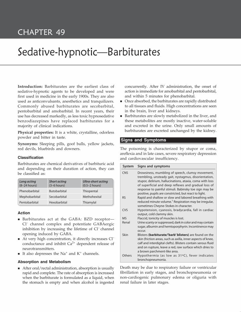
Introduction: Barbiturates are the earliest class of sedative-hypnotic agents to be developed and were first used in medicine in the early 1900s. They are also used as anticonvulsants, anesthetics and tranquilizers. Commonly abused barbiturates are secobarbital, pentobarbital and amobarbital. In recent years, their use has decreased markedly, as less toxic hypnosedative benzodiazepines have replaced barbiturates for a majority of clinical indications.
Physical properties: It is a white, crystalline, odorless powder and bitter in taste.
Synonyms: Sleeping pills, goof balls, yellow jackets, red devils, bluebirds and downers.
ClassificationBarbiturates are chemical derivatives of barbituric acid and depending on their duration of action, they can be classified as:
Long acting(8–24 hours)
Short acting(3–6 hours)
Ultra-short acting(0.5–2 hours)
Phenobarbital Butobarbital Thiopental
Mephobarbital Secobarbital Methohexital
Pentobarbital Hexobarbital Thiamylal
Action � Barbiturates act at the GABA: BZD receptor—
Cl– channel complex and potentiate GABAergic inhibition by increasing the lifetime of Cl– channel opening induced by GABA.
� At very high concentration, it directly increases Cl– conductance and inhibit Ca2+ dependent release of neurotransmitters.
� It also depresses the Na+ and K+ channels.
Absorption and Metabolism � After oral/rectal administration, absorption is usually
rapid and complete. The rate of absorption is increased when the barbiturate is formulated as a liquid, when the stomach is empty and when alcohol is ingested
concurrently. After IV administration, the onset of action is immediate for amobarbital and pentobarbital, and within 5 minutes for phenobarbital.
� Once absorbed, the barbiturates are rapidly distributed to all tissues and fluids. High concentrations are seen in the brain, liver and kidneys.
� Barbiturates are slowly metabolized in the liver, and these metabolites are mostly inactive, water-soluble and excreted in the urine. Only small amounts of barbiturates are excreted unchanged by the kidney.
Signs and Symptoms
The poisoning is characterized by stupor or coma, areflexia and in late cases, severe respiratory depression and cardiovascular insufficiency.
System Signs and symptoms
CNS Drowsiness, mumbling of speech, clumsy movement, trembling, unsteady gait, nystagmus, disorientation, stupor, delirium, hallucinations, ataxia, coma with loss of superficial and deep reflexes and gradual loss of response to painful stimuli. Babinsky toe sign may be positive, pupils are constricted, but react to light.
RS Rapid and shallow or slow and labored breathing with reduced minute volume.1 Respiration may be irregular, sometimes Cheyne-Stokes in character.
CVS Hypotension, cyanosis, bradycardia, fall in cardiac output, cold clammy skin.
MS Flaccid, tonicity of muscles is lost.Renal Urine scanty or suppressed, dark in color and may contain
sugar, albumin and hemtoporphyrin. Incontinence may occur.
Skin Blisters (barbiturate/’barb’ blisters) are found on the skin (friction areas, such as axilla, inner aspects of knee, calf and interdigital clefts). Blisters contain serous fluid and on rupture, leave a red, raw surface which dries to a brown parchment-like area.
Others Hypothermia (as low as 31ºC), fever indicates bronchopneumonia.
Death may be due to respiratory failure or ventricular fibrillation in early stages, and bronchopneumonia or non-cardiogenic pulmonary edema or oliguria with renal failure in later stages.
Sedative-hypnotic—Barbiturates
Chapter 49

560 Review of Forensic Medicine and Toxicology
Fatal dose and blood level
Category Fatal dose Blood level
� Ultra-short acting 1–2 g 3 mg/dl
� Short acting 2–3 g 7 mg/dl
� Long acting 3–5 g 10 mg/dl
Fatal period: 1–2 days.
MAnAgeMent
History � Acute barbiturate intoxication should be clinically
evaluated to differentiate it from other forms of coma or CNS injury.
� History of possible trauma, associated ingestion of alcohol, previous psychiatric illnesses and attempts at suicide and drug usage should be obtained.
� The effect of barbiturates is potentiated by alcohol, narcotics, tranquilizers and antidepressants.
Diagnosis � Urine, gastric lavage and blood are specimens of
choice. Quantify serum alcohol and barbiturate concentrations (particularly phenobarbital). A urine drug screen may help establish co-ingestants.
� Some capsules may be suggestive by color—pentobarbital: yellow or brown, seconal: red, amytal: blue, tuinal: blue and red.
treatment
The management of barbiturate poisoning is supportive. Maintenance of ABC is vital for patient survival. Once the patient is stabilized, gut decontamination and elimination enhancement is done. i. Airway support: Mechanical ventilation with O2
(artificial respiration) is given. If the patient is comatose, prompt intubation is recommended because of the fear of impending, worsening respiratory failure.
ii. Cardiovascular support: Hypotension responds to crystalloid bolus, and vasopressors (dopamine or norepinephrine) are rarely required.
iii. Decontamination and elimination enhancement z Gastric lavage with KMnO4 and activated charcoal
is administered 2–4 hours (h) apart as barbiturates re-enter the GIT through enterohepatic circulation.
z Bowels are evacuated by enema. z Forced alkaline diuresis by sodium bicarbonate
(2–3 ampoules) in 1 liter of 5% dextrose with rate
of infusion at 30 ml/kg/h and guided by urinary pH which should be maintained between 7.5 and 8 and an arterial pH of < 7.5. The goal is to maintain an urine output of 150–250 ml/h.2
z Extracorporeal drug removal: Hemodialysis or hemoperfusion is indicated in phenobarbital poisoning. In hemodynamically unstable patient, sustained low efficiency dialysis may be used instead of conventional dialysis. Newer technique of lipid dialysis (which removes lipid-soluble substances from the blood) can extract long acting drugs in greater quantity.
iv. Supportive care: The most important aspect of management is close observation and quality nursing care.
z Patient is kept warm (passive rewarming) and mucus removed from the throat.
z Endotracheal intubation for first 3 days, but after this tracheostomy should be done.
z Good oral hygiene, temperature maintenance, posture change at regular intervals, antibiotics and symptomatic treatment.
� Multiple doses of activated charcoal may be effective for substances that undergo enterohepatic recirculation (e.g. phenobarbital, theophylline) and for sustained-release preparations.
� Urinary alkalinization enhances the elimination of phenobarbital and other long acting barbiturates by ion trapping in renal tubular cells, but it is not recommended as first line treatment (as multiple-dose activated charcoal is superior) or for short acting barbiturate toxicity.2
� Analeptic drugs: Earlier analeptics were used in the treatment of barbiturate overdose. They are nonspecific arousal agents such as strychnine, camphor, caffeine, picrotoxin, pentylenetetrazol, nikethamide, amphetamine, megride and methylpheridate. The principal goal of analeptic therapy was to awaken the patient. Adverse effects such as hyperthermia, dysrhythmias, seizures and psychoses were associated with its use.
� Scandinavian method: Clemmensen and Nilsson proposed this conservative procedure which abandoned the use of analeptics in the treatment of barbiturate poisoning. It consists of gastric lavage, oxygen, prophylactic antibiotics, determining fluid balance, administration of vitamins, administration of heat or cold for hypo- or hyperthermia respectively, and prevention of bed or eye sores and mouth lesions.
Postmortem Findings
external
i. Mainly those of asphyxia. ii. Cyanosis is present. iii. Froth is seen from the mouth and nostrils.

561Sedative-hypnotic—Barbiturates
iv. Congested face, and prominent postmortem staining.
v. Barbiturate blisters may be seen.
Internal i. Stomach: White particles may be seen. Gastric
mucosa may be eroded. Fundus may be thickened, granular and hemorrhagic.
ii. Lungs: Congested and edematous. Broncho-pneumonia, and/or petechial hemorrhages may be present.
iii. Heart: Subendocardial hemorrhages may be seen. iv. Kidneys: Degeneration of convoluted tubules. v. Other organs: Congested.
Medico-legal Aspects
� Mostly suicidal, rarely homicidal. � Barbiturates are used as ‘date rape’ drugs, since they
can be easily placed into drinks and produce a state of relaxation and disinhibition.
� Accidental poisoning occurs due to an overdose (automatism).
� Addiction due to excessive use of barbiturates. � Occupational hazards: Barbiturates may impair the
mental and/or physical abilities required for the performance of tasks, such as driving a vehicle or
operating machinery. Patients should be warned accordingly.
� Following the use of barbiturates in OPD procedures, patients should be warned against driving vehicles for the rest of the day.
Barbiturate Automatism (Self-poisoning)
Definition: It is taking of barbiturate tablets repeatedly, because of mental confusion.
Cause: The patient develops a state of toxic delirium after ingestion of one or several doses of drug, and in the delirium or automatism state, takes additional doses of drug in order to get to sleep without any intention to commit suicide and without realizing it.Medico-legal aspects: Barbiturate automatism may be more pronounced with alcohol consumption.
� Barbiturate in high doses is used for physician-assisted suicide, and in combination with a muscle relaxant for euthanasia and for capital punishment by lethal injection.
� Thiopental is used IV for the purposes of euthanasia. The Belgians and the Dutch have created a protocol that recommends sodium thiopental as the ideal agent to induce coma, followed by pancuronium bromide.
� Barbiturates including thiopental (sodium pentothal) and sodium amytal (amobarbital) are used as a ‘truth serum’.
1. D 2. A
multiple choice questions
1. Breathing seen in barbiturate poisoning: CMC (Vellore) 14
A. Rapid and deep B. Slow and shallow C. Normal breathing D. Rapid and shallow
2. Alkalization of urine is done in which poisoning: AI 08
A. Barbiturates B. Amphetamine C. Alcohol D. Cocaine

562 Review of Forensic Medicine and Toxicology
Plants that contain the tropane alkaloids include the following:
� Datura stramonium � Atropa belladonna (deadly nightshade) � Hyoscyamus niger (henbane) � Mandragora officinarum (mandrake).
A subgroup of the alkaloids is the alkaloid amines. The three major groups of alkaloid amines are:
i. Hallucinogenic alkaloid amines ii. Stimulant alkaloid amines iii. Anticholinergic tropane alkaloids (belladonna alkaloids or
bicyclic alkaloids).
Dhatura/Datura
Introduction: Dhatura, a member of the Solanaceae family and belongs to the genus Datura, which consists of nine species, such as Datura ferox, Datura alba, Datura fastuosa, etc.
Common names: Thorn apple (fruits are spherical and have sharp spines), Jimson weed,* Hell’s bells and devil’s trumpet (for their large trumpet-shaped flowers) (Fig. 50.1A).
Toxic part � All parts of these plants are poisonous—fruit, flowers
and seeds (highest concentrations of alkaloids are found in roots and seeds).
� The seeds resemble chilli seeds (Diff. 50.1 and Fig. 50.1B). Poisoning occurs only if seeds are masticated and swallowed.
� The usual route is ingesting seeds or other plant parts as tea, although smoking dried leaves also are common.
Fig. 50.2: Longitudinal section of dhatura and capsicum seed
Figs 50.1A and B: (A) Dhatura fruit (thorn apple) and leaves; (B) Dhatura and capsicum seeds
Deliriants—Dhatura/Datura
CHAPTER 50
* A shortened version of ‘Jamestown weed’, named after the first recorded accidental ingestion occurred in Jamestown, Virginia, US in 1676.
Differentiation 50.1: Dhatura and capsicum seedsS.No. Feature Dhatura seeds Capsicum seeds
1. Size Large and thick Small and thin2. Shape Kidney-shaped Rounded3. Color Dark brown Pale yellow4. Convex border Double edge Single edge5. Smell Odorless Pungent6. Surface Small depression Smooth7. Taste Bitter Pungent
8. On cut section (LS) (Fig. 50.2)
Embryo curved outwards
Curved inwards like figure ‘6’
A B

563Deliriants—Dhatura/Datura
Active Principles1
The plant contains belladonna alkaloids whose primary actions are anticholinergic.
� Hyoscine (scopolamine) � Hyoscyamine � Atropine.
Action � Atropine and hyoscine block the acetylcholine
receptor and produces sympathomimetic or parasym-patholytic actions.
� CNS stimulant in early phase, but later CNS depression occurs, especially of the respiratory center.
� Vagolytic action resulting in stimulation of the heart.
Absorption and Excretion � The alkaloids are absorbed through the mucous
membrane of the GIT and respiratory tract, and through the skin and conjunctiva.
� It is destroyed in the liver by enzyme atropinase. � Part of it is excreted through the urine.
Signs and Symptoms
Symptoms are seen 30–60 minutes (min) after ingestion and may continue for 24–48 hours (h) because tropane alkaloids delay gastric emptying and absorption. The mnemonic ‘red as a beet, dry as a bone, blind as a bat, mad as a hatter and hot as a hare’ is useful to remember the anticholinergic toxidrome. Signs and symptoms can be summarized as 9 Ds:2
i. Dryness of the mouth (dry as a bone), bitter taste, burning pain in stomach and vomiting.
ii. Dysphagia (difficulty in swallowing). iii. Dysarthria (difficulty in talking) due to inhibition
of salivation—mumbling in quality and is often incomprehensible.
iv. Dilatation of cutaneous blood vessels (red as a beet). Face is flushed and conjunctiva congested.3
v. Diplopia due to dilated pupil (mydriasis) with loss of accommodation for near vision, developing into temporary blindness (blind as a bat) and photophobia (Fig. 50.3).3-5 Light reflex is sluggish, and later absent.
vi. Dry hot skin (hot as a hare) due to inhibition of sweat and stimulation of heat regulating center. There is dry mucous membrane with dry axillae. Temperature is raised by 1–2°C.
vii. Drunken gait: There is giddiness, confusion, restlessness, agitation and unsteady gait, the patient staggering like a drunken individual.
viii. Delirium (mad as a wet hen): Mutters indistinct words, exhibits typical pill-rolling movements, pulls imaginary thread from fingertips, picks at clothes and tries to run away from his bed.6 Visual and auditory hallucinations may be present. Patient cannot recognize relatives and friends. Undressing behavior is common. The changes in mental status are characteristic of delirium.
ix. Drowsiness: Delirium passes off and patient becomes drowsy, may progress to stupor, coma, or rarely to death from respiratory paralysis.
Additionally, there may be: � Diminished bowel sounds. � Distention of urinary bladder due to urinary
retention. � Rapid pulse (120–140/min), full and bounding, but
later becomes weak and irregular. � Increased respiration. � Scarlatiniform rash over the body. � Amnesia regarding events following ingestion is
common.
Fatal dose � Seeds: 75–125 (stupefying dose: 40–50 seeds). � Hyoscine: 15–30 mg.
Fatal period: 24 h.Differential diagnosis: Drunkenness and heat stroke.Diagnosis: Atropine can be detected by radioimmuno-assay, GS-MS, thin layer chromatography and liquid chromatography. Scopolamine can be analyzed in plasma and urine by radio-receptor assay and GC-MS.
Treatment
Treatment includes control of anticholinergic toxicity usually by supportive measures. Agitation can be controlled with intravenous benzodiazepines. i. Emetics. ii. Gastric lavage with tannic acid, KMnO4 or activated
charcoal. First dose of activated charcoal may be given with cathartic (e.g. sorbitol). One or two additional doses may be given at 1–2 h intervals to ensure adequate gut decontamination.
Fig. 50.3: (A) Normal pupil, (B) Dilated pupil
A B

564 Review of Forensic Medicine and Toxicology
iii. Physiological antidote: Physostigmine salicylate (reversible acetylcholinesterase inhibitor capable of directly antagonizing CNS manifestations of anticholinergic toxicity) 0.5–1 mg slow IV over 5 min with ECG monitoring (0.02 mg/kg/dose).7
iv. Pilocarpine 5–15 mg subcutaneously is also useful. v. Purgatives and colonic lavage is recommended. vi. Tepid sponge baths to control high temperature,
and diazepam IV for sedation and seizures. Morphine is avoided.
vii. Delirium is controlled by short acting barbiturates. viii. O2 inhalation and artificial respiration. ix. Hemodialysis and hemoperfusion are generally
ineffective (tropane alkaloids are lipophilic and cross the blood-brain barrier).
x. Catheterization in case of urinary retention.
Moistening of the tongue and change in the size of pupils point towards normalization and are useful as guidelines for adequate management.
� Physostigmine can induce a life-threatening cholinergic crisis such as seizures, respiratory depression and asystole. Since most patients can be safely treated without this antidote, physostigmine preferably should be used in consultation with a poison control center.
� Physostigmine is contraindicated in patients receiving tricyclic antidepressants, disopyramide, quinidine, procainamide and cocaine.
Postmortem Findings
External: Signs of asphyxia.
Internal i. Seeds may be detected in the stomach and small
intestines. It resists putrefaction and may be found even in a decomposed body. Identification of ingested seeds can be diagnostic of tropane alkaloid poisoning.
ii. Stomach: Mucosa may show inflammation. iii. Lungs: Edematous and congested. iv. Heart: Petechial hemorrhages in endocardium.
Medico-legal Aspects � In India, dhatura is employed mainly as a stupefying
poison prior to robbery, kidnapping and rape. It is sometimes known as rail-road poison, as it is commonly encountered during a journey. Many a time, robbers disguised as saints offer ‘prasad’ mixed with dhatura seeds and rob the passengers.
� Occasionally, it is used for suicidal purpose and for criminal abortion. In Mexico, Datura is taken by Yaqui women to lessen pain of childbirth.
� Accidental poisoning is common in children who may chew the fruit. Sometimes, it may be due to intake of seeds mistaking them for chilli seeds.
� Chinese herbal medicines containing tropane alkaloids have been used to treat asthma, chronic bronchitis, pain and flu symptoms. In Africa, a common use is to smoke leaves of Datura to relieve asthma and pulmonary problems.
� Homicide is rare. � It is used as an adulterant in country liquor for
enhancing the ‘kick’ effect. � Sometime, it is used as an aphrodisiac and as a
recreational hallucinogen. Unintentional poisoning may result in teenagers who eat seeds, drink tea or smoke cigarettes for its hallucinogenic effect.
Atropa belladonna
Atropa belladonna belongs to Solanaceae family, and grows abundantly in India in the Himalayan ranges. All parts of this plant are poisonous.
Active principles: It contains three alkaloids—atropine, hyoscine and belladonine, but the most important of them is atropine.
Action: It acts by inhibiting the muscarine effects of acetylcholine.
Absorption and metabolism: They are absorbed from the skin and parenteral sites, and detoxicated in the liver.
Signs and symptoms resemble those of poisoning by dhatura.8,9
Fatal dose: Atropine: 100–130 mg.
Fatal period: Within 24 h.
Treatment: Same as for dhatura poisoning.
Postmortem findings: Similar to those found in poisoning by dhatura.
Medico-legal aspects: Poisoning by belladonna occurs accidentally from an overdose of its pharmacopoeial preparations or from swallowing ‘eye drops’ by mistake. Accidental poisoning is also seen in children and adults from the plant being grown in the garden, either willfully or accidentally.
Hyoscyamus niger
It yields the active principles hyoscyamine, hyoscine and atropine. It also produce signs and symptoms similar to dhatura.
Fatal dose: Hyoscyamine: 200 mg.
Fatal period: Within 24 h.
Treatment: Similar to that for dhatura.
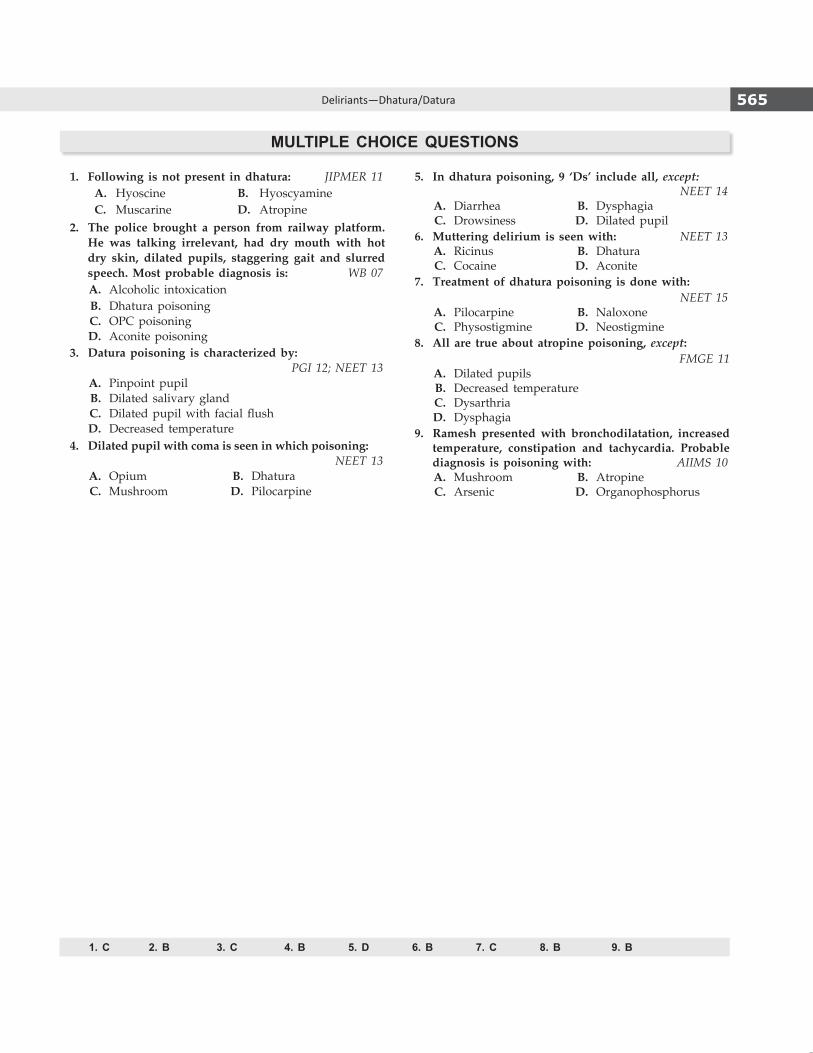
565Deliriants—Dhatura/Datura
1. C 2. B 3. C 4. B 5. D 6. B 7. C 8. B 9. B
MULTIPLE CHOICE QUESTIONS
1. Following is not present in dhatura: JIPMER 11 A. Hyoscine B. Hyoscyamine C. Muscarine D. Atropine 2. The police brought a person from railway platform.
He was talking irrelevant, had dry mouth with hot dry skin, dilated pupils, staggering gait and slurred speech. Most probable diagnosis is: WB 07
A. Alcoholic intoxication B. Dhatura poisoning C. OPC poisoning D. Aconite poisoning 3. Datura poisoning is characterized by:
PGI 12; NEET 13 A. Pinpoint pupil B. Dilated salivary gland C. Dilated pupil with facial flush D. Decreased temperature 4. Dilated pupil with coma is seen in which poisoning:
NEET 13 A. Opium B. Dhatura C. Mushroom D. Pilocarpine
5. In dhatura poisoning, 9 ‘Ds’ include all, except: NEET 14
A. Diarrhea B. Dysphagia C. Drowsiness D. Dilated pupil 6. Muttering delirium is seen with: NEET 13 A. Ricinus B. Dhatura C. Cocaine D. Aconite 7. Treatment of dhatura poisoning is done with: NEET 15 A. Pilocarpine B. Naloxone C. Physostigmine D. Neostigmine 8. All are true about atropine poisoning, except:
FMGE 11 A. Dilated pupils B. Decreased temperature C. Dysarthria D. Dysphagia 9. Ramesh presented with bronchodilatation, increased
temperature, constipation and tachycardia. Probable diagnosis is poisoning with: AIIMS 10
A. Mushroom B. Atropine C. Arsenic D. Organophosphorus

566 Review of Forensic Medicine and Toxicology
Introduction: Cannabis sativa (marijuana/marihuana/hashish), a deliriant cerebral neurotic hemp plant which has several varieties: Cannabis indica (India), Cannabis mexicana (Mexico) and Cannabis americana (US). It is the most commonly abused illegal substance in India and the US, particularly among adolescents, and the most commonly abused substance in the world after nicotine, alcohol and caffeine.1
Distribution: Grows all over India. Whole plant is poisonous.
Synonyms: Pot, grass, weed, ya(r)ndi, rope, mull, dope, joint, Mary Jane, skunk, hash, chronic, reefer, cone or shit.
Active Principle
It is not an alkaloid, but a fat-soluble oleoresin, can-nabinol, the active form being δ-9-tetrahydrocannabinol (THC). It also contains benzopyrene, a known carcinogen which is also found in tobacco.
Preparations of Cannabis (Table 51.1) (Fig. 51.1)
� Bhang is the mildest of cannabis concoctions. It consists of dried cannabis leaves that are ground to a fine paste, mixed with a combination of sugar, spices and fruit.
� Hashish is a highly potent, concentrated cannabis resin that has been collected, dried and pressed into bricks.
� Charas is the handmade form of hashish.
� Marijuana is the dried leaves and flowers of the cannabis plant, and most common form of drug in the US. It is usually smoked, although it is occasionally baked into foods such as brownies or brewed as tea for drinking.
� Majum: Sweetmeat made with bhang. � Hash oil: A lipid soluble plant extract which is mixed
with tobacco and smoked. It may contain THC upto 25–50% and may be added to hashish and marijuana to enhance its THC concentration.
� Sinsemilla (means ‘without seeds’) is unpollinated/unfertilized flowering tops from the female plant (similar to ganja). THC content is 6–11%.
THC is also available in synthetic forms (dronabinol and nabilone) which are used as an appetite stimulant for AIDS-related anorexia and as treatment for vomiting associated with cancer chemotherapy.
Routes of intake: Cannabis is usually smoked in cigarettes (joints or reefers)* or pipes, added to food
* It is a type of cigarette used for intoxication, containing 0.3–0.6 g of marijuana which is dipped in tincture of cannabis and dried.
Deliriants—Cannabis
CHAPTER 51
Table 51.1: Preparations of cannabis2
Features Bhang (siddhi, patti) Ganja Charas (hashish)
Source Dried leaves and shoots Flowering tops of female plant Resinous exudates from leaves, flowers and stems
Color Brownish Rusty green color Dark green or brown
Active principle 2–5% (least potent) 5–8% 10–20% (most potent)3
Uses Beverage Mixed with tobacco and smoked in pipe/hukka
Mixed with tobacco and smoked in pipe/hukka
Fig. 51.1: Cannabis (fresh and dried leaves)
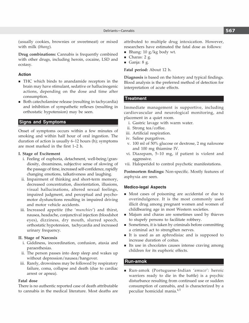
567Deliriants—Cannabis
(usually cookies, brownies or sweetmeat) or mixed with milk (bhang).
Drug combinations: Cannabis is frequently combined with other drugs, including heroin, cocaine, LSD and ecstasy.
Action
� THC which binds to anandamide receptors in the brain may have stimulant, sedative or hallucinogenic actions, depending on the dose and time after consumption.
� Both catecholamine release (resulting in tachycardia) and inhibition of sympathetic reflexes (resulting in orthostatic hypotension) may be seen.
Signs and Symptoms
Onset of symptoms occurs within a few minutes of smoking and within half hour of oral ingestion. The duration of action is usually 6–12 hours (h); symptoms are most marked in the first 1–2 h.
I. Stage of Excitement i. Feeling of euphoria, detachment, well-being/gran-
diosity, dreaminess, subjective sense of slowing of the passage of time, increased self-confidence, rapidly changing emotions, talkativeness and laughing.
ii. Impairment of thinking and short-term memory, decreased concentration, disorientation, illusions, visual hallucinations, altered sexual feelings, impaired judgment, and perceptual and psycho-motor dysfunctions resulting in impaired driving and motor vehicle accidents.
iii. Increased appetite (the ‘munchies’) and thirst, nausea, headache, conjunctival injection (bloodshot eyes), dizziness, dry mouth, slurred speech, orthostatic hypotension, tachycardia and increased urinary frequency.
II. Stage of Narcosis i. Giddiness, incoordination, confusion, ataxia and
paraesthesias. ii. The person passes into deep sleep and wakes up
without depression/nausea/hangover. iii. Rarely, drowsiness may be followed by respiratory
failure, coma, collapse and death (due to cardiac arrest or apnea).
Fatal doseThere is no authentic reported case of death attributable to cannabis in the medical literature. Most deaths are
attributed to multiple drug intoxication. However, researchers have estimated the fatal dose as follows:
� Bhang: 10 g/kg body wt. � Charas: 2 g. � Ganja: 8 g.
Fatal period: About 12 h.
Diagnosis is based on the history and typical findings. Blood analysis is the preferred method of detection for interpretation of acute effects.
Treatment
Immediate management is supportive, including cardiovascular and neurological monitoring, and placement in a quiet room. i. Gastric lavage with warm water. ii. Strong tea/coffee. iii. Artificial respiration. iv. Saline purgatives. v. 100 ml of 50% glucose or dextrose, 2 mg naloxone
and 100 mg thiamine IV. vi. Diazepam, 5–10 mg, if patient is violent and
aggressive. vii. Haloperidol to control psychotic manifestations.
Postmortem findings: Non-specific. Mostly features of asphyxia are seen.
Medico-legal Aspects
� Most cases of poisoning are accidental or due to overindulgence. It is the most commonly used illicit drug among pregnant women and women of childbearing age in most Western societies.
� Majum and charas are sometimes used by thieves to stupefy persons to facilitate robbery.
� Sometimes, it is taken by criminals before committing a criminal act to strengthen nerves.
� It is used as an aphrodisiac and is supposed to increase duration of coitus.
� Its use in chocolates causes intense craving among children for its euphoric effects.
Run-amok
� Run-amok (Portuguese-Indian ‘amuco’: heroic warriors ready to die in the battle) is a psychic disturbance resulting from continued use or sudden consumption of cannabis, and is characterized by a peculiar homicidal mania.4,5
568 Review of Forensic Medicine and Toxicology
1. A 2. C 3. D 4. C 5. B
� After intake, there is a period of depression, followed by excitation, confusion and a violent attempt to kill people (impulse to murder).
� The addict first kills a person against whom he may have real or imaginary enmity and then kills anyone who comes in his way, until the homicidal tendency lasts. The person may then
commit suicide or surrender himself to the law enforcement authority.
� Criminal responsibility: The person is not held responsible for his acts since ‘run amok’ is considered a disorder of mind and not intoxication, unless he had taken it purposefully to ennerve (strengthen) himself before commission of the offence.
MULTIPLE CHOICE QUESTIONS
1. Most common substance abuse in India:AI 07; AIIMS 10; NEET 13, 15
A. Cannabis B. Tobacco C. Alcohol D. Opium 2. Not a form of cannabis: AIIMS 14 A. Bhang B. Charas C. Afeem D. Ganja 3. Most potent form of cannabis: Maharashtra 11 A. Bhang B. Charas C. Ganja D. Hash oil
4. Run-amok is a feature of poisoning with: Maharashtra 10
A. Opium B. Dhatura
C. Cannabis D. Alcohol 5. Run-amok is: JIPMER 13 A. Running-away from stressful situation B. Killing people randomly C. Feeling of insects running under skin D. Ingesting corrosive in rage

569Deliriants—Cocaine
Deliriants—Cocaine
CHAPTER 52
Introduction: Cocaine (crack, pasta, bazooka, snuff, coke, snow or white lady) is a colorless, odorless, crystalline substance with bitter taste and slightly soluble in water, but freely soluble in alcohol.
� It is an alkaloid deliriant, obtained from dried leaves of Erythroxylum coca, a shrub indigenous to Peru, Bolivia, Mexico, West Indies and Indonesia (Fig. 52.1).
� Illicit forms of cocaine include the hydrochloride salt and its alkalization products, freebase or crack.
� Beside alcohol, cocaine is the most common cause of drug-related emergency department visits in the US.
Action
� Cocaine produces a hyperadrenergic state. � It increases the synaptic concentrations of the
monoamine neurotransmitters dopamine, norepine-phrine and serotonin by binding to transporter proteins in presynaptic neurons and blocking uptake.
� It is also a local anesthetic, as it blocks initiation and conduction of nerve impulse by decreasing axonal membrane permeability to sodium ions.
� It stimulates the cortex for a short time, followed by depression.
Absorption and Excretion
� Cocaine is rapidly absorbed from the mucous membranes and subcutaneous tissues.
� About 30–50% of cocaine is metabolized by hepatic esterases and plasma pseudocholinesterase, resulting
in the formation of ecgonine methyl ester. Spontaneous nonenzymatic hydrolysis of another 30–40% results in benzoylecgonine.
� Only 1–5% of cocaine is excreted unaltered through the kidneys within 6 hours (h) of use.
� A metabolite of cocaine, cocaethylene has been found in blood and urine of patients who abuse both alcohol and cocaine.
Routes of administration: Chewing, application to nasal mucous membrane (snorting), smoking (free basing) and IV.
Cocaine may be inhaled through a straw or rolled-up paper currency, or a spoon containing 5–20 mg of the drug is used to snort.
Signs and Symptoms
Signs and symptoms of acute poisoning include elevated pulse, blood pressure, respiration and temperature.1 Onset occurs within 7 seconds after inhalation, 15 seconds after taking IV, 3 minutes (min) after nasal insufflations and 10 min after oral ingestion.1 The initial stimulatory effects (rush) are followed by depression (crush).
I. Stage of Excitement1,2
System Signs and symptoms
Local Feeling of numbness or tingling at the place of application.
Face Flushed.Skin Pale.GIT Bitter taste, dryness of mouth, vomiting, diarrhea,
hyperactive bowel sounds.CNS Mydriasis, headache, bruxism, feeling of well-being,
euphoria, restlessness, excitement, talkativeness, delirium, maniacal, hallucinations, nonintentional tremors (e.g. twitching of small muscles, especially facial and finger) and tonic-clonic seizures. Reflexes are exaggerated.
RS Tachypnea, dyspnea, cyanosis.CVS Tachycardia, hypertension, ventricular arrhythmias.Temperature Hyperthermia.Ocular Pupils are dilated resulting in blurred vision.
Fig. 52.1: Erythroxylum coca leaves and cocaine

570 Review of Forensic Medicine and Toxicology
II. Stage of DepressionAfter an hour, respiration becomes slow, there is profuse sweating, and patient becomes calm and dull.
System Signs and symptoms
CNS Coma, areflexia, pupils fixed and dilated, flaccid paralysis and loss of vital support functions.
CVS Ventricular dysrhythmias result in weak, rapid, irregular pulse and hypotension, circulatory failure and cardiac arrest.
RS Cheyne-Stokes respirations, apnea, pulmonary edema, cyanosis, respiratory failure.
� Tea colored urine may indicate rhabdomyolysis and potential renal failure.
� In fatal cases, the onset and progression are accelerated, with convulsions and death (from respiratory failure, cerebral hemorrhage or cardiac failure) frequently occurring in 2–3 min.
Fatal dose: 20 mg IV; 500 mg to 1.2 g orally.
Fatal period: Few minutes to 1–2 h.
Differential diagnosis: Lithium toxicity, cyclic anti-depressants toxicity, neuroleptic malignant syndrome, acute withdrawal from sedatives or ethanol, thyroid storm and other hyperadrenergic states.
Diagnosis: Qualitative toxicological analysis of blood and urine. Finding cocaine and metabolites in the urine supports the diagnosis. However, urinalysis can detect cocaine metabolites 2–4 days post-exposure, a positive screen does not equate with clinical toxicity.
Urine, blood, gastric contents and unknown substances found on patients, such as on a moustache, may be sent for toxicological evaluation.
Treatment
There is no specific antidote; therapy consists of treatment of symptoms until the acute effects (which are generally short lived since half-life of cocaine is about 1 h) are gone.2
i. If injected, apply tourniquet above the part; if applied to nose or throat, wash-out with warm water or saline. If swallowed, gastric lavage should be done with KMnO4 and/or activated charcoal.
ii. Control seizures: Diazepam in doses upto 0.5 mg/kg IV may be given over an 8 h period. Physical restraint should be avoided due to risks of rhabdomyolysis and hyperthermia.
iii. Dysrhythmias should be treated according to standard advanced cardiac life support (ACLS)
protocols. Ventricular arrhythmia is managed by giving 0.5–1 mg of propranolol IV.
iv. Short acting, direct vasodilator (esmolol) and short acting beta-blockers are indicated for tachycardia and hypertension.
v. Thiamine 100 mg IV. vi. Airways are kept clean, artificial respiration and
O2 inhalation as required.
Complications
� CNS: Cerebrovascular accidents (frequent cause of stroke in < 45 years), subarachnoid or intracerebral hemorrhage and cerebral vasculitis.
� CVS: Myocardial, bowel and kidney ischemia, myo-cardial infarction, skin necrosis and aortic dissection.1
� Pulmonary infarcts, eosinophilia with granuloma formation.
� Inhalational exposure can result in cough, hemoptysis, reactive airway disease, pneumonitis (‘crack lung') and barotrauma due to forceful Valsalva maneuver that are performed during smoking (e.g. pneumothorax).
Postmortem Findings
Non-specific findings. i. Patients may have linear excoriations, ‘crack pipe’
burns of the fingers or thumbs, thermal burns of the face and upper airway.
ii. Track marks in the usual sites such as the antecubital fossae, and at unusual sites such as under the tongue and on top of the feet may be seen.
iii. Intense asphyxial signs, and cardiac dilatation may be seen.
Blood should be preserved by adding fluoride.
Medico-legal Aspects
� Accidental cases occur from urethral, vesical and rectal injection. Oral overdose can occur in body packers and body stuffers.
� Cocaine is rarely used for homicide or suicide. � Intentional criminal poisoning wherein prenatal
(known ingestion of cocaine by pregnant female), infant and child deaths determined to be homicide has been reported.
� It is believed to increase the libidinal drive and increase the duration of sexual act by paralyzing sensory nerves of glans penis.
� It causes lowering of moral values, loss of decency and self-respect.
� It is rapidly destroyed in the body and is difficult to detect by chemical analysis.
571Deliriants—Cocaine
� ‘Crack’ is produced when the hydrochloride molecule is removed by ether extraction, which frees the basic cocaine molecule (‘freebase’). The term ‘crack’ describes the crackling sound heard when cocaine freebase is smoked.
� Illicit drugs are frequently admixed with additional chemicals either to increase the apparent quantity of the street drug or to enhance its effect. For example, 8–20% of stimulants available on the street contain cocaine and methamphetamine hydrochloride. Other adulterants may include quinine, talc, ascorbic acid, boric acid, chalk, laundry detergent, laxatives and lactose.
� ‘Crack baby’: The term ‘crack baby’ was used to describe children who were exposed to crack as fetuses in the US during 1980–90 in the midst of a crack epidemic.
� ‘Crack lung’ may occur 1–48 h after cocaine smoking. It is a hypersensitivity pneumonitis wherein there is chest pain, cough, hemoptysis, dyspnea, bronchospasm, pruritus, fever, diffuse alveolar infiltrates without effusions, and pulmonary and systemic eosinophilia.
� ‘Crack dancing’ refers to the extrapyramidal phenomena and other movement disorders that are sometimes associated with cocaine abuse.
Cocainism (Cocainomania / Cocainophagia)
� Cocainomania is an irresistible craze, crave, or impulse to intoxication by cocaine, or any of its salts or combinations, at all risks. It is addiction and morbid craving for cocaine.
� Many users take repeated doses to keep the high going and avoid the ‘crash’ or try to modify the effects with other drugs like alcohol, tranquilizers or heroin. This rush-and-crash pattern leads to toxic
levels of cocaine in the bloodstream and reinforces the highly addictive nature of cocaine.
� Abusers can tolerate upto 10 g/day.
Signs and Symptoms � Emaciation, anorexia, digestive disturbances,
significant loss of libido, impotence, gynecomastia, galactorrhea and major derangements in menstrual cycle in women—amenorrhea and infertility.
� Face is pale, shifty gaze, sunken eyes, dilated pupils, tongue and teeth are black, and ulceration of nasal septum.3
� Degeneration of CNS with hallucinations, convulsions and delirium may occur.
Magnan's Syndrome / Cocaine Bugs4,5
� This is seen in cocaine addicts. � It is a type of tactile hallucination. � There is a feeling as if grains of sand are lying under
the skin or small insects are creeping on the skin giving rise to itching sensation (formication).
Formication (Latin formica: ant): Tactile hallucination involving the sensation that tiny insects are crawling over the skin. Causes of formication can be actual physical conditions, including diabetic neuropathy, menopause, skin cancer or herpes zoster, and may also be physical or psycho logical side effect of substance abuse (cocaine, amphetamines, and alcohol withdrawal along with delirium tremens).
MULTIPLE CHOICE QUESTIONS
1. Following are complications of cocaine poisoning, except: UPSC 08
A. Myocardial infarction B. Epileptic seizures C. Hypothermia D. Hypertension 2. True regarding cocaine are all, except: JIPMER 13 A. Causes tachycardia and hypertension B. Half life is 3 h C. No antidote D. Can be taken by snorting 3. An addicted patient presenting with visual and tactile
hallucinations, has black staining of tongue and teeth. The agent is: Gujarat 10
A. Cocaine B. Cannabis C. Heroin D. Opium
4. A person feels that small insects are creeping on the skin giving rise to itching sensation; the condition is seen in: Maharashtra 09; AIIMS 09, 11
A. Cocaine poisoning B. OPC posioning C. Morphine poisoning D. Alcohol withdrawal 5. Magnan’s syndrome is associated with:
WB 09; BHU 09; MP 11; AFMC 12; NEET 14 A. Cocaine B. OPC C. Snake bite D. Alcohol
1. C 2. B 3. A 4. A 5. A
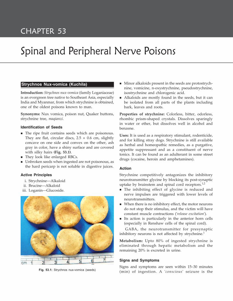
572 Review of Forensic Medicine and Toxicology
Spinal and Peripheral Nerve Poisons
CHAPTER 53
Strychnos Nux-vomica (Kuchila)
Introduction: Strychnos nux-vomica (family Loganiaceae) is an evergreen tree native to Southeast Asia, especially India and Myanmar, from which strychnine is obtained, one of the oldest poisons known to man.
Synonyms: Nux vomica, poison nut, Quaker buttons, strychnine tree, maqianzi.
Identification of Seeds � The ripe fruit contains seeds which are poisonous.
They are flat, circular discs, 2.5 × 0.6 cm, slightly concave on one side and convex on the other, ash gray in color, have a shiny surface and are covered with silky hairs (Fig. 53.1).
� They look like enlarged RBCs. � Unbroken seeds when ingested are not poisonous, as
the hard pericarp is not soluble in digestive juices.
Active Principles i. Strychnine—Alkaloid ii. Brucine—Alkaloid iii. Loganin—Glucoside.
� Minor alkaloids present in the seeds are protostrych-nine, vomicine, n-oxystrychnine, pseudostrychnine, isostrychnine and chlorogenic acid.
� Alkaloids are mostly found in the seeds, but it can be isolated from all parts of the plants including bark, leaves and roots.
Properties of strychnine: Colorless, bitter, odorless, rhombic prism-shaped crystals. Dissolves sparingly in water or ether, but dissolves well in alcohol and benzene.
Uses: It is used as a respiratory stimulant, rodenticide, and for killing stray dogs. Strychnine is still available as herbal and homeopathic remedies, as a purgative, appetite suppressant and as a constituent of nerve tonics. It can be found as an adulterant in some street drugs (cocaine, heroin and amphetamines).
Action
Strychnine competitively antagonizes the inhibitory neurotransmitter glycine by blocking its post-synaptic uptake by brainstem and spinal cord receptors.1,2
� The inhibiting effect of glycine is reduced and nerve impulses are triggered with lower levels of neurotransmitters.
� When there is no inhibitory effect, the motor neurons do not stop their stimulus, and the victim will have constant muscle contractions ('release excitation').
� Its action is particularly in the anterior horn cells (especially in Renshaw cells of the spinal cord).
GABA, the neurotransmitter for presynaptic inhibitory neurons is not affected by strychnine.1
Metabolism: Upto 80% of ingested strychnine is eliminated through hepatic metabolism and the remaining 20% is excreted in urine.
Signs and Symptoms
Signs and symptoms are seen within 15–30 minutes (min) of ingestion. A 'conscious ' seizure is the Fig. 53.1: Strychnos nux-vomica (seeds)
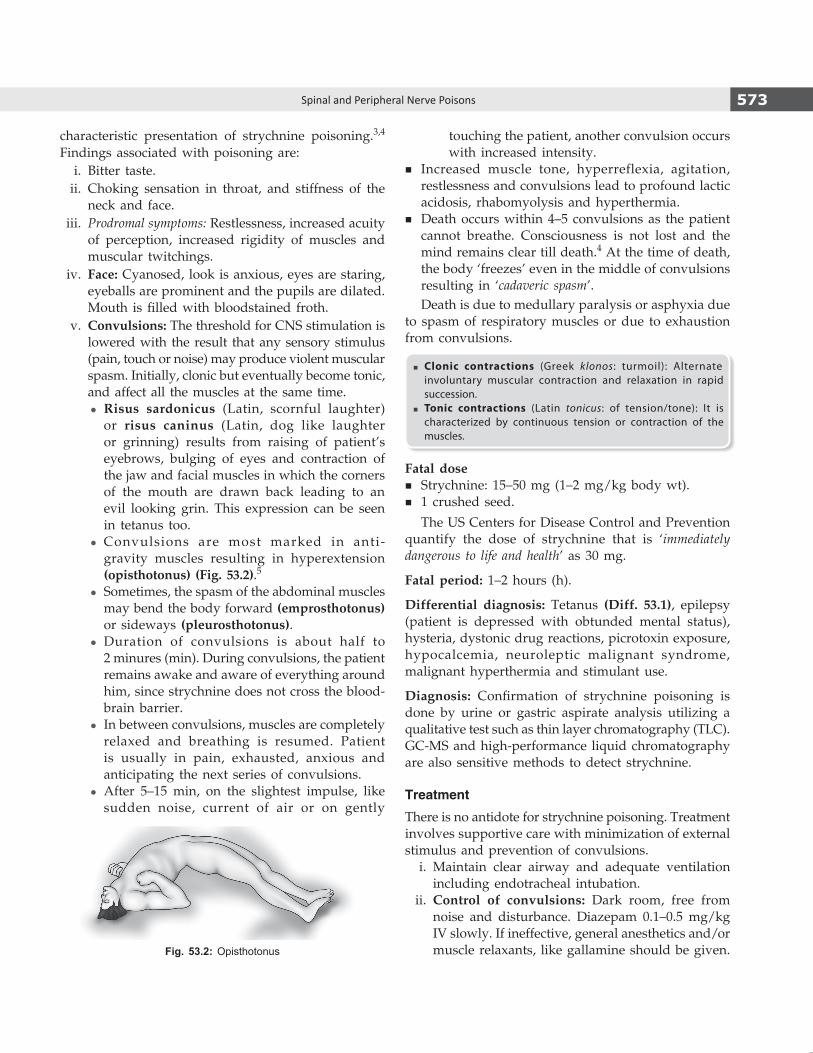
573Spinal and Peripheral Nerve Poisons
touching the patient, another convulsion occurs with increased intensity.
� Increased muscle tone, hyperreflexia, agitation, restlessness and convulsions lead to profound lactic acidosis, rhabomyolysis and hyperthermia.
� Death occurs within 4–5 convulsions as the patient cannot breathe. Consciousness is not lost and the mind remains clear till death.4 At the time of death, the body ‘freezes’ even in the middle of convulsions resulting in ‘cadaveric spasm’.Death is due to medullary paralysis or asphyxia due
to spasm of respiratory muscles or due to exhaustion from convulsions.
� Clonic contractions (Greek klonos: turmoil): Alternate involuntary muscular contraction and relaxation in rapid succession.
� Tonic contractions (Latin tonicus: of tension/tone): It is characterized by continuous tension or contraction of the muscles.
Fatal dose � Strychnine: 15–50 mg (1–2 mg/kg body wt). � 1 crushed seed.
The US Centers for Disease Control and Prevention quantify the dose of strychnine that is ‘immediately dangerous to life and health’ as 30 mg.
Fatal period: 1–2 hours (h).
Differential diagnosis: Tetanus (Diff. 53.1), epilepsy (patient is depressed with obtunded mental status), hysteria, dystonic drug reactions, picrotoxin exposure, hypocalcemia, neuroleptic malignant syndrome, malignant hyperthermia and stimulant use.
Diagnosis: Confirmation of strychnine poisoning is done by urine or gastric aspirate analysis utilizing a qualitative test such as thin layer chromatography (TLC). GC-MS and high-performance liquid chromatography are also sensitive methods to detect strychnine.
Treatment
There is no antidote for strychnine poisoning. Treatment involves supportive care with minimization of external stimulus and prevention of convulsions. i. Maintain clear airway and adequate ventilation
including endotracheal intubation. ii. Control of convulsions: Dark room, free from
noise and disturbance. Diazepam 0.1–0.5 mg/kg IV slowly. If ineffective, general anesthetics and/or muscle relaxants, like gallamine should be given.Fig. 53.2: Opisthotonus
characteristic presentation of strychnine poisoning.3,4 Findings associated with poisoning are: i. Bitter taste. ii. Choking sensation in throat, and stiffness of the
neck and face. iii. Prodromal symptoms: Restlessness, increased acuity
of perception, increased rigidity of muscles and muscular twitchings.
iv. Face: Cyanosed, look is anxious, eyes are staring, eyeballs are prominent and the pupils are dilated. Mouth is filled with bloodstained froth.
v. Convulsions: The threshold for CNS stimulation is lowered with the result that any sensory stimulus (pain, touch or noise) may produce violent muscular spasm. Initially, clonic but eventually become tonic, and affect all the muscles at the same time.
z Risus sardonicus (Latin, scornful laughter) or risus caninus (Latin, dog like laughter or grinning) results from raising of patient’s eyebrows, bulging of eyes and contraction of the jaw and facial muscles in which the corners of the mouth are drawn back leading to an evil looking grin. This expression can be seen in tetanus too.
z Convulsions are most marked in anti-gravity muscles resulting in hyperextension (opisthotonus) (Fig. 53.2).5
z Sometimes, the spasm of the abdominal muscles may bend the body forward (emprosthotonus) or sideways (pleurosthotonus).
z Duration of convulsions is about half to 2 minures (min). During convulsions, the patient remains awake and aware of everything around him, since strychnine does not cross the blood-brain barrier.
z In between convulsions, muscles are completely relaxed and breathing is resumed. Patient is usually in pain, exhausted, anxious and anticipating the next series of convulsions.
z After 5–15 min, on the slightest impulse, like sudden noise, current of air or on gently

574 Review of Forensic Medicine and Toxicology
iii. Barbiturates, like pentobarbital sodium or sodium amytal are antidotes.6 Dose: 300–600 mg IV.
iv. Gastric lavage with KMnO4 may be done cautiously, if there are no convulsions. Activated charcoal is recommended as it adsorbs strychnine and may reduce its absorption if given 1 h of ingestion.
v. Hyperthermia is treated by active cooling with ice water immersion, cooling blanket or mist and fan.
vi. Intravenous fluid is given to maintain a urine output > 1 ml/kg/h, since metabolic acidosis and renal failure may occur.
vii. Hemodialysis is not effective. viii. Symptomatic treatment.
Postmortem Findings i. Not characteristic. ii. Rigor mortis appears early. iii. Signs of asphyxia. iv. Extravasated blood may be found in the muscles. v. Viscera are congested.
Strychnine can be easily detected in blood, urine, gastric fluid, bile and fixed liver and kidney samples in autopsy.
Medico-legal Aspects � Death is usually accidental due to overdose, exposure
to rodenticide, quack remedies and poison mistaken for some other harmless drug, or in children eating the seeds.
� Intentional self harm has also been reported. However, homicide is rare, because of its bitter taste, and strychnine is detectable in food in very low concentrations, hence it is impossible to disguise in food and beverages.
� It is used as an aphrodisiac, as cattle and arrow poison, and to kill dogs and rats.
� Tolerance develops on repeated consumption. � It can be detected easily even in a decomposed body
(detectable as low as 0.01 ppm in tissue).
Peripheral Nerve Poisons
Curare
Introduction: The alkaloid is a peripheral muscle relaxant and is available from the plant Chondrodendron tomentosum or from some species of Strychnos plants. It is not poisonous when swallowed.
Active principles: d-tubocurarine, dimethyl tubocurarine, syncurine and succinylcholine chloride.
Action: It blocks the postsynaptic nicotinic acetylcholine receptors in the muscles, thus causing flaccid paralysis of skeletal muscles.
Signs and Symptoms
It causes paralysis of voluntary muscles, followed by paralysis of respiratory muscles resulting in death from asphyxia. The mental faculties remain clear till the end.Fatal dose: 30–60 mg of curarine parenterally.Fatal period: 1–2 h.
Treatment
i. Artificial respiration and O2 should be given. ii. If applied to a wound or introduced by an arrow,
a ligature should be applied proximal to the site and is washed with a solution of KMnO4.
Differentiation 53.1: Strychnine poisoning and tetanus
S.No. Feature Strychnine poisoning Tetanus
1. History of injury None Present
2. Onset Sudden (in hours) Gradual (several days)
3. Site of action Postsynaptic membrane Presynaptic membrane
4. Muscles affected All muscles affected at the same time Not affected at the same time
5. Lower jaw Does not start in, nor especially affect the jaw Starts in and affects the jaw (lock-jaw)
6. Muscular condition Relaxed in between convulsions Rigid
7. Fatal period 1–2 h > 24 h
8. Chemical analysis Strychnine found No poison
9. Culture No growth Clostridium tetani found
10. Progression Steadily worse/steadily better Progress rarely steady. Variations and longer remission not uncommon

575Spinal and Peripheral Nerve Poisons
iii. Atropine 0.6–1.2 mg, followed by physostigmine (1–2 mg, physiological antidote) or neostigmine (0.5–1 mg) subcutaneously should be given.
Postmortem findings: Those of asphyxia. Skin and tissue from the wound due to the arrow or injection should be preserved.
Medico-legal aspects: Most deaths are from its use in anesthesia. It is also used as arrow poison.
Conium Maculatum (Hemlock)
Introduction: This plant is also known as spotted hemlock, because of the purple spots on its stem. It grows in wastelands. All parts of the plant are poisonous. The whole plant has a mousy odor which is intensified by crushing the leaves or stems.
Active principles: Coniine, methyl coniine and six other alkaloids. Coniine content is highest in the unripe fruit and seeds. Symptoms may be caused by ingestion, injection or even inhalation of coniine (volatile alkaloid).
Action: It causes paralysis of the motor nerve terminals in the muscles, gradually spreading to the motor cells of the spinal cord and the brain.
Signs and Symptoms � Nausea, unpleasant mousy odor in breath. � Ingestion causes burning in the mouth and throat,
gastric inflammation, vomiting, diarrhea, slow respiration and pulse, mental confusion, tremors and blindness.
� This is followed by progressive muscular paralysis due to depression of the motor nerves. The lower limbs are affected first and the paralysis ascends till the muscles of respiration are affected.
� Delirium, convulsions and coma may supervene, and the patient dies of asphyxia due to respiratory paralysis. The mind remains clear till the end.
Fatal dose: 60 mg coniine or a piece of plant about 1 cm in diameter.
Fatal period: Few hours.
Diagnosis: Coniine and other alkaloids can be measured in the urine by various methods including gas chromatography, mass spectrometry and thin layer chromatography.
Treatment
i. Gastric lavage with KMnO4. ii. Artificial respiration. iii. Oxygen inhalation. iv. Stimulants. v. Symptomatic treatment.
Postmortem findings: Those of asphyxia, the remains of the roots or leaves should be looked for in the stomach contents and preserved for chemical analysis.
Medico-legal aspects: Poisoning is mostly accidental, the plant being mistaken for parsley or some harmless herb.
Hemlock was administered to Socrates, the Greek Philosopher in 399 BC as a form of execution.7
MULTIPLE CHOICE QUESTIONS
1. Inhibitory neurotransmitter in spinal cord is: NIMHANS 08 A. GABA B. Glycine C. Glutamate D. Acetylcholine 2. Strychnine acts by inhibiting: COMEDK 15 A. GABA B. Glycine C. Acetylcholine D. Dopamine 3. Respiratory center depression is caused by all, except:
AIIMS 10 A. Opium B. Strychnine C. Barbiturates D. Gelsemium 4. In strychnos nux vomica poisoning, patient: NEET 13 A. Becomes unconscious immediately B. Becomes unconscious in 1 h or so
C. Becomes unconscious at end stage D. Remains conscious throughout 5. In nux vomica poisoning, posture commonly assumed
by the spine is: NEET 13 A. Opisthotonus B. Emprosthotonus C. Pleurosthotonus D. B and C 6. Antidote for strychnine poisoning is: NEET 13 A. Fomepizole B. Physotigmine C. Barbiturates D. Naloxone 7. Socrates was killed by which poisoning: NEET 14 A. Cyanide B. Conium C. Strychnine D. Dhatura
1. A & B 2. B 3. B 4. D 5. A 6. C 7. B

576 Review of Forensic Medicine and Toxicology
Cardiac Poisons
CHAPTER 54
These are poisonous plants having an action mainly on the heart, either directly or through the nerves. Important poisonous plants and compounds in this group are:1
� Aconite (Aconitum napellus, Aconitum ferox) � Nicotine (Nicotiana tabacum) � Digitalis (Digitalis purpurea) � Oleander (Cerbera thevetia, Cerbera odorum) � Quinine.
Aconite
Introduction: All parts of the plant are poisonous, however, the root and root tubers are the most potent.
Dry root is conical or tapering, shows bases of the broken rootlets and shriveled with longitudinal wrinkles (Fig. 54.1). It is 5–10 cm long, 1.5–2 cm thick at the upper end and dark brown in color. Roots are mistaken for horseradish root.
Synonyms: Monk’s hood, mitha zaher, bish, wolf’s bane, women’s bane, devil’s helmet or blue rocket.2
Active principles: Aconitine, pseudo-aconitine, indaconitine, picraconitine and aconine.
Properties of aconitine: Colorless, transparent, rhombic crystals. Insoluble in water, but readily soluble in benzene and chloroform.
Action
� Toxicity of aconitine and related alkaloids are due to their actions on the voltage-sensitive sodium channels of the cell membranes of excitable tissues. Aconitine first stimulates and then paralyzes the peripheral terminations of sensory and secretory nerves, CNS, and nerves of the myocardium, skeletal and smooth muscles.
� It does not affect the higher centers of the brain as consciousness remains intact till the end.
Signs and Symptoms
Patients present mainly with a combination of gastro-intestinal, cardiovascular and neurological features.
System Signs and symptoms
GIT Nausea, vomiting, salivation, pain in the abdomen and diarrhea. Bitter-sweet taste, severe burning and tingling of tongue, mouth, perioral area and throat, followed by numbness.3
CVS Hypotension, chest pain, palpitations, bradycardia, sinus tachycardia, ventricular ectopics and ventricular tachycardia/fibrillation.4 Pulse is slow, feeble and irregular.
CNS Vertigo, restlessness, headache, giddiness.MS Weakness of the muscles of the limbs with twitchings and
spasms.RS Respiration is slow, labored and shallow.Ocular Pupils alternately contract and dilate (hippus). Diplopia
and impaired vision occurs.Others Temperature is subnormal and skin is cold.
� Terminal stages are marked by severe pain and paralysis of facial muscles.
� Many victims remain conscious until near death; some complain of yellow-green vision and tinnitus.
� Death is due to respiratory failure or ventri cular fibrillation.
Fatal dose � Root: 1–2 g. � Aconitine: 2–5 mg.
Fatal period: 2–6 hours (h).Fig. 54.1: Aconite (root)

577Cardiac Poisons
Treatment
There is no specific antidote for aconite, and treatment is supportive. i. Gastric lavage with tannic acid/activated charcoal. ii. Inotropic therapy is required if hypotension
persists, and atropine (0.5–1 mg IV) should be used to treat bradycardia.
iii. Ventricular arrhythmia is treated with amiodarone and flecainide (first-line treatment). In refractory cases and cardiogenic shock, early use of cardiopulmonary bypass is recommended.
iv. Symptomatic treatment.
Postmortem Findings
i. Not specific, those of asphyxia. ii. Organs are congested. iii. Stomach: Fragments of root may be found in the
stomach. iv. Lungs: Hemorrhagic pulmonary edema. v. Heart: Diffuse contraction-band necrosis in
myocardium.
Medico-legal Aspects
� It is often regarded as an ideal homicidal poison.Advantages are:
a. It is cheap and easily available. b. Lethal dose is small and the fatal period is short. c. Color can be disguised by mixing it with pink
colored drinks. d. Taste can be masked by mixing it with sweets
or by giving it with betel (paan) leaves. e. Extremely unstable and destroyed by putre faction,
hence cannot be detected by chemical analysis. � Accidental poisoning occurs due to:
a. Eating the roots mistaking it for horseradish. b. Use of quack remedies. c. Taking of liquor mixed with aconitine to increase
intoxication. d. Consumption of herbal decoction made from
aconite roots. � It is also used as an abortifacient, cattle and arrow
poison. � Suicide is not common.
� Horseradish is a perennial plant which includes mustard and cabbages.
� Hippus (Greek hippos: horse): Abnormal exaggeration of the rhythmic contraction and dilatation of the pupil, independent of changes in illumination or in fixation of the eyes.
� The term ‘wolf’s bane’ comes from its use to poison meat laid out for wolves.
� In ancient Rome, aconite was widely used by professional poisoners, and cultivating the plant was considered capital offence. On the Greek island of Chios, it was used for euthanasia of the old and infirm.
Nicotiana Tabacum (Tobacco)
Introduction
� All parts of the plant are poisonous, except the ripe seeds.
� Dried leaves contain 1–8% nicotine. � Leaves contain toxic alkaloids, like nicotine, ana-
basine, nornicotine and lobeline (in Indian tobacco). � An average cigarette delivers 1–3 mg of nicotine.
Properties of nicotine: Colorless, hygroscopic oily liquid. Burning acrid taste and disagreeable odor.
Action
� It acts on the autonomic ganglia, which are stimu-lated initially, but are depressed and blocked at the later stages.
� It also acts on the somatic neuromuscular junction and afferent fibers from sensory receptors.
Signs and Symptoms
Acute Poisoning
i. CVS: Tachycardia followed by bradycardia, hypotension, arrhythmia, tachypnea followed by respiratory depression and collapse.
ii. GIT: Burning acid sensation, nausea, vomiting, abdominal pain, salivation and odor of tobacco.
iii. CNS: Headache, restlessness, confusion, vertigo, sweating, convulsions and coma.
Chronic Poisoning
i. RS: Cough, wheeze, dyspnea, chronic bronchitis and lung cancer may develop.
ii. CVS: Anemia, palpitations, irregularity of heart, angina pectoris and Berger's disease.
iii. GIT: Anorexia, vomiting and diarrhea. iv. CNS and others: Impaired memory, blindness,
tremors, insomnia, anxiety and headache.
Fatal dose � Nicotine: 60–100 mg. � Tobacco: 15–30 g.
Fatal period: 5–15 minutes.

578 Review of Forensic Medicine and Toxicology
Treatment
i. Gastric lavage with charcoal, KMnO4. ii. Purgatives. iii. Cardiac monitoring. iv. Atropine to correct hypotension, and diazepam
for convulsions. v. Symptomatic treatment.
In chronic poisoning, clonidine has shown encoura-ging result.
Postmortem Findings
i. Brownish froth at mouth and nostrils. ii. Stomach may contain fragments of leaves or smell
of tobacco. iii. Features of asphyxia are seen.
Medico-legal Aspects
� Accidental poisoning results from ingestion, excessive smoking and application of leaves or juice to wound or skin.
� Common drug of addiction. � For malingering, leaves are soaked in water for some
hours and placed in axilla at bed time, poisonous symptoms are seen by next morning.
� Suicidal/homicidal cases are rare.
Green tobacco sickness commonly occurs in tobacco growing States. Workers handling leaves may absorb alkaloids through skin. Symptoms include nausea, vomiting, diarrhea, diaphoresis and weakness that usually resolve with symptomatic treatment.
Digitalis Purpurea (Foxglove)
Active principles: Its roots, leaves and seeds contain several glycosides—digitoxin, digitalin, digitalein and digitonin are the most poisonous.
Action
The glycosides act directly on the heart muscle (prolong diastolic period) and improve the function of the failing heart. In toxic doses, excitability is increased with extrasystoles.
Glycosides are substances found in plants, and are composed of a sugar and a non-sugar compound, the later having toxicological action.
Signs and Symptoms
Toxic symptoms are due to overdose or by a cumulative action.
System Signs and symptoms
GIT Nausea, vomiting, pain in abdomen, burning sensation, diarrhea.
CVS Bradycardia, extrasystoles, ventricular tachycardia and fibrillation, atrial fibrillation, faintness, precordial oppression, heart block.
CNS Headache, fatigue, confusion, anxiety, depression, disorientation, drowsiness, hallucinations, delirium.
RS Labored and sighing respiration.Ocular Transient ambylopia, blurring, photophobia, scotoma,
diplopia, color aberration.Skin Urticaria.
The patient becomes drowsy and the condition may deepen into coma. Convulsions may precede death. Death occurs from cardiovascular collapse.
Fatal dose � Digitalis: 2–3 g. � Digoxin: 5 mg. � Digitalin: 15–20 mg. � Powdered leaves: 2.5 g.
Fatal period: 1–24 h.
TreatmentECG monitoring is necessary as a guide to treatment. i. Gastric lavage is done with a solution of tannic
acid. ii. Activated charcoal is given. iii. Purgatives may be given. iv. Atropine is given in a dose of 0.6 mg IV to treat
bradycardia. v. Potassium chloride may be given to reduce
extrasystoles. vi. Specific antidote for cardiac arrhythmias is
lignocaine 100 mg IV or novocaine or propranolol. vii. Trisodium EDTA may help to lower serum calcium. viii. Symptomatic treatment.
Postmortem FindingsNon-specific changes are seen. There may be irritation of the gastric mucosa, and digitalis leaves or seeds may be found in the stomach.
Medico-legal Aspects
� Accidental poisoning due to overdose of a medicinal preparation or from eating leaves by mistake.

579Cardiac Poisons
� It is a cumulative poison, and persons taking it for a long time may suddenly develop symptoms of poisoning.
� Homicidal poisoning cases may be seen, and no suspicion of poisoning may arise in such cases as it will simulate heart disease.
OLEANDER (KANER)
The oleander plant grows wild in India. There are two varieties:
� Nerium odorum: Bears white, dark red or pink flowers. � Cerbera thevetia: Bears yellow bell-shaped flowers,
globular fruits, light green in color, about 5 cm in diameter containing a single nut, triangular in shape and light brown in color. The nut contains five pale yellow seeds (Fig. 54.2).
Nerium Odorum (White Oleander, Kaner)
All parts of the plant are poisonous.
Active principles: Nerin consisting of three glycosides— neriodorin, neriodorein and karabin.5
Action
It is similar to that of digitalis causing death from cardiac failure. Neriodorein causes muscular twitching and tetanic spasm which is more powerful than strychnine. Karabin acts on the heart like digitalis, and on the spinal cord like strychnine.
Signs and Symptoms
� Locally, contact dermatitis. � Inhalation of flowers may cause headache, dizziness,
respiratory difficulty and nausea.
� Ingestion causes vomiting, pain in the abdomen, frothy salivation, difficulty in swallowing and articulation. Later on, there is restlessness, muscular twitchings, tetanic spasms and lock jaw. The pulse is slow and weak, respiration is rapid, blood pressure falls, and there is fibrillation and AV block. This is followed by exhaustion, drowsiness, coma, respiratory paralysis and death from heart failure.
Fatal dose: Root: 15–20 g; leaves: 5–15.
Fatal period: 24 h.
Treatment
i. Gastric lavage. ii. Administration of an anesthetic is usually necessary. iii. Morphine injection seems to be beneficial. iv. Symptomatic treatment.
Postmortem Findings
Non-specific findings. Petechial hemor rhage on the heart is a characteristic feature. Organs are congested.
Medico-legal Aspects
� Suicide is common among village girls, using it as a paste or decoction.
� It is used as an abortifacient, applied both locally and internally.
� Homicide is rare. � Accidental poisoning is sometimes met with when
decoction is used: a. Externally to reduce swelling. b. As a remedy for venereal diseases. c. As a love-philter (increases attraction between the
giver and taker). d. For treatment of cancer and ulcers.
� It is used as cattle poison. � Nerium odorum resists heat and can therefore be detected
even from the burnt remains of the dead body.
Cerbera Thevetia (Yellow Oleander, Pila Kaner)
All parts of the plant are poisonous. Milky juice exudes from all parts of the plant (Fig. 54.2).
Active principles: Glycosides—thevetin, thevotoxin, cerberin and peruvoside. Thevetin is a powerful cardiac poison. Thevotoxin is less toxic than thevetin and resembles the glycosides of digitalis in action. Cerberin acts like strychnine.
Signs and Symptoms
The sap of the plant may cause inflammation.Fig. 54.2: Cerbera thevetia (oleander)
580 Review of Forensic Medicine and Toxicology
On ingestion, there is burning sensation in the mouth with tingling of the tongue, dryness of throat, vomiting, diarrhea, headache, dizziness, dilated pupils, drowsiness and loss of muscular power. Pulse is rapid, weak and irregular, blood pressure falls. Heart block, collapse and death is due to peripheral circulatory failure.
Fatal dose: Seeds: 8–10; root: 15–20 g.
Fatal period: 2–3 h.
Treatment
i. Gastric lavage. Single-dose activated charcoal is beneficial and safe.
ii. Molar solution of sodium lactate IV and 5% glucose to combat acidosis.
iii. Atropine 1 mg, 2 ml of adrenaline 1:1000 and 2 mg of noradrenaline (if blood pressure is low) to counteract heart block.
v. Digoxin-specific antibody fragments is effective in reverting life-threatening cardiac arrhythmias.
iv. Symptomatic treatment.
Postmortem Findings
Non-specific. i. Signs of GIT irritation may be seen. ii. Stomach and duodenum may be congested and
may show fragments of seeds. iii. Congestion of visceral organs are seen.
Medico-legal aspects: Same as Nerium odorum.
Quinine
The bark of Cinchona plant contains quinine, quinidine, cinchonidine and other alkaloids. Quinine occurs as white needle-shaped, odorless, crystalline and bitter powder.
Action
It is a protoplasmic poison with anesthetic and sclerosing effect. It stimulates and then depresses the CNS. It causes circulatory failure by direct and indirect actions.
Signs and SymptomsOn ingestion, there is pain in the abdomen, vomiting, diarrhea, headache, giddiness, tinnitus, partial deafness, loss of vision, scotoma, confusion, muscular weakness, itching, tachycardia, hypotension and cyanosis.
� There may be oliguria, hemolysis, hematuria and uremia.
� Respiration is rapid and shallow, pupils are fixed and dilated, delirium and coma.
� Death occurs from respiratory failure.
Cinchonism or quinism is caused by repeated thera-peutic doses or overdose of quinine.Symptoms are tinnitus, vertigo, deafness, diplopia, scotoma, blindness, skin rash, hypoglycemia and cardiac arrhythmias.Fatal dose: 2–8 g.Fatal period: About 6 h.
Treatment i. Assisted ventilation, if necessary. Continuous
cardiac monitoring is needed. ii. Gastric lavage is done, and magnesium sulfate is
used for purgation. iii. Activated charcoal. iv. For cardiac toxicity, IV bolus of sodium bicarbo-
nate is given. v. Ventricular tachycardia may be treated with
magnesium IV or overdrive pacing. vi. Intravenous fluids are given to promote diuresis. vii. Protection of vision: Blocking of bilateral stellate
ganglion is sometimes recommended. viii. Symptomatic treatment.
Postmortem Findings
Non-specific. Organs are congested, and hemolysis of red cells may be found. Renal tubules may be blocked by hemoglobin.
Medico-legal Aspects
� Accidental poisoning occurs due to medicinal overdose. � Suicide/homicide is rare. � It is used as an abortifacient.

581Cardiac Poisons
MULTIPLE CHOICE QUESTIONS
1. All are cardiotoxic, except: AFMC 12; NEET 13 A. Aconite B. Opium C. Oleander D. Nicotine 2. Mitha bish is: MAHE 12 A. Oleander B. Aconite C. Quinine D. Digitalis 3. Characteristic symptom of aconite poisoning: AI 10 A. Increased salivation B. Hypertension C. Tingling and numbness D. Hyperthermia
1. B 2. B 3. C 4. C 5. D
4. Not a sign/symptom of aconite poisoning: JIPMER 12 A. Pain abdomen B. Bradycardia C. Hypertension D. Diplopia 5. Active principle of oleander plant is: NEET 14; AIIMS 14 A. Cannabinol B. Atropine C. Semicarpol D. Nerin
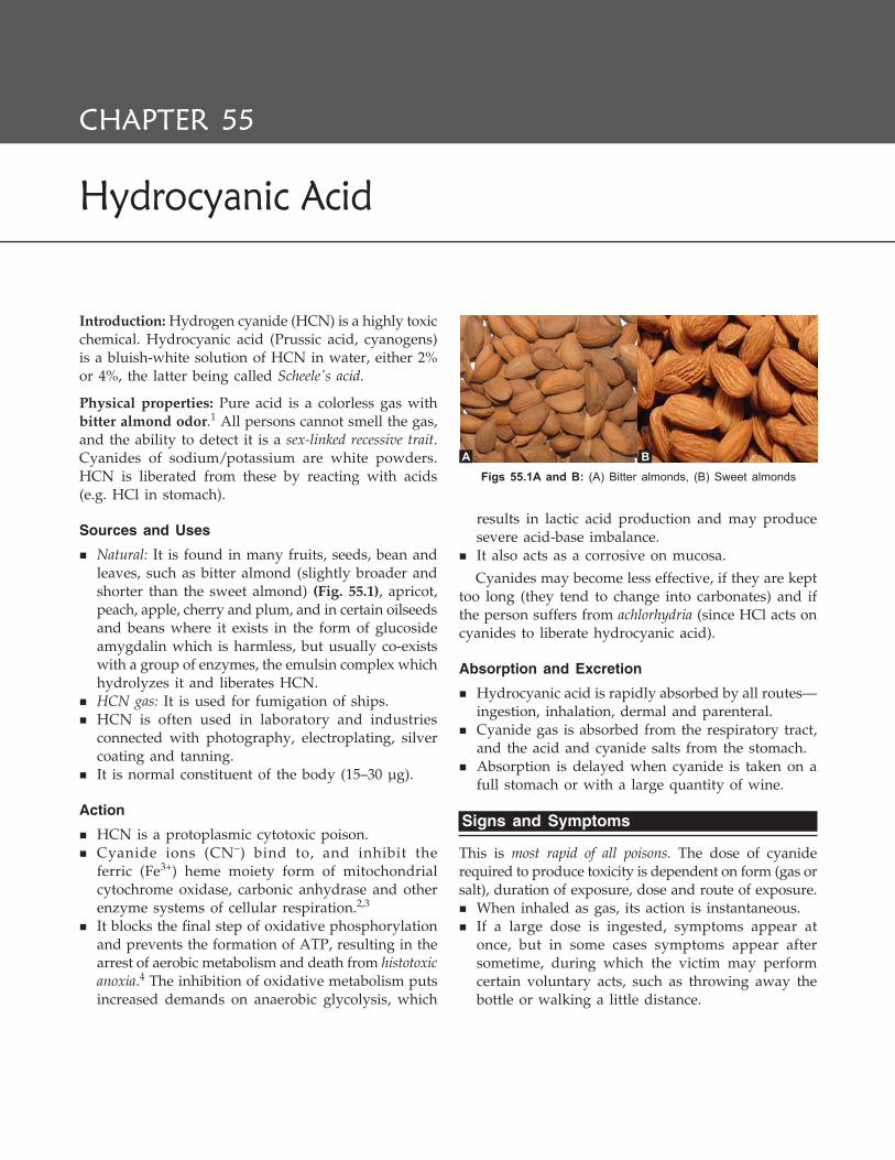
582 Review of Forensic Medicine and Toxicology
Hydrocyanic Acid
CHAPTER 55
Introduction: Hydrogen cyanide (HCN) is a highly toxic chemical. Hydrocyanic acid (Prussic acid, cyanogens) is a bluish-white solution of HCN in water, either 2% or 4%, the latter being called Scheele's acid.
Physical properties: Pure acid is a colorless gas with bitter almond odor.1 All persons cannot smell the gas, and the ability to detect it is a sex-linked recessive trait. Cyanides of sodium/potassium are white powders. HCN is liberated from these by reacting with acids (e.g. HCl in stomach).
Sources and Uses
� Natural: It is found in many fruits, seeds, bean and leaves, such as bitter almond (slightly broader and shorter than the sweet almond) (Fig. 55.1), apricot, peach, apple, cherry and plum, and in certain oilseeds and beans where it exists in the form of glucoside amygdalin which is harmless, but usually co-exists with a group of enzymes, the emulsin complex which hydrolyzes it and liberates HCN.
� HCN gas: It is used for fumigation of ships. � HCN is often used in laboratory and industries
connected with photography, electroplating, silver coating and tanning.
� It is normal constituent of the body (15–30 µg).
Action
� HCN is a protoplasmic cytotoxic poison. � Cyanide ions (CN–) bind to, and inhibit the
ferric (Fe3+) heme moiety form of mitochondrial cytochrome oxidase, carbonic anhydrase and other enzyme systems of cellular respiration.2,3
� It blocks the final step of oxidative phosphorylation and prevents the formation of ATP, resulting in the arrest of aerobic metabolism and death from histotoxic anoxia.4 The inhibition of oxidative metabolism puts increased demands on anaerobic glycolysis, which
results in lactic acid production and may produce severe acid-base imbalance.
� It also acts as a corrosive on mucosa.
Cyanides may become less effective, if they are kept too long (they tend to change into carbonates) and if the person suffers from achlorhydria (since HCl acts on cyanides to liberate hydrocyanic acid).
Absorption and Excretion
� Hydrocyanic acid is rapidly absorbed by all routes— ingestion, inhalation, dermal and parenteral.
� Cyanide gas is absorbed from the respiratory tract, and the acid and cyanide salts from the stomach.
� Absorption is delayed when cyanide is taken on a full stomach or with a large quantity of wine.
Signs and Symptoms
This is most rapid of all poisons. The dose of cyanide required to produce toxicity is dependent on form (gas or salt), duration of exposure, dose and route of exposure.
� When inhaled as gas, its action is instantaneous. � If a large dose is ingested, symptoms appear at
once, but in some cases symptoms appear after sometime, during which the victim may perform certain voluntary acts, such as throwing away the bottle or walking a little distance.
Figs 55.1A and B: (A) Bitter almonds, (B) Sweet almonds
A B

583Hydrocyanic Acid
� In case of dermal application, latent interval can be several hours.
System Signs and symptoms
Local Corrosive effect on the mouth, throat and stomach. CNS Headache, vertigo, faintness, anxiety, excitement,
confusion, drowsiness, prostration, opisthotonus, lockjaw, hyperthermia, epileptiform or tonic convulsions, paralysis, stupor and coma.
CVS Initially hypertension with reflex bradycardia, sinus arrhythmia, later on tachycardia with hypotension and cardiovascular collapse.
GIT Bitter acid burning taste, constriction or numbness of throat, clenched jaw, salivation, froth, nausea, rarely vomiting.
RS Odor of bitter almonds in breath, initially tachypnea and dyspnea, followed by rapid slowing of respiratory rate with severe respiratory depression and cyanosis.
Skin Perspiration, bullae, pinkish or brick-red color*Ocular Glassy, prominent eyes, pupils dilated and
unreactive.Renal Acidosis.
� Additionally, after inhalation, there is nasal and laryngeal irritation, dyspnea, feeling of suffocation and chest tightness and air hunger. Cyanosis is usually absent, unless respiratory depression supervenes.
� Death occurs from respiratory failure.
Fatal dose (Blood levels > 2.5 mg/l is fatal) � Pure acid: 50–60 mg. � NaCN and KCN: 200–300 mg. � Pharmacological preparation: 30 drops. � Crude oil of bitter almonds: 60 drops or 50–60 beans. � Airborne concentration: 270 ppm (µg/ml) of HCN
for few minutes.
Fatal period � HCN: 2–10 minutes (min), sometimes immediate. � KCN or NaCN: 30 min.
Differential diagnosis: Neurotoxic organophosphates.
Diagnosis: The triad of lab findings is suggestive of cyanide poisoning: i. A narrow arterial-venous oxygen difference ii. An anion gap metabolic acidosis iii. An elevated lactate concentration.
� Measurement of whole blood cyanide in an anticoagulant tube (not done with plasma or serum since cyanide is sequestered in RBCs) can confirm toxicity.
� HCN is also measured by gas chromatography or spectrophotometry (shows characteristic bands).
Lee Jones test for gastric aspirate: A few small crystals of ferrous sulfate are added to 5 ml of gastric aspirate, and 4–5 drops of 20% NaOH solution are added to precipitate the iron. The mixture is boiled, cooled and acidified with few drops of 10% HCl. A greenish-blue precipitate (ferricyanide) which intensifies on standing indicates the presence of cyanide.5 Similar color change may occur with salicylate too.
Treatment
If the patient is symptomatic, emergency life support measures are started; oxygen (100%) should be given immediately followed by specific antidote.
Stabilization: Assisted ventilation, 100% oxygen and cardiac monitoring. In cases of respiratory compromise, shock or seizures, patient is treated according to advanced life support (ALS) protocols.
Decontamination: Remove the person from the source of poisoning.
� Health care provider should always be protected from potential dermal contamination by using protective devices such as water-impervious gowns, gloves and eyewear.
� For patients with dermal exposure, remove clothing, brush off any powder from the skin and flush the skin with water.
� In case of ingestion, gastric lavage is done with 5–10% solution of sodium thiosulfate, followed by potassium carbonate to form Prussian blue which is inert. Activated charcoal is ineffective (because of low binding of cyanide), but can be given in patient with patent airway. Emetics should not be used.
Antidotal Therapy
The 3-step cyanide antidote kit is used. It contains amyl nitrite, sodium nitrite and sodium thiosulfate.6
i. 0.3 ml ampoule (perles) of amyl nitrite is broken in a gauze and the victim is made to inhale for 30 seconds, every minute (1st step), and use a new perle every 3 min.7 Stop amyl nitrite, if systolic BP is < 80 mmHg.
ii. 10 ml of 3% solution (300 mg) of sodium nitrite is injected IV slowly over 5 min (2nd step), followed by
* Pink color results from increased venous hemoglobin saturation due to decreased utilization of O2 at tissue level. On fundoscopic examination, veins and arteries may appear similar in color.
584 Review of Forensic Medicine and Toxicology
50 ml of 25% solution (12.5 g) of sodium thiosulfate over 10–20 min, by the same needle (3rd step).
Alternative therapy � Two 20 ml ampoules of 1.5% dicobalt tetracemate (Kelo-
cyanor) are given IV followed by 20 ml of 50% glucose. � 50 ml of 1% sterile aqueous solution of methylene
blue may also be used as an antidote.
In case of KCN/NaCN poisoning
i. Hydroxocobalamin (Vit B12) 4–5 g IV is given as infusion over 15 min.
ii. Administer crystalloids and vasopressors for hypotension, and NaHCO3 for acidosis (1 mEq/kg IV).
In case of mercury cyanide poisoning: Inject BAL also.Survival for 4 hours after poisoning is usually followed by recovery.
� The principle of treatment is to reverse the cyanide-cytochrome combination. This is done by converting hemoglobin to methemoglobin by giving nitrites. Methemoglobin has a higher binding affinity for cyanide than cytochrome oxidase complex and removes cyanide from cytochrome oxidase.
� Cyanides combine with methemoglobin and form non-toxic cyanmethemoglobin which in the presence of rhodanase and sulfate donors, such as thiosulfate, converts cyanide to thiocyanate which is excreted in urine.
� Cyanide is directly converted to thiocyanate by complexing of cyanide with thiosulfate under the influence of enzyme rhodanase.
� Cyanide is also converted to cyanocobalamin by complexing with hydroxocobalamin.Cytochrome oxidase + NaCN → Cytochrome oxidase cyanideSodium nitrite + Hemoglobin → MethemoglobinMethemoglobin + NaCN → Cyanmethemoglobin
� Cyanide kit is proposed by WHO as contingency antidotes and the mainstay of antidotal therapy in the US.
� In Europe, 4-dimethylaminophenol (3 mg/kg) is the methemoglobin-inducing agent of choice in place of sodium nitrite which is coadministered with thiosulfate. PAPP (p-aminopropiophenone) can also form methemo globin, but its action is slow.
� Methemoglobin-inducing agents are no longer utilized in France, and dicobalt EDTA is prescribed.
� Stroma-free methemoglobin (oxidized hemoglobin from which cell membrane has been removed) is an investigational tool—provides exogenous methemoglobin to bind cyanide without compromising the oxygen-carrying capacity of hemoglobin, and removal of cell membrane eliminates antigenicity.
Postmortem Findings
Usually, those of asphyxia.
External i. Smell of bitter almonds near the body.8
ii. Face, lips and body surfaces show irregular pink patches, or rarely, cyanotic tinge.
iii. Fine froth at the mouth. iv. Eyes: Bright, glistening, prominent with dilated pupils. v. Rigor mortis appears early. vi. Jaws are firmly closed.
Internal i. In case of suspected cyanide poisoning, cranial
cavity should be opened first as the odor of bitter almonds is well marked in the brain tissue.
ii. Brain and meninges: Hyperemic, diffuse cerebral edema with loss of gray-white differentiation.
iii. Potassium or sodium cyanide produces slight corrosion of the mouth. Mucosa of the stomach may be eroded and blackened due to formation of alkaline hematin.
iv. Bloodstained froth in the trachea/bronchi. v. Pleura and pericardium may show petechial
hemorrhages. � Cyanide concentration can be measured in whole
blood, gastric contents, tissues and urine. � Extremely volatile substance—viscera for chemical
examination must be sent in air tight bottles. Lungs should be preserved and sealed in nylon bag.
� Spleen is said to be the best specimen for cyanide analysis, since it has the highest concentration of the poison owing to its presence of RBCs.
Exposure to cyanide vapors has been associated with toxic symptoms of cyanide in autopsy personnel (stomach contents containing ingested cyanide salts present the highest risk because the gastric acid converts cyanide salts to volatile HCN gas). Autopsies on victims of cyanide poisoning should be performed in a negative-pressure isolation room and using adequate protective devices.
Medico-legal Aspects � It is commonly used for suicidal purposes—ideal
suicidal agent. � Accidental incidences may be seen occasionally—
eating bitter almonds, chemists or technicians handling cyanides in laboratories, smoke inhalation from combustion of materials such as wool, silk, synthetic rubber and polyurethane.
� Homicide is rare—quick action and peculiar smell
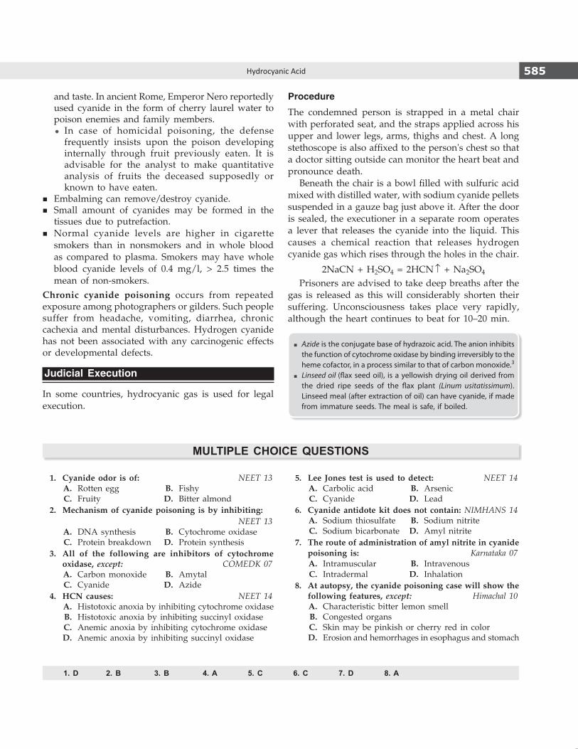
585Hydrocyanic Acid
and taste. In ancient Rome, Emperor Nero reportedly used cyanide in the form of cherry laurel water to poison enemies and family members.
z In case of homicidal poisoning, the defense frequently insists upon the poison developing internally through fruit previously eaten. It is advisable for the analyst to make quantitative analysis of fruits the deceased supposedly or known to have eaten.
� Embalming can remove/destroy cyanide. � Small amount of cyanides may be formed in the
tissues due to putrefaction. � Normal cyanide levels are higher in cigarette
smokers than in nonsmokers and in whole blood as compared to plasma. Smokers may have whole blood cyanide levels of 0.4 mg/l, > 2.5 times the mean of non-smokers.
Chronic cyanide poisoning occurs from repeated exposure among photographers or gilders. Such people suffer from headache, vomiting, diarrhea, chronic cachexia and mental disturbances. Hydrogen cyanide has not been associated with any carcinogenic effects or developmental defects.
Judicial Execution
In some countries, hydrocyanic gas is used for legal execution.
Procedure
The condemned person is strapped in a metal chair with perforated seat, and the straps applied across his upper and lower legs, arms, thighs and chest. A long stethoscope is also affixed to the person's chest so that a doctor sitting outside can monitor the heart beat and pronounce death.
Beneath the chair is a bowl filled with sulfuric acid mixed with distilled water, with sodium cyanide pellets suspended in a gauze bag just above it. After the door is sealed, the executioner in a separate room operates a lever that releases the cyanide into the liquid. This causes a chemical reaction that releases hydrogen cyanide gas which rises through the holes in the chair.
2NaCN + H2SO4 = 2HCN ↑ + Na2SO4
Prisoners are advised to take deep breaths after the gas is released as this will considerably shorten their suffering. Unconsciousness takes place very rapidly, although the heart continues to beat for 10–20 min.
� Azide is the conjugate base of hydrazoic acid. The anion inhibits the function of cytochrome oxidase by binding irreversibly to the heme cofactor, in a process similar to that of carbon monoxide.3
� Linseed oil (flax seed oil), is a yellowish drying oil derived from the dried ripe seeds of the flax plant (Linum usitatissimum). Linseed meal (after extraction of oil) can have cyanide, if made from immature seeds. The meal is safe, if boiled.
1. D 2. B 3. B 4. A 5. C 6. C 7. D 8. A
MULTIPLE CHOICE QUESTIONS
1. Cyanide odor is of: NEET 13 A. Rotten egg B. Fishy C. Fruity D. Bitter almond 2. Mechanism of cyanide poisoning is by inhibiting: NEET 13 A. DNA synthesis B. Cytochrome oxidase C. Protein breakdown D. Protein synthesis 3. All of the following are inhibitors of cytochrome
oxidase, except: COMEDK 07 A. Carbon monoxide B. Amytal C. Cyanide D. Azide 4. HCN causes: NEET 14 A. Histotoxic anoxia by inhibiting cytochrome oxidase B. Histotoxic anoxia by inhibiting succinyl oxidase C. Anemic anoxia by inhibiting cytochrome oxidase D. Anemic anoxia by inhibiting succinyl oxidase
5. Lee Jones test is used to detect: NEET 14 A. Carbolic acid B. Arsenic C. Cyanide D. Lead 6. Cyanide antidote kit does not contain: NIMHANS 14 A. Sodium thiosulfate B. Sodium nitrite C. Sodium bicarbonate D. Amyl nitrite 7. The route of administration of amyl nitrite in cyanide
poisoning is: Karnataka 07 A. Intramuscular B. Intravenous C. Intradermal D. Inhalation 8. At autopsy, the cyanide poisoning case will show the
following features, except: Himachal 10 A. Characteristic bitter lemon smell B. Congested organs C. Skin may be pinkish or cherry red in color D. Erosion and hemorrhages in esophagus and stomach

586 Review of Forensic Medicine and Toxicology
Asphyxiants
ChApter 56
Introduction
Asphyxiant gas is a non-toxic or toxic gas which causes respiratory embarrassment leading to unconsciousness or death by asphyxiation. The brain is commonly affected.
There are two broad categories of asphyxiants: simple and chemical. i. Simple asphyxiants: They are physiologically inert
gases that displace O2 from ambient air resulting in fall in partial pressure of O2 in the alveoli, e.g. acetylene, CO2, argon, helium, ethane, nitrogen and methane.
ii. Chemical asphyxiants: They interfere with the transportation or absorption of O2 in the body and interfere with cellular metabolism causing cells to become O2 starved. It can be:
a. Irritant gases: They produce toxic effect by destruction of the integrity of the mucosal barrier of the respiratory tract (damage to both type I and type II pneumocytes), e.g. ammonia, H2S, formaldehyde, phosgene and SO2.
b. Systemic asphyxiants: They produce significant systemic toxicity by various mechanisms, e.g. CO, cyanide and smoke.
Carbon Monoxide (CO)
Properties
� CO is a colorless, tasteless, non-irritative and odorless gas, and lighter than air.
� It produced by incomplete combustion of carbo-naceous material.
� It combines with chlorine and forms carbonyl chloride—commonly called phosgene.
Sources
� Common sources of CO include tobacco smoke, house fires, automobile exhaust (1–7% CO), industrial processes, unvented or faulty heating units (stove gas, water heater, burning fossil fuel or furnace) and fires.
� Coal gas (mixture of CO, methane and hydrogen). � Endogenous CO.
Action
� CO combines reversibly with hemoglobin to form carboxyhemoglobin (COHb) producing anemic hypoxia (blood O2-carrying capacity is reduced).1,2 It has a high affinity for Hb (about 250 times more than O2).
� It inhibits the electron transport by blocking cytochrome A3 oxidase and cytochrome P450 and hence intracellular respiration.
� About 15% of CO present in extracellular tissues combines with myoglobin (affinity constant—40). A ‘rebound effect’ with delayed return of symptoms may be due to late release of CO from myoglobin with subsequent binding to hemoglobin.
Signs and Symptoms
Presenting symptoms are mostly nonspecific and depend on the duration of exposure and levels of COHb. Most frequent acute symptoms are headache (dull, frontal and continuous), dizziness, weakness, nausea and confusion.
COHb (%) Signs and symptoms
0–10 No symptoms10–20 Breathlessness, mild headache, abdominal pain3
20–30 Throbbing headache, irritability, emotional instability, buzzing in the ears
30–40 Severe headache, nausea, vomiting, dizziness, dimness of vision, confusion, ataxia
40–50 Increasing confusion, hallucinations, rapid respira-tion, staggering and incoordination—mistaken for drunkenness
50–70 Weak thready pulse, hypotension, irregular respiration, convulsions, coma and death
> 80 Rapid death from respiratory arrest
� On examination, there may be tachycardia, hypertension or hypotension, hyperthermia, flame-shaped retinal hemorrhages and bright red retinal veins. Classic cherry red skin is rare; pallor is present more often.

587Asphyxiants
� CNS is most sensitive followed by heart (MI, dysrhythmias) due to their high oxygen demand. Patients display memory disturbance (most common) including retrograde and anterograde amnesia with amnestic confabulatory state.
Severity of CO Poisoning
Normal COHb level is < 5%, upto 9% in cigarette smokers. Serious toxicity is associated with levels > 25%, and risk of fatality at 70%.3,4
COHb (%) Severity of poisoning10–30 Mild30–40 Moderate-severe> 40 Very severe
Fatal dose and fatal periodThe normal atmospheric concentration of CO is usually < 0.001% (10 ppm). The atmospheric concentration can exceed 0.01% (100 ppm) in heavy urban traffic and during periods of atmospheric stagnation.
CO concentration (%) Fatality (hours)0.2 40.4 110 ½
Diagnosis: Misdiagnosis is common because of the vagueness and broad spectrum of complaints; symptoms often are attributed to viral illness (influenza).
� History: Following should alert suspicion: winter months, exposed to the previously named sources and when more than one patient in a group or household in a particular enclosed site presents with similar complaints.
� Laboratory findings: COHb analysis can be done by direct spectrophotometric measurement in specific blood gas analyzers. Bedside pulse CO-oximetry is available. Breath CO monitoring is an alternative to pulse CO-oximetry.
� CT: Symmetric low density areas in the region of globus pallidus, putamen and caudate nuclei are frequently seen within 12 h of CO exposure that resulted in unconsciousness.
� Lab diagnosis i. Spectroscopic test: Shows two absorption bands similar to
oxyhemoglobin, but placed nearer the violet end. ii. Hoppe-Seyler’s test: Few drops of blood + 10% NaOH → Greenish
brown (normal blood), Pink/red (COHb). iii. Kunkel’s test: Diluted blood (1: 10) + few drops of 3% tannic acid
(shake) → Deep brown (normal), Crimson-red coagulum (COHb). iv. Potassium ferrocyanide test: 15 ml of blood + 15 ml of 20%
potassium ferrocyanide + 2 ml diluted acetic acid → Dark brown coagulum (normal), bright-red coagulum (COHb).
v. Katayama’s test using ammonium sulfide and acetic acid is less sensitive.
Differential Diagnosis� Alcoholic intoxication � Head injury� Diabetic/Insulin coma � Uremia� Cerebral hemorrhage � Barbiturates/Narcotic
poisoning
TreatmentTreatment consists of removal from the source of exposure, immediate administration of high-flow or 100% O2 and aggressive supportive measures.5
i. Remove the victim from source of exposure. ii. Maintain patent airway, fresh air and orthobaric
oxygen (100% oxygen at atmospheric pressure) by tight-fitting high-flow reservoir face mask or endotracheal tube.5 Oxygen therapy is started if COHb > 10% and should be given for 4–6 hours (h). The immediate effect of oxygen is enhancement of the dissociation of COHb.
iii. The use of hyperbaric oxygen (HBO) is controversial. HBO has been postulated to reduce the incidence of neurological sequelae. It is indicated in cases of unconsciousness, cardiovascular instability or ischemia, and persistent mental and/or neurologic deficits. HBO at PO2 of 2–3 atmospheric pressure mixed with 5% CO2 may be given through mask or intratracheal tube.6
iv. Blood transfusion, if required. v. Gastric lavage to prevent aspiration pneumonia. vi. Cerebral edema is treated by mannitol 500 ml IV
as 20% solution over 15 minutes (min), followed by 500 ml of 5% dextrose over next 4 h.
vii. Hypotension is initially treated with IV fluids followed by inotropic agents. Standard ACLS protocols are followed to treat dysrhythmias.
viii. Antibiotics and symptomatic treatment.
Postmortem Findings
External i. Cherry red coloration of the skin, mucous mem-
branes, PM staining, blood, tissues and internal organs.7,8 The red lividity is usually associated with a COHb level > 30% CO. In dark-skinned individuals, fingernail beds can be examined.
ii. Fine froth at the nostrils/mouth. iii. Blisters of skin over dependent areas or bony
pressure points such as buttocks, calves, wrists and knees due to cutaneous edema.
Internal i. Lung: Edema and congestion. ii. Heart: Lesions vary from petechial hemorrhages
to myocardial necrosis.
588 Review of Forensic Medicine and Toxicology
iii. Rhabdomyolysis from the direct toxic effects of CO, and prolonged immobility lead to renal failure.
iv. CNS: Neuronal hypoxic injury is most pronounced in the deep gray matter, usually in a symmetrical distribution. Punctiform hemorrhages and softening of cerebral cortex and corpus striatum—particularly globus pallidus.
In addition to routine viscera, lungs and brain are preserved for analysis. If blood is not available for CO determination, spleen, liver or skeletal muscle can be utilized.
Medico-legal Aspects
� It is a common mode of suicidal poisoning in the West (by inhaling motor vehicle exhaust), but rare in India. Suicide is also committed by sealing off door and windows and burning charcoal and papers—common in Hong Kong, Southern China and Singapore.
� Accidental cases: Common in India from cooking gas leakage, and incomplete combustion of wood, char-coal or coal in ill-ventilated rooms.
z Unintentional fatalities occur in stationary vehicles from malfunctioning exhaust systems or operation in an enclosed space.
z CO presents greater risk to firefighters and victims than thermal injury or oxygen deprivation.
z Malfunctioning heating systems (e.g. blocked chimney) using combustible fuels can cause fatal CO poisoning.
� Homicide is uncommon (e.g. exhaust fumes used to poison an immobilized person). A victim of CO poisoning can be placed in a bed to simulate a natural death.
� Masochistic sexual asphyxia may be due to CO. � COHb can be detected even in a putrefied or embalmed
body, and it is not a product of putre faction.
Two features of CO poisoning may create confusion:
i. Bullous lesions on the body which simulate 20 thermal burn, deep coma, early putrefaction, antemortem and postmortem gasoline exposure.
ii. Tendency of the dying victim to wild, flailing movements inside the room, disturbing clothing and furniture which gives an impression of a violent tussle, thus creating a suspicion of murder.
Carbon Dioxide (CO2)
Properties
� CO2 is a heavy, colorless and odorless (slightly irritating) gas.
� Constituent of atmosphere air (0.4%). � Slightly acidic in taste.
Sources � It is formed during respiration, combustion, fer-
mentation and putrefaction of organic matter, mine explosion, refrigerating plants and limekilns.
� Solid form is known as dry ice. � Found in old wells, mine shafts and damp cellars.
Uses: CO2 is used in the food industry in the carbonation of beverages, fire extinguishers as an ‘inerting’ agent and in the chemical industry.
ActionIts mode of action is as an asphyxiant (lack of O2), although it also exerts toxic effects at cellular level. Pure CO2 causes vagal inhibition along with glottis spasm leading to instant death.
Signs and Symptoms
Blood CO2 (%) Signs and symptoms
0–2 No symptoms.2–5 Increased respiration, throbbing headache.5–10 Hyperpnea, tinnitus, mental confusion, muscular
tremors.10–20 Slow respiration, fall in blood pressure. 20–40 Dyspnea, muscular weakness, fall in blood
pressure, loss of reflexes.40–60 Dyspnea, feeling of tightness in chest, tinnitus,
muscular weakness, drowsiness, unconscious-ness, coma and death.
60–80 Immediate unconsciousness, convulsions, death due to asphyxia (cerebral hypoxia).
Solid CO2 may cause burns following direct contact. If it is warmed rapidly, large amounts of carbon dioxide are generated, which can be dangerous, particularly within confined areas.
Fatal concentration � Minimum: 25–30%. � Maximum: 60–80%.
Concentrations > 10% may cause convulsions, coma and death.
Fatal period: Instant collapse and death.
TreatmentThe treatment requires immediate removal from the source, administration of oxygen and appropriate supportive care including assisted ventilation. i. Shift to fresh atmosphere. ii. Maintain body warmth.

589Asphyxiants
iii. Artificial respiration with oxygen therapy. iv. Tham (2-amino-2 hydroxymethyl-1, 3-propanediol)
an amine buffer may be given IV. v. Cardiac stimulants, like amphetamine sulfate can
be used. vi. Dry ice burns are treated similarly to other
cryogenic burns, requiring thawing of the tissue and suitable analgesia.
Postmortem Findings
Features of asphyxia are found. i. Cyanosis, and pupils are dilated. ii. Marked capillary and venous congestion. iii. Petechial hemorrhages. iv. Froth at the nostrils and mouth. v. Blood is dark and fluid. vi. Deep congestion of the viscera.
Medico-legal Aspects
� Poisoning is mostly accidental. The gas being heavier settles at the bottom and may affect workmen associated with well sinking, well cleaning and descending in pits and ship holds.
� Blood CO2 accumulates during postmortem. Of critical importance is analysis of air-sample collected from the scene for CO2 content.
� Sometimes, anesthetist causes fatality by giving CO2 in place of O2 by mistake.
Hydrogen Sulfide (H2S)
Properties
� H2S is a colorless, transparent gas with smell of rotten eggs.
� It dissolves in water, and burns in air with a pale blue flame.
Sources
� Natural: Caves, volcanoes, decaying fish, sewage (sewer gas), manure and putrefying cadaver.
� Industrial: Petroleum and tanning industry, silk, rayon and paper manufacturing processes.
Action
� H2S does not combine with hemoglobin, but does so with methemoglobin to form sulfmethemoglobin.
� It causes asphyxiation by interfering with the use of oxygen in the cytochrome oxidase system.
� Its toxicity and rapidity of action is comparable to hydrocyanic acid (HCN).
Signs and Symptoms
Significant H2S poisoning usually occurs by inhalation. As a cellular poison, H2S affects all organs, particularly the CNS and the respiratory system.
System Signs and symptoms
CNS Headache, vertigo, nystagmus, weakness, comaRS Rhinitis, pneumonia, pulmonary edemaCVS Arrhythmia, myocardial depressionOcular Lacrimation, photophobia, conjunctivitis
The presence of H2S is apparent because of the characteristic rotten egg smell. However, concentrations > 150 ppm may overwhelm the olfactory nerve, so that the victim may have no warning of exposure.
Fatal dose and fatal period
H2S concentration (ppm) Clinical effects> 200 Anosmia, pulmonary edema> 500 Hyperpnea, apnea
> 1000 Respiratory paralysis, death
Exposure of > 700–800 ppm can cause immediate cardiopulmonary arrest.
� Detection: H2S, if present in significant concentration, can be tested by exposing a filter paper moistened with lead acetate. The filter paper will turn black.
� Spectroscopic test: It is characterized by absorption spectrum of two bands consisting of one band in the red between C and D and a fainter band between D and E.
Differential diagnosis: Smoke inhalation, CO, cyanide and hydrocarbons.
TreatmentHigh-flow (100%) O2 is the mainstay of therapy for H2S poisoning. i. Remove the victim into fresh air. ii. Artificial respiration and 100% O2 is given. iii. Antidote: Amyl nitrite and sodium nitrite (without
thiosulfate) enhance formation of methemoglobin which in turn is spontaneously detoxified in the body.*
z Break 0.3 ml ampoule/perle of amyl nitrite in a gauze and hold over the patient's nose for 15–30 seconds.
* Based on the similarities in cyanide and H2S toxicity, induced methemoglobinemia may be used for the treatment of H2S toxicity. Methemoglobin acts as a scavenger, and it is more attracted to H2S than cytochrome oxidase.
590 Review of Forensic Medicine and Toxicology
z 0.3 g of sodium nitrite in 10 ml of sterile water is given slow IV for over 2–3 min.
iv. Supportive measures: Correction of electrolyte imbalance and pulmonary edema.
Postmortem Findings
i. Signs of asphyxia (cyanosis, frothing at the mouth and nose, and petechial hemorrhages in respiratory mucosa) are seen.
ii. Rotten egg odor is present around the nostrils and mouth.
iii. Greenish discoloration of viscera, gray matter of brain and bronchial secretions may be found.
iv. Pulmonary edema and congestion of viscera are seen.
Medico-legal Aspects
� Poisoning is always accidental, causing number of deaths in sewer workers. The petroleum industry is
responsible for most cases of H2S toxicity in North America.
� H2S has recently been implicated in suicides in Japan.9
Detergent or chemical suicide: In Japan, it is a newer method of committing suicide and is gaining popularity in other countries from internet suicide websites. A near-instant death may occur by mixing common household chemicals—bath sulfur (5–30% calcium polysulfides) with toilet bowl cleaner (15% HCl)—to create H2S gas in cars, closets or other enclosed spaces.
Chronic H2S exposure causes headache, weakness, nausea, weight loss, ataxia and tremors.
� Patient can lose their ability to smell/detect the gas even though it is still present in the environment (olfactory fatigue/paralysis).
� Low-level exposure of H2S affects the mucous membranes, and may cause conjunctivitis, pharyngitis, green-gray line on gingiva and wheezing.
1. A 2. A 3. B 4. C 5. C 6. D 7. D 8. A 9. B
multiple ChoiCe questions
1. Carbon monoxide poisoning causes: JIPMER 11; AIIMS 14 A. Anemic hypoxia B. Histotoxic hypoxia C. Anoxic hypoxia D. Stagnant hypoxia 2. Anemic hypoxia is seen in: CMC (Vellore) 14 A. CO poisoning B. CO2 poisoning C. HCN poisoning D. Nerve gas 3. Symptoms of CO poisoning starts when the
concentration is: NEET 14 A. < 10% B. > 10% B. > 20 % D. > 40 % 4. Percentage of COHb that usually causes death: NEET 13 A. > 50% B. > 60% C. > 70% D. > 80% 5. In CO poisoning, immediate emergency treatment:
Jharkhand 10 A. 5% CO2 inhalation B. 10% CO2 inhalation C. High flow O2 D. Nitroglycerine
6. Hyperbaric oxygen is given in the treatment of: Bihar 12
A. Nerve gas B. CO2 poisoning C. HCN poisoning D. CO poisoning 7. Cherry red color in postmortem staining is a feature
of poisoning with: WB 08; NEET 13 A. Nitrites B. Aniline C. Phosphorus D. CO 8. Postmortemfinding inCOpoisoning is:
UP 09; PGI 10 A. Cherry red hypostasis B. Intense cyanosis C. Excessive salivation D. Pin-point pupil 9. Death caused in suicide by household things in Japan
is due to the production of: AI 12 A. Acidic solution B. H2S C. HCN gas D. CO

591War Gases and Biological Weapons
War Gases and Biological Weapons
Chapter 57
War Gases
Definition: War gases are chemicals (gas, liquid or solid) which are used for producing destruction or damage, mostly at times of war. These also include chemicals being used for dispersing unruly mobs.
Chemical warfare (CW) involves using the toxic properties of chemical substances as weapons.
Types of Chemical Warfare Agents (CWAs)
Major categories of CWAs i. Asphyxiants or lung irritants ii. Vesicants or blister gases iii. Lacrimators or tear gases or riot control agents iv. Sternutators or nasal irritants v. Nerve gases vi. Paralysants vii. Miscellaneous.
Asphyxiants / Lung Irritants / Choking Gases
� These are chlorine and phosgene (CG) gas, and can be released from tanks and gas shells.
� Phosgene is ten times more toxic than chlorine. � Their action is mainly on the pulmonary alveoli. � Symptoms: When inhaled, they cause watering of the
eyes, coughing, dyspnea, tightness of chest, headache, vomiting, restlessness, stertorous breathing, cyanosis and collapse. Death occurs in 24–48 hours due to acute pulmonary edema or bronchopneumonia.
� Treatment i. Remove the patient into fresh air. ii. Wash the eyes with normal saline and boric acid. iii. Oxygen and adrenaline when needed. iv. Antitussives (e.g. codeine) for cough, and antibiotics
for infection.
Vesicants / Blister Gases
� Mainly mustard gas (dichlordiethyl sulfide or yperite) and lewisite (dichlorarsine).1
� These are volatile liquids and discharged in artillery shells so as to saturate the area of attack.
Symptoms � Mustard gas causes irritation of the eyes, nose,
throat and respiratory passages, nausea, vomiting and abdominal pain. It penetrates the clothes and produces intense itching, redness, blisters and ulceration, especially of the moist areas of the skin.
� Lewisite causes blisters in skin and inflammation of mucous membrane, and on absorption produces signs of arsenic poisoning.
Treatment i. Wash the affected parts thoroughly with soap and
water. ii. Wash eyes with sodium bicarbonate solution. iii. Use BAL as an antidote to lewisite.
Lacrimators / Tear Gases
� Mainly chloracetophenone (CAP) which is solid, and ethyliodoacetate (KSK) and bromoben zylcyanide (BBC) which are liquids. These are fired in artillery shells or pen guns.
� Symptoms: The vapors cause intense irritation of the eyes, lacrimation, spasm of the eyelids and temporary blindness with irritation of air-passages.
With long continued exposure, there may be nausea, vomiting and blistering of skin.
� Treatment: The patient should be removed into fresh air and the eyes washed with warm normal saline or boric acid solution. Weak sodium bicarbonate solution is applied to the affected parts of the skin.
Sternutators / Nasal Irritants / Vomiting Gases
� They are diphenyl chlorarsine (DA), diphenylamine chlorarsine (DM) and diphenyl cyanarsine (CD).
� These are solid, organic compounds of arsenic and are fired in artillery shells to control riots.
� Symptoms: The vapors cause intense pain and irritation in the nose and sinuses, sneezing, headache,

592 Review of Forensic Medicine and Toxicology
malaise, salivation, nausea, vomiting, tightness in the chest and prostration.
� Treatment: The patient is removed into fresh air and the nose irrigated with 5% sodium bicarbonate.
Nerve Gases
� The term ‘nerve gas’ is a misnomer, since the nerve agents (NAs) are liquid at room temperature. These colorless and odorless volatile liquids are esters of phosphoric acid and are identical to organophosphorous (OP) in their biological activity, although fatality with NAs is generally higher than the OP pesticides.
� NAs are known as the deadliest CWAs and are divided into two classes—G and V agents. The G agents include GA (Tabun), GB (Sarin), GD (Soman) and GF (Cyclosarin).2 The V agents include VE, VM, VG, VR and VX. Recently, a new type of NAs has been developed named ‘Novichoks’ (‘newcomer’ in Russian).
� Mechanism of action: Irreversible inactivation of acetylcholine esterase (AChE) at the cholinergic synapses leading to accumulation of toxic levels of acetylcholine (ACh) at the synaptic junctions.
� Sites of absorption: They are absorbed from the lungs, GIT, skin or conjunctiva.
� Symptoms: Clinical manifestations are similar to OP pesticides poisoning. Acute effects include headache, sweating, nausea, vomiting, miosis, ocular pain, impaired visual acuity, lacrimation, bloodshot eyes, rhinorrhea, bronchorrhea, wheezing, respiratory failure, bradycardia and atrioventricular block, generalized fasciculation, fatigue, muscle weakness, and flaccid paralysis. Exposure to a large amount of vapor may cause loss of consciousness within seconds, followed by convulsions, respiratory failure and death in few minutes.
� Treatment is similar to organophosphates. The victims should immediately be removed from the field and treatment is commenced with auto-injector antidotes (atropine and oximes) such as MARK I kit. A 0.5% hypochlorite solution, as well as novel products like M291 resin kit, G117H and phosphotriesterase isolated from soil bacterias are now available for decontamination of NAs.
� For decontamination in field (for soldiers), M291 resin kit that contains carbonaceous adsorbent, a polystyrene polymeric and ion exchange resins is available. It can be used on the skin, face and around wounds. As the powder is scrubbed over the contaminated skin, its carbonaceous material rapidly adsorbs the agents and physically removes them from skin. Then the trapped agents in the interior of the resin particles will be neutralized through chemical detoxification due to the presence of basic and acidic groups in the resin.
� Antidotes could be injected by the victim himself or anybody who finds him, with auto-injectors. Two types are available—MARK I kit and antidote treatment nerve agent auto-injector (ATNAA). MARK I kit (most popular one) is composed of 2 mg atropine and 600 mg 2-pyridine aldoxime methyl chloride (2-PAMCl). The ATNAA, designated by the Department of Defense of the US, contains 2.1 mg atropine and 600 mg/2 ml 2-PAM, and has ability of simultaneously injecting both of them through single needle. Every soldier carries 3 kits and one auto-injector containing 10 mg diazepam when there is a suspicious of NA attack.
Paralysants
These are hydrocyanic acid, hydrogen sulfide and carbon monoxide which have been described in previous chapters.
Miscellaneous
� These include yellow/red rain and methyl isocyanate (MIC).
� Symptoms: Acute irritation of the eyes, lacrimation, blurring of vision, severe burning sensation in the throat, chest pain and labored breathing. Death is caused by pulmonary edema.
� Treatment is symptomatic. Sodium thiosulfate may act as an antidote.
The accidental release of a methyl isocyanate cloud was implicated in the Bhopal disaster in 1984.
Biological Weapons
Definitions � Biological weapons are microorganisms or toxins
found in nature which can be used to incapacitate, kill or otherwise impede an adversary.
� Bioterrorism is the deliberate release of viruses, bacteria, toxins or other harmful agents causing illness or death in people, animals or plants.

593War Gases and Biological Weapons
Types of Biological Warfare Agents
The CDC in the US has categorized these biological warfare agents as under (Table 57.1): i. Category A: These high-priority agents can be
easily transmitted and disseminated, result in high mortality, have potential major public health impact, may cause public panic or require special action for public health preparedness.
ii. Category B: These agents are moderately easy to disseminate and have low mortality rates.
iii. Category C: These agents are emerging pathogens that might be engineered for mass dissemination because of their easy availability, ease of production and dissemination, high mortality rate or ability to cause a major health impact.
Some of them are described below:3
1. Anthrax: Anthrax is a non-contagious disease caused by the spore-forming bacterium Bacillus anthracis. It usually affects animals. Humans who have contact with infected animals or animal products such as wool or hide can get the disease.
z Mode of transmission: Anthrax is propagated by terrorists in a powder form. Common method is by sending letters smeared with spores to target victims. When the letter is handled, spores enter the body by inhalation and skin contact.
– Anthrax spores are highly stable and can be dispersed by enclosing them in bombs and ammunitions. When the bombs explode, anthrax spores are liberated into the atmosphere.
z Symptoms: Three types of anthrax infections depending upon the route of entry of the spores:
a. Cutaneous: Symptoms are caused by skin contact with infected animal materials. Blisters and ulcers develop in the skin.
b. GIT: It is caused by consumption of undercooked meat of infected animals. Symptoms are fever, nausea, hemoptysis and bloody diarrhea.
c. Respiratory: It is caused by inhalation of the spores. Symptoms are fever, cough and myalgia. Later, serious respiratory symptoms may appear.
z Treatment: It can be treated with antibiotics. Anthrax vaccination is available as a prophylactic measure.
2. Botulism is caused by a toxin generated by bacterium Clostridium botulinum, and results in serious neurological symptoms. This toxin is more toxic than cyanide, and is readily available because of its widespread use in cosmetology.
z Mode of transmission: The toxin is propagated as lyophilized powder enclosed in rockets and bombs. The toxin enters the body through air, contaminated food and water.
z Symptoms: Botulism causes death by respiratory failure and paralysis.
z Treatment: Antitoxin is effective in reducing the severity of symptoms.
3. Plague: Plague is caused by Yersinia pestis, a bacterium found in rodents and their fleas. Rodents are the normal host of plague and the disease is transmitted to humans by flea bites (bubonic plague) and occasionally by aerosol (pneumonic plague).
z Mode of transmission: One of the methods is by releasing infected rat fleas in enemy country. The fleas are kept in porcelain containers attached to projectiles, like rockets and bombs before firing at targets.
z Symptoms include swollen and tender lymph nodes called buboes. If untreated, the bacteria spread through the bloodstream and infect lungs causing pneumonia. In pneumonic plague, the person has fever, weakness and rapidly developing pneumonia with dyspnea, chest pain, cough and bloodstained sputum. If untreated, death occurs due to respiratory failure and shock.
Table 57.1: Categories of biological warfare weapons
Category A Category B Category C
� Anthrax � Brucellosis � Nipah virus
� Botulinum toxin � Water borne threats (e.g. Vibrio cholera) � H1N1, a strain of influenza
� Viral hemorrhagic fever � Food poisoning threats (e.g. Salmonella, E. coli) � SARS
� Bubonic plague � Ricin and abrin
� Smallpox � Q fever
� Tularemia � Staphylococcal enterotoxin B
� Typhus, viral encephalitis
594 Review of Forensic Medicine and Toxicology
z Treatment: It is treated with broad-spectrum antibiotics. There is no vaccine available to prevent plague. Plague bacteria are destroyed by sunlight and drying.
4. Smallpox: Smallpox is caused by the virus variola major which is highly contagious and has a high mortality rate (20–40%). It occurs only in humans and has no external hosts or vectors.
z Mode of transmission: It is spread through aerosols and infected material. Even though smallpox has been eradicated throughout the world, virus samples are still available in laboratories of some countries (Russia and the US).
As a biological weapon, smallpox is dangerous because vaccines are no longer administered to the general population, and in the event of an outbreak most people would be unprotected.4
z Symptoms include fever, headache, fatigue, diarrhea, vomiting and a specific rash. The rash first starts as flat red spots which turn into blisters. Blisters contain a clear fluid, initially, and then pus, as the disease progresses.
z Treatment: There is no specific drug to treat smallpox.
5. Viral hemorrhagic fever: This includes hemorrhagic fever caused by members of the family Filoviridae (e.g. Ebola and Marburg virus) and by the family Arenaviridae (e.g. Lassa and Machupo virus). Ebola and Marburg virus have high mortality rates. It is believed that some terrorist group possesses Ebola virus culture. The fatality rate of arenaviruses is less compared to those caused by filoviruses.
Death from Ebola virus disease is commonly due to multiple organ failure and hypovolemic shock.
z Treatment: There is no effective treatment and prophylaxis for these viral infections, although vaccines are in the process of development.
6. Tularemia: Tularemia or rabbit fever is caused by Francisella tularensis bacterium through contact with
fur, inhalation or ingestion of contaminated water or by insect bites. It is a highly infectious disease and requires only a small number of organisms (10–50) to cause it.
z Mode of transmission: If it is used as a weapon, the bacteria would likely be made airborne for exposure by inhalation.
z Symptoms: On inhalation, there is severe respiratory illness, including life-threatening pneumonia, and if left untreated, systemic infection may result.
7. Brucellosis: Brucellosis is an infectious disease caused by Brucella bacteria. The bacteria affect cattle, dogs, pigs and other animals. Humans become infected by coming into contact with animals or animal products contaminated with these bacteria.
z Mode of transmission: Air, water and food articles are contaminated by terrorists. The bacteria can also enter through skin wounds. When cattle are infected, their milk contains the bacteria. Intake of unpasteurized milk can transmit the bacteria to those people who consume the milk.
z Symptoms: Fever, headache, back pain and weakness are seen. Sometimes, endocarditis and encephalitis may develop.
8. Ricin toxin: Ricin obtained from Ricinus communis, is one of the most poisonous naturally occurring substances known. Ricin is toxic by numerous exposure routes and its use by terrorists might involve poisoning of water or foodstuffs, inoculation via ricin-laced projectiles, or aerosolization of liquid ricin or distribution of powder.
9. Salmonella: This can be done by contaminating foodstuffs in restaurants, bars and grocery stores and lead to severe food poisoning.
z Symptoms include vomiting, nausea, diarrhea and abdominal pain. It is sometimes associated with very high fever. This condition can last for upto a week.
Multiple choice questions
1. Blistering war gas is: Maharashtra 09 A. Chlorine gas B. Mustard gas C. HCN gas D. Tabun
2. Nerve gas is: TN 10 A. Methyl isocyanate B. Phosgene C. Diphenylchloroarsine D. Sarin
3. Bioterrorism is associated with all, except: UP 12 A. Clostridia B. Chicken pox C. Plague D. Ebola virus 4. Most important and potential agent that can be used
in bioterrorism: AI 11 A. Plague B. Smallpox C. Tuberculosis D. C. botulinum
1. B 2. D 3. B 4. B

595Agricultural Poisons
Agricultural Poisons
CHAPTER 58
Introduction
Pesticides are varied group of agents used to control livings organisms that pose health or economic threats. Often the term is misunderstood to refer only to ‘insecticides’, but it also applies to herbicides, fungicides and various other substances used to control pests.
� They can be manmade (synthetic) or naturally occurring (biological) and may be active against a narrow (selective) or wide (broad-spectrum) range of pests.
� Classification: Pesticides are often grouped by the pest they control (e.g. insecticides, rodenticides, fungicides, etc.) or categorized by chemical structure (e.g. insecticides are categorized as organophosphate, carbamate, organochlorine, synthetic-pyrethroid, and microbial and insect growth regulators). WHO classifies pesticides by hazards they pose based on Globally Harmonized System (GHS) from Category 1 (LD50 < 5 mg/kg body wt) to Category 5 (LD50 2000–5000 mg/kg body wt) as given in Table 58.1.
� Commonest types of insecticide/pesticide substance used for poisoning are organophosphates, chlorinated hydrocarbons, aluminum phosphide, carbamates and pyrethroids.
Organophosphorus Compounds (OPCs)
OPCs and carbamates are one of the most common causes of self-poisoning seen in India. They are used as insecticides, herbicides, antihelminthics, ophthalmic agents, in chemical industry, and as nerve gas in chemical warfare.
ClassificationBased on chemical composition
� Alkyl phosphates: Tichlorfos, dimefox, HETP, TEPP and malathion.2
� Aryl phosphates: Parathion (Follidol), paraoxon, chlorthion and diazinon (Tik-20).
Based on toxicity � Agriculture insecticide (highly toxic): TEPP,
parathion. � Animal insecticide (moderately toxic): Trichlorfon,
ronnel. � Household insecticide (low toxicity): Malathion,
Tik-20.
Table 58.1: Classification of pesticides based on hazard
WHO Class Oral LD50*(mg/kg body weight)
Ia Extremely hazardous < 5
Ib Highly hazardous 5–50
II Moderately hazardous 50–2000
III Slightly hazardous > 2000
U Unlikely to present acute hazard > 5000
* LD50 value is a statistical estimate of the number of mg of toxicant per kg of body weight required to kill 50% of a large population of test animals: the rat is used unless otherwise stated.
Another method of classification of insecticides is based on their mode of penetration, i.e. whether they cause effect upon ingestion (stomach poisons), penetration of the body covering (contact poisons) or inhalation (fumigants).
� Stomach poisons are toxic only if ingested through the mouth and are useful against those insects that have biting or chewing mouth parts, such as caterpillars and grasshoppers, e.g. arsenicals like copper acetoarsenite (Paris green), calcium arsenate and lead arsenate; and fluorine compounds like NaF and cryolite.1
� Contact poisons penetrate the skin of the pest and are used against those arthropods that pierce the surface of a plant and suck out the juices. These can be divided into two groups: naturally occurring (nicotine, pyrethrum, rotenone and oils) and synthetic organic insecticides.1 The main synthetic groups are the organic phosphates (organophosphates), carbamates and chlorinated hydrocarbons.
� Fumigants are toxic compounds that enter the respiratory system of the insect through its spiracles or breathing openings, e.g. HCN, naphthalene, methyl bromide and nicotine.
Most synthetic organic insecticides penetrate by all three of these pathways.
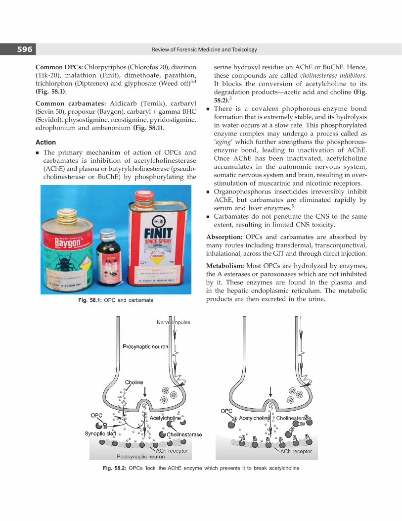
596 Review of Forensic Medicine and Toxicology
Common OPCs: Chlorpyriphos (Chlorofos 20), diazinon (Tik-20), malathion (Finit), dimethoate, parathion, trichlorphon (Diptrenex) and glyphosate (Weed off)3,4
(Fig. 58.1).
Common carbamates: Aldicarb (Temik), carbaryl (Sevin 50), propoxur (Baygon), carbaryl + gamma BHC (Sevidol), physostigmine, neostigmine, pyridostigmine, edrophonium and ambenonium (Fig. 58.1).
Action � The primary mechanism of action of OPCs and
carbamates is inhibition of acetylcholinesterase (AChE) and plasma or butyrylcholinesterase (pseudo -cholinesterase or BuChE) by phosphorylating the
serine hydroxyl residue on AChE or BuChE. Hence, these compounds are called cholinesterase inhibitors. It blocks the conversion of acetylcholine to its degradation products—acetic acid and choline (Fig. 58.2).3
� There is a covalent phophorous-enzyme bond formation that is extremely stable, and its hydrolysis in water occurs at a slow rate. This phosphorylated enzyme complex may undergo a process called as ‘aging’ which further strengthens the phosphorous-enzyme bond, leading to inactivation of AChE. Once AChE has been inactivated, acetylcholine accumulates in the autonomic nervous system, somatic nervous system and brain, resulting in over-stimulation of muscarinic and nicotinic receptors.
� Organophosphorus insecticides irreversibly inhibit AChE, but carbamates are eliminated rapidly by serum and liver enzymes.5
� Carbamates do not penetrate the CNS to the same extent, resulting in limited CNS toxicity.
Absorption: OPCs and carbamates are absorbed by many routes including transdermal, transconjunctival, inhalational, across the GIT and through direct injection.
Metabolism: Most OPCs are hydrolyzed by enzymes, the A esterases or paroxonases which are not inhibited by it. These enzymes are found in the plasma and in the hepatic endoplasmic reticulum. The metabolic products are then excreted in the urine.
Fig. 58.2: OPCs ‘lock’ the AChE enzyme which prevents it to break acetylcholine
Fig. 58.1: OPC and carbamate
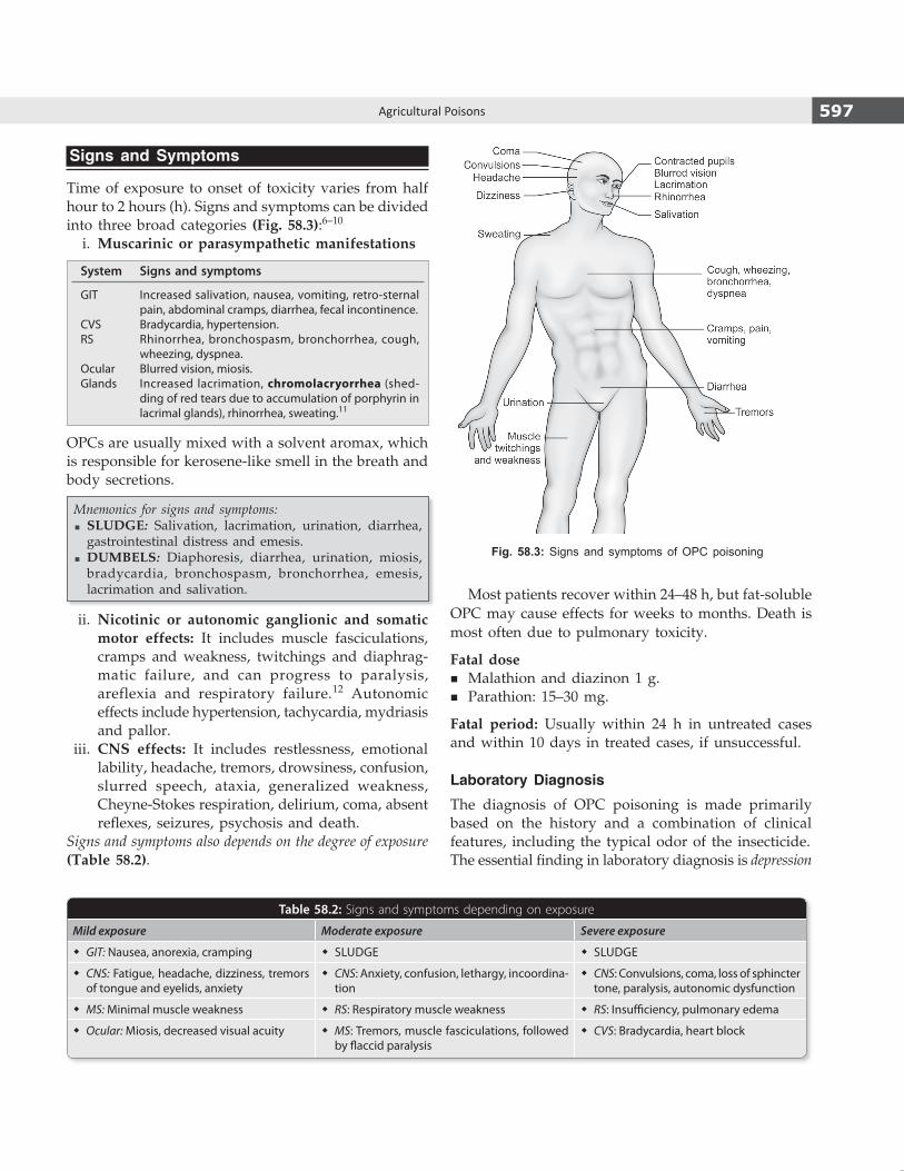
597Agricultural Poisons
Signs and Symptoms
Time of exposure to onset of toxicity varies from half hour to 2 hours (h). Signs and symptoms can be divided into three broad categories (Fig. 58.3):6–10
i. Muscarinic or parasympathetic manifestations
System Signs and symptoms
GIT Increased salivation, nausea, vomiting, retro-sternal pain, abdominal cramps, diarrhea, fecal incontinence.
CVS Bradycardia, hypertension.RS Rhinorrhea, bronchospasm, bronchorrhea, cough,
wheezing, dyspnea.Ocular Blurred vision, miosis.Glands Increased lacrimation, chromolacryorrhea (shed-
ding of red tears due to accumulation of porphyrin in lacrimal glands), rhinorrhea, sweating.11
OPCs are usually mixed with a solvent aromax, which is responsible for kerosene-like smell in the breath and body secretions.
Mnemonics for signs and symptoms: � SLUDGE: Salivation, lacrimation, urination, diarrhea, gastrointestinal distress and emesis.
� DUMBELS: Diaphoresis, diarrhea, urination, miosis, bradycardia, bronchospasm, bronchorrhea, emesis, lacrimation and salivation.
ii. Nicotinic or autonomic ganglionic and somatic motor effects: It includes muscle fasciculations, cramps and weakness, twitchings and diaphrag-matic failure, and can progress to paralysis, areflexia and respiratory failure.12 Autonomic effects include hypertension, tachycardia, mydriasis and pallor.
iii. CNS effects: It includes restlessness, emotional lability, headache, tremors, drowsiness, confusion, slurred speech, ataxia, generalized weakness, Cheyne-Stokes respiration, delirium, coma, absent reflexes, seizures, psychosis and death.
Signs and symptoms also depends on the degree of exposure (Table 58.2).
Most patients recover within 24–48 h, but fat-soluble OPC may cause effects for weeks to months. Death is most often due to pulmonary toxicity.
Fatal dose � Malathion and diazinon 1 g. � Parathion: 15–30 mg.
Fatal period: Usually within 24 h in untreated cases and within 10 days in treated cases, if unsuccessful.
Laboratory Diagnosis
The diagnosis of OPC poisoning is made primarily based on the history and a combination of clinical features, including the typical odor of the insecticide.The essential finding in laboratory diagnosis is depression
Table 58.2: Signs and symptoms depending on exposure
Mild exposure Moderate exposure Severe exposure
� GIT: Nausea, anorexia, cramping � SLUDGE � SLUDGE
� CNS: Fatigue, headache, dizziness, tremors of tongue and eyelids, anxiety
� CNS: Anxiety, confusion, lethargy, incoordina-tion
� CNS: Convulsions, coma, loss of sphincter tone, paralysis, autonomic dysfunction
� MS: Minimal muscle weakness � RS: Respiratory muscle weakness � RS: Insufficiency, pulmonary edema
� Ocular: Miosis, decreased visual acuity � MS: Tremors, muscle fasciculations, followed by flaccid paralysis
� CVS: Bradycardia, heart block
Fig. 58.3: Signs and symptoms of OPC poisoning
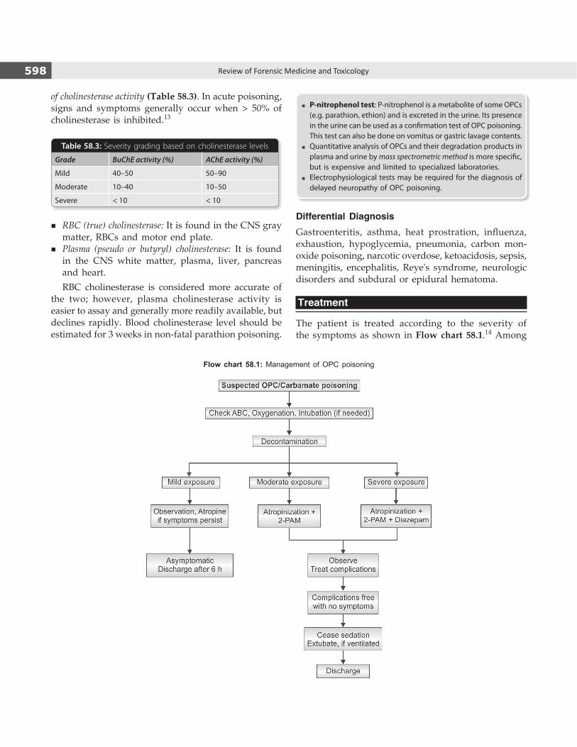
598 Review of Forensic Medicine and Toxicology
of cholinesterase activity (Table 58.3). In acute poisoning, signs and symptoms generally occur when > 50% of cholinesterase is inhibited.13
Table 58.3: Severity grading based on cholinesterase levels
Grade BuChE activity (%) AChE activity (%)
Mild 40–50 50–90
Moderate 10–40 10–50
Severe < 10 < 10
� P-nitrophenol test: P-nitrophenol is a metabolite of some OPCs (e.g. parathion, ethion) and is excreted in the urine. Its presence in the urine can be used as a confirmation test of OPC poisoning. This test can also be done on vomitus or gastric lavage contents.
� Quantitative analysis of OPCs and their degradation products in plasma and urine by mass spectrometric method is more specific, but is expensive and limited to specialized laboratories.
� Electrophysiological tests may be required for the diagnosis of delayed neuropathy of OPC poisoning.
Differential Diagnosis
Gastroenteritis, asthma, heat prostration, influenza, exhaustion, hypoglycemia, pneumonia, carbon mon-oxide poisoning, narcotic overdose, ketoacidosis, sepsis, meningitis, encephalitis, Reye's syndrome, neurologic disorders and subdural or epidural hematoma.
Treatment
The patient is treated according to the severity of the symptoms as shown in Flow chart 58.1.14 Among
Flow chart 58.1: Management of OPC poisoning
� RBC (true) cholinesterase: It is found in the CNS gray matter, RBCs and motor end plate.
� Plasma (pseudo or butyryl) cholinesterase: It is found in the CNS white matter, plasma, liver, pancreas and heart.
RBC cholinesterase is considered more accurate of the two; however, plasma cholinesterase activity is easier to assay and generally more readily available, but declines rapidly. Blood cholinesterase level should be estimated for 3 weeks in non-fatal parathion poisoning.

599Agricultural Poisons
all approaches, external decontamination and early atropinization are only likely to be beneficial in OPC poisoning, and oxime use is unlikely to be effective. i. Decontamination
z Patient is removed from source of exposure, stripped of his clothes and the skin flushed with water.
z Doctor and nurses should be protected with water-impermeable gowns, masks with eyeshields, and use double gloves while handling the patient.
z Gastric lavage: It should only be undertaken once the patient is stable. Gastric emptying should be done with continuous suction via a nasogastric tube with 1:5000 KMnO4. Activated charcoal should be administrated in doses of 1 g/kg.
z Patients with ocular exposures should have copious eye irrigation with normal saline or lactated Ringer's solution. If these are not available, tap water can be used.
ii. Atropinization: Atropine blocks the muscarinic manifestations and has no effect on nicotinic receptors (on muscle weakness or paralysis) and does not affect the rate of regeneration of inhibited AChE.10,15
Dose: 2–4 mg IV (0.05–0.2 mg/kg in children) repeated after every 5–15 minutes (min) till atropinization, the dose should be adjusted to maintain this effect for at least 24 h (maintenance dose: 0.02–0.05 mg/ kg).
Mild to moderate atropinization includes dryness of tongue, reduced secretion of oropharyngeal and bronchial tree, tachycardia and flushing. Mydriaris is an early response to atropine and is not a therapeutic end point. A common failure of therapy is not maintaining adequate atro pinization.
Glycopyrrolate may be substituted, if there is no evidence of central toxicity.
iii. Pralidoxime (2-PAM), a nucleophilic oxime, most effective when treatment is started early and if used within 48 h, and helps in regenerating AChE associated with skeletal muscle neuromuscular junctions.
Dose: 1–2 g IV (20–40 mg/kg) over 5–20 min dissolved in 0.9% normal saline solution, may be repeated at 1–2 h if muscle weakness is not relieved, and again after 8–10 h.16 Transient dizziness, blurred vision, diplopia and elevations
in diastolic BP may occur depending on the administration rate.
Alternatively, continuous infusion (200–400 mg/h) of 2-PAM is more effective because of shorter duration of action of single dose.
� Atropine and 2-PAM given together are synergistic against the signs and symptoms of cholinesterase inhibition, thus decreasing atropine requirements.
� PAM is ineffective in reversing the CNS effects of organophosphate because its positive charge prevents entry into the CNS. Diacetyl monoxime (DAM) crosses the blood-brain barrier and can regenerate some of the CNS cholinesterase.
� The use of oximes in acute OPC poisoning remains conflicting and controversial. Some randomized controlled trials showed no benefit in moderate and severe poisoning, and concluded that PAM has no role in the routine management of patients with OPC poisoning.
� PAM is not recommended for the reversal of inhibition of acetylcholinesterase by carbamate poisoning.*17,18 But its use is safe, particularly if administered in conjunction with atropine in case of unknown pesticide or in mixed poisoning.
iv. Diazepam: Addition of diazepam for treatment of seizures and neuropathy improves survival (must not be used with other CNS depressants). It decreases the cardiac and brain morphologic damage resulting from OPC seizures.
Dose: 0.5–2 mg IV every 15 min.
v. Supportive care z Foot-end of the bed is raised to ensure drainage
of respiratory secretions. z Suction as required, to remove respiratory
secretions. z Treat bronchospasm with atropine and not
bronchodilators. z Intubate in case of respiratory distress. z The use of other medication, including opioids
for sedation may worsen CNS manifestations and the degree of respiratory depression.
z Dextrose: 2–4 ml/kg of 50% dextrose IV. z Antibiotics to prevent pulmonary infection. z Vitamin K may also be given.
* The site on which oximes bind and reactivate the enzyme—the anionic site, is occupied by carbamates.
Newer therapies: Several new therapies have been studied, but there is insufficient evidence to recommend their use:
� Magnesium sulphate blocks ligand-gated calcium channels, resulting in reduced acetylcholine release from pre-synaptic terminals, thus reduce CNS over-stimulation. Administration on the first day decreases hospitalization period and improve outcomes.

600 Review of Forensic Medicine and Toxicology
Complications
Immediate Delayed
� Pulmonary edema � Paralysis10
� Aspiration pneumonia � Neurotoxicity
� Chemical peritonitis � Guillain-Barre syndrome
� Hyper-/hypoglycemia
� Coagulation abnormalities
Postmortem Findings
External i. Kerosene-like smell from nostrils and mouth. ii. Cyanosis of lips, fingers and nose. iii. Deep postmortem staining. iv. Congested face. v. Frothy discharge, often bloodstained from the nose
and mouth.
Internal i. Mucosa of the stomach and intestine is congested. ii. Stomach content may give kerosene-like smell.
iii. Respiratory passages are congested, contain frothy hemorrhagic exudates.
iv. Petechial hemorrhage may be present subpleurally. v. Edema and congestion of the lungs and other
visceral organs. vi. Edema of brain.
Medico-legal Aspects � Hospitalizing all symptomatic patients for at least
4–6 days following resolution of symptoms is recommended, because of the risk of development of respiratory depression or intermediate syndrome after resolution of an acute crisis.
� The symptoms of OPC poisoning can mimic other toxidromes and diseases. The clinician must keep in mind that misdiagnosis is a potential medico-legal pitfall.
� Accidental poisoning occurs in manufacturers, packers, sprayers and in children. OPC residue in fruits and vegetables may not induce toxic features, but could affect the health.
� Suicidal poisoning is common in our country, both in rural and urban areas. OPCs are also common suicidal agents in Pakistan, Sri Lanka and the other Asian and South East Asian countries.
� Homicidal poisoning does not occur due to detectable smell of the diluents, and signs and symptoms appear rather early.
Neurological manifestations19
Neurological manifestations are the most important sequelae of OPC poisoning. Three types of paralyses are recognized based on the time of occurrence, and differ in their pathophysiology.
i. Acute paralysis (Type I paralysis): It usually develops within 24–48 h.
Cause: Acute paralysis occurs with the initial cholinergic crisis owing to the persistent depolarization at the neuromuscular junction resulting from blockade of AChE.
Signs and symptoms: It includes muscle fasciculations, cramps, twitching and weakness. Muscle weakness may have upper motor neuron manifestations. It may also involve the respiratory muscles leading to respiratory failure.
Treatment: They respond well to atropine, although it may not fully block the nicotinic effects.
ii. Intermediate syndrome (Type II paralysis): It develops in 24–96 h after resolution of acute cholinergic poisoning symptoms and manifests commonly as paralysis and respiratory distress.20,21
Cause: Neuromuscular transmission defect, toxin-induced muscular instability or inadequate treatment of the acute episode.
Signs and symptoms: It involves proximal groups of muscle with relative sparing of distal groups and is characterized by cranial nerve palsies, weakness of neck flexors and proximal limb muscles.21
Treatment: Supportive measures, since it does not respond to oximes or atropine.
� Clonidine also reduces acetylcholine synthesis and release from presynaptic terminals.
� Sodium bicarbonate IV infusion in place of oximes has been found beneficial in nerve gas poisoning.
� Removing OPCs from the blood by hemodialysis and hemofiltration could allow optimum action of other therapies. Fresh frozen plasma or albumin is also a useful therapy through clearing of free OPC.
� Enzyme bioscavengers are being developed as a pretreatment to sequester highly toxic OPCs. BuChE purified from human plasma (HuBChE) scavenges OPCs in plasma, reducing the amount available to inhibit AChE in synapses.
� Neuroprotective drugs: Delayed management of convulsion and neuroprotection in OPC poisoning can be done with anticholinergic and antiglutamatergic agents. The CNS toxic affects results from increased release of glutamate (an excitatory amino acid) which causes over-activation of the N-methyl-D-aspartate (NMDA). Gacyclidine, tezampanel, ketamine and huperzine A are useful in treatment of nerve gas induced seizures when administered 1 h after exposure.
� Recombinant bacterial phosphotriesterases or hydrolases that are able to transfer organophosphorous-degrading enzymes are very promising in delayed treatment of OPC poisoning.
� Antioxidants: Vitamin E treatment may be beneficial in OPCs induced oxidative stress.
� Encapsulation of drugs or enzymes in nanocarriers has been proposed to enhance the blood–brain barrier crossing.
� Inhalable submicronic atropine respiratory fluid or nasal drops may be a safe and efficacious emergency treatment of OPC poisoning. Its advantages include early blood bioavailability and atropinization, and capability of mass treatment.

601Agricultural Poisons
iii. Organophosphate-induced delayed neurotoxicity or polyneuropathy (Type III paralysis) occurs 1–3 weeks after exposure to large doses of certain OPCs.
Signs and symptoms: Initially, complains of symmetric lower extremity weakness and glove and stocking paresthesias, leg cramping and calf pain. Atrophy and paralysis of the peroneal muscles lead to foot drop and eventually ataxia. Steppage gait develops with a positive Romberg sign. The Achilles and ankle jerk reflexes are lost. This ultimately progresses to the upper extremities. Sensory symptoms resolve over the ensuing 1–2 months, but paralysis remains.
This syndrome also does not respond to either oximes or atropine.
Other neurological manifestationsVarious other neuropsychiatric manifestations have been described: i. Chronic organophosphate-induced neuropsy chiatric disorder
(COPIND): It occurs without cholinergic symptoms and is not dependent on AChE inhibition. COPIND appears with a delay and is long lasting.
Symptoms include mood change, cognitive deficit, memory loss, lethargy, autonomic dysfunction, peripheral neuropathy and extrapyramidal symptoms.
ii. Extrapyramidal manifestations include dystonias, resting tremor, cog-wheel rigidity and choreoathetosis. It develops in 4–40 days following poisoning and lasts for about 1-4 weeks.
iii. Neuro-ophthalmological sequelae including optic atrophy, degeneration of retina, myopia owing to spasm or paresis of accommodation.
Chlorinated Hydrocarbons
The chlorinated hydrocarbons can be divided into four categories: i. DDT and analogues: DDT and methoxychlor. ii. Benzene hexachloride: Gamma hexachlorobenzene
(Lindane). iii. Cyclodienes and related compounds: Endrin, aldrin,
chlordane, chlordecone, dieldrin, endo sulfan, hepatachlor, isobenan and mirex.4,22
iv. Toxaphene and related compounds.
All these pesticides are absorbed through skin, orally and via inhalation.
The agents that are commonly used are: DDT, endrin, gammexane and dieldrin.* The chemical prototype for the group is chlorophenothane which is commonly known as DDT. However, since endrin poisoning is quite common, it is described here.
Endrin
Physical properties: It is the most toxic of all the chlorinated insecticides. It is synthetic, having a bitter taste. The preparations available in market contain endrin in 20–50% concentration mixed with 50–80%
of solvent, such as aromax, a petroleum hydrocarbon smelling like kerosene (Fig. 58.4).
* Organochlorine pesticides aldrin, DDT, dieldrin and endrin have been banned from use as a pesticide in the UK.
Fig. 58.4: Endrin
It is also called 'plant penicillin' because of its broad spectrum of activity against various insect pests. It is extensively used in India, and in Andhra Pradesh the poisoning is occurring at an alarming rate, both in urban and rural populations.
Action
Endrin interferes with nerve impulse transmission. CNS is first stimulated and then depressed.
Metabolism: Endrin is partially metabolized in the liver, and directly excreted in the urine, feces and milk; it is rapidly metabolized and eliminated, and does not persist in body tissues.
Signs and Symptoms
Toxic effects rapidly follow ingestion, inhalation or skin contamination. These begin between 1–6 h.
System Signs and symptoms
GIT Salivation, nausea, vomiting, abdominal pain, rarely diarrhea, oozing of fine froth, occasionally bloodstained from mouth and nostrils.
CNS Headache, giddiness, restlessness, irritability, dilated pupils, incoordination, ataxia, mental confusion, tremors, tonic and clonic convulsions, coma.
RS Hoarseness of voice, cough, dyspnea.
Death is due to respiratory failure. In some cases, convulsions herald the onset of symptoms. Recovery is within 24 h in non-fatal cases.
Fatal dose: 5 –6 g (DDT: 10–20 g).
Fatal period: 1–2 h, may be more.

602 Review of Forensic Medicine and Toxicology
Treatment
Mainly symptomatic treatment is given. There is no specific antidote. i. Maintain adequate airway, breathing and circulation. ii. Decontamination of the body should be carried
out and the airway cared for, similar to organo-phosphate insecticides.
iii. Gastric lavage is done and emetics, activated charcoal and cathartics are given. Castor oil, fats and milk are not given as they enhance the absorption.
iv. Cholestyramine (non-absorbable bile acid binding anion exchange resin which increases the fecal excretion of organochlorines) is given in a dose of 16 g/day in divided doses for few days.
v. Calcium gluconate is given in a dose of 10 ml of 10% solution IV every 4–6 h.
vi. Diazepam is given IV to control convulsions.
Recovery is likely, if onset of convulsions is delayed by more than an hour or if convulsions can be controlled readily.
Postmortem Findings
These are suggestive of asphyxia.
External i. Kerosene-like smell from the mouth and nostrils,
may be found even in decomposed bodies. ii. Fine white froth, occasionally bloodstained from
the mouth and nostrils. iii. The face and fingernails are cyanosed. iv. Conjunctiva is congested, and the pupils are
dilated.
Internal i. Mucosa of the esophagus, stomach and intestine
is congested, and emits a kerosene-like smell. ii. Blood is dark and fluid. iii. Respiratory passages contain frothy mucus and
the mucosa is congested. iv. Petechial hemorrhages over the lungs and heart. v. Lungs are congested and edematous. vi. Liver, kidneys and brain are also congested with
fatty degeneration of liver.
Medico-legal Aspects
� Endrin is mostly used for suicidal purposes as it is freely available and cheap, despite its unpleasant taste and painful death.
� It may be used for homicidal purposes by mixing it with alcohol, sweets or other food to mask its smell.
� Accidental deaths may occur. � It resists putrefaction and can be detected in exhumed
bodies. � Since, alcohol is generally used to mask the smell of
endrin, the viscera for chemical examination should be preserved in saturated saline in suspected cases of poisoning.
Chronic poisoning: Long-term exposure to some of these compounds results in cumulative toxicity characterized by weakness, loss of weight, ataxia, tremors, mental changes, oligospermia, increased tendency to leukemia, purpura, aplastic anemia and liver carcinoma.
Endosulfan (an organochlorine) toxicity manifestations are mostly neurological (low sensorium, generalized seizures including status epilepticus), although other organ dysfunction like hepatic transaminase elevation, azotemia, metabolic acidosis and leukocytosis also occurs. There is no effective antidote; prompt treatment of toxicity, mechanical ventilation and anticonvulsant therapy are recommended.
Naphthalene
Physical properties: It is a solid volatile substance obtained from the middle fraction of coal-tar distillation and has chemical properties similar to benzene. It occurs as large, lustrous, white crystalline balls with a characteristic odor (Fig. 58.5).
Fig. 58.5: Naphthalene balls
Uses: Deodorant in lavatories, as a pesticide in moth balls and in the dye industry for the manufacture of indigo and certain azo dyes.

603Agricultural Poisons
ActionIt causes hemolysis with subsequent blocking of renal tubules and hepatic necrosis. Patients with hereditary deficiency of glucose-6-phosphate dehydrogenase (G-6-PD) in the red cells are more susceptible to hemolysis.
Absorption and metabolism: Toxic effects follow from its absorption from the skin, respiratory tract and GIT. It is metabolized in the liver to a-naphthol, b-naphthol and their quinines.
Signs and Symptoms � On ingestion, there is gastric irritation with nausea,
vomiting, abdominal pain and fever in 1–2 days, followed by acute hemolytic crisis on 3rd to 5th day. The symptoms include pallor, mild jaundice, burning sensation in the urethra, and pain in the bladder and loins. The urine may be dark-brown or black containing albumin and hemoglobin. Severe poisoning may damage the liver and kidneys, and result in cyanosis, profuse perspiration, convulsions, coma and death.
� On inhalation, it causes headache, malaise, nausea, vomiting, conjunctivitis, mental confusion and visual disturbances.
� Contact with naphthalene dust on bedding may cause dermatitis, conjunctivitis, vomiting, headache, jaundice and hematuria.
� Complications: Acute nephritis, jaundice, hemolytic anemia and optic neuritis.
Fatal dose: Approximately 2 g.
Fatal period: Few hours to 2–3 days.
Treatment i. The patient should be kept warm. ii. The stomach should be washed out with warm
water or saline. iii. Ipecac syrup induced emesis is indicated, followed
by activated charcoal. iv. Milk and fatty meals should be avoided as they
facilitate absorption. v. Bowels should be cleared by magnesium sulphate. vi. Sodium bicarbonate should be administered to
maintain an alkaline urine so as to prevent the precipitation of acid hematin crystals and blocking of the renal tubules.
vii. If cyanosis is present, methemoglobin is suspected and treated with methylene blue.
viii. Blood transfusion may be necessary. ix. Hydrocortisone is helpful in limiting naphthalene
hemolysis.
Postmortem Findings
i. Skin may be yellow. ii. The gastric mucosa may be yellow, congested or
inflamed. iii. Liver and kidneys may show severe damage. iv. Respiratory tract may show signs of irritation. v. Other visceral organs may be congested.
Medico-legal Aspects
� Accidental poisoning in children with poison being inhaled from clothes stored in naphthalene mothballs.
� Toxicity from ingestion of naphthalene has occurred in children mistaking mothballs for candy.
� Suicidal ingestion may also occur.
Paraquat
Introduction: It is used as herbicide and weed-killer, available under the trade name, 'Gramoxone' and 'Weedol'.
Action
Paraquat undergoes a NADPH dependent reduction to form a free radical which acts with molecular oxygen to reform the cation to produce superoxide free radicals and hydroxyl radical (OH) which disrupt cellular function leading to cell death.
Absorption and excretion: Absorption through inhalation, skin or eye contact is minimal. About 5-10% of the ingested dose is absorbed and the rest is excreted in feces. It is distributed in all the organs, but highest concen trations are found in kidneys and lungs, followed by muscles. More than 90% of the absorbed paraquat is excreted unchanged in the urine within the first 24 h, but can be detected in urine upto 3 weeks after ingestion.
Signs and Symptoms
System Signs and symptoms
Local Irritation and inflammation of skin, cornea, conjunctiva and nasal mucosa.
GIT Ulceration and corrosion of mouth, oropharynx, and esophagus; nausea, vomiting, hemate mesis, diarrhea, dysphagia.
RS Cough, hemoptysis, dyspnea due to pulmonary edema, aphonia, aspiration.
Renal Oliguria, renal failure.CVS Hypovolemia, shock, arrhythmias.Hepatic Cholestasis.CNS Coma, convulsions, cerebral edema.
Death occurs from multiorgan failure, corrosive effects in the GIT or progressive pulmonary fibrosis leading to ARDS.

604 Review of Forensic Medicine and Toxicology
Pyrethrins and Pyrethroids
Pyrethrins are extracted from Crysanthemum cinerariaefo-lium plant. Pyrethroids are synthetic analogues. Toxicity is very low due to their rapid metabolism.
Uses: As insect repellents, insecticides and pesticides. They are available as sprays, dusts, powders, mats and coils. For example, d-allethrin, pyrethrum, allethrin, deltame thrin, decamethrin, cypermethrin and fenvalerate.
Action: They prolong the inactivation of the sodium channel by binding it in the open state.
Signs and Symptoms
Skin contact causes dermatitis and blisters.
On ingestion, there is nausea, vomiting, headache, vertigo, restlessness, paraesthesias, fasciculations, muscular weakness, hyperthermia, altered mental state, convulsions, pulmonary edema and coma. Respiratory failure may occur.
Inhalation causes rhinorrhea, sore throat, wheezing and dyspnea.
Fatal dose: 1 g/kg body wt.
Treatment
i. Gastric lavage is done. ii. Activated charcoal is given. iii. Oils and fats should be avoided. iv. Atropine and oximes are contraindicated. v. Skin should be washed with soap and water.
Medico-legal aspects: Exposure is usually accidental. Suicide/homicide is rare.
In chronic cases, the patient gets sensitized with allergic manifestations.
Fatal dose: 4 mg/kg; 5 ml of 'Gramoxone' or 1–2 g of 'Weedol' or 10 ml of 200 g/l concentrate.
Fatal period: 2–7 days.
Treatment
i. Remove all clothings, and wash the patient thoroughly with soap and water.
ii. Gastric lavage is done with water, and emetics are contraindicated.
iii. One liter of aqueous suspension of clay (fuller's earth or 7% bentonite) is given to adsorb paraquat, followed by 200 ml of 20% mannitol. Dose may be repeated.
iv. Administer repeated doses of activated charcoal every 2 h for 3–4 doses.
v. Charcoal hemoperfusion, 8 h/day for 2–3 weeks. vi. Supplemental oxygen is withheld, as it may
contribute to the pulmonary damage. vii. Analgesics are given to allay pain.
Postmortem Findings
External: There may be ulceration around the lips and mouth due to dribbling.
Internal i. Esophagus: Reddened and desquamated. ii. Stomach: Erosions and patchy hemorrhages. iii. Lungs: Pulmonary edema, effusions and hemor-
rhages. In delayed deaths—large, rigid and stiff lungs are seen.
iv. Liver: Fatty degeneration and centrilobular necrosis.
v. Kidneys: Cortical pallor and diffuse tubular damage.
Medico-legal aspects: Poisoning is mostly accidental and suicidal. Rarely, homicide is possible and the poisoning may be mistaken for viral pneumonia.
1. B 2. B 3. D
MULTIPLE CHOICE QUESTIONS
1. Not a contact poison: MAHE 14 A. Pyrethrum B. Paris green C. Rotenone D. Eucalyptus oil 2. NOT an aryl phosphate: AI 07; TN 10 A. Parathion B. Malathion C. Follidol D. Tik-20
3. Organophosphorus insecticides are all, except: MP 11 A. Chlorpyriphos B. Gardona (tetrachlorvinphos) C. Dimethoate D. Diethyltoluamide (DEET)

605Agricultural Poisons
4. Not an organophosphate: AFMC 12 A. Diazinon B. Endrin C. Malathion D. Parathion 5. In OPC poisoning, true is/are: PGI 06 A. Phosphorylated enzyme B. Irreversibly inhibit cholinesterase C. Oximes effective when given beyond 48 h D. Atropine cannot reverse early stage 6. All are features of organophosphorus poisoning,
except: UPSC 07; DNB 10; SGPGI 11; FMGE 13 A. Mydriasis B. Bradycardia C. Lacrimation D. Sweating 7. A farmer presented with confusion, increased
salivation, fasiculations, miosis, tachycardia and hypertension. Poison that can cause these mani-festations: FMGE 10; AIIMS 12; KCET 13; PGI 14; JIPMER 14
A. Opium B. OPC C. Dhatura D. Arsenic 8. Cholinesterase inhibitors causes all, except: CMC (Vellore) 10 A. Increased salivation B. Bronchodilation C. Constricted pupils D. Increased peristalsis 9. A patient was found in a locked room having labored
breathing, kerosene-like smell, pin-point pupils, frothing from mouth, cyanosed and pulse rate of 40/min. Likely diagnosis is: KCET 13
A. Cocaine poisoning B. Opium poisoning C. Organophosphorus poisoning D. Alcohol poisoning 10. All are true of OPC poisoning, except: AIIMS 13 A. They are anti-cholinesterase agents B. Bradycardia is seen C. Atropine reverses muscle weakness D. Paralysis may occur as complication 11. Chromolacryorrhea is seen in poisoning with: WB 09 A. Cobra B. Organophosphorus C. Dhatura D. Carbolic acid 12. Acetylcholine acting on nicotinic receptors produces:
KCET 12 A. Contraction of skeletal muscle B. Secretion of saliva C. Bradycardia D. Pupillary constriction
13. Estimation of plasma cholinesterase levels may be helpful in the management of poisoning with: UPSC 07
A. Dhatura B. Barbiturate C. Organophosphorus D. Opium 14. In a child with OPC poisoning, following is the correct
order of priority in management: UPSC 08 A. Pralidoxime, diazepam, atropine, clear airway B. Clear airway, atropine, diazepam, pralidoxime C. Diazepam, atropine, clear airway, pralidoxime D. Atropine, pralidoxime, diazepam, clear airway 15. A farmer visiting an orchard gets unconscious, excessive
salivation, constricted pupils and fasciculation of muscles. Treatment is started with: AIIMS 07
A. Atropine B. Neostigmine C. Physostigmine D. Adrenaline 16. Dose of pralidoxime in OPC poisoning: NIMHNS 13 A. 1–2 mg IV B. 1–2 mg IM C. 1–2 g IV D. 1–2 g oral 17. Oximes are contraindicated in which poisoning:
NEET 13 A. Malathion B. Diazinon C. Phorate D. Carbamate 18. Cholinesterase reactivators are ineffective in case of:
DNB 08, 10 A. Baygon B. Parathion C. Malathion D. Tik 20 19. Which of the following is not a phase of organophos-
phorus poisoning: Odisha 11 A. Acute cholinergic phase B. Intermediate syndrome C. OPC induced delayed polyneuropathy D. Late onset proximal myopathy 20. An 'intermediate syndrome' has been associated
with: Himachal 10 A. Organophosphates B. Opium C. Cocaine D. Alphos 21. A patient is admitted with acute organo phosphorus
insecticide poisoning, develops ptosis, inability to lift the head and difficulty in breathing on the third day. The most likely diagnosis is: TN 06
A. Hypokalemia B. Inflammatory polyneuropathy C. Intermediate syndrome D. Polymyositis 22. Polychlorinated hydrocarbon is: AI 08 A. Parathion B. Malathion C. Diazinon D. Endrin
4. B 5. A & B 6. A 7. B 8. B 9. C 10. C 11. B 12. A 13. C 14. B 15. A 16. C 17. D 18. A 19. D 20. A 21. C 22. D
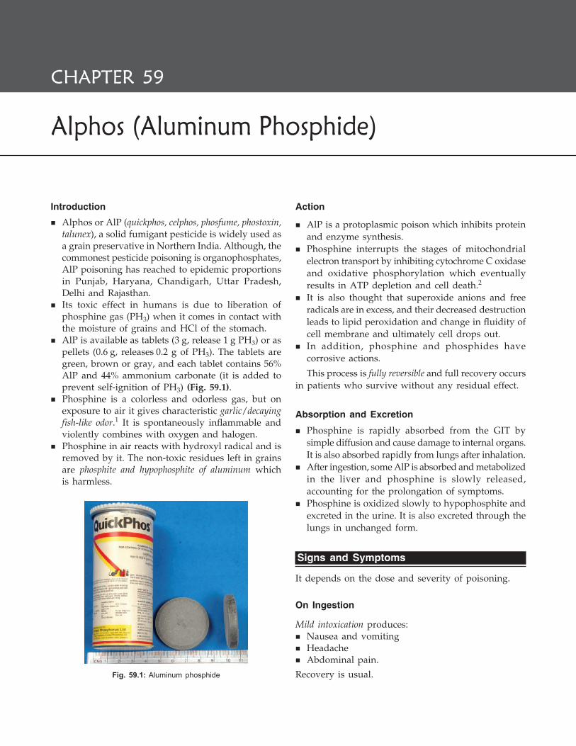
606 Review of Forensic Medicine and Toxicology
Alphos (Aluminum Phosphide)
CHAPTER 59
Introduction
� Alphos or AlP (quickphos, celphos, phosfume, phostoxin, talunex), a solid fumigant pesticide is widely used as a grain preservative in Northern India. Although, the commonest pesticide poisoning is organophosphates, AlP poisoning has reached to epidemic proportions in Punjab, Haryana, Chandigarh, Uttar Pradesh, Delhi and Rajasthan.
� Its toxic effect in humans is due to liberation of phosphine gas (PH3) when it comes in contact with the moisture of grains and HCl of the stomach.
� AlP is available as tablets (3 g, release 1 g PH3) or as pellets (0.6 g, releases 0.2 g of PH3). The tablets are green, brown or gray, and each tablet contains 56% AlP and 44% ammonium carbonate (it is added to prevent self-ignition of PH3) (Fig. 59.1).
� Phosphine is a colorless and odorless gas, but on exposure to air it gives characteristic garlic/decaying fish-like odor.1 It is spontaneously inflammable and violently combines with oxygen and halogen.
� Phosphine in air reacts with hydroxyl radical and is removed by it. The non-toxic residues left in grains are phosphite and hypophosphite of aluminum which is harmless.
Action
� AlP is a protoplasmic poison which inhibits protein and enzyme synthesis.
� Phosphine interrupts the stages of mitochondrial electron transport by inhibiting cytochrome C oxidase and oxidative phosphorylation which eventually results in ATP depletion and cell death.2
� It is also thought that superoxide anions and free radicals are in excess, and their decreased destruction leads to lipid peroxidation and change in fluidity of cell membrane and ultimately cell drops out.
� In addition, phosphine and phosphides have corrosive actions.
This process is fully reversible and full recovery occurs in patients who survive without any residual effect.
Absorption and Excretion
� Phosphine is rapidly absorbed from the GIT by simple diffusion and cause damage to internal organs. It is also absorbed rapidly from lungs after inhalation.
� After ingestion, some AlP is absorbed and metabolized in the liver and phosphine is slowly released, accounting for the prolongation of symptoms.
� Phosphine is oxidized slowly to hypophosphite and excreted in the urine. It is also excreted through the lungs in unchanged form.
Signs and Symptoms
It depends on the dose and severity of poisoning.
On Ingestion
Mild intoxication produces: � Nausea and vomiting � Headache � Abdominal pain.
Recovery is usual.Fig. 59.1: Aluminum phosphide
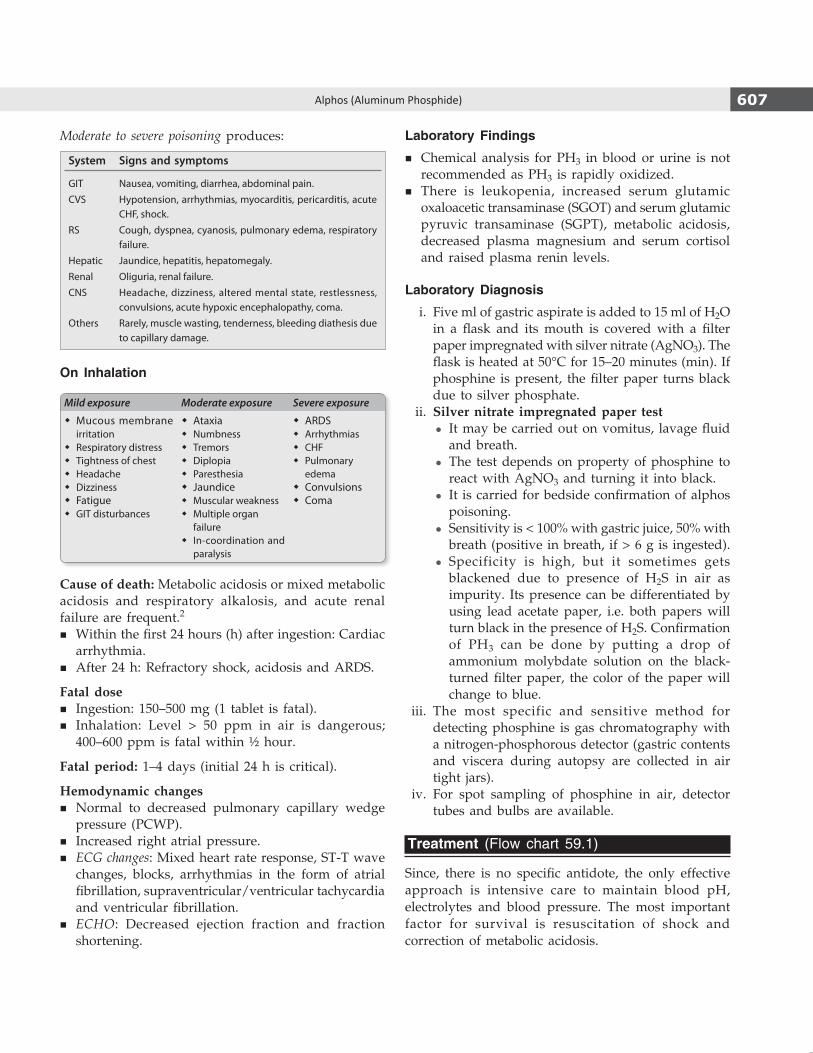
607Alphos (Aluminum Phosphide)
Moderate to severe poisoning produces:
System Signs and symptoms
GIT Nausea, vomiting, diarrhea, abdominal pain.CVS Hypotension, arrhythmias, myocarditis, pericarditis, acute
CHF, shock.RS Cough, dyspnea, cyanosis, pulmonary edema, respiratory
failure.Hepatic Jaundice, hepatitis, hepatomegaly.Renal Oliguria, renal failure.CNS Headache, dizziness, altered mental state, restlessness,
convulsions, acute hypoxic encephalopathy, coma.Others Rarely, muscle wasting, tenderness, bleeding diathesis due
to capillary damage.
On Inhalation
Mild exposure Moderate exposure Severe exposure
� Mucous membrane irritation
� Respiratory distress � Tightness of chest � Headache � Dizziness � Fatigue � GIT disturbances
� Ataxia � Numbness � Tremors � Diplopia � Paresthesia � Jaundice � Muscular weakness � Multiple organ
failure � In-coordination and
paralysis
� ARDS � Arrhythmias � CHF � Pulmonary
edema � Convulsions � Coma
Cause of death: Metabolic acidosis or mixed metabolic acidosis and respiratory alkalosis, and acute renal failure are frequent.2
� Within the first 24 hours (h) after ingestion: Cardiac arrhythmia.
� After 24 h: Refractory shock, acidosis and ARDS.
Fatal dose � Ingestion: 150–500 mg (1 tablet is fatal). � Inhalation: Level > 50 ppm in air is dangerous;
400–600 ppm is fatal within ½ hour.
Fatal period: 1–4 days (initial 24 h is critical).
Hemodynamic changes � Normal to decreased pulmonary capillary wedge
pressure (PCWP). � Increased right atrial pressure. � ECG changes: Mixed heart rate response, ST-T wave
changes, blocks, arrhythmias in the form of atrial fibrillation, supraventricular/ventricular tachycardia and ventricular fibrillation.
� ECHO: Decreased ejection fraction and fraction shortening.
Laboratory Findings
� Chemical analysis for PH3 in blood or urine is not recommended as PH3 is rapidly oxidized.
� There is leukopenia, increased serum glutamic oxaloacetic transaminase (SGOT) and serum glutamic pyruvic transaminase (SGPT), metabolic acidosis, decreased plasma magnesium and serum cortisol and raised plasma renin levels.
Laboratory Diagnosis
i. Five ml of gastric aspirate is added to 15 ml of H2O in a flask and its mouth is covered with a filter paper impregnated with silver nitrate (AgNO3). The flask is heated at 50°C for 15–20 minutes (min). If phosphine is present, the filter paper turns black due to silver phosphate.
ii. Silver nitrate impregnated paper test z It may be carried out on vomitus, lavage fluid
and breath. z The test depends on property of phosphine to
react with AgNO3 and turning it into black. z It is carried for bedside confirmation of alphos
poisoning. z Sensitivity is < 100% with gastric juice, 50% with
breath (positive in breath, if > 6 g is ingested). z Specificity is high, but it sometimes gets
blackened due to presence of H2S in air as impurity. Its presence can be differentiated by using lead acetate paper, i.e. both papers will turn black in the presence of H2S. Confirmation of PH3 can be done by putting a drop of ammonium molybdate solution on the black-turned filter paper, the color of the paper will change to blue.
iii. The most specific and sensitive method for detecting phosphine is gas chromatography with a nitrogen-phosphorous detector (gastric contents and viscera during autopsy are collected in air tight jars).
iv. For spot sampling of phosphine in air, detector tubes and bulbs are available.
Treatment (Flow chart 59.1)
Since, there is no specific antidote, the only effective approach is intensive care to maintain blood pH, electrolytes and blood pressure. The most important factor for survival is resuscitation of shock and correction of metabolic acidosis.
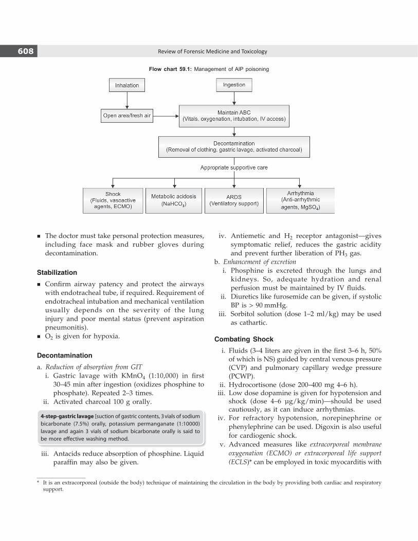
608 Review of Forensic Medicine and Toxicology
� The doctor must take personal protection measures, including face mask and rubber gloves during decontamination.
Stabilization
� Confirm airway patency and protect the airways with endotracheal tube, if required. Requirement of endotracheal intubation and mechanical ventilation usually depends on the severity of the lung injury and poor mental status (prevent aspiration pneumonitis).
� O2 is given for hypoxia.
Decontamination
a. Reduction of absorption from GIT i. Gastric lavage with KMnO4 (1:10,000) in first
30–45 min after ingestion (oxidizes phosphine to phosphate). Repeated 2–3 times.
ii. Activated charcoal 100 g orally.
4-step-gastric lavage [suction of gastric contents, 3 vials of sodium bicarbonate (7.5%) orally, potassium permanganate (1:10000) lavage and again 3 vials of sodium bicarbonate orally is said to be more effective washing method.
iii. Antacids reduce absorption of phosphine. Liquid paraffin may also be given.
iv. Antiemetic and H2 receptor antagonist—gives symptomatic relief, reduces the gastric acidity and prevent further liberation of PH3 gas.
b. Enhancement of excretion i. Phosphine is excreted through the lungs and
kidneys. So, adequate hydration and renal perfusion must be maintained by IV fluids.
ii. Diuretics like furosemide can be given, if systolic BP is > 90 mmHg.
iii. Sorbitol solution (dose 1–2 ml/kg) may be used as cathartic.
Combating Shock
i. Fluids (3–4 liters are given in the first 3–6 h, 50% of which is NS) guided by central venous pressure (CVP) and pulmonary capillary wedge pressure (PCWP).
ii. Hydrocortisone (dose 200–400 mg 4–6 h). iii. Low dose dopamine is given for hypotension and
shock (dose 4–6 μg/kg/min)—should be used cautiously, as it can induce arrhythmias.
iv. For refractory hypotension, norepinephrine or phenylephrine can be used. Digoxin is also useful for cardiogenic shock.
v. Advanced measures like extracorporeal membrane oxygenation (ECMO) or extracorporeal life support (ECLS)* can be employed in toxic myocarditis with
Flow chart 59.1: Management of AlP poisoning
* It is an extracorporeal (outside the body) technique of maintaining the circulation in the body by providing both cardiac and respiratory support.
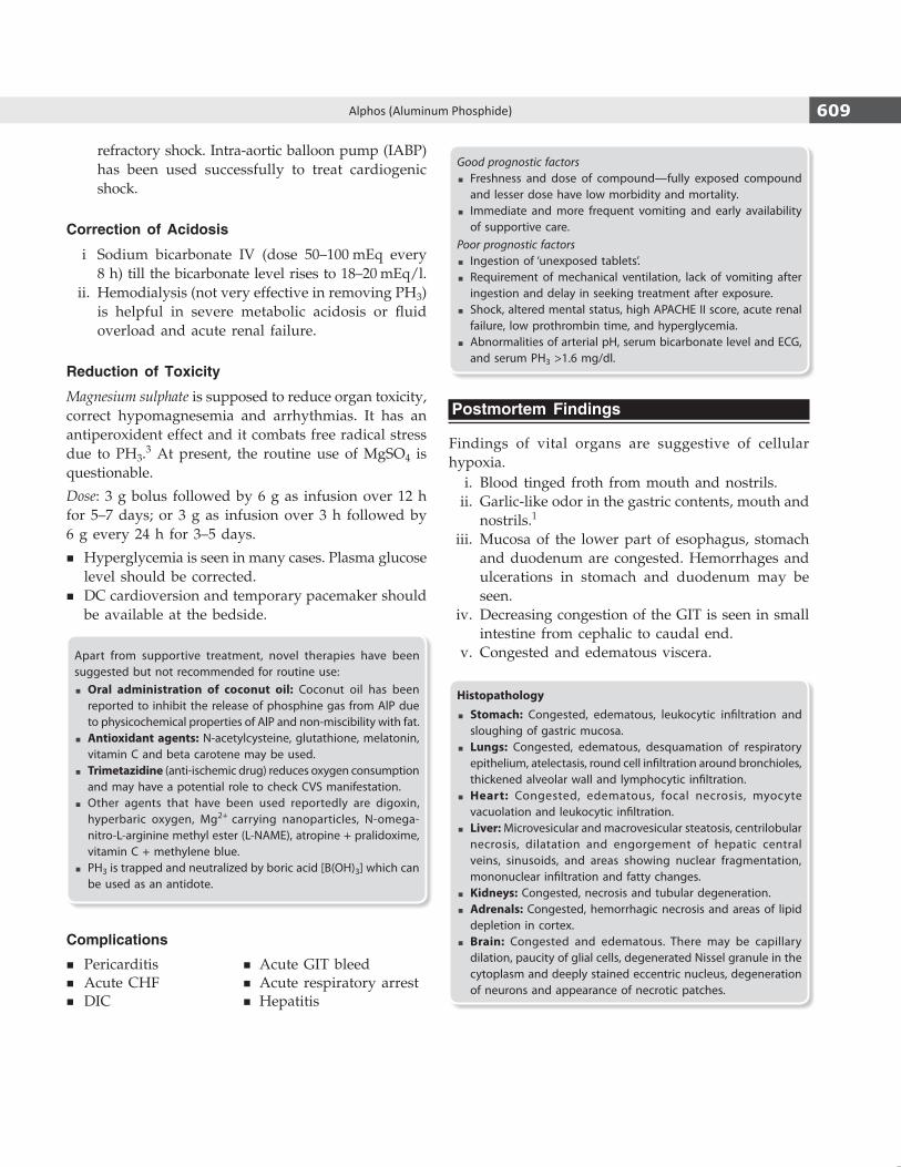
609Alphos (Aluminum Phosphide)
refractory shock. Intra-aortic balloon pump (IABP) has been used successfully to treat cardiogenic shock.
Correction of Acidosis
i Sodium bicarbonate IV (dose 50–100 mEq every 8 h) till the bicarbonate level rises to 18–20 mEq/l.
ii. Hemodialysis (not very effective in removing PH3) is helpful in severe metabolic acidosis or fluid overload and acute renal failure.
Reduction of Toxicity
Magnesium sulphate is supposed to reduce organ toxicity, correct hypomagnesemia and arrhythmias. It has an antiperoxident effect and it combats free radical stress due to PH3.3 At present, the routine use of MgSO4 is questionable.
Dose: 3 g bolus followed by 6 g as infusion over 12 h for 5–7 days; or 3 g as infusion over 3 h followed by 6 g every 24 h for 3–5 days.
� Hyperglycemia is seen in many cases. Plasma glucose level should be corrected.
� DC cardioversion and temporary pacemaker should be available at the bedside.
Apart from supportive treatment, novel therapies have been suggested but not recommended for routine use:
� Oral administration of coconut oil: Coconut oil has been reported to inhibit the release of phosphine gas from AlP due to physicochemical properties of AlP and non-miscibility with fat.
� Antioxidant agents: N-acetylcysteine, glutathione, melatonin, vitamin C and beta carotene may be used.
� Trimetazidine (anti-ischemic drug) reduces oxygen consumption and may have a potential role to check CVS manifestation.
� Other agents that have been used reportedly are digoxin, hyperbaric oxygen, Mg2+ carrying nanoparticles, N-omega-nitro-L-arginine methyl ester (L-NAME), atropine + pralidoxime, vitamin C + methylene blue.
� PH3 is trapped and neutralized by boric acid [B(OH)3] which can be used as an antidote.
Complications
� Pericarditis � Acute GIT bleed� Acute CHF � Acute respiratory arrest� DIC � Hepatitis
Good prognostic factors � Freshness and dose of compound—fully exposed compound
and lesser dose have low morbidity and mortality. � Immediate and more frequent vomiting and early availability
of supportive care.Poor prognostic factors
� Ingestion of ‘unexposed tablets’. � Requirement of mechanical ventilation, lack of vomiting after
ingestion and delay in seeking treatment after exposure. � Shock, altered mental status, high APACHE II score, acute renal
failure, low prothrombin time, and hyperglycemia. � Abnormalities of arterial pH, serum bicarbonate level and ECG,
and serum PH3 >1.6 mg/dl.
Postmortem Findings
Findings of vital organs are suggestive of cellular hypoxia. i. Blood tinged froth from mouth and nostrils. ii. Garlic-like odor in the gastric contents, mouth and
nostrils.1
iii. Mucosa of the lower part of esophagus, stomach and duodenum are congested. Hemorrhages and ulcerations in stomach and duodenum may be seen.
iv. Decreasing congestion of the GIT is seen in small intestine from cephalic to caudal end.
v. Congested and edematous viscera.
Histopathology � Stomach: Congested, edematous, leukocytic infiltration and
sloughing of gastric mucosa. � Lungs: Congested, edematous, desquamation of respiratory
epithelium, atelectasis, round cell infiltration around bronchioles, thickened alveolar wall and lymphocytic infiltration.
� Heart: Congested, edematous, focal necrosis, myocyte vacuolation and leuko cytic infiltration.
� Liver: Microvesicular and macrovesicular steatosis, centrilobular necrosis, dilatation and engorgement of hepatic central veins, sinusoids, and areas showing nuclear fragmentation, mononuclear infiltration and fatty changes.
� Kidneys: Congested, necrosis and tubular degeneration. � Adrenals: Congested, hemorrhagic necrosis and areas of lipid
depletion in cortex. � Brain: Congested and edematous. There may be capillary
dilation, paucity of glial cells, degenerated Nissel granule in the cytoplasm and deeply stained eccentric nucleus, degeneration of neurons and appearance of necrotic patches.

610 Review of Forensic Medicine and Toxicology
Medico-legal Aspects
� Poisoning is mostly suicidal, but homicidal cases may be seen in children. AlP is considered as ‘ideal suicidal’ poison since it is cheap, easily available, highly toxic, can be taken with food or drink, and has no effective antidote.
� Accidental and occupational poisoning may occur.
� Its gaseous form and toxicity makes it a potential agent for chemical terrorism.
Each tablet of AlP has the capacity to liberate 1 g of phosphine (PH3) gas.
AlP + 3HCl → AlCl3 + PH3 ↑
AlP + 3H2O → Al(OH)3 + PH3 ↑
(NH4)2CO3 + H2O → 2NH4OH + CO2↑→ 2NH3 ↑ + 2H2O + CO2 ↑
Therefore, gases liberated during fumigation or after ingestion are CO2, NH3 and phosphine (PH3) and a non-toxic residue aluminum hydroxide. The former two gases provide inert media for phosphine to act.
MULTIPLE CHOICE QUESTIONS
1. Garlic odor around the nostrils and mouth is indicative of poisoning with: AP 07
A. Cyanide B. Organophosphorus C. Carbolic acid D. Aluminum phosphide 2. In aluminum phosphide poisoning, which is NOT
true: AIIMS 10; Punjab 11 A. Accumulation of acetylcholine at NM junction B. Cytochrome oxidase inhibition
C. Phosphine formation D. Metabolic acidosis 3. In treatment of alphos poisoning, magnesium sulfate
acts as: WB 08 A. Adjuvant B. Stabilizer C. Preservative D. Purgative
1. D 2. A 3. B

Medicinal Poisons
ChaPter 60
Paracetamol (Acetaminophen)
Paracetamol (PCM) is a common analgesic in many nonprescription and prescription products.
Action
� PCM inhibits prostaglandin synthesis. � It produces liver damage through accumulation of a
toxic intermediate metabolite: N-acetyl-p-benzoquinone. Hepatic glutathione normally inactivates the metabolite, but in PCM poisoning, the glutathione becomes depleted.
� It also causes renal tubular necrosis.
Metabolism: After absorption, it is metabolized by glucuronidation and sulfation, and by cytochrome P450 oxidase system.
Signs and Symptoms
� Usually four stages of presentation are seen (Table 60.1).1
� Paracetamol toxicity is manifested primarily in the liver. Risk factors associated with fatal outcome include long-term alcohol ingestion, fasting, and treatment with drugs that induce the cytochrome P450 2E1 enzyme system.
Diagnosis
� PCM levels are assessed in the blood by enzyme immunoassay and high performance liquid chromato-graphy (HPLC).
� Emergency measurement of blood levels is essential in assessment of poisoning.
� There is a marked elevation of liver enzymes (peak alanine transaminase > 1000 IU/l) and an increased PT may be seen.
� Rumack-Matthew nomogram can help in planning treatment.
Fatal dose � Adults: 10–15 g (20–30 tablets). � Children: 150 mg/kg body wt.
Fatal period: 2–4 days.
Treatment i. Activated charcoal and gastric lavage within 1–2 h
of ingestion. ii. Antidote: N-acetyl cysteine (NAC) 150 mg/kg in 200
ml of 5% glucose over 15 minutes (min), followed by serial infusion of 50 mg/kg in 500 ml of 5% glucose in 4, 8 and 8 h (total 300 mg/kg in 20 h) or 140 mg/kg orally as loading dose, followed by 70 mg/kg every 4 h.2,3 Administration of NAC within 8 h of ingestion is nearly 100% hepatoprotective.
Oral methionine is an alternative, but is unreliable in patients who are vomiting.
iii. Supportive measures: Intravenous electrolytes, rehydration, vitamin K for bleeding, and mannitol for cerebral edema.
Postmortem Findings � External: Jaundice, petechiae in skin. � Internal: Congestion of the GIT, centrilobular hepatic
necrosis, acute tubular necrosis, cerebral edema and myocardial necrosis.
Death usually occurs in stage III. If not, then patient passes into stage IV.
Table 60.1: Signs and symptoms of PCM poisoning1
Stage/Phase Time of ingestion Signs and symptoms
I (Initial) 0–24 hours (h) Nausea, vomiting, diaphoresis, malaise, pallor.
II (Middle) 24–72 h Discomfort disappears, giving a false sense of relief. Upper abdominal pain may be present.
III (Hepatic) 72–96 h Vomiting, jaundice, hepatic pain, bleeding, confusion, coma, asterixis (flapping tremor), hepatic encephalopathy, cardiac arrhythmia, hemorrhagic pancreatitis, DIC.
IV (Recovery) > 5 days Resolution of liver function occurs in about 2–3 months.
612 Review of Forensic Medicine and Toxicology
Medico-legal aspects: Most cases of poisoning are suicidal. Accidental overdose may occur.
In January 2014, the FDA recommended that combination prescription pain relievers containing more than 325 mg of PCM per tablet, capsule or other dosage unit should not be prescribed because of a risk of liver damage.
Iron
Introduction: Commonly available preparations are ferrous sulfate (20% elemental iron), ferrous gluconate (12% elemental iron) and ferrous fumarate (33% elemental iron) which are used for supplemental therapy in case of iron deficiency anemia. Rarely, the source of poisoning may be from tanning, dyeing and from inks.
Action: Increased capillary permeability, release of hydrogen ions, inhibition of mitochondrial enzymes and corrosive action on gastric mucosa.
Signs and Symptoms
They are divided into four stages. Symptoms begin with acute gastroenteritis, followed by a quiescent period, then shock and liver failure. i. Stage I: Nausea, vomiting, abdominal pain,
gastrointestinal hemorrhage, hypotension, pallor and lethargy. These symptoms occur half an hour to 6 h post-ingestion.
ii. Stage II: Mild acidosis, hyperventilation, oliguria and hypotension; occur 6–24 h post-ingestion. Overall, the patient seems to show apparent improvement.
iii. Stage III: Multiorgan dysfunction involving GIT, CVS, CNS, hepatorenal systems with anion-gap metabolic acidosis, depression, hepatitis, coagulopathy, convulsions, disorientation, shock, coma and death. It occurs 24–48 h to few days post-ingestion.
iv. Stage IV: It is seen 2–6 weeks post-ingestion and includes complications, like gastric stricture and pyloric obstruction.
Fatal dose: 20–30 g (> 200 mg/kg).Fatal period: 24–48 h.
Investigations
� Abdominal X-ray: Radiopaque tablets may be seen. � Serum iron level > 350 µg/dl indicate toxicity. It is
measured 2–6 h post-ingestion. � Desferrioxamine challenge test: It is given in a dose of
25 mg/kg which imparts a reddish 'vin rose' color to the urine (iron-desferrioxamine complex).
TreatmentTreatment is usually whole-bowel irrigation and chelation therapy. i. Fluid resuscitation: If patient is in shock, normal
saline drip or lactated Ringer's solution, dopamine and whole blood transfusion may be given depending upon the cause. Dextrose for hypo-glycemia, and sodium bicarbonate for acidemia is given.
ii. Decontamination: Gastric lavage or whole bowel irrigation with normal saline or polyethylene glycol-electrolyte solution. After this, 1% sodium bicarbonate/magnesium hydroxide solution is added to precipitate the residual iron as insoluble ferrous carbonate/hydroxide.4
iii. Chelation therapy: Desferrioxamine (antidote) in a dose of 10–15 mg/kg/h as continuous infusion to a maximum of 6 g, till there is significant reduction of systemic toxicity.4,5
iv. Supportive therapy z Hemodialysis or exchange transfusion in severe
cases. z Endoscopy or gastrostomy, if there is clinical
toxicity and a large amount of tablets are visible on the X-ray.
Postmortem FindingsUsually, internal findings are seen. i. GIT: Hemorrhagic necrosis and perforation of the
gastric or jejunal wall. ii. Lungs: Pulmonary hemorrhage. iii. Liver: Centrilobular necrosis may be seen. iv. Kidneys: It may show necrosis of tubules.
Medico-legal aspects: Usually, accidental poisoning from overdose (children are attracted by its color and pleasant flavor), prolonged therapy or IV administration. Suicidal cases may occur in older children and adults.
Antipsychotic Drugs (Tranquilizers)
Introduction: These drugs relieve anxiety and mental tension without producing sedation, and are used in various neurotic conditions, anxiety states, relief of tension, and as anesthetics because of their muscle relaxant properties.
ClassificationPhenothiazines Examples
� Aliphatic Chlorpromazine, Triflupromazine

613Medicinal Poisons
� Piperazine Trifluoperazine, Prochlorperazine � Piperidine Thioridazine, Mesoridazine
Butyrophenones Haloperidol, DroperidolThioxanthenes Thiothixene, FlupenthixolOthers Pimozide, Respine, LoxapineAtypical neuroleptics Clozapine, Risperidone
Action: They have an inhibitory effect on a variety of receptors including dopaminergic (mainly D2 receptor), cholinergic, alpha1 and alpha2-adrenergic, histaminic and serotonergic receptors (5HT2).
Absorption and excretion: They are completely absorbed from the GIT. Excretion is mainly via feces (50%) and the kidneys (30%) as metabolites; less than 4% is excreted in the unchanged form.
Signs and SymptomsToxic manifestations can be divided into CNS and non-CNS effects.
CNS effects � Depression, agitation, seizures, somnolence and coma. � Behavioral reactions: Oversedation, impaired
psychomotor function and paradoxical effects, such as agitation, excitement, insomnia and toxic confusional states.
� Extrapyramidal signs: Dystonia (acute), akathisia, Parkinsonism (akinesia), neuroleptic malignant syndrome and tardive dyskinesia.
Non-CNS effects
System Signs and symptoms
CVS Orthostatic hypotension, ventricular tachycardia, torsades de pointes, ventricular fibrillation, atrioventricular block.
GIT Dry mouth, decreased colonic motility resulting in constipation, pseudo- obstruction.
Ocular Mydriasis; visual acuity, visual fields and color vision perception may be altered.
Genitourinary Urinary retention, priapism.Hematologic Agranulocytosis, aplastic anemia, leuko penia, eosino-
philia, thrombocytopenia, anemia, pancytopenia.Hepatic Cholestatic jaundice.
Fatal dose: 2–5 g (25–30 times of therapeutic dose).
Diagnostic trial: For a suspected case of poisoning, administration of diphenhydramine (dose: 1–2 mg/kg, maximum—25 mg) results not only in resolution of dystonia or oculogyria, but also assists in the diagnosis.
TreatmentThere is no specific antidote for acute phenothiazine poisoning.
Initial stabilization i. ABCs: Oxygen is given. Intubation may be
necessary. ii. Emesis: Contraindicated due to the risk of develop-
ment of seizures and sedation. iii. Gastric lavage must be done followed by adminis-
tration of activated charcoal; beneficial even upto 6 h following ingestion.
iv. Catharsis: Following gastric lavage, a saline cathartic (sodium or magnesium sulfate) may be introduced and left in the stomach.
v. Diuresis: Mannitol solution is given slow IV in a dose of 5 ml/kg initially, followed by 2 ml/kg every 6 hourly for 2 days.
vi. Correction of hypotension: Elevate the foot end of the bed. Give adequate IV fluids (0.9% NS). Treatment of resistant hypotension is done with norepinephrine, 1–2 µg/kg/min (titrated to blood pressure).
Management of specific condition i. Ventricular dysrhythmia: Administer sodium
bicarbonate. Ventricular dysrhythmias are treated with lidocaine (loading dose: 1 mg/kg IV repeated after 5–10 min, maintenance dose: 2–4 mg/min IV) or phenytoin (15–20 mg/kg IV).
ii. Dystonic reactions: Administer diphenhydramine, 0.5–1 mg/kg IV (maximum 50 mg) or benztropine mesylate, 1–2 mg IV or IM (0.01–0.02 mg/kg).
iii. Malignant hyperthermia: Administer dantrolene (2–5 mg/kg IV) or bromocriptine (2.5–7.5 mg orally daily).
iv. Seizures: Treat seizures initially with diazepam, 0.2–0.5 mg/kg IV, repeat after 10–15 min. Pheno-barbital or phenytoin can be used for persistent seizures.
v. Other measures: Hypothermia may occur, maintain normal body temperature and avoid overheating.
Medico-legal aspects: These drugs are the most frequent cause of acute accidental poisoning in children and mostly involve children < 6 years of age.
Antihistamines
Introduction: The commonly used preparations are: diphenhydramine (benadryl), doxylamine, pyrilamine, promethazine hydrochloride (phenergan), tripelennamine, chlorpheniramine, cimetidine, ranitidine, nizatidine and famotidine.
Action: Inhibition of central and postganglionic parasympathetic muscarinic cholinergic receptors.
614 Review of Forensic Medicine and Toxicology
Signs and Symptoms
� CNS: Drowsiness, lethargy, fatigue and hypnosis. There is vertigo, ataxia, tinnitus, dilated pupils and blurred vision, followed by tremors, anxiety, insomnia, excitement, delirium, hallucinations, convulsions and coma. Anticholinergic features (mydriasis, hyperthermia and flushing) are seen.
� GIT: Dry mouth, anorexia, nausea, vomiting, abdominal pain and constipation or diarrhea.
� There may be tachycardia, retention of urine and skin rashes.
Finally, there is severe CNS depression, and death results from respiratory failure or cardio vascular collapse.
Fatal dose: 1 g.
Treatment
i. Gastric lavage. ii. Physostigmine 0.5–2 mg IV every hour, until rever-
sal of symptoms occurs. However, it can produce serious adverse effects. Short-acting barbiturates may be used for the control of CNS stimulation.
iii. Symptomatic treatment.
Postmortem findings: Nonspecific findings. Signs of asphyxia are found.
Medico-legal aspects: Poisoning is usually accidental and sometimes suicidal.
Tricyclic Antidepressants (TCAs)
Introduction: Tricyclic antidepressants (TCAs) are one of the oldest classes of antidepressants and are still used extensively. Before the introduction of selective serotonin reuptake inhibitors (SSRIs), TCAs were the standard treatment for depression.
TCAs include:� Imipramine � Clomipramine
� Amitriptyline � Desipramine � Trimipramine � Nortriptyline � Doxepin � Dothiepin
Action: TCAs have complex actions. It inhibits monoamine uptake and interact with variety of receptors, viz. muscarinic, a-adrenergic, dopaminergic, GABA-ergic, histaminergic and serotonergic. They have potent anticholinergic and antiarrhythmic activity.
Signs and Symptoms
Features of poisoning appear in 1 h and maximum intensity is seen in 4–12 h.
� Anticholinergic effects: Dry skin and mouth, flushing, decreased bowel sounds, constipation, epigastric distress, urinary retention, dilated pupils, blurred vision and palpitations.
� CNS: Drowsiness, sleepiness, unresponsive to pain, depressed brainstem reflexes, seizures and coma.
� CVS: Tachycardia and hypotension. � MS: Myoclonus, later on flaccid paralysis. � Respiration is depressed, and temperature is
decreased.
Cause of death: Metabolic acidosis and cardiorespiratory depression.
Treatment5
i. Gastric lavage, respiratory support, fluid infusion, maintenance of BP and body temperature. Acidosis is corrected by sodium bicarbonate infusion.
ii. Diazepam may be given IV to control convulsions and delirium.
iii. Propranolol or lidocaine may be used for ventricular arrhythmias.
iv. Physostigmine 0.5–2 mg IV reverses the central, peripheral and cardiac effects, but seldom used, since arrhythmias and hypotension are sometimes worsened by this treatment.
Medico-legal aspects: Poisoning is frequent with suicidal attempts by the depressed individuals.
Tricyclic antidepressants are the leading cause of death by drug overdose in the US.
Benzodiazepines (BZDs)
Introduction: They are used mainly as anti-anxiety and muscle relaxant agents. The commonly used prepara-tions are: diazepam, flurazepam, chlordiazepoxide, nitrazepam, oxazepam, flunitrazepam, alprazolam and lorazepam. Addiction may occur with these drugs. They can be ultra-short acting (e.g. midazolam), short acting (e.g. alprazolam) and long acting (e.g. diazepam).
Action: They enhance the inhibitory actions of the neurotransmitter GABA located in the brain.
Signs and Symptoms
Confusion, dizziness, anxiety, vertigo, slurred speech, nystagmus, diplopia, dysarthria, ataxia, hallucinations, weakness, impairment of cognition, amnesia, sedation, somnolence, respiratory depression and coma. If taken alone, they are not fatal, but mixing with alcohol or

615Medicinal Poisons
drugs like opioids can lead to death, with advanced age as additional risk factor. Death after admission is rare, and due to respiratory depression with aspiration of gastric contents.
Fatal dose: 100–300 mg/kg body wt.
Diagnosis: Immunoassay screening techniques are performed most commonly. These tests typically detect BZDs that are metabolized to desmethyldiazepam or oxazepam; thus, a negative screening result does not rule out the presence of a BZD.
Treatment
i. Gastric lavage. ii. Activated charcoal. iii. Antidote: Flumazenil selectively blocks the central
effects of BZDs by competitive inhibition, 0.2 mg over 30–60 seconds given slow IV, repeated in 0.5 mg increments upto a total of 3–5 mg.6 However, it may itself induce seizures. A long acting drug, such as chlordiazepoxide or diazepam is useful to prevent complications (may not be effective in seizures).
Medico-legal Aspects
� BZDs are commonly used for suicidal poisoning. Usually, diazepam is safer in overdose or intentional attempts to suicide, but that the newer agents such as temazepam, flurazepam, zopiclone and triazolam have relatively low toxic doses and fatal levels, and are more often found as the cause of death in suicides.
� Flunitrazepam is frequently used as ‘date rape’ drug. � It may be mixed with food or drinks to facilitate
robbery during travel.
Signs and Symptoms of Chronic Poisoning
� CNS: Headache, anxiety, insomnia, muscle spasms, tremors, rarely convulsions and psychiatric disturbances.
� GIT: Anorexia and vomiting. � RS: Respiratory depression is rare.
High dose, long-term therapy may produce with-drawal symptoms when stopped suddenly.
The withdrawal syndrome with BZDs include fits and psychosis. In addition, anxiety symptoms, such as sweat-ing, insomnia, headache, tremors, nausea and disordered perception, such as feelings of unreality, abnormal bodily sensations and hypersensitivity to stimuli may be seen.
Acetylsalicylic Acid (Aspirin)
Introduction: Salicylate can be found in hundreds of over-the-counter medications. It is popular as an
antipyretic and analgesic. Toddlers are most vulnerable to acute salicylate poisoning.
Physical properties: It is a white, odorless, crystalline powder, having a slight acid taste.
ActionInitially, there is direct stimulation of respiratory center leading to hyperventilation and respiratory alkalosis. Later on, due to inhibition of Krebs cycle, uncoupling of oxidative phosphorylation, gluconeogenesis, increase lipid metabolism and inhibition of aminoacid metabolism, patient develops metabolic acidosis.7 It is also neurotoxic.
Absorption and excretion: It is rapidly absorbed from the stomach and to a slightly lesser extent from the small intestine. Metabolism occurs chiefly in the liver. Excretion is mainly through urine.
Signs and Symptoms
System Signs and symptoms
GIT Burning pain in the throat and abdomen, nausea, vomiting, thirst, hematemesis and melena.
CNS Ataxia, vertigo, tinnitus, headache, confusion, convulsion, coma—known as ‘salicylate jag’ secondary to hyperthermia and altered glucose metabolism.
CVS Tachycardia.Hepatic Reye’s syndrome.RS Initially, tachypnea and hyperpnea, followed by
Kussmaul’s breathing secondary to metabolic acidosis, pulmonary edema.
Electrolyte Dehydration, hypokalemia, hypo-/hyper natremia, hypo-/hyperglycemia.
Hematologic Hemorrhagic tendency. MS Rhabdomyolysis, tetany.Others Hyperpyrexia, dilated pupils, rapid and irregular pulse.
Fatal dose: Sodium salicylate and aspirin: 15–20 g (200 mg/kg). Blood level > 100 mg/dl is fatal.Fatal period: Few hours.
Reye’s syndrome may develop in children < 15 years on intake of aspirin. The main features are acute onset of hepatic failure and encephalopathy with residual neurological manifestations.
Investigations � Bedside diagnosis: Presence of salicylic acid in the
urine can be detected by ferric chloride test which involves combining 1 ml of patient's urine to few drops of ferric chloride solution. If salicylate is present, solution will turn to brown-purple color.
� Blood salicylate level: Best indicator of the severity of intoxication. It should be done 6 h after ingestion and then repeated every 4–6 hourly for serial estimation and response to therapy.
616 Review of Forensic Medicine and Toxicology
Differential Diagnosis
� Diabetic ketoacidosis � Lactic acidosis � Methanol toxicity � Renal failure � Ethylene glycol toxicity
Treatment
i. Decontamination z Gastric lavage is done. At the end of the lavage,
activated charcoal should be left in the stomach which will bind the unabsorbed salicylate.
z Elimination can be enhanced by whole bowel irrigation with polyethylene glycol.
ii. Fluid and electrolyte management z Correction of dehydration is done by crystalloids
to replace the fluid loss. z Alkalization of urine (enhances renal salicylate
excretion) and treatment of acidosis: Add 100 mEq (2 ampoules) of sodium bicarbonate to 1 liter of 5% dextrose in 0.2% saline and infuse this solution IV at a rate of about 150–200 ml/h. Add 20–30 mEq of potassium chloride to each liter of IV fluid.
z Hypocalcemic tetany is treated with 10% calcium gluconate.
z Seizures are controlled with diazepam (l–3 mg/kg/dose) or phenobarbitone (5 mg/kg).
z Vitamin K should be injected, if PT is prolonged. z If patient develops respiratory failure, positive
pressure ventilation should be started. iii. Hemodialysis: It is preferred over hemoperfusion
and peritoneal dialysis as it helps in removal of salicylate, and maintenance of fluid and electrolyte balance.
Postmortem Findings
External: Pupils are dilated. Skin rashes may be present.
Internal i. Stomach: Gastric mucosa is congested and petechial
hemorrhages are seen in the mucous and serous membranes.
ii. Lungs: Subpleural petechial hemorrhages, congested, edematous and collapsed.
iii. All organs are congested. iv. If the patient survives for few days, the myocar-
dium, liver and kidneys are usually soft, dirty in appearance and greasy to touch. Hepatitis may be present. Petechial hemorrhages are seen in various organs.
Medico-legal aspects: Aspirin is the most common salicylate in regular use, so both accidental and suicidal ingestion is common.
Chloral Hydrate
Introduction: Chloral hydrate (dry wine) is related to paraldehyde and is an unfashionable drug in modern times, but still used in psychiatric hospitals.
Physical properties: Colorless, crystalline powder having a peculiar pungent odor and a bitter taste.
Action: It causes depression of the CNS.
Absorption and excretion: It is absorbed from the stomach, small intestine and rectum, and metabolized in the liver, mainly to trichloroethanol which is further oxidized. Trichloroethanol is conjugated with glucuronic acid and excreted in the urine.
Signs and SymptomsRetrosternal burning sensation, vomiting, drowsiness, hypotension, slow irregular pulse, depression of respiration, deep sleep and coma. Albuminuria, scarlatiniform or urticarial rash and rarely, jaundice may be seen.
Death usually occurs from paralysis of the respiratory center.
Fatal dose: 3–5 g.
Fatal period: 8–12 h.
Treatment
i. Gastric lavage with alkaline solution. ii. Hemodialysis. iii. Flumazenil 0.1 mg as infusion upto a total of 3 mg. iv. Symptomatic treatment.
Postmortem Findings
� Gastric mucosa is eroded, softened and reddened, and smells of chloral hydrate.
� Brain and lungs are congested. � Damage to kidneys and liver is seen.
Medico-legal Aspects � Accidental poisoning results by taking large doses
as a hypnotic. � Suicidal/homicidal cases are rare. � It is mixed with food or drink to render a person
suddenly helpless for the purpose of robbery or rape. Its action is so rapid, hence the name 'knock-out drops'.8

617Medicinal Poisons
Chronic poisoning occurs after prolonged therapeutic use.
Symptoms: GIT irritation, erythematous and urticarial eruptions on skin, tremors and dyspnea may be seen. Convulsions, mental derangement and liver damage may occur.
Habitual use can lead to tolerance and physical dependence with delirium when the drug is withdrawn.
1. B 2. D 3. C 4. D 5. C 6. C 7. C 8. B
1. A patient presented with vomiting, pain in abdomen, jaundice and encephalopathy. There is a history of attempt to commit suicide. Poisoning suspected: CMC (Vellore) 14
A. Benzodiazepines B. Paracetamol C. Organophosphorus D. Acetylsalicylic acid
2. A female, Lalita, aged 26 years takes 100 tablets of paracetamol. Treatment of choice is: DNB 09; PGI 09; UP 11; FMGE 11; NEET 13
A. Lavage with charcoal B. Dialysis C. Alkaline diuresis D. Acetylcysteine
3. N-acetyl-cysteine is antidote for toxicity with: MP 11; MAHE 12 A. Benzodiazepine B. Barbiturates C. Acetaminophen D. Amphetamine
4. Not used for iron poison in: FMGE 09; NEET 13 A. Magnesium hydroxide B. Desferrioxamine C. Gastric lavage D. Penicillamine
5. A woman consumes several tablets of amitriptyline. All of the following can be done, except:
AIIMS 10, 13; NEET 14 A. NaHCO3
B. Gastric lavage C. Diazepam for seizures D. Atropine as antidote 6. Antidote for benzodiazepine poisoning: FMGE 10, 13; NEET 14 A. Naloxone B. Atropine C. Flumazenil D. N-acetyl-cysteine 7. Acetylsalicylate poisoning causes: UPSC 14 A. Metabolic acidosis B. Respiratory acidosis with metabolic alkalosis C. Metabolic acidosis with respiratory alkalosis D. Respiratory alkalosis 8. Knockout drops are: NEET 13 A. Paraldehyde B. Chloral hydrate C. Kerosene D. Turpentine
multiple ChoiCe questions
Insulin is a potent hypoglycemic agent and if severe lowering of the blood sugar persists for many hours, then brain damage and death will occur. In massive doses, especially intravenously, death can take place within few hours. Insulin, unless suspected, is an effective homicide method. If death from insulin is suspected (which looks like a natural death with no obvious anatomic cause of death found at autopsy), either from suicide (usually amongst nurses and doctors who have access to large doses), homicide or rarely from accidental overdose (usually in hospital), then a search of the body must be made for recent needle marks and the surrounding skin, subcutaneous tissue
and underlying muscle excised and sent unfixed for assay (occult injection sites can be a mucosal surface or scrotum in male). Blood samples are preserved (to distinguish between human, bovine and porcine insulin and detect adjuvants such as zinc which assists in tracing the origin of the extrinsic insulin). Samples should be taken soon after death and the plasma immediately separated from the cells, and kept deep-frozen until analysis. Postmortem blood glucose levels are generally unhelpful in confirming hypoglycemia, but vitreous humor is more useful. Oral hypoglycemic agents, such as sulphonylureas and biguanides, may be taken as an overdose, either suicidal or accidentally, producing hypoglycemia, hyperkalemia and acidosis. Radioimmunoassay (RIA) is used for measurement of insulin in the body. The method most used is chemiluminescent immunoassay and the measurement of blood insulin is possible even when embalming fluid had contaminated the blood.False negative analysis may be due to:
� Prolonged interval between injection and death (if the person was comatose for many days).
� Postmortem glucose measurements since post mortem glycolysis may falsely elevate blood glucose levels.

618 Review of Forensic Medicine and Toxicology
Drug Dependence and Date Rape Drugs
ChapteR 61
Definitions
� Drug: Any substance, when taken into the living body, may modify one or more of its functions.
� Psychoactive drug is one that is capable of altering the mental functioning.
� Drug dependence is a compulsion to take a drug to produce a desired effect or to prevent unpleasant effects when the drug is withheld, i.e. it is necessary for either physical or psychological well-being.
Drug dependence includes both the terms 'addiction' and 'habituation' (Diff. 61.1).
Nowadays, words 'addiction' and 'addict' are not used in medicine due to their derogatory implication. Instead 'abuse' or 'harmful use' or 'dependence' is used.
Hard and soft drugs are terms to distinguish between psychoactive drugs that are addictive and non-addictive.
� Hard drugs lead to severe physical addiction, e.g. heroin, methamphetamine, alcohol and nicotine.
� Soft drugs do not cause physical addiction but may lead to psychological dependence, e.g. cannabis, mescaline, psilocybin and LSD.
The distinction between soft drugs and hard drugs is important in the drug policy of the Netherlands, where cannabis production, retailing and use come under official tolerance, subject to certain conditions.
Patterns of Drug Use Disorders
There are four important patterns of drug use disorders, which may overlap with each other:
i. Acute intoxication ii. Withdrawal state iii. Dependence syndrome iv. Harmful use.
� Acute intoxication is a transient condition, resulting in disturbance of the level of consciousness, cognition, perception, behavior or other psycho-physiological functions and responses. This is usually associated with high blood levels of the psychoactive substance.
The intensity of intoxication lessens with time, and effects gradually disappear in the absence of further use of the substance. Recovery is complete, except where tissue damage or some complication has arisen.
� Withdrawal state is characterized by a group of symptoms, often specific to the drug used which develop on total or partial withdrawal of a drug, usually after repeated and/or high-dose use. The duration usually is of few hours to a few days. Typically, the patient reports that withdrawal symptoms are relieved by further substance use.
� Dependence syndrome: Cluster of physiological, behavioral and cognitive phenomena in which the use of substances takes on a much higher priority for a given individual than other behaviors that once had greater value. It is characterized by the desire (often strong, sometimes overpowering) to take psychoactive drugs, alcohol or tobacco.
� Harmful use: Continued drug use despite awareness of harmful medical and/or social effect of the drug being used/or a pattern of physically hazardous
Differentiation 61.1: Drug addiction and drug habituation
S.No. Feature Drug addiction Drug habituation
1. Compulsion Present Desire, but no compulsion
2. Dependence Psychological and physical Psychological, but not physical
3. Dose Tendency to increase No tendency to increase
4. Withdrawal symptoms Characteristic symptoms None or mild
5. Harm Both—individual and society Individual only
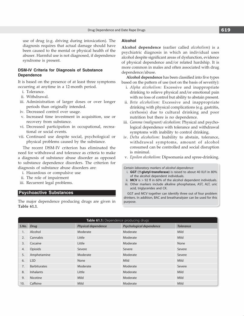
619Drug Dependence and Date Rape Drugs
use of drug (e.g. driving during intoxication). The diagnosis requires that actual damage should have been caused to the mental or physical health of the abuser. Harmful use is not diagnosed, if dependence syndrome is present.
DSM-IV Criteria for Diagnosis of Substance Dependence
It is based on the presence of at least three symptoms occurring at anytime in a 12-month period. i. Tolerance. ii. Withdrawal. iii. Administration of larger doses or over longer
periods than originally intended. iv. Decreased control over usage. v. Increased time investment in acquisition, use or
recovery from substance. vi. Decreased participation in occupational, recrea-
tional or social events. vii. Continued use despite social, psychological or
physical problems caused by the substance.
The recent DSM-IV criterion has eliminated the need for withdrawal and tolerance as criteria to make a diagnosis of substance abuse disorder as opposed to substance dependence disorders. The criterion for diagnosis of substance abuse disorders are: i. Hazardous or compulsive use ii. The role of impairment iii. Recurrent legal problems.
Psychoactive Substances
The major dependence producing drugs are given in Table 61.1.
Alcohol
Alcohol dependence (earlier called alcoholism) is a psychiatric diagnosis in which an individual uses alcohol despite significant areas of dysfunction, evidence of physical dependence and/or related hardship. It is more common in males and often associated with drug dependence/abuse.
Alcohol dependence has been classified into five types based on the pattern of use (not on the basis of severity): i. Alpha alcoholism: Excessive and inappropriate
drinking to relieve physical and/or emotional pain with no loss of control but ability to abstain present.
ii. Beta alcoholism: Excessive and inappropriate drinking with physical complications (e.g. gastritis, cirrhosis) due to cultural drinking and poor nutrition but there is no dependence.
iii. Gamma (malignant) alcoholism: Physical and psycho-logical dependence with tolerance and withdrawal symptoms with inability to control drinking.
iv. Delta alcoholism: Inability to abstain, tolerance, withdrawal symptoms, amount of alcohol consumed can be controlled and social disruption is minimal.
v. Epsilon alcoholism: Dipsomania and spree-drinking.
Certain laboratory markers of alcohol dependence: i. GGT ( -glutyl-transferase) is raised to about 40 IU/l in 80%
of the alcohol dependent individuals. ii. MCV is > 92 fl in 60% of the alcohol dependent individuals. iii. Other markers include alkaline phosphatase, AST, ALT, uric
acid, triglycerides and CK.
GGT and MCV together can identify three out of four problem drinkers. In addition, BAC and breathanalyzer can be used for this purpose.
Table 61.1: Dependence producing drugs
S.No. Drug Physical dependence Psychological dependence Tolerance
1. Alcohol Moderate Moderate Mild
2. Cannabis Little Moderate Mild
3. Cocaine Little Moderate None
4. Opioids Severe Severe Severe
5. Amphetamine Moderate Moderate Severe
6. LSD None Mild Mild
7. Barbiturates Moderate Moderate Severe
8. Inhalants Little Moderate Mild
9. Nicotine Mild Moderate Mild
10. Caffeine Mild Moderate Mild
γ

620 Review of Forensic Medicine and Toxicology
The treatment can be broadly divided into: i. Detoxification (detox) ii. Treatment of alcohol dependence.
Detoxification: Treatment of alcohol withdrawal symptoms, i.e. symptoms produced by removal of the 'toxin' alcohol. The most common withdrawal syndrome is hangover.
Signs and symptoms: Nausea, vomiting, weakness, mild tremors, irritability, headache and insomnia are common symptoms. Sometimes, it is characterized by delirium tremens, alcoholic seizures and hallucinosis.
Treatment: The drugs of choice are benzodiazepines. Chlordiazepoxide (80–200 mg/day in divided doses) and diazepam (40–80 mg/day in divided doses) may be used.1 Vitamin B complex is also added.
Treatment of alcohol dependence: After detox is over, there are several methods for further management:a. Behavior therapy (aversion therapy is commonly
used), psychotherapy and group therapy.b. Deterrent agents (alcohol sensitizing drugs): Disulfiram,
citrated calcium carbimide, metronidazole, nitrafezole and methyltetrazolethiol can be used.2
c. Anticraving agents: Acamprosate, naltrexone and fluoxetine are used.2-4
d. Other medications: Benzodiazepines, antidepressants, antipsychotics, lithium, carbamazepine and even narcotics have been used.
Opioids
Addiction with opiates involves dopaminergic pathways and reward circuits that control processes, such as hunger, thirst and drug addiction.
� Most common dermatologic manifestation is the 'tracks', the hypertrophic linear scars that follow the course of large veins (concealment of intravenous marks is done by making tattoos at unusual sites). Other manifestations include tetanus, skin infections, abscesses, hepatitis, HIV/AIDS, pneumonia, endo-carditis, osteomyelitis, fat necrosis, lipodystrophy, skin atrophy and amenorrhea.
� The onset of withdrawal symptoms occurs within 12–24 hours (h) and symptoms subside within 7–10 days of the last dose of opioid.
� Signs and symptoms: Nausea, vomiting, anorexia, sweating, diarrhea, yawning, lacrimation, rhinorrhea, tachycardia, pupillary dilatation, insomnia, muscle cramps, generalized bodyache, anxiety, piloerection (goose skin), and mild elevation of blood pressure,
body temperature and respiratory rate.5-8 The heroin withdrawal syndrome is more severe than that of morphine.
TreatmentThe treatment can be divided into: i. Treatment of overdose: Narcotic antagonists (e.g.
naloxone and naltrexone) are used.9
ii. Detoxification10
z Methadone (25–50 mg twice daily), a substitution drug is used in the West to recover from the withdrawal symptoms.11,12
z Clonidine, 0.3–1.2 mg/day is used which is gradually tapered off in 10–14 days. Use of naltrexone (100 mg orally, alternate day) with clonidine is recommended.
z Other drugs: Other detox agents like levo-alpha acetyl methadol (LAAM), propoxyphene, diphenoxylate, buprenorphine and lofexidine provides an alternative to methadone.
iii. Maintenance therapy: Methadone is commonly used. Buprenorphine and LAAM are considered effective in long-term management. Opioid antagonist like naltrexone combined with clonidine is effective for detox and maintenance therapy.
iv. Other methods: Individual psychotherapy, cognitive, family, group or motivational enhancement therapy with rehabilitation at the social and occupational levels are other methods of treatment in dependence disorder.
CocaineCocaine use produces a mild physical, but a strong psychic dependence.13 A triphasic withdrawal syndrome follows an abrupt discontinuation of chronic cocaine use.
� Signs and symptoms: In the early phase (crash phase, 9 h to 4 days), there is anorexia, depression, agitation, excessive craving, hypersomnia, fatigue and exhaustion which is followed by normal mood, anxiety and anhedonia (next 4–7 days). In third phase (extinction phase, after 7–10 days), there are no withdrawal symptoms, but increased vulnerability to relapse.14
� Treatmenta. Bromocriptine and amantadine are useful in
reducing cocaine craving. Gabapentin is being used in adult addicts.
b. Other useful drugs—desipramine, imipramine and trazodone (both for reducing craving and for antidepressant effect).

621Drug Dependence and Date Rape Drugs
c. Treating underlying psychopathology—most important step. Psychosocial treatment techniques, like supportive psychotherapy and contingent behavior therapy are useful.
Speedball (powerballing) refers to the intravenous use of cocaine with heroin or morphine in the same syringe.15
Cannabis
Cannabis produces a mild physical dependence and withdrawal syndrome. This syndrome begins within few hours of stopping cannabis use and lasts for 4–5 days. Psychological dependence ranges from mild (occasional 'trips') to severe (compulsive use) form.
� Signs and symptoms: Chronic users and abrupt cessation may experience malaise, irritability, agitation, insomnia, drug craving, depression, tremors, nausea, sweating and bodyache. Hippocampus is said to be affected which results in impairment of attention, learning, memory, retention and retrieval. Effects on the lungs are similar to nicotine. Chronic use may lead to 'amotivational' syndrome with loss of age-appropriate behavior, like lethargy, lack of interest in day-to-day activities at home and school.16 Decreased sperm count and sperm motility, and morphologic abnormalities of spermatozoa following marijuana use have been reported.
� Treatment: Since, the withdrawal syndrome is mild, supportive and symptomatic treatment is given.17 Psychotherapy and family therapy are also important in dependence.
Barbiturates
Barbiturates produce marked physical and psycho-logical dependence. Tolerance develops rapidly and is usually marked. There is also a cross-tolerance with alcohol.
� Withdrawal syndrome can be very severe and usually occurs in individuals who are taking > 600–800 mg/day of secobarbital equivalent for more than 1 month.
� Signs and symptoms: It is characterized by marked restlessness, tremors, hypertension, seizures and in severe cases, a psychosis resembling delirium tremens. The withdrawal syndrome is at its worst in about 72 h after the last dose. Coma followed by death can occur in some cases.
� Treatment: Treatment is conservative. Pentobarbital substitution can be given. Follow-up supportive treatment is important for associated depression.
Amphetamines
It is a CNS stimulant which can be used by snorting, smoking, ingestion and intravenously. Among common users are students and sportspersons who require to overcome the need for sleep and fatigue. Symptoms are similar to cocaine abuse.18
� Signs and symptoms (Acute intoxication) i. CVS: Tachycardia, hypertension, hemor rhage,
cardiac failure and cardiovascular shock. ii. CNS: Seizures, hyperpyrexia, tremors, ataxia,
euphoria, pupillary dilatation and tetany. iii. Psychiatric: Anxiety, irritability, panic, insomnia
and hostility. � Acute intoxication may present as a paranoid hal-
lucinatory syndrome which closely mimics paranoid schizophrenia.19 The distinguishing features include rapidity of onset, prominence of visual hallucina-tions, absence of thought disorder, appropriateness of affect, fearful emotional reaction and presence of confusion.
� Chronic use leads to severe compulsive craving for the drug and a high degree of tolerance (needs 15–20 times the initial dose to obtain the same effect). Tolerance usually develops to the CNS as well as CVS effects of amphetamines. Tactile hallucinations may occur in chronic amphetamine intoxication.
� Withdrawal symptoms include depression, apathy, suicidal tendency, fatigue, hypersomnia with alternating insomnia and agitation.
� Treatment: Patient should be kept in a dark room, acidification of the urine and gastric lavage is done. Acute intoxication is treated symptomatically—for hyperpyrexia (cold sponging, cooling blanket and antipyretics), hypertension (sodium nitroprusside or a-adrenergic antagonists), seizures (lorazepam or diazepam) and psychotic symptoms (haloperidol).
z For withdrawal symptoms, symptomatic treatment, antidepressants and supportive psychotherapy is indicated.
� Liquid gold (slang for urine of amphetamine addicts sold on the streets): A significant proportion of ingested amphetamine is excreted unchanged in the urine, consumption of which is an economical way of amphetamine intake.
� Methamphetamine (methyl homolog of amphetamine; ice, speed, crank, glass, meth, chalk, crystal or yabba) has developed into the stimulant of choice for adolescents as it is superior to amphetamine in CNS effects. Methamphetamine use is associated with violent criminal behavior (including sexual assault) through systemic dynamics (e.g., drug trafficking) and pharmacological effects (e.g., agitation, paranoia and psychosis).
622 Review of Forensic Medicine and Toxicology
HAllUCInOgenSMost commonly used hallucinogenic drugs are LSD and MDMA.
lysergic Acid Diethylamide (lSD)LSD (acid, blotters) is obtained from rye fungus and is rapidly absorbed from the GIT with onset of action in 30–40 minutes (min). LSD presumably produces its effects by an action on the 5-HT levels in the brain.
Although, tolerance and psychological dependence occur with LSD use, no physical dependence or withdrawal syndrome is seen.20 A common pattern of LSD use is trip (occasional use followed by a long period of abstinence).
� Signs and symptoms (Acute intoxication)a. Somatic or physical: Dizziness, dilated pupils,
nausea, flushing, hyperthermia, paresthesia, hyperactive reflexes and tremors.
b. Perceptual: Altered changes in vision and hearing, like floating feeling, illusions, sensation of synesthesia, i.e. 'seeing' smells and 'hearing' colors.21
c. Psychic or changes in sensorium: Delusional ideation, body distortion, suspiciousness to the point of toxic psychosis, depersonalization and loss of sense of time.
� Treatment includes removing the patient from aggravating situation, anxiolytics and symptomatic treatment.
Methylene-dioxy-methamphetamine (MDMA)MDMA (ecstasy or Molly) is similar to mescaline and also known as one of the 'club drugs' or ‘rave drug’.22,23 It is supposed to interact with serotoninergic neurons in the CNS.
� Acute symptoms include euphoria, heightened sensual awareness, and increased psychic and emotional energy. MDMA produces less amount of emotional labiality, depersonalization and disturbance of thought.
� Adverse effects include nausea, teeth grinding, blurred vision, anxiety, panic attacks and psychosis. MDMA has been associated with sudden death due to cardiac arrhythmia.
� No specific treatment for acute overdose, only symptomatic treatment is given.
InhalantsThey are commonly abused because of their easy availability, rapid action and low cost.
� The three major classes of inhalants are: i. Solvents: Paint thinners, gasoline, glues, dry-
cleaning fluid and correction fluid. ii. Gases: Butane lighters, propane tanks, refrigerant
gases, aerosol products—spray paints, deodorant sprays, and anesthetic gases—ether, chloroform and halothane.
iii. Nitrites: Cyclohexyl nitrite, amyl nitrite.
� Techniques for inhalation z Sniffing: Inhaling fumes from the liquid in an
open container. z Bagging: Placing the chemical in a bag and then
putting it over the face. z Huffing: Applying the chemical to a cloth/rag and
then inhaling it by covering nose and mouth with the cloth/rag.
� Acute symptoms: Initially, there is mild stimulatory effect (euphoria, enhanced musical appreciation and aphrodisiac effect) which is followed by inhibition and syncope. Concentrated amount of these aerosols may cause suffocation, heart failure and death.
� Adverse effects include hearing loss, peripheral neuropathies or limb spasms, CNS, liver and kidney damage, blood oxygen depletion, bone marrow damage, and Kaposi's sarcoma.
� Chronic abuse cause behavioral disturbances, such as inattentiveness, lack of coordination and general disorientation.
� Treatment of acute inhalant intoxication is usually supportive, like providing oxygen and phenytoin for cardiac arrhythmias, bretylium for ventricular fibrillation and checking methemoglobin or carboxyhemoglobin level.
Phencyclidine (PCP)PCP is a white, crystalline powder or a clear, yellowish liquid. It is used recreationally as a psychedelic and hallucinogen.
� Synonyms: Angel dust, super kools, dips, wack, rocket fuel, crystal, tic-tac, purple rain, zombie.24
� Route of administration: Smoking, IV, snorting, ingestion and transdermal. It is well absorbed from all routes of administration.
� Mechanism of action: Dopaminergic, anticholinergic and opiate-like activities.
� Signs and symptoms: Effects are usually dose dependent, and onset is rapid when smoked or injected (1-5 min) and are delayed when snorted or ingested (30 min).• Physiological: Hypertension, tachycardia, tachypnea with
shallow breathing, salivation, flushing, and diaphoresis, generalized numbness of extremities, blurred vision, grimacing facial expression, speech difficulties, ataxia, muscular in-coordination, nystagmus and anesthesia.
� Recently, there has been a resurgence of amphetamine use with the availability of ‘designer’ amphetamines, like MDMA (3, 4-methylenedioxy-methamphetamine; street name: ecstasy or XTC). Combining ‘ecstasy’ with psilocybin mushrooms is called ‘hippy flipping’.
� Amphetamine is one of the drugs included in the ‘dope test’ for athletes.
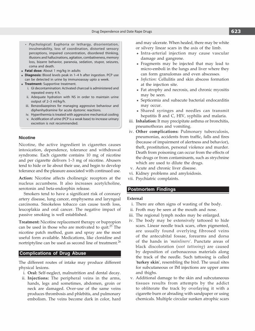
623Drug Dependence and Date Rape Drugs
• Psychological: Euphoria or lethargy, disorientation, invulnerability, loss of coordination, distorted sensory perceptions, impaired concentration, disordered thinking, illusions and hallucinations, agitation, combativeness, memory loss, bizarre behavior, paranoia, sedation, stupor, seizures, coma and death.
� Fatal dose: About 1 mg/kg in adults � Diagnosis: Blood levels peak in 1–4 h after ingestion. PCP use
can be detected in urine by immunoassay upto a week. � Treatment: Supportive treatment.
i. GI decontamination: Activated charcoal is administered and repeated every 4 h.
ii. Adequate hydration with NS in order to maintain urine output of 2–3 ml/kg/h.
iii. Benzodiazepines for managing aggressive behaviour and diphenhydramine for acute dystonic reactions.
iv. Hyperthermia is treated with aggressive mechanical cooling v. Acidification of urine (PCP is a weak base) to increase urinary
excretion is not recommended.
nicotine
Nicotine, the active ingredient in cigarettes causes intoxication, dependence, tolerance and withdrawal syndrome. Each cigarette contains 10 mg of nicotine and per cigarette delivers 1–3 mg of nicotine. Abusers tend to hide or lie about their use, and begin to develop tolerance and the pleasure associated with continued use.
Action: Nicotine affects cholinergic receptors at the nucleus accumbens. It also increases acetylcholine, serotonin and beta-endorphin release.
Smokers tend to have a significant risk of coronary artery disease, lung cancer, emphysema and laryngeal carcinoma. Smokeless tobacco can cause tooth loss, leucoplakia and oral cancer. The negative impact of passive smoking is well established.
Treatment: Nicotine replacement therapy or bupropion can be used in those who are motivated to quit.25 The nicotine patch method, gum and spray are the most useful form available. Medications, like clonidine and nortriptyline can be used as second line of treatment.26
Complications of Drug Abuse
The different routes of intake may produce different physical lesions. i. Oral: Self-neglect, malnutrition and dental decay. ii. injections: The peripheral veins in the arms,
hands, legs and sometimes, abdomen, groin or neck are damaged. Over-use of the same veins produces thrombosis and phlebitis, and pulmonary embolism. The veins become dark in color, hard
and may ulcerate. When healed, there may be white or silvery linear scars in the axis of the limb.
z Intra-arterial injection may cause vascular damage and gangrene.
z Fragments may be injected that may lead to micro-emboli in the lungs and liver where they can form granulomas and even abscesses.
z Infection: Cellulitis and skin abscess formation at the injection site.
z Fat atrophy and necrosis, and chronic myositis may be seen.
z Septicemia and subacute bacterial endocarditis may occur.
z Shared syringes and needles can transmit hepatitis B and C, HIV, syphilis and malaria.
iii. inhalation: It may precipitate asthma or bronchitis, pneumothorax and vomiting.
iv. Other complications: Pulmonary tuberculosis, pneumonias, accidents from traffic, falls and fires (because of impairment of alertness and behavior), theft, prostitution, personal violence and murder. Death from poisoning can occur from the effects of the drugs or from contaminants, such as strychnine which are used to dilute the drugs.
v. Acute and chronic liver disease. vi. Kidney problems and amyloidosis. vii. Psychiatric complaints.
Postmortem Findings
External i. There are often signs of wasting of the body. ii. Froth may be seen at the mouth and nose. iii. The regional lymph nodes may be enlarged. iv. The body may be extensively tattooed to hide
scars. Linear needle track scars, often pigmented, are usually found overlying fibrosed veins of the antecubital fossae, forearms and dorsa of the hands in 'mainliners'. Punctate areas of black discoloration (soot tattooing) are caused by deposition of carbonaceous materials along the track of the needle. Such tattooing is called 'turkey skin', resembling the bird. The usual sites for subcutaneous or IM injections are upper arms and thighs.
v. Additional damage to the skin and subcutaneous tissues results from attempts by the addict to obliterate the track by overlaying it with a cigarette burn or abrading with sandpaper or using chemicals. Multiple circular sunken atrophic scars

624 Review of Forensic Medicine and Toxicology
(tissue paper scars) suggest skin popping, followed by skin infection.
vi. Recent injection sites may show zones of inflammation surrounding or adjacent to a needle puncture site.
vii. Subcutaneous heroin users show a higher incidence of abscesses. Healing by fibrosis may produce hyperpigmented macules or retracted circumscribed scars which resemble those from smallpox vaccinations.
viii. Chronic edema of the hands, secondary to occlusive thrombophlebitis in the forearms is seen occasionally in long-term addicts.
ix. Habitual inhalation of cocaine or heroin (snorting or sniffing) causes perforation of the nasal septum.
Internal
i. There may be phlebitis, phlebosclerosis, thrombosis, and recent and resolving perivenous hemorrhage. The vein and surrounding tissue should be preserved for chemical analysis.
ii. Typical visceral findings include non-specific triad of edema, bronchopneumonia and aspiration of gastric contents.
iii. Pericardial, pleural and peritoneal effusions may be found.
iv. Stomach may contain pills or capsules. v. Liver: Most common changes from parenteral drug
abuse consist of hepatic lymphadenopathy and hepatic portal triaditis. The liver may be slightly enlarged or show evidence of cirrhosis.
vi. Spleen: Splenomegaly and portal lymph node hyperplasia are common. The most constant finding in both spleen and portal lymph nodes is the presence of large germinal centers, but the morphological features are not specific.
vii. Hyperplastic changes in the reticuloendothelial system are common.
viii. Lungs: Pleura may show petechial hemorrhages, and lungs are congested and edematous.
ix. Heart may show valvular disease. x. In mainliners, the crystals lodge in pulmonary
capillaries and produce a foreign body granulo-matous reaction. Pulmonary hypertension with right ventricular hypertrophy occurs due to extensive microcrystalline pulmonary emboli.
xi. Brain: It may show edema and focal areas of necrosis involving the globus pallidus and hippocampus due to hypoxia.
Date Rape Drugs
� Date rape: Forcible sexual intercourse of a woman by a male acquaintance, during a voluntary social engagement in which the woman did not intend to submit to the sexual advances, and resisted the act by verbal refusals, denials and/or physical resistance.
� Date rape drug (predator drug): Any substance that is administered to lower sexual inhibition and enhances the possibility of unwanted sexual intercourse and renders the individual vulnerable to a drug facilitated sexual assault (DFSA) including rape.
� Drink spiking: The act of surreptitious administration of such drugs to drinks. Although, drink spiking is often associated with malicious acts including assault, theft and DFSA, it is also used for misguided pranks or jokes. In the UK, drink spiking with intent to commit a sexual assault or other serious criminal act is an offence that may result in imprisonment for 10 years.
� Gamma hydroxybutyrate (GHB), ketamine, and flunitrazepam (Rohypnol) are the most common date rape drugs (‘club drugs’), though alcohol is the most common drug used to facilitate date rape (Table 61.2). Other drugs which are also associated with date rape are marijuana, benzodiazepines, cocaine, heroin, amphetamines and choral hydrate.
� Although, these drugs differ in their effect on the body, they all act as sedatives, frequently causing unconsciousness and amnesia. The drugs often have no color, odor or taste and are easily added to flavored drinks without the victim’s knowledge. Because of the effects of these drugs, victims are physically helpless, unable to refuse sex, and unable to remember what happened. Date rape drugs are particularly dangerous when mixed with alcohol and can lead to a coma or even death.
� Chloral hydrate: A solution of chloral hydrate and alcohol constituted the infamous ‘knockout drops’ or ‘Mickey Finn’ which is used in DFSA.

625Drug Dependence and Date Rape Drugs
multiple choice questions
1. Treatment of acute alcohol withdrawal: Punjab 09 A. Diazepam B. Bupropion C. Disulfiram D. Acamprosate 2. Drug not used in treatment of alcohol dependence:
NIMHANS 08; PGI 09, 11; TN 08; AI 11; AIIMS 14 A. Diazepam B. Disulfiram C. Acamprosate D. Naltrexone 3. Drugs is useful in the long term treatment of alcohol
dependence: COMEDK 13 A. Acamprosate B. Chlordiazepoxide C. Flumazenil D. Imipramine 4. Alcohol anti-craving agents are all, except: AIIMS 09 A. Lorazepam B. Clonidine C. Acamprosate D. Naltrexone 5. Yawning is a common feature of: AIIMS 06 A. Alcohol withdrawal B. Cocaine withdrawal C. Cannabis withdrawal D. Opioid withdrawal 6. All are true of opioid withdrawal, except:
NIMHANS 10; PGI 14 A. Yawning B. Hallucinations C. Lacrimation D. Piloerection
7. A teenager is having diarrhea, rhinorrhea and sweating, most probable diagnosis is: AIIMS 10
A. Cocaine withdrawal B. Heroin withdrawal C. Marijuana withdrawal D. LSD withdrawal 8. Usual sign of opioid withdrawal: PGI 13 A. Stupor B. Constipation C. Constricted pupil D. Yawning 9. Naltrexone is used in: Bihar 10; AIIMS 10 A. Treat withdrawal symptoms B. Treatment of overdose C. Prevention of relapse D. Deterrent agent10. Notused for treatmentofheroindetoxification:
AIIMS 09, 10; AP 11 A. Disulfiram B. Buprenorphine C. Clonidine D. Lofexidine 11. The drug which is used for long-term maintenance in
opioid addiction: AIIMS 06; Punjab 09; AI 11 A. Nalorphine B. Naloxone C. Butarphanol D. Methadone
1. A 2. A 3. A 4. A 5. D 6. B 7. B 8. D 9. A & B 10. A 11. D
Table 61.2: Characteristics of common date rape drugs
S.No. Feature GHB Flunitrazepam Ketamine
1. Chemical GHB is metabolite of GABA, the inhibitory neurotransmitter
Benzodiazepine Sympathomimetic amine similar to phencyclidine
2. Street names Grievous bodily harm, liquid G, liquid ecstasy, scoop
Roofies, R2, roofenol, roche, rope, forget pill
K, Ket, special K, super acid, smack K, kit-kat
3. Properties Powder or liquid, colorless, odorless, salty taste
Odorless and tasteless pill Colorless and odorless liquid, or white or off-white powder
4. Onset of action 15 min 15–20 min 20 min
5. Duration 1.5–2 h 4–6 h 6–24 h
6. Signs and symptoms Initially, relaxation, disinhibition, euphoria, followed by nausea, drowsiness, dizziness, amnesia, hallucinations, delirium, sedation
Lack of muscle control and motor abilities, confusion, slurring, amnesia, nausea, dizziness, sleepiness, unconsciousness
Dissociative anesthesia, confusion hallucinations, impaired motor function, nystagmus, amnesia, sedation, respiratory depression
7. Management Supportive, no antidote Supportive, antidote is flumazenil Supportive, ECG monitoring
8. Specimens Blood (in NaF and potassium oxalate): within 24 h of incident Urine (100 ml): within 96 h
Urine: Flunitrazepam metabolites within 12 h after ingestion
Urine: Norketamine and dehydronorket-amine, using GC-MS or LC-MS analyses
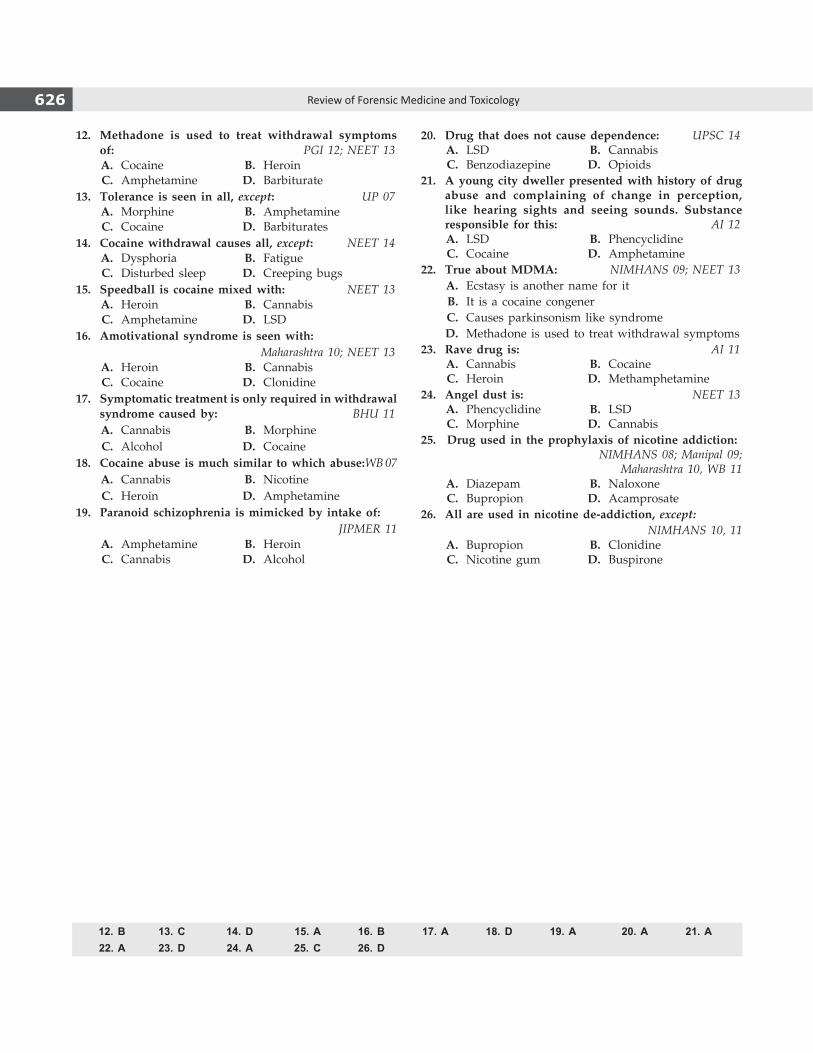
626 Review of Forensic Medicine and Toxicology
12. B 13. c 14. D 15. A 16. B 17. A 18. D 19. A 20. A 21. A 22. A 23. D 24. A 25. c 26. D
12. Methadone is used to treat withdrawal symptoms of: PGI 12; NEET 13
A. Cocaine B. Heroin C. Amphetamine D. Barbiturate 13. Tolerance is seen in all, except: UP 07 A. Morphine B. Amphetamine C. Cocaine D. Barbiturates 14. Cocaine withdrawal causes all, except: NEET 14 A. Dysphoria B. Fatigue C. Disturbed sleep D. Creeping bugs 15. Speedball is cocaine mixed with: NEET 13 A. Heroin B. Cannabis C. Amphetamine D. LSD 16. Amotivational syndrome is seen with: Maharashtra 10; NEET 13 A. Heroin B. Cannabis C. Cocaine D. Clonidine 17. Symptomatic treatment is only required in withdrawal
syndrome caused by: BHU 11 A. Cannabis B. Morphine C. Alcohol D. Cocaine 18. Cocaine abuse is much similar to which abuse: WB 07 A. Cannabis B. Nicotine C. Heroin D. Amphetamine 19. Paranoid schizophrenia is mimicked by intake of:
JIPMER 11 A. Amphetamine B. Heroin C. Cannabis D. Alcohol
20. Drug that does not cause dependence: UPSC 14 A. LSD B. Cannabis C. Benzodiazepine D. Opioids 21. A young city dweller presented with history of drug
abuse and complaining of change in perception, like hearing sights and seeing sounds. Substance responsible for this: AI 12
A. LSD B. Phencyclidine C. Cocaine D. Amphetamine 22. True about MDMA: NIMHANS 09; NEET 13 A. Ecstasy is another name for it B. It is a cocaine congener C. Causes parkinsonism like syndrome D. Methadone is used to treat withdrawal symptoms 23. Rave drug is: AI 11 A. Cannabis B. Cocaine C. Heroin D. Methamphetamine 24. Angel dust is: NEET 13 A. Phencyclidine B. LSD C. Morphine D. Cannabis 25. Drug used in the prophylaxis of nicotine addiction:
NIMHANS 08; Manipal 09; Maharashtra 10, WB 11
A. Diazepam B. Naloxone C. Bupropion D. Acamprosate 26. All are used in nicotine de-addiction, except:
NIMHANS 10, 11 A. Bupropion B. Clonidine C. Nicotine gum D. Buspirone
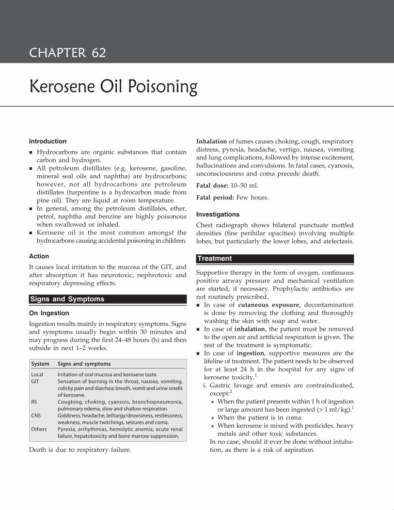
Kerosene Oil Poisoning
ChaPter 62
Introduction
� Hydrocarbons are organic substances that contain carbon and hydrogen.
� All petroleum distillates (e.g. kerosene, gasoline, mineral seal oils and naphtha) are hydrocarbons; however, not all hydrocarbons are petroleum distillates (turpentine is a hydrocarbon made from pine oil). They are liquid at room temperature.
� In general, among the petroleum distillates, ether, petrol, naphtha and benzine are highly poisonous when swallowed or inhaled.
� Kerosene oil is the most common amongst the hydrocarbons causing accidental poisoning in children.
Action
It causes local irritation to the mucosa of the GIT, and after absorption it has neurotoxic, nephrotoxic and respiratory depressing effects.
Signs and Symptoms
On Ingestion
Ingestion results mainly in respiratory symptoms. Signs and symptoms usually begin within 30 minutes and may progress during the first 24–48 hours (h) and then subside in next 1–2 weeks.
System Signs and symptoms
Local Irritation of oral mucosa and kerosene taste.GIT Sensation of burning in the throat, nausea, vomiting,
colicky pain and diarrhea; breath, vomit and urine smells of kerosene.
RS Coughing, choking, cyanosis, bronchopneumonia, pulmonary edema, slow and shallow respiration.
CNS Giddiness, headache, lethargy/drowsiness, restlessness, weakness, muscle twitchings, seizures and coma.
Others Pyrexia, arrhythmias, hemolytic anemia, acute renal failure, hepatotoxicity and bone marrow suppression.
Death is due to respiratory failure.
Inhalation of fumes causes choking, cough, respiratory distress, pyrexia, headache, vertigo, nausea, vomiting and lung complications, followed by intense excitement, hallucinations and convulsions. In fatal cases, cyanosis, unconsciousness and coma precede death.
Fatal dose: 10–50 ml.
Fatal period: Few hours.
Investigations
Chest radiograph shows bilateral punctuate mottled densities (fine perihilar opacities) involving multiple lobes, but particularly the lower lobes, and atelectasis.
Treatment
Supportive therapy in the form of oxygen, continuous positive airway pressure and mechanical ventilation are started, if necessary. Prophylactic antibiotics are not routinely prescribed.
� In case of cutaneous exposure, decontamination is done by removing the clothing and thoroughly washing the skin with soap and water.
� In case of inhalation, the patient must be removed to the open air and artificial respiration is given. The rest of the treatment is symptomatic.
� In case of ingestion, supportive measures are the lifeline of treatment. The patient needs to be observed for at least 24 h in the hospital for any signs of kerosene toxicity.1
i. Gastric lavage and emesis are contraindicated, except:2
z When the patient presents within 1 h of ingestion or large amount has been ingested (> 1 ml/kg).1
z When the patient is in coma. z When kerosene is mixed with pesticides, heavy
metals and other toxic substances. In no case, should it ever be done without intuba
tion, as there is a risk of aspiration.

628 Review of Forensic Medicine and Toxicology
ii. Activated charcoal has a limited role in the management of kerosene ingestion, as it poorly adsorbs most hydrocarbons.
iii. Bacterial pneumonia is uncommon. Prophylactic antibiotic therapy is not recommended. Antibiotics are indicated in limited situations, like malnutrition or immunocompromised state. If fever occurs, give specific antibiotic.
iv. Corticosteroids are not recommended, except when administered concurrently at the time of aspiration.
v. Bronchodilators are used for chlorinated or fluorinated solvent intoxication.
vi. Oxygen therapy is given in hypoxemia.
Complications: Aspiration pneumonitis is the most common complication of kerosene ingestion, followed by CNS and CVS complications.3
� Respiratory: Aspiration and lung injury secondary to pneumonitis. Secondary effects in the lungs include pneumothorax, pyopneumothorax, pneumatocele or bronchopleural fistula.
� CNS: Seizures, encephalopathy and memory loss. � CVS: Myocarditis and cardiomyopathy.
Postmortem Findings
i. Acute gastroenteritis and kerosene odor may be observed on opening the chest and abdominal cavity.
ii. Stomach: Petechial hemorrhages with congested mucosa.
iii. Lungs: Petechial hemorrhages, congested, edematous and bronchopneumonia.
iv. Degenerative changes in the liver and kidneys and hypoplasia of the bone marrow occur after pro longed period of inhalation.
v. Organs are congested, and other signs of asphyxia may be seen.
In case of suspected death from kerosene, the lungs, brain and other viscera should be preserved in saturated saline for chemical analysis.
Medico-legal Aspects
� In North India, it accounts for about 50% of infants and children brought to hospital for accidental poisoning, who have taken kerosene mistaking it for water. However, ingestion of large quantities is unusual because of its foul taste (rarely consume more than 30 ml).
� Kerosene is occasionally used for selfimmolation and suicidal purpose.
� Homicidal attempts by pouring kerosene on clothes and igniting them are common in case of dowry deaths in India.
� Inhalation of volatile hydrocarbons is common abuse in adolescents and young adults for recreation, similar to drugs and alcohol.
� Aspiration may occur during attempt to siphon off gasoline.
Chronic poisoning can occur in persons who handle petroleum products.
Symptoms are weakness, dizziness, pain in limbs, peripheral numbness, paresthesias, weight loss and anemia.
Cardiomyopathy, cerebellar atrophy, dementia, cognitive deficits and peripheral neuropathy are seen with chronic hydrocarbon inhalant abuse.
Treatment requires isolation of the patient from exposure and symptomatic management.
Multiple choice questions
1. A 3-year-old girl came to the emergency after 2 h of ingestion of kerosene, immediate management is: UPSC 13
A. Gastric lavage B. Chest Xray and observation C. Corticosteroids D. Emetics are given 2. Management of kerosene oil poisoning includes all,
except: AP 09 A. Gastric lavage is done B. Bronchodilators are given
C. Oxygen is given D. Corticosteroids are not beneficial 3. Complication of kerosene poisoning is: NEET 14 A. Paralysis B. Delirium C. Hemoptysis D. Aspiration pneumonia
1. B 2. A 3. D
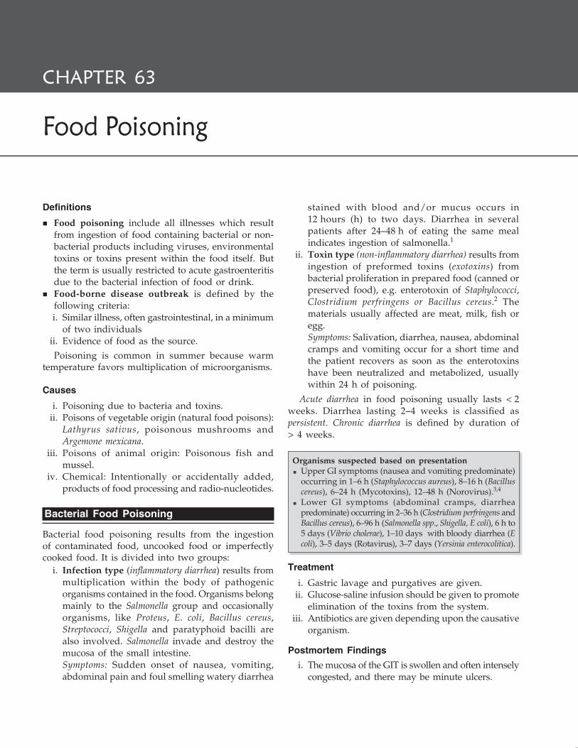
629Food Poisoning
Food Poisoning
ChaPter 63
Definitions
� Food poisoning include all illnesses which result from ingestion of food containing bacterial or non-bacterial products including viruses, environmental toxins or toxins present within the food itself. But the term is usually restricted to acute gastroenteritis due to the bacterial infection of food or drink.
� Food-borne disease outbreak is defined by the following criteria:
i. Similar illness, often gastrointestinal, in a minimum of two individuals
ii. Evidence of food as the source.
Poisoning is common in summer because warm temperature favors multiplication of microorganisms.
Causes
i. Poisoning due to bacteria and toxins. ii. Poisons of vegetable origin (natural food poisons):
Lathyrus sativus, poisonous mushrooms and Argemone mexicana.
iii. Poisons of animal origin: Poisonous fish and mussel.
iv. Chemical: Intentionally or accidentally added, products of food processing and radio-nucleotides.
Bacterial Food Poisoning
Bacterial food poisoning results from the ingestion of contaminated food, uncooked food or imperfectly cooked food. It is divided into two groups: i. Infection type (inflammatory diarrhea) results from
multiplication within the body of pathogenic organisms contained in the food. Organisms belong mainly to the Salmonella group and occasionally organisms, like Proteus, E. coli, Bacillus cereus, Streptococci, Shigella and paratyphoid bacilli are also involved. Salmonella invade and destroy the mucosa of the small intestine.
Symptoms: Sudden onset of nausea, vomiting, abdominal pain and foul smelling watery diarrhea
stained with blood and/or mucus occurs in 12 hours (h) to two days. Diarrhea in several patients after 24–48 h of eating the same meal indicates ingestion of salmonella.1
ii. Toxin type (non-inflammatory diarrhea) results from ingestion of preformed toxins (exotoxins) from bacterial proliferation in prepared food (canned or preserved food), e.g. enterotoxin of Staphylococci, Clostridium perfringens or Bacillus cereus.2 The materials usually affected are meat, milk, fish or egg.
Symptoms: Salivation, diarrhea, nausea, abdominal cramps and vomiting occur for a short time and the patient recovers as soon as the enterotoxins have been neutralized and metabolized, usually within 24 h of poisoning.
Acute diarrhea in food poisoning usually lasts < 2 weeks. Diarrhea lasting 2–4 weeks is classified as persistent. Chronic diarrhea is defined by duration of > 4 weeks.
Organisms suspected based on presentation � Upper GI symptoms (nausea and vomiting predominate) occurring in 1–6 h (Staphylococcus aureus), 8–16 h (Bacillus cereus), 6–24 h (Mycotoxins), 12–48 h (Norovirus).3,4
� Lower GI symptoms (abdominal cramps, diarrhea predominate) occurring in 2–36 h (Clostridium perfringens and Bacillus cereus), 6–96 h (Salmonella spp., Shigella, E coli), 6 h to 5 days (Vibrio cholerae), 1–10 days with bloody diarrhea (E coli), 3–5 days (Rotavirus), 3–7 days (Yersinia enterocolitica).
Treatment
i. Gastric lavage and purgatives are given. ii. Glucose-saline infusion should be given to promote
elimination of the toxins from the system. iii. Antibiotics are given depending upon the causative
organism.
Postmortem Findings i. The mucosa of the GIT is swollen and often intensely
congested, and there may be minute ulcers.
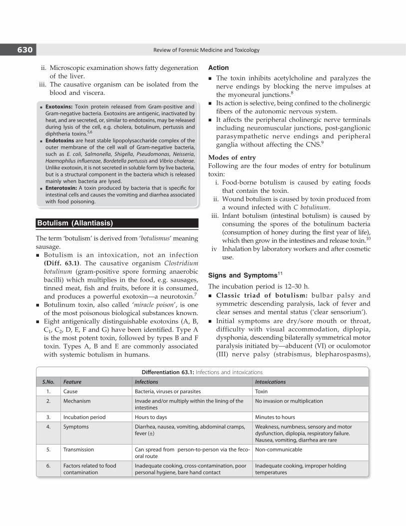
630 Review of Forensic Medicine and Toxicology
ii. Microscopic examination shows fatty degenera tion of the liver.
iii. The causative organism can be isolated from the blood and viscera.
� Exotoxins: Toxin protein released from Gram-positive and Gram-negative bacteria. Exotoxins are antigenic, inactivated by heat, and are secreted, or, similar to endotoxins, may be released during lysis of the cell, e.g. cholera, botulinum, pertussis and diphtheria toxins.5,6
� Endotoxins are heat stable lipopolysaccharide complex of the outer membrane of the cell wall of Gram-negative bacteria, such as E. coli, Salmonella, Shigella, Pseudomonas, Neisseria, Haemophilus influenzae, Bordetella pertussis and Vibrio cholerae. Unlike exotoxin, it is not secreted in soluble form by live bacteria, but is a structural component in the bacteria which is released mainly when bacteria are lysed.
� Enterotoxin: A toxin produced by bacteria that is specific for intestinal cells and causes the vomiting and diarrhea associated with food poisoning.
Botulism (Allantiasis)
The term ‘botulism’ is derived from ‘botulismus’ meaning sausage.
� Botulism is an intoxication, not an infection (Diff. 63.1). The causative organism Clostridium botulinum (gram-positive spore forming anaerobic bacilli) which multiplies in the food, e.g. sausages, tinned meat, fish and fruits, before it is consumed, and produces a powerful exotoxin—a neurotoxin.7
� Botulinum toxin, also called ‘miracle poison’, is one of the most poisonous biological substances known.
� Eight antigenically distinguishable exotoxins (A, B, C1, C2, D, E, F and G) have been identified. Type A is the most potent toxin, followed by types B and F toxin. Types A, B and E are commonly associated with systemic botulism in humans.
Action � The toxin inhibits acetylcholine and paralyzes the
nerve endings by blocking the nerve impulses at the myoneural junctions.8
� Its action is selective, being confined to the cholinergic fibers of the autonomic nervous system.
� It affects the peripheral cholinergic nerve terminals including neuromuscular junctions, post-ganglionic parasympathetic nerve endings and peripheral ganglia without affecting the CNS.9
Modes of entryFollowing are the four modes of entry for botulinum toxin: i. Food-borne botulism is caused by eating foods
that contain the toxin. ii. Wound botulism is caused by toxin produced from
a wound infected with C botulinum. iii. Infant botulism (intestinal botulism) is caused by
consuming the spores of the botulinum bacteria (consumption of honey during the first year of life), which then grow in the intestines and release toxin.10
iv Inhalation by laboratory workers and after cosmetic use.
Signs and Symptoms11
The incubation period is 12–30 h. � Classic triad of botulism: bulbar palsy and
symmetric descending paralysis, lack of fever and clear senses and mental status (‘clear sensorium’).
� Initial symptoms are dry/sore mouth or throat, difficulty with visual accommodation, diplopia, dysphonia, descending bilaterally symmetrical motor paralysis initiated by—abducent (VI) or oculomotor (III) nerve palsy (strabismus, blepharospasms),
Differentiation 63.1: Infections and intoxications
S.No. Feature Infections Intoxications
1. Cause Bacteria, viruses or parasites Toxin
2. Mechanism Invade and/or multiply within the lining of the intestines
No invasion or multiplication
3. Incubation period Hours to days Minutes to hours
4. Symptoms Diarrhea, nausea, vomiting, abdominal cramps, fever (±)
Weakness, numbness, sensory and motor dysfunction, diplopia, respiratory failure.Nausea, vomiting, diarrhea are rare
5. Transmission Can spread from person-to-person via the feco-oral route
Non-communicable
6. Factors related to food contamination
Inadequate cooking, cross-contamination, poor personal hygiene, bare hand contact
Inadequate cooking, improper holding temperatures
631Food Poisoning
dysphagia, constipation, hypothermia, respiratory insufficiency and urinary retention.
� The GIT symptoms, like nausea, vomiting and abdominal pain are rare.
� The patient is conscious till death which is preceded by coma or delirium.
Fatal dose: 0.01 mg or even less.
Fatal period: 24–48 h, may extend to a week.
Differential Diagnosis
Toxin type of food poisoning, poliomyelitis, myasthenia gravis, encephalitis, multiple sclerosis, Guillain-Barre syndrome, diphtheria, tetanus, and poisoning from CO, organophosphates and elapid snake bite.
Treatment
i. Maintenance of ABC. ii. Decontamination—gastric lavage, activated
charcoal, purgatives and whole bowel irriga- tion.
iii. Polyvalent botulinum antitoxin (types A, B and E) one vial by slow IV in normal saline and one vial IM, repeated at 2–4 h intervals IV.12
iv. Botulism immune globulin (BIG), 50 ml is given IV daily, till the patient recovers.
v. Frequent dose of brandy is beneficial as alcohol precipitates toxin.
Postmortem Findings
i. Kidneys, liver and meninges are congested. ii. Histological examination of the organs may show
thrombosis.
Medico-legal Aspects
� Unintentional outbreak of food-borne botulism is caused from foods that are not canned properly or improper handling during manufacture.7
� Iatrogenic botulism can also occur from accidental overdose of botulinum toxin. Cosmetic or therapeutic use may result in adverse events resulting in lawsuits alleging negligence (complications such as immune reaction and brain injury may occur).
� Botulinum toxin poses a major biological weapon because of its extreme potency and lethality, ease of production and transport. Dissemination of aerosols of toxin can produce mass casualties.
� Infant botulism is a neuroparalytic disease which affects otherwise healthy children < 1 year old. Early symptoms are constipation, generalized weakness and weak cry.10
� SIDS could be attributed to C botulinum intoxication.10
� Botulinium toxin type A (Botox) was approved by the US Food and Drug Administration (FDA) for the treatment of strabismus, blepharospasm and hemifacial spasm. It is also used for treatment of frown lines between the eyebrows (glabellar lines), spasticity and muscle pain disorders and cervical dystonia.13
PoiSonouS FooDSPoisonous foods are those which contain poison derived from plants, animals and inorganic chemicals.
Lathyrus Sativus (‘Kesari Dhal’)
This is a variety of pulse and is the staple food for the low-income groups in some areas of Central India.14 Consumption of L sativus seeds in quantities exceeding 30% of the total diet for more than 6 months has been known to cause paralysis. Men are more susceptible than women.
Active Principle
b-N-oxalyl amino-alanine (BOAA), a neurotoxic amino acid present in the seed cotyledons.15
Signs and Symptoms
The continued use of L sativus produces neurolathyrism, which is characterized by progressive spastic paraplegia with preservation of sphincters, sensation and mental activity.16
There may be pain in the back or weakness of legs and difficulty in sitting down and getting up. The patient is unable to walk without the aid of a stick, the legs tremble and are dragged along with difficulty. A spastic gait develops characterized by ‘walking on tiptoes’ with the legs crossing scissor-wise. Later, complete paraplegia occurs. There is no atrophy or loss of the tone of muscles and no reaction of degeneration. The knee jerks are increased, ankle clonus is marked and Babinski’s sign is present.
Treatment
� Rich diet with exclusion of the pulse, massage and application of electricity are useful.
� Steeping the pulse in hot water and parboiling remove 90% of toxic amino acid.16
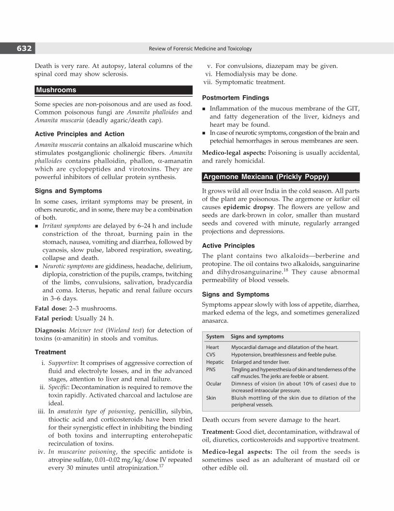
632 Review of Forensic Medicine and Toxicology
Death is very rare. At autopsy, lateral columns of the spinal cord may show sclerosis.
Mushrooms
Some species are non-poisonous and are used as food. Common poisonous fungi are Amanita phalloides and Amanita muscaria (deadly agaric/death cap).
Active Principles and Action
Amanita muscaria contains an alkaloid muscarine which stimulates postganglionic cholinergic fibers. Amanita phalloides contains phalloidin, phallon, a-amanatin which are cyclopeptides and virotoxins. They are powerful inhibitors of cellular protein synthesis.
Signs and Symptoms
In some cases, irritant symptoms may be present, in others neurotic, and in some, there may be a combination of both.
� Irritant symptoms are delayed by 6–24 h and include constriction of the throat, burning pain in the stomach, nausea, vomiting and diarrhea, followed by cyanosis, slow pulse, labored respiration, sweating, collapse and death.
� Neurotic symptoms are giddiness, headache, delirium, diplopia, constriction of the pupils, cramps, twitching of the limbs, convulsions, salivation, bradycardia and coma. Icterus, hepatic and renal failure occurs in 3–6 days.
Fatal dose: 2–3 mushrooms.
Fatal period: Usually 24 h.
Diagnosis: Meixner test (Wieland test) for detection of toxins (a-amanitin) in stools and vomitus.
Treatment
i. Supportive: It comprises of aggressive correction of fluid and electrolyte losses, and in the advanced stages, attention to liver and renal failure.
ii. Specific: Decontamination is required to remove the toxin rapidly. Activated charcoal and lactulose are ideal.
iii. In amatoxin type of poisoning, penicillin, silybin, thioctic acid and corticosteroids have been tried for their synergistic effect in inhibiting the binding of both toxins and interrupting enterohepatic recirculation of toxins.
iv. In muscarine poisoning, the specific antidote is atropine sulfate, 0.01–0.02 mg/kg/dose IV repeated every 30 minutes until atropinization.17
v. For convulsions, diazepam may be given. vi. Hemodialysis may be done. vii. Symptomatic treatment.
Postmortem Findings
� Inflammation of the mucous membrane of the GIT, and fatty degeneration of the liver, kidneys and heart may be found.
� In case of neurotic symptoms, congestion of the brain and petechial hemorrhages in serous membranes are seen.
Medico-legal aspects: Poisoning is usually accidental, and rarely homicidal.
Argemone Mexicana (Prickly Poppy)
It grows wild all over India in the cold season. All parts of the plant are poisonous. The argemone or katkar oil causes epidemic dropsy. The flowers are yellow and seeds are dark-brown in color, smaller than mustard seeds and covered with minute, regularly arranged projections and depressions.
Active PrinciplesThe plant contains two alkaloids—berberine and protopine. The oil contains two alkaloids, sanguinarine and dihydrosanguinarine.18 They cause abnormal permeability of blood vessels.
Signs and SymptomsSymptoms appear slowly with loss of appetite, diarrhea, marked edema of the legs, and sometimes generalized anasarca.
System Signs and symptoms
Heart Myocardial damage and dilatation of the heart.CVS Hypotension, breathlessness and feeble pulse.Hepatic Enlarged and tender liver.PNS Tingling and hyperesthesia of skin and tenderness of the
calf muscles. The jerks are feeble or absent.Ocular Dimness of vision (in about 10% of cases) due to
increased intraocular pressure.Skin Bluish mottling of the skin due to dilation of the
peripheral vessels.
Death occurs from severe damage to the heart.
Treatment: Good diet, decontamination, withdrawal of oil, diuretics, corticosteroids and supportive treatment.
Medico-legal aspects: The oil from the seeds is sometimes used as an adulterant of mustard oil or other edible oil.

633Food Poisoning
Food AllergySome persons are hypersensitive to certain types of protein, e.g. meat, fish, eggs or milk which are ordinarily
1. A 2. B 3. C 4. B 5. D 6. C 7. C 8. A 9. D 10. D 11. B 12. B 13. D 14. B 15. B 16. B & D 17. A 18. C
quite harmless, and suffer from gastroenteritis, local urticarial rashes joint pains or asthmatic attack. Antihistaminics and steroids may be given.
multiple ChoiCe questions
1. A 22-year-old male had an outing with his friends and developed fever of 38.5°C, abdominal pain, diarrhea and vomiting after eating chicken salad 24 h back. Two of his friends developed the same symptoms. The diagnosis is: FMGE 08
A. Salmonella enteritis poisoning B. Bacillus cereus poisoning C. Staphylococcus aureus poisoning D. Vibrio cholera poisoning 2. Preformed toxin is seen in:
CMC (Vellore) 13; COMEDK 15 A. Staphylococcus aureus B. Clostridium perfringens C. E. coli D. Yersinia enterocolitica 3. A 14-year-old male developed vomiting and diarrhea
in 2 h after having food from a restaurant. The most likely pathogen is: KCET 12
A. Clostridium perfringens B. Vibrio parahaemolyticus C. Staphylococcus aureus D. Salmonella typhimurium 4. Symptoms of food poisoning seen within 6 h is
commonly due to: NIMHANS 13; FMGE 13 A. Bacillus cereus B. Staphylococcus aureus C. Shigella D. Salmonella 5. Which of the following is an exotoxin: NIMHANS 07 A. E. coli toxin B. Proteus C. Pseudomonas D. Tetanus toxin 6. All are true of exotoxin, except: JIPMER 14 A. Protein in nature B. Antigenic C. Heat stable D. Highly toxic 7. Causative organism of food poisoning due to home
canned food: JIPMER 12 A. Bacillus cereus B. Salmonella C. Clostridium botulinum D. Staphylococci 8. Mechanism of action of botulinum toxin: Kerala 11 A. Synthesis of acetylcholine inhibited B. Reuptake of ACH is increased C. Blocks nicotinic receptors in muscle D. Blocks muscarinic receptors in brain 9. Botulinum affects all, except: AI 07 A. Neuromuscular junction B. Preganglionic junction
C. Post-ganglionic nerves D. CNS 10. Sudden infant death syndrome (SIDS) following
administration of honey is due to: MAHE 12 A. Staphylococcus aureus B. Yersinia enterocolitica C. E. coli D. Clostridium botulinum 11. Dysphagia, diplopia, dysarthria are characteristic
symptoms of food poisoning due to: UPSC 08 A. Staphylococcus aureus B. Clostridium botulinum C. Salmonella typhimurium D. Bacillus cereus 12. Which poisoning can be prevented by an antitoxin:
MAHE 11 A. Staphylococcus aureus B. Clostridium botulinum C. Salmonella typhimurium D. Bacillus cereus 13. Botulinum toxin is used for the treatment of: DNB 08 A. Blepharospasm B. Risus sardonicus C. Strabismus D. All 14. Lathyrism is seen with eating of: Kerala 08 A. Red gram B. Kesari dhal C. Mushrooms D. Sausages 15. In Kesari dhal poisoning due to lathyrus sativus, the
active principle is: NIMHANS 07; MAHE 08; UPSC 14 A. Pyrrozolidine B. BOAA C. Argemone oil D. Pilocarpine 16. True about lathyrism: PGI 09 A. Caused by Aspergillus flavus B. Prevented by parboiling C. Sanguinarine is the toxic principle D. Patient develops spastic paraplegia 17. The drug of choice for mushroom poisoning is: AI 07 A. Atropine B. Physostigmine C. Adrenaline D. Carbachol 18. Toxin responsible for epidemic dropsy: AIIMS 07; UP 09; PGI 11 A. BOAA B. Aflatoxin C. Sanguinarine D. Pyrrozolidine

635Question Bank-II
Question Bank-II
All questions are either short notes or viva, if not mentioned otherwise (LQ—long question, SN—short note, Diff.—differentiation).
Must Know
General toxicology
1. Toxicology, poison, tolerance, idiosyncrasy 2. Classificationofpoisons(basedonmodeofaction
and effects) 3. Color of PM staining and odor in different poisoning 4. Poisons causing subendocardialhemorrhages 5. Physical antidote, activated charcoal, demulcents 6. Duties of a doctor in poisoning cases 7. Gastric lavage, contraindications 8. Hepatotoxic and nephrotoxic poisons 9. Chelating agents 10. Universal antidote11. Dialyzableandnon-dialyzablepoisons 12. Collection and preservation of samples and viscera
in case of poisoning 13. Antidotes in different poisoning 14. Poisons causing miosis and mydriasis
Alcohol
1. Percentageof alcohol indifferentbeverages 2. McEwan's sign 3. Mellanbyeffect 4. Treatment of chronic alcoholism 5. Delirium tremens 6. Korsakoff's psychosis 7. Wernicke's encephalopathy 8. Drunkenness 9. Field impairment tests 10. Widmark's formula
Rest of the Poisons
1. Active principles and identification of seeds: Ricinus, Croton, Abrus, Semecarpus, Capsicum,Strychnos,Calotropis (plant)
2. Advantages and disadvantages of arsenic as an ideal homicidal poison
3. Danbury tremors/hatter's shakes, mercurialerethism, mercurialentis, acrodynia, Minamata disease
4. Metal fume fever 5. Phossy jaw 6. Vitriolage 7. Chemical colitis 8. Carboluria 9. Suis10. Medico-legal aspectsofmarkingnut 11. Active principles in opium 12. Body packers 13. Chasing the dragon14. Barbiturateautomatism 15. Various preparations and active principle of
cannabis16. Run-amok 17. Cocainism, Magnan's syndrome 18. Judicial execution with HCN19. Drugabuseanddependence 20. Food poisoning 21. Botulism
Differentiation
1. Ideal suicidal and homicidal poison 2. Arsenic poisoning and cholera 3. White and red phosphorus 4. Artificial and truebruise 5. Poisonousandnon-poisonous snakes 6. Neurotoxic and vasculotoxic venom 7. Dhatura and capsicum seeds 8. Strychnineand tetanus 9. Drugaddictionanddrughabituation
LQ
Signsandsymptoms,fataldose,fatalperiod,treatment,PMfindingsandmedico-legalaspectsofpoisoningwith:

636 Review of Forensic Medicine and Toxicology
1. Agriculturalpoisons:OPC,carbamateandendrin 2. Agricultural poisons: Alphos 3. Unknownsubstance 4. Inebriants:Ethanolandmethanol 5. Somniferouspoisons:Opium(includingdifferential
diagnosis) 6. Metallicpoisons:Arsenic(acuteandchronic),lead
(plumbism),mercuryandcopper 7. Deliriants:Dhatura(including9D’s),cannabisand
cocaine 8. Organic irritants (animal):Ophitoxemia,scorpion
bites 9. Barbiturates10. Hydrocyanicacid (includingaction)11. Corrosive poisons: Strong mineral and organic
acids, carbolism 12. Asphyxiants: CO and CO2
13. Cardiacpoisons:Cerberathevetia,Neriumodorum 14. Kerosene oil poisoning 15. Medicinal poisons: Aspirin, PCM, diazepam,
antihistaminics, antidepressants
DesiRAbLe to Know
LQ
Signsandsymptoms,fataldose,fatalperiod,treatmentandPMfindingsofpoisoningwith: 1. Inorganic irritants: Phosphorus 2. Metallic poisons: Thallium, cadmium, zinc
3. Organic irritants (vegetable): Castor, croton, rati,marking nut, capsicum, calotropis and ergot
4. Organicirritants(animal):Cantharides,beesandwasps 5. Inebriants:Ethylenegycol 6. Spinal (convulsants): Strychnine 7. Peripheral nerve poisons: Curare 8. Medicinal poisons: Iron 9. Cardiac poisons: Aconite and digitalis poisoning;
Oduvanthalai poisoning 10. Asphyxiants: H2Spoisoning 11. Mechanical irritants12. Wargasesandbiologicalweapons13. AmphetamineandLSD intoxication
sn and Viva
1. Factors modifying the action of poison 2. Methemoglobinemia inducingagents 3. Laboratory investigations for alcohol estimation 4. Naphthalene and paraquat 5. Withdrawal symptoms and treatment in opioids
and cocaine dependence 6. Physical and psychological dependence producing
substances 7. MDMA, inhalants 8. Date rape drugs, chloral hydrate 9. Lathyrus sativus, mushrooms, Argemone mexicana10. Cobraandviper (Diff.) 11. Infections and intoxications (Diff.)
Index
a
Abacard test 426fAbdomen 102, 104, 176, 255 enlargement of (fundal height) 356Abdominal injuries 293 complications of 256Abdominal pain 528Abdominal wall 362Abdominal X-ray 612ABO blood types 436tABO system 426, 426tAbortifacient drugs 338 ecbolics 338 emmenagogues 338 genitourinary irritants 338 GIT irritants 338Abortion 35, 337, 460Abortion causes 337Abortion stick 340fAbortion, action of methods designed to
induce 338fAbortion, artificial 338Abortion, classification of 337Abortion, complications of criminal 340, 340tAbortion, criminal 338Abortion, duties of doctor in criminal 341Abortion, fabricated 341Abortion, inducedAbortion, methods of 36Abortion, natural and criminal 342Abortion, natural or spontaneous 337Abortion, samples to be collected in criminal
343Abortion, septic 341Abortion, used to procure criminal 339fAbrasion collar/ring 217Abrasions, circumstances of 191Abrus precatorius 519 active principles 519 gunchi 519 jequirity 519 medico-legal aspects 519 rati 519 red seed 519f signs 519 symptoms 519 treatment 519 white seed 519fAbscess 131 of brain 163
Page numbers followed by f refer to figure and t refer to table.
Abuse of drugs 131Abuse, features arising suspicion of 331Abuse, types of 333Acetaminophen 479Acetaminophen See ParacetamolAcetylsalicylic acid (aspirin) 615 action 615 differential diagnosis 616 medico-legal aspects 616 postmortem findings 616 signs 615 symptoms 615 treatment 616Achlorhydria 582Acid diuresis 477Acid of sugar 485Acid phosphatase 381Acids, common 482Aconite (root) 576, 576fAconite, action 576Aconite, active principles 576Aconite, medico-legal aspects 577Aconite, postmortem findings 577Aconite, properties of 576Aconite, signs 576Aconite, symptoms 576Aconite, treatment 577Acoustic barotrauma 290Acridine-orange fluorescent stain 133Acrodynia 497Acro-reaction test 277Adipocere 151 and mummification 151 formation 151Adolescence, emotional disorder in 415Adrenal insufficiency 131Adrenals 609Adult personality and behaviour, disorders
of 415Adult respiratory distress syndrome 302Adultery 385Aeroplane method 447Affiliation cases 360Age determination of scars 78tAgonal period 128Agonal thrombi 108Agricultural poisons 595 complications 600 medico-legal aspects 600 postmortem findings 600 signs 597
symptoms 597 treatment 598 atropinization: 599 decontamination 599 diazepam 599 supportive care 599Agriculture insecticide (highly toxic) 595Air embolism 131, 254, 301Air embolus, demonstration of 112Air, exclusion of 150Airway support 560Akathisia 613Akinesia See ParkinsonismAlbumin 475Alcohol 619 absolute 542 bootlegged 556 commercial preparations of 542 complications 546 dehydrogenase method 553 dementia 548 dependence 619 treatment of 620 excretion of 544 factors affecting rate of absorption of 543t hangover 545 intoxication, criteria for 552 measurement of 553 medico-legal aspects 546 metabolism 543 or drunk driving 552 postmortem findings 546 signs of 544 smell of 549 symptoms 544 testing 93 withdrawal symptoms 547Alcoholic beverages, strength of 543Alcoholic drinks, sudden withdrawal of 546Alcoholic gaze nystagmus 550Alcoholic hallucinosis 547Alcoholic paranoia 548Alcoholic peripheral neuropathy 548Alcoholic seizures (rum fits) 548Alcoholism 619Aldrich-Mees lines 492Algor mortis 136, 138, 139, 153 medico-legal importance of 140Alkaline diuresis 477Alkalis complications, strong 488Alkalis medico-legal aspects, strong 488

638 Review of Forensic Medicine and Toxicology
Alkalis treatment, strong 487Alkalis, dilute 475Alkalis, strong 487Alkaloids 538Alkyl phosphates 595Allegations against doctors 397 legal aspect 397Alleged of impotence and sterility 347Alpha alcoholism 619Alphos 606 absorption and excretion 606 action 606 combating shock 608 complications 609 correction of acidosis 609 decontamination 608 hemodynamic changes 607 medico-legal aspects 610 on ingestion 606 on inhalation 607 postmortem findings 609 reduction of toxicity 609 signs 606 stabilization 608 symptoms 606 treatment 607Aluminum phosphide 606fAmanita muscaria 632Amateur tattoos 79Amenorrhea 355American law Institute test (1970) 417Amino acid racemization 70Ammonium molybdate test 435Amnesia 163 dissociative 413Amniotic fluid embolism 341Amotivational syndrome 621Amphetamines 621, 619Amussat’s sign 165Anal canal, anatomy of 388Anal opening 390Analeptic drugs 560Anaphylactic deaths 133Anaphylactic reactions 535Anaphylactic shock 299Anaphylaxis 131Androgen receptor deficiency 63Anemia 501 cause of 340t, 501 signs of 516Anemic anoxia 131Anesthesia 131Anesthesia, complications of 317Anesthetic deaths 316, 460Anesthetic drugs and suicide 318Anhidrotic heat exhaustion 267Animal hair 91fAnimal insecticide 595Animal RBCs 424fAnimal, artifact to 400Ankylosis 270Anorexia nervosa 413
Anoxia 130Antemortem and postmortem abrasion 191 bruise 194 burns 273 wound 302, 303Antemortem drowning 181, 182, 184Antemortem fracture 259Antemortem injuries 180Antemortem lacerations 196Antemortem thrombus 106Antemortem wounds 302Anterior-posterior compression 258Anterograde amnesia 239Anteroposterior compression fracture 165,
173Anthropometry 84Anti-abrin, injection of 519Anticholinergic effects 614Anticholinesterase 531, 479Anticraving agents 620Antidotes, administration of 475Antigens useful in forensic evaluation 436tAntiglobulin consumption test 425Antihistamines 613 signs 614 symptoms 614 treatment 614Antioxidant agents 600, 609Antipsychotic drugs (tranquilizers) 612 classification 612Antipsychotic drugs initial stabilization 613 signs 613 symptoms 613 treatment 613Antivenom 532 treatment 531Anxiety disorder 412Anxiety neurosis 403Aorta 255Aortic aneurysm 131Apnea test 127Apoplexy 179, 242Appellate powers 4Appendicitis 131Aqueous alkali, reaction with 514Arborescent markings 278Arcus senilis 75Argemone mexicana 629 active principles 632 food allergy 633 prickly poppy 632 signs 632 symptoms 632Armillaria mellea 147Armor piercing bullet 213Arrack 542Arrhythmia, ventricular 577Arsenic poisoning, chronic 490 arsenicism 491 arsenicosis 491
signs 491, 492t symptoms 491, 492t treatment 492Arsenious oxide 489fArson 274ART procedures, types of 351Arterial air emboli 112Arterial bleeding 430, 430fArtifacts, environmental 401Artifacts, exhumation 402Artifacts, third party 400Aryl phosphates 595Aschheim-Zondek test 357Aspartic acid racemization 70, 71Asphyxia 130, 141, 144, 160-163, 168, 171, 174,
179, 203, 269, 458, 584 features of 589Asphyxial deaths 163Asphyxiant 586 chemical 586 gas 586 simple 586 systemic 586Asphyxiophilia 184Aspiration pneumonia 163Aspiration, vacuum 340fAssault 295 indecent 397Assisted reproductive technology 351ASV, dose of 530Atavism 360Atelectasis, primary 328Atmosphere, dry 152Atomic absorption spectroscopy 229Atria mortis 126Atropa belladonna 562, 564Atropine 479Attack, circumstances of 373Authorization committee 32Autoerotic asphyxia 184Autograft 128Autolysis 147, 399Automated fingerprint identification system 88Autopsy 98, 125, 156, 259 academic 98 artifacts to delay in 402 in decompression sickness 310 medico-legal 457 second 116 surgeon induced artifacts 401Avulsed lacerated wound 196fAvulsion 173, 196, 258Axillary and pubic hair 91Axonal injury 232 grading of diffuse 239tAxonal swelling/ballooning 240f
b
Bacillariophyceae 182Bacillus cereus 629Bacteremic shock 131

639Index
Bacterial food poisoning 629 postmortem findings 629 treatment 629Bacterial infection 151Bagging 622Bailable offences 47Ballistics, external 209Ballottement 357Bansdola 167Barberio’s test 433, 433fBarbiturate automatism 561Barbiturates 574, 619, 621 absorption of 559 action of 559 blood level 560 classification of 559 diagnosis of 560 fatal dose 560 history 560 management 560 metabolism of 559 physical properties 559 postmortem findings 560 external 560 medico-legal aspects 561 signs of 559 symptoms of 559 synonyms of 559 treatment of 560Baritosis 511Barium 511 absorption of 511 action of 511 medico-legal aspects 511 physical properties 511 signs 511 sulphate, inhalation of 511 symptoms of 511 symptoms 511 toxic compounds 511 treatment of 511Barotrauma 310Barr body 59, 60Basilar fracture 236Basophilic stippling of RBCs 502fBattered baby syndrome 331Battered wife syndrome 384Battle’s sign 237, 237fBattlefield euthanasia 26Bayard’s ecchymoses/spots 161Beating-heart donor 128Beer belly 544Bees and wasps 534Behavioral syndromes 413Belly scales 524 of poisonous 526fBending of neck 165Beneke’s technique 110, 321, 321fBenzene hexachloride 601Benzidine (Adler) test 423Benzodiazepines See Sedative hypnoticBenzodiazepines 479, 614 medico-legal aspects 615
signs and symptoms 614 treatment 615Benzoylecgonine 569Benzyl-isoquinolone derivatives 537Berlin blue See Prussian blueBerry aneurysms 109, 248Bertillon system 84Bestiality/zoophilia 391Beta alcoholism 619Betke-Kleihauer test 358Beverages, by volume in 542tBhang 566Bicarbonate, use of 555Bicycle riding, long-distance 345Biliverdin reductase 193Binge eating disorders 413Biological warfare agents anthrax 593 botulism 593 brucellosis 594 mode of transmission 593 plague 593 ricin toxin 594 salmonella 594 smallpox 594 symptoms 593 treatment 593 tularemia 594 types of 593Biological weapons 592Bioterrorism 592Birth asphyxia 329Birth by umbilical cord changes 325tBirth, concealment of 363Bite marks 94, 524Bitter almond odor 582Bitter almonds 582fBlackfoot disease 492Blackmail 363Blackout 545Bladder 148Blank cartridge 214Blanket consent 25Blast injuries 289, 290fBlast lung 290Blast pressure wave 289Blast wind 289Blisters of skin over 587Blood 112, 264 acetaldehyde levels, causes increased
544f alcohol concentration 543 in relation to presence of food 543f analysis of 422 changes in 328 confirmatory tests for 424 crystal tests 424 donation and HIV 453 legal aspects 453 extravasation of 194 group typing 435 medico-legal application of (groups) 427 salicylate level 615
sample 378 transfusion, mismatched 131 vessels 148, 345 changes in 327 dilatation of cutaneous 563Bloodstain analysis 422, 461 approaches to 422 pattern 422Blowback phenomenon 217Blow-out fracture 236Blunt force injuries 191Blurred vision 252Boat tail bullet 213Body fluids 436Body marks 84Body snatchers 176Body, artifacts to mishandling of 402Body, condition of 149Body, decomposition of submerged 150Body, parts of 165, 168Body, place of disposable of 152Body, position of submerged dead 180fBody, preservation of dead 156Bolo shell 211Bomb 289Bone 176, 112, 257 contusion 195 cutter 100 marrow 75, 154 of foot 324 pearls 276 saw 100Bony irregularities theory 242Bore firearms, smooth 211Bore weapons, smooth 210Boric acid 485Botulism (allantiasis) 630 action 630 differential diagnosis 631 medico-legal aspects 631 modes of entry 630 poisonous foods 631 postmortem findings 631 signs 630 symptoms 630 treatment 631Boxer’s encephalopathy 239Boxer’s/Brawler’s fracture 257Boyde’s method 70Brachial index 57Brain 108, 109, 148, 169, 182, 239, 266, 271, 290,
509, 511, 609, 624 death 126, 127 certification 128 displacement theory 242 dissection of 109 fingerprinting 454 fingerprinting vs polygraph 455 fixation of 109 functions of 127, 136 herniation 250 including brainstem, irreversible cessation
of function of 136

640 Review of Forensic Medicine and Toxicology
infarction of 163 injury 237 knife 100 mapping See Brain fingerprinting mapping advertisement 455 mapping criminal cases 455 mapping drawbacks 455 mapping insurance fraud 455 mapping legal aspects 455 mapping medical diagnosis 455 mapping national security 455 mapping principle 455 mapping procedure 455 mapping uses 455 signature profiling or BEOS 455 swelling 232, 250, 251fBrainstem death 126Brainstem reflexes, absence of 127Brawner rule (1972) 417Braxton-Hick’s contractions 356Breach of trust/rape, criminal 363Breasts 362 changes in 355Breath 554 alcohol 553 analyserBreathalyzer 554Brentamine fast blue test 433Breslau’s second life test 327British Sexual Offences Act 2003 368Bromobenzylcyanide 591Bromocriptine and amantadine 620Bronchiectasis 174Bronchioles 181, 271Bronchodilators 512Bronchopneumonia 129, 131Brooke formula 269Brucella 594Brugada syndrome 134Bruise 192, 196 artificial and true 520t deep 192Bruxism 414Buccal coitus 392Buccal epithelial cells 442Buccal swab, method of taking 442fBucket handle incision 103Buggery 388Built of cadaver 139Bulbocavernosus reflex test 347Bulbouretral gland See Cowper’s glandsBulima nervosa 413Bulky foods 516Bullet 212, 225 emboli 229 soft point 213 types of 212, 213f wounds 224 in skull 221fBumper fracture 282Burking 176fBurn shock 299
Burns 93, 267, 276, 332 accidental 273 characteristics of 484 chemical 267 classification of 268, 268f, 268t contact 267 degree of 268 dry heat, moist heat and chemical 274 electric 276 electric and lightning 267 flame 267 flash 267, 276 microwave 267 sequelae of 270 types of 267Burton’s/burtonian line 501Butterfly bruise 192Butterfly-shaped bruise 332Butyrophenones 613
c
Cadaveric spasm 144, 573Cadmium 510 absorption and metabolism 510 action 510 blues 510 postmortem findings 510 signs of 510 symptoms of 510 treatment of 510Caffeine 619Caisson disease 310Calcaneum, center of ossification for 323Calcium disodium versenate 476Calcium gluconate 486Caliber of rifled firearm 210fCaliber of shotgun 212fCalliphora vomitoria 150Calorimetry 141Calotropis 521fCalotropis (rubber bush) 521 active principles 521 medico-legal aspects 522 signs 521 symptoms 521Calotropis procera 522Calyx 103Cancer 144Cannabis 619, 621Cannabis americana (uS) 566Cannabis indica (India) 566Cannabis mexicana (Mexico) 566Cannabis sativa 566Cannabis, distribution 566Cannabis, medico-legal aspects 567Cannabis, postmortem findings 567Cannabis, run-amok 567Cannabis, signs 567Cannabis, symptoms 567Cannabis, synonyms 566Cannabis, treatment 567
Cantharides 533 absorption of 533 action 533 active principle 533 medico-legal aspects 534 postmortem findings 533 signs 533 symptoms 533 treatment 533Capital punishment 47Capsicum annuum 521, 521f active principles 521 medico-legal aspects 521 signs 521 symptoms 521 treatment 521Capsicum seed 562fCaput succedaneum 326fCarabelli’s cusp 71Carabelli’s tubercle 71Carbamates, common 596Carbolic acid (Phenol, C6H4OH) 468, 486 medico-legal aspects 487 postmortem findings 487 signs 486 symptoms 486 treatment 487Carbolism 486Carboluria 486Carbon dioxide 588 action 588 fatal concentration 588 fatal period 588 medico-legal aspects 589 postmortem findings 589 signs 588 sources 588 symptoms 588 treatment 588Carbon monoxide 586 action 586 differential diagnosis 587 medico-legal aspects 588 postmortem findings 587 signs 586 sources 586 symptoms 586 treatment 587Carbonated drinks 543Carboxyhemoglobin 423Cardiac concussion 255Cardiac dysrhythmias 515Cardiac life support, advanced 570Cardiac poisons 576Cardiac tamponade 129, 131, 203, 254, 255Cardiogenic shock 299Cardiorespiratory symptoms 557Cardiovascular support 560Cardiovascular system 550Cartridge 213, 214 of rifled 212 of shotgun and rifle 214

641Index
Carunculae hymenales 354Casper’s dictum 150Catalytic color tests 423Cataract, subcapsular 507Cathartics 477Causal relationship between trauma and
abortion 343Caustic alkalis 468Cell death proteins 155Celphos 606Central nervous system 131, 505, 517, 548,
559, 570, 577, 614, 615 effects 597, 613 infections 539 symptoms 557Central venous pressure 608Cephalhematoma 326fCephalic index 57, 57tCerbera thevetia (oleander) 579, 579f postmortem findings 580 signs 579 symptoms 579 treatment 580Cerebellum 110Cerebral anemia 163Cerebral anoxia 168, 171Cerebral concussion 238Cerebral contusion and laceration 239Cerebral malaria 131Cerebral thrombosis 131Cerebrospinal fluid 154Cerebrum 109Cervical cellulitis 163Cervical vertebrae, dislocation of 163Cervix 362 dilation of 339Chalcosis lentis 507Chalcosis oculi 507Champagne 542tCharas 566Cheese cutter method 168Cheiloscopy 88Chelating agents 506Chelation therapy 612Chemical antidote 515Chemical colitis 484Chemical pneumonitis 510Chemical warfare 518Chemiluminescence 514 and fluorescence, tests using 423Chemiluminescent immunoassay 617Cherry red coloration of skin 587Chest injuries 293 complications of 254Chest X-ray 512Chest, flail or stove-in 254Cheyne-Stokes respiration 136Chhaang 543Chicken-fat 106Child abuse 94, 333Childhood, emotional disorder in 415Children, abandoning of 331
Chill of death 138Chilotic line 64Chloasma 356Chloracetophenone (CAP) 591Chloral hydrate (dry wine) 616, 624 medico-legal aspects 616 postmortem findings 616 signs 616 symptoms 616 treatment 616Chlorinated hydrocarbons 601Chlorine and phosgene gas 591Chlorpyriphos 596Chocolate brown 513Chocolate cyanosis 513Choke bore 211Choking 174, 21Choking of shotgun 211fCholera 144, 490Cholestyramine 602Choline and spermine test 435Choline iodide crystals See Florence testCholinergic, carbamates 538Cholinergic, organophosphate 538Cholinesterase activity, depression of 597Cholinesterase inhibitors 596Chondrodendron tomentosum 574Chop wound 200, 200fChristmas tree stain 434Chromatography, high performance liquid
553Chromic acid 468Chromolacryorrhea 597Chuak 543Chylothorax 254Cinchonism or quinism 580Circle of Willis 247fCisvestism 395Civil and criminal negligence 16Civil cases 407Civil negligence 15Clonic contractions 573Clonidine 600Clostridium perfringens 629Clostridium tetani 300Clostridium welchii 147, 300Clothings and wrappings 320Club drugs 622CME programmes 5Cobra 525t, 527f, 528Cocaethylene 569Cocaine 619, 620 absorption 569 action 569 bugs 571 excretion 569 routes of administration 569 signs 569 stage of depression 570 stage of excitement 569 symptoms 569 treatment 570
treatment complications 570 medico-legal aspects 570 postmortem findings 570Cocainism, cocainomania 571Cocainism, cocainophagia 571Coconut oil, oral administration of 609Coffee-ground appearance 485Cognizable offence 47Cold injury 263Cold stiffening 144, 145Cold water drowning 178Colic 501Colica pictorum 500Colliquative putrefaction 148Colostrum 356Colposcopic examination 377Coma 127, 129, 154, 471 airway 471 breathing 471 circulation 471 deep 270 drugs 472 stage of 544Commotio cordis 255Complex fractures 258Complex wave 289Confession chair 447Congenital adrenal hyperplasia 63Congenital diseases and malformations 329Congenital heart disease 131Congestion and postmortem staining 142Conium maculatum (hemlock) 575Conjunctiva 130Contraception, methods of 349Contrecoup contusion 240Contrecoup fracture 236Contrecoup injury 241Contrecoup laceration 256Contusion 232 collar 217Conversion disorder 413Convulsions 573Cookie cutter appearance 219Cooling of dead body 138Co-poisoning, severity of 587Copper (tamba) 505Copper acetoarsenite 489, 489fCopper arsenite 489Copper carbonate 505Copper dusts, acute inhalation of large doses
of 506Copper ions 505Copper poisoning, chronic 506 cause 506 medico-legal aspects 507 postmortem findings 507 signs 507 symptoms 507 treatment 507Copper subacetate 505, 505fCopper sulfate 505, 505fCoprophilia 397

642 Review of Forensic Medicine and Toxicology
Corneal capacity 270Coronal cutting method 109Coronal lacerations 256Coronary arteries 106, 106f, 131, 132Coronary atherosclerosis 132Coroner’s and Magistrate’s court 45Coroner’s inquest 45Corporate negligence 20Corporobasal index 67Corpus delicti 56Cosmetic tattoos 79Cotard delusion/syndrome 404Councilman rib shear/cutter 100Coup contusion 240Coup injury 241Court of law 11, 45Cowper’s glands 431Crack 571 baby 571 dancing 571 lung 571Cranial bones along suture lines, reflection
of 321fCranial suture closures 64Craniocerebral injuries 232Craniocerebral injury, classification of 232tCreatine phosphokinase test 435Crepitant lungs 326Crime scene reconstruction 428Criminal abortion 76Criminal cases 407Criminal law Amendment Act 2013 366Criminal negligence 15Criminal procedure, code of 43, 46Criminal responsibility 415 legal test of insanity (right or wrong
test) 416 Mcnaughten rule(s) 416 Sec. 84 IPC 415Crocodile skin 276Crossed-over electrophoresis 425Croton tiglium (jamalgota) 518 active principles 518 distribution 518 identification of seeds 518 medico-legal aspects 518 signs 518 symptoms 518 treatment 518Croton tiglium (seeds) 518fCrow’s feet 270Crural index 57Crush asphyxia 175Crush fractures 257Crush injury 195Crush syndrome 300Crysanthemum cinerariaefolium plant 604Cuboid and capitate appear 323Cul-de-sac 174Cullen’s sign 192Culpable homicide 295Cunnilingus 392
Cuprimine See PenicillamineCurling’s ulcer 270Curren’s rule (1964) 417Cushing ulcer 250Custodial deaths 449Custodial rape 367Cut lacerations 196Cutaneous exposure, case of 627Cutis anserina 180CVS 505, 559, 570Cyanide 479Cyanide antidote kit 583Cyanide ions 582Cyanide poisoning, chronic 585Cyanosis 130, 161Cyclodienes and related compounds 601Cylinder bore 211
d
Dactylography 84Dactyloscopy 84Dangerous injury 297Datura stramonium 562Davidson body 60DDT and analogues 601De Clérambault’s syndrome 404Dead bodies examination of 342 samples collected from 442 secret disposal of 331Dead, identification of 291Death 128 cause of 93, 96, 118, 128, 129, 139, 149,
163, 168, 171, 174, 179, 269, 273, 277, 292, 293, 447, 614
circumstances of 292 manners of 129t mode of 129, 139 sentence, professional 13 signs of 136, 152Decompression sickness 310Defloration 353Delhi Artificial Insemination (Human) Act,
1995 37Deliberate mutilation, dismemberment 400Deliriant atropa belladonna 564Deliriant, hyoscyamus niger 564Deliriant, medico-legal aspects 564Deliriant, postmortem findings 564Deliriant, signs 563Deliriant, symptoms 563Deliriant, treatment 563Deliriants, cannabis 566Deliriants, cocaine 569Deliriants, dhatura/datura 562Delirium 563 and dementia 410 tremens 547 causes 547 medico-legal aspects 547 signs 547
symptoms 547 treatment 547Delivery in dead, signs of recent 361 remote 362Delivery in living, signs 360 abdomen 360 breast changes 360 cervix 360 labia majora 360 labia minora 360 perineum 360 uterus 361 vagina 360Delivery in living, symptoms 360Delivery in living, types 361 lochia alba 361 lochia rubra 361 lochia serosa 361Delivery of brain 109Delta alcoholism 619Delusion 403Delusion of disguise 404Delusion, types of 403Delusional disorder 412Dementia pugilistica 239Dental eruption 64Dental profiling 94Dentigerous cyst 80Dentition, period of mixed 69Depressed fracture 234Depth of stab wound 201Depth of wound 196Dermabrasion 79Dermal nitrate 229Desferrioxamine 476Desferrioxamine challenge test 612Detergent or chemical suicide 590Deterrent agents 620Detoxification 620Devonshire colic See Painter’s colicDhat syndrome 345Dhatura 479, 562 absorption and excretion 563 action 563 active principles 563 and capsicum seeds 562f, 562t fruit (thorn apple) and leaves 562f seed 562fDiabetes 345Diabetic coma 131Diacetyl monoxime 599Diaphoretics 477Diaphragm 148, 254, 293Diarrhea, acute 629Diarrhea, inflammatory 629Diastasis 236Diatom test 182 principle of 183fDiatoms, types of 182fDichlorarsine 591Dichlordiethyl sulfide 591
643Index
Dicing injuries 285, 287Digitalis (digitalis purpurea) 576Digitalis purpurea (foxglove) 578 action 578 medico-legal aspects 578 postmortem findings 578 signs 578 symptoms 578 treatment 578Dihydrotestosterone 63Dimercaptosuccinic acid 476Diphenyl chlorarsine 591Diphenylamine chlorarsine 591Direct agglutination test 357Direct toxic effects 588Direct violence 234Disease, signs of 101Diseases, exhausting 130Disorder, dissociative 413Disputed chastity 363Distant shot 220Divorce 353 and nullity of marriage cases 428 grounds for 363Dizziness 252DNA fingerprinting 438, 440, 461 uniqueness of 444 uses of 443DNA profiling 436 procedure of 439fDnA storage and FTA card 443DnA testing, limitations of 444DnA to nylon, transfer of 439DnA, isolation/extraction of 439DnA, mitochondrial 444DnA, sources of 442Dopamine levels, causes increased 544Down syndrome 80Dowry death 296D-penicillamine 496, 506Dragon’s breath 211Dribbling of saliva 164Drink spiking 624Drinks, diluted 543Drowning 136, 177 accidental 183 atypical 178 classification of 177 dry 178 secondary 179Drowsiness 563Drug 618 abuse complications of 623 examine for 550 postmortem findings 623 postmortem findings internal 624 addiction 618t dependence 618 and date rape drugs 618 producing 619t facilitated rape 368
facilitated sexual assault 624 habituation 618t intoxication 128 removal, extracorporeal 560Drumstick appearance 60Drunken gait 563Drunkenness 548Drunkenness and concussion 238Drunkenness, clinical examination 549Drunkenness, consent for examination 549Drunkenness, diagnosing case of 549Drunkenness, general examination 549Drunkenness, mistaken for 586Drunkenness, opinion 552Drunkenness, specific physical examination
549Dum-dum bullet 212Duplex bullet 213Duret hemorrhages 251Durham’s rule (1954) 417Dyadic death 129Dying declaration 48, 49Dying deposition 49Dysarthria 563Dysbarism 310Dysomnias 414 hypersomnia 414 insomnia 414Dysphagia 563Dysrhythmia, ventricular 613Dystonia (acute), 613Dystonic reactions 613
e
Ear, changes in middle 327Ears 550Eating disorders 413Ecchymoses 299Ecgonine methyl ester 569Edema aquosum 181Edema of glottis/lungs 131Edema of lungs, larynx 163Edgeoscopy 87, 88Eggshell skull rule 19Ekbom syndrome 404Elapidae bite 532Elapinae and viperine bite signs of 528t symptoms of 528tElemental mercury 494, 494f, 495Emasculation 296Embalming 156Embalming artifacts 401Emotional abuse 333Emphysema aquosum 181Emphysema, interstitial 254Emphyseme hydroaerique 181Emprosthotonus 573En bloc removal 103En masse 103Enamel of teeth, incremental line in 328
Encephalopathy syndrome, chronic traumatic 238
Endocrinal disturbances 116Endocrine disorders 345Endogenous burns 276Endosulfan 602Endotoxins 630Endrin 601, 601f action 601 medico-legal aspects 602 postmortem findings 602 signs 601 symptoms 601 treatment 602Enlisting injuries 291Enterococcus faecium 122Enterotome 100Enterotoxin 630Envenomation, assessment of severity of 530tEnzyme bioscavengers 600Enzyme markers 428Enzyme typing 436Enzyme-linked immunosorbent assay (ELISA)
532Eosinophilia 509Epidural hematoma 243, 271Epilepsy 136Epithelial cells 264Erectile dysfunction 345 treatment of 346Ergot 522 active principles 522 medico-legal aspects 523 postmortem findings 522 signs of 522 symptoms of 522 treatment 522Erotomania 404Erroneous BAC results 554Erythema 268, 274Erythroblastosis fetalis 329Erythroxylum coca 569 leaves and cocaine 569fEsophagus 148 poisioning in dead 469Ethanol 542 action 543 administration of 555 detoxification 543 distribution 543Ethyl alcohol See EthanolEthylene glycol 479, 556 postmortem findings 557 signs of 557 symptoms of 557Ethylenediaminetetraacetic acid 476Ethyliodoacetate 591Euthanasia 12, 26, 27 reasons for 26Evisceration methods 103Ewing’s postulates 304Excitement, stage of 538, 544, 567

644 Review of Forensic Medicine and Toxicology
Excretion, enhancement of 608Exhibitionism 395Exotoxins 629, 630Exploding bullet 213Explosion injuries 459Explosives, classification of 290Exposure causes anosmia, chronic 510Extracorporeal life support 608Extractor 209Extradural hematoma 271Extradural, subdural and subarachnoid
hemorrhage 248Extragenital injury 374Extrapyramidal signs 613Eye and nostril, large head scales with pit
between 525fEye burns 484Eye drops 564Eyeballs, prominent 164Eyes 332, 550 cause of black 233, 234f
f
Fabricated injuries 206Face 549, 573 and visceral congestion, congestion and
edema of 130 of clock orientation with lithotomy
position 376fFacial hair growth 155Facial injuries 251Facial pallor 502Facial sensation 127Faculty Development Programme 5Fainting attack 136Fangs 524, 527Farwell’s brain 455Fascia, destruction of deep 268Fat embolism 301Fatal dose 527Fatal dose and fatal period 587Fatal period 163, 179, 314Fatal stab wound 204Fatality 314Fate of body 151Fate of PM staining 141Fatigue 144, 252Fatty liver 469Fatty meal 154FDI system, modified 95Feather test 136Fecal matter 436Feigned insanity 407Female anatomy, normal (in virgins) 353Female feticide 320Female genital organs 257Female genitalia 105 normal (vulva) 353fFemale infertility 350Female prostitute 123Female, examination of 348
Femur and upper end of tibia, lower end of 323f, 324
Feni 542Fetal cells in mother’s blood 358Fetal death, diagnosis of 324Fetal death, radiological signs of 358Fetal heart sounds 358Fetal movements and parts 358Feticide 320Fetish objects 396Fetishism 396Fetus compressus 359Fetus papyraceus 359Fetus, age of 322Fetus, death of 320Fetus, determination of age of 323tFetus, signs of dead-born 328Fetus, signs of stillborn 328Fetus/infant, viability of 324Fibers, artificial 90Fibrin 303Fibroblasts 113, 303Fictitious child 360Filicide 320Filigree burns 278fFinger nose test 551Fingernails 102Fingerprint 227 classification 87 development 86 powders 86 recording of 85 ridges, types of 85t types of 85fFirearm 209 classification of 210 entry and exit wounds 223f injuries 93, 209, 458 wounds 216, 221Fired bullets 225Fired cartridge 225Fireman’s cramps 265Fiscal, procurator 45Fish-mouth-like appearance 148Flameless atomic absorption
spectrophotometry 229Floatation test 327Florence test 432, 432fFlow-cytometry 155Fluid and electrolyte management 616Fluid resuscitation 269, 612Flumazenil 615Fluorescence in situ hybridization 434Fluorescence tissue spectroscopy 155Fluorescent feulgen reaction 60Flying missiles 291Fodere’s/static test 326Folate therapy 555Folie á deux 404Fontanelle, closure of 328Food poisoning 629 causes 629
Food-borne disease outbreak 629Footprints 88Forced alkaline diuresis 560Forearm 530fForensic autopsy 98Forensic practice, samples encountered in 442Forensic psychiatrist, role of 407Forensic psychiatry 403, 461Forensic serology 422Forensic toxicology 465Formication 571Fossa navicularis 355Fourchette 355Fracture contusion 240Fracture dislocation 168Fracture la signature 234Fracture anterior cranial fossa 236 middle cranial fossa 236 orbital plate of frontal bone 234 posterior cranial fossa 236 skull 286 tibia, type bumper 283f transverse processes 253 vertebral bodies 253Fracture, angulation and compression 258Fracture, angulation, compression, rotation
258Fracture, classification of 258fFracture, comminuted 235, 257Fracture, common sites of 253Fracture, complications of 259Fracture, distraction 258Fracture, elevated 235Fracture, healing of 258Fracture, hinge 236, 286Fracture, open 257Fracture, signature 234Francisella tularensis 594Frangible bullet 213Free air movement 152Freebase or crack 569Freezing 156Fresh water drowning 179, 181Friction skin, cross section of 84fFront impact crash 284Frostbite 264Froth, production of 180Frotteurism/toucherism 396Frustules 182Full metal jacket 212Fulminant hepatic failure 131Fulminant poisoning 465, 485, 515Fumigants 595Furosemide 532
g
Gait 549Galli-Mainini test 357Gamete intrafallopian transfer 351Gamma (malignant) alcoholism 619

645Index
Gamma hydroxybutyrate 624Gangrene 300Garlic/decaying fish-like odor 606Gas 591, 622 liquid chromatography 553 stiffening 144, 145Gastric aspirate, lee jones test for 583Gastric contents, consistency of 154Gastric decontamination See Poisons,
ingestedGastric lavage 473, 473f, 474, 599 fluid for 473 with sodium 511Gastric mucosa 616Gastrointestinal system 131, 552Gastrointestinal tract 290, 293, 496, 505, 508,
510, 517, 577, 612, 614, 615 acute inflammation of 522 hemorrhage of 516 mucosa of 518 signs of irritation of 512Gastrojejunostomy operation 154Gastromalacia 147Gel electrophoresis 439fGelineau’s syndrome 414Genetic markers in blood 426Genital and anal evidence 378Genital examination, lithotomy position
for 375fGenital injury 376 labia 376 vulva 376Genital organs development of 384 diseases of 347Genital pubic hair 384Genitalia, external 102Genitals 375, 384Genitourinary system 131Geriatric euthanasia 26Gestation, maximum and minimum period of 358Gestational age from size of foot 322fGestational surrogacy 352Gettler test 181Giemsa stain 434Glaister-keen globes 375, 377Gland 527Glass particles, fragments of 516Gliding contusion 240Glottis, acute edema of 269Glucose-saline and plasma, transfusion of 515Glue sniffing 184Glutyl-transferase 619Glycopyrrolate 599Glycosides 578GM and KM systems 427Gonadal agenesis 60Gonadal biopsy 58Gonadal dysgenesis 60 mixed 62Gonadarche 74
Gonorrhea 347Goodell’s sign 356Goose skin 180Gossypiboma 19Gossypol 349Gramoxone 603Grandeur or exaltation, delusion of 403Gravid uterus 148Gravindex test 357Green tobacco sickness 578Green-stick fractures 257Greiss test, modified 229Grenz rays 87Grey Turner’s sign 192Grievous hurt 96, 296 clauses of 296tGrinding compression 196Growing skull fracture 237Guillotine 47Gunshot entrance wounds, atypical 222Gunshot residues, detection of 229Gunshot wounds 191Gustafson’s criteria 70tGustafson’s method 70Gustatory hallucination 404Gutter fracture 236Gutter wound 221Gynecologic examination 348Gynecological examination 354
h
Habits 524Habitual passive agent of sodomy 389fHaderup system 95Haemoglobin, reduced 423Haemophilus influenzae 630Haemorrhage, subendocardial 469Hagedorn’s needle 100Hair 57, 89, 112, 270, 378, 490 and nail clippings 478 bulb, healthy 92f bulbs 196 cross section of 91f follicles with roots 442 follicles with roots, plucked hair 442 parts of 89f tip of 92fHallucination 404 auditory 404 psychomotor 404 types 404Hallucinogens 622Halo or ring sign 237Handgun 210Handia 543Handwriting 550Hanging 161, 164, 170, 184 atypical 162 classification of 162 complete 162Hangman’s fracture 167, 167fHarmful use 618
Harris lines 75Harris’s hematoxylin 433Harrison and Gilroy test 229Hash oil 566Hashish 566Hatter’s shake 497HBV 122HCn gas 582Head in infants, dissection of 110Head injuries 154, 179, 251, 293, 406 closed 232 open 232 to precipitate labor and blunt force 330Head of wound 198Head scales 524 large 526f of poisonous and non-poisonous snakes
525fHead, drooping of 528Headache 252Health care workers and hiv infection 451Health consequences, psychological 383Heart 106, 148, 176, 254, 264, 266, 293, 492, 496,
509, 515, 561, 609, 624 and blood vessels 181 and liver 511 auscultation of 136 changes in 328 dilation of 518 disease 130 poisioning in dead 469 syndrome, holiday 546Heat cramps 265Heat effects, artifacts environmental 401Heat exhaustion 265Heat flexures of limbs 271fHeat hematoma 271Heat hyperpyrexia 265Heat injury 265Heat prostration 266Heat rupture 271 and lacerated wound 271Heat stiffening 144, 145Heat stroke 144, 265, 266Heat syncope 266Heavy metals 505 arsenic 479 iron 479 lead 479 mercury 479Hegar’s sign 356Hemagglutination inhibition test See
Antiglobulin consumption testHematochezia 484Hematoma 242, 246 and depressed skull fracture 236Hematomyelia 252Hematopoietic tissues 311Hematorrhachis 252Hematoxylin-eosin autofluorescence 133Hemin crystal test See Teichmann testHemin crystals 424fHemiplegia 163

646 Review of Forensic Medicine and Toxicology
Hemochromogen crystals See Takayama testHemodialysis 616Hemodialysis/peritoneal 509Hemoglobinuria 506Hemolysis 506Hemoperitoneum 129Hemorrhage 130, 131, 144, 196, 198, 203,
237, 242 external 299 internal 299 primary 299 secondary 299Hemorrhagic pancreatitis, acute 131Hemostatic disturbances 532Hemothorax 254Hepatic 505Hepatitis B virus 122Hepatitis C infection 123Hepatitis C virus 122Hepatotoxic poisons 469Herniation contusion 240Heteropaternal super-fecundation 359Hexagonal scales on middle of back,
central 526fHide and die’ syndrome 264High environmental temperature 139High order explosives 290High tension injuries 275Hilt mark’ injuries 203Hindu Marriage Act, 1955 363Hippocratic oath 6Hippus 576, 577Histotoxic anoxia 131, 582Hit-and-run 281Hollow-point bullet 212Homicidal burns 273Homicidal drowning 184Homicidal hanging 167Homicidal laceration 197Homicidal strangulation 169Homicidal throttling 172Homicidal wounds 199Homicidal, ideal 468Homicide 204, 295, 363, 492Homosexuality 391 in males 396Honey comb 148Hooch tragedy 556Horseradish 577Hospital records 50Hostile witness 51House fly, common 150House fly, life cycle of 151fHousehold insecticide (low toxicity) 595Huffing 622Human and animal hair 90Human bites 197Human blood 425fHuman DnA quantitation 428Human hair 91fHuman immunodeficiency virus 122 clinical trials and 453
infection 123 medico-legal aspects of 451, 461 testing 451 policy 451Human organs authority for removal of 30 removal of 30Human RBCs 424fHuman rights 12Humero-femoral index 57Humidity 139Hunan hand 521Hydrochloric acid 468Hydrocution 178Hydrocyanic acid 582, 589 absorption and excretion 582 alternative therapy 584 antidotal therapy 583 judicial execution 585 medico-legal aspects 584 postmortem findings 584 signs 582 symptoms 582 treatment 583Hydrofluoric acid 468Hydrogen borate/orthoboric acid 485Hydrogen cyanide 582 action 582 physical properties 582 sources 582 uses 582Hydrogen sulfide 589 action 589 fatal dose 589 fatal period 589 medico-legal aspects 590 postmortem findings 590 properties 589 signs 589 sources 589 symptoms 589 treatment 589Hydrostatic lungs 181Hydroxyl radical 603Hymen 353, 355 annular 353 causes of rupture of 354 cribriform 353 infantile 353 semilunar 353 septate 353 signs of recent rupture of 377 types of 353, 354fHymeneal examination 377Hyoid bone, fracture of 173, 173f, 401Hyoscyamus niger 562Hyperdynamic septic shock 300Hypergonadotropic hypogonadism 62Hypertension 129, 472Hypertensive heart disease 131Hyperthermia 472 malignant 613
Hyperventilation deaths 184Hypnopompic hallucinations 414Hypochondriacal delusion 403Hypotension 128, 472 and shock 531 correction of 613Hypothermia 127, 141, 263, 472Hypothyroidism 345Hypovolemic hypodynamic septic shock 300Hypovolemic shock 299Hypoxia 128Hypoxic encephalopathy 163Hypoxyphilia 184Hysterotomy 35
i
Iatrogenic deaths 328Iatrogenic pneumothorax 254Ideal homicidal poison 577Identification of human remains 94Idiopathic epilepsy 131Ignitability 514Illegitimate child or bastard 360Illicit drugs, multiple-wrapped packets of 540Illicit liquor 556Illusion 405 and hallucination 405 and hallucination, difference between 405IMC, constitution of 3Immersion blast 289Immersion burns 274Immersion syndrome 178, 264Immunoassays with radioisotopes 357Impotence and sterility 345, 460 in females, causes of 346, 346f in males, causes of 345, 346Imprint marks 287Impulse 405 dipsomania 405 homicidal impulse 405 kleptomania 405 mutilomania 405 oniomania 405 pyromania 405 suicidal impulse 405 trichotillomania 405 types 405In situ dissection 103In vitro fertilization 351Incendiary bombs 289Incendiary bullets 213Incest 385 circumstances of 385Incised wound 197, 198f, 233Incomplete androgen insensitivity 63Indian Evidence Act 1872 43Indian Medical Council 6 Act, 1956 3Indian Medical Degrees Act, 1916 3Indian medical register, maintenance of 4Indian Penal Code (IPC) 1860 43
647Index
Indirect fractures 257Indirect violence 234Inebriants–alcohol 542Infant brain 321Infant clothings and wrappings 320Infant death 329 accidental causes 329 causes of 329 criminal causes 330 natural causes 329 perinatal 329 postnatal 330 syndrome, sudden 333 unnatural causes 329Infant extremities 320Infant face 320Infant general features 320Infant genital area 321Infant head 320Infant injuries 321Infant limbs and sternum 322Infant measurements 320Infant neck 322Infant thorax 322Infant, viability of 324Infanticide 320 and child abuse 320, 460Infants, external examination of 320Infants, internal examination of 321Infection 131, 163, 203, 237, 254 acquired 122 and intoxications 630t type 629Infidelity/jealousy, delusion of 403Influence/control, delusion of 403Influenza-like syndrome 512Infrared photography 224Ingestion, case of 627Inhalants 619, 622Inhalation, case of 627Inhalational exposure 570Inhaled poisons 472Inheritance claims 360Injections 623Injuries, accidental 124Injuries, classification of 290Injuries, duration of 307Injuries, electrical 275, 275fInjuries, explosion 289Injuries, external 102, 149, 287, 291Injuries, internal 287, 292, 333fInjuries, primary impact 281-283Injuries, recognition of 152Injuries, regional 459Injuries, secondary 281, 283, 290 impact 281, 283Injuries, sharp force 191Injuries, surface 332fInjuries, transportation 459Injury report 305Injury, age of 194, 198Injury, cause of 92, 197
Injury, diffuse axonal 239Injury, diffuse ischemic 250Injury, diffuse vascular 250Inorganic mercuric salts, ingestion of 495Inorganic mercury compounds 496Inorganic metallic irritants absorption 505 acute poisoning signs 505 symptoms 505 arsenic 489 copper 505 excretion 505 lead 499 mercury 494 postmortem findings 506 thallium 508 treatment 506Inorganic salts 494, 494fInquest, types of 43Insanity or unsoundness of mind 403Insanity, acute attack of 547Insanity, true and feigned 408Insect activity, artifact to 400Insecticides 595Insemination types, artificial 350Insemination, artificial 13, 350, 351fInsensitivity syndrome, complete
androgen 63Instantaneous rigor 144Insulin 617Intercourse, medical proof of 369Intermediary coup contusion 240Intermembral index 57Internal ballistics 209Internal complaints committee 39Intestine 104, 264 small 491, 492, 506Intestines 469, 515Intoxicated patient, management of 545Intoxication, acute 618Intoxication, diagnosis of 552Intoxication, pathological 545Intra-aortic balloon pump 609Intracerebral hematoma 248Intracerebral hemorrhage 129, 131Intracervical See Artificial inseminationIntracranial hematoma 242, 243f types of 242Intracranial hemorrhage 232, 539Intracranial pressure 232, 250Intracytoplasmic sperm injection 351Intradermal bruise 192Intraparenchymal hemorrhage 254Intravascular coagulation, disseminated 302Intravenous drug abuser 123Intravenous electrolytes 611Intraventricular hemorrhage 249Inward compression fracture 173 of hyoid bone 172Iodine fuming 86Ionizing radiation burns 267
Iron 612 investigations 612 medico-legal aspects 612 postmortem findings 612 signs 612 symptoms 612Iron-desferrioxamine complex 612Irreversible cessation of circulation 136Irritant gases 586Ischemic anoxia 131Ischemic injury 232I-shaped incision 102Isoenzymes 426Isopropyl alcohol 556 postmortem findings 556 signs 556 symptoms 556IV immunoglobulin 532
j
Jacketed bullets 212Jacquemier’s or Chadwick’s sign 356Jaundice 469Joint 257 deformity 270Joule burn 276, 276fJudicial death sentence, execution of 363Judicial electrocution 277Judicial hanging 167Judicial punishment 76Jugular vein 173Juvenile Justice Act, 2000 46
k
Kallmann syndrome 80Karl Pearson’s formula 118Kastle-Meyer test 423Kastle-Meyer test See Phenolphthalein test Kayser-Fleischer ring 507KCn/nACn poisoning 584Kennedy phenomenon 223Keraunos 278Kernohan-Woltman notch 251Kerosene oil poisoning 627 action 627 chronic poisoning 628 complications 628 investigations 627 medico-legal aspects 628 on ingestion 627 postmortem findings 628 signs 627 symptoms 627 treatment 627Kidnapping 76, 77Kidneys 104, 148, 182, 256, 264, 486, 491, 492,
496, 506, 507, 509, 511, 515, 561, 609, 612 effect on 503Kimura’s base-wing index 64Kit, contents of safe 371f

648 Review of Forensic Medicine and Toxicology
Kleine-levin syndrome 414Klinefelter syndrome 61, 62Knee reflexes 550Knee-elbow (genupectoral) position 389fKnob fractures 333Knockout drops 616, 624Knot 163Knot, position of 162Kohn or O’kelly test 423Korsakoff’s psychosis 548 signs 548 symptoms 548 treatment 548Kozelka and hine method or cavett
method 553Krait 528 common 527fKrönlein shot 224
l
le facie sympathetique 164labia majora 353, 355labia minora 353, 355lacerated wound 195, 196flaceration, accidental 197laceration, dating of 197lacerations 232, 233, 256lacrimators/tear gases 591lactate dehydrogenase 155lactate dehydrogenase isoenzyme 435lactic acid 154langer’s lines 198laryngeal spasm 174, 179larynx 148, 172, 181late onset of rigor 144latex test 425lathyrus sativus 629lathyrus sativus (Kesari dhal) 631 active principle 631 signs 631 symptoms 631 treatment 631lattes crust test 426lawful homicide 295lead (shisha) 499lead acetate 499flead encephalopathy 502lead palsy 502lead poisoning signs chronic 501f symptoms of chronic 501flead snowstorm 229lead tetraoxide 499fleathery stomach 487legitimacy and paternity 360legitimate child 360lendrum’s stain 341letulle’s technique 322leucomalachite green 423lewis system 427lGBTI (lesbian, gay, bisexual, transsexual
and intersex) 372
Lichtenberg flowers 278, 278flife support, advanced 583lilliputian hallucination (micropsia) 404limb, lower 72limbs, technique for lower 530flip prints 88 types of 89fliquid 591 chromato-graphy, high performance 611 gold 621lithopedion 360live birth, signs of 324liver 104, 148, 182, 256, 264, 293, 469, 491,
492, 496, 506, 507, 509, 515, 520, 609, 612, 624
inferior surface of 138 necrosis 469living cadavers 128living individual, examination of 342livor mortis 136, 140locard’s principle of exchange 86loco parentis 24loganin—glucoside 572long-scarf syndrome 170low order explosives 290low tension injuries 275lucid interval 406 in insanity 406lucilia sericata 150lugol’s iodine test 384lungs 105, 112, 148, 169, 176, 180, 254, 264,
266, 290, 293, 509, 511, 515, 561, 609, 612, 616, 624
abscess 174lying prone 140lynching 167lysergic acid diethylamide 622
m
Maceration 147Madea’s formula 155Magenstrasse 484Maggots 113Magic bullet 224Magistrate and police inquest 43Magistrate inquest 44Magistrates’ courts 46Magnan’s syndrome 571Magnesium sulfate See Gastric lavage with
sodiumMagnesium sulfate 599, 609Magnus hirschfeld 395Mahua 542Majum 566Male and female mandible 65, 65fMale and female pelvis 66, 66fMale and female sacrum 67Male and female skull 64, 65fMale genital organs 257Male homosexual 123Male reproductive system 431fMale urethra 257
Male, examination of 347Malignant hyperthermia 317Malingering 25 cases of 428Maltese cross abrasion 219Mandibles of infancy 74Mandragora officinarum 562Manmade (synthetic) 595Manner of burial 149Manner of death 128, 129Manner of injury 191, 194Maqianzi. 572Marchiafava-Bignami syndrome 548Marfan syndrome 80Marijuana 566Marjolin’s ulcer 270Marking nut 520Marquis test 539Marriage, void 363Marshall’s triad 291Martius scarlet blue stain 302Masochism/passive algolagnia 394Masque ecchymotique 175, 176Mass disaster 117Mass movement of air 289Massive hemoptysis 131Master-servant rule 21Masturbation 354Masturbation/onanism 396Mayer-Rokitansky-Küster-Hauser syndrome
80McEwan’s sign 545MCI, functions of 4Mechanical irritants 516 medico-legal aspects 516 postmortem findings 516 signs 516 symptoms 516 treatment 516Meconium 327Medical Council of India 3Medical defense procedure 18Medical ethics, code of 6Medical indemnity insurance 18, 34Medical jurisprudence 3Medical maloccurrence 20Medical malpractice 11Medical negligence 11Medical practitioner and torture 449Medical records 25 maintenance of 6Medical register, maintenance of 5Medical tattoos 79Medical Termination of Pregnancy Act,
1971 34Medicinal poisons 611Medulla 89Mellanby effect 545Menarche 74Meningeal hemorrhages 194Meningitis 131Menke’s syndrome 333
649Index
Mental and behavioral disorders, classification of 409, 409t
Mental depression 154Mental disorder and responsibility 415Mental disorders delirium, organic 409Mental disorders, organic 409Mental Health Act, 1987 39Mental retardation 403, 414Mental retardation mild 414Mental retardation moderate 414Mental retardation profound 415Mental retardation severe 415Mental status examination 407Mentally ill person 403Menthol, metabolism of 555Mercurial erethism 496Mercurialism 494Mercuric chloride 468, 494fMercuric chloride (corrosive sublimate) 494Mercuric cyanide, oxide and iodide 494Mercuric sulfide 494fMercuric sulfide (cinnabar or vermilion) 494Mercurous chloride (calomel) 494Mercury cyanide poisoning 584Mercury poisoning, chronic (hydrargyrism)
496, 497 signs 496 symptoms 496 treatment 497Mercury treatment 495Mercury, acute poisoning signs 495 symptoms 495Mercury, organic 495Mercury, types of 495Mercy killing 26Mesmeric trance 136Mesobuthus tamulus 534Metabolic abnormalities 337Metabolic acidosis 556Metabolic conditions 539Metabolism of ethanol in liver 544fMetal fume fever 506, 510, 512Metallic irritants, inorganic 510Metallic mercury See Elemental mercuryMethamphetamine 621Methanol 479Methemoglobin 423, 513Methemoglobinemia 513Methemoglobinemia inducing agents 513 signs 513 symptoms 513 treatment 513Methyl alcohol (methanol) 554Methyl isocyanate 592Methylated spirit, industrial 542Methylene blue 513Methylene-dioxy-methamphetamine 622Mickey finn 624Miles’ method 70Miliaria profunda 267Miliary plaques 148
Minamata disease 497Mineral/inorganic acids 482Mineralized methylated spirit 542Mirror test 136Missile injury 232Mnemonic ABCD 471Moderately toxic See Animal insecticideMoisture 149, 151Molecular death 126Molotov cocktail 289Monash technique 530Mongolian spots 57Monoclonal antibody mouse antihuman
semen 435Monovalent (specific) antivenom 532Montgomery’s tubercles 356Mood disorder 410 affective 411 bipolar 412 depressive episode 412 depressed 412 depressive ideation/cognition 412 psychomotor activity 412 manic episode 411 persistent 412 recurrent depressive 412Morbidity and mortality audits 18Morphine poisoning, chronic morphinism 541 fentanyl 541Mortis, rigor 328, 399Motile sperms 381, 434Motility of stomach 154Motor behavior, disorders of 410Motor vehicle collision 281Motorcycle and cycle injuries 286Motorcyclists fracture 237, 286Mountain sickness, acute 312Mouth, dryness of 563Mouth, poisioning in dead 469mRnA analysis 435MTP Act rules 34Mucopolysaccharides 303Mucosa of GIT 629Mucous membrane of esophagus 510, 533Mucous membrane of stomach to poisoning,
color of 470tMugging 167Muir and Barclay formula 269Müllerian agenesis 80Multiorgan dysfunction syndrome 506Mummification 152Mummified bodies 141Murder 295Musca domestica 150Muscarinic 597Muscles 264 atrophy of 507 of extremities 528 relaxation of 146 secondary relaxation of 145Mushrooms 632 postmortem findings 632
signs 632 symptoms 632 treatment 632Musket 211Mutilated bodies 117Muzzle blast 216Muzzle flash 216Muzzle velocity 210Mycobacterium tuberculosis 122Mydriasis See Pupil, diplopia to dilatedMyocardial infarction 132, 132f, 133t, 304Myocarditis 131Myocardium 107fMyrtiformes See Carunculae hymenalesMyxedemic coma or crisis 131
n
n-acetyl cysteine 515, 532, 611n-acetyl-P-benzoquinone 611nail scrapings 378nails 113, 490 print 96naphthalene 602 action 603 balls 602f medico-legal aspects 603 postmortem findings 603 signs 603 symptoms 603 treatment 603narco-analysis 455 principle 455 procedure 456narcolepsy 414 cataplexy 414 hypnagogic hallucinations 414 sleep attacks 414 sleep paralysis 414narcosis, stage of 567narcosis/coma, stage of 539narcotics Drugs and Psychotropic Substances
Act, 1985 465national AIDS Control Organization
guidelines 451natural cause of rupture of heart 255natural diseases 116, 192, 300Natural fibers 90Natural orifices 101natural tattoos 79nature of assault 93nature of bite marks 94nature of injury 191, 194, 197, 198nature of knot 163nature of weapon 92, 198nausea 252neck injuries 293neck, autopsy of 163neck, dimension of 165necrosis 274, 300needle, fragments of 519negative autopsy 116

650 Review of Forensic Medicine and Toxicology
Neisseria 630 meningitidis 123neoplasia 304neoplasm 131neostigmine 532nephritis, chronic 131nephrolithiasis 131Nerium odorum (white oleander, Kaner) 579 action 579 medico-legal aspects 579 postmortem findings 579 symptoms 579 treatment 579nerve agents 592nerve gases 592 sites of absorption 592 symptoms 592 treatment 592nerve poisons, peripheral 574 signs 574, 575 symptoms 574, 575nerve poisons, peripheral treatment 574, 575nerve poisons, spinal and peripheral 572nerve trauma 345nerves, effect on peripheral 503nervous apoplexy 134neurogenic cardiovascular failure 317neurogenic shock 269, 299neurolathyrism, 631neuroleptics, atypical 613neurological manifestations 600neurological sequelae 317neuroprotective drugs 600neuropsychiatric disorder, chronic
organophosphate-induced 601neurosis and psychosis 406neurotic and somatoform disorders 412neurotic disorders 413neurotoxic and vasculotoxic venom 527tneutralizing agents, abandonment of 484newborn, normal skull of 73fnewton’s law of cooling 138Nicotiana tabacum See nicotineNicotiana tabacum (tobacco) 577 action 577 acute poisoning 577 chronic poisoning 577 medico-legal aspects 578 postmortem findings 578 signs 577 symptoms 577 treatment 578nicotine 576, 619, 623 action 623 properties of 577 treatment 623nihilistic delusion 404nipples 355nitric acid 468nitrites 622n-methyl-D-aspartate (nMDA) 600nodding face’ sign 236
nomogram method 139non-accidental injury, sites of 331fnon-bailable offences 47non-cellular semen markers 434non-CnS effects 613non-cognizable offence 47non-habitual passive victim 389non-human blood 425fNon-inflammatory diarrhea 629non-metallic and mechanical irritants 514non-osseous method of estimating maturity
322non-poisonous snakes 524, 532non-protein nitrogen 154non-serum protein analysis 425non-toxic shot 213non-voluntary euthanasia 26noonan syndrome 80norman kreitman 406nose print 96Notifiable clauses 10Notifiable disease 7novichoks 592novus actus interveniens 20nuclear sexing 58, 59nuclear terrorism 311nucleic acid analysis 113nullity of marriage and divorce 360, 363nurns 268nymphomania 391nystagmus test, horizontal gaze 550fnysten’s law 143
o
Obliteration of external auditory meatus 270Obscure autopsy 116 cause of 116Obsessive compulsive disorder See Anxiety
neurosisObsessive-compulsive disorder 413Obturator foramen 67fOccupational marks 96Ochronosis 486Odor 514Oleander (Cerbera thevetia, Cerbera odorum)
576, 579Olfactory hallucination 404Oliguria 532Oocyte freezing 351OPC and carbamate 596fOPC poisoning 597 management of 598 signs and symptoms of 597fOPC, symptoms of 600Ophiophagus hannah 524Ophitoxemia, signs of 527, 529Ophitoxemia, symptoms of 527, 529Opiate 538Opioid receptors, activation of 538Opioid receptors, types of 538Opioids 479, 619, 620
Opisthotonus 573, 573fOpium 537 action 538 clinical toxidrome 538 distribution 537 metabolism 538 smell of 539 toxic part 537Optic atrophy 502Oral ASV 532 contraceptive pills 349Organ contusion 193Organic acids 485Organic amnestic syndrome 410, 548Organic irritants animal 524 plant 517Organophosphorous 592Organophosphorus compounds 595 classification 595 based on chemical composition 595 based on toxicity 595Organs 124Organs, other 520Osiander’s sign 356Osmium tetraoxide 87Osmotic pressure of gastric contents 154Ossification centers 328Ossification of bones 71Othello syndrome 403O-tolidine test 423Ouchterlony method 425Ovarian dysgenesis 80Ovaries 105Overdose, treatment of 620Ovotesticular DSD 60Ovum donation 351Oxalic acid 468, 485, 556 signs 485 symptoms 485 treatment 485Oxygen, supplemental 512Oxyhemoglobin 423Oxytocin infusion 35
p
Pachymeningitis hemorrhagica interna chronica 245
Painter’s colic 500Palliative care 26Pallor, contact 140Palm wine See ToddyPalmer’s notation 95Palmer’s sign 356Paltauf’s hemorrhage 181Panchnama 44Pancreas 104, 264Panic disorder 412Papanicolaou staining 434Papaver somniferum 537fPapyraceus See Fetus compressus

651Index
Paracetamol 611 action 611 diagnosis 611 metabolism 611 postmortem findings 611 signs 611 symptoms 611 toxicity 611 treatment 611Paradox guns 211Paraffin test 229Paralysants 592Paralysis, acute (type I paralysis) 600Paralyzed muscles 144Paraphilia 394Paraquat 603 action 603 medico-legal aspects 604 postmortem findings 604 signs 603 symptoms 603 treatment 604Parasuicide 406Parathyroid crisis 131Parkinsonism 613Parkland formula 269Parotitis 163Parous uterus 362Partial metal-jacketed bullet 212Passive defense injuries 205Pavor nocturnus See Sleep-terrorsPCM poisoning, signs and symptoms of 611tPCR based methods 441PCR, steps in 441fPedestrian injuries 281Pediatric euthanasia 26Pedophilia 396Pellets 225Pelvic fractures 258Pelvis 66f, 67, 67f, 104Penetrating fractures 257Penetrating wound 201, 216Penetrative sexual assault 37Penicillamine 476Penile erection 346Penis 384Percutaneous inoculation 122Percutaneous nephrolithotomy 22Perforating wound 201, 216Perianal signs of abuse 390tPericardial fluid 155Pericarditis 131Pericardium 108Perineum 353Periodic acid-schiff stain 133Permanent teeth 68Persecution, delusion of 403Persistent vegetative state 130Personality disorder, multiple 413Perthes syndrome 175Pesticides 595 based on hazard, classification of 595t
Petechiae, electrical 277Petechial hemorrhages 130Pfropf schizophrenia 411Phenanthrene derivatives 537Phencyclidine 622Phenolphthalein test 423Phenoxide 486Phloxine-tartrazine See lendrum’s stainPhobia, needle 413Phobia, social 413Phobia, specific (simple) 413Phobia, types of 412Phobic disorder 412Phophorous-enzyme 596Phosfume 606Phosphite and hypophosphite of aluminum
606Phosphoglucomutase 428Phosphorous 514 action 514 acute ingestion 514 acute poisoning signs 514 symptoms 514 contact injury 514 first stage 514 poisoning, chronic 516 red 515f or amorphous 514 second stage 514 treatment 515 white 515f or crystalline 514Phossy jaw 516Phostoxin 606Photobacterium fischeri 147Photon fluorimetry, single 434Physical abuse 333Physical appearance 527Physical health consequences 383Physical properties 554Physical torture 446Physical violence 373Physician-assisted suicide 26Physiological antidote 564Physostigmine salicylate 564Phytotoxin See ToxalbuminPiggyback bullet 213Pilocarpine 509Ping-pong fracture 235Pink disease See AcrodyniaPink feathery crystals 424Piskacek’s sign 356Placenta 321 medico-legal importance of 341Plant penicillin 601Plant seeds, ingestion of castor 518Plasma (pseudo or butyryl) cholinesterase 598Plasma magnesium 181Plastic bag asphyxia 176Plastic bullets 213Pleurosthotonus 573
Ploucquet’s test 327PM staining 153 color of 141 development of 140 distribution of 140 fixation of 140 medico-legal importance of 141Pneumonia 144, 174Pneumothorax 131, 203, 254 demonstration of 111P-nitrophenol test 598Poisioning in dead duodenum 469Poison 465 action of 467 apparatus 527 by excretion, elimination of 476 failure to detect 470 ideal infanticidal 330 ideal suicidal 468 removal of unabsorbed 472Poisoned bullets 213Poisoning 116, 130, 152, 539 by thallium 509 cases, management of 471 causes, chronic lead 500 in dead external findings 468 diagnosis of 468 in living 467 sign, chronic lead 500 symptoms, chronic lead 500Poisoning, acute 465, 485, 544 signs 490 symptoms 490Poisoning, chronic 465, 468 carbol marasmus 486 ergotism 522 lead plumbism 500 lead saturnism 500 phenol 486Poisoning, delayed 485Poisoning, duties of doctor in suspected 470Poisoning, management of case of 472Poisoning, signs of chronic 615Poisoning, subacute 465Poisoning, symptoms of chronic 615Poisoning, symptoms suggestive of 468Poisonous 526f and non-poisonous snakes 524t snakes 524, 532 features of common 524, 525tPoisons action of 467 classification of 466 contact 473, 595 corrosive 482 ingested 473 injected 472 medico-legal aspects of 465 Sec. 284 IPC, 465 Sec. 328 IPC 465 smell to various 469t
652 Review of Forensic Medicine and Toxicology
Police inquest 44Polygraph test 454f procedure 454Polygraphs in court, admissibility of 454Polyvalent antisnake venom 531Pond or indented fracture 235Poppy seeds (non-poisonous) 537fPoroscopy 87, 88Post-autopsy reconstruction 110Post-concussion syndrome 239Posthumous births 360Post-immersion syndrome 179Postmortem abrasion 191Postmortem artifacts 399, 461Postmortem caloricity 139Postmortem changes, artifacts to 399Postmortem corrosion, artifacts environmental
401Postmortem examination 342 of infants 320Postmortem findings 324, 511, 515, 518-
520, 522, 575 external 515 internal 515 medico-legal aspects 515Postmortem fracture 259Postmortem glycogenolysis 139Postmortem imbibition of arsenic 492Postmortem luminescence 147Postmortem maceration, artifacts
environmental 402Postmortem purge 148Postmortem report 25Postmortem samples 554Postmortem staining 140, 142, 179, 270, 399 and bruise 142Postmortem submersion 184Postmortem thrombus 106Postmortem wounds 302Postmortem, color of 141tPost-partum depression 320Postpartum psychiatric disorders 414Post-schizophrenic depression 411Post-traumatic automatism 239Post-traumatic intracerebral hemorrhage 249Post-traumatic stress disorder 291, 448Potassium chloride 509Potassium chromate 468Potassium permanganate 475Poverty, delusion of 403Powder, grading of black 215Pralidoxime 599Prazosin therapy 534Preauricular sulcus 64Precipitin test, ring 425, 425fPre-conception and prenatal diagnostic
techniques 36Pregnancy 354 and delivery feigned 363 medico-legal aspects of 363 diagnosis of 354
in dead, diagnosis of 359 length of 34 phantom 359 positive/conclusive signs of 358 probable signs of 356, 357f signs of 355, 359t symptoms of 355, 359t termination of 34 toxemia of 131Prenatal diagnostic techniques, regulation
of 36Preparations of cannabis 566, 566tPressure gradient theory 242Pressure immobilization technique 530Pressure pad 530Presumed consent 25Presumption of death 156Presumptive evidence of sex 58Presumptive signs 355Presumptive symptoms 355Presumptive tests 432 for blood 422Preventing medical litigation 16Primum non nocere 529Prinsloo gordon artifact 399Prisoners 23Prodromal symptoms 573Prostate 105, 148Protection of Children from Sexual Offences
Act, 2012 37Protection of Women from Domestic Violence
Act, 2005 39Protein markers 428Proteins and enzyme systems, functional
moiety of 505Proximate causes of death 129Prussian blue 509Pseudicide See Suicide attemptedPseudocyesis 359Pseudohermaphroditism 60Pseudomonas 630 aeruginosa 269Psychiatric assessment 407Psychiatry 403Psychic or changes in sensorium 622Psychoactive drug 618Psychoactive substance use disorders 410Psychoactive substances 619Psychogenic shock 300Psychological autopsy 98Psychological development, disorders of 415Psychological maltreatment 333Psychosis and neurosis 406Psychotic disorders 412Pterygia 75Pubarche 74Pubic hair 376 matted 432Pubic symphyseal morphology 64Pubic symphysis 73tPugilistic attitude 270, 271Pulmonary capillary wedge pressure 607, 608
Pulmonary embolism 131, 301 acute 299Punch drunk syndrome 239Pupil, dilated 563fPupil, diplopia to dilated 563Pupil, normal 563fPuppe’s rule 221, 221fPurpuric hemorrhages in skin 515Purtscher’s retinopathy 176Putrefaction 147, 148, 153 medico-legal importance of 149Pyloric stenosis 154Pyrethrins and pyrethroids 604Pyrethrins action 604 medico-legal aspects 604 signs 604 symptoms 604 treatment 604 uses 604Pyrogallol test 112
q
Quaternary injuries 291Quickphos 606Quinacrine dihydrochloride 60Quinine 576, 580 action 580 medico-legal aspects 580 postmortem findings 580 signs 580 symptoms 580 treatment 580
r
Raccoon eyes 233Race 56, 71, 79Radiant heat burns 267Radiation effects, acute ionizing 311Radiation sickness 311Radiation syndrome, acute 311Radioactive carbon 119Radioactive sulfur dioxide 87Radiocarbon dating of tooth enamel 70Radio-immunoassay 532Railway injuries 288Rape 76, 367 accused, examination of 384 and adultery 386 consent, punishment for 368 constitutes 369 corroborative signs of 380 date 624 drug, date 624 drug, date (predator drug) 624 drugs 625t exceptions 367 explanations 367 gang 367 intra-marital 383 kit 371

653Index
medical examination of victim of 370 medico-legal aspects of 368 on children 382 on deflorate 381 pack See Rape, gang preserved in victim of 375f punishment for 367 Sec 376-A IPC 367 Sec 376-B IPC 367 Sec 376-C IPC 367 Sec 376-D IPC 368 Sec 376-E IPC 368 Sec. 376 (1) IPC 367 Sec. 376 (2) IPC 367 statutory 367 survivor, stepwise approach to 370 survivor/victim, examination of 370 trauma syndrome 383 treatment 383 victim of 369Rat hole 219Rave drug 622Rayalaseema phenomenon 224Raygat’s test See Floatation testRBC (true) cholinesterase 598Reactionary hemorrhage 299Rear impact crash 285Rectum 138Red cross emblem 13Reflex vagal inhibition 163Refrigeration, artifacts to 402Regional injuries 232Register, maintenance of 35Register, medico-legal 305Registered medical practitioner 30, 99Registration numbers, display of 7Registration, renewal of 5Rehydration 611Renal 505, 557, 559 excretion 477 failure 532Report, medico-legal 25, 48Reproductive organs 257Reproductive system 131, 311 effects on 502Res judicata 18Resolution of rigor 143Respiration 136 irreversible cessation of 136Respirator brain 127Respirator lung 254Respiratory failure 316Respiratory system 131 poisioning in dead 469Restraint of insane 40Retina 138Retraction balls/bulbs 239Retrograde amnesia 239Retroperitoneal organs 293Retropharyngeal abscess 163Revolver 210Reye’s syndrome 615RFlP and PCR 438
Rhabdomyolysis 529Rhesus system 427Rib 254 fracture, complications of 254 shear 100Ricinus communis 594Ricinus communis (castor) 517 action 517 active principle 517 identification of seeds 517 medico-legal aspects 518 postmortem findings 518 signs 517 symptoms 517 treatment 517Ricinus communis (seeds) 517fRicochet bullet 222Rifle and shotgun barrel 210fRifled cartridge, parts of 214fRifled firearm 210, 222, 226Rifled weapons 209f, 210Rigor mortis 136, 141, 142f, 143, 143t, 144,
153, 180 and cadaveric spasm 146 and cold stiffening 146 and heat stiffening 146 medico-legal importance of 144Rimmed cartridge 214Ring fracture 236Risus caninus 573Risus sardonicus 573Robert’s sign 328, 358Rokitansky’s method 110Rolled impression 85Roll-over crash 285Romberg test 551Rope burns 165Rotational acceleration 233Rotational shear force theory 242Rubber bullets 213Rule of 12 143Rule of consent 23Rule of hasse 322Rule of palms 269Rumack-Matthew nomogram 611Rupture of abdominal aneurysm 131Rupture of ectopic pregnancy 131Ruptured liver abscess 131
s
Sacral index 67Sacrum 67f, 74Sadism/algolagnia 394Saline diuresis 477Salmonella 629, 630Salt water drowning 181Salt, common 475Sandwich technique 107Saturated solution of common salt 114Saturnine gout 504Scalding 274Scalds 267, 274
Scales distal to anal plate 524Scalp lacerations 232Scalp, removal of 108fScalpel and disposable blades 100Scandinavian method 560Scanning electron microscopy 87, 276Scars 78Scene of crime 99, 224Scheele’s green See Copper arseniteSchizophrenia 410 autistic thinking 410 catatonic 411 delusions 410 disorganized/hebephrenic 410 neologisms 410 paranoid 411 residual and latent 411 simple 410 thought blocking 410 type I or positive 411 type II or negative 411 types of 410Sciatic notch index 67Sclerotinum of fungus 522Scold’s bridle 447Scorpion antivenom 534Scorpions 534 action 534 physical properties 534 postmortem findings 534 signs 534 symptoms 534 treatment 534SDH, acute 245SDH, causes of 245tSea snake 529Sea water drowning 179Sedative hypnotic 538Sedative-hypnotic—barbiturates 559Seeds cause, dust of 517Seeds injected, extract of 519Seeds of abrus precatorius 519Seeds, dentification of 519Seeds, fragments of 518Seeds, identification of 520, 572Seizures, control 570Self-inflicted bite 94Self-inflicted wounds 206Self-poisoning 561Self-throttling 172Semecarpus anacardium (seeds) 520, 520f active principles 520 medico-legal aspects 520 signs 520 symptoms 520Semen 350, 431 from victim, detect 380tSeminal collection of material 431Seminal fluid 380Seminal identification, purpose of 431Seminal stains and other biological samples
431Seminal stains, dried 432

654 Review of Forensic Medicine and Toxicology
Seminal stains, examination of 432Seminal stains, individualization of 435Semi-smokeless powder 215Semisomnolence See SomnolentiaSensory functions 413Sepsis 269 cause of 341Septic shock 300Serotonin reuptake inhibitors, selective 614Serum glutamic oxaloacetic transaminase 607Serum glutamic pyruvic transaminase 607Serum iron level 612Serum strontium 181Severity of brain injury stratification 238tSeverity of concussion 238Sex 58, 149 chromatin 58, 59 determination of 12, 59, 71 verification tests 58Sex-linked offences 366Sexual abuse 333 indicators of 383 victim of 123Sexual asphyxia 184Sexual assault 38, 94, 191, 366, 436 forensic evidence (safe) 371 forensic evidence kit 371 suspected seminal stain in 432Sexual battery 368Sexual characters, secondary 64, 74Sexual contact 436Sexual development classification of disorders of 61, 61t disorder of 60Sexual harassment 38, 366 of women at workplace (prevention,
prohibition and redressal) Act, 2013 38
Sexual intercourse 354Sexual natural offences 366Sexual offence, unnatural 366, 388Sexual offences I 366, 460Sexual offences II 388, 460Sexual offences III 394, 461Sexual offences, classification of 366tSexual offences, natural 366Sexual perversions 366Sexual torture 448Sexual unnatural offences 366Sexual violence 366Sexually active woman 381Shaken baby syndrome 333Shallow water drowning 179Shamming 25Sharia Islamic law 385Sheehan syndrome 80Sheehan’s hemorrhages 108Shigella 629, 630Shock 128, 131, 299 types of 299Shotgun cartridge, parts of 214fShotgun pellets 211
Shotgun weapons 212Shotgun wounds 218fShotguns 222, 226Sickle cell crisis 131Silver nitrate impregnated paper test 607Sims-Huhner test 348Sin of Gomorrah 392Singeing, cause of 270Single base powder 215Single bullet theory 224Single cell gel electrophoresis 155Sinsemilla 566Six-penny bruises 195Skeletal injuries 332Skeletal muscle 126 electrical excitability of 126Skeletal remains, dating of 119Skeletonization of body 149Skin 101, 113, 270, 311, 559 and mucous membrane to poisoning, color
changes in 468t applied on 518, 520, 521 autofluorescence 155 burns 483 changes in 325 color of 192 color, changes in 325t destruction of superficial 268 dry hot (hot as a hare) 563 exposure of 506 incisions 102 marbling of 147 of lower limbs, marbling of 148f pigmentation of 356 slippage 147Skull 57, 108, 271 cap, removal of 108f deformation 242 fracture, compound 237 fractures 232, 234, 258t complications of 237 suture closure 73 X-rays 237Sleep disorders 414Sleep-drunkenness 414Sleeping beauty syndrome See Kleine-levin
syndromeSleep-terrors 414Slippery elm bark (ulmus fulva) 340SMC, functions of 5Smell, poisioning in dead 469Smokeless powder 215Snake 524 bite envenomation severity scale 530 bite poisoning, management of 530 bite, clinical spectrum of 527 bite, external features of 533f bite, history of 529 classification of 524 nomenclature 524 non-poisonous 526f postmortem findings 532
venom 479, 527 metalloproteases 532 ophthalmia 529, 532Sniffing 622Snowfield vision 555Socio-familial factors 331Sodium amytal 456Sodium and potassium arsenate 489Sodium bicarbonate IV 600Sodium formal-dehyde sulfoxylate 496Sodium nitrite 511Sodomy 388 examination of active agent of 390 passive agent of 388Soiling of wound 196Solid 591 blasts 289Somatic death 126, 136 and molecular death 126Somatoform disorders 413Somnambulism (sleep walking) 414Somniferous poisons active principles 537 detection 539 differential diagnosis 539 narcotic 537 opiate 537 opioids 537 postmortem findings 539 body packers 540 chasing dragon 540 external 539 internal 540 medico-legal aspects 540 signs 538 symptoms 538 toxidrome 537 treatment 539Somniloquy (sleep-talking) 414Somnolentia 414Sonti 543Soot tattooing 623Souvenir bullet 213Spacing of jaw 69Spalding’s sign 328, 358Spanish fly See CantharidesSpark burns 276Sparrow foot marks 285Spasm of larynx 329Species identification 425, 532Species origin, identification of 435Spectacle hematoma 233Spectra of hemoglobin and derivatives 423Spectroscopic examination 423Speech 549Speedball 621Sperm donors, rights of 351Spermatozoa under light microscope 434fSpermatozoon, morphological appearance
of 434fSpermine picrate crystals See Barberio’s testSphenoid sinuses 182
655Index
Spill burns 274Spinal anesthesia 317Spinal cord 110, 251 concussion of 253Spine, hyperflexion of 328Spiral fracture 258Spirit, proof of 542Spirit, surgical 542Spirits (whisky, brandy, rum, gin, vodka) 542tSpleen 104, 148, 182, 256, 264, 293, 491,
509, 624Split lacerations 195Split transplants 128Spontaneous cerebral hemorrhage 249Spontaneous hemorrhage 299Spontaneous pneumothorax 254Spotted hemlock 575Stab wounds, classification of 201fStack’s method 70Staphylococcus aureus 122, 269Starvation deaths 313, 460Starvation acute 313 chronic 313, 314State Medical Council 5 composition of 5Statutory rape 76Steam burns 274Steering wheel impact abrasions 287Steering wheel impact injury 284Sterilization 348 classification of 348 medico-legal aspects 349 methods 349 permanent 349 temporary 349Sternal index 67Sternal notch 103fSternum 254Stillborn and dead-born fetus 329Stomach 104, 114, 148, 153, 181, 264, 486, 492,
506, 509, 515, 520, 561, 609, 616, 624 agent in 484f and intestines, changes in 327 bowel test See Breslau’s second life test contents 154 damage in empty 484 poisioning in dead 469 poisons 595 wash 509, 518 washing See Gastric lavageStone baby See lithopedionStrangulated hernia 131Strangulation 167, 169, 170 accidental 170Streak gonads 60Streptococcus pyogenes 123Stretch lacerations 195Striking velocity 210Struck hoop theory 242Strychnine poisoning and tetanus 574tStrychnine, action 572
Strychnine, medico-legal aspects 574Strychnine, metabolism 572Strychnine, postmortem findings 574Strychnine, properties of 572Strychnine, signs 572Strychnine, symptoms 572Strychnine, treatment 573Strychnine—alkaloid 572Strychnos nux-vomica family loganiaceae 572 kuchila 572 seeds 572fStryker saw 100Stud guns 211Stupor 406 stage of 539Sturner’s formula 155Subacute SDH 245Subarachnoid hematoma 246Subarachnoid hemorrhage 131Subcapsular laceration 256Subconjunctival hemorrhage 164, 194Subcutaneous bruise 192Subdural hematoma 244, 246t age of 246Subdural hygroma 246Subendocardial hemorrhages 108Subendocardial infarcts 132Submersion inhibition 178Succimer or DMSA 476Suction bruise 193fSudden death 131Sudden infant death syndrome 144, 175Sudden withdrawal of steroids 131Sudorifics See DiaphoreticsSuicidal and homicidal cut-throat wounds
199Suicidal burns 273Suicidal lacerations 197Suicidal poisoning, common mode of 588Suicidal strangulation 169Suicidal, accidental and homicidal firearm
injury 228Suicide 183, 199, 203Suicide See HomicideSuicide attempted 406Sulphmethemoglobin 147Sulphuric acid 468Super-added permanent teeth 69Superfecundation 359Superfetation 359Superficial perforating wound 224Superglue fuming 86, 87Supplice de baguettes 447Supportive therapy 612Supposititious child 360Surest sign of antemortem burns 271Surrogacy, traditional 352Surrogate mother 351Surrogate parenting 352Surrogate pregnancy 351Sutures 111
Swabs 384Sweet almonds 582fSwelling 194Swiss cheese appearance 276Swollen, cyanosed face 164Swyer syndrome 62Sympathomimetic 538Symphyseal surface of pubis 73Symphysis pubis 103fSymptomatic patient, emergency management
of 471Syncope 130Syndrome, compartment 532Syndrome, dependence 618Syndrome, intermediate (type II paralysis)
600Syndrome, second impact 239Synovial fluid 155Systemic disorders 130Systemic reactions 311
t
Tactile hallucination 571Tactile/haptic hallucination 404Tail 524Tailing of wound 198Takayama test 424, 425fTalunex 606Talus, center of ossification for 323Tandem bullet 213Tangential entrance wounds 221Tannic acid 475Tardieu’s spots 130, 165, 181Tarsal bones 323fTaste 514Tattoo marks 78Tattoos, classification of 79Tattoos, professional 79Teardrop sign 236Tears 196Teeth, charting of 95Teeth, eruption of deciduous 69f, 69tTeeth, eruption of permanent 69tTeeth, pink 148Teeth, successional permanent 69Teeth, temporary 68 and permanent 68Teichmann test 424, 424fTension pneumothorax 254Terminal ballistics 209Tertiary impact injuries 283Testamentary capacity 77, 407Testicular feminization syndrome 63Testimony 51Testing of rigor mortis 143, 143fTête de négre’ appearance 180Tetramethylbenzidine 423Tetrathiomolybdate 507Textiloma 19Thallium acetate 508Thallium stress test 509
656 Review of Forensic Medicine and Toxicology
Thallium sulfate 508Thallium, absorption and excretion 508Thallium, action 508Thallium, acute poisoning signs 508 symptoms 508Thallium, diagnosis 509Thallium, laboratory investigations 508Thallium, postmortem findings 509 medico-legal aspects 509Thallium, treatment 509Thanatopraxia 156Tharra 542Thelarche 74Therapeutic artifacts 400Therapeutic privilege 23Therapeutic waiver 23Thermal injuries 263, 459 classification of 263Thermogenic anhidrosis 267Thin layer chromatography 423Thiopentone sodium See Sodium amytalThioxanthenes 613Thoracic blood vessels 293Thorax 102Thought and speech disorders 410Throat, poisioning in dead 469Thrombosis 301Throttling 171, 172 accidental 172Thumb pressure 140Thunderclap headache 248Time of assault 381Time since death 93, 101, 152Tincture iodine 475Tinnitus 252Tissue injury, soft 233Tissue paper scars 624Tissue, deep 193Tobacco smoke 489Toddy 542Tongue 168, 179, 549 protrusion of 164Tonic contractions 573Tooth dislocation 258tTooth, parts of 68fToothed forceps 100Tooth-grinding See BruxismTorture and custodial deaths 446, 461Torture bell (campana) 447Torture branks 447Torture chepuwa 447Torture flying airplane 447Torture helicopter trip 447Torture interrogation chair 447Torture iron maiden 447Torture judas cradle 447Torture methods, specific physical 448tTorture torment of sticks 447Torture, forms of 447Torture, pharmacological 448Torture, psychological 448Torture, reasons for 446
Torture, sequelae of 448Torture, sequelae of management 448Torture, types of 446Toxalbumin 517Toxemia 269Toxic agent 479Toxic compounds 494, 499 and uses 499t, 505, 508Toxic principles 523, 527Toxicity 514 by inhalation 510 of aconitine 576 of copper, action 505Toxicological analysis, samples preserved
for 477Toxicological artifacts 401Toxicology 463 clinical 465 general 465Toxidromes 538 clinical 538tToxin type 629Toxinology 465Tracer bullet 213Trachea 148, 181, 264, 271 and bronchi 509Tracheal reflexes 127Transcapsular laceration 256Translational acceleration 233Transmissible spongiform encephalopathies
123Transmitted wave theory 242Transmural infarcts 132Transplantation of human organs 31 Act, (amendment 2011, 2014) 30Transplants, types of 128Transportation injuries 281Transverse fracture 236Transvestic fetishism/eonism 395Trauma 295, 304 and abortion 343 and nervous system 305Traumatic and nontraumatic ICH 249Traumatic brain injury 232Traumatic cardiac rupture, common sites
of 255Traumatic epilepsy 237, 305Traumatic hemorrhage 299Traumatic shock 299Traumatic tattoos 79Tree bark appearance 106Trendelenburg position 474Tricyclic antidepressants 614 signs 614 symptoms 614 treatment 614Trimetazidine 609Triphasic withdrawal syndrome 620Triphenyl tetrazolium chloride 133Tropical anhidrotic asthenia 267Tuberculosis 123, 144 of kidney 131Tug fracture 173
Tumor 131 of kidney/bladder 131Turkey skin 623Turner syndrome 61, 62Twin syndrome, vanishing 360Twins, fraternal 360Twins, identical 360Two-digit system 95Two-finger test 377Typhoid 144
u
undertaker’s fracture 402undinism 397unfavorable witness 51universal (Cunningham) system 95universal illusions or permanent illusions 405unlawful homicide 295unrespired and respired lung (stillborn and
live-born) 326upper limb 72upper respiratory tract 176uranism 396urinary bladder 105, 153, 154urinary disturbances 356urine 436, 490, 554 alcohol 553 alkalization of 616 sample 378urobilin 436urolagnia 397urophilia 397usefulness of X-rays 224u-shaped incision 103uterine cavity in nulliparous, shape of 362f parous woman, shape of 362futerine hemorrhage 131uterine perforation with small bowel prolapse
340fUterine soufflé 357uterine wall 362uterus 105, 148 after delivery, size of 362t nulliparous and parous 362 postdelivery (in days), level of upper
border of 361f
v
Vacuum metal deposition 86Vagal inhibition 130, 134, 171, 174, 179Vagal inhibitory phenomenon 136Vagal stimulation 179Vagina 105, 353, 355, 362Vaginal discharge 380Valve 108Valvular heart disease 131van Gogh syndrome 411Vascular injury 232Vasovagal shock 134, 299Vegetative signs 407
657Index
Venom, prevention of spread of 529Venom, spread of avoid manipulation 530 immobilization 529 pressure-immobilization 530 reassurance 529Venous air embolus 112Venous bleeding 430, 430fVenous congestion 163 combined asphyxia and 163Venous thrombosis, deep 301Ventricular fibrillation 179Ventricular hypertrophy 108Verbal/mental abuse 333Vernix caseosa absence of 321 presence of 321Vertical compression fracture 258Vertigo 252Vesicles, formation of 533Vessels, dissection of 105Vessels, staining of 532Vibrating saw 100Victim or offenders, identification of 428Vin rose 612Vineyard sprayer’s lung 507Violence, degree of 194Viper 525t, 527f, 528 head 529Viperine bite 532Viral hemorrhagic fever 594Virchow’s method 103Virgin 353 true and false 355Virginity, pregnancy and delivery 353, 460Virginity, principal signs of 354Virilizing ovarian tumor 80Virtopsy 98Viscera containment 111Viscera, preservation of 113Visceral injuries 332Vision, protection of 580Visual hallucination 404Vitamin D 510Vitamin K 515, 611Vitreous humor 112, 114, 138, 155Vitreous potassium and hypoxanthine 138Vitriol throwing
Vitriolage treatment 484Voluntary breath holding 136Voluntary euthanasia 26Vomiting and salivation, signs of 549Voyeurism/scoptophilia 395Vulva 353 carcinoma of 131
w
Waddell’s triad 282, 283fWalk and turn test 551Wallace rule of nines 268, 268fWandering bullet 229War gases 591 and biological weapons 591 chemical warfare 591Warrant case 47Washburn/ischiopubic index 67Washerwomen’s hand 180Wasting diseases 144Water 514 current 150Wax drippings 276Weapon, types of 203Weedol 603Welding of shot 211Wernicke’s encephalopathy 548 signs 548 symptoms 548Wernicke-Korsakoff syndrome 548Wet drowning 181Whiplash injury 252Whole-bowel irrigation 477Widmark’s formula 553Wife syndrome, symptoms of battered 384Wine 542tWinslow’s test 136Wischnewsky spots 264Witness torture 448Witness, types of 50Woman with alleged history of abortion,
examination of 341Wood’s lamp, examination with 374Workmen’s Compensation Act, 1923 34Wound 276 accidental 199
age of 303f ballistics 209, 216 contact 218, 220f dating of incised 198, 198t defense 199, 205, 205f entry 221 and exit 222 exit 221, 222 healing of lacerated 197t of entry 201 of exit 201 shape of stab 202f sharp 197, 198f types of 206Wreden’s test 327Wrist drop 502f
x
Xenograft 128Xtracorporeal membrane oxygenation 608
y
Yaw 223
z
Y-shaped incision 103 modified 103, 164fZiehl-neelsen’s method 434Zinc 512 action 512 chloride 468, 512 medico-legal aspects 512 oxide fumes 512 oxide, inhalation of 512 phosphide 512 postmortem findings 512 shakes 512 signs 512 stearate, inhalation of 512 sulfate 512 symptoms 512 treatment 512 uses 512Zip guns 210Zsako’s phenomenon 126Zygote intrafallopian transfer 351