American Academy of Forensic Sciences
513
American Academy of Forensic Sciences ROCEEDINGS 2010 VOLUME 16 ROCEEDINGS American Academy of Forensic Sciences Annual Scientific Meeting Seattle, WA February 22-27, 2010 P P
-
Upload
khangminh22 -
Category
Documents
-
view
0 -
download
0
Transcript of American Academy of Forensic Sciences

Am
erican Academ
y of Forensic Sciences
RO
CE
ED
ING
S2
01
0 V
OL
UM
E 1
6
ROCEEDINGS
AAmmeerriiccaannAAccaaddeemmyy
ooff FFoorreennssiiccSScciieenncceess
AAnnnnuuaall SScciieennttiiffiicc MMeeeettiinnggSSeeaattttllee,, WWAA
FFeebbrruuaarryy 2222--2277,, 22001100
P
PP

PROCEEDINGS
of the American Academy of Forensic Sciences February 2010Volume XVI
Contents
Special Sessions ............................................................................3
Breakfast Seminars .......................................................................6
Luncheon Seminars.....................................................................10
Workshops ...................................................................................11
Scientific SessionsCriminalistics.........................................................................19Digital & Multimedia Sciences ...........................................136Engineering Sciences...........................................................149General.................................................................................177Jurisprudence .......................................................................216Odontology ..........................................................................240Pathology/Biology ...............................................................272Physical Anthropology ........................................................338Psychiatry & Behavioral Science ........................................419Questioned Documents........................................................430Toxicology ...........................................................................442Last Word.............................................................................467
Financial Disclosure Index .......................................................473
Key Word Index ........................................................................492
Presenting Author Index ...........................................................503


3* Presenting Author
S1 Putting Our Forensic House in Order:The Best Path Forward?
Betty Layne DesPortes, JD*, Benjamin & DesPortes, PC, PO Box 2464,Richmond, VA 23218; Thomas L. Bohan, PhD, JD*, MTC Forensics, 54Pleasant Avenue, Peaks Island, ME 04108; Peter M. Marone, MS*,Department of Forensic Science, 700 North 5th Street, Richmond, VA23219; Kenneth E. Melson, JD*, Bureau of Alcohol, Firearms, Tobacco, &Explosives, 99 New York Avenue, NE, Suite 5S 100, Washington, DC20226; Peter Neufeld, JD*, Innocence Project, 100 Fifth Ave, 3rd Floor,New York, NY 10011; Barbara J. Rothstein, JD*, Federal Judicial Center,Thurgood Marshall Federal Judiciary Building, One Columbus Circle NE,Washington, DC 20002-8003; Marvin Schechter, JD*, 1790 Broadway,Suite 710, New York, NY 10019; Edward J. Ungvarsky, JD*, 1853 NewtonStreet, Northwest, Washington, DC 20010; and Nicole L. Waters, PhD*,National Center for State Courts, 300 Newport Avenue, Williamsburg, VA23185-4147
The forensic science community has had an eventful year. In NewMexico, Governor Bill Richardson cited forensic science as a factor inhis decision to endorse the repeal of the death penalty in his state, bothfor proving the occurrence of wrongful convictions and as a causal factorin wrongful convictions:
“The system is inherently defective. DNA testing has proventhat. Innocent people have been put on death row all acrossthe country. Even with the advances in DNA and otherforensic evidence technologies, we can’t be 100-percentcertain that only the truly guilty are convicted of capitalcrimes. Evidence, including DNA evidence, can bemanipulated.”Press Release of Governor Bill Richardson, March 18, 2009
This duality in the reputation of forensic science, as a method ofproving the truth to achieve justice and as a means of fosteringincompetence to subvert justice, resonated throughout the country in thewake of the release of the NAS Report, “Strengthening Forensic Sciencein the United States: A Path Forward.” Forensic scientists andprofessional organizations immediately issued press releases, whitepapers, and calls for action to address the Report’s indictment of theforensic science system as fragmented, inconsistent, and weaklygoverned. The contents and conclusions of the Report were notsurprising or unexpected for the scientists as many within their ranks hadbeen struggling for years to bring enforceable standards to theirscientific disciplines.For others, attorneys and legislators – even those familiar with the
justice system – the Report was an alarm. The shocking truth was thatthe people within the justice system had little knowledge or appreciationof the reality of forensic science practice. As one of the authors of theReport stated to Congress:
“I had never heard an appeal in which a criminal defendantchallenged the admission of forensic evidence at trial . . . Isimply assumed, as I suspect many of my judicial colleaguesdo, that forensic science disciplines typically are grounded inscientific methodology and that crime laboratories andforensic science practitioners generally are bound by solidpractices that ensure forensic science evidence offered in courtis valid and reliable. I was surprisingly mistaken in what Iassumed.”Statement of The Honorable Harry T. Edwards, Senior CircuitJudge and Chief Judge Emeritus United States Court of
Appeals for the DC Circuit, before the United States SenateCommittee on the Judiciary, March 18, 2009.
The Report provided attorneys and other activists with the means togarner the attention of the media and legislators to advance efforts toreform the forensic science system. Some of the reform efforts havefocused on setting standards and providing resources for research,training, and certification of forensic scientists. Other efforts; however,have focused on adopting legislation to regulate the presentation offorensic science testimony and evidence in court.These reform efforts raise the question whether attorneys and
legislators, through laws and court rules, should be telling forensicscientists how to do their jobs. Relevant to the debate is the need forregulation and the effectiveness of court rule and legislative controls.Guided by the proposals submitted by professional organizations,
governmental entities, and private groups, the potential effectivenessand utility of reform efforts in the federal and state court systems will beanalyzed in this session. Invited organizations include the InnocenceProject, Consortium of Forensic Science Organizations, NationalAssociation of Criminal Defense Lawyers, the AAFS President’s Panelon Scientific Integrity, and representatives from federalexecutive agencies.The goals are to provoke debate and to achieve workable solutions to
the problems highlighted by the NAS Report.Interdisciplinary Symposium, NAS Report, Reform
S2 The Future of Forensic Science: Where WeAre andWhere WeAre Going
Arliss I. Dudley-Cash, BA*, 17 Pond View Court, Iowa City, IA 52240;Tanisha V. Henson, BS, 1605 Vosspark Way, Sacramento, CA 95835;Rachael Lehr, MS, 5839 Oakland Avenue, Minneapolis, MN 55417;Samantha H. Neal, BS, 302 Oglebay Hall, PO Box 6217, Morgantown, WV26506-6217; Dade L. Chisler, BS, 383 Blacks Run Road, Core, WV 26541;Amanda K. Kilgore, BS, Iowa DCI Crime Lab, 2240 South AnkenyBoulevard, Ankeny, IA 50023; Anthony M. Sutter, BS, 966 Pope Court,Ripon, CA 95366; Stephanie M. Crider, BA, Louisiana State University,Department of Geography & Anthropology, 227 Home Russell, BatonRouge, LA 70803; Casandra L. Hernandez, MSFS, 1000 River WalkBoulevard, Apartment 802, Shreveport, LA 71105; Jenna L. Oakes-Smith,MFS*, St. Louis Metro Police Department, 1200 Clark Avenue, Saint Louis,MO 63103; Melissa DeBerry, BS, 106 Dogwood Drive, South, Florence,MS 39073; Kelly L. Brown, MS, 221 Milford Mill Road, Pikesville, MD21208; Jennifer W. Mercer, BS, West Virginia University, 217 Clark Hall,Morgantown, WV 26506; Thomas L. Bohan, PhD, JD*, MTC Forensics, 54Pleasant Avenue, Peaks Island, ME 04108; Carol Henderson, JD*, StetsonUniversity, College of Law, 1401 61st Street, South, Gulfport, FL 33707;Anjali R. Swienton, MFS, JD*, SciLawForensics, Ltd., 25 WalnutwoodCourt, Germantown, MD 20874; Vincent J. Desiderio, MS*, New JerseyState Police, Central Laboratory, 1200 Negron Road, Hamilton, NJ 08691;Randall Lockwood, PhD*, 2214 Tulip Drive, Falls Church, VA 22046;Vernon J. Geberth, MS, MPS*, PO Box 197, Garnerville, NY 10923; AricDutelle, MFS*, University of Wisconsin - Platteville, Criminal JusticeDepartment - 1147 Ullsvik, 1 University Plaza, Platteville, WI 53818;Richard Vorder Bruegge, PhD*, Federal Bureau of Investigation, OTD-FAVIAU, Building 27958A, Pod E, Quantico, VA 22135; Mark R. McCoy,EdD*, University of Central Oklahoma, Forensic Science Institute, 100North University, Edmond, OK 73034; Christina A. Malone, MFS*,USACIL, 4930 North 31st Street, Building 925, Forest Park, GA 30297;
SPECIAL SESSIONSSeattle 2010 Seattle 2010

4 * Presenting Author
Gregory A. Schmunk, MD*, Polk County Medical Examiner's Office, 1914Carpenter Avenue, Des Moines, IA 50314; Cheryl D. Hunter*, AmericanAcademy of Forensic Sciences, 410 North 21st Street, Colorado Springs,CO 80904; Lucy A. Davis, BHS*, 18 Van Buren Street, Albany, NY 12206;Robin Bowen, MA*, 1600 University Avenue, PO Box 6217, Morgantown,WV 26506-6217; and Susan M. Ballou, MS*, National Institute ofStandards & Technology, Law Enforcement Standards, 100 Bureau Drive,MS 8102, Gaithersburg, MD 20899-8102
Throughout the past twelve years the Young Forensic ScientistsForum has provided a program for a group of attendees ranging fromstudents to professionals new to their career in forensic science. Theprogram has grown and continues to change each year since its inceptionto provide students and scientists with five years experience or less, withthe most quality information possible. The continuing goal is to providetopics relevant to their education and training. The Forum also seeks toprovide a comfortable environment for students and professionals newto their respective fields to establish contacts and communicate withtheir peers and with more experienced members and fellows of AAFS.This year proves to be exciting with a focus on the future of forensicswith the theme, “The Future of Forensics: WhereWeAre andWhereWeAre Going.” Speakers will share their experience, casework, andresearch in order to give participants an idea as to some of the newadvances in forensic techniques, equipment, and even “what notto wear.”Following the daily session, the program will continue with an
evening session titled, “Young Forensic Scientists Forum PosterSession.” The poster session will feature posters by undergraduate andgraduate students as well as forensic science professionals. The postersession will also present new, emerging forensic research, andtechnologies to attendees.The annual YFSF Bring Your Own Slides Session, with presentations
from students and emerging forensic scientists, is scheduled forWednesday evening. The program will continue Thursday morning withthe annual YFSF Breakfast Meeting, a CV/resume review, and variousjob related presentations. The presenters will focus on a variety of topicsrelating to the importance of professionalism when entering into theforensic science field and will share knowledge with participantsthrough an open question and answer period.It is the goal of the YFSF to foster relationships between the
participants of the session with peers as well as established members ofAAFS and to provide for a smooth transition from student, to emergingscientist, to established member. With the Forum group setting and thevariety of programs offered throughout the week, the YFSF will not onlyprovide academic and relevant technical information to attendees, butwill also cultivate relationships that will last throughout a career.YFSF, Special, Session
ES1 American Society of Crime LaboratoryDirectors/Laboratory Accreditation Board(ASCLD/LAB) Symposium – The NationalAcademy of Sciences Report: Impacts ofSelected Recommendations on TheOperation of Forensic Science Laboratories
Joseph P. Bono, MA*, Forensic Sciences Program, IndianaUniversity/Purdue University Indianapolis, 402 North Blackford Street,Indianapolis, IN 46202; Kenneth E. Melson, JD*, Bureau of Alcohol,Tobacco, Firearms, & Explosives, 99 New York Avenue, Northeast, Suite 5S100, Washington, DC 20226; Peter Neufeld, JD*, 100 Fifth Avenue, 3rdFloor, New York, NY 10011; Peter M. Marone, MS*, Department ofForensic Science, 700 North 5th Street, Richmond, VA 23219; and Dean M.Gialamas, MS*, Orange County Sheriff-Coroner Department, ForensicScience Services, 320 North Flower Street, Santa Ana, CA 92703
The Committee on Identifying the Needs of the Forensic ScienceCommunity: Committee on Applied and Theoretical Statistics NationalResearch Council issued the report Strengthening Forensic Science inthe United States: A Path Forward. This report contains thirteen“recommendations” all of which are important and will be impacted,perhaps indirectly, but impacted none-the-less, by legislative action inWashington, DC. The requirements to which forensic sciencelaboratories will be expected to conform in the United States will changeover the next few years.There are three recommendations which will most probably impact
forensic science laboratory accreditation and will have animmediate effect on the following: the way forensic scientists reportanalytical findings; the way in which a laboratory exists within a parentorganization’s structure; and, the requirement to conform toaccreditation and certification standards. The specific elements of thesethree recommendations, in a paraphrased format, are:
Recommendation 2: In conformance with standards including but notlimited to ISO 17025, standard terminology will be used in reporting onand testifying about the results of forensic science investigations.Laboratory reports for different forensic sciencedisciplines should specify threshold minimum information necessary fora proper interpretation of results included in the report documentationand testimony. As part of the accreditation and certification processes,laboratories and forensic scientists should be required to utilize modellaboratory reports when summarizing the results of their analyses. Theterminology used in reporting and testifying about the results of forensicscience investigations must be standardized. A side-by-side analysis willbe presented comparing the existing reporting standards andterminology definitions found in ISO 17025 with the ASCLD/LABsupplemental requirements. Discussions will center on the need forchanges in the accreditation program regarding terminology and reportwriting.
Recommendation 4: To improve the scientific bases of forensicscience examinations and to maximize independence from or autonomywithin the law enforcement community, Congress should authorize andappropriate incentive funds for allocation to state and local jurisdictionsfor the purpose of removing all public forensic laboratories and facilitiesfrom the administrative control of law enforcement agencies orprosecutors’ offices. Discussions will explore the sufficiency of the ISO17025 standards for scientific integrity and autonomy of laboratories inlight of the NRC report.
Recommendation 7: Laboratory accreditation and individualcertification of forensic science professionals should be mandatory andall forensic science professionals should have access to a certificationprocess. In determining appropriate standards for accreditation andcertification, the [oversight body] should take into account established

5* Presenting Author
and recognized international standards, such as those published by theInternational Organization for Standardization (ISO). No person (publicor private) should be allowed to practice in a forensic science disciplineor testify as a forensic science professional without certification.Difficult challenges lie ahead for achieving realistic mandatory
certification. Possibilities exist within accreditation programs tofacilitate and provide opportunities for the functional equivalent ofcertification. Options for certifications within the accreditationframework will be debated.The program will include presentations by all speakers, followed by
questions from the moderator to the panel. The session will concludewith questions from the audience to the panel members.ASCLD/LABAccreditation, NAS Report, NAS Recommendations
ES2 White House Subcommittee onForensic Science
Joseph P. Bono, MA*, Forensic Sciences Program, IndianaUniversity/Purdue University Indianapolis, 402 North Blackford Street,Indianapolis, IN 46202; Kenneth E. Melson, JD*, Bureau of Alcohol,Tobacco, Firearms, & Explosives, 99 New York Avenue, Northeast, Suite 5S100, Washington, DC 20226; Mark D. Stolorow, MBA, 100 Bureau Drive,Gaithersburg, MD 20899; and Duane Blackburn, MS, Office of Scienceand Technology, 3524 International Court Northwest, Washington, DC20008
A critical first step has been taken in our nation’s efforts to improve thescientific foundation of forensic science, as called for in the NationalAcademies of Science (NAS) report Strengthening Forensic Sciences in theUnited States: A Path Forward, as well as other reports. The NationalScience and Technology Council (NSTC), under the oversight of the Officeof Science and Technology Policy, established a Subcommittee on ForensicScience on July 7, 2009. The NSTC, a Cabinet-level Council chaired bythe President of the United States, is the principal means within theexecutive branch to coordinate science and technology policy. TheSubcommittee on Forensic Science has three co-chairs, one each from theNational Institute for Standards and Technology, the Department of Justice,and the Office of Science and Technology Policy. The Subcommittee hasfive interagency working groups (IWGs) each of which focuses on specificrecommendations of the NAS report:
• Research, Development, Testing, and Evaluation(RDT&E)
• Standards, Practices, and Protocols• Education and Ethics• Certification and Accreditation• Outreach
The Subcommittee and IWGs are composed of individuals representingfederal, state, and local public crime laboratories and other federal and stategovernmental bodies.The Subcommittee is inviting AAFS members to meet with
Subcommittee co-chairs and representatives of each IWG for an update onprogress being made to develop recommendations to improve the forensic
sciences. This session will also provide an opportunity for Academymembers to provide input and commentary to the Subcommittee regardingthe path forward.White House, NAS Report, Forensic Science
6 * Presenting Author
BS1 The Child Sexual Abuse AccommodationSyndrome (CSAAS): AMyth That Shouldbe Banished From the Courts – LessonsFrom the Country Walk Case
Mohan Nair, MD*, PO Box 849, Seal Beach, CA 90740; Rob Friedman,JD*, 301 South Monroe Street, Suite 401, Talahasse, FL 32301
After attending this presentation, attendees will learn about theconcept of the Child Sexual Abuse Accommodation Syndrome(CSAAS), current scientific support (or lack thereof), and concernsabout ongoing misuse of CSAAS in the courtroom.
This presentation will impact the forensic science community byproviding details of mental health professionals, lawyers, judges, lawenforcement, and child protection workers who deal with sexuallyabused children will be made aware of the ongoing concerns about thelack of validity of CSAAS and its harmful impact in legal proceedingsinvolving child sexual victimization.
In the early 1980s, courts eagerly admitted expert testimony inchild sexual abuse prosecutions because of increased public awarenessof the problem of child sexual abuse. In 1983, Dr. Roland Summitpublished an article titled The Child Sexual Abuse AccommodationSyndrome (CSAAS) in order to dispel certain misconceptions regardingthe behaviors of the sexually abused child. Summit classified thereactions of the sexually abused child into five categories: (1) secrecy;(2) helplessness; (3) entrapment and accommodation; (4) delayed,conflicted, and unconvincing disclosure; and (5) retraction. Thisscientifically unsubstantiated and flawed theory became the wellspringof “expert evidence“ in showing that a child had been sexually abused inthe face of data that may suggest otherwise.
About a decade later, due to the concerns about the misuse ofCSAAS as a CSA diagnostic tool, Summit, in 1992, explained that his1983 theory was a "clinical opinion and not a scientific instrument.”How meaningful this “retraction” was remains unclear in view of Dr.Summit’s insistence (at least as of 1994) that the children of McMartinwere indeed abused and the tunnels under the preschool are real.
Much of the premise for Summit's Child Sexual AbuseAccommodation Syndrome has been found to be not true. More recentstudies of disclosure do not suggest any association between therelationship to the perpetrator and the delay. Higher disclosure rateswere associated with incidents of sexual abuse that involved threats ofphysical injury rather than the other way around as he claimed. Only aminority of children who made claims of CSA recant.
Though no longer in the textbooks, with no new research thatsupports it in 20 years and overwhelming research that undermines itspremise, CSAAS continues to be rolled out into courts by “experts “ to“rehabilitate” alleged victims whose accounts and circumstances lackcredibility. A recent high profile case where CSAAS was used by theprosecution involved the allegations against singer Michael Jackson in2006. CSAAS is a dangerous theory that fails to meet the threshold forDaubert, has done immense damage and no good, and needs to bebanished from the courtroom.
A notable misuse of the CSAAS is the 1984 CountryWalk (Florida)child molestation case which resulted in a 35-year-old Cuban immigrantbeing sentenced to 165 years in prison and shares the same bizarrecharacteristics as other infamous CSA cases of that era: the McMartinPreschool case in Manhattan Beach California, the Kelly Michaels (WeeCare) case in New Jersey, and the Little Rascals case in North Carolina,all of which have been reversed.
Case presentations will discuss how untrained and unqualified“experts,” Joe and Laurie Braga, used highly leading, suggestiveinterviewing strategies, and the use of anatomically detailed dollsdetailed in the videotapes they maintained; and detail the multiplechildren interviewed by the same people who produced horrific andbizarre stories of sexual assault, being satanically ritually abused bymasked men, snakes, drills, and other objects.
The only child who apparently showed physical evidence of abusewas Francisco Fuster's son, Noel, who tested positive for gonorrhea ofthe throat; however, the evidence was destroyed three days later, beforethe defense had any opportunity to retest. The validity of the test resultswere questioned and Noel, now an adult has repeatedly claimed that hewas coerced to make false allegations against his father. Ileana Fuster,Francisco’s wife and codefendant claims that she was, kept naked insolitary confinement and subjected to hypnotic brainwashing andcoerced into testifying against her husband for a reduced sentence.
The Country Walk case is important in that the prosecution (andtheir experts) have insisted that it was an investigation and trial whereeverything was done “right.” It remains a high profile case, handledclosely by then Florida Attorney General Janet Reno, a case withmultiple appeals, one where academics continue to fight over at thepresent time. A tragic difference between Country Walk and the othercases is that in the other cases, the courts reversed the findings.Francisco Fuster remains convicted and in prison.False Memories, Ritual Abuse, Accommodation Syndrome
BS2 Fatal Forensic Moments — WhenMurphy's Law Enters the Court of Law
Susan E. Morton, BA*, 821 Kains Avenue, San Bruno, CA 94066
This presentation will take a light-hearted look at some unexpecteddisasters experts have faced in court and lived to tell about. Theexperienced expert may relive some or his or her own moments; thenovice may be heartened to learn that human moments can be survived.
This presentation will impact the forensic science community byproviding perspective on courtroom misadventures and benefit from thewisdom of those who have survived them.
We all prepare for things that can go wrong, but what about thosethings that were never thought of? That couldn’t possibly happen? Justhow does an expert make an accidental head-first summersault into thejury box look like part of a well prepared presentation? How did thatbug get into my underwear and where is it going next? Why am I sittingon the floor surrounded by splintered wood instead of on the witnesschair? All of these events were not related to the evidence beingpresented or anything else that anyone might have imagined happening.No one blundered; fate simply intervened.
Many of these events have befallen the presenter; other yarns havebeen gathered over a long career comparing notes with colleagues,usually while consuming adult beverages. Everyone has a story—and Iam grateful that some have shared theirs with me. I will pass them alongand hope that you enjoy these tales as much as I have, and indeed, asmuch as the participants do, now at a safely remote time.
The attendee may gain some insight in how to survive disasters orat least the wisdom to know that they can be survived. Humiliation isnot the end of life as we know it. Egos survive, careers survive, and thecourts carry on.Testimony, Disaster, Wisdom
BREAKFAST SEMINARSSeattle 2010 Seattle 2010

7* Presenting Author
BS3 Cold Cases: An Evaluation Modelfor Investigators
James M. Adcock, PhD*, 1812 Rambling Rose Lane, Hattiesburg, MS39402; and Sarah L. Stein, MFS*, 1812 Rambling Rose Lane,Hattiesburg, MS 39402
After attending this presentation, the attendees will be briefed on someof the reasons why the United States has so many cold cases (unresolvedhomicides). They will then learn how to conduct an organized approach ofanalyzing and evaluating old investigations with a view towards anaccurate resolution with a successful prosecution.
This presentation will impact the forensic community by providinglaw enforcement agencies with a finite, scientific approach to theevaluation process for unresolved homicides. If the model is diligentlyfollowed it should significantly contribute to the solvability of these crimes.
In the United States, there are literally thousands of unresolvedhomicides and considering the past and present clearance rates that hoveraround 62%, this number of cold cases will increase over time. The causesof the phenomena are many and while not detailed in this lecture some ofthe significant reasons will be mentioned as a backdrop to theproposed model.
This Cold Case Evaluation Model was specifically designed to assistinvestigators and others through a regimented step by step process ofreviewing and analyzing unresolved homicides. It is also suggested thatanyone adopting this model should seriously consider the utilization ofmembers from the private sector such as doctors, professors, nurses,business people and/or graduate students, etc., to conduct the evaluationunder the direct supervision of at least one senior police detective. This willbring another dimension to the process as these evaluators will have fresheyes and perspectives that are not jaded or biased by prior lawenforcement experience.
The model is based on the premise that each investigation has threemajor aspects consisting of physical evidence, informational evidence (e.g.,interviews, canvassing results, records checks, etc.), and the behavioralactions of both the victim and suspect(s). It contains numerous teammeetings during each of the four phases where the information is fleshedout and the question? “How Do We Know That?” is answered withconfidence and proper documentation.
There are four phases to the model that should be followed withoutexception. Phase I is the process of determining solvability factors andorganization of the case file into categories followed by alphabetical andchronological listing of information under each category. Phase II lists allthe physical evidence, correlates it to the information in the file, andsuggests further testing. It also contains a comprehensive witness list.Furthermore, Phase II, provides the victimology and designs timelines asdictated by the information in the case file. In Phase III, relationship chartsare designed, logic trees for evidence and other concerns are developed,suspectology for all suspects is identified along with suggested interviewstrategies for the investigative plan. Finally, in Phase IV the groupdiscusses and identifies pre-, peri-, and post-offense behaviors relating toall suspects, lists all pros and cons for each suspect, prepares a thoroughinvestigative plan, and writes the evaluation report.
The application of this cold case evaluation model will reduce timeand resources in identifying what is solvable and will provide theinvestigators with a detailed plan of how to successfully resolve theinvestigation with a view towards prosecution.Cold Cases/Homicides, Evaluation Model, Investigators
BS4 Patriotism, Perseverance, and Adaptability –The Amazing Story of Battlefield Forensics
David Wikoff, BA, United States Army Intelligence Center, USAIC, FortHuachuca, AZ; and Michael J. Salyards, PhD*, 4930 North 31st Street,Forest Park, GA 30297
After attending this presentation, attendees will: (1) understand therole of forensic science in the expeditionary environment; and (2) learnthe history of battlefield forensics.
This presentation will impact the forensic science community bydiscussing a transformational application of forensic science andhighlights the dedication and ingenuity of several key players.
This presentation recalls the story of three men who transformedthe way forensic science is used on the battlefield. During 2004 – 2005,David Wikoff, Greg Pitsenberger, and John “Crit” Bartley accepted anexperimental mission in Iraq to identify combatants hiding within thelocal populace by recovering latent fingerprints. Their efforts led themovement to permanently implement forensics in the expeditionaryenvironment. Interviews of these heroes with pictures and statistics willbe used to tell this impressive story and show the effectiveness offorensic science in the tactical arena.
In 2004, Improvised Explosive Devices (IEDs) were the lead killerof troops in the war in Iraq. The National Ground Intelligence Centerwas tasked to fight the network of insurgents responsible for IEDs. Asa part of that effort, they proposed a 30 day experimental forensictechnician program to determine the usefulness of obtaining fingerprintsin this tactical environment. Based on his broad experience as a formerMarine and detective, DaveWikoff was chosen to be the first person intoIraq to recover latent prints from IEDs. But after he arrived, he wasinitially prohibited from taking fingerprints. So, after readingrecommended electronics books, he began producing technicalintelligence reports that mapped the circuitry of IED devices. Thiscollective effort led to an amazing journey across the combat zone whichinvolved being shot at by sniper fire, rocket attacks, dragging insurgent’sdead bodies behind points of cover to obtain their postmortemfingerprints, and processing body parts for forensic value. These effortseventually led to the recovery of latent fingerprints, toxicology samples,explosive residue samples, and DNA samples from of numerous postblast IED attack sites and deliberate operations. Soon after, he wouldmeet soldiers with material they had saved for him to fingerprint.
On one occasion, he received rocket launcher tubes used in a recentattack. Inside of the tape used to hold the weapon together, hediscovered latent prints. However, Mr. Wikoff’s original forensicequipment did not provide the capability to obtain prints from the insideof tape. Using an old detective trick, he mixed fingerprint powder, handsoap and water, and applied the mixture to the tape. Holding the camerawith one hand and an alternate light source with the other, Wikoffcrouched under an old tarp to take pictures. Soon Mr. Wikoff wasprocessing rocket launchers and other more relevant material. Mr.Wikoff was awarded the Superior Civilian Service Medal for “CourageUnder Fire, Perseverance, and Persistence.” Despite being recognizedfor his success, Mr. Wikoff privately agonized he had not truly made adifference to the war fighter. He served as a short time as a BiometricIntelligence Analyst. One day he received a request to research a latentprint match and assumed that it was an FBI generated match, but as hedug into the case he opened the image of the latent print and recognizedthe fluorescent ridges. It was a match to the rocket launcher tape hephotographed on his knees with the tarp thrown over his back.Battlefield Forensics, Latent Finger Prints, Improvised ExplosiveDevices

8 * Presenting Author
BS5 A Tale of Two Fetishes – A Case of SexuallyMotivated Murder
Robert J. Morton, MS*, Federal Bureau of Investigation,CIRG/NCAVC, FBI Academy, Quantico, VA 22135
After attending this presentation, attendees will be exposed to thebizarre nature of fantasy involved in sexuallymotivated murder.
This presentation will impact the forensic science community byhighlighting different fetishes that are seen in cases of sexuallymotivated murder and presenting the depth of these types of proclivities.
This presentation is designed to highlight a unique sexuallymotivated murder and the unusual fetishes displayed by the offender.This presentation will impact the forensic community by highlightingthe bizarre behavior that manifests itself in sexually motivated murdercases, including the interactions with the victim and the scope of fantasyfetish material that offenders maintain.
The FBI’s National Center for the Analysis of Violent Crime(NCAVC) is routinely consulted by federal, state, and local authorities ina variety of cases of bizarre and repetitive violent crimes, especiallyhomicides. NCAVC assistance was requested by local authorities inregards to a case involving the murder of a local woman who was founddumped in the rural area of the county. The victim had been strangledand sexually assaulted.
The victim worked as a waitress and was last seen leaving workafter her shift. The investigation quickly focused on a possible suspect,a local dentist who frequented the restaurant where the victim workedand had what was described as an unusual interest in the victim. Afterbeing questioned by the police, the suspect attempted to commit suicide.
After forensic evidence showed that the victim had been in thesuspect’s vehicle, the suspect was arrested. During questioning, thesuspect stated that he and the victim had engaged in consensual sex, andthe victim “accidently” died. The suspect panicked and removed thevictim from his residence and disposed of her body.
The suspect also confessed to have several fetishes includingnecrophilia and a foot fetish. He claimed he had begun this obsessionwith feet and dead bodies as a result of his being exposed as a dentalstudent to the morgue after the Oklahoma City Bombing case. He statedthat since that time he was very interested in both areas and collectedpictures and videos that utilized those themes. He even paid a companyto make two movies involving both fetishes.
The suspect was also investigated for several other murders thathad occurred in Oklahoma. He was indicted, eventually pleaded guiltyto murder, and was sentenced to life in prison.
This case highlights the need to understand paraphilias and otherbizarre sexual practices that manifest at crime scenes ofsexuallymotivated murder.Forensic Science, Sexually Motivated Murder, Fetish
BS6 Management of a Mass Disaster
Sawait Kanluen, MD*, Chulalongkorn University, Department ofForensic Sciences, 254 Phayathai Road, Bangkok, 10330, THAILAND
After attending this presentation, attendees will learn a systemicapproach to managing a mass disaster utilizing major aviation disastersas examples.
This presentation will impact the forensic science community byallowing attendees to learn how to integrate scientific, operational, andscene management at a mass disaster site.
Casualties from mass disasters such as earthquakes, fires,hurricanes, and aviation disasters are becoming more frequent as the
world’s population continues to increase and development extends intopreviously rural areas. This presentation will focus on the managementof aviation disasters utilizing the crash of Northwest Airlines Flight 255,Northwest Airlines Flight 1482, and ComAir Flight 3272 as examples.
Without proper planning, preparation, and practice, a mass disastercan be the “second disaster” for the medical examiner’s office. Theinitial step in any mass disaster is to enlist local law enforcement tosecure the scene to ensure safety for employees, limit tampering with thescene, and prevent theft of personal effects of victims. All scenepersonnel should wear photo identification badges without exception.Initial evaluation involves formulating a plan using the magnitude of thecasualties, victim conditions, location, and likely identification method.
Body recovery is a vital step and usually occurs after the scene hasbeen secured. The body recovery team includes a medical examineroffice investigator, law enforcement, photographer, and scribe. A gridsystem should be used to mark the location of bodies discovered.Photographic documentation of the victim, personal effects, and locationshould occur prior to body transport. A refrigerated truck should be usedfor transport to the temporary morgue to prevent further decompositionof bodies. In an aviation disaster, a hanger and warehouse close to thecrash scene can be used as a temporary morgue facility. A DisasterPortable Morgue Unit is available from the Federal Disaster MortuaryOperational Response Team and contains forensic equipment,computers, scientific supplies, as well as digital, dental, andphotographic equipment for a temporary morgue. The morgue facilityshould have multiple areas to be able to accommodate receipt of thebody, to conduct victim property inventory, body assessment andautopsy, dental identification, fingerprinting, photographicdocumentation of process, and body storage. A family assistance center,usually facilitated by the airline, should obtain antemortem records suchas dental, medical, and x-rays from families and provide rooms for deathnotification and grief counseling.
To effectively manage a mass disaster, the medical examiner’soffice should have a written mass disaster plan and participate in regularmock drills.Mass Disaster, Aviation Disaster, Airplane
BS7 School Bus Crash and Olga Franco:The Rest of the Story
Donn N. Peterson, MSME*, Peterson Engineering, Inc, PO Box 664,Brainerd, MN 56401-0664
After attending this presentation, participants will learn how biasesand prejudices of multiple parties can adversely affect the outcome in ahigh profile highway accident case. Examples will be given to show biasand prejudice on the parts of first responders, investigating officers,news media, state police accident reconstructionist, prosecutor, and jury.They will also gain affirmation that simplified analyses can lead tosignificant errors in an accident reconstruction.
This presentation will impact the forensic science community bydemonstrating how biased and incomplete analyses can lead tosignificant errors in an accident reconstruction and to a wrongfulconviction of a person who is not guilty of the charges. Preconceivednotions of physical phenomena may be wrong and difficult to viewobjectively.
On February 19, 2008, a cold winter afternoon in southwesternMinnesota, an eastbound minivan “blew” through a stop sign andcrashed into the right side of a southwest bound school bus in an opencountry intersection. The school bus went out of control, continuedmoving southwest while rotating clockwise such that its rear endoccupied most of the oncoming lane. A northeast bound pickup crashedinto the rear left side of the school bus. The school bus came to rest onits left side in the northeast bound lane southwest from the intersection

9* Presenting Author
facing southwest. Its rear axle was separated, and it came to rest nearthe rear end of the school bus. The pickup came to rest upright on thenortheast bound shoulder facing northwest with its front end beneath theschool bus left side. The minivan came to rest upright in the southwestbound lane facing northeast about 1½ car lengths southwest from theintersection. Four student passengers in the school bus were fatallyinjured and eleven others suffered personal injuries. Olga Franco, anillegal immigrant from Guatemala, was found on the driver’s seat of theminivan owned by her boyfriend with her right foot trapped near the footpedals, and no one else was found in the minivan.
Olga Franco, who speaks little or no English, consistently statesthrough interpreters that her boyfriend was the driver and that she wasthe front seat passenger. They were on their way to work and wouldhave made a 580 left turn at the intersection. Her boyfriend was neverfound. She was treated in a hospital, incarcerated, charged with grossnegligence in being the driver causing the fatal crash, tried, convicted,and sentenced.
Many persons bought into the preconceived notion of physicalphenomena that it would be impossible for Olga Franco to be found onthe driver’s seat with her right foot trapped near the foot pedals unlessshe had been the driver. They went on to conclude simply that OlgaFranco must be lying when she says her boyfriend was the driver.
The frequent news media reports contained only selected elementsfrom the events for their story. Most of the public following thesereports were not even aware that the pickup had been involved. Theyalso were not aware that tests of blood on the minivan’s deployedairbags showed the DNA did not match Olga Franco but were from anunidentified male. Very few news media sources reported that OlgaFranco had said her boyfriend was the driver with little or no follow up.
Engineering analyses show how, in a crash like this, a minivan frontseat passenger would be moved toward the driver’s seat and a driverwould, in a crash like this, be moved toward the opened driver’s door.This concept is contrary to the previously referenced preconceivednotion of physical phenomena and was totally disregarded by theprosecution and the jury.
Engineering analyses also showed that the second crash with thepickup was several times more severe than the first crash with theminivan. Thus, the second crash with the pickup most probably causedthe severe injuries and fatalities to the students on the school bus. Postcollision spinning in yaw by the minivan accounted for about 2.5 timesas much kinetic energy as that due to linear translation. Thus, anyanalyses that do not account for the spinning effects are subject tosignificant errors in the post collision dynamics.Accident Reconstruction, Bias, Prejudice
BS8 Thomas Krauss Memorial Bite MarkBreakfast: Forensic Pathology andthe Media
Jan C. Garavaglia, MD*, District 9 Medical Examiner, 1401 LucerneTerrace, Orlando, FL 32806
After attending the presentation, the attendees will: (1) appreciate thecomplex interactions between the media and the forensic pathologist; and,(2) be better prepared to deal with media requests and pressures.
This presentation will impact the forensic community by providing aunique perspective on media relations.
The forensic pathologist has long been taught to keep a low profileand to speak to the media as little as possible, and then only when theinvestigation is complete. This presentation will offer the personalexperiences of the author who has followed a somewhat different model –one that has included a long running syndicated TV show about forensicpathology as well as a book, and how these efforts have affected the
function of the office and her abilities as a medical examiner. Perhapssurprisingly, the presenter has found that these forays into the world of"educational entertainment" have actually enhanced her office's reputationand helped her become a better and more compassionate medical examiner.
The presentation will also cover the growing separation between themedia's expectations and the reality of the job of medical examiner and willreview the conflicts which inevitably arise from this dichotomy. A varietyof suggested do's and don'ts for media interactions will be offered,including hard learned advice on pitfalls to avoid.Media Relations, Medical Examiner and the Press, Popular Cultureand Pathology

10 * Presenting Author
L1 “Smart” Criminals Brought Down byCutting Edge Forensic Science
Ann Rule, BA*, PO Box 98846, Seattle, WA 98198
After attending this presentation, attendees will learn how thetiniest scintilla of physical evidence has helped to convict killers.
This presentation will impact the forensic science community bybringing to light case studies of killers in the U.S. and how minuteamounts of evidence helped solve the crime.
Killers are almost always quite intelligent and feel impervious tochallenges by crime scene investigators, forensic pathologists, andforensic labs. However, time and again, their smug self-confidencetrips them up. Using several of the cases researched in-depth, thispresentation will highlight how the tiniest scintilla of physical evidencehelped to convict them.
Several cases will be presented, specifically a segment on TedBundy and the things he didn’t know about forensic science thattrapped him—long before DNA appeared. Also presented is a case onDiane Downs, a murderous mother who recently had her first chance atparole turned down, had an I.Q. of over 140, but she didn’t know muchabout ballistics or blood patterns on bandages.
Speaking of DNA, this presentation will discuss cases that werecold for more than 30 years and would surely have remained so—wereit not for DNA matches.Diane Downs, Investigation, Bundy
L2 Forensic Jeopardy!
Carl Wigren, MD*, 449 Wells Avenue, North, Renton, WA 98057-5404
The goal of this presentation is to present an interactive and funparody of the Jeopardy game giving a forensic twist to the television hit.
This presentation will impact the forensic science community byproviding a fun, thrilling and knowledge gaining presentation offorensic facts.
Categories include Mind Your Manners, Manners of Death, AnnieGet Your Gun, For the Gun Nuts, Rotten Stuff, DecompositionalChanges, Tinker Toys, AnatomyWith Forensic Significance; and Others.See if you can answer basic forensic knowledge questions to extremelydifficult questions. This fun session is sure to elicit cheers as well ascollective “boos“ from the crowd.Forensic, Jeopardy, Forensic Facts
LUNCHEON SEMINARSSeattle 2010 Seattle 2010

11* Presenting Author
W1 Assessment and Interpretation ofToxicology in Neonatal, Pediatric, andGeriatric Deaths
Barry K. Logan, PhD*, and Laura M. Labay, PhD*, NMS Labs, NMSLabs, 3701 Welsh Road, Willow Grove, PA 19090; Suzanne White, MD*,Wayne State University, Department of Emergency Medicine, 4201 St.Antoine, Detroit, MI 48201; Dennis J. Wickham, MD*, Office of MedicalExaminer, PO Box 5000, Vancouver, WA 98666-5000; Ruth E. Winecker,PhD*, Office Chief Medical Examiner, Campus Box 7580, Chapel Hill,NC 27599-7580; and Walter I. Hofman, MD*, Montgomery CountyCoroner's Office, HSC, Ground Floor, 1430 DeKalb Pike, Norristown,PA 19401
After attending this workshop, attendees will be able to: (1) definethe terms “neonatal,” “pediatric,” and “geriatric,” and describe theuniqueness of these populations as they apply to the practice oftoxicology; (2) describe the most commonly encountered substances inthese populations; (3) explain how the pharmacokinetic profiles of drugsmay be affected in these populations; and, (4) identify the factors thatshould be taken into account when evaluating toxicological findings inthese types of cases.
This workshop will impact the forensic science community andmankind as a whole by improving the quality of the investigation ofdeath in these two challenging populations.
This workshop will examine the special considerations that need tobe taken into account in pediatric and geriatric populations whenordering tests and interpreting test results. Presentation topics willinclude pharmacokinetic and pharmacological idiosyncrasies of thesepopulations, investigation of SIDS deaths, and appropriate scope oftoxicological analysis.Pediatric, Geriatric, Toxicology
W2 Tools for Controlling Cognitive Biasesin Casework
Chesterene L. Cwiklik, BS*, Pacific Coast Forensic Science Institute,2400 6th Avenue South, Suite 256, Seattle, WA 98134; and Kerstin M.Gleim, BA, BS*, Pacific Coast Forensic Science Institute, 2400 6thAvenue South, Suite 256, Seattle, WA 98134
Upon completion of this workshop, the participants will beintroduced to a logical framework for making casework decisions thatcontrol for cognitive biases and assist with making clear, useful,defensible, scientifically sound, and legally reliable decisions about thefocus, priority and sequence of examinations, and what the results mean.This provides a means for laboratory work to meet the needs of the usersand the needs of the courts and criminal justice system as a whole.These tools can be used by scientists in both the civil and criminal arenasto make decisions about casework, by laboratory management to makecasework policy and allocate resources, by quality assurance officers tomonitor controls for bias, and by attorneys and judges evaluatingscientific work and making decisions about its use.
The role of the scientist in society is to provide information forother people to use in making decisions. The responsibility of theforensic laboratory is to provide the criminal justice system with goodscience applied to evidence in criminal or civil investigations: work thatthe users, the courts, and individuals accused of crimes or civil issues
can be confident is useful, complete, and fair. The role of the forensicpractitioner is to provide accurate, useful, and reliable scientific work onindividual cases – work that can answer questions, withstand scrutiny,and address concerns raised by the NAS report. Thispresentation will impact the forensic science community by providingconceptual tools (i.e., reasoning tools) for the forensicpractitioner and manager to use in making the everyday decisions uponwhich sound and defensible scientific information rests. A corollaryfocus is to provide attorneys and judges with reference points forevaluating the reliability of the information.
This half-day workshop is an introduction that addresses the stepsbetween the receipt of a request for laboratory services and thebeginning of testing. It also addresses the final steps when the forensicscientist must decide when the work is complete. Decisions made at theinitial stage can control for cognitive bias and have a large impact on thefinal evaluation of evidence and the reliability of the final results.Hypothesis generation and testing flow from decisions about whichitems to examine, in what sequence to examine them, and which samplesto collect for testing, and in turn affect decisions about when the work iscomplete. These decisions rely upon human interpretation(i.e., judgment).
This workshop will provide tools that the forensic practitioner canuse in managing cognitive biases in the judgment part of casework. Theapproach emphasizes distilling information from the users of laboratoryservices (e.g., submitting agencies and attorneys) into scientificallytestable questions that allow formation of multiple hypotheses at theoutset and the formation of an analytical plan. Participants will applythese tools to case examples, making impact-based decisions early in thecase; then as the case progresses, applying scientific reasoning to thecase issues; and concluding work by evaluating the impact of results onthe case hypotheses and determining whether the criterion offalsifiability (of interest to the courts) has been met. The principles aredrawn from scientific practice and forensic literature and addressesconcerns raised in the National Academy of Sciences report on theforensic sciences. The principles, based upon the scientific method, arepresented as conceptual (reasoning) tools that are applied in a step-wisefashion to casework examples from criminalistics cases. Workshopparticipants will practice applying the tools in making caseworkjudgments that are logical, minimize bias, can be documented, and relyupon scientific reasoning:
1. Multiple hypotheses at the outset to control for bias andlay the groundwork for final evaluation of results.
2. A framework for deciding which items to examineand when.
3. A framework for interpreting testing results based uponthe weighing of hypotheses.
4. Reference points for knowing when you are done.5. Having defensible work, records, conclusions,
and testimony.The presentation will consist of interwoven lectures, casework
examples, and discussion exercises, applying conceptual tools to thecasework examples.Bias, Hypothesis Formation, Inference
WORKSHOPSSeattle 2010 Seattle 2010

12 * Presenting Author
W3 The Recognition, Detection, andSignificance of Gunshot Residues
Vincent J. Desiderio, MS*, New Jersey State Police, Central Laboratory,1200 Negron Road, Hamilton, NJ 08691; Peter J. Diaczuk, BS*, 555West 57th Street, New York, NY 10019; Michelle D. Miranda, MS, 3655th Avenue, New York, NY 10016 and Michael A. Trimpe, BS*, HamiltonCounty Coroner's Office, 3159 Eden Avenue, Cincinnati, OH 45219
After attending this presentation, attendees will have a betterunderstanding of the construction and function of firearms andammunition, the mechanisms of gunshot residue formation anddeposition, and the forensic methods employed for the detection andinterpretation of gunshot residues.
This workshop will impact the forensic community by providingtraining on the methods used for the detection of gunshot residues andthe significance of any findings.
This full day workshop will discuss the various factors involved indetecting and interpreting gunshot residues. Special attention will bepaid to the construction and function of firearms and ammunition and theforensic methods employed for this purpose.GSR, Firearms, Distance Determinations
W4 Determining the Manner of Death inEquivocal Death Investigations: Homicide,Suicide, Accident, or Natural?
Vernon J. Geberth, MS, MPS*, PHI Investigative Consultants, Inc., POBox 197, Garnerville, NY 10923; Barbara C. Wolf, MD*, Chief MedicalExaminer, District 5 Medical Examiner's Office, 809 Pine Street,Leesburg, FL 34748; and Mary I. Jumbelic, MD*, Chief MedicalExaminer's Office, Onondaga County, Medical Examiners Office, 100Elizabeth Blackwell Street, Syracuse, NY 13210-2303
After attending this presentation, attendees will better understandthe importance of crime scene integrity, the management of the homicideinvestigations, and the processing of the homicide crime scene as well asthe application of the medicolegal investigation specifically as it relatesto cause and manner of death and the evaluation of the lethality ofinjuries and wounds.
This presentation will impact the forensic science community byfamiliarizing forensic scientists and investigators in the art and scienceinvolved in death investigation.
Upon completion of this workshop, the participants will have anunderstanding of the unique dynamics of equivocal death investigationsand the application of professional homicide investigation andmedicolegal analysis to these events.Equivocal Death, Manner of Death, Victimology
W5 The Forensic Tools Utilized to ReconstructDeath Scenes
Mary Fran Ernst, BLS*, Saint Louis University Medical School,Division Forensic Pathology & Education, 6039 Helen Avenue, SaintLouis, MO 63134; Julie A. Howe, MBA, Saint Louis University, Divisionof Forensic Pathology, 1402 South Grand Boulevard, R512, Saint Louis,MO 63104-1028; Mary E.S. Case, MD*, Saint Louis University, 6039Helen Avenue, Saint Louis, MO 63134; Neal H. Haskell, PhD*, 425Kannal Avenue, Rensselaer, IN 47978; Paul E. Kish, MS*, ForensicConsultant & Associates, PO Box 814, Corning, NY 14830; and TimothyM. Palmbach, JD*, University of New Haven, Department of ForensicScience, 300 Boston Post Road, West Haven, CT 06516
After attending this presentation, attendees will become familiarwith methods and techniques involved in investigating andreconstructing multidisciplinary death scenes.
This presentation will impact the forensic science community byenhancing tools and techniques used in a multidisciplinary approach toreconstructing a death scene.
The reconstruction of a death scene is a complex problem whichrequires a multidisciplinary approach involving investigators as well asforensic scientists. A successful reconstruction requires cooperationbetween forensic disciplines with each knowing their respective roles in thereconstruction process. Reconstruction done properly is an applied sciencethat can be conducted in a manner consistent with the scientific method.The body provides a wealth of pertinent information essential todetermining much about the decedent and the events surrounding death.Those who possess the insight and skill to reconstruct life’s last momentsmay ascertain an enormous amount of information from the decedent. Athorough, systematic inspection of the scene as well as the decedent assistsall involved in the investigation and its outcome. The scene investigationis a primary source of information for medical examiners and coroners todetermine cause and manner of death. Two of the more commonlyencountered forms of forensic evidence associated with death scenes arebloodstains and the by-products of a discharged firearm(s). The criticalexamination of these two types of evidence can assist an investigator indetermining the events surrounding a violent death. This workshop willdemonstrate the forensic tools used to reconstruct death scenes, thethorough inspection of the decedent, methods of determining time of deathand the difficulties therein, collection of pertinent information for theforensic pathologist to correctly determine cause and manner of death, aswell as the reconstructing of death scenes with bloodstain pattern analysisand shooting incident reconstruction.Reconstruct Scenes, Death Scenes, Forensic Tools
W6 Forensic Applications of RamanSpectroscopy
Patrick Buzzini, PhD*, West Virginia University, Forensic &Investigative Sciences, West Virginia University, 304 Oglebay Hall - POBox 6121, Morgantown, WV 26506-6121; Edward G. Bartick, PhD*,Suffolk University, Department of Chemistry/Biochemistry, Beacon Hill,41 Temple Street, Boston, MA 02114-4280; John Lombardi, PhD*, CityCollege of New York - CUNY, 138th Street & Convent Avenue, New York,NY 10031; and Edward M. Suzuki, PhD*, Washington State PatrolCrime Lab, 2203 Airport Way, South, Suite 250, Seattle, WA 98134-2045
Upon completion of this workshop, participants will have a betterunderstanding of raman spectroscopy and a greater appreciation of thepotential capabilities of this technique for the analysis of a variety ofevidentiary materials.
Recent technological progress in developments of commerciallyavailable instruments, deeper research and applications in casework has

13* Presenting Author
rendered this method more attractive for forensic laboratories. Theexperience, knowledge, and related information presented in thisworkshop will impact the forensic science community by helping in theimplementation of this technique in the laboratory setting.
This workshop covers the different applications of ramanspectroscopy for the examination of different types of substancesfrequently submitted for analysis to a forensic laboratory.Raman, Spectroscopy, Criminalistics
W7 Signature Examination: Translating BasicScience to Practice
Linton Mohammed, MFS*, San Diego County Sheriff Crime Lab, 5255Mount Etna Road, San Diego, CA 92117; Michael Caligiuri, PhD*,University of California, San Diego, 3350 La Jolla Village Drive, SanDiego, CA 92161; Peter V. Tytell, BA, Forensic Research, 116 FultonStreet, Suite 2W, New York, NY 10038-2712; and Karen S. Runyon, BA,400 South 4th Street, Suite 505, Minneapolis, MN 55415
The goal of this workshop is to provide attendees with anunderstanding of the neuroscience and motor control processes involvedin the production of normal handwriting. Research in the effects ofsimulation, disguise, medication, disease, and aging on handwritingmovement and signatures will be presented, and the attendees will havethe opportunity to work on hands-on examples which may improve theirability to distinguish between various types of signature behavior.
This workshop will impact the forensic science community,specifically those in questioned documents, by providing attendees withinformation about the various process involved in handwriting and datathat may enable more objective and quantitative decision-making andguide future research in forensic document examination.
This workshop comprises theoretical and practical components. Itgives an overview of motor control systems in handwriting and exploresrecent kinematic studies in signatures. The workshop will provideinformation on the effects of medication, illness, and agingon handwriting.Document Examination, Signatures, Motor Control
W8 Forensic Multimedia Analysis
Zeno J. Geradts, PhD*, Netherlands Forensic Institute, Ministry ofJustice, Laan van Ypenburg 6, Den Haag, 2497 GB, NETHERLANDS;Richard Vorder Bruegge, PhD*, and Nicole A. Spaun, PhD*, FederalBureau of Investigation, FBI-FAVIAU, Building 27958A, Pod E,Quantico, VA 22135; William R. Oliver, MD*, Georgia Bureau ofInvestigation, Northwest Regional Crime Lab, 533 Underwood Drive,Trion, GA 30753; and Ivo Alberink, PhD*, Jurrien Bijhold, PhD*, andBart Hoogeboom, MS*, NFI, Laan van Ypenburg 6, Den Haag, 2497GB, NETHERLANDS
After attending this workshop, attendees will understand thepossibilities and limitations of forensic multimedia analysis.
This presentation will impact the forensic science community byproviding an overview of tools in forensic multimedia analysis,including an overview of state-of-the art tools and techniques. It willreference the National Academy of Sciences report and how they can beimplemented.
This workshop on forensic image and video analysis covers thebroad field of investigations, from camera comparison, to imageenhancement, length measurement, 3D-model, facial comparison, andimaging in pathology. For some parts of the workshop, it is advised tobring a laptop with you for the hands-on experience.Image Analysis, Photogrammetry, Camera Comparison
W9 Advances in Forensic DNAAnalysis
Steven B. Lee, PhD*, San Jose State University, San Jose StateUniversity, 1 Washington Square, Macquarrie Hall 521, San Jose, CA95192; Jaiprakash G. Shewale, PhD*, Life Technologies, 850 LincolnCentre Drive, Mail Stop 402, Foster City, CA 94404; Bruce Budowle,PhD*, Forensic & Investigative Genetics, University of North TexasHealth Science Center, 3500 Camp Bowie Boulevard, EAD 310, FortWorth, TX 76107; Michael D. Coble, PhD*, Armed Forces DNAIdentification Laboratory, 1413 Research Boulevard, Building 101, 2ndFloor, Rockville, MD 20850; Arthur J. Eisenberg, PhD*, University ofNorth Texas - Health Science Center, 3500 Camp Bowie Boulevard, FortWorth, TX 76107; Cecelia A. Crouse, PhD*, Palm Beach Sheriff's CrimeLab, 3228 Gun Club Road, West Palm Beach, FL 33406; Peter MichaelVallone, PhD*, National Institute of Standards & Technology, 100Bureau Drive, Gaithersburg, MD 20899-8311; Ranajit Chakraborty,PhD*, Department of Environmental Health, University of CincinnatiCollege of Medicine, Cincinnati, OH 45267; Nader Pourmand, PhD*,University of California, Santa Cruz, 1156 High Street, Department ofBiomolecular Engineering, Santa Cruz, CA 95064; and Charles H.Brenner, PhD*, DNAView, 6801 Thornhill Drive, Oakland, CA 94611-1336
After attending this presentation, attendees will learn principles andapplications of new and emerging forensic DNA technologies andapplications in forensic casework.
This presentation will impact the forensic science community byproviding a forum for review and in depth discussion of several new andemerging technologies like DNA repair, ancient DNA typing, SNPtyping, and statistical challenges of combining multiple genetic markersfor the analysis of challenging samples in trace DNA analysis. Theattendees will also learn more about new and emerging forensictechnologies including biometrics. New powerful and reliable forensicDNA methods being discussed may revolutionize the application ofDNA to forensic science.
The use and application of molecular tools in forensic science isnow well established. Recent research has led to new applications thatare currently being evaluated and tested for their utility in helping toresolve cases in the justice system. New and emerging forensic DNAtechnologies and applications will be reviewed and discussed in thisworkshop. The workshop will cover topics such as: (1) Trace DNA -Low Copy templates and DNA repair; (2) new developments in DNAprofiling of challenged samples; (3) novel methods for DNAstorage/preservation; (4) utility of SNPs in missing persons and complexpaternity cases; (5) advances and new technologies in forensic DNA; (6)DNA as a potential biometric tool; (7) statistical challenges incombining the results from autosomal STRs, Y-STRs, SNPs, andmtDNA typing; and (8) new directions in forensic mathematics –understanding the evidential strength of rare haplotype evidence.References:1 Jeffreys, A. J., Wilson, V. & Thein, S. L. Hypervariable
‘minisatellite’ regions in human DNA. Nature 314, 67–73(1985). Describes the discovery of hypervariable DNA anda method of detection that is sensitive enough to allowanalysis of the small amounts of DNA that might beencountered in casework.
2 Jeffreys, A. J., Wilson, V. & Thein, S. L. Individual-specific‘fingerprints’ of human DNA. Nature 316, 76–79 (1985).
3 Walsh, S. Recent Advances in Forensic Genetics. ExpertRev. Mol. Diagn. 4(1) 31 - 40 (2004)
4 Budowle B, van Daal A. Extracting evidence from forensicDNA analyses: future molecular biology directionsBiotechniques. 2009 Apr;46(5):339-40, 342-50.
Forensic DNA, Trace DNA, LCN
14 * Presenting Author
W10 Taphonomy of Bone Destruction:Information Lost, Information Gained
James T. Pokines, PhD*, JPAC, CIL, 310 Worchester Avenue, HickamAFB, HI 96853; Steven A. Symes, PhD*, Mercyhurst ArchaeologicalInstitute, Mercyhurst College, 501 East 38th, Erie, PA 16546-0001;Miranda M. Jans, PhD*, Institute for Geo and Bioarchaeology, VrijeUniversiteit, De Boelelaan 1085, Amsterdam, 1081 HV,NETHERLANDS; Elizabeth S. Daly, BA*, 501 East 38th Street,Mercyhurst College, Applied Forensic Sciences, Erie, PA 16546; AllisonM. Nesbitt, BS*, Mercyhurst College, Department of Applied ForensicSciences, 501 East 38th Street, Erie, PA 16546; Josephine M. Paolello,MS*, JPAC/CIL, 310 Worchester Avenue, Building 45, Hickam AFB, HI96853; Alexandra R. Klales, MS*, 501 East 38th Street, Erie, PA 16546;Mark O. Beary, MS*, 1508 West Ash Street, Columbia, MO 65203;Alexis R. Dzubak, BS*, Mercyhurst College, 501 East 38th Street,Preston Hall 236, Erie, PA 16546; and Murray K. Marks, PhD*,University of Tennessee, Department of Pathology, 1924 AlcoaHighway, Box 108, Knoxville, TN 37920
After attending this workshop, attendees will learn about the fullspectrum of natural and artificial alterations common to bone in forensicsettings, the signatures of taphonomic processes, and the higher-ordertaphonomic syndromes to which they can be attributed. The workshopincludes hands-on presentations of taphonomic examples.
This presentation will impact the forensic science community byproviding common language and basic understanding of taphonomicprocesses (the study of an organism’s changes from its death onwards)and the imprints that they leave upon bones is necessary to forensicanthropological investigations. These processes are structured into threemajor categories of:
1. Differentiation between deliberate human interactionincluding perimortem traumas, natural postmortemalterations.
2. Contextualization of remains, indicating whatenvironments and agencies influenced the remains.
3. Estimation of postmortem interval.While information is lost through these alterations, knowledge is
gained through accurate analysis and interpretation of imprinting uponosseous remains concerning the causal agencies involved.
This workshop provides lectures on and practical demonstrations inforensic osteological taphonomy. The topics covered includehistological analysis of microscopic bone destruction; characteristicsignatures of natural processes including burial, surface deposition, andwater transport; the effects of animal gnawing and dispersal of bone;artificial alterations including ritual, anatomical, and curational;indicators of postmortem interval including weathering; and indicatorsof perimortem vs. postmortem timing, including thermal alteration,fracture, and other types of bone trauma.Taphonomy, Bone Destruction, Postmortem
W11 Investigation of Deaths in Custody –Evolution of Police Practices and MedicalExaminer Methods
Wendy M. Gunther, MD*, Office of the Chief Medical Examiner,Tidewater District, 830 Southampton Avenue, Suite 100, Norfolk, VA23510-1046; William R. Oliver, MD*, Georgia Bureau of Investigation,Northwest Regional Crime Lab, 533 Underwood Drive, Trion, GA30753; Jeffery J. Gofton, MD*, 830 Southampton, Suite 100, Norfolk,VA 23510; and Donald R. Norrell, BA*, Office of the Chief MedicalExaminer, 830 Southampton Avenue, Suite 100, Norfolk, VA 23510-1046
The goal of this workshop is to review the medicolegalinvestigation of deaths in custody, including those occurring duringpursuit, subdual, and incarceration of offenders. Using a decade of casesto illustrate changes that have occurred as police practices have requiredalteration following media outcry and family lawsuits, this workshopwill illustrate how medicolegal investigation may be updated to followsuit. Additional goals include review of the current state of knowledgeof the role of less lethal weapons, including conducted energy weapons,and excited delirium, in deaths during subdual; and discussion ofmedicolegal death investigator responsibilities and relationships withpolice during police-involved deaths.
This presentation will impact the forensic community by analyzingthe nature of deaths in custody using a case-study format, discussing theprinciples behind investigation of each major category of death in orderto answer the five questions the family will ask and the six questions themedical examiner/investigator must ask, while reviewing past and recentcase histories to learn from the lessons of the past. Attendees will gaininsight into ways in which updated death investigationtechniques, for both medical examiners and medicolegal deathinvestigators, can keep pace with changes in police practices.
After a brief introduction which includes a pre-test to assess theattendee’s current state of knowledge, the workshop begins with adiscussion of the five questions the family will ask about a death inpolice custody, and the six questions the medical examiner andmedicolegal death investigator must ask. These questions are then putto use in case studies covering deaths during pursuit, subdual, andincarceration. Cases covered in the section on deaths during policepursuit include motor vehicle crashes and other accidental deaths whilefleeing from police, suicide by perpetrator while fleeing, “suicide bycop”, and a police shooting in a SWAT team situations. Discussion ofinvestigation of these deaths involves analysis of the relationshipbetween medicolegal death investigators, who are often ex-officers, withpolice. Judicial outcomes and changes in police policy are discussed.Cases covered in the workshop include discussion on deaths duringsubdual including restraint asphyxia with a scene re-enactment, deathsinvolving ingestion of drugs by the perpetrator, deaths following lesslethal weapons such as pepper spray or conducted energy weapons(CEWs), deaths following CEW use and excited delirium, jailhousehanging and unexpected natural deaths, and judicial outcomes andchanges in custodial policy. The presentations are followed by asummary and a post-test.Death in Custody, Medicolegal Death Investigation, ConductedEnergy Weapons

15* Presenting Author
W12 Attorneys and Scientists in the Courtroom:Bridging the Gap
Max M. Houck, MA*, West Virginia University, 1600 University Avenue,208 Oglebay Hall, Morgantown, WV 26506-6217; Ted W. Vosk, JD*,8105 Northeast 140th Place, Bothell, WA 98011; and Ashley Emery,PhD*, University of Washington, Department of MechanicalEngineering, MEB 215, Seattle, WA 98195
After attending this workshop, attendees will be presented a basisfor improved communication between legal and scientific professionals,will better understand the substantive issues facing each in the context ofthe criminal justice system, and gain common knowledge basedframework within which each can operate.
This workshop will impact the forensic science community byminimizing the negative impacts to the criminal justice system createdby the cultural gap between legal and scientific professionals and tomaximize the benefits to be gained from the utilization of forensicscience by fostering understanding and communication.
This workshop will examine the distinct professional cultures offorensic scientists and attorneys, their practical and philosophicaldifferences, how these differences negatively impact the criminal justicesystem and how both professions can bridge the gulf between them.Culture, Science, Courtroom
W13 Forensic Comparison in a Digital World
David K. Ord, DDS*, Clark County Office of the Coroner MedicalExaminer, University of Nevada - Las Vegas, School of Dental Medicine,1001 Shadow Lane, Mail Stop 7415, Las Vegas, NV 89106; SteveScarborough, BS*, 3702 River Canyon, Las Vegas, NV 89129; StevenDowell, BS*, Los Angeles County Department of Coroner, 1104 NorthMission Road, Los Angeles, CA 90033; and Edward E. Herschaft,DDS*, University of Nevada - Las Vegas, School of Dental Medicine,1001 Shadow Lane, Mail Stop 7412, Las Vegas, NV 89106-4124
After attending this workshop, attendees will be aware of thenuances and workflow barriers that face forensic laboratory personnelwhile employing a variety of digital media at the center of theircomparative operations. Most comparative analysis sections continue touse traditional comparison processes employing combinations of variousmedia to arrive at probative comparison conclusions. This workshopwill introduce attendees to a new technology that facilitates secureimage work flow and addresses these problems. This advancedtechnology can now provide forensic scientists with the tools to makethe transition to a Network Based Comparison Workflow.
This presentation will impact the forensic science community byserving as a reference for those forensic odontologists, fingerprintexaminers, tool mark analysts, and other experts who may be requestedto provide comparison testimony, demonstrating a workflow that is bothscientific and documentable. Furthermore, it will allow the participantto familiarize themselves with the system by using pre-suppliedodontologic, fingerprint, and tool mark data.
This program interweaves the fields of fingerprints, odontology, andtool marks in developing an electronic workflow using Mideo Systems'CASEWORKSeis software. Presentations will be made in each of thefields followed by a hands-on experience in using the software incase development.Forensic Science, Workflow, Comparison
W15 Chemometrics for Forensic Scientists: TheGood, the Bad, and the Misleading
J. Graham Rankin, PhD*, Marshall University, Forensic ScienceProgram, 1401 Forensic Science Drive, Huntington, WV 25701; John V.Goodpaster, PhD*, FIS Program, Indiana University Purdue UniversityIndianapolis, 402 North Blackford Street, LD 326, Indianapolis, IN46202; Stephen L. Morgan, PhD*, University of South Carolina,Department of Chemistry & Biochemistry, 631 Sumter Street, Universityof South Carolina, Columbia, SC 29208; and Peter de B. Harrington,PhD*, Ohio University, Chemistry & Biochemistry, ClippingerLaboratories, Athens, OH 45701-2979
The goal of this workshop is to acquaint attendees with theapplication of Chemometrics (advanced statistical techniques applied toanalytical data) in forensic applications especially those involving largedata sets derived from GC/MS, GCxGC, LC/MS,ICP/MS and similartechniques of complex mixtures where pattern matching is used toidentify or match two samples (fire debris, micro spectral analysis, etc).After completing this workshop, the attendees will know the basictheory of principal components analysis (PCA), cluster analysis, andother techniques; how and when to apply them to forensic data; and whatcommercial and non-commercial statistical packages are best forforensic applications.
With the recent NAS report and other court challenges to experttestimony about determination of “match” or “consistent” between twoor more samples of physical evidence, a determination of statisticalprobability of a random match is now being required for suchdeterminations. Advance statistical techniques used in Chemometricscombined with large datasets can aid in that determination. There are anumber of different commercial packages available, each with a numberof “features” that may not be appropriate for a particular application. Inparticular, some packages automatically perform “pre-treatments” to thedata may be appropriate to spectral data, but not for chromatographicdata. Further, different packages determine “statistical distance” (and byextension, statistical probability of a random match) by differentalgorithms which may lead to different results. A better understandingof how and when these techniques should be used will impact theforensic science community by enabling the criminalists to explain theirresults in court.
This workshop will give an overview of chemometric techniquesand software available which is being increasingly used by forensicscientists for doing comparison of physical evidence. Proper selectionof techniques and especially pre-treatments of data are key to correctinterpretation of the results.Chemometrics, Pattern Matching, Cpectral Analysis
W16 Introduction to Perception, ObserverEffects, Bias, and Expectation inForensic Science
Keith Inman, MCrim*, California State East Bay, Department ofCriminal Justice Administration, 4068 Meiklejohn Hall, 25800 CarlosBee Boulevard, Hayward, CA 94542; John J. Lentini, BA*, ScientificFire Analysis, LLC, 32836 Bimini Lane, Big Pine Key, FL 33043; NorahRudin, PhD*, 650 Castro Street, Suite 120-404, Mountain View, CA94041; and Michael Risinger, JD*, Seton Hall University, School ofLaw, One Newark Center, Newark, NJ 07102
Upon completion of this workshop, participants will betterunderstand how the brain processes information, and particularly thetype of information used and generated in forensic science. Attendeeswill be able to define the terms that are key to understanding how and

16 * Presenting Author
when bias occurs; will understand the scientific philosophicalfoundation and practical applications of sequential unmasking; and willparticipate in a series of exercises that demonstrates the need forprocedures that minimize the impact of domain irrelevant informationwhen making decisions and inferences about physical evidence.
The presentations will impact the forensic science community bydemonstrating the need for improved procedural safeguards against biasin the examination of physical evidence, which in turn will lead to moreobjective evaluations and properly limited inferences in reporting resultsto the criminal justice community.
This workshop will begin to address the need identified by the NASReport by introducing participants to the concepts of cognitivepsychology, how the brain works, the vocabulary of perception, bias andexpectation, and the application of these concepts to forensic casework.In addition, workshop participants will engage in a variety of exercisesthat will further clarify bias and expectation, and how these mayinterfere with objective processing of data, including the processing ofcrime scene information and analytical results.Bias, Observer Effects, Sequential Unmasking
W17 Fires and Explosions: A MultidisciplinaryOverview of Investigative Methods, MentalStates of Perpetrators, and PsychologicalTrauma to Victims
Alan R. Felthous, MD, Forensic Psychiatry Division, Saint LouisUniversity School of Medicine, 1438 South Grand, Saint Louis, MO63104-1027; Robert Weinstock, MD, 10966 Rochester Avenue, #4C, LosAngeles, CA 90024; Douglas J. Carpenter, MS*, Combustion Science &Engineering, Inc., 8940 Old Annapolis Road, Suite L, Columbia, MD21045; Daniel A. Martell, PhD*, Park Dietz & Associates, 2906Lafayette, Newport Beach, CA 92663; Jimmie C. Oxley, PhD*,University of Rhode Island, 51 Lower College Road, Kingston, RI02881; Thomas P. Shefchick, BSEE*, PO Box 62284, Sunnyvale, CA94088; Douglas H. Ubelaker, PhD*, Department of Anthropology,NMNH - MRC 112, Smithsonian Institution, Washington, DC 20560;Allan J. Warnick, DDS*, Wayne & Oakland Counties, MedicalExaminer’s Office, 31632 Schoolcraft Road, Livonia, MI 48150; John J.Lentini, BA*, Scientific Fire Analysis, LLC, 32836 Bimini Lane, Big PineKey, FL 33043; Suzanne Yang, MD*, Law & Psychiatry Program,University of Pittsburgh, 3811 O'Hara Street, Pittsburgh, PA 15213; andJ.C. Upshaw Downs, MD*, Georgia Bureau of Investigation, 925 AMohawk Drive, Savannah, GA 31419
Upon completion of this workshop, attendees will obtain a broad,multidisciplinary understanding of various methods of investigatingfires and explosions as well as of the psychology of perpetratorsand victims.
This presentation will impact the forensic science community bybetter informing attendees about methods of various disciplines ininvestigating fire and explosions, motivation and mental statesassociated with setting fires and bombs, and patterns of psychopathologyexperienced by surviving victims.
A multidisciplinary, multisectional faculty brings a variety ofmethods to the investigation of fires and explosions and to theunderstanding of the psychology of perpetrators and of survivingvictims. Myths, old, new, and high tech, in fire investigations will be putto bed. Changes in the seventh edition of NFPA 921 Guide for Fire andExplosion Investigation will be explained. The motivations, mentalstates and mental disorders of arsonists will be discussed. Theapplication of fundamental knowledge and engineering tools to theinvestigation of fires, using the scientific method, will be explained. Anelectrical engineering approach will demonstrate with case examples
how fires initially reported to have been caused by electrical malfunctionturned out to have been arson. The pathological component will addressthe approach to the postmortem examinations of victims in a fire setting,to include the expected findings, common artifacts, determination of fireversus other trauma, and masqueraders (i.e., non-fire deaths with burnedbodies). This pathological overview should prepare participants torecognize what they should look for in the examination of a burned bodyin order to determine whether the victim died as a direct result of the fireor was otherwise dispatched with attempted concealment.
Description of investigation of large bomb scenes will emphasizenovel approaches learned by the British during the PIRA bombingcampaign. Issues of packaging and contamination will be given to thehistory of explosive accidents and attacks and/or terrorist opportunitiesoffered by common chemicals that can be made into explosives
After an introductory discussion of explosion investigation, themultiple way in which victims of arson suffer will be described. Thedynamics of the arson victim’s experience will be presented andattention directed to issues of loss, survivor guilt, depression, and post-traumatic stress. Treatment of arson survivors will be explained. Thephysical anthropology component will cover the fundamentals ofrecognizing and interpreting thermal effects on bones and teeth. In thiscontext specific attention will be given to coloration, microscopicstructure and form, fragmentation patterns, antemortem versuspostmortem exposure, and the complex factors involved ininterpretation. The odontology contribution will explain the importanceof teeth in the investigation of fires and explosions and in theidentification of otherwise unrecognizable deceased victims.Fires, Explosions, Investigation
W18 Strengthening Forensic Science in theUnited States: A Path Forward – TheJudges’ Perspective
Stephanie Domitrovich, JD, PhD*, Sixth Judicial District of PA, ErieCounty Court House, 140 West 6th Street, Room 223, Erie, PA 16501;Joseph P. Bono, MA*, Indiana University Purdue UniversityIndianapolis, Forensic Sciences Program, Indiana University PurdueUniversity Indianapolis, 402 North Blackford Street, Indianapolis, IN46202; Kenneth E. Melson, JD*, Bureau of Alcohol, Tobacco, Firearms,& Explosives, 99 New York Avenue, Northeast, Suite 5S 100,Washington, DC 20226; Robert Epstein, JD*, Federal CommunityDefender Office, Suite 540 West Curtis Center, 601 Walnut Street,Philadelphia, PA 19106; Joseph J. Maltese, JD*, New York SupremeCourt, 355 Front Street, Staten Island, NY 10304; Linda L. Chezem,JD*, 530 Denny Drive, Mooresville, IN 46158; Barbara P. Hervey, JD*,Texas Court of Criminal Appeals, Supreme Court Building, 201 West14th Street, Room 106, Austin, TX 512-427-9057; W. Milton Nuzum,JD*, Director, Judicial College, The Supreme Court of Ohio, 65 SouthFront Street, Columbus, OH 614-387-9445; and Catherine Shaffer, JD*,King County Superior Court, 516 Third Avenue, C-203, Seattle, WA98104
The goal of this workshop is to present for discussion the opinionsof judges, attorneys, and a forensic scientist on the impact of what is nowreferred to as the “NAS Report.”
This presentation will impact the forensic science community bypresenting how judges might be impacted by the verbiage in theNAS Report.
The goal of this workshop is to form a bridge between forensicscientists and those decision-makers in the judiciary (judges) inunderstanding what the courts may require in the way of expert witnesstestimony and report writing.NAS Report, Expert Witness Testimony, Judges' Perspective

17* Presenting Author
W19 Gunshot Wounds - Theory and Practice
Vincent J.M. Di Maio, MD*, 10 Carriage Hills, San Antonio, TX 78257;and Kimberley Molina, MD*, Bexar County Medical Examiner's Office,7337 Louis Pasteur Drive, San Antonio, TX 78229
After attending the presentation, the attendee will appreciate thetheory of mechanisms and features of gunshot wounds includinghandgun, rifle, and shotgun wounds. In addition, the attendee willbecome familiar with the principles of gunshot residue testing and thedemographics of wound types and locations.
The presentation will impact the forensic community by impartingcritical knowledge to medical examiners and death investigatorsregarding firearm wounds. This knowledge will be used in decision-making process of medical examiners/death investigators in decidingcause and manner of death and will augment the literature used toestablish the standard of practice for the examination of firearm wounds.It will increase the competence of the medical examiner in examiningfirearm wound cases and thus, will improve performance on such cases.
This workshop will address the theory of wounding, includingwound dynamics and the effects of the differing types of firearms andammunition. The workshop will demonstrate entrance and exit woundsmade from different types of firearms, from handguns, rifles, assaultrifles, and shotguns, and will discuss and explain those differences. Inaddition, the workshop will address gunshot residue and its testing utilityin the medical examiner setting as well as the demographics of firearmusage including range and location of wounds. Blowback and reactionstimes pertaining to gunshot wounds will also be discussed.Gunshot Wounds, Firearms, Handguns
W20 The Forensic Investigation of HumanRemains From Armed Conflictsand Catastrophes
Shuala M. Drawdy, MA*, 124 Fisherman Road, Satsuma, FL 32189;Morris Tidball-Binz, MD*, International Committee of the Red Cross,19 Avenue de la Paix, Geneva, 1202, SWITZERLAND; Duarte N.P.Vieira, PhD, MD*, Instituto Nacional, de Medicina Legal, Coimbra, CA3000-213, PORTUGAL; John E. Byrd, PhD*, JPAC/CIL, 310Worchester Avenue, Hickam AFB, HI 96853-5530; Luis Fondebrider*,Argentine Forensic Anthropology Team (EAAF), Rivadavia 2443, 2dopiso, dpto.3 y 4, (1034) Capital Federal, Buenos Aires, ARGENTINA;Stephen Cordner, MB*, Victorian Institute of Forensic Medicine, 57-83Kavanagh Street, Southbank, Victoria 3006, AUSTRALIA; WilliamGoodwin, PhD*, School of Forensic Medicine and InvestigativeSciences, University of Central Lancashire, Preston, PR1 2HE, UNITEDKINGDOM; Ute Hofmeister, MA*, International Committee of the RedCross, Avenue de la Paix 19, Geneva, 1202, SWITZERLAND; Paul S.Sledzik, MS*, NTSB, Office of Transportation Disaster Assistance, 490L'Enfant Plaza, SW, Washington, DC 20594; Thomas Parsons, PhD*,Forensic Sciences International Commission on Mission Persons, 45AAlipasina, Sarajevo, 71000, BOSNIA-HERZEGOVINA; and Clyde C.Snow, PhD*, 2230 Blue Creek Parkway, Norman, OK 73071
After attending this workshop, attendees will become familiar withthe main normative and practical considerations for large-scale forensicinvestigations in the search for missing persons from armed conflictsand catastrophes.
This workshop will impact the forensic community by providing anupdated review of current knowledge and practice in investigations intomissing persons from armed conflicts and catastrophes.
A multidisciplinary panel of international experts will share theirrecommendations, experiences, and lessons learned regarding practical
considerations for large-scale forensic investigations in the search formissing persons from armed conflicts and catastrophes. Topics fordiscussion will range from logistical issues and constraints involved inorganizing international forensic missions and integrating variousforensic methods in the identification of human remains, to workingwith families, and addressing expectations of the bereaved. In addition,the importance of maintaining quality assurance and ethical conductwhile carrying out international forensic missions will be addressed.Missing Persons, Armed Conflicts, Catastrophes
W21 Insects: Their Practical Applications inDeath Investigations
Jeffrey D. Wells, PhD*, West Virginia University Department of Biology,Life Sciences Building, Room 3135, 53 Campus Drive, PO Box 6057,Morgantown, WV 26506-6057; Ralph E. Williams, PhD*, PurdueUniversity, Department of Entomology, 901 West State Street, WestLafayette, IN 47907; and Neal H. Haskell, PhD*, 425 Kannal Avenue,Rensselaer, IN 47978
After attending this presentation, attendees will have anunderstanding on the practical application insects can have in deathinvestigations. Attendees will learn how insects can be used inestimating the postmortem interval; how insects can be assessed in thegeographical association between a victim, crime scene, and assailant;how insects can be utilized in toxicological tests of suspect residue froma corpse; and how DNA evaluation in insects can be utilized in humanidentification and insect species identification. Attendees will also learnproper insect collection and preservation methods.
This presentation will impact the forensic science community byproviding practical guidelines on how insect evidence can be ofsignificant importance in death investigations.
Insect evidence can be of pertinent value in death investigations.Insects can be useful in estimating the postmortem interval, geographicalassociation of the crime scene, assessing toxicological residues, andaiding in human identification from DNA analysis. These facets offorensic entomology will be presented as well as how insect evidenceshould be properly collected and preserved for examination and analysis.
Forensic Entomology, Forensic Insects, Postmortem Interval
W22 Navigating the World of Forensic Journals& Forensic Information
Barry K. Logan, PhD, NMS Labs, 3701 Welsh Road, Willow Grove, PA19090; A.W. Jones, PhD, DSc*, National Lab Forensic Chemistry, 12Artillerigatan, Linkoping, 0 58758, SWEDEN; Jeffrey B Teitelbaum,MLIS*, Forensic Laboratory Services Bureau, 2203 Airport Way South,Suite 250, Seattle, WA 98134; Jay A. Siegal, PhD, IndianaUniversity/Purdue University Indianapolis, 402 North Blackford Street,Indianapolis, IN 46202; Bruce A. Goldberger, PhD*, Department ofPathology, University of Florida College of Medicine, 4800 SW 35thDrive, Gainesville, FL 32608; Michael A. Peat, PhD*, 6700 WoodlandsParkway, Suite 230-308, The Woodlands, TX 77381; and Mary A.Hotchkiss, JD*, University of Washington, Washington School of Law,William H. Gates Hall, Seattle, WA 98195-9333
After attending this workshop, attendees will possess newtechniques and guidelines that will assist them in searching for andassessing forensic information and making decisions about the properuse of copyrighted material, as well as gain insight into the world offorensic journal publishing.

18 * Presenting Author
This presentation will impact the forensic community by providingpractical information that can be used by anyone working in any field ofthe forensic sciences. Finding the right information is central to theprogress of any casework or discipline, and this workshop will presentthe full life cycle of forensic information - how the information isgenerated, distributed, retrieved, and utilized—by the leadingpractitioners in the field.
This workshop will consist of a 4-hour lecture/panel format. Threespeakers will explore topics (impact factors, search techniques, copyrightissues, etc.) relating to the use of forensic journals and forensic information,and a panel of forensic journal editors and publishers will discuss the issuesfacing major publishing companies (article costs, online access, openaccess, etc.).Forensic Information, Forensic Journals, Searching Techniques
W23 Good Measurement Practices in the ProperUse and Calibration of Balancesand Pipettes
Thomas A. Brettell, PhD*, Cedar Crest College, Department ofChemical & Physical Science, 100 College Drive, Allentown, PA 10104;Donna Lodek*, and Joesph Moran, BS*, Henry Troemner, LLC, 201Wolf Drive, PO Box 87, Thorofare, NJ 08086; and Janine Kishbaugh,MS, Cedar Crest College, Forensic Science Program, 100 CollegeDrive, Allentown, PA 18104
The objective of this workshop is to present a thorough and concisesynopsis of the procedures required to properly use and calibratelaboratory balances and pipettes. The topics covered are targetedtowards preparing forensic science laboratories for accreditation underISO/IEC 17025 General Requirements for the competence of testing andcalibration laboratories.
This workshop will have a significant impact on laboratorypersonnel and administrators of forensic science laboratories byeducating forensic scientists on the proper use and calibration ofbalances, weights, and pipettes. By providing proper technicalinformation to forensic science laboratory personnel, the analyses andtests performed will be assured to be reliable and hold up to legalchallenges when confronted by the adversarial process of the courtsystem.
As forensic science laboratories strive to upgrade and maintaintheir accreditation to meet ISO/IEC 17025standards, personnel must understand the proper use of the balances,weights, and pipettes. This understanding includes the meaning of themeasurement and the variables that exist in this measurement, such asuncertainty and certification. An important part of this workshop willinvolve detailed discussions of the procedures and issues involved inhandling, measuring, and certification of balances, weights, and pipettes.The goal of the workshop is to present suggestions for developingviable, practical suggestions for making proper measurements withlaboratory balances and pipettes for achieving accreditation under theISO accreditation program for a forensic science laboratory.Balances, Weights, Pipettes

19* Presenting Author
A1 The Importance of Accreditation:Advancing Forensic Science andLaboratory Management
Dean M. Gialamas, MS*, Orange County Sheriff-Coroner Department,Forensic Science Services, 320 North Flower Street, Santa Ana,CA 92703
After attending this presentation, attendees will gain anunderstanding and knowledge on the general importance, current status,and future direction of laboratory accreditation.
The presentation will impact the forensic science community byserving as a historical review of the historical, current, and futureimportance of laboratory accreditation programs.
Laboratory accreditation programs have been in existence for wellover twenty-five years. Originally implemented and still used today as avoluntary programs to demonstrate compliance with accepted standardsof quality, the recent release of the National Academy of Sciences Report,entitled Strengthening Forensic Science in the United States: A PathForward, has brought a renewed awareness to the importance ofaccreditation programs. Moreover, the movement towards internationallybased standards, such as ISO/IEC 17025, has brought a new awareness tosatisfy not only the technical quality standards, but also to meet the needsof stakeholders and further improve the overall management systems inforensic laboratories. Now more than ever, quality and managerialsystems in forensic science are being put to the test. This presentationwill briefly review the history of accreditation, its importance in thecriminal justice system, the current status and future direction oflaboratory accreditation, including its impact on policy makers andforensic service providers.ASCLD, Accreditation / ISO, Laboratory Quality
A2 Certification in the Forensic Sciences
Victor W. Weedn, JD*, Office of State Medical Examiner, PO Box 94,Trenton, NJ 08625-0094; and Vincent J. Desiderio, MS, New Jersey StatePolice, Central Laboratory, 1200 Negron Road, Hamilton, NJ 08691
After attending this presentation, attendees should appreciate thevaried certification programs in forensic sciences, understand thedifferent approaches of the medical and criminalistic communities,recognize the important role that individual certification has in assuringstakeholders in the quality of forensic sciences, and grasp the impactcertification has had in the courts.
This presentation will impact the forensic science community byrevealing the lack of uniformity and impact that certification has had inthe forensic sciences, despite its important role in the quality of forensicscientific services and the credibility of forensic sciences.
The National Academies of Science Report, Strengthening ForensicScience in the United States: A Path Forward, released precisely a yearago, recommended that certification and accreditation be mandatedthroughout the forensic sciences. They are indeed both importantcomponents of quality assurance.
Certification generally refers to people. Typically “certification”refers to education and/or training short of an educational degree and theissuance of a document. In the case of a professional career field, it canbe much more and it is so in the case of most forensic disciplines.
Certifying bodies in forensic sciences include: the American Boardof Criminalistics (ABC), the American Board of Pathology (ABP), theAssociation of Firearms and Toolmarks Examiners (AFTE), the AmericanBoard of Psychiatry and Neurology (ABPN), the American Board ofMedicolegal Death Investigators (ABMDI), the American Board ofForensic Toxicology (ABFT), the American Board of ForensicOdontology (ABFO), the American Board of Forensic Anthropology(ABFA), the American Board of Forensic Entomology (ABFE), theAmerican Nurses Credentialing Center (ANCC), the InternationalAssociation of Forensic Nurses (IAFN), the Digital ForensicsCertification Board (DFCB), the National Academy of ForensicEngineers (NAFE), the International Institute for Forensic EngineeringSciences (IIFES), the American Board of Forensic Document Examiners(ABFDE), the Board of Forensic Document Examiners (BFDE), and theInternational Association for Identification (IAI).
Board certification is generally required for practice of a medicaldiscipline and is becoming true for forensic pathology. The NationalAssociation of Medical Examiners (NAME) requires forensic pathology(FP) board certification to call oneself a forensic pathologist. It is onlyconferred after successful completion of an American Council onGraduate Medical Education (ACGME)-accredited medical school,successful completion of a medical board examination, successfulcompletion of an ACGME-accredited pathology residency program,successful completion of an ACGME-accredited forensic pathologyfellowship, and passage of anatomic (AP) and forensic (FP) pathologyboard certification tests, promulgated by the American Board ofPathology (ABP). Despite the large investment of time and resources andeven after months of disciplined study, more than one third of all thosewho take the forensic pathology boards fail and an even higher rate failthe anatomic boards. Applicants are given only three chances to pass.The American Board of Medical Specialties (ABMS), including the ABP,requires recertification through passage of a new board examinationevery ten years. Maintenance of Certification (MOC) is beingimplemented which requires continued education and experience in theinterval period. Furthermore, state licensure requirements imposeadditional requirements, which among other things monitor criminalbehavior, ethical lapses, and drug and alcohol impairments.
The American Board of Criminalistics (ABC) is the primarycertification program for most criminalists, specifically comprehensivecriminalistics, drug analysis, fire debris analysis, molecular biology, andtrace analysis (hairs & fibers, paints & polymers). Certification requirespassage of a rigorous test, in which a sizable percentage of applicants fail.The test includes some general forensics, quality assurance, evidence
handling, safety, ethics, and legal questions in addition to knowledge,skills, and abilities (KSA) subspecialty questions. Eligibility for thecertification as a Diplomat includes a baccalaureate degree in a naturalscience or equivalent, two years of full-time experience in the field, andthe applicant must be actively working in criminalistics. However, tobecome a Fellow one additionally needs to maintain successfulproficiency testing. Certification must be maintained on a five-year cyclethrough the accumulation of points based on continuing education and
CRIMINALISTICSSeattle 2010Seattle 2010
20 * Presenting Author
contributions made to the field. Loss of certification can occur if aDiplomat or Fellow does not accrue the required points or is found to beacting in an unethical manner.
Thus, in both cases, the certification in forensic sciences involvesboth knowledge and practice. The medical community has emphasizedprofessional practice and as a result most forensic pathologists arecurrently board-certified, but relatively few medical examiner offices areaccredited. On the other hand, the criminalistics community hasemphasized technical procedures and as a result most crime labs areaccredited; however, relatively few criminalists are certified.
Many other programs in the forensic science involve knowledge butnot skills or practice. In total there are about a eighteen recognized andcredible certifying bodies in the forensic sciences. However, anyone canissue a certificate—there is no guarantee that a certificate is meaningful.The Forensic Specialties Accreditation Board (FSAB) accredits suchprograms as evidence of their credibility.
Judicial scrutiny of the forensic sciences has largely focused on thecredentials of the expert. Thus, it is surprising that, to date, the issue ofcertification has generally been neglected or minimized in the legalfoundation of forensic science experts. In fact, most attorneys seem to beunaware of the vagaries of certification in the forensic sciences. Case lawemphasizes this lack of judicial focus.
The forensic science community must do a better job in recognizingand broadcasting the importance of individual certification as afoundation for scientific authority within society.NAS, Forensic Certification, Certifying Bodies
A3 Traceable Standards to Ensure NationalUniformity in Analysis Results
Susan M. Ballou, MS*, National Institute of Standards and Technology,Law Enforcement Standards, 100 Bureau Drive, MS 8102, Gaithersburg,MD 20899-8102
After attending this presentation, attendees will be able to evaluatetheir laboratory methodology or protocols relative to, implementation ofmaterial standards, identifying existing gaps in their use, and NISTstandard reference material production.
This presentation will impact the forensic science community bydemonstrating the application of the National Institute of Standards andTechnology (NIST) standard reference materials (SRMs) to forensicexaminations.
This presentation will provoke the forensic community to re-evaluate their respective analysis process and determine if a materialstandard should be in place. The first step requires understanding ofstandards, the difference between paper and material, and how thesestandards are perceived by the community. Standards DevelopmentOrganizations (SDOs) will briefly be discussed to provide a framework ofunderstanding between this type of activity and the development ofmaterial standards. NIST’s foresight created several projects thatidentified critical material standards and their placement in the laboratoryprocess. Details on these projects will be provided offering insight intotwo forensic disciplines’ methodology, meddlesome human factors, andthe respective changes that dramatically improved test results andnational uniformity.
The National Academy of Sciences (NAS) Report, StrengtheningForensic Science in the United States: A Path Forward released onFebruary 18, 2009, offers several recommendations to improve accuracy,
reliability, and validity in the advancement of forensic science. As aresult, NIST’s involvement in these recommendations relative tostandards, validation, and proficiency testing will increase providing anopportunity for information sharing at this presentation. It isrecommended that the forensic science community be familiar with theNAS Report.Standards, SDO, NIST
A4 Meeting the Challenge of the NRC Report:Producing Meaningful MandatoryStandards for Forensic Science
John J. Lentini, BA*, Scientific Fire Analysis, LLC, 32836 Bimini Lane,Big Pine Key, FL 33043
After attending this presentation, attendees will have an appreciationfor the standards development infrastructure that already exists withinforensic science, and will understand how to get involved in producingadditional necessary standards.
This presentation will impact the forensic science community bymaking it clear that meeting the challenges for standardization proposedby the National Research Council (NCR) Report can be accomplishedusing already existing infrastructure.
The NRC Report, Strengthening Forensic Science in the UnitedStates: A Path Forward, recognizes the need for valid standardized testmethods and reports, but was critical of the forensic science communityfor failing to mandate the use of meaningful standards, and critical ofsome of the standards, especially those developed by SWGs, for being toovague. These are perhaps valid criticisms when the state of forensicscience standards is viewed in a vacuum, but it must be noted that asforensic scientists, we usually do just what we are asked.
We are servants of the criminal justice system, and ever since theDaubert case was decided in 1993, have been waiting for the judiciary todemand more of us, and for our legislative bodies to fund the effortsrequired for us to comply. Neither the request nor the funding appeared,though the request to do more seems to be here now that the NRC reporthas been published.
The forensic science community is quite capable of complying withthe demand for meaningful mandatory test methods, followed byunderstandable and complete scientific laboratory reports. All that isrequired is the will to use the infrastructure that we have built up over thelast twenty years.
There has always been some initial resistance to the idea oflaboratory accreditation, but now more than eighty percent of our publicsector laboratories are ASCLD-LAB accredited, without any mandates.Certification is available in most forensic science disciplines, and if thecourts want to demand that only certified practitioners testify, all that isneeded is to find a way to pay for it. Certification is already tied to ethicalpractice codes of the various certifying bodies (at least those bodiesaccredited by FSAB). Enforcement of ethics codes could easily bestrengthened by making it possible to file a complaint without riskingone’s life savings.
As for standard test methods, the well-oiled machine, ASTMInternational has been providing forensic science with credible standardssince before Daubert, and has been providing most other industries withcredible voluntary consensus standards for more than a century.
Working with the SWGs, Committee E-30 on Forensic Sciences hasproduced over fifty forensic standards, and always has more in the

21* Presenting Author
pipeline. These standards are maintained constantly, and must bereapproved or revised on a five year schedule. There is no reason that theSWGs that have not yet partnered with E30 could not do so now, and noreason that the use of a standardized methodology could not be mandated.
Recognizing that not every test can be anticipated, E30 is nowworking on a general method describing how to set up and validate newmethodologies when the need arises.
It is hoped that the NRC report results in actual legislation that isshaped with the help of the forensic sciences, and actual funding so thatits new demands can be compiled with, and respond to its criticisms. Ifthat happens, there is no doubt that we are up to the challenge.Forensic Science, Standardization, Infrastructure
A5 How Much Do You Really Know About theQuality Assurance Standards?
Heather J. Seubert, BS*, Federal Bureau of Investigation Lab, DNA IUnit, 2501 Investigation Parkway, Room 3230, Quantico, VA 22135
After attending this presentation, attendees will have anunderstanding of the history behind the development and implementationof the Quality Assurance Standards for use by forensic and DNAdatabasing laboratories.
This presentation will impact the forensic community bydemonstrating how the Quality Assurance Standards have improved thefield of Forensic DNA testing and evolved into a benchmark within theforensic science discipline.
The roles in which the DNA Identification Act of 1994, the DNAAdvisory Board, the Federal Bureau of Investigation, the ScientificWorking Group for DNA Analysis Methods (SWGDAM), and theforensic community as a whole played in the development of the QualityAssurance Standards will be highlighted. The attendees will also gain anunderstanding for the development of the FBI Quality AssuranceStandards Audit Document and how it has been used to assess the qualitypractices and performances of DNA laboratories throughout the country.DNA, History, Quality Assurance Standards
A6 Standard Methods of Analysis in ForensicScience: Panacea or Problematic
Jay A. Siegel, PhD, Indiana University - Purdue University Indianapolis,Chemistry, School of Science, 402 N Blackford, LD 326 D, Indianapolis,IN 46202; and Barry A.J. Fisher, MS, MBA*, 19854 Vintage Street,Chatsworth, CA 91311
After attending this presentation, attendees will understand theimportance of developing standard methods of analysis in forensicscience but also to not rely completely on such methods because it stiflesthe necessary creativity, imagination, and intuition that is so important tosolving crimes and reaching conclusions about scientific evidence.
This presentation will impact the forensic science community byreminding forensic scientists that standard methods are important but thaton occasion, it is necessary to go beyond them and use creativity,imagination, and intuition in doing their work.
One of the hallmarks of good scientific procedure is to developstandard methods and protocols for the analysis of practically anythingand make sure that the methods are validated for their intended purposes.As a general principle, it is hard to argue with such an approach. In recent
years as increasing demands are being put on forensic science to becomemore scientific, practitioners and academics have run headlong into aprogram of developing standards for analysis of every type of evidenceand every situation. The recent report of the National Academy ofSciences on the needs of forensic science had a whole chapter devoted tothe issue of standard methods. There are numerous TWGs and SWGssponsored by no less than the NIJ and the FBI that are setting standardsfor analysis in particular areas. The forensic science committee of theASTM has been busy for years developing consensus standards. Groupsof practitioners in various disciplines also have committees that help setstandards in those areas.
Primarily all of this activity can be beneficial to forensic science butthere are also dangers lurking within. For one thing, the higher level ofstandardization, the fewer degrees of freedom in making choices aboutthe best methods of analysis in a given case. This runs the risk of turningforensic scientists into forensic technicians; people who are permitted todo only what is in the “cookbook.” Forensic cases aren’t like that. Theyare unique. The evidence is intertwined within the context of the sceneand the surrounding circumstances. Recognizing the need to havereliable, valid scientific methods available for the analysis of physicalevidence, forensic scientists must be able to exercise, judgment and useimagination and yes, even intuition, in deciding how best to approachevidence analysis.
This presentation will discuss the concept of standardization offorensic analysis in the context of the real world crime scene and makethe case that another hallmark of good science is to be able to useexperience and lateral thinking in developing solutions to problems.Further, good forensic science also implies reasonable common sense. Itis not possible to have standard procedures for everything encountered incriminal investigations. Forensic scientists, like all scientists shouldapply their expertise based on good science, prior experience and anunderstanding of the task at hand.Standard Methods, Forensic Analysis, Interpreting Evidence
A7 Investigating the Role and Impact ofForensic Science Evidence on the CriminalJustice Process
Joseph L. Peterson, DCrim*, Ira Sommers, DSW, Deborah Baskin, PhD,and Donald Johnson, MS, California State University, Los Angeles,School of Criminal Justice and Criminalistics, 5151 State UniversityDrive, Los Angeles, CA 90032
The goal of this presentation is to discuss the results of a nationalresearch study investigating the role and impact of forensic scienceevidence on the criminal justice process.
This presentation will impact the forensic science community bypresenting empirical data collected in a research project that tracked thecollection, analysis and use of forensic science information on randomlyselected criminal cases in three jurisdictions nationwide. Data wasgathered from criminal justice agencies that use the services of threeforensic crime laboratories/systems: Los Angeles County Sheriff’sDepartment Scientific Services Bureau, Indianapolis-Marion CountyForensic Services Agency, and the Indiana State Police LaboratorySystem.
The study had four main objectives: (1) estimate the percentage ofcrime scenes from which one or more types of forensic evidence iscollected; (2) describe and catalog the kinds of forensic evidencecollected at crime scenes; (3) track the use and attrition of forensic

22 * Presenting Author
evidence from crime scenes through laboratory analysis, and then throughsubsequent criminal justice processes; and, (4) identify which forms offorensic evidence contribute most frequently (relative to their availabilityat a crime scene) to successful case outcomes.
The primary data collection method was a prospective analysis ofofficial record data that followed cases from police incident report to finalcriminal disposition. A random selection of incidents reported to lawenforcement agencies in 2003 were drawn from the serious crimecategories of homicide, attempt murder/aggravated assault, rape, robbery,and burglary. For the smaller jurisdictions (South Bend, Fort Wayne, andEvansville) using the Indiana State Police Laboratory, additional yearswere sampled to obtain a sufficient number of homicides and rapes. Atotal of 1,723 incidents were sampled in Los Angeles, 1,229 incidents inIndianapolis, and 1,253 incidents from the smaller Indiana jurisdictions,for a total of 4,205 cases that were entered into the final data set.Information was collected primarily through review of three differenttypes of case reports: police incident reports and investigator files, crimelaboratory records, and prosecuting attorney files. Additional informationand insight was gathered in each of the study jurisdictions throughinterviews with crime scene investigators, criminalists, detectives,prosecutors, and defense attorneys about their views of, and relianceupon, scientific evidence.
In addition, a poll was administered by telephone to more than 1,200registered California voters, asking their attitudes about the reliability ofvarious types of testimony and scientific evidence, the amount of timethey spent watching television programs with a criminal justice theme,and several sociodemographic questions. The project team alsodeveloped seven robbery case scenarios in which the type andstrength/specificity of the forensic evidence was varied, and that wereadministered to a convenience sample of about 950 persons in the greaterLos Angeles area.
The findings of empirical data collected from the participatinglaboratory systems, and from the surveys of citizens about their viewstoward scientific evidence and testimony, will be presented. Thefrequency that various types of physical evidence were collected,submitted, and examined by crime laboratories was affected by crimetype. Additional detailed information about cases was gathered,including: overall investigative techniques used in making arrests,relationship between assailant and victim, number of witnesses to thecrime, time elapsed between crime, its report to police and suspect’sarrest, and suspect statements to police. A multivariate statistical analysiswas performed on cases, and results showing the impact of physicalevidence on the arrest, prosecution and adjudication of affected cases willbe presented.Role, Impact, Forensic
A8 The Implications of the National ResearchCouncil’s Report – Strengthening ForensicScience in the United States: A PathForward for Graduate Forensic ScienceDegree Programs
Walter F. Rowe, PhD*, and Moses S. Schanfield, PhD, Department ofForensic Sciences- George Washington University, 2036 H Street,NorthWest, 102 Samson Hall, Washington, DC 20052
After attending this presentation, attendees will understand theNational Research Council’s (NCR) recommendations for forensic
science education in the United States and the issues theserecommendations raise for forensic science graduate degree programs.
This presentation will impact the forensic science community bymaking the forensic science community aware of the implications of theNRC report for graduate education in the forensic sciences and bysuggesting ways in which the goals and objectives set forth in the NRCreport might be met.
In February 2009, the National Research Council issued a reportentitled, Strengthening Forensic Science In The United States: A PathForward. This report included a number of recommendations forstrengthening forensic science in the United States. It also reviewed thestatus of forensic science education programs in the United States and laidout a broad set of goals and objectives for these degree programs:
Forensic examiners must understand the principles, practices, andcontexts of science, including the scientific method. Training shouldmove away from reliance on the apprentice-like transmittal of practices toeducation at the college level and beyond that is based on scientificallyvalid principles. In addition to learning a particular methodology througha lengthy apprenticeship or workshop during which a trainee discerns andlearns to copy the skills of an experienced examiner, the junior personshould learn what to measure, the associated population statistics (ifappropriate), biases and errors to avoid, other threats to the validity of theevidence, how to calculate the probability that a conclusion is valid, andhow to document and report the analysis. Among many skills, forensicscience education and training must provide the tools needed tounderstand the probabilities and the limits of decision making underconditions of uncertainty.
The report also laid out five goals or objectives for graduate forensicscience degree programs. For graduate programs, the curriculum should,at a minimum, ensure that each student: (1) understand essential issues inthe forensic science disciplines, including the reduction of error rates; (2)develop an understanding of the areas of knowledge that are essential toforensic science; (3) acquire skills and experience in the application ofbasic forensic science concepts and of specialty knowledge to problemsolving; (4) be oriented in professional values, concepts and ethics; and(5) demonstrate integration of knowledge and skills through a capstoneexperience, such as a formal, objective tool (e.g., the American Board ofCriminalistics Forensic Science Aptitude Test) or another comprehensiveexamination or a thesis and/or research project.
The report makes some concrete curriculum suggestions:Graduate students also should take a hands-on crime scene
investigation class that covers investigation techniques and evidenceassociation, including its examination, collection, and preservation. Inaddition, in-service work with a collaborating institution can providesignificant practical training. In addition, student research and exposureto research is a critical component of an appropriate forensic scienceeducation.
The specific curriculum components advocated by the authors of theNRC report are easily implemented: indeed most graduate forensicscience degree programs already have courses in crime sceneinvestigation and required research courses. However, the overallapproach of the NRC report to forensic science education raises a numberof interesting issues that this presentation will explore:
• What is the best balance between graduate education andhands-on “apprentice” or “‘workshop” learning?
• Given the NRC report’s focus on understanding of the scientificmethod, should admission to graduate forensic science degreeprograms be restricted to students having undergraduatedegrees in natural science?

23* Presenting Author
• For would-be forensic science graduate students who did notmajor in a natural science, how many undergraduate sciencecourses (and in what subjects) should be required to insure anunderstanding of the scientific method?
• Should the scientific method and critical thinking be explicitlytaught in graduate forensic science courses (in graduate seminarcourses, perhaps)?
• Does the report’s enumeration of minimum goals for graduatedegree programs adequately address current problems in theforensic sciences? Or should graduate degree programs seteven higher educational goals?
• Given the fact that the overwhelming majority of forensicscientists will not be involved in research during their careers,is the emphasis on research misguided? Or is research a proxyfor other pedagogical goals that might be better met throughother teaching approaches?
“If forensic science education programs had sufficient rigor inscience, law, and forensics, crime laboratories would have to spend lesstime and money for training, thereby shortening as well theapprenticeship time needed. Forensic science methods should be taughtin the framework of common scientific practice. Even if a studentgraduates with a science degree, he or she often lacks education in issuesthat are critical to the functioning of crime laboratories, including qualityassurance and control, ethics, and expert testimony.”
“Measures should be taken to improve feedback from thelaboratories to the schools to insure that the curriculum is not onlycomprehensive from an academic standpoint but also meets the practicalrequirements of operating laboratories.”NRC Report, Forensic Science Education, Scientific Method
A9 Adverse Consequences Stemming From theConceptualization of the Forensic ScienceLaboratory as a Mere Testing Facility
Peter R. De Forest, DCrim*, PO Box 141, Ardsley, NY 10502; GregoryB. Matheson, BS, Los Angeles Police Department, Crime Laboratory,1800 Paseo Rancho Castilla, Los Angeles, CA 90032; Faye Springer, BS,Sacramento County District Attorney’s Office, Criminalistics Laboratory,4800 Broadway, Suite 200, Sacramento, CA 95820; Claude Roux, PhD,University of Technology Sydney, Centre for Forensic Science, PO Box123, Broadway, 2007, AUSTRALIA; and Edward G. Bernstine, PhD, BayPath College, 588 Longmeadow Street, Longmeadow, MA 01106
The goal of this presentation is to contribute to an awareness of theneed for more scientific input in criminal investigations.
This presentation will impact the forensic science community byencouraging a reexamination and a broadening of the role of the forensicscience laboratory.
A forensic science laboratory system should be more than a testingfacility. Succinctly stated, it should be a scientific problem solvingresource with a physical evidence focus. Of course, there are predefinedanalytical problems faced by the laboratory that arise in routine, high-volume cases such as drug testing. The testing facility conceptualizationsuffices with these. However, when the forensic science laboratory isviewed exclusively as a mere testing facility, the true nature of theforensic science enterprise is obscured. The function of a forensic sciencelaboratory system must be directed to the optimal extraction ofinformation from the physical evidence record produced during theevents comprising a crime (or accident) to be investigated.
In most jurisdictions around the world law enforcement agenciesassume control of the crime scene, and most commonly nonscientistinvestigators circumscribe and define the scientific problem(s) to besubsequently addressed by laboratory scientists. Although this is thewell-entrenched traditional approach, it needs to be rethought. While itmay be natural for law enforcement agencies to take initial control of thecrime scene, it does not follow logically that law enforcement personnelshould carry out the physical evidence investigation. The physicalevidence problems to be addressed should be defined by scientists. Thisshould be done in the context of the scene, not later in the laboratory.Skilled definition of the scientific problem is critical to ensuring that themost appropriate testing is performed and that the most effective andefficient use is made of resources. These critical activities are properly inthe domain of scientists. Overlooked possibilities for obtaining usefulinformation from the physical evidence record may thwart case solutions,whereas meaningless testing is wasteful of resources. Beyond the scene,a further complication is that prosecutors can also become involved indecisions that effectively circumscribe laboratory activity, both early inthe investigation and during the development of the prosecution case.While input from prosecutors may be valuable, it should not take the formof interference with scientific decisions.
Where the forensic science laboratory service is seen only as atesting facility, myriad adverse consequences flow from thismisperception. Some of these are directly related to case resolutions andthe quality of justice. Others affect such things as laboratory operationand funding.
In circumstances where both the definition of the scientific problemand the interpretation of laboratory results are left to investigators andattorneys, the laboratory assumes a reactive stance, and the scientists arecast into the role of technicians passively carrying out tests on items inresponse to naïve requests by nonscientists. It should not be a surprise tosee poor, incomplete, inaccurate, misleading, and erroneous casework asa direct consequence. In these circumstances, the likelihood of successfulcase solutions and the concomitant physical evidence contribution to theconviction of the guilty would decline while the risk of wrongfulconviction could rise. With more scientific input across the entirephysical evidence continuum, from crime scene to courtroom, thissituation would be reversed.
In addition to gaining more effective and equitable case solutions, abroader understanding, on the part of user agencies and the public, of thetrue role and capability of a forensic science laboratory system can beexpected to offer other important positive benefits. It should result inimproved funding and allocation of personnel resources and lessuninformed interference by external agencies and critics. Laboratorymanager’s responses attempting to address some of this interference andcriticism can be can be counterproductive and lead to unintended adverseconsequences, such as over-reliance on “safe” but restrictive protocolsthat result in “cookie cutter-like” approaches to a succession of the highlyvaried case scenarios that are encountered “real world” practice.Forensic Science Laboratory, Role of Laboratory, CriminalInvestigation

24 * Presenting Author
A10 A Minimally-Destructive DNA ExtractionTechnique for Feather Barbs and ItsApplication to Wildlife Forensics
Camilla F. Speller, MA*, George P. Nicholas, PhD, and Dongya Y. Yang,PhD, Department of Archaeology, Simon Fraser Unviersity, 8888University Drive, Burnaby, BC V5A 1S6, CANADA
After attending this presentation, attendees will be introduced to theimportance of accurate species identification in regards to wildlifeforensics, and describe a new minimally-destructive yet highly-sensitivetechnique for obtaining accurate DNA-based species identification frombird feather barbs. The attendees will also learn how this technique maybe applied to a variety of bird-related wildlife forensic contexts, includingwildlife law enforcement, the illegal trade of bird products, and hazardousenvironmental incidents.
This presentation will impact the forensic science community byproviding an efficient and accurate DNA-based species identificationtechnique for bird feathers recovered in forensic contexts. Additionally,due to its high-sensitivity, this technique will also benefit bird studies inother disciplines, including ecology, biology and archaeology, byallowing the recovery of DNA from small, damaged, or degraded birdfeather samples.
The accuracy of bird species identifications is critical for wildlifelaw enforcement and other aspects of wildlife forensics, and may bechallenging when only the feathers are available for analysis. Thoughaccurate morphologically based identifications are possible when feathersare complete and intact, they may be unfeasible when feathers have beenmodified, dyed, or damaged; DNA-based species identificationtechniques can be far more accurate. Current DNA extraction techniques;however, require the destruction of the entire feather or are restricted tothose samples that retain fresh tissue in the feather shaft. Existingtechniques are not effective when dealing with damaged, modified, orantique feathers, and are extremely destructive when testing crafted itemsand artifacts such as headdresses and fans, including those that may beculturally valued or historically prized.
This presentation will present a new DNA extraction technique thatis both minimally destructive and highly effective. This techniqueborrows methods and strategies from “ancient DNA” analysis to obtainaccurate mitochondrial DNA-based species identification using only afew feather barbs, rather than the whole feather. The technique was testedon feathers from a variety of species, including American crow (Corvusbrachyrhynchos), wild turkey (Meleagris gallopavo), a museum-curatedspecimen of Ruffled Grouse (Bonasa umbellus), and a 200-year-oldarchaeological magpie (Pica pica) feather.
DNA extraction of the feather barbs was conducted within adedicated Forensic DNA Laboratory at Simon Fraser University. Two toseven feather barbs were removed from the feather shaft using a sterilescalpel and digested overnight in a lysis buffer. A modified silica-spincolumn method was followed for DNA extraction and subsequent PCRamplifications targeted mitochondrial DNA using bird-specific primersfocused on either the control-region or cytochrome b gene. The obtainedDNA sequences were compared to modern published reference sequencesthrough multiple alignments and phylogenetic analyses. The successfulrecovery of species-specific DNA from “fresh” feathers, historic museumspecimens, and archaeological samples demonstrates the sensitivity andversatility of this minimally destructive technique.
This new DNA extraction technique can be applied to a variety ofnational and international forensic contexts. The technique will benefit
wildlife law enforcement responsible for identifying the illegalpossession of feathers (and other bird products) from species protectedunder the U.S. Migratory Bird Treaty (MBTA), the U.S. EndangeredSpecies Act (ESA), and the Convention on International Trade inEndangered Species (CITES). This minimally destructive technique willbe valuable for identifying crafted trade products or artifactsincorporating the feathers of protected birds. Furthermore, this highlysensitive technique can be applied to small, damaged, or degradedfeathers in other forensic contexts, including the identification of speciesinvolved in bird strikes (collision between birds and man-made vehicles,usually aircrafts) or hazardous environmental incidents (e.g., oil spills).Wildlife Forensics, DNA Analysis, Bird Feathers
A11 Dual Extraction of DNA and mRNA FromHuman Body Fluids for Forensic Analysis
Courtney M. Tate, PhD*, Federal Bureau of Investigation, CFSRU, 2501Investigation Parkway, Building 12, Room 308, Quantico, VA 22135;James M. Robertson, PhD, Federal Bureau of Investigation, CFSRU, FBIAcademy, Building 12, Quantico, VA 22135; Rhonda L. Craig, MS,Federal Bureau of Investigation Lab Building, Quantico, VA 22135; andRichard A. Guerrieri, MS, 2501 Investigation Parkway, Quantico,VA 22135
After attending this presentation, attendees will be presented withresults suggesting that a dual extraction protocol is realistic for isolationof DNA and RNA for downstream forensic analysis of evidentiary-typebody fluid samples.
This presentation will impact the forensic science community byshowing that how DNA and mRNA can be isolated and analyzed from asingle evidentiary-type body fluid stain yielding sufficient quality andquantity of nucleic acids in order to obtain DNA short tandem repeat(STR) typing. It will also demonstrate how to perform mRNA profilingto identify person of origin and tissue of origin from a forensic sample,respectively.
DNA evidence allows for the identification of the person from whoma sample was derived by STR typing but does not provide informationconcerning the tissue origin. Identification of the tissue origin of a stainmay aid in the investigation. Currently, reverse transcription polymerasechain reaction methods have been developed for definitive identificationof body fluids including blood, saliva, semen, menstrual blood, andvaginal secretions. These methods utilize tissue-specific mRNA markersto identify fluid origin in a rapid manner with minimal amounts ofsample. Many of the stains encountered at crime scenes involveheterogeneous mixtures and, in many cases, involve small amounts ofbody fluids. Separate samplings of mixed stains to isolate DNA and RNAare less desirable than performing one extraction on a single sample.Therefore, a pre-requisite to the use of mRNA expression profiling inforensic analysis is the ability to co-extract DNA and RNA from the samesample that yields sufficient sensitivity for downstream forensicapplications.
The purpose of this study was to identify the best method to co-extract DNA and RNA from a single sample that yields sufficient qualityand quantity for molecular typing. Various dual extraction kits for DNAand RNA were tested for their ability to extract DNA and RNA frommultiple sized body fluid stains. A phenol-chloroform method for DNAextraction and a silica spin-column based kit for RNA extraction was usedfor each sample set to serve as a control to compare the dual extractionkits. These studies were performed on various amounts of blood, saliva,

25* Presenting Author
and semen stains along with menstrual blood and vaginal secretion stainson cotton swabs. All of the dual extraction kits successfully yielded DNAand RNA based on quantification utilizing real-time quantitativepolymerase chain reaction (qPCR) assays. The RNA yield obtained fromthe dual extraction kits was similar to that of the silica spin-column basedRNA extraction kit. In contrast, the DNA yield obtained from these kitswas significantly lower than the phenol-chloroform extraction for all ofthe body fluids tested. The DNA isolated from the body fluid stains wereanalyzed by STR typing, and the mRNA was converted to cDNA andanalyzed by PCR utilizing tissue-specific primers for mRNA profiling.Despite lower DNA yields compared to phenol-chloroform extraction, allof the dual extraction kits analyzed produced DNA and mRNA ofsufficient quantity and quality to generate full STR profiles from the DNAand to obtain positive results for mRNA profiling utilizing tissue-specificprimers. In conclusion, dual extraction of DNA and RNA from forensic-type samples appears feasible for the use of STR typing andmRNA profiling.DNA, mRNA Profiling, STR Analysis
A12 DNA Extraction From Putrefied and/orSkeletonized Human Remains
Ciro Di Nunzio*, Institute of Legal Medicine, School of Medicine, MagnaGraecia University, Viale Europa Germaneto, Catanzaro, 88100, ITALY
After attending this presentation, attendees will be provided withguidelines for accurate, reproducible, and efficient DNA extraction fromeither putrefied or skeletonized human remains.
This presentation will impact the forensic science community bydiscussing how DNA extraction from either putrefied or skeletonizedhuman remains recovered in open spaces or in cemetery areas where theperiod of inhumation, exhumation, and subsequent tumulation in stoneniches is regulated by local laws.
Compact bone represents a suitable tissue for DNA extraction evenif the applied methodology is complex and requires a correct procedure,including cleansing of the obtained fragments, pulverization,demineralization, phenol-chloroform extraction, and subsequentpurification of the DNA on silica columns. The quality of the STRgenetic profiles is acceptable and they can be used for forensic purposeswithout regard to both quality and quantity of the extracted DNA, whichas can easily be foreseen, are often low.
The personal identification test and the following comparisonbetween the profile obtained and that of close relatives is requested by theJudicial Authority in the case where the mortal remains found arecompatible with a missing person record filed or in the case of a requestof parent attribution regarding a deceased person. Though DNAextraction can be carried out in any body region, it is recommended toperform it from compact bone tissue when in the presence of postmortemdegenerative phenomena.
In this study, the human remains made available from the JudicialAuthority were found in a wade range of conditions, from theconservation point of view, due to the different kind and time ofexposition to biotic and abiotic factors. They were recovered in openspaces or in cemetery areas where the period of inhumation, exhumation,and subsequent tumulation in stone niches is regulated by local laws.Some of them were also recovered in cemeteries where the dead areburied in zinc coffins. The operations which made their expositionpossible were the following: extumulation from a zinc coffin; removalfrom cemetery niche following exhumation; exhumation after inhumationin a wooden coffin; and recovery in open spaces after death.
DNA extraction from bone fragments can be obtained with severalmethods, depending on the conditions of the human remains. In the caseunder examination, the extraction methodology was complex, due to thefact that DNA had to be extracted from corpses which were undergoingputrefaction and/or were reaching the stage of skeletonization. Afragment of femoral diaphysis of approximately 4.0 cm was fixed inalcohol and subsequently deprived of the muscles and the inner trabecularstructure (where present) before being rinsed with water – alcohol – etherand later pulverized and demineralized.
Pulverization was carried out with steel balls. Demineralization of0.5 g of bone powder was obtained by means of a 0.5 M EDTA solutionwith a pH of 8.0. Purification after phenol-chloroform extraction wasachieved with silica gel columns.
The quality of the DNA extracted was assessed via 2% P/V agarosegel electrophoresis run, in the presence of ethidium bromide.
The quantity of the DNA contained in the extracts was determinedwith the REAL-TIME PCR technique.
The individual profiles were obtained with STRs multiplexamplification followed by separation using capillary electrophoresis.
Statistical processing of the results obtained has shown that, withoutregard to the state of degradation of the specimen, it is possible to extractan individual profile. The methodology proposed here is also useful forpersonal and criminological identification on human remains in a badstate of preservation.DNA, Extraction, Human Remains
A13 Inference of Ethnic Origins of ForensicUnknown Y-STR Profiles in Singapore
Hang Yee Wong, MSc*, Eng Seng Simon Lim, BSc, and Hui Koon JoyceLow, BSc, Health Sciences Authority, DNA Database, 7 Maxwell Road,MND Building Annex B, #05-04, S069111, SINGAPORE; and Wai FunTan-Siew, MSc, Health Sciences Authority, 11 Outram Road, Singapore,169078, SINGAPORE
After attending this presentation, attendees will understand that thecumulative variations of the Y-STR markers among the different ethnicgroups might be used to infer the ethnic origins of an unknown Y-STRprofile obtained from a crime scene.
This presentation will impact the forensic science community bydemonstrating how the predicted ethnic origin can be a potentialinvestigation lead for law enforcement agencies.
Singapore is a multi-ethnic country consisting of mainly Chinese,Malay, and Indian. In addition, with an increase in the foreign workforce,the percentage of other minority populations such as Caucasian,Bangladeshi, and Thai are on the rise as well. With this diverse ethnicpopulation in view, it could be beneficial to the law enforcement agencyif the ethnic group of the perpetrator can be predicted in order to providean investigative lead. Based on the laboratory’s Y-STR populationdatabase, it was observed that there are some unique and distinctdifferences in the Y-STR markers between the different ethnic groups. Bystudying these dissimilarities, the laboratory has developed an excelprogram to predict the ethnic group of unknown forensic Y-STR profiles.
The program was formulated based on a few assumptions andcriteria. Firstly, as Y-STR is inherited as a haplotype from one generationto another, therefore haplotype comparison will be a major component inthis program. Secondly, certain alleles are more common in one ethnicgroup compared to others, thus allowing those alleles to serve asdistinctive markers for that ethnic group. Lastly, there is an unequal

26 * Presenting Author
distribution of allele frequencies between the ethnic groups in a few lociif not all loci. Hence for calculation purposes, this program assumes eachmarker is an independent locus even though this defers from thehaplotype inheritance pattern.
The Y-STR loci involved are DYS19, DYS389I, DYS389II,DYS390, DYS391, DYS392, DYS393, DYS385ab, DYS438, DYS439,DYS437, DYS448, DYS456, DYS458, DYS635, Y GATA H4. Profilesof 240 individuals from each of the Chinese, Malay, Indian, Thai,Bangladeshi, and Caucasian ethnic groups were used to write this excelprogram. The functions of the program are to compare Y-STR haplotypesbetween the unknown profiles with those in the existing database, toidentify alleles that are distinctive to each of the ethnic groups, and toprovide arbitrary frequencies of the unknown profiles. Finally, all theseaspects will be computed to rank and predict the possible ethnic group ofthe unknown profile.
Preliminary results based on a total of 160 Y-STR profilesdemonstrated that the percentage of having the correct ethnic group beinginferred is close to 88%, 66%, 89%, and 75% for Chinese, Malay, Indian,and Caucasian respectively. The percentage decreases for the three majorethnic groups when Bangladeshi and Thai are considered. This can beexplained by the similarities among the Asian populations which aregenetically closer in nature. An outlier ethnic group (African-American)is included in the program to serve as a control and none of the Asianethnic groups returned African-American as the predicted ethnic origin.
This in-house excel program demonstrates the possibility of using Y-STR data to infer the most likely ethnicity of the DNA profile and tofurnish such information to law enforcement agencies to serve as apotential investigative lead in Singapore.Y-STR, Ethnic Groups, Haplotype
A14 Study on SNPs Relating to Ethnicity andHair/Eye Pigmentation in a Populationof Texas
Breanna Mead, BS*, 110 Lindley Drive, Willis, TX 77378; and David A.Gangitano, PhD, 455 Wildwood Forest Drive, Apartment 4206, Spring,TX 77380
After attending this presentation, attendees will be able tocomprehend the workings of a SNP analysis of pigmentationcharacteristics. Attendees will also be knowledgeable about preliminarydata and results from a population study to evaluate the power ofprediction of SNPs related to ethnicity and hair/eye pigmentation inHuntsville, Texas, as well as, the correlations that have been found in theliterature.
This presentation will impact the forensic science community atlarge in that it will gain insight into little practiced methodologies whenit comes to SNP analysis of ethnicity and hair/eye color pigmentation. Bybeing able to identify phenotypic characteristics based off of a biologicalspecimen, investigators may be able to ascertain more concretedescriptive factors of the ever so common “Jon Doe” or providecharacteristics to the public of a criminal that has left evidence behind ata crime scene. By being able to rely on genetically coded characteristicsrather than counting on eyewitness accounts of characteristics, it ispossible that unidentified victims can be claimed more quickly and thishas the potential to apprehend criminals more efficiently.
This research proposes to test the power of prediction using SNPslinked to ethnicity and hair/eye pigmentation in a population within rural
Huntsville, Texas. To do this, a significant number of samples will begathered and analyzed using a multiplexing system and then compared tocorrelation studies that are currently available. This study will also focuson a few anti-contamination efforts and how they apply to SNP analysisas well as whether or not one approach is more efficient in reducingcontamination than another.SNP, Pigmentation, Population
A15 Application of Modified STR AmplificationProtocols to Commingled Remains From theUSS Oklahoma
Kimberly A. Sturk, MFS*, Rebecca S. Just, MFS, Toni M. Diegoli, MFS,Odile M. Loreille, PhD, Lauren M Stagnitto, MFS, and Jodi A. Irwin,PhD, Armed Forces DNA Identification Laboratory, 1413 ResearchBoulevard, Building 101, 2nd Floor, Rockville, MD 20850; Alexander F.Christensen, PhD, Joint POW-MIA Accounting Command-CentralIdentification Laboratory, 310 Worchester Avenue, Hickam AFB, HI96853; and Suzanne M. Barritt, MS, Armed Forces and Christopher W.Los, MSFS, Armed Forces DNA Identification Laboratory, 1413 ResearchBoulevard, Building 101, 2nd Floor, Rockville, MD 20850
After attending this presentation, attendees will have learned aboutthe use of aggressive amplification strategies to obtain nuclear data fromdegraded skeletal remains in the case of the USS Oklahoma.
This presentation will impact the forensic science community bydemonstrating how additional genetic information can assist with the re-association and identification of commingled remains in missing personscases.
Four hundred and twenty-nine crewmembers were lost when theUSS Oklahoma capsized and sank during the attack on Pearl Harbor onDecember 7, 1941. Over the next two years, the Oklahoma was rightedand eventually towed to dry dock. Unknown remains recoveredthroughout the salvage process were buried in individual or commongraves at the Nu’uanu and Halawa cemeteries in Hawaii. In 1947, theunidentified remains from the USS Oklahoma were disinterred. Afterthree years of controversy surrounding the segregation of the grosslycommingled skeletons, the approximately four hundred unknowns wereseparated into sixty five caskets and reburied in the National MemorialCemetery of the Pacific. In 2003, one of the Oklahoma caskets wasexhumed by the Central Identification Laboratory (CIL). This casket wasbelieved to contain the remains of five individuals; however,anthropological analyses determined that many more were represented.Subsequently, 177 skeletal elements from the casket were submitted tothe Armed Forces DNA Identification Laboratory (AFDIL) formitochondrial DNA (mtDNA) typing. The 95 distinct mtDNA sequencesrecovered confirmed the suspicions of the CIL anthropologists.
Although the identification of degraded skeletal remains at AFDIL isprimarily achieved through mtDNA typing, the forensic utility of this datais often limited by the molecule’s uniparental inheritance and lack ofrecombination. Additionally, mtDNA testing requires either direct ormaternal references for evidentiary comparison, and in some cases thesetypes of references are unavailable. In the case of the USS Oklahoma,reference material has been collected for only fifty three of the nearly fourhundred missing individuals. Further, the sometimes low power ofdiscrimination of mtDNA is evident in this case as sequences from severalskeletal elements and references share matching control regionhaplotypes. In one particular instance, four Oklahoma samples possess a
27* Presenting Author
common mtDNA haplotype shared by two families and the use of codingregion data was unable to provide any resolution despite past successes.1
When specific limitations of mtDNA testing such as these areencountered, data from alternative DNA markers in the nuclear genomecan benefit the overall identification effort. Unfortunately, the poorquality and limited quantity of nuclear DNA present in degraded skeletalremains has historically restricted the use of short tandem repeat (STR)markers. However, aggressive STR typing protocols2 have recentlyshown great promise on the degraded skeletal elements typicallyencountered at AFDIL3, particularly when the modified amplification iscoupled with an improved DNA extraction.4
To provide additional genetic information, STR amplificationprotocols were applied to the four Oklahoma samples that share acommon mtDNA haplotype. As in most cases processed at AFDIL, thedegraded skeletal elements from the Pearl Harbor battleship yielded toolittle DNA to produce usable data under standard amplificationconditions. Therefore modifications were made to the suggestedprotocols of two commercially-available amplification kits, one targetingmarkers on the Y-chromosome and another containing autosomal STRswith reduced-size amplicons. In addition to the commercial kits, twomultiplex panels developed at AFDIL were utilized to type 15 X-chromosomal STR loci. Since aggressive parameters were used toamplify the low DNA quantity samples, data authenticity was confirmedby performing triplicate amplifications with only duplicated allelesincluded in a finalized, consensus profile.2 Nuclear data generated withall three marker systems enabled the sorting of the four Oklahomasamples. Additionally, kinship analyses were performed using geneticdata derived from the various skeletal elements and family referencespecimens in order to assess the confidence in the presumptiveidentifications based upon odontological, anthropological, and contextualfindings. The successful re-association of the commingled remains andgenetic support for identification in this example from the USS Oklahomademonstrates the practical utility of modified STR typing strategies inmissing persons cases.
The opinions and assertions contained herein are solely those of theauthors and are not to be construed as official or as views of the UnitedStates Department of Defense or the United States Department of theArmy.References:1 Just R, Leney M, Barritt S, Los C, Smith B, Holland T, Parsons T.
The use of mitochondrial DNA single nucleotide polymorphisms toassist in the resolution of three challenging forensic cases. JForensic Sci 2009; 54(4): 887-91.
2 Gill P, Whitaker J, Flaxman C, Brown N, Buckleton J. Aninvestigation of the rigor of interpretation rules for STRs derivedfrom less than 100 pg of DNA. Forensic Sci Int 2000; 112: 17-40.
3 Irwin J, Leney M, Loreille O, Barritt S, Christensen A, Holland T,Smith B, Parsons T. Application of low copy number STR typing tothe identification of aged, degraded skeletal remains. J Forensic Sci2007; 52: 1322-7.
4 Loreille O, Diegoli T, Irwin J, Coble M, Parsons T. High efficiencyDNA extraction from bone by total demineralization. Forensic SciInt Genet 2007; 1: 191-5.
Degraded Skeletal Remains, Short Tandem Repeats, Missing Persons
A16 Successful Extraction of DNA FromPaper Currency
Genevieve L. Ferguson, BSc*, Archeology and Forensic Laboratory,University of Indianapolis, 1400 East Hanna Avenue, Indianapolis, IN46227; and Christine Halling, BS, Kristin Fenker, BS, and Krista E.Latham, PhD, University of Indianapolis, Biology Department, 1400 EastHanna Avenue, Indianapolis, IN 46227
The goal of this presentation is to demonstrate to attendees thepossibility of an additional source of analyzable nuclear DNA in aforensic context, namely paper currency. In addition, it will showcase theimportance of small pilot studies in advancing forensic science research.
This presentation will impact the forensic science community as itintroduces a previously unexplored source of analyzable nuclear DNA, inthe form of U.S. paper currency. Low quality and quantity DNA isassociated with crime scene evidence; it is important that all potentialsources of analyzable DNA be investigated in a laboratory setting beforevaluable biological evidence is potentially destroyed in unfruitfulattempts at producing a genetic profile.
Previous studies investigating the primary transfer of analyzableDNA from an individual to an inanimate item have neglected to considerpaper currency as a potential source of DNA in a forensic context.Publications concerning paper currency and illegal actions focus mainlyon drug contamination, specifically the extraction of narcotics such ascocaine. DNA recovered from crime scenes is subject to degradationfrom many different contaminants. As such, all possible sources of viableDNA must be investigated. The direct contact that is required for papercurrency use, in addition to its high frequency in society, creates anobvious source for collection and association within a forensicinvestigation. The goal of this research project was to conduct a pilotstudy that explored the possibility of extracting and analyzing viableDNA from United States (U.S.) currency.
This pilot experiment was designed in two parts: (1) to explore thepossibility of actually obtaining nuclear DNA from paper currency that isable to be successfully amplified via the polymerase chain reaction(PCR); and, (2) to compare DNA recovery in relation to variability amongthe bills by a comparison of DNA quantity and quality. Bill variabilityincludes wear and creasing with samples being taken from a worn area,an unworn area, and the center crease of the bill. DNA quantity wasevaluated by amplicon intensity on an agarose gel and DNA quality wasassessed by PCR amplification success. The preliminary test on a singleU.S. one dollar bill revealed successful PCR amplification targeting theHUMTHO1 locus. The HUMTHO1 locus was selected for this studybecause of its inclusion in the Combined DNA Index System (CODIS)and thus has a direct applicability to forensic investigations. The studywas then expanded and eleven U.S. one dollar bills were taken directlyout of circulation and swabbed for potential analyzable DNA in threeareas (worn, unworn, and center crease). PCR amplification of theHUMTHO1 locus was successful for all the samples. Comparison ofsamples retrieved from an unworn bill and a worn bill suggest that wornbills may have higher concentrations of donor DNA as exemplified bydifferences in amplicon intensity on an agarose gel. Differences alsoappeared between the samples obtained from creased areas versus flatareas of eight different bills (four bills with center creases and four flat).Samples taken from flat areas produced brighter bands when viewedunder ultraviolet lighting than those taken from along a centerfold. Thisstudy suggests that it is possible to extract viable DNA from U.S. paper
28 * Presenting Author
currency; however, the yield may vary depending on the condition of thebill and the area sampled.DNA Extraction, Paper Currency, PCR Amplification
A17 DNA Laboratory Bailout: No-Cost Methodsfor Improving Productivity
Tiffany Adams, BS*, Las Vegas Metropolitan Police Department ForensicLaboratory, 5605 West Badura Avenue, Suite 120B, Las Vegas, NV 89118
After attending this presentation, attendees will have learned howsimple macro-based automation using pre-existing spreadsheet softwarecan improve productivity in a DNA laboratory, how to identify the mostsuitable targets for macro development, and what factors affect the overallimpact of macro-based automation.
This presentation will impact the forensic science community byassisting DNA laboratories in identifying and developing no-cost, simplemacro-based automation solutions to increase productivity and reducecase backlogs.
When laboratory budgets shrink, funds dissipate for staff expansion,and additional equipment acquisitions despite increasing demands forforensic DNA testing. Any clerical/calculation-based task that is repeatedcan be automated by developing macro-based solutions using spreadsheetsoftware programs already in use by the laboratory. By substituting time-consuming, repetitive tasks in DNA casework with macro-basedautomation, productivity and consistency can be enhanced while utilizingexisting staff and equipment, affording dwindling budgets more “bang fortheir buck,” and ultimately decreasing total DNA case backlogs withminimal cost adjustments.
Four automation targets were identified based upon expected overallimpact and ease of development. Using Microsoft® Office Excel 2007,workbooks were created to interface with commonly-used data collectionand statistical analysis software in order to automatically tabulate andevaluate data into a concise, report-ready format. The amount of timerequired for the original manual procedures versus newly automatedprocedures was compared and the timesavings were projected to increaseproductivity by at least ten percent. To fully understand the true overallimpact of implementing no-cost, macro-based automated methods intoDNA casework, actual time consumption is measured for several batchesof various types of cases performed by several analysts using both manualand newly automated procedures. Additionally, this study examines howincreased productivity affects DNA case backlogs and what factors limitor increase the expected results.DNA, Automation, Productivity
A18 Analysis of Mock Sexual Assault SamplesUsing a One-Step Cell Elution andPreferential Lysis Method
Jenny A. Lounsbury, MSFS*, and Shanti Nambiar, University of Virginia,Department of Chemistry, McCormick Road, Charlottesville, VA 22904;Robert C. Giles, PhD, Orchid Cellmark, Incorporated, 13988 DiplomatDrive, Suite 100, Farmers Branch, TX 75234; and James P. Landers,PhD, University of Virginia, Department of Chemistry, McCormick Road,Charlottesville, VA 22904
After attending this presentation, attendees will have gained anunderstanding of the improvements that have been made to a one-step cell
elution and preferential lysis method for the recovery of cellular materialfrom cotton swabs.
This presentation will impact the forensic science community bydescribing a fast and simple method to recover sperm cells from low cellnumber samples, which could help increase sexual assault samplethroughput.
Differential extraction is the most common method for the recoveryand separation of male and female cellular material from sexual assaultswabs, accomplished by utilizing proteinase K and an anionic detergent.However, differential extraction is laborious and time-consuming, oftenrequiring an overnight incubation of the cotton swab. In addition, somesperm cells are lost due to digestion by proteinase K, which can greatlydecrease the likelihood of obtaining a STR profile of the perpetrator1.
Microfluidic devices can be used for forensic analysis, providing arapid, cost-effective alternative to the most widely used DNA analysismethods. Furthermore, these devices allow for the potential to integratemultiple analysis processes on a single device.2 A microfluidic device hasbeen used in combination with an acoustic trapping method to separatesperm cells from female epithelial cell lysate;3 however, results obtainedwith this method depend on the recovery of sperm cells from the cottonswab, as well as comprehensive lysis of epithelial cells. A faster, reliablemethod for the successful recovery of sperm cells and lysis of epithelialcells from a sample is necessary to further benefit from these novelexamples of integrated microfluidic devices for DNA analysis of sexualassault evidence.
Previous work has demonstrated a two-step method consisting of athirty-minute incubation to elute all cellular material, followed by theaddition of proteinase K and a second thirty-minute incubation toselectively lyse female epithelial cells4. This two-step method provides acomprehensive lysis of female epithelial cells and nearly doubles therecovery of sperm cells over differential extraction techniques. However,more recent work has improved the two-step method by using an OrchidCellmark proprietary reagent and a single step method to reduceincubation time to as little as thirty minutes, while providing comparablesperm cell recoveries to those obtained with the two-step method4.
The current work and focus of this presentation is on theoptimization of the one-step method to maximize the recovery of spermcells from mock sexual assault samples and the application of this methodto low cell number mock sexual assault samples. Low cell numbersamples are defined in this work as samples containing ≤3000 spermcells. Several incubation times and temperatures were evaluated tomaximize recovery of sperm cells and the comprehensive lysis of femaleepithelial cells. Additionally, several anionic detergents were evaluated todetermine if sperm cell recovery could be increased. The sperm andepithelial cells eluted from mock sexual assault evidence swabs werecounted using a hemacytometer and results indicate that sperm cellrecovery can reach as high as 90% with a thirty-minute incubation usingthe one-step method. This method has the potential to be used as analternative to conventional differential extraction methods, as well asbeing adapted to microfluidic devices for eventual integration into amicro-total analysis system for DNA processing and more rapid analysisof sexual assault samples.References:1 Norris, JV, Manning K, Linke SJ, Ferrance JP, Landers JP. J Forensic
Sci 2007;52(4):800-805.2 Easely, CJ, Karlinsey, JM, Bienvenue, JM, Legendre, LA, Roper,
MG, Feldman, SH, Hughes, MA, Hewlett, EL, Merkel, TJ, Ferrance,JP, Landers, JP. 2006:103(51):19272-19277.
3 Norris, JV, Evander, M, Horsman-Hall, K, Nilsson, J, Laurell, T,Landers, JP., Anal Chem 2009:Article ASAP.

29* Presenting Author
4 Norris, JV, Cunniffe, H, Manning, K, Linke, SJ, Ferrance, JP,Landers, JP. Development of an Improved Cell Elution andPreferential Lysis Method for Sexual Assault Cotton Swab EvidenceAnalysis; Washington, DC. American Academy of ForensicSciences, 2008 Feb 18-23.
5 Lounsbury, JA, Norris, JV, Cunniffe, H, Giles, RC, Landers, JP.Development of a One-Step Cell Elution and Preferential LysisMethod for Analysis of Sexual Assault Samples; Denver, CO.American Academy of Forensic Sciences, 2009 Feb 16-21.
Cell Elution, Preferential Lysis, Low Cell Number
A19 Touch DNA Profiling by Means of SingleTube Extraction Method FromCyanoacrylate Fumigated Latent PrintsDeveloped on Weapons, Cartridges,and Casings
Aldo A. Mattei*, Enrico Di Luise, MSc, and Carlo G. Romano, MSc, RISCarabinieri, S.S. 114 Km 6,400, Messina, 98128, ITALY
After attending this presentation, attendees will gain knowledgeabout the potential of a Single Tube DNA extraction method in STRprofiling from cyanoacrylate treated prints.
This presentation will impact the forensic science community byshowing the application of a combined approach developed to yielduseful STR profiles from cyanoacrylate fumigated weapons andcartridges, in real casework investigations, through the set-up and thereporting of the preliminary results of a thorough experimentation,designed to assess the influence of critical factors in DNA profiling, suchas the role of surface matter in PCR inhibition, the degree of enhancementreagent inhibition and how to overcome it, the effect of ALS exposure toDNA degradation, and the detrimental effect of the enhancement processin the epithelial cells/nucleic acids release.
Touch DNA normally refers to STR typing from handled objects forpersonal identification purposes. Routinely, forensic practitioners used tofumigate touched objects with cyanoacrylate in order to enhance latentprints. Superglue fuming is one of the most widely used techniques forthe detection of marks. On the other hand, fingerprints are also a well-known source of biological material; indeed, epithelial cells can betransferred as residues from sloughing or through direct contact with anobject. Sometimes fingerprint characterization cannot be performed afterenhancement because of smudging and/or overlay. For this reason, manyauthors have attempted nuclear DNA typing from recovered fingerprintsfor use in criminal investigations. In general this trend has led to thedemand for an assessment of technical reliability of DNA analysisperformed on exhibits. So far, unambiguous approaches in this field havenot been indicated yet, nor has the establishment of reliable and robustguidelines been considered an easy task. Different homicide cases, whichincluded “touch DNA” analysis after cyanoacrilate fuming on handguns,rifles, and various ammunitions, are presented. Following collection byswabbing with bidistilled water, adjusted single tube extraction,traditional STR, and mini-STR analysis methods were performed. In thefirst case, a profile recovered from a partial print on a handgun triggerrevealed that the gun was carried to the crime scene by the victim andused first to threaten the alleged murderer. In the second case, a partialprofile was yielded from the bolt handle of a rifle, excluding the matchwith the suspect’s profile. In the third case, partial DNA profiles wereobtained from a 12-gauge and from a .32 auto caliber spent casings. AnSTR allelic pattern from the .32 auto casing was not assigned, but the
DNA profile from the 12-gauge casing matched with a LP Unittechnician’s profile that accidentally contaminated the item. Even if QArequirements were strictly adhered to, the mishap strengthens theargument for evolution in laboratory procedures. In addition to theinvestigative potential, the above mentioned results points out the limitsand drawbacks of such an approach. Following the results from realcasework, the goal was to set up an experimental procedure to assess theinfluence of various factors affecting the yield of STR profiles. At first,the laboratory’s proficiency was assessed by analyzing prints left byunrelated donors on microscope sterile slides. Then DNA analysis wasset up on an array of prints left by the same donors on different itemsincluding plastic bags, metal boxes, a brand new Beretta handgun, andseveral brand new 9mm brass fmj cartridges, properly cleaned beforeprint deposition. Each article was separately inspected with a forensiclight. Cyanoacrylate fuming was then performed in a DNA free cabinetthat was sterilized before and after each single process to avoid crosscontamination. After enhancement, each article was inspected andphotographed to collect marks. Finally the article was submitted to theDNA lab for analysis. DNA extractions were performed using thefollowing parameters: (1) swab with 3x3 mm paper with bidistilledwater; (2) single tube extraction in 30-50 µl final volume and DNAamplification following recommended protocol; and (3) blank controlextracted with every sample. PCR was carried out using traditional STRand mini-STR kits and every sample was amplified in duplicate or intriplicate repetition to monitor stochastic fluctuation. Preliminary resultsindicate a good degree of reliability of the approach when applied on mostof the tested items. Previous work showed inhibition caused bycyanoacrylate during the extraction and amplification processes, whilemore recent articles indicate the use of diverse strategies to overcomesuch analytical hurdles. As to the latter issue, it should be emphasizedthat the untreated fingerprints usually provided better STR DNA profilesthan the treated fingerprints. In the single tube approach, adequatemethodology prevents or minimizes the loss of DNA, whereas inhibitionand “in-tube” nucleic acid degradation is still the major concern. As amatter of fact, the single tube approach revealed an enormous potential: ahigher sensitivity in touched objects for STR profiling could be reachedby properly adjusting the reaction conditions and by using length-reducedamplicon markers. Profiles were obtained both from good and poorquality fingerprints, revealing the independence between good fingerprintdonors, and good DNA shedders.Touch DNA, Latent Prints, Superglue
A20 Collection of DNA From SpentShotgun Shells
Alexis Smith, BA*, 341 Fisher Hall, 600 Forbes Avenue, Pittsburgh, PA15282; Julia R. Patterson, BA, 1602 East Carson Street, Floor 3,Pittsburgh, PA 15203
After attending this presentation, attendees will gain a betterunderstanding of obtaining DNA profiles from spent 12-guage shotgunshells. Attendees will also be educated about the effect of the shooter’sgender, the type of shell, and the order in which the shells were loaded onthe completeness of the DNA profile obtained.
This presentation will impact the forensic science community bydemonstrating that genetic profiles can be obtained from spent shotgunshells.
According to the FBI in 2005, shotgun crimes accounted for fivepercent of homicides by firearms. This is second only to homicides by

30 * Presenting Author
handguns.1 Typically when investigators arrive at the scene of the crime,the only evidence related to the gun is a spent shell casing. When theshells are handled and loaded into the shotgun, DNA is transferredthrough the loss of epithelial cells. It has been shown previously thattouch DNA can yield DNA profiles from spent bullet casings from ahandgun. However, fewer epithelial cells may be shed during thehandling of shotgun shells because less pressure is required to load theshotgun. It is hypothesized that profiles can also be obtained from spentshotgun shell casings.
Previously, it was believed that the difficulty of obtaining DNAprofiles from fired cartridge cases was due to the high temperatures towhich the cartridge is subjected in the gun barrel. Moreover, the smallamount of DNA deposited through handling of the cartridge was thoughtto be negligible. This is analogous to the conditions of a shotgun.
Subjects involved in the present study were asked to load a 12-guageshotgun with three shotgun shells. After loading, each subject fired theshotgun. The shells were collected post ejection by retrieving them withsterilized wooden sticks and placed in a bag, which was assigned arandom number. The bags were color coded according to the order theshotgun shells were shot by an individual not involved in the study,creating a blind study. Reference buccal swabs were collected andassigned the same number associated with the shooter’s ejected shells.This allowed the research subjects to never be directly linked to theiridentification number.
The experimental design involved twenty study subjects, ten malesand ten females. The ratio of males and females was chosen to test theeffect of gender on the completeness of the genetic profile obtained. Paststudies with extremely small sample sizes have suggested that there maybe sexual dimorphism in shedding behavior, with males being highershedders. This study will contribute to the other studies and a meta-analysis of all the studies. The subjects fired both high brass and lowbrass shotgun shells to study the effect of the type of shell on the results.High brass shotgun shells were originally created to be able to hold morepowerful ammunition. The appearance of these two types of shells differsin that the brass on low brass shells only extends about half an inch up theplastic. The high brass shells have more exposed brass. The loadingorder of the shells was also analyzed in order to observe if there was anyeffect on the data.
DNA from the casings was transferred using a double-swabtechnique with 20% SDS as the surfactant. An organic extraction wasthen performed on the DNA swabs. Multiplex PCR was performed on thesamples using the AW12106 miniplex developed at Duquesne University.This miniplex utilizes five loci: D8S1179, D16S539, D5S818, TPOX, andamelogenin. The miniplex was specially designed for DNA segments lessthan 200 base pairs. This is useful for extremely degraded DNA samples.The samples were then genotyped using a genetic analyzer and comparedto the reference samples collected. A partial profile was considered to beamplification of one to four loci. The completeness of the genetic profilesobtained was examined in connection with the variables previouslystated.References:1 United States. FBI. Crime in the United States. Sept. 2006. 28
July 2009, http://www.fbi.gov/ucr/05cius/offenses/expanded_information/data/shrtable_07.html
Shotgun Shells, Touch DNA, Firearms
A21 Internal Validation of Robotic LiquidHandler for Real-Time PCR Set-Up
Jennifer Hayden, BS, Marshall University, Forensic Science Center, 1401Forensic Science Dr, Huntington, WV 25701; Cassie Carradine, MS,Austin Police Department, Forensic Science Division, PO Box 689001,Austin, TX 78768-9001; and Pamela J. Staton, PhD*, MarshallUniversity, Forensic Science Center, 1401 Forensic Science Drive,Huntington, WV 25701
After attending this presentation, attendees will gain anunderstanding of an internal validation study for a robotic liquid handler,and have seen sample data.
This presentation will impact the forensic science comunity byproviding an example of an internal validation of a liquid handler robot.
In an effort to reduce backlogs and increase throughput, the AustinPolice Department (APD) DNA Unit has implemented the use ofautomation in many steps of analysis. The newest automation for theADP DNA Unit was a robotic liquid handler designed for real time PCR(rtPCR) plate set up. An internal validation was performed to ensure thereliability and precision of the robot when used for rtPCR set up ofsamples to be quantified utilizing a commercially avalibly quantificationkit. This validation study included evaluation of precision,contamination, comparison to manual methods, and mock casework.Standards prepared by the robot resulted in an average Ct standarddeviation of 0.196 and average slopes of -3.26, showed no signs ofcontamination, and were shown to perform similar to validated manualmethods.Validation, Automation, qPCR
A22 Indentification of Barrel Fingerprints(Striations) of Lead Core and Splinter ofJacket of Bullets
Anil K. Sinha, PhD*, Senior Scientist, Forensic Science Laboratory,Government of Bihar, India, 55/60, Officers’ Flat, Bailey Road, Patna,800001, INDIA
The goal of this presentation is to inform the attendees of theimportance of analyzing the striations on small or likely to be destroyedsplinters on the jacket and lead core of the bullet when attemting toascertain the weapon responsible for firing.
This presentation will impact the forensic science community bydiscussing the importance of finding the weapon responsible for firing toaid in the detection of crimes.
When bullets are fired from high velocity firearms, the jacket of thebullet is often broken into fragments. The degree of fragmentationdepends on the velocity of the bullet, the striking energy, the position andangle of travelled path, the hardness of target, etc. Impact of the bulletmay lead to the complete separation of the lead core and its fragments.Due to the impact and striking of various objects, the striations on thesurface or a portion of the bullet may be missed, obliterated, or interferedwith. The jacket pieces (splinters) due to additional rubbing and impactmarks may not be identifiable even to the extent of elimination on thebasis of class characteristics, although they bore characteristics riflingmarks (striations) initially. This presentation also discusses the benefit ofutilizing marks other than the usual striations/rifling marks/barrel fingerprinting to identify the lead core and jacket pieces/splinters.
When a bullet is fired through a rifle, it is engraved withcharacteristic rifling marks/fingerprint from the inside of the barrel. The

31* Presenting Author
depth and nature of imparting depends upon the condition of riflingirregularities of the barrel, diameter of the bullet, the constructionmaterial of the bullet, etc. Therefore, these imparting marks, wheneverpresent, can be quite useful when the normal identification procedure isnot available
To experimentally study the marks on the under surface of jacketsand the lead core, the firings were conducted with high velocity firearms.The jacket of the recovered bullets was cut open and the lead core was
removed. A few rounds were fired so that the bullet hit the target therebycausing splintering of the bullet. In addition, bullet fragments from actualcases and controlled unfired bullets were also studied.
Result and Discussion: The examination of the under surface of thejacket and the lead core revealed, in some cases, the presence ofidentifiable characteristic rifling marks/fingerprint marks on the inside ofthe barrel. The engraving in most of these was sufficient for theidentification of class characteristics like twisting, angle, and width ofrifling.
Figure 1:
Class characteristic rifling marks/fingerprint of the barrel under thesurface of the jacket.
Figure 2:
Class characteristic rifling marks/fingerprint of the barrel marks on theinside of the lead core.Lead Core, Fragment, Bullet
A23 Methodology Used to Estimatethe Measurement of Uncertainty:Accuracy, Reliability, and Validity Obtainedfor a Firearms Section BulletClassification Procedure
Dana M. Punte, MSFS*, and Patrick W. Wojtkiewicz, PhD, NorthCriminalistics Laboratory, 1115 Brooks Street, Shreveport, LA 71101
After attending this presentation, attendees will have anunderstanding of a measurement of uncertainty and how to perform ameasurement of uncertainty on firearms measurement procedures.
This presentation will impact the forensic science community bydiscussing how research defining error rates for the discipline of firearm
and tool mark identification is needed to associate error with methods,assure reliability of methods, provide statistical proof to ensure“soundness” of methodologies to deter attacks by the defense (Daubert),and promote further understanding for uncertainty of measurementrequired for ISO 17025 accreditation. Uncertainty of measurement iscrucial for forensic evidence analysis, which will be used by the justicesystem to make decisions where someone’s life or freedom may be atstake.
This project addresses several aspects of accuracy, reliability, andmeasurement validity for the method utilized at the North LouisianaCriminalistics Laboratory to measure physical characteristics of bullets.The National Academy of Sciences Report (2009) spoke to the Firearmand Toolmark Identification discipline as lacking established statisticalerror rates for methodologies commonly used to measure physical aspectsof weapons. Therefore, with no known error rates, the reliability of thesemethods is undocumented. In order to obtain a measurement ofuncertainty for physical characteristics of bullets, a digital micrometermeasuring instrument was utilized to determine the diameter and width ofland and groove impressions. Guidelines published by ASCLD/LABInternational, “Estimating Uncertainty of Measurement Policy”followed. Recommendations from NIST 6919, NIST 6969, and Guide tothe Expression of Uncertainty in Measurement (GUM) per ACLASS foruncertainty of measurement were also considered.
Test samples (fired bullets) were obtained by firing several firearmsof differing calibers with differing types of ammunition. Participantsmeasured the diameter and width of land and groove impressions of eachtest sample. Descriptive statistics were calculated to propagate theoverall error for the entire sample population. A propagation equation canbe described as a compilation of errors via vector addition to incorporatemany small errors into one all-encompassing error range, and was used tocalculate the overall representative error for the methodology. A ninetyfive percent confidence interval was calculated around the populationmean and the resultant value was compared to the theoretical value. Thetheoretical values were obtained from the GRC File published by the FBI.Statistical significance of the measured result was determined byassessing whether the calculated confidence range encompassed thetheoretical value. If the theoretical value was found to not encompass thecalculated range then the error was classified as systematic and wasaccounted for with a method adjustment.
A bottom-up approach for a measurement of uncertainty was utilizedfor this research. The method for measuring bullet characteristics wasmapped out onto flow diagrams and sources of potential error wereestablished. Cause and effect diagrams were developed to determine thesignificance of each contributor to the overall measurement of uncertaintyfor the method. Contributors encompassed within the error budgetconsist of errors that are both measureable and immeasurable; therefore,an assessment of the errors was performed to determine what errors couldbe accounted for statistically.
Thus far, measurement trials have been performed which displayvariance within the results. A window of uncertainty has been establishedfor the method, and various aspects within the measurement method wereshown to contribute a significant amount of error. Currently,measurement factors such as instrumentation, methodology, and analystcontributions are being reviewed to improve the reliability ofmeasurements. Trials are still underway to collect additional data forstatistical propagation. Solid conclusions will be made when a sufficientamount of data is collected to perform the statistical calculations.ASCLD/LAB- International, Measurement of Uncertainty,Propagation of Error

32 * Presenting Author
A24 Automated Searching of an IgnitableLiquids Library of Summed Ion Spectra byTarget Factor Analysis
Kelly McHugh, BS*, 11126 Pondview Drive, Apartment D, Orlando, FL32825; and Mary R. Williams, MS, and Michael E. Sigman, PhD,University of Central Florida, National Center for Forensic Science, POBox 162367, Orlando, FL 32816-2367
The goal of this presentation is to describe the use of summed ionmass spectra combined with target factor analysis (TFA) to identifyignitable liquids and pyrolysis products in fire debris samples.
This presentation will impact the forensic science community byaddressing the ability of combining summed ion spectra and target factoranalysis as a method for supplementing current techniques of patternrecognition employed in fire debris analysis.
This presentation will describe the use of summed ion mass spectracombined with target factor analysis (TFA) to identify ignitable liquidsand pyrolysis products in fire debris samples. Ignitable liquid residuesrecovered from prepared-in-laboratory fire debris samples were identifiedby searching a library of ignitable liquid summed ion spectra as TFA testspectra. A second test of this method was applied to determine ifpyrolysis products from a particular substrate could be identified by usingthe summed ion spectra from a library of previously burned substrates asthe TFA test spectra. The importance of this method for the fire debrisanalysts lies in the method’s ability to identify an ignitable liquid residuein the presence of significant pyrolysis background.
The summed ion spectrum, created by summing the intensity of eachion across all elution times in a gas chromatography-mass spectrometry(GC-MS) data set retains sufficient information content for theidentification of specific components in complex mixtures.1 The summedion spectrum is information rich and much faster to calculate than thecovariance map of the data set.2 In traditional fire debris analysis, thechromatographic patterns of total ion, extracted ion, and target compoundchromatograms are used to classify an ignitable liquid according to theAmerican Society for Testing and Materials (ASTM) E 1618 standardmethod. A complementary method of applying principal componentsanalysis (PCA) followed by target factor analysis (TFA) to the summedion spectra of fire debris samples allows for rapid, automated searchingagainst a library of ignitable liquid summed ion spectra. Receiveroperating characteristic (ROC) curves measure how well the modelpredicts the ASTM-class identification of the ignitable liquid in the firedebris sample based on the correlation between the predicted summed ionspectrum and the test spectrum from the ignitable liquids library.
The summed ion spectra method combined with TFA has been usedto correctly identify the particular ASTM classification of ignitableliquids used in the laboratory sample burns for each of the ASTM E 1618classifications except for the light petroleum distillate classification (themiscellaneous classification was not examined) with a correlation of 0.92or better. Low correlations between the test and resulting spectra for thelight petroleum distillate class were attributed to the high volatility ofliquids falling into this classification, resulting in minimal post-burnignitable liquid residue.
The area under the curve (AUC) from the ROC analysis was above0.92 for all of the ignitable liquid classifications except for the aromaticliquids and the oxygenated solvents. The AUC indicates the probabilitythat the method will rank a randomly chosen ignitable liquid from thecorrect classification higher than a randomly chosen ignitable liquid fromthe incorrect classification. The low AUC for the aromatic liquid and
oxygenated solvent classifications indicated difficulty in correctlyidentifying the broader ASTM classification of ignitable liquid; however,TFA showed a high correlation between the test and resulting spectra forthe specific ignitable liquid used in the test burn. The low AUC from theROC analysis (designating correct identification as the proper ASTMclassification) can be explained by the significant variation within thearomatic and oxygenate classifications. Using the aromatic class, sub-classifications of light (C4-C9), medium (C8-C13), and heavy (C9-C20+)as positive qualifiers for the ROC analysis resulted in an AUC increasefrom 0.41 to 0.98. The sub-classifications of the aromatic ASTMclassification closely parallel subdivisions of the class into single-ringand multi-ring aromatics. Sub-classification cannot be applied to theASTM oxygenated solvent classification because clustering of theoxygenated solvent classification does not result in groupings of liquidsgiving similar mass spectral profiles and therefore does not improve theROC AUC results. These preliminary results indicate that TFA is apromising method for identifying the ignitable liquid present in firedebris, especially in the presence of a significant pyrolysis signature.Statistical analysis of the performance of the method supports thisconclusion and points to the summed ion spectrum/TFA as a method ofsupplementing current techniques of pattern recognition employed in firedebris analysis.References:1 Michael E. Sigman, Mary R. Williams, Joseph A. Castelbuono,
Joseph G. Colca, and C. Douglas Clark, “Ignitable LiquidClassification and Identification using the Summed-Ion MassSpectrum,” Instrumentation Science and Technology 36 (2008)373-395.
2 Michael E. Sigman, Mary R. Williams, and Rebecca G. Ivy,“Individualization of Gasoline Samples by Covariance Mapping andGas Chromatography/ Mass Spectrometry,” Analytical Chemistry 79(2007) 3462-3468.
Fire Debris, Ignitable Liquid, Summed Ion Spectra Method
A25 Development of New Reagents for theDetection of Latent Fingerprints
Yvette Rada, BS*, John Jay College of Criminal Justice, 445 West 59thStreet, #4405, New York, NY 10019; and Gloria Proni, PhD, John JayCollege of Criminal Justice, 445 West 59th Street, Science Department,New York, NY 10019
After attending this presentation, attendees will have an idearegarding new derivatives for fingerprint detection obtained by modifyingthe molecule of lawsone. A comparison analysis between the newdeveloped compounds and currently used ones in the forensic industry ispresented.
This presentation will impact the forensic science community byintroducing new fingerprint detecting reagents. Additionally, comparingnew fingerprint reagents with enhanced properties to the ones currently inuse is of great importance.
After attending this presentation, attendees will know that a newclass of fingerprint detecting reagents has been developed. These newcompounds present a chemical structure derived from the molecule oflawsone (2-hydroxy-1,4-naphtoquinone) and have very favorablechemical and spectroscopic properties. This presentation will impact theforensic community and the general public because the compoundsinvestigated are used for detecting latent fingerprint and represent an

33* Presenting Author
alternative to the more commonly used products, such as ninhydrin, DFOand 1,2-indanedione. Fingerprint development is one of the most widelyused techniques for indentifying possible offenders in criminal cases.Because not all fingerprints can be detected easily, a wide range ofoptical, physical, and chemical techniques have been presented for thedetection and enhancement of latent (hidden) fingerprints. In particular,fingerprints on porous surfaces (cardboard, paper) demand a chemicaldevelopment to be examined. Ninhydrin is one of the most widely usedchemical reagents in forensic investigation. It reacts with the amino acidsin the sweat left behind by the print, producing a dark blue-purple coloredprint known as Ruhemann’s purple. In order to detect high fluorescence,the Ruhemann’s purple compounds are treated with metal salts such aszinc (II) and cadmium (II) chloride at very low temperatures. This allowssignificant gain in sensitivity by improving the contrast on variousbackgrounds and improving the number of prints which may bevisualized. Over 90 ninhydrin analogs have been synthesized, some withimproved properties over ninhydrin; however none have shown to bemore advantageous than its parent compound, thus not replacingninhydrin. 1,8-diaza-9-fluorenone (DFO), 1,2,-indanedione, and genipinare some of the most recent examples. DFO is commonly used inforensic investigations because it is very sensitive to latent prints on paperand exhibits high fluorescent yields; however, heat is required fordevelopment and a much paler print is produced when compared toninhydrin. 1,2-indandione has been documented to be promising, but isn’tas fluoregenic. The potential of genipin, a natural product produced fromthe extract of Gardenia fruit, has also been proposed. Genipin offers theadvantage of colored/luminescent compounds when reacted with aminoacids in a single reaction. However, genipin is costly, the chemicalstructures of its products are not known to date, and it’s only soluble invery polar solvents—which is not ideal for fingerprint analysis ondocuments since de-inking problems may result.
The latest reagent studied for latent fingerprint analysis on papersurfaces is 2-hydroxy-1,4-napthoquinone, commonly referred to aslawsone. Lawsone is natural and safe. Lawsone is thought to be thecompound responsible for the staining properties of henna, which isextracted from the leaves of Lawsonia inermis. Lawsone reacts with theamino acids left behind in sweat residue, producing purple-brownimpressions of ridge details which are also photoluminescent. Thisreagent shows to be very promising; however, its drawback is itssolubility. A high concentration of polar solvent is required to dissolvethe compound, which may cause unfavorable de-inking problems.
The molecular structure of lawsone was modified and a series ofderivatives were prepared in order to improve the solubility and toenhance the fluorescence properties. Comparative fluorescence studiesbetween lawsone, the new derivatives, and the commonly usedfingerprint detecting reagents were performed. In addition, themechanism of reaction between lawsone and several aminoacids wasinvestigated both synthetically and computationally.Fluorescence, Lawsone’s Derivatives, Fingerprint Detection
A26 Development of Aged Latent Prints: HowOld Is Too Old?
Susan Q. Smith, BA, BS*, Plano Police Department –Crime SceneInvestigation Unit, 909 East 14th Street, Plano, TX 75074; Paul Stein,PhD, 25757 Bellemore Drive, Ramona, CA 92065; and Ismail M.Sebetan, PhD, 12752 Via Nieve, San Diego, CA 92130
After attending this presentation, attendees will understand theproblems involved with the enhancement of aged latent prints onporous surfaces.
This presentation will impact the forensic science community byserving as a demonstration of the ability of DFO, ninhydrin, and 1, 2-indanedione to develop aged latent prints on porous surfaces.
Three of the current and most widely used chemical formulations forthe development of latent prints will be discussed, as well as theirapplication to the development of aged latent prints. Statistical data fromthis research will show the effectiveness of 1, 8-diazafluoren-9-one(DFO), ninhydrin, and 1, 2-indanedione formulations on the developmentof latent prints ranging in age from less than one year to twenty one yearsold. Ninhydrin is a chemical reagent that reacts with the amino acids inthe latent print residue to form a purple coloration along the frictionridges of the latent print, known as Ruhemann’s purple. Ninhydrin is oneof the more common chemical processes used for the development oflatent prints on paper. Developed from a ninhydrin analog, DFO reacts inmuch the same manner as ninhydrin, responding to the amino acidspresent in the latent print residue. However, DFO produces a light pinkcoloration along the friction ridges that is only slightly visible to thenaked eye. When examined under an alternate light source of 470 to 570nm and with an orange filter, the details of the latent print fluoresce,becoming much more visible. With further research into ninhydrinanalogs, 1, 2-indanedione was discovered as a latent print developmenttechnique. 1, 2-indanedione also reacts with the amino acids in the latentprint residue, and like DFO, the developed latent print is barely visible tothe naked eye, but fluoresces when viewed under an alternate light sourcewith an orange filter. However, research has shown that 1, 2-indanedionehas a higher sensitivity to the amino acids in the latent print residue,allowing for a higher frequency of enhanced latent prints. This studyexamined whether the advantage of using 1,2-indanedione’s sensitivitycould be exploited to detect latent prints on “aged” documents. Theenvelopes were handled in the mailing process and at the time of receiptwere then stored in a light tight container until the time of chemicalprocessing for this study. The envelopes were processed using the DFO,ninhydrin, and 1, 2-indanedione methods. The latent prints thatdeveloped using all three processes were given the following score: (1)no ridge detail developed; (2) few ridge details developed; (3)comparable ridge detail developed; and, (4) AFIS quality ridge detaildeveloped. The DFO processed envelopes produced positive results on35% of the twenty envelopes processed with 10% of those beingconsidered identifiable. The ninhydrin processed envelopes producedpositive results on 40% of the twenty envelopes processed with 15% ofthose being considered identifiable. The 1, 2-indanedione processedenvelopes produced positive results on 95% of the twenty envelopesprocessed with 85% of those being considered identifiable. This data wasanalyzed by Chi Square test and these differences were statisticallysignificant with 1,2-indanedione having a higher identification rate theneither DFO or Ninhydrin, over the age range tested. There was nodifference in the ability to detect latents in the “older” (16-19 yrs, N=20latent prints; 11-14 yrs, N=58) and the “younger” latents (< four-years

34 * Presenting Author
old, N=22), p value =.167. In criminal investigations, the development oflatent prints often lead to evidence that will provide substantiation of anindividual’s involvement in a crime. With the passage of time, thedegradation of the latent print residues reduces the probability ofdeveloping identifiable latent prints on evidentiary items. It can beconcluded that the degradation of the amino acids within the latent printresidue was not too significant to allow for a reaction with the chosenchemical processes. 1, 2-indanedione showed to be the best process touse for developing aged latent prints on paper, producing the highestquantity and quality enhanced latent prints.Aged Latent Prints, Porous Surfaces, Chemical Development
A27 Collection of Human Remains Volatiles byNon-Contact Dynamic Airflow Sampling
Lauryn E. DeGreeff, BA, BS*, Florida International University, 11200Southwest 8th Street, Miami, FL 33199; and Kenneth G. Furton, PhD,International Forensic Research Institute, Florida InternationalUniversity, University Park, Miami, FL 33199
After attending this presentation, attendees will learn that the scentof human remains is composed of specific volatile compounds and can beeasily collected by dynamic airflow sampling.
This presentation will impact the forensic science communitybecause the results presented indicate the possibility of using GC/MSanalysis of human remains odor to confirm or reject the alert of a humanremains detector canine.
Human remains detector canines or HRD canines are utilized by lawenforcement and disaster relief agencies to locate human remains in avariety of environments. Such canines have the ability to distinguishbetween living and deceased humans, as well as between human andanimal remains. The unique scent associated with human remains anddetected by the canines evolves during the decay process as the body’sbuilding blocks, proteins, nucleic acids, carbohydrates, and lipids, arebroken down into smaller components. As these macromolecules breakdown, volatile organic compounds are released into the surroundings andcan subsequently be detected by HRD canines as early as four minutesafter death.
Currently, in the United States, canines are used to locate live peoplewith the assistance of a non-contact, dynamic airflow sampling device.This device is a field-portable, dynamic headspace sampling vacuum. Itwas designed to collect the scent of living humans from a variety ofobjects. However, this vacuum has the potential to be used for thecollection of volatile compounds originating from other sources beyondliving humans. This research would be among the first to apply dynamicairflow sampling with such a device to the collection of humanremains odor.
The device consists of a small vacuum pump with a Teflon-coatedplastic hood affixed to the top. The hood has been designed to hold asmall piece of material or gauze with a stainless steel plate. Forcollection, it is swept over the subject or object of interest while“vacuuming” any volatile compounds associated with the object onto thecollection medium. For this research, the scented material is thenremoved and placed into a vial, where it is allowed to equilibrate overnight. The volatile compounds that have escaped from the material intothe headspace are sampled using solid phase micro extraction withGC/MS.
The collection medium and vacuum flow rate were first optimizedusing standard compounds. The standard compounds used were VOCs
known to be associated with cadaver scent. Low, medium, and high flowrates were compared. It was determined that the collection of volatileswas better at the medium and low flow rates depending upon thecollection material. Also, the collection and release of the standard VOCsfrom several types of collection materials were compared. The optimumcollection material differed for each compound, thus a combination ofseveral materials were used to maximize scent collection.
To determine the key compounds that could be used to identify thepresence of a dead body, the scent from deceased peoples were sampledwith the vacuum and analyzed using SPME-GC/MS. A large samplepopulation of deceased bodies, at varying stages of decomposition, wassampled. Samples were collected by sweeping the vacuum over the bodyfor one minute using a combination of pure cotton gauze and Dakronpolyester material as the collection medium. The results of this studyshow that universal compounds do exist in the scent of deceasedhuman material.
Applying dynamic airflow sampling in this way would be beneficialto the law enforcement community. Occasionally HRD canines mayindicate the presence of a body when no body is actually present. Thisoccurs because canine olfaction is so sensitive that canines are likely toindicate the presence of a body in a location where there was once a bodyeven after the body is no longer present. This approach permits the useof the sampling vacuum for the collection of human remains volatilesfrom a location where a body may have once been located, based on theindication of an HRD canine. Key compounds found in the scent usingthe sampling vacuum with SPME-GC/MS detection could be used toprove or disprove the canine’s indication. The results presented indicatethat it may be possible to confirm or reject the alert of a HRD caninescientifically.Human Remains Odor, HRD Canine, Odor Detection
A28 The Development of a Universal Non-TargetCalibration Compound(s) for the DailyReinforcement and Determination ofDetection Limits for Detection Canines
Katylynn Beltz, BS*, Florida International University, 11200 Southwest8th Street, RM CP345, Miami, FL 33199; and Kenneth G. Furton, PhD,International Forensic Research Institute, Florida InternationalUniversity, University Park, Miami, FL 33199
After attending this presentation, attendees will understand the goalof this study, developing a calibration compound for which the reliabilityof the biological and instrumental detectors can be studied. Attendeeswill also learn the criteria used for calibration compound selection and theresults obtained from the laboratory and field studies.
This presentation will impact the forensic science community byincreasing the value of the detection canine’s responses. By defining theworking parameters of the detection canine, the responses to specificodors will withstand greater scrutiny from the legal system as the handlerwill be able to provide documentation that the detection canine wasworking within acceptable limits when the tests were completed, thusmaking the detection canine as objective and reliable as a laboratoryinstrument.
Detection canines are widely used in many forensic fields and havebeen proven to be valuable assets while investigating a case. The use ofdetection canines as biological detectors is one of the most widelyaccepted methods for reliable odor detection due to the ability of thecanine to quickly and reliably locate the source of an odor to which they

35* Presenting Author
are trained. The goal of this study is to develop a calibration compoundfor which the reliability of the biological and instrumental detectors canbe studied. Currently there are no set practices to ensure that a biologicaldetector is working at a reliable and suitable standard on a daily basis. Asinstruments in a laboratory are calibrated to ensure they are in properworking order, developing a universal compound for which biologicaldetectors can be calibrated would be useful. By training the canine toalert to a calibration compound before each working day, the handler canrecord if the biological detector is working to suitable standards. Thiscompound has the potential to also be used in selecting future biologicaldetectors by determining the time it takes to train the canine to alert to thecompound and the sensitivity of detection the canine can achieve.
Compound selection was based on three criteria: safety, scarcity inthe natural environment, and a non-dominant odor compound used bydetection canines. Since the calibration compound will be used daily, itmust pose no danger to both the handler and canine, therefore limiting thecompounds to those having no health hazards. The compound must berarely seen in the environment to ensure that when the canine alerts, it isnot alerting to a commonly seen environmental odor. The odor must beunique for all detectors to ensure there is no cross detection. For example,if the compound is a dominant odor of an explosive compound, traininga drug canine to alert to the calibration compound will pose a problembecause if the canine alerts in the field, it may be alerting to an explosivecontaining the dominant odor of the calibration compound rather than adrug odor. Several compounds meeting these criteria have been tested andthe best potential calibration compounds are compared in this study.These calibration compounds are rarely seen in the environment, safe fordaily use, and have not been found to be an odorant for any of thedetection canines tested to date. Examples of odorants identified asdominant odor compounds used by detector canines include piperonal for3,4-methylenedioxymethamphetamine (MDMA) and 2-ethyl-1-hexanolfor plastic explosives.
After training the detector canines to the calibration compound, aseries of field trials are performed to test the canine’s limits of detectionfor that compound. Once trained to the calibration compound, the limitsof detection are determined by performing scent line-ups in which variousamounts of the compound will be exposed and the lowest concentrationof compound for which the canine can still alert to will be recorded. Thistest is repeated with the same canine and with multiple canines undersimilar conditions until reliable statistical analyses can be performed.
The development of a universal non-target calibration compound fordetection canines will increase the value of the detection canine’sresponses. By defining the working parameters of the detection canine,the responses to specific odors will withstand greater scrutiny from thelegal system as the handler will be able to provide documentation that thedetection canine was working within acceptable limits when the testswere completed thus making the detection canine as objective andreliable as a laboratory instrument.Detection Canines, Calibration, Detection Limits
A29 Ethylene Glycol in Medical Devices
Irene Tan*, Poh Ling Chia, BSc, Chin Chin Lim, MSc, MBA, and Yuk LinYong, BSc, Health Sciences Authority, 11 Outram Road, Singapore,169078, SINGAPORE
After attending this presentation, attendees will have learned of themethods employed in identifying the chemical content of hot/cold packsand the use of a derivatization technique to quantify the amount of
ethylene glycol detected in these packs using gas chromatography-massspectrometry (GC/MS).
This presentation will impact the forensic science communitiy bydiscussing how ethylene glycol is a toxic substance. It was used in somelocal medical devices as an anti-freeze ingredient instead of the non-toxicpropylene glycol. The results of laboratory analysis led to the recall ofsome Hot/Cold Packs from the local market.
The Medical Device Branch (Health Product Regulation Group) ofHealth Sciences Authority was first alerted to the possible presence ofethylene glycol in hot/cold packs due to a case of accidental poisoning inAustralia when a young child chewed through the plastic packaging of apack and consumed its content, which contained the toxic ethyleneglycol. Ethylene glycol is harmful or fatal if swallowed. It may causeallergic skin reactions and can be harmful if inhaled or absorbed throughthe skin. Extended exposure over a period of time, if the compound isheated, may lead to pulmonary edema and central nervous systemdepression. As a result, the Medical Device Branch acquired fourteentypes of the Hot/Cold packs from the local markets and submitted thepacks to the laboratory for analysis.
The hot/cold packs were analyzed by fourier-transformed infraredspectroscopy, Raman spectroscopy, and gas chromatography-massspectrometry. Of the fourteen types of hot/cold packs analyzed, five ofthem were found to contain the toxic ethylene glycol while the other ninetypes contained either propylene glycol, glycerol, sodium acetate, orwater plus either a polyacrylamide or polyacrylate type polymer. All fiveof them were of the same brand. The qualitative analysis of the hot/coldpacks and the quantitative analysis of ethylene glycol in these packs willbe presented in the poster.Ethylene Glycol, Toxic, Hot/Cold Pack
A30 Statistical Analysis of Visible AbsorptionSpectra and Mass Spectra Obtained FromDyed Acrylic Fibers
Katie M. White, BS*, Mary R. Williams, MS, and Michael E. Sigman,PhD, University of Central Florida, National Center for ForensicScience, PO Box 162367, Orlando, FL 32816-2367
After attending this presentation, attendees will learn more about theapplications of statistical analysis to the comparison of fiber dye spectra.
This presentation will impact the forensic science community bypresenting an objective method for the comparison of absorption profilesin which spectral similarity is assessed at a known significance level.
Among various techniques, fiber examiners presently utilizemicrospectrophotometry to obtain the absorption spectra of known andquestioned samples. Comparison of the questioned and known spectra isrelatively straightforward when absorption profiles are visually distinct,but the task becomes more challenging when dyes exhibit highly similarabsorption profiles. A recent report by the National Academy of Sciencesindicated the need for the forensic science community to “minimize therisk of results being dependent on subjective judgments.” Hypothesistesting provides an objective method of comparing absorption profilesand assessing their similarity at a known significance level.
The objective of this study is to examine the levels of Type I andType II error for fiber discrimination based on hypothesis testing usingparametric and nonparametric analysis of visible absorption spectra andESI mass spectra. Samples of blue acrylic yarn from different sourceswere used in this study, two pairs of which came from the samemanufacturer but had different color names. Samples were chosen to

36 * Presenting Author
represent yarns that, in bulk, were considered visually indistinguishableby color. No identifying information about the yarn dyes was known. Insitu dye absorption profiles were obtained using microspectrophotometry.Dye extracted from the fibers was also used to collect absorption profilesand mass spectra.
Microspectrophotometry measurements in the visible spectral regionwere collected using spectrometers interfaced to a polarized lightmicroscope (PLM). Several fibers were taken from each yarn source,with each fiber cut into segments and multiple measurements taken alongthe length of each segment. For analysis of the extracted dyes, fibersegments were heated in a solvent system of pyridine-acetic acid-water(20:5:75, v/v). The extracts were evaporated and resolvated withmethanol for absorption spectral and direct infusion electrosprayionization mass spectrometry analysis. Appropriate blanks were preparedand analyzed under the same conditions.
Normalized absorption spectra of the dyes in the fibers wereanalyzed using a parametric test, which assumes normal distribution ofthe test statistic, and a non-parametric permutation test, which is notbound by this restriction and guarantees the specified significance level.In hypothesis testing, rejection of the null hypothesis when it is true isconsidered a Type I error, while failing to reject the null hypothesis whenit is false indicates a Type II error. Type I and Type II error will bediscussed for segments taken from the same fiber, for different fiberstaken from the same source, and for fibers taken from different sources.The sensitivity of this statistical approach will also be discussed in termsof how instrumental parameters and sampling method may affect the errorrates. Results from the parametric and non-parametric tests will also becompared. In addition, selected ion intensities from the mass spectra ofthe extracted dyes were normalized and analyzed using principalcomponents analysis (PCA). PCA scores were compared using clusteranalysis based on the Euclidean distance. Hypothesis testing methodswill also be investigated for the analysis of this data.
This research was conducted at the National Center for ForensicScience, a State of Florida Type II research center.Statistics, Fiber Dyes, Microspectrophotometry
A31 Fast Forensic Analysis of Methamphetamineand its Enantiomers Using IonMobility Spectrometry
Howard K. Holness, MBA*, Florida International University, 11200Southwest 8th Street, CP330, Miami, FL 33199; and Jose R. Almirall,PhD, Department of Chemistry, Florida International University,University Park, Miami, FL 33199
After attending this presentation, attendees will learn of a novelanalytical method, Chiral Ion Mobility Spectrometry (CIMS), for theseparation of enantiomers found in amphetamine type substances (ATS).The technique will introduce, for the first time, the separation ofenantiomers found in methamphetamine and could open the possibility ofutilizing the data obtained in determining the provenance of seized drugs.
This presentation will impact the forensic science community byhelping to determine sources of supply, drug trafficking routes, andconnections between different seized batches of drugs. The cost ofconducting chiral separations has previously made this techniqueimpractical in routine analysis. However, CIMS serves to alleviate thiscost barrier by providing a high speed and low cost analytical techniquewith results comparable to that obtained from previously established andmore expensive chiral separation techniques.
Chiral Separations have long since been a challenging aspect ofanalytical chemistry. There currently exists thousands of chiralseparation phases predominantly used in liquid chromatography, gaschromatography, and capillary electrophoretic assays. The chiral phasesthemselves are expensive while the time and resources required inselecting the appropriate phase for a particular enantiomer is anotherprohibitive factor. Second only to Cannabis, ATS are the most widelyabused illicit drugs in the world to date. Nearly all ATS are opticallyactive and, as such, exist as enantiomers. Abused ATS are synthesizedfrom precursor compounds that are often found in the end product. Theseimpurities from the manufacturing process vary widely, depending on thekind of drug being manufactured and the steps taken during purification.For Methamphetamine, common precursors are ephedrine andpseudoephedrine, both being chiral in nature. The ability to detect andultimately separate these enantiomers as impurities in seized drugs hasbeen proven in previous studies and has also been used to determineprovenance leading to connections between seizures by utilizing capillaryelectrophoresis with UV detection and GC-MS analysis.
Chiral IMS, much like drift tube IMS, relies upon separation ofcharged species under atmospheric pressure as they move through anapplied electric field while under the mild interaction of a counter-flowing chiral modifier. Steps taken in introducing the chiral modifierand the variations in resolving power will be displayed. The easy “on thefly” setup will also show the creation of a dual mode separation systemthat is able to conduct non-chiral drift tube separations for rapid screeningand identification of unknown substances and then, through the touch ofa button, the system becomes chiral to conduct enantiomeric separations.
Results obtained to date have identified an IMS counter flow gas thatmay be used to effect separation of ephedrine and pseudoephedrineenantiomers, which are currently indistinguishable by using standard GC-MS techniques. This first reporting of a CIMS separation will be used asa stepping-stone in developing techniques that rely upon previouslyignored chiral data within the forensic arena.
This presentation will ultimately demonstrate the capabilities ofCIMS, through the conversion of a standard commercially available drifttube IMS system to separate enantiomers of ATS, namely ephedrine andpseudoephedrine. The results of the study will be discussed andcompared with similar data obtained from other techniques. Thedevelopment of a high speed, low cost chiral separation system willbecome a valuable forensic tool for the analysis of ATS and enantiomersand could lead to provenancing of illicit drugs of abuse.Enantiomer, Chiral, Ion Mobility Spectrometry
A32 A Study of Complex Mixtures From FireDebris by Summed Ion Spectra
Michael E. Sigman, PhD, and Mary R. Williams, MS*, University ofCentral Florida, National Center for Forensic Science, PO Box 162367,Orlando, FL 32816-2367
After attending this presentation, attendees will comprehend thecomplexity of fire debris analysis and appreciate the use of summed ionspectra from GC-MS analysis to quickly compare reference liquids to firedebris samples.
This presentation will impact the forensic science community byelucidating information about the complex mixtures of ignitable liquidsand substrate materials found in fire debris.
In fire debris analysis, a complex mixture is extracted from firedebris, which may contain an ignitable liquid residue and pyrolysis
37* Presenting Author
products from burned substrates at the fire scene. Typically, thesecomplex mixtures are analyzed by gas chromatography - massspectrometry producing a 3D data file [m/z, time (scan), and intensityaxes]. Total ion and extracted ion chromatograms are generated from thetime and intensity data and are comprised of a series of peaks where eachpeak is a constituent in the complex mixture. Pattern recognition of atotal ion chromatogram is the basis for classifying an ignitable liquidresidue according to the ASTM E1618 standard method. Acomplementary method of summing the intensity of each m/z across thechromatographic time range produces a summed ion spectrum. Thesummed ion spectra allows for rapid automated searching against alibrary of reference spectra and a measurement of similarity between thespectra. An ignitable liquid residue spectrum can be compared to librariesof ignitable liquid and burned substrate reference spectra.
Summed ion spectra were created from existing GC-MS data filesobtained in the Ignitable Liquid Reference Collection (ILRC) database.Summed ion spectra of pyrolysis products from substrates were obtainedfrom the GC-MS data of burned substrate materials. Similaritycomparisons between normalized summed ion spectra were performed bycustom software written in-house. The automated search produces a listof library entries and their similarity with the sample spectrum in rankorder from most similar to least similar. When searching against bothignitable liquid and substrate libraries, the search produces a list of themost similar combination of ignitable liquid reference and burnedsubstrate reference spectra with the percentage of their relativecontributions to the sample spectrum. A comparison of summed ionspectra similarities was performed by cluster analysis based on theEuclidean distance between the similarity measurements. This studycompared: (1) ignitable liquids within the same ASTM classification; (2)weathered ignitable liquid spectra to their corresponding un-weatheredliquid spectra; (3) burned substrate spectra; and, (4) burned substratespectra to ignitable liquid spectra. Automated searches of summed ionspectra against ignitable liquid and burned substrate reference librarieswere conducted on fire debris samples.
The results indicate ignitable liquids can be further grouped byvarious classifiers within given ASTM classifications. Cluster analysisdemonstrates weathered ignitable liquids were more similar to theircorresponding un-weathered ignitable liquid than to other ignitableliquids. Gasolines with the same amount of weathering were moresimilar to one another than their corresponding un-weathered gasoline.Cluster analysis demonstrates the majority of products from burnedsubstrate materials are not similar to one another. The majority ofignitable liquids are not similar to the products of burned substratematerials tested. Results from automated searching of a fire debrissample spectrum against ignitable liquid and burned substrate referencelibraries contained numerous high similarity matches with ignitableliquids of the same ASTM classification, except for liquids in the lightcarbon range.
Software developed at UCF can rapidly perform the comparisonsbetween complex mixtures found in fire debris based on their summed ionspectra. Large libraries of spectra can be compared to elucidateinformation about these complex mixtures. The software can be appliedto compare fire debris sample spectra to libraries of ignitable liquid andburned substrate reference libraries for ASTM classification and/oridentification of the ignitable liquid residue.Fire Debris, Complex Mixtures, Summed Ion Spectra
A33 Optimization of Thermal ChemistryVariables in Trace Explosives Detection
Marcela Najarro, MSFS*, Greg Gillen, PhD, and Melissa Davila, MS,National Institute of Standards & Technology, 100 Bureau Drive,Gaithersburg, MD 20899
After attending this presentation, attendees should have a betterunderstanding of the complex chemistries that exist when studyingexplosive molecules alone and in mixtures in explosives trace detection.
This presentation will impact the forensic science community byproviding a better understanding of the impact that thermal chemistryvariables have on the maximum sensitivity of trace explosives.
The National Institute of Standards and Technology (NIST), Surfaceand Microanalysis Science Division, is working to build a chemicalmetrology program to support the widespread operational deploymentand effective utilization of explosive trace detectors (ETD’s) currentlyused throughout the United States for Homeland Security screeningdirectives. The research goal is to optimize the detection of traceexplosives by optimally manipulating the analytical conditions in theETD’s and thereby increasing the overall sensitivity of detection topotential threat compounds. There are many types of ETD’s currentlydeployed in the United States, but the most common systems are based onion mobility spectrometry (IMS). In a typical screening implementation,technicians swipe the surface of luggage, packages, cargo, etc. with a“trap” composed of cloth, paper, or Teflon coated materials. Traps arethen introduced into the IMS instrument where the explosive particles arevaporized by rapid heating via a thermal desorber. The explosive vaporis then introduced into the ion source region where it is ionized using Betaemission from a 63Ni source. After ionization, the analyte ions areinjected into a drift tube where their atmospheric gas phase mobility isdetermined by their time of flight in a weak electric field. Ions traversethrough the drift tube at different rates depending on their size, shape, andcharge, and the resulting measured drift time is compared to a referencelibrary of known explosives/narcotics for identification. Typically,thermal desorbers within ETD’s are set to a relatively high temperature of230-280 °C. This temperature range allows for the best chance ofdetection of a wide range of explosives (and narcotics) simultaneously. Inthis work, a series of experiments were conducted to optimize thedesorber temperature for several common explosives based on 0.1-100 ngof deposited residues. By modifying the desorber temperature, order ofmagnitude increases in sensitivity were obtained for several highexplosives. Optimal temperatures (giving the highest IMS sensitivity)were 60 °C for TNT, 100 °C for PETN, 140 °C for RDX, and 200 °C forHMX. These desorber temperatures are not in the typically used rangenoted above, and this data will be discussed in the context of developingoptimized conditions for screening applications. Ongoing research isfocused on developing similar optimization protocols for some of themore complex explosives such as plastic bonded explosives (C4, Semtex,and Detasheet) and ammonium nitrate fuel oil (ANFO). In addition,solution mixtures of the high explosives were analyzed to determinewhether the enhanced sensitivity is also observed when multiple targetanalytes are present within a sample. Additional experiments designed toprovide insights into the possible mechanisms of the observed sensitivityenhancements are also presented. These mechanisms include the conceptthat rather than vaporizing the sample in the desorber, there is liquidparticle emission of the explosives. Preliminary studies indicate thatupon rapid heating similar to that in ETD’s, solid explosives desorb byfirst entering into a liquid phase before reaching gas phase equilibria.

38 * Presenting Author
During this process, aerosol emission via a bimodal distribution of dropletsizes was observed, including 100-200 nm droplets that egress into thevapor phase from the tops of droplets following a bursting of liquid-basedbubbles and larger mm-sized droplets that emit from the desorption areaafter the droplet cavity collapses upon itself. The summation of presenteddata represents the complex chemistries that exist when studyingexplosive molecules alone and in mixtures. These detailed efforts mayresult in a better understanding of the chemical and physical processesinvolved in explosives trace detection.Explosives, IMS, Explosives trace detectors
A34 Operators as Sources of Error–ImprovedEfficiency Through PipettingTechnique Training
Keith J. Albert, PhD*, Bjoern Carle, PhD, and Wendy Vaccaro, BA, Artel,25 Bradley Drive, Westbrook, ME 04092
After attending this presentation, attendees will understand howoperator pipetting technique can be measured and improved to haveconfidence in laboratory work so the results can be trusted.
This presentation will impact the forensic science community bybringing to light the variability in pipetting technique and performancefrom operator-to-operator (technician-to-technician).
Data integrity and confidence in assay results are critical measuresof a laboratory’s quality system. No matter which tests or assays areperformed, robust and trustworthy results must be the highest priority forany laboratory, especially in forensics-related assays, where the resultsmay have to hold up in court. In the material presented herein, the focusis primarily on operator pipetting and pipetting technique when usingmanual, hand-held pipettes. Though pipetting is sometimes considered amundane, routine task in the laboratory, where everyone thinks they aregood at pipetting, it is shown that poor operator technique can beuniversal, but can be overcome with technique training. Becausepipetting poorly can inadvertently, unknowingly, and severely impactassay results, it is of imperative importance that pipetting is takenseriously in the laboratory.
Many regulatory bodies have established guidelines designed to helplaboratories achieve and maintain good quality practices, while providinga sense of confidence in the quality of work performed at an accreditedfacility. Not meeting quality standards is an expensive failure for anylaboratory. Questionable and unreliable results may lead to severalconsequences. Of particular relevance to calibration and testinglaboratories, as well as medical and reference laboratories are: ISO17025, ISO 15189, ISO 15195, as well as FDA regulations on cGLP andcGMP. All of these guidelines place a strong emphasis on operatorcompetency, assessment, and documentation.
In one facet of the this presentation, a case study is discussed wherethe goal was to assess the proficiency pertaining to pipetting skills ofprofessionals who use pipettes daily in quality control processes in theircompanies. In brief, it is clearly evident that several pipette usersdelivered liquid volumes, which would exceed even the most liberaltolerance limits for accuracy and precision in any SOP.
In the best case scenario, tests and assays will have to be repeated,incurring labor and material costs, which can be quite substantial. Ifquestionable or incorrect results have been released, consequences areusually much more severe and costly, and can include misdiagnosis, poorpatient outcomes, and significant legal challenges by affected parties.Pipetting Error, Laboratory Results, Operator Pipette Technique
A35 An Efficient, Systematic Approach toSerology Screening of SexualAssault Evidence
Katherine Welch, MSFS*, Tiffany Best, BS, Anna Timanova, PhD, Lisa A.Gefrides, MS, and Roger Kahn, PhD, Harris County Medical Examiner’sOffice, 1885 Old Spanish Trail, Houston, TX 77054
After attending this presentation, attendees will understand the prosand cons of a new processing regime for sexual assault evidence.Elimination of semen confirmation on swabs prior to extraction alongwith full AP mapping of clothing and bedding will be compared toconventional kit screening and the use of an alternate light source toidentify potential semen stains. Laboratories will be able to assess whichapproach will allow them to increase throughput and reduce the backlogof sexual assault cases without sacrificing effectiveness.
This presentation will impact the forensic science community bycomparing different strategies for sexual assault evidence screening tohelp laboratories evaluate which will result in increased throughput andreduced costs for a laboratory without sacrificing effectiveness.
Forensic serology is a term used to describe the process ofidentifying biological fluids on items of evidence collected from crimescenes. Most crime laboratories use a combination of presumptive andconfirmatory testing methods on sexual assault cases to test for body fluidstains such as semen, blood, and saliva in order to isolate stains for DNAtesting. Examining evidence using conventional testing methods is timeconsuming and costly due to the labor intensive nature of the work andthe fact that it cannot be automated.
Common practice for sexual assault kit testing is to first test kitsswab for the presence of acid phosphatase and then to confirm thepresence of semen either by microscopic identification of spermatozoa orby detection of prostate specific antigen (PSA). If kit swabs are negative,both microscopic and PSA testing must be performed to ensure thesample is truly negative. When there are no semen positive sexual assaultkit samples, an alternate light source is used to aid in identifying potentialsemen stains on other evidence items that are invisible in ambient light.Screening evidence in such a manner enables analysts to isolate stains onclothing and bedding items and determine the nature of a stain prior toDNA testing.
One goal of the Harris County Medical Examiner’s Office ForensicBiology Laboratory is to streamline this process to be more efficientwithout sacrificing effectiveness. Two processes have been identifiedwhere procedural modifications may streamline case processing of sexualassault cases. The first focuses on sexual assault kit processing while thesecond focuses on screening non-sexual assault kit evidence such asclothing and bedding.
The first improvement is to perform differential DNA extraction onall sexual assault kit swabs instead of using conventional presumptive andconfirmatory testing to determine which swabs to send for DNA testing.Following extraction, all swabs are quantified using the Quantifiler Duosystem, in which both the total amount of human and male DNA isdetermined. Swabs with a minimum male quant value of 0.003 ng/uLcontinue to DNA testing while those that have less than 0.003 ng/uLterminate processing at this point. The confirmation of semen can bedone by either visualizing spermatozoa from the slide made from thesperm fraction pellet during differential extraction or by performing PSAtesting. Eliminating the acid phosphatase step increases efficiencybecause in current processing, semen is routinely confirmed twice, onceduring serology screening by PSA or slides and again during differentialextraction by microscopic confirmation of spermatozoa. The added
39* Presenting Author
benefits of this modified process are that DNA extraction can beautomated and this approach has the ability to detect male DNA from anycells instead of only from sperm cells.
The second potential process improvement for increasing efficiencyis full AP mapping. Currently, analysts examine sexual assault evidencein visible light and a second time under alternate light. Afterward, theareas identified as possibly containing semen are then tested for acidphosphatase. By systematically AP mapping an entire object, the need forvisual examination under two different types of light is removed. The factthat the testing is systematic has the additional benefit of detecting semenpositive stains that cannot be visualized with AP or visible light. Usingthis approach, clothing and bedding of dark color or with a fluorescentbackground will be as easy to examine as lighter fabrics. This approachhas the added benefit of saving laboratory space because the need for darkrooms is diminished.
The purpose of this study is to describe the validation of bothmodified serology procedures. A comparison will be made to show thetraditional versus modified procedure and which: (1) is most efficient; (2)performs better; and, (3) is most cost effective.Serology, Sexual Assault, Efficiency
A36 Determination of Forensic Signatures forGrowth Media in Bacillus Spores UsingMatrix Assisted Laser Desorption Ionization(MALDI) Spectrometry and MicrobialAdherence to Hydrocarbon (MATH) Assays
James M. Robertson, PhD*, and Christopher Ehrhardt, PhD,Counterterrorism and Forensic Science Research Unit, FBI Academy,Building 12, Quantico, VA 22135; and Jason D. Bannan, PhD, FederalBureau of Investigation, Laboratory Division, 2501 InvestigationParkway, Quantico, VA 22135
After attending this presentation, attendees will understand thesignificance of the effect that different media compositions have in thederivation of protein profiles of Bacillus spores and its utility in thecharacterization of pathogens recovered as evidence.
This presentation will impact the forensic science community byproviding potential tools to establish protein signatures of Bacillus sporesgrown on different media which can be used to provide leads in microbialforensic investigations.
The variety and abundances of proteins found either inside Bacillusspores or on its membrane surfaces can vary significantly with the typesof metabolic substrates available to the cell during vegetative cell growthand sporulation. To investigate the relationship between spore proteinprofiles and growth medium, Bacillus cereus T-strain spore cultures weregrown on 12 different media containing a range of different protein andamino acid substrates. Each spore culture was then analyzed with MatrixAssisted Laser Desorption Ionization (MALDI) spectrometry andMicrobial Adherence to Hydrocarbon (MATH) assays to characterize thewhole-cell and cell-surface proteins, respectively.
Results suggest that protein signatures do vary among spore culturesgrown on different protein/amino acid substrates. Whole-cell proteinprofiles detected by MALDI suggest that eight spore cultures can bereproducibly differentiated (G, BHI, LL, NSM, CDSM, BE, PepBE,G4TN). In addition, biomarkers m/z 5724, 6278, 6982, and 7514 areexclusively associated with spore cultures grown in BHI, CDSM, PepBE,and G4TN respectively. Results from MATH assays also suggested thatmany of the spore cultures have unique protein profiles that are related to
the medium used for growth. Profiles for spore cultures grown in mediawith higher concentrations of complex protein sources (CAB, BHI, andSch) showed consistently higher hydrophobicity values (~40%-64% in200µl hexadecane) compared to cultures grown in nutrient limited media(G, BE, G4TN, PepBE, GPN, CDSM; 10%-32% in 200µl hexadecane).This suggests that the concentration of protein used in the culturing mediahas a significant effect on the concentration of hydrophobic proteinsassociated with the spore membrane. However, as in the MALDIprofiles, differences in cell surface hydrophobicity could not be directlycorrelated with specific protein/amino acid components in any of themedia suggesting that the relationship between substrates and proteinsignatures in Bacillus cereus T-strain spores depends on more complexmetabolic relationship between substrate and biochemical phenotypes.Nevertheless, both MALDI spectrometry and MATH assays showpromise as analytical tools to analyze spore cultures and could becombined with other types of biomolecular phenotyping, such as FattyAcid Methyl Ester Analysis, to build larger databases useful for microbialattribution in biocrime investigations.Protein Profiles, Growth Media, Bacteria
A37 Estimating Area of Origin Through the Useof Void Patterns and Shadows
Zerah M. Malone, MS*, 5102 Galley Road, #443AW, Colorado Springs,CO 80915; and April Allgood, MS*, University of Colorado at ColoradoSprings, 1420 Austin Bluffs Parkway, Colorado Springs, CO 80918
After attending this presentation, attendees will understand the basicmethod for determining the area of origin through the use of the VoidPattern Shadow Matching method (VPSM).
This presentation will impact the forensic science community bydiscussing how matching provides a quick and accurate alternative whichcan be used when an immediate estimate is needed. Since VPSM requiresno equipment other than a flashlight, it would be easily accessible toeveryone. VPSM requires no contact with evidence and can therefore beused without compromising the integrity of the scene. Finally, since itrequires fewer measurements than any other method in use, the potentialfor human error is reduced.
The presentation of this material will describe the basic method fordetermining the area of origin of a spatter pattern in a bloodletting eventthrough use of the Void Pattern Shadow Matching method. Resultsobtained through the method will be compared to results of the Stringingand Backtrack methods to determine relative accuracy. The researchhypothesizes that it is possible, if a void pattern is present and the objectthat created it is still in its original position, to estimate the area of originby duplicating the void with a shadow thrown by a controlled, externallight source.
It is suggested that the technique used in this research be termed“Void Pattern Shadow Matching (VPSM)”. For VPSM to be utilized avoid pattern must be present and the object which created it must still bein its original position. A light must then be directed at the object andadjusted until the shadow it creates exactly duplicates the void patternbehind it. The position of the light source will represent the area oforigin. VPSM shows the best results when two void patterns are matchedsimultaneously using shadows cast from an LED flashlight with a one-inch lens.
Researchers conducted an experiment in which blood spatter wasgenerated from a sponge set on a cinder block 27.75 inches from thetarget area and 34 inches above the floor. The target area consisted of a

40 * Presenting Author
set of shelves holding various household objects with a backing of whiteposter board. The spatter left void patterns which were visible on theposter board backing. Estimations of area of origin were obtained usingBacktrack, Stringing, and the Void Pattern Shadow Matching methods,and all results were compared to the known area of origin. Evaluationswere then conducted to determine the relative accuracy of the methods.
The VPSM results were three inches above the known area of originand horizontally exact. The Stringing method gave results that were 3.45inches lower than the known area of origin and diverged by 2.6 inchesfrom the known point of convergence. Backtrack results showed adeviation of 3.85 inches from the area of origin and 3.7 inches from thepoint of convergence.
In this experiment, the Void Pattern Shadow Method proved to be themost reliable when compared to the other methods tested. It is concludedthat VPSM is an acceptable alternative when circumstances allow forits use.Blood Spatter, Void Patterns, Light
A38 Assessing Touch DNA CollectionMethodologies for Obtaining aGenetic Profile
Marie Frigolette, BA*, Duquesne University, 1420 Centre Avenue,Apartment 103, Pittsburgh, PA 15219; and Christina DeWoehrel, BS*,1420 Centre Avenue, Apartment 103, Pittsburgh, PA 15219
After attending this presentation, attendees will have learned aboutthe collection of Touch DNA using three chemical surfactants, a tape liftmethodology, and a combination of surfactant followed by a lifttechnique.
This presentation will impact the forensic sciences community bydemonstrating that this new method could be useful in many crime labsfor optimal collection of touch DNA, used in conjunction with double-swabbing techniques.
Attendees of this presentation will learn about the collection ofTouch DNA using three chemical surfactants, a tape lift methodology, anda combination of surfactant followed by a lift technique. Touch DNA isDNA obtained from epithelial cells which have been shed or sloughed.The following collection methodologies were used: double swabbingtechnique using ddH2O (control); 1:1 EtOH; 20% SDS; hydrophilic tape;and a combination of the hydrophilic tape lift followed by the mostsuccessful swabbing surfactant. This study hopes to impact the forensicscience community by demonstrating that this new method could beuseful in many crime labs for optimal collection of touch DNA, used inconjunction with double-swabbing techniques.
A total of twenty test subjects were used; ten of each sex, each askedto wear a t-shirt for one twelve hour period. The t-shirts were purchased,washed once and given to the participants in a paper bag with wear andhandling instructions. The study participants were blinded to theexperiment, and were from an age cohort of 20-25 years old. The t-shirtswere stored in their individual brown paper bags in the pre-PCRlaboratory at room temperature until they were processed.
The shoulder area of the t-shirt was used for the swabbing/liftexperiment. This work builds on an earlier study conducted in ourlaboratory (Schantz, et al AAFS Feb ’09), and a pilot study conducted bythe first two authors. The previous work indicated that the shoulder areafrom the seam down towards the torso (9 cm from the shoulder seam onthe front and back of shirt) was in consistent contact with the wearer.Moreover, the treatment condition for swabbing and lifting where
randomly assigned to one of six quadrants laid out in on a template, priorto cutting for each t-shirt. This allowed all treatments to be evaluatedwith different cutting areas from each t-shirt.
After each swab or lift, the sample was digested at 550C withProteinase K and a stain lysis buffer. It was found that a longer digestiontime, thirty-six hours rather than the standard overnight incubationincreased DNA recovery. Swab tops, or tape pieces were taken from theirdigestion tube and placed in a spin basket in a new 2.0 ml tube, then thedigested lysis material was then carefully pipetted into the new tube. Thetubes were centrifuged for two minutes at 4,500 g, the flow though wasused as the starting material for an organic extraction. A standard organic(P:C:I) extraction was performed, along with a microcon concentrationstep. Samples were quantified and then amplified with ProfilerPlus, andrun on a 3100 Avant Genetic Analyzer. Samples were examined for eachtreatment condition, by sex, and by individual. Reference samples werethen processed to confirm the allelic base pair (bp) sizes.Touch DNA, Surfactant, Genotyping
A39 Optimization of Touch DNA CollectionTechniques Using AlternativeSwabbing Solutions
Sarah M. Thomasma, BA*, Kalamazoo College, 1148 Hicks Center, 1200Academy Street, Kalamazoo, MI 49006; and David R. Foran, PhD,Forensic Science Program, 560 Baker Hall, Michigan State University,East Lansing, MI 48824
After attending this presentation, attendees will learn about theoptimization of swabbing techniques using various solutions foracquiring low-copy number (LCN) DNA evidence from touch samples.
This presentation will impact the forensic community by improvingthe effectiveness of recovering cells/DNA from swabbed evidence.Various laboratory and commercial detergents were utilized, determiningthe highest quantity of DNA obtainable from touch samples. This willbetter allow DNA profiles to be generated when standard swabbingtechniques may not produce enough DNA for complete analysis.
There has been minimal research into how to best obtain DNA fromtouch samples. Most laboratories simply moisten a swab with sterilewater before swabbing evidence. The double swab technique, in which asurface is treated with a moistened swab followed by a dry swab, is alsoutilized, as it has been shown to recover more DNA from surfaces than asingle swab in some instances. A sodium dodecyl sulfate (SDS) solutionhas also been used for swabbing, with the thought that the detergent mighthelp loosen cells adhering to the surface. However, none of these methodshas been objectively studied in order to maximize DNA yields.
Many components of a fingerprint/cell are relatively insoluble inwater, including oils, lipid membranes, and some proteins. To make themwater soluble, detergents or soaps, which have surfactant properties, canbe added. These lower water tension and surround the non-polarmolecules, allowing them to dissolve in water. Given this, it washypothesized that adding surfactants to the water used to swab an objectmight more readily release cellular material, thereby increasing DNAyields.
Swabs moistened with sterile water were compared to thosemoistened with laboratory or commercially available detergents.Fingerprints were deposited using medium pressure on differentsubstrates that had been bleached, autoclaved, and/or UV irradiated. Theprints were swabbed, and DNA isolated using a standard organicextraction procedure. DNA yields were quantified using a Quantifiler™

41* Presenting Author
Human DNA Quantification Kit and real time PCR, and relative yieldscompared. The ability to produce STR profiles was then examined.Statistical analysis allowed determination of if there was a significantdifference among the various solutions in DNA recovery, and if this wasassociated with more complete STR profiles.Low Copy Number DNA, Swabbing Solutions for DNA, DNAQuantity and Quality
A40 Quantitative Analysis of DNA Distributionon Cigarette Butt Filter Paper
Lisa Casey, BS*, Washington State Patrol, Crime Laboratory Division,2700 116th Street North East, Suite P, Marysville, WA 98271; SarahEngen*, Eastern Washington University, 226 Science Building, Cheney,WA 99004; and Greg Frank, BS, Washington State Patrol, CrimLoboratory Division, 2700 116th Street North East, Suite P, Marysville,WA 98271
After attending this presentation, attendees will have learned theresults of a quantitative study regarding the distribution of DNA onsmoked cigarette butt filters. The data can then be used to make informeddecisions when selecting from a limited sample in order to avoidcomplete consumption of available evidence.
This presentation will impact the forensic science community byproviding forensic scientists with a better idea of the location and amountof DNA evidence that can be found on cigarette butt filters which havebeen found indoors or left outdoors for unknown periods of time. Thiswill allow for more accurate sampling of cigarette butt filters and enablethe analyst to defend against speculation that evidence wasinappropriately consumed.
DNA evidence from cigarette butt filters can be an important part ofreconstructing a crime and the sampling of this type of evidence iscontrolled so as to prevent consuming more than 50% of a sample. TheWashington State Patrol (WSP) Crime Lab is one of many laboratorysystems in the country that follow a common practice of preserving atleast half of an evidence sample to be made available for independenttesting by the defense team or retesting in the future. Recently, a questionwas raised about the effectiveness of this practice in regard to leaving halfof a cigarette butt filter when there is no way to ensure DNA is equallydistributed on the filter paper.
DNA distribution on smoked cigarette butt filters can bequantitatively mapped by taking measured cuttings of the filter paper,noting their location, and quantifying the amount of DNA on each cutting.The DNA in this experiment was quantified using a Quantifiler® Kit anda 7500 Sequence Detection System. The percentage of total DNA on eachcutting can then be compared to the other cuttings to define where thehighest percentage of DNA can be found on the filter paper of cigarettebutts.
Preliminary testing on twenty four cigarette butt filters has shownthat when paper from a cigarette butt filter was sliced parallel to the seamof the cigarette in a consistent pattern, DNA was spread uniformly.However, it was also found that when paper from a cigarette butt filterwas sliced perpendicular to the seam of the cigarette, DNA was not spreadequally. In short, trends of DNA distribution have been observed.
Data from an experiment in quantification of DNA distribution onthe filter paper of 50 randomly collected smoked cigarette butt filterswhich have been cut into 200 samples. Twenty-five of those cigarette buttfilters were collected outdoors while the other twenty-five were collectedindoors in an attempt to mimic crime scene situations as closely as
possible. The 50 cigarette butt filters consisted of multiple brands frommultiple unknown smokers. The results of this experiment will allowDNA analysts to more confidently and accurately sample this type ofevidence while preserving the necessary DNA for future testing.Cigarette Butt Filter, DNA Distribution, Sampling
A41 Evaluation of Evidence Recovery MethodsFrom Improvised Explosive Devices
Stephen K. Gicale, BS*, and David R. Foran, PhD, Forensic ScienceProgram, 560 Baker Hall, Michigan State University, East Lansing,MI 48824
After attending this presentation, attendees will become familiarwith how the order of processing a deflagrated improvised explosivedevice (IED) affects the ability to recover DNA.
This presentation will impact the forensic science community byidentifying how different IED forensic analysis techniques affect theability to obtain a genetic profile of the assembler.
Over the past decade, researchers have shown that it is possible toobtain a genetic profile from low copy number (LCN) DNA, generallydefined as less than 100 picograms. One example of this is DNA obtainedfrom skin cells deposited on a surface after a person has come in contactwith it, or so called touch DNA. However, given the small quantities ofDNA found on touch samples, and that such DNA is often highlydegraded, it is important to optimize DNA isolation and purificationprocedures in order to maximize the quality and quantity of DNArecovered.
As part of terrorist-related activities, IEDs are commonly used inattacks, owing to ease of assembly and concealment, and the convenienceof remote detonation. Recent studies at Michigan State University’sForensic Biology Laboratory, in collaboration with the Michigan StatePolice Bomb Squad, have examined the feasibility of obtaining DNAprofiles from deflagrated IEDs. While these efforts have met with somesuccess, questions still exist regarding how best to process an IED so asto maximize the likelihood of identifying its assembler. In particular, anIED may be processed for fingerprints prior to, or in lieu of, itssubmission for DNA processing. At a minimum this is likely to includecyanoacrylate fuming, or can be more extensive. Whether or not theseprocedures are detrimental, or perhaps advantageous, to subsequent DNAisolation and analysis is unknown.
In the research presented, conducted as a blind study, volunteerswere asked to mock assemble IEDs by handling steel pipes and end caps,as well as provide a buccal swab. In preliminary tests one-half of the endcaps were fumed prior to DNA extraction, and DNA yields and quality(STR profiles) were compared to non-fumed devices. Subsequently,handled bomb components were filled with smokeless powder, anddeflagrated in a controlled environment. Fragments were collected andagain either fumed on not fumed, followed by DNA extraction,quantitation, and STR analysis. STR profiles were developed, and theiraccuracy determined through comparison to the buccal swab results.DNA, Improvised Explosive Device, Super Glue Fuming

42 * Presenting Author
A42 Characterizing DNA Contamination onthe Outer Packaging of ForensicBiology Evidence
Rhonda C. Williams, PhD*, Lisa Gefrides, MS, and Roger Kahn, PhD,Harris County Medical Examiner’s Office, 1885 Old Spanish Trail,Houston, TX 77054
After attending this presentation, attendees will appreciate the extentto which DNA contamination may be present on the exterior of evidencepackaging. This presentation will characterize the DNA contaminationfound on outer packaging with respect to case type, rate, and possiblesources. At the end of this presentation the attendee will be able to reviewtheir own evidence examination procedures and revise them if needed.
This presentation will impact the forensic science community byhighlighting a potential contamination risk for evidence examination inforensic biology laboratories. Recognizing that outer evidence packaginghas the potential to contain DNA will allow laboratories to put preventivemeasure in place to reduce or eliminate this potential contamination risk.
Forensic biology laboratories try to achieve the highest standards ofcontamination-free casework. Many strategies are used to eliminatecontamination from individuals, consumables, or other case samples.Even with all the precautions used in the laboratory, a rare contaminationevent may be observed. One potential source of contamination in thelaboratory may be overlooked – the outer packaging of submittedevidence.
In general, evidence items are packaged by law enforcement prior tobeing submitted to a crime laboratory. Packaging is usually done in anuncontrolled location such as crime scene, a residence, or a hospital. Theouter packaging may be handled without gloves or with contaminatedgloves. DNA may be introduced onto the exterior of evidence packagingby the collector or the transporter. The outer packaging may also becontaminated by contact with surfaces at the collection location or withsurfaces encountered during transport.
To investigate the frequency and extent of DNA contamination onouter evidence packaging, the exterior of evidence packaging for threetypes of cases (homicide, sexual assault, and burglary) was swabbed andsubjected to STR DNA testing with the ABI Profiler Plus and COfiler testsystems. In the study, the exterior surface of randomly selected evidencefrom ten burglaries, ten homicides, and ten sexual assault kits wereswabbed. The swabs were extracted and processed using the laboratory’sstandard procedure for evidence samples. Of the ten homicide cases, fourbags produced partial profiles containing eight or fewer alleles. The threeof the ten bags from burglaries exhibited partial profiles containing twntyor fewer alleles. Of the ten sexual assault kits that were tested only oneproduced a partial profile of eight alleles. Overall, 27% of the exteriorpackaging tested contained enough DNA to produce at least a partialprofile with an average of 6 alleles (range 1-20 alleles).
While more profiles were obtained from the outer packaging ofhomicide and burglary cases (40% and 30%, respectively) than sexualassault kits (10%) it is unclear whether this observation is due to the typeof case or the material of the outer packaging itself. Sexual assault kitshave a smooth, less porous surface than brown paper bags in whichevidence is commonly packaged. It may be that sexual assault kits areless likely to accumulate DNA than the comparatively rough surface ofbrown paper bags. Another consideration is that sexual assault kits arecollected in hospital environments which are likely to be cleanerenvironments than a typical crime scene. Interestingly, the DNA
recovered from the exterior packaging of only one case, a homicide case,was consistent with the profile of the evidence from the case. Of the otherseven partial profiles obtained, the profiles were either not associatedwith the case (five cases) or there was not enough genetic information tomake a determination (two cases).
The observation that the DNA on the exterior of evidence packagingdoes not match the DNA on the inside suggests that the DNA may havebeen deposited by personnel transporting the evidence to the crimelaboratory or anyone coming into contact with evidence packaging whodid not wear gloves or a mask. The exterior packaging may also collectDNA from its environment, possibly from the containers that the evidenceis transported in or from another surface it came into contact with,including another evidence container.
To be safe, we recommend that laboratories treat exterior evidencepackaging as if it is contaminated with DNA. The packaging should bekept separated from the area used to examine evidence. Gloves should bechanged after touching the exterior packaging and before touchinganything else in the laboratory, especially evidence. These safeguardsshould reduce the possibility of introducing DNA from external evidencepackaging onto items or samples cut for DNA processing.Contamination, STR, Evidence Packaging
A43 Touch DNA From Property Crimes – CODISSuccess Stories
Nikia S. Redmond, MSFS*, Katherine Welch, MSFS, Lisa Gefrides, MS,and Roger Kahn, PhD, Harris County Medcal Examiner’s Office, 1885Old Spanish Trail, Houston, TX 77054
After attending this presentation, attendees will understand theimpact that processing items from property crime scenes can have on thenumber of CODIS hits. The presentation will focus on DNA analysis ofitems containing touch DNA to prove that they are almost as successfulas body fluid containing cases in obtaining a profile for comparison andCODIS entry, as well as in attaining CODIS hits. Examples of successfulproperty crime cases from touch DNA will be presented to display theutility of collecting and examining these types of evidentiary items. Theimpact of entering property crimes into CODIS and the number ofresulting CODIS hits will be discussed and compared to those of personalcrimes.
This presentation will impact the forensic community byencouraging law enforcement and laboratories to collect and processevidence with touch DNA from property crimes for purposes of CODISentry. Inevitably, the more profiles entered into CODIS the more CODIShits will be made leading to an increase in solved crimes, benefitting lawenforcement and the community. This is especially true for propertycrimes because they are several times more likely to lead to CODISmatches than personal crimes.
The majority of evidence submitted to crime laboratories fromproperty crimes is in the form of blood evidence obtained from a crimescene. However, perpetrators do not always leave blood at the scene.Instead, the perpetrator may leave DNA in the form of saliva, semen, ortouch DNA. Analysis of touch DNA evidence items from property crimescan dramatically increase the number of CODIS entries and the successrate of potentially solvable crimes.
Touch DNA in property crimes can be regarded as DNA transferredfrom a person to an object in the form of epithelial cells. Touch DNA canbe obtained from objects handled by a perpetrator at a scene. In addition,

43* Presenting Author
personal items left behind at the scene may prove to be effective sourcesof touch DNA. The surfaces of clothing and items such as hats, masks,and gloves left at crime scenes may be analyzed in an effort to capture theDNA profile of the wearer. Items such as watches, screwdrivers, knives,and flashlights may be thoroughly swabbed in the areas most likelytouched by the user. These types of evidence samples usually containminimal amounts of DNA and should be processed to capture themaximum DNA yield. All of these items can result in DNA profiles forcomparison and CODIS entry.
The Harris County Medical Examiner’s Office (HCMEO) ForensicBiology Laboratory has been testing evidence from property crime casesand submitting them for entry into CODIS since 2005. DNA profilesfrom both touch and body fluid containing items have been uploaded intoCODIS from property crime evidence. Five hundred and thirty-twoproperty crime cases were analyzed and reported at the HCMEO duringthe four month interval from March through June, 2009. Of these, 313(59%) produced interpretable DNA profiles, almost all of which wereeligible for CODIS entry. One hundred ninety-two (36%) of the propertycrime cases reported were touch DNA cases while the remaining 340(64%) contained blood or saliva evidence. Touch DNA cases yieldedCODIS-eligible profiles 27% of the time while body-fluid containingcases yielded CODIS-eligible DNA profiles 73% of the time. While thetouch DNA success rate was roughly 2.7 times less than the rate for itemscontaining body-fluids, the CODIS hit rate for cases entered during thisperiod was similar (36% of touch cases and 55% of body-fluid containingcases produced CODIS matches).
This presentation will discuss the success rate of property crimecases in comparison with personal crime cases, both in producing CODISeligible profiles and in the number of CODIS hits obtained. Thispresentation will also provide case examples of touch and other non-blood evidence collected at property crime scenes that have hit in CODISso that the attendee will better understand the utility of touch DNA tosolve crimes.Touch DNA, Property Crime, CODIS Hits
A44 Visualizing Fingerprints From Fired Casingsfor DNA Extraction
Cara M. Fisher, BA*, 129 22nd Street, Pittsburgh, PA 15203; StephanieHorner, BA, 2711 Larkins Way, Pittsburgh, PA 15203; Lisa Ludvico, PhD,Duquesne University Department of Biology, 341 Fisher Hall 600 ForbesAvenue, Pittsburgh, PA 15282; and Sara E. Nedley, BA, ChemImage,7301 Penn Avenue, Pittsburgh, PA 15208
After attending this presentation, attendees will learn if thevisualization of fingerprints by a non-destructive means, such as with ahyperspectral imaging system, increases the DNA yield of “touch DNA,”over the conventional double swabbing method of the entire evidencesurface.
This presentation will impact the forensic science community byproviding information about the effectiveness of target swabbing over thetraditional double swabbing of a larger area of crime scene evidence.
Previously, it has been shown that touch DNA can be retrieved fromfired bullet casings. In this study, fingerprint visualization will beattempted on fired casings using hyperspectral imaging, and a targetedswabbing will be performed to determine the effect on ability to retrieveDNA. It is believed that a target swab will concentrate the DNA retrieveddue to decreased, but more directed sampling. Casings from a 9 mmhandgun, 40 mm handgun, and 12-gauge shotgun were examined. A
blind study was conducted using ten shooters, five from each sex. Eachshooter fired five rounds from the 9 mm and the 40 mm handgun, as wellas two cartridges from the shotgun. For a negative control, two unfiredand untouched cartridges from each type of ammunition were set aside forlater analysis. For the positive control, two unfired cartridges from eachtype of ammunition were handled and bagged like the samples. Eachshooter also provided an archival FTA card reference sample forcomparison and identification after genotyping. These reference sampleswill be processed after the experimental samples.
Prior to shooting, all cartridges were placed under UV light so anyarchival DNA present was destroyed. The spent casings were collectedusing wooden stirrers that were similarly decontaminated under UV lightand placed into individually labeled bags. The casings will be examinedusing hyperspectral imaging and swabbed only where fingerprints arevisualized by the imaging software. DNA will be extracted from theswabs using a modified organic extraction protocol and quantified on aUV/VIS spectrophotometer. The extracted DNA will be genotyped usingan in-house multiplex, containing four common STRs and the sex marker,Amelogenin.
The use of hyperspectral imaging allows for the visualization offingerprints on various surfaces, including complex and interferingbackgrounds. Forensic scientists are limited in their ability to obtain acomplete DNA profile after many fingerprint processing techniques arecarried out, and latent prints are often destroyed during swabbing. Withthe use of a hyperspectral imaging instrument, this study aims to establishthe ability to visualize fingerprints and swab for DNA on the samesample.Hyperspectral Imaging, Fingerprints, Touch DNA
A45 A Comparative Analysis of Low CopyNumber (LCN) DNA Testing: How Low CanYou Go?
Becky Hill, MS*, and John M. Butler, PhD, National Institute ofStandards & Technology, 100 Bureau Drive, Mail Stop 8311,Gaithersburg, MD 20899-8311
After attending this presentation, attendees will understand theimportance of determining the lowest amount of DNA that can be reliablyanalyzed to give a full profile and the comparison of data between threecommercial multiplex short tandem repeat (STR) kits will be discussed,as well as the value and relevance to the forensic community.
This presentation will impact the forensic science community bydiscussing how the term Low Copy Number (LCN) DNA is typicallyused when there is less than 100 - 125 pg of genomic DNA present in asample.1,2 More and more labs are attempting to process lower amountsof DNA without realizing the consequences of doing so. STR typing kitswill generally fail to amplify all of the loci present or even one or bothalleles present within a locus at these low levels of DNA. Partialincorrect profiles are generated that can be misleading without takingadditional precautions including replicate testing.3,4 In these cases, thereare too few copies of the DNA template to provide reliable polymerasechain reaction (PCR) amplicons, causing preferential amplification tooccur.5 Next generation manufacturers’ kits are being made moresensitive with improved PCR master mixes and more robust DNApolymerases. This can potentially lead to labs pushing the envelope andgetting results that may not represent the true DNA profile of theoriginating source due to stochastic effects including allele dropout ordrop-in.

44 * Presenting Author
Methods and Materials: Multiple LCN experiments wereperformed to evaluate three different samples that are heterozygous atevery locus in the AmpFlSTR Identifiler™, AmpFlSTR MiniFiler™, andPowerPlex 16 HS PCR amplification kits. Completely heterozygoussamples were used in order to evaluate peak height ratios (PHR) andpotential imbalance due to stochastic effects (as compared to 9947Awhich is often used but has many homozygous loci). Each sample wastested in triplicate at multiple concentrations, including severalconsidered to be LCN amounts (1 ng, 200 pg, 150 pg, 125 pg, 100 pg, 50pg, and 10 pg) and at different PCR ranging from 28 to 34 cycles.3,6 Theywere tested in triplicate to determine the consensus profile, where anallele cannot be scored (considered real) unless it is present at least twicein the triplicate samples.2,3,6,7 The heterozygote peak height ratios (PHR)were calculated and compared at different concentrations and PCRcycling.2,4 Results will be shown with different multiplex kits. Inaddition, post-PCR purification was performed on some of the samples toevaluate the impact of salt removal on signal strength and datainterpretation. Post-PCR purification requires a change in interpretationthreshold. Therefore, thoughts on setting interpretation thresholds toavoid stochastic effects will be described. The value of anchoring DNAquantitation results to a calibrated reference material will also bediscussed.
Summary of Results: Consensus profiles from triplicate samples atdifferent concentrations and PCR cycling, peak height ratio calculations,and stutter percentages will be presented, as well as a thoroughcomparison of results between multiplex STR kits.
Conclusions: DNA typing is typically reliable when concentrationsare as low as 100 – 125 pg. LCN profiles (<100 pg DNA) are affected bystochastic variation resulting in an increase of PHR, an increase in allelicdrop out, and an increase in locus drop out that can lead to incorrectgenotypes even when triplicates are evaluated.References:1 Gill, P. (2001) Application of low copy number DNA profiling.
Croatian Med. J. 42(3): 229-232.2 Gill, P., Whitaker, J., Flaxman, C., Brown, N., and Buckleton, J.
(200) An investigation of the rigor of interpretation rules for STRsderived from less than 100 pg of DNA. Forensic Sci. Int. 112(1):17-40.
3 Kloosterman, A.D. and Kersbergen, P. (2003) Efficacy and limits ofgenotyping low copy number (LCN) DNA samples by multiplexPCR of STR loci. Progress in Forensic Genetics 9 – InternationalCongress Series 1239: 799-801.
4 Whitaker, J.P., Cotton, E.A., and Gill, P. (2001) A comparison of thecharacteristics of profiles produced with the AMPFISTR SGM Plusmultiplex system for both standard and low copy number (LCN)STR DNA analysis. Forensic Sci. Int. 123: 215-223.
5 Walsh, P.S., Erlich, H.A., and Higuchi, R. (1992) Preferential PCRamplification of alleles: Mechanisms and solutions. PCR Meth.Appl. 1: 241-250.
6 Gill, P. (2002) Role of short tandem repeat DNA in forensiccasework in the UK—past, present, and future perspectives.BioTechniques 32(2): 366-385.
7 Caragine, T., Mikulasovich, R., Tamariz, J., Bajda, E., Sebestyen, J.,Baum, H., and Prinz, M. (2009) Validation of Testing andInterpretation Protocols for Low Template DNA Samples UsingAmpFlSTR Identifiler®. Croatian Med. J. 50: 250-67.
Short Tandem Repeat DNA Typing, STR Multiplex Kits, ConsensusProfiles
A46 Application of Circular Ligase to ProvideTemplate for Rolling Circle Amplification ofLow Amounts of Fragmented DNA
Ada N. Nunez, MS*, Federal Bureau of Investigation, Nuclear DNA Unit,2501 Investigation Parkway, Quantico, VA 22135; Mark F. Kavlick, BS,FBI Laboratory, 2501 Investigation Parkway, Quantico, VA 22135;James M. Robertson, PhD, Federal Bureau of Investigation, CFSRU, FBILaboratory, 2501 Investigation Parkway, Quantico, VA 22135; BruceBudowle, PhD, Forensic & Investigative Genetics, University of NorthTexas Health Science Center, 3500 Camp Bowie Blvd, EAD 310, FortWorth, TX 76107; and Richard A. Guerrieri, MS, 1 Corduroy Court,Stafford, VA 22554
After attending this presentation, attendees will be familiarized withthe use of a ligase enzyme that circularizes DNA, which in turn may serveas a template for Rolling Circle Amplification and the product of thatamplification can be subjected to DNA analysis.
This presentation will impact the forensic science community and/orDNA community by providing a new protocol for analysis ofdegraded/low copy number (LCN) DNA.
Degraded/LCN template is a frequently encountered obstacle whenperforming DNA analysis. The described methodology attempts torecover useful information from such templates using a robust form ofWhole Genome Amplification (WGA), termed Rolling CircleAmplification (RCA), in which a circular DNA template is amplified bydegenerate primers and a highly processive polymerase. Because humangenomic DNA exists in linear form, a novel commercially-availablecircular ligase (CL), which circularizes linear ssDNA, was investigatedtowards the goal of producing templates which are suitable for RCA.Also described is a polyacrylamide gel electrophoresis (PAGE) methodfor the detection and analysis of template circularization.
Initial studies on CL involved optimization of ssDNA templatecircularization utilizing synthetic DNA oligonucleotides. These studiesinvolved varying reagent concentration, incubation temperature, andreaction time. Circularization was observed to increase with increasedenzyme amounts, however a maximum activity level was observed. Highconcentrations of manganese chloride were found to adversely affect thecircularization of longer DNA templates via non-specific degradation.
The application of CL for circularizing dsDNA, the native form ofhuman DNA, was also investigated. dsDNA was first heat denatured andsnap-cooled, to generate ssDNA templates for circularization. However,while these were immediately subjected to CL, the results revealed thatcomplementary ssDNA strands readily re-annealed to form dsDNA justprior to circularization. CL-treated dsDNA templates nonetheless formedexonuclease III-resistant products suggesting that CL is directly active ondsDNA templates. Furthermore, restriction digestion analysis confirmedthat the dsDNA was circularized. In contrast, control experiments usingT4 DNA ligase resulted in the formation of linear concatemers of dsDNA.CL optimization studies on dsDNA were conducted; however, novariation in template circularization was observed when incubation time,temperature, and ATP concentration were altered.
Additional experiments revealed that specific 5’ and 3’ terminalnucleotides of the linear CL template can affect the efficiency ofcircularization. To this end, an adaptor sequence was developed tocontain nucleotide ends which are optimal for ligation. Ligation of suchadapters to both ends of a linear template which contained suboptimalterminal nucleotides yielded successful circularization.
The results described form the foundation for further development ofa method to analyze degraded/LCN samples via the circularization of

45* Presenting Author
human DNA fragments and subsequent RCA. The technique showspromise for obtaining partial or perhaps complete nuclear DNA and/ormtDNA profiles from compromised samples.Degraded/LCN DNA, Template Circularization, Rolling CircleAmplification
A47 Low-Template DNA Analysis: Applicabilityand Risk Assessment
Tamyra R. Moretti, PhD*, Nuclear DNA Analysis Unit, FBI Laboratory,2501 Investigation Parkway, Quantico, VA 22135
After attending this presentation, attendees will understand thedifference between traditional and LCN DNA analysis, and appreciatemany issues associated with LCN analysis and its potential applicabilityto specific human biometrics applications.
This presentation will impact the forensic science community byincreasing understanding and awareness of a DNA testing strategy that isgaining in popularity in the U.S. forensic community. Attendees willappreciate that the gain of increased DNA detection sensitivity must bebalanced against the potential for loss of fidelity and reproducibility in theanalytical process that is incurred by LCN procedures. While LCNanalysis warrants further consideration and development, laboratoriesmust be mindful of the additional quality assurance and trainingrequirements that it necessitates.
The fact that human biological material can be transferred ontoobjects via physical contact presents criminal investigators with apotential means of associating evidentiary items with individuals by wayof DNA typing. Traditional analytical conditions are routinely used inDNA testing of touched objects and other items containing low-levelDNA. However, over the past decade, forensic scientists have exploredthe possibility of enhancing existing DNA analysis methodologies tomaximize the ability to obtain DNA profiles from low-template samples.Strategies such as increasing the amplification cycle number,modification of capillary electrophoresis conditions, desalting amplifiedsamples, and whole genome amplification have demonstrated increaseddetection sensitivity down to the single-cell level. Experimental studiesconducted at the FBI Laboratory, Nuclear DNAAnalysis Unit (NDNAU),have explored the transfer of skin cells onto objects and the ability toobtain DNA typing results that are suitable for matching purposes usingtraditional and enhanced analysis methods. These studies showed thatenhancement strategies can alter the performance characteristics of thePCR and result in demonstrable losses of fidelity and reproducibility inthe analytical process. Consideration is given to factors that affect theaccuracy of DNA typing results, given the increased risk of allele drop-inand contamination with low copy number analysis. The potential existsfor DNA test kits, reagents, and supplies to contain biologicalcontaminants that may be detected together with, or instead of, sampleDNA. Studies demonstrated detectable DNA in consumable products andexplored the efficiency of decontamination efforts. This presentationaims to explore the applicability of low copy number DNA analysis andincrease awareness of issues associated therein. At present, guidelineshave not been established in the U.S. for validation or the complexinterpretation procedures associated with low copy number analysis. It isessential that, as interest in these strategies increases, the forensiccommunity be mindful of the additional quality assurance and trainingrequirements that would be necessitated with implementation andinformed auditing. Deliberation of ways to improve the recovery of DNAfrom evidentiary items, augment quality control practices, and prevent
DNA typing errors have made low-level DNA analysis a potentiallyuseful tool that warrants further development and consideration forcertain human biometrics applications.DNA, LCN, Interpretation
A48 Generating DNA Profiles FromImmunochromatographic Cards UsingLCN Methodology
Reena Roy, PhD*, Penn State University, Forensic Science Program, 325Whitmore Lab, University Park, PA 16802; and Tam Ho, MPS, Penn StateUniversity, Forensic Science Program, 107 Whitmore Laboratory,University Park, PA 16802
After attending this presentation, attendees performing DNAanalysis will learn the optimum use of evidence which is limited in size.
This presentation will impact the forensic science community byproviding a method which allows forensic scientists to identify the bodyfluid as well as the donor of the body fluid.
Biological fluids such as blood, saliva, and semen encountered atcrime scenes are valuable sources of physical evidence in forensiccasework since it can be used in the identification of an individual viaDNA analysis. However, some of these evidence samples may notoriginate from humans or be mixtures of body fluids between humans andanimals. Currently, various presumptive tests are available for detectionof body fluids such as blood, saliva, semen and other body fluids. Oncethe presence of a certain body fluid is indicated on a piece of evidenceconfirmation of the nature of the stains are performed. Severalconfirmatory tests are available in the forensic science community fordetermining the species of origin of a biological stain. Speciesidentification of a biological fluid usually relies on the binding of anantibody to a corresponding antigen, resulting in a visual precipitation.These human specificity tests include the use of antibodies, and allow thescientist to determine if the stains are of human in origin. Each assayemploys slightly different working mechanism. One type of confirmatorytest, which is available to scientists, employs immunochromatographicassay on membrane strips.
The one-step immunochromatographic assays used to identifyhuman body fluids such as blood, semen, and saliva are now widely usedin the field of forensic science. These devices have been validated bymany scientists and have been confirmed to be reliable, sensitive andspecific for forensic casework. These tests are used replacing thetraditional methods, such as cross-over electrophoresis, which can betime consuming. These monoclonal and polyclonal antibody based testscome in the form of one inclusive single device, similar to a pregnancytest, and provide results within ten minutes and are quite sensitive. Inaddition, these devices are small and easily portable so that the assays canbe performed at the crime scene. The procedures for these tests are fairlysimple to follow, and the results are very easy to interpret.Immunochromatographic tests thus provide a convenient and versatilemethod in forensic casework.
The goal of this research was to obtain DNA profiles fromimmunochromatographic test devices which have already yieldedpositive or negative results with body fluids such as blood and salivausing Polymerase Chain Reaction method (PCR). The present researchinvolved body fluid samples from 14 male and four female donors. Threedifferent immunochromatographic cards for the identification of humanblood and one for the identification of human saliva were used to confirmthe presence of human blood and human alpha amylase. Two of these

46 * Presenting Author
cards used for confirmation of human blood are also known to react withblood from ferret.
Each body fluid was detected using the appropriateimmunochromatographic card. The used cards were kept at roomtemperature for various lengths of time. The membranes were removedat the end of the designated times and the entire strip was extracted usinga low copy number (LCN) extraction procedure. The extracted DNA waspurified and concentrated using a Microcon® 100 device, and quantifiedusing the Applied Biosystems (AB) Quantifiler™ kit on the AB 7500Real Time PCR System. The extracted DNA was amplified using areduced amplification volume and higher PCR cycle numbers for both theAB AmpFlSTR® Identifiler™ and AmpFlSTR® Yfiler™ kits.
While the best results were obtained when membranes wereextracted at the end of one week, it was possible to obtain complete STRDNA profiles from most of the cards which were stored at roomtemperature for almost three months. These profiles were consistent withthe profiles obtained from the donors’ reference samples. Figure 1 is theY-STR DNA profile obtained from a RSID-Bloodimmunochromatographic card. It is consistent with the DNA profileobtained from the donor’s reference buccal swab (Figure 2). DNA profilewas obtained from all of the three types of immunochromatographic cardsused in this study to confirm the presence of human blood and from onetype of device which detects human alpha amylase.
Given these results, when evidence samples are limited in size ananalyst may confirm the presence of a blood and saliva and identify thedonor of a particular body fluid by performing DNA analysis using onlythe immunochromatographic cards. This procedure could also be usedshould there be a question about the presence of ferret blood at the crimescene. These immunochromatographic cards can be used to determinethe identity of the human donor by performing DNA analysis from thesame cards.
Figure 1: Y-STR DNA profile generated from atwo month-old RSIDTM-Blood Card
Figure 2: Y-STR DNA profile generated from the donor’s referencebuccal swab
DNA, LCN Methodology, STR DNA
A49 Airbags as Sources of DNA Evidence forMotor Vehicle Incident Reconstructions
Dean P. Hildebrand, PhD*, Steen Hartsen, BSc, and Jason Moore, BSc,British of Columbia Institute of Technology, 3700 Willingdon Avenue,Burnaby, BC V5G 3H2, CANADA
After attending this presentation, attendees will gain anunderstanding of the utility of deployed drivers’ airbags as potentialsources of DNA evidence for motor vehicle incident (MVI)reconstruction investigators.
This presentation will impact the forensic science community byhighlighting that careful evidence collection techniques and theapplication of sensitive STR technologies can provide investigators withthe capability of associating an individual with a deployed airbag to allowinferences to be drawn with respect to occupant position during a crash.
After attending this presentation, attendees will gain anunderstanding of the utility of deployed drivers’ airbags as potentialsources of DNA evidence for motor vehicle incident (MVI)reconstruction investigators. With careful evidence collection techniquesand the application of sensitive STR technologies, investigators have thecapability of associating an individual with a deployed airbag to allowinferences to be drawn with respect to occupant position during a crash.Based on the investigators’ experiences, the majority of drivers involvedin MVI investigations are the registered owners (RO) themselves.Because they are potentially regular users of the vehicle precautions must,

47* Presenting Author
therefore, be taken to ensure that any DNA associations found relate tothe incident in question and not to innocent secondary transfer events.
In the first phase of this study, driver’s airbag cassettes wereremoved from Japanese and North American automobiles and deployedunder controlled laboratory conditions while secured to a custom rack.Airbags deployed in such a manner allowed the investigators to assess the“background” trace evidence associated with the bag prior to crash-related human contact. The bags were carefully searched under ambientand ultraviolet lighting and the numerous visible and fluorescentmarkings were photographed. The airbags were sampled for backgroundDNA by cutting out areas of fluorescence and swabbing select areas onthe front of the airbag that would be expected to contact a driver. AllDNA samples were subjected to a standard organic extraction method(plus micro-concentrator device) and quantification with AppliedBiosystems QuantifilerTM.
In the second phase of this study, driver’s airbag cassettes werereinstalled in their respective steering wheels and mounted on the rack.Red paint was applied to each wheel and cassette and the airbagsdeployed. Under these conditions it was clear where contact was madewith the airbag during deployment and deflation and how potentialsecondary transfer mechanisms may come into play. These resultshighlight the importance of careful airbag collection and packagingtechniques.
In the final phase of this study, the potential for secondary transferof biological evidence to the airbag was assessed. Driver’s airbagcassettes were reinstalled in their respective steering wheels, thoroughlycleaned to remove any biological material and then vigorously handled bya known individual to simulate driver contact. These airbags were thendeployed, collected, searched and analyzed for DNA as previouslydescribed. Where applicable, standard and mini-STR analysis wasperformed using Applied Biosystems AmpFLSTR Profiler PlusTM andMiniFilerTM, respectively.
The results from this study add to the knowledge and experience offorensic scientists and accident investigators that wish to utilize deployedairbags from motor vehicle incidents as part of a reconstruction. Withproper evidence collection and analysis, such evidence can add importantinformation to the investigation but should be applied with the particularcase circumstances in mind.DNA, Airbag, MVI
A50 The Effectiveness of Bleach forEliminating Contaminating DNA onLaboratory Surfaces
Michal L. Pierce, MS, Diana Gonzalez, MS*, Lisa Gefrides, MS, JenniferSycalik, BS, and Roger Kahn, PhD, Harris County Medical Examiner’sOffice, Harris County Medical Examiner Office, 1885 Old Spanish Trail,Houston, TX 77054
After attending this presentation, attendees of this presentation canexpect to gain practical knowledge of the multiple factors that affect theability of sodium hypochlorite, the active ingredient in commercialbleach, to eliminate amplifiable DNA contaminating laboratory surfaces.Issues to be addressed include bleach concentration, method ofapplication, time of exposure, age of solution, and bleach brandmanufacturer.
This presentation will impact the forensic science community byproviding laboratories a comprehensive reference to evaluate the
effectiveness of their current decontamination procedures with sodiumhypochlorite and to improve decontamination methods, if needed.
Commercially available bleach is the most common means ofremoving DNA from laboratory surfaces, tools, and equipment in forensicbiology laboratories. While there is an abundance of literature on theeffects of bleach on microorganisms, the effectiveness of bleach on theelimination of DNA has not been systematically studied.
Most laboratories have protocols for the preparation and shelf-life ofdiluted bleach, although these protocols vary within the forensiccommunity. Variables include the concentration of sodium hypochloritein water (v/v), the manner of bleach application, the action of cleaning,and the time of exposure. Furthermore, forensic laboratories do notmonitor the performance of their bleach solutions to determine whetheran effective concentration is being utilized. All of these factors will affectthe laboratory’s ability to decontaminate surfaces.
Sodium hypochlorite in aqueous solution rapidly oxidizes to sodiumhydroxide and free chlorine when it is exposed to light and air whicheventually renders it ineffective for DNA decontamination. Many genericbrands of bleach are not marked with their date of manufacture so it isimpossible to tell the relative amount of sodium hypochlorite expected inunopened bottles of bleach. The fresher the bleach is, the higher thepercent sodium hypochlorite present in the solution. Since individuallaboratories retain their bleach solutions for different lengths of time, aworking bleach solution may not be at an effective concentration for DNAdecontamination when it is used. Likewise, a laboratory might assignarbitrary expirations dates to their bleach dilutions requiring them to bediscarded while they are still effective. Many forensic biologylaboratories require bleach dilutions be made daily. Without definitiveknowledge of the life-span and the effective concentration of bleachsolutions, a laboratory may not be consistently decontaminatinglaboratory work areas.
To address the effectiveness of bleach at removing DNA fromsurfaces, different concentrations of bleach, the method of bleachapplication, and time of exposure to bleach were studied. Scrubbing is amore effective method than submersion in bleach for DNA removal. Tocompletely remove DNA from a surface, a sample requires more than 10minutes of submersion in a 5% bleach solution or 3 minutes ofsubmersion in 20% bleach. However, a 5% bleach solution can eliminateDNA from a surface immediately if scrubbing is used. When a lint-freetowel is submerged in a dilution of 1% bleach and used to scrub acontaminated laboratory surface, 89% of contaminating DNA is removedfrom the work surface. Bleach dilutions of 5% and above will remove100% of the contaminating DNA present. Interestingly, scrubbing withwater alone removed 96% of DNA from a surface while scrubbing with95% ethanol removed 40% of the DNA from a surface. While bleach iscertainly more effective than water or ethanol in the removal of DNA,these findings suggest that the action of scrubbing plays the mostimportant role in DNA removal. Also, ethanol should not be used aloneas a decontamination strategy because it is not as effective at removingDNA even when combined with scrubbing.
The stability of 10, 15, and 20% bleach solutions exposed to andprotected from light was monitored over a period of several days. Bleachdilutions protected from light were stable for at least five days whilebleach dilutions exposed to light began to be less effective at DNAremoval at five days. Final results of tests of the stability of bleachsolutions protected from light will be presented.
Finally, the effectiveness of name brand Clorox TM and two genericbrands of bleach were compared. The generic brands of bleach performedas well or better than Clorox bleach. Only slight differences were

48 * Presenting Author
observed between brands and that may be the result of differing sodiumhypochlorite concentrations. Additional studies of the preciseconcentration of sodium hypochlorite in bleach will be presented.STR, Sodium Hypochlorite, Contamination
A51 The Recovery of Spermatozoa From theOral Cavity
Katherine A. Roberts, PhD*, School of Criminal Justice & Criminalistics,California State University - Los Angeles, 5151 State University Drive,Los Angeles, CA 90032; Donald Johnson, MS, California StateUniversity, Los Angeles, 5151 State University Drive, Los Angeles, CA90032; Sherillelynn V. Cruz, MS, 1800 Paseo Rancho Castilla, LosAngeles, CA 90032; Heather A. Simpson, MS, School of Criminal Justice& Criminalistics, California State University - Los Angeles, 5151 StateUniversity Drive, Los Angeles, CA 90032; and Edwin L. Jones, MS,Ventura County Sheriff’s Department, Lab of Forensic Sciences, 800South Victoria Avenue, Ventura, CA 93009
After attending this presentation, attendees will have a betterunderstanding of the factors that contribute to recover spermatozoa fromthe oral cavity.
This presentation will impact the forensic science community byemphasizing how the recovery of physical evidence, such as semen in theoral cavity is of utmost importance in that it can provide valuableinformation and corroborate or refute statements.
Sexual assault investigations can often be problematic. These casesare rarely witnessed and conflicting accounts often occur between victimsand suspects. Rapes occur under different circumstances such as strangerrape, acquaintance or date rape, and spousal rape. It is for this reason thatthe recovery of physical evidence, such as semen and saliva, is of utmostimportance in that it can provide valuable information and corroborate orrefute statements. This study evaluates two methods of collection ofspermatozoa in the oral cavity. The collection methods consisted offlossing and swabbing the oral cavity. Recovery of spermatozoa wasconsidered as a function of three variables: the method of collection(floss vs. swab); the post-coital interval; and the effect of oral activity(teeth brushing, eating, drinking, etc.) during the post-coital interval.
Each sample was extracted using a differential extraction procedurewith the resultant epithelial cell fraction being discarded. The spermfraction was stained using hematoxylin and eosin, and examinedmicroscopically under x200 and x400 magnification. The spermatozoavisualized were counted individually. The two collection methods gavedifferent results in the ability to recover spermatozoa. As a general trend,the average count of spermatozoa recovered for both swabbing andflossing decreases over time, with the greatest decline seen within 1.5 to3 hours post-copulation. Collection of spermatozoa as a function of oralactivity also suggests a sharp decrease in recovery as oral activityincreases. In this study, the floss collection method recoveredspermatozoa on three occasions where the preceding swab collectionfailed to recover spermatozoa. This study also revealed incidences wherethe combination of swabbing and flossing could significantly increase theyield of spermatozoa for DNA analysis.Spermatozoa, Oral Swabbing, Oral Swabbing
A52 Chapped, Pale Lips? Just Shimmer!
Reena Roy, PhD*, Penn State University, Forensic Science Program, 325Whitmore Lab, University Park, PA 16802; Lily Wong, BS, and ChelsieVan Sciver, Penn State University, Forensic Science Program, 107Whitmore Laboratory, University Park, PA 16802
After attending this presentation, attendees will learn to usemethodology which optimizes the yield of DNA. DNA analysts will learnthat it is now possible to obtain DNA profiles from objects such as lipstickor chap sticks.
This presentation will impact the forensic science community byallowing analysis of touch evidence by LCN methodology.
The goal of this research was to obtain DNA profiles from differenttypes of touch evidence samples such as lipstick, lip gloss, lip shimmerand other similar items that are normally used on human lips. The presentresearch involved objects used by several male and female donors. Thesurface of each sample which came in direct contact with the lips wasswabbed with the tip of a sterile cotton swab. The tip of each swab wascut and extracted using a modified extraction procedure. This modifiedmethod of extraction is used when the amount of template DNA is lowerthan the amount normally needed (approximately 0.3 to 0.5ng/µl) toobtain a full STR DNA profile upon amplification. The assay includedcell lysis in a buffer containing 0.01% SDS and Proteinase K.
Some of the samples were also extracted using a robotic system.This automated instrument and the reagents in the provided extraction kituse magnetic bead technology and a silica-based purification system.These samples were subsequently subjected to the modified extractionprocedure. All of the extracted DNA samples were purified andconcentrated using a filtration method. After the concentration wascompleted by the filtration method each DNA sample was eluted from themembrane with 10µl of the TE-4 buffer.
The eluted DNA was quantified using a DNA quantitation kit and aReal Time PCR System. The lowest amount of DNA detected from thesesamples was 0.0009ng/µl. Human DNA was also detected from a make-up brush that was used to apply lip gloss on the lips of a female donor.The quantified DNA was amplified using a reduced amplification volumeand higher PCR cycle numbers using primers contained in commerciallyavailable autosomal and Y-STR kits. After amplification, samples wereinjected on a 3130xl Genetic Analyzer to generate DNA profiles. Thedata was analyzed using DNA analysis software.
The reference samples from the donors of the objects were alsocollected and extracted using a conventional DNA extraction method.These reference samples were amplified using the recommendedamplification cycle and the same amplification kits. The samples wereinjected and analyzed using the same instrument and the same software.
Complete STR DNA profiles were observed when the extractedDNA from the items that touched the lips of the donors was amplified at34 cycles. A partial DNA profile was obtained from a sample with lessthan 0.0009ng/µl of DNA. STR DNA profiles obtained from theselipstick sample and similar items were consistent with profiles generatedfrom the donors’ reference samples.
Given these results, when evidence samples are limited in size, ananalyst may confirm the identity of the body fluid donor. This methodthus can be used where the victim or the suspect is missing or the bodyfluid from the victim or the suspect is no longer available for DNAanalysis. In these circumstances, lipsticks or similar objects may be theonly available evidence. Obtaining DNA profiles from these types ofevidence can help in the investigation of the crime and aid in theidentification of a missing person.DNA, STR DNA, LCN
A53 Application of mini-STRs to Low-Copy Number
Nicole M. Paes, BS*, 2501 Lake Road, Apartment 34, Huntsville, TX77340; and David A. Gangitano, PhD, 455 Wildwood Forest, Apartment4206, Spring, TX 77380
After attending this presentation, attendees will understand someprinciples regarding low-copy number DNA samples and where they can

49* Presenting Author
be found in everyday scenarios. Attendees will also learn how topotentially increase the yield of DNA from such highly degraded samplesusing a novel method of collection, extraction, and amplification.
This presentation will impact the forensic science community byshedding light on a new method of collecting highly degraded DNAsamples, such as those commonly found in crime scenes. Thepresentation will detail a novel collection method for low copy numberDNA samples and possible ways to increase DNA yield from saidsamples.
This study tests the combination of mini-short tandem repeat (STR)polymerase chain reaction (PCR) amplification kits to low-copy numberDNA samples, such as fingerprint residues, with the use of a double swabtechnique utilizing sodium dodecyl sulfate (SDS) on both swabs duringcollection to yield full DNA profiles for identification purposes.
DNA typing of low-copy number and degraded DNA samples can beproblematic when using STR markers. Low-copy number DNA samplesare typically categorized as being less than 200pg and completeamplification of all markers is difficult. The use of mini-STR markers,which employs smaller fragments, helps to increase the probability ofcomplete amplification. The most common samples of low-copy numberencountered are fingerprints. When a fingerprint is left, the residue ismade up of sweat, oils, and epithelial cells from the individual. The useof SDS, which is a surfactant, is being explored for collection offingerprint residues. A typical surfactant will absorb oils and fats, so itsuse for fingerprint residues will hypothetically increase DNA yield. It istheorized that the surfactant will break open the cells, which contain lipidmembranes, to release the DNA and surround the oil residues of the print.
The addition of extra DNA polymerase, post-PCR purification usingcentrifugal filters, the use of increased purified sample during analysis,and an increased injection time were tested. Three different knife handles(wood, plastic, and metal) and a doorknob were handled by sixindividuals. Skin debris samples were taken from the neck of threefemale individuals in areas where moderate rubbing by a male individualhad occurred hours earlier. This study used a double swab techniqueusing 20% SDS on both swabs in combination with the mini-STR PCRamplification kit (D13S317, D7S820, D2S1338, D21S11, D16S539,D18S51, CSF1PO, and FGA).
The wood handled knife and doorknob obtained the most number offull profiles out of the objects tested for samples with and without clean-up methods (five and four out of six samples, respectively). For sampleswithout clean-up, full DNA profiles were obtained from five of the sixindividuals tested and in the skin debris sample portion, no full male DNAprofile was obtained. Full DNA profiles were observed in 14 out of 24samples (58%) and six out of 24 (25%) showed no profile. The CSF1POlocus showed the greatest number of samples with successfulamplification of all autosomal loci (17 out of 24), while D7S820 and FGAshowed the lowest number (15 out of 24).
An increased injection time and use of increased purified sample forclean-up showed excess DNA for analysis. Only the techniques ofadditional DNA polymerase prior to PCR and post-PCR purification wereused. After clean-up, full DNA profiles were also obtained for five out ofthe six individuals and one full male DNA profile was obtained for oneset of individuals in the neck skin debris portion (16.67% ± 16.67%). Theamount of full DNA profiles and no profile samples was unchanged fromthose seen with the regular mini-STR protocol. All autosomal locishowed successful amplification above 70% after the clean-up protocol,with AMEL being the only loci out of the nine loci tested showing adecrease after the clean-up protocol (17 to 15 out of 24 samples). Nopositive fingerprint samples were obtained for subject 01B.
Results indicate that the use of SDS and the mini-STR PCRamplification kit may give a full DNA profile from fingerprint residuesand the use of the clean-up techniques studied did not increase the numberof full DNA profiles obtained. Caution needs to be taken when dealingwith low-copy number samples since there is an increased risk ofcontamination, allele drop-in and drop-out and heterozygote imbalance.Mini-STRs, LCN, Fingerprints
A54 A SNP Based Assay to PredictHair Pigmentation
Cheryl A. Schaeper, BS*, Sara L. Jubelirer, BS, and Kimberly L.Anderson, BS, Forensic Science Program, 560 Baker Hall, MichiganState University, East Lansing, MI 48824; Mark D. Shriver, PhD,Department of Anthropology, Pennsylvania State University, UniversityPark, PA 16802; and David R. Foran, PhD, Forensic Science Program,560 Baker Hall, Michigan State University, School of Criminal Justice,East Lansing, MI 48824
After attending this presentation, attendees will learn how humanhair pigmentation can be predicted based on single nucleotidepolymorphisms (SNPs).
This presentation will impact the forensic science community in theidentification of victims, suspects, and unidentified remains whenreference samples are not available for traditional forensic DNAcomparison. The ability to predict hair pigmentation will permit a moreaccurate description of an individual’s physical appearance, therebyincreasing the ability of family, friends, detectives, and the like, to makean identification, which is critical in many investigations.
Hair pigmentation is determined by the interaction of many genesand their corresponding protein products during melanogenesis. Twopathways exist for this, one that leads to eumelanin, a black/brownpigment, while the other leads to pheomelanin, a red/yellow pigment. Itis the relative abundance of these two products that determines hair color.
Previous research has lead to a SNP assay that correlates with redhair pigmentation. However, due to the more complicated nature of theeumalanin pathway, a similar assay for blonde/brown hair pigmentationhas not yet been developed. In an effort to begin understanding thefactors that contribute to blond/brown hair color, researchers haveinvestigated SNPs within genes in the eumalanin pathway. Examplesinclude tyrosinase; tyrosine related protein 1; solute carrier families 24member 5, 45 member 2, and 24 member 4; oculocutaneous albinism typeII; KIT ligand gene; immune regulatory factor 4; and HEct domain RCc1domain protein 2. However, an individual’s population ancestry alsoinfluences hair pigmentation, so it needs to be accounted for.
In order to develop a SNP-based assay for hair pigmentation thatconsiders ancestry, 18 SNPs within the aforementioned genes wereamplified and genotyped from African-Americans with at least 25% WestAfrican ancestry; Europeans with at least 90% European ancestry; andBrazilians with at least 25% West African ancestry. The genotypic dataalong with the melanin index, a numerical measurement of melanincontent, were combined and used to create a predictive model for hairpigmentation. Significant SNPs in the prediction of hair pigmentationand a preliminary model based upon these SNPs will be presented.Hair Pigmentation, Single Nucleotide Polymorphism, DNA

50 * Presenting Author
A55 Examples of Database Lab Case Approachfor Chimeric Samples
Natasha K. Pranger, BS*, Monica M. Price, BS*, and Robert T. Dorion,BA*, Washington State Patrol CODIS Crime Laboratory, 2203 AirportWay South Suite 250, Seattle, WA 98134
After attending this presentation, attendees will see two differentexamples of chimera data sets from convicted offender DNA samples thatwere received as part of the convicted offender program in WashingtonState. Chimerism has been defined as the presence of two geneticallydistinct cell lines in an organism. The troubleshooting and follow-upwork to eliminate contamination as a possibility and confirm these twoindividuals as chimeras will also be presented.
This presentation will impact the forensic science community byproviding two different examples of chimeras to study and deliberateabout analysis and interpretation strategies.
DNA samples received by the Washington State Patrol (WSP)CODIS Laboratory are typically buccal cells transferred to FTA paper.One such DNA sample was received for a convicted offender by the WSPCODIS Laboratory and was initially typed by an outsourcing laboratory.The results were an apparent mixture. The same mixture was observedwhen the sample was re-typed in-house with the same amplification kit.After a search against the staff elimination database yielded no results, itwas initially assumed that the sample had been contaminated by thecollecting agency. Years later, another sample was received for the sameindividual by the WSP CODIS Laboratory and typed by a differentoutsourced lab using a different amplification kit. A mixed profile wasobtained. In-house typing resulted in the same profile. Research intowhether or not a duplicate submission had been received for theindividual was conducted and the earlier sample submission was noted.Comparison of the “mixed” profiles from all four typing attempts resultedin the same profile.
A similar situation occurred with a DNA sample received for adifferent individual. In this case, the sample was extracted twice in-houseand resulted in the same apparent mixture. A duplicate sample taken bya different collector was received about a month later. The duplicatesample was typed and the same mixed profile was seen.
Both profiles were determined to be eligible for upload to NDIS.Attempts to collect additional tissue or body fluid samples wereunsuccessful.
Samples from both individuals were sent to NIST for furtheranalysis. At the time of this writing, the results had not been returned. Ifthe results are available at the time of the poster presentation, they will bepresented as well.
This poster provides two different examples of chimeras. Foranalysts in DNA databasing laboratories, there is an expectation for asingle-source profile; mixed profiles are generally assumed to becontamination. Where possible, typing a duplicate sample for anindividual could provide additional confirmation of whether or not thesample was contaminated or if it is exhibiting chimerism. For DNAcasework laboratories, mixtures are assumed to be from multiplecontributors. While chimeras are rare, they are a possibility. Typing ofreference samples may or may not exclude this possibility, as chimericprofiles may only be observed in one body fluid or tissue and not another.Chimera, DNA, CODIS
A56 Direct Amplification of Genotyping FromScrape Tissues
Jian Tie, MD*, Division of Legal Medicine, Nihon University School ofMedicine, 502 2-45-16 Toshima-Ku, Takamatsu, Tokyo, 171-0042,JAPAN; Seisaku Uchigasaki, MD, Nihon University School of Medicine,30-1 Oyaguchi Kamimachi,Itabashi-ku, Tokyo, 1738610, JAPAN; andShigemi Oshida, MD, Department of Legal Medicine, Nihon UniversityShool of Medicine, 30-1, Oyaguchi kamimachi, Itabashi-ku, Tokyo, 173-8610, JAPAN
After attending this presentation, attendees will be able to determinewhen the sample size is inadequate for conventional extraction or whenrapid DNA typing, a simpler and higher yield method for genotypingfrom human soft tissues, is required.
This presentation will impact the forensic science community bydemonstrating how a new method of direct amplification genotyping issufficient and suitable for further PCR diagnosis screening.
Every year a number of disasters occur throughout the world,claiming the lives of thousands of individuals. These disasters may occurdue to many reasons but are broadly classified as environmental, medical,industrial, vehicular, and terroristic. In case of disasters, many geneticanalyses require isolation of genomic DNA not only from blood, but alsofrom various kinds of human soft tissues. However, it is a commonexperience that the DNA extraction procedure is time consuming andresults in loss of some DNA. In order to address the situations when thesample size is inadequate for conventional extraction or when rapid DNAtyping is required, a simpler and higher yield method has to be developed.Scrape preparations were collected from seventeen autopsied individuals.Approximately 0.5 g each of liver, lung, kidney, and heart tissues wereobtained from each individual. The genotypes of D1S80, 15 STR lociD8S1179, D21S11, D7S820, CSF1PO, D3S1358, TH01, D13S317,D16S539, D2S1338, D19S433, vWA, TPOX, D18S51, D5S818, FGA,and Amelogenin were investigated using 0.5 mg of scrape tissue yieldingover 70 ng/µl of DNA in 500 µl of digest buffer. The genotypes of theD1S80 locus were successfully identified from 0.5, 1.0, 1.5, 2.0, 2.5, or3.0 mg of scrape preparation for all seventeen cases. Multiplex STRanalysis was conducted using different amounts of scrape preparationsfrom four tissues in all cases. The results indicated that when the amountof tissue was less than 2.0 mg, the STRs were genotyped from the DNAtemplates in the digests in all cases. This study has demonstrated for thefirst time the ability to isolate a good quantity of DNA from minuteamounts of tissues. The yield of DNA is sufficient and suitable for furtherinvestigations such as PCR diagnosis screening when rapid DNAgenotype is necessary. A second important result of the present study isthat the simple DNA isolation procedure can avoid contamination, whichmay occur during complicated DNA extractions.Direct Amplification, Soft Tissues, Genotyping

51* Presenting Author
A57 A Comparison of the Extraction ofBuccal Cells From DNA Collection CardsUsing Magnetic Bead and OrganicExtraction Methods
Jacquelyn M. Jenkins, PhD*, and Jason G. Linville, PhD, UBOB 210,1530 3rd Avenue, South, Birmingham, AL 35294; and Alison S. Ethridge,MSFS, Sue Rogers, MSFS, Angelo Della Manna, MSFS, AlabamaDepartment of Forensic Sciences, Birmingham Regional Laboratory,2026 Valleydale Road, Hoover, AL 35244
After attending this presentation, attendees will have a basicunderstanding of the potential application of a magnetic bead extractionprocedure in a DNA databank setting with respect to the analysis ofchallenging known samples.
This presentation will impact the forensic science community byproviding data that can be used when choosing an extraction method forDNA collection cards that are considered challenging samples. Acomparison of a magnetic bead extraction method with an organicextraction method will allow forensic analysts to evaluate the efficiency,suitability, and practicality of both methods for use in a DNA databanklaboratory.
DNA must be extracted from the protected environment of cells andseparated from other cellular material in order to obtain a genetic profile.The quality and quantity of DNA obtained from the sample determines
the level of success in obtaining a complete profile, therefore theefficiency and sensitivity of the extraction method employed is extremelyimportant.
DNA cards are commonly used in forensic DNA laboratories as amethod for the collection and storage of samples collected using a buccalswab. Most often, a genetic profile can be obtained from these samplesby adding a punch of the DNA collection card directly to theamplification tube. Occasionally, amplification using this method fails.Organic extraction is the method the Alabama Department of ForensicSciences (ADFS) DNA Databank employs when dealing with challengingsamples on DNA cards from convicted offenders. However, even withthe use of an organic extraction method, some samples still fail to yield acomplete genetic profile. A low quantity of DNA present on the card isthought to be one of the primary underlying reasons for the failure toobtain a full profile.
Extraction methods utilizing magnetic bead techniques have beendeveloped to improve the overall yield of DNA isolated from both routineand challenging forensic samples, as well as enhance the concentrationand purity of the DNA. Validation studies of magnetic bead kits havedemonstrated that the DNA yields were equal to or better than thoseobtained from other methods, the kits were able to efficiently removePCR inhibitors during the extraction process, STR profiles generatedfrom extracted DNA were complete and conclusive, and automation ofthese techniques is possible. While these magnetic bead kits have beenproven to recover DNA from a variety of samples, recovery of buccalcells from DNA collection cards has not yet been demonstrated.
The goal of this study was to evaluate the efficiency of a magneticbead extraction method to recover DNA from DNA cards impregnatedwith buccal cells and provide a complete genetic profile. AnonymousDNA samples were provided by the ADFS DNA Databank and consistedof DNA collection cards containing buccal cells. Study samplespreviously had undergone three unsuccessful attempts to amplify DNA onthe DNA card as well as at least one attempt using an organic extractionmethod. All samples used in this study were considered challengingforensic samples.
A magnetic bead extraction kit was used to extract DNA from thesamples. If enough of the sample remained, an additional organicextraction was performed. The protocol currently in use by the ADFSDNA Databank, which is proprietary, was followed for the organicextractions. The extracted DNA was quantitated using real-time PCR andSTRs were amplified. The performance of the magnetic bead extractionkit and the organic extraction method was compared.DNA Extraction, FTA Cards, Databanking
A58 Developing a Simple Method to ProcessBone Samples Prior to DNA Isolation
Richard Li, PhD*, Department of Science, John Jay College of CriminalJustice, 445 West 59th Street, New York, NY 10019
After attending this presentation, attendees will understand theprinciple of this simple method to process bone samples prior toDNA isolation.
This presentation will impact the forensic science community bydeveloping an alternative cleaning method to physical cleaningprocedures, such as sanding.
Forensic scientists are called upon to identify human remains incriminal cases. A number of methods are used to identify human remains.The most common of these methods includes identification of facial
characteristics; recognition of individualizing scars, marks, or otherspecial features; comparing dentition with premortem dental records; andby fingerprint comparisons. In many situations, these methods cannot beused because of extensive putrefaction and decomposition of the remains.Human remains undergo a series of changes during decomposition. Therate of degradation of human remains varies greatly with environmentalconditions (such as climate, bacterial growth, and insect and animalscavengers). After a period of time, soft tissues may be lost, while bonetissue may remain stable. In this type of case, DNA typing is a useful toolfor identifying human remains.
Thus, bone tissue is often used for recovering DNA samples for thepurpose of human identification. However, bones are challengingbiological samples for DNA isolation since bone samples are difficult toprocess. Due to the potential of having co-mingled remains,contamination by physical contact, environment-born inhibitors, andbacterial contamination that interferes with forensic DNA analysis, theouter surface of a bone fragment must be cleaned using a current method,such as sanding. This initial cleaning of the bone is a labor-intensive andtime-consuming step. Moreover, it is difficult to adapt the current methodfor automation. To address these issues, an alternative sample processingmethod for bone samples was developed in this study. Trypsin, secretedin the digestive system, is a proteolytic enzyme that breaks down proteinsthrough a process referred to as proteolysis. Trypsin was chosen due toits ability to degrade various types of proteins. Trypsin also has beenutilized in enzymatic maceration methods for processing bone samples inanthropological laboratories. In this study, the trypsin-based macerationtechnique was adapted to the sample processing method prior to DNAisolation from bone samples. By incubating samples with the trypsinsolution, the outer surfaces of the bone fragment samples were removed.The trypsin-processed bone fragment or a portion of the fragment canthen be used for DNA isolation.
The data obtained suggests that this method can be used in the initialsample preparation for cleaning the outer surface of human bone samplesprior to DNA isolation. The use of the trypsin-based procedurepotentially reduces the amount of labor required by a physical method
52 * Presenting Author
such as sanding. This method potentially has a low risk of cross-contamination between samples and diminishes safety concerns forlaboratory analysts due to the exposure of bone powder. This methodcould be adapted for automated DNA isolation for human identificationof bone samples, namely, from mass fatality incidents.
The application of trypsin on DNA isolation of bone will bepresented. In particular, the yield of isolated DNA will be evaluated andthe quality of the DNA will be accessed.Bone, Forensic DNA, Trypsin
A59 Pathogen Detection Using a Unique LiquidArray Technology
Amanda Battaglia, MS*, 249 Hillman Avenue, Staten Island, NY 10314;Andrew J. Schweighardt, MA, 108 Sandy Hollow Road, Northport, NY11768; and Margaret M. Wallace, PhD, John Jay College of CriminalJustice, Department of Sciences, Room 4510, 445 West 59th Street, NewYork, NY 10019
After attending this presentation, attendees will have knowledge ona novel, bead-based liquid array hybridization system and its capabilitiesin detecting low concentrations of pathogens.
This presentation will impact the forensic science community bydemonstrating that detection of bacterial DNA with a new technology thatincorporates the established capabilities of flow cytometry, microspheres,lasers, and digital signal processing is rapid and efficient. This proof ofconcept study demonstrates how instruments combining thesetechnologies in a unique way can be used to successfully identify thesource of a DNA sequence and is not an endorsement of the vendorssupplying the components of the system.
Since the anthrax scare in 2001, the threat of modern biologicalrelated terrorism has become a reality. Bio-terrorist attacks are difficultto prevent and patients cannot be treated correctly without a properdiagnosis. This dilemma requires that there be a rapid means to identifypathogens in the environment and in a patient in order to respond with theproper vaccinations and care more readily. Techniques that can performseveral assays on a single sample have become a necessity. MultiplexingDNA-based technologies are ideal for pathogen detection, specificallybecause they can provide rapid, positive identification of biologicalweapons. In this research, selected sequences of PCR amplifiedmicroorganisms’ genomes were identified with a novel technology thatcombines bead-based liquid array hybridization with flow cytometry.
This unique system consecutively analyzes up to 96 samples per runwith up to 100 probes per sample. Liquid kinetics allows for three-dimensional exposure and thus a multitude of collisions can occurbetween the polystyrene microspheres and the sample. If there iscomplementarity between the oligonucleotide probe affixed to the beadand a DNA sequence in the sample, a positive identification is reported.
Part of the 23S ribosomal RNA gene, rrl, was successfully amplifiedin four microorganisms: Bacillus cereus, Escherichia coli, Salmonellaenterica and Staphylococcus aureus. A biotin tag was incorporated at the5’ end of one the strands of each PCR product. This strand included thereverse complement of the 20-base-pair probe attached to a microsphereset, which was designed as a probe for the particular microorganism. Oneprobe was created for each DNA target sequence, producing four beadtypes to combine and utilize in a multiplexed assay. The instrumentclassifies the microspheres by using a red diode laser (635 nm) to detectthe fluorescence emitted by the internal dyes of the microspheres and a
green diode laser (532 nm) to detect and quantify the target DNA bymeasuring the intensity of the fluorescence emitted by the reporter.
PCR products were positively identified by specially designed,multi-analyte profiling beads, which are spectrally addressed. Afterunknown samples and a microsphere mixture were combined,hybridization was detected by adding the reporter fluorophorestreptavidin-R-phycoerythrin to the reaction. In all cases, the fluorescentresponse was greatest for the bead set homologous to the target DNApresent. The assay was highly specific and no false positives wereobserved. However, a few reactions resulted in false negatives, as thefluorescent intensity was sometimes less than the minimum value for apositive identification (>2 times background fluorescence). In attemptsto rectify some cross-hybridization between E. coli and S. enterica,hybridization temperature and salt concentration were adjusted, yet theproblem persisted.
A sensitivity test performed on decreasing concentrations of PCRamplicons showed that a very low amount of DNA could be detected andthat the instrument response was directly proportional to the inputconcentration. The lower limit of detection was determined to be 0.5 ngfor B. cereus and E. coli and 2 ng for S. enterica. The lower limit ofdetection for S. aureus could not be determined, as the instrumentresponse was still very high for samples at concentrations as low as0.25 ng.
Future studies will include analyses of pathogens that are moreclosely related. Additional markers will be included to increase thespecificity of the assay and positively distinguish between species thathave very similar nucleotide sequences. Sensitivity and mixture studieswill be performed on these additional microorganisms and probes todesign a robust multiplexed assay for pathogen detection.Multiplexed Detection, Pathogens, Bioterrorism
A60 Myth Busting: Can Lip Balm Aid inDNA Transfer?
Annamaria Crescimanno, BA, Elena M. Madaj, BA*, and Kara Woodlee,University of Indianapolis, Archeology and Forensics Laboratory, 1400East Hanna Avenue, Indianapolis, IN 46227; and Krista E. Latham, PhD,University of Indianapolis, Biology Department, 1400 East HannaAvenue, Indianapolis, IN 46227
The goal of this presentation is to demonstrate to the forensiccommunity how the quality and quantity of DNA extracted fromepithelial cells transferred from the lips to a drinking vessel can beimpacted by the use of lip balm.
This presentation will impact the forensic science community byadding to the current knowledge base regarding the transfer of epithelialcells from an individual to an inert surface. Though much research hasbeen conducted on saliva as a vector for DNA transfer, this research hasexpanded upon the available information by systematically analyzing anadditional element: lip balm.
The media portrays DNA analysis as a relatively quick and easyforensic tool that can serve as the silver bullet that magically solves anyforensic investigation. However, practicing forensic scientists know thatextracting analyzable DNA from crime scene evidence is not asstraightforward as the media portrays it to the general public. Whileseveral studies in published academic journals have demonstrated thatepithelial cells from both saliva and the skin surface will transfer to atouched object, no research has been systematically undertaken to test ifcommon skin and lip protection, such as lip balm, will help or hinder therecovery of DNA profiles from inert surfaces such as aluminum cans andceramic mugs. The goal of this research is to conduct a pilot study to

53* Presenting Author
assess how lip balm will influence the transfer of DNA from anindividual’s mouth to a drinking vessel. Data is also presented thatcompares DNA quality, based on PCR amplification success, and DNAquantity, based on agarose gel amplicon intensity, among vesselscontacted with lip balm coated lips versus bare lips.
Original Chapstick brand lip balm was employed in this research dueto its unisex use, and aluminum soda cans and coffee mugs were selectedas the drinking vessels in this project due to their prevalent use by thegeneral public. DNA was extracted from three aluminum cans and threecoffee mugs from which the participant drank without the use of lip balm.DNA was also extracted from three aluminum cans and three coffee mugsfrom which the participant drank with the use of lip balm. Appropriatepositive and negative controls were used to monitor for contaminationduring the research analysis. The authors chose to test for the presence ofamplifiable nuclear DNA because of its individualizing nature in forensicinvestigations. The authors also employed the HUMTHO1 primersbecause of their common use in forensic genetic investigations, primarilydue to its inclusion in CODIS. The results of this study show that DNAextracted from drinking vessels that came into contact with lips coveredin lip balm yielded the most successful PCR amplification results basedon amplicon intensity. It was also discovered that although lip balm doesaid in the quantity of DNA transferred, the quality is lower whencompared to DNA transfered from bare lips due to the increased smearingof the bands present on the agarose gels. In general, DNA collecteddirectly from lips covered in lip balm yielded a better quantity of DNAwhich suggest that its use should increase chances of collecting transferDNA from inert surfaces.DNA, Transfer, Lip Balm
A61 A Correlation Study Between Sample Age,Human Salivary a-Amylase Activity, andDNA Quantity on Postage Stamps
Emily B. Herren, MFS*, 12340 Midsummer Lane #4B102, Lake Ridge,VA 22192; Laura E. Dolezal, MFS*, 3021 Appomattox Avenue #104,Olney, MD 20832; and Daniele S. Podini, PhD*, 2036 H Street,Northwest, Samson Hall, Room 301, Department of Forensic Science,Washington, DC 20052
After attending this presentation, attendees will be informed of anoptimized extraction method for obtaining profile data from aged postagestamp samples. Attendees will also be presented with the current findingsobtained in this study with regards to a possible correlation betweensalivary a-amylase activity and DNA quality and quantity in degradedsamples such as old postage stamps.
This presentation will impact the forensic science community bydemonstrating that DNA and a-amylase appear to remain present onpostage stamp samples over periods of time up to at least 83 years (bothremained present on stamps that dated back to 1925). It also presents thefindings that the starch-iodine test for a-amylase activity is not a reliablepredictor or presumptive test for the quality and quantity of DNA onpostage stamp samples.
The George Washington University Department of ForensicSciences received a donation of 15 postcards mailed between 1918 and1946 to the same address; all of the postcards were supposedly storedtogether in the same environmental conditions (unknown) until theirdonation to the university in 2008. The 15 postcards included sixdifferent types of postage stamps with either red or green dyes, a cost ofone or two cents, and all but one of the stamps were United States postalstamps, with the other being a Canadian postal stamp. It washypothesized that using a simple test for α-amylase, testing for the
presence of active salivary α-amylase in a postage stamp sample couldpotentially be used as an indicator for the quality and quantity of DNA inthe sample. Ultimately it was thought that this could be used as ascreening tool to infer the likelihood of obtaining an interpretable STRprofile.
Starch-iodine agarose plates were used to test for the presence of α-amylase activity on all postage stamp samples. A modified organicextraction method with ethanol and Microcon 100 purification andconcentration was used to extract any potential DNA from all postagestamp samples. The extractions were quantified using Real-time PCRand amplified with a commercially available DNA kit. Low CopyNumber (LCN) amplification was performed on samples that exhibitedcharacteristics of degradation.
Out of the 15 postage stamp samples, ninety-five percent of thesamples resulted in detectible α-amylase activity, and eighty percent ofthe stamps resulted in detectable amounts of extracted DNA. Of theeighty percent of the samples that resulted in detectible amounts ofextracted DNA, approximately forty percent of the samples resulted in anSTR profile of greater than or equal to eight of the 16 kit loci, and fifteenpercent of the samples resulted in full STR profiles with all 16 kit loci.Various levels of contamination were observed from unknown externalsources. The data provided no support for any correlation between theage of the stamp and the α-amylase activity and DNA quantity and qualityobtained.
It was concluded that due to the lack of a correlation between the α-amylase activity and DNA quantity and quality on postage stampsamples, testing for salivary α-amylase activity is not a reliablepresumptive test for the likelihood of obtaining a DNA profile frompostage stamp samples. The age of the postage stamp samples also didnot seem to affect the presence of α-amylase activity or DNA as greatlyas other unexamined factors such as the actual action of licking the stamp,the handling of the stamps, and the stamp components such as dyes andadhesives.Postage Stamp, Salivary a-Amylase, DNA
A62 Investigations on the Recovery of DNA FromFootwear and Gloves
Katherine A. Roberts, PhD*, and Donald J. Johnson, MS*, CaliforniaState Universtiy - Los Angeles, School of Criminalistics, 5151 StateUniversity Drive, Los Angeles, CA 90032; and Maryam Nickooshiam,BS*, California State University, Los Angeles, Hertzberg-Davis ForensicScience Center, 1800 Paseo Rancho Castilla, Los Angeles, CA 90032
After attending this presentation, attendees will have learned of thefactors that contribute to successful recovery of DNA from footwear andgloves.
This presentation will impact the forensic science community byproviding a potential approach to recovering touch DNA from footwearand gloves.
Shoes and gloves may be recovered at the scene of home invasions,homicides, and sexual assaults. The perpetrator may either discard thegloves used to commit the crime in the vicinity of the scene or whenfleeing the scene. In addition, shoes that are not adequately secured maybe discarded as the perpetrator flees the scene. Further, footwearimpressions are invariably left at a crime scene but few are collected andanalyzed. These impressions can provide class characteristics and mayalso yield features that permit an individualization to be made betweenthe impression and the footwear. As suspects are identified, they may

54 * Presenting Author
deny owning the shoes or gloves found at the crime scene; they may alsodeny having worn the shoe found in their possession. In these instances,establishing a positive association between the suspect and theevidentiary item becomes crucial.
One approach to establishing this association is by recovering trace(“touch”) amounts of DNA from the interior surface of the items and tocompare the profile with a reference sample (oral swab in the proposedstudy). This comparison will assist in the investigation by providinginformation that will either include or exclude the suspect. This studyinvestigates methods for obtaining DNA profiles from the interiorsurfaces of footwear and gloves. Various types of footwear and gloveswere sampled using a tape-lift procedure to recover trace amounts of“touch” DNA from interior surface of the items. The samples wereextracted using a commercial kit prior to mitochondrial and nuclear DNAanalysis. This presentation will summarize the factors that contribute tothe successful recovery of DNA from footwear and gloves.Touch DNA, Gloves, Footwear
A63 The Evaluation of Multiple CommerciallyAvailable Extraction Chemistries forForensic Laboratory Use
Mallory Mest, BS*, Marshall University Forensic Science Center, 1401Forensic Science Drive, Huntington, WV 25701; and Amy McGuckian,MSFS, and Cecelia A. Crouse, PhD, Palm Beach County Sheriff’s Office,Crime Laboratory, 3228 Gun Club Road, West Palm Beach, FL 33406
After attending this presentation, attendees will learn more aboutvarious extraction chemistries available for forensic labs. Thechemistries are compared side by side to assist other forensic scientists tolearn about the results that can be produced by using each kit.
This presentation will impact the forensic science community byassisting DNA or forensic biology labs in determining which extractionchemistry may best suit the needs of their lab.
The Palm Beach County Sheriff’s Office Forensic Biology Unit hasobserved a dramatic increase in the number of touch, environmentallychallenged, and inhibited evidentiary samples submitted for DNAanalysis. This generally means samples with low or degraded DNAtemplate levels resulting in partial, inconclusive, or negative DNAprofiles. In order to extract optimum purified DNA concentrations fromthese types of samples, a matrix analysis of three extraction protocols wasconducted. Samples were evaluated using EZ1 DNA Investigator Kitextracted on the EZ1 Advanced XL, DNA IQ Casework Sample Kitextracted on the Maxwell®16 and PrepFiler™ Forensic DNA ExtractionKit extracted manually.
Four comprehensive evaluations were conducted including: (1) acontamination assessment study using liquid blood samples extractedusing the EZ1 and Maxwell®16; (2) a template concentration sensitivitystudy using a female dried blood dilution series comparing all threechemistries; (3) an inhibition study with all three extraction protocols inwhich saliva samples were spiked with tannic acid, humic acid, orhematin; and, (4) an extraction of mock evidence prepared from “touch”samples. The contamination assessment was conducted with 200 ml ofliquid blood and blank samples arranged in a checkerboard fashion. Theresults indicated no DNA carryover from the samples to the negativecontrols for the two automated systems. The sensitivity studies wereconducted on samples containing 45 ng to 1.0 pg based on quantificationresults. PrepFiler extractions routinely provided DNA profiles from
concentrations down to a 1:512 dilution. DNA IQ provided routineprofiles from concentrations down to a 1:32 dilution, with inconsistenciesseen at dilutions less than 1:32. Qiagen provided routine profiles to adilution of 1:128. Samples were spiked with known Taq inhibitors andextracted using PrepFiler, DNA IQ, and EZ1 Investigator. All threechemistries show the ability to remove inhibitors. PrepFiler wassuccessful in this study with removing hematin, but not in removinghumic acid or tannic acid. This may be due to improper washing duringthe manual extraction process. Mock “touch” samples provided a higheryield of profiles using both the PrepFiler and Qiagen extractionchemistries over DNA IQ. Data from all four studies will be presented.
Although each of the chemistries and instruments provide uniqueadvantages, the results obtained from PrepFiler and Qiagen wereconsistently predictable. The results of this evaluation have beensubmitted to the Forensic Biology Unit and will be used to determine achemistry that would provide an optimum extraction method thatproduces accurate and reliable results. The information presented in thisposter may assist other laboratories in choosing an extraction method thatis sensitive enough to extract low DNA template concentrations as well asremove inhibitors and avoid contamination.
All extraction protocols and evaluation methodologies will bepresented including qPCR and AB3130xl PowerPlex16 data analysis.Extraction Chemistries, Commercially Available Kits, DNA Analysis
A64 Comparative Study on Stability of DNA inAlternative Sample Tissues for Use in theIdentification of Decomposed Cadavers
Mario Galioto, BS*, 1011 County Road, Killeen, TX 76543; David A.Gangitano, PhD, 455 Wildwood Forest Drive, Apartment 4206, Spring,TX 77380; and Joan A. Bytheway, PhD, Sam Houston State University,Chemistry & Forensic Science Building, 1003 Bowers Boulevard, Box2525, Huntsville, TX 77340
The goal of this presentation is to identify several alternative sampletissues for the DNA identification of decomposed cadavers and comparetheir DNA yield and stability over variable postmortem intervals (PMIs).The effectiveness of each tissue at providing consistent short tandem
repeat (STR) profiles will be determined and evaluated in comparison tothe performance of the other tissues. The tissues at the focus of thispresentation are vitreous humor, cartilage, tendons, and nails.
This presentation will impact the forensic science community byinforming attendees about the possible usefulness of alternative sampletissues for DNA identification of decomposed cadavers, their resistance todegradation, ease of collection, amount of DNA recoverable from eachtissue, and the stability of nuclear DNA contained in these tissues. Thesedata will be considered comparatively in order to determine the mostsuitable alternative sample tissue for DNA identification of decomposedcadavers.
Identifying highly decayed cadavers is a frequent difficulty in deathinvestigations. Developing a DNA profile of such cadavers using STRstypically requires sampling of hard tissues such as bone, because theDNA yield from soft tissues decreases exponentially after death. Bonehas been shown to provide sufficient quantities of DNA for amplificationover a wide range of PMIs; however, the extraction process is time-consuming and laborious, requiring bone segmenting with an electricsaw, several washing steps including decalcification, and pulverizationusing specialized implements. If alternative sample tissues that are

55* Presenting Author
simpler to collect and extract could be identified, forensic DNA analystswould benefit from having additional sample choices that may be lessexpensive and more expedient to process.
The alternative sample tissues chosen for study in this presentationwere selected for their potential to resist postmortem degradation due totheir anatomical location and matrix properties. Vitreous humor, found inthe eye, is anatomically isolated and has been used previously inpostmortem toxicology analyses of alcohol. Cartilage and tendons werechosen because they have a fibrous, collagen-rich structure lacking invascularization that inhibits autolysis and bacterial putrefaction. Previousstudies have already shown that nails offer substantial quantities of DNA.However, in this presentation nails will be compared with otheralternative tissues.
All tissues will be extracted, quantified, and amplified using thesame method: Samples will be washed prior to extraction in distilledwater and ethanol or detergent. Solid tissues will be powderized tofacilitate sample preparation. Extraction will be performed using a silica-based spin column method, following the manufacturer’s protocol. Real-time PCR will be used for DNA quantitation and determination ofinhibition. Amplification will be performed using an STR miniplex kitspecialized for 16 loci with the following cycling parameters: a one minhold at 96°C; followed by 30 cycles of 30 s at 94°C, 30 s at 60°C, and 45s at 70°C; and a final extension for 30 min at 60°C. Amplified extractswill be subjected to capillary electrophoresis for 5-10 s at 15 kV. Alleledesignations for each of the 16 loci analyzed will be made using allele-typing software.
The percentage of full profiles obtained from DNA analysis of eachtissue relative to the total number of samples analyzed will be the gaugeof the experiment’s success. Consideration will also be given to the tissuemost capable of yielding full STR profiles for the most advanced states ofdecomposition and longest PMIs. Each of the alternative sample tissuesstudied will be compared against the others to determine which yields themost DNA of the highest quality for degraded specimens and gives fullSTR profiles with the most consistency. A recommendation will be madebased on the results as to which alternative sample tissue provides thegreatest potential for use in DNA identification of decomposed cadavers.DNA Stability, Decomposed Cadaver, Alternative Tissues
A65 Identification of Korean War Era UnitedStates Service Members From HighlyCommingled Remains
Suzanne L. Shunn, MS*, Armed Forces DNA Identification Laboratory,1413 Research Boulevard, Building 101, Rockville, MD 20850; AudreyMeehan, BS, and Alexander F. Christensen, PhD, Joint POW/MIAAccounting Command – Central Identification Laboratory, 310Worchester Avenue, Hickam Air Force Base, HI 96853; and Suzanne M.Barritt, MS, and Louis N. Finelli, DO, Armed Forces DNA IdentificationLaboratory, 1413 Research Boulevard, Building 101, Rockville,MD 20850
After attending this presentation, attendees will understand somestrategies employed by AFDIL and JPAC-CIL in identifying individualsfrom highly commingled remains, specifically from the Namjong-guregion of the K208.
This presentation will impact the forensic community bydemonstrating a use of established forensic methods for the identificationof commingled remains. The poster will further impact humanity byhighlighting the ongoing efforts of AFDIL and JPAC-CIL to bringmissing United States service members home.
A key component of the mission of the Armed Forces DNAIdentification Laboratory’s (AFDIL) Mitochondrial DNA Section is toassist the Joint POW/MIA Accounting Command – Central IdentificationLaboratory (JPAC-CIL) in the identification of U.S. service membersmissing from past military conflicts. This includes individuals missingfrom World War II, the Korean War, the Cold War, and the Vietnam War.In order to accomplish this mission, a combination of methods includingmitochondrial DNA (mtDNA) analysis (performed by AFDIL),anthropology, archeology, odontology, and circumstantial evidence areused. Although mtDNA analysis alone cannot positively identify anindividual, it may be very useful in situations involving commingledremains lacking a firm archeological context.
Between 1990 and 1994, 208 sets of skeletal remains from theKorean War were repatriated to the United States from the DemocraticPeople’s Republic of Korea (DPRK). These samples are colloquiallyreferred to as the K208. Using anthropology and mtDNA analysis, theseputative 208 individuals were instead found to be a severely commingledset of remains and to contain more than the purported number ofindividuals. Currently AFDIL has indentified 271 different mtDNAsequences from these samples.
The K208 remains were attributed by the North Koreans to 20different proveniences, each designated by a village name. These villagescorrelate with locations in which U.S. servicemembers are known to havedied. Some of these geographic series have turned out to be morecommingled than others, due to different original burial circumstancesand recovery practices. The region focused on in this presentation isNamjong-gu in which 42 sets of remains are present and 55 differentmtDNA sequences have been obtained thus far. This village was thelocation of a prisoner-of-war camp used to hold U.S. personnel during thespring of 1950, where numerous prisoners died and were buried.Segregation of individuals is further complicated by numerous samplessharing common mtDNA haplotypes. In Namjong-gu, 14 samples belongto the most common Caucasian mtDNA haplotype with a minimumnumber of individuals (MNI) of three based solely on bone type. In theentire K208 population, 115 skeletal elements belong to this haplotype.Additionally, AFDIL does not have reference mtDNA sequences frommaternal family members of all service members missing from thisregion. Despite these challenges, six individuals from Namjong-gu havebeen identified and returned to their families, as well as 33 individualsfrom this entire population of repatriated remains. So far, identificationshave focused on the least commingled remains.
The views expressed herein are those of the authors and not TheArmed Forces Institute of Pathology, the U.S. Army Surgeon General, northe U.S. Department of Defense.Commingled Skeletal Remains, Mitochondrial DNA, Korean War
A66 Utility of “PrimeStore” in the Long TermStorage of DNA for Forensic DNA Analysis
Sulekha Coticone, PhD*, and Dawn Gant, BS, Florida Gulf CoastUniversity, 10501 Florida Gulf Coast University Boulevard, Fort Myers,FL 339652; Dennis J. Reeder, PhD, Reeder Analytical Consulting, 7094Glen Abbey Court, Frisco, TX 75034; and Luke T. Daum, PhD, LonghornDiagnostics, 1747 Citadel Plaza, Suite 206, San Antonio, TX 78209
After attending this presentation, attendees will have learned aboutthe factors affecting long-term storage of DNA.
The presentation will impact the forensic science community byinvestigating the issues affecting long-term storage of DNA.
Forensic evidence must be maintained for many years as the backlogin casework samples is eliminated. The cost to retain evidence in freezerspace can be significant, but if effective preservatives could be added to
56 * Presenting Author
the biological evidence for room temperature storage, storage cost couldbe reduced. To encourage suitable preservation and storage of forensicevidence, the National Institute of Justice (NIJ) recommends determiningcost effective methods for storage of evidence. This study researches theutility of PrimeStore in comparison with organic osmolytes on the long-term storage of DNA from biological samples as well as in improving thedownstream analysis of STRs (short tandem repeats) in forensic samples.Prime store is presently being used to preserve viral nucleic acids andother biological samples. Preliminary data shows that DNA incubatedwith PrimeStore using an optimization assay can be amplified using STRprimers without inhibition. We have previously shown that osmolytes(trehalose and sorbitol) can be used to stabilize blood samples. To assessthe ability of PrimeStore to improve the storage of biological samples incomparison with organic osmolytes, PrimeStore is incubated with DNAsamples extracted from biological samples for various time periods andheld at extreme environmental conditions (e.g., high temperature andhumidity). DNA from these samples, as well as those incubated withorganic osmolytes, is then analyzed by STR analysis. The goal is todetermine if PrimeStore can protect DNA from oxidative damage using anovel assay involving an aldehyde reactive probe. These studies willprovide data for the effectiveness of PrimeStore in protecting DNA fromdamage due to environmental factors over extended periods of time incomparison with organic osmolytes.DNA, Storage, Degradation
A67 Purification of Sperm DNA From VaginalSwabs Using DNase I
Alex M. Garvin, PhD*, Bureco, 17 Kagenstrasse, Reinach, BaselLandt4153, SWITZERLAND
After attending this presentation, attendees will understand thatsperm DNA can be isolated quickly, easily, and with high purity and yieldfrom vaginal swabs by selectively degrading the victim’s DNA using anuclease, DNase I.
The presentation will impact the forensic science community byallowing crime labs to obtain DNA profiles from suspected rapists morequickly, easily, and in those rape cases where the number of sperm on thevaginal swab is limited.
The profiling of sperm DNA present on vaginal swabs taken fromrape victims is a proven tool for identifying and incarcerating rapists.Large amounts of the victim’s epithelial cells contaminate the spermpresent on swabs, however, and complicate this process. The standardmethod for obtaining pure sperm DNA from a vaginal swab is to digestthe epithelial cells with Proteinase K and to then physically separate thevictim’s solubilized DNA from the sperm by pelleting the sperm headsand repeatedly washing the sperm pellet, up to five times in someprotocols. The sperm pellet washing steps are labor intensive, difficult toautomate, and result in sperm loss. An alternative approach that does notrequire washing steps is to digest with Proteinase K, pellet the sperm, andthen destroy the residual victim’s DNA with a nuclease. This method isfound to be fast, easy, and effective for obtaining abundant and highlypure male DNA from post-coital swabs taken as long as forty four hoursafter sex. The nuclease degrades the solubilized victim’s DNA but doesnot affect the sperm DNA which is sequestered in the sperm heads and isnot in solution.
Results: Fifteen post-coital vaginal swabs taken from up to fortyfour hours after sex were processed using the nuclease protocol. DNAwas quantitated before and after purification for both total and male DNA
using Quantifiler Duo from Applied Biosystems. All swabs taken from10 minutes to 44 hours after sex yield a similar amount of total unpurifiedDNA (2.2-7.3 ug). The amount of male DNA on each swab dropped bya factor of 25 from 1,573 ng at 10 minutes to 63 ng at 44 hours, while thepercentage of male DNA present on the swabs dropped from 33% to 1.5%of total DNA. After nuclease-based sperm DNA purification, the yield ofmale DNA for each swab was between 19-667 ng, more than enough forSTR profiling. Importantly, the purity of the male fractions wasexceptional, being greater than 95% male for each male fraction,including that taken from the 44 hour swab. STR profiling of the malefraction taken from the forty four hour swab gave a clear male profile.Sperm DNA, Vaginal Swab, Nuclease Treatment
A68 Differential Extraction Conditions andthe Premature Lysis of Spermatozoa:Effects on DNA Mixture Quantificationand Amplification
Catherine M. Hennekens, MS, 51 Linden Street, Apartment 12, Allston,MA 02134; and Catherine M. Grgicak, PhD*, and Robin W. Cotton, PhD,Boston University School of Medicine, Biomedical Forensic Sciences, 72East Concord Street, L1004, Boston, MA 02118
After attending this presentation, attendees will observe the effectProteinase K concentrations, SDS concentrations, incubation times, andtemperatures have on differential extraction efficiencies and thepremature lysis of spermatozoa. This will allow attendees to optimize apreferential extraction procedure that will minimize sperm and non-spermDNA mixtures. The attendees will also be able to differentiate betweenthe differences in DNA concentrations derived from the extractionprocedure versus the qPCR methods. Discussions on how to correct forqPCR irreproducibility will be discussed.
This presentation will impact the forensic science community byclearly demonstrating which differential extraction procedure to use in thelaboratory to optimize the chemical means of sperm and epithelial DNAsingle source recovery, helping the forensic analyst/researcher understandthe errors introduced during qPCR, and showing the DNAanalyst/laboratory how to minimize these errors.
Many biological samples deposited at and collected from crimescenes contain mixtures from two or more individuals. The elucidationof individual donors in mixed biological samples has traditionally been aproblem with serological testing and remains an issue today. Determining(1) the total number of contributors; (2) whether all of the data arepresent; (3) the relative ratio of DNA from each contributor; and, (4)whether a known individual is included or excluded, continues to be oneof the most difficult and time consuming areas of DNA testing andtraining.
However, if the sample contains a mixture of sperm and epithelialcells, the DNAs may be segregated during the extraction process byutilizing differential extraction. In most laboratories, this is performed bychemical means, via the exploitation of the varying stabilities of cellmembranes. The success of this type of extraction is based on the factthat sperm cell heads contain protein disulfide bonds in their outermembranes, making them impervious to lysis by treatment withProteinase K and surfactant. The first step usually entails incubation ofthe sample in the presence of Proteinase K and surfactant (i.e., SodiumDodecyl Sulfate (SDS)) to lyse epithelial cells, while leaving sperm cellsintact. Following epithelial cell lysis, intact sperm are pelleted bycentrifugation, allowing the DNA from the epithelial cells to be removed

57* Presenting Author
in the supernatant or “non-sperm fraction.” Once removed, a second celllysis is employed to extract DNA from sperm cells. In addition toProteinase K and SDS, dithiothreitol (DTT) is usually added to reducedisulfide bonds in the sperm head, allowing access to the sperm’s DNAresulting in a “sperm fraction.”
It has been previously reported that considerable sperm lysis occurssimultaneously with epithelial cell lysis in the absence of DTT.1 If spermcells are lysed concurrently with epithelial cells there are tworamifications. First, DNA originating from sperm may be lost to the non-sperm fraction resulting in a lower DNA yield in the sperm fraction.Second, the profile obtained from the non-sperm fraction may be amixture of DNA. The goal of this research was to analyze the effectProteinase K concentrations, SDS concentrations, incubation times andtemperatures had on differential extraction efficiencies and the prematurelysis of spermatozoa.
The effect was quantified using the Quantifilier® Duo DNA kit,whereby the concentrations of male and female DNA in the non-sperm-and sperm- fractions were compared. To accomplish this, reproducibilitystudies designed to evaluate error in forensic qPCR analysis by assessingits source were performed. Methods designed to minimize qPCR errorswere utilized to ensure differences in extraction concentrations did notstem from qPCR deviations. Three qPCR external calibration methodswere explored, where the method which uses a validated curve as theexternal calibrator, is recommended due to its ability to increase samplethroughput, reproducibility and eliminate the need to quality check DNAstocks from manufacturers. Finally, all samples were amplified utilizingthe Identifiler® PCR Amplification kit and male/female mixture ratios ofboth fractions were analyzed and compared to those derived fromquantification.
Comparisons between expected and observed ratios illustrated thequantity of female DNA in the sperm fraction is substantially affected bythe absence of Proteinase K. Additionally, there was no indication ofsimultaneous sperm and epithelial cell lysis in the absence of DTT atProteinase K concentrations ranging from 10 – 300 µg/ml. All otherconditions exhibited minimal variation in DNA concentration whenmeasured by qPCR. Therefore, despite the various protocols used for thedifferential lysis of epithelial and sperm cell mixtures encountered incasework, the method is robust and successful at most conditions tested.Reference:1 Norris, Jessica V., et al. “Expedited, Chemically Enhanced Sperm
Cell Recovery from Cotton Swabs for Rape Kit Analysis.” Journalof Forensic Sciences 52 (2007): 800-5.
DNA, Extraction, qPCR Error
A69 Quantification of Nuclear DNA ObtainedFrom Hairs Based on Root Appearance
Katherine Igowsky, BS*, Minnesota BCA, Minnesota BCA, 1430Maryland Avenue, East, Saint Paul, MN 55106
After attending this presentation, attendees will understand themacroscopic and microscopic assessment of hairs to determine suitabilityfor nuclear DNA, and nuclear DNA preparation, extraction, and profilingof hairs.
This presentation will impact the forensic science community byaiding forensic examiners in the isolation of hairs best suited for nuclearDNA analysis, by more thoroughly exploring of the appearance of theroot ends of hair and the correlation to the amount of DNA obtained.
This paper, by more thoroughly exploring of the appearance of theroot ends of hair and the correlation to the amount of DNA obtained, willaid forensic examiners in the isolation of hairs best suited for nuclearDNA analysis.
Hairs in forensic casework are often times analyzed macroscopicallyand microscopically prior to nuclear DNA analysis. This is done todetermine suitability of these hairs for nuclear DNA analysis. A morethrough exploration of the appearance of the root ends of hair and howthat correlates to the amount of DNA obtained would aid forensicexaminers in determining those hairs that are the best suited for nuclearDNA analysis. In this paper, casework results from hairs whose root endswere examined both visually, and tested for nuclear DNA were examined.During the visual exam, hairs were mounted in Permount, photographed,removed from the slides, and rinsed with xylenes. Then they werewashed prior to DNA testing, extracted using a SEB, DTT, and Prokextraction buffer, quantitated using Quantifiler, amplified usingIdentifiler, and run on the genetic analyzer to see if any nuclear DNAresult s were obtained. The root appearance was divided into sixcategories, anagen roots with no tissue, anagen roots with tissue,telogen/catagen roots with much tissue, telogen/catagen roots withmoderate tissue, telogen roots with slight tissue, and telogen roots withvery slight tissue. These results were analyzed to determine whatpercentage of full, partial, and no profiles were produced by each roottype. These results will show which hair roots are the best for nuclearDNA testing and which hairs may be better suited for mitochondrial orother forms low quantity DNA testing.
Findings of this study, including the possible impact of futurenuclear DNA testing on hair, other factors to consider with these results,and future research to explore these factors further will be discussed.Hair, Root, DNA
A70 A Comparison of a Dual Human/MaleQuantitative PCR System and SerologicalMethods for Screening SexualAssault Samples
Heather B. Shacker, BSc*, 725 West Cary Street, Apartment 418,Richmond, VA 23220; and Joanna Jackowski, MSc, Carlee Kantautas,BSc, Alison A. Morris, MSFS, and Jonathan S. Millman, PhD, Centre ofForensic Sciences, 4th Floor Biology Section, 25 Grosvenor Street,Toronto, ON M7A 2G8, CANADA
After attending this presentation, attendees will appreciate thepotential for the use of dual human/male quantitation as a sensitive anddiscriminatory analysis method for detection of seminal fluid on internalsexual assault samples based on a comparison of its sensitivity tomicroscopic sperm search methods.
This presentation will impact the forensic science community byproviding a comparison of a DNA based and a microscopic screeningmethod for the analysis of internal sexual assault swabs; and by providinginsight into how seminal fluid may be identified based on quantitationdata using Dual Human/Male Quantitative following differentialextraction.
Vaginal swabs from sexual assault kits are typically screened usingserological and microscopic methods to identify chemical and cellularcomponents of semen. This research compares these established methodswith an alternate screening approach using a DNA quantitation systemthat detects both male and female DNA in a single reaction. A DNA baseddetection method for internal sexual assault swabs could allow fordetection of small quantities of male DNA within a largely female

58 * Presenting Author
sample. Seminal fluid from three donors, one vasectomized and two non-vasectomized, was collected and applied to vaginal swabs in varyingdilutions. Acid phosphatase, p30 testing, and microscopic spermsearches were performed on extracted whole swabs to determine thesensitivity of these approaches. Identically prepared swabs wereconcurrently subjected to differential extractions and quantified using areal-time PCR instrument with a dual human/male quantitative PCRsystem.
Both microscopic sperm searches and quantitation of male DNAusing the PCR system reproducibly detected semen/male DNA when asfew as 500 sperm cells were applied to vaginal swabs. Where dilutionstargeting 50 sperm cells were applied to vaginal swabs, the dualhuman/male PCR system detected male DNA at measured concentrationsof less than 1 pg/mL in sperm fractions in 4 of 6 samples, each with avolume of 25 mL. In these samples no spermatozoa were observedmicroscopically, demonstrating the enhanced sensitivity of thequantitative PCR system. Where possible, STR analysis demonstratedthat the DNA detected was attributable to the semen donor.
No sperm were detected microscopically with an azoospermicsample, however male DNA was detected with the PCR system to adilution of 1:12 800, well below the sensitivity of the serologicalchemical screening techniques investigated. As expected there was nofractionation of male DNA into the sperm fraction with any of theazoospermic samples.
In order to determine the parameters under which semen can bedifferentiated from other male body fluids, mixtures of male blood orsaliva with vaginal swabs were also subjected to differential extractionand quantitation with the quantitative PCR system. These studiesdemonstrated that by evaluating the results of male DNA fractionationand quantitation it is possible in most cases to differentiate these bodyfluids from semen based on: i) absolute male DNA quantity in the spermfraction; ii) the enrichment of total male DNA in the sperm fraction; andiii) the enrichment of male vs. autosomal DNA in the sperm fraction.
This research demonstrates that both microscopic sperm searchesand DNA quantitation using a dual human/male quantitative PCR systemare comparable in terms of sensitivity for screening vaginal swabs for thepresence of spermatozoa. The quantitative PCR system is more sensitivethan other serological techniques, even when liquid semen is directlyapplied to vaginal swabs, thereby making this technique better for thedetection of azoospermic semen. It is also possible to define parametersbased on DNA quantitation results that provide strong support for thepresence of semen over other male bodily fluids.
Not only does the use of a dual human/male quantitative PCR systemprovide a sensitive and robust screening tool for internal sexual assault kitswabs, but it also provides information regarding the quantity of maleDNA that can further be used to determine the most appropriate analysistechnique (autosomal vs. Y-STR) for that sample. This studydemonstrates that that this technique could replace the current serologicalmethods in use, including acid phosphatase, p30 and microscopic spermsearch techniques, with consequent improvement in processingefficiency.Sexual Assault, Dual Human/Male Quantitative, Sperm Search
A71 Lessons Learned in the Application of theNIST DNA Quantification SRM 2372 toQuantitative Real-Time PCR Analysis
Gary G. Shutler, PhD*, Washington State Patrol Crime Laboratory, 2203Airport Way, South, Suite 250, Seattle, WA 98134-2027; Philip E. Hodge,MA, 2203 Airport Way, South, Building A, Suite 250, Seattle, WA 98134-2028; and Amy Jagmin, BA, and Nathan Bruesehoff, BS, WashingtonState Patrol, Crime Lab Division, 2203 Airport Way South, Suite 250,Seattle, WA 98134-2045
The goal of this presentation is to assist attendees who are attemptingto improve their DNA quantification analysis.
This presentation will impact the forensic science community bysharing information that will provide assistance to crimes labs to assessthe accuracy of their protocols and the precision of their qRT-PCRinstruments when determining the amount of target DNA to amplify forSTR analysis of casework material.
One of the issues encountered in our laboratory system when qRT-PCR (Applied Biosystem QuantifilerTM) analysis was implemented toreplace the old hybridization dot/slot blot quantification procedures(QuantiblotTM) was that the target amount of DNA amplified to obtainequivalent STR peak heights on the genetic analyzer changedsubstantially. The gap between the DNA concentration measured by qRT-PCR and other procedures such as UV absorbance could be substantialdepending on the lot of qRT-PCR kits used during internal validation.Another issue was the lot to lot variability in concentration of the DNAstandard (Standard A) provided in the qRT-PCR kit. Consistency incalibration of the target DNA concentration measured by the qRT-PCR tothe amplified STR peak heights detected could be maintained from lot tolot by monitoring and adjusting the concentration of the standard. Theavailability of regression analysis parameters in qRT-PCR assays such asy intercept, slope and r2 from the standard curve were welcome features;however, there were no internal calibrator controls provided with the kitslike there were in the old hybridization based protocol.
The goal of this study was to improve DNA quantification and makethe Applied Biosystem QuantifilerTM DNA qRT-PCR analysis traceable tothe NIST SRM 2372 quantification standard. Neilson et al (FSI-Genetics, 226-230, #2, 2008) did a comparison of 5 different DNAquantification methods and came to the conclusion that the accuracy ofthe QuantifilerTM Human DNA Quantification kit could be improved byswitching the DNA standard from the Raji cell line DNA provided in thekit to genomic DNA (G147A obtained from Promega). As part of theNIST SRM 2372 traceability implementation it was decided to do anevaluation of the QuantifilerTM Human DNA kit using both the DNAstandard provided in the kit and the G1471 genomic DNA from Promegaas an alternative standard. It was also decided to test a commerciallyprepared human genomic DNA from buffy coats (Roche) as a qRT-PCRcalibrator control at two different concentrations. Work started using theApplied Biosystems 7000 SDS qRT-PCR instrument but the SRM 2372did not give the expected results. Consequently the study continued withdifferent plate configurations and a comparison analysis performed on thenewer Applied Biosystems 7500 SDS qRT-PCR instrument.
The results of the study supported the following:1) A material modification should be made to improve the
QuantifilerTM Human DNA kit by changing the DNA standardand by adding a calibrator control. The G1471 genomic DNA asa DNA standard was found to provide a DNA concentrationestimate of buffy coat pooled DNA concentrations that was very
59* Presenting Author
close to the DNA concentration obtained from UV absorbance bythe manufacturer. The calibration of target DNA amplified to theexpected STR peak heights detected by the genetic analyzer iscloser to that historically used for the old hybridization slot blotmethod.
2) There is an optimum plate configuration for the 7000 SDSqRT-PCR instrument for using the SRM 2372. In general SRM2372 component A works better in columns 5 and 6 whilecomponents C and B work well in columns 1 through 4.
3) There is better precision for DNA quantitation when usingthe 7500 rather than the 7000 SDS qRT-PCR instrument. Byloading a 96 well optical plate of uniform DNA concentrationwith Quantifiler Human and plotting the CT versus plate positionit was ascertained that the 7500 has much more consistentreadings across the plate then the 7000 SDS qRT-PCRinstrument.
qRT-PCR, NIST SRM 2372, DNA quantification
A72 Validating the Use of a Human and MaleSpecific Quantitation Kit to EstablishMinimum Amplifiable Quantities forAutosomal and Y-STRs
Kevin J. MacMillan, MS*, Harris County Medical Examiner’s Office,1185 Old Spanish Trail, Houston, TX 77054; Cindi L. Klein, MS, 1020Brand Lane, Apartment 1538, Stafford, TX 77477; and Lisa Gefrides, MS,and Roger Kahn, PhD, Harris County Medical Examiner’s Office, 1885Old Spanish Trail, Houston, TX 77054
After attending this presentation, attendees will understand how toestablish and validate a minimum amplifiable quantity for terminatingevidentiary sample processing. Termination at the quantification stagemay be useful due to the expectation of results that will not lead toacceptable genotyping data for both autosomal and Y chromosome STRs.The audience will be led through the validation study and shown how tocorrelate quantification data to both detection and match interpretationthresholds (MIT). The goal is to establish quantitatively based cutoffvalues where evidentiary sample processing will be terminated.
This presentation will impact the forensic science community bydemonstrating how the establishment of minimum amplifiable quantitiesfor terminating evidentiary sample processing at the quantification stagewill result in increased productivity and reduced costs for a laboratory.The termination of evidentiary processing of samples that are expected toyield no useful genotyping information will save a laboratory time at twodifferent stages. First, the laboratory will save time in sample processing,as fewer samples will need to move onto subsequent and more expensivestages. Second, and more importantly, this will allow the laboratory tosave time during the interpretation/report writing stage. In addition, thiswill prevent needless consumption of evidence. Validated earlytermination of evidentiary sample processing will have the added benefitof cost savings for the lab in analyst time, equipment usage, and reagentcost. Decreased analyst time per case will mean that a single analyst, aswell as the laboratory as a whole, can process more cases overall.Increased productivity, reduced cost, and improved efficiency will benefitthe individual laboratory, the criminal justice community, and the publicthat a laboratory serves.
A forensic biology laboratory should try to derive the maximumamount of information from each sample tested. However, it is
inefficient, wasteful and not cost effective for a laboratory to process asample that will lead to negative or uninterpretable results. The questionbecomes: is there a minimum amplifiable DNA quantity for effectiveevidentiary sample processing?
The FBI Quality Assurance Standards for Forensic DNA TestingLaboratories that went into effect July 1st, 2009 state: “In order for alaboratory to determine that evidentiary sample processing is to beterminated after DNA quantitation, the laboratory shall have a validationstudy to support that determination.” A laboratory that chooses toestablish a minimum quantity for amplification must perform an internalvalidation study in order to set the maximum quantity of DNA in anevidentiary sample that will not be amplified.
Here we describe a validation study using a human and male specificquantitation kit which establishes a maximum DNA quantity for thetermination of evidentiary sample processing after the quantificationstage. We performed amplification using four different STR typingsystems (three autosomal and one male specific). All amplification wasdone according to the manufacturer’s specifications on 96-well thermalcyclers. Samples were run on a 16-capillary electrophoresis geneticanalyzer and analyzed using available genotyping software.
Initially, two different male samples were quantified inquadruplicate and the average quantity was used. Dilutions ranging from0.001 ng/µL – 0.007 ng/µL were prepared and amplified in triplicate forboth male samples using the maximum allowable sample volume. Theamplified target amount ranged from 10 pg – 70 pg; based on previouswork, this range was shown to produce genotyping data in the stochasticrange for the different STR typing systems. To determine a stochasticrange, the Match Interpretation Threshold (MIT) and peak amplitudethreshold (PAT) must be defined. The PAT is the threshold above whicha peak can be considered a true allele. The MIT is the threshold at whichthe taller of two heterozygous alleles must surpass for the sister allele tobe reliably detected above the PAT. The stochastic range resides betweenthe MIT and PAT as this is the range where allelic drop-out is expected tooccur.
Sample data was analyzed with consideration to the PAT and MIT, inregards to the profile as a whole and at individual loci. A minimumamplifiable quantity was established as the amount of DNA that must beamplified to produce alleles above the MIT, also taking into account thevariability of a human and male specific quantitation kit. Of 1,979casework samples quantified between June and July of 2009, 18% of thesamples could have terminated after the quantification stage assuming aquantification threshold for human DNA of 0.005 ng/µL. Of these, 25%percent are estimated to contain enough DNA in order to continue withprocessing if the same amount of sample was extracted and combinedwith the original extraction. Assuming a male quantification threshold of0.003 ng/µL, 41% of the samples could have terminated after thequantification stage. Nine percent of these could be salvaged byperforming a second extraction and combining both extractions together.
Establishing a minimum amplifiable quantity for amplification isexpected to reduce the number of fully analyzed samples by 20%. Weestimate that one amplification kit per month would be conserved inaddition to the DNA processing and interpretation time that would besaved by implementing a quantification threshold.Minimum Quant, Interpretation Thresholds, Quality Standards
60 * Presenting Author
A73 Acetylated Versus Non-Acetylated BSAWhen Processing Inhibited Samples
Brittany L. Box, MFS*, Suzanne M. Barritt, MS, and Odile M. Loreille,PhD, Armed Forces DNA Identification Lab, 1413 Research Boulevard,Rockville, MD 20850
The goal of this presentation is to demonstrate the differencesbetween using acetylated and non-acetylated bovine serum albumin(BSA) in the amplification of mitochondrial DNA (mtDNA), particularlywhen working with inhibited and/or ancient skeletal remains and blood.
This presentation will impact the forensic science community byincreasing the success rate of obtaining DNA sequence data frominhibited samples by using proper amounts of non-acetylated BSA duringpolymerase chain reaction (PCR) amplification.
One of the primary missions of the Armed Forces DNAIdentification Laboratory (AFDIL) is to identify the remains of militarypersonnel missing from previous United States armed conflicts. In manycases, the skeletal remains recovered have been exposed to harshenvironmental conditions and inhibitory factors causing the osseousstructure of the remains and total genomic DNA within the bones to behighly degraded due to the condition of the skeletal remains, mtDNAtesting is routinely performed. Many samples have also been subjected tosurrounding stressors, such as high/low pH and high levels of humic acidthat can cause DNA recovered from the remains to be inhibited. DuringPCR amplification of these samples, it is important to know how toovercome potential inhibition. BSA can be added to PCR amplificationsto help minimize inhibition by acting as a blocking agent. There are twotypes of BSA available, acetylated and non-acetylated. Publications thatdescribe the amplification of ancient DNA often recommend BSA as anadditive but almost never stipulate which type. Preparations of BSAusually contain high amounts of nucleases, so BSA is often treated withacetic anhydride in order to inactivate nucleases. However, thisacetylation can modify the binding characteristics of BSA by transferringthe acetyl group from the BSA protein to the polymerase, thereforeminimizing the effectiveness in overcome inhibition. Non-acetylatedBSA is a highly purified version of BSA that is tested for DNase, RNase,endonucleases, protease, peroxidase, and alkaline phosphatase activity,the absence of which is vital to maintain DNA or RNA integrity.1
The two types of BSA were tested to determine which one providedthe greatest success when processing inhibited samples. The optimizedconcentrations of acetylated BSA and non-acetylated BSA were used inseparate PCR amplifications against the common inhibitors humic acidand hematin, found in soil and blood, respectively. Humic acid andhematin solutions were prepared in house at concentrations ranging from7.5ng/µl to 20ng/µl for humic acid and 10µM to 50µM for hematin. Thesample used for the PCR amplification setup was high quality DNA of aknown sequence. The samples were spiked with various concentrationsof the inhibitors. For the master mixes containing acetylated and non-acetylated BSA, 2µl of Taq was added per sample. In addition, a mastermix was prepared containing non-acetylated BSA with 1µl of Taq persample. Amplifications using acetylated BSA failed to overcomeinhibition at the varying concentrations of humic acid and hematin.However, all amplifications involving non-acetylated BSA, at both1µland 2µl of Taq per sample, were successful. The two types of BSA werethen compared using samples displaying low quantitative values. Similarto previous results, the non-acetylated BSA outperformed acetylated BSAduring the PCR amplification of the samples.
Results obtained from the comparison of the acetylated and non-acetylated BSA, demonstrate that non-acetylated BSA should be used forthe processing of degraded and/or inhibited mtDNA samples.
The opinions and assertions contained herein are solely those of theauthors and are not to be construed as official or as views of the UnitedStates Department of Defense or the United States Department of theArmy.Reference:1 http://www.ambion.com/catalog/CatNum.php?AM2616Bovine Serum Albumin, Inhibition, MtDNA and STR
A74 Collection of DNA From Spent ShotgunShells Using dcDOP-PCR
Alexis Smith, BA, and Julia R. Patterson, BA*, 341 Fisher Hall, 600Forbes Avenue, Pittsburgh, PA 15282
After attending this presentation, attendees will understand theprinciples of dcDOP-PCR, and how it can be used to obtain geneticprofiles of DNA found on spent shotgun shells.
This presentation will impact the forensic science communitybecause there is a need for validation of a technique that can analyzetouch DNA that is severely degraded.
It is recommended that 0.5-2.5 ng of template DNA be used forcommercially available STR multiplex amplification kits, however manysamples processed consist of low copy number or severely degradedDNA. The goal of this research project was to evaluate a new low copynumber technique, dcDOP-PCR, using fired shotgun shells as would befound at a crime scene. Specifically, the dcDOP-PCR technique will becompared to traditional PCR analysis.
According to the FBI in 2005, shotgun crimes accounted for 5% ofhomicides by firearm. This is second only to homicides by handguns.1Typically when investigators arrive at the scene of the crime, the onlyevidence related to the gun is a spent shell casing. When the shells arehandled and loaded into the shotgun, DNA is transferred through the lossof epithelial cells. It has been shown previously that transfer DNA canyield DNA profiles from spent bullet casings from hand guns. It ishypothesized that profiles can also be obtained from spent shotgun shellcasings. However, fewer epithelial cells may be shed in the loading ofshogun shells as opposed to bullet casings because much less pressure isneeded.
It is difficult to obtain STR profiles from fired shotgun shells due totheir limited handling and the high temperatures to which the DNA issubjected. A modification of Whole Genome Amplification will be usedknown as dcDOP-PCR. This method uses a 10-N degenerateoligonucleotide primer in order to pre-amplify the sample. This producesa greater copy number of template DNA to be used in future PCRreactions and genetic profiling.
A group of twenty subjects consisting of ten males and ten femaleswere selected to load and fire three shotgun shells with a 12-guageshotgun. The shells were then collected post-ejection with a sterilizedwooden stick and placed in a paper bag. Each individual shooter wasassigned an identification number used to label all of their fired shellcasings. The shotgun shells also underwent a colored coding to designatethe first, second, and third loaded shotgun shells from each other. All ofthe labeling was performed by an individual who was not involved in theanalysis of the shotgun shells, creating a blind study. A reference buccalswab was also collected from each individual that was labeled with theiridentification number. At no time was the subject’s name linked to theiridentification number during the processing of samples.
An equal number of males and females were used in this experimentin order to study the effect of gender on the completeness of the genetic

61* Presenting Author
profile that was obtained. High brass and low brass shotgun shells werealso used in the study to determine if the type of shell affected the geneticprofile. The two shells differ in the appearance of their outer brasscovering. The brass covering extends along the sides of the shotgun shellwith a smaller area of plastic exposed on high brass shells. Low brassshells have a small brass ring at the bottom and more plastic exposed onthe sides of the shell. This difference in textures may cause a differencein the amount of epithelial cells shed onto the casing. Lastly, the order ofloading was analyzed to see if there was a statistical difference betweenthe completeness of the genetic profiles for the first, second, and thirdloaded shells.
A double swabbing technique was used on the collected shell casingsusing a 20% SDS solution. The DNA was then extracted using DNA IQand then pre-amplified using the dcDOP-PCR method. The samples werethen amplified using the AW12106, a miniplex for small base pair DNA(< 200 bp) that was developed at Duquesne University. AW12106 utilizesfive loci including D8S1179, D16S539, D5S818, TPOX, andAmelogenin. The samples were then genotyped using the ABI 3100-Avant Genetic Analyzer. The genetic profiles from the spent shotgunshells were compared to the genetic profiles obtained from the referencesamples. A partial profile was considered to have one to four amplifiedloci. The completeness of the genetic profiles were also examined inconjunction with the variables of gender, shell type, and loading order.Reference:1 United States. FBI. Crime in the United States. Sept. 2006. 28 July
2009, http://www.fbi.gov/ucr/05cius/offenses/expanded_information/data/shrtable_07.html.
Whole Genome Amplification, Degraded DNA, Fired Shotgun Shells
A75 Frequency of Apparent False Inclusions inComplex Y-STR Mixtures
R. Vincent Miller, PhD*, and R. James Bentley, BS, ChromosomalLaboratories, Inc., 1825 West Crest Lane, Phoenix, AZ 85027
After attending this presentation, attendees will gain anunderstanding of the limitations of Y-STR mixture statistical analysis; useof a Y-STR mixture analysis tool (Y-MAT) that utilizes random mixturesfrom a database of over 2500 profiles; and, use of the Y-MAT to ascribemeaningful weight to Y-STR mixtures.
This presentation will impact the forensic science community byexplaining how Y-STR analysis is a powerful tool for the forensiccommunity. However, Y-STR mixture interpretation has a seriouslimitation in the inability to ascribe a meaningful statistical analysis. Assuch, partial matches to Y-STR mixtures can result in misleadingconclusions. Y-MAT provides a method to obtain.
Y-STR analysis is a powerful tool in the DNA arsenal for humanidentification, particularly in the forensic arena. A serious limitation,particularly in forensic samples, is in interpreting mixtures that have morethan one male contributor. The fallacy is that the Y-STR profile isdonated as a block of loci, rather than as independent loci such as theautosomal loci used in conventional STR analysis. As such, partialprofiles that are observed in a complex mixture may represent dropout, oralternatively may be an exclusionary event and therefore it is presentlyimpossible to conduct a valid statistical analysis. But without statistics,the jury cannot properly weigh the significance of the evidence. If asuspect’s Y-STR profile is observed in over ninety percent of the loci in amixture, intuitively it seems reasonable that that he is likely one of theDNA donors. However, this could be misleading as a mixture of profilesfrom random individuals will often yield a profile combination that thesuspect will match at 12, 13, 14, or even more of the 16 loci tested.
The development of a method will be described that, rather thancomparing the mixture to individual profiles where dropout wouldconfound the evaluation of the data, creates mixtures of two, three, or fourrandom individuals from a database of over 2,500 individuals. Thisallows the determination of the expected frequency of partial or entirematches of an individual’s Y-STR profile to the random mixtures. Asoftware program was then developed that facilitates conducting literallythousands of comparisons in less than an hour. It also allows theincorporation of random individuals either from the entire database or itcan be limited to a specific race. This tool enables the analyst and the juryto weigh the relative significance of the evidence.
The software program was applied to a real case involving a threeperson mixture. The suspect, in this case, was indicted by a match at 15out of 16 loci from another agency’s data that did not include statistics.Of 1,000 three-person random mixtures, the suspect was included in 254at 15 loci and 105 at 16 loci. This data dramatically shows that theintuitive conclusion that the data implicates the suspect could bemisleading as over one-third of random three-person mixtures wouldinclude the suspect. Thus the genetic data only weakly supports theconclusion that DNA source is that of the suspect. Combine this with thefact that autosomal data did not support the conclusion, and the findingreached by the jury could be significantly different.
Y-MAT continues to be developed to further investigate thediscriminating power of Y-STR’s in forensic science. As seen, the tool isproving useful in determining the probability of a selected suspectmatching frequencies in criminal cases, thereby providing the ability ofascribing meaningful weight to Y-STR matching statistics. Thepreliminary indications are a lack of confidence in 4 person Y-STRmixtures, and a significant note of caution in ascribing probativesignificance in matches against mixtures of three.Y-STR Mixtures, Random Matches, Y-STR Analysis Tool
A76 Weight of Evidence for DNA Profiles FromDegraded Samples: Low Template Samplesand Mixtures
Adele A. Mitchell, PhD*, Office of Chief Medical Examiner of New YorkCity, Box 13-69, 421 East 26th Street, New York, NY 11215; LaurenLiberman, MS, 8701 Ridge Boulevard, Apartment C7, Brooklyn, NY11209; Andrew Wong, BS, and Mechthild K. Prinz, PhD; and Theresa A.Caragine, PhD, Office of the Chief Medical Examiner, New York City,Department of Forensic Biology, 421 East 26th Street, New York,NY 10016
After attending this presentation, attendees will understand how thelikelihood ratio can be used to assign statistical weight to comparisonsbetween DNA evidence profiles and profiles from known individuals.This method is appropriate for low or high template samples, for singlesource samples or mixtures, for degraded or pristine DNA, and forscenarios with multiple sets of results for the same sample.
This presentation will impact the forensic science community, as theanalytic method presented allows quantitative comparison betweenevidence and exemplar profiles when degradation and/or allelic drop-outmay have occurred.
The generation of DNA profiles from small amounts of skin cells ordegraded body fluids was historically not feasible. However, with theadvent of more sensitive molecular technologies, it is now possible toobtain genotypes from these samples. The generation of STR profiles

62 * Presenting Author
from low-template or degraded DNA samples may be accomplished byseveral methods, such as increased PCR cycle numbers (Findlay et al.1997; Gill et al. 2000),1,2 nested PCR (Taberlet et al. 1996),3 andpurification of PCR product (Smith and Ballantyne 2007).4 Usingincreased PCR cycle numbers, full STR profiles can reliably be obtainedfrom 25 – 50 pg of DNA; partial profiles may be obtained from evenlower quantities of starting DNA (Prinz et al. 2006; Caragine et al.2009).5,6.
While these advances have expanded the range of case types forwhich DNA evidence is useful, they have also introduced new analyticchallenges. The comparison of known DNA profiles to evidence samplescontaining small amounts of DNA or degraded DNA can be challenging,as many of the results produce mixtures and/or partial DNA profiles.Alleles from known contributors may be absent or, conversely,extraneous alleles that cannot be attributed to known contributors may bepresent. These phenomena are commonly known as allelic drop-out ordrop-in, respectively. Due to a higher occurrence of allelic drop-out anddrop-in with low template or degraded samples, relative to high templateor robust samples, the DNA Commission of the International Society ofForensic Genetics (ISFG) cautions that standard STR analysis methodsmay not be appropriate for low template samples (Gill et al. 2006)7.
The standard statistic calculated when evidentiary and exemplarSTR profiles are identical is the random match probability (RMP). TheRMP can be used for single source evidentiary profiles and for mixtureswhen individual contributors’ profiles can be deconvoluted (deduced).Two methods, Random Man Not Excluded (RMNE) and likelihood ratio(LR), are commonly used to quantify the statistical weight of mixed DNAprofiles when contributors cannot be deduced. The DNA commission ofthe ISFG recommends the LR (Gill et al. 2006),7 as it uses more of theavailable data and parameters for allelic drop-out and drop-in can beincorporated. That said, RMNE does not require specification of thenumber of contributors to a mixture and the calculation is more intuitive;therefore, RMNE is easier than the LR to explain to a jury. However,RMNE cannot be used if any of the exemplar profile alleles are missingfrom the evidence profile.
An analytic method has been developed for the comparison ofevidence profiles from small or compromised DNA samples to knownprofiles while accounting for the probability of allelic drop-out and drop-in, starting with a framework similar to that presented in Curran et al(2005).8 The method compares the probability of the evidence profiledata under two competing hypotheses via a likelihood ratio. Specificationof the hypotheses is flexible and the method can include data frommultiple replicates of an evidence profile. Drop-out and drop-inparameters were estimated empirically in single source samples and inmixtures of DNA from two to four contributors with 6.25 pg to 500 pg ofstarting DNA. Estimates were obtained from purposefully degradedsamples and from non-degraded samples.
The method has been implemented in a web-based softwareapplication. In this presentation, the analytical strategy will be presentedand the software’s performance will be demonstrated using mockcasework profiles.References:1 Findlay I, Taylor A, Quirke P, Frazier R, Urquhart A (1997) DNA
fingerprinting from single cells. Nature 389:555-5562 Gill P, Whitaker J, Flaxman C, Brown N, Buckleton J (2000) An
investigation of the rigor of interpretation rules for STRs derivedfrom less than 100 pg of DNA. Forens Sci Int 112:17-40
3 Taberlet P, Griffin S, Goossens B, Questiau S, Manceau V,Escaravage N, Waits LP, Bouvet J (1996) Reliable genotyping of
samples with very low DNA quantities using PCR. Nucleic AcidsRes 24:3189-3194
4 Smith PJ and Ballantyne J (2007) Simplified low-copy-numberDNA analysis by post-PCR purification. J Forens Sci 52:820-829
5 Prinz M, Schiffner L, Sebestyen JA, Bajda E, Tamariz J, Shaler RC,Baum H, Caragine T (2006) Maximization of STR DNA typingsuccess for touched objects. Int Congress Series 1288:651-653
6 Caragine T, Mikulasovich R, Tamariz J, Bajda E, Sebestyen J, BaumH, Prinz M (2009) Validation of testing and interpretation protocolsfor low template DNA samples using AmpFlSTR Identifiler. CroatMed J 50:250-267
7 Gill P, Brenner CH, Buckleton JS, Carracedo A, Krawczak M, MayrWR, Morling N, Prinz M, Schneider PM, Weir BS (2006) DNAcommission of the International Society of Forensic Genetics:Recommendations on the interpretation of mixtures. Forens Sci Int160:90-101
8 Curran JM, Gill P, Bill MR (2005) Interpretation of repeatmeasurement DNA evidence allowing for multiple contributors andpopulation substructure. Forens Sci Int 148:47-53
Likelihood Ratio, Degraded DNA, Low Template DNA
A77 Casework Validation of Genetic CalculatorMixture Interpretation
Mark W. Perlin, PhD*, Cybergenetics, 160 North Craig Street, Suite 210,Pittsburgh, PA 15213; and Barry W. Duceman, PhD, NY State PoliceCrime Lab, Forensic Identification Center, 1220 Washington Avenue,Building 30, Albany, NY 12226
After attending this presentation, attendees will better understandhow to conduct a DNA mixture validation study, how to measure theefficacy and reproducibility of any DNA interpretation method, and whycomputer interpretation of DNA evidence can be more informative thanmanual review.
The presentation will impact the forensic science community byenabling practitioners to conduct DNA mixture validation studies oninterpretation methods that they would like to present in court.
Interpreting DNA mixtures can be challenging. With the advent ofstatistical computing, one can reproducibly infer consistent, highlyinformative results. Such reliable mixture inference is critical for theadmissibility of scientific evidence. This paper establishes the efficacy ofcomputer-based genetic calculator mixture interpretation by comparinginferred match information on adjudicated mixture cases relative tocurrently used manual methods. It also demonstrates the reproducibilityof the computer’s results.
The key mixture interpretation task is inferring a questionedgenotype of an unknown contributor. When there is uncertainty in aninferred genotype, allele pairs are assigned a probability distribution thatdescribes this uncertainty. Different mixture interpretation methods mayinfer different genotype distributions.
A genetic calculator provides a statistical computer approach thatinfers genotypes by hypothesizing all feasible solutions, comparing thesewith observed STR peak height data, and assigning higher probabilities togenotype hypotheses that better fit the data. Two quantitative inferencemethods were examined:
• TA1, which uses a known victim genotype to help infer theother unknown contributor, and

63* Presenting Author
• TA2 that does not use a victim genotype, but instead infers twounknown contributors.
There are also qualitative list-based inclusion methods that applypeak thresholds:
• CLR, which uses a known victim genotype, and• CPI, a qualitative approach that does not use a victim genotype.
The Likelihood Ratio (LR) is the generally accepted forensic sciencemeasure of match rarity. The LR gives the probability of a match betweenthe evidence genotype and a suspect, relative to a match with a randomperson. The data-inferred evidence genotypes above (TA1, TA2, CLR,and CPI) each produce a LR match statistic when their probabilitydistribution is substituted into a generic LR match formula.
The efficacy of the genetic calculator was determined by comparingits LR match information to other methods. In particular, the LRlogarithm (i.e., order of magnitude, or powers of ten) was determined oneight adjudicated cases for the two unknown TA2 computer method, andcompared with that of the reported CPI value. Whereas the average log(LR) information for CPI was 7 (LR = 10 million to one), the averagematch information on these same cases with TA2 was 13 (LR = 10trillion). This shows a six order of magnitude improvement when usinggenetic calculator method TA2 relative to manual CPI.
Relative efficacy was also assessed when the victim profile wasknown, and just one unknown contributor was inferred. The average log(LR) match information reported on eight adjudicated CLR cases was 13(10 trillion). The average genetic calculator TA1 match information onthese same cases was 18 (quintillion), a five order of magnitudeimprovement. Thus, for both one and two unknown contributors, thegenetic calculator mixture interpretation method is more informative thanthe manual CPI and CLR match statistics.
Reproducibility was measured on these sixteen mixture cases byobtaining duplicate computer solutions for each case. The average matchinformation deviation between the two independent solutions was underhalf a log (LR) unit.
From this study it is concluded that a genetic calculator can providereliable mixture interpretation. Specifically, when inferring either one ortwo unknown contributor genotypes, the genetic calculator is effectiverelative to current manual methods. Moreover, we quantified the geneticcalculator’s interpretation reproducibility using match information. Thegenetic calculator has already been admitted into evidence in a Fryejurisdiction. This validation study (assessing efficacy andreproducibility) establishes the genetic calculator’s reliability for theadditional prongs of Daubert.DNA Mixture, Validation Study, Computer Interpretation
A78 Three Match Statistics, One Verdict
Mark W. Perlin, PhD*, Cybergenetics, 160 North Craig Street, Suite 210,Pittsburgh, PA 15213; and Robin W. Cotton, PhD, Boston UniversitySchool of Medicine, Biomedical Forensic Sciences, 715 Albany Street,Boston, MA 02118
After attending this presentation, attendees will learn how to presentmultiple DNA match statistics in court, how to testify on the results ofcomputer-based DNA mixture interpretation, and why some DNAinterpretation methods are more informative than others.
The presentation will impact the forensic science community byenabling practitioners to introduce in court computer-based interpretationof DNA evidence that can often provide more informative match results.
There is currently no consensus on the interpretation of DNAmixtures. Some groups advocate inclusion methods, while others preferthe likelihood ratio (LR). Key methodological distinctions include theuse of qualitative or quantitative peaks, thresholds, and computer mixtureinterpretation. These issues all appeared in a recent criminal trial, andwere integrated in a way that produced a harmonious resolution.
In 2006, Pennsylvania dentist John Yelenic was brutally murdered inhis home. State Trooper Kevin Foley, boyfriend of the victim’s estrangedwife, was arrested for this crime. The major physical evidence was DNAextracted from the victim’s fingernails. The STR data generated by theFBI laboratory showed a two person mixture largely containing thevictim’s own DNA, along with a seven percent unknown secondcontributor.
The prosecution presented three different DNA match statistics:• A CPI (inclusion) statistic of 13 thousand was given by the FBI.The CPI method ignored both the victim profile evident in the
data, as well as the quantitative peak height information.• An obligate allele interpretation (subtraction) was doneindependently by Dr. Cotton. Her method did use the victimprofile, though not the peak heights, and produced a matchstatistic of 23 million. The match improvement came from twoloci that had four alleles.• A quantitative computer interpretation (addition) was reportedby Dr. Perlin. This approach used the victim information,together with quantitative peak heights, to produce a matchstatistic of 189 billion. The genetic calculator employed acomprehensive scientific model of the STR data generationprocess to infer unknown genotypes.
At the pretrial Frye admissibility hearing, it was explained that allthree methods were LRs. Each method used progressively more of theevidence data, and all are generally accepted by the relevant scientificcommunity. All methods were admitted into evidence.
At the 2009 trial, the three experts explained their underlying dataassumptions to the jury. It was shown how each method analyzed theDNA to infer a genotype (up to probability), and how its LR matchstatistic followed automatically from the genotype. The jury was shownhow a mixture interpretation that uses more of the available evidencebecomes a more powerful DNA microscope. While the defense tried toshow that multiple match statistics could be confusing, the prosecution’sexperts demonstrated how multiple interpretations are persuasive. Thejury convicted the former trooper of first degree murder.
All three DNA LRs used in this case were correct, although someextracted more match information from the data than others. Given theweakness of the 13 thousand CPI statistic, the multiple DNA statisticsproved instrumental in securing a just verdict. The jury had no difficultyunderstanding the different data assumptions behind each method, andwas persuaded that more informative use of the data produced a greaterLR. Based on this experience, we suggest that all scientific evidence andinterpretations should be presented in court, and that experts withholdnothing from the jury.DNA Match, Computer Interpretation, DNA Mixture

64 * Presenting Author
A79 New York State Police Validation of aStatistical Tool for Genotype Inference andMatch That Solves CaseworkMixture Problems
Jamie L. Belrose, MS*, Northeast Regional Forensic Institute, Universityat Albany, 1400 Washington Avenue, Albany, NY 12222; and Barry W.Duceman, PhD, New York State Police Forensic Investigation Center, NYState Police Crime Lab, 1220 Washington Avenue, Building 30, Albany,NY 12226
After attending this presentation, attendees will have a betterunderstanding of how expert system software for forensic genetic identitytesting laboratories can facilitate analytical workflow by automating datainterpretation and eliminating case file technical review bottleneckswhile, at the same time, reducing examiner bias, introducing greaterstandardization, and preserving more identification information.
This presentation will impact the forensic science community byintroducing them to the objectivity, time savings, and accuracy an expertsoftware system for forensic DNA data interpretation can lend to their lab.
In today’s forensic laboratory, a greater range of crimeclassifications are being considered for DNA analysis (i.e. propertycrimes) and ever more challenging evidence items are being submitted(i.e., low copy number). Many labs have responded to this increase insubmissions by introducing automation into their workflow. Theresultant increase in analytical capacity, in turn, has created bottlenecks atthe data interpretation and case file technical review steps. To resolvethese bottlenecks, the New York State Police Forensic InvestigationCenter has undertaken to test whether expert system A is capable ofunattended STR DNA data review and interpretation.
In this validation study, re-analysis was made on forty-oneadjudicated cases previously analyzed by qualified DNA analysts at theForensic Investigation Center. To span the range of interpretationchallenges commonly encountered in forensic casework, these 41 caseswere distributed relatively equally between sexual assaults containingvictim and suspect reference samples along with various evidence swabs;and more complex multiple-victim homicides involving upwards oftwenty evidence items. A broad spectrum of samples commonlysubmitted as evidence ranging from vaginal swabs, anal swabs, oralswabs, penile swabs, dried secretions, blood stains, and semen stains; toweapons, cigarette butts, condoms, human hair, bite marks, and fingernailscrapings were included in the study (368 items in total).
The original data files generated by the NYSP were uploaded toexpert system A, analyzed, and the data was then returned to the authorsfor retroactive comparison to the data gleaned from the correspondingcase reports issued by the NYSP. Allele concordance was analyzed for all368 items of evidence, monitored expert system A’s ability to deconvolutemixtures across a range of mixing weights and complexities, evaluatedthe mixing weight percentages determined by expert system A, andcompared the statistical weight obtained by traditional means (NYSPprotocol) to those calculated by the software.
In this study, 4,958 alleles were first analyzed in 202 single-sourceprofiles in forty one previously adjudicated cases, and found thegenotypes inferred by the expert system to be in complete concordance.The results of deconvolution of a wide range of mixtures, whether insimple or complex cases, were in accord with those determined usingstandard validated procedures at the SP Crime lab. Without priorinformation concerning the STR profile of the suspect, the softwareeffectively ascertained the profile of the perpetrator and, commonly,
provided more profile information than the standard non-automatedmanual process. The software automatically provided likelihood ratiosand, in every case examined, preserved more identification information asmeasured by comparison of match likelihood ratios.
The software conveniently utilizes STR DNA data accepted from in-house genetic analyzers, and, as intended, has demonstrated the potentialto relieve bottlenecks due to increased automation. The interpretation ofSTR DNA data by expert system A offers enhanced objectivity throughreduced examiner bias in forensic DNA casework. The software allowsthe genetic testing laboratory workflow to be designed so that there is noprevious exposure of the reporting analyst(s) to the DNA profiles of asuspect or pool of suspects until the laboratory report is prepared. Thesoftware achieves greater resolution in deconvolution of mixture profilesthan current standard practices. Most importantly, the software offersincreased statistical strength.Bottleneck, Mixture Deconvolution, Expert System
A80 Measure for Measure: A Decade of theImpact of CODIS in Criminal Cases in NewYork City and Beyond
Kimberly A. Michalik, MSFS*, Amy Baldwin, MSFS*, Marie Samples,MS, Noelle J. Umback, PhD, and Mechthild K. Prinz, PhD, Office of theChief Medical Examiner, Department of Forensic Biology, 421 East 26thStreet, New York, NY 10016
After attending this presentation, attendees will have a greaterappreciation of the CODIS database. Almost a decade ago, the New YorkCity Office of the Chief Medical Examiner joined CODIS as a local LDISlaboratory. Since then CODIS has been used to serve the people of NewYork as well as those around the world. This is done through theidentification of donors of evidentiary DNA profiles which may aid ininvestigations. This presentation measures the impact of CODIS oncriminal cases in New York City and selected cases presented willdemonstrate the direct effects that the database has had in aiding orsolving these cases.
This presentation will impact the forensic science community bydemonstrating the potential for more cases aided and solved as more DNAprofiles from individuals are entered into the databases.
The Combined DNA Index System (CODIS) originated as asoftware project created by the FBI in 1990. In 1994, The DNAIdentification Act gave the FBI authority to establish a National DNAIndex System (NDIS) for law enforcement purposes. To this database,labs from around the country upload forensic and offender profiles in anattempt to identify the source of evidentiary samples and connect relatedcases. In 2000, the New York City Office of Chief Medical Examiner wasmade a local CODIS laboratory and began uploading evidentiary profiles.As of May 2009, NDIS contained over 7,000,000 offender profiles and
260,000 forensic profiles with CODIS producing over 90,900 hitsassisting in more than 89,600 investigations. Of these profiles, New YorkState has uploaded over 308,000 offender profiles, 26,000 forensicprofiles, and has helped to assist in over 7,900 investigations, with over18,500 forensic profiles and 3,400 investigations aided being produced bythe New York City OCME. By the end of 2009, the New York CityOCME expects to have contributed 20,000 DNA profiles to the New YorkCity LDIS database.
A correlation can be shown between the number of offender profilesin the New York State database and the number of matches per samples
65* Presenting Author
uploaded from the OCME. For example, from 2002 to 2006, as thenumber of offender profiles uploaded by the state increased by 72%, thenumber of OCME matches per profile uploaded increased by 75%. Thisassociation has continued as New York has increased the number ofoffender profiles uploaded since becoming an “all felon” state. Althougha majority of the matches obtained by the OCME are profiles with 12 to14 loci, 8% of matches have been obtained with partial profiles (<12 loci).This means that for New Yorkers, almost 400 investigations have been
aided even when only a partial DNA profile could be determined.Most of the CODIS “hits” to New York City cases have been
matches within its own borders and the state of New York, but criminalsdon’t operate in New York alone. A significant number of matches (12%)have come from areas outside of the state, often south along I-95, whichruns along the east coast of the U.S. from Maine to Miami. Though manymatches come from neighboring states, matches have also come from 34additional states, Puerto Rico, Canada, and most interestingly, Geneva,Switzerland.DNA, CODIS, Database
A81 Integration of DNA Authentication Into theForensic Procedure
Dan Frumkin, PhD*, and Adam Wasserstrom, PhD, Nucleix, 27 HabarzelStreet, Tel-Aviv, 69710, ISRAEL
After attending this presentation, attendees will get acquainted withauthentication of DNA, an emerging field in forensic science. DNAauthentication is a new test that verifies that a DNA sample is genuine,rather than artificially-synthesized, as could be the case as the result ofdeliberate falsification or inadvertent contamination.
This presentation will impact the forensic science community andthe general public by demonstrating that the current standard forensicprocedure is incomplete without DNA authentication, and that adoptingsuch an assay for casework samples is necessary for maintaining the highcredibility of DNA evidence in the judiciary system.
Over the past twenty years, DNA analysis has revolutionizedforensic science, and has become a dominant tool in law enforcement.Today, DNA evidence is key to the conviction or exoneration of suspectsof various types of crime, from theft to rape and murder. However, thedisturbing possibility that DNA evidence can be faked has beenoverlooked. It turns out that standard molecular biology techniques suchas PCR, molecular cloning, and recently-developed whole genomeamplification (WGA), enable anyone with basic equipment and know-how to produce practically unlimited amounts of in vitro synthesized(artificial) DNA with any desired genetic profile. This artificial DNA canthen be applied to surfaces of objects or incorporated into genuine humantissues and planted in crime scenes.
This presentation will demonstrate that the current forensicprocedure fails to distinguish between such samples of blood, saliva, andtouched surfaces with artificial DNA, and corresponding samples with invivo generated (natural) DNA. Furthermore, genotyping of both artificialand natural samples with Profiler Plus® yields full profiles with noanomalies.
An authentication assay developed will be presented, whichdistinguishes between natural and artificial DNA based on methylationanalysis of a set of genomic loci: in natural DNA, some loci aremethylated and others are unmethylated, while in artificial DNA all lociare unmethylated. Data will be presented from testing of the assay on
natural and artificial samples of blood, saliva, and touched surfaces, allwith complete success.Artificial DNA, Methylation Analysis, DNA Authentication
A82 Evaluation of Quantitation Methods forImplementation in Forensic Casework
Sarah C. Schultheis, MS*, Dixie Peters, MS,and Arthur J. Eisenberg,PhD, University of North Texas Health Science Center, 3500 Camp BowieBoulevard, Fort Worth, TX 76107; and Bruce Budowle, PhD, Forensic &Investigative Genetics, University of North Texas Health Science Center,3500 Camp Bowie Boulevard, EAD 310, Fort Worth, TX 76107
After attending this presentation, attendees will be familiarized withthe use of Quantifiler® Duo and Plexor® HY and how they compare witheach other in performance capabilities and salient features.
This presentation will impact the forensic science community byproviding validation data and practical guidance to support use of a robustmultiplex DNA quantitation kit.
The quantity of DNA added to an amplification impacts the abilityto obtain a usable genetic profile. In forensic biological evidence, thetotal DNA recovered from a sample can be comprised of human and non-human DNA (e.g., comprised of human and bacterial DNA) and/or can bemixtures of human male and female contributors. The amount of femaleDNA can be present in excess in mixed samples such that no male DNAprofile can be obtained. Therefore, determining the amount of totalhuman and male DNA derived from a sample will enable an analyst tomake an informed decision regarding autosomal and Y-STRamplifications. The amount of DNA is important for STR assays becausethere is a narrow optimal template range for DNA typing.
Quantifiler® Duo is a commercially available kit designed toquantify the concentration of total human DNA and human male DNAsimultaneously. The system makes use of three 5’ nuclease assayssimultaneously in a real time PCR format: a target-specific human DNAassay (ribonuclease P RNA component H1, located on chromosome 14),a target-specific human male DNA assay (sex-determining region Y), andan internal PCR control assay (a synthetic sequence not found in nature).The internal PCR control can be used to assess the presence of inhibitors.The ability to determine optimal template addition and inhibition will
enable greater success, potentially reduce labor, cost of supplies, andminimize consumption of evidentiary DNA samples.
Commercially available Plexor® HY is also designed to quantify theconcentration of total human DNA and human male DNA simultaneously.The system measures the decrease in fluorescence by utilizing specific
interactions between two modified nucleotides, isoC and isoG. Thehuman autosomal DNA target is a multicopy, 99 base pair segment onchromosome 17, while the human male target is a 133 base pair Y-chromosome region. The internal PCR control is a novel 150 base pairsequence. A passive reference is added to each sample, which is used tonormalize the data from the other three dyes to this signal.
Human male DNA (from the Quantifiler® Human kit) and K562DNA (female) were used to assess the sensitivity of detection of the assayand of total human and human male mixtures. In addition, concordancestudies were performed. For the sensitivity study concentrations of 50,12.5, 3.13, 0.78, 0.2, 0.05, 0.012, and 0.003 ng/µl were prepared andanalyzed. Duplicate samples were run on separate plates. Mixtures ofmale:female ratios included 1:1, 1:2, 1:4, 1:8, 1:16, 1:32, 1:64, 1:32, 1:64,1:128, 1:256, 1:512, and 1:1024. Duplicate samples were run for the

66 * Presenting Author
mixture study as well. For the concordance study, selected caseworksamples from UNTCHI casework that had been quantified previouslyusing Quantifiler® Human were compared with data from Quantifiler®
Duo and Plexor® HY.The results to be presented, in concert with current casework
experience, form part of the validation foundation for properlyimplementing a robust methodology to quantify the amount of totalhuman and male DNA derived from forensic samples.Quantifiler® Duo, Validation, Casework
A83 Forensic Analysis of Salvia divinorumand Related Salvia Species UsingChemometric Procedures
Melissa A. Bodnar, BS*, and Victoria L. McGuffin, PhD, Department ofChemistry, Michigan State University, East Lansing, MI 48824; and RuthWaddell Smith, PhD, Michigan State University, School of CriminalJustice, 506 Baker Hall, East Lansing, MI 48824
After attending this presentation, attendees will be familiar with theanalysis of the hallucinogen, Salvia divinorum. This will beaccomplished by presenting a standardized method to extract salvinorinA, the active constituent, from S. divinorum; demonstrating the ability todifferentiate S. divinorum from other Salvia species based on the presenceof salvinorin A; and, demonstrating the use of Pearson product momentcorrelation (PPMC) coefficients and principal component analysis (PCA)to objectively associate plant materials spiked with S. divinorum or puresalvinorin A to S. divinorum.
This presentation will impact the forensic science community byenhancing the community’s knowledge by demonstrating objectivemethods for the analysis of this potent hallucinogen.
S. divinorum is a perennial herb whose active constituent, salvinorinA, is considered to be the most potent naturally occurring hallucinogenknown. Although the U.S. Drug Enforcement Administration has listedS. divinorum under Drugs and Chemicals of Concern, the herb has not yetbeen federally regulated. Currently, fourteen individual states haveregulated either the plant or salvinorin A and fourteen others have pendinglegislation against its possession. Dried S. divinorum leaves are generallysmoked; however, spiking S. divinorum onto other plant materials, suchas marijuana, is also known to occur. In forensic laboratories in stateswhere the plant or its active component are regulated, extraction methodsfor salvinorin A are widely varied.
Four solvents of varying polarity (methanol, acetone,dichloromethane, and hexane) were evaluated to extract salvinorin A fromS. divinorum. Gas chromatography-mass spectrometry (GC-MS) wasused to analyze the extracts for salvinorin A content. The extractionsolvent with the highest extraction efficiency, precision, and stability ofthe extract was then used to extract salvinorin A from S. divinorum for 1,3, 5, 10, 30, 1,000 minutes, allowing for determination of the optimalextraction time. The optimized extraction method was then used toextract additional Salvia species (S. splendens, S. nemerosa, S.guaranitica and S. officinalis). The extracts were analyzed by GC-MSand the chromatograms compared to S. divinorum. As salvinorin A isonly known to exist in S. divinorum, visual differentiation of the Salviaspecies from S. divinorum was possible through identification ofsalvinorin A.
S. divinorum and pure salvinorin A were then spiked, in varyingconcentrations, onto different plant materials (marijuana, tobacco, andother Salvia species). Spiked samples were then analyzed by GC-MS.
Data pretreatment, including background subtraction, retention timealignment, and normalization, were performed on the total ionchromatograms to minimize experimental sources of variance that areunrelated to the chemical composition of the spiked extract. Principalcomponent analysis (PCA) was performed and the resulting scores plots(plot of principal component 1 versus principal component 2) were usedto associate the spiked extracts to S. divinorum. Pearson product momentcorrelation (PPMC) coefficients were calculated to statistically determinethe association of the spiked extracts to S. divinorum. Replicates of eachplant material were closely associated with each other and the spikedplant materials were closely associated with the replicates of S.divinorum. Results of the research will be presented and implications forthe forensic analysis of S. divinorum will be discussed.Salvia divinorum, GC-MS, Chemometrics
A84 Validation of Thin Layer ChromatographyWith AccuTOF-DART™ Detection forForensic Drug Analysis
Susanne E. Howlett, BA*, Fredericksburg, VA 22407
After attending this presentation, attendees will be familiar with theresults of validation for the identification of several pharmaceuticalpreparations on Thin Layer Chromatography (TLC) plates using theDirect Analysis in Real Time (DART™) ion source and an exact mass,time-of-flight mass spectrometer in conjunction with physicalexamination.
This presentation will impact the forensic science community byoffering the potential benefits of this identification method relative tocurrent pharmaceutical identification methods using TLC and GasChromatography-Mass Spectrometry (GC-MS).
At the conclusion of this presentation, attendees will be familiar withthe results of validation for the identification of several pharmaceuticalpreparations on Thin Layer Chromatography (TLC) plates using theDirect Analysis in Real Time (DART™) ion source and an exact mass,time-of-flight mass spectrometer in conjunction with physicalexamination. The potential benefits of this identification method relativeto current pharmaceutical identification methods using TLC and GasChromatography-Mass Spectrometry (GC-MS) will also be offered.
Thin Layer Chromatography (TLC) is a technique that is commonlyemployed in forensic drug analysis. Detection is typically accomplishedusing various spray reagents – forming visible chromophores indicativeof the compounds analyzed. Direct Analysis in Real Time (DART™) isan ionization source, coupled to an accurate mass time-of-flight, massspectrometer that has the capability to ionize materials in ambientconditions. The AccuTOF-DART™ system is currently used at theVirginia Department of Forensic Science to screen drug samples withidentification being made only after the use of other confirmatorytechniques.
Analysis of pharmaceutical preparations in Virginia’s Department ofForensic Science laboratories begins with the comparison of physicalidentifiers of the tablet based on the size, color, shape and markingscompared with the expected characteristics detailed in the publishedpharmaceutical libraries. TLC is then employed to separate thecomponents of the preparation and compare the relative retention factorof the sample against the relative retention factor of the standards, withspray reagents used for detection. Once TLC is successfully completed,the sample is then analyzed with GC-MS against the expected standards.Three common pharmaceutical preparations, tablets of codeine,
67* Presenting Author
hydrocodone, and oxycodone mixed with acetaminophen, were chosenfor this study.
This study consisted of four main steps: (1) determination of thelower limit of detection (LLOD) of codeine, hydrocodone, andoxycodone standards spotted on TLC plates with detection by DART™;(2) determination of the selectivity of TLC-DART™; (3) DART™detection of codeine, hydrocodone and oxycodone when dissolved fromtablets containing acetaminophen after TLC;, and, (4) reproducibility ofthe results. In the LLOD portion of the experiment, serial dilutions weremade of each standard and spotted onto TLC plates. The plates were thenvertically incised, sprayed with 1:25 glycerol in methanol (to enhancedetection) and analyzed with the DART™ to determine the best gasheater temperature. The ideal temperature was determined to be 325o Cfor all three preparations. Additional TLC plates were spotted, incised,sprayed with the glycerol solution and analyzed to determine the LLOD.The LLOD was determined to be 0.3 mg/mL for codeine and 0.5 mg/mLfor hydrocodone and oxycodone. For the selectivity determination,standards were obtained for drugs with similar empirical formulae todetermine the ability to differentiate them using the TLC-DART™method. Orifice 1 voltages of 30 V and 90 V were used to give bothmolecular weight and fragmentation data. While there was some overlapin retention factors for TLC and peaks seen with the DART™, there wereenough differences in both the chromatography and the DART™ massspectra that the combination allowed for specific identification. Thedetection of codeine, hydrocodone and oxycodone tablets containingacetaminophen was determined by crushing a portion of the tablet anddissolving in an appropriate solvent. TLC plates were spotted,chromatographed, incised, sprayed with the glycerol solution andanalyzed to determine if the separation achieved by the TLC bathsallowed for the identification of the components of the preparations. Tenreplicates were run to test reproducibility. The reproducibility study wasrepeated twice more on separate days.
The combination of TLC with DART™, after physical examination,streamlines the analytical scheme used to screen and identifypharmaceutical preparations while still meeting the requirements ofSWGDRUG guidelines for drug identification. This study validates theuse of TLC-DART™ in the forensic identification of the components ofseveral pharmaceutical preparations.Thin Layer Chromatography, Direct Analysis in Real Time, MassSpectrometry
A85 Synthesis of Fluoromethcathinones
Thomas A. Brettell, PhD, and Marianne E. Staretz, PhD, Cedar CrestCollege, Department of Chemical & Physical Science, 100 College Drive,Allentown, PA 18104; and Jillian Conte, BS*, Cedar Crest College, 100College Drive, Allentown, PA 18401
After attending this presentation, attendees will have learned how tosynthesize fluoromethcathinones and will be familiar with the supportinganalytical data for all three structural isomers of thefluoromethcathinones.
This presentation will impact the forensic science community byproviding information on the synthesis of 2’-, 3’- and 4’-fluoromethcathinone along with analytical data by GC/MS, ATR-FTIR,1H-NMR, LC-MS/MS, solubility, and other physical chemical data.
Fluoromethcathinone is an amphetamine-like drug of abusesurfacing in the world of illicit drugs. This emerging cathinone derivative
has been seen in both pill and powder form. It is being advertised as a“legal alternative to ecstasy” in online marketplaces in the UnitedKingdom. Fluoromethcathinone, methcathinone, 4’-methylmethcathinoneand 3, 4-methlenedioxymethcathinone are analogs of cathinone that arebeing used as recreational drugs.1 These analogs of cathinone have beenpopularized by their ease of synthesis. Cathinone is the primary centralnervous system stimulating component of Catha edulis, better known asthe khat plant. When the plant matures, cathinone is converted intocathine and norephedrine, which also have stimulating effects. The khatplant is grown mainly in Yemen, Somalia and Kenya and its roots andbuds are commonly chewed to obtain euphoric effects.2 When the leavesdry, they can be used in a tea called Arabian or Abyssinian tea. The khatplant is believed to be brought to the United States by immigrants to helpcope with being away from their homelands and families. Internationally,cathinone is a schedule I drug under the Convention of PsycotropicSubtanaces and it is also a schedule I drug under the DEA ControlledSubstances Act of 1993.
The fluorine in fluoromethcathinone can be arranged in the 2’-, 3’-or 4’- position of the methcathinone structure. The 4’-fluoromethcathinone is also called “flephedrone”. Synthesis offluoromethcathinones has been done previously with the followingyields: 27% of 4’-fluoromethcathinone, 20% of 2’-fluoromethcathinoneand 8% of 3’-fluoromethcathinone.1 In an attempt to increase yields offluoromethcathinone, sodium carbonate, lithium carbonate or cesiumhydroxide were added in equimolar amounts before addition of themethylamine. Sodium carbonate proved to be most beneficial in thesynthesis of 4’-fluoromethcathinone with a yield of 73.9%. Yields of17.4% and 11.6% were obtained with the addition of sodium carbonatefor 3’-fluoromethcathinone and 2’-fluoromethcathinone respectively.Lithium carbonate was used in the synthesis of 2’-fluoromethcathinoneand produced yields of only 4.5%. Since lithium carbonate was notadvantageous in the 2’-fluoromethcathinone synthesis, it was no longerused in further synthesis of fluormethcathinones.
Confirmation of the presence of the fluoromethcathinones was doneby ATR-FTIR and gas chromatography/mass spectroscopy and comparedto previous results.1 A 30 meter methylsilicone (0.25 mm x 0.025 um)capillary column was used which yielded retention times of thecompounds as follows: 10.123 minutes – 4’-fluoromethcathinone;10.197 minutes – 3’-fluoromethcathinone; 10.264 minutes – 2’-fluoromethcathinone. Major fragmentation of fluoromethcathinoneincludes the ions m/z 58, m/z, 95 and m/z 123. In 2’-fluoromethcathinonem/z 56 is present instead of m/z 58 which correlates with previous datafrom R.P. Archer.1
H-NMR, LC-MS/MS, solubility, and other physical chemical datawill be presented.References:1 Archer RP. Fluoromethcahinone, a new substance of abuse.
Forensic Sci Int 2009;185:10-20.2 Feyissa AM, Kelly JP. A review of the neuropharmacological
properties of khat. Progress in Neuro-Psychopharmacology andBiological Psychiatry 2008;32:1147-1166.
Fluoromethcathinone, Cathinone, Gas Chromatography/MassSpectroscopy

68 * Presenting Author
A86 Microcrystal Analysis of CocaineHydrochloride and Added Adulterants
Hannah C. Nelson, BS*, University of Alabama at Birmingham, 1201University Boulevard, Birmingham, AL 35294; Elizabeth A. Gardner,PhD, University of Alabama at Birmingham, Department of Justice,UBOB 210, 1530 3rd Avenue, South, Birmingham, AL 35294-4562; andDan Matteo, MSFS, Alabama Department of Forensic Sciences, 2026Valleydale Road, Hoover, AL 35244
After attending this presentation, attendees will have a basicunderstanding of microcrystal tests, the effect of adulterants on cocainemicrocrystal morphology, and general trends to observe when performingmicrocrystal tests on cocaine.
This presentation will impact the forensic science community byshedding new light on an old technique by showing trends in crystalbehavior of cocaine when diluted with common adulterants. It willdemonstrate that microcrystal tests are specific enough to be used in druganalysis. The techniques developed in this project have potentialapplication in drug profiling to track both local and internationaltrafficking patterns.
Microcrystal analysis of drugs, once used as a confirmatory test, hasgradually been replaced with more sophisticated technology; however,these tests still have a place in forensic labs. The objective of this projectwas to investigate the changes in the crystal morphology of cocaine in thepresence of the common adulterants, caffeine and lidocaine.
The observed changes in the morphology of the cocaine crystalswere unique to both the specific adulterant and the concentration of thatadulterant. Similar trends were seen for aqueous and powder samples.Cocaine/caffeine mixtures can be identified by the appearance of curvedshort axes. The degree of curvature increases with caffeine concentration,until at fifty percent caffeine, sphere shaped branched crystals appear.The crystal formation was also delayed in the presence of caffeine.
Unlike caffeine, the changes in crystal morphology of cocaine in thepresence of lidocaine were seen immediately. Lidocaine adulterant canbe identified by longer, thinner crystals with an X-shaped short axis. Asthe lidocaine concentration increases, the crystals become x-shaped andat fifty percent lidocaine, the crystal form an X with the presence of fewnon-branched spherical crystals.
The results show that the cocaine crystal morphology does change inthe presence of an adulterant. Distinct trends were observed with eachadulterant at each concentration.
Current work on this project includes examining the crystal habit ofcocaine mixtures of procaine, benzocaine, table sugar, baking soda,and levamisole.Microcrystal, Cocaine, Adulterants
A87 Exploration of Cocaine Contaminationof United States Currency –Continuing Studies
Thomas H. Jourdan, PhD*, Forensic Science Institute, University ofCentral Oklahoma, 100 North University Drive, Box 203, Edmond, OK73034; Allison Veitenheimer, BS, 801 Northern Trace, Keller, TX 76248;and Jarrad R. Wagner, PhD, Department of Forensic Sciences, OklahomaState University-CHS, 1111 West 17th Street, Tulsa, OK 74107
The goals of this presentation are to continue the development of theunderstanding of the contamination of U.S. currency resulting from illicitcocaine trafficking, as well as to introduce a survey of several foreigncurrencies for similar contamination; and to offer a mathematical modelwhich seeks to assign a numerical probability of drawing from currencyin general circulation bills contaminated at the levels quantitated inevidence submissions to the FBI Laboratory associated with fortycriminal investigations involving drug trafficking and money laundering.
This presentation will impact the forensic science community byproviding information on an analytical protocol developed forquantitation of loosely-bound cocaine residue levels on currency, and amathematical model offered regarding the interpretation of the resultingvalues for U.S currency.
This study, early results of which have been reported to the Academy(1997 and 2003), had its beginnings in response to a 1994 decision by the9th Circuit Court of Appeals in the case of U.S. v. U.S. Currency(Alexander), 39 F.3d 1039, in which the court(s) acknowledged the wide-spread contamination of the U.S. currency supply by the illicit cocaineimportation trade. The argument has been put forth and a former FBILaboratory forensic examiner, successfully so during expert witnesstestimony in federal court on several occasions, that the absolute amountof the drug on currency, and not its mere presence, is probative.
The ink on U.S. currency never really dries. In effect, one canconceptualize currency as in a microscopic sense a “sticky” surface on towhich, as it is circulated, various oils (e.g., human sebaceous) andmiscellaneous environmental dirt and grime (including residue amountsof drugs of abuse) become attached. In the case of cocaine, a person whohas handled the drug the then handles currency transfers residue in thelow hundreds of nanograms range to the bill(s), and that this amount overthe course of subsequent circulation and manipulation is reduced to asteady state “background” level.
A study has recently been completed of the currency in generalcirculation in the U.S. Quantifiable levels of cocaine have beenencountered on more than ninety percent of the bills thus far examined.Because it is unlikely that members of the illicit drug trade have actuallyphysically handled this volume of bills, it was presented during a 1997presentation to the Academy that some other agent is responsible for theextent of the distribution of the drug on currency in general circulation, inparticular submitting that this agent is the mechanical currency counterswhich are universally employed in financial institutions have had a“homogenizing” effect on the currency supply.
The sampling aliquot for this study is $1,860, which translates to tenbills of each common U.S. currency denomination ($1, … $5, … , $100).Thus results are reported by location and by denomination. The initial
screening is performed with a Barringer Instruments IONSCAN ionmobility spectrometer (IMS), an instrument with nanogram sensitivity fora number of the commonly encountered drugs of abuse, and cocaine inparticular. Confirmation and quantitation is accomplished using liquidchromatography-mass spectrometry- mass spectrometry (LC/MS/MS) on

69* Presenting Author
an Applied Biosystems API4000Q instrument with Shimadzu LC. Adeuterated internal standard is introduced in the extraction solvent in theinitial step of the quantitation process so as to eliminate potential errorsassociated with subsequent manipulations. Currency aliquots from some70 locations in 42 states have been surveyed (quantitated). In addition,currency from on the order of ten foreign countries has been similarlysurveyed.
Following plotting of the background currency data (frequency as afunction of contamination level in ng/bill) the equation of the resultingcurve has been established and the function then normalized. Integrationfrom zero to a particular contamination level, i.e., one from a givencriminal case, with subsequent subtraction from 1.00, estimates theprobability of drawing a bill from general circulation contaminated to thatparticular extent or higher.Cocaine Residue Recovery, Quantitation of Cocaine on Currency,Interpretation of Cocaine Currency Contamination
A88 The Scientific Working Group for theAnalysis of Seized Drugs (SWGDRUG)
Scott R. Oulton, BS*, Southwest Laboratory, 2815 Scott Street, Vista,CA 92081
After attending this presentation, attendees will discuss the currentstatus of SWGDRUG’s work products. Representatives from theSWGDRUG Core Committee will answer questions and address theconcerns of the attendees.
This presentation will impact the forensic science community byproviding the current work products by SWGDRUG as it relates to theanalysis of seized drugs.
The objective of this presentation is to update forensic drug analystson recent work products from the Scientific Working Group for theAnalysis of Seized Drugs (SWGDRUG). Currently, SWGDRUG isworking on the following topics:
• Examples for the estimation of uncertainty of measurement inweight determinations;
• Revising/updating current SWGDRUG Recommendations;• Developing standard training competencies.
These topics have been widely discussed in the forensic sciencecommunity. During this presentation, the current status of SWGDRUG’swork products will be discussed. Representatives from the SWGDRUGCore Committee will answer questions and address the concerns ofattendees.
In past presentations to the American Academy of Forensic Sciences,a synopsis of the history of SWGDRUG and goals of the core committeehave been presented. This year’s presentation will focus on the specificsdescribed above. However, the following information is presented herefor those unfamiliar with the SWGDRUG process. SWGDRUG has beenin existence since 1997. The mission of SWGDRUG is to recommendminimum standards for the forensic examination of seized drugs and toseek their international acceptance.
The objectives of SWGDRUG are the following:• Specifying requirements for forensic drug practitioners’
knowledge, skill and abilities;• Promoting professional development;• Providing a means of information exchange within the forensic
science community;• Promoting ethical standards of practitioners;
• Providing minimum standards for drug examinations andreporting;
• Establishing quality assurance requirements;• Considering relevant international standards; and,• Seeking international acceptance of SWGDRUG
recommendations.The SWGDRUG core committee is comprised of representatives
from federal, state and local law enforcement agencies in the UnitedStates, Canada, Great Britain, Germany, Japan, Australia, the EuropeanNetwork of Forensic Science Institutes (ENFSI), the United Nations DrugControl Program (UNDCP), forensic science educators, the AmericanSociety of Crime Laboratory Directors (ASCLD), ASTM, and theNational Institute of Standards and Technology (NIST). Publishedrecommendations are available on the SWGDRUG website located at:www.swgdrug.org.Analysis of Drugs, SWGDRUG, Seized Drugs
A89 PCR Optimization of a Highly PolymorphicMarijuana STR Locus on Collection Cardsfor High-Throughput Screening
Heather M. Coyle, PhD*, University of New Haven, Forensic ScienceDepartment, 300 Boston Post Road, West Haven, CT 06516
After attending this presentation, attendees will have a betterunderstanding of evidence archival for plant DNA and how plant DNAcan be useful as forensic evidence.
This presentation will impact the forensic science community beingthat this is the first time collection cards and automation for plant databasing has been presented in a useful forensic context that could beimplemented in all forensic laboratories and for crime scene personnel formarijuana typing.
The genetics of marijuana has long been undefined and a betterunderstanding of the different relationships of Cannabis cultivars wouldbe useful in trying to understand different grow operations and for tracingdrug distribution patterns. As such, a DNA-based bioinformaticsclassification program using a variety of genetic markers andmethodologies is being initiated. A series of different markers have beenidentified and published in the scientific literature in recent years;however, evaluating which markers would be ultimately the best to use(based on power of sample discrimination) is challenging at thepopulation level. A polymorphic hexanucleotide repeat STR locus(NMI01) was selected as a genetic marker to screen our samples for initialclassification by DNA. As our samples are sorted into groups, we willadd more markers to determine if further individualization of the samplescan be accomplished as deemed necessary from the data.
As an initial step, one goal was to define a convenient, long-termarchival system for plant DNA typing of marijuana (Cannabis sativa L.).Forensic evidence collection and archival of plant evidence is typicallyperformed by collecting leaf samples in coin envelopes and air drying thesample for later trace evidence analysis. Collection cards, however, areutilized for human DNA database archival of blood and saliva fluids andare considered valuable for long-term preservation of DNA evidence andfor high through-put processing by PCR. Samples on these cards arestable for several years at room temperature storage. These cards are alsopre-treated with chemicals that lyse cells, denature proteins and protectnucleic acids from nucleases, oxidation and ultra violet irradiationdamage as well as preventing mold and bacterial growth.

70 * Presenting Author
Collection cards were selected and utilized the manufacturerprotocol for preparation of a 3 mm diameter punch removed from the cardafter the plant leaf had been applied to the card. This preparationprocedure took approximately 30 - 60 minutes to perform for samplebatches of ten samples at a time, processed by hand. As long as greenmaterial from the fresh leaf was visibly transferred, a DNA profile wasobtained from the card punch. Further parameters such as size of cardpunch, number of reagent washes and time between drying the punch andperforming PCR will be assessed to determine if processing time can beshortened.
For the PCR reaction, a PCR kit was utilized and supplemented withcustom-labeled PCR primers and PCR conditions as specified in Hsieh etal., 2003.1 In order to conserve reagents, the PCR reaction volume for thekit was reduced in-scale from 50 to 25 microliter reaction volumeswithout any effect on profile quality. As a positive control, fresh budmarijuana material was used that genotyped consistently as a 22/26. Ivy(Hedera helix L.) and catnip (Nepeta cataria) were used as negative cardcontrols. Results show that collection cards are a simple and effectivemeans for capturing plant nucleic acids and for simplifying marijuanagenotyping for high throughput processing by PCR. Further steps tostream-line processing with collection cards will be reviewed. Inaddition, the level of genetic diversity that we identify within our sampledatabase with the NMI01 locus will be discussed and compared againstother published data sets.Reference:1 Hsieh et al. 2003. A highly polymorphic STR Locus in Cannabis
sativa. Forensic Science International. 131: 53-58.Cannabis, Plant, DNA
A90 Development of PAH-SPME Phases for theForensic Science Application of SelectiveAbsorption of Nitroaromatic Explosives
Jana A. James, BS*, The Pennsylvania State University, 107 WhitmoreLaboratory, University Park, PA 16802; and Dan G. Sykes, PhD, ThePennsylvania State University, 330 Whitmore Laboratory, UniversityPark, PA 16802
After attending this presentation, attendees will have a betterunderstanding of solid phase microextraction (SPME) and the uniqueselectivity that polyaromatic hydrocarbons (PAH) have for nitroaromaticexplosives. This presentation will also discuss the development ofhomemade SPME fibers, sampling and analysis techniques. Lastly, thepresentation will project the future integration and impact this techniquewill have on the forensic science community.
This presentation will impact the forensic science community bydemonstrating the there is an increasing need for a cost efficient,environmentally friendly technique that can detect a broad range ofexplosive compounds effectively to ensure the future safety of theAmerican people. With the events of September 11 and the increase ofnational threats, the homeland security agency has developed a series ofscientific devices to hinder and absolve these types of events in the future.The current security explosive detection methods, such as ion mobility
spectrometry and canine detection units, are tuned to only a small band ofcompounds.
Solid-phase micro-extraction (SPME) has found widespread use inthe extraction of volatile and semi-volatile compounds fromenvironmental matrices. SPME is a rapid, re-usable, environmentally-
benign and a cost-effective, field sampling technique when compared toliquid-liquid extraction and solid phase extraction. The basis for SPMEis the use of a small- diameter, fused silica fiber coated with a polymerthat has strong affinity of the target analytes. Commercially-availablepolymers include polydimethylsiloxane (PDMS), polyacrylate, andcarbowax among others. Once the analytes are adsorbed onto the surfaceof the coating, removal requires chemical or thermal desorption. As such,SPME is typically coupled with either gas chromatography or high-pressure liquid chromatography for the separation, identification andquantitation of adsorbed analytes. The development of efficient andsensitive analytical methods to detect and quantify trace amounts ofexplosives and explosive residues is significant both for environmentalprotection and public security agencies.
Phase I: Explosives Selectivity – The initial work of this projectfocuses on developing new pyrene-, phenyl- and phenoxy- basedstationary phases bonded to silica substrates. The commercial Supelcophase, Ascentis Butylphenyl, synthesized for high pressure liquidchromatography columns is particularly selective toward aromaticanalytes that have electron withdrawing groups attached to the aromaticring. This is believed to be caused by strong p-p interactions between thephenyl phase and the analytes. The enhanced selectivity of the phenylphase may provide unique applicability for HPLC as well as SPMEapplications for components of explosive mixtures that contain nitroaromatics. Extending the stationary phase chemistry to include polyaromatics will potentially yield the same selectivity advantage, or better,but with the added benefit that poly aromatic compounds are fluorescent.
Phase II: SPME Extraction – The fiber coating process beginswith the addition of a platinum catalyst to a linear polymer, PDMS. Thiswill then start a reaction that will crosslink the PDMS with the PAH phasethat has been added simultaneously. This process creates a threedimensional polymer with a high molecular weight. Also, the platinumcatalyst induces a hydrosilation reaction to remove any double bondswithin the polymer chain and increase the dimensions. The poly-aromaticphases are covalently bonded to the oxygen group on the surface of thefused silica fibers through a coating process. Once the fiber is evenlycoated with the PDMS/PAH phase, the high molecular weight of thepolymer will give the coating the ability to withstand the degradation ofthe organic solvents that the fiber may be subjected to during sampling oranalysis. This characteristic will allow the fiber to have an indefinitelifetime and increase the recycling aspect of the fiber after completechemical or thermal desorption.
Phase III: Explosives Detection – It is a well known verity thatnitro aromatics quench the fluorescence of poly aromatic compounds.However, the mechanism for fluorescence quenching is not completelyunderstood. The high degree of selectivity of the synthesized polyaromatic silanes towards nitro aromatic compounds translates into acorresponding unique fluorescence (or fluorescence quenching)signature. This study will collect the emission spectra of the synthesizedpoly aromatic silanes in the absence, and in the presence, of nitroaromatic compounds.
The PAH silane-based SPME fibers serve a dual purpose: (1)fluorescence quenching of the PAH silane by nitro-aromatics can bemonitored by attaching the fiber to a field-portable fluorimeter allowingreal-time quantitative detection of nitro aromatics in ambient air; and, (2)the analytes can be thermally or chemically desorbed from the fiber uponreturn to the laboratory and undergo “normal” chromatographic analysis.
The principal goal of the research is to develop a suite of silanecoatings with a high-degree of selectivity towards specific nitro aromaticsand/or develop a mixed-mode phase (PDMS/PAH) with broad selectivity
71* Presenting Author
and use multiple component analysis to recover individual analytespecies. The forensic application of these PAH fibers could lead to futurevalidation studies confirming accelerant canine detection alerts and aid inthe screening process by airport security agencies.Nitroaromatic Explosive Compounds, Solid Phase Microextraction,Public Security
A91 Competitive Adsorption of Ignitable Liquidson Charred Wood
Ronald L. Kelly, BS*, Federal Bureau of Investigation, FBI Laboratory,2501 Investigation Parkway, Explosives Unit, Room 4140, Quantico,VA 22135
After attending this presentation, attendees will have learned aboutthe complications that may arise in the analysis of ignitable liquidsextracted from charred wood in fire debris (arson) cases. Anunderstanding of the complications that result from this problem mayassist an analyst to more thoroughly understand the results obtained fromthe analysis of certain fire debris samples.
This presentation is from results of a study that qualitativelyexamined the complications associated with the extraction of ignitableliquids onto carbon strips from heavily charred wood. The charred woodhas a potential to retain ignitable liquid residues resulting in skewedchromatographic patterns of the liquids, or compete for vapors in theheadspace above the debris, either of which can prevent an analyst’s fromproperly classifying the residue per ASTM criteria. This presentation willimpact the forensic science community by examining these effects anddocumented the skewed patterns, which in practice, can be used toexplain unexpected variations of peak ratios of compounds of ignitableliquids adsorbed onto carbon strips from heavily charred wood. It isdesirable for the fire debris chemist to be familiar with the criterianecessary for the proper identification of ignitable liquids as well asunderstand the factors which may cause variations from the expectednorms.
Fire debris is most often analyzed for ignitable liquids in the forensiclaboratory using an ASTM extraction method which utilizes a heatedpassive adsorption onto activated charcoal strips. Identification of theextracted ignitable liquid residues is accomplished by ASTM E-1618 testmethod which employs gas-chromatographic-mass spectrometry as aseparation, detection, and classification technique for ignitable liquids. Afairly strict set of data interpretation guidelines are used in the reportingand classification of ignitable liquids. Data interpretation is oftencomplicated by the presence of background hydrocarbons, combustionproducts, and pyrolysis products making it difficult to distinguish evencommon products, such as gasoline, from the complicated chemicalprofile of the fire debris. Typically these interfering compounds are thesame compounds that comprise the ignitable liquid. The problempresented by charred wood in fire debris is that the ASTM extractiontechnique is using charcoal, essentially the same material as charredwood, as the receptor or absorbent medium to capture the vapors of theignitable liquid as it heats during extraction. The process is furthercomplicated because charred wood seems to also selectively retainedcompounds of ignitable liquids. This study qualitatively looked at thecompetitive adsorption and retention of volatile compounds on charredwood and the variation of compound ratios typically used by the firedebris analysts in the interpretation, classification, and reporting ofignitable liquids. Various types of wood and varying degrees of charring
were variables studied in the project. Some attention was also given tothe affects of temperature and length of extraction on the recovery of theignitable liquid. Additional work was performed using different classesof ignitable liquid products (such as distillates) to see if or how thepresence of charred wood affects these products.Fire Debris, Competitive Adsorption, Ignitable Liquids
A92 Chemical Agents for Use in PreservingFire Debris Evidence AgainstMicrobial Degradation
Dee A. Turner, BS*, Indiana University Purdue University Indianapolis,402 North Blackford Street, Indianapolis, IN 46202; and John V.Goodpaster, PhD, FIS Program, Indiana University Purdue UniversityIndianapolis, 402 North Blackford Street, LD 326, Indianapolis,IN 46202
After attending this presentation, attendees will understand theconcept of microbial degradation and what chemicals can stop thisprocess and help preserve fire debris evidence.
This presentation will impact the forensic community and the justicesystem by suggesting means for stopping microbial degradation, whichwill allow forensic chemists to determine that an ignitable liquid ispresent and what type of ignitable liquid it is.
The analysis of ignitable liquids, like gasoline, is an important partof the investigation into arson cases. Even after a fire, there can beignitable liquids still present on materials, including on the surroundingsoil. Samples collected at the crime scene usually sit for some time untilthey can be analyzed. Over time, microorganisms in the soil have beenthought to degrade the ignitable liquids to the point where they can nolonger be identified. This is problematic for forensic scientists asevidence often times will be allowed to sit for days or weeks before it isanalyzed.
Suggested methods for preserving evidence include freezing at -5°C,autoclaving, and using acidic slurry with sodium bisulfate. However,these methods are not practical in a forensic science laboratory. Coldstorage would require too much space, autoclaving samples containingignitable liquids could result in the loss of that ignitable liquid, andcreating an acidic slurry would require a large amount of sodiumbisulfate. Furthermore, microbial degradation has already begun by thetime the samples reach the laboratory, so a method for inhibitingmicrobial degradation should be applied in the field. The suggestedmethods are not applicable on site, which also makes these methodsunfavorable for use in stopping microbial degradation. However, otherchemical agents that are readily available to fire investigators couldprovide a solution to the microbial degradation.
Bleach, Lysol®, and 3% hydrogen peroxide have been tested inminimal media with soils gathered in winter and summer. An assay using0.2, 0.4, 0.6, 0.8, and 1mL (3% H2O2 only) of each chemical in 10 mL ofminimal media inoculated with 10µL of bacteria from the soil. Theseexperiments were also repeated but instead of adding the chemical at thesame time as the bacteria, the cultures were allowed to grow for 72hrbefore the chemical agent was added. 2% bleach was enough to preventgrowth in both the cultures from summer and winter soil, whereas 4%was required in order to kill the microbes in both cultures from summerand winter soil. For Lysol, growth was prevented in winter soil cultureswith 8% Lysol® for 72hrs. However, after a week growth was present inall cultures. For 3% hydrogen peroxide, 0.06% was enough to prevent
72 * Presenting Author
growth, but even the 0.3% was not enough to kill the microbes.Headspace GC/MS was also used to analyze cultures with gasoline
and gasoline, bacteria, and the chemical agent in minimal media todetermine if the chemical agent was successful in preventing microbialdegradation of the gasoline. Other household chemicals will also betested.Chemical Agent, Fire Debris, Ignitable Liquid
A93 Prediction and Standardization of FireDebris Evaporation Patterns With theAdvanced Distillation Curve
Tara M. Lovestead, PhD, and Thomas J. Bruno, PhD*, National Instituteof Standards & Technology, 325 Broadway, Boulder, CO 80305
After attending this presentation, attendees will have anunderstanding of the importance of evaporation patterns, the capabilitiesof the Advanced Distillation Curve (ADC) technique, the huge variationin accelerants, and the use of equations of state to predict such variations.
This presentation will impact the forensic science communitybecause it is impossible on a practical level to know a priori theevaporation patterns of all potential accelerants, but the ADC and its linkto fluid theory can provide such information in a validated database (i.e.,math is cheaper than chemistry).
As pointed out by the recent National Academy of Sciences reporton forensic sciences, the study of fire patterns and debris in arson fires isin need of additional study and standardization. Part of the difficulty is indetermining the appropriate suite of analytes for which to focus fire debrisanalyses. This has been done with time consuming accelerant weatheringor evaporation studies.
In this presentation, a recently introduced method will be discussedthat has the potential of providing predicted evaporation patterns foraccelerants. The method is complex fluid analysis protocol called theadvanced distillation curve (ADC) approach, which features: (1) acomposition explicit data channel for each distillate fraction (for bothqualitative and quantitative analysis); (2) temperature measurements thatare true thermodynamic state points that can be modeled with an equationof state; (3) temperature, volume and pressure measurements of lowuncertainty suitable for equation of state development; (4) consistencywith a century of historical data; (5) an assessment of the energy contentof each distillate fraction; (6) trace chemical analysis of each distillatefraction, and, (7) a corrosivity and stability assessment of each distillatefraction.
As applied to accelerants, the method allows the rapid prediction ofthe evaporation or weathering pattern as a function of temperature. Thisis done by measuring the boiling curve and along with it a compositionexplicit data channel. It is this channel, provided by any analyticaltechnique that one cares to apply, that furnished the predicted evaporationpattern. Access to the qualitative and quantitative composition data canalso provide an enthalpic analysis of the accelerant, as well as traceanalysis of constituents that can serve as taggents. The application willbe discussed of the method to kerosenes and gasolines, and outline howexpansion of the scope of fluids to other accelerants can benefit thecriminalist in the analysis of fire debris for arson. We also describe theexisting database of accelerants that has been measured, and the potentialfor additional measurement parameters that can be added to the protocol.Accelerants, Evaporation Patterns, Fire Debris
A94 Detection and Forensic Analysis ofTriacetone Triperoxide (TATP) inUninitiated and Initiated Samples
Michael E. Sigman, PhD*, and Charles D. Clark, BA, BS, NationalCenter for Forensic Science, University of Central Florida, PO Box162367, Orlando, FL 32816-2367; and Kimberly Painter, BS, 802 CrestPines Drive, Apartment 824, Orlando, FL 32828
After attending this presentation, attendees will have learned aboutcurrent analytical methods used for the analysis of triacetone triperoxide.
The presentation will impact the forensic science community byteaching new methodology in explosives analysis.
Triacetone triperoxide (TATP) is a cyclic organic peroxide that isextremely unstable and is classified as a primary explosive. TATP hasbeen used frequently by Palestinian extremists in improvised explosivedevices (IEDs) against Israeli military and civilian targets for over twentyfive years. TATP use in terrorist attacks has not been limited to just theMiddle East. Over that past ten years TATP has been linked to bombingattacks, and attempted attacks, in Asia, Europe, and the United States.Research at the National Center for Forensic Science (NCFS) has focusedon both the detection and the forensic analysis of uninitiated and initiatedTATP samples, as well as the characterization of precursor chemicals usedin the synthesis of TATP.
Optimized methods have been developed for the detection of TATPusing gas chromatography mass spectrometry (GC-MS), electrosprayionization mass spectrometry (ESI-MS), atmospheric pressure chemicalionization mass spectrometry (APCI-MS), and ion mobility spectrometry(IMS). Our optimized GC-MS method, utilizing a lower sourcetemperature and injection port temperature, has yielded picogram levelsof detection for TATP using both electron ionization and chemicalionization. Analysis of TATP by GC-MS using chemical ionization withammonia as the reagent gas is the preferred method as it not only givesdetection at the picogram level, but also provides a diagnostic[TATP+NH4]+ ion. This method has also been successful in identifyingdiacetone diperoxide (DADP) and the cyclic tetramer tetraacetonetetraperoxide (TrATrP) via their ammonium adduct ions. Detection ofTATP by ESI-MS and APCI-MS gave higher limits of detection, but thesemethods proved to be more sensitive in the detection of linearoligoperoxides that are formed in the TATP synthesis reaction and oftenpresent in the final “crude” product.
Rapid detection of TATP, both solid and vapor samples, has beenachieved using ion mobility spectrometry (IMS). Though detection ofTATP in the negative ion mode is possible, detection in the positive ionmode gave better limits of detection by at least one order of magnitude.Field detection of TATP was performed on post-blast samples by directlyswabbing the debris, and by sampling the headspace over the debris.
Forensic analysis of TATP samples to obtain information about theprecursor chemicals used in its synthesis was also investigated. Acetoneobtained from twenty seven different commercial sources was used tosynthesize TATP. Many of the acetones contained organic additives andimpurities that were found to carry through the synthesis reaction andcould be detected in the final TATP product by GC-MS analysis usingelectron ionization. In some cases the additives could be detected in post-blast samples.
The influence of the acid used to catalyze the TATP reaction was alsoinvestigated. TATP was synthesized using H2SO4, HCl, HNO3, H3PO4,CH3CO2H, and SnCl5 as the acid catalyst. In some cases samples couldbe discriminated based on the acid used to catalyze the synthesis reaction.
To confirm results obtained from microscale syntheses, large scale

73* Presenting Author
syntheses (2g-88g) of TATP were also performed. Detonation of theselarge scale synthesis samples were conducted in the field at a test range,and post-blast samples were collected and analyzed both in the field andlater in the laboratory.
Views presented do not reflect the position of the government or inferendorsement.Triacetone Triperoxide, Analytical Chemistry, Trace Evidence
A95 Analysis and Potential Differentiation ofSoot From Different Fuels Using Laser-Induced Thermal Desorption FourierTransform Mass Spectrometry(LITD-FTMS)
Katherine Hutches, MSFS*, Donald P. Land, PhD, and Diana Wang,University of California at Davis, One Shields Avenue, Davis, CA 95616
After attending this presentation, attendees will have beenfamiliarized with laser-induced thermal desorption (LITD) coupled withFT-MS, pyrolysis products of common fuels, soot formation mechanisms,and the application of LITD-FTMS for the analysis of soot deposited ontoglass surfaces.
This presentation will impact the forensic science community byintroducing its members to a new analytical technique and the potentialapplications of this technique to surface and soot analysis.
This study seeks to determine whether LITD-FTMS can be used asa tool for the analysis of soot deposited onto glass surfaces duringcompartment fires. Fire debris chemists have long recognized thepresence of varying pyrolysis products from different fuels, such asstyrene from polystyrene and nitrogen-containing compounds frompolyurethane. If the soot and soot-adsorbed species that deposited ontoglass surfaces were likewise different from one fuel to another, then itmight be possible to differentiate between fuel sources, and possiblydetermine the order of deposition (and first fuel), using this surface-analyzing technique.
The deposits from polystyrene, flexible polyurethane foam, andgasoline have been analyzed and compared for visible “marker” peaks.Polyurethane is easily distinguished by peaks at m/z 122 and 148, whichcorrespond to toluene diamine (TDA) and toluene aminoisocyanate (TAI)respectively. These are known pyrolysis products of flexiblepolyurethane foam, which is typically based on toluene diisocyanate(TDI). The polystyrene and gasoline deposits share many peaks in theirmass spectra, which vary greatly with incident laser power density. Highpower densities yield mainly low-m/z peaks that may be attributed to theC2nH2m??+ ions predicted by the hydrogen-abstraction C2H2-addition(HACA) theory of soot formation. At low incident laser power densities,both fuels yield spectra dominated by aromatic compounds such asbenzene, naphthalene, and other conjugated ring systems. At these lowincident power densities, the spectra for gasoline deposits are dominatedby a large peak at m/z 202, which may be attributed to several isomers ofC16H10. This peak is typically significantly smaller for the polystyrenedeposits. The relative abundance of other peaks, such as m/z 91 and 128,is also a potential indicator of the identity of the initial fuel. The first lasershot in a given location typically yields the greatest relative abundance ofhigh-m/z peaks, with later laser shots yielding a range of compounds.
Simple two-layer samples are also prepared and likewise analyzed.Where one layer contains polyurethane deposits, this layer iseasily distinguishable by the TAI and TDA peaks. The gasoline andpolystyrene layers are more difficult to distinguish, in some cases being
inseparable using only the markers noted in earlier portions of the study.It is noted that as successive laser shots are fired in the same location, thepeaks from the topmost layer are often visible in spectra from later lasershots.
While the single-component samples are separable using thismethod, further method development will be required before this methodcan become a viable tool for the analysis of more complicated layeredsamples, which would be necessary for fire investigation. Towards thisend, some initial analyses of single-component samples using principlecomponent analysis will be presented.Soot, Fourier Transform Mass Spectrometry, Laser
A96 Analysis and Detection Limits of SmokelessPowder Components by CapillaryElectrochromatography – Time-of-FlightMass Spectrometry
Inge Corbin, BS*, Miami-Dade Police Department, 9105 Northwest 25thStreet, Room 2149, Doral, FL 33172; Maximilien Blas, PhD, Apartment211, 10491 Southwest 15 Lane, Miami, FL 33174; and Bruce R. McCord,PhD, Department of Chemistry, Florida International University,University Park, Miami, FL 33199
After attending this presentation, attendees will have learned howthe components of commercial smokeless gunpowder can be detected andidentified using capillary electrochromatography – mass spectrometry(CEC-TOF-MS).
This presentation will impact the forensic science community byproviding the details of a fast and robust analytical method requiringminimal sample preparation that avoids the sample instability anddegradation that can occur with methods such as gas chromatography. Inthis process, unburned particles of smokeless powder such as those thatmight be found at a bombing scene are analyzed in an attempt to associateevidence found at the crime scene to a particular brand or lot of powder.Using the data obtained from this analysis, investigators may be able togenerate leads or narrow the number of potential sources of the smokelesspowder used in the bombing.
A mixed standard of commonly found smokeless powder additiveswas prepared by dissolving 1.0 mg of each standard in 1.0 ml ofmethylene chloride. A standard calibration curve was prepared bymeasuring an aliquot of each standard, evaporating the methylenechloride, and reconstituting the sample in a run buffer. Samples were runby capillary electrochromatography on a hexyl acrylate-based monolith.All standards were analyzed with an Agilent capillary electrophoresis unitrun in CEC mode, connected to an Agilent time-of-flight massspectrometer (TOF-MS). Detection limits were determined for elevencompounds found in smokeless powders: nitroglycerin, diphenylamine,dimethylphthalate, diethylphthalate, dibutylphthalate, methyl centralite,ethyl centralite, 2-nitro- and 4-nitrodiphenylamine, and 2-nitroso- and 4-nitrosodiphenylamine.
The use of CEC-TOF-MS represents a promising analytical schemefor the detection, identification, and quantitation of smokeless powdercomponents. It is a fast, reproducible technique for the discrimination ofsmokeless gunpowder that avoids the problems presented by thebreakdown of thermally labile components of smokeless powder duringGC-MS analysis. Resolution in the CEC mode is high and samplepreparation requirements are minimal.Smokeless Powder, CEC-TOF, Improvised Explosive Device
74 * Presenting Author
A97 A Proposed Mechanism for the TrichloroTriazine Trione/Isopropanol Reaction withRegards to Its Use in the Construction ofChemical Reaction Bombs
Vincent J. Desiderio, MS*, New Jersey State Police, Central Laboratory,1200 Negron Road, Hamilton, NJ 08691; and Diana M. Nguyen, MS, 583Sherrington Lane, Runnemede, NJ 08078
After attending this presentation, attendees will have a betterunderstanding of the mechanism involved in the reaction between trichlortriazine trione based chlorine tables and isopropyl alcohol with regards totheir use in the construction of chemical reaction bombs.
This presentation will impact the forensic science community byproviding data related to the analysis and detection of residues fromchlorine tablet/isopropyl alcohol based chemical reaction bombs.
Also known as pop bombs and bottle bombs, chemical reactionbombs are not all that new to the forensic laboratory. These devices andtheir remnants have been encountered for numerous years. Within recentmemory however, the number of submissions involving these devices hasbeen steadily on the rise. Reasons for this may be attributable to thepresence of numerous recipes on the internet with available videodocumentation on their construction, the overall ease of construction, andthe fact that they can be manufactured with materials that are either onhand already or can be obtained at just about any local store.
At first glance, such devices would appear to be relatively harmless.Since they consist of nothing more than either a single compound or amixture that can produce large volumes of gas and a sealable container(typically a soda bottle), one might be led to believe that these devices arenothing more than a simple nuisance. To the contrary, these devices canpresent a significant danger to anyone in their immediate vicinity whenthey explode. The forces involved can have surprising effects and thesubstances that are either used in their construction or evolved during thereactions that take place can be highly acidic, strongly basic, or toxic.Combine these features with an inherent unpredictability and it shouldbecome obvious why such devices should be treated as dangerous.
Some of the more common components of these devices include dryice, which acts simply through sublimation, hydrochloric acid andaluminum foil, which produces large volumes of hydrogen gas, andsodium hydroxide and aluminum foil, which also produces large volumesof hydrogen gas. Another popular mixture that is often encountered is theaddition of isopropyl alcohol to pool chlorine tablets, the primaryingredient of which is trichloro triazine trione (also known as symcloseneand trichloro isocyanuric acid). When mixed together, these twocompounds rapidly react in an exothermic fashion to produce a largevolume of gaseous product. If confined in a vessel such as a soda bottle,pressure will build rapidly until the confinement capacity is reach atwhich point the vessel will rupture with forceful consequence.
The goal of this research was to attempt to elucidate the mechanisminvolved in the exothermic chemical reaction between trichloro triazinetrione and isopropyl alcohol. By mapping out this mechanism it isthought that a better understanding of the types of by-products producedcould be obtained. If armed with such knowledge it might be possible todevise better methods for the detection and identification of the residuesinvolved. Preliminary research indicated that an oxidation reaction wastaking place in which the isopropyl alcohol was being converted toacetone. In order to test this hypothesis, carefully controlled quantities ofreactants were mixed together and the reactions that took place werecarefully monitored. The reaction products were then characterized usinga combination of gas chromatography/mass spectrometry, scanning
electron microscopy-energy dispersive spectrometry, Fourier transforminfrared analysis, and X-ray diffraction. Based on the results obtainedusing these analytical techniques and observations of the reactionsthemselves, a possible reaction mechanism was thus constructed.
This presentation will discuss the methods that were employed toachieve this goal and the results that were obtained. In addition to theinformation pertaining to the mechanism, a brief discussion on theproducts obtained and their analysis will also be provided.Chemical Reaction Bomb (CRB), Chlorine Tablet/Isopropyl Alcohol,Reaction Mechanism
A98 An Evaluation of the Stability ofSeminal Fluid in Condoms
Katherine A. Roberts, PhD*, School of Criminal Justice & Criminalistics,Hertzberg-Davis Forensic Science Center, 1800 Paseo Rancho Castilla,Los Angeles, CA 90032; Donald J. Johnson, California State University,School of Criminal Justice and Criminalistics, Los Angeles, CA 90032;and April A. Wong, MS, Hertzberg-Davis Forensic Science Center, LosAngeles, CA 90031
After attending this presentation, attendees will have a betterunderstanding of the underlying mechanisms of the stability of semenstored in condoms.
This presentation will impact the forensic science communitybecause the ability to recover spermatozoa from the seminal fluid incondoms holds significant implications to forensic investigations. Theidentification of spermatozoa serves to confirm the presence of semenwhile the genotyping of the spermatozoa serves to identify the semendonor.
Used condoms are routinely found at crime scenes. The ability torecover spermatozoa from the seminal fluid in condoms holds significantimplications to forensic investigations. First, the identification of thespermatozoa by microscopic examination serves to confirm the presenceof semen. Second, the genotyping of the spermatozoa serves to identifythe semen donor, which may then implicate the guilty and exonerate theinnocent. However, the analysis of semen in condoms can be problematicas demonstrated in casework. Even spermatozoa recovered from recentlyused condoms can be in poor condition, suggesting that condoms possessphysical and/or chemical properties that can compromise the semensamples.
To better understand the underlying mechanisms of thisphenomenon, a controlled study was conducted on the stability of semenstored in condoms. The two independent variables tested were condomtype and duration of storage. The three condom types selected for thisstudy were Trojan lubricated, Trojan non-lubricated, and Trojanspermicidal. One milliliter of semen was stored in each of the condomsfor a period of 1, 3, 5, 7, and 14 days (short-term study) and 7-9 months(long-term study). The hypotheses of the study presented here is that thecomponents of semen are expected to degrade over time when stored in acondom. In addition, spermicidal condoms were expected to compromisethe morphology of the spermatozoa relative to lubricated and non-lubricated condoms. In order to test these hypotheses, the samples wereevaluated for seminal acid phosphatase activity, spermatozoa morphologyand spermatozoa concentration. The first and tenth swabs of selectedsamples were evaluated based on the ability to obtain mitochondrial DNAprofiles and for total nuclear DNA recovery.
The results of the present study demonstrate that the type of condomand duration of storage are important factors that contribute to the

75* Presenting Author
instability of semen. Specifically, acid phosphatase activity was detectedin all condom types for semen stored up to two weeks; only twolubricated condoms gave negative results. In comparison, acidphosphatase activity was detected in a majority of the non-lubricatedcondoms and some of the spermicidal condoms stored between 7-9months. However, acid phosphatase activity was not detected in any ofthe lubricated condoms stored over a comparable time period. No majorspermatozoa morphological changes or concentration decreases wereobserved for the samples stored in the short-term study. However, forsamples stored in the long-term study, both non-lubricated and lubricatedcondoms primarily exhibited head and tail separation. The morphologyof the heads associated with the lubricated samples appeared grainysuggesting compromised membrane structure. In contrast, spermatozoawere difficult to locate in samples stored in the spermicidal condoms.The heads that were identified appeared to have a grainy and tulip-shapedsilhouette, again suggesting a compromised cellular membrane. FullmtDNA profiles were recovered from all samples tested, regardless of thetype of condom used to store the semen. There was a noticeable decreasein the intensity of the profile for the long-term study samples. NuDNAwas recovered for all samples; however, there was a decrease in theamount recovered as the duration of storage increased.
The results of the present study suggest that condoms have anegative affect on the stability of seminal fluid; however, nuDNA andmtDNA was recovered from all the samples. This suggests that there maybe other factors (besides nuclease activity) contributing to the degradationof seminal fluid in condoms.Semen Identification, Condoms, Genotyping
A99 Comparative Analysis of CondomLubricants in Pre- and Post- Coital Swabsby Accurate Time-of-Flight – Direct Analysisin Real Time
Lesley A. Huggings, BS*, John Jay College of Criminal Justice, 445 West59th Street, #4405, New York, NY 10019; and Gloria Proni, PhD, JohnJay College of Criminal Justice, Science Department, 445 West 59thStreet, 10019, New York, NY 10019
After attending this presentation, attendees will understand how acommercially available time-of-flight instrument, a very sensitive andaccurate state-of the-art mass spectrometer, may be used to analyzevaginal swab and condom residues obtained by volunteers before andafter intercourse. In particular, this study uses in vivo samples for theanalysis (vaginal swabs after intercourse) in order to mime a rapesituation.
This presentation will impact the forensic science communitybecause this analysis put the basis for the application of thisinstrumentation to analyze biological samples obtained in rape cases. Theimplementation of this technique in forensic analysis is also a key pointof this work.
In the last several years the number of sexual assaults in which theperpetrator used a condom has dramatically increased. Condomlubricants can be polyethylene glycol (PEG)-based or silicone-based.Polyethylene glycol is a non-ionic water-soluble polymer of ethyleneoxide whose viscosity depends on the chain length. The compound iswater-soluble and can therefore easily pass across mucous membranes,limiting its forensic detection. Silicone-based lubricants are found onmost lubricated condoms, they are not absorbed into the skin or acrossmembranes, thus staying on the surface to provide lasting slipperiness. It
is this characteristic that makes it of great forensic value. Its majoringredient is PDMS (polydimethylsiloxane), a silicone-based organicpolymer, which is a mixture of oligomeres ranging in molecular weightsup to 20,000 amu. The oligomeres have a limited number of monomerunits while polymers can have an unlimited number of monomer units.The spermicide most commonly found in condom lubricants is thedetergent nonoxynol-9. It is a non-ionic surfactant and is typically a harshdetergent, but is found in condom lubricants at concentrations rangingfrom 5% to 10%. In sexual assault cases, lubricants and polymersrecovered from the crime scene may provide useful information for theinvestigation, particularly when DNA evidence is not available.Individuals, generally, use condoms to be protected by sexuallytransmitted diseases and to prevent identification from the depositedsemen. Several techniques have been used in the past to analyze tracesleft by condoms: Raman spectroscopy,1 gas chromatography – massspectrometry,2 infrared spectroscopy,3 nuclear magnetic resonance,4 andcapillary electrophoresis.5 In this research, the instrument has been usedto determine differences between commercially available condoms. Thelubricant from condoms sold in the United States were collected using aspecially designed glass apparatus and analyzed directly, without anymanipulation, with the instrument in order to obtain pre-coital data and todifferentiate between the condoms. Data obtained from vaginal swabsobtained before sexual activity will be presented: these data were used inthe study as a blank. The traces obtained from vaginal swabs in post-coital conditions were also analyzed by means of the same technique.Due to interference from the background in the post-coital vaginal swabsand an overall low sample yield, the vaginal swab samples were alsoextracted using different polar and non-polar solvents in an attempt toincrease signal power. Data derived from the extraction step will be alsopresented. Volunteers have been recruited to obtain the vaginal swabsbefore and after intercourse and several brands of condoms were used inthe analysis. The overall goal of the project was to be able toindividualize the condoms and consequently be able to discriminatebetween the brands and consequently collect useful information thatcould be used in sexual assault cases.References:1 J. Wolfe, B.S. and D. L. Exline, M.S.F.S., J. Forensic Sci,. 2003, vol.
48, No. 5 pp. 1 - 82 P. Maynard, K. Allwell, C. Roux, M. Dawson, D. Royds, Forensic.
Sci. Int., 2001, Vol. 124, pp. 140-1563 G. P. Campbell, M. Sc. (Hons) and A. L. Gordon, M. Sc. (Hons), J.
Forensic Sci, 2007, vol. 52, no. 3, pp 630 – 6424 G. S. H. Lee, Ph.D., K. M. Brinch. BSc, K. Kannangara, Ph.D., M.
Dawson, Ph.D., M. A. Wilson, D.Sc., J. Forensic Sci,. 2001, vol. 46,No. 4 pp. 808 - 821
5 F. Burger, M. Dawson, C. Roux, P. Maynard, P. Doble, P. Kirkbride,Talanta, 2005, vol. 67, pp 368-376
Time-of-Flight, Vaginal Swab, Condom’s Residue
76 * Presenting Author
A100 The Collection of Forensic Evidence FromPrepubescent Victims of Sexual Assault
Nicole M. Paes, BS*, 2501 Lake Road, Apartment 34, Huntsville, TX77340; Rebecca Girardet, MD, University of Texas - Houston - MedicalSchool, 6410 Fannin Suite 1425, Houston, TX 77030; Sheela Lahoti, MD,University of Texas - Houston - Medical School, 6431 Fannin Street, MSBG.400, Houston, TX 77030; Kelly Bolton, BS, University of Texas -Houston - Medical School, 6410 Fannin Suite 1425, Houston, TX 77030;Angelo Giardino, MD, Texas Children’s Health Plan, 2450 HolcombeBoulevard, Suite 34L, Houston, TX 77021; Reena Isaac, MD, BaylorCollege of Medicine - Texas Children’s Hospital, 2450 HolcombeBoulevard, Houston, TX 77021; Irma Rios, MS, and William B. Arnold,MS, Houston Police Department, 1200 Travis, Houston, TX 77002; andBreanna R. Mead, BS, Lindley Drive, Willis, TX
After attending this presentation, attendees will become familiarwith the types of evidence collected from sexually assaulted victimsunder the age of thirteen. In addition, attendees will be exposed to themost valuable types of evidence that yield positive DNA results linking asuspect to the crime as well as where this type of evidence can be found.Attendees will learn about how a child’s developmental stage and
activities following the assault impacts the amount of recoverableevidence found.
This presentation will impact the forensic science community bycreating new guidelines and protocols for health care professionals, lawenforcement personnel, and research personnel regarding collection ofevidence from juvenile sexual assault victims. It will create anunderstanding of the differences between adult and juvenile sexual assaultvictims as well as highlight the major differences between sex kitprotocols for both adults and children.
National data for 2006 show that among an estimated 905,000 childmaltreatment victims, nearly nine percent were sexually abused with justover half of sexually abused children being under twelve years of age(U.S. Department of Health and Human Services). National figures forchild maltreatment include only children reported to Children’s ProtectiveServices agencies, and therefore likely underestimate actual numbers ofchild sexual abuse victims.
The “72 hour” rule for evidence collection from sexual assaultvictims has been recommended by the American Academy of Pediatrics(AAP) and is supported by laboratory research and studies of adultvictims. However, minimal research has been performed to support thisrule regarding prepubescent and juvenile sexual assault victims.Research has shown that evidence collection kits obtained from bothchildren and adult victims after twenty four hours following a sexualassault yields minimal to no evidence when using non-DNA methods ofanalysis. Some reasons proposed for such a low yield of evidence inchildren likely include the lack of genital development, such as the lackof mucus and folds in prepubescent tissues, the smaller size of vaginaland anal canals when compared to adults, and the nature of violence beingdecreased in some child sexual assaults compared to adult assaults.
The goal of this research is to create a database regarding childassault cases analyzed by the Houston Police Department crimelaboratory from January 1, 2007 to December 31, 2008. One goal of thestudy is to determine what pieces of evidence collected from sexuallyassaulted children are the most valuable in identifying a suspect orperpetrator using DNA-based methods of analysis. Several factors arethought to influence the value of evidence collected from victims ofsexual assault.
The first factor is the effect of post-assault activities performed bythe victim, such as bathing, washing, wiping, eating and drinking,urinating and defecating, vomiting, and brushing teeth or usingmouthwash. It is expected that the amount of evidence that will yield apositive DNA result decreases as post-assault activities increase. Thereason for this is because activities such as bathing, washing and wiping,and urinating and defecating potentially remove any evidence left on thebody and in the genital cavities following an assault.
A second factor is where the evidence is found, such as the vaginalcavity, the anus, the mouth, or bedding and clothing. It is expected thatevidence found on undergarments, clothing, and bedding might yieldmore positive DNA results when compared to swabs from the vaginalcavity or anus because of the developmental stage of children less thanthirteen years of age. For example, evidence is less likely to remain in thevaginal cavity because of the lack of mucus and folds in prepubescentchildren.
A third factor is the difference in evidentiary value from older versusyounger children and how genital development can influence the value ofrecovered evidence. It is expected that older children will provideevidence that will test positive for DNA at a higher rate than youngerchildren because of the stage of genital development.
The ultimate goal of this research is to provide a database that canserve as the foundation of a protocol or set of guidelines for health careprofessionals, law enforcement personnel, and laboratory researchers.This database will be consulted and utilized to determine what kind ofevidence is the most valuable when dealing with sexual assault victimsless than thirteen years of age. In addition, the database will outline thebest place to find such evidence and whether or not it is useful to conducta rape kit examination on a child rather than simply collecting beddingand clothing, where positive DNA evidence is likely most prominent.Sexual Assault, Prepubescent, DNA
A101 Internal Validation a Florescent LabelingSystem: A New SpermatozoaIdentification Method
Jennifer Hayden, BS*, Marshall University, Forensic Science Center,1401 Forensic Science Drive, Huntington, WV 25701; Cassie Carradine,MS, Austin Police Department, Forensic Science Division, PO Box689001, Austin, TX 78768-9001; and Pamela J. Staton, PhD, MarshallUniversity, Forensic Science Center, 1401 Forensic Science Drive,Huntington, WV 25701
After attending this presentation, attendees will have gained anunderstanding of how a new spermatozoa identification method worksand a sample validation process.
This presentation will impact the forensic science community byproviding an example of an internal validation for the new spermatozoaidentification method and showing some of the expected results.
Common sources of evidence for DNA analysis come from sexualassaults. These samples often require identification of semen stains andthe presence of spermatozoa. The current method for spermatozoaidentification is Kernechtrot Picoindigocarmine (KPIC) staining. Whilethis method is effective, the time required to search slides is considerable,particularly, when the slide contains a significant amount of cellulardebris. A kit utilizing florescent labeling of spermatozoa allows for fasterand more accurate slide screening. This internal validation includedstudies in cell type specificity, substrate, sensitivity, and previously KPIC

77* Presenting Author
stained slides. In each of these studies the florescent labeling system wasshown to be specific and sensitive to sperm heads without incorrectlabeling.Florescent Labeling, Internal Validation, Sperm Identification
A102 Effect of Pulsed Pressure Injection on theAnalysis of Gasoline Using GasChromatography-Mass Spectrometry andChemometric Procedures
Emily G. Riddell*, Michigan State University, 560 Baker Hall, EastLansing, MI 48824; John W. McIlroy, BA, and Victoria L. McGuffin, PhD,Department of Chemistry, Michigan State University, East Lansing, MI48824; and Ruth Waddell Smith, PhD, Michigan State University, Schoolof Criminal Justice, 560 Baker Hall, East Lansing, MI 48824
After attending this presentation, attendees will understand theimportance of a pulsed pressure injection on the precision of gaschromatography-mass spectrometry (GC-MS) data, as well as, the effecton subsequent chemometric procedures used in data analysis.
This presentation will impact the forensic science community byidentifying parameters that can affect the precision of injection and byillustrating the subsequent effect on data analysis procedures. Optimizinginjection parameters is essential in order to minimize non-chemicalsources of variation prior to applying chemometric procedures and,hence, to ensure meaningful results. The effect of injection mode on theassociation of gasoline replicates with discrimination from othergasolines will be demonstrated using principal components analysis(PCA).
Chemometric procedures such as principal component analysis(PCA) are widely used in the analysis of samples of forensic interest.These chemometric tools can facilitate the rapid and reliableidentification and differentiation of complex samples based on thechemical composition. However, it is necessary to minimize any sourcesof variance that are not inherent to the samples, to ensure that meaningfulresults are obtained. The purpose of this research is to investigate theeffect of a pulsed pressure injection on the precision of chromatographicdata and the resulting effect on data analysis using PCA.
For optimization studies, a standard mixture of five alkanes, rangingfrom C8 to C16 was prepared. The alkane standard was analyzed by GC-MS with an automatic liquid sampler, using a standard oven temperatureprogram. The pulsed pressure injection was firstly optimized,investigating pulse pressures up to 40 psi and up to one minute induration. For each combination of pressure and duration, the alkanestandard was analyzed in triplicate and relative standard deviations(RSDs) of peak height, peak area, and retention time were calculated foreach alkane, from the total ion chromatograms. The optimal pulsedpressure method was chosen based on the combination that offered thelowest RSDs and, hence, the best precision. The alkane standard was thenanalyzed by the optimal pulsed pressure injection in both split andsplitless modes, as well as by an unpulsed injection in both modes.Relative standard deviations of the peak height, peak area, and retentiontime for the alkane components were compared among all combinationsof injection parameters to determine the most precise injection mode.
To demonstrate the effect of injection mode on PCA, five gasolinesamples were collected from different brand service stations. Eachsample was diluted 1:200 in dichloromethane and analyzed in triplicateby GC-MS, using both the optimized pulsed injection as well as a non-
optimized injection. The resulting 30 chromatograms were retention timealigned using a commercially available alignment algorithm andnormalized. Principal components analysis was then applied to assess theeffect of injection mode on the precision of the analysis, which hasconsequences for the association of replicates and discrimination ofdifferent gasoline samples. Results of these studies will be presented anddiscussed along with the implications for forensic analyses usingchromatographic data and chemometric procedures.GC-MS, Gasoline, Principal Components Analysis
A103 The Differentiation of Kerosene Samples byTarget Compound Ratio Analysis
Alexandria M. Bondra, BS*, Carolyn E. Trader, MSFS, and J. GrahamRankin, PhD, Marshall University, Forensic Science Program, 1401Forensic Science Drive, Huntington, WV 25701
The goal of this presentation is to present initial results of a methoddeveloped to differentiate kerosene samples and residues from fire debrisby Gas Chromatography/Mass Spectrometry (GC/MS) using targetcompound ratio analysis. This method is an extension of a methoddeveloped for the differentiation of gasoline samples as neat liquids andfire debris residues. The ultimate goal of both projects is the developmentof a uniform statistical method to establish the degree of similaritybetween any two ignitable liquid residues in fire debris analysis.
This presentation will impact the forensic science community byproviding methodology and a statistical basis for declaring a similaritybetween any two kerosene samples as required by the courts and therecent National Academy of Sciences report.
Although gasoline is the number one ignitable liquid used as anaccelerant in arson cases, kerosene is number two in much of the UnitedStates. Similar products classified as middle petroleum distillates (MPD)by the American Society for Testing and Materials (ASTM) E1618method are also common due to their availability as charcoal lighters,paint thinners, and solvents. Classification of ignitable liquids accordingto the E1618 classification scheme can readily identify the ignitableliquid residue as a gasoline, MPD, or kerosene; however, comparing twosamples (i.e. residue from fire debris to residue from a suspect’s clothing)to determine if they are from the same source is much more problematic.This research was undertaken to provide the analytical and statistical
basis for making such a determination.Target compound ratio analysis is applied to high resolution GC/MS
data utilizing the peak area ratio of sequentially eluting key compounds.Compounds eluting from a non-polar polydimethylsiloxane column eluteprimarily in boiling point order. Therefore, two compounds eluting withsimilar retention times will have similar boiling points and thus similarevaporation rates during a fire. Although the two compounds maydecrease in overall concentration with increasing evaporation, their peakratio will see little change. This allows comparison between samples withdifferent amounts of evaporation. Kerosene, unlike gasoline, is a simpledistillation product from crude oil and should be strongly related to thepetroleum from which it was distilled. The relative concentrations of thekey components in kerosene from a refinery will often change dailybecause of the variety of sources that distribute crude oil. This variationin concentration can provide sufficient variability for comparisonbetween individual kerosenes. A number of compounds have beenidentified by the petroleum industry for crude oil typing and for matchinga given crude oil source with environmental contamination from a tanker

78 * Presenting Author
spill or pipeline release. The preliminary work completed has identifiedthirty possible ratios for the comparison between kerosene samples. Thetarget compounds selected have adequate concentrations in the kerosenesamples, which allows for reproducible ratios between injections, as wellas significant variation between kerosene samples from different sources.
Two commonly used metrics for crude oil typing are odd/evenpredominance of normal hydrocarbons and the pristine/phytane ratio.Pristine and phytane are both in low amounts in kerosene (near the upperboiling point limit); therefore, this ratio was found to be too variablewithin a given sample to be useful. Odd/even predominance showed goodreproducibility within samples, but showed little statistical differencebetween the first set of kerosene samples, thus making the ratio notapplicable for this purpose. The relative amount of normal hydrocarbonsto iso- and cyclo-paraffins does show both reproducibility and statisticaldifference between kerosene samples as neat liquids. Burn tests andevaporated samples will be presented to show the robustness of thesecompound ratios under a variety of conditions and substrates.
Comparison of MPD will be presented with a similar set of targetcompounds for comparison and differentiation of this ignitable liquidclass. A database of gasoline, MPD, and kerosene analyses is beingdeveloped from a large number of samples from a variety of sources.This database will be necessary to establish the statistical criteria fordeclaring two samples similar with a probability of error.Kerosene, Target Compound Ratios, Fire Debris Analysis
A104 Effects of Matrix Interferences on theIdentification of Mixed Ignitable Liquids inFire Debris Using Chemometric Procedures
Tiffany P. Van De Mark, BS*, Michigan State university, 560 Baker Hall,East Lansing, MI 48824; Melissa A. Bodnar, BS, and Victoria L.McGuffin, PhD, 320 Chemistry Building, Department of Chemistry, EastLansing, MI 48824; and Ruth Waddell Smith, PhD, Michigan StateUniversity, School of Criminal Justice, 560 Baker Hall, East Lansing,MI 48824
After attending this presentation, attendees will have anunderstanding of an objective method for the association of mixtures ofignitable liquids to the corresponding neat liquids despite the presence ofmatrix interferences, using principal components analysis (PCA) andPearson product moment correlation (PPMC) coefficients.
This presentation will impact the forensic science community byproviding a more objective method for the identification of an ignitableliquid residue in fire debris, even in the presence of mixed ignitableliquids and matrix interferences.
In fire debris analysis, ignitable liquid residues (ILRs) are extractedfrom the collected fire debris and analyzed by gas chromatography-massspectrometry (GC-MS). Chromatograms are compared visually to areference collection of ignitable liquids analyzed on the same instrumentto confirm the presence, or absence, of an ignitable liquid. However,visual examination is highly subjective and increases in difficulty due toweathering of the ignitable liquid, the presence of mixed ignitable liquids,and the formation of pyrolysis and thermal degradation products, as wellas matrix interferences from the debris. Thus, in order to reduce thesubjectivity of visual comparisons of chromatograms, an objectivemethod must be developed for identification of ILRs in fire debris.
The purpose of this research is to investigate association of ignitableliquid mixtures to the corresponding neat liquids, even in the presence of
matrix interferences. In the first study, the potential of principalcomponents analysis (PCA) and Pearson product moment correlation(PPMC) coefficients to associate the mixed liquids to the correspondingneat liquids with discrimination from other liquids was investigated. Inthe second study, the effect of matrix interferences on the association anddiscrimination was assessed.
Six ignitable liquids, one from each of six classes defined by theAmerican Society for Testing and Materials (ASTM), were chosen forthis initial study. Each ignitable liquid was spiked onto a Kimwipe™ andextracted using passive headspace extraction with activated carbon strips.Extracts were then analyzed in triplicate by GC-MS under standardconditions. Two of the liquids, gasoline and kerosene, were thenevaporated to two different levels (50% and 90%), and a set of mixedignitable liquids was prepared using all combinations of the evaporatedgasoline and kerosene (1:1 v/v), as well as the neat gasoline and kerosene.The mixed liquids were extracted and analyzed as described previously.
Total ion chromatograms (TIC) and extracted ion profiles (EIP) of thearomatic (m/z 91+105+119+133) and alkane (m/z 57+71+85+99)components were generated for each liquid and three data sets werecompiled: one containing TICs of the neat and evaporated liquids, onecontaining the aromatic EIPs of the neat and evaporated liquids, and thelast containing the alkane EIPs of the neat and evaporated liquids. Eachdata set was subjected to retention time alignment using a commerciallyavailable algorithm to account for slight variation in retention timebetween analyses. Each data set was then peak area normalized toaccount for variation in injection volume and instrument sensitivitybetween analyses.
Principal components analysis was used to identify the naturalclusters among the neat ignitable liquids and the mixed ignitable liquidsbased on the TICs as well as the EIPs. In the PCA scores plot, replicatesof the neat liquids and replicates of the mixed liquids were closelyclustered, while liquids of different classes were distinct. The mixedliquids were more closely associated with the corresponding neat liquidsthan the other liquids in the sample set. Loadings plots were generated toidentify the chemical components that were most variable among theliquids. PPMC coefficients were calculated to further confirm theassociation and discrimination afforded by PCA.
To assess the effect of matrix interferences on association, eachmixed liquid was spiked onto a carpet matrix, which was then burned andextinguished through covering with a petri dish. The ILR and matrixinterferences were extracted and analyzed in the same manner, generatingTICs and EIPs as described previously. Then, PCA was used to assess theassociation of the mixed ignitable liquids to the neat ignitable liquids inthe presence of matrix interferences based on the TIC and each EIP.PPMC coefficients were also calculated and were used to aid inassociations that were not clearly determined from the scores plots alone.Ignitable Liquids, Chemometrics, Matrix Interferences
A105 Qualitative and Quantitative X-RayDiffraction in Forensic Analysis ofDuct Tapes
Rebecca E. Bucht, BSc*, John Jay College of Criminal Justice, John JayCollege, 899 Tenth Avenue, 636T, New York, NY 10019
After attending this presentation, attendees will understand in whichways Qualitative and Quantitative XRD analysis can be used in theforensic analysis of duct tapes.

79* Presenting Author
This presentation will impact the forensic science community bydiscussing how XRD analysis has the potential to offer a convenient, costeffective, and non-destructive method for further characterization of thetape backing layer. This research project investigates the usefulness ofXRD analysis of duct tapes in distinguishing between tapes from differentmanufacturers and between tapes from different batches from the samemanufacturer.
Duct tapes fall under the umbrella of pressure sensitive tapes (PST),which are an increasingly important class of forensic evidence. This studyproposes the use of x-ray diffraction (XRD) technology to extend theability of evidence examiners to gain additional information about a ducttape specimen. XRD data will support the commonly used microscopy,elemental, and spectral analyses for greater discrimination. XRD analysishas the potential to offer a convenient, cost effective, and non-destructivemethod for further characterization of the tape layers.
Forensic analysis of duct tapes has two main goals. Comparingphysical and chemical characteristics can help determine how likely it isthat two or more tape samples have the same origin. This can help inproving or disproving links between suspects and crimes or crimes toeach other. In addition, comparing the characteristics of an unknown tapesample to a database of known tape samples can provide additionalinformation for the investigative process.
A variety of physical investigations and instrumental analyses areused to gather information for discrimination of tapes. Efforts to furtherindividualize tapes have centered on elemental analysis of the adhesivelayer. The adhesive layers have more variation in composition than thebacking layers, but they are also more sensitive to contamination andweathering effects. Many of the methods providing information aboutelemental composition involve analyses that destroy the sample, which isundesirable as the size of forensic tape samples is often very limited.
XRD analysis has the potential to offer a convenient, cost effective,and non-destructive method for further characterization of the tape layers.Although the composition of the polymer in the backing may not vary
much between duct tapes, the different production methods andmanufacturing machinery could result in different patterns of orientationof the components. The polymer used in the backing layer of duct tapesis polyethylene which is a semi-crystalline material. The crystallinecomponent is influenced by many factors including microstructure,thermal history, processing, and average molecular weight. As a result,slight variations in the manufacturing process may produce significantand detectable differences in the crystallinity of the product.
This research project investigates the usefulness of XRD analysis ofduct tapes in distinguishing between tapes from different manufacturersand between tapes from different batches from the same manufacturer.Limitations of specimen size will also be explored.Duct Tape, X-Ray Diffraction, Multivariate Statistics
A106 Developing and Validating a Method toAnalyze Components of Bank Dye PacksWith HPLC and GC-MS
Elizabeth N. Farnham, BS*, 702 Queens Crosse Court, Apartment 1B,Richmond, VA 23238; Randall D. Fornshell, BS, Sedgwick CountyRegional Forensic Science Center, 1109 North Minneapolis, Wichita, KS67214; and Timothy P. Rohrig, PhD, Regional Forensic Science Cnter,1109 North Minneapolis Street, Wichita, KS 67214
After attending this presentation, attendees will see the importanceof having both a presumptive and confirmatory test for detecting bank dyepack components. It will be demonstrated that the presumptive test
developed in this study, utilizing HPLC, successfully separates anddetects all three bank dye pack components. Additionally, the GC-MSmethod that was developed is a successful confirmatory test for all threecomponents. Incorporating these two methods together provides anefficient and easy protocol for analyzing bank dye pack evidence.
This presentation will impact the forensic science community byproviding a presumptive test for detecting bank dye pack components thathas the potential to replace the previously used technique. TLC has beenpreviously used to screen for bank dye pack components. However, TLCtesting often has limitations. Some of these include solvent expense,solvent waste, operator time, and analysis time. HPLC is an efficienttechnique that can be used instead. Additionally, HPLC has the potentialto be automated, which makes it even more appealing than TLC.
A common type of evidence encountered with bank robberies isexploding bank dye packs. These are used to aid in the investigationprocess and sometimes deter the criminal from completing the crime.The dye pack not only contains a red dye, 1-(methylamino) anthraquinone(MAAQ), but two tear gases as well, ortho-chlorobenzylidenemalononitrile (CS), and chloroacetophenone (CN). The packs are oftensubmitted to the local crime laboratory with subsequent pieces ofevidence (i.e., a suspects clothing) that may have encountered the dyepack. It is the job of a trace chemist to determine whether or not the dyepack came in contact with the submitted evidence. In this project, anextraction method was developed for evidence exposed to bank dyecomponents. The best solvent for extraction was determined to be a 50:50mixture of ethanol and isopropanol and the extraction was performed atroom temperature using an ultrasonic tank. A presumptive test was alsodeveloped using reverse phase High Performance LiquidChromatography (HPLC) that incorporates a UV detector. The HPLCinstrument used was an Agilent 1120 LC with EZChrom Software version3.3 and the column was a Zorbax Eclipse Plus C18, 4.6 x 150 mm, 5-Micron. The mobile phase incorporated methanol as the organic phaseand 0.1% trifluoroacetic acid in deionized water as the aqueous phase.All three of the components present in bank dye packs are successfullyseparated by gradient elution. Ibuprofen was used as an internal standardbecause its retention time falls among the bank dye components. Thelinear range for MAAQ, CS, and CN was found to be 0.001 mg/ml – 0.1mg/ml, with r2 values of 0.9999 for all components. The robustness,specificity, accuracy, and precision of this method were also tested andfound to be acceptable. A confirmatory test was developed using GasChromatography-Mass Spectrometry (GC-MS). The GC-MS instrumentused was an Agilent 6890 GC and 5973N MS with Chemstation Software1701DA. The injection mode was splitless at an initial temperature of250˚C. The method incorporates an initial oven temperature of 125˚Cwhich then ramps at 35˚C/min up to 265˚C. The method provides goodseparation and resolution of all three bank dye components as well as theinternal standard. A full internal validation was completed for thismethod.High Performance Liquid Chromatography, Gas Chromatography -Mass Spectrometry, Bank Dye Packs
A107 Raman Spectroscopy of Pigmented Fibers
Shay M. Smith, BS*, John Jay College of Criminal Justice, 445 West 59thStreet, New York, NY 10019; and John A. Reffner, PhD, 97 Ocean Drive,East, Stamford, CT 06902
After attending this presentation, attendees will understand wherepigmented fibers are encountered, the importance of differentiating

80 * Presenting Author
between dyed and pigmented fibers, and the usefulness of Ramanspectroscopy in identifying pigments found in fibers.
This presentation will impact the forensic science community byshowing that Raman spectroscopy is a useful technique for the analysis ofpigmented fibers.
Fibers are a type of trace evidence sometimes found at crime scenes.Analysis and characterization of these fibers may be pertinent to an
investigation. Dyed fibers have been successfully analyzed usingtechniques such as microspectrophotometry and thin-layerchromatography. However, a successful technique for analyzingpigmented fibers has not been established. This study will show thatRaman spectroscopy can be used to identify the pigments in such fibers.
Standard pigment samples will be obtained from manufacturers andused to verify the methods as well as for comparison purposes.Pigmented fiber samples will be obtained and subsequently analyzedusing Raman spectroscopy. From this analysis, the specific pigments inthe fibers will be compared to standards and identified. This will showthat Raman spectroscopy is a useful technique for analysis of pigmentedfibers.Raman Spectroscopy, Pigment, Fibers
A108 Surface-Enhanced Raman Analysis of1,2-Triazolo-Benzodiazepines Using Goldand Silver Colloidal Dispersions
Erika L. Asselin, MS*, Florida International University, CP 175, 11200SouthWest 8th Street, Miami, FL 33199; and Bruce R. McCord, PhD,Department of Chemistry, Florida International University, UniversityPark, Miami, FL 33199
The goal of this presentation is to show the applicability of surface-enhanced Raman spectroscopy to the analysis and detection of tracequantities of benzodiazepines. The limits of this technique will also bediscussed
This presentation will impact the forensic science community byproviding a quick and easy method, surface-enhanced Ramanspectroscpoy, for the analysis of trace quantites of benzodiazepines.
Forensic science drug laboratories are reporting a significantincrease in the prevalence of benzodiazepines in submissions from drug-facilitated sexual assault cases. Due to the pharmacological properties ofthese drugs and their availability by prescription, their potential for abuseis high. Their physiological effects on the central nervous system, suchas confusion, drowsiness, amnesia, and impaired coordination, are idealfor their use in the commission of these crimes.
Raman spectroscopy is a relatively new method to the field offorensic science. Due to major advances in the technique, analysis can beperformed on trace quantities of materials in different mediums.Surfaced-enhanced Raman spectroscopy (SERS) overcomes the lowsensitivity and quenches unwanted fluorescence effects that are seen withconventional Raman spectroscopy. SERS spectra are obtained byapplying the analyte of interest onto a SERS-active metal substrate suchas colloidal particles or metal films and then obtaining the Raman spectra.
Surface-enhanced Raman spectroscopy (SERS) is proposed as atechnique for the analysis of trace quantities of 1,2-triazolo-benzodiazepines in aqueous samples. SERS provides a spectral“fingerprint” of small, Raman active molecules at trace concentrations.This technique is simple to use, utilizes easily prepared substrates andsimple instrumentation, and has the ability to distinguish analytes whichare structurally very similar due to the spectral information provided.
Aqueous colloidal dispersions of gold and silver nanoparticles weresynthesized using a modified Lee Meisel method. Dilutedbenzodiazepine samples were added to the colloidal dispersions andSERS spectra were obtained. A comparison of the enhancement of thespectral characteristics observed using gold and silver colloidaldispersions as the SERS active substrate was performed. Detection limitsfor the various colloidal dispersions were also characterized.Benzodiazepine, Surface-Enhanced Raman Spectroscopy, DrugChemistry
A109 Soil Discrimination and Provenancing byLaser Induced Breakdown Spectroscopy(LIBS) as Compared to Other ElementalAnalysis Methods
Sarah C. Jantzi, MSc*, International Forensic Research Institute, FloridaInternational University, 11200 Southestest 8th Street, Miami, Florida33199; and Jose R. Almirall, PhD, International Forensic ResearchInstitute, Department of Chemistry, Florida International University,University Park, Miami, FL 33199
After attending this presentation, attendees will understand how theapplication of Laser-Induced Breakdown Spectroscopy (LIBS) for theelemental analysis of soil can provide for the discrimination and/orassociation of soil samples. Attendees will also learn the advantages ofLIBS over other analytical methods.
This presentation will impact the forensic science community byintroducing a strategy for the differentiation of soils incorporating LIBSas an analytical tool for elemental analysis. The presentation will alsocompare the LIBS results to other more accepted analytical techniquessuch as Inductively Coupled Plasma Mass Spectrometry (ICP-MS),Inductively Coupled Plasma Optical Emission Spectroscopy (ICP-OES),Laser Ablation Inductively Coupled Plasma Mass Spectrometry (LA-ICP-MS), and micro X-Ray Fluorescence (µXRF).
The goals of this study were to develop a method for the qualitativeand quantitative elemental analysis of soil samples using LIBS and toapply the method to the analysis of environmental soil sample sets fordiscrimination and provenancing. Soil contains a mixture of mineral,organic and anthropogenic material, which varies greatly from region toregion, resulting in a large variation of the major, minor, and tracecomponents. The elemental profile can be used to discriminate soilsamples originating from different geographic regions and associatesamples originating from the same source. For example, the provenanceof soil transferred to objects such as shoes, tires, or instruments wouldprovide valuable information in a criminal investigation. In addition tothe discrimination of soil samples themselves, an optimized soil analysismethod could also be used in the future as a tool to examine the effects ofsoil diagenesis in bone, and possibly in field analysis applications. Manytechniques have been used to determine the elemental profile of soilsamples, including those mentioned above. LIBS is an emergingtechnique that provides many advantages over these techniques. Forexample, LIBS is relatively inexpensive, fast, and easy to use.Additionally, field-portable instruments have recently become available.The LIBS spectrum provides a richness of information on all the elementspresent without the requirement for prior knowledge of the element menu.In addition, LIBS has good sensitivity in the low atomic mass range,which is often an issue for both LA-ICP-MS and µXRF. For the LIBSanalysis, a pulsed Nd:YAG laser is used as the excitation source to createa very small plasma. The light emitted from the plasma is focused into a

81* Presenting Author
high resolution Mechelle spectrometer coupled to an intensified CCDcamera detector. The emission lines collected are characteristic of theelemental composition of the sample. The initial sample set includes tencontaminated soil samples, taken at various locations and distancesaround an automotive battery manufacturing facility. These samples wereinitially analyzed for environmental contaminants (lead, tin, andantimony) using ICP-MS and also by ICP-OES.1 These results are nowcompared to the results obtained using LIBS, as well as LA-ICP-MS andµXRF. A second, larger, sample set taken from a broader geographicalarea is used to further test the method. Two soil standard referencematerials were used for method optimization and as control standardsduring the elemental analysis of the samples. The element menu waschosen to maximize precision, signal to background ratio, and linearity ofresponse. All samples, blanks, and standard reference materials werespiked with internal standards, homogenized using a high speed ball millmixer, and pressed into pellets without the need for the addition of abinder. An external calibration curve was constructed using blank sandspiked with various levels of the elements of interest. A convenient ratiocomparison tool has been developed by the Almirall group that facilitatesthe forensic comparison of soils. Discrimination was performed usingmultivariate statistical analysis, such as pairwise comparison by analysisof variance (ANOVA) and correlation/discrimination trends are alsoreported using principal component analysis (PCA) and partial leastsquares discriminant analysis (PLS-DA). An analytical method wassuccessfully developed for the elemental analysis of soil by LIBS. Goodcorrelation was observed between the different methods making LIBS aviable alternative to the more expensive and complicated techniques.Reference:1 Arroyo, L., Trejos, T., Gardinali, P.R., Almirall, J.R., Spectrochim.
Acta Part B 64 (2009) 16-25.Soil, Elemental Analysis, Discrimination
A110 Errors Associated With Pipetting Warm andCold Liquids
Wendy Vaccaro, BA, Bjoern Carle, PhD, and Keith J. Albert, PhD*, Artel,25 Bradley Drive, Westbrook, ME 04092
After attending this presentation, attendees will have a greaterunderstanding of how warm and cold reagents can unknowingly be under-or over-pipetted, respectively, with more error coming with smallervolumes.
The presentation will impact the forensic science community byshedding more light on how error can creep into a process, even at thepipetting stages.
The material presented herein discusses errors associated withpipetting warm or cold liquids when using typical, air displacement-basedpipettes. Many common laboratory procedures require the handling andquantitative dispensing of reagents at various temperatures. Mechanicalaction micropipettes are most often used for this routine task. Theconstruction of these pipettes, however, makes their performancesusceptible to variations in temperatures of the samples dispensed. Thissusceptibility to thermal effects is reflected in pipette calibrationstandards (i.e., ISO 8655-6 and ASTM E1154), stipulating stringentcontrol of temperatures (20 ± 0.5oC) during pipette calibration, and alsorequiring that all materials, including the liquids, be thermallyequilibrated prior to the calibration. However, many common assayprotocols require the dispensing of reagents that are not in the specifiedtemperature equilibrium.
Two common examples are tissue culture applications, whichemploy reagents and buffers at 37oC, and assays with nucleic acid-basedreagents at 4oC or lower. The work presented herein investigates theaccuracy of micropipettes from three different manufacturers, in the mostcommonly used range of 2 µL to 1000 µL, when used to pipette aqueoussamples at various temperatures. The data showed that pipetting errorswere more pronounced at the low volume ranges where the cold reagentswere over-pipetted and the warm reagents were under-pipetted.Pipetting Errors, Assay Errors, Confidence in Pipetting
A111 A Comparison of Fingerprint ScreeningAbility Between a Computerized SearchProgram and Human Examiners
Gary H. Naisbitt, PhD*, Utah Valley University, Criminal JusticeDepartment, Mail Stop 286, 800 West University Parkway, Orem,UT 84058
The goal of this presentation is to compare the screening ability offingerprint examiners using the same data and experimental design thatwere used in the computer based companion study.
This presentation will impact the forensic science community bycomparing Daubert considerations of error rate, statstical confidence, andexaminer subjectivity.
In the companion paper, a Fingerprint Search Program ValidationStudy, the optimal search settings and latent fingerprint screeningcapability of a finger print search program are reported. A parallel studyis presented here. The goals of this project were to compare the screeningability of fingerprint examiners using the same data and experimentaldesign that were used in the computer based companion study. Both theresults obtained by fingerprint examiners and a comparison of the twostudies are reported here.
Using rolled and plain (slapped) fingerprints of high quality, severalfingerprint examinations were performed and the results were comparedto known theoretical outcomes. In place of a database of extracted prints,examiners were provided with the prints found in the search program’sdatabase in hard copy format. Examiners used conventional optical/handmethods to compare the database prints to the test latent print. Theexperimental design is described below:
1. Can the fingerprint examiner find an exact copy of the fulllatent in the database? This is a self-matches-self experimentwith the expected outcome of a match on every attempt.
2. Can the fingerprint examiner match the full print bysearching portions of an exact copy of the full print? This is aform of a self-matches-self experiment that simulates searchingthe database with an ideal partial print. To mimic a partial print,the full latent print was divided into quarters or quadrants andeach was searched as a separate latent print. Fifteen minutiaewere arbitrarily chosen for all experiments as it is generallyaccepted that a full latent can be identified from twelve to sixteenminutiae, even if only a partial print is present. The expectedoutcome is 100 percent.
3. Can the fingerprint examiner match the full print bysearching a series of plain prints of the same finger that havevarying quality and spacial orientation? In this case the databasecontained only prints made with the same finger as the full latentand is another version of a self-matches-self test. Because eachplain print contained at least fifteen minutiae, the theoreticaloutcome should be 100 percent.

82 * Presenting Author
4. This experiment was the same as experiment number threeabove, except non-matching prints were included in the databaseto better simulate a real life search. The same setting and numberof minutiae were used as before, except the theoretical outcomechanges to include non-matching candidates for examination.
In some cases fifteen minutiae were not sufficient to identify the fullprint in the database resulting in true candidates not being omitted fromthe list of candidates for consideration by the examiner. Even when theselectivity setting was reduced to allow more lenient comparisons, not allthe true candidate prints in the database were identified. Although thisdeficiency does not lead to false identifications it does increase theexaminer’s work and in the case where only a single true print exists inthe database, it may not be selected as a candidate for consideration
The inability of a partial print with fifteen minutiae to match a fullprint in the database casts a shadow on both the software and theprevailing fingerprint comparison protocol. Basing an identification ontwelve to sixteen minutiae (prevailing standard) might be too low.
Daubert considerations of error rate, statstical confidence, andexaminer subjectivity will be discussed. Graphs and tables of the datawill be presented.Fingerprint, Daubert, Validation
A112 Is Your Automated Liquid Handler Workingfor Your Assays? – Uncovering Errors,Understanding Device Behavior, andOptimizing Methods
Keith J. Albert, PhD*, Artel, 25 Bradley Drive, Westbrook, ME 04092
After attending this presentation, the attendees will appreciate theneed to check, calibrate, and/or verify the volume transfer performance(“device behavior”) of automated liquid handlers employed for importantlaboratory tasks.
This presentation will impact the forensic community by focusingattention on assays that are performed with automated liqid handlers.Without checking these systems for performance metrics, the assayresults could, in some cases, be unknowingly flawed where the resultscannot be trusted - or hold up in court.
The focus of this presentation is to highlight the need of ensuringquality in important assays performed with automated liquid handlers.Nearly all assays performed within a laboratory are volume-dependent.In turn, all concentrations of biological and chemical components in theseassays, as well as the associated dilution protocols, are volume-dependent. Because analyte concentration is volume-dependent, anassay’s results might be falsely interpreted if liquid handler variability andinaccuracies are unknown or if the system(s) go unchecked for a longperiod. No one wants to perform assays that could essentially producemeaningless results. If liquid handlers are properly employed (with theright methods/materials for the specific assay) and they are regularlyassessed for performance, they can be powerful systems for loweringcosts, increasing throughput, and avoiding errors associated withmanually-pipetted methods. It is imperative, therefore, to quantify thevolumes transferred with an automated liquid handler, especially for thespecific automated methods that are used to perform the assays.Measuring and knowing the exact volumes transferred, for specific and/orroutine methods, will inherently lead to confidence in the experiment, i.e.,the results can be trusted.
As presented herein, a case-study is shared where a real timepolymerase chain reaction (RT-PCR) assay was being transferred from
the bench (using a manually pipetted method) to an automated liquidhandler. Many assays, such as RT-PCR assays, depend on accuratevolume delivery. The technician was observing acceptable results withthe manual assay, but as the assay was transferred to the automatedpipettor, the results could not be repeated and were deemed unacceptable,i.e., the liquid handler was producing errant results for the same assay thatworked with a handheld pipette. In brief, the automated liquid handlerwas delivering a different volume compared to the manual method andultimately, it was blamed for performing poorly. It is often the case thatassays are initially performed on the benchtop using handheld pipettesbefore they graduate, or transfer, to an automated liquid handler. Duringthe transfer process; however, the manual assay should be directlycompared to the automated assay for consistencies in pipettingperformance. As discussed, this RT-PCR assay transfer was finallysuccessful after simply comparing the volume transfer performance of thehandheld pipette vs. the automated liquid handler.
This presentation focuses on the importance of understanding liquidhandler behavior for forensic assays. To understand and assess the liquidhandler performance, the MVS® Multichannel Verification System wasemployed. The MVS measurement results are traceable to NIST and themeasurement methodology follows international guidelines (ISO 8655,Part 7). By using this system, the RT-PCR assay method was successfullytransferred from the bench to the automation. Additionally, themeasurement system was used in numerous forensics laboratories toassess liquid handling behavior when running specific methods. Forinstance, it will be presented how the liquid handling steps weremeasured, understood, and/or optimized for numerous assay experiments,including: (1) on-board mixing efficiency; (2) tip-to-tip reproducibility;(3) finding a bad “tip-in-the-box” in a newly opened tip box; (4)highlighting the differences between accuracy and precision; (5)comparing individual volume transfers over multi-sequential dispenses;(6) optimizing an automated method for a specific target volume; and, (7)directly comparing performance between liquid handlers from multiplelocations.Liquid Handling Error, RT-PCR Assay Transfer, Liquid HandlerBehavior
A113 Forensic Identification of FluorescentBrighteners on Trace Evidence Fibers byCapillary Electrophoresis
Oscar G. Cabrices, BS*, Stephen L. Morgan, PhD, James E. Hendrix,PhD, Pakritsadang Kaewsuya, BS, and Micheline Goulart, University ofSouth Carolina, Department of Chemistry and Biochemistry, 631 SumterStreet, Columbia, SC 29208
The goal of this presentation is to present the development of traceanalytical methods for the analysis of fluorescent brighteners extractedfrom white fibers through capillary electrophoresis and followed byidentification by UV/visible spectrophotometry and/or massspectrometry.
This presentation will impact the forensic community bydemonstrating that the characterization of white fibers with respect tofluorescent brighteners provides significant chemical marker informationthat is useful in identifying the origin and past history of such a fiber.
Textile fibers have become an important aspect of forensic sciencedue to their abundance at crime scenes. Fibers are fundamental evidenceas they could offer supportive evidence involving personal contact,whether between suspect and victim, or victim and inanimate objects such

83* Presenting Author
as cars, windows, and screen doors. Fibers fluoresce either because thecolored dyes on them, the fiber polymer itself or the fluorescentbrighteners (FBs) that have been added to the fiber. Due to the fact thatFBs are the only dyes present on white fibers in most cases and the highnumber of compounds that can be used as FBs makes the probability of amatching combination of two or more apparently unrelated fibers highlyimprobable by coincidence alone. Although ultraviolet(UV)/visible andfluorescence microspectrophotometry allows direct and nondestructiveanalysis of a fiber of few mm in length, a more selective and sensitivetechnique such as capillary electrophoresis, is required to analyzediminutive amounts of dye (2-200 ng) present on forensically relevantanalytes.
Neat white cotton, nylon and acrylic fabrics available at our lab weresubject to simulated laundering by actual FBs industrially used on fibers.Suitable solvent conditions for the microextractions of optical brightenersfrom fibers were determined by screening experiments on small scalethreads (2-5cm) following progress millimeter size threads were subjectto extractions followed by reconstitution of the FBs with deionized water(ddH2O). Individual and mixed standards of different types of FB’s (e.g.,coumarin, distyrylbiphenyls, heterocycle, pyrazolines, stilbenes andthiophene oxazole) used on white fibers were prepared by dissolving 1.0mg of each standard in 1.0 mL of deionized water followed by a pre-cleaning stage using disposable pipette extraction (DPX) for the removalof surfactants. The FB analysis were performed with an Agilent 3D-CEsystem equipped with a diode array detector; the dyes were identified bymatching migration times, UV/visible and fluorescence spectra. Opticalbrightener analysis through CE/MS will be performed after initial runs ofFBs standard solutions performed with an Agilent 1100 Series LC/MS-TOF system due to technical difficulties with the CE/MS instrumentationavailable at the laboratory. The FBs present in the fibers werequalitatively identified and their relative quantitative composition wasestimated. In addition the number of false positive and false negativematches, whether the FBs were correctly recognized, and the report ofestimated proportions of multiple FBs was recorded.
The ability to discriminate white fibers from one another will add anew dimension to current fiber characterization technology. Thesuccessful use of CE/MS represents a promising analytical scheme for thedetection and identification of fluorescent brighteners and dramaticallyincreases the evidential value of white fibers.Capillary Electrophoresis, Textile Fibers, Fluorescent Brighteners
A114 Elemental Analysis of Unprocessed Cottonby LIBS, LA-ICP-MS, and µ-XRF: AComparison of Methods
Emily R. Schenk, BSc*, Florida International University, 11200Southwest 8th Street CP 194, Miami, FL 33199; and Jose R. Almirall,PhD, Department of Chemistry, Florida International University,University Park, Miami, FL 33199
After attending this presentation, attendees will: gain an insight intothe need to verify the source of cotton commodities; learn howprovenancing can be accomplished using analytical tools; be shown acomparison between three different analytical methods used in this study;and, better understand the chemometrics approach used for datainterpretation and validation of the methods.
This presentation will impact the forensic science community, aswell as the trade and commerce community in that the work describedprovides a means for the elemental analysis of cotton, which can be usedfor provenancing in forensic and fraud detection applications. Yarn fraud
affects the multi-billion dollar cotton imports industry, resulting in seriousconsequences for fair competition of U.S. produced goods withfraudulent imported cotton material.
Cotton is the most abundant natural fiber in the world. Manycountries are involved in the importation and exportation of thiscommodity. Because customs documentation can be easily falsified it isa necessity to develop an irrefutable method for corroborating the sourceof the cotton commodities. Elemental analysis of natural fiber evidenceprovides a means to increase the discrimination power beyond thephysical and morphological examinations normally performed. Cottonexhibits an elemental signature that is characteristic of the attributes fromthe plant skeleton, nutrients absorbed in the plant, and environmentalcontributions that can either be absorbed through the plant system orcollect on the outer fibers of the cotton boll. Previous work1 hasdemonstrated that elemental analysis by laser ablation inductivelycoupled mass spectrometry (LA-ICP-MS) can provide a means fordifferentiating between different processed cotton samples. Also reportedrecently is a correlation between the composition and the geographicorigin of unprocessed cotton.
This presentation will impact both the forensic science disciplineand the trade and commerce community in that the work describedprovides a means for the elemental analysis of cotton, which can be usedfor provenancing in forensic and fraud detection applications. Yarn fraudaffects the multi-billion dollar cotton imports industry, resulting in seriousconsequences for fair competition of U.S. produced goods withfraudulent imported cotton material. The goal of this work is to morethoroughly evaluate the correlation between growing region and otherenvironmental factors to the elemental composition of the cotton using alarger sample set – incorporating samples from the U.S. and frominternational origins.
The analytical methods of laser-induced breakdown spectroscopy(LIBS), LA-ICP-MS and micro X-ray fluorescence (µ-XRF) were usedfor comparison of the analytical figures of merit between all threetechniques.
LA-ICP-MS is an accepted analytical instrument that is commonlyused in forensic laboratories. However, this technique is expensive topurchase and maintain, which can be beyond the budget allotted to somelaboratories. LIBS is a less expensive alternative to LA-ICP-MS thatoffers many advantages including simplicity of instrumentation, thequickness of acquisition, and the ability to provide the user with aspectrum rich in information with no prior knowledge of the samplematrix. In addition, many forensic laboratories are already equipped withµ-XRF instrumentation and therefore, this method was also incorporatedas a possible technique for elemental analysis of raw cotton.
LA-ICP-MS and LIBS data showed good analytical correlationsuggesting that LIBS is a viable alternative for elemental analysis ofcotton commodity samples. Grouping trends based on geographicregions were observed using principal component analysis (PCA) andpartial least squares – discriminant analysis (PLS-DA) for the samples inthis study. This study suggests that LIBS could, in the future, prove to bea beneficial tool to associate cotton evidence that has the same source oforigin and in the field for prevention of cotton fraud at U.S. ports.Reference:1 J.M. Gallo and J.R. Almirall, Elemental Analysis of Cotton FiberEvidence using Solution ICP-MS and LA-ICP-MS, Forensic Sci. Int.,2009, 190(1), 52-57.Cotton, Elemental Analysis, Provenancing

84 * Presenting Author
A115 The Microscopic, Spectrophotometric,Chromatographic, Chemical Characterization,and Discrimination of Eco-Fibers
Brooke W. Kammrath, MA, MS*, and Dale K. Purcell, MS, CUNYGraduate Center/John Jay College of Criminal Justice, 365 Fifth Avenue,New York, NY 10016; Adanna Grandison, BS, and Kristy Sekedat, BS,John Jay College of Criminal Justice, 445 West 59th Street, New York, NY10019; and Thomas A. Kubic, JD, PhD, and John A. Reffner, PhD, CUNYGraduate Center/John Jay College of Criminal Justice, 365 Fifth Avenue,New York, NY 10016
After attending this presentation, attendees will obtain knowledge ofthe microscopic, spectrophotometric, chromatographic, and chemicalproperties of eco-fibers as well as the optimal method for theirdiscrimination.
This presentation will impact the forensic science community byproviding the complete characterization of eco-fibers and the best methodfor their discrimination.
The general population has become increasingly aware of theirimpact on the environment which has created a market forenvironmentally friendly, or “green,” products. This is seen in the fabricand textile industry via the re-emergence and introduction of eco-fibers.There are three fundamental types of eco-fibers that are commerciallyavailable: organic natural fibers, regenerated man-made fibers, andrecycled synthetic fibers. Organic fibers are grown, cultivated andprocessed without pesticides, harmful chemicals, or synthetic fertilizers,and include organic cotton, organic linen, and organic wool. Regeneratedfibers are made from natural resources and processed using greenchemistry and clean state-of-the-art technology. These fibers consist ofbamboo rayon, lyocell from seaweed, and azlons (regenerated proteins)made from corn, milk and soybeans. Synthetic materials, such as plastics,can be recycled into fibers, such as recycled polyethylene terephthalicacid (PET).
Eco-fibers are being used in all areas of the fashion industry, fromretail stores such as the GAP to haute couture by Oscar de la Renta.Clothing is not the only use for these fibers, as they are also used in themanufacturing of footwear, handbags, toys, pillows, towels, beddings,carpets, and furnishings. As eco-fibers become more prevalent in society,they will undoubtedly make their way into crime scenes and requireanalysis by trace evidence examiners. Consequently, forensic scientistsneed to be equipped with appropriate analytical techniques and referencesto support their comparisons and conclusions, as well as the ability toidentify counterfeit products (i.e., regular cotton marketed as organiccotton). Although the forensic characterization of eco-fibers has begun,there is no large scale collection of microscopical, spectrophotometric,chromatographic or chemical data of eco-materials.
This research focuses on the collection, analysis and characterizationof several different eco-fibers. The characterization of these eco-fiberswill be completed by polarized light microscopy, micro-melting pointanalysis, chemical staining, solubility, microscopical IRspectrophotometric analysis, micro-attenuated total reflection FourierTransform (FT) IR spectroscopy, FT Raman spectrometry, dispersiveRaman microspectroscopy, and pyrolysis-gas chromatography-massspectrometry. The best discriminatory method and combination ofmethods for the characterization and differentiation of eco-fibers will bedetermined.Fibers, Spectroscopy, Microscopy
A116 Classification of Organic Pigments byRaman Spectroscopy
Christopher S. Palenik, PhD*, Bryn M. Wilke, AS, and Skip Palenik, BS,Microtrace, 790 Fletcher Drive, Suite 106, Elgin, IL 60123-4755
The goal of this presentation is to follow up on previous seminarsregarding the study of pigments by Raman micro spectroscopy with anemphasis on forensic applications.
This presentation will impact the forensic science community bypresenting the first general classification of organic pigments.
Pigments are encountered in a variety of trace evidence, includingautomotive paints, architectural paints, inks, fibers, and other polymers ofmany varieties. Traditionally, pigments have been studied by polarizedlight microscopy, microchemistry, infrared spectroscopy, pyrolysisGC/MS, SEM/EDS and X-ray diffraction. Limitations inherent to each ofthese techniques have limited the practicality of pigment identification inthe analysis of trace evidence. Raman spectroscopy, which is slowlybecoming a more common instrument in forensic laboratories, providesthe spatial resolution and sensitivity to pigments (over binders and otherfillers) necessary for their effective and reliable characterization andidentification.
Over the past three years, the utility of Raman spectroscopy as atechnique for identifying both pigments in their free form and in situ inarchitectural and automotive paints has been demonstrated (Palenik et al.,2007, Palenik et al., 2008). During this period, a Raman spectral libraryof approximately 200 different pigments has been established. Thisincludes most major automotive and architectural pigments.
With the development of this Raman database and evidencesupporting the ability to identify pigments in situ, several higher levelquestions begin to arise. This presentation addresses several of thesequestions, which include:
Are the Raman spectra from a given pigment reproducible? Giventhe difficulty of obtaining pigment samples, how reliable are the samplesin a given collection? Are pigments produced under the same chemicalindex number by different manufacturers consistent with each other orcan they be differentiated. Finally, and most significantly, to what extentcan a given pigment be specifically identified by Raman spectroscopy (tochemical class or to the actual pigment name)?
Results to date show multiple spectra collected on different daysfrom the same pigment and from different pigment particles in the samesample are reproducible. More specifically, the wavenumber shift ofmajor, medium and minor intensity peaks (peaks >5% intensity relative tothe maximum peak) is reproducible.
In regards to the question of manufacturer to manufacturervariability, analysis of modern pigments purported to be of the samechemical index produced by different manufacturers have each beenfound to be spectroscopically consistent with each other, even whenproduced under different trade names (to date). The only exceptionidentified to date is a historic artist’s pigment with a questionablechemistry that has been produced in different (and disputable) ways overthe years (Indian yellow pigment).
With the above supporting background, the ultimate questionregarding pigment characterization can be addressed: the extent to whichvarious pigments can be differentiated by Raman spectroscopy. Just asanimals can be grouped by genus and species, pigments can be groupedinto a general chemical class (e.g., diarylide yellow, diazo,phthalocyanine, etc.) and then more specifically identified by specific R-groups. By characterizing multiple pigments in the major generalpigment classes, the extent to which specific pigments can be

85* Presenting Author
differentiated will be discussed. For example, the study of multiplepigments from a single class has shown that pigment classes can generallybe identified on the presence of certain groups of bands. This issomewhat analogous to the ways in which functional groups can berecognized in the infrared by characteristic peak positions and shapes.More specific pigment identification can, in most cases, be made beexamining the finer details of the Raman spectrum. In some cases;however, it was not possible to differentiate between certain pigments byRaman spectroscopy alone. The result of this work is the first iteration ofa pigment identification scheme that relies upon grouping of pigments bychemical class rather than being reliant upon a spectral library search toidentify pigments. The advantage of this development is the potential toobtain useful chemical information from the Raman spectrum of apigment that has not been previously characterized or included a spectraldatabase.Pigment, Raman, Microscopy
A117 Characterization of Selected Solid andLiquid Products From the Pyrolysis ofCotton Fabrics
Chesterene L. Cwiklik, BS, Cwiklik & Associates, 2400 6th Avenue South,Suite 257, Seattle, WA 98134; and Kathryn L. Wilberding*, 15869 174thAvenue Northeast, Woodinville, WA 98072
After attending this presentation, the attendee will learn to recognizeand characterize particulate and liquid residues that result from cottonpyrolysis and apply this information to casework situations involvingburnt cotton and cellulosic clothing.
An amber viscous liquid and microscopic spheroids produced duringcombustion of cotton fabrics can be used as markers of a burn event whenobserved microscopically and characterized by elemental and organicanalysis. This provides a tool for linking a person or object with a fireevent if such residues are found on the person’s clothing.
When cotton fabrics are burnt or exposed to high heat, the non-gaseous reaction products include char, ash, mineral residues, and anamber-to-brown viscous material. The ash progresses from black tobrown to white, each stage exhibiting greater loss of mass. The amber-brown material appears as a viscous residue. When the residues areexamined microscopically, tiny viscous amber beads are observedadhering to individual fibers, and microscopic hollow black spheroidsshaped like the amber beads are observed on the burnt fabric and in loosedebris from the burn. The amber beads appear to be a condensate; theyare found on less damaged portions of fabric protected by folds, yetappear after the fabric is heated past the initial charring stage. Thiscorresponds with descriptions in the cellulose pyrolysis literaturedescribing cotton “tar” resulting from the breakdown of cellulose duringcombustion above 300°C and reported to comprise levoglucosan, itsisomers and condensation products.
This interest in microscopically observed burn residues is twofold:(1) the types and ratios of the several types of residues vary with burnconditions and may be able to provide information about burn conditionssuch as rapid high heat versus slow smoldering char. The question iscomplicated by the effect of fire retardants on the pyrolysis product ratios(inhibition of levoglucosan formation) and will be the subject of furtherstudy; and, (2) a finding of readily observed residues such as amberviscous deposits and microscopic black spheroids on the clothing orartifacts of a person who was exposed to a fire or other high heat may
provide a link between the person and an event such as an arson fire. Tobe used as markers of proximate exposure to the pyrolysis of cotton orother cellulosic fabrics, the microscopic spheroids must bedistinguishable from microscopic spheres found as residues frompyrotechnics, from the discharge of black powder weapons, fromincinerator residues, and from the thermal decomposition of non-fabriccellulosic materials such as wood. The viscous amber residue must bedistinguishable from tree sap, mastics, and other sticky residues. Thefocus of this paper is the chemical characterization of the amber residueand the spheroids via elemental analysis and organic analysis, opticalcharacterization via light microscopy, and their production duringexposure to high heat and flame.
The black spheroids, analyzed using a scanning electron microscopeequipped with energy-dispersive X-ray (SEM-EDX), can becharacterized by trace elements, that despite a range of variation betweensamples are readily distinguishable from trace elements from spheresproduced in black powder discharge and pyrotechnic residues (the latteras reported by Kosanke, et. al.). A smaller number of white spheres werealso observed in burn residues and appear to be mostly carbonate. Theamber viscous residue from several samples were analyzed using Fouriertransform infrared spectroscopy (FTIR), gas chromatography-massspectrometry (GC-MS) of aqueous extracts and polarized lightmicroscopy (PLM) and SEM-EDX of particulates. The particulatesinclude tiny carbonate crystals that form in the viscous residue and mayform from dissolved carbonates. These crystals have not been observedin samples of tree sap examined to date and if the environment does notaccount for other sources of carbonate crystals, may suggest combustion.
Summary: A finding of amber viscous residues, microscopic amberviscous beads and microscopic black spheroids that result from theburning or heat exposure of cotton fabrics can be considered markers ofburn events and can be distinguished from materials of similarappearance that result from different processes or different materials.Such residues are forensically significant materials that can be used tolink an object found elsewhere with a fire event.Burn Debris, Pyrolysis, Trace Evidence
A118 Estimating the Uncertainty of PurityMeasurements in the Forensic ChemistryLaboratory: Application of theHorwitz Model
Sandra E. Rodriguez-Cruz, PhD*, Drug Enforcement Administration,Southwest Laboratory, 2815 Scott Street, Vista, CA 92081
The goal of this presentation is to illustrate the application of theHorwitz model for the estimation of the uncertainty associated with puritymeasurements.
This presentation will impact the forensic science community bycreating the interest to other forensic chemistry analysts and laboratorypersonnel involved in the evaluation of measurement uncertainty, asrequired for accreditation under the ISO 17025:2005 standard.
Accreditation under the ISO/IEC 17025:2005 standard requirestesting and calibration laboratories to have established procedures forestimating the uncertainty of their measurements. In order to meet thisrequirement and those of the American Society of Crime LaboratoryDirectors/Laboratory Accreditation Board (ASCLD/LAB)-InternationalProgram, the Drug Enforcement Administration (DEA) Office ofForensic Sciences has established procedures for reporting uncertainty of

measurement estimates associated with both weight and puritydeterminations. This presentation will describe the top-down approachused for the estimation of the uncertainty of measurement associated withthe quantitation of controlled substances.
Evaluation of six years (2003-2008) of proficiency testing program(PTP) samples and laboratory data from collaborative studies indicatesthat the relative standard deviation (RSD) of quantitative measurementamong DEA laboratories is a function of analyte concentration, withhigher RSD values observed as the concentration of the analyte decreases.This behavior has been previously characterized and documented by
Horwitz and collaborators.1 2 During the evaluation of numerous inter-laboratory studies in the early 1980’s, Horwitz observed that anapproximately 2-fold increase in RSD occurred for each 100-folddecrease in analyte concentration. Notably, Horwitz’s studies alsodemonstrated that the RSD associated with purity determinations isindependent of analyte, matrix, or analytical technique used. Horwitz’sempirically-derived equation (equation 1) summarizes the experimentalfindings and defines the mathematical relationship between RSD andpurity, where C is the analyte concentration expressed as a decimalfraction, i.e., 100 % = 1.00.
Equation 1 (Horwitz curve)
RSD values obtained from DEA PTP samples and collaborativestudies performed during 2003-2008 are found to be consistent with thecurve defined by the Horwitz model. Furthermore, analysis of the dataalso indicates that an upper limit for RSD values can be established,regardless of controlled substance, matrix, or analytical technique used,as predicted by Horwitz. This upper limit is defined by equation 2.
Equation 2 (2x the Horwitz curve)
Therefore, the Horwitz model provides a reasonable basis for theestimation of the uncertainty of measurement associated with purityanalyses performed by DEA laboratories. Uncertainty values reported areconcentration dependent, and are calculated using equation 3.
Equation 3
Where, x is the purity of the analyte, RSD2H is the relative standarddeviation (or coefficient of variation) defined by 2 times the Horwitzcurve, and k is the coverage factor for a 95% confidence level (k = 2).
Results from the historical data evaluated will be presented and thevalidity of using the Horwitz model as the basis for the uncertainty ofpurity determinations will be demonstrated. This presentation is expectedto be of interest to other forensic chemistry analysts and laboratorypersonnel involved in the evaluation of measurement uncertainty, asrequired for accreditation under the ISO 17025:2005 standard.References:1 Horwitz W. Evaluation of Methods Used for Regulation of Foods
and Drugs. Anal Chem 1982;54(1):67A-76A.2 Boyer KW, Horwitz W, Albert R. Interlaboratory Variability in Trace
Element Analysis. Anal Chem 1985;57:454-459.Uncertainty, Controlled Substances, Purity
A119 Comparison of Frequentist Methods forEstimating the Total Weight ofConsignments of Drugs
Ivo Alberink, PhD*, Annabel Bolck, PhD, and Reinoud D. Stoel, PhD,Netherlands Forensic Institute, Laan van Ypenburg 6, Den Haag, 2497GB, NETHERLANDS
After attending this presentation, attendees will understand theproblematic behavior of some ways that have been suggested to estimatetotal drug weight of consignments based on a subsample.
This presentation will impact the forensic science community byshowing that suggested confidence intervals of total drug weight ofconsignments based on a subsample are basically unreliable.
Suppose a consignment of n packages suspected of containing illicitdrugs is seized, of which it is considered too time-consuming toindividually determine the individual drug bearing weights x1,…,xn.Instead, a sub-sample of m packages is taken, without replacement.Given the sample, several authors have commented on how to obtainconfidence bounds for the total drug weight w = x1+…+xn of the wholeconsignment. These bounds turn out to be problematic from a practicalpoint of view.
The analyses are usually based on the assumption that theconsignment is interpreted as a population with a fraction p of thepackages containing drugs, and if so, the drug weights are normallydistributed.
Complicating factors in the statistical analysis of the set-up are that:1. Sampling takes place without replacement, whereas m/n may
be relatively large, which reduces uncertainty, and2. The probability distribution of total drug weights per package
is a mixture of a point-mass at zero and a normal distribution.The first of these leads to the introduction of the so-called finite
population correction in limits of confidenceintervals, and the second leads to separate estimating procedures for the“success rate” (fraction of non-zeroes) and the normal weightdistribution of drug bearing packages.
Indeed, let the mean and standard deviation of the wholesubsample, including zeroes, be defined as Xm and Sm. Moreover, letthe fraction of observed non-zero elements in the sub-sample be Pm =Km/m, and sample mean and standard deviation of the non-zeroelements X*
m and S*m. The obvious point estimator of the total drug
weight is nXm = nPmX*m. In [Tzidony, Ravreby, 1992], the statistical
behavior of X*m is studied, assuming that the fraction of non-zeros Pm in
the subsample is equal to that over the whole consignment (p). On thisbasis, confidence intervals for the total drug weight of the consignmentare obtained of the form(*) nXm – w ≤ tm-1,1-α x Fpc x (n/ ) x S*
mwhere the constant tm-1,1-α depends on the degrees of freedom m-1 andthe desired percentage of confidence (1-α)×100%.
It was recently observed in [Stoel, Bolck, 2009] that the standard
deviation used was only over the (non-zero) drug containing units, so that
in (*), the terms tm-1,1-α and (n/√m) should be replaced by tKm-1,1-α and
(n/√Km). They give new intervals. The above is an improvement but
adds the conceptual problem that degrees of freedom appear which are
random, next to the fact that the random behavior of Pm is still not taken
into account. An alternative is to ignore the possible zeroes, and use the
canonical inequality of the form86 * Presenting Author
(**) nXm – w ≤ tm-1,1-α x Fpc x (n/ ) x S*m
This certainly has the advantage of simplicity. Knowledge about thestatistical model, which separates success rate, mean drug weight andstandard deviation, is thus ignored. However, the estimation of twoparameters, each with its own measurement error, is avoided. Moreover,this approach does not use the assumption that Pm = p, which leads tounderestimation of the variance involved. The current study shows thatthe classical confidence intervals are basically unreliable, since they arebased on an underestimation of the variation of the random variablesinvolved. In a simulation experiment where 90% of the scores were non-zero, the 95% confidence interval based on (*) turned out to be correct in35% of the cases.
Two alternatives to (*) are presented that do yield asymptoticallycorrect results, among which the canonical one described in (**). Thesealternative intervals are still not reliable for small subsamples though.The reason for this is the inherent multimodal behavior of the samplemean. There seems to be no obvious way to fix this.Drug Sampling, Forensic Statistics, Finite Population
A120 Statistical Methods for Determination ofSample Sizes: The Binomial Distribution
Shannon L. Crock, BS*, University of Alabama at Birmingham, 1201University Boulevard, Birmingham, AL 35205; and Elizabeth A. Gardner,PhD, University of Alabama at Birmingham, Department of JusticeSciences, UBOB 210, 1530 3rd Avenue South, Birmingham, AL35294-4562
After attending this presentation, attendees will be able to explaineach of the variables in the binomial distribution, calculate the samplesize required for a given confidence level and percent of samples positive,and relate the sample size calculated with the binomial distribution to thatcalculated with the hypergeometric distribution.
This presentation will impact the forensic science community bypresenting an explanation of the binomial distribution at a levelappropriate for expert witness testimony in court, providing anunderstanding that will make it more easily accessible for use inlaboratories.
Statistical methods for choosing sample sizes from large populationsof items currently are rarely used in forensic laboratories, largely becausethey are difficult to explain to juries. One such method, the binomialdistribution, is explained from the derivation of the basic form through itsuse in contexts where the sample size is the unknown. The equation forthe binomial distribution is presented below.
This equation reduces down to:
Where n is the is the sample size, α is the confidence, and p is thefraction of positives in the population. When calculating the sample sizewith the binomial distribution, the sample size is not a factor in thecalculation.
Unlike the commonly used statistical examples such as a jar ofmarbles containing known proportions of red and blue marbles, theproportion of drugs (say, cocaine) in a seizure is not known for certain.However, it is possible to make guesses as to the proportion of cocaine inthe seizure and confirm or deny that guess with a previously-decideddegree of certainty. For example, common choices when the binomialmethod is used are being 95% certain that 90% of the seizure is cocaine.
The binomial distribution method is also compared with anotherstatistical method, the hypergeometric distribution. The sample sizes areplotted for the two methods. The binomial distribution is found tosometimes result in larger sample sizes, but to have other advantages thatmay counterbalance this difference, depending on laboratory needs.Sampling, Statistics, Binomial
A121 The Use of Inkjet Printing Technology forProducing Trace Narcotics StandardTest Materials
Jessica L. Staymates, MFS*, Jennifer R. Verkouteren, MS, MichaelVerkouteren, PhD, and Julie Ott, BS, National Institute of Standards andTechnology, 100 Bureau Drive Mailstop 8371, Gaithersburg, MD 20899;and Rona Nishikawa, MS, PO Box 148, Hanapepe, HI 96716
After attending this presentation, attendees will be aware of theefforts made by NIST for developing a method to produce standard testmaterials for swipe-based ion mobility spectrometers. Attendees willunderstand the benefits of using inkjet printing technology for preciseanalyte deposition.
This presentation will impact the forensic science community byproviding information about the standard test materials that will beavailable for use to verify the working conditions of ion mobilityspectrometry screening instruments.
Ion Mobility Spectrometry (IMS) is a widely used screening tool forboth explosives and narcotics trace detection. These IMS instruments areversatile and can be used in many settings because they operate atatmospheric pressure, are field deployable, and are convenient forlaboratory use. In a typical implementation of IMS technology, a fabricswipe or trap is used to sample trace residues on a surface. The trap isthen introduced into the IMS where it is rapidly heated to desorb theanalyte for chemical analysis. The analysis is based on the gas phasemobility of analyte ions in a weak electric field. In order to ensure theseinstruments are functioning properly, well controlled test materials areneeded. There is a major focus at the National Institute of Standards andTechnology to make such standards containing either explosives ornarcotics. Historically, quality-assurance verification samples are madeby solution deposition onto the instruments collection trap via pipette ordropper bottle. However, this method is often unrepeatable and leads toerroneous results that can mislead instrument operators.
An ideal collection trap and a precise deposition method are neededto make standards that are accurate and repeatable, and a quickproduction method is desirable. In this work, the feasibility is evaluatedof piezo-electric drop on demand inkjet printing for production ofstandard test materials. One benefit of utilizing inkjet printing technologyfor making standards includes the ability to deposit extremely smallvolumes of solution at a specific location on a surface. When similar
87* Presenting Author
88 * Presenting Author
solutions are pipetted, the volumes dispensed need to be relatively large,causing the liquid to wick-out over the surface. This can cause problemswith IMS analysis since most thermal desorption units in theseinstruments have a “sweet spot” that is generally in the center of theswipe. The larger pipetted volumes soak through the entire swab,distributing the analyte throughout the thickness of the collection trap.This leads to decreased detection levels when a sample is analyzedbecause the analyte may not be completely vaporized during the thermaldesorption process. Piezo-electric inkjet printing offers the ability todispense smaller, more precise volumes on the top surface of the swipe toensure that the analyte gets fully desorbed.
Manufacturers of IMS provide a variety of collection swabs for theirinstruments. For simplicity of this study, one type of material was chosenfor all samples. Nomex was the chosen material because of its highresistance to heat and its ability to be easily cut into any shape for use indifferent IMS desorber configurations. Illicit drugs including cocaine,methamphetamine, and oxycodone were weighed out and dissolved inisobutanol. Piezo-electric print heads have a narrow range of viscositiesthat they can print, therefore isobutanol was chosen because of itsrheological properties and stability during the jetting process. Serialdilutions were made in quartz cuvettes to create a calibration curve usingUV-Vis spectrphotometry in order to confirm the solution concentrationinitially and throughout the printing process. Narcotic solutions wereprinted on the nomex swipes and stored in metal cans with lids.
Studies were performed in our laboratory to confirm therepeatability of mass printed on the swipe substrates. For further testing,a pilot study was conducted by sending printed samples to variousagencies that perform regular drug screening with IMS. The results of thepilot studies will be presented, as well as the studies that have beencontinually performed in our laboratory with various IMS instruments.Ion Mobility Spectrometry, Inkjet Printing, Piezo-Electric
A122 Application of the Raman Microscope toForensic Science: From Trace ElementsAnalysis to Drug Identification
Sergey Mamedov, PhD*, Eunah Lee, PhD, Fran Adar, PhD, Horiba;Andrew Whitley, PhD, and Jon Goldey, MS, Horiba Jobin Yvon Inc., 3880Park Avenue, Edison, NJ 08820
After attending this presentation, attendees will have learned aboutRaman Microscopy in Forensics Applications.
This presentation will impact the forensic science community byproviding information about using Raman microscopy in trace elementanalysis, fibers, and drug identification.
Raman microscopy was developed in the early to mid 1970s forchemical analysis with 1 micron spatial resolution. Early motivation wasto identify substances/contaminants that appeared in crime sceneevidence and manufactured products. However it was quickly applied toall types of materials analysis. Raman analysis has been recognized tohave potential for solving an entire variety of problems of forensicscience. However, one of the barriers to exploiting this potential has beenthe overhead of the technology – the cost of the equipment, its footprint,and the level of skill required for successful use. New Ramanmicroscopes have been introduced at about quarter the cost of largerresearch systems, and they take up no more lab table space than anordinary optical microscope. During this presentation, this newequipment will be described, as well as forensic applications including
identification of illicit drugs in their containers, counterfeit currency,fibers, and glitters. In particular, the difference in Raman spectra ofCocaine HCl and Crack (free base) will be shown as well as possibility toidentify cocaine in plastic bag or vial will be shown. In an effort to aidlaw enforcement personnel and the public at large, the ability of Ramanspectroscopy to identify a variety of polymers used in fibers has beeninvestigated. “Fingerprints” of nylon 6, Kevlar, poly-styrene, PET, poly-propylene, and some others along with different types of Nylon (nylon 6,nylon 66, nylon 12 and others) will be shown. Using Confocal RamanMicroprobe fiber(s) embedded in epoxy were identified. Because of thedifferent fillers, a counterfeit $20 bill was differentiated from legitimateones. The entire Raman analysis can be performed in less than fiveminutes Raman spectra will be presented and method development willbe described. It will be shown that commercial software is available thatcan provide quick identification of materials whose spectra have beencollected in a library, or just matched to suspect material samples.Raman Spectroscopy and Microscopy, Fiber Identification, DrugIdentification
A123 Confirmatory Analysis of SelectedControlled Substances by DART-TOFMass Spectrometry
Warren C. Samms, PhD*, Yongyi J. Jiang, MS, Mark D. Dixon, BS,Stephen S. Houck, BS, and Ashraf Mozayani, PhD, PharmD, HarrisCounty Medical Examiner’s Office, 1885 Old Spanish Trail, Houston,TX 77054
After attending this presentation, attendees will understand theadvantages of utilizing Direct Analysis in Real Time – Time of FlightMass Spectrometry (DART-TOF) in the context of a forensic druganalysis laboratory, as well as validated parameters for ensuring properdaily instrument performance. Attendees will understand the feasibilityof using DART-TOF as a confirmatory test for the selected controlledsubstances alprazolam and cocaine.
This presentation will impact the forensic science community byproviding validation parameters and data for the first known drugconfirmatory methods utilizing DART-TOF. Forensic laboratories will beable to better understand the benefits of DART-TOF confirmation leadingtowards decreased turn-around-time and backlog reduction, a significantconcern among controlled substance laboratories.
DART-TOF is a novel and powerful technique providing near real-time mass spectrometry data. It allows for the rapid identification oftarget analytes with minimal sample preparation through an openatmospheric sampling interface. The selectivity of DART-TOF is basedupon the high degree of mass accuracy, various fragment ions at differentvoltages, and the presence of characteristic isotopic ion ratios. TheControlled Substances Laboratory has successfully validated thisinstrument for use in forensic casework and it has been in operation since2007. Herein, criteria are presented to insure proper instrumentperformance, as well as validated methods for confirmatory analysis ofalprazolam and cocaine currently utilized in the Controlled SubstancesLaboratory.
Confirmation of alprazolam in a pharmaceutical tablet matrix wasperformed without sample preparation and was based on analytefragmentation at both 40V and 120V. Thirty previously confirmedalprazolam tablet case samples were analyzed and tested positive for thepresence of the protonated molecular ion (309.09070 m/z) at 40V as well

89* Presenting Author
as the presence of two fragment ions (281.07197, 205.07657 m/z) at120V within a range of ±5 mmu. Neither sample preparation (wholetablet, partial tablet, or crushed tablet dissolved in methanol) nor tabletorientation upon introduction was found to significantly affect the exactmass outside of the specified range. A survey of similar compoundsrevealed nine potential structures which fall ±10 mmu of the protonatedmolecular ion. The presence of chlorine in the alprazolam structure andthe subsequent observable isotopic ratio excluded all but four of thesecompounds, with none of them being commercially available.Phenylbutazone was studied as an interfering compound as its protonatedmolecular ion is within 0.0696 amu from alprazolam. All fragments weredistinct from those characteristic of alprazolam.
Suspected cocaine samples for confirmation were prepared bydissolving several milligrams of solid (or a cotton swab of residue) inminimal methanol and were introduced into the DART source by the useof a borosilicate glass capillary tube. Thirty previously confirmed casesamples (comprised of either cocaine salt, base, residue or no controlledsubstance) were analyzed by DART-TOF and fragmented at both 40V and100V. All previously confirmed cocaine samples tested positive for thepresence of the protonated molecular ion (304.15488 m/z) at 40V and thepresence of two fragment ions (82.06567 and 182.11810 m/z) at 100Vwithin a range of ±5 mmu. A survey of similar compounds in threescientific databases revealed multiple potential structures which fall ±10mmu of the protonated cocaine molecular ion. However, most of thosecompounds are in the research stage, and only two of them arecommercially available. Those two compounds, scopolamine andphenoxybenzamine, were studied as interfering compounds. They weredistinguished from cocaine by lacking the fragment ions specific tococaine. In addition, phenoxybenzamine exhibited the typical chlorineisotopic ion ratio of 3:1, which is absent in cocaine samples. It was alsodetermined that up to ten case samples could be easily analyzed in oneacquisition with acceptable mass accuracy, with baseline blanks analyzedin between each sample to ensure no carryover between introductions.Grouping samples in this manner significantly reduced acquisition anddata analysis time, furthering the efficiency of this technique.DART-TOF, Controlled Substances, Method Validation
A124 Development of a Forensically IntegratedMicrofluidic DNA Analysis System
Andy Hopwood, PhD*, Pete Koumi, BS, Keith Elliott, BS, Nina Moran,PhD, John Lee-Edghill, BS, Colin McAllister, BS, and Richard Livett, BS,The Forensic Science Service, Trident Court 2920 Solihull Parkway,Birmingham, B37 7YN, UNITED KINGDOM; Ralf Lenigk, PhD, Zhi Cai,PhD, Jianing Yang, PhD, Alan Nordquist, BS, Cedric Hurth, PhD, andStan Smith, BS, The Center for Applied Nanobioscience, 1001 SouthMcAllister Avenue, Tempe, AZ 85287; Frederic Zenhausern, PhD, 1001South McAllister Avenue, MC 5101, Tempe, AZ 85287-5101; and GillianTully, PhD, The Forensic Science Service, Trident Court 2920 SolihullParkway, Birmingham Businees Park, Birmingham, B37 7YN,UNITED KINGDOM
After attending this presentation, attendees will be informed of theprogress made by this group on the development of a fully integrateddevice for the analysis of multiplex short tandem repeat DNA profilesfrom control buccal samples. Furthermore, attendees will understand thestrategy of implementation of such a device within a forensicenvironment such as a police custody suite/booking office.
This presentation will impact the forensic science community by
demonstrating a method for the delivery of a step change in the DNAanalysis process: By the integration of an instrument and microfluidiccartridge with the forensic process, it will be possible to process a DNAsample taken from an individual in police custody and compare theprofile with over five million United Kingdom samples held on TheNational DNA Database® in under two hours.
In urgent cases, control samples such as buccal swabs for DNAanalysis can be processed in the laboratory in as little as six hours; moretypically; however, processes allow for the routine analysis of DNAsamples from control buccal swabs in 24-72 hours and require that thesample taken from a suspect is transported to a laboratory for processing,adding additional time to the overall process. Hence the suspect is verylikely to have been released from police custody while the sample isprocessed. Frequently, where the suspect believes they will besubsequently charged with an offence, additional crimes are committedbetween release from custody and re-arrest following DNA databaseintelligence reports. The implementation of a rapid system whereby acontrol sample can be processed within the police custody area would beof value to the law enforcement community: a suspect’s DNA samplecould be processed and compared to a database of crime sample DNAprofiles whilst the individual remains in custody. Rapid elimination of anindividual from an investigation can also be achieved, reducing cost andreleasing resources to focus on the investigation of alternative leads in acase.
The microfluidic cartridge-based system has been designed tooperate in a non-laboratory environment. Contamination is minimized byimplementation of a closed cartridge system which performs DNAextraction, DNA amplification using an 11 or 16 locus multiplex STRsystem, resolution of the STR alleles by microchip CE and detectionusing laser induced fluorescence. The disposable plastic cartridge issupplied prefilled with reagents required for the entire process and simplyclips into the instrument which provides simple integration usingembedded actuators and sensors. This avoids the need for complexfittings and fixings, reducing the complexity of integration andfacilitating the goal of single push button processing. Data collected fromthe CE-LIF is processed using FSS software and the DNA profile isrecorded in a format compatible with the data requirement for submissionto the UK National DNA Database.
The whole system is designed to allow simple loading of a suspect’scontrol DNA sample to the cartridge, and robust walk-away processing ofthe sample in under two hours.
Data will be presented describing the development process, theissues encountered and the solutions that were produced to develop arobust integrated prototype.Forensic DNA, STR, Microfluidic
A125 Optimization of Automation StrategiesThrough Improved User Interfaces
Cristopher A. Cowan, PhD*, Joseph Bessetti, MS, Melissa R. Schwandt,PhD, and Benjamin Krenke, MS, Promega Corporation, 2800 WoodsHollow Road, Madison, WI 53711-5399
After attending this presentation, attendees will understand theopportunities for increased automation adoption through improved userinterfaces.
This presentation will impact the forensic science community byshowing the capabilities of well integrated automation strategies.

90 * Presenting Author
Many forensic laboratories are turning to automation not only toincrease sample throughput, but also to improve reliability andreproducibility across the entire laboratory workflow. The use ofautomated DNA extraction and analysis can be an effective means ofaddressing these needs. Currently, only a portion of forensic DNAlaboratories in the U.S. have successfully implemented automated sampleanalysis. A number of factors are responsible for the limited use of suchsystems, including (1) general lack of familiarity with automated liquidhandlers; (2) difficulty of integrating information handling between stepsin the DNA workflow; (3) complexity of software interfaces whichreduce accessibility; and, (4) lack of flexibility required to integrateautomated liquid handling with laboratory practices for handling ofsamples and controls. The development of flexible forensic-specificworkflow automation and simplified user interfaces, compatible withmultiple robotic platforms, can significantly increase the ease ofimplementing automation in the forensic laboratory. Examples of fullautomation strategies will be presented employing forensic-specificinterfaces for sample extraction, quantitation, normalization and STRanalysis. In addition the potential impact of these measures in reducingadoption and validation times for automated instrumentation willbe highlighted.Automation, User Interface, STR Analysis
A126 A Novel Platform for the ModularIntegration of Forensic Assay Setup andMedium- to High-Throughput Purificationof Nucleic Acids
Mario Scherer, PhD, Thomas Schnibbe, PhD*, and Thomas Weierstall,PhD, QIAGEN GmbH, Qiagen Strasse 1, Hilden, 40724, GERMANY; andLesley M. Clifford, BSc, Orchid Cellmark, Unit 8 Blacklands Way,Abingdon Business Park, Oxford, OX14 1DY, UNITED KINGDOM
The goal of this presentation is to share validation and customerevaluation data on a new platform for automated medium- to high-throughput extraction of genomic DNA from trace material. A modularconcept for integration of nucleic acid extraction and automated assayreaction setup will be presented.
This presentation will impact the forensic science community byenabling forensic investigators to process case evidence with higheraccuracy in a standardized manner and to integrate extraction and setupof downstream assays in a single automated workflow.
There is increasing demand for optimized forensic laboratoryprocesses. Requirements include minimization of manual interactions,scalable throughput formats, and comprehensive audit trail logging andprocess documentation,
A novel, modular system has been developed that integratesmedium- to high-throughput purification of DNA, RNA, or proteins withforensic downstream assay setup. An extraction module (SP module) andan assay setup module (AS module) each are equipped with independentrobotic work-heads allowing multitasking and both, independent orconnected processing of samples. Sample extraction and assay setupareas are individually contained with external touch screen controls. Aneluate transfer “tunnel”` facilitates full interoperability between modules.The fully air pipetting system allows processing of any number of
samples between 1 and 96 in multiple, scalable batches. Reagents andplastic consumables are administered through a novel drawer concept.Barcode reading of samples, reagents, eluates and assay components
provides comprehensive tracking throughout the process. The SP modulefacilitates extraction of molecular targets from a range of reference andcasework sample types, using standardized protocols to ensure optimizedprocessing of samples. The platform utilizes proven magnetic-particletechnology. Buffers required for nucleic acid extractions are contained ina sealed ready-to-run reagent cartridge. This cartridge is openedautomatically by the instrument when used for the first time. Dedicatedprotocols were designed to provide maximal DNA yield from caseworksamples. Sample input volumes ranging from 200 µl to 1 ml lysate forcasework samples can be combined with a relatively broad range ofelution volumes down to 30 µl for sample input and output flexibility.The AS module integrates reaction setup of commonly used assays forforensic DNA quantification and STR analysis, facilitating multi-channelprecision pipetting and Peltiers based active cooling. Two workflows aresupported by the SP and AS module – integrated or independent. Forintegrated operation eluates from extractions are directly transferred tothe assay setup module, reducing manual steps and documentation.
The extraction module has been evaluated for sensitivity processinga range of typical casework samples, like surface swabs, hairs, stains,cigarette butts or chewing gum. Success rates obtained in profiling ofthose samples during the evaluation were compared to statistic records ofthe corresponding sample category and found to be at least equivalent orbetter. Exclusion of sample carry-over was tested for the caseworkprotocols using saliva samples of different donors arranged in acheckerboard pattern alternating with negative extraction controls. Nomixed profiles were observed, and none of the negative controls showeda profile.
The AS module was tested for setup of a commonly used forensicquantification assay. Preliminary validation results will be shown.
Conclusion: The integrated system of extraction and assay setupmodules provides a highly flexible solution for processing casework orreference samples in a medium- to high-throughput scale. It minimizesmanual interactions and thereby increases efficiency and process safety ofa laboratory workflow.Automation, Nucleic Acid Extraction, PCR Reaction Setup
A127 Automated Extraction of High QualityGenomic DNA From Forensic EvidenceSamples Using a Cartridge-Based System
Jason Liu, PhD*, Maxim Brevnov, PhD, Allison Holt, PhD, and JamesStray, PhD, Life Technologies, 850 Lincoln Centre Drive, Foster City, CA94404; Declan Donovan, PhD, Life Technologies, 5791 Van Allen Way,Carlsbad, CA 92008; Alan B. Dixon, MSFS, Applied Biosystems, 850Lincoln Centre Drive, Foster City, CA 94404; Jim Nurse, BS, andManohar R. Furtado, PhD, Life Technologies, 850 Lincoln Centre Drive,Mail Stop # 402, Foster City, CA 94404; and Jaiprakash G. Shewale,PhD, Life Technologies, Applied Biosystems, 850 Lincoln Centre Drive,Mail Stop 402, Foster City, CA 94404
After attending this presentation, attendees will have learned about anew and unique method for automated extraction of genomic DNA fromforensic evidence samples. The extraction method enables recovery ofgenomic DNA from forensic samples.
This presentation will impact the forensic science community bydemonstrating a novel automated method developed specifically forextraction of genomic DNA from forensic evidence samples.
DNA analysis plays an important role in human identification (HID).

91* Presenting Author
The past two decades have witnessed advancements in the developmentof new technologies for short tandem repeat (STR) analysis. However,isolation of DNA from forensic evidence samples is still a challengingprocess that creates bottlenecks in the sample processing workflow. Thisis due to the large variation in sample types and substrates, possibleexposure of the samples to environmental insults, presence of PCRinhibitors and limited starting material. Therefore, it is important that theprocedure used to isolate genomic DNA is efficient and delivers DNA ina highly purified form. It is also desirable to have an extractionmethodology that enables quantitative recovery of DNA from smallquantities of starting material.
In this presentation, we describe an automated DNA purificationsystem that enables the isolation of genomic DNA from forensic samples.This DNA is ready for downstream applications including real-time
qPCR and genotyping. We have also designed and implemented a novelapparatus for lysis and the separation of the lysate from the substrate.This novel apparatus minimizes sample handling and maximizes lysaterecovery. This automated extraction method employs multi-componentsurface chemistry to isolate genomic DNA from forensic evidencesamples. All reagents required for purification of DNA from the lysate ofone forensic sample are packaged into a single cartridge, resulting inconsistent recovery and minimizing cross contamination risks. A total ofthirteen sample lysates can be processed for isolation of DNAsimultaneously. Walk-away operation increases both the efficiency oftrained forensic analysts and the throughput of forensic labs. Theautomated protocols are optimized for extraction of DNA from a varietyof sample types including blood stains on denim, cotton cloth, and FTA®
paper, samples spiked with PCR inhibitors, saliva on swabs, semen oncotton fabric, bones, teeth, chewing gum, cigarette butts, tape lifts, andtouch evidence samples. DNA yields for all samples tested were equal toor greater than other automated DNA extraction methodologies. DNAobtained from these samples was free of detectable PCR inhibitors.
High quality of isolated genomic DNA is demonstrated by thesuccessful amplification of STR profiles obtained. Performance and easeof use of the automated benchtop DNA extraction system is better than orcomparable to similar benchtop extraction systems for extraction of DNAfrom forensic samples.DNA Extraction, Automation, DNA Analysis
A128 Sample Collection System for DNA Analysisof Forensic Evidence
Eugene Tan, PhD*, Network Biosystems, 1B Gill Street, Woburn,MA 01801
After attending this presentation, attendees will be familiar withrecent advances in buccal swab and crime scene evidence collectiondevices compatible with microfluidic DNA purification protocols thatenable forensic sample analysis to be performed rapidly and with minimaluser intervention.
This presentation will impact the forensic science community bydemonstrating a sample collection system that allows evidence collectedon a swab to be processed microfluidically to yield purified DNA, a majorstep towards the development of a fully integrated, samples-in to results-out STR analysis system for both laboratory use and field forwardoperation. Such a system has the potential to reduce the time, labor, andcost of performing STR analysis.
A major challenge in bringing biochip-based DNA analysis tools tothe forensic community has been in developing a robust, easy to operate
commercial instrument that offers reliable and reproducible performance.A fully integrated STR analysis system based on microfluidic biochiptechnology for forensic laboratory and field-forward operation wouldcomprise modules to perfor: (1) DNA purification and, for caseworksamples, human specific DNA quantification; (2) multiplexed STRamplification; and, (3) separation and detection of the resultingamplicons.
The development of a sample collection system consisting of asample collection device and a sample processing cartridge, referred to asthe Smart Cartridge will be reported. The sample collection system is thecritical interface between the user, real world samples, and microfluidicsbiochips for rapid automated DNA processing. Development tasksincluded the evaluation and selection of various evidence collectionmatrices, evaluation and selection of an evidence collection device,design and fabrication of the Smart Cartridge, and development of thecomputer controlled pneumatic drive system.
Data will show that mock casework and database samples can beextracted and purified with high efficiency and that the resulting DNA iscompatible with subsequent microfluidic PCR amplification andseparation and detection. The sample collection system is well-suited forincorporation into a fully-integrated microfluidic forensic DNA analysissystem will be demonstrated.STR Analysis, DNA Extraction and Purification, Biochip
A129 Rapid Microfluidic Human SpecificDNA Quantitation
Eugene Tan, PhD*, Network Biosystems, 1B Gill Street, Woburn,MA 01801
The goal of this presentation is to familiarize the forensic scientistwith recent advances in biochip based DNA analysis systems and, inparticular, with a biochip-based rapid human-specific DNA quantitationsystem for fully integrated microfluidic STR analysis.
This presentation will impact the forensic science community bydemonstrating biochip-based human specific quantitation, a major steptowards the development of a fully integrated, samples-in to results-outSTR analysis system.
The rationale for developing biochip-based DNA analysis tools isthat a fully integrated system has the potential to reduce the time, labor,and cost of performing STR analysis. These advances may increase thecapacity of forensic laboratories as well as reduce the current backlog ofcasework and database samples. Furthermore, a fully-integrated systemthat can be operated in the field offers the potential to expand the use ofSTR analysis beyond an evidentiary role at trial to an investigative role atthe crime scene. A fully integrated STR analysis system based onmicrofluidic biochip technology for forensic laboratory and field-forwardoperation of casework samples would comprise modules to perform: (1)DNA purification and human specific DNA quantification; (2)multiplexed STR amplification; and, (3) separation and detection of theresulting amplicons.
The development of a rapid microfluidic human specific DNAquantitation system that is able to perform human specific DNAquantitation in seventeen minutes will be reported. Quantitation is basedon end-point detection of PCR product using human specific primers andfluorescent intercalating dyes. The system uses the same rapid biochip-thermal cycler required to perform rapid STR amplification. Similarly,excitation and detection of the reaction solutions is accomplished with thesame laser and optical train as used in the separation and detection
92 * Presenting Author
module. This multipurposing of critical components is important tominimize the dimensions, cost, and complexity of the instrument.
Data will show generated with demonstrate the accuracy,repeatability, and specificity of the system. Mock casework and buccalswab samples will also be used to show that microfluidic biochip DNAquantitation is well-suited for incorporation into a fully-integratedmicrofluidic forensic DNA analysis system.STR Analysis, Human Specific DNA Quantitation, Biochip
A130 Integration of RNA Purification and ReverseTranscription Amplification (RT-PCR) in aSingle Microfluidic Device for BiowarfareAgent and Pathogen Detection
Carmen R. Reedy, BS*, Kristin A. Hagan, BS, Whitney L. Meier, BS, andJames P. Landers, PhD, University of Virginia, Department of Chemistry,McCormick Road, Charlottesville, VA 22904
After attending this presentation, attendees will have learned theadvantages of utilizing a microdevice for RNA purification andamplification for application to various biological samples associatedwith biowarfare providing a completely closed environment reducingcontamination and sample degradation.
This presentation will impact the forensic science community bydemonstrating the use of a microfluidic device capable of both RNApurification and reverse-transcription amplification (RT-PCR). Theintegration of these sample processing steps provides the next steptowards a micro total analysis system (µTAS) which would reduce overallanalysis time and provide imperative information to the forensic anddefense communities in a timely manner.
The identification of genes encoded for by RNA is a techniquecommonly used within the defense community for detection ofbiowarfare agents, such as pandemic diseases like influenza A. In orderto detect the presence of biowarfare agents in a biological sample, theRNA must first be purified from cellular and extracellular material andthen undergo RT-PCR amplification of a specific region of the genome toidentify the threat at hand. These processes can be completedconventionally using solid phase extraction (SPE) and RT-PCRamplification performed on a thermal cycler, however, they often requiremultiple sample transfer steps, exposing highly unstable samples topotential contamination or degradation as well as exposing the individualprocessing the sample to the infectious agent. Conventional extractionand amplification methods also require lengthy analysis times. Incontrast, the use of a microfluidic purification and amplification systemwould benefit the defense community by providing a closed environmentto reduce contamination of the sample and exposure of the individualprocessing the sample to the infectious disease. A microfluidic system forRNA analysis also provides reduced sample consumption, reagentconsumption, and analysis time, and provides portability for on-siteanalysis while allowing for seamless integration of multiple sampleprocessing steps.1
The implementation of SPE on a microfluidic device for thepurification of RNA has been well characterized, and shown to bereproducible and widely applicable to multiple sample types using both asilica-based chaotropic method and a completely aqueous method usingan alternative solid phase, chitosan.2,3 This aqueous methodologyinvolving the reversible binding of DNA to chitosan-coated silica basedupon a buffer pH change was first demonstrated Cao et al.4 The chitosan
phase, also employed by Hagan et al.3, was later found to performsuperior to silica for RNA purification. The use of this phase allows fornucleic acid purification to be completed in a completely aqueousenvironment, eliminating the need for reagents commonly used inchaotropic extraction methods involving a silica solid phase that caninhibit downstream PCR analysis such as guanidine hydrochloride orisopropyl alcohol. Using completely aqueous buffers also allows elutionof RNA in PCR buffer, ensuring compatibility with downstream PCRanalysis. Additionally, the low-molecular weight chitosan used has beenproven a RNase inhibitor, providing an ideal environment for extractionof RNA.5 The implementation of PCR on microfluidic devices has alsobeen well established using small sample volumes (nL).6 One example ofPCR on a microfluidic device involves the use of infrared (IR)-mediatedheating, which results in increased speed of temperature transitions and,therefore, a decrease in analysis time. This was used by Legendre et al.7
for the integration of SPE with PCR for DNA analysis, however, theintegration of SPE and RT-PCR has not before been demonstrated.
This work provides the first demonstration of a microfluidic systemfor purification of RNA integrated with RT-PCR utilizing IR-mediatedheating for more rapid thermal cycling. The use of an integratedmicrodevice provides a completely closed environment and decreasessample handling steps, with less opportunity for the introduction ofcontaminants and RNases and exposure of the individual processing thesample to an infectious disease. The decreased analysis time achievedwith this method would assist the forensic and defense communities byproviding information faster in situations where response time may becritical as well as provide vital information to first-responders. Anintegrated device design will be shown in addition to characterization ofthe SPE domain using purified human RNA as a simulant for cellsinfected with influenza A virus. Method development employing separatemicrodevices for SPE and RT-PCR will be demonstrated. This will entaildetermining the optimal SPE elution fraction containing the maximummass of purified RNA for RT-PCR. The reduction from a two-step RT-PCR method to a one-step method on a microfluidic device will also beshown. Lastly, integration of SPE and RT-PCR on a single microfluidicdevice will be demonstrated.References:1 Easley, C. J., Karlinsey, J. M., Bienvenue, J. M., Legendre, L. A.,
Roper, M. G., Feldman, S. H., Hughes, M. A., Hewlett, E. L.,Merkel, T. J. Ferrance, J. P. Landers, J. P. Proceedings of theNational Academy of Sciences of the United States of America 2006,103, 19272-19277.
2 Hagan, K. A., Bienvenue, J.M., Moskaluk, C.A., Landers, J.P.Analytical Chemistry 2008, 80, 8453-8460.
3 Hagan, K. A., Meier, W., Ferrance, J.P., Landers, J.P. AnalyticalChemistry 2009, 81, 5249-5256.
4 Cao, W., Easley, C. J., Ferrance, J. P., Landers, J. P. AnalyticalChemistry 2006, 78, 7222-7228.
5 Yakovlev, G. I., Mitkevich, V. A., Struminskaya, N. K., Varlomov, V.P., Makarov, A. A. Biochem. Biophys. Res. Commun. 2007, 357.
6 Roper, M. G., Easley, C.J., Landers, J.P. Analytical Chemistry 2005,77, 3887-3893.
7 Legendre, L. A.; Bienvenue, J. M.; Roper, M. G.; Ferrance, J. P.;Landers, J. P. Analytical Chemistry 2006, 78, 1444-1451.
Solid Phase Extraction, RT-PCR, RNA

93* Presenting Author
A131 Advanced Integrated and PortableMicrofluidic Systems for Fully-AutomatedSTR Analysis
James P. Landers, PhD*, University of Virginia, Department ofChemistry, McCormick Road, Charlottesville, VA 22904; Jessica V.Norris, PhD, Brian Root, PhD, and Orion Scott, PhD, MicroLabDiagnostics, 705 D Dale Avenue, Charlottesville, VA 22903; AnneliseBarron, PhD, Stanford University, Department of Bioengineering, PaloAlto, CA; Abby Mackness, JD, Lockheed Martin, 9221 CorporateBoulevard, Rockville, MD 22407; and Joan M. Bienvenue, PhD,Lockheed Martin, 8801 General Griffins Court, Fredericksburg,VA 22407
After attending this presentation, attendees will have a completeunderstanding of the current state of development of automated,microfluidic systems for human identification. It will also highlight theadvantages of microscale analytical systems for rapid generation of STRprofiles from buccal swab samples.
This presentation will impact the forensic science community byillustrating next-generation DNA typing technology that willrevolutionize how genetic analysis is performed both in the field and inthe laboratory.
STR typing has become the accepted gold standard for humanidentification over the past two decades, and is now successfullyemployed in paternity testing, criminal casework, and missing personcases, as well as for databasing efforts. Although highly successful andreliable, current methodologies require 8-10 hours to complete underroutine conditions, use large sample volumes, costly reagents, and arelabor-intensive. Additionally, samples are open to the environment atmultiple points during processing, making them susceptible tocontamination. A translation of these sample processing and analyticalmethods to the microscale format will permit automation,miniaturization, and integration that will provide the end user with asystem that provides expedited, cost-effective analysis in a closed systemthat reduces sample handling and possible contamination.
A variety of fully-integrated microfluidic biochemical analysissystems have been demonstrated recently for a variety of applications(e.g., Easley et al.).1 Although integrating purification, PCRamplification, and electrophoretic separation/detection has beensuccessfully demonstrated for pathogen detection, human identificationusing STR typing poses a number of new challenges for integratedsystems, including: efficient miniaturized DNA purification, PCRamplification of the required thirteen core STR targets with commercialmultiplexed kits, fine-tuning the use of commercial kits optimized forlarge volume amplification (25 µL) to function effectively at themicroscale and, finally, rapidly separating the amplified target fragmentswith single base resolution and detection of 5-color fluorescence.
A system capable of the simultaneous and fully-automatedprocessing and analysis of STR loci directly from buccal swab sampleswill be presented. Utilizing a single, integrated and disposablemicrofluidic chip, the multi-step sample processing and analysis thatconsumes 8-10 hours for conventional forensic STR analysis, can becarried out in less than forty five minutes. Exploiting novel DNApurification technology, DNA is purified from crude sample in less thanfifteen minutes and guided into a chamber for a complex, STR PCRamplification using IR-mediated thermocycling. The PCR process, alonerequiring ~3 hrs with conventional thermocycling, can be completed inless than twenty five minutes due to the excellent thermal properties ofthe microchip and the use of IR as a heat source, with efficient
amplification of all 16 STR loci in sub-microliter volumes. Separation iscarried out using electrophoresis in a short channel (6 cm), using anoptimized polymer, with baseline resolution and with 5-color detectionbased on acousto-optic filtering. Seamless integration of these methodson a single disposable microdevice provide a means long sought after inthe forensic community for performing sample processing and fluidicmanipulation entirely in sub-microliter volumes regime. Thispresentation will provide a glimpse at the role that microfluidics will playin the future of forensic DNA analysis. The design and function of theintegrated instrument capable of accepting the microfluidic device will bedetailed, with data supporting the capability of the microfludic system forrapid, automated, end-to-end genetic analysis for human identification.Reference:1 Easley, C. J., Karlinsey, J. M., Bienvenue, J. M., Legendre, L. A.,
Roper, M. G., Feldman, S. H., Hughes, M. A., Hewlett, E. L.,Merkel, T. J. Ferrance, J. P. Landers, J. P. Proceedings of theNational Academy of Sciences of the United States of America 2006,103, 19272-19277.
DNA, Microfluidics, STR
A132 A Review of the Changing Use ofMitochondrial DNA in Identifications by theCentral Identification Laboratory
Alexander F. Christensen, PhD*, Joint POW/MIA AccountingCommand—Central Identification Laboratory, 310 Worchester Avenue,Hickam AFB, HI 96853; Thomas D. Holland, PhD, DoD JPAC, CentralID Lab, 310 Worchester Avenue, Hickam AFB, HI 96853; and Michael D.Coble, PhD, Sarah L. Bettinger, MSFS, Kerriann Meyers, MFS, andSuzanne M. Barritt, MS, Armed Forces DNA Identification Lab, 1413Research Boulevard, Building 101, Rockville, MD 20850
After attending this presentation, attendees will understand how theCentral Identification Laboratory and Armed Forces DNA IdentificationLaboratory use mtDNA to identify United States service members lost inhistoric conflicts, and how mtDNA can be used in different ways as theevidence and reference database sizes increase.
This presentation will impact the forensic science community byshowing the power of large mtDNA evidence and reference databases fordirectly making identifications and for constructing hypotheses that maylead to identifications.
In 1991, the U.S. Army Central Identification Laboratory—Hawaii(CILHI) made its first identification that used mitochondrial DNA(mtDNA), followed by three more in 1994 (Holland et al. 1993).1 Afterquestions were raised about the reliability of mtDNA-basedidentifications, the Defense Science Board Task Force on the Use of DNATechnology for the Identification of Ancient Remains conducted its ownreview. Their 1995 final report recommended that mtDNA could indeedbe used for the identification of the remains of U.S. service members lostin prior conflicts. Over the following four years, mtDNA reports issuedby the Armed Forces DNA Identification Laboratory (AFDIL) supported36% of the identifications made by the CILHI. In 2000, for the first time,mtDNA reports supported a majority of identifications, and since then79% of identifications by the CILHI and its successor organization theJoint POW/MIA Accounting Command—Central IdentificationLaboratory (CIL) have been supported by mtDNA.
The manner in which the CIL uses mtDNA results has developed andexpanded over time. The earliest identifications resulted fromcomparisons of a single evidence sequence with a maternal family

94 * Presenting Author
reference sample (FRS) obtained for a single casualty by that casualty’srespective military service. These were followed by comparisonsbetween one or more evidence sequences and a small, closed populationof casualties. In the late 1990s, as CILHI began analyzing remainsrecovered from North Korean battlefields, potential casualty populationsbecame more open. As a result, the services were asked to obtainmaternal references for much longer lists of casualties. In 2003, the CILbegan submitting megacomparison requests to AFDIL, asking for a longlist of potential casualties to be compared to a list of cases recovered froma given region. At the same time, it became clear from mtDNA resultsthat the remains of single individuals might be spread across multipleaccessions from North Korea. To address the question of commingling,AFDIL began generating spreadsheets that charted the occurrence of eachindividual sequence across all of the different cases received from NorthKorea. In 2008, this spreadsheet was extended to include all thoseremains recovered by the CIL in North Korea. Most recently, the sametabulation has been done for all remains associated with the Vietnam War.At the same time, the proportion of casualties for whom FRS are
available has increased to 68% for Korea and 69% for Vietnam. Giventhe completeness of this reference set, it now makes sense to run eachdistinct evidence sequence against all the FRS available for that conflict.
At the same time that the FRS database has grown, so has thereference population database. The first mtDNA comparisons by AFDILrelied upon a reference population of 715 individuals reported in theliterature. In 1998, AFDIL began using what is currently the SWGDAMdatabase, which by 2002 contained 4839 individuals. In 2007, AFDILbegan using its own Casework Population Database (CPD) of 10,428individuals, including samples from each racial and continental grouping.Even when an evidence sequence does not match any FRS, if it is presentwithin the CPD, hypotheses can be generated about the ancestry of theindividual. This has allowed the CIL to focus attention on these remainsexhibiting sequences consistent with individuals of probable Americanancestry (whether European, African, or Native American), as opposed tothose indigenous to Korea, Southeast Asia, or elsewhere in Asia or thePacific. In cases from Europe, such determinations are clearly of lessvalue; however, in at least one case, greater credence could be given to aclaim that a unilaterally turned over bone derived from an Americanaircraft loss because the mtDNA sequence obtained was only found in theCPD among U.S. Hispanics.
MtDNA is, in theory, an exclusionary method of identification: If anevidence sequence does not match an FRS, then the sequence does notrepresent that casualty. If a sequence does match an FRS, then it ispossible, but not certain, that the sequence represents that casualty. Insmall, closed populations, all individuals generally have differentsequences, so this is not a problem. In large scale comparisons, such asthat between hundreds of evidence sequences and thousands of FRS,many sequences may represent multiple skeletal individuals and matchmultiple casualties. In these comparisons, mtDNA is a source ofhypotheses, which can then be tested using other methodologies. If, forinstance, a sequence from Korea or Southeast Asia matches only onecasualty from that conflict, then the identitity of that sequence with thatcasualty is a fairly strong hypothesis. If circumstances of recovery areconsistent with those of loss, and dental and anthropological comparisonsare also consistent, identification may be possible. The first such IDswere made in 2004 and 2005, when skeletons turned over by theVietnamese and North Koreans, respectively, were identified based onunique blind mtDNA matches combined with biological profiles of theskeletal remains and odontological analysis of the dental remains. If asequence matches multiple casualties, or has been obtained from multiplesets of remains, anthropology and odontology may be insufficient to
support an identification. AFDIL is now developing the capability to useY-DNA and autosomal STR comparisons to support mtDNA matches, andboth methods have been used in recent identifications (Irwin et al. 2007).2
The views expressed herein are those of the authors and notnecessarily those of the Joint POW/MIA Accounting Command, theArmed Forces Institute of Pathology, the U.S. Army Surgeon General, northe U.S. Department of Defense.References:1 Holland M, Fisher D, Mitchell L, Rodriguez W, Canik J, Merril C,
Weedn V. Mitochondrial DNA sequence analysis of human skeletalremains: identification of remains from the Vietnam War. J ForensicSci 1993;38:542-53.
2 Irwin J, Edson S, Loreille O, Just R, Barritt S, Lee D, et al. Theintersection of genetic identity: the application of multiple markersystems and new technologies to establish identity 50 years afterdeath. J Forensic Sci 2007;52:1115-8.
mtDNA, Population Databases, Military Casualties
A133 Making a “Dentin” – The Use of Teethfor Human Identification: Modificationsin Extraction Protocol Allow forVast Improvements in MitochondrialDNA Results
Carla D. Paintner, MS, MFS*, Jennie G. McMahon, MS, and Suzanne M.Barritt, MS, Armed Forces DNA Identificatiom Lab, 1413 ResearchBoulevard, Building 101, Rockville, MD 20850; Brion C. Smith, DDS,11663 Fairmont Place, Ijamsville, MD 21754; and Louis N. Finelli, MD,Armed Forces DNA Identification Lab, 1413 Research Boulevard,Rockville, MD 20850
After attending this presentation, attendees will have step by stepknowledge on the implementation of a new protocol for the extraction ofmitochondrial DNA from highly degraded tooth samples, a greaterunderstanding of handling difficult dental samples for DNA processing,and knowledge of the improvement in the success rate of samplesprocessed at AFDIL.
This presentation will impact the forensic science community byexplaining how the modifications to this protocol have radically changedhow tooth samples from highly degraded cases are processed, and willillustrate how laboratories can implement this protocol. The AFDIL hasdocumented an increase in reported tooth samples from a historic successrate of 78% to a rate of greater than 96% with the implementation of thissimple protocol.
The primary mission of the mitochondrial DNA (mtDNA) section ofthe Armed Forces DNA Identification Laboratory (AFDIL) is to aid theJoint POW/MIA Accounting Command (JPAC) in the identification ofUnited States service members lost in previous military conflicts.Mitochondrial DNA testing is frequently used to obtain geneticinformation in cases where nuclear DNA or direct reference material islimited. Obtaining DNA from skeletal remains exposed to harshenvironmental conditions such as acidic soils, extreme heat or severefragmentation is commonly a challenge. Often, a single tooth is the onlyelement remaining after decades of degradation. Fortunately, toothenamel offers a greater level of protection against environmentalconditions than that provided to other osseous elements. A modifiedextraction protocol that yields reproducible results from a smaller samplewith improved reliability while preserving the tooth for comparison todental records is the goal of this study. The protocol utilizes increased

95* Presenting Author
cleaning stringency, along with a previously published demineralizationextraction buffer (Loreille et al FSI:Genetics 2007) which dissolves amajority of the tooth powder. Dentin masses as low as 0.01 grams havebeen used to obtain fully reproducible HV1/HV2 mtDNA sequences,depending on the level of degradation. The implementation of thisprotocol has enabled the mtDNA Section at AFDIL to regularly generatemultiple extracts from one tooth achieving our requirement forreproducibility. This protocol also improved the mtDNA testing successrate from a historical average of 78% to a current average of over 96%.Since less material is required for this protocol, it is not necessary topulverize a complete tooth, allowing the odontologist to conductsubsequent comparisons to antemortem dental records that may becomeavailable at a later date. This protocol will benefit the forensiccommunity in DNA identification of skeletal remains using bone andtooth material in a wide variety of circumstances including missingpersons and mass graves.mtDNA, Tooth, Extraction
A134 Resolving Extremely Commingled SkeletalRemains From the Sinking of the USSOklahoma at Pearl Harbor on 7 December1941 Through Mitochondrial DNA(mtDNA) Testing
Jennifer E. O’Callaghan, MFS*, and Christopher W. Los, MSFS, ArmedForces DNA Identification Laboratory, 1413 Research Boulevard,Building 101, 2nd Floor, Rockville, MD 20850; Lauren M. Stagnitto,MFS, AFDIL, 1413 Research Boulevard, Rockville, MD 20850;Alexander F. Christensen, PhD, JPAC-CIL, 310 Worchester Avenue,Hickam AFB, HI 96853; and Suzanne M. Barritt, MS, Louis N. Finelli,MD, and Suni M. Edson, MS, Armed Forces DNA Identification Lab,1413 Research Boulevard, Building 101, Rockville, MD 20850;
After attending this presentation, attendees will learn of differentDNA methods being employed in the resolution of the highlycommingled remains from the sinking of the USS Oklahoma.
The presentation will impact the forensic science community bypresenting a discussion of current status of the USS Oklahomaidentifications, a look at instances of bone-leaching occurring betweenskeletal elements, the potential application of improved DNA typingmethods (i.e., LCN STR, YSTR, XSTR), and a glimpse into severalfuture directions for this case as it represents only one casket amongapproximately 65 containing those service members unaccounted forfrom the USS Oklahoma.
One of the primary missions of the Armed Forces DNAIdentification Laboratory (AFDIL) is to aid the Joint POW/MIAAccounting Command – Central Identification Laboratory (JPAC-CIL) inthe identification of missing service members from past U.S. militaryconflicts, including World War II, the Korean War, and the conflict inSoutheast Asia. While it is common for JPAC-CIL to encountercommingled remains recovered from the field, a recent investigation ofskeletal remains believed to have originated from the battleship USSOklahoma has proven particularly difficult to identify due to thecircumstances surrounding their burial.
On December 7, 1941, the United states was preemptively struck inPearl Harbor, HI, by Japanese forces, effectively drawing the U.S. intoWWII. The USS Oklahoma (BB-37) was one of eight battleshipsanchored in the area of Pearl Harbor known as “Battleship Row.” Due todamage sustained from torpedoes during the attack, she capsized quickly.Several sets of remains were recovered in the following days; the remainsof the rest of the crew were recovered during salvage operations spanningthe next two years. A total of 429 Sailors and Marines were lost aboard
the USS Oklahoma, including one pair of twins and one set of threebrothers; 35 of these casualties were resolved historically.
In 1947, all “Unknown” individuals associated with the USSOklahoma were disinterred and sent to the Central IdentificationLaboratory (CIL) at Schofield Barracks to be processed for potentialidentification. The identifications could not be made easily due tocommingling, and the remains were recommended for group burial.Concurrently, changes in the definition of group burials resulted in thisproposal being denied. Unfortunately, the damage was already done. Theskeletons had been segregated into caskets of similar skeletal elements(i.e., one filled with skulls, another with femora, another with pelvicbones) in order to reduce the total number of required caskets. Orders tore-segregate the remains into individual skeletons failed and they werereinterred simply as unknown U.S. service members at the NationalMemorial Cemetery of the Pacific in Hawaii.
In 2003, the Central Identification Laboratory – Hawaii (CILHI)received information that indicated a particular casket among those fromthe USS Oklahoma should contain the remains of five individuals. Theunusual treatment and segregation of the remains during the 1947exhumation had been lost to the historical archives, and as a result,potential problems with these sets of remains were not apparent at thetime. The casket was disinterred and initial anthropological assessmentsof the remains suggested extensive commingling.
Over the last five years, AFDIL has processed a total of 177 skeletalelements from this single disinterred casket. Sequencing has resulted, atthe present time, in 95 unique mitochondrial DNA haplotypes, confirmingsuspicions of a worst-case-scenario in regards to the state ofcommingling. If one assumes all individuals currently unaccounted forfrom the USS Oklahoma to have a unique mtDNA haplotype, at aminimum, 22.7% of those service members can be attributed to skeletalremains within this assemblage. As this assumption does not take intoaccount common mtDNA haplogroups shared among maternal lineages,nor the known instances of twins/siblings on board the battleship, thesituation becomes much more complex. A lack of appropriate referencesalso provides a hindrance. Currently samples have been collected from53 of over 400 families which could be represented, which has allowedonly three skulls to be identified to date. Included in this presentation willbe a discussion of this unique case’s current status, a look at instances ofbone-leaching occurring between skeletal elements, the potentialapplication of improved DNA typing methods (i.e., LCN STR, YSTR,XSTR), and a glimpse into several future directions for this case as itrepresents only one casket among approximately 65 containing thoseservice members unaccounted for from the USS Oklahoma.
The views expressed herein are those of the authors and not TheArmed Forces Institute of Pathology, the U.S. Army Surgeon General, northe U.S. Department of Defense.Commingled Skeletal Remains, Mitochondrial DNA, USS Oklahoma
A135 Management in the Search, Recovery, andAnalysis of Skeletal Remains in Colombia
Juan C. Leon Lagos*, Fiscalia General de la Nación - Colombia,Diagonal 22 B No. 52-01 Bloque T Piso 2, Bogota, COLOMBIA
After attending this presentation, attendees will have an insight onthe search, recovery, and processing of the skeletal remains found as aresult of information obtained from defendants who enter into pleaagreements with the government in exchange for information on theirvictims, as part of the new Law of Justice and Peace. The most relevantfactors involved in the successful identification of the victims of theColombian domestic conflict will be described, together with some of theresulting challenges.

96 * Presenting Author
This presentation will impact the forensic science community byexplaining the complexity of recovering decomposed remains. Most ofthese remains are severely deteriorated by external agents. The remainsare analyzed by a multidisciplinary forensic team, made up of variousprofessionals, i.e., medical examiners, forensic anthropologists, forensicodontologists, forensic DNA experts, and facial reconstruction experts.Their experience and expertise has contributed to the determination of thecause, manner, and form of death, as well as to the estimation of age, sex,race, and stature. These are the factors taken into account to make apositive identification of the victims.
When the new law became effective, both the human and technicalinfrastructure of the Technical Investigation Corp (CTI) of the AttorneyGeneral’s Office were insufficient to respond to the enormous amount offield recovery requests. A large number of skeletal remains were foundon the basis of the information obtained. Consequently, CTI facilities andcapabilities had to be expanded by implementing new state-of-the-arttechnologies and hiring highly qualified staff from the technical andhumane standpoints.
Once the remains are found, the primary goal is to ensure reliableand high quality results in the analysis of the remains recovered by theabove mentioned multidisciplinary teams. The grief of the victim’s familymembers is somewhat mitigated if positive identification is accomplishedand the skeletal remains are returned to the families for them to giveproper burial to their loved ones.Management, Multidisciplinary Teams, Skeletal Remains
A136 Quality Assessment and Alert MessagingSoftware for Raw Mitochondrial DNASequence Data
Rhonda K. Roby, PhD*, Nicole R. Phillips, MS, and Jennifer L. Thomas,MS, University of North Texas Health Science Center, 3500 Camp BowieBoulevard, Fort Worth, TX 76107; and Russ Kepler, BS, and Arthur J.Eisenberg, PhD, University of North Texas Health Science Center, 3500Camp Bowie Boulevard, Fort Worth, TX 76107
After attending this presentation, attendees will understand theprinciples associated with expert system software analysis and theprogress that has been made for mtDNA sequence data.
This presentation will impact the forensic science community byproviding another software tool to assist DNA analysts in processingsequence data.
Expert system software programs and rule firings are not new to theforensic community. In fact, many laboratories have validated an expertsystem for use with single-source convicted offender data to be uploadedinto the national DNA database. Expert systems have demonstrated thatthey can accurately and rapidly evaluate STR data. Similarly, optimizedFilter Metrics have been shown to quickly and accurately assess sequencedata and to save time and resources. The analyst can immediatelyevaluate the data after a run is complete. For example, a sample maymerely need to be re-injected or a control failure can be instantlyidentified and the analyst can take action; a decision can be madeimmediately by the analyst while the plate is still on the sequencer insteadof days after the run when the analyst is analyzing the data.
The University of North Texas Center for Human Identification
(UNTCHI) is one of three laboratories in the United States funded by theNational Institute of Justice explicitly for the identification of missingpersons and is a partner in the National DNA Index System (NDIS)database for missing persons. The UNTCHI has established a multi-faceted approach to the identification of unknown human remains formissing person and cold case investigations by integrating both forensicanthropology and odontology with state-of-the-art DNA testing methodswhich include STR analysis and mitochondrial DNA (mtDNA) analysis.
By far, mtDNA processing and analysis is more time-consumingthan STR analysis. A new software tool, eFAST Software (expert Filterand Assessment of Sequence Trace Software) will be presented, which isdesigned to automate the assessment of trace files generated by theinstrument – minimizing the time needed by the operator to reviewquality of raw data – and quickly alert the operator of any data that do notmeet defined metrics. The eFAST Software calculates the Trace Score,the Contiguous Read Length, and Signal Strength for each trace file assoon as it is generated by the instrument. These Filter Metrics are used toidentify “high quality,” “questionable,” and “low quality” sequence tracefiles. The Filter Metrics are user-defined and can be modified for specificsequence analyses and primers. Additionally, the software will alert ananalyst of any control failures as soon as a control run is complete.Negative controls and reagent blank controls are examined for signal andare marked as “questionable” by the software if the signal strength isabove a defined threshold. Failed positive controls or failed negativecontrols on a plate automatically cause all samples in the plate with thesame primer to be marked as “fail.”
eFAST Software operates on the same computer running thesequencer’s Data Collection software and must be launched prior toprocessing the samples. “High quality” trace files are copied into onedirectory and “low quality” or “questionable” trace files can elicit anoperator alert (i.e., email sent directly to the operator) for immediateintervention. A graphical user interface (GUI) will allow the operator toexamine the data, annotate the data, or forward the data to an appropriatedirectory. Further, eFAST Software maintains Administrative and User-defined security privileges.
Trace files that pass the quality review are automatically distributedinto network directories which reduce the time needed by the analyst tosort and filter the data before it is analyzed. The eFAST Software usesfile names to differentiate controls and samples and identify the primersused. When all files in a plate have been processed and distributed, theplate directory on the sequencer is removed.Quality Assessment, mtDNA Sequences, Filter Metrics
A137 A Cold-Case Investigation Utilizing CanineSTRs, mtDNA, and Y-STRs
Elizabeth Wictum, BS*, Teri Kun, BS, and Christina D. Lindquist, MS,University of California Davis, Veterinary Genetics Forensic Lab, OneShields Avenue, Davis, CA 95616-8744
After attending this presentation, attendees will understand thatforensic analysis of canine DNA is under utilized in crime sceneinvestigations. Due to the number of homes with dogs, the closerelationships between owners and their pets, and the easy transfer of pethair to various substrates, pet hair has the potential to provide valuable

97* Presenting Author
probative information. Attendees will be educated on the discriminatorypower of canine DNA through presentation of a multi-pronged approachto analysis of dog hairs in a cold-case homicide investigation.
This presentation will impact the forensic science community bystimulating interest in and increasing awareness of an untapped source ofpotential DNA evidence. The application of this technology can provideinvestigative leads, elucidate connections between victims and suspects,and contribute powerful empirical data at trial.
In May, 2006, the nude body of an eighteen-year-old female wasdiscovered wrapped in a sheet and shower curtain and deposited in awooded area. The cause of death was asphyxiation, and she had beendead less than twenty-four hours. At the medical examiner’s office,vacuum sweepings were taken from the sheet, shower curtain, andtransport sheet. Microscopic comparisons performed by the FederalBureau of Investigations (FBI) Trace Evidence Unit identified some ofthe hairs obtained from the vacuum sweepings as dog. The victim did notown pets. Her boyfriend was eliminated as a suspect, and the case wentcold. The FBI determined that the hairs were visually similar to thosefrom dogs owned by the man who first discovered the body. To eliminatehim as a possible suspect, the questioned hairs, along with exemplarsfrom two dogs belonging to the man, were submitted to the VeterinaryGenetics Forensic Laboratory at the University of California, Davis, forDNA analysis and match comparison. The hairs were further sorted atGeorge Washington University for length and root swelling to optimizeDNA yield.
Ten hairs were chosen for DNA extraction using aphenol:chloroform protocol and then quantified for canine DNA using aTaqMan-based quantitative PCR assay. One hair from the sheet and onehair form the transport sheet yielded sufficient DNA to proceed togenotyping. Amplification was performed using a panel of 15 autosomalshort tandem repeat (STR) markers and the SRY gene for sexidentification. Both hairs yielded a partial DNA profile from a malecanine that excluded the two suspect dogs as the source. However,amplification of Y-chromosome STRs and sequencing of the caninehypervariable region I (HVSI) of the mitochondrial DNA (mtDNA)provided insights into the relationship between the questioned and knownhairs. The questioned hairs yielded Y-STR profiles that matched the Y-chromosome haplotype obtained from the male suspect dog and amitochondrial haplotype that matched both suspect dogs. Furthermore,the questioned hairs shared an allele at every locus with the female dog,qualifying her as a possible parent or offspring. The male dog wasexcluded as a parent or offspring at three loci. The two suspect dogsshared a mtDNA haplotype as well as alleles at all loci but one, indicatinga potential sibling relationship. Due to the shared Y and mtDNAhaplotypes, the amount of allele sharing between the questioned andknown hairs, and the exclusion of the male dog as the sire or offspring ofthe hairs collected from the victim, investigators were advised to locatemale siblings of the two suspect dogs as possible sources of the hairsrecovered from the victim.Dog, DNA, Cold Case
A138 Comparison of Quantity and Quality ofDNA Recovered From Simulated ArsonCases in Which Burn Temperatures andConditions Were Varied
Raelynn E. Kadunc*, 1174 Dexter Street, Broomfield, CO 80020; andKelly M. Elkins, PhD*, Metropolitan State College of Denver ChemistryDepartment & Criminalistics Program, PO Box 173362, CB 52, Denver,CO 80217
After attending this presentation, attendees will learn the results ofsystematic experiments performed using controlled burns with pig ribs todetermine the quantity and quality of DNA recovered from simulatedarson cases in which burn temperatures and conditions were varied. Anopen flame and convection oven were used for varying times for thecontrolled burns to simulate potential cases resulting from an accidentalfire, arson fire, or a mass disaster event. The quality and quantity of DNAwere assayed using UV-Vis spectroscopy, agarose gel electrophoresis,and real time PCR. In particular, each one of these conditions results indifferent burn rates and final temperatures and differentially affected thequality and quantity of DNA recovered.
This research will impact the forensic science community byproviding systematic data that can be used in evaluating and collectingsamples in cases of accidental fire, arson, and mass disaster. Agarose gelelectrophoresis and UV-Vis spectroscopy methods allow an investigatorto determine the presence and quality of DNA samples recovered from thecrime scene using rapid and non-destructive techniques and real timePCR is a well-respected amplification and quantification technique for afinal quality and quantity analysis. The determination of which samplesprovide quality DNA in terms of amplifying a short autosomal STRfragment in comparison to those that yield no detectable DNA may helpthe investigator to decide which samples to collect and package forfurther DNA processing and which are less likely to produce results.
The use of DNA to identify human remains after an accidental fire,arson fire, or even a mass disaster has become a cornerstone in theforensic community. This presentation involves two different ways ofburning pig tissue and bone and will demonstrate how each one can havea unique effect on the recovery of DNA. When dealing with arsonvictims and the need to identify burned remains, it has not been confirmedwhen autosomal and mitochondrial DNA typing should be used. There isa definitive window of time, dependent upon the heat of the flame and thelength of the burn, when autosomal STR analysis of DNA can be used toidentify a burn victim. In this research, the goal was to answer at whattemperatures and burn times would DNA become unrecoverable andunusable for identification purposes based on quality and quantitydeterminations.
The research described in this presentation includes both the detailedsystematic methods constructed in this study and answers to the questionsposed by concluding the results of each controlled burn including bothagarose gel (1%) electrophoresis and UV-Vis spectroscopy (260/280 nmratio) results. Standard DNA extraction techniques (phenol/chloroform)were utilized using food-grade pig muscle as purchased from thesupermarket. In order to determine the effect of burn temperatures andconditions on DNA, one large rack of pork ribs was divided evenly,massed, and each approximately 10 gram piece was analyzedindividually. The samples were burned using an open flame using aBunsen burner (average temperature 526ºC for 25 minutes) and aconventional oven (287ºC for 20, 30, 35, and 40 minutes). Thetemperatures were evaluated using a Traceable Workhorse Thermometer

98 * Presenting Author
with Type-K probe. Control samples of unburned pig ribs were alsoassayed; negative controls were reagent blanks. Three replicate sampleswere collected for each burn temperature and time including materialfrom the the top, middle, next to bone, and bone marrow. Agarose gelelectrophoresis results for the open flame burn revealed that all the DNArecovered from the top was degraded and the bone marrow samples hadthe highest quantity of DNA. The results from the 20 minute oven burnrevealed that there was very little to no DNA from the top and smallamounts of degraded DNA from the middle, next to bone, and bonemarrow samples as assayed by agarose gel electrophoresis. The 30minute oven burn revealed no DNA except for a faint smear in the gelfrom the bone marrow sample. The 35 and 40 minute oven burnsrevealed no visible DNA recovery from agarose gel electrophoresis usingSYBR Green dye for any of the samples. The 40 minute oven burnyielded mostly calcined bone that easily cracked and demonstrated fullpenetration of the burn for the small samples although the flesh was notcompletely burned off. The quality of the extracted DNA after the burnswas degraded as indicated by the further migrating fragments at thelongest burn time indicating smaller and more degraded DNA and lowUV-Vis quality values. The longest burn times yielded no detectableDNA post-extraction from the UV-Vis spectroscopy and gelelectrophoresis techniques. Overall, as expected, the surface DNA wasmost degraded and had the lowest recovery, although the oven burn by 35minutes demonstrated full degradation. Since the flesh was removedfrom the pig muscle, the short burn times were sufficient to fully burn the10 g meat and bone in the 35 and 40 minute oven burns. Real time PCRis being used to extend these results and determine whether the extractedDNA can be amplified to the length of a 308-bp fragment using pig-specific primers and better quantify the recovered DNA.Arson, DNA, PCR
A139 Efficacy of Organic Osmolytes in thePreservation of Biological Samples forForensic DNA Analysis
Dawn Gant, BS, Florida Gulf Coast University, 10501 Florida Gulf CoastUniversity Boulevard, Fort Myers, FL 339652; Dennis J. Reeder, PhD,Reeder Analytical Consulting, 7094 Glen Abbey Court, Frisco, TX75034; David Brown, PhD, Florida Gulf Coast University, 10501 FloridaGulf Coast University Boulevard, Fort Myers, FL 33965; and SulekhaCoticone, PhD*, Florida Gulf Coast University, 461C Library Annex,10501 Florida Gulf Coast University Boulevard, Fort Myers, FL 33965
After attending this presentation, attendees will have knowledge onchemical reagents that can be used to stabilize biological evidencesamples for long term storage.
This presentation will impact the forensic science community byproviding information on biochemical reagents that can be used tostabilize biological samples for long term storage.
Storage of biological evidence requires expensive equipmentincluding security systems, environmental control systems, freezers, anddehumidifiers. If effective preservatives could be added to the biologicalevidence, the cost to store the evidence could be reduced. In addition,trace biological evidence could be stored indefinitely awaiting futuremore sophisticated tests. Osmolytes are naturally produced by organismsthat have adapted to extreme conditions such as high temperature, lowhumidity, and high salinity. These compounds have previously beenshown to increase the thermal stability of proteins under stressful
conditions. The effect of osmolytes on the long term storage of DNA inbiological samples was investigated in the present study. To assess theability of osmolytes to improve the storage of DNA from biologicalsamples, osmolytes (e.g., trehalose, sorbitol, myo-inositol, taurine, andhypotaurine) were incubated with samples (blood, saliva, and DNA) forfour to six months under extreme environmental conditions (e.g., hightemperature and humidity). DNA extracted from these samples wasanalyzed by STR analysis. Osmolytes (polyols, specifically trehalose andmyo-inositol) were found to protect DNA from oxidative damage. It isconcluded that osmolytes may be used to protect biological samples forlong term storage for forensic DNA analysis.Osmolytes, DNA, Preservation
A140 DNA Extraction and Amplification FromSoft Contact Lenses
Trisha DeWitt, BA, and Michelle Perry, University of Indianapolis,Archeology & Forensics Laboratory, 1400 East Hanna Avenue,Indianapolis, IN 46227; Sheila D. Pierce, BS, 516 South 21st Street,Richmond, IN 47374; Anthony J. Koehl, BS, University of Indianapolis,Archaeology & Forensics Laboratory, 1400 East Hanna Avenue,Indianapolis, IN 46227-3697; Amandine Eriksen, BS, 625 BridgeCrossing Place, Apartment G, Indianapolis, IN 46227-2562; and KristaE. Latham, PhD*, University of Indianapolis, Biology Department, 1400E Hanna Avenue, Indianapolis, IN 46227
The goal of this presentation is to demonstrate to the forensiccommunity the feasibility of obtaining high quality and quantity DNAyields for subsequent analysis from soft contact lenses.
This presentation will impact the forensic science community bypresenting pilot data on the successful extraction and PCR amplificationof transferred epithelial cells from soft contact lenses. Since the items leftat crime scenes are often not the best reservoirs for DNA or in idealenvironmental circumstances, it is important to systematically testpotential pieces of crime scene evidence in a controlled environment.
Forensic investigators are often confronted with less than ideal crimescene evidence available for the positive identification of an individual orfor linking an individual to a particular location. DNA analysis is apopular technique that produces individualizing biological information.However, there is often limited evidence containing DNA of high enoughquantity and quality for subsequent analysis. There have been publishedcase reports in which victims have lost contact lenses during an attack orconfinement at a particular location. Soft contact lenses are used by alarge proportion of the American society and it is very likely that theymay be recovered at a crime scene location and become a valuable pieceof information in a forensic investigation. There have not been anysystematic studies undertaken to examine the utility of contact lenses as asource of viable DNA for PCR amplification based analyses. The goal ofthis research is to conduct a pilot study to assess the potential ofsuccessful DNA extraction and amplification from soft contact lenses.
Soft contact lenses can potentially provide a source of DNA insufficient quantity to produce a DNA profile due to their close associationwith human skin. A contact lens fits tightly against the anterior aspect ofthe eye and contains two types of epithelial cells: corneal and bulbar. Thecorneal epithelial cells are located along the eye’s surface and the bulbarepithelial cells are located on the inner eyelid and corners of the eye.These epithelial cells are shed each time an individual blinks their eye.Since these cells are nucleated and regenerate every six to twenty four

99* Presenting Author
hours, they become a potential source for template DNA. This studyexploits the possibility that the epithelial cells are transferred to the innerand outer contact surfaces once they are shed from the body.
This pilot study employed both dry and wet contact lenses that hadbeen worn for various time periods beginning at fifteen minutes. The drylenses were allowed to air dry for one to three weeks, and the wet lenseswere swabbed for DNA immediately after removal from the eyes. Thetype of DNA employed in this study is nuclear DNA because of itsindividualizing properties and its use in forensic investigations. PCRamplifications used the HUMTHO1 primer set, and appropriate controlswere used to check for contamination throughout the process. Thequantity and quality of the amplified DNA was compared between the wetand dry lenses. DNA quantity was assessed by amplicon intensity on anagarose gel and DNA quality was assessed by PCR amplification success.PCR amplifiable DNA was obtained from both the wet and dry softcontact lenses in various quantities, suggesting that contact lenses are apotential source for analyzable DNA at crime scenes.DNA, PCR Amplification, Contact Lenses
A141 Application Studies on Direct Quantitation,Direct PCR, and mtDNA SequencingWithout DNA Purification From SalivaSpotted Paper
Seung Hwan Lee, PhD*, and Su-jeong Park, PhD, Supreme Prosecutor’sOffice, 706 Banporo, Seocho-Gu, Seoul, 137-730, KOREA; and Jong-yeol Kim, MS, and Young geun Yang, PhD, BioQuest, Inc., GayangTechnotown, Gayang3-Dong, Gangseo-Gu, Seoul, 157-793, KOREA
After attending this presentation, attendees will have informationabout direct PCR.
This presentation wll impact the forensic science communty bydiscussing direct PCR without DNA isolation.
Buccal cell DNA concentrations were directly quantified in thisstudy by real time PCR without a prior DNA purification step. For thispurpose, a newly developed direct q-PCR mixture was used. A total of 86saliva-spots were collected and divided into three groups (A, B, and C).The “A” group samples were collected regardless of tooth brushing, butthe “B” and “C” groups were collected before and after tooth brushingrespectively from the same individuals. The quantification of eachsample was performed three times and followed by calculation of theiraverage concentration. The result showed that about 84.9% of quantifiedsaliva spot had a DNA concentration range of 0.041 to 1.0 ng/µL. Theaverage DNA concentration values from 89% of “B” group samples werehigher than those from corresponding “C” samples. Moreover, “B” groupsamples showed higher PCR success rates than those of “C”. Theseresults suggest that the PCR buffer can amplify saliva directly, regardlessof possible PCR inhibitors in saliva. It is thought that if a sufficientamount of saliva is spotted on saliva spotted paper at the sampling stage,PCR reaction can be successfully performed without a prior DNAquantification procedure. Additionally, mtDNA templates were directlyamplified using the same direct PCR buffer, followed by sequenceanalysis with a cycle sequencing kit. In general, the purified DNAtemplate is required for an amplification of a hypervariable region inroutine mtDNA sequencing. It was possible to directly amplify ~ 1 kbmtDNA template using direct PCR buffer system from blood, saliva, andhair roots followed by sequence analysis with a terminator cyclesequencing kit. The resulting HV1 and HV2 sequence data showed good
resolution without particular noise peaks and whole sequenceconcordance with data from a routine method. In the case of hair roots,pre-lysis with DTT and proteinase K is necessary, but needs no furtherpurification step.
In conclusion, streamlined work flow through the studied PCRbuffer system appears to be suitable for fast forensic DNA analysis oranalysis for criminal DNA databases.Direct Quantitation, Direct PCR, mtDNA Sequencing
A142 Developement of a New Autosomal STRMultiplex System as a Supplemental ToolWith Other Commercial Kits
Kwangman Woo, MS, and Seung Hwan Lee, PhD*, Supreme Prosecutor’sOffice, 706 Banporo, Sedcho-gu, Seoul, 137-730, KOREA
After attending this presentation, attendees will be introduced to thedevelopement of a new autosomal STR multiplex system.
This presentation will impact the forensic science community bycomparing a supplemental tool with other commercial kits.
A newly developed and highly discriminating fluorescent dyelabeled short tandem repeat (STR) multiplex PCR set, which includes 4non-CODIS loci (D1S1612, D15S659, D11S1978, and D22S691) plusthe sex-typing marker amelogenin gene, was developed as part of thisstudy. The new autosomal STR quadruplex plus amel set was performedin one reaction. The amelogenin primers used in this set were differentfrom those commonly used in commercial kits. Sequence analysisconfirmed the allele and allele assignment was performed bycomparision with the ladder for each locus.
Optimal PCR conditions were determined in singleplex for eachlocus and the locus combined to a set. This set of a quadruplex plus amelhad allele size range of ~97 base pairs (bp) to ~356bp, with the size ofamelogenin ranging from ~220bp to ~226bp. The allelic distribution wassurveyed using the population DNA from 300 unrelated Koreans and themutation rate of each locus was determined by pedigree analysis ofKorean families.
The combined probability of identity (PI) value from four STR lociwas calculated as1.48 X 10-5 in the Korean population. This quadruplexplus amel set may be combined to a previously developed singleamplification system with three other multiplex sets (12 STR loci, posterpresentation # p-75 in 22th ISFG, 2007). The tests may also be used forpaternity tests as a supplemental tool. This combined multiplex systemincludes a quadruplex plus amel set and appears to be a supplemental toolfor use with other commercial STR kits in forensic casework andpaternity test.4 Non-CODIS loci, Multiplex, Korean DNA
A143 Unusual Variations in Mitochondrial DNASequences Between Three MaternallyRelated Siblings
Sean Patterson, MS*, Susan Belote, BS, Toni M. Diegoli, MFS,Christopher T. Johnson, BS, Andrew VanPelt, MS, Suzanne M. Barritt,MS, Mike Coble, PhD, Brion C. Smith, DDS, Louis N. Finelli, MD, andSuni M. Edson, MS, Armed Forces DNA Identification Laboratory, 1413Research Boulevard, Building 101, Rockville, MD 20850
After attending this presentation, attendees will learn about theintricacies of developing guidelines for exclusion for mitochondrial DNAanalysis.
100 * Presenting Author
This presentation will impact the forensic science community bydemonstrating the importance of collecting multiple mitochondrial DNAreferences for the identification of skeletal remains.
One mission of the Armed Forces DNA Identification Laboratory(AFDIL) is to aid the Joint POW/MIA Accounting Command (JPAC) inthe identification of missing U.S. service members from previous militaryconflicts. This is accomplished by extracting mitochondrial DNA(mtDNA) from samples of unknown origin collected in the field andcomparing the haplotype to a database of mtDNA profiles obtained fromfamily reference samples (FRS). The sequence comparison is used toeither include or exclude potential missing service members in theremains identification. AFDIL requests at least two FRS per missingservice member to resolve any potential differences between the missingindividual and their maternal relatives (e.g., heteroplasmy or any singlebase mutations that may have occurred), although this may not always bepossible.
Recently, two family reference submissions from persons listed asfull siblings to an individual missing from the Korean War were found todiffer by two polymorphisms in their respective mtDNA sequences. Thesister had eight polymorphisms when compared to the rCRS and thebrother had the same eight polymorphisms along with two additionalpolymorphisms, at nt146 and nt152. Examination of theelectropherograms showed that both nucleotides exhibited completehomoplasmic changes and were free of any low level heteroplasmy orcontamination. According to AFDIL’s standard operating procedures andthe current SWGDAM guidelines for mtDNA interpretation (SWGDAM,2003),1 these two individuals would be excluded from being maternallyrelated.
A maternal half-sister to the siblings submitted a reference sampleupon request for additional samples from maternal relatives. The half-sister’s sequence was found to have the same eight polymorphismscommon to the sister and brother as well as one additional polymorphism(nt146 C). AFDIL confirmed the relationship of the three siblings bytesting autosomal STRs as well as an experimental assay analyzingchromosomal X-STRs.
In the 18-year history of AFDIL, 13,243 FRS have been receivedfrom 8,146 families and this is the first instance of siblings showing twodistinct polymorphic differences in the mtDNA control region (whenexcluding the HV2 C-stretch and nt16093). Unlike autosomal STRswhich allow for discrimination between individuals, mtDNA is typicallyused to discriminate among maternal familial lineages. The variabilityamong the siblings in this case is explained given the positions at whichthe sequences are dissimilar. Positions 146 and 152 have relatively highmutation rates as both positions are within the top ten fastest mutatingsites in the control region. It can be hypothesized that the mother of thethree siblings was likely heteroplasmic at both nt146 and nt152.Unfortunately, the mother is not available for contribution of a sample toverify this hypothesis.
Given the infrequency of such an event, it is unnecessary to modifythe current reporting guidelines; however, this can be seen as support forcollecting more than one mtDNA family reference for the purposes ofidentifying unknown remains.Reference:1 SWGDAM (Scientific Working Group of DNA Analysis Methods).
Guidelines for Mitochondrial DNA (mtDNA) Nucleotide SequenceInterpretation. Forensic Science Communications 2003, April; 5(2).
Mitochondrial DNA, Sibling Variability, Identification
A144 Pollen DNA: A New Tool forForensic Investigations
Jennifer L. Sycalik, BS*, Master of Science in Forensic Science ProgramCollege of Criminal Justice, 1803 Avenue l, Huntsville, TX 77341; DavidA. Gangitano, PhD, 455 Wildwood Forest Drive, Apartment 4206,Spring, TX 77380; and Jamie L. Jouett, MS, 13 FM 1696 East, Huntsville,TX 77320
After attending this presentation, attendees of this presentation willgain practical knowledge as it applies to the extraction and real-timequantification of Pinus echinata (yellow pine) pollen DNA, internaltranscribed spacer (ITS) sequencing of extracted pollen DNA forverification of pollen grain origin as being from Pinus echinata, and thegenetic analysis of existing Pinus sp. sequences for allele frequencies inthe development of short tandem repeat (STR) markers used for STRanalysis of the selected loci in yellow pine pollen.
This presentation will impact the forensic science community byproviding forensic biology laboratories a novel point of view in the use oftraditional forensic palynology practices combined with the recognitionof species specific pollen DNA STR profiles from yellow pine as it relatesin the use of pine pollen to link suspects or objects to specific locations.
Consequences resulting from the failure to undoubtedly associatesuspects or objects to a crime and/or a crime scene through the use oftraditional investigative techniques, including DNA profiling obtainedfrom collected biological specimens as well as the identification of othercrime related indicators, can be mitigated when pine pollen is present.
Yellow pine has the widest range of any pine in the southeasternUnited States. It grows in twenty two states with a dense population ineastern Texas. Additionally, yellow pine pollen is heavily shed at a timewhen other pollens are at relatively low numbers by comparison due to itswide distribution across the warmer regions of the Deep South. Yellowpine pollens predominate seasonally in the environment; collecting onclothing and other objects, in hair and even in nasal passages. Theidentification of yellow pine pollen in association with a specific crime bymorphology alone may not provide any forensically defensible insightdue to an inability to provide a sufficiently distinct region orsubpopulation of taxa of pollen origin, therefore having low evidentiaryvalue in a court of law. Yet, as a wind facilitated pollinator, Pinusechinata pollen is unlikely to travel long distances allowing for theevaluation and identification of genetically distinct subpopulations ofyellow pine. Pollen grains can be linked to their male conifer donorthrough the use of population studies on existing pine DNA sequencesand the genetic analysis of yellow pine strands using yellow pine pollenDNA.
The goal is to identify polymorphic microsatellite regions suitablefor the development of STR markers. This goal will be achieved throughthe collection and extraction of DNA from yellow pine pollen and theevaluation of Pinus sp. DNA sequences already available throughNCBI’s Genbank. Once these markers have been developed, they can beused to amplify locus-specific regions in Pinus echinata pollen DNA toprovide insight into the originating pine tree’s associated geographicalpopulation substructure. Knowledge of this geographical distributionwould allow for pollen grains to be traced back to their individual coniferdonors. This presentation will also demonstrate that a system can bedeveloped to enable this protocol to be used for any pollen for moreextensive applications.Pollen, DNA, Investigation

101* Presenting Author
A145 Internal Validation of an AutomatedExtraction Robot
Mario Galioto, BS*, Sam Houston State University, 1011 County Road,Killeen, TX 76543; Diana G. Morales, MS, 504 Wiley Street, Hutto, TX78634; and Cassie Carradine, MS, Austin Police Department, ForensicScience Division, PO Box 689001, Austin, TX 78768-9001
The goal of this presentation is to describe the internal validation ofan automated extraction robot. Attendees of this presentation will learnhow the extraction robot provides for an automated extraction usingsilica-based spin columns and the procedure by which a laboratory mayinternally validate such an instrument. The parameters used to validatethe instrument will be defined and the methods to test them will bedemonstrated for attendees. An example of the results of a successfulvalidation of an automated extraction robot will then be given.
This presentation will impact the forensic science communitybecause it will offer an internal validation plan that may serve as atemplate that can be applied to any automated technology new to forensicDNA analysis.
There are two levels of validation for any new instrumentation inforensic science. Developmental validation, the first level, is conductedby the manufacturer of new instrumentation during the design process.Internal validation, the second level, is performed within individuallaboratories to confirm the results of the developmental validation.Protocols for the internal validation of new instruments are necessary sothat forensic laboratories maintain a rigorous standard of integrity andcredibility. This presentation will thus be beneficial to forensic sciencebecause it will offer an internal validation plan that may serve as atemplate that can be applied to any automated technology new to forensicDNA analysis.
The objective of this study was to validate an automated extractionrobot for use in casework at the DNA Section of the Austin PoliceDepartment Forensic Science Division. The authors performed severalstudies within the framework of the validation. Blood and saliva sampleswere extracted on the extraction robot, quantified, amplified, andseparated to verify that the resulting STR profiles were concordant withthe known profiles for each sample. This part of the validation wasintended to demonstrate the extraction robot’s effectiveness at extractingDNA from different sample matrices. A series of four contaminationstudies were also completed, in which different configurations ofalternating blood samples and reaction blanks were extracted on therobot, quantified, amplified, and separated in order to ensure that theinstrument does not cause cross-contamination. A set of mixtures werealso extracted on the robot and STR profiles developed to ensure thatthere was no interference in obtaining profiles composed of alleles fromboth donors. Reference samples traceable to the National Institute ofStandards and Technology (NIST) and mock casework samples extractedon the robot were also analyzed with the objective of achieving fullyaccurate profiles. Finally, a sensitivity study was conducted using a rangeof DNA dilutions from 1:10 up to 1:200 to determine the lowestconcentration of DNA that can be extracted on the robot and still result ina full profile.
The results of all blood and saliva samples achieved fullconcordance at all loci. All amplified reaction blanks from each of thefour contamination studies contained no detectable DNA. Alleles foundin all loci of profiles obtained from the mixture samples were consistentwith the donors. Profiles from the NIST standards and mock casesamples were determined to be fully accurate when verified against theappropriate references. For the sensitivity samples, full profiles were
achievable at DNA concentrations with a lower limit of approximately0.01 ng/µL.
From these results, this research has demonstrated the extractionrobot’s ability to extract DNA from a variety of sample matrices withoutintroducing contamination, while maintaining a high level of accuracyand sensitivity suitable for forensic casework.Automation, Validation, Extraction Robot
A146 Effect of Inhibitors in STR AmplificationsFrom Forensic Samples
Bruce R. McCord, PhD, Department of Chemistry, Florida InternationalUniversity, University Park, Miami, FL 33199; Silvia Zoppis, MD,Department of Legal Medicine, University of Rome “Sapienza”, VialeRegina Elena 336, Rome 00161, ITALY; and Maribel E. Funes-Huacca,PhD*, Florida International University, University Park 11200SouthWest 8th South, Miami, FL 33199
After attending this presentation, attendees will examine allelespecific effects of PCR inhibition on STR and Y-STR amplifications andto correlate these data with results from real time PCR measurements.
This presentation will impact the forensic science community bydemonstrating the effects of PCR inhibition on locus specific alleledropout by illustrating the ways real time PCR can be used to predictinhibitory effects.
All who use PCR are likely to be impacted by inhibitors at sometime, but the wide range of forensic sample types and variety of samplingconditions encountered make forensic scientist particularly vulnerable.PCR inhibitors generally exert their effect through direct interaction withDNA or via interferences with thermostable DNA polymerases. Thepresence of inhibitors in samples has been the focus of much publishedliterature. Common sample types known to contain inhibitors includeblood, fabrics, tissues, bones, and soil.
Inhibitors can produce various effects on amplified DNA includingpeak balance problems, locus specific dropout, enhanced stutter, and poorsensitivity. The mechanisms may vary with type of inhibitor andsequence of amplicon. Therefore, it is important to understandconcentration effects and mechanisms so that inhibition cannot beconfused with degradation, dropout, and mixture effects.
STR amplifications were examined with two different commercialkits, STR and Y-STR. The effect of testing different inhibitors on therelative intensity of various alleles in the electropherograms was lookedat in both amplifications. Humic acid, collagen, and calcium phosphatewas used in different concentrations to evaluate the profiles of allelesinhibited in the amplifications. The effect of DNA template concentrationwas also examined. These data were correlated with information fromreal time PCR melt curves and Ct values. Overall, the results showinteresting effects with respect to allele loss that appear to correlate withthe type of inhibitor and the length of the amplicon.Inhibition, STR, Y-STR, Real-Time PCR

102 * Presenting Author
A147 Internal Validation of Y-STRs for Caseworkat the Kentucky State PoliceCrime Laboratory
Sally Edwards, BS*, Marshall University Forensic Science Center, 1401Forensic Science Drive, Huntington, WV 25701
The goal of this presentation is to make the forensic sciencecommunity aware of the benefits of Y-STRs for casework at the KentuckyState Police Crime Laboratory.
This presentation will impact the forensic science community byshowing the importance and effectiveness of Y-STRs.
Y-STRs allow amplification of human male DNA in a singlereaction. Crime laboratories are interested in amplification kits forsituations such as sexual assaults. In cases of sexual assault, often timesa complete profile for a male contributor is difficult to obtain due to highlevels of female DNA and low levels of male DNA. These kits are alsoeffective for blood-blood or saliva-blood mixtures, samples fromazospermatic males, and other samples containing male DNA notoptimally amplified with autosomal STR kits. One Y-STR kit targets 17Y-STR markers including the European minimal haplotype markers(DYS19, DYS385a/b, DYS389I, DYS389II, DYS390, DYS391,DYS392, and DYS393), two markers recommended by SWGDAM(DYS438 and DYS439), and six polymorphic haplotype markers(DYS437, DYS448, DYS456, DYS458, DYS635, and Y GATA H4)while another kit targets the European minimal haplotype markers(DYS19, DYS385a/b, DYS389I, DYS389II, DYS390, DYS391,DYS392, and DYS393), two markers recommended by SWGDAM(DYS438 and DYS439), and one polymorphic haplotype marker(DYS437). Some markers, such as DYS385a/b, are multi-copy markers.At this specific marker, the a/b indicates two peaks at this locus. Inactuality, the locus is amplified with a single set of primers, which mayresult in the peaks being the same height (seen as one peak) or differentheights (seen as two peaks).
An internal validation for Y-STRs was performed at the KentuckyState Police Central Laboratory in order to determine which commercialamplification kit meets the needs for forensic casework at the KSP. Thevalidation studies included: sensitivity, female/male mixtures, malespecificity, allelic dropout, distinguishing profiles from both contributorsin male/male mixtures, adjusting stutter filters, documentation ofcommon artifacts, reproducibility of results, accuracy of results usingNIST Standard Reference Material® (SRM) # 2395 samples,contamination, precision, and Y-STR database statistics. One Y-STRamplification kit was chosen for validation purposes because moremarkers were amplified as well as an established relationship with themanufacturer. In the validation studies performed, this kit proved to besensitive and able to produce complete profiles from samples containingtemplate DNA as low as 125 pg. Samples with as little as 50 pg had88.2% alleles called. Female DNA is not amplified at strengths of 200times that of male DNA (e.g., 100 ng:0.5 ng mixtures) because theprimers bind specifically to the 17 Y-STR markers. This means that atconcentrations as strong as 100 ng, female DNA will not interfere.Male/male mixture samples were created to mimic casework-typesamples. These mixtures could be discerned at a ratio of 70:30 in sampleswith 1.2 ng, 0.6 ng, and 0.3 ng total DNA and most 60:40 samples. For90:10 mixtures, samples with 1.2 ng total DNA had 95.70% of allelescalled and for samples with 0.3 ng total DNA, 77.42% of alleles werecalled. Stutter peaks were documented and adjustments to stutter filterswere based on the highest stutter value observed. During this validation,it was found that the following loci needed higher stutter filters than those
recommended by the manufacturer: DYS456, DYS390, DYS389II,DYS385 a/b, DYS393, and DYS392. Based on the results, this internalvalidation shows that reproducible results from a Y-STR amplification kitwill be beneficial for forensic casework purposes.
A future study using a Y-STR amplification kit may be performed todetermine whether or not there will be any interference with male DNAamplification if female DNA is present at stronger concentrations (e.g.,>100 ng). Other studies of interest include increasing the PCR cyclenumber during amplification, analyzing male/male mixtures withadditional genotypes, and performing post-amplification purification toremove primer peaks.Y-STR, Validation, Y-filer
A148 Comparison of Different AmplificationReagents for Alleviating Inhibitory Effects ofIndigo Dye in PCR
Steven B. Lee, PhD*, San Jose State University, One Washington Square,Macquarrie Hall 521, San Jose, CA 95192; Clara Wang*, CrystalSprings Uplands School, 15 Anguido Court, Hillsborough, CA 94010;Clarissa Trogdon, Forensic Science Program, Justice Studies DepartmentMH 521, San Jose State University, San Jose, CA 95192; Linda Le, SanJose State University, Forensic Science Department, Justice Studies, OneWashinton Square, MH 521, San Jose, CA 95192; and Marissa R.D.Meininger, San Jose State University, One Washington Square, ForensicScience Justice Studies Department, MH 521, San Jose, CA 95192
After attending this presentation, attendees will have learned aboutthe effectiveness of a commercially available PCR enhancing reagent incomparison to 3X Taq polymerase/BSA in alleviating inhibitory effects ofindigo dye during PCR.
This presentation will impact the forensic science community bypresenting data to confirm the effectiveness of different reagents incombating inhibitors such as indigo dye during PCR. This knowledge isimportant because forensic scientists can better select which reagents touse in their future PCR studies based on the outcome of this experiment.
PCR is an indispensable tool in forensic science studies. All who usePCR are likely to be impacted by inhibitors at some time, but the widerange of forensic sample types and variety of sampling conditionsencountered renders forensic DNA samples particularly susceptible toinhibitors. There are several approaches that have been utilized toaddress PCR inhibitors including simply diluting the sample. Thisapproach may not always be effective depending on the concentration ofthe inhibitor and the template concentration. Another method recentlydeveloped involves removing inhibitors from DNA samples using anautomated instrument. This method appears promising, but the backlogsat crime labs, and the expense and time of purifying each sample beforePCR may make this method impractical for all cases.
Another method of addressing PCR inhibitors is to find reagents thatwill alleviate the inhibiting effects during the PCR process. Knowingwhich reagents to select to accomplish this goal has multiple advantages.First, this may reduce sample consumption as the number of re-amplifications of inhibited samples may be reduced. Second, it may alsoincrease productivity by reducing the need for analysis and review ofreplicate samples. Third, it will also likely reduce costs.
A number of studies claim that the use of a variety of reagents canenhance PCR performance by improving sensitivity and specificityduring amplification of genomic DNA templates. The goal of this studyis to evaluate the effectiveness of different reagents in alleviating the

103* Presenting Author
inhibitory effects of indigo dye (known inhibitor) from denim, which is afrequently encountered evidentiary substrate from blue jeans.
Control 9947a DNA will be first quantified using qPCR and spikedwith different amounts of Indigo dye to mimic samples that contain co-extracted indigo dye. In addition, DNA extracted from blood deliveredon blue jeans will also be utilized. Comparisons of three reagents will beperformed on control DNA spiked with Indigo Dye and DNA extractedfrom blood on denim at different concentrations: (1) A PCR enhancingreagent (commercially available); (2) tripling the amount of DNApolymerase with additional bovine serum albumin; and, 3) a combinationof both 1 and 2. After PCR, capillary electrophoresis will be used toseparate the amplicons and computer analysis will be used to evaluate theresults in terms of degree of amplification (eg peak heights and peakheight ratios) and the number of alleles recovered. It is hypothesized thatthe combination of both approaches will be most effective in alleviatinginhibitory effects of indigo dye.
The long term goal of this study is to investigate the possiblemechanisms of indigo dye inhibition on PCR. Overcoming inhibitionwith different reagent strategies may provide useful information to thisend. There are several possible mechanisms of inhibition. First, indigodye molecules may bind to DNA and inhibit the amplification process.Second, it is possible that indigo dye interferes with primer extension bythe DNA polymerase. If such is the case, PCR cannot take place properly.Finally, in order to utilize indigo dye for staining denim, it is must bereduced to the negatively charged leucoindigo (soluble form) and when insolution (as it may be in co-extracted DNA samples), may interfere withthe cofactors (eg Mg++) necessary for successful PCR. Otherexperiments with varying amounts of DNA template, indigo dye, andadditional Mg2+ will also be performed. Knowing the indigo dyeinhibitory mechanisms is important because further research can be madeto enhance reagents aimed to alleviate indigo effects on PCR, thusmaking this tool more accurate and effective in forensic identification.PCR, Inhibition, Indigo Dye
A149 Internal Validation of the Short AmpliconY-STR Multiplex System for Use inForensic Casework
Hye Hyun Oh, MS*, Forensic Division, Supreme Prosecutors’Office, 706Banporo, Seocho-gu, Seoul, 137-730, KOREA; Na Young Kim, MS,Department of Forensic Medicine,, Yonsei University College ofMedicine, 250 Seongsanno, Seodaemun-Gu, Seoul, 120-752, KOREA;Jong-yeol Kim, MS, BioQuest,Inc., Gayang Technotown, Gayang3-Dong,Gangseo-Gu, Seoul, , KOREA; Kyoung-Jin Shin, PhD, Department ofForensic Medicine, Yonsei University College of Medicine, 250Seongsanno, Seodaemun-gu, Seoul, 120-752, KOREA; and Seung HwanLee, PhD, DNA Analysis Laboratory, Forensic Division, SupremeProsecutors’ Office, 706 Banporo, Seochogu, Seoul, 137-730, KOREA
After attending this presentation, attendees will understand a new Y-miniplex system that may be a supplemental tool in the examination ofdegraded forensic casework samples with other commercial kits.
This presentation will impact the forensic science community bydemonstrating how Short Amplicon Y-STR makes it possible to analyzedegraded forensic casework samples effectively and reliably.
Y-chromosome short tandem repeat (Y-STR) markers are being usedas tools for distinguishing male DNA as is present in many sexual assaultsamples. DNA samples from forensic cases, however, are often degraded
and/or tainted by environmental contaminations. To increase the successrate of Y-STR genotyping for degraded forensic samples, a new Y-miniplex system (DYS391, DYS439, DYS385, DYS392, DYS390,DYS438, DYS635) was developed in previous research (M.J. Park, K.J.Shin, 2007). In this study, an internal validation study of a new Y-miniplex system was performed to implement into routine forensiccasework analysis. In a concordance study between the commercial Y-STR kit and the new Y-miniplex system, no genotype differences wererevealed in 100 randomly selected Korean-male individuals. A sensitivitytest using serially diluted standard 9948 male DNA showed that all thevalues of loci in the Y-miniplex were reliable at template concentrationsas low as 30 pg. In the male-male mixtures, a complete profile from theminor component was detected up to 1:16 ratio. Complete Y-STR profileswere obtained when 30 pg male DNA was mixed with female DNA atratios up to 1:8000. According to results from the test on degraded andtiny amounts of Forensic DNA samples (old bone & rape case sample),the new Y-miniplex system was proved to be quite an effective tool foranalyzing forensic DNA samples. It is concluded that the new Y-miniplexsystem appears to be a possible supplemental tool in the examination ofdegraded forensic casework samples with other commercial kits.Short Amplicon, Forensic Casework, miniSTR
A150 Development of Standardized Protocols forthe Intervention of Forensic Pathologistsand BPA Experts in the Solution ofViolent Crimes
Giulio Di Mizio, PhD*, Policlinico Universitario Campus, Germaneto,Edificio Clinico, Section Forensic Medicine, Viale Europa, Catanzaro,88100, ITALY
The goal of this presentation is to highlight the need forstandardization of investigations in the field of forensic pathology and oftheir integration with the Bloodstain Pattern Analysis (BPA) within theframework of judicial on-site inspections regarding deaths by firearm,which is one of the most frequent causes in murder and suicide cases.
This presentation will impact the forensic science community bydealing with a real case brought to the attention of the authors of thepresent study and will allow the forensic community to understand that anadequate drive to aid in the further development of criminalinvestigations can only come from the standardization of on-siteinspection techniques and their integration with the medico-legal andlaboratory procedures, which will allow the sharing of the know-how andcompetence of experts operating in the field of forensic sciences.
The case at the center of the study is reported from the CalabriaRegion, a geographic area in Southern Italy with a high rate of murdersconnected with organized crime, largely in part to the activities of acriminal organization called “Ndrangheta,” whose goals are the control ofweapons and drug trafficking as well as extortion. In summer, atapproximately 10:00 p.m., the corpse of 70-year-old man was found. Thedecedent was lying on the lawn in front of his country home and had awound by a single-charge firearm at the level of his left cheek. The sceneof the crime included about 500 square meters of a steep rural area withseveral natural obstacles. Wide areas stained with blood could beobserved. Several blood traces were found, in particular, on leaves andbushes. Immediately after the finding, the inspections converged towardsthe assumption of murder, as the distribution of blood traces along alengthy and winding path with ascents, descents, and obstacles to be

104 * Presenting Author
overcome appeared compatible with a subject on the run, thus supportingthe hypothesis of murder. From the thorough on-site inspection carriedout by the forensic pathologist and the technicians with an expertise inBPA, it turned out, however, that the morphology of blood traces, theirdistribution on the corpse, and the surfaces stained, raised serious doubtsabout the relationship between the real dynamics of the events and theinitial hypothesis assumed by investigators. The BPA analysis carried outthus allowed the consideration of a different sequence of events. It wasonly after a long and thorough on-site inspection that the revolver used forthis action was found among some leaves, thus allowing the completionof the investigation on the spot. The study of the corpse and the medico-legal postmortem examination, performed in compliance with thestandards set forth in Recommendation No. R(99)3 of the Council ofEurope, provided the correct solution via an intra-body ballistic exam.The overall evaluation of the technical data allowed the investigators toconclude a suicidal event. BPA applications led the authors to reconstructthe event and to evaluate each step from the firing of the shot to themoment of death.
An evaluation of the blood flow in the vascular sections caused bythe lesion allowed the calculation of the hemorrhage volume and made itpossible to reconstruct the victim’s survival time after the shot was firedinto his mouth. Additionally, lacerations were found on the neck of thesubject, which may have made it possible for the undoubtedly frightenedsubject to run around for some time until his vital functions slowed. Thisstudy should reveal the need for investigators to focus their inquiries oncorrect assumptions and avoid possible evaluation mistakes regarding thedynamics of an event and the involvement of different subjects.BPA, Standardized Protocols, On-Site Inspection
A151 Performance Evaluation of a New14-Channel Extraction Robot
Mario Scherer, PhD, and Thomas Schnibbe, PhD*, QIAGEN GmbH,QIAGEN Str. 1, Hilden, 40724, GERMANY
The goal of this presentation is to share validation and customerevaluation data on a new platform for automated low- to medium-throughput extraction of nucleic acids from demanding trace material aswell as reference samples.
This presentation will impact the forensic science community byenabling forensic investigators to process case evidence with higherspeed and accuracy and in a standardized manner with greaterconsistency and reproducibility.
Forensic DNA laboratories are challenged by the requirement toprovide results on the identity of genetic evidence within a very shorttime. This holds true for important casework evidence as well as forreference samples taken from suspects. This often requires the start ofDNA extractions immediately after reception of samples and to work incomparatively small batch sizes. Fast low-throughput robotic extractionsystems have found widespread utilization for this purpose.
Recently, a second generation instrument system has been developedincorporating additional functionalities to meet increasing requirementsregarding forensic process safety: A UV lamp decontaminates the innersurface of the workstation, which helps to eliminate sample carryoverfrom run-to-run. Forensic data management and chain of custody areimproved in the new system. Barcode reading enables complete trackingof samples and reagents throughout the entire purification process.Reagent identification and lot numbers are logged and all relevant process
information is documented in a report file that can be either sent to aconnected PC or a small printer available for the system.
The instrument enables nucleic acid purification from a wide rangeof forensic reference and casework samples. Utilization of pre-filledreagent strips reduces the risk of contamination during setup. Throughputis increased to accommodate the simultaneous extraction of up tofourteen samples in a single run which allows the integration of positiveand negative extraction controls more easily. The instrument is operatedusing application specific protocol cards. A dedicated protocol card forhuman identity related applications stores various protocols for theextraction of forensic samples. A large volume protocol allows the use ofup to 500 µl lysate as input for nucleic acid extraction. A so called “tipdance” protocol simplifies handling of samples that contain solidsubstrates, such as swabs, dried blood spots, fabrics, or cigarette butts.Furthermore, there is no need for removal of solid material prior toextraction. Various elution volumes down to 40 µl can be combined withany protocol to yield more concentrated DNA and to improve sensitivityin downstream STR analysis.
The 14-channel instrument was evaluated for the extraction ofvarious typical crime scene or reference sample types. Buccal swabswere reliably processed using the “tip dance” protocol. Negativeextraction controls run in parallel all turned out to be free ofcontaminating DNA, proving exclusion of sample carry-over. DNA waspurified from cellular material on demanding casework samples known tocontain high amounts of potent PCR inhibitors, like strongly coloredleather substrates. No inhibitory effects were observed in downstreamquantification PCR or STR analyses and full profiles were obtained.Automation, Nucleic Acid Extraction, DNA
A152 Liquid DNA Extraction and ExpeditedPCR for the Analysis of ForensicBiological Samples
Jenny A. Lounsbury, MSFS*, and James P. Landers, PhD, University ofVirginia, Department of Chemistry, McCormick Road, Charlottesville,VA 22904
After attending this presentation, attendees will have gained anunderstanding of a phase-less DNA purification method to preparebiological samples for direct transfer to an expedited DNA amplificationreaction.
This presentation will impact the forensic science community bydescribing a simplistic and faster method for DNA purification, as well asa more rapid method for DNA amplification. These improvements havethe potential to greatly decrease overall analysis time and, therefore,increase sample throughput in forensic laboratories.
Solid phase extraction (SPE) is a widely used DNA extractionmethod, which typically utilizes a silica-based solid phase to reversiblybind DNA, followed by a series of washes to remove cellular debris,proteins, and PCR inhibitors. The bound DNA can then be eluted fromthe solid phase by using a low ionic strength buffer. SPE methods havebeen successfully adapted for use on a microdevice, providing purifiedDNA in a concentrated, small volume.1 However, success withmicrofluidic-based SPE can be hindered by several problems that mayarise, such as uneven packing of the solid phase within the microchanneland high backpressure, if the method is not well-optimized. Many of theissues with microscale SPE methodologies can be resolved by moving toa phase-less, liquid purification method, thus eliminating the need for a
105* Presenting Author
solid phase and simplifying microscale DNA extraction by making it lesstime consuming and more automatable.
Microfluidic devices provide a rapid, cost-effective alternative toconventional DNA analysis techniques and provide a platform forintegration of multiple techniques on a single device. Along with SPE,PCR can readily be adapted to a microdevice where amplification can beperformed in nanoliter reaction chambers. A non-contact heating method,such as infrared (IR)-mediated PCR, provides faster heating and coolingrates than can be achieved in a conventional thermal cycler, resulting inmore rapid thermal cycling times.2
Previous expedited PCR studies have demonstrated the successfulamplification of DNA in ~36 minutes using a commercially availableSTR amplification kit, commercially available polymerases that havebeen modified to have faster extension rates and improved processivityover traditional polymerases, and a conventional thermal cycler.3 Whilea substantial advancement for the forensic community, improvementscould broaden the use of this method to compromised and/or degradedsamples with the use of a mini-STR amplification kit as well as morerapid thermal cycling technology. Using faster polymerases and the mini-STR amplification kit, in combination with IR-mediated heating on amicrofluidic device, could lead to even faster amplification times andtherefore, decrease the overall analysis time.
The current work presented here focuses on the evaluation of a DNApurification process without the use of a solid phase that requires only a20 min incubation to obtain PCR-amplifiable DNA from crude samples.The sample, either whole blood or epithelial cells eluted from a buccalswab, is added directly to the purification reaction mix that consists ofbuffer and enzyme. Several purification reaction recipes were evaluatedto maximize the amount of DNA recovered. Once purified, the DNA wasadded to an expedited PCR mixture, was loaded onto a microdevice, andPCR was performed using an IR-mediated thermal cycling method. Theresults indicate that the phase-less DNA purification method is able toproduce amplifiable DNA, yielding full (nine of nine loci) mini-STRprofiles. This method will demonstrate the first example of a phase-lessDNA purification method in combination with IR-mediated PCR on amicrofluidic device in ~ 1 hr, a reduction of three hours in analysis timein comparison to conventional methodology.References:1 Bienvenue, JM, Duncalf, N, Marchiarullo, D, Ferrance, JP, Landers,
JP. J Forensic Sci 2005;51(2):266-273.2 Roper, MG, Easely, CJ, Legendre, LA, Humphrey, JAC, Landers, JP.
Anal Chem 2007;79(4):1294-1300.3 Vallone, PM, Hill, CR, Butler, JM. FSI: Genetics 2008;3(1):42-45.PCR, DNA Extraction, STR Analysis
A153 Detection of Pathogen Mixtures UsingLuminex® Technology
Andrew J. Schweighardt, MA*, 108 Sandy Hollow Road, Northport, NY11768; Amanda Battaglia, MS, 90 Washington Street, #2E, New York, NY10006; and Margaret M. Wallace, PhD, John Jay College of CriminalJustice, Department of Sciences, Room 4510, 445 West 59th Street, NewYork, NY 10019
After attending this presentation, attendees will gain some insightinto how Luminex® technology may be used to address the perennialthreat of bioterrorism.
This presentation will impact the forensic science community bydemonstrating that the Luminex® technology is capable of detecting
pathogens with a celerity and specificity that surpasses alternativemethods, thus allowing for a swift and appropriate response to potentialcases of bioterrorism.
Luminex® technology is a bead-based liquid array platform thatrelies on small (5.6 µm diameter) polystyrene microspheres that are eachinternally labeled with their own unique dye combination. DNA probesunique for a particular bacterial species were attached to the microspheresurface. Target DNA (i.e., the unknown) was prepared by PCR andlabeled with a reporter fluorophore. Hybridization between the DNAprobes and the target DNA causes a fluorescent emission which is thendetected using Luminex® xMAP technology and MiraiBio MasterPlex®
software. The instrument classifies the microspheres by using a red diodelaser (635 nm) to detect the fluorescence emitted by the internal dyes ofthe microspheres, and a green diode laser (532 nm) to detect and quantifythe target DNA by measuring the intensity of the fluorescence emitted bythe reporter.
It was hypothesized that the robustness of Luminex® technologywould be validated not only when screening for individual pathogens, butalso when screening mixtures of several pathogens. First, the ability todetect each of four individual pathogens (Bacillus cereus, Escherichiacoli, Staphylococcus aureus, or Salmonella enterica) was evaluated usingprobes complementary to sequences in the respective 23S ribosomal RNAgenes, rrl. The specificity of the assay was confirmed, although somecross-reactivity was observed between E. coli and S. enterica.Adjustments made to the buffer salt concentration and hybridizationtemperature did not remedy the problem.
The ability to detect DNA in mixtures was then evaluated. To begin,all possible binary, ternary, and quaternary mixtures of pathogen DNAwere examined using 10 ng DNA from each respective pathogen. In allmixtures, the highest median fluorescent intensity (MFI) values alwayscorresponded to the bacterial DNA that was present. However, the MFIvalues did not always meet the minimum threshold (> 2X backgroundMFI), and thus there were several instances of false negatives. No falsepositives were observed. Most of the false negatives involved the probefor S. enterica, which exhibited MFI values below the threshold evenwhen S. enterica DNA was present. This was possibly due to cross-reactivity with the E. coli probe, which exhibited 85% sequencehomology with the S. enterica probe.
A subsequent mixture study examined all possible binary mixturesof the four pathogens’ DNA when present in various ratios (1:1, 1:2, 1:5,1:10, and all converse ratios). Again, the highest MFI values alwayscorresponded to the bacterial DNA that was present, even when the minorcomponent was present at 1/10 the concentration of the major. As before,there were several instances in which the MFI values unexpectedly failedto meet the minimum threshold, thus yielding a false negative. All falsenegatives involved the S. enterica probe, possibly due to the cross-hybridization described above. No false positives were observed.
Future studies will include specificity, sensitivity, and mixturestudies on DNA from additional pathogens that are more closely related.The use of markers with minimum sequence homology is expected toreduce potential problems with cross-reactivity. Multiple markers foreach pathogen will also be sought, so that the likelihoods for falsepositives among closely related pathogens can be minimized.Luminex®, Pathogens, Bioterrorism

106 * Presenting Author
A154 Rhizobial Profiling of Soil forForensic Applications
Ethan S.T. Smith, BS*, and David R. Foran, PhD, Forensic ScienceProgram, Michigan State University, 560 Baker Hall, East Lansing,MI 48824
After attending this presentation, attendees will understand theutility of identifying soil via microbial fingerprinting, using a novelquantitative polymerase chain reaction (qPCR) assay designed todetermine Rhizobium abundance.
This presentation will impact the forensic community by addressinga novel method for soil identification.
Soil can be of tremendous evidentiary value in forensicinvestigations, wherein questioned and known samples can either bedistinguished from one another or can be shown to have a common origin.Historically, this has been accomplished through physical or chemicalexaminations, revealing what are functionally class characteristics. Morerecently, microbial analysis has emerged as a possible way toindividualize soils. Within soil there are hundreds or thousands of speciesof microorganisms, each differing in abundance. These differences canpotentially be targeted and assayed, producing a unique microbial“fingerprint” for a given soil sample. However, as with all scientificevidence, microbial profiling of soil must withstand Daubert challenges.In this regard, a technique that can generate measurable error rates and iswidely accepted in the scientific community would be of great utility incases where soil evidence plays a large role.
Rhizobia are an ideal group of bacteria for a forensic soil assay,owing to the fact that they are found in virtually all soils, and are wellcharacterized scientifically. However, a present/absent assay, whichearlier studies have utilized, may be undesirable because bacterial speciesthat are present at very different levels among soils still test positive,indicating the soils are the same. The goal of this study is to takeadvantage of well-established frequency variability among soil bacterialspecies. By developing a quantitative assay that measures the abundanceof different rhizobial species known to exhibit high levels of variability intheir abundance among soils (largely related to plant species present), oneshould be able to generate a profile for any soil sample. qPCR haspreviously been used for microbial profiling, including quantifyingrhizobia in experimental settings; however it has not been utilized in aforensic context. The assay presented focuses on the recombination A(recA) gene, as it is one of the most highly conserved bacterial genes,being essential for the DNA repair and maintenance, but still containshypervariable regions that allow for species identification. These regionswere targeted through the utilization of species-specific TaqMan probes.By incorporating different dyes on the probes, multiple species can bedetected and directly quantified within a single soil sample. Suchquantification allows unique profiles to be generated for different soiltypes, based on the abundance of the different species assayed. The assayhelps decrease run-to-run variability observed in previous studies, lendsitself to high-throughput capabilities within a 96-well format, and allowsfor statistical analysis that will prove vital in translating the assay into auseful forensic application.Soil Analysis, Bacterial (Rhizobial) Fingerprinting, QuantitativePCR
A155 mtDNA and STR Analysis of Stored MaggotCrop Content Extractions Following Real-Time PCR Quantitation
Changpin Zhang, MD*, University of Alabama at Birmingham, CHB 116,1600 6th Avenue South, Birmingham, AL 35233; and Jason G. Linville,PhD, University of Alabama at Birmingham, UBOB 317, 1201 UniversityBoulevard, Birmingham, AL 35294
After attending this presentation, attendees will understand acomparison of real-time PCR and Quantiblot method used forquantitating human DNA recovered from maggot crop contents, and learnthe relationship between the success of mtDNA and STR analyses and thequantity of DNA in the extraction.
This presentation will impact the forensic science community byproviding mtDNA and STR data from maggot crop content extractionsthat have been quantitated using real-time PCR.
DNA analysis of maggot crop contents can be used to identify amissing victim if maggots are discovered at a suspected crime scene in theabsence of a corpse or can be used to associate a maggot with a specificcorpse in a chain of custody dispute. Maggot crop DNA analysis is a newarea of study; many of the limitations of this method have been exploredin recent years. In a previous study by Linville and Wells, maggots fedon human tissue were preserved under eight different conditions, wherethe presence and type of preservation fluid, storage temperature, andlength of storage time were varied. In a second study, the ability torecover human DNA from the maggot throughout its development and thepersistence of DNA in the crop after the maggot’s removal from humantissue were observed. In both studies, maggot crops were removed,extracted, and human DNA was quantitated using Applied Biosystems’Quantiblot Human DNA Quantitation Kit. Amplification and analysis ofmitochondrial DNA (mtDNA) and short tandem repeat (STR) loci wereattempted on all samples.
While the Quantiblot method provided an adequate method forquantitating samples, the test relied on subjective interpretation of bandintensities, was unable to detect low levels of DNA in samples, and didnot provide any information on the presence or absence of inhibitors in agiven extraction. For example, in the preservation study, 45.8% (33/72)of the quantitation results fell below the level of the last visible standard,which was 0.06 ng/ul. In the development study, 88.9% (64/72) of thequantitation results fell below this standard. Several samples that fellbelow 0.06 ng/ul were successfully analyzed, producing mtDNAsequences and/or STR profiles. Successful analysis of the samples didnot directly relate to the quantity of DNA in the sample, suggesting otherfactors, such as inhibition, may affect the ability to amplify.
The objective of this study was to lower the detection limit, increasethe precision of results, and evaluate the presence of inhibitors byretesting the same maggot crop extractions from the previous studiesusing real-time PCR and the QuantifilerTM Human DNA Quantificationkit (Applied Biosystems). Also, based on the real-time PCR quantitationresults, mtDNA and STR analyses were reattempted on 8 maggot cropcontent extractions selected from each study in order to examine theconsistence between the current test and the original test.
Compared with the Quantiblot method, real-time PCR lowered thedetection limit, increased the precision of the quantitation results, andprovided some evidence that inhibition was not an issue in these samples.The Quantifiler kit detected DNA in more samples than the Quantiblot
kit used in the previous studies. Quantifiler failed to detect DNA in22.2% (16/72) of the maggot crop content extractions from the

107* Presenting Author
preservation study and 66.7% (48/72) from the development study. Thelowest amount of DNA detected by real-time PCR was 0.001 ng/ul. Theinternal positive control (IPC) included in each Quantifiler reaction didnot indicate any PCR inhibition in most of the samples. The success ofprevious and repeated mtDNA and STR analyses did not always directlyrelate to the quantity of DNA in the extraction.Forensic Entomology, Maggot Crop, DNA Analysis
A156 FidoTyping™ and FidoSearch™: Validationof a Forensic Canine MitochondrialDNA Protocol and a New On-LineCanid Mitochondrial HypervariableRegion Database
Terry Melton, PhD*, and Janusz Sikora, PhD, Mitotyping Technologies,2565 Park Center Boulevard, State College, PA 16801; and VeronicaFernandes, MSc, and Luisa Pereira, PhD, Instituto de Patologia eImunologia Molecular da Universidade do Porto, R. Dr. Roberto Friass/n 4200-465, Porto, PORTUGAL
After attending this presentation, attendees will be aware oflaboratory protocols and a new database for the forensic use ofmitochondrial DNA (mtDNA) analysis in dogs and other canids.
This presentation will impact the forensic community bydemonstrating that mtDNA analysis of domestic dog hair isstraightforward and that the current inability to place matches betweencrime scene hairs and donor dogs in a population genetics or statisticalcontext for meaningful presentation in court can be resolved by use of anew validated forensic database.
Canine hairs may link suspects, victims, or crime scenes. MtDNAanalysis applied with few modifications from human mtDNA protocolscan provide nucleotide sequence data from the canine hypervariableregion for comparisons between samples. As in other species, a publishedand curated reference dog sequence is available as a benchmark(Genbank, NC_002008).2 In canids, the mtDNA hypervariable regionextends from nucleotide positions (nps) 15,458-16,727, and contains 30copies of a 10 bp imperfect repeat region (at nps 16130-16430); thetandem repeats are difficult to analyze and relatively uninformative. Fora standard forensic analysis, overlapping hypervariable regions HV1 (nps15431-15782, 352 bp), HV2 (nps 15739-16092, 354 bp), and HV3 (nps16451-00014, 298 bp)3 are amplified and sequenced. Abundant canidmtDNA sequences (N = 2,807, including 866 and 696 domestic dogsfrom Europe and North America, respectively) were available in Genbankto populate the new forensic database, where standardization of thedatabase entries was per.4 To aid in developing a statistical forensiccontext for canine mtDNA matches, a software package calledFidoSearchTM was developed.
For laboratory validation of the analytical process (FidoTypingTM), atotal of 18 domestic dogs and wolves were sampled for various tissues(guard and undercoat hairs, saliva, blood) to establish the optimumconditions for canid mtDNA analysis. Animals tested more than oncegave the same profile for each analysis; a positive control DNA wasprepared for future use in casework. No substantive modifications tostandard Mitotyping protocols (extraction, amplification, or sequencing)were necessary to analyze the mitochondrial DNA of dogs. New primerpairs based on3 amplify and sequence with reliability providing a robustproduct. Pale and fine undercoat hairs down to 0.5 cm in size amplifiedwell. Some cross-species primer hybridization was observed betweendog, cat and human in the limited number of experiments performed, and
as it is impossible to test all mammalian species to check for cross-hybridization, all amplification products that result from casework will beneed to be sequenced and submitted to Genbank for a speciesidentification as a confirmation of canid origin.
The validation of the database and its accompanying search softwarepackage, FidoSearchTM, will be described in the presentation. Thisdatabase and search engine will be resident and searchable on theMitotyping Technologies website (www.mitotyping.com). The softwarepackage will allow entry of a profile of interest, designation of species(Canis familiaris, C. lupus; C. latrans; etc), continent of interest, settingof search parameters, and printout of final results. Partial or fullhypervariable region profiles may be entered and polymorphisms will beentered according to standard parameters, allowing for ambiguous sites,insertions, and deletions, as well as all polymorphic variants.References:1 Pereira L, Van Asch BA, Amorim A. Standardisation of
nomenclature for dog mtDNA D-loop: A prerequisite for launchinga Canis familiaris database. Forensic Science International 2004141: 99-108.
2 Kim KS, Lee SE, Jeong HW, Ha JH. The complete nucleotidesequence of the domestic dog (Canis familiaris) mitochondrialgenome. Molecular Phylogenetics and Evolution 1998 10:210-220.
3 Gundry RL, Allard MW, Moretti TR, Honeycutt RL, Wilson MR,Monson K et al. Mitochondrial DNA analysis of the domestic dog:Control region variation within and among breeds. Journal ofForensic Sciences 2007 52: 562-572.
4 Pereira L, Freitas F, Fernandes V, Pereira JB, Costa MD, Costa S etal. The diversity present in 5140 human mitochondrial genomes.American Journal of Human Genetics 2009 84: 628-640.
Canine, mtDNA, Database
A157 Forensic Discrimination of Bacillus SporesGrown With Different Media Based onCellular Fatty Acid Composition:Implications for Biocrime Investigations
Christopher J. Ehrhardt, PhD*, and James M. Robertson, PhD, FederalBureau of Investigation, CFSRU, FBI Academy, Building 12, Quantico,VA 22135; and Jason D. Bannan, PhD, Federal Bureau of Investigation,Laboratory Division, 2501 Investigation Parkway, Quantico, VA 22135
After attending this presentation, attendees will be familiar with fattyacid profiling of bacteria, the diagnostic phenotypic signatures associatedwith different growth substrates and culturing conditions of spore cells,and the potential applications of fatty acid profiling for microbial forensicinvestigations. In addition, attendees will be exposed to statisticaltechniques that can aid in analyzing complex phenotypic systems anddifferentiating closely related forensic samples.
This presentation will impact the forensic science community byintroducing novel applications of accepted microbiologic techniques thatcan assist forensic investigators in identifying the culture methods used toproduce microbial bioterrorism agents.
Fatty acids are the main components of bacterial membranes thatprotect the cell from its environment. The types and relative proportionsof different fatty acids present in a laboratory grown bacterial culture is inlarge part determined by the nutrients available in the culturing media andthe environmental conditions present during growth. Fatty acid profilingis a common technique for bacterial characterizations of environmental

108 * Presenting Author
samples and species identification of unknown microbial agents inacademic, industrial, and clinical settings. However, the potential for fattyacid profiling to assist in biocrime investigations by identifyingphenotypic markers for different growth media within spores of aforensically relevant organism has not been explored.
In this study, cellular Fatty Acid Methyl Ester (FAME) profiling wasinvestigated as a method to resolve the differences in membranecomposition among spore cultures of Bacillus cereus T-strain each grownon different substrates. Ten media formulations were chosen that variedin the sources and concentrations of protein/amino acids, sugars,carbohydrates, and inorganic salts. B. cereus was used as a forensicallyrelevant surrogate for B. anthracis because of the genetic, structural, andbiochemical similarities between these two organisms. To analyze FAMEprofiles and identify biomarkers that were diagnostic for the growthmedia used to culture Bacillus spores, total profile dissimilarities wereassessed with non-metric multidimensional scaling (nMDS) and analysisof similarities (ANOSIM). Discriminant Function Analysis (DFA) wassubsequently used to isolate a subset of fatty acids that maximized profiledifferences among spore groups and determined which variables arecontributing most to sample discrimination. In addition, profiles for eachspore sample were characterized either by the relative abundances of thefour structure classes of Bacillus fatty acids (iso-odd, iso-even, anteiso,and straight-chained) as well as the abundance of individual FAMEbiomarkers.
Results showed that FAME profile differences were mostpronounced among spore cultures grown on media with varying sourcesof proteins and amino acids in their formulations (R>0.8, p<<0.01 forANOSIM). Organisms grown on chemically defined media withexogenous protein sources either absent or in low concentrations wereeasily differentiated. In addition, spore cultures grown on mediasupplemented with disparate protein sources (i.e., tryptone versuspeptone) exhibited the largest variation in FAME composition. OneFAME biomarker, oleic acid (18:1 ω9c), was found exclusively in sporecultures grown on Columbia Agar supplemented with sheep blood. DFAindicated that the proportion of anteiso fatty acids (15:0 anteiso, 17:1anteiso A) contributed significantly to the discrimination of spores grownin yeast extract media whereas branched-even (14:0 iso, 16:0 iso) andbranched-odd (17:1 iso ω5c, 17:1 iso ω10c) fatty acids drovedifferentiation of spores grown on media supplemented with singularsources of supplemental protein (peptone or tryptone). For sporesprepared on media containing multiple protein sources, discriminationfunctions were influenced most by branched odd variables 17:1 iso ω5cand 17:1 iso ω10c. The fact that different sets of FAME markers areneeded for various group comparisons indicates that forensicdifferentiation of spores may require a hierarchical classification systembased on dicriminant functions. These results suggest that FAMEprofiling can detect individual biomarkers and multivariate differencesamong spore cultures that are diagnostic for the protein/amino acidcomponents of growth media and that this technique may be a promisingtool in biocrime investigations.Biocrime, Microbiology, Fatty Acid
A158 Identification of Distant Relatives UsingHaplotypes Constructed From MultipleLinked Autosomal STRs
Kristen E. Lewis, MS*, Melanie R. Caserza, BS, Tom Walsh, PhD, andMary-Claire King, PhD, University of Washington, Department ofGenome Sciences, Box 355065, Seattle, WA 98195-5065
After attending this presentation, attendees will learn how linkedautosomal STR loci can be used to support relationship in complexkinship cases. Additionally, attendees will learn how the presentedgenetic and statistical approaches can be applied to other complex kinshipcases.
This presentation will impact the forensic science community byproviding a new genetic approach to identify distant relatives whentraditional forensic markers fail to provide sufficient statistical support foror against the hypothesized relationship.
Relatedness of living persons separated by multiple historicalgenerations or by natural disaster or war can generally be determined ifthe survivors share a Y-chromosome haplotype or a mitochondrial DNA(mtDNA) sequence. However, establishing relatedness of individuals notexpected to share Y or mtDNA sequences has posed a dilemma. Thisproblem can be overcome by identifying haplotypes, each consisting ofmultiple closely linked autosomal STR markers, which are shared by theputative relatives but not present in large numbers of controls.
Remains of an American soldier missing since WWII werediscovered in 2002 in Papua New Guinea. Mitochondrial DNAsequencing of his remains and of surviving maternal cousins led to hisidentification. The proposita, an Australian woman raised to believe shewas the soldier’s posthumous daughter, was not related to the soldier bymtDNA or Y lineages. Bones from the soldier’s remains did not yieldadequate DNA to genotype autosomal markers. Eight of the soldier’ssurviving relatives contributed DNA to compare with that of theproposita. Each of the eight relatives would be a second cousin of theproposita were she related to the family. Forty forensic STRs weregenotyped in all persons using Identifiler™ and the NIST-developed26plex, but these markers failed to provide sufficient statistical evidencefor or against the hypothesized relationship. Therefore, clusters of linkedSTRs on each of multiple chromosomes were genotyped, and haplotypeswere determined among undisputed relatives. On several chromosomes,genotypes of the proposita were consistent with haplotype sharing withone or more family members. To test whether these genotypes wereshared by descent or by chance, 960 controls (1,920 chromosomes) withancestry matched to the family were genotyped for the STRs in eachlinked cluster. It was critical to genotype multiple informative relativesso as to infer phase (haplotypes) of STR clusters to which unphasedgenotypes of the proposita could be compared. It was not necessary forthe phase of genotypes among population controls to be known, so longas the presence of the index haplotype could be unambiguously excluded.
At several STR clusters, the proposita and her putative secondcousins shared an entire haplotype not present among controls. For anyone such cluster, the probability that the genotypes were shared bychance, rather than by descent, was less than 1/1920, or 0.00052 (upper95% confidence limit, 0.00312). Based only on the upper confidencelimit, the likelihood of the data is 320 times greater if the haplotypesbelong to relatives of the proposita than if the haplotypes belong toindividuals not related to her. The same reasoning holds for other STRclusters with haplotypes shared by the proposita and her putative secondcousins and absent among controls. As many independent chromosomes

109* Presenting Author
as necessary may be evaluated to provide additional statistical support forthe test of relationship.
Identification of genetic relationship depends on individuals sharinggenotypes by descent rather than simply by chance. Identity by descentcan best be established when genotypes are very rare in the generalpopulation. Such rare genotypes can be defined by haplotypesconstructed from linked markers.Complex Kinship, Relatedness Testing, Linked Autosomal STRs
A159 Determining the Evidential Strength ofmtDNA and Y-STR Matches
John S. Buckleton, PhD, ESR, Private Bag 92021, Auckland, WA , NEWZEALAND; and Bruce S. Weir, PhD*, University of Washington,Department of Biostatistics, Box 357232, Seattle, WA 98195
After attending this presentation, attendees will be aware of methodsto determine the numerical strength of lineage marker profiles.
This presentation will impact the forensic science community byintroducing an improvement on existing methods of giving the numericalstrength of lineage marker profiles.
The interpretation of mtDNA and Y-STR evidence differs fromautosomal DNA largely because these two types of DNA are inheriteduniparentally and without recombination. The usual method forinterpreting such markers, refereed to collectively as lineage markers, hascentered around the empirical count in a database. This is termed thecounting method. Areas of current debate relate to the assessment ofsampling uncertainty in such a count and methods to deal withsubpopulation effects.
Sampling uncertainty is often assessed using the method of Hollandand Parsons.1 This method assumes normality and Holland and Parsonsrecognized that such an assumption would not be appropriate for lowfrequencies. However, the method has not been refined in the 10 yearssince publication. In this paper we present a standard frequentistapproach, known since 1934,2 and a Bayesian approach that remove thedifficulties associated with non-normality. Trials with these two methodsconfirm that the Holland and Parsons method is inaccurate, as suggestedby the initial authors, not conservative, and should be replaced.
Lineage markers are known to show strong subpopulation effects.3
As such it is expected that a general population database count may notbe applicable to a more localized subpopulation. However, theapplication of the known subpopulation correction appears extreme. Theknown formulation would change the database count f to . Here is thecoancestry coefficient that is often assessed as being of the order of 0.02- 0.10 for lineage markers so that such a correction would completelydominate the frequency term. However, although variation betweensubpopulations is large, variation within subpopulations is also large ifthe full haplotype is utilized suggesting that from single loci mayoverstate the differentiation. Recently Budowle et al4 recognized this andestimated for the full haplotype utilizing the method of Weir andCockerham5 which will not produce reliable estimates for such sparsedata. Another approach, that of Ewens6, does appear applicable and thissuggests that, indeed, estimates from single loci are misleading and thatmuch lower estimates of may be sustainable for multilocus lineagemarker haplotypes, as envisaged by Budowle et al.References:1 Holland, M.M. and T.J. Parsons, Mitochondrial DNA Sequence
Analysis – Validation and Use for Forensic Casework. ForensicScience Review, 1999. 11(1): p. 21-50.
2 Clopper, C.J. and E.S. Pearson, The use of confidence or fiducialintervals illustrated in the case of the binomial. Biometrika, 1934.26: p. 404-413.
3 Irwin, J.A., et al., Development and expansion of high-qualitycontrol region databases to improve forensic mtDNA evidenceinterpretation. Forensic Science International: Genetics, 2007. 1(2):p. 154-157.
4 Budowle, B., et al., The effects of Asian population substructure onY STR forensic analyses. International Journal of Legal medicine,2009. 11: p. 64-69.
5 Weir, B.S. and C.C. Cockerham, Estimating F-statistics for theanalysis of population structure. Evolution, 1984. 38: p. 1358-1370.
6 Ewens, W., The sampling theory of selectively neutral alleles.Theoretical Population Biology, 1972. 3: p. 87-112.
mtDNA, Y-STR, Lineage Markers
A160 Comparison of Commercial Multiplex KitPerformance With Low Template DNA
Jessica E. Stevens, MFS*, Rayna L. Hebard, MSFS, Carna E. Meyer,MSFS, Miriam Narvaez, MS, Lauren M. Stagnitto, MFS, Demris A. Lee,MSFS, and Louis N. Finelli, MD, Armed Forces DNA Identification Lab,1413 Research Boulevard, Rockville, MD 20850
The goal of this presentation is to provide attendees with anunderstanding of the relative merits and disadvantages of severalcommercial STR multiplex kits when dealing with low template DNA,including the sensitivity of each and common artifacts at low inputconcentrations.
This presentation will impact the forensic science community byproviding information that will aid in the selection of the most appropriateSTR kit for amplification of quantified evidentiary extracts.
The Armed Forces DNA Identification Laboratory has validated thePromega PowerPlex® 16, Applied Biosystems AmpfℓSTR® Minifiler® andAmpfℓSTR® Identifiler® amplification kits, which provide the scientistwith additional tools for evaluating evidentiary material. Extracts thatquant at the low end of the sensitivity spectrum can be processed withMinifiler®, but the question remains whether or not it is worthwhile toattempt amplification with PowerPlex® 16 or Identifiler®, especially if thequant values are on the cusp of the sensitivity of these traditional kits.Minifiler® has proven to be a useful tool with degraded samples but itssensitivity makes it prone to artifacts and peak height imbalances.Though many laboratories are investigating methods of dealing with lowcopy number specimens, there is a dearth of information directlycomparing different commercial kits, particularly at low templateamounts.
This study was undertaken to examine the behavior of theIdentifiler®, PowerPlex® 16, Minifiler® and PowerPlex® 16 HScommercial kits with low template DNA and provide information thatwill assist the analyst in choosing which method to pursue. The results ofthis study will also aid laboratories that are considering bringing anothercommercial amplification kit online.
Buccal swabs from two highly heterozygous individuals wereextracted using a standard organic extraction protocol. A known humanDNA standard was also utilized. All three known human DNA sampleswere quantitated using Quantifiler®.

110 * Presenting Author
The three known DNA samples were amplified in triplicate with thePowerPlex® 16, Identifiler® and Minifiler® kits at the following six DNAtemplates: 0.500, 0.250, 0.100, 0.050, 0.025 and 0.010ng. Amplificationwas conducted in accordance with validated protocols. One of thespecimens was also amplified in triplicate at both 30 cycles and 32 cyclesusing PowerPlex® 16 HS at 1.000, 0.500, 0.250, 0.100, 0.050, 0.025 and0.010ng input concentrations. All amplified products were typed andinjected for 5 and 10 seconds at 3KV on the 3130xl Genetic Analyzer.The data was analyzed using GeneMapper® ID v.3.2 with a 40 RFUdetection threshold.
The criteria used to assess the STR typing results for each of the kitsincluded detection of profile alleles and the number of profile allelesabove the laboratory-designated reporting thresholds of 75/150 RFU(heterozygotes/homozygotes) for the Identifiler® and Minifiler® kits and100/200 RFU (heterozygotes/homozygotes) for the PowerPlex® 16 andPowerPlex® 16 HSkits. Peak height ratio was calculated for allheterozygous loci, and average peak height was calculated for bothhomozygous loci and heterozygous loci within and across replicates. Thedata accumulated from these examinations will be presented anddiscussed.
The decision of which commercial kit is appropriate to employ foramplification is crucial, whether on a small scale such as an analystprocessing a single evidentiary sample or a technical leader decidingwhich amplification kits will be validated for use in the lab. Sensitivityto time and cost as well as the potential for limited extract volumes makesthe choice of an appropriate amplification kit imperative.
The opinions and assertions contained herein are solely those of theauthors and are not to be construed as official or as views of the UnitedStates Department of Defense or the United States Department ofthe Army.STR Multiplex, LCN DNA, Sensitivity
A161 Comparison of PowerPlex®16 HSto Minifiler® and Identifiler®Amplification Kits
Ashley E. Jessup, BS*, 725 West Cary Street, Apartment 412, Richmond,VA 23220; Tracey Dawson Cruz, PhD, 1000 West Cary Street, PO Box842012, Virginia Commonwealth University, Richmond, VA 23284; andCatherine Cupples, MS, and Meghan E. Clement, MS, LabCorp, 1912Alexander Drive, Research Triangle Park, NC 27709
After attending this presentation, attendees will be familiar with theadvantages and disadvantages of using PowerPlex®16 HS overMinifiler® and Identifiler® for common forensic samples, includingdegraded, inhibited, and low copy DNA. The performance of eachamplification kit was evaluated for sensitivity and overall quality of DNAprofiles obtained, including the number of alleles obtained and peakheight and balance.
This presentation will impact the forensic science community byproviding an informative initial evaluation of PowerPlex®16 HS, alongwith its comparison to other common amplification kits. Currently, anumber of commercially available short tandem repeat (STR)amplification kits are available for use in forensic DNA laboratories andnew, potentially promising kits frequently emerge. It is time consumingand costly for a laboratory to investigate each and every newmethodology that arises, which can result in a laboratory’s decision toforego evaluations of new products due to the uncertainty of their benefits
over current methods. Thus, the forensic community greatly depends onindividual laboratories’ evaluations of new products. This study will aidother laboratories in need of an alternative protocol for challengingsamples by presenting an unbiased view of the performance of threeamplification kits, thereby allowing attendees to decide whether pursuingthe implementation of PowerPlex® 16 HS would be beneficial to theirlaboratory.
The PowerPlex® 16 HS kit was designed to reliably typeproblematic forensic samples, including degraded, inhibited and low copynuclear DNA samples. An initial sensitivity study was performed tocompare the ability of PowerPlex® 16 HS, Minifiler®, and Identifiler®to obtain profiles from 0.01-1.0ng of DNA from blood stains and buccalswabs. Typical low quality forensic samples prepared and/or collected foradditional studies are as follows: blood degraded by heat, UV,environmental conditions, and treatment with active oxygen products;blood inhibited by dyes found in denim; high quality cell line DNAinhibited by common PCR inhibitors like hematin, tannic acid and humicacid; swabbings and cuttings from a variety of potential low copy DNAsources, ranging from telephones to worn clothing; and other commonforensic or clinical samples, including cigarette butts and biopsy slides.Signs of degradation and/or inhibition for each sample were evaluated bycomparing quantitation values obtained via the Quantifiler® Human andDuo Quantification Kits. In order to effectively evaluate and comparePowerPlex® 16 HS’s performance to that of the FID’s currentmethodologies, development of DNA profiles were attempted from eachsample through amplification using PowerPlex® 16 HS and, at aminimum, Minifiler® (if enough DNA was available, Identifiler® wasalso used). Additionally, for the cell line DNA spiked with inhibitors,overall profile quality was evaluated using 28, 30, and 32 PCR cycles.
Minifiler® and PowerPlex® 16 HS performed better thanIdentifiler® in each study evaluated. The first full profile was obtainedby Minifiler® at 0.10ng, followed by PowerPlex®16 HS at 0.25ng andIdentifiler® at 0.50ng. Profiles obtained from Minifiler® andPowerPlex® 16 HS were comparable for the forensic samples, with bothoutperforming Identifiler®, but both also exhibiting gross over-amplification on samples that were not truly low quality. Examination ofPCR cycle number for inhibited DNA revealed that PowerPlex®16 HSperformed best using 30 cycles at 0.50ng, while Minifiler® peaked using30 cycles with 0.25ng, and Identifiler® showed that a 28 cycle, 0.25ngDNA input was optimal. The presence of inhibitors may account for thelower than usual (0.50-1.0ng) optimal Identifiler® DNA input. ThoughMinifiler® and PowerPlex® 16 HS often exhibited comparable results, itis very important to note that Minifiler® is only capable of providing STRinformation for eight loci plus Amelogenin, whereas PowerPlex® 16 HScan potentially amplify all 13 CODIS loci plus Amelogenin and twoadditional penta repeats. Thus, a “full profile” from Minifiler® is not asinformative as a full profile from PowerPlex® 16 HS.PowerPlex®16 HS, Low Quality DNA, PCR Cycle Number

111* Presenting Author
A162 Improved Genotyping Performance ofComplex Multiplexes on DNA Mixtures
Chien-Wei Chang, PhD*, Life Technologies, 850 Lincoln Centre Drive,MS 404-1, Foster City, CA 94404; Robert Lagace, BS, AppliedBiosystems, 850 Lincoln Centre Drive, Mail Stop # 404-3, Foster City, CA94404; Dennis Wang, PhD, and Julio J. Mulero, PhD, AppliedBiosystems, 850 Lincoln Centre Drive, MS 404-1, Foster City, CA 94404;Lisa M. Calandro, MPH, Applied Biosystems, 850 Lincoln Centre Drive,Foster City, CA 94404; and Lori Hennessy, PhD, Life Technologies, 850Lincoln Centre Drive, MS404-1, Foster City, CA 94404
After attending this presentation, attendees will understand theperformance of complex STR multiplexes in DNA mixtures and theparameters influencing mixture interpretation.
This presentation will impact the forensic science community byproviding information on factors that affect the performance of differentSTR multiplex systems in compromised samples and solutions toimprove the recovery of information from complex multiplexes.
Evidence submitted for DNA analysis can be recovered from avariety of biological samples including blood, saliva, or semen stains ondifferent substrates, body surface swabs, hair, bones, and finger nailscrapings. Forensic analysts seek technologies to maximize the quality ofresults obtained and increase the success rate of obtaining a DNA profilefrom these types of samples. Multiplex short tandem repeat (STR) assayssimplify genotyping processes and preserve sample by eliminating theneed for multiple amplifications and/or electrophoretic injections.Observations have previously been presented that there is varyingperformance with different STR multiplexes. Therefore, a study wasundertaken to investigate the effect of multiplex complexity ongenotyping performance in mixtures by comparing STR assays ofdifferent complexity (e.g., 10-16 markers/reaction) using simulatedinhibited samples at various mixture ratios (1:0, 1:1, 1:5, and 1:7).Amplifications were performed with a total DNA input of 1.5 ng in either1X TE, 15µM hematin, or 6ng/µl humic acid concentrations, chosen toprovide a moderate level of inhibition and generate a patterncharacteristic of inhibited profiles. Allelic drop-out of the minor allelesin prepared inhibited samples was observed in all of the assays at eachmixture ratio; but when compared to the 10-plex assay, the 16-plex assaydetected an equivalent or greater number of alleles from the minorcontributors in all the mixture samples. Comparison of the intra-locusbalance values of the major and minor contributors were similar for allassays. Some loci exhibited greater sensitivity to PCR inhibition in alarger multiplex so modifications were made to the PCR buffer andthermal cycling parameters in an effort to improve performance of theseloci. The improvements in PCR buffer and cycling conditions enabledrecovery of all alleles in the inhibited mixtures amplified using the 16-plex assay and eliminated the ski slope effect seen with the other kitstested. This study illustrates how large multiplexes can be optimizedsuccessfully for use on challenging casework samples.Multiplex, STR, Mixture
A163 Increasing STR Analysis Success Rate andPotential Discrimination WithImproved Multiplexes
Martin Ensenberger, PhD, Patricia M. Fulmer, PhD, Benjamin Krenke,MS, Robert McLaren, PhD, Dawn R. Rabbach, AS, Cynthia J. Sprecher,BS, Jonelle Thompson, MS, Douglas R. Storts, PhD, and Erin McCombs,BS*, Promega, 2800 Woods Hollow Road, Madison, WI 53711
After attending this presentation, attendees will understand potentialstrategies for handling challenged samples and increasing discriminationfor difficult relationship testing using an increased number of loci.
This presentation will impact the forensic science community bydemonstrating capabilities of new typing strategies.
Multiplex short tandem repeat (STR) analysis remains the primarytechnique for human identification. At the beginning of this decade, focusof STR multiplex design was on increasing the number of concurrentlyanalyzed markers, largely to meet the demand of having the FBI CODIScore 13 loci in a single assay. Forensic analysts require STR systems thatare compatible with ever more challenging samples, prompting the needfor greater performance from assays. Additionally, the complexity ofrelationship testing for immigration and familial searches has promptedthe need for increased marker availability. This combination of greaterperformance and increased marker availability has driven the design ofthe most recent generation of STR multiplexes.
Forensic samples routinely include impurities known to inhibit PCRand reduce genotyping success rates. Additionally, high heat and otherenvironmental impacts can reduce the integrity of the DNA. Improvedbuffer systems and incorporation of shorter amplicon primer design (miniSTR) have significantly increased the tolerance to common inhibitors andyield from degraded samples. Additionally, increased sensitivity canimprove the likelihood of obtaining interpretable data from lowconcentration samples and challenging mixtures.
The recommendation to extend the current European Standard Set(ESS) for STR systems has prompted inclusion of several new markers inthe latest STR multiplex designs. Coamplification of two multiplexes canprovide full CODIS and ESS panels, plus Amelogenin, SE33, Penta E,and Penta D, for a total of 24 markers. Compared to the CODIS corepanel, the additional markers add significantly to the power ofdiscrimination that can be applied to statistically-challenging cases.
Comparison data of these systems will be presented with inhibitorsand challenging samples along with developmental validation data. Wewill also present strategies for use of these newer STR systems in theforensic laboratory.STR Analysis, Mini STR, Inhibition
A164 Human Genetic Identification in Cases ofUncertain Kinship
Andrea Pinzon, MSc*, Instituto de Medicina Legal, Calle 4B Nº 36-01,CALI, COLOMBIA
After attending this presentation, attendees will understand how inforensic genetics cases, human identification is an important tool to relatevictims to their closest blood relatives. This requires highly polymorphicmolecular markers in order to find probable kinship between the victimand his/her family members with a high level of certainty. The purpose

112 * Presenting Author
of this paper is to explain the limitations of identity orientation whenkinship with family members may not be established for purposes ofgenetic comparison.
This presentation will impact the forensic science community byshowing human identification tests when no first of kind are available. Inthis case, the lab is required to develop strategies to clearly andobjectively report to the authorities the conclusions on unexpectedfindings observed in genetic studies and supplementary analysesconducted to establish unclear kinship and identity when no truthfulinformation is provided or genetically available.
Ideally, before conducting the identification process via molecularmarkers, the victim’s ante mortem information is required (technicalinterview with family members of the missing person, physicalantemortem information, dental charts, medical history, a description ofthe individual’s physical characteristics), as well as positive informationon kinship with family members available for testing. This will helporient the case and contribute to the future interpretation of findings bythe DNA analyst.
However, it is sometimes difficult to find information to help orientthe case. Reference samples from the closest blood relatives arefrequently hard to find. Additionally, sometimes it is necessary to usegenetically useful family members who are not ideal for victim“identification,” i.e., presumptive siblings, half-siblings, uncles or aunts,cousins, etc. The genetic information provided by these relativessometimes does not confirm the information reported during thepreliminary investigation. Consequently findings are unexpected and thelab needs to make decisions when submitting a preliminary report on thegenetic results of the investigation. Additionally, the lab is required torequest and report future actions to clearly establish the victim’sidentification.
This problem has been found in the lab’s casework. Therefore,mtDNA and Y chromosome testing have been conducted, but theinformation obtained only helps establish maternal and/or paternallineage, which does not provide certainty on the degree of kinship withfamily members or the identification of the unidentified body.
The genetics lab of the National Institute of Legal Medicine willpresent a case where the victim’s identification based on kinship with thepresumptive father and siblings was impossible. Consequently, theprobabilistic analysis had to be conducted based on various hypotheses,taking into account the inherited and/or shared genetic information. Thepositive identification of the victim required a direct comparison with theskeletal remains of the actual mother, which lead to a conclusion on thepositive identification of the individual.Human Identification, Kinship Probability, Maternal and/orPaternal Lineage
A165 Genetic Approach to the Identificationof Enforced Disappearance Victimsin Colombia
Martha L. Camargo, BS*, Medicina Legal, Calle 4B Nº 36-01, CALI,COLOMBIA
This goal of this presentation is to describe the problems posed bythe internal armed conflict and enforced disappearance of over 15,000victims in the last four decades in Colombia. The results obtained by theForensic Genetics Lab of the National Institute of Legal Medicine, in thearea, of DNA identification will be shown. This effort was undertaken to
support the Colombian legal system, particularly the Justice and Peaceproject implemented in 2005. The audience will also understand theresulting technical obstacles, challenges, constraints, andrecommendations of the genetics lab. Some examples of the casesanalyzed will be presented.
This presentation will impact the forensic science community byproviding the key aspects of this approach to the identification of victimsof enforced disappearances, such as the evaluation of the geneticinformation provided by living family members. Some technicalproblems include a complete kinship relationship (e.g., siblings who sharefather and mother), paternity certainty (e.g., presumptive father), andnumber of informative family members.
The protocols for DNA extraction from skeletal remains,amplification via autonomic PCR STRs and Y Chromosome, as well asmtDNA sequencing used by the Genetics Lab of the Institute will bepresented. The genetic approach to the analysis of maternal (mtDNA)and paternal (Y Chromosome) lineage will be highlighted, together withthe construction and implementation of genetic profile databases obtainedboth from missing persons (unidentified bodies) and presumptive familymembers. The purpose of the above is to determine positive identity andreturn the remains to family members in order for them to grieve for theirloved ones.
In some identification cases, the only information available toconduct a genetic analysis is the victim’s presumptive living sibling. Thisposes a technical challenge to the genetics lab, primarily because of theprobabilistic kinship assessment. Therefore, it is necessary to conduct anorientation study on the maternal and/or paternal lineage throughsequencing of HVI and HVII regions of mtDNA and Short TandemRepeats (STRs) of Y chromosome. The lab must have informationconcerning the circumstances in which the body was buried and/orexhumed, potential kinship among victims, body mutilation, etc.
A molecular approach protocol is proposed, depending on the livingfamily members that may provide genetic information. The requirementsof the lab to effectively support the Colombian legal system in the fieldof identification of missing Individuals within the framework of theJustice and Peace Project will be presented.
Two cases that show technical constraints from the geneticstandpoint will be presented as an example of the current problems.Recommendations will also be made.Enforced Disappearance, mtDNA, Y Chromosome
A166 Investigations Into Fast STR Assays forRapid Screening of DNA Samples
Lori Hennessy, PhD*, and Dennis Wang, PhD, Life Technologies, 850Lincoln Centre Drive, MS 404-1, Foster City, CA 94404
The goal of this presentation is to provide information on theinvestigations into decreasing PCR cycling time of multiplex STR assaysto enable faster screening of DNA samples.
This presentation will impact the forensic science community byproviding information on the effects of utilizing a more processive DNApolymerase on the performance of STR multiplexes.
Forensic DNA typing currently requires approximately one day toprocess the sample. The workflow involves DNA extraction,quantification of DNA, PCR amplification of STR loci, separation offragments by capillary electrophoresis followed by data analysis.Significant effort has been invested into decreasing the separation time

113* Presenting Author
from 45 minutes to approximately 15 minutes using microfluidic devices.However, the STR amplification step takes approximately 2.5 to 3 hoursto complete representing a significant portion of the workflow.
Many companies have advertised “fast” PCR master-mixes forshortening PCR times to less than one hour. However, these productshave predominantly been used on single marker targets which are thenused for other downstream applications and therefore do not need to meetthe quality standards required by a forensic laboratory. Interest indeveloping fast master-mixes or utilizing fast PCR protocols formultiplex STR assays has attracted the attention of forensic scientists asa method for improving the time to results. Novel DNA polymeraseenzymes for PCR amplification have been developed by our researchgroup. The efficacy of these alternative and more processive DNApolymerase enzymes were investigated to decrease PCR cycling time. Itwas necessary to balance the benefit of shortening the time to resultagainst the need to address factors which can impact the interpretation ofa DNA profile. Examples of these factors are: generation of stutterproducts, non-template addition, intra-locus balance, accuracy, andspecies specificity.
In this presentation, the results will be presented of the evaluation ofdifferent DNA polymerase enzymes, optimization of a new DNApolymerase for a fast multiplex PCR assay, initial feasibility studies, andexperiments to overcome PCR inhibition and species cross reactivity.These results show that this assay can decrease PCR cycling time to lessthan one hour.Fast PCR, STR Multiplex, DNA Polymerase
A167 Body Fluid Identification byMass Spectroscopy
Donald Siegel, PhD*, Heyi Yang, PhD, and Bo Zhou, PhD, New York CityOffice of the Chief Medical Examiner, 421 East 26th Street, New York, NY10016; and Yingying Tang, PhD, and Mechthild K. Prinz, PhD, Office ofthe Chief Medical Examiner, Department of Forensic Biology, 421 East26th Street, New York, NY 10016
The goal of this presentation is to demonstrate the usefulness of massspectroscopy for routine body fluid identification.
This presentation will impact the forensic science community bydiscussing how successful completion of this work will establish a fast,reliable, sensitive, and confirmatory test for body fluid identification in asingle assay significantly improving forensic testing for body fluids.
It is proposed that body fluid specific peptide fingerprints can beused to simultaneously detect multiple body fluids from a mixed samplein a single confirmatory test with high sensitivity and discriminatorypower.
The nature of biological forensic evidence is complex andchallenging. This is particularly true for body fluids which are composedof both cellular and non-cellular components and are often present inmixtures of varying ratios and in small amounts. Conventional forensicmethods used for body fluid identification are typically performedserially, are often labor-intensive, technologically diverse (requiringpersonnel skilled chemical, immunochemical and microscopictechniques), costly in terms of time and sample, and not alwaysconfirmatory. The goal of this work is to identify multiple, body fluidspecific proteins from which to assemble unique peptide fingerprints thatcan be used in a single confirmatory tests for all body fluids present in asample.
The five body fluids under investigation are: semen, saliva, blood,menstrual blood and vaginal fluid. The ideal peptide fingerprint wouldconsist of three peptides from each of three proteins that are unique to orhighly enriched in each body fluid. Positive body fluid identificationwould require the detection of all, or nearly all, fingerprints peptides.Methods for fingerprint identification include: protein extraction andseparation by isoelectric focusing (IEF), peptide separation by liquidchromatography, and MALDI MS/MS peptide detection.
To date peptide fingerprints have been identified for blood, semenand saliva, and even without IEF protein separation these body fluids canbe easily identified from 1 mg of body fluid protein which corresponds toapproximately 0.003 ml of blood, 0.05 ml semen, and 0.2 ml of saliva,demonstrating the sensitivity of these markers. Six candidate markerproteins have been identified for both menstrual blood and vaginal fluid.Results of all body fluid markers will be presented.
Successful completion of this work will establish a fast, reliable,sensitive and confirmatory test for body fluid identification in a singleassay.Body Fluids, Proteins, Mass Spectroscopy
A168 Odor Availability and Its Effect on CanineDetection of Explosives
Erica Lotspeich, BS, BA*, 1207 Cherry Street, Noblesville, In 46060;Neoshia R. Roemer, 3502 Kebil Drive, Apartment A, Indianapolis, IN46224; and John V. Goodpaster, PhD, FIS Program, Indiana UniversityPurdue University Indianapolis, 402 North Blackford Street, LD 326,Indianapolis, IN 46202
The goal of the presentation is to familiarize the audience with thecanine detection of explosives by discussing the available odor ofexplosives and the factors that govern the amount of explosive vapor.
This presentation will impact the forensic science community byimproving knowledge about explosives-detecting canines so that caninetrainers and handlers can more effectively train, evaluate, and utilize theseassets.
Trained canines are commonly used as biological detectors forexplosives; however there are some areas of uncertainty that have led todifficulties in training and canine testing. The odor available to caninesdepends upon multiple factors which include explosive vapor pressure,the rate with which the explosive vapor is transported from its source, theextent to which the explosive degrades into more (or less) volatilesubstances and the degree to which the explosive is confined. To betterunderstand odor availability, experiments with varying amounts of purenitroalkanes (nitromethane, nitroethane and nitropropane) was completedby simple headspace GC/MS. The results demonstrated that once theheadspace of the container is saturated any subsequent increase in samplevolume will not result in the release of more vapor. This was predictedby the ideal gas law and the vapor pressure and densities of thenitroalkanes can be used as a predictive model for odor availability. Forexample, the minimum amount of nitromethane needed to saturate a 20mL volume is only 2.3 µL. This model can also be used for othercompounds in containers used for canine training and testing (e.g., twoounce sniffer tins as well as quart size and gallon size tin cans). The effectof temperature was also explored showing that increased temperatureresulted in an increase of vapor within the headspace of a container.
Additional experiments were completed with the use of the containerwithin a container method utilized for testing canines, in which a two

114 * Presenting Author
ounce tin can is placed inside a quart size can and then placed inside agallon sized paint can. The two ounce tin can had a perforated lid and thisperforation was varied to demonstrate how confinement affected theamount of vapor released. This was completed by the measurement ofmass loss with an analytical balance. The mass loss recorded over timeaided in the determination of the evaporation rate and the subsequent fluxof the nitroalkane’s vapor. Preliminary results indicated that confinementdirectly affected the amount of odor available. The mass loss wasmeasured and recorded, over a period of time, directly from a quart sizecan which aided in the determination of the concentration of vaporpresent. Analysis via headspace GC/MS was completed to complementthe gravimetric measurements. As stated above, the minimum amount ofa pure liquid needed to saturate the headspace is easily determined withdifferent sized containers and the subsequent increase of volume willresult in the same amount of vapor released at room temperature.
Further experimentation with solutions of nitroalkanes in water viasimple headspace GC/MS was conducted. This study demonstrated theeffect of sample volume (and hence phase ratio (β)) on headspaceconcentrations. It was predicted that since nitromethane is volatile it willbe more abundant in the headspace with increasing sample volume asopposed to nitroethane and nitropropane, which will diminish in relativeabundance. The effect of temperature was also analyzed with nitroalkanesolutions and it was predicted that the semi-volatile nitroethane andnitropropane will increase in relative abundance with increase oftemperature.Canine, Detection, Explosives
A169 Improved Location of Forensic EvidenceThrough Implementation of the ScientificWorking Group on Dog and OrthogonalDetector Guidelines (SWGDOG)
Kenneth G. Furton, PhD*, International Forensic Research Institute,Florida International University, University Park, Miami, FL 33199; andJessie Greb, BA, and Howard Holness, MBA, Florida InternationalUniversity, 11200 Southwest 8th Street, CP 330, Miami, FL 33199
After attending this presentation, attendees will have a betterunderstanding of how establishing best practices for detection teams isimproving interdiction efforts as well as courtroom acceptance of dogalert evidence by improving the consistency and performance of deployeddetector dogs.
This presentation will impact the forensic science community byproviding a better understanding of how improving the consistency andperformance of deployed detector dog teams and their optimizedcombination with emerging electronic detectors improves the collectionof evidence by maximizing the discovery of trace evidence in an efficient,cost effective manner while minimizing the collection of samples notrelevant to an investigation.
The Scientific Working Group on Dog and Orthogonal detectorGuidelines (SWGDOG) are being developed by a membership ofrespected scientists, practitioners, and policy makers representing diversebackgrounds. SWGDOG is cooperatively funded by the NIJ, FBI andDHS with general meetings held biannually since 2005. This project wasundertaken as a response to concerns coming from a variety of sectorsincluding law enforcement and homeland security regarding the need toimprove the performance, reliability, and courtroom defensibility ofdetector dog teams and their optimized combination with electronicdetection devices.
The approval of each subcommittee best practice document takes sixmonths to complete including a two month period of public comments.The nine SWGDOG subcommittees and target timetable for posting ofthe best practice guidelines are as follows: (1) Unification of terminology(Part A - April ‘06; Part B - October ‘06; Part C - March ‘07; Part D -August ’07; Part E - March ’08; Part F – September ‘08); (2) Generalguidelines (April ‘06) - Publication in FSC October ’06) First Revision(September ’08) Second Revision (September ’09); (3) Selection ofserviceable dogs and replacement systems (October ‘06); (4) Kenneling,keeping, and health care (October ‘06); (5) Selection and training ofhandlers and instructors (October ’06); (6) Procedures on presentingevidence in court (October ‘06); (7) Research and technology (March‘07); (8) Substance dogs: Agriculture; Accelerants; Drugs; Explosives;(August ‘07) Human remains (September ’09); and, (9) Scent dogs:Scent identification; Search and Rescue; Trailing dogs; Tracking dogs(Part A - March ‘07; Part B – August ’07; Part C – March ’08; Part D –September ’08; Part E – March ’09; Part F – September ‘09)
Establishing consensus based best practices for the use of detectionteams is providing a variety of benefits to local law enforcement andhomeland security. Benefits include improved interdiction efforts as wellas courtroom acceptance by improving the consistency and performanceof deployed teams and optimizing their combination with electronicdetection devices. This paper also provides an update of ongoing studiesinvolving the identification of odorants used by certified law enforcementdetector dogs and using these signature chemicals for instrumentaldetection to reliably locate forensic specimens including drugs,explosives and human scent.
The current success of SWGDOG is being manifest by a shift ofsome national canine organizations to adopt the approved best practiceguidelines proposed. Though SWGDOG guidelines are not mandatory,this positive change is the ultimate goal of the working group. Thecontinued approval and revision of SWGDOG documents has received anincreased number of public responses and input which has shaped thedocuments making them publicly vetted. While it is not technically partof the scope of SWGDOG, a future accreditation program based onSWGDOG guidelines is being proposed to further facilitate the adoptionof these SWGDOG guidelines.Detector Dog, Evidence Recovery, SWGDOG
A170 Temperature and Humidity Effects on theStability of Human Odor
Paola A. Prada, BS*, Florida International University, 11264 Southwest128th Court, Miami, FL 33186; Allison M. Curran, PhD, 14101 WillardRoad, Suite E, Chantilly, VA 20151; and Kenneth G. Furton, PhD,International Forensic Research Institute, Florida InternationalUniversity, University Park, Miami, FL 33199
After attending this presentation, attendees will learn the effects ofvarious environmental conditions on the stability and durability of humanodor.
This presentation will impact the forensic science community byhighlighting the effects of environmental conditions on the stability ofhuman odor. In turn, the information presented will enhance the validityof human scent as a source of evidence when brought into courts of law.
Courts in the United States have increasingly utilized human scentcanine evidence as additional scientific research expands in this area.Challenges faced by scent evidence has its roots in the limited number ofscientific publications in this field as well as the possible procedural
115* Presenting Author
errors made by canine law enforcement personnel in a criminalinvestigation scenario. Due to the lack of standard protocols, theprocedures by which canine scent evidence is collected and handled hasa wide range of variation across agencies thus leading to more criticismas to measurable canine performance. This is why there is an extremeimportance in assessing proper canine performance so that there is a clearunderstanding of their capabilities and/or limitations. In general it couldbe stated that the goal is to obtain a clear understanding of how canineswork and what variables affect their detection probability.
Human odor is influenced by a number of different factors such asgenetics, biological body functioning and environment conditions.Together, they all directly affect the collected odor samples from anysingle individual. In turn, the transfer and ultimate detection of this odorby canines could be potentially linked to many variables. Thesurvivability and stability of human odor has been of great interest sinceit is important to understand how long scent is available after its initialcollection or presence at a certain location. Furthermore, it could be ofgreat help to verify through scientific validation the presence of humanodor after extreme temperature changes such as explosions, or even whenin presence of environmental conditions such as humidity. Thus far,studies have shown the survivability of human scent which was stilldetectable to the canine in order to make a positive scent match even afterundergoing violent thermal conditions. However, even though caninefield testing has been conducted, a laboratory approach as to the effects oftemperature changes on human odor has not been properly performed.
The goal of this study is to evaluate environmental variables such astemperature and humidity on the stability of human odor samples. Dryscent samples are compared to water saturated scent pads at varioustemperatures to compare obtained chemical profiles. The studies includean instrumental evaluation via SPME-GC/MS analysis of all collectedscent samples. Furthermore, in addition to a headspace extraction of thecollected scent pads, a parallel study evaluated the chemical profilesobtained from a direct extraction of the water portion of the scent sample.The hydration technique is used by canine handlers in training proceduresand thus will help further elucidate this process. The hydration techniquehas allowed handlers to train canines at a much faster rate. The traininginvolves the target subject spraying the concentrated scented water alongthe traveled path. Handlers use different concentrations of the scentedwater and slowly decrease it until the canine is following nothing but theactual human odor. In this experiment, scent samples are allowed to soakfor different time ranges in room temperature water. A fiber optimizationis also performed so as to obtain the best results with SPME with asolvent such as water.
In addition to a controlled laboratory study, parallel canine fieldtesting is also conducted to include the above mentioned environmentalconditions including survivability of human odor. This field evaluationtests the durability of human odor after extreme thermal changes as seenfrom collected post-blast debris using human scent specific canine teamsto locate and identify individuals who have been in contact withimprovised explosive device (IED) components and/ or delivery vehicles.Human Odor, Hydration Technique, SPME-GC/MS
A171 Evaluating the Deposition Sequence ofOverlapping Bloodstains Caused byTransfer and Airborne Droplets
Caitlyn Hauke*, Cedar Crest College, 100 College Drive, Allentown, PA18104; and Brian J. Gestring, MS, Cedar Crest College, Department ofChemistry & Physical Science, 100 College Avenue, Allentown, PA 18104
Attendees of this presentation will learn if it is possible to sequenceoverlapping stains produced by airborne droplets and contact transfers.
This presentation will impact the forensic community, and thosebeing served by it, by evaluating the validity and boundaries of atechnique used to reconstruct the sequence of bloodstains depositedduring violent crime.
The order of deposition of bloodstains found at crime scenes can beuseful when reconstructing the events of violent crime. This informationcan reveal the movements and actions of those involved (victim,perpetrator, and witness) and can be useful in corroborating or refutingstatements made by the victim, suspect, or witnesses.
While there are many different mechanisms that can cause blood tointeract with a target surface, they can all be grouped into two broadcategories. Blood can appear from contact with the target surface or it canappear from the deposition of airborne droplets. It is possible, and evencommon, for airborne droplets to overlap with other airborne droplets.The same holds true for multiple bloodstain patterns produced by contactwith a surface. For this preliminary study, only patterns produced by thecombination of airborne droplets and contact transfers were evaluated.
Stains were produced on two commonly encountered surfaces;painted sheetrock and linoleum floor tile. The stains were made withrecently acquired defibrinogenated ovine blood brought to bodytemperature in a water bath. Initial experiments illustrated nodemonstrable difference in the patterns produced between fresh drawnhuman blood and the defibrinogenated ovine blood at body temperature.
Patterns were produced by creating contact stains on the twosubstrates and then overlapping airborne droplet stains at different stagesof the initial contact stains’ dryness. Each pattern was then photographedand evaluated. This same process was repeated with airborne dropletstains first and then overlapped with stains from contact at different stagesof the initial airborne droplets’ dryness. Temperature, humidity, and aircirculation were monitored for all of the experiments.
Evaluation of the stains was first performed visually with theunaided eye and then with low power (5X and 10X) overallmagnification. When possible, stains were also excised and evaluatedunder higher magnifications with epi-illumination. Since it is notuncommon for bloodstains to be dry before they can be adequatelydocumented, additional work was conducted with traditional digitalphotography as well as infrared and ultraviolet imaging. Variousadjustments were also attempted in image processing software to aid inthe determination.
The interpretation of overlapping bloodstains has limitations, yetalso significant information potential. Specific information is oftenrequired to make the analysis of such stains possible. This preliminarystudy has identified these boundaries and will outline future studiesnecessary to evaluate the same phenomena with two overlapping contacttransfers or two overlapping airborne droplet stain patterns.Bloodstains, Interpretation, Overlapping

116 * Presenting Author
A172 Epidemiological and Biomechanical Studiesof Falling Fatalities in Taiwan
Kai-Ping Shaw, PhD*, Hung-Lin Lai, MS, and Chih-Hsin Pan, MD,Institute of Forensic Medicine, Ministry of Justice,Taiwan, 166-1, Sec. 2,Keelung Road., Taipei, 106, TAIWAN, ROC
After attending this presentation, attendees will learn to use thelegitimate measures of horizontal distance and height to calculate theinitial velocity and to speculate the falling patterns and the manner ofdeath.
This presentation will impact the forensic science community bydetermining the falling pattern by measuring the height and horizontaldistance.
Due to lack of information, falling from a height can be difficultto determine the manner of death. The point of trajectory, the horizontaldistance and the impact point are closely related to the initial velocity,angle and height. Prospective study of 18052 forensic cases werereviewed, 1,144 falling fatalities with 794 male (69.4%) and 350 female(30.6%) during 1994 to 2007 in Taiwan’s Institute of Forensic medicine,Ministry of Justice are collected. Biomechanical study of the standingjumps of swimmers during the initially jump were estimated at the angleof 36.35±3.62° using Phantom V4.3 high speed digital imagine systemconnected with IEEE1394 computer, and calculating results according tothe Shaw and Hsu’s equation (J Forensic Sci 1998; 43(4):765-771). TheManners of death of accidental, suicidal and homicidal cause are 69.2%,19.8% and 4.2%, respectively. The heights of accidental, suicidal andhomicidal falls are 13.9±11.5 (n=107), 25.8±15.9 (n=67) and 15.6±8.8meters (n=9), respectively. The initial velocity (at the angle of 36º) ofaccidental, suicidal and homicidal falls are 1.1±0.6, 1.9±0.9, and 0.7±0.4meters/second, respectively. By defining the initial velocity above 2.3meters/second implies jump with running acceleration, the initial velocityof jump with vs. without acceleration of suicidal jump are 3.1±0.9 vs.1.5±0.3 meters/second. These results indicate that horizontal distanceand height are legitimate measures to speculate the falling pattern and themanner of death. Speculating the initial velocity (meter/second) at theangle of 36º after converted with height and horizontal distance, canbecome the criterion of the falling cases during the medico-legalinvestigation.Fatal Fall, Standing Jump, Biomechanical Study
A173 Evaluating the Validity of Angle ofImpact/Incidence Determinations MadeFrom Very Small Bloodstains
Kristin L. Allard, BS*, Cedar Crest College, 100 College Drive,Allentown, PA 18104; Paul E. Kish, MS, Forensic Consultant &Associates, PO Box 814, Corning, NY 14830; and Brian J. Gestring, MS,Cedar Crest College, Department of Chemistry & Physical Science, 100College Avenue, Allentown, PA 18104
After attending this presentation, attendees will know if the size of abloodstain affects the analyst’s ability to determine the angle of impact orincidence of that bloodstain.
This presentation will impact the forensic science community byguiding bloodstain pattern analysts in how to select bloodstains that willprovide accurate calculated angles of impact after being measured usingthe technique outlined in this study.
The error associated with calculating the angle of impact orincidence of bloodstains has never been fully elucidated. The calculationwas originally created by Balthazard in 1939 and has been involved in
many studies since then. Laturnus demonstrated the fluctuations in howbloodstains are measured, particularly in determining the length.DeForest attributed at least a plus or minus 5° range of error for well-formed stains. Analysts; however, are often faced with interpretingbloodstain patterns comprised of stains that are not always well-formed.Sometimes the individual stains are more circular in shape or very small,which may result in measuring difficulties as well as the potential forincreased error in calculating the angle of impact or incidence. The stainmay be a result of a deviation from the light model of how the angle ofimpact or incidence calculation is made. These deviations can includeexpansion of the droplet upon contact with the impact surface or acontinuation of oscillations in flight up until the point of impact. Littleresearch has been conducted to determine if the size of a stain will affectits impact with the target surface, and therefore alter the determination ofthe angle of impact or incidence.
This study is working to determine a more specific rate of errorassociated with calculating the angle of impact. Bloodstains at differentangle ranges were compared to determine if the rate of error differedbased on the angle at which the blood impacted the surface. Thebloodstains analyzed were also of varying sizes to establish anyconnections between the accuracy of the calculated impact angle and thesize of the bloodstain.
To accomplish this goal, a device was made to create very smallbloodstains. The device involved the use of a modified spring hinge,which would spatter blood onto pieces of Foam BoardTM that were held ina fixed position on either side of the device. The extremely fine impactpatterns created on the Foam BoardTM was at known angles due to thefixed position of the target surface as well as the stationary impact source.The individual stains, some smaller than 0.3 mm, were excised using a
scalpel and placed on a microscope slide. Mounted stains were thenplaced under a transmitted light microscope at low magnification (40 xoverall) and captured via a digital photomicrograph. The images werethen opened in Adobe® Photoshop®, enlarged, and measured.
The results of this study illustrate which stains are suitable for useand which are more prone to error. This study will also correlate datafrom very small stains which are not often used with larger stains whichare often used in bloodstain pattern reconstructions.Bloodstain Pattern Analysis, Angle of Impact, Validity
A174 Re-Evaluating Previously PublishedMethods for Determining the Volume ofDried Bloodstains and Its Height of Fall
Rachel C. Soda, BS*, 359 Main Street, Red Hill, PA; Brian J. Gestring,MS, Cedar Crest College, Department of Chemistry & Physical Science,100 College Avenue, Allentown, PA 18104; and Paul E. Kish, MS,Forensic Consultant & Associates, PO Box 814, Corning, NY 14830
After attending this presentation, attendees will have learned howpertinent blood volume determination is not only helping reconstruct andpossibly solve a crime, but also in calculating the estimated height theblood droplet fell from. The correlation between volume, diameter, andheight has long been disputed and this presentation will hopefully bringsome clarity to that topic.
This presentation will impact the forensic science community beingthat inconsistencies have been found in previous published research onthis topic. By bringing clarity to these issues people will have a betterunderstanding of how blood droplet volume and height can help in thereconstruction of a crime scene.

117* Presenting Author
Early in the study of bloodstain patterns certain relationships wereobserved that were not fully understood. One such relationship was thecorrelation between the diameter of a bloodstain and the height fromwhich the resultant droplet fell. Often experiments were developed wherea volume of blood was dropped from different heights onto a non-poroustarget surfaces. The resultant stains were evaluated and exhibiteddifferent diameters based upon the differences in height. As useful as thismight prove, Balthazard and then later Pizzola pointed out that thisrelationship is dependent on the volume of the drop. Pizzola went on tofurther dispel the myth of the “normal droplet” demonstrating that thevolume of the blood droplet will vary greatly based upon the surface fromwhich the blood was dripping.
While not always significant on every case, there are times when theheight of fall of bloodstain can be of reconstructive significance. Toaddress this issue, a study done in 1986 evaluated if it is possible todetermine the volume of blood from examination of the dried blood stain.This information would then be used in conjunction with the stain
diameter to conduct experiments on the same target substrate to determinethe approximate height of fall.
Four different methods were developed to obtain the dry weight ofthe blood on different substrates. Once the dry weight was obtained, theoriginal volume was calculated by multiplying the dry weight by a dryweight constant (0.4167 mL/mg). The authors developed the dry weightconstant through experiments with known volumes of human blood. Theslope of the plot of dry weight vs. volume was the dry weight constant.
Currently this remains the only method in the peer reviewedliterature to conduct such a determination and it is widely cited. Initially,the purpose of this study was to evaluate the error associated with thismethod. A more critical evaluation of the initial publication revealedsignificant issues. It was not possible to reproduce the published dryweight constant. Replotting the published data revealed what appears tobe a typographical error. Instead of the published 0.4167 mL/mg, the dryweight constant was calculated to be 0.004135 mL/mg. When theexperiments were reproduced with five different human subjects, theconstant was not constant fluctuating between 0.0035 to 0.0040 mL/mg.Similar results were obtained when blood samples were collected withEDTA tubes or with no anticoagulant.
In light of these developments, this project will reevaluate the abilityof this method to calculate the volume of dried stains and to determine theheight of fall of individual droplets.Dry Weight, Height of Fall, Blood Volume
A175 Lucilia sericata and Their Effect on theMorphology and Presumptive Chemistryof Medium Impact and PooledBloodstain Patterns
Larry Barksdale, MA, Lincoln Police Department, 575 South 10th Street,Lincoln, NE 68508; Amanda Fujikawa, MS*, University of Nebraska-Lincoln, 202 Entomology Hall, Lincoln, NE 68583-0816; and David O.Carter, PhD, University of Nebraska-Lincoln, Department ofEntomology, 616 Hardin Hall, Lincoln, NE 68583-0996
After attending this presentation, attendees will have a betterunderstanding of insect stains, their importance in bloodstain patternanalysis, and practical applications of locating and identifying insectstains.
This presentation will impact the forensic science community byincreasing knowledge of confounding variables, specifically the activity
of Lucilia sericata, on bloodstain patterns and the means of using thisknowledge to make more accurate scene reconstructions.
Bloodstain pattern analysis can give insight into many events of acrime scene. However, bloodstain patterns can be altered in the presenceof insects. To address this problem, we conducted an experiment to testthe effect of Lucilia sericata on medium impact and pooled bloodstainpatterns and to assess presumptive blood tests for differentiating betweenblood spatter and insect stains.
The experiments were conducted in microscenes (.46 m3 woodenboxes) that had two glass walls and a plexiglass ceiling to facilitateobservation and photography. Interchangeable inserts were made toallow for surface changes in the microscenes. Surfaces used in this studywere combinations of indoor materials commonly found at crimes scenes.Combinations of white linoleum with white textured and painted walls(Combination 1), wood floor laminate with a wallpapered wall(Combination 2), and mid-grade carpet with light hued paneling(Combination 3) were used to demonstrate surface texture and its effecton the flies’ ability to feed and deposit artifacts. Medium impactbloodstains were made from fresh (within five minutes of drawing)human blood on two walls and a pool was formed on the floor. The flieswere placed in holding cages that attached to the microscene. Thisprovided an opportunity for the flies to choose to enter the microscene.The flies were provided access to the microscenes for forty eight hours ata temperature of 22 ºC ± 2 ºC. Flies entered the microscene within twohours with combinations 1 and 2. They entered the microscene within 3hours with combination 3. After they were removed, measurements,photo documentation, and presumptive tests were performed. Fivecommonly used presumptive tests were used: phenolphthalein,Hemastix®, leucocrystal violet, fluorescein, and an alternate light source.
Lucilia sericata fed from the pooled bloodstains but left littlephysical evidence of feeding. Lucilia sericata added insect stains throughregurgitation and defecation of consumed blood but no artifacts weredeposited on the carpet (Combination 3). Defecation was the mostcommon source of insect stains. Lucilia sericata formed defecatory trailsof artifacts that indicated directionality. No evidence of tracking wasobserved. There was no difference (< 2 seconds) in the reaction timesbetween blood and insect stains tested with phenolphthalein, Hemastix®,leucocrystal violet, and fluorescein. However, defecatory artifactsfluoresced under light at 465 nm when viewed through an orange filter(Marumi, 58 mm, YA2) with no added chemicals. Thus, insect stainsdiffer with different surfaces and textures and L. sericata will likely onlyform insect stains within 48 hours of the formation of bloodstain patternswithout the presence of an additional food source.Insect Stains, Bloodstain Pattern Analysis, Fly Spots
A176 Differentiation of Tire Rubber by Pyrolysis-Gas Chromatography/Mass Spectrometry
Roxanne Holowienka, BS*, Cedar Crest College, 100 College Drive,Allentown, PA 18104; and Lawrence Quarino, PhD, Cedar Crest College,Department of Chemistry & Physical Science, 100 College Avenue,Allentown, PA 18104
The goal of this presentation is to demonstrate how pyrolysis-gaschromatography/mass spectrometry (Py-GC/MS) can be used todifferentiate tire rubber samples.
This research will impact the forensic community by showing thattires between and within manufacturers and product lines are chemically

118 * Presenting Author
distinct allowing methodologies to be developed that can lead to theirdifferentiation and identification.
Vehicles are commonly associated with crimes, whether deliberateor accidental, such as hit and runs, kidnapping or carjacking. Traces oftire rubber can be found on road surfaces due to the friction between thetire and road as the vehicle applies its brakes or quickly accelerates.1 Themain component of a tire is rubber, which is approximately 60% of a tire’scomposition. Rubber can be natural or synthetic. The tire industrytypically uses styrene-butadiene rubber (SBR), along with natural rubber(isoprene), and butadiene rubber.2 In previous research, Sarkissian et al.2
performed research on tires and tire traces using pyrolysis-gaschromatography/mass spectrometry (Py-GC/MS). They collected 59samples for analysis and used a pyrolysis temperature of 450°C topyrolyze the tire rubber. A total of twenty seven compounds were used toclassify the samples by target compound identification. PrincipalComponent Analysis was used to identify the scores for the principalcomponents. The first six components scores were calculated and usedfor linear discrimination analysis. Linear discrimination analysis was ableto discriminate 98.3% of the samples, making it the respectable methodfor analysis.
Used passenger tires from different manufacturers were collectedfrom various auto body shops to determine if brands and styles withinbrands could be distinguished from each other. Two separate runs wereperformed on each of the tire rubber samples on different days to accountfor reproducibility within a particular type of tire. Samples were placedin quartz tubes and placed in a coil probe and pyrolyzed at 550°C for 20seconds using a CDS Analytical Pyroprobe 5000. Ion chromatogramswere produced using an Agilent 6890N Network GC System 5873Network Mass Selective Detector. Chromatographic runs wereperformed at a maximum temperature 315°C for 30 minutes. The GCtotal ion chromatograms obtained were examined and differences inpeaks present and relative ratios of the peaks were noted. In cases wherethe total ion chromatograms between samples were similar, select ionprofiling using ions common to tire rubber was performed to differentiatethe samples.
Tires were tested for homogeneity within a single tire by collectingsamples from five random locations on the tire’s tread.
Results showed that the five samples for each tire were analyticallyindistinguishable, demonstrating homogeneity within each tire. Samplescollected from tires of the same product line that were manufactured atdifferent weeks and years from the same manufacturing plant were alsoanalytically indistinguishable. Tires produced from the samemanufacturing plant in the same week and year were also collected andanalyzed. Results showed homogeneity of all tested rubber batches.Tires of the same brand but different styles were also studied. Results ofthis test set showed that styles within a brand could be differentiated fromeach other. Lastly, tires of different brands were differentiated using themethodology employed.
All tire rubber samples tested were differentiated using this method.Thus, Py-GC/MS shows potential to be a valid, reproducible method for
the differentiation of tire rubber between and within manufacturers.References:1 Sarkissian G. The Analysis of Tire Rubber Traces Collected After
Braking Incidents Using Pyrolysis-Gas Chromatography/MassSpectrometry. J Forensic Sci 2007;52(2):1050-1056.
2 Sarkissian G, Keegan J, Du Pasquer E, Depriester J-P, Rousselot P.The Analysis of Tires and Tire Traces Using FTIR and Py-GC/MS.Can Soc Forensic Sci J 2004;37(1):19-37
Tire Rubber, Pyrolysis-Gas Chromatography/Mass Spectrometry,Trace Evidence
A177 In Situ Identification of Bismuth Vanadatein Automotive Paints Using Extended RangeFT-IR Spectroscopy (4000-250cm-1) andElemental Analysis
Edward M. Suzuki, PhD*, Washington State Patrol Crime Lab, 2203Airport Way, South, Suite 250, Seattle, WA 98134-2045
After attending this presentation, attendees should be able to identifya yellow inorganic pigment, bismuth vanadate, in situ in automotive andother types of paint using infrared spectroscopy and elemental analysis.This can be useful for both identification of automotive paints and fordistinguishing between paints having similar colors.
This presentation will impact the forensic science community byproviding forensic paint examiners with background information about arelatively new pigment used in automotive and other types of paint.
Yellow pigments play an important role in formulating paints as theyare used to help produce yellow, orange, brown, green and other colors.This is particularly true in the automotive industry, as the finishesformulated by automobile color stylists span essentially the entire rangeof hues and heavy pigment loads are often used to achieve the vivid colorsfavored by consumers. Hydrous ferric oxide, for example, is a verycommon yellow inorganic pigment that was identified in a large numberof U.S. automobile original (OEM) finishes (1974 to 1989) from theReference Collection of Automotive Paints (J Forensic Sci 1996;41:393-406). These included finishes with yellow, orange, red, brown, and greennonmetallic and yellow, orange, brown, olive, and green metallic hues.
In addition to hydrous ferric oxide, two other yellow inorganicpigments, Nickel Titanate (J Forensic Sci 2006;51:532-47) and ChromeYellow (J Forensic Sci 1996;41:393-406), were also commonly used inthese paints. When used as masstones (undiluted), hydrous ferric oxideand Nickel Titanate produce pastel shades (pencil yellow and lemonyellow, respectively). In contrast, Chrome Yellow, a lead chromatepigment, produces a bright hue (school bus yellow) and is more amenablefor production of brilliant “glamour” shades. Use of lead-containingpigments, however, was discontinued for U.S. automobile OEM finishesin the early 1990s due to health concerns, although they continued to beused for vehicles produced in Europe (Sci and Justice 1999;39:181-7).Most of the replacements for Chrome Yellow were organic pigments asthey usually produce brighter colors than inorganic pigments. Comparedto these organic pigment replacements, Chrome Yellow was moredurable, had a greater opacity, and cost considerably less. The loss ofChrome Yellow from the palette of U.S. automobile color stylists thus hada pronounced adverse effect on the formulation of some automobile painthues.
In 1985 a new yellow inorganic pigment, Bismuth Vanadate(BiVO4.nBi2MoO6 n = 0 to 2), was introduced commercially. LikeChrome Yellow, Bismuth Vanadate produces a very bright hue and isquite durable. Its other attributes include high tinctorial strength, highgloss, good gloss retention, good hiding power, excellent solvent- andheat-resistance properties, and low toxicity. Consequently, BismuthVanadate has become an important automotive paint pigment in recentyears. It has, to some extent, filled the large void that was created whenuse of Chrome Yellow was discontinued.
This presentation describes the identification of Bismuth Vanadate insitu in automotive paints using Fourier transform infrared (FT-IR)spectroscopy (4000 to 250 cm-1) and X-ray fluorescence (XRF)spectrometry. The various formulations of Bismuth Vanadate producedby BASF and Ciba, the main suppliers of this pigment for the NorthAmerican automotive OEM and refinish markets, differ somewhat. In a

119* Presenting Author
few cases, they can be distinguished in automotive paints using infraredspectroscopy, elemental analysis, or both, particularly based on thepresence or absence of molybdenum and certain far-infrared absorptions.Bismuth Vanadate is rarely used as a masstone and most automotivefinishes in which this pigment was identified also contain IsoindolineYellow, a common organic pigment used for automobiles (J Forensic Sci1999;44:1151-75), hydrous ferric oxide, rutile, or combinations thereof.Paint, FT-IR, XRF
A178 Detecting Gravesoil from HeadspaceAnalysis with Adsorption on Short PorousLayer Open Tubular (PLOT) Columns
Tara M. Lovestead, PhD*, and Thomas J. Bruno, PhD, National Instituteof Standards & Technology, 325 Broadway, Boulder, CO 80305
After attending this presentation, attendees will have a solidunderstanding of headspace analysis with adsorption on short porouslayer open tubular (PLOT) columns, a technique developed recently tocollect ppb levels of low volatility compounds, along with the benefitsand advantages of detecting gravesoil with this technique, as well as, anexample of an application of this technique to the detection of tracevapors of ninhydrin-reactive nitrogen in actual gravesoil.
This presentation will impact the forensic science community bypresenting a simple and reliable technique that can be used to detectninhydrin-reactive nitrogen in the air above gravesoil, paving the way fordeveloping a reliable and cheap in-the-field devise for the detection ofclandestine graves.
Victims of crimes are often buried in clandestine graves. While thereare several methods to find bodies, none of these methods are veryreliable; thus, a simple and rapid diagnostic tool for forensic detection ofclandestine gravesites would be invaluable to criminal investigators.Cadaver decomposition results in the release of nitrogenous compoundsinto the surrounding area/dirt. Some of these nitrogenous compoundsreact with ninhydrin to form Ruhemann’s purple, a reaction that is oftenused to detect latent fingerprints. Ninhydrin is low cost and readilyavailable to law-enforcement personnel. Recently, an improvedheadspace analysis technique for sampling low volatility, as well as tracevolatile, compounds by applying low temperature collection on shortalumina-coated porous layer open tubular (PLOT) columns wasdeveloped (T.J. Bruno, “Simple Quantitative Headspace Analysis byCryoadsorption on a Short Alumina PLOT Column” Journal ofChromatographic Science, 2009). This method was modified for the in-the-field (ambient temperature) collection of ninhydrin-reactive nitrogenfrom the air (headspace) above decaying rats. Frozen feeder rats werelaid in individual grave boxes on top of 3 inches of dirt. Half of the ratswere then covered with another two inches of dirt. Additionally,gravesites that contained only dirt (no rats) were also examined. Thegraves were sealed for the duration of the experiment. The headspace wassampled via piecing a PLOT column through a septum port on the lid ofeach grave using a pump to pull the headspace air through the PLOTcolumn. After headspace collection, analytes that adsorbed onto thePLOT column were eluted into 0.5 mL of 2% ninhydrin reagent using 0.5mL 2M KCl. The solution was then incubated at 100 °C for 25 minutes,diluted with 10 mL 50/50 v/v ethanol/water, and the absorbance of thesolution read at 570 nm. Measurements were made over several months.Ninhydrin-reactive nitrogen was detected in each grave at each time pointat levels significantly above the dirt-only graves
An in-the-field method will be presented for detecting tracequantities of ninhydrin-reactive nitrogen in the air above decaying ratsand present the results from the above experiment that took place over aseveral month period. This work paves the way for developing a portabledevice for detecting clandestine graves.Gravesite, Headspace, Adsorption
A179 Improved Methods for the Discriminationof Automotive Paint Clear Coats UsingMicrospectrophotometry and Chemometrics
Elisa Liszewski, BS*, 3950 Gable Lane Circle, Apartment #328,Indianapolis, IN 46228; Simon W. Lewis, PhD, Curtin University ofTechnology, Department of Applied Chemistry, GPO Box U1987, Perth,6845, AUSTRALIA; John V. Goodpaster, PhD, FIS Program, IndianaUniversity, Purdue University Indianapolis, 402 North Blackford Street,LD 326, Indianapolis, IN 46202; and Jay A. Siegel, PhD, IndianaUniversity, Purdue University Indianapolis, Chemistry, School ofScience, 402 North Blackford, LD 326 D, Indianapolis, IN 46202
After attending this presentation, attendees will have a betterunderstanding of how automotive clear coats are analyzed for forensicpurposes, how proper sample preparation and UVmicrospectrophotometry can discriminate among clear coats, and howchemometrics can provide further distinction among visually similar clearcoats.
This presentation will impact the forensic science community byproviding a new analytical tool in the characterization of automotive clearcoats.
The purpose of this research is to revaluate this analytical techniquefor its ability to discriminate among automotive clear coat finishes byapplying a different sample preparation technique. Automotive paintshave been important examples of trace evidence in crime laboratories formany years. Paint evidence is often found at the scenes of automobilescrashes and can be transferred between cars or from a car onto a person.Automobile paint consists of several layers. These include one or moreprimer layers which serve to provide a good adhering surface forsubsequent layers. Over the top of primers are topcoats (color coats)which give the finish its color and help protect the body of the car. Sincethe early 1990s, car manufacturers have been adding clear coats to theirpaint finishes. The clear coat protects the topcoats from scratches, dents,breaks, and damage caused by ultraviolet light. In most situations, paintcannot be attributed back to a specific source so testing focuses ongenerating as much physical and chemical data on the paint in questionand a known sample of paint from the automobile. Frequently thisanalysis focuses on the colored paint including the number and sequenceof the layers. Automotive clear coats have not been studied extensivelybecause they have no color and have only become a staple on automobileswithin the last twenty years. By studying clear coats, more data can begenerated about paint evidence and thus a better association can be drawnbetween the transferred and native paints.
Samples of automotive finishes were obtained with the make, model,and year data, from paint and body shops and junkyards, resulting in 360spectra. This database is larger in size and more diverse than before, nowincluding a few foreign exemplars. Initial sample preparation includedthe use of a microtome with an association binding medium (Lensbond).The chemometric data resulted in three main classes of clear coats;however, these results were skewed due to this mounting medium

120 * Presenting Author
absorbing in the UV range. The current research involved a differentsampling technique that involved shaving off the clear coat and usingthese paint peels and thus avoiding a mounting medium issue. Statisticaltechniques including clustering, principal component analysis, anddiscriminant analysis were applied to the spectra. Based upon clusteranalysis, four main classes of clear coats were present among the data set.Overall, the results show that this technique is more useful indiscriminating clear coats. This improved method resulted in moredefined spectra, and overall the database improved in size and diversity.The chemometric analysis resulted in more discrimination and reliableclasses.Clear Coat, UV Microspectrophotometry, Chemometrics
A180 Application of Cathodoluminescence (CL)Microscopy and Spectroscopy to ForensicSediment Analysis
Dale K. Purcell, MS, City University of New York, John Jay College, 445West 59th Street, New York, NY 10019; Ashley Standerfer, BS, MitreCorporation, FBI Laboratory, CFSRU, Quantico, VA 22135; Dyanne E.Cooper, BS, 1564 Barry Drive, North Huntingdon, PA 15642; Graham F.Peaslee, PhD, Hope College, Chemistry Department, Hope College, 35East 12th Street, Holland, MI 49423; Robert D. Koons, PhD, CFSRU,FBI Academy, Quantico, VA 22135; and JoAnn Buscaglia, PhD*, FBILaboratory, CFSRU, FBI Academy, Building 12, Quantico, VA 22135
After attending this presentation, attendees will understand theprinciples of cathodoluminescence (CL) microscopy and spectroscopyapplied to forensic sediment analysis, as well as sample preparation,mineral component identification, digital image processing, andelemental analysis.
This presentation will impact the forensic science community byillustrating the key steps in practical application of the method and itsintegration into techniques currently used in forensic sediment analysis.
This presentation describes and demonstrates the application ofcathodoluminescence (CL) microscopy and spectroscopy to thecharacterization of mineral components of sediment. Forensic geologicsamples are often comprised of varying concentrations of both light andheavy minerals, as well as foraminifera, diatoms, and organic particles,making them amenable to identification by a variety of methods. Quartz,carbonates, and feldspars are the most abundant minerals on the Earth’scrust and, as such, are usually encountered as constituents of sedimentsamples. Because these minerals are ubiquitous, they may be found ineven very small amounts of trace geologic materials, such as smears anddust. Application of CL microscopy and spectroscopy is suitable todifferentiate among common minerals and classes of minerals, such asfeldspars, carbonates, zircons, and quartz, all of which exhibitcharacteristic CL colors when bombarded with an electron beam. The CLemission is related to the presence of trace element activators, such asCr3+, Mn2+, Mn4+, Fe3+, and rare earth elements (REE2+/3+), such ashafnium, dysprosium, and europium, as well as due to lattice defectswithin the crystal.
Within the mineral types, CL microscopy and spectroscopy willprovide information that can discriminate among different sources of eachmineral. The additional discrimination among sources of quartz, forexample, would provide a useful tool for the forensic comparison of thesegeologic materials. Further, CL microscopy and spectroscopy, combinedwith traditional forensic geologic methods, may offer information for
source determination by providing information about the conditionsunder which the mineral was formed.
At the 2009 AAFS Annual Meeting, study results including suitablesample preparation for processing with multiple techniques and particleelemental analysis, using automated SEM-EDS, and micro-XRF werepresented. The focus of this study was to develop an optimized analyticalscheme for processing small sample sizes with these microanalyticalmethods. Considerations of sample size and sequence of analysesnecessary for sample manipulation, integrity and beam damage, as well asautomation of processing for high sample throughput, was presented.This presentation will expand on these prior results, including refinementof the sample preparation process, comparison of CL and SEM-EDSparticle identification results for hundreds of particles from different sizefractions of several sources of sediment. Additionally, the application ofautomated digital image processing of CL images will be presented andevaluated in the context of its utility to process the large numbers ofparticles for necessary for source characterization.Cathodoluminescence, Sediment, Microscopy
A181 Molecular Features Observed in Spark-Induced Breakdown Spectroscopy (SIBS) ofBiological Warfare Agents
Morgan Steele Schmidt, MS*, 2895 Cherry Way, Parker, CO 80138; andAmy Bauer, PhD, University of Denver, Department of Chemistry andBiochemistry, F.W. Olin Hall 202, 2190 East Iliff Avenue, Denver, CO80202-0138
After attending this presentation, attendees will understand theprinciples of Spark-Induced Breakdown Spectroscopy (SIBS), theequipment and techniques used, and how SIBS compares to Laser-Induced Breakdown Spectroscopy (LIBS). The aim of this work was toinvestigate further use of SIBS for bioaerosols as an alternative to themore expensive and well-known LIBS.
This presentation will impact the forensic science community byintroducing a unique method for rapidly screening and analyzingbiological warfare agents as airborne pathogens.
The development of SIBS will impact the forensic community byintroducing a unique method for rapidly screening and analyzingbiological warfare agents as airborne pathogens. SIBS has the ability todistinguish between atomic spectra of airborne biological particles.These spectra can then be used to differentiate between harmful andharmless biological species, such as Bacillus anthracis (Anthrax) vs.Bacillus thuringiensis (Bt). Therefore, SIBS can be used as a real-timetrigger sensor for biological warfare agents.
SIBS was initially developed as a real-time sensor for toxic heavymetals in aerosols at detection limits of 1 to 10 mg/m3. SIBS uses a high-energy electrical spark between two electrodes to ionize, vaporize andexcite the elements of the sample of interest. The aerosol sample passesthrough the spark gap containing rod-shaped electrodes, which ablates thesample creating a plasma. Within this plasma, the ablated materialdissociates into ionic and atomic species. After plasma cooling, atomicand molecular emission lines of the sample can be observed. The SIBSapparatus is coupled with a spectrometer and an ICCD detector. Themore familiar technique LIBS, on the other hand, is very similar, exceptthat it uses a focused laser pulse as the excitation source. When the highlyenergetic laser is discharged, it ablates a small amount of the sample andcreates a plasma.

121* Presenting Author
Previous experiments compared biological warfare simulants Bt,Ragweed Pollen, and Johnson Grass Smut in the spectral region around380nm. Both atomic and molecular features (CN (B to X), N2
+, OH)were present in the previous data. However, the size distributions of thesimulants are drastically different. Preliminary results concluded that dueto the radically different size distribution, it is very difficult to compareprocesses of the particles.
In this experiment, a sheath flow system was built as a modificationto the existing SIBS apparatus to alleviate size distribution complications.The addition of a sheath flow allows for single particle analysis of
various biological samples by eliminating accumulation of particles onthe electrodes. The sheath flow system also collimates particles fordetermination of the upper particle size limit for quantitative analysis ofaerosols. Biological warfare simulants Bt, ragweed pollen, and johnsongrass smut were again compared in the spectral regions around 300, 380,and 442nm. Biological samples of similar size distribution were alsocompared using the sheath flow system. This presentation will includedata acquired in each of the three spectral regions at a variety of delaytimes. This presentation will also include a comparison of the molecularfeatures CN (B to X), N2+ and OH.
The results of this and other studies demonstrate that biologicalsamples have unique molecular features that behave differently, and thisinformation can be used for more complete sample analysis. SIBS canspectrally distinguish between biological and non-biological samples, aswell as distinguish between biological samples within the same species.The low detection limit, sensitivity, and discrimination potential of SIBSindicates this system as an alternative to the costly LIBS system.SIBS, Bioaerosols, Air Sampling
A182 Detection of Burnt Bloodstains Using Light-Emitting Enhancement Reagents
Peter Bilous, PhD*, Marie McCombs, BS, Matt Sparkmon, BS, and JennSasaki, BS, Eastern Washington University, Department of Chemistry &Biochemistry, 226 Science Building, Cheney, WA 99004-2440
The goal of this presentation is to show how three different light-emitting blood enhancement reagents: Luminol, Bluestar©, andHemasceinTM compare with respect to their ability to detect liquid blood,untreated bloodstains, and bloodstains subjected to simulated fireconditions. Attendees will learn how the three reagents differ in both themagnitude and duration of light emission when testing these types ofblood samples.
This presentation will impact the forensic science community bydiscussing the complex nature of arson-homicide scenes, which pose asignificant challenge to both crime scene investigators and forensicscientists trying to locate and identify biological evidence such asbloodstains in the fire debris. The availability of a quick and easyscreening test that selects only relevant bloodstains can reduce thenumber of samples selected for DNA typing analysis, thereby reducinganalytical time and cost.
Investigators and forensic scientists employ a variety of bloodscreening tests in an effort to find the presence/absence of blood at acrime scene. Color-based blood screening tests, such as the Kastle-Meyertest, are conducted on any stain which visually appears to be blood inorigin. Only those stains which test positive are collected for subsequentlaboratory tests. However, when the perpetrator has purposely cleaned upthe scene in order to remove any blood evidence, sensitive light-emitting
blood enhancement reagents are typically employed to locate residualblood. Luminol is a very sensitive blood enhancement reagent.Unfortunately, the reagent must be prepared fresh and the crime scenedarkened in order to see the short-lived chemiluminescent blue-lightemission. Bluestar© is a relatively new product which uses a modifiedluminol reagent, emitting an intense blue light in the presence of blood.HemasceinTM is a new commercial product emitting a green light whenexcited with an intense blue light source.
Criminals have also used fire to destroy any incriminating evidence.In these situations, bloodstains may be completely destroyed, difficult tolocate in the fire debris, or difficult to recognize due to the charring ofsurfaces by the intense heat. This study evaluated the effectiveness ofluminol, Bluestar©, and HemasceinTM as screening reagents when testingburnt bloodstain samples. The three reagents were tested for theirintensity and duration of light emission using a Bio-Rad VersaFluorTM
fluorometer. With the Luminol and Bluestar© reagents, the excitationlight from the instrument was blocked and only an emission filter of 460± 5 nm was used. With HemasceinTM, an excitation filter of 460 ± 5 nmand an emission filter of 520 ± 5 nm were used. To determine thesensitivity of detection of each reagent, dilutions of canine blood wereprepared from 1:10 to 1:10,000, and 25 ul of each dilution were mixedwith 2 ml of reagent for analysis on the fluorometer.
To determine whether or not the reagents could detect burnt blood,bloodstains of approximately 2 x 2 cm, were prepared on glassmicroscope slides using 5 ul of a 1:10 canine blood dilution. Theresulting bloodstains were exposed to the direct flame of an alcohol firefor one to five minutes. The burnt stains were removed using a cottonswab moistened with distilled water and the cotton tip was agitated in 2ml of test reagent for analysis on the fluorometer. Light emissions weremonitored and recorded for 5 minutes for each of the test conditions.
The results showed that both Luminol and Bluestar© performedequally well when the limits of detection of liquid blood were compared.Light emissions above background were detected with test samples fromthe 1:10,000 dilution of blood. Light emissions were strongest during thefirst 30 to 90 seconds, decaying to near background levels at the end ofthe five minute assay period. The HemasceinTM reagent exhibited a limitof detection of only 1:1000, however, strong and continuous lightemissions were observed over the entire five minute testing period.
With burnt blood samples, Luminol exhibited weak light emissionswith only the one minute burn sample, whereas Bluestar© emitted lightwith the one, three, and five minute burn samples. The HemasceinTM
reagent yielded maximum light emission values similar to that ofBluestar© for each of the timed-interval burn samples. However,Bluestar’s emission decayed rapidly, whereas light emissions from theHemasceinTM reagent were stable over the five minute assay period.
By comparing the light emitting properties of Luminol, Bluestar©,and HemasceinTM in a quantitative manner, it was determined thatBluestar© and Luminol exhibited the greatest sensitivity with liquidblood samples. With burnt bloodstain samples, both HemasceinTM andBluestar© detected bloodstains that had been exposed to the direct flameof an alcohol fire for up to five minutes. However, Hemascein’sTM lightemission was stable over the entire assay time. Although both Bluestar©and HemasceinTM successfully detected burnt bloodstain samples, theresearch indicates that HemasceinTM would be the reagent of choice forthe detection of burnt bloodstains at arson-homicide scenes.Luminol, Bluestar©, HemasceinTM
122 * Presenting Author
A183 A Fingerprint Search ProgramValidation Study
Robert E. Ryberg*, Utah Valley University, Crimnal Justice DepartmentMail Stop 286, 800 West University Parkway, Orem, UT ; and Gary H.Naisbitt, PhD*, Utah Valley University, 4374 West Redwood Cove, CedarHills, UT 84062
The goal of this presentation is to explore the capabilities of afingerprint search program and identify its optimal settings anddeficiencies.
This presentation will impact the forensic science community bydiscussing validation strategy and ramifications of the Daubert Ruling.
Validation strategy and ramifications of the Daubert Ruling will bediscussed. This is the companion paper to A Comparison of FingerprintScreening Ability between a Computerized Search Program and HumanExaminers.
Two types of experiments were performed in this study. One groupof experiments sought to optimize the software’s selectivity setting to thenumber of minutiae in the search. The other group of experimentsmeasured search accuracy by comparing search results to knowntheoretical outcomes.
The search program uses two categories of prints in its operations,the print being searched called the “Latent Print” and the library of printscalled the “Database.” In the following experiments the latent print wasalways the test print being searched against the database. Using rolledand plain (slapped) fingerprints of high quality, several tests wereperformed and the results were compared to known theoretical outcomes.Graphs and tables of the data will be presented and the experimentaldesign is described below.
Optimized Settings Experiments: Starting with a known numberof minutiae, what is the optimal selectivity setting that gives the highestnumber of true candidate latent prints with the fewest false candidateprints? A test print was searched with minutiae ranging from 18 to oneand the highest selectivity settings that identified a true candidate weredetermined for each number of minutiae. As the number of minutiae forsearching declined, the search selectivity needed to be reduced to identifythe matching print in the database. It was also noted that as the searchselectivity was lowered, a greater number of non-matching candidatesincreased. The optimized balance between these search parameters, i.e.,the highest selectivity for a given number of minutiae was determined.
Search Accuracy Experiments:1. Can the software find an exact copy of the full latent in the
database? This is a self-matches-self experiment with the expectedoutcome of a match on every attempt.
2. Can the software match the full print by searching portions of anexact copy of the full print? This is a self-matches-self experiment thatsimulates searching the database with an ideal partial print. To mimic apartial print the full latent print was divided into quarters or quadrants andeach quadrant was searched as a separate latent print. Fifteen minutiaewere arbitrarily chosen for all experiments as it is generally accepted thata full latent can be identified from twelve to sixteen minutiae even if onlya partial print is present.
3. Can the software match the full print by searching a series of plainprints of the same finger that have varying quality and spatial orientation?In this case the database contained only prints made with the same fingeras the full latent. It is also another version of a self-matches-self test.Each plain print was searched with fifteen minutiae with the theoreticalmatching expectation of 100 percent.
4. This experiment was the same as experiment three except non-matching prints were included in the database to better simulate a real lifesearch. The same settings and number of minutiae were used as beforeexcept the theoretical outcome changes to include non-matchingcandidates for examination.
Summary: In some cases fifteen minutiae were not sufficient toidentify the full print in the database. And at times, true candidatespresent in the database were omitted from the list of candidates forconsideration. The results will be discussed as they apply to the use ofthis software and also to the broader debut about the ACE-V approach offingerprint examination’s compliance to scientific methodology andDaubert considerations such as an error rate.Fingerprint, Validation, Daubert
A184 Opinions of Forensic Professionals on KeyIssues Facing Friction Ridge Analysis
Samantha H. Neal, BS*, West Virginia University Forensic ScienceInitiative, 208 Oglebay Hall, PO Box 6217, Morgantown, WV26506-6217
After attending this presentation, attendees will have an increasedunderstanding of the views, perspectives, and opinions of forensicpractitioners on key issues facing friction ridge analysis.
This presentation will impact the forensic science community byhighlighting current opinions of forensic professionals in light of recentgovernment publications. This data will create a deeper appreciation forthe gap between perception and the reality of friction ridge analysis inforensic laboratories.
Since the National Academy of Sciences Report, StrengtheningForensic Science in the United States: A Path Forward, was released inFebruary 2009, many forensic disciplines have been under increasedscrutiny, including friction ridge analysis. As part of an award-winningseries of on-line continuing professional development courses, 150forensic practitioners were surveyed between 2007 and 2009 with thefollowing eight questions:
1) Do you believe that fingerprint identification is a science?2) Do you feel that fingerprint identification should be admittedinto courtrooms as expert testimony?3) What type of research, if any, should be done to correct themisidentifications that occur?4) Should there be a minimum number of points foridentification? Why?5) Do you feel that other misidentifications have occurred thathave never been caught?6) Do you believe that innocent people are in jail or on death rowbecause of fingerprint errors?7) What additional quality assurance steps should be taken forfingerprint examiners?8) Do the Daubert factors apply to fingerprint identifications?
The practitioners’ represented local, state, and federal agencies in theUnited States and ranged from 0 to 20 years or more experience.
The responses to the questions varied more than might be expectedfor certain questions but less than expected for others. For example, inresponse to the question as to whether a minimum number of points foridentification should be used, two participants responded:
“No. The ACE-V process is based on an overall examination of thelatent print presented, and that includes the level 1 examination of pattern
123* Presenting Author
type, ridge flow and creases along with the level 2 points, and level threeridge edges and pores.”
“My personal preference is for a number of points of comparisonswhich would likely give fingerprint evidence more legal standing andpublic acceptance.”
Consensus in the field of forensic science can be difficult given thevariance in jurisdictional requirements. While, the majority ofpractitioners surveyed believed that fingerprint identification is a science,this survey demonstrates that a clear, coherent message as to whatconstitutes that science is yet to be achieved.Friction Ridge, Fingerprint Identification, NAS Report
A185 21st Century Forensic Education:Surveying Lab Director Entry-LevelExaminer Requirements and AttitudesRegarding Educational Standards
Victoria Springer, MA*, and Mara L. Merlino, PhD, Grant Sawyer Centerfor Justice Studies, Mail Stop 313, University of Nevada - Reno, Reno,NV 89523
After attending this presentation, attendees will know whatlaboratory directors reported as their current educational backgroundrequirements across a range of forensic disciplines (including QDE, LPE,firearms, toxicology, serology, etc.), and what directors felt were the mostimportant social and legal forces that impacted their standards.
This presentation will impact the forensic science community byproviding them with the results of a social scientific survey of entry-leveleducational requirements (updating research from the 1980s and 1990s)and an analysis of the socio-legal forces that may be impacting thosestandards.
Over the past twenty years, the field of forensic science hasundergone a myriad of technological as well as socio-legal changes.These changes include, but are not limited to, advancements in DNAtechnology (i.e., RFLP to PCR method, the mapping of the humangenome), the construction and maintenance of the Combined DNA IndexSystem (CODIS, 1994) and the Integrated Automated FingerprintIdentification Systems (IAFIS, 1999). As technology has advanced, sohas the burden placed on forensic examiners to keep up with innovativeprocedures, tools, and techniques in the field. This burden is, perhaps,never more acutely apparent than in the courtroom.
Because of these and many other developments, decades-oldattitudes regarding the value of advanced degrees in forensic andforensic-related sciences can no longer be expected to accurately describethe level of expected educational attainment for entry-level forensicexaminers. This study was designed to update and expand upon theprevious survey work by surveying currently practicing lab directors andinclude additional attitudinal measures that were not presented in theoriginal survey series (see Furton, Hsu, & Cole, 1999; Higgins &Selevaka, 1988; Siegel, 1988; and Lee & Gaensslen, 1988). Through thisexpansion the authors explored not only what educational backgroundforensic lab directors expect or require their applicants to have, but whythey have developed the standards that they ostensibly enforce. Usingtraditional survey methodology (Schwarz, Groves, & Schuman, 1998),this study sampled forensic laboratory directors and solicited a variety ofresponses including indications of the director’s own background (i.e.,education, work history, etc.), desired educational background for entry-level examiners, amount and kind of prior professional experience, andexplored a variety of attitudes toward the forensic sciences.
It was found that overall the coursework and degree requirementshave not changed. An emphasis on chemistry across all forensic specialtycategories employed in this survey (drug chemist, trace/ impression,serologist/ DNA, firearms examiner, questioned documents, and latentfingerprints) is consistent with the research conducted by Furton, Hsu,and Cole (1999). One notable descriptive difference that appeared is theseemingly increased emphasis on mathematics and statistics. The modalresponse indicating that Bachelors-level degrees are the dominant degreerequirement is also consistent with the previous research, as is the lack ofan internship requirement, even when Daubert and non-Daubert stateswere compared. The lack of required specialization within the degreebackground for entry-level examiners in the fields of firearmsexamination, questioned documents, and latent fingerprint examinersappears to differentiate those practice areas from drug chemist, trace/impression, and serologist/ DNA analysts. There has been an historicdivision within the forensic sciences that differentiates between the“hard” and “soft” sciences. The descriptive results regarding areas ofspecialization suggest that this division is still present. Extending beyonda mere update of previous entry-level education requirements research,the authors found that, in some cases the factors that directors feltinfluenced the current entry-level educational standards differedaccording to the Daubert vs. non-Daubert status of the state as well as thestate’s caselaw history of judicial evaluation of expert forensic witnesseducational background.
It has been over fifteen years since Daubert v. Merrell DowPharmaceuticals, Inc. changed the way that the court interacts withforensic science, and it has been nearly ten years since Kumho Tire Co.,Ltd. v. Carmichael rounded out the Daubert trilogy. Only time will tell ifthe methodological and technological advances in the field of forensicscience or the socio-legal changes that are introduced in the courtroomwill fundamentally change the education requirements for entry-levelforensic examiners entering the laboratory environment. This survey wasa preliminary foray into the potential factors that may be influencingrequirements in the forensic laboratory environment.Director Survey, Education Requirements, Socio-Legal Influences
A186 Development and Implementation ofa Custom Paperless LIMS Serology Module
Anna Timanova, PhD*, Kailin Len, MS, Tiffany Best, BS, KatherineWelch, MSFS, Lisa Gefrides, MS, and Roger Kahn, PhD, Harris CountyMedical Examiner’s Office, 1885 Old Spanish Trail, Houston, TX 77054
After attending this presentation, attendees will learn how to createtheir own customizable modules in LIMS. The creation of the HarrisCounty Medical Examiner’s Office serology modules will be used as amodel to demonstrate how to create customized modules to record andmaintain testing results. The validation and implementation of paperlessserology modules as well as the increased data capturing capabilities thatthis type of system requires will also be discussed.
This presentation will impact the forensic science community bydemonstrating that customizable modules can be created in-house forLIMS without the need for vendor intervention. It will also provide anexample of how a laboratory might approach creating a paperless systemfor serology case notes and other documentation.
Laboratory Information Management Systems (LIMS) have beenimplemented in crime laboratories to different extents and with variouslevels of success. Maintaining a case file in an electronic format, therebycreating a “paperless system” is the future of laboratory documentation.
124 * Presenting Author
The ability of a LIMS system to respond to the specific needs of eachcrime laboratory and to record and report their findings electronically areof great importance. Therefore, a LIMS system must includecharacteristics such as security, flexibility, and customization.
Case management at the Harris County Medical Examiner’s Office(HCMEO) is maintained in an electronic LIMS. The system has beenimplemented throughout the laboratory to track chain of custody ofevidence and some sections also use it to maintain data and create reports.Its current use in the Forensic Biology Laboratory is limited tomaintaining an electronic chain of custody of the evidence because anacceptable software solution for tracking serology and DNA caseworkhas been implemented. The laboratory is moving towards a paperlesscase management system that requires new ways of capturing forensicbiology processing and results.
In response to this need, a unique serology module customized forin-house serology testing within LIMS was created. This module consistsof four separate forms designed to capture the analysis results for fourdifferent types of submitted evidence: (1) swabs from sexual assault kits;(2) swabs from non-sexual assault cases; (3) non-swab evidence; and, (4)known reference samples. These modules capture all data generatedduring testing, reagent lot numbers and expiration dates, samples forDNA extraction, witness verification steps, and anything else that iscurrently tracked in the case file. Upon completion of analysis, a reportis generated which includes all data in a worksheet format which can thenbe stored electronically or printed whenever needed. The laboratorysuccessfully validated these modules and they are currently in the finalstages of casework implementation.
This serology module presents a step towards greater efficiency ofevidence examination by creating a paperless case managementenvironment. It provides an example of an irreplaceable tool for evidenceprocessing in a fully computerized crime laboratory. The implementationof this module is expected to streamline serology evidence processing,provide standardization of case documentation, and enable faster caseturnaround times.
In addition, the serology examination areas are being outfitted withcomputers on stands equipped with bleach-resistant keyboards and micedesigned to ensure accessibility and proper ergonomics.Serology, Paperless, LIMS
A187 Comparison of Organic Components ofPre- and Post-Blast Smokeless Powdersby HPLC
Justin Owens, BS*, Marshall University, Forensic Science, 1401 ForensicScience Drive, Huntington, WV 25701; Rebeca L. Haught, MarshallUniversity, Chemistry Department, 1 John Marshall Drive, Huntington,WV 25755; and J. Graham Rankin, PhD, Marshall University, ForensicScience Program, 1401 Forensic Science Drive, Huntington, WV 25701
After attending this presentation, attendees will gain anunderstanding of the analysis of smokeless powders using HPLC. Also,attendees will understand the significance of pre- and post- blast analysisof explosive debris.
This presentation will impact the forensic science communitybecause it provides a way for comparing pre- and post- blast samples ofsmokeless powders. Since smokeless powders are being used more nowthan various black powders, it is important for the community tounderstand the analysis of smokeless powders.
This presentation aims to show how a simple gradient highperformance liquid chromatography (HPLC) system can lead to theidentification of several of the explosive and stabilizing components ofmany common smokeless powders. The smokeless powders can beextracted using a single solvent and injected directly into the HPLCsystem.
Black powder was once the frontrunner for use in homemadeexplosive devices, but now more smokeless powder substitutes are beingused in place of black powder. The main advantage of using a smokelesspowder rather than black powder is that several stabilizing componentshave been added to the smokeless powder. Gas chromatography followedby mass spectroscopy was the initial method studied for the examinationof smokeless powders. However, several components found in smokelesspowders have low thermal stability. HPLC can be used as an effectiveway of separating the different components found in a smokeless powder.
Smokeless powders can be analyzed using high performance liquidchromatography after a simple extraction process using a solution that is25% butanol and 75% methanol and sonication in an ultra sonic bath.Methanol and an internal standard of beta-naphthol were added to theextracts before being analyzed using HPLC. A gradient HPLC system,using a reverse phase octyl (C8) column, and UV detection can showseparation of many components including: Nitroglycerin; Dimethylphthalate; TNT; 2,4-Dinitrotoluene; 2,6-Dinitrotoluene; Dibutylphthalate; Diphenylamine; 2-Nitrodiphenylamine; 4-Nitrodiphenylamine; and N-Nitrosodiphenylamine. The wavelength ofmaximum absorbance was determined for each component listed aboveand each component was then analyzed at this maximum wavelength.The extracted smokeless powders were analyzed over a range of 210-400nm, separating the range into five different channels of analysis.
Also, using this system a comparison can be made between pre-blastand post-blast samples taken from an explosion. It is essential in bombdebris analysis to be able to compare smokeless powders before and afterthe blast has occurred. Unburned and partially burned powder foundamong the bomb debris can be compared to samples of powder before theblast occurred. This can link the powder in the debris to the brand ofpowder, lot, or even a single can of powder based on morphology andspecific component concentrations.High Performance Liquid Chromatography (HPLC), Pre- and Post-Blast Comparison, Smokeless Powder Analysis
A188 Identification of Peroxide and TraditionalAnion Explosive Residues byCapillary Electrophoresis
Stephanie Olofson, BS*, Oklahoma State University, 1111 West 17thStreet, Tulsa, OK 74107; and Jarrad R. Wagner, PhD*, Department ofForensic Sciences, Oklahoma State University-Center for HealthSciences, 1111 West 17th Street, Tulsa, OK 74107
The goal of this presentation is to describe the analysis of peroxideexplosives through capillary electrophoresis (CE) with a traditionalborate buffer, which allows the detection of peroxide explosives in amethod that was designed to detect anions associated with lowexplosives.
This presentation will impact the forensic science community bybeing the first use of CE to screen for peroxide explosives in addition toits more traditional ionic analytes and will enable an explosives’ chemistto screen for anions commonly associated with low explosives and

125* Presenting Author
peroxide explosives simultaneously, without the use of separate methodsand preparations.
The analysis of traditional explosive residues can be a difficult taskfor the forensic chemist. There are many methods to choose from andthey can be performed on a wide variety of instruments. Few of themethods give definitive answers, but instead provide results thatinvestigators use to infer the types of explosives employed. Due to thenature of explosives, it is unusual to find a single method that will analyzefor multiple types of explosives without the use of several extractions orinstruments. Although not new to the scientific world, peroxideexplosives are now the explosive of choice for terrorist bombers andpresent new challenges to scientists as investigators attempt to identifyresidues left behind after an attack.
Terrorists choose to use peroxide explosives in improvised explosivedevices (IEDs), because they offer devastating explosive power withoutthe challenges of procuring and packaging traditional explosives. Eventhough they have destructive power close to TNT, peroxide explosives arenot used in military or commercial applications due to their low stability,sensitivity to impact, and high volatility. Most recently peroxideexplosives have been used by Richard Reid in 2001, the London TrainBombings of 2005, and by Joel Heinrichs III outside a University ofOklahoma football game in 2005. Peroxide explosive residues are ananalytical challenge due to their volatility, as well as their simple structurewhich lacks distinctive metallic and ionic signatures.
Capillary electrophoresis (CE) is a separation technique that is usedin many different aspects of forensic science and is common to mostcrime laboratories. CE is considered an excellent separation techniquefor explosives due to its sensitivity, ease of sample preparation,reproducibility, and rapid analysis. CE offers the explosive analyst theability to detect multiple explosive ions in a single examination.Although successfully used to identify traditional explosives (anion,cation, and commercial explosives), CE has yet to be used to identifyperoxide explosives.
The method presented uses a borate buffer, a dichromatechromophore, and diethylenetriamine (DETA) as the electroosmotic flow(EOF) modifier. The method was optimized from traditional boratebuffer methods by adjusting the pH and DETA content. Samples arevisualized by a UV detector in the indirect mode using both 200 and 254nm wavelengths. This buffer system is able to separate traditional anionoxidizers as well as TATP.Peroxide Explosives, Capillary Electrophoresis, Anions
A189 Hyperspectral Imaging of Post-BlastExplosives Residues
Daniel E. Mabel, BS*, Virginia Commonwealth University, 3416 GroveAvenue, Apartment 17, Richmond, VA 23221; Kerri L. Moloughney, BS,Oak Ridge Institute of Science and Education, 2501 InvestigationParkway, Quantico, VA 22135; and Diane K. Williams, PhD, FederalBureau of Investigation, 2501 Investigation Parkway, Quantico,VA 22135
The goal of this presentation is to provide attendees with anintroduction to the theory and practice of hyperspectral image analysisand its potential for use in examining post-blast explosives residues.
This presentation will impact the forensic science community bydemonstrating the significance of using hyperspectral imaging to locate,identify, and characterize trace chemical evidence, such as post-blastexplosives residues, which might otherwise be overlooked or destroyed in
a crime scene investigation. In addition, the determination of a standardprotocol for fixed, in-laboratory hyperspectral imaging of post-blastexplosives residues will be both novel and necessary for the advancementof the technique within the field of forensic science. The goal of theauthors is to adapt the protocol determined in the current study for use inairborne hyperspectral imaging systems, which would allow for large-scale imaging of crime scenes where the presence of post-blast explosivesresidues is questioned.
Identification of key wavelengths for analysis of post-blastexplosives residues in the visible-near infrared (VNIR) and shortwaveinfrared (SWIR) regions of the electromagnetic spectrum will be madeusing a handheld point spectrometer with a tungsten halogen source.During each sample analysis, the spectrometer automatically measuresreflectance over a set spectrum of wavelengths. For every samplescanned, the spectrometer obtains and averages five readings to accountfor error. Spectroscopic analysis of post-blast explosives residues willallow for the construction of a spectral library of “clean” samples. Thespectral library will provide a point of comparison between post-blastsamples and a previously assembled spectral library of pure explosivesamples, as well as provide a possible means for identifying unknownpost-blast residues at a crime scene. In addition, key wavelengths forpost-blast explosives residues identified by the spectrometer will allowfor a focused study of both “clean” and questioned samples using twohyperspectral imaging camera systems (VNIR and SWIR, respectively).
While the point spectrometer is beneficial for its portability andgeneral ease of use, the fixed hyperspectral imaging camera systemsprovide a much higher level of spectral resolution and control of externalconditions. For each pixel in an image, a hyperspectral camera acquiresthe intensity of light (radiance) for a large number of contiguous spectralbands. Every pixel in the image therefore contains a contiguous spectrum(in radiance or reflectance) and is used to characterize the objects in ascene. One narrow spatial line in the scene is imaged at a time and thisline is split into its spectral components before reaching the sensor array.On the 2D sensor array, one dimension is used for spectral separation andthe second dimension is used for imaging in one spatial direction. Thesecond spatial dimension is obtained by scanning the camera over a sceneor object. The result can be seen as one 2D image for each spectralchannel or simply stated, every pixel in the image contains one fullspectrum.
Preliminary results confirm the ability of the handheld spectrometerto detect post-blast explosives residues on a metal substrate. While low-reflectance (i.e., black) samples such as some of the low explosives, arenot ideal samples for analysis using the point spectrometer, results havebeen obtained from several high explosive samples. The present focus ofthis study is the optimization of production and collection of “clean” post-blast explosives residues for analysis by both the point spectrometer andthe hyperspectral imaging cameras. Once these methods have beendetermined, key wavelengths for explosives samples can be establishedand used to construct a spectral library.
Preliminary results confirm the ability of the hyperspectral imagingcamera systems to detect post-blast explosives residues on a metalsubstrate. Initial results indicate that the key wavelengths for thedetection of these samples may exist in the VNIR region of theelectromagnetic spectrum.Hyperspectral Imaging, Explosives, Post-Blast Residues

126 * Presenting Author
A190 Electrostatic Lifting-Coupled WithNanomanipulation-Electrospray Ionizationfor the Analysis of Illicit Drugs
Nicole M. Wallace, BS*, 6013 Sun Ray Drive, Denton, TX 76208; GuidoF. Verbeck, PhD, University of North Texas, Department of Chemistry,1155 Union Circle, #305070, Denton, TX 76203; and Edward E. Hueske,MA, Forensic Training & Consulting, LLC, 1544 Valley Creek Road,Denton, TX 76205
The goal of this presentation is to address a method of lifting drugresidues that comes from a common technique used to lift dust prints atcrime scenes and coupling it with nanomanipulation-nanospray ionizationinstrumentation. From this distinct combined method, it will be shownhow increased sensitivity of analysis and lower limits of detection fordrug analysis can be achieved with ease and efficiency of this two-partmethod. The principles of electrostatic dust lifting can be applied tolifting different types of drug residues on various surfaces.Nanomanipulation-coupled to nanospray ionization instrumentation hasbeen used in the analysis of multiple solution mixtures and is known to bea very sensitive technique.
This presentation will impact the forensic science community bydiscussing how the standard limits of detection for most types of druganalysis can be improved with this proposed method of electrostatic druglifting and nanomanipulation-nanospray ionization by bringing theanalyzing capillary tip to the sample, as opposed to preparing the sampleand bringing it to the tip.
In most cases, many illegal drugs are in the physical form of dustparticles when found at crime scenes. Investigators often have to gatherthe amount they find at these crime scenes and send it to the laboratoryfor analysis. There are often times when there is not enough to send tothe laboratory since there may be too small of amounts to gather or thecrime scene has been cleaned. There are standard limits of detection withvarious types of drug analysis that laboratory personnel will follow whendoing drug analysis, and so extremely small amounts cannot always bedetected.
With that fact in mind, the introduction of this unique method willshow how drug residues can be lifted with the electrostatic dust lifter andthen analyzed with nanomanipulation-nanospray ionization massspectrometry. Drug residues that are lifted from crime scenes with theelectrostatic dust lifter can be detected and verified in amounts normallynot considered enough for standard limits of detection. The metallic filmthat the drug is lifted on serves as a great platform to dissolve the drugresidue with a prepared solvent. The dissolved drug can then bewithdrawn into the nanospray capillary so that analysis can be performed.Nanomanipulation-nanospray ionization is a method that provides greatersensitivity to trace analysis work. Extremely small sample amounts ofcompounds (nanoliter to picoliter amounts) can be detected withefficiency and ease. The technique is very direct in that the drug residueis dissolved with a prepared solvent and withdrawn into the nanospraycapillary for nanospray ionization by the mass spectrometer. Singlecrystal extraction produces ample signal for this sensitive technique. Thedrug residues on the film can also be magnified under a microscopebefore analysis is performed to verify that there are suspected drugresidues present.
The intent is to demonstrate how effective this two-part method isfor lifting drugs off of various surfaces and objects and to show how wellit may facilitate drug analysis in the laboratory. Various types of drugstandards will be analyzed in the current methodology and compared with
the analyses of drugs lifted onto the metallic film. The technique as wellas the procedure utilized will be explained at length according to the bestpractice implemented throughout the experiment.Electostatic Lifts, Illicit Drug, Nanomanipulation
A191 Design and Development of the HumanScent Collection Device
David J. Winkel, MS*, Battelle, 20-0-78F, 505 King Avenue, Columbus,OH 43201
After attending this presentation, attendees will have learned howthe human scent collection device was designed to be a scientificallyvalidated means by which to collect human scent evidence.
This presentation will impact the forensic science community byshowing how a scientifically designed human scent collection devicerepresents a critical step towards gaining acceptance, within the scientificand legal communities, of the process by which human scent evidence iscollected and stored for future use.
The ability of trained canine teams to locate contraband substancesand explosives or to track fleeing suspects and missing persons isgenerally well accepted by the American public and law enforcementcommunity due to clear successes in the field. When trained canine teamsuse human scent to indicate that a person may have been present at acrime scene, however, widespread acceptance is weak. Conversely, manyEuropean countries have been using scent-discriminating canines incriminal investigations for decades with strong success. One importantstep towards the use of scent evidence in U.S. courts is to gain credibilityfor the process by which human scent is collected from evidence andstored for future use.
The Technical Support Working Group (TSWG) sought to addressthe need for a rugged, reliable, and compact human scent collectiondevice, yet also one that has a scientific basis, for canine handlers tocollect human scent for future use. When held over an item, such a devicepulls air across a cotton gauze pad to collect a volatile’s profile onto thepad from the item. The resulting “scent pad” can then be presented to acanine trained in human scent detection to identify or trail the personmatching the volatile’s profile.
Laboratory testing verified that the principle of operation used insuch a device was capable of allowing the transfer of a human scentcompound to a cotton gauze pad. A mockup device was exposed to a lowconcentration (< 1 ppm) of dodecane, a common constituent of humanscent, and scent pads were generated under the same procedures as wouldbe used in the field. The gauze pads were stored in glass jars, where theheadspace vapor was captured with solid phase microextraction (SPME)fibers. SPME fibers were analyzed via gas chromatography/ massspectrometry to determine if the gauze pads had retained dodecane. Thelaboratory evaluation was also used to evaluate the efficacy of thecleaning procedure, as the potential cross-contamination of scent padsmust also be addressed in the design of a scent collection system.
Initial field tests with a mockup device were conducted with caninestrained in human scent detection to determine some of the key designspecifications of a human scent collection device, including the fan flowrate, collection time, and efficacy of the cleaning procedure. Specifically,the ability of the canine to initiate trailing in the direction of the targettrail was observed when presented with a positive scent pad, and theresponse to being presented with negative or blank scent pads was alsoobserved. Similar field tests were conducted with a fabricated humanscent collection device to demonstrate the performance of the device.Volunteer targets provided articles of clothing to be used as sources of

127* Presenting Author
human scent. Targets walked specific paths in normal operationalenvironments, including residential neighborhoods and parking lots.Canines trained in human scent detection were presented the scent padsand evaluated for their ability to begin trailing the appropriate target path.
Tests were also conducted to evaluate the ability of such a device towithstand high temperature exposures and mechanical shock.Canine, Human Scent, Scent Transfer
A192 Nanomanipulation-Coupled to NanosprayMass Spectrometry Applied to Documentand Ink Analysis
Ubisha Joshi, BS*, 1613 South Cooper Street, #235, Arlington, TX76010; and Guido Verbeck, PhD, and Teresa D. Golden, PhD, Universityof North Texas, Department of Chemistry, 1155 Union Circle, #305070,Denton, TX 76203
The goal of this presentation is to address the usage ofnanomanipulation-coupled with nanospray mass spectrometry as appliedto the extraction of small volume ink samples from questioneddocuments. Utilization of this technique is of special importance inmaintaining the integrity of century old documents.
This presentation will impact the forensic science community byproviding a non-destructive approach to analyzing ink samples fromquestioned documents with small sample volumes.
This technique will provide a non-destructive approach to theanalysis of ink samples from questioned documents. The key advantageof using the nanomanipulation technique is that the process does not leaveany destructive tracks on the surface of the document, which enables theanalyst to retain the document in the original form.
Ink analyses are important aspects in the investigation of questioneddocuments. Ink analyses provide information on the type of ink used andgive an estimate of the time frame in which the document was generated.Nanomanipulation-coupled with nanospray mass spectrometry can beadvantageous in the field of questioned document examination wheredealing with extremely small sample volumes is typical.
The advent of nanospray ionization has allowed the forensic sciencecommunity to use extremely small sample volumes for trace analyses.Nanomanipulation-coupled with nanospray mass spectrometry is knownto be an ideal approach for analyzing samples with increased sensitivityand resolution. The application of the nanomanipulator allows extractionof very small volumes (nanoliter to picoliter) of the ink sample from thequestioned documents using a nanospray capillary.
The advantage of using the nanomanipulator is that one can use amicroscope to visually determine the extraction of the ink sample fromthe document in question. The nanomanipulator is coupled with a NikonAZ 100 microscope with 2X objective lens and has total optical zoom of16X. A nanopositioner controls capillary placement and it can beelectronically moved in the x, y, and z direction using a joystick.Piezoelectric motors control the nanopositioner which are capable oftranslational resolution that is beyond the optical limit of 200 nm in bothcourses of fine mode, allowing for discrete placement of the nanospraycapillary in relation to the document surface. A PE200b four-channelpressure injector is used to supply up to 60 psi of injection pressure and24 inches of mercury of fill vacuum to the nanospray capillary. Once theextraction has taken place, the nanospray tip can be taken directly fromthe nanomanipulator to the ionization source of the mass spectrometer foranalysis.
To demonstrate this technique, a nanospray capillary is filled with anappropriate solvent and mounted on a nanopositioner. The capillary isthen maneuvered very close to the surface of the document where theextraction is to be made. A very small drop of water can be placed on thesurface of the ink using a gel loader pipette tip. The water drop is then
allowed to sit for a predetermined amount of time to allow the ink todiffuse into the water. The dissolved ink is then collected into thenanospray capillary and analyzed by nanospray mass spectrometry.
This novel technique will impact the forensic science community byproviding a non-destructive approach to analyzing ink samples fromquestioned documents with small sample volumes. This will be animprovement from past analytical methods such as HPLC and TLC wherehigher sample volumes were required along with more preparation time.This approach will provide a more direct and efficient way to do inkanalysis.Ink Analysis, Non-Destructive, Nanomanipulation
A193 Microcrystal Tests of Cocaine WithLevamisole and Sugar Added as Adulterants
Altovise Broaden*, University of Alabama at Birmingham, 1201University Boulevard, Birmingham, AL 35294; and Elizabeth A. Gardner,PhD, University of Alabama - Birmingham, Department of Justice,UBOB 210, 1530 3rd Avenue, South, Birmingham, AL 35294-4562
After attending this presentation, attendees will have a basicunderstanding of microcrystal tests, the effect of levamisole and sugar onthe cocaine microcrystal morphology, and general trends to observe whenperforming microcrystal tests on cocaine.
This presentation will impact the forensic science community bydocumenting crystal behavior of cocaine when diluted with levamisoleand sugar and has potential application for profiling cocaine in order totrack trends in drug trafficking.
Every year there are tons of pounds of cocaine seized in the UnitedStates. However, this cocaine is not always pure. The cocaine submittedinto evidence is generally between 60-70% pure. Common adulterantssuch as lidocaine, sugar, baking soda, caffeine, and levamisole are addedto pure cocaine to add bulk and increase the street value. Themicrocrystal analysis of cocaine, once used as a confirmatory test, stillhas a place in forensic science. However, there have not been any studiesconducted to determine the ratio of adulterant to cocaine in samples ofcocaine submitted for analysis. The purpose of this project is to documentthe changes in the crystal morphology of cocaine in the presence of thesugar and levamisole adulterants.
The most common microcrystal test for cocaine uses gold chloridereagent to form a distinctive cross shape with branches perpendicular tothe axis. After preparing and observing samples of these adulterants inconcentrations of 10%, 20%, and 50%, the changes in the shape of thecocaine crystals formed were linked to a specific adulterant andconcentration.
In order to determine the homogeneity of the powder samples mixedfrom purchased standards, three samples from different parts of thelevamisole/cocaine mixture were injected on a GC/MS and the ratio ofcocaine to adulterant was plotted and the correlation determined. Evenwith careful grinding and mixing with a mortar and pestle, the line of bestfit had a correlation of 0.97.
Finally, FTIR spectra of the levamisole/ cocaine powdered mixtureswere taken and entered into a searchable library for future analysis ofcocaine mixtures.
Future plans include testing the homogeneity of other adulterantsand building the library to include more adulterants and illicit drugs.Microcrystal, Levamisole, Sugar

128 * Presenting Author
A194 Evaluation of Nitrogen Spray FreezingTechnique for Separating Duct Tape andRecovery of Latent Impressions
James A. Bailey, PhD*, 617 Chestnut Street, Wilmington, NC 28401; andJonathan S. Crane, DO, Atlantic Dermatology Associates, P.A., 1099Medical Center Drive, Wilmington, NC 28401
The goals of this presentation are to: describe a nitrogen freezingtechnique using a cryogun for the separation of duct tape, describe theseparation of duct tape by use of gradual force at ~22°C. (72°F), processlatent impressions with a suspension powder, and enhance and evaluatelatent images using computer software.
This presentation will impact the forensic community by discussinga technique involving the controlled release of liquid nitrogen using acryogun to neutralize the adhesive on duct tape. This technique can beeffectively used to separate tape for latent print processing.
Duct tape was introduced in the early 1940s and has beencommercially available for use over sixty years. Duct tape has been usedas a restraint in some crimes, as well as for sealing packages andcontainers collected into evidence. The tape may contain trace evidenceas well as latent impressions. Both surfaces, adhesive and non-adhesive,may contain latent impressions. In this study, 200 donor impressionswere placed on the surface of duct tape samples. One-hundredimpressions were placed on the adhesive surface and 100 impressionswere placed on the non-adhesive surface of the tape samples.
General purpose Scotch® 3M Duct Tape samples were prepared bycutting 100 pieces of cardboard into rectangular sections measuring ~88.9x 215.9 mm (3½ x 8½ in). Pieces of duct tape ~50.8 mm (2 in) wide by101.6 mm (4.0 in) long were cut and affixed to the approximate center ofeach piece of cardboard. A donor latent impression was placed on thenon-adhesive side of the tape that was affixed to the cardboard. A secondpiece of duct tape ~50.8 mm (2 in) wide by 101.6 mm (4.0 in) long wascut and a donor latent impression was placed on the adhesive side of thesecond tape sample. The second duct tape sample was placed on top ofthe first piece of tape that was affixed to each piece of cardboard.
Two hundred duct tape samples with impressions were stored at~22 C. (72 F) for 24 hours. One hundred of the samples, 50 impressionson the adhesive surface and 50 on the non-adhesive surface, wereseparated using gradual force and processed for latent impressions. Onehundred impressions, 50 impressions on the adhesive surface and 50 onthe non-adhesive surface, were separated by spraying the tape with liquidnitrogen using a Brymill CRY-AC® Cryogun. The liquid nitrogen exitedthe cryogun nozzle at approximately −196 C (−321°F) and within secondsneutralized the tape’s adhesive. After separating the tape, 200 latentimpressions were processed with WetWop™, a suspension powder, andenhanced with Jasc® computer software.
To determine the more superior method for separating duct tapewhen latent prints are present on the tape, latent impressions weredeveloped on tape separated by gradual force and compared toimpressions on tape separated with liquid nitrogen applied with acryogun. Once the tape samples were separated, the suspension powderwas brushed onto the tape surfaces with a camel hair brush. Following aperiod of 15 seconds, each sample was rinsed with distilled water toremove excess suspension powder. Next, the samples were photographedwith a moticam digital camera on a stereoscopic microscope at 7magnification. Each of the 640 x 480 pixel images were corrected withJasc software. Then each image was sharpened, converted to a grey scale,and the contrast was enhanced prior to being evaluated. A rating of poor,
medium, or excellent was assigned to each impression based on itsquality. Poor quality impressions displayed no minutiae to limited ridgeminutiae. Medium quality impressions had some minutia and pattern.Excellent quality impressions had numerous minutiae and a morecomplete pattern.
When gradual force at ~22 C. (72 F) was used to separate the ducttape, the non-adhesive surface produced 40 out of 50 impressions thatwere poor, 8 out of 50 impressions were medium and 2 out of 50impressions were excellent. For the adhesive surfaces, there were 6 outof 50 impressions that were poor, 20 out of 50 were medium and 24 outof 50 were excellent. For the samples that were separated using liquidnitrogen, the non-adhesive surface had 43 out of 50 that were poor, 6 outof 50 that were medium, and 1 impression out of 50 that was excellent.The adhesive surfaces of the tape yielded 1 out of 50 that was poor, 7 outof 50 that were medium and 42 out of 50 that were excellent.
In this study, the tape separated with liquid nitrogen using a cryogunyielded more latent impressions on the adhesive surface that were ratedexcellent compared to latent impressions recovered at ~22 C. (72 F) usinggradual force. Of the 200 impressions processed, 21% (42) of theimpressions were rated as excellent when liquid nitrogen was used on theadhesive surface compared to 12% (24) of the impressions when gradualforce was used. However, only 0.5% (2) of the impressions were rated asexcellent compared to 1% (1) with liquid nitrogen for non-adhesivesurfaces. Therefore, when collecting latents from multiple layers of ducttape, investigators should consider using liquid nitrogen applied with acryogun to separate the tape.Liquid Nitrogen, Latent Impressions, Duct Tape
A195 Biomechanical Study of Identifying andMatching the Chop Angles and Knives onBone Tissue
Kai-Ping Shaw, PhD*, Chun-Pang Yang, MS, Fang-Chun Chung, MS,Bo-Yuan Tseng, MD, and Chih-Hsin Pan, MD, Institute of ForensicMedicine, Ministry of Justice,Taiwan, No. 166-1, Sec. 2, Keelung Road.,Taipei, 106, TAIWAN, Republic Of China
After attending this presentation, attendees will gain the knowledgeof the patterns of chop marks including the type of knife, angle of blade,and the correlation between angle of blade and impulsive force by usingdigital microscope method developed in this study.
This presentation will impact the forensic science community byrefocusing on the retention of knife marks on the hard tissue which canplay a crucial role to profile the characteristics of knives during themedico-legal investigation procedure.
According to Edmund Locard’s Principe (1910), the tool marks canplay a crucial role to profile the shape, nature and characteristics of theweapon. The imprint of the sharp-force instruments in the hard tissuemay implicate a prominent role in the forensic sciences, although littleresearch has been directed specifically quantitative analyses of cut markson the bone. Knife tool marks on the hard tissue retain theircharacteristics including knife shape, knife striate and engraved patternsthat are useful to outline the type of knife including the shape and angleof the blade. The purpose of this study is to characterize the retained toolmarks after knives chopping the bone tissue. A 2.5 meters chopped stagewith a fixed handle of knife in a gravitational bar and a simultaneouslygravitational accelerator with adjustable heights of fall were set up tomimic the human chopping action. In addition, a polyester resin fixed-
129* Presenting Author
bone platform that fulfilled with a compact plain bearing and a bearingshaft rolled on balanced rolling gears are also used to make sure that porkskulls were perfectly fixed and horizontal to ground. A digitalmicroscope (HIROX KH-7700, Japan), which is able to take images fromdifferent focal planes and digitally merge them into a single 3D picture,is applied to measure the angle of blade (θ) and the angle of choppingmark in pork skull (ψ). The certificated block gages (thickness of 1.0 mmand 1.5 mm) was used to calibrate the method that measured by digitalmicro scope in this study. The measured values of block gages are 1.50± 0.00 mm (mean ± SD) and 1.00 ± 0.00 mm. The results show thereproducibility and precision of optical measurement method are wellacceptable. Establish the k value (elastic coefficient; θ/ψ) aftercomparing the angle of knife (θ) accompanied with height (19.6 ~ 122.5cm), velocity, impulsive force of knife and retained angle (ψ) in thechopped bone tissue were performed. At constant knife weight(gravitational acceleration) engaged at the impulsive force between 9.8 to24.5 (kg-m/s) reveals an angle (ψ) and elastic coefficient (k) of bonetissue between 31.28 to 10.16 degree and 1.125 to 2.179, respectively.The experimental results show a positive, linear correlation betweenangle of blade and impulsive force. These data are eligible for us toprofile the pattern of knife including the weight and type of knife, angleof blade as well as the impulsive force. In addition to the striate of thebone, the retain knife marks on the hard tissue can play a crucial role toprofile the characteristics of knife during the medico-legal investigation.Biomechanical Study, Tool Mark, Chop Marks
A196 Rapid and Automated ChromatographicAnalysis of Drugs and Metabolites inBiological Specimens using DisposablePipette Extraction (DPX)
William E. Brewer, BS, PhD*, Department of Chemistry andBiochemistry, 631 Sumter Street, Columbia, South Carolina, 29208; andDemi J. Garvin, PhD, 5623 Two Notch Road, Columbia,South Carolina, 29223
After attending this presentation attendees will learn how toautomate sample preparation for solid forensic drug case samples.
This presentation will impact the forensic science community bydemonstrating how the automated method presented will improvelaboratory throughput and minimize sample handling. Most importantly,this method should allow drug chemists more time for case review andcourtroom testimony.
Disposable Pipette Extraction (DPX) is a dispersive solid-phaseextraction device that mixes solutions in a pipette tip to provide rapidextractions with minimal solvent volumes. By using DPX, extractionscan be performed from 30 seconds (for sample cleanup procedures) toless than six minutes. The extractions are performed completelyautomated using a dual rail GERSTEL MPS-2 instrument.
Several applications of automated DPX are described. Oneapplication is for comprehensive screening of basic, acidic and neutraldrugs in blood, urine and oral fluid using GC/MS. Another applicationfocuses on opiates for pain management using GC/MS and LC/MS/MS.These analyses are accomplished using cation exchange sorbent (DPX-CX) with reversed phase characteristics.
Another application of DPX combines desalting and phospholipidremoval in a single extraction in less than 1 minute for LC/MS/MSanalysis. This extraction eliminates the deleterious effects of ionsuppression and matrix effects in LC/MS methods of analysis.
This study focuses on the automated extraction of biological samplescoupled to GC/MS and LC/MS/MS to provide high throughput analysis“one sample at a time.” The concept is to decrease the extraction time sothat the extraction of one sample is completed during thechromatographic analysis of the previous sample of a sequence. Thistype of high throughput analysis is advantageous because all of thesamples are being processed exactly the same.Sample Preparation, GC-MS, Illicit Drugs
A197 The Statistical Evaluation of Duct TapeFracture Match as Physical Evidence
Frederic A. Tulleners, MA*, and Ka Lok Chan, BS, University ofCalifornia - Davis, Forensic Science Program, 1333 Reasearch ParkDrive, Davis, CA 95618; and You-Lo Hsieh, PhD, University ofCalifornia - Davis, Fiber and Polymner Science, One Shields, Davis,CA 95616
After attending this presentation, attendees will have anunderstanding of the uniqueness of duct tape shearing. Data and astatistical evaluation of a series of physical matches of randomly assignedduct tape samples will be provided.
This presentation will impact the forensic community by providinga statistical basis for the analysis of duct tape shearing.
Duct tapes are often submitted to crime laboratories as evidenceassociated with abductions, homicides, or construction of explosivedevices. As a result, trace evidence chemists are often asked to analyzeand compare commercial duct tapes to establish a possible evidentiarylink between a suspect and a victim, or a suspect and a particular crime orbetween different crimes. Duct tape physical matches, which is thesubjective arrangement or association of two or more separatedfragments, have a significant higher evidentiary value and are consideredto be the strongest association in forensic science comparativeexamination. Even though it is a fairly routine examination, there is alack of sufficient statistical data and objective criteria to support whatconstitutes a physical match. The typical statement from a crimelaboratory report is that the specimens in question physically match. Thisstatement is assumes an absolute association without the consideration ofany statistical data or error rates. This study was designed to examineduct tape physical matches in order to determine the statisticalsignificance of a “match” conclusion. Two study participants separatelyevaluated three sets of experiments consisting of 174 torn duct tapespecimens. Each of these experiments included the assignment ofrandom numbers to the question specimens and the inclusion of at leastfive specimens that were not part of the exemplar sets. In experiment one,100 specimens were torn by hand. In experiment two, 50 specimens fromtwo similar duct tape samples were torn by hand and in experiment three,24 specimens were torn by an Elmendorf Tear Tester. The examinerswere able to correctly associate all the question and exemplar specimensand properly exclude the specimens not associated with the exemplars.To compare all the question specimens to all the exemplars, eachexaminer had the potential to conduct 13, 076 comparisons.Duct Tape, Fracture Match, Physical Match
130 * Presenting Author
A198 Evaluation of Individualization of ShoeWear Patterns
Stephanie M. Horner, BA*, 2711 Larkins Way, Pittsburgh, PA 15203;Cara M. Fisher, BA, 129 22nd Street, Pittsburgh, PA 15203; Alexis Smith,BA, 341 Fisher Hall, 600 Forbes Avenue, Pittsburgh, PA 15282; Julia R.Patterson, BA, 1602 East Carson Street, Floor 3, Pittsburgh, PA 15203;and Ronald Freeman, BA, 341 Fisher Hall 600 Forbes Avenue,Pittsburgh, PA 15282
After attending this presentation, attendees will have a greaterappreciation for the amount of wear necessary to change a shoe print fromclass evidence to individual evidence.
This presentation will impact the forensic science community byproviding information about the effects of wear, weight, gender, and soletread on shoe wear patterns. It is expected that these factors contribute tothe individualization of wear patterns. This information could be used toassist in the evaluation of shoe prints and impressions at crime scenes.
Shoe print evidence is found at a significant number of crime sceneseach year. Two-dimensional footwear prints are often found in dust,blood, and oil. They can also be found on glass, paper products, andhuman skin. Three-dimensional impressions can also be found. Randomwear patterns affect the impressions found at scenes; however, this studyfocuses on shoe prints. Some of these prints may be class evidence whilemany others are individual evidence. If the footwear evidence found at acrime scene is class evidence then it is difficult to draw definitiveconclusions. However, if the footwear evidence is found to be individual,then it could link that pair of shoes to the scene and possibly theindividual who wore the shoes. The class evidence of shoe prints can betransformed into individual evidence based on random wear patterns.
In this study, nine subjects were given new sneakers to be worn attheir discretion. Of the nine subjects, there were three males and sixfemales. Three of the females wore the same size and model NewBalance sneakers. The remaining females wore the same Reebok model;two of the three females wore the same size shoe. Two of the men worethe same model of Champion sneakers in different sizes. The third manwore a pair of Asics sneakers. The shoe wear patterns were trackedthrough weekly prints taken with black magnetic powder while thesubjects wore the shoes. The subjects were asked to take a typical step,as if while walking. These prints were photographed with a scale, as wellas preserved using hairspray and protective covers. Shoe prints werecompared using transparencies. Individuals’ prints were compared totheir own earlier prints, and weekly prints were compared betweenindividuals.
The study resulted in distinct wear patterns between individuals’shoes. The weight and gender of the subject also seemed to influence thetime required for the wear patterns to become individualized. One subjecthad a distinctive gait which produced an unusual wear pattern. While theother subjects’ left and right shoe patterns were very similar, thisindividual’s shoe patterns differed significantly between feet.Shoe Prints, Random Wear Patterns, Individual vs. Class Evidence
A199 Bullet Ricochet Off Water
Peter J. Diaczuk, BS*, John Jay College, 555 West 57th Street, New York,NY 10019; and Dominick Bongiovi, BS, John Jay College, 445 West 59thStreet, New York, NY 10019
After attending this presentation, attendees will have learned aboutthe minimum depth of water necessary to allow a bullet to ricochet, andat what angle the bullet has departed the water as a factor of incidentangle.
This presentation will impact the forensic science community byrevealing the dangers inherent in bullet ricochet off of water and offer thecalculations used to make these determinations.
There are many different surfaces from which a bullet can ricochet.These surfaces could include but are not limited to hard, unyieldingmaterials such as concrete or automobile glass, and soft, yieldingmaterials such as soil or water. These surfaces have incident angles atwhich a bullet will ricochet, whereas angles larger than this critical anglewill cause the bullet to either penetrate into and/or fragment upon contactwith the surface.
The bullet morphology may also affect the ricochet. Round-nosebullets may produce a ricochet angle that is quite different than hollowpoint bullets. Impact velocity will also affect ricochet. For mostunyielding surfaces, the ricochet angle increases as the incident angleincreases, and the ricochet angle tends to be slightly lower than theincident angle, whereas for yielding surfaces the ricochet angle is greaterthan the incident angle. This is due to the formation of a small hill or“ramp” which the bullet must now overcome instead of the smooth flatsurface that it originally encountered.
This research was undertaken to explore the ricochet phenomenonon a commonly encountered yielding substrate– water. It is alreadyknown that bullets can and do ricochet off water, but at what incidentangle will the bullet no longer ricochet, and how deep must the water beto allow the bullet to ricochet? This project involved the construction ofa large shallow water tank at which bullets were fired at various anglesand at various depths of water. The height of the ricochets weredocumented by witness panels at the end of the water tank, and using highspeed photography to determine the impact point on the water surface,trigonometry was applied to calculate the ricochet angles. The waterdepth was then varied and the results of additional shooting documented.It was surprising at just how shallow the water level could be lowered yetricochets were still possible without impacting the bottom of the tank. Inaddition to providing information for the application of trigonometry, thewitness panels also confirmed that bullets that ricocheted were indestabilized flight, resulting in somewhat unpredictable trajectories,especially as the distance measured from the impact point became greater.Bullet, Ricochet, Water
A200 The Forensic Assessment of HID (Xenon)Headlights: Determining Energization Stateand Identifying Illegal Vehicle Modifications
Harry R. Ehmann, MS*, 2501 Lake Road, Apartment #120, Huntsville,TX 77340
After attending this presentation, attendees will have a basicunderstanding of the construction and operation of HID headlights, havea tentative method of determining the on/off state of HID headlights priorto vehicular impact, and be able to identify illegal vehicle modifications.

131* Presenting Author
This presentation will impact the forensic science community bybreaking new ground on a little touched aspect of vehicle forensics. Theknowledge to be presented will allow analysts to address twoinvestigative questions in the course of one analysis.
Methods for determining on/off state of halogen headlights fromvehicles involved in accidents are well established and are used often.However, for the last fifteen years these lights have slowly been phasedout in favor of newer high intensity discharge (HID or Xenon) headlights.No known method yet exists to determine on/off state of HID lights fromvehicles involved in crashes. As the number of HID equipped roadvehicles continues to increase, this gap in knowledge needs to be filled.Furthermore, the popularity of HID lights has fostered the developmentof an entire underground industry dealing in illegal HID retrofits. Theseillegal retrofits violate federal rules, however enforcement of said rules issparse at best.
The purpose of this research is twofold: (1) analyst should be ableto recognize what aspects of the various functional components of HIDlights vary in crashes dependant upon energization state, thereby allowingone to determine if the headlights were on or off at impact; and, (2)analysts should be able to distinguish HID light configurations commonto illegal retrofitting from HID lights common to legally equippedvehicles. Together, these will allow analysts to address two importantinvestigative questions in the course of one analysis.
For the experiment, several high intensity discharge headlight bulbswere destroyed in a manner approximating vehicle impact events; beingdestroyed in energized and non-energized states. The glass, electrodes,halide salts, and lead connectors were then analyzed withstereomicroscopy with application of UV light & SEM/EDX to determinewhat aspects of each vary dependant upon hot or cold breakage. Of allaspects observed, oxidation of electrodes and dispersal of halide saltssecondary to inner chamber rupture are best for distinguishing anenergized lamp from a non-energized lamp. Lamps broken while off/coldshowed no oxidation of the electrodes and the salts remained affixed asan amalgam to the lower eighth of the inner chamber. Application ofEDX confirmed oxidation/non-oxidation of electrodes. UV lightapplication to the salts can also confirm hot/cold break determinations, assalts exposed to air after a cold break will fluoresce at 350 nm whereassalts exposed while hot do not. Furthermore, signs of electrical arcing ofthe lead connectors were present if an energized lamp was broken alongthat part of the bulb’s axis.
The inner chamber and outer shield glasses could be separated fromeach other with a UV light when fragments of each were mixed together.The idea being tested was that the inner glass experiences more extreme
temperature variation than outer shield glass and that a large temperaturedifference could manifest in the form of unique fracture morphologies.Analysis of hot and cold break inner chamber glass under SEM, however,did not reveal any significant differences.
Evaluation of illegal vs. legal HID lights was conducted byresearching legal manufacturing practices and applications of HID’s inthe car market. This included technical specifications of legal lights andon what vehicle models HID’s are normally equipped. Then, onlineexploration of illegal retrofit retailers, purchase and examination of theirlights, and research into instances of federal enforcement of violationswas conducted.
Legal lights are characterized by being available in only twoconfigurations, with lights being made after 2007 additionally beingmercury free. Illegal lights from retrofits are characterized by having amultitude of configurations mimicking those of the halogen lights theyare meant to replace. Furthermore, EDX analysis confirmed the presence
of mercury in retrofit lights. In regards to enforcement of federal rules,only one instance in 2003 appears in the last decade.
The results obtained from this preliminary research have led to thedevelopment of a method to tentatively determine if a HID light was onor off at impact. It also presents a way to distinguish legal lights fromillegal lights. Though further research is needed, the first steps in a newdirection have been taken.Xenon, Headlight, Filament
A201 A Study Conducted to Address theSignificance of Submission of FingerprintsObtained From Unidentified HumanRemains to Various Fingerprint Databases
Marzena H. Mulawka, MFS*, 11255 North Torrey Pines Road, La Jolla,CA 92037-1011
After attending this presentation, attendees will understand theproblems involved with identifying fingerprints from unidentified humanremains (UHR). A fact unknown to many forensic professionals taskedwith the identification of unknown UHR, all fingerprint databases are notlinked to one another and many of them are unable to perform completesearches of all fingerprints available.
This presentation will impact the forensic community and/orhumanity by augmenting UHR identification efforts through thepresentation of the resources available for unidentified deceasedfingerprint identification. As a direct result, utilization of thesefingerprint databases may help identify the tens of thousands of UHR thatare currently being held within medical examiner/coroner’s (ME/C)offices throughout the United States. More importantly, families of thesedeceased individuals will no longer wonder what happened to their lovedones and struggle with the agony of possibly never having the ability oflaying their loved ones to rest.
Current fingerprint databases utilized by medical examiner/coroner(ME/C) offices and law enforcement (LE) agencies exist at federal, state,and local levels and do not always overlap with the information theycontain. In addition, not all ME/C offices and LE agencies possess theability to search every one of these databases—a fact often unfamiliar tomany forensic professionals who lack expertise in fingerprint searchingand analysis techniques. Moreover, many agencies tasked with theidentification of UHR continue to be unaware of the Department ofHomeland Security (DHS) Biometric Support Center (BSC) West and theFederal Bureau of Investigation (FBI) Criminal Justice InformationServices (CJIS) Special Processing Center (SPC)—both databases thatare capable of performing broader searches and containing morefingerprint records than local databases. This study was conducted toacknowledge the significance and shortfalls of various fingerprintdatabases currently available to assist forensic experts in identifyingUHR.
Statistical data from UHR cases at the San Diego County MedicalExaminer’s Office (SDMEO) will be presented to demonstrate theeffectiveness of utilizing all of these fingerprint databases. UHRfingerprints were submitted to three different fingerprint databases todetermine of the same results would occur. Prior to this study,fingerprints from UHR at the SDMEO were generally only submitted tobe searched through a local fingerprint database, yielding no hits. Duringthe study, UHR fingerprints were resubmitted to the DHS BSC West andthe FBI CJIS SPC. A multitude of the submissions received multiple hits

132 * Presenting Author
from both agencies, resulting in identifications—many that were coldcases dating back as far as 1987. Therefore, the results of this studyindicate that to ensure all UHR fingerprint files are being searchedthoroughly through multiple databases, copies of fingerprints should besubmitted to both DHS BSC West and FBI CJIS SPC agencies, as well aslocal databases. This would allow maximum exhaustion of all resourcesin attempt at identifying the decedent’s fingerprints. An obvious declinein the number of unidentified persons was yielded, which correlated to theutilization of these fingerprint databases.
Specific resources and supporting data will be provided forutilization of federal fingerprint databases currently available to theforensic community. It is recommended that ME/C offices and agenciestasked with the identification of UHR become familiar with the variousfingerprint databases which can assist in the identification of current and“cold case” UHR and linking them to missing person cases.Unidentified Human Remains, Fingerprint, Fingerprint Database
A202 Characterization and Analysis of Blackhorn209: A New Black Powder Substitute
Guihua L. Lang, PhD*, Bureau of Alcohol, Tabacco, Firearms andExplosives, Forensic Science Laboratory-Washington, 6000 AmmendaleRoad, Ammendale, MD 20705
After attending this presentation, attendees will have learned aboutthe physical and chemical properties of Blackhorn 209 and the analyticalmethods for analyzing the intact powder and the post-burn/post-blastresidues of this new black powder substitute.
This presentation will impact the forensic community by providinga comprehensive description of this newest black powder substitute onthe market.
For many years, black powder has been one of the most commonlyused propellants in improvised explosive devices (IEDs) in the UnitedStates. Black powder contains 75% potassium nitrate (KNO3), 10%sulfur (S) and 15% charcoal. However, there are drawbacks with blackpowder, such as the presence of sulfur in its composition. Sulfurgenerates the solid combustion products sulfate and sulfide, which cancorrode the barrel of a muzzle loading firearm over time. This led to thedevelopment of alternative formulations by various companies, replacingsulfur with organic compounds such as carboxylic acid or its derivatives.These modified black powders are known as black powder substitutes
(BPS). BPS have been designed to have a more controlled burn rate,generate less smoke upon firing, and improve the safety of storage. Theyare also classified as flammable solids by the United States Departmentof Transportation, so they do not have the same restrictive storagerequirements as traditional black powder, making them more appealing toretailers. There are currently two main groups of black powdersubstitutes on the market. One group, which includes Jim Shockey’sGoldÔ and Goex Pinnacle Replica black powder, utilizes ascorbic acid asa replacement fuel for sulfur. Another group, containing PyrodexÒ andTriple SevenÒ manufactured by the Hodgdon Powder Company, uses thesodium salt of benzoic acid and dicyandiamide (DCDA) as fuels. Sulfuris absent in the composition of Triple Seven®, but still present inPyrodex®.
Blackhorn 209 is the newest BPS on the market. The preliminaryresults from this study show that Blackhorn 209 is very different fromother BPS on the market. The intact powder sample has morphologysimilar to tube-shaped smokeless powder, but the data collected fromanalytical techniques such as X-ray Powder Diffraction (XRD), Gas
Chromatography-Mass Spectrometry (GC-MS), and IonChromatography-Mass Spectrometry (IC-MS) show that Blackhorn 209contains ingredients from both black powder (potassium nitrate andsulfur) and double-base smokeless powder (nitrocellulose, nitroglycerine,and ethyl centralite). In addition, a special compound, acetyl triethylcitrate (or citroflex A), was identified by GC-MS and trace levels ofperchlorate were identified by IC-MS in the de-ionized (DI) water extractof the intact powder. The analysis of the post-burn residues shows thatBlackhorn 209 generates inorganic combustion products similar to blackpowder, including potassium sulfate, potassium carbonate, andthiocyanate. Trace levels of perchlorate were also identified in the post-burn DI water residue extract. The original organic ingredients -nitrocellulose, nitroglycerine, ethyl centralite, and citroflex A - were alsodetected in the dichloromethane (DCM) extract of post-burn residues.This presentation will also report on the analytical results obtained forBlackhorn 209 post-blast residues from pipe bomb fragments.Explosive, Black Powder Substitute, Blackhorn 209
A203 Monitoring Air Quality Using GasChromatography/Mass Spectrometry
Dina Justes, PhD, 3000 Kent Avenue, West Lafayette, IN 47906; andGarth E. Patterson, PhD*, Cynthia Liu, PhD, and James M. Wells, PhD,ICx Technologies, Inc., 3000 Kent Avenue, West Lafayette, IN 47906
After attending this presentation, attendees will recognize thebenefits of utilizing gas chromatography/mass spectrometry (GC-MS) forcontinuous air analysis and the ease with which a mass spectrometer mayalert a user to the presence of chemicals of interest.
This presentation will impact the forensic science community bydemonstrating the benefits of laboratory-caliber and on-site analysis.Performing on-site analysis eliminates the need for the transport andstorage of chemicals and eases the burden of the chemical analysislaboratory allowing them to focus on samples not amenable to fieldanalysis.
A cylindrical ion trap (CIT) enables the manufacture of a smallermass spectrometer. The smaller mass analyzer is housed in a smallervacuum manifold and can be used at higher pressure, requiring lessstringent vacuum pumping. These characteristics make a fieldable MSsystem a reality. The Griffin GC-MS system was recently updated toenable continuous air sampling while maintaining standard liquidinjection capability.
Detection and identification of drugs of abuse and explosives in thefield is important for the forensics market to be able to acquire actionableinformation quickly. Mass spectrometry is capable of providing analysesof both types of analytes with potentially court-actionable information.By producing a laboratory quality mass spectrometer that has beenruggedized for field use, the forensic scientist can collect the neededinformation while maintaining a higher level of safety.
In another study, internal and external air was monitored andcharacteristic signatures for isolated chemicals were obtained. Theinternal air analysis plotted the relative signal intensities of hexane,cyclohexane, D5 (decamethylcyclopentasiloxane), and limonene. Overthe course of twenty four hours, these chemicals provided a chemical mapof the staff’s activity. The relative signal intensity of D5, a commoningredient in personal care products, began to rise at approximately 6:19a.m. and ebbed at around 6:32 p.m. Additionally, a limonene signaturewas present due to a staff member eating an orange prior to entering thelaboratory, demonstrating the ability of a GC-MS monitoring human

133* Presenting Author
activity in the area. An external air analysis was also conducted,demonstrating the presence of naphthalene, methyl naphthalene, andacenaphthalene. One week prior to this analysis, the parking lot wasresurfaced and this activity is likely responsible for the chemicalsdetected by the GC-MS.
This presentation will show the applicability of this proof-of-principle study to work conducted by forensic scientists, includingdetecting toxic chemicals, narcotics, explosives, and chemical warfareagents in the field.Mass Spectrometry, Gas Chromatography, Air Analysis
A204 New Sampling Methods for theSimultaneous Analysis of Trace ParticleEvidence by Optical and ScanningElectron Microscopy
Daniel M. Baxter, BA*, Envionmental Analysis Associates, Inc., 5290Soledad Road, San Diego, CA 92109
After attending this presentation, attendees will be able to moreeffectively collect and microscopically evaluate particle evidence bystreamlining the collection, preparation, and analysis processes.
The presentation will impact the forensic science community byproviding attendees with knowledge of new sampling tools and ways tocombine and simplify the sample collection and comprehensivemicroscopic analysis (both Optical and SEM) of trace particle evidence.The advantages of combining these analytical methods will be illustrated.
Until recently, a major roadblock to both practical andcomprehensive analysis of trace particle evidence was the differentsample mounting procedures required for analysis by Optical Microscopyand Scanning Electron Microscopy. Typically, particles or fibersanalyzed by Polarized Light Microscopy had to be removed fromadhesive tape, mounted on a separate glass slide, and appropriate stains orrefractive index oils applied. If SEM and X-ray analysis was thenrequired, the particle would need to be cleaned and mounted on a separatespecimen stub for SEM analysis. These preparation procedures are oftenimpractical, time consuming, and run a high risk of evidence loss.Furthermore, analyzing intact distributions of particle evidence cannot besimultaneously examined by both PLM and SEM. Over the past threeyears, extensive research has resulted in the development of a universaladhesive sampling media suitable for both optical and electronmicroscopy. This media has now been incorporated into practical fieldsampling devices.
This presentation focuses on using the new media in two types ofcommercially available sampling devices. Both devices were originallyresearched and developed for use in indoor air quality dust analysis, butalso solve the same problems encountered for forensic particle evidenceanalysis. The new media is unique as it is very tacky, has high opticalclarity, is resistant to alcohol, and stable in refractive index oils. At thesame time, it provides a smooth background and high stability undervacuum and resistance to electron beam damage. Simplified preparationprocedures allow Polarized Light Microscopy analysis, and then directanalysis by Scanning Electron Microscopy without any complex samplepreparation.Microscopy, Particle, Sampling
A205 Standardized Chemical Characterization ofGlass by Laser Induced BreakdownSpectroscopy (LIBS)
Erica M. Cahoon, BS*, Florida International University, UniversityPark, CP 194, 11200 Southwest 8th Street, Miami, FL 33199; and Jose R.Almirall, PhD, Department of Chemistry, Florida InternationalUniversity, University Park, Miami, FL 33199
After attending this presentation, attendees will have learned howchemical analysis and comparison of glass evidence by usingcommercially available LIBS instrumentation can provide informationuseful to forensic scientists.
This presentation will impact the forensic science community bydemonstrating that LIBS is a viable alternative to the forensicexamination of glass.
Glass is a commonly encountered type of trace evidence found atmany crime scenes. Glass fragments can provide forensic investigatorsvaluable information of association between items of evidence andpotentially link a suspect to the scene of a crime. Much of the populationof glass made for the same end use exhibits a similar elementalcomposition and therefore the value of information derived fromchemical analysis will depend on the discrimination (or informing) powerof the techniques used for its characterization. There is a range oftechniques available for the forensic examination of glass with LaserAblation Inductively Coupled Plasma Mass Spectrometry (LA-ICP-MS)considered as the “gold standard” for the elemental analysis of glass. Thismethod; however, requires very expensive instrumentation, elaboratefacilities to house the equipment and a high level of operatorsophistication, therefore less than 10 such instrumental setups areinstalled in forensic laboratories in the U.S. The use of micro X-rayFluorescence (µ-XRF) for forensic analysis of materials is more popularin forensic laboratories but this technique suffers from disadvantages suchas dependence on sample geometry, time required for analysis and thelimitation of qualitative analysis (vs true quantitative analysis for LA-ICP-MS). Laser induced breakdown spectroscopy (LIBS) has emerged asa viable atomic spectroscopy alternative to both LA-ICP-MS and µ-XRF.LIBS has been shown to provide for fast multi-element chemical analysesthat provides very similar informing power (discrimination) to LA-ICP-MS and µ-XRF, for a fraction of the cost while offering many advantagesover these methods. With a sensitivity of approximately 10-100 mg/Kg(ppm) for most elements of interest in the glass matrix, LIBS is able tochemically characterize the sample quickly and provides the potential forstraightforward data analysis.
To date, researchers who have reported good discrimination powerand type I and type II errors have used LIBS instrumentation built for theresearch environment, making the technique not yet practical for theoperational forensic laboratory. This presentation describes, for the firsttime, an approach that incorporates a robust commercial LIBS instrumentthat can be used for the chemical characterization of glass in the forensiclaboratory. The results presented were acquired using an RT100-HPLIBS system using a Q-switched Nd:YAG laser operating at thefundamental wavelength and equipped with a high resolutionspectrometer coupled to an ICCD detector. Parameters such as theacquisition gate delay, gate width, and number of laser pulses were variedto optimize the signal-to-noise and signal-to-continuum performance ofthe instrument. The spectra were optimally collected as a result of 100laser shots with the accumulation of the last 50 shots used to generate thespectra for analysis. Each glass sampled was analyzed at five locations to

134 * Presenting Author
account for any heterogeneity. NIST glass standards 614, 610, 612, 1831and Bundeskriminalamt (BKA) glass reference standards FGS 01 andFGS 02 were used for the development of the analytical protocols and todetermine the precision, accuracy and repeatability of the LIBS analysis.A set of 41 vehicle glass samples and 32 container glass samples fromdifferent sources were analyzed with the optimized method and allsamples were compared to each other to determine discrimination power,type I and type II error rates. The discriminating elemental emission linesused were Sr, Fe, Ca, Mg, Ti, Ba, K, Al, and Na. The presentation willalso report how this instrumentation can find use in other areas of traceevidence characterization within the forensic laboratory.LIBS, Glass, Chemical Characterization
A206 An Evaluation of Volatile OrganicCompounds From BiologicalSpecimens Over Time
Jessica S. Wirks, BS*, Florida International University, 11200 Southwest8th Street, CP 345, Miami, FL 33199; and Kenneth G. Furton, PhD,International Forensic Research Institute, Florida InternationalUniversity, University Park, Miami, FL 33199
After attending this presentation, attendees can expect to have abetter understanding of the chemical composition of biological specimensand the persistence of these compounds over time. In turn, the viewerwill become better acquainted with human scent and its role in theforensic science arena.
This presentation will impact the forensic science community byproviding the essential scientific foundation that is required for the use ofhuman scent evidence in the court of law.
The concept that every individual has an odor that is specific tohim/her has lead to the utilization of human scent as a novel method ofidentification. Human scent is defined as the most abundant, identifiablevolatile organic compounds (VOCs) in the headspace of a collected scentsample. Continuous research into human scent identification hasexplored the various types of VOCs that can be found in collected bodyodor as well as determine if these chemical profiles will allow for theindividualization of people within a population. Recently, human scenthas been employed with human scent discriminating canines. In function,this works by presenting a canine with odor that has been collected froma person of interest (traditionally from the palms of the hands) and in turnthe canine will begin to trail the odor path if that corresponding odor is inthe area.
Using human scent as evidence in criminal investigations hasdemanded that further research be conducted to ensure that it can hold upto legal scrutiny. Within the United States, a newly developedinvestigative technique can only be admissible into a court of law if itsatisfies the Frye or Daubert standard (depending upon the state) whichis put into place to evaluate the validity of the technique. Thus, for thecontinued use of human scent in forensic work it is imperative that manycrucial concepts are explored and that there is a foundation of scientificresearch supporting it. Important assessments need to be made regardinghuman odor, such as the best location/bodily specimen to detect humanodor from and is human odor consistent over time. The purpose of thisstudy was to monitor the volatile organic compounds that are detected inthe headspace of various biological specimens (such as hand odor, headhair, fingernail clippings, and saliva) for a period of six months and toevaluate the consistency in which these VOCs appear over time.
The importance of this investigation was to gauge whether theprimary odor remains consistent over time for hair, fingernails, and salivain addition to hand odor, in such that the influences from secondary andtertiary odor sources did not significantly alter the chemical profile beingdetected. For this study, biological specimens were collected from bothmale and female subjects once a month for six months. The volatileorganic compounds found in the headspace of these samples wereextracted utilizing the solvent-free technique of solid-phasemicroextraction (SPME) and VOC detection was performed using gaschromatography-mass spectrometry (GC/MS). The results from themonth-to-month collection of biological specimens from each of thesubjects will include: intra-subject comparison of chemical profilescollected monthly and inter- & intra-subject trends will also be noted.Briefly, the outcome of these experiments revealed that saliva possessedthe most distinguishable chemical composition of all the specimens witha large number of compounds being acidic, which were unlike hand odor,hair and fingernails. The three remaining biological specimen types hadmany compounds in common with functional groups ranging fromalcohols, aldehydes and hydrocarbons. However, when using amultivariate statistical evaluation (i.e., Principal Component Analysis andcluster analysis) it appears that chemical compounds detected in hair andfingernails are the most similar and form very tight groupings.Human Scent, Biological Specimens, Solid-Phase Microextraction(SPME)
A207 The Investigation of Adamantanes inVarious Petroleum Distillates
Thomas A. Brettell, PhD, Cedar Crest College, 100 College Drive,Allentown, PA 18104; Taylor Grazulewicz, BS*, 33 Spruce Drive,Gettysburg, PA 17325; and Vincent J. Desiderio, MS, New Jersey StatePolice, Central Laboratory, 1200 Negron Road, Hamilton, NJ 08691
After attending this presentation, attendees will be familiar with theresults of the examination of adamantanes in petroleum distillates for thepotential use in fire debris analysis by GC/MS.
This presentation will impact the forensic science community bydescribing a gas chromatographic-mass spectrometric method that couldpotentially add to the ASTM standard test method for ignitable liquidresidues in extracts from fire debris samples. The addition of a new classof compounds, adamantanes, to the standard test method may lead togreater differentiation between petroleum products found at arson scenes.
In suspicious fires where a petroleum distillate has been used as anaccelerant, it may become difficult to identify said petroleum distillatebecause of matrix and pyrolysis products. This type of analysis ischallenging in that the matrix and/or pyrolysis products may containcompounds that co-elute or produce similar chromatographic patterns. Ithas been proposed that using diamondoids, specifically adamantanes,may aid in the ability to differentiate the distillates. Adamantane is amolecule consisting of three fused cyclohexane rings in chair formations.The cage structure of this molecule adds to its stability with a boiling
point of 269˚C.A number of petroleum distillates with varying amounts of
weathering were examined using GC/MS for the presence of eleven (11)adamantanes with different functional groups: adamantane,1-methyladamantane, 1,3-dimethyladamantane, 1,3,5-trimethyladamantane,2-mehtyladamantane, cis-1,4-dimethyladamantane, trans-1,4-dimethyladamantane, 1,2-dimethyladamantane, 1-ethyladamantane, 1-

135* Presenting Author
ethyl-3-methyladamantane, and 2-ethyladamantane. Extracted ionprofiles (EIPs) of the standard adamantanes were utilized for comparisonsto the EIPs of various petroleum distillates including gasoline, diesel #2,kerosene, mineral spirits and jet fuel A. Mass to charge (m/z) ratiosspecific for the adamantanes (136, 135, 149, 150, 163, and 107), wereused to screen various different petroleum products from all three ASTMclasses of accelerants (light, medium and heavy petroleum distillates).All samples were analyzed as liquids (diluted with pentane as needed),static headspace (SHS) samples, and passive headspace (PHS) extractsusing carbon disulfide as the extracting solvent.
It was determined that at least one adamantane was found in allsamples except 50% weathered and 75% weathered diesel fuel. Gasolinesamples, consisting of unweathered, 25%, 50%, 75% and 99% weatheredsamples, showed inconsistent results across three trials. They alsoshowed the most “interference” from non-adamantane ions in the EIP.Diesel fuel, kerosene, and mineral spirits showed peaks consistent withthose found in the EIP of the adamantane standards. The mineral spiritssample was allowed to weather to 25% and 50%. Again, these consumersamples showed peaks consistent with the adamantane standards. Themineral spirits samples proved to be the most consistent across the threetrials showing the least amount of interference ion peaks from non-adamantane ions.
Seven other consumer products found on the ASTM E1618-06 tableof flammable liquids, four of which were non-petroleum products, werepurchased. Static headspace analysis was performed on these samplesand it was determined that only the products made from petroleumdistillates showed the adamantane ion peaks on the EIP. Samples thatwere not made from petroleum products had no adamantane ion peakspresent.
Using this information, it is believed that the ASTM E1618-06standard for fire debris analysis could be amended to include the ions foradamantane. With this amendment, it may be possible to improve theidentification of petroleum distillates found in fire debris using ASTME1618-06.Petroleum Distillates, Adamantane, GC/MS

136 * Presenting Author
B1 Using Reports, Peer Review, andAdministrative Reviews to Teach andMaintain Quality in Digital Forensics
Mark Pollitt, MS*, University of Central Florida, PO Box 162367,Orlando, FL 32816-2367
After attending this presentation, attendees will understand how tointegrate a functional and managerial approach to teaching andimplementing forensic report writing, peer review, and administrativereviews for digital forensic examiners in both traditional and non-traditional settings.
This presentation will impact the forensic science community byproviding digital forensic examiners, their managers, and educators witha model that can be adapted to provide training, education, and processimprovement in forensic report writing.
Arguably, forensic reports are the single most important documentcreated in forensic science. These documents represent the culminationof the examiner’s work and are the foundation for the legal use of theresults of the examination. The quality of the written report is critical forthe examiner’s testimony and her professional reputation. Theexaminer’s organization is likewise heavily invested in the report, as it“represents” the quality of the entire laboratory and the work of allexaminers employed by the organization.
One of the traditional approaches to maintaining the quality of theforensic reports is to conduct reviews of the finished products by bothpeers and administrators. This process has long been accepted as a bestpractice by forensics managers and accrediting bodies. It is a processthat has been followed for years.
It is therefore ironic that managers and examiners are oftenfrustrated with the report writing and review process. Many managerswill complain about the writing abilities of their examiners, but areseemingly unable to teach report writing skills effectively. Further,forensic customers such as investigators, lawyers, and jurists aresometimes critical of the reports provided by examiners. In the digitalforensic discipline, this is especially difficult, as the methodologies arecomplex, the results are often voluminous, and the border between theinvestigative and the forensic can be fuzzy.
This paper will describe a methodology that has been developed intraining both students of forensic science and practicing forensicexaminers. The approach will combine a functional and a managementapproach. The former is focused on defining the structure of the forensicreport based upon the forensic objectives and the requirements of theFederal Rules of Evidence. Since an effective peer or administrativereview must have an objective standard or “measuring stick” againstwhich to evaluate the report, the development of metrics for this purposewill be discussed. Since there are a great number of digital forensicpractitioners who do not operate in traditional laboratories with the readyavailability of peers and knowledgeable administrators, this presentationwill discuss how reviews can be implemented in non-traditional settings.
Since the goals of peer and administrative reviews are a continuousprocess improvement, these approaches have application in academic,aswell as, initial and recurrent examiner training. The author will discusshis observations of the effectiveness of this approach in trainingexaminers as well as educating students.Forensic Reports, Quality Management, Peer Review
B2 Quantifying Error Rates of File TypeClassification Methods
Vassil Roussev, PhD*, University of New Orleans, Computer Science,2000 Lakeshore Drive, 311 Mathematics Building, New Orleans, LA70148
After attending this presentation, attendees will gain an appreciationof the limitations of current file type classification techniques. Attendeeswill also gain a methodological insight on how to critically examinepublished work and will learn of new techniques and tools that aidtheir work.
This presentation will impact the forensic science community bydemonstrating how to practically approach the problem of “examiningvalidation and expelling incompetence” for an important area in thefield. It is believed that this basic approach readily extends beyond thespecific area and has much wider methodological implications.
The problem of identifying the file type of a sample of data arises aspart of basic digital forensic processing, such as data recovery andreconstruction, as well as network forensics. Looking ahead, itsimportance is expected to grow further with the adoption of forensicstriage and statistical sampling techniques, which will be increasinglyneeded to deal (in part) with the rapid growth of target data.
This presentation will discuss both methodological issues notaddressed by current work and provide a case study to illustrate thepoints. Specifically, it will be argued that the current formulation of thefile type classification problem is inherently flawed for large classes offile types, such as pdf and doc/docx, and cannot possibly yieldinformative error rate estimates. Therefore, the problem is re-formulatedas two separate problems—primary data format classification andcompound data format classification that are independent of each other.
It is also argued that existing studies have been flawed bothmethodologically and statistically, as the volume of data studied iswoefully inadequate to draw any reliable conclusions. This presentationwill demonstrate that for some popular storage formats, such as deflate-coded data, the problem of classifying it cannot be based on currentstatistical approaches and a deeper, specialized analysis, includingexpectations of the content of the uncompressed data, is ahard requirement.
Finally, a case study will be discussed in which classificationtechniques were evaluated for some popular primary data formats, suchas jpeg and mp3, and quantify their reliability as a function of the samplesize. The reliability of compound format detection for pdf and zip/docxformats will be evaluated and a new analytic technique will bedemonstrated that can distinguish deflate-compressed data from othertypes of high-entropy data.Digital Forensics, Error Rate Estimation, File Type Classification
B3 Psychological Assessments and AttitudesToward Deviant Computer Behaviors
Marc Rogers, PhD*, 401 North Grant Street, West Lafayette, IN 47907;and Kathryn C. Seigfried-Spellar, MA*, 401 North Grant Street, KnoyHall of Technology, West Lafayette, IN 47907
The goals of this presentation are to explore whether deviantcomputer behavior is part of a larger syndrome of deviance. Thispresentation will examine whether the personality profiles of those
DIGITAL&MULTIMEDIASCIENCES
Seattle 2010 Seattle 2010

137* Presenting Author
committing deviant computer behaviors are similar to the profilesobtained from those who engage in more general deviance; will examinea potentially unique correlation of deviant computer behavior —Asperger's syndrome; will validate psychometric instruments for usewith the "hacker" sub-culture, and to assist digital evidence investigators
This presentation will impact the forensic science community byproviding information related those individuals who are involved incriminal computer behavior.
Surveys indicate that there is an increasing risk of computerintrusion, computer crime and attacks on personal and businessinformation. Computer criminality is a serious problem that affectsindividuals, businesses, and our nation’s security. In 2008, the ComputerSecurity Institute (CSI) released the findings from their Computer Crimeand Security Survey. The survey consisted of 521 respondents, whoreported an average cost per financial fraud incident of $500,0002. Fortyfour percent of the respondents also reported that they were victims ofinsider abuse and twenty-seven percent reported being the victim oftargeted attacks2. Despite these figures, most work in this area is aimedat devising approaches to protect computer information; very littleresearch has been aimed at understanding why and who commits thesecriminal acts. The current research adds to this small body of knowledgeby examining the motives and characteristics and those involved indeviant computer behavior.
The current study has four specific goals. The first goal is toexplore whether deviant computer behavior is part of a larger syndromeof deviance. Much research has shown that non-computer-relateddelinquent/criminal activities, substance use, and early/risky sexualbehavior are typically seen in the same individuals and can be consideredpart of a larger syndrome of deviance. The first goal of the presentproject is to ascertain how strongly related deviant computer behavior isto these other markers of deviance. This is achieved by examining theinterrelations among measures of delinquency/crime, substance use,early/risky sexual behavior, and deviant computer behavior.
Second, personality profiles are examined to determine whetherthose committing deviant computer behaviors are similar to the profilesobtained from those who engage in more general deviance. Severalmeta-analyses have demonstrated that interpersonal antagonism (i.e.,lack of empathy, oppositionality, grandiosity, and selfishness) andproblems with impulse control are the most consistent personalitycorrelation of a variety of antisocial and deviant behavior. Thus, thesecond goal of the present study is to compare the personality correlationof deviant computer behavior to what is known about the personalitycorrelations of crime/delinquency, substance use, and early/risky sexualbehavior. This goal is achieved by correlating the degree of deviantcomputer behavior with indicators of five broad-band personality factorsderived from basic research on personality and widely-used in studies ofdeviant behavior. The five factor model employed consists of five broadtraits: Neuroticism (vs. emotional stability), Extraversion (vs.introversion), Openness (vs. closedness) to experience, Agreeableness(vs. antagonism), and Conscientiousness/Constraint (vs. lackof constraint).
The third goal is to examine a potentially unique correlation ofdeviant computer behavior—Asperger’s syndrome. Within the pastdecade, questions are emerging regarding the possibility of there being alink between computer criminality and a disorder known as Aspergersyndrome. Unfortunately, this question has not received the attentionfrom empirical and scientific research that it deserves and demands; noresearch has been conducted on whether or not there is a relationshipbetween hacking and Asperger syndrome1,6,7. As computer criminalsbegin to face our judicial system, the possibility of a link betweencriminal behavior and this disorder is extremely important, for it couldbecome a legal defense or mitigating factor in a criminal case. Inaddition, “a diagnosis could alter sentencing . . . [by] assessing thedegree of criminal intent1.” Due to the lack of research, understandingthe true relationship between computer criminals and Aspergersyndrome needs to be addressed. Therefore, the goal of the current studyis to conduct an extensive comparison and exploration of the relation
between computer criminality andAsperger syndrome. This comparisonwill involve examining relations between a self-reported measure ofsymptoms of Asperger’s syndrome and measures of computer-related deviance.
The fourth objective is to further validate certain psychometricinstruments for use with the “hacker” sub-culture. These instrumentshave been developed and/or used in previous studies3,4,5.
Results and future implications of the study’s findings willbe discussed.References:1 Dreyfus, S. (2002). Cracking the hacker code. Retrieved January
31, 2006 from http://www.smh.com.au/articles/2002/08/20/1029114072039.html
2 Richardson, R. (2008). CSI computer crime & security survey.Retrieved November 1, 2008 from http://i.cmpnet.com/v2.gocsi.com/pdf/CSIsurvey2008.pdf
3 Rogers, M., Seigfried, K., Tidke, K. (2006). “Self-reportedcomputer criminal behavior: A psychological analysis”, DigitalInvestigation, 16:116-121.
4 Rogers, M., Smoak, N., & Liu, J. (2004). “Self-reported criminalcomputer behavior: A Big-5, moral choice and manipulativeexploitive behavior analysis.” Journal of Deviant Behavior, 27(3),245-268.
5 Seigfried, K., Lovely, R., & Rogers, M. (2008). Self-reportedconsumers of Internet child pornography: A psychological analysis.International Journal of Cyber Criminology. 2(1), 286-297.
6 Silberman, S. (2001). The Geek Syndrome: Autism. WiredMagazine. 9, 12.
7 Zuckerman, M. J. (2001, March 29). Hacker reminds some ofAsperger syndrome. USA Today Retrieved January 31, 2006 fromhttp://www.usatoday.com/news/health/2001-03-29-asperger.htm
Computer Deviance, Psychological Assessments, Digital EvidenceInvestigations
B4 Cloud Computing and Electronic Discovery:Challenges in Collection of Large ScaleDigital Evidence With Internet-BasedStorage, Applications, and Infrastructure
Alan E. Brill, MBA*, Kroll Ontrack, One Harmon Meadow Boulevard,Suite 225, Secaucus, NJ 07094; and R.J. Straight, JD*, Kroll Ontrack,Inc., 1166 Avenue of the Americas, New York, NY 10036
After attending this presentation, attendees will understand how theevolution of information processing from traditional storage systems todistributed cloud computing has impacted electronic discovery. Manyorganizations have transitioned from storing data on its premises to so-called “cloud computing” environments in which data, applications orinfrastructure is in remotely dedicated or shared locations accessedthrough the internet. The increasing reliance on cloud computing has asignificant impact on planning and performing electronic discovery incivil actions.
This presentation will impact the forensic science community byreviewing the increasing challenges to the collection of large-scaledigital evidence in civil cases caused by the evolving use of internet-based remote computation, storage and infrastructure - commonlyknown as “Cloud Computing” services.
As more and more organizations make use of various forms of so-called cloud computing (Software as a Service, Internet-based storage,Infrastructure on Demand, etc.) traditional approaches to the collectionof information subject to discovery in civil cases will present challengesof increasing complexity to the forensic science community. Ready ornot, this is happening, and this presentation will help with anunderstanding of the challenge and approaches to evolving solutions.

In the United States, the Federal Rules of Civil Procedure andsimilar rules in other countries recognize that various forms ofelectronically stored information can be vital in civil litigation. But asthe technology of the internet has evolved, organizations are processingand storing data not only in their own data centers, but in sharedfacilities, virtual systems, and increasingly, in storage and processingfacilities accessed over the internet.
This presentation will show how these so-called “cloud computing”services affect the process of electronic discovery, from identifyingwhere and how information relevant to the litigation is stored, to how itcan be collected from remote sources in a forensicallyacceptable manner.
Electronic discovery can involve thousands – or even millions ofdocuments. Such volumes, particularly where the data may be in manyplaces and in many forms represents an evolving threat to the ability ofthe forensic science to keep up with the evolution of informationprocessing.Electronic Discovery, Cloud Computing, Civil Litigation
B5 Lessons Learned From Teaching a Distance-Delivered Incident Response Course
J. Philip Craiger, PhD, University of Central Florida, Department ofEngineering Technology, University of Central Florida, Orlando, FL32816; Paul K. Burke, BS, Northeastern University, Boston, MA 02115;and Mark Pollitt, MS*, University of Central Florida, University ofCentral Florida, PO Box 162367, Orlando, FL 32816-2367
The goals of this presentation are to: a) discuss the specific incidentresponse knowledge and skills students are expected to demonstrate; b)compare and contrast the two modes of access, discussing advantagesand disadvantages of both methods; and c) discuss a third method underinvestigation that involves virtualization software running on a serverthat is accessible over the internet.
This presentation will impact the forensic science community byproviding educators and trainers with alternative methods of deliveringhands-on, computer-based forensic projects and assignments that requirea deal of realism.
Universities are increasing the number of online (distance) coursesoffered in order to reduce space requirements while allowing increasedenrollment, both of which generate more revenue. Experience has taughtus that there are courses that do not easily translate into an online format,particularly those that require students to complete hands-onassignments under quasi-realistic conditions in a physical computer lab.Over the last several years the authors have taught graduate andundergraduate versions of a course in Incident Response that requiresstudents to assimilate concepts from the fields computer/networkforensics and computer/network security. In this course students learn toidentify and address factors related to computer incidents, such as:malware, hacker reconnaissance and exploits, insider access, socialengineering, log file interpretation, and combining digital “evidence” todraw conclusions and make recommendations. The capstone project forthis course requires students to manage a quasi-realistic ‘live computerincident’ where an external unauthorized user (hacker) has gained accessto a ‘privileged account’ and attempts to control the server. Studentsmust investigate the incident on a live, running server, which runscontrary to the “traditional” computer forensics process (pull-the-plug,create a forensic duplicate of the media, perform a forensic examinationon the duplicate), but is a situation they may encounter under real-world circumstances.
This is a fairly simple assignment to create and manage provided itis run within a computer lab where a professor can supervise students asthey are sitting at a computer terminal working on the problem. Thesame assignment run under an online class, however, creates issues forboth professor and students, including: a) ensuring students can access
the server from a distance; b) ensuring students do not cheat; c) ensuringstudents have sufficient knowledge for the assignment, and; d) providingstudents sufficient access rights to conduct the investigation, whileensuring they cannot change or delete any important assignment orsystem files on the server.
Over the years two modes of student access to the ‘victimized’server were used for the capstone assignment. In the first two class runsa Linux server was created that was ‘self hacked,’ leaving both obviousand non-obvious signs of unauthorized access and behavior. Eachstudent was provided with an account, and students accessed the serverover the Internet using SSH (a secure tunneling protocol). In the secondtwo class runs virtualization software was used to create a Linux virtualmachine that was again ‘self hacked.’ The running virtual machine wasthen ‘suspended,’ which wrote the state of the running system (i.e.,contents of memory, running processes, etc.) to disk. The suspendedvirtual machine was compressed (zipped) and the compressed fileuploaded to the course website. Students could then download the file,uncompress, and run it within the virtualization software running on theirown computer.Incident Response, Online Learning, Distance-Based Learning
B6 Recovering Deleted Evidence FromMobile Devices
Eoghan Casey, MA*, 3014 Abell Avenue, Baltimore, MD 21218
After attending this presentation, attendees will gain anunderstanding of how to recover deleted evidence from mobile devicesand will learn how this evidence has been used in actual cases.Practitioners will learn about the forensic challenges and advantagesassociated with mobile devices. Researchers will learn about areas thatrequire further exploration to advance the field of mobiledevice forensics.
This presentation will impact the forensic science community byexpressing how the growing number of mobile devices can containdigital evidence, including cell phones, smart phones, and satellitenavigation systems. Although some deleted data may be recoverablefrom these devices, the process can be technically difficult andphysically destructive. This presentation presents various approaches toextracting and interpreting deleted data from non-volatile memory onmobile devices and discusses the forensic implications of each approach.
Mobile devices present significant forensic opportunities andchallenges. Their ever-increasing prevalence, functionality, and storagecapacity make them valuable sources of evidence. Evidence on thesedevices can include incriminating communications, illegal materials,location-based information, passwords, and other personal data.However, the complexity and variety of mobile devices make it difficultto develop standardized forensic methods for recovering deleted datafrom non-volatile memory of these systems. Current tools andtechniques available to forensic practitioners and researchers foracquiring and examining data from embedded systems are limited tospecific model devices and, under most circumstances, not all of the datacan be retrieved due to proprietary hardware and software.
To ensure that important evidence on mobile devices is notoverlooked, it is important for practitioners in digital forensics to beaware of the potential for recovering deleted data from mobile devicesand how this evidence can be useful in an investigation. Digital forensicresearchers also need to be aware of the unsolved challenges relating toextraction and interpretation of deleted data from non-volatile memoryon mobile devices. This presentation covers various approaches toobtaining and analyzing deleted data from mobile devices, including filesystem examination and chip extraction. The forensic implications ofeach approach are discussed, and case examples and research arepresented to demonstrate and emphasize key points.Mobile Device Forensics, Cell Phone Forensics, Digital Evidence
138 * Presenting Author

B7 Kernel-Independent Tools for Deep, LiveDigital Forensic Investigation
Golden G. Richard III, PhD*, University of New Orleans, Department ofComputer Science, New Orleans, LA 70148
After attending this presentation, attendees will understand theimportance of developing kernel-independent, deep analysis tools tosupport live digital forensic investigations. Existing live forensics toolseither perform shallow analysis, relying on the target’s operating systemto retrieve data of evidentiary value, or target specific operating systemand kernel versions. This limits their usefulness for investigatingmachines in the wild. This presentation discusses ongoing research indeveloping deep analysis tools for live forensics, which automaticallyadapt to different operating system and kernel versions.
This presentation will impact the forensic science community bydiscussing methods for designing portable live forensics tools that areapplicable to a wide range of forensic targets. This work is importantbecause manually developing support for the large number of operatingsystem and kernel versions now widely deployed is impractical.
A number of factors have contributed to an increasing interest inlive forensics, where the machine under investigation continues to runwhile forensic evidence is collected. These factors include a hugeincrease in the size of forensic targets, increasing case backlogs as morecriminal activity involves the use of computer systems, and the need toturn around cases very rapidly to counter acts of terrorism or othercriminal activity where lives or property may be in imminent danger. Inaddition, a live investigation may reveal a substantial amount of volatileevidence that would be lost if only a traditional “pull the plug”investigation were performed. This volatile evidence includes lists ofrunning processes, network connections, data fragments such as chat oremail messages, and keying material for drive encryption. All of thisvolatile information can be crucial in expediting processing of a case, byproviding critical contextual information that supplements traditionalanalysis, such as processing disk images.
Early live forensics efforts typically involved running a number ofstatically linked binaries on the forensic target (e.g., ls, ps, lsmod, lsof,etc. under Linux) and capturing the output of these commands for lateranalysis. A physical memory dump might also be captured, but analysisof the physical memory dump was often limited to simple stringsearches. Recently, research into deeper, advanced techniques hassubstantially increased the capabilities of live investigation. Physicalmemory dumps can now be analyzed to reconstruct models of the currentand historical states of a live system under investigation. This kind ofanalysis relies on deep understanding of data structures in the runningoperating system kernel to extract evidence pertinent to an investigation.
A key problem in developing tools for deeply analyzing livesystems is that the format of key kernel structures changes acrossversions of the installed operating system. This is a problem for bothMicrosoft Windows and for Linux. For Microsoft operating systems,support is required for Windows NT, 2000, XP, Vista, Windows 7, andthe various server offerings, with patch levels (e.g., XP with SP2)introducing even more diversity. For Linux, the problem is much moresevere, because official kernel versions are released much morefrequently and a large installed base of each version may be present.Furthermore, different distributions, such as Ubuntu, Red Hat, SUSE,etc. may introduce distribution-specific modifications to the kernel.Finally, since the Linux kernel is open source, individual users canmodify and install custom versions of the kernel. Linux kernel structuresthat must be analyzed for deep, live forensics therefore change quitefrequently and constantly manually updating tools to support new kernelvariants is essentially an impossible task.
Automatic model key kernel data structures, such as processdescriptors and process memory maps, regardless of kernel version willbe presented. The techniques rely on on-the-spot disassembly ofimportant kernel routines, with pattern matching applied to the resulting
disassemblies, to discern the structure of kernel data. This is basically anautomatic, on-the-spot reverse engineering effort against importantkernel components. A description of the techniques and the liveforensics tools built using these techniques will be provided in thepresentation. The tools use both list-walking approaches as well as datacarving techniques to discover both current (e.g., processes now running,currently open network connections) and historical (e.g., terminatedprocesses, closed network connections) volatile evidence.Digital Forensics, Live Forensics, Linux
B8 Solid State Drive Technology and AppliedDigital Forensics
John J. O’Brien, MA*, United States Army, Cyber CounterintelligenceActivity, 4553 Llewellyn Avenue, Fort Meade, MD 20755; and Sean P.O’Brien, BS*, Defense Computer Forensic Laboratory, 911 ElkridgeLanding Road, Linthicum, MD 21090
After attending this presentation, attendees will understand someprinciples of digital forensic methodologies when solid state drives areexamined; will understand what the differences are compared totraditional rotating media; and will understand what new concepts maybe put into operation and which old ones still work.
Solid state drives are becoming more commonplace. This new“old” technology is finding its way into the notebook computer market aswell as enterprise systems, and it is important for forensic practitioners tobe ready when they first encounter such a drive. This presentation willimpact the forensic science community by preparing practitioners forwhen the first solid state drive hits a forensic lab.
The purpose of this presentation is to learn about the newest solidstate drives and the forensic methodologies that may be applied to thesetechnologies and to specifically answer the question: “What uniqueissues and challenges do solid state drives present to digital forensics?”A solid-state drive (SSD) is a data storage device that uses solid-statememory to store persistent data. Data on the SSD is interpreted by theinstalled Operating System (OS) and File System (FS) which presentsthe data to the user. High Proficiency SATASSDs are being built in botha 2.5” and 1.8” form factors in capacities from 64-300GB. There are twodifferent types of SSD technologies; the SLC (single level cell), and theMLC (multi-level cell) design. MLC doubles the capacity of flashmemory by interpreting four digital states in the signal stored in a singlecell – instead of the traditional (binary) two digital states. The forensictechniques for the SSDs may differ from those for traditional rotatingmedia. Areas that will be discussed are: forensic tools for working withSSDs, the Flash File System (FSD), the translation and magic that occurswithin the chip sets on the SSD to provide the data to the OS and FS foroperations, the Host Protected Area (HBA), residual data, wear levelingrules, data streams, data carving, and recovering deleted file data.Solid State, Digital Forensics, Computers
B9 Mobile Device (Cell Phone/PDA/GPS) DataExtraction Tool Classification System
Samuel I. Brothers, BBA*, 7501 Boston Boulevard, Room 113,Springfield, VA 22153
After attending this presentation, attendees will be able tocategorize any mobile device acquisition tool in a systematicclassification system. In addition, an overview of all currently availablecommercial tools for cell phone data extraction will be discussed.
This presentation will impact the forensic science community byproviding a common framework for a digital device data extraction toolclassification system for the entire digital forensics community.
139* Presenting Author

140 * Presenting Author
Our world today has been saturated with inexpensive digitaldevices. These digital devices are used to stay in contact with friends,(e.g., phone calls and text messages) and to help find our way (e.g., GPSdevices). These digital devices have become ubiquitous in our society.
Criminals use these devices to aid them in committing crimes,including the use of GPS devices for human and narcotics smuggling tothe use of cell phone text messaging to coordinate attacks andcommunicate orders in organized crime. These devices contain a wealthof information and intelligence for investigators of crimes.
The field of digital device forensics is in its infancy at this time.The digital forensics community needs a framework (common baselineof understanding) for the classification and understanding of the plethoraof tools released into the commercial marketplace in the last five years.A lot of false advertising has occurred, and many practitioners in thisfield are yearning for an understanding of how these tools work.
It is also critical that the practitioners understand how these toolswill impact their evidence collection process. This presentation will“peel back the curtain” to allow attendees to see what is going on“behind the scenes” of these tools. Also presented is a framework andclassification system for these tools to provide a better understanding fordigital forensics practitioners.Digital, Forensics, Classification
B10 Exploiting Metadata to RecoverMultimediaFrom 3GP Fragment
Michael Piper, BA*, United States Secret Service, 950 H Street,Northwest, Suite 4200 - FSD, Washington, DC 20223
After attending this presentation, attendees will gain anunderstanding of how 3GP multimedia files, commonly used in cellphones, are structured and how that structure can be exploited to recoverthe audio and video contents of fragmented files.
This presentation will impact the forensic science community bybridging the gap between computer forensics and multimedia forensicswithin the digital and multimedia sciences by demonstrating amethodology for recovering the multimedia payload within a damagedor partially recovered 3GP file.
Cell phone forensics examinations are performed every day torecover existing and deleted data. Reviewing file fragments can beproblematic depending on the type of data in the file and the scheme usedto encode it. ASCII text characters have a direct representation in digitaldata and can be interpreted easily. Multimedia information (audio andvideo) is more complex. Audio and video encoders have evolved toexploit research into human perception with that from data redundancyreduction. This results in algorithms that are highly complex and havemany variable options. Knowing the state of these variablesdistinguishes streaming multimedia from gibberish.
In this case study, a cell phone was recovered from a crime scene. Acomputer forensic analyst recovered one intact audio/video recording inthe 3GP file format1,2 (K1) and another fragment of a 3GP file (Q1).Attempts to play the fragment directly were not successful, but didsuggest that information relevant to the crime was present. The two fileswere evaluated to recover the complete recording. This was done usingthe following steps: (1) examine the intact file to understand how thisparticular phone implements the 3GP standard; (2) examine the fragmentto determine which 3GP structures are present and which are missing;and (3) use the structure of the intact file to infer or compute the missingQ1 metadata. Analysis of the fragment revealed that the “Media DataBox”, the audio/video payload of the file, was completely intact, but the“Movie Box”, the metadata describing the payload’s format andstructure, was truncated.
Successful recovery was dependent upon three assumptions: (1)The Q1 audio and video data are interleaved within the Media Box, as in
K1; (2) the Q1 audio data is encoded using adaptive multi-ratecompression (AMR)3, as in K1; and (3) the audio data in Q1 is structuredsimilarly to that in K1. Since some of the metadata concerning the videopayload of Q1 survived the truncation, these assumptions about theremaining audio structure were all that was required to recalculate themissing metadata to play the audio/video payload. Even though theimage sizes were different between the intact file and the fragment, theprocess successfully recovered all of the audio/video data. Theconsistency of the 3GP implementation and the available redundancy offormatting information within the metadata were exploited to fill in thegaps. By successfully reconstructing the metadata, a standardmultimedia viewer could be used to play the recording.References:1 3GPP TS 26.244 V7.2.0 (2007-06).2 ISO-14496-12:2005/ISO-14496-12:2005.3 3GPP TS 26.090 V7.0.0 (2007-06).Multimedia, Metadata, Cell Phone
B11 Video File Recovery and Playback of PartlyErased Videofiles
Zeno J. Geradts, PhD*, and Rikkert Zoun, MS, Netherlands ForensicInstitute, Ministry of Justice, Laan van Ypenburg 6, Den Haag, 2497 GB,NETHERLANDS
After attending this presentation, attendees will have anunderstanding of methods of repairing video files in forensic casework.
This presentation will impact the forensic science community bypresenting an open source approach for the analysis of image and video files.
The use of in digital video is rapidly increasing. Analog CCTVsystems are replaced by digital systems, digital cameras are increasinglypopular and affordable, many mobile phones now come equipped with acamera, and high-bandwidth internet allows home users to share theirrecordings and download video material in larger quantities than everbefore. When digital video content is an important part of case evidence,such as in cases of recorded child pornography or other recordablecrimes, finding every last bit of video data and making it viewable can becrucial to the investigation.
This is not always as easy as simply searching the data carriersusing regular operating system functionality. Deleted files can usuallybe found with typical forensic software, if they are not yet overwrittenand still reside intact on an undamaged data carrier. In some cases,however, the deleted video files may be partly overwritten or file systemsmay be damaged, leaving the investigator only with fragments of files.Recognizing such fragments and rebuilding them to valid files that canbe viewed using video playback software requires thorough knowledgeof file format specifications and laborious manual data editing. Manydigital forensic investigators lack the time to get into such details.
Netherlands Forensic Institute developed Defraser (DigitalEvidence Fragment Search & Rescue), an open source software tool tohelp the investigator by searching for video file fragments and analyzingtheir integrity. It allows drag-and-drop combining of video file elementsto create playable video files. The tool is plug-in-based, allowing usersto store and share their knowledge of particular file formats byprogramming their own plug-ins.
The product can be downloaded including sourcecode fromhttp://sourceforge.net/projects/defraser. This tool was developed opensource, so that other people can write plug-ins, and also if other softwareengineers would like to review the code, this possibility exists, since it isnot a black box approach. It can also be implemented in other products,since it is written under BSD license. Also other users with proposals forchanges can submit these changes, and they will be implemented.
Within defraser, plug-ins for MPEG-1, 2 &4,3GPP/QuickTime/MP4 and AVI are implemented, and new plug-ins

141* Presenting Author
developed based on casework. The user can also develop their ownplug-ins with .net and C#. Examples are provided as reference.
The defraser tool could be developed further, with more plug-ins forother image and video file formats such as JPEG, ASF, FLV andMatroska. Forensic logging: trace results to source evidence files (usinghash), and tools to automate the analysis. The tool can be used on largeimages of unkown data, to extract relevant video data. Since the toolsalso tries to visualize partly erased video, false hits might occur, andfurther analysis is necessary. In this presentation some examples will bepresented in casework where repair was necessary, and this tool wasuseful for analysis.Defraser, Video Streams, Recovery
B12 Calibration and Validation of VideographicVisibility Presentations
Thomas Ayres, PhD*, 101 Kensington Road, Kensington, CA 94707;and Paul Kayfetz, JD, PO Box 310, Bolinas, CA, 94924
The goal of this presentation is to review the history and current statusof calibration and validation techniques for forensic visibility presentations.Attendees will learn what steps can be taken to ensure a technicallydefensible approach to capturing and presenting scene visibility tests, aswell as, the results of a study that can support the use of such tests.
This presentation will impact the forensic science community bypresenting details of the findings as well as recommendations forforensic applications of videographic visibility tests. These results add tothe technical foundation for introduction of such images in courtroom settings.
In order to capture and demonstrate visibility of crucial aspects ofan accident scene or reenactment for forensic purposes, there must besome means of ensuring that the final image corresponds in a known wayto the original viewing conditions. Careful photographic or videographiccapture, e.g., from a known position of interest and with lighting similarto the time of the incident, is an essential first step, but it is alsonecessary to have a procedure for calibrating and validating the image.
Throughout the past several decades, there have been two primaryapproaches used for such calibration and validation. One involves directcomparison of an image with the scene itself; the use of Polaroid®photos for this purpose has largely given way to viewing static videoimages on a monitor. In this approach, one or more viewers comparewhat they see in the scene ahead to what is visible in the photos ormonitor, and select photos or adjust the images to match crucial aspectsof scene visibility. The other common approach involves the use ofstimuli introduced into the scene, generally contrast charts; viewersrecord their observations of the charts at the scene, and then later selector adjust final images to provide the same level of chart visibility as atthe scene.
Further validation of the final images can be obtained by comparingwhat a group of viewers detect and notice in the scene to what they detectand notice in the images. In forensic applications, it is usually notpractical to have more than a few people involved in a site visibility test.This presentation will describe a study conducted specifically todetermine the success of current videographic calibration and validationtechniques. Test subjects were brought to view a night roadway scene(not as part of any collision investigation), and were interviewed in orderto determine what aspects of the scene they found visible. Later, subjectsviewed an HD videographic presentation of the scene that had beencalibrated by adjustment of the monitor at the scene and validated basedon observations of a contrast test chart. Comparison of scene andvideographic visibility results demonstrated the utility of the techniques.
Details of the findings as well as recommendations for forensicapplications of videographic visibility tests will be presented. These
results add to the technical foundation for introduction of such images incourtroom settings. Visibility, Videography, Contrast Detection
B13 Utility of Quantization Tables for DigitalImage Authentication
Amanda E. Broyles, MAM, Federal Bureau of Investigaion, Building27958A, Quantico, VA 22135; and Richard W. Vorder Bruegge, PhD*,Federal Bureau of Investigaion, OTD-FAVIAU, Building 27958A, Pod E,Quantico, VA 22135
After attending this presentation, attendees will understand what aJPEG quantization table is, how they differ among manufacturers, andhow this information can be used in image authentication examinations.
This presentation will impact the forensic science community byreinforcing the value of metadata analysis in digital image authenticationand providing another avenue through which claims of imagemanipulation can be addressed.
The process of digital image authentication usually incorporates anassessment of the metadata contained within the digital image file. Thiscan include information on the make and model of the camera used totake a picture, as well as other information such as date and time orcamera settings like shutter speed, aperture, or image size (in pixels).This type of metadata often serves to provide circumstantial evidenceregarding the source of a questioned image. For example, if image filesdepicting child pornography are found on a suspect’s computer and thefiles contain metadata indicating the same make and model as the camerafound next to the computer, then this provides compelling evidence thatthe camera was used to record those image files. Likewise, if the timeand date indicated in the metadata correspond to a time when the suspecthad access to the camera and victim, this provides additionalcircumstantial evidence. The mere presence of camera metadata in adigital image file is often cited as support for the authenticity of the fileand the scene depicted therein.
However, because there is the potential that such metadata can befalsified (or “spoofed”), the value of such analysis in these cases may belimited, depending upon the specific type of metadata in question. Forexample, it is a relatively straightforward process to manually modify thetime and date metadata using a hex editor, while leaving no trace of thismodification. On the other hand, other types of metadata may be verydifficult to falsify. This paper addresses one such type of metadata –JPEG quantization tables.
Quantization tables are used to define the amount of compressionan image file will undergo when subjected to JPEG compression. Aquantization table includes a total of 192 values, broken out into threesets of 64 values. The first set affects the luminance of the image, whilethe second and third sets affect the chrominance. When a digital camerauser selects an image quality setting such as “Fine,” “Normal,” or“Basic,” they are typically selecting a specific quantization table that hasbeen predefined by the camera manufacturer. In some cases, themanufacturer will also use a different quantization table for images ofdifferent size (or resolution). Based on an analysis of approximately 200cameras, Farid1 suggested that the quantization table could be used tonarrow the source of an image to a small subset of camera makes andmodels. Subsequently, after examining 1,000,000 images, Farid2identified a total of 10,153 combinations (“classes”) of camera make,model, resolution, and quantization table. The fact that a given cameramake and model can generate files of the same size with differentquantization tables typically reflects variations in the quality setting.Therefore, in order to completely spoof a digital camera image, themanipulated file must also include the correct quantization table.
The work described in this paper extends the analysis ofquantization tables contained in digital images to the “thumbnail”

142 * Presenting Author
images included within many digital image files. “Thumbnail” imagesare reduced size versions of images that are used for ease of displayeither on a camera monitor or within a computer browser. They arecomplete digital image files in and of themselves, so they can have theirown quantization tables. As a result, digital camera image files can havemore than one set of quantization tables – one for the thumbnail and onefor the full size image. The quantization tables for the full size imageand the thumbnail image usually are different, which means that anyspoofing attempt must utilize two quantization tables, making itmore difficult.
Further complicating spoofing attempts is the fact that one cannotsimply modify the quantization tables using a hex editor, since this canresult in dramatic modifications to the image quality. Likewise,commercially available image processing applications such AdobePhotoshop will typically utilize a small set of quantization tables thatdiffer from those of camera manufacturers, meaning that anymanipulated image will have to be reprocessed outside of Photoshop tocreate a falsified quantization table if the proper quantization tables areto be generated. Finally, additional properties of thumbnail imagesgenerated by digital cameras as opposed to image processing will bedescribed, such as size, orientation, and framing.References:1 Farid, H. Digital Image Ballistics from JPEG Quantization,
Technical Report TR2006-583, Dartmouth College, ComputerScience, 6 pp, 2006. Accessed July 27, 2009.(www.cs.dartmouth.edu/farid/publications/tr06a.html)
2 Farid, H. Digital Image Ballistics from JPEG Quantization: AFollowup Study, Technical Report TR2008-638, DartmouthCollege, Computer Science, 28 pp, 2008. Accessed July 27, 2009.(www.cs.dartmouth.edu/farid/publications/tr08.html)
Digital Image Authentication, Digital & Multimedia Sciences, ImageManipulation Detection
B14 Identification of Cameras With PhotoResponse Non-Uniformity (PRNU) inLarge Databases
Zeno J. Geradts, PhD*; and Wiger Van Houten, MS, NetherlandsForensic Institute, Ministry of Justice, Laan van Ypenburg 6, Den Haag,2497 GB, NETHERLANDS
After attending this presentation, attendees will be aware of thepossibilities of linking a certain image to a specific camera inlarger databases.
This presesntation will impact the forensic science community bydiscussing validation of techniques in large scale databases.
Photo Response Non-Uniformity pattern is a unique sensor noisepattern that is present in all the images and videos produced by a camera,as it originates from intrinsic sensor properties.
This sensor noise pattern acts as a fingerprint, and this fingerprintessentially represents the deviation of each pixel with respect to theaverage pixel response at uniform illumination. This noise pattern is astable pattern, which makes it a good candidate for linking a certainimage to a certain camera. In each case validation is needed to determineif the patterns are really random, which means the creation of flat fieldimages from at least ten cameras of the same make and model. Flat fieldimages are images taken from a gray surface where no edges are visible.
Indeed, large scale testing on photo cameras has shown that it ispossible (with low false acceptance and false rejection rates, or a highlikelihood ratio) to identify the source camera based on the patterns thatwere extracted from images. In general, the amount of compressionpresent in photos is much less severe than what is encountered in videos,especially if they originate from webcams or mobile phones.
The compression effectively acts as a low-pass filter, thus removingor attenuating the PRNU pattern. Another problem is that these deviceshave in general a much lower resolution and visual inspection alreadysignifies the high amount of compression artifacts present. On the otherhand, multiple frames are available from which an average noise patterncan be calculated, which alleviates some of these problems.
Also, these low-cost devices may have a stronger PRNU patterncompared to full size digital cameras, which adds to the feasibility ofidentification. Previously seen in small scale tests that under certainconditions it is also possible to correctly identify the source videocamera, even for heavily compressed webcam videos obtained fromYouTube. It is expected that these low quality webcam videos behavesimilar to videos shot with mobile phones, and hence may also be veryuseful in a forensic context.
For testing PRNU on larger databases a framework for comparisonhas been developed, which is open source and can be downloaded fromhttp://sourceforge.net/projects/prnucompare/ .
One of the problems encountered with these low quality cameras isthat these compression artifacts (seen as square DCT blocks in theextracted patterns) cause high correlations to occur between unrelatedcameras. This stresses the need for large scale testing. However, largescale tests are tedious to perform, mainly due to the long calculationtimes needed for extracting the noise pattern from the videos, and thelack of online video databases with known source (similar to Flickr forphotos). With the advent of an online pattern database, there is hope toovercome both these problems. This allows larger scale testing, andhopefully the ability to make predictions about the reliability of themethod applied to these low resolution cameras in practice.
In forensic reports, the error rates of the method are also considered,and will conclude within the Bayesian approach.Photo Response Non Uniformity, Likelihood Ratio, CameraIdentification
B15 An Alternative Method for ENFData Analysis
William L. Hinton, BS*, Kenneth Marr, MS*, and David J. Snyder, BS*,Federal Bureau of Investigation, Engineering Research Facility,Building 27958A, Quantico, VA 22135
After attending this presentation, attendees will understand currentENF (Electric Network Frequency) analysis techniques for use in thearea of digital and multimedia evidence (DME). ENF analysis attemptsto detect, analyze, and evaluate embedded power line information thatoccasionally is recorded on analog and digital recordings. An alternativemethod to record ENF data will be presented and assessed for accuracyand suitability.
This presentation will impact the forensic science community,especially the sub-disciplines of forensic audio, video, and imageanalysis, by showing an alternative method available for ENF analysis.Cautions concerning the resolution and accuracy of ENF analysis willprovide the DME practitioner with important Daubert and Fryetestimony information.
Hypothesis: An alternative method to evaluate ENF (ElectricNetwork Frequency) data can be accomplished using single-purposepower logger instruments.
Synopsis: Single-purpose power logger instruments were used tocompile archive ENF data in the Eastern Interconnect grid of the UnitedStates. Comparison of simultaneous power logger and audio recordingsshowed that pattern matches depend on the resolution selected of thedata.
Forensic audio examiners are evaluating the suitability of usingENF information that is occasionally embedded on analog and digitalrecordings. The goal of the analysis is to match the ENF pattern of

143* Presenting Author
embedded power line information found on evidence recordings to theENF patterns archived from electric power grids. Potential shortcomingsin data resolution, database collection and custody, and the searchabilityof ENF databases have not yet been resolved. This paper presents analternative method to collect, store and evaluate ENF data in the easterngrid of the United States.
In the past, power line information (50/60 Hz hum and harmonics)on evidence recordings has been used in audio signal analysis andauthenticity examinations. This information can assist audio examinersto correct the speed of recordings and to indicate areas of possiblerecording alterations when an interruption in the power line hum isdetected. The analysis of ENF information using pattern matching is arecent focus in forensic audio research. ENF analysis attempts to matchthe power line hum pattern on evidence recordings to a hum patternarchived in databases of previously recorded electric grid information.This paper describes the results of test recordings to collect ENF data inVirginia (the eastern interconnect grid of the United States) using stand-alone, single-purpose instruments, AEMC Power Quality Loggers,Model PQL 120. This type of instrument measures and stores theelectric power information directly with a frequency resolution of 0.01Hertz. Power Loggers are plugged directly into wall sockets and sampleelectric grid parameters, including voltage, current, frequency, andpower.
This paper describes the characteristics of Power Loggers and theirsuitability for analyzing ENF grid information. Test data indicates thatthere is a distinct trade-off between data accuracy and pattern matching.At the highest resolution of test data gathered, the data from multipleloggers in the same building indicates differences in simultaneousrecordings. This result is contrary to previous ENF test results whichclaim that simultaneous ENF recordings on the same electric grid alwaysmatch. Data was recorded simultaneously on three power loggers duringan audio recording on which ENF power line hum was intentionallyrecorded. The first comparison analysis evaluated the power logger dataresolution needed to uniquely identify a known recorded interval withthe same pattern in the power logger database. The test results indicatethat multiple pattern matches can occur as the data quality is reduced.
Tests were then conducted with simultaneous power loggerrecordings and known audio recordings that have 60 Hz inducedinterference coupled into the audio recording. Comparison analysis wasmade of the power logger data with the known audio recording.Additional tests were conducted which compared the Logger data withintentionally altered known audio recordings. The results indicate that atrade-off must be made between desired resolution and successfulmatches of ENF patterns. This evaluation to detect known interruptionsin audio recordings is an ultimate goal of ENF analysis. Test resultsindicate that power line interruptions can be made without detection. Inaddition, the power logger data will be used to calculate this method’s‘error rate’ for Daubert hearing purposes.
These results indicate data incompatibility can exist between powerline hum information derived from audio evidence and that archivedusing data collection methods not designed for the analysis of power linehum information. Another result of this analysis is highlighting theimportance of conditioning the data. Successful ENF analysis to datehas used traditional data conditioning techniques (resampling andfiltering) in order to have compatible data patterns for analysis. This dataconditioning can have unknown effects for ENF data analysis.Audio, ENF, Data Analysis
B16 Building a Database of Electric NetworkFrequency Variations for Use in DigitalMedia Authenticity
Jeffrey M. Smith, MSc*, National Center for Media Forensics, 1800Grant Street, Suite 230, Denver, CO 80210
After attending this presentation, attendees will become familiarwith the fundamental principles behind utilizing the variations in theElectric Network Frequency to authenticate digital recordings and theapproach the National Center for Media Forensics has taken to create adatabase of ENF variations for the Western United States.
This presentation will impact the forensic science community byproviding an overview of a new technique in authenticating digital audioand discussing the current research on this subject underway at theNational Center for Media Forensics.
There is great need for research in the area of forensic mediaauthentication with regards to digital audio. Tools used in authenticatinganalog tapes do not apply in the digital domain where sophisticatedcriminals are capable of seamlessly editing evidence. One proposed toolthat is currently undergoing a fair amount of research and has beenpresented in European courts is the extraction of the Electric NetworkFrequency (ENF) component of a digital recording in order toauthenticate it and perhaps even obtain a time-stamp. When this tool issuccessful, a forensic investigator is capable of locating edits, identifyingthe broad geographical area in which a recording was made, accuratelyassessing the date and time a recording was made, whether the recordingis an original or copy, or if it is the result of two audio files beingcombined or mixed together.
However, for this application to reach its potential, a database ofElectric Network Frequency variations for each grid must be available.For the United States, these grids include one for the Western UnitedStates, one for the Eastern United States, and one for Texas. It is possiblefor one to obtain this information from the local power company but forthis data to be readily available and easily analyzed, a custom databasemust be recording ENF variations twenty four hours a day, seven days aweek, three hundred and sixty five days a year. The National Center forMedia Forensics is currently maintaining a database covering ENFvariations for the Western United States and has plans to implementdatabases in other parts of the country as well. The analysis of thisdatabase involves generating a low sample rate waveform from datacollected directly from a power outlet. This waveform can then bespectrally analyzed for pseudo-random variations and compared to fieldrecordings exhibiting a strong ENF component.
In this presentation examples will be shown to demonstrate theapplication as well as proposed methods regarding its use. Followingthis brief introduction, elaboration on current research trends includingthose at the National Center for Media Forensics where a database of thedistributed power grid covering the western United States is collectingdata twenty four hours a day for potential use in media authenticationand time stamping recordings of unknown origin. A review of relevantliterature that relates to this topic will be given in addition to proposedareas in need of research. A scenario in which recordings originating ineither the continental United States or Canada can be authenticatedand/or time stamped for use in forensic applications will be described.This involves at least one database in each of the three grids coveringthese countries. In addition to establishing these databases, refinementin techniques regarding ENF extraction, analysis, and comparison mustbe undertaken in order for this tool to reach its full potential.Audio Forensics, Digital Media Authentication, Electric NetworkFrequency

B17 The Digital Forensics Certification Board:Its History and Future
Carrie M. Whitcomb, MSFS*, National Center for Forensic Science,University of Central Florida, PO Box 162367, Orlando, FL 32816-2367; Zeno J. Geradts, PhD*, Netherlands Forensic Institute, Ministryof Justice, Laan van Ypenburg 6, Den Haag, 2497GB, NETHERLANDS;Robert T. Lee, MBA*, Mandiant, 675 North Washington Street, Suite210, Alexandria, VA 22314; Marcus Rogers, PhD*, 401 North GrantStreet, West Lafayette, IN 47907, and Eoghan Casey, MA*, 1101 East33rd Street, Suite C301, Baltimore, MD 21218
After attending the presentation, attendees will understand theevolution of the Digital Forensic Certification Board (DFCB), becomeaware of the application process, will understand the value ofprofessional certification, and will understand it is community driven.
This presentation will impact the forensic science community bydiscussing how the Digital Forensic Certification Board was able torespond immediately to one area that the National Sciences AcademyFebruary 2009 Report’s found lacking, which was the need forcertification programs. The DFCB launched their certification programfor digital forensics practitioners and managers on March 2, 2009.
Professional certifications in digital forensics are something thecommunity has needed for years and it is now a reality. The DigitalForensics Certification Board (DFCB) professional certifications aretruly independent and community driven. The DFCB certificationprogram was developed with National Institute of Justice (NIJ) funding.The terms for the development of this certification program byconsensus were followed. The DFCB will eventually be applying forrecognition by the Forensic Specialties Accreditation Board (FSAB),which is currently recognized by the American Academy of ForensicSciences.
There are (2) types of forensic certifications offered: DigitalForensic Certified Practitioner (DFCP) and the Digital Forensic CertifiedAssociate (DFCA). The Founders process offers those, who have beenactive for years in the digital forensic discipline the opportunity tobecome certified by demonstrating expertise and experience. Once theFounders process concludes, those seeking certification will be requiredto sit for a comprehensive exam.DFCB, Professional, Certification
B18 Changes in Approach for Scalabilityin Digital Forensic AnalysisEmphasizing Distributed, Collaborative,and Automated Techniques
Brian Roux, MS*, 701 Poydras Street, Suite 150P, New Orleans, LA70065
After attending this presentation, attendees will understand thedifficulties currently faced in digital forensics, the mounting problemincreasing storage capacities pose for the field, the failings inherent incurrent generation digital forensic tools’ approach to analysis, thenecessity of adopting a collaborative and staged approach to analysis,and see an example of how a new such approach can be implemented toaddress these problems.
This presentation will impact the forensic science community byproposing a more scalable architecture for digital forensic analysis andexposing failings in current generation practices as they relate to timeconstrained scenarios.
Digital forensics is traditionally performed using “dead” analysiswhere storage devices are imaged for later processing while the device isin an off state. Modern digital forensics is moving toward the
employment of “live” analysis techniques for memory capture, amongother things, prior to power off or solely live techniques in cases wheresystems cannot be powered down. The former approach requiresextensive time for thorough analysis and the later requires highly skilledindividuals able to deal with unexpected situations arising from liveanalysis where malicious content could be triggered. Both practicesrequire finite resources: time and expertise.
In a criminal setting the expanding need for immediate triaging ofdevices is beginning to be articulated as backlogs expand. In England,for example, the Association of Chief Police Officers is searching for asystem to act as a “digital breathalyser” to deal with evidence backlogsapproaching two years in some areas. The United States has had anumber of high profile cases dealing with laptop searches at bordercrossings with the methodologies exposed painting a haphazard pictureof their practices.
Modern digital forensic tools, both commercial and open source,employ a single user paradigm wherein the evidence, once acquired, isworked on in an individual workstation context. The prevailingapproaches are also pointed toward analysis in the lab rather than in thefield. This design fails to apply modern advances in distributed and highperformance computing to speed analysis, and is overly reliant on staticfeatures rather than allowing for dynamic insertion of automatedprocesses into the analysis.
Reconstruction of the forensic process is proposed as a stagedapproach using a pipelined system for the automated portion of analysis.The proposed process treats forensic data as a server centered, ratherthan workstation centered, resource. By centralizing control over theforensic data, information can be used by multiple systems in tandem. Inthe example presented, a triage stage takes files from the loaded diskimage and distributes them to processing nodes which push the read datathrough an expandable pipeline of automated processes including filehashing, text extraction, indexing, file type identification, etc.Experimental results show a significant decrease in processing timeversus the traditional single station approach.
This distributed approach also allows for node specialization fordealing with proprietary data types requiring specialized or in-depthsecond pass processing such as extracting individual emails from emailstores or performing cross analysis of multiple systems for similarities.Post-triage stages can continue using automated processing whilemaking previous stage data available for one or more analysts to examineallowing preliminary reports to be generated before the data iscompletely processed. Likewise, limited preliminary analysis can beperformed in the field during acquisition or initial inspection with thatinformation integrated with the triage stage.
An initial overview will be presented of the prototype “Black Friar”framework which implements the staged approach to forensic analysiswith performance results. Results will be followed by examination ofthe future development road map as an implementation of the stagedforensic approach with special emphasis placed on the flexibility opensource frameworks for forensics provide for analysts to integrate newtools into the process. After becoming “production ready” Black Friarwill be available as an open source digital forensic tool.
It is recommended attendees be familiar with current generationdigital forensic practices, have a working knowledge of file systems andcommon file types, and some understanding of distributed computing,distributed file systems, and general concepts in high performancecomputing.Digital Forensic Analysis, Distributed Processing, Digital ForensicFrameworks
144 * Presenting Author
B19 Forensic Data Extraction in ComputerChild Pornography Investigations
Sigurd Murphy, BA*, Defense Computer Forensic Laboratory,Linthicum, MD; Donald P. Flynn, JD, Defense Cyber Crime Center,DC3, Linthicum, MD; Thomas J. McAleer, Defense Computer ForensicLaboratory, Linthicum, MD; Andrew Medico, BS, Defense Cyber CrimeInstitute, Linthicum, MD; Daniel Raygoza, Defense Computer ForensicLaboratory, Linthicum, MD ; and Michael J. Salyards, PhD*, U.S. ArmyCrminal Investigations Laboratory, 4930 North 31st Street, Forest Park,GA 30297
After attending this presentation, attendees will understand theunique technical, legal, and investigative challenges in childpornography investigations, learn about the key forensic steps inconducting an automated examination, and understand the importance ofa user-friendly display.
This presentation will impact the forensic science community byexplaining how forensic data extraction is a powerful tool that couldrevolutionize the way digital evidence is examined primarily in childpornography investigations and possibly in other types of offenses.
This presentation will describe a new and powerful model forexamining digital evidence in child pornography examinations. Childpornography introduces three challenges. First, anecdotal reports fromlocal, state, and federal laboratories show that the sheer numbers of casesand volume of media associated with child pornography overwhelmsmost digital evidence laboratories. Second, computer forensicexaminers are often asked to make determinations outside of theirexpertise like medical determinations (especially about the age ofchildren in digital images) and legal determinations about whether thecontent of a picture of document contains pornographic material.Finally, examiners are burdened with lab requests that ask for “allinformation about all files.” These types of examinations can take 3-9months to complete, and often contain detail that is neither understoodnor used by customers in the investigative and legal communities.
Forensic Data Extraction (FDE) was designed to meet thesechallenges and consists of four key elements. First, a robust extractiontool uses commercially available forensic software tool to search casesfor images, videos, Peer-to-Peer file sharing logs, email messages,internet chat logs, and web browser history. The tool searches bothallocated files and unallocated space, and automatically looks insidecompressed files. Operating system and application files are eliminatedusing the NIST NSRL. This step is performed in a highly efficientmachine-driven manner that was designed to be run in a massivelyparallel operation.
Second, all of the recovered files are stored in a database with theirassociated metadata (original path, size, last modified time, etc). MD5hashes are computed for each recovered file so that files can be matchedagainst lists of known child pornography files. Images and videos notfound in the known files list are ranked by an algorithm that judgeshuman (flesh) content. In addition, thumbnails of videos are generatedafter skipping past opening title/credit screens so the investigator caneasily see a preview of the content of the video.
Third, a highly robust and user-friendly front end allows for easyviewing, sorting, and classification of the files by the investigative,medical, and legal communities. Known child pornography files andfiles that are known to be transferred via peer-to-peer (P2P) software aregrouped together and highlighted. The remaining images are sorted byhuman content rating so that the investigator can view more likely filesfirst. This front is delivered inside of a mini virtual machine to facilitatesupport for the largest possible set of customer machine configurations.
Finally, a follow-up mechanism allows the customer to quickly andsecurely request that a follow-on examination be conducted in a mannerthat they control. This technique marries files selected by customerswith the back-end database to allow for timely follow-up examinationson files of interest.
This model results in dramatic increases in productivity andtimeliness while simultaneously allowing the customer to maintain anincreased amount of control over their casework. Unexpected benefitsinclude increased guilty pleas, identification of additional victims andacceptance of the customer front end by the judicial community. Detailswill be presented about how FDE works, statistics showing the effects onproductivity and some noteworthy case examples.Digital Evidence, Computer Forensics, Child Pornography
B20 Challenges for Digital Forensic Acquisitionon Virtualization and CloudComputing Platforms
Christopher W. Day, BS*, Terremark Worldwide, Incorporated, 2 SouthBiscayne Boulevard, Suite 2800, Miami, FL 33131
After attending this presentation, attendees will understand theissues involved with acquiring digital evidence from virtualizationsystems such as VMware and Xen-based systems, as well as so-called“cloud computing” platforms that rely on these technologies to provideorganizations and users with highly-scalable and distributed computingcapabilities. Attendees will learn how virtualization systems work andthe particular challenges they pose to the forensic investigator. Inaddition attendees will learn about the most common types of cloudcomputing platforms and how each introduces additional challenges forthe investigator above and beyond those presented by virtualizationtechnologies.
This presentation will impact the forensic science community byproviding practitioners with a primer for these increasingly common butto many practitioners, still mysterious, technologies and platforms thatthey will likely be asked to perform forensics acquisitions andinvestigations on in the near future. This presentation will also presentsome practical techniques and procedures practitioners can utilize intheir work with these systems.
Given the theme of this year’s conference, it seems fitting toexamine the technology of virtualization and one of the primaryemerging applications of this technology, cloud computing, and thechallenges these will present to digital forensic investigators now and inthe near future. Various estimates and projections point to an increasinguse of cloud computing platforms now and in the near future, someindicating as much as 30% of corporate information processing will takeplace on some form of cloud platform by 2011. In any case, forensicinvestigators will need to have an understanding of the technologiesinvolved, the different types of cloud platforms likely to be encounteredand what acquisition and investigation challenges they are likely toencounter. Most importantly, investigators must have an established,tested, and accepted methodology for performing evidence acquisitionsand investigations on these platforms. One methodology the author isworking on in conjunction with the FBI will be presented as an example.Digital Forensics, Virtualization, Cloud Computing
B21 Virtualizing the Computer ForensicExamination Platform
Brian D. Nunamaker, BS*, Drug Enforcement Administration, 10555Furnace Road, Lorton, VA 22405
After attending this presentation, attendees will learn how toimplement a virtualized examination platform to conduct computerforensic examinations. Attendees will also have a greater knowledge ofthe advantages and disadvantages of this configuration.
This presentation will impact the forensic science community byshowing how a computer forensics laboratory implemented a virtualized
145* Presenting Author

146 * Presenting Author
examination platform integrated with a Storage Area Network (SAN) toimprove performance.
Most computer forensic laboratories suffer from the same problems,too many exhibits to process, with too little time to complete the task.Virtualization with SAN storage may help reduce preparation timebetween cases. Overall examination time can also be reduced.Virtualization allows for the standardization of examination platforms, atemplate for updates and deployments, and multiple isolated examinationplatforms to exist on a single physical server. By using virtualization anda fiber channel SAN, multiple examination platforms can take advantageof shared hardware between the servers and the SAN. This allows for ahigher density of examination platforms on the same physical hardware.The virtualized examination platforms can take advantage of theincreased disk performance of the SAN while still sharing a single fiberconnection to reduce networking costs and complexity.
Examination platforms, such as server operating systems (OS)(Windows NT 2000 2003 2008, SQL Server, MySQL, Linux, BSD,SCO, etc.), can be rapidly rolled out from templates. These templatescontain operating systems stored on wiped virtual drives. The fullyconfigured examination platforms are ready for case processing in amatter of minutes, as opposed to the hours needed to wipe a hard driveand install a new OS. Redundant Array of Independent Disks (RAID)volumes located on the SAN containing evidence can be moved fromone virtual examination platform to another with a few clicks of a mouse.The centralization of the evidence allows for less handling of physicalhard drives and equipment. This should allow for an increase in thelongevity of equipment.
An additional advantage to virtualization is the isolation of theexamination platforms on a single physical server. This isolation allowsexaminers to work multiple cases at the same time without the concernfor cross contamination of the evidence. The isolation of theexamination platforms on the network is achieved through strongsecurity polices and the use of a stand-alone network. This allowsexaminers to log into their local desktop computer and remote into theirexamination platform. The examiner can switch between multipleremote examination platforms containing different case materials.Multiple examination platforms can reside on a single server and sharethe hardware resources.
Due to the network infrastructure of this configuration, the networkof computers can be exploited for other purposes. The laboratory cannow take advantage of distributed password breaking applications.These applications will distribute attacks against a single passwordprotected file across multiple servers (nodes) and act as a collectivepassword breaker. Resources can be controlled to balance theperformance of the examination platforms running processes overnightversus the resources for password breaking.Virtualization, SAN, Examination Platform
B22 A Baseline for XP Boot Changes
Benjamin R. Livelsberger, BA*, National Institute of Standards &Technology, 100 Bureau Drive Stop 8970, Gaithersburg, MD20899-8970
After attending this presentation, attendees will become familiarwith a baseline of what changes take place to the volumes of a WindowsXP system on boot.
This presentation will impact the forensic science community bydiscussing how an accidental boot to a computer’s host operating systemis a serious error for a forensic investigator, but understanding thechanges that occur when a system boots can mitigate resulting damage.
The behavior of the Windows XP SP2 operating system installed toa FAT32 volume was studied. The operating system was installed andthe research was done in an isolated environment. No additionalprograms were installed, but “user files” were copied to two secondary
volumes. Over the course of five boot cycles, before and after imageswere made of the system. The images were then compared for differingsectors and differences were analyzed using the Sleuth Kit digitalinvestigation tools.
Over the course of five boots and shutdowns of a generic WindowsXP system little or no change was made to the secondary volumes, but anaverage of close to 13,900 sectors changed on the system’s boot volume.Changes occurred in each of the boot volume’s reserved, FAT, and dataareas. On each cycle the reserved area’s FS Info sector changed and foreach cycle between two and five sectors of the FAT 0 table and betweentwo and five sectors of the FAT1 table changed.
Between 12,501 and 14,827 sectors changed in the data area oneach boot. Most of these sectors represented changes to allocated filecontent. On average, the content of 34 system files changed with therange being 32 - 36 files. A core set of 31 files had content change ineach of the five boot cycles. 96% of the changes to file content were tothe PAGEFILE.SYS and SYSTEM files (the page file size was 2GB). Ingeneral, sectors weren’t de-allocated, but on each boot a small number ofpreviously unallocated sectors (averaging around 170, but ranging from20 to 633) did become allocated.
In the boot volume’s data area, in addition to changes to file contentand to allocation status, a small number of sectors containing directoryentries (file metadata) differed after each boot cycle. For one of the bootcycles, the access times for 497 entries changed, but for the remainingfour cycles no access times were altered. Changes to write date and timevalues were more consistent. On average 54 directory entries had theirwrite date and time values updated with a range of 52 to 55 directoryentries. A core set of 51 of those directory entries changed consistentlyin each of the five boot cycles. Four to seven entries consistently hadtheir size values changed. A set of four of these entries had their sizevalues altered in every cycle and on each cycle eight new directoryentries were created.
Having an understanding of the nature of how a system changes onboot and having a baseline for those changes allows the investigator tobegin and to defend the examination of an inadvertently booted system.Boot, Filesystem, FAT32
B23 Tips & Tricks for the Analysisand Harvesting of Data FromMacintosh Computers
Jessica J. Reust Smith, MFS*, Stroz Friedberg, LLC, 1150 ConnecticutAvenue, Northwest, Suite 200, Washington, DC 20036
The goal of this presentation is to show attendees what they can doto avoid some of the forensic pitfalls caused by the occasional foray intothe forensic analysis of Macintosh computers. In addition, attendees willlearn forensic examination techniques for extracting valuable evidencefrom Macintosh computers, drawn from both applied research and actualinvestigations.
This presentation will impact the forensic science community bydescribing tips and tricks for digital forensic examiners whenapproaching both the forensic analysis of Macintosh computers ininvestigations and the harvesting of data for counsel review anddiscovery obligations.
As the market share of Macintosh computers rises, they are turningup in increasing numbers in forensic investigations of both civil andcriminal manners. However, when compared to their PC counterparts,Macintosh computers usually present additional challenges to the digitalforensic examiner due to the nature of their file system and operatingsystem and the analysis tools available.
There are a number of different software programs available tofacilitate the examination of Macintosh computers, and it is important forthe examiner to understand the strengths and weaknesses of the tools

they use so as not to overlook important evidence or data. Thispresentation will show attendees what they can do to avoid some of theforensic pitfalls caused by the occasional foray into the forensic analysisof Macintosh computers. In addition, attendees will learn forensicexamination techniques for extracting valuable evidence fromMacintoshcomputers, drawn from both applied research and actual investigations.Macintosh Computers, E-Discovery, Digital Forensics
B24 AForensicAnalysis of a Vista 64 Hard Drive
Marc Rogers, PhD, 401 North Grant Street, West Lafayette, IN 47907;Katie Strzempka, BS*, 3708 Sweet Valley Lane, Apartment A1, Lafayette,IN 47909; and Eric Katz, BS*, 2099 Malibu Drive, West Lafayette, IN47906
After attending this presentation, attendees will have the tools andknowledge necessary to view evidence and other data stored inMicrosoft Vista’s Shadow Volumes.
This presentation will impact the forensic science community bycommunicating one potential way of analyzing Vista Shadow copies andviewing a Vista 64-bit hard drive using a virtual machine.
Sixty-four bit processing may be revolutionary for computing, butcan create a hassle for computer investigators. This is especially true indepartments without the funding to afford a new 64 bit PC. Criminalsrarely face the same limitations. Vista tries to help its users by offering afunction called Shadow Copy that creates restore points to recover filesas they were. Shadow Copy is a wonderful tool, but in some Vistaversions, such as Home Premium, the user has no access to the ShadowCopy. For investigators this means that the Shadow Copy is there andfiles may be present in it, but there is no easy way to access or restorefrom it. What happens when an investigator must look at a Vista 64 bitmachine and restore the Shadow Copy volume and all that is availableare 32 bit computers?
The case discussed in this paper addresses these exact issues.Illegal images were known to be on the suspect’s hard drive, but wereinaccessible. Several methods were utilized to access the files within theshadow volume and the combination of some of these methods proved tobe successful and forensically sound.Vista, Shadow Copy, 64 Bit
B25 Indication of CD Burning by DetailedExamination of $MFT: ACase Review
Douglas Elrick, BA*, Digital Intelligence, 17165 West Glendale Drive,New Berlin, WI 53066
After attending this presentation, attendees will be able to examinea Windows based computer or evidence that files have been burned to anoptical disc.
This presentation will impact the forensic science community byproviding new sources of potential evidence in digital investigations.
One of most common case scenarios in civil computer forensics isone in which an employee leaves a company and is suspected of takingintellectual property to a competitor. Examiners are routinely asked toanalyze the work computer and look for file activity prior to theemployee’s departure, what external storage devices have been attached,what files have been attached to emails, and if any files have been burnedto a CD or DVD disc. The first three requests are fairly straightforwardto complete but the detection of files that have been burned to disc hasbeen difficult to determine. Thousands of files and gigabytes ofproprietary information can be absconded with few methods fordetection.
In a recent case, several hundred documents were found to havebeen last accessed just prior to the employee leaving. Not all the files ina particular subfolder were accessed which suggests a selective processand not an automated routine. Further examination revealed that theNTFS entry modified date and time was also updated within seconds ofthe last accessed time. Test burning of files through Windows XP SP2revealed similar date and time results with the last accessed date and timeand the entry modified date and time.
Through a detailed examination of the $MFT, clues are revealedindicating that files have been burned using the Windows operatingsystem. Testing has shown that this detection can be accomplished forWindows XP through Windows 7 beta. A thorough understanding of the$MFT record structure including file attributes, record headers andrecord slack is needed for recognition of these indicators.
This presentation will demonstrate the artifacts left behind by whichthe Windows CD Burning process. While the evidence found is notconclusive that particular files and folders have been stored on opticaldisc, the artifacts found will provide strong indicators.CD-Burning, MFT, File Attributes
B26 Testing Tools to Erase Hard Drivesfor Reuse
James R. Lyle, PhD*, and Craig Russell, MS, National Institute ofStandards & Technology, 100 Bureau Drive Stop 8970,Gaithersburg, MD
After attending this presentation, attendees will be made aware ofsome of the issues used in testing computer forensic tools used to erasehard drives before reuse of the hard drive.
The presentation will impact the forensic science community byincreasing awareness in the community of the tool behaviors andlimitations likely to be encountered when using tools to prepare digitalmedia for reuse between cases.
The Computer Forensics Tool Testing (CFTT) project at theNational Institute of Standards and Technology develops methodologiesfor testing computer forensic tools. A methodology for testing tools wasdeveloped that erases entire hard drives. Digital media used in theprocessing and analysis of digital data is often reused between cases.Good practice dictates that the reused media is completely erasedbetween cases so that data from one case does not get mixed in withanother case. Test methodologies have been developed to examine thebehavior of drive wiping tools. By using this methodology aninvestigator should have confidence that the tool used to prepare storagedisk drives for forensic reuse are in fact wiped of any remnants of earlierinvestigations.
The core requirement for a tool is the removal of all accessible dataon the hard drive. At a minimum all visible sectors should beoverwritten with new data that is forensically benign. Some tools mayadhere to a policy that ignores hidden sectors (Host Protected Area(HPA) or Device Configuration Overlay (DCO) with the justificationthat as long as the hidden area is in place it is inaccessible and not likelyto cause a problem. Other tools remove hidden areas and then overwritethe formerly hidden data.
An important feature, sometimes overlooked by tools, is the eraseinstruction built in to recent hard drives. ATA drives that implement theSECURE ERASE instruction can overwrite an entire hard drive with asingle command. Our test methodology provides for testing tools thatuse either multiple WRITE commands or the SECURE ERASEcommand. There are several advantages to using the SECURE ERASEcommand; these include faster performance and the erasure of data fromfailed sectors. Sometimes a hard drive removes a failing sector fromnormal use and substitutes a new sector from a spare sector pool. TheSECURE ERASE command can access the failed sector and remove anydata that is present.
147* Presenting Author

148 * Presenting Author
There are problems testing two often implemented tool features:multiple overwrites, overwrite verification. To determine if a toolactually completely overwrites a drive multiple times, the overwriteprocess would need to be intercepted at the end of each pass and the harddrive examined. This would have to be done by guesswork withoutspecial purpose equipment such as a bus monitor. For a tool that offersverification of the overwriting, a tester would need to determine that thetool can detect a failure of the overwrite process. This would requiredetection of the tool finishing the overwrite process and about to beginthe verification pass, interruption the execution of the tool, modifyingthe hard drive being erased and then restarting the tool at the point ofinterruption. This might be possible by using a virtual machine, but theeffort to check the operation of a verification feature may not be the bestuse of limited resources. The CFTT methodology ignores both featuresand only considers the basic functionality.Digital Forensics, Software Testing, Media Erasure
B27 The Application of Known SampleSearching to Digital Forensics
Robert Monsour, BA*, Forensics3, PO Box 79134, Corona, CA 92877
After attending this presentation, attendees will gain anunderstanding of the potential benefits and challenges of applyingknown sample searching to the field of digital forensics.
This presentation will impact the forensic science community bydiscussing how the application of known sample searching to digitalforensics can allow examiners to search for and identify thousands ofpotentially important artifacts, including artifacts which examiners maynot otherwise recognize. It will also assist examiners in confirming theorigin of artifacts identified during investigations, and in more preciselyunderstanding the activity that led to the creation of specific artifacts.Finally, the presentation may aid in generating interest, debate, andadditional work in the application of known sample search techniques todigital forensics.
The forensic sciences have long used databases of known samplesto assist in the identification of a wide range of physical evidence items.In this methodology, items such as paint chips or shoe prints are digitallyquantified and searched against a database of known samples in order toidentify the origin of an evidence item.
Surprisingly, the field of digital forensics has yet to generate agovernment or commercial solution for comparing digital evidenceagainst a large collection of known samples in order to pinpoint artifactsthat might be of evidentiary significance.
Forensic examiners typically learn to recognize and understandcommon digital artifacts through training, research, and experience. Itcan take several years for a forensic examiner to be able to recognize andunderstand even a few hundred common artifacts. The development ofan automated system for screening evidence for thousands of knownartifacts has the potential to allow examiners to identify many moredigital artifacts than is currently possible through traditional trainingmethods.
A year-long research effort aimed at developing the firstcomprehensive solution for screening digital evidence for knownartifacts was started. The resulting solution allows forensic examiners tosearch digital evidence for over 2,000 artifacts left by popular websitesand applications. This product is currently being tested by lawenforcement and corporate digital forensic examiners in the UnitedStates.
Using this research effort as a case study, the potential benefits andpractical issues involved in applying known sample searching to digitalforensics will be discussed. Many artifacts not generally known in theforensic community were identified through the effort to develop thissolution, indicating a known sample methodology may have the potential
to allow investigators to locate artifacts that might otherwise gounnoticed. The benefits of using a keyword-based approach to knownsample screening versus relying on more complex scripting will bediscussed, which can allow development of known sample searchsolutions to keep pace with the constant changes associated with websiteand application artifacts. Finally, potential pitfalls and challenges ofapplying known sample search methods to digital forensics will bediscussed.Digital Artifacts, Keyword Searches, Known Sample Searching
B28 Reliability of Computer Forensic Tools:Application of Chain of Custody Principles
Daniel Ayers, MSc*, and Ricard Kelly, forensic-validation.com, PO Box97651, Manukau City, Auckland, 2241, NEW ZEALAND
After attending this presentation, attendees will understandlimitations of the reliability of current computer forensic tools, protocols,and results. Attendees will then be able to consider what modificationsto their computer forensic analysis protocols may be required, and willbe better informed as to the types of validation tests that should becarried out on computer forensic tools and results.
This presentation will impact the forensic science community byencouraging the forensic community to require that tool vendorsimprove their products to better account for how data is handled and howcomputations are performed. These factors will in turn improve thereliability of computer forensic evidence presented in court.
“Chain of Custody” protocols are widely used to establish that aphysical exhibit uplifted from a crime scene is the same exhibit producedin court and that the exhibit has not been tampered with in any way. Thechain of custody comprises every person responsible for handling theexhibit, from the person who collected it through to the person producingthe exhibit in court. Each person must be able to give evidence as tofrom whom (or where) they obtained the exhibit, to whom the exhibitwas relinquished, what happened to the exhibit whilst in their custody,and what steps were taken to ensure the integrity of the exhibit waspreserved.
Computers, hard drives, and other electronic media are physicalexhibits for which the chain of custody must be maintained in the usualway. However, when computer forensic analysis tools are used toexamine electronic evidence the traditional chain of custody protocolsmay not be adequate to establish that analysis results presented in courtare reliable and have not been subject to tampering.
This presentation demonstrates how inadvertent errors anddeliberate tampering can adversely affect the reliability of widely usedcomputer forensic tools in ways that may not be easily detected. Theproblem is illustrated using a recent case study involving multiple flawsin a widely used computer forensic tool.
Current practice and tools do not effectively address the problemare illustrated. It is argued that, with current tools and practices, thechain of custody in respect of computer forensic analysis results is oftenbroken. It will be proposed that the issue could be addressed by adaptingtraditional chain of custody protocols to provide assurance over theinternal processes employed by tools to read, interpret and display data.
The concept of judicial notice, the Daubert test and statutoryprovisions as to reliability of evidence are briefly discussed in thecontext of computer forensic tools.Computer, Reliability, Validation

149* Presenting Author
C1 Pickup Rear Bumper Damage Patterns andForce-Deflection Characteristics
Robert D. Anderson, MS*, Biomechanics Analysis, PO Box 7669, Tempe,AZ 85281-0023; Eric R. Miller, MS, PO Box 7337, Tempe, AZ 85281;and Russell L. Anderson, MS, Collision Analysis and Research, PO Box7185, Tempe, AZ 85281
The goal of this presentation is to present a method for full scaleforce-deflection bumper testing for accident reconstruction. The specificresults will be set forth and their utility will be demonstrated through acase study.
This presentation will impact the forensic science community byproviding specific information that is useful in accident reconstructionand in replicating the characteristic damage sustained by pickup rearbumpers.
The results of force-deflection testing of the rear bumper installedon a pickup are presented. The usefulness of the characteristic bumperbehavior is demonstrated using a specific example of a rear-end impact,in which it was suspected that the damage was deliberately enhanced.By replicating the damage it was demonstrated how the characteristicpattern of damage could appear suspicious in post-accident photographs,and the associated impact magnitude was derived.
A small pickup was rear-ended by a small SUV. The driver of theSUV described accelerating to about 10 mph and then skidding into thepickup after it abruptly stopped ahead. The SUV driver insisted that thepost-accident photographs depicted a pickup from an older year rangewith greater downward bumper deflection than was observed at theaccident scene.
Figure 1, Post Collision Photograph of the Pickup.
Post-repair inspection of the pickup revealed the absence of the sidebraces between the bumper ends and the brackets. In post-collisionphotographs, before repair, the side braces were not visible, as seen infigure 1 above.
Given the apparent absence of side braces, it was theorized that theloosening or removal of the bolts that connect the bumper to the bumperbrackets could result in the enhanced downward appearance of thepickup rear bumper, thereby exaggerating the appearance of the damageas was alleged by the SUV driver. Indeed, as shown in Figure 2, thephotographs of the SUV fail to reveal the type of under-ride damage thatwould ordinarily be associated with the pickup’s rear bumper deflection.
Figure 2, Post Collision Photograph of the SUV.
However, inspection of the SUV revealed that there was damageabove the front bumper to the left head light and grille areas. In addition,a horizontal rectangular imprint was found on the central front bumper.Thus, the SUV’s damage was consistent with a direct contact between itscentral front bumper and the pickup’s rear bumper step. As the pickup’sbumper rolled under, the SUV’s head light and grille areas sustaineddamage as they directly contacted the top of the pickup’s rear bumper.
The SUV’s bumper presumably pocketed the pickup’s rear bumperstep. This pocketing may have been enhanced by the SUV’s licensemounting hardware. As a result, the SUV did not fully under-ride aswould normally be expected for the amount of downward deflection ofthe pickup’s rear bumper. As such, a unique combination of vehicledamage patterns was produced.
By capturing the pickup’s rear bumper step, contact forcescontinued to be applied to the lower portion of the rear bumper, evenafter the bumper began to roll under. This loading pattern lends itself toanalysis by generating force vs. deflection curves to determine theamount of work required to cause the damage pattern exhibited on thepickup.
For the demonstration, an exemplar pickup and four sets of rearbumper assemblies were acquired. The vehicle was mounted on jackstands to make room for equipment and enhance visualization of thebumper during the testing. Bumper defection was produced using a12,000 pound capacity winch attached to the under-side of the pickup.The tests were instrumented with a 10,000 pound Interface load cell, andan Ametek 50 inch high tension linear motion transducer. The data wascollected at 10,000 Hz using a Diversified Technical System’sTDAS Pro.
Figure 3, Force-deflection of the Pickup’s Rear Bumper.
Shown above is the force deflection collected on the fourth test.The first three tests were halted due to winch mounting, chain slipping,or instrumentation interference issues. Test numbers two and threeresulted in similar initial curves before the tests were halted. Betweentests the damaged brackets, etc. were replaced. Integrating the aboveforce-deflection curve for the work required to cause this damageresulted in a velocity change of nearly 3½ mph.
Post-test examination of the bumper brackets revealed that thepresence of the side braces forced the bumper brackets to bend just ahead
ENGINEERING SCIENCESSeattle 2010 Seattle 2010

of the brace attachment in a characteristic manner. As viewed fromabove and behind the bumper, only the narrow bent portion of thebumper bracket was visible between the bumper and the cargo bed, asdepicted in figure 1 above. Therefore, the post-collision bumper damagewas replicated with an intact bumper assembly.
As such, it can only be concluded that the post-collision appearanceof the pickup did not depict a bumper position that was exaggerated byremoval or loosening of bumper to bracket mounting bolts and sidebraces. Indeed, the SUV’s head light and grille area damage confirmsthat it forced the pickup’s rear bumper to roll under to the extent depictedin post-collision photographs. However, the pocketing of the pickup’srear bumper step prevented the under-ride type damage that wouldordinarily result.Pickup, Bumper, Testing
C2 Minivan, School Bus, and Pickup CrashAnalysis: A Case Study
Donn N. Peterson, MSME*, Peterson Engineering, Inc., PO Box 664,Brainerd, MN 56401-0664
After attending this presentation, attendees will learn how postcollision spin in yaw affects calculations of dynamics of vehicles and ofoccupants. They will also follow calculations for force-velocity-displacement during impact with short time duration.
Overly simplified and incomplete analyses can lead to significanterrors in an accident reconstruction. This presentation will impact theforensic science community by demonstrating how some, if not most,accident reconstruction software either does not include effects of spin atall or they do so with questionable accuracy of the polar moment ofinertia. Slow spins might be satisfactorily handled by applying anappropriate averaging method to calculate an effective drag factor. Fastspins are better handled by calculations based on numerical integrationswith short time steps.
An eastbound minivan “blew” through a stop sign and crashed intothe right side of a southwest bound school bus in an intersection withunrestricted visibility. The school bus went out of control, continuedmoving southwest while rotating clockwise such that its rear endoccupied most of the opposite lane. A northeast bound pickup crashedinto the rear left side of the school bus. The school bus came to rest onits left side in the northeast bound lane southwest from the intersectionfacing southwest. Its rear axle was separated, and it came to rest near therear end of the school bus. The pickup came to rest upright on thenortheast bound shoulder facing northwest with its front end beneath theschool bus left side. The minivan came to rest upright in the southwestbound lane facing northeast about 1½ car lengths southwest from theintersection. The movements of the minivan and of unbelted front seatoccupants during collision are desired in order to determine who was thedriver.
As in most crash analyses, the first interval of motion to be analyzedis the last interval in time to occur. This is from the instant of separationbetween the minivan and the school bus until the minivan reached itsfinal rest. The initial conditions are determined for this interval whichare equated to the final conditions for the preceding interval. Thecollision becomes the next interval of motion to be analyzed which isfrom the instant of initial contact to the instant of separation. Thevehicles are assumed to be approximately following their respectiveroadways before collision, but the speeds are initially unknown and mustbe determined. Unbelted occupants will travel in the same direction atthe same speed as that of the minivan at the instant of initial contact withthe school bus. The minivan c.m. (center of mass) velocity will abruptlydecelerate in its original travel direction while it accelerates in theoriginal travel direction of the school bus; the relative angle between
which is called the PDOF (principal direction of force). Coincidentally,the minivan orientation will undergo an abrupt acceleration in aclockwise spin in yaw. This means that initially all points forward fromthe c.m. will move to the right relative to the c.m. while the c.m. itselfmoves to the right from its original travel direction. Unbelted occupantswill thus strike deploying airbags and parts of the dash or firewall thathad been to their left prior to the collision. Deployed airbags will restrictthe occupant’s torso motion toward the dash in front of them, but onlyslightly affect motion toward the left side of the vehicle. Legs and lowerabdomen motion will experience only little effect from the airbags untilstretching is induced between body parts.
Spin in yaw during the collision and post-collision both acted tomake unbelted front seat occupants move to the left relative to theirpositions prior to the collision. The unbelted passenger moved towardthe driver’s seat, and the unbelted driver moved toward the driver’s doorwhich according to witnesses flew open during the collision.
Engineering analyses also show that the second crash with thepickup was several times more severe than the first crash with theminivan. Thus, the second crash with the pickup most probably causedthe severe injuries and fatalities to the students on the school bus. Post-collision spinning in yaw by the minivan accounted for about 2.5 timesas much kinetic energy as that due to linear translation. Thus, anyanalyses that do not account for the spinning effects are subject tosignificant errors in the post collision dynamics.Accident Reconstruction, Post Collision Spin, Relative Motion ofVehicle Occupants
C3 Train Versus Train Collision Resulting in aTrain Yard Fatality
Kurt D. Weiss, MS*, Automotive Safety Research, 5350 Hollister Avenue,Suite D, Santa Barbara, CA 93111-2326
After attending this presentation, attendees will be shown how asequence of events and the actions of several railroad employees lead toa yardman being killed while on the job.
This presentation will impact the forensic science community byillustrating how the analysis of an event data recorder, voice recorder,and video were used to demonstrate the proper behavior of a railroademployee prior to a collision between two train cars.
Introduction: Train yards are intricate networks of parallelrailroad tracks that provide temporary storage and passage of train cars.Linked train cars are called a “cut of cars.” These cuts are moved aboutthe yard in order to fulfill space and delivery requirements. Crossoversare manually operated track assemblies that allow movement of carsfrom one track to an adjacent track. The cuts on storage trackssubsequently do not block the main lines on which trains continuallypass.
The cut of cars are moved within the yard by a remotely operatedlocomotive. A trainman uses a belt pack to control the locomotive’sthrottle and braking system. A belt pack is a radio frequency transmitterwith a built-in safeguard. If the trainman tilts the belt pack for anextended length of time, that is the pack is not held level during theoperation, it signals a tilt fault. A tilt fault causes the locomotive’s brakesto be applied automatically.
Case Study: In the early morning hours of August 30, 2007, atrainman was remotely controlling a locomotive within a northernCalifornia train yard. The trainmaster in the tower overlooking the yardgave the trainman verbal instructions to move two hopper cars from onetrack to an adjacent track. To do so, the trainman used a crossover.
However, prior to the trainmaster’s instructions, another trainmanhad positioned a second cut of cars on the adjacent track in a foulcondition of the crossover. A foul condition occurs if any portion of a
150 * Presenting Author

train car is positioned such that it will interact with another passing car.The car’s position on the adjacent track within proximity of the crossoverguaranteed a collision would occur.
The locomotive’s operator was on the right front corner of thehopper car as it approached the tanker car in foul. The right front cornerof the hopper sideswiped the left side of the tanker, and the operator waswedged between the impacting cars. He died as a result of his injuries.
Injuries: The coroner’s report reveals the trainman suffered bluntforce trauma injuries to the head, neck and chest. He also sustainedlacerations, fractures, and abrasions to his extremities.
Locomotive Event Data: The remotely controlled locomotivecontained an event driven data recorder that only records an event it isreceived from an operator. The event data retrieved from the locomotivewas produced as a nine-page spreadsheet with 27 columns and 249 rows,containing a total of 6,723 data entries. The spreadsheet identifies thelocomotive, date and time, as well as numerous event names likelocomotive throttle position, track location and speed, among others.Each event name has an associated time stamp. The corresponding 28minutes and 10 seconds of data captured the event during which thetrainman was killed.
Physical Evidence: The hopper car sustained collision damage toa side platform and a ladder-like structure on which the decedent wasriding. Additionally, the right side of the hopper reveals a large,longitudinal dent with an associated heavy black paint transfer from thetank car. Scaling photographs of the hopper taken at the scene indicatesthe cars were in contact for approximately 26 feet until the locomotivecame to a stop.
Videotape and Voice Recorder Analysis: A fixed-position videocamera is located near the tower in the train yard. This wide-anglecamera has a fixed focal length and cannot pan side to side. From itslocation, the events leading up to the train collision are recorded;however the point of contact between the cars occurs to the right and outof the camera’s field of view.
All radio communication within the train yard is continuouslyrecorded by a voice recorder. The time stamp of the voice recorder isheard on tape followed by the corresponding verbal transmission fromany trainman, trainmaster, or railroad employee. This voice recorderalso records any audio transmission from the locomotive event datarecorder, such as the announcement of a tilt fault.
The event data recorder and videotape time stamp are set to PacificStandard Time, whereas the voice recorder time stamp is set to CentralStandard Time. Not surprising is the fact that all time stamps do notmatch, despite the 2-hour time zone adjustment. However, the offsetbetween the event data recorder and the voice recorder time steps can becalculated. By locating an entry on the event data recorder spreadsheetand its corresponding entry on the voice recorder, the offset betweenthem was determined to be 2 hours, 5 minutes and 2 seconds, or02:05:02. With this time offset, the time of specific voice recordings canbe compared with what commands were sent to the locomotive by thetrainman remotely operating it with his belt pack.
Analysis: An analysis of the recordings on the voice recorderreveal the sound of the impact was captured by the radio of the trainmanremotely operating the locomotive. Therefore, the time stamp of theimpact was determined. Comparing this time stamp with the event dataspreadsheet shows the locomotive was commanded to slowapproximately 25 feet before impact, in the area corresponding toentering the crossover. Three seconds later, the locomotive’s brakeswere applied and the locomotive transmission direction was changedfrom forward to reverse. These two commands were sent after contactbetween the two train cars had already occurred. Finally, a tilt warningtransmitted from the trainman’s belt pack corresponds to the time whenhe began to fall from the train car, and a tilt fault transmissioncorresponds to the time when he struck the ground.
In their defense, the train company stated that the trainman’s actionswere the cause of his own death, stating he failed to comply with GeneralCode of Operating Rules (GCOR) section 6.28. Section 6.28 states:
Except when moving on a main track or on a track where a blocksystem is in effect, trains or engines must move at a speed thatallows them to stop within half the range of vision short of 1)Train, 2) Engine, 3) Railroad car, 4) Men or equipment foulingthe track, 5) Stop signal, or 6) Derail or switch lined improperly.
Conclusion: The plaintiff’s argued successfully that:1) The dark conditions of his early morning shift and theproximity to the tank car restricted the trainman’s view of theimminent danger of collision with the car in foul of thecrossover.2) The cut of cars previously placed in foul of the crossoverby another trainman was in violation of GCOR section 7.1stating;
Do not leave cars or engines where they will foul equipment onadjacent tracks or cause injury to employees riding on the side ofa car or engine.
3) The trainmaster on duty gave the other trainman permissionto position the cars in foul of the crossover.4) The trainmaster gave oral instructions to the decedent to usethe crossover that eventually lead to the collision that resultedin fatal injuries.5) The event data recorder and the trainman’s operations of thelocomotive indicate he was not aware of the car in foul. Hesimply began to slow the locomotive prior to entering thecrossover.
Train Yard, Belt Pack, Locomotive Event Data Recorder
C4 Excessive Seat Belt Webbing Slack in a LowSpeed Frontal Collision Resulting in OrbitalBlow-Out Fracture
Robert D. Anderson, MS*, Biomechanics Analysis, PO Box 7669, Tempe,AZ 85281-0023
The goal of this presentation is to demonstrate seat belt inspectiontechniques and methodology and relate them to a case study in which amechanism for introducing excessive seat belt slack into the restraints
This presentation will impact the forensic science community bypresenting a potential mechanism for seat belt failure by which excessiveslack is introduced into the seat belt. This can assist in the explanationof the disparity between the evidence of seat belt use with unrestrainedoccupant injuries in some instances.
A mechanism for the inadvertent introduction of excessive slackinto the seatbelt webbing is presented. As a result of the additional slack,a face-to-steering wheel strike was allowed to occur in spite of seat beltuse in a low speed frontal collision. This facial strike produced an orbitalfracture with blindness from a direct blow to the eye from the steeringwheel.
Figure 1, Frontal Under-ride Damage.
The extent of frontal damage is shown in figure 1 above. The sedanunder-rode a full-sized pickup in a rear-end type collision orientation. It
151* Presenting Author

sustained a little more than six inches crush across approximately 80%of its front, which is generally consistent with a Delta V as high as the13 to 14 mph range. The airbags did not deploy presumably due to the“soft” contact with a longer than usual crash pulse.
The driver would have ordinarily had sufficient clearance for the lapand shoulder belt to prevent any face to steering wheel strikes during thecollision. However, the extra lap belt webbing slack that passed throughthe latch plate compromised the seat belt’s effectiveness. This allowedthe driver to travel forward with little restraint so that they suffered afacial strike on the steering wheel. The direct blow to the eye from thesteering wheel rim caused an orbital blow out fracture with blindness.
Inspection of the driver seat belt revealed scratching on the latchplate consistent with habitual use. Loading marks consistent with seatbelt use were found on the driver’s latch plate and D-ring. Transfers onthe seat belt webbing revealed evidence that a little more than half a footof webbing (excess slack) had been pulled through the latch plate.
Near the seat belt anchor, there was a plastic cover for the belt fuse,which is also known as a webbing tuck, energy loop or rip stitch,. Asseen in figure 2, the plastic cover had a tendency to become caughtbeneath the seat. Unlike other fuse covers which are integral with theanchor bolt cover, this cover had a gap of exposed webbing that allowedit to twist relative to the anchor thereby allowing it to catch under the seatwhen
Since only modest seat belt forces consistent with restraining anoccupant in a low speed frontal collision were sufficient to free the beltfuse cover, this served to introduce excessive webbing slack.
Figure 2, Belt Fuse Caught Beneath Seat
In addition, as seen above, because catching of the belt fuse covereffectively moved the anchor point forward, it also increased thelikelihood of creating even more slack when the lap belt getsinadvertently caught on the recliner lever. This is consistent with thewebbing transfers which showed more than half a foot of webbingforcefully passed through the latch plate in the collision.
Certainly, this seat belt configuration is dangerous since itintroduces excessive slack into the seat belt, which directly underminesthe seat belt’s efficacy in a collision. In addition it is problematic anddangerous as it also provides a mechanism to inadvertently recline theseat back, which could cause a loss of vehicle control while driving.
Interestingly, review of NHTSA consumer complaints revealeddescriptions of the seat belt catching the reclining lever and/orinadvertent reclining of the seat back, while driving in some cases.There were also complaints of the seat belt getting caught under the seat,on the reclining lever and/or extra slack, as well as complaints that theseat belts did not restrain in frontal impacts. These complaints areconsistent with a pattern of the catching of the seat belt fuse cover underthe seat, as occurred in the described collision.Seatbelt, Slack, Ocular Injury
C5 Relationship Between Vehicular RearImpactWith Seat Back Failure, Delta V, andOccupant Compartment Intrusion
Matthew A. Ivory, BS*, and Carley C. Ward, PhD, BiodynamicsEngineering, Inc., 3720 East La Salle Street, Phoenix, AZ 85040; andHrire Der Avanessian, PhD, Biodynamics Engineering, Inc., 2831Montrose Avenue #5, La Crescenta, CA 91214
After attending this presentation, attendees will understand howmoderate-to-severe rear-impact motor vehicle accidents can result indebilitating cervical spine injuries when seatbacks fail. The forcesinvolved can cause the front passengers’ seatbacks to deform to such adegree that the bodies of the passengers are no longer restrained. Thebodies continue to move rearward until they contact structures in thevehicle’s rear compartment, causing severe injuries. This presentationaddresses the frequent misconceptions in the analysis of such impacts.Following the presentation, the audience will have a better understandingof the important parameters in a rear impact setback collapse.
This presentation will impact the forensic science community bydiscussing how to protect against cervical injuries, it is important toidentify and understand the risk factors.
Moderate-to-severe rear-impact motor vehicle accidents can resultin debilitating cervical spine injuries when seatbacks fail. The forcesinvolved can cause the front passengers’ seatbacks to deform to such adegree that the bodies of the passengers are no longer restrained. Thebodies continue to move rearward until they contact structures in thevehicle’s rear compartment, causing severe injuries. This presentationaddresses the frequent misconceptions in the analysis of such impacts.Following the presentation the audience will have a better understandingof the important parameters in a rear impact seatback collapse.
To protect against cervical injuries, it is important to identify andunderstand the risk factors. Unfortunately, misunderstandings related tothe kinematics and biomechanics of such events has greatly hinderedinjury prevention efforts. Without adequate analysis, unproductivearguments regarding seat strength typically result. This presentation willaddress the following misconceptions: a) delta V of the struck vehicle isthe primary risk factor, b) intrusion and or deformation of the rearcompartment is a critical factor in the injury causation, and c) protectionprovided by the occupant’s seat back in a moderate-to-severe rear impactis equivalent to protection provided in frontal or side impact at the samedelta V.
A new analysis technique will be used, where the motion of theoccupant’s change of velocity is divided into the five phases. Althoughthere will be some overlap between phases, the separation greatly assistsanalysis and understanding of the event. The five phases are:
1. Occupant is restrained by the seat back2. Seat begins to fail and deforms3. Lap belt friction with the lower extremities slows the body4. Body moves freely rearward and impacts the rear seat back
or other structures in the rear of the vehicle5. Occupant experiences a ride down with the vehicle.
Since it is the change in velocity in phase four that is causing theinjury, not the Delta V of the vehicle, it is this phase that needs to beminimized. The only important velocity is that of the head relative tostructures struck once moving freely rearward and released from the seat.Depending on the seat back angle, a change in velocity of seven to tenmph in phase four can produce a serious cervical spine injury. Vehiclerear crush intrusion that limits the seatback excursion can reduce theinjury risk. Also, such intrusion can reduce the distance traveled by theoccupant and, thus, reduce the change of velocity experienced by theoccupant in the impact with rear structures. These phenomena will bedemonstrated using a MADYMO simulation of an actual event andcomparisons between injured and uninjured occupants in the same rearimpact crashes.
152 * Presenting Author

The government has no crash criteria for the protection ofoccupants in rear impacts; the only requirement is that the fuel tank isprotected. This Federal Motor Vehicle Safety Standard (FMVSS) is 301.In this test, the rear of the vehicle is impacted by a moving barriertraveling at 30 mph. Depending on the weight of the vehicle, the changeof velocity imparted to the vehicle is typically much less than 20 mph.Also, the Anthropomorphic Test Devices (ATD/Dummies) in the vehicleare not instrumented and no ATD measurement criteria have to be met.Frequently, the seat backs deform rearward significantly in the FMVSS301 test. Even if the seatback fails entirely and the ATD is flat on itsback the vehicle passes this Federal standard. In a frontal impact theFederal standard known as FMVSS 208 specifies that the vehicle crashinto a fixed barrier at 30 mph and that injury measures in the ATD not beexceeded. Clearly, the protection afforded an occupant is far less in arear impact than in a frontal impact.
Several case studies will be presented. In these cases, there werevarying Delta Vs of the struck vehicle, varying stiffnesses of theoccupants’ seat backs, and varying degrees of intrusion into the occupantcompartment.Rear-End Impact, Seatback Collapse, MADYMO
C6 Retro-Reflective Efficiency of ConspicuityTape at Entrance Angles Greater Than 45Degrees: Application to NighttimeAutomobile/Semi-Trailer Collisions
James B. Hyzer, PhD*, Hyzer Research, 1 Parker Place, Suite 330,Janesville, WI 53545-4077
The goal of this presentation is to show that the retro reflectiveefficiency of conspicuity tape falls off dramatically at entrance anglesgreater than 45 degrees.
This presentation will impact the forensic science community bydemonstrating to the audience how semi-trailers that may be highlyconspicuous to oncoming drivers when approached on a perpendicularpath can be nearly inconspicuous under conditions where the semi-trailerblocks the lane of travel at an angle greater than 45 degrees fromperpendicular, and that the situation then significantly worsened in thepresence of disability glare.
Background: In driving situations, conspicuity is defined as theability of an object, such as a hazard, to visually draw the attention of adriver. Hazards which are more conspicuous are first detected andrecognized (visually perceived) at greater distances than hazards that areless conspicuous. Conspicuity tape is intended to make semi-trailersmore conspicuous and identifiable as a hazard to an oncoming driver atnight. Tape brightness for the approaching driver/observer depends onseveral factors: (1) the level of illumination from the headlamps thatshine onto it; (2) the angle at which the light from the headlamps strikethe tape (entrance angle); and, (3) the subtended angle between thedrivers eyes, the tape, and the headlamps (observation angle).Additionally, conspicuity tape that is older (weathered), dirty, and/orphysically degraded (partially missing or scratched) is less efficient andtherefore will be less conspicuous to oncoming drivers.
Disability Glare: Bright headlamps on vehicles in the opposinglane is a well known cause of visual disability glare for drivers at night.The degree to which glare disables the ability of a driver to visuallydetect and recognize hazards in the road ahead is dependent on a numberof factors, including: (1) the luminous intensity of the glare source; (2)the angular position of the glare source relative to the hazard thatrequires detection and recognition; (3) the duration of glare exposure inrelation to the time of impact, and, (4) the degree to which a particulardriver is affected by glare (middle-aged to older drivers are generallymore affected by glare than younger drivers, for example). It will beshown that the presence of disability glare, an approaching driver’s
ability to visually perceive conspicuity tape is significantly worse than ifglare were not present.
Geometric Considerations: The geometry of the 53 foot longsemi-trailer is such that it can easily block two 12-foot lanes of aroadway, at angles greater than 45 degrees from perpendicular to thecenterline, as the tractor-trailer pulls onto the roadway from a drive.
Experimental Study/Results: A photometric study wasundertaken to quantify the retroreflective efficiency of conspicuity tapeunder typical driving conditions. Two inch by four inch wide rectanglesof both red and white retroreflective tape were mounted into a gonimeterand illuminated by tungsten halogen headlamps. The observation anglewas controlled to 0.2 degrees as the entrance angle was varied from -90to 90 degrees in 5 degree increments. Relative retroreflective efficiencywas then determined as a percent of maximum and is plotted as shown.
The experimental results demonstrate that, all else being equal,conspicuity tape on a semi-trailer that is blocking a lane of travel at 45degrees will be less than one quarter as bright as it would be if the semi-trailer were perpendicular to the lane of travel. As the entrance anglebecomes greater than 45 degrees the reflectance efficiency of the tape,and therefore its brightness and conspicuity to oncoming drivers,decreases rapidly with angle with values less than 5% at 65 degrees andthen effectively zero as the angle approaches 90 degrees.
Conspicuity Tape, Visibility, Accident Reconstruction
C7 Directional Control of a Vehicle Towing aSmall Trailer Related to the OverturnParameters of Tow Vehicle
Robert L. Anderson, MS*, Applied Research and Investigations, PO Box1208, Scottsdale, AZ 85252
After attending this presentation, attendees will be aware of testresults that show that under some circumstances the ability of a vehicleto successfully tow a small trailer can be reduced if the tow vehicle hasa susceptibility to overturning.
This presentation will impact the forensic science community bymaking attendees aware how the loss of control of a vehicle towing atrailer can be influenced by overturn stability characteristics.
The determination of whether a vehicle can adequately tow a traileris usually determined by things like engine size, suspension, tire ratings,and weight ratios between the tow vehicle and trailer. Testing that hasdemonstrated that the Static Stability Factor (SSF) can also play a role inthe tow vehicle’s ability to control trailer sway, even for a small trailer,is presented.
153* Presenting Author

The accident that prompted this testing involved a Sport UtilityVehicle (SUV) towing a small trailer with an All Terrain Vehicle (ATV).
The reconstruction showed the vehicle combination swayed withincreasing amplitude until it went into a grassy median and rolled.
The tow vehicle weighed approximately 4,400 lbs. and the loadedtrailer weighed approximately 1,100 lbs. for a weight ratio ofapproximately 4 to 1. Normally that would mean that the tow vehiclecould easily withstand a swaying trailer.
The propensity of the SUV tow vehicle to rollover without a trailerwas also evaluated for the stock vehicle and again with the tow vehiclemodified so that the SSF was higher. The stability testing presentedconsists of testing that utilizes turns in one direction, like the J turn aswell as several reverse steer tests.
It is well known that trailers sway for a variety of reasons, but thosereasons are not always readily identified. The directional control testsconsisted of a rapid double lane change to consistently and predictablyproduce significant trailer sway at a predetermined time and location.Combination vehicle testing was performed with the stock tow vehicle aswell as with the modified tow vehicle.
The tow vehicle modifications merely consisted of changing thewheels. The modified wheels had a smaller diameter, lower profile anda larger lateral offset. This effectively lowered the axle byapproximately 1¾ inches and widened the track width by approximately6.7 inches, thereby increasing the SSF.
The modified SUV tow vehicle, with a higher SSF, was sufficientlystable to not go up on the outriggers, even when subjected to more severetesting maneuvers than had previously caused rollovers with the stocktow vehicle.
During the directional control testing it was observed that the trailerhad a tendency to pull the rear end of the stock tow vehicle out ofalignment more easily as compared to the modified vehicle.
Between the modified and unmodified conditions there were tworuns that were the most comparable with essentially the same steeringinputs for the first two turns. The directional divergence between thevehicles was apparent during the second turn, where the measuredheading angle was approximately 25% higher for the stock vehicle. Thisindicates that the slip angle for the stock vehicle was significantly higherdue to the trailer pulling it’s rear-end towards the outside of the turn.
Correspondingly, the stock vehicle’s lateral acceleration, which wascorrected for roll angle, was significantly lower during the second turn,particularly the initial part. This also indicates that the stock vehicle’srear end was being pulled out of alignment. Finally, the stock vehicle’sroll angle was twice as great as the modified vehicle’s during the secondturn. The increased roll angle probably contributed to allowing a higherheading angle and a lower lateral acceleration.
An even greater directional divergence was observed in the thirdturn of the directional control tests and in one instance the trailerrolled over.
The conclusion is that in spite of a favorable weight ratio, enginesize, etc, a tow vehicle with a low SSF can also be inadequate to resistsway for even small trailers.Trailer Towing, Trailer Controllability, Overturn Stability
C8 Teaching Forensic Engineering Concepts toNon-Engineering Graduate Students
Alexis N. Sommers, PhD*, Tagliatela School of Engineering, Universityof New Haven, 300 Boston Post Road, West Haven, CT 06516
After attending this presentation, attendees will have reviewed asuccessful attempt to educate non-engineering graduate students inforensic engineering basics, applications, and methodology, therebygaining insight into how to provide an effective orientation to forensicengineering for lawyers, scientists, insurance investigators,manufacturers, and quality assurance professionals.
This presentation will impact the forensic science community byproviding a better understanding of forensic engineering will advancethe profession and expand its role in the broad economy.
Given that forensic engineers tend to enter the field by a variety ofrandom routes following opportunity and a fondness for detective work,formal instruction is rarely seen in higher education. Yet there is anincreasing amount of litigation which demands some forensicengineering input, and hence a need on the part of non-engineers,including forensic scientists, to be familiar enough with forensicengineering to identify characteristic problems, choose suitableconsultants, and manage findings as they apply to a particular case. Thepresentation audience will review a successful attempt to educate non-engineering graduate students in forensic engineering basics,applications, and methodology, thereby gaining insight into how toprovide an effective orientation to forensic engineering for lawyers,scientists, insurance investigators, manufacturers, and quality assuranceprofessionals. The assumption is that a better understanding of forensicengineering will advance the profession and expand its role in thebroad economy.
Experience at the University of New Haven suggests that forensicengineering needs to have more exposure and utilization in thedeveloping problems of product counterfeiting, system failure, qualitybreaches, shoddy manufacturing, complex accidents, supply chainmalfunctions, and inadequate life cycle performance, all of which tend toculminate in litigation or arbitration. Wider knowledge of forensicengineering may result in faster, more equitable, and more effective caseresolution and product improvement. A side effect may be theencouragement of engineers to specialize in forensic work.
Contemporary high levels of accident litigation, increasinglyserious quality issues in manufacturing, product counterfeiting in globalsupply chains, and difficulties in resolving insurance claims due tonatural disasters have all focused on a need for more forensicengineering talent and the ability to put it to use in resolving civillawsuits, both real and threatened. Efforts at the University of NewHaven are described which attempt to make forensic science and othernon-engineering graduate students familiar with forensic engineeringtechniques and practitioners. The goal is to expand their employmentoptions to include law firms, insurance companies, and management andquality consultants. The goal was not to train forensic engineers but togive non-engineers a feel of what forensic engineers do and how to selectand work with one to serve a particular client or project. Specificcoursework was designed and implemented, with some success, which isdelineated. A four-course concentration is described, half of which isoffered annually. Enrollments, issues, and instructional problems aredescribed, together with an assessment of the effort and its likely futurepath. The courses are a joint effort of the Department of ForensicScience and the Tagliatela College of Engineering.Forensic, Engineering, Courses
C9 Environmental Forensics: A Repository forJunk Science
James S. Smith, PhD*, Trillium, Inc, 28 Graces Drive, Coatesville, PA19320-1206; and Carol A. Erikson, MSPH, Trillium, Inc, 356 FarragutCrossing Drive, Knoxville, TN 37934
After attending this presentation, attendees will understand theimportance of thoroughly researching an opposing expert’s publications.
This presentation will impact the forensic science community byhelping experts identify junk science and defend good forensic science.
The inconvenient truth about environmental forensics is that theU.S. Supreme Court rulings of Daubert,1 Joiner,2 and Khumho Tire3
backfired. The groundwork laid in these cases was intended to providea series of tests for a judge, as the “gatekeeper,” to discern and allowappropriate science to be heard by the trier of fact, and to keep “junk
154 * Presenting Author
science” out of the courtroom. But instead, it has led to the promotionof junk science. The U.S. Supreme Court’s rulings are being used toadvance the specific junk science needed to aid the “expert’s” client andnot the good science needed to inform the trier of fact. This so-calledexpert publishes the pertinent method and case studies in a peer-reviewed journal, which immediately addresses and satisfies Daubert’speer-reviewed test and implies complete acceptance by the scientificcommunity.
This is wrong because: (1) they are purportedly publishing ascientific method (one they know little about in many instances) when infact they are using the article as a vehicle to solidify or validate theirposition in a particular litigation case, (2) the “science” and itsapplication that they espouse is seriously flawed and not worthy ofpublication, and (3) publication of information from an ongoinglitigation matter is professionally and ethically wrong and potentiallyprejudicial when offered to the court as evidence of the validity of theirapproach. These published papers have several attributes in commonthat should caution the reader and a judge about the objectivity of theinterpretation of the environmental forensics used to form the so-calledexpert’s conclusions.
The author or authors have little or no formal training concerningsome of the disciplines in the article or case. For instance, in Cornell-Dubilier Electronics, Inc. v. Home Insurance Company,4 in which thescientific disciplines were chemistry and hydrogeology, an expert wasaccepted by the court as an environmental scientist even though this“expert” testified that he had no expertise in either chemistry orhydrogeology.
The author uses a case study that is in litigation. The litigants arenot given but the data maps, and case description are included in thearticle. The fact that the author’s interpretation of the data has beenaccepted for publication has made it at least iron-clad, most likely gold-plated, for use in the trial.
The publication will contain numerous general references. Forinstance, a ten-page article might have 50 references, which gives thearticle the illusion that the article is of a very scholarly work.
The interpretation of the data in the article is rarely accompanied byany actual data. There are tables of general or average results, with little,if any, backup sampling and analytical information.
Age-dating releases is a favorite topic in these publications. In eacharticle there are many variables given, which are basically “fudgefactors,” used to determine the date of a release of a substance. Afavorite tactic is the use of numerous methodologies that provide thesame result. These methodologies are accepted as good science, withlots of general references and very little data and quality control.Numerous avenues filled with fog give the impression of a clear anddefinitive picture.
The development of the Daubert hearing for experts has theinconvenient consequence of promoting peer-reviewed junk sciencepublications. What should have created rigorous hurdles that goodscience could easily clear has, instead, become a pole vault. No wonderthe “gate keepers” are in Never Never Land.References:1 Daubert v. Merrell Dow Pharmaceuticals, 509 U.S. 579 (1993).2 General Electric Co. v. Joiner. 522 U.S. 136 (1997).3 Kumho Tire Co. v. Carmichael, 526 U.S. 137 (1999).4 Home Insurance Comapany v. Cornell-Dubilier Electronics, Inc.
Superior Court of New Jersey Law Division: Mercer County, CivilAction No. MER-L-5192-96.
Environmental Forensics, Junk Science, Age-Dating
C10 Tools of the EnvironmentalForensic Microscopist
Richard S. Brown, MS*, MVA Scientific Consultants, 3300 BreckinridgeBoulevard, Suite 400, Duluth, GA 30096-893
After attending this presentation, attendees will have a basicunderstanding of how Polarized Light Microscopy (PLM), ScanningElectron Microscopy-energy Dispersive X-ray Spectrometry (SEM-EDS), Fourier Transform Infrared Microspectroscopy (FTIR)) andtransmission electron microscopy-energy dispersive x-ray spectrometrywith selected Area Electron Diffraction (AEM) can be applied to thecharacterization of nuisance dusts, airborne and waterborne particulate,and other materials that contain fine particulate.
This presentation will impact the forensic science community byreinforcing the procedures used to characterize and study some of themost challenging samples in a logical and systematic manner.
Dust and debris samples can be intimidating to the inexperiencedanalyst as can the instrumentation used by the microscopist! Knowingthe capabilities and limitations of the various microscopes used in theenvironmental forensic laboratory is essential to guide the analysis andcollect the data needed to characterize the most complicated samples ina timely manner.
Basic concepts of sample preparation, sample study and advancedtechniques for preparation of some of the more challenging samples willbe presented through case studies. The types of materials and particlespresent in an “unknown” sample dictate how the analysis progresses.Having a basic procedure to record observations, collect information anduse the information collected allows the microscopist to guide hisanalysis procedure and proceed in a logical, reproducible and confidentmanner. The initial examination of the sample using gross visualexamination and low power stereomicroscopy coupled with themicroscopist’s experience and knowledge of microscopy allows thecharacterization of a complex particulate sample to progress in a logicaland flexible manner. The process of studying a sample using the toolsavailable to the microscopist cannot be overemphasized especially whenresults are needed yesterday. The time spent in guiding an ongoinginvestigation through the careful study of a microscopic sample canresult in a huge savings in time, labor and costs.Microscopy, Environmental, Dust
C11 Characterization of Coal Ash From TVASpill and Other Investigations
James Millette, PhD*, MVA Scientific Consultants, 3300 BreckinridgeBoulevard, Suite 400, Duluth, GA 30096
After attending this presentation, attendees will understand howmicroscopy plays a part in tracking coal ash/fly ash particles from itssource to various locations in waterways and inside homes.
This presentation will impact the forensic science community byincreasing the general knowledge of how coal particles and coal ash canbe identified in samples of dust, water, and sludge.
In December of 2008, a dam of a landfill holding flyash at theTennessee Valley Authority Kingston power plant broke, releasing about5.4 million cubic yards of ash into the surrounding area. It is estimatedthat the spill will cost up to $975 million to clean up. Questions abouthow the flyash particles might disperse into local waterways andadjacent properties led to the need for environmental forensicmicroscopy to identify the flyash particles in samples of water, sludge,and household dust.Fly Ash, Cenosphere, Microscopy
155* Presenting Author
C12 Asbestos and Environmental Crimes
Peggy J. Forney, BS*, U.S. Environmental Protection Agency NationalEnforcement Investigations Center Laboratory, Building 25, Box 25227,Denver Federal Center, Denver, CO 80225
After attending this presentation, attendees will see how anenvironmental crime can impact an entire community. This presentationwill show some of the interesting challenges faced by an environmentalforensic chemist.
This presentation will impact the forensic science community byshowing how the crime put the community in turmoil, and why theschool was described as a war zone.
EPA regulations come into play when specific amounts of AsbestosContaining Material (ACM) are removed from a school building and/orreleased into the environment. Often when the investigation starts, muchof the material has already been removed. Calculations fromphotographs or evidence found after the removal may be used to helpdetermine if a violation exists.
A small town contracted asbestos abatement at the local high schoolduring the summer break. The school library was torn apart; the wallsand ceilings of the halls were “power washed” with high pressure waterto clean asbestos containing material off the surfaces. The materialmigrated down pipe chases, inside the classrooms and into lockers. Inmid August, the work was reported as completed. Teachers complainedabout dust coming out of the vents. They were told that the dust wasfrom cement and non-toxic. Due to the complaints, air monitors wereplaced in the school. Fiber counts varied from non-detect to over 4000structures/mm2. The legal limit is 70 structures/ mm2.
Contaminated water was washed down storm drains. The pipesdrained into an irrigation canal, which went into the reservoir thatprovided drinking water to the town. Debris was also washed down floordrains. Floor drains in the school emptied into the sanitary sewer system.
The treatment plant composted the solids and sold the residue tofarmers, who spread it on the fields surrounding the town.
These are common types of requests asking the chemists at the U.S.EPA to use not only chemistry, but logic to help assist in determining ifviolations of an environmental regulation act have occurred. Not only ispolarized light microscopy used to determine the amount of asbestos insamples, but other documentary evidence, such as photos, are used topiece together information for case development.Asbestos, School, Community
C13 The Role and Challenges of the Microscopein a Mobile Analytical Setting
Kelly M. Brinsko, MS*, McCrone Research Institute, 2820 SouthMichigan Avenue, Chicago, IL 60616
After attending this presentation, attendees will have an awarenessof the advantages and shortcomings of the microscope in a mobile lab,especially in the context of the analysis of chemical and biologicalwarfare agents.
This presentation will impact the forensic science community byillustrating how various complementary techniques can be used to makean identification of an unknown substance in the most challengingcircumstances.
The microscope is an intrinsic component of any forensic,environmental, or analytical laboratory, and as such it plays a significantrole in the mobile analytical lab setting. A microscopical visualexamination of an unknown substance will often yield enoughinformation for an identification to be made, or conversely, it couldprovide an indication of the absence of certain hazardous materials. Oneparticular mobile lab setting where the microscope plays such a role is
used by the National Guard’s Weapons of Mass Destruction – CivilSupport Teams (WMD-CSTs). These analysts provide support to civilauthorities as first responders during incidents that may involve weaponsof mass destruction. Each state and U.S. territory has at least one WMD-CST and a mobile lab equipped with a polarized light microscopecapable of epifluorescence microscopy and infrared microspectroscopy.These tools are used in order to establish the presence or absence ofchemical and biological agents. Because these analysts are working withpotentially life-threatening substances, certain precautionary measuresmust be taken before, during, and after an examination with the sample.These procedures include the use of sealed slides or permanent mountingmedia, preparing samples in a contained glove box, and decontaminationsteps. Such methods, though necessary for the protection of the analyst,are not always conducive to microscopical analyses. As such, there aresome challenges and limitations to the capabilities of the microscope inthese settings.
This presentation will discuss the use of the polarized lightmicroscope, fluorescence microscopy, and infrared microspectroscopy inthe detection of chemical and biological substances. These varioustechniques frequently complement each other and together may form thebasis for the identification of an unknown substance. Special attentionwill be given to the important role of the microscope, which includes itsadvantages as well as its shortcomings in the mobile analytical lab.Microscopy, Mobile Laboratory, CBRNE
C14 Identification of Cocaine Using Hand-HeldRaman Spectrometer for Use in RoadsideDrug Tests: Surface-Enhanced RamanSpectroscopy (SERS) is Used Along With aCommercially Available Raman Instrument
Nathan Greeneltch, BS*, Northwestern University, 2145 Sheridan Road,Tech k157, Evanston, IL 60611
The goal of this presentation is to introduce the forensic communityto the field of SERS, highlight a promising direction for thisspectroscopy, promote analytical/reproducible science in a forensicsetting, and set the groundwork for other researchers to use SERS toidentify different drugs such as marijuana and methamphetamine.
This presentation will impact the forensic science community byintroducing a new technique to the forensic field that will no doubt be amajor force in the future of drug screening, and to continue moving theforensic science field toward its rightful respected place among theanalytical sciences.
Surface-enhanced Raman Spectroscopy (SERS) is a generalanalytical detection methodology that has application in a number offields including biomedical detection chemical/biological warfare agentdetection. Here, the technique has been chosen for the screening ofcocaine. 10-7 M drops of cocaine have produced a unique Raman signalsufficient for positive identification. Emphasis is put on the challengesin using Raman spectroscopy to distinguish between cocaine and itsmetabolites. The spectrometer used for all measurements is acommercially-available hand-held Raman spectrometer. The SERS-active sensing substrate is a 200 nm thick silver film over nanospheredeposited using vapor deposition. Fabrication of the SERS substrate willbe demonstrated in easy-to-understand figures.Surface-Enhanced Raman Spectroscopy, Cocaine, Roadside
156 * Presenting Author

C15 Semi-Quantitative Mapping andIdentification of Dispersed Chemicals Usingan Ambient-Air Ion Source/Mass Spectrometer
Andrew H. Grange, PhD*, United States Environmental ProtectionAgency, Environmental Sciences Division, PO Box 93478, Las Vegas, NV89119
The goal of this presentation is to inform attendees about a new,high throughput, highly specific analytical technique for identifying andmapping dispersed chemicals.
This presentation will impact the forensic science community byleading to more thorough characterization of contaminated sites, real-time monitoring of remediation, and documentation of thoroughclean-ups.
The research focus is to develop high throughput analyses to semi-quantitatively map with high spatial resolution polar chemicals dispersedaccidentally, deliberately, or by weather-related events or present inSuperfund or Brownsfield sites. Recently developed ambient-air ionsources provide highly specific, mass spectral analysis of compoundsadsorbed on surfaces in several seconds. Development of anautosampler and field sample collection system could provide 10-100times faster analyses than conventional mass spectrometric methods,thereby greatly reducing analysis costs and enabling more rapid andthorough characterization of contaminated areas. A Direct Analysis inReal Time (DART®) ion source directs a heated and energized heliumstream onto surfaces to desorb and ionize analytes absorbed on surfaces.The ions are analyzed by a Time-of-Flight Mass Spectrometer (TOFMS),which measures exact masses and relative isotopic abundances thatprovide the elemental compositions of ions and a compound’s identity.Because time-consuming chemical separation techniques are not used,software was written to deconvolute composite mass spectra based onthe exact masses and relative isotopic abundances of the ions frommultiple compounds. An autosampler was designed, built, and testedthat incorporated N-scale model railroad flatcars, track, and atransformer (±15 VDC power supply); a 3-feet-long, 1/4-inch-squarebar; fish line; a small 7-rpm motor; and other easily procured andinexpensive parts. Cotton swab wipe samples were inserted throughholes in the bar spaced ½ inch apart. In 7.5 minutes, 76 cotton swabwipe samples were analyzed. To provide wipe samples to the lab nearlyready for analysis and to simplify sample collection, a field samplecarrier was built around the aluminum bar. Only five minutes wasrequired for sample preparation for each set of samples. The cost ofmaterials for the two devices was about $350. To determine thefeasibility of plotting a semi-quantitation map for an analyte, tenNoDoz® (45% caffeine) tablets were ground to powder and dispersedacross a concrete driveway using a shop vacuum operated as a blower. Afour-color, semi-quantitation map for high, moderate, low, and non-detect levels of caffeine was plotted from the abundances for the m/z 195[M+H]+ ion from the analyte. The wipe samples were collected from a7 x 12 sampling grid by dipping the cotton head of a 6-inch swab intowater and then rolling it back and forth from side to side and from top tobottom within a 10-cm square template. Palliatives for analyte carryoverbetween swabs during the analyses were use of water-soaked swabsinterspersed between the analyte swabs to generate hot water vapor thatwashed condensed analyte from the inlet orifice cone into the massspectrometer and signal integrating software that accounted for theremaining carryover. Other sampling techniques are being developed tosample soil, sand, and grass surfaces that will provide an aqueous extractwithin a few minutes into which a cotton swab will be dipped.Successful development of additional sampling techniques will providea high throughput analytical technique for rapidly mapping contaminatedareas, for locating “hot spots” in Superfund and Brownfields sites, forguiding remediation in real time, and for documenting thorough clean up
of contaminated sites. Rapid mapping of contaminated sites with highspatial resolution will provide better risk assessments for humans andecosystems and better delineate contaminated areas in need ofremediation.Dispersed Chemical, Rapid Analysis, Mass Spectrometry
C16 Tool Mark Creation and Transfer Issuesin Firearms
John R. Nixon, MBA*, Athena Research & Consulting, PO Box 66,Bippus, IN 46713
After attending this presentation, attendees will gain knowledge inthe mechanisms and mechanical processes behind tool mark creation onfirearms components during manufacture. Tool mark analysispractitioners will gain knowledge of component manufacture processes,the mechanical production and transfer of tool marks, and anappreciation of the impact of modern manufacturing processes on toolmark formation and individuality. It is anticipated that this presentationwill be of particular interest and benefit to attorneys and investigators.
The presentation will impact the forensic science community in theway that firearm tool mark analyses are viewed in terms of tool markcreation, and their potential for individuality & variability within thecontext of the manufacturing techniques employed in firearmscomponent production.
The paper will discuss the production of tool marks in firearmcomponents that are manufactured through the use of machine tools,hand tools, and other techniques. The impact of recent manufacturingtechnological innovations with regard to tool mark creation, and theimplications for the discipline of tool mark analysis and comparison, willbe discussed.
Firearms consist of numerous components, and several of thecomponents will leave tool marks on ammunition componentsdischarged through the firearm. The most common firearm componentsthat create tool marks on ammunition components are the barrel, breechface, firing pin, extractor, and ejector. There are numerous processalternatives when it comes to the manufacturing of these components,particularly in the rifling of barrels. The tool marks imparted to theammunition components comprise stria and impressions – stria beinggenerated by tool marks on the firearm components, and impressionsbeing formed by the tool marks on the firearm components.
Linking particular individual firearms to fired ammunitioncomponents recovered from crime scenes is a routine activity for mostcrime labs. Tool mark identifications are performed using tool markanalysis and comparison, with the aid of a comparison microscope.These analyses are performed by individuals with job titles such as“forensic scientist” or “firearms & tool mark examiner”. It is almostunheard of for a one of these practitioners to have mechanicalengineering qualifications, or any experience in the use of machine tools& hand tools in a commercial manufacturing environment.
In recent years use of the tool mark identification process, and thequalifications of its practitioners, have came under intense scrutiny fromthe legal community, and some forensic scientists. The process has beendisputed via Daubert and Frye challenges in state and federal courtsacross the United States. The discipline was a subject of discussion inthe February 2009 National Academy of Sciences Report StrengtheningForensic Science in the United States: A Path Forward. Some attorneyshave noted that tool mark analysis practitioners provide differing courttestimony with regard to how tool marks on firearms components arecreated. The paper will discuss these issues with the aid of case studies.Mechanical Engineering, Tool Mark, Firearm
157* Presenting Author

C17 Theory of Tool Mark Identifcation
Ronald R. Scott, MA, MS*, Arizona Ballistics & Firearms, 37881 North10th Street, Phoenix, AZ 85086
The goal of this presentation is to provide an appreciation andexplanation of the theory of tool mark identification based on thehypothesis that “sufficient agreement” can lead a qualified firearmsexaminer to correctly identify tool marks which originate from acommon origin; image exemplars will provide insight into the physicalfactors of general, sub-class, and individual characteristics which canlead to conclusions of identification, elimination, or inconclusive.
This presentation will impact the forensic science community byraising awareness of the underlying principles, methods, and procedureswhich must be applied by forensic examiners to interpret the comparisonof tool marks on various items of evidence within the parameter ofguidelines for reliability and validity as determined by the courts underDaubert and similar standards, the AFTE Theory of Identification, andwill address the impact that external influences play in reported opinions.
Hypothetical Propositions:1) Tool marks imparted to objects by different tools will rarely,if ever, display agreement sufficiently to lead a qualifiedexaminer to conclude they were created by a single tool.
2) Most manufacturing processes involve the transfer of rapidlychanging or random marks onto work pieces such as barrel bores,breechfaces, firing pins, and working surfaces of other commontools. Caused principally by the phenomena of tool wear andchip formation. Microscopic marks on tools continue to changefrom further wear or abuse.
Summary: Debate continues on the issue of tool markidentification and its impact in the legal arena. Decisions made by thecourts in cases such as Daubert and Frye have brought the disciplineunder immense scrutiny. Supporters and foes have both put fortharguments which address reliability and validity of the physical toolmark and the issue of subjectivity in reaching conclusions.
The Association of Firearms and Tool Mark Examiners (AFTE) iswidely recognized as the leading organization in this discipline andcourts will reference the theory when making rulings.
The Theory of Tool Mark Identification is comprised of three maincomponents:
1) Opinions of common origin can be made based on theprinciple of “sufficient agreement”.2) “Sufficient agreement” is defined by the pattern orcombination of patterns of surface contours and that significanceis determined by comparative examination of physical attributeswhich can indicate that agreement is significant when it exceedsthe best agreement demonstrated between tool marks known tohave been produced by different tools and is consistent with theagreement demonstrated by tool marks known to have beenproduced by the same tool and that the likelihood that anothertool could have made the tool mark is so remote that it should beconsidered a practical impossibility.3) Currently the interpretation of individualization/identificationis subjective in nature, founded on scientific principles and basedon the examiner’s training and experience.
Numerous studies have been published which purport to show thatthe qualified tool mark examiner can identify marks of a single origin.These are based mostly on studies of “consecutively manufactured”firearms, or parts of firearms. However, not all studies provide adequateinformation indicating that the acquisition of these items wasaccomplished by monitoring the manufacturing process to ensureconsecutiveness. Other studies involve the comparison of projectilesand/or cartridge cases fired in one firearm with conclusions being drawnafter pre-determined quantities of shots being fired which are thencompared for changes from prior shot batches.
The common denominator in all tool mark comparison is thesubjectivity of each examiner in how he interprets the evidence andapplies the theoretical principles for “sufficient agreement.” UnlikeDNA or other hard sciences, tool mark identification does not provide anobjective standard on which to reach a conclusion.
Providing that the evidentiary item is suitable for comparison, thethree conclusions available to examiners are “Identification”,“Elimination”, and “Inconclusive”.
Some crime labs have adopted policies which influence theindependence of the examiner. These include policies where theexaminer is prohibited from reporting an elimination if the general classcharacteristics agree regardless of any significant disagreement ofindividual characteristics.
Another policy which places external influence on the opinions ofexaminers is when there are differing opinions between the primaryexaminer and the verifying examiner in the same lab. Protocol calls forthem to attempt to rectify their differences before going to court and it israre that a unified conclusion is not published. Instead, many labsinterject a process which requires a board to review the circumstancesand issue a decision. This means that the opinions of the examiners areoverruled by personnel who may not be qualified to do so.
Subjectivity could result in two examiners comparing tool marks onevidence and reach the same conclusion based on viewing differing areasof the evidence, or differing opinions even when examining the sametool marks. This is because each tool mark examiner is drawing uponhis/her own experience and would be influenced by the standards ofhis/her mentor.
Under current methodology, the ability to determine and initiate auniform national standard which must be met is under study but this is inits infancy. Until a standard is determined, if ever, the courts willcontinue to be the gatekeepers to evaluate whether expert testimonymeets the prong of whether the expert is qualified to give the testimony,and the two Daubert prongs.
The relevancy prong: The relevancy of a testimony refers towhether or not the expert’s evidence “fits” the facts of the case.
The reliability prong: The Supreme Court explained that in orderfor expert testimony to be considered reliable, the expert must havederived his or her conclusions from the scientific method.
• Empirical testing: the theory or technique must be testable.• Subjected to peer review and publication.• Known or potential error rate and the existence and
maintenance of standards concerning its operation.• Whether the theory and technique is generally accepted by a
relevant scientific community.The majority of rulings made by the courts when there are opposing
expert opinions is that those conclusions are issues for the jury to decideproviding that the relevance and reliability prongs have been met.Tool Mark Orgins, Sufficient Agreement, Subjectivity of Opinions
C18 Use of Supplementary AnalyticalTechniques in Firearm Tool Mark Analyses:Utilizing Mechanical EngineeringKnowledge and Experience, DeductiveReasoning, and Supplemental AnalyticalTechniques to Minimize the Number of“Inconclusive” Conclusions
John R. Nixon, MBA*, Athena Research & Consulting, PO Box 66,Bippus, IN 46713
After attending this presentation, attendees will gain knowledge intechniques that may be utilized to minimize the number of“inconclusive” conclusions drawn following analysis of tool marks
158 * Presenting Author

found on ammunition cartridge cases recovered from shootingcrime scenes.
This presentation will impact the forensic science community in theway that cartridge case tool mark analyses are conducted, and willminimize the number of “inconclusive” conclusions published bycrime labs.
Fired cartridge cases are frequently recovered from shooting crimescenes. These cartridge cases contain tool marks created by the firearmin which they were discharged. The most common tool marks used foridentification purposes are the firing pin impression and the breech facemarkings. Crime laboratory employees use a comparison microscope tocompare marks on the cases recovered at the crime scene to marks lefton cases that they personally have test fired in a suspect firearm orfirearms.
In many laboratories, the employees must follow prescribedlaboratory protocols and/or trade association recommendations withregard to what analyses may be performed and what conclusions areallowed be drawn. The prescribed protocols stipulate that the examinermust first determine that the suspect firearm and the recovered cartridgecases are of the same, or compatible, caliber. Once this is affirmed theexaminer may move on to the microscopic comparison analysis. Ifdistinctively individual and unique characteristics are present, then theexaminer may declare either an identification, or an exclusion. Ifinsufficient distinctive individual or unique firing pin, breech face, orother markings can be found, then the laboratory protocol typicallystipulates that the examiner must declare that the examination is“inconclusive.” The “inconclusive” conclusion is relatively common,and it diminishes the overall probative value of the tool mark analysisprocess. Many defence attorneys believe that juries view the“inconclusive” statement as a weak “identification,” and that theimplication of this for defendants is negative in nature. Clearly, less“inconclusive” conclusions would benefit the justice system.
The paper will describe and discuss how mechanical engineeringknowledge and experience regarding manufacturing techniques andmechanical function, plus the application of logical supplementalanalyses and deductive reasoning, can be used to reduce the number of“inconclusive” conclusions. The techniques will be illustrated by casestudies.Mechanical Engineering, Tool Mark, Inconclusive
C19 The Relationship Between the MeasuredFriction Coefficient and the Safety of aWalkway Surface
Marcus P. Besser, PhD, Thomas Jefferson University, 130 South NinthStreet, Philadelphia, PA 19107-5233; Howard P. Medoff, PhD,Pennsylvania State University, 1600 Woodland Road, Abington, PA19001; and Mark I. Marpet, PhD*, 14 Cowie Road, Chester, NJ07930-9715
After attending this presentation, attendees will have learned todefine a “risk-adjusted friction coefficient”, which takes into accountwhat is an acceptable risk of falling.
The presentation will impact the forensic science community byshowing that merely using an average friction coefficient to characterizethe friction of a walkway surface grossly oversimplifies the safetypicture. A quantitative way of understanding the implications of frictionvariability on safety can allow users to understand better just how safe asurface actually is.
Historically, the average of a set of friction values is used tocharacterize the coefficient of friction between a shoe bottom (or testfoot) and a walkway surface (or test surface). Oftentimes, a standard orpractice sets a numeric target, a threshold, above which is considered“slip resistant.” For example, ASTM D 2047 Standard Test Method on
Floor Polish specifies that if the average of twelve determinations (4orthogonal tests on each of three test tiles) of the static coefficient offriction, as determined using a James Tribometer and a Standard Leathertest foot, is 0.5 or above, that polish can be considered slip resistant.
This approach is potentially inefficient and potentially problematic;inefficient when it requires a floor surface to have more friction than itneeds to be safe; problematic when in spite of having an average frictionlevel above a safety threshold (for example, 0.5), a certain proportion ofthe test results (or pedestrian steps, in real life) fall below the safetythreshold.
An example can illustrate this. The following diagram depicts thelogistic-regression curves for two real-world flooring materials, callthem Surfaces A and B. The typical way to describe a single-pointestimate of the friction from a logistic-regression curve would be thevalue at which the P(Slip) = 50%. We can see, by drawing lines downfrom the intersection of the logistic regression curves and the horizontalline at P(slip) = 50% that (a) both surfaces have excellent traction and (b)that Surface B has slightly better friction than does Surface A.
In fact, from a safety standpoint, these statements are misleading.To see this, the question should be framed a bit differently. Instead ofasking for a single point estimate, like the average or median (or here,the P(Slip) = 50%) friction value, first (a) recognize that friction isstochastic, varying according to some probability distribution; and (b)pedestrians take many, many steps every day, so to prevent a fall, onemust drive the probability of a fall in a single step to be quite low, forexample, P(slip) = 1/10,000. What is the “risk-adjusted frictioncoefficient?” The graph above does not give enough detail, but byplotting the ordinate on a logarithmic scale, we can see the answer.Drawing a line, not at the 50% level, but at P(slip) = 0.0001 = 10-4, it isshown that the risk-adjusted friction coefficient of surface A is about 0.6,about 0.33 for Surface B:
Thus, because Surface A has a much tighter spread of friction-coefficient values, i.e., a smaller standard deviation, it is ultimatelysuperior to surface B, the one with the slightly higher ordinary frictioncoefficient.
Looking at this another way, many practitioners traditionally use afriction coefficient of 0.5 as the border between slip-resistant and not-slip-resistant surfaces. At 0.5, what is the imputed probability of slippingat a friction coefficient of 0.5? With surface A, the probability is about1/9,000,000; for surface B, it is about 1/200.
159* Presenting Author
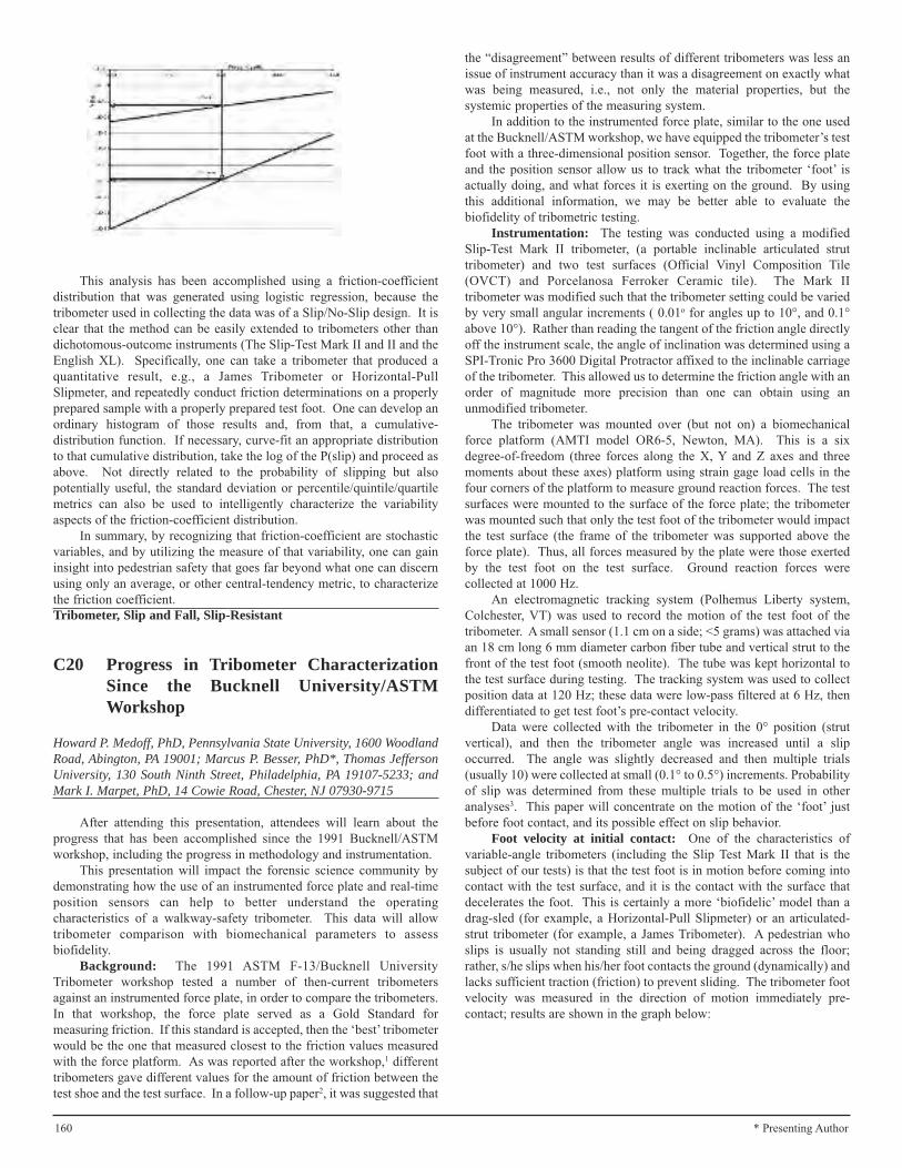
This analysis has been accomplished using a friction-coefficientdistribution that was generated using logistic regression, because thetribometer used in collecting the data was of a Slip/No-Slip design. It isclear that the method can be easily extended to tribometers other thandichotomous-outcome instruments (The Slip-Test Mark II and II and theEnglish XL). Specifically, one can take a tribometer that produced aquantitative result, e.g., a James Tribometer or Horizontal-PullSlipmeter, and repeatedly conduct friction determinations on a properlyprepared sample with a properly prepared test foot. One can develop anordinary histogram of those results and, from that, a cumulative-distribution function. If necessary, curve-fit an appropriate distributionto that cumulative distribution, take the log of the P(slip) and proceed asabove. Not directly related to the probability of slipping but alsopotentially useful, the standard deviation or percentile/quintile/quartilemetrics can also be used to intelligently characterize the variabilityaspects of the friction-coefficient distribution.
In summary, by recognizing that friction-coefficient are stochasticvariables, and by utilizing the measure of that variability, one can gaininsight into pedestrian safety that goes far beyond what one can discernusing only an average, or other central-tendency metric, to characterizethe friction coefficient.Tribometer, Slip and Fall, Slip-Resistant
C20 Progress in Tribometer CharacterizationSince the Bucknell University/ASTMWorkshop
Howard P. Medoff, PhD, Pennsylvania State University, 1600 WoodlandRoad, Abington, PA 19001; Marcus P. Besser, PhD*, Thomas JeffersonUniversity, 130 South Ninth Street, Philadelphia, PA 19107-5233; andMark I. Marpet, PhD, 14 Cowie Road, Chester, NJ 07930-9715
After attending this presentation, attendees will learn about theprogress that has been accomplished since the 1991 Bucknell/ASTMworkshop, including the progress in methodology and instrumentation.
This presentation will impact the forensic science community bydemonstrating how the use of an instrumented force plate and real-timeposition sensors can help to better understand the operatingcharacteristics of a walkway-safety tribometer. This data will allowtribometer comparison with biomechanical parameters to assessbiofidelity.
Background: The 1991 ASTM F-13/Bucknell UniversityTribometer workshop tested a number of then-current tribometersagainst an instrumented force plate, in order to compare the tribometers.In that workshop, the force plate served as a Gold Standard formeasuring friction. If this standard is accepted, then the ‘best’ tribometerwould be the one that measured closest to the friction values measuredwith the force platform. As was reported after the workshop,1 differenttribometers gave different values for the amount of friction between thetest shoe and the test surface. In a follow-up paper2, it was suggested that
the “disagreement” between results of different tribometers was less anissue of instrument accuracy than it was a disagreement on exactly whatwas being measured, i.e., not only the material properties, but thesystemic properties of the measuring system.
In addition to the instrumented force plate, similar to the one usedat the Bucknell/ASTM workshop, we have equipped the tribometer’s testfoot with a three-dimensional position sensor. Together, the force plateand the position sensor allow us to track what the tribometer ‘foot’ isactually doing, and what forces it is exerting on the ground. By usingthis additional information, we may be better able to evaluate thebiofidelity of tribometric testing.
Instrumentation: The testing was conducted using a modifiedSlip-Test Mark II tribometer, (a portable inclinable articulated struttribometer) and two test surfaces (Official Vinyl Composition Tile(OVCT) and Porcelanosa Ferroker Ceramic tile). The Mark IItribometer was modified such that the tribometer setting could be variedby very small angular increments ( 0.01o for angles up to 10°, and 0.1°above 10°). Rather than reading the tangent of the friction angle directlyoff the instrument scale, the angle of inclination was determined using aSPI-Tronic Pro 3600 Digital Protractor affixed to the inclinable carriageof the tribometer. This allowed us to determine the friction angle with anorder of magnitude more precision than one can obtain using anunmodified tribometer.
The tribometer was mounted over (but not on) a biomechanicalforce platform (AMTI model OR6-5, Newton, MA). This is a sixdegree-of-freedom (three forces along the X, Y and Z axes and threemoments about these axes) platform using strain gage load cells in thefour corners of the platform to measure ground reaction forces. The testsurfaces were mounted to the surface of the force plate; the tribometerwas mounted such that only the test foot of the tribometer would impactthe test surface (the frame of the tribometer was supported above theforce plate). Thus, all forces measured by the plate were those exertedby the test foot on the test surface. Ground reaction forces werecollected at 1000 Hz.
An electromagnetic tracking system (Polhemus Liberty system,Colchester, VT) was used to record the motion of the test foot of thetribometer. A small sensor (1.1 cm on a side; <5 grams) was attached viaan 18 cm long 6 mm diameter carbon fiber tube and vertical strut to thefront of the test foot (smooth neolite). The tube was kept horizontal tothe test surface during testing. The tracking system was used to collectposition data at 120 Hz; these data were low-pass filtered at 6 Hz, thendifferentiated to get test foot’s pre-contact velocity.
Data were collected with the tribometer in the 0° position (strutvertical), and then the tribometer angle was increased until a slipoccurred. The angle was slightly decreased and then multiple trials(usually 10) were collected at small (0.1° to 0.5°) increments. Probabilityof slip was determined from these multiple trials to be used in otheranalyses3. This paper will concentrate on the motion of the ‘foot’ justbefore foot contact, and its possible effect on slip behavior.
Foot velocity at initial contact: One of the characteristics ofvariable-angle tribometers (including the Slip Test Mark II that is thesubject of our tests) is that the test foot is in motion before coming intocontact with the test surface, and it is the contact with the surface thatdecelerates the foot. This is certainly a more ‘biofidelic’ model than adrag-sled (for example, a Horizontal-Pull Slipmeter) or an articulated-strut tribometer (for example, a James Tribometer). A pedestrian whoslips is usually not standing still and being dragged across the floor;rather, s/he slips when his/her foot contacts the ground (dynamically) andlacks sufficient traction (friction) to prevent sliding. The tribometer footvelocity was measured in the direction of motion immediately pre-contact; results are shown in the graph below:
160 * Presenting Author

Discussion: In the trials depicted above, slipping first occurred ata friction angle of 27°, and became more likely as the tribometer anglewas increased (P(slip| friction angle = 27°) = 0.20); (P(slip| friction angle= 30°) = 0.90)). An increase in the foot velocity was noted as thetribometer angle was increased. The human analog to this would be anincrease in walking speed. As such an increase in speed increases thelikelihood of a slip in humans, the change in foot speed with change intribometer angle must be considered when considering if a tribometermeasurement is biofidelic.
Ground reaction forces: Ground reaction force curves for a no-slip and a slip trial are shown below:
Discussion: Returning for a moment to the Bucknell workshop, adrag-sled tribometer would exhibit a constant vertical force, with agradually increasing horizontal force, which would plateau when slipoccurred. But considering the “real-world” slip, which usually occurs atinitial contact, we can see that the characteristics of the ground reactionforce is not the quasi-static, gradual application of a force, but instead isan active impulsive loading, that must be considered when assessing slip.
Conclusion: The kinematics and dynamics of loading when usinga tribometer must be considered to assure biofidelity when measuringfriction and resistance to slip.
References:1 Marpet, M. Comparison of walkway-safety tribometers (1996)
Journal of Testing and Evaluation, 24 (4), pp. 245-254.2 Marpet, M., Fleischer, D. Comparison of walkway-safety
tribometers: Part 2 (1997) Journal of Testing and Evaluation, 25(1), pp. 115-126.
3 See: Medoff, HP., Besser, MP., Marpet, MI. (2007): “TheCharacterization of the Slip-Test PIAST Tribometer byCharacteristic Functions Based Upon Logistic Regression”,Conference proceedings of the 59th Annual Meeting of theAmerican Academy of Forensic Sciences, San Antonio, TX,February, 2007, p. 172.
Tribometer, Slip and Fall, Verification
C21 Visual Characterization of TribometricReference Surfaces Using LogisticRegression
Marcus P. Besser, PhD, Thomas Jefferson University, 130 South NinthStreet, Philadelphia, PA 19107-5233; Howard P. Medoff, PhD*,Pennsylvania State University, 1600 Woodland Road, Abington, PA19001; and Mark I. Marpet, PhD, 14 Cowie Road, Chester, NJ07930-9715
After attending this presentation, attendees will have learned of avisually intuitive method, based upon logistic regression, to characterizethe efficacy of a tribometrist’s technique in using a TribometricReference Surface (TRS) set. TRS’s are floor surfaces with specificparameters that are utilized to, calibrate and verify a tribometer (awalkway-friction test instrument), and provide a means of comparing thetest results of different tribometers on a given test surface (not a TRS).
This presentation will impact the forensic science community bygiving researchers and practitioners knowledge of reliability issues intribometric testing, and the background needed to understand thestrengths and weaknesses of their technique in validating a TRS set.
Background: ASTM Committee F-13 on Pedestrian WalkwaySafety and Footwear has been tasked with developing a non-proprietaryStandard Test Method for the evaluation of walkway friction. As oneimportant step, Subcommittee F13.1 on Traction is developing aStandard Test Method for measuring the slip properties of the TRS’s(generically called Standard Reference Materials (SRMs) by the ASTM).Very briefly, to be considered acceptable, a tribometer must (a) rank thedifferent TRS surfaces in the reference set in the proper, predeterminedfriction order (low to high) and (b) be able to discriminate (exhibitstatistically-significant friction differences) between the different TRSsurfaces. Importantly, the specific numeric values obtained in the testingare not in themselves considered an end result. The predeterminedfriction rank order of the TRS surfaces correlates with the human slip asdetermined by human-subject experimentation. Thus, biofidelity isinherent in the choice and ranking of the TRS set. It is noted that theofficial TRS set, i.e., the SRMs that are sold and certified by the ASTM,are not yet available for sale. Although the tests and analysis in thispaper use similar (if not identical) TRS surfaces as will be available fromASTM International, it is important not to infer anything vis à vis theSRM set based upon these tests. Rather, the user can repeat these testsusing the official SRMs, for test-process verification and quality control.
Logistic Regression: This presentation examines a process bywhich tribometry-instrument users can gain insight into the efficacy oftheir techniques using the TRS set and analyzing test results usingLogistic regression.
Logistic regression is a mathematical modeling tool that allows oneto determine the “best” parameters in a model that predicts adichotomous (YES/NO) result using continuous independent, predictor
161* Presenting Author

variables. Here, the dichotomous variable is whether there will be a slipor not (the probability of a slip), and one (if not the) independent variableis the friction between the shoe (or test foot) and the surface (or testsurface or the TRS) as determined by the tribometric instrument. Thelogistic regression parameter of interest here is the logistic slope of thecurve: the larger in magnitude the slope parameter, the finer thediscrimination between the slip and no-slip conditions on that TRS.Interestingly, the intercept parameter in the model is of no interest in ourapplication. The logistic-regression equation is:
TRS Testing: The testing was conducted using a modified Slip-Test Mark II tribometer, (a portable inclinable articulated struttribometer) and four TRS’s (Official Vinyl Composition Tile (OVCT),Porcelanosa Ferroker Ceramic tile, Stone Peak Ceramic Tile, andpolished black granite). The Mark II tribometer was modified such thatthe tribometer setting could be varied by very small angular increments( 0.01o for angles up to 10°, and 0.1° above 10°). The angle of inclinationwas determined using a SPI-Tronic Pro 3600 Digital Protractor affixedto the inclinable carriage of the tribometer. The tribometer angle wasincreased until a slip was noted, the tribometer’s slip angle was backedoff, and sets of successive readings were taken (typically n=10 at eachsetting) as the tribometer’s friction angle was increased, typically by 0.1°in order to generate the logistic regression values for each TRS. Here arethe results, again noting that these results should not be consideredapplicable to the ASTM-certified SRM set:
It was found that the two ceramic tiles and the OVCT had steep anddistinctly separated logistic curves. On the other hand, the PolishedBlack Granite had a shallow logistics curve that crossed over all three ofthe other TRS curves. This is a clear indication that (a) the experimentaltechnique used in with the Polished Black Granite was wanting or, (b)that the Granite was not the same as the granite used in the ASTM’s SRMset.
Conclusion: It is concluded that, beyond the basic Analysis of Variancetools that must be utilized to ensure that the rank-order and statistically-
distinct requirements that must be met to ensure TRS-set validation,characterization of the TRS set by logistic regression is a usefultechnique to enable the researcher or practitioner to determine at a glancewhether any problems exist in TRS set and, further, to show where thoseproblems lie.Tribometer, Slip and Fall, Logistic Regression, Logistic Regression
C22 Comparison of Slip Resistance of TreadPlate and Smooth Steel With VariousFinishes Using a Variable IncidenceTribometer (VIT)
Keith E. Vidal, MSME*, Vidal Engineering, LC, PO Box 31875, St.Louis, MO 63131
After attending this presentation, attendees will understand: howthe slip resistance of tread plate (aka diamond plate) steel compares toflat plate steel, and how paint coatings can dramatically affect the slipresistance of both tread plate and flat steel products.
This presentation will impact forensic science community byshowing that physical design differences of walking surfaces, as well ascoatings on those surfaces can dramatically affect the slip resistance ofthe surface.
This study was the result of a slip and fall accident that took placeon a hydraulic lift gate installed on a truck that was used for transportingused tires. The lift gate had been manufactured approximately fivemonths before the incident. Although optional surfaces were available,the lift gate was originally manufactured with a flat plate-steel surfacecoated with a primer. It was subsequently painted with an acrylic enamelpaint and then installed onto the truck. The truck was driven to variousautomobile tire shops where worn out tires were picked up to berecycled. Many of the tires had varying amounts of rainwater within thecarcass, which would splash out when being thrown onto the truck.There were also intermittent rain showers the day of the incident.
At the time of the slip and fall, the lift gate was elevated to the samelevel as the bed of the truck, several feet high. A man wearing workboots was standing on the surface of the lift gate and was observed bywitnesses with his feet above his head and his back parallel to the liftgate surface. As his body fell onto the surface, the back of his head hitthe edge of the lift gate resulting in serious injuries. The surface of thelift gate was reportedly wet at the time of the fall.
It was hypothesized that the painted finish of the lift gatecontributed significantly to the likelihood of a slip on the lift gate, andthat several of the various lift gate options available were more slipresistant and thus safer under foreseeable and expected conditions of use.Since the accident had taken place three years prior to the engineer’sinvolvement, the original surface material and condition were no longeravailable for testing.
Test Method: Samples of tread plate and flat plate-steel wereobtained and prepared for slip resistance testing. A sample of eachmaterial was left uncoated; coated with the same primer used on the liftgate; and coated with black acrylic enamel, the same enamel the lift gatewas coated with. Each of the surfaces were tested dry and wet using anEnglish XL tribometer (aka Variable Incidence Tribometer (VIT) or aVariable Angle Tribometer (VAT)) to determine the slip resistanceproperties of the various uncoated and coated materials. Testing wasperformed in accordance with ASTM Standard F-1679 and the VITmanufacturer’s operating instructions and use manual. Each sample wastested at least four times and the tests results were averaged.
162 * Presenting Author

Results:Material description Avg. Slip resistance (Dry/Wet)Smooth Bare Steel .93 .42Primed Smooth Steel .79 .48Painted Smooth Steel .89 .26Bare Tread Plate .78 .68Primed Tread Plate .76 .72Painted Tread Plate .76 .60
Discussion: There are at least two consensus standards that putforth guidelines for slip resistance levels on walking/working surfaces.ANSI A1264.2 - Provision of Slip Resistance on Walking/WorkingSurfaces has published the guideline value of 0.50 as a reasonable levelof slip resistance for walking/working surfaces. NFPA 1901 – Standardfor Automotive Fire Apparatus sets a minimum level of slip resistanceunder wet conditions for exterior surfaces as measured with the VIT,as 0.68.
Results of the slip resistance testing showed that the smooth steelsurface was marginal to unacceptable (depending on the above testmethod criteria used), in the bare (.42) and primed (.48) condition underwet conditions but otherwise acceptable dry (.93 and .79 respectively).The acrylic enamel painted smooth steel surface was acceptable dry (.89)but significantly more slippery when wet (.26). All of the diamond platesamples, except the painted (.60), were found to have acceptable slipresistance properties in both the wet and dry condition.
Conclusion: The evaluation of the various surfaces using the VITtribometer illustrates the utility of this particular tribometer as a tool inassessing the safety of a walking/working surface. The hypothesis thatthe painted smooth steel surface contributed to cause the slip in this case,and that other options were safer, was supported through test evaluationof the available surfaces under foreseeable conditions.Slip Resistance, Slip and Fall, Tribometer
C23 Practical Engineering Applied to Aid inAssessing Walkway Slip Resistance
Ronald F. Zollo, PhD*, University of Miami, College of Engineering,1251 Memorial Drive, Room 322, Coral Gables, FL 33124
After attending this presentation, attendees will have both abackground and a working knowledge of what is required to avoid theconsequences of improperly specified, constructed, or maintainedwalkway surfaces by learning how to evaluate design alternatives as afunction of architectural type of occupancy and conditions of use as slipresistant. Attendees will also understand, how to quantify limits of slipresistance on a scientific basis, how to choose among available slipresistance measurement technologies, and how to best choose andqualify experts to assist both in the choice of a walkway surface and inthe prosecution or defense of personal injury claims.
The presentation will impact the forensic science community byhelping assure that the constructed environment is built and maintainedto a standard related to the type of use and conditions of occupancy thatis suitable to the interests of public safety.
The subject of walkways and slip resistance is well known for thebreadth of opinion that it attracts. Unfortunately, the state of the art onthe subject provides little aid and comfort to walkway design decisionmakers. Talk (opinion) is cheap in comparison to the responsibility, orwhat lawyers call duty, that accrues to those who make the decision thatdetermines the quality of a walkway surface. The walkway choicedecision makers, “the choosers” for purposes of this discussion, havebackgrounds that range from naive property owners or their agents tomore sophisticated owners and architectural engineers.
The paper is intended both as an aid to benefit choosers who bearwalkway safety decision responsibility and as an aid in the resolution of
disputes arising from claims of damage resulting from personal injury.The focus of the discussion is to provide practical guidance, based onexisting standards, published research as to what is acceptable and whatis defensible regarding the choice of a walkway surface. The goal is tohelp simplify what can be an overwhelmingly complicated subject byproviding pragmatic advice to the choosers and litigators who wish towork in the public interest.
The presentation proposes a common language through definitionson which to base the discussion. It gives historical perspective leadingup to the state of the art regarding what is suitably deployed as awalkway surface based on the architectural type of occupancy andconditions of use. It discusses, as an aid to understanding, why statics isappropriate rather than dynamics in the application of mechanics to thedescription of the physics of not slipping, though a decision on thismatter is not actually required for assessing walkway safety issues. Itposes and then answers rhetorical questions such as: what is therationale and practicality for an approach that establishes a singleparameter as a measure of walkway safety, what is the nature of theforces that represent slip resistant conditions, how can slip resistance bequantified and evaluated experimentally, how does one choose amongthe available technology for tribometry, and finally how are expertschosen to who can best perform the testing and analysis related towalkway performance. It divides the walkway system as itmetaphorically is and can literally be argued to be that which existsbelow the walkway solid material interface (excluding porosity), and thatover which little or no control is exerted including at the interface, ascontamination or non-adhered covering, and that which exists or ishappening above the interface.
The conclusions support the use of the force ratio, determined as thehighest (limiting) value of a traction force that can be applied to anapproximately level surface divided by the simultaneously appliednormal force, both before slip occurs which is termed the Coefficient ofSlip Resistance (CSR). It also discusses jurisprudence related issueswithout preference given to either those responsible for the choice ofwalkway surface, or to those prosecuting on behalf of or those defendingagainst claims of defective conditions of either construction ormaintenance resulting in personal injury claims.Slip Resistance, Walkway Safety, Traction Tribometry
C24 The Role of Lateral Shear Force in theRequired Coefficient of Friction forLevel Walking
Wen-Ruey Chang, PhD*, Chien-Chi Chang, PhD, and Simon Matz, MS,Liberty Mutual Research Institute for Safety, 71 Frankland Road,Hopkinton, MA 01748
The goal of this presentation is to help better identify the mostcritical instants that a slip and fall incident could potentially occur duringlevel walking.
Traditionally, only the longitudinal component of the shear force atthe shoe and floor interface has been used in calculating the requiredcoefficient of friction and the lateral component of the shear force at thesame interface has been often ignored by most researchers for simplicity.
This presentation will impact the forensic science community byshowing how the lateral shear component at the shoe and floor interfacecould play a critical role in determining the instant that a slip and fallincident might occur.
Slips, trips, and falls are a serious problem. Occupational injuriesdue to slip, trip, and fall incidents remain a leading source of losses inworkers’ compensation (Leamon and Murphy, 1995). The annual directcost of occupational injuries due to slips, trips, and falls in the UnitedStates is estimated to exceed six billion U.S. dollars (Courtney et al.,2001). Falls on the same level accounted for 65% of claim cases, and,
163* Presenting Author

consequently, 55% of claim costs in the total direct workers’compensation for the occupational injuries due to slips, trips, and falls(Leamon and Murphy, 1995).
Required friction is the minimum friction needed at the shoe andfloor interface to support different types of human activities. When therequired friction for an activity exceeds the available friction at the shoeand floor interface, a slip may occur (Redfern et al., 2001). The availablefriction represents the maximum frictional force that can be supportedwithout a slip at the shoe and floor interface. The required coefficient offriction (RCOF) is typically measured on dry surfaces with a force plateand is one of the maximum values in the friction coefficient obtained bydividing the component of the measured ground reaction force (GRF)tangent to the floor surface by the normal component during a step(Redfern et al., 2001). A mechanical device, known as a slipmeter, istypically used to measure the available coefficient of friction (ACOF) atthe contact interfaces (Chang et al., 2001a; 2001b). Various models toestimate slip probability based on the comparison of the RCOF andACOF have been published in the literature.
Traditionally, only the longitudinal component of the shear force atthe shoe and floor interface has been used in calculating the RCOF inwhich the longitudinal component was divided by the normal componentat the same instant. The lateral component of the shear force at the sameinterface has been often ignored by most researchers for simplicity. Theargument for this neglect was that the longitudinal component of theshear force is usually much larger than the lateral component. Therefore,the contribution of the lateral component to the total magnitude of theactual shear force which is the vector sum of both the longitudinal andlateral components was negligible.
This general assumption might be true in many of the cases.However, the results from this experiment on level walking show thatsome walks exhibited very different results with a large lateralcomponent shear force compared with its longitudinal component in theearly part of a heel contact. Under these circumstances, the lateralcomponent should not be ignored, and the instantaneous coefficient offriction reached its maximum near the instant when the lateral shearcomponent also reached its maximum. Therefore, it triggered themechanism for determining the RCOF. In comparison with the casesthat the RCOF was triggered by the longitudinal component, thosetriggered by the lateral component usually happened earlier in a gaitcycle than those triggered by the longitudinal component. Perkins(1978) reported that the shoe started slipping forward approximately 0.1second after a normal landing in a typical severe slip on slippery surfacesin their experiment. Slip at the instant of heel contact was not verycommon, according to his results, but it usually led to an irrecoverableslip. The lateral shear component might help explain why some slipsoccurred early in a gait cycle, but some did not.Lateral Component of Shear Force, Slips and Falls, RequiredCoefficient of Friction
C25 Utilized Friction During Bathtub Entry/ExitUnder Dry and Wet Conditions
Gunter P. Siegmund, PhD*, MEA Forensic Engineers & Scientists, 11 -11151 Horseshoe Way, Richmond, BC V7A 4S5, CANADA; James E.Flynn, BS, J2 Engineering Inc., 7636 North Ingram Avenue, Suite 108,Fresno, CA 93711; Daniel W. Mang, BSc, School of Human Kinetics,University of British Columbia, 210 - 6081 University Boulevard,Vancouver, BC V6T 1Z1, CANADA; Dennis D. Chimich, MSc, MEAForensic Engineers & Scientists, 11 - 11151 Horseshoe Way, Richmond,BC V7A 4S5, CANADA; and John C. Gardiner, PhD, MEA ForensicEngineers & Scientists, 23281 Vista Grande Drive, Laguna Hills, CA92653
After attending this presentation, attendees will understand how thefriction used while entering and exiting a typical bathtub varies under dryand wet conditions between young, middle, and old age individuals.
This presentation will impact the forensic science community byproviding forensic slip and fall investigators with baseline informationon the friction levels used when entering and exiting a dry or wetbathtub.
Bathtubs and showers are a common source of unintentional slipsand falls. Entering and exiting a bathtub requires an individual to stepover the tub’s apron as they transition between two different andpotentially slippery surfaces. The goal of this study was to quantify thefriction used by barefoot subjects entering and exiting a typicalbathtub/shower enclosure under dry and wet conditions. It ishypothesized that the complexity of simultaneously stepping over thebathtub apron and dealing with a potentially slippery surface wouldproduce lower frictional demands in older subjects than in young andmiddle-aged subjects, particularly under wet conditions.
Sixty subjects (30F, 30M) distributed equally in three age groups(20-30 yrs, 40-50 yrs, 60-70 yrs), entered and exited a slip-resistantbathtub using six movement patterns simulating actual use (threeentering and three exiting the tub). Each subject repeated eachmovement pattern five times under blocked dry and wet conditions,yielding 60 trials per subject. For bathtub entry, the first movementconsisted of a single forward step over the tub’s apron starting andending in a standing position; the second movement consisted of a singleforward step combined with a 90º left rotation to stand facing the faucet;and, the third movement consisted of multiple forward steps approachingthe tub combined with a 90º left rotation on the last step over the tubapron to stand facing the faucet. The three exiting movements were thereverse of the three entry movements. Force plates (Bertec 4060,Columbus, OH) installed in the slip-resistant tub floor and the slip-resistant deck immediately outside the tub measured ground reactionforces, from which utilized friction and double support times werecalculated. Force plate data were acquired at 1 kHz and peak utilizedfriction during heel strike and toe off of each limb was obtained from a50ms running average of the instantaneous ratio of shear force tonormal force.
Subjects completed all trials without tripping, falling, or reachingfor either of two grab bars. No obvious slips were observed and nosubjects reported slipping. Overall, utilized friction varied from 0.102 to0.442 (0.235 ± 0.057) and was 0.058 ± 0.040 lower in wet conditionsthan in dry conditions across all movement patterns (p<0.0001). Oldersubjects used less friction than young subjects during exiting movementsonly (p<0.006). Utilized friction did not vary between genders (p>0.14).Double support times were longer in older subjects than in both youngand middle-aged subjects for all movement patterns (p<0.0009) andlonger under wet conditions than under dry conditions for all entrymovements (p<0.0001).
The combination of lower friction and longer double support timesin wet conditions suggests that subjects regard the wet condition as morehazardous than the dry condition and adapt their utilized frictionaccordingly. The results also suggest that older subjects are morecautious than young subjects when confronted with the dual task of bothstepping over the tub’s apron and transitioning to a surface perceived tobe more slippery.Slip and Fall, Friction, Bathtub
C26 Deformation Mechanisms of Walk-Off Mats
Anastasia D. Micheals, MS*, Forensic Materials Consulting, 1784Sanchez Street, San Francisco, CA 94131-2741
After attending this presentation, attendees will understand thedeformation mechanisms of contemporary floor mats (also called walk-off mats). Attendees will comprehend the nomenclature used to describemat deformations. Attendees will also become aware of the variousforces present in use, laundering, and delivery, which may contribute to
164 * Presenting Author
deformation. In addition, attendees will understand how these principleswere applied in defense of a trip and fall injury case.
This presentation will impact the forensic science community byproviding a tutorial of failure mechanism of materials as related to walk-off mats.
Floor mats, also called dust mats or walk off mats, are ubiquitous inour society. They may be encountered many times during a day; at thecoffee shop, grocery store, office building, and school. Sizes vary;typical dimensions are 2 feet by 3 feet, 4 feet by 6 feet or 4 feet by 10feet. Frequently placed at the transition from outdoors to indoors, themats appear as a safety device to prevent tracking water onto surfaceswith low coefficients of friction, i.e., floors which are slippery when wet.But, an additonal rationale behind their widespread placement is thatthey remove and trap dirt, debris, and moisture at the building entryway,reducing building cleaning costs. Mats are typically rented from acompany that services the mats periodically, by removing and launderinga soiled mat, and replacing with a clean mat.
Contemporary floor mats are composites made of a rubber basewith a fiber carpet pile. While specific material formulations vary, atypical rental mat consists of a nitrile rubber backing and a nylon pile.The pile may be attached to the rubber backing by various mechanisms,depending on manufacturer. For example, individual tufts of the pilemay be directly bonded to the rubber by thermomechanical means, ormay be bonded by means of a woven or non-woven pile substrate. Dueto the nature of the materials they are made of, mats can fail by severalmechanisms:
• Delamination – separation of the pile from the substrate;• Substrate Degradation – changing the properties of the
rubber substrate through exposure to chemicals, UV light, orheat; or
• Rippling – permanent deformation of the mat caused byunequal stretching the polymer bonds of the substrate pasttheir limit of plastic deformation.
These mechanisms, which degrade the properties of the mat, occurwith time, and depending on the exact conditions of use and care. Thefloor-mat manufacturing and rental industries recognize that mats have afinite lifetime, and that they must be periodically removed from serviceand replaced.
During duty, cleaning and placement, the mat is subjected to variousforces. The forces can result in elastic or plastic deformation of the matsubstrate, depending on their magnitude. During use, frictional forceshold the mat to the floor; tensile and compressive forces are generated bypeople walking on the mat; and torsional forces are generated if the matis not lying flat on the floor. These forces are generally small enough tocause only elastic, or non-permanent deformation. During laundering,the mats are agitated with soap and water, rinsed, and spun dry, inindustrial washing machines. During water extraction by spinning, themats experience the largest forces. For a mat lying flat against the wallof the washer barrel, the centripetal spinning force becomes a simplecompressive force through the thickness of the mat, similar to thatcreated by placing a weight on it during use. However, if the mat becametwisted or rolled over during washing, a bending force is experiencedduring spinning, which is maximized on the outer surface of the bend.Centripetal force, coupled with the weight of additional wet mats, canexceed the elastic strength of the material, break polymer bonds andcause permanent deformation. Ripples that prevent the mat from lyingflat can thus develop over multiple laundering cycles. Standards forlaundering are specified by the mat manufacturer. Care is taken tospecify parameters such as laundering temperature, soap formulation andamount, the weight of mats to be placed in each pocket of the washingmachine, and the spin speed.
In one case, a rental company placed a mat at the doorway of acounty museum. An 80-year-old woman tripped on a ripple in the mat,fell, and broke her hip. It was found that although the mat rentalcompany observed the manufacturer instructions regarding laundering ofits mats, they failed to have a formal inspection program in place to
ensure that rippled mats were removed from service.Walk-Off Mats, Deformation, Rubber
C27 Exploding Pool Filters That Can Kill
Robert N. Anderson, PhD*, RNA Consulting, Inc., 27820 Saddle Court,Los Altos Hills, CA 94022; and Kevin Lancaster, JD*, The Veen LawFirm, 711 Van Ness Avenue, Suite 220, San Francisco, CA 94102-7296
After attending this presentation, attendees will understand designfailures that are present in certain pool filters and their potential forinjury. Attendees also will learn of the legal issues involved inpresenting such cases.
This presentation will impact the forensic science community byproviding insight into a forensic engineering evaluation of swimmingpool filters. The major legal issues and case workup will be discussed,as well as, the engineering obligation to notify the Consumer ProductSafety Commission of the problems.
During the course of accident evaluation of pool filter failures, itwas determined that design flaws existed that did not provide safetyprotection during routine servicing of the filter. The Consumer ProductsSafety Commission has had 18 serious injuries and 4 deaths reported.All the injuries have involved the kettle-style filters and the incidentsoccurred when the persons doing the maintenance had finished cleaningthe cartridges inside the filter, put the top back on, and started the pump.For various reasons, the compressible air was not vented, and the bandthat clamps the top of the filter to the bottom was not tightened properlyor failed to clamp. When this occurred, the compressed air forced the topto fly off, resulting in death or serious injury to the person it struck. Ifthe air had been vented, then an incomplete seal of the top to the bottomwould only have resulted in a water leak. The failure of pool filters isassociated with the following conditions:
• Failure of automatic air venting valves.• Short and wide kettle-style filters.• Failure in the case where the top and bottom flanges are held
in place by adhesives.• Body clamp failure due to design or failure to tighten.• Placing essential controls too close to the pool filter.
Automatic venting valves operate through the means of a float thatcloses off the vent when the water in the filter rises to the top. If the floatis pushed up by the rapid flow of air and becomes stuck to the top of thevalve, then it fails to vent the air. Unfortunately, it is not possible to pre-determine if the valve is functioning properly and air has been released.
The short, wide filters are dangerous because as the diameterincreases, the area increases as the square of the diameter, and the forceon the lid increases accordingly. The safest design is tall, narrow filters.
Adhesive failures are due to quality control where the adhesive isnot applied uniformly or the adhesive breaks down due to thermal fatigueand mechanical stress.
The body clamps must be robust so that aggressive tightening doesnot cause failure. Some designs lack such ruggedness. The clamptightening mechanism relies on the individual to determine when theclamp is adequately tight. Under-tightening and over-tightening caneasily occur. The safest pool filters use bolts to hold the top in placerather than clamps.
The location of switches and timers in close proximity to the poolfilter puts the operator at risk and is a factor in causing most ofthe injuries.Pool Filters, Explosions, Clamp
165* Presenting Author

C28 Scientific Perspective on Frye and DaubertWith Respect to the NAS Report
Harold E. Franck, MSEE, PE*, Advanced Engineering Associates, Inc.,4713 MacCorkle Avenue, Southeast, Charleston, WV 25304
After attending this presentation, attendees should gain someinsight into the necessity for standards in the engineering sciences andthe lack of standards in engineering sciences.
This presentation will impact the forensic community byencouraging the development of standards, protocols, and guides.
Engineering and science have a variety of goals, which are readilyunderstood by the general public. Some of the most important goals,such as the protection of life and property, are not so well known orunderstood. In engineering and in science there are ethical standards towhich practitioners adhere. In the context of forensic science,investigations are carried out to determine the events that led to theincident and in many instances, to develop methods of avoidance. At thepresent time there are two standards that are used by the courts todetermine the validity of the expert’s testimony.
The Frye Standard stems from a 1923 case that established theminimum standard required for the admission of expert testimony infederal cases. This standard requires the expert to use data andmethodology “generally accepted” by other experts. In the Daubert casein 1993 the evidence that was presented by the plaintiff was consideredto be novel scientific evidence or junk science. Therefore, this novelscientific evidence did not qualify under the Frye Standard as admissibleexpert testimony. In the U.S. Supreme Court appeal the lower courtrulings were overturned and a new standard was developed where thereliability of the evidence must meet a non-exclusive four part test.
• Can the theory or technique be tested?• Have they been subjected to peer review and publication?• Is there a known or potential rate of error?• Is there general acceptance in the scientific communit similar
to the Frye Standard?On November 22, 2005, the Science, State, Justice, Commerce, and
Related Agencies Appropriations Act of 2006 became law. Congressauthorized the National Academy of Sciences to conduct a study onforensic sciences. The Senate Report set forth many charges to theforensic sciences community including to disseminate best practices andguidelines concerning the collection and analysis of forensic evidence tohelp insure quality and consistency in the use of forensic technologiesand techniques to solve crimes, investigate deaths, and protect the public.One of the issues covered during the committee’s hearings was thefundamental of the scientific method as applied to forensic practice –hypothesis generation and testing, “falsifiability” and replication, andpeer review of scientific publications. Another observation was the lackof mandatory standardization, certification, and accreditation. Thecommittee stated that the fragmentation problem is compounded becauseoperational principles and procedures for many forensic sciencedisciplines are not standardized or embraced. Often there are nostandard protocols governing forensic practice in a given discipline. Onerecommendation is to establish a national code of ethics for all forensicscience disciplines.
It is clear that standards and protocols must be developed for theforensic sciences. In the engineering sciences there are many recognizedstandards in certain fields, but they are utterly lacking in others. Forexample, the fire sciences have a multitude of standards, guides, andprotocols that were developed by ASTM and NFPA. In the engineeringsciences there are but a handful of standards. Some were developed 25years ago and a few others were developed five years ago when a majorpush was made in ASTM to develop such standards. Since then no
standards have been developed, and their development has actually beencurtailed. The AAFS through its long and close association with ASTM,has an opportunity develop standards, guides, and protocols in forensicengineering sciences.Standards, Protocols, Guides
C29 An Accident Reconstructionist’s View ofHow the Rules of Evidence Affect ExpertTestimony
Peter Alexander, PhD*, Raymond Smith & Associates, 4934 WagontrailCourt, Parker, CO 80134
The goal of this presentation is to inform attendees of the flaws inthe judge’s gatekeeper role regarding admissibility of scientifictestimony.
This presentation will impact the forensic science community byproviding case studies of application of Daubert ruling in civil litigationinvolving scientific conclusions.
The National Academy of Sciences (NAS) has recently issued a 254page draft report stressing the need to strengthen forensic scienceactivities in the United States. Among the areas mentioned were:
• The lack of standardization of procedures;• The lack of mandatory certification, and accreditation
of practitioners;• The lack of established accuracy limits for procedures;• The admissibility of forensic evidence in litigation;
This paper deals with the last item, the admissibility of accidentreconstruction data and conclusions used at trial which are drawn fromthat data.
The NAS Report recounted the Frye standard developed in 1923and the 1975 Federal Rules of Evidence – Rule 702 which led up to the1993 Daubert decision. Daubert established several standards (citedbelow) that the trial judge could consider with regard toexpert testimony.
1) Whether a theory or method has been tested;2) Whether it has been subjected to peer review and publication;3) Whether it has a known rate of error;4) Whether there are standards controlling the technique;5) Whether it is accepted in the scientific community;
When one side in a lawsuit challenges the other side’s expert basedon the Daubert decision, the judge is required to act as the “gate keeper”and rule on the admissibility of the presentation in terms of the abovecriteria, in order to prevent the jury from being exposed to unsound orunscientific conclusions. Often during a Daubert challenge the judge ispresented with conflicting, rather technical, testimony from experts oneach side. One side may contend that the other expert’s conclusions areinvalid because they rely on junk science or faulty assumptions. Thisprocess is an exercise in fairness to ensure that one side in a court casedoes not influence the jury with testimony that is not sound or accurate.How well this process works for the accident reconstructionist isdiscussed below.
On occasion the judge shirks his gatekeeper role and decides to letthe jury decide the merits of each side’s expert conclusions. Sometimesit appears that the judge may feel that he is insufficiently technicallyastute to render a decision. Unfortunately, a jury is even less qualified tomake this judgment than the judge. When the judge fails to fulfill thisrole, the jury is presented with two experts who each sound veryconvincing but have contradictory scientific conclusions. In such asituation it is possible for justice to be perverted. Two case studiesillustrating this process are detailed below.
166 * Presenting Author
Case Study 1: This case involved a rear impact to the stoppedplaintiff’s 1992 Suzuki Sidekick, by a 1997 Jeep Grand Cherokee.Roughly $1500 in damage was suffered by each vehicle. Physiciansattributed cervical and mild brain injuries to the plaintiff resulting fromthis collision.
The plaintiff’s expert placed the Jeep’s impact speed at >10 mph,resulting in a 5 mph. speed change of the plaintiff’s vehicle. The femaleplaintiff was restrained and turned when she was hit by surprise fromthe rear.
The defense expert concluded that the impact speed was <5 mph,resulting in a 4g peak acceleration to the occupant. The expert citedstaged rear impact crash tests at this speed, in which the humanvolunteers were uninjured. The expert also stated that the forces in thiscollision were no greater than those experienced during normal dailyactivities. The defense expert’s conclusions were based on a numericalmodel which was used only by that expert and had never beenindependently tested or validated. The model was capable of producingany result the expert wanted, depending on his choice of key inputparameter values.
Previously the State Supreme Court had ruled that comparing forcesin a collision to forces related to daily activities was junk science andinadmissible. Likewise the Supreme Court had also ruled thatcomparison of the lack of injuries incurred in staged crash tests with thelikelihood of injury in an actual collision was junk science.
During the hearing, in this case, challenging the defense’s right topresent this data to the Jury, the judge abdicated his responsibility anddecided to let the jury decide which expert was right. The jury decidedthat the plaintiff could not have been injured in this impact. If the juryhad known that the defense expert was relying on “junk science” wouldthe result have been different?
Case Study 2: This case involved a 2000 Toyota Avalon whichstruck the plaintiff’s slowly moving 1989 Cadillac Deville from the rear.The defendant claimed she was only traveling at 5 mph when the
plaintiff stopped suddenly. There was $1,000 damage reported to theplaintiff’s vehicle and a precision frame measurement showed that theframe had been bent by the force of the collision. Physicians testified tovarious significant back and neck injuries suffered by the plaintiff inthis collision.
The plaintiff’s expert testified that this collision’s relative impactspeed was in excess of 12 mph. and aggravating factors such as a priorasymptomatic injury, being turned at the moment of impact, and beinghit by surprise contributed to the likelihood of injury to the plaintiff. Theexpert also established that the stated impact speed was a minimum valuenecessary to cause the frame damage noted.
Despite being challenged, the defense expert, a medical doctor, wasallowed to testify regarding her reconstruction of this accident. Sheconcluded that the peak acceleration in this collision was less than 2g andit was unlikely that someone would be injured at that acceleration level.The expert also noted that we live with 1g acceleration exerted on ourbodies. Despite the defense expert having no credentials in the area ofaccident reconstruction, she was allowed to testify as to her conclusionsin that area. Incredibly, the plaintiff’s accident reconstruction expert wasnot allowed to rebut the defense expert’s accident reconstructiontestimony because he was not a medical doctor like the defense’s expert.
The jury found against the plaintiff, agreeing with the defenseexpert that acceleration levels of 2g were unlikely to cause the statedinjuries. The jury never heard that the 4 mph. that would have producedthe 2g acceleration claimed by the defense, never would have bent theplaintiff’s vehicle frame. The jury also never heard that, even if thedefense was correct about the impact speed, a 1g vertical accelerationapplied to one’s body is not comparable to a 2g horizontal accelerationapplied to the head and neck. In addition, the jury never heard that thepeak accelerations experienced by the plaintiff could actually have beenas high as 15g. Would the jury verdict have been the same if they hadbeen allowed to hear these arguments during rebuttal?
Conclusion: How can the possible perversions of justice apparentin the above examples be avoided? It appears that some judges are notcomfortable deciding issues which are technically based. One optionmight be to require judges to have some minimal training in science orengineering. Another possibility might involve providing scienceeducation to sitting judges. Still another option might involve theappointment of a technical advisor by the judge to help him rule on casesinvolving complex technical matters. Attorneys and judges mightprovide different sets of options with regard to this question.Accident Reconstruction, Daubert, Expert Testimony
C30 Biomedical/Biomechanical Analysis ofInjury/Trauma Reported for Restrainedand Un-Restrained Adult and PediatricOccupants Involved in Vehicular RolloverCrashes: A Nominal andStatistical Approach
Laura L. Liptai, PhD*, and Tia L. Orton, BS, BioMedical Forensics,1660 School Street, #103, Moraga, CA 94556
After attending this presentation, attendees will understand thenumerous in-depth biomedical analyses of both adults and childreninvolved in rollover crashes illustrate injury causation and relatedmitigation methods.
This presentation will impact the forensic science community bydocumenting the importance of a nominal crash-specific biomedicalforensic analysis, not simply a reliance on statistical studies, whendetermining injury causation and related mitigation.
Despite effective improvements in vehicular safety, rollover crasheshave continued to pose a significant problem due to their associationwith high injury trauma rates. As a category, rollover crashes constituteover a third of the trauma-related harm although they represent just over2% of the crash population. Approximately 84 percent of the harmassociated with rollovers occurs to unrestrained occupants.
Numerous in-depth biomedical analyses of both adults and childreninvolved in rollover crashes illustrate injury causation and relatedmitigation methods. In the majority of cases it was specificallydetermined that use of the available restraint system would significantlylower the injury risk. In several cases; however, use of the availablerestraint system did not prevent serious injury/fatality.
This paper documents the importance of a nominal crash-specificbiomedical forensic analysis, not simply a reliance on statistical studies,when determining injury causation and related mitigation. This isparticularly true with rollover crashes which can be highly variableevents with often-erratic parameters.Rollover, Unbelted, Biomechanical
C31 Occupant Excursion and Restraint SystemPerformance in Rollover Testing
Steven E. Meyer, PE*, Arin A. Oliver, BS, and Brian R. Herbst, PE, SAFELaboratories, 6775 Hollister Avenue, Suite 100, Goleta, CA 93117
After attending this presentation, attendees will have a betterunderstanding of the effect of seat belt looseness, or slack, and itsrelationship to occupant excursion during a rollover by evaluatingvarious restraint system designs and configuration in rollover-type testenvironments.
167* Presenting Author

This presentation will impact the forensic science community byproviding effective occupant protection in automotive rollover crashesrequires supplying the occupant with a restraint system proven effectivein this accident mode. Preventing occupant ejection and providingrestraint through the rollover sufficient to prevent potentially injuriouscontacts with interior vehicle components are paramount for effectiveoccupant protection. Research has shown that the potential for injurycan be decreased by closely coupling the occupant to the seat. Thepresented testing studies the ability of various restraint systems tocontrol occupant excursions out of the seat.
Providing effective occupant protection in automotive rollovercrashes requires supplying the occupant with a restraint system proveneffective in this accident mode. Preventing occupant ejection andproviding restraint through the rollover sufficient to prevent potentiallyinjurious contacts with interior vehicle components are paramount foreffective occupant protection. Research has shown that the potential forinjury can be decreased by closely coupling the occupant to the seat. Thepresented testing studies the ability of various restraint systems tocontrol occupant excursions out of the seat. This presentation will giveattendees a better understanding of the effect of seat belt looseness, orslack, and its relationship to occupant excursion during a rollover byevaluating various restraint system designs and configuration in rollover-type test environments.
A series of roll spit tests were performed utilizing production sportutility vehicles (SUVs) with a female occupant (5th to 25th percentile)positioned in one of the front seating positions. Measurements weretaken of the occupant’s initial upright clearances to the surroundingvehicle structure. The vehicle occupant compartment was then rotated toan inverted orientation, 180 degrees of rotation (unless otherwise noted),and the excursions and clearances were recorded. Various restraintsystems were considered and subjected to testing, including the originalequipment manufacture (OEM) as well as other restraint systems designsseen in production. The observed results of the testing are summarizedin the following Table.
The testing demonstrates that occupant excursions in vehiclerollover circumstances are dramatically affected by the chosen restraintsystem. Slack in an OEM restraint system increases the occupantexcursion from that of a normally tight OEM restraint system. Thisadditional excursion can greatly increase the occupant’s potential forinjurious contacts with the interior of the vehicle during a rollover and/orcontribute to the occupant’s partial or full ejection. Various restraintsystem components added to the tested vehicles demonstrate a decreasein the occupant’s excursion; and therefore, a decrease in injury potential.Simply adding a cinching latch plate, even allowing slack in the torsobelt, is seen to prevent vertical excursions beyond that of a tight beltconfiguration. A pretensioned belt or all-belts-to-seat (ABTS) restraintsystem shows a large decrease in the amount of occupant excursion.Rollover, Restraint, Excursion
C32 Comparison of Hybrid III Dummy NeckStiffness to Actual Human Neck InjuryTolerance for Axial Compressionin Rollovers
Michelle R. Hoffman, MS*, and Carley C. Ward, PhD, BiodynamicsEngineering, Inc., 3720 East La Salle Street, Phoenix, AZ 85040; HrireDer Avanessian, PhD, Biodynamics Engineering, Inc., 17383 SunsetBoulevard, Suite A300, Pacific Palisades, CA 90272; and Brian R.Herbst, PE, SAFE Laboratories, 6775 Hollister Avenue, Suite 100,Goleta, CA 93117
The goal of this presentation is to examine the Hybrid III dummyneck stiffness and compares it to the human neck. After attending thispresentation, attendees will understand the difference between dummyand human neck response to axial loading and its significance.
This presentation will impact the forensic science community byshowing that a value of at least 10,500 N is an appropriate representationfor upper neck axial load in the Hybrid III dummy as a threshold forpredicting human cervical spine injury.
Over 10,000 people die annually in the United States from rollovermotor vehicle collisions, and more than 16,000 people suffercatastrophic injury. Most of the research on cervical injury potential inrollover accidents has utilized the Hybrid III anthropomorphic testdevice (ATD), aka, the Hybrid III dummy. This presentation examinesthe Hybrid III dummy neck stiffness and compares it to the human neck.After attending this presentation, attendees will understand thedifference between dummy and human neck response to axial loadingand its significance.
Although it is the most advanced ATD currently available, theHybrid III dummy is not a good surrogate for neck injury investigationsduring rollover collisions. Inadequacies of the Hybrid III dummy neckhave been reported in the literature. The dummy neck geometry isclearly not the same as a human neck. The human neck is articulatedwith joints while the dummy neck is a non-jointed structure, which leadsto dynamic dissimilarity. In fact, the Hybrid III in current use is a scaled-down, cheaper version of the original GM design. The dummy neck istoo stiff in the axial direction when compared to the human neck. Noone-to-one relationship between the dummy and the human neck withrespect to forces experienced and mechanism of injury has been widelyaccepted. Despite this, extensive use of the Hybrid III dummy inrollover testing has led researchers to infer that the dummy neckresponse is a close representation to that of a human neck. Someresearch even uses a dummy axial neck load as low as 2000 N torepresent injury potential from roof crush.
In the current study, the MADYMO (MAthmatical DYnamicMOdels) computer simulation program was used to establish arelationship between the dummy and human neck response. A one-directional (translational) joint between the lower neck and upper torsowas added to the basic 50th percentile male dummy model to account forthe axial compression response of the dummy neck. The axialcompression properties were selected from tests on the dummy head-neck system as reported by Herbst (1998) who used loading rates from4.5 to 17.9 mph.
Dummy drop tests conducted at SAFE in 2005 were used to validatethis modified MADYMO dummy model. A 50th percentile Hybrid IIImale dummy was inverted and placed in a seated position such that theneck was almost vertical (head 7 degrees rearward of vertical). Thedummy was then dropped, free fall onto a concrete surface covered withlinoleum flooring from heights of 12 inches and 24 inches. The peakupper neck axial load was 8,458 N for the 12-inch drop and 11,344 N forthe 24-inch drop. Figure 1 shows good correlation between theMADYMO model and the drop tests.
168 * Presenting Author

(a) (b)Figure 1. Dummy upper neck axial loads from MADYMO model andSAFE drop tests for (a) 12-inch drop and (b) 24-inch drop.
Because neck tolerance data on living humans is limited, the onlyverified injury measure is from recreational activities such as diving andfootball accidents. This research indicates that cervical flexion-compression injuries can occur at head impacts as low as 10.2 feet/sec(approximately 7 mph). The results of research utilizing cadavers arealso consistent with this number.
The modified MADYMO model was then utilized to simulate a 20-inch drop height to represent a 7 mph impact velocity. The results fromthis validated MADYMO model showed that the neck compressive forceis approximately 10,500 N. When the dummy torso position waschanged to represent a more vertical neck condition (5 degree variation),the loads increased to about 11,500 N. Thus, this study shows a range ofcompressive neck loads between 10,500 to 11,500 N for a 20-inch dropheight, which represents an approximate 7 mph impact velocity.
The drop testing and the MADYMO modeling clearly demonstratethat the Hybrid III dummy neck is much stiffer in the axial direction thanthe human neck. It should be noted that peak force has not beenestablished as a reliable parameter for assessing catastrophic cervicalinjury in the human. However, if one does use peak axial load, thehuman neck is roughly 4,000 N in an inverted drop with an impact speedof 7 mph and the equivalent peak load in a Hybrid III dummy neck undersimilar loading conditions is over 2.5 times that value. The dummy neckinadequacies, as described above, are responsible for this disparity.
The Hybrid III dummy is routinely used for analyzing the forcesand injury potential during rollover accidents with various axial neckloads to predict injury outcome from roof crush. This study shows thata value of at least 10,500 N is an appropriate representation for upperneck axial load in the Hybrid III dummy as a threshold for predictinghuman cervical spine injury.Rollover, Hybrid III Neck, MADYMO
C33 Containment Potential of LaminatedGlazing in High Speed Rollover Testing
Brian R. Herbst, PE*, Steven E. Meyer, PE, and Arin A. Oliver, BS, SAFELaboratories, 6775 Hollister Avenue, Suite 100, Goleta, CA 93117
After attending this presentation, attendees will recognize theretention capabilities of laminated glass door windows for unrestrainedoccupants in a dynamic rollover event.
This presentation will impact the forensic science community bydiscussing how keeping occupants inside the vehicle during a rolloveraccident event is the first step to reducing occupant injury in this accidentmode.
Keeping occupants inside the vehicle during a rollover accidentevent is the first step to reducing occupant injury in this accident mode.While increased restraint use, restraint performance and vehiclestructural integrity have all been identified as being associated withoccupant containment, vehicle glazing is known as the structural
component that is most likely to fail during this accident mode. Vehicleglazing failure usually creates large portals in the vehicle structurethrough which occupants can be ejected. In the presented automotiverollover testing, the original tempered door glazing was replaced withlaminated glazing and unrestrained Anthropometric Test Devices (ATDs)were subjected to high rates of rotation. Under various configurations,ATD kinematics and glazing performance during rollover were recorded.
The performed study includes seven controlled, multi-revolutiontests of a partial sport utility vehicle (SUV) occupant compartment testfixture rotating about a fixed, longitudinal axis. The front doors of thefixture were modified by replacing the original equipment manufacturer(OEM) glazing with a piece of 0.269” thick laminated glassmanufactured by American Glass Products containing a 30 mil PVBinner layer. Although slightly thicker (from OEM thickness of 0.152” to0.269” laminated thickness), the laminated glass was able to fit into theOEM window hardware by slightly expanding the window frame metalflanges. Additionally, the right front door window frame was reinforcedwith a 1.5 to 2.25” wide piece of sheet metal.
Unrestrained Hybrid III 50th percentile male ATDs were positionedin the driver’s and/or passenger’s seating positions with dummykinematics and interaction with the front window glazing observed atroll rates of up to 644 degrees per second. The same driver’s andpassenger’s doors, as well as their associated laminated glazing, wereutilized in all of the seven tests conducted. At various points during thetesting, the laminated glazing on each door was pre-damaged in order tonote the effect of existing fractures. The observed results of the testingare summarized in the following Table.
No separation of the laminated glazing from the door windowsurround was noted as the ATDs migrated through the test fixture duringthe roll event. The undamaged as well as the fractured laminated doorglazing was found to retain the ATDs with no ejection through this portalwith no additional glazing fracture or minimal propagation ofglazing cracks.Rollover, Laminated Glazing, Containment
C34 Increased Risk of Submarining and LowerExtremity Injuries Associated With Obesityin Frontal Impacts
Christopher J. Furbish, BSc*, Biodynamics Engineering, Inc., 3720 EastLa Salle Street, Phoenix, AZ 85040; Parris Ward, JD, BiodynamicsEngineering, Inc., 17383 West Sunset Boulevard, Suite A300, PacificPalisades, CA 90272; and Hrire Der Avanessian, PhD, BiodynamicsEngineering, Inc., 2831 Montrose Avenue #5, La Crescenta, CA 91214
After attending this presentation, attendees will be familiar withvarious risk factors associated with restraint effectiveness relative tobody mass index (BMI), primarily for lower extremity injuries in frontalimpacts.
169* Presenting Author

This presentation will impact the forensic science community byaiding in better understanding occupant kinematics and restrainteffectiveness during frontal impacts.
Between 1960 and 2004, the CDC reports that the obesity rate in theUnited States more than doubled from 13.3 to 32.1 percent for adultsaged 20 to 74. Should the current trend continue to the year 2015, somemodels predict that the obesity rate will reach 41 percent, with as manyas 75 percent of U.S. adults being overweight. After attending thispresentation, attendees will be familiar with various risk factorsassociated with restraint effectiveness relative to body mass index(BMI). Multiple volunteers of various body sizes were placed in thedriver’s seat of a 2003 SUV and measurements of the seat belt and body-to-vehicle interior distances were recorded. These measurementsrevealed trends indicating an increased risk for submarining and lowerextremity injuries for occupants with higher BMI ratings involved in afrontal impact.
Previous studies addressing obesity and injury severity for variousbody regions and crash conditions have revealed some contradictingconclusions. For example, a 2003 study found that the overweightoccupant has a decreased risk of abdominal injury compared to both leanand obese occupants. However, a 2008 study concluded that overweightoccupants (but not obese occupants) are at risk of suffering more severeinjuries than an occupant with a normal BMI. The majority of thesestudies were based on statistical epidemiological analyses of datareported in various databases, such as CIREN, NASS-CDS, etc. Thisstudy addresses the expected kinematics of occupants with various BMIratings based on the seat and restraint geometries, while keeping thevehicle and assumed impact configuration constant. Based on theseexpected kinematic differences, the potential injury patterns and riskswere determined.
The male and female adult volunteers used in this study were ofvarious ages with BMI’s ranging from 20 to 45. A BMI greater than orequal to 30 is generally considered to be obese. Each volunteer wasplaced in the driver’s seat of the SUV and asked to adjust the seat fore/aftposition and seat back recline angle into the most comfortable drivingposition. Multiple measurements of the lap belt were taken relative tothe left and right anterior superior iliac spine (ASIS) locations of thepelvis. Knee-to-dash, abdomen-to-steering wheel, and sternum-to-steering wheel distances were recorded. Additionally, the positioning ofthe seat bottom, seat back, and lap belt relative to the occupantcompartment were documented.
Based on the recorded data, various trends were found as a functionof BMI. As BMI increased, the average seat back angle relative tohorizontal decreased. In other words, the higher the BMI the more likelythe occupant would ride in a more reclined position. The distances in thelongitudinal (x-axis) and vertical (z-axis) directions between the ASISand the most forward portion of the lap belt both increased as BMIincreased. Additionally, the length of the lap belt from the floor anchorto the latch plate increased with an increase in BMI. The data presenteddemonstrates how these trends predispose an occupant with a higherBMI to greater risk of lower extremity injuries and/or submarininginjuries. For occupants of higher BMI, the longer lap belt length and itsincreased distance from the bony pelvis due to additional soft tissuewould allow for a greater amount of lower extremity forward excursionduring a frontal impact. The more reclined seat back position, as well asthe increased height of the forward-most portion of the lap belt, wouldplace a high-BMI occupant at greater risk of submarining as the directionof force from the lap belt shifts up toward the abdominal structures witha higher probability of overriding the pelvis. Additionally, vehiclesequipped with load-limiting energy activating webbing loops in the lapbelt further increase the likelihood of submarining and resultant lowerextremity injury.
Mathematical dynamic models (MADYMO) simulations wereconducted to demonstrate some of the expected differences between theoccupant kinematics for occupants in the normal, overweight, and obeseBMI ranges. Additionally, a real world case study with an occupant
sustaining both lower extremity and submarining injuries was analyzedto demonstrate the potential outcome of these risk factors.Body Mass Index (BMI), Restraint Effectiveness, MADYMO
C35 Assessment of Extended Range ElectronicProjectile Impact Injury Potential UsingCadaveric Testing
Scott R. Lucas, PhD*, Exponent, 3401 Market Street, Philadelphia, PA19064
After attending this presentation, attendees will be familiar with arecent human cadaveric test series that was executed to assess the impactinjury potential of the newly designed XREP (eXtended RangeElectronic Projectile).
This presentation will impact the forensic science community byproviding an assessment of human impact injury potential from ashotgun round with an electronic charge. The results from this study areuseful in assessing the effects of the XREP impact velocity on humantissue and in identifying superficial wounds as a result of XREPdeployment.
The XREP was designed as a wireless electronic neuromuscularincapacitation (NMI) electronic control device (ECD) self containedwithin the XREP projectile. Utilizing NMI, it is intended to neutralize athreat up to 100 feet away as it is deployed from a 12-gauge shotgun. Itis not intended to produce significant impact injuries such as significantblunt trauma or for the cartridge to penetrate the skin. As with anykinetic energy munition, design variables such as mass, velocity, andimpact cross-sectional area must be considered to reasonably minimizepotential penetrating or blunt impact injuries. During the design process,extensive experimentation is necessary to minimize unintended injuries.This paper presents the results from a recent evaluation of blunt trauma
and penetration potential of the XREP design via a series of human torsocadaveric tests.
Two male cadaver torsos were obtained. The torsos were frozenpostmortem, were not chemically preserved, and were completelythawed prior to usage. The stature and mass of the two cadavers were 68inches and 165 pounds and 72 inches and 165 pounds. A nitrogenpowered air cannon was used to fire the XREP rounds throughout thestudy. This air cannon consisted of a pressure regulator and gas reservoirarranged to propel the experimental device through either a smooth-boreor rifled-bore shotgun barrel. A range of cannon firing pressures andcorresponding pre-impact velocities were utilized. These values weredesigned to replicate velocities that would be obtained by firing theXREP with a conventional shotgun. A total of 43 shots impacted thetorsos, including 12 shots on the posterior aspect of Torso 1, 14 shots onthe anterior aspect of Torso 1, and 17 shots on the anterior aspect ofTorso 2. Impact areas included the ribs and abdomen. Two tests werefired at a range of 15 feet and all of the other tests were fired at 1.5 feet.Pre and post-test photographs were taken and high-speed video wasrecorded at locations orthogonal to and oblique to the impact location.Pre and post-test magnetic resonance imaging (MRI) and computerizedtomography (CT) images were obtained. Post-test internal examinationswere performed by dissection.
The measured and calculated XREP pre-impact velocities from thetest series ranged from 230 to 315 feet-per-second (fps). The majority ofthe shots fired were at a cannon pressure of 300 pounds-per-square inch(psi) and resulted in pre-impact velocities averaging 251 ± 6 fps (rangingfrom 244 to 257 fps) for the rifled barrel and 265 ± 11 fps (ranging from234 to 280 fps) for the smooth barrel. In 42 of the 43 shots fired, theXREP impacted the torso and its nose section did not penetrate the skin,resulting in superficial wounds only. Internal examination of these 42shots revealed that the XREP barbed electrode tips penetrated throughthe skin layer and into the superficial fat. At most, the XREP electrodes
170 * Presenting Author

penetrated through the skin and fat and punctured superficial muscletissue as there was no evidence of penetration through the peritoneum onthe deep surface of the muscle layer. On the remaining shot it wasobserved that the nose section of the XREP separated prematurely inflight which then exposed the smaller diameter chassis to impact with thetorso. That shot in particular was fired at the closest range at the highestcannon pressure, and was directed to a relatively fatty portion of theabdomen lateral to the umbilicus. It penetrated the abdominal wall andlodged inside the torso. Separation of the nose section had beenpreviously noted to occur in air cannon firings but not in shotgun firings.In the study, no rib fractures or other bony fractures were observed withany of the shots. When the XREP remained intact to point of contact,there was no evidence of blunt impact trauma or electrode penetrationthrough the abdominal, thoracic or the retroperitoneal wall based uponexternal observation, imaging, and internal examination.Blunt Impact, Kinetic Energy Munitions, Human Injury
C36 The Effect of Restraint Use on Skull VaultFractures in Rollover Crashes
Michael Freeman, PhD*, 205 Liberty Street, Northeast, Suite B, Salem,OR 97301; Lars Uhrenholt, PhD, Institute of Forensic Medicine,Institute of Forensic Medicine, University of Aarhus,Brendstrupgaardsvej 100, Aarhus n, 8200, DENMARK; and CraigNewgard, MD, Oregon Health and Science University, School ofMedicine, 3181 Sam Jackson Park Road, Portland, OR 97239-3098
After attending this presentation, attendees will understand therelationship between roof crush, restraint use, and risk of skull vaultfracture in rollover crashes.
This presentation will impact the forensic science community bydiscussing the belief that this analysis is the first population-based studyto evaluate a specific injury type (skull vault fracture) and associate itwith a specific injury mechanism (contact with a crushed roof) inrollover crashes. These research findings lead to a rejection of the crashtest dummy-based “diving” theory as the primary cause of head injury inrollover collisions with roof crush.
Introduction: Skull vault fractures (SVF) are a seriouscomplication of rollover crashes and there is a question as to whetherthey result from roof crush or “diving” kinematics of the occupanttoward the roof. Based on the effect of three point restraints in limitingoccupant movement in rollovers we hypothesized that progressivelygreater crush would result in a relatively greater increase in the rate ofskull vault fractures in unbelted occupants if diving was the mechanism,and a greater increase in belted occupants if roof crush was the injurymechanism.
Methods: A search was conducted of the National AutomotiveSampling System-Crashworthiness Data System (NASS-CDS) of theUnited States National Highway Traffic Safety Administration (NHTSA)for the years 1997 through 2005, inclusive (9 years total) for rollovercrashes with at least two one quarter turns and in which an occupantsustained a head or neck injury of some degree as a result of roof,windshield headliner, or side rail contact (as opposed to ejection). A casewas only included for analysis if the degree of roof, windshieldheadliner, or side rail intrusion at the position of the occupant wasrecorded and correlated to the head injury, as well as the restraint usestatus of the occupant. Both raw and weighted counts of injuries as wellas occupants were recorded by NASS injury code, and also the numberof occupants with head and neck injuries by injury severity rank wasrecorded. The ratio of skull vault fractures versus other injuries by roofcrush severity was calculated for restrained and unrestrained occupantsand tabulated.
Results: A total of 2,120 injuries were recorded in the NASS giventhe above parameters for 558 restrained occupants (330,056 weighted),
and 1,261 injuries were recorded for 288 unrestrained occupants(143,389 weighted).
Although belt use decreased the rate of serious head injury amongrestrained occupants relative to unbelted occupants in all crashes exceptthose with >61 cm of roof crush, the efficacy of seatbelts was inverselyrelated to the degree of intrusion (see Figure 1). At 3-8 cm of roof crushbelted occupants sustained a reduction of serious or greater head injuriesof 45%, at 8-15 cm serious injury was reduced 40%, at 15-30 cm seriousinjury was only reduced 23%, and at 30-46 cm and 46-61 cm seriousinjury was reduced 15% in each category in belted vs. unbeltedoccupants.
A risk ratio assessment of the effect of belt use vs. roof crush wasperformed. Among restrained occupants SVF comprised 11.9% ofserious and greater (AIS≥3) head/neck injuries (6.4% of all head/neckinjuries), and thus only 0.8% of all injuries at the lowest level of roofcrush (3-8 cm). At 8-15 cm of crush the risk was 1.9 times the lowestlevel, at 15-30 cm it was 2.5 times greater, at 30-46 cm of crush the riskit was 3.5 times greater, at 46-61 cm of crush it was 8.4 times greater, andat more than 61 cm the risk was 6.6 times greater. Among unrestrainedoccupants at the lowest level of roof crush SVF accounted for 20.6% ofall AIS≥3 injuries (10.9% of all injuries in this category were serious),and thus 2.2% of all injuries. Risk ratios, relative to the lowest level,were as follows: 8-15 cm, 0.7; 15-30 cm, 0.9; 30-46 cm, 1; 46-61 cm,1.8; and at >61 cm, 1.6.
Figure 1
Discussion: At the lowest levels of roof crush SVF occurred 175%more often in unrestrained occupants. In contrast, SVF occurred 22%,68%, and 47% more often among restrained occupants in rollovercrashes with 30-46, 46-61, and more than 61 cm of crush, respectively.The explanation for this finding is that SVF results from sudden forcefulloading associated with the higher head accelerations resulting fromincreased roof crush, and a relatively less mobile restrained occupant ismore likely sustain a head impact from a collapsing roof, relative to anunrestrained occupant. Additionally, higher speed rollovers aretheoretically more likely to result more violent occupant kinematics,leading to a higher probability of displacement of unbelted occupants inrollovers away from a crushing roof, and thus the observed relativelylower frequency of SVF. This finding is the opposite of what would beexpected if the injury mechanism was diving.
Conclusions: As a general rule the use of passive restraints reducesinjury frequency, although are specific exceptions to this rule. We haveidentified one of these exceptions; skull vault fracture risk in rollovercrashes with 30 cm or more of roof crush. Increased roof strength wouldlikely reduce the frequency of these injuries.Skull, Vault, Fracture
171* Presenting Author

C37 Using CT Scans to Identify the Mechanismfor Left-Sided Skull Fracture From a Right-Sided Broadside Collision
Robert D. Anderson, MS*, Biomechanics Analysis, PO Box 7669, Tempe,AZ 85281-0023
The goal of this presentation is to introduce the three-dimensionaland scaled information contained within CT scans, and demonstrate howthis information can be used to identify the mechanism of injury.
This presentation will impact the forensic science community byshowing how the three-dimensional and scaled information containedwithin CT scans, can be used to identify an injury mechanism.
CT scans provide three-dimensional information of injury patternsskeletal system, which can be used to narrow down or even define theinjury mechanism. The use of CT scan images to determine themechanism of a left sided-skull fracture in a right-sided broad-sidecollision is presented.
Figure 1, Rear Wheel Area DamageAfter Partial Repairs
The sedan pictured above sustained a right-sided broadsidecollision centered about the right rear wheel. Using the vehicle damageand post-impact vehicle motion, the lateral Delta V was estimated to bewithin the approximate 10 to 12 mph range.
The collision pushed the vehicle side-ways and rotated it clock-wise, as viewed from above. The driver initially continued at the pre-impact speed and direction, as per Newton’s laws. As such, relative tothe vehicle interior, the driver moved rightward until striking structuresto their right and/or until sufficient force supplied by their restraints tolimit this relative motion.
With right-sided occupant strikes, the mechanism for injuries fromright-sided blunt trauma is clear. However, as shown in figure 2, thedriver sustained a left-sided skull fracture. The narrow punch-out typeappearance of the fracture confirms that it was caused by forceful contactwith a narrow object that was partially penetrated the skull.
Figure 2, CT Image Showing LeftSkull Fracture
Vehicle inspection revealed a lap and shoulder belt with a webbingsensitive emergency locking retractor, pass through latch plate, seatmounted buckles, and an adjustable D-ring. Scratching on the latch plateconsistent with habitual seat belt use was found. Seat belt abrasions on
the outboard side of the seat back hinge cover and loading marks on thelatch plate confirmed seat belt use during the collision.
Placing the driver in the vehicle, it was noted that due to height,weight and seat position, the shoulder belt was not in contact with the leftshoulder. This would contribute to the shoulder belt slipping off thedriver’s shoulder during the rightward body motions within the vehicleinterior. Indeed, the driver described a memory of being face-to-facewith the radio during the collision.
Figure 3, Driver Seated in Vehicle
Rebound from the seat belts as well as slowing from the vehicle’slateral motion and rotation evidently combined to produce sufficientleftward body motion within the vehicle immediately following thecollision to produce left-sided occupant trauma. As shown in figure 3,this leftward body motion makes a left head to B-pillar strike possible.
Using the scale shown in figure 2, the object that caused the skullfracture was about ¾ inches or 2 cm wide. As shown in figures 3 and 4,the knob for the adjustable D-ring was not only aligned with the fracturesite, but it was also about ¾ inches wide.
Figure 4, Width of D-Ring Adjustment Knob
With this evidence it can only be concluded that the driver sustaineda left-sided skull fracture from strking the D-ring adjustment knobduring leftward rebound-type motions immediately following the right-sided broadside collision.
The three-dimensional scaled information contained in the CT scanimages made this type of lock-and-key identification of the injurymechanism possible.CT Scan, Skull Fracture, Injury Mechanism
C38 Head Injuries in Lower Speed CollinearCollisions: An Analysis of the NationalAutomotive Sampling System Database
Michael Freeman, PhD*, 205 Liberty Street, Northeast, Suite B, Salem,OR 97301; Lars Uhrenholt, PhD, Institute of Forensic Medicine,Institute of Forensic Medicine, University of Aarhus,Brendstrupgaardsvej 100, Aarhus n, 8200, DENMARK; and CraigNewgard, MD, Oregon Health and Science University, School ofMedicine, 3181 Sam Jackson Park Road, Portland, OR 97239-3098
After attending this presentation, attendees can expect to learnabout the types and severity of head injuries that may occur in low speed
172 * Presenting Author

front and rear impact collisions. This information will be beneficial inassessing claims of injury or head impact in such collision.
This presentation will impact the forensic science community bydescribing how delta V corresponds with head injury in lower speedcollisions, and demonstrates a lack of a lower threshold for injuries suchas concussions and other closed head injuries.
Introduction: Occupants with head injuries following lower speedfrontal or rear collisions will occasionally claim to have struck their headon a steering wheel or even windshield despite the use of a restraint. Theclaims defy common clinical and biomechanical experience and are thussometimes doubted. The goal was to evaluate the potential validity ofsuch claims by searching the NASS for cases of head injury associatedwith lower speed collinear collisions.
Methods: Individual counts and weighted data were obtained fromthe NASS-CDS for the years 1997-2007 for all collinear crashes inwhich there was a single front, single rear, or rear and then front impactcollision with a delta V of 20 mph or less and in which a head injury wascoded. A collinear impact was considered to be one with a principledirection of force making an angle of 15 degrees or less with the longaxis of the vehicle. Rollovers and ejections were excluded from theanalysis. Parameters evaluated were injury type and severity, delta V,restraint use, airbag deployment, and injury contact source.
The National Automotive Sampling System-Crashworthiness DataSystem database for the years 1997-2007 inclusive was queried per theparameters described above. The Crashworthiness Data System (CDS)collects detailed information on an annual sample of approximately5,000 Police Accident Reports involving at least one towed vehicle, andincludes cars, vans, or trucks with less than 10,000 pounds GVWR. TheNASS reports both raw data counts and weighted counts, with the latterintended to indicate the likely number of real world cases represented byeach NASS case.
Results: The query yielded 947 injuries among 711 occupants, anda weighted value of 365,732 crashes. There were 787 frontal crashesranging from 5-20 mph in delta V, 166 rear impact collisions rangingfrom 7-20 mph delta V, and 75 rear-front impact collisions at 7-20 mphdelta V. There were three injury groups; superficial (scalp) injuries(488), brain concussion (348 injuries), and AIS≥3 injuries (127).Seatbelt presence played the largest role in AIS≥3 frontal collisions(24.4% of injuries in belted occupants versus 47.9% and 45.3% ofconcussion and scalp injuries, respectively). In contrast, most injuriesseen in the rear impact collisions occurred in belted occupants (75.8,80.8, and 100% of scalp, concussion, and AIS≥3 injuries, respectively).Airbag deployment was indirectly related to injury severity in frontalcollisions; 41.6% of scalp injuries involved an airbag deployment,whereas 36.6% of concussions and 31.2% of AIS ≥3 injuries occurred incrashes with an airbag deployment (See Table 1).
Among the frontal crash concussion cases with three point seatbeltuse 15 of the injuries occurred without head contact, 21 resulted from astrike against the steering wheel rim, six involved a head strike to thewindshield, and two struck the roof and A-pillar each. Average speedchange was the same among belted and unbelted occupants; 14 mph. Aconcussion occurred in one restrained occupant at 5 mph.
In the rear impact concussion cases there were an equal number ofhead restraint/seat contact and no head contact injuries. Five of theconcussions resulted from rebound movement and contact with thesteering wheel or mirror despite the use of a three point seat belt.
In the frontal crash AIS≥3 cases there were 28 skull fractures, 11 inbelted occupants. The injuries resulted from contact with a variety offrontal structures. A skull fracture occurred at 7 mph in an unrestrainedoccupant. There were 4 AIS≥3 injuries resulting from a rear impactcollision, all in restrained occupants.
Discussion: It is apparent that head injuries such as concussion,intracranial bleeding, and even skull fracture can and do occur inrestrained occupants in lower speed rear and frontal collisions, includingthose with speed changes below 10 mph. Because of the bias of theNASS-CDS toward more severe collisions it is difficult to estimate a
lower threshold for any of these injuries, even skull fractures. The factthat a fracture was observed at 7 mph in the NASS’s higher crashseverity-biased sample suggests that in the general population of real-world crashes such injuries may occur at even lower speeds. It isconcluded from this, inter alia, that there is a small but real possibilitythat a restrained occupant can strike the windshield in a <10 mph delta Vfrontal collision, or that a rear impact collision of can cause a beltedoccupant to rebound and to strike the steering wheel and sustain aconcussion.
It is likely that the lack of non-tow away crashes in the NASS hadthe effect of greatly understating the number of concussion associatedwith rear impact collisions. Since more than half of the concussionswere attributed to either no head contact or contact with a head restraintand occurred at speed changes as low as 7 mph it is reasonable to infersimilar injury mechanisms at lower speed changes.
Conclusions: These results may be some of the first to indicatesignificant head injuries can and do occur in lower speed frontal and rearimpact collisions, with and without head contact, regardless ofrestraint use.
1. All but 2 of the scalp injuries were AIS 1 severity, which consistedof contusions, abrasions, and lacerations.
2. Concussion was accompanied by loss of consciousness in 84.8% offrontal impacts, 7.7% of rear impacts, and 52.6% of rear-frontcollisions.
3. Delta V for the rear-front collisions is the total for both rear andfrontal impact.
Lower Speed, Collinear, Collisions
C39 Solar Heat Buildup in a Parked Automobile
Robert L. Anderson, MS*, Applied Research and Investigations, PO Box1208, Scottsdale, AZ 85252; Robert D. Anderson, MS, BiomechanicsAnalysis, PO Box 7669, Tempe, AZ 85281-0023; Russell L. Anderson,MS, PO Box 7185, Tempe, AZ 85281; and Eric R. Miller, MS, 17830 WestColumbine Drive, Surprise, AZ 85388
After attending this presentation, attendees will understand howtemperature rises inside an automobile parked in the sun.
This presentation will impact the forensic science community byproviding data on temperature inside a parked vehicle. This informationis related to hyperthermia and material properties.
The temperature inside a parked vehicle can rise dramatically, whenparked in the direct sun. This can play a role on both the comfort andsafety of occupants. Children, elderly, those taking certain medicationsand pets are particularly vulnerable to heat related sickness or evendeath. Hyperthermia or increased core body temperature leads to heatstroke. In humans, it is life-threatening when the core body temperaturerises above 40°C (104°F) and brain death begins above 41°C (106°F).
The elevated temperature can also affect the material properties ofcomponents in the vehicle. In particular, the strength properties ofplastic materials are temperature dependent. This is true of structureslike the plastic layer sandwiched between the glass layers in laminatedsafety glass. To be valid, demonstrations, including rollover testing toevaluate the performance of laminated safety glass must take thistemperature into account.
173* Presenting Author

A vehicle was tested in Arizona, in direct sun, when the ambienttemperature was a peak of 93°F. The vehicle was facing northwest on aPortland cement pad. The testing was conducted within five miles of theNational Weather Station at Sky Harbor International airport. Hourlyofficial temperatures were compared with measured ambienttemperatures.
Measurements were taken for approximately four hours starting at10:45 a.m. on May 22, 2007. Measurements were taken on the surfaceof the windows, interior of the vehicle and externally.
The temperature sensors consisted of six National SemiconductorCorporation’s LM35 Precision Centigrade Temperature Sensors. Ahandheld Cen-Tech Non-Contact Thermometer (model 91778) was usedto take temperature measurements on the exterior of the windows. Thesix sensors were located in the following locations:
1. Interior to the vehicle, on the driver’s side, 10 inches from theinterior roof and centered on the visor;
2. Same as 1 except on the passenger side;3. Between the glass and gasket on the upper rear corner of the
driver’s door window;4. Same as 3 except on the passenger side front door;5. On a tripod near the vehicle in the direct sun;6. Attached to the front bumper. Initially in the shade and was
moved at 2:00 because it was no longer in shade of vehicle;The temperature from these sensors was measured continuously in
one hour blocks. The surface temperature of the gasket around thedriver’s door window and passenger’s door window was measured everyfifteen minutes at the center top, center rear and center bottom with thehand held sensor.
The interior air temperature started out 100°F and reached 130°Fafter the first hour. The temperature continuously climbed till it reacheda temperature in the 140 to 145°F range.
The temperature between the glass and gasket for the sunny sidereached a maximum of approximately 165°F.
The hand held sensor roughly followed the measurements made bythe sensors between the glass and gasket.
The high temperature of 93°F was reported at the airport at 2:00p.m. The temperature at 1:00 and 3:00 was 88 and 90 deg. F.
In summary, on a day that the ambient temperature is 93°F., theinterior air temperature will reach over 140°F, which poses a serioushealth risk for any occupants and the glass temperature can reach 165°Fwhich can have an adverse affect on the material properties of laminatedsafety glass.Car Temperature, Car Heat Soak, Solar Car Heating
C40 Investigation of Illumination State of High-Intensity Discharge Automotive Headlamps
Donald C. MacFarland, BS*, SUNY at Buffalo, B1 Squire Hall, 3435Main Street, Buffalo, NY 14214; and Peter J. Bush, BS, Laboratory forForensic Odontology Research, School of Dental Medicine, SUNY atBuffalo, B1 Squire Hall, South Campus, Buffalo, NY 14214
The goal of this presentation is to establish methodology indetermination of illumination state of high-intensity discharge (HID)automotive headlamps.
This presentation will impact the forensic science community bydemonstrating the microstructural and chemical condition of lampfragments after hot and cold fracture.
The forensic examination of tungsten filament headlamps hasproven to be very useful for determination of fault and cause of caraccidents and has been widely covered in the literature. Automobilemanufacturers and customizers have been increasingly moving towardHID headlamps, especially in higher-end automobiles. Analyticaltechniques such as Scanning Electron Microscopy/Energy Dispersive
Spectroscopy (SEM/EDS) and Auger Electron Spectroscopy (AES) havebeen utilized to determine microstructural condition, elementalcomposition and degree of oxidation of the tungsten. However, HIDlamps have a structure and mixture of compounds completely differentfrom conventional bulbs. While the same analytical techniques can beapplied to analysis of the HID lamps, a new interpretation will benecessary due to the potential presence of halide gases and rare earthelements and potential changes in compositional ratios. Forensicanalysis of tungsten filament lamps does not apply to analysis of HIDlamps due to their vastly different construction and mode of operation.As the popularity of this type of lamp increases, it becomes important todevelop suitable methods of forensic evidence collection and analysis.One important issue concerning fracture of HID lamps is the highinternal pressure (3 atmospheres in the off [cold] state). In the on state,the temperature of the quartz tube reaches temperatures around 1,000C,raising the internal pressure of the lamp to 30 atmospheres. Clearly,fracture of the lamp in either condition presents evidence collectionissues with severe fragmentation of the quartz envelope. Fractured athigh temperature, the remnants of the quartz envelope may beanticipated to have differences in structure and residual chemical traces.
A method for controlled fracture and fragment collection wasdeveloped. A custom jig was fabricated for safe fracture of the lamps inboth off and on state. It was found possible to collect quartz fragmentsand identify the internal surface of the tube. Analysis of the cold-fractured quartz shards showed the presence of residual halides(bromides and iodides) rare earths (neodymium and dysprosium), as wellas cesium and mercury. In the hot fracture condition, the absence ofmercury was noted. This may have been anticipated, as this element wasthe most volatile of those present, and was expected to be the mostcompletely vaporized during operation. Notably the other elements werepresent as surface deposits on the interior of the quartz, presumably inthe form of complex compounds in both the cold and hot condition. Thetungsten electrodes that provide the required voltage and current tocreate and maintain the plasma become red-hot during operation.Despite the temperature at the time of fracture, it was observed that therewas very little oxidation of the tungsten, presumably because unlike witha conventional lamp, current is immediately interrupted upon fracture.
The nature and construction of HID lamps presented challenges indistinguishing whether the lamp was on or off at the time of fracture.Although it was possible under controlled circumstances to demonstratea difference in mercury concentration, experience gained in this studyillustrated the difficulties that may be encountered in the field in terms ofillumination state and evidence collection.Automotive, Headlamp, Failure Analysis
C41 Human Injuries Accompanying CollisionsWith Wood Plank Fences
David Pienkowski, PhD*, University of Kentucky, Department ofOrthopedic Surgery, 740 South Limestone, Suite K401, Lexington, KY40536-0284
After attending this presentation, attendees will understand howautomobile or light truck collisions with wood plank fences can result inhuman injury due to passenger compartment plank penetration. Thishazard is largely unappreciated due to its sporadic publication in isolatedcase reports. The present study shows that such collisions, even thoserecorded during a limited time and area, are more prevalent thansuspected and that the associated human injuries appear “binary” inseverity.
This presentation will impact the forensic science community bydiscussing how the study of vehicle collisions with roadway signs andguard rails have led to new designs and materials that have reducedmotorist injuries, but wood plank fences have not been considered. The
174 * Presenting Author

present study offers new information regarding the incidence of motorvehicle – wood plank fence collisions as well as the type and severity ofhuman injuries that can occur. The goal of this presentation is to raiseawareness of the hazards posed by roadway adjacent wooden plankfences and motivate additional studies that will result in fence redesign,altered placement, or use of new materials to mitigate injury risk toerrant motorists.
Wooden plan fences are commonly used to delimit real estate orconstrain the movement of large animals. These fences are typicallyconstructed of three or four horizontally placed oak or hickory boards(~2” thick, 4” – 6” wide, 6’ – 16’ long) nailed to intermittently spaced(6’-16’) vertical round wooden posts (~6” diameter, ~8’ long, ~4’ ofwhich are underground). Isolated sporadic reports exist of injuries toerrant motorists who strike these fences at an acute angle, but this hazardis largely unrecognized. Similarly, the frequency and type of injuriessuffered by these motorists are also not well known. The purpose of thisstudy was to quantify the frequency and severity of injuries to motoristsdue to this mechanism that were observed during more than a decade ata single location.
The databases of a major University Level I Trauma Center and theCounty Coroner were retrospectively analyzed over the period 1995-2007. This study was IRB approved. Hospital charts, operative reports,and charges were abstracted retrospectively to confirm fence contact,injury data, subject demographics, and hospital costs. Motorcyclecollisions were excluded. Mean values were compared by usingStudent’s t-test; correlation was analyzed by using regression techniques.
One hundred and twenty eight subjects were involved in 127 acute-angle collisions of automobiles or light trucks with wooden plank fencesduring this period. Of these 128 subjects, 123 were evaluated at thisTrauma Center and of these, 35 (27%) had a documented wood fenceplank-patient interaction (PPI). Mean subject age was 32.8 years. Males(30 of the 35) were more frequently (86%) represented and 91% of these35 subjects were in the driver’s seat at the time of injury. Fourteen of the35 (40%) died from injuries related to PPI. Blunt injury predominatedover penetrating injury; only one subject had a mortal penetrating headinjury from PPI. Survivors of PPI had a lower (p=0.05) Injury SeverityScore (14.5 vs. 27) than nonsurvivors. Restraint data were available for87 of the 128; 48.5% were restrained. No correlation was detectedbetween restraint status and level of injury or mortality.
Two-thirds (64%) of the impacts occurred on the subject’s rightside. The most common body region of plank contact was the head(13/14, 93%) and as expected, brain injury was the most common causeof death in that group. The upper torso (chest and shoulder) was the nextmost common region of injury; PPI was associated with significant softtissue, bone and vascular injuries as well as tissue loss. PPI involvingthe upper extremity was also associated with neurovascular compromiseand these injuries required extensive operative intervention for salvageor repair. Near complete amputation of the involved extremity afterplank contact was not uncommon. Neck injuries were uncommon butwhen present, they were associated with significant vascular and softtissue injury. A single penetrating abdominal injury (fatal) occurred inthis group of 35 subjects.
Total mean hospital incurred costs for 13 of the 35 PPI relatedinjuries were $50,530 for those requiring surgery (n=6) and $34,256 forthose not requiring surgery (n=7). The latter value was skewed because4 of the 7 subjects expired shortly after arrival at the hospital.
This study adds new data underscoring the frequency of this injurymechanism and suggests that injuries to motorists who collide withwooden plank fences at acute angles are binary in severity; eithernone/minor or major/fatal. This conclusion is based upon a limited timeand region sample; the national extent of this problem is unknown due tothe lack of standardized databases linking this specific mechanism.Engineering initiatives to mitigate injuries associated with PPI arecomplicated by the absence of wood plank fence construction standardsand the lack of information regarding the mechanism by which wooden
planks enter the passenger compartment of roadway errant automobilesand light trucks.
Additional studies are needed to quantify the extent of the problemnationally, understand the mechanism of vehicle penetration by woodplanks, increase public awareness of the hazards attending collisionswith these fences at acute angles, and develop injury-mitigating fenceconstruction strategies or new frangible material alternatives.Human Injury, Collision, Wood Fence
C42 Trajectory Analysis Applied to SnowboardTerrain Park Crash Investigation
Seth W. Bayer, BS*, Ponderosa Associates, Ltd., 130 Miners Drive,Lafayette, CO 80026
After attending this presentation, attendees will understand someprinciples of trajectory path as a function of inrun (take off ramp) angles,out run (landing ramp), and initial speed for a snowboard terrain parkjump features.
This presentation will impact the forensic science community byproviding a clear analytical model which can be used to calculate thetake off speed of a user of a ski jump based on measurements of the snowsurface take-off angle, snow surface landing angle, and the distance theuser was not in contact with the snow surface.
This presentation will also show how it is possible to calculate theinitial speed of a snowboarder based on the takeoff angle, landing angle,and total airborne distance as measured through the distance on the snowsurface between the take off point and the appearance of ski tracks on theoutrun snow surface where the jumper landed. The length of the gapbetween take off point and landing point is easily measured along thesnow surface. Take off angle and landing ramp angle are easilymeasured using a digital level. Using the three parameters of take offangle, landing slope angle and the distance the skis are not in contactwith the snow it is possible to calculate the path of the trajectoryincluding the maximum height above the snow as well as the vectorvelocity components at landing and the time period of the jump.Understanding the velocity of the jumper at the landing area is afundamental aspect of creating good jump design
In regard to the investigation and reconstruction of terrain parkaccidents it will be shown how the initial speed of the user can becalculated based on the physical parameters of the jump and the distanceof the jump.
This presentation will provide a simple analytical tool that could beused by the designers of snowboard terrain parks as an aid in designingjump features so that the in-run speeds and angles are coordinated withthe landing ramp so that for expected take-off speed ranges the users ofthe features will not “overshoot” the landing ramp.
The author will present a case study in which a terrain park featurein-run was modified following reports of users (snowboarders)overshooting the landing, due to excessive speed at take off. Instead ofmodifying the in-run to control the take off speed of the jumpers, thepark designers decided to increase the steepness of the take off ramp.The intent was to increase vertical projection of the jumpers and reducethe horizontal projection down the hill.
The modifications resulted in a greater horizontal projection and“overshooting” of the landing ramp by the users-not what was intended.Also, increasing the steepness of the take off ramp created more counterrotation (as in back flip) to the user. The problem with overshooting thelanding area is that the user then lands on a “flatter” part of the ski hillwhich results in a harder landing. The injured user had used the jump theday before the modifications without incident. The jump take off anglewas modified during the evening hours when the terrain park was closed.During first use of the jump the next day the snowboarder rotated
175* Presenting Author

rearward approximately 180 degrees in the air and overshot the landingramp landed on his head.
The ramifications of the trajectory of the jump modifications will beshown to be contrary to what the jump modification was intendedto show.Trajectory, Snowboard, Jump
C43 Forensic Evidence on Highway Hardware inAccident Reconstruction and Analysis
Russell L. Anderson, MS*, PO Box 7185, Tempe, AZ 85281; Matthew A.Ivory, BS*, and Carley C. Ward, PhD, Biodynamics Engineering, Inc.,3720 East La Salle Street, Phoenix, AZ 85040
After attending this presentation, attendees will understand thedesign and performance criteria by which highway hardware is designed.A background on Federal Highway Crash Testing will be presented, aswell as how to use these tests in accident reconstruction.
This presentation will impact the forensic science community byexamining forensic evidence found on highway hardware after motorvehicle impacts. Currently, the performance of highway hardware isoften ignored or misinterpreted during an accident reconstruction.
According to the Fatality Analysis Reporting System (FARS),roadway departure crashes account for 53 percent of all fatal crashes inthe United States. A roadway departure crash is defined as a non-intersection crash in which a vehicle leaves the traveled roadway.Highway hardware, such as longitudinal barriers, crash cushions,luminaire, and sign supports, are installed along roadways to helpmitigate the dangers of these roadway departures.
This presentation examines forensic evidence found on highwayhardware after motor vehicle impacts. Currently, the performance ofhighway hardware is often ignored or misinterpreted during an accidentreconstruction. After attending this presentation, attendees willunderstand the design and performance criteria by which highwayhardware is designed. A background on Federal Highway Crash Testingwill be presented, as well as how to use these tests in accidentreconstruction
The American Association of State Highway TransportationOrganization (AASHTO) Manual on Uniform Traffic Control Devicesdictates the dimensions and hardware requirements of highwayhardware, while the National Co-operative Highway Research Project(NCHRP) 350 dictates the crash test requirements that highwayhardware must meet. NCHRP 350 requires testing by different sizedvehicles and at various speeds and impact angles. The exit angle of theimpacting vehicle must be less than 60 percent of the entrance angle.The hypothetical occupant’s impact velocity should be below 12 metersper second and their ride down acceleration must remain under 20 g’s.
Many accident reconstructionists rely upon National HighwayTraffic Safety Administration (NHTSA) crash tests, but are unaware thatthe Federal Highway Administration (FHA) requires crash tests of theirhighway hardware and that the results of these tests are publiclyavailable in a test database, which is a useful resource. This presentationwill demonstrate how to apply these crash test results as an accidentreconstruction tool. Understanding the damage caused to both theimpacting vehicle and the highway hardware itself at varying speeds andimpact angles can help the reconstructionist in the analysis of theircase accidents.
An examination of several cases studies will be presented. In onecase, the investigating officer opined that the vehicle had becomeairborne as a result of an impact, spun around in the air and came to restfurther down the road. Forensic evidence confirmed that the vehicle infact contacted a bridge rail. In another case, the accidentreconstructionist was able to determine the orientation of the vehicle as
it passed through a cable barrier based on striations left by the cables asthey contacted the vehicle. Another case study revealed that vehicledamage, originally attributed to contact with another vehicle, wasactually caused by contact with a guardrail.Highway Hardware, Contact Evidence, Roadway Departures
176 * Presenting Author
177* Presenting Author
D1 The Society of Medicolegal DeathInvestigators (SOMDI)
Mary Fran Ernst, BLS*, Saint Louis University School of Medicine,Saint Louis University Medical School, Division Forensic Pathology &Education, 6039 Helen Avenue, St. Louis, MO 63134
The goal of this presentation is to review the history of themedicolegal death investigator profession, to introduce medicolegaldeath investigators and forensic scientists to the Society of MedicolegalDeath Investigators (SOMDI), and to explain why SOMDI has beenformed, its goals and objectives.
This presentation will impact the forensic science community bydiscussing why/how the Society of Medicolegal Death Investigators wascreated to provide medicolegal death investigators with a professionalmembership organization to assist in their training, education andprofessional development.
Medicolegal death investigators are lay individuals employed bymedical examiner, coroner offices, and private organizations toinvestigate violent, suspicious, and sudden unexpected deaths. There aremore than 3,100 counties within the United States; each jurisdictionrequiring some type of death investigation system. It is a conservativeestimate that there are more than 8,000 people currently serving as deathinvestigators in these medicolegal jurisdictions.
Medical examiner and coroner offices in the United States firstbegan employing lay medicolegal death investigators (MLDI) in the late1960s. This occurred as medical examiner and coroner officesrecognized the need for independent death investigations to beconducted by their staffs. These investigators are responsible forrepresenting the medical examiner or coroner at the death scene. Theytake charge of the decedent’s body and actualize the subject at the sceneof death. They develop pertinent scene-related information. They areresponsible for ensuring that the subject is conveyed from the scene tothe forensic office for examination and that a balanced deathinvestigation is performed with local law enforcement authorities.
Investigators develop the decedent’s demographic information andmedical/social/occupational/criminal history. They establish theperson’s identity. They then are responsible for locating and notifyingthe decedent’s next of kin. This need is magnified because few forensicpathologists conduct scene investigations due to time constraints.
After several years of effort by veteran medicolegal deathinvestigators working on a Technical Working Group for DeathInvestigation; the National Institutes of Justice published the NationalGuidelines for Death Investigation in December 1997. Adopting thoseguidelines, the American Board of Medicolegal Death Investigators(ABMDI) was created in 1998 and began operation at Saint LouisUniversity School of Medicine in St. Louis, Missouri. In 2005, theABMDI was accredited by the Forensic Specialties Accreditation Board.Two levels of ABMDI certification now exist – the basic level (Registry)and advanced level (Board). Currently there are more than 1,000ABMDI Registered and 150 Board certified medicolegal deathinvestigators worldwide.
In February 2009, the National Academy of Sciences released theirreport, Strengthening Forensic Science in the United States: A PathForward. The report emphasized the future needs for forensicpractitioners: mandatory professional certification, development ofpractice standards and adequate training and continuing educationopportunities.
In response to that report, a new professional organization, theSociety Of Medicolegal Death Investigators (SOMDI), has been createdspecifically for the medicolegal death investigator forensic practitioner.SOMDI is expected to begin operations in early 2010. Its purpose is topromote medicolegal death investigators training, education, andencourage to ABMDI certification for its membership. SOMDI isexpected to provide medicolegal death investigators with an associationdedicated to their specific needs and encourage professionaldevelopment and networking opportunities. SOMDI is expected toprovide the membership a forum for exchange of information, ideas, andexperiences. Standards for medicolegal death investigators will bediscussed, formulated, and scrutinized for future adoption by themembership. A list serve and website will be developed to supportmembership-wide communications. In the future, SOMDI will conductmembership meetings in conjunction with other forensic organizationsto foster collaborative projects.
This presentation will provide attendees updated information as toSOMDI’s goals and objectives, membership criteria, and organizationalapplications.Medicolegal Death Investigators, The Society of Medicolegal DeathInvestigators, SOMDI
D2 Experiment Design for TaphonomicStudies: Improving Research Designs, DataAcquisition, and Collaborative Research
Thomas Evans, MA*, Montana State University, Department of EarthSciences, PO Box 173480, Bozeman, MT 59717-3480
After attending this presentation, attendees will have learned waysto improve taphonomic experimental designs which will improve thescience produced, increase data acquisition, decrease monetary expense,and increase collaboration between scientists practicing forensicanthropology, archaeology, and paleontology.
This presentation will impact the forensic science community byoutlining a philosophy of research design that, if followed, will improvethe scientific quality of taphonomic studies, and increase collaborationbetween scientists practicing different historical sciences. Thecollaborative philosophy combined with improvements to experimentaldesign will yield quantifiable reproducible data making theinterpretations derived more defensible and rigorous. In addition,collaborative research designs maximize data acquisition per monetaryexpense which improves both study resolution (discriminatory power)and efficiency.
Applied forensic sciences are, by their nature, historical sciences;observations are collected in the present and used to reconstruct the past.Analytically the fields of archaeology and paleontology perform anidentical task; however, their data sets are collected in different ways. Itis not surprising that there is an observational and experimentaltaphonomic literature in all three fields, which has largely developedindependently of each other, though there has been a long and productiveexchange between anthropology/archaeology and the forensic sciences.
Given that all three fields are interested in the same processes,albeit at different temporal scales, similar experiments and observationalstudies have been performed in each field. Forensic experimentstypically focus on early stages of decay, while archaeological studiesfocus on longer durations of exposure into disarticulation.
GENERALSeattle 2010 Seattle 2010

178 * Presenting Author
Paleontological studies are often even longer in duration since theassemblages studied are frequently accumulations formed throughthousands to hundreds of thousands of years. Consequently the studiesperformed by each group focus on different time frames which oftenonly partially overlap. Needless time, resources, and money are spentperforming experiments on only portions of the taphonomic history of aset of remains. If members from the three fields collaborated to performone experiment from inception and experiment design to publication, allthree fields would benefit from a coherent longitudinal data set. Ratherthan performing three experiments, one would suffice, and the resultsfrom different phases could be used by each principle investigator as pertheir research interests.
Although the three disciplines actively study taphonomy, there areonly two general study types: observational and experimental.Observational studies provide essential initial data with whichhypotheses can be erected. At present there is voluminous literatureinvolving observations and experiments upon which one can erecthypotheses concerning most taphonomic processes. Consequently it istime for taphonomy to adopt hypothesis driven science utilizing themethod of multiple working hypotheses. Not only does such a methodyield results faster, but it also increases the chance that the results arecorrect, which is a problem for research designed to identify positivecorrelations between variables.
Studies should be designed with clearly defined and explicitlystated multiple hypotheses. The data required to falsify these hypothesesshould be determined before the experiment is designed to direct datacollection. This procedure will prevent the waste of both money andtime in performing unsuccessful pilot studies. Each study shouldinclude a control and multiple treatment groups with specimensrandomized into each group. Large sample sizes should be used, usuallydefined as greater than ~30, for each treatment since such large samplesimprove test resolution and power. Studies should be run for longperiods, observing decay processes from death to bone weathering andbreakdown. In addition replicates of each experiment should beperformed to better constrain the variability within and betweentreatments and trials. Experiment samples should all have the sameknown history and future studies should utilize the same specimenhistories and data collection techniques. This procedure would enabledirect comparison between data collected in multiple experimentsconducted by different investigators since the same protocols werefollowed. Analytically statistics should be used to quantify thevariability and differences between treatments. Each investigator shouldlearn which statistical tools are appropriate for each data set, and how tocorrectly apply them.
Lastly, previous research, including the initial “classic” studies,should be repeated and their conclusions tested. A fundamental tenantof science is the replication of research by other groups. Too frequentlyprevious research is accepted uncritically, leading to further studiesbased on false conclusions since the original research was not subjectedto falsification.
If these general guidelines are followed there will be a generalincrease in taphonomic information gathered for a lower cost and over ashorter time. Collaboration will improve experimental design, dataanalysis, and applications to the historical problems faced by eachinvestigator. This will result in the field of taphonomy moving forwardat a faster rate and improve the scientific quality of the researchperformed.Taphonomy, Experiment, Design
D3 The New Adversarial System in Colombia:Difficulties and Challenges
Santiago Reina Camacho*, and Claudia Delgado Aguacia, MSc*,AFFIC Foundation, Calle 100 No 8A-55, Torre C Piso 10, World TradeCenter, Bogota, COLOMBIA
After attending this presentation, attendees will understand thecurrent situation of the new adversarial system in Colombia and also itssocial, cultural, forensic, and legal impact during the last five years.
This presentation will impact the forensic science community byshowing how the defense lawyer is at an enormous disadvantage inrelation with the prosecutor, especially in the quality and facilities forforensic analysis and criminal investigation.
For almost 100 years, the Colombia criminal system was aninquisitive one. For this reason more than 80% of criminal lawyers weretrained to undertake cases for the defense using all the legal tools of thissystem. At the same time, criminal investigators and forensic scientistsworking for the General Attorney Office have been the only ones able toassist cases in a legal context, getting training and experience. OnJanuary 1, 2005, Colombia changed its criminal system implementing anadversarial type system. From this moment the defense lawyers hadbeen in a disadvantage in relation to the prosecution due to severalreasons, especially training in the new system, independent forensicsupport, and limitations in criminal investigation among others. On theother hand, especially due to international support, the GeneralAttorney’s Office and its forensic scientists and criminal investigatorsare getting better facilities and training.
Three cases will show how this situation has caused a troublingsocial situation in Colombia because most defense lawyers are notgiving correct advise to their clients and for that reason, defendants areaccepting guilty charges before a trial, even if they are innocent. In thisway, defense attorneys avoid the preparation of the case for the trial.Adversarial System, Criminal Investigation, Defense Investigation
D4 Studying the Effects of Plastic StorageSystems on DNA Degradation ofBlood Evidence
Alyssa Wilson, BS*, 814 Cedar Avenue, Apartment #2, Pittsburgh, PA15212; and Emily Adams, BA*, 2709 Larkins Way, Pittsburgh, PA 15203
After attending this presentation, attendees will have a greaterunderstanding of the effects of storing blood evidence in plasticcontainers as opposed to the conventional paper bags.
This presentation will impact the forensic science community byserving as enlightenment to alternative storage methods for bloodevidence that is collected from a crime scene. The goal is to avoidevidence contamination by increasing organization abilities and reducethe effects of damp storage conditions or flooding.
This research challenges the conventional wisdom that bloodevidence should only be stored in paper bags. By expanding theknowledge of the effects of various storage systems on the rate of DNAdegradation from blood evidence collected from a crime scene, it will bepossible to create alternative storage opportunities for evidence,especially for archival purposes. This has been of concern becauseevidence stored in paper bags and cardboard boxes have beensusceptible to extreme condition changes such as an evidence storagelocker flooding or being stored in a freezer. The same properties thatallow for moisture to escape, also allow moisture to enter andcontaminate the evidence. However, plastic containers are known fortrapping moisture as well as repelling it. By learning more about thedegradation of evidence stored in plastic containers, future catastrophes

179* Presenting Author
could be avoided by storing blood evidence in airtight and waterproofplastic containers. The hypothesis being tested is that if blood evidenceis allowed to dry before being stored, there will be lower or equaldegradation rates observed in samples stored in plastic containerscompared to those stored in conventional paper bags. Moreover, ifplastic containers can be employed as a storage system, it would providefor better organization and cataloguing of evidence that could minimizethe unnecessary handling of evidence and possible contamination.
The hypothesis is tested by establishing various experimentalconditions that are prepared in triplicate and vary in the type of containerused and the interval of time that the evidence is stored. Samples areprepared from fresh blood used to stain swatches of t-shirt material inorder to replicate actual evidence collection conditions. The controlsconsist of closed and open paper bags, which respectively represent thecurrent method of evidence collection and evidence that is not stored,but isolated. The time variables being employed were 1 day, 1 week, 2weeks, 1 month, 2 months, and 3 months. Varying types ofcommercially available plastic bags and containers as well as plasticevidence bags are used for the container variables. The samples arecollected and stored for their assigned lengths of time before organicDNA extractions are performed on the samples. In order to measure theresults in a quantitative manner, DNA degradation is measured throughthe allelic dropout rate observed after amplification using Promega’sPowerplex® 16, a NIST approved set of human STR primers. Allelicdropout can be related to DNA degradation because as DNA degrades, itwill break randomly throughout the strand, occasionally causing breaksin a sequence that is supposed to be amplified. This occurs first in thelonger strands and progressively into the smaller strands as the DNAbecomes more degraded. Thus, allelic dropout will begin in the longersequences and can be measured as it becomes more prominentthroughout the DNA profile. This allows for the quantification of thenumber of samples affected and the degree of degradation observed thusthe rate of allelic dropout will be used to test the hypothesis to a 95%statistically significant level.Plastic, Storage, Blood
D5 A Quantifying Study of VOCs ReleasedDuring Early Decomposition Using SPMEand GC/MS and the Relationship to theInterval Since Death
Dan G. Sykes, PhD, The Pennsylvania State University, 330 WhitmoreLaboratory, Univeristy Park, PA 16802; and Sarah A. Jones, BS*, ThePennsylvania State University, 107 Whitmore Laboratory, UniversityPark, PA 16802
After attending this presentation, attendees will develop a betterunderstanding of the human decomposition process and the analyticaltechniques used to analyze the volatile organic compounds emitted.Attendees will also learn that the gases released can provide forensicscientists with information on the interval since death as a alternative tocadaver dogs.
This presentation will impact the forensic science community byproviding an insight on the chemical composition of the volatile organiccompounds humans release during decomposition. It will also provideclues about the impact that the environment has on the decompositionprocess and the VOCs released. Furthering this study, will benefit theforensic science community by establishing a new method for detectingand quantifying VOCs which can ultimately assist in victim recoverysuch as in mass graves and clandestine burial sites.
The basis for human decomposition has been studied andresearched thoroughly for a long time. Decomposition refers to thereduction of the body of a formerly living organism into simpler formsof matter. The process of decomposition can be divided into two
categories. Phase one is where the production of vapors occur. In thesecond phase, liquid materials form and the flesh matter begins todecompose. The progression of decomposition in a living organismoccurs in four stages: fresh, bloat, decay, and dry. Fresh is the stage ofdecomposition that occurs in the first few days following death. Duringthis stage of decomposition, the body enters algor mortis, which is wherethe body cools to a temperature consistent with its surroundings. Whenthe body reaches the final stage of autolysis, an anaerobic (without air)environment is created. When this environment is generated, it allowsthe normal bacteria to breakdown the remaining carbohydrates, proteins,and lipids in the body. The products of the breakdown then create acids,gases, and other products which then produce volatile organiccompounds (VOC). The putrefaction stage is where odor, color change,and bloating of the body occur. The bacteria activity occurring in thececum, area near the small intestine, causes the lower abdomen to turngreen which is a result from the breakdown of the hemoglobin intosulfohemoglobin ultimately causing the green color change. Theformation of gases enters the abdomen which forces liquid and feces outof the body. The bacterium formed in this stage enters the venoussystem therefore causing the blood to hemolyze. Once the putrefactionprocess concludes, the body enters the black putrefaction stage. Duringthis stage the body cavity ruptures, the abdominal gases escape, and thebody darkens from its greenish color. This stage ends when the bones ofthe corpse become evident, which can take anywhere from 10-20 daysafter death. The conclusion of this stage is dependent on the temperatureand region where the body is located. The Butyric fermentation stage iswhere mummification of the body starts to take place. During this stagethe body starts to dry out and then goes through adipocere formation.The final stage of human decomposition is dry decay. There are anumber of factors that affect the rate and manner of decomposition suchas temperature, humidity, rainfall, and bug activity.
Human decomposition is a very complex process and has not beenwell studied at the chemical level. Many studies have been done tomeasure the accumulation of volatile organic compounds (VOC) that areproduced during the early stages of human decomposition. Studying thedevelopment of VOCs over a certain period of time using pig (Porcus)carcasses as an alternative to human bodies could possibly provideimportant results about the unknown chemical composition of death.The VOCs will be collected using solid phase microextraction (SPME)fibers. Once the compounds are collected, they will be quantified andidentified using gas chromatography/mass spectrometry (GC/MS). Thedata will be used to determine if there is a correlation between thecompounds present and the interval since death. The results will also bestudied to determine whether or not the environmental conditions havean impact on the formation and distribution of the VOCs from the bodyduring the decomposition process. Four different scenarios will beestablished to measure the VOCs released during the earlydecomposition of a pig. According to current literature publications, theVOCs that release during this process occur most often within 0-3 daysafter death. The pig carcasses will be monitored at varying timeintervals ranging from hours to days. Each scenario will take intoaccount different environmental factors such as humidity, temperature,and rainfall which could possibly affect the decomposition process of thepig and ultimately the release of VOCs.Human Decomposition, Volatile Organic Compounds, IntervalSince Death

180 * Presenting Author
D6 The Influence of Experience on UtilizedCoefficient of Friction While Walking inHigh-Heeled Shoes
Mark G. Blanchette, MS*, 352 Myrtle Street, #6, Glendale, CA 91203
After attending this presentation, attendees will have beenintroduced to the preliminary findings of a research study investigatinghow an individual’s experience wearing high heel shoes influences theutilized coefficient of friction (uCOF) during walking. After attending,attendees will understand the basic theory of why slips occur, how heelheight affects uCOF, and whether one’s experience wearing high-heeledshoes plays a role in contributing to increased slip risk.
This presentation will impact the forensic science community bysharing the beneficial knowledge obtained from this presentation toforensic scientists/engineers who study the scientific and/or practicalaspects of slip and fall events.
Slips occur when the utilized friction (uCOF) of an individualexceeds the available friction provided by the shoe/floor interface.1uCOF can be influenced by a number of factors including walkingspeed, age, the presence of a disability, shoe hardness, and shoedesign.2,3 With respect to shoe design, we recently have reported thatthe friction demand during walking increases as a function of heelheight.4 More specifically, we reported that the uCOF while wearinghigh heels (9.5 cm) was significantly higher than when wearing shoeswith low (1.3 cm) and medium heel heights (6.4 cm). Although uCOFincreases with heel height, it is not known how experience affects uCOFduring walking. It is conceivable that women who do not have extensiveexperience walking in high heels may ambulate in a way that increasesuCOF and therefore slip risk. The purpose of this study was todetermine whether the level of experience wearing high heeled shoesaffects uCOF during walking.
To date, six healthy women have been recruited for this study.Based on a survey describing their experience wearing high heeledshoes, three subjects were classified as “experienced” (experience ratingof 8 or higher on a 10 point scale) and three were classified as novice(experience rating of 3 or lower on a 10 point scale). The two groupswere similar in terms of age (28.0 ± 4.4 vs. 26.0 ± 5.3 yrs), height (159.3± 7.5 vs. 166.0 ± 6.6 cm), and weight (53.7 ± 12.6 vs. 64.4 ± 16.1 kg).Subjects walked at self-selected velocity under 2 different shoeconditions that varied in heel height (low: 1.27 cm and high: 9.53 cm).Each subject was provided with footwear in their respective size. Bothshoes had the same manufacturer and were chosen for their similaritiesin design, construction materials, and quality. Ground reaction forceswere recorded using a force platform at 1560 Hz. Utilized friction wascalculated as the ratio of resultant shear force to vertical force. For eachtrial, subjects’ peak uCOF was determined during the first 50% of thestance phase.
Subjects in both groups walked at similar velocities for both shoeconditions. For all subjects, utilized friction increased for both groupsas heel height increased. However, the change in uCOF across shoeconditions in the novice group (0.23 to 0.35) was more pronounced thanthe experienced group (0.22 to 0.29). The higher uCOF in the novicegroup during the high heel trials was the combined result of a 14%decrease in the vertical ground reaction force and a 30% increase in theresultant shear force when compared to the experienced group.
Consistent with the previous study,4 results indicate the frictiondemand during walking increases as a function of heel height. Whenwearing high heels, novice subject demonstrated a 51% increase inuCOF compared to the low heel condition. This was in contrast to the31% increase in uCOF observed in the experienced group. These resultssignify the need for individuals to be properly acquainted with high-heeled shoes in order to minimize the risk of slips and falls.
References:1 Hanson JP, Redfern MS, Mazumdar M. Predicting slips and falls
considering required and available friction. Ergonomics.1999;42(12):1619-33.
2 Burnfield JM, Powers CM (eds). Influence of Age and Gender onuCOF during Walking at Different Speeds. Volume ASTM STP1424. West Conshohocken, PA: ASTM International; 2003. 3-16 p.
3 Burnfield JM, Tsai YJ, Powers CM. Comparison of utilizedcoefficient of friction during different walking tasks in persons withand without a disability. Gait & Posture. 2005;22:82-8.
4 Blanchette MG, Powers CM. The Influence of Heel Height onUtilized Coefficient of Friction During Walking. Gait & ClinicalMovement Analysis Society. Denver, CO, 2009.
Slips, High Heels, Utilized Friction
D7 Was It an Accident That He Shot His WifeWith a Gun?
Carrie Costello, BA, and J. Steve Kohne*, 2408 Temple Court, West,West Lafayette, IN 47906
After attending this presentation, attendees will understand theprinciples of crime scene reconstruction and the use of firearms trainingand pattern injuries in investigating a death scene.
This presentation will impact the forensic science community bydemonstrating the real world implementation of the knowledge gainedthrough hours of lectures and training in crime scene reconstruction andthe use of firearms training and pattern injuries in investigating a deathscene.
During this presentation, a case study will be presented of an actualinvestigation that was initially reported as an accidental shooting.Through the evaluations of statements, observation at the death scene,examination of pattern injuries, and radiological testing, theinvestigation took a sharp turn. This was no longer being investigatedas an accidental shooting.
Using trajectory, mathematics, bloodstain pattern analysis, patterninjuries, and gunshot pattern analysis, the crime scene was physicallyreconstructed. This was all due to refute the statement of the suspect thathe tripped, while walking down a dark hallway, causing him to shoot thevictim.
The investigation started on January 24, 2001 when the TippecanoeCounty Sheriff’s Department was advised of a shooting that hadoccurred in the south/east part of the county. When officers arrived atthe scene, they found a 20-year-old female lying on the basementbedroom floor. The female had a gunshot wound to her lower leftabdominal area. She was transported to an area hospital and pronounceddead on arrival.
While investigating the shooting scene, the husband of the victimwas explaining to the officers what had occurred. He told officers thathe had been hunting earlier in the evening and had brought the shotguninto the residence where he and the victim, his girlfriend, lived with hisparents. The husband had stated that he and the victim had drunk somebeer and had retired to bed. After having intercourse, the male states thathe got up to use the bathroom. Upon returning from the bathroom, themale tells deputies that he remembered the shotgun that he had broughtinto the house and wanted to put it up. He said he retrieved the weaponand was walking through the darkened living area towards the bedroomwhen he tripped over a small stool. He told the deputies that when hefell to the floor the shotgun struck the floor with the butt of the shotgunand discharged. He then heard his wife moaning and turned on thelights. He found that she had been shot in the stomach by the shotgun.
An examination at the hospital was requested in order to look forany evidence on the body that might help the investigation. The female
181* Presenting Author
victim’s body was still in the trauma room. Her injuries were stillbandaged and upon removing these to examine the wound, it wasnoticed that the intestines had eviscerated. An x-ray of the victim wastaken and it was found that the pellets from the shotgun had gone downinto her pelvic area and not up into her chest. This directionality of thepellets reinforced this shooting did not occur as relayed by the husband.
After an autopsy was performed, it was noted that the gunshotwound was 35½ inches from the bottom of the wife’s heel. There wasalso no stippling pattern which indicated that the shotgun blast was from3-6 feet away from the victim. The approximate angle of entry of theshot pattern was 27 degrees.
Using this information, a number of forensic disciplines wereutilized to come to the conclusion that the husband had intentionally shothis wife. The reconstruction will be discussed and the attendee will beable to observe how this conclusion was attained.Homicide, Accidental Shooting, Crime Scene
D8 Rodent Gnawing, Wildfire, and CulturalModification: Using Forensic Techniques toInterpret Historic Artifacts From theSpencer Site
Jolen Anya Minetz, MA*, 1023 Arthur Avenue, Apartment 7, Missoula,MT 59801
The goal of this presentation is to provide an example of howmodern forensic techniques contribute to the field of historic archeology.
This presentation will impact the forensic science community byhighlighting a case where knowledge relevant to both fields assistedwith the interpretation of a historic site. The presentation will hopefullyencourage communication between forensic experts and archeologists;ideally, this would optimize the insights of professionals in theinterpretation of sites and cases through reciprocal knowledge, thereforebenefiting all parties.
The Spencer Site is a historic site in the Seeley Lake area ofWestern Montana that contains a wealth of information within only afew artifacts. The site was revealed to archeologists in the fall of 2007after a large wildfire swept through the area, which severely burned thevegetation and exposed the ground surface. The Spencer site lies withina travel corridor proximal to the Old Jocko Indian Trail and dates to thelate Bison fur trade period, around the late 1870s. Spencer .56-52cartridges were the dominant artifact at the site, and a cluster of otherhigh value artifacts were found amidst the dispersed cartridges. Thecluster of artifacts included a bullet mould, a pair of scissors, a cut nail,components of a possible beaver trap, an axe head, and a Bison hidescraper crafted from an octagonal rifle barrel. The artifacts wereanalyzed and curated in the Heritage department at the Lolo NationalForest Supervisors Office. Each artifact was analyzed individually aswell as macroscopically to determine the effects different environmentaland cultural processes had on the artifacts over time. Some of theartifacts that were found in the cluster at the base of a stump exhibitunique characteristics that were produced through cultural means,including odd striations on the axe head, strange use patterns on thescissors and bullet mould and hammer marks on the Bison hide scraper.The Spencer cartridges were malformed from a variety of cultural andenvironmental influences. Rodent gnawing marks of various degreesoccur on the bullets; some of the bullets have been extensively gnawed,while others show no gnaw marks. The bullets also show variousdegrees of melting and oxidation that would have taken place prior to theJocko Fire of 2007. Several unique metal pieces were analyzed using aSEM with EDX to determine their elemental makeup, and the resultsprovided key insights into the events surrounding the deposition of thesite materials. The artifacts and the cultural modifications as well as the
environmental processes that affected them have interesting implicationsto the overall interpretation of the site and how the site fits into thehistorical context of Montana during this fascinating period of history.These artifacts reinforce the possibility that Native American artifactassemblages during this time period look strikingly similar to Euro-American assemblages due to the extent of assimilation. Though theartifacts at the site provide insight for the events that happened at thesite, the evidence was not conclusive enough to determine which culturalgroup the artifact assemblage should be associated with.Spencer Cartridges, Rodent Gnawing, Historic Archeology
D9 Field Capability of Dogs Trained to LocateIndividual Human Teeth
Mary E. Cablk, PhD*, Desert Research Institute, 2215 Raggio Parkway,Reno, NV 89512; and John C. Sagebiel, PhD, University of NevadaReno, Department of Environmental Health and Safety, Mail Stop 328,Reno, NV 89557
After attending this presentation, attendees will have a basicfamiliarization with the use of human remains detection dogs and willspecifically learn what to expect from a team capable of locating teeth insupport of forensic investigation. In addition, the attendees will gain anunderstanding of the relevance of utilizing dogs trained specifically forlocating a particular target, such as human teeth, rather than ageneralized “search dog.”
This presentation will impact the forensic science community byexpanding knowledge about the capability of dog teams trained forhuman remains detection focused on human teeth. A second impact willinclude demonstrating the educational value for investigators on how toapproach requests for this specialized but highly useful resource.
Avulsed teeth can be difficult if not impossible to recover in theoutdoor environment, yet are important for victim identification. Dogshave an advantage as a tool to locate teeth in that they rely primarily onolfactory rather than visual cues and their olfactory sense exceeds man-made equipment. However, not all search dogs teams are trained forhuman remains detection, and within that specialized detectiondiscipline not all teams are prepared for, or necessarily capable of,precision detection in support of forensic evidence collection. Teamsthat are capable of working this type of assignment can be an efficientand valuable means for locating evidence during an investigation.Furthermore the use of such dog teams may reduce costs, minimizescene disturbance, and/or expedite data collection.
Results are presented from a study which had two objectives, (1)quantify the capability of dog teams at locating individual human teethin the field setting; and, (2) quantify the role of human remains detectiontraining relative to field performance. The field capability trials wereconducted using a double-blind research design. Each of three dogteams searched two separate (10m)2 plots containing ten teeth each.Dog teams worked between 27 and 50 minutes in each plot. Studyresults demonstrated that dog teams can locate individual human teeth inthe field environment, with a recovery capability to 79 percent, but notall teams were equally capable.
Training data were analyzed for the seventy-eight days immediatelypreceding the trials. Dog team capability in the field trials correlatedwith capability in training. The best predictor of capability during thetrials was the cumulative recovery rate for the team’s last training priorto participation in the trials. This is important because “recovery”during training equals the probability of detection (POD), and POD isvariable based on numerous factors one of which is the sensitivity,strengths, and limitations of the detection tool. Based on the results fromthis study, capability in training predicts the POD of a team during anactual deployment, which directly relates to evidence recovery.

182 * Presenting Author
Overall, results showed that human remains detection dog teamscan be an effective and efficient tool for locating individual human teethin the field setting. Individual team qualifications are important whenselecting teams to search for individual human teeth and trainingspecifically for this task is critical. It is common in human remainsdetection dog training to expose the dogs to as many different sources asreasonable to help expand their scent picture as to what constituteshuman remains. Nevertheless, the challenge of finding such small andlimited scent sources as single human teeth should not beunderestimated. While this study demonstrated that dogs are capable offinding single human teeth in a field setting, it also showed thevariability in capability in dog/handler teams. Much of this could beconnected with both the type and amount of training the teams did priorto the field trials. Working blind problems, where both the number andlocation of sources are unknown to the handler, is an important means todevelop the skills of the team to find target sources in a searchenvironment. Finally, a team’s recovery rate in training, calculated onsuccess during blind problems, is a good predictor of POD on actualsearch deployments.Evidence Collection, Odontology, Forensic Investigation
D10 Problematic and Perspectives of ChildAbuse Investigation in Colombia
Carolina Puerto Valdivieso*, AFFIC Foundation, Calle 100 No.8A-55.Torre C Piso 10. World Trade, Bogotá, 110221, COLOMBIA; and EdwinO. Olaya Molina*, Fiscalía General de la Nación. Colombia, Calle 100No. 8A-55. Torre C Piso 10. World Trade, Bogotá, 110221, COLOMBIA
After attending this presentation, attendees will become familiarwith child abuse legal problems in Colombia and will learn somesuggestions for the criminal investigation of these cases.
This presentation will impact the forensic science community byexhibiting the difficult current situation about legal classification ofsome behaviors related with child abuse in Colombia and theintroduction of useful research directives in certain cases ofmaltreatment.
Child abuse is defined as a series of deliberated actions and/oromissions that are carried out by parents, relatives, caretakers, or otherchildren, that result in physical or emotional damages, or the imminentrisk of serious damage or death. Nevertheless, within the Colombianpenal code, child abuse phenomenon is not clearly defined. It isdetermined according to the characteristics of each case as well as thepublic prosecutor criteria, that a crime can be typified as a personalinjury, abortion, kidnapping, torture, human trafficking, sexual assault,sexual abuse, nutritional nonattendance, incest, or domestic violence,among others, being this last one the most frequently used to process thecase. An example of this situation is the statistical results released by theNational Institute of Legal Medicine and Forensic Sciences, which in2008 completed 13,523 medical examinations by physical injurieswithin the domestic violence context, 16,120 sexual examinations, and882 autopsies in cases of homicide, but this information does notemphasizes the conditions under the facts took place or the specific kindof child abuse.
Although there are some guides and protocols regarding forensicmedical examination on physical injuries and sexual violence, both inadults and children, there is not a manual to guide in the criminalinvestigation of child abuse cases. The investigation is restricted tofulfillment of routine activities which do not provide an integralunderstanding of the phenomenon and less to undertake the right judicialdecision and to achieve an effective children protection.
Doing a review to the current situation of these cases and analyzingchild abuse typology, widely described in scientific and forensicliterature, certain directives and checklists within the criminalinvestigation are suggested in order to make emphasis in an
interdisciplinary work and approach on the victim (family and socialstructure, socioeconomic context, medical background, stage ofdevelopment, scholastic performance, etc.), the aggressor (maltreatmentantecedents, drug abuse, labor situation, mental condition, educativelevel, relationships, criminal antecedent, etc.), the crime scene(characteristic of the place, suitable inspection, compilation ofevidences, versions given by the victim and the aggressorcorrespondence, etc.), and other alternative sources of information(documents, professors, neighbors, relatives, civil servants of socialservices, medical personnel, etc.), applicable in cases of physical abuse,Münchausen by proxy syndrome, shaken baby syndrome, negligence,psychological maltreatment, institutional abuse, sexual violence andhomicide of children.Child Abuse, Criminal Investigation, Check List
D11 Cognitive Contamination in MedicolegalDeath Investigations
Daniel Morgan, MS*, Harris County Medical Examiner’s Office, 1885Old Spanish Trail, Houston, TX 77054
After attending this presentation, attendees will recognize theaffects of biases and heuristics in medicolegal death investigations.
This presentation will impact the forensic science community byproviding awareness of the internal and external factors that influencemedicolegal death investigations and providing techniques to mitigatethose destructive influences.
Independence and objectivity are the most important values inmedicolegal death investigations. Preliminary information provided bylaw enforcement officials, witnesses, and family members can affect aninvestigator’s observations. These external sources in conjunction withinternal processing of information can mislead or damage aninvestigation. Internal processing includes “heuristic thinking” whichare simple mental shortcuts (“rules of thumb”) used to comprehend alarge amount of information in an efficient (quick and dirty) manner.
All forensic investigations, especially medicolegal deathinvestigations, are influenced by conformity effects, confirmatorybiases, and availability and representative heuristics. In order tomaintain independence and objectivity, an investigator must not onlyhave awareness of biases and heuristics, but institute investigativetechniques to mitigate these influences. A case example will be used toillustrate how biases and heuristics affect medicolegal death sceneinvestigations.Medicolegal Death Investigations, Cognitive Contamination, Bias
D12 A Forensic Investigation of an Epidemicof Blindness
John D. Bullock, MD*, Wright State University Boonshoft School ofMedicine, 1475 Ridge Gate Road, Condominium B, Kettering, OH45429-1254
The goal of this presentation is to review the details of the blindingworldwide Fusarium keratitis epidemic of 2004-2006 and presentlaboratory proof that defective plastic bottles and improper temperaturecontrol resulted in fungistatic failure of the contact lens solution.
This presentation will impact the forensic science community bydiscussing why the previous theory to explain the Fusarium keratitisepidemic of 2004-2006 was based on a false premise; namely, that thecomponents of the contact lens solution itself interacted in such a waythat Fusarium growth was facilitated. If this were correct, then cases ofFusarium keratitis should have been traced to all four worldwidefactories where the solution had been manufactured. In fact, the cases

183* Presenting Author
could be traced to only one factory, in Greenville, South Carolina. Theattendees will learn the details of the epidemic and the laboratoryinvestigation which showed that defective plastic bottles and impropertemperature control resulted in fungistatic failure. The attendees shouldlearn not to accept superficial answers to difficult questions. As theNobel Laureate Albert Szent-Gyorgyi said: “Research is to see whateverybody has seen and think what nobody has thought.”
Background: In August 2004, Bausch & Lomb (B&L) introduceda new contact lens solution, ReNu with MoistureLoc (ReNuML),containing the antimicrobial agent alexidine; an agent not found in othercontact lens solutions. In July 2005, an increased incidence of Fusariumkeratitis was noted in Hong Kong and in February 2006, the first 35 of62 cases of ReNu-related Fusarium keratitis were reported from theRepublic of Singapore. In early March 2006, the first U.S. reports ofReNu-related Fusarium keratitis were reported to both the Food andDrug Administration (FDA) and the Centers for Disease Control andPrevention (CDC) from Newark, New Jersey and (by JDB) Dayton,Ohio. A total of 154 confirmed cases were ultimately identified in theU.S. outbreak and the use of ReNuML was significantly associated withhaving Fusarium keratitis (adjusted odds ratio, 22.3). In mid-May 2006,the product was finally withdrawn from the world market. At thetermination of the epidemic, hundreds of cases of Fusarium keratitis hadoccurred worldwide with many resulting in permanent blindness.Numerous researchers have since attempted to explain the etiology ofthis epidemic. B&L investigators acknowledged that all of the caseswere related to the ReNuML solution produced only in their Greenville,South Carolina plant (as opposed to the other manufacturing sites inItaly, China, and India). The CDC found no fungal contamination ofunopened bottles produced by that plant (including bottles with the samelot numbers as those that were used by affected patients) and notedmultilocus genotyping of clinical isolates from affected patients,essentially excluding the possibility of a single-point sourcecontamination of the solution itself. They concluded that this epidemicwas due to a failure of ReNuML to disinfect adequately after point-of-use contamination rather than from intrinsic contamination withFusarium. Factors hypothesized to have contributed to this epidemicinclude direct uptake of alexidine by contact lenses, reducedantimicrobial activity of evaporated ReNuML, enhanced growth ofFusarium on ReNuML biofilms on contact lens cases, direct penetrationof Fusarium into soft contact lenses, and patient noncompliance.However, none of these factors, either alone or in combination, wouldexplain why only the ReNuML produced in South Carolina had beenimplicated. A recently published study (Bullock et al, Arch Ophthalmol2008;126[11]:1493-1498) reported that a May 2006 FDA inspection ofB&L’s Greenville, South Carolina manufacturing site affirmed that B&Lhad failed to regulate storage and transport temperatures of theirproducts, even though the label on the bottle clearly stated: “Store atroom temperature.” Historic climatological and other data revealed thatthe solution, when stored and transported without temperature controls,could have been exposed to temperatures as high as 75oC (167oF). Sixdifferent contact lens solutions were then studied for temperaturestability. Two bottles of each solution were separately stored at roomtemperature and 60°C (140oF) for 4 weeks, serially diluted, and thentested for their ability to inhibit growth in two different fungal media of11 Fusarium isolates (7 of which were associated with the Fusariumkeratitis epidemic). ReNuML demonstrated the greatest decline inefficacy after 60°C storage. Regarding the Fusarium keratitis epidemicisolates only, the ReNu with MoistureLoc bottle stored at roomtemperature allowed growth in 27 of 84 combinations vs. 67 of 84combinations with the 60°C-stored bottle (P<0.0001). Thus, whenexposed to prolonged temperature elevation, ReNuML lost its in vitrofungistatic activity to a much greater extent than other commercialproducts. That study concluded that improper temperature control ofReNuML may have contributed to the Fusarium keratitis epidemic of2004-2006. Bullock et al also demonstrated that boiling
(~100oC/212oF) the solution for ten minutes in a glass tube did notdegrade its fungistatic capability, suggesting that the plastic container, incombination with prolonged heat exposure, could have been the cause ofthe observed fungistatic failure.
Purpose: To demonstrate the effects of container properties andstorage temperatures on the ability of the ReNuML contact lens solution,previously implicated in the Fusarium keratitis epidemic of 2004-2006,to inhibit growth of Fusarium species.
Methods: The solution was divided into six aliquots and storedseparately for four weeks at room temperature (RT), 42oC (108oF), and60oC, in both their original plastic bottles and similarly-sized glasscontainers, then tested in triplicate for their ability to inhibit the growthof seven Fusarium isolates previously associated with the Fusariumkeratitis epidemic of 2004-2006.
Results: When stored in glass containers, the solutiondemonstrated no fungistatic deterioration at all three temperature levels.However, when the solution was stored in its original plastic container at60oC, a highly statistically significant fungistatic deterioration of thesolution was noted compared to those stored in plastic at either RT (P =4.0x10⁻⁷), 42oC (P = 2.10x10⁻⁶), or in a glass container at 60oC (P =1.29x10⁻⁶).
Conclusions: When stored in its original plastic (as opposed to aglass) container and exposed to prolonged temperature elevation (60oCfor four weeks), the contact lens solution implicated in the Fusariumkeratitis epidemic of 2004-2006 loses its in vitro fungistatic capability.The temperature required for fungistatic failure is >42oC and ≤60oC.Thus, a lengthy and highly detailed forensic investigation revealed thatthis epidemic was associated with a combination of defective plasticbottles and improper storage temperatures. In the interest of preventingfuture epidemics, since the exact type of plastic containers used at eachof the various manufacturing sites is presently undivulged, thisinformation should be revealed.Blindness, Epidemic, Fusarium
D13 Ancient DNA Analysis of Dried CoralSamples: An Accurate DNA-BasedIdentification of Threatened Speciesfor Support of Wildlife TradeLaw Enforcement
Ursula M. Arndt, MA*, and Camilla F. Speller, MA, Simon FraserUniversity, Department of Archaeology, 8888 University Drive,Vancouver, BC V5A 1S6, CANADA; Ernest Cooper, BSc, WWF/TrafficCanada, Suite 1588, 409 Granville Street, Vancouver, BC V6C 1T2,CANADA; and Mark Skinner, PhD, and Dongya Y. Yang, PhD, SimonFraser University, Department of Archaeology, 8888 University Drive,Vancouver, BC V5A 1S6, CANADA;
The goal of this presentation is to introduce attendees to a newancient DNA-based approach to extract DNA from dried and/orprocessed coral samples. Attendees will also gain an understanding ofhow this technique will benefit wildlife trade law enforcement, throughthe accurate identification of threatened and endangered coral species.
This presentation will impact the forensic science community byproviding a reliable, sensitive, DNA-based protocol for the rapididentification of protected coral species, and provide law enforcementwith an effective method of detecting illegally traded specimens ofprotected coral species. This technique will contribute to the long-termprotection of coral reefs, which has wide implications for marineecosystems, environmental conservation, and sustainable trade.

184 * Presenting Author
Despite being listed as protected taxa under CITES, every year overone million corals are illegally harvested and shipped worldwide for theuse in jewelery, art, and for the purpose of collection. For example, redcoral (Corallium rubrum)-or precious coral- has been highly valuable asa gemstone for millennia. Illegal coral harvesting (in addition to theeffects of global warming) has significant harmful impact on the marineecosystem. Stony coral colonies are an essential part of highly diversemarine reefs, providing the basis for food and shelter of other marinewildlife. Research has shown that the slow growth rate of some speciesleads to colonies with ages of up to thousand years old.
To ensure the survival of these coral species and subsequently ofthe fragile marine ecosystem, law enforcement personnel must be able todiscriminate between material manufactured from protected species,those made from unregulated species, and imitations made from legalmaterials to uphold international agreements such as CITES and nationallaws for wildlife conservation. However, these efforts are handicappedby the lack of reliable and accurate methods for species identification.The currently common visual identification of protected coral species ishampered by corals’ diverse morphology, the modification of the coralinto beads and other jewelery, and the excellent quality of someimitation material.
Although DNA-based species identifications have been applied tofresh coral specimens, the feasibility of extracting DNA from museumspecimens or modified specimens found in jewelery and arts has notbeen tested. The goals of this pilot study were to test the feasibility ofextracting DNA from modified or dried red coral samples and to obtainan accurate DNA-based species identification. Ancient DNA extractionprotocols and strategies used here are highly sensitive techniquesoriginally designed to maximize the amount of DNA recovered fromseverely degraded materials. The coral samples used in this study wereobtained from the TRAFFIC repository in Vancouver, Canada. Coralsamples were prepared and extracted using a modified ancient DNAextraction protocol (Yang et al. 1998). To ensure a reliable DNA-basedspecies identification, the study targeted short, conserved regions(including COI and 16S gene fragments) of the coral mitochondrialgenome. In this study, red coral DNA was successfully extracted andamplified from less than 0.5g of coral specimen, and the obtainedsequences matched available red coral (Corallium rubrum) referencesequences. This result demonstrates the feasibility of recovering DNAfrom dry coral samples and the high sensitivity of this method forspecies identification with minimal destruction of the source material.
Once optimized, this technique will prove to be a fast, reliable andsensible DNA-based method for wildlife law enforcement agencies toidentify endangered and protected coral species during investigations ofillegal trade of protected coral species.Ancient DNA, Forensic Wildlife, CITES
D14 Location of Graves Through SoilSpectroscopy: Differentiating theReflectance of Grave Soils From CommonFertilization Treatments
Carrie Herzog, BA*, Burnside Hall, 805 Sherbrooke Street West,Montreal, H3A 2K6, CANADA; and Margaret Kalacska, PhD,Department of Geography, McGill University, Montreal, H3A-2K6,CANADA
After attending this presentation, attendees will have learned thatcommon fertilization practices of soils minimally affect the spectralsignatures of soils and that the spectral signatures of grave soils differfrom non-grave soils and fertilized soils. This differentiation in thespectral signature of grave soils, soils treated with manure, bone meal,or compost indicate that it is possible to use the spectroscopy of soils to
identify locations of clandestine graves through aerial or satelliteimagery with minimal confusion from common soil treatments.
This presentation will impact the forensic science community byincreasing knowledge on how common fertilization practices affect thespectral signature of soil. A better knowledge about this matter indicatesthat it is possible with high confidence to determine the location ofgraves using hyperspectral data from aerial or potentially satelliteimagery. Confusion between spectral signatures of treated soil (nongrave soils mixed with manure, bone meal, or compost) and graves isminimal.
This research is part of ongoing multidisciplinary studies at theburial site of an African animal zoo (Parc Safari), near Montreal,Quebec, Canada. This site is an ideal ground to conduct research on theeffects of cadaver decomposition on the soil properties. The sitecontains several graves with multiple animals that had been buried 6 to50 years ago. For this particular aspect of the research a total of threedifferent burial sites were examined: an African elephant buried sixyears ago, a comingled mass grave of unknown age containing theremains of several animals such as a zebra and a ram, and anunexcavated grave containing a large ungulate (similar to a buffalo) andpotentially other remains and a reference area (non-grave site).Furthermore, treatments that are often found in association with fields(compost, manure, bone meal, blood meal) that may have effects on thereflectance properties of the soil similar to cadaveric decompositionwere examined.
The objective was to determine if the reflectance of grave soils canbe differentiated from soils that have been fertilized with manure,compost, bone meal, or blood meal. The manure used for thisexperiment was collected from the McGill University Farm and camefrom cattle that had been fed a mixed diet similar to what free rangecattle would have. The organic bone meal and blood meal werepurchased at a local gardening center and the two-year-old compost hadbeen produced from grass cuttings. While spectroscopy of soils and soilproperties is a well developed and studied field in the physical sciences,few studies have examined the effect of adding products such as manureor compost on soil reflectance. Virtually no studies have yet comparedthe reflectance of such treated soils to grave soils from the same site. Itis important to be able to differentiate between the spectral signature ofcadaveric decomposition in the soil and soil that has been fertilized ortreated with additives such as compost and others in order to reduce thepotential false positives when searching for clandestine graves fromaerial or satellite imagery.
Soil samples were taken from the three aforementioned graves inaddition to reference soil collected from an area of the cemeterycontaining no bodies. The reference soil was mixed in equal parts witheach of the additives (manure, compost, bone meal, blood meal) and thereflectance from 400-1,000nm of each grave, pure treatment (e.g. purebone meal), treatment mixed with reference soil and pure reference soilwas measured with an Analytical Spectral Devices HandheldSpectrometer in a dark room under a high intensity halogen light sourcefor illumination. Lighting and viewing geometries were kept constantfor each measurement. The reflectance data was subsequently classifiedto allow for a quantitative assessment of spectral differences and alsoconverted to spectral fingerprints to allow for a visual comparison.
The results indicate that not only does each of the treatments havevery different spectral signatures from the reference soil, but whenmixed with reference soil, the spectra are still from that of that of thereference soil. Furthermore, the spectral signatures of the treatments andthe treated reference soil also differ markedly from the spectra of thethree graves indicating that the likelihood for confusion between thespectra of graves and soils treated with common forms of fertilizationsis minimal from hyperspectral data.Spectroscopy, Clandestine Graves, Fertilization

185* Presenting Author
D15 Tangled Corpses: Interpreting a ComplexMass Grave at the Parc Safari Cemetery
Colin Nielsen, MSc, Neha Gupta, MSc*, Christopher J.H. Ames, MA,and Stephen Leacock Building, Room 718, 855 Sherbrooke Street West,Montreal, H3A 2T7, CANADA
After attending this presentation, attendees will better understandthe complexities of mass grave excavation, especially the necessaryarchaeological methodology for collecting, analyzing and interpretingdisarticulated skeletal and non-skeletal remains, where no oral narrativeof that burial event exists.
This presentation will impact the forensic science community byaugmenting with traditional grave recovery tools, an interdisciplinarymethodology, to assist mass grave investigation. That, in turn gives thecommunity at large, an approach that can be applied in parts of the worldwhere sensitive social relations exist as a result of human rights abuses.
Very often, investigations of human rights abuses have focused onthe identification of individual and mass graves. Reconstruction of theburial event has taken a secondary position in those scenarios. Yet, usingan interdisciplinary methodology, the scope of investigation can bewidened by collecting important contextual information during graveidentification and excavation.
A more thorough recovery and analysis of the collected contextualinformation offers scholars and human rights investigators greaterunderstanding of the depositional context, and the stratigraphy related tothe burial event. That information is augmented with an analysis of therecovered skeletal and non-skeletal remains.
On-going studies of a mass grave at a zoological park in Quebec,Canada will be presented as an example of that interdisciplinarymethodology. The fall 2008 excavation recovered from the mass gravethe disarticulated remains of up to four mammalian individuals,including a zebra, bighorn sheep, antelope, and water buffalo and a blackgarbage bag with the semi-decomposed remains of a flamingo. Foundin close proximity to the mammalian remains, were garbage bags withsemi-decomposed intestinal remains.
The spatial entanglement of the skeletal and non-skeletal remainssuggested that the individuals were buried in a single burial event. Thebodies were piled on top of each other, rather than arranged side-by-sidein the grave. The disarticulated state of their remains led excavators tospeculate that the animals were partially dismembered before burial.Laboratory examination and analysis of the skeletal remains confirmedthe hypothesis when, cut marks were identified at two points: below thehead, and on the lower thoracic vertebrae. No cutting implement wasrecovered in the excavation. Investigations to identify the implementthat was used for dismemberment are continuing.
Close examination of the soil stratigraphy in the excavation unitindicated the northern edge of the grave cut. The remains were situatedin the southwest of the grave. There are strong indications that moreindividuals will be recovered from that part of the grave. The materialevidence of the buriers suggests that the dismembered bodies were likelytransported in garbage bags to the burial site. Excavators recoveredthick nylon ropes from the grave. The rope is seen in prevalence on thesurface, and just below ground cover throughout the cemetery. That inturn has raised questions about how it was used by the buriers.
There exist no institutional records at Parc Safari for this massgrave, or for the cemetery grounds where excavations are taking place.This cemetery ground is no longer used by Parc Safari, and represents upto forty years of animal burials. Because there is limited or no memoryabout the mass grave of interest, or about burial practices in the past,excavators are relying on high-resolution collection and analysis fortheir reconstruction of the burial event.Mass Graves, Excavation Methodology, Human Rights
D16 Insect Succession Model for SoutheastTexas in Early Spring
Jeffrey D. Kelly, MS*, 2821 Marbella Lane, Dallas, TX 75228; NatalieLindgrin, BS, Sam Houston State Univeristy Department of BiologicalSciences, Box 2116, Huntsville, TX 77341; Alan Archambeault, BS, SamHouston State University Department of Biology, Box 2113, Huntsville,TX 77341; Sibyl Bucheli, PhD, Sam Houston State UniversityDepartment of Biological Sciences, Box 2116, Huntsville, TX 77341;and Joan A. Bytheway, PhD, Sam Houston State University, Chemistry& Forensic Science Building, 1003 Bowers Boulevard, Box 2525,Huntsville, TX 77340
After attending this presentation, attendees will be briefed on thefirst controlled human decomposition study for the Piney Woodsbiogeoclimatic zone which incorporated Houston, TX.
This presentation will impact the forensic science community bycreating the first insect succession model on humans in this area andprovide a baseline for future studies.
A human corpse was allowed to decompose above ground at theSoutheastern Texas Applied Forensic Science (STAFS) Center at theCenter for Biological Field Studies (CBSF) at Sam Houston StateUniversity in early spring. Other studies on carrion and survey studiesusing liver and heart had been previously conducted in the area. Thisstudy; however, represents the first controlled observation of humandecomposition for the Piney Woods biogeoclimatic zone, an expansivearea of sub-tropical Texas that includes the metropolis of Houston.Insect succession was recorded three times daily for approximately threeweeks to document thoroughly the insect activity. Duringdecomposition, night time temperature lows frequently dropped below10 °C while day time temperature highs were frequently above 18.3°C.Several brief but drenching rain showers occurred. Of particular interestis the rapid mummification of the remains and the suite of insectsspecific to this process. Data from this research will be presented.Forensic Entomology, Corpse, Early Spring
D17 Seeing Is BeLeeding
Matthew Doyle, BSN*, Harris County Medical Examiner’s Office, 1885Old Spanish Trail, Houston, TX 77054
After attending this presentation, attendees will understand someinvestigative principles that are required when investigating deaths dueto exsanguination from an AV fistula used for hemodialysis. Theattendees will also gain awareness of the physiology underlying thedisease processes of chronic renal failure, diabetes mellitus,hypertension, and coronary artery disease and will be able to apply thisknowledge to their death investigations. Some of the treatments forthese diseases will be discussed as they are contributing factors forexsanguination. The types of hemodialysis access sites will also bereviewed in relation to manner of death.
This presentation will impact the forensic science community byserving as an educational tool for death investigators and augmentinginvestigative practices currently in place in each attendee’s locale.Better investigations being performed allows for more accurate statisticsto be kept, possibly preventing some of these deaths in the future.
This project was designed to assist death investigators ascertain allthe necessary information on deaths due to exsanguination fromhemodialysis sites. This presentation will be useful as an introductioninto these disease processes, highlighting the symptoms and contributingfactors that can lead to exsanguination. The reasons behind the need forinvestigating psychiatric history will also be explored. Many times, it isassumed that exsanguination from the AV fistula is a known

186 * Presenting Author
complication of hemodialysis and the manner of death is either naturalor accidental. No questions are asked about suicidal ideations. Theinitial investigation and interviews of family members in deaths causedby exsanguination from the AV fistulas in decedents with chronic renalfailure may be the most important aspects of the case. The cause andmechanism of death are rarely questionable mainly due to the quantityand pattern of arterial blood spatter present. The investigator can ruleout blunt or sharp force injuries as he/she performs his/her physicalassessment of the decedent during the scene examination. The mannerof death requires further scene investigation as well as communicationwith the family and friends.
Although most of these deaths can be ruled as natural deaths aftera quality investigation is performed, some of the deaths may be ruled asaccidents or suicides, depending on the circumstances. It is the job ofthe investigator to gather the pertinent information during theinvestigation. Therefore, the investigator is the key to classifying thesedeaths properly. Since 2003, Harris County Medical Examiner’s Office(HCMEO) has investigated 29 cases of exsanguination fromhemodialysis sites. One incident occurred at the decedent’s dialysisfacility while the remainder of the incidents occurred at the decedent’sresidence. Of these 29 cases, 21 were ruled as natural deaths, 4 wererules as accidental deaths, 2 were ruled as suicides, and 2 wereundetermined. Of the 29 cases, 18 were scenes while the remaining 11were hospital deaths.
With an adequate investigation, jurisdiction of some of these casescan be released with only the questionable cases requiring the medicalexaminer/coroner to perform an autopsy. Of the 29 cases, jurisdictionwas released by HCMEO in one of the cases for the primary carephysician to sign the death certificate and that took place in January2009. Autopsies were performed in 20 of the cases. With improvedinvestigations, HCMEO has been able to perform more externalexaminations on these cases. So far, six of the eight total externalexaminations since 2003 have been performed in 2008 and 2009(through June) with eight autopsies from 2008 to present (June).Exsanguination, Hemodialysis, Death Investigation
D18 Current Status of the Scientific WorkingGroup on Bloodstain Pattern Analysis(SWGSTAIN)
Paul E. Kish, MS*, Forensic Consultant & Associates, PO Box 814,Corning, NY 14830; Iris Dalley, MS, Bevel, Gardner & Associates,10026A South Mingo Road, #319, Tulsa, OK 74133; Mike Illes, BSc,Ontario Provincial Police, 453 Lansdowne Street East, Peterborough,ON K9J 6Z6, CANADA; Michael Taylor, PhD, Institute ofEnvironmental Science and Research, 27 Creyke Road, Christchurch,NEW ZEALAND; and A. Brian Yamashita, PhD, RCMP/GRC, ForensicID Research Services, 1200 Vanier Parkway, NPS Building, Ottawa, ONK1A 0R2, CANADA
After attending this presentation, attendees will have anunderstanding of the current objectives and purposes of the ScientificWorking Group on Bloodstain Pattern Analysis (SWGSTAIN) as well asthe potential impact the guidance documents produced by this workinggroup will have on the discipline of bloodstain pattern analysis.
This presentation will impact the forensic science community byupdating them on the work currently being completed by SWGSTAIN.
In 2002, the FBI Laboratory coordinated a meeting to explore theidea of a scientific working group related to bloodstain pattern analysis.A core group of sixteen recognized bloodstain pattern analysis expertsaffirmed the need for a scientific working group on bloodstain patternanalysis, SWGSTAIN. Currently, the membership of SWGSTAINincludes 29 recognized bloodstain pattern analysis experts who representlaw enforcement agencies, laboratories, and private practitioners, in
North America, Europe, New Zealand, and Australia. In 2009, theMidwest Forensics Resource Center, assumed the leadership role ofSWGSTAIN from the FBI Laboratory.
The mission of SWGSTAIN is to promote and enhance thedevelopment of quality bloodstain pattern analysis practices through thecollaborative efforts of governmental forensic laboratories, lawenforcement, private industry, and academia. Currently, theSWGSTAIN membership is split into five (5) standing committees: (1)Taxonomy and Terminology; (2) Training and Education; (3) QualityAssurance; (4) Legal; and 5) Research. Each of these sub-committeeshas been assigned and is working on specific tasks that are deemednecessary by the entire working group. During our bi-annual meetingseach sub-committee presents status updates to the entire working group.Once a committee is nearing the completion of a guidance document, thedocument is then distributed amongst the entire group for comment.This often occurs multiple times prior to the documents being publishedfor public comment. Documents are not released for public commentuntil the document is passed through the membership and then throughthe SWGSTAIN Executive Board.
Within this presentation, the role of each sub-committee will bediscussed along with their respective published guidance documents. Inthis presentation specific attention will be given towards addressing theprojects currently being undertaken by the SWGSTAIN Research sub-committee. SWGSTAIN recognizes the need for further research inbloodstain pattern analysis and is working towards addressing whatareas in the discipline are in need of research. In addressing the areas inneed of further research, the Research sub-committee with the assistanceof the entire SWGSTAIN membership, has compiled an extensivebloodstain pattern analysis bibliography that is available onwww.swgstain.org.
In addition, SWGSTAIN is working to be proactive towards currentissues impacting the discipline. A public position statement regardingthe National Academy of Sciences Report on Forensic Science wasrecently published. The work of SWGSTAIN is on-going and reliesupon the input of bloodstain pattern analysis practitioners. The currentdocuments that are available for public comment as well as thecompleted guidance documents can all be found on www.swgstain.org.Bloodstain Pattern Analysis, SWGSTAIN, Bloodstain Research
D19 A Short Stab in the Back at Long Distance
B.G. Brogdon, MD*, University of South Alabama Medical Center,Department of Radiology, 2451 Fillingim Street, Mobile, AL 36617
After attending this presentation, attendees will learn of thepotential of computed tomography (CT) for not only the evaluation ofthe manner and extent of intentional sharp trauma in the living, but alsoinfluencing the deliberations of the judges of fact.
This presentation will impact the forensic science community byincreasing the appreciation of the usefulness of sectional imaging inevaluating trauma in the living.
A young woman, 28-years-old, was injured in her home by her 29-year-old ex-boyfriend during a social visit including consensual sex.The mood of the moment was destroyed when the young woman,hereinafter called the “victim” received a telephone call from anotherman. This enraged her visitor, hereinafter called the “defendant” whobegan to punch and kick her. The battering moved to the kitchen wherethe defendant knocked the victim to the floor, stepped on her face, andstabbed her in the back with a steak knife from the kitchen counter. Hethen screamed that he could not believe that he had stabbed her, but hither in the head twice more. The defendant’s emotions swung fromremorse that he’d stabbed her to rage over the phone call but withinminutes the victim persuaded her attacker to drive her to a nearby urgentcare facility. Upon arrival, she fled inside while the defendant fled in thevictim’s automobile.

187* Presenting Author
The victim was seen to have a stab wound in the back andimmediately was transferred to a hospital. In the emergency departmentshe was found to have bruises and tenderness around the eyes, nose andmouth, stable vital signs, and a 1 cm transverse stab wound in the backjust to the right of the midline.
The American College of Radiology Appropriateness Criteria, andmost surgical literature, recommend CT examination for stab wounds inthe back to determine the necessity for surgical exploration. This wasdone and showed retroperitoneal air bubbles in the paraspinous softtissues and around the kidney and renal pedicle. Subsequently thewound was packed with ¼ inch gauze, she was observed for about 36hours, and discharged home.
Approximately 24 hours after the stabbing, the defendant wasarrested in an adjacent state while driving the victim’s automobile. Hewas charged with: (1) attempted 1st degree homicide while armed as arepeater; (2) felony aggravated assault while armed as a repeater; and (3)felony bail-jumping as a habitual criminal. Sequential terms ofimprisonment for these charges would amount to 107 years.
A court-appointed defense attorney asked for an evaluation of theknife wound and its significance as depicted on the CT. He had receiveddiffering reports from the local physicians, neither of which was wantedto give an “expert” opinion. The CT findings were reminiscent of anobsolete method for visualization of the kidney and/or adrenal glandbefore the advent of CT. The anatomy of the renal fossa is pertinent tothat old method and to this contemporary care, and will be illustrated.
The victim’s CT show a cutaneus defect at the site of the stabwound. As the CT slices progress cephalic, small air bubbles indicatethe angulation of the blade, reaching the area of the kidney and itspedicle. There is no evidence of extravasation of blood or urine on thisor for a delayed re-study. The renal vessels are intact. The tip of theknife (with a 4 inch blade) only penetrates 2.5-3 inches — a half-heartedstabbing in an area where the compressibility of soft tissue allows ablade to penetrate substantially deeper than its length.
Direct questioning and cross-examination at a juried trial broughtout the opinion that the wound was relatively shallow compared to theweapon, and no vital organ or vessel was damaged. The tip of the knifeblade obviously just nicked the renal facia sufficiently to admit some airwithout damaging a capsular artery or the kidney per se. (Comments willdescribe the unusual, if not unique, problem of testimony and display ofcourtroom exhibits over a “crow-fly” distance of approximately 860miles because the State Public Defender Office refused travel money.)
The jury, apparently influenced by the lack of significant harm doneand by the defendant taking her to medical care immediately after thestabbing, decided that it was not the defendant’s intent to kill her. Theybrought in a reduced verdict of first degree reckless endangerment andbail jumping which carries a 25 year maximum sentence with amaximum of 15 years in prison up front with 5 years supervision, and 5years concurrent time for bail-jumping.Forensic Science, Forensic Radiology, Intentional Stabbing
D20 The Bullet That Killed ConfederateGeneralBen McCulloch? FirearmIdentification and Analysis of a Civil War.58-Caliber Bullet
Douglas D. Scott, PhD*, Nebraska Wesleyan University, 11101 South98th Street, Lincoln, NE 68526
After attending this presentation, attendees will understandemploying modern firearm identification procedures and theory tohistoric situations providing an alternative validation process to the fieldof firearm and tool mark examination.
The presentation will impact the forensic science community bydemonstrating the capability and validity of firearm and tool markexamination to very old cold case evidence, in this case an 1862 battle-related death.
A large impact-deformed lead bullet that is purportedly the bulletthat killed Confederate General Benjamin McCulloch during the CivilWar battle of Pea Ridge, Arkansas on March 7, 1862 was examinedusing modern firearm identification procedures to ascertain if the bullettype and condition are consistent with the family story of this being thefatal projectile. McCulloch was a prominent Texan who had beenappointed a Confederate general officer to command Texas andArkansas troops in 1861. He had been a Texas Ranger and was a veteranof the Mexican-American War of 1846-1848. McColluch’s daring cameto an abrupt and fatal end in Oberson’s field on August 7, 1862 at theBattle of Pea Ridge, Arkansas. The bullet purported to have beenrecovered from McCulloch’s body is a .58-caliber hollow base Miniéball or bullet with three rings or canelures around the lower skirt. Thepurported McCulloch bullet is impressed with three broad shallow landand groove marks. The land and groove marks are consistent with thebullet having been fired from a Model 1855 or Model 1861 rifled musketor other firearms rifled according to U.S. Government specificationssuch as the altered M1841 “Mississippi” rifle. The hollow base exhibitsa rough surface or stippling effect that is consistent with the bullet beingfired from a blackpowder weapon. The bullet essentially mushroomedon impact, although the mushroom effect is asymmetrical. The impactdeformation on the bullet head is consistent with it having struck andpenetrated an object with no intervening hard elements. Thedeformation is consistent with a bullet that was spin stabilized, and at thetime it struck the object was still traveling in trajectory at a velocity wellabove its terminal limits. The deformation is consistent with havingpenetrated tissue, but not striking any bony features. On one side of thebullet’s impact deformed area a tool mark is evident. The area is slightlyflattened and impressed with very fine crisscross striations. Thesecrisscross tool marks are consistent in type with the gripping or insidesurface of the jaws of a medical forceps tip of the type in common usein the mid-nineteenth century. The impact deformed bullet was fired ina rifled musket of .58-caliber that is consistent with the type of weaponknown to have been issued to the men of Company B, 36th IllinoisInfantry Regiment who are credited with killing General McCulloch onMarch 7, 1862. The impact deformations evident on the bullet areconsistent with it having struck McCulloch in the breast, but passingbetween the ribs, encountering only soft tissue in its path. The toolmarks present on one side of the bullet are consistent with the grippingsurface of medical forceps of the type known to be part of Civil War erasurgical kits. None of the observations are inconsistent with the oralhistory ascribed to the bullet’s origin or that it was removed fromGeneral McCulloch’s body.Firearm, Bullet, Historic
D21 The Rosario Shooting Incident: A ComplexAnalysis and Reconstruction
Alexander Jason, BA*, ANITE Group, PO Box 375, Pinole, CA 94564
After attending this presentation, attendees will become familiarwith techniques for performing shooting reconstruction and analysis.
This presentation will impact the forensic science community byteaching novel and advanced techniques and technology for shootingreconstruction and analysis.
An officer involved shooting occurred in which two NYPD policeofficers fatally shot two suspects. Firing three handguns, a total of 28rounds were fired by the officers striking the two decedents 8 and 14times, respectively. The shooting scene included many bullet defects inthe floor beneath where the decedents were laying.

188 * Presenting Author
The primary issue: Were the two decedents shot while on the flooror while they were active. Secondary issues included the location of thetwo shooters; the movements of the decedents; and time involved infiring the 28 shots. The physical evidence included the location of entryand exit defects on the bodies, wound paths, shored entrance and exitwounds, the bullet defects on the clothing, and the presence or absenceof gunshot residue and bullet residue on floor and clothing and thesignificance of this evidence.
This paper demonstrates the methodology involved in amultidisciplinary approach to the reconstruction and analysis of ashooting incident in which bullet impact damage, cartridge caselocations, victim wound path evidence from the autopsy, experimental,and photographic analysis and other elements are all integrated into anoverall analysis which can be used to make significant determinations.These facts can then be utilized to determine what could and could nothave occurred and specifically, if the description of the two policeofficers is or is not consistent with the physical evidence. Although ashooting incident reconstruction always includes the forensic crimelaboratory analysis of the physical evidence, an effective reconstructionrequires an understanding of the capabilities and dynamic characteristicsof firearms, projectiles, ejected cartridge cases, gunshot residue, and thedynamics associated with bullet penetration into and out of clothing.This case is an effective example of how all these items can be integratedinto an analysis and reconstruction of a shooting incident.
Additional important components in the overall reconstructioninclude the analysis of layers of clothing, the alignment of bullet defectsin the clothing; advanced photographic analysis to attempt to correlatethe floor defects with the gunshot wounds on the bodies and to createoverlays of clothing layers for analytical purposes and to be used asexhibits; experimental ballistic testing; chemical tests used to distinguishentry from exit; the use of 3D computer animation modeling for bothanalytical purposes and to be used as demonstrative exhibits. Highspeed video imaging of relevant ballistic phenomena and fabricdynamics were also utilized for analytical and demonstrative purposes.This paper will discuss the incident, the evidence, and specifically howthe analysis and reconstruction was performed.Shooting Reconstruction, Gunshot Analysis, 3D Modeling
D22 Shooting Dynamics: Elements of Time andMovement in Shooting Incidents
Alexander Jason, BA*, ANITE Group, PO Box 375, Pinole, CA 94564
After attending this presentation, attendees will have anunderstanding of the times involved in shooting a handgun and becomefamiliar with some of the significant human performance factorsassociated with shooting.
This presentation will impact the forensic science community byproviding baseline data on shooting performance and related dynamics.
In the analysis and reconstruction of shooting incidents, a keyelement is often the timing involved in shooting, reacting, and moving.These data can be significant because they may be helpful in definingsignificant elements. These elements can include: how much movementor distance a person could have achieved before or during the shotsequence. Other determinations can relate to perception, reaction, andresponse before and during the shooting.
Specific data on the ranges of typical, average, or expected rate offiring do not exist in the literature. Although there is much availabledata on the mechanical firing rates of automatic weapons, there is verylittle data on basic questions as “How fast could someone have shot,”“How fast could the officer draw and fire?” “How could the person havebeen shot in the back?” or certainly, “Why were so many shots fired?”
This paper addresses these and other questions.Primary Issues Examined:
1. Shooting performance baselines.2. Time to draw a pistol from a holster and fire the
first shot.3. Time intervals between shots.4. Time required to stop shooting.5. Time required to move from standing erect to a
prone/supine positionThis paper discusses the human performance dynamics involved in
shooting and it presents an analysis and of several experiments:Time Required to Stop Shooting: Most people have experienced
instances in which they decided to inhibit or stop an action but wereunable to do so. Clicking a computer mouse just after you noted that thedialog window closed or tossing an object (like a pen or candy bar) atsomeone just after you noted that their head turned away and would notsee it coming. You know you shouldn’t do it, but you can’t stop youraction.
There are psycho-physiological mechanisms which limit the time inwhich a human action (motor program), once begun, can be stopped.This experiment was designed to test the perception and the stop reactiontime of a group of police officers.
Shots Fired at a Falling Person: The falling movement – whethera rapid collapse or a crumple resulting from incapacitating wounds –cannot be distinguished from a deliberate tactical maneuver of someonewho has decided to go to ground to avoid being shot or to assume a lessexposed position while returning gunfire. Falling to the ground itselfcannot be a reliable indicator that a threat is no longer active. Even amortally wounded person can fall to the ground and fire one or moreshots before becoming incapacitated and/or unconscious.
The goal of this experiment was to measure the amount of timerequired for a person to fall to the ground from a standing position andto determine the number of shots that could have reasonably been firedduring that period.
Time to Draw & Fire, Shooting Speed: The goal of thisexperiment was to determine the minimum, maximum, and average timerequired for a group of police officers to draw their handgun from aholster and fire one shot.
Experienced & In-Experienced Shooters: The shooting speedsof experienced and in-experienced shooters were measured. Thepurpose being to establish performance rates which can be used asreference baselines.
Rates of Fire (Shooting Intervals): Historical and empirical datawas used to establish the fastest shooting rates measured during highlyskilled professional shooting events and by experienced shooters.Shooting Reconstruction, Human Factors, Reaction Time

189* Presenting Author
D23 An Interdisciplinary and CommunityApproach to the Identification ofClandestine Mass Graves: The McGillUniversity - Parc Safari Project
Andre Costopoulos, PhD*, Department of Anthropology, McGillUniversity, Room 718, Leacock Building, 855 Sherbrooke Street West,Montreal, H3A 2T7, CANADA; Margaret Kalacska, PhD, Department ofGeography, McGill University, Montreal, H3A-2K6, CANADA; NehaGupta, MSc, and Colin Nielsen, MSc, Department of Anthropology,McGill University, Room 718, Leacock Building, 855 Sherbrooke StreetWest, Montreal, H3A 2T7, CANADA; Frederic Megret, PhD, Faculty ofLaw, McGill University, Montreal, H3A 1X1, CANADA; Carrie Herzog,Burnside Hall, 805 Sherbrooke Street West, Montreal, H3A 2K6,CANADA; and Samuel F. Algozin, JD, 5633 Waverly, Montreal, H2T2Y2, CANADA
After attending this presentation, attendees will better understandhow to structure a large mass grave identification project that isinterdisciplinary and features a community participation component andcollaborations with private enterprise.
This presentation will impact the forensic science community bymaking three main contributions with the help of scholars of a numberof disciplines, of private enterprise partners, and of local communitymembers, the project will develop, refine, and test methods for thelocation of mass graves. It increases knowledge of how mass gravesmodify a pre-existing built landscape, and it helps identify ways inwhich local traditions reflect the presence of graves. A non-negligiblebenefit of the project is that it shows how academics, private enterprise,and local community members can cooperate to each reach their ownobjectives and at the same time produce knowledge critical to addressinga serious human rights-related issue.
For the past year, archaeologists, remote sensors, geologists,wetland scientists, legal scholars, representatives from private enterpriseand local community members have been collaborating to developinnovative ways of locating clandestine mass graves and identifying andsolving the main challenges involved. In September 2007, thezoological director of an African animal park located near Montreal,Canada, contacted the Anthropology Department at McGill University toexplore the possibility of recovering animal skeletons from the park’scemetery to create educational exhibits. The Parc has been in operationfor nearly 40 years, but due to a recent change in ownership andadministration, the cemetery is almost entirely undocumented. Even itsspatial extent is currently uncertain. The land on which the cemeterystands no longer belongs to the park, but is owned by a local farmingfamily.
The McGill Anthropology Department began using the cemetery asthe location of its archaeological field methods course. Members of theGeography Department working on the question of clandestine massgraves in genocide and war crimes contexts soon added themselves tothe team and started using the cemetery to test new methods for locatinggraves using a mix of remote and onsite sensing. Scholars from theMcGill Faculty of Law are now participating in the project.
The cemetery is currently a mix of graves in various stages ofarchaeological documentation, some of them being merely identifiedthrough test-pitting or surface visible remains, others being partially orfully excavated, others still being excavated and their remains studiedand processed in the laboratory, and reassembled as exhibits. Theremote and onsite sensing researchers on the team have a ready madeexperimental situation in which to develop and test methods for theidentification of graves of various types, from individual to mass graves.They have known graves on which to develop and refine methods, and alarge area (< 1 acre) of possible or suspected graves on which to testthose methods. Over time, the archaeologists are using excavation to
test the predictions of the remote and onsite sensing researchers as to thelocation and nature of graves, and the extent of the cemetery.
Because the park has been a major feature of the local community,and a major employer for over a generation, there is considerable localoral tradition about the park and its cemetery. There is also limitedinstitutional memory at the park itself about what is in the cemetery andwhere. These local stories are being recorded and compared with theresults of excavation and sensing in order to determine in what waysthey relate to the actual material record. There has been considerablecommunity interest in the project. The archaeological survey of thesuspected extent of the cemetery and surrounding areas has also turnedup significant historical remains dating from the 18th century onwards,some of which are co-mingled with documented animal graves. Theproject is therefore producing a record of how later events modify andmix with earlier remains and how they are perceived locally andintegrated into local traditions.Mass Graves, Interdisciplinary, Community Based Approaches
D24 Remote Detection of Clandestine Graves:A Comparison Across Ecosystems
Margaret Kalacska, PhD*, and Tim Moore, PhD, McGill University,Department of Geography, 805 Sherbrooke Street West, Montreal, H3A2K6, CANADA; Andre Costopoulos, PhD, Department of Anthropology,McGill University, Room 718, Leacock Building, 855 Sherbrooke StreetWest, Montreal, H3A 2T7, CANADA; and Frederic Megret, PhD,Faculty of Law, McGill University, Montreal, H3A 1X1, CANADA
After attending this presentations, attendees will gain anunderstanding of the use of remote sensing for detecting clandestinegraves across a range of ecosystems. The fundamental concepts ofremote sensing utilizing the reflectance of solar radiation are presentedas applied to forensic investigations of clandestine graves.
This presentation will impact the forensic science community bydemonstrating how imaging spectroscopy can be used to detect thespectral signature of subsurface cadaveric decomposition in a range ofecosystems. This technology goes beyond the simple detection of“disturbed soil” and illustrates how cadaveric decomposition affects thereflectance of soils in a similar way in varying ecosystems from tropicalto temperate.
Remote sensing in the earth and planetary sciences is a welldeveloped field with several proven applications in geology, forestry,ecology, marine sciences, defense and security, biogeochemistry, andagriculture and soil sciences among many others. The transfer of thistechnology to forensic investigations has been relatively recent. Thisform of remote sensing utilizes solar radiation to infer characteristicsabout the Earth’s surface. The solar radiation reflected from the surfaceto field-portable, airborne and satellite sensors is recorded across ofrange of hundreds of narrow, discrete wavelengths of light. Specificpatterns in this reflected radiation (i.e., spectral signatures) can be usedto determine the nature of the material or objects on the Earth’s surfaceor in more precise studies to identify individual targets such asvegetation types, vegetation health, mineralogical compositions, andwater content of soils, among many other others.
For several years, a critique of this technology had been that“generic” disturbances in the soil would lead to several false positives inthe detection of clandestine graves, which, from a solely contextualperspective, such as the interpretation of aerial photos resemble otherforms of disturbance. In these critiques; however, the spectralinformation from the hyperspectral domain was often neglected.
The potential of differentiating the reflectance of subsurfacecadaveric decomposition from generic soil disturbance in a seasonaltropical environment has been recently shown in the literature from fieldspectroscopic and airborne imaging spectroscopy.

190 * Presenting Author
This research is part of an ongoing interdisciplinary researchprogram investigating the detection of clandestine burials acrossecosystems. A comparison in the similarities of the spectral signaturesfrom field and airborne imagery across contrasting ecosystems: tropicalseasonal (distinct dry season with no rain), tropical rain forest (limiteddry season and overall precipitation in excess of 4m), and temperate(distinct summer and winter with freeze-thaw cycles). In eachecosystem the graves examined are animal proxy graves, encompassinga wide range of species from cattle in the tropical systems to elephant,zebra, buffalo, and several unknowns in the temperate environment.Additionally, a broad temporal range in the ages of the graves from oneweek to over six years at the time of data collection.
Each of the graves consisted of one to several bodies interred insoil. The spectral signatures of the graves differed from reference non-grave soils and “disturbed” soil in each environment. Furthermore,similarities in the spectra of the grave soils were observed acrossenvironments indicating that subsurface cadaveric decomposition altersthe soil reflectance in similar ways in different ecosystems with differentsoil types, climates, and ages of the burials, and species compositions.Physical soil chemical composition and temperature, vegetationreflectance and vegetation pigment concentration data corroborate thefindings from the soil spectral data. The similarities in the spectra of thegraves from the various ecosystems can be used to further developdetection methodologies that can be applied to airborne orsatellite imagery.Remote Sensing, Clandestine Graves, Detection
D25 A Comparison of Conventional or PlainRadiography Versus ComputerizedRadiography (CR) in Forensic Imaging
Gerald J. Conlogue, MHS*, c/o Diagnostic Imaging Program,Quinnipiac University, 275 Mount Carmel Avenue, Hamden, CT 06518;Tania Blyth, MHS, 169 Watch Hill Road, Branford, CT 06405; and JiaziLi, BS, Bioanthropology Research Institute, Quinnipiac University, 275Mount Carmel Avenue, Hamden, CT 06518
After attending this presentation, attendees will have a betterunderstanding of the two types of digital imaging systems that are beingintegrated into medical imaging, the advantages and disadvantages ofeach, and a comparison between one of the systems, ComputedRadiography, CR, and conventional x-ray film. In addition, attendeeswill learn the benefits of an industrial CR in cases of suspectedchild abuse.
This presentation will impact the forensic science community byexplaining why digital radiography should be integrated into forensicsetting as it has been into the medical setting. Although the initial costof the switch is considerable, the savings over time would be significant.In addition, the ability to adjust the appearance of the imaging followingprocessing would eliminate the need for repeat exposures. However,dedicated medical CR systems may not provide optimal images in allsituations. On the other hand, industrial CR would provide moreimaging flexibility and the higher image resolution necessary in cases ofsuspected child abuse.
In medical imaging, film, the principle recording medium since1895, is rapidly being replaced by one of two digital imaging systems:computed radiography (CR) and direct digital radiography (DR). Bothsystems have numerous financial advantages over film, but there may bedisadvantages for forensic applications. This presentation will comparethe image quality of one of the digital systems, CR, with conventionalfilm in a forensic setting. In addition, a comparison will be madebetween medical and industrial CR image receptors, demonstrating thebenefits of the latter.
Eliminating film as the recording medium in forensic imaging hasa number of advantages, but care must be taken to avoid the limitationsof digital imaging systems that have been designed for medicalapplications. Algorithms for medical applications are designed forhydrated living tissues and based on lower radiation doses. In order toachieve the low dose, pre-set algorithms sacrifice image detail. Becauseof this, medical applications are less than satisfactory for demonstratingforensically relevant defects such as a non-displaced rib fractures indeceased children. Also, because these systems were developed forhydrated tissue, images of skeletal material are less than optimal. Anindustrial CR system, in contrast, is based on five to ten times theradiation dose of a medical system. It produces an image with highimage resolution and employs various algorithms developed formaterials ranging from plastics and rubber to metals such as steel andaluminum.
All radiographs were taken at the Office of the Chief MedicalExaminer for the State of Connecticut in Farmington, Connecticut.Anterior-posterior, AP, and lateral projections of the skull and chest weretaken on a cadaver with three systems: conventional film in a cassette,Konica medical CR, and Fuji ST-VI CR plates. The conventional filmswere processed through a Kodak automatic processing unit, the Konicaplate with a Konica CR reader with medical algorithms. The Fuji plateswere placed into a Fuji high resolution (HR) CR reader capable of 50-micron resolution. A sudden infant death case was examined using thesame three systems. Selected areas from each set of images werecompared to determine the best resolution.
Digital radiography should be integrated into forensic setting as ithas been into the medical setting. Although the initial cost of the switchis considerable, the savings over time would be significant. In addition,the ability to adjust the appearance of the imaging following processingwould eliminate the need for repeat exposures. However, dedicatedmedical CR systems may not provide optimal images in all situations.On the other hand, industrial CR would provide more imaging flexibilityand the higher image resolution necessary in cases of suspectedchild abuse.Forensic Radiography, Forensic Imaging, Computed Radiography
D26 The Shroud of Turin as an Object ofForensic Science Investigation
John P. Jackson, PhD, Turin Shroud Center of Colorado, PO Box 25326,Colorado Springs, CO 80936; Keith E. Propp, PhD, Rebecca S.Jackson, MBA, and David R. Fornof, MBA, PO Box 25326, ColoradoSprings, CO 80936; and Kim M. Look, DDS*, 1885 South AcademyBoulevard, Colorado Springs, CO 80916
The goal of this presentation is to present an overview of thescientific data that exists with respect to the Shroud of Turin and itsimage in order to encourage new collaborative studies that involve themethods and techniques of applicable forensic sciences.
This presentation will impact the forensic science community byhelping attendees who wish to contribute their particular expertise to theunderstanding of the Shroud as a legitimate forensic object, and couldincrease the knowledge of the Shroud from a forensic perspective.
The Shroud of Turin is a long rectangular cloth that exists in theItalian city of Turin. This cloth appears to bear full-size frontal anddorsal images and apparent bloodstains of a human male. It is seriouslythought to be by many to be the burial cloth of the historic Jesus. Thispaper is presented at this time to coincide with a public showing in Turinof this cloth.
The Shroud of Turin was allowed to be examined for five days(around the clock) during October 8-13 in 1978. This examination wasconducted to extract, in a non-destructive manner, scientific data for

191* Presenting Author
later hypothesis testing pertaining to image formation and authenticity(with respect to the hypothesis that the Shroud is the historic burialshroud of Jesus). The data collections included sticky tape sampleremoval, Spectral reflectance and UV fluorescence photography, close-up photography, spectral reflectance spectroscopy, x-ray radiographicimagery, and x-ray fluorescence. The results of these data collectionsand their subsequent analyses were published in the peer-reviewedscientific literature which are available for reference and study. The twomajor conclusions from the scientific team, working collaboratively,were: (1) that the image on the Shroud, which contains no evidentextraneous substances that can be associated with the image color, ischemically a degradation of the cellulose; and, (2) that the blood-likestains on the Shroud are indeed blood.
Ten years later, in 1988, the Shroud was subjected to radiocarbonanalysis, which yielded a radiocarbon date of mid-Fourteenth century.Certain challenges to this result are currently under investigation basedon possible contamination from air-borne carbon-containing molecules.
From the available scientific data that is presently available for theShroud, collaboration is encouraged and even solicited from the forensiccommunity to formulate and test hypotheses via the Scientific Method inorder to advance proper understanding of the Shroud and its image. Thepresentation will discuss several example topics where forensic inputmight be useful from scientific, cultural, and historical perspectives.Finally, collaborative ideas may be suggested from the forensiccommunity regarding how modern techniques might acquire useful datafrom the Shroud of Turin in a non-destructive manner for furtherhypothesis testing.Shroud of Turin, UV Fluorescence, Forensic
D27 Optimizing Radiographic Image Qualityin the Postmortem Investigation ofChild Abuse
Tania Blyth, MHS*, 169 Watch Hill Road, Branford, CT 06405; andGerald J. Conlogue, MHS, c/o Diagnostic Imaging Program,Quinnipiac University, 275 Mount Carmel Avenue, Hamden, CT 06518
After attending this presentation, attendees will gain a furtherappreciation of the importance of skeletal imaging in the investigation ofsudden, unexplained deaths in infants and children. The attendees willacquire an understanding of the imaging techniques necessary tooptimize image quality, and therefore, to best demonstrate subtlefractures.
This presentation will impact the forensic science community byproviding an understanding of the imaging techniques recommended inorder to enhance radiographic image quality and discussing thetechniques which should be avoided during postmortem pediatricskeletal imaging.
This research includes 29 deceased infants and children agedseveral days to five years that underwent autopsy and radiographicexamination at the Office of the Chief Medical Examiner in Farmington,Connecticut between the period of December, 2005 and December,2007. “Babygrams” and skeletal survey examinations were performed.In addition, various combinations of image receptor speeds (400, 100,and 50 relative speed index (RSI)) were used in order to observe thedifferences in recorded detail for each image receptor speed. Toestablish optimal exposure factors for various sized infants and children,the length, weight, kVp, mAs, RSI, source-to-image distance (SID), andthe presence or absence of a grid were recorded. Bilateral oblique viewsof the thorax were included in the protocol to determine if thoseprojections enhanced the ability to detect rib fractures in these cases.
Two of the cases demonstrated rib fractures and the affected ribs wereextracted and specimen radiographs were performed.
The results of this study confirmed the superiority of the skeletalsurvey images over the “babygram” images as anticipated. In addition,50 RSI image receptors offered superb recorded detail compared to the100 and 400 RSI counterparts. Rib fractures demonstrating abundantcallus formation were present in two of the 29 cases (case # 1 and case#29). Both infants suffered from multiple bilateral fractures of theposteriolateral ribs. AP thorax radiographs in case #1 demonstratedevidence of healing fractures of the left 3rd-6th and right 2nd-7thposteriolateral ribs with abundant callus formation bilaterally. Additionof AP oblique views of the thorax provided improved fracturevisualization and demonstrated abundant callus formation and possiblenew fractures through the bony callus. Specimen radiographs providedimproved visualization of the fracture sites and confirmed the newfractures through the bony callus; however, no additional fractures wereidentified in case #1 through this method since the bony callus allowedfor easy fracture visualization.
The initial AP radiograph of the thorax in case #29 failed todemonstrate evidence of bony thoracic trauma. The addition of APoblique views of the thorax resulted in visualization of multiple ribfractures with callus formation of the left 4th-6th posteriolateral ribs.The entire thoracic cage was removed at autopsy and specimenradiography was performed using 50 RSI image receptors. Thespecimen images resulted in improved visualization of the leftposteriolateral rib fractures which were also easily visualized upon grossinspection. However, specimen radiographs revealed multiple fracturesof the right 4th-6th posteriolateral ribs which were not easily visualizedon the previous radiographs or upon gross inspection. Additionally, a 3°cephalic angle was utilized in order to demonstrate the fractures fromanother perspective and further enhanced the visibility of the fracture ofthe 5th rib.
Many child abuse fractures are subtle and can easily be overlooked,therefore optimal images are critical. In order to produce images withthe best diagnostic quality, specific protocols and image receptors mustbe used, and “babygrams” should never replace skeletal surveyexaminations. Proper positioning of the anatomy is imperative,otherwise fractures can be missed. The addition of oblique views of thethorax increased the fracture yield in case #29 and proved to bebeneficial since rib fractures have a high specificity for abuse andoblique views can increase the visibility of such fractures. In this study,the addition of a 3° cephalic angle during specimen imaging betterdemonstrated the fracture line of the right 5th rib in case #29. Thisfinding reinforced the fact that the relationship between the x-ray tube,anatomy, and image receptor plays a significant role in fracturevisualization, particularly with rib fractures.Radiography, Child Abuse, Rib Fractures
D28 Inadequate Investigation ImpedesDetermination of the Manner of Death
Maureen A. Fogarty, RN*, 3019 Ledgebrook Court, Louisville, KY40241; Donna Stewart, MD, Office of the Chief Medical Examiner, 810Barret Avenue, Louisville, KY 40204; and William S. Smock, MD,University of Louisville Hospital, Department of Emergency Medicine,530 South Jackson Street, Louisville, KY 40202
After attending this presentation, attendees will have learned theimportance of on-scene investigation in the determination of the mannerof death.
192 * Presenting Author
This presentation will impact the forensic science community bydemonstrating the consequences of an inadequate on-scene investigationand collection of evidence.
The importance of a thorough and meticulous on-scene deathinvestigation cannot be over-emphasized. The failure of lawenforcement to adequately recognize, document, and collect evidence atthe scene may impede the medical examiner’s ability to determine thecause and manner of death.
The body of a 25-year-old, Caucasian male (D.S.) was found at theedge of the woods on his “best friends” property. D.S. was found to beon his knees with his upper body and head flopped forward with his faceon the ground and mud in his mouth. The family of the decedent,reported that he had recently been arrested for public intoxication andplaced on a “suicide watch” while incarcerated. The family alsoreported that D.S. had one prior suicide attempt and is known to have“no friends and hangs around drug dealers.” After being released fromjail two weeks prior to the death, he had been residing with his “bestfriend” who reportedly argued with D.S, on the morning of the incidentsecondary to “drinking his beer” and had told him he needed to move outto “watch dirty movies.” No suicide note was located.
The “best friend” found the decedent’s body approximately 90-yards from the house. Two revolvers were found at the scene along withtwo spent shell casings in one of the handguns. One weapon was foundin the pocket of D.S. and the other was found lying on the ground nextto his left foot and a large pile of cerebral tissue. Both firearms werenoted to be .357 magnums. At the insistence of the law enforcementofficer a gunshot residue test was not performed at the scene. D.S. isreported to be right handed.
At autopsy, the body was found to have an explosive type stellate,hard contact gunshot wound to the right forehead, a graze wound ofindeterminate range over the right eyebrow with associated tattooing,and a perforating contact gunshot wound with near amputation of theright index finger with a large amount of soot present. No exit woundlocated. Toxicology was positive for alcohol and cannabinoids. Thebrain was within the body bag in a separate biohazard bag. Noprojectiles were found on physical or radiographic examination of theskull or bagged cerebral tissue.
The trajectory of the right forehead wound is noted to be from frontto back, right to left, and downward. The path of the projectile of theright forehead graze is noted to be from front to back with minimalrightward and up deviation.
Review of the coroner and law enforcement investigative reportsfailed to reveal that a gunshot residue test was performed. In addition,the reports did not indicate that the ground surrounding the victim wasexamined for additional footprints.
Most forensic disciplines assume that a decedent who has been shotmore than once is a victim of homicide, and usually they are correct.However, forensic literature has multiple cases of multi-shot suicides.When presented with a victim with multiple gunshot wounds theinvestigating agency must use extreme caution in assuming the mannerof death to be either homicide or suicide. Special attention must be paidto recognize, preserve, and collect on-scene evidence if the forensicpathologist is to be able to determine the manner of death. The loss ofthe trigger finger on the right hand of a right-handed individual is ofconcern if the individual was to have fired both rounds with hisdominant hand.
The possible failure to collect all of the biologic materialsurrounding the head wound at the scene may have resulted in theinability to locate the offending projectile. In addition, the on-scene lawenforcement agency advised the coroner that an autopsy was notnecessary for this “obvious suicide.” The manner of death in this caseremains undetermined secondary to inadequate on-scene collectionof evidence.Suicide vs. Homicide, On-Scene Investigation, Evidence Collection
D29 Interactions Between the GermanCockroach (Blatella germanica) and PooledBloodstain Patterns
Andrea D. Rieger*, 2728 Woodbine Court, Bellevue, NE 68005; LarryBarksdale, MA, Lincoln Police Department, 575 South 10th Street,Lincoln, NE 68508; Amanda Fujikawa, MS, 202 Entomology Hall,University of Nebraska-Lincoln, Lincoln, NE 68583-0816; and David O.Carter, PhD, University of Nebraska, Lincoln, Department ofEntomology, 616 Hardin Hall, Lincoln, NE 68583-0996
After attending this presentation, attendees will have a betterunderstanding of bloodstain pattern analysis and interactions betweencockroaches and bloodstains.
This presentation will impact the forensic science community byincreasing knowledge of insect stains, specifically those made by theGerman cockroach (Blatella germanica), and bloodstain patterns atcrime scenes; this will result in a more accurate bloodstain patternanalysis.
Bloodstain pattern analysis can assist in reconstructing a sequenceof violent events at crime scenes. Yet, bloodstain pattern analysis can beconfounded by the behavior of insects that use blood as a food source.For example, cockroaches have been reported anecdotally to change theshape of bloodstain patterns and form additional patterns (insect stains)through feeding. At present these changes are poorly understood. Toimprove understanding of these processes, a laboratory experiment wasconducted to observe the interactions between German cockroaches(Blatella germanica) and pooled bloodstains. The null hypothesis wastested that German cockroaches will not alter the morphology andpresumptive chemistry of pooled bloodstain patters over a period of 48hours.
This experiment was conducted in a microscene. A microscene isa 47.5 cm3 wooden box with two glass walls, two wooden walls and aPlexiglas ceiling. The two wooden walls were covered with wallpaperand the floor was covered with linoleum. Six milliliters of freshly drawnhuman blood was pooled on the linoleum. Five cockroaches were thenadded to the microscene. Cockroaches were kept in the microscene for48 hours. After this time three presumptive blood tests were used(phenolphthalein, leucocrystal violet, Hemastix®) to determine ifcockroach stains tested positive for blood. This experiment wasreplicated four times and controls (blood without cockroaches) wereused.
During the initial 30 hours the cockroaches did not alter or feed onthe bloodstains. Cockroaches walked around pooled bloodstains whenmoving throughout the microscene. In the final 18 hours, however,cockroaches fed on bloodstains and formed insect stains via defecationand tracking (transferring blood from feet or abdomen to surface).Insect stains were present on the linoleum floor only and could beconfused with impact bloodstain patterns. No significant differencesexisted when testing insect stains and blood with Hemastix® andphenolphthalein. However, insect stains did not react withleucocrystal violet.Insect Stains, Bloodstain Pattern Analysis, Insect Artifacts

193* Presenting Author
D30 Weapon Width Determination UsingCast-Off Blood Spatter
Lindsay M. Lambert, MS*, 1807 Pheasant Run Drive, MarylandHeights, MO 63043; and Marilyn T. Miller, EdD, VirginiaCommonwealth University, 1020 West Main Street, #2017, Box 843079,Richmond, VA 23284-3079
The goal of this presentation is to show attendees that cast-off bloodspatter can be useful in the determination of weapon width using boththe phenomenon of side-by-side blood drops within cast-off spatter trailsand the proportionality of width between the spatter trails and respectiveweapons of use.
This presentation will impact the forensic science community bydemonstrating the phenomenon of weapon determination using cast-offblood spatter and its application in the investigation and discovery of theweapon in question in forensic casework, including analysis at the crimescene.
Bloodstain pattern analysis can be a very useful aid in theinvestigation and reconstruction of a crime. Created when blood comesin contact with a surface following a bloodshed event, key pieces ofinformation may be obtained including the object in question and theminimum number of blows. Different bloodshed events producevarying bloodstain patterns. Impact spatter, and more particularly cast-off blood spatter, is created when blood is flung or projected from anobject in motion, following adherence of a sufficient volume of blood tothe object. When swung, blood will be propelled off the end. Thisaction is the result of the angular momentum overcoming the surfacetension of the blood. The result of such action is distinct linear patterns,or trails, of bloodstain. SWGSTAIN defines a cast-off pattern as abloodstain pattern resulting from blood drops released from an objectdue to its motion.
Crime scene casework has seen the potential of side-by-side drops,an uninvestigated cast-off blood spatter stain phenomenon. Theobjective of this research is to investigate the phenomenon of observedside-by-side cast-off blood spatter stains to determine the relative size ofthe swinging object. As different objects have more surface area andcreate different linear patterns, the volume of blood present on the objectand the width of the object are important in the determination of theobject of interest based on the examination of the resultant cast-offpattern. By varying weapon characteristics, experimentation will beconducted to confirm this principle. Observations of spatter trail widthswill also be performed for comparison to the weapon of creation’swidths.
In this study, twenty-five objects of varying sizes, construction, anddimensions commonly used as weapons were used to generate cast-offblood spatter for analysis. These objects included tools/buildingmaterials (pry bar, an adjustable wrench, two different sizes of hammers,a wooden stake, a metal ruler, painters’ pole, brick and two differentsizes (length and diameter) of PVC piping), metal blades (switchblade,butter knife, axe, and machete), and sport equipment/miscellaneousitems (plastic bat, golf club, hair brush, spatula, hardcover book, remotecontrol, plastic sword, wine bottle and ice scraper). Each weapon wasmoved in a manner analogous to a bloodshed event producing cast-offspatter. Observations were made regarding the width of the spatter trailsand the presence of any side-by-side drops.
Twenty-two of the weapons produced the desired side-by-sidedrops, with the other three illustrating alternate results. Both the axe andswitchblade resulted in vertical drop pairings, corresponding to theheight, rather than the width of the weapon blade. The butter knife wasthe only weapon to result in no visible pairings. Following repeatedexperimentation, the results were found to be reproducible. Proportionalspatter trail widths were also observed for all weapons used. Overall, theresults of the experimentation were able to demonstrate the correlationbetween weapon widths and spatter trail width.
The appearance of side-by-side stains was also observed, supporting theaim of this project.Cast-Off Spatter, Weapon Width, Side-by-Side Blood Drops
D31 Stressors, Needs, and Management of DeathNotification for the Working Professional
Martin K. Overly, MSc*, Forensic Science Initiative, 208 Oglebay Hall,PO Box 6217, Morgantown, WV 26506
The goal of this presentation is to inform attendees of the need forformal death notification training, primarily from the perspective ofstress management and awareness for the notifier.
This presentation will impact the forensic science community bymaking professionals aware of the effect that death notifications have onthe notifier and the suggested solutions for this traumatic task.
The emotionally difficult task of informing a survivor that theirloved one has died is an assignment that is necessary, yet traumatic, foreveryone involved. While this understudied area is reviewed largelyfrom the perspective of how to assist the loved one’s next-of-kin, studieshave not fully addressed how the notification affects the individualdelivering the bad news. Forensic professionals, including deathinvestigators, crime scene personnel, and even laboratory analysts, mayface this situation in their professional roles.
What must be considered is that any forensic professional who isexposed to a case involving death is potentially exposed to the trauma ofdeath notification. Whether they make the notification personally, or arepresent at a scene where a notification occurs, forensic professionalsneed to be prepared to deal with survivors of the deceased. While someprofessionals may take the attitude that notification is just part of the joband that they don’t need any assistance, studies in this presentation showeffective notification delivery skills and stress management tools fornotifiers to follow.
The leader in death notification study and training is the groupMothers against Drunk Driving (MADD). The research, protocol andtraining that MADD performs have opened a door toward conductingeffective death notifications as well as strategies to reduce stress andtrauma for the notifier. In this presentation MADD’s suggestions andprotocol are combined with research results of Lord, Stewart, andMercer whose extensive survey of 245 professionals providesinformation relative to notifier stressors including causes of death,survivor reactions, and methods used to cope with the difficultiesassociated with notifications.
The Lord, Stewart, and Mercer study also showed that 70% ofparticipants had performed at least one death notification; however,nearly 40% of these participants had never received formal training indeath notification. The call for training in delivering effectivenotifications as well as the need for professionals to implement stressmanagement tools is a need that should be continually addressed.Ultimately, the goal is the well-being of the survivors and the ability forforensic professionals to continue in their respective professions withgood mental health and effective work relationships.Death Notification, Stress Management, Communication

194 * Presenting Author
D32 Remote Hyperspectral Imaging ofHuman Remains
Kerri L. Moloughney, BS*, Oak Ridge Institute of Science andEducation, 2501 Investigation Parkway, Quantico, VA 22135; LauraConner, MS, Oak Ridge Institute of Science and Education, ERFBuilding 27958A, Quantico, VA 22135; Kiersten F. Schiliro, BS, FederalBureau of Investigation, Building 27958A, Quantico, VA 22406; andDiane K. Williams, PhD, Federal Bureau of Investigation, 2501Investigation Parkway, Quantico, VA 22135
After attending this presentation, attendees will have a betterunderstanding of the role that remote hyperspectral imaging could playin the search for human remains.
This presentation will impact the forensic science community byproviding a possible method for detecting human remains via remotesensing using hyperspectral imaging cameras that are sensitive in theshortwave region of the electromagnetic spectrum.
Human skin has been shown to possess characteristic reflectivity inthe infrared region of the electromagnetic spectrum. Previous researchusing a hand-held short-wave infrared (SWIR) spectrometer hasrevealed that there are key wavelengths that can be used to distinguishskin and bone from foliage and other environmental objects. As a result,the current research study was designed to investigate the ability of anairborne SWIR hyperspectral system to detect human remains.
To determine the viability of differentiating spectral signatures ofhuman skin from background variables, data were collected fromdecomposing remains at the Anthropological Research Facility at theUniversity of Tennessee at Knoxville using portable SWIRspectrometers. Libraries of these data were developed, along with dataon live human skin, and common environmental factors such asvegetation, roofing, asphalt, and other debris. Following developmentof the spectral libraries, principle component analysis was performed tocreate data models, which were subsequently tested using a softindependent modeling of class analogy (SIMCA) classification. TheSIMCA results revealed that the PCA models were able to distinguishbetween the spectral signatures of human skin versus environmentalvariables. In addition, SIMCA results were used to demonstrate that livehuman skin and skin from human remains are spectrally similar.
Once it was determined that the categories of interest for thisproject were each spectrally unique in the SWIR region of theelectromagnetic spectrum, the concept was applied to hyperspectralimaging (HSI). HSI allows for the collection of spatial and spectral datasimultaneously, creating a “data cube” which can be used to chemicallyclassify objects in an image. The spectral profile collected for each pixelcontains reflectance data characteristic of the material or combination ofmaterials present in that location in the image. By processing thehyperspectral images using commercial image analysis software, spectrain the image can be matched to reference spectra, allowing for thedetection and visualization of specific substances or objects.
Tests using an airborne hyperspectral system (400-2350nm) havebeen completed. A small human remains sample was placed on theground along with live skin subjects, various test materials, and debris.Hyperspectral images were obtained at altitudes ranging from 200-1500ft from a helicopter hovering over the target area. Using referencespectra, each image was calibrated and atmospherically corrected. TheSpectral Angle Mapper (SAM) classification method was used to matchspectral library data from skin to spectra from the airborne images. Thereference spectra were successfully matched to spectra within theimages, and the corresponding pixels were then classified andilluminated accordingly. These results suggest that airbornehyperspectral imaging can be used to remotely detect human remains.Remote Sensing, Hyperspectral Imaging, Human Remains
D33 Suicide Notes: WhatAre the Victims Tryingto Tell Us?
Robert C. Gaffney, MFS, MBA*, United States Army CentralIdentification Lab, Investigative Support, 4930 North 31st Street, ForestPark, GA 30297-5205; Samantha Blizzard, 310 Belle Drive,Fayetteville, GA 30214; and Stephanie Gordon, 375 Turkey Creek Road,Newnan, GA 30263
After attending this presentation, attendees will gain knowledge inthe motivation of suicide victims. Suicide notes offer the only insightinto the mind of the victim. These suicide notes, recovered from actualcases, have been analyzed for theme, content, and demographicinformation.
This presentation will impact the forensic science community byserving as a key aspect into the behavior and thoughts of thosecommitting suicide. It will provide further incite to criminalinvestigators and medical examiners to aid in the determination ofmanner of death.
Each day in the United States, between eighty and ninetyindividuals commit suicide. Suicide is the eleventh leading cause ofdeath in the United States. For young men between 15 and 24, it is thethird leading cause after accidents and homicide. The suicide victimleaves uncounted “victims” that are friends and family who struggle todetermine why this happened. Medical professionals have looked tochemical imbalances and brain defects to explain suicide. Criminalinvestigators and medical examiners labor with lengthy investigationsand testing to determine the mindset of the suicide victim so they canconduct an adequate investigation and correct classification for mannerof death. The U.S. Military is having an unprecedented number ofsuicides with the rate rising each of the last four years. This had lead tothe U.S. Army developing new programs and studies to help preventsuicides.
A number of studies and theories on suicide indicate that the suicidevictim has feelings of hopelessness. They feel that they are a burden onothers and society. When they are caught in a personal crisis, theirreaction is to end their lives. In the vast majority of cases, the motivationis for suicide will never be known, only about 30-35% of suicide victimsleave a note showing their intention.
Suicide notes from actual cases were collected for this research.They were analyzed using a qualitative research method known as“Grounded Theory.” This method, developed by Anselm Straus andJuliet Corbin, has been used in sociological research to get to themeanings and themes of dialogues and discourses of individuals. Thesuicide notes themselves offer the thoughts of the victim at or near thetime of death. It is their dying declaration.
This presentation will provide the results of that research. It willprovide insight into their behavior and motivation for suicide. Commonthemes will be revealed of suicide victims and provide investigatorsinformation that can be used in determining manner of death.Suicide, Notes, Criminal Investigation
D34 A Case Study of a Suicide in theMountains of Cyprus - Focusing onPostmortem Changes
Patrick J. Connor, MFS*, 11th MP BN CID, Fort Hood, TX 76544;Ronald G. Meyer, MFS*, 139 Lyle Curtis Circle, Waynesville, MO65583; and Keith M. McCullen, MFS*, U.S. Army, 5TH MP BN (CID),CMR424, BOX 3482, Kaiserslautern, APO, AE 09092, GERMANY
After attending this presentation, attendees will be familiar with thedifficulties of working a death scene in the extreme conditions of the

195* Presenting Author
Cypriot Mountains and the unique postmortem changes that occur in theharsh environment
This presentation will impact the forensic science community byexposing attendees to postmortem changes and activities, includingputrefaction, mummification, adipocere, antemortem, and postmorteminjuries, as well as insect activity, and animal activity.
In July 2007, a decorated U.S. Army Colonel, serving as theDefense Attaché at the United States Embassy in Cyprus was reportedmissing. It was believed by the Associated Press and the U.S.Ambassador that the Colonel may have been abducted by one of aseveral organizations hostile to the U.S. Mission in the Middle East. Anextensive search by the Cypriot police and the Cypriot Military wasinitiated. The U.S. Ambassador requested assistance from the U.S.military. A joint Department of Defense team of Special Agents wasformed through the U.S. Army European Command, consisting ofSpecial Agents from the U.S. Army Criminal Investigative Command,the U.S. Air Force Office of Special Investigations, and the U.S. NavalCriminal Investigative Service. This team gathered in Nicosia, Cyprusand assisted the Cypriot Police and the Foreign Service OfficeInvestigator of the U.S. Embassy during their investigation. The formalinvestigation showed the Colonel was not abducted, but had taken hisown life in the austere conditions of the Cypriot Mountain Range.
The Colonel committed suicide by slicing his neck with a sharppaint scraper extending from just below the left ear to about midline ofthe neck. The carotid artery was nicked as a result of the slice, as wellas damage to the jugular vein, and the Colonel slowly bled to death.Evidence from the scene and the autopsy confirmed that the cause ofdeath was exanguation as a result of the damaged artery and vein.Suicide as the manner of death was confirmed through normalinvestigative measures such as interviews, scene analysis of multiplescenes, and computer forensics.
In the time after death, the severe conditions of the mountain rangecaused several postmortem changes that are rarely seen together. Theputrefaction caused severe blotching of the skin. This led severalnewspapers to report the Colonel was severely beaten and that the causeof death was a murder. These reports were incorrect and were due toleaks of pictures and information from within the police force. Theextreme heat and dry air caused mummification to occur on theColonel’s hands. Additionally, sweat from his bout of shock becametrapped when the Colonel liad down in the shade and created a smallarea of adipocere on the Colonel’s back. The injury to the Colonel’sneck provided an area of antemortem injury. However, animals andinsects were drawn to the area of injury after death and caused severalpostmortem injuries. There were also several areas of animal activity onthe Colonels right leg. Taken together, the areas of postmortem activityand changes provides as excellent opportunity to refresh one’sknowledge in these areas.Suicide, Cyprus, Postmortem
D35 Front Line Forensics: ExpeditionaryForensic Facilities in Iraq and Afghanistan
Edmund D. Tamburini, MFS*, United States Army Central IdentificationLab, 4930 North 31st Street, Building 925, Forest Park, GA 30297; andRobert C. Gaffney, MFS, MBA*, United States Army CentralIdentification Lab, Investigative Support, 4930 North 31st Street, ForestPark, GA 30297-5205
After attending this presentation, attendees will gain knowledgeabout the use of forensics and biometrics to identify, arrest, and bring tojustice terrorists in Iraq and Afghanistan.
This presentation will impact the forensic science community byproviding information and knowledge on the impact of forensics in theglobal war on terrorism. Forensics is used on a daily basis to identifyterrorists, develop methods to protect individuals, and deter terrorists.
The concept of battlefield forensics is not new to the Global War onTerror or the military actions in Iraq and Afghanistan. This conceptdates back to WWII when a mobile lab was used throughout NorthAfrica, Italy and France to investigate fraud, homicides, and war crimes.Since World War II, the U.S. Army developed several “stationary”criminal laboratories in Japan, Germany, and the United States. Thesestationary labs were consolidated into one crime laboratory for allmilitary services at USACIL, Forest Park, GA in 1996.
The U.S. Marines recognized the value of forensics in identifyingsnipers and bomb makers during the insurgent phase of the War in Iraq.This led to the development by the U.S. Army and Navy of the JointExpeditionary Forensic Facilities (JEFF) with the mission to useballistics, latent print, and DNA analysis to identify sniper and IEDthreats.
The JEFF labs returned to the World War II concept of mobility. Toremain effective, the labs had to be near the conflict and move as theinsurgency moved to other areas of the country. The labs also serve toassist Iraq into returning to a “Rule of Law.”
There are presently six JEFF labs operating or under constructionin Iraq and Afghanistan. They provide a scientific and methodicalapproach for evidence exploitation while providing general support totheater by providing customers with specific forensic applications or fullspectrum forensics analysis.
This presentation will provide the audience an overview ofbattlefield forensics and how forensics has become an integral part of themilitary mission. The labs have provided a comprehensive program foroversight of forensics collection, handling, analysis, intelligencereporting, case development, storing, and disposition. They also providestandards for supporting Iraqi and Coalition Forces for intelligence,investigative, and judicial processes. The presentation will also provideinformation on situations that are unique to the battlefield forensic lab.
A case study of the use of forensics in a battlefield environment willalso be presented.Battlefield Forensics, Terrorism, Criminal Investigation
D36 Interdisciplinary Forensic and InvestigativeEfforts Paramount for ResolvingViolent Crime
Michael J. Bosse, MFS*, HQ, 6th MP Grp (CID), Box 339500, AttentionCIRF-OP, Fort Lewis, WA 98433; and David J. Zeliff, MFS*, 102Glacier Way, Stafford, VA 22554
After attending this presentation, attendees will understand theimportance of methodical crime scene processing, the necessity ofemploying a multi-disciplinary scientific approach when conductingviolent crime investigation, the tenacity required in violent crimesinvestigation once the “shock” of the initial report has subsided, and theenduring and critical role of “old-fashioned” detective work wheninvestigating violent crime, particularly serial crime.
This presentation will impact the forensic community bydemonstrating that significant advances in technology still rest upontime-honored crime scene processing and investigative techniques. Newtechnology, bolstered by the effective use of the wide spectrum ofscientific disciplines within the forensic community, and coupled withmajor case management, are all critical components to effectivelyresolve violent crime, particularly serial crime.
With the advent of DNA and other technologies, the days of beingunable to link crimes committed in varied jurisdictions to a “travelling”serial criminal are quickly coming to an end. Despite the developmentsin technology, which allow near real-time access to the report, andinvestigation, of crimes literally around the world, the efficacy of allefforts is still directly tied to the initial police response, the initial crimescene processing, and the effective cooperation of investigating agencies

196 * Presenting Author
across jurisdictions. Research of and incorporation of the psychologicalor crime scene assessment of violent crimes rests upon those similaritiesnoted at varied crime scenes. Recognizing and documenting relevantfacts remains largely dependent upon the experience of the respondinginvestigative personnel, the tenacity of the investigator assigned topursue the information, and the willingness of law enforcement to shareinformation, pool resources, and cooperate to resolve serial crimes.
A case study will be presented which illustrates how the initialmethodical approach to processing a crime scene, leveraging of themulti-disciplinary approach in the subsequent advanced and continuedprocessing of the crime scene, the management of a task force, and theincorporation the forensic pathology and laboratory findings, combinedwith the efforts of traditional stalwart investigative methods, resulted inidentification of a violent serial offenderCrime Scene, Serial Crimes, Investigation
D37 Battlefield Forensics: The Application ofTraditional Criminalistics/Forensics on theBattlefield to Support Combat Operations
Ronald G. Meyer, MFS*, 139 Lyle Curtis Circle, Waynesville, MO65583; and Phillip M. Curran, MFS*, United States Army CriminalInvestigation Command, 10th Military Police Battalion (CID), FortBragg, NC 28307
After attending this presentation, attendees will have a betterunderstanding about how military personnel are trained to collectforensic material on the battlefield. Attendees will also understand howmilitary personnel are trained to prioritize evidence on an objective, howto protect, document, preserve, collect and transport forensic material,but also how to process items expeditiously at the scene in order todevelop and collect latent evidence, such as fingerprints, DNA, shoeimpressions, etc.
This presentation will impact the forensic science community bydiscussing the application of traditional forensic/criminalisticstechniques on the battlefield for intelligence and operational purposes.Combat actions in Iraq and Afghanistan have identified a need across theDepartment of Defense to increase the ability to collect and exploitforensic materials for targeting and/or prosecution. The Army mustsubstantially increase the training and ability of soldiers to identify,collect, preserve, and process forensic material with potentialevidentiary value. This skill, normally performed by militaryinvestigators during garrison law enforcement operations and byspecialized crime scene teams in civilian law enforcement, must be doneby deployed soldiers in both Iraq and Afghanistan, regardless of MilitaryOccupational Specialty (MOS).
Forensic Material Collection and Exploitation Course will train thecollection, processing, and preservation of forensic material on anobjective or site. Students will be taught not only the basics, such ashow to prioritize evidence on an objective, how to protect, document,preserve, collect and transport forensic material, but also how to processitems expeditiously at the scene in order to develop and collect latentevidence, such as fingerprints, DNA, shoe impressions, etc. The coursewill consist of both classroom training and hands-on practical exercises.This training will be geared towards selected soldiers preparing fordeployment who will serve as part of a unit’s forensic material collectionteam. Each team member will be cross-trained in every area, to includephotography, sketching, note-taking, latent print processing, and traceevidence collection.Battlefield, Forensics, Criminalistics
D38 Evidence From Forensic Botany inEstablishing Time of Death inIncarcerated Persons
Jane H. Bock, PhD*, EE Biology Department, University of Colorado,Box 334, Boulder, CO 80309-0334; and David O. Norris, PhD,Department of Integrative Physiology, Campus Box 354, University ofColorado, Boulder, CO 80309-0354
After attending this presentation, attendees will be able tounderstand how prison and hospital records can be used to help indetermining the time frame for deaths of inmates and patients as well aselimination of some possible suspects.
This presentation will impact the forensic science community byproviding a little used and inexpensive method for narrowing thepossible time of death and suspect in a suspicious death when the victimis a confined person.
Two cases where prisoners were murdered were investigated. Inone case, the prisoner was in a state prison. In the other, the person wasconfined in a prison for the criminally insane. Fellow inmates weresuspected in both crimes.
In both cases stomach content samples were provided as well asautopsy reports. In both individuals, the digestive tracts appeared to benormal. Menus for the full days meals on the deaths of the prisonerswere also provided.
The objective of this evaluation was to determine the last meal thatwas consumed by the prisoners in order to eliminate some suspects andto evaluate testimony given by fellow inmates about their whereaboutsand their observations of the victims’ movements around the time oftheir deaths.
In both cases it was determined the contents of the last meals couldbe defined. In one case, due to the condition of the stomach contents, itwas determined that the prisoner was likely to have died shortly aftermeal consumption. In the second case, the nature of the last meal couldbe stated in comparison with the details of the prison menu; but therewas a complicating factor. This prisoner was a hoarder. He hadsequestered a great deal of food in his cell. However, the nature of thelast known meal was still clearly indicated by the stomach contents.Forensic Botany, Time of Death, Incarcerated Persons
D39 A Crime Scene That Included Six Vehicles,Fifty-Eight Cartridge Casings, and OverTwenty Homes: Using a Different Methodof Sketching to Document Large Scenes
Claire E. Shepard, MS*, Griffin Technical College, 501 Varsity Road,Griffin, GA 30223
After attending this presentation, attendees will have learned analternative method for sketching large or non-traditional crime scenes.
This presentation will impact the forensic science community byproviding alternative documentation methods that can be used at crimescenes where traditional textbook methods are not feasible or effective.
In March 2001, police were called to a shooting in a suburbanAtlanta neighborhood. Almost immediately the 911 system was floodedwith calls from the same general area, regarding more shots fired,vehicles leaving the scene driving at high rates of speed, and a deceasedvictim dumped in a neighborhood park. Victims suffering from gunshotwounds showed up at local hospitals, and crime scenes with vehiclesrelated to the case were located. Not long after the initial call it wasdetermined the initial shooting occurred at the home of the prime suspectin the recent assassination of the incumbent Sheriff and a former policeofficer.

197* Presenting Author
Due to the nature of the case, all hands were on deck. The primaryscene included fifty-eight cartridge casings, four vehicles, and overtwenty homes. Questions began to arise as to the documentation of thescene in a timely manner. Since the shooting encompassed an entiresubdivision, there was concern with keeping families out of their homesfor an extended period of time. Also, deciding where to begindocumentation of the scene was a difficult task when in addition to aprimary scene there was a separate death scene, a bullet-ridden vehiclefrom the scene found on the interstate, and another vehicle presumablyused to transport the deceased victim to the park, abandoned in a woodedarea. When there are four scenes related to a homicide and two crimescene investigators on duty, where and how should the documentationbegin? Not only must these scenes be worked in a timely manner, butno stone can be left unturned. Additionally while this murderinvestigation was beginning, it was also tied to the active murderinvestigation of the incumbent sheriff, a law enforcement officer.
In this case, it was determined that the traditional method of usingtriangulation or rectangular coordinates would be too time consumingand cumbersome for a scene of its size. Therefore the scene was dividedinto twenty foot sections and sketched using paint, photography, and aspreadsheet. This method provided adequate documentation of thescene and was presented in court.
Therefore, proper documentation procedures can be employed evenif the standard procedures are deemed to be an inefficient method due tothe circumstances of a large scene or multiple scenes that must beworked consecutively. Finally teamwork between all investigators andmembers of the forensic science community is essential in non-traditional scenes to get the job done legally, correctly, and efficiently.Crime Scene Investigation, Sketching, Documentation
D40 The Obsession With Online Role-PlayingGames and Child Neglect Deaths
Sarah L. Reeve, MFS*, and Elizabeth Richards, PhD, U.S. Air ForceOffice of Special Investigations, 721 Vandenberg Drive, Building 373,Travis AFB, CA 94535; and David R. Englert, PhD, U.S. Air ForceOffice of Special Investigations, 10383 Chamberlin Court, East,Waldorf, MD 20601
After attending this presentation, attendees will understand how acaregiver’s obsessive on-line gaming activities can contribute to childneglect deaths.
This presentation will impact the forensic science community bybringing to light the negative impact of obsessive on-line gaming onparents’ ability to properly care for their children. Supervisory neglectcan often be difficult to ascertain particularly in the absence of otherwitnesses. This presentation supplies an additional line of questioningto the investigator which may shed some light on the predisposing ordirect causes of effect in child neglect deaths.
Research on obsessive internet use and electronic gaming hasprovided insight into a host of potential problems not only for theindividual(s) but also for those around them. Those often most at riskare children neglected due to this obsessive behavior, particularly thosewho lack the ability to engage in the most basic of self-care. Of thevarious types of child abuse, neglect is the most prevalent form. In2007, an estimated 1,760 children died due to abuse or neglect, with34.1% of all fatalities attributed to neglect. Physical and supervisoryneglect become significant concerns when the caregiver(s) areobsessively preoccupied with the above-mentioned media. Neglectissues become even more concerning when caregivers engage in“Massively Multiplayer Online Role-Playing Games” (MMORPGs) asunlike many other types of media, there is no ability to immediately“pause” the game and MMORPGs have no termination point, leading tomarathon gaming session in which all other concerns are ignoredincluding the well being of their infants and children.
In this paper, four case studies will be presented that constitute bothphysical and supervisory neglect that resulted in death. In each casestudy, parental obsession with on-line gaming and its role in each child’sdeath will be discussed. In the cases presented, all children were underthe age of four, with three under the age of one year. In three cases, theparents admitted to leaving their children unsupervised on average up to14.5 hours while they either played on-line games or slept afterextensive gaming sessions.
The published literature has identified several factors as “indictors”of the potential to abuse or neglect children. The indicators of abuseinclude depression, psychological inadequacy and poor problem solvingskills amongst several others. The purpose of this paper is to proposeobsessive preoccupation with MMORPGs or online role-playing asanother indicator of potential child neglect. Although the obsessivebehavior may be a symptom of some of the indicators noted above, it isimportant to explore this avenue of investigation when processing achild death scene.Child Neglect, Online Role-Playing Addiction, Child Death
D41 “If I Had a Hammer, I’d ...”: Rare Caseof a Hammer Initiated Self-InflictedBullet Wound
William S. Smock, MD*, University of Louisville Hospital, Departmentof Emergency Medicine, 530 South Jackson Street, Louisville, KY 40202;and Mariah E. Smock, Centre College, Danville, KY 40422
After attending this presentation, attendees will be introduced to theunique use of a tool as an improvised firearm.
This presentation will impact the forensic science community byexpanding the knowledge of forensic investigators involved in theinvestigation of improvised firearms.
A 33-year-old Caucasian male presented to an urban Trauma Centerwith a complaint of pain from a reported self-inflicted gunshot wound tothe left lower quadrant. There was no active bleeding and the patientonly complained of slight pain. The patient admitted that this was asuicide attempt but denied having a history of depression. However,upon further questioning, he admitted to a prior attempted suicide fourmonths earlier by overdosing on acetaminophen. At that time his familyhad removed his .22 caliber pistol, but not the ammunition, from thehome.
Examination of the abdominal wall revealed a small, round andabraded tissue defect without evidence of soot or tattooing. Plainradiographs and a contrasted CT scan of his abdomen and pelvisrevealed a round nose bullet in the subcutaneous tissue of the leftanterior abdominal wall. There was no evidence of intra-abdominalinjury or fascia penetration from the bullet. When questioned about theweapon, the patient explained that he had put the head of a hammer onhis abdomen, then wedged a cartridge in the claw of the hammer andused a second hammer to strike its base. When the cartridge discharged,the bullet traveled into the subcutaneous tissue and stopped. Whenviewed on CT scan, the projectile appeared to have traveled at a 45-degree downward angle from left to right. The patient reported thecartridge was a .22 caliber long rifle.
The use of improvised firearms to inflict injury upon oneself orothers is unusual. One of the reasons that handmade firearms are rare isbecause they usually do not provide the projectile enough velocity totravel very far and are generally ineffective. As this case indicates, theprojectile had only enough energy to penetrate the skin and not enoughto enter the abdominal cavity.
When fired from a gun, a .22 rimfire bullet reaches the “optimumvelocity” in a 14 to 16-inch barrel. Depending upon the weight of thebullet the maximum velocity of a handgun bullet is about 1350 ft/s, butwithout the barrel, the bullet does not have the energy behind it

198 * Presenting Author
necessary to reach that velocity. The minimum velocity reported for a.22 caliber bullet with a weight 16.5 grams to penetrate the skin is 245ft/sec. Obviously the bullet must have lost its velocity very quicklybecause it only succeeded in penetrating the skin and caused no intra-abdominal injuries. This failed suicide attempt presumably left nolasting traumatic injuries. The patient was admitted to the surgicalservice for observation with an inpatient psychiatric evaluation.
In this case, the bullet wound was a contact wound. However, thebullet and not the barrel of the gun, was in contact with the skin whenthe projectile was discharged. Because of this, the wound does notpresent with the usual characteristics of a contact gunshot wound. Thisbullet wound of contact is unique, as it has none of characteristics typicalof contact gunshot wounds.
The use of a claw hammer as a platform to discharge a round isintended to inform forensic investigators of an unusual type ofimprovised firearm.Improvised Firearm, Hammer, Gunshot Wound
D42 Exsanguinated Blood Volume EstimationUsing Fractal Analysis of Digital Images
Sonia P. Sant, BSc*, and Scott I. Fairgrieve, PhD, Department ofForensic Science, Laurentian University, 935 Ramsey Lake Road,Sudbury, ON P3E 2C6, CANADA
The goals of this presentation are to introduce attendees to chaostheory and fractal geometry and to demonstrate how fractal analysis maybe applied to the characterization of non-linear, morphometricallycomplex, bloodstain patterns, and ultimately lead to a scientificallyreproducible means of estimating exsanguinated blood volume.
This presentation will impact the forensic science community byproviding a novel approach to estimating single-event exsanguinatedblood volume from blood spatter evidence at crime scenes. This methodresults in the deconstruction of bloodstain patterns into mathematicalparameters thereby removing the subjective element inherent in previousstudies. This analytical approach represents a shift from a formerlysubjective field towards a more objective analytical technique that canwithstand scientific and legal scrutiny.
Although the field of bloodstain pattern analysis has evolved tocombine a unique set of well-defined and established scientificprinciples, drawing on many disciplines to characterize and interpretstain patterns, there is currently no scientifically accepted, or courtqualified method of quantifying original bloodstain volume. Thisinformation may be vital in cases where a large volume of blood must becorrelated to determining post injury survival time, the location of severeor lethal injuries, and the probability of death when no body is found.
The present study combines a quantitative analytical approach withan area previously dominated by subjective qualitative observations andallows the modeling of natural systems such as blood spatter. Thisresearch has lead to the development of a novel fractal approach to theestimation of bloodstain volume, which deviates from the classical directvolumetric methods previously proposed by Lee (1986). Variability ofthe appearance of different bloodstains can be simplified and quantifiedinto a single numerical value that defines its shape complexity, namelyits fractal dimension, and are ideally suited for computer analysis, hence,removing inherent observer bias.
The central hypothesis of this analytical technique is that digitalimages of bloodstain patterns are quantifiable using fractal geometry,hence, each volume may be characterized by a unique Hausdorff fractaldimension. This allows the analyst to provide an estimated volume witha statistically valid methodology in order to conform to DaubertStandards.
Binary photographs of passive bloodstains of known volume weresubjected to computer analysis using FracLac V2.0 for ImageJ. Through
application of the box-counting method, the Hausdorff fractal dimensionof each replicate volume was extracted from a scaling plot of these data.Generated fractals were utilized to create scatter plots yieldinglogarithmic regression predictive equations for blood. Fractal curves, ofknown and accepted Hausdroff dimension were used to calibrate thesystem. The validity of the proposed methodology was assessed duringa blind trial evaluation.
The results of this study indicate that chaos theory and fractalgeometry may be applied in a systematic method to assist in quantitativeanalysis and modeling of passive bloodstains by a unique geometricHausdorff fractal dimension through application of the box-countmethod. A power law relationship is observed when the box size isplotted against the number of grid boxes that contain pixels in a box-counting scan. A scaling plot was subsequently generated by performinga logarithmic manipulation of these data and the fractal dimension wasextracted from the slope of the linear portion of this plot. This procedurewas repeated for each replicated volume.
Passive blood stain patterns are characterized by a fractaldimension duality due to the underlying mechanisms that influence theresulting primary and secondary spatter, that in combination form theoverall pattern. This study used a single line of best fit for the extractionof the fractal dimension from the generated scaling plot.
The regression yielded a logarithmic function that was predictedfrom the power law. The fractal dimension approached 2,asymptotically. Mathematical theory suggests that as the fractaldimension of a 2-dimensional natural object increases, it will do so as alimit, approaching, but never reaching, 2.0. At this point the pattern isno longer considered fractal and becomes Euclidean.
This study provides the basis for the estimation of blood volumefrom the fractal analysis of digital images; forging the way for moredetailed investigations, while highlighting areas that demonstrate thepotential for future research.Bloodstain Pattern Analysis, Fractal Analysis, Volume Estimation
D43 The Steven Tauzer Murder Case
Gregory E. Laskowski, MPA*, Kern County District Attorney’s Office,Forensic Science Division, 1300 18th Street, 4th Floor, Bakersfield, CA93301
After attending this presentation, attendees will have anunderstanding of the pitfalls involved when a high ranking member oflaw enforcement agency is murdered by fellow member of that agencyand the ramifications of conflict of interest charges brought against thecrime scene investigation team and the laboratory analysts involved inthe case.
This presentation will impact the forensic science community bycreating awareness on the complexity of murder cases that involve highranking public officials and the ramifications of investigations byforensic laboratories situated in rural communities.
Assistant District Attorney Steven Tauzer was found murdered inthe garage of his residence in September of 2002. Chris Hillis, a formerlieutenant of the Kern County District Attorney’s Office Bureau ofInvestigation was soon developed as a suspect. DNA located on keyevidence was crucial in developing Hillis as a suspect. Although thecase was investigated by the Kern County Sheriff’s Department, thecrime scene was investigated by members of the Kern County DistrictAttorney’s Office Regional crime lab (KCDA). Initial evidenceprocessing was also conducted by personnel from the KCDA RegionalCrime Laboratory (KCDA RCL). Because of the onus of ‘conflict ofinterest’, the processing of evidence was halted by KCDARCLpersonnel, and the evidence was then packaged and shipped to theCalifornia State Department of Justice Bureau of Forensic SciencesLaboratory. As a result of the Cal DOJ Laboratories Analysis of the

199* Presenting Author
DNA evidence Chris Hillis was arrested and charged with the murder ofSteven Tauzer.
This presentation will focus on the evidence collected at the scene,its processing, and the resulting interactions of the presenter with thedistrict attorney, sheriff’s homicide investigators, California Departmentof Justice personnel. The personal conflicts faced by the presenterhaving to investigate a high profile murder case of his boss and acolleague, who were both more than just acquaintances willbe discussed.Conflict of Interest, DNA, High Profile Murder Case
D44 Women as Killers: Are WeMissing Something?
Dayle L. Hinman, BS*, Farrell & Associates, Incorporated, 3830 SouthHighway A-1-A, Suite 4, #200, Melbourne Beach, FL 32951; and RodEnglert, BS*, Englert Forensic Consultants, PO Box 605, West Linn, OR97068
After attending this presentation, attendees will have a betterunderstanding of homicides perpetrated by women.
This presentation will impact the forensic science community bypromoting a greater understanding of the efficacy of collaborativeworking relationships between the professional disciplines involved incriminal investigations.
The public’s perception about crime and criminals is often shapedby the vast coverage of this subject on television programs, movies,novels and many other media outlets. The writers and producers may beguided more by ratings than by facts. Homicide investigators arefrequently portrayed as individuals with special, often psychic abilities.In reality, we draw upon our own experience, specialized training inforensic and behavioral science and empirically developed informationabout the characteristics of known offenders.
Professionals who participate in the investigation of violent crimeshould understand and appreciate the potential evidentiary value of whatcan be discovered on, in, around and near a victim’s body at the crimescene. A thorough crime scene analysis can provide clues to theinteraction between the victim and the offender along with the specificchain of events that led to the victim’s demise. An experienced crimescene investigator can interpret the physical evidence, often through ananalysis of bloodshed at the scene, to propose and test theories of how acrime unfolded, to a reasonable conclusion. The crime scene yieldsclues not only about the victim and the crime but also about the offender.Media portrayal of offender and associated stereotypical behavior mayprejudice investigative analysis. Some may assume that “men murderwith guns and knives, while women defend themselves with pots andpans.” Public accounts seem, more often than not, to attribute femalemurderers to more familial settings rather than homicide associated withthe male offender. Have some cases remained unsolved becauseinvestigators disproportionately focused on men to the exclusion of allavailable possibilities?
Using specific case examples, the speakers will demonstrate howthe investigative focus changed and these complicated cases wereresolved. Participants will gain a greater understanding and betterappreciate the efficacy of collaborative working relationships betweenthe professional disciplines involved in criminal investigation.Female Offenders, Crime Scene Reconstruction, Rush to Judgment
D45 Elder Sexual Abuse: What is New in 2010?
Diana K. Faugno, MSN*, 1351 Heritage Court, Escondido, CA 92027;Patricia M. Speck, DNSc*, 1740 Overton Park Avenue, Memphis, TN38112; and Patricia A. Crane, PhD*, University of Texas MedicalBranch Galveston, School of Nursing, 301 University Boulevard,Galveston, TX 77555-1029
After attending this presentation, attendees will be able to listobstacles for elderly women in reporting sexual assault, will be able toidentify clinical findings or behaviors that may indicate sexual abusewhen there is no disclosure, will be able to describe potential challengesin performing a forensic exam on elderly patients, and will be able todiscuss current evidence-based literature and injury patterns that areunique to this group.
This presentation will impact the forensic science community byunderstanding that elder physical abuse, neglect, and exploitation,including reports of sexual assault and abuse, have risen rapidly over thelast decade.
The graying of America population is expected to grow from 4million in 2000 to 19 million in 2050, mainly due to medicaladvancements. The true extent of this problem is difficult to determine.Post-menopausal women represent 2.2-6.9 percent of women reportingsexual assault. Reluctance to report sexual abuse, relative isolation ofelderly victims, and lack of public and professional awarenessundoubtedly contributes to the increasing number of undetected cases ofsexual assault in this population. This age group is uniquely physicallydifferent when compared to young women and health care providers arenot generally familiar with the clinical manifestations of sexual traumain elderly. There are four things to screen for:
• Being sensitive to observable signs and symptoms associatedwith sexual assault
• Capacity to consent to sexual activity• Using appropriate interviewing techniques and questions• Using more formal assessment tools when needed—Teitlman
and Copolillo 2002This presentation will also include the epidemiology of elder abuse
and data from several cities across the U.S. has been analyzed that willbe presented as well. Elder women are vulnerable because they arelikely to live alone, lack physical size and strength, and are less capableof fleeing or resisting attack as well as lacking in guardianship. Elderwomen also have an increased chance of sustaining serious injury,increased vaginal or anal tearing and bruising that may never fully heal,brittle pelvis or hip bones can be broken by friction or weight, increasedrisk of infections and STDs. Evaluation and treatment of sexuallyabused or assaulted elderly women will be discussed using casevignettes (including history and injury associated with each elderlywoman’s case) that will assist the medical provider in the application ofevidence to nursing practice. Because sexual violence takes away avictim’s sense of control, returning and offering control empowersvictims. It is important to ask the patient if they want to talk where theywould prefer you to sit, how they would like to be addressed (first nameor Ms., Mrs., Mr., etc.), and whether they would like someone elsepresent. The healthcare provider must also be aware of cultural/religiousdifferences that will impact the patient. There are also prosecutions andinvestigative challenges that will need to be addressed: Overcoming theattitude: who in the world would want to have sex with an old person?;preserving testimony – death, illness, sexual assault by family membervs. stranger, getting beyond the generational view points; and, therecanting patient. There are key points to examine:
• Elder abuse is very underreported• Injury is higher due to age and medical conditions that are
frequent in the elder population• Consider what support your courtroom and system has for these
elders who cannot sit on the hard benches, etc.

200 * Presenting Author
• Special needs if this patient• Talk slowly … and have lots of Patience….Patience…..Patience
Elder, Sexual Assault, Older Person
D46 Intimate Partner Violence — What CausesVictims to Finally Leave: A Case Study
L. Sue Gabriel, EdD*, BryanLGH College of Health Sciences, School ofNursing, 5035 Everett Street, Lincoln, NE 68506-1299
After attending this presentation, attendees will have learned howto: identify the phases in this case study from the initiation of a violentrelationship to the final decision to leave; describe the steps taken tomove from victim to survivor; and, analyze how health careprofessionals can help victims of intimate partner violence.
This presentation will impact the forensic science community byraising awareness of global communities and healthcare professionals inidentifying IPV and responding to the needs of victims of IPV. It is theintent of this case study to identify one survivor’s story, by investigatingwhat factors brought an end to her abusive relationship, how sheexecuted this task, what and who were of help to her in making thisprocess successful. This survivor’s experiences can convey to theworldwide forensic community, ways to be more pro-active inunderstanding and assisting individuals living in abusive relationships,recapture their lives, and have productive and meaningful futures.
Intimate partner violence (IPV) is intertwined in the lives of untoldnumbers of men, women, and children around the world. This humanabuse dates back as far as the Roman times, when women wereconsidered property of their husbands. In many ways, this viewpointcontinues to hold true for many families today. Between one and twomillion women are victims of IPV yearly. This occurs at the hands ofspouses, boyfriends, ex-spouses, and ex-boyfriends. There arenumerous assessment tools available to identify victims and potentialvictims of IPV. Education in this area is insufficient for health careprofessionals, which makes it difficult to make the transition fromassessment to intervention. Healthy People 2010 has identified violencein relationships as having escalated to a world wide pandemic. Millionsof dollars are spent annually on studying the short and long term effectsof violent relationships and there are thousands of missed work days lostevery year. In order to identify and intervene, regular and consistent riskassessments need to be performed. There needs to be more education formedical and nursing professionals so they are better equipped to identifyrisk factors and red flags in patients and provide help and resources forvictims of violent relationships. Many victims feel there are notadequate resources available in their communities or enoughprofessionals who are willing to get involved. Information is abundantabout risk factors, types of abuse, why victims continue to remain inthese relationships, and who the abusers often are. However, there isvery little literature that identifies what decisive factor occurs in the livesof victims that gives them the courage and strength to sever their tieswith their abuser and become survivors.
The purpose of this study is to explore all facets of a single violentrelationship and identify the causative elements that evolve whichfinally bolster the victim’s need to leave. This qualitative case study ofone will describe one participant’s encounter with a violent relationship,identify themes and phases of a violent intimate partner relationship,describe what facilitated leaving, analyze how severing ties from theabuser was accomplished, and discuss how the survivor recapturedher life.
Research questions asked in this case study are: how are womendrawn into relationships that are abusive; what impact do violentrelationships have on victims emotionally, physically, and socially; howand when do victims realize when enough is enough; and how do theygo about severing ties from their abuser? Lastly, realizing this case
study of one may have limitations because this is one person’s encounterwith interpersonal violence and it may not be applicable to all violentrelationships, cultures, or age groups.
The intent of this case study of one is to document in rich text, thepassage of one victim, who unsuspectingly journeys into the dark worldof intimate partner violence. She will identify her reason for severingties with her abuser, and will reflect on her journey through severalphases in her life, to emerge on the other side, into hope, healing, andrecapturing her life.Case Study, Intimate Partner Violence, Severing Relationship Ties
D47 Development of a Forensic Nurse ExaminerTraining Program at a UniversityTrauma Center
Maureen A. Fogarty, RN*, 3019 Ledgebrook Court, Louisville, KY40241; and William S. Smock, MD*, University of Louisville Hospital,Department of Emergency Medicine, 530 South Jackson Street,Louisville, KY 40202
After attending this presentation, attendees will understand theimportance of training forensic nurse examiners at a university traumacenter.
This presentation will impact the forensic science community bydemonstrating the benefits to patients, law enforcement agencies, andthe criminal justice system through the use of forensic nurse examinersin a university trauma center.
The goal of this presentation is to provide an overview of thedevelopment of a Forensic Nurse Examiner Training Program at auniversity trauma center to illustrate the unique contributions of theForensic Nurse Examiner.
The Forensic Nurse Examiner Training Program is a component ofthe Clinical Forensic Medicine Program within the Department ofEmergency Medicine at the University of Louisville Hospital. Theprogram is comprised of registered nurses who have a broad range ofexperience and who also specialize in the care of sexual assault victims.With the 2005 requirement of JACHO, the hospital recognized the needfor a team of trained healthcare professionals to address the forensicneeds of crime victims that present to the trauma center. The programhas expanded the knowledge and training of sexual assault nurseexaminers by providing the forensic education necessary to attain theposition of certified forensic nurse examiner. The program is fundedunder a contract with the Louisville Metro Police Department.
The programs goals are to provide victims of violent crimes withthe highest level of medical and forensic care. The forensic nurseexaminers advocate for victims of physical and sexual assault;recognize, document and preserve evidence on victims of assault orother violent crime; provide a specialized trained team of forensicpractitioners to address the needs of injured victims of crime; provideclinical forensic training for hospital nurses and physicians; and offer anin-patient clinical forensic medicine consultation service.
The educational program addresses a wide variety of forensictopics including: forensic photography, documentation andinterpretation of wounds, adult/adolescent sexual assault, suspectexaminations, domestic violence, elder abuse and neglect, feloniousassault and blunt force trauma, gunshot wounds, stabbings/sharp forceinjuries, motor vehicle trauma – driver vs. passenger, airbag inducedinjuries and hit and run pedestrian incidents, in-custody suicide attempts,police internal affair complaints and excited delirium.
Examinations are currently performed at the request of local, state,and federal law enforcement.
An example of a recent clinical forensic medicine consultationperformed by a forensic nurse examiner will be presented.

201* Presenting Author
Case #1: In July of 2009, two brothers were engaged in anargument, which escalated into the brothers drawing knives on eachother. As a result, one of the brothers presented to the emergencydepartment with a stab wound to the chest requiring a pericardialwindow. This brother stated he was an innocent victim did not admit tohaving possession of a knife during the altercation. The other brother,being less injured, was questioned, detained and arrested by theinvestigating domestic violence detective. The detained brother wasadamant he was acting is self defense as the other brother came at himwith two knives.
The forensic nurse examiner was called to document, evaluate, andinterpret the injuries and wounds of both brothers. Examination of thechest wound revealed a vertically oriented single edged stab wound. Theinjuries to the brother who was detained at the police department revealedmultiple superficial incised wounds that supported his statement that theother brother was in possession of a knife.
After the forensic nurse examiner carefully examined both brothers.The forensic nurse, after an examination of the wounds and the clothing,determined that the injuries sustained by the brother who was in custodywere consistent with his statements and not consistent with the statementsof the brother who said he was an innocent victim.
Based upon the interpretation of the wounds by the forensic nurseexaminer, the detective released and dismissed charges against thebrother in custody and filed lesser assault charges against the hospitalizedbrother.
The forensic nurse examiner is an asset to the community and lawenforcement. With an accurate forensic analysis by a trained forensicnurse examiner, justice was served. All trauma centers should considerthe establishment of a forensic nurse examiner program as a service tovictims of violent crime.Nurse Examiner, Forensic, Training Program
D48 Sexual Child Abuse and Forensic Nursing
Michele Stallone, BSN*, Pediatric Hospital Giovanni XXIII - Bari - Italy,Via I Traversa Vittorio Veneto, 63, Giovinazzo - Bari, 70054, ITALY;Janet B. Duval, MSN, 9383 East County Road, 500 South, Greensburg,IN 47240-8138; Mary Sullivan, MSN, 4553 East Buist Avenue, Phoenix,AZ 85044; Emilio Nuzzolese, PhD, Ambulatorio Nuzzolese, viale J.F.Kennedy 77, Bari, 70124, ITALY; and Giancarlo Di Vella, PhD, Sezionedi Medicina Legale, DIMIMP, University of Bari, Policlinico, piazza G.Cesare, Bari, 70121, ITALY
The goals of this presentation are to provide an overview of thedevelopment of forensic nursing in Southern Italy and to illustrate theunique contribution of professional nurses in a child abuse center.
This presentation will impact the forensic science community bydemonstrating how nurses are utilized within the GIADA project ofPediatric Hospital “Giovanni XXIII” of Bari (Southern Italy).
GIADA Project is a new program implemented in the pediatrichospital. It was established in 2000 by the department of Psychology toaddress domestic violence and the abuse of women and children.GIADA is a multidisciplinary team effort and in addition to clinicalforensic nursing services, includes expertise by psychologists,pediatricians, gynecologists, medical-legal physicians, radiologists,biologists, dentists, and social work services.
Three scenarios will be presented to highlight the contributions ofthe clinical forensic nurse (CFN) and how this important specialtycompliments other disciplines in these forensic cases.
Case # 1 – 12-year-old female: A 12-year-old female was broughtto the hospital by a CFN from the foster home where she had been living.The girl reported that her “foster grandfather” had been sexuallymolesting her and that she was experiencing continual pain in her vaginaand anal areas.
Case # 2 – 7-year-old female: A 7-year-old girl was brought intothe hospital by her mother after she noticed a rash on her genital area aswell as a yellow secretion from the vagina. The parents were divorcedand the girl had weekly visits with her father. Her mother suspected thather ex-husband was sexually abusing his daughter.
Case # 3 – 4-year-old female: A 4-year-old female was brought tothe hospital by her mother with bleeding in her vagina. Her underwearwas stained with blood and was experiencing severe pain in her pelvicregion. Her mother reported that she had fallen out of a closet and sheinjured herself on the corner of the bed. It was immediately clear to theCFN that the patient history did not match the injury and furtherinvestigation was necessary.
Conclusion: The clinical forensic nurse continues to be a valuablemember of the forensic team often in the position to identify victims ofcrime immediately. The CFN is essential in starting the chain of eventsnecessary to manage medical evidence, obtain legal documentation andmake referrals to the appropriate services.Clinical Forensic Nursing, Child Abuse, Sexual Abuse
D49 Capacity Building Towards PublicHealth and Prevention AmongForensic Practitioners
May Jennifer Amolat-Apiado, MD*, 57 Yancy Drive, Newark, NJ 07103
After attending this presentation, attendees will have an overview ofthe injury and violence prevention resources.
The presentation will impact the forensic science community byincreasing their potential for public health program development andpractice related to injury and violence prevention.
Public health practice can benefit from increased participation ofmedical examiners and other forensic disciplines as they are inherentlyinvolved in cases of injury and violence. Preliminary review suggeststhat medical examiner/coroner offices have limited in-house preventionprograms and direct public health service because they may not have theskill set demanded of a prevention practitioner. If so, what can anindividual forensic practitioner and other interested medical and publichealth personnel address capacity building in injury prevention? Thegoal of this paper is to provide the reader with an overview of the injuryand violence prevention resources to increase public health programdevelopment and practice.
Forensic practitioners’ knowledge of injury process and fatal injuryrisk factors put them in good stead to be a critical public health partner.With skill-building and better understanding of the opportunities and theresources in injury prevention, they can be valuable in the planning,implementation, and evaluation of preventive programs in the future.With existing resources, they can already promote better mental healthfor survivors and assist referral or intervention in vulnerable populationsor individuals identified from their casework. Since the most crucial dataset, fatality data, is essentially death investigation/medical examiner-derived, forensic practitioners should also support and advocate forstronger forensic and public health systems worldwide.
Numerous documents for individual skills and knowledgeacquisition as well as system-wide capacity building are provided onlineand for free. Compendium of best practices, evidence-basedrecommendations of groups and public health ministries, coalitions up tothe international level and United States fatality databases are highlyaccessible. Long-distance mentoring (MENTOR-VIP) and injurycurricula (TEACH-VIP) through the World Health Organization cansupplement knowledge as do subscription to free injury newsletters orrelevant article listings (i.e., Safetylit). Memberships in coalitions ofinjury prevention professionals (STIPDA) or within a specialty (i.e.,American Academy of Pediatrics, American Public Health

202 * Presenting Author
Association–Injury Control and Emergency Health Services) andparticipation in mailing lists from relevant agencies are additional waysto link with those in injury prevention.
Any additional stakeholders among the medical and public healthdisciplines are valuable to add to the cause against injury and violence.Knowledge of the ecological model and rigorous evaluation practicesallow establishment of risk factors and effective interventions, thecheapest and most significant being primary prevention at a very youngage. By being aware of appropriate and evidence-proven interventions,primary (stopping the violence before it takes place), secondary(minimizing harm), and tertiary (rehabilitation of offender and victim)prevention can be achieved.
Greater support, funding and professionalism are encouraged in allthe forensic and public health disciplines towards injury prevention work.Awareness of public health through training and continuing education cannurture multi-sectoral thinking in medical examiner/coroner offices andencourage them and their peers in criminal justice among others towardsless punitive and more preventative approaches. In death investigationwhere the standards are variable from either being a coroner or a medicalexaminer system, conversion to a medical examiner set-up and/orprovision of support in either office to conduct research, developalliances and initiate programs are helpful to increase publichealth interest.Capacity Building, Prevention, Public Health
D50 She Loved Him to Death: A DomesticViolence Homicide Case Study
Carrie Costello, BA*, Purdue University/Tippecanoe County Coroner’sOffice, 2408 Temple Court, West, West Lafayette, IN 47906
After attending this presentation, attendees will have anunderstanding of the cycle of domestic violence and how this can beapplied to male victims of domestic violence. Although studies indicatethat 85% – 95% of domestic violence victims are female, this case studywill show there are similarities in cases when the male is the victim of thedomestic violence. In addition, this case will demonstrate how difficultit is for the victim to end the relationship and escape the domesticviolence even when utilizing multiple resources.
This presentation will impact the forensic science community by notonly examining the crimes scenes themselves, it will also relay the killersbehavior before, during, and after the murder as it relates to power andcontrol a batterer has over a victim of domestic violence. Thispresentation will also impact the forensic community by examining thisstatistically unusual domestic violence homicide case where the male isthe victim his wife is the perpetrator. In addition, discussing the multiplecrime scenes and the processing of these scenes, how multiple lawenforcement agencies worked together, and the cyber crime forensicexperts all supported the prosecution of this offender. Finally, examiningthe importance of how domestic violence has no boundaries or does ittarget a specific gender, social economic class, race, or ethnicbackground.
The primary goal of this presentation is to present a domesticviolence case study of a Purdue University married student who waskilled by his wife. His body was then dismembered and found in a trunkof a vehicle. The multiple crime scenes, weapons used, and the autopsyfindings will be described and illustrated. In addition, the cycle ofdomestic violence, myths surrounding victims and batterers, and how thepolice, courts, and Purdue University addressed the domestic violencebetween the student and his wife that preceded his murder.
In this particular domestic violence case study, the wife’s physicalabuse of the husband came to the attention of the Purdue UniversityPolice Department when she physically assaulted her husband causinghim minor injuries. She was arrested; however, there were no formal
charges filed by the prosecutor’s office. The reason given for the “NoFile” was the determination of mutual combativeness and how thehusband/victim refused to cooperate with the prosecution process.
A few months later, the Purdue University Police responded to theapartment again. Upon their arrival they found that while having sexualintercourse the wife had stabbed the husband in the chest with a knife.She then stabbed herself in the chest and cut her wrist prior to the arrivalof the police. The husband and wife were taken to the hospital, formedical treatment, where they recovered. The medical personnelinformed the police that the husband’s pericardium sack had been nickedby the tip of the knife. The wife was arrested a second time for domesticviolence battery related crimes and formal charges of attempted murderwere later filed against her.
Although the husband told his wife, on numerous occasions hewanted a divorce, he bonded her out of jail. Within a few months, thewife would violate the protective order. The protective order wasadjusted so they could attend marriage counseling together. A short timeafter the adjustment was made, the husband was reported missing by co-workers.
During this investigation, it was learned that the husband had movedout of his campus apartment and was living with his wife in her apartmentin Lafayette, Indiana. There was blood found in that apartment and it waslearned that the wife had purchased a gun online and shot her husband inthe head. She then dismembered his body, placed it in the trunk of hervehicle and went across the state line. She then boarded an airplane,leaving his body in the truck of the vehicle which was parked in a parkinggarage and left the country. Once she reached China, she was stoppeddue to not having proper documentation to enter the country. She hadstolen her husband’s passport and pretended to be him in an attempt toflee prosecution. She was later prosecuted in China and is currently ondeath row.Domestic Violence, Dismemberment, Homicide
D51 Picking on the Elderly: An Unfair Fight —Elder Abuse and Neglect Through the DeathInvestigator’s Eyes
Lauralee A. Veitch, ADN*, Harris County Medical Examiner’s Office,1885 Old Spanish Trail, Houston, TX 77054
After attending this presentation, attendees will be able to identifythe risk factors and trends in elder mistreatment; understand theimportance of prevention programs; identify possible abuse or neglectrelated deaths; and, develop methods for prevention. Suggestions foreducational resources will be provided to assist in public awareness andprevention.
This presentation will impact the forensic science community byexploring the aging population and the mistreatment elders can succumbto. Emphasis will be placed on identification and prevention methods.Though many elder deaths may be considered “natural” due to their age,the individual and their surroundings must be examined wholly to ruleout mistreatment. This includes their current living situation, caregivers,and the individual’s own physical and mental abilities. Older individualsusually have fewer support systems, ranging from financial, emotionaland physical support, and the impact of abuse and neglect is,subsequently, magnified. This lecture will include several case studiesthat will highlight common risk factors and key points to consider wheninvestigating elder deaths. Collaboration with outside agencies is anintegral part of an abuse and neglect investigation and suggestions intocooperative efforts will be provided. Elder Fatality Review Teams are acollaborative, multi-agency effort to reduce elder abuse or neglect deaths,increase awareness and decrease the incidence of elder abuse, neglect and

203* Presenting Author
exploitation. Information regarding initiating an Elder Fatality ReviewTeam will also be explored.
As the aging population grows, so does the amount of Abuse andNeglect mortality rates. By the year 2030, it is estimated that elders overage 65 will grow to 71.5 million or 20% of the U.S. population. Thispresentation explores the aging population and the mistreatment they cansuccumb to. Emphasis will be placed on identification and preventionmethods. This lecture will include several case studies that will highlightcommon risk factors and key points to consider when investigating elderdeaths. Collaboration with outside agencies is an integral part of anabuse and neglect investigation and suggestions into cooperative effortswill be provided.
This presentation is recommended for medical personnel, lawenforcement, death investigators and individuals working with or caringfor the elderly.Investigation, Elder, Abuse
D52 Use of Dolls in Reenactments in SuddenUnexplained Infant Death Investigations(SUIDI)
Terri O’Shea, MSFS*, Harris County Medical Examiner’s Office, 7010Little Redwood Drive, Pasadena, TX 77505
After attending this presentation, attendees will understand theimportance of, and correct use, of dolls in scene reenactments of suddenunexplained infant deaths, the appropriate approach to use with families,the correct dolls to use in various scenarios, the benefits and drawbacksto the different types of dolls, and the photographic documentationneeded for the reenactment.
This presentation will impact the forensic science community bycreating a better understanding of the scene through doll reenactment andphotography, thus providing the forensic pathologist the ability to makemore concise evaluations resulting in more accurate determinations of thecause and manner of death.
The ultimate goal is the realization of the benefit in the appropriateuse of dolls in reenactments in SUIDI to caregivers, investigators, and tothe forensic pathologists.
Following the premises set forth in this presentation will have globalimplications within the forensic community as well as with those whoselives are touched through doll reenactment within the community. Notonly will the investigator gather better information and insight specific tothe investigation, but, through proper reenactment techniques, proper useof the correct doll for the scenario, proper photographic documentation,empathy, and compassion, the potential for psychological trauma can bereduced among those involved in reenactment during the investigation ofSUIDS. Following these premises, both caregivers and investigators willbe more understanding and at ease during the reenactment portion of theinvestigation. These important factors allow for better documentation ofthe infant’s original placed position as well as the found position, givingthe forensic pathologist a more concise understanding of the infant’sposition in relation to the surrounding physical environment.
The term “Sudden Unexpected Infant Death” is self explanatory inthat it describes any infant death that is sudden and unexpected. In thepast, most of these deaths were classified under the umbrella of “SuddenInfant Death Syndrome” (SIDS) as the cause of death. Thus, there wasoften no explanation given for the death. Through investigationtechniques including the photographing of doll reenactment, fewer deathsare being classified as an unexplained death syndrome, and more arebeing determined to be due to suffocation, positional asphyxia or theresult of over-lay through co-sleeping.
In 1996, The Center for Disease Control and Prevention (CDC),established guidelines for infant death investigations. They includereenactment of the death scene with the person who found the deceased
with photos to assess the sleep environment. These national guidelinesshould, thus, be incorporated nationally into all sudden and unexpectedinfant death investigations.
The Harris County Child Fatality Review Board has tracked themortality rates of infants, documenting a decrease in deaths classified asSIDS from 0.9 per 1000 live births in 1995 to 0.25 per 1000 live births in2005. The Harris County Medical Examiner’s Office (HCMEO)documented a sharp decline in 2000 from 0.6 per 1,000 live births at thebeginning of that year to 0.2 per 1000 live births by the beginning of thefollowing year. This significant drop directly correlates to the initiationof the use of dolls in scene investigation reenactments by theinvestigators from that office. In 2005, HCMEO classified 0.54 SUIDsper 1000 live births as undetermined, 0.1 as asphyxia (over-lay andpositional) and 0.27 as actual SIDS. Since that time, the rates of SIDSdeaths have continued to decline and the rates of asphyxia fromsuffocation, positional asphyxia, and co-sleeping over-lays haveincreased. This is due in large part to a better understanding of the scenethrough photographic documentation of doll reenactments.
Understanding the best tools and approaches for following theseguidelines will allow the investigator to most accurately depict the scene,thus allowing visualization and understanding of the scene for thepathologist. This in turn, yields a more accurate certification of cause andmanner of death of the infant. There has been a reluctance to initiate dollreenactments by some forensic death investigators due to the fear ofpsychological trauma for the caregivers and parents. These fears can beallayed through compassion and the imparting of the information to thesepeople about the importance of the reenactment to the final outcome ofthe case, as determined by the forensic pathologist.Reenactment, Dolls, Photography
D53 Forensic Linguistics: An Overview WithEmphasis on Questioned Authorship
Gerald R. McMenamin, PhD*, California State University, Departmentof Linguistics, Mail Stop 92, 5245 North Backer Avenue, Fresno, CA93740-0092
The goal of this presentation is to introduce the discipline of forensiclinguistics (FL), to present a brief overview of present progress in thefield, and to focus in some detail on the practice of one important sub-area of FL - authorship identification.
This presentation will impact the forensic science community byinforming attendees of the nature of linguistic analysis and the ways inwhich it is being applied to forensic questions. It will provide a briefhistory of FL, specific sub areas of the discipline, including importantpublications and resources, and the example of linguistic analysis asapplied to questioned authorship.
Forensic linguistics is the scientific study of language as applied toforensic ends. With the exception of forensic phonetics, forensiclinguistics is a relatively new application of general linguistics andtherefore a growing area of modern applied linguistics. While thesubareas of FL and their classification are evolving as the field grows,they generally follow given taxonomies for the study of the structure andfunction of language. Recent research and practice in FL include variousforensic analyses of language: Spoken Language – auditory and acousticvoice identification, dialect identification, and oral inter-languageinterpretation; Written Language – stylistics and authorshipidentification, written inter-language translation, legal discourse, productlabeling and advertisement, trade marks, legal language, and plagiarism;Spoken or Written Language – semantics, pragmatics, discourse analysis,defamation, and jury instructions; and, Transcribed Language –transcripts of recorded language, of recorded testimony, perjury, andlanguage of various courtroom participants (e.g., vulnerable witnesses,cross examiners, etc.).

204 * Presenting Author
Authorship identification is an important part of FL and is based onthe theory and practice of forensic stylistics as a technique that utilizesthe linguistic analysis of writing style for the purpose of authorshipidentification. Such analysis is known as linguistic stylistics, brieflysummarized as follows: Language is the internal system human speakersand writers develop and use to communicate. A dialect is a variety oflanguage that appears when a particular group of speakers developsconsistent patterns (“class characteristics”) of language use. An idiolectis a variety of language developed by the individual speaker as a uniquelypatterned aggregate of linguistic characteristics (‘individualcharacteristics’) observed in his or her language use. Linguistics studiesthe nature and development of this internal system of language andexamines the ways groups and individuals use language in all itscommunicative contexts. The study of linguistic variation identifieslinguistic and non-linguistic forces that lead to linguistic diversity amongspeakers and writers. Style is seen as that part of human behavior thatreflects individual variation in activities that are otherwise invariant.While style in spoken language is linguistic variation that is directlyrelated to the social context of conversation, style in written languagereflects both the writer’s conscious response to the requirements of genreand context as well as the result of his or her unconscious and habituatedchoices of the grammatical elements acquired through the long term,experiential process of writing. Written style is in part, then, the sum ofthe recurrent choices the writer makes in the writing process. Finally,stylistics is a broad approach to the study of style in language, andlinguistic stylistics is the scientific interpretation of style-variables asobserved, described and analyzed in the language of groups andindividuals.
It is important to distinguish between linguistic stylistics anddocument examination. The focus of forensic stylistics is on theconsistent, variable, idiosyncratic use of language as such. The focus offorensic document examination is on handwriting, typewriting,computer-generated documents, paper, ink, etc. While there is someoverlap between these two fields of inquiry (e.g., typing habits that reflectunderlying language patterns), their practitioners find little practicaldifficulty keeping the two fields separate.Forensic Linguistics, Stylistics, Questioned Authorship
D54 Lesional Aspects of Cranio EncephalicInjury Caused by an Ax: Two Cases
Jocelyn Pollard, MD*, Gilles Tournel, PhD, Sebastien Budes, MD,Cedric Houssaye, MD, Anne Becart-Robert, Valéry Hedouin, PhD, andDidier Gosset, PhD, Institut de Medecine Legale, Faculte de Medecine,Lille, 59045, FRANCE
After attending this presentation, attendees will have learned aboutcranio encephalic injuries as a result of ax wounds are not commonlyreported in forensic literature.
This presentation will impact the forensic science community bydescribing the forensic investigations, autopsy, toxicological, andhistopathological findings in case presentations. Additionally, forensicinvestigations, examination of the forensic pathologist, and theneurosurgical intervention are discussed, as well as the value of imagingfor the justice in such situations will be presented.
Introduction: Cranio encephalic injuries as a result of ax woundsare not commonly reported in forensic literature. Just like machetes andswords, they are suitable for causing not only soft tissue wounds, but alsodeep slashes in the underlying bone. On the basis of two cases from Lille,in the northern France, fatal and survived injuries caused by ax arediscussed.
Materials and methods: The forensic investigations of the crimescene and woman’s autopsy findings are reported. Another living womanwith important cranial lesions caused by an ax was examined by a
forensic pathologist. The cranial lesions are described with importanticonography.
Results: Case 1: A 50-year-old woman suffered two strokes froman ax in her home by her neighbor suffering from schizophrenia. Theexamination revealed a left fronto-parietal fracture and a right temporalwound with achieving the temporal scale and loss of bone substance. Italso revealed a right superior frontal contusion, sub arachnoidhemorrhage, sub-dural hematoma, and an extra-dural hematoma of thevertex. Case 2: A 73-year-old woman was hospitalized four months inthe aftermath of a stroke. She was back in her home for four days with aleft hemiplegia. She was discovered in a wheelchair, dead with a skullfracture. An ax was found on the ground. Body’s examination foundnumerous lesions on the scalp and right hand with suggestive defenselesions. At the autopsy, a cranial trauma and multiple areas of attritionwas identified. The police investigation revealed that the fatal blow hadbeen delivered by her husband, who had then committed suicide.
Discussion: Although an ax is potentially dangerous andwidespread in the population, there is no legislation concerning. Inforensic literature, the common sites of wounds were the head and theneck. Defense injuries are often associated. Homicide represents one ofthe leading causes of death, and the head is the target in the majority ofcases. Most of the victims were predominantly male, contrary to this casereport. The majority of the victims died instantly or within 24 hours.Blunt force is commonly used when the head is the target. Defensewounds, when present, are indicative of the homicidal nature of theattack. And multiple strokes present over the body indicate perpetrator’sdetermination to end the life of the victim. Finally, the value of imagingfor the justice in such situations is discussed.Ax, Cranio Encephalic Injury, Hemorrhage
D55 Radiological, Forensic, and AnthropologicalStudies of a Concrete Block ContainingBones: Report of One Case
Fabrice Dedouit, PhD*, Service de Médecine Légale, Hôpital deRangueil, 1 Avenue du Professeur Jean Poulhès, TSA 50032, ToulouseCedex 9, 31059, FRANCE; Norbert Telmon, PhD, Nicolas Franchitto,MD, and David Gainza, MD, Service de Médecine Légale, CHUToulouse-Rangueil, 1 Avenue Professeur Jean Poulhès, Toulouse Cedex9, 31059, FRANCE; Hervé Rousseau, PhD, Service de RadiologieGénérale, Hôpital de Rangueil, 1 Avenue du Professeur Jean Poulhès,TSA 50032, Toulouse Cedex 9, 31059, FRANCE; Daniel Rouge, PhD,Service de Médecine Légale, Hôpital de Rangueil, 1 Avenue duProfesseur Jean Poulhès, TSA 50032, Toulouse Cedex 9, 31059,FRANCE; and Eric Crubezy, PhD, Laboratoire d’anthropobiologieAMIS FRE 2960 CNRS, Université Paul Sabatier, 37 allées JulesGuesdes, Toulouse, 31000, FRANCE
The goal of this presentation is to illustrate the potentialities ofmultislice computed tomography (MSCT) in forensic anthropology.
This presentation will impact the forensic science community byproviding an example of forensic anthropological application of theMSCT. Multi-slice computed tomography (MSCT) is uncommonly usedin forensic anthropology. This presentation will present a case of MSCTexamination of a block of concrete containing bones. This explorationwas performed with an anthropological aim in order to analyze the natureand the type of the bones.
Introduction: During demolition work of houses in France,workers found bone fragments at the surface of a concrete block. Localjudiciary authorities asked the block to be analyzed. The forensicpathologist was asked many classical forensic and anthropologicalquestions by the police: how many bones or bones’ fragments werepresent within the block? Were the bones humans or animals?; If human,was it possible to determine the racial phenotype, the sex, the age, and the

205* Presenting Author
stature of the deceased?; and, How old were the bones? In order toanswer to these questions, a multi disciplinary study of the concrete blockand of the bones’ fragments was performed with radiological, forensic,and anthropology studies.
Material and methods: Imaging study — The CT examinationswere performed at the Department of Radiology, Hospital of Toulouse,France. The block of concrete approximately 42 * 37 * 17 cm in size wasfully scanned with a multisection CT scanner using the followingparameters: 120 kV, 200 mAs, 0.75 mm section thickness, and 0.5 mmincrements. The images were reconstructed according to both soft-tissueand bone algorithms. The reconstructed spiral CT scans were transferredto a workstation for post processing. Maximum Intensity Projections(MIP) and Volume Rendering Technique (VRT) three-dimensional (3D)reconstructions were obtained. Based on axial CT scans, two-dimensional (2D) coronal and sagittal multiplanar reformatted images(MPR) were performed. The images and 2D and 3D reconstructionswere studied by a radiologist also medico-legal anthropologist, prior tothe removal of the bones from the concrete block.
Virtual Anthropological Study: To determine if bones were humanor animal, medullar index was calculated. The medullary index isdefined by the ratio: minimal diameter of the medullar shaft/diameter ofthe diaphysis at the same level. The major drawback of this technique isthe necessity to have relatively well preserved long bone. For humansand anthropoid monkeys the medullar cavity is narrow compared to thetransverse diameter of the bone. For human adult, medullary index is onaverage equal to 0.45; for human foetus, varying from 0.15 to 0.48; forhuman child, from 0.37 to 0.50. For current domestic animals the indexis greater than 0.50: on average 0.55 for pigs, 0.66 for dogs, and 0.75 forchickens. To determine the type of bones, racial phenotype, and sexingthe deceased, textbooks of anatomy and anthropology were used. Todetermine the age of the decedent, measures of lengths of long boneswere performed, using classical abacuses and textbooks.
Dry Bones’Anthropological Study: This study was possible after theremoval of the bones from the concrete block. The bones were carefullyextracted from the block of concrete, guided by the indication of theMSCT using basic hammers and gravers. After complete extraction, thebones were partially restored and analyzed. To determine if bones werehuman or animal, classical macroscopical criteria used in archeology andanthropology was used. To determine the postmortem interval of thesebones, a transversal cut of a well preserved long bone was made andmacroscopical analysis of the external and internal walls of the shaft wasperformed. Furthermore, an ultraviolet-induced fluorescence analysiswas performed.
Results: Anthropological Studies — In summary, internal andsurface bones were identified:
- A mix of human and animal’s skeletal remains,- Identified human bones were:
• Two sided femur,• One left tibia,• One left humerus,• One left peri acetabular region.
Bones were badly preserved and dramatically damaged withabsence of epiphyses or cartilages at their proximal or distal extremities.
Human skeletal remains were consistent with a child, from 8 to 13-years-old, with a minimal stature of 128 cm. Sex and racial phenotypedetermination were not possible.
The bones were interred in concrete after soft tissues disappearedand no anatomical connexion between different bones was visible. Theconcrete surrounded the bones, with no free space, in favor of asecondary closed space configuration. Some extremities of the bones hadbrown trace evocating oil. This evocated a secondary burial: secondaryremoval of the bones from the first (primary) burial after the completeputrefactive process and entire skeletisation.
The bones dating were evaluated at almost 100 years old byultraviolet fluorescence.
The main hypothesis for the presence of human skeletal remainswithin the concrete was the secondary removal of bones discovered in aprimary burial (soil), but not reported, by a previous owner of the housein which bones were found. This phenomenon is quite often encounteredin practice.
Discussion: Forensic cases which involved paving materials,required special equipment and technical considerations. Exhumation ofa concealed body is always a complex process best handled by a team ofexperienced death investigators. Use of heavy construction equipmentfor exhumation, including the pitfall of creating artifactual injury of thebody, has been previously described. When the body is encased in pavingmaterials, heavy equipment is necessary for handling the mass andresistance of the material. The effects of body disposal may includepreservation of the body and its identifying marks, preservation of traceevidence and toxicology specimens, and the creation of a negative cast ofthe body. In several cases the cement provided a mold of evidentiaryvalue that could be used to identify the decedent by fingerprints or othermeans. On the other hand, removing the body from the concrete maycause artifact. The hydration of cement is exothermic. As concrete cures,it may reach temperatures up to 79° for the first few days, resultingaccelerated decomposition. After curing is finished the concrete mayinsulate the body from heat and air. In addition, damp cement is highlyalkaline. Thus encasement in concrete may slow decomposition in somecircumstances. Decedents encased in cement or mortar may bediscovered by chance, following the confession of the perpetrator,through an anonymous tip, or during the investigation of a missingperson’s report. It is essential to examine the remains under optimumconditions, transporting the heavy cement or concrete blocks to themedical examiner’s office for evaluation. This allowed for MSCTexploration to be performed before disturbing the cement encasing thedecedent or the bones, as efforts to free them could be directed away fromthe remains. A multidisciplinary team approach was essential andinvolved the extensive use of consulting professionals in the disciplinesof criminalistics, anthropology, odontology, and radiology. Consultantsin the disciplines of anthropology, odontology, and radiology areparticularly helpful in establishing the age of the decedent, and thepresence of pre-existing trauma. MSCT has also already been used witharchaeological purposes. Soil samples containing particular materialshave already been studied by MSCT to better characterize its contents.Jansen et al. reported the study of ancient roman glass fragments in situin blocks of soil. Only one previous report of the application of MSCTin the evaluation of skeleton in soil matrix has been published by Chhem.Contrary to mummies and fossils, studies of ancient skeletal remains donot exist because of the lack of fascination they procured, of their recentand current character. However, MSCT seems to have a potentialimportant role as a non-destructive imaging test for skeletal remains thatare embedded in soil or concrete matrix and as diagnostic imaging test forpaleopathological lesions and for the detection of burial goods.
The estimation of the PMI of the bones was not possible with theMSCT techniques.
It is the first time, that MSCT is used to study the inner of a concreteblock for an anthropological purpose. As presented, this technique isuseful for the forensic pathologist and the forensic anthropologist. Drybone study identified more accurately the type of bones and their sides.This can be explained by the bad conservation state of the bone due totaphonomical processes, concrete erosive action on the bones and the factthat bones were those from an immature subject, more fragile than adultbones. Furthermore, the impossibility of making VRT 3Dreconstructions made difficult the surface morphology analysis whichcan be helpful in such cases. One advantage of the MSCT is the noninvasive in situ study, without risk of damage for the bone. In this case,
206 * Presenting Author
the extraction of the bones was difficult and many bones fractured duringtheir concrete removal. This was due to the vibration of the hammer andthe bad state of conservation of the bones.
Conclusion: This study represents an initial attempt to scan skeletalremains that remained embedded in a concrete block in order to preventdisintegration of bones and joints because of their fragility. Thisapproach seems promising and may help in rescuing qualitative andquantitative data that are sometimes irreversibly lost during concreteremoval. It is of the utmost importance if one wishes to keep, forexample, rare hominid fossils surrounded by calcium rich ground forfurther study without taking the risk of damaging the original specimen.Concrete Block, Forensic Anthropology, Multislice ComputedTomography
D56 Analysis of Non-Toxic Ammunition byDouble Shot Pyrolysis Gas Chromatography/Mass Spectroscopy (DS-PY GC/MS)
Jeffrey D. Kelly, MS*, 2821 Marbella Lane, Dallas, TX 75228; and JornC. Yu, PhD, Sam Houston State University, College of Criminal Justice,Box 2525, Huntsville, TX 77341
After attending this presentation, attendees will understand howpyrolysis can be used in determining qualitative differences in smokelesspowder.
This presentation will impact the forensic science community byproviding a new testable method for the analysis of lead free ammunitionwhere traditional methods might be lacking.
A Pyrolysis Gas Chromatography Mass Spectroscopy (Py-GC/MS)has been applied to the analysis of trace additive in smokelessgunpowder. The experiment used evolving gas analysis (EGA), singleshot pyrolysis (SS-Py), and double shot pyrolysis (DS-Py) to determinethe qualitative difference between conventional gunpowder and non-toxic gunpowder. Only 0.28 mg (about three grains of gunpowder) of thesample was needed for the analysis. The organic gunshot residuecomponents, such as ethyl centralite and methyl centralite, which areimportant markers in determining the presence of gun shot residue, couldbe detected by double shot pyrolysis. Based on intensities and peakobservations, gunpowder additive, such as diphenylamine, methyl/ethylcentralite, dibutyl phthalate, 2 nitro-diphenyl amine, and 4 nitro-diphenylamine, were different. Differences between manufacturers can bedetermined. Differentiation between conventional and non toxicammunition could only be seen in Fiocchi brand ammunition.Double Shot Pyrolysis, Non Toxic Ammunition, Lead Free
D57 Battery and Abuse in the Elderly: A Reviewof 100 Cases
Amy Y. Carney, MS, MFS*, 16226 Avenida Venusto, #B, San Diego, CA92128
After attending this presentation, attendees will have anunderstanding of the incidence and prevalence of elder abuse, thedifferent types of elder abuse including physical, sexual, and financial,and the types of crimes committed against the elderly in 100 casesprosecuted through the San Diego District Attorney’s office.
This presentation will impact the forensic science community bypresenting key aspects of elder abuse and the legal issues surrounding itas well as highlighting the need for close collaboration between thenursing, medical, and legal communities.
Awareness of elder abuse is becoming more prevalent as thepopulation of America ages. Since the 1970s when Congressionalhearings on elder abuse were held in the United States, more funding and
research has focused on elder mistreatment, originally under the umbrellaof Family Violence. Multiple aspects of abuse in the elderly have beenexamined including tools for detection, assessment, and documentation.Studies have also been done on the circumstances surrounding abuse aswell as theories of causation and characteristics of the abuser.
Physical, sexual, and financial abuse of the elderly is beingidentified and prosecuted at an increasing rate in the California courtsystem. The purpose of this study was to examine the relationshipbetween victims of elder abuse and those convicted of crimes stemmingfrom the abuse.
A review of 100 cases of elder abuse prosecuted through the SanDiego District Attorney’s Office was done to answer questions regardingrelationship between victim and offender, types of abuse, and criminalcase outcome. All of the defendants had been convicted of crimesstemming from incidents of elder abuse. The relationship between typeof abuse and type of abuser was also examined. Research findings arepresented as well as recommendations for further nursing and medicalawareness in cases of elder abuse, and cites the need for closecollaboration between the medical and legal communities.Elder, Abuse, Relationship
D58 The Characterization of Heroin DrugsSeized in Taiwan
Hsien-Ming Wu, MS, Shih-Hao Tseng*, Ni-Jen Ho, and San-ChongChyueh, Bureau of Investigation, Ministry of Justice, 74, Chun-HwaRoad, Hsing-Tien, TAIWAN, Republic Of China
After attending this presentation, attendees will gain knowledge onthe different regions, seasons, and planting schemes for opium cultivationbringing about basic changes of the ratio of codeine to morphine, so it ispracticable to trace back to illicit heroin origins including plantingorigins, smuggling origins or black market origins.
This presentation will impact the forensic science community bydiscussing the probative value to determine the origins of different heroinseizures, even if there is no information about real opium planting orpossessing origins.
Eighty heroin seizures of over 100 gram in Taiwan during the periodof 2006-2008 were analyzed by gas chromatography with a flameionization detector (GC-FID). In contrast to the methods of traceimpurities identification by GC-FID, the ratio of total morphine contentto total codeine content were calculated for each heroin seizure. The totalmorphine content refers to the amount of heroin and morphine relatedimpurities converted back to morphine, and the total codeine contentrefers to the amount of codeine and 6-acetylcodeine. Different region,season, and planting schemes for opium cultivation bring about basicchanges of the ratio of codeine to morphine, so it is practicable to traceback to illicit heroin origins including planting origins, smuggling originsor black market origins. Even if there are no information about realopium planting or possessing origins, the probative value to determinethe origins of different heroin seizures is very valuable in suit pending.Forensic Science, Heroin, Characterization
D59 Death Scene Investigation: The Role ofScene Re-Creation
Kathleen Diebold Hargrave, MA*, Saint Charles, Jefferson & Franklin,Medical Examiner’s Office, 3556 Caroline Street, Room C305, SaintLouis, MO 63104
After attending this presentation, attendees shall have a basicunderstanding of both the mechanics of how, and the need for scene re-creations in the field of death investigation.

207* Presenting Author
Attendees from the forensic science community shall be impactedby factors associated with scene re-creation and how critical it is indetermining cause and manner of death in infant death investigations, aswell as, the need to standardize and improve data collected at infant deathscenes.
When an infant dies suddenly and unexpectedly after being placeddown to sleep, a thorough infant death scene investigation cannot beaccomplished without a re-creation of the sleep environment. The re-creation is a critical part of the medicolegal death investigation and isnecessary for an accurate certification of cause and manner of death. TheCDC’s SUID Initiative is aimed at improving the accuracy andconsistency of the reporting and classification of SUID deaths. Caseexamples will be presented demonstrating practical application of thescene recreation technique to be utilized during infant deathinvestigations, as well as, extending this application for investigations ofolder children and adult deaths.
In 2007, there were 127 sudden, unexpected deaths of infants underthe age of one year reported to the Child Fatality Review Program inMissouri. Based on autopsy, investigation and CFRP panel review, 15were diagnosed as Sudden Infant Death Syndrome (SIDS), 59Unintentional Suffocation, 25 Illness/Natural Cause, and 23 could not bedetermined. Four infants were found to be victims of homicide and oneinfant’s death was determined to be an accident, resulting from exposureto excessive heat. Those five deaths are discussed under “Fatal ChildAbuse and Neglect.”
Of the 127 sudden, unexpected infant deaths in Missouri in 2007, ascene investigation was completed in 122 cases (96%); 60 (49%) of thosewere completed by a medical examiner or coroner or their investigator.
The SUIDI Reporting Form is one of the many tools available toprofessionals involved in the investigation and evaluation of all childdeaths. The reporting form has been refined and updated over time, andprovides a guide to the investigator, regardless of experience level, toconsistently collect the information necessary for an accuratedetermination of the cause and manner of death.
The goals of the SUID Initiative are to develop tools and protocolsto: standardize and improve data collected at infant death scenes;promote consistent diagnosis and reporting of cause and manner of deathfor SUID cases; prevent SUIDs by using improved data to monitor trendsand identify those at risk and improve national reporting of SUID.
In some cases, even the most thorough autopsy and sceneinvestigation do not produce a definitive cause of death, in 2007, thecause of death of 23 Missouri infants could not be determined, yet riskfactors are present that are significant enough to have possiblycontributed to the death. One such risk factor is an unsafe or challengedsleep environment. Recent studies of epidemiological factors associatedwith sudden unexpected infant deaths, demonstrate that prone sleepingand the presence of soft bedding near the infant’s head and face pose verystrong environmental challenges, by limiting dispersal of heat or exhaledair in the vast majority of cases. The extent, to which, suchenvironmental challenges play a role in a particular sudden infant death,often cannot be determined. Therefore, a sudden unexpected infant deathinvolving an unsafe sleep environment would be classified asundetermined, when unintentional suffocation is not conclusivelydemonstrated by the scene investigation.
In conclusion, this presentation will address practical applicationson how to incorporate a scene recreation doll as an investigative tool tobe utilized during infant death investigations. This tool will enhance aninvestigators ability to conduct a thorough infant deathscene investigation.Scene Re-Creation, Infant Death Investigation, SUID
D60 Disaster Victim Identification AfterMass Fatality Events: Lessons Learnedand Recommendations for DisasterResponse Planning
Megan Bassendale, MSc, MA*, 6678 Marine Drive, West Vancouver, BCV7W2S9, CANADA
After attending this presentation, attendees will understand some ofthe challenges associated with the Disaster Victim Identification (DVI)aspect of three specific mass fatality incidents, the issues that arecommon between these events, and the lessons that can be learned fromcritical comparisons of the DVI response to these incidents.
This presentation will impact the forensic science community byhighlighting areas that have been challenging in past DVI efforts andproviding recommendations for procedures and protocols that should beincorporated into future disaster response planning in order to betterprepare for DVI in the wake of an incident with mass fatalities.
The identification of deceased victims of a disaster is an essentialaspect of disaster response. Although disaster response plans usuallyaccount for the recovery of a small number of victims in the immediateaftermath of a disaster, identification in the wake of a large-scale disastercan be much more complex and long term. Often disaster response plansdo not comprehensively address this aspect despite warnings fromexperts about the lack of preparedness and useable guidelines to addressthis issue. Protocols need to be established prior to an event to overcomethe challenges and facilitate an organized response to DVI in theaftermath of a mass fatality. These protocols should incorporate lessonslearned from earlier events, which thereby necessitates criticalcomparisons of the response to past mass-fatality events to identify areasfor improvement. In the past, a failure to document and learn followingmass-fatality disasters has resulted in similar mistakes occurring time andtime again, including a lack of appropriate planning for mass fatalitiesand a lack of operational protocols to address the needs of a mass-fatalitysituation.
This research was conducted through a comparative analysis ofthree contemporary incidents that resulted in mass death including: theWorld Trade Center attack in the United States in 2001, the tsunamidisaster in Southeast Asia in 2004, and Hurricane Katrina in 2005, also inthe United States. Literature and case studies on the DVI process of eachincident was analyzed for specific factors and concepts that werechallenging to each in order to develop categories that were common toall three disasters. For the tsunami, research was limited to theexperience in Thailand because much of the published research isconcentrated on this context; and for Hurricane Katrina, the experience inLouisiana was concentrated on for the same reason. Each of the incidentswas analyzed to determine the main issues that were encountered in theDVI aspect of the disaster response. In order to consistently compare theincidents, the data was initially organized according to the number ofvictims, the breakdown of nationalities represented within the victimsand the major issues and/or difficulties in the DVI process. All literaturewas reviewed using this approach. The data from the three incidents wassubsequently compared to identify if there were similar problematicfactors across the events. As a result of the analysis, problematic factorsexperienced by the different events could be categorized into three mainfields: planning and preparedness, collection of antemortem data, andidentification methodologies.
This research has resulted in key lessons and recommendations in anumber of areas for practical actions to improve the capacity ofauthorities to deal with a mass-fatality situation. These include: (a)consideration of the logistical requirements of the DVI efforts; (b)development of SOP’s to guide the process; (c) training of key players inthe response efforts; (d) establishment of methods for the creation of an

208 * Presenting Author
accurate manifest list of deceased individuals; (e) development of asystem to track and label information prior to an incident; (f)establishment of guidelines for the collection of relevant, accurate, andstandardized antemortem data; (g) access and training in data-management systems prior to an incident; (h) development of operatingprotocols and procedures to guide the selection of the most efficient andeffective identification technique; and (i) establishment of the detailsrelated to DNA analysis prior to an incident. Preparedness in these areaswill result in a smoother identification process that will facilitate quickerand more efficient identification and return of human remains to therespective families.Disaster Victim Identification, Mass Fatality, Disaster Response
D61 Household Furniture Tip-Over Deaths ofYoung Children
Brett E. Harding, MBA*, District 21 Medical Examiner’s Office, 70Danley Drive, Fort Myers, FL 33907; and Barbara C. Wolf, MD, Officeof the District Medical Examiner, Office of the District 5, MedicalExaminer, 809 Pine Street, Leesburg, FL 34748
After attending this presentation, attendees will understand the roleof medicolegal death investigations in identifying childhood deaths dueto household furniture tip-overs.
This presentation will impact the forensic science community bycalling attention to household hazards that may be the cause ofpreventable childhood deaths.
Although many investigators have recognized that unsafe sleepingconditions such as bed sharing (co-sleeping) and/or compressible sleepsurfaces play a causal role in many sudden, unexplained infant deaths,there is a dramatic increase in the incidence of accidental deaths whenchildren reach the developmental stage of mobility. Accidental deaths inchildhood result from falls, poisoning, drowning, fires/burns,transportation-related deaths, and deaths due to foreign body inhalation.The majority of these deaths occur in the child’s residence, and manyresult from avoidable hazards in the home and/or lapses in supervision ofthe children by their caregivers - in some instances because ofimpairment of the caregivers due to exhaustion or substance abuse.Examples include poisonings resulting from a child having access tohousehold products containing hazardous chemicals or to objects leftwithin the child’s reach that could be swallowed, and deaths due tohyperthermia when children are inadvertently left in closed vehicles.
Although the majority of childhood accidental deaths in the homerelate to readily recognizable domestic sources of danger such asdrowning deaths due to inadequate barrier mechanisms preventing thechild from having access to residential swimming pools, other hazardsare less well recognized. Childhood deaths due to tip-overs of householdfurniture or appliances are uncommon. The Consumer Products SafetyCommission (CPSC) has warned the public of the potential for injuriesand deaths due to pieces of furniture or television sets falling on youngchildren. However, the forensic literature contains little information onchildhood deaths resulting from furniture tip-overs.
Cases of nine childhood deaths will be presented that resulted fromhousehold accidents in which furniture or domestics appliances fell onthe child, to elucidate the causes of death in such rare but potentiallypreventable circumstances. Three of these deaths resulted from bedroomdressers falling onto a child, one from the tip-over of a kitchen stove, onefrom a lounge chair, and four from television sets. All but one child wasless than five-years-old. The cause of death was attributed to blunt headtrauma in three cases and chest and abdominal trauma in one. Fourdeaths were certified as asphyxia due to chest compression, with theweight of the heavy object impeding the child’s breathing. The cause ofdeath in the remaining case was attributed to a combination of asphyxia
and blunt head trauma. In all nine cases the death could have beenprevented by adequate anchoring of the piece of furniture or by closersupervision of the child.
Childhood deaths due to traumatic asphyxia are uncommon. Inthese circumstances, the determination of the cause and manner of deathmust be based predominately on the investigation of the scene andcircumstances of death, since the physical findings at autopsy are few andnonspecific. The medicolegal death investigator, in collaboration with theinvestigating law enforcement agency, plays a key role in elucidating thecause and manner of death in such cases. Findings indicate that athorough, multidisciplinary approach correlating the scene investigationwith autopsy findings is essential in reducing the incidence of deaths duesuch domestic hazards.Tip-Over, Furniture, Childhood
D62 NamUs Human Identification andReconciliation: Process and Implementation
Anthony B. Falsetti, PhD*, University of Florida, Gainesville, FL 32610;Susan M. Thurston Myster, PhD, Hamline University, MB 196, 1536Hewitt Avenue, Saint Paul, MN 55104; Norman J. Sauer, PhD,Department of Anthropology, Michigan State University, 354 Baker Hall,East Lansing, MI 48824; Bruce E. Anderson, PhD, Forensic ScienceCenter, 2825 East District Street, Tucson, AZ 85714; Elizabeth A.Murray, PhD, College of Mount Saint Joseph, Department of Biology,5701 Delhi Road, Cincinnati, OH 45233-1670; Warren D. Tewes, DDS,108 Bakers Lane, Queenstown, MD 21658-1101; Peter W. Loomis, DDS,3801 San Marcos Place, NorthEast, Albuquerque, NM 87111; andRichard M. Scanlon, DMD, 27 Sandy Lane, #206, Lewistown, PA17044-1320
After attending this presentation, attendees will understand how theNational Missing and Unidentified Persons System (NamUs) can be usedin the process of human identification, what types of data can be storedand searched, available forensic services, and how potential matches withmissing persons are included/excluded as a case moves towardsresolution.
This presentation will impact the forensic science community bydefining what the human identification process is for NamUs.
NamUs fills an overwhelming need in the United States for a centralreporting system for unidentified human remains and missing person’srecords. There are as many as 40,000 unidentified decedents andapproximately 100,000 missing persons in the United States annually(The CJIS Link Vol. 9, No. 3, October 2006, FBI Criminal JusticeInformation Services Division). NamUs consists of two databases thatare fully searchable by law enforcement and majority searchable by thegeneral public, allowing these groups to share information and worktogether across state and jurisdictional lines to more effectively resolvecases. The two NamUs databases are fully integrated to allowsimultaneous searching of the Missing Persons (MP) records againstcases in the Unidentified Decedents Database (UP) to locate comparablerecords and resolve cases.
The definition of the human identification process for purposes ofNamUs refers to the reconciliation of antemortem information frommissing persons with data derived from unidentified decedents. Thereconciliation of data sets includes comparison of intrinsic biological datasuch as estimated age at death, biological sex, racial affiliation, livingstature, known physiologic conditions, and other individualizingcharacteristics. Extrinsic information includes clothing manufacturerdata, shoe size and make, vision prescription, last known address, heightand weight from non-medical records such as a driver’s license, or otherwork- issued identification and is also searchable.

209* Presenting Author
Unidentified decedent files entered by medical examiner’s andcoroner’s offices include the physiological or intrinsic characteristics of aperson, as well as any available extrinsic factors including the location,search and recovery of the decedent, if recorded. A member of theNamUs Forensic Team can create a basic biological profile to include awide range of identifiers and record any and every individuatingcharacteristic for further study. In the case of dental structures, denturesand the like, NamUs forensic odontologists are available to chart thedentition in accordance with accepted practice. Tissue samples for DNAanalysis can be prepared following the guidelines established by theUniversity of North Texas Center for Human Identification, at no cost toagencies.
Missing persons files can be entered by families, law enforcementofficials and other non-profit agencies and typically contain many basicbiological characteristics as well as extrinsic information that is oftenhighly detailed. To ensure accuracy and legitimacy, NamUs RegionalSystems Administrators (RSA) review MP cases prior to publication.The NamUs Forensic Services Team coordinates with the RSA tocompile detailed information regarding dental and medical records andpotential sources of biological reference material. Review of these filesby the members of the NamUs Forensic Team results in a written opinionthat stored on the NamUs MP file.
Case managers review NamUs-generated potential matches,assessing points of similarity to determine next steps. All forensicservices used to include/exclude any potential employ standard forensicmethods, procedures and documentation in the course of casecomparison. A final reconciliation report is made to the localcoroner/medical examiner, justice of the peace and/or law enforcementagency for final evaluation and assessment of “positive identification.”These agencies follow conventional practices and legal protocol forofficial notification to the family and generate the death certificate.
Several cases will be used as examples of how forensic services andthe NamUs system have advanced the resolution of MP and UP cases. Itis recommended that forensic professionals, medical examiners andcoroners and law enforcement become familiar with this powerful anduseful new tool that is available nationwide at no charge.NamUs, Human Identification, Missing Persons
D63 Where Is Your Family Member?: HarrisCounty Medical Examiners Office’sInnovative Approach to Locating Nextof Kin
Michele L. Hunt, BS*, Bethany L. Bless, MS, and Vanessa N. Trevino, BS,Harris County Medical Examiner’s Office, 1885 Old Spanish Trail,Houston, TX 77054
The goal of this presentation is to instruct those attending in theproper methods of locating Next Of Kin (NOK) in an effort to eliminateany NOK discrepancies.
This presentation will impact the forensic science community byproviding tools to more effectively locate and notify NOK. Theinformation provided will illustrate the various methods that are criticalin conducting accurate NOK searches and demonstrate how each searchmethod is utilized as a part of the thorough investigation of medical-legalcases.
The Harris County Medical Examiner’s Office (HCMEO) located inHouston, Texas is the third largest county in the United States and islocated on the gulf coastal plain. The county contains the Port ofHouston, which is one of the busiest sea ports in the nation, the fourthlargest airport system, and the world’s largest medical center. HarrisCounty is multicultural and highly diverse with an estimated populationof 3.9 million that includes the third largest Hispanic/Mexican American
population in the United States. Approximately 400,000 illegalimmigrants reside in the city of Houston alone. The homeless populationof an estimated 12-14,000 is exceedingly high in comparison to nationaldata. Due to the large population, diverse community, and significantnumber of transient residents, it is often difficult and time-consuming tolocate NOK. HCMEO has addressed this problem with the formation ofthe Identification and Tracking Unit and the development of aninnovative electronic system that tracks homeless decedents and is usedto maintain annual statistics on certain categories of cases. From 2007through the first six months of 2009, 152 cases of homeless decedentswere investigated. From 2004-2006 the average time required to identifya decedent was 23 days. The average time has decreased since 2007 toan average of three days. In 90% of cases NOK are notified of a deathwithin 24 hours of the decedent’s arrival to HCMEO.
HCMEO follows the legal order of succession when determininglegal NOK as follows: decedent’s written directive, legal spouse,common-law spouse, adult children, parents or siblings, and extendedfamily members. This succession is followed to properly identify andlocate NOK and to eliminate any NOK discrepancies. When a NOKdiscrepancy arises, the family is referred to the HCME General Counsel,a Harris County attorney. HCMEO is currently the only medicalexaminer’s office in Texas staffed with an on-site attorney. The attorneyreviews written directives and instructs families on how to seek legalcounsel via a private attorney if necessary.
HCMEO has established procedures in place to ensure that NOK arenotified promptly. Investigators are directed to conduct thoroughsearches at the scene to obtain any phone numbers, documents or addressbooks containing NOK information. HCMEO accesses publicinformation through the county via birth/death records, marriage licenses,divorce decrees, personal/real property records, various internet searchesand criminal history documents. Due to the large influx of immigrants inHarris County, HCMEO consults and seeks assistance from theappropriate consulate on any foreign national. The consulate will verifyforeign nationals, locate NOK, and obtain birth records and fingerprints.Public outreach through local, national and international media outlets isaccomplished through the Critical Reach program, giving the public anopportunity to assist HCMEO with locating NOK. HCMEO employsthree anthropologists who assist with identification of decedents viaradiograph comparison. An odontologist provides dental identificationconsultations when required. Unknown decedent fliers, with photoswhen appropriate, are distributed through multi-lingual media outlets,consulates, homeless advocacy groups and law enforcement agencies.Law enforcement agencies provide criminal history searches which mayinclude NOK contacts, last known addresses and previous lawenforcement encounters. HCMEO works in conjunction with lawenforcement agencies by requesting assistance in responding to a lastknown address to locate NOK and perform death notifications.
Over the years, HCMEO has reached out to the public in a sensitiveand respectful manner to locate NOK and confirm decedentidentification. In each case HCMEO ensures that the informationprovided to the public is presented in a sensitive manner out of the respectfor the decedents and their families. This innovative process may bemodeled at other medical examiner’s offices to insure that decedents areproperly identified and NOK located expeditiously.Identification, Scene Investigation, Next of Kin

210 * Presenting Author
D64 Bridging the Communication Gap:A Collaboration with the Oakland PoliceDepartment Criminalistics Laboratory,CALICO, and Children’s Hospital
Laura D. Silva, MS, MPH*, and Jennifer S. Mihalovich, MPH, OaklandPolice Department Criminalistics Laboratory, Oakland PoliceDepartment, Criminalistics Division, 455 7th Street, Room 608, Oakland,CA 94607
After attending this presentation, attendees will be informed of thebenefits of collaboration between scientists, investigators, prosecutors,and children’s advocates.
The presentation will impact the forensic science community byproviding all parties with the tools to more effectively assess the differentneeds of the stakeholders involved in crimes committed against children.
The Oakland Police Department Crime Lab has developed anongoing collaboration with organizations working with child victims ofsexual assault. The crime laboratory collaborates with the policeinvestigators, the Oakland Children’s Hospital and CALICO – ChildAbuse Listening, Interviewing & Coordination Center.
CALICO is a non-profit organization specialized in non-traumatizing and unbiased interviewing methods for child victims ofcrimes. Members of CALICO include prosecutors, child interviewers,and family service coordinators. Child advocates are also part of the casereview process. The doctors and nurses at Children’s Hospital documentand collect all potential evidence from a child after an alleged assault.Both organizations are often the first contacts for children and theirfamilies and are a vital resource during a very stressful time.Collaboration between these groups has been in existence for many years.The crime laboratory was invited to join this group in 2008.
The crime lab’s communication with these organizations and thecase investigators elucidates our purpose and abilities with regards to theuse of physical evidence. This has improved the overall outcome forvictims of sexual assault by debunking common myths spread by themedia and fictional television. Better communication also ensurespotential evidence is not being missed. Likewise, understanding theexam and interview process can help the crime lab understand how theyare only one part of a large process.Communication, Children Victims, Sexual Assault
D65 Ruminations on Competencies, Taxonomies,and Rubrics for Forensic Science Education
H. Dale Nute, PhD*, Florida State University at Panama City, 4750Collegiate Drive, Panama City, FL 32405
After attending this presentation, attendees will: (1) understand thedistinctions among competencies, taxonomies, and rubrics; and (2) howthey apply to forensic science in general and forensic science educationin particular.
This presentation will impact the forensic science community byimproving the educational rigor in forensic science education programs.
The problems inherent in the education and training of forensicscience practitioners have been obvious since the advent of forensicscience as a profession. The solutions advanced; however, have too oftenbeen dictated by considerations of convenience, cost, and control. Threekey problems exist: the scientific basis underlying each discipline; thedifferent cognitive decisions required of classification, individuation,individualization, and reconstruction; and the relative roles of academiccredentials versus practitioner skills. Addressing these problems needs tobegin with the student – what can he do when he meets the teacher, what
can he do when he leaves the teacher, and is what he learned what heneeds to do the job? This paper will address the use in forensic scienceeducation of three educational solutions – competencies, taxonomies, andrubrics – and how they relate to the key problems.
Competencies are the latest arrival on the scene – that combinationof knowledge, skills, and abilities in a particular career field, which, whenacquired, allows a person to perform a task or function at a specificallydefined level of proficiency. It seems straightforward enough that aprofessional degree program should be designed to provide such aproduct until one considers two problems: forensic science examinationsrequire logical thinking, creative thinking, and judgment, all difficult toteach and assess; and delineating competencies for forensic sciencepractitioners leads to similar delineations for educators. That is,experience as practitioners and advanced degrees are both required, notone or the other.
Taxonomies were the earliest of the three approaches proposed toprofessionalize education. The best known of the taxonomies wasdeveloped by Bloom; however, it has two deficiencies when it comes toforensic science education. One, it does not distinguish between the twotypes of applications – technician and professional. Two, it does notinclude levels for attitude, work ethic, and integrity which are keyelements of a desirable forensic scientist. A revision to Bloom’sTaxonomy is presented that includes these.
Rubrics were proposed somewhat after taxonomies in an effort tocorrelate the assessments in a course to the material being taught and tostandardize the grading process itself. However, as learned in proficiencytesting, when judgment and creativity are involved the concept ofassessment may be simple but the execution is complex.
Like many other facets of forensic science, the education andtraining of a professional practitioner is a complex adaptive system. Thethree approaches discussed interact among themselves and each willadapt as the field changes. Each of the three approaches has valuestanding alone but they have even more value in their combination.Competencies describe the desired end product while taxonomies providethe environment within which they are to be achieved and rubrics providea road map linking the two and insuring that they are achieved. But, theirreal value lies in their ability for guiding communication for changeamong the agencies dictating the tasks, the professional associationssetting standards for those tasks, and the educational communitypreparing students to meet those standards.Forensic Science Education, Judgment Assessment, Standardization
D66 Ethical and Legal Issues of End of LifeBetween Past and Future in the“Globalized” European MediterraneanCulture: The Italian Experience
Antonino Bonifacio*, Institute of Legal Medicine, Viale Lazio, 118, Viadel Vespro, 127, Palermo, 90100, ITALY; Valentina Triolo, Department ofBiotechnology and Legal Medicine, Section of Legal Medicine, Via delVespro, n. 129, Palermo, 90127, ITALY; Stefania Zerbo, Institute of LegalMedicine, via del vespro, 127, Palermo, 90100, ITALY; Cettina Sortino,via del Vespro 129, Palermo, ITALY; Paolo Procaccianti, PalermoUniversity, via del vespro, n. 127, Palermo, 90100, ITALY; and AntoninaArgo, via Narbone n 13, Palermo, ITALY
After attending this presentation, attendees will gain knowledge ofItalian issues relating to the consent/dissent expressed by the patients (itwill briefly explained the judiciary cases of Piergiorgio Welby and EluanaEnglaro) or by their relatives. It will also present the legal obligations toact of the medical doctor and the obligation respecting the good life of thepatient in the wider context of multi-ethnic society (still in search of trueintegration) which is now situated on the Mediterranean European basin.
211* Presenting Author
This presentation will impact the forensic science community byexploring the question of the patient consent, which often falls in theunderstanding or misunderstanding in the case of foreigners.
In the multi-ethnic context that has recently affected the role of themedical doctors has become particularly complex, because they have tomake decisions using professional resources in the best possible way and,in particular, because called upon to perform the role of mediatortransforming the social needs of citizens in request for services.
The protection of health is, in fact, the primary objective of any formof welfare but feelings of suspicion, disappointment, and anger begin toarise between medical doctors and patients. The immigration of the lastdecade into Italy, a traditional cross-road of culture between Europe andCentral Asia and between Europe and Africa, has created problems ofcommunication between medical doctor and patient, generatingmisunderstandings, distrust and error, with consequent increase ofdenouncements relating to professional liability.
As well known by Italian jurisprudence and doctrine, the lawfulnessof the medical act comes from the consensus, defined as a final act of aprocess that requires adequate information and it is achieved through agood relationship between health professionals and patients, includingthe relatives. In Italy, the legal basis of the request for informed consentfor the patient is governed by Acts 13 and 32 of the Constitution. Thelack of explicit consent prevents any type of health care activities anddetermines very serious consequences especially in the Criminal Code,which states the patient’s consent as a precondition to any medical action(act 50 of the Criminal Code). The contemporary age, with itsglobalization, has made inevitable the need to deal with the major ethicalissues raised by contemporary medicine – especially those regarding theend of life – combining with the fundamental truths valid for all religiouscommunities (especially Catholicism, Islam, and Jehovah’s Witnesses),which are strongly present in Italy. Therefore, the statement of informedconsent as a theory and rule of law appears troubled in these differentcommunities. From the doctrinal point of view, Catholicism has alwaysregarded the medical doctor as a “ministry of life,“ called to help theliving, cure disease, relieve pain. Human life is understood as a gift fromGod and the patient is seen as a child of God and personification of Christhimself. For these reasons, the crimes against life, such as abortion,homicide, suicide, abandonment of minors, and all forms of violencewere ever convicted.
On the contrary, today’s Islamic world has a strong heterogeneity ofits population, due to movements, currents, and trends that, in the ultra-millenarian history of Islam, have crossed the entire Islamic world. Thishas influenced the thinking and behavior of Muslims, leaving behindtraces more or less sustained, being currently in continuous tensionbetween the acceptance of instances and models from the West and theneed to safeguard the tradition.
A Muslim doctor is traditionally awarded a paternalistic role in therelationship with the patient and also has the freedom to make thedetermination if the patient is incapacitated in cases of serious or terminalillness, because the patient is considered severely physically and mortallyill and unable to deliberately end his own life.
The phenomenon of migration from North African countriescontinues to create problems of communication that tend to weaken therelationship between doctor and patient generating misunderstandings,distrust and consequent medical malpractice.
In conclusion, the authors hope that a policy of full integrationbetween different cultural matrixes is processed in order to achieve apeaceful coexistence between Italian public health and patients,respecting each other’s freedom.Informed Consent, End of Life, Religious Communities
D67 A Cause for Forensic Public Health:Prevention and Public Health in Two UnitedStates Forensic Journals
May Jennifer Amolat-Apiado, MD*, 57 Yancy Drive, Newark, NJ 07103
After attending this presentation, attendees will understand thebreadth and depth of prevention literature published by the forensicdisciplines as reflected in the articles of two large United States forensicjournals.
This presentation will impact the forensic science community bymaking attendees more aware of injury prevention and other widelyavailable resources.
Interpersonal violence and unintentional injury exact highpsychological, medical, and financial toll. Through analysis of 162papers, literature review and personal interviews, it is found that theforensic community assists public health through data and adviceprovision and occasional (and rarely published) program implementation.
A full-text Medline search was instituted on January 2009 on twoforensic journals (Journal of Forensic Sciences and The AmericanJournal of Forensic Medicine and Pathology) for the terms “prevention”or “public health.” The following data was collected: forensicdiscipline(s) involved (forensic pathology, forensic psychiatry, forensicsciences, forensic toxicology, jurisprudence/public policy, andcriminalistics/criminology); primary author’s location and affiliation;injury mechanism or forensic topic discussed; manner of death discussed;relevant public health service provided (provision of risk factors, adviceor direct service); and prevention content.
By forensic discipline, the number of articles related to forensicpathology leads by a wide margin (69%) followed by forensic psychiatry(13%) and public policy/jurisprudence (10%). Forensic toxicology,various forensic sciences, and criminalistics/criminology were discussedin 4-7% of the total papers. One odontology and one anthropology articlewere included in the ten under forensic sciences.
The leading forensic topic was child fatality (18%). Firearms andasphyxia/hanging (14% each), substance abuse (13%), and blunt forceinjuries (10%) were the leading stand-alone (single modality)mechanisms. Death certification and standards were discussed in 16 or10% of the papers.
In terms of prevention or public health service, most papers offeredrisk factor establishment (96 or 59%) or helpful theory/advice (76 or47%). Only six papers described a direct service and three were jailsuicide prevention programs.
Accidents were the most discussed manner of death at 61 papers(37%). Categories of intentional death – homicides and suicides – werementioned in 19% and 18%, respectively.
Majority of the readership of NAME and AAFS is U.S.–based, thusthe North American predominance.
By specific institutional affiliation, medical examiner’s offices werethe most common source of material (21%) followed by academicforensic departments, and hospital departments of pathology (11% each).As one group, hospitals, hospital departments, medical schools/medicalcenters produced 36% or more than one-third of the papers. Medicalexaminers, coroners, and law enforcement groups wrote 23%;government and nongovernmental agencies, 20%; and universities,including academic forensic departments, 19% of the papers,respectively.
Extracted from the articles’ prevention content, the followingprevention suggestions are found across different manners and causesof death:
1. Limit firearm use2. Promote mental health3. Uphold and improve standards in surveillance, reporting
and investigation4. Avoid drugs and alcohol
212 * Presenting Author
5. Increase social support of vulnerable groups6. Support rehabilitative measures in the legal or prison system7. Decrease environmental risk (through self-protection or
behavioral, environmental or product modification)8. Maintain education, awareness and advocacy of
preventable harmAny interested parties should consider becoming more aware of
their potential for injury prevention and utilize widely availableresources. It is hoped that forensic professionals can optimize theirknowledge and participation in injury and violence prevention work.Public Health, Prevention, Forensic Sciences
D68 Association Between Alcohol Dependenceand Glutamate Acid Decarboxilase (GAD67) Gene Polymorphisms in a MaleItalian Population
Claudio Terranova*, Marianna Tucci, PhD, Donata Favretto, PhD, andSanto Davide Ferrara, PhD, University of Padua - Section of LegalMedicine and Forensic Toxicology, Via Falloppio 50, Padua, 35121,ITALY
After attending this presentation, attendees will understand theproposed methodological approach in analysis of biological factorsassociated with alcohol use disorders.
This presentation will impact the forensic science community byproviding novel insights into the biological understanding of alcoholdependence.
Alcohol consumption has been associated with personal, familial,and social problems including school drop-out, productivity losses atwork, as well as driving impairment with road accidents. Alcoholconsumption has also been considered as one of the major contributingfactor in violent crime.
Facing alcohol related problems is highly related to alcohol usedisorder prevention and treatment. A contribution to a betterunderstanding of the biological factors associated with alcohol usedisorder (abuse and dependence) can be found in genetic studies.
The essential feature of Alcohol Dependence (AD) is a cluster ofcognitive, behavioral, and physiological symptoms indicating that theindividual continues to use the substance despite significantsubstance–related problems. In general, the development of AD ofalcohol use disorder has been linked to environmental and biologicalfactors. The role of biological factor has been widely published in studiesrelating gamma aminobutyric acid (GABA) to alcohol use disorder.Acute and chronic effects of ethanol have in fact been linked to aGABAergic system involvement.
Even though many studies have focused attention on GABA Areceptor, this study concentrated on the glutamate decarboxylase (GAD),the rate limiting enzyme in GABA synthesis, believing it could be ofpotential interest in relation to AD development. In particular the isoformGAD 67, responsible for maintaining basal GABA levels as suggested byrodent studies (GAD67 knockout mice is usually lethal) was studied.
Based on these premise, a genetic association study was conductedin a rather homogeneous sample of individuals of Western Europeanorigin and of the Veneto Region in Italy, trying to provide novel insightsinto the biological understanding of the disorder.
Methods: The research has been structured as a case-control study.The patient group included 350 Caucasian males coming from Venetoregion, North-east Italy, 140 of whom were alcohol dependent accordingto the DSM IV TR criteria, and 210 controls recruited from blood donors.Twenty-six SNPs localized in the coding and in the untranslated regionsof the GAD 67 gene with a Genotyping System were analyzed. Fisher
chi-square test for allelic and genotype distributions and Hardy-Weinbergequilibrium (HWE) analysis for cases and controls were performed. TenSNPs at the GAD67 gene were valid for further statistics.
Preliminary results show a difference in genotype distribution(p=0.0030) between alcoholic subjects and controls of SNP rs 11542313localized in exon 3 of the GAD 67 gene that is responsible for a silentmutation (HIS37HIS).
Discussion: This is the first genetic study regarding GAD 67 genein relation to the condition of alcohol dependence in an Italian populationcoming from the same region (Veneto). These results put in evidence astatistical association between one SNP of GAD 67 and the condition ofalcohol dependence (AD). In order to clarify the possible meaning of thisassociation, further genetic analysis is being undertaken. In particular,investigation of other genetic polymorphisms both up and down streamfrom rs 11542313 that could interfere with splicing and/or GAD 67mRNA stability will be researched.Alcohol Dependence, Glutamate Decarboxylase, SNPStream
D69 Sex Offender Registration and Public Bias
Lindsey N. Westlund, MSFS*, 2517 Edinbrook Terrace, Brooklyn Park,MN 55443; and Crystal D. Smith, MSFS*, 4000 Sigma Road, Apartment4107, Dallas, TX 75244
After attending this presentation, attendees will understand the needfor additional research in the area of sex offender registration along withadditional registries concerning the public heath safety.
This presentation will impact the forensic science community bydiscussing that additional research is highly encouraged. It is only withadded research that any major impact can happen within the criminaljustice/forensic science community. The current study allows the generalpublic to become aware of the current registries that are in place. Withfuture research into this spectrum, adults can become better educatedabout the world around them. The study also allows for the generalpublic to find out if an offender; whether a sexual offender, drug offender,or an individual with a mental disorder, resides within close proximity.
In recent years there have been many public policy changes thathave affected sex offender registration. Many of these changes havecome about through significant media coverage of child abductions,molestation, and murders. There is now a mandatory registration forindividuals who have been convicted of any sex offense. The objectiveof this study is to determine if there is a public bias towards havingmandatory registration for sex offenders versus other potential mandatoryregistrations. While sex offender registration has been deemed necessaryfor public safety there may be other areas of public safety that have beenignored due to the public’s zeal towards sex offenders. Areas that areoften ignored and just as dangerous to the public’s safety or healthinclude weapons, sexually transmitted diseases, mental health diagnoses,and other violent crimes.
The study was conducted through the use of voluntary surveys usingboth qualitative and quantitative data analysis. The results wereevaluated using descriptive statistics to show any potential public biastowards registering sex offenders. Surveys were passed out to the generalpublic ages eighteen to thirty, with no preference towards race, gender,religion, relationship status, and education level. Data was obtainedthrough the University of Colorado as well as through means of publicvenue.
The results from the survey showed a high percentage ofrespondents indicating the public’s need for additional mandatoryregistration. Both male and female respondents had a very highpercentage of yes responses for mandating sex offender registration.However, both male and female respondents also had a high percentageof yes responses for all weapon registries for the exception of tasers. Allrespondents had a high percentage of yes responses for other crimes
213* Presenting Author
except for illegal drug use. It is important to note that at least 96% of allrespondents felt murder should result in mandatory registration. Resultsfrom the survey showed the general public ages eighteen to thirty had abias towards registering sex offenders versus having mandatory registriesfor other public health safety registries such as gun ownership, HIV(positive), STDs, illegal drug association, mental illness diagnosis, andhate crimes. Although there is a bias towards registering sex offenders,it is essential to note that a majority of respondents felt there needed to bea mandatory registration for offenders guilty of murder. A large percentof respondents also felt there needed to be a mandatory registration forspecific violent crimes and for specific types of weapons. The surveyresults revealed the need for future research in this area as well aspossible changes to current policies and procedures mandating federalregistration. The sole purpose of this research study is to providestatistical analysis and to increase knowledge without changing currentstate and federal policies.
These three words are used throughout the study and used in thesurveys which were passed out.
Mandatory in reference to the study means that uponsentencing the offender has to complete the registration in atimely manner regardless of the offender’s opinion. As a resultof the crime committed, the offender must complete registrationin a timely manner. This term is a key component to the survey.
Registration in reference to the study means that the offendermust supply their name, current address, convictions or otherpublic health safety, along with date of birth, and a physicaldescription of the offender. Registering an offender into a database or system provides helpful knowledge to law enforcementand the public.
Crime refers to the offense of which an offender has beenconvicted; such as statutory rape, first degree murder, and so on.In the study and statistics found, each offender has committed acrime that has ultimately lead to the registration for their offense.
Additional research is highly encouraged. It is only with addedresearch that any major impact can happen within the criminaljustice/forensic science community. The current study allows the generalpublic to become aware of the current registries that are in place. Withfuture research into this spectrum, adults can become better educatedabout the world around them. The study also allows for the generalpublic to find out if an offender; whether a sexual offender, drug offender,or an individual with a mental disorder, resides within close proximity.Mandatory, Registration, Crime
D70 From Abstract to Publication: The Fateof Research Presented at an AnnualForensic Meeting
Silvia Tambuscio*, via Rezzonico, 24, 35131, Padova, ITALY; and AnnySauvageau, MD, Office of the Chief Medical Examiner, 7007, 116 Street,Edmonton, AB T6H 5R8, CANADA
After attending this presentation, attendees will have a betterunderstanding of the fate of abstracts presented at forensic meetings andgood predictive factors of publication.
This presentation will impact the forensic science community byemphasizing that publication in a peer-review journal remains theultimate corroboration of research results.
Introduction: Abstracts presented at scientific meetings is avaluable way of conveying state-of-the-art knowledge and promisingtechniques, but publication in a peer-review journal remains the ultimatecorroboration of research results. Indeed, peer-reviewed publicationsallow a more rigorous evaluation of the design, methods, results, andconclusions of a paper than abstract acceptance, since conference
scientific committees decide on abstract acceptance or refusal based onlimited information contained in the abstract itself. A possiblemeasurement of the quality of abstracts presented in scientific meetingsis the abstract to publication ratio, representing the proportion of abstractspublished in peer-reviewed journals. This ratio has been studied forseveral international meetings, ranging from 8.5% to 78%. In forensicsciences, the fate of abstracts presented at international meetings has notyet been evaluated.
Material and Methods: All abstracts of published for the 2006AAFS Annual Scientific Meeting in Seattle were searched in the PubMeddatabase of the National Library of Medicine for subsequentcorresponding published paper in peer-reviewed journals. Papers foundon PubMed were closely compared to the proceeding abstracts to confirmcorrespondence of both. Furthermore, abstracts from three sections ofthe AAFS meeting, namely engineering sciences, jurisprudence, andquestioned documents, were also searched through the FORS ForensicBibliographic Database. Finally, for all published and unpublishedabstracts, the following variables were compiled: section of the meeting,type of presentation (oral/platform or poster), number of authors perabstract and per paper, time span to publication, countries involved, andjournal of publication.
Results: For the 58th Annual Scientific Meeting, 623 abstracts werepresented at the meeting, from which 102 were subsequently published asa full paper in a peer-review journal. The majority of those papers werepublished in the meeting’s official journal, the Journal of ForensicSciences (64.7%).
Publication ratio: The overall publication rate was of 16.4%,ranging from 3.4% (for the Jurisprudence Section) to 28.8% (for theToxicology Section). Although Criminalistics ranked second consideringthe publication rate (21.8%), in absolute numbers, it published morepapers than all other sections (38 papers).
Type of presentation: In general, oral presentations were more likelyto be later published than poster presentations, with respectivepublication ratios of 17.2% and 14.6%. However, this difference was notstatistically significant (p = 0.4219). The only exception appeared in thePhysical Anthropology section, with a statistically significant differencefor the publication ratios of oral (18.9%) and poster (3.2%) presentations.
Number of authors: Overall, the average number of authors perabstract was of 2.9. This number of authors per abstract was higher forpublished abstracts (3.7) compared to unpublished ones (2.7). Thisdifference was statistically significant (p = 0.0001).
Time span to publication: The time span to publication averaged 10± 9 months. Among the published articles, 13% were published beforethe AAFS conference, 52% were published within a year and 75% within1.5 years.
Countries involved and International collaboration: As expected,American authors outnumbered foreigners at this American meeting(USA 76%, other countries 20%, and international collaboration 4%).Publication ratio; however, was highest for abstracts written ininternational collaboration (37%), followed by abstracts from non-USAauthors (21%), whereas U.S. authors presented the lowest publicationrate (14%). Statistical analysis revealed a strong association between thegeographical source and the publication ratio (p = 0.0021; non-USA vs.USA p = 0.0538, international collaboration vs. USA p = 0.0012).
Conclusion: Forensic scientists are encouraged to publish theirfindings since abstracts that fail to attain subsequent publication remainvalueless in forensic sciences, their data being hardly accessible and ofdubious validity due to lack of rigorous peer-review. Since goodpredictors of publication are a higher number of authors and internationalcollaboration, authors are incited to work in teams, locally andinternationally, in order to increase the productivity of research.Research teams must be careful however to avoid gift authorship.Bibliometry, Meeting Abstracts, Publication Ratio

214 * Presenting Author
D71 Analysis of 436 Cases of Sexual Assault
Jocelyn Pollard, MD*, IML de Lille, Place de Verdun, Lille, 59045,FRANCE; Gilles Tournel, PhD, Institut de Médecine Légale de Lille, 1,place de Verdun, Faculté de Médecine, Lille, 59045, FRANCE; SandrineDepret, MD, Institut de Médecine Légale de Lille, Lille, FRANCE;Cedric Houssaye, MD, and Sebastien Budes, MD, Institut de MédecineLégale de Lille, Place de Verdun, Lille, 59045, FRANCE; ValéryHedouin, PhD, Institut de Médecine Légale de lille, Lille, 59045,FRANCE; and Didier Gosset, PhD, Institut de Médecine Légale, Facultede Medecine, Lille, 59045, FRANCE
The goal of this presentation is to describe victim, assailant, assaultcharacteristics, medico-legal findings, and judicial outcomes.
This presentation will impact the forensic science community byestablishing a collaboration with gynecologists during the day as is doneduring the night and weekend in order to improve the multidisciplinarycare of victims. Researchers have also proposed the creation of a specificfolder for the medical examiners in order to standardize the forensicmedical examination for a better assessment of injuries and health statusof the victim.
Introduction: Sexual violence now require multidisciplinarymedical care, both in order to attempt to collect as quickly as possible theclinical and paraclinical elements that will be useful for justice, and formonitoring medical and psychological care of victims. In Lille, the careof victims is ensured in forensic consultation during the day but alsoduring the night and weekend in collaboration with emergencygynecological and pediatric
Materials and methods: This study was based on 436 examinedvictims of sexual assault over 15 years established in February 2003 toFebruary 2007 by forensic pathologists. Victims were referred frominvestigating police authorities. Two groups of victims were defined: afirst group of victims examined during the day by a forensic pathologist(247 cases) and a second group of victims examined during the night andweekend by a forensic pathologist and a gynecologist (189 cases). Legaloutcomes were obtained from courtroom proceedings.
Results: About 89% of the cases were female victims in the firstgroup and 100% in the second group. Age ranged from 15 to 78 yearsand the mean age was about 27 years in the two groups. Vulnerabilitywas present in 20% of the cases of the first group and 7% of the secondgroup, including disabled and pregnant victims. There was a singleassailant in the majority of the cases for the two groups (about 80%). Theassailant was a stranger only in 27% for the first group and 40% for thesecond group. When the assailant is known, he’s a family member in 8%for the first group and 16% for the second group. The victim’s home wasthe most frequent place of sexual assault (38% for the first group and29% for the second group). Vaginal penetration without condom was themost frequent type of sexual assault in the two groups. The period ofmedical care was less than two days in 36% for the first group and 94%for the second group. General body trauma was found in 33% of the firstgroup and 44% in the second group. Genital trauma occurred in 16% forthe first group and 29% for the second group. About 50% of the cases inthe two groups, formal criminal charges were not filed due to insufficientevidence. 24% of the assailants were convicted in the first group, and21% in the second group.
Discussion: In this study, as in the forensic literature, young, single,and active women are most often assaulted and by a known assailant inthe majority of the cases. Sexual assault often occurs in the home of thevictim or the assailant. The forensic examination found more damage ifit is done shortly after the incident, but the absence of injury does notmean that there was no sexual assault. Concerning the judicial outcomes,the presence of general body and genital trauma were not necessarilyassociated with conviction. Physical evidence of trauma was neitherpredictive nor essential for conviction. But victim’s examination must beperformed as early as possible in order to collect the evidence needed to
identify the assailant and initiate preventive treatment. When the time isimportant in relation to the facts, the care of victims should be a constantconcern of medical examiners in order to enable the psychologicalreconstruction of these victims. Establishing a collaboration withgynecologists during the day as is done during the night and weekend inorder to improve the multidisciplinary care of victims is proposed. Thecreation of a specific folder for the medical examiners is also proposed inorder to standardize the forensic medical examination for a betterassessment of injuries and health status of the victim.Sexual Assault, Adults, Judicial Outcomes
D72 Body Packing as a Forensic andRadiological Challenge: Sensitivity,Specifity, and Accuracy in Detection ofCocaine Drug Containers byDifferent Modalities
Patricia M. Flach, MD*, Institute of Forensic Medicine Bern / Virtopsy,Buehlstrasse 20, Bern, 3012, SWITZERLAND; Steffen G. Ross, MD,Institute of Forensic Medicine Center of Forensic Imaging “Virtopsy”,Buehlstrasse 20, Bern, 3012, SWITZERLAND; and Ulrich Preiss, MD,Stephan Bolliger, MD, Tanja Germerott, MD, Michael Thali, MD, andMichael Patak, MD, University of Bern, Radiology, Freiburgstrasse,Bern, 3010, SWITZERLAND
After attending this presentation, attendees will be able to tell whichmodality is best for radiological detection of body packs. The differenceof body packing, stuffing, or pushing will be elaborated and the varyingappearance of the packs in CT and conventional imaging will bedemonstrated. Furthering the necessity of a tight collaboration of thecustody ward, the forensic institute and the radiology department will beshown.
This presentation will impact the forensic science community byraising awareness of the difficulties in ante mortem imaging of bodypackers and the organizational problems in custody wards and theupcoming medicolegal issues.
Purpose: The goal of this study was to investigate the diagnosticvalue of unenhanced multidetector CT (MDCT), plain radiographs andstatscan imaging of the abdomen for detection of concealed cocaine –filled packs in the alimentary tract of human transporters.
Materials and methods: Thirty two suspects of drug body packing(29m, 3f, mean age 27y, range 16-45 y) underwent radiological imaging:MDCT (n=14), plain radiograph (n=26) and Lodox (n=8). A total of 57examinations were investigated (15 MDCTs, 32 plain x-rays, 10 Lodox)whereas some patients had more than one exam, according to clinical orforensic indication. The images were assessed retrospectively byinvestigators without special training or experience in reading images ofdrug carriers. Radiological findings were compared with listed evidencein the feces of each detained suspect. Sensitivity, specificity andaccuracy for drug concealment were calculated for each modality.
Results: Cocaine-filled containers could be detected in 19 out of 32patients. Twenty-eight examinations were true positive and nine falsenegative, whereas 19 were correctly identified as negative, and one wasread as false positive. Lodox showed a sensitivity of 57%, specifity of100 % and accuracy of 70%; plain radiographs 76%, 90%, 81% andMDCT 88%, 100%, 93%, respectively.
Conclusion: MDCT imaging showed the highest diagnosticaccuracy and sensitivity in verification of body packing. Based on thisfast disposable and reliable result of MDCT and the usually limited spaceat custody wards, forensic and of course medical issues do lead to anincreasing number of (judicial warranted, if needed) MDCTexaminations during the last years. Still there is the problem of radiationdose that could be addressed by the application of low-dose protocols for

215* Presenting Author
the suspect’s benefit. Obviously, the radiologist needs to be wellschooled in the appearance of the drug containers in order to diagnosethose correctly – therefore a tight collaboration with the custody ward,the associated forensic institute and the radiology department is desirable.Body Packer, Radiology, Cocaine
D73 Aircraft Accident Investigation and Pilots’Autopsies in General Aviation:A Retrospective Study in France Between2002 and 2007
Sebastien Budes, MD*, and Valéry Hedouin, PhD, Institut de MédecineLégale de Lille, Lille, 59045, FRANCE; Didier Delaitre, MD, B.E.A.,Batiment 153, 200 rue de Paris, Zone sud - Aeroport du Bourget, LeBourget Cedex, 93352, FRANCE; Gilles Tournel, PhD, Institut deMédecine Légale de Lille, 1, place de Verdun, Faculté de Médecine, Lille,59045, FRANCE; Cedric Houssaye, MD, and; Jocelyn Pollard, MD,Institut de Médecine Légale de Lille, Place de Verdun, Lille, 59045,FRANCE; Anne Becart, PhD, Institut de Médecine Légale de Lille, Lille,59045, FRANCE; and Didier Gosset, PhD, Institut de Medecine Legale,Faculte de Medecine, Lille, 59045, FRANCE
The goal of this presentation is to focus on how far medical aspectsare taken in account in fatal air accidents investigations and,subsequently, whether they can or not be considered as a “cause” ofaccident.
This presentation will impact the forensic science community byshowing the interest of improving the postmortem investigations in aircrashes in France.
Introduction: The autopsies of pilots killed in an aircraft accidentsare performed in the context of judicial investigation. Despite EuropeanRecommandation 99-3, the decision to perform or not an autopsy variesfrom a prosecutor to another. This study focuses on how far medicalaspects are taken in account in fatal air accidents investigations and,subsequently, whether they can or not be considered as a “cause” ofaccident.
Material & Methods: A study conducted by the Institute ofForensic Medicine of Lille (CHRU Lille) on aircraft accidents occurredbetween 2002 and 2007. Data have been provided by BEA. Occurrencesare sorted by probable cause and fatal accidents due to mechanical causesare eliminated. Other causes (i.e., revealing medical issue, loss ofcontrol, and unspecified causes) are included. Contents of postmortemexaminations, autopsies, and toxicological reports are compared to pilots’antemortem medical examinations of fitness.
Results: The number of fatal accidents decreases from 51 in 2002to 39 in 2007, except a spike in 2003 (54). About five fatal accidents peryear are related to a medical disease. Nevertheless, a medical impairmentas cause of an accident is questionable in 10 cases per year. It was alsoreported five cases of suicide. Most of the time, heart attack is suspectedon the basis of pilot’s medical past-history and similarities between theactual occurrence and incidents or serious incidents in which pilots havesurvived.
Discussion: This study reveals a lack of medical post-crashinformation related to: (1) the difference of aims between judicial andtechnical investigation; and (2) the lack of standard practices in forensicexaminations. Despite Standard 5.9 of Annex 13 and EuropeanRecommandation 99-3, performing an autopsy of a pilot after a crash isnot systematic. French civil aviation authorities have notified thisdifference to International Civil Aviation Organization that could mean asteady state for the next years. Thus, the medical cause is oftenestablished when medical findings and abnormal maneuvers aresimultaneous. The lack of data can be a starting point for a discussion
about the concepts of “medical cause” and “air risk factor” to initiate animprovement of postmortem data collection in air accident investigation.Autopsy, Aircraft Crash, Pilot

216 * Presenting Author
E1 Keeping Safe From Youth Gang Violence inOur Communities
Cliff Akiyama, MA, MPH*, Philadelphia College of OsteopathicMedicine, Department of Forensic Medicine, 4170 City Avenue,Philadelphia, PA 19131-1694
After attending this presentation, attendees will be able to explainthe organization of youth street gangs, examine the historical evolutionof gangs in the United States, distinguish the behavioral differences andsimilarities between gangs, compare and contrast activities of variousgangs, and determine gang implications for medical examiner/coroner,forensic science, and law enforcement personnel.
This presentation will impact the forensic science community byproviding current trends and timely data on youth gangs; offer strategieson how to recognize and interpret various tattoos and graffiti associatedwith different youth gangs, which could assist the medicalexaminer/coroner, death investigator, and detective in the positiveidentification of the decedent out in the field and/or in the autopsy room.Most importantly, it is imperative that the medical examiner/coroner,forensic science, and law enforcement communities understand the“signs and symptoms” of various youth gangs in order to keepthemselves and those around them safe when investigating the deaths ofthese gang members.
Youth gangs throughout the United States continue to terrorize theneighborhoods that they claim as their own, causing the citizens in thesegang infested neighborhoods to live in constant fear of their lives everysingle day. As a result of the recent influx of gang violence and gangrelated homicides in all communities, the safety of those first respondersand investigators at the scene are put in jeopardy, leaving the medicalexaminers/coroners, death investigators, and detectives as possibletargets of intramural shootings just because they are at the scene.Throughout the United States gang violence has risen over 20% over thelast year. Sadly, every single state has gangs and the problem is gettingmuch worse in areas that would never have thought about gangs a yearago. Gangs are not just an urban problem, but a suburban and ruralproblem too. There are over 24,500 gangs in the United States with atotal gang membership of 750,000. Ninety-four percent are male and sixpercent are female. The ethnic composition nationwide include: 47%Latino, 31% African-American, 13% Caucasian, 7% Asian, and 2%“mixed race” according to the Office of Juvenile Justice andDelinquency Prevention of the U.S. Department of Justice. This studyidentified eight distinct manifestations of youth gang violence and nineethnic differences and similarities among African American, Latino, andAsian American gangs. A sample of the findings include: distinctcultural differences between African American, Latino, and AsianAmerican gangs; drugs; weaponry; killing over turf/territory; extortion;defacing property/graffiti; women in gangs.Youth Gangs, Youth Violence, Personnel Safety
E2 Methamphetamine: Peanut Butter to Ice
Sanford A. Angelos, MSc, MEd*, Aris Associates Ltd., PO Box 10130,Chicago, IL 60610-0130
After attending this presentation, attendees will understand thedifferent forms of methamphetamine. The analysis of the samples willdetermine how to charge the defendant and which sentencing guidelinesapply.
This presentation will impact the forensic science community byproviding key aspects of how the collaboration of the forensic scientistand the attorney is vital for appropriate court outcome. The attorney’sunderstanding of the analysis of a methamphetamine sample will resultin the proper charging and sentencing.
Methamphetamine is still a major drug of abuse. Although recentregulation of pseudoephedrine by most states has decreased the numberof clandestine laboratories, it is still a significant problem. The 2008availability and seizure data indicate a strengthening in domesticmethamphetamine availability and domestic methamphetamineproduction, and an increase in the flow of methamphetamine into theUnited States from Mexico (National Methamphetamine ThreatAssessment 2009, National Drug Intelligence Center, December 2008).
Legally, methamphetamine is a schedule II controlled substance (21C.F.R. 1308.12(d)(2); 49 F.R. 12734 (7/791)). The salts, isomers andsalts of isomers of methamphetamine are controlled (21 C.F.R.1308.12(d)) with the term isomer meaning the optical isomer (21 C.F.R.1308.02(d)). Thus, any amount of any form of methamphetamine is aschedule II controlled substance unless it is one of the listedpharmaceutical preparations. The Federal Sentencing Guidelines formethamphetamine have separate levels for (1) a substance containingmethamphetamine; (2) actual methamphetamine; and, (3) ice (S.G.2D1.1) Actual methamphetamine is a measure of purity and is definedto be pure, uncut, unadulterated, (U.S. vs. Patrick 983 F.2d 206(1993)),and does not include the weight of impurities, (U.S. vs. Spencer 4 F.3d115(1993); U.S. vs. Stoner 927 F.2d 45(1991)). The amount of actualmethamphetamine is determined by multiplying the net weight of thesample by the percent purity determined by the forensic chemist.
Ice is a special form of methamphetamine. It is a slang term for avery pure form of methamphetamine that is almost clear crystal chunks– like ice (frozen water), or rock salt. However, the Federal SentencingGuidelines defines “ice” as d-methamphetamine hydrochloride at 80%or greater purity. The definition of the term specifies the isomer and thesalt form of the molecule. Both of these must be specifically,unambiguously determined by the forensic chemist. The definition doesnot describe the appearance of the sample. Thus methamphetamineknown as peanut butter because of its color and tacky consistency can becharged as “ice” if it is d-methamphetamine hydrochloride at 80% orgreater.
The legal definition of ice has a variety of possible legalconsequences. For example ecstasy tablets often contain multiple drugsalong with the MDMA. It is not uncommon to find methamphetamine.Charging decisions a prosecutor faces includes whether to charge a caseas possession of ecstasy, or the more serious charge ofmethamphetamine. This determination can be made after a discussionwith the scientist as to the chemical makeup of the drugs seized.
Methamphetamine is a generic term, which specifies neither theisomer nor the salt form of the molecule. Both of these must bespecifically identified by the chemist so that one may unambiguouslyknow the exact form of the molecule. The attorney’s understanding ofthe analysis of a methamphetamine sample will result in the propercharging and sentencing.Methamphetamine, Ice, Sentencing Guidelines
JURISPRUDENCESeattle 2010 Seattle 2010

217* Presenting Author
E3 Cold Cases, Missing Persons, and NamUs:Initiatives of the National Instituteof Justice
Charles M. Heurich, MFS*, National Institute of Justice, Department ofJustice, 810 7th Street, Northwest, Washington, DC 20531
After attending this presentation, attendees will have a greaterunderstanding of the programs that the National Institute of Justice (NIJ)funds regarding these topics and how funding is available to assist stateand local agencies in these areas. Attendees will also be educated on thepurpose and use of the National Missing and Unidentified PersonsSystem (NamUs).
This presentation will impact the forensic science community,particularly the jurisprudence section, by spreading information aboutthe programs supported by the National Institute of Justice regardingsolving cold cases and identifying the missing.
Since 2005, the National Institute of Justice (NIJ) has initiatedprograms dedicated to assisting state and local law enforcement agenciesand crime laboratories solve cold cases and identify the missing andunidentified. This presentation will discuss NIJ’s Solving Cold Caseswith DNA and Using DNA to Identify the Missing programs, their goals,and show success stories associated with them. In 2007 NIJ made publicthe National Missing and Unidentified Persons System (NamUs). Thepresentation will also discuss the two databases incorporated into thissystem, how they can be used, and success stories associated with the useof NamUs.Cold Cases, Missing Persons, NamUs
E4 FBI Evidence Response Team
Thomas Lintner, BS*, Federal Bureau of Investigation, EmergencyResponse Team Unit, 2501 Investigation Parkway, Room 4310,Quantico, VA 22135
After attending this presentation, attendees will have a betterunderstanding of how the FBI handles crime scene investigation andmass fatalities.
This presentation will impact forensic science by disseminatingimportant information on the abilities, and limits of the FBI’s EvidenceResponse Teams.
The FBI Evidence Response Team does both crime sceneinvestigation and mass fatality operations. The program consists ofERTs in all 56 FBI field offices. These highly-trained and equippedteams, totaling about 1,200 personnel, operate at a high level ofcompetence to ensure evidence is collected in such a manner that it canbe introduced in courts throughout the United States and the world.ERT’s strive to be the model for crime scene processing from thestandpoint of safety, expertise, training, equipment, and ability.
This presentation will provide an overview of the program from anational perspective. It will also discuss the latest technical advances incrime scene operations. One or more major cases will be presented as ateaching tool, illustrating the capabilities of the FBI Evidence ResponseTeam. Lawyers will learn what evidence can reasonably be expected tobe obtained - and the limits of the sciences.Crime Scene Investigation, Mass Fatalities, FBI ERT
E5 Clandestine Laboratory Capability: Actualvs. Theoretical
Sanford A. Angelos, MSc, MEd*, Aris Associates Ltd., PO Box 10130,Chicago, IL 60610-0130
After attending this presentation, attendees will understand thedifferent methods a forensic chemist will use to determine the capacityof a clandestine methamphetamine laboratory. The analysis of thesamples and evidence provided will determine how to calculate thecapacity and ultimately the amount of methamphetamine to charge thedefendant and which sentencing guidelines apply.
This presentation will impact the forensic science community byproviding the attorney with the tools necessary to collaborate with theforensic scientist in determining the capacity of a clandestine laboratory– which is vital for an appropriate court outcome. The attorneys’understanding of how a forensic chemist determines the capacity of aclandestine laboratory through the analysis of a methamphetaminesample and the other evidence necessary will result in the propercharging and sentencing.
This past year there has been a decreased in the number ofclandestine laboratories; however, it is still a significant problem.“Domestic methamphetamine production will most likely increasemoderately in the near term.” The expected increase is due to theresurgence of small-scale methamphetamine production to meet the needcaused by the reduced Mexican methamphetamine production, althoughthere has been a relocation of some Mexican methamphetamineproducers from Mexico to California. While recent regulation ofpseudoephedrine by most states has limited the amount available, therehas been a new emergence of large-scale pseudoephedrine smurfingoperations throughout the country. Smurfing is the officially acceptedlaw enforcement term for individuals or criminal groups circumventingstate and federal pseudoephedrine sales restrictions by making numeroussmall-quantity pseudoephedrine product purchases from multiple retailoutlets, all creating conditions conducive to a moderate increase indomestic methamphetamine production (National MethamphetamineThreat Assessment 2009, National Drug Intelligence Center, December2008).
There are two main types of clandestine methamphetamine labs.The first is the “super” lab sometimes referred to as Mexican NationalLabs. They are large, highly organized laboratory operations that canmanufacture ten or more pounds of methamphetamine per productioncycle. To date, super labs are concentrated in southern California andMexico. The other type is small-scale laboratories, often referred to as“mom and pop” or “Beavis and Butthead” labs. These laboratoriesusually manufacture only one to four ounces of methamphetamine perproduction cycle. Their operators typically produce enough drugs fortheir own and close “associates” use, and just enough extra to sell toothers to finance the purchase of production chemicals. There is a thirdfar less common type of clandestine laboratory that has emerged inrecent years. It is being called a “dirt lab.” They are very small-scale laboperations that seek out areas where super labs dump their toxic waste,dig up the soil, and try to extract the residual methamphetamine.
The more common small-scale laboratories currently use one ofthree synthetic methods to convert ephedrine or pseudoephedrine tomethamphetamine. The first of these is the hydriodic acid (HI) and redphosphorus (red P) method; the second is commonly referred to as the“Cold Cook” (the iodine (I2) and red phosphorus method); and the thirdis known as the “Birch Reduction” method (using anhydrous ammonia(NH4) and either sodium (Na) or lithium (Li) metal). All of theseprocedures require either ephedrine or pseudoephedrine as the primaryprecursor. Additionally, each of the three procedures can have severalvariations. The Cold Cook method is sometimes heated in themicrowave or placed in a pressure cooker. Clandestine laboratoryoperators use several different ratios of ephedrine, iodine, and redphosphorus (e.g., 1:2:3, 1:1:2 and 2:1:1 are just three). The Birch

218 * Presenting Author
Reduction method is occasionally encountered using pseudoephedrineand lithium, dry-mixed together with anhydrous ammonia sprayed on themixture to form a paste. This procedure decreases the actual yield, butis used because it is very rapid. Given the ease of clandestinelymanufacturing methamphetamine and the similarities in the varieties ofchemical syntheses procedures, recognition and positive identification ofthe manufacturing process can present challenges.
Production capability can only be based on the precursor chemicals.A precursor is incorporated into the final compound therefore only l-ephedrine or d-pseudoephedrine can be considered. All the otherchemicals are either reagents or solvents that assist in the reactions butnot added to the final product. Therefore, amount of the reagents cannotbe used to determine production capability without a specific recipe.The forensic chemist can use three different methods in determining theclandestine laboratory’s manufacturing capacity. The Actual YieldDetermination which is ideal requires that the forensic chemist musthave the amount of precursor used, and the amount of finished productwith its purity determined. The ‘recipe’ or synthesis formula providingdetails as to the amount of precursor and the amounts or ratios ofreagents used and the procedure is optional but very helpful. Theabsence of this information would require the chemist to use the secondReconstructed Yield Determination method. Here the forensicchemist will use scientific assumptions to fill in the needed information.This information is obtained from notes indicating weight of finalproduct; lists of the operator’s sales; sales receipts of chemicalpurchases; statements including elocutions; and agent’s reports.
However, the recent Supreme Court decisions have moved theclandestine laboratory capacity determination from the sentencing phasewhere the preponderance of the evidence and qualified reasonableexpectations are allowed to the trial in chief or guilt phase where thestandard is beyond a reasonable doubt with some factual qualifiedassumptions. Thus, the Theoretical Yield Determination whichcalculates the 100% yield from the primary precursor is the only beyonda reasonable doubt method. Production capability is based on theprecursor chemicals and calculated at 100% yield and 100% purity.Yield and purity are two independent concerns both expressed as apercentage. When calculating the theoretical yield of any reaction it isalways at 100% purity.Methamphetamine, Clandestine Laboratories, SentencingGuidelines
E6 Bad Lawyering and Wrongful Convictions
Keith S. Belfry, JD*, 1400 Alworth Building, Duluth, MN 55802
After attending this presentation, attendees will recognize the valueof competent and ethical lawyering and forensic testing, but will alsounderstand that misconduct of prosecutors, forensic scientists and/ordefense counsel (ineffective counsel) can result in the wrongfulconviction of the innocent.
This presentation will impact the forensic science community byshowing how testing methods, results, and interpretations, if not doneneutrally, dispassionately, and without bias and proper validation, canassist in the rendering of legal harm of innocent people.
Both lawyers for the prosecution and defense have certain ethicaland legal obligations in all criminal cases to protect against wrongfulconviction of the innocent. Coupled with this is the obligation of anyforensic expert, involved in a criminal prosecution or defense, to provideinformation that is scientifically sound and not to advocate. Thispresentation will examine samples of conduct that have lead to thewrongful conviction of the innocent and provide an understanding ofwhat should have been done to protect those innocent individuals.
In 1987, an 18-year-old man was arrested and convicted of threecounts of sexual intercourse without consent on an 8 year-old girl andwas sentenced to three, forty-year concurrent terms in prison. In the year
2000, after being rejected for parole release because he would notparticipate in the prison’s sexual offender treatment program, the casewas re-investigated.
The re-investigation discovered that the State’s case included“fraudulent” forensic testimony from the state’s forensic expertconcerning hair samples. The expert testified that the head and pubichairs found at the scene were “indistinguishable” from the Defendant’sand gave a statistical probability of a non-match (1/10,000) without anyhaving any standard by which such a statistical conclusion could berendered. Semen found in the victim’s underwear could not, at the time,be typed and thus the testimony of the state’s forensic expert becamecritical to the State’s case.
Moreover, the identifications of the Defendant by the victim werehighly suspect. The Defendant participated in a videotaped lineup whichwas shown to the victim after she could not identify the Defendant in a“live” lineup. After these two separate identification procedures, sheindicted her confidence was 60 to 65% of her pre-trial identification ofthe Defendant. At trial, she indicated “she was not sure”. Even underthose circumstances, the Court permitted the identification of thedefendant before the jury.
The Defendant’s trial lawyer never challenged the pre-trialidentification methods or her in-court identification. The Defendant’slawyer failed to conduct any investigation of the case, hired no expert torefute the state’s forensic expert, filed no pre-trial suppression motions,made no opening statement to the jury, did not prepare a closingstatement, and failed to file an appeal after the conviction of theDefendant.
The Defendant was ultimately exonerated by DNA testing. Theforensic scientist headed the state’s major crime lab and had testified inhundreds of cases in two states. The scientist was investigated by acommittee of forensic scientists, and the committee concluded that hisstatistical evidence was “junk science.” Before his release, theDefendant spent fourteen-and-a-half years in prison for a crime he didnot commit.
When the Defendant in this case was released, he was the 111thperson in the United States to be exonerated from a wrongful conviction;more have followed. Bad lawyering (ineffective counsel) plays a largepart in wrongful convictions, as does the misconduct by the prosecution,police and forensic scientists.Wrongful Convictions, Ineffective Counsel, ProsecutorialMisconduct
E7 These Tips Don’t Lie: Jurors Absorb MoreThan They Think
Katherine Ramsland, PhD*, DeSales University, 2755 Station Avenue,Center Valley, PA 18034
After attending this presentation, attendees will be able to identifyspecific cognitive processes that influence how jurors listen and makedecisions, and will describe the “encoding advantage” for courtroompresentations.
This presentation will impact the forensic science community byshowing that people unfamiliar with cognitive research in socialpsychology fail to realize that most people possess minimalcomprehension of the milieu that shapes their beliefs. For effectiveevidence presentation, courtroom personnel can benefit from a bettergrasp of the complex influences on juror thought processes.
In June 2009, the U.S. Supreme Court ruled that lab analysts mustappear in court to explain and defend their work. This means that crimelab directors must deal not only with the National Academy of SciencesReport’s stricter controls but also the art of addressing a jury. However,it’s unlikely that such training will include the subtle yet significantmechanisms of cognitive processing that memory research has revealed.

219* Presenting Author
Although the idea of heuristics, or cognitive shortcuts, has longbeen studied in social psychology, people unfamiliar with this researchbelieve that decision making is rational and easy to articulate. Post-decision queries about juror perceptions mistakenly assume they’re fullyaware of how they think. However, most people possess minimalcomprehension of the milieu that shapes their beliefs.
The field of social cognition finds that “cognitive schemas”structure how people listen, remember, and decide. Being subtlyprogrammed with social narratives via news, film, television and othermedia, they subconsciously “script” situations in which they findthemselves. These internalized plots guide how they attend to somethings and ignore others, despite their best intentions. They may evenretain false beliefs despite evidence to the contrary, or hear things thatwere never said. It’s not that they’re stubborn; it’s that they’re human.
Cognitive strategies simplify life’s information bombardment sopeople can focus on what matters most. These mechanisms form theirbeliefs, and subsequent information is filtered through them. Facts thatare plausible, satisfying and consistent with what’s already known aremost quickly absorbed and most easily remembered. The sameprocesses are at work for people who sit on juries. They’re not blankslates. While they might want to listen without prejudice, they cannotfully purge what’s been absorbed through their automatic mentalmechanisms. Thus, these inner narratives will influence how they hear,anticipate, and accept, or reject the information presented.
Since jurors rely on structured schemas, attorneys and expertscannot assume that their courtroom logic will be sufficiently potent; theymust also include the right elements for credibility, clarity, and closure.Research shows that if information is missing or ambiguous, listenerswill interpret the narrative according to what feels right to them. Theymay even fill in holes themselves or transform facts to suit their beliefs.Thus, their personal frame of reference may override accurate recall ofevidence and distort the logic. The presentation will demonstrate withexamples.
Justice demands good decisions. People who seek justice mustlearn how jurors listen, remember, and think. A tight, satisfying narrativeacts like glue. It confers an encoding advantage that makes the evidencemore easily recalled during the final decision process. In summary, if astory gets rolling that makes sense and feels complete, its psychologicalmomentum can shut out all else. It’s one thing to say that the best storywins; it’s another to understand what “best” actually entails. Effectivecourtroom presenters will proactively devise a narrative frame thatacknowledges how most people process information.Jury, Heuristic, Encoding
E8 Case Studies of Wrongful Convictions:Canthe NAS RecommendationsChange Results?
Pamela A.W. King, JD*, 400 South Broadway, Suite 15, Rochester, MN55904
After attending this presentation, attendees will understand how insome cases, the failure of the prosecution and defense bar, as well as theinaction of the bench, has lead to the introduction of improper forensicscience. These failures have resulted in documented cases of wrongfulconvictions. The presentation will explore why these problems persistand what steps all participants in the criminal justice system from labs tolawyers can play in assuring that forensic science is being used properlyin criminal proceedings.
This presentation will impact the forensic community bydemonstrating the importance of participation by all professionals withinthe criminal justice system, to improve the quality of forensic sciencepresented in any criminal proceeding. By examining the past mistakes,
we can learn how to improve the criminal justice system and avoidsimilar mistakes in the future.
Wrongful conviction cases provide a good starting point for lookingat how problems with inaccurate or invalid forensic science arise incourtroom. A number of exoneration cases have highlighted that thereare areas of the criminal justice system where improvement can be madeto assure more accurate results. Many of these cases involve unvalidatedor improper forensic science including lack of validation studies to testthe validity and reliability of the science being presented, inaccuratetestimony about forensic evidence, and in a few cases misconductinvolving fabricating of data and failing to disclose exculpatory data.While some of the blame may properly be laid at the feet of the scientistsinvolved, the prosecution and defense, as well as the judge, bearresponsibility as well.
The facts that lead to these wrongful convictions should serve asreminders for why forensic scientists, lawyers, and judges must strive foraccuracy and act with due care in presenting forensic evidence in court.Failure to do so not only increases the possibility of wrongful conviction,but leads to the erosion of public confidence in the criminal justicesystem. The responsibility to assure the quality of the forensic sciencepresented in a criminal proceeding lies not only with the forensicscientist but with the lawyers and judges.
Yet, the questions surrounding how these mistakes can be avoidedin the future are complex. How should science be used in the courtroomto assure accuracy? What constitutes proper validation and who shoulddecide? Is a new kind of oversight needed in and among the variousdisciplines in forensic science? Should forensic science, like the practiceof law, be a self governing profession and if so, who should implementsuch a program? These are some of the many questions asked today, inan effort to improve forensic science and avoid wrongful conviction.This presentation will look at some of the recommendations made byorganizations, including the National Academy of Sciences and theAmerican Bar Association and consider how the recommendations mayor may not have changed the results in wrongful conviction cases.Wrongful Convictions, Criminal Justice, Forensic Science
E9 Admissibility Issues After the NationalAcademy of Sciences Report
Paul C. Giannelli, JD*, Case Western Reserve Univiersity Law School,11075 East Boulevard, Cleveland, OH 44106
After attending this presentation, attendees will appreciate thesignificance of the National Academy of Sciences Report onadmissibility issues.
This presentation will impact the forensic science community byhighlighting the probable judicial response to the National Academy ofSciences Report on forensic science.
Several developments have contributed to a reappraisal of the waycourts deal with expert testimony in criminal cases. First, the advent ofDNA evidence dramatically changed the legal landscape. Indeed, onejudge called it the “single greatest advance in the search for truth... sincethe advent of cross-examination.” The second development was theSupreme Court’s landmark decision in Daubert v. Merrell DowPharmaceuticals, Inc. If DNA evidence revolutionized forensic science,Daubert and its progeny revolutionized the admissibility of evidencebased on forensic science. A third development involved the abuse ofscientific evidence. These developments provide the backdrop for theNational Academy of Sciences 2009 Report on Forensic Science:Strengthening Forensic Science in the United States: A Path Forward.Within months, the Supreme Court cited the report in Commonwealth v.Melendez-Diaz, noting that “[s]erious deficiencies have been found inthe forensic evidence used in criminal trials” and “[f]orensic evidence isnot uniquely immune from the risk of manipulation.” This presentation

220 * Presenting Author
considers how the Report may impact litigation, including challenges toforensic evidence as well as possible limitations on the admissibility ofexpert testimony.National Academy of Sciences Report, Admissibility, Issues
E10 The NAS Report: An Update on theJudicial, Legislative, and ExecutiveBranch Responses
Kenneth E. Melson, JD*, Bureau of Alcohol, Tobacco, & Firearms, 99New York Avenue, Northeast, Suite 5S 100, Washington, DC 20226
After attending this presentation, attendees will have learned aboutthe judicial reaction to the National Academy of Sciences Report on thestatus of forensic science, the legislative response to the report, and theexecutive branch’s work to improve forensic science through thesubcommittee on forensic science.
This presentation will impact the forensic science community byproviding important information as to the most current responses to theNAS study on the status of the forensic sciences.
The National Research Council of the National Academy ofSciences (NAS) issued a report during the 2009 AAFS Annual Meetingexamining the state of the forensic sciences. As a result, has anythingchanged? A number of courts have ruled on defense Daubert motionsthat used the NAS Report as the main thrust of their attack. Although notuniform across all courts as to all disciplines, the judicial response hasnot been a surprise. Is that response the result of judicial precedence,practicality, or a real exercise of its gatekeeping function? TheLegislative Branch of the federal government has taken some action, buthas it put their money where the need is, and was the reauthorization ofthe Justice for All Act truly helpful to the forensic science community?The Executive Branch reacted to the NAS Report at the highest levels ofgovernment by forming the Subcommittee on Forensic Science, part ofthe National Science and Technology Council in the Executive Office oftheWhite House. The Subcommittee brought the federal, state, and localforensic science stakeholders together to develop the strategic process ofimproving forensic science across the broad spectrum of disciplines,including issues such as validation studies, standards development,terminology refinement, quality management oversight, and personnelcompetency and proficiency. This presentation will give the audiencean insight into the current state of forensic science from the viewpoint ofa presenter who is involved in the relevant legal issues, laboratoryoversight, and the Executive Branch response to the NAS Report.A Path Forward, NAS Report, Subcommittee on Forensic Science
E11 The National Academy of Sciences Reportand the Law Commission ConsultationPaper: Differences and SimilaritiesBetween the United States and Englandand Wales
Carrie Rowland, MSc*, FBS, 2850 Presidential Drive, Suite 160,Fairborn, OH 45324; and Dan E. Krane, PhD, Wright State University,3640 Colonel Glenn Highway, Department Biological Sciences, Dayton,OH 45435
After attending this presentation, attendees will have gained insightinto the charges and recommendations of both the United States basedNational Academy of Sciences Report Strengthening Forensic Science inthe United States: A Path Forward and the United Kingdom based LawCommission’s consultation paper, The Admissibility of Expert Evidence
in Criminal Proceedings in England and Wales (A New Approach to theDetermination of Evidentiary Reliability). Charges andrecommendations, as well as the similarities and differences between thetwo documents will be outlined and briefly discussed.
This presentation will impact the forensic science community bybringing awareness to the proposed concerns and recommendations setforth in the National Academy of Sciences Report and the LawCommission’s Consultation Paper and how each of those jurisdictionsplan to strengthen the forensic science community.
In February of 2009, the U.S. based National Academy of Sciences(NAS) released its 254 page report, Strengthening Forensic Science inthe United States: A Path Forward, to address challenges currently facedby the Forensic Community, specifically, the lack of resources, need foradditional research, lack of mandatory standardization and the necessityof more education. At the urging of the crime laboratory community, acongressionally mandated committee was formed by the NAS in the fallof 2006 and charged with addressing eight primary tasks encompassingthose challenges. The findings of the committee, as published in the2006 NAS report, suggest that nation’s forensic science enterprise doesnot have a unified plan, lacks national direction and therefore calls formajor reform. Likewise, in April of 2009, the U.K. based LawCommission published a 98 page consultation paper, The Admissibility ofExpert Evidence in Criminal Proceedings in England and Wales (A newApproach to the Determination of Evidentiary Reliability) after claimingthat the “current law governing the admissibility of expert evidence incriminal trials is unsatisfactory.” The consultation paper provides anumber of provisional proposals established to reform the law governingthe admissibility of expert evidence in criminal proceedings in Englandand Wales. The intent of this presentation is to explore thedifferences/similarities of these two documents crafted to reform theforensic communities in two distinct jurisdictions.NAS Report, Law Commission Consultation Paper, Forensic Science
E12 What Attorneys Need to Know Aboutthe State of Forensic Science Today:A Report on the Proceedings of the Cyril H.Wecht Institute of Forensic Scienceand Law’s CLE Seminar, “Does ForensicsNeed Fixing?”
Frederick W. Fochtman, PhD*, Duquesne University, Bayer School ofNatural and Environmental Science, 340 Fisher Hall, 600 ForbesAvenue, Pittsburgh, PA 15282; and Benjamin E. Wecht, MA*, Cyril H.Wecht Institute of Forensic Science & Law, 305 Hanley Hall, DuquesneUniversity, 600 Forbes Avenue, Pittsburgh, PA 15282
After attending this presentation, attendees will possess a morecomprehensive and current understanding of the scope of the problemsand challenges facing the admissibility and reliability of forensicevidence and experts; and be able to converse more knowledgeablyabout the legal and judicial impact of the National Academy of SciencesReport.
This presentation will impact the forensic science community byextending the reach of the community’s understanding of problems andchallenges facing the admissibility and reliability of forensic evidenceand experts; and conveying the consensuses and lingering disagreementsto have emerged from the interdisciplinary discussions held at theSeptember 11 seminar.
In this oral presentation, the director the proceedings of theInstitute’s September 11 continuing legal education seminar, “DoesForensics Need Fixing?,” in which panels of eminent prosecutors,defense attorneys, judges, legal scholars, and forensic experts will havediscussed the legal and judicial implications of the findings and

221* Presenting Author
recommendations of the National Academy of Science’s recentpublication, Strengthening Forensic Science in the United States: A PathForward will be reviewed. From rules and standards governing theadmissibility of forensic evidence and the appellate review of trial courtdecisions, to limitations in the adversarial process and in the legalcommunity’s understanding of scientific principles and methodologies,the spectrum of issues discussed by these panelists will be of interest andimportance to trial attorneys, judges, forensic scientists, and all thosewho work with scientific evidence and experts.Forensic Evidence, Forensic Experts, Wecht Institute
E13 Crime in the University Setting: AnInternal Perspective on the InvestigationInto UCLA’s Body Donor Scandal
Brandi Schmitt, MS*, University of California, Office of the President,1111 Franklin Street, Oakland, CA 94607
After attending this presentation, attendees will have gained a basicunderstanding of how crimes committed in a university setting areinvestigated using examples from a nationally publicized criminal case.Attendees will be introduced to the roles of university counsel, police,and audit staff and how others charged with investigative functionsperform their duties and interact with city, county and state officials, aswell as external counsel, during on-campus investigative proceedings.
This presentation will impact the forensic science community byeducating attendees about criminal investigations in a university setting.Specific procedures and challenges involved in the collection,preservation, and processing of evidence from an institutional wholebody donation program will be described. Additional information willbe presented to help attendees understand the criminal investigation,charges, perpetrators, and convictions as well as nuances of the laws,rules, and regulations governing whole body donation programs andlegal implications arising from recent state and national proceedings of asimilar nature.
In March of 2004, The University of California at Los Angeles(UCLA) voluntarily closed its Willed Body Program amid local andnational press stories alleging corruption and improper activity withdonated human remains. This was one of many scandalous storieserupting nationally at the time that related to human tissues, organs, andbodies donated for transplant, education, and research, and was not thefirst time UCLA’s program had been linked to related problems. Shortlythereafter, and under injunction by the Los Angeles County SuperiorCourt, the university undertook a painstakingly detailed process ofsimultaneously investigating allegations at UCLA and proactivelyreorganizing and modernizing the policies, procedures, and practices ofthe four remaining donor programs located at the University ofCalifornia’s Schools of Medicine at Davis, Irvine, San Diego, and SanFrancisco.
Behind the stories in the local and national news and the very publicclosure of the program, are the facts of the case resulting from problemsidentified through the university’s own internal investigation.Sensationalized headlines insinuating improper activity with donatedspecimens fueled public outrage and often clouded the fact that theprimary criminal indictments of the then alleged perpetrators wereconspiracy to commit grand theft, embezzlement, and tax evasion.Evidence was collected by institutional personnel and by investigatorsexternal to the UC system and included documents, financialtransactions, witness statements, expert testimony, and biologicalsamples from donated bodies and body parts. Some aspects of theinvestigation and litigation were conducted externally by university-contracted litigators as well as by the district attorneys assigned to thecase and defense attorneys hired by or assigned via the court to theaccused.
Universities often operate as small cities in and of themselves.Many have and rely on institutional resources such as police and counselto prevent and investigate crimes internally. When a crime expands fromthe more mundane campus offense to one of local and nationalimportance, the system’s interface with non-university investigativepersonnel and cases become increasingly complex. These complexitieswill be highlighted through details of the investigations behind the caseand convictions from The People of the State of California v. Henry Reidand Ernest Nelson.Body Donation, Embezzlement, UCLA
E14 Forensic and Justice Issues in AmericanPolygamous Sects and Rejectionist Groups
Michael Welner, MD*, The Forensic Panel, 224 West 30th Street, Suite806, New York, NY 10001
After attending this presentation, attendees will be familiar of arange of mental health and other forensic science issues unique toAmerican polygamous sects and other native groups that disavowjurisdiction of criminal and civil laws.
This presentation will impact the forensic science community bydiscussing the fact that the subcultures and their relationship to thejustice system are often well concealed by design, with belief systemsand ways of operating that engage numerous criminal and civil issues,touching on child, domestic, financial, and even death investigation.Better understanding is needed to properly examine such situations andto recognize wider forensic issues that may be at play.
An estimated 200,000 Americans live in polygamous cults. Thosewho comprise this population remain elusive in recognition of theillegality of this practice. Groups who reside in cloistered communitieswith unique laws alien from our own, or who otherwise live beyonddetection may orient around religion or rejection of the U.S.Government. Polygamists and rejectionists share a common quality –lifestyles that seriously breach laws of the land and a sense of entitlementto a parallel society.
Numerous forensic issues lend themselves to polygamous sects andrejectionist organizations. Exploitation of members for sexual orfinancial gain, and competition for power in such fiefdoms, presentcommon challenges. This presentation examines the significance ofbrainwashing, child exploitation, and domestic violence in the context offorensic investigation and victimology.
Psychopathology and group leaders are also reviewed and theimportance of motivation in such groups is reviewed through a range ofjustice issues. Attendees will gain familiarity with considerations ofabuse, questionable death, mind control, and fraud that distinguishthemselves in such groups, drawing from real examples that spanreligions and ideologies. Participants will learn to identify key forensicand justice issues that might otherwise remain unexplored andunresolved, with significant consequences.Domestic Violence, Brainwashing, Financial Fraud

222 * Presenting Author
E15 The Independence of Medical Examinersand Forensic Pathologists in the CriminalJustice System
Lindsey C. Thomas, MD*, and Susan J. Roe, MD, Minnesota RegionalMedical Examiner’s Office, Regina Medical Center, 1175 Nininger Road,Hastings, MN 55033; and Christine Funk, JD, 919 Vermillion Street,Suite 200, Hastings, MN 55033
After attending this presentation, attendees will understand some ofthe inappropriate influences that can be put on expert witnesses, and howthe defense attorneys involved may respond.
This presentation will impact the forensic science community bypresenting one of the challenges to adequate access to expert witnessesand demonstrating how one group responded to this threat.
For a number of years there have been attempts by certainprosecutors and law enforcement officials to at restricting forensicpathologists in the state of Minnesota from testifying for the defense,including subtle and overt intimidation tactics. These have includedrequests to terminate the employment of forensic pathologists whotestify for the defense, delays in signing medical examiner contracts untilthere is acquiescence to an agreement agreeing that medical examinerstaff would will not testify for the defense in Minnesota, and withdrawalof support [what kind of support?]. for a medical examiner’s re-appointment. These efforts intimidation tactics have largely beenbycome from county attorneys, and sheriffs, and state law enforcementpersonnel. As a result of this climate of hostility and intimidation,forensic pathologists in Minnesota have been reluctant to assist defenseattorneys. This and this has led to increased costs for defendants and thepublic, sinceas defense experts from out of state have had to be consultedin several cases.
The Minnesota Regional Medical Examiner’s Office (MRMEO)acts as medical examiner for __eight Minnesota counties. In 2008, anMRMEO assistant medical examiner began consulting with defenseattorneys on a case in another county which is not served by MRMEO.During the trial in November 2008,, theat assistant ME’s Chief receivedan email from the county attorney of one of their counties served byMRMEO, who had no involvement whatsoever in the pending case.This email stated,stating, “If you continue to do so [consult with andtestify for defense attorneys], I am giving you the courtesy of letting youknow that neither the Sheriff or I will be in a position to continue tosupport your appointment [as county medical examiner].” Additionally, tThe email also stated, “there are many other forensic experts out therewho do not have the added credibility of being a sitting medicalexaminer in another jurisdiction who can assist in the criminal defense ofpersons charged with a crime. Refer these requests to them.” At thispoint in the trial, the assistant medical examiner forensic pathologist wasserving as an in-courtroom consultant and was likely to be a rebuttalwitness. As the result of this threatening email, which was forwarded tothe defense attorneys involved, the assistant medical examinerforensicpathologist withdrew from the case. Shortly thereafter the countyattorney who sent the email sent an a second email directly to thepresiding judge, as a “Clarification of Earlier Email” stating, “it was notmy intent to stop her from doing so [testifying for the defense] or toinfluence or affect her testimony in this case in any respect.”
After the trial, the state public defender submitted a complaint to theMinnesota Office of Lawyer’s Professional Responsibility. They Thatoffice investigated the complaint and ultimatelydetermined that theCounty county Attorney attorney had violated Rule 8.4(d) of theMinnesota Rules of Professional conduct, which prohibits a lawyer from“engaging in conduct that is prejudicial to the administration of justice”.By stipulation, he the county attorney admitted the allegations of thepetition. As a result, the Minnesota Supreme Court issued; there was apublic reprimand and imposed a $900 fine.
As a direct result of this case, several changes have occurred in thestate of Minnesota. Wwhile the Professional Responsibility case waspending before the Supreme Court, the board of the Minnesota Coroners’and Medical Examiners’ Association authored a letter stating their itspositions on the independence of medical examiners; these will bediscussed during the presentation, but the conclusion was, “forpreservation of a fair and just judicial system, it is imperative thatmedical examiners remain independent officials, and be available forconsultation for both prosecuting and defense attorneys in Minnesota.”
There are also on-going discussions with other involvedorganizations, including the MN Minnesota County AttorneysAssociation, the Minnesota Association of Criminal Defense Lawyers,the Minnesota state chief judges, Innocence Projects, the MinnesotaAttorney General’s Office, and others.
This presentation seeks to share the Minnesota experience and thechanges in the culture which stemmed from the case. Thise presentationtakes a careful look at how the adversarial systemhostility of certainprosecutors, the limited resources of the public defender’s office, theneed for available defense experts, and the lack of an abundance ofqualified forensic pathologists collided to create a “perfeperfect storm”for change in the way defense consulting is done in Minnesota.Independence, Expert, Influence
E16 The Medical Examiner in the 21st Century
Charles V. Wetli, MD*, 2 Berkery Place, Alpine, NJ 07620-0398
After attending this presentation, attendees will comprehend thefunctions and qualifications of medical examiners, be aware of theirlimitations, understand the necessity of independence from lawenforcement, and be better equipped to examine and cross-examine themedical examiner.
The presentation will impact the forensic science community bymaking participants aware of the significance of office accreditation andthe appropriate application of technological advances.
Medical Examiner Systems arose in the late nineteenth centuryfrom the Coroner System of England created centuries ago. In the firsthalf of the twentieth century medical examiners were generallypathologists with varying degrees of training and experience. Thedevelopment of formal training programs lead to formal boardcertification in forensic pathology, the creation of forensic nurses, andthe creation and certification of medico-legal death investigators.Inspection and accreditation of medical examiner offices were alsoinstituted. The momentum of these developments is being carried intothe twenty first century, with board certification being a virtualrequirement for the practice of forensic pathology, and more offices areseeking accreditation by the National Association of Medical Examiners.A few states have gone so far as to require all medical examiner officesto be so accredited. Medical examiner involvement in the procurementof organs and tissues for transplantation is becoming more frequent andcommonplace as they are invited to be on advisory panels and provideinput into the Uniform Anatomical Gift Act to facilitate functioning andcommunication with procurement agencies.
Attempts by law enforcement agencies to control, subjugate, orcurtail medical examiner functions have been met with a great deal ofresistance, and it is expected that medical examiners will continue toassert their independence by continuing to provide assistance to criminaldefense attorneys as well as to prosecutors. In addition, there appears tobe a trend to incorporate at least some, if not all, functions traditionallyassociated with crime laboratories to achieve independence, neutrality,and objectivity. The decisions and procedures of medical examiners areincreasingly coming under legal scrutiny: from denial of organs andtissues for transplantation to conclusions expressed on death certificates,and the imputation of police actions in deaths occurring during policeapprehension and arrest.

223* Presenting Author
Technological advances are supplementing the low-tech autopsy:the use of DNA technology and application of radiological imagingtechniques have proven to be helpful adjuncts. Serious problems areoccurring with over-reliance on these techniques (“Virtopsy”) and thegeneration of computerized autopsy reports.
Expect to see greater independence, more accountability, and moreuniformity in death investigation and certification as advancingtechnology is incorporated into existing and future medical examinersystems.Medical Examiner Systems, Forensic Pathology, Accreditaion andCertification
E17 But What if It’s Not Child Abuse!
B.G. Brogdon, MD*, University of South Alabama, University of SouthAlabama Medical Center, Department of Radiology, 2451 FillingimStreet, Mobile, AL 36617
After attending this presentation, attendees will have learned of ahost of physical conditions that might be confused with child abuse andthe necessity that they be recognized to prevent unfounded accusations.
This presentation will impact the forensic science community byalerting all those disciplines involved in the issue of child abuse to 20+musculoskeletal entities that could be mistaken for intentional trauma,thus seriously disrupting families, social services, and the legal system.
One hundred and fifty years ago Ambroise Tardieu, a Frenchphysician published a paper on the abuse and maltreatment of children.In his 32 cases, Tardieu set forth all of the salient features of child abuse– sociologic, demographic and medical – except, of course, for theradiologic. He described the typical injuries, recognized that caregiverswere the perpetrators and observed the emotional responses of victims.His work was republished in a book on wounds a year after his deathnineteen years later. On neither occasion is there evidence that muchattention was paid to his revelations or for the next 65 years. It was onlythen that the seminal work of the pediatric radiologist, John Caffey, andthe provocative name, The Battered Baby Syndrome, proposed by thepediatrician, Henry Kempe, finally attracted the widespread interest ofthe lay public, the medical profession and the legal community.1 This ledto increased vigilance for evidence of child abuse, and laws were passedmandating compulsory reporting of suspected abuse to authority. Thus,overzealousness and over-reporting is encouraged and may lead toserious consequences in unsubstantiated cases.
Many congenital, developmental, infectious, and accidentaltraumatic entities have been reported as mistaken for child abuse andneglect. In a large series of 504 referrals for possible abuse, slightlymore than half proved to be unsubstantiated, and 7% of those had beeninitially misdiagnosed as abuse.2 Most mistaken diagnoses of abuse orneglect are related to dermatological or neurological conditions or togrowth disorders.
Caffey’s description of the skeletal and cerebral radiologicalfindings in child abuse have stood the test of time and are well-known inboth medical and legal circles. Yet there are musculoskeletal conditionsthat resemble somewhat the lesions of intentional trauma, and others,more farfetched, that might be mistaken for child abuse by the untrainedor inexperienced observer. Some of these are well known, others quiterare. Since radiological findings often are central to the successfulprosecution or defense of physical child abuse, it is important that theseimitators be recognized; the unfounded diagnosis and/or allegation ofchild abuse can cause cruel multigenerational distress for the familiesinvolved and wastes time and resources for social services and thejudicial system.3
More than two dozen of these “mimicking” conditions – congenital,developmental, infections, metabolic, iatrogenic, accidental, anomalous,or peripartum – will be presented in order to acquaint the attendees withthe magnitude of these diagnostic dilemmas. Most include findings of
metaphysical abnormalities, fractures, subluxations or dislocations,periosteal reactions, bone distruction, or combinations of theseabnormalities anomalies, and injuries. Considerable experience andexpertise is required in distinguishing the unfortunate from the feloniousfinding.
This presentation is designed primarily for clinicians, pathologists,anthropologists, attorneys, and child advocates.References:1 Brogdon, B.G. Forensic Radiology, CRC Press, Boca Raton, 1998,
282-88.2 Wardinsky, T.D. Viscarrondo, F.E., Cruz, B.K. The mistaken
diagnosis of child abuse: a three-year USAF Medical Centeranalysis and literature search. Military Med. 1995; 160:15-20.
3 Kirschner, R.H., Stein, R.J. The mistaken diagnosis of child abuse:a form of medical abuse? Am. J. Dis. Child. 1985; 139: 873-875.
Child Abuse, Skeletal Abnormalities, Forensic Radiology
E18 A Scientific Approach to Infant HeadInjury Evaluation
John Plunkett, MD*, 13013 Welch Trail, Welch, MN 55089
After attending this presentation, attendees will be able to: describethe differences between infant and adult neuroanatomy contributing tothe differences in impact injury mechanisms; describe the differencesbetween skull deformation and whole brain differential accelerationcontributing to infant head injury; and describe the role of formalbiomechanical reconstruction of an event with a history of a fall.
This presentation will impact the forensic science community bygiving prosecuting and defense attorneys the tools to evaluate an infanthead injury when the history is a fall.
There is often a history of a fall or clinical evidence for an impactin an infant or a toddler with head injury. Unfortunately, there is scantobjective information to assist a prosecuting or defense attorney whoneeds to evaluate the history. This presentation will describe anapproach to injury evaluation of such an event.
The analysis should include: a determination of whether the injuryis acute or remote; potential pre-existing conditions placing the child atincreased risk for impact injury from a fall; biomechanicalreconstruction; and evidence after appropriate evaluation indicating thatthe history is incorrect.
This presentation will discuss the above considerations. Inaddition, described in this presentation are: the basic mechanicaldifferences between infant and older child head trauma; Federal HeadInjury Criteria; and examples of reconstruction of falls will be provided.Take-home messages will include:
Any head impact may be associated with diffuse as well as focalinjury. “Diffuse” rather than “focal” injury does little to assist indetermining the ultimate mechanism. Pre-impact motion rarely hasanything to do with the mechanism, morphology, and/or outcomefor infant head injury, i.e., the “translational fall” is a myth.Bridging vein rupture is an unlikely mechanism for most cases ofinfant subdural hemorrhage or traumatic brain injury.Finally, the default diagnosis will be suggested for an infant with an
unexplained head injury is “I don’t know”, not “Non-accidental injury.”Infant Head Injury, Falls, Biomechanics

E19 Shaken Baby Syndrome: Issues andConcerns for Attorneys
Katherine H. Judson, JD*, New Mexico Public Defender Department,505 Marquette Avenue Northwest, Albuquerque, NM 87108
After attending this presentation, attendees will have a betterunderstanding of the unique legal problems related to a diagnosis ofshaken baby syndrome. The presentation will include research thatsuggests a new approach to the diagnosis of shaken baby syndrome maybe in the interests of justice.
This presentation will impact the forensic science community byoffering a perspective on this syndrome that is sometimes overlooked. Ifshaken baby syndrome is misdiagnosed, or if causation issues are notcritically addressed, the resultant mistake may mean accusation,conviction, or incarceration of innocent individuals. Further, if thesegrave injuries to children are actually caused by other means, it is in thebest interest of society to understand the exact causes of such injuries,instead of placing blame inappropriately.
At a time when evidence-based medicine is the norm and manyaspects of forensic science are being rethought and reexamined, it istroubling that many experts fail to examine evidence suggestingfundamental flaws in the shaken baby syndrome theory. Findings ofshaken baby syndrome rest on many assumptions that must be criticallyexamined in the face of new evidence that tends to discredit previousthought on the subject. Attorneys must be aware of new research thatsuggests not only that it may be more difficult than previously thought todetermine the identity of the alleged abuser in such a case, but also thatthe triad of symptoms frequently mentioned by investigators may not beindicative of abuse in the first place. In light of recent research, it isappropriate to consider a new approach to litigating cases involvinginfant head injury.
While attorneys are not and should not be expected to act as expertsin medicine or biomechanics, such complex medical evidence andemotional subject matter demands that attorneys be familiar with theconcepts and principles underlying a finding of shaken baby syndrome,as well as research that challenges those principles. When craftingarguments, an understanding of the science behind this complicated issueis essential. Findings of shaken baby syndrome must be approached witha critical eye. It is in the interest of justice to avoid the circular logic andexaminer bias that sometimes plagues cases of this nature, and to do that,professionals must be well-informed and objective.
Recent research raises concerns with both the weight andadmissibility of evidence of such a diagnosis. An analysis of theadmissibility of a finding of shaken baby syndrome will be presentedwith special attention to the factors set forth in Daubert v. Merrill-DowPharmaceuticals. Shaken baby syndrome is a unique and dangerousdiagnosis because it presumes a legal conclusion as well as the intent andidentity of the perpetrator. Special attention should be given to caseswhere the syndrome is diagnosed on the basis of subdural or retinalhemorrhages alone in light of new information that suggests thesesymptoms can have myriad other causes. Without corroboratingevidence that suggests abuse, it is even more likely that a misdiagnosiscould be made, which is likely to result in a wrongful conviction.Child, Homicide, Investigation
E20 The Legal Aspects of Using Remote SensingTechnology as a Method to Locate MassGraves and Prosecute International Crimes
Samuel F. Algozin, JD*, 5633 Waverly, Montreal, ON, H2T 2Y2,CANADA
After attending this presentation, attendees will possess knowledgeof the domestic and international laws governing the use of remotesensing data as a method to locate mass graves and prosecuteinternational crimes. Attendees will gain an understanding of how tointroduce such evidence in court and how courts have utilized suchevidence.
This presentation will impact the forensic science community byproviding a thorough overview of the law governing remote sensingactivities, and remotely sensed imagery of mass graves may be utilizedto prosecute international crimes.
This presentation examines the legal aspects, both domestic andinternational, of using remotely sensed imagery of mass graves toinvestigate and prosecute war crimes. Forensic scientists point toremotely sensed imagery as an emerging method to locate andinvestigate mass graves. Combined with on-site investigations, aerialand satellite imagery may be utilized to investigate potential war crimesand other large scale human rights violations. In the ex-Yugoslavia,investigators used satellite imagery to investigate mass graves and asevidence in war crimes trials. In Iraq, investigators used satelliteimagery and aerial photography to locate mass graves. Remotely sensedimagery has been used to track population displacement in Darfur, thusproviding possible evidence of genocide and crimes against humanity.On the domestic level as well, officials use airborne remote sensors incriminal investigations.
First, this presentation explains the international and domestic legalregimes governing the collection and dissemination of remote sensingdata. International instruments such as the Outer Space Treaty and theConvention on International Civil Aviation set out rules and generalprinciples which States and private entities must adhere to whileengaging in remote sensing activities. Individual nations also havedomestic laws which govern the collection and dissemination ofremotely sensed imagery. This presentation provides an analysis of theselegal regimes, and examines the extent to which the law may impingeupon the use of remote sensing technology as a method of investigatingmass graves.
Second, this presentation examines how remotely sensed imagerymay be used to investigate and enforce international criminal law.Remote sensing is already utilized in public international law as amethod to verify and enforce States’Treaty Compliance. The technologyhas also already been used as an enforcement tool of internationalcriminal law. The International Criminal Tribunal for the ex-Yugoslaviahas used satellite imagery to investigate mass graves and prosecute warcriminals, relying on the satellite imaging technologies of westernintelligence services. At the International Criminal Court, prosecutorshave introduced satellite imagery to the court in pre-trial proceedings toattempt to establish evidence of war crimes. This presentation examineshow remote sensing technology may be used by international criminalprosecutors, international criminal tribunals and NGO’s to investigatemass graves and prosecute large scale war crimes and human rightsabuses.
Lastly, this presentation sets out how remotely sensed imagery ofmass graves may be utilized in domestic and international courtproceedings. This presentation provides a summary of the standards foradmitting such imagery as evidence before domestic courts andinternational tribunals. On the domestic side, this presentation will focusupon the admissibility standards of United States and Canadian courts.This presentation also sets out the standards for admitting such evidencebefore international criminal tribunals. The presentation will provide a
224 * Presenting Author

review of international criminal case law and explain how courts haveused evidence of mass graves and remotely sensed imagery to hold warcriminals accountable.Law, Mass Graves, Remote Sensing
E21 Indian Premier League vs. Asif: A Tale ofJurisdictional, Legal, and Scientific Conflict
A. Robert W. Forrest, LLM*, Office of HM Coroner, 37 MarlboroughRoad, Broomhill, Sheffield, S10 1DA, UNITED KINGDOM
After attending this presentation, attendees will have learned aboutthe cultural, scientific, jurisdictional, and legal issues that can beencountered when assisting a sporting franchise dealing with anallegation of “doping” for the first time.
This presentation will impact the forensic science community bydiscussing the problems that can arise when assisting a court or tribunalin a novel case and of the need to keep focused on the duty of the expertto the Court even in difficult circumstances.
Cricket is a sport popular as both a spectator sport and a game thatcan be played at all levels throughout the former British Empire outsideof North America. Recently the game has been commercially revitalizedby the introduction of 20/20 cricket, where each side has 20 overs and agame can be completed in a day. Another development is that players atall levels, amateur and professional, now incorporate strength training intheir training schedules.
The Indian Premier League (IPL) is a franchise operation withconsortia first bidding to establish teams, usually with a regional basesuch as the “Mumbai Indians”, and then participating in a very activeauction to attract the best players, mainly but not exclusively from SouthAsia. The blend of exciting 20/20 Cricket, elite players and a knockoutcompetition has proved commercial very successful in both the 2008 andthe 2009 seasons.
Mohammed Asif, “the player,” is a Pakistani medium paced bowlerwho has played in the Pakistani domestic game, for Leicestershire inEngland and internationally. In 2008, he was a member of the DelhiDaredevils team. He had previously been suspended after an adversefinding of nandrolone in his urine in 2006. He attributed this to the useof protein supplements. He was initially banned for two years after ahearing before the Pakistani Cricket Board (PCB) in November 2006.He appealed and in December 2006 a differently constituted PCBtribunal dismissed his ban on a 2-1 majority decision. Despite this, hewas dropped from the Pakistani national team shortly before they left fora tour of the West Indies in March 2007 because of concerns that if hewere to be tested on that tour he would still give a positive result fornandrolone
In July 2008, it was announced that he had provided a sample ofurine that had been reported as providing an adverse finding in respect ofthe presence of nandrolone metabolites. A vigorous defense wasmounted at the hearings held at the Board of Control for Cricket in India(BCCI) headquarters in Mumbai. The jurisdiction of the tribunal overIPL players was challenged. The collection procedure, sample storage,and transport were challenged. The precision of the assay waschallenged, the point being made by the defense that the tribunal couldnot be sure that the concentration of nandrolone metabolites exceededthe threshold value set by theWorldAnti-Doping Organisation (WADA).The possibility that the urine was “active” with micro-organismsproducing metabolites otherwise characteristic of nandrolonemetabolism was raised together with the points being made about samplestorage.
The entire of the first session of the hearing was taken up withjurisdictional issues. The day before the second hearing Islamicterrorists attacked Mumbai, with the hearing being cancelled. Anapplication was then made to change the venue, on the basis that theplayer was at risk as a result of anti-Islamic feeling in Mumbai.
The hearing was eventually reconvened in Mumbai and, as thehearing recommenced, a report was disclosed from an Ophthalmologistin Karachi, which indicated that the player had been treated withKeratylÒ Eye drops (Chauvin Bausch & Lomb, Montpellier, France) fora corneal abrasion. Keratyl contains a 1% solution of nandrolonesulphate. Keratyl has been reported to produce positive results for thepresence of nandrolone metabolites in urine. (Avois L, Mangin P, SaugyM. Concentrations of nandrolone metabolites in urine after thetherapeutic administration of an ophthalmic solution. J Pharm BiomedAnal. 2007, 9;44(1):173-9).
The decision of the Tribunal was that the adverse analytical findingwas accepted and the player was banned from participation in sport untilSeptember 21, 2009.Cricket, Doping, Nandrolone
E22 Rule 702, Daubert and Frye –Loose Gatekeepers?
Andrew Northrup, JD*, 540 Fairview Avenue, North, Suite 300, SaintPaul, MN 55104; Christine Funk, JD*, 919 Vermillion Street, Suite 200,Hastings, MN 55033; and Roderick T. Kennedy, JD*, New Mexico Courtof Appeals, PO Box 25306, Albuquerque, NM 87504-2008
The goal of this presentation is to provide an examination anddiscussion about whether the scientific standards currently used in thecourt system today are appropriate for determining whether or not aparticular scientific method is good or junk.
This presentation will impact the forensic science community bytaking a candid look to determine if the current method for admittingscience in the court system needs to be overhauled or not.
Over the years, many presentations have been given explaining anddefining the different standards the courts use in determining whether ornot particular fields and techniques are scientific, but there does notappear to have been a fundamental examination of the admissibilitystandards allowing scientific evidence into court. Do Frye, Daubert, orRule 702 genuinely allow for and instruct in the rigorous scientificinquiry one would expect when deciding whether a particular scientificmethod is suitable for a court of law? If not, then the question remainsas to what the suitable standard is. There is the additional issue of evenif the standard is suitable, do the parties have enough of a scientificbackground to insure that it is properly applied. This session seeks toexplore these issues.
The panel begins with a brief overview of the tension betweenscience and law as well as a look at the legal standards currently in place.Fundamentally the issue boils down to a misunderstanding of thescientific method. Lawyers and laypeople tend to believe that science isthere to provide certainty, and scientists believe that science providesanswers with an associated level of uncertainty. Even those lawyers whorecognize the uncertain nature of scientific inquiry feel penned in by thedemands of the legal system.
Next, there will be two presentations from a lawyer and judicialperspective on the presentation of scientific evidence. This presentationwill discuss a lawyer’s perspective on conducting a Frye hearing. Issuessuch as when to conduct a Frye hearing, what to present and what not topresent, and how to approach different types of Judges will be discussed.
The confusion among lawyers and judges about what is scientific,what an expert is, and how “gatekeeping” ought to work. Therelationship between trial and appellate levels when it comes toreviewing matters of scientific and expert testimony will be explored.Given that there are differing standards between the application ofempirical “scientific” disciplines will also be discussed, and experttestimony based on knowledge, training and experience, a discussion oftheir application and misapplication is intended to both enlightenparticipants as to the judicial process, and give some ideas as to thepitfalls and problems to be avoided when presenting testimony to the
225* Presenting Author
court. A discussion of what appeals courts look at on appellate reviewwill conclude the presentation.
These issues will be examined from a more scientific viewpointwith presentations from an engineering sciences viewpoint.
The session will end with a roundtable discussion of the issuesbrought forth by the presentations.Daubert Frye, Scientific Legal Standard, Scientific Method
E23 “...Horse to Water” — How to EducateLegal and Forensic CommunitiesConcerning the Processes and Importanceof Expert and Scientific Evidence inthe Law
Roderick T. Kennedy, JD*, New Mexico Court of Appeals, PO Box25306, Albuquerque, NM 87504-2008; Keith E. Inman, MCrim*,Forensic Analytical Specialties, 3777 Depot Road, Suite 409, Hayward,CA 94545; Cyril H. Wecht, MD, JD*, 1119 Penn Avenue, #404,Pittsburgh, PA 15222-4205; and Michael D. Saks, PhD*, Arizona StateUniversity, Sandra Day O’Conner College of Law, PO Box 877906,Tempe, AZ 85287
The goal of this presentation is to expose the issues and methods bywhich those constituting the “forensic” in “forensic science” might beeducated to understand the systems, processes, and nature of evidenceproduced by the application of scientific, technical and experientialexpertise for use in court.
This presentation will impact the forensic community by educatingattendees who have ever wondered why good forensic evidence ismisunderstood, or why bad evidence is not caught by the legal processwill be exposed to the need for and ideas about educating the legalcommunity. This is a two-way street, with the education of experts andscientists about the legal system, its intellectual and professional processand reasoning being essential to achieving the goal of fair and justresolution of disputes being equally essential. Top experts in theprocesses of science, the law, and professional understanding willprovide insight and a plan for action.
Forensic science is rarely the first-hand evidence presented in acase. It explains, illustrates, and compares the circumstances of theevidence through the lens of its skilled practitioners, with a purpose ofassisting greater understanding of the evidence. If the goal of forensicscience is indeed “assisting law enforcement officials, enhancinghomeland security, and reducing the risk of wrongful conviction andexoneration”, as the National Academy of Sciences (NAS) stated in theexecutive summary to their report, an emphasis on ensuring that therecipients of the knowledge and expertise of the forensic practitioner areequipped to use the information they are given is paramount.
At the heart of the National Academy of Science report is therecognition that the individuals who work in the legal profession asinvolved in the criminal justice system do not always have a fullunderstanding of the scientific evidence that is presented for them toevaluate. The NAS report recommended an evaluation of the use offorensic evidence in criminal and civil litigation, to include:The collection and flow of evidence from crime scenes to courtroomsThe manner in which forensic practitioners testify in courtCases involving the misinterpretation of forensic evidenceThe adversarial system in criminal and civil litigationLawyers’ use and misuse of forensic evidenceJudges’ handling of forensic evidence
While individuals in the system may be able to understand forensicissues, the system itself is broadly failing to educate its participants.Thus, the issue of education needs to be addressed at the systemic orinstitutional level. The presentions in this session are designed toaddress different aspects of this problem, and talk about issues pertaining
to educating the participants at the institutional level where allparticipants gain the knowledge necessary to understand this area.
The first topic to discuss is institutionalizing the scientific educationof the judicial bench. In addition to being fact-finders in some cases,judges also are charged with making the legal determination of whetherparticular types of evidence are acceptable and reliable applications ofknowledge, science or technical skills. This role unambiguously requiressome knowledge of scientific principles and the scientific method. Italso includes the need to evaluate competing assertions of technicalsufficiency in obtaining results sought to be introduced as evidence. Thetwo presentations in this area will discuss how to institutionalize judiciallearning in this area and perhaps more importantly, if this is evenrealistic.
The second presentation pertains to institutionalizing forensicknowledge among public defender offices. In the criminal justicesystem, government agencies usually have primary access and control ofevidence in any given case. Prosecutors therefore have an institutionaladvantage owing to better access to the crime labs and other forensicresources controlled by the state and law enforcement that defenseattorneys simply do not have. Consequently, defense attorneys have tobe better trained to be able to handle forensic evidence in and out of thecourtroom. A presentation about the challenges of institutionalizingforensic knowledge in public defender agencies as well as discussingdifferent models for spreading this information throughout publicadvocacy agencies at and above the trial level will be given.
The third presentation pertains to how laypeople perceive scientificand forensic evidence. Research regarding how jurors perceive scientificevidence will be discussed as well as findings and the lessons and pitfallsthat they show about the process of educating lay people in the forensicarena.
The final presentation pertains to the role of how the forensicscientist has a role and should approach their job in dealing with the legalaspect of their practice. Keeping in mind that the word “forensic” itselfimplies the relationship between expertise and its practice for evidentiarypurposes, a relationship with the courts is implicit in being a “forensicscientist”. Two-way communication between scientists and lawyershave languished. Many state-employed scientists have been shown tobelieve their job is to promote the state’s interest, not the impartialapplication of science. Such a narrow view is inimical to a system thatprevents wrongful convictions and seeks to enable righteousexonerations. Education in ethics and professionalism among thescientific community needs to approach common ground with the samesubjects in the legal community. As it is imperative that lawyers have tounderstand science and its processes, scientists have to have a betterunderstanding of their role in both science and law beyond preparing togive testimony. Until both sides have a clearer understanding of thelimitations of each other’s disciplines, as well as their respective goals,there will continue to be a great divide between the two groupsExperts, Lawyers, Antagonism
E24 NAS Solutions: Do They Solve Anything?
Susan H. Johns, MA*, 468 High Point Drive, Peoria, IL 61614; SkipPalenik, BS*, Microtrace, 790 Fletcher Drive, Suite 103, Elgin, IL60123-4755; Peter Neufeld, JD*, Cochran, Neufeld & Scheck, LLP, 99Hudson Street, 8th Floor, New York, NY 10013; and Roger G. Koppl,PhD*, Fairleigh Dickinson University, Institute for Forensic ScienceAdministration, M-MS2-02, Madison, NJ 07940
The goal of this presentation is to provide a frank, informed, andhonest discussion of what certification, accreditation, and a NationalInstitute of Forensic Science Agency as prescribed by the NationalAcademy of Sciences (NAS) Report would actually accomplish.
This presentation will impact the forensic science community byproviding a greater understanding of the issue surrounding National
226 * Presenting Author

Institute of Forensic Science, certification, and their ability to bringchange and reform to the way forensic science is conducted.
One of the problems identified with the field of forensic science isthe fact that standards of practice vary widely throughout the country andeven sometimes widely within a state. The report recommendscorrecting this problem through requiring standardization at both a localand at a national level through the creation of the National Institute ofForensic Science, whose job it would be to promulgate forensic sciencestandards.
At first blush, these recommendations seem to be nothing more thanlong overdue common sense reform. After all, our food, our consumerproducts, and even the legal profession are regulated by governmentbodies. Regulation seems like the logical and natural way to protect thepublic from possible harm due to substandard work.
However, whether or not this is the case is far from clear. Crimelaboratories currently have a fairly robust regulatory environment. WithASCLD-LAB standards moving firmly into adopting ISO standards wellbefore the advent of the NAS report, one would think that the NASreport would find very little to criticize within the field of forensicscience.
However, it is precisely this accreditation environment that appearsto fall short of assuring that the science presented to the court system hasbeen validated and shown to be reliable. Critics assert that accreditationprovides a false sense of security for the work conducted by a given lab,and that the process of accreditation is more concerned with assuring thatprocedures are in place and are followed than with assuring that theprocedures are the correct ones for assuring quality work.
This presentation will discuss the pros and cons of accreditation andcertifications.
This session also seeks to address the related topic of theestablishment of NIFS, the proposed new federal agency that wouldregulate forensic science. NIFS would set standards for the use offorensic evidence in the courtroom, support training and education, andconduct validation research. Once again, on its face the notion ofresearching and setting standards for forensic evidence seemsuncontroversial, and like a good idea. However, critics assert that NIFSwould also have the ability to set standards for evidence to be admittedin courtroom and giving any one agency the power to allow or disallowevidence in courts across the land must be viewed with a healthyskepticism. This session will also discuss the pros and cons of theestablishment of NIFS.NIFS, Accreditation, Certification
E25 Research on Forensic Science Error RatesUnder Ideal Conditions and Under theConditions of Practice
D. Michael Risinger, JD*, Seton Hall University, School of Law, OneNewark Center, Newark, NJ 07102
After attending this presentation, attendees will learn about thedifficulties of doing research on error rates under ideal conditions, andalso the difficulties of doing research about error rates prevailing underthe non-ideal conditions of real practice.
This presentation will impact the forensic science community byleading to changes in normal practice to obviate the need for researchabout error rates under non-ideal conditions of practice.
Research concerning “error rates” in the forensic identificationdisciplines that rely centrally on human evaluation is very important, butit can help only if a program of research is undertaken on a task-specificlevel consistent with the demands of Kumho Tire v. Carmichael (whichmay have been what the NAS Committee had in mind by the use of thephrase “relevant error rates”). However, it is too early to tell whether theNAS Report will foster such research, or whether it has created an
environment where a lot of “faux research” will be undertaken, designedand directed toward giving the appearance of data blessing the status quo(and whether such research will absorb most of any research moneynewly made available by virtue of a kind of research program“Gresham’s law”). Finally, the research that can be done on error ratesunder ideal conditions is the easiest part of the task. It is much harder todo research on error rates under normal conditions of practice, which areof course the only truly meaningful ones. What is needed is not to awaitsuch research, but to adopt sequential unmasking protocols to eliminatethe need for such research by bringing the conditions of practice more inline with the ideal.Research, Error Rates, Forensic Science
E26 The Error Odds Method of ObjectivelyAssessing Bioengineering Based Claims ofCausation: A Bayesian Approach to TestValidity Quantification
Michael Freeman, PhD*, 205 Liberty Street, Northeast, Suite B, Salem,OR 97301
The goal of this presentation is to demonstrate the Error Odds test,a method of objectively assessing the validity of forensic applications ofbioengineering methods and conclusions
Bioengineering has seen increasing use in civil and criminalforensic settings in recent years as a means of objectively assessinginjury risk. A lack of a validating gold standard for the method makesthe technique highly susceptible to evidentiary challenges such asDaubert, et al. This presentation will impact the forensic sciencecommunity by demonstrating how the ability to quantify the probabilitythat a positive bioengineering test result is a true rather than falsepositive can have a substantial impact in guiding fact finders with regardto the admissibility and weight assigned to such testimony.
The National Research Council of the National Academy report onforensic science included a list of 13 recommendations.1 The thirdrecommendation addressed the lack of research pertaining to accuracy,reliability, and validity in forensic sciences, and recommended thedevelopment of quantifiable measures of the uncertainty in theconclusion of forensic analyses as a form of quality control. Uncertaintyis quantified by probability, as the one is the complement of the other;i.e., [1-probability] = uncertainty, and [1-uncertainty] = probability.2 Incompliance with the NRC recommendations, the present abstract is adescription of a measure of test validity for the application of forensicbioengineering analyses and conclusions.
A relatively recent trend in civil and criminal litigation is theinvestigation of injury causation using a biomechanical or engineeringreconstruction of the forces of an injury mechanism, which are thencompared with human injury tolerance levels as a means of quantifyinginjury risk. The results of such analyses are typically presented asprobabilities that either support or refute medical observations of injury,or that assign a probability to an alternative hypothetical explanation;e.g., the effect of seat belt use on the risk of a traffic crash injury in anunbelted occupant. Courts have been reluctant to accept such testimonyin some cases, in part because it is difficult to assess the validity of theopinions and in part because outside of the forensic arena (e.g.,academic, industry, or government applications) bioengineering isexclusively used to explain how medically observed injuries occur, andnever to cast doubt on such observations. There are other problemsassociated particularly with the bioengineering approach to refutinginjury causation, most notably the fact that the opinions are rendered interms of injury risk, a prospective rather than retrospective forensic tool.Regardless of how small of a risk a particular injury mechanism maypresent, low risk cannot serve as a basis for concluding that an event didnot occur; when assessing an individual outcome of traumatic event an
227* Presenting Author

injury is either present or it is not. Another problem with thebioengineering approach to injury causation is the fact that riskassessment arises from observational or historical data, and notexperimentally derived theory. As an example, the biomechanicalliterature on femur fractures in traffic crashes indicates a minimal toabsent risk of fracture in frontal collisions of less than 25 mph speedchange, yet real world observations demonstrate femur fractures occur incollisions with a delta V of as little as 10 mph delta V and a fracture riskof 0%.3
A means of objectively assessing the validity of a bioengineeringassessment of injury risk is the Error Odds test, a “Bayesian” method ofconditioning probability.4 Bayes’ Law is means of conditioningprobability given specific circumstances surrounding the probability.Conditioning of probability simply refers to how a probability ismodified when certain conditions are accounted for. For example, givenno information beyond the fact that a crime has been committed, theprobability that it was committed by a man is approximately 80%. If theprobability that a crime has been committed is conditioned by theknowledge that the crime took place in a women’s prison then theprobability it was committed by a man is substantially less than 80%.The Error Odds application of Bayes’ Law is designed to be applied topositive forensic test results, and it incorporates three metrics associatedwith the test; the true positive rate of the test (the rate at which the testdetects the condition of interest when it is present), the false positive rateof the test (the rate at which the test erroneously identifies the conditionof interest as present when it is not), and the base rate of the condition(the prevalence of the condition among relevant test subjects). The ErrorOdds is calculated using the simple formula below:
The result of the calculation is the ratio of true positive tests to falsepositive tests given the base rate of the condition in a population like thetest subject. It has been postulated that an Error Odds result of more than10 (indicating that for every 11 positive test results 1 will be a falsepositive) is a minimum threshold for a test to be considered valid.5Several examples of the application of the Error Odds test as a means ofvalidity assessment will be presented.References:1 Strengthening Forensic Science in the United States: A Path
Forward. http://www.nap.edu/catalog/12589.html (accessed6-20-09)
2 Dawid A P, Mortera J. Forensic identification with imperfectevidence. Biometrika 1998;85;835-49. (Correction: Biometrika1999;86: 974.)
3 Tencer AF, Kaufman R, Ryan K, Grossman DC, Henley BM,Mann F, Mock C, Rivara F, Wang S, Augenstein J, Hoyt D,Eastman B. 2002. Femur fractures in relatively low speed frontalcrashes: the possible role of muscle forces. Accid Anal Prev34(1):1-11
4 Freeman MD, Kohles SS. Applications and limitation of forensicbiomechanics; a Bayesian perspective. J Forensic Legal Med(in press)
5 Freeman MD, Hand ML, Rossignol AM. Applied ForensicEpidemiology: A Bayesian evaluation of forensic evidence in avehicular homicide investigation. J Forensic Legal Med 2009Feb;16(2):83-92. Epub 2008 Oct 21
Bayes’ Law, Forensic Epidemiology, Error Odds test
E27 Damned If You Do: The Story of aMiscarriage of Justice and the ConflictingRoles of Lawyers and Forensic Scientists
Judith Fordham, BSc, LLB*, PO Box 1349, Booragoon, WA 6954,AUSTRALIA
After attending this presentation, attendees will have a keen(er)understanding of the different roles and objectives of forensicpractitioners and lawyers, and better appreciate the dangers and potentialfor injustice when forensic practitioners are subjected to pressure for aresult in a high profile emotional case, or lawyers fail to understand orpresent forensic findings.
This presentation will impact the forensic science community byraising the awareness of practitioners about the traps created when lawand science meet in and out of the courtroom, and two different sets ofobjectives and methods of proof collide.
There is an inherent conflict between the approaches toinvestigation and proof by lawyers and forensic scientists that most ofthe time this goes unnoticed. When the conflict surfaces, there isconfusion and disagreement about the proper approach which should betaken: where are the boundaries? What is a proper request of a forensicscientist? What should a forensic scientist refuse to do? What shouldthey do before or in court if they believe vital information is not beingdisclosed? What is the duty of a prosecutor in the face of potentiallyexculpatory forensic evidence?
AWestern Australian case which has rocked the justice system andthose within it, from judges to police to prosecutors to forensic scientistswill be examined to illustrate these questions.
In May 1994, Perth jeweler, Pamela Lawrence, was murdered.Andrew Mallard, a drifter with psychiatric issues was charged andsentenced to a life term. A journalist mounted a campaign for hisexoneration but he had served 12 years of a life sentence before the HighCourt of Australia quashed his conviction.
The results of potentially exculpatory forensic pathologyexperiments were not disclosed to the defense, exculpatory informationin a chemistry report was removed at the request of police, and questionswere not asked of experts at trial which may well have disclosedexculpatory material. Later, a palm print found at the scene identifiedanother man, Simon Rochford, then serving a sentence for the murder ofhis girlfriend. There were striking similarities in the injuries suffered byMrs. Lawrence and Mr. Rochford’s victim.
The subsequent Corruption and Crime Commission Inquiryrecommended disciplinary action be considered against police involvedin the case including two who had become assistant commissioners, anda senior prosecutor who had become Deputy Director of Prosecutions.Potentially exculpatory forensic evidence had been altered, not disclosedto the defense or was said to have been avoided at trial.
Many of the questions posed in this presentation seem incapable ofresolution. Different perspectives produce different answers. Whilst themiscarriage of justice is clear, the solution is not, and the fear is thatmany other convictions are based on such a dangerous approach andremain undetected.Miscarriage of Justice, Forensic Ethics, Legal Ethics
E28 Miscarriage of Justice: Peter Montoya -The Shooter Who Didn’t Shoot
Ronald R. Scott, MA, MS*, Arizona Firearms and Ballistics, 37881North 10th Street, Phoenix, AZ 85086
The goal of this presentation is to provide insight into the value ofutilizing expert witnesses to review and rebut inaccurate andmisrepresentative theory and firearms evidence, and how the failure to
228 * Presenting Author

do so resulted in the conviction and imprisonment of man which wasoverturned based on ineffective assistance of counsel at trial and newevidence proving that he could not have been the shooter, and that theprosecution’s expert ballistics opinions should have been challengedprior to trial via Daubert or similar motion.
This presentation will impact the forensic science community byshowing how police testified to scientifically invalid ballistics theory,misrepresented their own police training, and failed to apply thegenerally accepted standards for firearms investigations in order toobtain a wrongful conviction. This presentation will illustrate how thewitness statements and the physical evidence were not adequatelychallenged by defense counsel at trial.
In February 1999, a shooting took place in Salt Lake City, Utahwhich resulted in a young man being accused of firing a semi-automaticpistol into the rear of a Honda which contained three young men. Thedriver was killed, the rear passenger was wounded, and the frontpassenger was not injured. Prosecutors presented evidence that three ormore shots were fired; two entering the back window and one enteringthe right rear quarter panel.
At trial in 2000, Peter Montoya was convicted of homicide and twocounts of attempted homicide in a Utah court. The state presentedfirearms and ballistics testimony from the lead detective and a Utah StateTrooper which purportedly proved that Montoya, who was seated in thefront passenger seat of a Ford Pickup truck, leaned out the window andfired gunshots from a 9mm semi-automatic pistol as the driver of thetruck was turning right and accelerating.
Police had information that the shooter was the driver of the truck,not Montoya; however, this was not pursued by defense counsel nor wasa ballistics expert hired to review the evidence and theory of police.
To offset any claim by the defendant that the driver was theperpetrator, a Utah Trooper testified that police are trained to use theirweak hand to shoot only if the strong hand is injured and that accuracywhile shooting with the weak hand was limited to five feet and inferredto the jury that it was impossible for the driver to have been shifting,turning, and shooting simultaneously.
The police version of events went unchallenged until 2005 when aSalt Lake City attorney took the case seeking a new trial based oninadequate defense by trial counsel.
The initial goal was to ascertain if the number of purported shotsfired was accurate. However it was found that there were many moresignificant misrepresentations of the ballistics evidence which werescientifically invalid and witness statements provided a different versionof events including that the driver of the truck was the shooter. Thedriver had been killed in a shootout with police shortly after the shootingwhich left police with only Peter Montoya to accuse of being the shooter.
A reconstruction based upon data contained in the crime scenediagram, photos, and witness statements revealed that only the drivercould have fired the pistol at the Honda and that it was scientificallyimpossible for the gunshots to have come from the passenger side of thetruck.
Critical factors in the reconstruction were:1. Location of the vehicles in relation to each other at the time ofthe gunshots.
2. Physical attributes of both vehicles.3. Entrance angle of gunshots on the Honda.4. Location of glass fragments on the roadway.5. Location of discharged cartridge cases.6. Victim witness description of truck acceleration noise.7. Trajectory of gunshots.
The testimony of the Utah Trooper that accuracy was limited to adistance of five feet when shooting with the weak hand was impeachedby learning that in his own training he was required to qualify using hisleft (weak) hand at a distance of seventy five feet or fifteen times thedistance he stated would be inaccurate.
Had the passenger fired the pistol, the discharged cartridge caseswould have been on the right side of the truck not on the driver’s side.
The angle of entry and the continuing trajectory of the gunshots wasscientifically impossible to have come from the truck’s passenger sidedue to the relative location, height, and position of both the truck andMontoya.
Using an exemplar manual shift vehicle and driver, a shootingreconstruction was video recorded showing the driver was capable ofshooting with either hand while driving and turning the truck withoutshifting.
In December 2006, testimony was given in a Utah Court on behalfof Montoya’s Motion for Post Conviction Relief. The state did notintroduce any rebuttal evidence to the new findings.
The court overturned the conviction on the new scientific evidence,the ineffective assistance of counsel at original trial, and ordered Mr.Montoya released after seven years behind bars for a crime he did notcommit.
This case illustrates the importance of ensuring that the theory andevidence involved in shooting incidents be evaluated for reliability andvalidity and presented at trial in an objective manner based on scientificprinciples and not in an inaccurate or misrepresentative manner. It isclear and convincing that this conviction was based on invalid,unscientific, and subjective theory and testimony which was disprovenby the real facts.Angle & Trajectory, Discharged Cartridge Case Ejection Pattern,Location of Shooter From Physical Evidence
E29 Snitch Testimony: Taking Care ofNumber One
Joseph M. Parise, JD*, Public Defender’s Office, 715 North 11th Street,#404, Moorhead, MN 56560
After attending this presentation, attendees should recognize thatjailhouse informants are a great danger to our criminal justice system,and understand why the use of snitch testimony should be scrutinized atall stages of the criminal justice process.
This presentation will impact the forensic science community byincreasing attendees’ knowledge about a system which has been found tobe the leading cause of wrongful convictions in United States capitalcases. Although forensic practitioners should strive for quality sciencein all applications, after this presentation, attendees will have a keenerappreciation for the importance of validated and proper forensic science,lest false snitch testimony be cloaked in apparent forensic corroboration.
The use of informants by police and prosecutors has been likenedby one commentator to the marketplace: snitches trading information forleniency (avoidance of arrest, reduced or dismissed charges, reducedsentences), or for money. For years it has been an era of non-regulation.Although the harm of deceitful snitch testimony has been most
prominently identified in studies of the many death row exonerations, itsuse in our criminal justice system is not restricted to capital cases. Useof informants in drug enforcement, for example, has sky-rocketed,especially in federal court and in states which have dramaticallyincreased penalties over the course of the decades-long “War on Drugs.”
Information coming from an accomplice or a jailhouse snitch withsomething to gain, especially when uncorroborated, carries a high riskthat false testimony will not be detected by the police or by theprosecutor before being presented, that it will not be successfullyattacked on cross-examination, and that it will result in a wrongfulconviction. Once the government incorporates the snitch informationinto its case, it becomes harder to be objective. The prosecution becomesa stakeholder in the validity of the snitch information. This presentationwill examine not only why snitches lie, and how they manage to lie soconvincingly, but also the relationship between the snitch and lawenforcement/prosecution.
Some wrongful conviction cases will be reviewed to illustrate howdangerous and destructive snitch testimony can be, including a notorious
229* Presenting Author

230 * Presenting Author
murder case out of Winnipeg, Manitoba, in which a man endured threetrials, two appeals. and almost four years in custody before obtaining anacquittal, and then spent another fifteen years seeking exoneration. Amajor contributing cause to this man’s wrongful conviction(s) wastestimony from three jailhouse informants (chosen by the governmentfrom a group of at least eleven snitches that had lined up to helpthemselves by the time of the third trial). One of these snitchesbenefitted by having twenty six counts of fraud withdrawn by the Crown,and by obtaining release from custody with the understanding that hewould appear to testify at the man’s third trial. When the snitch failed toappear at the third trial, the court allowed his testimony from the earliertrials to be read in. Another of the snitches had a significant criminalrecord, including a conviction for perjury.
A Commission of Inquiry followed shortly after the WinnipegPolice Service announced, 18 ½ years after the murder, that it hadidentified another suspect. The presentation will include a review ofrecommendations of that inquiry relating to the use of jailhouseinformants as witnesses in criminal prosecutions, and other remedies, asstark as prohibiting such testimony altogether, which have beenproposed following studies of numerous wrongful convictions.Wrongful Convictions, Snitch Testimony, Unreliability
E30 Discovering Related Individuals in STRDNA Databases
Jason R. Gilder, PhD*, Forensic Bioinformatics, 2850 PresidentialDrive, Suite 160, Fairborn, OH 45324; and Dan E. Krane, PhD*, WrightState University, Department: Biological Sciences, Biological SciencesBuilding 128, 3640 Colonel Glenn Highway, Dayton, OH 45435
After attending this presentation, attendees will have a betterunderstanding of the makeup of allele frequency databases, issues withthe presence of population substructure, how population substructure iscurrently being identified, and new methods of identifying populationsubstructure that could be used for better quality control of allelefrequency databases.
This presentation will impact the forensic science community byproviding awareness of a new method of identifying the presencepopulation substructure in forensic DNA databases that is far morereliable than the Hardy-Weinberg and linkage equilibrium tests that arecurrently being employed.
Allele frequency databases form the basis of the statisticalweighting of forensic DNA profiles. An ideal allele frequency databasewould be representative of an underlying population and consist ofrandomly-chosen, unrelated individuals. Hardy-Weinberg and linkageequilibrium tests have been heavily relied upon to establish that thepopulations that have been sampled to establish allele frequencies arefree from significant amounts of population substructure. Populationsubstructure can lead to increased homozygosity and can misrepresentthe allele frequencies present in the actual population. However, Hardy-Weinberg and linkage equilibrium are known to be weak statistical testsand are subject to the fallacy of denying the antecedent. For example,the presence of rain indicates the presence of clouds. However, a lack ofrain does not indicate that no clouds are present. Loci that deviate fromHardy-Weinberg equilibrium expectations may be an indication ofpopulation substructure. However, loci that are consistent with Hardy-Weinberg equilibrium expectations may also be from populations withsubstantial amounts of substructure, but are not identified as such.
Kinship analyses in which each individual in a database iscompared with every other individual to identify pairs of possiblerelatives is a more sensitive means of assessing population substructure.While Hardy-Weinberg and linkage equilibrium are intended to identifyoverall population substructure, kinship approaches have the potential toidentify smaller clusters of related individuals that might skew allele
frequencies, yet are unlikely to be identified by tests for Hardy-Weinbergor linkage equilibrium.
For this study, the Federal Bureau of Investigation (FBI) andNational Institute of Standards and Technology (NIST) AfricanAmerican, Caucasian, and Southwest Hispanic allele frequencydatabases were examined with kinship analyses to determine if theycontain pairs of individuals that are likely to be close relatives. Both theFBI and NIST population databases appear to contain pairs of closelyrelated individuals. An exploratory study of Hardy-Weinberg andlinkage equilibrium was also carried out to characterize the limits of theability of these tests to identify the presence of related individuals in adatabase.DNA Database, Population Substructure, Kinship Analysis
E31 Can Cold Hit Cases Be Tried Fairly?:Confrontation Clause and DNAAnalysis
Bicka Barlow, JD*, Office of the Public Defender, 555 7th Street, SanFrancisco, CA 94103
After attending this presentation, attendees will have a betterunderstanding of the implications of the recent U.S. Supreme Court caseMelendez-Diaz which disapproves the use of sworn statements in druganalysis based on the defendant’s right to confront testimonialstatements. Based on the broad language of the case, it appears that allscientific evidence will be impacted by the right to confrontation of theanalyst that actually conducted the analysis.
The presentation will impact the forensic science community byproviding a road map for the legal requirements of testimony regardingDNA evidence.
In the recent U.S. Supreme Court case Melendez-Diaz v.Massachusetts, 557 U.S. ___ (2009), the court held that the forensic druganalysis was “testimonial” evidence as defined by the Sixth Amendmentand therefore subject to cross examination at trial. Justice Scaliaexplains the meaning of testimonial, describing the signed affidavits inthe case as falling squarely within “the ‘core class of testimonialstatements’” set forth in Crawford v. Washington, 541 U. S. 36, 51-52(2004). Justice Scalia elaborated:
The fact in question is that the substance found in the possessionof Melendez-Diaz and his codefendants was, as the prosecutionclaimed, cocaine—the precise testimony the analysts would beexpected to provide if called at trial. The “certificates” arefunctionally identical to live, in-court testimony, doing “preciselywhat a witness does on direct examination.” (Id. at 4 [citationsomitted]).
The question now for the forensic science community is the extentand nature of the requirements of the Confrontation Clause to allscientific evidence. The far reaching language of Melendez-Diazappears to apply to all types of evidence from drug testing to fingerprintanalysis to DNA testing to medical examiner reports.
In the context of cold hit cases where many if not all witnesses whocollected and analyzed scientific evidence are deceased or otherwiseunavailable, a fair trial may not be possible. The Confrontation Clausemay require that important witnesses such as the medical examiner whoconducted the autopsy or the crime scene technician who collected bloodevidence at the scene, testify so that defendant’s can challenge not onlythe factual basis of the evidence – how it was collected for example—butalso the validity and reliability of the opinion of the expert who actuallyconducted the analysis.
However, not all chain of custody witnesses necessarily fall underthe class of testimonial witnesses:
Contrary to the dissent’s suggestion, post, at 3–4, 7 (opinion ofKENNEDY, J.), we do not hold, and it is not the case, that anyonewhose testimony may be relevant in establishing the chain of

custody, authenticity of the sample, or accuracy of the testingdevice, must appear in person as part of the prosecution’s case. . .“gaps in the chain [of custody] normally go to the weight of theevidence rather than its admissibility.” It is up to the prosecutionto decide what steps in the chain of custody are so crucial as torequire evidence; but what testimony is introduced must (if thedefendant objects) be introduced live. (Id. at 5 n.1).
One open question of particular concern is whether or not medicalexaminers who conducted the autopsy in homicide cases will be requiredto testify. Although the autopsy report itself may be considered abusiness or government record that on its face complies with the hearsayexceptions for those types of documents, the Sixth Amendment may stillrequire that the medical examiner be called to testify. The dissent arguesthat there are better ways to check the veracity of a particular test suchas retesting a sample, however, in a footnote, Justice Scalia rejects thisarguments noting that some forensic analysis such as autopsies orbreathalyzer tests can only be done once and that samples can be lost ordegraded. (Id. at 12, n.5). The implication being that if the testing orexamination cannot be repeated, then the only way to ensure a fair trialand comply with the Sixth Amendment is for the defendant to have theopportunity to confront the analyst.Cold Hit, DNA, Confrontation Clause
E32 Beer, Wine, and Forensic Science
Norah Rudin, PhD*, 650 Castro Street, Suite 120-404, Mountain View,CA 94041; Dan E. Krane, PhD, Wright State University, 3640 ColonelGlenn Highway, Department of Biological Sciences, Dayton, OH 45435;Jason Gilder, PhD, Forensic Bioinformatics, 2850 Presidential Drive,Suite 160, Fairborn, OH 45324; Keith E.P. Inman, MCrim, CaliforniaState University, East Bay, Department of Criminal JusticeAdministration, 4068 Meiklejohn Hall, 25800 Carlos Bee Boulevard,Hayward, CA 94542; Roger G. Koppl, PhD, Fairleigh DickinsonUniversity, Institute for Forensic, Science Administration, M-MS2-02,Madison, NJ 07940; Allan Jamieson, PhD, The Forensic Institute, BalticChambers, 50 Wellington Street, Glasgow, G2 6HJ, UNITEDKINGDOM; D. Michael Risinger, JD, Seton Hall University, School ofLaw, One Newark Center, Newark, NJ 07102; William C. Thompson,PhD, JD, University of California, Irvine, Department of Criminology,Law and Society, School of Social Ecology, 2340 Social Ecology II,Irvine, CA 92697-7080; Marc S. Taylor, BS, Technical Associates, Inc.,4125 Market Street, #3, Ventura, CA 93003; Simon Ford, PhD, LexigenScience and Law Consultants, Inc., 2261 Market Street, #302, SanFrancisco, CA 94114; and Irving Kornfeld, PhD, University of Maine,School of Marine Sciences, 5751 Murray Hall, Orono, ME 04469-5751
After attending this presentation, attendees will gain a generalunderstanding of how observer effects can influence the human decision-making process in general, and specifically, how confirmation bias andcontext effects can compromise the interpretation of a forensic analysis.Attendees will learn how an administrative and analytical work flowdesigned to unmask domain-relevant information in an appropriatesequential manner can effectively minimize the potential for bias.
This presentation will impact the forensic science community byeducating the forensic consumer about how bias can enter the enter thesystem, how it can affect a forensic analysis, and how to minimize theseeffects.
The National Academy of Sciences (NAS) Report clearlyarticulates the need to “… minimize, to the greatest extent reasonablypossible, potential bias and sources of human error in forensic practice”.The committee also encourages “… research programs on humanobserver bias and sources of human error in forensic examinations …”
including “… studies to determine the effect of contextual bias inforensic practice”. They add that, “Unfortunately, at least to date, thereis no good evidence to indicate that the forensic science community hasmade a sufficient effort to address the bias issue; thus, it is impossible forthe committee to fully assess the magnitude of the problem”. They alsosuggest that the “development of such research programs can benefitsignificantly from other areas, notably from the large body of research onthe evaluation of observer performance in diagnostic medicine and fromthe findings of cognitive psychology on the potential for bias and errorin human observers”.
Observer effects are rooted in the universal human tendency tointerpret data in a manner consistent with one’s expectations. Thistendency is particularly likely to distort the results of a scientific testwhen the underlying data are ambiguous and the scientist is exposed todomain-irrelevant information that engages emotions or desires. Even indisciplines such as DNA, in which instrumental data customarilyproduces high resolution patterns, analysts often must resolveambiguities, particularly when interpreting difficult evidence samplessuch as those that are very small, contaminated, degraded, or containinhibitors.
The idea that cognitive bias is inherent to the human condition hasnow gained wide acceptance in the forensic community. However thereremains resistance to instituting measures to minimize the chance forsuch bias to influence the decisions, judgment, and conclusions of theforensic analyst. Some suggest that the scientist is somehow differentfrom others, and can, with sufficient education and experience, developan immunity to the influence of external motivators. Others suggestcertain quality assurance measures can mitigate the effects of domain-irrelevant information. It is instructive to examine other realms ofhuman endeavor, both professional and general, to see examples of howbiasing information can affect cognition and judgment. Thispresentation will discuss examples ranging from medicine to marketing.
The full potential of forensic testing can only be realized if observereffects are minimized. This risk can be minimized by preventinganalysts from having information unnecessary to the proper analysis ofan item, and proceeding through interpretation in a step-wise fashion,with additional information revealed only after traits of the questioneditem have been characterized and documented.
It is understood that at least some of the resistance to implementingsequential unmasking procedures derives from a fear that the criminalistwill be denied information required for an intelligent and optimizedanalysis. It is not suggested that forensic scientists be blind toinformation that might afford them the greatest opportunity to generatereliable information from evidentiary samples. Nor do they ascribe tothe perspective that complete and enduring ignorance of case specificinformation is a good idea. However, a sequential unmasking proceduremust be used to shield the analyst from task-irrelevant information wheninitially interpreting results in order to minimize observer effects.Discussion on why such procedures can and should be adoptedimmediately by all forensic testing laboratories will be presented.Sequential Unmasking, Observer Effect, Context Effect
E33 Examining of the Case of the DeventerMurder in the Netherlands
Jason R. Gilder, PhD*, Forensic Bioinformatics, 2850 PresidentialDrive, Suite 160, Fairborn, OH 45324; and Dan E. Krane, PhD*, WrightState University, Department: Biological Sciences, Biological SciencesBuilding 128, 3640 Colonel Glenn Highway, Dayton, OH 45435
After attending this presentation, attendees will have a newunderstanding of the forensic and legal practices in the Netherlands.Attendees will also obtain a better understanding of the case reviewprocess, the necessity for chain of custody, and the potential foralternative interpretations of DNA evidence.
231* Presenting Author
232 * Presenting Author
This presentation will impact the forensic science community bycreating an awareness of forensic practices outside of the United States,the importance of the chain of custody, the ability to make statementsmade about the provenance of a DNA profile, and the potential foralternative interpretations of DNA profiling results.
The Deventer moordzaak (murder) has become one of the mostcontroversial criminal cases in the history of the Netherlands. OnSeptember 23, 1999, Jacqueline Wittenberg was stabbed to death. Lawenforcement focused their investigation on Mrs. Wittenberg’s attorneyand the executor of her will, Ernest Louwes. A knife was foundapproximately one mile from the scene of the crime that was determinedto match Louwes by means of a sniffer-dog test. Ernest Louwes wastried and convicted in 2000 on the basis of that evidence. Subsequentanalyses demonstrated that the knife could not have been the murderweapon due to its shape and dimensions. In addition, sniffer-dogevidence was discredited in the Netherlands and found to beinadmissible in court. Ernest Louwes, who had already begun a 12-yearprison sentence, was acquitted of all charges. Law enforcement thensought to perform DNA testing on the remaining murder evidence. Fouryears after the murder, a box containing the items of evidence wasretrieved from the police station attic and sent to the NetherlandsForensic Institute for DNA analysis. It was concluded that DNAassociated with the victim’s blouse was a mixture from which ErnestLouwes could not be excluded. In addition, one cutting was a single-source sample matching Mr. Louwes. In the presence of the DNAevidence, Ernest Louwes was re-tried and convicted. His sentence wascompleted in April 2009, yet he still maintains his innocence and isfighting for a new trial in an effort to be exonerated.
The case has continued to be controversial in the Netherlandslargely because there are viable alternative interpretations of the DNAtesting results. The Netherlands Forensic Institute claimed that thequantity of DNA present in the samples that were tested could only havebeen transferred as “the result of violent contact.” However, the samplescontaining DNA foreign to the victim contained approximately 200pg orless of template DNA not associated with the victim. In addition, verylittle can be said about the locations in which DNAwas observed due tothe handling of the garment. Prior to DNA testing, the surface of theblouse was examined using micro-adhesive tape in an effort to obtainhair and fiber evidence. The blouse was also folded while still wet andstored in an envelope. Both actions could cause the transfer of DNAfrom one area of the blouse to another. Finally, there was an opportunityfor the innocent transfer of DNA from Mr. Louwes during a businessmeeting with the victim the morning of the day she was murdered. Areview of this case raises interesting questions regarding a number ofissues that may reduce the weight that should be attached to the forensicDNA profile evidence including: improper storage and handling ofevidence samples, inferences based on the quantity of DNA recovered,and the interpretation of mixture samples with small amounts oftemplate DNA.Deventer Murder, DNA, Case Review
E34 Cold Hit Statistics and Database Access
Bicka Barlow, JD*, Office of the Public Defender, 555 7th Street, SanFrancisco, CA 94103
After attending this presentation, attendees will learn about whycold hit DNA cases require a different statistical analysis. Additionally,disclosure of CODIS database information will be discussed andattendees will learn more about privacy issues and the statistical analysisthat can be done on the data.
This presentation will impact the forensic science community bychallenging some long held beliefs regarding the use of statistics in coldhit DNA cases.
Even though both reports from the National Research Council onDNA evidence (known as NRC 1 and NRC 2) propose two statisticalmethods that should be applied to matches found through a databasesearch, the forensic community has ignored these recommendations.Forensic labs in the United States have continued to use the RandomMatch Probability (RMP) statistic in cold hit cases. A review ofacademic journals shows that there is a split in the statistical communityregarding what is the appropriate statistic method. However, all sidesagree that the RMP is not the correct statistic because it does not take intoaccount the manner in which the defendant was identified.
The purpose of applying a statistical analysis to matches is toprovide a jury with a way to give weight to the match. The RMPprovides a measure of the probability of a coincidental match, meaningthat the defendant did not leave the biological material but matchespurely by coincidence. Many people - including DNA analysts -misunderstand this number to mean the probability that anyone besidesthe defendant left the DNA. This is called the prosecutor’s fallacy and acase addressing this issue has recently been taken up by the U.S.Supreme Court.
The discussion around the appropriate statistic to be used in coldcases centers on how one measures the probability of a coincidentalmatch given the manner in which the match is made. Unlike in“probable cause” cases where the defendant is already a suspect, in acold hit case the defendant only becomes a suspect based on a match.There are three groups in this debate, only two of which will bedescribed in detail. All alter the RMP statistic to account forascertainment bias.
One camp adopts the NRC 1 approach. This method uses the RMPbut only on loci that are not used to identify the defendant in the firstplace. For instance, the database search would be done with nine lociand once a possible suspect is identified additional testing would be doneand the RMP applied only to those additional loci. Although there are 13core loci that are routinely used by labs in the CODIS system, additionalloci have been identified and are in use that allow testing up to 15 loci.
Another camp adopts the NRC 2 approach. This method also usesthe RMP but then takes into account the number of people who weresearched in the database. So the RMP for the evidence profile is 1 in 1million and 300,000 profiles are searched to find one match, the NRC 2number would be one in three. In other words, the probability ofchoosing a person who matches the crime scene evidence from adatabase of innocent people, is one in three.
The third approach is Bayesian. The statistic applied isapproximately the same as the RMP.
Many statisticians believe that one way to determine the appropriatemeasure would be to study the CODIS databases. Due to the huge sizeof these databases, the statistical value of the research would be muchhigher than any previous study. Questions relating to the heterogeneousnature of the database which include mixed racial groups and relativescan be addressed through statistical modeling.
Privacy issues also are extremely low. No individual information isnecessary and the data could be treated in the same manner as all humandata used for research is handled.Cold Hit, Statistics, CODIS Database
E35 AMaggot in the Justice System
Ian Dadour, PhD*, Centre for Forensic Science, University of WesternAustralia, 35 Stirling Highway, Nedlands, 6009, AUSTRALIA
The goal of this presentation is to provide attendees with anoverview on a number of entomological tools that may be helpful insolving crime. The research presented here is novel and maybe useful toboth law enforcement and the judiciary when conducting an enquiry.

This presentation will impact the forensic science community byhighlighting to what extent forensic entomology can aid in crimesolving. Forensic entomology is regarded as the “gold standard” forestimating time since death.
Based on the experience of any scientist everything is falsifiableand facts are but fleeting moments on a sliding scale. To the lawyer theForensic Entomologist (FE) like many experts presents an interpretationof an event(s) based on an existing set of principles interpreted in thecontext of expertise and past training. Of course the result is aprobability not a certainty. Experience with many lawyers is they do notreally understand what is done in analyses. This paper will hopefullyenlighten the judiciary of a number of recent and informative researchstudies.
Forensic entomology is the study of insects pertaining to legalinvestigations. The occurrence of decomposing remains within anenvironment provides a temporary habitat and food resource opportunityfor numerous insect species. Insect succession patterns are also closelylinked to the progression of carcass decomposition and as such, while acontinuous process, decomposition can be defined into distinct stageswhich are linked to specific insect groups used as markers for theestimation of postmortem interval (PMI). The following topicsconcerning PMI that will be briefly discussed include:
Other Useful Insects Attracted to Cadavers - This studyconsidered annual, seasonal and shorter-term variation in patternsof insect succession onto decomposing remains at two contrastinglocations in Western Australia, bushland and agricultural.Forensically relevant data detailing the seasonal pattern of insectsuccession onto decomposing remains for Western Australia arebriefly discussed. An additional focus of discussion has beendirected towards Hymenopteran parasitoids that frequentdecomposing remains and parasitise Diptera colonisers.Parasitoids can be used to provide an extended PMI timeframe incases where traditional forensic indicators have completed theirdevelopmentRestricted Access Environments - This research was
conducted in two parts, insect accessibility into trash containersand vehicles. Bodies dumped in trash containers or suicidevictims ensconced in vehicles how do they decompose and mostimportantly for the FE what insects are involved in thedecomposition process. Most suicides conducted in vehicleshappen in isolated areas where the vehicle is parked in woodedareas for the purposes of concealment. Decomposition rates arecompared between the pig in the vehicle and two other pigs (onepig sacrificed by CO poisoning and the other by head bolt)decomposing under normal conditions. A model has now beendetermined which adequately explains how temperatures changein vehicles.
Trash containers are becoming popular repositories forhomicide victims but little is known about how bodies decomposein such environments. Research conducted in Knoxville, TN willhighlight how humans decompose in wheeled trash containersand the significant disruption to the expected faunal succession.Entomotoxicology, Drugs, GSR, DNA - Finally, a brief
overview of an alternate detection method for gunshot residue(GSR). This research involves blowfly larvae as analyticalspecimens for the detection of GSR. The results indicate thedetection of lead, antimony and barium and suggestbioaccumulation of these elements within the larvae.
Entomotoxicology, Decomposition in Restricted Environments,Developmental Life Cycles
E36 Don’t Cut Off Your Nose to SpiteYour Trace
Vincent J. Desiderio, MS*, New Jersey State Police, Central Laboratory,1200 Negron Road, Hamilton, NJ 08691
After attending this presentation, attendees will have a betterunderstanding of the significance and importance of non-DNA evidencefor the investigation of criminal activities.
This presentation will impact the forensic science community byproviding an important discussion on the relevance of fingerprints,shoeprints, firearms, tool marks, and trace evidence for use not only asinculpatory evidence but also exculpatory.
The report that everyone has been waiting for has finally beenreleased. It has been just about one full year since the National Academyof Sciences has spoken and the effects of the report have yet to be fullyrealized. By now, the recommendations contained therein are certainlyknown to a vast majority to those who might be interested in its results.Forensic scientists have poured over it to see if their respective fields areabove and beyond the recommendations contained therein and if not,they have been hard at work making strides to abide by any suggestions.The legal community is also hard at work figuring out how best to usethe words contained within to either defend their clients or see to it thatthe best techniques available were used to bolster their cases.
Two particular areas of forensic analysis appear to be shoulderingthe greatest burden: the so-called identification sciences and traceevidence. Long regarded to be infallible and well established in the lawenforcement community, both of these analytical disciplines have beenunder attack as to the efficacy of their scientific validity for some timenow. The challenges that have been proffered significantly pre-date thereport and in fact may have provided some of the impetus for theNational Academy of Sciences (NAS) inquiry. Amongst thesechallenges there has been a disheartening trend in which many membersof the legal community are going to great lengths to exclude the use ofpotentially valuable types of evidence. Some of the hardest hit areasinclude fingerprints, shoeprints, firearms, tool marks, and the variousdisciplines of trace evidence, most notably hair examinations.
It is argued that these disciplines all provide relevant information ina scientific fashion. They are valid, certainly more so than eyewitnesstestimony, and it would therefore be irresponsible to throw out thesedisciplines in their entirety. That being said, there is certainly a need forgreater scrutiny on what is being written in reports and presented incourts of law. As valid as the findings may be, there is greatresponsibility in offering such results. This responsibility falls on all ofthose that are involved in this process, the scientists performing the workand the attorneys either presenting the information or thosequestioning it.
During the course of this presentation, it will be argued that were itnot for a diminutive minority of cases, there would be no basis forattacking these disciplines. High profile missteps and biased analyses ofdata are leading to the false impression that these forensic disciplines areentirely unreliable. If the data were looked at in their entirety, anyindividual should undoubtedly find that such evidence has been used infar greater numbers to exculpate potential suspects than it has to falselyimprison them. Additionally, in those cases involving falseimprisonment, the forensic evidence often played a minute role that, inmany circumstances should have been completely overshadowed byinvestigative misconduct and coerced confessions. It could even beargued in some cases that there was exculpatory forensic evidence thatwas ignored because it did not fit a prosecutorial scenario.
Unbeknownst to some, there are already several mechanisms inplace that are continually increasing the scientific rigor involved in thesevarious disciplines. Some such mechanisms include accreditation,standardization, and certification. Numerous groups exist within theforensic community who are concerned with increasing the level atwhich forensic scientists operate. Some of their primary concerns
233* Presenting Author

include the assurance that quality work is being produced, acceptedmethods are being utilized, scientists performing the work are at anacceptable level of knowledge and that knowledge is maintained, andreports are not overstating the value of any given evidence. Some ofthese groups include the American Society of Crime LaboratoryDirectors (ASCLD), the various working groups including but notlimited to the Scientific Working Group on Materials Analysis(SWGMAT), and the Technical Working Group for Fire and Explosives(TWGFEX), ASTM International’s E-30 committee on the forensicsciences, and the American Board of Criminalistics.
Attorneys hold great sway over what is and is not let into courts oflaw. Given the current climate towards the identification sciences andtrace evidence, the potential exists to do irreparable damage. Instead ofproceeding down such a dangerous path, attendees should use any of thenumerous constructive routes they can follow to ensure that qualityforensic science is being utilized. By educating themselves on thecaveats of the various disciplines, enlisting their own experts for review,and questioning opposing experts on their personal and/or theirlaboratories level of participation in accreditation, standardization, andcertification; they should be able to determine if the science that is beingpresented is worthy of the courtroom. These are surely better ways toraise the forensic bar than haphazardly tossing aside potentially relevantevidence.
It cannot be forgotten that the American legal system is here for allof our sake. It would be a travesty if the application of fingerprints,shoeprints, firearms, tool marks, and trace evidence were excluded fromuse. All of these disciplines have been used to both inculpate andexculpate an uncountable number of participants in the legal system.From the litigious perspective, one never knows when such evidence willhelp or hinder the cause of either side. It would therefore be wise toexercise caution when considering the wholesale expulsion of such time-tested disciplines. As with any scientific endeavor, the more data that iscollected, the more one can be certain that any given hypothesis is true.This author would not want to be a participant in any system that doesnot look at all of the facts, especially those of a physical nature whendeciding innocence or guilt.NAS, Identification Sciences, Trace Evidence
E37 Digital Forensics — What Lawyers Needto Know
Jessica J. Reust Smith, MFS*, Stroz Friedberg, LLC, 1150 ConnecticutAvenue, Northwest, Suite 200, Washington, DC 20036
The goal of this presentation is to describe the fundamentals ofdigital forensics and the types of questions examination of digitalevidence can answer, with an emphasis on what lawyers need to know tomake strategic decisions about the digital evidence in their cases and tonavigate this relatively new discipline of forensic science.
This presentation will impact the forensic science community byproviding the attendees with the fundamentals for making thatassessment, both in support of their client’s claims and in anticipation ofopposing counsel’s counterclaims. In addition, this presentation willdiscuss when it makes sense to call in a digital forensic expert, what tolook for when choosing an expert, and the questions to ask to ensure anexpert has performed his or her due diligence before reaching aconclusion.
Desktop, laptop, netbook, flash drive, Playstation, cell phone, PDA,iPod, and digital camera. Although this sounds like a birthday wish listit is just as likely to be the list of evidence collected in one’s latestcriminal or civil case. The ubiquitous nature of digital media and thebreadth of information that can be gleaned from them, if properlyexamined, have led to a dramatic increase in the number of digital mediaitems collected in investigations. This presentation will describe thefundamentals of digital forensics and the types of questions examination
of digital evidence can answer, with an emphasis on what lawyers needto know to make strategic decisions about the digital evidence in theircases and to navigate this relatively new discipline of forensic science.
The analysis of digital evidence can provide a wealth of informationabout both the content of the data and contextual information regardinghow the digital media was used and the activities and knowledge of theuser. Having a basic understanding of how data is stored on the mediaand the types of information that can be extracted through a digitalexamination will assist in one’s ability to understand the questions thatcan be answered and to also assess the evidentiary value of the digitalevidence.
Was the iPod carried by a suspect in a rape case previously used bythe victim? Who is sending the anonymous harassing e-mails to theCEO? Is the key document in a contract dispute case authentic? Did theCFO view and therefore have knowledge of the spreadsheet e-mailattachment containing the company’s fraudulent financial information?Was confidential data stolen from a company by the hackers who gainedunauthorized access to their network? These are all questions that maybe answered through digital forensic examinations.
As the number of type of digital devices turning up in criminal andcivil cases continues to grow, so too does the importance of a lawyer’sability to assess the evidentiary value of the digital evidence and makeinformed strategic decisions. This presentation will provide theattendees with the fundamentals for making that assessment, both insupport of their client’s claims and in anticipation of opposing counsel’scounterclaims. In addition, this presentation will discuss when it makessense to call in a digital forensic expert, what to look for when choosingan expert, and the questions to ask to ensure your expert has performedtheir due diligence before reaching a conclusion.Digital Forensics, Computer Forensics, Digital Media
E38 Moving Towards Using Statistics forFingerprint Evidence in the Courtroom
Glenn M. Langenburg, MS*, Minnesota BCA, 1430 Maryland AvenueEast, Saint Paul, MN 55106; and Cedric Neumann, PhD*, ForensicScience Service, 2920 Solihull Parkway, Solihull, B37 7YN, UNITEDKINGDOM
The goal of this presentation is to inform the audience of the currentstate and views of fingerprint evidence, share concerns within thecommunities regarding statistics and fingerprint evidence, inform theaudience of current research initiatives and share available data, andelicit feedback and comments from the jurisprudence communityregarding their views on fingerprint statistics in the courtroom.
The presentation will impact the forensic science communitybecause in the past the jurisprudence community has not heard aboutcurrent research and statistical tools for fingerprint evidence, and thepractitioner community has not heard from judges and attorneys how touse these tools, or even if they want these tools introduced into thecourtroom.
Since the early 1900s it has been suggested that fingerprintevidence could be presented probabilistically to express the uncertaintyassociated with the inference of a source attribution to a questionedimpression. However, this approach never gained widespreadacceptance from the practitioner community. In fact, the forensicfingerprint community has generally eschewed, even banned, the use ofprobabilities to express fingerprint evidence, asserting that the inherentbiological uniqueness of friction ridge skin prevented duplication ofridge arrangements. Any use of probabilities would thus allow for “someprobability” of duplication. Practitioners have also noted that proposedtheoretical models did not correctly or completely capture expertprocesses, and thus use of such statistical tools was limited or inaccurate.
Recent advances in technology, computing power, and fingerprintdatabase development have begun to make these arguments obsolete.
234 * Presenting Author

Furthermore, the National Academy of Sciences (NAS) Report clearlysupports a move towards the use of probabilities to express fingerprintevidence, not completely unlike DNA evidence. The NAS Reportrecommends basic understanding and knowledge in probabilities andstatistics for all forensic scientists.
The first half of this presentation will discuss changing attitudesamongst practicing fingerprint experts towards a probabilistic approachand the use of likelihood ratios (LRs) to express fingerprint evidence.Attention will be paid to recent papers, research, and discourse on thetopic. In the second half of the paper, some examples will be providedof how transition towards a new approach to traditional evidence can beaccomplished. Obstacles, counterviews, and lessons learned will also bepresented. Finally, some data from recent experiments and forthcomingpublications will be shared with the audience.
At the end of the presentation, attendees may provide comments ofthe legal community on this matter. There has been much debateamongst the legal scholars, the practitioners, and academics. Thepresenters have yet to really hear from what their clients (the courts)truly want and need. There remains uncertainty on how attorneys willdeal with a statistical approach to fingerprint evidence. These arequestions that would be exceptionally helpful in coordinating effortstowards a more transparent and objective probabilistic approach.Fingerprints, Statistics, Testimony
E39 Report on the Progress of National Instituteof Forensic Science Legislation in Congress
Sarah Chu, MS*, Innocence Project, 100 Fifth Avenue, 3rd Floor, NewYork, NY 10011
The goal of this presentation is to describe the content and progressof legislation proposed in Congress to create a National Institute ofForensic Science (NIFS) based on the primary recommendation of theNational Academy of Sciences (NAS) Report, Strengthening ForensicScience in the United States: A Path Forward.
This presentation will impact the forensic science community bydiscussing how the creation of a NIFS has the potential to greatlyenhance the reliability of the criminal justice system’s use of forensicevidence to identify criminal offenders. All the participants of thecriminal justice system – police, prosecutors, defense attorneys, judges,juries, and indeed, forensic analysts themselves – need and deserve tohave the clearest understanding possible of the extent and the limits ofthe crime scene forensic evidence. That the evidence is accurate andscientifically valid is essential to both the public safety and the promiseof fair and reliable criminal proceedings.
In 2006, Congress tasked the National Academy of Sciences withidentifying the needs of the forensic community. In February 2009, theNAS released their unprecedented, wide-ranging report on forensicscience, Strengthening Forensic Science in the United States: A PathForward. This report confirmed that many of the forensic sciences usedin criminal investigations and presented as evidence at trial have beendeveloped without the benefit of rigorous scientific testing and foundbasic scientific deficiencies in many disciplines.
Chief among the NAS Committee’s recommendations is the needfor creation of a National Institute of Forensic Science (NIFS), a science-based, independent body responsible for leading research efforts;establishing and enforcing scientific standards; ensuring that forensicscience professionals and laboratories are properly credentialed andaccredited; and promoting the development of improved forensics tools.The creation of this new body has potential to fundamentally alter – forthe better - the entire criminal process. A NIFS that is responsible forscientific research, review and oversight, including the development ofnational standards for quality assurance and control, will make thesetools more – not less – effective for law enforcement and courts. Toguarantee reliable criminal justice outcomes, we must ensure that all
types of forensic evidence used to secure an arrest or conviction arebased on valid and reliable science.
The Innocence Project initiated a conversation about legislation thatdirectly reflects the primary recommendation of the NAS report – thecreation of a NIFS, its approach to research and standard-setting, supportfor technology innovation, accreditation, and certification, complianceand enforcement, training and education support, needs assessments forthe forensic science community, and oversight. This presentation willdiscuss the content and progress of the legislation in its current form asit stands in Congress.
Society as a whole benefits when the best possible evidence drivescriminal investigations and prosecutions. Public safety can be increasedby improving the accuracy of criminal investigations and promotingscience-based prosecutions. When unvalidated forensic evidencewrongfully implicates someone as a perpetrator, everyone shares in thecost. When a crime’s true perpetrator is not identified, communities areless safe, and the jobs of law enforcement are made that much harder.National Institute of Forensic Science (NIFS), Legislation, NationalAcademy of Sciences Report
E40 Pollution Solution? Suggested Identificationand Possible Amelioration of a PotentialSource of Contamination inForensic Examinations
Peter V. Tytell, BA*, Forensic Research, 116 Fulton Street, Suite 2W,New York, NY 10038-2712
After attending this presentation, attendees should be aware of thepotential of attorneys as sources of contextual bias that can contaminateforensic examination, and also have some ideas to consider in terms ofputting the Section’s forensic house in order to prevent future pollutionof the forensic environment.
This presentation will impact the forensic science community byraising awareness of the potential of attorneys as sources of contextualbias that can contaminate forensic examination, and also provide someideas to prevent future pollution of the forensic environment.
The seventeenth century French innovation of using mercury nitratein the felt making process led to poisoning among hat makers, a form ofcontamination that spread to England and America with the feltingtechnique, causing symptoms bizarre enough to be used by Lewis Carrollin creating the Mad Hatter, and widespread enough in the industry’senvironment to be known as the Danbury Shakes around Connecticut’snineteenth century hat making center. French legislation dealt with thissource of pollution in 1898, but a legislative remedy in the United Statestook until 1941. A more egregious (or at least more recent) case is themid-twentieth century Japanese experience with Chisso-MinamataDisease that ended with a court case revealing years of corporateexecutives ignoring what they knew to be ongoing environmentalcontamination from methyl mercury in the waste water of theiracetaldehyde plant. Other stories might come to mind (e.g., relating toasbestos or tobacco) where the individuals at the source of thecontamination did not realize the consequences of their normal course ofbusiness actions, progressing through denial to cover-up, ending withcorrection through the courts, or legislation, or both. In the early twenty-first century those who knowingly contaminate are subject to substantialfines or lengthy terms of imprisonment under environmental protectionlaws or various anti-terrorism act, or even worse consequences asdefendants in a civil suit.
In the twentieth century most public (or litigated) concerns aboutcontamination compromising forensic examinations were focused on thephysical side of the examinations: process induced issues (e.g., thecollection and handling of material during scene processing or cross-contamination in a laboratory), or intrinsic problems (e.g., mixed DNA
235* Presenting Author

samples). While they have not been completely eliminated, awarenessof the potential sources of contamination has certainly ameliorated them,and will hopefully reduce them to a negligible level.
In the first years of the twenty-first century the focus has shifted tothe human side of the examinations and problems caused by theintroduction of contextual bias. The symptoms of this form ofcontamination are said to affect the independence, the impartiality, andeven the judgment of the forensic examiner, which in severe cases canresult in erroneous results. The much more common perception of thesource of contamination is that blatantly extraneous information given(or forced upon) the forensic examiner by a detective, or agent, or evena prosecutor.
The most popular current responses by those who considercontextual bias to be a real threat to forensic impartiality have taken theform of “target hardening” to swaddle the laboratory expert in layers ofbureaucratic protection from direct access to potentially domain non-relevant information. As with terrorism, however, target hardening isexpensive, inconvenient, and never as satisfactory or permanent asolution as elimination of the source of the problem.
There have been some recent studies (rigorously controlled andotherwise) that appear to suggest that forensic practitioners might have acertain level of acquired immunity to this kind of contamination. For theforensic document examiner this is not that surprising because the basictexts of their field have discussions and warnings against this veryproblem that are absorbed by fledgling examiners from the first days oftheir training. Furthermore, the awareness of the problem is enhanced byvirtually every telephone inquiry from an attorney.
Attorneys, and especially trial lawyers, are trained to be persuasive,to lay out their facts (if they have any) and their arguments (which theyalways have) in a way to convince the listener, be that a judge, a jury, orthe person sitting next to them in a bar. That is just what they do, andthey do it when speaking to prospective expert witnesses. While thismay have been excusable in the past as mere advocacy with no intentionto introduce contextual bias, it is now moving into the realm of willfulignorance to deny the potential of such behavior to contaminate aforensic examination.
Certainly the members of the Jurisprudence Section of theAmerican Academy of Forensic Sciences and other well informedlawyers are fully cognizant of this issue. Those section members that arelaw school professors are surely passing this information along to theirclasses, and it is hard to believe that everyone graduating from lawschool in 2020 and aiming for courtroom practice would not be aware ofthese issues.
If an over-confident detective or an over-eager prosecutor is thesource of contextual bias, then she or he should be punished. However,what of the over-zealous defense attorney who can also be a source ofcontamination when retaining an expert for the defense. Some mightargue about the duty of the defense attorney to the defendant, but surelythere are limits; the location of those limits should certainly be left to thelegal community to determine, but that community must consider ifknowingly introducing contextual bias into a forensic examination iswithin or beyond acceptable behavior.
It must also be remembered that a great deal of forensic practicetakes place in civil matters. Indeed, many more resources are deployedin a medium to large civil case than in most capital cases. Just considerthe asbestos, Agent Orange, and tobacco litigation of the past generation,and also recall that all three cases in the Daubert, Joiner, Kumho trifectawere civil cases. It would be for the legal community to determine whatlimits, if any, should apply to the behavior of an attorney in a civil matter.
Some years ago a Code of Professionalism was proposed by theJurisprudence Section for the guidance to the Section’s members in theirprofessional relationships with forensic experts. Item 6 of this codeincluded a pledge to “not withhold nor suppress any relevant facts,evidence, documents or other material… that may be relevant to theexpert’s opinion.” If the members of the Jurisprudence Section considercontextual bias to be a real and serious threat to the fair and impartial
conduct of forensic examinations and the accuracy of the results of thoseexaminations, then the members might consider adding a provision to theCode to “not inform an expert of any facts, evidence, documents or othermaterial… that may not be relevant to the expert’s opinion.” The levelof sanctions used to enforce such a code would, of course, reflect thelevel of concern of the members about contextual bias as a real problemthat has real potential to distort forensic results.
Judges, attorneys (prosecutors and the defense bar as well as civillitigators), along with scientists and other scholars or experts whosework has addressed forensic science issues and who are actuallyconcerned that contextual bias is some sort of clear and present dangerto getting accurate results from impartial forensic examinations willshow the level of that concern by urging their Bar Associations toconsider attempts to cause contextual bias as serious ethical violationsworthy of disbarment, and urge the legislatures to consider such behaviorby attorneys or non-attorneys to be akin to obstruction of justice orsubornation of perjury.Contextual Bias, Jurisprudence, Forensic Examinations
E41 Avoiding Wrongful Convictions: Provingthe Corpus Delicti
John J. Lentini, BA*, Scientific Fire Analysis, LLC, 32836 Bimini Lane,Big Pine Key, FL 33043
After attending this presentation, attendees will have anappreciation for a class of criminal cases where the association betweena suspect and the scene is not at issue. Rather, the issue in these cases iswhat happened at the scene. A proposal for avoiding the miscarriages ofjustice that accompany these kinds of cases will be put forward.
This presentation will impact the forensic science community bygiving defense attorneys several options for eliminating prejudice andimproving the scientific quality of trials. Current trial tactics involvingbootstrapping weak science with character assassination may becurtailed as a result.
The SixthAmendment to the United States Constitution begins withthe phrase, “In all criminal prosecutions, the accused shall enjoy the rightto a speedy and public trial, by an impartial jury of the State and districtwherein the crime shall have been committed”. These words werewritten a simpler time, when the existence of a crime was usually notdebated. It is unlikely that there were many criminal prosecutions forwhich there was not an obvious corpus delicti. Even today, in the vastmajority of criminal trials, the issue before the jury is “Whodunit?”rather than “What happened?”
It is not surprising, therefore, that the recent National Academy ofSciences Report, Strengthening Forensic Science in the United States: APath Forward, dealt almost exclusively with forensic science issuesrelated to associating a suspect with a crime scene, rather than issuesrelated to determining what happened.
In almost all cases where “What happened?” is at issue, the defensetakes the position that “It was an accident,” or, “It was self-defense”.When these defenses are raised in shooting incidents, the entire trialrevolves around what exactly happened, but in other cases, the evidenceis far more subtle and open to debate. There is no doubt that in thesecases, the prosecution should have the burden of proving beyond areasonable doubt that a crime actually took place, but far too often, whenfaced with weak scientific evidence, the prosecution focuses on characterassassination of the defendant to bootstrap weak or nonexistent evidencethat a crime actually took place.
The Goudge Inquiry in Canada (www.goudgeinquiry.ca) focused on20 cases of alleged shaken baby deaths, in which the cause of death wasknown, but the manner of death was ruled a homicide based on debatableinterpretations of injuries detected at the autopsy. The following excerptfrom the report sums up the problem:
236 * Presenting Author

237* Presenting Author
The interpretive nature of forensic pathology - both inevaluating the findings made at the autopsy and determiningwhat, if any, conclusions can be drawn from them - reinforcesthe limitations of the science. Even when the controversy doesnot divide the pathology community, there are diagnosticchallenges that limit what a pathologist can reasonably sayabout an individual case, and the level of confidence orcertainty with which he or she can say it.
There is another kind of crime for which the same problems exist,and that is arson. The problem is magnified by the fact that while anyforensic pathologist can determine the cause of death, in order todetermine manner of death, he or she must rely on the opinion of a fireinvestigator, possibly one with little or no scientific education. Thus thehighly educated and trained forensic pathologist relies on someone withmuch less education and training to determine the manner of death. Infact, the bottom line of the Goudge Inquiry can be translated directly tothe problem of fire investigation ever simply by substituting “fireinvestigator” for “forensic pathologist” and “fire scene” for “autopsy.”
The interpretive nature of fire investigation-both in evaluatingthe findings made at the fire scene and determining what, if any,conclusions can be drawn from them-reinforces the limitationsof the science. Even when the controversy does not divide thefire investigation community, there are diagnostic challengesthat limit what a fire investigator can reasonably say about anindividual case, and the level of confidence or certainty withwhich he or she can say it.
Cases involving the death of children are always very emotional,and when a criminal case is brought, that is exactly where theprosecution tends to focus. In arson trials, particularly when the scienceis weak, the prosecution may spend the first few days of trial proving thatthe accused mother or father is a monster because if this was in fact a setfire, then a monstrous act has been committed. Unfortunately, by thetime the defense goes on, the jury is already persuaded by the characterassassination evidence that the fire was intentionally set, even if there isserious doubt about that fact. The same kind of dynamic takes place withshaken baby trials.
In both arson and in shaken baby cases, there is usually little doubtabout the identity of the perpetrator, only about the manner of death. Ifthe fire can be shown to have been intentionally set, there is little doubtabout the identity of the perpetrator. It is a survivor who provided thefire investigator with an account of an accidental fire. If the shaken babycan be shown to have been shaken to death, the perpetrator is the personholding the baby. The question of “Whodunit?” is settled.
Because it is incumbent upon the prosecution to prove beyond areasonable doubt that a crime actually took place, it only seems fair thatbefore the prosecutor begins proving who is responsible for this horribleact, he or she should be required to prove that it was in fact a horrible act.Put forward for your consideration is the following language which issuggested should be the law:
In all prosecutions involving homicide, unless the manner ofdeath is stipulated, the State shall have the burden of provingbeyond a reasonable doubt that the manner of death washomicide prior to any evidence being presented regarding theguilt or innocence of the defendant.
And similarly,In all prosecutions involving arson, unless the cause of the fireis stipulated, the State shall have the burden of proving beyonda reasonable doubt that the cause of the fire was incendiaryprior to any evidence being presented regarding the guilt orinnocence of the defendant.
Several states already have statutes or case law regarding fires anda presumption of accidental cause unless proven otherwise. When meremoney is at stake, Federal Rule 42 (b) allows for bifurcation to preventprejudice:
The court, in furtherance of convenience or to avoid prejudice,or when separate trials will be conducive to expedition andeconomy, may order a separate trial of any claim, cross claim,counterclaim, or third party claim, or of any separate issue…
Other then the near impossibility of passing legislation over thescreaming opposition of the prosecution and insurance defense bars,there should be no reason that trials where the manner of death or thecause of the fire are in dispute should not be bifurcated so that the stateis first required to prove the corpus delicti.
Short of passing legislation, bifurcation might be accomplished byrequesting that the judge bifurcate the trial, and short of that, the judgecan be requested to issue a jury instruction telling them that before theybegin debating the guilt or innocence of the defendant, they first decidewhether a crime has in fact been committed.Bifurcation, Arson, Shaken Baby
E42 When Two Worlds Collide: The Interfaceof Science and the Law
Christopher P. Montagna, MS, MPA*, Foren-Tech, 83 Bradford Lane,South China, ME 04358
After attending this presentation, attendees will have anunderstanding of how the interface between science and the law haslead to misunderstanding and at times contentious interactions betweenthe scientist and the attorney.
This presentation will impact the forensic science community byopening a discussion on ways to gap the divide between science andthe law by providing insight into the training of the scientist as expertwitness, address issues raised by the recent National Academy ofSciences (NAS) Report, and seek to clarify that, what is perceived asunsound science may in fact be flawed follow-through.
Scientists learn that the word science comes from the Latin wordscientia meaning knowledge. This knowledge is attained through studyand/or experience in employing the scientific method. The key to anyscientific endeavor is to provide empirical (derived from experimentand observation) and unbiased conclusions. Once forensic science ischosen as a career path, one learns that the term forensic means,“pertaining to law.” This knowledge (science) is applied in a legalsetting. That is, a setting established by or founded upon official oraccepted man-made rules.
It is this interface between science and the law that has lead tomisunderstanding and at times contentious interactions between thescientist and the attorney. Forensic scientists are precariously balancedat the apex of two worlds – science and law. They teeter on the linebetween what is scientifically right and what is legislatively legal. Assuch, things can be legally right but scientifically ambiguous or wrong.
The presentation will address the key topics necessary to bridgethe gap between the two disciplines: education, communication andethics. Understanding and applying these key concepts will provide abroader understanding of how science fits in with the law.Science, Law, Jurisprudence
E43 Forensics for the Defense
O’Brian C. Smith, MD*, Conscience and Science in Medicine, LLC,Conscience and Science in Medi, 9639 Rosemark Road, Atoka, TN38004
After attending this presentation, attendees will be presented three(3) cases involving complex medical and legal issues beyond the typicalscope of homicide trials. The benefit of deconstruction and differential

238 * Presenting Author
diagnoses are used to evaluate and test prosecution claims; realizinginitial medical opinions are sometimes made using bad facts, superficialobservations or lack of research. To remedy this requires an integratedapproach to investigation which will establish strong points for thedefense.
This presentation will impact the forensic science community byproving a better understanding of the value of integrating the forensicand medical sciences into investigations and their contribution to theprovision of justice.
Case One: Alleged Shaken Baby Syndrome - The manner of deathfor this child was determined to be homicide by the medical examinerwho accepted verbal turnover reports from physicians at an advancedpediatric hospital that the child suffered non-accidental trauma. Highlymanipulated laboratory data, inappropriate for this circumstance wasused to indicate a normalcy for this child’s laboratory report. Thechannelized thinking that followed produced an absence of inquiry intothe childs’ significant past medical history. Four physicians wereprepared to testify to the non-accidental nature of the child’s injuries.The father was tried for murder in the first degree, but was resolved bydiversion.
Case Two: Homicide vs. Accidental - A young couple with ashared history of alcoholism and adultery entered into a domestic disputeresulting in the woman being forced to sleep in their garage. Somephysical violence was admitted by the husband and a delay of treatmentafter discovery occurred while the spouse sought informal medicalassistance. At trial the prosecution maintained all injuries were the resultof deliberate violence despite a seriously flawed autopsy, inadequatedocumentation and failure of the treating physicians and the medicalexaminer to recognize a chemical pattern of derangements in the victimsufficient to cause death. The husband was convicted of second degreemurder.
Case Three: Homicide vs. Suicide - An older couple with a historyof domestic discord was investigated by police as a homicide, despite theadamant denials of the surviving husband. He was originally chargedwith second degree murder during a preliminary hearing, but theprosecution elevated it to first degree via the grand jury. Past discord andbad acts were given great authority despite an absolute dearth ofevidence towards homicide, missed investigatory efforts, and disregardof exculpatory evidenced coupled with interference by the prosecutionwith the defense expert. The case resulted in a conviction for criminallynegligent homicide, the lowest of any charge involving death.
Jurists for the defense are often faced with a prosecution theorydeveloped through its endless resources of money, facilities and experts.But there are times when defense experts may broaden the view of thecase and significantly impact the outcome, both at trial and in theappellate phase. The focus is on the process of deconstructing theproffered case by assessing the strengths and weakness in the state’s useof its forensic capabilities; the need to develop sound differentialdiagnoses and alternative explanations, and ensure the standards of carehave been met.Forensics, Ethics, Deconstruction
E44 Rights of Expert Witnesses From anAttorney’s Perspective
Harry L. Miles, JD*, Green, Miles, Lipton & Fitz-Gibbon LLP, 77Pleasant Street, PO Box 210, Northampton, MA 01061-0210
After attending this presentation, attendees will have learned aboutthe differences in language, philosophy, and pragmatic approaches topresenting scientific evidence; the rights and obligations of the expertswho present it; and, their relationships with the attorneys who engagethem and the clients on whose behalf they work.
This presentation will impact the forensic science community byproving an awareness of how members of the forensic community
interact with colleagues, what to expect, and how to work together underthe umbrella of the criminal and civil justice systems in the UnitedStates.
This presentation will seek to raise the awareness of experts to theirrights and obligations as forensic scientists in the judicial system. Thematerial will be presented from the perspective of the practicing lawyerwho has to find, engage, pay, educate and be educated, and workcomfortably with expert witnesses. The participants also will discusswhat happens when attorney-expert or expert-client relationships sour;what traps are there for the unwary during depositions and trials; how toavoid or counter those traps; and how to advocate for a client withoutbreaching a scientist’s ethical boundaries.Expert Witnesses, Rights and Obligations, Testimony
E45 A Judicial Perspective on Ethicsfor Forensic Experts: Basic Instinctsand Beyond
Stephanie Domitrovich, JD, PhD*, Sixth Judicial District ofPennsylvania, Erie County Court House, 140 West 6th Street, Room 223,Erie, PA 16501
After attending this presentation, attendees who are experts willhave a judicial perspective on the court’s expectations as to their ethicalroles and responsibilities to themselves, the court, their clients, and theircommunities.
This presentation will impact the forensic science community byteaching experts how judges and others view the expert’s ethical rolesand responsibilities.
This presentation will examine expert witnesses’ ethicalresponsibilities and roles as to professional and personal integrity as wellas to community expectations and restrictions. This presentation willcover: the expert’s important role of independence and objectivity;coping and working with lawyers in preparation for trial; confidentialityand conflicts of interest as to whom do experts owe their primary loyalty;and handling discovery issues as to communication with adversecounsel, production of documents, and depositions where there isconferring off the record and instructions or directions are given bycounsel. Best practices approaches will be discussed as to trial conductregarding ex parte communications and excluded evidence so as not toback door court’s rulings in motions in limine. The role of the expert asto specialized knowledge and assisting the triers of fact as educator aswell as the expert’s liability for negligence will be examined. Alsodiscussed will be how unethical experts are sanctioned through: theparties of the litigation; by and of lawyers; by juries; by professionalorganizations; and by “outing” where the court excludes the expert’stestimony on admissibility grounds in a written opinion and goes beyondmere exclusion as in the Kumho case. The malpractice of expertwitnesses–emerging trend or aberration–will also be presented regardingprofessional liability standards as well as legal and policy rationales.Ethics and Experts, Judicial Perspective, Forensic
E46 Drake vs. Portuondo: Notorious Case ofProsecutorial Misconduct, CharlatanExpert, and Forensic Fraud
Gil Sapir, JD*, PO Box 6950, Chicago, IL 60680; and Victor M. Salas,JD, John Marshall Law School, Law Library, 1315 South PlymouthCourt, Chicago, IL 60604
After attending this presentation attendees will have a betterunderstanding of the legal and ethical obligations, and consequences, ofabusing expert witness testimony.

239* Presenting Author
The presentation will enable the forensic science community tocomprehend the impact of scientific evidence, its integrated ethicalissues and how it can affect the outcome of judicial proceedings.
The prosecutor has a duty to seek justice, not merely convict. Toattain this objective, the prosecution often relies upon expert witnesstestimony. The expert witness’ existence is created and perpetuated bythe legal system. The Rules of Evidence codified consulting andtestimonial evidence. An expert does not testify in court without beingproperly qualified to do so. A summary of Federal Rule 702 - 706 is thata qualified expert may give his or her opinion to help the courtunderstand evidence or to establish a fact in issue. States have similarrules. These evidentiary requirements apply to all experts includingthose who work full time for a government agency.
Use of expert witness testimony is a path through which a party canpresent their theory of the case to the trier of fact. Expert witnesstestimony is the most persuasive of all witnesses1. This presentation willbriefly review general types of ethical violations through expertwitnesses in the second federal habeas corpus case of Robie J. Drake v.L.A. Portuondo, 553 F.3d 230 (2nd Cir. 2009)(Drake II).
Drake II addresses brazen prosecutorial misconduct, medicalquackery – “picquerism, fraudulent use of a charlatan expert witness,perjured testimony, and distortion of facts to obtain two murderconvictions. The principal parties are Peter L. Broderick, Sr.(Prosecutor) and Richard D. Walter (expert witness).
Broderick failed to exercise due diligence in investigating Walter’scredentials and testimony. He used false evidence, employed misleadingquestions, while essentially vouching for the credibility and truthfulnessof his expert witness, and wrongfully bolstered Walter’s testimonyespecially in summation arguments. The trial was carefully orchestrated.
Walter grossly exaggerated most of his qualifications and outrightlied about others. He created blatantly bogus and prejudicial testimonyon “picquerism” to provide motive. “Picquerism, is a fictional syndromeof sexual dysfunction or criminal profile whereby the perpetrator realizessexual satisfaction from penetrating a victim by sniper activity or by stabor bite wounds ... it is a derivative misspelling of the French verb piquer,which means, among other things, to stick or poke ... and is medicallyspeaking, nonsense ... quackery”2.
The jury relied upon Walter’s sensationalistic and pseudo-scientificexplanations of picquerism to convict Drake of the double murders.Drake received two consecutive prison terms of 25 years to life.
The impartiality of forensic science is used to convict the guilty andprotect or exonerate the innocent. An expert witness’ testimony isfrequently prejudiced by ideological and personal biases. Expert witnessfraud and ethical violations are not isolated random incidents. This istrue of prosecution and defense witnesses in state and federal litigation.The vase majority of witnesses testify truthfully. However theappearance of “mountebanks” is too numerous to suggest that it is aremote occurrence. Personal opinions too often corrupt an expertwitness’s testimony.
The predominant categories of unethical conduct are negligenceand deliberate dishonesty. The most common types of expertmisconduct regarding unethical testimony usually involve subtle butdeliberate deviations from the truth, or parts of it. There are no degreesof honesty. The ethical conduct of witnesses, especially experts, is aserious issue confronting the judicial system. The most dangerous liesare those that most resemble the truth. Unfortunately, violators of ethicalconduct are seldom held accountable for their detestable conduct.
Unethical and illegal behavior is practiced by individuals whopossess indicia of expertise (licenses, academic degrees, certifications intheir specialty, professional memberships, etc.) and by those whofabricate or purchase their credentials. Fraud is not self-correcting.Unfortunately, violators of ethical conduct are seldom held accountablefor their detestable conduct.
Walter was never disciplined or prosecuted for perjury in the Drakecase. However, based upon the January 23, 2009 opinion by the Second
Circuit Court of Appeals, Richard D. Walter will have difficulty refutingthe Court’s moniker of charlatan.3
The events in the Drake case are a sordid and reprehensible affair.1 Justice Blackmun, Daubert v. Merrell Dow Pharmaceuticals
Inc., 509 U.S. 579, 595 (1993)2 Drake v. Portuondo, 553 F.3d 230, 235 (2nd Dist. 2009)3 Drake v. Portuondo, 553 F.3d 230, 245 (2nd Dist. 2009)
Forensic Fraud, Expert Witness, Charlatan

240 * Presenting Author
F1 Orthodontic Surgery and ProfessionalLiability: The Homozygote Twin Case
Emilio Nuzzolese, DDS, PhD*, Ambulatorio Nuzzolese, viale J.F.Kennedy 77, Bari, 70124, ITALY; and Nunzio Cirulli, DDS, PhD, ViaChe Guevara 1, Bari, 70124, ITALY
After attending this presentation, attendees will learn theimportance of psychological support when a patient is undergoingmaxillo facial surgical intervention to reposition one or both jaws. Thisallows to evaluate the expected/desired versus predictable/obtainableresults.
This presentation will impact the forensic science communityhighlighting how somatic features modifications are involved a patientwith unrealistic expectations should be discouraged from surgery.
Patients with dental and skeletal malocclusion may need not onlyseveral fixed orthodontic appliances but, under certain conditions, alsoone or more maxillo facial surgical interventions to reposition one orboth jaws. This is the case when facial discrepancies are beyond thecorrective range of a traditional orthodontic appliance and therapeuticresults later may be considered a compromise. Nevertheless orthognaticsurgery of jaws requires a full evaluation of expected/desired versuspredictable/obtainable results. For this reason, in some cases, acompromise reached without surgery may be more appropriate.
Orthognatic surgery cases need a correct diagnosis and planningthrough frontal and lateral X-ray images of the patient along withcephalometric assessment and a jaw model study. As facial somaticfeatures would be modified by the treatment, it is essential to providepsychological support to these patients before and after surgery, evenwhen an aesthetic improvement is expected and/or effectively obtained.In some cases patients may have unrealistic expectations and should bediscouraged from surgery.
A professional liability lawsuit of an orthodontic case, where thepatient underwent several maxillofacial surgical interventions, ispresented. The patient, unhappy with the results obtained consultedother orthodontists in order to achieve a more satisfactory outcome. Thesubsequent treatment resulted in increased temporomandibulardiscomfort and the patient requested a medico legal evaluation wishingto sue the dental and maxillofacial surgeons. An odontologist assistedby an orthodontist completed an expert witness report. Following theassessment it was determined that there were no indications for suchinterventions on the jaws, along with other examples of professionalnegligence: no psychological assessment or indications as to theaesthetic results post surgery were given. Unfortunately, the patientkilled himself four years after the last surgery and the medico legalassessment was completed through the orthodontic study of hishomozygote twin brother. This presented the same type of malocclusionbut had never applied any orthodontic device, signifying the brother hadhe had not received any “corrective” jaw treatment.Professional Liability, Orthodontics, Maxillo Facial Surgery
F2 Ethics in Forensic Dentistry
Barbara L. Needell, DMD*, Den-Care West, 5280 North UniversityDrive, Lauderhill, FL 33351; and Mervin H. Needell, MD, 1535Southwest 151st Avenue, Pembroke Pines, FL 33027
After attending this presentation, attendees will understand some ofthe rules and principles of general and medical ethics. Attendees will beable to apply this knowledge to cases in forensic dentistry.
This presentation will impact the forensic science community byenhancing awareness of ethical issues in actual cases and offeringtechniques to resolve ethical conflicts.
Ethics is the process of determining right and wrong conduct.Three common approaches to decision making in bioethics are: (1)Principalism, (2) Moral Rules, and (3) Casuistry. Not only can adisciplined ethics analysis help to distinguish right action from wrongaction in difficult cases, but it also provides a basis to understand whyone action is morally preferable to another.
Ethical issues usually arise when one’s own interests come intoconflict with the interests of others. In forensic dentistry there areinstances where ethics and law may conflict. One has a prima facieethical obligation to obey the law, but arguably a greater obligation to dothe morally correct thing. A legal resolution is not necessarily ethicallyjustified, nor is an ethical resolution necessarily legally permissible.Ethics considers obeying the law a prima facie obligation, while lawattempts to achieve ethical harmony.
Forensic dentistry includes but is not limited to bite mark analysis,dental autopsy, expert witness, and mass disaster disciplines. Whentestifying as an expert witness one should not be concerned about thelegal or social outcome of a trial, but rather only about providingtruthful, informative testimony. In bite mark analysis as well as in theother areas of forensic dentistry, the dentist must strive to be impartial.In order to eliminate bias, many professionals believe that a dentistshould either collect or interpret the evidence but not do both on thesame case. Others claim that a competent forensic dentist would nothave a conflict of interest in performing both tasks.
Ethical analysis should be initiated when ethical principles or rulesare in conflict. Cultural beliefs and customs must be taken intoconsideration in making ethical choices, but this does not mean thatbecause a particular culture accepts a particular action, it is morallycorrect.
Each expert should have the knowledge to analyze his or her caseand arrive at a justifiable outcome. This presentation will illustrateseveral methods of ethical analysis and describe how to apply them tospecific cases.
Three cases will be presented and each analyzed and analyze eachin the methods described. By the end of the presentation, each attendeeshould have the tools necessary to conduct an appropriate ethicalevaluation of any case that requires it.Ethics, Forensic Odontology, Dental Case Studies
ODONTOLOGYSeattle 2010 Seattle 2010

241* Presenting Author
F3 Glitches, Faux Pas, and Gaffs:A Case Evaluation
Kenneth F. Cohrn, DDS*, 422 Teague Trail, Lady Lake, FL 32159; andJan Westberry, DMD*, 2234 State Road 44, New Smyrna Beach, FL32168
After attending this presentation, attendees will have a betterunderstanding of how to avoid errors in case presentation, testimony, andreport writing.
This presentation will impact the forensic science community byfostering increased credibility both within the forensic community andthe legal system.
The paths of traditional science, forensic science, and the law areon a convergence path for change. The comparative sciences offingerprints, handwriting, fibers, tool marks, tidemarks, and bite marksare being looked at as being “plausible, under researched, and oversold”.The science of definitive individual uniqueness is being challenged inthe courts and within the individual forensic subspecialties as well.Questions remain - Is traditional science methodology the appropriatestandard for measurement of forensic science? Is the gold standard ofDNA type of analysis appropriate for all forensic analysis? Are forensicscientists overselling their science?
The recent National Academy of Sciences Report commented onthe shortcomings of various forensic science disciplines. The report hasvalid issues and recommendations. However, special interest groupswill use the report to discredit various of forensic science disciplines as“junk science.” Forensic odontology, in particular bite mark analysis,has been repudiated recently with DNA exonerations of incarceratedindividuals as a result of faulty bite mark testimony. Charges oftampering with evidence, unscientific procedures, false reports, evenoutright lying taint the integrity of the cases. Is the problem with thescience, the scientist, or both? As forensic scientists, there is a need tobe acutely aware of the pitfalls and ramifications of our actions andopinions.
Forensic dental cases will be presented that demonstrate problemsthat can occur with report writing, court presentation, and examinationprocedures. Reviewing cases that presented challenges allows us toanticipate and avoid problems. Using appropriate systematic, consistentprotocols is essential and attainable for a problem free case.Examination of mistakes, misinterpretations, and oversights prepares usfor future cases. Among the cases presented will be misidentification,photographic errors, mistakes in the Medical Examiner office, errors injudgment, falsification of evidence, and more. Mistakes can beaccidental and then there are those made by arrogance and enhancedself-importance.
The objective of this presentation is to learn how to avoid problemsin identification and bite mark cases by reviewing errors inexisting cases.Forensic Odontology, Forensic Errors, Bite Mark Errors
F4 Dental Age Estimation in a Puerto RicanPopulation Using Demirjian’s Method forAge Assessment
Xiomara Rivera, DMD*, Urb. University Gardens, 251 Fordham, SanJuan, PR 00927
After attending this presentation, attendees will have seen resultsand heard the explanation of research for dental age estimation on aPuerto Rican population aged 12 to 20 years.
This presentation will impact the forensic science community byadding data concerning age estimation in a racially varied populationwhere this topic has not been researched before.
Age estimation is an important part in forensic science. It aids inthe identification process if unknown remains are found by narrowingsearch parameters for possible victims, aids in providing age estimateinformation relating to illegal immigrants that come into the countrywithout the proper identification documents, and helping the legalsystem determine if a person in question is considered a minor or not. Itis also helpful in identifying the victims of mass fatality incidents thatcan occur anywhere in the world.
The island of Puerto Rico is located in the Caribbean and is acommonwealth of the United States of America. Puerto Ricans are amixture of three different races: Spanish settlers, African slaves, andTaino Indians (the original inhabitants of the island), resulting in a vastvariety of physical and anthropological characteristics. This mixture hasyielded what Puerto Ricans usually identify as three different races:white, black, and “mulato” (the offspring of a white and black or Tainocouple).
To date, few studies concerning age estimation in Hispanics havebeen completed in the United States.1,2,3 “Hispanic” is a term thatdefines “a person of Mexican, Puerto Rican, Cuban, Central, SouthAmerican or other Spanish culture or origin, regardless of race”(Directive 15: Race and Ethnic Standards for Federal Statistics andAdministrative Reporting; May, 1977). As noted, the terms “Hispanic”or “Latino” do not refer to a race, and therefore, more population-specific studies are needed.
Regarding race, comparison studies have been done usingDemirjian’s age estimation method on different populations, and hasbeen generally accepted among the scientific community. Someinvestigators have shown that this method is applicable to theirpopulation due to its high accuracy. Others have reported confirmed ageoverestimates or that the method has not been applicable.
For this study, panoramic radiographs from subjects aged 12 to 20years old, all patients of the Pediatric Dentistry and OrthodonticsDepartments from the School of Dental Medicine in Puerto Rico wereexamined. This population consists mainly of subjects living in citiesclose to and in the island’s capital, San Juan. The radiographic sampleconsists of approximately equal number of radiographs separated intomale and female groups, and further divided into same age groups.Dental age from each subject was calculated by scoring teeth on the leftmandibular quadrant according to Demirjian’s stages. If any subject wasmissing a target tooth (except the third molars), the corresponding toothon the right side was scored.
The age estimated from scoring the radiographs will be comparedwith the chronologic age of the subjects at the time the panoramic filmwas taken. This study investigates whether Demirjian’s methodprovides accurate results that can be used for age estimation in thePuerto Rican population or if population-specific standards may beneeded.References:1 Solari A, Abramovitch K, “The accuracy and precision of third
molar development as an indicator of chronological age inHispanics”. J Forensic Sci vol. 47, no.3. (2002):531-535.
2 Kasper, K, et al, “Reliability of third molar development for ageestimation in a Texas Hispanic population: a comparison study”. JForensic Sci vol. 54, no.3. (2009):651-656.
3 Demirjian A, and Goldstein H, “New systems for dental maturitybased on seven and four teeth”. Annals of Human Biology vol. 3,no. 5 (1976):411-421.
Age Estimation, Forensic Odontology, Puerto Rican Population

242 * Presenting Author
F5 Sub-Adult Age Estimation From Three-Dimensional Imaging of the Cervical Spine
Ann Monasky, DMD*, University of Texas - Health Sciences Center,7703 Floyd Curl Drive, Mail Code 7919, San Antonio, TX 78229
After attending this presentation, attendees will have heard andseen results of investigations into the use of Cone Beam VolumetricTomography (CBVT) of the cervical spine for age estimation in sub-adults.
This presentation will impact the forensic sciences community byoffering a potentially more accurate method of age estimation for thisage group. The purpose of this study is to investigate the use of CBVTfor assessment of cervical vertebrae development as a viable method forage estimation.
The use of cone beam volumetric tomography has increasedsignificantly since it was first introduced in the United States in 2001.Many orthodontic practices use this modality for comprehensive scansof the head and neck that include the cervical spine. Cephalometricimages familiar to orthodontists can be created from the original scans.Orthodontists and others have used multiple methods to estimatedevelopmental age including two dimensional cephalometricradiography of the cervical spine, analysis of hand-wrist development,and analysis of 3rd molar development.
A recent unpublished study at the University of Texas HealthScience Center, San Antonio demonstrated that age estimation usinganalysis of third molar development with CBVT is more accurate thantwo-dimensional panoramic radiography.
A retrospective study of cases originally sent for routine assessmentand reports of pathology was done. The cases selected were based onthe type of CBVT machine used, an i-CAT manufactured by ImageSciences International. This was done in order to limit any possiblevariations in the scans created due to differences in acquisitiontechniques and reconstruction algorithms between the different conebeam machines. Cases selected were also limited to those taken usingthe large field of view (FOV), either a 16 cm (diameter) x 13 cm (height)or 16cm x 22cm. The main criterion, regarding size of FOV, was thatthe structures analyzed were easily readable within the scan. This wasdictated by the variability in physical size of the patients who werescanned. Analysis of the vertebral structures was done using a single,third party software, InVivo Dental from Anatomage that was capable ofboth measurements and three dimensional renderings. The principleinvestigator was blinded as to the ages of the patients until after ageestimation was completed. As in the two-dimensional radiographictechnique, the cervical vertebrae assessed were C2, C3, and C4. Thematuration and structural changes of these vertebrae were assessed andthe age estimation made using standards originally established for theassessment of two dimensional cephalometric radiographs. Thisincludes examination of the degree of concavity of the lower border ofthe vertebrae, the vertebral body height and width and the overall shapeof the vertebral body (Roman, Palma, Oteo and Nevado, 2002). Theestimated ages were then compared to the known chronological ages ofthe subjects. Statistical analysis of the results will be reported.
The use of three-dimensional imaging using CBVT of the cervicalspine is an accurate and reliable method for estimating thedevelopmental and chronological ages of sub-adults.Age Estimation, Three Dimensional Imaging, CBVT
F6 Using Dental Cementum Increments toDetermine Season at Death
Vicki Wedel, PhD*, Department of Anatomy, Western University ofHealth Science, 309 East 2nd Street, Pomona, CA 91766
After attending this presentation, attendees will understand howdental cementum increments can refine estimates of postmorteminterval.
This presentation will impact the forensic science community byexploring how the development of new scientific methods is crucial tothe further advancement of forensic science. In this presentation, amethod of determining season at death in human skeletal remains isdescribed. Once a validation study of this new method is completed,odontologists and anthropologists will be able to provide moreinformation to aid in the identification of unknown remains.
Forensic anthropologists are often called upon to estimate timesince death when analyzing decomposing and skeletonized humanremains. Estimates are based on the overall condition of the remains, thepresence of insect activity, and the decomposition microenvironment.Postmortem interval estimates are usually expressed as broad ranges ofmonths or years, especially when forensic anthropologists are notpresent at the time of recovery. Dental cementum increment analysis hasthe potential to help us be much more specific in our determinations.Dental cementum anchors teeth into their sockets via the periodontalligament. The main components of cementum are collagen bundles thatbecome mineralized by hydroxyapatite crystals. Cementum is first laiddown immediately before the tooth erupts and additional layers areadded throughout life. During cementum formation, hypermineralizedlayers of extracellular matrix alternate with less mineralized layers,creating alternating dark and light bands, analogous to tree rings.
Research with comparative samples of known-age and known date-of-death individuals has demonstrated a consistent relationship betweenannual seasons and the formation of distinct increment types. Thewinter or arrested cementum increment appears under polarized light asan opaque band while the summer or growth increment appears as atranslucent band. Together these bands represent one year of anindividual’s life. The total number of pairs of opaque and translucentincrements provides a means of determining the individual’s age at deathwithin two and one half year ranges of error. To derive age at death, thenumber of pairs of bands is added to the age at which the tooth is knownto erupt.
Zoo archaeologists have long used dental cementum incrementanalysis to estimate season at death in other mammals, yet the methodhas been little tested in humans. In this recently completed pilot study,the method of dental cementum increment analysis is extended tohumans for the first time. Extracted teeth were donated by dentalpatients, and date of extraction was used as a proxy for date of death.The participating dentist also recorded the patient’s date of birth. Teethwere assigned random sample numbers and were then cleaned andembedded. Thin sections were cut using a low speed saw, the waferswere mounted on petrographic slides, and ground to a thickness ofapproximately 100 microns. After being polished, the sections wereviewed under transmitted polarized light, and the outer cementumincrement was identified.
The pilot study revealed that the translucent and opaque bands didcorrelate with dates of extraction. Translucent bands were significantlycorrelated (p<0.001) with dates of extraction between April and October.Opaque bands were significantly correlated (p<0.001) with dates ofextraction between October and April. Teeth were effectively sorted byseason 99% of the time. Further, both translucent and opaque bandsincreased in thickness incrementally from the beginning to the end of therespective season (p<0.001). This indicates the possibility that with alarge enough sample, the seasons might be refined even further from twosix-month periods to four three month periods. The results of the pilot

243* Presenting Author
study will be discussed here, and the application of the technique toseveral forensic cases will be described.Postmortem Interval, Dental Cementum, Odontology
F7 Age Estimations on Third MolarDevelopment: A Comparison of NineSamples From Populations WithDifferent Nationality
Patrick W. Thevissen, DDS*, Katholieke Universiteit Leuven, School ofDentistry, Forensic Odontology Department, Kapucijnenvoer 7, Leuven,3000, BELGIUM; and Guy Willems, PhD, Katholieke UniversiteitLeuven, School of Dentistry, Kapucijnenvoer 7, Leuven, B-3000,BELGIUM
After attending this presentation, attendees will become aware ofhow dental age estimation methods used to discriminate the age ofmajority of unaccompanied asylum seekers preferably are based on areference sample including subjects from equal origin as the examinedindividual.
This presentation will impact the forensic science community byproviding reference samples for age estimations on third molardevelopment containing subjects with equal skin color and nationality.Moreover, the analysis of the separate country outcomes will offerforensic investigators legally unquestionable tools when performing ageestimations on asylum seekers from equal geographic origin.
Worldwide unaccompanied asylum seekers enter countriesclaiming to be minor and accordingly request corresponding legalfacilities. The authorities of the country they arrive have to discriminateon ethical, legal, and scientific base whether the questioned personpassed the chronological age of majority to be able to judge themcorrectly. Only age estimation methods based on samples obtained fromgeographically equally localized populations as the investigatedindividual can offer total legal and scientific proof. The most frequentlyused dental age estimation method for estimating the age of majority isbased on the radiologically obtained third molar developmental stage.The aim of this study is to collect referral third molar developmentalscoring data bases of samples containing subjects of equal nationalityand skin color. Secondly the obtained data will be analyzed andcompared to detect possible specific geographic related information.
From nine different countries (e.g., Belgium, China, Japan, North-India, Poland, Saoudi-Arabia, South–Korea, Thailand, and Turkey)samples were collected. The same selection criteria as published indetail related to the Belgium and Thai dataset were used. The obtainedresults were statistically described, analyzed, and the country specificoutcomes mutually compared.
A main forensic research topic is to know if, for instance, a Belgiumreference population can be used to assess age of non-Belgium subjectsand what the clinical consequences are. Therefore, a Belgium controldata set was developed to verify the performance of the use of Belgium(data) as reference compared with the use of the country specificreference (data). This Belgium control data set was establishedfollowing the same protocol as for the development of the countryspecific test data bases.
This worldwide collection of orthopantomograms can continuouslyextend and provide the forensic odontological community with a legallyreliable reference data base when discriminating the age of majority ofunaccompanied asylum seekers with known nationality.Dental Age Estimation, Unaccompanied Asylum Seekers,Geographical Comparison
F8 Radiological Length Ratio of Human Thirdand its Preceding Second Molar asAge Predictor
Patrick W. Thevissen, DDS*, Katholieke Universiteit Leuven, School ofDentistry, Forensic Odontology Department, Kapucijnenvoer 7, Leuven,3000, BELGIUM; and Guy Willems, PhD, Katholieke UniversiteitLeuven, School of Dentistry, Kapucijnenvoer 7, Leuven, B-3000,BELGIUM
After attending this presentation, attendees will be able to performdental age estimations on third molar development applying a newdeveloped methodology.
This presentation will impact the forensic science community bydemonstrating a semi-automatic dental age estimation method with highreliability.
Dental age estimation methods based on tooth development collectinformation about tooth eruption, the transition between deciduous andpermanent dentition, and tooth germ calcification. The developmentalstage of the radiologically detected tooth germ is registered by scoring,measurements or comparison with common tables or reference atlases.The aim of this study was to develop a dental age estimation methodbased on the calculation of the ratio between the developing third molarlength and the length of the preceding second molar.
From the datasets available at the dental clinics of the KatholiekeUniversiteit Leuven, 170 orthopantomograms of each gender wereselected. The subjects were of Caucasian origin, their chronological ageat the moment of radiological exposure was between 7 and 24 years anduniformly spread within the collected sample. The x-rays were importedin Adobe Photoshop® for standardized length measurements of thelower right third molar and its preceding second molar. For evaluationof inter- and intra observer reliability the variables of 17 randomlychosen female and male subjects were measured again after one monthby the main and a second observer. Descriptive and inductive statisticswere performed to detect the relation between ratios of the measuredvariables and the suspects chronological age. The developed dental ageestimation methodology prescribes different protocols whether thesecond molar was completely developed or not. Furthermore aprobability of the age of complete development and the relatedprediction interval for the second molar is given. The high precision ofthe variable measurements and corresponding high repeatability andreproducibility was proven by the very good inter and intra observerreliability scores. Specific gender related characteristics concerning thisage estimation method were reported. The new methodology and itsobtained results are discussed in full extent. Based on variablesmeasureable with high precision, perfect reliable tools for age estimationof unaccompanied asylum seekers are made accessible.Dental Age Estimation, Tooth Length Ratio, Orthopantomogram
F9 Radiographic Disappearance of the RootPulp in Wisdom Teeth: A New Method toDetermine Whether Young Asylum Seekersare Below 18 Years of Age
Tore T. Solheim*, University of Oslo, Box 1052 Blindern, Oslo, 0316,NORWAY
After attending this presentation, attendees will acquire knowledgeof the radiographic disappearance of the root pulp canal and the reasonfor the occurence. The potential application of this finding in ageestimation of young asylum seekers will be revealed.

244 * Presenting Author
The presentation will impact the forensic science community byenabling forensic odontologist to prove with certainty that asylumseekers who pretend to be below 18 years are in fact above that age andalso in most cases above 21 years of age.
From a group of 1,198 OPGs from 629 females and 569 males from15 to 40 years of age, the visibility of the root pulp of third molars withcompleted root formation was scored according to stages 0, 1, 2, and 3.The SPSS program was used for statistical analysis and median,minimum and maximum, upper, and lower quartiles were computed foreach of the wisdom teeth and for each gender.
The findings showed that for stage 0, minimum age was about 17years for all teeth and gender. Median age was from 21 to 24 years. Forstage 1, min age was above 21 years except for maxillary wisdom teethin males. Median age was from 23 to 28 years. For stage 2 min age wasabove 22 years except for 18 for females where it was 21.9 years.Median age was above 30 years except for maxillary wisdom teeth formen where it was 29 years. For stage 3 min age was for females 24 to25 years and for males 25 years, except for maxillary teeth where it was21 years. Median age was 32 to 34 for males and 34 to 36 for females.
This phenomenon has not been described before and no research ofthe age distribution has been available.
This investigation shows disappearance of the pulp canal as it isseen in radiographs. This does not mean that the pulp is completelyobliterated. It is proposed that it has become so narrow that comparedto the rest of the hard tissue in the tooth and the surrounding bone itmeans so little that it is not longer visible on radiographs.
These findings indicate that for stage 0 with complete formed rootsbut visible canal to the apex an age below 18 years cannot be excluded.However, when the canal starts to disappear in one of the roots theperson must be above 18 years and most surely above 21 years. Forstage 2 and 3, the age can safely be stated to be above 21 years of age.The latter age limit is important in Germany as criminal law prescribemore lenient punishment if the person is below 21 years.
This method may be a powerful tool for the forensic odontologistin age estimation of young asylum seekers who pretend to be below 18years of age as we now may make a more safe exclusion than before.Ethnical differences in the mineralization of the pulp canal have not beeninvestigated, but it is reason to believe that such differences eventuallymay be of minor importance.Age Estimation, Wisdom Teeth, Pulp Canal
F10 Color of Dentine as an Age Indicator forHispanic Populations in Southwest Texas
Ingrid J. Marrero, BA*, Texas State University-San Marcos, 1014 DosVerdes, San Antonio, TX 78258; and Michelle D. Hamilton, PhD, TexasState University-San Marcos, Department of Anthropology, 601University Drive, San Marcos, TX 78666
After attending this presentation, attendees will become familiarwith a new technique utilizing portable color measurement scannertechnology to quantify color of dental tissues; the use of the colormeasurement scanner to determine if dentine shade measurably changeswith age in individuals of Hispanic ancestry, and future applications ofthis technique utilizing other dental and skeletal tissues (i.e., enamel andcementum) for age estimation in forensic anthropological andodontological settings.
This presentation will impact the forensic science community byevaluating a non-subjective method for measuring tooth tissue color, inan effort to identify trends in shading that may provide an estimation ofage-at-death in individuals of Hispanic ancestry. The focus on Hispanicsis significant due to the fact that they are the largest minority populationin the United States. Additionally, age estimation techniques for
Hispanic populations in Southwest Texas is of particular importance toforensic anthropology practitioners in the United States, especially thosetasked with providing positive identifications of deceased individualsfrom border crossing locales.
Although this is not the first study where dentine has been used asan age estimation indicator, it may be the first time that this specifictechnological appliance has been applied towards assessing andquantifying its shade. Previous research examining dental tissue colorincludes studies by Martin-da la Heras and colleagues (2003), whoanalyzed the color of dentine using spectroradiometry and concludedthat their technique is a potentially useful and objective method toestimate age in adults. Similarly, tooth root color has been digitallyrecorded by Laskarin and colleagues (2006), with their resulting datashowing that there is a correlation between the obtained RGB (red,green, blue) color values and age. For this project, dentine shade wasevaluated in a known, documented sample of molars from 72 modernHispanic adult individuals from San Antonio, Texas in an effort todetermine if dentine coloration differences existed, and whether thesedifferences could be used as an age estimation indicator. Using HunterLab’s Mini Scan XE Plus© color measurement scanner, a yellownessindex was formulated from the shades obtained by the scanner and anaverage of the yellowness index was calculated for each of the agedecades represented by individuals in the study. The results yielded noevident positive correlation between the shade of dentine and the age ofthe individual. The findings of this pilot study do not support thehypothesis that dentine shade quantifiably changes with age, but thereare potentially confounding factors that may require furtherinvestigation. First and foremost, the majority of molars in the samplewere third molars. Due to the fact that these molars display the greatestvariability in size, shape, and eruption rates even within singleindividuals, their reliability may not be as accurate as other tooth types.The second factor may be that this is not an ideal randomized dentalsample, due to the limitations imposed by the requirements of the colorscanner. Lastly, a larger sample size would have provided for a widerpicture and clearer trends to be observed. A detailed description of thetechnique will be presented, a review of the color measurementinstrumentation, and future directions and suggestions will be providedfor utilizing this technique to additionally evaluate the color of enameland cementum as possible age estimation markers.Dentine, Age Estimation, Hispanic Populations
F11 Clinical Age Estimation in the Case of aChild With Seckel Syndrome
Olga L. Barragan Amaya*, National Institute of Legal Medicine andForensic Sciences, Calle 7A ·12-61, Bogotá D.C., COLOMBIA
The goal of this presentation is to present a case that was achallenge for Colombian forensic scientists because the chronologicalage of the child was undetermined as a result of low weight and heightdevelopment, low psychomotor development, mental retardation, andunknown medical and family history. Additionally, the child’s physicalcharacteristics were consistent with Seckel Syndrome.
This presentation will impact the forensic science community bydiscussing how age was estimated on an abandoned child with physicalcharacteristics consistent with Seckel Syndrome.
The child was referred to the Forensic Clinic of the Basic ChildrenUnit of the National Institute of Legal Medicine by the Colombian ChildProtection Institute (ICBF) for purposes of “Age estimation anddetermination of physical condition,” pursuant to law 938 of 2004. TheInstitute of Legal Medicine provides technical and scientific support tothe legal system in areas that include age estimation of abandonedchildren, which is a common problem in Colombia.
245* Presenting Author
As part of the medical history, the social workers who accompaniedthe child during the forensic process explained that the mother hadabandoned the child at the baby sitter’s house. Consequently, her origin,her parents’ names, her name, her family and medical history, and herchronological age were unknown. The forensic assessment conductedby a medical examiner and a forensic odontologist showed clinicalcharacteristics consistent with Seckel Syndrome or “Bird Head”syndrome. The most relevant features of this syndrome aremicrocephalia, large, elongated, protuberant eyes with strabismus,marked telecanthus, and short palpebral fissures. The face wasasymmetric and elongated, with small mandible and notably retractedforehead and chin, as compared to the rest of the face. Othercharacteristics included long, curved, and very prominent beak-likenose, short and curved fifth finger on both hands, eleven ribs, hipluxation and dysplasia, significant mental and psychomotor retardation,and indications of the stature and weight of a 2-year-old child. All of theabove are described in the literature as typical signs of Seckel Syndrome.
A notable retardation of weight and height development wasobserved, which corresponded to a 2-year-old child. Bone age wasconsistent with a 3-year-old child (the pediatric radiologist used theGreulich & Pyle method). Dental age, according to the method ofMoorrees et al (1963), was 8.75 years. The method of Demirjian et al(1991) indicated 9.3 years of age. Significant discrepancies were foundbetween staturo-ponderal age, bone age, and dental age.
Many authors have described tooth root formation and dentalcalcification evolution (periapical X rays) as more reliable methods toobtain an approximation of the clinical age up to 21 years of age, inaverage. This is due to the fact that eruption and dental calcification areless affected by social, environmental, nutritional, genetic, andendocrine factors.
A thorough medical and dental analysis was conducted. Weight,height, bone, and teeth development were examined. The physicalcondition of the child resulting from the Seckel Syndrome and theinfluence of this pathology on stature, weight and bone maturation weretaken into account. All of the above, combined with the opinion ofpediatric radiology experts, contributed to establish an approximateclinical age of 8.5 years. This was the result of an interestinginterdisciplinary approach.Seckel Syndrome, Clinical Age, Abandoned Child
F12 Intentional Biological Terrorism: WillDental Students Assist Medical andMortuary Personnel in the Event of aMass Disaster?
Pamela Jurgens-Toepke, DDS*, 801 South Paulina, Room 221, Chicago,IL 60612
After attending this presentation, attendees will know if dentalstudents would be willing to assist medical and mortuary personnel inthe event of intentional biological terrorism.
This presentation will impact the forensic science community byencouraging governmental agencies to work with dental schools andother health science schools to provide sufficient mass disaster trainingfor students and faculty.
International Public Health planners have concerns that thousandsto millions of people will become affected in the event of a mass disastercaused by the intentional release of a biological agent into thecommunity. Should a situation arise, where thousands of people areinfected by a biological agent such as small pox, anthrax, or the plague,the public health planning community recognizes that local hospitalswill be overwhelmed and staged immunization sites will be needed toprovide pharmaceutical distribution and pharmaceutical delivery care topatients. If other responders beyond hospital personnel are available to
care for the infected, the morbidity and mortality rate of all involved canbe significantly reduced due to timely distribution and delivery of anti-infective pharmaceuticals. The Illinois Department of Public Health(IDPH) and the public health planning and disaster response communityin Illinois, recognizes that the University of Illinois at Chicago (UIC)College of Dentistry (COD) provides National Disaster Life Support(NDLS) training through the Disaster Emergency Medicine ReadinessTraining Center for all dental students and faculty. The UIC COD islocated several miles west of downtown Chicago. First year students arecertified in Core Disaster Life Support (CDLS). Senior students arecertified in Basic Disaster Life Support (BDLS). Illinois recognizescertified Dental Emergency Responders as qualified assistants during amass disaster. The COD can provide additional clinical care for providersupport, to the IDPH during an intentional biological terrorism event.Thus, reducing the risk for community, state, national, and globalpandemics.
An IRB approved survey was given to UIC dental students duringan introduction to forensic odontology lecture. At the beginning of thelecture pictures were shown of patients suffering from anthrax, plague,and small pox. The students were given a survey asking them if theywould be willing to assist medical and/or mortuary personnel in theevent of biological terrorism event. The following were used asqualifiers: gender, year in school, religion, marital status and children.It was hypothesized that married students, with and without children,would be less likely to assist medical and mortuary personnel during amass disaster involving biological warfare. Also, with advancedtraining, more dental students would be willing to assist.
One hundred fifty-two students answered the survey. Second, third,fourth year, and international dental students (first and second year)responded to the survey. There was no difference between male andfemale dental students response. Approximately 85% of all students saidthey would be willing to help during a mass disaster of this magnitude.All classes were equally willing to help. The international second yearstudents indicated more of a willingness to assist than the internationalfirst year students. Students who marked that religion played a role intheir decision to help were more likely to say they would assist. Noreligious group was more willing to help than another. Single andmarried students were equally willing to assist. Single students said thatmarital status and children played a role in the decision to help. Marriedstudents did not feel that their marital status or children affected thedecision to assist. Willingness to help correlated with interest inadvanced training (r =0.42, p<.001), and avoidance correlated negativelywith interest in advanced training (r = -0.19, p<.02).
Overall, neither gender, year in school, religion, marital status, orchildren played a significant role regarding dental students’ willingnessto assist medical or mortuary personnel with a mass disaster of thismagnitude. If advanced training was available, 90% of the dentalstudents surveyed indicated a willingness to assist medical and mortuarypersonnel. Dental students and society could benefit if advancedtraining was provided to dental students interested in assisting medicaland mortuary personnel after an intentional biological terrorist attack.Training should be considered for students in all health care fields.Biological Terrorism, Dental Students, Mass Disaster

246 * Presenting Author
F13 An Investigation of the Uniqueness of theHuman Dentition
James P. Fancher, DDS, PhD*, PO Box 682, 345 Buie Lane, Martindale,TX 78655; Paula C. Brumit, DDS, PO Box 608, Nocona, TX 76255; andBruce A. Schrader, DDS, and David R. Senn, DDS, University of TexasHealth Sciences Center at San Antonio, Center for Education andResearch in Forensics, 7703 Floyd Curl Drive, San Antonio, TX 78229-3900
After attending this presentation, attendees will be familiar with thevalue of establishing a database of human dentitions to aid in bite markanalysis and population comparisons. The goal of this pilot study is touse an innovative digital analysis technique to measure keycharacteristics of the anterior dentitions of selected pre- and post-orthodontic treatment cases. This is designed to validate an approachthat may be used to add to an existing relational database of 400individuals.
This presentation will impact the forensic science community byproviding scientific evidence on the question of the uniqueness of thehuman anterior dentition. This will aid the forensic sciences byproviding an objective method to investigate whether two or moreindividuals have the same anterior dental profile.
One of the key problem areas identified by the 2009 NationalAcademy of Sciences Report Strengthening Forensic Science in theUnited States: A Path Forward is that the uniqueness of the humandentition in relation to bite mark cases has not been scientificallyestablished. To date there have been no comprehensive studies reportedthat have been conducted on large populations or suitably qualifiedsamples of populations to establish the uniqueness of the humandentition. Additionally, there is no central repository of patterns ofhuman dentitions that can allow the comparison of a suspected biter’sdentition with a reference population or sample that can indicate whatpercentage of the population or subgroup of the population could alsohave produced a bite or patterned injury. There are two postulates thatunderlie all bite mark analyses. The first is that the anterior teethcharacteristics of a biter are unique. The second is that this uniquenessis accurately recorded in the material bitten. Several statistical andgeometric studies have been reported that have each supported theconcept of uniqueness of the anterior dentition that are most commonlyregistered in bite marks. Recent reviews have offered scholarly critiquesof the historical studies and have largely concluded that much morework needs to be done to not only establish the uniqueness of the humandentition, but also to address the question of the uniqueness of bitemarks. The National Academy of Sciences publication has also pointedout that the circumstances within which the techniques used in forensicodontology can provide probative value warrant research to establishvalid evidence to support or nullify assumptions that forensicodontologists have used in bite mark analysis. It has also been noted thatfollowing orthodontic treatment the anterior dental pattern becomesmore homologous, creating greater difficulty in bite mark perpetratoridentification.
The Triservice Orthodontic Residency Program (TORP) atLackland AFB, TX, has digitized patient records for many years using anon-proprietary file format (STL). In this study a convenience sample ofthe 50 pre- and post-treatment records are used for analysis using anautomated measurement and recording system. All patient identifiersare masked to protect the identity of each patient. The demographicinformation recorded for each case is age and sex at the time the recordwas taken. The factors for inclusion in this study are that the recordsmust be of patients 18 years old or older, all 12 anterior teeth must bepresent (upper and lower incisors and cuspids), and the sex must bemale.
The images of the dental models are opened in a three dimensionalviewing program. The models are oriented using a Z plane that parallels
the occlusal plane, positioned for optimal viewing, and a twodimensional screen capture is recorded for analysis. The followingmeasurements will be made on each case:
1. The mesio-distal width of each of the maxillary andmandibular incisor teeth
2. The width of each arch from the center point of one canine tothe opposite canine
3. The degree of rotation of each of the maxillary and mandibularincisor teeth
The data represented by these measurements is recorded in arelational database. This will give an accurate method of recording themesial-distal dimension and degree of rotation of each of the maxillaryand mandibular incisors, as well as the arch width. This database ofdescriptive data of each dentition will allow analysis of each caseindividually and comparative analysis within this sample. The results ofthis study will contribute to building a database of characteristics of thehuman anterior dentition and will document changes due to orthodontictreatment. The data from this sample will also help validatemethodologies and data already collected.Unique Dentition, Human Bite Mark, Orthodontic Changes
F14 Use of Common OrthodonticMeasurements to Investigate theUniqueness of the Human Dentition andBiting Complex
Holland Maness, DMD*, 499 Fury’s Ferry Road, Martinez, GA 30907;Paula C. Brumit, DDS, PO Box 608, Nocona, TX 76255; Bruce A.Schrader, DDS, 9004 Francia Trail, Austin, TX 78748; and David R.Senn, DDS, University of Texas Health Sciences Center at San Antonio,Center for Education and Research in Forensics, 7703 Floyd CurlDrive, San Antonio, TX 78229-3900
After attending this presentation, attendees will see results ofapplying existing objective tools utilized within the specialty oforthodontics to forensic odontology to investigate the uniqueness of anindividual’s dentition and biting complex.
This presentation will impact the forensic science community byfurthering the body of evidence needed to investigate the uniqueness ofthe human dentition and biting complex.
The American Board of Orthodontics (ABO) has adopted ameasurement, called the Discrepancy Index, to evaluate the severity ofmalocclusions in cases that are submitted by candidates for boardcertification. The discrepancy index consists of objective categories thatare scored individually. In this manner malocclusions can becategorized by severity. The following dental categories are included inthis model and used for this study: overjet, overbite, anterior open bite,lateral open bite, maxillary crowding, mandibular crowding, molarrelationship, lingual posterior crossbite, and buccal posterior crossbite.The following cephalometric measurements are included and utilized inthis study: ANB, SN-MP, and lower incisor to mandibular plane.Finally, the following distinctive traits are scored: supernumerary teeth,ankylosis of permanent teeth, anomalous morphology, impaction,midline discrepancy, missing teeth, missing teeth-congenital, spacing,midline diastema, tooth transposition, and skeletal asymmetry.
In addition to the ABO’s Discrepancy Index tool, for many yearsthe specialty has utilized Bolton’s tooth size discrepancy values toevaluate difficulty of cases. Bolton’s tooth size discrepancy takes intoaccount the summary of the mesial-distal width of the maxillarydentition from first molar to first molar and compares this to themandibular summary. An ideal ratio has been established. An anteriorratio comparing canine to canine and a posterior ratio was utilized in thisstudy. The last measurements used in this study were intercanine width

247* Presenting Author
and intermolar width for both arches. These twenty nine variables wereanalyzed individually to determine a distribution within the populationas well as for their dependence/independence on other variables.
This study consisted of over one hundred pretreatment records froma private orthodontic practice in which digital radiography and Orthocaddigital models were utilized. All dental measurements were made usingthe Orthocad software and specifically the Discrepancy Index moduledeveloped for the American Board of Orthodontics.
To determine the probability of uniqueness of the dentitions of thesubjects in this study, a series of analyses were performed. First asample distribution probability density function (pdf) from the sampledata for each of the twenty nine variables was determined. Secondly, foreach individual, an overall uniqueness index was created and theprobability of this individual having this index was determined.
The results of this study indicate that the individuals in this studydemonstrates a unique combination of variables. The “Bite Index” foreach individual in the study was unique.Forensic Odontology, Uniqueness, Bite/Bite Marks
F15 The Effect of Dental Model Placement onImage Distortion During Scanning forOverlay Production in Digital BiteMark Analysis
Holland Maness, DMD*, 499 Fury’s Ferry Road, Martinez, GA 30907;Paula C. Brumit, DDS, PO Box 608, Nocona, TX 76255; Bruce A.Schrader, DDS, 9004 Francia Trail, Austin, TX 78748; and David R.Senn, DDS, University of Texas Health Sciences Center at San Antonio,Center for Education and Research in Forensics, 7703 Floyd CurlDrive, San Antonio, TX 78229-3900
After attending this presentation, attendees will have learned anappropriate method for cast placement on flatbed scanners to minimizedistortion.
This presentation will impact the forensic sciences community bydemonstrating the critical nature of proper cast placement for consistentand accurate scanning of casts for bite mark analysis.
Computer generated overlays are an accepted method of comparingexemplars to bite marks by the American Board of Forensic Odontology.Scanning dental casts using a flatbed scanner is a first step in creatingthe computer generated overlays to be used as exemplars in amethodology outlined in the book Digital Analysis of Bite MarkEvidence by Johansen and Bower. Accurate analysis depends on theaccurate capture of scanned digital images of the biting surfaces of thedental casts.
Many flatbed scanners utilitze a charge-coupled device (CCD)sensor. Typical CCD’s in flatbed scanners incorporate 6,000 to 8,000sensors. Newer CCD chips may contain 10,000 or more sensors. Thenumber of sensors determines the highest dot-per-inch resolution that aparticular scanner is capable of producing. A traditional flatbed scannerwith 6,000 element chip and an 11-inch-wide bed can offer a maximumresolution of 6,000 dpi. When divided by 11, the width in inches of thescanner bed, the overall calculated resolution is approximately 545 dpi.The CCD chip must scan the entire width of the bed with each pass. Ifthe flatbed element chip is 8,000 sensors then the maximum dpi is 725dpi. Resolution if further limited by lens distortion at the edges of thescan. Only a central strip of the long axis of the scanning bedexperiences the full resolution. So although the CCD must scan theentire bed and will reproduce images located anywhere on the glassplate, only that portion running along the central strip will experiencefull resolution. Full resolution allows accurate reproduction of theimage without distortion.
When dental casts are placed on the bed of the flatbed scanner inany position other than the central strip, distortion occurs. Examples ofthis distortion will be demonstrated. If these images are used for bitemark analysis, this distortion will translate to the computer generatedoverlays created and an incorrect analysis could result. Consequently,when scanning casts with traditional flatbed scanners, the casts must beplaced in the center of the bed. This requirement is in addition to otherrecommendations outlined by Johansen and Bowers.
Traditionally, scanned images of dental models allow for theanalysis of the teeth in a mesial-distal and buccal-lingual dimension (Xand Y axes). This methodology does not account for the verticaldimension (Z axis), the relative intrusive or extrusive position of theteeth. Developing technology using digital models instead of stonemodels, or three dimensional scans of dental models or impressions,may help to eliminate the issue of the failure of the flatbed scannertechnique to properly consider X axis information. Three dimensionaldata capture and analysis is needed to allow for a more accuraterepresentation of the morphology and position of the teeth inall dimensions.Forensic Odontology, Bite Mark Analysis, Scanned Images
F16 The Use of Geometric MorphometricMethods in Forensic Odontology:Overview and Analysis of theHuman Dentition
H. David Sheets, PhD*, Canisius College, Department of Physics, 2001Main Street, Buffalo, NY 14208; Peter J. Bush, BS, Laboratory forForensic Odontology Research, School of Dental Medicine, SUNY atBuffalo, B1 Squire Hall, South Campus, Buffalo, NY 14214;and Mary A.Bush, DDS, SUNY at Buffalo, B1 Squire Hall, 3435 Main Street, Buffalo,NY 14214
The goal of this presentation is to present an established techniquefor shape analysis, and to illustrate how this method can be useful indescribing the human dentition.
This presentation will impact the forensic science community byproviding an analytical tool that can help to describe and comparesimilarities/differences within the human dentition.
One of the central questions in bite mark analysis regards thequestion of uniqueness of the human dentition. Few attempts have beenmade to address this question. Prior studies have typically usedtraditional metric dimensional parameters to depict the variation inhuman dentition. However, this method may not be appropriate withregard to bite mark analysis, as it does not address the manner, anddegree to which these features transfer to the bitten surface.
Most studies on dental uniqueness have ignored the possible effectsof the skin and reported the results based on the query: Are thediscrepancies of the incisal surfaces of the 12 anterior maxillary andmandibular teeth sufficient to describe uniqueness within a group ofindividuals or given population? There is a loss of resolution withtransference of the dentition to skin. With regards to bite mark analysis,many of the subtle metric measurement differences that allow fordistinction of dentitions, may not transfer to the tissue. Therefore, themore pertinent question may rest with the ability to relate how a givenshape or (mal) alignment pattern compares to a given population, withinthese limitations of measurement resolution. One such method todescribe shape variation between specimens is Geometric Morphometricanalysis.
The fundamental basis of one class of Geometric Morphometricanalysis involves placement of landmark points on either 2D or 3Ddatasets from which the landmarks can be analyzed statistically as a unit.

248 * Presenting Author
The information extracted by means of this technique includes shapevariance analysis and statistical treatment of populations from which canbe extracted match rates. Amongst the tools available from statisticalanalysis is principle component analysis (PCA) with which the principlevariation of shape can be plotted and visualized.
In order to evaluate this method for bite mark analysis, twodifferent populations were obtained. One was a two dimensional (2D)dataset and the other was three dimensional (3D). In 2D, landmarkswere placed that depicted the mesial to distal extensions of each anteriortooth, the intercanine extension, and rotation of the teeth in question.For 3D, curves were placed on the incisal edges of the 6 anterior teeth(upper and lower), each curve containing 10 datapoints (60 total foreach arch).
For the 2D dataset, landmark placement was accomplished usingtspDIG freeware. The 3D landmark placement was accomplished usingLandmark freeware. The landmark data was extracted and statisticalanalysis was completed using IMP suite of freeware written by theauthor (HDS) and modified for this project.
Established methodology in geometric morphometric analysis willbe described and illustrated with examples from biological shapeanalysis. As applied to bite mark analysis, the tools necessary todescribe the dentition are presented. An initial approach to searching formatches will also be shown. This presentation serves as an introductionand overview of the capabilities of this technique.Forensic Odontology, Bite Mark Research, GeometricMorphometric Analysis
F17 Similarities of the Human Dentition in anOpen Population Using Two-DimensionalGeometric Morphometric Analysis
Cynthia Brzozowski, DMD*, 179 Dayton Street, Sea Cliff, NY 11579;Lillian A. Nawrocki, DDS*, 2 Laura Court, Mount Sinai, NY 11766;Phyllis Ho, DDS, 140 East 56th Street, Suite 1C, New York, NY 10022;H. David Sheets, PhD, Canisius College, Department of Physics, 2001Main Street, Buffalo, NY 14208; Peter J. Bush, BS, Laboratory forForensic Odontology Research, School of Dental Medicine, SUNY atBuffalo, B1 Squire Hall, South Campus, Buffalo, NY 14214; and MaryA. Bush, DDS, SUNY at Buffalo, B1 Squire Hall, 3435 Main Street,Buffalo, NY 14214
The goal of this presentation is to analyze a large dataset of humandentitions using 2-D geometric morphometric analysis.
This presentation will impact the forensic science community bydemonstrating the similarity of the human dentition with respect to bitemark analysis.
In February 2009, The National Academy of Sciences released thereport Strengthening Forensic Sciences in the United States: A PathForward. This report provided critical comments on the collection,analysis, and application of forensic data in all the forensic disciplines.With respect to the field of forensic odontology, the NAS outlinedseveral inherent problems with bite mark analysis, one of which includesthe notion of uniqueness of the human dentition, an integral componentof the field.
The premise for bite mark analysis is based on the belief that in anopen population every person’s tooth arrangement, arch shape and sizeis unique. The subsequent premise is that based on metric analysis of thebite mark, a dentition can be matched to the injury. Very few scientificstudies have attempted to address the issue of individuality of thedentition. Those that did either used a flawed statistical approach or hadvery small datasets. In this limited literature, authors have tended toarrive at conclusions that support their hypotheses rather than examinetheir own data, which contrary to their claims, actually supports dentalsimilarity rather than uniqueness.
In this study a more appropriate question is asked by addressingsimilarities between dentitions of individuals in an open population.
In a joint effort between the SUNYAB School of Dental Medicine’sLaboratory of Forensic Odontology Research and the Suffolk CountyMedical Examiners Forensic Odontology Department, an examinationof the similarities between tooth arrangement and intercanine arch sizewas conducted. The study was based on data from a 2D geometricmorphometric analysis utilizing landmark placement of coordinates ondigitized images of dental exemplars. Dental models were collectedfrom the SUNY dental clinic and from the practices of participatingodontologists from Suffolk County. Models were scanned on a flatbedscanner and digital images were acquired at 300dpi resolution.Landmarks were placed using the tpsDIG set of freeware, and dataanalysis was performed using the IMP statistical freeware package.Landmarks in this study were placed on the mesial/distal endpoints ofthe six anterior teeth for both upper and lower dentitions. In addition,landmarks were placed on the central point of each canine, delineatingintercanine width. A pilot study was conducted to determine if there wasany measurable sexual dimorphism. It was concluded that thecorrelation was only slightly better that chance, and that therefore, malesand females could be combined in the dataset.
Internal controls were added to the dataset that consisted ofadditional images of the same patient’s dentition. These controlsillustrated the ability of the statistical analysis to successfully determinea match.
Repeated measurement error was calculated and expressed as aProcrustes distance value, essentially representing the resolution ofmeasurement. This value was then used as a baseline for match ratecalculation. A Poisson distribution was noted in Procrustes distancewhen the datasets were analyzed. This indicated that as measurementresolution was reduced, match rate of dentition dramatically increased.The practical implication of this is that as the dentition is impressed inthe skin and distortion occurs thus reducing resolution of measurement,large percentages of dental configurations in an open population mayappear to have caused the same bite mark.
Superposition of the extracted landmarks showed that the humandentition occupies a well-defined shape-space. One of the prominentconclusions arising from this study was that as the database sizeincreased, the shape-space became more densely populated, rather thanspreading out. This again illustrates the similarity of the humandentition
After this presentation attendees will see the possibility of morethan one dentition having a similar dental profile. The results of thestudy will impact the forensic community by providing a statisticalquantification of the similarities of the human dentition and therefore aidin the resolution of some of the “problems” mentioned in the NASReport findings.Bite Marks, Dentition, 2-D Analysis
F18 Description of the Human Dentition UsingThree-Dimensional Landmarks: AnInvestigation of Similarity and Match Rates
Mary A. Bush, DDS*, SUNY at Buffalo, B1 Squire Hall, 3435 MainStreet, Buffalo, NY 14214; Peter J. Bush, BS*, Laboratory for ForensicOdontology Research, School of Dental Medicine, SUNY at Buffalo, B1Squire Hall, South Campus, Buffalo, NY 14214; and H. David Sheets,PhD, Canisius College, Department of Physics, 2001 Main Street,Buffalo, NY 14208
The goal of this presentation is to describe the principle shapevariables of the human anterior dentition in three-dimensions (3D).
This presentation will impact the forensic science community byillustrating the similarities and match rate for the human dentition fromthree-dimensional laser scanned models.

249* Presenting Author
The basis of bite mark analysis incorporates two core premises: (1)The human dentition is unique; and (2) The uniqueness transfers to theskin. Recent research, with regards to skin distortion, has suggested thatthe first premise should be re-stated.
When taking the affect of the skin into play, the more pertinentquestion to ask may be, what is the probability of finding a dentition inthe general population similar enough that the two cannot bedistinguished once impressed in the skin?
In order to adequately and correctly address this issue, thedescription of uniqueness must reflect loss of resolution that occurs oncethe dentition is impressed in the skin. This loss of resolution, withtransference of teeth to skin, results in an increased probability of morethan one possible dental match. This is due to the inherent qualities ofthe tissue resulting in a range of distortional effects. The visco-elastic,anisotropic, non-linear nature of skin contributes to the distortion seen ina bite mark. Therefore, the more logical question centers on the limitsof discrimination between similar dentitions; in other words, how similarthe human dentition is, given constraints of the impression medium.Consideration of similarity of dentitions, coupled with loss of resolutiondue to skin distortion, may lead to establishment of boundaries ofconfidence levels expressed in bite mark analysis.
One well established means used to describe and delineate betweenbiological forms is geometric morphometric analysis. Geometricmorphometric methods allow for a quantitative analysis of shape bycapturing the geometry of morphological structures of interest andpreserving this information through statistical analysis. One of theimportant contributions of this technique is the clear definition of sizeand shape.
Advances in digital imaging have facilitated the use of landmarkplacement as coordinates. These coordinates can then be analyzed todescribe size and shape of the object in question. The software allowsplacement of landmark points that are used to delineate dental featuresincluding intercanine widths, mesial-distal lengths, rotations, and in 3D,curves, and surfaces.
Landmark placement essentially delineates “dentition space”, orpossible configurations of human bite pattern. Given this framework, itcan be determined: (1) How big is the bite-space; (2) How much of it isactually occupied by human individuals. As humans are a single species,it is rational to assume that dental dimensions would fit in a finiteboundary as determined by the species; (3) Configurations of the humandentition; and (4) Probability of match-rate. Thus a pilot study wasconducted on 3D digital dental models to investigate these hypotheses.
All necessary Human Subject Institutional Review Board (HSIRB)protocols were completed for this project and exemption has beengranted. Permission has been obtained from a dental company, whichmanufactures orthodontic appliances and occlusal guards, to copy andutilize 3D laser scanned digital dental model images (resolution of 10um) of patient dentitions. All patient identifying information wasstripped from the file. The 3D datasets were collected for use inCAD/CAM fabrication of occlusal guards (night guards, etc.), thus thedata represents a wide population of age, gender, race and socio-economic status. The scans were NOT part of orthodontic therapy. Themal-alignment patterns ranged from relatively straight to severely mal-aligned. 500 upper and 500 lower models were obtained.
The datasets were analyzed in 3D, using geometric morphometricsoftware. With the landmark placement software, the dentition can berotated freely in 3D space and enlarged as needed for accurate placementof landmarks. Inter and intra operator error was determined after anappropriate wash out period.
Curves were placed which delineated the incisal edges of the sixanterior teeth in both uppers and lowers. Each curve contains 10landmark data points on each incisal edge. A total of 60 data points wereobtained for each arch.
Following landmark data point extraction, statistical analysis wascompleted to describe the configuration of the human dentition and todetermine match rates in the population studied.
The data presented will allow the forensic community tounderstand the similarity of a dentition to the general population.Forensic Odontology, Bite Mark Research, Dentition Similarity
F19 Inquiry Into the Scientific Basis forBite Mark Profiling and ArbitraryDistortion Correction
Mary A. Bush, DDS*, SUNY at Buffalo, B1 Squire Hall, 3435 MainStreet, Buffalo, NY 14214; Howard I. Cooper, DDS, 5101 WashingtonStreet, Gurnee, IL 60031; and Robert B.J. Dorion, DDS, LaboratoireS.J.M.L., Edifice Wilfrid-Derome, 1701 Parthenais, 12ieme, Montreal,QC H2K 3S7, CANADA
The goal of this presentation is to illustrate the potential problemsthat can arise with bite mark profiling and arbitrary distortion correctionof bite mark images.
This presentation will impact the forensic science community byproviding data that demonstrates bite mark profiling, and arbitrarydistortion correction of bite mark images, may not be advisable.
It is well known that distortion can occur in a bite mark. What maynot be recognized is the limitation that this distortion can place on twopotential tenets of bite mark analysis. Prediction of dentalcharacteristics from a bite mark (bite mark profiling), and arbitrarydistortion compensation are two practices that have been proposed inbite mark analysis. The assumption that a bite mark photograph can bearbitrarily altered to account for the tissue distortion is one theory. Inthis situation bite mark photograph is enlarged/decreased in attempt tocorrelate it to an unadulterated dental overlay of a suspect. A secondsupposition is that a profile can be generated from a bite mark in anattempt to anticipate the dental configuration of a biter. Recent researchon the effect of inherent skin tension properties in bite mark analysis,however, suggests that these practices may be questionable.
One of the properties of skin responsible for distortion isanisotropy, meaning that skin possesses different properties in differentdirections. Thus in a bite mark, the transferred dental pattern can bedistorted unequally in one direction, or another, due to the inherent pre-tension that exists in skin. Anisotropy itself can dictate the overallresultant configuration of a bite mark.
Skin pre-tension does not have a uniform distribution in a humanbody. Tension not only varies from person to person but also varies at asingle site on the same individual. Tension is always greater parallel totension lines and more relaxed perpendicular to them, resulting inanisotropy in skin. Therefore, the degree of distortion will not beuniform throughout a bite mark. There may be intra-arch as well asinter-arch distortion. The magnitude of these distortional changes canalso vary considerably both within and between each arch.
To assess these issues, evaluation of 122 bites created on 11 humancadavers was completed. Of the 122 bites, 66 were selected for thisstudy. Bite marks created to investigate issues such as posturaldistortion and laceration were excluded from this study, as the distortionin these bites would have been more extreme. Human SubjectInstitutional Review Board (HSIRB) exemption was granted for allphases of this project.
The bites were inflicted with models mounted to a handheld vicegrip. The maximum anterior bite force capable of the vice grip wastested with a bite force transducer and found to be within the range ofmaximum anterior human biting capacity. This range was established bya volunteer’s in-vivo test biting on the bite force transducer giving anaverage of 190N. This range was also consistent with studies of meanmaximum anterior bite force.
Photography was performed with a Canon Rebel XTi 10.1 Mpdigital camera. An ABFO #2 scale was in place for each photograph.Each photograph was sized 1:1 and metric and angular measurements

250 * Presenting Author
were made to calculate the distortion that resulted. The changes for eachbite were tabulated. Hollow volume overlay comparison was alsoperformed. The experimental intra-observer measurement error was +/-0.2mm for the inter-canine and mesial to distal distances, and +/- 2degrees for the rotational angle difference.
For bite mark profiling, the photographs were analyzed, and anybite pattern that had a deviation great enough from the dentition of thebiter that could be misleading for an investigator was included in thisstudy.
Though some bite patterns reflected the biter’s dental arrangement,in many instances the bite pattern, if profiled, would misdirect aninvestigator to a person that had features not present in the perpetrator’sdentition. Of the 66 bites, 25 (38%) showed a change that could bemisleading if profiled.
For arbitrary distortion compensation, three sets of three bite marks(each set produced on the same body part) were created with the samedentition and metric and angular measurements were made to calculatethe distortion that resulted. The deviations for angle between teeth,mesial to distal length and inter-canine diameter for the six anteriormaxillary and mandibular teeth tooth for each bite were tabulated.Hollow volume overlay comparison was also performed.
Arbitrarily and uniformly altering the bite mark photographsproduced an inconsistent increase/decrease of dental features to thebiter’s dental overlay. This study indicated that arbitrary distortion of abite mark photograph to “match” a dental overlay in an attempt tocompensate for tissue distortion is not an appropriate technique. Theanisotropic nature of human skin cannot at this time be preciselyanticipated to arrive at a percentage enlargement or reduction of animage in any given direction. Results showed distortional ranges werenon-uniform both between bites, as well as within each bite. Thusenlarging/decreasing the photograph uniformly would not correct thedistortion that resulted.
There may be compelling evidence associated with a bite mark,including the presence of DNA, crime scene context, corroboration ofvictim accounts, timing of injury/death, exclusion, perpetratoridentification, and other factors, which will continue to make bite markevidence important in court. However, caution should be exercised inbite mark profiling as well as the enlargement/decrease of photographicbite mark evidence to correct for any skin distortion.Forensic Odontology, Bite Mark Profiling, Bite Mark Distortion
F20 Mammelons and Diastemas in an AdultPopulation: Frequency and Implicationsfor Bite Mark Evidence
Diane T. Penola, MA*, 54 Fayson Lakes Road, Kinnelon, NJ 07405
After attending this presentation, attendees will become familiarwith the prevalence of mammalons and diastemas in an adult population.
This presentation will impact the forensic science community bydrawing attention to the prevalence of dental characteristics that impactbite mark evidence. It will also encourage dental professionals to tracktheir patient populations for these characteristics.
The seed for this presentation was planted in 1999 at the ArmedForces Institute of Pathology course in forensic dentistry. Dr. WilliamMorlang presented the Penn bite mark case, calling it the “GoldStandard” of bite mark evidence. In the intervening years, there has notbeen a case of comparable quality.
The bite mark showed the presence of mamalons on the incisaledges of the biter. The clarity and distinct quality of the mark was quiteremarkable. Since there have been no cases that approached that levelof merit, it became apparent that the particular dental characteristicswere deserving of additional investigation.
Mammalons are present on adult incisor teeth due to the
developmental lobes that fuse during maturation. They usually are wornflat by the forces of mastication, before adulthood. Sometimes theypersist. The observation of that circumstance is the basis of thispresentation.
For approximately six months, the adult patients in a private,general practice were evaluated for the presence or absence ofmammalons, during the course of their routine recall examination.
The age minimum was established at 18 years. Gender, ethnicity,and current age were noted when mammalons were visualized. It wasdecided to look for the presence of diastemas as well.
This characteristic is most often associated with the upper frontteeth. On occasion it is seen in the lower teeth too. This type of spacingcan produce an important feature in a bite mark.
At the end of the six month period, the observations were totaledand compared with the number of patients seen. The resulting fractionwill be discussed as a small step toward quantifying the presence ofmammalons and diastemas in a general adult population.
The community where the dental practice is located is generallyconsidered upper middle class, with a high proportion of Caucasianresidents. These demographics will be discussed, as they could have hadan impact on the findings.
This pilot study is meant to draw attention to the simplicity ofgathering data that may serve the forensic community. It is hoped thatdental professionals, from varying communities, will initiate similarstudies. The results can aid in the eventual formulation of a statisticaldatabase that may be able to quantify the probability of a biter havingmammalons or a diastema. Of course, this will be useful only in caseswhere these characteristics are evident.Bite Marks, Mammalons, Diastemas
F21 Using Fractal Dimension to ClassifyHuman Dentitions
James McGivney, DMD*, 346 Tulip Drive, Webster Groves, MO 63119
After attending this presentation, attendees will understand theconcept of fractal dimension and to comprehend how a box countingalgorithm can approximate the fractal dimension of an image. At thecompletion of the lecture participants will appreciate that a humananterior dentition can be described mathematically and classified as toits uniqueness.
This presentation will impact the forensic science community bymaking an easy to implement method, to describe and classify the teethused in a bite mark analysis, available.
Bite mark analysis has been criticized for its lack of scientific basisand mathematical foundation. This study makes available to the forensiccommunity an easy to implement method to describe and classify theteeth used in a bite mark analysis.
This study was undertaken to determine if the fractal dimension ofan image of a human anterior dentition could be used to describe andclassify the dentition. Images of ten dentitions were available for study.The upper and lower arches were separated from the initial images toproduce 20 working images. The working images were manipulated inGIMP 2.6.4 (GNU Image Manipulation Program) to yield an occlusalview of the outline of the facial surfaces of the six anterior teeth. Theimages of the outlines of the facial surfaces were analyzed by twodifferent box counting programs to calculate the fractal dimension ofeach image. The box counting programs were ImageJ 1.42q, a freewareprogram from the National Institutes of Health and a proprietaryprogram written in Microsoft C#.
Fractals are natural phenomena in which a structure is composed ofparts that are similar in shape to the whole. The shape of the partsremains the same as the scale is changed when the viewpoint is zoomedin. This is called self-similarity. The facial outline of human anterior

251* Presenting Author
human dentition of either arch is convex in shape and composed ofconvex shaped teeth, which are composed of convex shapeddevelopmental ridges, which are formed from convex shaped enamelrods. The fractal dimension is a statistical quantity that gives anindication of how completely a fractal appears to fill space. In a boxcounting procedure smaller and smaller grids are placed over the curve.The number of times the curve crosses a grid box is summed for eachdifferent scale. The fractal dimension is approximated form the changesin box counts at the different scales.
A regression formula was developed that allowed classification ofthe images as either common or unique.
The dentitions depicted in the initial images were divided byforensic dentists into two groups; either normal alignment ormalalignment of the teeth.
This study has shown that there is a positive correlation betweenuse of a box counting algorithm to classify dentitions and the ability oftrained forensic dentists to discriminate alignment features of adentition.
This study is an initial feasibility study into the use of fractaldimension as a tool for the forensic odontologist. It is hoped that futurestudies will yield a scientifically valid method to evaluate theevidentiary value of an individual dentition.Bite Mark Analysis, Odontology, Fractal
F22 Detection of Flowable Composites UsingUV LED Light
Gerald Guzy, DDS*, 259 Kinderkamack Road, Westwood, NJ 07675
After attending this presentation, attendees will gain knowledge ofthe value of UV LED lights in detecting the presence of flowablecomposites during forensic dental examination.
This presentation will impact the forensic science community bydemonstrating that small battery operated UV LED lights can make thepresence of flowable composites more easily detected by the examiningforensic dentist.
One of the primary goals in the restoration of carious lesions is thepreservation of tooth structure. The philosophy of minimally invasivedentistry is to conserve as much tooth structure as possible by usingsmall cavity preparations. This is possible because adhesive dentalmaterials do not require mechanical retention.
The use of air abrasion, laser technology, and magnification hasfurthered the philosophy of minimally invasive dentistry. Thedevelopment and introduction of flowable composite resin in the mid1990’s gave dentists a new class of aesthetic restorative materials thatcomplimented minimally invasive dentistry techniques. The primaryadvantages of flowable composites have been described as their ease ofplacement and the precision with which they can be applied.
Flowable composites have been used for a variety of dentalrestorative procedures including small CII, II, III, IV, V restorations,porcelain crown margin repairs, enamel defect repairs, preventativeresin restorations, repair of bis-acryl composite provisional crowns, andrepair of polycarbonate crowns.
Pit and fissure sealant materials have been evaluated by the authorusing battery operated UV LED lights. Recently, a battery operated UVLED light was used to detect the presence of resin based compositesduring the forensic dental examination of a severely decomposed body.These studies have shown that small battery operated UV LED lights canbe important tools in the detection of conventional resin basedcomposites and pit and fissure sealants during forensic dentalexaminations of unknown human remains.
The purpose of this preliminary study was to evaluate the use ofsmall battery operated UV LED lights at 365 nm and 395 nm for thedetection of flowable composite resins.
Twenty-two extracted noncarious, nonrestored human permanent
molars were used in this study. The occlusal surfaces were cleaned witha slurry of oil-free pumice and distilled water, and the teeth were storedin distilled water until used. Twenty different flowable composites fromtwelve different manufacturers were applied to the occlusal surfaces oftwenty different teeth. Two teeth had no flowable composite applied,and served as controls. The flowable composites were polymerized witha Morita Jetlite 5000 LED curing light. The light intensity was measuredusing the radiometer built into the charging base of the curing light. Thelight output was measured each time a flowable composite waspolymerized and was consistently greater than 800 mW/cm2.Polymerization times were based on the manufacturers’recommendations.
The teeth were examined using standard overhead fluorescentlighting, then re-examined in a darkened room using a Nichia 365 nm 5LED UV light and an Inova X5MT 395 nm 5 LED UV light. Theselights were chosen because they are small, inexpensive, easily obtained,and battery operated.
In general, flowable composites appear brighter than thesurrounding tooth structure when illuminated with UV LED light due totheir fluorescent properties. UV LED lights at 365 nm and 395 nm bothenhance the appearance of flowable composites by contrasting theflowable composite with the surrounding tooth structure. The presenceof flowable composite is easier to detect using the 395 nm light ascompared to the 365 nm light. The flowable composite fluorescesignificantly brighter and whiter with the 395 nm light as compared tothe light blue fluorescence with the 365 nm light.
The results of this study suggest that the use of small batteryoperated UV LED lights can be valuable in the detection of flowablecomposites during forensic dental examinations. However, their usedoes not preclude a thorough visual and radiographic examination.UV LED Lights, Flowable Composite, Forensic Odontology
F23 Analysis of Dental Evidence From theCrime Scene
Phyllis Ho, DDS*, 140 East 56th Street, Suite 1C, New York, NY 10022;B. Kalman Friedman, DDS, 42 Greene Avenue, Amityville, NY 11701;and David S. Lynn, DDS*, 1 Millwood Gate, Hicksville, NY 11801
After attending this presentation, attendees will be familiarizedwith the use of Scanning Electron Microscopy/Energy Dispersive X-raySpectroscopy (SEM/EDS), Fourier Transform Infrared Spectroscopy(FTIR), and Polarizing Light Microscopy in identifying trace dentalevidence.
This presentation will impact the forensic science community bydocumenting the use of these technologies, Scanning ElectronMicroscopy, FTIR, and Polarizing Light Microscopy in analyzing a solidfragment, and possibly connecting it to the crime committed.
The presentation will emphasize the usefulness of SEM/EDS,FTIR, and Polarizing Light Microscopy, and their respective databasesin the dental and general forensic communities.
During the commission of a crime, many types of trace evidencemay be left behind. The challenge for the forensic team, besidescollecting this evidence, is to analyze the specimen, and either confirmor deny its connection to the perpetrator or victim. Through SEM/EDS,a suspected tooth fragment can be assessed, and its chemical elementalcomposition determined. Though this will not positively identify theorigin of the fragment, an elemental spectral comparison to a knownsample can show whether it is consistent with tooth structure. PolarizingLight Microscopy and FTIR are two more methods which can be used.These analyses can determine the molecular and chemical structure ofthe fragment, for example, the carbon chain orientation. Once again, theresults from Polarizing Light Microscopy and FTIR need to becompared to a known subject. A database of these “knowns” can beaccumulated by the investigating institution, or as in the case of

252 * Presenting Author
SEM/EDS, access to the FBI’s (SLICE) database can be obtained. Theauthor will document the use of these technologies, Scanning ElectronMicroscopy, FTIR, and Polarizing Light Microscopy in analyzing a solidfragment, and possibly connecting it to the crime committed.SEM/EDS Analysis, Trace Dental Evidence, FTIR
F24 Bite Mark Profiling Based Upon Color, UV,and ALI Photographic Interpretation
Robert B.J. Dorion, DDS*, Laboratoire S.J.M.L., Edifice Wilfrid-Derome, 1701 Parthenais, 12ieme, Montreal, QC H2K 3S7, CANADA
After attending this presentation, attendees will have a betterunderstanding of the limitations in predicting a biter’s profile based uponcolor, UV, and ALI bite mark photographs
This presentation will impact the forensic science community byanalyzing bite marks through various photographic means, providingnew knowledge in the already extensive list of factors affecting bitemark analysis.
It is known that bite marks are influenced by many factors such asthe nature of skin, Langer’s lines, underlying tissue, location of theinjury, presence/absence of clothing, victim and/or perpetratormovement as well as conditions under which a body is found and/orpreserved1.
In an upcoming publication2, the authors’ analyzed postmortembite marks while the current research deals with antemortem bite marks.
Twenty-five scaled (ABFO No.2) color bite mark photographs ofvarying evidentiary value (Minimal; Poor; Excellent) and of knownperpetrator origin (gold standard) were given to two individuals withbite mark experience. The examiners were asked to classify theevidentiary value and to identify if any of the bite marks were created bythe same person or if a number of persons were involved and how many.They were not given dental models of the bite mark perpetrator(s). Thebite marks were inflicted on many areas of a piglet’s body including theabdomen, thigh, hip, leg, back, shoulder, chest, and neck. The examinerswere told where the bite marks had been inflicted but were not given anoverview photograph other than the scaled photograph.
Following this exercise, the examiners were given UV photographsof the same bite marks and were asked to perform the same exercisewithout comparing previous color photographs or results.
Lastly, examiners were given ALI photographs of the same bitemarks with the same previous conditions.
In summary, this presentation will inform attendees of limitations inpredicting a biter’s profile based upon color, UV and ALI bite markphotographs. As stated in an earlier study3, every occasion in which adentition comes in contact with skin can be considered a unique event.This author urges caution in definitive dental profiling based upon bitemark photographs.References:1 Dorion RBJ, editor. Bite mark evidence. New York: Marcel Dekker
(CRC Press), 2005.2 Bush MA, Cooper HI, Dorion RBJ, A Review of the scientific basis
for bite mark profiling and arbitrary distortion compensation.Journal of Forensic Sciences, 2010 (in publication).
3 Bush MA, Miller RG, Bush PJ, Dorion RBJ. Biomechanical factorsin human dermal bite marks in a cadaver model. Journal of ForensicSciences 2009;54(1):167-76.
Bite Mark, Documentation, Photographic Interpretation
F25 Macroscopic and Microscopic Study of theEffects of Freezing and Thawing onBite Marks
Robert B.J. Dorion, DDS, Laboratoire S.J.M.L., Edifice Wilfrid-Derome,1701 Parthenais, 12ieme, Montreal, QC H2K 3S7, CANADA; andMarie-Josee Perron, DDS*, 11445 Jean-Meunier, Suite 103, Montréal,QC H1G 4T3, CANADA
After attending this presentation, attendees will have a betterunderstanding of the effects of freezing and thawing on bittenmammalian skin.
This presentation will impact the forensic science community byexploring this uncharted territory of bite mark studies, providing newknowledge in the already extensive list of factors affecting bite markanalysis.
It is known that bite mark study and analysis is complicated bymany factors such as skin color, Langer’s lines, location of the injury, thevictim’s medical history, underlying tissue, presence/absence of clothingat the bite site, victim and/or perpetrator movement as well as conditionsunder which a body is found and/or preserved.
On the other hand, many other variables affect the speed of freezingand thawing of a body including skin exposure, temperature, wind chill,humidity, clothing, bacterial/insect/animal activity.
The first part of the presentation will explore the macroscopiceffects of freezing and thawing by photographic documentationincluding general overview, close-up with and without the ABFO no 2scale, color, UV, and ALI. A comparison done between pre and postfreezing photographic documentation is closely examined anddiscussed. Photographic comparison was performed following thevarious stages of the experiment to record for loss of bite mark structuraldetail and to observe the effects of freezing and thawingmacroscopically.
Fifteen ante- and postmortem bite marks were produced on a pigletby means of a Vice-Grip mounted dentition. The subject was thenplaced in a mortuary refrigerator for three days, removed and allowed toreach room temperature, examined, and photographically documented(color, UV, and ALI). The specimen was refrigerated anew for two days,and finally frozen covered by a plastic bag (not vacuum packed) andpreserved for (274) days at (-6 C) degrees. The piglet was removed fromthe freezer, placed in a mortuary refrigerator for three days andsubsequently allowed to thaw to room temperature. Completephotographic documentation was performed including general overview,close-up with and without the ABFO no 2 scale, color, IR, UV, and ALI.Bite mark excision was also performed (Dorion Type V),transillumination with further photographic documentation.
The second part of the presentation will look at the microscopiceffects of freezing and thawing on bitten mammalian skin and to provideknowledge about the differences or similarities that are to be expectedbetween histological samples. The results will also be studied in thehope of providing ways of maximizing information obtained fromfrozen and thawed bite marks while minimizing its potential negativeeffects.
In summary, this presentation will inform the attendees of theinfluence of freezing and thawing on bitten mammalian skin. Thisinformation is expected to aid forensic dentists to take all the necessaryprecautions to avoid loss of valuable information in bite markdocumentation and analysis in the case of artificially or naturally frozenbite mark victims.Mark, Freezing, Thawing

253* Presenting Author
F26 Bite Marks on a Live Victim: DataCollection, Healing Process, and Lossof Details
Marie-Josee Perron, DDS*, 11445 Jean-Meunier, Suite 103, Montréal,QC H1G 4T3, CANADA; and Robert B.J. Dorion, DDS, LaboratoireS.J.M.L., Edifice Wilfrid-Derome, 1701 Parthenais, 12ieme, Montreal,QC H2K 3S7, CANADA
After attending this presentation, attendees will have a betterunderstanding of the influence of many factors involved in bite markrecording, the healing process, and the loss of detail when the injury isinflicted on a live victim.
The presentation will impact the forensic science community byenabling the forensic scientist to observe how, with known medicalhistory in the absence of victim/perpetrator movement and clothing, andthe natural healing process, effects bite mark data collection andanalysis.
It is known that bite mark data collection and analysis is subject tovariables that complicate the task. Such factors as skin color, Langer’slines, location of the injury, underlying tissue, presence of clothing,victim and/or perpetrator movement in addition to the healing processand the medical history of the live bite mark recipient.
The first part of the presentation will expose some of the factorsthat are usually unknown to the forensic dentist. The objective: tominimize the unknowns such as the amount of pressure applied toproduce the pattern injury, the position of the biter and the victim, howspecific is the biter’s dentition, what are the possible appearance of thepattern injury created by one perpetrator at different locations, with anunanimated victim of known medical history, to prove that even withless unknown factors, bite mark data collection and analysis is a verysubjective, delicate and difficult task.
A volunteer was bitten on four locations on the left side without anymovement except for the movement of a Vice-Grip mounted dentition.Complete photographic documentation including general overview,close-up with and without the ABFO no 2 scale, color, IR, UV, ALI wasperformed for each injury on the day of infliction and for threeconsecutive days post-infliction, to evaluate the healing process ofmultiple bite marks on a live victim with optimal data collectionconditions (immediately post infliction) and a close follow-up.
In the second part of the presentation the ABFO recommendedphotographic documentation is analyzed (color, IR, UV, ALI) inreference to the healing process. Also comparisons done betweenvarious stages of healing and various dentitions is closely examined totry to determine when the amount of information loss is such thatperpetrator identification by dental means is not possible.
In summary, this presentation will inform the attendees about theadded difficulties of bite mark data collection and analysis when dealingwith a live victim. It will also provide the scientific community with theeffects of healing on photographic documentation and help in theunderstanding of important influences of the usual unknowns (forceapplied, exact timing of the injury) when confronted with pattern injuryanalysis.
This information is expected to aid forensic dentists to take all thenecessary precautions to avoid loss of valuable information and toremain cautious before rendering an opinion in bite mark cases on a livevictim.Bite Mark, Healing, Live Victim
F27 Bite Marks: Physical Properties of RingAdhesion to Skin - Phase Two
Sylvain Desranleau, DDS*, Clinique Dentaire Sylvain Desranleau, 273boul. Laurier, Mont St-Hilaire, J3H 3N8, CANADA; Robert B.J. Dorion,DDS, Laboratoire S.J.M.L., Edifice Wilfrid-Derome, 1701 Parthenais,12ieme, Montreal, QC H2K 3S7, CANADA
After attending this presentation, attendees will acquire newinformation regarding factors influencing ring adhesion to skin.
This presentation will impact the forensic science community byproviding scientific evidence for the different methods for ring adhesionin addition to potentially affecting the American Board of ForensicOdontologists (ABFO) bite mark guidelines.
A recent article1 suggests that 87.5% of Diplomates of theAmerican Board of Forensic Odontology excise the bite site oncadavers. It is also well documented that unsupported excised tissuemay shrink by as much as 50% or more2. In 1981, a method wasdeveloped for ring fixation prior to tissue excision3. Several othermethods have since been proposed to minimise tissue distortion. Thescientific literature, however, reveals little supporting evidence for thepreferential use of one adhesive/suturing technique over another in bitemark excision.
In August of 2007, a one week hands-on training course on bitemarks was held at the “Laboratoire de sciences judiciaires et demédecine légale » in Montreal; this yearly session is part of an onlineforensic dentistry course which incorporates theory and practice leadingto a certificate in forensic odontology from the Faculty of Dentistry atMc Gill University since 2004. During this module, the “Dorion type 5technique” was used for pig skin excision. It incorporates TakÒhydroplastic, mosquito fiberglass netting (screen) and cyanoacrylate gel.A new method was adopted in preparing the pig skin which involved theuse of VeetÒ, a commonly used chemical depilatory. The results weredisastrous; almost all of the rings separated from the skin during excisionand the idea of experimenting on the physical properties of ring adhesionto skin was born.
Ring detachment can be attributed to many factors includingtemperature variations, ventilation, atmospheric humidity, body wetnessand temperature, as well as the cyanoacrylate’s physical properties not tomention other chemicals; but little research has been accomplished toscientifically demonstrate these hypotheses as clinical experienceprevailed.
The present task is a continuation of Phase 1 presented at last year’sAAFS meeting in Denver. It compares ring adhesion methodologiesusing instrumentation and software from another forensic arena,ballistics, the TriggerScan™ version 2.0.
In addition, Phase 2 deals with: 1. studying the amount of thetensile stress needed to rupture the bond between TAKÒ hydroplastic,the Loctite Supergel and the pig skin that was previously shaved andcleaned with dishwashing detergent and ethanol, with specialconsideration to temperature factor; 2. Comparing histologically thedifferences in pig skin when untreated/treated, cleaned with variousagents including Veet; 3.Comparing the solubility of differentcyanoacrylate glues in formalin 10%, and 4.Testing the Dorion Type 5technique and its modifications on the TriggerScan with differentcyanoacrylate glues. The results give a clearer scientific exposé of thephysical properties of the various materials utilized and their interaction.
In conclusion, by compiling and analyzing the precisemeasurements, risks of tissue distortion and loss of adhesion during bitemark excision could be significantly reduced by utilizing recommendedtechniques and materials which could ultimately facilitate perpetratoridentity.References:1,2 Tissue Specimens: Invasive Analysis; Bite mark Evidence, Dorion

254 * Presenting Author
RBJ, ed., Marcel Dekker (CRC Press), New York, NY, 2005;228-29.
3 Dorion, RBJ, Preliminary research on the preservation of traumaticinjury patterns. Canadian Society of Forensic Science. Hamilton,ON. Aug. 1981; and Dorion RBJ, Preliminary research on thepreservation of traumatic injury patterns. American Academy ofForensic Sciences. Orlando, FL., Feb. 1982.
Bite Marks, Ring Adhesion to Skin, Trigger Scan
F28 Bite Mark Analysis From a Police Shooting
Richard R. Souviron, DDS*, Miami-Dade County, Medical Examiner’sOffice, Number One on Bob Hope Road, Miami, FL 33136
The goal of this presentation is to show and classify bite markpatterns and the interpretation of the marks to produce a profile of thebiter. The affects of time, humidity, and temperature on the patterninjuries will be shown with examples.
This presentation will impact the forensic science community byshowing through actual cases the affects of alternate light, ambient light,time and temperature enhancement of a bite mark injury as well as otherexamples of pattern injuries. Profiling of the biter from the bite mark isenhanced by gravity, time, temperature and light.
In Miami-Dade County from January through July of 2009 therewere thirteen police shootings with the resultant death of six suspectsand the wounding of seven others. There was one officer fatally shot bya suspect. In one of the fatal incidents where a suspect was shot andkilled by a police officer the officer was bitten in the process ofattempting to arrest the suspect. The suspect was combative and theofficer was forced to use deadly force when the suspect attempted to takethe officer’s weapon.
The bite wound on the officer presented DNA evidence, a profile ofthe biter, position of the biter, in relation to the officer and produced apermanent injury. The analysis of the bite established the position of theofficer and suspect as well as the dental arrangement of the suspect’steeth. These investigative opinions proved to be helpful to the InternalAffairs Committee and validated the officer’s account of the incident.
The use of alternate light sources has advantages over conventionalambient light to enhance a pattern injury. There are changes that occurto a pattern injury specifically a bite mark during the healing process andthese changes were documented graphically in this case. The theory ofobtaining bite mark records as soon as possible on the victim, living ordead, may not always produce the best evidence for evaluation.Refrigeration of a decedent in the morgue cooler for extended time suchas 24-48 hours will allow for a pattern injury to become recognizablewhere it may not have been prior to the refrigeration. The reasons aremultiple. If the deceased has been in water the injury or injuries may notbe noticeable until the body has dried out. The effects of gravity tend tomove blood from normal tissue but not from an injury such as ligaturemarks, other type pattern injuries or bite marks. The drying effect of lowhumidity tends to dry the skin surface which enhances the traumaticlesion whether bite mark or other type pattern injury. The effects of thisphenomenon, drying and gravity are demonstrated with ligature marksundetected on initial examination but became more apparent after dryingand storage in the morgue refrigerator in a case of suicide by hanging.
As a result of this presentation the audience will have had anopportunity to see in actual cases the affects of alternate light, ambientlight, time and temperature enhancement of a bite mark injury as well asother examples of pattern injuries. Profiling of the biter from the bitemark is enhanced by gravity, time, temperature and light.Bite Mark Analysis, Bite Mark Classification, Bite Profile
F29 Misdiagnosis of a Bite Mark by anUnqualified Physician
Thomas V. Brady, DMD*, 1823 Boston Post Road, PO Box 622,Westbrook, CT 06498
After attending this presentation, attendees will have learned to relyon their knowledge of dentistry, and training in forensics to determinethe accuracy and reliability of diagnosing a pattern injury as a bite mark.
This presentation will impact the forensic science community bystressing the necessity of meeting the Supreme Court Daubert decisionof 1993 and the ASFO guidelines to fulfill the requirements of a legalbite mark identification.
Two men engaged in a fight on a street in Boston. The precipitatingcause was one man’s affair with the second man’s wife. There were nowitnesses at the start of the fight. As the fight ensued, one participantpulled his windbreaker over his head in a defensive move. A crowdgathered and the police arrived.
One participant suffered a through and through tear injury of his leftear. He was transported to the hospital for treatment. Based on what thepatient said, the plastic surgeon at the hospital diagnosed the ear injuryas a result of a bite by the assailant. The victim underwent a three hoursurgery to restore the ear. One hundred and fifty sutures were used. Thealleged assailant was arrested and charged with five felony counts whichcould result in 20 years incarceration. The defendant was a successfulbusinessman. Finances were not an issue but jailtime was abborhent tohim. The defendant hired a prominent defense attorney. However, thedefense attorney had no forensic experience. He chose to not questionthe surgeon’s diagnosis despite the defendant’s insistence that he did notbite the victim. One year later, the criminal case came to trial. As thetrial progressed, the pattern injury became the defining issue. Theattorney’s secretary had watched a television show involving Dr. HenryLee and forensic odontology. She suggested to the attorney that hecontact a forensic odontologist. The attorney was hesitant, but did so athis client’s insistence. The attorney’s overture to the dentist started with“you are only a dentist. The prosecution expert is a doctor. He is afamous plastic surgeon. Do you think you can help me in this case?”The dentist agreed to look at the evidence despite the attorney’s ringingendorsement. The attorney sent the evidence consisting of severalpictures were taken by the Boston Police. None of the pictures had aruler in them. The police did not take the victims jacket for evidence.Hence, there were no pictures of the bloody jacket, no DNA samplestaken and no amylase test for saliva. The dentist went to Boston andtook pictures of the defendants teeth, study models and occlusal recordsAfter a thorough review of the pictures and the study models, usingestablished forensic investigation procedures and the Lucis computerprogram, the dentist determined that the pattern injury was not caused byhuman teeth. He so informed the attorney and was asked to testify as adefense witness. In court the plastic surgeon reiterated his diagnosis ofa bite mark injury. Based on questions supplied by the dentist duringcross examination, the physician admitted he was not a member ofAAFS. He also admitted to his lack of knowledge of the Daubertprincipals and dentistry. Based on his own answers, the plastic surgeonrecanted his testimony. He assumed the injury was a bite mark, but usedno scientific method to reach his conclusion. The defense presented aforensic odontologist who explained the principals of the Daubertdecision. He showed pictures he took of the defendants mouth. Hedemonstrated the arch form of the defendants teeth versus the linearpattern of the ear injury. During his testimony, the odontologist testifiedabout the Lucis program, how it can sharpen pictures and distinquish255 shades of gray. He demonstrated how the program brought up thedetail of the pattern injury to the point that it could be determined thatthe injury was indeed caused by teeth, but not human teeth. The causeof the injury was the teeth from the zipper on the victim’s jacket. Dueto the testimony of the forensic odontologist and the prosecutors

255* Presenting Author
omissions science triumphed of supposition, the jury took only twohours to find the defendant not guilty on all five charges.Daubert, Lucis, Bite Mark
F30 The Sharra Ferger Homicide:A Cautionary, Cautionary Tale
Lowell J. Levine, DDS*, New York State Police, 1220 WashingtonAvenue, Building #30, Albany, NY 12226
After attending this presentation, attendees will understant thatkeeping the totality of a case in view is crucial to a just resolution.
This presentation will impact the forensic science community bypresenting two incidents, in this case, that could easily have led to amiscarriage of justice. All the evidence in a case is crucial to the correctoutcome.
On October 3, 1997, nine-year-old Sharra Ferger disappeared fromher home in eastern Pasco County, Florida. Her body was found in anearby field. She had been stabbed 46 times, sexually assaulted, and abite mark found on her left shoulder. A dentist who worked with thelocal medical examiner compared numerous dental models tophotographs of the bite mark and rendered an opinion that a neighborhad caused the bite mark. A well qualified forensic odontologist wasconsulted. He agreed with the dentist’s opinion and the neighbor wascharged with the murder.
A forensic odontologist retained by the defense was able to digitallyclarify the autopsy photographs and show the original opinions were inerror. The State’s Attorney dropped the charges. The defense expert wasrequested to review the entire case and act as the expert for the State. A“Round Table” review of the case by the agency which employed theforensic odontologist originally retained as a defense expert revealed ahair recovered from the victim’s body was never analyzed. That hairyielded DNA information which matched another individual. Dentalmodels obtained from that individual were compared and both theforensic odontologist examining the case and a peer reviewer agreed thatthe individual whose DNA matched did not cause the bite mark. Theinvestigators determined that the individual whose DNA matched was aclose friend of the victim’s uncle. Dental model’s were obtained fromthe victim’s uncle and the forensic odontologist comparing the modelsand a peer reviewer rendered opinions that unusual characteristics foundmade it highly probable that the uncle caused the bite mark. All thiswork took three years. The DNA match individual was convicted attrial. The uncle pleaded guilty to the murder and admitted causing thebite mark.
The behavioral profile indicated the murderer was a singleindividual. Had the hair not been examined until many years later andthe uncle convicted at a trial based upon evidence which included thebite mark comparison it is possible some individuals would claim thatDNA from the hair could exonerate the uncle. An examination of thehair many years post conviction would have shown the DNA did notmatch the person whose bite mark was part of the evidence which causedthe conviction. Bite mark evidence might have been, “Thrown under thebus,” in this case…completely in error. Totality of cases MUST belooked at before forensic evidence is blamed for unjust convictions.Bite Marks, Totality of Evidence, Hair and DNA
F31 Report on a Closed Population Bite MarkCase Involving Two Unrelated IndividualsWith Similar Dentitions
David K. Ord, DDS*, 1001 Shadow Lane, Mail Stop 7415, Las Vegas,NV 89106
After attending this presentation, attendees will understand andappreciate how similar dentitions may be found in a closed populationfrom unrelated individuals. It will show the need for the study of bitemarks on a microscopic level for comparison purposes.
This presentation will impact the forensic science community byserving as a reference for those dental practitioners and other expertswho may be requested to provide a bite mark testimony before the courtthat bite marks on the gross scale, in a closed population, may not be asindividualized as previously thought. Additionally, this shows that skinas impression material is not accurate enough with macroscopicexamination to determine the differences in two similar but unrelatedindividuals that allow for an accurate bite mark comparison. Furtherstudies using microscopes such as the scanning electron microscope arerecommended to study the individual tooth characteristics of the humanbite mark.
It has been the basis of bite mark comparison that no two unrelatedindividuals in a closed population would have dentitions that producebite marks close enough in similarity as to prevent an outcome other thaninconclusive to the case. This case disproves that assumption becausetwo of the three persons of interest in the case had similarly positionedteeth and because of that both individuals fit a well defined bite mark.An odontologist would expect this similarity if the two persons ofinterest had been orthodontically treated. However, both individuals hadnot had orthodontically treated dentitions, yet their arch characteristicsare close enough that when compared macroscopically to the bite theywere virtually indistinguishable. This will be demonstrated by use of theMideo Systems’ CASEWORKSeis TM program. This is a softwaresystem designed for the forensic sciences. The Mideo Systems’ TMprogram is a state of the art system capable of managing all aspects ofthe forensic case from comparison to court exhibits. It has been used formanaging both identification and bite mark cases. The system, as usedin this presentation, is capable of capturing digital images and bringingthem to a 1:1 relationship for comparison. This allows comparison ofvarious images while using filters and other tools available in thesoftware.
The purpose of this case review is to demonstrate that despite usingcurrent state of the art comparison equipment a conclusion in a bite markcase involving similar dentitions may not be reached without the neededmicroscopic information. Standard digital photography alone cannot beenhanced enough to show these small irregularities in the dentition.With this information, forensic odontologists, will realize that even in aclosed population there may not be unique enough dental characteristicsto form a scientific conclusion as to whom can be reasonably ruled outor included. It is also hoped that with this information the forensicodontologist and other researchers will be spurred into studying themicroscopic aspects of bite marks and provide ample scientific data asset forth in the February 2009 report on forensics by the NationalAcademy of Sciences.Forensic Science, Bite Marks, Closed Populations

256 * Presenting Author
F32 Solving Bite Mark ProblemsDuring Testimony
Paul G. Stimson, DDS*, 902 Lakespur Drive, Sugar Land, TX77479-5909
After attending this presentation, attendees will see how toovercome obstacles such as inadmissable evidence, inability to projectPowerPoint presentation, and improper case write-up will be exploredand solved, prior to testimony and actually on the stand.
This presentation will impact the forensic science community bydemonstrating how to overcome such obstacles and avoidinginadmissible evidence.
During this presentation, a demonstration on how to overcome thefollowing three different situations will be shown: (1) you discover yourwrite up has an error just prior to testimony; (2) the judge rules the casematerials you have generated for the jury is “inadmissible” and how tosolve this matter just prior to testimony; and, 3) the lighting is such inthe courtroom that your PowerPoint presentation is “washed out” andwill not be viewable to the jury.
Three actual cases will be used to show the above problems andhow to overcame these problems. One was done shortly prior totestimony. In the second and third cases, evidence was prepared in thejury’s presence. How to create useable evidence in the courtroomsuitable for use in the case will be illustrated. This will be a simple showand tell for the jury and demonstrate evidence made in their presencethat can be examined by the jury will be shown.
These techniques solve the problem of poor lighting and having thejudge rule against the evidence you brought to admit for testimony andeducation of the jury. A bite mark emergency kit carried to trialsinvolving bite marks.
Experience shows that even after the attorney has deemed theillustrative materials are of good quality and he/she will be able to getthem admitted for use, preparation must still be made in the event theevidence has been ruled out.
In the case of the poor lighting, the courtroom was surveyed priorto trial, which was an overcast and cloudy day. The court scene wasrather dark and the projection was excellent. On the day of presentation,the courtroom was flooded with bright sunlight and the projection wasalmost useless, as it could hardly be seen. To overcome this problembite mark exemplars were created in the jury’s presence fordemonstration, education, and use.Evidence Rejection, Production, Kit
F33 Gate-Keeping, Bite Mark Evidence,and Research: Out of AdversityComes Opportunity
Robert E. Wood, DDS, PhD*, Princess Margaret Hospital, 610University Avenue, Toronto, ON M5G 2M9, CANADA
After attending this presentation, attendees will be aware of thepotential problems with gate-keeping in forensic science, be cognizantof the pitfalls of peer review, and learn a new algorithm as an approachto bite mark cases that lends itself to the development of testablehypotheses to buttress or refute various steps in the bite mark analyticalprocess.
This presentation will impact the forensic science community byexplaining the reason that present peer review and judicial gate-keepingare pillars of the post-Daubert era. Current standards of terminology inbite mark cases are based on consensus opinion rather than hard science.Peer review in journals is fallible and neither guarantees truth nor
quality. Judges, charged with gate-keeping for the most part have littlescience background. In this milieu the National Institute of ForensicScience has recommended terminology be standardized for bite markcases. An alternate algorithm for processes in bite mark analysis ispresented that lends itself to the design of evidence-based approaches tobite mark recognition and analysis, and provides alternate, appropriate,accurate, under-stated terminology, that better reflects the level ofcurrent science in this field.
Daubert v. Merrel-Dow, a non-unanimous Supreme court decisionmade judges, few of whom have science backgrounds, the gate-keepersof whether an expert or his testimony is suitably scientific forpresentation to the trier of fact. In turn, in jury trials the common manmust assess scientific or pseudo-scientific testimony, often with noscientific background. This gate-keeping function of judges is partiallypredicated on assessment of methodology that has been subject to peerreview and published in, presumably, reputable journals that have theirown, peer-review processes fraught with potential error. Recent externalpressure has justifiably focused a microscope on bite mark analysis asone area of endeavor that requires clear standardization of technique,testing of error rates, and reporting of findings that reflect the science ofbite mark analysis as it presently stands. A change in the way thatforensic dentists approach bite mark cases can readily address these new,improved requirements and also provide opportunities and direction inthe way that future research projects can be developed. Currentterminology, though standardized, does not reflect scientific knowledgein the field. Presently, terminology attesting to whether a givenpatterned injury is a bite mark or not is not a dichotomous one. Thisallows an injury to be diagnosed as suggestive of a bite mark. Despitethese reservations there is nothing to prevent a forensic dentist fromcarrying on with a “suggested” bite mark and attributing it to thedentition of a suspect. Additionally, despite demonstrations by severalauthors, there are many bite marks that are diagnosed as such, yetbecause of their low evidentiary value, do no merit further comment. Arevised approach also provides the forensic dentist options to make adiagnosis of a bite mark but not proceed with a complete work-upbecause the evidence does not warrant one. Further, in testimony andcommunication with judges and juries the forensic dentist should uselanguage that accurately reflects what is known in the field and moreimportantly what is not known. Finally in light of a recommendationabout clear and non-confusing testimony a less-adversarial, lessaccusatory, more accurate, and understated means of attributing a givendentition to a suspect dentition is proposed.
The proposed a system is as follows:1. Is the injury a bite mark? Yes or No
a) The patterned injury present is a bite mark.b) The patterned injury present is not a bite mark.c) The material available for review is insufficient for a
decision to be made.2. What is the evidentiary value of the bite mark?
a) The evidentiary value of data available warrantsfurther investigation.
b) The evidentiary value of data available does not warrantfurther investigation.
3. Results of comparisons to suspected dentitions:a) The suspected dentition can be excluded as having made the
bite mark.b) The suspected dentition cannot be excluded as having made
the bite mark.Bite Mark, Gate-Keeping, Terminology

257* Presenting Author
F34 Innocent People Convicted by Bite MarkEvidence: Is There Still a Problem?
Christopher J. Plourd, JD*, Law Office of Christopher Plourd, 1168Union Street, Suite 303, San Diego, CA 92101-3818
The goal of this presentation is to demonstrate how innocent peopleare serving sentences for crimes based upon erroneous bite markidentification evidence.
This presentation will impact the forensic science community byproposing a solution to the problem of wrongful convictions based onbite mark evidence.
False convictions using bite mark evidence continue to be a seriousconcern for the criminal justice system. The educational objectives ofthis presentation are to identify common errors made in bite mark caseswhere factual innocence has been demonstrated, and to proposesolutions for improving the reliability of bite mark analysis. Theattendee will understand the need for extreme caution in reaching anyconclusion of identity solely upon a bite mark comparison.
DNA identity testing continues to exonerate innocent people incases that involved bite mark comparison evidence. The problem ofinnocent people being convicted and unjustly imprisoned for crimes theydid not commit should be of serious concern to the forensic odontologycommunity. Two recent bite mark cases will be discussed.
In a case from Wisconsin, Robert Lee Stinson, served 23 years of alife sentence for the 1984 murder of Ione Cychosz. Ms. Cychosz hadbeen a neighbor of Mr. Stinson, and her body was bitten a number oftimes in the course of a brutal assault that killed her. Mr. Stinsonconsistently maintained his innocence. In 1986, Stinson’s direct appealof his first degree murder conviction was denied (State of Wisconsin v.Robert Lee Stinson (1986) 134 Wis.2d 224; 397 N.W. 2d 136). InFebruary of 2008, after DNA testing, the State of Wisconsin agreed thatRobert Lee Stinson should be granted a new trial. On July 27, 2009, theState of Wisconsin dismissed the murder charge against Robert LeeStinson.
In a Mississippi case, Kennedy Brewer was accused of the 1991murder of Christine Jackson, the 3-year-old daughter of his girlfriend.Mr. Kennedy Brewer was initially convicted of raping and stranglingJackson to death in 1995. He was sentenced to death and spent 12 yearson Mississippi’s death row. In February of 1987, Kennedy Brewer’s casewas dismissed by the State of Mississippi after another person identifiedby DNA evidence confessed to killing the 3-year-old girl. Theprosecution had relied exclusively upon bite mark evidence to convicthim.
Both Stinson and Brewer were convicted because the prosecutionrelied upon bite mark evidence. In both the Robert Lee Stinson andKennedy Brewer cases the dental bite mark experts were board certifiedby the American Board of Forensic Odontology (ABFO). The Stinsonand Brewer cases are examples in a growing number of cases where bitemark evidence has been shown to be erroneous. Bite mark identificationevidence is now on the brink of being proven to be a junk science.Defense attorneys are filing challenges against bite mark evidence. Thechallenges contend that there is effectively no valid documentedscientific data to support the hypothesis that bite marks are demonstrablyunique. Although there is evidence that a person’s teeth can be unique,it is argued that there is no documented scientific data to support thehypothesis that a bite mark is a true and accurate reflection of thisuniqueness. To the contrary, what little scientific evidence that doesexist supports the conclusion that crime-related bite marks are grosslydistorted, inaccurate, and therefore unreliable as a method ofidentification. These criticisms were echoed in a study recentlypublished by the National Research Council entitled StrengtheningForensic Science in the United States: A Path Forward, (The NationalAcademies Press, 2009, National Academy of Sciences, hereafter NASReport). In the specific section on forensic odontology, the NAS Reportfound that bite mark comparison was the most controversial area of
forensic odontology and that there is continuing dispute over the valueand scientific validity of comparing and identifying bite marks (Id. at p.5-35). In its criticism of bite mark comparisons, the NAS Report stated:
There is no science on the reproducibility of the different methodsof analysis that lead to conclusions about the probability of a match...Even when using the [American Board of Forensic Odontology]guidelines, different experts provide widely differing results and a highpercentage of false positive matches of bite marks using controlledcomparison studies.
If bite mark evidence is to remain as viable evidence ofidentification in our judicial system, serious and specific measures mustbe taken to correct all circumstances where miscarriages of justice haveoccurred. One principal lesson learned from bite mark exoneration casesis that errors occur when an overstatement of the validity and certaintyof a bite mark identification is testified to by the odontologist.Exoneration cases also demonstrate the need to develop a minimumthreshold objective criteria for the suitability of a suspected bite markbefore a comparison is attempted.
The investigation of bite mark cases by forensic dentists hasnecessarily evolved as the result of deficiencies uncovered only afterconvictions which relied on bite mark evidence were overturned byDNA. Developments in this area include improved technology as wellas an increasing awareness by forensic dentists that previousassumptions were unsupportable. As a direct result of past mistakesthere should be a better understanding by forensic dentists of theinherent variability and resulting distortion of marks left in human skinby teeth. Also, forensic dentists should accept that there is rarely, if ever,a scientific basis to make a statement that a person is an A positivematch.
One path forward: A scientific technical working group of the mosthighly qualified and experienced forensic odontologists should beformed. This technical working group would objectively review all bitemark cases where a person is serving a prison sentence to determine ifthe error factors found in exonerated cases also exist in those cases. Ifso, steps would be taken to guarantee that an innocent person is notbeing wrongfully confined based upon bite mark evidence.Bite Mark, DNA, False Conviction
F35 DNACollection From Used Toothbrushes asa Means to Decedent Identification
David Sweet, DMD, PhD, Bureau of Legal Denistry Lab at theUniversity of British Columbia, 6190 Agronomy Road, Suite 202,Vancouver, BC V6T 1Z3, CANADA; Lowell Riemer, DDS*, Box 141D,RR8, Edmonton, T5L 4H8, CANADA; David R. Senn, DDS, 18 VillaJardin, San Antonio, TX 78230-2749; and Diane Fairley, BSc, BOLDForensic Laboratory, #202, 6190 Agronomy Road, Vancouver, BC V6T1Z3, CANADA
After attending this presentation, attendees will understand arefinement of existing methods to recover and extract DNA from usedtoothbrushes in order to provide a recommended protocol forlaboratories to use when provided toothbrushes as known DNAreference samples.
This presentation will impact the forensic science community bydescribing a simple and effective method to collect the DNA from theused toothbrush while preserving the remainder of the brush for futuretesting.
This presentation will show that there is no significant difference inthe quantity and quality of DNA recovered from a toothbrush that hasbeen used for only one month vs. three months. Additionally, it will beshown that any PCR inhibitors present in the DNA samples will notsignificantly affect the usefulness of the DNA sample.
A method using aviation snips can be used to remove the distal endof the toothbrush head to leave sufficient area for further analysis intact

258 * Presenting Author
and attached to the toothbrush handle. Testing randomly usedtoothbrushes collected outside of the controlled study will yield similaramounts and quality of DNA as the test controlled brushes.
Fifty-two adult subjects who are not biologically related to eachother were recruited as volunteers. The number of subjects will addsignificance to the results of this study since previous related studiesgenerally used smaller groups of subjects. A used toothbrush wasprovided by each subject along with a small bloodstain control. Sampleswere numbered in such a fashion that they could not be attributed to anyindividual person in this study.
The subjects were divided into three groups and given newtoothbrushes: 20 subjects used their toothbrushes in the normal way forfour weeks; 20 subjects used their toothbrushes in the normal way for 12weeks; and 12 subjects surrendered their current toothbrush for DNAtesting. Two new, unused toothbrushes were used as negative controls.
DNA Recovery: It was assumed that sufficient DNA would bepresent on the head of a toothbrush to provide opportunity to completemany DNA analyses. Thus, a technique to recover DNA from arepresentative sample of the head of the subjects’ toothbrushes wassought.
The distal end of the toothbrush head was removed to leavesufficient area for further analysis intact and attached to the toothbrushhandle. The sample for use in this study was removed from the headusing aviation snips, which are inexpensive and commonly available inhardware stores. The aviation snips were used for each sample and thesnips were cleaned and decontaminated between each sample accordingto established laboratory guidelines and protocols.
DNA Extraction: All experimental samples (52) and controls (2)were extracted using the organic, phenol-chloroform extraction methodto ensure consistency and avoid introducing variables.
All toothbrush DNA samples were quantified, amplified, andanalyzed at ten STR loci to obtain a full DNA profile. The DNA profileobtained from each toothbrush was compared against the knownreference bloodstain DNA profile from the user of the toothbrush.
External Validation: The toothbrush supplied by each of 12subjects after normal use for random periods of time were analyzed inthe same way to determine if these “normal” toothbrushes contain DNAof similar quality and quantity as the experimental samples, and whetherit is possible to determine which toothbrush was used by a subject basedon the DNA profile.
The results of this study confirm earlier conclusions that a usedtoothbrush is a good, reliable source of antemortem DNA from aputative decedent. The use of aviation snips to remove a small portionof the toothbrush head provides an easy, inexpensive method ofobtaining a DNA sample while preserving the remainder of the samplebrush for possible future sampling. This method is recommended as astandardized technique for use in forensic DNA laboratories.Used Toothbrush, DNA, Aviation Snips
F36 Cheiloscopy: A Reliable Tool in HumanIdentification - Part Two
Sylvain Desranleau, DDS*, Clinique Dentaire Sylvain Desranleau, 273boul. Laurier, Mont St-Hilaire, J3H 3N8, CANADA; Sylvain Laforte,DMD*, Centre dentaire Laforte, 5773 Bannantyne Street, Verdun, PQH4H 1H2, CANADA; and Robert B.J. Dorion, DDS, LaboratoireS.J.M.L., Edifice Wilfrid-Derome, 1701 Parthenais, 12ieme, Montreal,QC H2K 3S7, CANADA
After attenting this presentation attendees will be shown a“modern’’ glimpse at cheiloscopy.
This presentation will impact the forensic science community byproviding a new perspective to cheiloscopy. The use of modern
technology will elevate the understanding, identification, andapplication of lip print comparison.
Many techniques exist to establish a person’s identity. DNA, fingerprints, and the dentition are the most reliable scientific methods forhuman identification. Cheiloscopy (from Greek cheilos, lips andskopein, to see) is the name given to the study of lip prints and is alsoknown as quieloscopy. Like finger prints and palatal rugae, lip prints areunique to one person (Suzuki and Tsuchihashi 1970:52-57; Cameron andSims 1974:3). Lip grooves rarely change throughout life; they arepermanent and unchangeable with few exceptions: physical or chemicalburns and pathology may permanently alter the lip subtract.
Even though some rare exceptions, the study of lip prints has beenneglected in the past. Historically cheiloscopy earliest study dates to1902 with the biological description of lip patterns by Fischer. Later inthe century, cheiloscopy was used in criminology. By the 1950s, thepossibility of using lip prints in human identification was developed.Santos, in 1960, was one of the first to suggest that lip patterns could beclassified. In 1972, Renaud’s study of 4,000 lip prints confirmed thesingularity of the human lip pattern. In 1974, Suzuki and Tsuchiashideveloped a new classification for lip prints. They concluded not onlylip print singularity but also the response of the lip tissue to differentforms of trauma. After healing the lip pattern normally returns to itsinitial state. The goal of this presentation is to revive interest in thispotentially useful tool in human identification.
A first paper on the current subject was presented at the AAFSannual meeting in Washington D.C. in February 2008 by Dr. SylvainLaforte. It included a historical review of cheiloscopy with an overviewof the different classifications (Santos, Suzuki and Tsuchiashi, Renaud,Afchar-Bayat and Domingues). Techniques of lip print lifting andtransfer were also demonstrated as well as the use of Photoshop CS2 forlip print photographic enhancement.
The current paper deals with methodology and the use of differentmaterials of impression taking and comparison. In fingerprint analysis,the use of cyanoacrylate, Rhodamine, Bright Yellow, Black Powder, andRed Wop powder are very useful in detection and/or lifting techniques;cups, mugs, glasses, cigarettes, cigars, oral instruments, pencils, pens,humans, etc., are also all potential recipients of lip prints. They may beleft by suspects at crime scenes.
This paper will expose a variety of techniques and alternatemethodologies resulting in a proposal for a new lip print classification -The Desranleau-Laforte method.References:A New Attempt of Personnal Identification By Means of Lip
Print. K. Suzuki and Y. Tsuchiahashi. Journal of Forensic ScienceVol.4 Dec. 1971
Skin Research and Technology. Vol. Ii Issue #3, Page 157-164Aug 2005.
A Three-Dimensional Qualitative Analysis of Lips in NormalYoung Adults. Ferrario, V.F., Chiarella,S. Serrao, G. Cleft PalateCranio-Facial Journal. Jan 2000, Vol.37, 31
Normal Growth and Development of The Lips, A 3-DimensionalStudy From 6 Year to Adulthood Using a Geometric Model.Ferrario Et Al. J. Anat. 2000, 196. Pages 415-423
Luminous Lip Prints as Criminal Evidence. Castelli Et Al.Forensic Science International Vol155, Issue 2-3, Dec 2005 P.185-187
Establishing Identity Using Cheiloscopy And Palatoscopy. AbelSalazar Biomedical Institute of Oporto University, Portugal
Cheiloscopy, Lip Prints, Odontology

259* Presenting Author
F37 Forensic Analysis and Historical Review ofan Excavated Partial Denture From aUnited States Civil War Camp Site
Thomas A. Gromling, DDS*, 100 Highlander Road, Stephens City, VA22655; Thomas Beatley, 119 Elmwood Road, Winchester, VA 22602;Peter J. Bush, BS, Laboratory for Forensic Odontology Research,School of Dental Medicine, SUNY at Buffalo, B1 Squire Hall, SouthCampus, Buffalo, NY 14214; Paula C. Brumit, DDS, PO Box 608,Nocona, TX 76255; Bruce A. Schrader, DDS, 9004 Francia Trail,Austin, TX 78748; and David R. Senn, DDS, University of Texas HealthSciences Center at San Antonio, Center for Education and Research inForensics, 7703 Floyd Curl Drive, San Antonio, TX 78229-3900
After attending this presentation, attendees will have a betterunderstanding of the history of the fabrication of removable prostheticsin the United States.
This presentation will impact the forensic science community byincreasing awareness of alternative techniques for the identification ofdental prosthesis that may be associated with an individual to aid in theiridentification.
Historical and archaeological evidence have shown us that ancientcivilizations had some knowledge of dental maladies and theirtreatments. While there have been written records with detailedinstructions for wounds of the mouth, there is nothing mentioned for therestoration of lost teeth resulting from these injuries or maladies.Around 700 B.C. we begin to see the art in dentistry and the replacementof lost teeth with the use of ivory and bone, secured by gold bridgework.This level of dental art for the replacement of teeth was lost on furthercivilizations until the 1700s.
In 1774, the French pharmacist Duchateau designed hard baked, rotproof porcelain dentures, and patented them in 1789 as “Mineral PasteTeeth”. These teeth were held in place by platinum pins, invented in1808 by Italian dentist Giuseppangelo Fonzi. Porcelain teeth came intouse in the United States in 1817. Artist Charles Peale began bakingporcelain teeth in Philadelphia and the commercial manufacturing ofporcelain teeth began in 1825 by Samuel Stockton in Philadelphia.Stockton’s nephew improved the design and founded the S.S. WhiteCompany in 1844.
Throughout history, denture bases were fabricated from manydifferent materials, each with their own advantages and disadvantages.Carved ivory was used early on as well as metals such as gold, bothhammered and cast. Cast aluminum was not available untilapproximately 1870 but there were problems with it involving warpingand imperfect density during casting.
While metal detecting in a field known to have been the site ofseveral camps for armies from the North and possibly the South duringthe Civil War in the Shenandoah Valley of Virginia, an item was foundat a depth of approximately 8 inches. When cleaned, it was found to bea metallic partial denture, yellow in color and having what appeared tobe two denture teeth attached to it. After an initial examination,permission was granted to have the denture taken to SUNY at Buffalofor a thorough microscopic examination.
The denture was photographed, weighed, and examined with astereomicroscope and images were captured of different areas of thedenture. It was noted that each of the teeth were held in place to the baseby two pins. It was also noted that the metal framework was actuallythree different pieces that appeared to have been soldered together.
The denture was analyzed by Scanning Electron Microscopy andEnergy Dispersive X-ray Analysis (SEM/EDS). Areas analyzed werethe base metal, soldered areas, denture teeth material, and the pins. Theresults were noted by a weight percent analysis of each of the regionsgiving the elemental breakdown. The results showed the base area to be81.3% gold, 17.3% silver, and 1.4% copper. The soldered areas were55% gold, 40% silver, and 5% copper. The solder had higher silver
content, with a lower melting point than that of the predominately goldportions of the denture, allowing for soldered connections between thebase area, clasp and the area connecting the denture teeth. The pinssecuring the teeth to the gold framework were 95% platinum, and 5%copper.
The tooth material had a composition corresponding to a ceramicfeldspathic silicate, with oxides of silicon, aluminum, and potassium.From the composition of the teeth in this object and presumedprovenience, it is possible that teeth from the S.S. White Company wereused in this partial.
After the completion of various analyses and reviewing thehistorical data available regarding the fabrication of removable partialdentures in the United States, the investigator determined that the datatends to confirm that the prosthesis that was excavated from a knownCivil War campsite is consistent with the materials in use during thattime period in U.S. history.Forensic Odontology, Forensic Archaeology, Identification
F38 Utilization of the OdontoSearchComparison Program to SupportIdentification in a Modern Identification Case
John P. Demas, DDS*, 8814 Fort Hamilton Parkway, Brooklyn, NY11209; Vincent Funaro, DDS, 2752 East 64th Street, Brooklyn, NY11234; and Gerald Guzy, DDS, 259 Kinderkamack Road, Westwood, NJ07675
After attending this presentation, attendees not already familiarwith the OdontoSearch program will be introduced to it, and allattendees will understand its usefulness in establishing personalidentification in modern identification cases in which dental informationis available only in the form of the written treatment record or dentalcharting (i.e., sans radiographs).
This presentation will impact the forensic science community bydemonstrating how dental identifications might, indeed, be made incases where antemortem radiographs are not available (lost, misplaced,or non-existent) if the antemortem charting is adequate and theuniqueness of the dental restorative pattern is objectively found to besignificant.
OdontoSearch is a software program, developed at the U.S. ArmyCentral Identification Lab, Hawaii (CILHI). The program wasdeveloped by Dr. Bradley J. Adams et. al. and presented in the Journalof Forensic Sciences, May 2003, Vol. 48, No. 3. The program is not anidentification program (e.g., Win ID, CAPMI, UDIM). It is acomparison program that allows the odontologist, when an associationbetween postmortem remains and a specific individual has been made,to compare said dental restorative pattern to large reference datasets.The program then allows for the significance of the dental pattern matchto be quantified. The results can be used to form an objective andquantifiable association between a missing individual and anunidentified set of remains. By attaching an empirically derivedprobability value (the expected frequency that a specific pattern wouldbe found in the population), matches based on dental patterns can bequantified in a manner that is easily intelligible and defensible in a courtof law.1
The case presented is as follows. A murder was alleged to havetaken place in Brooklyn, New York in 2004 and was brought to theattention of the authorities in 2005. The supposed victim, anemotionally disturbed young man was allegedly abused, murdered, anddismembered by a family member. His remains were alleged to havebeen placed in several black trash bags and disposed of in multiplepublic trash receptacles along a main thoroughfare in the borough. Thepolice were directed to a spot where several of the bags were purportedto have been left. The police did, in fact, find three plastic bags, which

260 * Presenting Author
contained human remains. Among the remains were an intact skull andmandible, with an intact dentition.
Dental records from the dental facility where the young man hadreceived treatment were secured. The dental chart was legible and up todate. The treatment records were also quite legible and thorough.Unfortunately, however, no antemortem dental radiographs available.
Comparison of the antemortem dental record and twoindependently performed postmortem chartings revealed virtuallyidentical dentitions. As no radiographs were used the opinion of positiveidentification based strictly on written records might raise someeyebrows. Prior to the introduction of OdontoSearch the strength of anantemortem/postmortem match based on non–radiographic evidencewas “supported” by the subjective judgment of the odontologist. Suchjudgments were unsupported by statistical analysis. Statements such as“one in a million” and “nobody else on earth” are both unfortunate andtotally without any basis in fact.
The restorative pattern of the remains were compared, both ingeneric and detailed form, to the datasets within OdontoSearch (both,combined and modern civilian). The results of the detailed searchreinforced the opinion of a positive identification by demonstrating theuniqueness of the restorative pattern found.
In this particular case, had it been necessary, non-dentalconfirmation of identification would have certainly been possible (i.e.,DNA). However, had that not been a viable option, without theobjective analysis afforded by the OdontoSearch program the dentalidentification would be without any real numerical support which mighthave rendered it, at the very least suspect and at the very worst, withoutdefense and not believable in a court of law, had it come to that.Reference:1 Adams, Bradley J. Establishing Personal Identification Based on
Specific Patterns of Missing, Filled, and Unrestored Teeth. JForensic Sci 2003; 48(3)
Dental Identification, Empirical Analysis, Non-Radiographic BasedIdentification
F39 Digital Dental Image Transmission forForensic Identification
Richard B. Serchuk, DDS*, 5 Valentines Lane, Old Brookville, NY11545; and B. Kalman Friedman, DDS, 42 Greene Avenue, Amityville,NY 11701
The goal of this presnetation is to present the problems associatedwith transmission of digital dental x-rays in forensic dentistry.
This presentation will impact the forensic science community bycreating a better understanding of the pitfalls associated with electronictransmission of digital dental x-rays when used for forensicidentification.
In the aftermath of a mass fatality incident involving multi-geographical decedents, retrieval of antemortem evidence often takesextra time while waiting for their delivery. For the forensic odontologist,trying to obtain one antemortem record from any jurisdiction can presentproblems. The use of current technology allows the transmission ofantemortem and postmortem records, including digitized dental images,to a location of choice.
Proprietary software was developed by many companies formedical and dental imaging devices for use in hospitals and privatesettings. Transmission of medical and dental images for consultationand diagnostics outside the initial facility could not be done or wasextremely complicated. The need for compatibility of imaging systemsbecame apparent.
The American College of Radiography and the National ElectricalManufacturers Association created a joint committee to set up protocols
and standards that eventually became known as Digital Imaging andCommunications in Medicine (DICOM). The DICOM standards allowdifferent manufacturers to integrate peripherals into a picture archivingand communications system also known as PACS. In hospitals andlarger institutions, PACS in conjunction with DICOM, allows forautomated exporting of all pertinent information associated with digitalimages.
The creation of DICOM and PACS has allowed for bettertransferability for medical and dental imaging. In smaller facilities suchas dental offices, the inclusion of a PACS is usually cost prohibitive.Therefore, these changes to proprietary software thru DICOM still doesnot necessarily allow for easier transferability.
The presentation will show how dental imaging software variesgreatly among the different manufacturers. Resulting images producedby DICOM do not always produce the necessary information associatedwith the original images. Attendees name may not export with thedigital dental x-rays and the original dates may be lost during export.The participants will learn that DICOM and PACS are only a set ofprotocols and standards. There are pitfalls associated with DICOM. Theforensic dentist will be shown differences between the DICOM sets.The forensic dentist will leave with a better understanding of whatinformation is contained in the electronically transferred digital imagesand what information is not.Forensic Dentistry, Digital Dental X-Ray, DICOM
F40 Hidden in Plain Sight and All in the Family
Allan A. Raden, DMD*, 4 Monroe Avenue, Box 863, Glassboro, NJ08028
After attending this presentation, attendees will have learned aboutsome unusual death circumstances requiring the services of the forensicodontologist. Some useful tips for securing antemortem dental recordswill also be provided.
This presentation will impact the forensic science communityproviding knowledge for future investigations.
Occasionally, investigators have only half of the informationneeded to complete an investigation. For example, an individual claimsto have committed a murder and hidden the body of his victim. Policemay charge the individual for his actions, but until the victim’s body isrecovered and scientifically identified, it may not be proved that a crimehas actually occurred. It could be months or years until all the pieces ofa case are brought together for proper resolution.
Another example could be a missing person where there is noevidence of foul play. It is difficult to investigate a person who maychoose not to be found or may be well concealed by circumstances ofdeath.
The two cases described in this presentation demonstrate the role ofthe odontologist in the identification of persons who have died inunusual circumstances.
The first case involves a missing young man who either fell or waspushed to his death and remained undetected for some time even thougha thorough search of his neighborhood revealed nothing. The ultimatediscovery of his remains was not by investigators, but rather someonelooking for something else. He was found in a most unusual location,almost a year after his disappearance. This individual was listed withNCIC and featured on America’s Most Wanted.
The second case involves an alleged crime of murder linked to afamily dispute. An abandoned live baby was the first clue to thedisappearance of a young woman, yet no remains were discovered untilmonths later even after exhaustive searches of the area whereinvestigators suspected the remains to be. Even though a family memberclaimed responsibility for the missing person’s disappearance, noevidence of murder was found. A very bizarre family situation had

261* Presenting Author
emerged from the investigation of this untimely death. This case isparticularly interesting because of the difficulty in obtaining antemortemdental records in spite of postmortem evidence of extensive dentaltreatment. The delay in securing adequate antemortem dentalradiographs compelled investigators to employ alternate methods ofscientific identification. The difficulties with the antemortem dentalrecord search and the unusual source of records associated with this casewill be discussed.Record Keeping, Death Investigation, Digital Radiography
F41 Identification: It Isn’t Magic
Sheila Dashkow, DDS*, 7675 Maple Avenue, Pennsauken, NJ 08109;and Donna A. Fontana, MS, New Jersey State Police, Office of ForensicSciences, 1200 Negron Drive, Hamilton, NJ 08691
After attending this presentation, attendees will have a greaterunderstanding of the need to have the appropriate forensic expertsprovide their expertise in cases of missing and unidentified persons.
This presentation will impact the forensic community by serving asa reminder to those involved in missing and unidentified person casesthat review, interpretation, corroboration, and the recording of accuratescientific evidence are the keys to victim identification. The goal is tohighlight these functions. Experience has shown how easy it is tooverlook or misinterpret scientific evidence.
Due to the complex nature of these cases, inaccuracies in theinterpretation or recording of evidence pertinent to such cases can delayor hinder identification. Another key to accuracy is to allow anotherforensic scientist to corroborate findings, when possible. This secondpair of eyes is an asset in the review process and reduces the possibilityof misinterpreting or otherwise missing key evidence that may actuallylead to identification. Diligence, scrutiny, and thoughtful review of thescientific evidence by those who are appropriately trained andexperienced in their disciplines will be the best path to success. This iscrucial to ensure scientific accuracy for comparison and ultimatescientific identification.
Law enforcement agencies are responsible for the data entry ofcases in their jurisdiction. Once entered into a database, this informationwill be compared to available data to determine if any existing cases maybe a possible match. The accuracy of the available data is thereforecrucial to provide an effective matching process.
Case studies will be presented involving dental and anthropologicinformation as an example of the diligence required when documentingand recording pertinent information used in the identification process.Providing information just to fill a database will not magically produceidentification.Identification, Missing Persons, Unidentified Persons
F42 Practical Update in UV LED FluorescentLight Restoration Detection: Scienceand Casework
Peter J. Bush, BS*, Laboratory for Forensic Odontology Research,School of Dental Medicine, SUNY at Buffalo, B1 Squire Hall, SouthCampus, Buffalo, NY 14214; Gerald Guzy, DDS, 259 KinderkamackRoad, Westwood, NJ 07675; Arnold S. Hermanson, DDS, 4121 West83rd, Suite 220, Prairie Village, KS 66208; and Mary A. Bush, DDS,SUNY at Buffalo, B1 Squire Hall, 3435 Main Street, Buffalo, NY 14214
The goal of this presentation is to inform and update the forensicodontologist of the potential benefits and pitfalls of using an ultra violetlight emitting diode (UV LED) light for composite resin and dentalrestoration detection.
This presentation will impact the forensic community by describingthe practical circumstances in which UV LED light dental inspection canbe effective.
Composite resin restorations whose shades are well matched to thatof teeth and are contoured to be anatomically correct may be difficult torecognize by visual inspection or tactile inspection. Evenradiographically, these restorations may not be apparent. This problemis evident in postmortem charting for the forensic odontologists. Teethnaturally fluoresce when exposed to sunlight. This affect hasencouraged many manufacturers to add compounds to mimic thisproperty in the composite resin. The result is a wide variety of materialsthat exhibit varying fluorescent properties that range from nofluorescence to that similar to tooth structure, or much brighter thantooth structure.
A technique using LED UV light was introduced as an aid incomposite resin detection at the 2006 AAFS Annual Scientific Meeting.The use of this method can greatly enhance the visual detection ofrestorations that may otherwise go unnoticed. Many forensicodontologists now include UV flashlights as standard equipment in theirforensic armamentarium. However, there have been subsequentquestions involving casework in which materials present did not respondto UV illumination. There are several instances in which UV LED lightmay not produce expected results.
Since dental materials, including porcelains, possess varyingdegrees of fluorescence properties it is important to understand what thepotential limitations of using a UV LED light may be. There are alsocircumstances that contribute to these limitations, such as instances thatinvolve incineration. Resin material will lose fluorescent properties at afairly low temperature. This temperature range was determined to bearound 300C. Thus, with the wide range of visual results likely with theuse of a UV LED light, a spectroscopic study was performed todetermine the range of these possibilities.
Twenty-four brands of modern composite resin were evaluated.Discs of resin, 1.7mm in thickness and 1cm in diameter, were preparedbetween two glass plates and polymerized according to manufacturersdirections. Fluorescence spectroscopy was performed on each discusing a UV-Visible light spectrofluorometer (SLM 8100Spectrofluorometer). The fluorescence intensity maxima and emissionmaxima of the composite resins was determined. Control samples ofdentin and enamel were also measured. The spectral distributions andintensities of flashlights with different LED configurations andwavelengths were also analyzed. The optimal wavelength for LED lightinspection was determined to be 395nm. The relative fluorescentproperties of the composite resin were documented. The resins weresubsequently placed in extracted teeth and the tooth/material exposed to395nm UV LED light and the results photographed and documentedwith the aid of a stereomicroscope.
Results indicated that several manufacturers incorporate afluorescing agent that mimics tooth structure exactly. In this situationthere will be no visible contrast between the resin and the enamel. Thismay lead to a false negative result if the UV LED light technique is used.It is important when using a procedure to be aware of limitations thatmay exist. The pros and cons of UV LED light will be discussed, andcase studies will be presented.Forensic Odontology, Fluorescence, UV LED
F43 Use of Ultra-Violet Light in VictimIdentification: A Case Report
Henry J. Dondero, DDS*, Nassau County Medical Examiner’s Office, 2Emerald Drive, Glen Cove, NY 11542
The goal of this presentation is to discuss how the forensicodontologist must be able to utilize all devices and methods available in

262 * Presenting Author
the quest for victim identification. This presentation will deal with theuse of an ultra-violet light source to detect fluorescence in certaincomposite resins or sealants.
This presentation will impact the forensic science community byencouraging the forensic odontologist to be aware of the variousinvestigative modalities available.
The forensic odontologist may not be able to identify every victimhe or she encounters due to a multitude of reasons. It is indeedunfortunate when a lack of complete antemortem records will oftenpreclude the certification of a dental identification. Sometimes, however,a relatively small amount of dental information can contribute to apositive identification when considered along with information gleanedfrom other disciplines. The following case is an example of such anidentification; what is particularly unique is that the dental informationwas ascertained by the use of an ultra-violet light source.
The case involved four victims of a suspicious house fire; laterconfirmed by the fire marshall as arson. The victims were believed to bemembers of the same family and included a mother, a teenage son, andtwo pre-teen daughters. The husband/father in this family was at workwhen the incident occurred. A complete postmortem dental examinationon the adult female was performed and a dental chart with a full mouthseries of radiographs was generated. These records were compared to theantemortem dental records supplied by the family dentist. Based on thisexamination and comparison of both post and antemortem records apositive dental identification of the adult female was established.
The medical examiner had ordered an evaluation of mitochondrialDNA on the victims and it was determined that all the individuals sharedthe same mDNA. Based on this laboratory finding coupled with thepositive dental examination of the mother, the medical examinerconcluded that the three children were indeed members of the samehousehold.
A postmortem dental examination of the teenage male victim wasperformed and a dental chart was produced. Because this victim did nothave any antemortem dental records available, a dental identification wasimpossible. Due to the consistencies of the forensic evidencesurrounding this individual, such as age estimation, location at the scene,gender, jewelry, and mDNA a positive identification was deemedcredible.
The two young girls presented a different situation. Because of thecloseness in their ages there was no significant dental evidence toaccurately separate them by the usual age determination techniques.Both victims’ mandibles were locked in a slightly open position withapproximately 15mm measured at the central incisors. For variousreasons resection of the jaws was not an option. No restorations werevisible on either victim. Both had been seen by a dentist but there wereno radiographs taken and restorative charting had not been done. Therecords did indicate however that an occlusal sealant was placed on tooth#14 on Girl Victim #1 and an occlusal sealant had been placed on tooth#3 on Girl Victim #2. Examination with a #23 explorer was difficult andinconclusive. Utilizing the properties of Ultra Violet light examinationespoused by Guzy et al, the fluorescence observed was consistent withthe dental record. With this information Girl Victims #1 & #2 could betentatively identified.
While these consistencies afforded a “probable” dentalidentification it was considered prudent that a “positive” dentalidentification could not be certified based on this one parameter alone.This information when coupled with the mDNA match resulted in givingGirl Victims #1 and #2 their proper names.UV Light, Fluorescent Resins, Probable Identification
F44 Dental Age Estimation of UnaccompaniedMinors as a Part of Human RightsProtection in Europe
Emilio Nuzzolese, DDS, PhD*, Ambulatorio Nuzzolese, viale J.F.Kennedy 77, Bari, 70124, ITALY; Sasa Milosevic, DDS, Private DentalPractice, Belgrade, SERBIA AND MONTENEGRO; Svend Richter, MS,Faculty of Odontology, University of Iceland, Reykjavik, ICELAND;Ivica Milosavljevic, MD, Institute of Forensic Medicine, MilitaryMedical Academy (VMA), Belgrade, SERBIA AND MONTENEGRO;Marko Micic, MD, Institute of Forensic Medicine, Military MedicalAcademy (VMA), Belgrade, SERBIA AND MONTENEGRO; ClaudiaLiuzzi, MD, Sezione di Medicina Legale, Università degli Studi di Bari,Piazza Giulio Cesare, 11, Bari, 70125, ITALY; and Giancarlo Di Vella,Sezione di Medicina Legale, DIMIMP, University of Bari, Policlinico,piazza G. Cesare, Bari, 70121, ITALY
After attending this presentation, attendees will have anunderstanding of some of the procedures used in European countries forage estimation of unaccompanied minors.
This presentation will impact the forensic science community byshowing the importance of age assessment for protection of human rights.
The increase in migratory flows in Europe, and the subsequentcomplexities resulting from them taken in the broader context ofglobalization, has revealed a number of problems, such as the protectionof human rights, identification of those with the right to apply for refugeestatus, and the age estimation of unaccompanied minors.Unaccompanied asylum seekers deemed to be under 18-years-old face avery different path through the immigration system from that followed byadults. Generally, adults are subject to immediate deportation ordetention in jail. Minors are processed through the juvenile system,where detection is not mandatory; they will often have access toeducational programs and may be granted a residency permit. Theassessment of chronological age is notoriously difficult. Age assessmentis particularly difficult for those who are aged between 15 and 20 years,yet it is precisely this age group where the assessment of age and theoutcome of the process is most critical.
In this context dental age estimation methods have proved versatileand are used effectively in various European countries facing the problemof illegal immigration. The purpose of this presentation is to showdifferent examples of dental age estimation through case studies, whereodontologists played a major role in age assessment. A review of sixunaccompanied asylum seeker/refugees cases from Iceland, Italy andSerbia are presented.
Case 1: Presents a case in Iceland which was requested byIcelandic Directorate of Immigrants. A male from Albaniainsisting to be 16-years-old, was found to be over 20.Case 2: Presents a case in Iceland which was requested byIcelandic Directorate of Immigrants where a female fromChina claiming to be 17. Estimates confirmed the probabilitythat the she was the age claimed, given the standard deviation.Kullman (1992), Mincer (1993) and Haavikko (1970) dentalage estimation methods were employed.Cases 3 & 4: Presents two cases in Italy which were requestedby Immigration Police authorities and Judges. A male from
Nigeria and a male from Iraq, both claiming to be minors.Relying on skeletal maturation as seen on an x-ray of the wrist,iliac crests, and dental panoramic (Harris, 1984; Kullman, 1992and Moorrees, 1963), together with background informationand external examination of each individual, only case twoproved to be under 18.Cases 5 & 6: Presents two cases from Serbia requested bySerbian NGO “Praxis.” The cases examined regard tworefugees from Kosovo who escaped after NATO bombing in

263* Presenting Author
1999. In both cases, tables by Kahl & Schwartz (1988) Mincer(1993), Olze (2003), Orhan (2007), Gunst (2003), and fromSmith (1991) were employed by an odontologist to verify thereal year of birth in order to issue proper identificationdocuments. The experts’ report was based on therecommendations of Forensic Age Estimation Study Group ofthe German Association for Forensic Medicine includinganthropometric measures and radiological analysis of the wrist.
The age claimed was confirmed by the procedures.The presentation does not attempt to give a definitive account of the
different scientific methods for the assessment of age, but age estimationof unaccompanied minors is a fundamental principle of human rights anddignity. A possible increase in the accuracy of age estimation process canonly be achieved by using multiple age estimation parameters. In orderto achieve and maximize the effectiveness of the age assessment process,implementation of international standards through a technical table withthe political will is needed. Nevertheless, more observational data in thecountries where refugees come from and a synergy between medicalexaminers and odontologists is needed, in order to assess the correctionparameters to be used in dental age estimation formulas.Dental Age Estimation, Asylum Seekers, Refugees
F45 What’s So Special About a Specialist?
Roger D. Metcalf, DDS, JD*, Tarrant County Medical Examiner’sDistrict, 200 Feliks Gwozdz Place, Fort Worth, TX 76104-4919
After attending this presentation, attendees will have anunderstanding of the reason why pursuing recognition of forensicodontology as a “legitimate” specialty by the American DentalAssociation might not be in the best interest of the field.
The presentation will impact the forensic science community byproviding a more complete answer, than a simple “no” to the questionoccasionally asked of forensic odontologists in court: “Is forensicodontology recognized as a specialty by the American DentalAssociation?”
Forensic odontology is not recognized as a “legitimate” dentalspecialty area by the American Dental Association (ADA). Most statedental boards allow dentists to present themselves to the public asspecialists in only the areas approved by the ADA. Therefore, in moststates, dentists are permitted to claim to be specialists in only the fieldsof endodontics, orthodontics, periodontics, pedodontics, prosthodontics,oral & maxillofacial pathology, oral & maxillofacial radiology, oral &maxillofacial surgery, and dental public health. A dentist wishing topresent him/herself as a specialist must, of course, meet the requirementspromulgated by the particular dental board in the state where the dentistpractices—generally, one of the requirements is certification by theappropriate board of examiners in the specialty area. The ADA hasdesignated the organizations which are these “legitimate” certifyingbodies, and also what sort of training is necessary to meet therequirements in order to challenge the respective board exam.
The American Board of Forensic Odontology (ABFO) wasestablished in 1976 under sponsorship of the American Academy ofForensic Sciences (AAFS). In order to practice forensic odontology at ahigh level of competency—particularly in the area of bite markanalysis—substantial training and experience is required beyond thatreceived in the usual undergraduate dental curriculum. The requirementsestablished by the ABFO for an odontologist to be eligible to challengethe board exam are extensive, and the examination is rigorous. In thespan of 33 years since its founding, less than one hundred and fiftyindividuals have achieved board certification by ABFO.
Nevertheless, the ADA does not recognize specialty certification byABFO, and does not recognize forensic odontology as a true dentalspecialty. The primary reasons appear to be: (1) forensic dentistry is not
considered a “healing art;” and, (2) the educational prerequisites set forthby the ABFO do not include the typical requirement of two-year’s full-time study in an ADA-approved academic institution.
Many forensic odontologists, according to anecdotal reports, havebeen challenged in court by opposing counsel with the question “Isforensic odontology recognized by the ADA as a legitimate specialty?” Itis possible the truthful answer “no” might be used by counsel in anattempt to plant a seed in the jury’s mind that forensic dentistry is,therefore, somehow untrustworthy. It is contended, though, uponreflection, it is not recognition of forensic odontology by those in thedental field that is important, but recognition by those in forensics that issignificant in court. Forensic odontology may well not be a healing art,but it is a legitimate and accepted forensic field.
A caution is further presented that if forensic odontology wererecognized by the ADA as a specialty field, there could be acounterproductive result. Dental specialists are usually required by theirorganizations to restrict their practice to only the specialty area. Sincemost forensic odontologists are “part-timers” with their principalemployment in academics, the military, or in general practice, manywould still not be able to limit their practice to only forensics and claimspecialty status.
The case of Potts v. Zettel, 220 Fed.Appx. 559, 2007 WL 412232(Ninth Cir. 2007), involved a California dentist (Potts) who advertised tothe public that he was a specialist in dental implantology, and that he wasboard-certified by the appropriate board in that field. But since this is nota recognized specialty area by the ADA, and, consequently, notrecognized by the California dental board, California sought aninjunction to prohibit Potts from further such advertising. Potts, in turn,sought declaratory and injunctive relief on freedom of speech grounds,and was awarded summary judgment by the federal district court. Onappeal, the Ninth Circuit reversed and remanded on other grounds. Theimpact this case might have on those who wish to present themselves asspecialists in forensic odontology is reviewed.Specialization, Forensic odontology, Potts vs. Zettel
F46 A Preliminary Investigation of Bite Markson Human Skin: Clothed Versus Unclothed
Tanya R. Peckmann, PhD*, Saint Mary’s University, Department ofAnthropology, Mail Stop 208, 923 Robie Street, Halifax, NS B3H 3C3,CANADA; and Jeanette D.H. Kristiansen, MSc*, Ulvefaret 2, Blystadlia,NORWAY
After attending this presentation, attendees will understand someprinciples of bite mark investigation, in relation to force and bruising ona given anatomical location.
This presentation will impact the forensic science community byincreasing understanding of the nature of bruising on clothed skin versusunclothed skin.
In bite mark analyses, a forensic odontologist must consider theprobability that a bite mark found on the skin surface can be matched toa given pair of teeth. Acceptance of bite mark evidence in court can betraced back to the early nineteen century, although recent knowledge inthe field has increased since the late 1970s. This development wassubstantiated by the number of reported criminal cases which began toaccelerate after the conviction of Ted Bundy in 1978. Much attention hasbeen concentrated and focused around the preservation and accuracy inbite mark analyses. The American Board of Forensic Odontology(ABFO) developed a set of guidelines to improve the methodologies usedin bite mark cases with one of the most important developments being theABFO No. 2 reference scale.
This study collected bite mark data and analyzed the differences inbruises between bites on bare skin versus bites through clothing fromeight white European adults, in Nova Scotia, of both male and females

264 * Presenting Author
from various ages. One set of dentition was used to create the bite marks;the dentition were dentures mounted on a vice grip-type device.Photographs were recorded every 15 minutes for the first two hours andthen hourly up to six hours in indoor day light illumination. Thefollowing day, examinations of bruising were carried out in a dark roomusing alternative ultraviolet crime light sources at 415 nm and 450 nmwavelength (Hughes et al: 2006). The anterior side of the left forearmwas chosen as the substrate for the bites; the unclothed bite mark wascreated approximately 7 cm from the wrist and the clothed bite mark wascreated approximately 5 cm from the elbow. The force was kept constantfor each bite mark created on the volunteers. Variances were observedbetween the bite mark inflicted upon clothed skin as compared to theclassical bite mark structure. The strongest variable, regarding visibilityof the bite marks, was the age of the volunteer. Other variations were alsoseen between volunteers within the same age range. Body Mass Index(BMI) was not included in this study so the results do not reflect theindividual distribution of fat and musculature tissue in this anatomicallocation.
The results of this study provide preliminary data for the analysis ofbite marks inflicted on unclothed and clothed skin. The results indicatethat the bites on clothed skin heal faster than the bite marks on unclothedskin. Further studies would need to be conducted in order to assess thedistortion and bruising of skin in relationship to age, sex, BMI, and bitemark distortion on clothed and unclothed skin surface with a given force.
A preliminary study of bite marks on clothed versus unclothed skinas examples of case work related to sexual assault and abuse will bepresented.Bite Mark, Force, Bruising
F47 Identification of the Edentulous Individual:An Investigation Into the Accuracy ofRadiographic Identifications
Ray Richmond, PhD*, School of Dentistry, University of Manchester,Higher Cambridge Street, Manchester, M15 6FH, UNITED KINGDOM;Iain A. Pretty, DDS, PhD, Dental Health Unit, 3A Skelton House,Manchester Science Park, Manchester, M15 65H, UNITED KINGDOM
After attending this presentation, attendees will understand that theidentification of edentulous individuals is often found to be problematic,due in part, to a poor uptake in the labeling of complete dentures.
This presentation will impact the forensic science community byhighlighting the fact that the dental identification process is often foundto be challenging due to the lack of antemortem materials and/or uniquefeatures more commonly visible in dentate radiographs.
Since radiology provides the basis for most dental identifications itwould appear reasonable to assume that the majority of dental recordsmay provide useful information to facilitate comparative identification.However, the task of identifying found human remains based on dentalcomparisons of postmortem and antemortem radiographs is labor-intensive, subjective, and has several drawbacks, including inherentlypoor image quality, difficulty in matching the viewing angles inpostmortem radiographs to those taken antemortem, and the fact that thestate of the dental remains may entirely preclude the possibility ofobtaining certain types of radiographs postmortem.
This less than satisfactory situation is more often than not,exacerbated by the constant resorption process occurring within themaxillary and mandibular alveolar ridges over the lifetime of anedentulous individual. From such observations it could be argued thatany radiograph taken of an edentulous ridge may at best represent only a“snapshot in time” of that process, hence unless the examiner is proficientin matching bone trabeculations, such temporal changes in residual ridgemorphology have the potential to mislead all but the most experienced ofdental investigators.
The purpose of this study therefore, was to quantify the error rateand reliability of dental identifications based on a comparison ofsynthesised antemortem and postmortem radiographs of edentulousindividuals. Ten observers examined ten cases on two occasions andreported dichotomous and conclusion level decisions. The data wereanalyzed using Kappa and ROC. The mean area under the curve was0.75 and the mean sensitivity was 0.57 and specificity was 0.83.
The results obtained from the data suggest that dental identificationsof edentulous individuals using radiographs alone have a high error rateand hence should be dual reported. Forensic organizations worldwidehave recommended that dental prostheses should be labeled with at leastthe patient’s name and preferably with further unique identifiers such associal security number, etc. The data obtained from the aforementionedstudy add further weight to the argument that all dental prostheses shouldbe labeled and that all dental implants should be serialized.Forensic Science, Radiography, Denture Marking
F48 X-Rays, Angles, and an ID: ACase Presentation
Denise C. Murmann, DDS*, 7365 West North Avenue, River Forest, IL60305
After attending this presentation, attendees will gain anothertechnique to aid in obtaining postmortem radiographs for a dentalidentification.
This presentation will impact the forensic science community bydemonstrating how a specific difficulty with antemortem radiographs canbe addressed.
Radiographs are one of the best tools that forensic odontologists useto assist in the identification of deceased individuals. Obtaining qualityantemortem records, especially radiographs, is crucial for identification.However, in this case, the antemortem radiographs were not ideal,therefore, the postmortem radiographs needed to be similar to allow foran accurate comparison.
In this situation, the suspect poured gasoline on Victim A while thevictim was sleeping in the lower level of a house, and then set him on fire.The flames soon spread to the rest of the home and consumed it. Twoother family members were able to escape from the upper level, butVictim B did not get out of the building. As sad as any homicide is, thiswas made more poignant by the fact that the suspect, Victim A, andVictim B were all related. The suspect was the cousin of Victim A andthe brother of Victim B.
The identification of Victim A was straight forward. Hisantemortem radiographs were obtained and compared to the postmortemradiographs taken in the Coroner’s office. He had several posterioramalgams that were consistent and it was determined to be a positiveidentification.
The identification of Victim B was more difficult. There was acurrent set of full mouth radiographs that were provided by the decedent’sgeneral dentist. These antemortem radiographs were compared with thefull mouth set of postmortem radiographs taken by a forensic dentist atthe corner’s office. When compared, the posterior radiographs showedmuch consistency, but the anterior teeth did not. The antemortemradiographs were very foreshortened. The case was made even morearduous in that the victim had all thirty-two adult teeth, but norestorations.
Test radiographs were made on a human skull that had beendissected for medical study. Different film placements and angulations ofthe X-ray unit were attempted to reproduce the foreshortening seen in theantemortem radiographs; but to no avail. Finally, the dental office ofVictim B was contacted and it was requested that they demonstrate theirradiographic technique. The dental office agreed and the dental assistantthat took the antemortem radiographs of Victim B showed exactly how
265* Presenting Author
she placed the radiographic film, and at what angulations she took theradiographs. To document this technique, the assistant was asked tophotograph a sample of her method, so that it could be replicated at thecoroner’s office. The postmortem radiographs were then taken again onthe descendent, using the new film placement and angulations.
Comparison of the antemortem radiographs of Victim B and the newpostmortem radiographs revealed that they were consistent. Thereforethe conclusion was a positive identification.Forensic Odontology, Indentification, Dental Radiographs
F49 A Review of the Literature ConcerningRadiation Safety Features of the Nomad™Portable Hand-Held Dental RadiationEmitting Device
Kenneth P. Hermsen, DDS*, Creighton University, School of Dentistry,2500 California Plaza, Omaha, NE 68178; and Edward E. Herschaft,DDS, and Robert A. Danforth, DDS, University of Nevada - Las VegasSchool of Dental Medicine, 1001 Shadow Lane, Las Vegas, NV 89106
After attending this presentation, attendees will be able tounderstand the current literature regarding the radiation-emittingcharacteristics of the Nomad™, compare and contrast the results of thevarious independent studies concerning radiation safety for theNomad™, and evaluate the consistency and validity of the variousindependent studies to determine for themselves the operational safetyfor the device.
This presentation will impact the forensic science community byclarifying why the Nomad has proven to be a valuable tool for theforensic odontologist. The Nomad™ presents many possibilities indentistry and other fields of science and industry. However, StateRadiation Safety Authorities have been reluctant to allow the use of theNomadTM. This presentation will allow the profession to evaluate thesafety of the device to encourage its broader use in dentsitry and otherprofessions.
The Nomad™ portable hand-held dental radiation emitting device,developed in 2004 and approved as a medical device by the FDA in 2005,and has since its introduction, been used almost exclusively in theresolution of mass fatality incidents (MFIs) requiring forensic dentalidentification of numerous victims. Thus, radiological assistanceprovided by this device is generally acknowledged among forensicspecialists and units have become standard components of theprepositioned armamentarium supporting the mission of Federal DisasterMortuary Operational Response Teams (DMORTs) in the United Statesand their counterparts internationally.
Since the introduction of the Nomad™ unit, a body of research thathas analyzed and measured scatter radiation control capabilities andradiation shielding characteristics of this portable hand-held dentalradiation emitting device has evolved. With the information provided inthis review of that literature, forensic odontologists, general dentalpractitioners, and those in other disciplines, seeking to employ theNomad™ device, will have access to a broad knowledge base related tothe radiation safety parameters of the Nomad™ unit. Analysis of theradiation safety aspects that have been incorporated into the Nomad™portable hand-held dental radiation emitting device will be stressed.These features have been shown in previous studies to offer protection tothe operator of the Nomad™ device as well as the patient, attending staffpersonnel and bystanders.
Therefore, it is the purpose of this report to review and collateinformation from studies which have evaluated radiation safety factorsassociated with use of the Nomad™ unit. By distilling and summarizingthis information, the presentation will impact the forensic communityand/or humanity by serving as a single reference which will facilitate
dissemination of this knowledge to forensic dentists, general dentalpractitioners and other experts who may be asked to use the Nomad™device in dental setting or in other fields of practice (veterinary medicine,physical anthropology, surgery). This will permit those who utilize theNomad™ instrument to make decisions based on evidence in theliterature regarding their need or choice to use additional radiationprotective and monitoring devices, such as lead aprons and dosimeters,while operating the machine.
Additionally, although the NOMAD™ unit has been usedsuccessfully, since its introduction in the situations described previously;in the United States, use of these portable radiation emitting instrumentsin private dental offices and/or clinics, or by other professionals, has beenhindered by individual state radiation safety laws. If these restrictivepolicies are to change, state radiation safety officers in the United Statesand similar officials internationally, can utilize the information in thispresentation when determining future policies related to the use of thesedevices within their jurisdictionsForensic Science, Portable Radiation Emitting Device, RadiationSafety
F50 Current Radiation Safety RegulatoryPolicies and the Utilization Status inthe United States of the Nomad™ PortableHand-Held Dental RadiationEmitting Device
Edward E. Herschaft, DDS*, University of Nevada - Las Vegas, School ofDental Medicine, 1001 Shadow Lane, Mail Stop 7412, Las Vegas, NV89106-4124; Kenneth P. Hermsen, DDS, Creighton University School ofDentistry, 2500 California Plaza, Omaha, NE 68178; Robert A.Danforth, DDS, University of Nevada - Las Vegas School of DentalMedicine, 1001 Shadow Lane, Las Vegas, NV 89106; and Thomas J.McGiff, MS, University of Nevada - Las Vegas Risk Management andSafety, 4505 Maryland Parkway, Las Vegas, NV 89154
After attending this presentation, attendees will understand andappreciate that despite the successful implementation of the Nomad™hand-held dental radiation emitting device in the aftermath of recentinternational and national forensic multiple fatality incident (MFI)events, virtually every state radiation safety regulatory agency in theUnited States has continued to adhere to its previously establishedradiation safety regulations, which prohibits the general use of the hand-held radiation emitting device in dental settings and in other fields ofpractice (veterinary medicine, physical anthropology, surgery).
This presentation will impact the forensic science community byserving as a reference for those dental practitioners and other experts whomay be requested to provide supportive testimony before state radiationsafety regulatory agencies when advocating the common use of thesehand-held radiation emitting devices. Additionally, this state-by-stateradiation safety policy review study can facilitate the task of stateradiation safety officials, responsible for reevaluating current restrictiveprinciples associated with these units, as they deliberate and reassessjurisdictional policies which can lead to eventual approval status for theNomad™ portable hand-held dental radiation emitting device for generaluse in their respective states.
Although recent studies have shown the Nomad™ unit to beextremely safe for the operator, patient, and bystander, state radiationsafety regulatory agencies have often been reluctant to approve of theapplication of the Nomad™ device for general use as indicated above.Thus, these agencies have continued to maintain rigid regulationsgoverning the general use of this devise. Principally, the cautionexpressed by these regulatory agencies continues to be based on theextremely poor scatter control and poor shielding characteristics of

266 * Presenting Author
earlier hand-held radiation emitting devices. As reported, the Nomad™unit has overcome the limitations of its predecessors.
It is the purpose of this study to review, compare, summarize, andreport the current state-by-state radiation safety regulatory policies in theUnited States regarding the approval status of the Nomad™ device forgeneral use. With this information, forensic odontologists, general dentalpractitioners, and those in other disciplines, seeking to employ theNomad™ device, will have knowledge of the regulatory stipulationsrequired in their respective jurisdictions.Forensic Science, Portable Radiation Emitting Device, RadiationSafety Regulatory Policies
F51 Comprehensive Disaster Preparedness andNew York City’s Medical Examiner SpecialOperations Response Team (MESORT): AForensic Odontologist’s Perspective
Kenneth W. Aschheim, DDS*, 44 East 67th Street, New York, NY 10065;Lawrence A. Dobrin, DMD*, Office of the Chief Medical Examiner-NewYork City, 471 East Westfield Avenue, Roselle Park, NJ 07204; and FrankDePaolo, Office of Chief Medical Examiner, 520 First Avenue, New York,NY 10016
The goal of this presentation is to brief attendees on the New YorkCity’s Medical Examiner Special Operations Response Team(MESORT). The attendee will be exposed to NYC’s multidisciplinaryapproach to emergency management and its multi-faceted trainingprotocol.
This presentation will impact the forensic science community bydiscussing how mass fatality incidents come from many sources; terroristattacks, hurricanes, earthquakes, and pandemic influenza events, and howmunicipalities need to be prepared. This presentation will expose theattendees to year round training drills New York City undergoes and howforensic odontology plays a key role in the New York City’smultidisciplinary approach to disaster management. These risks areuniversal and therefore its usefulness in the field of forensics and itsimpact on humanity is incalculable.
As part of its disaster preparedness program, New York City’s Officeof Chief Medical Examiner (OCME) has developed a multidisciplinaryteam to respond to and manage mass fatality incidents. Funded withDepartment of Homeland Security grant funds, the federal mandate forthis program is to not only cover a terrorist attacks, but to take and allhazards approach in dealing with other incidents such as hurricanes,earthquakes, or pandemic influenza events. An essential element of thisplan is New York City’s Medical Examiner Special Operations ResponseTeam (MESORT) program. This presentation will give a brief overviewof MESORT and cover some of the key elements of preparation, training,and mock disaster drills to insure an organized and effective deploymentin the event of an incident. It will also discuss the role the role forensicodontologist plays as part of this multidisciplinary response team.
Thousands of person-hours have gone into design, coordination, andimplementation of specific response plans for MESORT operation. Thefundamental component of MESORT operations are preparedness andtraining. MESORT training consists of multiple annual drills deployed innumerous areas. Drills are divided into three types: Family AssistantCenter, Disaster Mortuary Operations, and Field Investigative/RecoveryOperations. Additionally, a critical aspect of these drills incorporatesManagement Information Services (MIS) and the deployment of criticalcomputer hardware and software components.
Family assistance center drills are performed at specially designatedlocations throughout the city often near major transportation hubs such asairports. This program is coordinated with multiple local and federal
agencies including New York City Office of Emergency Management,New York City’s Police Department, and the Department ofEnvironmental Protection, the Department Of Transportation, as well asnumerous other city agencies. Federal agencies include NationalTransportation Safety Board (NTSB), Department of Defense (DOD),and the Department of Homeland Security.
Disaster Mortuary Operational preparedness involves exercisesconducted at one of five New York City medical examiner facilities.These drills test the preparedness of each facility to handle a suddenincrease in volume as well as the ability to distribute the work force ifnecessary. Again, coordination with other city and federal agencies is acrucial component of this disaster drill.
Finally, a crucial part of MESORT operation is FieldInvestigative/Recovery Operational training. The ability to deploy a fieldmortuary to deal with potentially hazardous remains is crucial. Thepresentation will cover this process including the HazMat componentsnecessary to insure the safety of MESORT personnel. Additionaldiscussion will include the special equipment developed for FieldOperations.
Another key component of MESORT operations is the PandemicInfluenza (PI) Surge Plan for In and Out of Hospital Deaths. Theplanning assumptions are based on the Centers for Disease Control andPrevention’s (CDC) estimates of a PI event fatality scenario. This plancoordinated OCME’s efforts with local Health Care Facilities (HCFs) toassists in the removal, tracking and temporary storage of decedents.Through the use of strategically placed Body Collection Points (BCPs) ofrefrigerated storage units the MESORT is responsible to aid HCFs byproviding temporary storage, track decedents under their responsibility,and to release decedents to private sector entities (e.g., funeral directorsand crematorium owners) without delay or perform city-directed burial ofdecedents when necessary.
The lifeline of the MESORT operation is its ManagementInformation System (MIS) division. This presentation will cover some ofthe deployment specifications as well as the difficulties in deploying acomplete technology infrastructure in New York City within hours ofarriving at a location. A discussion of connectivity requirements as wellas field-testing of MIS systems will be covered. In addition, review ofcommunication cooperation agreements with the Department of Defenseand Homeland Security utilizing their terrestrial and satellitecommunications infrastructure to ensure backup communicationscapability will be presented.
A brief discussion of updates to the Unified Victim IdentificationSystem (UVIS) and the UVIS Dental Identification Module (UDIM) as itrelates to disaster operations will also be presented. This software is thekey component of the MESORT operation and its role as the integrationof a multidisciplinary approach to mass fatality management cannot beoveremphasized.
The presentation will conclude with a discussion of the MESORTmultidisciplinary approach to disaster management. A critical componentof this approach was the inclusion of Forensic Odontologist in the processand the seamless integration of dentistry in the identification at process.MESORT, UVIS, UDIM - UVIS
F52 Dental Identification Based on PhotographicComparison: A Case of HomicideConcealed as an Auto Accident
William C. Rodriguez III, PhD*, Armed Forces Medical ExaminersOffice, 1413 Research Boulevard, Building 102, Rockville, MD 20850
The goal of this presentation is to provide the attendee with a caseexample in which the postmortem dental identification of a badly burntbody was based on comparison of an antemortem photograph.

267* Presenting Author
This presentation will impact the forensic community by presentinginformation on the technique of utilizing unique dental characteristicsand anomalies of the anterior dentition which may be useful inestablishing positive identification of the deceased when radiographicrecords may not be available.
Use of distinguishing individual dental morphology such as shape,size, contours, and chipping injuries can be utilized for identificationpurposes through detailed comparison to dental characteristics depictedin a recent photograph of the deceased in question. The usefulness of thistechnique depends on the uniqueness of the dental feature/s beingcompared, and the clarity/resolution of the photograph of comparison.How modern digital photography and computer enhancement will bedemonstrated so that the attendee will be able to incorporate thistechnique in their forensic practice.
The case to be presented involves the murder of a 19-year-oldCaucasoid female whose badly burnt remains were recovered from aburning vehicle. In early July 1986, the Quachita Parish, LouisianaSheriff’s Department responded to a vehicular fire along a local interstateroad. Examination of the scene by the Sherriff’s Deputies and the localcoroner concluded that the vehicle involved apparently had run off theroad striking a tree, a fire ensued thus resulting in the death of the driver.The remains of the deceased were transported to the Bossier ParishCoroner’s Office for examination as a vehicular accident victim.
On the same day the Bossier Parrish Coroner’s Office receivedanother body, that of a young adult male whose body had been discoveredin a parish adjacent to Quachita Parrish. The deceased male wasdetermined to be a homicide victim who died as the result of a large knifewound to the neck. According to the sheriff’s report the remainsrecovered from the burnt vehicle were likely that of an elderly male inwhose name the vehicle was registered. Forensic anthropologicalexamination of the remains found them to primarily consist of a badlyburnt torso in which the internal organs were exposed and heavilycharred. The skull was noted to be in a burnt and fragmented state. Muchof the remaining top half of the skull, which was recovered as burntcranial fragments from the body bag, were reconstructed on top of theinferior half which remained partially articulated to the cervical spine.Additional anthropological examination revealed the remains not bethose of an elderly male but those of a young adult Caucasoid female.The identification of the remains as a young female was unexpected inlieu of the sheriff’s investigational reports.
Detailed reconstruction and examination of the upper craniumrevealed the presence of a small defect located along the left side of thefrontal bone. The defect was identified as an entrance gunshot wound tothe head. Radiographic examination of the reconstructed skull, andadhering brain remnants provided evidence of bullet remnants whichwere collected as evidence. Investigational reports received later in timefrom the scene of the car fire, noted that the degree of impact exhibitedby the car did not appear sufficient to produce the death of the deceasedmuch less result in the vehicle fire. As investigational clues began todevelop during the day it was determined that the victim from the car firewas possibly that of a young female who was reported missing along withher married boyfriend. The actual owner of the burnt vehicle was locatedand he informed the authorities he had loaned the car to the boyfriend ofthe young female in question. At this point identification of the youngmale with the slit throat was confirmed by the coroner’s office as thefriend who had borrowed the car.
Identification of the badly burnt torso became crucial and solving afinal piece of the forensic mystery. Initially, dental records could not belocated of the young woman in question, and therefore a request wasmade for a recent photograph. A joint examination by the consultingodontologist and anthropologist found the remaining dentition to beconsistent with a dental age of approximately 18 to 21 years of age. Norestorations were noted on the dentition; however, the remains of themaxilla revealed a chip on the labial surface along the inferior medialedge of the right lateral incisor which was discolored. Comparison of the
configuration of the remaining maxillary dentition found a matchingdiscolored chip on the corresponding tooth in the photograph. Based onthe anthropological and odontological findings the deceased waspositively identified. Additional confirmation of the identification wasmade later after a radiographic record was recovered and utilized forcomparison by the odontologist.
Establishing the identity of the young woman lead the lawenforcement authorities to theorize that the two young individuals whohad planned to travel to a local motel for an intimate date had possiblyencountered a hitch hiker or other individual at one of the many truckstops along the section of interstate. The individual or individuals theyencountered apparently took control of the vehicle, driving to one parishand killing the young male and dumping his body along side of the road.The vehicle was then driven to the adjoining parish where the vehicle wasdriven off the road where the young woman was shot in the head, and thecar set on fire in an attempt to disguise the crime. As of this presentationthe two murders have yet to be solved.Identification, Odontology, Photographic Comparison
F53 Albuquerque, New Mexico Serial Homicides– The Dental Identification of Seven SkeletalRemains Recovered From a ClandestineBurial Site
Peter W. Loomis, DDS*, New Mexico Office of the Medical Investigator,700 Camino de Salud Northeast, 1 University of New Mexico,Albuquerque, NM 87131
After attending this presentation, attendees will have anunderstanding of the difficulties in making dental identifications withlimited or no antemortem dental records and to learn how theidentifications of remains from this large serial homicide burial site weremade.
This presentation will impact the forensic science community bydemonstrating the importance of obtaining antemortem dentalradiographs and written records soon after a person goes missing. Sinceall of these remains had evidence of past dental treatment, it is likely thatall eleven remains could have been identified quickly if antemortemrecords had been obtained when the women went missing.
On February 2, 2009 a left human femur was found by a womanwalking her dog in a remote area of Albuquerque, NM. Over the ensuingthree months, eleven complete and partial sets of human skeletal remainswere recovered from this recently developed land for a home subdivision.It was soon apparent that these were modern remains, perhaps some oftwenty four women that had gone missing in Albuquerque between 2001and 2005.
Antemortem medical and dental records on these twenty fourmissing women were scant. Other than missing person reports beingfiled when these women went missing, little was done in follow-up toobtain antemortem medical and dental records. Once the recovery beganand it was realized that these might be some of the missing women, aconcerted effort was made to obtain antemortem medical and dentalrecords by the New Mexico Office of the Medical Investigator(NMOMI). NMOMI investigators searched for hospital records, dentalrecords, and correctional and institutional facility records. New Mexicodentists were contacted and asked to search their current and archivedrecords for records of these women.
As a result of this effort, seven of these recently recovered remainshave been positively identified by dental comparisons with antemortemdental and medical radiographs and written records. Another skeletonrecovered in 2004 that had remained unidentified until June 2009 wasalso identified by dental comparison as one of the twenty-four missingwomen from Albuquerque.

268 * Presenting Author
This presentation will focus on the recovery and identification ofthese emains. The dental identifications were not only based on thecomparison of antemortem dental radiographs and written dental recordswith the postmortem evidence, but also on the comparisons of head andneck CT scans, facial photographs, medical records, and in one case anoral surgeon recognizing his surgical handiwork of an osteotomy and theinternal wire fixation he placed on the fractured ramus of a mandible.
The presentation will also stress the importance of the need to obtainantemortem records of missing persons in a timely manner. All but oneof the eleven remains exhibited evidence of past dental treatment. Somehad extracted teeth, silver amalgam restorations, sealants, endodonticallytreated teeth, and crowns. They had been to a dentist, therefore a dentalrecord and likely dental radiographs were probably available at the timethese women went missing, yet missing person investigators did notobtain the information at that time. Trying to find records three to nineyears after a person has gone missing is difficult. Records are purged,archived at remote locations, dental practices are sold, dentists die, andoffices are reluctant to spend the time needed to search for old records. Itis incumbent upon law enforcement to seek out missing persons dentalrecords after thirty days. A missing person report with the supplementaldental coding must be entered into the NCIC missing person database aswell as the NamUs MP database. Along with the dental coding, it isimportant that images of the radiographs be uploaded into the NamUs fileand the National Dental Image Repository (NDIR) so they can beaccessed by forensic odontologists attempting to make comparativedental identifications.Dental Identification, Missing Persons, Skeletal Remains
F54 Flight 3407 andApplication of TechnologicalAdvances in Victim Identification
Raymond G. Miller, DDS*, 122 Covington Road, Buffalo, NY 14216;Mary A. Bush, DDS, SUNY at Buffalo, B1 Squire Hall, 3435 Main Street,Buffalo, NY 14214; Peter J. Bush, BS, Laboratory for ForensicOdontology Research, School of Dental Medicine, SUNY at Buffalo, B1Squire Hall, South Campus, Buffalo, NY 14214; Dianne Vertes, PhD,Erie County Medical Examiner, 462 Grider Street, Buffalo, NY 14215;and James J. Woytash, DDS, Erie County Medical Examiner’s Office, 462Grider Street, Buffalo, NY 14215
After attending this presentation, attendees will understand theaffect of incineration and fragmentation on victims of a mass fatalityincident and how the identification of this degraded postmortem evidencecan be enhanced through use of technological advances in instrumentanalysis. The importance of quality record keeping will be emphasized.
This presentation will impact the forensic science community byillustrating how knowledge of dental materials and instrument analysiscan aid in victim identification in extreme circumstances.
The goal of this presentation is to outline the sequence of events invictim identification in a mass transit disaster in which difficultcircumstances included severe fragmentation, incineration anddisassociation. Every disaster provides us with an opportunity to learnfrom our experiences and better prepare for inevitable future catastrophicevents.
The crash of Flight 3407 at 10:20 p.m. (EST), February 12, 2009 inClarence, NY presented a number of challenges due to the nature of thecrash, ensuing fire, prevailing weather, and subsequent difficulties ofvictim retrieval. In most cases, severe fragmentation occurred at the timeof impact. The fire that followed was fueled not only by the aviation fuelbut also by a natural gas leak, burning for 11 hours. From evidencerecovered at the scene it was apparent that temperatures reached at least800◦C in certain areas of the wreckage. Attempts of firefighters toextinguish the blaze in below-freezing conditions resulted in a frozenmass that subsequently had to be thawed, hampering recovery efforts.
Recovery operations proceeded for a number of days following theincident. The condition of the victims ranged from mostly intact to thatof a single tooth. A team consisting of volunteer and DMORT dentistsand hygienists performed the majority of the identifications throughtraditional radiographic comparative analysis.
As disassociated remains were logged and inspected in the ME’soffice, some dental specimens that were not readily identifiable byconventional radiographic means were tagged for further instrumentalanalysis. Portable instrumentation that could identify materials wasdeployed from the Laboratory for Forensic Odontology (LFOR), SUNYBuffalo, and characteristics were compared to a database previouslydeveloped for victim identification. Other unidentified fragmented dentalspecimens were brought to LFOR for inspection utilizingstereomicroscopy and analysis by SEM/EDS. The combination of digitalradiography, stereomicroscopy and SEM/EDS proved to be powerful.Recognition of restorative situations and dental materials was rapid andthe analysis took place in minutes. These advances contributed toidentification and re-association which otherwise was not possible.
From a dental identification standpoint, it was deemed a successfuloperation with a majority of the identifications being of dental origin.The enhanced ability to recognize and analyze restorative situations andmaterials added another level of certainty in victim identification. Oneparticular identification could not have been made without thisknowledge. Fragmented specimens existed that may have been deemedunidentifiable through conventional clinical and radiographic comparisonmethods. Useful information from these previously unidentifiablespecimens could be gained through the use of advanced techniques.
A successful operation is based on the condition of the recoveredpostmortem (PM) evidence along with acquiring meaningful antemortem(AM) information. The two go hand in hand and the mission is driven bycollecting quality AM information. This operation also revealed thatthere is an inverse relationship in the ability to make an identification andthe quantity and quality of the PM and AM information. Select casesfrom this mission showed that as the quality of the PM evidencedegraded, from fragmentation and incineration, there was a need for AMinformation that exceeded normal requirements to establish an identity.Instrument Analysis, Victim Identification, Dental Records
F55 Flight 3407: Lessons Learned
Harry K. Zohn, DMD*, 150 River Road, Buiding B, Suite 2B, Montville,NJ 07045; Raymond G. Miller, DDS, 122 Covington Road, Buffalo, NY14216; and James J. Woytash, DDS, Erie County Medical ExaminerOffice, 462 Grider Street, Buffalo, NY 14215
Upon completion of this presentation, participants will appreciatethe importance of forensic odontology in a mass disaster identificationeffort, understand the importance of full body radiographs, recognize theimportance of altering work flow to optimize efficiency in a mass disasteridentification effort, and learn the procedural changes made during theFlight 3407 identification effort which resulted in increased efficiency
This presentation will impact the forensic science community byexplaing why in a situation where family members have experiencedpersonal loss, it is critical to have an identification process that runsefficiency and accurately in order for family members to have timelyclosure.
On February 12, 2009, Continental Connection Flight 3407 leftNewark, New Jersey on route to Buffalo, New York. It never reached itsdestination. The purpose of this abstract is to identify lessons learns fromthe recovery and identification operation.
In the case of Flight 3407, odontology and finger printing were thetwo most expedient forms of identification. DNA was also used to makeidentification; however, the results took several days to several weeks andincineration adversely affected the quality of the tissues for sampling.

269* Presenting Author
The recovery operation was completed in less than one week. Theprocessing of remains was completed in nine (9) days. The identificationoperation started on February 13, 2009 and continued as an ongoingprocess for weeks in an attempt to identify the victims. After all effortswere exhausted 49 out of 50 victims were positively identified.
What made this operation run efficiently? In the early stages of theidentification process, a traditional process was followed with remainsbeing moved from one station to the next in a pre-determined series.There were three (3) stations. Station #1 was full body radiology, Station#2 was examination, and Station #3 was odontology. In Station #2, therewere multiple tables on which different forensic activities wereperformed. Thus remains were moved from station to station, and withina station, from table to table.
Moving a set of remains sequentially from station to station was notefficient because at any given station the processing of an individual’sremains could be delayed and thus the whole identification operationwould come to a stop. Some stations would take a lot more time thanothers and in some cases, a station was not needed at all (i.e., there wasno finger print evidence or odontology evidence). Moving from stationto station in a predetermined series was therefore a very inefficientprocess.
Moving from table to table within a station also required movingremains from one table to another. For example, in Station #2 there were3 tables. Table #1 was personal effects, photography, and finger printing.Table #2 was pathology and Table #3 was anthropology and DNA. Theprocess of transferring each set of remains from one table to the next wasvery time consuming and labor intensive.
To increase efficiency, teams of pathologists, anthropologists,odontologists, DNA experts and Personal Effects/Photography started tomove from table to table. Remains, therefore, were situated in one placewhile in Station #2. What eventually evolved during this process was atriage system where teams of forensic professionals circulated throughthe tables in Station #2. If there was, for example, dental evidencepresent, a member of the odontology team would resect the dentalevidence, clean the dental evidence, re-bag the material and label the bagas dental with the case number. The bag was placed with the rest of theremains.
It should be noted that full body radiography was essential in thistriage process. Through the full body radiographs, the anthropologistsand odontologists could quickly identify dental evidence. This wascritical for two reasons. First as dental evidence was recognized, thedental evidence was recovered and not displaced or separated from thevictim. Second, when the remains with the dental evidence bag reachedthe odontology station, the exam, x-rays and charting took in a fraction ofthe time. By triaging the remains in the exam room, if there was nodental evidence for identification, the flow sheet would be signed by thetriage person and the remains would then bypass the odontology station.The remains then moved immediately on to the next station. This alsoworked very well for finger printing.Forensic Odontology, Victim Identification, Flight 3407
F56 The Recovery and Identification of theVictims of the 2008 Trinity CountyHelicopter Crash
Anthony Cardoza, DDS*, 266-B Avocado Avenue, El Cajon, CA 92020;and William L. Farrell, DDS*, 9461 Deschutes Road, Suite 2, PaloCedro, CA 96073-9763
After attending this presentation, attendees will understand someprinciples of forensic dental identification as they apply to a small massfatality event involving victims with severely burned remains.
This presentation will impact the forensic science community byserving as an example of the value of forensic dental identification when
the remains are so severely charred that the resulting identification canonly be accomplished via dental records.
On the evening of August 5, 2008 a Sikorsky-61N helicopter with 13people on board crashed deep in the mountains of Trinity CountyCalifornia. The helicopter and its occupants had been participating in thecontainment of the Buckhorn fire. They had just refueled and wereheaded back to their U.S. Department of Forestry base in Oregon. Uponliftoff the helicopter lost power and altitude. It’s blades struck a treecausing it to freefall and crash land on its port side. Miraculously, fourpeople were ejected from the aircraft and survived but the remaining ninepassengers died on impact. The helicopter immediately burst into flamesand due to the inaccessibility of the crash site, the steep rugged terrain,and the ignition of the magnesium metal in the aircraft engines, the firecould not be extinguished. The fire was was allowed to burn itself out,which took three days.
Once the fire had extinguished itself coroner’s investigators fromboth Trinity and Shasta counties participated in the recovery of theremains. The intensity and heat of the three day fire had resulted in acrash scene of melted, twisted metal along with nine sets of co-mingled,and cremated human remains. It took the investigators three additionaldays, working on their hands and knees, in one hundred plus degreetemperatures, on the side of a mountain, to uncover, collect, catalogueand bag the remains. According to one of the investigators, they woulduncover the spinal column remains and trace it up to the skull. Then theycarefully collected the skull and dental remains and bagged themseparately knowing that the dental remains were going to be key inidentification.
Trinity County has a cooperative agreement with its neighborcounty, Shasta, for use of its coroner’s facilities and staff so all theremains, once collected, were transported to the Shasta County Coroner’soffice. It was at this time that the process of identification of the ninedecedents by means of dental records began. In addition, the Governor’sOffice of Emergency Services (OES) was contacted for activation of theCalifornia Dental Identification Team (CalDIT). The OES arranged fortransportation of team members to Northern California. Drs. Anthony R.Cardoza, Duane Spencer, and James Wood were asked to participate inthe event. A portable digital radiograph unit was also procured.
On August 13, Wednesday afternoon, the process of identification ofthe nine decedents was started. Since antemortem dental records wereinitially slow in arriving it was decided to sort out and process the ninesets of postmortem dental remains first. Once the antemortem recordsarrived, then the antemortem to postmortem comparisons would proceed.
The maxillas and mandibles were mostly fragmented and all werecalcined. The teeth were often missing postmortem and/or fractured withno coronal portion recovered. The procedure was to sort out andphotograph the remains to determine which jawbone fragmentscorresponded. The corresponding fragments would be bonded togetherwith cyanoacrylate. We then focused on piecing together the fragmenteddental remains though most were root tips only. Lastly, it was determinedwhich sockets the dental fragments corresponded with and this would beconfirmed both visually and radiographically. If in fact the fragments fitthen they were bonded with cyanoacrylate, if not, then the fragment wasremoved and the process was repeated in a different area or differentfragment. It was during this step that the use of the digital radiographyequipment greatly expedited the process. The ability to radiograph anarea and see the picture in three seconds saved time and energy. ByThursday afternoon, the postmortem documentation for all ninedecedents was complete.
Beginning on Wednesday August 13 and into the following monthantemortem radiographs were delivered to the coroner’s office. Duringthat time eight of the nine victims were positively identified by dentalrecords. Some identifications were accomplished by comparison of theporcelain/metal crowns or root canals which survived the fire mostlyintact. Other comparisons were based on root morphology or therelationship of the roots to adjacent bony anatomy. Only one decedent
270 * Presenting Author
did not have antemortem radiographs available and coincidentally thisperson was missing teeth eight and nine antemortem. Only one bodyrecovered had closed and healed sockets in the position of eight and nineso because this was a limited population that decedent was signed off asidentified.
In conclusion, this tragic event serves as an ideal example of thestrength and value of dental identification. It was felt from the onset thatbecause of the calcined remains, no other forensic evidence could beutilized to complete these identifications - so dental evidence ruledthe day.Crash, Calcined, Odontology
F57 Dental Identification of a BurnedHomicide Victim
Mark L. Bernstein, DDS*, Department of Diagnostic Sciences,University of Louisville School of Dentistry, 501 South Preston Street,Louisville, KY 40292; and Caroline Curtis, BS*, University of LouisvilleSchool of Dentistry, 501 South Preston Street, Louisville, KY 40292
After attending this presentation, attendees will be able to list stepstaken to recover charred human remains in a careful and thoroughmanner, and determine the effectiveness of dental identification afterattempted criminal concealment by burning, pulverizing, and scatteringof remains.
This presentation will impact the forensic science community byshowing how, in spite of the best efforts by criminals, thorough andcareful search for charred human remains can recover enough dentalevidence for identification following complete conflagration,pulverizing, and scattering of evidence.
A 73-year-old retired reclusive millionaire living in a modestsubdivision in northern Kentucky was kidnapped, bound, forced to signover power of attorney and drugged. His body was driven 100 milesaway to a remote field near Indianapolis and burned in a fire fueled byautomobile tires, then raked and scattered and then transported to anothersite. Among the debris at the first site, an anthropologist collected thendry sifted the remains. Only one intact lower right first molar with anocclusal amalgam was recovered along with multiple charred roots andjaw fragments. These were individually bagged and tentatively labeledaccording to likely tooth numbers. Antemortem periapical radiographs ofthe putative victim were compared to the intact tooth. The radiographicsilhouette pattern of the amalgam was similar in both films as was theanatomy of fused roots. The forensic odontologist requested anothersearch of the scene and a wet sift of remaining fragments. Additionalroots and jaw fragments were recovered including a critical specimen.Lastly, a fine sifting of the crumbled material in a single bag holding amolar fragment revealed another surprise. After the final collection ofroot and jaw fragments, most teeth could be correctly assembled andlabeled and compared to antemortem films. The combination of specificconcordant features permitted a positive identification in what wasinitially considered a seemingly hopeless case in which homicide andperson identification was intended to be well concealed.Dental Identification, Burning, Homicide
F58 Forensic Odontology in the Aftermath of the2009 Australian Bush Fires
Richard Bassed, BDS*, Victorian Institute of Forensic Medicine, 57-83,Kavanagh Street, Southbank, Melbourne, 3006, AUSTRALIA
After attending this presentation, attendees will have a greaterunderstanding of the issues involved in the identification of victims of a
large bushfire, with particular reference to forensic odontologytechniques.
This presentation will impact the forensic science community byserving as a reference for improving forensic odontology practice andpreparedness in dealing with mass casualty events.
On February 7 and 8, 2009, the state of Victoria suffered the hottesttemperatures ever recorded, with some parts of the state reaching 48◦Cand an average temperature of 46.4◦C (115.52◦F). Combined with hotnortherly winds reaching speeds of 130kpH, this resulted in the creationof a firestorm which ravaged approximately 1 million acres anddestroying over 2,200 homes. The fire danger index on February 7 wasrecorded at 180; an extreme reading on the same index is 50. There were173 fatalities resulting from this disaster, 164 of whom were included inthe DVI operation mounted. Over the ensuing days and weeks 298suspected human remains were admitted to the Victorian Institute ofForensic Medicine (VIFM) for identification.
The Odontology team’s contribution to the Victorian Bushfiretragedy involved the commitment of the totality of the forensic dentalresources within Australia, as well as assistance from both Indonesia andNew Zealand. In total, over 50 forensic dentists deployed to VIFM overthe operational period, and at the peak of the identification process wehad up to 20 dentists on site at any one time. The majority of the forensicodontologists utilized in this operation had many years of previous massdisaster experience. Many had been involved in both the 2003 Balibombings and the Boxing Day 2004 Tsunami, and so were well equippedto deal with an operation of this magnitude.
The geographical scope of these massive fires, coupled with thecondition of many of the deceased, made both scene examination andidentification work very challenging. The odontology team worked in allphases of this DVI process, from multiple scene attendances to assistpolice with the complicated recovery of commingled and severelydamaged remains (Phase 1), to the mortuary where we conducted ourdetailed examinations of the deceased (Phase 2). Dentists were alsoheavily involved in antemortem data collection and interpretation (Phase3). Once all antemortem and postmortem data had been entered onto thecomputer program DVI Sys®, by Plass Data, the exacting task ofmatching these records and providing identifications could begin.Reconciliation is the culmination of our work, where we matchantemortem and postmortem findings in order to confirm the identity ofdeceased individuals (Phase 4). Following the confirmation of identity,our findings were then presented to the State Coroner in formalidentification boards.
The Victorian bushfire was the first disaster victim identificationoperation where the odontologists worked almost exclusively with digitalinformation. To ensure accurate and error free handling of thisinformation, and subsequent high quality analysis, a series of newstandard operating protocols were developed. These protocols will proveto be an invaluable tool for the management of any future incidents.
At the conclusion of the last identification board on the April 30th,there had been some 140 dental reports generated, 65 of these being apositive dental match, and 48 being probable matches. Dental evidencewas presented in all of the 19 identification boards, and odontologistscontributed to the evidence confirming identity in approximately 60% ofcases. The remainder of our reports dealt with non-human remains,exclusionary reports, and reports on commingling of human remains. Ofthe 164 individuals included in this DVI operation, 163 were positivelyidentified, with only one person for whom no remains were everdiscovered.
In all phases of this process odontologists worked closely withmortuary staff, police, coronial staff, administration staff, IT staff, andother scientific staff at VIFM including molecular biology, pathology,and anthropology. This close cooperation enabled our work to proceedwith the greatest efficiency and resulted in timely and accurateidentifications. The remarkable conclusion to this operation was that outof 164 people reported missing as a result of the Black Saturday fires,

271* Presenting Author
positive identification of 163 individuals was achieved, with only oneperson for whom no remains were ever discovered.Odontology, Disaster, Identification
F59 Dental Identification in a Massacre Case
Olga L. Barragán*, National Institute of Legal Medicine and ForensicSciences, calle 7a #13-62, Bogotá D.C., COLOMBIA
The goal of this presentation is to describe the dental approachadopted by a team of forensic scientists who conducted the identificationof 11 individuals who were kidnapped and murdered by an illegal armedgroup as a result of the Colombian domestic conflict.
This presentation will impact the forensic science community byexploring how dental identification was completed on the 11 victims of aguerilla massacre.
Twelve congressmen of the state of Valle del Cauca in Colombiawere kidnapped on April 11, 2002. After five years of captivity, elevenof them were murdered.
As soon as the news on the victims’ death was published, familymembers requested the bodies. A Commission of the ICRC wasappointed to undertake body recovery activities in the area. Additionally,a commission of international forensic scientists was created to overseethe forensic recovery process conducted by the Colombian forensic team.
The ICRC commission found obstacles due to the timelineestablished by the armed group to return the bodies. Other constraintswere the topographic conditions and limited access to the area. Thedeadline for delivery of the victims’ bodies was purposefully delayed bythe kidnappers. Additionally, the conditions of the terrain was asignificant limitation.
One week after the arrival of the forensic scientists in Cali, thebodies were taken to the National Institute of Legal Medicine andForensic Sciences. The waiting time was used by forensic scientists toconduct detailed planning in terms of management, logistic coordination,forensic scientists’ roles, and detailed analysis of the victims’ antemorteminformation, i.e., photographs, medical records, dental records, intra- andextra-oral x-rays, myofunctional plates, retainer plates (after orthodontictreatment), dental models, etc. Both medical and dental records weresupplemented by telephone interviews with treating dentists. Informationon every potential victim was published on a bulletin board at the morgueas a tool to orient the identification process. Each piece of evidence wasanalyzed jointly with the international forensic team. Informativemeetings with family members were carried out. The participation andsupport of well-known international forensic scientists contributed tobuild trust among relatives, who provided additional information and newevidence for identification purposes.
The forensic identification process started after the 11 bodies wereinspected by the Technical Investigation Team (CTI). At the morgue,forensic odontologists contributed significantly with identification toolsdue to advanced decomposition of the bodies. Initial identification wascorroborated using formal odontoscope and fingerprint comparison. Twoforensic odontology teams were created for verification purposes. First,a forensic odontologist of the National Institute of Legal Medicineexamined and described dental findings, followed by an odontologist ofthe Technical Investigation Team who verified the records and minimizedpotential error. Postmortem evidence included each and every dentalfeature, positive and/or negative findings in bone and soft tissue, intra-oral X rays and/or dental models. This presentation will describe eachone of the cases identified using odontology, as well as the evidencesupporting dental identification.
The conclusion of this effort was that adequate planning of how,when, who, and where is an essential success factor and that antemorteminformation weaknesses on potential victims may be overcome by adetailed analysis of existing evidence. In this case, forensic odontology
was able to orient the initial identification and represented an added valueto the forensic approach. The support of the international commission,harmonious work, and high scientific standards were essential to generateconfidence among the victims’ family members and the internationalcommunity at large.Dental Identification, Massacre, Antemortem Information
272 * Presenting Author
G1 Retinal and Optic Nerve SheathHemorrhages Are Not Pathognomonic ofAbusive Head Injury
Evan Matshes, MD*, Southwestern Institute of Forensic Sciences, 5230Southwestern Medical Avenue, Dallas, TX 75235
After attending this presentation, attendees will understand thelimited value of eye evaluation in child death investigation.
This presentation will impact the forensic science community bybringing clarity to the controversial topic of retinal and optic nervesheath hemorrhages.
For many years, the dogma of pediatric forensic pathology was“retinal and optic nerve sheath hemorrhages are pathognomonic ofabusive head injury”, including especially, the Shaken Baby Syndrome(SBS). Growing controversy surrounding the existence of SBS has leadto questioning of that dogma. A retrospective review of all child deaths(≤ 36 months of age) at a metropolitan medical examiner (ME)department was undertaken to establish the spectrum of retinal (RH) andoptic nerve sheath hemorrhages (ONSH) encountered in a medicalexaminer’s population. In this office, pediatric eye removal is routine,and all eyes are evaluated by consultant ophthalmologic pathologists.The medical Examiner’s database had 137 cases that met age criteriaover a five year period; complete case files were available on 123 cases.Of those 123 cases, 18 cases (15%) had RH and/or ONSH; eight caseshad both RH and ONSH, seven had only RH, and three had only ONSH.Of these 18 cases, two were certified as natural deaths, eight werecertified as accidents, and eight were certified as homicides. Evaluationof the data demonstrated statistically significant relationships betweenRH/ONSH and: restitution of a perfusing cardiac rhythm followingadvanced cardiac life support (with short term survival); and cerebraledema (regardless of etiology). Of those children who died without headtrauma, but with eye pathology, 6 of 7 received advanced cardiac lifesupport. Qualitative assessment of hemorrhage severity suggestsslightly more severe retinal hemorrhages in children whose deaths wereruled homicides; these children were also more likely to have morelengthy post-injury survival periods and brain swelling. In conclusion,RH/ONSH are not limited to children who die of inflicted head injuries;instead, they may be seen in a wide variety of situations, and may belinked to cerebral edema, and sequelae of advanced cardiac life support.Retinal Hemorrhages, Shaken Baby Syndrome, Pediatric ForensicPathology
G2 Child Abuse vs. Accidental Falls: JudicialOutcomes in Alleged Child Abuse
James A.J. Ferris, MD*, Department of Forensic Pathology, LabPlus, 85Park Road, Grafton, PO Box 110031, Auckland, NEW ZEALAND
After attending this presentation, attendees will learn that theincidence of accidental head injuries in infants and children is greaterthan previously accepted.
This presentation will impact the forensic science community bydemonstrating how accidental short distance falls may simulate childabuse.
This presentation will review the trial outcomes in 14 cases frompersonal case files of alleged child abuse in which the defense claimed
that the head injuries were as a result of short distance accidental falls(40-120cm) or relatively minor head impact trauma.
The nature and extent of the pathology will be presented and theincidence of subdural and retinal hemorrhage will be presented. Twelvecases were found to have unilateral subdural hemorrhage and in twocases the subdural hemorrhage was bilateral. Eight cases had bilateralretinal hemorrhages and four cases had ipse-lateral retinal hemorrhages.There were three cases of skull fracture but in one case with bilateral
skull fractures no retinal hemorrhages were described. In four casesthere was evidence of a prior head injury.
Cerebral edema or raised intracranial pressure was documented in12 cases. However, in one case, a six-week-old infant born seven weekspremature with several documented hypoxic episodes, who hadapparently fallen 60 cm from a bed, had unilateral subdural hematoma,bilateral retinal hemorrhages and no evidence of increased intracranialpressure.
The evidential basis for the respective arguments by the prosecutionand defense will be presented and the possible reasons for the verdictswill be analyzed. It may be significant to note that in two cases there wasa history of minor shaking as attempt at resuscitation after the infant hadexhibited signs of collapse and seizing. The defense council decided toplead his client guilty to shaking as he was afraid to expose the accusedto a jury because of the widespread adverse publicity related to ShakenBaby Syndrome.
The problems relating to the presentation in court of thecontroversies relating to the pathogenesis and interaction of hypoxia andraised intracranial pressure on the development of subdural hemorrhageand retinal hemorrhages will be discussed.
The influence of these current controversies, particularly relating toShaken Baby Syndrome, had on the outcome of each case willbe discussed.Child Abuse, Head Injury, Accidental Falls
G3 Pediatric Deaths in Harris County
Kathryn H. Haden-Pinneri, MD*, and Sharon M. Derrick, PhD, HarrisCounty Medical Examiner’s Office, 1885 Old Spanish Trail,Houston, TX 77054
After attending this presentation, attendees will gain a betterunderstanding of the types of pediatric deaths investigated in HarrisCounty, Texas and will be exposed to the extensive pediatric deathinvestigation and autopsy procedures employed in our office.
This presentation will impact the forensic community through thedocumentation and discussion of almost 900 pediatric deaths.
Pediatric deaths pose a unique and sometimes complex set ofchallenges for forensic investigators. As is typical in other cases,information is gathered from family members regarding the eventsleading up to the death. In infant deaths, the parents must be interviewedwith as much detail as possible in order to document the correct set ofcircumstances. This is often very difficult to do when the parents areextremely distraught and when they are the potential suspects. Becausebabies and small children may have injuries that aren’t apparent at thescene or emergency room, all deaths need to be thoroughly investigatedbeginning as soon as proper officials are notified. This can sometimescause emotional duress and resistance to talk on the part of the parents.With experience, understanding, and a standard infant deathinvestigation procedure, these obstacles can be overcome.
PATHOLOGY/BIOLOGYSeattle 2010 Seattle 2010

273* Presenting Author
The Harris County Medical Examiner’s Office (HCMEO) is locatedin Houston, Texas, serving a population of 3.9 million (per 2008 datafrom the Office of the State Demographer, Texas State Data Center).Additionally, contract services are provided to seven counties in thesurrounding area. Approximately 16,000 deaths are reported each yearand an average of one-fourth are brought in for either an externalexamination or a full autopsy. Discussed in this presentation will be theextensive investigative and autopsy procedures, including photographicdocumentation of the scene, special techniques and consultantassistance.
Over a four year period beginning January 2005, the HCMEOassumed jurisdiction of 870 deaths involving children 10 years of age oryounger, 12.3% of which were homicides. Deaths in which an infant isfound dead or unresponsive while sleeping with an adult are classified asundetermined (co-sleeping) in our office, allowing for tracking of thisrisky behavior. Documented wedging or overlays are classified asaccidents. The diagnosis of Sudden Infant Death Syndrome (SIDS) isutilized when all investigative and autopsy findings fail to reveal a causeof death in a child under the age of one. The average rate of SIDS deathsover the four year study period is 12% (104). As expected, non-motorvehicle related accidents account for the majority of the deaths, with anaverage of 21.6% (188).
Statistics will be reviewed in detail for each year of the study anddiscuss the significance of the trends with regards to co-sleeping,asphyxial deaths, drowning, and child abuse. An unfortunate occurrencein our hot climate is the yearly cluster of heat related deaths due tochildren being left in motor vehicles and the increasing number ofdrownings. An alarming statistic discovered from this study is that thenumber of child deaths due to homicide is higher than those due to motorvehicles. Preventable deaths need to be targeted and all reasonableattempts need to be made to educate parents and caregivers of thedangers of leaving children in hot cars, unsupervised in swimming pools,co-sleeping with small infants, and other inappropriate sleepingconditions that may result in a child’s death.Pediatric Deaths, Homicides, Co-Sleeping
G4 What Is the Frequency of Finding LethalInjury When a SIDS-Like DeathIs Reported?
M.G.F. Gilliland, MD*, Brody School of Medicine at East CarolinaUniversity, Pathology & Lab Medicine, Brody 7S-10, Greenville, NC27858-4354
After attending this presentation, attendees will understand howfrequently lethal injuries were found in a group of infants less than oneyear of age initially reported to have been found dead after sleep.Attendees will also understand how important it is to thoroughlyinvestigate infant deaths.
This presentation will impact the forensic science community byproviding a scientific basis for the need to perform an autopsy, even ifthere is parental objection in infant deaths. Attendees will have scientificsupport for thorough law enforcement investigation of unexpecteddeaths in infancy.
Sudden unexpected deaths of infants less than a year of age areconcerning to families and law enforcement. The frequency of findingevidence of lethal injury when the history is that of Sudden Infant DeathSyndrome – child found unresponsive after sleep – is important indetermining the extent of investigative effort required.
Method: Examination of a database of prospectively studied childdeath investigations from the Southwestern Institute of ForensicSciences in Dallas, Texas from 1981-1989 identified 84 infants less thanone year of age. These infants were part of a larger study of 169 childrenless than ten years of age. The deaths were from Dallas city and county
as well as adjacent Justice of the Peace jurisdictions in north centralTexas. Investigations included: scene circumstances, medical records,investigative information from law enforcement and social services,autopsies with ocular examinations, toxicologic studies, and radiographswhen indicated.
Results: SIDS-like histories were reported in 36 of the 84 infants,42.9% of the total group. Infants with SIDS-like histories were found tohave injuries playing a role or causing their deaths in eight cases of thisgroup, 22.2%. Seven of these were attributed to non-accidental injurieswhen no adequate explanation was provided once internal lethal injurieswere found at autopsy. Three of these non-accidentally injured infantshad no external injuries. Three others had only small facial or scalpinjuries which were concerning in the context of a SIDS-like history.The seventh infant of the non-accidental death group was slightlydecomposed and had visible injuries in spite of the SIDS-like initialhistory. The accidental death occurred in an infant who had sustained asimple skull fracture when his stroller rolled down hill and crashed intoa wall three days prior to death. He was treated and released and foundunresponsive in the morning. He had a healing small head abrasion.Laryngeotracheobronchitis was considered a significant contributingfactor in his death.
Seven of the infants’ deaths in the SIDS-like history group wereruled undetermined, 19.4%. None of them had external injuries orinternal injuries sufficient to cause death and none had sufficient naturaldisease to account for death.
Sufficient gross and/or microscopic findings to attribute death tonatural diseases were found in eight infant deaths. Six died ofrespiratory tract illnesses and two died of other illnesses for a total of22.2% of the total group.
The diagnosis of exclusion, SIDS, was reserved for 13 of theinfants, 36.1%. None of these infants had any external injuries. At thetime of the study the SIDS definition did not include extensive metabolicand radiologic studies. Scene circumstances, medical and social servicesinformation, complete autopsy, and toxicologic studies for child deathsbetween one month and one year of age were used to define SIDS in thisstudy. As has been found in most studies of SIDS deaths, ten of theinfants were three months old or less, 76.9%.
Accidental Non-accidental Natural Undetermined TOTALHead Injury 1 7 0 0 8Undetermined 0 0 0 7 7Respiratory 0 0 6 0 6Other Natural 0 0 2 0 2SIDS 0 0 1 0 13
1 7 21 7 36
Conclusion: Although more than half of sudden unexpected deathsof infants less than one year of age were attributed to natural causes 15of the 36 deaths this study (41.7%) required additional law enforcementactivity. Non-accidental injuries were found in 19.4% of deaths and asimilar percentage could not be attributed to natural causes(undetermined cause and manner). Sudden unexpected infant deathsmust be thoroughly investigated; many will be the result of naturalcauses, but a significant number will be unnatural deaths. Any externalinjury is an indication that an autopsy must be performed. The absenceof external injuries did not accurately predict natural deaths. Autopsiesare still necessary to exclude trauma. This study did not address high-resolution radiographic virtual autopsy techniques to allow examinationin the face of parental objection to autopsy.SIDS, Non-Accidental Injury, Infant Deaths

G5 Cardiac Channelopathies Linked to SuddenInfant Death Syndrome/SuddenUnexplained Death Syndrome
Dawei Wang, PhD*, and Donald Siegel, PhD, New York City Office ofthe Chief Medical Examiner, 421 East 26 Street, New York, NY ; andYingying Tang, MD, PhD, Mechthild K. Prinz, PhD, and Barbara A.Sampson, MD, PhD, Office of Chief Medical Examiner, Department ofForensic Biology, 421 East 26th Street, New York, NY 10016
The goal of this presentation is to describe the use of genetic testingto assist medical examiners in determining cause of death inundetermined cases. After attending the presentation, attendees willunderstand the definition of sudden infant death syndrome (SIDS) andsudden unexplained death syndrome (SUDS), the procedures ofSIDS/SUDS investigations, and the SIDS/SUDS genetic testing method.An example of a SUDS case investigation will be presented.
This presentation will impact the forensic science community byemphasizing the need for and use of genetic testing in the determinationof unexplained deaths. Discovery of the new mutations presented herewill also enrich cardiac ion channel mutation databases and hopefullylead to better understanding of the pathogenesis of these diseases, theirdiagnosis and treatment.
SIDS is defined as sudden unexplained death under the age of oneyear. SUDS is defined as sudden unexplained death from one year of agethrough adulthood. In both syndromes a thorough scene investigation,complete autopsy, and review of the circumstances of death and clinicalhistory are required.
Both environmental risk factors and genetic risk factors arebelieved to contribute to SIDS and SUDS. Environmental factorsinvolved in SIDS include bedding, bed sharing, and sleeping in the proneposition. SUDS can be triggered by vigorous exercise, swimming,emotional stress, and auditory stimuli. Genetic risk factors of SIDS andSUDS include genes that can contribute to arrhythmias. Studies haveshown that cardiac arrhythmia may constitute up to fifteen percent ormore of SIDS/SUDS cases. Since mutations on six cardiac ion channelgenes- KCNQ1, KCNH2, KCNE1, KCNE2, SCN5A, and RyR2 aremajor causes of cardiac arrhythmias, current genetic testing forSIDS/SUDS is to sequence all exons of these six genes.
Testing of SIDS and SUDS cases in the New York City Office ofChief Medical Examiner has identified genetic variants that areconsistent with a cause of death due to cardiac arrhythmias. Fifty-oneSIDS cases and thirty-four SUDS cases have been tested. Thirty percentof tested SIDS cases and twenty two percent of tested SUDS cases carrypossible disease causing mutations on one of the six cardiac ion channelgenes describe above. Among the fifty-one SIDS cases, twelve percentcarry mutations on SCN5A, 8% of cases carry mutations on eachKCNQ1 and KCNH2, and two percent of cases carry mutations onRyR2. Among thirty-four SUDS cases, eleven percent of cases carrymutations on SCN5A, five percent of cases carry mutations on KCNQ1,and three percent of cases carry mutations on each KCNH2 and RyR2.These results appear to confirm a link between cardiac channelopathiesand SIDS/SUDS deaths.
A SUDS case investigation will be presented as an example howgenetic testing could help medical examiners determine cause of deathwhen autopsy findings are negative. It is recommended that SIDS/SUDSgenetic testing become a routine procedure in undetermineddeath investigations.Sudden Unexplained Deaths, Genetics, Arrhythmias
G6 Fatal Acute Intracranial Injury WithSubdural Hematoma and RetinalHemorrhages in an Infant Due toStairway Fall
Patrick E. Lantz, MD*, Department of Pathology, Wake ForestUniversity, School of Medicine, Medical Center Boulevard, Winston-Salem, NC 27157-1072; and Daniel E. Couture, MD, Department ofNeurosurgery, Wake Forest University School of Medicine, MedicalCenter Boulevard, Winston Salem, NC 27157
The goals of this presentation are to discuss the significance ofretinal hemorrhages in an infant with a traumatic brain injury and anacute subdural hematoma and the discordance of published articles aboutserious injuries or fatalities in infants and young children associated withstairway or short falls.
This presentation will impact the forensic science community byemphasizing the importance of a meticulous investigation required whenan infant or young child dies following a history of a short fall coupledwith a critical examination of the current literature on short fall fatalities.
Mistaking a fatal accidental head injury in a young child for abusivehead trauma can cause serious and protracted consequences. A case ofan infant with an acute subdural hematoma (SDH) and severehemorrhagic retinopathy due to a fatal accidental head injury from ashort fall down carpeted steps will be described. The clinical, autopsy,and investigative findings of this case refute the pervasive belief of manyphysicians that a short fall down stairs by infants and young children areinvariably trivial events and cannot cause serious intracranial injuriesand extensive retinal hemorrhages.
According to the mother, her 7¾-months-old son had been active,playful, and crawling on the floor when she heard a loud thud and foundhim supine on the basement steps’ landing. He was transported byambulance to the medical center’s emergency department. The child wasin extremis and cranial computed tomography revealed a left-sided acuteSDH with a midline shift. He was taken immediately to the operatingroom; however, in the surgical suite he became asystolic. Theneurosurgeon evacuated the blood but resuscitative efforts wereunsuccessful.
Neuropathological examination verified the radiological findings ofan acute intracranial injury with compressive effects from a left-sidedacute SDH. He had bilateral multilayered retinal hemorrhages (left >right), optic nerve sheath hemorrhages, macular edema and microscopicretinal detachments.
The upper half of the stairway from the hallway to the landing wasa flight of six carpeted steps with a carpet over hardwood landing (totalunits of rise = 7). The stairway pitch was 37° and the rise of each stepwas 0.2032 m with a total rise of 1.42 meters. The oak runners andlanding were 2.0 cm thick and the synthetic carpet and pad over the stepsand landing measured 1.9 cm in thickness.
The accounts of the incident by the mother were repeatedlyconsistent and unchanging as provided to the emergency dispatcher,paramedics, emergency department physicians and nurses,neurosurgeon, detectives, and medical examiner. A multidisciplinaryteam of medical professionals and law enforcement personnel reviewedthe investigative reports, scene images plus clinical and autopsyfindings. All concurred that his injuries were due exclusively to thestairway fall.
Published studies on stairway falls and serious injuries or fatalitiesfrom short falls involving young children are discordant. Joffe andLudwig (1988) maintained that falls down stairs seldom result in seriousinjury. In contrast, Chiaviello et al. (1994) concluded that while moststairway-related injuries in young children are minor, severe head injurycan occur. Hall et al. (1988) reported that falls accounted for 5.9% ofchildhood deaths due to trauma and 41% of the falls were minor.
274 * Presenting Author

275* Presenting Author
Williams (1991) reported that falls witnessed by two or more people orby a non-related person were associated with less severe injuriessuggesting alternate mechanisms in the unwitnessed group. Chadwick etal. (1991) described seven children who died in short falls and had otherinjuries (5/7 with retinal hemorrhages). They concluded that whenchildren incur fatal injuries in falls of < 4 ft, the history is false. Reiber(1993) reviewed coroner’s records (1983-1991) and analyzed relevantarticles. He concluded that while children on occasion suffer fatal headinjuries from short falls, such events are rare. Plunkett (2001) described18 head injury deaths resulting from playground falls in the NationalElectronic Injury Surveillance System database over 12 years (1988–1999). He concluded that an infant or child can sustain a fatal headinjury with retinal hemorrhages from a fall of less than three meters.Wang et al. (2001) reported on low and high-level falls in a pediatricpopulation and found a mortality rate of 1% for low-level (<15 feet)falls. Chadwick et al. (2008) reviewed the current literature plus astatewide injury database and asserted that the best current estimate ofshort fall mortality rate for infants and young children was <0.48 deathsper one million young children per year.
The clinical, radiographic, autopsy and investigative findings of thiscase will be presented followed by a critical examination of publishedarticles on stairway-related injuries and fatalities from short fallsinvolving young children. Lastly, caution is urged in attributing an acuteSDH and traumatic brain injury with extensive retinal hemorrhagessolely to abusive head trauma in an infant or young child following astairway or short fall based on the current medical literature.Short Fall, Subdural Hematoma, Retinal Hemorrhages
G7 Morbidity and Mortalities Related to TVTip Over
Marvin S. Platt, MD, JD*, 5050 La Jolla Boulevard, Apartment 2G, SanDiego, CA 92109; and Christina Stanley, MD, San Diego CountyMedical Examiner’s Office, 5555 Overland Avenue, Suite 1411, SanDiego, CA 92123
The goals of this presentation is to increase awareness of incidenceof injuries from TV tip over, provide guidelines for distinguishing theseinjuries from abusive head trauma, and emphasize risk factors and needfor prevention.
This presentation will impact the forensic science community bydemonstrating how the incidence of injuries related to TV tip over isincreasing.
This presentation will emphasize how the incidence of injuriesrelated to TV tip over is increasing. It will present the sceneinvestigation and autopsy findings from three fatal cases anddemonstrate how a forensic pathologist can distinguish them fromabusive head injury. Recent literature of this phenomenon will also bepresented.
The San Diego County Medical Examiner’s Office investigatedthree fatal cases within a nine month interval from December 2007 toSeptember 2008. Rady Children’s Hospital in San Diego treated anadditional twenty-six children with non fatal injuries from the samemechanism in the two years prior to September 2008. The workup ofeach case will illustrate how it was distinguished from abusive headtrauma (AHT).
In the first case a 3-year-old girl attempted to reach for items on aTV and/or the dresser on which the TV was positioned. The dresser andTV tipped over impacting her face. She had a fractured orbit andsubarachnoid hemorrhage as a result of a probable vertebral artery injurywhen her neck was hyperextended. She had a brief period ofconsciousness prior to transport to a hospital where she was diagnosedwith nonsurvivable head injuries. The second case involved the death ofa 21-month-old child in which a TV on a shelf held up by threeunsecured wooden dowels in an entertainment cabinet was dislodged by
a sibling playing in the room. The TV fell on the decedent and causedmultiple fractures of the calvarium and base of the skull. One posteriorfracture intersected with the foramen magnum and caused atlanto-occipital hemorrhage and cerebral injuries resulting in rapid death. Inthe third case an 11-month-old infant was struck by a falling TV whenher older siblings tried to climb a dresser serving as a TV stand. Shesustained massive skull fractures, destruction of her right frontal lobeand basal ganglia, and impaired perfusion of her left cerebrum. Thesecases were distinguished from AHT by comparing the data obtained fromthe scene investigation and interviewing the parties at the scene andmatching the patterns of injury with the characteristics of the TV sets andtheir stands and positions, and noting the absence of any prior injuries atthe postmortem examination and on x-rays.
A review of the literature for the past ten years indicated thatcrushing head injuries and fatalities from falling TV’s and standupappliances are increasing while the manufacture of larger TV’s withinadequate support appliances is also increasing. However, there is aneed to critically examine the reporting methods of these cases since thedata may not be complete or accurate. There is also a need for betterpublic education about this problem and for the development ofstandards so as to prevent these injuries. One may consider requiringmanufacturers to give notice to purchasers of the dangers of TV-standtip-over by placing warning notices on the products, developing morestable TV support appliances, and consider better ways to anchor TV’son their stands.Television Injuries, Head Injuries, Children
G8 Hanging Deaths in Children: AnInvestigation of Manner of Death
Julie Adams, DO*, 1234 Big Bend Crossing Drive,Valley Park, MO 63088
After attending this presentation, attendees will understand thatinvestigations into pediatric hanging deaths require a very thoroughscene investigation, research into the decedent’s psychiatric, medical,and social history, and a complete forensic postmortem examination inorder to determine the manner of death. Clearly, the determination of themanner of death in these cases can be controversial and can have atremendous impact on the child’s family. Our research supports thehypothesis that hanging deaths in children aged eight to twelve years ofage are less likely to have suicide as the manner of death compared tohanging deaths in those aged thirteen to eighteen years of age.
This presentation will impact the forensic science community byhelping medical examiners and forensic investigators elucidateinformation which will help determine the manner of death in thesedifficult cases.
Background: Suicide in children unfortunately is not anuncommon phenomenon. Suicide is the fourth most common overallcause of death of children aged ten through nineteen years of age in theUnited States. However, suicide attempts and completions are rare inpre-pubertal children. The rate of suicide deaths increases withincreasing age after the onset of puberty.
The number of suicide deaths in the United States for those agedfifteen through nineteen has doubled in the past 40 years, and has tripledfor those in the ten to fourteen year age group. However, not all deathsby hanging in children are suicides. Asphyxial “contests” such as the“choking game” have emerged in the past few years as increasingconcern with hanging deaths involving children. Additionally, many ofthe children whose hanging deaths are deemed accidental have historiesof attention deficit disorder and impulsive behavior.
Investigations into pediatric hanging deaths require a very thoroughscene investigation, research into the decedent’s psychiatric, medical,and social history, and a complete forensic postmortem examination inorder to determine the manner of death. Clearly, the determination of the

manner of death in these cases can be controversial and can have atremendous impact on the child’s family. Our hypothesis is that hangingdeaths in children aged eight to twelve years of age are less likely to havesuicide as the manner of death compared to hanging deaths in those agedthirteen to eighteen years of age.
Design: Using the medical examiner’s computer registry, allhanging deaths from the past ten years involving children aged 18 yearsof age and younger will be identified. This will include all applicabledeaths in St. Louis City and surrounding counties. All the aspectssurrounding the deaths, will be analyzed including the decedent’smedical, social, and psychiatric history. The results will then becompiled and presented in two groups divided by age, 8-12 and 13-18.Data will then be analyzed to show whether our hypothesis is supported.
Results and Conclusion: These findings support the hypothesisthat hanging deaths ultimately ruled suicides in children aged 8-12 yearsof age is an unusual phenomenon and is more likely to be accidental innature compared to hanging deaths in children aged 13-18. Sinceresearch in this area of hanging deaths in children is lacking, our goal inthis retrospective review is to help elucidate information which will helpmedical examiners determine the manner of death in these cases. Furtherresearch will help to illuminate the issues surrounding these deaths andwill assist forensic pathologists in determining the manner of death inthese cases.Hanging, Child Deaths, Asphyxial Deaths
G9 Does a Draft Really Influence PostmortemBody Cooling?
Michal R. Kaliszan, PhD*, Medical University of Gdansk, Debowa 23,Gdansk, 80-286, POLAND
After attending this presentation, attendees will become familiarwith the process of body cooling after death in various body sites,conditions which could influence this process, and the estimation of theTime Of Death (TOD).
This presentation will impact the forensic science community byshowing the possibility of estimation of the TOD by measuring thepostmortem temperature of the eye together with the analysis of the bodycooling process in different environmental conditions, including still airand the presence of draft in the experimental room.
The postmortem body temperature decrease is a key factor indetermining the time of death in humans and temperature-based methodsof the TOD estimation are deemed to be most precise during the firstseveral hours after death. The study focused on verification of thesignificance of the effects of airflow (draft) present in the room wherethe corpse is found, on the cooling process of specific body sites, andhence on determination of the TOD. The study was carried out in pigs.The investigations were commenced 75 min after the pigs had beenkilled and involved computerized recording of the cooling process of theeyeball interior (the vitreous humour), soft tissues of the orbit, muscles,and the recta, measured with thermal pin probes. The first part of thestudy was performed in still air; the second, with airflow generated by airconditioners and a fan.
The data was processed with Matlab® Software version 7.0. Theestimation was done via the least squares method implemented inMatlab’s nnlinfit function. The precision of the parameters estimated wasassessed by calculating the coefficient of variation (% CV) using thenlparci function. The influence of air flow on the cooling rate and theinitial temperature was tested comparing the individual estimates of thecooling rate in the first and the second part of the study. A t-test wasperformed to test the hypothesis that individual estimates of cooling ratewith and without air flow are independent random samples from thesame normal distribution with equal mean and variance. Additionally,the relative difference (RD) was calculated as a difference between themean individual estimates of both parts of the study divided by the value
of the first part of the study to assess the magnitude of the differencebetween the parameters. It was demonstrated that the moderate airflow(draft) present in the experimental conditions did not significantly affectthe course of cooling of the investigated body sites. Despite moderatewind generated in the room, it appeared that the air movement close tothe pigs bodies was in fact minimal. Therefore, in order to evaluate theTOD most precisely, one should first have reliable data on the actualvelocity of air in the direct vicinity of the body rather than relying on thesubjective sensation of the air velocity and using various unnecessarycorrective factors.Time of Death (TOD), Postmortem Body Cooling, Draft
G10 Forensic Medicine in Dubai, UnitedArab Emirates
Fawzi A. Benomran, MD*, Dubai Police, Dubai Medical College, POBox 39844, Dubai, UNITED ARAB EMIRATES
The goal of this presentation is to present the medicolegal featuresof the United Arab Emirates with different culture and spectrum. Interestto the audience would include the type of cases examined compared tothat of their own countries, and this presentation will contain someinteresting statistical information.
This presentation will impact the forensic science community bypresenting information on a topic where little information is publishedabout this region of the world, as far as forensic science is concerned.
Various characteristics of the medicolegal scene in Dubai aredescribed, along with an overview of all cases examined over a period ofsix years. During the period of study, a total of 17,683 cases wereexamined in the Department of Forensic Medicine of Dubai PoliceGeneral Headquarters. This constituted a yearly average of (2,947). Theaverage annual increment was 11.13%, the percentage of increasebetween 2002 and 2007 being 68.96%. This rate of increase representsthe actual increase of referral by the prosecution and the police, as wellas the increase due to population growth of 7% to 8% annually. Clinicalcases of injuries were found in 10,165 (57.48%), 5,404 (30.56%)postmortem examinations, 1,525 (8.62%) clinical cases of sexual crimes,409 (2.3%) age estimations, 58 (0.32%) medical responsibility, 20(0.11%) criminal abortion, 61 (0.34%) civil actions, and 38 (0.21%)miscellaneous cases. Males represented 4,846 (89.7%) of postmortemexamination; females 558 (10.3%). The age ranged from (0-90) years,with a mean age of 40.5 years. The peak incidence was in the age group(20-50) years, where the extremes of age were least represented. Onlyin 361 cases (6.68% of the grand total) the deceased was a local citizen.Autopsies amounted to 394 cases, which constituted 7.29% of the totaldeaths examined. The four manners of death in descending order offrequency were natural 3003 (55.57%), accidental 1,727 (32%), suicidal498 (9.2%), homicidal 164 (3%). The manner was undetermined in 12(0.22%) of the cases over the six-year period.
As anywhere else, interesting cases have been seen occasionally.These include all manners of death, even a natural manner of death thatoccurs in circumstances that puzzle the crime scene investigators.Unusual cases previously reported include a case of homicidalstrangulation that was staged by the perpetrators to simulate suicidalhanging; masking and bondage in suicidal hanging; accidental death dueto inhalation of sulfuric acid fumes; postmortem sole incisions in amorphine overdose; an unusual case of accidental positional asphyxia;and, accidental sand inhalation which was misdiagnosed by the doctor inthe hospital. Unpublished cases of note are several: a man was founddead in the passenger seat of his own car, which was locked and histrousers and pants were half way down his thighs, which was found laterto be due to massive cardiac infarction: a man alleged by his family tohave been found dead in his bed was discovered later to have committedsuicide by hanging and the family cut him down and put him in bed toavoid loss of life insurance policy if the fact of suicide death was known;
276 * Presenting Author

and in the drug scene arena, during 2008 three accidental fatalities frommisuse of Tramadol (Ultram) tablets were reported for the first time.Bloody death scenes are often found when the police suspect homicide,but the forensic evidence confirmed that the death was suicidal. In onecase self mutilation was so extensive that it really took some couragefrom the forensic medical examiner to face the suspicious and skepticalcrime scene officers.Forensic Medicine, Dubai, United Arab Emirates
G11 Injuries toAbdominal Organs in Fatal RoadTraffic Crash Victims
Lars Uhrenholt, PhD*, Louise Moller Andersen, and Freja Gaborit,Department of Forensic Medicine, Faculty of Health Sciences,University of Aarhus, Brendstrupgaardsvej 100, Aarhus N, 8200,DENMARK
After attending this presentation, attendees will have learned aboutthe types and distribution of abdominal injuries detected in a study offatal road traffic crash victims at a large department of forensic medicine.The types of injuries, their incidence, distribution, and relationship to
the mode of transportation will be presented and the relevance to theforensic community will be discussed.
This presentation will impact the forensic science community byaugmenting future forensic studies and supplying forensic scientific datafor the purpose of improving traffic safety, injury prevention, and clinicalmanagement.
Introduction: In clinical settings, abdominal injuries can bechallenging and knowledge of common topographic distribution ofinjuries may be helpful. There is literature suggesting that injuries to theabdominal organs are common following automobile accidents, and thatthe symptomatology of these injuries may range from significant to moreoccult clinical presentation. Abdominal injuries have been found to becommon among people killed in road traffic crashes. The characteristicsof fatal and non-fatal abdominal injuries are both correlated with the useof safety belts and the direction of impact. This study examined theimpact of factors such as mode of transportation and type of crashscenario on abdominal injury in a group of people killed in road trafficcrashes who subsequently underwent autopsy.
Methods: Autopsies performed during the period 2000-2004,involving road traffic crash victims were included. Data from autopsyand police records were retrieved from an internal database andevaluated with regard to the mode of transportation, the type of crash(i.e., passengercar, motorcycle, moped, and bicycles), and presence ofinjury to abdominal organs (i.e., liver, spleen, kidneys, andintestines/mesentery). Details concerning age, gender, influence ofalcohol, and drugs/medication were retrieved.
Results: A total of 180 road traffic crash fatalities (133 passengercar occupants, 5 motorcycle, 19 moped, and 23 bicycles) were included.Overall, 53% of the subjects had injury to one or more abdominal organ,the liver being the most commonly affected, followed by the spleen,intestines, and kidneys. After grouping into “hard” (passenger car) and“soft” (MC, moped, and bicycle) victims, a significantly higher risk ofinjury to the spleen was found among car passengers *(RR=2.41 [1.10-5.32], p<0.05), whereas no statistically significant differences werefound for other types of injury in relation to this grouping (Table 1).Frontal collision was the most common crash vector in passenger carcrashes. For all types of abdominal organ injury lateral impact increasedthe likelihood of injury in passengercar victims. Injuries were morecommon among passenger car victims compared with other road users.Safety belt use was positively identified in 20 (36%) of a total of 55recorded cases. Among the safety belt users, there was a higher risk ofintestinal/mesentery injury, but a tendency towards a reduced risk of all
other types of abdominal injury. Alcohol test was positive in 38% of 146tested subjects (55/146), and 39% of 46 tested subjects (18/46) werepositive for drugs/medication.Table 1 Incidence of abdominal injuries according to mode oftransportation by grouping “Soft group”; MC, moped and bicycles,“Hard group”; passenger car, n=180
Discussion: Injuries to the liver and spleen were found to be themost common abdominal injuries following fatal road traffic crashes.Interestingly, only minor differences were observed in the incidence ofabdominal injury in car passengers versus less protected road users(motorcycle, moped and bicycle). The significantly higher risk of injuryto the spleen among passengers in motor vehicles is probably due to thegenerally higher energy transfer to occupants in passengercar crashes.Similarly, the increased risk of injury in lateral impact is in agreementwith previous studies. The high number of positive tests for alcohol anddrugs/medication in this population is similar to figures reported in theliterature. Although abdominal injuries are not necessarily fatal bydefinition, they often contribute significantly to the cause of death. Thepresence, location, and severity of these injuries therefore remain ofimportance to the medicolegal investigation.
Conclusions: This study showed that injuries to the abdominalorgans are very common following fatal road traffic crashes. Injuries tothe liver and spleen were the most common types of injury affectingabout a third of the deceased. The incidence rates and distribution ofabdominal injuries were found to correlate to the direction of impact andmode of transportation. Future investigations into the mechanisms andpathology of abdominal injury are needed in order to improve trafficsafety issues, injury prevention and clinical management.Abdominal Injury, Fatal Road Traffic Crash, PostmortemInvestigation
G12 Case Example: Cerebral Dural VenousSinus Thrombosis Following a MotorVehicle Accident
Ariel Goldschmidt, MD*, Jackson County Medical Examiner’s Office,1700 Forum Boulevard, Apartment 509, Columbia, MO 65203; AdrianBaron, MD, and Megan Minniear, BS, 660 East 24th Street,Kansas City, MO 64108
After attending this presentation, attendees will understand generalprinciples related to cerebral dural venous sinus thrombosis, especiallythose impacting the forensic science community, including presentingsigns and symptoms, risk factors, clinical diagnostic tests, potentialautopsy findings, and pathophysiology. Additionally, a specific caseexample from the Jackson County Medical Examiner’s office illustratingmany of the above principles will be presented.
This presentation will impact the forensic science community byreviewing findings related to cerebral dural venous sinus thrombosis that
277* Presenting Author

278 * Presenting Author
could easily be overlooked by medicolegal investigators, clinicians,and/or forensic pathologists unfamiliar with this potentially fatalcondition.
Once familiar with this condition, medicolegal professionals will beless likely to overlook subtle diagnostic clues in a decedent’s medicalhistory and/or postmortem examination.
Among the information presented in this presentation will beanatomical diagrams and images illustrating the cerebral dural venoussinus system and potential areas of thrombosis. The potential forcerebral dural venous sinus thrombosis to cause a fatal cerebral infarctwill be discussed. Risk factors including traumatic head injury andhypercoagulability from various medical conditions includingdehydration from diabetes mellitus will be discussed. Images from acase example will be used to show petechial hemorrhages in the brain, acommon autopsy finding in cerebral dural venous sinus thrombosis.Additional images from the case example will show scattered pulmonarythromboemboli; pulmonary thromboemboism is a rare and seriouspotential complication of cerebral dural venous sinus thrombosis ofwhich many medicolegal professionals are undoubtedly unaware.
A complete case example from the Jackson County MedicalExaminer’s Office will be presented, including scene investigationfindings, medical history including CT and MRI radiologic findings, andgross autopsy findings as described above.Sinus, Thrombosis, Thromboembolism
G13 A Death Due to Subinvolution of theUteroplacental Arteries: ACase Report andLiterature Review
Ruth E. Kohlmeier, MD*, El Paso County Coroner’s Office, 2743 EastLas Vegas Street, Colorado Springs, CO 80906; and Norma J. Farley,MD, Valley Forensics, 200 South 10th Street, McAllen, TX 78501
After attending this presentation, attendees will be educated onsubinvolution of the uteroplacental arteries, the risk of delayedpostpartum hemorrhage with subinvolution, associated morbidity withsubinvolution, and pathophysiology of subinvolution.
This presentation will impact the forensic science community byproviding education as to the morbidity and mortality of the pospartumpatient with subinvolution of the uteroplacental arteries.
Postpartum hemorrhage remains one of the major causes ofpostpartum morbidity and mortality and is defined as blood loss > 500mL in vaginal deliveries and > 1000 mL for cesarean births.Hemorrhage within the first twenty four hours after the birth is morecommon, and referred to as primary or early postpartum hemorrhage.Primary and secondary postpartum hemorrhage share many of the samecauses and can include uterine atony, retained placenta, placental accreteor percreta, endometrial infection, inherited coagulation disorders,consumptive coagulopathy, and lacerations of the perineum. Secondarypostpartum hemorrhage, however, has received less attention, mostlikely because it complicates only about one percent of all pregnanciesand is more frequently associated with maternal morbidity rather thanmortality. However, secondary postpartum bleeding may be fatal, as isthe case in this individual, and because the increase uterine bleedingoccurs between one to two weeks after delivery and the patient is oftenhome and unaware that the hemorrhage is significant.
The etiology of secondary postpartum bleeding often remainsunknown if the patient can be treated conservatively; however, ifbleeding is severe, a hysterectomy may be performed or the individualmay not survive and require an autopsy to determine the cause of thebleeding. In subinvolution of the placental site, the uterus is grosslyenlarged and boggy. Multiple microscopic sections of the placentalimplantation site should be taken to determine the cause of thehemorrhage and to rule out other causes of secondary postpartum
bleeding such as gestational trophoblastic disease, retained placenta,placenta accreta, and endometritis. Subinvolution of the placental site isan important cause of secondary postpartum bleeding and is defined byeither a partial or complete lack of the normal involution of thesuperficial modified spiral arteries at the placental implantation site.Microscopically, the spiral arteries in the superficial myometrium arelarge and dilated and are partially occluded with thrombi. In addition,cytotrophoblasts are identified within and surrounding the vessels andcan be highlighted using low molecular cytokeratinimmunohistochemistry staining.
The physiologic and anatomic changes that occur in the uterinevessels during pregnancy and in the postpartum period are complex. Inthe beginning of pregnancy, the cytotrophoblasts derived from theplacenta invade and surround the maternal spiral arteries, transformingthem into large vessels that accommodate the increased blood flowneeded by the placenta and fetus. The findings are most striking at thesite where the placenta has inserted into the uterus. In the normalpostpartum period, involution of the arteries occurs. Involution involvesthe modification of the arteries back to the non-gestational state andeventual removal of the arteries from the uterus. The changes in thearteries include fibrointimal thickening, endarteritis, thrombosis,replacement of the cytotrophoblasts within the vessels by maternalendothelial cells and regeneration of the internal elastic lamina. There isalso a disappearance of the cytotrophoblasts from the myometriuminterstitium. This process, in addition to the sloughing of the decidua inthe superficial endomymetrium and the uterine smooth musclecontraction, is necessary to avoid abnormal postpartum bleeding.
The clinical symptoms are delayed postpartum bleeding usuallywithin two weeks of delivery. There is an abrupt onset of increaseduterine bleeding that may require a hysterectomy in some cases.
The exact pathophysiology of subinvolution is not known. Somesuspect an immune component leading to abnormal interaction betweenthe maternal and fetal tissues.
Subinvolution of the uterine arteries at the placental implantationsite is the result of the modified spiral arteries refusing to convert to anon pregnant state. This can lead to significant postpartum bleeding, andif not suspected, may result in death as in our case. The pathophysiologybehind subinvolution is unknown but speculated that an immuneetiology with miscommunication between the maternal and fetal tissues.Although it is a common suspect in delayed postpartum bleeding and
can cause significant morbidity, the mortality rate due to subinvolutionis unknown.Postpartum Hemorrhage, Subinvolution, Uteroplacental Arteries
G14 Case Studies of Cranial Trepanationin Apulia (Southern Italy) ThroughForensic Imaging
Emilio Nuzzolese, DDS, PhD*, Ambulatorio Nuzzolese, viale J.F.Kennedy 77, Bari, 70124, ITALY; Sandro Sublimi Saponetti, BSc, andVito Scattarella, BS, Department of Animal and Environmental Biology,Università degli Studi di Bari, Bari, 70100, ITALY; and Marino Capece,MD, Imaging Department, ASL BA, Monopoli, 70100, ITALY; Nunzio DiNunno, PhD, Università del Salento, Lecce, 73100, ITALY
After attending this presetation, attendees will have a greaterunderstanding and interpretation of trepanated skulls.
This presentation will impact the forensic science community byallowing a differential diagnose between traumatic and intentional anteor postmortem trepanation.
Cranial trepanation is a practice known since prehistory in various,often geographically distant populations, from Europe to Peru. It seemsto have been mainly spread during the Bronze Age and underwent apartial decline during the Iron Age. Also during the Roman Era this

279* Presenting Author
practice is well attested by detailed description of specific surgicaltechniques and tools. It consists of several surgical treatments performedwith various tools with the aim of opening a hole in the cranial vault fortherapeutic purposes on living individuals. It is believed that this surgerywas intended to cure cerebral disturbances related to vascularpathologies, migraines caused by intracranial pressure, or edemadrainage after a severe skull trauma or as a religious ritual to drive outthe evil spirit, to obtain bone powder to be used in curative potions, toobtain a bone disc as an amulet against disease, to fill the skull withincorruptible substances, or as a victory sign on dead enemies.
Differential diagnosis and the interpretation of trepanated skulls canbe particularly difficult. It is necessary to distinguish between traumaticor intentional and ante or postmortem trepanation.
In case studies two early trepaned skulls who are being evaluatedthrough radiological imaging are presented. Both skulls were found inApulia (Italy). The first skull comes from Canosa (6th–7th AD) and thesecond is from Vieste (3rd BC). Both skulls present perforation, althoughat different stage of healing. The radiological analysis confirmed that theskull perforation was a consequence of a therapeutic operation followingtrauma in the Vieste skull, while the perforation was a pathologic processin the Canosa skull.
The radiological analysis was performed with a portable X-raydevice (Nomad) combined with a digital sensor and computerized axialtomography with 3D reconstruction. Signs of healing reaction and boneapposition around the perforations were recognized in the Vieste skull,but not in the Canosa skull. The Vieste skull perforation can therefore bereferred to as a therapeutic operation following trauma, while the lesionsof the Canosa skull suggest a pathological process or a postmortem ritualpractice.
The case study indicates the value of a forensic imaging approachin order to improve data analysis for a complete osteological evaluationof skulls.Forensic Science, Cranial Trepanation, Forensic Imaging
G15 Systemic Lupus Erythematosus and FatalCardiac Failure Due to Pancarditis in aYoung Man
Irene Riezzo, MD*, Stefania Bello, MD, Margherita Neri, PhD, andCristoforo Pomara, PhD, Department of Forensic Pathology Universityof Foggia, Viale degli Aviatori 1, Foggia, 71100, ITALY
The goal of this presentation is to present a case of sudden cardiacfailure and death in a 28-year-old Caucasian male, with reactivation ofSystemic Lupus Erythematosus (SLE). A complete methodologicalforensic approach by means of autopsy, histological, andimmunohistochemical examinations lead investigators to conclude anacute congestive heart failure due to pancarditis as cause of death.
This presentation will impact the forensic science community bydiscussing a definitive diagnosis of acute congestive heart failure withdilated cardiomyopathy after pancarditis was made, as a fatal and rarecomplication of Systemic Lupus Erythematosus.
SLE is an inflammatory, autoimmune disease of unknown etiology,characterized by the production of autoantibodies and the deposition ofimmune complexes in various organs. Cardiac involvement occursfrequently, although it is often mild enough not to cause clinical concern.Pericarditis is most commonly seen, with a reported prevalence of 60%.Myocardial involvement is present in only a minority of patients andvalvular abnormalities can be demonstrated in an increasing number ofpatients. Although most of the valvular lesions will be present withoutany symptoms, valve incompetence can result in congestive heart failure.Myocardial involvement usually accompanies other cardiac lesions.Isolated myocarditis, or dilated cardiomyopathy, is a rare and usually late
clinical manifestation of SLE. Autopsy series in diagnosed SLE patientsshowed 62% pericardial involvement, 50% valvular involvement(Libman-Sacks lesions and infective endocarditis) and 40% myocarditis,but all have been underdiagnosed clinically.
A 28-year-old Caucasian man, with systemic lupus erythematosus(SLE) treated with hydroxychloroquine and systemic glucocorticoids,was admitted to the emergency department for an arm-ache after anaccidental fall. Admission radiographs revealed a spiroid diaphysealhumeral fracture at the mid-distal third, which was treated by surgicalinternal fixation with a locked antegrade intramedullary nail, and then itwas replaced by an external fixation. An ECG showed sinus bradycardia(58/min), QRS axial left deviation in the frontal plane, incomplete rightbundle branch block, marked ST-T segment elevation.
After few days he was discharged to continue anticoagulant andantibiotic therapy at home, but three days later he was admitted again tothe same hospital for high fever (39.5–40.5°C). The clinical examinationrevealed pharyngeal hyperaemia, cervical lymphadenopathy and theclassical “butterfly” erythematosus rash on the face and on the neck.Hematologic studies revealed anaemia, neutropenia, lymphopenia andthrombocytopenia; the morphological examination of peripheral bloodand the research for viruses with cardiac and lung tropism were negative.On the eighth day the diagnosis of reactivation of SLE was made andhigher doses of glucocorticoid, antipyretic, and antibiotic therapy wereadministered.
On the fourteenth day, an echocardiography was performedshowing normal atrioventricular and semilunar valves, the ventricleswere dilated and hypocontractile, with a 33% ejection fraction; theDoppler examination revealed the mitral valve regurgitation. He wastransferred to the Department of Cardiology but few hours later hesuddenly collapsed; blood gas analysis revealed metabolic acidosis.Vasoactive drugs (dopamine and noradrenaline), bicarbonate, and fluidswere administered. The next morning he collapsed again butcardiopulmonary resuscitation was unsuccessful and the man waspronounced dead.
A postmortem examination was performed 48 hours after death.The external examination revealed only malar erythematosus cutaneousrash. Internal examination was unremarkable except for heavy lungs andreddish colored foam on trachea and the main bronchi and a cerebraledema.
The heart had a normal shape (15x13x5cm) and a weight of 495g.The left ventricular wall thickness was 1.9cm and the right ventricularwall thickness was 0.8cm. The atrial chambers were normal, theventricules ones were dilated, and the myocardium was flaccid. Crosssectioning of extramural coronary arteries showed no significant stenosisor thrombotic occlusion. The atrioventricular and semilunar valves werenormal except for mitral valve, which showed abnormal leafletthickening with a decreased mobility.
The histological examination of the heart was performed usinghaematoxylin-eosin (H&E) and revealed pericardial spots (lymphocyticinfiltrates); the myocardium showed focal and rare lymphocyticinfiltration in perivascular areas, patchy fibrosis, rare foci of irreversiblehypercontraction with myofibrillar break and anomalous cross bandformation, and focal interstitial hemorrhages in subendocardial layers(reflow areas). The mitral cusps showed diffuse fibrosis andlymphocytic infiltrates.
The immunohistochemical examination of the heart specimensrevealed a positive reaction in cardiac myocytes for antibodies anti-TNF-α and IL-8, and a stronger positive reaction for antibodies anti-IL-15 andIL-10.
Furthermore, the expression of CD-4 and CD-8 showed a strongpositive reaction in pericardium, valvular endocardium, and less positivein myocardial specimens.
Examination of the other organs was unremarkable except forcytotoxic cerebral edema, massive pulmonary edema and polyvisceralstasis.
280 * Presenting Author
A definitive diagnosis of acute congestive heart failure with dilatedcardiomyopathy after pancarditis was made, as a fatal and rarecomplication of Systemic Lupus Erythematosus.Lupus, Pancarditis, Dilated Cardiomyopathy
G16 Autopsy Investigation and BayesianApproach to Coronary Artery Disease(CAD) in Victims of MotorVehicle Accidents
Antonio Oliva, PhD*, and Sara Merigioli, PhD, Institute of ForensicMedicine, Catholic University, School of Medicine, Largo FrancescoVito 1, Rome, ITALY; Jose Flores, MD, Montreal Heart Institute.University, Montreal, Quebec, Canada, Montreal, CANADA; FrancescaCittadini, PhD, Sara Partemi, MD, and Vincenzo L. Pascali, PhD,Institute of Forensic Medicine, Catholic University, largo Francesco Vito1, Rome, ITALY; and Ramon Brugada, MD, Montreal Heart Institute,Montreal Quebec, Canada, Montreal, CANADA
After attending this presentation, attendees will understand theimportance of coronary artery disease in causing motor vehicleaccidents. Each year 1.2 million people die world-wide as a result ofmotor-vehicle accidents and the prevalence of injuries is estimated at 50million, representing a tremendous burden to health. The objective ofthis study was to define the prevalence of coronary disease and itspossible role in motor-vehicle accidents.
This presentation will impact the forensic science community bydiscussing the data regarding the important percentage of evidence ofacute myocardial ischemia in traffic accidents.
Consecutive cases of non-hospital sudden death autopsies between2002 – 2006 were examined. The research focused on those individualsvictims of motor vehicle accidents. A total group of 1,260 individuals inthe area of West Quebec were identified. Severe coronary artery disease(CAD) was defined as a narrowing of ≥ 75% cross-sectional area oracute plaque events in major epicardical coronary arteries. In order toevaluate the probability of fatal accidents caused by the presence ofsignificant coronary disease, a Probabilistic Expert System (PES) wasapplied.
Motor-vehicle accidents were responsible for a total of 123 deaths(63%); 100 (81.3%) were males and 23 (18.7%) were females. Inindividuals over 40 there was significant coronary artery disease in64.1%, with evidence of acute myocardical ischemia in 12%. Indecedents older than 60 years, the prevalence of significant coronarydisease and ischemia were 84.6% and 18.18% respectively. Two-thirdsof the coronary patients were identified as having erratic drivingbehavior by bystanders before the accident. ETOH was detected in11.8% and drugs in 4.9% of the drivers. Statistical analysis showed thatan individual affected by coronary artery disease has an accident with aprobability of 0.09 (9%).
This research data shows that there is a very high prevalence ofsevere coronary artery disease in individuals who have suffered a motor-vehicle accident. In an important percentage there is evidence of acutemyocardial ischemia. In contrast with previous statements, a large groupof the coronary drivers who died, had no time to control and stop the carbefore the accident. This evidence has important implications fordriving safety.Motor Vehicle Accident, Coronary Artery Disease, AutopsyInvestigation
G17 An Unusual “In-Custody” Death
Brian Drewry, BS*, Iowa and Jerri McLemore, MD, Iowa Office of theState Medical Examiner, 2250 South Ankeny Boulevard, Ankeny, IA50023; Dennis Klein, MD, Iowa Depaartment of Public Health, 2250South Ankeny Boulevard, Ankeny, IA 50023
After attending this presentation, attendees will have learnedpossible symptoms related to pheochromocytomas and learn basicguidelines for investigating in-custody deaths.
This presentation will impact the forensic science community byproviding basic information required by the medical examiner or coronerfor deaths that occur in the presence of law enforcement officials. Thepresentation will also provide information of symptoms related to anadrenal gland tumor.
In-custody deaths or deaths that occur in the presence of policeofficers are usually high-profile cases that have the potential to becomepolitically charged events. Scene investigation is vital to these types ofdeaths and should include acquisition of any video of the event,eyewitness’ statements, investigation of the event by an independentagency, and autopsy of the decedent. Because of the potential for rumorsof foul-play or police misconduct to be propagated within a community,an autopsy should be performed on individuals who die while interactingwith law enforcement officials even if the deaths seem “straightforward.”
This presentation presents the sudden death of a woman with long-standing hypertension during detainment by a peace office for a trafficviolation. During her detainment, which caused her considerable stress,she complained of having a “panic attack,” chest pains, and shortness ofbreath. She became increasingly confused and would not respond to thepeace officer’s questions. She became unresponsive shortly afteremergency medical services arrived at the scene. According to the peaceofficer, at no point was the woman physically restrained. Autopsyfindings were remarkable for lack of trauma, cardiomegaly with leftventricular hypertrophy, hypertensive changes in the kidneys, and atumor in the left adrenal gland that was diagnosed as apheochromocytoma. Pheochromocytomas can produce a variety ofsymptoms including hypertension and have been associated with suddendeath. Physical and emotional stress may precipitate hypertensive crisesin individuals with these tumors. In this case, the woman’s unfortunatedeath happened to be in the presence of a police officer. Although thedeath was regarded as a probable natural manner of death from the onset,an autopsy was mandated to confirm this initial impression byestablishing an exact cause of death and to quell any possible accusationsof misconduct by the peace officer.In-Custody Deaths, Investigation, Pheochromocytoma
G18 Analysis of Female Firearm Homicides inKing County, Washington 2000 - 2007
Janaki Warushahennadi, MD*, and Richard C. Harruff, PhD, KingCounty Medical Examiner’s Office, 325 Ninth Avenue, HMC Box359792, Seattle, WA 98116
After attending this presentation, attendees will be able to describedistinctive features of female homicides due to firearm injuries.
This presentation will impact the forensic science community bydelineating the demographics, modalities, circumstances, andmotivations that characterize female homicides.
Materials and Methods: The records of the King County MedicalExaminer’s Office (KCMEO) in Seattle, Washington, were searched tolocate homicide victims from 2000 through 2007. These records wereanalyzed with respect to demographics and cause of death to comparemale and female homicide victims. Those cases in which the decedent

was female and the cause of death was firearm injury were analyzed inmore detail and were used to construct a database comprising essentialmedical examiner information. Investigative records from the HomicideInvestigation Tracking System (HITS) of the Washington AttorneyGeneral’s Office were incorporated into this same database to includedetails regarding the victim, circumstances, and perpetrator of her death.Analysis of this database was the basis of the results of this study.
Results: From 2000 through 2007, there were a total of 618homicides investigated by KCMEO; 490 were male and 128 werefemale. Asphyxia, blunt force injuries, sharp force injuries, firearminjuries, and mixed modalities were identified as causes of death.Considering deaths due to firearm injuries only, 334 (68.2%) of the malehomicides were due to firearm injuries, compared to 52 (40.6%) of thefemale homicides. This difference is highly statistically significant(p<0.0001). In the group of 52 female firearm homicides, ages rangedfrom 5 to 93 years with an average of 41.7 years; 23 were married, 17single, 8 divorced, 2 widowed, and 2 of unknown marital status; 21 wereemployed outside the home, 7 were homemakers, 5 students, 5 retired,and 2 unemployed. Blood alcohol levels in the decedents were positivein 21 cases and ranged from 3 to 24 mg/dL. In 34 cases, the shootingoccurred inside a residence, 2 in unspecified buildings other than aresidence, 8 on the street, 3 in vehicles, 2 at worksites, and 1 in a tavern.In 27 cases, the homicide was a consequence of domestic violence.Other motivations and/or circumstances included 6 reckless orunintentional shootings, 4 for financial gain, 2 in “heat of anger”, 2“mercy killings”, 2 police officer involved shootings, 1 gang-related, 1“recreational”, 1 child abuse, and 1 due to ethnic hatred. Sexual assaultdid not appear to be a motivation in any case. Seven of the decedentswere from incidents involving multiple homicides. Perpetrators wereidentified as 15 husbands; 17 boyfriends, ex-boyfriends, male room-mates or male acquaintances; 8 family members (child, parent, or otherfamily member); 9 strangers, unknown assailants or unspecified male;and 1 female acquaintance. In 20 cases the perpetrator shot himselfimmediately after killing the female.
Conclusions: In this study, firearm injuries accounted for less thanhalf of all female homicides and occurred most commonly in a setting ofdomestic or intimate partner violence. Typically the decedent was amature woman and had stable employment. Perpetrators were nearly allmales with a close or intimate relationship with his victim. Mostinstances occurred in homes, but it was not unusual for an ex-partner tomake a deliberate attack elsewhere, such as at a worksite. Althoughattacks were often directed at intimate partners or ex-partners, sexualassault was not a factor in any case. Nevertheless, the emotional contextof these homicides was evident in that nearly forty percent of theperpetrators shot themselves after killing the female. These findingssupport the conclusion that domestic violence and firearms are adangerous combination.Firearm Injuries, Female Homicides, Domestic Violence
G19 Genetic Aspects of Sudden Death in Youth:A Retrospective Study of FamilialHypercholesterolemia
Maiken K. Larsen, MD*, Department of Forensic Medicine,Brendstrupgårdsvej 100, DK-8200 Aarhus N, DENMARK; Peter H.Nissen, MSc, Department of Clinical Biochemistry, Aarhus UniversityHospital, Tage Hansens Gade, DK-8200 Aarhus N, DENMARK; IngridB. Kristensen, MD, Department of Forensic Medicine,Brendstrupgaardsvej 100, DK-8200 Aarhus N, DENMARK; Henrik K.Jensen, MSc, Department of Cardiology, Aarhus University Hospital,Skejby, Brendstrupgaardsvej 100, DK-8200 Aarhus N, DENMARK; andJytte B. Lundemose, PhD, Faculty of Health Sciences Aarhus University,Department of Forensic Medicine, Brendstrupgårdsvej 100, DK-8200Århus N, , DENMARK
After attending this presentation, attendees will understand someprinciples of genetic heart disease and the advantage of geneticexamination in selected forensic autopsies of sudden death. Preliminaryresults of premature ischemic heart disease will be presented as anexample.
This presentation will impact the forensic science community byserving as a key aspect of sudden cardiac death investigation as it canaugment traditional means of investigation by including postmortemgenetic examination in order to reveal familial hypercholesterolemia(FH) in young people dying from coronary athero-thrombotic disease.
Several cases of sudden death due to basis of genetic heart diseasehave inspired this newly started retrospective study. The goal of thestudy is to examine inherited heart disease from selected forensicautopsies.
Purified DNA from blood of approximately 230 selected autopsies;aged 0-40 will be examined. The following genetic heart diseases willbe emphasized: Ischemic heart disease due to FH caused by defects inthe low density lipoprotein receptor (LDLR) and apo – lipoprotein B(ApoB) gene; Long QT-syndrome and Brugada syndrome due to defectsin cardiac ion channel proteins; catecholaminergic polymorphventricular tachycardia due to defects in the ryanodine receptor;arrhythmogenic right ventricular cardiomyopathy due to defects in thedesmosome proteins; hypertrophic, and dilated and restrictivecardiomyopathies due to defects in the contractile proteins.
Preliminary results of the study concerning premature ischemicheart disease will be presented. Examination of approximately fortycases of death in youth due to ischemic heart disease is being examinedfor defects in the LDLR and ApoB gene.
Mutations in the genes of the above mentioned proteins are knownto present as arrhythmia or sudden death. Diagnosed cases of suddencardiac death in the Danish population are few, despite the estimatedhigher number of cases in the literature. The perspective of the study isto determine the molecular cause of sudden cardiac death in order tointervene and prevent sudden cardiac death in relatives to cases withproven genetic heart disease.Sudden Cardiac Death in Youth, Genetic Heart Disease, FamilialHypercholesterolemia
281* Presenting Author

282 * Presenting Author
G20 An Unusual Death of a Masochist: Accidentor Suicide?
Biagio Solarino, PhD*, Sezione di Medicina Legale, Università degliStudi di Bari, P.zza Giulio Cesare, 11, Bari 70125, ITALY; Lucia Tattoli,MD, Sezione di Medicina Legale, Univerisity of Bari, Bari, ITALY;Ignazio Grattagliano, PsyD, Section of Forensic Psychiatry, Universityof Bari, Piazza Giulio Cesare, Bari 70124, ITALY; Claas T. Buschmann,MD, Institute of Legal Medicine and Forensic Sciences, Turmstrasse 22,Berlin 10439, GERMANY; Michael Tsokos, MD, Institute of LegalMedicine & Forensic Sciences, Turmstr 21, Haus L, Berlin 10559,GERMANY; and Giancarlo Di Vella, PhD, Sezione di Medicina Legale,DIMIMP, University of Bari, Policlinico, piazza G. Cesare, Bari70121, ITALY
The goal of this presentation is to describe a very unusual case ofthe death of a masochist resulting from autoerotic behavior.
This presentation will impact the forensic science community bybringing attention to the unusual practice of compressing the neck andchest during masochistic activities, along with other information relatedto basic crime scene investigation, reconstruction of events, and autopsyfindings in these type cases.
Fatal masochistic asphyxia is a relatively rare phenomenonsecondary to the malfunction of apparatus used to provide sexualpleasure. The basic mechanism of sexual asphyxia is the creation ofcerebral hypoxia which, according to the literature, is generally broughtabout by constriction of the neck by use of a ligature. In a smallpercentage of cases, less typical methods of sexual asphyxia involvingchest and abdominal compression are also employed. In all such cases,hypotheses of suicide and homicide must not be ruled out.
A case of a 52-year-old man found dead in the house where he livedalone is reported here. The corpse was found in his study, lying supineon the floor, underneath an open chair bed with a 1.3 x 3.2 metermattress. The victim’s head protruded from under the mattress and waspartially covered by two blankets. One of the legs of the chair-bed wasdiscovered pressing perpendicularly into his throat, between which arubber slipper was positioned, and whose sole was facing the anteriorportion of his neck in midline. An iron support bar, which made up partof the bed frame, was pressed against the chest and upper abdomen,causing the bed frame to be elevated off floor. He was wearing typicalmen’s pajamas, underneath which he wore boxer shorts with the flyopen. Autopsy revealed the clear imprint of the slipper’s sole on theanterior side of the neck. There was no fracture of the hyoid bone orthyroid cartilage, but several deep muscle bruises of the neck wereidentified. Histological analysis revealed a hemorrhage of the jugularvein and injury to the vagus nerve. An examination of the lungs revealeda large solid mass (7 cm in diameter) occupying the inferior lobe of theright lung; nodules and sclerotal patches involving the omentum wereobserved, along with the presence of very large adhesions of theperitoneum together with sub-obstructions of the bowel. Toxicologicalexamination revealed no substances of abuse in the blood or urine. Thecause of death was attributed to asphyxia by external compression ofbreathing apparatus
Further investigation of the victims’ history revealed that he wasunder the care of a psychologist, due to the fact that he the had habit ofplacing heavy objects (especially books and chairs) onto his chest orabdomen with the purpose of engaging in masochistic sexualgratification. This practice interfered with his ability to becomeintimately involved with women, and so he sought out psychologicalhelp to free him from this behavior. The victims’ medical history isunknown, including the fact that he had cancer. As far as is known, nosuicidal ideation was ever expressed by the victim.
These findings suggest that the manner of death should be classifiedas accidental. However, the unusual circumstances involved in this case,
including the presence of cancer, does not rule out that the death mayhave indeed been suicidal.Masochism, Asphyxia, Autoerotic
G21 Numerous Rhabdomyomata and CorticalTubers in a Possible Case of SIDS
Iyare Izevbaye, PhD*, State University of New York, Buffalo, 100 HighStreet, Buffalo, NY 14203; and Fazlollah Loghmanee, MD, Erie CountyMedical Examiner’s Office, 111 Lehn Spring Drive, Williamsville, NY14221-6920
After attending this presentation, attendees will exercise differentdifficult possible manners of death in cases of SIDS.
This presentation will impact the forensic sience community by theimportance of fact findings through detailed investigations; medical,interviews with family members, etc.
Sudden Infant Death Syndrome (SIDS) is the leading cause of deathfor infants between the ages of one month to one year. This position hasremained unchanged despite risk reduction campaigns and the resultingdecline in prevalence in the past two decades. The rate of SIDS in theUnited States is 0.539 per 1000 livebirths in 2005, accounting for 7.8 %of all infant death. SIDS is defined as the “sudden death of an infant lessthan one year of age, with onset of the fatal episode apparently occurringduring sleep that remains unexplained after a thorough investigation,including performance of a complete autopsy, review of thecircumstances of death, and the clinical history. SIDS, a diagnosis ofexclusion, can only be made after other explanations for unexpecteddeath have been ruled out. Such explanations include Tuberous sclerosisand infantile asphyxia.
Tuberous sclerosis complex is an autosomal dominant syndromethat is occasionally the findings in these patients with unexpected infantdeath. Infantile asphyxiation is an important condition that results fromunsafe sleeping conditions and must be ruled out before a diagnosis ofSIDS can be made. Unsafe sleeping conditions include excess softbeddings, adult beds, chairs, sofas, waterbeds etc.
A case of an unexpected infant death during sleep with multiplefactors that confound the cause of death will be discussed. Factors andattempts to delineate their contributions to arrive at a cause andmechanism of death will also be discussed.Cerebral Tuberous Sclerosis Cardiac Rhabdomymata,Undetermined Manner of Death, Final Fatal Mecanism of Death
G22 Fatal Subarachnoid Hemorrhage DuringSexual Activity: A Case Report
Federica Portunato*, Maria Celeste Landolfa, MD, Manuela Botto,MD, and Francesco De Stefano, MD, Department of Legal and ForensicMedicine, Via De Toni 12, Genova, I-16132, ITALY; and FrancescoVentura, MD, Department of Forensic Pathology, University of Genova,via de’ Toni, 12, Genova, 16132, ITALY
After attending this presentation, attendees will have learned abouta case of sudden death immediately after coitus.
This presentation will impact the forensic science community byexplaining that sexual activity, in susceptible subjects may be a trigger ofmedical emergencies with a real risk of sudden death.
Particularly, the anatomical and physiological responses to coitusmay determine many acute and severe complications. Among these,cardiovascular, neurological and urological diseases, soft tissue andimmunological consequences may arise in patients with predisposingrisk factors, even if asymptomatic (A. Banerjee, 1996).

283* Presenting Author
Many cases of sudden and unexpected death during autoeroticactivity have been reported in literature; the majority of these may beconsidered accidental deaths, especially by asphyxial mechanism. Onlyfew cases are reported as due to natural causes (N. Beahrendtet al., 2002).
Studies on sexual related deaths show that cardiovascular diseasesand cerebral hemorrhages are the most important causes of deathconnected to sexual activity.
Coronary artery disease (CAD), myocardial infarction andreinfarction, dissection of aortic aneurysms along with cardiomypathy –with or without heart failure – are more frequently associated withcoital death.
Even if intracerebral bleeding during sexual activity is rare, coitushas also been considered to trigger subarachnoidal bleeding, because ofthe transient rise in blood-pressure.
As any form of physical exercise, sexual intercourse increases heartrate and blood-pressure. In the majority of cases of natural deathcombined with sexual activity, the victims are generally male (W.Janssen et al., 2005).
Although the gender differences in the incidence of CAD and SAHare statistically not significative, the male dominance of CAD has beenshowed. On the contrary, the female dominance of SAH has beendemonstred (S. Lee et al., 2006).
Many authors described a “malignant coital headache,” so that itcan be considered a common feature of cerebral vascular accident (M.Sutton Brown et al., 2006).
A case of sudden and unexpected death of a homeless 45-year-oldwoman is described. During the questioning of the circumstances ofdeath, the partner reported that they were on the beach, lying under aboat, around 1:00 p.m. The woman suddenly presented severe dyspneaand rigidity of the body just after sexual intercourse. Medical assistancewas immediately called but the woman died despite attempts atresuscitation. According to the antemortem data obtained from thepolice report and relatives, it showed that the deceased was apparentlyhealthy and did not show any prior symptoms of cardiovascular disease.No signs of serious headache were present at all in the clinical history.Because the cause of death remained unknown in order to investigate thepartner’s report, a complete medicolegal autopsy was performed. Theexternal examination was unremarkable and no signs of injuries ortrauma were observed. The internal examination revealed pulmonaryedema and lung congestion. There was massive subarachnoidhemorrhage due to a basilar artery aneurysm rupture. No other importantpathological findings were observed. In conclusion, subarachnoidhemorrhage secondary to a cerebral aneurysm rupture is still animportant cause of death despite steady advances in diagnosis andtreatment. Although transient hemodynamic changes associated withsexual activity seem to play some role in the pathogenesis ofsubarachnoid hemorrhage, the mechanism of physical activity inducedsubarachnoid hemorrhage is still not completely known.Sexual Activity, Subarachnoid Hemorrhage, Sudden Death
G23 Occurrence of MRSA in the PeritonealCavity Following PEG Tube Insertion
Nicole Singer, BS*, 206 South 13th Street Apartment 702, Philadelphia,PA 19107; and Fredric N. Hellman, MD, Office of the Medical Examiner,Fair Acres, Route 352, Lima, PA 19037
After attending this presentation, attendees will become familiarwith the means of transmission of methicillin-resistant staphylococcusaureus (MRSA) to the peritoneal cavity, proper percutaneous endoscopicgastrostomy (PEG) tube insertion procedures, and potential pitfalls,peritonitis, and a situation that links all previously mentioned aspects.
This presentation will impact the forensic science community byexplaining that the occurrence of MRSA as an isolate in bacterial
peritonitis does not increase the significant mortality for the patient to agreater extent than from mixed aerobic gram negative organisms or fromanaerobic infection. It is nonetheless important for the forensicpathologist to appreciate the potential for nosocomial spread of MRSAto both the respiratory tract as well as into the peritoneal cavity, with theskin often colonized by MRSA when internal isolates of thismicroorganism are detected. Proper understanding of the mode oftransmission will hopefully facilitate the development of guidelines tohelp prevent peritoneal nosocomial spread of Methicillin-resistantStaphylococcus aureus.
This presentation will examine the transition of MRSA as a skincommensal both to the respiratory tract of an immunologicallycompromised individual as well as to the peritoneal cavity following theinsertion of a percutaneous endoscopic gastrostomy tube. This is thecase of a 46-year-old white male who had a history of severe mentalretardation/Down Syndrome and upper respiratory problems includingdysphagia, being discharged from a regional hospital on January 20,2009 after having a percutaneous endoscopic gastrostomy tube insertedfor feeding. Upon returning to his place of residence, he sufferedrespiratory arrest, with resuscitative efforts to no avail. The decedentwas pronounced dead at 9:15 p.m. on January 20, 2009, only severalhours after being discharged from the hospital. Autopsy examinationdemonstrated a peritonitis that tested positive for Methicillin-resistantStaphylococcus aureus, with an excess of 300 ml of purulent tan fluidwithin the peritoneal cavity as well as coating visceral surfaces, and withfibrinous adhesions extending between bowel loops. Present as well wasa gastrostomy tube inserted within the pyloric region of the stomach thatreadily slipped out of the insertion point upon removal of the viscerafrom the abdominal cavity. Other significant findings included chronicpancreatitis, with an extensively sclerotic pancreas, cortical contusionsof the inferior orbital gyri of the left cerebral hemisphere, and extensivefenestrations of all aortic valve cusps, with extensive epicardial scarringof the surface of the heart. The cause of death was determined to beacute pneumonitis, with aspiration complicated by Methicillin-resistantStaphylococcus aureus-positive acute peritonitis, with significantcontributing factors being inanition and dehydration, history of severemental retardation/Down Syndrome, dysphagia, and chronicpancreatitis. The manner of death was rendered undetermined.Percutaneous endoscopic gastrostomy tubes are used to provide longterm hydration and nutrition to patients who are no longer capable ofreceiving nutrition through oral means. Infections such as peritonitismay arise upon gastrointestinal perforation, but may also develop afterpercutaneous placement of gastrostomy feeding tubes in patientsafflicted with commensal skin involvement by pathogenic bacteria.Typical bacteria cultured from the peritoneal cavity in circumstances ofgastrointestinal perforation (e.g.,-perforated diverticuli, gastriculcerations, etc.) include mainly a mix of aerobic gram negative bacteria(primary) and anaerobes (secondary). Isolation of a pure culture ofMRSA is no longer an uncommon event in cases of bacterial peritonitis,however, likely a consequence of percutaneous nosocomial transfer ofthese organisms. Prior MRSA infections in an individual increase thelikelihood of developing future such events. The insertion of a PEG tubefacilitates MRSA spread into the peritoneal cavity, presumably throughnosocomial spread from the skin. Bacterial peritonitis is always a life-threatening event; MRSA as the source of bacterial peritonitisunderscores both the ubiquity of this microorganism and the dangersassociated with introduction of catheters into the peritoneal cavity withinthis context. The occurrence of MRSA as an isolate in bacterialperitonitis does not increase the significant mortality for the patient to agreater extent than from mixed aerobic gram negative organisms or fromanaerobic infection. It is nonetheless important for the forensicpathologist to appreciate the potential for nosocomial spread of MRSAto both the respiratory tract as well as into the peritoneal cavity, with theskin often colonized by MRSA when internal isolates of thismicroorganism are detected. Proper understanding of the mode oftransmission will hopefully facilitate the development of guidelines to
284 * Presenting Author
help prevent peritoneal nosocomial spread of Methicillin-resistantStaphylococcus aureus.PEG Tube, Peritonitis, MRSA
G24 Two Cases of Generalized Myxedema
Meredith A. Lann, MD*, and Jeffrey J. Barnard, MD, SouthwesternInstitute of Forensic Sciences, 5230 Southwestern Medical Drive,Dallas, TX 75235
After attending this presentation, attendees will be able tosummarize the clinical manifestations of hypothyroidism, recognizevarious autopsy findings associated with the hypothyroid state,appropriately utilize ancillary testing to support their diagnosis, anddiscuss pathophysiologic aberrancies which may lead to death in thistype of case.
This presentation will impact the forensic science community byproviding education about this medical condition and photographicrepresentation of several autopsy findings, as well as underscore thediagnostic importance of performing a complete medicolegal autopsywith ancillary studies.
Hypothyroidism is rarely diagnosed in the forensic setting. Twocases of hypothyroidism with generalized myxedema were diagnosed atthe SWIFS between 2006-2009 and will be discussed in the presentation.
Generalized myxedema is also known as Gull disease, as it was firstlinked to the hypothyroid state in 1873 by Sir William Gull. The clinicalmanifestations of hypothyroidism varies with age of onset. Childrenpresent with cretinism. Adults; however, suffer from relativelynonspecific manifestations such as generalized fatigue, apathy, andmental sluggishness, slowing of speech and intellectual function.Constipation, decreased sweating, cold intolerance, and weight gain arecommon. Skin involved by myxedema takes on a thickened and waxyappearance. The skin may become cool and pale due to decreased bloodflow and/or an anemic state. Reduced cardiac output contributes tosymptomatology of shortness of breath and decreased exercise capacity.The hair often becomes thinned, coarse, and dry-appearing.
There are two forms of myxedema – generalized and pretibial.Generalized myxedema is often seen in persons with hypothyroidism,whereas pretibial myxedema is associated with a hyperthyroid state.Histologic changes are similar in both forms, as the affected skin showsaccumulation of matrix substances (glycosaminoglycans and hyaluronicacid), with the separation of collagen bundles in the reticular dermis. Ingeneralized myxedema, matrix accumulation occurs in deepersubcutaneous tissues and visceral sites, therefore involvement of theheart may directly lead to death in some cases.
In cases of generalized myxedema, a thorough scene investigationand medical history should be obtained. In addition, a full medicolegalautopsy to include toxicologic and ancillary serologic analyzes should beperformed. Thyroid stimulating hormone (TSH) is the most sensitivescreening method for the diagnosis of hypothyroidism, and TSH levelsin the serum of both adults and children are reliable up to twenty fourhours after death. It is important to note that in cases of secondary ortertiary hypothyroidism (i.e., pituitary or hypothalamic disease), the TSHlevel will not be increased. Thyroxine (T4) levels will be decreased inall cases of hypothyroidism. Hypothyroidism is easily treatable andcarries a low mortality if one is given timely and sufficient hormonetherapy.
Although a diagnosis of generalized myxedema is rare in theforensic setting, it is critical for the forensic pathologist to be able tocorrectly identify this disease. There are many variations and subtlefindings which may easily be missed by the uneducated pathologist. Onemust be able to recognize the various abnormalities at the time ofautopsy, critically examine tissue by light microscopy, and select the
appropriate serologic studies in order to correctly determine causeof death.Forensic Pathology, Myxedema, Hypothyroid
G25 Adipositas Cordis and Iatrogenic Death:Fatal Complication or Medical Error?
Guido Viel, MD*, and Giovanni Cecchetto, MD, University of Padua,Via Falloppio 50, Padova, 35121, ITALY; Ann S. Schroder, MD, andNadine Wilke, MD, Eppendorf - Hamburg, Hamburg,, GERMANY;Massimo Montisci, PhD, Via Falloppio 50, Padova, ITALY; and KlausPueschel, PhD, Eppendorf - Hamburg, Hamburg,, GERMANY
After attending this presentation, attendees will learn some basicinformation regarding the risk of pacing maneuvers, and the role of fattyinfiltration of the right ventricle in causing delayed cardiac laceration.
This presentation will impact the forensic science community bydiscussing the utility of an integrated analysis of clinical, radiologicaland histological data for identifying any eventual medical error duringpacing maneuvers.
It is well-known that the hearts of most adults in western countriescontain varying physiological amounts of fat, found mainly in thesubepicardial region of the anterolateral wall of the right ventricle. In thenormal heart the boundary between the inner myocardium and the outersubepicardial fat is usually distinct, although a slight fuzzy border maybe observed. On the contrary, in the fatty infiltration of the rightventricle irregular islands of adipose tissue may extend from theepicardium to the endocardium with the interposition of only fewmuscle fibers.
In such cases the risk of cardiac rupture after myocardial infarctionas well as the risk of ventricular laceration after cardiac surgery isnotably increased.
The case of a 70-year-old woman who died of an acute pericardialtamponade due to a delayed laceration of the right ventricle afterpacemaker implantation is reported. The autopsy finding of a severefatty infiltration of the right ventricle, its causal role in determining thefatal pericardial effusion and the legal responsibilities of the physicianswho performed the implantation are critically discussed under a forensicpoint of view.
Myocardial perforation by pacing electrodes or ImplantableCardioverter-Defibrillator (ICD) leads is a well-known and documentedcomplication, occurring at a rate of about 0.4-2.0%. The largest part ofthe injuries are clearly related to the impacting maneuvers peculiar to themanipulation of pacing catheters and are recognized intraoperatively orin the early postoperative period. Even if the complication ismisdiagnosed or the rupture is delayed, due to the “self-sealing”properties of the myocardium and to the fact that generally the leadcloses up the ventricular perforation (avoiding a massive bleeding), life-threatening pericardial or pleural effusions are rare.
In our case, the presence of an extended fatty infiltration of thelateral wall of the right ventricle (35% of the myocardium was displacedby adipose tissue) forced the operator to move the implantation lead backand forth to obtain a valid electric signal. In that manner, because of theenhanced fragility of the right ventricle, the surgeon produced threemicro-perforations, one of them localized on the lateral wall above theinsertion of the anterior papillary muscle, and two of them localized nearthe apex. All the perforations were of small dimensions and had “self-sealed” soon after the lead damage because the echocardiographyperformed thirty minutes after the implantation did not reveal pericardialeffusion and the patient was totally asymptomatic during the afternoonand the evening of the operative day.
Clinical and radiological data suggest that the fatal ventricularlaceration has formed during the late evening or night. Indeed, the

285* Presenting Author
granulocyte infiltration along the margins of the tear dates the lesionbetween four and six hours before death.
Considering the size and morphology of the injury as well as theextensive transmural fatty infiltration observed in that point of theventricle, the most probable explanation is that the micro-perforation,produced by the lead, progressively enlarged due to the presence ofmultiple adipose cells that reduced the adhesion forces between themyocytes. Therefore, the fatty infiltration not only favored the lead-related injuries, but also played a key-role in causing the rapid and fatalpericardial bleeding.
Regarding the site and method of pacemaker implantation as well asthe post-operative clinical monitoring, it is believed that severalquestionable choices have been made.
Attempting multiple maneuvers (i.e., making several punctures) tofind a site to place an active fixation lead at the apex is extremelydangerous, above all if the patient suffers from a fatty infiltration of theright ventricle.
Moreover, even if the echocardiography performed thirty minutesafter the intervention did not reveal any pericardial effusion, consideringthe complicated implant procedure, the patient should have beencautiously monitored in a coronary unit, instead of being transferred toan internistic department. A proper postoperative surveillance wouldhave prevented the fatal outcome with a high degree of probability.Fatty Infiltration of the Right Ventricle, Delayed Cardiac Rupture,Hemopericardium
G26 Public Death From Orally Ingested DrugsDuring a One Year Period in Louisiana asAnalyzed by a Single Forensic ToxicologyLaboratory
Gilbert E. Corrigan, PhD*, 11801 Hidden Lake, Saint Louis, MO 63138
After attending this presentation, attendees will learn about apopulation-based timed study of death by oral ingestion of drugs.
This presentation will impact the forensic science community byteaching the necessity of scientific precision in all aspects of a forensicstudy.
Monday, July 28 (HealthDay News) – Researchers have discovereda soaring increase in the number of fatal medication errors that occur inpeople’s homes.
The report incidentally follows the death earlier this year of HeathLedger, the 28-year-old actor who died from an accidental overdose ofprescription drugs in his apartment in New York City.
“[There was] large-scale evidence that the death rate fromprescription errors was going up very fast, but I didn’t know until thispaper that they were going up extremely fast in particular circumstances,namely at home and when alcohol and/or street drugs are involved,” saidstudy author David P. Phillips, a professor of sociology at the Universityof California at San Diego.
“I also didn’t know from this paper that the number of years ofpotential life lost from potential medication errors are greater than thenumber of years of potential life lost from all accidents combined,including falls and drowning,” he said.
According to background information in the paper, published in theJuly 28 issue of the Archives of Internal Medicine, there has recentlybeen a dramatic shift in fatal overdoses away from inpatient settings tooutpatient settings. More and more medications are taken outside of thehospital or clinic, with far less oversight from health-care professionals,the researchers said.
At the same time, more medications that once were available onlyby prescription are now bought over-the-counter, and more people aretaking more than one medication.
All of this makes it easier for individuals to combine medicationswith alcohol and/or street drugs. But despite this shift, few if any studieshave looked at drug errors outside clinical settings. Almost 50 milliondeath certificates were filed in the United States between January 1, 1983and December 31, 2004, with 224,355 of them involving fatalmedication errors (FMEs). After examining all of these documents, itwas discovered that the overall death rate from fatal medical errorsincreased by 360.5 percent during that time period.
The surge in FMEs differed by type. FMEs occurring at home andcombined with alcohol and/or street drugs increased the most, by 3,196percent. FMEs not happening at home and not involving alcohol and/orstreet drugs showed the smallest increase, at 5 percent.
Meanwhile, at-home FMEs not involving alcohol and/or streetdrugs increased by 564 percent, while at-home FMEs involving alcoholor street drugs increased by 555 percent.
Overall, the increase in FMEs was particularly pronounced amongpeople aged 40 to 59, where the increase was 890.8 percent. “Peopleshould no longer just focus on medication errors in clinical settings andcaused by clinical staff,” Phillips said. “There’s a whole new world outthere that needs to be investigated, that is to say, fatal medication errorsoccurring at home and not in clinical settings, and apparently influencedby patients and not by staff.”
Another expert agreed.“Most of the information we have about medication errors and their
effect take place within the hospital setting,” noted Lisa Killam-Worrall,director of drug information and assistant professor of pharmacy practiceat Texas A&M Health Science Center Irma Lerma Rangel College ofPharmacy.
But she said there’s a real challenge in finding out exactly whatsubstances people might be taking along with their prescriptionmedications.
“As pharmacists, we always try to counsel people whenmedications could interact with alcohol or other medications, but therearen’t that many studies looking at interactions with street drugs,”Killam-Worrall said. “We normally don’t ask people, ‘Are you usingstreet drugs and which ones are you using?’ We normally try to askpeople, “What other medications are you taking, prescription, over-the-counter, herbal supplements?’ But usually with illicit drug use, you’renot going to garner a lot of information.”
The findings also have policy implications in terms of patient care ,Phillips added.
“Asking patients to be part of the quality-control team is notsomething you can just automatically do,” he said. “It’s true that keepingshorter times in hospitals saves money, but it apparently loses lives, anda way to try to ameliorate that would be to spend more time in educatingthe patient about the risks of taking these powerful medicines and therisks, particularly, of taking these powerful medicines in conjunctionwith alcohol and/or street drugs.”
Public death as a studied scientific phenomenon provides a uniqueopportunity for the understanding of the human condition and itsattributes. This study of the death during the year 2008 of a small clusterof Louisianians whose death became public as determined by theirwillful consumption of controlled substances and drugs will provide thereader with a privileged insight into these actions. The study has definedboundaries.
The deaths are in single geopolitical area, under a single authority,had no pre-established descriptors save that the deaths are secondary todrug use investigation, were in a precise timeframe of one year, had auniform management in all details, and most importantly hadprofessional scientific establishment of the cause and the nature of thedeath through detailed pathological and toxicological studies. Theexpenses of the study are secondary to the established budgetarystandards of this government. These high standards are dictated by theimportant and constant use of the data and the conclusions derivedtherefrom to maintain the order of a complicated modern society.
286 * Presenting Author
More informationThe U.S. Food and Drug Administration has more on medication errors.SOURCES: David P. Phillips, Ph.D., professor, sociology, University ofCalifornia at San Diego, La Jolla; Lisa Killam-Worrall, Pharm.D., BCPS,director, drug information and assistant professor, pharmacy practice,Texas A&M Health Science Center, Irma Lerma Rangel College ofPharmacy, Kingsville, Tex; July 28, 2008, Archives of Internal MedicineCopyright © 2008 ScoutNews, LLC. All rights reserved.2008-07-28 16:00:00Public Death, Fatal Oral Ingestion, Population Studies
G27 Complex Suicide: A Case Report
Cristina G. Cordeiro, MD*, and Duarte N.P. Vieira, PhD, InstitutoNacional de Medicina Legal, IP, Largo da Sé Nova, Coimbra,3000-213, PORTUGAL
After attending this presentation, attendees will appreciate the needof a high index of suspicion for the diagnosis of a complex suicide andthe importance of a full and careful autopsy.
This presentation will impact the forensic science community bydescribing the diagnosis of complex suicides.
In 1974, Marcinkowski had proposed a general division of suicide.In this classification, suicides are first divided into simple versuscomplex. The term “complex suicide” refers to suicides in which morethan one suicide method is applied and usually a distinction is madebetween planned and unplanned complex suicides. In planned complexsuicides, the combination of two or more methods of suicide arepreviously planned and employed simultaneously in order to make surethat death will occur even if one method fails. On the other hand, inunplanned complex suicides, several other methods of suicide are triedafter the first method chosen failed, if death occurs too slowly or when itproves to be too painful.
In planned complex suicides, typically two of the common methodsof suicide (e.g., ingestion of hypnotics or other medicaments, hanging,drowning, use of firearms, jumping from a height) are combined. Inunplanned complex suicides, injuries by sharp force, especially cuttingthe wrists, are often found as the primary act of suicide and then anappropriated method of suicide is use, more frequently hanging orjumping from a height.
A case of a complex suicide is presented where the victim shothimself in the head and hanged himself. The death scene investigationassociated with the findings at the autopsy was very important to classifythis complex suicide as an unplanned one.
The need, in some situations, of a high index of suspicion for thediagnosis of this entity is emphasized. So, a full and careful autopsy,including toxicological analysis, combined with the investigation of thedeath scene is mandatory in these cases. First, to exclude the possibilityof intervention of another person in the death; and second, to allow adistinction between planned and unplanned complex suicide.Suicide, Complex, Autopsy
G28 Fire Death of Two Lovers:An Immunohistochemical andToxicological Study
Paolo Fais, MD*, Guido Viel, MD, Massimo Montisci, PhD, AlessandroNalesso, Silvano Zancaner, MD, and Giovanni Cecchetto, MD,University of Padua, Via Falloppio 50, Padova, 35121, ITALY
After attending this presentation, attendees will understandinvestigation of deaths due to phosgene intoxication and the importance
of an integrated analysis of histological and toxicological data todetermine the manner and the cause of death in such cases.
This presentation will impact the forensic science community byunderlining the importance of sampling and analyzing burned materialswhen phosgene intoxication is suspected. This compound is notdetectable in body fluids and tissues due to its rapid conversion tohydrochloric acid.
The rate of annual deaths related to fire is about 13 per millioninhabitants in the United States and Canada. These are mostly accidentsfollowed by suicides. Homicides with subsequent burning of the victimor killings by burning are comparatively rare in Europe just as in theUnited States and Japan and are reported more often from India or SouthAfrica.
The morphological findings in burned bodies may cover a broadspectrum. They can range from minor, local, superficial burns of theskin to calcined skeletal remains without any soft tissue left and totalincineration. In most cases the effects of heat on the body continuebeyond death, consequently, the changes found are largely ofpostmortem origin. The forensic investigation of deaths related to fire isimportant in order to determine the manner and cause of death and thevitality of the findings. The issues of vitality and cause of death areclosely linked: the basis of the assessment is a careful evaluation ofautopsy findings to distinguish morphological consequences of theeffects of heat during life and after death.
A case will be presented where two burned bodies found early in themorning inside a joust (largely made of polyvinyl chloride – PVC andnamed “Wrestling labyrinth”), that burned in a town square after afestival. The victims were reportedly lovers (the boy 20 and the girl 16-years-old).
At external examination the corpses showed a typical boxer’sattitude with general incineration, exposure of body cavities, bonefractures and partial amputation of extremities. To analyze themorphology of the fractures and their location a high-resolutioncomputed tomography (CT) was performed, indicating that all fractureswere a result of thermal effect.
Major internal findings consisted of hemorrhagic pulmonary edemaand “puppet organs.” Foam and soot particle depostis were detectedinside the respiratory tract of both victims.
At histological examination of the lungs, ninety-five percent of thealveoli were flooded with edema and erythrocytes. There was noevidence of fibrin and inflammatory infiltrates. Immunohistochemistry,using epithelial (epithelial membrane antigen and cytokeratin) andendothelial (CD-34 and F-VIII) markers, revealed severe alveolarnecrosis without endothelial damage of the vessels.
Systematic toxicological analyzes, performed on postmortem bloodand urine, excluded alcohol and drugs intoxication. Monoxide-hemoglobin (CO-Hb) and cyanides concentrations were well belowlethal values.
The presence of soot deposits and mucus inside the respiratory tract(not occluding the airways) along with a heat damage of the mucosa ofthe upper respiratory tract (edema, mucosal bleeding and vesiculardetachment) suggest that the victims were alive during the fire andbreathed fire-fumes.
The combined analysis of histological and immunohistochemicalfindings led us to identify the origin of the lung damage in the inhalationof an irritative gas. Laboratory tests, performed on burned samples ofthe joust (collected at death scene) and on samples of a similarundamaged joust, demonstrated an extensive production of phosgeneduring experimental burning.
Phosgene is a combustion, thermal decomposition orphotodecomposition product of certain volatile chlorinated hydrocarbons(for example, trichloroethylene or perchloroethylene). These chlorinatedhydrocarbon compounds can evolve phosgene if they come into contactwith very hot metal, flame, or ultraviolet light. Phosgene is a colorless,extremely volatile gas which, at low concentrations, smells sweet, likefreshly mown hay, whereas at high concentration has a pungent and

objectionable odor. When aspirated, it combines with the water of themucous membranes being rapidly converted to hydrochloric acid, withsubsequent injury to the lungs (hemorrhagic pulmonary edema).
In this cases, even in the presence of extensive direct thermalinjuries, the integration of histological and immunohistochemicalfindings suggests as principal mechanism of death an asphyxia by airwaysubmersion related to the inhalation of phosgene (called “dry landdrowning”). Indeed, the detected hemorrhagic pulmonary edema was ofsuch an extension (involving more than ninety five percent of thealveolar space) to be clearly incompatible with life, and capable ofcausing a rapid death.
In conclusion, the reported cases highlight the following teachingmessages:
1. Histological and immunohistochemical investigations mayenhance the identification of the real cause and mechanism ofdeath in fire accidents.
2. Sampling and analyzing burned materials may be ofvaluable importance when dealing with phosgeneintoxications. This compound is not detectable in bodyfluids and tissues due to its rapid conversion tohydrochloric acid.
Phosgene Intoxications, Fire Deaths, Immunohistochemistry
G29 Non-Traumatic Subdural Hematomain Adults
Carolyn H. Revercomb, MD*, and Sarah M. Colvin, MD, Office of theChief Medical Examiner, District of Columbia, 1910 MassachusettsAvenue Southeast, Washington, DC 20003; and Marie L. Pierre-Louis,MD, 6404 Luzon Avenue, Northwest, Washington, DC 20012
The goal of this presentation is to provide attendees with knowledgeof the range of causes of subdural hematoma in adults and the keyclinical and anatomic features that distinguish nontraumatic fromtraumatic subdural bleeding.
This presentation will impact the forensic science community byenhancing the efficiency and accuracy of investigation and certificationof deaths from subdural bleeding.
While head trauma is the commonest cause of subdural hematomaboth in hospital and in medicolegal autopsy settings, some patientspresenting with subdural hemorrhage have a non-traumatic etiology.Because rapid demise may preclude angiography and other procedures toestablish the source of subdural blood, these cases often come to theattention of the medical examiner. Distinguishing such “spontaneous”subdural hemorrhage from the more common traumatic subduralhematoma rapidly and with confidence can be a challenge to the forensicand neuropathologist. Complete radiologic reports often are notavailable at the time of the report of death, allegations of head impactduring collapse may complicate the investigation, and neuropathologicexamination of the brain at autopsy is best preceded by fixation of thebrain prior to dissection. Certain historical and gross autopsy findingsshould prompt a heightened index of suspicion of nontraumatic etiologyin subdural hemorrhage. The entities most often associated withspontaneous subdural bleeding include subdural extension ofintracerebral hemorrhage, cerebral arteriovenous malformations andaneurysms, and metastatic tumors. Impaired coagulation frommedications or from natural conditions such as hematologic or hepaticdisorders also can result in subdural hemorrhage. In cases ofnontraumatic subdural hemorrhage, the face and scalp will lackabrasions or contusions. When the brain is examined grossly onremoval, focal, thick subarachnoid hemorrhage, especially if locatedother than in the parasagittal cerebrum, is suggestive of a source ofsubdural hemorrhage within the brain rather than from rupture ofbridging veins as is usual in trauma. Five cases of non-traumaticsubdural hemorrhage in adults are presented with case histories,
radiologic data when available, autopsy findings and a review of theliterature. The information presented will enhance the efficiency andaccuracy of investigation and certification of deaths fromsubdural bleeding.Subdural Hematoma, Death Investigation, Neuropathology
G30 Accidental Carbon Monoxide Poisoning:A Review of Environmental and CulturalRisk Factors of Fatal Cases in King County
Kristinza R. Woodard, MD*, University of Washington Pathology andLab Medicine, 1959 Northeast Pacific Street, PO Box 356100, Seattle,WA 98195; and Richard C. Harruff, MD, PhD, King County MedicalExaminer’s Office, 325 9th Avenue, Box 359792, Seattle, WA 98104
After attending the presentation, the attendees will be able toidentify certain environmental and cultural factors that may increaseaccidental death by carbon monoxide inhalation.
This presentation will impact the forensic science community byincreasing awareness of environmental and cultural factors that influencethe misuse of carbon monoxide producing devices and will suggest waysto decrease the incidence of accidental deaths.
Introduction: Carbon monoxide (CO) is an odorless, colorless gasthat forms as a result of incomplete combustion of carbon-containingfuels. While trace levels of CO are found in the atmosphere, fatal levelsare found in exhaust from multiple sources including automobiles,generators, propane heaters and charcoal burning grills. Accidentalcarbon monoxide poisoning is responsible for up to fifty percent of theyearly carbon monoxide related fatalities in King County (five accidentaldeaths in ten total carbon monoxide deaths in 2007).
Purpose: Risk factors of accidental CO related deaths in KingCounty from 1996 to 2008 were reviewed in an attempt to revealpreventable causes.
Methods and materials: Between 1995 and 2008, 221 cases ofcarbon monoxide poisoning were identified between 1995 and 2008within the King County Medical Examiner’s information database.Forty-three of which were results of accidental CO poisoning between1996 and 2008. These cases were analyzed with respect to sceneinvestigation reports and circumstances surrounding fatality.
Findings: CO producing devices were found placed within singlefamily residences in 19 of the 43 accidental deaths. Eleven casesinvolved CO producing products within vehicles used for residenceincluding trailers, RVs, campers, and vans. Seven of the deceased werefound in their cars in their garage, four died from house fires, and theexact location of the source of CO was unclear in two cases (outsideversus inside the home). Further review indicated generator exhaust asthe most common source of accidental CO poisoning, with 18 of 43 totalaccidental deaths. Other sources of CO in decreasing incidence includedexhaust from vehicles (7), heaters (6), charcoal burning (6), house fires(4), furnaces (2), a hot water heater (1), and an engine from an industrialcarpet-cleaning machine. Nine deaths were due to generator exhaust orcharcoal burning during power outages, including eight during awindstorm during December 2006. Four incidents included deaths ofmore than one individual with three paired deaths (6 total deaths) andone Vietnamese family (5 total deaths). 69% (30) of the CO victimsduring this time were White, 7% (3) were Black, 7% (3) were Hispanicand 16% (7) were Asian/Pacific Islander. The majority of these casesinvolve people who are unfamiliar with the proper use of generators orcharcoal products, either due to inexperience or inability to gaininformation about certain products in their native language. No carbonmonoxide monitors were identified in any scene investigation reports.
Discussion: The most significant environmental and cultural riskfactors identified were unfamiliarity with CO producing products and theinability to receive information about these products in various
287* Presenting Author

288 * Presenting Author
languages. Preventing accidental deaths in cities with multiple ethnicgroups begins with increased availability of educational information inseveral languages. Many of these products are purchased directly beforepower outages in a rush to provide heat and power and the propereducational information is not exchanged. The Vietnamese familymentioned earlier, had a receipt for their generator, which was purchasedone day prior to death.
After identifying these products in stores, many of the instructionsand warning labels are written in English and Spanish, however, warninglabels in less frequently spoken languages may help prevent COpoisoning. Ways to educate consumers include increasing awareness viatelevision, the internet through downloadable brochures available inmultiple languages and product education including the additional orpaired purchase of carbon monoxide monitors, especially prior toanticipated power outages. While the most important time to discussproduct education occurs during the purchase of the product, educationabout CO producing products should occur through multiple methods.Carbon Monoxide, Poisoning, Accidental Death
G31 Differences in Scene Reenactment ofPediatric Death: Homicide Versus Others
Marianna Sandomirsky, MD*, and Jane W. Turner, PhD, MD, St. LouisCity Medical Examiner’s Office, 1300 Clark Street, St. Louis, MO 63103
After attending this presentation, attendees will be able to criticallyapply information gathered from the experience of the City of St. LouisMedical Examiner’s office. The main goal is to help differentiatehomicide from other manners of death such as accident andundetermined when dealing with pediatric death. Scene reenactment aspart of the investigation is an invaluable tool in assessing thesedifficult cases.
This presentation will impact the forensic science community bydiscussing key differences observed while investigating pediatric deathswith the aid of scene reenactment.
Pediatric deaths can be complicated cases for the medicolegalsystem, not to mention the families involved. Determination of causeand manner of death is the driving principle behind the investigation.Key parts of the investigation consist of scene investigation, postmortemstudies including autopsy, radiographs, ancillary studies such astoxicology, and if pertinent, microbiology testing. Thoroughphotographic documentation during the initial visit to the scene as wellas at the time of the autopsy is vital to assessing pediatric deaths. Sceneinvestigation is usually performed by medicolegal death investigatorswho may conduct their inquiry either via telephone or actual visit tothe scene.
All pediatric cases (ages 0-5) referred to the St. Louis City MedicalExaminer’s office during a five-year period, from January 2003 toDecember 2008 were analyzed. The cases were stratified according tothe manner of death of either homicide, accident, or undetermined. Thedifferences in cases that underwent scene reenactments and correlatedthem with the postmortem studies were compared. Some of the caseswere investigated with phone interviews, usually due to traveling orjurisdictional constraints. Telephone interview investigation findingswill also be discussed.
One of the most difficult aspects of pediatric deaths for the familyis that the event is generally unexpected, unless there is history of naturaldisease. SIDS (Sudden Infant Death Syndrome) is a diagnosis ofexclusion, reserved for cases for which no cause of death is found aftera thorough investigation. The scene reenactments conducted in ouroffice frequently demonstrate bed sharing or positional asphyxia as acause of the child’s death. These cases are no longer classified as SIDSas a result of this investigative tool. Additionally, our investigators usea standardized questionnaire published by Missouri Department of
Social Services titled Death Scene Investigative Checklist for ChildFatalities. The form covers minimum necessary information whichmaybe used later on in the death certification process. It covers keypoints such as prenatal history, events surrounding death, condition andposition of the child, as well as social and environmental conditions.
While natural, accidental and undetermined manner of death isdistressing to the family, homicide has its own caveats. The perpetratoris frequently known to the family and is usually not biologically relatedto the deceased. Most pediatric homicides are crimes of spontaneousimpulsive behavior. The killing is not usually premeditated, but rather areaction to the child’s behavior such as loud crying or poor feeding.Most frequently the assailant uses their own body (i.e., hands, feet, torso)to inflict the fatal injuries onto the child. The troubling aspect ofpediatric death for medicolegal investigators, law enforcement andforensic pathologists is that homicide within this population does notalways exhibit overt trauma. In instances of mechanical asphyxia andunusual poisoning, even a diligent postmortem examination and standardtoxicology panel may not reveal the cause and manner of death.Therefore, we must rely on either keen investigative techniques or awaitperpetrator’s confession. While in most sudden infant deaths, the parentsor caretakers appear distressed, the stories and reenactments of in casesof homicide frequently shift during the investigation. Investigations inthese deaths often reveal an inconsistency or improbability during thescene reenactment.Scene Reenactment, Pediatric Death Investigation, Manner of Death
G32 Suicide by Multiple Gunshots FromAutomatic Weapons
Paul Uribe, MD*, 7807 Mineral Springs Drive, Gaithersburg, MD20877
After attending this presentation, attendees will be able to describethe characteristics of selective fire and “full-auto” weapons and becomefamiliar with the patterns of injury associated with self-inflicted injuriesusing these types of weapons.
This presentation will impact the forensic science community byproviding a case series of a self-influcted pattern of injury that has rarelybeen discussed in the forensic literature.
Eight cases of suicide from multiple gunshot wounds by use ofautomatic weapons will be discussed. Automatic weapons are eithersolely automatic or have selective fire mechanisms. Selective firemechanisms include settings for semi-automatic, three round burst, and“full-auto” modes of fire. Weapons with either selective fire settings orthat are solely automatic can rapidly discharge multiple rounds inimmediate succession when the trigger is pulled. In this case series,there was a strong predilection for wounds of the head (7/8) and only one(1/8) had recovery of the projectile fragments. The recoil produced fromfiring an automatic weapon can produce considerable distance betweenentrance wounds. In all of the cases studied, two or more roundsdischarged and each had at least two entrance wounds; however, in twocases the number of rounds discharged could not be determined due tothe extent of the injuries, co-mingling of trajectory paths, and sharedentrance and exit wounds. Thorough scene investigation is essential inthese cases to in an effort to determine how many shots were fired, whattype weapon was used, and if a selective fire setting was used.Reconstructive computed tomography can also be useful in illustratingwound paths and assisting the determination of how many shotswere fired.Suicide, Gunshots, Automatic

G33 EBV (+) T-Cell LymphoproliferativeDisorder of Childhood Causing SuddenDeath: A Case Report
Mark A. Super, MD*, Sacramento County Coroner’s Office, 4800Broadway, Suite 100, Sacramento, CA 95820-1530; and Karimireddy J.Reddy, MD*, University of California, Davis, Department of Pathology,4400 V Street, Sacramento, CA 95817
After attending this presentation, attendees will become familiarwith this unusual disorder that can have a rapid course with highmortality such that medical examiner/coroners (ME/Cs) are involved inthe investigation. Attendees will learn the value of special testing inautopsy cases, such as immunohistochemistry, EBER-ISH, and T-cellreceptor gene rearrangement studies.
This presentation will impact the forensic science community bystressing the need for access by medical examiner/coroner’s offices ofgood immunohistochemistry testing, in-situ hybridization testing, andgene rearrangement studies. Lack of access to these modern techniquescan lead to many death investigations remaining unsolved,or misdiagnosed.
Systemic Epstein-Barr virus (EBV) positive T-cellLymphoproliferative disorder (LPD) of childhood is a life-threateningillness of children that may be associated with chronic active EBVinfection or following a primary acute EBV infection. This entity is mostprevalent in Asia and rarely reported in the West. Common sites ofinvolvement include the liver, spleen, lymph nodes, bone marrow, skinand lungs. It has a fulminant clinical course with development ofhepatosplenomegaly, liver failure, lymphadenopathy, rapidlyprogressing to multiorgan failure. Other complications such ashemophagocytic syndrome and sepsis can occur. The prognosis in mostcases is dismal with death resulting in days to weeks.
We present a case of a 3½-year-old, previously healthy, Hmong girlwho presented with to a hospital ER with nausea and vomiting. InitialCBC revealed leukocytosis with an absolute neutrophilia andlymhocytosis. Over the next twenty-four hours, the decedent developedrapidly progressive hepatic failure, became lethargic and unresponsive.Her hematological parameters were as follows: Fibrinogen=152,PTT=41.8, PT=32.1, INR=3, D-dimers: 1869 (n<250ng/ml). Her liverfunction tests were markedly elevated AST: 4770, ALT: 5030, Ammonia:421. Mushroom poisoning was strongly considered. Immunoassays forRSV, Influenza A & B, Adenovirus and Hepatitis A & B were negative.EBV serology showed antibodies to EBV (EBV VCA IgG: 1185(Normal<100) and EBVNA IgG: 1392 (Normal <100). On day two ofadmission, a CT scan of the head showed cerebral edema with tonsilarherniation. Due to the extremely poor prognosis of the critically illpatient, care was ultimately withdrawn.
Significant findings at autopsy were cerebral edema with tonsilarherniation, hepatic necrosis, splenomegaly (96.9 grams) and massivemesenteric lymphadenopathy. Multiple matted mesenteric lymph nodeswere noted; the largest measuring 3 cm in greatest dimension. Sectionsrevealed homogenous tan-pink cut surfaces.
Microscopic examination of the liver showed moderate portal acuteand chronic inflammation with hepatocellular necrosis. Sections ofspleen showed atypical lymphoid cell infiltrates. Histologicalexamination of an enlarged mesenteric lymph node revealed completeeffacement of nodal architecture by medium to large, atypicallymphocytes with irregular nuclear contours and occasional nucleoli, andabundant mitoses. Immunohistochemical stains performed on the lymphnode showed a predominant T-cell population (CD3+/CD5+ cells) withhigh proliferation index (MIB-1: 70-80%) and a small population ofscattered B-cells (CD20+). EBV encoded RNA (EBER) was positive byin-situ hybridization (ISH) in the mesenteric lymph node and spleen. AT-cell receptor gene rearrangement study was performed confirming aclonal population of T-cells.
Neuropathologic examination performed after brain fixationrevealed hypoxic encephalopathy with marked swelling and cerebellartonsillar herniation. Alzheimer type II astrocytes were noted in globuspallidus, neostriatum, thalamus, medulla and cerebellar dentate nucleusconsistent with hepatic encephalopathy.
In the work-up of sudden unexpected deaths in children and youngadults with similar presentations, especially in Asians, EBV positive T-cell lymphoproliferative disorder should be considered. Since theclinical course is usually rapid and the mortality rate is high, medicalexaminer/coroners are often involved in investigating the cause of death.Antemortem EBV serology and relevant histological evaluation of liver,spleen, lymph nodes, and bone marrow aid in the initial diagnostic work-up. Immunohistochemistry, EBER-ISH & T-cell receptor generearrangement studies that can all be performed on paraffin embeddedblocks are additional valuable tools in clinching the diagnosis.Epstein-Barr Virus, T-cell Lymphoproliferative Disorder,EBER-ISH
G34 Temporal Variation of Ethanol RelatedFirearm Deaths
Rameen S. Starling-Roney, MD*, Anna Rubio, MD, Donna M. Vincenti,MD, and David R. Fowler, MD, State of Maryland Office of the ChiefMedical Examiner, 111 Penn Street, Baltimore, MD 21201
After attending this presentation, attendees will understand thepotential risk of ethanol use and subsequent homicidal death by firearms(gunshot and shotgun), and the seasonal temporal variation in homicidesin which the decedent was under the influence of ethanol.
This presentation will impact the forensic community by examiningthe association between ethanol intoxication and firearm relatedhomicides. Previous reports have shown a direct correlation betweenethanol intoxication and suicides and accidental deaths (specificallymotor-vehicle accidents). However, a definitive association betweenethanol intoxication and homicides has not been established.
A review of all homicides in the State of Maryland between 2003and 2007 was performed for cases in which death was due to firearmsand in which heart blood was available and evaluated for toxicology(cases in which complications occurred were omitted). Cases wereclassified by whether the decedent’s heart blood ethanol level was aboveor below the legal limit of intoxication (0.08 g/dl). Predictors of elevatedblood ethanol were examined by logistic regression analysis withmultiple independent variables including age, gender, week of the year,day of the week, month, and season. Statistical significance wasdetermined by likelihood ratio tests. The numbers of total homicideswere compared for different days of the week and month of the year byPoisson regression analysis, aggregating the five years of the studyperiod.
A total of 1,571 cases were identified using the above criteria. Themedian age for the cases was 26-years-old, 91.4% of the decedents weremale and 86.4% were African-American. Statistically significanttemporal variation was noted in the aggregate number of homicides byday of the week (greater on Saturday) and month of the year (greater inJuly and January). Of all cases, 271 (17.3 percent) had a blood ethanollevel of 0.08% g/dl or greater. There was statistically significanttemporal variation in ethanol related homicides by day of week(increased on Saturday and Sunday) and month of the year (increasedbetween May and August with peaks in June and July). In addition therewas a significant increase in ethanol related homicides in the summerwhen compared to the remaining seasons. No temporal variation wasseen in non-alcohol related homicides. A direct relationship was not seenbetween increased ethanol related homicides and increased totalhomicides when compared to month of the year and season, however atrend was seen when compared to the day of the week (increasedon Saturday).
289* Presenting Author

In conclusion this study shows temporal variations in overallfirearm homicides and ethanol related firearm homicides. However adirect association in terms of increased ethanol consumption wasnot established.Ethanol, Firearm, Temporal Variation
G35 Axonal Injury in Pediatric Head Trauma:A Study of the Interpretation of ß-AmyloidPrecursor Protein (ß-APP) Expression inTrauma and Non-Trauma Cases
Michael W. Johnson, MD, PhD*, and Anna Rubio, MD, State ofMaryland Office of the Chief Medical Examiner, 111 Penn Street,Baltimore, MD 21201; Juan Troncoso, MD, The Johns HopkinsUniversity, School of Medicine, 558 Ross Building, 720 Rutland Avenue,Department of Neuropatholyg, Baltimore, MD 21205; and David R.Fowler, MD, and Ling Li, MD, State of Maryland Office of the ChiefMedical Examiner, 111 Penn Street, Baltimore, MD 21201
After attending this presentation, attendees will understand thatalthough beta amyloid precursor protein expression (β-APP) can beuseful in confirming axonal injury, its presence or absence cannot in andof itself, prove or disprove traumatic injury.
This presentation will impact the forensic community by illustratingthe complexities of interpretation of amyloid precursor proteinexpression as evidence of axonal injury
The purpose of this presentation is to illustrate the utility of β-APPimmunohistochemistry as morphologic evidence of traumatic braininjury. Often special studies are suggested and/or warranted to rule outthe possibility of occult trauma in cases of sudden unexpected death ofyoung children. A number of reports, over the past decade, havedescribed various patterns of β-APP expression in axonal injury. Brainmaterial from a group of twenty-seven young children in order to test theapplication and interpretation of β-APP immunohistochemical stainingwere examined.
In 1999, the State of Maryland Office of the Chief MedicalExaminer (OCME) investigated 153 deaths of subjects three years of ageor younger. Of these, 97 deaths were natural [including 56 casesattributed to Sudden Infant Death Syndrome (SIDS)], 24 wereaccidental, 18 were homicides, and 14 were undetermined. Among thehomicides, seven children sustained blunt force injuries to the head. Thestaining pattern of β-APP in multiple brain regions (frontal, temporal,and parietal cortices, cingulate cortex/corpus callosum, and thecervicomedullary junction) was evaluated. Compared, in a blindedfashion, the β-APP staining of the homicide cases to similar brain regionsfrom seven age matched cases, in which death was due to a non-traumatic disease (other than SIDS), and ten cases with similar ages,from the same calendar year in which death was attributed to SIDS.
Three reviewers achieved consensus regarding the β-APP stainingby using a simplified semi-quantitative scoring method based on 1)staining density per high power microscopic field and 2) the presence orabsence of multifocal staining within a single microscopic slide from asingle brain region. Upon consensus interpretation, the reviewers agreedthat significant β-APP axonal expression was present in five of the sevenhomicides (71%). Subsequent unblinded review of autopsy recordsdemonstrated that in these cases there was gross evidence of intracranialhemorrhage at the time of autopsy. In the other two homicides cases, thereviewers agreed there was not evidence of axonal injury byimmunohistochemistry. These two homicide cases had superficialcranial injuries with significant traumatic injury only to the thoracicspinal cord, determined at autopsy. Two (2) of the SIDS cases and oneof the non-trauma cases displayed axonal immunostaining with densityand pattern similar to that in the traumatic cases, and the reviewers could
not, with certainty, differentiate these cases from the five homicides byimmunohistochemical staining alone.
The specifics of the cases to illustrate the complexities involved ininterpreting β-APP deposition in cerebral tissues and to makerecommendations regarding the use of adjunct immunohistochemicalstudies in suspicious infantile deaths will be discussed. Perspective oftrends, since 1999, in the evaluation of SIDS versus sudden unexplaineddeath of an infant (SUDI)—especially with regards to co-sleepingfactors that might result in asphyxia and hypoxic ischemic injury willalso be discussed. Data confirms that while β-APP staining can be usefuland corroborative, immunohistochemistry cannot be used independentlyto determine the presence or absence of traumatic injuryAmyloid Precursor Protein, Axonal Injury, Trauma
G36 Association of Retinal Hemorrhages WithFatal Head Injuries in Infant Monkeys
Candace H. Schoppe, MD*, and Patrick E. Lantz, MD, Wake ForestUniversity School of Medicine, Department of Pathology, MedicalCenter Boulevard, Winston Salem, NC 27157; Kurt A. Schoppe, MD, andJonathan Burdette, MD, Wake Forest University School of Medicine,Department of Radiology, Medical Center Boulevard, Winston Salem,NC 27157; Keith G. Mansfield, DVM, Harvard Medical School, NewEngland Primate Research Center, 1 Pine Hill Drive, Southborough,MA; and Constance A. Stanton, MD, Wake Forest University School ofMedicine, Department of Pathology, Medical Center Boulevard, WinstonSalem, NC 27157
After attending this presentation, attendees will gain familiaritywith the use of animal models for shaking injuries and appreciate thepotential for further study of retinal hemorrhages using accidental headinjuries in infant monkeys.
This presentation will impact the forensic science community byproviding objective scientific data about the natural history of retinalhemorrhages, which will assist forensic pathologists, pediatricians,ophthalmologists, and emergency medicine physicians by offering abetter understanding of the pathogenesis of retinal hemorrhages.
Published studies about the specificity of retinal hemorrhages forAbusive Head Trauma (Shaken Baby Syndrome) are controversial. Adiagnosis of child abuse based on the presence, number and distributionof retinal hemorrhages has serious consequences, and thus deservesunbiased scientific investigation. Some authors claim that retinalhemorrhages are virtually pathognomonic of a shaking (acceleration-deceleration) injury, but for such a purportedly specific finding, thisclaim has never been scientifically proven. Many papers have beenwritten on the subject; however, disproportionately few have hadsignificant substantive value. To date, no reasonably scientific, reliableand ethical animal model for retinal hemorrhages has been identified.Consequently, an exhaustive list of situations and conditions in whichretinal hemorrhages can be seen has not been established. Based on theexperience of this institution, observational data suggests that retinalhemorrhages occur fairly commonly in the absence of shaking or othernon-accidental injury. The goal of this study is to help elucidate thesesituations though the use of a natural animal model for retinalhemorrhages. This study is intended to serve as a pilot study to evaluatethe possibility of using baby monkeys that have died as a result of traumato demonstrate the presence of retinal hemorrhages in the absence ofshaking.
Trauma is a well-documented cause of neonatal and infant mortalityin certain non-human primate breeding colonies. One mechanism oftrauma is related to changes in the carrying behavior of captive dams,including more frequent cradling of the infant monkeys. Cradling of theinfants has resulted in an increased number of fatal accidental head
290 * Presenting Author

291* Presenting Author
injuries in these monkeys. The injury occurs when the mother’s chesttouches the ground as she jumps and lands, thus allowing the infant’shead to hit the ground with significant force. Previously publishednecropsy data for infant squirrel monkeys (Saimiri sciureus) has revealedboth open and closed skull fractures. No non-lethal or incidental skullfractures have been reported in any captive monkey populations.Unfortunately, none of these studies examined the eyes of the infantmonkeys for the presence of retinal hemorrhages.
The heads of seventeen infant monkeys (Callithrix jacchus orSaguinus oedipus) who died from either trauma or natural disease wereprovided by the New England Primate Center following necropsy andselective histological examination (KGM). Information initiallyprovided to the primary investigators (CHS, PEL, CAS, KAS and JHB)included the species, animal number and necropsy number. All monkeyheads received CT scans (KAS, JHB) using the Siemens MicroCT[Resolution: Bin x 4 = .0732 (73 micron)] followed by pathologicalexamination (CHS, PEL, CAS) including external examination, grossdissection and microscopic examination of the brains and eyes. Findingswere digitally photographed including all brains and retinas. Theexaminations demonstrated eleven animals with apparent head injuries.Nine monkeys had skull fractures; five fractures were identified bothradiographically and grossly, three fractures were only identified grosslyand one fracture was only identified radiographically. Microscopicallyevident retinal hemorrhages were present in at least one eye in allspecimens with skull fractures and were unilaterally present in onespecimen without evidence of a head injury. Because of poorpreservation, several of the retinas were fragmented, thus hinderinginterpretation. Following completion of the examinations, the age, dateof birth, date of death, dam and sire numbers, type of postmortemexaminations originally performed, postmortem interval, and cause ofdeath for each animal was revealed. All animals were born and died in2002, with a mean and median age of 1.88 and 2 days at death,respectively. The majority of animals (13/17) died or were euthanized(5) as a result of suspected parental neglect and inanition. Of theremainder, two died of infection, one was stillborn and one died ofunspecified cause(s). Postmortem interval was less than twelve hourswith the exception of the euthanatized animals, which were examinedwith in two hours. Based on the above information, this studydemonstrates a possible association between skull fractures and retinalhemorrhages. Although more studies are necessary to identify a causalrelationship between accidental head injuries and retinal hemorrhages,these results suggest that this type of animal model may be of use instudying retinal hemorrhages not associated with allegedshaking incidents.Retinal Hemorrhages, Abusive Head Trauma, Animal Model
G37 Postmortem Pulmonary Findings byComputed Tomography Compared WithConventional Autopsy
Lene W. Boel, PhD*, Institute of Forensic Medicine, University ofAarhus, Brendstrupgaardsvej 100, Aarhus N, 8200, DENMARK; LarsUhrenholt, PhD, Institute of Forensic Medicine, Department of ForensicMedicine, University of Aarhus, Brendstrupgaardsvej 100, Aarhus N,8200, DENMARK; Rita Ullerup, Institute of Forensic Medicine,Brendstrupgaardsvej 100, Aarhus N, 8200, DENMARK; and AnneGrethe Jurik, DSc, Department of Radiology Aarhus UniversityHospital, Noerrebrogade 44, Aarhus C, 8000, DENMARK
After attending this presentation, attendees will improve theirknowledge about interpretation of postmortem CT scanning images ofthe lungs and to distinguish them from pathological changes developedbefore death.
The presentation will impact the forensic science community bydemonstrating an important contribution to the new practical knowledgethat the forensic pathologist should know regarding the role of CTscanning in autopsy (virtopsy) with reference to the new ACCMEcriteria.
Postmortem CT scanning (PMCT) is becoming an increasinglyimportant supplement to the medicolegal autopsy. It contributessignificantly to the description of skeletal lesions, thereby clarifying themechanisms of trauma. Gas and foreign bodies are readily identified,and it provides insight in the process of decomposition, in case of whichvisualization of organs such as the brain is also improved. Interpretationof the CT images acquired from dead people is in many ways differentas compared to living people. Evidently, the circulation stopped,resulting in reduced blood filling in the arterial system and sedimentationof blood and other body fluids in the soft tissues. Decomposition of thebody begins and is clearly visible as air formation in the soft tissues at avery early stage. It can be difficult to discern the various postmortemchanges from pathological conditions in the organs and other soft tissues,especially because experience with PMCT is very limited in contrast tothe widespread knowledge in clinical CT scanning.
The purpose of this study was to compare the findings in the lungsby PMCT with the findings and diagnosis made by conventional autopsy,and to learn how to identify common postmortem changes in the lungsin PMCT and to distinguish them from pathological changes developedbefore death. Internal lividity can be present in all organs, but they areeasier to recognize in the lungs both at the autopsy table as well as onPMCT images, because the presence of air in the lung tissue acts as acontrast to the denser appearance of blood and tissue. Internal lividity ofthe lungs is often seen in the posterior parts due to the frequently supinepositioning of the body. In many cases, internal lividity is easilyrecognized as such. However, differential diagnoses should always beconsidered, e.g. pneumonia, edema, contusion, and infarction.
The material consisted of 100 forensic cases which were autopsiedin 2008-2009 at the Institute of Forensic Medicine, preceded by PMCTby using an in-house Siemens Definition 64 slice scanner. Whole-bodyscanning was performed in all cases. The torso scanning was obtainedwith 140 kV and 500 mAs; a beam collimation of 1 mm and pitch 0.75.From the PMCT data axial images were made using different algorithms(H20S smooth and H60S sharp) provided by the manufacturer.Evaluation of the axial images was supplemented by secondarymultiplanar reconstructions obtained with available software at theworkstation. The PMCT images were initially evaluated by anexperienced forensic pathologist and in selected cases a seniorradiologist with postmortem radiology experience also evaluated theimages. Following the initial evaluation all thoracic axial slices obtainedin each of the cases were reviewed by the authors in order to complete adetailed description of the lungs with respect to internal lividity andpathological findings, using the standard settings for viewing of thelungs (window width 1200 HU, center -600 HU) and the mediastinum(window width 400 HU, center 40 HU). The results of the PMCT werecompared with the macro- and microscopic findings at autopsy.
The results will be presented and discussed.Postmortem CT Scanning, Virtopsy, Lung Pathology

G38 Forensic Imaging: Yes, We Scan! NewChallenges for a Radiographer
Alexendre Dominguez*, Haute Ecole Cantonale Vaudoise de la Santé,Avenue de Beaument 21, Lausanne, SWITZERLAND; Francesco Doenz,Service de Radiodiagnostic et de Radiologie Interventionnelle, Rue duBugnon 46, Lausanne, Richard Dirnhofer, MD, and Beat Steger,Fumedica AG, Luzernerstrasse 91, Muri, SWITZERLAND; BarbaraSollberger, Department for Cardiovascular Surgery, University HospitalBern, Hochschulstrasse 4, Bern, SWITZERLAND; Erich Gygax,Department for Cardiovascular Surgery, Hochschulstrasse 4, Bern,SWITZERLAND; Reto Meuli, Service de Radiodiagnostic et deRadiologie Interventionnelle, Rue du Bugnon 46, Lausanne, 1011,SWITZERLAND; and Patrice Mangin, MD, PhD, and Silke Grabherr,Centre Universitaire Romand de Médecine Légale, Rue du Bugnon 21,Lausanne, CH-1011, SWITZERLAND
After attending this presentation, attendees will be able tounderstand the role of radiographers in forensic imaging regarding CT(computed tomography) angiography and will know the differentresponsibilities of forensic radiographers such as sample collection fortoxicological analyzes (postmortem liquid puncture), sample collectionfor additional analysis such as histology or bacteriology (postmortembiopsy), and the performance of postmortem angiography including theuse of a perfusion machine.
This presentation will impact the forensic science community bydisplaying the first experiences and future possibilities of this newopportunity. It will also introduce the radiographer and his skills to themedicolegal public.
It is a logical fact, that the implication of a radiographer into a teamof forensic radiologist and pathologists can increase the radiologicalquality of examinations. With the application of postmortemangiography in forensic cases, the importance of such a specialist isagain increasing, because this examination is complex and needsexperience in handling a CT-scan. Additionally, it brings other needswith it, such as the necessity to perform sample collection fortoxicological analysis, before injecting a contrast agent into the corpse.These responsibilities can be fulfilled by the radiographer. His technicalknowledge facilitates also the control of the perfusion machine, which isnecessary for postmortem angiography.
Sample collection for toxicological analyzes: During the process ofpostmortem angiography, a contrast agent is injected into the corpse andthe blood is rinsed out of the vascular system. Such treatment couldeventually alter the findings in toxicological analysis. To avoid thisproblem, samples of liquids used for these analyzes are collected beforeangiography. To get samples of vitreous humour, bile, urine, cardiacblood and peripheral blood, punctures are done manually by theradiographer.
Postmortem biopsy: For some additional analyzes such ashistology (especially search for fatty embolism) or bacteriology, samplescan be collected already before performing angiography in order to avoidcontamination of the tissue of interest. For this purpose, postmortembiopsies can be performed by the radiographer.
Performance of postmortem angiography: After sample collection,the radiographer performs the postmortem angiography. He prepares theperfusion machine and the body. The body-preparation includes thecorrect positioning on the CT-table as well as preparation of the femoralvessels and inserting cannulas into them. After connecting the perfusionmachine with those cannulas, the postmortem angiography is performed.Hereby, CT-acquisition and the perfusion machine have to be wellsynchronized.
For a radiographer, the switch from living patients to dead bodiesmight be difficult in the beginning. With skills in technology (imagingacquisition, reconstruction of 2D and 3D images, etc.) and anatomicalknowledge, (vascular anatomy, positioning of the body, etc.) theradiographer is predisposed to become a member of a forensic team.
The radiographer represents a profession that is necessary toguaranty good quality of radiological examinations and allows a rapidinvestigation, which is important to implement biopsies and angiographyin the daily routine of forensic medicine. This collaboration is wellaccepted in the forensic team. The interdisciplinary exchange of forensicpathologists, radiologist and radiographers leads to fructuous discussionsand successful collaborations between those specialists. Regarding theincrease of radiological exams in forensic departments, this newradiographer allows to save much time in the daily routine.Radiographer, Forensic Imaging, Postmortem Angiography
G39 Benefits and Limitations of PostmortemMultislice Computed Tomography asAdjunct to the Perinatal andPediatric Autopsy
Kerstin Aschenbroich, MD*, Institute of Forensic Medicine,Buehlstrasse 20, Bern, 3012, SWITZERLAND; Steffen G. Ross, MD,Institute of Forensic Medicine, Center of Forensic Imaging “Virtopsy”,Buehlstrasse 20, Bern, 3012, SWITZERLAND; Michael Thali, MD,MBA, University of Bern, Institute of Forensic Medicine, Buehlstrasse20, Bern, 3012, SWITZERLAND; and Harald Bonell, MD, Institute ofDiagnostic Radiology, Inselspital Bern, Bern, 3010, SWITZERLAND
After attending this presentation, attendees will understand thebasics of the radiologic investigation of perinatal and pediatric death byMultislice Computed Tomography (MSCT) as well as the advantagesand the limitations of this method.
This presentation will impact the forensic science community byserving as an introduction of postmortem MSCT as a useful noninvasiveadjunct to classic autopsy or even as a potential replacement in caseswhen autopsy is refused by the next of kin.
Perinatal and pediatric autopsy provides essential diagnosticinformation not only for parents but also for medical audit and clinicaltrials. The autopsy rate is decreasing throughout the world for numerousreasons. Medical imaging has always been part of the autopsy process,but in the last decade there has been increased interest in imaging asadditional to or a replacement for autopsy. A retrospective data analysisof thirty child autopsies will be presented with correlation in all caseswith previously performed MSCT. Postmortem whole body six slice CTimaging was performed on average of twenty-three hours after death.Reconstructions in 1.25mm thickness (soft tissue and lung kernel).Radiological diagnosis was carried out by two radiologists, each withthree years experience in postmortem/pediatric radiology. Thecomparison between autopsy and cross sectional imaging showed a highdiagnostic accuracy for intracranial hemorrhage, pulmonary pathologies,the visualization of other (partly) gas containing structures like theintestines and bony pathologies like fractures or tumor caused erosionsof bony structures. Obvious weaknesses of the unenhanced CT imaginglied in the detection of cardiovascular vascular pathologies and subtlepathologies of the central nervous system. CT imaging does not providea histological diagnosis, although histopathologic examinationcontributes often important information regarding the cause of death.This is clearly a crucial issue if CT is to be used to replace autopsy. Apossible solution is the application of CT-guided biopsies to gainhistological specimens. The emerging field of postmortem CTangiography could help to close the gap in vascular imaging. This studyshows that postmortem CT imaging alone is not a sufficient completereplacement of classic autopsy in the perinatal and pediatric death.Despite the drawbacks, we are convinced of the potential of this methodas a planning tool and complement to the classical pediatric autopsy andas the method of choice when autopsy is refused by the next of kin.Postmortem CT, Perinatal Autopsy, Pediatric Autopsy
292 * Presenting Author

293* Presenting Author
G40 Multidetector Computed Tomographic(MDCT) Autopsy in Suicide by Gunshot tothe Head
Thoedore Harcke, MD, Craig T. Mallak, JD, MD, and Terrill Top, MD*,Armed Forces Institute of Pathology, 1413 Research Boulevard,Rockville, MD 21771
After attending this presentation, attendees will be able to discussthe ability of MDCT to identify critical forensic elements in suicide bygunshot to the head. The discussion will include both strengths andlimitations of MDCT imaging.
This presentation will impact the forensic science community byshowing the potential for postmortem MDCT imaging to simplify causeof death determination in selected cases of suicide.
Postmortem MDCT has been showed to be accurate in theevaluation of gunshot wounds with regard to presence of ballisticfragments, entry and exit wound determination, and determination ofwound track. Two-dimensional and three-dimensional CT images froma consecutive series of ten cases (nine male, one female; age range 19-32 years) with history of self-inflicted gunshot wound(s) to the head bysmall arms were studied retrospectively by a radiologist and forensicpathologist. Neither individual had knowledge of the autopsy findings.Using a computer workstation to view axial images and 2D/3D computerreconstructions, determinations of number of shots, entry and exitwounds, soot/stippling, beveling, and wound direction were ascertained.The results were compared to the autopsy findings. All autopsies were
performed by board certified medical examiners with access to 2Dradiographic images.
The radiographic conclusion that all cases were single shotperforating wounds (one with residual metal fragments) agreed withautopsy reports. There was agreement in designation of all entry and exitwounds (20); entry wounds were submental (1), glabellar area (3), righttemporal region (5) and intraoral (1). Three-dimensional surfacerendering of scalp wounds was not as helpful as skull findings inclassifying wounds. Presence of soot was not mentioned in any of theradiographic assessments but was described at autopsy in all cases.Stippling was not noted in either the MDCT or autopsy findings.Presence of beveling was agreed upon at 9/18 sites, in 8/18 sites MDCTwas positive for beveling but not mentioned or called negative in theautopsy report. At one entry site, the autopsy noted beveling whereas theradiology review did not call it (intraoral and submental entry sites areoften not subject to beveling).
There was agreement in 10/10 cases regarding the track direction(anterior vs posterior, left vs right, up vs down) with only a minorvariance in one case (horizontal track by MDCT vs downward byautopsy measure from vertex). The internal description of brain injuryreflected some differences in terminology. While the MDCT tended todescribe direction and distribution of bone fragments and pathway,autopsy was more descriptive of hemorrhage and brain anatomy butoverall the pathways were in agreement.
Self inflicted, perforating GSW’s of the head were correctlydescribed by MDCT in regard to number of shots, entry and exit wounddetermination and description of wound direction and track. Significantlimitations of MDCT are its inability to assess the external soft tissuefindings at entry and exit sites and in particular to determine the presenceof soot. This study shows that MDCT adds objective information to theinvasive part of the cranial autopsy in cause of death determination forcases of suicide with perforating GSW’s; however, it cannot replaceexternal assessment of wounds.
However, the combination of hands-on external/internal autopsyassessments and non-invasive internal evaluation by MDCT are notenough. The knowledge of the circumstances leading up to the death andlaboratory tests are required to strengthen the medical examiner’s ability
to objectively establish the cause and manner of death in cases involvingself inflicted, perforating GSW of the head.Suicide, MCDTAutopsy, GSW to Head
G41 Classification of Asphyxia: The Needfor Standardization
Anny Sauvageau, MD*, Office of the Chief Medical Examiner, 7007, 116Street, Edmonton, AB T6H 5R8, CANADA
After attending this presentation, attendees will better understandthe lack of uniformity in the classification of asphyxia and the need forstandardization.
This presentation will impact the forensic science community byproposing a unified system of classification of asphyxia.
Introduction: Asphyxial deaths are common in forensic practice.Unfortunately, the classification of asphyxia and the definition of itssubtypes are far from being uniform, varying widely from one textbookto another and from one paper to the next. This presentation will beginby summarizing the definitions that are currently described in theliterature and highlighting their discrepancies. An attempt will then bemade to draw on the mainstream definitions to create a unified system ofclassification.
Classification and definition of types of asphyxia in theliterature: A comprehensive review of the different classifications ofasphyxia found in the literature will be presented as well as a thoroughcompilation of definitions of each term. From this complete review, themost widely accepted views will be drawn. The followingrecommendations will be discussed, with their underlying rationale:
a) Unified classification model: It is proposed that asphyxia shouldbe classified into four main categories: suffocation, strangulation,mechanical asphyxia, and drowning. Suffocation subdivides intosmothering, choking and confined spaces/ entrapment/ vitiatedatmosphere. Strangulation includes three separate forms: manual,ligature and hanging. Mechanical asphyxia encompasses positional aswell as traumatic asphyxia.
b) Suffocation: Some authors confusingly use this termsynonymously with smothering. Considering the lack of specificity ofthis term, its use is strongly discouraged in death certificates and requiresreplacement with a more precise descriptor.
c) Smothering and choking: There is no consensus as to theanatomical landmark serving as a boundary between these entities. Theepiglottis is proposed as a standardized anatomical landmark. Ifconfronted with an obstruction extending above as well as below theepiglottis, it is recommended to use the lowest level of airwayobstruction in classifying the case.
d) Mechanical asphyxia: Mechanical asphyxia has been defined bydifferent authors as either a specific entity characterized by restriction ofrespiratory movements by external pressure on the chest or abdomen oras a broad term encompassing several types of asphyxia caused byvarious mechanical means. To avoid confusion, it is recommended tokeep the phrase mechanical asphyxia as a specific term to designateasphyxia by restriction of respiratory movements.
e) Strangulation and hanging: The classification of hanging iscontroversial: several authors consider hanging to be a type ofstrangulation or a subtype of ligature strangulation, whereas otherauthors consider strangulation and hanging as different entities. It isrecommended that hanging should be regarded as a type of strangulation,along with manual and ligature strangulation. Some authors believe thataccidental hanging can also occur without a ligature: it is howeverrecommended to restrict the appellation of hanging for cases involvingsome type of ligature tightened by the weight of the body. Furthermore,it is recommended that all asphyxial deaths caused by external pressureon the neck structures should be labeled strangulation and terms such as

294 * Presenting Author
positional asphyxia should be avoided in these circumstances. If astrangulation does not fall into the category of manual, ligature orhanging it should be labeled as strangulation NOS (not otherwisespecified).
f) Drowning: It is recommended that drowning should be includedin the forensic classification of asphyxia. However, this inclusion doesnot necessarily mean that the entity should be discussed in the chapter ofasphyxia in textbooks or formal teaching. A better approach would be toinclude drowning in the classification of asphyxia but discuss it furtherin the context of the investigation of bodies recovered in water.
Conclusion: At this point in time, there is so much variation in theclassification and definitions of terms that research and practice areinevitably tainted by confusion. Unfortunately, similar research designscan lead to totally different results depending on the definitions used.Closely comparable cases are called differently by equally competentforensic pathologists. The proposed unified model in this study wasdesigned in an effort to standardize the classification of asphyxia in theforensic context.Forensic Pathology, Asphyxia, Classification
G42 Discrimination of Falls and Blows in BluntHead Trauma: AMulti-Criteria Approach
Anny Sauvageau, MD*, Office of the Chief Medical, Examiner, 7007, 116Street, Edmonton, AB T6H 5R8, CANADA
After attending this presentation, attendees will have betterknowledge of the criteria pointing towards blows or falls in blunthead trauma.
This presentation will impact the forensic science community byproviding tools to improve the discrimination between falls and blows.
The distinction between accidental falls and homicidal blows is animportant one in forensic pathology as it occurs frequently, but mostimportantly, because of the legal branching related to a homicide.Indeed, autopsy findings are often used to corroborate or complementinvestigative information. In the discrimination of falls versus blows,the hat brim line (HBL) rule is mentioned in several textbooks as themost useful single criterion. According to this rule, an injury located atthe level where the brim of a hat would lie is more likely the result of afall, while a blow would generally produce a wound above this line.Recent studies however have found that the HBL rule is only moderatelyvalid and that its use on its own is not recommended. The HBL ruleshould instead be used in conjunction with other tested criteria, such asthe side lateralization and number of lacerations and the length oflacerations. The purpose of this research is first to find additionalindividually useful criteria in the distinction of falls from blows, andsecond to construct a decision tree by selecting and combining criteriawith the highest predictability rates.
Materials and Methods: This retrospective study used autopsycases from the Montreal Laboratoire de sciences judiciaires et demédecine légale spanning a six-year period (2000-2005). The selectedcases represented falls downstairs, falls from one’s own height andhomicidal blows to the head by a blunt weapon. Designation of cases asfalls or blows was not solely based on head examination but on athorough case review, including scene investigation, witness testimony,perpetrators confession and other autopsy findings. The cases where avictim was struck while lying on the ground were excluded from thesample. For each case, the following features were compiled: thenumber of lacerations, the location of lacerations and fractures in relationto the HBL, the side lateralization of lacerations and fractures, scalplaceration length; calvaria fracture type; number of facial abrasions,contusions, and lacerations (including mouth lesions); presence oflacerations on the ear; presence of facial fractures; pattern of post-cranial
osseous and visceral trauma; and the quantity of alcohol (mg/100ml)when toxicology reports were available. The HBL definition used in thisstudy is the following: the area located between two lines parallel to aline inspired by the Frankfort horizontal plane (horizontal plane passingthrough right and left porion points and the left orbitale), the superiormargin passing through the glabella (G line) and the inferior marginpassing through the center of the external auditory meatus (EAM line).
Results and Conclusion: A total of 113 cases were studied: 29cases of falls from one’s own height, 21 cases of falls downstairs, and 63cases of homicidal blows. Cases of falls downstairs revealed amale:female ratio of 6:1 with an average age of 50 (± 14.3 years rangingfrom 26 to 79 years), while the ratio for falls from one’s own height was8.7:1 with an average age of 51.5 (± 17.5 years ranging from 15 to 85years). Cases of blunt head trauma to the head showed a male:femaleratio of 2.9:1 with an average age of 44 (± 19.8 years ranging from 9 to81 years).
The goal of this study was to improve the discrimination betweenfalls and homicidal blows by a blunt weapon in a forensic pathologysetting. The request to give an expert opinion on this distinction is acommon and crucial one given the legal consequences. Overall, basedon the present study as well as previous ones, the criteria pointingtowards blows are:
1. More than three lacerations2. Laceration length of seven cm or more3. Comminuted or depressed calvarial fractures4. Lacerations or fractures located above the HBL5. A left side lateralization of lacerations or fractures6. More than four facial contusions or lacerations7. Presence of ear lacerations8. Presence of facial fractures9. Presence of post-cranial osseous and/or visceral trauma
Blunt Head Trauma, Falls, Homicide
G43 Glioblastoma – Cause of Sudden Death onan Apparently Healthy Woman
Jerónimo F.S. Silva*, National Institute of Legal Medicine - Portugal,Bairro de Santa Justa, 10, Coimbra, 3000-356, PORTUGAL
After attending this presentation, attendees will learn theimportance of performing a complete autopsy in cases of suddenunexpected death, completed with a meticulous neuropathologicalexamination, mainly in the cases where an extracranial cause of deathwas not found.
This presentation will impact the forensic science community bythe report of a very rare case of sudden unexpected death by anundiagnosed glioblastoma.
Sudden unexpected deaths due to primary brain tumors are veryrare in forensic pathology practice. Nowadays, most fatal brain tumorsare diagnosed before a fatal outcome, based upon neurologicalmanifestations and imaging techniques, such as computed tomographyand magnetic resonance imaging. Gliobastomas are the most commonprimary brain neoplasms and account for more than fifty percent of themalignant gliomas. Usually they cause headaches, seizures and focalneurological deficits according to their anatomic location in the brain.
A case of a 44-year-old woman, who was found dead in her bed,resting naked with her body lying down ventrally. According torelative’s statement she was apparently a healthy woman.
The autopsy revealed a vast “froth mushroom” covering her mouthand nostrils, as well as a marked cerebral edema with a cystic yellowlesion on the white matter of the right fronto-parietal lobe, surrounded byhemorrhagic foci. Neuropathological examination established thediagnosis of glioblastoma, grade IV according to WHO (World HealthOrganization).

295* Presenting Author
The importance of performing a complete autopsy in cases ofsudden unexpected death, complemented with a meticulousneuropathological examination, mainly in the cases where anextracranial cause of death wasn’t found will be highlighted.Glioblastoma, Brain Tumor, Sudden Death
G44 Stab Wounds, Incised Wounds, or BluntTrauma With Single or Multiple Weapons –How to Read Soft Tissue and Bone Injuries
João S. Pinheiro, MS*, Rosario L. Silva, MD, and Claudia Marques,MD, Institute Nacional Medicina Legal, Delegação do Centro, Largo daSé Nova, Coimbra, 3000, PORTUGAL; José Elísio P.A. de Campos eSousa, MD, Largo da Sé Nova, Coimbra, 3000-213 COIMBRA,PORTUGAL; and Francisco C. Real, PhD, Institute Nacional MedicinaLegal, Delegação do Centro, Largo da Sé Nova, Coimbra, 3000,PORTUGAL
The goal of this presentation is to remind attendees of theimportance of careful observation in all autopsies performed (either onthe skin or internally in soft tissues, organs and even bones), in order toknow how to correctly classify injuries, as well as to establish arelationship between both the external and internal injuries and theweapon(s) involved. By combining the knowledge of all these elements,the pathologist will then be able to better read the wounds’ language.
This presentation will impact the forensic science community byadvocating a return to the basics in the analysis of wounds. Alsodefended is the need for pathologists to be highly trained in clearlydistinguishing incised, blunt and ballistic trauma, and to be prepared tosolve difficult cases with mixed and atypical injuries, such as the onepresented here. It is argued that the best interpretation of autopsies willcome from those who use all these capacities and experience in everycase, providing good answers to the questions aroused from the criminalinvestigation.
A young woman that was found dead in her home, laying on thebed, dressed; the body, clothing, and sheets stained of blood. Profuseblood spatters were visible on the walls and floor. Fragments of cementwere found aside of the left hip. The victim presented at autopsy withtypical incised wounds in the arms, neck and in the scalp, some of themwith a tail. The scalp wounds had an internal translation as bone cutmarks. However, these marks had different shapes and particularpatterns. Underneath one of the incised scalp wounds there was also askull fracture of the right zygomatic and frontal bones and cerebrallaceration.
The injuries of the head, neck, and arms, suggested at first a knife.However, after examination of the deeper head injuries, it was found thatalthough they appeared incised, the margins were not so clean as usual,and some of the bone cut marks showed one clear cut margin and littlesplinters on the other margin. Consequently, the knife assumption wasdiscarded and instead, an axe or a similar tool was considered as ahypothesis, reinforced by the blunt trauma seen on the right side of theskull. Nevertheless, one abraded tangential lesion of the skull and theundulated shape of one of the cut marks lead us to look for anotherweapon that could produce blunt trauma and incised-blunt trauma at thesame time – or to consider two different weapons.
This presentation will discuss the possible weapon(s) used toproduce the different and complex injuries described, matched to theskin, subcutaneous tissues, organs and bones patterns of wound. Thelethal wounds will be identified and possible defense lesions among themultiple injuries observed. Hypothesizing the existence of one or moreaggressors and estimating the position versus the victim is also debated.
The solution of this case was found by the police in the mainsuspect’s van (the victim´s husband) near other material that as a builder,worked with: a bloody shovel – that fit with all the injuries found.
It was concluded that, facing complex and contradictory lesionssuch as the ones presented in our case, the pathologist should interpretthem all, provide information about the weapon or weapons probablyinvolved, determine those that produced the death, and the position of theaggressor vs. the victim, among other objectives that may appear duringthe investigation. He/she must be prepared, experienced, and able toread the wound language written in different morphological supports,including skin, soft tissues, and bone.Cut Marks, Blunt, Shovel
G45 Sudden Infant Death Syndrome and InfantMortality in Serbia
Djordje M. Alempijevic, PhD*, Faculty of Medicine University ofBelgrade, Institute of Forensic Medicine, 31a Deligradska Street,Belgrade, 11000, SERBIA AND MONTENEGRO; Ana Milenkovic, andNikola Vukelic, Faculty of Medicine University of Belgrade, 8 DriveSubotica Street, Belgrade, 11000, SERBIA AND MONTENEGRO; andDragan S. Jecmenica, PhD, Snezana Pavlekic, PhD, Aleksandra V.Nedeljkov, MD, and Branimir V. Aleksandric, PhD, Institute of ForensicMedicine, Faculty of Medicine, University of Belgrade, 31a DeligradskaStreet, Belgrade, 11000, SERBIA AND MONTENEGRO
After attending this presentation, attendees will understand possiblepitfalls in infant death evaluation, in particular, related to sudden infantdeath syndrome (SIDS). The attendees should also become familiar withmajor gaps in data integration between forensic pathology institution andpublic health system.
This presentation will impact the forensic science community byproviding figures on SIDS cases for a ten-year period (1998-2007). Datafrom two sources, autopsy records from the Institute of ForensicMedicine in Belgrade and the State Office of Statistics are provided,compared, and commented.
A review of 93 cases of SIDS will be presented within a ten yearsperiod where 6,980 deaths of children under the age of one year havebeen recorded. Issues of SIDS autopsy diagnostic and current legislationpertinent to postmortem examination is widely discussed.Infant Death Evaluation, Sudden Infant Death Syndrome, PublicHealth
G46 Fatty Versus Fibrofatty Involvement ofthe Myocardium in Sudden Death andHeart Failure
Lise A.M. Matzke, MSc*, and William M. Elliott, PhD, UBC-James HoggiCAPTURE, University of British Columbia/Providence Health Care,Saint Paul’s Hospital Room 166, 1081 Burrard Street, Vancouver, V6H1P9, CANADA; Crystal Leung, BMLSc, James Hogg iCapture Centre forCardiovascular and Pulmonary Research, Saint Paul’s Hospital, Room166 1081 Burrard Street, Vancouver, V6Z 1Y6, CANADA; Carol Lee,MD, 2165 Fraserview Drive, Vancouver, V5P 2N2, CANADA; CharlesLee, MD, Vancouver General Hospital, Department of ForensicPathology, 855 West 12th Avenue, Room 1352, Vancouver, BC V5Z 1M9,CANADA; Bruce M. McManus, PhD, James Hogg iCAPTURE Centre,Saint Paul’s Hospital, 1081 Burrard Street, Vancouver, V6H 1P9,CANADA; and Michael Allard, MD, James Hogg iCapture Centre forCardiovascular and Pulmonary Research, 1081 Burrard Street,Vancouver, BC V6Z 1Y6, CANADA
After attending this presentation, attendees will recognize thepattern of pathologic and histologic findings as correlated to clinicalinformation from cases within the spectrum of fatty cardiomyopathyincluding arrhythmogenic right ventricular cardiomyopathy.

This presentation will impact the forensic science community byreviewing clinical and pathological data as well as associated histologyfor sudden cardiac death cases from the spectrum of fattycardiomyopathy and arrhythmogenic right ventricular cardiomyopathy.Forensic and cardiovascular pathologists, as well as other forensicscientists, may find this information useful for comparison withobservations from their home institutions and practices.
Arrhythmogenic right ventricular cardiomyopathy (ARVC) is agenetically determined heart muscle disease characterized by fibrofattyreplacement of myocardium in the right ventricle (RV) and to lesserdegree in the left ventricle. ARVC is commonly associated with suddendeath and heart failure. Isolated infiltration of the RV by fat alone is alsobelieved to be associated with sudden death. However, the ARVCphenotype versus that characterized by isolated fatty infiltration alonehave an unclear separation. Such lack of clarity makes pathologicalevaluation of sudden death in these circumstances very challenging.While genetic testing for mutations in genes known to be associated withARVC would aid in rendering a diagnosis, that approach is not practicalin everyday pathology practice. One other possible strategy in betterdelineating phenotypic variation might be immunohistochemicalstaining and quantitative evaluation of proteins related to geneticmutations underlying some ARVC phenotypes.
Purpose and Approach: In this study, heart case materials fromautopsy (8) and cardiac transplantation (2) from patients with ARVC andfatty infiltration of the RV are characterized. Each case was accessionedin the iCAPTURE Cardiovascular (CV) Biobank at St. Paul’sHospital/University of British Columbia and each case was referred to acardiovascular pathologist at the CV Biobank for assessment. Underapproved ethics protocols, patient data were obtained from medicalrecords or referring pathologists. The CV Biobank, a research andeducational tool, was established in 1982 and includes cardiovasculartissue specimens from surgery and autopsy, along with theiraccompanying annotations and data held in a secure database.
Methods & Results: The Ten sets of case materials were archivedbetween 1993 and 2008. All hearts were assessed for their macroscopicand microscopic features with confirmation by at least two observers.The specimens were found to fit into one of two patterns. Nearly two-thirds demonstrated fibrofatty (6 male, age = 17-36 years) replacementof the RV myocardium, while about a 1/3 showed a pattern ofpredominantly fatty replacement (2 male, 2 female; age = 15-64 years).Within the fatty replacement group, individuals died during non-strenuous activity and at rest. In this group, one individual had a historyof fainting and clinical intervention for arrhythmia and one patient had ahistory of anorexia and bulimia. In the fibrofatty replacement, grouppatients died following non-strenuous activity, during strenuous activityand at rest. This group of patients included one individual withdocumented familial ventricular tachycardia for which he receivedtreatment, one patient with dilated cardiomyopathy and mitral valveregurgitation, and one individual with sudden death of a brother due toan unspecified “aneurysm”. Quantitative computer-assistedmorphometric analysis confirmed two pathological phenotypes,fibrofatty and fatty. Of interest, the distribution and extent ofinvolvement differed substantially between fibrofatty and fatty patterns,with changes being more extreme and widely distributed in the fibrofattygroup, while localized to the anterolateral apex and lateral base in thefatty category. None of the hearts studied had a notable cellularinflammatory element. Further, immunohistochemical staining wasperformed on all heart cases for desmosomal protein plakoglobin, aprotein that links adhesion molecules at the intercalated disk to thecytoskeleton and is thought to aid in the evaluation of ARVC.
Summary and Conclusion: Fibrofatty replacement of the RV,characteristic of ARVC, and fatty infiltration of the RV alone aredistinctive phenotypes in the setting of sudden cardiac death and heartfailure. The distinctly different extent and distribution of involvementbetween the two morphological patterns supports the concept that theyrepresent different disease processes. Further, preliminary quantitative
analysis of immunohistochemical staining for plakoglobin suggests thatsuch staining may aid in the assessment and distinction of these twoconditions.ARVC/ Fatty Cardiomyopathy, Cardiovascular Pathology, SuddenCardiac Death
G47 A Homicide Due to an AtypicalAsphyxiation Tool: A Rolling Pin
Margherita Neri, PhD*, Santina Cantatore, Gabriela Perilli, MD, andIrene Riezzo, MD, Department of Forensic Pathology, University ofFoggia, viale degli Aviatori, 1, Foggia, 71100, ITALY
The goal of this presentation is to present an unusual case ofhomicide asphyxia due to an atypical compression of the neck by arolling pin.
This presentation will impact the forensic community by dicussingthe rarity of the deaths due to rolling pins utilized as an asphyxiationtool, the particular features of macroscopic lesions caused by the tool,and for the importance of a careful autopsy examination with animmunohistochemical study in order to clarify the exact mechanism ofthe death.
Death by asphyxia can present in various different ways. It isusually determined by typical actions imputable to an asphyxial agent ofcompression on the neck, usually classified as throttling, strangulation,hanging, and mugging. All these tools can cause an externalcompression of the neck. Various atypical forms have been described,caused by rods or sticks used to compress the neck anteroposteriorly, bywooden rods with cords or screws attached to their extremities used ingarroting, by violent pulling of the neck backwards in a pincermovement between the forearm and arm in mugging, or by compressionof the victim’s neck by the aggressor’s knee or foot. In this case, the toolused to kill was a rolling pin.
On December 1, 2008, at 3:00 p.m., a man called the police and saidthat he found his wife dead. The police and the forensic pathologist wentto the crime scene and found the body of a 74-year-old Caucasianwoman inside of the kitchen of her own house. The woman lived in thehouse with her husband and her only son. The corpse was lying supineon the floor. She was fully and tidily dressed, the head rested on a pillow,the arms were adduced to the trunk, the forearms were on the abdomen,and the legs were extended and slightly spread.
Close to the shoulder of the woman, on the floor, under a metallicfeet-stool, was a brown wood rolling pin with a length of 79.5 cm,maximum circumference of 11 cm, diameter of 3.5 cm, and weight of530 g. The thanatological data recorded by the forensic pathologistcalled to the scene stated that, at the time of discovery (4,00 p.m.), thecorpse did not show rigor mortis, and the hypostasis blanched with fingerpressure but was congruous with body position. The rectal temperaturewas 35 °C and ambient temperature was 24 °C. The prosecutor arrangedfor an autopsy because the circumstances of the death suggested that itwas an homicide, and made inquiries about the son and the husband.
A complete autopsy was performed twenty-four hours after death.The external examination showed a remarkable cyanosis of the face, lipsand nails; skin petechial hemorrhages in frontal and periorbital region,and mucosal petechiae on the oral vestibule and conjunctivae. On theneck were two parallel, horizontal, oval shape, mild blue bruise areas, thefirst on the anterior face of the neck (measuring 2,3 cm x 1.8 cm) and thesecond on the left mandibular region (measuring 2,8 cm x 2.4 cm).Dissection of the neck revealed thin hemorrhages in the subcutaneoustissues and in both sternohyoid and sternothyroid muscles and rightthyrohyoid muscle. The esophagus, larynx, and trachea wereunremarkable. Subpericardial and subpleural petechiae were observed.The other organs did not show specific alterations except for an intensevascular congestion. Skin sections for histological examination wereremoved at the neck in long strips perpendicular to bruises. Sample of
296 * Presenting Author
297* Presenting Author
muscle tissue were also taken at the neck (sternohyoid, sternothyroid andthyrohyoid muscle). The histological examination showed mildhemorrhages in the cutaneous and subcutaneous tissues, and in themuscles. The stratum corneum of the epidermidis was detached and thedermis was split from the epidermis. An immunohistochemical studywas performed to assess the vitality of the skin injury with antibodies toCD 15, IL15, and tryptase and the microscopical observations showed astrong positivity of tryptase, IL15 and weaker reaction to CD 15.Moreover, histological investigation of other organs showed mildcerebral and pulmonary edema, focal emphysema, and perivascular andintra-alveolar hemorrhages. The toxicological analysis was negative.According to the examination of neck bruises, autopsy findings andhistological data, the mechanisms of death was consistent with asphyxia.Death was attributed to an external neck compression, and the tool thatcaused the death was perfectly compatible with the rolling pin found onthe floor near the body. Fingerprints belonging to the son were identifiedon the rolling pin. Detailed examination of the crime scene and autopsy,along with the investigation of the psychological background of the sonproduced clear evidence that killer was the son and few days heconfessed to the murder.Rolling Pin, Athypical Asphyxia, Vitality Lesions
G48 Use of Volatile Organic Compounds andChemometric Procedures to DeterminePostmortem Interval
John W. McIlroy, BS*, Michigan State University, Chemistry Building,East Lansing, MI 48824; and Ruth Waddell Smith, PhD, Michigan StateUniversity, School of Criminal Justice, 560 Baker Hall, East Lansing,MI 48824
After attending this presentation, attendees will be familiar with theuse of volatile organic compounds and chemometric procedures for theestimation of postmortem interval (PMI).
This presentation will impact the forensic science community byfurther developing a chemical method to estimate the postmorteminterval, which can be applied to death investigations, when traditionalPMI estimations may fail.
An important aspect of any death investigation is to determine timesince death, or postmortem interval (PMI). Establishing the PMI isimportant for identifying and eliminating suspects as well as helping toreconstruct the crime. However, unless eye-witnesses are known, it isdifficult to establish when the death occurred. Many of the currentmethods that are used for PMI estimation involve gross changes to thebody and are only useful for the first few days after death. However,after death, chemical changes also occur within a body. This researchhas focused on the chemical changes that occur in individual viscera toestimate the PMI. The purpose of this initial work was to identifybiomarkers that can be useful for the estimation of the PMI.
An initial in vitro study was conducted on four viscera (heart, lung,liver, and kidney) harvested from two different pigs. Samples werecollected from all viscera and from different areas within each viscusthroughout the decomposition process. All samples were homogenizedin a tissue grinder, extracted, and derivatized prior to analysis by gaschromatography-mass spectrometry (GC-MS). Total ion chromatograms(TICs) were assessed initially and, through mass spectral interpretation,major volatile organic compounds (VOCs) that are potentially importantbiomarkers, were identified. Principal components analysis (PCA) wasthen applied to identify differences in VOCs for samples collected fromdifferent areas of the same viscus, as well as differences in VOCs indifferent viscera. Compounds that showed minimal variation within aviscus and between viscera were selected as biomarkers for PMIestimation. It is important to identify biomarkers that do not have widevariability, in order to allow for accurate PMI estimation. The changes
in abundance of these biomarkers in each viscus, over time, wereobserved and used to create a model that could be used to estimate thePMI. Samples from each viscus were collected from both pigs over timeand analyzed by GC-MS. The abundances of the VOC biomarkers werenormalized to an internal standard and plotted as a function ofaccumulative degree days (ADDs) in order to estimate the PMI. Theresults of these studies will be presented and discussed along with theimplications for PMI determinations using the developed model.Postmortem Interval, Chemometrics, Volatile Organic Compounds
G49 A Comparison of Drug-Related Deaths inTarrant County, Texas, With LawEnforcement Seizures of Illicit SubstancesOver a Similar Time Frame
Lucile B. Tennant, JD*, Marc A. Krouse, MD, and Nizam Peerwani, MD,Tarrant County Medical Examiner’s Office, 200 Feliks Gwozdz Place,Fort Worth, TX 76104-4919
After attending this presentation, attendees will have an awarenessof the most recent pattern of abuse of illicit substance use in TarrantCounty and understand which substances are responsible for the mostlethal intoxications in this Texas county.
This presentation will impact the forensic science community bysharing recent information on substance abuse and lethal intoxication inone community. It should also encourage similar studies and the sharingof this information between law enforcement and forensiclaboratory personnel.
Hypothesis: There is a change in the pattern of drug-related deathsand substance abuse in Tarrant County.
Methods: Information covering the last twelve months, from threelaboratories which do testing for law enforcement agencies in TarrantCounty, Texas have been gathered and synthesized. These laboratoriesperform toxicological analysis on autopsy fluid and tissue, and onevidence seized by law enforcement officers. The results and pattern ofsubstances in fatal intoxications and in drug seizures over a relatedperiod of time have been compared.
Results: Tarrant County is a Texas county with a population ofapproximately 1.7 million which includes its largest two cities, FortWorth and Arlington which have populations of approximately 650,000and 650, 000 respectively. The county covers an area of 897 squaremiles, over thirty incorporated cities and towns and covers urban andrural territory. The demographics of the population include a diverseracial and age makeup and includes more than fifteen different schooldistricts. The Tarrant County Medical Examiner’s Office, along withtwo regional labs serves over 100 county law enforcement agencies. Allthree labs have cooperated to produce this data.
Although there are differences in the statistics gathered by eachlaboratory, all have seen a distinct change in the pattern of drug abuseover the last few years. These changes include the emerging popularityof certain prescription drugs as well as illicit drugs, and the appearanceof new designer drugs such as “cheese”, benzylpiperazine (BZP), 3-trifluoromethylphenylpiperazine (TFMPP) and others. The threelaboratories serve different size towns and cities and the drugs seizedfrom these communities follow certain trends, so the laboratories see adifferent spread of cases in seizures from the small towns than in seizuresfrom the larger communities. In the small towns, law enforcementseizures tend to yield the highest incidence of methamphetamine, ecstasyand pharmaceuticals. The larger towns and cities’ cases more frequentlyinvolve cocaine and heroin. These findings are consistent with nationalreports.
Conclusion: The pattern of drug-related deaths and abuse of illicitsubstances has changed through the years. This study reports some ofthe changes seen recently. These include the increase in popularity of

298 * Presenting Author
certain prescription drugs and the appearance of “new” drugs of abuseand a change in the drugs responsible for acute intoxication andfatal overdose.Substance Abuse, Lethal Intoxication, Illegal Substances
G50 Nocturnal Oviposition of Blow Flies(Diptera: Calliphoridae) in the LowerMainland of British Columbia, Canada
Jaime S. Prevolsek, BSc*, and Gail S. Anderson, PhD, Simon FraserUniversity, School of Criminology, 8888 University Drive, Burnaby, BCV5A 1S6, CANADA
After attending this presentation, attendees will better understandthe nocturnal egg-laying behavior of several of the blow fly (Diptera:Calliphoridae) species that commonly inhabit suburban regions of theLower Mainland of British Columbia, Canada.
This presentation will impact the forensic science community as itwill discuss the potential implications of nocturnal oviposition of blowflies (Diptera: Calliphoridae) on postmortem interval (PMI) estimationsin human homicide investigations.
The most important and common use of forensic entomology is toestimate the elapsed time since death. Specifically, the postmorteminterval (PMI), defined as the minimum time that has elapsed sincedeath, is determined through the analysis and identification of theforensically important insect species present at the crime scene. Anaccurate PMI estimation has been proven very valuable in homicideinvestigations as it points the investigators toward the correct time frame.
Nocturnal oviposition of blow flies has not been investigated inCanada; therefore, the potential effect of its presence on the PMI wasunknown. If some or all of the blow fly species in British Columbia werefound to lay eggs at night, this could have major implications in theCanadian Criminal Justice System, as the presence of nocturnaloviposition could alter the PMI by up to eight to twelve hours. Such anerror rate could lead to the appeal of previous cases in which convictionwas based on the assumption that nocturnal oviposition does not occur.This may also play a role in unsolved homicides, as suspects would haveoriginally been interrogated based on a time of death that was incorrect.
In this study, six beef liver baited inverted cone traps were putoutside in a suburban garden on individual days in July and August inorder to monitor the egg-laying behavior of local blow fly species.Individual experimental days were chosen based on an expectednocturnal minimum temperature of greater than 12ºC. Oviposition wasmonitored over twenty-four hour periods in two locations, one withcomplete darkness nocturnally and one in the presence of artificial lightproduced from a high pressure sodium street light. The bait was replacedwith fresh bait every four hours and the number of eggs was visuallyestimated. The eggs were then reared to adulthood at the Centre forForensic Research at Simon Fraser University, for species identification.The use of these traps also allowed for the nocturnal activity levels of
blow flies to be assessed as active adults were caught in the plastic bagattached to the top of the trap.
In this experiment, no eggs were ever found after sunset or prior tosunrise on any of the experimental days. The artificial street light wasnot sufficient to stimulate egg laying at night. The three species thatwere primarily caught were Calliphora vicina (Robineau-Desvoidy),Lucilia sericata (Meigen), and Lucilia illustris (Meigen). No calliphoridadults were caught after sunset or before sunrise, except on one night, inwhich three L. sericata adults were caught post sunset in two differenttraps. Based on these results, forensically-important blow fly species inthis region do not nocturnally oviposit or remain active at night.
This experiment is the first of its kind to be done in Canada andtherefore, these results suggest that the assumption of no nocturnaloviposition that has been used for many years by the Canadian Criminal
Justice System and local forensic entomologists is likely to be accurate.As a result, this research will allow forensic entomologists to estimatetime of death in future B.C. homicide investigations with greateraccuracy and confidence.Forensic Entomology, Nocturnal Oviposition, Blow Flies
G51 BloodAspiration as a Vital Sign Detected byCT Imaging and Postmortem CTGuided Biopsy
Laura Filograna, MD*, and Steffen G. Ross, MD, Institute of ForensicMedicine Center of Forensic Imaging “Virtopsy”, Buehlstrasse 20,Berne, AL 3012, SWITZERLAND; Stephan Bolliger, MD, and TanjaGermerott, MD, University of Berne Institute of Forensic Medicine,Buehlstrasse 20, Bern, AL 3012, SWITZERLAND; Patricia M. Flach,MD, Institute of Forensic Medicine Bern / Virtopsy, Buehlstrasse 20,Bern, SWITZERLAND; and Michael Thali, MD, University of Bern,Institute of Forensic Medicine, Buehlstrasse 20, Bern, 3012,SWITZERLAND
After attending this presentation, attendees will have learned aboutthe possible contribution of multi-detector computed tomography inforensic investigations on blood aspiration.
This presentation will impact the forensic science community bysuggesting that the execution of a CT scanning prior to autopsy in casessuspected for occurrence of blood aspiration may avoid misdiagnosesand provide an easier and immediate visualization of distribution andseverity of aspiration.
Based on the proved efficient role of the modern cross-sectionaltechniques as complementary/additional tool to traditional forensicmethods, the aim of this study was to examine the value of postmortemCT imaging in evaluating pulmonary findings related to blood aspiration,compared to traditional forensic pathology techniques.
Identification and correct interpretation of blood aspiration is ofsubstantial importance in forensic cases, as this finding can provide theforensic pathologist with information on whether an injury occurredintravital or postmortem, and give suggestions on the cause of death.
Between January 2005 and December 2008, at the Institute ofForensic Medicine in Bern a total of 359 human corpses underwentMSCT scanning prior to autopsy, within the project. Thirty-seven non-decomposed bodies where blood aspiration was documented with thetraditional examinations, or where blood or bloody fluids were found inthe airways from larynx to small bronchi were selected. A total of thirty-one cases had demonstration of aspiration in lung parenchyma onautopsy inspection or on histological analysis. The remaining six casesall showed blood or bloody fluids in the airways. Blood aspiration wasreported in final autopsy reports as being the primary, assisting orcompeting cause of death in seven cases. All cases underwent body CTscanning on a six slice scanner. Two- and three-dimensionalreconstructions were obtained at a workstation. The images wereassessed for presence, entity, density and composition of material in theairways, and for presence, entity and distribution of lung densityalterations. The possibility to consider blood aspiration as cause orassisting cause of death was also assessed.
In one exemplary case, biopsy-specimens from abnormal regions ofthe lungs have been obtained under CT fluoroscopy guidance forhistological examination.
The thirty-one cases with traditional demonstration of aspiration inlung parenchyma had ground glass opacities suggestive for bloodaspiration on pulmonary CT imaging. In the six remaining cases CTimaging detected pulmonary abnormalities suspected for bloodaspiration that was not mentioned in the final autopsy reports. In twocases among these, the route of aspiration was evaluated on the basis ofinjuries detected by whole body CT images as being anterograde and of

299* Presenting Author
scarce severity, in one case retrograde, and in three combined. Thebiopsy specimens obtained in the one case confirmed the occurrence ofblood aspiration. The concordance between post-mortal CT imaging andtraditional techniques in attributing primary, assisting or competingcause of death to blood aspiration was of 71%.
Our results show the superior sensitivity of post-mortal CT imagingin detecting areas suspected for blood aspiration in some particular casesof blood aspiration of scarce severity, or when pulmonary injuries areassociated. In these circumstances, the typical macroscopical findingson the lung inspection may be absent or be largely concealed by otheralterations. Thus, postmortem CT can be excellently used in these casesto guide the forensic pathologist during lung tissue investigation, and toprovide focused specimens for the histological examination.
Moreover, postmortem two and three dimensional CT techniqueshave been proven by this study to be a great device to better analyzedistribution and amount of aspirated blood and to document and conducehypothesises on the cause of death. With the traditional diagnosis of afatal blood aspiration (made through the analysis of just few slices of thelung tissues considered representative for the whole pulmonary volume)information about the real extent and distribution of this phenomenon islost. On the contrary, CT imaging techniques can provide a completecollection and documentation.
The analysis of post-mortal CT images of lungs and airways alonedoesn’t offer in many cases enough data to distinguish with certaintypulmonary findings due to blood aspiration and lung alterations due toother causes. Nevertheless, it should be considered a fundamental,highly suggested complementary tool to traditional autopsy techniquesin cases of blood aspiration to avoid misdiagnoses and to providecomplete and exhaustive description of the severity of the phenomenon.Blood Aspiration, Postmortem CT, Postmortem Needle Biopsy
G52 Brain Tissue Responses After TraumaticBrain Injury in Animal Models
Kazuhiko Kibayashi, MD*, Ken-ichiro Nakao, MS, and Ryo Shimada,PhD, Department of Legal Medicine, School of Medicine, TokyoWomen’s Medical University, 8-1 Kawada-cho, Shinjuku-ku, Tokyo, 162-8666, JAPAN
After attending this presentation, attendees will more clearlyunderstand the types of animal models of traumatic brain injury (TBI),the significance of experimental studies of TBI and the mechanisms ofbrain damage after TBI.
This presentation will impact the forensic science community inaiding the understanding of the mechanisms of brain damage after closedand open head injuries. These studies also show the sequential changesoccurring in the brain after TBI: changes that should be useful forestimating the time after trauma in cases of head injury.
TBI can be caused both directly, by immediate mechanicaldisruption of brain tissue, and indirectly, by delayed injury mechanismsthat include intracranial hemorrhage, brain edema, and hypoxic/ischemicdamage. Whereas human TBI is a highly complex multifactorialdisorder, animal models of TBI are able to focus on various specificfactors involved in TBI and so have helped develop a betterunderstanding of pathophysiology after brain injury, including changesin cellular and molecular pathways. The commonly used models thatreplicate human closed head injuries are fluid percussion, controlledcortical impact, weight drop and freeze injury models. Utilizing thesemodels allows us to produce a controlled range of severity of braininjury.
The magnitude- and time-dependent changes after TBI in a rat fluidpercussion model was studied. The focus was on synaptophysin (SYP),a molecular marker of synapse. SYP immunoreactivity increased in boththe cortex and subcortical white matter with increasing magnitude ofinjury and time after trauma. Increased SYP immunoreactivity was
accompanied with neuronal degeneration and glial cell proliferation.The amount of SYP remained unchanged in brains after trauma. Thesefindings indicated that after trauma, SYP accumulates at injured sites ofneurons without any change in SYP content. The increased SYPimmunoreactivity in the cerebral cortex following traumatic injuryreflects an inhibition of synaptic vesicle transportation and synapticdysfunction, thus providing a histological substrate forbrain dysfunction.
In cases of open head injuries, a foreign body may remain in thebrain for a period of time after the trauma. A animal model incorporatinga foreign body in the brain was developed. The time-dependent brainchanges caused by a foreign body was studied. A lead or a glass ball wasused as the foreign body and was implanted in the cerebral cortex of rats.Brains were analyzed at various times between twelve hours and fourweeks after implantation. Results from brains with a lead ball werecompared with those with a glass one. The number of macrophagesincreased significantly with increasing time after implantation of a leadball. Multinucleated giant cells appeared at three weeks in brains with alead ball. The immunoreactivity of metallothionein, a metal bindingprotein, increased significantly in astrocytes and endothelial cells withincreasing time after implantation of a lead ball. Moreover, apoptoticcells were identified at two weeks, but had mostly disappeared at fourweeks after implantation of a lead ball. Apoptotic cells were notobserved in brains with a glass ball. This study showed that lead leachedfrom a lead ball induces macrophage infiltration, metallothioneinexpression and apoptosis in the brain.Forensic Neuropathology, Head Injury, Experimental Model
G53 Determination of Procalcitonin, C-ReactiveProtein, Tumor Necrosis Factor-Alpha,Interleukin-6, and Interleukin-8 Levels inSerum, Vitreous Humor, and CerebrospinalFluid as Markers of Sepsis
Cristian Palmiere, MD*, Bettina Schrag, Marc D. Bollmann, MD, andPatrice Mangin, PhD, Centre Universitaire, Romand de MedecineLegale, Rue du Bugnon 21, Lausanne, CH-1011, SWITZERLAND
The goal of this presentation is to evaluate the potential role ofprocalcitonin, C-reactive protein, tumor necrosis factor alpha,interleukin-6 and interleukin-8 levels in different biological fluids(serum, vitreous humor and cerebrospinal fluid) as markers of sepsis, toevaluate the stability of these markers at different measurement timesafter collection, and to evaluate additional benefits of combined analysisof the mentioned markers compared to procalcitonin and C-reactiveprotein alone.
This presentation will impact the forensic science community byevaluating different markers that can be useful in postmortem diagnosisof sepsis.
In forensic pathology routine, a well-documented medical history isoften not available for a deceased person and sepsis as the cause of deathremain difficult to diagnose. In fact, postmortem blood cultures are oftencontaminated by putrefaction processes and macroscopic postmortemfindings (such as myocardial ischemia, pulmonary edema andhemorrhages, hypoxic liver damage, mesenteric and gastrointestinalhemorrhages, spleen infarctions and septic spleen alterations, kidneyischemia, and brain edema), as well as routine histological findings, mayhave an infectious or non-infectious origin and are neither specific norsensitive for recognizing sepsis-associated fatalities.
The observation by Assicot and coworkers that serum procalcitoninlevels increase above normal values in patients with bacterial sepsis, butnot in patients with viral infection or without infection, has generatedconsiderable interest in this marker.

A large number of clinical studies have investigated procalcitoninlevels and courses of procalcitonin levels under various clinicalconditions and they concluded that procalcitonin is valuable as a markerof serious bacterial sepsis and show a good correlation with the severityof the disease. In different groups of patients with sepsis, procalcitoninwas compared to C-reactive protein (CRP), tumor necrosis factor alpha(TNF-α), interleukin-6 (IL-6), interleukin-2 (IL-2), interleukin-10 (IL-10) and interleukin-8 (IL-8) as a diagnostic and prognostic parameter.The results commonly showed that procalcitonin exhibits a greatersensitivity and specificity in differentiating patients with systemicinflammatory response syndrome (SIRS) from those with sepsis.
Tsokos and co-workers have investigated procalcitonin, C-reactiveprotein and interleukin-6 in postmortem serum as a marker of sepsis.Their results show that serum procalcitonin levels can be considered asa valuable postmortem marker to distinguish sepsis-associated fatalitiesfrom other non-septic causes of death. Compared to other potentialbiochemical postmortem markers of sepsis, procalcitonin has severaladvantages: in contrast to tumor necrosis factor alpha and interleukin-6,procalcitonin has a long half-life (25 to 30 hours); in comparison tocytokines, procalcitonin is a very stable protein, even at roomtemperature; procalcitonin concentrations do not differ in arterial andvenous blood samples from living persons; repeated freezing andunfreezing of the blood samples does not significantly influenceprocalcitonin concentration.
Levels of C-reactive protein and interleukin-6 may increase veryrapidly in response to inflammation of infectious origin; however,significantly elevated C-reactive protein and interleukin-6 levels can alsobe demonstrated in a large number of life-threatening clinical conditions,such as major trauma, extensive surgical procedures or burn injury, as aresult of the systemic inflammatory response syndrome, irrespectivewhether the patient develops a sepsis or not.
Statement of the Method: Postmortem blood, vitreous humourand cerebrospinal fluid samples were collected at autopsy. Two studygroups were formed according to whether there was an underlying septiccondition as the cause of death based on the subject medical records aswell as autopsy findings. Marker levels were measured at different timesafter collection. In the sepsis group, cause of death was multiple organfailure. In the non-sepsis group, cardiopulmonary resuscitation was notattempted in any case. Autopsy findings did not give any cause tosuspect an underlying infectious disease.
Results will be presented and compared with published results inthe literature.Postmortem Chemistry, Sepsis, Diagnostics
G54 Sudden Death Due to Mesothelioma of theAtrio-Ventricular Node
Géraldine Maulean, MD*, Alain Tabib, PhD, Daniel Malicier, andLaurent Fanton, PhD, Institut of Legal Medicine, 12 Avenue Rockefeller,Lyon, 69008, FRANCE
After attending this presentation, attendees will gain muchknowledge on sudden cardiac deaths, and understand that sudden cardiacdeaths constitute a major health problem as one of the central topics inforensic literature. Although most cases are still attributed tocomplications of cardiomyopathies or coronary artery diseases,functional dysregulations are nowadays reported with an increasingfrequency, due to the development of molecular autopsy. The roleplayed by primitive cardiac tumors in sudden deaths is smaller as theirprevalence is estimated to 0.05% of autopsies. Despite its rareness,mesothelioma of the atrioventricular node should be considered in thedifferential diagnosis of heart block in children or young adults.
This presentation will impact the forensic science community byinforming attendees that the clinical presentation of a mesothelioma ofthe atrio-ventricular node is non-specific and may considerably vary
from sudden death to an asymptomatic patient. This presentation is thethird case of sudden death in patients with pace makers. The role playedby primitive cardiac tumors in sudden deaths is small as their prevalenceis estimated to 0.05% of autopsies. Among such lesions, mesotheliomaof the atrio-ventricular node is rare and has only been reported aboutseventy-five times since its first description in 1911.
Case: A 35-year-old man was found dead in the early morning byone of his friends, while he was lying on his sofa, after having lived it upwith some friends. The emergency physician could only certify death.Six years previously, the man had a syncopal episode while coming outfrom his truck. Electrocardiography showed a type I second degreeatrioventricular block. Echocardiography was normal and no curableetiology could be found. He finally had a duel-chamber pacemaker fitteda few weeks later, which had been reliably effective and well toleratedup to his sudden death. Considering the young age of the man and thesudden character of his death, a medicolegal autopsy was ordered todetermine the cause of death.
On external examination, the body was that of a young Caucasianman, 164 cm in height and 80 kg in weight. Nonspecific abrasions weredetected on both the right and left frontal scalp. Toxicologicalinvestigations, including alcohol, were negative. At autopsy, the onlygross abnormality was a left atrophic kidney, which was 24 g in weight.The heart weighted 420 g; there was no abnormality in the epicardium
or in the valves. The coronary arteries only showed a few lipidic striae.One endocavitary pacemaker lead was found located in the atrial cavity,and was involved by noninfectious vegetations. The other pacemakerlead, which was observed in the right ventricular cavity, was alsoaffected by some fibrosis. The myocardium showed fibrosis blocks andrecent left subendocardic ischemia. Left and right ventricular walls wererespectively 18 and 8 mm thick. Histopathological examination revealedan extensive infiltration of the atrioventricular node and of the his bundletrunk, corresponding to a benign tumor called a mesothelioma. Thistumor consisted in tubular adenoid micronodules of various sizes, linedby mesothelial cells. In the lumens, pseudo colloid eosinophilic materialwas found. Some areas of the tumor also showed a moderate degree offibrosis.
On the basis of these findings, arrhythmia-related death wasdiagnosed, directly caused by a mesothelioma of the atrioventricularnode, despite the presence of a pace maker.
Discussion: The clinical presentation of a mesothelioma of theatrioventricular node is nonspecific and may considerably vary fromsudden death to asymptomatic patient, including syncopal episodesrelated to severe atrioventricular block, with a possible familialoccurrence discussed by Travers. No correlation was found in theliterature between the size of the tumor and the symptomatologyobserved.
This explains that the precise incidence of such a disease is quitedifficult to estimate, as much as diagnosis is most often done after deathwhen an autopsy is ordered, only nine cases having been successfullytreated antemortem.
Conclusion: This report is the third case of sudden death inpatients with pace maker. Despite its rareness, mesothelioma of theatrioventricular node should be considered in the differential diagnosis ofheart block in children or young adults.Sudden Death, Mesothelioma, Atrio-Ventricular Node
300 * Presenting Author

G55 Accidental Drowning Deaths in a CoastalRegion of South India – A Ten Year Study
Tanuj Kanchan, MD*, Kasturba Medical College, Department ofForensic Medicine, Light House Hill Road, Mangalore, 575 001, INDIA
After attending this presentation, attendees will identify with thepattern and trend of drowning deaths in a coastal region of South India.This presentation will impact the forensic science community bydeveloping an understanding of the burden of accidental drowning in thecoastal region and to develop preventive strategies so that precioushuman lives are saved.
Accidental drowning constitutes a significant public health problemthat is often neglected in our country. This study will describe theepidemiology and pattern of accidental drowning deaths in Manipal, acoastal region in South India. This study is a registry based descriptiveresearch spanning over a period of ten years from January 1998 toDecember 2007. All medicolegal autopsy case records wereretrospectively reviewed and the cases of death due to drowning werestudied. The information obtained from autopsy reports, policeinvestigations and toxicological analysis was registered in a database andanalyzed. All deaths where the manner was recorded as suicidal orhomicidal were excluded from the study.
During the study period forty cases of drowning deaths werereported. Males accounted for 82.5% of cases, male- female ratio being4.7:1. Majority of the victims were in 2nd and 3rd decades, togetheraccounting for 55% of drowning deaths followed by children in the firstdecade (15%). Fresh water drowning was reported in 70% cases. Riversconstituted the most common sites of drowning (35%) followed by sea(27.5%). Wells, canals, lakes, ponds, and water tanks were the other sitesof drowning. Most (87.5%) victims of drowning were found dead. Theremaining five cases died in hospitals later on. The maximum period ofstay in hospital before a fatal outcome was three and a half days. Mostof the accidental drownings (45%) were reported in the post monsoonperiod. Nearly one-third (30%) of the total drowning deaths werereported in the years 2006 and 2007.
Drowning is a major global public health problem which isamenable to prevention. The study highlights the pattern of accidentaldrowning deaths in Manipal, a coastal region of South India. Morbidityand mortality due to drowning can be prevented by understanding itsepidemiology, common patterns and educating people about prevention.This is especially when hindsight often shows that many deaths from
drowning are preventable.Drowning, Accidents, South India
G56 Sudden Death From Atypical Pneumonia ina Healthy Adolescent
Sabina Di Donato, MD*, Ospedale San Carlo - U.O. Medicina Legale,Via Potito Petrone, s.n.c., Potenza, 85100, ITALY; Margherita Neri,PhD, Department Forensic Pathology, University of Foggia, Viale degliAviatori 1, Foggia, 71100, ITALY; and Rocco A. Maglietta, MD,C.R.O.B. - Rionero in Vulture (Pz), via Padre Pio, 1, Rionero in Vulture(Pz), 85100, ITALY
After attending this presentation, attendees will become familiarwith the possibility that a completely asymptomatic atypical pneumoniamay induce sudden death, even in a previously healthy adolescent, withabsence of histological signs of diffuse alveolar damage.
This presentation will impact the forensic science community bymaking attendees aware of the insidious development of atypicalpneumonia in immunocompetent subjects, focusing the possibleresponsible mechanisms of sudden death in such cases, in the absence ofARDS and histological signs of diffuse alveolar damage.
The most common causes of atypical pneumonia are Mycoplasmapneumoniae, Chlamydophila pneumoniae and Legionella pneumophilawhich cause fifteen percent to as much as fifty percent of cases ofcommunity-acquired pneumonia. Other organisms include viruses; fewcases are due to zoonotic agents like Chlamydophila psittaci, Coxiellaburneti and Francisella tularensis. Clinical and pathological patternsrange from mild upper respiratory infection to severe lower respiratorytract disease. Atypical pneumonia generally is benign, with systemiccomplaints often more prominent than respiratory ones; fever, headacheand myalgia are common. Although the clinical course is often self-limited, these pathogens can cause severe community-acquiredpneumonia. The inflammatory reaction is localized in the alveolar septathat appear thickened, edematous, with infiltrates of leukocytes. Insevere cases, fibrinous thrombi inside alveolar capillaries andhaemorrhagic necrosis of alveolar walls are visible. Alveoli may containscant exudate. Fibrin and hyaline membranes line the denudatedalveolar walls, due to diffuse alveolar damage. Superimposed bacterialinfection is common.
Case Report: A 16-year-old boy who spent all the day with hisfamily, went to sleep after dinner. His brother checked on him after onehour and found him agonizing in an anomalous prone position, with thelegs out of bed. Immediately he turned the body supine, called theambulance, and tried to resuscitate him. When the doctor arrived, afterthe attempting with reanimation maneuvers, pronounced the adolescentdead. The boy had a negative history (except for a mild headache) anda negative family history for sudden death. He was a basketball playeron the school team and was not known as a drug abuser. Death sceneinvestigation was unremarkable. External examination was insignificantexcept for the presence of a little superficial wound on the right frontalscalp. The internal examination revealed polyvisceral congestion,cerebral and pulmonary edema, free fluid in the pleural cavities, andrelease of foamy material on sectioning of both lungs. The left ventricleshowed a concentric hypertrophy (anterior wall 2 cm, lateral 2.4 cm,posterior 2 cm, septal 2.2 cm). The histological examination showed apattern of massive diffuse interstitial pneumonia with markedlythickened alveolar septa with extreme congestion of capillaries, thepresence of abundant eosinophilic material, and infiltrates of leukocytes.In the adjacent fields there were some amorphous eosinophilic materialand erythrocytes inside the alveoli. The immunohistochemical stainsrevealed that the pulmonary infiltrates consisted of lymphocytes,histiocytes and plasma cells. There were some foci of leukocytes withinthe epicardium, and focal areas of patchy myocardial fibrosis andperivascular fibrosis were visible, with a mild degree of myocardialhypertrophy. The encephalon showed leukocytic meningitis withsubarachnoid infiltrates of lymphocytes and mild perivascular edema.The immunohistochemical analsysis (RSV, HSV1, HSV2,VZV,CMV,HHV A and B, Parainfluenza Virus 1, 2 and 3, Adenovirus, Aspergillusspp., P. carinii, T. gondii) gave negative results. Additional tests werecarried out to identify possible pathogenic agents throughmicrobiological studies. Toxicological screening was negative.Molecular genetic analysis was conducted and excluded underlyingheritable diseases. The decedent’s parents indicated that the boy did nothave the scalp injury before going to sleep, so it’s possible that the boysuddenly fell to the bed, striking the bedside table and arresting in theanomalous position described by his brother. To explain the occurrenceof sudden death in this case, two possible mechanisms of acuterespiratory failure are hypothesized: (1) the underlying respiratoryacidosis (well tolerated by a young active boy, by means of an inducedtachypnoea) and hypoxemia may have conducted to tachycardia anddeteriorating hemodynamics. This instability may have elicited a lethalventricular arrhythmia supported by a mechanism of re-entry,considering that the boy’s heart showed diffuse areas of patchy fibrosis;and (2) the irritation of the adjacent cerebral cortex by inflamedmeninges may have caused epileptic seizures. Seizure activity candisrupt normal physiological regulation and control of respiratory andcardiac activity (similar to mechanisms operating in cases of sudden
301* Presenting Author

death in epilepsy), precipitating the unstable equilibrium present at lunglevel (reduction in gas exchange due to massive interstitial pneumonia),causing an acute respiratory insufficiency.Sudden Death, Atypical Pneumonia, Meningitis
G57 Virtopsy Project - Postmortem NeedleBiopsy of the Lungs: A Feasible Tool for theStudy of Fat Embolism as Vital Reaction
Laura Filograna, MD*, Institute of Forensic Medicine, Centre ofForensic Imaging and Virtopsy, Buehlstrasse, 20, Berne,SWITZERLAND; Stephan Bolliger, MD, University of Berne Institute ofForensic Medicine, Buehlstrasse 20, Bern, AL 3012, SWITZERLAND;Danny Spendlove, MD, Institute of Forensic Medicine, Centre ofForensic Imaging and Virtopsy, Buehlstrasse, 20, Berne, AL 3012,SWITZERLAND; and Ulrich Preiss, MD, and Michael Thali, MD,University of Bern, Institute of Forensic Medicine, Buehlstrasse 20,Bern, 3012, SWITZERLAND
After attending this presentation, attendees will learn how toovercome the diagnostic gap of postmortal cross-sectional imaging indetecting the occurrence of fat embolism as vital reaction, by usingpercutaneous needle biopsy techniques.
This presentation will impact the forensic science community bydemonstrating how percutaneous needle biopsy technique can improvethe diagnostic accuracy of postmortem imaging investigations onpulmonary fat embolism as vital reaction within the concept of aminimally invasive virtual autopsy.
Pulmonary fat embolism, usually, and pulmonary embolism of bonemarrow, always, can be considered indicative for antemortem violence.In fact, it is a vital phenomenon after trauma, depending on the pumpingaction of the heart and an intact circulation. The postmortem diagnosisof pulmonary fat embolism is traditionally based on the histologicaldemonstration and analysis of fat droplets within the lungmicrocirculation.
The study population consisted of twenty-six randomly selectedautopsy cases examined from September 2008 to November 2008,delivered to the Institute of Forensic Medicine of the Universityof Berne.
In each case, probes from both lungs were obtained using twodifferent sampling methods. Prior to the autopsy, multiple postmortembiopsies from both lungs were executed using clinically approved andpostmortem tested ACN-III biopsy core needles (14 gauge -160 mm)with an automatic pistol device. Then, during the traditional autopsy ofthe same cases, other thin slices of lung tissue from both lungs weretaken, using a double-edge knife technique. The double-edge knifeconsists of a blade sharpened on one or both slides to which a secondblade, similar in size and shape, is added on the side, folded out by meansof a joint. A knurled nut regulates the distance between the blades, andthus the slice thickness.
All the samples were subjected to water storage and Sudan IIIstaining. The microscopical examination was then performed by sixboard certified forensic pathologists, and scores were assigned accordingto the grading scale by Falzi et al. A comparison was made between theresults of the histological examinations on both lung specimens from thetwenty-six death cases, obtained with postmortem needle biopsy anddouble edge knife techniques respectively. A statistical analysis of theresults was performed.
The statistical analysis conducted separately for each samplingtechnique showed no significant differences in the grading score for thesamples from both lungs obtained with the two techniques. Moreover, itwas demonstrated that the six forensic pathologists evaluatedhomogeneously the slides obtained by both lungs. Absence ofpulmonary fat embolism was detected in the same cases investigated by
both techniques. With respect to the assigned grading score, astatistically relevant discrepancy between the results of the histologicalexamination conducted on samples by the needle biopsy and double edgeknife techniques was found in six cases. Nevertheless, the discrepancywas not systematic, because in three cases the analysis conducted withneedle biopsy gave results bigger than that with double edge knife, andin the other three smaller.
In conclusion, this study demonstrates that postmortem pulmonarybiopsy, if compared with double edge knife technique, can represent afeasible method of specimen collection for detecting and analyzingpulmonary fat embolism as vital reaction.
Although further studies are needed, the application of post-mortalpercutaneous needle biopsy methods to forensic investigations on fatembolism as vital reaction could be able to improve the diagnosticaccuracy of postmortem imaging examinations, and even more, thepossibility of a minimally invasive virtual autopsy can be envisaged forselect cases.Pulmonary Fat Embolism, Percutaneous Needle Biopsy,Postmortem Imaging
G58 Cerebral Artery Thrombosis AfterPenetrating Oral Trauma: An ExceptionalAutopsy Case
Renaud Clement, MD*, 1 Rue Gaston Veil, Nantes, 44093, FRANCE
After attending this presentation, attendees will understand themechanisms of interruption of intracranial cerebral circulation bythrombosis arising in the anterior cerebral artery as a result of penetratingoral trauma.
This presentation will impact the forensic science community bypresenting the forensic examination supported by the histologicalfindings. Microscopic examination made it possible to establish theexact causes and vascular consequences of the impalement; they explainperfectly the clinical symptomatology, as well as itsneurologic evolution.
A young man fell onto a metal rod at a construction site. Theaccident resulted in perforation of the oropharynx. After several hours,right hemiplegia developed.
Complementary examinations revealed left middle cerebral arterythrombosis. Forensic autopsy performed after the death of the patientrevealed left sylvian artery thrombosis extending into the left intracranialcarotid sulcus, into the left internal carotid artery and into the leftanterior cerebral artery. Skull base exploration demonstrated a fractureof the left internal tip of the petrous bone. There was a breach of theintima in the anterior communicating artery and extensive thrombosisextending to the anterior, middle and internal cerebral arteries, and to theinternal carotid arteries. As the adventitia was spared, this indicatedindirect injury to the anterior communicating cerebral artery. This is thefirst description of cerebral artery thrombosis caused by indirecttraumatic injury to this artery. Although the case is clinically similar tointernal carotid arterial thrombosis by perforating trauma of the palate inyoung children, the initial clinical symptoms and signs were different,with hypoesthesia in the territories of the V2 and V3 branches of the fifthcranial nerve. These clinical findings indicated traumatic injury to thebase of the skull.Anterior Communicating Cerebral Artery, Thrombotic Process,Penetrating Trauma
302 * Presenting Author

303* Presenting Author
G59 Does Embalming Impact VitreousGlucose Levels?
Stephany Fiore, MD*, County of Sacramento, Coroner’s Office, 4800Broadway, Suite 100, Sacramento, CA 95820-1530; and Charlotte A.Wacker, MS*, University of California, Davis Body Donation Program,4800 Broadway, Suite 100, Sacramento, CA 95820
After attending this presentation, attendees will understand howembalming may impact the level of glucose found in vitreous fluidobtained during autopsy.
This presentation will impact the forensic science community byeducating the viewer on the utility of analyzing vitreous glucose inembalmed decedents and by informing people of the valuable resourceof human body donors and how they can be used to further forensicscience.
This case involves an 80-year-old female who was known to be abrittle diabetic. Because of a presumed natural cause of death, the bodywas originally released to the funeral home. Adult Protective Servicesrequested the coroner perform an investigation into the death due toallegations of elder abuse/neglect by a home care provider. The decedentwas embalmed six days prior to autopsy using Ultra 27 (PierceChemicals/Royal Bond, Inc) as the arterial preservative and Restorative(The Dodge Company); both fluids are rich tissue hydrators. Mostmortuary chemicals use glycerol as the main humectant. The vitreousglucose was analyzed at University of California Davis Medical Centerusing a GLUCm reagent on a Beckman Coulter Synchron System. Theconcentration of glucose is determined by measuring the rate of oxygenconsumption based on the following chemical reaction:
Vitreous fluid collected at autopsy had an elevated level of glucose(544 mg/dL). The cause of death was determined to be fromhyperglycemia due to diabetes mellitus with hypertensive cardiovasculardisease listed as a contributing condition. The caregiver, a registerednurse, is facing criminal charges of elder abuse/neglect for failing toprovide medical care.
It is not uncommon for the forensic pathologist to perform anautopsy on an individual who has already been embalmed. Theinterpretation of tests perform on the blood is clearly limited due to thedilution effects of the embalming process, but what about the vitreousfluid within the eyes? Is this fluid protected from the embalming processand can it be used to aid in postmortem examination? Can the value ofan elevated glucose level in a post-embalmed individual be trusted or isthe result falsely elevated due to contamination by an embalming fluidwith high glycerol content? Was the analytical method used to measurethe glucose specific for this analyte or was it unable to distinguishglycerol from glucose?
An experiment was designed to test the vitreous glucose levels on abody donor before and after embalming. The body donor was a 78-year-old female, average height and weight that died from respiratory failureand interstitial lung disease. Standard anatomical embalming wasperformed. The donor remains were arterially injected, and thepreservation was supplemented by hypodermic injection to poorlypreserved areas. The total amount of fluid injected was 951oz., muchmore than the funeral home had injected. The embalming solution usedconsists of various preservatives, disinfectants, water correctives, andhumectants. A total of three samples of vitreous were obtained; the firstan unadulterated sample from the un-embalmed donor, the second, alsofrom the un-embalmed donor, was “spiked” with embalming solution byadding a drop to the test tube, and the final sample was obtained post-embalming. All samples were sent for glucose testing by the sameservice that tested the autopsy sample.
Results: There was a very slight increase in the post-embalmingglucose level compared to the pre-embalming samples (15 mg/dL vs. 7mg/dL), but not enough to be clinically significant. Both pre-embalmingsamples (neat and spiked) had the exact same result (7 mg/dL).
Conclusion: Embalming does not interfere with the analysis ofglucose in the vitreous fluid when using the Beckman Coulter SynchronSystem with the GLUCm reagent. This study supports what has beenpreviously published in the literature.Autopsy, Embalm, Glucose
G60 Analysis of an Unusual Misfire of aCommon Handgun
Janaki Warushahennadi, MD*, King County Medical Examiner’s Office,325 9th Avenue, HMC Box 359792, Seattle, WA 98104; Brian J. Smelser,BS, Washington State Patrol Crime Laboratory, Washington State Patrol,2203 Airport Way South, Building A Suite 250, Seattle, WA 98134;Richard T. Wyant, MS, Washington State Patrol CLD, 2203 Airport Way,South, Suite 250, Seattle, WA 98134; and Timothy L. Williams, MD, KingCounty Medical Examiner’s Office, 325 - 9th Avenue, Box 359792,Seattle, WA 98104-2499
After attending this presentation, attendees will be informed of anunusual mechanism accounting for the misfire of a common handgun.
This presentation will impact the forensic science community byillustrating the utility of cooperation between experts from differentdisciplines in reconstructing incidents.
There are many circumstances in which an apparent live cartridgemay misfire even though the firing pin has struck the primer. Factors thatcontribute to those circumstances include design, manufacture, andcondition of the ammunition and firearm as well as the actions of theindividuals involved in the case.
In this report, a case will be illustrated in which two unusualmisfired cartridges were found at the scene of the suicide of a 53-year-old woman by means of gunshot using a Glock 9 millimeter handgun.
Expert examination of the firearm and ammunition involveddetermined that the main factors contributing to the misfiring of thecartridges were likely the design of the firearm in conjunction with theactions of the decedent. The examination supported the conclusion thatthe misfires were caused by the decedent pressing the firearm slightlyagainst her head in such a way that a safety mechanism was activated.
Questions outside of particular investigators’ areas of expertise canarise during any investigation. In such cases, cooperation betweenexperts from different disciplines is essential to understand thecomplexities of reconstructing incidents.Misfire, Handgun, Glock
G61 Fatal Cardiac Perforation DuringPercutaneous Treatment in IliacArtery Occlusion
Alessandro Bonsignore, MD*, Massimo Gallo, MD, Francesco Ventura,PhD, and Francesco De Stefano, MD, Department of Legal and ForensicMedicine, University of Genova, Via De Toni 12, Genova, 16132, ITALY
After attending this presentation, attendees will have learned of anextremely rare complication of percutaneous transluminal angioplasty(PTA) and stenting for iliac artery occlusive disease; attendees will alsounderstand the cause of the error and verify the professional liabilityprofiles derived from this case.
This presentation will impact the forensic science community byreminding researchers of this rare complication and the opportunity toavoid this event.
Only a complete forensic approach by means of autopsy andmicroscopy examination led to the conclusion for cardiac tamponade dueto left ventricular wall rupture.

Aortoiliac occlusive disease (AIOD) is a common manifestation ofatherosclerosis that may lead to lower limb ischemia. In this case theTrans Atlantic Inter-Society Consensus (TASC) offers guidelines for thetreatment of such disease. In particular total unilateral iliac occlusionsshould be treated by primary stenting, reducing the risk of embolisationin iliac stenoses, and moreover the periprocedural morbidity andmortality rates. In addition, primary stenting is indicated in the presenceof specific risk factors as ulcer/gangrene, smoking history, and chronicrenal failure with hemodialysis. Following these directives the use ofendovascular interventions for arterial occlusive lesions continues toincrease consequently causing the detriment of open surgicalrevascularization. A careful evaluation of the various restrainingparameters should precede the choice of surgical approach, to ensure theselection of the most suitable technique in each individual patient on thegrounds of clinical presentation of the disease. For example, TASClesions type A or B are best treated with angioplasty and stenting, whileTASC lesions type C and D show better results with surgical treatment.
Technically PTA provides for an ipsilateral, or less frequentlycontrolateral, common femoral artery access, crossing the lesion with aguide wire, dilating the vessel with an angioplasty balloon catheter andplacing a self-expandable stent. Sometimes a brachial approach ispreferred, especially when many lower limb vessels are impaired. In thiskind of approach it is important to pay attention at some neurologicalcomplications (i.e., hematoma that compresses the brachial plexusleading to a sensory-motor deficit) and vascular ones (i.e.,pseudoaneurysm, local thrombosis or distal embolism) which have a lowincidence, estimated between 2 and 13,4% (Tsetis et al, 2008).
A 68-year-old male smoker presented to the hospital with a historyof bilateral and severe lower limb arterial disease. He was suffering aleft common iliac arterial occlusive lesion as showed by thearteriographic examination. The patient was treated with PTA andstenting through the left brachial artery, instead of controlateral femoralapproach, due to the presence of small lesions in the right leg vessels.During the procedure, the radiologist used videoscopic to help to see thepart of the iliac artery concerned the occlusion without following theentire route taken by the guide wire. By doing this he did not notice thathe had taken a wrong direction, passing through the ascending aorta andthen going into the cardiac chambers; in a second attempt he finally wasable to enter the descending aorta and reach the left common iliac arterywhere the stent was successfully located.
Two hours after PTA the patient suddenly died. An autopsy wasarranged for investigating any professional liability profiles. A completepostmortem examination was performed three days after death.
External examination was insignificant. The internal examinationrevealed a cardiac tamponade without identifying the breaking point, butwith evidence of hemorrhagic infiltration area in the epicardium andthroughout the thickness of the myocardium at the distal part of the leftventricle. At the aortic cone level a small area of hemorrhagicinfiltration, in contact with the fibrous pericardium, was found. Thepresence of a correctly positioned stent in the left common iliac arterywas observed. Other findings concerned polyvisceral congestion,cerebral, and pulmonary edema.
The histological heart examination, performed with routinehematoxylin-eosin, revealed hemorrhagic dissection of myocardialtissue at left ventricle level consistent with a rupture of the heart,excluding natural causes of death as myocardial infarction.
In conclusion, the cause of death was attributed to a cardiactamponade due to traumatic left ventricular rupture during PTAprocedure.
This case, which has attracted medicolegal interest because ofmedical liability profiles that were assumed as the doctors’ fault, showsa fatal PTA complication particularly uncommon, as reported by
literature. The complications rate for angioplasty selective stenting,indeed, is generally very low both for perioperative deaths (0,03-0,06%- Aburahma et al, 2007) and immediate total complications (0,7% - Kudoet al, 2005).Iliac Artery Occlusion, Percutaneous Transluminal Angioplasty,Fatal Left Ventricular Rupture
G62 Swiss Virtobot (Virtual Autopsy)Documentation and Analysis: Work Flowand Procedure
Steffen G. Ross, MD, Institute of Forensic Medicine Center of forensicimaging “Virtopsy”, Buehlstrasse 20, Bern, AE 3012, SWITZERLAND;Lars Ebert, University Bern, IRM, Buehlstrasse 20, Bern, AE 3012,SWITZERLAND; Silvio Näther, Institute of Forensic Medicine, Centre ofForensic Imaging, Bühlstrasse 20, Bern, 3012, SWITZERLAND; andStephan Bolliger, MD, and Michael Thali, MD*, University of Bern,Institute of Forensic Medicine, Buehlstrasse 20, Bern, 3012,SWITZERLAND
The goal of this presentation is to discuss the 2009 reportconcerning “Medical Examiners and Coroners Systems: Current andFuture Needs“ of the “National Academy of Sciences“, it is written, thatmodern imaging technologies (Virtual autopsy, Virtopsy,www.virtopsy.com) has a great potential to detect forensic relevantfindings. In the lecture, based on the experiences obtained up until now,the possibilities and realization of a process-optimized forensicexamination procedure, including the subsequent analysis process, areillustrated.
This presentation will impact the forensic science community bypresenting an overview of the techniques of forensic imaging and virtualautopsy and the effect to forensics in future and in correlation with the2009 National Academy of Sciences report concerning, “MedicalExaminers and Coroners Systems: Current and Future Needs”.
Over ten years ago, the Virtopsy Project with its systematicintegration of various technologies and modalities, such asphotogrammetric 3D surface scanning, computer tomography andmagnetic resonance scanning as well as in the area of clinical andpostmortem forensic medicine as well as postmortem biopsy andangiography and synthetic somatic modeling development, wasperceived by professional circles as being revolutionary.
After a decade, these technologies at the University ForensicInstitute Bern have been integrated as an evolutionary processdevelopment in daily forensic practice.
The almost completed documentation procedure in the postmortemarea has also influenced future image-based documentation and analysisprocesses in clinical forensic medicine.Forensic Imaging, Virtual Autopsy, Virtopsy
G63 Mortuary Management in the Aftermath ofthe 2009 Australian Bush Fires
Jodie J. Leditschke, PhD*, Victorian Institute of Forensic Medicine, 57-83 Kavanagh Street, Southbank, Melbourne, 3006, AUSTRALIA
After attending this presentation, attendees will have a greaterunderstanding of the principles and logistics behind mortuarymanagement following a mass fatality incident.
304 * Presenting Author
305* Presenting Author
This presentation will impact the forensic science community byserving as a reference for improving the management of the dead andencouraging greater emergency preparedness within communities.
The Victorian Institute of Forensic Medicine (VIFM) is a statutorybody with the responsibility to provide forensic pathological, clinicalforensic medical, and related services to the State of Victoria, Australia.This includes the conduct of postmortem examinations and the provisionof a range of forensic scientific investigations in cases referred bythe coroner.
One of the roles of the VIFM is to provide scientific services in theevent of multiple fatalities within the State of Victoria and extending toaround Australia where necessary. Some of these past incidents includethe 1996 Port Arthur Massacre, 2003 Bali Bombing, 2004 Tsunami andthe Australian Embassy bombing in Jakarta.
On February 8, 2009 now known as Black Saturday, the State ofVictoria, Australia suffered the deadliest bushfires recorded in its history.There were 173 fatalities and over 2,200 homes were destroyed. Over
the following days 298 suspected human remains were admittedto VIFM.
On the day of the fires VIFM activated its emergency plan. Within48-hours, a temporary mortuary was constructed adjacent to the existingmortuary facility. This temporary mortuary had the capacity to store upto 300 deceased persons. It was linked to the main building by a seriesof marquee walkways, was completely undercover and surrounded bysecurity fencing. Additional catering and office spaces werealso constructed.
Pathologists, anthropologists, odontologists, police, and mortuaryassistants responded from all around Australia, New Zealand andIndonesia. The mortuary facility and staff were divided into two areas:DVI (Disaster Victim Identification) and “normal operations.”
A high priority for the mortuary was to ensure all “normal”admissions of deceased persons (those cases which were not related tothe bushfires) were handled concurrently and in a timely manner. TheVIFM examines approximately 3,000 deceased persons per year and in2005 a multi-slice CT scanner was installed. This scanner has becomeintegral to the day to day operations of the VIFM and played a major rolein the identification of the victims.
On admission, each bush fire victim was given both a unique DVIand a coroner’s case number. The case was CT scanned, examined by apathology team, an anthropologist, an odontologist, and in someinstances a fingerprint expert. Where possible a DNA sample was taken.All processes, samples, labels, and paperwork underwent a quality
assurance check prior to the case completion. Regular audits wereconducted. The majority of postmortem examinations were completedwithin twenty days of admission
Occupational health and safety issues of the staff were paramount;this included correct manual handling, infection control, andpsychological debriefings. During the operation it was found that someremains were contaminated with asbestos. Procedures were set in placeto manage these cases individually and each was isolated to reduce theexposure to staff.
On May 1, 2009 the identifications of all missing persons wascomplete. Of the 164 missing persons, 163 were found, identified, andthe remains returned to the families. Nine deceased persons died inhospital or related circumstances and did not undergo the formal DVIprocess.
This operation identified a number of significant challenges, inparticular the management of multiple parts of human remains for oneindividual. A new procedure was developed to ensure all humanremains, where possible, were reconciled with deceased persons prior tothe release to the funeral director.
Finally, no mass disaster operation can function successfullywithout a close working relationship between police, mortuary, medicalexaminers, and coronial staff. This operation highlighted the value ofcultivating this relationship during the “quiet” times to ensure efficient
activation of an emergency response, timely identifications, andultimately some degree of closure for the victim’s families.Mortuary, Disaster, Bush Fires
G64 A Uniform Protocol to Address the RapidlyAccumulating Unidentified HumanRemains and Missing Persons in the UnitedStates — A Nation’s Silent Mass Disaster
Marzena H. Mulawka, MFS*, Forensic Sciences Program, NationalUniversity, 11255 North Torrey Pines Road, La Jolla, CA 92037-1011;Ismail M. Sebetan, MD, PhD, 12752 Via Nieve, San Diego, CA 92130;and Paul Stein, PhD, 25757 Bellemore Drive, Ramona, CA 92065
After attending this presentation, attendees will understand theproblems involved with the investigation of unidentified human remains(UHR). The strengths and limitations of current technologies andresources available for investigating UHR cases will be discussed as wellas presenting for the first time, a uniform protocol and procedures for theidentification of UHR.
The presentation of this protocol will impact the forensic sciencecommunity by serving as a guideline as it can expedite and augmentUHR identification efforts by presenting the resources available in anorganized and consistent format. As a direct result, utilization of thisprotocol may help identify the tens of thousands of UHR that arecurrently being held within medical examiner/coroner’s (ME/C) officesthroughout the United States. More importantly, families of thesedeceased individuals will no longer wonder what happened to their lovedones and struggle with the agony of possibly never having the ability oflaying their loved ones to rest.
Statistical data from UHR cases at the San Diego County MedicalExaminer’s Office (SDMEO) from 1997-2007 will impact the forensicscience community by demonstrating the effectiveness of thisnew protocol.
No uniform protocol or procedure exists describing every avenuecurrently available to facilitate the identification of UHR. Therefore,many jurisdictions lack consistent guidelines for pursuing theidentification of UHR and continue to be unaware of the most currentresources available to aid in their investigations. This study wasconducted to determine whether a uniform protocol could be developedto aid in streamlining the process of identification. Many avenuescurrently available to aid in the identification of UHR were examinedand combined to create a comprehensive and universal procedure thatcan be followed by any agency or organization in the forensic sciencecommunity tasked with the identification of unidentified persons.
During a brief time period from January 2007 to January 2008,when components of the uniform protocol were used for theinvestigation of specific UHR cases at the San Diego County MedicalExaminer’s Office (SDMEO), there were seventeen “cold” UHR casesfrom the 1997-2007 time period that were identified. Furthermore, therewere only four UHR cases recorded in 2007, a significant decrease fromthe average number of fourteen UHR cases per year. An obvious declinein the number of unidentified persons was yielded, which correlated tothe utilization of components of the uniform protocol.
A uniform protocol as will be presented can be created to assist inthe identification of current and “cold case” UHR and linking them tomissing person cases, which can further assist law enforcement in anyrelated criminal investigations.
Collaboration and organized, consistent protocols among local,state, and federal agencies tasked with the identification of missing andunidentified persons will expedite the collection and distribution ofinformation crucial to these investigations. Thus, a consistency of

incoming information will be established, allowing the searching andcorrelating of case information and as a result, increasing the probabilityof UHR cases being linked to missing person cases. This will likelyresult saving millions of dollars and countless hours of time that could beused more efficiently by the agencies involved with the identificationof UHR.
Specific resources and supporting data for the application of theuniform protocol in ME/C offices in the United States will be presented.It is recommended that ME/C offices and agencies tasked with theidentification of UHR become familiar with the various UHRidentification avenues available that the protocol will exhibit.Unidentified Human Remains, Missing Persons, Investigation
G65 Request for Uniform Autopsy Protocols onCertain Drowning Victims
Gerald N. Nance, BA*, National Center for Missing and ExploiedChildren (NCMEC), 699 Prince Street, Alexandria, VA 22314
The attendees will learn indicators that may determine whetherdrowning victims may require additional forensic examination to assesswhether the drowning is in fact a homicide staged to look like anaccident. Attendees can anticipate implementing autopsy protocols thathelp identify the need to conduct examinations that assist lawenforcement in investigating suspicious or inadequately corroborateddeaths while proactively addressing potential threats to vulnerablepopulations of abuse/neglect.
This presentation will impact the forensic science community byproviding key indicators to identify drowning cases of interest,improving their understanding of a certain class of victim (victims ofunobserved and inadequately corroborated drowning) resulting inimproved forensic evidence collection, enhancing the accuracy andutility of the autopsy on these victims, and increasing the ability for lawenforcement to respond and investigate viable/scientifically-driven timesensitive leads.
Use of Indicators for Positive Impact: A plan of action toimprove forensic evidence collection related to victims of unobserved orinadequately corroborated drowning will enhance the utility of theautopsy, and positively impact the medical examiner (M.E.)/forensicscientist community, law enforcement (L.E.) partners, victims’ familiesand the safety of both children and adults. Oftentimes, the M.E./forensicscientist community is best situated to provide investigators tools torecognize a homicide staged to look like an accident; excluding natural,traumatic, and toxicological factors in the medical cause of death (COD)are critically important.
Preservation of Forensic Evidence: Frequently, forensic evidenceindicating criminal conduct is destroyed or washed away in victims ofunobserved or suspicious drowning. While the accurate assessment ofautopsy findings requires thorough examination of circumstancespreceding death and circumstances of recovery – without advanceevidence of foul play, when victims are recovered from the water, thecircumstances, manner of death (MOD), and water entry point often lackthorough examination. NCMEC request consistent initial drowningexaminations to complement L.E. efforts nationwide – as timelyinformation on MOD can lead L.E. to water entry point analysis andother investigative leads prior to the disappearance of critical evidence.
Methodology Changes: NCMEC request the AAFS support theestablishment of uniform nationwide protocols for the examination ofunobserved drowning victims and for victims recovered in the waterunder suspicious or inadequately corroborated circumstances. Treatingthese investigations as homicides from initiation is vital to judiciousevidence recovery and adoption of certain examinations (includingtesting for sexual assault, subcutaneous bruising, predatory drugs, etc.)
under a defined set of circumstances can provide vital forensic cluesregarding the MOD, and potential prevent serial or repeat murders.Drowning, Protocols, Homicide
G66 MAPS: How a Statewide PharmaceuticalDatabase Improves Death Investigation
Shawn A. Silver*, Sparrow Forensic Pathology, 1215 East MichiganAvenue, Lansing, MI 48909; Joyce L. deJong, DO, Sparrow HealthSystems, Forensic Pathology, 1322 East Michigan Avenue, Suite 118,Lansing, MI 48909; and Philip R. Croft, MD, Michael A. Markey, MD,and Michelle P. Elieff, MD, Sparrow Forensic Pathology, 1215 EastMichigan Avenue, Lansing, MI 48909
After attending this presentation, the attendee will have a betterunderstanding of the benefits of using a controlled substancepharmaceutical database such as the Michigan Automated PrescriptionSystem (MAPS) when obtaining a decedent’s medical history. Attendeeswill be presented with several case studies illustrating how the MAPSsystem can provide missing information and potentially change the causeand/or manner of death.
This presentation will impact the forensic science community byraising awareness among all parties involved in death investigation,specifically medical examiners, by describing the Michigan AutomatedPrescription System (MAPS) and its use in aiding in the investigation ofdeaths reported to medical examiners.
Accurate patient medical history is essential to the success of everydeath investigation. However, gaps in patient histories and medicalrecords can sometimes lead to incorrect interpretation of data and maycompromise the opinion rendered by the medical examiner. Obtainingaccurate information regarding a decedent is critical to a high qualityinvestigation and the interpretation of postmortem toxicology. TheMichigan Automated Prescription System allows the medical examinerto gather information regarding controlled substances prescribed to thedecedent for months before the death.
MAPS grants physicians with a DEA number the ability to accesspharmaceutical dispensing data statewide to determine all controlledsubstances dispensed to a particular patient. The MAPS requirespharmacists, veterinarians, and dispensing physicians to reportelectronically (or by mail) all controlled substances dispensed inSchedules 2-5. Michigan launched the service in its current form inJanuary 2003, and any previously existing prescription, patient, andhealthcare provider data were entered into the new system. With over1.2 million prescriptions reported each month, the MAPS system wasbuilt for ease of use, fast report generation (average turnaround time forindividual reports is less than ten minutes), and prescription trendwatching.
In cases of suspected drug overdose due to a controlled substancewith “positive” toxicology, the medical examiner makes an inquiry intothe database using the name and date-of-birth of the decedent. Thereport generated may indicate no information is available for anindividual with the particular information. More commonly, the reportgenerates a list of the controlled substances(s) prescribed, the quantitydispensed, the date dispensed, the prescribing physician(s), and thedispensing pharmacy(s).
The use of information provided by MAPS led to the prospectivereview of seventeen deaths since February 2009. Of the seventeendeaths, the MAPS report in three deaths did not change the opinion orassist the medical examiner in the investigation, the report in tenconfirmed or supported the medical examiner’s opinion, and in fourcases, a change of the cause and/or manner of death occurred based oninformation contained in the MAPS report.
Example cases will be presented in detail to demonstrate how theinformation available in a database of controlled substances dispensed to
306 * Presenting Author

patients contributes to the investigation of deaths of individuals withpostmortem drug screens “positive” for prescription medications inwhich drug intoxication may have caused or contributed to the death.
Death investigators should be aware of this advantageous tool.With a better understanding of the patient’s history, investigators canpaint a more accurate picture of the life of the decedent, which, in turn,gives the medical examiner better tools to properly evaluate the situationand return a more confident ruling regarding cause and manner of death.Toxicology, Death Investigation, Drug Related Fatalities
G67 Death Investigation and Organ and TissueDonation in Clark County, Nevada
Alane Olson, MD*, Clark County Coroner’s Office, 1704 Pinto Lane,Las Vegas, NV 89106
After attending this presentation, attendees will be acquainted withalternatives and compromises which have been adopted between amedicolegal death investigation agency and the local organ procurementorganization in an effort to optimize medicolegal death investigationsand organ and tissue procurement.
The presentation will impact the forensic science community byproviding knowledge of some successful alternatives in meeting theneeds of coroner/medical examiner offices and organ procurementorganizations.
During its 2007 session, the Nevada Legislature considered modellegislation to modify the Uniform Anatomical Gift Act. At the sametime, the Clark County Office of the Coroner/Medical Examiner and theNevada Donor Network initiated discussions aimed at tailoring themodel legislation to better accommodate specific needs and existingrelationships. As a result of these activities, the legislation finallyenacted contains provisions which, among other things, allow thecoroner’s office to refuse organ and tissue donation if it will interferewith the death investigation, attend the procurement if necessary, bereimbursed for attending the procurement, and obtain video andphotographic documentation before, during, and after the procurement.In order to accommodate the anticipated need for photo documentation,the coroner’s office formed the Forensic Investigative Rapid SupportTeam (FIRST), which is composed of experienced autopsy technicianswho are on-call and available to respond to hospitals in conjunction withthe coroner investigator for the purpose of photographing prospectivedonors. When the coroner’s office is notified of a request for donation,on-call medical examiner is responsible for deciding if the procurementcan take place, and the FIRST team is activated at his/her discretion. Thecoroner’s office and organ procurement organization consider this areasonable compromise between optimizing recovery of organs andtissues, and the requirements for conducting thorough medicolegaldeath investigations.Death Investigation, Organ Procurement, Legislation
G68 Fatal Sexual Violence Database forPostmortem Genital ExaminationsWith Colposcopy
Sharon R. Crowley, MN*, FCNS, 122 Emeline Avenue, Santa Cruz,CA 95060
The goals of this presentation are to provide a systematic method ofdata collection and storage that will enable us to better understand thenature and appearance of the anogenital tissues at various postmortemintervals; to integrate a taxonomy that is consistent with conventional
terminology, e.g., terms used in forensic pathology and forensicodontology; and to study the reliability of previously-presentedmethodology for postmortem genital examinations, with colposcopy.
This presentation will impact the forensic science community byimproving the diagnostic acumen of the forensic examiner and serve asa format for quality improvement; providing a framework for theevaluation of fatal sexual violence against women; and increasing thereliability and validity of both taxonomy and techniques (methodology)used to examine victims of fatal sexual violence.
This paper describes ongoing clinical research of postmortemgenital anatomy and a methodology to capture data gleaned both frombaseline studies and presenting cases of fatal sexual violence.
The nature of these crimes, coupled with a lack of a detailed historyfrom the victim, predicates adoption of the most accurate methodologyand technology available; these victims are not available for follow-upexaminations.
A fatal sexual violence database provides a relational system inwhich to record, analyze, and compare data from both baseline studies ofnormal anogenital anatomy and cases of sexual homicide. While it ishelpful for the forensic examiner to be cognizant of previousclassification systems used to describe findings in living subjects(Fraser: WHO, 1999), a taxonomy germane to the postmortem arenashould incorporate salient terms that will be consistent and universallyapplicable and acceptable within the forensic community (Crowley &Peterson: AAFS, 2004). Inclusion of these findings into a relationaldatabase will permit aggregate summaries of individual and population-based summaries.
Materials and Methods: Initial case documentation for thebaseline clinical study conducted at the Donated Body Program ofUniversity of California Davis, Sacramento, is via the PostmortemGenital Examination Case Worksheet. A hardcopy of this form iscompleted in the morgue. It contains all data fields, with essentialelements of the case, methods of examination, and summary of findings.
For these cases of normative controls, some fields in the databasewill not be populated; other variables are common to both sexualhomicide and control groups. Because the strictest efforts are enacted bythe Donated Body Program to protect identifying data and personalinformation of the donors, some information is simply not available, e.g.,date of birth (only age is used), address, disposition of the body, timebody found, position of the body, social history and lifestyle,gynecological history, clothing, and other personal items on the body atthe time of death. Conversely, for cases of sexual violence, theaforementioned variables, plus date of birth, elements of the crime scene,restraints and bindings, body positioning, nongenital trauma, includingbitemarks and other patterned injuries, genital trauma, and all biologicaland forensic specimens for the Sexual Assault Evidence Kit are germaneto the case composite. Some techniques for examination would berelevant to medical-legal cases, but might not routinely be available fornormative studies, e.g., Wood’s Lamp, alternate light source, orreflective light imaging.
Some variables common to both normative and sexual homicidecases include age and reproductive status, (pre-pubertal, reproductiveage, peri-menopausal, and post-menopausal) and genital examinationtechniques (gross visualization, colposcopy, single lens reflex (SLR)camera photography, speculum and anoscopic examination, and the useof balloon-tipped swabs). Also, the same twelve anatomic sites arevisualized, inspected, and photographed: labia majora, peri-clitoralarea, peri-urethral area, labia minora, hymen, vagina, cervix, perineum,fossa navicularis, posterior fourchette, anus, and rectum.
Other common variables include the unique case identifier, date andtime of the examination, interval from death to arrival in forensic sciencemorgue (£ 24 hrs., 24-48 hrs., 48-72 hrs., 72-96 hrs., ³ 5 days); generalcondition of body; race and ethnicity (per CDC definitions); cause ofdeath, and contributory and/or concomitant medical and gynecologicalconditions, especially those presenting lesions.
307* Presenting Author

308 * Presenting Author
Postmortem artifact, such as mucosal autolysis and skin slip that isvisualized in the anogenital tissues is documented for each anatomic sitewhere it is noted.
Initially, a spreadsheet was utilized for its capability to easilyrecord, sort, and organize the various data elements. A relationaldatabase, e.g., ACCESSâ, permits data to be divided into many subjectfields and represented only once. Divided information can be re-synthesized via common, related subject-based tables. This will removedata redundancy and help ensure accurate information. The rows andcolumns in the tables are expanded data collections of the postmortemexamination worksheets for documentation of data during the course ofthe clinical examination. Data can eventually be exported into other datasystems, e.g., SPSSâ, for more advanced statistical analysis.
Discussion: In addition to the multiple variables present during anyfemale genital examination, the postmortem arena superimposes aunique set of factors onto the scene. Many of these were not previouslybeen studied or sufficiently documented in the literature. A fatal sexualviolence database serves as an efficient repository of data accumulatedduring the Donated Body Program baseline study, in addition to anyconcomitant, presenting sexual homicide cases.
Missing data may also be significant e.g., the fact that a body of aJane Doe found without any identifying information, e.g., driver’slicense, passport, could be a potential link to human trafficking(Crowley: AAFS, 2009). Records of actual fatal sexual violence caseswill have many variables that are not germane to the baseline controls.Thus, a relational database is an ideal method to simplify and quantifydata for interpretation, analysis, and linkage to other cases.
Storage and evaluation of data will help avoid ambiguity in theinterpretation of findings for this target population. Analysis andinterpretation of data will increase the diagnostic acumen of the forensicexaminer. It will also facilitate effective and reliable communicationwithin the forensic and legal community, via a more descriptivetaxonomy. An effective database will allow eventual comparison of thegenital findings in fatal sexual homicide victims to a control group ofindividuals who died of other causes, i.e., natural, accidental, suicide,and non-sexual homicide.
Finally, the ultimate goal of this research is to improve ourunderstanding of what is normal, and what is not, for the anogenitalanatomy during the postmortem interval. To this end, data gleaned froma fatal sexual violence database can be used to expand and enhance ourknowledge. The forensic examiner is presented with the challenge to“capture” in hardcopy and electronic systems, a myriad of variables andconditions presented by each body in the morgue. Until recent years, apaucity of information existed on the appearance of the anogenitaltissues during the postmortem interval. Comparisons to either livingsexual assault victims or postmortem cases of non-sexual etiology wereextremely difficult. Thus, it is paramount that the examiner always becognizant of the need to perform these examinations with optimal levelsof expertise and to permanently chronicle vital information. In thismanner, our capacity and understanding of fatal sexual violence againstwomen will continue to grow.Fatal Sexual Violence, Colposcopy, Forensic Clinical NurseSpecialist
G69 Grant Solicitations: New Opportunities forMedical Examiners and Coroners – Tips onthe Process
Marcella F. Fierro, MD*, Fierro Forensics, 8702 Berwickshire Drive,Henrico, VA 23229-7833
After attending this presentation, attendees will be able to avoidpitfalls in the process of preparing grant solicitations.
The availability of Federal grants for medical examiners andcoroners has been limited. In contrast to academics, medical examiners
and coroners do not have much experience in preparing grantsolicitations. With new funding available, this presentation will impactthe forensic science community by demonstrating how medicalexaminers could benefit from some tips on pitfalls in the process.
Grant funding for medical examiners and coroners has previouslybeen limited. New federal funding sources are now available with moreexpected in the future. Preparing a grant solicitation may appear to be adaunting task. This presentation will offer some simple tips to make agrant solicitation more appealing to reviewers and some pitfalls to avoid.Grant Solicitations, Grant Pitfalls, Grant Tips
G70 Development of Standard OperatingProcedures for Conducting ArthropodSuccession Studies: Improving PostmortemEstimates Through Ecology
Jeffery K. Tomberlin, PhD*, Department of Entomology, TAMU 2475,College Station, TX 77843-2475; and Jason H. Byrd, PhD*, MaplesCenter for Forensic Medicine, University of Florida, 4800 Southwest35th Drive, Gainesville, FL 32608
The goal of this presentation is to provide attendees with a betterunderstanding of experimental design as it relates to arthropod-baseddecomposition studies.
This presentation will impact the forensic science community bydemonstrating the development of standard operating procedures forconducting arthropod succession studies in the field.
The period of insect activity (PIA) encompasses the time fromdiscovery of human remains to when the remains were actuallycolonized. Therefore, the PIA in most cases represents the minimumpostmortem interval (minimum-PMI). The amount of the PMIencompassed by the PIA can vary depending on a number of variablessuch as wind, rain, temperature, or if arthropods are excluded from theremains due to a physical barrier (i.e. wrapping, enclosed in a car orhome). Consequently, understanding the variability the actual time ofcolonization as it relates to the actual time of death is of greatimportance.
Arthropod succession studies are conducted for a number ofreasons. The majority of these studies are done to determine the speciescomposition for a given location during a particular time of year toprovide data that can be used to determine the “postmortem interval” ofa decedent discovered in the same vicinity of the study site, and todetermine the variation in time of initial colonization of remains.
A review of the forensic entomology literature indicates that astandard operating procedure is needed to in order to glean as muchinformation from these decomposition studies as possible. Suchinformation could lead to a better understanding of the succession anddecomposition variability in different geographic regions and greaterexplanation of variables delaying arthropod colonization patterns onhuman remains. Furthermore, developing consistent practices could leadto data sets that can be combined in meta-analyses.
The following variables are suggested for inclusion in a SOP forarthropod succession studies:
1. Actual time of death of the remains used in the study2. Storage of remains prior to use (i.e., frozen)3. Method used for euthanasia4. Actual time of initial colonization5. Identification of species initiating colonization6. Environmental conditions at the time of colonization
(temperature, rain, shade, etc)7. GPS coordinates of study site
Forensic Entomology, Ecology, Period of Insect Activity

309* Presenting Author
G71 Using Biolog EcoPlatesTM as anEconomical Approach to DeterminingPostmortem Body Dump Sites ThroughMicrobial Community Level PhysiologicalProfiling
M. Eric Benbow, PhD*, University of Dayton, Department of Biology,300 College Park, Dayton, OH 45469-2320; Jeffery K. Tomberlin, PhD,Department of Entomology, TAMU 2475, College Station, TX 77843-2475; Tawni L. Crippen, PhD, Southern Plains Agricultural ResearchCenter, Agricultural Research Service, U.S. Department of Agriculture,College Station, TX 77845; Andrew Lewis, BS, University of Dayton,Department of Biology, 300 College Park, Dayton, OH 45469-2320; andJennifer Pechal, MS, TAMU 2475, Texas A&M University, CollegeStation, TX 77843-2475
After attending this presentation attendees will have a betterunderstanding of the role microbial communities play in the rate andecological dynamics of decomposing remains, and how this informationcan be used to better understand the timing and postmortem placementof human remains in the environment. Attendees will learn how changesin microbial community level physiological profiles (MCLPPs), duringcommunity succession on a body and in the soil beneath, can be utilizedto predict the location and duration of decomposing remains.
This novel approach will impact the forensic sciences communityby providing a more in-depth understanding of the ecological principlesgoverning microbial community succession. Its cost-effectiveframework makes it ideal for use in crime scene investigations. On abroader scope this technique will provide insight into the influence of themicrobial composition and metabolic products on insect colonization ofdecomposing remains (i.e., forensic entomology), thus improving thescience behind estimates of the period of insect activity (PIA), andhence, that of the postmortem interval (PMI).
Microbial communities are a substantial component of thedecompositional ecology and processing of organic material, such ascarrion and human remains. Studies in both aquatic and terrestrialsystems have shown that microbial communities follow a pattern ofsuccession by metabolizing and modifying resources in a way that makesthem usable or undesirable to other organisms, such as insects. Whilethere have been studies describing the succession and diversity ofmicrobial communities involved in carrion decomposition, none haveevaluated their potential use for determining the postmortem spatial andtemporal placement of decomposing remains in the natural environment.Further, most forensic entomology studies of insect succession suggestthat volatile metabolic by-products of this community cue initial blowfly attraction and colonization. Postmortem structural and functionalchanges in these microbial communities may thus affect the PIA ondecomposing remains, having applied importance to estimates ofthe PMI.
One established and economical method for understanding changesin environmental microbial communities is the use of BiologEcoPlatesTM. EcoPlatesTM have 31 different carbon sources representedin triplicate on each plate, and were designed for describing entiremicrobial communities from environmental samples such as soil. Thepattern, or signature, of carbon resource utilization by the microbialcommunities provides MCLPPs. The MCLPPs, calibrated withtemperature and genomic sequencing, has the potential to provideecological data that can predict how long a body has been decomposing,and for how long at a particular location (e.g., on soil).
The objectives of this study were to describe microbial communitychanges over time (i.e., succession), in a variety of environmentalsettings and throughout multiple seasons, using Biolog EcoPlatesTM inconjunction with pyrosequencing of the microbial genome. MCLPPsfrom communities on decomposing remains and the soil beneath were
hypothesized to change as a function of succession, and identified stagesof succession could be used to determine the stage of decomposition andthe spatial and temporal positioning of remains on a rural forest floor.Further, we hypothesized that microbial successional dynamics(community structure rate and sequence of change) would impact initialspecies-specific blow fly oviposition and colonization. We predicted thatMCLPPs could be matched and calibrated with genomic-based methodsof describing microbial communities, providing a more economicalapproach for use in crime scene investigations.
For this study, microbial samples were taken from carrion (swine)(N = 3–9) and the soil underneath (treatment soil) and at two distanceslateral (0.25 and 1.0 m) of each carcass (control soil). To understandmicrobial community structure differences on the carcass, swabs of thebuccal, urogential and shoulder skin were evaluated, and all sampleswere described using Biolog EcoPlatesTM. This study was done in twoseasons and two geographic locations to understand variability andgenerality of these techniques. In one location, matched samples of eachindividual sample, or composite sample, were taken and evaluated usingthe Roche 454 FLX pyrosequencing platform. Each of the samples wereanalyzed using the bacterial tagged encoded FLX ampliconpyrosequencing (bTEFAP) method to identify patterns of organismsoccurring on the decomposing tissue during the longitudinal study andcalibrated to MCLPPs from the EcoPlatesTM.
Preliminary results found substantial change in microbialcommunities both on the carcass and in the soil beneath the carcass, withlittle change in the control soil communities over time. Variation ofMCLPPs among body regions was minimal and could be combined toprovide an average body MCLPP signature. During the decay stage ofdecomposition, MCLPPs were significantly different in soil beneathcompared to soil lateral of the body; this supported the hypothesis thatMCLPP have the potential to differentiate soil communities wheredecomposition has been occurring, and possibly predict the time sinceplacement. Further, there was substantial MCLPP variation among inter-replicate body communities, indicating different volatile signatureswhich could be important to initial blow fly attraction and ovipositionlocation; creating “founder” conditions that could influence subsequentintra- and inter-specific competition, the duration of PIA and, thus,estimates of PMI. Calibration of MCLPPs with metagenomicsequencing is on-going. We will continue to evaluate these communitiesduring multiple seasons and habitats, providing new data important for abetter understanding of the ecology of decomposition, and its relevantapplication to forensic science.Biolog EcoPlatesTM, Forensic Entomology, Microbial Communities
G72 Microbes Associated With DecomposingRemains Regulate Insect Colonization
Jeffery K. Tomberlin, PhD*, Department of Entomology, TAMU 2475,College Station, TX 77843-2475; M. Eric Benbow, PhD*, University ofDayton, Department of Biology, 300 College Park, Dayton, OH 45469-2320; Tawni Crippen, PhD, 2881 F&B Road, College Station, TX77843; Charity Owings, BS, 2475 TAMU, College Station, TX 77845;Francisco I. Ortiz, BS, 1818 South 2nd Street, Apartment 62, Waco, TX76706; and Jill C. Ross, BS, 110 Hillview Avenue, Millersville, PA 17551
The goal of this presentation is to give attendees a betterunderstanding of the role microbes play in regulating colonization ofdecomposing remains by blow flies (Diptera: Calliphoridae).
This presentation will impact the forensic sciences community byproviding a more in depth understanding of the ecological principlesgoverning insect succession of human remains.
Explaining why insects delay their colonization of human remainsin some instances while colonizing immediately in others is afundamental question in forensic entomology. Two of the authors of this

presentation, Tomberlin and Benbow, along with others have developedand proposed a new framework for studying human decomposition.They point out that a majority of past research focuses on the post-colonization interval (post-CI) which extends backwards in time fromthe discovery of the insect infested remains to the point that the insectsinitially colonized the remains. The time of colonization estimation isviewed as the period of insect activity (PIA) and is often considered theminimum postmortem interval (minimum PMI). The time frame prior tocolonization has been termed the pre-colonization interval (pre-CI).Speculations as to why insects delay colonization have been suggestedand small advancements explaining this ecological unknown have beenmade. Known abiotic factors, such as temperature, wind, and rain playa role in regulating colonization of human remains. It is hypothesizedthat microbial populations associated with human remains represent amajor biotic factor regulating insect colonization.
Human remains represent nutrient rich resources for manyorganisms ranging from microbes to vertebrate scavengers. Microbeswere initially thought of only as nutrient recyclers. However, recentlyother hypotheses have been suggested. Some researchers speculated thatmicrobes were resource competitors with other consumers, includinginsects. Microbes may alter food resources and produce toxins thataffect the “appeal” of the resources, and themselves, to other consumers.It is hypothesized that volatiles emitted by microbes associated withcarrion, regulate the attraction to and diversity of colonization of theremains by insects. It is futher hypothesized that volatiles emitted bymicrobes associated with and physiological by-products produced byblow fly larvae feeding on the remains influence the attraction andcolonization of the resource by future blow fly species. It has beendemonstrated that many saprophagous insects feed directly on microbesassociated with decomposing material as part of their diet. In addition,microbes can have a mutualistic relationship with these arthropods. It ishypothesized that specific bacterial species which survive digestion andpupation with one fly species, may not with another fly species.Therefore, bacterial proliferation and dispersal is mitigated bycolonization patterns of fly species. Such an association could, however,prove detrimental to both microbe and associated fly if the volatilesemitted also attract predators. Basically, it would be a two-wayecological chess match where the pawns are the insects and the playersare the associated microbes. But, these roles can be reversed dependingon those involved. This model examines if the volitles emitted by thenative species, Cochliomyia macellaria, larvae (the prey) and associatedbacteria attract the introduced predatory blow fly, Chrysomya rufifacies.
A series of laboratory experiments were conducted examining theinteractions between microbes associated with carrion (beef liver) andthe attraction of C. rufifacies and C. macellaria adults. Furthermore, twofield experiments were conducted examining the interaction of microbeson carrion with the attraction of blow flies as well as the role ofexcretions/secretions of blow flies on the attraction of Diptera. Theseresults will be provided in this presentation and will hopefully shed lighton biotic factors governing the time span of the pre-CI.Forensic Entomology, Microbes, Trophic Interactions
G73 Lower Temperature Threshold for BlackSoldier Fly (Diptera: Stratiomyidae) Eggand Adult Eclosion
Leslie A. Holmes, BS*, and Sherah L. Van Laerhoven, PhD, Universityof Windsor, Department of Biology, 401 Sunset Avenue, Room 119 Bio,Windsor, ON N9B 3P4, CANADA; and Jeffery K. Tomberlin, PhD,Department of Entomology, TAMU 2475, College Station, TX77843-2475
After attending this presentation, attendees will learn the lowerdevelopmental threshold dynamics of temperature that either facilitate orimpede black soldier fly egg and adult eclosion.
This presentation will impact the forensic science community byproviding valuable insight into variation in developmental thresholdswith respect to insect development and its application in calculating theminimum time to colonization.
Black soldier flies, Hermetia illucens (L.) (Diptera: Stratiomyidae)are of particular interest for their applications in forensic entomology.Initially thought to be a late colonizer (20-30 d postmortem) of carrion,recent evidence indicates they will colonize a corpse within the firstweek after death. Black soldier flies are native to warmer environments,including North and South America, and therefore studies on blacksoldier fly development have primarily focused on determining thehigher temperature thresholds and optimal temperatures fordevelopment. This study determined the lower temperature thresholdsfor egg and adult eclosion. Preliminary studies indicate temperaturesfacilitating successful egg eclosion do not necessarily result in larvaldevelopment and adult eclosion. For that reason, the black soldier flyhas two different lower developmental thresholds; one supporting egghatch, and one supporting egg hatch and larval development to the adultstage. In relation to the postmortem interval, not differentiating thesetwo temperature thresholds could result in errors in calculating larval ageand retrospectively, time of colonization. Black soldier fly eggs werecollected in corrugated cardboard clutches from an established colonyoutdoors, at the Texas A&M University’s F.L.I.E.S. facility in CollegeStation, Texas and placed in three growth chambers, each maintaining a70% RH, 14:10 [L:D] photoperiod respectively. Each growth chamberwas set at 12ºC, 15ºC or 18ºC. Egg clutches were randomly assigned toeach treatment. Percent hatch and survivorship to the adult stage wererecorded. Larvae were provided bovine liver ad libitum and allowed todevelop without disturbance. Egg eclosion, length of development, andadult eclosion was recorded daily.Black Soldier Fly, Lower Developmental Threshold, ForensicEntomology
G74 Colonization Behavior of ForensicallyImportant Blow Fly Species: Implicationsfor Postmortem Interval Estimations
Jennifer Y. Rosati, BSc*, and Sherah L. Van Laerhoven, PhD, Universityof Windsor, Department of Biology, 401 Sunset Avenue, Room 119 Bio,Windsor, ON N9B 3P4, CANADA
After attending this presentation, attendees will have a betterunderstanding of ecological interactions between various forensicallyimportant blow fly species and how this relates to PMI estimation.
This presentation will impact the forensic community by providinginsight into the importance of understanding species interactions andindividual colonization events and how these behaviors can impact one’sestimation of the MTC. This presentation will highlight the importanceof rigorous scientific testing in order to validate current assumptions inthe field with respect to delays in colonization prior to their incorporationinto estimations of colonization events and, ultimately, PMI estimations.
Blow fly species are known to be among the primary colonizers ofremains and as a result, blow fly species composition, colonizationevents and successional patterns are important aspects to consider in thedetermination of the postmortem interval (PMI) and minimum time ofcolonization (MTC). Previous research and case studies have indicatedthat certain blow fly species may experience a delay in colonization (i.e.,Phormia regina (Meigen) and Chrysomya rufifacies (Macquart)). Thesefindings have led to a debate within the field of forensic entomology asto whether or not these delays should or should not be incorporated intoMTC and PMI estimations.
It was hypothesized that the colonization behaviour of blow flies(i.e., P. regina and C. rufifacies) would be altered based upon thepresence or absence of an additional blow fly species (i.e. Luciliasericata (Meigen)). The colonization behaviour of three forensically
310 * Presenting Author

311* Presenting Author
important blow fly species were examined: L. sericata, P. regina and C.rufifacies. Specifically, gravid adult females of L. sericata and P. reginaand L. sericata and C. rufifacies were allowed to colonize fetal pigcarcasses, Sus scrofa (Linnaeus), however, their arrival order variedaccording to one of five different treatment conditions. Species wereallowed to colonize either on their own, in the presence of an additionalspecies, and prior to and subsequent to an additional species.colonization events and behaviour were recorded from the time of arrivalto forty hours postmortem. Upon removal of the carcasses, egg masseswere examined and depth measurements were recorded. The eggsmasses were then photographed and volumetric measurements wereobtained using the Image J software program. A linear regression wascarried out (using SPSS) with volume (mm3) versus total number of eggsin known egg masses in order to predict the number of eggs based uponthe volumetric measurements recorded.
It was determined that the colonization behaviour varied withrespect to time of first colonization, location of colonization, and totalnumber of eggs laid on an individual species basis. In particular, P.regina experienced a significant decrease in time to first colonization andlaid more eggs in the presence of L. sericata, which indicates that thepresence of an additional blow fly species could act to facilitate thecolonization of P. regina. Thus, the colonization behavior of blow fliesshould be examined on an individual basis. Furthermore, ecologicalinteractions between other blow fly species could play an important rolein altering a species colonization behavior, specifically with respect tothe time and location of colonization, as well as the amount of eggs laid.Blow Fly, Postmortem Interval, Minimum Time of Colonization
G75 Petechiae in Hanging: A RetrospectiveStudy of Contributing Variables
Renaud Clement, MD*, 1 Rue Gaston Veil, Nantes, 44093, FRANCE;and Anny Sauvageau, MD, Office of the Chief Medical Examiner, 7007116 Street, Edmonton, AB T6H 5R8, CANADA
After attending this presentation, attendees will better understandthe variables contributing to the development of petechiae in hanging.
This presentation will impact the forensic science community byproviding evidence-based data on the contributing variables to thedevelopment of petechiae in hanging.
Introduction: It is often stated in the literature that petechiae aremore frequently observed in cases of hanging where part of the body issupporting the victim’s weight, i.e., cases of incomplete hanging,because it is believed that the jugular veins become occluded while thedeeper and less compressible carotid and vertebral arteries remain patent.The present study is intended to evaluate the relationship between
petechiae and the type of hanging (complete vs. incomplete) as well asseveral other variables: victim’s age, height, weight, the body mass index(BMI), type of ligature and cardiopulmonary resuscitation.
Material and Methods: A total of 309 suicidal hanging deathswere autopsied in the province of Quebec (Canada) over an 8.5-yearperiod. Of these, one case was excluded since it was not a typicalhanging but a hanging from height, with dislocation of neck vertebrae(hanging after jumping from a bridge). Additionally, fifty cases wereexcluded from the analysis because postmortem changes interfered withthe evaluation of petechiae (significant decomposition, skeletal andcharred bodies). Finally, fifty two cases were also excluded because thetype of hanging was not specified in the autopsy files, thus making theiranalysis not applicable to the present study. Overall, a total of 206 caseswere analyzed for the presence of conjunctival, palpebral, gingival andfacial petechiae. For each case, the following information was alsocompiled: gender and age, height and weight, the type of hanging(complete or incomplete), the type of ligature used (rope, wire, clothes,sheet or lace) and the presence of alcohol or drugs. A note was also
added about whether or not the victim had received cardiopulmonaryresuscitatative maneuvers.
Results: Incidence of petechiae in relation to cardiopulmonaryresuscitation maneuvers: Of the 206 hanging victims, thirty-sixunderwent attempts at cardiopulmonary resuscitation (CPR). Nosignificant difference existed between the two groups (χ2 =.34, df = 1,N=206, p=.56 ).
Incidence of petechiae in relation to the type of hanging: Of the 170victims without reanimation manoeuvres, 128 died of an incompletehanging and 42 of complete suspension. The incidence of petechiae inincomplete hanging (50%) was significantly higher than in completehanging (29%) (χ2 =5.87, df = 1, N=170, p=.02 ). The age and sexdistribution was similar between both groups.
Incidence of petechiae in relation to the type of ligature: The typeof ligature was known in all 170 cases of hanging victims withoutreanimation manoeuvres: 72 ropes, 28 electrical cords, 27 pieces ofclothing, 30 bed sheets, and 13 shoe strings. These types of ligatureswere regrouped into two broad categories: narrow and wide. Theincidence of petechiae was similar (χ2 =.66, df = 1, N=170, p=.42 ) forwide and narrow ligatures (47% and 40% respectively).
Incidence of petechiae in relation to age: The incidence ofpetechiae decreased slightly with age, from 61% in teens to 40% inadults over forty years of age. However, the differences between thethree age groups was not statistically significant (χ2 =2.41, df = 2,N=170, p=.30 ), and neither was the statistical comparison of victimsolder to younger than 40-years-old (χ2 =.66, df = 1, N=170, p=.42 ).
Incidence of petechiae in relation to the BMI: In the studiedpopulation, only two hangings occurred in underweight individuals. Forthe remaining 204 there was no statistically significant differencebetween the incidence of petechiae in normal weight individuals andoverweight individuals (χ2 =.13, df = 1, N=204, p=.71 ). Thecomparison between the three groups (normal weight, overweight andobese) was not statistically significant either (χ2 =.82, df = 2, N=204,p=.67 ).
Incidence of petechiae in relation to height: The incidence ofpetechiae varied inversely with the height of the victims: 77% in victimsof less than 1.60 m, 44% in victims between 1.60 and 1.79 m and 35%in victims of 1.80 m or more (χ2 =5.36, df = 2, N=204, p=.07 ). This isnot merely attributable to a difference in the proportion of complete vs.incomplete hangings in the different height groups. Among incompletehanging victims, a similar inverse relationship with height was noted (χ2=4.64, df = 2, N=155, p=.10 ).Asphyxia, Hanging, Petechiae
G76 Fractures of the Neck Structures in SuicidalHangings: A Retrospective Study onContributing Variables
Anny Sauvageau, MD*, Office of the Chief Medical Examiner, 7007, 116Street, Edmonton, AB T6H 5R8, CANADA; and Renaud Clement, MD, 1Rue Gaston Veil, Nantes, 44093, FRANCE
After attending this presentation, attendees will better understandthe variables contributing to the development of neck structures fracturesin hanging.
This presentation will impact the forensic science community bycontributing to a better understanding of important factors to thedevelopment of fractures of the thyroid cartilage and hyoid bone inhangings.
Introduction: Fractures of the neck structures figure among theclassic autopsy findings in suicidal hangings. Several factors may playa role in the development of fractures of the neck structures in hanging.It has been repetitively demonstrated that the incidence of fracturesincreases with age. The role of gender is less clear: some authors found
a male predominance of fractures, whereas other observed a femalepredominance or no significant difference between genders. Similarly,studies on the role of several other factors have shown opposite resultsfor the type of hanging (incomplete or complete suspension), the type ofligature, the location of the knot, the highest suspension point and thesuspension time. However, most of these studies evaluated these factorsindependently of the age of the victims. Considering that age is probablythe most important factor in the development of neck structuresfractures, all other contributing factors should be studied in relation toage. The goal of the study is to evaluate the role of contributing factorsto the development of neck structures fractures, taking age categoriesinto account.
Material and Methods: Overall, a total of 206 suicidal hangingswere analyzed for the presence and localization of fracture of the neckstructures. For each case, the following information was also compiled:gender and age, height and weight, type of hanging (complete orincomplete), type of ligature used (rope, wire, clothes, sheet or lace) andlocalization of the knot (anterior, right, left or posterior).
Results: Incidence of fracture in relation to age and gender: Theincidence of neck structures fractures increased with age (χ2=21.851;p=.000). Victims of less than forty years of age presented an incidenceof fracture of 18% whereas this incidence increased significantly to 49%in victims of forty years or more. The average age of victims withoutfractures of the neck structures was 31.7 compared to 42.6 for victimspresenting fractures (t=5.66; p<.001; D=.88). As for gender, theincidence rate of fracture is significantly higher in male victims (31.4%)compared to female ones (11.8%) (χ2=5.408; p=.020).
Incidence of fracture in relation to the height, weight and BMI: Theincidence of fractures varied significantly with the height (t=2.19;p=.031; D=.33), weight (t=4.38; p<.001; D=.89) and BMI (t=3.84;p<.001; D=.60) (Table 3). The average height of hanging victims withfractures of the neck structures was of 1.74 m compared to 1.71 m forvictims without fractures. As for the average weight and BMI of victimswith fractures, it was of 78.2 kg and 25.6 respectively, compared to 68.6kg and 23.2 in victims without fractures.
Incidence of fracture in relation to the type of hanging and the typeof ligature: The incidence of fractures did not vary significantly with thetype of hanging (χ2=.05; p=.828; Phi=.015) and the type of ligature(χ2=3.12; p=.077; Phi=.077). However, when taking the age of thevictims into account, a different picture was revealed: in individuals agedforty years or more, victims with complete suspension of the bodypresented with a significantly higher incidence of petechiae (63.2%)compared to victims with incomplete suspension (31.0%) (χ2=6.79;p=.009; Phi=.318). This difference was not present in individuals of lessthan forty years of age (χ2=.52; p=.471; Phi=.061). As for the type ofligature, no significant difference was found in individuals of less thanforty years of age (χ2=.11; p=.737; Phi=.028) as well as in older victims(χ2=.01; p=.936; Phi=.010)
Incidence of fracture in relation to the localisation of the knot: Theincidence of fractures did not vary significantly with the localisation ofthe knot (χ2=4.11; p=.250; Phi=.141). The side lateralization of fracturein relation to the position of the knot will also be presented.
Conclusion: Apart from age, several other factors seem to play animportant role in the development of fractures of the neck structures:height, weight and BMI. The type of hanging is also an important factorin victims of more than forty years of age.Hanging, Thyroid Cartilage, Fracture
G77 Precision of Autopsy BodyLength Measurements
William Oliver, MD, MPA*, Leone Lisa, MA, and Colleen Tetterton, PA,Brody School of Medicine at East Carolina University, Division ofForensic Pathology, Department of Pathology and LaboratoryMedicine, 7S-10 Brody Medical Sciences Building, Greenville,NC 27858
After attending this session, attendees will learn the precision ofbody length measurements at autopsy and its importance in medicolegaldeath investigation. In addition they will learn how this compares toheight determination precision in antemortem clinical practice.
This presentation will impact the forensic science community byintroducing quantitative measures of error in an important determinationmade at autopsy.
In many cases, the height of a decedent is important in theinvestigation of his of her death. For instance, prosecutors may wish toposit hypotheticals and ask if it is physically possible for a person of agiven height to commit suicide with a particular weapon, such as a longgun. In these cases, autopsy body length measurements are sometimesused as ground truth for antemortem height. This study attempts toprovide a bounds on the precision of autopsy body length measurementsin one facility.
Methods/Data collection: For a period of approximately two andone-half months (83 days) as cases were sequentially brought into themorgue facility, all staff members on duty and available in the autopsyarea were asked to independently measure the length of each body.Measurement was done with a standard metal tape measure(Metric/English, 8m/26’). This particular facility is an academic facilitywith permanent staff members, student workers, resident physicians intraining, and attending physicians. In most cases, only one or two staffmembers were available, but for those cases in which three or more wereavailable, the body length measures were recorded. Each observer wasblinded to the measurement results of other observers. The measurementby the assistant assigned to the case was recorded as the nominal“correct” body length measurement for the autopsy report. The bodieswere weighed on a calibrated scale, and body mass index (BMI) wascalculated using the official length recorded in the autopsy report. Visualevaluation was done to estimate the degree of body deformation due torigor or pugilistic pose in charred remains, and recorded on a subjectivescale of one (straight) to five (full fetal or pugilistic pose) by the secondauthor, blinded to the measurements.
Study Population: A total of 74 cases had three or moremeasurements. Of these, 73 were adult cases. A total of eight observerswere involved, including two certified Pathology Assistants, two full-time staff members, two student workers and two resident physicians.Twenty-six cases had three measurements, 33 cases had fourmeasurements, 12 cases had five measurements, two cases had sixmeasurements, and one case had seven measurements.
Results: The average range of measurements was 1.86 inches (4.72cm) with a standard deviation of 1.2 inches (2.99 cm). The range variedfrom 0 to 5.5 inches (0-13.97 cm, Figure 1). No individual observerdisplayed significant systematic error (Table 1). The average range didnot vary significantly with the number of measurements( Chi-squarep=0.54), body length (Pearson’s r = 0.08), or body deformation(Spearman’s r= 0.15, two-tailed p= 0.2). There was a moderatecorrelation with BMI (Pearson’s r = 0.27, two-tailed p=0.019).
Discussion: Autopsy body length measurements are prone tonumerous errors. There are issues of posture, with some bodies beingstraight and other being held in flexion by rigor, heat effect, or otherconstraints. Obese bodies may have an artificially increased body lengthif the tape is laid over the panniculus. Charred and fragmented bodiesmay not have all body parts. The position of the feet may cause the heelto rise or fall. Hair may cause observers to incorrectly estimate the exact
312 * Presenting Author

313* Presenting Author
position of the top of the head. Different observers may measure withdifferent care.
This study attempted to evaluate the precision of body lengthmeasurement within one facility. This was specifically not an attempt toestimate accuracy, not merely because the nominal antemortem heightwas generally not known, but also because antemortem heightmeasurements are themselves variable. The concept of “ground truth” inbody length measurements may be inappropriate.
Studies on antemortem height measurement show significantvariation for measurements of a single individual. Antemortem heightcan vary over time. It can vary significantly with posture. Further,antemortem height measurements are themselves fraught with error.Studies of the measurement of height routinely show that observervariation provides enough error to make them uninterpretable forsome purposes.Precision, Error, Autopsy
G78 “Goodness Gracious Great Balls of Fire”:Genital Thermal Injuries FromAirbag Exhaust
William S. Smock, MD*, University of Louisville Hospital, Departmentof Emergency Medicine, 530 South Jackson Street, Louisville, KY 40202
After attending this presentation, attendees will understand thepotential for thermal injuries from airbag exhaust.
This presentation will impact the forensic community by expandingthe investigators knowledge of airbag induced injuries, in particularsecond and third degree burns.
Burns, thermal and chemical, from the hot gases and chemical by-products of deploying airbags account for approximately 7-8% of allairbag-induced injuries. Three mechanisms for thermal injuries havebeen described: (1) the direct skin exposure to hot gases expelled fromthe airbag vents; (2) the melting of fibers or burning of clothing fromexposure to the hot gases; and, (3) direct contact with a hot airbag.
A 25-year-old restrained driver was transferred to an urban traumacenter from a suburban emergency department for evaluation of thermalinjuries to his penis, scrotum, thighs and arm. The patient reported thathe was involved in a single-vehicle collision on his way work after a deerran into his path. He stated he turned his steering wheel 180-degrees tothe right when the front of his vehicle impacted the rear of a parkedvehicle at approximately 20 mph. Moments later he noticed two areas offlames coming from his pants, one in the area of his upper left thigh andthe other over his groin. The driver quickly removed his seat belt andattempted to smother the flames with his hands and arms. He exited thedriver’s door, dropped to the ground and rolled to smother the remainingflames. He stated he was not wearing any underwear.
Examination of the patient’s skin revealed first and second degreethermal injuries to the following areas: left forearm, left thigh, leftinguinal area, scrotum and penis. Blisters were noted on the glans,scrotum and medial aspect of the left thigh. Arm and pubic hair werealso burned to the skin level in some areas. The patient’s pantsdemonstrated two areas with melted and charred fibers over the groinand left anterior thigh.
The vehicle, a 2009 Dodge Charger was examined within hours ofthe event. The airbag vents are located at the 1 and 11 o’clock positionwhen the steering wheel is in a straight ahead position and in the 5 and 7o’clock position when the wheel is turned 180-degrees. Examination ofthe airbag vents revealed melted nylon airbag fibers around both ventopenings and charred material, presumed to be fibers from the pants,around one vent.
Hot gas is generated within an airbag from an exothermic reactionthat occurs when sensors within the vehicle are activated during a suddendeceleration. The gas, principally nitrogen that is a byproduct from the
rapid burning of sodium azide, is exhausted from inside the airbagthrough vent holes in the airbag. The temperature of the exhaust gaseshas been measured to be between 200 and 500 °C.
The 180-degree rotation of the steering wheel at the time of impactresulted in the vents and the associated hot exhaust gases beingdischarged directly toward the driver’s pants in the area of his groin andleft thigh. The synthetic composition of the pants, 75% polyester and25% rayon, melted and produced a flame based upon the patient’s historyand confirmed from inspection of the clothing. The melting point ofRayon is 120 to 170 °C and 225 °C for polyester.
The extremely hot gases associated with airbag deployment pose arisk of burns to vehicle occupants. Thermal injuries to the male genitalsand inguinal area from the exhaust gases have not been reported in themedical literature. Consideration of modifying the direction of hotvented gases from the airbag by the automotive industry and airbagmanufactures maybe warranted in light of the severity of injuriessustained in this patient.Airbag, Thermal Injury, Airbag Exhaust
G79 Histologic Diagnosis of Amniotic FluidEmbolism: Providing Context ThroughImmunohistochemistry
Philip R. Croft, MD, Sparrow Forensic Pathology, 1215 East MichiganAvenue, Lansing, MI 48909-7980; Michael A. Markey, MD*, SparrowForensic Services, Sparrow Hospital, 1215 East Michigan Avenue,Lansing, MI 48912; Joyce L. deJong, DO, Sparrow Health Systems,Forensic Pathology, 1322 East Michigan Avenue, Suite 118, Lansing, MI48909; and Michelle P. Elieff, MD, Sparrow Forensic Pathology, 1215East Michigan Avenue, Lansing, MI 48909
After attending this presentation, attendees will understand that asubstantial amount of cytokeratin-positive cellular material isconsistently present in the vasculature of lung sections obtained atautopsy from non-gravid women, complicating the utility of keratinimmunohistochemistry in the evaluation of cases of suspected amnioticfluid embolism. This cytokeratin-positive material is likely an autopsyartifact, as corroborated by the presence of TTF-1-positive cells withinvascular spaces in the same lung sections. The caliber of vessel in whichcytokeratin-positive material is found may help to identify truecirculating keratin.
This presentation will impact the forensic science community byusing immunohistochemistry to characterize the intravascular cellularmaterial in postmortem lung specimens from non-gravid women in orderto provide the appropriate context in which to assess the sameimmunohistochemical stains when they are employed in the evaluationof suspected cases of amniotic fluid embolism.
Amniotic fluid embolism (AFE) is among the most common naturalcauses of maternal death in the United States, yet AFE remains anenigmatic condition that is difficult to diagnose, the identification orconfirmation of which often rests on the autopsy pathologist. Themicroscopic examination of multiple lung sections is essential whenevaluating for AFE, with the identification of squamous cells, keratindebris, mucus, and other presumably fetal cellular debris, usually in thelungs, widely considered diagnostic in the appropriate clinical setting.Identifying these cellular elements, in particular circulating squamouscells or keratin, can be challenging despite extensive tissue sampling andthorough microscopic examination.
The difficulty in finding circulating keratinocytes is compoundedby other cellular debris that may mimic their appearance, such assloughed endothelial cells and pneumocytes. Immunohistochemistry, inparticular cytokeratin AE1/AE3, has been advocated as a means toidentify circulating keratinocytes. However, cytokeratin immunostainsare not specific for fetal keratinocytes, and the immunohistochemical

profile of intra-vascular cellular material in autopsy lung specimens fromwomen who are not pregnant has not been formally described. To thisend, three immunohistochemical stains–cytokeratin AE1/AE3, ThyroidTranscription Factor-1 (TTF-1) and CD34–were used to characterize theintravascular cellular debris in postmortem lung sections from non-gravid women in order to provide the appropriate context in which tointerpret such stains in the evaluation of suspected cases of AFE.
Fourteen cases of women who died without penetrating injuries oridentifiable peri-mortem needle punctures, who were not pregnant, andwho were not decomposed at the time of autopsy were selected. Lungtissue was fixed in formalin and embedded in paraffin as part of theroutine histologic sampling of each autopsy. Hematoxylin and eosin(H&E), TTF-1, cytokeratin AE1/AE3 and CD34 stains were performedon sections of each block of lung tissue. For purpose of comparison,H&E stains and the same three immunohistochemical stains were alsoperformed on blocks of lung tissue from a known case of unequivocalamniotic fluid embolism and a case of a deceased neonate with abundantintra-alveolar amniotic fluid. The H&E sections were evaluated for thepresence of intra-vascular material consistent with or resemblingsquamous cells or keratin debris. The immunostains were evaluated forthe presence or absence of positive-staining intra-vascular cellularmaterial.
All fourteen lung sections from non-gravid women containedelongate cellular material and debris by H&E staining, most of whichappeared to be sloughed endothelial cells and only superficiallyresembled epidermal squamous cells and keratin when compared to theknown AFE case and the neonatal lung. Rarely, fragments of bronchialepithelium were located in intra-vascular spaces. In both the known AFEand the neonatal lung sections, keratin characterized by distinctbasophilic, “glassy” flakes of material, often in aggregates, was easilyidentifiable by H&E staining. No such material was identified in thelungs of the non-gravid women. All fourteen lung sections alsocontained intra-vascular keratin-positive cellular material, usually ingreat abundance. This material consisted of round cells and debris, someof which was reminiscent of keratin. However, the keratin-positivematerial in these lung sections was present only in larger caliber vascularspaces and not in capillaries and arterioles. By contrast, the keratin-positive material in the known AFE case was present in both large andsmall caliber vessels, including capillaries and arterioles. Eleven offourteen cases had TTF-1 positive cellular material in intra-vascularspaces, although always a small amount and consisting only of roundcells. All fourteen cases had abundant intra-vascular CD-34 positivematerial, consisting of elongate cells and debris.
Most intra-vascular cellular material that even superficiallyresembled circulating squamous cells and keratin in the lung sectionsfrom fourteen non-gravid women was sloughed endothelium asconfirmed by CD34 immunostaining. The cytokeratin-positive intra-vascular cellular material in the lungs of the non-gravid women mostlikely represented respiratory elements trans-located into the vascularspaces as an autopsy artifact, an etiology corroborated by the presence ofTTF-1-positive cells within vascular spaces. The consistent abundanceof intra-vascular cytokeratin-positive cellular material emphasizes theneed for caution interpreting cytokeratin stains when evaluating autopsylung sections for amniotic fluid emboli. The caliber of vessel whichcontains cytokeratin-positive material may help to differentiate truecirculating keratin from an autopsy artifact, as only the known AFE casehad keratin in capillaries and arterioles.Amniotic Fluid, Immunohistochemistry, Maternal Mortality
G80 Utility of Large Bowel Examinationin Medicolegal Death Investigation
Michael R. Condron, MD*, and Mary L. Anzalone, MD, Harris CountyMedical Examiner, 1885 Old Spanish Trail, Houston, Texas 77054; andDwayne A. Wolf, MD, PhD, Harris County Medical Examiner, JAJForensic Center, 1885 Old Spanish Trail, Houston, TX 77054
After attending this presentation, attendees will understand thecontribution to medicolegal death investigation of examination of thelumen and mucosal surface of large bowels.
This presentation will impact the forensic science community byshowing what additional information can be contributed to medicolegalautopsies by examination of the large bowel.
The mucosal surface of the large intestine is not always directlyexamined in cases where findings in the bowels are not suspected to bea cause or contributing cause of death. Some pathologists opt to examinethe serosal surfaces and palpate the colon while others routinely open allcolons during the course of the autopsy. Numerous disease processessuch as ischemia, ulceration, colitis or diverticulitis, which may havecontributed to death, may be overlooked if the colon is not thoroughlyexamined. Additionally, the autopsy provides an opportunity forsurveillance for colon carcinoma or precancerous lesions. Routinethorough examination of the colon can potentially provide valuableinformation to the family members of the decedent if a hereditary naturaldisease is found, and can also provide general epidemiological data onthe prevalence of early precancerous lesions in the population youngerthan the age currently recommended for screening by colonoscopy. Tostudy the utility of opening colons in medicolegal autopsies, we presenta series of over 200 colons examined from sets of sequential autopsiesperformed at our institution. Colons were opened, rinsed of theircontents, and examined along their entire length internally andexternally. Correlation of findings with decedent’s age, sex, and causeand manner of death are presented. The most common pathologicalfinding is diverticulosis, and after examination of over 100 colons in thisongoing study, no carcinomas have been identified.Colon, Examination, Large Bowel
G81 Detection of KCNQ1 Genetic Variations byHigh Resolution Melting Analysis for theDiagnosis of Channelopathies inPostmortem Investigations
Audrey Farrugia, MD*, Christine Keyser, PhD, and Bertrand Ludes,MD, PhD, Institut de Medecine Legale, 11 rue Humann, StrasbourgCedex, 67085, FRANCE
After attending this presentation, attendees will be informed of thegreat interest of the high resolution melting method used for geneticvariations screening in cardiac ion channel genes in postmorteminvestigations.
This presentation will impact the forensic science community bydemonstrating the application of a recently developed moleculartechnique, high resolution melting (HRM), for the detection of geneticvariant on genes implicated in channelopathies in postmorteminvestigations.
In developed countries, sudden cardiac death (SCD) is one of themost common causes of death. One of the largest epidemiologicalstudies of unexpected deaths in young people showed that more than halfof the deaths were of cardiac origin and in 29% no recognizable causewas identified at postmortem (Tester et al., 2007).1
Potentially lethal ion channel disorders (channelopathies) such aslong QT syndromes, catecholaminergic polymorphic ventricular
314 * Presenting Author

tachycardia (CPTV) and the Brugada Syndrome may be responsible fora portion of such cases of sudden death in young persons.
Postmortem genetic testing for sequence variations in cardiac ionchannel genes has become an important tool for elucidating the cause ofsudden cardiac death (Ackerman et al, 2001; Kauferstein et al., 2009).2
Formalin-fixed and paraffin-embedded tissue (FF-PET) as well as frozentissue could be used as source of DNA in postmortem investigations. Iffrozen tissue is undoubtedly the greatest source of intact DNA, in somecases FF-PET is the unique source of genetic material.
In this context, the purpose of our study was first to validate asuccessful DNA extraction and purification method corresponding to theassociation of phenol-chloroform extraction and silica-based purificationprotocols. This protocol was previously reported in ancient DNA studieson archaeological bones but had not been used for DNA extraction fromFF-PET. The second step consisted of genetic investigations on frozenand FF-PE tissues in each case of sudden death involving adult youngerthan thirty-five years with no significant morphological anomaliesparticularly with no cardiac structural disease and with negativestoxicological investigations. The samples studied were collected fromautopsy cases performed at the Institute of legal Medicine fromStrasbourg (France). The autopsy practice and modalities of samplingwere realized according to the recommendations of the “EuropeanCardiovascular pathology Association” (Basso et al. 2008).3 TheKCNQ1 gene was chosen in a first approach.
Since, according to the literature, mutations on this gene arerandomly distributed, genetic screening was performed for each studiedcase, with the HRM method on the LightCycler 480 (Roche). The HRMis a technique that can detect sequence changes in amplicon throughmonitoring of the fluorescence of a double DNA binding dye whichdissociates from DNA as it denatures with increasing temperature. Ifsequence changes are present within the amplicon, they cause adifference in the melting profile compared with wild-type. The principleof this methodology will be more developed in the presentation.
The comparison of results obtained with frozen and FF-PE samplesshowed that the two types of samples have a great interest in the geneticinvestigations. The advantages and limits of each type of samples will bediscussed in details. From this study, it appeared that the HRM is a rapid,cost-effective and specific method allowing identification of KCNQ1genetic variations and avoids systematic sequencing of the entire codingregion of gene of interest in postmortem investigation of suddencardiac death.References:1 Tester DJ, Ackerman MJ. Postmortem long QT syndrome genetic
testing for sudden unexplained death in the young. J Am CollCardiol, 2007, 49, 240-6.
2 Kauferstein S, Kiehne N, Neumann T, Pitschner HF, Bratzke H.Cardiac gene defects can cause sudden cardiac death in youngpeople. Dtsch Arztebl Int. 2009;106(4):41-7
3 Basso C, Burke M, Fornes P, Gallagher PJ, de Gouveia RH,Sheppard M, Thiene G, van der Wal A; on behalf of the Associationfor European Cardiovascular Pathology. Guidelines for autopsyinvestigation of sudden cardiac death. Virchows Arch. Jan, 2008,452(1):11-8.
High Resolution Melting, KCNQ1, Formalin-Fixed and Paraffin-Embedded Tissue
G82 Cardioinhibitory Reflex Cardiac Arrest –Myth or Reality?: A Systematic ReviewFrom Cases
Bettina Schrag*, Rue du Bugnon 21, Lausanne, SWITZERLAND
After attending this presentation, attendees will be presented asystematic review of literature concerning reported cases of death by
cardioinhibitory reflex cardiac arrest due to short neck trauma and aproposal on how to diagnose it.
This presentation will impact the forensic science community bypresenting constructive evidence based guidelines to diagnose deathcaused by death from a cardioinhibitory reflex due to short neck traumawill be proposed.
Background: Forensic physicians often evoke baroreflex cardiacarrest following short neck trauma as a cause of death. No clear evidenceis available to support this hypothesis.
Objective: Construct evidence based guidelines to diagnose deathcaused by death from a cardioinhibitory reflex due to a shortneck trauma.
Methods: A systematic review of the literature extracting casestudies or reports from cases using [Medline, ISI Web of Knowledge,and Embase.] Two independent reviewers selected and extracted data.From the available data, the four authors then discussed the mostprobable cause of death for each case. A narrative approach was finallyused to define conditions and procedures to be followed to evokecardioinhibitory death.
Results: From the forty two cases (thirteen are anecdotes) whichmention cardioinhibitory reflex as a possible cause of death, twenty twoare most likely due to other causes (mechanical asphyxia, exciteddelirium and drug abuse). The twenty remaining cases were mainly men(15/20) and were from all ages (5 yrs to 74 yrs). From the fifteen whowere autopsied, ten had local lesions at carotid bifurcation, seven hadreported heart disease, and six were under the influence of alcohol.
Conclusion: Death should only be attributed to cardioinhibitoryreflex when sequence of events are known, duration of trauma is certain,marcroscopic and microscopic findings reveals important subsequenttrauma lesions of the carotid bifurcation, and all other possible causes ofdeath are excluded, including excited delirium, and cerebral hypoxia dueto substance abuse. Such cases are apparently extremely rare.Neck Trauma, Baroreflex, Death
G83 Differentiation of Bullet Type Based onAnalysis of Gunshot Residue UsingInductively Coupled PlasmaMass Spectrometry
Ruth N. Udey, BS*, Michigan State University, 209 Biochemistry, EastLansing, MI 48824; Brian C. Hunter, MD, 630 South Saginaw Street,Flint, MI 48502; and Ruth Waddell Smith, PhD, Michigan StateUniversity, School of Criminal Justice, 506 Baker Hall, East Lansing,MI 48824
The goal of this presentation is to demonstrate a chemical means todifferentiate gunshot residue (GSR) deposited by two different bullettypes throughout decomposition. Porcine tissue samples shot with full-jacketed and non-jacketed bullets and analyzed using inductivelycoupled plasma mass spectrometry (ICP-MS) displayed differences inchemical composition of the resulting GSR, allowing differentiationbetween the two bullet types. Decomposing porcine tissue samples werealso analyzed to identify the most persistent elements to be used fordifferentiation between the two bullet types at all stages ofdecomposition.
These research findings will impact the forensic science communityby increasing the confidence of gunshot wound identification, aidingpathologists and medical examiners in cause of death determination evenin corpses presented in an advanced state of decomposition. Identifyingwounds as gunshot wounds also aids law enforcement agencies in theirsearch for the perpetrator, and knowing the bullet type may provide alink between a suspect and a crime.
In decomposing corpses, the presence of GSR can be difficult tovisualize due to the decomposition process and larval activity, making
315* Presenting Author

316 * Presenting Author
chemical means of GSR identification necessary. Solution ICP-MS hasbeen used for the determination of antimony (Sb), barium (Ba), and lead(Pb), elements characteristic of GSR, from cotton swabs spiked withthese elements, from shooters’ hands, and from shot cotton tissue.Preliminary studies conducted in our laboratory have demonstrated theutility of ICP-MS for the determination of Sb, Ba, and Pb indecomposing GSR-containing porcine tissue samples through all stagesof decomposition. However, in order to increase confidence in GSRdetermination in advanced stages of decomposition, the identification ofadditional elements, characteristic of the bullet or the interior of thebarrel, is necessary. The goals of this research were to differentiate twodifferent bullet types based on element profiles and to investigate thepersistence of GSR in decomposing tissue as a function of bullet type.
In order to study the elemental composition of GSR deposited bydifferent bullet types, three pigs were euthanized and control (unshot)samples of skin removed from one. The other two pigs were then shotusing a .357 Smith & Wesson Magnum revolver. One pig was shot withammunition cartridges containing full-jacketed bullets, and the otherwith non-jacketed bullets. The fresh wounds were excised, and sectionsof each wound were microwave digested for ICP-MS analysis. Sectionsof each wound type were also removed for histology analysis, and resultsconfirmed the presence of GSR in both wound types. The digests wereinitially analyzed in full mass scan mode to identify all elements presentat significant levels in the GSR-containing tissue but not present in thecontrol tissue. A selected ion monitoring (SIM) method was thendeveloped to detect only the suite of characteristic elements from bothbullet types with greater sensitivity. The significance of variation inelement concentrations among full-jacketed bullet wounds, among non-jacketed bullet wounds, and between full-jacketed and non-jacketedbullet wounds were assessed statistically. Differences in elementconcentrations between the wound tissue (both full-jacketed and non-jacketed) and the control tissue were then assessed statistically. In thisway, the two bullet types were differentiated based on differences notonly terms of elements present but also based on differences inconcentration of common elements.
For this research to have any impact on the forensic sciencecommunity, the effect of decomposition on GSR persistence wasinvestigated. Three euthanized pigs were obtained and wounded. Onewas shot with full-jacketed bullets, one was shot with non-jacketedbullets, and one was stabbed to generate open wounds to serve as control(unshot) tissue. Wounds and control tissue samples were collectedthroughout the decomposition process, and then digested for ICP-MSanalysis. Histology was also used to detect GSR throughoutdecomposition, and results were compared with those from ICP-MSanalysis. The tissue digests were analyzed using the SIM methoddeveloped previously for ICP-MS analysis of the characteristic suite ofelements that differentiate the two bullet types. The most persistentelements throughout decomposition were identified, as they are the mostuseful for discrimination of bullet type.Gunshot Residue, ICP-MS, Firearms
G84 Vehicular Emissions Systems and TheirEffects on Suicides and Attempted Suicidesby Carbon Monoxide
Mark E. Goodson, PE*, 1500 Spencer Road, Denton, TX 76205-5105
After attending this presentation, attendees will understand theworkings of vehicular emissions systems and their ability (and inability)to generate CO (Carbon Monoxide). As cars run cleaner, CO suicides ina closed space (garage) are more difficult to accomplish. The cleaner carallows for more time for a “victim” to alter his intentions. The attendeewill understand these timing issues, as well as the possibility that deathis brought on not by CO intoxication, but by hyperthermia.
This presentation will impact the forensic science community byshowing how to properly investigate deaths associated with vehicles leftrunning in confined spaces.
Concerns about automotive emissions, greenhouse gasses, and fueleconomy have led car manufacturers to decrease CO emissions fromvehicles. Over the last thirty years, CO emissions levels from tailpipeshave dropped substantially. Corresponding with this drop in CO, thepresenter has seen a substantial drop in his caseload of deaths broughtabout by acute CO intoxication (usually suicide) brought about byrunning cars in enclosed garages.
The modern automotive engine (gasoline) makes use of an oxygensensor to determine how close the engine is running at idealstoichiometry: - 14.75 parts air to one part fuel. The engine has one (orseveral) O2 sensors placed in the exhaust stream to measure free oxygen.In an open air atmosphere, the oxygen sensor and ECU (Engine ControlUnit) work together to insure that CO emissions are kept low.
For a person attempting suicide, the effects of the emissions systemcan have three outcomes:
1. Non event – no fatal levels reached2. Suicide – fatal COHb levels reached3. Suicide – minor to moderate COHb levels reached, but death
caused by hyperthermiaThe non-event is perhaps the hardest for an medical examiner
system to analyze, as there is neither a death or case report.Empirically, testing of vehicles has shown that in some instances, a
garage has enough “leakage” and infiltration (air changes per hour) thatthere is sufficient oxygen to keep the engine running clean and for COlevels to stay at a minimum. This “non-event” can manifest itself in oneof three ways:
1. The emissions system ‘slowed’ down CO production somuch that the would-be suicide candidate changedhis/her mind.
2. A fatal COHb level was never achieved because the vehicleran out of fuel, thwarting the suicide.
3. The vehicle had enough free O2 (leaky building) that underno circumstances would a fatal COHb level everbe achieved.
The fatal CO event is the easiest case to analyze. Grossly, thecherry red lividity is the telltale sign, along with supporting COHb levelsat autopsy. But the fatal level raises the question: with modern carsrunning so clean, how does one ever achieve fatal results. Testingcarried out shows the function of the O2 sensor in the exhaust stream.This sensor is a ratiometric device, comparing free O2 in the exhauststream to free O2 in the atmosphere. The vehicle’s ECU will keep COproduction to remarkably low levels for some time, but there reaches apoint (in a well sealed garage) that the design assumptions (IE, 20% freeO2 in the atmosphere) are invalid and the vehicle become very dirty.Note that this fatal outcome is just an extension of one, above – COproduction was slowed down, but the candidate’s ardor was notinhibited; the death just took longer to achieve. Using empirical data, thepresenter has been able to model the CO production/accumulation as afirst order differential equation.
The fatal outcome with low CO is at first the most complex toanalyze. The body presents with CO levels more associated with highlevels of cigarette smoke: 5 to 10%, possibly higher, but never at levelsassociated with death. Testing done at our lab shows that temperaturescan be reached in garages (closed spaces) that are untenable. Theindicator that first led us to this area of inquiry was the existence of spraycans (paint, insecticides) that had bulged at their seams. It was known atwhat internal pressures the cans would expand, as well as the nature ofthe propellant gas inside. This data shows the increase in temperatureover time within various garages, and the factors that work for andagainst this type of hyperthermic event are presented. In these cases themanner of death is still suicide, but causation has changed from acute COintoxication to hyperthermia.Suicide, Carbon Monoxide, Hyperthermia

317* Presenting Author
G85 Pseudostrangulation
Thomas W. Young, MD*, Heartland Forensic Pathology, LLC, 12717Oakmont Drive, Kansas City, MO 64145
After attending this presentation, attendees will recognize autopsyfindings that can be misinterpreted as due to homicidal strangulation.Attendees will also learn how to avoid making false positivedeterminations of strangulation in cases where the body is found dead atthe scene.
This presentation will impact the forensic science community byinstructing forensic pathologists how to avoid concluding falsely in anycase that strangulation is the cause of death. Successful application ofthese concepts by forensic pathologists will prevent injustices that comefrom false accusations made by police officers and prosecutors –accusations that may lead to false convictions and imprisonments.
The investigation of the death of a person found unexpectedly deadis critically important. Prosecuting attorneys and police officers rely onthe knowledge and expertise of the forensic pathologist to determine thecause and manner of such deaths. Unfortunately, the unwary forensicpathologist may misinterpret findings in the head and neck areas of thedead person at autopsy and falsely conclude that strangulation is thecause of death and that the manner of death is homicide. Incorrectdeterminations such as these all too often lead to the arrests of innocentpeople on false charges, to confusion in the courtroom with thepresentation of misinterpreted evidence, and to false imprisonments.Even in cases truly involving foul play, a falsely positive determinationof strangulation may lead to a misunderstanding of the chain of eventsthat led to the violent death.
The classic and typical autopsy findings for manual or ligaturestrangulation are well documented in the literature and in forensicpathology textbooks, but simply relying on autopsy findings alone toreach a proper conclusion will lead to mistakes. Without knowledge ofthe witness evidence and other physical evidence in a case, a pathologistat the autopsy table may misinterpret certain head and neck findings,falsely concluding that they indicate homicidal strangulation. On theother hand, knowledge of the witness evidence and other physicalevidence and the proper interpretation of this evidence will prevent thepathologist from being misled at the autopsy table.
Five general sources of confusion at autopsy will be presented.These include: (1) confusion of ligature marks with band-likediscolorations from decomposition; (2) confusion of asphyxial findingswith artifacts from postmortem hypostasis; (3) confusion of strap musclehemorrhages caused by blunt or sharp force with strangulation; (4)misinterpretation of blood extravasations posteriorly placed within theneck; and, (5) misinterpretation of laryngeal petechiae.
The forensic pathologist may make an erroneous determination ofstrangulation when he or she attempts to surmise the past events that ledto the physical findings disclosed by autopsy without regard to thestatements of the witness or witnesses, particularly if the witness is thedefendant. In this presentation, why that approach leads to mistakes willbe demonstrated. The Also demonstrated is how to correctly test witnessaccounts with the physical evidence in order to determine if the witnessaccounts are truthful.
The cases and illustrations used in this presentation come from theauthor’s forensic pathology consultation practice. The forensicpathologists who originally performed the autopsies concluded in eachcase that strangulation caused each of the findings.Strangulation, Homicide, False Positive
G86 Investigation and Autopsy Procedures inCases Involving Conducted Energy Devices(CEDs) in the State of Maryland
Mary G. Ripple, MD*, David R. Fowler, MD, and Ling Li, MD, Office ofthe Chief Medical Examiner, State of Maryland, 111 Penn Street,Baltimore, MD 21201
After attending this presentation, attendees will understand theinvestigation and autopsy procedures necessary in cases involving theuse of CED’s and the demographics of their use.
This presentation will impact the forensic community by reportinginvestigative and autopsy findings in a group of conducted energydevice cases.
Controversy exists over the possible contribution of CED use tosudden death. CEDs are primarily used as a restraint method by lawenforcement personnel on aggressive individuals. The typical sceneinvolves an acute onset of agitated and delusional behavior in a personwith mental health issues and/or who is on drugs. An attempt is made tocontrol the uncooperative individual leading to a struggle at which somepoint the person becomes unresponsive. Experience at the OCME hasemphasized the necessity of complete investigation and autopsy in thesecomplex cases.
From 2004 until January 2009, the OCME autopsied 12 casesinvolving CEDs. The most commonly used CED in Maryland is the X26TASER®. The TASER® was used in drive stun mode only in 2/12 (16%)cases, probe deployment only in 6/12 (50%) cases, and combination ofboth in 4/12 (33%) cases. In 75% of the cases, the TASER® was usedmore than once. The average age of the individuals was 35 years old,92% were male, 67% were black, and 33% were white. Manner of deathwas ruled undetermined in 58% of the cases, homicide in 25% of thecases, and accident and suicide in 8% each of the cases. In two of thehomicides, gunshot wounds were the cause of death when the X26TASER® was ineffective. Excluding these two homicides, the accidentand the suicide, the TASER® probes were deployed in seven of the eightremaining cases. The time elapsed between deployment of the TASER®
and the time the individual went unresponsive was several minutes infour cases and in three cases it could not be determined with certainty.In the eight remaining cases, the cause of death was generally consideredto be a combination of police restraint methods, the agitated/exciteddelirium state of the individual, the presence of drugs or alcohol, andheart disease when these were identified. In no case was the TASER®
considered the sole cause of death. Of these cases, 75% were consideredto be in an agitated/excited delirium (ED) state and 87.5% had ethanol orillicit drugs including cocaine, heroin, or phencyclidine in their systems.Of the ED cases, all were obese and most had heart disease. The non-ED cases included two thin individuals who struggled with police andboth cases had either ethanol or illicit drugs in their system. Thetemperature was not recorded in the majority of cases. The initial cardiacrhythms recorded were also evaluated.
In June of 2008, in their interim report studying deaths followingelectromuscular disruption, the National Institute of Justice (NIJ)published considerations in the performance of investigation and autopsyin CED cases. The OCME has adopted these considerations and addedto them. Investigation should develop a timeline of events withemphasis on when the subject went unresponsive. A complete review ofpast medical records and incident EMS, hospital, and police records,TASER® dataport download, types of restraint used, witness reports, andany videos or photos must be performed. Autopsy procedures shouldinclude: documentation of all injuries with both black and white andcolor photographs, measurement of the distance between the injuries andsoft tissue injury, separate anterior and posterior neck dissections, cutdowns of the body, microscopic sections of organs and injury,cardiovascular and neuropathology consultations, and a fulltoxicology screen.
318 * Presenting Author
Recommendations in this report are based on the experience at theOCME and follow those put forth by the NIJ in their interim report.These complex cases should each be evaluated on an individual basis, asthe correlation of the investigation and autopsy findings is critical inorder for the medical examiner to come to a determination of the causeand manner of death.Conducted Energy Device, Investigation, Autopsy
G87 Postmortem CT-Angiography UsingAngiofil®
Silke Grabherr*, Centre Universitaire Romand de Médecine Légale, Ruedu Bugnon 21, Lausanne, 1011, SWITZERLAND; Francesco Doenz,Service de Radiodiagnostic et de Radiologie Interventionnelle, Rue duBugnon 46, Lausanne, , SWITZERLAND; Alexandre Dominguez, HauteEcole Cantonale Vaudoise de la Santé, Filière Technique en RadiologieMédicale, Avenue du Beaument 21, Lausanne, , SWITZERLAND;Richard Dirnhofer, and Beat Steger, Fumedica AG, Luzernerstrasse 91,Muri, 5630, SWITZERLAND; Barbara Sollberger, and Erich Gygax,Department for Cardiovascular Surgery, University Hospital Bern,Hochschulstrasse 4, Bern, 3012, SWITZERLAND; Reto Meuli, Servicede Radiodiagnostic et de Radiologie Interventionnelle, Rue du Bugnon46, Lausanne, 1011, SWITZERLAND; and Patrice Mangin, MD, PhD,Centre Universitaire, Romand de Medecine Legale, Rue du Bugnon 21,Lausanne, CH-1011, SWITZERLAND
After attending this presentation, attendees will know how toperform a postmortem CT-Angiography using Angiofil®, will understandthe differences between this technique and other techniques ofpostmortem angiography, and will understand the advantages andlimitations of postmortem angiography.
This presentation will impact the forensic science community bypresenting a new technique of postmortem angiography which has thepotential to be largely used for postmortem radiological examinations.The perfusion machine and other technical materials developed for themethod as well as a standardized protocol makes it easy applicable andtherefore interesting for postmortem examiners.
Postmortem CT-angiography using Angiofil® is a minimally-invasive technique that allows to map the vascular system of a decedentin detail and therefore to perform vascular diagnosis similar to clinicalCT-angiography.
Synopsis of Contents: Preparation of the corpse: To perform thepostmortem CT-angiography, the body is placed on the CT-table. There,a small incision is made in the inguinal region to prepare the femoralvessels. Cannulas are inserted into the vascular lumina, and connectedwith the tubes of a special perfusion machine.
Perfusion Machine: In the University Center of Legal MedicineLausanne and Geneva, a special perfusion machine has been developedthat is easy to handle. Its special software gives further informationabout the pressure measured in different regions of the vascular systemand these parameters provide some information about the conditions ofthe investigated vessels.
Contrast Agent: Angiofil® is a mixture of paraffin oil and iodizedlinseed oil. Thanks to the hydrophobic abilities of this oily contrastagent, no extravasation through the intact vascular wall is observed.Therefore, infiltratation of the surrounding tissue is avoided. This isimportant to increase the quality of the procedure and to avoiddeformation of the investigated body as it happens when using aqueouscontrast agents, especially when important quantities of aquaeus contrastagent are injected.
Technique of the Angiography: To start the angiographicexamination, the cannulas are inserted into the femoral vessels andconnected with the tubes of the perfusion machine. The examinationconsists of different phases. As a first step, the arterial phase of
angiography is performed. The perfusion machine is started andAngiofil® is introduced into the vascular system, entering by the femoralartery. To demonstrate the venous part, the contrast agent is injected bythe femoral cannula and a further CT-acquisition is started.
As a third step, one or more further CT-acquisitions can beperformed after establishing a “postmortem circulation”. Hereby thecontrast agent is flowing from the arterial into the venous system andquits the vascular system by the femoral vein.
Conventional autopsy: In the University Center of Legal MedicineLausanne and Geneva the radiological findings are compared with thoseobtained by conventional autopsy. This procedure is important to verifythe angiographic diagnoses and to define advantages and limitations ofthe angiographic examination.
Results: By performing a dynamic postmortem CT-angiography,the vascular system can be visualized in detail. Vascular pathologiessuch as ruptures of vessels, aortic dissection and cardiac tamponade canbe diagnosed. By comparing the different phases of angiography,information about the rapidity of extravasation and therefore about thequantity of blood loss can be gained.
However, problems persist in the diagnosis of thrombosis andembolism, since postmortem clots have the same appearance onCT-images.
Conclusion: Postmortem dynamic CT-angiography is of greatinterest in forensic pathology, because the detailed mapping of the entirevascular system is almost impossible with conventional autopsy tools.The presented method and the use of the recently developed perfusionmachine allow postmortem angiography in an easy and standardizedway. The new method using Angiofil® as a contrast agent allows toinvestigate blood vessels under pressure similar to real life conditionswithout creating artifacts due to extravasations and therefore withoutdeforming the corpse. By performing different phases of angiography,information about the relation between the quantity of blood loss andtime can be gained.Postmortem Angiography, Forensic Radiology, Postmortem CT
G88 Radiological Interpretation of PostmortemCT-Angiography
Francesco Doenz*, Service de Radiodiagnostic et de RadiologieInterventionnelle, Rue du Bugnon 46, Lausanne, SWITZERLAND;Alexendre Dominguez, Haute Ecole Cantonale Vaudoise de la Santé,Avenue de Beaument 21, Lausanne, SWITZERLAND; Richard Dirnhofer,and Beat Steger, Fumedica AG, Luzernerstrasse 91, Muri,SWITZERLAND; Erich Gygax, and Barbara Sollberger, Department forCardiovascular Surgery, University Hospital Bern, Hochschulstrasse 4,Bern, SWITZERLAND; Reto Meuli, Service de Radiodiagnostic et deRadiologie Interventionnelle, Rue du Bugnon 46, Lausanne, 1011,SWITZERLAND; and Patrice Mangin, PhD, and Silke Grabherr, CentreUniversitaire Romand de Médecine Légale, Rue du Bugnon 21,Lausanne, 1011, SWITZERLAND
After attending this presentation, attendees will have the knowledgeto distinguish normal from abnormal findings on postmortem CT-angiography, to recognize the traumatic and non-traumatic pathologies,to understand the differences between clinical and postmortem CT-angiography, and to know the limitation of diagnosis in postmortem CT-angiography.
This presentation will impact the forensic science community bydemonstrating the key findings of traumatic and non-traumatic vascularlesions allowing for an accurate diagnosis of the cause of death.
Postmortem CT-angiography is a minimally invasive techniquewhich enables the diagnosis of traumatic and non-traumatic vascularlesions with confidence. However, interpretation of postmortem CT-

319* Presenting Author
angiography varies from clinical CT-angiography and demands specialknowledge from the interpreting radiologist.
This presentation will introduce the attendees to the generalprinciples of postmortem CT-angiographic interpretation describing thenormal and pathological presentation of the venous and arterial vascularcirculation. After a short introduction on the technique of opacificationwe will first present the normal appearance of the organs during arterialopacification, followed by the normal appearance during the venousopacification and in the end during systemic continuous circulation withthe help of a perfusion pump. The description of the pathologic findingswill distinguish the traumatic and non-traumatic pathologies with specialcare, describing the false positive findings and how to distinguish them.The most common pathologies responsible for death and visible in CT-angiography are traumatic rupture of vessels, mostly aortic rupturesfollowed by aortic dissection and aneurismal ruptures. Traumatic organlacerations are also a common finding, splenic lacerations being the mostfrequent, followed by renal and hepatic lacerations. These organiclacerations are by themselves most of the time not the cause of death, butaccompany more vital lesions, such as aortic, cerebral andcardiopulmonary ones. On the venous side the most common pathologyresponsible for death are also ruptures due to trauma, followed bymassive pulmonary embolism. This pathology is picturing the limitationof postmortem angiography because it is the origin of most of thediagnostic errors. The reason therefore is the presence of postmortemblood clots which are often situated in the pulmonary vessels and theheart chambers. While small exemplars of these clots can be rinsed outby an ongoing perfusion, large ones can not be removed and imitates theradiological image of thrombosis or embolism. The importance ofimaging during active circulation will be discussed to distinguishembolism from postmortem thrombi, in the arterial as well venouscirculation.
Conclusion: The advent of postmortem CT-angiography allowsvisualization of traumatic and non traumatic lesions of the arteries andveins. Clear advantages of postmortem angiography over conventionalautopsy are observed in detecting sources of bleeding. By the use of ourmethod, which includes acquisition of data during a dynamic circulation,it is even possible to quantify blood loss. This is important to confirm ifa lesion may have been the cause of death and if the injury may have ledto an immediate or a delayed death. The main limitation of the techniqueis the inherent difficulty in differentiating pre and postmortem thrombiand emboli. Another difficulty, due to the same mechanism, is todistinguish aortic dissection from sedimented postmortem blood clots,mainly in the descending thoracic aorta. Because of all these challengesin vascular diagnosis, the help of an experienced angiographer hasproven useful to us.Postmortem Angiography, Postmortem CT, Forensic Radiology
G89 Perfusion Technique for PostmortemCT-Angiography
Erich Gygax*, and Barbara Sollberger, Department for CardiovascularSurgery, University Hospital Bern, Hochschulstrasse 4, Bern,SWITZERLAND; Alexandre Dominguz, Haute Ecole CantonaleVaudoise de la Santé, Avenue de Beaument 21, Lausanne,SWITZERLAND; Richard Dirnhofer, and Beat Steger, Fumedica AG,Luzernerstrasse 91, Muri, SWITZERLAND; Francesco Doenz, and RetoMeuli, Service de Radiodiagnostic et de Radiologie Interventionnelle,Rue du Bugnon 46, Lausanne, 1011, SWITZERLAND; and PatriceMangin, PhD, and Silke Grabherr, Centre Universitaire Romand deMédecine Légale, Rue du Bugnon 21, Lausanne, 1011, SWITZERLAND
After attending this presentation, attendees will understand theinterpretation of different pressure variations and gradients, understandthe concept of a specialized perfusion machine with integrated controller
and software for most-mortem CT-angiography, and know the concept ofan ideal ante- or retrograde perfusion.
This presentation will impact the forensic science community byexplaining the background and the development of a new technique andnew equipment for postmortem CT-angiography, which is easilyapplicable and therefore interesting for everyone performingpostmortem analysis.
By the use of special perfusion techniques and equipment,important information about the status of the vascular system can begained, even without performing radiological imaging. The combinationof such adequate perfusion and imaging by CT-angiography providesimages and physical data that allow diagnoses of the vascular system.
In the University Center of Legal Medicine, Geneva – Lausanne, aresearch group has been created with the goal to develop a standardizedprotocol and special equipment for postmortem CT-angiography.Therefore, a specialized perfusion machine that should ease the use ofthe technique and that includes software giving information about thevascular status of the investigated body should be developed. With theknowledge of two European-board certified perfusionists, pressurevalues obtained during the perfusion for postmortem angiography areused for this development.
In general, the postmortem perfusion can be obtained by a femoro-femoral access. Therefore, the femoral artery and vein of one side arecannulated. Once the cannulas are connected to the tubes of theperfusion machine, the perfusion is started using the oily contrast agentAngiofil®. The arterial and the venous tubes are connected to thepressure monitor to register the pressure variations andpressure gradients.
In the first phase of the perfusion, the arterial system is filledantegrade under pressure control. In general, 1200 ml of contrast agentare introduced during ninety seconds. Increasing pressure valuesmeasured in the venous tube are signaling the integrity of the arterialsystem. Once the defined quantity of contrast agent is injected, thearterial and venous tubes are clamped to keep the pressure inside of thevascular system steady and the perfusion machine is stopped. Underthose “static conditions” (stopped perfusion), a first acquisition of CT-images can be performed to visualize the arterial phase of angiography.
The same technique is repeated with the venous system, with theonly difference that the veins are perfused retrograde. This second phaseof the perfusion is made to visualize the venous phase of angiography.
During the first to phases of postmortem angiography, the mostimportant perfusion value is the “delta p” which is indicating thepressure gradient form the arterial to the venous system. A low delta p,during an arterial perfusion is a sign for an intact arterial system, duringa venous perfusion it indicates the integrity of the venous system. If thisvalue increases, a leak of the arterial (during the arterial perfusion) or thevenous system (during the venous perfusion) has to be suspected.
As a third phase in postmortem angiography a dynamical perfusioncan be performed, that means that further contrast agent is injected andCT-data are obtained during the ongoing perfusion. Depending on thecase, one or more acquisitions of images can be made. During theperfusion, pressure gradients are measured under a volume-controlledpump speed. This dynamic phase is especially useful if leaks of thevascular system are identified. In those cases, it can allow to quantifyblood loss in cases of hemorrhages.
Conclusion: By performing a standardized perfusion techniquewith adequate perfusion equipment, changes of perfusion values canindicate leaks and show weather they are situated in the arterial or in thevenous part. By using a contrast agent as perfusate, the perfusion can beused to perform postmortem angiography. The developed perfusiontechnique and special perfusion pump with integrated controller andsoftware allows the performance of postmortem CT-angiography and theinterpretation of the perfusion values, so that the technique becomesapplicable in the routine of postmortem investigation.Postmortem Angiography, Forensic Radiology, PostmortemPerfusion

G90 The Role of Microscopic Postmortem Studyin Explaining Traffic-Crash Related NeckInjury: A Case Review
Lars Uhrenholt, PhD*, Institute of Forensic Medicine, Department ofForensic Medicine, University of Aarhus, Brendstrupgaardsvej 100,Aarhus N, 8200, DENMARK; and Michael Freeman, PhD, 205 LibertyStreet, Northeast, Suite B, Salem, OR 97301
After attending this presentation, attendees can expect to understandthe state of the literature regarding the microscopic investigation ofhistopathology of traffic crash-related neck injuries.
This presentation will impact the forensic science community bydiscussing how the histopathologic study of traffic crash related cervicalspine injuries indicate that imaging occult injury must be considered asa possible source of symptoms in patients with apparently negative plainx-ray, CT or MRI studies. The relatively high false negative rate ofconventional imaging for injury to the cervical spine following trafficcrash must be taken into account when a forensic medical review of suchinjury is conducted.
Introduction: Approximately fifty percent of occupants involvedin a road traffic crash sustain a painful neck injury, ranging in severityfrom short lived mild discomfort to long lasting pain syndromes.Approximately ten percent of all injured patients are impaired to thepoint that they are disabled from their work duties. In the majority ofpatients objectively identifiable injury using medical imaging modalitiessuch as x-ray, CT, and MRI are the exception rather than the norm. Sincethe late 1980s it has been suggested by some authors that there are crash-related spine injuries that cannot be visualized with conventionalmedical imaging because they are too small. Subsequent postmortemstudies describing microscopic investigations of cervical spine tissue indecedents with a history of neck injury have demonstrated that suchimaging occult injuries do exist. The purpose of the current review is topresent a review of the literature describing histopathologic studies ofthe post-traumatic cervical spine.
Methods: A MEDLINE search was conducted using the Meshterms/keywords; “Accident, Traffic”, “Spine”, “Autopsy”, “WhiplashInjuries”, and keywords; forensic imaging, imaging occult lesions,postmortem, and cervical spine. Articles describing examination of thecervical spine after fatal road traffic trauma using microscopicprocedures of stained histological sections were included and retrievedarticles were further crosschecked for relevant references. The includedreferences were reviewed with regard to microscopical procedures used,microscopical findings, and diagnostic imaging procedures, to bedescribed in a table format.
Table 1
* References arise from one large-scaled study
Results: Nine references were retrieved for review (Table 1). Thenumber of subjects suffering from road traffic crash related deaths wasnot defined clearly in all studies. The microscopic procedures includedthe evaluation of 2 to 2.5-mm thick slices using a dissecting microscope,and stereomicroscopy of 3-mm thick slices to microscopy of 3 to 100-µm thick stained sections. The microscopic findings were defined in allstudies and included injuries to the osseous cervical spine (vertebralbody, lamina and articular facets), surrounding soft tissues (muscles,ligaments, joint capsules, and synovial folds), articular cartilage,bleeding in the joints, dorsal root ganglion injury, ventral root injury,nerve root avulsion, and injury to the intervertebral disc. Diagnosticimaging procedures were performed in five studies, including one ormore of the following procedures; conventional radiology, microfocusradiography, computed tomography (CT) and magnetic resonanceimaging (MRI).
Discussion: The current review of publication describing aninvestigation of microscopic injuries to the cervical spine of occupantssubjected road traffic crashes identified nine studies for review. Discretenon-fatal injuries to the cervical spine were described in all nine studies.Injuries to the facet joints (synovial folds, articular cartilage, jointcapsule, and haemarthrosis) as well as the nerve roots were particularlycommon. The majority of injuries could not be identified usingconventional plain x-rays nor could they be found using advanceddiagnostic imaging procedures such as CT and MRI. Although theresearch described herein did not investigate whiplash injuries per se, theinjuries identified in these postmortem studies were non-fatal in severityand potentially painful. The presence of similar injuries in survivorsfrom road traffic crashes of different severities seems likely. Threestudies were not included in the current review, as they did not utilizemicroscopical procedures but relied on macroscopic examination andevaluation of photographs.10-12 Even though these studies were notincluded, they identified very similar findings of discrete injuries tocervical spine structures and supported the finding of these beingimaging occult.
Conclusions: The present review demonstrates the important rolethat microscopic postmortem investigation can have in elucidatingtraumatic pathology that is not apparent with conventional medicalimaging.References:1 Schonstrom N, Twomey L, Taylor J. The lateral atlanto-axial joints
and their synovial folds: an in vitro study of soft tissue injuries andfractures. J.Trauma 1993;35:886-92.
2 Taylor JR, Twomey LT. Acute injuries to cervical joints. An autopsystudy of neck sprain. Spine 1993;18:1115-22.
3 Taylor JR, Taylor MM. Cervical spinal injuries: an autopsy study of109 blunt injuries. J Musculoskeletal Pain 1996;4:61-79.
4 Taylor JR, Twomey LT, Kakulas BA. Dorsal root ganglion injuriesin 109 blunt trauma fatalities. Injury 1998;29:335-9.
5 Stäbler A, Eck J, Penning R et al. Cervical spine: postmortemassessment of accident injuries—comparison of radiographic, MRimaging, anatomic, and pathologic findings. Radiology2001;221:340-6.
320 * Presenting Author

321* Presenting Author
6 Yen K, Sonnenschein M, Thali MJ et al. Postmortem multislicecomputed tomography and magnetic resonance imaging ofodontoid fractures, atlantoaxial distractions and ascendingmedullary edema. Int.J.Legal Med. 2005;119:129-36.
7 Uhrenholt L, Hauge E, Charles AV et al. Degenerative andtraumatic changes in the lower cervical spine facet joints. Scand JRheumatol. 2008;37:375-84.
8 Uhrenholt L, Nielsen E, Vesterby Charles A et al. Imaging occultlesions in the cervical spine facet joints. Am.J.Forensic Med.Pathol.2009;30:142-7.
9 Uhrenholt L, Vesterby A, Hauge E et al. Pathoanatomy of the lowercervical spine facet joints in motor vehicle crash fatalities. JForensic Leg.Med. 2009;16:253-60.
10 Jonsson H, Jr., Bring G, Rauschning W et al. Hidden cervical spineinjuries in traffic accident victims with skull fractures. J.SpinalDisord. 1991;4:251-63.
11 Rauschning W, McAfee PC, Jonsson H, Jr. Pathoanatomical andsurgical findings in cervical spinal injuries. J.Spinal Disord.1989;2:213-22.
12 Yen K, Lovblad KO, Scheurer E et al. Postmortem forensicneuroimaging: correlation of MSCT and MRI findings with autopsyresults. Forensic Sci.Int. 2007;173:21-35.
Forensic Pathology, Microscopic Lesions, Imaging Occult
G91 Lethal Consequences Arising From theRupture of an Undetected Large DuctusArteriosus Aneurysm During a T-12Kyphoplasty Procedure
William J. Bonner, BA*, 1100 South Broad Street, Unit 402B,Philadelphia, PA 19146; and Fredric N. Hellman, MD, Office of theMedical Examiner, Fair Acres, Route 352, Lima, PA 19037
After attending this presentation, attendees will appreciate thecomplications encountered with a patent ductus arteriosus, the necessityfor repair of the ductus, and a rare case of rupture of an undetected patentductus arteriosus aneurysm.
This presentation will impact the forensic science community byaddressing the importance of diligent diagnostic assessment of allpatients. The decedent had undergone numerous prior imaging studieswith failed recognition of a large ductus arteriosus aneurysm, and failureto detect such lesions can have disastrous consequences. Additionally,this case illustrates the importance of a thorough autopsy examinationwith toxicologic assessment to clarify the circumstances of in-hospital/intraoperative deaths, recognizing the potential civil litigativepitfalls should such an approach not be pursued.
The case of an 80-year-old white female who suffered hypertensivecrisis and cardiovascular collapse during T-12 kyphoplasty is reported.The decedent had been admitted to the hospital for kyphoplasty to repaira T-12 compression fracture. She had a history of prior cerebrovascularaccident, hypertension, hyperlipidemia, and osteoporosis. During theprocedure, the decedent experienced a spike in blood pressure toapproximately 200/100 mmHg, with sudden cardiovascular collapse.Resuscitative efforts were unsuccessful. Autopsy examination revealedrupture of a large ductus arteriosus aneurysm producing a large lefthemothorax. The aorta and its main branches showed markedarteriosclerotic change with Monckeberg calcific sclerosis, and the heartwas enlarged, with biventricular hypertrophy. Additionally, examinationrevealed arterionephrosclerosis and adrenal cortical hypertrophy. Thecause of death was listed as massive left hemothorax due to rupture of alarge ductus arteriosus aneurysm associated with marked aorticarteriosclerotic change, with calcific sclerosis during kyphoplasty of T-12, associated with intraoperative hypertensive crisis.
The ductus arteriosus connects the aorta to the pulmonary arteryand functions in the fetus to bypass the unexpanded lungs. Ordinarily,this connection closes shortly after birth, but in some infants the ductusarteriosus remains patent. A patent ductus areteriosus creates a left-to-right shunt and can lead to complications like congestive heart failure,infective endocarditis, and aneurysm with subsequent rupture. However,not all individuals with a patent ductus arteriosus becomesymptomatically evident, and some people can live normal lives neverknowing they have this congenital abnormality. In the rare case of apatent ductus arteriosus aneurysm, the ductus must be repaired to preventrupture of the aneurysm. Rupture of a ductus arteriosus aneurysm is adevastating event and often leads to a swift death.
Detection of a large ductus arteriosus aneurysm can often beaccomplished through the acquisition of a chest x-ray, thougharteriography is the definitive technique if such an anomaly is suspected.While multiple imaging studies of her chest had been conducted in the
past, the decedent’s large ductus arteriosus aneurysm was nonethelessnot identified. It is unclear what event prompted the sudden, lethalhypertensive event which led to aneurysm rupture, though an adverseevent arising from administered anaesthetic agents must be considered asa potential etiology. The tragic consequences arising from this sadsequence of events is a sobering lesson that uncommon and unsuspecteddiagnoses are far too commonly lethal.Ductus Arteriosus, Aneurysm, Rupture
G92 Exploring the Potential for NocturnalColonization of Fresh Cadavers by CarrionFlies in the Central United States
Timothy E. Huntington, PhD*, Concordia University Nebraska, 800North Columbia Avenue, Seward, NE 68434; and Leon G. Higley, PhD,University of Nebraska, 706 Hardin Hall, Lincoln, NE 68583-0987
After attending this presentation, attendees will understand theunlikelihood of nocturnal colonization of bodies by blow flies and howthis affects estimates of the postmortem interval.
This presentation will impact the forensic science community bydemonstrating that while nocturnal colonization of human bodies bycarrion flies is conceivable, it remains highly improbable and nocturnalcolonization of carrion by flies appears to be the exception rather thanthe rule.
Forensic (or medicocriminal) entomology, the use of arthropods inlegal investigations, is most frequently employed to estimate thepostmortem interval (PMI) of victims of violent crimes or suspiciousdeaths. The most commonly used method of PMI estimation employstemperature-dependent developmental rates of blow fly larvae (Diptera:Calliphoridae). Retrospective scene temperatures, those temperatureswhich the insects experienced during development, are used incombination with known developmental rates of the species involved toestimate the age of the insects.
Because forensically important flies are known to colonize cadaversvery shortly after death (often within minutes), the age of theirdeveloping offspring found on a body often corresponds closely with thetime of death. One exception to this standard has traditionally beendeath occurring at night, when flies are not presumed to be activelysearching for host carrion, and colonization is often assumed to bedelayed. Recent studies both confirm and refute this assumption.However, none of the previous studies have actually examined wholecarrion that has been freshly killed after dark. Previous work has beenlimited to butchered meats, thawed carcasses, or aged meats, all of whichdo not adequately replicate the conditions often encountered duringmedicocriminal investigations of human death; death occurring duringthe hours of darkness.

322 * Presenting Author
Live pigs (Sus scrofa), ranging from 23-32 kg each, wereeuthanized at the study site via captive-penetrating bolt device to thebrain. Euthanization took place after astronomical twilight had passed,ensuring that conditions were as dark as possible. Each night of thestudy, three pigs were placed at a site illuminated by a mercury vaporlamp and three pigs were placed at a separate site that was kept dark.Periodic observations of the dark site were made using 3rd generationnight vision equipment to observe any insect activity. Exposure of thecadavers continued until either astronomical twilight began or ambientair temperatures went below 5˚C, whichever came first, with a minimumperiod of exposure of four hours. Following exposure of the cadavers,the body surface and orifices of each pig were closely examined under abright light for the presence of fly eggs, maggots, or fly artifacts (spotscaused by regurgitation or defecation).
After eighteen studies in both brightly lit and completely dark fieldsettings with dense populations of necrophilous insects, no colonizationof the cadavers was observed at night. It is our opinion that estimates ofPMI based on insect development should continue to exclude nighttimeas potential times for colonization. The data at hand from multiplestudies indicates nocturnal colonization of human bodies by carrion fliesis highly improbable and appears to be the exception rather than the rule.When applied to medicocriminal investigations, the data do not supportnocturnal colonization as a plausible scenario.Forensic Entomology, Postmortem Interval, Nocturnal Colonization
G93 Suicide by Extraordinarily NumerousBlade Wounds
Dollett T. White, MD*, and Leszek Chrostowski, MD, HillsboroughCounty Medical Examiner Department, 11025 North 46th Street, Tampa,FL 33617
After attending this presentation, attendees will understand theimportance of correlating terminal events, scene investigations, andautopsy findings in determining the manner of death in a multiple stabwound suicide.
This presentation will impact the forensic science community byoutlining the findings of a case of uncommon method of suicide bymultiple stab wounds.
In the absence of any circumstantial information, the autopsyfinding of multiple stab wounds ordinarily creates the rebuttablepresumption of homicide. Classification of the manner of death alwaysrequires integration of the terminal circumstances, scene investigationand autopsy findings. This case illustrates the importance of all three inassigning manner of death.
The decedent was a 42-year-old man with a history of depression,suicidal ideations and an involuntary admission to a behavioral healthinstitution. On the day of his death, neighbors did not see or hear anysuspicious activity. He was found dead by his girlfriend on the garagefloor of his undisturbed single family home, when she returned fromwork. The body was lying at the edge of a very large stain of smearedblood on the floor. A large, blood-stained, non-serrated, kitchen knifewas on the floor. The wall opposite the bay door had smeared handmarks. The floor below had drops of blood with a pattern of verticalimpact. Several bloody footprints were on the floor, and matched thedecedent’s shoes. The body was in a flexed position, face down on thefloor, leaning to the right; numerous stab wounds to the neck, chest andforearms were visible at the scene. The death was deemed of suspiciouscircumstances by the police.
The autopsy revealed more than fifty four incised and stab wounds.These included Twenty three stab wounds to the right side of the neck,
three stab wounds on the anterior aspect of the neck, eight stab woundson the left side of the neck, nineteen stab wounds on the anterior aspectof the thorax, and one stab wound to the abdomen. The wounds
penetrated the pharynx, pericardium, heart, left lung, and the bloodvessels of the neck. The left wrist had multiple horizontally orientedsuperficial cuts. The configuration of wounds, i.e., shape, depth,location, etc., in correlation with the scene findings and circumstances ofdeath indicated that the manner of death was suicide, despite theunusually high number of the injuries, and impressively complex bloodstain pattern at the scene.
Multiple stab wounds are possible but not common with suicides.This case demonstrates the importance of correlating terminal events,scene investigations and autopsy findings in determining the manner ofdeath in a multiple stab wound case.Multiple, Stab Wound, Suicide
G94 A DNA Database for Species Identificationof Forensically Important Flesh Flies(Diptera: Sarcophagidae) in the ContinentalUnited States
Trevor I. Stamper, PhD*, 3516 State Route 222, Batavia, OH 45103-9708; Alice E. Timm, PhD, Department of Biological Sciences,University of Cincinnati, Cincinnati, OH 45221-006; Gregory A.Dahlem, PhD, Department of Biological Sciences, Northern KentuckyUniversity, Highland Heights, KY 41099; and Ronald W. DeBry, PhD,Department of Biological Sciences, University of Cincinnati, Cincinnati,OH 45221-0006
The goal of this presentation is to inform attendees of the basiccontent for a newly developed DNA database for the identification ofcarrion flies in the continental United States. In particular, emphasis isplaced upon the flesh flies (Diptera: Sarcophagidae), a hitherto largelyunusable resource due to the lack of expertise in species identification foreither adults or immature (larval or pupal) stages.
This presentation will impact the forensic science communitybecause with this tool, species identification of sarcophagid flies ispossible in a fast, precise method. This will then allow flesh flies,species commonly encountered at carrion, to be used by forensicentomologists in postmortem interval (PMI) estimation.
Species were collected from as far across their United Statesgeographic range as possible. Geographic patterns of the mtDNA locusas well as the utility of subdivisions of the locus for species identificationwill be discussed.
Additionally, data is also provided for other sarcophagid speciesthat are believed to be closely related to forensically important species,or might be confused for forensically important species frommorphological examination. In total, we report on twenty threeindividual species, for a total of over 200 specimens. All specimens arevouchered in a collection that will be publicly available, allowing forfuture comparison of the original specimens if necessary.
This sarcophagid data joins the 300 plus specimens alreadysequenced from the families Calliphoridae and Muscidae to provide themost comprehensive database to date for the sole purpose of speciesidentification of these flies and allows for the first time a rapid,independent verification of almost every major species found activelyinvolved in the decomposition process.Forensic Entomology, PMI, Diptera

323* Presenting Author
G95 NMR and Bioinformatic Studies on theMetabolic Effects of Acetaminophen inRat’s and Human’s in Urine: AMetabonomic Approach
Joshua R. McMillen, BS*, 4715 Garden Ranch Drive, Apartment 308,Colorado Springs, CO 80918
After attending this presentation, attendees will have learned howacetaminophen affects the body differently, depending on the individual,and how to identify biomarkers that are unique to that individual’sresponse to acetaminophen.
This presentation will impact the forensic science community bydiscussing how once unique biomarkers are identified and correlated,and how further study can be done to help determine which individual’swill have adverse side effects to certain medications.
Acetaminophen is one of the most widely used analgesic drugs inthe United States today due to its therapeutic effects and high toxicitythreshold. This research aims to measure the effects of variousacetaminophen doses on rats and humans using 1H-NMR spectroscopy.Previous work has been done to establish the therapeutic and toxic levelsof acetaminophen and found them to be 10-15mg/kg and 150mg/kgrespectively. The purpose of this research is to study the effects ofacetaminophen on rats to determine if metabolic biomarkers can beidentified and then compare those biomarkers to those found in a humanstudy. This research will show that the unique metabolic biomarkersfound are due to the specific responses of exposure to theacetaminophen. This particular experiment will involve three groups offive rats (control, low, and high dose) and two groups of five humans(control and low dose). Urine will be collected over the course of sevendays post-dose. A pre-dose urine sample will also be collected and thiswill act as another control. Once samples have been prepared andanalyzed using a water suppression method, data analysis will begin.The spectra will be analyzed using various bioinformatics methods to seeif changes occurred metabolically and what those changes were. Theseresults will then be compared to those found in the human study to see ifany correlations can be established. The biomarkers identified willdetermine whether or not the subject in question is geneticallypredisposed in their metabolism of acetaminophen. This can then beexpanded to other medications, including those still undergoing clinicaltrials, to help establish what biomarkers are indicative of certain adverseside effects of a medication. This will assist in prescribing medicationsto individuals who will not exhibit the adverse side effects.Metabonomics, Acetaminophen, Biomarkers
G96 ACold Case: A Forensic Review Nine YearsAfter the Crime
Luigi Saravo, PhD, Reparto Carabinieri Investigazioni Scientifiche,Viale Tor di Quinto 151, Roma, 00190, ITALY; Gennaro Aprea, PhD,Università degli Studi di Napoli “Federico II”, Complesso MonteSant’Angelo, Via Cinthia, Napoli, 80126, ITALY; and Paola A. Magni,MS*, F.E.LAB ASL TO1, c/o Civico Obitorio di Torino, Via Bertani112/A, Torino, 10137, ITALY
The goal of this presentation is to provide information aboutpotential capabilities and limits of forensic entomology analyses on anold case in order to determine time of death.
This presentation will impact the forensic science community byunderlining how our current understanding of the forensic sciences canhelp solve old cases and how important it is to have a DNA database offorensically important insects.
Three days after the disappearance of a teenage girl from a smallcity in the South of Italy the corpse of a girl was found in a wood not farfrom that city.
Immediately it was clear that the girl was murdered and moreoverthe crime scene appeared to be an execution. She was still clothed, buther hands and feet were tied with wire, her head was covered with asupermarket plastic bag and her eyes were hidden by a plastic tape. Themurdered girl was recognized as the girl who disappeared.
The autopsy noted that she was not sexually abused, but there weremany contradictory observations about the cause of death. The head ofthe girl sustained a bloody wound and the plastic bag over her head wasnot sealed properly, so there was a large mass of fly larvae on the headwound and in the eyes. The entomological evidence was poorly sampledand not used at the time, instead the level of humidity of the girl’s clotheswas used to determine a contradictory time of death. Many medicolegalprofessionals were consulted and each one wrote a different conclusion.
Two years later, the investigation led to a male suspect who wasfound with a note written by the girl. However, after two years ofimprisonment he was exonerated.
The case was reopened six years later and the prosecutors who werehandling the case decided to use another team of investigators and theyalso decided that a forensic entomology analysis might be useful todetermine the time of death.
All entomological samples collected during the autopsy weredestroyed some years before, so the work was performed with thecollaboration of old and new investigators and based only on reports,pictures, crime scene and autopsy video, the girl’s clothes andmeteorological data from the area nearest to the crime scene.
Desiccated insect material was collected after eight years from thegirl’s clothes and because of the state of this evidence a morphologicalexamination was not possible. Instead using mtDNA analyzes (COI) theinsect material was determined to be Lucilia sericata (determined bya taxonomist).
To identify the instar of the desiccated larvae a lab experiment wasdesigned in order to identify the original length of maggots before thedehydration process. This experiment revealed that the larvae from thebody of the girl were 2nd instars of Lucilia sericata.
This information together with the environmental parameters andthe ecological data helped to determine when the eggs were depositedand therefore the most probable time of death. The investigation is stillin progress.Investigation, Entomology, mtDNA
G97 Dead Men in Wells: How Forensic ScienceWas Used to Solve a Crime in anAquatic Environment
Paola A. Magni, MS*, F.E.LAB ASL TO1, c/o Civico Obitorio di Torino,Via Bertani 112/A, Torino, 10137, ITALY; and Mario A. Apostol, PhD,SC Medicina Legale ASL TO4, Via C. Bertetti 10A, Torre Canavese (TO),10100, ITALY
The goal of this presentation is to provide information on how threeallied sciences (pathology, anthropology, and entomology) workingtogether can produce important information on a complex crime scene inan obscure location. Furthermore, some understanding of the technicaldifficulties of removing a corpse from an aquatic environment while stillretaining corpse integrity.
This presentation will impact the forensic science community byunderlining the importance of collaboration and dialogue betweenforensic specialists. Moreover, this case demonstrates how importantprotocols are in crime scene recovery so fundamental information is notlost.

324 * Presenting Author
A corpse in an advanced stage of decomposition was found in lateFebruary at the bottom of a well. The well was a small hole in theground covered by a large and heavy stone and it was situated in an openroofed dwelling of an abandoned farmhouse not far from a lake innorthern Piedmont (Turin, Italy).
The extraction of the corpse was very difficult because the verysmall opening of the well (about 50 cm of diameter). The size was justsufficient for the entry of one man with his equipment and the air tank.The well was deep, a little more than 6 m and the corpse was floating inabout 3 m of water.
The corpse was clothed but no documents were found. Because ofthe high decay of both tissue and bone the identity of the corpse wasperformed by anthropological and anthropometrical examination. It wasrecorded that the man disappeared in May the year before.
Further pathological, histological, and SEM EDX examination ofthe bone marrow was performed to determine the presence of diatoms,causes of death were identified.
Forensic entomology was used in order to calculate time of death(colonization interval) and to investigate with a possible time frame inmind as to whether following his murder he was dumped in the lake.This led to the lake being scoured for months by many civil defense andfiremen volunteers.
Entomological material was sampled both from the corpse and fromthe water in the well which was pumped into large plastic tanks.Numerous species of flies were identified including Calliphora vicina,Fannia sp., Muscidae, Trichoceridae, Sphaeroceridae and Psychodidae.
Data on seasonal presence of Calliphoridae in the Piedmont regionof Italy and stage of corpse decomposition (saponification) helped toconfirm that the time when the man first disappeared coincided with thebeginning of insect colonization. Moreover, it was possible todemonstrate that the corpse was never in the lake environment, thanks toinformation gathered from the literature about the biology of the insectsfound on the corpse. This fact was supported by the absence of diatomswithin the marrow of long bones, and by the presence in the internalorgans of the same silicates found in the water.
This case underlines the importance of collaboration andcommunication at crime scenes. In particular, when there are severalexperts such as firemen, policemen, medical examiners and otherdifferent forensic scientists (entomologists, botanists, anthropologists)present team work is essential. When a crime scene is conductedproperly, relevant evidence is conserved and subsequently a completeanalysis of the association of the human remains and the place ofrecovery can be documented.
Moreover, this case highlighted the importance of evidencerecovery from water bodies and the scant information available on thistopic, in particular the lack of literature on protocols and equipment.Aquatic Environment, Anthropology, Entomology
G98 Fatal Rescue Burns
Tanuj Kanchan, MD*, Manipal University, Department of ForensicMedicine, Kasturba Medical College, Light House Hill Road,Mangalore, 575001, INDIA
After attending this presentation, attendees will identify with thesignificance of identification of a rescuer in a case of burns to ascertainif burns are sustained in an effort to save the victim, or trying to committhe crime.
This presentation will impact the forensic science community byunderstanding the need to prevent rescuers from becoming a victim ofburn injuries, and the need to identify rescuers for medicolegalimplications.
Dowry deaths in India are an investigative challenge andidentification of a rescuer can have serious medicolegal implications. A
case of fatal rescue burns where a six month pregnant female committedsuicide by pouring kerosene and igniting herself will be reported. Thefather-in-law of the deceased, in trying to rescue her, got entrapped infire and sustained fatal rescue burns. As per the preliminaryinvestigations into the incident and eyewitnesses account, a young sixmonth pregnant female poured kerosene and set herself ablaze followingan argument with the mother-in-law. The father-in-law, in an attempt torescue her, also sustained burn injuries. Subsequently both were rushedto the district hospital. The female aborted on the 4th day of the incident.The victim (female) and the rescuer (father-in-law) expired later. The
pattern of burn injuries in the rescuer and the victim will be presentedand the case details of the victims along with body involvement in burnswill be discussed.
Self-immolation is a preferred method of suicide in Indian women.The death of married females due to thermal burns that is commonly
reported in India is usually associated with the social evil of dowry. Afatal thermal injury in married women in India hence is a major concernfor the investigating and law-enforcing authorities. It is a challengingtask for the medicolegal experts to discriminate homicidal and suicidalburns in married women and comment on the manner of sustaininginjuries in cases relating to dowry disputes. Pattern of distribution ofburns in different circumstances have been studied and a difference hasbeen noted in between assault and self-immolation groups as well asbetween males and females. The issue becomes critical in case ofthermal injuries sustained to the relatives and associates of the victim (ayoung married woman) during such an incident. It is vital to ascertain ifburns are sustained in an effort to save the victim or trying to committhe crime.
An unprofessional rescuer of a burn victim is one who tries to savethe victim, in spite of the consequences of putting out the fire withoutany safety precautions. Menezes et al introduced the term ‘‘rescueburns’’ for such thermal injuries as an option to allow easy tracking andidentification of such cases. They opined that difference between rescueburns, accidental burns, and suicidal burns can have profoundramifications to the family of the injured or deceased rescuer, or theinsurance company concerned in the case, as well as the judiciary. InIndia, dowry is a tradition; bride burning a social problem, hence casesof thermal burns in newly married females is an investigative challengeand identification of a rescuer can have serious medicolegalimplications.
Rescuers, under the influence of emotional distress and with greatcourage, try to save the victim. Efforts should be made so that a rescuerdoes not become the next victim. To prevent the rescue burns generalpublic should be educated about precautions to be taken before trying toprevent a victim especially during their early years of life. Identificationof the rescuer is vital since it has profound medicolegal implications.Dowry Deaths, Rescuer, Rescue Burns
G99 Sports Tool as a Weapon of Assault:A Case Report
B. Suresh K. Shetty, MD*, Kasturba Medical College, Light House HillRoad, Mangalore, 575001, INDIA
After attending this presentation, attendees will understand theinjuries produced by a rarely reported sports tool as a weapon of assault.
This presentation will impact the forensic science community byhelping the officials responsible for the maintenance of law and order toadminister the justice.
Trauma to different regions of the body using different types ofweapons is commonly seen in literature, but there is a dearth of casesreported about sports equipment as a weapon of assault. Here a case ofa moderately built male who had a homicidal attack with a hockey stickthus producing multiple injuries in head, abdomen, and genitals.

325* Presenting Author
A case report will be presented of a sports tool as a weapon ofassault, a rare event. It is recommended that medicolegal deathinvestigators become familiar with such injuries in a detailed autopsy,which may ultimately prove or disprove the case, and may be ofsignificant value to the investigating authority.Sports Tool, Hockey Stick, Multiple Injuries
G100 Unusual Case of Blunt Chest TraumaWithout Rib Fractures Leading to a MajorPulmonary Laceration
Javier Serrano, MD*, Puerto Rico Institute of Forensic Sciences, CalleMaga Esquina Casia #9, Urb. Reparto Metropol, San Juan, PR 00921;and Carlos F. Chavez-Arias, MD, Puerto Rico Institute of ForensicSciences, PO Box 11878, Caparra Heights Station, San Juan, PR00922-1878
The goal of this presentation is to describe and discuss a case of amajor pulmonary laceration after a blunt chest trauma without ribfractures in an infant involved in a car accident as a passenger.
This presentation will impact the forensic science community bydemonstrating an infrequently discussed mechanism of lung lacerationdue to a blunt chest trauma.
Pulmonary laceration is a common result of penetrating trauma butmay also be caused by blunt trauma; broken ribs may perforate the lung,or the tissue may be torn due to shearing forces that result from differentrates of acceleration or deceleration of different tissues of the lung
This case involved a 6-year-old, Hispanic healthy female infantwho was a partially restricted backseat passenger in a compact vehiclethat was traveling along a local highway. She was lying across the backseat when the driver suddenly fell asleep and collided with the back of alarge truck. As a result of the impact her body was thrown against theback of the passenger’s front seat and died instantaneously.
At autopsy the body corresponded to a well-developed and wellnourished female infant. She was forty inches tall and weighed thirtytwo pounds. External examination of the anterior torso disclosed thepresence of a horizontal linear abrasion over the superior aspect of theleft hemithorax. Also a small elliptical contusion was over thesuperomedial aspect of the right hemithorax. Other small abrasions werepresent in the lateral aspects of the right upper and lower quadrants of theabdomen and posterior aspect of the right arm. The body had no otherexternal signs of trauma. Upon reflection of the skin of the anteriorthorax, no hemorrhagic infiltrates were present. There were no ribfractures. The left pleural space had 420 mL of liquid blood. The rightpleural space and pericardium had no hemorrhages. The left lung had anextensive oblique laceration that practically transected the upper lobe,from the apex to the inferior medial aspect of the base. In additionmultiple contusions were present over the anterior and posterior aspectsof both lungs. The rest of the thoracic and abdominal organs had nolesions. Toxicological evaluation was negative for alcohol, cocaine,opioids and canabinoids.
Major laceration of the lung is a rare and not a well recognizedcomplication of blunt chest trauma. Pulmonary laceration caused byblunt high-energy trauma results from a mechanical shear or puncturethat disrupts the parenchyma, creating a cleavage plane within the lung.The mechanism of development of pulmonary laceration after bluntchest trauma is usually thought to be the result of direct impact leadingto rib fractures and thereafter, the broken ends of the ribs directly tearingthe lung. However, the absence of rib fractures in this infant makes thismechanism unlikely. In 1988 a group led by R.B. Wagner dividedpulmonary lacerations into four types based on the manner in which theperson was injured. In type 1 the laceration results from suddencompression of the thorax causing rupture of the lung. They usually
occur in a central location of the lung and tend to be large as in this case.Type 2 laceration results from severe compression of the pliable lower
thorax of younger patients. Sudden herniation of the lower lobe in frontof the vertebral bodies causes a paravertebral shear injury withlaceration. Type 3 lacerations result from direct puncture of the lung bya displaced rib fracture. Type 4 results from lung shearing at sites ofpleural adhesions.
This case represents a particular mechanism involved in a bluntchest trauma in which a high-energy non-penetrating injury was appliedto one hemithorax leading to a major unilateral pulmonary lacerationwithout other organ involvementPulmonary Laceration, Chest Trauma, Car Accident
G101 Postmortem Analysis of Vitamin D UsingLiquid Chromatography TandemMass Spectroscopy
Geza Bodor, PhD, Denver VA Medical Center, 1055 Clermont Street,Denver, CO 80220; John Carver, JD, City and County of Denver, 660Bannock Street, Denver, CO 80204; and Amy Martin, MD, and MichaelA. Burson, PhD*, City and County of Denver, Office of the MedicalExaminer, 660 Bannock Street, Denver, CO 80204
The goal of this presentation is to review physiology of vitamin D,review current methodologies for measuring vitamin D, and understandthe utility of measuring vitamin D in postmortem blood samples.
This presentation will impact the forensic science community byproviding the framework to understand the utility of measuring vitaminD in postmortem blood samples. With the recent debate regardingvitamin D deficiency, bone fractures, and questions of child abuse itseems imperative to be able to address these issues as thoroughly aspossible. Often is the case in forensic cases that antemortem bloodsamples are not available or specific questions have not been asked by adecedent’s physician prior to death. Thus, there is no way to know if avitamin D deficient state was present prior to death. The results studywill allow the forensic community to know whether or not a postmortemblood sample can or cannot be analyzed appropriately for vitamin Dnutritional status.
Objective: To measure vitamin D in postmortem blood samplesusing our recently developed liquid chromatography-tandem massspectrometric (LCMSMS) method. Briefly, our current method providesfor measurement of the 25-hydroxy derivatives of vitamin D, specifically25(OH)-D2/D3, (OHD2, OHD3) in human serum. Increasingly, currentclinical practice is to measure OHD2 and OHD3 to assess vitamin Dnutritional status. To our knowledge, methods have not been evaluatedfor measuring these analytes in postmortem samples. The most commonassay platform used today is an immunobased assay, which relies onantibodies which are known to cross-react with many vitamin Dmetabolites. Such immunobased assays are particularly sensitive tosample integrity and it is likely that a postmortem blood sample may notbe appropriate due to hemolysis and other postmortem artifacts.
Hypothesis: Postmortem vitamin D concentration, measured witha sensitive and specific assay such as LC-MSMS, will correlate well withantemortem concentrations. Such analysis will be helpful in those caseswhere antemortem vitamin D levels have not been previously measuredin the primary care setting. Furthermore, with the recent debate overvitamin D deficiency (Rickets) and suspicious non-accidental bonefractures, such an assay will, without doubt be of interest in casesquestioning abuse.
Materials and Methods: In preliminary studies, three recent casesof natural disease were selected. In each case, peripheral blood (iliacvein) was sampled within 24 hours of the time of pronouncement.Approximately 8 ml of peripheral blood was drawn into a red-top tube

under gentle pressure to minimize hemolysis. Each sample was allowedto clot at room temperature for one hour and then centrifuged for twentyfive minutes. The serum was then transferred to a clean red-top tube andfrozen at -10 C until assayed. Hexa-deuterated OHD2 and OHD3(OHD2d6 and OHD3d6, Medical Isotopes, Inc.) were used as internalstandards (IS). Calibrators were prepared in acetonitrile (ACN) at 5, 10,20, 50, 100 and 150 ng/ml for each analyte (OHD2 and OHD3).Samples and calibrators (500 ul) were spiked with 75 ng IS, extracted in1 ml ACN and centrifuged. Thirty ul of supernatant was injected into aShimadzu HPLC at 70% H2O:30% ACN at 350 ul/min flow. Analyteswere separated on a C18 column (100 mm x 2.1 mm x 3 um, RESTEK)and then introduced into a triple quadrupole mass spectrometer (ABI3200 Q-trap) via an APCI source in the positive ion mode. The analyteswere eluted at 100% ACN over a 13 minute run.
Results: Preliminary studies addressed whether or not vitamin Danalytes are stable in postmortem blood and if so whether they can bemeasured with our LC-MSMS method. In each of the samples tested todate, successful and reproducible total vitamin D in levels ranging from6.43 ng/ml to 95.3 ng/ml have been detected and quantitated. We areconfident in these results because the level of quantitation (LOQ) haspreviously established of these assay at 5 ng/ml.Summary: It can be shown that postmortem blood contains measurablevitamin D and can be accurately measured on our LC-MSMS platform.Immediate planned studies on adult and pediatric cases include: (1) adirect comparison of hospital admission antemortem blood with our 24hr postmortem blood samples; (2) a direct comparison of plasma andserum samples; and, (3) a postmortem stability assay to characterize howthe postmortem interval affects our ability to accurately measurevitamin D.Vitamin D, Postmortem Analysis, LC-MSMS
G102 An Unusual Case of Homicide by Knife,Screwdriver, and a Kitchen Fork
Sabina Di Donato, MD*, Ospedale San Carlo - U.O. Medicina Legale,Via Potito Petrone, s.n.c., Potenza, 85100, ITALY; Aldo Di Fazio, Sectionof legal medicine - Matera Hospital, via Montescaglioso n.5, Matera,75100, ITALY; and Rocco Maglietta, CROB Rionero in Vulture(Potenza), via Padre Pio n. 1, Rionero in Vulture (Pz), 85028, ITALY
After attending this presentation, attendees will be familiar withwound patterns inflicted by multiple unusual means. Only a few casesof homicide by screwdriver, knife, and kitchen fork are reported inforensic literature. Sometimes it may be difficult for the forensicpathologist to identify the penetrating weapon missing from the crimescene. The importance of a thorough forensic investigation, includingcrime scene evaluation, analysis of circumstantial data, autopsy findings,toxicological analysis, histological, and immunohistochemical studiesis emphasized.
This presentation will impact the forensic science community bypresenting a homicide where the murderer assaulted the victim with ascrewdriver, a knife, and a kitchen fork. The unusual injuring tools andthe relevant injuries were studied and analyzed to approach the case. Theconfocal microscope was utilized to verify the three dimensionalappearance of the cutaneous lesions.
Injuries caused by sharp or pointed objects are common. Theyrarely cause fatal injuries; however, and the fatality rate is estimated tobe 3% at most. Most fatalities caused by sharp force are homicides. Theratio of homicide to suicide is estimated at 6:1 to 5:2. Wheninvestigating deaths owing to sharp force, the forensic pathologist isexpected to give an opinion on the following points: the type of injuries;the number and anatomical distribution of injuries; the shape, size,length, and depth of injuries; the object (weapon) used; the amount offorce needed to inflict the injuries; the extent of internal injuries; thecause of death; and the victim’s capability to act. These points are of
decisive importance for the reconstruction of the sequence of events.Most homicides by sharp force are committed by males, often under theinfluence of alcohol. The most common tool used is a knife, but otherpointed objects, such as scissors, ice picks, forks, or broken glass, mayalso be used. The victims are usually family members or acquaintances.The death scene is most frequently the victim’s home. Fatal stabs are
usually located in the precordial or cervical region. The number of stabsdoes not allow the drawing of conclusions as to the mode of death, themotive, or sex of the perpetrator. When the number of stabs is higherthan necessary to kill the victim, this is referred to as “overkill,” and maypoint to a strong emotional conflict between the perpetrator and thevictim.
Case Report: The lifeless body of a 18-year-old girl was found ina mansard by the owner. He was giving hospitality to a friend since threemonths. The girl was the former girlfriend of his guest. The body laidface down in a large pool of blood. Immediately he tried to help her andcalled the ambulance, but she was pronounced dead. There was a greatconfusion in the room, on the floor beneath the body a large pool ofblood was evident, with extensive blood spatter on the surface of the wallon the right, of the cupboard on the left and of the bed, in the center ofthe room. The head of the decedent lied near a chest of drawers whichsurface was full of stripes of blood made by the girl’s fingers in anattempt to getting up from the floor. The postmortem examinationshowed the face devastated by very numerous cross lesion of the cutis,0.4 cm in length, ending in deep incision on the bone surface underneath.The same wounds were also on the thorax and on the dorsal face of the
hands. These wound appeared similar to the shape of a phillipsscrewdriver tip. On the left side of the face and neck there were manylinear wound that appeared always paired and of the same length,suggesting the use of a sharp and pointed object like scissors, kitchenfork, etc. There were also numerous deep linear cutaneous wound on theanterior surface of the neck, slightly oblique, with clear-cut divergentmargins, exposing the underlying structures, also sectioned, and endingin linear superficial incisions at different vertebral bodies of the cervicalspine. Other deep linear cutaneous wounds were localized on the rightemithorax; beneath these lesions the costal cartilage was sharplysectioned at many levels, with soft tissue bleeding underneath. Nopulmonary lesions were found. On the hands and the forearm the girlshowed many defense lesions made by the sharp and pointed object, andby a cross-tipped mean. Immunohistochemical studies were performedon the cutaneous specimens for the determination of the vitality. Theevaluation of skin samples with confocal microscope allowedresearchers to observe the three-dimensional model of the differentwounds. Toxicological analyzes were negative. On the basis of theautopsy results, the pathologists gave indications to policemen aboutpossible responsible weapons. A thorough investigation of the crimescene allowed the finding of a phillips screwdriver dried with blood anda kitchen fork in a drawer; no other weapons were found. The day afterthe body finding, the boyfriend, who was sought by police, crashed withhis car while he was escaping along the highway. Inside his car policediscovered a knife, stained with blood, and locks of hair.Knife, Screwdriver, Fork
G103 Love and a Bullet: Autoerotic Accident orIntentional Suicide?
Kelly L. Rose, MD*, and Kim A. Collins, MD, Fulton County MedicalExaminer’s Office, 430 Pryor Street Southwest, Atlanta, GA 30312
The goal of this presentation is to highlight the potential forconfusion regarding manner of death by using an interesting example.
This presentation will impact the forensic science community byhighlighting the gray area of manner of death.
Autoerotic deaths have traditionally been caused by asphyxia due tohanging. To a lesser degree, trauma secondary to electrocution and
326 * Presenting Author

327* Presenting Author
insertion of objects have resulted in death. Based on scene investigationsand the autopsy findings in these more traditional autoerotic cases,manner of death is best classified as accident. However, there does arisewithin the forensic spectrum cases involving autoerotism where themanner of death is more equivocal. It is in these fringe cases whereexcellent investigation of the scene and the victim’s past medical history,is paramount. We present here an unusual case of autoerotic death inwhich the autoeroticism involved a gun.
The decedent, a 29-year-old man killed his father when he wasthirteen-years-old. Using a handgun, he shot his father because he wasrepeatedly abused by him. His mother encouraged the murder. As ayoung man, the decedent found titillation from a toy handgun. As hegrew older, he felt the need for a more powerful arsenal in order toachieve sexual arousal. During his last years, the decedent not onlyconsidered his silver .38 caliber Rossi handgun to be “sexy,” but hesmelled, fondled, and caressed it. The 38 Rossi was kept in a velvet bagin his bedroom, retrieved easily to be used as a sexual and masturbatoryaid. His sexual routine with his beloved Rossi escalated from dry firingthe empty gun, to leaving one bullet in the cylinder of the revolver whilepulling the trigger. Eventually, this repeated practice resulted in hisdeath due to a self-inflicted contact range gunshot wound to his head. Afurther complication in this case is the decedents past psychiatric history.He had mild depression and suffered from bouts of insomnia. So thiscase serves as a great illustration and topic for discussion regardingaspects of autoeroticism, cause of death, manner of death, and the lessclear distinction between suicide and accident.Gunshot Wounds, Autoerotic, Manner of Death
G104 Laryngeal and Hyoid Bone TraumaResulting From Forces Other ThanCompression of the Neck
Carlos F. Chavez-Arias, MD*, Puerto Rico Institute of ForensicSciences, PO Box 11878, Caparra Heights Station, San Juan, PR 00922-1878; Dario Sanabria, MD, Puerto Rico Institute of Forensic Sciences,Department of Pathology, PO Box 11878, Caparra Heights Station, SanJuan, PR 00922-1878; and Javier Serrano, MD, Puerto Rico Institute ofForensic Sciences, Calle Maga Esquina Casia #9, Urb. RepartoMetropol, San Juan, PR 00921
The goal of this presentation is to describe and discuss ten casesassociated with laryngeal and hyoid bone blunt trauma.
This presentation will impact the forensic science community bydemonstrating the pathological features of this infrequently discussedentity in the non-homicidal setting.
Hyoid bone fractures are usually the result of direct trauma to theneck through manual strangulation or hanging. These fracturessecondary to blunt trauma other than strangulation are rarely reportedand discussed. This report discusses ten cases with hyoid bone orlaryngeal fractures associated to blunt trauma.
Ten head and neck trauma cases in which the autopsy disclosed thepresence of hyoid bone or laryngeal fractures were reviewed. Thesecases occurred within the period 2008-2009 and examined at the PuertoRico Institute of Forensic Sciences. Cases with a diagnosis ofstrangulation or hanging were excluded.
The ten cases are summarized in Table 1.Eight of ten cases were male and two were women. All cases
corresponded to adults between 24 to 80 years. Half of the cases wereolder than 50 years. Eight out of ten cases corresponded to motor vehicleaccidents; one case was a small plane crash accident and one case was ahomicidal blunt trauma to the head with a concrete block. The motorvehicle accident cases included three motorcyclists, three pedestriansand two car drivers.
Common autopsy findings for all cases included the presence ofhyoid bone and or laryngeal fractures associated with craneofacialtrauma with maxilla and mandible fractures.
The most common fractured site was the joint between the leftgreater cornu with the left side of the body of the hyoid bone. Five caseshad only one fracture at the left side and two cases had bilateral fractures.One case had a unilateral fracture at the right side. Fractures of the hyoidbody were found in two cases, including one case with multiple fracturesof the hyoid bone. Cases with thyroid cartilage fractures included oneleft superior cornu fracture and two cases with fractures of the right andleft laminae. The cause of death for each of the ten cases was BluntForce Injuries and the manner of death for nine of the cases was accidentand one case was homicide.
Hyoid bone fractures secondary to trauma due to strangulation orhanging are rare. In the forensic literature, little information oflaryngohyioid trauma in victims other than compression of the neck isavailable. Forensic pathologists look for a fractured hyoid bone asevidence of strangulation. There are several reasons contributing to therarity of this fracture. The first is that the hyoid bone is well protectedby the mandible. Most trauma to the face results in fracture of themandible without hyoid bone fracture. The second is that hyoid bone isprotected by its mobility in all directions, so the pressure may becushioned. The third is that it is not completely ossified in youngerpatients allowing for more flexibility and decreased rigidity.
In laryngohyoid fractures, three mechanisms could be involved.The first involves a direct impact of the neck structures. The secondinvolves an indirect muscle strain on the hyoid bone or thyroid cartilageresulting from hyperextension or hyperflexion of the neck or secondaryto associated local trauma such as mandible fractures. The third is acombination of direct and indirect mechanisms.
These cases could represent similar mechanisms involved during ablunt trauma in which hyoid bone and laryngeal fractures are the resultof high energy forces applied to the mandible strong enough to betransmitted by its anatomical contiguity. Strong muscle strains on themylohyoid muscle could lead to hyoid bone lesions in case of mandiblefracture where this muscle is inserted. This could explain the commonassociation of hyoid bone fractures with mandible fractures in this report.
Table 1: Ten Laryngeal and Hyoid Bone Trauma Cases
Hyoid Bone Fracture, Laryngeal Fracture, Mandible Fracture

328 * Presenting Author
G105 The Evolving Distribution of Cause andManner of Death in HIV Positive MedicalLegal Cases: Links Between ART andTraditional Categories of Chronic Disease
Sharon M. Derrick, PhD*, Harris County Medical Examiner’s Office,Harris County Medical Examiner’s Office, 1885 Old Spanish Trail,Houston, TX 77054
The goal of this presentation to describe and evaluate the impact ofHIV infection, obesity, and two linked diseases, diabetes mellitus(diabetes), and cardiovascular disease (CVD), on the forensic practiceand public health roles of medical examiner/coroners. Attendees willreceive an epidemiological analysis of these diseases in medicolegal(ML) cases presenting to a large urban-based medical examiner office.Specific emphasis will be placed on the interrelationships between HIVinfection and traditional categories of chronic disease and on theimplications of the study results for other medical examineroffices nationally.
This presentation will impact the forensic science community byillustrating the evolving status of HIV infection as a chronic disease andthe effects of increased longevity of the HIV positive patient on thecomposition of medicolegal caseloads. The role of medical examineroffices as guardians of the public health and the practical aspects ofpublic health reporting will be discussed.
In 2009 the Centers for Disease Control and Prevention (CDC)released the results of HIV/AIDS surveillance data collected from thirtyfour reporting states. An estimated 552,000 adults and adolescents wereliving with HIV/AIDS in these states in 2007, an increase of 16% over2004.1 The increase in persons living with HIV infection is well-documented in the literature and it is associated with the implementationof ART treatment (combined antiretroviral therapy and highly activeantiretroviral therapy), prolonging the time interval from HIV infectionto development of AIDS, and with increased HIV screening at point ofcare, which can lead to earlier treatment. HIV positive patients are livinglonger through better disease management. However, ART producesside effects that increase the HIV positive patient’s susceptibility toobesity, (especially visceral fat around the waist), diabetes,hyperlipidemia and CVD.
The population sample for this retrospective study consists of MLcases investigated by the Harris County Medical Examiner’s Office(HCMEO), Houston, Texas in 2008-2009 that fall within theseparameters: 15+ years old, 60+ inches tall, and weight of 70+ pounds.Size limits are set to exclude young children and decedents in advanceddecomposition. The incidence of CVD and diabetes is obtained from theprimary and contributing causes of death, and HIV incidence frommedical history and HCMEO serology results reported to the local healthauthority. The data presented here reflect 5794 ML cases received fromJanuary 1, 2008 through July 22, 2009. The balance of the 2009 casesand the biostatistics results will be included in the final analysis andpresentation.
The population of Harris County, Texas is an ethnically diverse 3.9million residents, of which 29% are obese (BMI >30) and the averageBMI is 27-30. Approximately 8.3% of Harris County residents havebeen diagnosed with CVD, 7% with diabetes, and 0.5% with HIV.2 Inconcordance with these data, 29% of the ML decedents have a BMI >30,with a range of 10-98 and an average BMI of 27, and in 7% of casesdiabetes is the cause or contributing cause of death. Due to the nature ofML cases and the efficacy of autopsy diagnosis, the percentages of CVDand HIV in the sample are higher at 31% and 0.9%, respectively, eventhough these conditions may be under-reported in a forensic sample.
Among the fifty five HIV positive decedents, 13% (7) have a BMI>30. The average BMI is 24 and the range is 15-55. Examinationphotographs reveal that 42% (23) have a concentration of visceral fat in
the belly area. Review of the medical records is underway to determinethe number of these decedents in ART at death. The racial/ethniccomposition is 44% black, 42% white, 13% Hispanic, and 2% Asian.The age range is 18-81 years, with a median age of 47, a relativelymiddle-aged distribution. The leading causes of natural death arecomplications of AIDS (11) and CVD (11).
As these preliminary results show, improved treatment of HIVinfection may lead to a higher number of deaths from CVD, fewer AIDS-related causes of death, and fewer infectious findings at autopsy thatresult in a request for an HIV serology by the forensic pathologist.Medical examiner/coroners can prevent a negative impact on publichealth surveillance of HIV infection in forensic cases through awarenessof the changing epidemiology of HIV.References:1 http://www.cdc.gov/hiv/topics/surveillance/resources.htm2 Texas Department of State Health Services Epidemiology andSurveillance Branch 2008 Annual ReportHIV, Medical Examiner, Epidemiology
G106 Death in a Wine Vat
Romano La Harpe, MD*, Sandra E. Burkhardt, MD, and KebedeShiferaw, MD, Institut de Medecine Legale, 9 Av de Champel (CMU),Geneva, 1206, SWITZERLAND
After attending this presentation, attendees will understand thenecessity of good collaboration between the different institutions thatwork on an undetermined death crime scene and will become familiarwith the autopsy presentation of death by inhalation of carbon dioxide(CO2).
This presentation will impact the forensic science community byincreasing understanding the need for collaboration between thedifferent institutions (e.g., police, casualty department, nationaltoxicology department, local eco-toxicology department, regional wineinstitute) to get the cause of death in a unclear case.
A 42-year-old man was found unresponsive by his father with thehead and the left limb in a wine vat. The father could not remove thebody, so he immediately called for a help. The emergency respondersfound him in asystole and pronounced him dead after twenty minutes ofa resuscitation attempt.
The vat of 1,750 liters (455 gallons) was fuelled at 80% with grapesthe day before. To give a better aroma and flavoring to the wine, 40 kg(88 pounds) of dry ice were added into the grapes. The worker had tothen check the evolution of the must. For that, he had to regularly takesamples to look at the color, to smell and to taste the must, especiallyduring the period of fermentation of alcohol (about 15 days).
Autopsy showed cyanosis of the face and the neck, conjunctivalpetechiae, cerebral edema, and signs of acute anoxia into the brain.Toxicological analysis was negative. The National ToxicologyDepartment suggested that it could be an intoxication by carbon dioxide,but could not prove it, because of the evaporation of the gas (CO2).
The day after, the scene was visited with the police to try tounderstand the events. The Local Eco-Toxicology Department wasasked to perform the analysis of the air on the top of the vat. They found100% of CO2. The Regional Wine Institute speculated that the additionof dry ice produce immediately a lot of CO2 and not progressively as ina normal fermentation. In this case, if someone breaths inside the vat,loss of consciousness can come in a few seconds and then the death in afew minutes. Finally, as a result of the police investigations, a pair ofglasses were found inside the vat when it was emptied after threemonths. It was concluded that the worker had lost his glasses into the vatand he tried to recover them.
The medical literature contain only a few cases of intoxications bycarbon dioxide, occurring in ship holds, in the brewing industry, in silos,

tunnels, sewer shafts, and poultry plants that use dry ice, but rarely inwine industry.
The cause of death was not determined on the basis of the autopsy.But the information received from the different institutions allow thedetermination the cause of death as an acute intoxication by carbondioxide and the manner of death as a accident, due to the loss of glassesin the vat.Death, Wine Vat, Carbon Dioxide
G107 Nailing the Diagnosis: Features of FatalInjury Inflicted By Unusual Projectilesand Firearms
Hilary S. McElligott, MD*, Cook County Office of the MedicalExaminer, 2121 West Harrison Street, Chicago, IL 60612-3705
After attending this presentation, attendees will recognize thedistinguishing features and potential diagnostic pitfalls of injuriesinflicted by uncommon projectiles and firearms.
This presentation will impact the forensic science community bydiscussing the pertinent characteristics of a variety of wounds inflictedby atypical ammunition or firearms. A ten-year retrospective review ofcase files at the Cook County Office of the Medical Examiner and aregional collar county yielded seven cases of suicide committed withatypical projectiles or firearms. These include four cases of nail gunsuicide and a case of an antique firearm loaded with Phillips head screwsused to commit a double homicide-suicide. Additionally, two cases ofaccidental death involving aerial fireworks mortars were identifiedwhich are similar in many ways to two cases of suicidal fireworksinjuries that have been previously reported in the literature.
The use of a nail gun to commit suicide is extremely rare, withfewer than ten cases detailed in the literature. It has been previouslyreported that nails recovered from individuals who have committedsuicide remain straight upon entering the body. This is in contrast toreports of accidental nail gun injury where recovered steel nails havebeen observed to be curved or bent most commonly as a result ofricochet. It has been suggested that one may infer a given injury isaccidental if the nails are bent and suicidal if the nails are straight. A caseof nail gun suicide is reported with recovery of both bent and straightnails.
The external evidence of injury inflicted by nail guns may be subtleand easily overlooked, especially if the nail is not visible externally.Blood may be minimal or absent both on the body and at the scene. Notuntil the autopsy examination may the devastating extent of the injuriesbe appreciated. In three cases of nail gun suicide involving the head andone involving the chest, the injuries were small entrance woundstypically measuring 0.1 inch or less in diameter. The individual withself-inflicted chest wounds was dead at the scene and the three remainingcases had variable survival times.
In contrast to the injuries produced by nail guns, the cases involvingthe use of an antique firearm produced injuries deceptively similar toclose-range or contact shotgun wounds. Radiographic examinationidentified the atypical nature of the projectiles, short Phillips headscrews. Because the load was noncommercial, the number of projectiles(screws) varied between the cases. Additionally, from two cases, paperused as patching to contain the projectiles was recovered from within thewound track.
Atypical projectiles can, of course, produce atypical patterns ofinjury. Two cases of accidental death involving aerial fireworks mortarsshow that patterns of injury and soot deposition can be distinctive and, inthe absence of additional information, may help identify the use of lesscommon incendiary powders. Although no cases of suicide involvingfireworks were found, two cases of accidental death involving fireworkswere identified. Both occurred outdoors and had massive craniocerebral
injuries with complete or partial avulsion of the brain as well as thermalinjury of the surrounding scalp and skin. The injuries were limited to thehead. This pattern of injury is consistent with that seen in suicidalfireworks injuries as described in the literature and highlights theimportance of a scene investigation when investigating these deaths.
Perhaps due to the ready availability of more conventionalweaponry, suicide using atypical projectiles and firearms remains rare.Despite their infrequency, it is important to recognize the pertinentfeatures of such cases. In nail gun suicide cases involving the head, bothbent and straight nails were identified. Many of the wounds in thesecases were very subtle and could easily be overlooked. In cases offirearms loaded with atypical ammunition, the external appearance maybe that of a close-range or contact shotgun wound. Finally, cases ofaccidental fireworks deaths demonstrated injuries similar to thosedescribed in suicidal fireworks deaths. All of these cases emphasize theimportance of obtaining a detailed history, radiographic studies, andperforming a thorough scene investigation.Nail Gun, Fireworks, Atypical Injury
G108 Please, Don’t Get Angry! Two Fatal Casesof Emotional Stress-Related Death in LeftVentricular Apical Ballooning Syndrome(Tako Tsubo Cardiomyopathy)
Stefano D. Errico, MD*, Benedetta Di Battista, MD, Carmela Fiore,MD, and Cristoforo Pomara, MD, PhD, Department of ForensicPathology, University of Foggia, Viale degli Aviatori 1, Foggia,71100, ITALY
The goal of this presentation is to present two cases of death due toTako Tsubo cardiomyopathy. The growing interest of the scientificcommunity in understanding physiopathology, still far from a completedefinition and the amazing of videos presented, makes the presentationabsolutely peculiar.
This presentation will impact the forensic science community byhighlighting characteristics of syndrome and the importance of acomplete postmortem examination in rare fatal cases. This presentationdemonstrates the typical histological signs of catecholamine toxicity(CBN), but further studies are still needed for further in-depthknowledge of TTC and stress-related cardiac physiopathology. Inparticular structural alteration of the contractile and cytoskeletal proteinscould also be investigated.
Tako-tsubo cardiomyopathy (TTC) is also known as stress-inducedcardiomyopathy (SICMP) or left ventricular apical ballooning syndrome(LVABS), broken heart syndrome, and ampulla cardiomyopathy. It wasfirst described in the early 1990s in Japan in which patients (generallypostmenopausal women) complained of chest pain and dyspnea,mimicking a coronary arterial disease. The name of “tako-tsubo”cardiomyopathy is derived from a pot with a short neck and a roundbottom used for octopus fishing in the Japanese sea, as this resembles theleft ventriculogram during the acute phase of the disease. It ischaracterized by a transient akinesia of the apex and compensatory basalhyperkinesis, triggered by marked psychological or physiological stressin the absence of significant epicardial coronary artery disease. TTC hasbeen recently classified as primary, acquired cardiomyopathy anddiagnostic criteria have been proposed: reversible akinesis or dyskinesisof the left ventricular apical and midventricular segments, with apicalballooning extending beyond a single epicardial vascular territory, newECG ST-segment or T-wave abnormalities mimicking AMI, absence ofexclusion criteria, including obstructive coronary disease orangiographic evidence of acute coronary plaque rupture, recentsignificant head trauma, etc. Although precipitating stress is not alwaysidentifiable, the stressful trigger could be emotional or physical.Multivessel epicardial spasm, myocardial dysfunction triggered by
329* Presenting Author

excess of cathecolamine levels, microvascular coronary spasm ordysfunction and neurologically mediated myocardial stunning have beenproposed to explain TTC. Generally the prognosis is good butcomplications including death have been reported with an extremely lowmortality rates. Deaths in these cases generally occur as a consequenceof fatal ventricular arrhythmia (VF) or cardiogenic shock due to stress-related sudden severe ventricular dysfunction. Two fatal cases of TTCwill be presented.
Case 1: A 52-year-old woman complained of thoracic pain anddyspnea after a quarrel with colleagues at the workplace. She hadcomplained of the same symptoms a few months prior. Clinicalexamination on ED showed moderate high BP (160/90); pulse (90 bpm)and oxygen saturation (96%) were normal. A 12 lead ECG registrationwas immediately performed showing ST segment reduction mimickingmyocardial infarction. Cardiac markers were elevated on lab test (CK220, troponin 5.173). A severe ventricular failure was observed onechocardiography (EF < 30%). Cardiac catheterization wasunremarkable for coronary obstruction. TTC was suspected, andconfirmed at ventriculogram, where a typical systolic dysfunctioninvolving left ventricular apex was recorded. Beta-blocker therapy wasintroduced but few hours after charge on cardiology department, deathsuddenly occurred in spite of resuscitation maneuvers.
Case 2: A young 30-year-old suddenly collapsed after a violentaltercation with colleagues at the workplace and immediately presentedto the emergency department of the local hospital. ECG was performed,showing ventricular fibrillation. The patient died few minutes afterpresentation. One week before, the young man complained thoracic painand a 12 lead ECG was performed, showing ST segment reductionmimicking myocardial infarction. A complete postmortem examinationwas performed few days after death, in both cases. External examinationwas unremarkable. Internal examination showed mild cerebral edemaand heavy lungs presenting white foam on the main bronchi, in bothcases. Hearts were fixed in formalin. Cardiac sizes were normal, withconical shape. Macroscopic study (cut in cross-section 3 mm intervals)of coronary arteries were unremarkable, in both cases. Histologicalexamination revealed polyvisceral stasis, mild cerebral edema; massivepulmonary edema was also detected. The pathological myocardialpicture included multiple foci of contraction band necrosis; a few areasof patch interstitial fibrosis were also detected. Cardiac microscopicstudy was completed by means of immunohistochemistry by means ofbeta2 adrenergic receptor antibodies, showing expression on myocytemembranes in both cases. Confocal laser 3D scans of myocytes was alsoperformed. No signs of cell death (apoptosis) was detected (TUNEL).Dosage of catecholamines and their metabolites on a blood and urinesamples was performed, showing high levels of catecholamines,metanephrine and vanillyl-mandelic acid. Toxicological examinationwas negative. Clinical data, autopsy findings, data collected fromimmunoistochemical and CLSM study of myocytes and laboratoryanalysis, led us to conclude that cardiogenic shock after intenseemotional stress complicated by malignant arrhythmia (VF) in Tako-tsubo cardiomyopathy was the main cause of death in both cases.
This research demonstrates typical histological signs ofcatecholamine toxicity (CBN) but further studies are still needed forfurther in-depth knowledge of TTC and stress-related cardiacphysiopathology. In particular, structural alteration of the contractile andcytoskeletal proteins could be also be investigated.Tako Tsubo Cardiomyopathy, Emotional Stress-Related Death,Cathecolamine Toxicity
G109 A Case of Anaphylactoid Syndromeof Pregnancy
Jennifer L. Shuttlesworth, MD*, Ana E. Lopez, MD, and Luis A. Sanchez,MD, Harris County Medical Examiner’s Office, 1885 Old Spanish Trail,Houston, TX 77054
The goal of this presentation is to explain the use of the termanaphylactoid syndrome of pregnancy and the difficulties one canencounter in making a diagnosis of amniotic fluid embolus.This presentation will impact the forensic science cimmunity byillustrating a case of anaphylactoid syndrome of pregnancy and willinclude a discussion of the autopsy procedures necessary forintrapartum deaths.
A 19-year-old G2P1 Hispanic female presented to the emergencyroom with spontaneous rupture of membranes at 33-6/7 weeks gestation.She had no prior medical history and had undergone routine prenatalcare. Upon admission, fetal heart rate monitoring showed evidence offetal distress, and a decision was made to deliver the fetus via Cesareansection (the decedent had undergone a Cesarean section for a priordelivery). During the C-section delivery with an epidural anesthetic, thepatient suddenly became bradycardic and hypoxic at the point of fascialclosure, following delivery of the fetus and placenta. Cardiopulmonaryresuscitation efforts were unsuccessful, and she died in the operatingroom. The male fetus survived and had no complications.
At autopsy, she had an intact surgical site with no evidence ofcardiac disease or pulmonary embolus related to deep venousthromboses. Microscopically, there were platelet and fibrin thrombi withadmixed neutrophils filling the small pulmonary vasculature. Thoroughsampling and special stains of the lungs failed to reveal squamous cellsin the pulmonary vasculature, necessary for the diagnosis of amnioticfluid embolus. Examination of the placenta showedacute chorioamnionitis.
Even though the clinical features in this case pointed towards anamniotic fluid embolus (i.e., sudden intrapartum bradycardia andcardiopulmonary arrest), the diagnosis could not be made becausesquamous cells were not identified in the pulmonary vasculature. Areview of the decedent’s medical records indicates that intraoperativelyher hemoglobin decreased from 9.0 to 5.2 to 4.5 gm/L. No source ofhemorrhage was identified at autopsy; therefore, the decrease inhemoglobin and the pulmonary platelet and fibrin thrombi were likelyrelated to disseminated intravascular coagulopathy (DIC). Instead ofclassifying the cause of death as “amniotic fluid embolism”, the cause ofdeath was classified as “intrapartum maternal demise with diffusepulmonary fibrin and platelet thrombi complicating Cesarean section forfetal distress, with acute chorioamnionitis.”
The clinical and hemodynamic manifestations of amniotic fluidembolism have been noted to be similar to those that are manifested inanaphylaxis and septic shock. The signs and symptoms includehypotension, fetal distress, cardiopulmonary arrest, coagulopathy,cyanosis, dyspnea, and seizures. The pathophysiological mechanism forthe development of the amniotic fluid embolism begins with maternalintravascular exposure to fetal elements, when there is a breach in thebarrier between amniotic fluid and maternal circulation. This in turninitiates an endogenous mediator response similar to an allergic reaction,with mast cell degranulation and activation of the complement pathway.
The diagnosis of amniotic fluid embolism has been traditionallymade by identifying squamous cells in the pulmonary vasculature;however, fetal tissue or amniotic fluid components are not always foundin the women who present with the clinical signs and symptoms ofamniotic fluid embolism, as was the case in our autopsy. In light of theapparent pathophysiological mechanisms involved and becausesquamous cells may not always be identified in the pulmonaryvasculature, the term “anaphylactoid syndrome of pregnancy” has beenused to describe the syndrome of acute peripartum hypoxia,
330 * Presenting Author
331* Presenting Author
hemodynamic collapse, and coagulopathy, which we believe thiscase represents.
The postmortem diagnosis of amniotic fluid embolism can bechallenging to forensic pathologists. The gross findings are usuallynonspecific and can include pulmonary edema and atelectasis, evidenceof DIC, and pulmonary hyperinflation. Autopsy findings include fetalsquamous cells in the pulmonary vasculature and masses of neutrophilsand fibrin thrombi in the small pulmonary vessels. Special stains suchas cytokeratin and mucin may be helpful. The autopsy should include athorough sampling of the lungs, a proper evaluation of the uterine bodylooking for the possibility of wall tears as well as examination of theplacenta. Thorough toxicology testing and a tryptase level are alsoimportant procedures in the evaluation of intrapartum deaths when anamniotic fluid embolism is suspected because the diagnosis is essentiallyone of exclusion, based on clinical presentation.Amniotic Fluid Embolism, Anaphylactoid Syndrome of Pregnancy,Intrapartum Death
G110 Pheochromocytoma Causing UnexpectedDeath – Two Unusual Presentations
Jacqueline L. Parai, MD*, Ontario Forensic Pathology Services,Division of Anatomical Pathology, The Ottawa Hospital, 501 SmythRoad, Box 117 4th Floor, Ottawa, ON K1H 8L6, CANADA; and Iris Teo,MD, Itrat Ahmed, MD, and Christopher M. Milroy, MD, The OttawaHospital, 501 Smyth Road, Box 117 4th Floor, Ottawa, ON K1H8L6, CANADA
After attending this presentation, attendees will be able toappreciate the postmortem pathology of pheochromocytomas andsudden death.
This presentation will impact the forensic science community bypresenting the medicolegal significance and varied clinical presentationof adrenal and extraadrenal pheochromocytomas including suddendeath, their histological features and the possible genetic implications oftheir diagnosis.
Pheochromocytomas are rare tumors of paraganglionic tissue.Paraganglionic tissue is distributed throughout the body and tumors mayoccur in multiple sites. Patients may present with severe headaches,nausea, excessive sweating, palpitations due to tachycardia and anxiety,tremors, pain in the lower chest and upper abdomen, and weight loss.These symptoms are due to the fact these tumors produce, store andsecrete catecholamines (epinephrine, norepinephrine). Patients typicallyhave hypertension, which may be intermittent. Clinical diagnosis ismade by urinary and plasma catecholamine measurement, along withimaging. The tumors may present in the adrenal medulla and extra-adrenal sites. They may rarely be associated with sudden death, andcatecholamine induced damage to the myocardium may be present, asthe so called catecholamine cardiomyopathy.
Two cases of sudden death due to pheochromcytomas are presented.Both patients were 34-year-old males. In the first case, the malepresented with abdominal pain. An ECG showed left bundle branchblock and changes of an inferior myocardial infarction. He was variablyhypertensive and hypotensive. He went into cardiac arrest whileundergoing radiological investigation. At autopsy there was a suprarenalmass measuring 8.5 x 7.5 x 4.5 cm, with 700 mL of blood in theperitoneal cavity along with retroperitoneal hemorrhage. On histology,the tumor had the characteristic appearance of a pheochromocytoma.There were typical Zellballen. The tumor cells stained positively withneuroendocrine markers including chromogranin and synaptophysin.The supporting sustentacular cells showed some S100 positivity.
In the second case, the male had a witnessed collapse and diedunexpectedly. He had been diagnosed as a non-insulin dependentdiabetic five days previously. On the day of his death, he was describedas well and his glucose level had been measured within the normal range.
At autopsy a mass was found adjacent to the kidney but below theadrenal gland 3.5 cm in diameter. Histology showed the characteristicappearance of a pheochromocytoma.
Pheochromocytomas secrete catecholamines which causehypertension. They also modify glucose metabolism, which accounts forthe hyperglycemia seen in the second case. Diagnosis depends onhistology and the characteristic immunohistochemical appearance.Malignancy cannot be reliably diagnosed by morphological features.These tumors may also be associated with genetic syndromes – such asmultiple endocrine neoplasia (MEN) syndromes. Although traditionallyknown as the 10% tumor because 10 % are extraadenal and 10%malignant, a higher proportion of the cases, of the order of 25%, aremalignant when associated with familial syndromes.
In the two cases, their clinical presentations alone may haveresulted in their deaths being erroneously attributed to more commoncauses of sudden death, such as hypertensive or atheroscleroticcardiovascular disease. An appreciation of the clinical and pathologicalfeatures of pheochromocytomas however properly diagnoses thesecases. As such, surviving relatives can be informed and screened.Pheochromocytoma, Sudden Death, Adrenal
G111 Digital UV/IR Photography for TattooEvaluation in Mummified Remains
William Oliver, MD, MPA*, and Leone Lisa, MA, Brody School ofMedicine at East Carolina University, Department of Pathology andLaboratory Medicine, Division of Forensic Pathology, 7S-10 BrodyMedical Sciences Building, Greenville, NC 27858
After attending this presentation, attendees will recognize anadditional value of UV/IR imaging in the evaluation ofmummified remains.
This presentation will impact the forensic community by providingan additional tool for postmortem identification.
The presence and location of tattoos can be an important componentin the identification of remains in the extended postmortem period whenremnants of skin persist. However, when there is significantmummification, elucidation of tattoos can be technically difficult due toskin discoloration and dehydration. Many methods have been proposedto increase the visibility of tattoos in the extended postmortem interval,including rehydration, hydrogen peroxide, and exposing subdural tissue.All have some, but limited applicability.
The use of ultraviolet and infrared photography has been ofsignificant interest in forensic science in general and of cyclicalpublished interest in forensic pathology and odontology. A large numberof articles were published in the 1990s investigating the use of so-called“alternate light” methods, including narrow band illumination,fluorescence, and UV/IR photography for the evaluation of bite marksand trauma. There has been limited publication in the use of suchmethods for tattoo evaluation in the extended postmortem interval. Onestudy found utility in evaluating fluorescence of ink using narrow bandillumination. This study noted that infrared photographic evaluation,while slightly more useful than hydrogen peroxide, has traditionally beenof limited utility because it “required photographic skills and wasdifficult and time consuming.” Others have noted that the use ofultraviolet photography was difficult because it is impossible to see whatis being photographed. With the use of film photography, theopportunity for quick feedback and fine-tuning of photographicparameters was not available. The photographs were, literally,taken blind.
In recent years, relatively inexpensive cameras sensitive to infraredand ultraviolet light have been marketed for forensic use. Many low-costconsumer digital cameras are sensitive to the infrared or ultravioletspectrum, and incorporate blocking filters for standard use. Anaftermarket has developed to market these cameras with the filters

332 * Presenting Author
removed. The availability of these relatively inexpensive cameras hasspawned an active hobby market in artistic infrared andultraviolet photography.
In this report, a commercial forensic camera sensitive in the UV/IRrange was used to visualize a tattoo that was not perceptible in thevisible spectrum.
The nude body of an adult female was found prone unburied in thewoods. A missing person’s report had been filed in a nearby cityapproximately two months earlier and police investigators suspectedthese were the missing person’s remains. At autopsy, the body waslargely skeletonized, with mummification of the skin of the back andupper extremities. Anthropological evaluation was consistent with theage, sex, and race of the missing person. Dental evaluation wasconsistent with the missing person, but was limited to do postmortemloss of teeth, which defied efforts at recovery. By history, the decedentwas known to have a small tattoo of a heart on the back of her left hand,though the exact location was uncertain.
Examination of the left hand revealed marked mummification, butthe skin of the dorsal surface of the hand was intact. Visual examinationof the hand did not reveal any evidence of a tattoo. Attempts to increasevisualization by rehydration and washing were unsuccessful. Hydrogenperoxide was not applied.
Under both UV and IR photography, a small heart-shaped tattoowas noted between the metacarpals of the thumb and index finger. Thedetail of the tattoo was visually similar in both spectra, though the UVprovided a more subjectively “realistic” appearance of the texture ofthe skin.
This case demonstrates that at least with some inks, tattoos areclearly discernible using UV and IR photography. The almost immediatefeedback provided by digital photography allowed evaluation of eachimage as taken to provide the optimum exposure.
The development of relatively inexpensive commercial digitalUV/IR cameras allows the immediate evaluation and optimization ofUV/IR photographs of postmortem tattoos. This, in turn, may make apreviously rather esoteric method practical.Ultraviolet, Infrared, Tattoo
G112 Undiagnosed, Untreated AcutePromyelocytic Leukemia Presenting asSuspicious Sudden Death
Pauline Saint-Martin, MD*, and Patrick O Byrne, MD, Service deMedecine Legale, CHRU Tours, Tours, 37000, FRANCE; Jean MichelGaulier, PhD, Service de Pharmacologie et Toxicologie, CHRUDupuytren, 2 avenue Martin Luther King, Limoges, 87042, FRANCE;and Sophie Martin Dupont, MD, Agnès Peyclit, MD, and FrançoisParaf, PhD, Service de Medecine Legale, CHRU Dupuytren, 2 AvenueMartin Luther King, LIMOGES, 87042, FRANCE
Leukemia as a cause of sudden death is rare, because symptoms areusually present and treatment is initiated prior to death. After attendingthis presentation, attendees can expect to learn about a rare differentialdiagnosis of criminal death.
This presentation will impact the forensic science community bypresenting a differential diagnosis of criminal death and reveals an aspectof the French medicolegal system which can be unknown to theAmerican audience and in competence as it enlightens the importance ofbone marrow removal during the autopsy.
Introduction: An autopsy case with acute promyelocytic leukemiais reported in which foul play had been initially suspected.
Case Report: A 40-year-old male who was found dead in hisbedroom will be presented. He was working for the Brazilian Army andwas in France for a training period. He had a two-month history of lower
back pain. A complete blood count was normal one month before hisdeath. At scene, the police noticed multiple bruises of markedly differentcolors on the body. A forensic autopsy was requested by the ChiefProsecutor because foul play was suspected. The external examinationrevealed multiple subcutaneous hemorrhages of different ages coveringthe whole body. The autopsy showed subarachnoid hemorrhage withoutany skull fracture. There was no other significant finding. Toxicologywas negative. Histology revealed right-sided subarachnoid hemorrhageand a cerebellar hematoma. As foul play was initially suspected, thehyoid bone was removed. Histologic examination of the bone marrowshowed no normal hematopoietic cells. Myeloperoxidase stainingrevealed the diagnosis of acute promyelocytic leukemia (APL). Deathwas attributed to acute intracranial hemorrhage due to APL. The mannerof death was ruled natural.
Discussion: According to the literature, the most common tumorscausing sudden unexpected death in adults include bronchogeniccarcinoma, acute leukemia, gastric adenocarcinoma and adenocarcinomaof the urinary bladder. Death is usually attributed to a variety ofmechanisms, including hemorrhage, thromboembolism and widespreaddissemination. APL is characterized by the proliferation of abnormalpromyelocytes and is classified as type M3 in the French-American-British (FAB) leukemia system. APL comprises approximately tenpercent of the acute myeloblastic leukemias in adults. Because of thecomplicating disseminated intravascular coagulation and the likehood ofthreatening hemorrhage, APL is usually regarded as a medicalemergency. This disease leads to a high rate of mortality, primarily fromintracranial hemorrhage. There could be a tendency to overlook thediagnosis of this disease when a deceased presents multiple bruises thatseem consistent with injuries. However, the French medicolegal systemis different from the American system. In France, the decision toperform toxicology or histology after the autopsy is made the office ofthe prosecutor and not by the pathologist. Due to financialconsiderations, it is quite frequent that no complimentary analyzes aremade, even if the pathologist thinks it is necessary to determine the causeof death. In our case, the circumstances of death and externalexamination at the autopsy did not raise the diagnosis of a malignantneoplasm in the hemopoeitic system. This type of case points out theimportance of a thorough autopsy, including microscopic examination toprotect innocent people from unwarranted prosecution. It is alsoimportant to retain bone marrow to enable the testing to be done and toconfirm the diagnosis if required.Forensic Pathology, Sudden Death, Acute Promyelocytic Leukemia
G113 Death of a Bodybuilder: A Case Report ofMixed Drug OverdoseWith Lethal Gamma-Hydroxybutyrate Level
Wendy M. Gunther, MD*, Office of the Chief Medical Examiner,Tidewater District, 830 Southampton Avenue, Suite 100, Norfolk, VA23510-1046
After attending this presentation, attendees will learn an approach toevaluation of a multi-drug overdose primarily due to gamma-hydroxybutyrate (GHB) with complex history and presentation; and, beable to recognize the symptoms and signs of GHB poisoning whencombined with lower levels of multiple other drugs. Consideration ofthe changes in drug levels possible with decomposition; and be able toevaluate the role of confounding causes of death such as the possibilityof heat-related death in an enclosed car in a parking lot in the sun.
This presentation will impact the forensic science community byassisting attendees to be able to recognize the signs and symptoms ofgamma-hydroxybutyrate use, abuse, and overdose, particularly whenexacerbated by the presence of multiple other drugs; compare to other

333* Presenting Author
drug signs and symptoms when assessing a multi-drug overdose; andevaluate the confounding effect of perimortem heat exposure with onsetof decomposition.
A 29-year-old male bodybuilder with a history of utilizing gamma-hydroxybutyrate (GHB or GBH) for its anabolic effect, was found deadin the passenger seat of a car in an airport short-term parking lot, at 2:00p.m. during the month of May. The windows of the car were closed.Although it had been slightly more than seven hours since he was lastseen alive, the decedent was in the early stages of decomposition.
The decedent had mentioned use of GHB to his employer as anevent that occurred in the past. His father was also aware of his GHBabuse, but believed it to have ended. In April, a drug screening had notfound any drugs in his system. He was known to have an intermittentproblem with alcohol abuse and had recently signed up for rehab. Hehad sustained a significant fall not long before his death, for which hewas treated and released; he had multiple healing injuries. He was alsoon regularly prescribed medications for a recent problem with sleep.These medications included zolpidem, alprazolam, and mixedamphetamine salts.
The week before his death, he hosted a friend from out of state, whowas a physician. The night before death, which was also the last nightof his friend’s stay, the two of them went to a party which lasted for mostof the night. In the early morning hours, they had an argument. Thevictim called another friend asking for intervention; this friend noted thathe seemed somewhat groggy on the phone. The other friend was not ableto provide intervention. A neighbor saw the victim’s car depart in theearly morning with two men in it, but could not identify them through thewindows. It is possible that the friend who needed to go to the airportwas driving the car, with the victim in the passenger’s seat, where he wasfound dead more than seven hours later.
At autopsy, he was well developed and very muscular (5’8”, 236lbs; BMI = 35.9). The BMI classification into “obese” is likely incorrectas the body fat percentage was probably low, based on body habitus. Thebody showed evidence of early decomposition, with rigor passed, livorfixed in a pattern consistent with his position slumped forward in hisseat, and extensive skin slip along the upper back. Small amounts ofdecomposition fluid in the body cavities were found on internalexamination, and tissues were moderately autolyzed onhistologic examination.
The only autopsy findings besides decomposition were healinginjuries of the face, hands, toes, heels, and left flank, which werenonsignificant in death; and minor heart hypertrophy, which wasprobably physiologic (exercise-related), as he was known to do extensiveexercising, and there were no hypertensive changes to the myocardiumon histology. Of note, the gastric mucosa was free of small hemorrhages.
Toxicology provided the answer. There was present in his systemmore than enough GHB to be lethal. There were also small amounts offour other drugs, amphetamine (likely due to Adderall), citalopram(prescribed for depression), diphenhydramine (over-the-counterantihistamine, sometimes used as a sleep aid), and trazodone (anotherantidepressant). These four drugs likely contributed to death and likelycontributed by making him sleepy, so that he did not exit the car nortelephone to seek help. Amphetamine likely made him more vulnerableto a cardiac arrhythmia in the setting of a lethal dose of GHB causingrespiratory depression. Of note, no alcohol was present. The role ofperimortem heat in accelerating his death could not be definitivelydetermined by autopsy; this was a point of considerable significance tothe family, who were of the opinion that the physician friend wasculpable for allowing the groggy victim to remain in the car with thewindows rolled up when it was time for him to catch his plane.
The time sequence of GHB intoxication, its effects in use, abuse,and overdose, and the likely mitigating or exacerbating effects of theother drugs present, are considered in relation to the findings in this case
of fatal GHB overdose in a setting of multidrug use in a decedent whowas otherwise probably healthier than the average person.Gamma-Hydroxybutyrate, Perimortem Heat Exposure, Multi-DrugOverdose
G114 Hara-Kiri or Homicide?
Wendy M. Gunther, MD*, Office of the Chief Medical Examiner,Tidewater District, 830 Southampton Avenue, Suite 100, Norfolk, VA23510-1046
After attending this presentation, attendees will be able to recognizefactors from scene investigation, history, and autopsy which may help indifferentiating stab wound suicides from homicides.
This presentation will impact the forensic science community byreviewing an in-depth case presentation some of the factors that assist indifferentiation of stab wound suicide from homicide.
The body of a 49-year-old white male was found collapsed facedown on the carpet of his bedroom during the afternoon of a Saturday inApril, about twenty hours after he had last been seen alive.
The front door was secure, but the back door to the residence wasunlocked and propped ajar. The decedent had been seen mowing hisback yard on the evening prior to death, and all his mowing equipmentwas still out in the back yard. He was found clad in gray shorts withoutshirt or shoes, appropriate for mowing. He was known to drink heavilywhen he mowed.
He was last seen alive by his girlfriend, with whom he had madearrangements to grill steaks on Saturday. She came to his house at theprearranged time, found the front door locked, knocked and called outfor some time, but was unable to reach him; she did not think to checkthe back door. She left the scene, but continued to feel concern, and atlast contacted police for a welfare check. Police found the body atshortly after 1600h; emergency medical services pronounced him deadon the scene at 1643h.
On initial examination, officers found a small fluctuant discoloredmass protruding from the left lower side of his abdomen, and guessedthat it might be a tumor eroding through the skin. There was a smallamount of brown fluid on the carpet near the mass, but no blood. Basedon this information and on history that the decedent carried the diagnosisof an unspecified aneurysm, while his twin had died prematurely at agethirty four of a myocardial infarct, the decedent’s doctor initially agreedto sign the death certificate. It was not until a senior officer recognizedthe fluctuant mass as a loop of bowel protruding through a stab woundthat patrol officers realized they should contact homicide detectives.
The house was immaculate. The decedent was a martial artist, andthe first floor of the house contained a room dedicated to multipledisplays of numerous Eastern swords and daggers. As far as police couldascertain, no swords were missing. There was no obvious blood stainingor spatter, but there were too many swords in the room to ascertain on theday of death whether any had blood smears, or had been wiped.
There had been no 911 call nor was there any sign of a struggle. Abeagle dog in the house seemed to be in no distress, and had not beenheard barking. Cash and expensive watches were in place where theyhad been laid out with care equidistant and parallel on top of a dresser inthe bedroom. The only item out of place was a box cutter, with apossible bloodstain on the blade, which was lying on the counter in theupstairs bathroom about eight feet from where the decedent was foundcollapsed. In the bedroom, about the same distance from him there wasa tray table at the foot of the bed, on which was a tray with three sheathedknives laid out carefully equidistant and parallel. Investigation showedwiped blood smear on one of the sheathed blades.

334 * Presenting Author
At autopsy, the decedent was muscular and slightly obese at 65” and183 lbs. There were no defense injuries on his arms or hands. Thefingernails were very cyanotic, but short and even, without chips or tears.There were scabbed healing superficial abrasions on the backs of the
2nd and 3rd fingers, by the proximal interphalangeal joints. A number oflinear scars were identified on the arms and hands. Scars on the backsof the hands appeared consistent with martial arts practice, but threetransverse superficial linear scars across the wrist suggested self-incision, and there were overlapping linear scars on the anterior leftupper arm which also suggested self-cutting. A 3½” linear scar on theouter aspect of the left upper arm was initially attributed to an assault hetold family he sustained many years prior.
The only sign of injury was a complex stab wound overlain byincised wounds in the left lower quadrant. The abdominal wall showedinternal characteristics of perforation by more than one blade. Thewound, which was 1” long and about 6” deep, and therefore notconsistent with the box cutter in the bathroom, perforated the abdominalaorta and ended its course in the anterior ligament of the lumbar spine; itcaused death by internal bleeding. There was an extensive and bulgingretroperitoneal hematoma, as well as 1400 cc of liquid blood and clotsforming casts of the right and left colonic gutters. The blade passed veryclose but without injury to the loop of bowel which eviscerated throughthe stab wound. Evisceration had blocked the exit of blood through thewound and was responsible for the lack of blood on the carpet at thescene. Toxicology showed an ethanol level of 0.19% by weight byvolume in blood, 0.23% in vitreous humor. Of note, no aneurysm of thecerebral, coronary, or aortic circulation was identified.
In the trash can in the bedroom was a letter to his girlfriend in hishandwriting. The letter was not a suicide note. It appeared to be anangry letter of accusation. However, it had been crumpled and discardedwithout signing.
The decedent’s complex history, involving decades of training inmartial arts, samurai stories and films, alcoholism, divorce, current plansfor remarriage, and the absence of support for his remote story of assault,caused a scenario to develop suggesting that he had attempted to commithara-kiri as an honorable way out of an intolerable emotional situation.The story unraveled at last when the family reported on reading years ofhis journals, at which time a manner of death could be pronounced.This complex history is presented in light of the autopsy findings andscene investigation to illustrate why this case of attempted hara-kiri wasadjudged to be suicide rather than homicide.Hara-Kiri, Stab Wound, Manner of Death
G115 Two Cases of Novel Influenza A (H1N1)Virus (“Swine Flu”) Infection: ClinicalPresentations, Autopsy Protocol WithFindings, and Review of Literature
Abraham T. Philip, MD*, Onondaga County Medical Examiner’s Office,100 Elizabeth Blackwell Street, Syracuse, NY 13210; Kerry Whiting, BS,and Sanjay Mukhopadhyay, MD, State University of New York - UpstateMedical University, 766 Irving Avenue, Syracuse, NY 13210; and RobertStoppacher, MD, Onondaga County Medical Examiner’s Office, 100Elizabeth Blackwell Street, Syracuse, NY 13210
After attending this presentation attendees will be familiar about theorigins, spread, autopsy procedures, and findings in seven cases of H1N1virus infection, that is currently designated a global pandemic.
This presentation will impact the forensic science community byorganizing its preparation for a mass disaster situation involving abiological agent. The lessons learned from the way the nation and theworld has responded will also be reviewed.
This novel infection, which has undergone a series of nomenclaturechanges (including new influenza virus, swine-like influenza virus,
swine-origin influenza virus, and known colloquially as “swine flu”) isnow labeled a novel form of influenza. A virus resulting from acombination of genes derived from two types of swine influenza, one ofwhich was in turn a “reassortment “ of human, avian, and swineinfluenza A strains.
The initial spike of cases started in La Gloria, Mexico, generallyregarded as the ground zero of this epidemic. The United States ofAmerica soon after became the epicenter of this rapidly spreadingepidemic with a distinct pattern of disease incidence in relation to theusually seasonal variety of Influenza. On June 11, 2009, the WHOproclaimed the H1NI infection as a global pandemic, based on its spreadin several continents, especially in the southern hemisphere. Now inearly November, North America has become the epicenter of the disease.As of the of November 1, 2009, there have been more than 480 thousandlaboratory confirmed cases of pandemic influenza worldwide and oversix thousand deaths reported to the World Health Organization. Theweek of October 25 to 31 saw spike of at least eighteen flu relatedpediatric deaths, of which fifteen death were confirmed 2009 H1N1 andthree were not sub-typed.
Described in htis presentation is the experience with casesevaluated in two counties in Central New York. The clinical history,hospital course and autopsy precautions and protocol followed, anddiagnostic testing in cases seen by us are summarized in this Table # 1.Table # 1.
Besides the variation in risk factors, the H1NI infection itself hasraised many changes in business (loss of earning by the pig industry),changes in social mores, religious rituals and public behavior. It has alsoraised questions, including how one defines epidemic and pandemic, andto what extent preventive strategies should be allowed to disrupt normallife and economic activity. In New York State the Public HealthDepartment had promulgated laws mandating all health care workers toreceive the Seasonal and Novel Influenza vaccinations. OnondagaCounty expanded the requirement to include all medical examinerPersonnel as well. These were later rescinded, and shortages of thevaccines became the dominant theme for conversation. Meanwhile thereare reports that the virus has mutated and developed resistance tocommonly used anti retroviral medications.
This presentation will also review the most recent publications,monitor and update the latest information about the spread of theinfection as well as evaluate the public health response and lessonslearned from the epidemic/pandemic. The main focus of thispresentation will be to review the role of medical examiners/forensicpathologists monitoring sentinel events which adversely influencingpublic health.Swine Flu, Bronchopneumonia, H1N1 Virus

G116 An Unusual Case of “Piggyback”Sandwiched Projectiles Caused by a Round-Nose Bullet Shot Through a Door
Geoffrey P. Smith, MD, Kelly L. Rose, MD*, and Randy L. Hanzlick, MD,Fulton County Medical Exam Center, 430 Pryor Street, Southwest,Atlanta, GA 30312
The goal of this presentation is to reinforce with an unusualexample, the concept of intermediate targets and secondary projectiles asthey relate to gunshot injuries and in addition, to highlight theimportance of correlating scene investigation with autopsy findings.
This presentation will impact the forensic science community byreinforcing the concept of secondary projectiles and highlighting theimportance of correlating scene investigation with autopsy findings.
The concept of an intermediate target and secondary projectilescausing bodily injuries has been well documented in relation to gunshotwounds. An unusual case is presented in which a round-nose, copperjacketed .40 caliber projectile perforated a foam-filled metal door andcarried two disc-like pieces of metal from the inner and outer lining ofthe door to the victim’s body. Autopsy showed a distant type gunshotentry wound to the left front shoulder area and an adjacent superficiallaceration, as well as a second small laceration of the left flank. Theoverlying clothing had corresponding defects from the bullet andfragments. On the adjacent skin and under the clothing, two metallic,essentially circular, concave pieces of thin metal were found. A round-nose, copper jacketed .40 caliber bullet had perforated the spinal cordand was retrieved from the spine. The nose of the bullet was slightlyflattened. The two disc-like pieces of metal were very close in diameterto the bullet’s diameter and fit nicely on top of each other on the flattenednose of the bullet, having the same, slightly out-of-round shape as theunderlying bullet nose. The fragments also had a similar thickness as themetal surfaces on the door. Scene investigation and findings suggestedthat the man was shot through the door and the bullet carried the twometal discs “piggyback” on its flattened nose toward the victim, then thefragments perforated the clothing causing the small lacerations. Woundscaused by materials from intermediate targets have been well described,but we have found no case reports of piggyback sandwiched fragmentscarried on a round nose bullet such as we have described. By thoroughlyanalyzing the scene and the bullet, we were able to determine that thedoor fragments piggybacked on the bullet’s nose to the decedent.Therefore, this case highlights the importance of correlating sceneinvestigation with autopsy findings and shows the benefit of maintainingpersistence when trying to explain discovered peculiarities.Bullet Wounds, Intermediate Targets, Projectiles
G117 Newborn Kidnapping by CrudeCesarean Section
Todd M. Luckasevic, DO*, Laine L. Frazier, BS, Abdulrezak M. Shakir,MD, Baiyang Xu, MD, and Karl E. Williams, MD, MPH, AlleghenyCounty Medical Examiner’s Office, 1520 Penn Avenue, Pittsburgh,PA 15222
The goal of this presentation is to illustrate an unusual case of fetalabduction via crude cesarean section.
This presentation will impact the forensic science community byillustrating the need for close collaboration between the forensicpathologist and the forensic laboratory when dealing with cases thatinvolve kidnapping, drugging and restraining.
Introduction: The number of missing children reported each yearin the United States remains astronomical. Between the years 1983 and2008, 256 infant abductions occurred in the United States. The firstdocumented case of a newborn kidnapping by cesarean section occurred
in 1987. Currently, there are a total of eleven reported cases in which thefetus was abducted by a prenatal fetal snatcher between the years 1987and 2008.
Materials and Methods: This case involves an 18-year-oldAfrican-American female who was 38 weeks pregnant. The victimbecame acquainted with the abductor, a 38 year old African-Americanfemale, while visiting their respective male partners at the jail. On July15, 2008, the women saw each other again at the jail and engaged inconversation. The victim never returned to her home that evening. Onthe next day, the abductor presented to a local hospital claiming that shejust gave birth in her apartment to a healthy baby boy.
Results: The scene of the crime is a third floor apartment inWilkinsburg, PA. There was a foul order coming from the apartment.There were numerous flies around the windows. The decedent’s bodywas located in an alcove off of the bedroom hidden by a mattress andhead board. The body was that of a decomposing black female who waswrapped in a comforter. Upon inspection, it was noted that the handswere bound behind the back with duct tape and the ankles were boundtogether also with duct tape. The head was completely wrapped in ducttape with a plastic bag and duct tape totally occluding the airway. Therewas an incised wound of the abdominal area with clearly exposedintestine and uterus. The placenta was clearly visible.
Further inspection of the apartment revealed a roll of duct tape witha bloody fingerprint and a roll of plastic wrap. Loose pills were foundon a shelf. All the above evidence was collected and submitted to theforensic laboratory.
The autopsy revealed a well developed, well nourished African-American female in a state of moderate decomposition. The body wasidentified via fingerprint comparison. The postmortem examinationrevealed a crude jagged edged incision of the lower pelvis and abdomen.There was exposure of a gravid uterus with a vertical incision over its
anterior aspect. Loops of small intestine were exposed. A placenta wasrecovered from the comforter that covered the body. The distal edge ofthe umbilical cord revealed a dog-eared cut surface. Inspection of thecervix revealed that it was not dilated.
There were no other pathologic abnormalities or trauma identifiedduring the autopsy.
Conclusions: The pills that were recovered from the scene wereidentified as Gabapentin. The decedent’s blood along with a sample ofthe newborn baby’s urine was found to contain elevated levels ofGabapentin. The abductor’s fingerprints were recovered from the rollsof duct tape and plastic wrap. Investigation revealed that the abductorrecently had a miscarriage and had recently faked another pregnancygoing so far as to have a baby shower weeks before the abduction. Thecause of death was certified as asphyxiation due to smothering by plasticbag and duct tape with contributing conditions of exsanguination due topartial evisceration of abdominal and pelvic contents and the presence ofGabapentin in the victim’s blood.Pregnancy, Kidnapping, Cesarean Section
G118 An Unusual Case of Accidental Poisoning:Fatal Methadone Inhalation
Cristian Palmiere, MD*, Christophe Brunel, MD, Frank Sporkert, MD,and Patrice Mangin, MD, PhD, Centre Universitaire, Romand deMedecine Legale, Rue du Bugnon 21, Lausanne, CH-1011,SWITZERLAND
After attending this presentation attendees will gain insight on acase of unusual accidental poisoning with methadone, occurring in a 38-year-old man who inhaled a white powder bought on the black market.
This presentation will impact the forensic science community bypresenting the dangers from using “home-made” drug preparations. Todate and to our knowledge, no case of accidental death following
335* Presenting Author

methadone inhalation has been previously described up to the caseherein presented.
Methadone hydrochloride (3-heptanone, 6-(dimethylamino)-4, 4-diphenyl-, hydrochloride) is a white, essentially odorless, bitter-tastingcrystalline powder. It is very soluble in water.
It is a synthetic, long-lasting opioid with pharmacologic actionsqualitatively similar to morphine and is active by oral and parenteralroutes of administration. It is primarily used for relief of moderate tosevere pain. It is also used in the detoxification and maintenance ofpatients who are dependent on opiates, particularly heroin.Recreationally, it is abused for its sedative and analgesic effects.
Methadone was synthesized by Ehrhart and Schaumann inGermany in 1941 in the Hoechst Laboratories and came into clinical useafter the war. The use of methadone as a maintenance drug in heroinaddicts began only in 1964, when Dr Vincent Dole and Dr. MarieNyswander pioneered the use of a particular form of synthetic opiate fornarcotic maintenance.
It is primarily a µ-receptor agonist and may mimic endogenousopioids and affect the release of other neurotransmitters (acetylcholine,norepinephrine, substance P and dopamine). This accounts for itsanalgesic and antitussive properties, respiratory depression, sedation,decrease in bowel motility, increase in biliary tone, hormone regulationand increase of prolactin and growth hormone release, miotic pupils,nausea, and hypotension.
As well as being an opioid receptor agonist, methadone acts as anantagonist at the N-methyl-D-aspartate (NMDA) receptor. The NMDAreceptor system is a major excitatory central nervous system pathwayinvolved in the neurobiology of pain. Methadone’s ability to antagonizethe NMDA receptor system may explain its superior analgesic behaviorand why it can have effects in morphine resistant pain.
Unlike other opiates, methadone is primarily administered orallybecause of its good gastrointestinal absorption. It has high oralbioavailability and minimally lower rectal bioavailability. It iscommercially available in liquid form. Most pharmacies, however,manufacture solutions, capsules or suppositories from less costlymethadone powder.
Methadone hydrochloride powder is for oral administration onlyand is used in the preparation of a liquid by dissolving the powder in anappropriate vehicle. This preparation must not be injected.
The first fatality from methadone was recorded by Bieter andHirsch (1948) in a 54-year-old man, who was given hypodermicinjections of methadone (50 mg) in three doses over eight hours and whodeveloped cyanosis and hypotension. They also recorded severerespiration depression in a 15-year-old boy who was given, by mistake,a 25 mg methadone hypodermic injection.
After inhaling methadone powder, he developed a cardiopulmonaryarrest. Cardiac activity was restored only after prolonged resuscitativeefforts. He was admitted to the local hospital and died after twenty-fourhours of intensive care due to cardiac arrest.
An autopsy was performed at the University Center of LegalMedicine in Lausanne. At external examination there were only signs ofmedical treatment. Internal examination showed congestion of internalorgans and cerebral and pulmonary edema. Histological examinationshowed moderate generalized congestion and hepatic steatosis.
Toxicological tests included blood ethanol levels and screening forcommon drugs and illegal substances by gas chromatography and massspectrometry. This presentation will impact the forensic sciencecommunity by showing the dangers of using “home-made” drugpreparations. To date, case presentations of accidental death followingmethadone inhalation have not been previously described.
Conclusion: The cause of death was determined to be methadoneintoxication, whose effects have been enhanced by the presenceof ethanol.Substance Abuse, Methadone, Intoxication
G119 Fatalities Occurring With Ingestionof Ibogaine
James R. Gill, MD*, Office of Chief Medical Examiner, 520 FirstAvenue, New York, NY 10016; and Kenneth R. Alper, MD, New YorkUniversity School of Medicine, 403 East 34th street, 4th Floor EPC, NewYork, NY 10016
After attending this presentation, attendees will understandibogaine, its uses, and issues that may arise with the investigation ofthese deaths.
This presentation will impact the forensic pathology and toxicologycommunities by increasing knowledge of ibogaine’s use, detection, andrisk of death.
The psychoactive indole alkaloid ibogaine is the focus of analternative medical subculture in which it is used most often for opioiddetoxification, as well for individuals seeking psychotherapeutic insightor religious experience. Eighteen fatalities were reviewed that arereported to have occurred since 1990 in individuals within seventy sixhours of taking ibogaine. These deaths occurred in numerous countriesand we reviewed all available autopsy, toxicologic, andinvestigative reports.
There were fourteen males and four females with a mean age of 39years (range 24-54) years. Fourteen individuals took ibogaine for theindication of acute opioid withdrawal and 3 individuals were non-addictswho used it for spiritual/psychological reasons. The circumstances wereunknown in one decedent. Ibogaine was given as the HCl form in nineinstances at doses ranging from 4.5 to 29 mg/kg, and as an alkaloidextract in four. The concentrations determined in ten decedents rangedfrom 0.24 to 6.6 mg/L. The time interval from the most recent ingestionof ibogaine until death ranged from 1.5 to 76 hours. In addition,commonly abused drugs (including benzodiazepines, cocaine, opiates,and methadone) were detected in eight of eleven decedents. Seven of thedecedents had co-morbidities including: cirrhosis, hypertensive andatherosclerotic cardiovascular disease, and obesity. Among the twodecedents in which no other drugs of abuse were detected in postmortemtoxicology analysis, one had advanced heart disease and another hadcirrhosis of the liver. Full toxicology and autopsy results were notavailable in seven and three decedents, respectively. Among these 18decedents, the involved countries included the United States (5), Mexico(4), France (4), the Netherlands (2), Germany (1), the United Kingdom(1), and South Africa (1).
The uncontrolled settings in which ibogaine is given make thecauses of these deaths difficult to evaluate, and little is known regardingtoxic concentrations of ibogaine in humans. Contributing causes ofsome of these deaths appear to have involved drug use during treatmentand preexisting cardiovascular disease. There appeared to be no clinicalor postmortem evidence suggestive of a characteristic syndrome ofneurotoxicity. Cardiac monitoring may be a more important safety issuein view of published observations of bradycardia in animals and a recentcase report of QT prolongation in an alcohol dependent womanfollowing the ingestion of alkaloid extract, as well the common use ofpretreatment EKGs and cardiac exclusion criteria, and in some medicalsettings, implementation of cardiac monitoring duringibogaine treatment.Ibogaine, Intoxication, Substance Abuse
336 * Presenting Author

G120 Acetaminophen Induced Death of a FetusWith Maternal Survival: An Unusual Caseof a Suicide Attempt Resulting inFetal Death
Jeffrey K. Racette, MD*, Todd M. Luckasevic, DO, Baiyang Xu, MD,Abdulrezak M. Shakir, MD, and Karl E. Williams, MD, MPH, AlleghenyCounty Medical Examiner’s Office, 1520 Penn Avenue, Pittsburgh,PA 15222
After attending this presentation attendees will learn about anunusual case of a suicide attempt with unexpected complications to anunborn child.
This presentation will impact the forensic community byillustrating the risks of acetaminophen overdose on fetal survival.
Introduction: Suicide represents one of the most common causesof death in young women. Acetaminophen overdose represents one ofthe most common methods used to attempt suicide. Suicide ratesamong pregnant women are fortunately rare by comparison. However,poisoning deaths, both suicidal and unintentional are rising.
Material and Methods: The subject of this case is a 32-year-oldcaucasian woman who was pregnant with a healthy 35 week, 5 daygestation fetus. The subject has a history of depression and isprescribed Zoloft, although she was not compliant with hermedications. She has no prior suicide attempts or ideations. She had afight with her husband and exhibited increasing depressive symptoms.She stated to her husband that she had taken ‘all her medicine’, but sherefused medical attention for approximately thirty six hours. Uponadmission to the hospital, she was found to be in liver failure. She wastreated with n-acetlycysteine. The next day, she started having uterinecontractions. She was not able to clot her blood due to acetaminophentoxicity and liver failure, precluding a Cesarean section. Attempts todelay the delivery were unsuccessful. The fetus became increasinglybradycardic with labor progression and died shortly before spontaneousvaginal delivery (approximately four and one half days after the initialoverdose event).
Results: The external and internal examination of the fetus wasconsistent with a gestational age of 36 weeks. There were no grossmalformations, anomalies or evidence of external trauma to either thefetus or to the placenta. Likewise, metabolic screening was negativeand postmortem tissue cultures were not helpful. Histologic sections ofthe fetal tissue were unremarkable with no signs of placentalabnormalities or liver necrosis. Neuropathology of the brain revealedfindings consistent with fetal distress and hypoxia. Postmortemtoxicology on the fetal blood revealed an acetaminophen level of 8.55mcg/ml.
Conclusion: The cause of death in the previously healthy fetus isattributed directly to the high levels of maternal acetaminophen. In anadult, an intake of 7000 mg or more is associated with death via liverfailure in the absence of treatment. The maternal intake in this case isestimated to include 60 tablets of 500 mg each. Acetaminophen readilycrosses the placental barrier to alter the function of the immature fetalliver which has only minimal abilities to safely metabolize the drug.The fetus is thus placed at greater comparative risk by acetaminophenthan is the maternal source in cases of an acetaminophen overdose.
The mother ultimately survived, although she was placed on fullliver support for coagulopathy. She is currently on the livertransplantation list. The fetus, despite survival to within minutes ofdelivery, died in utero secondary to fetal distress complicated bymaternal acetaminophen toxicity.Suicide, Acetaminophen Overdose, Fetal Complications
337* Presenting Author

338 * Presenting Author
H1 Interpretation and Confirmation ofPatterned Clothing Stains Observed onBoth Tibiae
Danielle A.M. Wieberg, MA*, Knoxville Police Department, 800 HowardBaker, Jr. Avenue, PO Box 3610, Knoxville, TN 37927; and Daniel J.Wescott, PhD, Florida International University, Department ofBiological Sciences, 11200 Southwest 8th Street, Miami, FL 333199
After attending this presentation, attendees will observe howstaining observed on skeletal remains was positively attributed to thevictim’s last known attire and how different patterns were linked tovarious parts of the garment.
This presentation will impact the forensic science community byproviding an additional means of linking the description of an individualreported missing to unidentified skeletal remains. Investigators couldpotentially use this information as a means of ruling out individualswhen attempting to make an identification.
Compared to the typical case received by a medical examiner,forensic anthropologists are at a considerable disadvantage whenattempting to establish the identity of the human remains they receive.Usually there is very little soft tissue remaining. This eliminates thepresence of identifying scars, marks and tattoos; makes it difficult todetermine a subject’s weight; and makes visual identification difficultwithout reconstruction. Dental records and radiographs can only becompared if you have a pool of potential victims to compare them with.Furthermore, skeletal remains are often discovered devoid of anycontextual evidence, such as clothing or jewelry.
On a positive note, anthropologists can use skeletal measurementsto determine height and several discrete skeletal features to determinesex. Several different skeletal features can help determine anindividual’s age at death, but the estimate typically includes +/- fiveyears in each direction, if not more. Often, they are able to determinewhat type of build an individual had while based on the bone structureand degree of gracility or robusticity of the skeleton. Additionally, somefeatures on the skull and some of the long bones may indicate what“race” an individual may have identified themselves as during life.Unfortunately, all of that information can still leave several possible“victims.”
When a person is reported missing, it is standard to provide adescription of the clothing the victim was last seen in. Therefore,associating skeletal remains with a missing individual can be easier ifthere is clothing accompanying those remains. Additionally, if theremains belong to an individual that was not reported missing, the typeof clothing may provide an indication of the season or temperature at thetime the individual died.
Analysis of remains in this case indicated a middle-aged white malewith multiple gun shot wounds and several fractures, both peri-mortemand postmortem. Furthermore, staining patterns on the tibiae of thevictim in this case indicated an object, possibly a material, with a grid-like pattern had lain against the bones for some time. The stains werelight brown in color and showed two distinct patterns, a very small ortight-knit square pattern, and a larger square pattern higher up on thetibia. One author postulated that the larger square pattern looked like thewaffle pattern of thermal underwear, while the smaller squaresresembled the cuffs. As is typical in forensic anthropology cases, anyclothing accompanying the remains had not been provided so theanalysis would not be influenced or biased in any way. Once the analysiswas finished and the report turned in, the authors reviewed the photos
taken at the recovery scene. These photos showed insulated pants thathad a thermal underwear-like lining in which the decedent’s legs wereenclosed at the time of recovery.
The exact method through which the staining occurred is unknown,but it is most likely due to differential exposure to the sun because of thevarying thickness of the fabric. Observing such patterns in the future andassociating them with certain clothing items could assist anthropologistsand law enforcement agencies with identification.Anthropology, Patterns, Stains
H2 A Preliminary Study of the Timing ofSpecific Characteristics of Copper and IronDiscoloration on Bone
Cate E. Bird, BA*, Michigan State University, 2740 Senate Drive, #3E,Lansing, MI 48912; and Amy R. Michael, BA, Michigan State University,528 West Lapeer Street, Lansing, MI 48933
After attending this presentation, attendees will understand the earlystages of copper and iron alloy corrosion and their transfer to bone.
This pilot study will impact the forensic science community bycontributing to the understanding of the postmortem interval in forensiccases in which skeletal remains are recovered with associated metalobjects.
Metal objects are often found in association with human skeletalremains in forensic contexts. Due to the presence of these objects,evaluation of the processes of metal discoloration on bone should befurther explored. This preliminary qualitative analysis evaluates thetiming and extent of stains on bone over a one year period from twogeneral metal classes: copper and iron alloy. Specifically, this researchevaluates the timing of discoloration, the reflection of the object form,the depth of cortical penetration, and the persistence of the stain throughcleaning.
Five common metal object types, with different chemicalcompositions and forms, were affixed with non-metallic mesh on asample of defleshed non-human (Bos taurus) bones. These objectsincluded steel shot, steel utility knives, copper zippers, copper-platedshot, and copper-jacketed bullets. Bone samples were placed on theground surface in secure enclosures that allowed for exposure to year-round environmental conditions in mid-Michigan. One bone samplefrom each metal category was then collected monthly and evaluated in alaboratory setting. Monthly climatic data were documented at eachcollection episode through the use of an on-site weather station.
Macroscopic changes related to metal staining on the bone surfaceswere photographed and recorded by two observers. Microscopic changesrelated to the penetration of the metal discoloration into the corticalsurface of the bone were documented using histological analysis.Samples exhibiting extensive staining were thin sectioned and observedmicroscopically to determine the diffusion of the stain into the corticalbone.
Discolorations were consistent with the corrosion of the specificmetal type. Both metal classes exhibited evidence of corrosion withinthe first month of exposure, but only the corrosive product of the ironalloy objects stained the cortical surface of the bone. The iron alloyobjects consistently produced deep-penetrating red-brown stains: thesteel shot closely mirrored the shape of the corresponding metal object,while the utility knife did not. All of the iron stains persisted through thesubsequent cleaning process. Conversely, the rate of stain for copper
PHYSICALANTHROPOLOGYSeattle 2010 Seattle 2010

339* Presenting Author
objects was inconsistent. Although the teal-green corrosive product wasvisible superficially on both the metal object and the adhering soft tissuewithin the first few months of exposure, the copper stains rarely persistedthrough the subsequent cleaning process. When visible, the copperstains vaguely reflected the form of the corresponding object.
Metal staining on bone may provide forensic investigators withvital clues concerning secondary burials or primary burials where metalartifacts associated with human remains are transported away from thecrime scene. Although this pilot study does not reflect the total breadthof crime scene recoveries, it does contribute to understanding thephenomenon of metal corrosion and its effect on bone in a controlledenvironment. With further studies, this research may help investigatorsinterpret the postmortem interval when known metals are discovered inclose proximity to skeletal remains.Metal Corrosion, Human Osteology, Taphonomy
H3 Detecting Various Burial Scenarios in aControlled Setting Using Ground-Penetrating Radar
Michael Martin, BS*, 4000 Central Florida Boulevard, Phillips Hall,Room 309, Orlando, FL 32816; and John J. Schultz, PhD, University ofCentral Florida, Department of Anthropology, PO Box 25000, Orlando,FL 32816
The goal of this presentation is to demonstrate the effectiveness of gravedetection using ground-penetrating radar (GPR) at a controlled researchsite that incorporates multiple burial scenarios. Ground-penetratingradar can be a useful geophysical instrument used by forensicinvestigation teams in the search for buried bodies. After attending thispresentation, attendees will gain a better understanding of thecapabilities of ground-penetrating radar (GPR) to detect a variety ofcommon grave scenarios that involve buried bodies.
This presentation will impact the forensic science community byproviding guidelines to death investigation personnel on the benefits ofthe use of GPR when searching for buried bodies.The field of forensic archaeology has proven vital for the improvementof forensic searches by conducting controlled research with variousgeophysical technologies. In particular, controlled research hasdetermined that GPR is the best geophysical tool used to locateclandestine burials of homicide victims. One advantage of using GPR isthat it provides the best resolution out of all geophysical instrumentsbecause real-time data is displayed on a monitor for immediateassessment in the field. The objective of this research project is toinvestigate the capability of GPR to detect a variety of grave scenariosutilizing buried pig carcasses. This presentation focuses on one aspectof a larger research project involving monitoring controlled graves for atwo-and-a-half year period, and will focus solely on the first six monthsof data collection using a 500-MHz antenna.
The ground-penetrating radar unit chosen for this research was the[Mala RAMAC X3M] with a 500-MHz antenna. GPR grid data wereprocessed using REFLEXW and GPR-SLICE computer programs, andwere displayed using radargrams (the GPR transects collected over thetwo rows of graves), Z-slices (planview representations of the grid thatcan be displayed at different depths), and fence diagrams (data viewedsimultaneously in multiple planes). A permanent grid measuring 11 mby 22 m containing six graves, each with a single pig carcass, and twocontrol graves was set up in two rows. Data were collected in both awest to east direction and a north to south direction utilizing a transectinterval spacing of 0.25 m. The six graves containing pig carcasses andthe two control graves were devised to test a number of common forensicscenarios involving buried bodies. The eight scenarios consisted of adeep (1.0 m) blank control grave containing only disturbed backfill todetermine the geophysical response of only the disturbed soil; a shallow
(0.50 m) blank control grave consisting of only disturbed backfill todetermine the geophysical response of only the disturbed soil; a deepgrave containing only a pig carcass; a shallow grave containing only apig carcass; a deep pig carcass wrapped in a vinyl tarpaulin; a deep pigcarcass wrapped in a cotton blanket; a deep pig carcass with a layer oflime placed over the carcass; and a deep pig carcass with a layer of rocksplaced over the carcass.
Initial results using the radargrams for months one to six showedthat the graves containing items over the pig carcasses (rocks or lime)displayed the best resolution out of the six scenarios with pig carcasses.The grave containing a pig carcass wrapped in a tarpaulin displayed agreater resolution compared to the grave that contained a carcasswrapped in a cotton blanket as well as both graves that containedcarcasses with nothing added to the graves. When viewing the shallowZ-slices, the disturbed backfill of all of the test graves is detected.Furthermore, the Z-slices demonstrated that each of the deep graves wereeasily discernable compared to the shallow graves. Finally, the fencediagrams showed that each of the deep graves containing a pig carcasswere easily discernable, with the graves containing rocks and limedisplaying the best resolution. Overall, the combination of radargrams,Z-slices and fence diagrams provided maximum resolution anddelineation of the various grave scenarios.Forensic Archaeology, Ground-Penetrating Radar, ControlledGraves
H4 Precision of Coordinate Landmark DataAcquired From the Os Coxa
Joan A. Bytheway, PhD*, Sam Houston State University, Chemistry &Forensic Science Building, 1003 Bowers Boulevard, Box 2525,Huntsville, TX 77340; and Ann H. Ross, PhD*, North Carolina StateUniversity, Sociology and Anthropology, Campus Box 8107, Raleigh, NC27695-8107
The goal of this presentation is to show that traditional and novellandmarks located on the human adult os coxa are repeatable and thatresearchers with at least moderate experience in landmark identificationcan locate varying types of landmarks on the human adult os coxa withminimal observer error.
This presentation will impact the forensic anthropologicalcommunity by revealing new landmarks (as well as the use of traditionalones) located on the human adult os coxa that are repeatable and can belocated with minimal observer error. These 36 landmarks provide a clearrepresentation of the adult os coxa shape and some landmarks are locatedin regions that previously have not been metrically considered for sexdetermination. These landmarks can be used with a high percentage ofaccuracy in the assessment of sex.
In a recent sexing study using three-dimensional landmarkcoordinate data collected from 200 human adult os coxae, Bytheway andRoss (in print JOFS July 2010) found that sex and size have a significanteffect on shape for both European Americans and African Americans.They achieved a sexing accuracy of 98% for both males and females ofEuropean Americans and 98% for African American females and 100%for African American males.
In this study, a total of 36 landmarks were chosen that would bestcapture the complete shape variation of the os coxa. Thirteen of the 36landmarks are newly described landmarks identified by the first author.Landmarks were chosen because: (1) they are considered useful andsignificant in the literature; (2) they are repeatable; (3) they represent theobject being studied; (4) they represent regions that are consideredreliable for sexing the pelvis; and, (5) they represent regions that havenot been metrically considered for sex determination but are addressedin this research. The landmarks fell into the general categories ofTraditional or Type 2, Constructed, and Extremal or Type 3 landmarksaccording to Lele and Richtsmeier (2001) and Bookstein’s (1991)

landmark classifications. Traditional and Extremal landmarks are singlepoints identified by biological description whereas Constructedlandmarks are points identified once maximum values are established.
The purpose of the present study is to test the precision ofconsistently locating the 36 landmarks on a single form between therepeated measures of the same individual used in the morphometricsexing study. Nineteen individuals were randomly selected and eachindividual was digitized three times with a wait period of at least 30minutes between digitizing sessions. The os coxa were “fixed” or notmoved between digitizing sessions. A [Microscribe® 3D digitizer] wasused to register the x, y, and z coordinates. Each os coxa was clampedinto a vertically oriented vise and the 36 landmarks were digitized. Priorto digitizing the Constructed landmarks, each was measured with asliding caliper to obtain maximum measurements. Once maximum valuewas obtained both points were marked with a pencil. Because the objectwas not moved between digitizing sessions, it is not necessary to bringeach digitized object into the same coordinate system and the estimationof error along each axis can be calculated (Corner et al. 1992). Error wasevaluated by calculating the standard deviation of each coordinate for the36 landmarks across digitizing sessions and individuals. The maximumstandard deviation, which is a measure of measurement error, for the Xaxis is 0.26mm with a mean of 0.180mm, 0.48mm for the Y axis with amean of 0.240mm and 0.40mm for the Z axis with a mean of 0.260mm.These results show that for the 36 landmarks no one axis or direction isprone to error confirming consistency in locating the landmarks. Bothauthors were experienced in locating the landmarks, however, a lessexperienced observer would be expected to have less precision.Precision, Coordinate Landmarks, Os Coxa
H5 The Utility of Cohen’s Kappa for TestingObserver Error in Discrete Dataand Alternatives
Alexandra R. Klales, MS*, 501 East 38th Street, Erie, PA 16546; andStephen D. Ousley, PhD, Mercyhurst College, Department of AppliedForensic, Anthropology, 501 East 38th Street, Erie, PA 16546
After attending this presentation, attendees will learn to use cautionin the application of Cohen’s Kappa (1960) for assessing interobservererror for ordinal scoring and will learn of the utility in forensicanthropology of alternative methods that can be applied to data for errortesting.
This presentation will have an impact on the forensic anthropologycommunity by presenting methods that can be reliably used to test inter-and intra-observer error for discrete data, which is frequently used in sexand age estimation. Additionally, this study will encourage the use ofappropriate tests of error for discrete data that can then be applied tomake forensic anthropological studies Daubert compliant.
Compliance with the Daubert criteria requires the validation of allmedico-legal methods through error testing. However, Ingvoldstad andCrowder (2009) have revealed a lack of consistency in the field fortesting observer error in forensic anthropological research. While thereis a clear trend to increase error testing in recent years, many studies failto test both inter- and intra- observer error, or to use reliable methods forsome specific data sets. Among the latter, testing inter- and intra-observer error in discrete data, such as the qualitative or ordinal dataoften employed to assess sex or age, poses a particular challenge.Cohen’s Kappa (1960) is one of the most popular methods to testobserver error in discrete data in disciplines such as clinical medicineand psychology, to name a few, yet its application within forensicanthropology has been limited.
The goal of this study is to examine the utility of Cohen’s Kappa forassessing observer error and also to compare it with those of otheralternative methods. This evaluation is based on the analysis of newlyrecorded data and the original datasets from Klales et al. (2009) andVollner et al. (2009) to develop new methods for sex estimation from thehuman pelvis. A sample of 170 innominates of known, adult individualsfrom the Hamann-Todd Collection (HTH), housed at the ClevelandMuseum of Natural History, was used for this study. Each of the Phenice(1960) traits was scored from the HTH material on separate occasions bytwo to four different individuals. Scores from one to five were assignedfollowing the scale and corresponding illustrations and writtendescriptions in the previously mentioned studies. Different estimates ofintra- and inter- observer error were obtained, as well as the percentagesof correct classification for each specimen observed.
In the prior studies, sex estimation was based on linear discriminantfunction analysis and provided correct classification rates above 99%.However, Cohen’s Kappa in these studies rendered low values: ventralarc 0.53, subpubic concavity 0.40, and medial aspect of the ischio-pubicramus 0.43 (i.e., high inter-observer errors), therefore questioning thelikelihood of replicating this high correct classification rates when thevariables are scored by other researchers. Specifically, the low Kappavalues did not seem to reflect differences in the end result, a highlyaccurate method of estimating sex; therefore, using this method ofmeasuring reliability, specifically, inter-observer differences, may itselfnot be reliable for discrete data, because differences in scoring minimallyaffected classification accuracy. Unreliable methods cannot be valid andin this case, a low indication of reliability was contradicted by a highindication of validity. When evaluating reliability, one should not bediscouraged by a “low” Kappa value, but must also look at otherstatistics and the practical consequences of inter-observer differences.
Results suggest that in spite of the low Cohen’s Kappa figuresoriginally obtained for these data sets, correct classification rates remainhigh and fairly constant independent of the observer. Cohen’s Kappawas clearly outperformed by some of the alternative error estimationmethods, which provided results more consistent with the observedcorrect classification rates. This suggests that Cohen’s Kappa should beinterpreted with caution, or even abandoned, when analyzing ordinal andother discrete data in forensic contexts.Observer Error, Cohen’s Kappa, Discrete Data
H6 Tags and Spurs: Morphological Features ofCranial Blunt Force Trauma Fractures
Vincent H. Stefan, PhD*, Department of Anthropology, Lehman College,CUNY, 250 Bedford Park Boulevard, West, Bronx, NY 10468
After attending this presentation, the attendees will be aware ofunique, morphological features observed through the examination ofseveral forensic cases that may serve as diagnostic features of cranialblunt force traumas.
This presentation will impact the forensic community byintroducing and discussing morphological features, “Tags” & “Spurs”,which can be utilized to identify cranial fractures produced through bluntforce traumas when complete crania or isolated cranial fragments arerecovered and/or examined.
The ability to recognize, identify and interpret cranial fracturepatterns is instrumental in the correct and accurate assessment of the typeof trauma(s) which produced those fracture. Berryman and Symes(1998) provide a comprehensive discussion on recognizing gunshot andblunt cranial trauma through fracture interpretation, while Galloway(1999) provides a detailed discussion of blunt force fracture patterns andcranial morphology which influence their generation and propagation.Several fracture types and patterns have been documented that arefrequently utilized to identify blunt force trauma (i.e., linear, depressed,
340 * Presenting Author

stellate, ring, Le Fort, contra-coup, internally beveled concentric, etc.),however through the course of examination of several forensic casesadditional morphological features have been observed which may be ofdiagnostic importance for blunt force trauma recognition. Presented herewill be two morphological features, “Tags” and “Spurs”, and theprobable mechanisms of their production.
Tags are small, hinge-fracture segments of the fracture margin,found on the inner or outer table of the cranium. After a linear, radiatingand/or concentric fracture has divided a cranium into several pieces thereis the potential for shearing, edge-to-edge movement of the fragmentsinternally and externally through the application of force. This shearingmovement along the fracture may not be smooth and even, andoccasionally the fracture margins may snag creating hinge-fracturesegments as force continues to be applied.
As previously noted, internally-beveled concentric fractures areassociated with blunt force traumas, with the degree of bevelingappearing slight to moderate. However, occasionally localized largebevels are produced along fracture margins creating what could betermed spurs. As force is applied to a cranium and a cranial fragment iscreated, the internal or external movement of the fragment may producea relatively large spur or flake of the internal table attached to the marginof the fragment or adjacent fragment. These spurs are analogous to the“break-away” spurs frequently produced at the terminal aspect of sawcuts of long bones (Symes, Berryman & Smith, 1998).
Two forensic cases will be utilized to illustrate the “Tags” and“Spurs” discussed above. Case No. 1 involves a probable Caucasian,possible Hispanic female, 30-45 years of age, with multiple blunt forcetraumas of the left parietal, which possesses several distinctive “tags”along fracture margins and one fragment with a moderately large “spur.”Case No. 2 involves an adult, probable Caucasian, possible Hispanicfemale with multiple blunt force traumas of the left parietal and frontal,which produced a fragment with very large “spur” and a small “spur”along a fracture margin. Each case clearly illustrates one of these uniquefracture features of blunt force trauma.
Though there are several well know and documented fracture typesand patterns that can be directly attributable to either blunt or gunshotwound traumas, two additional fracture types where noted during thecourse of examination of forensic cases involving blunt force trauma thathave not been addressed in the forensic literature (at least to thisresearcher’s knowledge). These fractures types, “Tags” and “Spurs,”appear to be unique to blunt force trauma and may serve as additionaldiagnostic features in the identification of blunt force trauma in completeand/or more importantly fragmentary crania.Blunt Force Trauma, Fracture, Tag & Spur
H7 Primary and Secondary SkeletalBlast Trauma
Angi M. Christensen, PhD*, Federal Bureau of InvestigationLaboratory, Trace Evidence Unit - Anthropology, 2501 InvestigationParkway, Quantico, VA 22135; Vanessa Ramos, BS, Oak RidgeAssociated Universities, 2501 Investigation Parkway, Quantico, VA ;Rachealle Sanford, BA, Western Kentucky University, College HeightsBoulevard, Bowling Green, KY 42101; Candie Shegogue, BS, Oak RidgeAssociated Universities, 2501 Investigation Parkway, Quantico, VA22135; Victoria A. Smith, MA*, ORAU, Federal Bureau of InvestigationLaboratory, Trace Evidence Unit, 2501 Investigation Parkway,Quantico, VA 22135; and W. Mark Whitworth, BS, Federal Bureau ofInvestigation Laboratory Explosives Unit, 2501 Investigation Parkway,Quantico, VA 22135
After attending this presentation, attendees will understand some ofthe basic principles of explosives, blast trauma, skeletal injurymechanisms, and fracture patterns, and will learn the results of anexperimental study on primary and secondary blast trauma, specifically
skeletal fracture and dismemberment patterns resulting from variouscontrolled explosive events.
This presentation will impact the forensic science community byassisting anthropologists in interpreting skeletal fracture patterns relatedto blast trauma.
Forensic anthropologists have become increasingly involved incriminal, humanitarian, and conflict-related investigations that involvehuman skeletal remains. Skeletal trauma interpretation is often animportant aspect of these investigations. In recent wars and terrorevents, most injuries of the skeletal system have been caused byexploding ordnance. Within the anthropological literature, however,studies of skeletal trauma emphasize blunt, sharp and ballistic trauma,with little mention of skeletal trauma resulting from blasts. Explosiveweapons are designed to be destructive through the sudden pressurechange caused by the blast, or by spreading shrapnel which acts as smallprojectiles, both of which may result in skeletal fractures anddismemberment. In order to properly interpret skeletal fracture patternsresulting from blasts, it is important to understand the mechanisms ofskeletal blast trauma, and to document known blast trauma patterns.
While there is an abundance of literature on blast trauma,particularly in medical and orthopedic journals, the focus of these studiesis generally mortality and treatment of blast injuries. Moreover, most ofthese papers are case reviews, with very few controlled, empiricalstudies having been conducted. This project examines primary (resultingfrom blast wave) and secondary (resulting from disintegrated,penetrating fragments) blast trauma to bone in semi-controlledenvironments, and documents skeletal fracture and dismembermentpatterns.
Pigs (Sus scrofa) procured from a local farmer were used as testspecimens. Specimens were exposed to explosive events of varyingexplosive type (including C4, det cord, PETN, and pipe bombs), chargesize (ranging from 0.5-10lbs of C4, up to 120 feet of det cord, and 1ft2 ofPETN), and distance (ranging from contact to several feet away).Specimen and test preparation were carried out in conjunction with andunder the supervision of explosives experts. Following the explosiveevents, the remaining biological material was retrieved and transferred tothe FBI Laboratory where the specimens were macerated in warm water,and reconstructed using Duco cement. Specimens were examinedradiographically, visually, and microscopically. The extent and patternof skeletal fracture and dismemberment was documented, along with anyother pertinent observations.
Skeletal trauma from the blast events observed in this study tendedto be extensive, presenting as complex, comminuted fractures withnumerous small, displaced bone splinters and fragments. Traumaticamputation of the limbs and cranium was also observed. Skeletalinjuries were concentrated in areas nearer the explosion, but there wasgenerally no identifiable point of impact. Fracture patterns were morerandom in appearance than those typically associated with ballistic orblunt force injury events. Fractures tended to be more extensive on longbone shafts, though proximal and distal ends were also affected.
The patterns found appear to be uniquely associated with blasttrauma, or at least differ enough in quality and extent to appear distinctfrom other types of well-documented skeletal trauma (such as ballistic,sharp force, and blunt force). These results may therefore assist forensicanthropologists and other forensic examiners in the interpretation ofskeletal trauma by enabling them to differentiate between blast traumaand trauma resulting from some other cause. It is important, however; toconsider the various factors affecting trauma and trauma variationincluding the bone type, injury location, and all available contextualinformation.Forensic Anthropology, Blast Trauma, Skeletal Fractures
341* Presenting Author

342 * Presenting Author
H8 Case Studies and Patterns ofPostmortem Dismemberment
Nicolette M. Parr, MS*, 1305 Northeast 6th Terrace, Gainesville,Florida ; Katherine Skorpinski, MA, 1626 Southwest 14th Street,Aartment 16, Gainesville, FL 32608; Traci L. Van Deest, MA, 121Southeast 16th Avenue, Apartment J201, Gainesville, FL 32601; andLaurel Freas, MA, 3425 Southwest 2nd Avenue, #246, Gainesville,FL 32607
The goal of this presentation is to present three case studies onpostmortem dismemberment through the joint surfaces, and discuss thepatterns of tool mark damage to the soft tissue and surrounding and/orunderlying bone.
This presentation will impact the forensic science community bysuggesting a protocol for processing and analyzing cases ofdismemberment through the joint surfaces.
Two patterns of postmortem dismemberment are commonlydiscussed in the cutmark literature: one in which the bones of the limbsor torso are transected, and the other in which dismemberment isexecuted via disarticulation through the joint capsules. However, thelatter method is observed at much lower frequencies. A survey of casesfrom the C.A. Pound Human Identification Laboratory identified 34instances of postmortem dismemberment over the past 37 years. Ofthese, 27 (79%) occurred through the bone, while 7 (21%) were throughthe joint capsules. Recently; however, the Pound Lab has seen anincrease in cases of postmortem dismemberment through the joints, withfour such cases since August 2007.
The majority of the literature on cutmark analysis focuses primarilyon the characteristics of marks made directly in the bone. As such,analysis and interpretation of dismemberment through the joint capsuleis often difficult, as many of the cutmarks are often found in cartilagerather than in bone. Additionally, implements typically used to cutthrough the joints, such as knives or scalpels, have fewer diagnostic classcharacteristics than saws, making them more difficult to differentiate.With the involvement of the soft tissue, a different approach to theprocessing and analysis of the remains may aid in identifyingcharacteristics of the tools used in the dismemberment event. Thecurrent study provides an overview on how to process and analyzedismemberments through the joints by presenting three case studies,each involving a slightly different approach.
The remains from Case 1 were preserved in formalin by the medicalexaminer prior to arrival at the lab in September 2008. Dismembermentoccurred at the major limb joints and between two cervical vertebrae.Cutmarks were found in the articular cartilage or in the non-articularbone immediately adjacent to the joint surfaces, and had characteristicsconsistent with a straight-edged (non-serrated) blade. While some tissuewas removed during analysis, preservation in formalin precluded a moredetailed analysis of the underlying bone.
Case 2, in April 2009, provided an opportunity for exploringdifferent methods of preserving cutmarks in cartilage. Some of theremains were submerged in formalin; other joint surfaces were coveredwith damp towels to prevent drying and permit eventual examination ofthe subchondral bone. A cross-cut saw was used to transect the lumbarspine; a knife was used to dismember the shoulder and hip joints. Theknife cutmarks were highly variable in morphology, ranging fromsmooth, straight margins to very ragged, irregular margins. Thisvariability may be an artifact of the remains’ exposure to the differingpreservation conditions, however the use of two different implementscannot be ruled out. Analysis of the underlying bone failed to provideany further distinguishing characteristics of the knife.
In June 2009, Case 3 arrived at the Pound Lab completelyskeletonized. As the remains were fully fleshed at initial recovery,photos provided by the medical examiner’s office were used to visuallyassess the articular cartilage of the major joints. This case displays both
patterns of dismemberment, with the shoulders and knees disarticulatedthrough the joint capsules and the proximal femora transected below thelesser trochanters with a hacking implement. The articular cartilagedisplayed incised defects, gouges and scrape marks, some of whichcorresponded with defects in the subchondral bone. The morphology ofthe cutmarks observed within and adjacent to the joint surfaces isconsistent with the use of a finely serrated blade.
After examining these three different cases, it appears that the bestapproach is a two phase analysis starting with an in-depth examinationof the soft tissue followed by an analysis of the underlying bone afterprocessing. As the soft tissue must be kept hydrated prior to processing,a prolonged period of analysis can lead to differential preservationconditions that may alter the appearance of the cutmarks. If the analysisis conducted over an extended period of time, preservation of theremains in formalin may be ideal to better preserve the diagnosticfeatures of the cutmarks. It is important to note; however, that thisapproach largely precludes an analysis of the underlying bone. Furtherresearch on knife marks in cartilage and other soft tissue is needed, asthese types of cutmarks may contribute valuable information to toolmark analyses in dismemberment cases.Dismemberment, Tool Mark Analysis, Soft Tissue
H9 A SEM-EDS Trace Elemental Analysis ofSharp Force Trauma on Bone
Shannon E. May, MA*, 250 South Stadium Hall, Department ofAnthropology, University of Tennessee, Knoxville, TN 37966
The goal of this presentation is to apply the scanning electronmicroscope, with energy dispersive spectroscopy (SEM-EDS) to traceelemental analysis of sharp force trauma on bone.
This presentation will impact the forensic community by expandingthe use of the SEM beyond that of cutmark analysis and to apply EDS tosharp-force injuries specifically. This study furthermore aims to provideanother method for identifying tools used to inflict trauma.
The study of cutmarks on bone, as evidence of sharp forces, has along history in anthropological studies. Forensic anthropologists have aparticular interest when medico-legal questions are raised concerning thepresence of cutmarks along with circumstances surrounding death. Theunifying goal of these studies is to match diagnostic features of the toolwith unique impressions left in the resulting cut.1 It is crucial thatforensic practitioners become familiar with recent technologicalinnovations which could potentially meet these goals.
The scanning electron microscope (SEM) is a high-poweredvisualization tool typically applied to engineering, microbiological, andmaterial sciences. Energy Dispersive Spectroscopy (EDS) is anadditional technique that may be used concurrently with the SEM toidentify the elemental composition of scanned materials by collecting thefluorescent X-rays generated in the SEM process. While SEM has beenused extensively for cutmark analysis, few studies have applied EDS forthe principles of forensic anthropology in published literature. EDS hasdetermined the presence and chemical ratio of elements present in dentalrestorative resins,2,3 and has additionally been used to authenticate oreliminate a possible set of human cremains.4,5 In 2007 Berryman et al.identified several elements characteristic of gunshot residue on boneswhich had been impacted by gunshot trauma.6 However, research hasnot yet applied SEM-EDS to additional forms of trauma on bone,particularly sharp force trauma.
Most contemporary metallic instruments are composed of steel or asimilar alloy, which will rust upon exposure to aquatic environments.External layers of rust can easily be shed through contact, as wellsuperficial metallic particles that deteriorate through the corrosionprocess. Sharp force trauma was preferentially selected for this analysisbecause the incision often resembles a trough, in which trace particles

are more likely to become imbedded and preserved in the bone matrix.Even when a sufficient amount of force is used, sharp instruments rarelyobliterate or completely destroy point of contact.
Osteological material was provided by the University of TennesseeZooarchaeological Laboratory. One Bos taurus limb (humerus, radius,ulna, and metapodials) was disarticulated and flesh was removed,leaving periosteum intact. Eight sharp tools of known metalliccomposition, with various shapes, sizes, and level of rust development,were selected to induce sharp force trauma to samples. Each tool wasused to strike a single area of bone, leaving cut marks relativelyperpendicular to the shaft. Samples were either fully processed byboiling and removing residual tissue, or minimally process by removingperiosteum only. Samples were then dried, and vacuum-pumped for 72hours prior to scanning. SEM was performed using a [Hitachi VP-SEMS-3400N microscope] and elemental analysis was performed using the[Oxford INCA Energy 200 Dispersive X-Ray Spectroscope.]
The SEM successfully imaged the cutmarks on bone atmagnifications 200x – 500x. EDS found organic compounds (Ca, C, O,P, Na) in the control samples at generally expected ratios. Inorganicelements (Fe, Ni, Mn, Cr) were determined in samples cut with metallichand-axes, and the highest amount of iron (Fe) and elevated oxygen(together creating corrosive rust compounds) were seen in the sampleswhere a rusty instrument was used. The presence of inorganic elementswas greatly reduced by the full processing method. Future research willinclude comparisons of various corroded tools to potentiallyindividualize a source, and investigation of other metallic substances inaddition to steel.
Results suggest that EDS may successfully be used to identifyinorganic elements resulting from sharp force trauma. This methodologywill lend to class-level identification, or negate alternative explanationsfor bone pathology. This work may potentially support othercorroborating evidence when attempting to identify an instrument usedin forensic contexts.References:1 Boyd A, Jones S. 1996. Scanning Electron Microscopy of Bone:
Instrument, Specimen, and Issues. Microsc Res Tech 33:92-120.2 Ubelaker DH. The Evolving Role of the Microscope in Forensic
Anthropology. In: Reichs KJ, editor. Forensic Osteology. 2 ed.Springfield, IL: Charles C. Thomas, 1997:514-532.
3 Bush MA, Miller, RG, Prutsman-Pfeiffer J, Bush PJ. Identificationthrough X-Ray fluorescence analysis of dental restorative resinmaterials: A comprehensive study of noncremated, cremated, andprocessed-cremated individuals. J Forensic Sci 52(1):157-165.
4 Warren MW, Falsetti AB, Kravchenko II, Dunnam FE, Van RinsveltHA, Maples WR. Elemental analysis of bone: proton-induced X-rayemission testing in forensic cases. Forensic Sci Int 2002;125(1):37–41.
5 Brooks TR, Bodkin TE, Potts GE, Smullen SA. Elemental analysisof human cremains using ICP-OES to classify legitimate andcontaminated cremains. J Forensic Sci 2006 Sept ;51(5):967-973
6 Berryman H, Kutyla, A. Detection of Gunshot Residue (GSR) onBone: Potential for Bullet Direction and Range Estimation; 2008February 18-23; American Academy of Forensic Sciences AnnualMeetings, Washington DC. p 360-361. Colorado Springs, CO:American Academy of Forensic Sciences, 2008.
Sharp Force Trauma, Scanning Electron Microscopy (SEM), EnergyDispersive Spectroscopy (EDS)
H10 Long Bone Healing Following Trauma
Lenore Barbian, PhD*, Department of History & Anthropology,Edinboro University of Pennsylvania, Edinboro, PA 16444
The goal of this presentation is to offer information on the timing ofmacroscopically observable osseous changes related to long bone
healing for an historic skeletal sample and compare these observations tothose previously reported for cranial fractures.
This presentation will impact the forensic community bycontributing to the available research on fracture healing rates byproviding a non-destructive method to assess fracture healing. The datapresented here can be used to aid investigators in more accuratelypredicting time since injury in forensic settings.
The analysis of fractures in dry bone is of considerable medicolegalimportance and can contribute significant information on the cause andtiming of death. However, much remains to be learned about the timingof specific bony responses during the healing process. A previous studyhas already explored the timing of initial bony response to injury inregard to cranial fractures (Barbian and Sledzik 2008).1 Using the samehistoric sample, this study further investigates the macroscopic timingand appearance of fracture healing in long bone specimens.
The Civil War skeletal collection at the National Museum of Healthand Medicine, Washington, DC represents a collection with detailedcase history reports that make it possible to compute the time elapsedfrom insult to recovery, amputation, or death. From this collection, asample of humeri and femora specimens with gunshot wound fractureswere selected for observation (n=262). Each specimen wasmacroscopically examined and scored for the presence or absence of anybony response. This presentation will report on the rates of four bonyresponses around the fracture area: osteoblastic response, osteoclasticresponse, line of demarcation, and sequestration. Osteoblastic responsewas defined as any bone building response including the deposition ofsubperiosteal new bone, rounding of the fracture margins, or thepresence of areas of woven bone. Osteoclastic response was defined asareas of pitting, exposure of the diploë, or other bone resorptiveresponse. A line of demarcation was seen as an “etched” line runningadjacent to the fracture margins and appearing as a shallow depression.Sequestration was noted when a segment of bone was becomingnecrosed.
A comparison of the findings from this study with the previous onereveals that the osteoclastic, osteoblastic, and line of demarcationresponses show a similar pattern to that recorded for cranial fractures.Specifically for the clastic and blastic responses, there is a clear trend ofincreasing frequency in the weeks post-fracture with over 80% of thesample demonstrating a response by the sixth week post-fracture. Forthe line of demarcation, there is an increasing prevalence of this responsethrough the fifth week post-fracture followed by a decrease in frequencyin the sixth and subsequent weeks.
Different from the cranial fracture study is the onset and prevalencerates of the bony responses. The cranial fracture healing study found thatthe osteoclastic response can occur earlier than the osteoblastic response.While this finding is not supported by the long bone data, the frequencyof osteoclastic response in long bones is slightly higher than that of theosteoblastic response through the second week post-fracture. The mostnotable finding from this study is the prevalence of all the bonyresponses during the first week post-fracture. In fact all four bonyresponses were scored as present during the first week post-fracture andover 41% of the first week post-fracture specimens displayed at least oneosseous response. In comparison, the cranial fracture study found aninitial latency period of approximately one to two weeks for each of thefour osseous responses scored. Although it has been long recognizedthat cranial bone responds differently to fracture (Sevitt 1981),2 the longbone rates of osseous response during the first week post-fracture stillseem remarkable. The cranial healing study found that the earliest bonyresponse occurred five days post-fracture. However, the long bone datasuggest that some individuals are manifesting a bony response in lessthan five days. This occurs most frequently for the osteoclastic (n=33)and osteoblastic (n=32) responses although line of demarcation (n=3)and sequestration (n=3) are also represented. This appears to be in directcontradiction to our current understanding of the bone healing process.
While these results may seem surprising, it should be noted that atleast some of the bony responses scored during the first five days post-
343* Presenting Author
fracture do not appear to be consistent with the initial osseous responseto trauma, but rather appear to be more consistent with longer term bonychanges. This would appear to be the case with the observations ofsequestration as well as the some of the osteoblastic responses whichwere associated with rounded fracture margins (n = 8) or the depositionof areas of woven bone (n=8). It, therefore, seems likely that some ofthe bony lesions scored represent pre-existing conditions unrelated to thebony fracture per se. It is known that the health of Civil War soldiers wascompromised due to a number of factors including nutrition, sanitation,and infectious disease vectors (Bollet 2002),3 and these factors may helpto explain the observations reported here. In forensic contextsdetermining the timing of multiple fractures is often important, and theinitial results of this study suggests that macroscopic bony responses onfractured long bones may reflect more than simply fracture healing.References:1 Barbian L, Sledzik P. Healing following cranial trauma. J Forensic
Sci 2008; 53: 263-268.2 Sevitt S. Bone repair and fracture healing in man. Edinburgh:
Churchill Livingstone, 1981.3 Bollet AJ. Civil War medicine: challenges and triumphs. Tucson,
Arizona: Galen Press, Ltd., 2002.Long Bone Fracture, Fracture Healing, Gunshot Wounds
H11 Schmorl’s Nodes in the Skeletal Remains ofan American Military Population:Frequency, Formation, and Etiology
Kelly L. Burke, MSc*, Joint POW/MIA Accounting Command, CentralIdentification Laboratory, 310 Worchester Avenue, Building 45, HickamAFB, HI 96853
After attending this presentation, attendees will understand thepatterns of occurrence of a frequent pathological condition, theSchmorl’s node, in a U.S. military population.
This presentation will impact the forensic science community bybroadening our understanding of factors which may cause and influencethe frequency of Schmorl’s nodes.
This research considers multiple hypotheses regarding the etiologyof Schmorl’s nodes. Schmorl’s nodes result from herniation of thenucleus pulposus of the intervertebral disc through the cartilaginousendplate and into the cancellous bone of the vertebral centrum. Theauthor hypothesizes that the frequency of Schmorl’s nodes in militarypopulations will be higher than that seen in most non-militarypopulations, since physical stressors, such as those experienced by thosein military training and operations, may increase the incidence of thispathological condition. Schmorl’s nodes might also be expected toappear more frequently in farming and athletic populations.
This research investigates the frequency of Schmorl’s nodes indiffering populations, with new data from a skeletal sample from theCentral Identification Laboratory (CIL) at the Joint POW/MIAAccounting Command (JPAC) in Hawaii. The sample consists of U.S.servicemembers (the majority of whom are young Caucasoid males) whoserved during World War II, the Korean War, and the Vietnam War, withadditional servicemembers from the Civil War and World War I. Theremains derive from JPAC recoveries of battlefield burials, cemeteryexhumations, and turnovers from foreign and domestic citizens andgovernments.
The current study consists of two samples, a broader study ofinstances of this pathological condition in CIL case reports and a morespecific study examining intact skeletal remains. In the broader sample,34 of 172 individuals (19.8%) had at least one vertebra affected by aSchmorl’s node. This frequency has of course been depressed byinstances of incomplete skeletal recovery and poor preservation. In thissample, the greatest concentration of Schmorl’s nodes was seen in the
lower thoracic region. In the smaller sample of nearly completeskeletons, 28 of 38 individuals (73.3%) had at least one Schmorl’s nodepresent. The greatest concentration of Schmorl’s nodes was again seenin the lower thoracic region, with diminishing frequencies seen as thevertebrae get higher and lower in number from this point. SeveralSchmorl’s nodes were also seen in the cervical vertebrae, a finding thathas not often been reported.
In a comparison of other studies charting the occurrence ofSchmorl’s nodes, it was found that the frequency of these lesions canrange from 8% to 79% of a population. A number of processes have beenimplicated in causing Schmorl’s nodes, including trauma, the agingprocess, disease, intrinsic factors, and unknown causes. Of theseprocesses, it is hypothesized that both rapid-loading events on the axialskeleton and repeated stress injuries due to hard physical labor over timeare the predominant causes. Age does not appear to be a factor in theformation of these lesions, with similar frequencies found in young andold populations.Schmorl’s Node, Paleopathology, Trauma
H12 Protocol for Objective EvidentiaryPhotography in Forensic Anthropology
Malina L. Reveal, MSc*, PO Box 4493, Chico, CA 95927; and IanHanson, MSc, Bournemouth University, Room C136, ChristchurchHouse, Talbott Campus, Fern Barrow, Poole, BH12 5BB,UNITED KINGDOM
After attending this presentation, attendees will understand the needfor photographic procedural guidelines and protocols within the field offorensic and biological anthropology and will gain insight into proposedphotography protocols that are straightforward and user-friendly toproduce objective photographic documentation for a court of law.
This presentation will impact the forensic science community bydemonstrating the importance of establishing professional standards forproducing objective photographic documentation to visually substantiatescientific findings in a court of law.
This presentation will help attendees gain a better insight into thecurrent limited availability of standardized photographic proceduralguidelines and protocols within the field of forensic and biologicalanthropology and will demonstrate the importance to the forensiccommunity of establishing professional standards for producingobjective photographic documentation to visually substantiate scientificfindings in a court of law by outlining specific, reproducible, user-friendly photographic procedural guidelines, and protocols.
Currently, there is no generally accepted protocol for thephotographic evidentiary documentation of human remains analyzed bythe forensic and biological anthropologist in the determination of abiological profile. A detailed analysis of existing photographyprocedural guidelines and protocols (PG&P) from a variety of forensicprofessions was conducted and elements of these protocols weresynthesized, supplemented, and organized into straightforward andaccessible procedural guidelines and protocols specific to the forensicand biological anthropologist that are applicable for the field or thelaboratory. Procedural guidelines and a photography protocol for use inthe documentation of a biological profile, as well as a digital imageprocessing PG&P were also developed.
The proposed photography PG&P were used to produce digitalimages at the California State University, Chico Human IdentificationLaboratory (CSUC-HIL). Photographs were taken of the human skeletalremains of a single individual curated at the CSUC-HIL. [The followingequipment was used: a Canon Digital Rebel, 35 mm camera, Canon EFS18-55 mm normal lens and a Canon 50 mm Compact-Macro EF lens.] Inthis instance, the more common JPEG photographic format was used,although TIFF is recommended. A ScanDisk Flash 4 GB memory cardwas used to store all photographs.
344 * Presenting Author

345* Presenting Author
Following the laboratory protocol, initial photographs of the humanremains were taken with a 35 mm normal lens. All four corners, sealsand the inside of the box containing the remains were photographed toensure chain of custody. The human remains were photographed inanatomical position from head to foot and foot to head. The protocolrecommends taking 100% orientation photographs using a 35 mmnormal lens. This is achieved by photographing each skeletal elementfrom all possible views to create a permanent visual record of allelements present.
Next, a 50 mm Compact-Macro EF lens was used to photograph alltrauma, pathological conditions, and developmental anomalies. Usingthe 35 mm normal lens and the macro lens, photographs of alltaphonomic changes present on all bones, the facial skeleton, maxillaeand the mandible were taken. Photographs of the skull/cranium using a35 mm normal lens were taken in the following order: Frankfurt plane,left and right lateral views, sagittal plane, occipital region, frontal plane,and basilar view. The photographs produce detailed visual andpermanent documentation of fragile skull/cranial bones and of longsbones, flat bones and irregular bones.
The PG&P recommended for the photographic documentation ofthe biological profile incorporates elements of the laboratory proceduralguidelines adapted to each skeletal element used in determining ancestry,sex, stature, age at death, and individuating characteristics.
The protocols worked well in the CSUC-HIL; however, thelaboratory tables were low and could not be configured to reduce backstrain and fatigue. Many photographs are needed to fully documenthuman remains, thus it is important that the photography station facilitatethe taking of many photographs. The protocols are straightforward andcan be easily followed. Therefore, even if the Forensic anthropologist isnot available to attend the recovery of the remains, detailed visualinventory and documentation of the human remains can be establishedfor a court of law.
Forensic anthropologists are called upon to testify as expertwitnesses in legal proceedings related to medico-legal deathinvestigations and often use photographs of skeletal elements todocument their findings regarding a biological profile. It is anticipatedthat the recommended photography procedural guidelines suggested herewill be expanded and modified as the situation dictates, and astechnology and the profession advance. The wide-spread and consistentapplication of these guidelines and protocols will, hopefully, lead to thespeedy adoption of standardized photography procedural guidelines andprotocols as part of the established methodologies in the profession.Anthropology, Photography, Standards
H13 Postmortem Interval of Surface RemainsDuring Spring in Southeast Texas
Katelyn A. Stafford*, Sam Houston State Univeristy, Department ofChemistry, PO Box 2117, 1003 Bowers Boulevard, Huntsville, TX77341; Kathryn E. Moss, BS*, 4800 Calhoun Street, Houston, TX 77004;Natalie Lindgren, BS, Sam Houston State University, College ofCriminal Justice, 1300 Bowers Boulevard, Huntsville, TX 77340; andJoan A. Bytheway, PhD*, Sam Houston State University, Chemistry &Forensic Science Building, 1003 Bowers Boulevard, Box 2525,Huntsville, TX 77340
After attending this presentation, attendees will learn the effect ofenvironmental factors on decomposing human remains located on theground surface in southeast Texas during the spring season. In the past,decomposition studies have been conducted mostly on pig carcasses indifferent types of climates. Southeast Texas has a subtropical humidclimate which is quite different than climates in previous studies. Thepresent study was conducted on a human cadaver in the spring season ina sun-exposed location at the Southeast Texas Applied Forensic Science
Facility (STAFS), located in the Center for Biological Field Studies atSam Houston State University, Huntsville, Texas.
This presentation will impact the forensic science communitybecause the data collected from the surface remains will show thepostmortem interval and body changes that occur while exposedoutdoors during the spring season in southeast Texas. This data will beinformative information that can be used in law enforcementinvestigations involving human decomposition in southeast Texas orother geographic areas with similar climates.
A previous study done in Saskatchewan, Canada on pig carcasses(Sharanowski et. al, 2008) measured the rate of decomposition and insectactivity in the fall, spring, and summer in a shaded and sun-exposedlocation. There were three pig carcasses placed at each location andsecured to the ground so that scavengers could not remove the carcasses.The pig carcasses were clothed in long sleeve shirts when placed for theobservation period. The spring season in the Saskatchewan experiment(sun-exposed area) related most to the research done on the subject at theSTAFS facility. The spring season time frame recorded for theSaskatchewan, Canada study was May 17, 2000 until June 18, 2000. Theaverage temperature at the sun-exposed site was 17.8 degrees Celsius.The conclusion of the study conducted in Canada was thatdecomposition occurred at a much faster rate during the spring seasonwith little insect activity.
Sam Houston State University recently began research at SoutheastTexas Applied Forensic Science Facility (STAFS) to studydecomposition and insect activity on various human cadavers underdifferent environmental conditions. The test subject was an unclothedmiddle-aged male. He was placed on the ground surface in the outdoorfacility, in an unobstructed, open area, in direct sunlight. The subject wasplaced during the spring from March 9, 2009 until May 29, 2009. Theaverage temperature during the study time period was 19.22 degreesCelsius and the average rain fall was 0.0134 inches. Climate data wasrecorded at two hour intervals, twenty four hours per day over the threemonth period. Vigorous insect activity was seen throughout the study.
At the conclusion of the study some skeletal elements wereexposed, but the majority of bone elements remained covered bydesiccated tissue. Portions of the upper and lower limbs of the body,skull mandible, and cervical vertebrae had been moved from theiroriginal positions as a result of scavenger activity. A similardecomposition pattern was seen on the STAFS’ subject as was recordedin the Canadian study with skin and soft tissue desiccated. Thedecomposition findings were not expected in the humid climate ofSoutheast Texas.
With heavy rain in mid-March the desiccating body tissuemoistened, but once the rain ceased, it immediately dried and remaineddesiccated for the remainder of the study.Decomposition, Surface Remains, Southeast Texas
H14 Common Household Rope and an OutdoorHanging: An Investigation Sparked by aSkeletal Case Exhibiting CervicalVertebra Entrapment
Alicja K. Kutyla, MS*, University of Tennessee, Department ofAnthropology, 250 South Stadium Hall, Knoxville, TN 37996; Rebecca J.Wilson, MA, 3108 Rennoc Road, Knoxville, TN 37918; and Hugh E.Berryman, PhD, Department Sociology & Anthropology, MiddleTennessee State University, Box 89, Murfreesboro, TN 37132
After attending this presentation, attendees will appreciate thelimitations imposed by the type of rope used in outdoor hangingcontexts.
This presentation will impact the forensic science community byproviding data on the limitations of the type of rope used in outdoor

hanging contexts, and insight into the clues important in determiningwhether skeletal remains decomposed on the surface or while beingsuspended.
Despite the fact that most suicidal hangings occur indoors, outdoorsuicidal hangings are a relatively common occurrence (Komar et al.,1999; Spitz and Fisher, 2004). The purpose of this study was to recreatea skeletal forensic case involving an outdoor suicidal hanging thatpresented with a cervical vertebra entrapped in a cotton and beltmakeshift noose (Berryman, 2008). In this type of case the presence ofthe entrapped cervical vertebra is thought to provide clear evidence ofhanging as the cause of death (Ledford et al., 2009); however, it isunclear what conditions are required for this to occur. In the absence ofa feature of this type, especially in anthropological cases where the bonesand associated cultural items have been scattered, it would be difficult toattribute hanging as a cause of death as the pattern of skeletal depositionof a hanging individual is virtually unknown.
This preliminary investigation attempted to recreate a skeletalforensic case that presented with an entrapped cervical vertebra in anoutdoor context. In order to do so, a 10-foot high wooden device wasbuilt to bear a maximum load of over 600 pounds with a pulley/cranksystem to ease the force required to lift an individual into the hangingposition (1 to 2 feet above the ground surface). This study was designedto test whether a cotton rope, commonly used in indoor hanging contexts,can be used to recreate a “true hanging” where both feet are off theground. Although a cotton rope was not used in the case that precipitatedthis research, it is thought to be an adequate substitute for the actualcotton material (rolled to form a cord approx. 3/8 inch in diameter) thatappears to be the point of failure, having been broken or cut. Thisresearch was conducted at the Anthropological Research Facility at theUniversity of Tennessee using cadavers from the Forensic AnthropologyCenter body donation program.
Results indicate that a cotton rope 3/8 inch in diameter is unable tosustain the full weight of a hanging, adult individual. In mostcircumstances the tensile strength of a particular rope is made availableby manufacturers and can be used to assess whether an individual wasfully suspended, or suggest a positional asphyxiation scenario, wherefeet remain on the ground. It is impossible to determine how a rope willrespond to an outdoor hanging situation based solely on its tensileproperties, especially in situations where common household items havebeen strung together to form makeshift asphyxiation devices. The cottonclothesline rope used in this study is common in indoor hangingscenarios, but it seems that it does not possess the tensile strength towithstand a fully suspended load.
As forensic practitioners—particularly in anthropological cases inan outdoor setting–the possibility of hanging as a potential cause of deathis not considered without the presence of obvious features such as anoose. An understanding of the decomposition process and subsequentskeletal deposition in hanging cases is essential in order to differentiatebetween remains originally decomposing on the surface from those thatdecomposed while being suspended. Expanding our understanding ofthe decomposition process and subsequent skeletal deposition insituations other than an individual lying on the ground is necessary.Investigations of this nature may enable the discovery of other tell talemarkers to differentiate situations where an individual decomposed onthe surface as opposed to a hanging situation, and vice-versa. Identifyingtell tale markers indicative of decomposition while suspended, andunderstanding the variables that the hanging condition presents (e.g.,distance from the ground, positioning of the body in relation to theasphyxiation device, the type of noose used, and how these affect thedecomposition process) may allow a more accurate evaluation of thesetypes of cases.Outdoor Hanging, Cause of Death, Forensic Anthropology
H15 Estimating Sex of the Human SkeletonBased on Metrics of the Sternum
Rosanne Bongiovanni, BA*, 601 University Drive, ELA 232, SanMarcos, TX 78666
After attending this presentation, attendees will understand the callfor utilization of the sternum as a viable estimate of sex in a recentforensic sample from North America and be introduced to the necessarymeasurements of the sternum, reasons preventing the taking ofmeasurements, the process and reasoning behind the collection of data,and subsequent statistical analysis employed in this study.
This presentation will impact the forensic science community byshowing that the sternum provides a viable estimate of sex in a recentforensic sample from North America and is a useful addition to themethods employed in human identification.
Estimating the sex of an adult skeleton is a critical facet in creatingthe biological profile of an individual. To date, there are differentmethods used on select elements of the skeleton to assess the sex of theindividual. Studies performed on an Indian population (Jit et al 1980)indicate that analyzing the sternum may lead to an accurate estimation ofsex. Therefore, the method is not population specific and may not proveuseful on a recent forensic sample from North America, hence themotivation for this study.
Sternal measurements were collected from the William M. BassSkeletal Collection located at The University of Tennessee, Knoxville.This is a collection of recent forensic skeletons with known age at death,ancestry, and sex. The metric definitions provided by Schwartz (2007)and Bass (1987) were followed so that others attempting to replicate thisresearch will be able to reliably measure the sternum. The measurementsinclude length of the manubrium, length of the body, sternebra 1 width,and sternebra 3 width. A digital sliding caliper was utilized to take thesemeasurements. Time was designated at the beginning of the second andthird days to employ the test-retest method in order to calculate the intra-observer error rate and ensure reliability of these measurements.
In this study, comparisons of the proportion of the length of themanubrium to the length of the body of the sternum were performed todetermine if there are measureable differences between males andfemales. Based on the work of Dahiphale et al. (2002), it is hypothesizedthat the body of the sternum in an American population will be greaterthan twice the length of the manubrium in male samples, but that infemale samples, the length of the manubrium will be greater than half thelength of the body. This is referred to as Hyrtl’s Law. In addition,comparisons of these measurements between individuals identified asAmerican Black and White were analyzed to determine whether or notthis method could be used on both population groups.
The study contained 412 sterna (289 males, 123 females; 377White: 35 Black). The data was entered into the Statistical AnalysisSoftware (SAS) computer program, version 9.1.2. A discriminatefunction analysis (DFA), using all variables, produced an overall cross-validation classification rate of 84.12% for sex estimation. The cross-validation classification rates for males and females were 80.00% and88.24%, respectively. The applicability will be discussed of utilizing thesternum as a method of sex estimation and propose specialists in the fieldadopt it as a supplementary method for use in human identification.Forensic Anthropology, Sex Estimation, Sternum
346 * Presenting Author

347* Presenting Author
H16 Microscopic Markers of Trauma inDecomposed Bone and Skin
Anna Taborelli, MD, and Salvatore Andreola, MD, Sezione di MedicinaLegale, Dipartimento di Morfologia Umana e Scienze Biomediche, V.Mangiagalli, 37, Milan, ITALY; Alessia Di Giancamillo, DVM,Dipartimento di Scienze e Tecnologie Veterinarie p, Università degliStudi, Milan, ITALY; Guendalina Gentile, BSc, Sezione di MedicinaLegale, Dipartimento di Morfologia Umana e Scienze Biomediche, ViaMangiagalli, 37, Milano, ITALY; Daniele Gibelli, MD*, and MarketaPechnikova, BSc, Laboratorio di Antropologia e Odontologia Forense,Sezione di Medicina Legale, Dipartimento di Morfologia Umana eScienze Biomediche, Via Mangiagalli, 37, Milan, ITALY; CinziaDomeneghini, DVM, Dipartimento di Scienze e Tecnologie Veterinarie,Università degli Studi, Milan, ITALY; Marco Grandi, MD, Sezione diMedicina Legale e delle Assicurazioni di Milano, Dipartimento diMorfologia Umana e Scienze Biomediche, V. Mangiagalli, 37, Milan,ITALY; and Cristina Cattaneo, PhD, Laboratorio di Antropologia eOdontologia Forense, Sezione di Medicina Legale, Dipartimento diMorfologia Umana e Scienze Biomediche, V. Mangiagalli, 37,Milan, ITALY
The goal of this presentation is to detect the vitality of soft tissueand bone lesions in an advanced state of decomposition using amonoclonal anti-human Glycophorine A antibody in order to evaluatethe presence and distribution of blood cells.
This presentation will impact the forensic science community byshowing an attempt at detecting a vital reaction in decomposed skin andbone in order to distinguish between antemortem and postmortemlesions in difficult situations.
The diagnosis of the vitality of a wound, or rather the identificationof a vital reaction that enables one to differentiate an intravital woundfrom a postmortem wound, is a crucial issue in forensic pathology andmore so in forensic anthropology. In fresh skin the macroscopicexamination of hemorrhage infiltration can be sufficient to reveal thevitality of the wound but in many other cases histological andhistochemical analyses are required. Bone injuries may follow similar“laws” as concerns the evolution of the macroscopic and histologicalpicture.
The scope of this study was to detect the vitality of soft tissue andbone lesions in an advanced state of decomposition using a monoclonalanti-human Glycophorine A antibody in order to evaluate the presenceand distribution of blood cells.
Six samples of bone fractures and two samples of skin wounds weretaken from cadavers with a known time of survival between trauma anddeath, and then submitted to a simulated decomposition procedure.Negative controls were also included. The samples were left todecompose for 30 days in air and in water and analized at a time intervalof 3-6-15 and 30 days. The bones were decalcified in a specific solutionconsisting of water, HCl, and Formic acid. Bone samples were stainedwith HE, Perls’, PTAH, Weigert technique and PAS. Skin samples werestained with HE, Trichrome stain. Both bone and skin samples werestained with immunohistochemical technique. Skin and bone samplesfrom four real cases of blunt and gunshot trauma were also included inthe study.
Results showed, in the bone samples, red blood cell residues on thefractured margins and within Haversian canals may contribute to thediagnosis of a vital reaction. In the skin samples, red blood cells werevisible until the 6th day in air and granular deposits of glycophorinreactive material after six days in air and in all samples in water. Thegeneral microscopic structure of bone was assessed in order to verifytraumatic alterations in osteons.
In conclusion, this study may begin to shed some light on the issueof detecting a vital reaction in decomposed skin and bone in order to
distinguish between antemortem and postmortem lesions in difficultsituations.Bone Fracture, haemorrhagic Infiltration, Glycophorine
H17 Can We Estimate Stature From theScapula? A Test Considering Sexand Ancestry
Rachel M. Burke, MA*, 10024 Northeast 120th Sreet #D3, Kirkland, WA
After attending this presentation, attendees will better understandwhy multiple techniques for estimating stature are important, whatmeasurements of the scapula are potentially useful for estimating stature,and how to use different regression formula to estimate stature based onscapular measurements.
This presentation will impact the forensic science community byproviding a new method for estimating stature based on measurements ofthe scapula in order to create a more thorough biological profile.
The biological profile is one of the most important things thatforensic anthropologists accomplish in their work. This includes thedetermination of age, race, sex, and stature. These four components ofthe biological profile aid in the identification of an individual in theforensic context. Since the beginnings of the field of physicalanthropology, osteologists and anatomists have studied human remainsin order to provide new and innovative ways of building the biologicalprofile.
Two published studies have attempted to estimate stature frommeasurements of the scapula. Campobasso et al. (1998) found thatcertain measurements of the scapula were highly accurate in estimatingthe stature of males and females from an Italian population. Shulin andFangwu (1982) concluded that other measurements of the skeleton weremore useful in estimating stature than the maximum scapular breadth fora Chinese population.
The current research expands upon both of these previous studiesusing an American population collected from the Hamann-ToddOsteological Collection at the Cleveland Museum of Natural History. Inso doing, this researcher hypothesized that there was a significantrelationship between one or more measurements of the scapula andliving stature. Additionally, significant measurements were submittedfor multiple regression analysis in order to create regression formulaeuseful in estimating stature.
After taking eleven measurements of the scapula, these variableswere regressed against the stature measurements (N=223) provided inthe Hamann-Todd Human Collection Database. The results show thatseveral variables including the length of the scapular spine, themaximum acromion-coracoid distance, the length of the axial border, thelength of the coracoid, and the maximum scapular breadth significantlycontribute to stature. Additionally, race also significantly contributes tostature. Regression formulae were calculated for populations when raceis both known and unknown. After applying each of these formulae to asmaller test sample, results show that, unlike the findings of Campobassoet al. (1998), stature could be predicted for all individuals with anaccuracy of 27%, for blacks with an accuracy of 50%, and for whiteswith an accuracy of 36%.Stature, Scapula, Biological Profile

348 * Presenting Author
H18 A Pilot Study on Nuclear DNA RecoveryFrom Charred White-Tailed Deer(Odocoileus virginianus) Bone Tissue
Jordan N. Espenshade, BS*, 1420 Centre Avenue, Apartment 103,Pittsburgh, PA 15282; and Lisa Ludvico, PhD, Duquesne UniversityDepartment of Biology, 341 Fisher Hall 600 Forbes Avenue, Pittsburgh,PA 15282
After attending this presentation, attendees will understandrecovery of compromised DNA from charred bone remains from threecommonly used incendiary liquids.
This presentation will impcat the forensic science community byidentifying if certain bones yield more DNA than others, especially afterbeing burnt, although this study uses white-tail deer as an animal mode.Criminals often attempt to hide proof of the crime by burning evidence.Both wildlife and human homicide investigators will benefit from theresults charred bone DNA analysis can give to their respective forensicinquiry.
The proposed study utilizes White-tail deer (Odocoileusvirginianus) as an animal model to study the effects of differentaccelerants on long bones. The sample location (i.e., tibia, femur, and/orpatella) was also compared to determine the best area from which toextract DNA. The population for the proposed study is legs from white-tail deer recovered from road kill. Samples were obtained in conjunctionwith the Pennsylvania Game Commission from Pennsylvania roadways.Time of death was estimated using ocular fluid reflectivity based on an
established index used by wildlife officers. One of the legs was reservedas a control from each specimen. The remaining three legs wereincinerated with gasoline, kerosene, or lighter fluid respectively. Theseaccelerants are considered to be the most commonly used to concealcriminal behavior by investigators. This study will comprise a total often deer, with an associated sample size of 40 appendages, using threebone fragments from each appendage.
Appendages were soaked in a heated solution of water and sodiumcarbonate to ease removal of the soft tissue surrounding the bone. Afterelimination of all adhering tissue, each bone fragment was pulverized ina sterile coffee grinder. DNA from the subsequent bone powder wasextracted using the standard silica ancient DNA protocol (Paablo andMoss). Each sample was genotyped using a custom designed STRmultiplex. Multiplexing utilized primers previously designed byAnderson et al (2002) for white-tail deer populations. The modifiedmultiplexes consisted of eleven STR primers separated into two PCRpanels determined by base pair (bps) size and fluorescent label color.The white-tailed deer STR panel utilized two primer mix cocktails,which were run through an EdgeBio filter cartridge. These cartridges areessential when working with in-house multiplexes as they remove anyunincorporated fluorescent labels. Stacking of salts, which is a commonproblem associated with non-commercial multiplexes, is also diminishedvia the filter cartridge. The samples were then run on an ABI 3100 Avantgenetic analyzer. Allele sizes were binned and exported to a computerspreadsheet. Complete and partial genetic profiles were identified andscored for each appendage bone fragment.
Although this study uses white-tail deer as an animal model, itimpacts the forensic community by identifying if certain bones yieldmore DNA than others, especially after being burnt. Criminals oftenattempt to hide proof of the crime by burning evidence. Both wildlifeand human homicide investigators will benefit from the results charredbone DNA analysis can give to their respective forensic inquiry. Thisstudy purports to demonstrate that nuclear DNA can be extracted fromremains containing extremely degraded DNA and previously only typedfor mitochondrial DNA sequencing. It also outlines the development ofa microsatellite multiplex, which will be used as part of DuquesneUniversity’s service learning component. In this capacity, futurestudents can assist Pennsylvania game commission officers on white-tail
deer poaching cases, incorporating collection and documentationtechniques learned in another two semester long course, CriminalInvestigations.Charred Bone, DNA, Accelerants
H19 Rolling Bones: A Field “System” for theRecovery and Transportation of FragileSkeletal Evidence
Julie M. Saul, BA*, Lucas County Coroner’s Office ForensicAnthropology Lab, 2595 Arlington Avenue, Toledo, OH 43614-2674;Frank P. Saul, PhD*, Lucas County Coroners Office, US HHS DMORT5, 2595 Arlington Avenue, Toledo, OH 43614-2674; G. Michael Pratt,PhD, Heidelberg University, Department of Anthropology, 310 EastMarket Street, Tiffin, OH 44993; Richard P. Brownley, BA, Ohio PeaceOfficers Training Academy, 1650 State Route 56, London, OH 43140;and Lauri M. Martin, PhD, University of Texas, Austin, Department ofAnthropology, Campus Mail Code C3200 1 University Station, Austin,TX 78712
After attending this presentation, attendees will be better able topreserve, recover, document, and transport fragile skeletal remains andother evidence encountered in field situations.
This presentation will impact the forensic community bydemonstrating how an inexpensive, easily available material (aluminumfoil) can be used as the basis for a “system” to protect the integrity offragile evidence.
Skeletal remains are encountered. Usually, local and other lawenforcement recover them and bring them to the nearest coroner ormedical examiner – maybe even to the nearest forensic anthropologist.How often do they arrive jumbled in a body bag, paper bag or a box,where the bones rattle around, break themselves up and in generaldestroy the direct evidence that bones provide? How to preserve theintegrity of fragile, even fragmentary, skeletal remains? How todocument the relationship of bones and fragments to each other and toother material evidence in or on the ground? How to safely recoverfragmented and fragile material evidence other than bone?
In 1976, the Sauls excavated their first ancient Maya skeletons(Cuello site in northern Belize).1 They encountered poor bonepreservation due to the destructive actions of plant root penetration,burrowing critters, and alternating rainy and dry seasons. Skeletalremains were fragile, fragmentary and incomplete.
During the late 1970s and early 1980s, they received and analyzedshipments of additional burials from Cuello that were excavated by theproject in their absence. The chemical impregnation techniques in use atthe time were not easily reversed, damaged the bone, and potentiallyinterfered with molecular analysis. The traditional wrapping of remainswith toilet tissue, often while wet with preservative, made things evenworse. Traditional field diagrams were based on the assumption (oftenwrong) that the excavators had identified each fragmentary, bone or bonefragment and its orientation in situ.
This research was motivated to develop a different approach whennext in the field at Cuello. The technique was further refined duringseveral excavation seasons at various sites in the Maya area, andsubsequently applied it to forensic work. This procedure involves the useof aluminum foil for both retained alignment of fragmentary bonesduring removal from the grave, and packaging of the bone for transport.During removal the foil becomes the package, maintaining therelationships of bone fragments and protecting them from furtherbreakage. A technique of “rolling out” and lifting fragile, fragmentedbone (or other evidence) using foil was also developed, furthermaintaining integrity and preserving information.
A number and orientation end-mark can be written on individualfoil packages with a felt-tip marker, corresponding to the number and

349* Presenting Author
end-mark recorded on the “rough working” burial plan. When thepackages are appropriately labeled and field mapped in situ, theorientation of each bone (and therefore the body) in the grave can bemore easily determined in the laboratory. In essence, the bones can be“put back the way they were in the ground”, revealing finer nuances ofbody position and other relationships. This “rough working burial planis the one that goes with the bones to aid in analysis”. “A more carefullydrawn official” burial plan is also produced for reports.
This very simple and inexpensive system for preservation, recovery,documentation, and transport of skeletal (especially fragmentaryskeletal) remains has been field tested in both archaeologic and forensicsituations for more than 30 years. Useful with postcranial bones, skullsalso lifted and packaged in foil retain their teeth, cranial fragments areheld in position, and fragile facial bones are protected.
This training and this technique have paid off. Remains recoveredby individuals who have taken our course are coming to us in muchbetter condition. Skulls wrapped in foil have all the teeth they werefound with and facial bones are not broken en route. Other bones are notbreaking each other up inside paper or plastic bags. Importantrelationships are maintained within the packages. More information(evidence) can be obtained.
Those who have taken the OPOTA course are often assisted byother investigators in recoveries, and now some of these otherindividuals have begun to carry foil with them in their kits.Reference:1 Saul, JM, Saul, FP, Thompson, LM. Recovery and Documentation
of Skeletal Remains: A Brief Field Guide, Programme for BelizeArchaeological Project Field Guide Series 1, Occasional Papers,Number 7, Mesoamerican Archaeological Research Laboratory,The University of Texas at Austin, Austin; 2007.
Skeletal Recovery, Skeletal Preservation, Skeletal Transport
H20 The Effects of Fire Suppression Techniqueson Burned Bone
Briana K. Curtin, BA*, 1901 Elaine Drive, St. Joseph, MO 64505
After attending this presentation, attendees will understand thecauses and types of secondary fractures produced on burned pig (Susscrofa) bones by two distinct fire suppression techniques: (1) water froma portable fire engine with a pressurized hose; and (2) hand-heldcompressed air/chemical portable fire extinguishers.
This presentation will impact the forensic community by presentingthe differences of fracture patterns produced by natural burn, naturalcooling, handling, pressurized water, and hand-held compressed drychemical portable fire extinguishers. Results from this study will allowforensic anthropology practitioners to better discriminate betweenfractures caused by heat exposure and those caused by fire suppressiontechniques. In particular, this research will clarify the description andidentification of fracture patterns resulting from the two most commonsuppression techniques employed at fire scenes.
Pope and Smith (2004) discuss five typical fire scene events thatdamage bone: (1) falling debris; (2) heat embrittlement; (3) types of fireextinguishments; (4) manual handling of the burned remains; and, 5)transport of the burned remains. This research will focus upon fireextinguishment methods. It will be shown that burned remainsextinguished by pressurized water or portable fire extinguishers producedifferences in secondary trauma fragmentation and fractures comparedto burned remains allowed to cool naturally (i.e., no fire suppressiontechnique employed).
This study examines the results from experimentally burning fourintact fleshed pig carcasses for four hours on mattresses. Test oneinvolves burning the fleshed remains of one whole pig with no methodof fire suppression employed, with the remains allowed to cool naturally.
Test two involves physical removal of the burned remains from the fireand allowed to cool, which simulates conditions where firefightershastily remove remains from the fire scene. Test three examines theeffects of using pressurized water extinguishment from a fire engine.Test four examines the postmortem condition of burned remains thatwere extinguished using a hand-held compressed air/chemical portablefire extinguisher.
Several variables are held constant for each of the four tests: sizeof the fire and amount and type of fuel, size of the pig, temporal exposureof the body, and condition of the bone (DeHaan 2002). In addition tothese variables, the duration of the fire will be held constant for tests twothrough four, which will be allowed to burn for four hours, theapproximate time it will take for the majority of appendicular bones tobe defleshed and fractured from heat (de Gruchy and Rogers 2002).
Comparisons between the burned bones from each of the four tests(natural cooling, cooling and manual removal from the fire, pressurizedwater, and hand-held compressed dry chemical portable fireextinguisher) will be outlined. Examination of secondary post-firefractures caused by the different types of suppression techniques will beperformed through both macroscopic and microscopic comparativeanalyses, with variables including bone fracture type and size,fragmentation degree, length, width, and external/internal color scoredfor each bone. Type of trauma, distinguished between heat-relatedfractures caused by fire, post-fire fractures caused by handling/externalpressure due to fire suppression and post-fire fractures caused byhandling and transport alone will be demonstrated. This ability todistinguish between heat-related, post-fire extinguishment, and post-firehandling fractures is important to forensic anthropologists who areconfronted with burned bones, and this research will allow practitionersa better understanding of the mechanisms of bone fracture caused by firesuppression techniques.Burned Bone, Fire Suppression, Secondary Fractures
H21 Burned Beyond Recognition: Can theBiological Profile Be Estimated FromUnprocessed Human Cremated Remains?
Teresa G. Nugent, BA*, Texas State University, 601 University Drive,ELA 232, San Marcos, TX 78666
After attending this presentation, attendees will understand thepotential for determination of biological profile from bones that arebelieved to be “burned beyond recognition” based on macroscopicobservations of unprocessed, cremated human skeletal remains.
This presentation will impact the forensic community by providinginvestigators with an indication of the skeletal elements that mostfrequently survive high temperature fires of lengthy duration and thepatterning of preservation of the bony elements that are typically used toestimate the biological profile.
The biological profile of unidentified human skeletal remains is anintegral part of forensic investigations and analyses. Cremation makesestablishing a biological profile difficult due to the thermal destructionand associated fragmentation of bone. However, Stewart (1979:67)1
emphasized that a “good number of skeletal parts often survive the firingwith characteristic features intact enough to be recognizable as human.”Because there are a limited number of publications that corroborateStewart’s observations (Bass 1984; Bass and Jantz 2004; Bohnert 1998;Brickley 2007; Eckert et al 1988; Murat 1998; Warren and Schultz2002),2 this study evaluates the potential for estimation of the biologicalprofile based on macroscopic observations of unprocessed crematedhuman skeletal remains from contemporary commercial cremations.This presentation will impact the forensic community by providinginvestigators with an indication of the skeletal elements that mostfrequently survive high temperature fires of lengthy duration and the

350 * Presenting Author
patterning of preservation of the bony elements that are typically used toestimate the biological profile.
Data for this study were gathered from blind macroscopic analysesof 18 individuals from the collection of documented human cremainsfrom the University of Tennessee’s William M. Bass skeletal collection.These individuals were cremated using standard crematory procedures attemperatures between 1600-1700oF (870-926oC) for similar duration andwere not mechanically pulverized. Use of these cremains allowed for anin depth examination of burned bone without debris such as buildingmaterials, soil, melted glass, and plastic that often confound the recoveryof human remains at fatal fire scenes. Specific skeletal elements (pelvic,cranial, mandibular, vertebral, and certain long bone epiphyses) wereselected as the focus of this study due to their standard use in estimatingthe biological profile. Because of the fragmentary nature of the sample,elements were not laid out in anatomical position, instead each elementwas reconstructed to the extent that preservation allowed and examinedon its own. Each set of cremains was inventoried using the fragmentaryremains inventory procedure described in Ubelaker and Buikstra’sStandards for Data Collection of Human Skeletal Remains (1994). Oncean element was identified and sided, the completeness was determinedand scored as 1 = > 75% present (or “complete”), 2 = 25%-75% present(or “partial”), and 3 = < 25% present (or “poor”). Paired elements werescored separately. In addition, degenerative changes, skeletal agemarkers, and pathology were recorded. Data reported here include: (1)the frequency of which skeletal elements relating to the biological profilemost often survived cremation; (2) whether there was consistency inpreservation of particular elements from one cremain to the next; and, (3)whether preserved elements included features that could be used toestimate age and sex.
Results showed that the bones of the cranial vault preserved in over90% of the individuals in the sample. Cranial preservation frequencieswere 94.4% for parietals, 91.6% for frontal, 91.6% for occipital, and91.6% for temporals. Of the postcranial elements, only the proximalfemur had a preservation frequency above 90%. Postcranial preservationfrequencies were 94.4% for proximal femora, 88.8% for proximalhumeri, 86.1% for ischia, and 82.8% for acetabulae. Age and sexindicators from the pelvis and cranium were less frequent, butobservable. The following indicators of sex were observed from thepelvis: ventral arc (8.3%), subpubic concavity (2.7%), ischiopubic ramusridge (8.3%), greater sciatic notch (16.6%), and preauricular sulcus(13.8%). The following indicators of age were observable from thepubic bone: auricular surface (52.7%) and pubic symphysis (27.7%).The following indicators of sex were observable from the cranium:nuchal area (27.7%), mastoid processes (22.2%), supraorbital margins(38.8%), and glabellar region (5.5%).
The results from this study clearly support earlier work done onestimation of the biological profile using human cremains. From thispresentation, attendees will take away a greater understanding of thepotential for determination of biological profile from bones that arebelieved to be “burned beyond recognition.”References:1 Stewart, T.D. 1979. Essentials of Forensic Anthropology.2 Springfield: Charles C. Thomas.Cremains, Biological Profile, Burned Remains
H22 Effects of Heat-Modification on SharpForce Trauma in Charred Remains
Daisy D.M. Vincent, MA*, 29 rue des Poudrieres, Neuchatel, 2000,SWITZERLAND
The goal of this presentation is to evaluate the significance of heat-modification on sharp force trauma found in charred remains.
This presentation will impact the forensic community bydemonstrating that the influence of heat on sharp force trauma found in
charred remains is minimal and that the forensic analysis may be carriedout with accuracy.
Postmortem criminal burning holds a prominent place in attempts todelay the identification of the victim and the cause of death. Previousexperiments have focused on the microstructure of the bone, qualitativechanges such as color, shrinkage and warping, and to a lesser extent,trauma in relation to fire modification. Due to the reaction of bonemicrostructure to fire, it is expected that trauma would be affected aswell, which may have an effect on the accuracy of its analysis.
This experiment explores qualitative and quantitative differencesbetween sharp force trauma found on heated bone and on normal bone.The first part of this project focuses on the qualitative effects of heat andfire modification on sharp force trauma. Variability of color shades, aswell as the shape of the edges and extremities were documented. Thequantitative analysis includes measurements of the defects and of theirrelated morphological features. Depth at each extremity, width of theinternal extremity, width at the border of the rib and size of the boneraising at border were recorded. A count of associated fractures and ameasurement of the angle of penetration were also included in thequantitative analysis.
Forty-eight fresh domestic pig carcasses were stabbed four times inthe left ribs with a utility kitchen knife mounted on a guillotine to obtainstabs of similar orientation and force. The carcasses were stabbedperpendicularly to the ribs. Half the sample (24) was subsequentlycharred using Propane UN1978 blowtorches. They were superficiallycharred on their entire side and exposed to flames directed into thetrauma sites for 10 to 20 minutes. The process was stopped when theepidermis flaked off and the trauma sites had opened into oval-shaped,gaping wounds with deeper charring on the neighboring soft tissues. Theinterface temperature of the blowtorches varied between 985 and 1070degrees Celsius. The carcasses were monitored during decomposition atthe TRACES facility in northwest England, and the ribs were collectedat various stages of decomposition. After maceration, the trauma siteswere analyzed both macroscopically and microscopically. Microscopicanalysis included light microscopy qualitative observations and scanningelectron microscope measurements. The final sample contained 50control cut marks and 92 experimental cut marks.
A detrended correspondence analysis and an analysis of similarity(ANOSIM) were used to interpret the qualitative data. The ANOSIM,using the Bray-Curtis dissimilarity coefficient, showed that there is nosignificant difference between the groups based upon location,morphology and color (R=0.01372, p=0.28394).
A multivariate analysis of variance (MANOVA) and a principlecomponents analysis were used to analyze the quantitative data andidentify the significant variables. The multivariate analysis of varianceshowed that there is no significant difference between the groups.(Pillai=0.27526, F8,57=1.16417, p=0.33656). A principle componentsanalysis identified two variables explaining 99% of the variance betweenthe cuts marks: width of the defect at the border of the rib and size of theraising at border.
The results of this experiment show that in charred remains, heatdoes not seem to affect the shape, size and characteristics of the traumadefects. This discovery is significant as it indicates that tool markanalysis and sharp force trauma analysis can be carried out with the samelevel of accuracy than on normal bone. It is however expected that amodification of the trauma features may be observed at higher degrees oftemperature and longer exposure to heat and fire. More data needs to becollected to evaluate at which stage heat modification may start tosignificantly affect sharp force trauma features and the accuracy offorensic analyses.Sharp Force Trauma, Charred Remains, Scanning ElectronMicroscope

351* Presenting Author
H23 Teaching Forensic Field Methods toAnthropology Students: The University ofWest Florida Model
A. Joanne Curtin, PhD*, University of West Florida, Department ofAnthropology, 11000 University Parkway, Pensacola, FL 32514
After attending this presentation, attendees will understand thescope of forensic anthropological field training offered at University ofWest Florida.
The purpose of this presentation is to describe the field methodscourse currently offered at University of West Florida, and to open adialogue with other institutions offering similar courses, with theultimate goal of improving the training of undergraduate and graduatestudents in forensic anthropology.
Continuing education courses (“short courses”) in thedocumentation and recovery of human skeletal remains are offered forlaw enforcement personnel at numerous institutions across the UnitedStates. Academic courses devoted to training anthropology students inforensic field methods are fewer in number, and more variable in theircontent. The purpose of this presentation is to describe the field methodscourse currently offered at the University of West Florida (UWF), and toopen a dialogue with other institutions offering similar courses, with theultimate goal of improving the training of undergraduate and graduatestudents in forensic anthropology.
Since 2008 UWF has offered a dedicated forensic field schoolwhose goal is to train graduate students and senior undergraduates in theskills required to handle forensic field recoveries. These situationsrequire solid project management skills. Each forensic case is unique, soresponders must be able to assess each scene, make an informed decisionas to whether skeletal elements are human or not, devise an efficientstrategy for locating, recording and recovering remains, workcollaboratively as team members with other anthropology students, andwith professionals from other fields (forensic investigators from themedical examiner’s office, crime scene specialists from lawenforcement, dog handlers, etc.). The data collected must be accurateand meet contemporary evidence standards. Finally, responders must beable to communicate the results of their investigations clearly andunambiguously.
At UWF, students receive training in technical skills such ascompass use, line search, establishment of grids, setting up/running anoptical survey instrument, excavation, measurement, and fieldphotography, and broader project management skills (ability to assessneeds, determine efficient solutions, delegate tasks, contributeconstructively to team dynamic, and communicate effectively). Studentsfirst receive classroom instruction in relevant field techniques, and thenare required to apply their knowledge and skills initially in a series of“mock” forensic scenes including both surface scattered remains andclandestine burials, and later in a real cemetery excavation. Finally, eachstudent is required to write two case reports, one describing the recoveryof surface remains, and one for buried remains. Once they havesuccessfully completed the course, students may assist in actual forensiccase work, under the supervision of faculty members.Forensic Field School, Survey Methods, Excavation Techniques
H24 Fatal Fire Modeling: ReplicatingEnvironmental and Human FactorsAssociated With the Recovery and Analysisof Burned Human Remains
Elayne J. Pope, PhD*, Anthropology Department, University of WestFlorida, Anthropology Building 13, 11000 University Parkway,Pensacola, FL 32514
The goal of this presentation is to present replicative modeling ofstructural and vehicular fires provides forensic scientists with theopportunity to identify specific variables that are directly correlated withthe production of burn patterns, which serves to improve the overallaccuracy and reliability of our analysis of burned human remains.
This presentation will impact the forensic science community byshowing that just as each fire scene is unique, so are the burn patternsproduced on the body, which directly result from being exposed todifferent types of environmental conditions during and after the fire.
A year ago, a report by the National Academy of Sciences called forhigher standards and increased accuracy of scientific research in theforensic sciences. Experimental replication of known and realisticconditions is but one of several solutions that forensic anthropologistscan use to address these concerns. Traditionally, our investigation ofburned human remains begins in a laboratory setting, which is farremoved from the in situ context of the taphonomic changes thatoriginally produced the skeletal burn patterns used in our analysis. Thispresentation will show that just as each fire scene is unique, so are theburn patterns produced on the body, which directly result from beingexposed to different types of environmental conditions during and afterthe fire. Replicative modeling of structural and vehicular fires providesforensic scientists with the opportunity to identify specific variables thatare directly correlated with the production of burn patterns, which servesto improve the overall accuracy and reliability of our analysis of burnedhuman remains.
A total of eight donated human cadavers were burned differentenvironments that replicated actual forensic casework using twovehicles, four furnished burn cells (structures), a travel trailer, adumpster, and a light craft airplane that had crashed into a building.Though these seem like random scenarios, many of these environmentsproduced patterned similarities, as well as differences, depending on thetypes of combustible fuels, objects in contact with the body, duration,and methods of extinguishment. All of the experiments went toflashover (where all contents ignite), and burned for a duration of 15-30+minutes.
Combustibles and Points of Contact: Initially during the fire,certain surfaces of the body remained protected from heat, since theywere in direct contact with other objects (floor/furniture/fabrics/seats).However, these points of contact gradually changed as the furnishingsburned away and from limb flexion into the pugilistic posture, whichexposed more surfaces of the body directly to heat. Bodies that remainedelevated from the floor on exposed mattress springs, seating frames(household and vehicular) had more uniform heat-related damage to softtissues and bone than those that remained directly in contact with a flatsurface (metal bench/floor/stove/trunk) during the fire.
Fallen Debris: During the fire, especially after flashover, varioustypes of debris collapsed onto and around the body, more so forstructural fires than vehicular. This process created two differenttaphonomic artifacts of fragmentation and protection. Collapse of firedebris increased fragmentation of brittle burned bone from impacts to thebody and/or from the body shifting position as supportive materials

burned away. Debris also provided more areas of protection from directflame and dehydrated the remaining soft tissues. For example, theaircraft crash fire burned for 20 minutes with temperatures over 2000° Fthat destroyed the body of the plane and part of the building, yet thehuman body remained well protected from being buried in fire debris,leaving only the face, hands, and knees exposed.
Human Factors: Scenes were realistically extinguished byfirefighters with pressurized water, then searched and excavated by fireand death investigators, who had no prior knowledge of the originalconditions. Each of these activities contributed to the further breakdownand alteration of burned human remains. This inherent problem wasminimized with extensive photographic documentation of the original insitu context of the victim and the entire field investigative process.
Results from these documented structural and vehicular fires will bepresented to illustrate the how each kind of fire environment producesdifferent types of burn patterns and should be considered when analyzingburned human remains, along with postmortem changes from humanfactors during recovery.Fatal Fire Investigation, Burned Human Remains, Taphonomy
H25 Differentiating Peri- and PostmortemFractures in Burned Postcranial Remains
Elayne J. Pope, PhD, Heidi S. Davis, BA, BS*, and Ashley E. Shidner,BA, University of West Florida, Anthropology Department,11000University Parkway, Building 13, Pensacola, FL 32514
The goal of this presentation is to examine how preexisting skeletaltrauma alters the normal heat-related changes of the soft and skeletaltissues, causes limb deformation, and produces characteristic burnpatterns in the bone that are discernable from normal heat-relatedfractures.
This presentation will impact the forensic science community bydemonstrating how there are identifiable characteristics of peri-mortemtrauma in poscranial remains that alter the normal burn patterns for eachlimb and leave permanent evidence of injury after the fire.
The classic characteristics of peri-mortem and postmortem fracturesare relatively easy to determine in skeletonized long bones, however thisdistinction becomes more difficult to assess in burned human remains.When bone is exposed to heat, it undergoes systematic biochemical andstructural changes from pyrolysis that can result in charring, calcination,heat-related fractures, and fragmentation, all of which makes it verybrittle during and after the fire. This presentation examines howpreexisting skeletal trauma alters the normal heat-related changes of thesoft and skeletal tissues, causes limb deformation, and producescharacteristic burn patterns in the bone.
Blunt force trauma was intentionally produced in long bones of 10limbs prior to burning and documented for their post-fire condition asthey would appear at an actual scene, followed by laboratory analysis.The characteristics of preexisting trauma was evaluated for the type ofwound (closed or open), contextual soft and skeletal anatomy for theupper and lower limbs, degree of limb deformation (in situ and afterrecovery), heat-related color changes, and morphology of the fracturemargins. These were then compared to characteristics of normalpostmortem heat-related and post-fire handling fractures that arecommonly produced during and after the fire.
Peri-mortem Skeletal Trauma Characteristics: It takes aconsiderable amount of external force to produce fractures in living andgreen bone, especially while being protected within soft tissues. Longbones of the appendicular skeleton have been shown to fracture
predictably based on variable of area and amount of force, cortical andtrabecular thicknesses, injury types, and other biomechanical factors(Galloway 1999). This study found that additional factors contribute tothe heat-related changes of peri-mortem trauma in burned postcranialremains. One of the biggest influences was the anatomical arrangementof muscle and soft tissues around the bone itself.
Muscle Protection: Muscle is dense and fibrous, and heatexposure causes it to gradually shrink and retract, regardless if the boneis intact or not. It was observed that in thicker musculature of the thighthat the broken margins pulled over one another in the charred muscle.In contrast, limbs that had differential amounts of soft tissue, such as thelower leg, the fractured ends of the anterior tibia protruded into the fireenvironment as the thicker musculature of the calf continued to shrinkand opened the broken areas open like a hinge. Both conditions causedmarked limb deformation that is observable after the fire.
Open or Closed Wounds: The soft tissue wound around thefractured bone also influences the heat-related changes of limbdeformation and condition of the fractured margins. Open wounds(compromised layers of soft tissue) exposed the fractured margins theearliest and longest to the fire, thus producing abnormal burn patternsand heat-related changes to the bone. Closed wounds did not alwaysexpose the fractured ends to the fire, since they remained protected andoverlapped within charred muscle, but still shows marked limbdeformation.
LimbAnatomy: The upper arm and leg both have a singular bone,which are more uniformly protected by musculature. In contrast, thelower arm and legs have two bones with irregular distributions ofmusculature around each bone, particularly the lower leg. One of thevariables tested was to examine the differences of breaking one or bothbones within the same distal limb. Fractures to both bones resulted inmarked limb distortion, while fractures to a single bone resulted inpartial limb deformation. Neither condition affected the pugilisticposition of distal joints below the traumatized sites.
Fracture Morphology: Fractured margins from peri-mortemtrauma exhibited one or several identifiable characteristics of (1)fractures in or extending into unburned bone; (2) stark color differencesdivided by a fracture; (3) pieces of fractured bone present in charredmusculature; and/or, (4) deformation/erosion of the fracture margin andsometimes color differences (in charred and calcined bone).
Postmortem Heat-related Characteristics: Heat-related fracturesin bone result from direct exposure and the depletion of organiccomponents during the process of pyrolysis, thus causing the externalsurfaces of cortical bone to shrink and split. At this point, minimal forceapplied to burned bone can result in fracturing and fragmentation duringand after the fire. These and other postmortem fracture characteristics ofburned human remains will be presented.Peri-mortem Skeletal Trauma, Fractured Long Bones, Burned Bone
H26 Towards a Standardization of Burnt BoneAnalysis: The Use of Micro-ComputedTomography and 3-Dimensional Imaging toAssess Morphological Change
Patrick Randolph-Quinney, PhD*, Centre for Anatomy & HumanIdentification, College of Life Sciences, University of Dundee, Dundee,DD1 5EH, UNITED KINGDOM
After attending this presentation attendees will learn of the issuessurrounding analysis of burnt bone from a forensic anthropological basis,
352 * Presenting Author

353* Presenting Author
and methods which may be applied using advanced 3D imagingtechniques in order to standardize and obviate some of these issues.
This presentation will impact the forensic science community bypresenting a novel standardization method for the quantitative analysisof burnt bone, and raise significant issues with the methodologyexpounded by the current and historical literature.
An understanding of the heat induced alterations to bone is anecessary prerequisite for the subsequent identification of burned humanremains. Fire, or any form of combustion, has the capability to alter,damage, or destroy evidence that is vital to the identification process.However, since bone undergoes extensive alterations when exposed toheat, the accuracy of standard identification methods will therefore bedetrimentally affected. In spite of this, there is still a great deal not fullyunderstood regarding the transformative processes that heat causes tobone and the most appropriate method for study. This is in part due tothe large variation in experimental models used by investigators.Different temperature intervals, recorded measurements, and statisticalanalysis have lead to confusion in the literature regarding the typicalmechanism and expression of heat alteration. Although there is agrowing corpus now available to facilitate more accurate interpretationand analyses of burned bone, these studies are largely based onqualitative features and are, at best, misleading. Without quantitativemeasurements, there is no way to account accurately for the heat inducedalterations that bone experiences, or modify current anthropologicaltechniques. Recording quantitative measurements can therefore help tostandardise burnt bone analysis, improve current analytical methods and,in the process, meet the imperative need to develop more accurateidentification techniques for burned human remains.
The primary goal of this research was to quantify morphologicaland morphometrical differences between pre-burn and post-burn skeletalspecimens (before and after burning comparison) using advanced micro-imaging and three-dimensional volumetric techniques. Theexperimental investigation was conducted on a data set of porcineskeletal elements burned between 300°C and 1200°C. Differencesbetween the use of embalmed and unembalmed specimens were alsoinvestigated. The material was scanned before and after burning usingMicro-Computed Tomography and 3D surface laser scanning. Theresulting pre- and post-burn CT and surface scans were analyzed usingvolumetric reconstruction software in order that a comprehensivequalitative and metrical assessment could be carried out between the preand post-burn homologues. Numerical data, which could be statisticallyanalysed for the quantification of heat-induced alterations, was obtainedby generating volumes of interest (VOI). Interpolation and resamplingwas applied to the resulting VOI. This allowed us to investigate andquantify the percentage change in standard histomorphological skeletalcharacteristics between the pre- and post-burn states. The corticalthickness of each specimen was also measured in order to calculateaverage volume change. Changes in surface shape, morphology, anddistortions were quantified using Geomagic best-fit contour alignments.
The study found recognizable quantifiable morphological changebetween the pre- and post-burn homologues, some of which run counterto established expectations of thermal alteration from published sources.In particular, although an increase in trabecular thickness and subsequentdecrease in trabecular separation was expected (due to the well-documented loss of carbonates during inversion and fusion) this trendwas not achieved during this investigation. The results show a decreasein trabecular thickness at 600°C and 900°C, and although recorded toinitiate at 500°C, both features showed a marked change at temperatureas low as 300°C. These deviations from the normal trend can all beexplained by the high presence of bone marrow in the rib sections; thisreflects the process of “normal” anatomical burning whereby tissuescontain their full complement of inorganic and organic components(including marrow and fat), highlighting the need to establish elementspecific models for each anatomical region.Burnt Bone, Computed Tomography, Forensic Anthropology
H27 Mama Mia! Murder and Disposal ofa Corpse in a Pizza Oven
William C. Rodriguez III, PhD*, Armed Forces Medical Examiner’sOffice, 1413 Research Boulevard, Building 102, Rockville, MD 20850
The goal of this presentation is to provide the attendee with a caseexample involving the dismemberment and burning of the remains of anadult male in a commercial pizza oven. As a result of the remains beingburnt to a near carbonized state, determination of personal identificationand cause of death were found to be somewhat problematic. As a resultof the condition of the remains, forensic anthropologists were consultedso as to assist not only in the identification but to make to determinationsas to the temperatures required to render the remains in their burnt state.Forensic anthropological methodology, utilized in this case will bepresented to the attendee to demonstrate its importance in theinvestigation of fire death and disposal of human remains by fire.
This presentation will impact the forensic science community bygreatly assisting the attendee with forensic anthropological concepts andtechniques which can be applied to a variety of cases involving theburning of human remains.
In cases involving homicide, the body of the victim is often burnedas a means of hiding the crime and eradicating features of personalidentification. Various methods of disposing of a corpse by burning havebeen reported, including burning of the deceased in a house, car,fireplace, or in a fire pit or fire barrel. The degree of body cremation isdependent on a number of factors ranging from the type of heat source,temperature, time, and position of the body within the heat source. Thiscase involves a unique case of body cremation in which the deceased wascut up into several portions and placed on aluminum pizza trays in a 600degree fahrenheight commercial pizza oven. In August of 1993 in thecity of Surrey British Columbia, a fire was reported at the “pizza king” alocal pizzeria. Local fire fighters responded to the scene and quicklyextinguished the fire which was observed roaring from the large pizzaoven in the back of the restaurant. Inspection of the oven to determinethe cause of the fire, revealed the badly burnt skeletal remains of ahuman body. Police were summoned to the ghastly scene only todiscover additional evidence of a brutal murder. A search at the rear ofthe restaurant led to the recovery of a large plastic trash bag containingclothing items of the deceased which were stained with blood. A total offour large pizza pans were recovered from the oven, each holdingmultiple burnt skeletal elements. Additional burnt human remainslocated on the floor of the oven were also recovered.
Examination of the remains at the coroner’s facility revealed thatthe body had been cut into the following segments: head, arms plusupper thorax to the bottom of the rib cage, remaining thorax, spine andpelvic girdle, the left leg, and right leg. Anthropological analysis of theremains found them to represent that of a senior adult male.Identification of the deceased was later established via odontologicalcomparisons as that of a recent immigrant from India. The antemortemheight and weight of the deceased was reported to be approximately 65inches and 130 pounds respectively. The temperature conditions in thepizza oven not only reduced the copse to cremains, but were greatenough to produce partial melting of the heavy aluminum pizza trays.One of the key questions concerning the investigation of the murder wasthe time and temperature required to reduce the human remains to theirpresent burnt state. Data utilized to interpret the cremation of theremains included information on the construction and operation of thegas fueled oven, maximum obtainable temperature, and coloration anddensity changes observed in the cremains. The cremation findings willbe presented along with other details regarding the murder investigation.Cremains, Forensic Anthropology, Homicide

354 * Presenting Author
H28 XRD and FTIR: A Diagnostic Tool toDetermine Whether or Not a DNA ProfileCan Be Successfully Generated From HeatTreated Bone Prior to DNA Extraction
Jamie D. Fredericks, MSc*, Cranfield University, SCR 12, DASSR,Shrivenham, Swindon, SN6 8LA, UNITED KINGDOM
After attending this presentation, attendees will understand theeffects that temperature has on the material and physical properties ofbone and their subsequent correlation with DNA amplification andtyping.
This presentation will impact the forensic science community bydemonstrating that XRD and FTIR can be used to predict whether or nota DNA profile can be obtained from heat treated bone, prior to DNAextraction.
Deoxyribonucleic Acid (DNA) from skeletal tissue can beinvaluable, as it is often the only available source of information forindividual identification. Bone is considered a tri-phase composite, madeup from collagen (protein), hydroxyapatite (mineral) and water. Unlikesoft tissue, osteocyte cells, which are the source of DNA in bone, can beprotected from external environmental factors by the protein-mineralmatrix. However, when skeletal tissue is exposed to extreme conditions,including high temperature or long periods of submersion in water, thepotential for generating a useful ‘DNA profile’ can be adversely affected.Although heating has been shown to cause damage to DNA through
oxidation and hydrolysis, there has been very little detailed research intothe amplification of DNA from bone compromised by heat. Studies haveoften lacked appropriate controls or were based on case studies whereaccurate environmental parameters, such as temperature and/or theexposure period were unknown.
To date, DNA-based identification from badly decomposed remainshas often been reliant on the use of mitochondrial DNA (mtDNA), whichexists within cells in much higher abundance than nuclear DNA (nDNA).However, mtDNA is only inherited through the maternal line. Thisreduces the usefulness of identifications made using mtDNA, asmatrilineal relatives cannot be distinguished from one another. nDNAprofiling of such samples would greatly improve the specificity ofidentification.
By comparison to soft tissue, the protocols for extracting DNA frombone are often time-consuming and laborious, usually requiring ademineralization step prior to extraction, which can take a number ofdays. In the case of skeletal tissue that has been compromised byenvironmental insults, even more time can be ‘wasted’ as not all thesamples extracted will successfully produce a useable DNA profile. Inindividual cases, time may not be a critical issue. However, in caseswhere hundreds or thousands of samples are processed, such as in theWorld Trade Centre attacks (2001) where 13,000 samples wereprocessed, a large amount of time and money was wasted onunsuccessful profiles.
The goal of this project is to develop a diagnostic tool that canreliably predict the likelihood of successfully obtaining a useable DNAprofile from a compromised skeletal tissue sample. Mechanicalproperties, such as hardness and elastic modulus and material properties,such as collagen content and mineral content, of bone that has beencompromised through heat treatment, will be correlated with the resultsof nDNA profiling.
Using Bos taurus as an animal model, sections of femora were heattreated, using a muffle furnace, at 50 °C intervals to 600 °C for periodsof one and two hours. Post treatment, Vicker’s hardness was recorded,and the Crystallinity Index (CI) of samples’ mineral content measuredusing X-ray diffraction (XRD) and Fourier Transform Infrared imagingSpectroscopy (FTIR). These properties were then correlated with thepresence of detectable nDNA as determined by the PCR amplification of100, 300 and 500 base pair fragments and STR based DNA profiling.
The results of this study show that the characteristics of bonechange when heat-treated. XRD and FTIR findings showed that thecrystallinity of hydroxyapatite increased with temperature, whileVicker’s hardness was seen to increase until 200 °C then suddenlydecrease before a rapid increase at 300 °C. The ability to amplify DNAand hence obtain a DNA profile genotyping was lost above 200 °C.Using logistic regression both FTIR and XRD were shown to produce aCI value that could be used to successfully predict whether or not a DNAprofile could be obtained.
The ultimate goal of this study is to produce a diagnostic tool thatcan be performed quickly and cheaply, in order to determine whethersubsequent DNA profiling is a viable/cost effective option foridentification purposes. Such a tool would not only save time andmoney, but would increase the overall success rate of DNA profiling.Predicting DNA, HeatTreated Bone, Temperature
H29 Taphonomic Patterns: Can Brush FiresMimic the Natural Decomposition of HeavyMuscle Markers on Bone?
Tricia A. Fernandes, BSc*, Saint Mary’s University, 923 Robie Street,Halifax, Nova Scotia B3H 3C3, CANADA
After attending this presentation, attendees will understand thatbrush fires are capable of creating discoloration patterns similar to thosewhich occur on human bones from the natural progression ofdecomposition. This will be evaluated using three stages ofdecomposition: fresh, advanced decomposition, and skeletonization.
This presentation will impact the forensic community byintroducing ground breaking heat related skeletal trauma research thathas not previously been evaluated. Although heat related skeletal traumahas been studied in house, crematorium, and car environments, it hasrarely, been studied in outdoor environments and never examined in amaritime environment. This research will impact the forensiccommunity by helping discern the stage of decomposition for burnedhuman remains that were exposed to a brush fire. It will also contributeto the breadth of information available on the identification of heatrelated skeletal trauma and the interpretation of morphological colorcharacterizations.
Brush fires in Nova Scotia typically occur from April to June. Rare,but still occurring, is that humans fall victim to brush fire injuries. Theseinjuries may result in an individual’s demise or mask their uniquedeposition, if deceased in an outdoor environment before exposure to abrush fire. This study evaluates: (1) whether or not brush fires createunique burn patterns at specific stages of decomposition; and, (2)whether the discoloration patterns on bone, from burning, mimicnaturally decomposing muscle tissue on skeletal remains. Therefore,caution must be adopted in the interpretation of taphonomic processes inoutdoor environments that are frequented by brush fires.
This project was conducted during the summer months, of June toSeptember 2009, to isolate one Maritime season. Eight 50 kg pigs wereused as the experimental specimen due to their similarity in weightranges, percentages of fat content, and body hair texture to humancadavers. The pigs were placed into four paired groups for evaluation:(1) control, non-burned pigs; (2) fresh, burned pig; (3) advanceddecomposition, burned pigs; and (4) skeletonization, burned pigs. Eachpig was secured and protected under a custom-made cage throughout thedecomposition process. However, when the pigs were exposed to thesimulated brush fires, the cages were removed for a brief period of time.To quantitatively determine the decomposition stages of the pigs, they
were scored using Megyesi et al.’s (2005) method that was broken intothree different anatomical areas. The control pair was allowed toproceed from fresh to skeletonization in an undisturbed manner, andonce the optimum characteristics were observed in the other three groups

355* Presenting Author
(fresh, advanced, and skeletonized) their cages were removed and theywere exposed to simulated brush fires. The pigs were visited on a dailybasis, at the same time, to keep decomposition data and photodocumentation consistent. The local weather station was also consultedon a daily basis so that data pertaining to the minimum and maximumtemperature and relative humidity, total precipitation, and wind speedwas collected.
Once all four paired groupings reached the dry stage ofskeletonization they were dry macerated to remove any remaining fleshthat remained on the bone surfaces. This was to avoid disturbing orremoving any discoloration patterns on the surface of the bone that maybe otherwise damaged due to exposure to water or chemical treatment.Once prepared, the cleaned bones were observed for burn patternanalysis and characterized using a Munsell Color Chart forstandardization of colors. Bones exposed to the simulated brush fireswere compared to the taphonomic discolorations that occurred in thenaturally decomposing, control, paired grouping. As well, surveyors (aforensic anthropologist and a forensic pathologist) then scored thediscoloration patterns on a selection of various bones, anonymously, aseither coming from a burned or natural decomposed pig.
Burn analysis revealed: (1) no significant burn pattern on skeletalremains that have been exposed to fire while at various stages of thedecomposition: but, (2) that brush fires can mimic the naturaldecomposition patterning of heavy muscle markers on bone.Brush Fires, Taphonomy, Decomposition
H30 Differential Decomposition Patterns inCharred Versus Un-Charred Remains
Ariel M. Gruenthal, BA*, 2534 E, Eureka, CA 95501
The goal of this presentation is to provide attendees with a methodfor both recognizing differential decomposition patterns indicative offire modification and estimating the postmortem interval in charredremains.
This presentation will impact the forensic science community byproviding awareness concerning the process of decomposition in firemodified remains, and allowing for more accurate postmortem intervalestimation in charred remains. The pattern of decomposition in firemodified remains will be discussed in order to provide a greaterunderstanding of the taphonomy of burning and subsequentdecomposition.
In recent years there has been a renewed interest in fire modificationof human remains, focus has primarily centred on early soft tissuechanges during a fire and the effect of fire modification on bone. Thereis a paucity of literature on the subsequent decomposition of firemodified remains. This study established a scale by which investigatorsmay determine the postmortem interval (PMI) from the appearance ofcharred remains, using visual markers of decomposition. The scale isbased on accumulated-degree days (ADD) for standardization andapplicability across disparate geographic and environmental regions.Additionally, the unique pattern of decomposition in charred remains isproposed as a means by which investigators may distinguish areasexposed to the most intense levels of burning from those which sustainedless intense fire damage.
For the purposes of this study, a total of forty eight pig carcasses(Sus scrofa) were designated to either a control group (N=24) or anexperimental burn group (N=24). Experimental pigs were charred forapproximately ten minutes using a propane blowtorch (at sustainedtemperatures between 985-1070 ºC) to Crow-Glassman Scale (CGS)levels 1 (for the head, neck and limbs) and 2 (for the torso) (Glassmanand Crow, 1996). These levels have been associated with the termcharred for the purposes of this study, as opposed to burned, whichsuggests more extensive muscle tissue damage. Decomposition wasassessed visually every 50 ADD for all carcasses, with weights and pH
samples taken in subgroups (N=3) every 100 ADD for both charred anduncharred carcasses.
A Charred Body Scale (CBS) for decomposition, paralleling that ofMegyesi et al. (2005), was created and visual observations utilized toscore charred carcasses at 50 ADD intervals. Carcasses in the controlgroup were scored using the Megyesi et al. (2005) scoring system at thesame ADD interval. The total body scores (TBS) for the control groupand the total charred body scores (TCBS) for the experimental groupwere statistically analyzed to determine whether a significant differenceexisted in the rate of decomposition between the two groups.
Preliminary results indicate that there is a slight but significantdifference (p<.001) between the decomposition scores of charred anduncharred remains, due to charred remains passing through early stagesof decomposition at a faster rate than uncharred remains. Fieldobservations suggest that subsequent statistical analysis of thedecomposition rate between the two treatments with regard to specificbody regions (head and neck, torso or limbs) may uncover morepertinent trends.
Additionally, it has been shown that the pattern of decompositionwas altered by the charring process. Regions of the body which receivedthe most extensive fire damage decomposed prior to less burned areas.Thus for the purposes of this study, the torso (which was burned to CGSlevel 2) of the experimental group reached bone exposure long beforeeither the head and neck or the limbs. In contrast, the head and neckregion of the uncharred carcasses decomposed rapidly, whereas the skinand skeletal structures of the torso remained intact through the close ofthe experiment at 747 ADD.
Due to the altered body scores and pattern of decomposition incharred remains, traditional methods of estimating PMI are less thanideal in cases of fire modification. It is suggested that the Charred BodyScoring system be utilized as a more accurate and pertinent means ofdetermining PMI in charred remains. In addition to this, understandingthe differential pattern of decomposition in charred remains can aidinvestigators in reconstructing the events surrounding a body’s exposureto fire, as areas of most extensive fire damage decompose at a markedlyfaster rate than those with lesser charring.Charred Remains, Postmortem Interval, Accumulated Degree Days
H31 Rethinking Bone Trauma: A NewBiomechanical Continuum Based Approach
Anne Kroman, PhD*, Lincolm Memorial University-DeBusk College ofOsteopathic Medicine, 6965 Cumberland Gap Parkway, Harrogate, TN
After attending this presentation, attendees will have an improvedunderstanding of the biomechanics of bone trauma and gain exposure toa new biomechanically based way of analyzing bone trauma.
This presentation will impact the forensic community by proposinga shift in the way that forensic practitioners think and examine skeletaltrauma.
Anthropologists are now commonly tasked with integrating traumaanalysis into the biological profile of age, ancestry, sex, stature, andpathology. Past approaches have focused primarily on which category(i.e., blunt, sharp, ballistic) is present. Anthropologists often run intotrouble when there are characteristics of multiple types of trauma, i.e., anincised wound (indicator of sharp trauma) with a radiating fracture(indicator of blunt trauma). The categorical mindset sets the stage forerrors when the analysis is focused around identifying a weapon, ratherthan looking at basic biomechanics of the injury.
The alternative mode of thinking views trauma as a continuumrather than discrete categories. The fracture patterns are influenced bythree primary extrinsic variables of force, surface area of impactinginterface, and acceleration/deceleration. This new way of thinking wastested through a series of experimental studies and injury data analyseson over 500 specimens. The studies include fracture patterns in the skull,

356 * Presenting Author
thorax/upper body trauma, human phalanges, an lower limb fractures.The results show the importance of the variables (“engineering inputs”);force, surface area, and acceleration/deceleration, on the fracture patterns(“anatomical outputs”) of the human body.
Force: The human body is subjected to a variety of forces ineveryday activities; however, injury occurs when these forces exceed thetolerance levels for the tissues of the body. The amount of forceinfluences the severity of fracture. In the cranial base, the impact forcedetermined extent of fracturing. In forensic anthropology, clues to theamount of force may be seen in the extent of the fractures. In the vault,fracture patterns with numerous radiating and concentric fractures maybe indicative of higher force than a single linear fracture. However,anthropologists must keep in mind that it is not always a one to onecomparison. The intrinsic properties of the bone (such as geometry,location, quality of bone) come into play and can explain differences infracture patterns caused by equal force.
Surface Area of Impacting Interface: The variable of surfacearea between the impacting object and the bone is crucial in fractureanalysis. This variable explains the differences between blunt and sharptrauma. An impact to the skull of 12 lbs, but a large surface area maycause a typical blunt trauma fracture pattern with a point of impact,radiating, and concentric fractures. However, an impact to the skull withan identical force of 12 lbs, but a very small surface area (i.e., the edgeof a knife or axe) will create an incising type wound with straightmargins. While the force remains the same, a change to the surface areaof the impact interface alters the pounds per square inch (psi) influencingthe bone. As frequently and aptly noted, sharp trauma is simply a beatingwith a sharp object (Symes et al 1989, Symes et al 2002). The variablethat dictates the difference between a sharp trauma wound and a blunttrauma wound is simply surface area. It is possible to have sharp traumawounds that also contain characteristics of blunt trauma. In testing, thisvariable played an important role in understanding the mechanics ofimpacts to the thorax.
Acceleration/Deceleration: The variables of acceleration ordeceleration are important for understanding how a change in velocityover time can influence how bone responds to trauma. Since bone is aviscoelastic material, it has different mechanical properties dependant onthe rate of loading (acceleration/deceleration). Anthropologists areaccustom to looking for plastic deformation to indicate blunt trauma, andan absence of deformation to indicate ballistic trauma. These differencesare created by the differences in acceleration/deceleration rates betweenthe two. Instead of viewing these categories as independent, they can bevisualized as a continuum; influenced by how the deceleration of theimpacting object influences the fracture mechanics of the bone. Whenconceptualized in this manner, it is easy to understand how a bullet cancreate plastic deformation and “blunt trauma” when it has slowed down(i.e., reached terminal velocity) to an acceleration/deceleration rateconsistent with blunt trauma.
In conclusion, there is a need for a “rethinking” in regards totrauma, with a shift in focus from a categorical weapons based approachto a biomechanically based continuum.Fracture Biomechanics, Bone Trauma, Blunt Trauma
H32 A Forensic Pathology Tool to PredictPediatric Skull Fracture Patterns – Part 2:Fracture Quantification and FurtherInvestigations on Infant Cranial BoneFracture Properties
Nicholas V. Passalacqua, MS*, 3518 Hagadorn Road, Okemos, MI48864-4200; Todd W. Fenton, PhD, Michigan State University,Department of Anthropology, 354 Baker Hall, East Lansing, MI 48824;Brian J. Powell, BS, and Timothy G. Baumer, BS, OrthopaedicBiomechanics Laboratories, Michigan State University, East Lansing,MI 48824; William N. Newberry, MS, Exponent Failure AnalysisAssociates, Inc., Farmington Hills, MI 48331; and Roger C. Haut, PhD,A407 East Fee Hall, Orthopaedic Biomechanics, Michigan StateUniversity, East Lansing, MI 48824
The goal of this presentation is to inform attendees about furtherresearch on fracture propagation caused by impulsive loading of theparietal bone in a developing porcine (pig, Sus scrofa) model.
This presentation will impact the forensic community by describingdirections and patterns of fracture propagation in the developing porcinemodel and demonstrate the GIS image-analysis method to quantifyfracture patterns.
Pediatric deaths involving head injury with associated cranialfractures represent one of the greatest challenges to forensicprofessionals. The ability of the forensic investigator to establish thecircumstances of death in these cases is severely hampered by the lack ofskull fracture standards for infants and young children. In many of thesesituations the most likely cause of trauma is interpreted by examining theoverall patterns of bone fracture and comparing them to descriptions ofthe incident, however these analyses are both qualitative and anecdotal,leaving the final interpretation up to a best guess by the experiencedprofessional. This research aims to understand the basic principlesbehind infant cranial fractures in the porcine model which may then beused to guide later human research.
Previously findings were reported that identified multiple fractureinitiation sites on the porcine cranium away from the impact site, andalso demonstrated that a compliant interface caused relatively morefracture damage to the developing porcine cranium than did a rigidinterface at equal energy levels (Fenton et al. 2009). The phenomenonof remote fracture initiation in the infant porcine specimen has also beendocumented using high-speed video. This next phase of research dealswith fracture propagation at higher input energy levels. The generatedporcine fracture patterns from both the previous low energy (initiation)and new high energy (propagation) impacts have been quantified using aGIS image-analysis model. In addition, fracture propagation and therelationship between input energy and fracture length has been exploredthrough these higher energy impacts.
In order to examine higher energy fracture propagation, a gravityaccelerated mass (GAM) drop-tower was employed. Only rigid impactshave been conducted to date. To produce more input energy for theimpacts, the drop height of the mass was doubled. Based on a samplesize of 34 porcine specimens aged under 28 days, the preliminary data oftotal damage to the cranium (both bony fracture and diastatic fractures)followed similar patterns as the previous rigid impacts but with fourtimes more measured damage than documented in our earlier studies.
In an attempt to quantify these porcine fracture patterns, a GISmodel was employed using an image-analysis approach, as previouslydescribed by Marean et al. (2001). Using this method, each fracture
357* Presenting Author
configuration was traced onto an individual outline of the porcinecranium. The GIS model then superimposes each cranial outline andsums the overlying fractures, generating an overall fracture pattern.Results indicate that more fracture damage occurred in the younger agedspecimens, particularly more diastatic fractures. The fracture patternsfor each corresponding age class were similar to the previous initiationimpact fracture patterns. Interestingly, the higher energy impactsgenerated more fracture co-occurrence between specimens. Thissuggests that with more input energy, the tendency for repeated fractureconfigurations increases.
The location of fracture initiation has been in question due toconflicting viewpoints in the literature. Recently, Kroman (2007)documented fracture initiation beginning at the impact site for adulthuman cadaveric specimens. From gross observation of the currentinfant porcine model, fracture initiation appeared to occur at remote sitesand radiate back toward the site of impact. Fracture initiation andpropagation was then recorded using high-speed video on six drop-towerimpacts to infant porcine crania of differing ages. Each impact wasrecorded at a speed of 8,000 frames per second with a resolution of512x128 pixels. Results indicate that fracture initiation occurred at thesurrounding bone-suture boundary and propagated back towards thepoint of impact. It is, however, currently unclear if this translates toinfant human crania.
The opinions, findings, and conclusions or recommendationsexpressed in this presentation are those of the authors and do notnecessarily reflect the views of the Department of Justice.Child Abuse, Fracture Patterns, Bone Biomechanics
H33 Objective Interpretation of the StriationPattern Observed in Experimentally CutCostal Cartilage
Jennifer C. Love, PhD*, Jason M. Wiersema, PhD, and Sharon M.Derrick, PhD, Harris County Medical Examiner’s Office, 1885 OldSpanish Trail, Houston, TX 77054; and Heather Backo, MA, Departmentof Anthropology, Tulane University, 1326 Audubon Street, New Orleans,LA 70118
The goal of this presentation is to introduce a quantitative methoddeveloped for the analysis of tool mark impression evidence in cut costalcartilage. The method is designed in consideration of the federalguidelines for admissibility of forensic evidence.
This presentation will impact the forensic community by presentinga method that transitions a traditionally subjective analytical test to anobjective test, an approach that can be translated to other areas of visualanalysis, i.e., ballistic and fiber analysis.
Published literature on tool mark impression examinationdemonstrates the generally accepted theory that class and individualcharacteristics of a weapon’s cutting edge are recorded in the cut surfacesof cartilage and bone and can be identified using microscopic analysis.Several studies have correlated variation in striation pattern observed inthe cut surface to the cutting edge design and wear defects of a tool,while others have gone so far as to conclude the striation pattern isunique to the tool. Despite the general acceptance, limitations of thecurrent methodology are lack of quantitative analysis, failure to measureerror rate, and minimal independent testing.
Federal guidelines regarding admissibility of forensic evidencehave become more rigorous in recent years and as a result tool markimpression evidence has been found inadmissible on multiple occasions.A recent conviction was overturned by the Supreme Court of Florida
because of what was determined to be the invalid admission of experttestimony regarding tool mark impression analysis. The expert witness
identified a particular knife as a murder weapon based on a technique ofmicroscopic analysis of the markings left by the knife in a piece of cutcartilage. The expert for the defense testified that the methodology usedwas not generally accepted as reliable and did not therefore satisfy thefederal guidelines. The Florida Supreme Court ruled that while the knifeitself was admissible, the interpretation of the cut marks provided by thewitness was inadmissible. The Court found that no scientific precedentexisted to support the opinion that a specific knife can be identified frommarks made on cartilage (Ramirez I, 542 So.2d at 354-55).1
The inadmissibility of cut mark impression evidence is a threat tothe successful adjudication of countless violent crimes. In 2008, HarrisCounty Medical Examiner’s Office Forensic Anthropology Divisionreceived forty cut mark cases for tool mark impression evidence analysisand six suspect weapons for direct comparison. Given the limitations ofthe current methodology and need for admissible analysis, a quantitativemethod to analyze cut mark impression evidence in costal cartilage isdeveloped. The goal of the project is to quantitatively discern betweenstriation patterns made with knives of different cutting edge design.
Experimental incised wounds were made in pig (Sus scrofa) costalcartilage using a serrated, non-serrated, and micro-serrated kitchen knife.Thirty incised wounds were made with each knife. Each cut surface wascast with [Mikrosil Casting Material.] Each cast was photographedusing a digital camera attached to a stereomicroscope. The images wereimported into [Adobe Photoshop CS Extended software.] Using theRuler function of the Photoshop program, the distances between thestriations were measured. Presence of striations, regularity of thestriation pattern and presence of primary and secondary striation patternswere documented. Presence and absence of striation and distancesbetween striations were statistically evaluated using [SPSS 16.0 Basicsoftware.]
A pilot study was conducted using ten cut marks. Four analysts(three practicing forensic anthropologists and one doctorate levelanthropology intern) independently analyzed the ten cut marks (twentycut surfaces). The results of the pilot study showed 100% agreementamong the analysts for striation recognition and 85% agreement inregularity of the pattern (std. error 0.124). No correlation between thepresence of serrations in the knife’s cutting edge and regularity ofstriation patterns was found (r = -0.05). In light of the very small sample,sampling error cannot be excluded as a possible cause.
The pilot study shows striation patterns are easily recognized withincut costal cartilage surfaces. Evaluation of the correlation between thecutting edge design and striation pattern, observer error, and repeatabilitywill be possible following the examination of the complete sample of cutmarks.Reference:1 Ramirez v. State of Florida 2001 WL 1628609, 27 Fla. L. Weekly S
18 Supreme Court of Florida, Dec. 20Forensic Anthropology, Tool Marks, Impression Evidence Analysis
H34 The Contextual Nature of “ExcessiveForce”: Alcohol-Induced Osteopenia,Fracture Prevalence, and Healing RatesAmong In-Custody and Homicide DeathsFrom the Harris County MedicalExaminer’s Office
Heather Backo, MA*, Tulane University Deaprtment of Anthropology,1326 Audubon Street, New Orleans, LA 70118
After attending this presentation, attendees will better understandthe mechanism by which alcohol affects bones, and what those effectsare. In addition, they will understand the impact of alcohol-induced bonedisease on fracture incidence rates in a medico-legal setting.

358 * Presenting Author
This presentation will impact the forensic science community byreviewing the consequence of chronic ethanolism on bone health, andnon-destructive techniques by which bone quality can be assessed.
The effect of chronic ethanolism on the skeleton is well documentedin the medical literature. Although the exact mechanism is unknown,excessive amounts of alcohol appear to inhibit the bone buildingcapabilities of osteoblasts, leading to a noticeable decrease in bonedensity and a concomitant decrease in the mechanical strength of bone.Published rates of alcohol induced bone disease among chronic ethanolusers vary from 25-100%, depending upon the type and size of the study.Comorbidity factors such as liver disease, smoking and malnutritionexacerbate this loss in bone capability, but are not uniformly present.This decrease in bone density and altered mechanical properties,including reduced load capabilities, increases fracture prevalence amongchronic ethanol users, especially those diagnosed with clinical gradeosteopenia and osteoporosis. The inhibition of osteoblast activity willalso affect the healing rates of the fractured bones in the form of delayedunion and non-unions. Chronic ethanol users with bone fractures mayrequire increased time for healing, altering the rate at which thecharacteristic markers such as sub-periosteal bone deposition and callusformation will appear.
Four cases of in-custody or unlawful death examined by the HarrisCounty Medical Examiner’s Office in Houston, Texas are presented asexamples of increased fracture incidence rates due to osteopenia amongalcoholic individuals. These four cases consist of three men and onewoman, all 50 years of age or older, with a known history of long-termexcessive alcohol consumption. Each individual suffered multiplefractures, and the cause and manner of death is classified as blunt forcetrauma and homicide for each case. In addition, one of the individualssuffered fractures several weeks before death occurred, allowing somedegree of healing to take place.
Using radiographs and physical examination, the ribs from thesefour individuals are compared with individuals whose bone qualityappears in the normal range (control group). The control group includesthree individuals (2 males and 1 female, all 50 years of age or older) whohave no known history of alcohol abuse. The weight, cortical bonequality, and trabecular bone density was assessed in each rib. Ribs wereselected due to their availability and propensity to fracture in cases ofinterpersonal violence.
The difference in weight, cortical quality, and trabecular densitywas remarkable between the chronic ethanol users and the control group.The ribs taken from the ethanol group are lighter in weight and have analmost translucent quality to them. The trabecular bone located in the ribhead and neck area is notably decreased in the ethanol group whencompared to the control group. In the case of the individual withantemortem fractures, the degree of bone repair visible on the ribs wasalso less than is expected from the reported interval between injury anddeath.
The results of the study show that there is a qualitative differencebetween the bone strength of individuals with a history of chronicethanolism, and individuals lacking such a history. The standard policeprocedures for controlling individuals who are resisting arrest or provinga danger to themselves and others may therefore, in ethanol abusers,cause a greater number of fractures and can lead to the death of theindividual. This has prompted a reexamination of the concept of“excessive force” for individuals with known histories of alcohol abuse.In cases such as these, a proper evaluation by a physical anthropologistof the bone quality of the decedent will prove invaluable in determiningwhether or not excessive force was used to cause the injuries that mayhave led to the death of the individual, or if the injury was related to
severely compromised bone quality. Bone quality and history ofdrinking should also be considered if the timing of antemortem fracturesis of importance in a case involving a known alcoholic.Alcoholism, Osteopenia, Fracture Incidence
H35 Patterns of Trauma on the Skeletal Remainsof U.S. Soldiers in the Battle of East Chosin,North Korea
James T. Pokines, PhD*, Kelly L. Burke, MSc, and Josephine M.Paolello, MS, JPAC/CIL, 310 Worchester Avenue, Building 45, HickamAFB, HI 96853
After attending this presentation, attendees will examine a specificpattern of peri-mortem/early postmortem trauma detected on the skeletalremains of U.S. soldiers lost in the Battle of East Chosin, DemocraticPeoples’ Republic of [North] Korea (D.P.R.K.). Multiple hypothesesregarding the possible sources of the majority of this trauma will bepresented and examined.
This presentation will impact the forensic science community bybroadening the understanding of battlefield trauma derived during theKorean War, as it documents a pattern of apparent deliberate skeletalalteration of remains after death by non-U.S. forces.
Attendees will examine a specific pattern of peri-mortem/earlypostmortem trauma detected on the skeletal remains of U.S. soldiers lostin the Battle of East Chosin, Democratic Peoples’ Republic of [North]Korea (D.P.R.K.). Multiple hypotheses regarding the possible sources ofthe majority of this trauma will be presented and examined. Thisresearch broadens our understanding of battlefield trauma derived duringthe Korean War, as it documents a pattern of apparent deliberate skeletalalteration of remains after death by non-U.S. forces.
The mission of the Joint POW/MIA Accounting Command, CentralIdentification Laboratory (JPAC-CIL) is to recover and identify theremains of U.S. servicemembers lost in recent conflicts, including theKorean War (1950-1953). Over 8,000 U.S. servicemembers arecurrently missing/unrecovered from this conflict. One of the largestbattles occurred around the Chosin Reservoir. This large reservoir wasthe location of a U.S./Republic of [South] Korea (R.O.K.) advance farinto North Korea at the end of November 1950. The night of November27 saw the beginning of a massive attack by divisions of the ChinesePeople’s Army, which over subsequent days led to over 3,000 U.S. andR.O.K. casualties as their forces were pushed back in disarray south ofthe reservoir and subsequently toward the current border with SouthKorea. The rapid withdrawal and loss of unit cohesion caused themajority of the dead to be left behind, either buried by U.S./R.O.K.forces or left for enemy forces. The severe winter cold and heavy snowfurther reduced opportunities for burial of these remains.
Multiple recovery missions from 2001 until 2005 have excavatedbattlefield sites in the East Chosin Reservoir area and recovered skeletalremains and associated artifacts identified as U.S./R.O.K. in origin fromprimary and secondary burials. Bone preservation in most cases issufficient to allow detailed analysis of skeletal trauma. Peri-mortemtrauma, largely in the form of gunshot wounds, is common among theremains recovered from the chosin area and other locations in NorthKorea. An additional pattern of trauma has been identified: sawing ormassive blunt force trauma to humeri, usually with the portion of the armdistal to the trauma still associated with the remains. The indications ofsawing trauma include clear kerf formation, straight cuts, and residualstriations. Blunt force trauma could have been caused by multipleimplement types, with unknown duress applied to soft tissue to allowlimb removal. These remains likely were in a frozen state when altered,with access to remains at the base of open burial features restricted andalteration possible only to those remains near the top.

359* Presenting Author
Of a sample of n = 26 left humeri, 12 (46.2%) exhibited this traumapattern (three sawing/probable sawing, and nine massive blunt force).Of a sample of n = 24 right humeri, five (20.8%) exhibited this traumapattern (three sawing/probable sawing and two massive blunt force). Inaddition, definite sawing trauma was detected on one femur and onenasal region, and similar blunt force trauma also occurred on three leftand two right lower arm portions (radius or ulna). This pattern ofdifferential peri-mortem arm trauma is mirrored in the larger sample ofall U.S. remains recovered from North Korean battlefields or received byunilateral turnover, where a marked underrepresentation of lower armportions (i.e., distal to the proximal humerus) has been detectedpreviously.
The origin of this trauma is most consistent with the deliberatecutting or other dismemberment of these remains by enemy forces, withthe possible aim of trophy acquisition or other systematic defacement ofU.S./R.O.K. remains. Rejected hypotheses include coincidental peri-mortem battlefield trauma, the removal of extended frozen limbs toexpedite burial in small burial features, differential skeletal preservationof these elements, and battlefield amputations by U.S./R.O.K. medicalstaff. This trauma could have occurred immediately after the withdrawalof U.S./R.O.K. forces or months later, given the frozen state of theseremains.Battlefield Trauma, Korean War, Chosin Reservoir
H36 Peri-Mortem Skeletal Trauma in U.S.Korean War Soldiers: An Epidemiologicaland Historical Study of Prisoner-of-Warand Battlefield Casualties
Joan E. Baker, PhD*, JPAC-CIL, 310 Worchester Avenue, Building 45,Hickam AFB, HI 96853; and Alexander F. Christensen, PhD, JPAC-CIL,310 Worchester Avenue, Hickam AFB, HI 96853
The goal of this presentation is to review patterns of peri-mortemtrauma documented on the skeletal remains of U.S. soldiers lost duringthe Korean War. Attendees will gain a greater awareness of theimportance of documented historical context in the interpretation of war-related skeletal trauma.
This presentation will impact the forensic science community byincreasing the understanding of war-related trauma in a historical contextand documents patterns of skeletal trauma occurring under disparatecircumstances of death.
In a follow-up to our 2008 presentation (Baker and Christensen2008), data on peri-mortem trauma was collected from more than 200skeletons of U.S. casualties of the Korean War that were analyzedbetween 1996 and 2009. While the CIL does not conduct analyses ofperi-mortem trauma in order to determine cause or manner of death, wehave a unique chance to examine peri-mortem traumata in light ofhistorically documented circumstances of death. In some cases, we maybe able to gain insight into battlefield behavior.
War casualties can generally be divided between battlefieldfatalities and deaths that occurred elsewhere. For this study, battlefieldcasualties and Prisoners of War (POW) form the primary groups ofinterest. Remains came from three different sources: remains recoveredfrom alleged battlefields during Joint Recovery Operations (JROs)conducted in the Democratic People’s Republic of Korea from 1996 to2005, remains returned to the U.S. after the war by the Chinesegovernment which were subsequently buried as Unknowns and laterexhumed by the CIL for identification, and remains turned over to theU.S. by North Korean authorities between 1990 and 1994 from knownPOW cemeteries, as well as 2002 and 2004. The Killed-in-Action (KIA)sample included more than 130 individuals, while the purported POWsample comprised more than 50 individuals. Trauma patterns werecompared between groups as well as to historical data, which was taken
from reports of KIAs, those who were Wounded in Action (WIA), andthose who subsequently Died of Wounds (DOW).
Early in this study, it was noted that POWs had less peri-mortemtrauma than KIAs. With a larger sample of POWs now available, thatanecdotal observation has been confirmed. Peri-mortem trauma rarelyoccurred in POWs and was limited to three instances of cranial traumaand a single incidence of peri-mortem trauma to the femur. In one case,a POW was shot in the back of the head, and in the case of the femoralinjury, the individual in question is known to have been killed in astrafing incident. This suggests that most of these men may have died ofcauses unrelated to trauma sustained in direct combat and providessupport for the historical notion that most POWs died of disease,malnutrition, or exposure.
Statistical analysis of trauma frequency revealed some interestingpatterns. Disparities in trauma to the face, vault, and tibia between thetwo groups were significant at the p < 0.10 level, while trauma to thefemur was significant at the p < 0.05 level. Humeral injuries in the twogroups were significant at the p < 0.001 level. While calculation of theoverall trauma rate assumes that the probability of trauma to all regionsof the body is equal and is therefore not necessarily an accuraterepresentation of reality, it does provide a sense of the magnitude ofdifference in susceptibility to peri-mortem skeletal injury between thetwo groups. The overall trauma rate, calculated as the number ofelements with trauma versus the total number of elements present forexamination, was significant at the p < 0.0001 level.
During the 2008 research, it was discovered that a number ofindividuals experienced peri-mortem trauma to the humerus, includingsharp trauma. A number of other individuals were missing either anentire arm or one or both humeri, despite the presence of distal armelements. In this study, nearly one-third of the KIAs were classified ashaving missing arm elements (either with or without associated peri-mortem trauma in that region of the body). As noted above, the peri-mortem trauma rate in the KIAs was significantly higher than that seenin the POWs. In combination, these traits suggest that some remainswere tampered with postmortem.POW, KIA, Fractures
H37 Preliminary Studies of the Isolation ofDrugs From Bone and Bone Marrow:A Broadened Role for theForensic Anthropologist
Maranda A. Kles, MA*, C.A. Pound Human ID Labortory, 1376 MowryRoad, Room G17, University of Florida, Gainesville, FL 32610; BruceA. Goldberger, PhD, Department of Pathology, University of FloridaCollege of Medicine, 4800 Southwest 35th Drive, Gainesville, FL 32608;Michele Merves, PhD, University of Florida, Rocky Point Labs,Toxicology, 4800 Southwest 35th Drive, Gainesville, FL 32608; andMichael W. Warren, PhD, C.A. Pound Human ID Laboratory, 1376Mowry Road, Room G17, PO Box 113615, Gainesville, FL 32610, andJohn Krigbaum, PhD, University of Florida, College of Liberal Arts andSciences, Department of Anthropology, 1112 Turlington Hall, Gainsville,FL 32611
The goal of this presentation is to introduce attendees to a newanalytical technique, Accelerated Solvent Extraction (ASE), and itspotential to provide evidence of drugs and drug metabolites in boneand/or bone marrow.
This presentation will impact the forensic community by detailing anew analytical technique which provides a presumptive line-of-evidencetowards determining the identity of a decedent based on drug ormedication history by detecting certain classes of lipophilic drugs indecomposed or skeletonized remains.

360 * Presenting Author
Toxicologists continue to develop methods for the detection ofdrugs and drug metabolites in various biological matrices other thanblood and urine, including oral fluid, hair, and more recently, bone. Theuse of bone specimens for toxicological examination provides forensicanthropologists with an opportunity to collaborate with toxicologists infurther developing techniques for detection of drugs and drugmetabolites. A limited number of studies have been conducted; themajority utilize solvent extraction of analytes from bone specimens.However, these studies typically examine the whole bone and do notisolate the lipid portion of the bone only. This issue may be significantsince many drugs are lipophilic and are more likely to be distributed tothe lipid portion of bone and bone marrow. The typical method ofsoaking cut bone in methanol has not been thoroughly evaluated withregards to reproducibility or recovery. The current pilot study employsAccelerated Solvent Extraction (ASE) to separate the lipid portion fromthe non-lipid portion of the specimen, which reduces the potential forcontamination and the volume of solvent required to perform the test.ASE is a reproducible, easily calibrated, and robust method, allowing forincreased accuracy and reliability. ASE also requires only a few minutes,rather than twenty four hours, to complete the isolation process.
This is a preliminary study of the isolation of drugs and drugmetabolites from the lipid fraction of bone specimens obtained from ratsadministered drugs for various durations. The non-lipid portion of thebone remaining after ASE was also tested to determine the completenessof the extraction. The extractions performed by ASE were compared toextractions performed following a twenty four hour methanol soak.
Amitriptyline, a tricyclic antidepressant, was detected in the lipidfraction of whole bone specimens following single-dose administration;however, single-dose methylenedioxymethamphetamine (MDMA,Ecstasy) and repeated-dose cocaine were not detected. The lipophilicityof the drugs and/or experimental design may account for the lack ofdetection of these analytes. No drugs were detected in the non-lipidportion of the ASE extract, suggesting that ASE performed a thoroughextraction of the lipids, and therefore, the drugs.
Both methods resulted in the detection of amitriptyline, and theASE technique yielded evidence that the extraction was complete.Additionally, ASE can allow for calibration and potential correlation ofdrug administered and concentrations found. Overall, the results of thestudy are promising, lending support to ASE as an analytical techniquefor the isolation of drugs and drug metabolites in bone. Future workmust be conducted to elucidate the effectiveness of this method.
The assessment of drug use through the analysis of bone for drugsand drug metabolites is an important tool in the investigation ofskeletonized and decomposed cases, providing presumptive evidence ofthe decedent’s identity by correlation of the analytical findings withpurported drug history. The results may also provide evidencesupporting the cause and manner of death, particularly in cases involvingdrug use and misuse.Forensic Anthroplogy, Accelerated Solvent Extraction, BoneToxicology
H38 The Effects of Varying pH on Bone inAquatic Environments
Angi M. Christensen, PhD*, FBI Laboratory, Trace Evidence Unit -Anthropology, 2501 Investigation Parkway, Quantico, VA 22135; KevinJ. Horn, JD*, FBI Laboratory, Evidence Response Team Unit, 2501Investigation Parkway, Quantico, VA 22135; and Sarah W. Myers, BA,Emory University, 201 Dowman Drive, Atlanta, GA 30322
After attending this presentation, attendees will learn the results ofan experiment investigating the effects of varying pH solutions on bonesegments submerged for a one-year period. Attendees will also learnsome of the capabilities of the FBI’s Underwater Search and EvidenceResponse Team.
This presentation will impact the forensic science community byproviding empirical data on postmortem aquatic changes which may beextremely useful in forensic contexts for both improving time sincedeath estimates, and also for providing better information to underwaterrecovery experts thereby potentially increasing the quantity and qualityof remains recovery.
In the summer of 2007, the FBI Laboratory’s ForensicAnthropology Program received an inquiry from a member of the FBI’sUnderwater Search and Evidence Response Team (USERT) regardingthe possible condition of missing human remains. The USERT assists inwater-based searches for evidence, and divers are specially trained tolocate and recover items of evidence that are believed to be underwater.The USERT also utilizes the most advanced underwater technology toassist in its searches, including side-scan sonar, sector scan sonar, andremotely operated vehicles (ROVs). While the USERT has beeninvolved in several high-profile dive operations, the majority of USERToperations involve searches for handguns, knives, bodies, vehicles andsimilar items. The USERT also is capable of conducting hull and piersearches in support of counter-terrorism and counter-intelligenceoperations. FBI USERT also may be contacted for assistance in state andlocal police matters when there is a need for underwater evidencerecovery.
The question posed by the USERT diver in this case was: How wellpreserved would you expect a body to be after twenty years in a slightlyacidic lake? Divers were curious whether, because of the acidity of thewater, there would be enough of a skeleton remaining to warrant anunderwater search. A review of the literature revealed that little is knownabout the decomposition of remains in aquatic environments of varyingpH, and even less is known about the specific effects of theseenvironments on bone. Documentation of postmortem changes inaquatic environments has been scant and consists primarily of generaloverviews, a few empirical studies, and case reports. Other reportsemphasize the formation and preservation of adipocere, algae formation,invertebrate colonization, or fluvial transport, but little research has beendone specifically on bone preservation in various aquatic environments.This discovery prompted the following pilot study.
Bovine remains were obtained from a meat processing facility, andcut into approximately 3-5cm thick cylindrical discs using a table saw.Solutions were prepared to represent aquatic environments of pH1, pH4,pH7, pH10 and pH14 using nitric acid and sodium hydroxide. Thespecimens were placed into glass beakers and the solutions were addeduntil the bones were completely submerged. The specimens wereperiodically removed from their solutions and photographed. After oneyear, the specimens were removed from the solutions, rinsed,photographed, and examined visually and microscopically.
The pH7 and pH10 solutions had little effect on the bone, but allother solutions affected the bone to some degree. Extreme pH levelssignificantly affected the integrity and physical appearance of the bone,completely dissolving it in six days in the case of pH1, and degrading itconsiderably over one year in the case of pH14. Good to excellentpreservation was observed in the solutions of pH4, pH7 and pH10, withthe pH10 solution showing somewhat better preservation than the pH4solution. Given that the range for pH of water in the U.S. is aroundpH4.3-pH10, one would therefore expect the pH of the water to havelittle effect on bone preservation (at least over a period of one year orless). More information on the effects of pH levels on fully-fleshedremains would be needed to improve estimates of time since death, butthe results observed here may be useful in making statements regardingtime since skeletonization.
Empirical data on postmortem aquatic changes may be extremelyuseful in forensic contexts for both improving time since death estimates,and also for providing better information to underwater recovery expertsthereby potentially increasing the quantity and quality of remainsrecovery. While this study was rather small-scale and included pHextremes unlikely to be encountered in forensic contexts, it serves as oneof the first controlled studies of its kind. It is hoped that results will

361* Presenting Author
prompt larger empirical studies to be conducted including the use of, forexample, larger biological specimens, less extreme pH levels, andvarying temperature and salinity.Water pH, Bone Preservation, USERT
H39 Taphonomic Processes Involved With theDecomposition of Human Remains Withinthe Puget Sound
Sarah M. Huntington, MSc*, PO Box 961, Kingston, WA 98346; and TalSimmons, PhD, School of Forensic & Investigative Sciences, Universityof Central Lancashire, Preston, Lancashire PR1 2HE,UNITED KINGDOM
After attending this presentation, attendees will understand theaffect of factors (e.g., age, height, sex, position when recovered, amountof clothing at recovery, animal activities involving the remains,Postmortem Submergence Interval, and Accumulated Degree Days) onthe decomposition of remains within the Puget Sound, Washington State,USA. Attendees will also have an understanding of Total AquaticDecomposition Scores and Accumulated Degree Days and their utility indeath investigation.
This research provides equations for the calculation of thePostmortem Submergence Interval and Accumulated Degree Days fromthe Total Aquatic Decomposition Scores of remains recovered from thePuget Sound. This information is important for death investigators/lawenforcement personnel as an aid to the identification of individualsmissing in the interval predicted by the equation. The presentation willimpact the forensic anthropology community by adding to knowledge oftaphonomic processes within aquatic environments (specifically thePuget Sound) and contributing to any further studies on aquatic ormarine decomposition.
This research provides equations for the calculation of thePostmortem Submergence Interval and Accumulated Degree Days fromthe Total Aquatic Decomposition Scores of remains recovered from thePuget Sound. This information is important for death investigators/lawenforcement personnel as an aid to the identification of individualsmissing in the interval predicted by the equation. The findings presentedalso impact the forensic anthropology community by adding toknowledge of taphonomic processes within aquatic environments(specifically the Puget Sound) and contributing to any further studies onaquatic or marine decomposition.
Factors affecting the decomposition of human remains have been atopic of interest to Forensic Anthropology since its inception. However,only recently have the taphonomic processes involved with aquaticdecomposition become foci in forensic research. Several studies havebeen done on fluvial systems but research on marine systems is relativelyscant as recovery of remains from the open ocean can be problematic dueto factors such as depth, weather, currents, etc. The Puget Sound is alarge (~135x76 km), mostly enclosed body of saltwater that facilitatesthe recovery of a considerable number of remains. The aim of thisproject was to collect and analyze data on the taphonomic processesinvolved with decomposition within the Puget Sound.
Data from four of the eleven county coroner/medical examinersoffices surrounding the Puget Sound, including Kitsap (N=5), Mason(N=3), San Juan (N=8), and Pierce (N=6) counties, were analyzedstatistically for taphonomic processes which could affect thedecomposition of human remains within the Puget Sound. Forty-fourcases were originally examined; however, after the removal of casesfrom the sample due to extensive adipocere formation (N=3) andAccumulated Degree Days scores of less than ten (N=19), twenty twocases remained. Adipocere cases were removed as adipocere formationtends to retard decomposition. All cases were given Total AquaticDecomposition Scores (TADS) and Accumulated Degree Days for the
time of their submergence, from entry to recovery. The TADS are thesum of three Aquatic Decomposition Scores given based on thedecompositional stage of an area of the body, these include FacialAquatic Decomposition Score, Body Aquatic Decomposition Score, andLimb Aquatic Decomposition Score. The Accumulated Degree Days isthe sum of the temperature (˚C) of the water each day (or as near the dayas possible given obtainable data) that the remains were submerged.
Both Accumulated Degree Days (p= 1.177x10⁻¹⁰) and PostmortemSubmergence Interval (p=2.026x10⁻⁷) were found to have an affect onTotal Aquatic Decomposition Score using linear regression models.Equations for determining Postmortem Submergence Interval andAccumulated Degree Days from the Total Aquatic DecompositionScores of human remains recovered in the Puget Sound were created.The equation for establishing Postmortem Submergence Interval is:log10PMSI=((TADS+1.751)/5.649) +/- 3.047 and the equation forestablishing Accumulated Degree Days is log10ADD=((TADS+5.6607)/ 7.4294) +/- 2.107. The results of this study werecompared to similar studies done on fluvial systems in the UnitedKingdom (Heaton, et al., in press). No significant difference (p>0.01)was found between the decomposition rates within rivers in the UnitedKingdom and those seen within the Puget Sound when the data weresubjected to an analysis of covariance (ANCOVA) test withAccumulated Degree Days as a control.Marine Decomposition, Postmortem Interval, Puget Sound
H40 Microbial Marine Decomposition: MarineBacteria as an Indicator of PostmortemSubmersion Interval
Gemma C. Dickson, BSc*, and Russell T.M. Poulter, PhD, University ofOtago, Department of Biochemistry, PO Box 56, Dunedin, Otago 9054,NEW ZEALAND; Jules A. Kieser, PhD, University of Otago, Sir JohnWalsh Research Institute, Faculty of Dentistry, PO Box 647, Dunedin,Otago 9054, NEW ZEALAND; Elizabeth W. Maas, PhD, NationalInstitute of Water & Atmospheric Research, Ltd. (NIWA), Private Bag14901, Wellington, Otago 9054, NEW ZEALAND; and P. Keith Probert,PhD, University of Otago, Department of Marine Science, PO Box 56,Dunedin, Otago 9054, NEW ZEALAND
After attending this presentation, attendees will gain a greaterunderstanding of the involvement of marine bacteria important to theprocess of decomposition of bodies and/or body parts in marineenvironments, specifically a temperate New Zealand coastalenvironment. This presentation will provide marine bacterial successiondata from pig (Sus Scrofa L.) heads submerged in this coastal regionduring autumn, winter, and summer and will demonstrate how such datamay be used by forensic investigators to aid in submersion intervalestimation.
This presentation will impact the forensic community by increasingforensic knowledge on the role of extrinsic microbes in the postmortemdecomposition process, while introducing the concept of marinebacterial colonization and succession on bodies recovered from marinecoastal contexts as a novel, and potentially valuable, tool with which toestimate the length of time of submergence and the postmortemsubmersion interval (PMSI) of a corpse or individual body part.
Much is now understood regarding the involvement ofmicroorganisms in taphonomic processes on remains in terrestrialsettings, however very little is known about the role of bacteria andpattern of degradation of animal remains in aquatic environments.Because heterotrophic bacteria are ubiquitous in aquatic ecosystems andare ecologically important for the recycling of specific nutrients in theoceans, it is hypothesised that extrinsic bacterial action will mediate theprogressive decomposition of remains immersed in the sea and that theactions of successive bacterial species may act as indicators as to the

362 * Presenting Author
period of submersion. This is important for forensic professionals incoastal locations as bodies recovered from the sea form a significantportion of cases for PMSI determination.
This study used adult domestic pig (S. scrofa L.) carcasses asmodels for human remains. Pig heads were placed in cages surroundedby mesh so as to exclude larger scavengers and gain the longestsubmersion period possible for bacterial colonisation. Cages weresubmerged in the Otago Harbour in water 3-5 m deep in March (autumn),July and August (winter) 2007 and January (summer) 2009. Bacterialsamples were taken by swabbing the carcasses at two to four dayintervals until skeletonisation. Total bacterial community DNA wasextracted from the swabs and colonising marine bacteria identified bysequencing their 16S rDNA genes. On sampling days, observations ofgross decomposition changes and the presence of any small marinescavengers in or on the cage were also noted. During the course of theexperiments, environmental data such as seawater temperature weremonitored daily.
Marine bacteria rapidly colonised the submerged remains and did soin a successional manner. Marked differences were observed in thestructure of microbial communities identified on the pig remains duringthe different seasons, thus showing a seasonal succession pattern, forwhich a significant difference in water temperature is likely to have beena contributing factor. Several bacterial species were present for much ofthe duration of the experiments while others only colonised after specificsubmersion intervals.
Determining the length of time a body has been immersed in anaquatic environment is a crucial factor that must be determined in anydeath investigation. The dynamic shifts in marine microbial communitycomposition over a submersion period, as seen in this study in the formof relatively early or late colonisers, may be useful as submersionindicators. The data generated now forms the basis for development ofa novel indicator of PMSI in the sea and, with further study, may proveuseful for PMSI estimation of bodies and/or body parts recovered fromcoastal marine waters in the Otago region and beyond in cases where aspecific PMSI is in doubt.Marine Decomposition, Postmortem Submersion Interval, Bacteria
H41 A Study of the Differences Between FreshWater and Salt Water Decomposition:Establishing Time Since Death or TimeSince Submergence
Mallory S. Littman, BS*, and Peter J. Colleran, BS, Boston UniversitySchool of Medicine, 715 Albany Street, Boston, MA 02118; and Tara L.Moore, PhD, Boston University School of Medicine, 700 Albany Street,Boston, MA 02118; and Billie L. Seet, MA, Office of the Chief MedicalExaminer, 720 Albany Street, Boston, MA 02118
After attending this presentation, attendees will understand the needfor more research to in order to understand decomposition in aqueousenvironments.
This presentation will impact the forensic science community byproviding a better way to establish time since death or time sincesubmergence in underwater death investigations.
Forensic investigators frequently become involved with cases inwhich decomposing human remains are recovered from aqueousenvironments. One important task for these investigators is to establishtime since death or time since submergence. This estimation is useful tolaw enforcement so they can narrow their search of possible victims inorder to confirm the decedent’s identity. Time since death has beenshown through previous research to be a tricky timeline to establish.Many environmental factors must be taken into consideration whenestablishing time since death and cases in which decedents have beensubmerged in water only serve to make this estimation more
complicated. In addition to the usual suspects of ambient temperature,humidity, local insect activity and overall environment, the other factorsthat an aqueous environment introduce must be taken into account.These include, but are not limited to water temperature, water salinity,and micro and macro-organisms that already live in the environment thatthe decedent has been submerged in. To date, there have only been asmall number of research projects conducted that detail the process ofdecomposition in aqueous environments. As a result of this, time sincedeath determination in aqueous environments remains a tricky timelineto establish.
This project is an experimental study conducted to: (1) documentthe differences in the overall process of decomposition between bodiesthat have been submerged in freshwater and saltwater environments;and, (2) test whether time since death or submergence can be accuratelyestimated using previous research conducted by Seet.1 Fetal pigs wereused as a replacement for human remains because of the similarities inmuscle and tissue structure. Ambient air temperature, water temperature,the pig’s internal temperature, and pH were documented daily. Grosspostmortem changes that occur throughout the process of decompositionas well as insect activity were also noted and documented withphotographs. Bloating, purges, skin slippage, discoloration, maggotactivity, fly, and beetle activity was documented in a present/absentcontext. The pigs were placed in buckets and kept in adequately ventedcages. The cages were meant to keep out larger vertebrate scavengerswhile still allowing access to the invertebrate scavengers that play anactive role in the process of decomposition. The freshwater wascollected on site from a stagnant cranberry bog and the saltwater wascollected from an urban beach in the city of Boston, MA. This researchwas conducted twice in two distinct seasons of spring and summer inNew England.
Preliminary results from this research indicate that the environmentof saltwater versus freshwater does effect the process of decomposition.Decomposition was documented as occurring faster in the freshwaterthan in the saltwater. Results have indicated that the saltwater hinderedthe decomposition process by deterring insect activity. These results alsoindicate the importance of the role of forensic entomologists in timesince death research. Other data collected from this project will becorrelated with Seet1 to test the results of the estimation of time sincesubmergence based on gross morphological changes. Results from thatresearch indicate that time since submergence can be established byusing variables such as the presence or absence of purge and marbling.Reference:1 Seet, BL. Estimating the Postmortem Interval in Freshwater
Environments. Unpublished Masters Thesis, University ofTennessee, Knoxville, TN. 2005.
Forensic Anthropology, Aqueous Environments, Decomposition
H42 Decomposition Patterns in IndoorEnvironments: A Comparative Analysis ofRodriguez and Bass’s Stages
Melissa A. Pope, BA*, University of South Florida, Department ofAnthropology, 4202 East Fowler Avenue, Tampa, FL 33612; and Erin H.Kimmerle, PhD, University of South Florida, Department ofAnthropology, 4202 East Fowler, Soc 107, Tampa, FL 33820
The objectives of this study are to explore the patterns and timingof the effects of decomposition and to identify factors that may or maynot play a prominent role in the decay of bodies within enclosedenvironments, by comparing these data to the stages created by Bass(1997) and Rodriguez and Bass (1983).
This presentation will impact the forensic community by presentingdata related to decay within an environment that has been largelyunexplored. The patterns identified accentuate the need for generating

363* Presenting Author
comparative samples and engaging in collaborative research to createrefined standards for estimating the postmortem interval within shelteredenvironments. This investigation retrospectively reviewed 69 cases toidentify the presence and sequence of taphonomic effects of individualswho died within enclosed environments. The frequency of indoordecomposition and the patterns identified, such as the minimal role ofnecrophagy and sunlight on enclosed remains, underlines the need togenerate comparative samples and generate context-specific standardsfor estimating the postmortem interval.
Where do people die alone when they remain undiscovered forextended periods of time? The forensic literature implies that most casesinvolving decomposed remains occur in outdoor contexts, yet a reviewof 2003-2008 Nebraskan autopsy records demonstrates that most peopledying alone are within their homes. Of 87 forensic cases reviewed, 69died within enclosed environments. For enclosed locations, men (n=49)and women (n=20) were represented (ranging in age from 2 months to90 years). Unsurprisingly, police are most often the ones to discover theremains (20.3%). Estimation of the postmortem interval (PMI) is criticalto reconstructing the events surrounding a person’s demise and this is anarea in which forensic anthropologists are increasingly playing a leadingrole.
Rates of decay are context-specific and little attention has been paidto decomposition patterns within enclosed environments. This researchaims to achieve a better understanding of decay rates by exploringcontributing factors and the applicability of Bass’ stages to indoordecomposition cases.
There is a plethora of experimental research devoted to quantifyingthe rate of human decay for PMI estimation. Specifically, Bass (1997)and Rodriguez and Bass (1983) have created decomposition stages thatare widely used within medicolegal investigations, but are based ondecomposition in the outdoor environments. The value of retrospectivestudies in combination to experimental research is that the large numberof variables that affect decompositional rates may be explored.
Rodriguez and Bass’s work encapsulates the process ofdecomposition into four phases: fresh (first day), bloated (first week),decay (first month) and dry (first year), each of which is associated withspecific taphonomic effects. In this study, investigators rated the stageof decay that best fits the description of the remains, resulting in 50.7%(n=35) fresh, 36.2% (n=25) bloated, and 13.0% (n=9) advanced. The dryphase was not represented in this sample. Preliminary results show thatwith the passage of time, the likelihood of remaining undiscoveredwithin an enclosed environment decreases.
For fresh cases, the mean PMI was 1.4 days (range=1.0-7.5 days)and fell within the “first day” period 88.2% of the time. For bloated, themean PMI was 5.0 days (range=1-17 days) and occurred within the firstday to first week interval 73.9% of the time. For “advanced”, the meanPMI was 16.6 days (range=2-66 days) and correctly transpired within thefirst week to first month range 55.6% of the time. This demonstrates theproblematic increase in variability of decay rates with extended PMIs.
Investigators also documented the individual effects located on theremains and examined the frequency of necrophagy and climate on therate of decay. Within the bloated stage, bloating of the abdomen wasdocumented at a frequency of 91.7%; however, bloating was still presentwithin 62.5% of the “advanced” cases, when putrefactive gases havesupposedly been released. Skin slippage, a feature that is not expectedto occur until the bloated phase, was also documented in 20% of thefresh cases. Partial mummification was identified in 17.4% of thebloated cases. Skeletonization was only found within one advancedcase.
Bass’ stages heavily emphasize the actions of insects in soft tissueremoval, and there is debate within the field as to whether insectsdirectly contribute to indoor decay. Only seven cases of fly colonizationwere documented in this sample: 11.1% within the bloated stage and71.4% within the advanced stage. No cases of beetle colonization wereidentified in the indoor records, which is consistent with the literature forenclosed spaces. The only documented case of carnivorous activity was
found within the first week and “bloated” range. lt from the lack ofnecrophagous activity within enclosed settings.
As an approximation of climate, seasons of deposition wereanalyzed with an odds ratio, showing that a decedent is 1.5 times morelikely to undergo decomposition before discovery in the spring andsummer than in the fall and winter. This indicates that while the bodieswere in artificial environments, temperature does factor into their rate ofdecay. The frequency of indoor decomposition and the minimal role ofnecrophagy on enclosed remains underline the need to generatecomparative retrospective samples and context-specific standards forPMI estimation in indoor environments.Postmortem Interval, Forensic Taphonomy/Decomposition, IndoorEnvironments
H43 Differential Decomposition of Non-Traumatized, Blunt Force, and SharpForce-Traumatized Buried Pig Carcasses
Donna C. Boyd, PhD*, Lindsay Sliwa, BS, and Cliff Boyd, PhD*,Radford University, Anthropological Sciences Program, School ofEnvironmental and Physical Sciences, Radford, VA 24142
After attending this presentation, attendees will have a greaterunderstanding of the effect of peri-mortem trauma on decay of buriedremains. The study will specifically address the research question: Doesthe presence of peri-mortem blunt force or sharp force trauma acceleratepostmortem decay of buried remains?
This presentation will impact the forensic community by comparingthe differential effects of laboratory-induced blunt force and sharp forcetrauma on postmortem decay rates of stillborn pigs. These data will aidinvestigators’ understanding of differential decay of traumatized versusnon-traumatized remains and may aid in the determination ofpostmortem interval for such remains.
Forensic anthropologists have suggested that human remainsmanifesting peri-mortem trauma decay at a faster rate than non-traumatized remains (Mann et al. 1990).1 Irregular or prematuredecomposition may occur due to injury which exposes underlying tissueto decomposition agents and insects (Rodriguez 1997).2 In this pilotstudy, stages of decomposition are examined for non-traumatized buriednewborn pig carcasses compared to carcasses subjected to blunt andsharp force trauma. We hypothesize that pigs with peri-mortem trauma,particularly that which involves exposure of underlying tissue (sharpforce trauma), will decay more rapidly than non-traumatized or onlyminimally traumatized (blunt force) remains.
Nine stillborn pigs (Sus scrofa) were obtained for this experiment.Each pig was numbered and its initial weight, maximum length, andwidth recorded. Pigs 1, 4, and 7 were not subjected to trauma. Pigs 2,5, and 8 were subjected to blunt force trauma, administered by placing apig on a metal force plate and impacting the right head and shoulder witha concrete cylinder projectile, dropped through a 50 cm long PVC pipe.The left side of the head and shoulder were impacted by the sameprojectile dropped through a 108 cm long PVC pipe. A similar procedurewas used on Pigs 3, 6, and 9 to simulate sharp force trauma but theprojectile used was a sharpened iron wood-splitting wedge. Verticalvector force was recorded by the force plate and, along with impact time,was analyzed by [Logger Pro 3.2 software] to calculate the impulse, orchange in momentum of the projectile after striking the carcass. Asexpected, impulse measurements were higher for projectiles droppedfrom the 108 cm pipe, since gravitational potential energy is directlyproportional to height. Impulse was measured in Newtons/second.
Pigs were then buried at a decay facility in the Spring season inuniform depths of 40 cm in two rows of pits placed at 2 meter intervals.An iButton in each pig’s mouth recorded temperature at four-hour
intervals. Soil color and texture were described and soil samples

364 * Presenting Author
collected for determination of pH and soil chemistry (using X-rayfluorescence). Non-traumatized pigs and pigs with blunt and sharp forcetrauma were alternately buried in individual pits by their number.
The research plan involved exhuming Pigs 1 – 3 after one month,recording their stages of decay and reburying them. After three months,Pigs 1 – 6 were disinterred and described, then reinterred. After threeadditional months, all nine pigs were disinterred and their pitcharacteristics and decay rates described and compared. This stagedexhumation and reburial was enacted to assess and control for the effectsof burial disturbance on decomposition as a potential bias in thisinvestigation.
At one month exhumation, Pigs 1 – 3 had lost over 35% of theirbody weight. Stage of decomposition was assigned following Galloway(1997).3 Pig 1 (non-traumatized) manifested the least decomposition(early Phase II) compared to Pigs 2 and 3. Pig 3 (sharp force trauma)showed a slightly higher rate of decomposition compared to Pig 2 (mid-late Phase II).
At the three month exhumation of Pigs 1 – 6, all six pigs manifestedskeletonization with only a few portions of hair and tissue associatedwith bone. Acidic soil, higher summer temperatures, increased insectactivity, and higher overall rainfall with retention of moisture in claysoils likely accelerated decay in all pigs.
Results of this pilot experiment demonstrated differences in decayrates between non-traumatized and traumatized buried remains.Although sample sizes were small, pigs subjected to sharp force traumamanifested greater decay compared to other (blunt force-traumatized andnon-traumatized) pigs. Factors which may be responsible for thisdifferential decay are discussed and include internal (e.g., acceleratedmicrobial activity) as well as external (increased accessibility to thephysical and biological environment) influences. In future, experimentswith larger sample sizes and more frequent monitoring throughdisinterment may enhance detection of variability in decay rates oftraumatized and non-traumatized remains.References:1 Mann, RW, Bass WM, Meadows L. Time since death and
decomposition of the human body: variables and observations incase and experimental field studies. J Forensic Sci 1990;35:103-111.
2 Rodriguez WC. Decomposition of buried and submerged bodies.In Haglund WA and Sorg MH, editors. Forensic taphonomy: thepostmortem fate of human remains. Boca Raton: CRC Press,1997;459-468.
3 Galloway A. The process of decomposition: a model from theArizona-Sonoran desert. In Haglund WA and Sorg MH, editors.Forensic taphonomy: the postmortem fate of human remains. BocaRaton: CRC Press, 1997;139-150.
Postmortem Decay, Peri-mortem Trauma, Postmortem Interval
H44 Application of Geopedology to ForensicAnthropology: Can Vivianite Be a Markerof Burial in Soil? – Three Case Reports
Stephania Ern, BSc, and Luca Trombino, Dipartimento di Scienze dellaTerra, Università degli Studi di Milano, Milan, ITALY; Daniele Gibelli,MD*, Laboratorio di Antropologia e Odontologia Forense, Sezione diMedicina Legale, Dipartimento di Morfologia Umana e ScienzeBiomediche, V. Mangiagalli, 37, Milan, ITALY; and Cristina Cattaneo,PhD, Laboratorio di Antropologia e Odontologia Forense, Sezione diMedicina Legale, Dipartimento di Morfologia Umana e ScienzeBiomediche, V. Mangiagalli, 37, Milan, ITALY
The goal of this presentation is to illustrate the potential use ofgeopedologic analyses in determining the prior burial of human remainsin cases of remains coming from an unknown context and which may
have been previously buried and then exhumed.This presentation will impact the forensic science community by
showing how the presence of vivianite inside the bone structure of buriedbodies may be considered a marker of a previous burial, even if soilresidues are no longer recognizable on the remains.
Three cases were studied: the first case concerns the finding of askeletonized corpse of an old woman wrapped in a blanket, who wasburied by her daughter in the house garden seven years before; thesecond case concerns a corpse buried in a 80 cm deep grave in a woodedarea 20 years before, following the indications provided by organizedcrime; the third case is that of a skeletonized human cranium foundwithin a building site.
Sections of samples of bone from the three cases underwentpetrographic microscopy and composition microanalyses by scanningelectron microscopy (SEM-EDS), in order to verify the presence ofvivianite crystallization in bones. Vivianite is an iron hydratedphosphate (Fe3(PO4)2·8(H2O)) and a reducing agent, rich in organicmaterial, usually observed in anoxic environments. In these conditionsthe ferrous ions (Fe2+) in the soil combine with phosphorous from thesurrounding environment, as seen in archaeological contexts (forexample, in ancient latrines). In cases of buried corpses, the soil is asource of ferrous ions and bone provides phosphorous; thedecomposition processes create the necessary reducing conditions.Therefore, the presence of vivianite within the bone structure mayindicate that the corpse was previously under soil even if theskeletonized remains are found in a different environmental context.
In all three cases analysis by petrographic microscopy showed blue-green-violet shades in different bone districts, which indicate thepresence of vivianite. Microanalyses by SEM-EDS confirmed that thechemical composition of this material is concordant with vivianite,although further analyses need to be performed.
In addition, in the third case the SEM-EDS highlighted an inclusionof geopedologic material inside the diploe. This detail stresses thepotential of microtrace analysis in verifying whether the skeleton hasbeen in soil. In addition, geopedologic microtraces may allow one tocompare the mineralogical residues with soil samples from the possiblearea of burial.
This study showed the presence of vivianite inside the bonestructure of buried bodies, which may be considered a marker of aprevious burial, even if soil residues are no longer recognizable on theremains.
Further analyses are needed, however, in order to improve theknowledge of this field of geopedology.Forensic Anthropology, Geopedology, Burial
H45 Biometric Assessment of the Accuracy of aLarge Sample of Three-DimensionalComputerized Facial Approximations
Terrie L. Simmons, MA*, Counterterrorism and Forensic ScienceResearch Unit, FBI Laboratory, 2501 Investigation Parkway, Quantico,VA 22135; Peter H. Tu, PhD, and Jeffrey D. Erno, MS, GE GlobalResearch, One Research Circle, Niskayuna, NY 12309; and Philip N.Williams, BS, and Keith L. Monson, PhD, Counterterrorism andForensic Science Research Unit, FBI Laboratory, 2501 InvestigationParkway, Quantico, VA 22135
The goal of this presentation is to assess whether three-dimensionalcomputerized facial approximations generated by ReFace arebiometrically similar to three-dimensional models of their correspondingknown faces. This study is the first of its kind to evaluate such a largenumber of facial approximations (n = 288).
This presentation will impact the forensic science community byproviding the first large-scale evaluation of the accuracy of three-

365* Presenting Author
dimensional computerized facial approximations using objectivebiometric techniques. The establishment of biometric similaritiesbetween computerized facial approximations and known faces hasimplications for the application of computerized methods to solvingunidentified decedent cases.
ReFace is a computerized facial approximation program whichgenerates faces for unidentified decedents using a large referencedatabase currently populated by head CT scans of American males andfemales of African-, Asian-, and European-descent. An unknown skull isimported into the system as a 3D model, either as a CT scan or surfacescan, and registered to the reference skulls within the user-specifiedsex/ancestry demographic in order to calculate a statistically likely face.Any known biological information, such as age, height, or weight can
also be used to influence the final approximation. To date, evaluationsof the accuracy of ReFace approximations have been carried out byhuman recognition studies using face pool selections and resemblanceratings and by computer recognition studies.
In this study, the [ReFace] reference database, consisting of three-dimensional bone and skin models, was used as the test sample in aleave-one-out validation in which one head at a time was excluded fromthe system and treated as an unknown. For each individual, averageapproximations were generated with only sex and ancestry specified andwith no modifications for age, height, or weight. Because the referencefaces within each demographic are topologically equivalent, landmarksof interest could be placed on one canonical face from which x-, y-, andz-coordinates could be generated for the corresponding landmarks oneach known face and each approximation within a particulardemographic. For this study, 34 anthropometric and constructedlandmarks were chosen to evaluate the accuracy of the three-dimensionalconfigurations of the most influential and centrally located facialfeatures: the eyes, nose, and mouth. Landmarks along the perimeter ofthe face were avoided because they vary considerably with weight, andlandmarks along the brow ridges were also avoided because they areessentially determined by the shape of the underlying supraorbital ridges.Three-dimensional coordinates were used to calculate all interlandmarkdistances (n = 561) for each known and approximated face. Euclideandistances were calculated for each pairwise combination of faces, and foreach approximation, the rank of its corresponding known face accordingto the Euclidean distance was evaluated.
When each demographic was evaluated separately, the rank 1recognition rate, or the percentage of known faces with the smallestEuclidean distance to its approximation, ranged from 6.25% for African-descent males (n = 48) and females to 23.40% (n = 48) for Asian-descentmales (n = 47). The average rank of the known face ranged from 8.33for European-descent females (n = 49) to 14.19 for African-descentfemales (n = 48). Average ranks of the known faces for all demographicswere significantly higher than that expected by chance. In order tofurther assess the strength of association, each approximation wascompared to all known faces (n = 288). The average rank of the knownface when considering all individuals was 20.94 and the median rankwas 7, meaning that for 50% of the approximations, the known faceranked somewhere in the top 7. The rank 1 recognition rate for theoverall evaluation was 16.67%.
The results of this study indicate that facial approximationsgenerated by ReFace are biometrically similar to their correspondingknown faces even when compared against a large face pool. Higherranks than those obtained in this study may be obtained by includingmore landmarks, especially around the eyes. This study also suggeststhat the soft tissue structure of the face is highly influenced by themorphology of the underlying craniofacial skeleton and can be estimatedfairly accurately by a statistically-oriented computerized facialapproximation program, such as ReFace.Facial Approximation, Facial Recognition, Human Identification
H46 Results From a Survey on ComputerizedFacial Approximation
Terrie L. Simmons, MA*, FBI Laboratory Division, Counterterrorismand Forensic Science Research Unit, 2501 Investigation Parkway,Quantico, VA 22135; Lisa G. Bailey, BA, FBI Laboratory, 2501Investigation Parkway, SPU/Room 1115, Quantico, VA 22135; andMelissa A. Torpey, MS, Philip N. Williams, BS, and Keith L. Monson,PhD, Counterterrorism and Forensic Science Research Unit, FBILaboratory, 2501 Investigation Parkway, Quantico, VA 22135
The goal of this presentation is to provide the forensic identificationcommunity with feedback about opinions toward computerized facialapproximation (CFA) programs and to promote an interdisciplinarydiscussion on the potential of CFA as a tool for solving unidentifieddecedent cases.
This presentation will impact the forensic science community bypresenting the varied opinions of identification specialists towardcomputerized facial approximation and the unidentified backlog.
Recent advances in computer animation and three-dimensionalimaging have stimulated developments in computerized facialapproximation. Some programs are already being used for casework,while many others are still under development. Several advantagesoffered by CFA are: (1) shorter generation times as compared totraditional methods; (2) the possibility for non-artists to generateapproximations, allowing agencies to do so with their own personnel ontheir own schedule; (3) the reduced handling of fragile remains, and withsome programs the elimination of the need to de-flesh remains; and, (4)the digital archiving of unidentified cases for future analyses. Theprimary disadvantage is the need to digitize an unidentified skull usingequipment that most forensic identification agencies do not possess.Despite this disadvantage, many agencies are seeking tools such as CFAin order to utilize as many resources as possible to re-evaluate andpublicize cold cases. Given these recent efforts and the inevitableincrease in the use of CFA programs, this survey was designed toinvestigate previously unaddressed issues regarding the facialapproximation needs of the identification community.
Email invitations were sent in the summer of 2008 to 764 forensicidentification specialists in the U.S. asking them to participate in a web-based survey on CFA. The survey consisted of 29 multiple-choicequestions concerning the use of facial approximation, interest in CFA,program features, and the unidentified backlog. An open comment boxwas provided at the end of the survey for additional comments aboutsurvey content. Eighty-six anonymous responses from medicalexaminers/coroners, forensic anthropologists, forensic artists, lawenforcement officers, and other identification specialists were collectedand evaluated.
According to this survey, sixty six percent of respondents areinterested in using a CFA program at their agency, while only 50% arecurrently producing approximations in the traditional methods. Only39.5% of respondents would be able to obtain a three-dimensional scanof an unidentified skull in order to use a CFA program. Fifty-one percentthink CFA programs will play a significant role in reducing the backlogof unidentified decedent cases in the United States. Seventy-two percentanswered that if a thoroughly tested CFA package were made availableto their agency for no charge, their agency would use this system as itsprimary method of generating facial approximations for unidentifieddecedent cases. When asked to choose from among three types of CFAprograms which one they thought would produce the most accurateimages, fifty-five percent selected a program which calculates a facialapproximation based on a large reference database of faces, thirty-fivepercent selected a virtual clay program based on traditional facial tissuedepth markers and tables, and ten percent selected a program whichallows you to paste three-dimensional eyes, noses, and mouths onto askull (n = 83). When asked to select the primary reasons for the highnumber of unidentified decedent cases in the United States, seventy-

366 * Presenting Author
eight percent selected no way to cross-reference unidentified cases withmissing persons, seventy-one percent selected no centralized system topublicize approximations/images, and sixty-nine percent selected nocentralized resources/guidelines for processing and analysis ofunidentified cases (n = 72).
The results of this survey indicate that a large proportion of theforensic identification community is interested in using CFA programs.While opinions about types of programs and features variedconsiderably, most individuals preferred a mathematically-orientedprogram that can address the ancestral diversity of unidentifieddecedents in the United States, including Hispanics. Regarding theunidentified backlog, survey respondents consistently emphasized theneed for inter-agency cooperation The results of this survey willhopefully provide valuable information to individuals involved in thedevelopment of CFA programs and help promote a cross-disciplinarydialogue about facial approximation and how it can be used to helpaddress the backlog of unidentified decendents in the United States.Facial Approximation, Human Identification, Survey
H47 Integrative Measurement ProtocolIncorporating Morphometric andBehavioral Research Tools From ForensicAnthropology, Human Biology,and Primatology
Phoebe R. Stubblefield, PhD*, Department of Anthropology, Universityof North Dakota, 236 Centennial Drive Stop 8374, Grand Forks, ND58202; Susan C. Anton, PhD*, New York University, Department ofAnthropology, 25 Waverly Place, New York, NY 10003; James J.Snodgrass, PhD*, Department of Anthropology, University of Oregon,1218 University of Oregon, Eugene, OR 97405-1218; ChristianCrowder, PhD, Medical Examiner’s Office, 520 1st Avenue, New York,NY 10016; Anthony Di Fiore, PhD, Department of Anthropology, NewYork University, 25 Waverly Place, New York, NY 10003; Dana L. Duren,PhD, Departments of Community Health, Neuroscience, Wright StateBoonshoft School of Medicine, 3640 Colonel Glenn Highway, Dayton,OH 45435; Eduardo Fernandez-Duque, PhD, Department ofAnthropology, University of Pennsylvania, 3260 South Street,Philadelphia, PA 19104-6398; William R. Leonard, PhD, Department ofAnthropology, Northwestern University, 1810 Hinman Avenue,Evanston, IL 60208-1330; Steve Leigh, PhD, Department ofAnthropology, University of Illinois, Urbana-Champaign, 109Davenport Hall, 607 South Matthews Avenue, Urbana, IL 61801; FeliciaMadimenos, MS, Department of Anthropology, University of Oregon,1218 University of Oregon, Eugene, OR 97405-1218; Scott McGraw,PhD, Department of Anthropology, The Ohio State University, 174 West18th Avenue Columbus, OH 43210; Emily R. Middleton, MS, and ChrisA. Schmitt, MS, Department of Anthropology, New York University, 25Waverly Place, New York, NY 10003; Richard J. Sherwood, PhD, WrightState Boonshoft School of Medicine, 3640 Colonel Glenn Highway,Dayton, OH 45435; Trudy R. Turner, PhD, Department of Anthropology,University of Wisconsin-Milwaukee, PO Box 413, Milwaukee, WI 53201;Claudia R. Valeggia, PhD, Department of Anthropology, University ofPennsylvania, 3260 South Street, Philadelphia, PA 19104-6398; andFrancis J. White, PhD, Department of Anthropology, University ofOregon, 1218 University of Oregon, Eugene, OR 97405-1218
After attending this presentation, attendees will become familiarwith a new integrative measurement protocol designed to promote anunderstanding of the relationship between soft and hard tissue anatomyand scale, will have access to video and written measurement definitions,and will be familiar with how these protocols can enhance forensicanthropological work.
This presentation will impact the forensic and biologicalanthropology communities by introducing a new integrative protocolthat will enhance research designs for questions addressing humanmorphology and identification.
In recognition of the interdisciplinary nature of the forensicsciences, and particularly of the subfield forensic anthropology, theBones and Behavior Working Group presents a set of protocols forlinking behavioral, biological, and skeletal databases. The goal of thisprotocol is to promote greater synthesis across biological anthropologyand to facilitate estimates of living parameters from skeletal remains offorensic interest. The practice of forensic anthropology involvesresearch into human skeletal, but also behavioral, variation across andwithin populations, requiring an understanding of human and nonhumanprimate evolution. Similarly, other human biologists, primatologists,and evolutionary morphologists seek to understand the evolution ofhuman adaptation. Yet despite the interdependence within an individualorganism of physiology, behavior, and skeletal biology, each subfield ofbiological anthropology works in relative isolation. Although eachsubfield might address similar “umbrella questions” regardingadaptation and growth, which would be enhanced by, for example, theability to estimate body weight or size from skeletal remains, thesescientists generally do so without integration of protocols acrosssubareas.
A small group of scientists with expertise in each of the targetsubareas, particularly drawing from the fields of primatology, humanbiology, skeletal biology, and forensic anthropology, met to generate aset of interdisciplinary protocols. This set of protocols promotes theintegration of data collection from living and skeletal specimens in orderto enhance knowledge of biological variation and ultimately our abilityto estimate aspects of living anatomy from an individual’s skeletalremains. Questions involving proximal life history variables or stressorswill be more accessible, which is of particular interest to forensicanthropologists investigating human identification. The group culled aset of core measures from across the subdisciplines, measures thataddress issues of universal concern and that could be made maximallycomparable. The resulting protocol is designed to provide a small coreof standard measures that can be easily added to lengthier and morespecific protocols generated to address targeted research questions. Theprotocol includes “nonskeletal” measures such as body weight andoverall size (e.g., stature, sitting height) and “proxies for key skeletalmeasures” (e.g., body segment measurements, cranial circumferences),with definitions that can be approximated on both living and skeletalsamples.
To facilitate dissemination of the protocol and to obtain feedbackfor its refinement, this presentation demonstrates the protocol andexamples of how it has been used in cross-disciplinary research and howit will benefit work in forensic anthropology. The presentation includesinstructional videos of collection methods and tools, a sample databasefor entering protocol measurements, sample resources for acquiringresearch tools, and sample results of pilot research projects. Measuresand proxies are demonstrated with written instructions, photographs,videos, and diagrams; users are assumed to have familiarity with skeletallandmarks and skeletal measurement technique. Pilot research projectsinclude evaluation of how well skeletal proxies correlate with measurestaken on living subjects, and consider the relationship between framesize (as measured from knee dimensions) and body weight. The group’swebsite, which can be viewed at www.bonesandbehavior.org, providesprotocols and videos for free download, along with ancillary dataprotocols targeting more specific questions (e.g., dental anatomy, bloodspot collection), archives of methods papers, and references to sourcesthat provide equipment and additional background information.Morphometry, Protocol, Interdisciplinary

367* Presenting Author
H48 Evaluation of Bilateral Differences inHistomorphometry From the AnteriorCortex of the Femur of Korean Adults
Seung Mook Jo, MD, PhD, Gachon University of Medicine andScience, Department of Anatomy, 1198, Kuwol-dong, Namdong-gu,Incheon, 405760, KOREA; and Yi-Suk Kim, MD, PhD*, EwhaWomans University, Departement of Anatomy, School of Medicine,911-1, Mok6-dong, Yangcheon-gu, Seoul, 158710, KOREA
After attending this presentation, attendees will learn about theimportance of bilateral differences in femur histomorphometry whenit comes to applying to the practical forensic fields. The usefulnessof age-predicting equations previously documented for Korean adultswill be discussed.
This presentation will impact the forensic science communityby verifying the usefulness of the microscopic age estimation methodbased on a sample of Korean adults and will provide the rationale fortaking femoral specimens without distinction of sides. This studywill be of greatest interest to forensic anthropologists in manycountries, as well as Korea.
The femur has an advantage in that it is often found in forensiccontext and can be used to provide basic skeletal materials forhistomorphometric analysis. From this aspect, the microscopic ageestimation equations from the right femur in Korean adults were reportedin 2009.1 However, a particular bone, such as right femur, to use for thehistomorphometric analysis is not always found in the practical forensicfields. The purpose of this study is to evaluate the bilateral differencesin histomorphometry from the anterior cortex of the femur of Koreanadults. The right and left bone specimens of anterior femoral midshaftwere removed from 21 Korean cadavers (14 males and seven females) inwedge form that one of the saw cuts was kept perpendicular to the longaxis of the shaft. The age range for the sample is 46 to 94 years with amean and standard deviation of 67.5 and 13.2 years, respectively. Aftercutting off the thick sections of 1-mm from each wedged femoralfragments using a diamond wheel, the thin sections (less than 100 umthick) were prepared for histological analysis by manual grindingmethod. Five subperiosteal areas of each thin section were analyzedmicroscopically by indicating points (the most anterior point and points10° and 20° to the left and right) on the glass cover slip of the bone slide.The number of intact osteon (Pi), number of fragmentary osteon (Pf),
osteon population density (OPD), and average size of intact osteon (OA)were measured using an [Olympus BX-51] light microscope with simplepolarizing attachment and image analysis solutions at a magnification of´100. The Paired samples T-test was performed to verify the differencesbetween right and left histomorphometry and regression analysis wasperformed to test the accuracy of age-predicting equations previouslydocumented by Han et al.1 for Korean adults. As the results, Pi, Pf, andOPD, except for OA, showed no significant differences in pairedsamples T-test between right and left sides (P = 0.245, 0.901, 0.214, and0.002, respectively). Even though further testing on the femurhistomorphometry is required and planned to evaluate more samples, theresults of this study suggest minimal effect on bilateral differences offemur histomorphometry measured in Korean adults, thereby providingthe reliability of taking femoral specimens without distinction of sidesfor histological age estimation techniques at the practical forensic fields.The results of the accuracy of age-predicting equations previously
documented by Han et al.1 for Korean adults using forensic specimensrequested at National Institute of Scientific Investigation, Korea will bepresented.Reference:1 Han SH, Kim SH, Ahn YW, Huh GY, Kwak DS, Park DK, Lee UY,
Kim YS. Microscopic age estimation from the anterior cortex of thefemur in Korean adults. J Forensic Sci 2009; 54(3): 519-522.
Age, Histomorphometry, Femur
H49 Forensic Anthropological Consideration ofQuantification Techniques of IndividualsFrom Excavated Human Remains in Caseof Burial Place at Daehak-Ro, Korea
U-Young Lee, MD*, Department of Anatomy, College of Medicine, TheCatholic University of Korea, 505, Banpo-dong, Socho-gu, Seoul,137701, KOREA; Dae-Kyoon Park, MD, PhD, SoonchunhyangUniversity, College of Medicine, Department of Anatomy, 366-1Ssangyong-dong, Cheonan-si, Seoul 330946 KOREA; Yi-Suk Kim, MD,PhD, Ewha Womans University, Department of Anatomy, School ofMedicine, 911-1, Mok6-dong, Yangcheon-gu, Seoul, 158710, KOREA;Sang-Seob Lee, DDS, National Institute of Scientific Investigation,Shinwol-7-dong, Yancheon-gu, Seoul, 158707, KOREA; Yong-Woo Ahn,DDS, PhD, Institute of Forensic Medicine, School of Medicine, PusanNational University, 1-10, Ami-dong, Seo-gu, Busan, 602739, KOREA;Nak-Eun Jung, PhD, National Institute of Scientific Investigation,Shinwol-7-dong, Yancheon-gu, Seoul, 158707, KOREA; and Seung-HoHan, MD, PhD, Department of Anatomy, Catholic Institute for AppliedAnatomy, College of Medicine, The Catholic University of Korea, 505,Banpo-dong, Seocho-gu, Seoul, 137701, KOREA
After attending this presentation, attendees will learn about theprocedures and forensic anthropologic considerations used to estimatethe number of buried individuals in a commingled burial site.
This presentation will impact the forensic science community byshowing how anthropologic findings and methods helpful for estimationof the number of buried individuals can be enhanced by using withmolecular (DNA) analysis.
The minimum number of individuals (MNI) and the grandminimum total (GMT) have been the traditional parameters ofquantifying individuals. However, these methods have limitations inquantifying the commingled osteological assemblages for two reasons:(1) Both traditional methods just estimate the number of individualsrepresented by the assemblage recovered; and, (2) the accuracy ofestimation is determined by the number of skeletal elements to becompletely recovered. The Lincoln Index (LI) and the most likelynumber of individuals (MLNI) were recently introduced as the methodsof estimating the number of individuals throughout the excavated humanremains. These recent methods can estimate the number of individualsby considering the bones unobserved, because the equation of estimationaccounts for the number of skeletal elements on each side and thenumber of bones in each pair. Therefore, the LI and the MLNI havebenefits to estimate the number of individuals in any types ofcommingled field.
On November 27, 2008, 170 scattered human remains wererecovered in a cave after the removal of buildings located at Daehak-Roin Seoul. The femur was most commonly found and the number of leftand right femora was 13 and 17, retrospectively. Nine pairs of femurorawere matched through forensic anthropological analyses and themolecular works targeting DNA. The MNI was calculated as 17 (themaximum number in both sides) and the GMT was calculated as 21(accounts for the number of paired bones). Finally, the MNI and theGMT were calculated as 18 and 22, respectively, after adding someskeletal remains that might be from one infant.
The scattered remains at the site indicated a secondary interment bythe presence of commingled skeletons and absence of bones. For thisreason, the LI or the MLNI was regarded as suitable methods to quantifythe individuals in the current study. The LI was 25 and the MLNI was24. The DNA analysis detected 26 types of mtDNA from the skeletalsample that was available for the molecular experiment. In two cases,
368 * Presenting Author
several bones with same mtDNA types were identified to be from thedifferent individuals. As a result, 28 individuals were finally confirmedto be present at the site.
The LI or the MLNI are conclusively the more valuable methods forquantification of commingled field than the MNI or the GMT, asindicated by verification using molecular analysis. However, significanterror in determining the number of individuals present may result whenonly DNA analysis is used.Number of Individuals, Commingled Field, Daehak-Ro
H50 Stature Estimation: Are There AnyAdvantages to Using PrincipalComponent Analysis?
Kalan S. Lynn, BSc*, Mercyhurst College, 501 East 38th Street, Erie, PA16546; and Stephen D. Ousley, PhD, Mercyhurst College, Department ofApplied Forensic, Anthropology, 501 East 38th Street, Erie, PA 16546
After attending this presentation, attendees will develop a basiccomprehension of principal component analysis (PCA) and methodsused in stature estimation. Some disadvantages that exist in currentstature estimation methods and how the current method remedies thissituation will be discussed. Attendees will also learn whether PCA isadvantageous to increase the precision of stature estimates. They willalso learn whether using a greater number of osteometric measurementsin a summation method used in Fordisc will result in more precise statureestimates.
This presentatrion will impact the forensic science community bypresenting a new method with which stature estimation equations may becreated. It will discuss methods with which the most precise statureestimates may be obtained and how this would improve a biologicalprofile and narrow down the list of missing persons in identificationefforts.
Stature estimation is an important component of the biologicalprofile in forensic cases. Many methods for estimating stature have beenpublished with various levels of accuracy and precision. Although manystature estimation methods are reasonably accurate, more precisemethods would improve identification efforts by narrowing the list ofmissing persons who may be the deceased. When trying to use multiplecorrelated long bone measurements to construct stature estimateregression equations, at least one measurement is often found to be notstatistically significant due to the effect of multicollinearity. Fordisc 3(Jantz and Ousley, 2005), as in previous methods, circumvents thisproblem by summing the measurements of several bones, and thenregresses these against forensic stature (FSTAT). Fordisc provided thefirst method to add combinations of three measurements to estimatestature, which enabled narrower prediction intervals. Another solution tothe problem of multicollinearity would be to analyze the principalcomponents of several bone measurements.
The present study investigated the use of principal componentanalysis (PCA) to estimate stature using a sample of 130 White malesfrom the Forensic Data Bank. Ten data subsets were created and eachincluded FSTAT as well as two to five of the following osteometricmeasurements: maximum length of the femur, condylo-malleolar lengthof the tibia, maximum length of the humerus, maximum length of theradius, and basion-bregma height. R (R Development Core Team, 2008)was used for all statistical analyses. Principal components wereextracted from each data subset and regressed against FSTAT. In eachsituation, either the first principal component only, or the first and lastprincipal components were found to be statistically significant (p <0.05). The raw measurements were also summed (creating a cumulativevariable) and regressed against FSTAT. The precision of the estimateswere determined by first using the 95% prediction interval at the mean.
Because prediction intervals are narrowest at the mean, the predictioninterval two standard deviations above the mean was also evaluated. Theprecision of the stature estimates obtained using PCA were comparedwith those obtained by using the summed measurements. Results wereinterpreted as practically significant if the decreases in the predictionintervals were great enough to affect the stature estimates, which aremost often rounded to the nearest inch. Another factor taken intoconsideration was the great variability that exists in reported statures(Willey and Falsetti, 1991). Thus, the prediction intervals needed todecrease by at least one-half inch to be considered practically significant.
In comparing summed measurements and PCA for statureestimation, PCA provided some small statistically significant predictioninterval improvements that were not practically significant. This studyalso investigated whether there was greater stature estimate precisionwhen using more osteometric measurements to create the cumulativevariables. When considering mean values, using four measurementsresulted in a prediction interval that was only 0.21 cm (0.1”) smaller thanusing three measurements (the Fordisc maximum) and this difference isnot practically significant. When using values that were two standarddeviations above the mean, an increase in precision of only 0.40 cm(0.2”) was seen using summed variables created from three and fourmeasurements. These decreases in prediction intervals are notpractically significant.
While no practical differences in FSTATs were found when usingPCA and summed variables, additional studies using cadaver lengths andmeasured statures are necessary to further investigate the relationshipbetween physical stature and osteological measurements. Areas offurther research may include investigating different populations (e.g.,White females and Black males and females), both separately andcombined, in which the methods above may be more useful.Stature Estimation, Biological Profile, Principal ComponentAnalysis
H51 An Investigation Into the Rate ofDecomposition of Decapitated Heads andHeads With an Attached Body
Tal Simmons, PhD*, School of Forensic & Investigative Sciences,University of Central Lancashire, Preston, Lancashire PR1 2HE,UNITED KINGDOM; and Elizabeth A. Walker, BSc, 3 Ruskin Road,Birtley, Co Durham, DH3 1AD, UNITED KINGDOM
After attending this presentation, attendees will understand the keyfactors involved in the rate of decomposition of the head and whether therate of decomposition in the head alone varies significantly to the rate ofdecomposition in the head when attached to the rest of the body.
This presentation will impact the forensic community by presentingdata from a controlled experiment in an area where very little research isavailable. This presentation will further add to the important researchbeing carried out in the area of forensic taphonomy by aiding theunderstanding of how carcasses decompose and thus enabling a betterappreciation of the processes involved in human decomposition.
It has been implied within taphonomic research that the headdecomposes at a quicker rate than the rest of the body; however, thecauses of this increase in rate are unknown and require investigation.This research explores whether the head decomposes more rapidly thanthe rest of the body and, if so, why this is the case. Previous work hassuggested the advanced rate of decomposition of the head may be due tothe preferential attraction of Diptera to the natural orifices of the head asa result of volatile gases emanating from these orifices (Cross andSimmons, in press).
Volatile gases produced during putrefaction result in the release ofliquids and gases which are in turn exuded through the body’s naturalorifices. It has been established through previous research that the nose
369* Presenting Author
and mouth are the two main sites from which such odours emanate(Bass, 1997). Previous research also showed that that the attraction ofcarrion flies to these volatile gases results in preferential oviposition inthe orifices of the head, especially the mouth. The preference of Dipterafor head orifices was examined to determine whether this is due toprotection from scavengers and the opportunity of a warm, moist shelterprovided by such orifices or the presence of volatile gases.
A control group of 24 whole domestic pigs (Sus scrofa) and anexperimental group of 24 pig heads was used to carry out this research.Both groups were left to decompose in the same environment at theTRACES facility in North West England. Data were collectedapproximately every 50 accumulated degree days (ADD) until datacollection ceased at approximately 750 ADD. The scoring systemproposed by Megyesi, et al. (2005) was adopted to visually assess thedecomposition rate of the heads; a score according to the rate ofdecomposition was assigned to the heads of both the control andexperimental groups. Three pigs from each group were discarded every100 ADD to allow for additional data collection (e.g. weight and soil pH)whilst leaving the remainder of the carcasses undisturbed.
It was observed that heads with an attached body decomposed at aquicker rate than heads alone (p ≤ 0.6140). This supports the paradigmthat the expulsion of volatile gases released during the decomposition ofthe body is a key factor in influencing the rate at which the headdecomposes. It was further noted that preferential oviposition occurredin the mouth of the heads with attached bodies, whereas the preferentialoviposition on the heads alone occurred at the foramen magnum. Adelay in oviposition of 26.85ADD was observed in the decapitated headsin comparison to the heads with an attached body. Moreover thedecomposition rate of decapitated heads lagged behind heads with anattached body by a minimum ADD of 21.02 to reach a decompositionscore of 2 and a maximum ADD of 103.06 to reach a decompositionscore of 10 on the Megyesi, et al. (2005) scale. A score of 11 was thehighest score attributed to the heads alone, whereas the heads with anattached body reached a score of 12.
Such findings further support the importance of the presence ofvolatile gases in influencing attraction of insects and hencedecomposition rate.
In conclusion, this study provides evidence to support the primaryrole that volatile gases play in the rate of decomposition of the head.Furthermore, information gained from this research can be used toimprove PMI estimation in cases of decapitation and thus aid in deathinvestigation.Decomposition, Heads, Taphonomy
H52 An Assessment of a Simple Model andMethod for Osteometric Sorting
Ana Del Alamo, BA*, 4521 Northeast 22 Road, Fort Lauderdale, FL
The goal of this presentation is to introduce participants to analternative method for osteometric sorting of paired elements.
After attending this presentation, attendees will learn thatalternatives to the paired elements model and method introduced byByrd (2008) will yield a higher percentage of correct classification. Theaforementioned method is examined for the distribution of the parameter“D” as well as the percent correct classification in an independentsample.
The assessment of whether two or more skeletal elements couldcorrespond to the same individual is a relevant problem in a variety offorensic scenarios. Determining the number of victims contained in afeature, sorting commingled remains in mass graves or mass disasters, orsimply assessing whether different sets of dismembered remains belongto the same victim, all require the matching of articulating or pairedskeletal elements. Such assessments have traditionally relied on visualexamination, based on the experience and expertise of the forensic or
physical anthropologist. However, in the last decades, the availability ofnew comparative samples, and the quantitative requirements imposed bythe Daubert standards, have resulted in the proposal of novel quantitativemethods to approach this problem. Among them, Byrd (2008)contributes the most comprehensive approach both to discuss andsystematize the conceptual problems attached to the different scenariosusually confronted in forensic settings, and to propose novel quantitativemethods specific to each scenario.
This study examines the simplest of these scenarios: “thecomparison of left and right bones using models that key on shape”(Byrd, 2008, p. 200.) To confront this problem, Byrd (2008, pp. 201-204) proposes a model based on a parameter “D” (Byrd’s D, herein),calculated from the pooled linear differences in a set of variables of thetwo skeletal elements under study. Among other postulates, the modelassumes a Student t distribution for this parameter, and recommends adiagnosis based on the 90% confidence interval for this distribution.
The present study tests the distribution of Byrd’s D, as well as itspercentage of correct classification using the same variables andparameters described by this author (Byrd, 2008), and based on a sampleof 81 male individuals from the Todd Collection. A sample of 236 maleindividuals from the Forensic Data Bank (1) are also employed to assessthe performance of Byrd’s D when standard measurements (sensuBuikstra and Ubelaker, 1994) are employed. An alternative methodbased on Euclidean distances from Principal Component Analysis isproposed; and the effect of the number of variables and victims on bothestimates is tested.
Results suggest that, as assumed in the original model, thedistribution of Byrd’s D neither departs from normality nor shows amean significantly different from zero. The equivalence of the 90%confidence interval proposed by Byrd (2008) with a 0.05 alpha-level ina one-tailed conventional hypothesis test is also shown. However, thedistributional parameters obtained by Byrd (2008) do not represent thebest fit, which would render a negative mean and a larger standarddeviation. As a consequence, the method results in a percentage ofmisclassification much higher than predicted by the model. It istherefore suggested that the larger standard deviation values should beused when applying Byrd’s method.
Finally, the accuracy of the assessment is shown to depend heavilyon the number of victims considered, so that simply selecting the skeletalelement showing the smaller Euclidean distance in the phenotypic spacerenders percentages of correct classification above 90%, and superior tothose attained by Byrd’s method, when two to four individuals areconsidered, and the true pair is present in the sample.Osteometric Sorting, Commingled Remains, Paired Elements
H53 Improving Histomorphometric AgeEstimation: An Application of OsteonPopulation Density on Kerley’s OriginalSample Data
Merissa Olmer, BA*, Department of Anthropology, University ofMaryland, 1111 Woods Hall, College Park, MD 20742; SophiaMavroudas, BA*, Department of Anthropology, New York University, 25Waverly Place, New York, NY; Franklin E. Damann, MA, NationalMuseum of Heath and Medicine, AFIP, PO Box 59685, Washington, DC20012-0685; and Christian Crowder, PhD, Office of the Chief MedicalExaminer, 520 1st Avenue, New York, NY 10016
After attending this presentation, attendees will understand thebenefit of the OPD variable in histological age estimation.
This presentation will impact the forensic community bydemonstrating that Osteon Population Density (OPD) can simplifypreviously reported age estimation techniques by reducing observer errorand eliminating the need to subjectively determine an age interval when

370 * Presenting Author
multiple regression models produce overlapping prediction intervalsfrom a single specimen.
In 1965 Kerley developed age regression formulas from 126undecalcified cross-sections taken from the midshaft of the femur, tibia,and fibula.1 These samples were of known age, sex, and clinical history.Kerley’s statistical models were based on four predicting variablesincluding intact osteons, osteon fragments, non-Haversian canals, andpercentage of circumferential lamellar bone. The variables wereobserved using four circular fields within the outer third of the cortexadjacent to the periosteal surface of the bone at 100x magnification. Theindividual variables (osteons, fragments, and non-Haversian canals)were counted within each field, including those partly obscured byperiphery of the field, and then totaled across all four fields to create acomposite value. The percentage of circumferential lamellar bone wasaveraged for all four fields. These raw counts were then used to developfour different regression models for estimating age from a single crosssection of bone. Kerley and Ubelaker2 revised the original Kerley paperwarning investigators that the variance in field diameters of differentmicroscopes would contribute to “apparent errors” and “unreasonable[age] estimates.” Kerley and Ubelaker2 realized that using a smaller fieldsize than Kerley’s original field size would underestimate age since thesum of recorded structures would be less than that recorded when theregression models were created. During this revision, it becameapparent that the original microscopes were not available for inspection.A survey of available microscopes suggested that the field diameter usedby Kerley1 was most likely 1.62 mm at 100x magnification, rather thanthe previously reported 1.25 mm diameter. A 1.62 mm field diameterresults in an area 2.06 mm2.
Recognizing the contributions of earlier bone histology studies inage estimation, Stout and Paine3 developed a histomorphometric variablethat summed the intact and fragmentary structures over the observablecortical area. This single variable was used as a predicting variable fordetermining age-at-death, rather than developing multiple predictionmodels from each of the observed structures. Stout and Paine3 suggestedthat combining the number of intact and fragmentary osteons reduced thepotential for inter-observer error associated with individual differencesin osteon interpretation. Later, Crowder evaluated the effectiveness ofOPD and determined that it significantly reduces inter-observer error ashad been suggested in the literature.4
Kerley’s original data from 126 specimens were used to calculateOPD by adding the raw composite values for intact and fragmentaryosteons and dividing by the sum of the four 2.06 mm2 field areas (8.24mm2) as determined by Kerley and Ubelaker.2 A statistical regressionanalysis was performed by plotting age-at-death against OPD usingSPSS 15. Four separate regression models were created, one for eachskeletal element tested and one that combined the data of all threeskeletal elements. All models correlate well with age, with the femuranalysis providing the strongest positive linear relationship betweenOPD and age-at-death (R2=0.912), followed by the combined analysisfor femur, tibia, and fibula (R2=0.894), tibia (R2=0.889) and fibula(R2=0.888). The results of this study indicate that the OPD variablecorrelates better with age than the raw counts, and the new modelsalleviate the need to generate a subjective age interval due to overlappingprediction intervals of the constituent variables. Suggestions for futureresearch in histomorphometric age determination are also discussed.References:1 Kerley ER. The microscopic determination of age in human bone.
Am J Phys Anthropol 1965; 23: 149-63.2 Kerley ER, Ubelaker DH. Revisions in the microscopic method of
estimating age at death in human cortical bone. Am J PhysAnthropol 1978; 49: 545.
3 Stout SD, Paine RR. Brief communication: histological ageestimation using rib and clavicle. Am J Phys Anthropol 1992;87:111-5.
4 Crowder C. Evaluating the use of quantitative bone histology toestimate adult age at death. Doctoral Dissertation, University ofToronto, Ontario 2005.
Histomorphology, Osteon Population Density, Age Estimation
H54 Histological Age Estimation: TowardsStandardizing Definitions of BoneHistological Variables
Meghan-Tomasita J. Cosgriff-Hernandez, MS*, The Ohio StateUniversity, Department of Anthropology, 4034 Smith Laboratory, 174West 18th Avenue, Columbus, OH 43210; and Sam D. Stout, PhD, OhioState University, Department of Anthropology, 4034 Smith Laboratory,Columbus, OH 43210-1106
After attending this presentation, attendees will be familiarized withcertain definitions of bone histological variables that pose interpretationproblems when microscopic age estimation methods are employed; andwhich definitions may help reduce the amount of subjectivity andobserver error leading to increased accuracy and reliability of suchmethods.
This presentation will impact the forensic science community byenhancing the understanding of how to reduce differences ininterpretations of various histological definitions, or descriptions, used inhistomorphometric age-at-death estimation methods.
The admissibility of expert testimony in federal courts is governedby the U.S. Supreme Court decisions made in Daubert v. Merrell DowPharmaceuticals and Kumho Tire Co. v. Carmichael. While the trialjudge must function as a gatekeeper, it is the responsibility of experts inthe forensic community to reach an agreement about the reliability andaccuracy of scientific methods. Quantitative histological methods can bereliable for estimating age at death. Their reliability is contingent uponthe accuracy and precision produced by the method of evaluation. Theuse of age at death estimations that employ histomorphometry rely on thedefinitions that the method in use presents. The descriptions, ordefinitions, of histological variables often differ between methods.These differences in definitions between researchers and methods leavethe door open for one forensic expert’s testimony to be negated byanother forensic expert who may use a method that uses differentdefinitions of the same variables. Forensic scientists should be concernedwith standardizing the specificity of these definitions. In doing so, thevariation in interpretation of the variables will be reduced, and in turn,inter- and intra- observer error that results from such differences ininterpretations can be addressed, and ultimately, disputes aboutdefinitions that are employed by forensic scientists in court may bereduced.
This report presents the results of an inter-observer study testinghow individuals interpret the definitions set out by the originators of twomicroscopic age estimation methods commonly used by forensicanthropologists. To explore how differences in definitions affect theidentification of histological variables, three groups of readers carriedout two age estimation methods: Kerley’s (1965) age estimation methodwas tested on femora cross-sections, and Cho et al.’s (2002) method oncross-sections of ribs. In testing these two commonly used methods thatemploy different bones, it is clear that the actual description, ordefinition, of the histological variables affect the accuracy and reliabilityof the method used. To ensure that the readers read the same definedarea, duplicates of the same calibrated digital image of several differentareas were taken from cross-sections of bone, as specified by thehistological age estimation method being tested, and corresponding gridoverlays for each method under review, were distributed to each reader.The readers were divided into three groups, composed of three

371* Presenting Author
individuals each, according to their respective levels of training inskeletal biology. The three groups were divided as follows: novice;intermediate; and advanced. The readers were given written instructionson how to carry out each method and provided with several differentdefinitions of histological structures from different sources. In additionto receiving different definitions of these structures, the readers wereprovided with images of intact Haversian systems, osteon fragments,primary vascular canals, and primary osteons.
Previous studies have examined the inherent deficiencies inmicroscopic age estimation methods. This study is unique in that it isconcerned with definitions of variables relied upon when employinghistomorphometric methods for age estimation. This study takes a steptowards recognizing why the process of standardizing histologicaldefinitions is important. The standardization of these definitions willmake future use of histomorphometric age estimation methods morereliable and easier to use.Forensic Anthropology, Bone Histology, Age Estimation
H55 And Dens There Were Two: The Utility ofthe Second Cervical Vertebra as anIndicator of Sex and Age-at-Death
Billie L. Seet, MA*, Office of the Chief Medical Examiner, 720 AlbanyStreet, Boston, MA 02118; and Jonathan D. Bethard, MA*, PellissippiState Community College, 10915 Hardin Valley Road, PO Box 22990,Knoxville, TN 37933
After attending this presentation, attendees will learn that thesecond cervical vertebra is a reliable estimator of sex and age-at-death.
This presentation will impact the forensic science community byillustrating the importance of continuing research and data collectionfrom previously under-utilized elements in forensic anthropologybecause they may also yield reliable results.
Medicolegal death investigation involving cases in which humanremains have become skeletonized rely heavily on the preparation of abiological profile. This profile, consisting of age-at-death, sex, ancestry,and stature, serves to assist the investigator in the confirmation of adecedent’s identity. Several reliable methods for establishing abiological profile exist, however many of these methods rely on eitherthe recovery of several specific bones or on fragile skeletal elements thatare frequently lost or destroyed in archaeological context. It is for thisreason new methods utilizing other previously under-documented bonesshould be established.
In instances of skeletal remains recovery where the most reliableindicators of sex and age-at-death have been destroyed or lost, aninvestigator must rely on other, less commonly used elements.1,2
Previous research conducted by Wescott3 suggests that the secondcervical vertebra is a reliable predictor of sex (ranging from 81.7 to83.4%) while the work of Algee-Hewitt and coworkers4 suggests that theelement may also inform age-at-death estimates. Moreover, the secondcervical vertebra has been shown to be more frequently recovered in isentirety than pelvic bones5; therefore, warranting further investigation bythe forensic anthropological community.
In order to test the utility of the second cervical vertebra in forensiccontexts, skeletons were drawn from the donated skeletal collectioncurated at the Hamilton County Forensic Center (HCFC) in Chattanooga,Tennessee (n=57). Data were collected from 19 adult females and 38adult males with an age-at-death for the pooled sample ranging from 21to 89 years with a mean age of 54.19 years. Following Wescott3, fivedimensions of the second cervical vertebra were taken with digitalsliding calipers and recorded to the nearest 0.1mm: (1) MaximumSagittal Length (XSL); (2) Superior Facet Sagittal Diameter (SFS); (3)Superior Facet Transverse Diameter (SFT); (4) Length of Vertebral
Foramen (LVF); and, (5) Maximum Height of the Dens (XDH).Measurements of bilateral landmarks were always taken from the left forconsistency. In addition, lipping projecting from the superioanterioraspect of the dens was recorded as present or absent. Lipping projecting≥ 1mm was scored as present.
Metric data collected from second cervical vertebrae were used tocalculate five discriminant functions reported by Wescott.3 Theseequations use as few as one and as many as five measurements to predictsex. Results mirror those of Wescott3 and suggest that the secondcervical vertebra is a dimorphic bone that can be used to predict sex inforensic anthropological contexts. The correct number of classifiedcases ranged from 80.7% (two variable model) to 87.5% (four variablemodel). Moreover, results demonstrated that a single measurement(XSL) correctly predicts sex in 84.2% of cases.
The lipping data collected were used to calculate the age-at-transition for which an individual passed from the absent to the presentcategory. Nphases, a Fortran-based computer program written byKonigsberg6, was used to calculate the age-at-transition for thisparticular trait. Results indicate an age-at-transition of 35.3 years(sd=11.2).
While additional analyses are required on a larger sample ofdocumented individuals, these results indicate that in the absence of theos coxa, the second cervical vertebra is a good indicator of sex. Also,lipping, present on the dens, may have some utility as an age indicator inforensic contexts. This reality underscores the importance forresearchers to continue developing novel approaches for documentingsexual dimorphism in skeletal remains as well as age-related skeletalchange.References:1 Komar DA, Buikstra JE. Beginning the identification process
developing a biological profile. In: Komar DA, Buikstra JE,editors. Forensic Anthropol. New York: Oxford University Press,2008; 115-153.
2 DiGangi EA, Bethard JD, Kimmerle EH, Konigsberg LW. A newmethod of estimating age-at-death from the first rib. Am J PhysAnthropology 2009;138(2):164-176.
3 Wescott DJ. Sex variation in the second cervical vertebra. JForensic Sci 2000;45(2):462-466.
4 Algee-Hewitt BFB, Weisensee KE, Milner GR. Age is Subjective:A Non-traditional Method of Age Estimation for the AdultSkeleton; 2008 Apr 7-13; Columbus, OH. Proceedings of theAnnual Meeting of the American Association of PhysicalAnthropologists.
5 Pickering TR, Carlson KJ. Baboon taphonomy and its relevance tothe investigation of large felid involvement in human forensiccases. Forensic Sci Int 2004;144:37–44
6 Nphases [computer program]. Fortran version. 2003.Forensic Anthropology, Skeletal Biology, Second Cervical Vertebra
H56 A Radiographic Assessment of Age UsingDistal Radius Epiphysis Presence in aModern Subadult Sample
Christina L. Fojas, BA*, Mercyhurst College, Department ofAnthropology & Applied Forensic Sciences, 501 East 38th Street, Erie,PA 16546
After attending this presentation, attendees will be presented newstandards for the appearance of the distal radius epiphysis as an ageindicator for subadult remains.
This presentation will impact the forensic community by suggestingan age-at-death estimation approach by means of radiographicassessment that provides better predictions than the currently accepted

372 * Presenting Author
standards. In conjunction with other methods, further research in thisarea may ultimately lead to more identifications of subadult remains byproviding more precise age ranges.
Due to the increasing number of missing children in the UnitedStates and the Daubert standards, better age estimation techniques arenecessary for a forensic anthropologist presented with a set ofunidentified subadult remains. The age indicators traditionally usedinclude dental development, long bone length, and appearance ofossification centers and epiphyseal fusion. For instance, according toGreulich and Pyle (1959), the epiphysis of the distal radius first appearsat 13.2 months ± 5.4 months (S.D.) in White males, and in White femalesat 9.8 months ± 4.1 months. Scheuer and Black (2000) report that thesame epiphysis appears, on average, during the first year and is presentin all individuals by three-and-a-half years old. However, currently usedage standards for subadults derive from data that are at least sixty yearsold. With today’s children maturing faster, age estimations based on datafrom the early 20th century will overage individuals. Additionally, mostof the growth standards were based on middle- to upper-class Whitechildren, while forensic anthropologists are faced with a range ofpopulations. Skeletal collections with large enough samples of modernsubadults are difficult to obtain, but radiographic analysis of bone ismore than manageable and can be used for research. This preliminarystudy examines the presence of the distal radius epiphysis as a means toestimate age.
The sample consists of 160 radiographs with Black, White, Asian,Hispanic, and Native American populations represented. All individualswere positively identified. For the purposes of this preliminary study,children of different sex and ancestry were pooled. To ensure a modernsubadult sample, all were born between 1998 and 2008 and werebetween 0 and 156 weeks (approximately 3 years old) at the time ofdeath. In order to provide statistically appropriate age estimates, logisticregression using R (R Development Core Team, 2008) version 2.8.1 wasemployed. Naturally, the probability of having the distal radiusepiphysis present increases as the individual ages. It was reasoned thatage estimates should be qualified with a certain confidence level andprovide, for example, the minimum or maximum age at death given thepresence or absence of the epiphysis, respectively.
In this preliminary study, it was determined with 95% probabilitythat, if the epiphysis is present, the individual is older than 34 weeks (7.8months), and if the epiphysis is absent, the individual is less than 72weeks (16.5 months) old. These findings are significant because notonly are they younger and narrower than the standards currently in place,but in using a statistical model, estimates with an explicit confidencelevel can be used.
This research will be expanded to incorporate the appearance ofother secondary ossification centers and epiphyseal union times to inferhow much earlier modern populations are maturing and allow for betterage estimates. In addition, sex and ancestry will be tested to evaluatetheir effects.Age-at-Death Assessment, Subadults, Secular Change
H57 New York City Unidentified DecedentsFrom 1980 – 2008
Benjamin J. Figura, MA*, New York City Office of the Chief MedicalExaminer, 520 First Avenue, New York, NY 10016
The goal of this presentationr is to provide attendees with a uniqueperspective on unidentified decedents in New York City includinghistorical and demographic trends.
By presenting statistics relating to unidentified individuals in NewYork City for a period spanning nearly three decades, this presentationwill impact the forensic science community by providing a betterunderstanding of historical and demographic trends within this
population. The presentation of these trends can provide direction forfuture research in identification techniques and allow for comparisonwith other large metropolitan areas.
A recent report published by the Bureau of Justice Statistics puts thenumber of unidentified human remains in the United States at over10,000 cases during the twenty five year period from 1980 – 2004(Hughes 2007).1 According to this study, the State of New York rankssecond only to California in number of cases during this period with atotal of 2,284. Because the majority of the cases in New York State arelikely to originate in New York City, one can safely estimate that NewYork City accounts for nearly twenty five percent of all cases ofunidentified individuals in the United States. The ForensicAnthropology Unit at the Office of Chief Medical Examiner in NewYork City (OCME-NYC) has recently begun an historical review ofcases of unidentified individuals with the goal of collecting all relevantcase data into a single location and disseminating that data to nationaldatabases such as NCIC and NamUs.
This presentation provides a current assessment of unidentifiedindividuals in New York City from the period spanning 1980 – 2008.The data are analyzed for trends based on a variety of variables includingestimated age and sex, and year, month, and location of death. The dataare also presented in relation to relevant milestones such as the full-scaleuse of DNA identification techniques.
Materials and Methods: The data were collected from twosources: the current OCME-NYC case database system, which includescases from 1998 thru the present, and the New York City Department ofHealth’s (DOH) logbooks of death certificates for the years prior to1998. The DOH logbooks were searched for cases listed as UnknownMale and Unknown Female. Other name combinations typicallyassociated with unknown individuals were also searched including Janeand John Doe, Unknown Bones, Unknown Skeleton, Unknown Skull,and Unknown Torso. All case files were reviewed to ensure theindividual was correctly listed as unidentified. Demographicinformation was recorded from the anthropology or autopsy reportswhen available. Reports were generally not available for cases obtainedusing the DOH logbooks. For these cases the demographic informationwas taken directly from the death certificates themselves. Sex was listedas Male, Female, or Unknown. Race was listed as White (includingHispanics), Black, Asian, or Unknown. Cases were grouped into threeage categories based on the available age estimates: Infant (less than 1year), Subadult (1 to 18 years), Adult (19-49 years), Elderly Adult (50years or older), or Unknown.
Results: There are a total of 3065 long-term unidentified cases forNew York City during the period of 1980 - 2008, occurring at an averagerate of 1.6 per 1,000 deaths. Overall there has been a gradual decline inthe number of unidentified cases annually to the current rate ofapproximately 0.5 per 1,000 deaths. Annual totals do show a high degreeof variability in the late 1980’s and early 1990’s, with fluctuations ofaround 50% between 1991 and 1995. It is possible that this variability isa byproduct related to the timing of death certificate filings or otherfactors, but it does appear to correspond with similar fluctuations in theoverall death rate for the city. Within the five boroughs of New YorkCity, Manhattan has the most unidentified cases in total, and averages themost cases each year except 1998, 2002, and 2004 when they aresurpassed by Brooklyn. Staten Island has the fewest with only 50 casesduring the 30 year period.
The majority of cases are males (80%), with White malesaccounting for about 38% of all cases and 28% for Black males. Thisdiffers from the BJS report, which estimated Whites males to account ofover 50% of unidentified cases nationwide. Females account for 20% ofthe sample with nearly equal distribution between White and Black (6%of cases). 35% of the sample is estimated to be in the Elderly Adultcategory and 60% are estimated as Adult. 3% are estimated as Subadult.
Historical reviews such as this study are important because theyallow for continued focus on cases of unidentified decedents that mayhave otherwise been forgotten about. The accurate accounting of these

373* Presenting Author
cases in the long-term is of vital importance to ensure that correct casedata can be distributed to national databases (e.g NCIC and NamUs) andthat potential matches with missing persons are not missed. In New YorkCity, this historical review process has led to recent identifications in anumber of long-term missing persons cases. The collection of this dataalso allows for comparison with other large jurisdictions.Reference:1 Hughes, K. – 2007Unidentified Human Remains in the United
States, 1980-2004, edited by U. S. D. o. Justice.Unidentified Decedents, New York City, Missing Persons
H58 Detecting Individuals With ReducedMobility Using Femoral Morphology
Stephanie L. Child, MA*, University of Missouri, 107 Swallow Hall,Columbia, MO 65211; and Daniel J. Wescott, PhD, FloridaInternational University, Department Biological Sciences, 11200Southwest 8th Street, Miami, FL 333199
The goal of this presentation is to demonstrate the uniquemorphological characteristics associated with reduced ambulatory abilityand immobility. Attendees will learn how to recognize characteristics ofthe femur that provide information about the mobility of individualswhose skeletons they are examining and how this information mayprovide unique clues to help in the identification process.
This presentation will impact the forensic community bydemonstrating that femur morphology can be used to determine activitylevels, including reduced ambulatory ability caused by disease or injury.
Bone is an extremely plastic material that constantly modifies itselfthroughout life, and unique or distinguishing skeletal traits and activitymarkers, especially those that are likely to be recorded in medicalrecords or observable in photographs, can provide valuable informationleading to identification in medicolegal investigations. Osseousmorphological features can often allude to distinguishing physicalactivities regularly conducted by an individual during life, especially ifthe activities require repetitive movement or commonly result inrecognizable bony injuries. For decades physical anthropologists havebeen examining femur morphology to reconstruct activity patterns andintensity in past populations, and osteological changes expected withincreases in particular activity are well known. Bony changes expectedwith decreased activity have received far less attention, but recentsecular change studies demonstrate that Americans have undergonesignificant changes in femoral shaft morphology and strength over thepast 150 years in large part due to reduced daily mobility. Likewise,numerous clinical studies have shown differences in the angle ofinclination, angle of torsion, and bicondylar angle between ambulatoryand nonambulatory individuals. Information gained from biomechanical,secular trend, and clinical studies can also be used by forensicanthropologists to not only recognize particular activities but alsodecreased ambulatory ability caused by disease or trauma. In thispresentation we compare femur mid and subtrochanteric diaphysealcross-sectional properties and three functional angles (inclination,torsion, and bicondylar) between normally ambulatory adult individualsand individuals known to have reduced mobility due to cerebral palsy.
Reduced mobility or long-term immobility results in diminishedmusclar stress and normal weight bearing on the lower limb bones.Depending on the age of the individual when ambulatory problems arise,normal ontogenetic changes in femoral angles often do not transpireand/or wasting of the femoral shaft cortical bone occurs. The angle ofinclination (angle between the long axis of the neck and long axis of theshaft), which ranges from 120 to 135 degrees in normal ambulatoryindividuals, is frequently coxa valga or greater that 135 degrees inindividuals with reduced mobility due to decreased muscular and weightstress on the developing hip. The angle of torsion, a measure of therotation of the femoral head and neck relative to the diaphysis that
averages about 12 degrees in normally ambulatory adults, is commonlygreater (shows antetorsion) in individuals with cerebral palsy due todifferential proximal and distal muscle pull. The bicondylar ortibiofemoral angle results from differential forces on the medial andlateral condyles during normal walking causing greater growth of themedial than the lateral condyle forming the bicondylar angle duringchildhood. Consequently, the bicondylar angle does not form in non-ambulatory individuals. The presence of the bicondylar angle isindication of at least early childhood ambulatory ability. Finally, wastingof the cortical bone associated with reduced mobility/immobility isreflected in bone strength and mediolateral diaphyseal dimensions.Individuals with reduced ambulatory ability associated with cerebralpalsy, for example, are often more than two standard deviations belowthe average (size standardized) in the mediolateral midshaft dimension,but do not differ significantly from normal in anteroposteriordimensions.
Bone is an extremely plastic material that constantly modifies itselfthroughout life. Many of the morphological features of the long boneshave been used by physical anthropologists to reconstruct activitypatterns and intensity in individuals and populations. Likewise,morphological features of the femur can also be used as evidence forreduced ambulatory ability or complete immobility. The uniquecombination of features may even provide information regarding whenthe ambulatory problems arose during life.Forensic Anthropology, Mobility, Femoral Antetorsion and CoxaValga
H59 Sociocultural Factors in the Identificationof Undocumented Migrants
Robin Reineke, MA*, The University of Arizona, School ofAnthropology, 1009 East South Campus Drive, Tucson, AZ 85721; andBruce E. Anderson, PhD, Pima County Office of the Medical Examiner,2825 East District Street, Tucson, AZ 85714
The goal of this presentation is to discuss the accuracy and utility ofthe “cultural profile” concept proposed by Birkby et al. (2008) from thecross-disciplinary perspective of a forensic anthropologist and a culturalanthropologist.
This presentation will impact the forensic science community byproposing a precise language with which to discuss non-biologicalevidence utilized in forensic examinations of believed-to-be migrantsfrom Mexico and Central America, a demographic that is steadilyincreasing within medico-legal jurisdictions throughout the UnitedStates. This presentation also aims to open a discussion within theforensic science community about the ways that regionally specificcultural information can be useful in death investigations, especiallywhen antemortem data is incomplete or limited.
Since 2001, the Arizona-Sonora portion of the U.S.-Mexicoborderlands has seen a massive increase in the number of migrants dyingin the desert while attempting to cross the border clandestinely. Sincethen, the Pima County Office of the Medical Examiner (PCOME) hasrelied on non-biological evidence alongside biological evidence to aid inthe identification of human remains.
The unique situation on the U.S.-Mexico border poses particularchallenges to forensic science. Because migrants are seeking to avoiddetection, many are found in remote desert areas without identificationmedia. Due to economics, fear, and the international nature of themigration issue, the acquisition of missing person’s reports is oftendelayed, and when missing person’s reports are filed, they rarely includedental or medical records. Undocumented Border Crossers (UBCs) area regionally diverse yet socially distinct category–they are defined not byshared ancestry, nationality, or ethnicity, but by the act of crossing theborder. Although most UBCs identified at the PCOME have come fromMexico or Central America, this is a region too large to be unified into a

374 * Presenting Author
category with homogeneous biological (or cultural) traits (Spradley,2008 and Hefner, 2008).
To respond to these challenges, the PCOME created a profile forUBCs in order to categorize unidentified human remains as eitherprobable migrants or probable U.S. citizens. This was done so that: (a)the proper pool of antemortem records could be compared; and, (b)deceased migrants could be better counted (Anderson 2008). Asignificant part of the determination of a UBC relies on what Birkby etal. termed the “cultural profile,” referring to “the geographic context ofrecovery, personal effects, dental health, and cultural accoutrements”(2008). In the current paper, the authors expand on the notion of acultural profile, arguing for use of the term “sociocultural” instead of“cultural” to better indicate the economic nature of much of thisevidence. Additionally, a description is provided of the methods used atPCOME to assess the sociocultural profile, and consider the potential formore specific predictive categorizing based on regionally specificinsights from a cultural anthropological approach to modern materialculture.Migration, Material Evidence, Anthropology
H60 What’s in a Number: Statistical ParadigmShifts in Forensic Anthropology
Natalie R. Shirley, PhD*, Alicja K. Kutyla, MS, and Richard Jantz, PhD,University of Tennessee, Department of Anthropology, 250 SouthStadium Hall, Knoxville, TN 37996
After attending this presentation, attendees will become familiarwith statistical paradigm shifts in forensic anthropology and understandhow these shifts relate to greater philosophical trends prevalent duringthe major historical periods of the discipline. Additionally, attendees willlearn about limitations of various statistical methodologies and currentstatistical requisites in forensic anthropology will be discussed.
This presentation will impact the forensic science community byestablishing a coherent statistical history of forensic anthropology and byoffering suggestions at a time when the profession is working torecommend appropriate standards and best practices for age, sex,ancestry, and stature estimation.
The history of forensic anthropology (FA) is divided into threeperiods based on the degree to which the field was organized as aprofessional discipline (i.e.; Formative, Consolidation, and ModernPeriods). Notable statistical trends run throughout FA, and these trendsfollow a similar progression. The role of statistics in FA has elevatedfrom insignificant (no pun intended) to prime player. This escalatingemphasis on statistical rigor in FA is linked to intellectual, technological,and political stimuli.
During the Formative Period, FA was primarily a descriptivediscipline within osteology, wherein three of the four primarycomponents of the biological profile (age, sex, and ancestry) weredetermined on the basis of descriptive parameters. Early descriptivemethods of estimating age, sex, stature, and race are described byThomas Dwight in 1878 (The Identification of the Human Skeleton: AMedicolegal Study) and in a series of papers from 1881-1905. H.H.Wilder and Bert Wentworth reiterate Dwight’s descriptions in PersonalIdentification (1918) and add criteria for sex estimation from the pelvisand skull. Shortly thereafter, Todd (1920) published his descriptiveaging criteria on the pubic symphysis. Early methods of ancestryestimation focused on metric and non-metric traits. Although most ofthese methods were not developed for forensic utility (i.e., Hooton’sPeabody forms, Hrdlička’s Anthropometry, and Pearson’s Coefficient ofRacial Likeness), they played a major role in later developments.Likewise, correlation coefficients and regression formulae for statureestimation were available as early as the late 1800s; these were notdeveloped for forensic use, but influenced later developments in statureestimation.
The Consolidation Period marked a turning point in FA historyprimarily due to the publication of Krogman’s Guide to the Identificationof Human Skeletal Material in 1939. During this period, an increasingnumber of physical anthropologists became interested in its forensicapplication and began researching modern skeletal variation, especiallyduring WWII. The influx of research literature provided the intellectualimpetus for developing more accurate and objective methods. Trotterand Gleser (1952, 1958) developed regression equations for statureestimation using a young, modern sample. Giles and Elliot (1962, 1963)broke statistical ground in sex and ancestry estimation with theirpioneering use of discriminant functions, thereby paving the way fordecades of discriminant analyses on all imaginable bone measurements.Descriptive techniques gradually gave way to statistical methods, withthe use of multivariate statistics advancing primarily due to readilyavailable computing packages. Nonetheless, age estimation resisted thestatistical pull, with seriation and accompanying sample descriptivestatistics remaining an industry standard.
The Modern Period began when the Physical Anthropology sectionof the AAFS was formed in 1972. By this time, modern computingpower was opening up new statistical possibilities, and the growingunderstanding of the intricacies of modern skeletal variation explicated aneed for quantification. Discriminant functions continued to be popularin sex and ancestry estimation and software development began in the1980s using Giles and Elliot’s functions. The 1993 release of FORDISC1.0 radically changed sex and ancestry estimation for forensicanthropologists and made easy computation available to anyone with aset of calipers and an osteometric board. However, greater statisticalawareness also fostered an understanding of the limitations of some ofthese methods, and researchers sought alternatives to compensate forthese weaknesses. Further stimulus was provided by the 1993 Daubertdecision. The millennium marked a statistical turning point, andgeometric morphometrics, maximum likelihood and Bayesianapproaches began receiving considerable attention in the forensicanthropology literature. In addition, established error rates became anecessary focus in the field, along with developing objective andrepeatable standards.
For 2010, the Scientific Working Group for Forensic Anthropologyis actively establishing “best practice” within the discipline.Consequently, it is imperative that forensic anthropologists are consciousof the statistical background in the field and are aware of currentstatistical trends as appropriate standards are recommended for age, sex,ancestry, and stature estimation. This presentation aims to acquaintattendees with statistical foundations in FA and to offer suggestions forfuture direction.Forensic Anthropology, Statistical Methods, Daubert Standards
H61 The Use (and Abuse) of the Sacrum inSex Determination
Elizabeth A. Miller, PhD*, California State University at Los Angeles,Department of Anthropology, 5151 State University Drive, Los Angeles,CA 90032; and Stephen D. Ousley, PhD, Mercyhurst College,Department of Applied Forensic, Anthropology, 501 East 38th Street,Erie, PA 16546
After attending this presentation, attendees will have a betterunderstanding of the use of the sacrum in sex determination, the mostaccurate measurements of the sacrum to use when determining sex, andthe accuracy of the new measurements and techniques proposed.
This presentation will impact the forensic community by serving asa metric technique, cross-validated, for identifying sex in fragmentaryhuman remains.
The sacrum has long been thought to be sexually dimorphic.Anecdotal evidence that the male sacrum is “narrower, longer and morecurved” than the “wider, shorter and less curved” female sacrum is

375* Presenting Author
taught in osteology courses, presented in forensic anthropologyreferences, and generally used in the field. A search of the literatureyielded six publications and three published abstracts in which theauthors attempted to quantify these morphological differences. Theresults of these statistical studies were highly varied, and none wereconducted on modern American skeletal populations, making themquestionable for their used in modern forensic cases.
The current study tests the validity of the use of the sacrum for sexestimation on two modern samples: the William Bass Donated SkeletalCollection at the University of Tennessee in Knoxville and the MaxwellMuseum Documented Skeletal Collection at the University of NewMexico in Albuquerque. Both of these collections are of modernindividuals who donated their bodies to the collections between the early1980s and today. Date of birth, date of death, ancestry, and sex areknown for these individuals, and medical history is known for many ofthem. A total of 114 males and 61 females were used in the analysis.Measurements were checked for errors and outliers, and aberrant valueswere removed. Discriminant function analysis (DFA) was performed onthe data using Fordisc 3.0 (Jantz and Ousley 2005) and all reportedclassification percentages were cross-validated.
Of twelve measurements taken, three variables were found usingstepwise variable selection that classified the sample 83% correctly. Thesingle best measurement was the antero-posterior diameter of the firstsacral vertebra (AD), followed by the (left) maximum auricular surfacelength (LA), and the anterior width of the sacrum at the level of theinferior auricular surface (MWI).
These measurements do not reflect the curvature of the sacrum,rather they reflect size and shape differences: males showed highermean AD and LA but virtually the same MWI as females. Substitutingthe width of the sacral wings for MWI yielded the same correctclassification rate of 83%. Further, the superior sacrum is dimorphicenough that using the transverse diameter of S1 (TD) and AD classifiedthe sample 81% correctly. Using three variables that reflected sacralcurvature (curved length, maximum depth of curvature and sacral length)did not classify the sample much better than by chance (61%). Thereappears to be too much overlap between the sexes in sacral curvature,which is overlooked when exemplary sacra are selected for teachingpurposes.
This study indicates that dimorphism in the sacrum is useful forestimating sex from skeletal remains, but sacral curvature does notappear to be useful, at least with the measurements used in this study.Sex Determination, Sacrum, Anthropology
H62 Sex and Ancestry Estimation FromLandmarks of the Cranial Base
Ashley H. McKeown, PhD*, University of Montana, Department ofAnthropology, Missoula, MT 59812; and Daniel J. Wescott, PhD,Florida International University, Department of Biological Sciences,11200 Southwest 8th Street, Miami, FL 333199
The goal of this presentation is to study the nature of morphologicalvariation in the base of the human cranium and the utility of thatvariation for estimating sex and ancestry from skeletonized remains.Discriminant functions that accurately estimate sex and ancestry forAmerican Whites and Blacks from the cranial base are presented.
This presentation will impact the forensic science community byproviding new tools for estimating sex and ancestry from skeletonizedhuman remains, particularly in the case of fragmentary crania whereportions of the cranial base remain intact. Sex and ancestry areimportant components of the biological profile that physicalanthropologists seek to establish for unidentified individuals. Methodsthat are appropriate for use with fragmentary remains expand the rangeof tools available to practicing forensic anthropologists.
It is well established that craniofacial morphology can be used toestimate sex and ancestry of human remains, but in some instancescrania are not recovered intact. Building on earlier research by Holland(1985, 1986) that indicated dimensions of the cranial base can be usedfor sex and ancestry attribution, it is hypothesized that sex and ancestrycan be more accurately estimated using the three dimensionalmorphology of the cranial base.
A sample of 277 crania from individuals of known sex and ancestryfrom the Robert J. Terry (National Museum of Natural History,Smithsonian Institution) and William M. Bass (University of Tennessee,Knoxville) collections is used to test this hypothesis. A series of 12landmarks from the cranial base were observed as three dimensionalcoordinates and analyzed using geometric morphometric and traditionalstatistical methods. The Cartesian coordinates were fitted with generalprocrustes analysis, which brings the configurations into a commoncoordinate system, rotates and scales them. The fitted coordinates werethen subjected to principal component analysis. Principal componentsrepresenting ninety percent of the shape variation and the centroid sizefrom the procrustes analysis were used to derive discriminant functionsfor classifying crania as to sex and ancestry (American Whites andBlacks).
Using landmarks from this morphological region, both sex andancestry can be classified with greater than eighty five percent accuracy.As seen in other studies, shape is the critical component for ancestry
estimation, while sex estimation requires both shape and size foraccurate classification. For estimating ancestry, only the principalcomponents representing the shape variation between the AmericanWhite and Black samples was necessary. Nevertheless, for estimatingsex the addition of centroid size significantly improved the accuracy ofthe method. Based on this study, discriminant functions employinginterlandmark distances that do not require a digitizer to observe arepresented.Morphpometrics, Sex, Ancestry
H63 Virtual Sex: Phenice and Metrics of thePelvis From 3D Computed Tomography(CT) Models
Summer J. Decker, MA, MS*, University of South Florida College ofMedicine, Department of Pathology & Cell Biology, 12901 Bruce B.Downs Boulevard, MDC 11, Tampa, FL 33612; Stephanie L. Davy-Jow,PhD, School of Biological and Earth Sciences, Liverpool John MooresUniversity, Liverpool John Moores University, James Parsons Building,236, Byrum Street, Liverpool, L3 3AF, UNITED KINGDOM; andJonathan M. Ford, MS, and Don R. Hilbelink, PhD, Deptartment ofPathology & Cell Biology, University of South Florida College ofMedicine, 12901 Bruce B. Downs Boulevard, MDC11, Tampa, FL 33612
The goals of this presentation are to introduce a new sample of over70 modern virtual skeletons with known biological profiles, as well asthe results of a pilot study using three-dimensional (3D) medical imagingand computer modeling technologies to validate standard sex and novelsex estimation methods of the human pelvis.
This presentation will impact the forensic community by serving toincrease scientific knowledge of new technologies and methodsavailable to the forensic community for human identification. It will alsoattempt to add to the body of knowledge on sex estimation from thepelvis, as well as provide an example for a new source of data.
The University of South Florida College of Medicine hasaccumulated three-dimensional (3D) data from medical scans(Computed Tomography (CT) and Magnetic Resonance Imaging (MRI))of donated cadavers and clinical scans into a collection of virtual bodies.Previous studies1,2 have demonstrated that three-dimensional (3D)imaging technologies are allowing researchers to create virtual computed

376 * Presenting Author
models of anatomical structures that go beyond traditionalanthropological resources. Virtual models of skeletons from the scannedindividuals have been made in order to test anthropologicalmethodologies used in the creation of the biological profile. This studyfocused on the estimation of sex from the pelvic bones. The most widelyaccepted technique for sex estimation of the pelvis is based onmultivariate evaluation of morphological traits. Sexually diagnostic non-metric traits include the width of the greater sciatic notch, ventral arc,subpubic concavity, and medial aspect of the ischio-pubic ramus.Documented success rates of Phenice3 and Phenice-derived methods4,5
consistently report accuracy rates in excess of ninety percent whenperformed by trained individuals. The goal of this project was toundergo a validation study to determine if sex can be estimatedmetrically and non-metrically from computed 3D models of the os coxafrom a collection of known individuals.
The accuracy of both visual and metric sex traits of the pelvis werestatistically evaluated in comparison with antemortem biologicalprofiles. Using a random selection from the 70+ known individuals, oscoxa models were extracted from the CT scans and modeled in Mimics©
version 13. The pelves were then registered against each other andpelvic sex traits were analyzed in 3D space in the software package.Phenice traits3 were compared with metric curvature modeling of thesub-pubic concavity. All measurements were independently verified bytwo trained anthropologists with the inter- and intra-observer errorcalculations. Additionally, student data was also used to evaluate theeffects of experience on correct classification and measurementtechniques.
This project demonstrates the potential for virtual data such as theUSF College of Medicine’s Virtual Skeleton Collection to increase theresources available to researchers. With the addition of more modernhuman data, traditional methodologies such as those used to estimate thebiological profile can be re-examined and the current knowledge baseexpanded. The virtual pelves can provide limitless opportunities fordiscovery of robust methods applicable to real-world problems inforensic anthropology.References:1 Decker SJ, Hoegstrom EJ, Hilbelink DR. Virtual skull anatomy:
three-dimensional computer modeling and measurement of humancranial anatomy. In: Proceedings of the American Academy ofForensic Sciences 60th Annual Meeting, Washington, DC 18-23 Feb.2008; (14)312.
2 Decker SJ, Davy-Jow SL, Ford JM, Hilbelink DR. MaintainingCustody: A virtual method of creating accurate reproductions ofskeletal remains for facial approximation. In: Proceedings of theAmerican Academy of Forensic Sciences 61st Annual Meeting; 2009Feb 16-21; Denver, CO: (15)334.
3 Phenice TW. A Newly Developed Visual Method of Sexing the OsPubis. American Journal of Physical Anthropology, 1969; vol.30(2):297-301.
4 Ubelaker DH, Volk CG. A test of the Phenice method for theestimation of sex. Journal of Forensic Sciences 2002; Vol. 47(11)19-24.
5 Bruzek J. A method for visual determination of sex using the humanhip bone. American Journal of Physical Anthropology 2002;Vol.117, 157–168.
Sex Estimation, Computer Modeling, Virtual Anthropology
H64 Molar Crenulation as an Attribute ofAncestry in Forensic Cases: Identificationand Accuracy
Christen E. Herrick, BS*, and Heather A. Walsh-Haney, PhD, FloridaGulf Coast University, Division of Justice Studies, 10501 FGCUBoulevard AB3, Fort Myers, FL 33965-6565; Katy L. Shepherd, BS,Florida Gulf Coast University, Division of Justice Studies, 10501 FGCUBoulevard, South, Fort Myers, FL 33965; Marta U. Coburn, MD,District 20 Medical Examiner’s Office, 3838 Domestic Avenue, Naples,FL 34104; and Margarita Arruza, MD, Medical Examiner’s Office, 2100Jefferson Street, Jacksonville, FL 32206
After this presentation, the attendee will gain understanding of thediagnostic accuracy of molar crenulation as a marker of African ancestry,as well as how to correctly classify and identify the condition.This presentation will impacts the forensic science community byproviding guidelines for the identification of molar crenulation and itsutility as a predictor of ancestry in forensic cases.
Forensic anthropologists frequently rely heavily on metricassessment and morphological observation of the mid-face and cranialvault to determine ancestry. Often, dental morphological traits are usedto bolster these skeletal ancestry assessments. On a macroscopic level,the differences in the size, contour, and shape of tooth enamel areintegral variables used in the determination of ancestry. Well-studieddental characteristics such as the “shoveling” of the lingual surfaces ofupper incisors or the presence of an additional lingual cusp adjacent tothe protocone on any or all of the upper adult molars (e.g., Cusp ofCarabelli) are often used as diagnostic markers of Asian or Europeanancestry, respectively. However, the complex wrinkling of molarocclusal surfaces (e.g., crenulation) coincident with African ancestrylacks the depth of lucubration evidenced by the shoveling and Cusp ofCarabelli ancestry traits used in forensic anthropology.
The dearth of scholarly research concerning molar crenulationprovided us the opportunity to investigate the extent to which Africanancestry groups expressed the condition relative to European and Asianancestry groups. To this end, we reviewed forensic skeletal cases (n=85)that were placed within an ancestry group (i.e.; African, European, orAsian) based upon metric assessment using FORDISC 3.0 and/orpositive identification through other scientific means (e.g., dental,fingerprints, DNA, or medical records/radiographs) from Floridamedical examiner districts 4, 17, and 20.
The forensic anthropologist inventoried each case and charted allteeth to ensure elements necessary to create a biological profile werepresent (i.e., cranium, mandible, ossa coxae, tibia, or femur). She thenmetrically and non-metrically analyzed each case to determine age, sex,ancestry, and stature using standard forensic anthropological methods.In order to determine the extent of crenulation necessary for this study,the upper and lower first, second, and third molars were scored for thepresence and absence of occlusal crenulations when observable using a5x hand lens and the naked eye. Three investigators independentlyscored each case to minimize the propensity for interobserver error. Thedata was statistically analyzed using SPSS 15.0. Specifically, comparedwas the frequency of the condition between the three ancestry groupsusing chi square analysis of variance.
Upon analysis of the data, it was found that molar crenulation wasassociated with individuals of African ancestry one hundred percent(100%) of the time, while fifteen percent (15%) of those with Asian orNative American descent presented with the condition. None of theindividuals of European ancestry showed evidence of molar crenulation.These results were statistically significant (x2=19.429, df=2, p= 0.00),
evidencing that while molar crenulation is an accurate indicator ofAfrican ancestry, this is not the only ancestral group that will exhibit thisdental morphological characteristic.Forensic Anthropology, Dental Morphology, Molar Crenulation
377* Presenting Author
H65 Subadult Ancestry Determinations UsingGeometric Morphometrics
Shanna E. Williams, PhD*, University of Florida, Department ofAnatomy, PO Box 100235, Gainesville, FL 32610-0235; and Ann H.Ross, PhD, North Carolina State University, Sociology andAnthropology, Campus Box 8107, Raleigh, NC 27695-8107
The goal of this presentation is to explore the applicability ofgeometric morphometric techniques in subadult biological profiling.
This presentation will impact the forensic science community byhighlighting the importance of understanding biological variation in thedetermination of ancestry in unidentified subadult remains.
Along with sex, age, and stature, ancestry determination is anessential component of the forensic toolkit. Several established metricand non-metric techniques exist which utilize the cranial and post-cranialskeleton to identify ancestry. However, all of these methods are limitedto adult skeletal material, as ancestral differences, like sex differences,are believed to be indistinct in youth until stimulated by the differentialgrowth of adolescence. As such, anthropological protocol tends toreserve judgment in regards to the ancestry of subadult material.However, the work of Strand Viðarsdóttir and colleagues (2002) foundthat, regardless of sex, population-specific facial morphology manifestsat birth and is further modified during ontogeny. Buck and StrandViðarsdóttir (2004) go on to suggest that geometric morphometrics (GM)is capable of identifying ancestry in subadult mandibles. The traditionalmetric analyses employed in ancestry determination apply discriminantfunctions to defined linear distances. Unfortunately, comparativedatasets derived from these analyses cannot be applied to subadultmaterial, as these datasets do not reflect the widespread allometricchanges occurring during the growth period. Geometric morphometrics,on the other hand, is capable of partitioning biological size from shape,thereby circumventing this obstacle. Ross and Williams (2009) used GMto explore cranial size and shape differences in a single population ofsubadults and adults and found no significant differences between oldersubadults (mid-teens) and adults (≥18 years), suggesting that subadultsreach their final form earlier than expected.
In order to further elucidate the potential of GM in identifyingsubadult ancestry, the present study explores how successful GMmethods are at correctly classifying ancestry in modern Portuguesesubadults both as a group and individually. The sample used for groupanalysis of craniometric affinity includes Portuguese adults (n=53) andsubadults (11 to 16 years-old; n=10) from the Luís Lopes Collection inLisbon, Portugal; native adult African slaves who died in Cuba (n=15)from the Morton Collection at the University of Pennsylvania; modernadult Cubans (n=21) from a cemetery collection housed at the Museo deMontane, Havana; and, modern adult African-Americans from the TerryCollection at the Smithsonian Institution (n=48). Nineteen three-dimensional type 1 and type 2 anatomical landmarks were collected.The landmark data were transformed by generalized Procrustes analysis(GPA) which optimally translates, scales, and rotates the points into acommon coordinate system. In order to reduce dimensionality, aprincipal component analysis (PCA) was performed on the covariancematrix of the aligned coordinates. Group similarity was evaluated viapairwise tests with Bonferroni correction (α/n). The pairwise resultsfound Portuguese subadults to be significantly different (Pr >F= 0.001)from all of the other populations except Portuguese adults (Pr >F=0.189). Additionally, ancestry assessment was performed on eachsubadult via 3D-ID (Slice and Ross 2009: www.3d-id.org), a softwareprogram which utilizes 3D cranial landmark data to classify an unknownspecimen into a probable sex and ethnic affiliation. Although thesoftware program does not yet include Portuguese among its 11 diverse
reference populations, all 10 subadults were classified as European orEuropean-American (average posterior probability: 0.741; averagetypicality=0.4621) and only one individual’s sex was misclassified.
Although exploratory in nature, these results indicate that GM iscapable of correctly capturing ancestry in subadult crania. This suggestsancestral differences are morphologically entrenched earlier incraniofacial development than conventionally believed. Moreover, GMallows comparative datasets of adult measurements to be directly appliedto individuals who have not yet completed skeletal growth.Incorporating such information into standard forensic practice may allowfor a more informative assessment of ancestry in unidentified humanremains of all ages than is currently possible.Subadult, Ancestry, Geometric Morphometrics
H66 Craniometric Variation in South Africanand American Blacks
Stephen D. Ousley, PhD*, Mercyhurst College, Department of AppliedForensic Anthropology, 501 East 38th Street, Erie, PA 16546; and ErickaN. L’Abbe, PhD*, PO Box 5023, Pretoria, 0001, SOUTH AFRICA
After attending this presentation, attendees will understand patternsof craniometric variation in two disparate populations with Africanancestry and implications for individual identification.
The presentation will impact the forensic community byemphasizing the morphological diversity in continental and regionalsamples.
The estimation of ancestry is an important part of the biologicalprofile, and in most cases up to the present, race or continental ancestryhas been used to narrow down possible identifications. With increasinghuman rights work and the spread of forensic techniques to more parts ofthe world, finer-grained analyses will no doubt become important. TheRwandan genocide involving Hutu and Tutsi ethnic groups is a recentexample of the need for data specific to a particular forensic challenge.All human populations are relative, are often locally specific, and can bequalified in numerous ways. Spradley et al (2008) illustrated the valueof regionally specific population samples in their analysis of ninetenthcentury enslaved Africans from Cuba.
South Africa is a linguistically and ethnically diverse country, witheleven official languages and a history of immigrants from Europe,India, Indonesia, and Malaysia. Dark-skinned South Africans (blacks)are from many different indigenous groups and comprise approximatelyeighty percent of the total population, whites are about nine percent ofthe population, and people of Indian/Asian ancestry are about 2.5%. Theother major ethnic group is the Coloreds, persons of mixed ancestry andlighter skin, who are descendants of slaves brought in from East andCentral Africa, the indigenous Khoisan, and South African blacks andwhites. Black Americans originated predominantly from West Africaand came to America through the slave trade, and show significantadmixture with white Americans. Given their different origins anddisparate histories, how does morphological variation in South Africanand American Blacks compare?
The South African sample consists of 64 males and 27 females fromthe Department of Anatomy at the University of Pretoria, and theAmerican samples of 61 males and 54 females come from the ForensicData Bank. Virtually all individuals were positively identified and wereborn after 1930. Most crania were digitized using a Microscribe andHowells standard cranial measurements were calculated from theinterlandmark distances, otherwise measurements were collected using
378 * Presenting Author
craniometric instruments. Discriminant function analysis (DFA) wasperformed on the data using Fordisc 3.0 (Jantz and Ousley 2005) and allreported classification percentages were cross-validated.
In a four-way DFA for country and ancestry using 22 variables,seventy one percent were correctly classified, and using nine stepwise-selected variables, seventy-seven percent were correctly classified,indicating significant differences among all groups. South Africanblacks have relatively shorter and wider noses, longer, lower, andnarrower crania, and wider interorbital breadths. Additionally, sexualdimorphism is lower in South Africans because South African blackmales are on the whole relatively smaller, and South African blackfemales are relatively larger, compared to American blacks. However,despite their larger overall size, South Africans have remarkably smallermastoid processes, with the South African male mean mastoid heightlower than the American female mean. When American white males andfemales were added in a six-way DFA using eight variables, they wereclassified with seventy-two percent accuracy and the South Africanmeans were intermediate between the means for South African blacksand American whites. These results are not unexpected, given thedocumented gene flow between white and black Americans, thoughSouth African and American blacks originate from different regions inAfrica.
Sex-specific analyses also illustrated differences between theAmerican and South Africans blacks. In males, ninety one percent werecorrectly classified by country using just three measurements (NLH,NOL, and OBB) and likewise in females, eighty five percent werecorrectly classified using BBH, NLB, and ZMB.
These results illustrate the inherent diversity in samples groupedinto traditional races, and complement studies of other Africans,Chinese, Native Americans, and Southeast Asians. The identification ofancestry is relative to the forensic questions asked, and fine-grainedestimation of ancestry will likely be successful in many cases. Further,the South African sample itself is ethnically diverse, and with a largeenough sample, the recorded ethnic groups can be studied separately.These results also highlight the usefulness of diverse reference samples,which will unfortunately be needed in the future.Craniometrics, Ancestry, Discriminant Function Analysis
H67 Death on America’s Southern Border:A Summary of Five Years of Genetic DataAcquisition and Analysis of the ReunitingFamilies Project
Lori E. Baker, PhD*, Baylor University, Forensic Research Lab, OneBear Place #97388, Waco, TX 76798-7388; and Yasmine M. Baktash,BA, Baylor University, One Bear Place #97388, Waco, TX 76798
After attending this presentation, attendees will have a greaterunderstanding of the issues involved with the identification process ofdeceased undocumented immigrants in the United States. In addition,attendees will hear a summary of the cases submitted to ReunitingFamilies for the past five years. The data obtained from HV1 and HV2mtDNA analysis performed for these cases will be discussed. Ofparticular focus will be the mtDNA variation across Latin America andthe utility of this data in future identification of remains.
This presentation will impact the forensic community and humanityby providing valuable insight into the genetic variation of theundocumented immigrant community, its reflection of the larger sourcepopulations of Latin America, and its ability to aid forensic scientist inthe identification and repatriation of these persons.
Significant numbers of undocumented immigrants enter the UnitedStates each year. According to the Pew Hispanic Center, the averagenumber of people entering the United States illegally was 800,000 peryear from 2000-2004 and 500,000 per year from 2005-2008. Roughly77% of illegal entrants are Hispanic with 59% from Mexico, 11% fromCentral America and 7% from South America. While accurate statisticsare difficult to ascertain, it is appropriate to assert that hundreds of thesemigrants die each year entering the United States from the southernborder. Conservative estimates of deaths due to illegal immigrationtypically exceed 300 per year, with the U.S. Border Patrol reporting 472for 2005, for example. Due to a tightening of border security that beganin the 1990s, undocumented entry into the United States is confined toareas of desolate, inhospitable terrain. As a result, the remains of manyillegal entrants that die along the 2,000 mile U.S./Mexico border are notfound for weeks or months if at all. The efforts of migrants to concealtheir identities, the large number of migrants per year and the delayedrecovery of remains make the identification process for deceasedundocumented border crossers extremely challenging.
In 2003 efforts began at Baylor University to assist in theidentification process of undocumented immigrants by establishing theReuniting Families project. In 2004, an online database was launchedand cases for DNA analysis were accepted. In 2005, the project teamedwith Mexico’s Secretaría de Relaciones Exteriores to launch a newdatabase, Sistema de Identificación de Restos y Localización deIndividuos, or SIRLI, in an attempt to facilitate efforts of locatingmissing Mexican citizens abroad, both living and deceased. Since 2004,Reuniting Families has analyzed the mtDNA from the remains of 301individuals believed to be undocumented immigrants. The samplessubmitted consist of 294 bone samples, 5 tooth samples, 1 hair, and 1dried blood sample. The project has identified 69 individuals to datewith several tentative identifications pending further analysis.
The data has reached the critical mass required to appropriatelyanalyze the genetic diversity of the source populations from which thesecases are derived. Briefly, the majority of cases that have beengenetically characterized have mtDNA types that fall into modernAmerican Indian variation. In rank order, the majority of samples arefrom mtDNA haplogroup A and the next largest haplogroup is B. Veryfew samples fall outside of one of the 4 major American Indianhaplogroups. In addition, the attempted use of this data will be discussedin order to speculate on the origin of unknown samples. Byunderstanding the genetic diversity of the source populations thatcontribute to the undocumented migrant population, an attempt has beenmade to pinpoint potential origins for our individuals without identity.mtDNA, Identification, Immigration
379* Presenting Author
H68 The Scientific Working Group forForensic Anthropology
Thomas D. Holland, PhD*, DoD JPAC, Central ID Lab, 310 WorchesterAvenue, Hickam AFB, HI 96853; Angi M. Christensen, PhD*, FBILaboratory, 2501 Investigation Parkway, Quantico, VA 22135; BradleyJ. Adams, PhD, New York Office of the Chief Medical Examiner, 520 1stAvenue, New York, NY 10016; Bruce E. Anderson, PhD, ForensicScience Center, 2825 East District Street, Tucson, AZ 85714; Hugh E.Berryman, PhD, Department Sociology & Anthropology, MiddleTennessee State University, Box 89, Murfreesboro, TN 37132; John E.Byrd, PhD, JPAC/CIL, 310 Worchester Avenue, Hickam AFB, HI 96853-5530; Leslie E. Eisenberg, PhD, 6228 Trail Ridge Court, Oregon, WI53575; Todd W. Fenton, PhD, Michigan State University, Department ofAnthropology, 354 Baker Hall, East Lansing, MI 48824; MichaelFinnegan, PhD, Kansas State University, Osteology Lab, 204 WatersHall, Manhattan, KS 66506; Diane L. France, PhD, Colorado StateUniversity, Human Identification Lab, Department of Anthropology, FortCollins, CO 80523; Lisa M. Leppo, PhD, U.S. Army QM Center &School, Joint Mortuary Affairs Center, 1201 22nd Street, Fort Lee, VA23801-1601; Lee Meadows Jantz, PhD, Department of Anthropology,University of Tennessee, 250 South Stadium Hall, Knoxville, TN 37996-0720; Robert W. Mann, PhD, Joint POW/MIA Accounting Command,Identification Laboratory, 310 Worchester Avenue, Hickam AFB, HI96853-5000; Stephen D. Ousley, PhD, Mercyhurst College, Departmentof Anthropology/Archaeology, 501 East 38th Street, Erie, PA 16546;William C. Rodriguez III, PhD, Armed Forces Medical Examiner’sOffice, 1413 Research Boulevard, Building 102, Rockville, MD 20850;Paul S. Sledzik, MS, NTSB, Office ofTransportation Disaster Assistance,490 L’Enfant Plaza, Southwest Washington, DC 20594; Richard M.Thomas, PhD, FBI Laboratory, DNA Unit II, Room 3220, 2501Investigation Parkway, Quantico, VA 22135; Andrew Tyrrell, PhD,JPAC-CIL, 310 Worchester Avenue, Hickam AFB, HI 96853; Douglas H.Ubelaker, PhD, Department of Anthropology, NMNH - MRC 112,Smithsonian Institution, Washington, DC 20560; Michael W. Warren,PhD, C.A. Pound Human ID Laboratory, 1376 Mowry Road, Room G17,PO Box 113615, Gainesville, FL 32610; and P. Willey, PhD, Chico StateUniversity, Department of Anthropology, Chico, CA 95929-0400
After attending this presentation, attendees will learn about thecreation, goals, and current efforts of the Scientific Working Group forForensic Anthropology (SWGANTH).
This presentation will impact the forensic anthropology communityby presenting draft procedural guidelines developed by the ScientificWorking Group for Forensic Anthropology in an open forum for thepurpose of eliciting comment and discussion prior to finalization.
The Federal Bureau of Investigation (FBI) and the Department ofDefense Central Identification Laboratory (DOD CIL) co-sponsored thecreation of the Scientific Working Group for Forensic Anthropology, orSWGANTH. The 20-member Board consists of professionals from theforensic anthropological community invited by the sponsors to representa broad spectrum of expertise and jurisdictional involvement.
The charter of the SWGANTH is to identify and recommend “bestpractice” within the forensic anthropology discipline. The SWGANTHis not a regulatory board with any formal coercive authority. Rather, theSWGANTH aims to develop consensus guidelines for the discipline offorensic anthropology, and to disseminate those guidelines to the broaderforensic community. To this end, the SWGANTH is attempting toidentify and codify existing standards, or, where clear standards don’texist, to formulate and establish them. The SWGANTH has createdcommittees, which are populated by United States and internationalforensic anthropologists, to examine targeted issues for the purpose ofidentifying what is best practice today and what paths should be followedin the future.
For the purpose of the Scientific Working Group for ForensicAnthropology, forensic anthropology is succinctly defined as: The
application of anthropological methods, techniques, and theory tomatters pertinent to civil or criminal law. This definition is inclusive,not exclusive. Historically, at least in the United States, these “methods,techniques, and theory” of forensic anthropology have been drawnalmost exclusively from the anthropological sub-disciplines of humanosteology and archaeology, and the “matters pertinent to civil or criminallaw” have been largely confined to: (1) the detection of buried humanremains; (2) the recovery of buried or scattered human remains; (3) thegeneration of a biological profile from skeletal remains for the purposeof individual identification; and, (4) the interpretation of hard-tissuetrauma. Certainly, while these aspects remain the core of ForensicAnthropology, this historical view has too often served to restrict thescope of the discipline.
But times are changing, and the field of forensic anthropology is atlast emerging as a full-fledged discipline in its own right. Concomitantwith this emergence is the rapid, and in many respects, uncontrolled,expansion of the role and scope of forensic anthropology. Thisexpanding scope occurs at the same time as there is a growingacknowledgment by forensic anthropologists of the need forstandardization of the procedures and protocols currently in practice—both in the “traditional” framework of forensic anthropology as well asin the expanding roles that the discipline is radiating into. Gone, or atleast waning, are the halcyon days of individuals employingidiosyncratic techniques and methods that lead to findings by fiat.Already, many anthropologists are employed in laboratories where theyare held to regulatory, statutory, and institutional guidelines that leavelittle room for deviation, and especially in light of the recent report bythe National Academy of Sciences, forensic anthropologists in all venueswill need to demonstrate adherence to established and standardizedprotocols.
This paradigm shift of forensic anthropology should not be viewednegatively. It is a healthy evolution, and one, arguably, long overdue.The challenge facing its practitioners is not (or at least should not be)how to arrest the change, or failing to do that, to forestall it as long aspossible, but rather the challenge should be to direct the field’sdevelopment into the most professional, efficient, and profitablepathway.
This symposium will review the SWGANTH’s current efforts tomeet this challenge and will present summaries of draft guidelines to theforensic anthropology community for review and discussion prior tofinalization. The summaries will be presented in four groups, and willconclude with an overview of the SWGANTH.Forensic Anthropology, Scientific Working Group, ProfessionalStandards

380 * Presenting Author
H69 Developing a Regional ForensicTaphonomy: Environmental andClimatic Inputs
Marcella H. Sorg, PhD*, Margaret Chase Smith Policy Center,University of Maine, Orono, ME 04469; William D. Haglund, PhD,20410 25th Avenue, Northwest, Shoreline, WA 98177; Edward David,MD, JD, 498 Essex Street, Bangor, ME 04401; Sarah A. Kiley, MS, 235Forest Hill Street, Jamaica Plain, MA 02130; William Parker, BS,Margaret Chase Smith, Policy Center, University of Maine, Orono, ME04469; Harold W. Borns, PhD, Climate Change Institute, University ofMaine, Orono, ME 04469; John Burger, PhD, Department of Zoology,University of New Hampshire, Durham, NH 03834; John Dearborn,PhD, School of Marine Sciences, University of Maine, Orono, ME04469; Ann Dieffenbacher-Krall, PhD, Climate Change Institute,University of Maine, Orono, ME 04469; Deborah Palman, MS, MaineK-9 Services, PO Box 57, Aurora, ME 04408; and Touradj Solouki, PhD,Department of Chemistry, University of Maine, Orono, ME 04469
The goal of this presentation is for the attendee to better understandthe potential for interdisciplinary environmental data and geographicinformation systems to improve death investigation using a regionaltaphonomic approach.
This presentation will impact the forensic science vommunity byassisting the attendee to consider how a regional taphonomic approachmight be developed for his or her jurisdiction.
Regionally specific taphonomic models are necessary in forensicdeath investigation to correctly interpret the condition of the body,estimate time since death, and interpret cadaver dog searches. This isparticularly true in outdoor settings where weather, climate, andtopography affect temperature, moisture, and scavenging, and hencedecomposition patterns. In order to refine the forensic taphonomy modelfor northern New England, this research uses an interdisciplinaryapproach (anthropology, geology, botany, entomology, chemistry,cadaver K-9 use, and wildlife management) with a combinedmethodology of forensic case series analysis and GeographicInformation Systems (GIS) mapping of environmental variables. Theproject will also test key components of the revised model usingactualistic experimentation with pig cadavers. In addition to public-useGIS environmental data layers and local GPS point data from the caseseries, the model incorporates a measure of accumulated degree days tocharacterize cases in the reference series that have a known time sincedeath. Additional refinements include consideration of the impacts ofrecent climate change on necrophagous insect metamorphosis and ondecomposition patterns.
Although it is readily acknowledged that taphonomic interpretationis ideally an interdisciplinary effort, forensic death investigationgenerally relies upon anthropology or entomology alone to contribute ataphonomic perspective. The very real limits of jurisdictional financialresources often prevent more elaborate approaches. However,taphonomic input can be critical for forensic investigations, especially inestimating time of death, place of death, and in differentiating traumafrom postmortem artifact. Through a university-government partnershipwe are developing an interdisciplinary taphonomy reference databaseand model to improve the quality and uniformity of multidisciplinarydata collection at outdoor scenes, and provide an evidence base forinterpreting these taphonomic data. The initial focus is on the relatedgoals of improving detection of remains and interpreting time sincedeath.
In Phase I of the project a Northern New England GIS-TaphonomyReference Archive (“NNE GIS-Taph”) was created using publiclyavailable datasets, including: topography; soil type and pH; geology;hydrology; vegetation; leaf cover; precipitation; and temperature. TheseGIS layers were added to statewide orthographic aerial photography datafor both leaf-on and leaf-off views. The environmental layers provide an
archive of generic data that can be used to inform an investigation of anew forensic site. Those preliminary data can be made more specific asa cadaver search or a body recovery is done. Point data (with specificGPS locations) have been added for cases already in our reference series,which have a known time since death; point data for cadaver dog findsare also included. When remains have been found, data describing bodycondition and spatial distribution are added to the GIS-Taph, whichfunctions as a relational database. GIS software applications can alsoaccommodate digital photographs linked to spatial locations. In this waythe system accumulates case data, which provide an ongoing evidencebase for forensic interpretation and modeling.
Another component of Phase I is the development ofinterdisciplinary assessment and data collection protocols. Members ofthe team have provided draft standards for a “rapid assessment” of aforensic scene’s environmental characteristics: geology (topsoil,topography, drainage, pH), botany (plants, pollen), zoology (insects,scavengers), and aquatic site characteristics, as well as characteristicsthat potentially impact detection by cadaver dogs. The environmentalassessment tools will include data collection protocols for insects, plants,soils, vertebrate and invertebrate scavengers, and volatile chemicals.Data collection protocols will be tested on the pig cadaver experimentalsites as they become available. The pig cadaver test sites are each pairedwith an environmentally similar control site for which the same datacollection protocols will be used. Pig cadavers at most sites will beprotected from mammalian scavenging by custom-built cages; at othersites scavenging will be documented with video cameras.Taphonomy, Environmental Assessment, Geographic InformationSystems
H70 Human Decomposition Ecology at theUniversity of Tennessee AnthropologyResearch Facility
Franklin E. Damann, MA*, National Museum of Health and Medicine,AFIP, PO Box 59685, Washington, DC 20012-0685; and AphantreeTanittaisong, MS, AFIP Armed Forces DNA Identification Laboratory,1413 Research Boulevard, Rockville, MD 20850
After attending this presentation attendees will understand theimportance of ecosystem ecology to studies of human decomposition.
This presentation impacts the forensic community by providing aframework for study in forensic taphonomy as those studies relate tounderstanding decomposition, microbial activity, and time since death.
Microbially-mediated decomposition of a corpse impacts the bodyand the soil environment. Carter and colleagues1 referred to microbially-mediated alteration of the landscape as a Cadaver Decomposition Island(CDI) since the normal processes of the terrestrial ecosystem change inresponse to a pulse of high-quality resource over a small area. Theaddition of a rich nutrient source is defined by an increase in total carbonand water that supports an increase in the carrying capacity of the niche.An increase in microbial biomass and energy transformation follows,
until the substrate cache of energy-bound biomolecules and nutrients aredepleted and the site of cadaver decomposition returns to a levelconsistent with the larger biogeographical footprint.1,2
In order to study changes in the ecological setting of humandecomposition, research was conducted at the University of TennesseeAnthropology Research Facility (UTARF). The UTARF has beenengaged in human decomposition research for 28 years. Between 1981and 2006, 782 bodies have decomposed over a small wooded 1.3 acreplot of land.3 As such, the goal of this research is to identify variation ofsoil parameters as they relate to microbial biomass, microbialcommunity structure, and human decomposition. To that end, the spatialdistribution of soil conditions at was addressed. Specific attention wasgiven to soil type, organic content, moisture content, and pH as these

381* Presenting Author
modulators contribute to the underlying chemical environment where thethermodynamic activities of energy transformations control microbialactivity.4 Total carbon, nitrogen, microbial biomass, and microbialcommunity structure were evaluated in conjunction with these basic soilparameters. Soil samples were collected following a stratified randomsampling strategy. Strata were determined based on cadaverdecomposition density per unit. A negative control stratum was definedas the area adjacent to the facility, located no less than five metersbeyond the outer perimeter. In total, 57 soil samples were collected fromthe A-horizon (zero to 10 cm) of five different strata; with 12 originatingbelow actively decomposing corpses. The samples were sifted toremove large debris, homogenized, and stored at -20˚C until analysis.
Results of soil analysis classify the soils as very deep, well-drained,clayey soils that are derived from calcareous sandstone and shale.5 Thearea encompassing the decomposition facility consists of partiallydecomposed hardwood leaf litter and dark reddish brown loam (5 YR3/3) making up the major component, with minor components beingcomposed of yellowish-brown loam (10 YR 5/4). The western half ofthe facility has less slope than the eastern half, which approaches 25%and also includes a greater concentration of rocks. Chemical analysis ofthe recovered sediments indicated an average soil pH of 6.6 that variedfrom 4.8 to 8.0. These recordings are consistent with that reportedpreviously.2,5 Soil organic matter (SOM) was determined by loss-on-ignition and is reported as a percentage. For the sampled areas, SOMvaried from 0.68 to 4.37 percent, and showed only slight differencesamong the sampled strata. Soil moisture content determined by ovendrying 3.0 g to 5.0 g varied from 3 to 27 percent and showed significantdifferences between low and high concentrations of decompositiondensity. Interestingly, differences in mean water content were bestexplained when the data were split by terrain (i.e., slope), rather than thedistribution of cadaver decomposition density.
This project evaluated physical and chemical constituents ofUTARF sediments since these parameters affect microbial biomass andmicrobial community structure. As evidenced by the basic soil datacollected for this study, soil factors such as water content, organiccontent, and pH are largely determined by state factors (i.e., climate,topography, parent material, time) of a temperate urban forest biome,rather than the ephemeral and localized pulses of a CDI that have agreater effect on the influx of carbon and nitrogen and the compositionof the microbial community.References:1 Carter DO, Yellowlees D, Tibbett M. Cadaver decomposition in
terrestrial ecosystems. Naturwissenschaften 2007;94:12-24.2 Vass AA, Bass WM, Wolt JD, Foss JE, Ammons JT. Time since
death determinations of human cadavers using soil solution. JForensic Sci 1992;37(5):1236–53.
3 Jantz LM, Jantz R. The Anthropology Research Facility: theoutdoor laboratory of the Forensic Anthropology Center, Universityof Tennessee. In: Warren MW, Walsh-Haney HA, Freas LE editors.The Forensic Anthropology Laboratory. Boca Raton: CRC Press,2008;7-21.
4 Voroney RP. The soil habitat. In Paul EA editor. Soil Microbiology,Ecology, and Biochemistry 3rd edition. Amsterdam: AcademicPress, 2007;25-49.
5 Hartgrove NT. Soil Survey of Knox County, Tennessee. USDA:Natural Resources Conservation Services, National CooperativeSoil Survey, 2006.
Decomposition Ecology, Microbe Community Structure,Taphonomy
H71 Deep Coastal Marine Taphonomy: InterimResults From an Ongoing ExperimentalInvestigation of Decomposition in theSaanich Inlet, British Columbia
Gail S. Anderson, PhD*, and Lynne S. Bell, PhD, Simon FraserUniversity, School of Criminology, 8888 University Drive, Burnaby, BCV5A 1S6, CANADA
After attending this presentation, attendees will understand theeffect of deep coastal marine submergence on carcass decomposition andthe abiotic and biotic factors which impact it.
This presentation will impact the forensic science community byshowing some of the myriad factors which can affect the breakdown ofa carcass in the marine environment as well as recognize some of theartifacts created by animal feeding.
Marine decomposition is a little investigated area withintaphonomy. The nature and speed of decomposition, the mechanisms bywhich decomposition occur, and skeletal survival and dispersal patternshave been the subject of an ongoing marine based high-resolution studyof located at a coastal deep sea site within the Saanich Inlet, BC. Thestudy is supported by the VENUS (Victoria Experimental NetworkUnder the Sea) underwater observatory and allows for real timeobservation using a number of remotely controlled cameras and sensors.
Methods: The initial study deposited three pigs at three differingtime intervals onto the sea floor in the Saanich Inlet. The inlet is a deepwater fjord with a maximum depth of 230 m and is separated from theStrait of Georgia by a sill, which restricts the flow of water into and outof the inlet. This results in the inlet being anoxic for the majority of theyear. The site of the first two carcass placements was at a depth of 94 mand the third was approximately 65 m away at a depth of 99 m. Theplacement sites were fine silt with cobble, over rock. Each carcass wasfreshly killed, and weighed approximately 26 kg. A remotely operatedvideo and still digital camera, with an array of lights, mounted on atripod, was placed at the site a day earlier by ROPOS (Remote OperatedPlatform for Oceanic Science), and then the weighted carcass waspositioned optimally under the camera remotely, by ROPOS. Thecamera site was close to an array of sensors measuring the physicalconditions of the water, including a Seabird and a Falmouth CTDmeasuring conductivity, temperature, and depth at 1 and 60 secondintervals, a gas tension device, an oxygen optode and a Sea-TechTransmissometer. Data from these instruments can be downloaded atwww.venus.uvic.ca. Three carcasses were placed over time, in Augustand September. The method of observation and data collection wasundertaken by real time digital video feed, with desk top control ofcameras, and sensor data of salinity, pressure, temperature and oxygenlevels.
Results: The results from our ongoing study to date, indicate thatthere is a key interplay between four key organisms, Dungeness crabs(Cancer magister Dana), three spot shrimp (Pandalus platycerosBrandt), squat lobsters (Munida quadrispina Benedict, FamilyGalatheidae), the amphipod Orchomenella obtusa Sars (FamilyLysianassidae) and dissolved oxygen levels. Low dissolved oxygenlevels did not prevent faunal colonization, even at levels consideredbelow tolerance level, indicating that the carcass provided a veryvaluable resource. Fauna remained at the carcass, rapidly skeletonizingit, even when oxygen levels dropped to 0.2 mL/L. However, when acarcass was placed at the site during a time of extremely low oxygenlevels, larger scavengers such as Cancer magister, were not attracted andsmaller scavengers, such as squat lobsters, could not break the skin,leaving the carcass intact for months.
Decomposition was seen to slow due to reduced faunal activity inborderline hypoxic levels and to be fully inhibited during hypoxicconditions. However, as soon as oxygen levels increased, arecommencement of faunal activity would reassert itself. This interplay

382 * Presenting Author
with environmental conditions resulted in the pig carcasses skeletonizingat differing rates. Hence, we conclude, that oxygen is an important co-factor in skeletonization rates in concert with the dominant fauna at thisexperimental site.Faunal diversity was much less than at shallower depths in nearby watersalthough actual numbers were much higher and carcasses wereskeletonized much faster, despite lower water temperatures and lowerdissolved oxygen levels1.
Conclusions This study has shown that decomposition andtaphonomic changes in the marine environment are very variable anddepend greatly on the abiotic and biotic factors of the surrounding area,in particular, oxygen levels. As long as scavengers are originallyattracted to a carcass during times of adequate oxygen levels, they willremain despite drops to near anoxic levels. However, if the carcass isdeployed during a period of very low dissolved oxygen, largerscavengers are unable to colonize. This is an ongoing study.Reference:1 Anderson GS, Hobischak NR. Decomposition of Carrion in the
Marine Environment in British Columbia, Canada. Int J Legal Med.2004;118(4):206-9.
Marine, Taphonomy, Oxygen
H72 An Experimental Study of Putrefaction andDecomposition in Aqueous Environments
Kristen E. Greenwald, MA*, 32 10th Street, Hermosa Beach, CA 90254
After attending this presentation attendees will have a betterunderstanding of the stages of decomposition in both salt and fresh waterenvironments, helping them to determine a more accurate postmorteminterval for submerged corpses.
This presentation will impact the forensic community by serving asa controlled experiment recording the postmortem changes that occurduring decomposition in aquatic environments during two seasons inSouthern California. The majority of previous research conducted ondecomposition in aquatic environments is primarily based on casestudies, thus making this research invaluable for aquatic deathinvestigations.
Understanding the process of putrefaction and decomposition in alltypes of environments is crucial for both forensic anthropologists andlaw enforcement officials. Although the relationship betweendecomposition and postmortem interval has been well studied, actualcontrolled studies of the physical disturbances occurring as a result ofdecomposition of corpses in aqueous environments is rare. Thisrepresents an unfortunate lapse in research, because many forensic casesare recovered from marine, riverine, or lacustrine environments. Suchcases tend to create considerable taphonomic difficulties, because themajority of forensic remains found in water are inextricably bound totheir hydraulic behavior as carcasses, their loss of soft tissue andsubsequent disarticulation, and the taphonomic environment in whichthese processes occur.
To document differences in the stages of the decomposition processin salt versus fresh water, a controlled experiment was designed to mimica submerged corpse in a forensic setting. Four adult swine (Sus scrofa),purchased and euthanized at the Meat Science Facility at California StatePolytechnic University in Pomona, California, were used for theexperiment. The swine were placed in four separate seventy-five gallontanks. Tanks A and C were filled with saltwater obtained from a depthof ten feet off the coast in San Pedro, California. Tanks B and D werefilled with freshwater from Lake Cahuilla in La Quinta, California. Toaccount for the seasonal difference in California, the water in tanks A andB were chilled to pre-calculated temperatures that mimic California’swinter water temperatures while Tanks C and D were chilled to mimicCalifornia’s summer water temperatures. Data collected included visualobservations and photographs made twice a day for the first four weeks
and then once a day for the remaining six weeks of the study. Thepresence or absence of insect activity was also noted.
At the end of the seventy-five day experiment significantdifferences in the stages of decomposition were observed between thesaltwater and freshwater carcasses. In both the winter and summercarcasses in saltwater, the decomposition process was significantlyslower than in the freshwater carcasses.
• Pig A (Saltwater/Winter): The specimen in this tank was themost expressive of the impact of saltwater on the rate ofdecomposition. By day four of the study, the specimenshowed very minor bloating in the abdominal region. On daytwenty of the study, a bulge in the abdomen split exposing aminor section of the intestines. Deflation began on day forty-five. Complete skeletonization did not occur during theduration of the study.
• Pig B (Freshwater/Winter): Minor bloating began on day fourof the study and the abdomen split revealing a large section ofthe intestines on day twenty. Deflation of the carcass beganon day thirty-two. Complete skeletonization did not occurduring the duration of the study.
• Pig C (Saltwater/Summer): On day four of the experiment,the specimen began bloating slightly. By day eight, theabdominal region had opened exposing the intestines. Deadflies were found floating on the surface of the water on daytwenty-four and deflation of the carcass began on day forty-two. Complete skeletonization did not occur during theduration of the study.
• Pig D (Freshwater/Summer): The bloat stage began on daythree and continued for the majority of the experiment. Thebloat was so severe that the carcass nearly fell out of the tank.On day seven of the study, the abdomen split completely,exposing the intestines and stomach. By day twenty-seven ofthe study, deflation of the abdomen and upper torso began. Onday twenty-eight, skin slippage appeared on the exposedsurface of the carcass. On day sixty of the experiment,disarticulation began and continued until the end of the study.Complete skeletonization of the carcass did not occur duringthe duration of the study.
In addition to providing preliminary insight into the impact ofsaltwater and freshwater on decomposition, this study may be used as acatalyst for further study into aquatic decomposition and morespecifically the stages of saltwater decomposition in comparison tofreshwater decomposition.Decomposition, Aquatic, Putrefaction
H73 Decomposition in Water: The Effects ofClimate on the Rate of Decay inNew England
Peter J. Colleran, BS*, and Mallory S. Littman, BA, Boston UniversitySchool of Medicine, 715 Albany Street, Boston, MA 02118; Billie L. Seet,MA, Office of the Chief Medical Examiner, Boston, MA 02118; Tara L.Moore, PhD, Boston University School of Medicine, 700 Albany St.,Boston, MA 02118; Debra A. Prince, PhD, Office of the Chief MedicalExaminer, Boston, MA 02118
The goals of this presentation are to educate attendees on thedifferences in the rate of decay in New England versus regions withtemperate climates and to bring to attention the gap in the literature inregards to both cold climate and water decomposition.

This presentation will impact the forensic community by helpingmedical examiners and forensic pathologists become more accurate intheir postmortem interval estimations. This will, in turn, aid lawyers andinvestigators because they can establish a more accurate timeline ofevents and corroborate alibis more efficiently. Families of victims willalso be aided by this work because accurate time since death estimationscan help medical examiners and medicolegal death investigators identifydecedents that might have otherwise remained unidentified.
New England experiences variable weather throughout the year.New England winters are often very cold and dry, while summers aremostly warm and humid. After attending this presentation, the audiencewill realize the significance of the gap in the literature about cold climatedecomposition and decomposition in water. The audience will alsounderstand the differences in the rate of decay in New England versusthe hot, humid environments, in which much of the research ondecomposition in the United States has taken place.
Medical examiners, forensic pathologists, medicolegal deathinvestigators, and forensic anthropologists benefit immensely fromdecomposition studies.1 Previous research that has focused ondecomposition has been conducted in different environments, which aidsforensic investigators who recover human remains in trying to identifythe decedents and estimate time since death.2 Compounding variables,such as ambient temperature, geographic location, and predator activity,make the task of gleaning information from a body very difficult.7
Decomposition studies considering these real-life variables are essentialfor investigators to perform their duties most efficiently.
Cold climate and water both serve to decrease the rate of decaysignificantly.3 Ambient temperature influences the rate of decaydramatically; yet, decomposition studies have been performed mostly intemperate climates.5 Similar experiments must be carried out in differentregions of the world with differing environmental conditions.4 In regardto water decomposition, much of what is known on the subject comesfrom case studies. Many bodies are found in water every year atdifferent stages of decomposition. There is still much to be determinedabout how aquatic environments influence the process of decompositionon a stage-to-stage level.6 Studies must be performed to determine whatfactors are affected and how when a body is found in the presence ofwater. Systematic studies on decomposition in water are also necessaryto test and confirm hypotheses about how water affects the rate of decay.
This presentation will exhibit the results of a systematic researchproject on fetal pig decomposition in water performed in two phases.Phase I was performed between the months of February and May of 2009in a wooded environment in New England. Phase II was performed inJuly of 2009 in the same environment. Four pigs were used in eachphase of the project. Each pig was initially submerged in water in aplastic five-gallon bucket. In each phase, two pigs were placed in saltwater and two pigs were placed in fresh water. The saltwater came froman urban beach in Boston, MA and the freshwater was taken fromstagnant water in an old cranberry bog. The buckets were kept in aeratedcages to prevent vertebrate scavenging; however, the cages permittedinsect activity. The carrion in Phase II displayed a rapid rate of decaycompared to the carrion in Phase I. Each pig decayed differently, buttrends were recognized. Preliminary results indicate that watertemperature is the most significant determining factor when consideringthe rate of decay. Results also showed that the water in each bucket (bothfresh water and salt water) became more alkaline over time.
This presentation will impact the forensic community by helpingmedical examiners and forensic pathologists become more accurate intheir postmortem interval estimations. This will, in turn, aid lawyers andinvestigators because they can establish a more accurate timeline ofevents and corroborate alibis more efficiently. Families of victims willalso be aided by this work because accurate time since death estimationscan help medical examiners and medicolegal death investigators identifydecedents that might have otherwise remained unidentified.
References:1 Bunch AW. The impact of cold climate on the decomposition
process. J Forensic Identification 2009; 59(1):26-44.2 Vass, AA. Beyond the grave—understanding human
decomposition. Microbiology Today 2001;28:190-192.3 Matoba K, Terawaza K. Estimation of the time of death of
decomposed or skeletonized bodies found outdoors in cold seasonin Sapporo city, located in the northern district of Japan. LegalMedicine 2008;10:78-82.
4 Mann RW, Bass WM, Meadows L. Time since death anddecomposition of the human body: Variables and observation incase and experimental field studies. J Forensic Sci 1990;35:103-111.
5 Komar DA. Decay rates in a cold climate region: A review of casesinvolving advanced decomposition from the Medical Examiner’sOffice in Edmonton, Alberta. J Forensic Sci 1998;43(1):57-61.
6 Anderson GS, Hobischak NR. Decomposition of carrion in themarine environment in British Columbia, Canada. Int J Legal Med2004;118:206-209.
7 Prieto JL, Magana C, and Ubelaker DH. Interpretation ofpostmortem changes in cadavers in Spain. J of Forensic Sci2004;49(5):1-6.
Forensic Anthropology, Aqueous Environments, Time Since Death
H74 Dead on Time? The Repellent Effect ofLiquid Petroleum Gas on Time SinceDeath Estimation
Branka Franicevic, MSc*, University of Bradford, Bradford, BD7 1DP,UNITED KINGDOM
After attending this presentation, participants will have anunderstanding of the impact of liquid petroleum gas may have on flycolonization, and its potential to lead to an under-estimate of PMI intested environments.
The presentation will impact the forensic science by indicatingpossible variables affecting the level of accuracy of postmortem intervalmethods in domestic gas related deaths from a taphonomic perspective.The findings are relevant in eliminating foul play to include preventinginsect colonization, or concealing a body in clandestine disposals.
The succession pattern of Diptera calliphoridae in the presence ofchemical attractants ethanethiol and sulphur, contained in LiquefiedPetroleum Gas (LPG) was observed, with an aim to investigate whethertraces of a gas leak can distort postmortem interval (PMI) estimations.After attending this presentation, participants will have an understandingof the impact this type of domestic gas may have on fly colonisation, andits potential to lead to an under-estimate of PMI in tested environments.
The research will impact the forensic science by indicating possiblevariables affecting the level of accuracy of postmortem interval methodsin domestic gas related deaths from a taphonomic perspective. Thefindings are relevant in eliminating foul play to include preventing insectcolonisation, or concealing a body in clandestine disposals.
The transformative process of taphonomic modifications during thedecomposition process is universally accepted to depend largely onambient temperature, with an optimal condition for the development ofbacteria and insect succession between 20 C° and 30 C°. Chemicalattractants tested are also significantly dependant on climatic conditionswith high temperatures and dry climate causing LPG to evaporate athigher speed, effectively reducing its impact on Diptera colonisation.The effect of LPG gas leaks on decomposition rates was assessed withhypothesis testing by statistical analysis, assuming that LPG and ambienttemperature will act in unison as main factors affecting Dipteracolonization.
383* Presenting Author

384 * Presenting Author
LPG is commonly used in households as well as caravanning andvehicles. It is a low carbon emitting fuel, a mixture of gases primarilypropane, butane with traces of ethanethiol, sulphur and mercaptanartificially added so that leaks can be detected easily. These componentsin domestic gas act as chemical attractants to flies, like decomposingcorpses. Here, during the putrefactive process, similar gasses such assulfur and methane compounds are released and appeal to variousinsects. Due to its scenting, LPG may have a repellant effect on flyattraction to cadavers during various stages of decomposition. Hydrogensulphide alone is generally heavy with strong odour concentration andmay distract the flies from the cadaver.
Field experiments were carried out on the south Croatian coast(43.02° N, 17.57° E) over a period of 30 days in June 2007, and repeatedin June 2008 to confirm previous observations. Fieldwork involvedcomparison of decomposition pattern between samples exposed to LPGleaks, and those not affected by it, in indoor and outer environments.This was achieved by means of recording and analysing fly successionand decomposition rates at the average ambient temperature of 25 C°.The sample size comprised eight adult carcasses of Sus scrofa rangingfrom 23 to 25 kg, and eight pig livers each weighting approximately 1kg,utilised as control samples. Carcasses tested for the effect of LPG gasleaks were placed in the direct vicinity of gas bottles with the minimumleak set up, whilst control samples were positioned three meters awayfrom the gas bottles. Sticky tapes were placed on the gas bottles, toanalyse flies attracted to the chemical attractants themselves. Carcasseswere kept in metal cages to minimise interference by local scavengers.Sampling, recording and observation was conducted randomly twice aday during the first week, and once a day for the rest of the experiment.Environmental data was obtained from the local Hydro-meteorologicalStation.
Statistical analyses confirmed that increased ambient temperaturereduced the impact of a gas leak on decomposition rates in bothenvironments (Mann-Whitney p=0.6772; p=0.6722). Statisticallysignificant time of an average of 10 days in indoor ambient, and 7 daysin outdoor setting in decomposition rates was demonstrated at an averageof 20 C° (Mann-Whitney p=0.0331; p=0.41125), stated with 95%confidence. This way, the null hypothesis is accepted. The Putrefactionstage was the least affected as opposed to the Fresh stage and the lateDecay in outdoor environment at an average of 27 C°. Fly successionwas further statistically significant (ANOVA, Fisher test, α = 0.005),with the maximum delay of 6 hours (S.D. = 4.1) during the Putrefactionstage at 21 C° in outer environment, and maximum of 5 hours (S.D.=3.2h) at average of 20 C° during the Decay stage in indoor settings. Nogeneral relation was found between control samples and tested carcasseswith regards to the distance of gas bottles.
Variations in the rate of decomposition were associated with the gasleak and the effect of ambient temperature in both tested environments.These preliminary results demonstrate the need for further research inthis area and caution when estimating postmortem interval for domesticgas related deaths, especially for subjects in advanced stages ofdecomposition.Liquefied Petroleum Gas, Time Since Death, Taphonomy
H75 Predicting the Postmortem SubmersionInterval From the Adipocere Formationon Rabbits
Marcella M.C. Widya, BSc*, 14 Stanleyfield Road, Preston, LancashirePR1 1QL, UNITED KINGDOM
After attending this presentation, attendees will gain a newunderstanding of the early stages of adipocere formation in relation toaccumulated degree days (ADD) in submersed remains.
This presentation will impact the forensic community by assessingthe link between adipocere formation and ADD, thus providinginformation that can assist in a more accurate estimation of postmorteminterval (PMI).
Adipocere is often present on decomposing bodies found in dampenvironments. The formation of adipocere is influenced by a number offactors such as the requirement of a moist and anaerobic environment. Insome circumstances the presence of adipocere may retarddecomposition. This has the potential to make PMI estimation in suchcases difficult. Limited research has been conducted to assess theapplicability of adipocere formation to the estimation of postmorteminterval, or the impact of adipocere formation on decomposition inrelation to ADD. This paper explores the correlation between ADD andthe early stage formation of adipocere.
This study, using 60 wild rabbit (Oryctolagus cuniculus) carcasses,was carried out in northwest England. A control group (N=30) wasdeposited on the surface in direct contact with the ground. Chicken-wirecages were used to prevent scavenging. The rabbits of the experimentalgroup (N=30) were submersed in water in individual buckets. Chicken-wire fencing on top of the buckets was used to prevent carcass floatationand to ensure complete submersion throughout the duration of theexperiment. Thermocouples and dataloggers were used to measure thewater temperatures, individual inner body temperatures and ambienttemperature at the site. Data collection protocol for both groups wascarried out every 100 ADD. This included assigning a Total Body Score(TBS), taking soil and water samples for pH measurement and for thesubmersed group, examining the subcutaneous adipose tissue and theinternal organs.
The preliminary results of this experiment indicate that adipocere ismore likely to form after 630 ADD on submersed remains. No adipocerewas formed on any of the control group rabbits. The late occurrence ofadipocere establishes the fact that its formation is a feature related to theadvanced stages of decomposition. The adipocere found was in its earlystage of formation, despite the advanced decomposition of the rabbitcarcasses. Previously published case studies indicate that adipocere canalso be found on submerged bodies in less advanced stages ofdecomposition (O’Brien & Kuehner 2007; Nishimura et al, 2009). Thisimplies that there are multiple factors influencing its formation.
An important factor in the formation of adipocere is the exposure ofthe adipose and other internal tissues to water. Intact skin prohibits thiscontact and therefore may inhibit adipocere formation. Skin slippage isa recognised feature in decomposition which can be used to establishPMI (Heaton, et al., in press). In cases where PMI estimation may havebeen complicated by adipocere formation, knowing that skin sloughinghad to have taken place before the adipocere could have been formedcould assist in establishing a more accurate PMI.
More extensive research is needed to fully establish the linkagesbetween adipocere formation and ADD.Adipocere, Postmortem Interval, Accumulated Degree Days
H76 Differential Decomposition in Terrestrial,Saltwater, and Freshwater Environments:A Pilot Study
Laura E. Ayers, BA*, 206 B Redbud, New Braunfels, TX 78130
After attending this presentation, attendees will learn howdecomposition rates can differ between terrestrial, freshwater, andsaltwater environments in central Texas.
This presentation will impact the forensic community by helping toexpand the knowledge base of information regarding rates ofdecomposition, which will in turn aid investigators in estimatingpostmortem interval in forensic settings.

385* Presenting Author
The study of decomposition is essential for any forensicanthropologist for estimating postmortem interval. While surface ratesof decomposition are well studied, especially in certain areas (Mann etal. 1990), the decomposition rate of bodies submerged in water have notbeen well studied using controlled experiments (Haglund & Sorg 2002;Sorg et al. 1997). Most forensic anthropologists simply rely on thegeneralization that a body decomposing one week on land is equivalentto two weeks in the water (after Mann et al. 1990). In addition, there hasnot been much investigation into whether a saltwater environment affectsdecomposition differently than a freshwater environment.
This study aimed to address three questions: (1) Does submersionin water affect the rate of decomposition compared to terrestrial surfacedecomposition?; (2) Does this effect support the longstandinggeneralization?; (3) Does type of water (salt or fresh) differentiallyaffects the rate of decomposition? Following anecdotal evidence, it washypothesized that the surface specimens would decompose the fastest,the specimens in freshwater would decompose slower, and specimens insaltwater would decompose the slowest of all.
This study took place outdoors at the Forensic AnthropologicalResearch Facility at Texas State University-San Marcos, Texas. Thoughhuman remains and pig carcasses do float differently in water (Haglund& Sorg 2002), pig carcasses were used in this experiment in lieu ofhuman remains due to their similarity to human tissue, as well as forpractical constraints. Six pigs (Sus scrota) with weights from 20-30 lbs(9-13.6 kg) were humanely euthanized following Institutional AnimalCare and Use Committee guidelines. Carcasses were placed on thesurface of the ground (N=2), in saltwater tanks (N=2) with water createdby mixing freshwater with a purchased saltwater mix, and in freshwatertanks (N=2) with water from the local Edwards Aquifer. Salinity in thesaltwater tanks was the same as the Gulf of Mexico (34-36 ppts;Boatman 2006). Air and water temperatures were recorded daily. Thesurface carcasses and tanks were penned to prevent animal scavenging.The study was completed when all specimens were fully skeletonized.
While placement in water affected the rate of decomposition,placement in freshwater made the specimens decompose much fasterthan those on land or placed in saltwater, at least in the summerenvironment of central Texas. This was due to the high temperatureskilling the maggot masses present on the surface carcasses only one dayafter hatching, while the maggot masses on the freshwater carcasseslived and thrived, possibly because the water was on average 8-12degrees Fahrenheit cooler than ambient temperature. Thus, the effect ofwater on decomposition did not support the longstanding generalizationin the field, as the carcasses in freshwater decomposed much morequickly than the surface carcasses. In addition, the type of waterdifferentially affected the rate of decomposition, as the carcasses insaltwater decomposed much more slowly than the carcasses placed infreshwater or on the surface. The reason for this slower rate is likelyrelated to the fact that the carcasses in saltwater did not have burstabdomens with intestinal protrusion present in the carcasses infreshwater, which were most likely the result of osmosis. The intestinalprotrusion on the freshwater specimens attracted blowflies, while thecarcasses in saltwater did not, and thus with no insect activity thedecomposition rate stagnated. This differential decomposition in diverseenvironments, whether open-air terrestrial or in fresh or saltwater, isimportant to consider in Texas because there because of an abundance offreshwater lakes and rivers and the proximity of the Gulf of Mexico.Forensic Anthropology, Decomposition, Postmortem Interval
H77 Inter- and Intra-Element Variation inCarnivore and Rodent Scavenging Patternsin Northern California
Eric J. Bartelink, PhD, 400 West First Street, Department ofAnthropology, Butte #311, California State University-Chico, Chico, CA95929-0400; and Lisa N. Bright, BS*, 1259 Hobart, Chico, CA 95926
After attending this presentation, attendees will gain a morecomplete understanding of postmortem artifacts from human remainscreated by animal scavengers, and their distribution in forensic casesfrom northern California. The goals of this research include: 1) anassessment of inter and intra-element patterning of postmortemmodifications on human remains caused by animal scavengers; 2) ametric analysis of canine impact damage in relation to carnivore toothdimensions; and 3) an evaluation of taphonomic models for predictingdisarticulation sequences and time-since-death estimates in the westernUnited States.
This presentation will impact the forensic community by providinga critical evaluation of the various taphonomic signatures caused byanimal scavengers and their relationship to understanding sequences ofdisarticulation.
Taphonomy has become an integral area of research within forensicanthropology since the late 1980s. Previous studies have generallyfocused on field or laboratory-based experiments, retrospective casereviews, or individual case reports. However, little recent attention hasbeen devoted to more detailed study of postmortem damage created byanimal scavengers in larger forensic collections. Throughout ruralnorthern California, decomposed and skeletonized human remainsrecovered from outdoor contexts commonly show evidence of carnivoreand rodent tooth impact damage. Although their distribution varies byregion, key mammalian scavengers include black bears, coyotes,raccoons, opossums, squirrels, and various rodents. A previous survey ofscavenged forensic cases from northern California indicated thatmedium-to-large bodied carnivores (canids and black bears) areresponsible for the majority of the postmortem damage to skeletalremains (Bartelink and Bright 2009).
This study examines twenty two forensic cases involving animalscavenging submitted for analysis to the California State University,Chico Human Identification Laboratory (CSUC-HIL). Sixteen of thecases are curated at the CSUC-HIL, with the remainder (n=6)documented from previous case reports. The cases derive from 13counties in northern California, and were originally submitted between1986 and 2009. With one exception, all cases derive from outdoorcontexts. For each case, a complete inventory was conducted and thepattern of scavenging documented. Diagrams were used to record thecompleteness of all scavenged elements, and the distribution of toothimpact damage (e.g., furrows, pits, punctures, striations) and spiralfractures within each element. The percentage of bone missing due toscavenging was recorded for all appendicular elements using an ordinalscale as a measure of scavenging intensity. A second component of thisresearch attempted to identify involvement of specific scavenger speciesassociated with the tooth impact damage. Digital calipers were used tomeasure the maximum diameter of shallow pits as well as deeperpuncture marks associated with carnivore scavenging. In addition,dental measurements were recorded for the canine teeth of severalscavenger species from the CSUC zooarchaeology comparativecollection (e.g., black bear, coyote, grey fox, mountain lion, andraccoon). The maximum diameter of the distal canine tip, maximumlabio-lingual crown diameter, and maximum mesio-distal crowndiameter was recorded with digital dental calipers for each specimen.Bivariate plots were used to compare distal canine diameters withshallow pits and maximum crown measurements with puncture marks.
The results indicate that the most intensively scavengedappendicular segments are the pubis and ischium, followed by the

386 * Presenting Author
proximal ulna, tibia, humerus, and radius. In general, distal andespecially midshaft segments were less intensively scavenged. Thepattern of more intensive carnivore involvement in proximal segmentslikely reflects differences in the amount of soft tissue available and theextent of decomposition and disarticulation at the time of discovery.Approximately 50% of all elements examined showed evidence ofanimal scavenging, with 98% of elements affected by carnivores and7.5% by rodents. Carnivore tooth impact marks were most common onthe hand, ribs, and lower limb elements, and rarely observed in vertebrae,sterna, and in the skull. Rodent gnawing damage most commonlyoccurred on the skull, and along muscle attachment sites of the femoraand innominates. Spiral fractures likely caused by scavengers wereobserved only in six cases, and most commonly affected humeri andulnae. Preliminary analysis of pit and puncture diameters also appearspromising for differentiating tooth impact marks caused by black bearsversus medium-sized carnivores (coyote, grey fox, and raccoon). Theimplications of these findings for understanding disarticulationsequences and limitations of time-since-death estimates are discussed.Scavenging, Taphonomy, Postmortem Interval
H78 Southeast Texas Applied Forensic ScienceFacility (STAFS) at Sam Houston StateUniversity: A New Forensic AnthropologyHuman Decomposition Facility
Joan A. Bytheway, PhD*, Sam Houston State University, Chemistry &Forensic Science Building, 1003 Bowers Boulevard, Box 2525,Huntsville, TX 77340
The goal of this presentation is to introduce the new willed-bodydonor program and human decomposition facility at Sam Houston StateUniversity and discuss current research and training opportunities beingconducted there.
This presentation will impact the forensic science community bybringing awareness to this new research and training facility. Thisfacility can be used by researchers to perform various types of researchon human cadavers.
In 2007, a proposal for the development of a willed-body donorprogram and forensic anthropology human decomposition facility wassubmitted to the administration of Sam Houston State University. Theproposal was approved March 12, 2008 and internal funding wasallocated for the construction of an outdoor facility and building.Construction of the outdoor facility began in early September 2008. Theoutdoor facility was completed in October 2008. The construction of thebuilding began in January 2009 and was completed in July 2009. Nineacres of a 247 acre parcel, managed by the Center for Biological FieldStudies, was dedicated to the STAFS facility. Both internal and externaladministrative and governmental requirements were satisfied during thelatter part of 2008 and the beginning of 2009.
This new facility is dedicated to research, education, and trainingfor anthropologists, entomologists, other forensic experts, lawenforcement agents and students. The College of Criminal Justice atSam Houston State University is one of the oldest and largest criminaljustice education and training facilities. It educates and trains bothnational and international graduate and undergraduate students and lawenforcement agents. The College was established by the Texaslegislature in 1965 under House Resolution 469, which directed theuniversity to establish a program of excellence in research, training,technical assistance and consultant services, and continuing education.In 2001, the College of Criminal Justice began a Forensic ScienceMasters program which was accredited in 2009. In 2006, the College ofCriminal Justice and the forensic science program began pursuing thedevelopment of the STAFS facility.
Outdoor Facility: As stated above, the nine acre parcel is locatedwithin a 247 acre parcel owned by the university. It is restricted toauthorized personnel only by a numerical code gate entry and fencingsurrounds the whole 247 acre perimeter. A portion of the north, east, andsouth property borders abut to the Sam Houston State Park. The westproperty line abuts to privately-owned, undeveloped forest land.
One acre of the nine acre parcel is enclosed with maximum securityfence and is used for research on human cadavers that are donated to thefacility. The maximum security fence consists of an 8’ chain link fencewith privacy slats and razor wire attached to the top. Entry to the facilityis restricted to a man and field gate, visible from the building. A varietyof environmental settings are enclosed in the acre, such as open,unobstructed areas, wooded, shaded, and sun exposed areas. A creekruns through a corner of the acre and an area has been designated forautomobile or small building structures for indoor body depositions.
Indoor Facility: The building is located approximately fifty yardsfrom the outdoor facility and is approximately 2,400 square feet. Thebuilding contains a necropsy suite, a lab prep room, a collection room,walk-in freezer and cooler units, a digital radiograph area, and an office.The building is equipped with sophisticated equipment, such as a digitalradiograph unit and a SpecFinder microscope. It is also equipped withcompound and stereomicroscopes, and multiple computer workstations.
The facility has received six body donations as of July 2009 andmultiple research projects are underway.Forensic Anthropology, Human Decomposition, Willed-Body DonorProgram
H79 Establishing a Taphonomic ResearchFacility in the United Kingdom
Tal Simmons, PhD, Peter A. Cross, MSc*, and Rachel E. Cunliffe, MSC,University of Central Lancashire, School of Forensic and InvestigativeSciences, Preston, AS PR1 2HE, UNITED KINGDOM
After attending this presentation, attendees will have anunderstanding of the ethical and legislative issues that impact theestablishment of a taphonomic research facility using animal models inthe United Kingdom. This will also enable comparison with theestablishment of facilities using human cadavers within the UnitedStates.
This presentation will impact the forensic community by explainingthe importance of and need for rigorous experimental taphonomy and thebenefits which the uses of animal models bring to taphonomic research.This presentation will demonstrate that with the necessary resources,enthusiasm, commitment and determination, such centres of excellencecan be established.
A number of taphonomic research facilities, using human cadavers,have been established in the United States. The Anthropologicalresearch Facility at the University of Tennessee was the first contributorto research in this field. More recent facilities include Texas StateUniversity, Western Carolina University, and Wichita State University.In May 2009, The University of Central Lancashire, United Kingdom,established TRACES (Taphonomic Research in Anthropology – Centrefor Experimental Study), the first United Kingdom facility dedicated totaphonomic research using animal models.
While human cadaver use enables important direct comparisons toforensic cases, and facilitates small scale experimental study, the use ofanimal models enables much larger studies to be carried out and allowsmore robust experimental testing of factors that have been consideredimportant influences of the processes of decomposition.
Whether a facility uses human cadavers or animal models the issuesthat arise can be common to both. Considerable investment of financialresources is required, and this can only be realised with support andcommitment from the university’s directorate level. A major factor

387* Presenting Author
encountered in the establishment of such a facility anywhere is localcommunity concerns, and these necessitate the need for lengthyconsultation with both immediate neighbours and the wider localcommunity. In the United Kingdom planning legislation is complex, andthe potential restrictions that result can take considerable time tonegotiate. Local authority planning approval is required for any changeof land or building use, as well as new developments.
The use of animal models also poses specific ethical and legislativeconcerns, many specific to the United Kingdom, that requireconsideration. This begins at the university ethics board which willconsider all research proposals relating to animal use. The production ofanimals specifically for taphonomic research is rarely allowed and suchanimals must have been bred and reared for commercial meat productionand, therefore, destined for slaughter. Environmental protection, withparticular emphasis on groundwater pollution is heavily regulated andwill require both groundwater vulnerability assessments as well asliaison with external regulatory bodies. Veterinary input is essential todevelop procedures and protocols to deal with animal welfare andslaughter. Recent events in the United Kingdom concerning outbreaksof Foot and Mouth, Avian Flu, and Blue Tongue Disease mean that acomprehensive bio-security protocol is required. Related to bio-securityare the regulations relating the use of animal by-products and theirdisposal.
Taphonomic research using animal models is an importantcontributor to the forensic community, but the establishment of suchfacilities is complex. However, with the necessary commitment,expertise, enthusiasm and determination, such centres of excellence canbe established.Taphonomic Research Facility, Decomposition, ForensicAnthropology
H80 Forensic Archaeological Recovery of theVictims of the Continental ConnectionFlight 3407 Crash in Clarence Center,New York
Dennis C. Dirkmaat, PhD*, Applied Forensic Sciences Department,Mercyhurst College, Glenwood Hills, Erie, PA 16546; Steven A. Symes,PhD, Mercyhurst Archaeological Institute, Mercyhurst College, 501 East38th, Erie, PA 16546-0001; and Luis L. Cabo-Pérez, MS, MercyhurstCollege, Department of Applied Forensic Sciences, 501 East 38th Street,Erie, PA 16546
After attending this presentation, attendees will learn how forensicarchaeological methods were used to rapidly document the location andcontext of human remains within the crash wreckage. The benefits tominimizing further damage to human tissue during recovery andeventual victim identification will be discussed.
This presentation will impact the forensic science community bydescribing in detail how a complex outdoor scene involving largenumbers of fragmented and burned human remains can be processedefficiently and effectively by utilizing standard forensic archaeologicalscene recovery methods.
On February 12, 2009 at 10:10 p.m., Continental Connection Flight3407 crashed on approach to Buffalo (NY) International Airport into atwo-story house in the Buffalo suburb of Clarence Center, resulting in 50total fatalities (49 plane passengers and crew, and one house inhabitant).As one of the primary agencies responding to the incident, the Erie
County Medical Examiner’s Office (ECME), Buffalo, NY, was presentedwith two primary responsibilities: (1) recovering the remains of thevictims from the scene in a timely manner; and, (2) channeling them intothe morgue for documentation and identification of the deceased. Withrespect to victim identification, existing morgue facilities were utilizedand supplemented with professional forensic personnel and equipment
supplied by the federal government’s Disaster Mortuary OperationalResponse Team (described in detail elsewhere).
Most medical examiner’s offices are not prepared to handle disasterscene recoveries of this magnitude involving large numbers of victims.The ECME office solicited the assistance of forensic anthropologistsfrom Mercyhurst College, Erie, PA. This presentation will focus on theevidence and victim recovery protocols employed at the Clarence Centercrash site and benefits related to victim identification issues derived fromthe forensic archaeological methods used at this scene.
In mass fatality situations, there is great pressure to quickly providepositive identification of the victims and return their remains to theirfamilies. Two options are available for a recovery in these situations.The first option is the immediate removal of the human remains andsurrounding debris primarily by the first responders (i.e., firefighters andlaw enforcement officials), without further documentation of victimlocation and position, even if that involves mechanical means, such asbackhoes. The benefit to this approach is that the remains can beremoved from the scene very quickly, generally in 1-2 days, even whena large number of victims are involved. However, for the agencyresponsible for victim identification, this type of recovery comes at ahigh cost in terms of: (1) further destruction of human remains duringrecovery: (2) increased likelihood of missing tissue: and, (3) loss ofcontextual information that may aid in victim identification. Lost ormissing information such as the proximity of potentially conjoiningelements, and association of personal effects, or even key plane parts canpotentially adversely affect or delay victim identification. In otherwords: days saved at the scene can easily result in days or even weeks,lost at the morgue.
The second option is to employ forensic archaeological protocols.Detailed contextual data is recorded during victim recovery and theprocess includes professionals trained in the recognition andidentification of fragmented and burned human remains. The result is anefficient and effective recovery that is conducted at a rapid andsystematic pace. Further damage to human tissue is minimized. Keycontextual and spatial information is recorded and preserved that mayeventually aid in the identification effort. This latter approach was usedduring the recovery of the victims of the crash of Flight 3407.
The forensic archaeological methodologies employed at theClarence Center crash site are similar to those utilized on all outdoorcrime scene recoveries conducted by the authors (described elsewhere).A “collapsing circle” excavation strategy was employed at this crashscene. Material was removed from the outer edges of the fuselage byhand, working inward in a more or less vertical (cake-cutting) orientationuntil human remains were encountered. Individual victims werecarefully and fully exposed, their position noted via an electronic totalstation, photographed, placed in an individual body bag, and thenremoved from the scene. The recovery progressed at a rapid pacewithout compromising either the National Transportation Safety Boardinvestigation or the condition of the remains. Several excavation teamsworked concurrently and the entire recovery process was completed infour days. In addition to the in situ exposure and removal of all humanremains, all of the fuselage debris was collected and screened through ¼in mesh screens. The combination of excavation methodologiesemployed, screening efforts and the relatively small and confined debrisfield of this particular incident, suggests that nearly 100 percent of therecoverable human remains from this scene was recovered in an efficientand effective forensic archaeological recovery.Forensic Archaeology, Plane Crash, Excavation Protocols
388 * Presenting Author
H81 Spatial Patterning of Clandestine Graves inthe Investigation of Large Scale HumanRights Violations: The Example of theSpanish Civil War Rearguard Repression
Derek Congram, MSc*, 706-1850 Comox Street, Vancouver, BC V6G1R3, CANADA
After attending this session attendees will understand theimportance of studying spatial patterns in the location of clandestinegraves in contexts of armed conflict. Attendees will learn about theprimary factors that influence offender choice of burial site for victimsof wide scale killings in repressive contexts. This method will helpinvestigators conduct more effective searches for victims of forceddisappearance.
This presentation will impact the forensic science community bydiscussing effective methods in the search for clandestine graves in theinvestigation of Human Rights violations and forced disappearances.
The creation of clandestine graves may be influenced by manyfactors but few are very influential and these few commonly reoccuracross different cultural contexts. This means that grave locations can bereasonably well predicted in certain contexts as part of the search formissing persons, victims of human rights violations.
In the context of the Spanish Civil War (1936-1942), over 100,000civilians were detained, executed and buried in anonymous, clandestinegraves by rebel soldiers and militia members in the rearguard of the warand postwar repression. The lack of formal criminal investigations inthis context, and the recent movement (2000-present) to find and exhumevictims allows researchers to study various aspects of the crimes,including spatial patterns related to clandestine graves. Similar patternshave been observed by the author in quite different cultural and temporalcontexts (e.g., Bosnia, Iraq) suggesting that there are broad factorsinfluencing offender decisions (e.g., logistics, least effort principle,restriction of resources during periods of armed conflict). Asanthropologists and archaeologists are commonly called upon to assistwith the location and excavation of clandestine graves (bothdomestically and internationally), it is important to evaluate the contextin which the crimes are taking place and the factors that may influencethe decisions of those committing the killings.
This study of spatial patterns will assist forensic anthropologistsand archaeologists in their efforts to locate clandestine graves in similarcontexts of internal armed conflict resulting in the forced disappearanceof civilians. Preliminary analysis of results demonstrates that graves aretypically located between 1 and 10 km of the location from which thevictim was detained. Graves are almost never more than 50 metres froma road, with main roads being the principal route used (rather thansecondary and minor roads). The physical locations of gravesites aretypically in areas not visible from the point of origin, urban areas or evenclose surroundings (e.g., olive groves, low-lying basins). Commonly,pre-existing features are used, such as wells, mines and ravines. All ofthese features speak principally of the importance of cost in resourceswhen having to dispose of victims. It appears that although the offendersare acting as the local authority and with consent from the governingmilitary – and so acting with impunity – there is still a clandestineelement in their behavior resulting in them committing killings andcreating burials in places not easily seen but also those which are notcostly in terms of time and energy. Despite the social disorder in armedconflict and the justification of killings in rhetoric employed by themilitary and civil authorities, there continues to be a degree of deliberatesecretiveness when authorites choose where to dispose of the bodies oftheir civilian victims. There is also a fair degree of consistency inbehavior across broad geographic areas governed by differentindividuals. A greater understanding of the factors that influence killerbehavior will result in more effective searches for graves by forensicanthropologists and archaeologists involved in the investigation of
crimes against humanity, genocide, forced disappearance and similarhuman rights violations.Clandestine Grave Prospection, Spatial Analysis, ForcedDisappearance Investigation
H82 Validity of Portable X-Ray Fluorescence inAssistance With Identification ofIndividuals in a Burial Setting byComparison With mtDNA
Jennifer F. Byrnes, MA*, SUNY at Buffalo, Department of Anthropology,380 MFAC, Ellicott Complex, Buffalo, NY 14261-0026; Peter J. Bush,BS, SUNY at Buffalo, South Campus Instrument Center, B1 Squire Hall,South Campus, Buffalo, NY 14214; Esther J. Lee, MSc, and D. AndrewMerriwether, PhD, Binghamton University, Department of Anthropology,PO Box 6000, Binghamton, NY 13902-6000; and Joyce E. Sirianni, PhD,SUNY at Buffalo, Department of Anthropology, 380 MFAC, EllicottComplex, Buffalo, NY 14261-0026
After attending this presentation, attendees will be able to discusspractical usage of portable X-Ray fluorescence in a burial setting withunknown individuals.
This presentation will impact the forensic science community byincreasing knowledge of the application of new technologies to oldquestions to help thoroughly understand the limits of the technologyitself, and what it can do in cases of unknown identity.
The Jackson Street Burials were excavated in 1997 after beingdiscovered during work on a road construction site in Youngstown, NY.The eleven burials (five adults and six children) under investigation haveno known history either from physical evidence or from past burialrecords. Based on the likely association of artifact assemblage and thehistory of Fort Niagara, the cemetery was used sometime between thelate 1700s and 1840. The individuals were buried in an extended(supine) posture, in an east/west orientation with the head towards thewest end of the grave. Based on the orientation of the burials,preliminary analysis suggested these individuals may be of Europeanancestry or buried by Christians. The purpose of this study was to gaininsight into the identity and relatedness of these unknown individuals byDNA analysis, and to explore the possible application of portable X-rayFluorescence (XRF) in a burial setting.
Certain trace elements in bone provide specific and pertinentinformation about the individual’s environmental exposure, diet andgeographical location of residence. XRF is a nondestructive method foranalysis of trace elements and portable instruments are available whichallow trace element analysis of samples in the field or in collections. Theremains of the eleven individuals were analyzed by portable XRF. Theresults show individuals can be divided into groupings based onstatistical analysis using Ward’s agglomerative method, focusing onstrontium readings from bones and teeth. Due to strontium’sincorporation in bone and teeth from the local environment, it istheoretically possible to assess if an individual moved between areas ofdifferent strontium levels. Teeth incorporate strontium during fetal(deciduous) and childhood (permanent) development, and represent theintake at that specific point in time. Bone is constantly remodelingthroughout one’s life, and only represents the strontium intake for yearsbefore death. The hypothesis of this analysis was that the individualsmay be related to one another due to dietary intake of strontium based ongeographical location. Individuals 2, 10, and 13 had high bone strontiumconcentrations. Individual 2 had high teeth strontium concentrations.Individuals 7 and 13 had intermediate strontium concentrations in theirteeth. Individual 7 had intermediate bone strontium concentrations. Allothers (1, 3-6, 8-10) had lower strontium in teeth, and intermediate inbones. This suggests groupings as follows for bone: Group 1(Individual 13), Group 2 (Individuals 2 and 10), Group 3 (Individuals 3,

389* Presenting Author
4, 7, and 8), and Group 4 (Individuals 1, 5, 6, and 9). For teeth, thegroupings are as follows: Group 1 (Individual 7 and 13), Group 2(Individual 2), and Group 3 (Individual 1, 3, 4, 5, 6, 8, 9, and 10).
Advancements in DNA extraction and amplification techniqueshave produced successful studies utilizing ancient DNA (aDNA) invarious anthropological studies. Mitochondrial DNA (mtDNA) has beenmore successful in genetic analysis of skeletal remains, due to its highcopy number than nuclear DNA. Many studies have utilized the mtDNAin understanding population history based on its unique characteristicsincluding no recombination, maternal inheritance, and higher mutationrate particularly in the non-coding, control region. We extracted DNAfrom teeth specimens from each individual and sequenced the mtDNAcontrol region following standard procedures. Results show that sixindividuals (burials 3-6, 8, and 9) have Native American ancestry. Twoindividuals (burial 8 & 9) have the exact same haplotype, suggestingshared maternal ancestry. Other individuals are not maternally related.Burial 2 seems to be of European origin and we were unable to resolveburial 7, while three samples (burials 1, 10, and 13) failed to producesuccessful DNA results.
Our study utilizing XRF and mtDNA analysis show conflictingresults when compared with the osteological assessment with regards togeographical origins. The XRF groups formed based on strontiumconcentration compared to the related groups of mtDNA individualspresents some interesting scenarios. XRF may not be the best method touse for geographical region or relatedness within a group like this, butdoes offer insight into who these people may have been when used incombination with the mtDNA evidence. This study draws attention toproblems that can arise from archaeological deductions and to theutilization of genetic analysis, which can validate or reject oldconclusions to further our understanding.Forensic Anthropology, Portable X-Ray Fluorescence,Mitochondrial DNA
H83 The Assessment and Determinationof Forensic Significance inForensic Anthropology
Lelia Watamaniuk, BSc*, University of Toronto, Department ofAnthropology, 3359 Mississauga Road, North, NB 226, Mississauga, ONM4V 1R6, CANADA
After attending this presentation, attendees will better understandthe theoretical groundwork from which to assess forensic significancevia three models of decision making.
This presentation will impact the forensic science community byexamining and formulating more systematic, supportable investigative,and analytical processes; and, by developing a higher theoreticalframework for the field of forensic sciences as a whole.
The purpose of this presentation is to examine the decision makingprocesses and underlying theory governing the determination of theforensic significance of human remains. Currently, decisions of forensicsignificance are made based largely on investigator experience, withlittle in the way of a systematic examination or discussion of the theoryunderlying conclusions. Attendees of this presentation will gain atheoretical groundwork from which to assess forensic significance viathree models of decision making. The impact of this discussion onforensic anthropology will be felt not only in courtroom, by furthercodifying and systematizing our methods, but throughout the field ofForensic Science, by introducing a higher theoretical platform fromwhich to unify its sub-disciplines.
Forensic significance, the relevance of human remains or physicalevidence to a medico-legal case, is the cornerstone of forensicinvestigation. Establishing the forensic significance of found humanremains is often the first step in the investigative process and the basisupon which human and material resources are allocated in a deathinvestigation. The determination of the forensic significance of humanremains may seem to be an obvious decision, based on context or theinvestigator’s experience, but a closer examination of the decisionmaking process is warranted in the age of Daubert and Mohan.Transparent, repeatable and statistically supportable methods of positiveidentification, assessment of the biological profile and trauma analysishave been required of the discipline as a result of these rulings, but thecrucial step of establishing the need for a death investigation to beginhave not. Rather, forensic significance is taken as the basis from whichall other methods proceed. Authors such as Byers (2008), Rogers (2005)and Berryman (1991) have addressed specific issues of establishingforensic context, but the decision making process and the theoryunderlying it have not been examined systematically in the literature.Dawes et. al (1998) demonstrate the superiority of actuarial(information/calculation) over clinical (experience/discretionary) basedmethods of decision making in medicine and psychology. Klepingerargues; however, that age of automatic, formula based decision makingin forensic anthropology has not yet arrived and may never (2006). Inan attempt to bridge the gap between these approaches and to address theunderlying theory behind the determination of forensic significance,three models are proposed for the decision making process: the ForensicSignificance Paradigm, The Bayesian Model, and the Hierarchy Model.
The Forensic Significance Paradigm borrows its structure fromInman and Rudin’s (2002) discussion of the origins of evidence. Thefocus in this model is the identification or inclusion of human remainswithin a class (modern, historic, archeological) along with theindividualization of those remains by excluding specific characteristics(cemetery, autopsy, coffin artifacts). The Bayesian Model uses theconcept of hypothesis formation followed by iterative decision making ateach step of evidence collection and analysis (Taroni, 1998). Thehypothesis of forensic significance is re-evaluated with each piece ofevidence collected and at each step of the analysis of the remains. TheHierarchy Model, based on the Hierarchy of Propositions Modeldescribed by Cook et al (1998) requires a pair of opposing propositionsat each of three levels of decision making. This model combines theidentification/individualization process of the Forensic SignificanceParadigm with the step by step deliberation of the Bayesian Model.
By assuming that the determination of forensic significance is anobvious, experience based activity, without investigating the underlyingtheory and thought processes behind decisions of significance, forensicanthropologists run the risk of a lack of transparency in the courtroomand potentially incorrect judgment. Examining the processes by whichwe determine forensic significance serves two purposes: the formulationof more systematic, supportable investigative and analytical processes;and, the development of a higher theoretical framework for the field offorensic sciences as a whole.Forensic Anthropology, Forensic Significance, Theoretical Models
H84 A Radiographic Database forForensic Anthropology
Stephen D. Ousley, PhD*, Mercyhurst College, Department of AppliedForensic Anthropology, 501 East 38th Street, Erie, PA 16546; Kyra E.Stull, MS*, Mercyhurst College, 501 East 38th Street, Erie, PA 16546;and Kathryn L. Frazee, MS*, 351 West 22nd Street, Floor 2, Erie,PA 16502
After attending this presentation, attendees will understand the needfor updated aging standards in subadults and learn of a resource for ageestimation methods in subadults.

390 * Presenting Author
This presentation will impact the forensic community by informingpractitioners of a future resource for researching skeletal developmentand methods of aging subadults.
There are large numbers of children who are murdered or gomissing every year in the United States. In analyzing modern subadultremains, it is surprising that few Daubert-compliant standards exist forskeletal or dental age estimation. Instead, forensic anthropologistsestimating age in children rely uncritically on data that may have beencollected over 80 years ago or from historic archaeology contexts. Forinstance, Scheuer and Black (2000), the standard anthropologicalreference text used in evaluating immature skeletal remains, providesaverage bone lengths by age, citing Maresh (1970). Although Maresh’swork was published in 1970, it was based on data collected in the 1930’s,as are most aging methods based on dental eruption and development. Ithas been well documented that American populations, and many others,have shown significantly greater early childhood growth, earlierdevelopment and maturation, and larger stature than in previousgenerations, so estimates for modern individuals using older data will bebiased upwards. Further, growth and development varies by ancestry.The reference data come overwhelmingly from middle-class whitechildren, while substantially more and more forensic casework involvesnon-whites. Therefore, the validity of current methods is in doubt.
Additionally, statistical approaches using dental or skeletalindicators have rarely been employed, and anthropologists have insteadused published mean bone lengths by age, as in Scheuer and Black(2000). Such age estimates are merely reasonable guesses based onsubjecting weighing of indicators and an arbitrary age estimation range.Because the error rate of such a method can’t be reliably estimated, italso falls short under Daubert. Statistical approaches, includingtransition analysis and logistic regression, provide explicit confidenceintervals, allowing errors to be controlled for, and address Daubertrequirements for errors in estimation.
Recognizing that methods for constructing the biological profile inmodern adults were based on nineteenth century samples, the ForensicData Bank (FDB) at the University of Tennessee, Knoxville, was startedin 1986 with a grant from the National Institute of Justice. Similarly,because the current methods used to estimate age in subadults are alsooutdated, funding was awarded for the creation of a digital radiographicdatabase in October 2008. In the absence of modern subadult skeletalcollections, forensic anthropologists must turn to radiographs.
Data were collected from files in medical examiners’ and coroners’offices and were limited to positively identified individuals born after1990 and less than 20 years old. Most medical examiner’s officespractice radiography in their daily protocol, and full body surveys areoften performed on all infants. As of July 1, 2009, over 7,000radiographs had been scanned from over 1,500 children, and someoffices will continue to contribute digital radiographs. The samplingmethod has produced samples that are geographically and ethnicallydiverse. Demographic information, including age, sex, ancestry,ethnicity, date of birth, date of death, and cause of death were collected,along with other routinely collected data (height, weight, othermeasurements, and other information) and entered into an electronicdatabase. The scans and demographic information will be madeavailable for online access in a manner similar to the NationalBiomedical Imaging Archive (http://ncia.nci.nih.gov/) thanks toadditional funding. Making the database available online will allowresearchers to develop new methods for identifying subadult remains andallow contributors to submit digital resources remotely.
Some preliminary results have already revealed important benefitsto collecting modern data and analyzing them in a statistical framework.For instance, as suspected, the initial appearance of certain epiphyses onradiographs occur earlier in the modern data, and using a ninety-fivepercent confidence interval produces narrower age predictions thanprevious estimates, which were apparently most often based on ages ofearliest and latest appearance. Further research involves establishingnew bone length standards, investigating changes in bone proportions
during maturation, and adjusting for magnification and distortion effectswhen measuring radiographs.Subadult Age Estimation, Daubert, Secular Changes
H85 New Scapular Measurementsfor Determining Sex
Natalie Uhl, MS*, 308 North Orchard Street, Apartment 7, Urbana,IL 61801
The goal of this presentation is to inform attendees about theperformance in sex estimation of eight new linear measurements on thehuman scapula. This research illustrates new scapular measurementsthat effectively discriminate between male and female scapulae.
This presentation will impact the forensic community by presentingnew linear measurements which correctly estimate sex from the humanscapula using discriminant analysis.
It has long been noted by physical anthropologists that the humanscapula shows a large amount of variation (Dwight, 1887) yet typicalosteological examination includes only two measurements of this bone(maximum height and scapular breadth). Not surprisingly, thesemeasurements do not effectively capture scapular variation and generallyfail to accurately estimate sex or ancestry. Uhl et al. (2007) noted that adiscriminant function analysis using these two measurements yieldedonly 58% correct classification for ancestry between two groups for 499individuals in the Forensic Databank. Further, maximum height hasshown only about a 30% classification rate when discriminating betweenmales and females (Dabbs, 2009). Previously, Bainbridge and Tarazaga(1956) noted shape differences in several areas of the scapula, includingthe acromion process, scapular spine, suprascapular notch, superiorangle, and vertebral and axillary borders. However, they treated thesefeatures as non-metric rather than metric variables, thus makingquantification and application difficult.
Recently, geometric morphometrics has allowed for thequantification and visualization of scapular variation for ancestrydetermination (Uhl et al., 2007). Unfortunately, to be of practical use tomost forensic anthropologists, variation must be captured by linearmeasurements. Therefore the goal of this research is to develop newlinear measurements to estimate sex based on areas that were previouslyshown to have the most shape variation (Uhl et al., 2007)
Eight linear measurements were taken (medial muscle attachment-lateral muscle attachment, maximum height of glenoid, A-P size of theglenoid, superior glenoid border-superior scapular angle, lateral acromialangle-inferior acromial angle, medial acromial angle-inferior acromialangle, lateral acromial angle-medial acromial angle, coracoid root-coracoid tip) on a sample of 51 individuals from the Hamann-ToddCollection, housed at the Cleveland Museum of Natural History.
These eight measurements were subjected to stepwise discriminantfunction analysis (DFA) with p(F) = 0.05 to enter and p(F) = 0.10 toremove. The DFA was significant (Wilk’s λ = 0.298, p<0.001) and threemeasurements (A-P size of the glenoid, superior glenoid border-superiorscapular angle, and lateral acromial angle-inferior acromial angle) werefound to correctly classify 94.6% of cases, with a correct classificationrate of 89.6% when cross-validated with a leave-one-out procedure.
This study indicates that much more information can be gleanedfrom the scapula with the inclusion of a few additional linearmeasurements. These measurements may be especially useful in casesof incomplete sets of remains which do not include a pelvis or cranium.Some potential drawbacks of these scapular measurements includebroken scapular angles, as they can be somewhat fragile, and thepresence of os acromiale, which would preclude the use of the acromialmeasurement. In the future, linear measurements may also prove usefulfor discriminating between different ancestry groups.Sex Determination, Discriminant Function Analysis, Scapula

391* Presenting Author
H86 Sex Estimation From the Calcaneus UsingDiscriminant Function Analysis
Daniel L. DiMichele, BS*, Texas State University, 601 University Drive,ELA 232, San Marcos, TX 78666
After attending this presentation, attendees will be better informedof the importance of the uses that the calcaneus can serve in estimatingsex during the creation of the biological profile.
The presentation will impact the forensic science community byshowing the importance the calcaneus can serve in providing anadditional reliable method for sex estimation via discriminant functionsbased on an American forensic population.
Reliable methods for sex estimation during the creation of abiological profile are important to the forensic community in instanceswhen the common skeletal elements used to assess sex are absent ordamaged. Sex estimation from the calcaneus has potentially significantimportance for the forensic community. Specifically, measurements ofthe calcaneus provide an additional reliable method for sex estimationvia discriminant function analysis based on a North American forensicpopulation.
The calcaneus was chosen for study because of its size and thedurability, which permit it to withstand postmortem alteration (Drechsleret al 1996; Bidmos and Asala 2003, 2004; Introna et al 1997). Previousstudies have estimated sex using the calcaneus and other tarsal bones(Bidmos and Asala 2003, 2004; Gualdi-Russo 2007, Introna et al 1997,Murphy 2002, Steele 1976; Wilbur 1998), However, these studies usepopulations from an older American sample (birth years from late 19th-early 20th century), Italy, South Africa, prehistoric Polynesian, andprehistoric Native American and thus are not applicable a modern NorthAmerican population. It is important to take into account demographics,secular change and regional origin of the collection being used (Komarand Grivas 2008). Due to secular change and regional origin, previousstudies must be revised and existing methods evaluated for populationsof differing geographic origin.
Research on a modern American sample was chosen in order todevelop up-to-date population specific discriminant functions for sexestimation. The current study addresses this matter, building uponprevious research (Bidmos and Asala 2003, 2004; Gualdi-Russo 2007,Introna et al 1997, Murphy 2002, Steele 1976; Wilbur 1998) andintroduces a new measurement, posterior circumference that promises toadvance the accuracy of use of this single, highly resistant bone in futureinstances of sex determination from partial skeletal remains.
Data was collected from The William Bass Skeletal Collection,housed at the University of Tennessee. Sample size includes 260 adultAmerican White individuals born between the years 1900 and 1985. Thesample was comprised of 131 females and 129 males. Skeletons used formeasurements were confined to those with fused diaphyses showing nosigns of pathology or damage that may have altered measurements, andthat also had accompanying records that included information onancestry, age, and sex. Measurements collected and analyzed includemaximum length, load-arm length, load-arm width, and posteriorcircumference. Posterior circumference was obtained by measuring theminimum circumference of the area between the dorsal articular facetand the most posterior point on the calcaneus avoiding the calcanealtuberosity.
The sample was used to compute a discriminant function, based onall four variables, and was performed in SAS 9.1.2. The discriminantfunction obtained an overall cross-validated classification rate of86.90%. Females were classified correctly in 90.08% of the cases andmales were correctly classified in 83.72% of the cases.
Due to the increasing heterogeneity of current populations, furtherdiscussion on this topic will include the importance that the re-evaluationof past studies has on modern forensic populations. Additionally due tosecular and micro evolutionary changes among populations, futureresearch must include additional methods being updated, and new
methods being examined, both which should cover a wide populationspectrum.Calcaneus, Sex Estimation, Discriminant Function
H87 Sex Determination Using the Calcaneusin Koreans
Deog-Im Kim, PhD*, Department of Anatomy, Kwandong UniversityCollege of Medicine, 522, Naegok-dong, Gangneug, 201701, KOREA;Yi-Suk Kim, MD, PhD, Ewha Womans University, Department ofAnatomy, School of Medicine, 911-1, Mok6-dong, Yangcheon-gu, Seoul,158710, KOREA; Dae-Kyoon Park, MD, PhD, Department of Anatomy,College of Medicine, Soonchunhyang University, 366-1 Sangyong-dong,Cheonan-si, Seoul 330946, KOREA; and U-Young Lee, MD, and Seung-Ho Han, MD, PhD, Department of Anatomy, College of Medicine, TheCatholic University of Korea, 505, Banpo-dong, Seocho-gu, Seoul,137701, KOREA
After attending this presentation, attendees will understand theutility of sex determination using the calcaneus and an example of apractical application of unknown skeletal remains for sex determination.
This presentation will impact the forensic science community bysuggesting the possibility for sex determination using the calcaneuswhen analyzing a Korean skeletal sample. This study is the first to testuse of the calcaneus for sex determination by discriminant functionanalysis in Koreans. The results will be helpful for sex determinationand distinguishing population differences in the calcaneus.
Sex determination from skeletal remains is of major interest toforensic anthropologist and important variable for personalidentification. The calcaneus is the largest of the foot bones and is oftenrecovered intact in forensic cases. The skull and many bones, such as thefemur, tibia, and humerus, have been used for sex determination but thecalcaneus has not been sufficiently assessed for use in individualidentification. The aim of this study is to define an equation for sexdetermination using discriminant function analysis and compare withother populations.
The sample consisted of 90 sets of the dry calcaneus of known ageand sex at Department of Anatomy, Yonsei University College ofMedicine in Korea. The calcaneal sample consisted of 63 males and 27females. The method was investigated based on 10 metric variables:three length measurements, three breadth measurements, and four heightmeasurements. Data was statistically analyzed with the computerprogram SPSS 17.0.
Bilateral asymmetry was assessed using paired t-tests. Four of the10 measurements differed significantly between the right and left sides(P < 0.05). Most measurements (nine of 10), showed statisticallysignificant difference between sexes (P < 0.05). A discriminantfunction analysis was applied to assess accuracy for both the right andleft sides. The accuracy of discriminant function analysis was 87.2% forthe right side, and 81.6% for the left side. For the right side, thediscriminant function is: D = 0.227 x (maximum height) + 0.227 x(cuboidal facet height) – 16.062. The sectioning point is -0.7296. Thecanonical correlation of discriminant function is 0.610, and Wilk’sLambda is 0.627. For the left side, the discriminant function is: D =0.368 x (dorsal articular facet length) + 0.248 x (dorsal articular facetbreadth) – 16.808. The sectioning point is -0.8047 and the canonicalcorrelation is 0.66, and Wilk’s Lambda is 0.564. The statistical resultsindicate that it is possible to discriminate between males from femalesusing measurement of the calcaneus
Bidmos and Asala among other studies used measurements similarto this study. Bidmos and Asala (2003, 2004)1,2 concluded that thecalcaneus of a South African sample showed statistically significantdifferences between sexes. The accuracy of discriminant functionanalysis was 90.6% in South African White, and 85.3% in South AfricanBlack using stepwise discriminate function analysis. The discriminant

392 * Presenting Author
equation was composed three variables in Bidmos and Asala and twovariables in this study. The variables used are different between theSouth African and Korean samples. The results of the two studiesindicate that the calcaneus is useful for sex determination of unknownskeletal remains. Furthermore, this study will be compared with otherstudies in detail, we can distinguish among populations.References:1 Bidmos MA and Asala SA. Discriminant function sexing of the
calcaneus of the South African Whites. J Forensic Sci 2003;48:1213-8.
2 Bidmos MA and Asala SA. Sexual dimorphism of the calcaneus ofSouth African Blacks. J Forensic Sci 2004; 49:446-50.
Calcaneus, Sex Determination, Korean
H88 Postcranial Sex Estimation of IndividualsConsidered Hispanic
Meredith L. Tise, BA*, Texas State University, Department ofAnthropology, 601 University Drive, ELA 232, San Marcos, TX 78666;and Kate Spradley, PhD, Texas State University, Department ofAnthropology, 601 University Drive, San Marcos, TX 78666
After attending this presentation, attendees will learn whichelements of the postcranial skeleton and specific measurements are themost accurate in estimating the sex of individuals considered Hispanic inthe United States.
This presentation will impact the forensic science community bydiscussing the rapidly growing Hispanic population within the UnitedStates and why it is important to provide population specific methods forsex estimation.
According to the U.S. Census Bureau in 2005, the Hispanicpopulation in the United States represented the largest minority, totaling42.7 million individuals. This number continues to grow every year. Itis critically important that forensic anthropologists are able to identifydeceased individuals considered Hispanic, although currently, the fieldof forensic anthropology lacks the data needed to effectively do so.Today, the majority of identification methods currently used in theUnited States by forensic anthropologists were developed usingAmerican Black and White individuals.
When unidentified skeletal remains are found, estimating sex is oneof the primary aspects of the biological profile. To estimate sex, whenbones of the pelvis are not present, the initial observations are typicallyaimed at the skull and the overall size of the skeleton. According toSpradley et al (2008), these observations cause Hispanic males tofrequently be misclassified as female. Hispanic individuals have beendescribed as smaller and more gracile than the groups to which they arecompared, including American Whites, Blacks, and (sometimes) NativeAmericans (Spradley et al 2008).
To help the forensic anthropological community more accuratelyestimate the sex of individuals considered Hispanic, this study usedHispanic individuals from the Forensic Anthropology Data Bank. Onlypositively identified individuals or individuals with known sex andancestry were used, which consisted of a sample of 17 females and 70males. Further, only standard postcranial metrics were used in theanalysis (Buikstra and Ubelaker 1994). First, a stepwise discriminantfunction was run in Statistical Analysis Software (SAS) 9.1.2 on eachpostcranial element to determine the best subset of variables for sexestimation, followed by a discriminant function analysis (DFA) on thestepwise selected variables. A comparison of the cross-validatedclassification rates, from the DFA, for each post-cranial element revealedthat the radius and ulna are the best elements for sex estimation forindividuals considered Hispanic. Cross-validated classification ratesusing the radius are 87.5% for females and 85.7% for males. Cross-validated classification rates using the ulna are 88.9% for females and85.3% for males.
Sex estimation rates from the radius and ulna are higher than whenusing metric methods derived from American Black and Whiteindividuals (Spradley 2008). The results from this study are consideredpreliminary due to the fact that the individuals used in the presentanalysis are not all from known geographic origins and the small femalesample size. However, the results highlight that individuals consideredHispanic exhibit sexual dimorphism differently than American Blacksand Whites and require different methods of sex estimation. Forensicanthropologists are impacted by the growing Hispanic population in theUnited States, and studies, such as this one, are important to the growingfield of forensic anthropology, as well as the changing dynamics of theUnited States.Forensic Anthropology, Sex Estimation, Hispanics
H89 The Use of Geometric MorphometricAnalysis for Subadult Sex EstimationUtilizing Innominates
Jennifer M. Vollner, MS*, 328 Baker Hall, East Lansing, MI 48824;Nicholas V. Passalacqua, MS, 3518 Hagadorn Road, Okemos, MI48864-4200; and Stephen D. Ousley, PhD, Mercyhurst College,Department of Applied Forensic Anthropology, 501 East 38th Street,Erie, PA 16546
The goal of this presentation is to inform attendees about a newgeometric morphometric analysis (GMA) approach to the estimation ofsex of subadult skeletal remains via innominates.
The presentation will impact the forensic science community byexploring the potential of sex estimation of subadult innominates usingGMAs and discriminant function analysis.
The estimation of the biological profile, particularly the estimationof sex, is notoriously difficult in subadults because of the immaturity ofthe skeleton. This issue affects both bioarchaeologists and forensicanthropologists working with juvenile skeletal remains. The difficulty inthe estimation of sex is because the skeleton is not fully sexuallydeveloped before puberty and is therefore less sexually dimorphic. Inskeletally mature adults; however, the innominate is recognized as one ofthe best indicators of biological sex. Due to the high reliability in sexestimation of mature adult skeletal remains, the innominate was selectedas the focus of this study.
Non-metric studies of juvenile innominates have been previouslyconducted with a wide range of accuracy rates from slightly better thanchance, to almost perfect classification. However, most relate to themorphology of the sciatic notch and do not take other morphologies intoaccount. Further, these subadult sex estimation accuracy rates oftenincrease as the individuals’ ages increase, which may bias the results ofthe younger specimen estimates. Metric studies of subadults are difficultto conduct because of the age related variation in size and relativelysmall sample sizes available. The geometric morphometric studypresented here provides a method to accommodate age related sizevariation thus focusing on shape. In addition, this research captures themorphologies of the innominate using 3D landmarks that have beenshown to have a high accuracy of sex estimation in adults (Klales et al.2009; Vollner 2009).
A sample of 36 left subadult innominates from the Hamann-ToddOsteological Collection, which is housed at the Cleveland Museum ofNatural History, was utilized. The individuals were of known age, sex,and ancestry without any apparent pathological conditions. Age-at deathof the individuals ranged from 4 to 19 years. A total of 18 landmarkswere collected using a digitizer on each individual and analyzed in

393* Presenting Author
MorphoJ (Klingenberg 2008) to conduct a Procrustes’ fit. A discriminantfunction analysis with a Wilks’ stepwise option was then conducted toprevent overfitting and to generate classificaiton rates.
The initial discriminant function analysis after the Procrustes’ fit inMorphoJ demonstrated that correct classification increased with the ageof the individual. The shape differences as displayed by MorphoJindicate that sub-adult pelvic sexual dimorphism is similar, yet moresubtle, than adult pelvic sexual dimorphism. The discriminant functionyielded a 75% correct cross-validated classification for the estimation ofsub-adult sex using a Wilks’ stepwise function. Females were classifiedat a higher rate of accuracy at 81.8% correct, while males classified at64.3% correct. This high classification rate for females versus malesmay suggest that subadult innominates tend to illustrate more femalemorphologies before skeletal maturity. Further studies will be conductedusing larger sample sizes to more conclusively analyze the geometricmorphometric morphologies of subadult innominates.Geometric Morphometrics, Sex Estimation, Discriminant FunctionAnalysis
H90 Secular Trends in Cranial MorphologicalSexing: The Mastoid Process
Angela M. Dautartas, MA, University of Tennessee, 250 South StadiumHall, Knoxville, TN 37996; and Kanya Godde, PhD*, University ofTennessee, 3904 Lonas Drive, Knoxville, TN 37909
After attending this presentation, researchers will be aware of themetric changes in size of mastoid processes in American Whites from1829-1983.
This presentation will impact the forensic science community bydemonstrating how increasing the understanding in the changes apopulation has undergone since the inception of a particular sexingtechnique will strengthen the accuracy of the forensic practitioner byallowing them to adjust for skeletal changes over time.
Cranial morphology has markedly changed over the last twocenturies (Godde and Dautartas 2009; Meadows Jantz and Jantz 2000)3,4
causing contemporary American head shape and size to vary from earlierAmericans. Both cranial nonmetric (Godde and Dautartas 2009)3 andmetric (Meadows Jantz and Jantz 2000)5 morphology changes suggestthat enough differentiation has occurred to render modifying currentmethods of sex estimation. This paper further explores the secular trendsin cranial morphological traits for cranial sex estimation, specifically bymetrically modeling the mastoid process.
In 1920, Hrdlicka officially adopted cranial morphological traits asindicators of sex. He incorporated characteristics and research that heread about in French and German literature. Later, Buikstra andUbelaker (1994)1 included this methodology in their volume forstandardization of skeletal data collection. However, both Hrdlicka(1920)4 and Buikstra and Ubelaker (1994)1 did not take a forensicapproach; these methods were not tested on a modern Americanpopulation. Walker (2008)6 applied the cranial morphological sexingmethod on two relatively modern collections, the Terry Collection andHamann-Todd Collection. He found that the method published inBuikstra and Ubelaker (1994)1 could be applied by observers of variousbackgrounds and levels of experience with accurate results. However, asGodde and Dautartas (2009)3 pointed out, there was a significant changein morphology from individuals born in the 1850s (Hamann-ToddCollection) to those born in 1930s and on (William M. Bass DonatedCollection), indicating the cranial morphology for sex estimation haschanged and forensic techniques that have been developed onarchaeological populations (Buikstra and Ubelaker 1994; Hrdlicka1920)1,4 and tested on an almost contemporary American population
(Walker 2008)6 are not reflective of the current trends in cranialmorphology.
Godde and Dautartas (2009)3 applied categorical time seriestechniques and teased out the patterns of secular change in their report ofsecular trends in cranial morphology from the 1820s through the 1980s.In order to support their prior study with continuous data, this projectmathematically modeled the mastoid process for use in well-establishedcontinuous time series methods. Howells’ mastoid measurements,mastoid length and mastoid breadth, were collected along with a newmeasurement, mastoid width (defined in Dautartas and Godde 2008).2
These three measurements were selected as they can metrically modelthe mastoid as a cone, and thus volume of a mastoid can be calculated.Data was collected from two skeletal collections: Hamann-Todd andWilliam M. Bass Donated Collection (Donated Collection). At theHamann-Todd collection, 99 white females and 81 white males weremeasured that had documented birth years associated with the remains.Conversely, 55 white females and 55 white males with known birth yearswere observed at the Donated Collection. Collectively, the birth years ofthe individuals from both collections span 1829-1983, allowing forinvestigations into skeletal changes spanning 154 years.
The best time series model applied to the data indicates that secularchange has occurred over the time period represented in the sample. Theresults show that in the last century and a half, females have becomelarger, while males have become smaller. In other words, mastoidprocesses in both sexes of American Whites are beginning to resembleand overlap each other in size. It is important for forensic scientists tounderstand that mastoid size has changed, lending to more ambiguityamong the sexes. Moreover, these changes also imply that techniquesdeveloped on archaeological populations need to be adjusted forapplication in a contemporary forensic context.References:1 Buikstra JE, Ubelaker D. 1994. Standards for data collection from
human skeletal remains : Proceedings of a seminar at the FieldMuseum of Natural History. Fayetteville: Arkansas ArchaeologicalSurvey, 1994.
2 Dautartas A, Godde K. 2008. Are cranial morphological traitspopulation specific? A reevaluation of traditional sex estimationmethodology. Proceedings of the American Academy of ForensicSciences XIV: 349-50.
3 Godde K, Dautartas A. 2009. Secular trends in cranialmorphological sexing. Proceedings of the American Academy ofForensic Sciences XV: 311-2.
4 Hrdlicka A. 1920. Anthropometry. Philadelphia: The WistarInstitute of Anatomy and Biology.
5 Jantz R, and Meadows Jantz L. 2000. Secular change in craniofacialmorphology. Am J of Hum Bio 12: 327-338.
6 Walker PL. 2008. Sexing skulls using discriminant functionanalysis of visually assessed traits. Am J Phys Anthropol 136:39-50.
Time Series, Mastoid Volume, Nonmetrics
H91 Twentieth Century Change in FacialMorphology and Its Relationship toMetric Sexing
Richard Jantz, PhD*, and Lee Meadows Jantz, PhD, Department ofAnthropology, University of Tennessee, 250 South Stadium Hall,Knoxville, TN 37996-0720
The goal of this presentation is to bring attention to secular changeover the past 80 years in the most dimorphic dimension in the skull,bizygomatic breadth. Attendees will learn the magnitude of secularchange in this dimension and how it can influence sex assessments.

394 * Presenting Author
This presentation will impact the forensic science community bydemonstrating the changing nature of facial morphology and the need forappropriate data.
Secular change in craniofacial morphology has been documented(Jantz and Meadows Jantz (2000).2 The most pronounced changes areseen in vault morphology, and these have been seen to impact ancestryassessment, at least as far as Blacks and Whites are concerned (Ayres etal. 1990).1 Facial morphology, especially bizygomatic breadth, isnormally the most important dimension in metric sexing from crania.Trials with Fordisc 3 show that bizygomatic contributes from 30 to 50 %of sex discrimination, depending on number of variables, and is alwayschosen in stepwise procedures. Bizygomatic breadth alone can correctlysex over 80 % of crania. It is therefore important to understand thenature of secular change in facial dimensions.
Measurements were taken from crania in the forensic anthropologydata bank. Samples were available as follows: White males (N=436),Whites females (N=319), Black males (N=205), Black females (N=146).Individuals were limited to those with twentieth century birth years.Birth years range from 1900-1988. Analysis was focussed onbizygomatic breadth, because of its high sex dimorphism. Whites andBlacks were analyzed separately, be sex. The crania were grouped bydecade of birth for purposes of analysis. Analysis of variance was run onnine decade of birth cohorts.
Anova results on the decade of birth cohorts are as follows:White males F=2.96 P=0.0031White females F=1.61 P=0.121Black males F=0.83 P=0.580Black females F=3.43 P=0.0013
The pattern among groups is inconsistent. White males exhibit aconsistent decline in bizygomatic breadth, beginning about 1940.Maximum values prior to 1940 are about 131 mm., while by 1960 theyhave reduced to about 128. Black females exhibit a less regular pattern,but generally one sees a decrease in bizygomatic after 1920, followed byan increase after 1960. Black males and White females do not exhibitsignificant variation among decade of birth cohorts, and there is nopattern to be seen in the variation.
The reduction in face breadth in white males influences sexclassification using bizygomatic alone. There are 83 White males born1960 and after. One quarter of them are mislcassified using a sectioningpoint from Fordisc’s current data base. Misclassification is lesspronounced in multivariate sexing, since other variables also contributeto the function.
In both Blacks and Whites, significant change in one sex and not theother may suggest that sex dimorphism varies significantly amongdecade of birth cohorts. The interaction term of sex and decade of birthin two level analysis of variance is not significant, so that hypothesis isnot supported.
Insight into the relationship of bizygomatic breadth to othercraniofacial features may be gained by observing its correlation withother dimensions. Bi-auricular breadth is the most highly correlatedvariable, followed by biorbital breadth. Both of these dimensions alsoshow significant variation among decade of birth cohorts, which to aconsiderable extent parallels that seen in bizygomatic breadth. Maxillarybreadth and face height have low correlation with bizygomatic and donot exhibit variation among decade of birth cohorts.
These results demonstrate that secular change in face dimensionsthat influence metric sex estimation is continuing among modernAmericans. This finding emphasizes the importance of continuing tocollect information from skeletal remains of modern Americans.References:1 Ayers, HG; Jantz, RL; Moore-Jansen, PH (1990): Giles and Elliot
race discriminant functions revisisted: A test using recent forensiccases. In: Skeletal Attribution of Race. (Eds: Gill, GW; Rhine, S)Maxwell Museum of Anthropology, Anthropological Papers No. 4,Albuquerque, NM, 65-71.
2 Jantz, RL; Meadows Jantz, L (2000): Secular change in craniofacialmorphology. Am. J. Hum. Biol. 12, 327-338.
Sex Estimation, Secular Change, Cranial Morphology
H92 Foramen Magnum Shape as a PotentialIndicator of Ancestry
Stephanie M. Crider, BA*, Louisiana State University, Department ofGeography and Anthropology, 227 Howe-Russell, Baton Rouge, LA70803; and Mary H. Manhein, MA, Louisiana State University,Department of Geography & Anthropology, Baton Rouge, LA 70803
After attending this presentation, attendees will be familiar with thevariation present in the shape of the foramen magnum and its potentialas an indicator of ancestry.
This presentation will impact the forensic science community byhighlighting the advantages and disadvantages of using foramenmagnum shape as an ancestry indicator when crania are fragmentary.
Accurate assessment of ancestry is essential in forensicanthropology to lead to a positive identification of unknown remains.The cranium has been used for numerous studies and many researchersbelieve it to be an excellent indicator of ancestry based on metric andnon-metric characteristics (Rhine 1990).3 The cranial base also has beenstudied on several different occasions (Holland 1986a, Holland 1989)1,2
to determine ancestral similarities and dissimilarities and is the focus ofthis presentation.
A total of 462 intact cranial bases for persons of known ancestrywere measured and visually assessed during a blind study. The databaseincluded crania from four different collections: Louisiana StateUniversity’s Forensic Anthropology and Computer EnhancementServices (FACES) Laboratory, Pima County Office of the MedicalExaminer, University of New Mexico’s Maxwell Museum of OsteologyLaboratory, and University of Tennessee at Knoxville’s William M. BassDonated Skeletal Collection. The crania in the sample set represent themajor ancestral groups typically seen in forensic cases (white, black,Hispanic/Southwest Hispanic, Native American and Asian). First,twelve measurements were taken on each cranial base; then, the foramenmagnums were assessed visually and were placed into differentcategories based on their respective shapes. The twelve measurementsthat were taken from each cranial base are: maximum length of the rightoccipital condyle, maximum width of the right occipital condyle,minimum width of the right occipital condyle, maximum distancebetween occipital condyles, minimum distance between occipitalcondyles, maximum interior distance between occipital condyles,maximum length of the left occipital condyle, maximum width of the leftoccipital condyle, minimum width of the left occipital condyle,maximum length of the foramen magnum, maximum width of theforamen magnum, and maximum length of the basilar process (based inpart on Holland 1986).1 The different shape categories that everyforamen magnum was placed into are Arrowhead, Egg, Circle, Oval, andDiamond.
Statistical results suggest some association between foramenmagnum shape and ancestry. Of 334 skulls of known white ancestry,almost half (46.4%, N=155) possessed an arrowhead-shaped foramenmagnum. Of 70 skulls of known black ancestry, 40% (N=28) also hadan arrowhead-shaped foramen magnum. The other four shapes weresomewhat evenly distributed throughout both groups. Of the 55Hispanic/Southwest Hispanic crania studied, none possessed the egg-shaped foramen magnum. This suggests that the presence of an egg-shaped foramen magnum has the potential as an eliminator forHispanic/Southwest Hispanic ancestry. Both American Indian and Asianancestries could not be categorized sufficiently due to the low number ofeach ancestry (three crania total) available for study. Results are alsopresented regarding measurements of the cranial base.

395* Presenting Author
Finally, in an effort to test the practicality of such a technique, oneof the purposes of this research is to determine the amount of subjectivityand accuracy for this new method of non-metric ancestry determination.Conference participants are asked to fill out a short survey based on theexample images on the poster and the different foramen magnum shapeson the survey to determine whether or not this method is user friendly orif it is too subjective for regular use. Results of the survey will bepresented.References:1 Holland, T.D. “Race Determination of Fragmentary Crania by
Analysis of the Cranial Base.” Journal of Forensic Sciences. Vol31, No 2. (1986): 719-725.
2 Holland, T. D. “Use of the Cranial Base in the Identification of FireVictims.” Journal of Forensic Sciences. Vol 34, No 2. (1989):458-460.
3 Rhine, Stanley. “Non-Metric Skull Racing.” Skeletal Attribution ofRace: Methods for Forensic Anthropology. Albuquerque: MaxwellMuseum of Anthropology Press, 1990.
Ancestry, Foramen Magnum, Biological Profile
H93 Prognathism and Prosthion in theEvaluation of Ancestry
Rebekah K. Baranoff, BA*, 10 East 34th Street, Apartment #1, Erie,PA 16504
After attending this presentation, attendees will understand theadvantages of another landmark, subspinale, as a valid substitute forprosthion in the estimation of ancestry.
This presentation will impact the forensic science community byoffering an alternative methodology for estimating ancestry viadiscriminant function analysis (DFA) of crania when prosthion, a majorlandmark currently used for this purpose, is unavailable.
The estimation of ancestry is an essential part of the biologicalprofile. Prognathism, expressed in cranial measurements involvingprosthion—the most anterior point on the alveolus between the centralincisors—is useful in estimating ancestry. American blacks are moreprognathic than American whites, and this is especially clear in therelationship between basion-prosthion length and basion-nasion length.Crania that are edentulous or display antemortem loss of the centralupper incisors with resorption, or postmortem damage, preclude the useof measurements involving prosthion. When unidentified humanremains are concerned, using subspinale as a substitute for prosthion insuch cases of edentulous crania has not yet been explored for ancestryestimation. Subspinale is defined by Howells (1973) as the mostposterior point on the crest inferior to the anterior nasal spine. Inanalyzing differences due to ancestry, this study helps to define andidentify prognathism and what it entails.
FORDISC 3 (Jantz and Ousley 2005) was used for a DiscriminantFunction Analysis (DFA) of 102 adult Black males and 83 adult Whitemales from the Terry Collection, a nineteenth century skeletal collection.All individuals were digitized and standard Howells measurements werecalculated from the landmark coordinates. In the first set of tests,standard Howells craniometrics were evaluated, including cranial baselength (BNL), maximum cranial breadth (XCB), maximum craniallength (GOL), nasal breadth (NLB), minimum frontal breadth (WFB),nasal height (NLH), basion-prosthion length (BPL), and nasion-prosthion height (NPH) from black and white males. Using thesemeasurements, which include ones involving prosthion, FORDISC 3correctly classified 88% of Black males and 86% of White males. In all,87% (161 out of 185 individuals) were correctly classified. Very similarresults are obtained when using 20th-century samples. Whenmeasurements involving prosthion were removed, simulatingantemortem loss and resorption, the overall accuracy fell to 77%,
confirming that measurements utilizing prosthion are especially valuablein estimating ancestry.
Next, basion-subspinale (BAS_SSP) and nasion-subspinale(NAS_SSP) were substituted for BPL and NPH, respectively, andanalyzed with the six other standard measurements previously used(BNL, GOL, NLB, NLH, WFB, and XCB). In this analysis, the samplesize was 103 Black males and 95 White males. With the substitutes, 83%of Black males were correctly classified, while 88% of White males werecorrectly classified, and overall, 86% (170 out of 198) of individualswere correctly classified. Therefore, measurements using subspinale canbe used in place of those involving prosthion with little or no loss inclassification accuracy.
In running more analyses with subspinale, morphologicaldifferences between Black male and White male subnasal regionsbecame apparent. Black males showed a larger distance betweennasospinale and subspinale when NLH and NAS_SSP measurementswere compared. Additionally, morphological differences between Blackmales and White males were seen when basion was used as an anchor tocompare relative projections of subspinale and prosthion. In Blackmales, the average BAS_SSP and BPL lengths are 97.1mm and104.6mm, respectively, a difference of 7.5mm. In White males, averageBAS_SSP and BPL lengths are 92.4mm and 95.8mm, respectively, witha difference of about 3.4mm—less than half the distance seen in Blackmales. In other words, the average Black male displays about twice asmuch anterior dental projection, or prognathism, relative to the subnasalregion than does the average White male. Another advantage to usingsubspinale is suggested by the fact that the total sample size increasedfrom 185 to 198 individuals, because some of the sample includededentulous individuals. Additionally, results revealed more specificmorphological differences in the expression of prognathism betweenwhite and black males. This is a proof of concept study, in that atechnique based on a nineteenth century sample is of uncertain validityfor twentieth century populations, though we anticipate that similarrelationships will be found in twentieth century individuals, the subjectof further research.Forensic Anthropology, Ancestry, Prognathism
H94 Craniometric Variation WithinSoutheast Asia
Michael W. Kenyhercz, BA*, 6327 Catawba Drive, Canfield, OH 44406;Michael Pietrusewsky, PhD, University of Hawaii, Department ofAnthropology, 2424 Maile Way, Saunders 346, Honolulu, HI 96822;Franklin E. Damann, MA, NMHM, AFIP, PO Box 59685, Washington,DC 20012-0685; and Stephen D. Ousley, PhD, Mercyhurst College,Department of Applied Forensic Anthropology, 501 East 38th Street,Erie, PA 16546
After attending this presentation, attendees will understand theutility of discriminant function analysis, Mahalanobis’ GeneralizedDistance, and cluster analysis as a means of observing regionaldifferences in supposedly homogenous “Asian” populations.
This presentation will impact the forensic science community byproving that intra-regional differences can be seen in Southeast Asianpopulations, thus allowing anthropologists to create a more accuratebiological profile which can aid in narrowing a missing persons list.
Craniometrics are frequently used to investigate human variationwithin and among populations. Work by W. W. Howells (1973, 1993,1995) investigated craniometric variation among geographic areasworldwide. More recently, Pietrusewsky (1992) investigatedcraniometric differences in modern Southeast Asian populations. Whilehis focus was population history, this study focuses specifically onregional differences in Southeast Asia.

396 * Presenting Author
The ability to accurately discriminate between populations isbeneficial in aiding in a more accurate biological profile which couldpotentially narrow down a missing persons list. In forensicanthropology, Ancestry is largely based on geographic origins, andSoutheast Asian populations are generally considered “East Asian,“though population differences are found among them (Ousley et al.2009). Forensic anthropologists should bear in mind the arbitrary natureof sample labels. For instance, a single sample from Laos, Cambodia, orVietnam may be labeled “Southeast Asian,“ though it may not representthe variation present throughout the region. Further, samples may belabeled using nationality, language, tribe, or religion, with similarassumptions. FORDISC, for example, uses reference samples that arecategorized by nationality or language. Southeast Asia’s recent, as wellas past, population history has been dominated by wars and politicalunrest that may have modified earlier patterns of variation. This studyexplores craniometric variation in Southeast Asian populations toexamine intra-regional differences.
Craniometrics from 110 male skulls were collected by Pietrusewskyand samples are as follows: 15 from Hanoi, 34 from Ho Chi Minh City,and 51 from Ba Chuc, Vietnam, 10 from Cambodia, 29 from Laos, 50from Bangkok, Thailand, and 16 from Mandalay, Burma. All samples arefrom the nineteenth and twentieth centuries and from dissecting rooms orcemeteries. The Ba Chuc sample is from a village located near theborder with Cambodia that was part of the “Killing Fields” massacre. Itslocation was part of a frequently disputed border area between Vietnamand Cambodia. At the time of the massacre, its residents were citizensof Vietnam, though many were probably ethnic Khmer they wereculturally and linguistically Cambodian. The Vietnamese sample inFORDISC 3 comes from Ba Chuc. It was hypothesized that North andSouth Vietnamese crania would be more morphologically similar to oneanother, while the crania from Ba Chuc would be more similar toCambodia, which it was historically a part of until recent history. Asthese relationships were observed, data from surrounding countries wasintroduced and examined.
Data were analyzed using FORDISC 3.0 (Jantz and Ousley 2005)and statistical program R (R Development Core Team 2008). The datawere checked for normality and any outliers were removed.Discriminant Function Analysis (DFA) was employed using FORDISC,and R was used for cluster analysis. Analyses were conducted usingvariable numbers that were limited to one-third of the smallest samplesize. This ensured that the data were not being over-fitted. Groups thatdid not prove to be significantly different using DFA were pooled and theanalyses continued. As groups were pooled and sample sizes increased,the number of variables used in the discriminant function were increasedin conjunction with the one-third rule.
Results show the emergence of three distinct clusters, though withoverlap, using 15 variables. The North and South Vietnam craniaclustered together, as did Laos and Burma, while Ba Chuc, Cambodia,and Thailand formed a third cluster. Generally, the Vietnamese clustershowed longer crania with narrower palates, while the Ba Chuc,Cambodia, Thailand cluster and Burma and Laos cluster had crania withwider palates and shorter crania. The Ba Chuc sample wascraniometrically more similar to the Cambodian and Laos samples. Withthree clusters, a random determination of geographic affinity would be33.3%, and these three clusters were classified with 69% accuracy whencross-validated. This function, then, proves to be more accurate in groupassignment. This initial study has shown that it is possible to regionallydifferentiate Southeast Asian populations into more specific geographicentities. These results act as a reminder to not take DFA classificationstoo literally, because biological samples are always assigned labels thatare considered meaningful, though they are often arbitrary. Additionally,the labels are most often based on cultural criteria, independent of thegroups’ biological affinities.Discriminant Function Analysis, Craniometric Variation, SoutheastAsia
H95 Ancestry Trends in Trophy Skulls inNorthern California
Lisa N. Bright, BS*, California State University, Chico, 400 West FirstStreet, Chico, CA 95928; Ashley E. Kendell, BS*, 808 West 2nd Avenue,Apartment 12, Chico, CA 95926; and Turhon A. Murad, PhD, CaliforniaState University, Chico, Department of Anthropology, Chico, CA95929-0400
The goals of this presentation are to: (1) document the ancestry oftrophy skulls curated at the California State University, Chico HumanIdentification Lab (CSUC-HIL); and, (2) assess trends in the ancestry oftrophy skull specimens between the 1970s and the present. Afterviewing this presentation, attendees will gain a greater understanding ofthe ancestral affiliations of trophy skulls.
This presentation will impact the forensic community byhighlighting the broad range of ancestral affiliations that are associatedwith trophy skulls observed in forensic contexts.
Although research involving human trophy taking has a longhistory within bioarchaeology and archaeology, few studies have beendirected towards human trophies from forensic contexts. Prior studieshave focused on trophy skulls brought back to the United States byservice men after armed conflicts. However, trophy skulls are routinelysubmitted for analysis from a broad array of contexts. In many instances,it may be difficult to differentiate trophy skulls from those that derivefrom archaeological, forensic and souvenir contexts (Sledzik and Ousley1991).1 This presentation will discuss osteological and contextualinformation that will aid in identification of trophy skulls.
Willey and Leach (2003)3 define human trophies as remains that areoriginally acquired under suspect circumstances and kept as a mementoof the event. For this study, trophy remains are defined as skulls thatshow evidence of postmortem modification, including decoration.According to Sledzik (1991),1 trophies also include the opportunistic orpassive collection of human remains as well as the deliberate peri-mortem collection of skeletal material. Therefore, trophies should notinclude remains that were obtained unintentionally, or those that did notserve as a form of memento. For this study, one non-modified skull wasincluded because contextual information indicated that the remains weredisplayed as a trophy.
Eight forensic cases are examined involving trophy skullssubmitted to the CSUC-HIL for analysis. Ancestry estimation wasconducted using both metric and non-metric traits, and the data areaddressed in light of contextual information for each case. Craniometricanalysis was conducted using Fordisc 3.0.
Of the eight forensic cases included in the analysis, four of the eightskulls were estimated to be female. This finding is inconsistent with thetrends observed in trophies brought back to the United States duringtimes of war, which typically are male (Taylor et al. 1984).2 Only two ofthe eight skulls were brought back as mementos during times of war. Itis worth noting however, that Fordisc is not always an accurate indicatorof sex in gracile specimens and therefore non-metric analyses of sexwere also taken into account. Also, postcranial remains were unavailablefor analysis and were therefore not used to provide a secondaryverification of sex assessment.
The ancestral affiliation of these cases is highly variable. This maybe explained by the fact that each trophy skull used in the analysis was

drawn from a unique forensic context. This also may be attributed to thesmall sample size used for the study. This study highlights the broadrange of ancestries that trophy skulls can be attributed to. Trophy takingof human skulls and body parts has a long history, and will continue toimpact future forensic anthropological caseworkReferences:1 Sledzik, Paul S. and Stephen OusleyAnalysis of Six Vietnamese
Trophy Skulls. Journal of Forensic Science 36(2):520-530.2 Taylor, James V., Louis Roh, and Arthur D. Goldman3 Metropolitan Forensic Anthropology Team (MFAT) Case Studies in
Identification: 2. Identification of a Vietnamese Trophy Skull.Journal of Forensic Science 29(4):1253-1259.
4 Willey, P, and Paulette Leach5 The Skull on the Lawn: Trophies, Taphonomy, and Forensic
Anthropology. In Hard Evidence: Case Studies in ForensicAnthropology. Dawnie Wolfe Steadman, eds. Pp.176-188. NewJersey: Prentice Hall.
Trophy Skulls, Postmortem Modification, Ancestry Estimation
H96 Ancestry Estimation From the Tibia: Sizeand Shape Differences Between AmericanWhites and Blacks
Natalie R. Shirley, PhD*, University of Tennessee, Department ofAnthropology, 250 South Stadium Hall, Knoxville, TN 37996; EmamElHak Abdel Fatah, BS, Center for Musculoskeletal Research,University of Tennessee, Department Mechanical, Aerospace, &Biomedical Engineer, 307 Perkins Hall, Knoxville, TN 37996; andMohamed Mahfouz, PhD, Center for Musculoskeletal Research,Department Mechanical, Aerospace, & Biomedical Engineer, Universityof Tennessee, 307 Perkins Hall, Knoxville, TN 37996
After attending this presentation, attendees will become familiarwith size and shape differences between American Black and Whitetibiae. Attendees will also be provided with measurements that offer thehighest discrimination between these two groups.
This presentation will impact the forensic science community byproviding criteria for determining ancestry from the tibia.
Background: As the United States becomes more of a melting pot,ancestry estimation is an increasingly challenging task for forensicanthropologists. Consequently, gathering ancestry information frommultiple skeletal elements can augment assessments based on cranialmorphology. While significant ancestral differences exist in cranialdimensions, shape differences are substantial, as well. In addition,researchers have documented ancestry differences in the lower limb,focusing primarily on the pelvis and femur. Stewart (1962) suggestedthat American Black femora are straighter and less torqued than Whitefemora. In addition, Trudell (1999) reported significant differences inlength, curvature, epicondylar breadth, and torsion between AmericanBlacks and Whites. Intercondylar notch height has been shown to be auseful discriminator, as well (Craig, 1995; Gill, 2001). The primarymorphological difference in the innominate is that American Whiteshave wider hips than Blacks (Iscan, 1983; DiBennardo and Taylor, 1982,1983).
These and other studies have captured size and shape differences inthe hip and thigh, but few studies have addressed the leg. Since thelower limb is a functional anatomical unit, it follows that the morphologyof the upper leg should influence the form of the lower leg. In fact, onlyone article has addressed ancestry differences in the tibia in the Americanpopulation, noting differences in length, width, and proximal breadthbetween Blacks and Whites (Farrally and Moore, 1975). The presentstudy aims to address the need for documentation of metric andgeometric morphometric ancestry differences in the tibia in the modernAmerican population by using three-dimensional bone modeling andautomated measurements from computed tomography (CT) scans.
Methods: A sample of 112 American Black and White males fromthe William M. Bass Donated Collection was used for this analysis. TheDICOM image slices from the CT-scanned tibiae were manuallysegmented, and three-dimensional models were constructed of the righttibiae. A subset of the models was used to create ancestry-specificstatistical bone atlases. A statistical atlas is an average mold thatcaptures the primary shape variation in the bone and facilitates rapid andaccurate generation of automated measurements. The final result is asample of tibiae that all contain the same number of points and share thesame spatial relationship.
Shape analysis was conducted by performing Principal ComponentsAnalysis on the atlases to reduce the data space and then using Fisher’sDiscriminant Ratio to pinpoint the areas of greatest difference. Theresulting deviation vector magnitudes were subsequently applied to acolor map in order to visualize the areas of greatest difference. Metricdifferences were evaluated by taking 28 computer-automatedmeasurements on all of the bones in the atlases. These measurementsinclude traditional measures of length and robusticity, as well asmeasurements of the intercondylar eminences, tibial plateau dimensions,cross-sectional areas of the midshaft, proximal shaft, and distal shaft, andindices and angles designed to capture information about shape, torsion,and position of bony landmarks. T-tests, power tests, and lineardiscriminant analysis (LDA) with cross-validation and stepwise variableselection was performed on the measurements.
Results: The shape analysis of principal components 2-10 (95% ofvariation) shows relatively low magnitudes of difference in tibial shapebetween American Blacks and Whites. The area of highest difference isat the tip of the medial malleolus. -tests reveal that the there aresignificant differences in length and shaft robusticity (reflected bydiameters and cross-sectional areas of the middle, proximal, and distalshaft). American Blacks have longer tibiae, larger antero-posterior andmedial-lateral shaft dimensions, and larger cross-sectional areas. Theseresults confirm Farrally and Moore’s (1975) earlier study. The cross-validated LDA attained 78.6% accuracy with a 6-variable model; Blackswere misclassified more often than Whites. This could be an artifact ofthe smaller Black sample, but the authors hypothesize that it reflects thephenomenon that American Black skeletal morphology is becomingmore similar to the American White morphology. These results indicatethat the tibia is useful in ancestry estimation, but that the majordifferences are due to size, not shape. Combining tibia and femurmeasurements may offer discriminatory power approaching that of thecranium.Ancestry Estimation, Tibia, Discriminant Analysis
H97 Recollected Versus Actual Stature: HowDoes the Height Reported by Next of KinMeasure Up?
Lauren J. Duhaime, BSc*, 1693 Virginia Drive, Sudbury, Ontario P3E4T7, CANADA
After attending this presentation, attendees will learn of theinaccuracy and bias of the estimated heights of individuals obtained fromtheir next of kin, how they compare to self-reported statures, what factorsmay influence accuracy and how useful these estimated heights can be inaiding in the identification of missing persons in post-conflict settings.This study proposes that recollected statures (RSTATs) obtained fromnext of kin can be useful in identification, as they are representative ofactual height within defined limits.
This research will impact the forensic science community byproviding information on the reliability and validity of antemortemheight information obtained from family. The usefulness of statureestimations in human identification at an international level has beenquestioned. A reason for this is that RSTATs are believed to beinaccurate and unreliable. Inaccuracy and bias are known to occur in
397* Presenting Author

398 * Presenting Author
heights that are self-reported, but those of recollected stature had notbeen extensively studied
In human rights investigations, an integral role of forensicanthropologists is identifying the victims of mass graves. In order to dothis, the creation of a biological profile (in which stature is a keycomponent) is important so that it may be compared to antemortem(AM) information. In post-conflict areas, AM records are not alwaysavailable and if they are, their reliability is often doubtful. In suchcircumstances, AM information of a missing person’s physical features,including their height, is obtained from interviewing their closestrelatives and/or friends. Metric values may not be understood, orfamilies may not be able to state the height of the missing person inmetres. Because of this, the interviewer will ask the family member toindicate how tall the individual is compared to him/her.
Volunteers were obtained from the City of Greater Sudbury,Ontario, Canada. A total of 367 RSTATs were collected (210 females,157 males, ages 18-85). Families were interviewed and each memberwas asked to fill out questionnaires about themselves (e.g. their age, sex,how tall they think they are, etc…) and the kin whose height they wereestimating (e.g., their relation, how long they have known them, etc…).RSTATs were obtained following the guidelines of the Physician’s forHuman Rights AM Data Collection protocol for Kosovo (1999).Participants were asked to indicate, using their hand, how tall theythought their kin were compared to the interviewer. A tape measure wasused to take the measurement of the RSTAT from the floor up to theinferiorly facing palmar surface of the participant’s fingers, with theirarm extended in front of them. The height of each participant was thenmeasured using an anthropometer.
The inaccuracy and bias of the self-reported heights and RSTATswere assessed in relation to the measured height of the individuals andthen compared to each other. Accuracy is the average deviation of heightestimation from actual height; bias is the direction of that deviation (i.e.,over- or underestimation). Similar to other studies of reported stature,self-reported heights and RSTATs showed a correlation with measuredheight (r=0.97 and 0.87, respectively) but were significantly differentand inaccurate (p<0.01). Self-reported heights show an average positivebias of 1.8cm (i.e., overestimation of actual height), while RSTATs showa negative of 1.1cm (i.e., underestimation of actual height). Self-reported heights had a mean accuracy of 2.3cm, while that of RSTATswas 4.5cm. A one-way ANOVA found these to be significantly differentfrom each other (p<0.01). Other factors that may influence the accuracyand bias of the RSTATs were explored (e.g., relatedness, age, sex, heightof the person being estimated, etc…). A correction factor was obtainedwith 95% confidence intervals so that measured height could becalculated from an RSTAT.
The underestimation of measured height by the next of kin differsfrom the observations made in the investigations of the FormerYugoslavia, which indicated that stature was over-estimated by family. Itappears that the self-perception of one’s own height is more accuratethan the perception of their height by others. RSTATs can be of use inidentification, provided one is aware of their limitations. Understandingthe inaccuracy and bias of RSTATs can allow for a better interpretationof height information provided by families.Stature, Human Identification, Height Estimation
H98 The Use of Morbidity and MortalityPatterns in Transitional Justice InitiativesTowards Human Identification
Liotta N. Dowdy,*BS, and Erin H. Kimmerle, PhD, Department ofAnthropology, University of South Florida, 4202 East Fowler AvenueSOC 107, Tampa FL 33620; and John O. Obafunwa, MD, JD,Department of Pathology and Forensic Medicine, Lagos State UniversityCollege of Medicine, Ikeja, Lagos, NIGERIA
The goal of this paper is to analyze the morbidity and mortality rateswithin a Nigerian population through a retrospective study of coronercases.
This presentation will impact the forensic science community bypresenting data on human variation for a previously unstudiedpopulation in Nigeria. These patterns highlight current issues in humanrights and human identification research.
There has been ongoing transitional justice reform in Nigeria withthe improvement of the coroner laws in Lagos State. As a result medico-legal death investigations and training for forensic pathology,anthropology and science in general are underway. Understandinghuman variation, among diverse populations, is critical for postmortemmethods of identification, such as age at death or sex estimation and isimportant for implementing medico-legal death investigations.
The main purpose of the study is to look at the demographicstructure of coroner cases among a sample of Nigerians to betterunderstand morbidity and mortality patterns. With the improvement ofthe coroner laws in Lagos State, patterns of morbidity among thepopulation can now be evaluated. Initial investigations into this areainvolved a total of 2,480 cases (n=1,766 males and n=714 females)autopsied at the Office of the Chief Medical Examiner for Lagos State atthe Lagos State University College of Medicine from 2006-2009. Thesecases represent some of the only autopsies systematically performed inNigeria, which has a population of more than 150 million people.
This preliminary investigation illustrates the impact of morbiditydue to natural disease and inflicted trauma among a relatively youngpopulation, and the implications of such for developing populationspecific age parameters or other methods based on biometric data forhuman identification. Approximately 47.18% of those included in thisanalysis died of natural causes, including hypertensive heart disease,asphyxia, congestive cardiac failure, and pneumonia. Disturbingly, thesecases involved individuals under 30 years of age, with low average agesat death; males = 38.43 years and females = 34.89 years. The low agesof death, particularly due to natural causes rather than accidental orviolent causes, speaks to possible health disparities and has implicationsfor other transitional justice initiatives centered around human healthsecurities.
The morbidity and mortality patterns observed in Nigeria and theimplications of such on developing methods for human identificationand understanding population variation are explored in this presentationthrough survivorship analysis and descriptive statistics. For example,among males the ages at death ranged from newborns to 85 years old,and males most affected were in their twenties and thirties.Approximately 37.60% died of natural causes, 27.52% from motorvehicle accidents, 25.82% from trauma related accidents, and 9.06% dueto homicide. In comparison, the ages at death among females rangedfrom newborns to 75 years, and females most affected were in theirtwenties and thirties. Approximately 70.87% died of natural causes,16.11% of motor vehicle accidents, 10.78% trauma related accidents,and 2.24% due to homicide. Among the female population, the naturalcauses of death included hypertensive heart disease, septicemia,ventricular failure, congestive cardiac failure, and eclampsia.
The overall impact of low ages at death due to natural causes speaksto basic health insecurities. This finding has relevance for a variety of
399* Presenting Author
human rights issues and highlights the significant role forensics can playin monitoring health disparities and human rights.Morbidity Patterns, Age Estimation, Nigeria
H99 Forensic Anthropology and Age-at-DeathEstimation: Current Trends in AdultAge Estimation
Heather M. Garvin, MS*, Johns Hopkins University, 1830 EastMonument Street, Room 302, Baltimore, MD 21205; and Nicholas V.Passalacqua, MS, 3518 Hagadorn Road, Okemos, MI 48864-4200
The goal of this presentation is to summarize the preferred skeletalage-at-death estimation methods across the field of forensicanthropology by analyzing forensic practitioner responses to aquestionnaire.
This presentation will impact the forensic science community byfamiliarizing attendees with the high degree of variation present inmethods used to generate adult skeletal age-estimates, providing anopportunity to unite the field and discuss future improvements instandardization.
Determining an accurate estimation of age-at-death from unknownadult skeletal remains continues to be a challenging responsibility ofskeletal biologists. As the discipline of forensic anthropology continuesto advance as a science it is crucial to be aware not only of one’s ownmethodological decisions, but how these decisions are being madethroughout the field. This is a difficult task when different skeletalregions may be used to estimate age, and numerous aging methods forthe same skeletal region/s are available. Each method may providedifferent forms of phases, mean ages, age ranges, standard deviationsand/or standard errors which may be used to produce an age estimate.Many of these methods have been developed or tested on distincttemporal and/or geographic skeletal samples, resulting in inconsistentreports of accuracy and reliability. Furthermore, there is no standardizedway of combining information from multiple age-estimation methods.These are all questions that could be raised in a court of law, especiallyin light of the Daubert Challenge.
Given the variation of preferred skeletal aging methods and the lackof standardization to the age-estimation process, the authors wereinterested in understanding how forensic practitioners actually determinewhich age estimation method to use. A questionnaire was devised todetermine if there is a universal set of methods used by all forensicanthropologists, or if methodological preferences are unique to eachpractitioner. The study investigated whether the accuracy and reliabilityof the techniques when applied to various age, sex, or ancestries of theindividuals are considered? How much does personal experience weighinto these decisions? How are the results from multiple methodsincorporated into a final age-estimate for the unknown set of remains andhow discrepancies between two methods are resolved? Are the standarddeviations, standard errors, age ranges, or means used when consideringthe possible age-estimate of the decedent? Is there pressure fromofficials to present unrealistically narrow age ranges and if so, how is thisissue approached?
An anonymous questionnaire was sent to members of the PhysicalAnthropology section of the American Association of Forensic Sciencesas well as other skeletal biologists. Information regarding experience,preferred aging techniques, and methods used in producing a final age-estimate were blindly collected through the use of an online surveyapplication. The results of more than 125 questionnaires were thenanalyzed, producing descriptive statistics to be used to inform theforensic society of the variation in currently practiced aging methods.From this knowledge, areas necessitating future advancements in age-estimation techniques can be identified, and improvements suggested.
Preliminary results suggest that personal experience weighs veryhighly both in determining an age range within a single method and
when combining results from multiple methods to obtain a final age-at-death estimate. The Suchey-Brooks pubic symphysis method remainsthe most highly favored age-estimation technique, with cranial suturesand dental wear being reported as the least preferred, regardless ofexperience. The majority of respondents stated that they vary theirskeletal age-estimation process case-by-case and ultimately present toofficials both a narrow and broad possible age-range. Overall, however,respondents displayed a very high degree of variation in skeletal regionspreferred, the methods chosen to age those regions, age informationextracted from the methods, and ways in which information frommultiple sources is pooled and contribute to a final reported age-at-deathestimate.
To maintain the reputation of forensic anthropology as a science,there should be standardized methods in determining accurate ageestimates, which have been validated, and proven reliable and replicable.The first step of this process must be awareness of the current state of
the discipline.Age-at-Death, Biological Profile, Standards
H100 Understanding Uncertainty in AgeEstimation: Error Associated With theMann et al. Maxillary Suture Method
Carrie A. Brown, MA*, Joint POW/MIA Accounting Command CentralIdentification Laboratory, 310 Worchester Avenue, Building 45, HickamAFB, HI 96853
After attending this presentation, attendees will gain furtherunderstanding of the Mann et al. maxillary suture method, the types oferror that are associated with adult age estimation methods, and ways toapproach the estimation of uncertainty in measurement as required bythe ISO and ASCLD/LAB.
This presentation will impact the forensic science community byproviding error rates for the Mann et al. maxillary suture method inresponse to critiques raised by the National Academy of Sciencesconcerning the need to evaluate the reliability and accuracy of methodsused in forensic science.
Age estimation using cranial sutures is not generally accepted as anaccurate or reliable adult age estimation technique. Cranial sutureobliteration is most commonly used to place an unknown individual intoa general age group (e.g., young versus old adult) or when no other ageindicators are present. While techniques utilizing cranial suture closurehave come under significant attack, the perceived failure of thesemethods may also be due to the use of inappropriate statistics andproblems with associated error intervals.
The Mann et al. maxillary suture method is based on palatine sutureobliteration. There are two versions of the method: the 1987 versionrelies on measuring the amount of obliteration and the 1991 version isthe revised visual method of assessment. Gruspier and Mullen (1991)claim that the 1987 method is inaccurate and Ginter (2005) found that therevised method is more accurate than commonly used age estimationmethods such as the pubic symphysis and sternal rib ends. Given thesefindings, an examination of the performance of the maxillary suturemethod and the error associated with its use is warranted. Error canresult from the method itself (e.g., improper statistical basis for ageintervals or the method does not express the total range of humanvariation possible) and human observer error (e.g., improper assignmentof individuals to phases of a method or misunderstanding ofmethodology).
This study was designed to examine the error associated with the1991 visual maxillary suture age estimation method. Error was analyzed

400 * Presenting Author
by comparing the known and estimated ages-at-death of individualsidentified at the JPAC/CIL between 1972 and 31 July 2008 whose casedocumentation referenced the revised maxillary suture method (n=55); asample size of n=7for the 1987 method precluded error analysis. Thefollowing calculations were employed: correct classification (percent ofindividuals whose known age fell within the assigned age interval),inaccuracy (average error in years), bias (directionality of the error), andPearson’s r (strength and direction of the relationship between knownand estimated age-at-death). To further test error associated withapplication of the visual maxillary suture method, volunteers inattendance at the 2009 AAFS Annual Meeting and JPAC/CILanthropologists (n=38) were asked to age two individuals.
The sample for the visual maxillary suture method (n=55) has amean age-at-death of 23.9 years, an age range of 18 years (youngestindividual=18, oldest individual=36), and is entirely male. There is astatistically significant difference (p=0.003, ANOVA) in mean age-at-death between this sample and the total known age-at-death sample(n=979, =27.2). The visual age estimation method has a correctclassification rate of 87.3%. Only those age estimates reporting closedintervals (e.g., 25-30 and not 30+) were further analyzed (n=27). Themethod has an average inaccuracy of 2.3 years, a negligible tendency toover-age (bias=0.1), and a statistically significant positive relationshipbetween estimated and known age-at-death (r=0.8, p<0.001, ANOVA).The full range of error extends from -6.5 years to 7 years. Error, asmeasured by bias, is normally distributed.
Approximately half (48.7%) of the participants in the interobservererror study had never used the maxillary suture method and of the 46.2%that had, only 16.7% use it on a regular basis. Participants reported a“low” level of comfort with this method. Sutures reported as obliteratedfor each of the two samples were consistent between observers, but ageassignment based on observations of obliteration was not. For sampleone, participants most frequently reported being 60% sure that theirobservations of obliteration were correct and 75% sure that theirinterpretation of the age interval was correct. For sample two,confidence in observations of obliteration and interpretation of the ageinterval were reported most frequently at 80%.
Results from analyses of the JPAC/CIL casefiles and applications ofthe method to two crania of unknown age indicate that there aresignificant problems in assigning age intervals based on the referencemethod. Given the overall low error rates associated with this method,the method would benefit from standardization in reporting age intervals.
This study does not provide information concerning methodperformance for older adults. Future improvements to the methodshould include the development of 95% prediction intervals, a more cleardefinition of what constitutes obliteration, and testing on a larger, older,and more varied sample.Adult Age Estimation, Maxillary Sutures, Error
H101 X-Ray Diffraction as a Tool for the Analysisof Age-Related Changes in Teeth
Teresa V. Wilson, MA*, and Mary H. Manhein, MA, Louisiana StateUniversity, Department of Geography and Anthropology, 227 Howe-Russell Building, Baton Rouge, LA 70803; and Ray E. Ferrell, Jr., PhD,Department of Geology and Geophysics, Louisiana State University,E235 Howe Russell Building, Baton Rouge, LA 70803
After attending this presentation, attendees will be introduced to atechnique for using X-ray diffraction (XRD) to determine the crystallitesize of hydroxylapatite, or the crystalline portion, in human teeth and
will learn how the crystallite size of hydroxylapatite has the potential tobe used to estimate age at death.
This presentation will impact the forensic community bydemonstrating the usefulness of XRD and the crystallite size of thehydroxylapatite found in tooth and bone material as a method for ageestimation.
Estimation of age is an important component of the biologicalprofile that forensic anthropologists construct in order to attain a positiveidentification of a deceased individual. This research is a proof ofconcept study for the use of XRD on a tooth sample to estimate age.Previous research (Meneghini et al. 2003; Hanschin and Stern 1992)1,2
has concluded that the crystallite size of hydroxylapatite in bone willincrease with increased age. This study explores the trends that wereseen in crystallite sizes of bone and applies that concept to thehydroxylapatite in teeth. The initial hypothesis for this research statesthe crystallite size of tooth hydroxylapatite will increase as the age of anindividual increases.
The feasibility of the use of teeth in XRD analysis was first testedusing a tooth from three separate pigs to determine if there weredifferences among individuals. Three other pig teeth from a single pigwere analyzed to determine if there were differences in tooth type for asingle individual. Ten human tooth samples were collected fromindividuals of known age in order to establish whether the crystallite sizeof hydroxylapatite changes with increased chronological age. Eachsample was ground into a coarse powder using a mortar and pestle andthen reduced to a fine powder using a micronizer. Each sample wasloaded into a sample holder and run with an X-ray diffractometer.
XRD is a technique that can be used to analyze any crystallinematerial. When an X-ray beam is produced by the XRD X-ray source,the X-ray hits the sample and diffracted radiation comes from thesample. The characteristics of the diffracted X-rays give a profile for thesample. The resulting diffraction patterns, or sample profiles, from theXRD testing were analyzed using Jade 6 software to determine the fullwidth half maximum (FWHM) for each of the samples. FWHM givesthe standard size for the peaks within the sample. The crystallite sizewas calculated using Scherrer’s formula, which takes into account theFWHM and XRD settings to determine the size of the crystals withineach sample.
The first set of pig samples proved that it was possible to analyzeteeth with XRD. The second set of samples demonstrated that there werecrystallite size differences in the tooth types. The human teeth confirmedthat there were differences in tooth type and presented evidence thatthere was a downward correlation between chronological age andcrystallite size in teeth. Due to the rejection of the initial hypothesis, analternative hypothesis was constructed stating that the crystallite size ofthe hydroxylapatite will decrease in teeth as age increases in anindividual. Results of this research suggest the trend toward a decreasein crystallite size as an individual increases in age. The difference withrespect to bone may be a result of the unique nature of enamel in teeth.References:1 Meneghini C, Dalconi MC, Nuzzo S, Mobilio S, Wenk RH.
Rietveld refinement on X-ray diffraction patterns of bioapatite inhuman fetal bones. Biophysical Journal 2003;84:2021-9.
2 Handschin RG, Stern WB. Crystallographic lattice refinement ofhuman bone. Calcified Tissue International 1992;51(2):111-20.
X-Ray Diffraction, Age Estimation, Dental Age Estimation

401* Presenting Author
H102 Using the Acetabulum to Estimate Age:A Revised Method
Stephanie E. Calce, BSc*, University of Toronto, Department ofAnthropology, 3359 Mississauga Road North, Mississauga, ON L5L1C6,CANADA
The goal of this presentation is to evaluate the Rissech et al. (2006)method of estimating skeletal age using the acetabulum and todemonstrate the benefits of simplifying the technique for use in aforensic context. This is the first step in a two-stage process involvingthe recognition of potential problems with the Rissech et al. (2006)method and the development of effective solutions. The second andfuture stage of the research will include devising and testing specific ageranges to accompany the modifications. At this juncture, emphasis is onthe need to alter the criteria utilized by Rissech et al. (2006) and todemonstrate the resulting improvements to precision in scoring traits,while maintaining the potential for accuracy in age estimation.Attendees will gain a better understanding of how morphologicalfeatures of the acetabulum change with age, as well as some of thedifficulties in distinguishing states within each criterion in the originalmethod. Participants will be introduced to a new, more effectiveapproach to acetabulum age estimation.
This presentation will impact the forensic field and largercommunity by providing a new, reliable age estimation technique toincrease the likelihood of identifying human remains in forensic practice.
In this study, Rissech and colleagues’ (2006) existing scoringmethod was analyzed using multiple stepwise regression to identify thetraits that contribute most to age estimation. Through this process, thetechnique was simplified to reduce the number of morphological featuresand scoring states. Three variables were found to account for most of thevariation associated with age. These traits were tested on males andfemales to determine if the simplified method does in fact increaseprecision, while maintaining the potential to accurately reflect agechanges. Because Rissech and colleague’s (2006) method relies on theuse of a known comparative collection to generate age ranges for thescores obtained by examining an unknown skeleton, there are no fixedage categories in the original method. The current research was designedto simplify the scoring and to assess the potential of the modified traitsto reflect age consistently, regardless of the ancestry of the individual,thereby eliminating the need for a reference population. For thispurpose, it was necessary to first determine the ability of the modifiedmethod to simply reflect broad age changes.
The revised non-destructive method to estimate broad categories ofage was developed on two twentieth century anatomy series, theUniversity of Toronto Grant Skeletal Collection (males) and the WilliamM. Bass Donated Skeletal Collection (females). Based on traitoccurrence, broad definitions of age were established: Young Adult (17-39 years); Middle Adult (40-64 years); and, Old Adult (65+ years).Descriptions distinguishing key features for phase identification aredefined in this manner to reflect the greatest variation observed amongindividuals. The method was tested blind on two contemporary NorthAmerican skeletal populations – the William M. Bass Donated SkeletalCollection and the University of New Mexico Documented Collection(n=249). Both collections contain complete skeletons from donatedpersons and positively identified forensic cases with documenteddemographic information, representing diverse socioeconomic classesand ethnic affinities. This study utilized 85 individuals from the WilliamM. Bass Donated Skeletal Collection and 164 from the University ofNew Mexico Documented Collection, ranging in age from 19 to 101years, who died between 1984 and 2006. The left os-coxa of eachspecimen was examined one at a time, to mimic forensic situations.Individuals with non-inflammatory osteoarthritis or diffuse idiopathicskeletal hyperostosis were not excluded since such manifestations arerelated to age. Known ages for each individual were not documented
until each specimen had been examined in order to eliminate observerbias.
Although males (n=189) and females (n=60) were examinedseparately, non-significant sex-specific differences were found. Theinaccuracy of the modified method is 8 years. The direction of biasindicates this acetabulum technique tends to underestimate age. Threestatistically significant characteristics are highly correlated with age(p<0.05), and together are capable of estimating age-at-death with 82%accuracy, both sexes combined. Results of intraobserver error testingwere extremely low (4.4%) indicating that very little error exists whenestimating the degree of development of features. Consistency inscoring, reduction in data collection time, and exclusion of a referencepopulation are significant advantages to using this technique and, as aresult, is more flexible and useful in forensic situations than the originaltechnique proposed by Rissech et al. (2006). Forensic investigatorsshould be aware that delicate features of the acetabulum are moredifficult to distinguish on greasy bone and specimens may appearyounger in these cases.
Improving the accuracy and precision of estimating age for adultsrequires a conservative but reliable approach. Based on successfulcorrelations with age that explain similarities between individuals in thenear age classes and differences among groupings of distant age classes,the second (and ongoing) step of this research is to develop narrow agecategories from morphological descriptions of 8 phases based on 10-yearage classes for individuals from 20-99 years.Forensic Anthropology, Skeletal Age Estimation, Acetabulum
H103 Error and Uncertainty in Pelvic AgeEstimation Part I: Younger vs. OlderAdult Males
Allysha P. Winburn, MA, BA*, and Carrie A. Brown, MA, JointPOW/MIA Accounting Command, Central Identification Laboratory,310 Worchester Avenue, Building 45, Hickam AFB, HI 96853
After attending the presentation, attendees will understand how theerror rates of four commonly-used pelvic age estimation methods differamong different age groups and how to quantify uncertainty in forensicanthropological analysis.
This presentation will impact the forensic science community byresponding to Recommendation #3 of the National Academy of Sciencespublication, Strengthening Forensic Science in the United States: A PathForward, which calls for research determining causes of bias and worktoward quantification of method error in forensic investigations.
It is often stated that adult skeletal age estimation methods havelower error rates when applied to young adults than to older adults.Additionally, age estimation methods are widely understood to overagethe young and underage the old. This paper supports these assertions byoffering quantified measurements of error for four frequently-usedpelvic age estimation methods, as applied to a large male sample ofindividuals between the ages of 19 and 94. The methods includeauricular surface and pubic symphyseal techniques: Lovejoy et al.(1985); Suchey-Brooks (1990); Buckberry and Chamberlain (2002); andOsborne et al. (2004).
The sample for this study was compiled from two sources: maleindividuals sampled from modern known-age Iberian skeletal collectionshoused at the Universidad de Valladolid and the Universidad Autònomade Barcelona; and male individuals identified at the JPAC-CIL between1972 and 31 July 2008 whose case documentation included known age-at-death and estimated age based on specific pelvic age estimationmethods. The entire sample was divided into ten-year age groups (e.g.,20-29) with a final age group of 60+. These divisions were distilled intotwo broad categories based on age (“young” individuals ≤ 39 years and“older” individuals ≥ 40 years). Error with respect to the methods’

402 * Presenting Author
assigned means was analyzed in terms of bias (directionality of error)and inaccuracy (absolute mean error in years). Percent of correct ageclassifications (i.e., the method’s predicted age range included theindividual’s actual age) was also calculated.
The Suchey-Brooks (1990) and Lovejoy el al. (1985b) methodsshow low mean positive biases for the group of individuals between theages of 20 and 39. The Osborne et al. (2004) method shows a low meannegative bias for this age group. The Buckberry and Chamberlain (2002)method shows a substantial positive bias for individuals between 20 and39. All four methods have substantial negative biases for individuals 40years of age and older. In all four methods, the differences betweenmean bias in individuals under 40 and individuals 40 years and older aresignificant at the p ≤ 0.001 significance level (Student’s t-test).
For the Suchey-Brooks, Lovejoy et al., and Osborne et al. methods,mean inaccuracy approximates four years in individuals between theages of 20 and 39. For the Buckberry and Chamberlain method, meaninaccuracy is greater than 13 years. In all four methods, meaninaccuracy is never less than 11 years for individuals 40 years and older.In all four methods, all differences between inaccuracy in the youngerand older age groups are statistically significant at the p ≤ 0.001significance level (Student’s t-test).
For the Suchey-Brooks and Osborne et al. methods, percent ofcorrectly classified individuals is approximately 95% for individuals ≤39 and 85% for individuals ≥ 40. For the Buckberry and Chamberlainmethod, percent of correctly classified individuals is approximately 94%for individuals ≤ 39 and 95% for individuals ≥ 40. For the Lovejoy et al.method, percent of correctly classified individuals is approximately 63%for individuals ≤ 39 and 31% for individuals ≥ 40.
Full ranges of error (in years) for each method for individuals ≤ 39are as follows: Suchey-Brooks (-8.3 to 28.2); Lovejoy et al. (-6.5 to 16);Buckberry and Chamberlain (-1.7 to 35.7); Osborne et al. (-10.9 to 18.8).For individuals ≥ 40, full ranges of error (in years) are as follows:Suchey-Brooks (42.4 to 20.2); Lovejoy et al. (-44 to 22); Buckberry andChamberlain (-22.6 to 29.3); Osborne et al. (-40.2 to 15.9).
This study indicates that pelvic aging techniques estimate age inyoung adults (≤ 39) with lower error than older adults (≥ 40). The errorof the Suchey-Brooks method increases with age, suggestingmodifications of upper phases are warranted. However, auricular surfacemethods are problematic regardless of age group. Narrow age intervalsin the Lovejoy et al. method result in low percentages of correctlyclassified individuals. The Buckberry and Chamberlain methodfrequently results in extreme overaging of the young and has the highesterror for every age class under fifty. Of the three auricular surfacemethods, the Osborne et al. method has the broadest applicability.
There will always be error associated with age estimation; the focusnow should be on understanding and quantifying error so as not tooverstate method performance.Adult Male Age Estimation, Pelvis, Error
H104 The Impact of Obesity on Morphology ofthe Femur
Gina M. Agostini, MA*, 205 Middle Street, Hadley, MA 01035
After attending the presentation, attendees will better understandhow differences in bone shape between weight classes might beexplained by biomechanical adaptations used by overweight individualsto cope with increased adiposity. Additionally, attendees will see keytheories behind adaptive cellular bone remodeling, and how direct,noninvasive bone measurements can be used to make inferences intoactivity and mechanical load.
This presentation will impact the forensic science community byillustrating how weight can indirectly alter bone shape. As obesityclearly affects how an individual appeared to others in life, this has great
potential to aid in efficient identification of the deceased using skeletalremains.
The goal of this project was to evaluate whether adult weight(specifically obesity), impacts the human skeleton. The project designoperated under the null hypothesis that biomechanical adaptations madeby overweight individuals would not trigger adaptive cellular boneremodeling and therefore would not result in significant alteration oflong bone shape or size. External measurements of diaphyseal cross-section were used as an indication of morphology, as these properties aresaid to be influenced by load and mechanical action. Using standardslargely devised by Ruff (1983), anteroposterior (AP) and mediolateral(ML) dimensions were measured at 20%, 35%, 50%, 65% and 80% ofdiaphyseal length, measuring superiorly from the distal end.
The left femur was used as it possesses two unique properties: (1) itis a weight bearing bone; and, (2) its unique articulation with the pelvisresults in forces that do not travel longitudinally through the diaphysis(as in the tibia). The latter property offers greater potential formorphology to reflect differences in force movement between weightclasses due to biomechanical modification of overweight individuals.Three categories were formed based on body mass index (BMI):underweight (BMI < 17.5), normal weight (BMI between 19.5 and 24.5)and overweight (BMI < 26.5). To ensure each category was distinct,individuals with intermediate BMI scores were not included for analysis.To control for morphological differences due to ancestry and sex, only
males of European ancestry were evaluated. To control for the effect ofage on cross-sectional geometry, individuals in all three groups wereage-matched to within one year, and age was included in all statisticalanalyses. The sample consisted of 184 individuals, 67 of whom wereoverweight, 59 normal weight, and 58 underweight.
After controlling for age, multivariate statistics show significant (p-value < 0.05) elongation of the ML dimension of the proximal andmidshaft femur in overweight individuals, with t-tests confirming thatML dimensions are significantly large in this weight class (p-value <0.05). These results suggest that femora of overweight individualsundergo abnormally high rates of sagittal stress. These findings correlatewell with biomechanical gait analyses, which show that overweightindividuals display significant increases in step width and hip abduction,disproportionately large ML ground reaction forces, and longer periodsof stance when compared to normal weight controls. These activities,especially when coupled with movement of excess mass, could explainabnormal sagittal stress of the proximal femur.
In addition, size and shape variables were computed according toMosimann and colleagues (Mosimann 1979; Darroch and Mosimann1985); however, they were not log transformed. The ANOVA resultsshow that BMI has a significant effect on overall ML size (p-value <0.05). However, these same tests show no significant effect of BMI onbone shape. This suggests that increases in BMI are associated withincreases in ML size, but do not appear to be associated with a change inshape.
As the prevalence of obesity in the American public continues toincrease, so too does the need to estimate weight in forensic contexts.The implication that weight can indirectly alter bone shape has greatpotential to aid in efficient identification of the deceased, as obesityclearly affected how an individual appeared to others in life.Additionally, these findings can contribute to public health and outreachendeavors related to health implications of obesity.Obesity, Bone Morphology, Biomechanics
H105 Mortality Structure and Age Estimation inNigerian Populations
Erin H. Kimmerle, PhD*, University of South Florida, Department ofAnthropology, 4202 East Fowler, Soc 107, Tampa, FL 33820; and JohnO. Obafunwa, MD, JD, Department of Pathology and ForensicMedicine, Lagos State University College of Medicine, Ikeja, Lagos,NIGERIA
The goal of this presentation is to analyze the morbidity andmortality structure of a modern Nigerian population to aid in thedevelopment of age estimation formula for the population. Further,population variation among Nigerian and American populations isdiscussed and the affects of nutritional disease and pathology on aging isexplored.
This analysis reviewed the morphological changes of the pubicsymphysis and sternal rib ends for more than 300 identified cases inNigeria. This presentation will impact the forensic science communityby presenting the outcome of a detailed analysis of the age at deathdistributions for males and females which will aid in the development ofage estimation methods for West Africa and enable further study ofhuman variation throughout the region.
The ability for authorities to identify unknown decedents in cases ofhomicide, human rights abuse, enforced disappearances, or extrajudicialexecutions is contingent in large part on a system for humanidentification that includes a protocol based on population specificstandards. Nigeria’s population is more than 150 million people,representing just over twenty-five percent of Africa’s inhabitants.Nigeria also has a global population with a large number of internationalworkers, refugees from neighboring countries, and migrant workers.The life expectancy for males and females are only 46 and 47 years,respectively. A high number of infectious diseases, deaths related tochild birth, and a low prevalence of medical care contribute to the lowlife expectancy. Additionally, in the past several years, there have beenover 10,000 extrajudicial killings of suspects, innocent civilians, multi-national oil workers, and politicians by the police, the military forces,vigilante groups, and armed militants in various parts of Nigeria. Morerecent sectarian violence has claimed more than 1000 lives in the pastyear.
Transitional justice initiatives in Nigeria are transforming thecoroner laws which now require medico-legal death investigations.Currently, the College of Medicine at Lagos State University in Nigeriais one of the only forensic pathology programs in the country andperforms about 3,000 autopsies each year. The facility also houses theOffice of the Chief Medical Examiner for Lagos State. Basic biologicalinformation such as age and sex are the first parameters for establishingthe identification process among unknown decedents. The assumptionthat one protocol is efficient across global populations is at timeschallenged because of both biological and social factors.
To study population variation and the applicability of applyingAmerican based aging methods to Nigerian populations, the mortalitystructure of the Nigerian population autopsied and contributing factorsinfluencing this structure are investigated. Demographic data forn=2650 cases and biometric scores (n=300) for the pubic symphysis andfourth ribs, scored in the manners of Suchey-Brooks and Iscan and co-workers, were collected for identified individuals autopsied at theCollege of Medicine, Lagos State University in Nigeria. Additionalcomparative data for identified American males and females werecollected for 2,078 pubic symphyses and 250 fourth ribs. American datacomes from numerous American forensic and anatomical referencecollections including The University of Tennessee Forensic Data Bank(FDB) and William M. Bass Donated Collection, Gilbert-McKernskeletal data, McKern-Stewart Korean War Dead data, Los AngelesCounty Medical Examiner’s Office, and the Robert J. Terry AnatomicalSkeletal Collection.
Hazard analysis is used to model mortality and survivorship amongNigerian and American samples. Age related changes of the pubicsymphyses and sternal ribs are also compared for each populationthrough an analysis of deviance calculated using an improvement chi-square to test for population variation. The outcome is a detailedanalysis of the age at death distributions for males and females whichwill aid in the development of age estimation methods for Nigerianpopulations and enable further study of human variation throughout theregion.Age Estimation, Population Variation, Human Rights
H106 Dead Man’s Curve: How Scoliosis AffectsRib Aging
Nicole M. Webb, BS*, 19760 Osprey Cove Boulevard, Apartment 136,Fort Myers, FL 33967; Heather A. Walsh-Haney, PhD, Katy L.Shepherd, BS, and Christen E. Herrick, BS, Florida Gulf CoastUniversity, Division of Justice Studies, 10501 Florida Gulf CoastUniversity Boulevard South, AB3, Fort Myers, FL 33965-6565; Alyssa L.Butler, BA, 9795 Glen Heron Drive, Bonita Springs, FL 34135; Marta U.Coburn, MD, District 20 Medical Examiner’s Office, 3838 DomesticAvenue, Naples, FL 34104; and Margarita Arruza, MD, MedicalExaminer’s Office, 2100 Jefferson Street, Jacksonville, FL 32206
The goal of this presentation is to present gross and radiographicmethods used in the identification of abnormal spinal curvature, e.g.,scoliosis, and the possible effects this condition has on the accuracy ofrib aging estimation from skeletal remains.
The presentation will impact the forensic community by elucidatingthe possible effects scoliosis has on rib aging criteria developed for theanalysis of skeletal remains.
Abnormal lateral curvature of the vertebral column, or scoliosis,currently affects 2-3% of the United States population. Due to its fairlyuncommon occurrence, the presence of the condition may help toestablish a positive identification from unidentified skeletal remainswhen medical records or radiographs are available. In the absence ofantemortem medical information, scoliosis becomes one aspect of thebroader biological profile (i.e., age, sex, stature, and ancestry) used tocompare the unknown individual with NCIC and NamUS missing andendangered persons profiles.
Two types of scoliosis are identified in clinical settings—adolescentscoliosis and adult degenerative scoliosis. Clinicians visually identifyboth types through the presence of asymmetric shoulders, scapulae, andhips. The diagnosis is then confirmed through antero-posteriorradiographs. Once the displaced vertebrae are radiographicallyidentified, the degree of the abnormal curvature is calculated using theCobb angle. On the radiograph, one line is drawn from the superiorvertebral body of the uppermost-displaced vertebrae. A secondintersecting line is drawn along the inferior vertebral body of the mostinferiorly displaced vertebra. The angle between these two lines asmeasured by a protractor is the Cobb angle. A 10° curvature is theminimum angulation required for a clinical diagnosis of scoliosis withmost scoliotic spinal deformities falling between 10° and 30°.
The postmortem skeletal identification of scoliosis is typicallydetermined through gross analysis of the ribs (e.g., asymmetrical lengthand shape of rib antemeres), isolated vertebrae (e.g., deflected transverseprocesses, lateral wedging, rotation, and/or torsion of vertebral bodies,asymmetrical presence of sclerotic bone, and/or ossified intervertebraldisc spaces), as well as the abnormal appearance of the articulatedvertebral column. These changes to the ribs and vertebrae may cause theanalyst to over estimate the age of the individual if the scolioticpathology is present but slight in its expression or if the analyst does notrecognize the condition. It is hypothesized that the loss of rib symmetrycaused by the curvatures, which is attributed to the adaptation the ribs
403* Presenting Author

404 * Presenting Author
must make to accommodate spinal deformities, as well as the potentialossification of spinal ligaments and intervertebral disc space, results inchanges to the sternal rib ends that inevitably complicate age assessment.
For this study, a series of skeletons (n=44) under the jurisdiction ofvarious medical examiners, including the offices of District 4, 17, and20, were evaluated for scoliosis indicators. Twenty-five percent (n= 11)of the forensic skeletal sample revealed scoliotic curvatures uponreconstruction. Left and right fourth ribs were aged of each individualand assigned a phase score using the Iscan and colleagues technique. Inaddition, the Cobb angle was calculated for each reconstructed skeletonwithin our forensic sample. Lastly, five analysts were used in this studyin order to account for interobserver error.
It was found that the more severe instances of spinal curvatureswere caused by underlying congenital defects, the most common beinghemivertebrae. Most instances of scoliosis observed presented with adouble curvature and exhibited osteoarthritis; thereby, suggesting thecurvature was attributed to age-related changes. In general, rib ends didnot necessarily age older, but in some cases sustained such a high degreeof distortion they appeared more flattened or youthful as defined by theIscan and colleagues’ technique. Therefore, we suggest that whenskeletal analysts age scoliotic individuals shape criteria should be de-emphasized. Rather, features such as porosity and pitting criteria shouldsupplant shape changes when using Iscan and colleagues agingtechnique on scoliotic skeletal remains.Forensic anthropology, Scoliosis, Fourth rib end aging
H107 The Effect of Axial Developmental Defectson Forensic Stature Estimates
Katy L. Shepherd, BS*, Heather A. Walsh-Haney, PhD, and Christen E.Herrick, BS, Florida Gulf Coast University, Division of Justice Studies,10501 Florida Gulf Coast University Boulevard South, AB3, Fort Myers,FL 33965-6565; Marta U. Coburn, MD, District 20 Medical Examiner’sOffice, 3838 Domestic Avenue, Naples, FL 34104; and MargaritaArruza, MD, Medical Examiner’s Office, 2100 Jefferson Street,Jacksonville, FL 32206
The goal of this presentation is to explore how the presence of axialdevelopmental defects affects living stature estimates.
This presentation will impact the forensic science community byhelping to establish methodological standards when calculating livingstature estimates from human skeletal remains that evidence vertebraldevelopmental defects.
The accurate estimation of stature is vital to the establishment of anindividual’s identity in medicolegal investigations involving humanskeletal remains. In forensic anthropological analyses, stature iscommonly estimated by: (1) combining the measurements of those bonesresponsible for living stature (i.e., anatomical method); or, (2) usingregression equations based on intact long bone measurements (i.e.,mathematical method). The use of Fully’s anatomical method toestimate living stature, for example, involves the measurement of cranialheight, maximum anterior height of the vertebrae C2 through S1,bicondylar length of the femur, physiological length of the tibia, and themaximum height of the articulated calcaneus and talus. Conversely,stature estimates derived solely from long bone measurements, with theoccasional addition of sacral height, do not consider the entire vertebralcolumn.
Congenital anomalies or malformations are produced bypathological changes in the normal development during intrauterine life.Specifically, axial developmental defects affect the skull, vertebralcolumn, ribs and sternum. Because a major component of skeletal heightis the combined length of C2 through S1, vertebral agenesis,supernumerary vertebrae and irregular vertebral segmentation such asthose seen in axial developmental defects may influence stature. It ishypothesized that using stature estimations based solely upon long bone
measurements may under or overestimate stature, depending on the typeof axial developmental defect present. Very little research has beenconducted on how these anomalies, especially those involving thevertebral column, affect the estimation of living stature. To this end,stature was estimated using both anatomical and mathematical methodsfor skeletal remains with and without the presence of axialdevelopmental defects in order to determine the variance, if any, betweenthe two methods.
Study materials for this research came from skeletal cases under thejurisdiction of Florida medical examiner districts 4, 17, and 20. Thesample consisted of 32 adult individuals, of which 23 were male and ninewere female. At least 50% of each individual was present, including theelements necessary to ensure accurate sex, age, ancestry, and bothanatomical and mathematical stature estimates. Each case wasinventoried, a biological profile was determined, and the remains wereanalyzed for vertebral developmental defects. The presence or absenceof vertebral anomalies such as irregular vertebral segmentation (e.g.,block vertebrae), vertebral border shifting (e.g., lumbarization,sacralization, etc.), supernumerary vertebrae and vertebral agenesis wasrecorded. Interobserver error was minimized by having threeinvestigators estimate the stature on all individuals. Stature wasdetermined using both anatomical (i.e., revised Fully method) andmathematical (i.e. regression equations from long bone measurementsusing FORDISC 3.0) methods. Data using chi square analysis ofvariance.
Upon analysis, eight of the 32 individuals presented with at leastone axial developmental defect. Most individuals exhibited only onetype of developmental defect; however, two individuals had defects inmultiple vertebral regions. Overall, there were three instances ofirregular vertebral segmentation, four instances of general vertebralborder shifting, one instance of supernumerary vertebrae and twoinstances of vertebral agenesis. The chi square analysis of variancerevealed a significant difference in mean stature, as well as standarddeviation within each stature estimate for those sets of remains with axialdevelopmental defects (df = 7, p = 0.005). The difference in meanstature for the remains without axial defects was not significant (df = 23,p = 0.619). In individuals with vertebral agenesis and block vertebrae,the mathematical method tended to overestimate stature, therebyemphasizing the vertebral column’s impact on living stature.Forensic Anthropology, Stature Estimation, Axial DevelopmentalDefects
H108 Automatic Skull Landmark Determinationfor Facial Reconstruction
Jeffrey D. Erno, MS*, and Peter H. Tu, PhD, GE Global Research,Imaging Technologies, 1 Research Circle, Niskayuna, NY 12309; andTerrie Simmons, MA, and Philip N. Williams, BS, FBI Laboratory,CFSRU, Building 12, Quantico, VA 22135
The goal of this presentation is to present new methods fordetermining landmark positions on skeletal remains.
This presentation will impact the forensic community by makingthe facial reconstruction process more objective by lessening therequirement for user judgment in determining the initial landmarklocations.
When building a computer based facial reconstruction in ReFace,key locations need to be indentified on the questioned/found skull.These key locations, or landmarks, are used in the reconstruction and arehistorically selected manually by the ReFace user. After attending thispresentation, attendees will understand a number of principles regardingthe automatic determination of landmarks on skulls or any similar three-dimensional shapes.
The automatic landmark determination will also reduce the timeand effort required to set up a facial reconstruction. The goal is not to

405* Presenting Author
take the user out of the loop. To this end, the user will be able to reviewand override any or all automatically determined locations. This may benecessary for unusual skulls or where skull material is missing.
Automating the detection of key locations involves the use of aprediction algorithm that is trained using existing data. For the purposesof skull landmark determination the ReFace database of known skulls(over 400 CT scans containing both skull and soft tissue information) areused as the training data. The known skull landmark locations have beenpreviously entered into ReFace by the system developers using their bestjudgment regarding these locations.
A three-dimensional shape descriptor was developed to characterizeany point on the skull and provides a training basis for the predictionalgorithms. The shape descriptor is generally spherical and has subregions separated by angles and radial layers. At any location on theskull the shape descriptor counts the number of three-dimensional modelpoints in each sub region, with the result being an array of numericvalues. The quantity, shape, and size of the shape descriptor sub regionsare controlled by control variables with inputs for the initial radius, thenumber of angular sectors and the number of layers. Differentconfigurations of the control variables were evaluated based on locationdetermining capability. Performance and other results will be presented.
When a new questioned skull is to be analyzed by ReFace, thesystem can compare the results of the shape descriptor evaluation atvarious points on the questioned skull with the algorithms trained bysimilar evaluations on the known skulls. Several different algorithmswere evaluated and will be reviewed during the presentation, includingboosting and cost based methods.
Depending on the concentration of points on the three-dimensionalmodel, skull data can be large in size. Larger models will take longer toprocess so the algorithm needs to perform efficiently and the amount ofthe skull that is evaluated should be reduced as much as possible. Usinginformation about the known skulls in the ReFace database, thelandmarks were normalized within the skull’s bounding region in orderto provide an initial guess with respects to the location of the landmarkand the volume of possible locations. The normalized estimate allows aregion much smaller than the overall skull to begin a more detailedexploration of the location. Other methods to address and improveperformance will also be presented.
An example case in which the methods described are used to builda successful face reconstruction will be presented. The presentation willshow the capability of automatically determining the location of the skulllandmarks within a few millimeters of the correct location.Skull, Reconstruction, Algorithmnstruction, Algorithm
H109 In Vivo Facial Tissue Depth Measurementsof African Nova Scotian Children for 3-DForensic Facial Reconstruction
Meaghan A. Huculak, BSc*, Saint Mary’s University, 923 Robie Street,Halifax, Nova Scotia B3H 3C3, CANADA
After attending this presentation, attendees will be aware ofgroundbreaking research that has expanded the facial tissue depth data toinclude African Canadians. They will also learn the significance ofcollecting data that is specific for similar ancestral populations living indifferent geographical regions.
This presentation will impact the forensic science community byincreasing the facial tissue depth data available in Canada to includeAfrican Canadians, and thus helping forensic artists generate moreaccurate 3-D forensic facial reconstructions. Consequently, a moreaccurate facial reconstruction will increase the likelihood of recognitionand positive identification. As a result, this research will impacthumanity by providing data that may help alleviate the psychological,emotional, and physical suffering endured by relatives and friends ofmissing persons.
The purpose of this research is to expand the facial tissue depth dataavailable in Canada to include African Canadians. Population specificfacial tissue depth data helps increase the accuracy of three dimensionalforensic facial reconstructions as well as the chance for establishing apositive identification for unknown individuals. Specifically, this studyinvolves collaborating with the African Nova Scotian community tocreate the first African Canadian tissue depth database to help identifymissing children of African Nova Scotian descent.
The specific goals of this research are to: (1) report standardsummary statistics, including means, standard deviations, and ranges oftissue thicknesses for both sexes and varying sub-adult age groups; (2)determine if there is a relationship between age and tissue thickness formales and females; (3) determine if there are significant differences offacial tissue depths between and within sexes of differing sub-adult agegroups; (4) compare the results of this study to contemporary data forAfrican American children (Manhein et. al., 2000); and, (5) providesonographic training for African Nova Scotian students.
This study utilizes ultrasound technology to collect the facial tissuedepth measurements since it is the most accurate method of measuringtissue depth; it is non-invasive, safe, and portable. Tissue depthmeasurements are collected from fifty living volunteers of African NovaScotian descent. Participants included males and females between threeand 18 years of age. Height and weight were recorded and photographsof the front and right side were taken of each participant. To maintainconsistency in locating the nineteen anatomical landmarks on livingindividuals, the protocol developed by Manhein and colleagues (2000)was followed.
The participant was seated in the upright position, facing forward,with facial muscles and jaw relaxed. The transducer was coated with anon-allergenic gel and lightly applied to the skin at a 90° angle to theunderlying bony landmark. To prevent depression of the soft tissues, thegel was the only substance in contact with the skin.
The participant remained still while the measurement was beingtaken to ensure an accurate reading. Once the coated transducer was inthe correct position, the image was frozen on the monitor and theparticipant was able to relax for a few moments prior to the nextmeasurement. An ultrasound output was generated and the depth of thesoft tissue was measured using calipers built into the computer system.This process was then repeated for all nineteen anatomical points.Averages of each point were taken for males and females of specific agegroups and a reference table was generated. Statistical analyses wereused to analyze the data. Results and preliminary findings will bepresented.
This presentation will impact the forensic science community byincreasing the facial tissue depth data available in Canada to includeAfrican Canadians, and thus helping forensic artists generate moreaccurate 3-D forensic facial reconstructions. Consequently, a moreaccurate facial reconstruction will increase the likelihood of recognitionand positive identification. As a result, this research will impacthumanity by providing data that may help alleviate the psychological,emotional, and physical suffering endured by relatives and friends ofmissing persons.African Canadian, Tissue Depth, Facial Reconstruction
H110 Skeletal Identification by RadiographicComparison: Blind Tests of aMorphoscopic Method Using AntemortemChest Radiographs
Carl N. Stephan, PhD*, and Andrew J. Tyrrell, PhD, JPAC-CIL, 310Worchester Avenue, Hickam AFB, HI 96853
After attending this presentation, attendees will have anappreciation for how identifications can be established using the bone

morphology depicted on chest radiographs, what error rates can beexpected, and why the methods form the last viable modality for theidentification of many United States personnel unaccounted for from theKorean War.
This presentation will impact the forensic science community byproviding data for the most rigorous validation test of a non-dentalradiographic comparison method that has so far been undertaken (seemethods reported below). Additionally, it demonstrates the potency thatnormal radiographic anatomies of the claviculae and C3-T4 vertebraehold for the correct identification of unknown human skeletons (when ofcourse, antemortem [AM] chest radiographs are available).
Radiographs of the chest form the second-most frequently capturedx-ray image after dental radiographs; however, chest radiographs aretypically easier to locate (i.e., they form part of the medical record) and,therefore, they hold greater potential forensic value. Despite this,research on chest radiographs (at least from an identificationperspective) has been limited and past validation studies have beenhampered by the use of simulated AM radiographs and relatively easytest protocols (multiple pair matching tasks from the same, typicallysmall, simultaneous array [n<40 individuals]). Here we redress theselimitations using twelve field-recovered skeletons, authentic AM chestradiographs (including those of >1390 non-matching individuals),postmortem radiographs taken and utilized by independent examiners,and a radiographic comparison method that employs the claviculae andC3-T4 vertebrae. Rigorous method assessments were undertaken byusing: examiners who operated in the blind; a single target individual ineach identification test; new non-target individuals across all tests; up to1000 radiographs in any single simultaneous array; sequential arrays insome trials (= examiners blinded to identification universe size, noopportunity for examiners to compare array radiographs side-by-side,and no opportunity for examiners to review decisions and/orradiographs); < quarter-size 50-year-old radiographs of suboptimalimage quality; skeletons in various states of preservation (includingvaried states of erosion/completeness, two very poorly preservedskeletons and four other skeletons that had fifty percent of one clavicularshaft missing due to prior mtDNA sampling), back-to-back testswherever possible (to encourage examiner fatigue), and time pressuresfor some trials. Thus, the performance levels observed in this studyshould represent baseline values. Eight examiners took part in the study:two trained on the radiographic images/methods and six other untrainedexaminers (= persons not receiving in-depth training on methods andadditionally with limited radiographic experience [especially for thechest]; and/or limited knowledge of the in vivo position of the thoracichuman skeleton; and/or limited understanding of x-rayprinciples/equipment operation).
Only true positive identifications were made for the simultaneousarrays (accuracy = 100%, sensitivity = 100%; n = 6 trials). Whileerroneous identification responses were made during the sequential trialsthey were almost exclusively made by untrained examiners. That is, theaccuracy, sensitivity and specificity for trained examiners was 90%, 80%and 100% respectively (n = 10 trials), whereas the accuracy, sensitivityand specificity for the untrained examiners was 35%, 50% and 29%respectively (n = 20 trials). Furthermore, untrained examiners tooktwice as long as trained examiners to reach identification decisionsduring the sequential trails, even though they performed with lessaccuracy (mean = 68 sec compared to 34 sec for trained examiners).Method limits were established by the very poorly preserved remains inconjunction with the sequential trials administered under anidentification context; but when these very incomplete remains weretested using simultaneous arrays and/or sequential trials underexclusion/inclusion contexts, the methods retained their value in thehands of trained examiners (accuracy = 100%, specificity = 100%, n = 2trials). In view of the purposefully imposed stringency of this study,these results indicate that AM chest radiographs hold value for skeletalidentification when implemented by trained examiners, especially formoderate-to-well preserved skeletal remains. Moreover, since
benchmark accuracies for other radiographic identification methods havebeen set using more lenient tests, we suspect that the identification powerof the method reported here at least rivals (and possibly even surpasses)that for higher esteemed body regions. These results justify theemployment of the method in future forensic casework and shouldencourage attempts to make future improvements to these methods.Additionally, the results underscore the danger for untrainedpractitioners to employ the technique in its current unquantified state, butthey also indicate that competency can be quickly induced by trainingwith practice sets of images (c. 100-200 individuals) administered undersimultaneous formats.Skeletal, Identification, Radiographs
H111 Positive Identification Using Radiographs ofthe Lumbar Spine: A Validation Study
Jane C. Wankmiller, MA*, Michigan State University, Department ofAnthropology, 354 Baker Hall, East Lansing, MI 48824
After attending this presentation, attendees will learn the results ofa validation study/survey that was carried out to establish accuracy anderror rates among practicing forensic anthropologists with regard topositive identifications made by comparing “antemortem” and“postmortem” radiographs of the lumbar spine.
This presentation will impact the forensic community by adding tothe existing literature on validation, accuracy rates and the potential riskof arriving at false positives when using radiograph comparison as ameans for arriving at positive identifications of unknown decedents.
Following court rulings, such as Frye v. United States (1923),Daubert v. Merrell Dow Pharmaceuticals (1993) and Kumho Tire Co.Ltd. v. Carmichael (1999), in addition to the recent recommendationsincluded in the 2009 report from the National Academy of Sciences, theforensic community is increasingly mindful of the importance ofevaluating, improving, and standardizing the methods employed byscientists practicing in all disciplines of forensic science. Specificallywith regard to the Daubert ruling, guidelines were established foradmissibility of expert witness testimony: a method must have been orhave the potential to be empirically tested, there must be establishederror rates, it must have been subjected to peer review, and it must begenerally accepted in the relevant scientific community. As a result,several studies have been published to validate identification methodsused by anthropologists. Existing publications on methodology andvalidations studies involving comparative radiography include, amongothers: Christensen 2004; Hogge et al. 1994; Hulewicz and Wilcher2003; Kahana et al. 2002; Koot et al. 2005; Kuehn et al. 1997; Mundorffet al. 2006; Quatrehomme 1993; Telmon et al. 2001; Weiler et al. 2000.Though several published case reports and studies involve the use of
lumbar spine radiographs, none have sought to validate the comparisonsof antemortem and postmortem x-rays of the lumbar region with thespecific goal of establishing the method as admissible evidence in a courtof law.
This project was designed to evaluate the validity of renderingpositive identifications by comparing antemortem and postmortemradiographs of the lumbar spine, and to evaluate the specific featuresanthropologists most often employ when carrying out suchidentifications. With permission from the Willed Body Program and theDepartment of Radiology at Michigan State University, the researchbeing presented made use of cadavers from the MSU Gross AnatomyLaboratory. To mimic the antemortem condition, A-P abdominal x-rayswere taken of 29 cadavers, using standard clinical procedures. Five ofthose individuals were then randomly selected to have their lumbarspines extracted, defleshed, rearticulated and x-rayed a second time tomimic the postmortem condition. Careful attention was paid to orientthese “postmortem” images as closely as possible to the “antemortem”images.
406 * Presenting Author

407* Presenting Author
After duplicating the radiographs and selecting suitable images,packets of materials containing sets of 20 “antemortem” and 5“postmortem” radiographs, along with a letter to participants, theproject’s abstract, consent forms, and data sheets were sent to the studysubjects (practicing anthropologists and forensic anthropology graduatestudents) who participated voluntarily. The first portion of the datasheets contained questions asking personal information such as highestdegree, whether the participant was a graduate student or a professional,years of experience each participant has practicing forensicanthropology, whether they had experience using radiographs to makepositive identifications, whether any of those identifications were madebased on radiographs of the spine, and approximately how many of theircases have involved such identifications. The second portion of the datasheet asked the study participants to identify which “antemortem”radiographs corresponded to the “postmortem” radiographs, how manysimilarities they found between the radiographs, and which specificfeatures they took into consideration.
Analysis was carried out using contingency tables to evaluate therelationships between the independent variables (i.e. level of education,years of experience practicing forensic anthropology, number of casesinvolving radiographs, and number of cases involving radiographs of thespine) and the dependent variable (% correct of the five simulatedidentifications). Results suggest that level of education and years ofprofessional experience may be unrelated to the accuracy ratesassociated with this type of identification. However, there appears to bea significant relationship between the amount of experience observershave with making identifications using radiographs and the number ofidentifications they made correctly.Identification, Validation, Radiographs
H112 Hand Comparison: The Potential forAccurate Identification/Recognition inCases of Serious Sexual Assault
Xanth Mallett, PhD*, University of Dundee, Centre for Anatomy &Human Identification, Dow Street, Dundee, UK DD1 5EH, SCOTLAND
After attending the presentation, attendees will have anunderstanding of the differences between human identification,recognition, and comparison in forensic body mapping scenarios,forensic photographic comparisons methods, as discussed in relation tobody mapping – using the human as example, and different geometryand biometric identifiers, in terms of developing multi-modal systems.
This presentation will impact the forensic science community byproviding an overview of a new and innovative humanidentification/recognition system which does not require the offender’sface to be visible, offering another mechanism for the detection andconfirmation of the identity of serious sex offenders, including membersof international paedophile rings, providing inter-agency and cross-boundary support to investigative agencies outside of the UnitedKingdom investigating serious sex crimes in which faces are not visible,and making the system details available to other law enforcementagencies, with a view to increasing the number of successfulprosecutions of serious criminal, including those guilty of sexualoffences.
This presentation will summarize a number of high profile casesthat have been heard recently in the United Kingdom courts – one ofwhich was described by the residing judge as Scotland’s worst child sexabuse trial – which have utilized a novel and innovative hand comparisonmethod to identify and corroborate the identify several pedophiles.
Researchers at the Centre for Anatomy & Human Identification,University of Dundee, Scotland, have developed the world’s first largedatabase for hand recognition and identification, with thousands ofimages which can now be used as a reference sample to show the
occurrence of features within a discrete population. This database wasdeveloped as a direct result of a case that was presented theanthropological team at the Centre by the Metropolitan Police Service(MPS), London, which consisted of an offender taking illegalphotographs of under-aged children while on a business trip to Thailand– who was careful not to catch his face on camera; however, his handswere in clear evidence. Previous to the Dundee team’s work, onlycircumstantial evidence linked the suspect to the offender in the images.
The resulting report showed that it was unlikely that the hand in thephotographs could have belonged to anyone else; that there was morevariation between the suspects’ two hands than there was between thesuspect’s left hand and the hand in the offender image. This result wasachieved by comparing multi-modal features, such as overall finger sizeand shape, in addition to more individualistic characteristics such as scar,moles, freckles, etc. When confronted with the evidence the suspectconfessed, and was subsequently found guilty of indecent assault ofchildren under the age of 14 years, together with other serious sexualassault charges. This ground-breaking forensic identification case wasrevealed to the media by the MPS (2009) and the British CrownProsecution Service (2009).
Subsequently, a second hand comparison was undertaken forLothian and Borders Police, Scotland, which resulted in the successfulconviction of a pedophile ring, with eight men being found guilty ofchild pornography and sex abuse charges. Since this time, and aconsiderable media attention, an increasing number of similar scenarioshave been presented to the team for analysis, demonstrating that there aresignificant applications for the work being undertaken in Dundee. Thispresentation will summarize the methods under development, and detailthe most significant cases reviewed by the team to date.References:CPS (2009) Paedophile Sentenced after Hand Evidence Secures
Conviction. Crown Prosecution service. Available from:http://www.cps.gov.uk/london/press_releases/paedophile_sentenced_after_hand_evidence_secures_conviction/.
MPS (2009) Freckles Prove Child Abuse. Metropolitan Police Service.Available from:http://cms.met.police.uk/news/convictions/freckles_prove_child_abuse.
Forensic Anthropology, Forensic Hand Comparison, Body Mapping
H113 Forensic Characteristics of Hand Shape:Analysis of Individuation Potentialand Sexual Dimorphism UsingGeometric Morphometrics
Patrick Randolph-Quinney, PhD*, Centre for Anatomy and HumanIdentification, College of Life Sciences, University of Dundee, Dundee,DD1 5EH, UNITED KINGDOM
After attending this presentation attendees will learn of the issuessurrounding the biometric use of hand geometry and its forensicanthropological basis, and methods which may be applied usingadvanced shape analysis statistical techniques in order to standardize theindividuation of hand shape.
This presentation will impact the forensic science community bypresenting a novel standardization method for the quantitative analysisof hand morphology as an aid in individuation and sex assessment.
The human hand is often used for verification of identity only(using one-to-one comparison), as the dimensions of hands have beenconsidered to be too alike to individuate based on a one-to-manycomparison. Past research has often focused solely on lengths andwidths of the fingers and palm to compare variation in the hand, and thishas led to the belief that hand dimensions are insufficiently individuating

408 * Presenting Author
for human identification. The research here presented utilizes geometricmorphometric techniques to investigate and quantify shape and sizevariation in the human hand, and consequently the potential for forensichuman identification and recognition. The results are presented of anovel hand biometry study, the goal of which was to develop amethodologically and statistically robust means of investigating theindividuating potential of the human hand by studying the extent ofmorphological variation within a sample population. It was imperativethe developed technique was simple and highly repeatable, as currentlythere is no universally accepted method for hand comparison capable offacilitating a systematic assessment of individuation.
Simultaneous digital images were acquired of the dorsal and palmarsurfaces of the left & right hands of male and female participants. Tenrepeat runs were acquired with varying time-lapses between imagecapture in order to assess variation due to hand placement. Nine 2Dlandmarks were selected on both surfaces of both hands, and weresubsequently digitized using [TPS Digit.] The resulting landmarkconfigurations (n=720) were subjected to Generalized ProcrustesAnalysis (GPA) with Full Tangent Space Projection in [Morphologika2.5.] Principal Components Analysis (PCA) was applied in order toassess individual and populational variation. Factor loadings weresubject to Canonical Variates Analysis with stepwise and leave-one-outclassification in order to assess individuation potential and the effects ofsexual dimorphism on hand shape. The results showed individuals to becorrectly classified in 95.3% of cases, with 87.5% being correctlyclassified by sex (males were correctly classified in 91.9% of cases, andfemales in 83.1%). These results are strongly significant and suggest thehuman hand offers significant individuating power for forensicidentification purposes. They also indicate male and female hands tohave sufficient shape variation for sex-based discrimination with theeffects of allometry being strongly implicated. These and otherimplications of the shape analysis will be discussed.Hand Biometrics, Geometric Morphometrics, Sex Assessment
H114 Bionic Remains: Positive IdentificationsFrom Surgical Implants
Alison E. Jordan, BS*, Forensic Institute for Research and Education,PO Box 89, Middle Tennessee State University, Murfreesboro, TN37132; and Hugh E. Berryman, PhD, Department Sociology &Anthropology, Middle Tennessee State University, Box 89, Murfreesboro,TN 37132
After attending this presentation, attendees should appreciate theforensic potential of surgical implants in making positive identifications.
The attendees will become aware of the forensic and futurebioarchaeological implications of the information presented, the types ofprocedures that require surgical implants, and a novel method for rapididentification of the implants and their uses.
This research will benefit the forensic community by facilitatingpositive identifications of victims who possess surgical implants.Additionally, this research may provide future bioarchaeologists avaluable single-source reference on twentieth and twenty-first centurymedical procedures.
Surgeries as treatments and preventative medicine have beenaround almost as long as man has had ailments. From trepanation inprehistoric times to complex neurosurgery in modern times, surgeries ofall sorts (preventative, treatment, or cosmetic) have left their mark onhuman skeletal remains for millennia. Increasingly, surgeons are relyingupon devices—from simple to electronically or mechanically driven—toreplace or enhance organs and organ systems. Medical implants are nowused in surgeries involving all major organ systems and are a frequentfind in decomposed, burned and skeletal remains. Their significance inlife as a medical aide is transformed in death to a tool for identification.This paper proposes to provide a single-source compilation (i.e., printed
and/or online reference manual) of common surgical implants along withtheir forensic significance for identification.
The presence of an implant of any type tells the investigator thatmedical records exist on the questioned remains. The presence of certaintypes of remains (e.g., testicular implants, breast implants, fallopian tuberings or clips, etc.) may provide an indication of the sex of the questionedremains, or, in some cases, the preferred sex of the questioned remains.A general indication of age may be revealed by the presence of implantssuch as porcine verses mechanical heart valves, since porcine heartvalves are at risk of calcification and breakdown and are not commonlyplaced in the young. The postmortem interval (PMI) may be garneredfrom the style of the implant as the manufacturer frequently modifies orredesigns them, varies manufacturing materials through time as researchdictates, or the implant may contain power sources with known limits. Abaseline for time since death (TSD) can be determined throughidentification of the implant and when it was manufactured. Finally, themorphological uniqueness of the implant may provide a means ofspecific identification from comparisons of antemortem and postmortemradiographs.
When analyzing forensic remains to determine identity, specificinformation on an implant is a valuable source of information, but isoften problematic and time consuming to find. Although many implantspossess manufacturer names and logos, contact information for themanufacturer may not be readily obtainable and may require library orcomputer searches. The purpose of this paper is to provide a convenientsource of information on implants commonly used in a variety ofmedical specialties, including orthopedics and sports medicine,cardiothoracic surgery, bariatric surgery, and cosmetic surgery.Specifically, under each specialty the name and representativephotographs or illustrations will be provided of the more commonimplant, a description of their function, the manufacturers’ logos whenpresent, contact information for the manufacturer, whether lot numbersare present on the device, and any known forensic value.Forensic Anthropology, Surgical Implants, Positive Identification
H115 Epidemiology of Homicide in the SpanishCivil War
Dawnie W. Steadman, PhD*, Binghamton University, Department ofAnthropology, PO Box 6000, Binghamton, NY 13902-6000; CamilaOliart, MA, Universidad Autònoma de Barcelona, Department ofPrehistory, Edifici B, Barcelona, 08193, SPAIN; Elena Garcia-Guixé,MA, Museu d’Arqueologia de Catalunya, Laboratori dePaleoantropologia i Paleopatologia, Barcelona, SPAIN; María InésFregeiro, MA, and Elena Sintes, MA, Universitat Autonoma deBarcelona, Department of Prehistory, Edifici B, Barcelona, 08193,SPAIN; Jennifer Bauder, MA, and Aimee E. Huard, MA, BinghamtonUniversity, Department of Anthropology, Binghamton, NY 13902; JorgeJiménez, MA, Universidad Autònoma de Barcelona, Department ofPrehistory, Barcelona, 08193, SPAIN; and Carme Boix, PhD, BadleyAshton & Associates Ltd., Winceby House, Winceby, Horncastle,Lincolnshire, LN9 6PB, UNITED KINGDOM
After attending this presentation, attendees will understand howarchaeological and forensic anthropological data can be used toreconstruct the manner in which extrajudicial executions occurred duringthe Spanish Civil War and how the execution modes varied byperpetrator group.
This presentation will impact the forensic science community byshowing how multidisciplinary cooperation among historians,archaeologists, forensic anthropologists, pathologists, and ballisticexperts excavate seventy-year-old mass graves in Spain to reconstructhistoric memory of the Spanish Civil War.
Forensic scientists involved in the exhumation, recovery, andanalysis of victims of human rights atrocities are often tasked with

evaluating opposing testimonies concerning the number and identity ofthe victims as well as the nature of the event(s) surrounding the deaths.While investigators working on many sites within a single conflict mayinformally comment on a recognized pattern of execution and disposal,the modus operandi of perpetrators has received little formal attention.This is in large part because of the perceived heterogeneity of conflictbehavior across space and time. However, patterns can arise withinconflicts. The focus of this paper is to apply archaeological and physicalanthropological data from a number of well-documented human rightsinvestigations around the world, as well as Spanish historic information,to help define who was targeted for execution and define the manner ofexecutions in recently excavated mass graves from the Spanish Civil War(1936-1939) and subsequent period of Franco rule (1939 – 1975). Theepidemiology of homicide focuses on who was targeted for executionand how the process by which executions were carried out varied overspace and time and whether the perpetrators were military or civilian.
Despite the death of Franco in 1975, Civil Society in Spain is stillstruggling with when and how to reclaim historic memory of the CivilWar and investigations of the atrocities committed by both sides are onlyrecently underway. Most graves investigated to date are of the politicalleft who were killed by the military as well as civilian Fascist groups. Itis predicted that there will be distinct differences in the modus operandibetween army and civilian perpetrators. Published and unpublishedreports on documented clandestine execution methods from Iraq,Argentina, Guatemala, the former Yugoslavia, and several other conflictzones are compared to reports of controlled excavations of mass gravesin Spain to establish the procedures to target, capture, and executenoncombatants during the Spanish Civil War and to document anyregional or temporal variation.
The data collected for this study focus on the grave, any activitiesassociated with the execution event at the site, and detailed informationabout the victims. The first dataset provides information concerning theactions of the perpetrators and includes: (1) geographic location of thegraves; (2) the number of graves at the site (e.g., a single mass grave ormultiple contiguous or nearly contiguous pits that may indicate use overa period of time); (3) dimensions of the grave(s); (4) evidence of primaryor secondary interment; (5) evidence of postmortem disturbance ofgraves; (6) presence or absence of ballistic evidence in and around thegrave (e.g., shell casings, bullets); (7) presence or absence of ballisticevidence near likely execution sites (e.g., cemetery walls); (8) type andnumber of firearms used; and, (9) position of individuals within thegrave.
The second dataset focuses on the remains of the victims to examinewho was targeted, whether it is likely they were combatants or unarmedcivilians, the nature and frequency of peri-mortem trauma, and thepositional relationship between the victim and perpetrator at the time ofexecution. Such data include: (1) number of individuals per grave; (2)demographic profile of victims; (3) date of execution; (4) number ofindividuals with peri-mortem gunshot, sharp or blunt force trauma; (5)number of individuals with multiple forms of peri-mortem trauma; (6)spatial relationship between perpetrator and victim (as determined bydirection of gunshot fire and/or blunt force blows); (7) anatomicallocation(s) of peri-mortem trauma; and, (8) presence or absence ofbindings (blindfolds, ligatures).
The results demonstrate that the mass graves investigated by ourteam, as well as most graves throughout Spain, involved littleconstruction effort and that the majority of the victims were males whodid not support the military revolt. Two or more types of bullets arerecorded for most of the mass graves. The distribution of gunshotwounds on the skeletons are largely confined to the torso and entered thebodies from multiple directions. While some bullets entered the skull,the evidence is most consistent with death by firing squads asdocumented in Iraq. Importantly, the physical evidence of the graves andbodies is inconsistent with claims of battlefield deaths and support the
local oral histories indicating unarmed men were rounded up andillegally executed by both military and civilian firing squads.Forensic Anthropology, Human Rights, Spanish Civil War
H116 Forensic Anthropology in Colombia:Working Amidst Armed Conflict
Isla Yolima Campos Varela*, Institute of Legal Medicine, Calle 7A #12-61, Bogota, COLOMBIA; and Elizabeth A. DiGangi, PhD*, ICITAP,Calle 125 #19-89, Of. 401, Bogota, COLOMBIA
The goal of this presentation is to describe the current status offorensic anthropology in Colombia and its challenges. Forensicanthropology efforts are conducted amidst on-going armed conflict, asopposed to other Latin American countries.
This presentation will impact the forensic science community byproviding an overview of the obstacles posed by armed conflict in termsof forensic anthropology activities.
Since the twenieth century, Colombia has been going through anarmed conflict among various players: leftist insurgents, rightistparamilitary groups, drug traffickers, and the government itself. Theconflict has resulted in countless deaths, displacements, and missingpersons. These cases need to be resolved in court and victim reparationis essential.
In 2005 the law offered benefits to illegal group members who werewilling to surrender and provide useful information. It was required thatillegally obtained assets had to be returned to contribute to the victimreparation process and perpetrators had to give up their criminalactivities. This strategy resulted in the discovery of countlessclandestine graves. Investigating and processing these graves requiredadditional forensic teams and the strengthening of anthropology teamsexisting at government agencies. These teams are responsible forexhuming remains and collecting evidence to support identificationprocesses and help establish the truth.
Forensic anthropology in Colombia is unique. Despite multiplelegal, military, and diplomatic efforts, armed conflict is an on-goingproblem that forces anthropologists to continue working amidst conflict.As a result of the continuing state of conflict, forensic anthropology
emerged as a government initiative.Working amidst conflict creates some unique problems for the
investigating forensic teams which may bring about legal and historicalchallenges in the future. Some of these problems include:
1 Collection of Victim Information: No precise informationis available on the actual number of deaths or missing anddisplaced persons. These figures are constantly growing andthere is no consensus between victim organizations and stateagencies. Victims are reluctant to provide informationbecause of their fear of retaliation from armed groups that arestill operating in conflict areas. It if very difficult to locatecivilians who may provide information about the death oftheir loved ones because most of them have been displacedby violence, live away from their place of origin, or theirwhole families have been exterminated.
2 Exhumation of Clandestine Graves: In many cases theparties to the conflict state that bodies of the victims weredisposed of to avoid prosecution. Exhumations arefrequently conducted in a very short time and there is the riskof missing essential evidence to help clarify thecircumstances of the victims’ death.
3 Laboratory Analysis and Victim Identification: Due todifficulty gathering antemortem information about missingpersons frequently results in a lack of information to
409* Presenting Author
compare against the evidence obtained from the skeletalremains. This situation delays identification and creates abacklog of unresolved cases.
The above represents multiple challenges for Colombian forensicanthropologists. Fieldwork and laboratory protocols based on thecountry’s reality must be developed and adapted. Further research isrequired to develop standards specific to the Colombian population.Additionally, the modus operandi of armed groups must be understood tointerpret field and laboratory findings.
Colombia has a long way to go. Even though the country hasreceived invaluable support from nations such as the United States,which have made significant financial and logistic contributions tovictim search and identification processes, work teams still need trainingand laboratory and field standards need to be developed. Suchadvancements would not only help solve cases, but contribute to theconstruction of the country’s historical memory. It would be the firststep towards justice and reconciliation, which will ultimately lead tolasting peace.Forensic Anthropology, Armed Conflict, Colombia
H117 Ten Years On: Problems Relating to VictimIdentification in Timor Leste
Debra Komar, PhD*, United Nations Mission in Timor Leste, UN House,Dili, EAST TIMOR
After attending this presentation, attendees will learn of the specificfactors limiting the efforts of the United Nations Forensic Team inidentifying victims of the 1999 conflict in Timor Leste.
This presentation will impact the forensic science community byproviding vital information on standards and practices associated withhuman rights investigations.
In the fall of 1999, a referendum was held to determine whether thearea known as East Timor (now Timor Leste) should seek independencefrom Indonesia. The Timorese voted overwhelmingly for independence.The Indonesian militia responded with a scorched earth policy, during
which over 1,000 Timorese were reportedly killed. In the decade since,the United Nations has been responsible for investigating anddocumenting the offenses committed in 1999. The current unit, knownas the Serious Crimes Investigation Team or SCIT, is given jurisdictionalauthority to investigate by, and reports directly to, the Office of theProsecutor General of the Timorese government. The SCIT contains aForensic Unit that is responsible for the exhumation and autopsy ofvictims of the 1999 conflict.
To date, personal identification of the victims is not DNA based,despite the considerable efforts of all anthropologists associated with themission and offers to conduct DNA testing from numerousorganizations. The decision to utilize DNA testing rests with theProsecutor General. As a result, the identification of victims remainspresumptive and the process is suspect because of the followingconfounding factors: the requirement of family consent to exhume; thepractice of monetary incentives; reliance on family identification; and,the use of North American methodological standards. Each of thesefactors will be examined in detail:
1 Family Consent to Exhume: The Office of the ProsecutorGeneral requires written family consent prior to anyexhumation or examination of purported victims. While thepractice shows respect for local custom and family wishes incases involving individual and consecrated burials, therequirement becomes burdensome and limiting in the case ofmass internments, clandestine burials and unidentifiedremains. Investigations were curtailed in cases of multipleburials because some families agreed to the exhumationwhile other families withheld consent. Exhumationsauthorized by one family member would be canceled when
another family member withdrew consent. Unidentifiedremains unearthed during construction projects or in similarcircumstances could not be examined by the SCIT ForensicUnit until the victim was tentatively identified or the deathcould be shown to be from 1999. All forensic andmedicolegal investigations, particularly those involvinglarge-scale human rights violations, must be able to operatindependently and without constraint in order to produceunbiased results. The family consent requirement in TimorLeste violates this principle.
2 Monetary Incentives: Timor Leste is the poorest nation inAsia. Local mortuary customs require elaborate rituals,including village feasts associated with burial, exhumation,and reburial. To assist with the costs associated with therituals, the United Nations introduced a stipend to familiesconsenting to exhumations. The stipend, originally $40.00U.S. dollars, has now grown to $150.00. Personalobservations include continual family demands for moremoney and, on one occasion, the family withdrew their priorconsent, demanding thousands of dollars to examine theremains. The investigative process relies on families toidentify potential victims to the investigative team. In such aharsh economic environment, paying families to exhumetheir loved ones creates an ethically questionable situation.What began as a well-intentioned practice is now suspect andsubject to abuse.
3 Reliance on Family Identifications: The identificationprocess in Timor begins and ends with the family of thedeceased. While cases exist in which a family member waspresent when the victim was killed, providing a reasonablevisual identification of the remains, a significant proportionof the “identifications” are based on local folk traditions.Personal observations include witnessing family members cutthemselves and bleed on the bones of their purported lovedones, in the belief that only family blood will be absorbedinto the dry remains and the identification of skeletonizedremains based on the dreams of the supposed victim’sgrandmother. As anthropologists, local traditions must berespected; however, as forensic scientists, more must berequired. Most troubling are cases in which the biologicalprofile generated at autopsy does not match the family’sdescription of the victim. Given the requirement of familyconsent and the family’s belief that the remains are those oftheir loved one, there is little that can be done in such casesbeyond informing the family of the findings.
4 The Validity of Applying North American Standards tothe Timorese Population: Even casual observationindicates that the Timorese are significantly smaller than theirNorth American counterparts. A pilot study usingpresumptively identified individuals was conducted and willbe presented in an upcoming article. The sample included 26individuals, all male, with an age range of 2 to 52 years. Allmetric forms of analysis were shown to be inaccurate. Forexample, metric sex determination methods using femoraland humeral head dimensions misclassified 100% of the adultmale sample as “unambiguously female.“ Pubic symphysisaging methods also appeared to be incorrect. The need forregionally specific standards is great, yet would require a testpopulation of positively identified individuals withdocumented age and stature (a problem in a country wheremany individuals do not know how old or tall they are).Currently, the ambiguity of family reported data, combinedwith the knowledge that our anthropological standards areinaccurate for the population under study, results in anidentification process fraught with error. When the biologicalprofile and the family description of the victim do not match,
410 * Presenting Author
is it because the identification is incorrect, the family ismistaken about the age or stature of the victim, or because ourmethods are inaccurate?
The cumulative effect of these factors is an identification processthat is scientifically unacceptable. DNA testing must be utilized inTimor Leste. There is no other means of addressing issues of victimidentification. The current procedure does little more than apply a falseveneer of scientific credibility to an otherwise invalid exercise. Thecomplete reliance on families as both the sole source of identificationand access to the remains compromises investigative efforts and providesno means of addressing cases where the presumptive identification isincorrect.
Professional standards and practices benefit from review, critique,and reevaluation. This presentation is not intended as an indictment ofthe current investigation in Timor Leste, but rather as an opportunity tolearn from past experiences, to reconsider practices that introducemonetary or resource incentives into forensic investigations, and toidentify the need for research into regional standards for allanthropological methodologies.Personal Identification, Biological Profile, International HumanRights
H118 Personal Identification from SkeletalRemains in Human Rights Investigations:Challenges from the Field
Luis Fondebrider*, Argentine Forensic Anthropology Team (EAAF),Rivadavia 2443, 2do piso, dpto.3 y 4, (1034) Capital Federal, BuenosAires, ARGENTINA; and Soren Blau, PhD*, Victorian Institute ofForensic Medicine, 57-83 Kavanagh Street, Southbank, Melbourne,Victoria 3146, AUSTRALIA
After attending this presentation, attendees will understand thecomplexities of personal identification from human skeletal remains incases of political violence (human rights cases) where traditionalantemortem records are rarely available.
This presentation will impact the forensic community by examiningsome of the problems with presumptive identification techniques andposing questions about reliance on DNA identification methods.
The personal identification of an individual(s) from skeletalremains poses a number of difficulties for forensic specialists. Theabsence of soft tissue, including skin (with possible tattoos, scars, orbirthmarks) and fingerprints, limits the possibility of making apresumptive (e.g., based on visual assessment) or a positive (based onfingerprints) identification, and consequently, augments the role of theforensic anthropologist.
The forensic anthropologist may assist by providing a biologicalprofile (that is, the ancestry, sex, age, and stature of an individual) as wellas identifying any skeletal anomalies, defects and/or pathologies. Suchinformation is lead-generating and may narrow down the search.However, even with the formation of a biological profile, an attempt atidentification rests on antemortem records being obtainable forcomparison with the postmortem data. Adequate antemortem recordsmay not always be available. This is particularly true in cases of politicalviolence (human rights cases), where the affected population rarely visita dentist or doctor.
While fingerprints, DNA, and dental/medical information are theonly evidence accepted in most countries by the legal authority to makea positive identification, other “unofficial” antemortem information mayexist. For example, the mother of a victim may remember very well thather son had a missing upper lateral incisor even though no dental recordsexist. Is this information acceptable for identification? How is suchinformation to be evaluated in the context of a lack of official dentalrecords? Is a fracture in a bone or a pathological change to a limb that
produces an unusual gait enough to identify an individual despite thelack of medical reports or x-rays?
And how useful are personal belongings in the identificationprocess? Presumptive identification based on associated clothing and/orpersonal property has been criticized as being unsystematic andproviding high failure rates (Simmons and Skinner 2005; Simmons2007).1,2 However, in many contexts the relatives of the missing and/ortheir friends may recognize clothing and/or property and there will notbe funding and/or DNA facilities available to undertake positiveidentification.
While discussions about acceptable levels of identification arecommon in contexts where legal and medical standards have not beenestablished and geopolitics dictates international will and funding toinvestigate the missing, “acceptable” means of identifying deceasedindividuals need to be rethought and redefined. Does the forensiccommunity have to adapt their practice to fit available resources? Theobvious question following this is does such adaptation result in thelowering of standards? Such questions need to be addressed as thefamilies of victims will continue to claim the remains of their loved ones.References:1 Simmons T, Skinner MF. The accuracy of antemortem data and
presumptive identification: Appropriate procedures, application andethics. Proceedings of the American Academy of Forensic Sciences;2005 Feb 20-25; Seattle, USA. 2006. 12;H51:303.
2 Simmons, T. 2007. Presumptive (mis)identification rates for theBalkans. Paper presented at the 9th Indo-Pacifica Congress on LegalMedicine and Forensic Sciences of The Indo-Pacific Association ofLaw, Medicine and Science 22nd-27th July Colombo, Sri Lanka.
Positive Identification, Standards, Skeletal Remains
H119 The International Commission on MissingPersons and an Integrated,Multidisciplinary Forensic Approach toIdentification of the Missing From the 1995Srebrenica, Bosnia Mass Execution Event
Thomas Parsons, PhD*, Adnan Rizvić, BSc, Andreas Kleise, LLM; AdamBoys, MA, and Asta Zinbo, MA; Forensic Sciences InternationalCommission on Missing Persons, 45A Alipasina, Sarajevo, 71000,BOSNIA-HERZEGOVINA; Mark Skinner, PhD, Simon FraserUniversity, Department of Archeology, Burnaby, BC, V5A 1S6,CANADA; and Kathryne Bomberger, MA, Forensic SciencesInternational Commission on Missing Persons, 45A Alipasina, Sarajevo,71000, BOSNIA-HERZEGOVINA
After attending this presentation, attendees will gain an overview ofthe mission and role of the International Commission on MissingPersons (ICMP), and an introduction to the integrated forensic sciencesthat the ICMP has employed in a massive and complex effort to identifyand repatriate the victims of the 1995 mass killing associated with thefall of Srebrenica, Bosnia.
This presentation will impact the forensic community by increasingthe understanding of the success that can be achieved in large scalemissing persons identification, and the range of technical processes andcomplex considerations that must be taken into account when attemptingsuch an undertaking.
After the breakup of the former Yugoslavia, and the resulting armedconflicts from the period 1992-1995, there were some 40,000 personsmissing and unaccounted for, many as a result of severe human rightsviolations involving civilians. The International Commission onMissing Persons was established in 1996 to ensure the cooperation ofgovernments in locating and identifying those who have disappearedduring armed conflict or as a result of human rights violations. Since2003, ICMP has been active in regions outside of the former Yugoslavia
411* Presenting Author

and has played a substantial role in addressing the issue of personsmissing from armed conflict and human rights violations in suchcountries as Chile, Colombia and Iraq, and in Disaster VictimIdentification efforts such as the 2004 SE Asian tsunami, HurricaneKatrina, and the 2008 Typhoon Frank ferry disaster in the Philippines.
In the former Yugoslavia, the ICMP integrates the disciplines offorensic archaeology, forensic anthropology, pathology, high throughputDNA testing and informatics to achieve identifications on a massive,regional scale. Between 2001 and August 2009, the ICMP has madeover 14,700 DNA matches between victim samples (recovered mainlyfrom mass graves) and family members of the missing, throughcomparison with a regionally comprehensive database of over 86,400DNA profiles from family members of missing persons from this region(representing over 28,700 missing persons). In addition to forensicassistance, a central role of the ICMP is to promote governmental andlegal structures to responsibly deal with missing persons issues and tofoster justice and civil society initiatives in support of victims’ families.
One of the ICMP’s biggest forensic challenges in terms ofcomplexity and scale has related to the mass killings associated with theJuly, 1995 fall of the United Nations’ “Safe Haven” in Srebrenica to theArmy of Republika Srpska (VRS) forces. This event is the largest massmurder in Europe since World War II and has been designated asgenocide by the International Criminal Tribunal of former Yugsolavia(ICTY). Approximately 8,100 men and boys were killed, either duringflight from Srebrenica on foot across mountainous terrain, or whenseparated from a larger civilian contingent and systematically executed.The detainees were taken to various execution sites where the majorityof victims died from gunshot wounds and were buried in large primarymass graves within or near the executions sites. In order to hideevidence of the killings and prevent discovery of the remains of thevictims, over the ensuing several months the primary graves werecrudely exhumed by heavy machinery and the victims reburied inmultiple secondary mass graves scattered throughout remotecountryside. This caused the remains of the victims to becomecommingled and fragmented, with the partial remains of many victimsbeing desposited in two or more separate secondary graves. Repatriationof identified mortal remains to families thus also requires re-associationof remains, as well as primary identification through a “DNA-led”process. As of August, 2009, the ICMP has provided technical assistanceto Bosnian and international authorities in the assessment and excavationof some 250 Srebrenica-related grave sites and established DNA matchesfor almost 6200 individuals missing from the fall of Srebrenica.
This presentation will serve as an introduction to a following seriesof presentations that will detail the intregrated forensic sciences whichthe ICMP brings to bear on the massive and complex undertaking ofidentification of the missing from Srebrenica, and associated evidentiaryanalysis and documentation that contributes to an objective forensic andhistoric record of this event. In addition, this presentation will outlinepolicy issues that arise in large scale forensic missing personsidentification from armed conflicts. Also highlighted will be how sucha forensic undertaking is dependent on interactions and issuesconcerning victim families, family organizations, larger society, andlegal and governmental structures.Missing Persons Identification, International Commission onMissing Persons, Srebrenica
H120 The Work of the ICMP in the Detection,Excavation, Documentation, andAnalysis ofClandestine Graves Relating to the 1995Fall of Srebrenica: A Review of Activitiesand Challenges Encountered
Renée C. Kosalka, MA*, Sharna Daley, MSc, and Jon Sterenberg, MSc,Forensic Sciences International Commission on Missing Persons, 45AAlipasina, Sarajevo, 71000, BOSNIA-HERZEGOVINA; RickHarrington, PhD, PO Box 40191, Tucson, AZ 85717; Hugh Tuller, MA,JPAC CIL, 310 Worchester Avenue, Hickam AFB, HI; Cecily Cropper,PhD, Forensic Sciences International Commission on Mission Persons,45A Alipasina, Sarajevo, 71000, BOSNIA-HERZEGOVINA; MarkSkinner, PhD, Simon Fraser University, Department of Archeology,Burnaby, BC, V5A 1S6, CANADA; and Thomas Parsons, PhD, ForensicSciences International Commission on Missing Persons, 45A Alipasina,Sarajevo, 71000, BOSNIA-HERZEGOVINA
After attending this presentation, attendees will understand howforensic activities conducted by the International Commission onMissing Persons (ICMP) have contributed to the detection, excavation,documentation, and analysis of clandestine graves relating to the Fall ofSrebrenica 1995.
This presentation will impact the forensic community by increasingunderstanding of how best practices in forensic archaeology andevidentiary recording can be applied to a complex series of mass gravesto result in historical documentation and enable the large scale re-association and identification of victims.
Investigations into alleged mass killings related to the Fall ofSrebrenica in Eastern Bosnia began in earnest following the release ofaerial imagery by the U.S. Government. Evidence collected throughhumanitarian and judicial inquiries confirmed that several massexecutions had occurred in areas surrounding the town of Srebrenicawhich were at the time under the control of Bosnian Serb forces. Eachexecution was reported to have involved many hundreds of individualswith an overall estimate of ~8,000. The victims were subsequentlyburied in various primary mass graves located at or in the near vicinityof the execution sites. The following months saw Bosnian Serb forcesreturn to the primary sites and, in an attempt to destroy any potentialforensic evidence, the graves were crudely exhumed using heavymachinery and the victims reburied in multiple clandestine secondarygraves in many different locations throughout Eastern and North EasternBosnia. This resulted in significant postmortem trauma demonstrated byhigh fragmentation, disarticulation, and commingling rates amongst theremains and between different graves and deposits.
For over a decade, graves have been located mainly by testimoniesprovided by survivors, eyewitnesses, and also perpetrators. Technicalstrategies employed by the International Criminal Tribunal for foramerYugoslavia (ICTY), the ICMP, and other investigative teams to detectand confirm sites have included the use of invasive techniques such asprobing and test trenching, as well as non-invasive techniques such asaerial and satellite imagery analysis. Ground teams have primarily reliedon the visual identification of disturbance patterns during systematicground searches, GIS analyses, and to a lesser degree, the use ofselective geophysical techniques such as resistivity and groundpenetrating radar.
The excavation of Srebrenica-related graves has enabled theimplementation of a significantly standardized yet methodologicallyflexible set of procedures based on integrated principles of forensicarchaeology, forensic anthropology, and crime scene processing. Theoverall goal of this approach is to maximize the collection anddocumentation of all human remains, forensic artifacts and site featuresfor the purposes of establishing an objective historical record, supportingthe criminal justice process, and contributing to the victim identificationprocess.
412 * Presenting Author

Accurate and detailed documentation is critical to the excavationprocess and is implemented by the ICMP via the application of globalpositioning systems, 3D surveying, and a suite of tailored recovery andchain of custody forms, supplemented by digital imagery and extensivenote taking. Substantial post-excavation data management and analysis,together with computer-generated mapping results not only aids inaccountability of the excavation strategy itself but also in thedetermination of intra- and inter- grave characteristics and variability,and ultimately in event reconstruction. The ICMP has developed acomprehensive forensic database software application (fDMS) whichincludes a module to manage all data related to field activities.
Despite the significant progress made towards the excavation of allknown Srebrenica-related graves over the years, there have arisen anumber of political, operational, and technical obstacles and limitations– many of which persist to the present day. Some relate to difficultiesinherent in working in an immediate post-conflict environment, with thenecessary and desirable involvement of nascentgovernmental/institutional structures that provide the context of rule oflaw and applicable jurisdictional standards. Others are based purely onpractical aspects of the infrastructure required to excavate and processseveral thousand sets of remains. It is important to note that in itsforensic archaeological work, the role of the ICMP is to providetechnical assistance to authorities under whose auspices the excavationsare officially conducted.
For the past nine years, the ICMP has provided technical assistanceto local and international authorities in the assessment and excavation ofover 250 Srebrenica-related sites. This has resulted in the recovery ofthousands of partial and complete sets of human remains and artifacts offorensic significance. This presentation will review the sources ofbackground information, utility of detection methods, benefits tostandardized yet flexible excavation and documentation strategies, andresults of field activities as they pertain to the most complex graveassemblages relating to the Fall of Srebrenica.Forensic Archaeology, Srebrenica, Mass Grave
H121 The Podrinje Identification Project:A Dedicated Mortuary Facility for theMissing From Srebrenica
Rifat Kešetović, MD*, Laura Yazedjian, MSc, Dragana Vučetić, MSc,Emina Kurtalić, Zlatan Šabanović, Cheryl Katzmarzyk, MA, AdnanRizvić, BSc, and Thomas Parsons, PhD, Forensic Sciences InternationalCommission on Missing Persons, 45A Alipasina, Sarajevo, 71000,BOSNIA-HERZEGOVINA
The goal of this presentation is to inform attendees of the limitationsof traditional identification methods and the importance of a DNA-ledsystem in a large-scale identification project.
This presentation will impact the forensic science community byproviding information that will add to the understanding and capabilitiesof the forensic science field to deal with large scale identificationprojects.
Exhumations of victims from the Srebrenica massacre began in1996 by multiple international teams with varying mandates and littlecoordination. In response, the International Commission on MissingPersons established the Podrinje Identification Project (PIP) in 1999 andbuilt a facility for the systematic examination and identification ofrecovered mortal remains.
Until the first “blind” DNA match on November 16, 2001, the PIPhad achieved only 222 identifications. Fifty-one were classicalidentifications based on excellent circumstantial evidence and theremaining 171 cases were presumptive identifications with subsequentconfirmation through DNA testing by out-of-country laboratories. All of
these identifications were virtually complete bodies recovered fromprimary graves.
Classical identifications were further attempted with the aid ofphotos of clothing and personal effects presented to the families of themissing. The “Book of Photos II” project collected 2,702 photos relatingto 483 exhumed complete bodies from primary mass graves. Twohundred fifty copies of the book were distributed by specially trainedteams across the country, and families were requested to view the bookfor possible recognition of items. In total, 2,767 family membersresponded, representing about 4,000 missing persons. Of 69 recognizedcases, 44 (30%) were excluded based on negative antemortem-postmortem comparison and only 17 (less than 15%) were positivelyconfirmed by DNA testing.
It became clear early in the identification process that classicalmethods of identification, including the use of dental records, medicalrecords, and biological profiles resulting from the anthropologicalanalysis of remains, were inadequate to determine the identity of therecovered mortal remains. There are several reasons that classicalidentification of the victims from the fall of Srebrenica is particularlyproblematic. These include the lack of antemortem information, the lackof associated clothing and personal effects, the similar demographiccomposition of missing persons, and the very large number ofindividuals. Moreover, the majority of mortal remains from Srebrenicaare disarticulated and/or commingled due to various taphonomic factors,especially as the result of the creation of secondary mass graves in adeliberate attempt to hide evidence. Natural dispersion occurred in manycases when individuals were not buried, which included scattering byweather conditions, rivers, and animal scavenging. These circumstanceshave proven challenging not only for the identification process, but alsoin determination of a cause of death. In almost 30% of cases, a definitivecause of death cannot be determined due to missing body parts and/orpost-mortem damage.
With the adoption of a large-scale DNA-led identification system,the PIP has identified more than 5,000 missing persons to date. Finalidentifications are DNA-based but include consideration of all availableevidence such as place of last sighting/site of recovery, associatedclothing and personal effects and antemortem/postmortem comparisonof biological attributes.
The PIP comprises a range of professionals: a project manager, ateam leader, case managers, a criminologist, a pathologist, andanthropologists. All of these individuals have specific roles in theidentification process, and each case file (representing a missing person)follows a series of stages as different components of the case arecompleted. This presentation will illustrate the process of identificationfrom receipt of a DNA match report to final identification.Physical Anthropology Examination, Pathology, Srebrenica
H122 The Lukavac Re-Association Center:A Model for a MultidisciplinaryApproach in the Examination ofCommingled Remains
Cheryl Katzmarzyk, MA*, Rifat Kešetović, MD, Kerry-Ann Martin, MSc,Edin Jasaragić, René Huel, BA, Jon Sterenberg, MSc, and Adnan Rizvić,BSc, International Commission on Missing Persons, 45A Alipasina,Sarajevo, 71000, BOSNIA-HERZEGOVINA; Mark Skinner, PhD; SimonFraser University, Department of Archeology, Burnaby, BC, V5A 1S6,CANADA; and Thomas Parsons, PhD, Forensic Sciences InternationalCommission on Missing Persons, 45A Alipasina, Sarajevo, 71000,BOSNIA-HERZEGOVINA
The goal of this presentation is to increase the understanding of thechallenges associated with the identification of fragmentary andcommingled human remains.
413* Presenting Author

This presentation will impact the forensic science community byincreasing the understanding and capability of the forensic sciencecommunity to deal with the complex challenge of identification in casesof large scale fragmentation and commingling of human remains.
The Srebrenica massacre involved the execution of ~8,100 people,mainly men and boys, who were deposited into primary mass graves,then relocated to numerous secondary graves. The exhumation andrelocation resulted in disarticulation, fragmentation, and commingling ofbody parts within and between multiple graves. In 2005, theInternational Commission on Missing Persons (ICMP) established theLukavac Reassociation Center (LKRC), under the auspices of thePodrinje Identification Project, to process the most complex graveassemblages related to the fall of Srebrenica.
To ensure adherence to an evidence-based approach, standardanthropological procedures were defined at the beginning of the effort.In almost every instance, anatomical continuity of the remains was acondition of acceptance of association of skeletal remains. These bodyparts were confirmed by age consistencies, evaluation of antimeres, andassessment of non-biological evidence supporting the association such asreference to in-situ documentation and consideration of remains foundwithin clothing. Anthropological practices, such as fracture matchingand articulation of synchondroses, were used to physically reassociatepreviously unassociated remains; however, its application was limiteddue to the high number of remains and the extensive commingling withinand between graves. Pair matching of sided elements was usedinfrequently and articulations of flexion and extension joints were onlyconsidered with corroborating evidence. These conservative criteriaresulted in a massive amount of isolated skeletal elements, but werenecessary to avoid incorrect associations in such a large set of cases.
The ICMP DNA laboratory developed a custom STR multiplex with7 loci (including sex determination) for cost-effective use in DNA-basedre-associations. The LKRC has extensively sampled dissociated remainsfor this “mini-STR” testing, specifically for the purpose of reassociation.This approach is complemented by the use of Powerplex16© DNA
testing for kinship matching to family reference samples to establishidentity. DNA sampling guidelines were developed and implemented,and in general, all isolated long bones, relatively complete crania, anddental arcades were routinely tested. The composition of a body part,defined as one or more associated skeletal elements, dictated whichoptimum bone or tooth sample would be taken, as determined byextensive experience in DNA typing success rates.
This large-scale reassociation effort, based on DNA typing results,requires anthropological expertise to ensure the anatomical consistencyof the remains. The development of individual biological profiles, usingpopulation-specific standards, allows for comprehensiveantemortem/postmortem comparisons as an additional corroboration ofidentity. The detection and documentation of individualizingcharacteristics has also played a key role in the acceptance of theidentification by families of the missing person.
A massive reassociation effort of highly commingled remainscannot be conducted without the integration of several sources of data.This is particularly important since the Srebrenica massacre ischaracterized by a high number of first-degree relatives reported missingand, in some cases, family groups representing several generations. Atthe ICMP, pathologists, anthropologists, and DNA scientists work intandem to ensure the physical reassociations have a sound scientificbasis. This multidisciplinary approach has also allowed the ICMP toaddress the challenges associated with the use of mini-STRs inbone:bone and bone:blood matching that includes evaluation of allelicdropouts and the possibility of adventitious matches among relatedvictims.Srebrenica, Reassociation, Integrated Forensic Methods
H123 The Use of Population-Specific Standards inAnthropological Examination and TheirIncorporation Into a MultidisciplinaryMortuary Database
Cheryl Katzmarzyk, MA, and Kerry-Ann Martin, MSc, Forensic SciencesInternational Commission on Missing Persons, 45A Alipasina, Sarajevo,71000, BOSNIA-HERZEGOVINA; Senem Skulj, MSc*, 17 VKB 19/11,Sanski Most, 79260, BOSNIA-HERZEGOVINA; and Laura Yazedjian,MSc, Dragana Vučetić, MSc, Adnan Rizvić, MA, and Thomas Parsons,PhD, Forensic Sciences International Commission on Missing Persons,45A Alipasina, Sarajevo, 71000, BOSNIA-HERZEGOVINA
The goal of this presentation is to present an overview of howpopulation-specific standards are incorporated in anthropologicalexaminations and the use of a custom mortuary database module forreassociation and case tracking.
This presentation will impact the forensic science community byillustrating the benefits of population-specific standards and a multi-disciplinary database.
As a result of the high instance of fragmentation and commingling,cases recovered from Srebrenica graves and received by the InternationalCommission on Missing Persons (ICMP) mortuary facilities may includethe mortal remains of more than one individual. Standardanthropological practices are applied to sort the skeletal assemblage anda preliminary biological profile is created for each bone, body part, orbody. Upon the receipt of DNA match results, a final biological profileis developed for the DNA-identified complete or reassociated skeleton.
In general, the blind-matched Powerplex 16© DNA typing resultsestablish a probable identity for a set of remains. The identity issubsequently corroborated by anthropological data based on the sex, age-at-death, estimated stature, and individualizing characteristics thencompared with information provided by the family. The ability tosupport the DNA identification with anthropological data is directlydependent on the amount and type of skeletal remains presented forexamination and the appropriateness of published anthropologicalstandards. In response to the paucity of literature related to Balkan-specific anthropological standards, the ICMP has supported developmentof such standards to ensure greater accuracy of developed biologicalprofiles.
It is important to note that the Srebrenica graves are characterizedby numerous instances of first-degree relatives among the victims, andas such, there is a high incidence of childless brothers reported missing.Thus, independent, evidence-based parameters of age-at-deathestimation are necessary to ensure the greatest level of confidence indetermining a certain identity. Circumstantial evidence, such as clothingand personal effects is always considered as part of the overall case, butdoes not stand as a scientifically-based identifier. In many casesinvolving childless siblings, a final determination cannot be ascertaineddue to the lack of remains present for examination.
Tracking anthropological reassociations within commingled caseshas proven to be challenging and, in response, the ICMP has developeda skeletal inventory and mortuary management database module, withinthe larger Forensic Data Management System (fDMS). This moduleallows any bone or associated body part to be accurately inventoried inits original case, and then informatically reassociated to one or moreDNA-matched bones or body parts, following the physical reassociationof the skeletal remains. This informatic tool greatly simplifies whatotherwise is a challenging and complex case tracking task. This is vitalin the mortuary examination of Srebrenica remains as the “building” ofindividuals involves multiple reassociations, in some situations fromtwelve different commingled cases, each of which requires a clearlyretrievable case history and chain of custody. Furthermore, this database
414 * Presenting Author
allows for the addition of appropriate analytic methods, with population-specific standards, as they become available.Srebrenica, Anthropology, Mortuary Database
H124 High Throughput DNA Typing forDegraded Skeletal Remains and VictimReference Samples in a Large Scale “DNA-Led” Missing Persons Identification andRe-Association Project: The ICMP Workon the Missing Recovered From SrebrenicaMass Graves
Rene Huel, BA*, Ana Miloš-Bilic MSc, Sylvain Amory PhD, StojkoVidović, Tony Donlon, BSc, Adnan Rizvić, BSc, and Thomas Parsons,PhD, Forensic Sciences International Commission on Missing Persons,45A Alipasina, Sarajevo, 71000, BOSNIA-HERZEGOVINA
After attending this presentation, attendees will gain anunderstanding of the protocols, workflow, and challenges involved inoperating a quality-controlled, high throughput laboratory specializedfor challenging skeletal remains samples.
This presentation will impact the forensic community by furtheringknowledge on the technical details and laboratory management practicesthat allow DNA typing to be used cost- and time-efficiently on a largescale in missing persons identification casework.
The International Commission on Missing Persons (ICMP) DNAlaboratories were brought online in late 2001 to assist authorities in ablind DNA lead identification process in the former Yugoslavia. Sinceits inception the laboratories have successfully profiled close to 30,000bone samples and over 85,000 family reference samples resulting inDNA matches to over 15,000 unique individuals.
The laboratories utilize a modular approach in which analysts playa specific role in each part of the process as opposed to processing thesample from start to finish. This approach allows maximal throughputwhich can process up to 105 bone sample extractions per day. Typicallybone samples tested are between 10-17 years old but can range up to 65-years-old in other projects. For most cases performed over the years, anextraction protocol involving overnight digestion in protease K, followedby a silica-based purification has been used. The results of multiplethousands of DNA tests have permitted a detailed evaluation of therelative preservation of DNA in various skeletal elements. The overallsuccess rate on the >15,000 skeletal and tooth samples submitted fromSrebrenica-related graves has been 83%.
Recently, the DNA laboratories have validated a new extractionprotocol based on a complete demineralization of the bone sample,coupled with silica based clean up. This new protocol requiressignificantly less starting material, and requires fewer manipulationsthroughout the procedure, and provides higher DNA yields. Thepurification portion of this new protocol has the potential to beautomated.
Typing of bone/tooth sample extracts is primarily done with thePromega PowerPlex© 16 STR multiplex. Amplification conditions foroptimal success with degraded samples, as well as profile interpretationcriteria will be discussed. Additionally, the ICMP has developed a seriesof short-amplicon multiplexes, one of which (6 loci plus amelogenin) hasbeen widely applied for cost-effective DNA-based re-assocation ofdissociated body parts from Srebrenica-related graves. Other multiplexkits and Y-chromosomal testing are also applied as needed to resolvefamily relationships.
The ICMP DNA Laboratory system is accredited to ISO-17025 andILAC standards, and the role of key elements of the ICMP Quality
Management System in assuring accuracy and chain of custody in sucha large scale system will be discussed.DNA Typing of Skeletal Remains, International Commission onMissing Persons, Mass Grave
H125 The ICMP Identification CoordinationCenter: A Sample Accessioning and BlindDNA Matching System for Missing PersonsIdentification on a Regional Scale
Edin Jasaragic, BA*, Zlatan Bajunovic, Adnan Rizvić, BSc, and ThomasParsons, PhD, Forensic Sciences International Commission on MissingPersons, 45A Alipasina, Sarajevo, 71000, BOSNIA-HERZEGOVINA
After attending this presentation, attendees will have learned how acentralized DNA matching, sample, and information coordinationfacility can assist in making missing persons identifications on a largescale.
This presentation will impact the forensic community by increasingthe knowledge base of how large scale missing persons identificationprograms may be structured efficiently.
The International Commission on Missing Persons (ICMP)conducts large scale missing persons identification using an integrated,multidisciplinary approach that is anchored by a “DNA-led” blind DNAmatching system that compares DNA profiles from victims (usuallyskeletal remains from mass graves) to a database of DNA profiles fromfamily members of the missing. At the heart of this system is the ICMPIdentification Coordination Center (ICC), where victim and referencesamples are received and accessioned, distributed to the DNA laboratorysystem, DNA profiles are received back from the laboratory, and DNAmatching and reporting are performed. In the former Yugoslavia, as ofAugust 2009, the ICC has collected over 87,000 family reference bloodsamples representing 28,783 missing persons, and maintains a databaseof 29,748 DNA profiles obtained from victim bone or tooth samples.DNA database comparisons have resulted in issuing 24,741 DNA matchreports representing 14,741 different individuals. Many match reportsinvolve reassociation of dissociated body parts which are commonamong remains recovered from the secondary mass graves associatedwith the 1995 Srebrenica mass killing event.
The ICC is responsible for the collection of blood samples fromfamily members of the missing, and recording relevant information onboth the missing person and the family members to permit establishing aDNA match. Antemortem information on the missing person isrecorded, and informed consent is established to assure participants ofgenetic data protection and the use to which their sample will be made.Participants may indicate their willingness to have their sample used forthe purposes of war crimes trials or other such criminalistic proceedings.Large scale family reference collection efforts by the ICC involve publicinformation campaigns, and have been conducted throughout the formerYugoslavia and globally.
Victim samples, usually bone or teeth, are obtained by the ICCeither from ICMP field or mortuary teams, or by other contributors.Both victim and reference samples are accessioned at the ICC in strictaccordance with chain of custody, and are immediately assigned barcodes which remain their sole identifier throughout the DNA testingprocedure. A complete lack of information regarding sample origin orpresumption of identity during DNA testing contributes to the objectivityof the process.Once DNA profiles are obtained from the DNA laboratory, they areentered into a central database and candidate matches to any existingprofiles are determined using internally developed DNA Matchingsoftware. The DNA Matching module conducts pairwise comparisons,
415* Presenting Author

outputting results based on direct match, half allele share (maternity orpaternity indices), or sibling indices. Once candidate matches arediscovered, final kinship statistics involving all family reference samplesare generated using the commercial software DNAView. DNA matchreports are issued by ICC when the final surety of identification exceeds99.95%, with prior probabilities ascribed based on the number ofindividuals missing in a particular region or event. Statisticalcomparison reports are also generated to report possible DNAassociations of lower surety, which can be used in combination with overevidence under well defined policy.
All data storage and analysis functions of the ICC are managed byan internally developed forensic data management system (fDMS). Inthis system, missing persons information, family reference information,the DNA Matching Module, and DNA report generation and trackingfunctions are efficiently integrated to result in a fast, flexible, and user-friendly system.ICMP, Missing Persons Identification, DNAMatching
H126 An Innovative Software Solution for LargeScale Forensic Identification Efforts
Adnan Rizvic, MA*; Azra Aljić, MSc; Djordje Badza, BsC; Damir Bolić,BsC; Goran Jotanović, BsC; Muris Pucić, BsC; Amir Mandzuka, PhD;Zoran Cvijanović, PhD; Edin Jasaragić, BA, Zlatan Bajunović, CherylKatzmarzyk, MA, Kerry-Ann Martin, MSc, Sharna Daley, MSc, ReneéKosalka, MA, René Huel, BA, Tony Donlon, BSc, and Thomas Parsons,PhD, Forensic Sciences International Commission on Missing Persons,45A Alipasina, Sarajevo, 71000, BOSNIA-HERZEGOVINA
After attending this presentation, attendees will learn howintegrated informatics systems can be developed and used to greatlyfacilitate complex forensic identification efforts.
This presentation will impact the forensic science community byincreasing the understanding and capacity of the forensic sciencecommunity to deal with large scale identification projects.
More than 40,000 persons went missing as a result of the conflictsin former Yugoslavia from 1992-1995. The International Commissionon Missing Persons (ICMP) was established in 1996 with a primary goalto assist local governments to resolve the matter of missing persons. Aspart of its technical assistance, the ICMP applies an integrated approachinvolving advanced forensic disciplines, such as forensic archeology,forensic anthropology, forensic pathology, and DNA matching. Thisgenerates and utilizes vast amounts of data, which much be archived,tracked, accessed, and reported. Due to the limited availability ofsuitable software solutions, ICMP has committed itself to developmentof a software solution to support all its forensic activities.
A group of software engineers started with development of anintegrated software solution now known as Forensic Data ManagementSystem (fDMS) in the beginning of 2006. As a result of interaction andcooperation with ICMP and other forensic experts in their respectivefields of expertise, the team analyzed, designed, developed, tested, anddeployed a set of applications for support of all forensic activities. ThefDMS enables DNA-led identification process in the large scale.
Each application can be used as stand-alone individually or as a partof integrated system. The fDMS supports entering, storing, andprocessing data about missing persons, relatives, field activities, andforensic archaeology, anthropological examination and skeletalinventory process, chain of custody, DNA analysis, DNA matching, andidentification. This presentation will present an overview of both thedevelopment and capabilities of the fDMS system and illustrate how theintegrated database and software package facilitates the effectiveconduct of forensic sciences in a large scale identification system.Data Management, Forensics, DNA Identification
H127 Mapping Forensic Evidence Onto the Storof Srebrenica: Augmenting the HistoricalRecord Through Analysis of Archaeology,Anthropology, and DNA
Renee Kosalka, MA*, Cheryl Katzmarzyk, MA*, Sharna Daley, MSc, JonSterenberg, MSc, Rifat Kešetović, MD, Laura Yazedjian, MSc, RenéHuel, BSc, Edin Jasaragić, Adnan Rizić, BSc, and Thomas Parsons,PhD, Forensic Sciences International Commission on Missing Persons,45A Alipasina, Sarajevo, 71000, BOSNIA-HERZEGOVINA
After attending this presentation, attendees will understand howforensic activities, conducted by the International Commission onMissing Persons (ICMP) since 2001, are integrated to augment thehistorical record relating to war crimes and human rights violationsresulting from the fall of Srebrenica
This presentation will impact the forensic community by serving asan example of multi-disciplinary amalgamation of data to objectivelyreconstruct a large-scale genocidal event within a post-conflict setting.
By July of 1995, over 25,000 Bosnian Muslim refugees hadoverwhelmed the United Nations designated “Safe Haven” inSrebrenica, Bosnia and Herzegovina (BiH). On July 11, the UnitedNations controlled zone fell to the Army of Republika Srpska (VRS)forces, resulting in the largest mass murder in Europe since World WarII. According to many witnesses, investigative reports and accumulatedlines of evidence – and as detailed in multiple war crimes/crimes againsthumanity indictments – approximately 8,100 men and boys were killed,either during flight from Srebrenica on foot across mountainous terrainor when separated from a larger civilian contingent and systematicallyexecuted. The detainees were taken to various execution sites and killed,mainly by gunshot. The victims were buried in large primary massgraves within or near the executions sites. In order to hide evidence ofthe atrocities and prevent discovery of the remains of the victims, theprimary graves were crudely exhumed, over several months, by heavymachinery and the victims deposited in multiple secondary mass gravesscattered throughout remote countryside. This caused the remains of thevictims to become fragmented and commingled, with the partial remainsof many victims being deposited in two or more separate secondarygraves. These actions significantly confounded the detection andrecovery effort and have necessitated an integrative approach in thedetection of mass grave locations and the determination of linkagesbetween graves.
The ICMP has provided technical assistance to the BiH andinternational authorities in the assessment and excavation of some 250Srebrenica-related grave sites since 2001. This has resulted in therecovery of thousands of partial and complete sets of human remains andartifacts of forensic significance. Furthermore, the ICMP has providedtechnical assistance in the application of a DNA-led process towardsidentification that integrates pathology, anthropology, DNA, andcircumstantial evidence (such as personal effects). As of August 2009,the ICMP has revealed the identity of 6,186 persons missing from the fallof Srebrenica, by analyzing nuclear DNA profiles extracted from bonesamples of exhumed mortal remains and matching them to the DNAprofiles obtained from blood samples provided by relatives of themissing.
The complex nature of the Srebrenica graves has defined all aspectsof the search, recovery, and identification efforts. Since the remains ofsingle individuals may be scattered amongst numerous locations, thereare profound consequences for reassociation of body parts, withconcurrent repercussions regarding legal case closure, familynotification and acceptance of the identification, repatriation, anddignified burial. Thus, analysis of evidentiary linkage between graves isimportant not only in establishing a historical record, but in prioritizationof grave excavation, so that series of interconnected graves arecompleted together to enable closure of individual cases.
416 * Presenting Author

This presentation will outline the patterns of evidentiaryconnections within and between Srebrenica-associated graves that havebeen documented as a result of the ICMP’s work, and summarize howthis evidence contributes to and fits with the larger reconstruction of theevents associated with the fall of Srebrenica that comes from a variety ofsources. More detailed analysis of selected grave assemblages will behighlighted as examples with focus on DNA-matched body partsrecovered from specific deposits within graves to assist in reconstructingthe sequence of events. These links have served in criminal justiceproceedings as supplementary evidence supporting the scale andorganization of the executions and the subsequent attempts to conceal theevidence.Srebrenica, Event Reconstruction, Integration of Forensic Sciences
H128 Identifying the Missing From Srebrenica:Family Contact and the FinalIdentification Process
Nedim Durakovic, BSc*, Rifat Kešetović, MD, Emina Kurtalić, AmirHasandžiković, BSc, Adnan Rizvić, BSc, and Thomas Parsons, PhD,Forensic Sciences International Commission on Missing Persons, 45AAlipasina, Sarajevo, 71000, BOSNIA-HERZEGOVINA
The goal of this presentation is to inform attendees of theimportance of communicating with family members in complexidentification projects relating to armed conflict/human rights abuses.
This presentation will impact the forensic science community byemphasizing the role of family members in the identification process.
The Podrinje Identification Project (PIP) was established by theInternational Commission on Missing Persons (ICMP) as a centralizedeffort for the identification of victims of the mass killings associated withthe fall of the United Nations “Safe Haven” in Srebrenica. From thebeginning of the project, the families of the missing were involved in theidentification process. The participation of families is vital at the outset,in order to obtain information about the missing person and obtaingenetic samples from relatives to permit DNA matching. In theseactivities, the PIP staff members work closely with teams from the ICMPIdentification Coordination Division to ensure that family members areproperly informed of the identification process. To this end, the ICMPconducts public information campaigns, sponsors and meets withnumerous family organizations to explain the identification process andinforms them of the particular challenges associated with identifying thevictims from the fall of Srebrenica. On an individual level, the PIP casemanagers communicate daily with family members providinginformation on the progress of specific cases.
Most importantly, the case managers interact with families duringthe process by which identifications are finalized and the identifiedperson is repatriated for burial. This is a stressful process for families.Confirmation of their loved one’s death represents terrible news, thoughthe recovery of their loved one’s remains has been anxiously awaited. Tothese surviving family members, the case manager is a combination of asocial worker and grief counselor.
The first contact with family members informing them ofidentification does not occur immediately when a DNA report isreceived, as a case file must be prepared. Initial contact, and anysubsequent contacts, can take place either at the PIP office or at the homeof the family member. There are several circumstances, related to thenature of the Srebrencia event, which are particularly difficult to presentto family member, including when fragmentary and commingledremains are recovered and may result in a final case that consists ofincomplete remains or a small body part. In situations where furthergraves remain to be exhumed or examined, it is necessary to explain thatthere is the possibility of locating additional body parts. Alternatively,families must be informed when no further remains are expected to be
recovered, such as in situations of surface recoveries or a completedseries of graves despite that the final case consists of an incompleteskeleton.
The final disposition of the identified person is exclusively thefamily’s decision. The vast majority choose to bury their loved ones atthe annual commemoration at the Potočari Memorial, which takes placein July each year. However, a few families choose to bury their relativesin family cemeteries.
A case study of a family with multiple missing first-degree relativeswill be presented.Srebrenica, family contact, case managers
H129 The Social Effects of RecognizingSrebrenica’s Missing
Sarah Wagner, PhD*, University of North Carolina Greensboro,Department of Anthropology, 437 Graham Building, Greensboro, NorthCarolina 27410
After attending this presentation, attendees will gain insight intohow the successful identification and reburial of victims of theSrebrenica genocide have affected the lives of surviving families andinfluenced the political discourse in postwar Bosnia and Herzegovina.
This presentation will impact the forensic science community bydemonstrating the interface between science and society through theexample of the DNA-based identification efforts developed to counterthe devastating effects of the July 1995 genocide at the United Nations“Safe Haven” of Srebrenica.
For the Srebrenica victims whose remains have been unearthed,examined, sampled, and stored by forensic experts over the past fourteenyears, identification has been the primary goal—that is, re-attachingindividual identity to a set of previously unrecognizable remains. Yet thebiotechnological innovation that has successfully returned names toremains and coffins to grieving families involves layers of recognition,from the instant of the “blind” DNA match to the moment a relative spiesa still familiar piece of clothing. Identifying the missing thusencompasses both scientific and social recognition.
Nowhere is this more apparent than at the culmination of theidentification process: the annual commemoration ceremony and massburial held at the Srebrenica-Potocari Memorial Center on July 11 ofeach year. Once complete, the results of identification, namelyindividual coffins placed in individual plots, enable families and friendsto care for the souls of their loved ones, knowing at long last where theirbones rest. At the same time, the effects of identification, of recoveryand reburial, radiate beyond the most intimately connected, entering intothe political discourse of postwar Bosnia as recognition of social identityis demanded in the prayers and political speeches that preface the massburials. Indeed, individual identity at times becomes subsumed in theceremony’s rhetoric of collective victimhood and, by extension,collective responsibility. In response, Bosnian Serbs in the regioncounter the scientifically-backed evidence of the scale and intensity ofthe Srebrenica genocide by tabulating their own losses and erecting theirown memorials, chief among them the commemorative site in the villageof Kravica at which Bosnian Serbs gather on the very next day, July 12.
Drawing on ethnographic research conducted in eastern Bosniasince 2003, this paper examines the sociopolitical significance of theDNA-based identification process developed to recognize the Srebrenicamissing, juxtaposing the expectations and responses of survivingfamilies with those of Bosnian political and religious leaders, bothBosniak (Bosnian Muslim) and Bosnian Serb. Three arguments emerge:(1) for the surviving relatives of the missing, the identification processsucceeds in bridging painful gaps of knowledge and in reconstitutingfamilies (if only metaphorically) torn asunder by mass violence throughsanctified, witness burial; (2) tempering conventional assumptions thatidentification brings about social repair, an analysis of the multivalentmeanings of the Srebrenica-Potocari Memorial Center and its rapidly
417* Presenting Author

expanding cemetery illustrates that political manipulation of theidentification efforts has often exacerbated rather than assuaged tensionsin postwar Bosnia; and, (3) the intervention of forensic science into themissing person issue, manifest in the capacity of the DNA-basedidentification system to identify Srebrenica’s missing, has raised thestakes of facticity, forcing both Bosniaks and Bosnian Serbs to documenttheir losses in increasingly quantifiable terms. Thus when indicted warcriminal Radovan Karadzic attempts to discredit the numbers and typesof victims of Srebrenica July 1995 he must do so—howeverproblematically—through the language of DNA. These three outcomesof the identification process remind us that the scientific and the socialcannot be separated out from one another, just as individual identitycannot exist apart from social identity. In these ways, postwar Bosniaand specifically the case of Srebrenica’s missing reveal the powerful rolegenetic science has come to play in grappling with the devastating effectsof mass violence.DNA Identification, Social Repair, Commemoration
H130 Lessons and Challenges From Srebrenica:A Summary and Future Perspectives
Thomas Parsons, PhD*, Andreas Kleiser LLM, Adnan Rizvić BSc, andKathryne Bomberger MA, Forensic Sciences International Commissionon Missing Persons, 45A Alipasina, Sarajevo, 71000, BOSNIA-HERZEGOVINA
After attending this presentation, attendees will gain informationand perspectives relating to the large and complex undertaking of theICMP in the application of integrated forensic sciences to theidentification of the missing from the 1995 fall of Srebrenica.
This presentation will impact the forensic community by increasingan understanding of the success that can be achieved in large scalemissing persons identification, and the range of technical processes andconsiderations that should be taken into account when attempting suchan undertaking.
This presentation is intended as a conclusion to a series ofpresentations in a multidisciplinary symposium highlighting theintegrated forensic sciences that the International Commission onMissing Persons (ICMP) has employed in a massive and complex effortto identify and repatriate the victims of the 1995 mass killing associatedwith the fall of Srebrenica, Bosnia and Herzegovina. Discussion willfocus on some of the central and ongoing challenges of the undertaking,and attempt to draw lessons that may be more widely applicable in othercontexts.
The nature of the Srebrencia “event”—the mass killing of ~8,100men and boys that occurred in July 1995 as result of the fall of the UnitedNations “Safe Haven” in Srebrenica to the Army of Republika Srpska,and the subsequent distribution of the mortal remains throughout a largeseries of secondary mass graves—poses a formidible challenge in anattempt to scientifically identify the victims. Elements adding to thechallenge include: the very large number of victims, of a relativelyuniform demographic; a systematic lack of antemortem medical ordental records; the relative lack of distinctive clothing and personaleffects among the refugee victims; the distribution of fragmented andcommingled remains in either secondary graves and surfaceenvironments; and the clandestine nature of the graves that complicatesfull recovery.
It is through the novel use of DNA typing on a very large scale, andblind DNA matching in a “DNA-led” identification process, that theICMP has been able to, as of August 2009, establish a named DNA matchon over 6,100 individuals missing from the Srebrenica event. More than4200 of these cases have been closed, with repatriation to familymembers. The discrepancy between these numbers is again due to thenature of the event, with many cases awaiting recovery of additionalportions of the mortal remains prior to case closure. This underscores
some of the policy challenges that will be discussed in this presentation,and that become more acute as the number of remaining known un-excavated Srebrenica graves dwindles.
While Srebrenica is in many ways unique, each of the componentchallenges involved are present to one extent or another in almost anylarge scale forensic identification undertaking. This presentation willdiscuss how the approach used to achieve large scale success in the caseof Srebrenica is applicable elsewhere, and the variables that conditionsuch considerations. Other policy issues that are of general relevancerelate to personal data protection in various contexts, and thepresentation of forensic identification casework in the support ofcriminal justice proceedings.Srebrenica, ICMP, Missing Persons Identification
418 * Presenting Author

419* Presenting Author
I1 Intimate Partner Violence: Trackingthe Development of a Killer ThroughLove Letters
Helen M. Farrell, MD*, The University Hospital of Cincinnati, 260Stetson Street, 3200, Cincinnati, Ohio 45219; and Scott Bresler, PhD,Department of Psychiatry, UC Medical Center, Box 670559, 260 Stetson,Cincinnati, OH 45267-0559
By attending this presentation, attendees will be provided with acomprehensive review of scientific research on Intimate PartnerViolence which will educate them on its importance as a major worldhealth organization concern. Attendees will also be provided with adescription of a well-known female athlete in Ohio, who married herhigh school sweetheart, and was later murdered while sleeping at home.Details of this case, including the arrest and conviction of her husband,will be provided; a forensic analysis of love letters from this murderer tohis wife will be given. Audience members will have the rare opportunityto view these letters through visual aids. This in-depth analysis, whichfollows a teenager into young adulthood, uniquely portrays thedevelopment of an abuser; it correlates the stages of development of anintimate-partner-violence relationship through literature review andcomparison with the murderer’s love letters to his wife. A videointerview given by the parents of the victim will be shown, so thataudience members better can understand the development of thismurderer, identify warning signs of intimate partner violencerelationships, and review risk assessment literature, which forensicspecialists may utilize in practice to identify perpetrators and preventvictimization of individuals in abusive relationships.
This presentation will impact the forensic science community byproviding comprehensive scientific research about Intimate PartnerViolence and educating specialists about risk assessment tools and theirclinical and forensic use. The forensic community will have the uniqueopportunity to view portions of hundreds of love letters that clearlyoutline the development of an abuser and murderer.
Intimate partner violence (IPV) is a major, preventable publichealth crisis. “Battering” is a term reserved for physical violenceexperienced in intimate relationships. The battering often begins duringthe dating relationship and continues into marriage, sometimes leading tomurder.
In this presentation, the stages of development of IPV have beencorrelated with the analytic review of love letters written from amurderer to his wife throughout their courtship. This relationship endeddramatically with the murder of the abuser’s pregnant wife. Forensicspecialists can play a pivotal role in the prevention of complications fromIPV by using objective psychological screening tools and semi-structured interviews to identify IPV in suspected abusive perpetrators.Violence, Murder, Risk Assessment, Intimate Relationship
I2 Women Victims of Violent Partners TheItalian Situation Between Cultureand Psychopathology
Felice Carabellese, MD*, and Ignazio Grattagliano, PsyD, Section ofForensic Psychiatry, University of Bari, Piazza Giulio Cesare, Bari,70124, ITALY; Chiara Candelli, MD, Piazza Giulio Cesare, Bari, 70124,ITALY; Donatella La Tegola, PsyD, Section of Criminology and ForensicPsychiatry University of Bari, p.za G. Cesare, 11, Bari, ITALY; andRoberto Catanesi, MD, Section of Forensic Psychiatry, University ofBari, Piazza Giulio Cesare, Bari, 70124, ITALY
By attending this presentation, attendees will understand social,cultural, and psychopathological factors that could explain thephenomenon of women as victims of inter-family violence, physical andsexual, in Italy.
This presentation will impact the forensic science community bypresenting a hypothesis of insanity in violent men that have killed theirfemale partners. This is especially important now that the Italianjudiciary has ruled that personality disorders can be considered in trialsinvolving such crimes.
For some time, criminological investigations have noted an elevatednumber of women who have been victims of violence perpetrated bytheir partners.
In Italy, a complete forensic psychiatric examination is conductedonly in cases of homicide. From the cases observed, it is clear that apsychopathological perspective alone cannot explain the multiplicity offactors involved in violent behaviors toward women.
Two studies conducted in Italy – the first involving all inter-familyhomicides (EURES, 2003-2007) and the second involving family abuse(ISTAT, 2007) – have shed light on how vast (though hidden) thisphenomenon has actually become. The ISTAT conducted an anonymousquestionnaire on a nationwide, representative sample of women whichdemonstrated that 31.9% of women had been subjected to violence,perpetrated by their partner or ex-partner, during the course of their life.Despite this fact, the number of actual formal police reports was verylow (3-8%). This figure may be explained by considering the fact thatthese episodes were considered “true offenses” in only 18.2% of cases.
By reviewing Italian homicide cases from recent years, the studiesconducted by EURES show that there has been an increase in inter-family homicide cases from 20% in 2002 to 31% in 2006. In 2006, morethan 50% of these homicides were committed by partners or ex-partners;most of the perpetrators (84%) were male. Furthermore, in only a smallpercentage of such homicides (11%) were the culprit reported to have aclear mental disorder.
Therefore, it is almost certain that social and cultural aspectsinteract with psychopathological aspects of the criminal. It has been rareto find serious mental disorders in individuals allegedly perpetratingthese homicides or attempted homicides; instead, it is common to findthat many of these individuals have narcissistic traits or characteristics.However, it was rare for these individuals to meet criteria for NarcissisticPersonality Disorder.
The research has often seen male adults with little cognitive andemotional ability to deal maturely with a relationship. In these immaturerelationships, partners tend to react to real or perceived “loss” in aviolent manner. It is hypothesized that this increased narcissism canaffect men who lack a “cultural anchor,” which might otherwise protectthem. For example, the title “head of the family” guarantees a man aprecise and relatively prestigious position within the couple or family. Itis a model, which though unacceptable, is able to provide a sense to their
PSYCHIATRY &BEHAVIORAL SCIENCE
Seattle 2010 Seattle 2010

420 * Presenting Author
male being, either intellectual or characterological. It is a model whichthough abandoned has not been substituted with something equally“strong” and able to support psychological fragile personality.
To further this view, women declare their rights - as if trying todistance themselves physically or psychologically from their partner.This declaration; however, is purely verbal; many young male adultsappear “frightened” of a (negated) fragility, though being only structural,regarding their role in identity, and the only response they then findthemselves able to enact is that of aggression.
The court expert will have to bear all this in consideration whencalled upon to express the ability to intend and want regarding theaccused. Above all now that the Italian judicial order has accepted thehypothesis that even personality disorders could be considered relevantwhen taking into account proposed insanity.Inter-Family Violence, Social Pressure, Psychopathological Factors
I3 The Psychopathic Mother: Identification,Characteristics, Prognosis, and...Treatment?
Vivian Shnaidman, MD*, Jersey Forensic Consulting, LLC, 181 CherryValley Road, Princeton, NJ 08540; and Karen B. Rosenbaum, MD*,Einstein College of Medicine, Bronx Lebanon Hospital Center,Department of Psychiatry, 1276 Fulton Street, 4th Floor, Bronx, NY10456
By attending this presentation, attendees will understand thatpsychopathy is not a condition limited to men and will learn aboutspecial characteristics of psychopathic women (with an emphasis onmothers), who are in the position to do the most harm to their mostvulnerable victims. The topics of family reunification versus terminationof parental rights will be addressed, as will the controversial topic oftreatment of psychopathy.
This presentation will impact the forensic science community by:(1) acknowledging that psychopathic women exist outside of fiction;and, 2) describing characteristics that the forensic evaluator can identifyin order to help determine which abusive mothers can be conventionallytreated, and which mothers are psychopaths (whose victims include theirown children).
Abusive mothers fall into one of four groups. These groups are: thesubstance abusers, the mentally ill and personality-disordered, thementally retarded, and the psychopathic. Psychopathy in women hasbeen overlooked by researchers for many years, although the new HarePCL-R II now has been normed for use in women. However, socialbiases tend to work against identifying women as psychopaths. Sadly, itis these rare psychopathic women who tend to be misunderstood andexcused by the criminal justice system.
Current research will be covered, which focuses on thepsychopathic mother – how to identify her, what interventions arecommonly utilized (if any), and what interventions should be utilized indealing with families in which abuse is the result of psychopathy.
To date, there has been little research on this question. Existingrelevant literature will be reviewed. Data will be presented on a cohortof 100 women who have been documented to be abusive to theirchildren, whose children have been removed by the court, and whohave been referred for psychiatric/evaluations and dispositionrecommendations.
The results of the data will be discussed. If time permits, specificcases will be discussed and the audience will be asked to categorize themothers using the four-pronged system. In addition, some individualswho have received extensive media coverage will be discussed; theaudience will be asked to help categorize these individuals using theaforementioned four-pronged system.
The data will show that although psychopathic mothers are rare,they are some of the most dangerous parents in our society and thatproperly identifying them is critical.Psychopathic Mothers, Abusive Mothers, Psychopathy
I4 The “Schizopath” Revisited: The ForensicImplications of the Co-Occurrenceof Schizophrenia and AntisocialPersonality Disorder
David S. Rad, MD*, University of Southern California Institute ofPsychiatry, Law, and Behavioral Science, PO Box 86125, Los Angeles,CA 90086-0125
By attending this presentation, attendees will have a betterunderstanding of the special issues involved in the psychiatric-legalevaluations and treatment of individuals with both schizophrenia andantisocial personality disorder.
This presentation will impact the forensic science community bypromoting more accurate psychiatric-legal evaluations of individualswith both schizophrenia and antisocial personality disorder. It will alsopromote more appropriate treatment of such individuals.
The occurrence of antisocial behaviors in individuals withschizophrenia has been examined by multiple investigators. While thereis heterogeneity in the etiology of such behaviors, with some stemmingfrom psychotic symptoms and others from the effects of downwardsocial drift, a subset of such individuals meets the additional diagnosticcriteria for antisocial personality disorder (APD), independent of thediagnosis of schizophrenia. This co-occurrence previously has beendescribed by such names as heboïdophrenia, pseudopsychopathicschizophrenia, schizoid psychopath and schizopath. Yet, such notionsnever gained wide acceptance. However, recent large-scaleepidemiological studies (which suggest a high co-occurrence ofschizophrenia and APD) as well as advances in neuroscience have led toresearchers’ taking a closer look at the nature of this co-morbidity. Whilethe data are still preliminary (and in many instances anecdotal), there issome indication that the co-occurrence of these disorders results in acondition that is more complex than the mere combination of symptomsof each illness. Consequently, this presents unique challenges in forensicsettings and requires special consideration. This presentation willdescribe how this co-occurrence presents clinically and its importance inpsychiatric-legal evaluations and treatment.
Individuals with schizophrenia without APD usually engage inantisocial behavior in the context of psychotic symptoms; however,those with both schizophrenia and APD tend to present with moreenduring antisocial behaviors independent of psychotic symptoms.While there are no evident differences between the two populations interms of the onset and severity of psychotic symptoms, there is growingevidence of more consistent and frequent criminal behavior, violence,homelessness, substance-related disorders, and recidivism throughoutthe lives of individuals with both schizophrenia and APD. In addition,there is growing evidence of higher premorbid levels of functioning,lower levels of anxiety, and less overall cognitive dysfunction amongindividuals with both schizophrenia and APD. Neuropsychiatric studiescorrelate with these observations and indicate distinct differencesbetween individuals with schizophrenia alone and those diagnosed withboth schizophrenia and APD. Those with both diagnoses appear to haveless severe general brain pathology, better executive functioning, andmore impulsivity.
When translating these findings to the forensic setting, such casespresent challenges in both the psychiatric-legal and clinical arenas.Special care should be employed in evaluating such individuals for legal

421* Presenting Author
purposes. Given their higher cognitive functioning, lower levels ofanxiety, familiarity with psychotic experiences, and antisocialtendencies, they may malinger more convincingly, thereby allowingthemselves to avoid or mitigate the legal consequences of their antisocialactions. Similarly, such individuals may be more successful inminimizing active symptoms of psychosis, which may result inpremature release from court-mandated hospitalization or prison. Interms of assessing for dangerousness, the forensic practitioner should beadept at distinguishing between violent behaviors stemming from typicalsymptoms of schizophrenia and violence associated with co-morbidAPD. The individuals exhibiting the former behaviors may be moreamenable to pharmacological and psychological treatment thanindividuals in the latter group. Therefore, it may be easier to lower therisk of future dangerous behavior in these individuals as compared toindividuals with co-morbid APD. The same considerations extend toclinical settings, where the management of violent and other antisocialbehavior is a common challenge. It is of paramount importance for theforensic practitioner to be aware of this co-morbid presentation and itsclinical features, so that s/he may render more accurate opinions andrecommendations regarding mental health/legal issues andclinical treatment.Schizophrenia, Antisocial Personality Disorder, Schizopath
I5 Old Men Gone Bad: FrontotemporalDementia and Its PotentialCriminal Consequences
Michael Yoo, MD, MPH*, University of Southern California Institute ofPsychiatry, Law, and Behavioral Science, 2020 Zonal Avenue, IRD #714,Los Angeles, CA 90033
By attending this presentation, attendees: (1) will receive anoverview of frontotemporal dementia (FTD); (2) will examine thepathophysiology of acquired sociopathy; and, (3) will explore itssignificance related to criminal responsibility.
This presentation will impact the forensic science community byincreasing the awareness of frontotemporal dementia, demonstratinghow biological processes causally may impact behavior, and exploringhow advancing neuroscience research may impact the criminal justicesystem, particularly with regard to criminal responsibility.
FTD is an insidious and progressive neurodegenerative disordercharacterized by the development of neuropsychiatric symptoms whichare strikingly different from premorbid behaviors. It is the second mostcommon cause of non-vascular dementia, (only Alzheimer’s Dementia ismore common). In contrast to Alzheimer’s, which produces a decline inmemory and/or other cognitive deficits, behavioral symptoms are moreprominent in FTD. FTD results from frontotemporal lobar degeneration,which includes both gross atrophy and histopathological changes in thefrontal lobes and/or anterior temporal lobe regions.
The prefrontal cortex (PFC), the anterior region of the frontal lobes,is responsible for higher level cognitive processes that include the abilityto strategically organize information, plan, and make judgments (i.e.,“executive functioning”). The PFC mediates moral reasoning andsocially-appropriate behaviors. It does this in part by functioning topromote and individual’s delaying immediate gratification in order togain a larger reward at a later time. The PFC is also critical in regulatingemotions and processing regret. Damage to this structure can produceimpulsive and disinhibited behavior, can decrease capacity for empathyor remorse, and can cause deterioration in social behaviors. The anteriortemporal lobes are part of the limbic cortex. This region is important inprocessing emotions; lesions here may cause alterations in mood,aggression, and even violent behavior. In patients with FTD, antisocialbehaviors are frequently seen. Stealing, reckless driving, physical
assault, unethical job conduct, indecent exposure, inappropriate oroffensive speech, and public urination/masturbation have been reported.
A diagnosis of FTD is made by clinical history, neuropsychologicaltesting, and neuroimaging, which may include magnetic resonanceimaging (MRI), single photon emission computerized tomography(SPECT), and/or positron emission tomography (PET). Diagnosis isoften difficult and frequently delayed by as much as three to four years,because symptoms may be confused with other neurological andpsychiatric disorders.
The legal implications of FTD are numerous. Sociopathic behaviorseen in FTD will make contact with the legal system much more likely.In cases of older adult defendants with no history of sociopathic behaviorand a reliable ancillary history suggesting a recent dramatic personalitychange preceding the instant offense, FTD can be considered as adefense and an expert can be consulted. Neuropsychological tests andneuroimaging can be ordered if there is a strong clinical suspicion forFTD.
If FTD has been diagnosed, what would be the defendant’s level oflegal responsibility? It is uncertain if a defendant with FTD would meeta cognitive insanity standard (i.e., one that assesses for knowledge ofwrongfulness). Because more global cognitive processes are largelyspared in the early stages of FTD, an individual with FTD often canunderstand still the legal and moral wrongfulness of his actions. S/hesimply cannot keep from acting on his/her impulses. Therefore, it ispossible that a defendant with FTD could meet the “impaired volition”prong of the American Law Institute (ALI) standard for insanity.Diminished capacity could also be a relevant partial defense fordefendants with FTD and the finding of this mental state could lead tomore appropriate sentencing and/or treatment planning.
As neuroscience research progresses and begins to provide morecompelling evidence for biological explanations for behavior, shouldthose better explanations diminish criminal responsibility? This raisessome controversial issues. For example, there is evidence to suggest ahigher prevalence of structural PFC abnormalities in death row inmates.What if new evidence emerges that individuals with certain geneticconstitutions can acquire structural PFC abnormalities if involuntarilyexposed to neglect or abuse in developmentally critical periods? Wouldthis also diminish responsibility in the same manner as would FTD?Where does biological determinism begin and free will andaccountability end?Antisocial, Dementia, Violence
I6 Prevalence of Asphyxial Games inSadomasochists and Nonsadomasochists
Mark Benecke, PhD*, International Forensic Researching & Consulting,Postfach 250411, Cologne, NRW 50520, GERMANY; and Ewelin C.Wawrzyniak, MSc*, County Hospital for the Mentally Ill, Clemens-August-Str. 49, Dorsten, 46282, GERMANY
By attending this presentation, attendees will understand thatasphyxial games are often practiced among consensual sadomasochistsbut also among non-sadomasochists (at a rate of 6.5%). Nevertheless,one must take care to distinguish between consensually acting and non-consensually acting sadomasochists; a current case example (involvingfirst degree murder) will demonstrate why.
This presentation will impact the forensic science community byshowing that asphyxial games are performed relatively often in somecommunities but are only a danger if performed alone or in a non-consensual manner.
In a large questionnaire study (n = 1627, age 13—80 years, meanage 31.2 years), correlations between different styles of sadomasochisticbehavior and personality traits consistent with experience-seeking were

422 * Presenting Author
checked (among other items). Experiences the respondents had withasphyxial sexual games were also checked, which include using hands orropes to compress the neck, wrapping plastic bags around the head,obstructing the mouth and nose, etc. Seventy percent of the participantswere sadomasochists, 30% were non-sadomasochists. The participantswere recruited in German-speaking parts of Europe (Germany, Austria,and parts of Switzerland). Sadomasochists were recruited throughInternet-based, specialized newsgroups and chat rooms related tosadomasochism; non-sadomasochists were contacted through generalinterest groups on the Internet.
More than half of the female sadomasochists reported experienceswith asphyxial play (54%), whereas only 4.8% of non-sadomasochisticfemales had such experiences. Forty percent of male sadomasochasistsreported experiences with asphyxial games compared with only 8.3% ofthe male non-sadomasochists.
There was a significant correlation between experience-seeking andsadomasochistic behavior. This correlation was strongest in “switchers”(persons switching between active and passive roles), followed bymasochists (switchers: OR 6.8; masochists: OR 6.6). Sadists had lowerscores (OR 6.5); compared to non-sadomasochists (OR 6.1); these were;however, not significant (ANOVA).
Persons with higher scores in experience-seeking also had asignificantly higher number of sexual partners (p > 0.01) and of diversityof sexual practices, such as needle play, asphyxial play, bondage, anddifferent types of penetration (p > 0.01).
The levels of neuroticism were lowest in switchers (1.8) and sadists(1.5; significant), and highest in masochists (2.0) and non-sadomasochists (2.0). This suggests that sadists are in significantlybetter control of their emotions than all other groups.
These results are relevant because, to our knowledge, there were noaccidents ever reported during such games (except if performed alone ornon-consensually). One must therefore distinguish betweenconsensually acting and non-consensually acting sadomasochists. Acurrent first-degree murder case (offender: German scientist; victim:sadomasochistic photo model; final verdict pending) will illustratethis concept.Asphyxia, Strangulation, Sadomasochism
I7 The Use of Actuarial Risk AssessmentsInstruments in Sex Offenders: Researchand Practice
Amy Phenix, PhD*, PO Box 325, Cambria, CA 93428; Doug Epperson,PhD*, PO Box 642630, Pullman, WA 99164-2630; Mohan Nair, MD*,133 North Promenade, Suite 108, Long Beach, CA 90802; and FabianM. Saleh, MD*, Law and Psychiatry Service, Massachusetts GeneralHospital, Boston, MA
By attending this presentation, attendees will gain an understandingof the principles of instruments used in sex offender risk assessment andthe criticisms/limitations thereof. Future directions, alternatives, andevolving science will be reviewed.
This presentation will impact the forensic science community byattempting to provide a balanced review of actuarial instruments andhow they may have relevance to mental health professionals, lawenforcement personnel, and members of the legal community who workwith sex offenders.
Most first-world countries have instituted programs aimed at theidentification and preventive detention of individuals perceived to be athigh risk for violent sexual recidivism. Research in this area has shownthat predictions based on actuarial instruments are consistently superiorto predictions based on clinical interviews. A number of actuarial toolshave been developed over the last decade for the purpose of predictingsexual and non-sexual violence. While there are limitations on these
instruments, predictive accuracy has improved with their continued use.In the field of sex offender risk assessment, research continues todevelop instruments that are more comprehensive and can be used inmore different types of populations. The Static 99 is “one of the bestsingle predictors that our science has to offer in the area of sex offenderrisk assessment (Vincent, 2008).”
Amy Phenix, PhD has served as Consulting Psychologist for theCalifornia Department of Mental Health Sexual Offender CommitmentProgram from its inception in 1996 to 2008. She has been responsiblefor state-wide training of all clinical staff who perform Sexually ViolentPredator (SVP) Evaluations in California (the training program is largestprogram of its kind in the U.S.). An overview of the actuarial toolscurrently in use will be presented, including the Static 99, the Static2002, the SORAG, and the Minnesota Sex Offender Screening Tool –Revised (MnSOST). Attendees will be educated about sex offender riskassessment, report writing, and court testimony issues. The rapidlyevolving research related to changing norms of the Static-99 and theMnSOST will be discussed. Strategies on interpreting and effectivelycommunicating the implications of actuarial risk scores will also bepresented. Recommendations will be made related to using theseinstruments in pre-treatment, parole, and civil commitment settings.Doug Epperson, PhD is the lead author of the MnSOST-R and theJuvenile Sexual Offense Recidivism Risk Assessment Tool – II(JSORRAT-II) and will discuss these instruments.
Drs. Saleh and Maram will educate the audience on current practiceand new research related to risk assessment instruments for adolescentsexual offenders. The elements of a comprehensive forensic assessment,data gathering strategies, and the use of risk assessment instruments suchas the PCL: Youth Version, ERASOR, and the J-SOAP-II willbe covered.Actuarial Instruments, Static 99, MnSOST
I8 Evidence-Based Forensic Psychiatry
Park Dietz, MD, PhD*, Park Dietz & Associates, Inc., 2906 LafayetteRoad, Newport Beach, CA 92663
By attending this presentation, attendees will have a betterunderstanding of the importance of the scientific method in forensicpsychiatry, will recognize that it is essential to seek to videotapeexaminations, and will learn the value of transparency and consultationfor improving the standards of the discipline.
This presentation will impact the forensic science community bycombating incompetence, dishonesty, and distortion in forensicpsychiatry.
The most important tools for ridding forensic psychiatry andpsychology of incompetence, dishonesty, and distortion are: (1) theapplication of the scientific method in the collection, analysis,interpretation, and presentation of data and in the cross-examination ofopinion; and, (2) improvements in the transparency of evaluations andreasoning and in the use of consultation to guard against confirmationbias.
Forensic psychiatry and psychology are branches of the forensicsciences, which, like other sciences, require objective observation,measurement, and reproducible results. Videotaping of examinationshas been a practical option for 30 years, during which time no examplehas arisen of videotaping having harmed the search for truth. Yet eventoday there are those who conduct unrecorded examinations, concealingfrom scrutiny their examination technique, the utterances of theexaminee, and any “evidence-tampering” that may have occurredthrough the use of suggestive or leading questions, the sharing ofinvestigative information or witness statements, coaching on the relevantlegal standards, or coaching on the symptoms of a particular diagnosis.Without verbatim recording, subsequent examiners and the trier of facthave only the selective reporting of the examiner on which to judge what

423* Presenting Author
transpired and how the evaluee or the evaluee’s story may have changedas a result of the examination.
The need for forensic mental health professionals to contribute tothe collection and preservation of evidence during investigations and tobase their analysis, interpretation, and presentation of evidence in bothreports and testimony on reliable and valid observations, properlyrecorded, with reference to the applicable scientific literature will beaddressed. This requires a different method of interviewing and a higherstandard of practice than clinical diagnosis or opinion formation becauseof the higher incentives for malingering, distortion of facts, andconcealment of evidence and motives.
Forensic mental health professionals are as susceptible toconfirmation bias as other forensic scientists and can take steps toaddress this universal phenomenon by greater transparency in theevaluative and analytic processes and through more frequent useof consultation.Forensic Psychiatry, Evidence, Videotaping
I9 Parental Alienation: Past, Present,and Future
William Bernet, MD*, Vanderbilt University School of Medicine, 160123rd Avenue South, Suite 3050, Nashville, TN 37212-3182; Joseph N.Kenan, MD*, 436 North Roxbury Drive, #201, Beverly Hills, CA 90210;and James S. Walker, PhD*, Vanderbilt University School of Medicine,1601 23rd Avenue South, Suite 3050, Nashville, TN 37212-3182
By attending this presentation, attendees will understand the longhistory of the concept of parental alienation, the difference between“parental alienation” and “parental alienation syndrome,” and reasonswhy parental alienation should be included as a mental condition inDSM-V and ICD-11.
This presentation will impact the forensic science community byhelping to clarify to meaning of “parental alienation,” a very importantconcept that has been immersed in controversy and misunderstanding.
Parental alienation is a serious mental condition in which a child –usually one whose parents are engaged in a hostile divorce – allieshimself or herself strongly with one parent and rejects a relationship withthe other parent without legitimate justification.
James S. Walker, PhD, will present “Parental Alienation Past: ABrief History of the Concept of Parental Alienation.” Parental alienationwas described in legal documents in the 18th century and in psychiatricand psychological professional literature since the 1940s. Theprevalence of parental alienation increased in the 1970s when it becamemore common for fathers to seek custody of their children. In the 1980s,the phenomenon of parental alienation was “discovered” and describedindependently by six researchers, one of whom was Richard Gardner,MD. It is important to understand the difference between “parentalalienation” and Gardner’s concept of “parental alienation syndrome.”
Joseph N. Kenan, MD, will present “Parental Alienation Present:Typical Clinical and Forensic Presentations of ParentalAlienation.” Twoclinical vignettes will be presented, one representing the clinical practiceof psychotherapy and one that was identified in a forensic evaluation.The typical attitudes and behaviors of the family members (the preferredparent, the alienated parent, and the child) will be described.
William Bernet, MD, will present “Parental Alienation Future:Parental Alienation in DSM-V, ICD-11, and Beyond.” Considerableresearch – primarily in the Grounded Theory Method – has establishedthat parental alienation is a valid concept, and that it causes seriouspsychological symptoms for many children and their families. Theprevalence of parental alienation in the United States is about 1%, andthe condition occurs throughout the developed countries of the world.
The speakers and the audience will discuss: Should “parentalalienation disorder” be considered a mental disorder in DSM-V? If nota full-fledged diagnosis, should “parental alienation disorder” be
included in the appendix of DSM-V, Criteria Sets and Axes for FurtherStudy? If not a mental disorder, should “parental alienation relationalproblem” be a V-code in the chapter of DSM-V, Other Conditions ThatMay Be a Focus of Clinical Attention?Parental Alienation, Divorce, DSM
I10 Update on the Neuroscience of TraumaticBrain Injury
Robert Granacher, MD*, Lexington Forensic Psychiatry, 1401Harrodsburg Road, Suite A400, Lexington, KY 40504; Manish Fozdar,MD*, Triangle Forensic Neuropsychiatry PLLC, 1109 Chilmark Avenue,Wake Forest, NC 27587; and Mohan Nair, MD, PO Box 849, Seal Beach,CA 90740
By attending this presentation, attendees will be educated about theadvances in the neuroanatomy and neuropathogy of Traumatic BrainInjury (TBI).
This presentation will impact the forensic science community byassisting physicians, nurses, and lawyers, both military and civilian, whotreat or conduct disability evaluations on those suspected to have atraumatic brain injury.
Disorders related to traumatic brain injury are the second-mostcommon neurological disorder (behind only headaches). There areroughly 1.4 million cases of traumatic brain injury in the United States,75% of which are deemed “mild TBI.” Every year, about 90,000individuals are left with some level of chronic disability as a result ofTBI. About 300,000 American servicemen and women may be sufferingfrom TBI; sports-related injuries result in another 300,000 cases of TBI.Between 2.5 and 6.5 million Americans live with the long-term sequelaeof TBI.
The understanding of the neuroanatomy and pathophysiology ofthese injuries is continuing to evolve. Significant mechanical forces onthe brain initiate a cascade of events, the clinical impact of which maynot be obvious in the short term. Diffuse axonal injury, which can occureven in the absence of loss of consciousness, may be a factor in thepersistent emotional, behavioral, and cognitive problems experienced byvictims of mild TBI. High-pressure shock waves, such as thoseexperienced by individuals exposed to IEDs (Improvised ExplosiveDevices) at close range, can result in cerebral bruising, bleeding, tornnerve fibers, and destroyed neurons, even in the absence of skull-relatedinjury. These injuries may be unreported by the victim but result inemotional, behavioral, cognitive, interpersonal, and occupationaldysfunction, which may be noted weeks or months after the event.
The goal of this presentation is to educate the audience on theadvances in the neuroanatomy and neuropathogy of TBI.TBI, Neuroanatomy, Diffuse Axonal Injury
I11 Pitfalls in the Forensic Application ofFunctional Neuroimaging to TraumaticBrain Injury
Manish Fozdar, MD*, Triangle Forensic Neuropsychiatry PLLC, 1109Chilmark Avenue, Wake Forest, NC 27587; Mohan Nair, MD, PO Box849, Seal Beach, CA 90740; and Robert Granacher, MD*, LexingtonForensic Psychiatry, 1401 Harrodsburg road, Suite A400, Lexington, KY40504
By attending this presentation, attendees will understand howSPECT imaging can be misused in Traumatic Brain Injury (TBI) cases.
This presentation will impact the forensic science community byinforming attorneys and psychiatrists that the misuse of brain SPECTimaging is a concern in both criminal and civil forensic psychiatry.

424 * Presenting Author
“The frequencies of mild TBI, the increasing clinical availabilityand application of SPECT, and a litigious environment have united toproduce an atmosphere in which the introduction of evidence involvingthe interpretation of SPECT images is inevitable (Wortzel, 2009).”
Although most survivors of mild TBI fully recover within one yearof their injury, a minority have ongoing, mostly subjective disturbancesof cognition, emotion, and behavior, which is characterized as poorly-defined “post-concussive syndrome.” Because the majority of thesesubjects have no demonstrable abnormalities on either conventionaldiagnostic electrophysiological or structural neuroimaging studies,SPECT imaging, a (relatively) affordable and available functionalimaging modality, has become popular with litigants.
Juries’ increasing expectation of visual aids in the courtroomreinforces the use of such evidence. The Society of Nuclear Medicinehas cautioned that such use may be unethical, stating that it can lead tounsupportable conclusions if introduced as “objective evidence.” Thecolor-coding of statistical data can create an illusion of lesions wherenone exists. There is no generally accepted standard for the diagnosis ofmild TBI and there are no published standards for pathognomonic lesiondetermination using either PET or SPECT after mild traumatic braininjury (Granacher, 2009). SPECT imaging does not meet the Daubert orFrye standards for presentation of scientific evidence in legal settings.
The goal of this presentation is to help the audience understand keyelements of this important forensic area.Brain SPECT, Mild TBI, Post-concussive Disorder
I12 Malingering, Deception, and AbnormalIllness Behavior in Traumatic BrainInjury (TBI)
Mohan Nair, MD*, PO Box 849, Seal Beach, CA 90740; and Daniel A.Martell, PhD*, Park Dietz & Associates, 2906 Lafayette, NewportBeach, CA 92663
By attending this presentation, attendees will obtain the tools toconduct a systematic assessment for malingering in cases of purportedtraumatic brain injury (TBI) and learn about the current research in thisarea.
This presentation will impact the forensic science community byhelping evaluators distinguish between those who are truly affected byTBI and those who are feigning symptoms related to TBI.Neuropsychological testing is an important tool in making thisdistinction.
The medical literature suggests that litigants claiming mildtraumatic brain injury (MTBI) have a high likelihood of magnifyingsymptoms or exaggerating the duration of symptoms purportedly relatedto MTBI merely for compensation. The actual prevalence of persistentcognitive deficits in individuals with MTBI is unknown. In one recentstudy, the base rate of malingering of MTBI symptoms in more than30,000 neuropsychology examinations was reported to be 39%. Incontrast, a prospective study in Scotland (where adversarial litigation isuncommon) found that almost half the subjects who experienced mildhead injury experienced moderate to severe disability.
The assessment of malingered TBI remains difficult and there is nocredible research-based “profile” for genuine cases. While much of theliterature on MTBI has focused on cognitive impairment, manyindividuals with genuine MTBI may manifest primarily emotional andbehavioral problems. Abnormal illness behavior other than malingeringalso must be considered by the forensic evaluator.
This presentation will provide the attendee with the tools to conducta systematic assessment for malingering, including detecting falsecomplaints of memory, executive, motor/visuo-spatial//sensory functiondeficits, and poor effort. The role of personality testing (e.g., MMPI-2,PAI) and forced-choice tests (e.g., Portland) will also be discussed.
The Digit Recognition Test, the Test of Memory Malingering(TOMM), and neuropsychological batteries will be reviewed. Attendeeswill learn practical ways of obtaining multiple streams of data and howto integrate these into evidence-based opinions.TBI, Malingering, Neuropsychological Testing
I13 Healthcare Serial Killers: Helper or Hunter
Dean De Crisce, MD*, 41 Schermerhorn Street, #325, Brooklyn, NY11201; and Martha E. Vargas, RN*, Borough of Manhattan CommunityCollege-Department of Nursing, 41 Schermerhorn Street, #325,Brooklyn, NY 11201
By attending this presentation, attendees will be able to discussserial killer typology, particularly as it applies to healthcareprofessionals.
This presentation will impact the forensic science community byadding to the body of knowledge on a unique, alarming, and yet poorlystudied phenomenon.
A New Jersey nurse was sentenced to 18 consecutive life sentencesfor the murder, over many years, of over 50 patients. An English nurseassaulted 13 children over a period of two weeks, leading to the deathsof at least three of the children. An English family doctor killed over 200of his patients, making it appear that they had died of natural causes.These are rare events. Serial killings comprise a very small proportionof murders, yet are fascinating to the public. There is particular interestin individuals, working in “helping professions,” who purposely harm,rather than help others. It is a phenomenon that is incomprehensible tomany.
In the last 40 years, approximately 50 identified healthcareprofessionals, mostly nurses, have killed over 2,000 patients; the exactnumber of deaths and the extent of these types of killings are unknown.The actual cause of death might be easily obscured, appearing as thenatural course of events or as adverse reactions to appropriateinterventions. Perpetrators have been caught and prosecuted, often whenepidemiological evidence uncovered multiple unexpected deathscorrelated with a specific healthcare provider. FBI estimates havesuggested, however, that as many as 500 - 1,000 patient deaths per yeargo unnoticed as crimes of malice.
Various typologies have been put forth to explain the motives ofhealthcare serial killers. The “hero” is one type of serial killer. Whenassuming this role, the health care professional creates a crisis and thencomes to the patient’s “rescue,” thereby gaining accolades for his or heractions; patients die “by mistake,” rather than by design. The “angel” isa healthcare professional that kills to relieve the perceived suffering of apatient. The “god” is a type of serial killer who kills to exercise the“power of life and death.” The “hedonistic” killer murders simply for theexcitement and thrill. There are also other proposed types of healthcareserial killers.
Healthcare serial killers differ from more typical serial killers intheir demographic characteristics, characteristic behaviors, and thetypology used to describe them. Healthcare serial killers are frequentlywomen, infrequently use overtly violent methods, and generally do nothave a sexual component to their behavior.
This presentation will explore the various types and characteristicsof the healthcare serial killer, using examples from prosecuted cases inrecent years, in an attempt to understand these unusual events.Comparisons will be made against common theories regarding more“typical” serial killer subtypes, demographic characteristics, andpsychological profiles.Psychiatry, Serial Killers, Healthcare
425* Presenting Author
I14 The Investigation of Burnout and JobSatisfaction Levels in Jail Guardians inCeyhan M Type Jails
Özgür Özdemir, MD, PhD, Mus State Hospital, Kültür Mah. Bahar-2Sitesi B Blok, Kat:4 No:7, MUS, 0 49100, TURKEY; Esin Özdemir, MSc,Çukurova University, Institute of Health Sciences, Kültür Mah. Bahar-2Sitesi B Blok, Kat:4 D:7, MUS, MUS 49100, TURKEY; and Mete K.Gulmen, PhD, MD*, Cukurova University, School of Medicine,Department of Forensic Medicine, Adana, 0 01330, TURKEY
By attending this presentation, attendees will learn about “burnout,”job satisfaction levels, and positively influencing these factors in custodystaff.
This presentation will impact the forensic science community bypresenting information relevant to burnout and job satisfaction ofcustody staff who work with prisoners.
In this study burn-out, job satisfaction, and ways of coping withstress in jail workers in Ceyhan M Type Jail were investigated. Thestudy included 87 participants. A sociodemographic data form preparedby the researchers, the Maslach Burnout Inventory (MBI), was utilized.This rating scale measures burnout in three dimensions:depersonalization (D), emotional exhaustion (EE), personalaccomplishment (PA). The Job Satisfaction Inventory (JSI) was alsoadministered to the sample. Burnout and job satisfaction in individualswith varying educational levels, numbers of children, working periods,specific positions in the detention setting, working conditions (day ornight), satisfaction with salary, satisfaction with their profession, andsatisfaction with the institution (in which they worked) were compared.
Number of children, working conditions, satisfaction withprofession, and satisfaction with the institution showed a positivecorrelation with depersonalization. Job position, satisfaction withprofession, and satisfaction with institute showed a positive correlationwith personal accomplishments. Educational level, specific position inthe detention setting, working conditions and satisfaction with theirposition, satisfaction with their profession, and satisfaction with theinstitution were significantly correlated with emotional exhaustion.
Overall, job satisfaction level was positively correlated with jobposition, working conditions, satisfaction with position with theirprofession, satisfaction with their salary, and satisfaction withtheir institution.Burnout, Job Satisfaction, Jail
I15 The Rights of Minors to RefuseAntipsychotic Medication
Leanne M. Stoneking, MD*, University of Southern California, Instituteof Psychiatry and Law, PO Box 86125, Los Angeles, CA 90086
By attending this presentation, attendees will learn: the legal basisunderlying minors’ ability/inability to refuse antipsychotic medication;the rights of parents in the process of consenting to/dissenting from theadministration of antipsychotic medication to their children; theCalifornia Riese hearing process and its applicability to minors.
This presentation will impact the forensic science community byaddressing whether or not minors are afforded the same rights as adult torefuse antipsychotic medication.
Involuntarily medicating psychiatric patients has been an issue ofconsiderable debate in psychiatry and the law. Currently, California lawrequires a “Riese hearing” (from the 1989 California Supreme Court caseRiese v. St. Mary’s Hospital) to determine if a patient has the capacity torefuse psychiatric medications. Patients who are judicially determined to
lack to this capacity may be involuntarily medicated in non-emergentsituations. It is not clear whether or this standard applies to both minorsand adults.
This presentation will also address the ethical implications ofminors’ ability or inability to understand, while on an inpatient setting,the basis for recommended antipsychotic treatment. The differing rightsof minors under dependency or delinquency court jurisdiction (comparedwith those residing with custodial parents) will also be discussed. Thesedifferences could have an impact on the current psychotropic medicationauthorization process in California.
This presentation will attempt to answer the following questions:1. What is a Riese hearing?2. Are minors entitled to a Riese hearing?3. Can inpatient psychiatric treatment facilities involuntarilymedicate minors with antipsychotic medications against theirwill in non-emergent situations?
4. What happens in inpatient treatment facilities when minorsrefuse antipsychotic medications?
There are non-emergent circumstances under which antipsychoticmedication is required to decrease mental suffering and improvefunctioning. How are inpatient treatment facilities or juvenile justicefacilities to proceed with care of the minor if he/she orally refusesantipsychotic medication? What if the parent refuses consent? These aresituations which occur in the many inpatient treatment facilities. Is itethical for the patient to remain untreated until they become self-injurious, dangerous to others, or “gravely disabled” (i.e., unable toprovide for their food, clothing, or shelter)? There is considerablevariability in state law with regard to procedures for involuntarilyhospitalizing minors and adults. Does state law vary with regard tominors’ rights to refuse antipsychotic medication?Antipsychotic Refusal, Minors Rights, Riese and Minors
I16 Association Study Between the SNAP-25/STX1A Genes and Alcoholism
Duarte N. Vieira, PhD*, National Institute of Legal Medicine, Largo daSé Nova, Coimbra, 3000-356, PORTUGAL
By attending this presentation, attendees will learn about the SNAP-25 and STX1A genes, which do not appear to be associated withalcoholism in a Portuguese population.
This presentation will impact the forensic science community byidentifying genes that were postulated to predispose Portugueseindividuals to alcohol dependence.
Excessive alcohol consumption contributes to numerous healthproblems, such as high blood pressure, heart attacks, obesity and suicide,and also increases the frequency of traffic accidents. In Portugal, alcoholis estimated to be the fourth most common cause of death.Epidemiological studies suggest that alcohol dependence has a geneticcomponent and some evidence suggests that changes in exocytoticmachinery may be involved in this disorder. However, to date no geneticstudies have been performed in order to investigate the relationshipbetween “exocytotic-machinery” genes and alcoholism. The aim of thisstudy was to investigate the association (or lack thereof) betweenpolymorphisms of the SNAP-25/STX1A genes and alcoholism in thePortuguese population.
Genomic DNAwas extracted from peripheral lymphocytes by usingenzymatic methods. Genotyping of the SNAP-25 and STX1Agenes wasperformed using conventional PCR methods. The statistical analysiswas performed by c2. We found no evidence of an association betweenSNAP-25/STX1A genes and alcoholism.
Although it will be important to extend the present study to a largersample, our preliminary results do not suggest any association between

SNAP-25 and STX1A genes and alcoholism in thePortuguese population.Alcoholism, Genetics, Exocytotic Machinery
I17 Genetics and Suicide: Gene GABAAGamma 2
Duarte N. Vieira, PhD*, National Institute of Legal Medicine, Largo daSé Nova, 3000-213 Coimbra, 3000-213, PORTUGAL
By attending this presentation, attendees will learn howpolymorphisms in the gabaergic system, namely GABAA gamma 2, donot appear to be correlated with completed suicide.
This presentation will impact the forensic science community byidentifying genes that may predispose Portuguese individuals tocompleted suicide.
Every year worldwide, one million people commit suicide and atleast ten million more attempt suicide. Epidemiological studies suggestthat suicide attempts and completed suicides have a genetic component.These phenomena may share a common genetic underpinning which isindependent of the genetic transmission of other psychiatric disorders orsymptoms that are causative of or comorbid with suicidal behavior (e.g.,Major Depressive Disorder). Alterations in gabaergic neurotransmissionand GABA receptors long have been postulated to play a critical role inthe etiology of completed suicide. Therefore, we investigated thepotential association between the GABAA gamma 2 gene and suicide.
Peripheral blood was collected from alcoholics and controls.Genomic DNA was extracted from blood lymphocytes using a standardmethod. The polymorphism of the GABAA gamma gene wasinvestigated by RFLP-PCR. There was no statistical difference in theallelic and genotypic frequency of the polymorphism of GABAAgamma2 in individuals who committed suicide. In conclusion, no evidence wasfound for an association between the GABAA gamma 2 gene andcompleted suicide in this sample.Suicide, Genetics, Gabaergic System
I18 Forensic Aspects of Fetal Alcohol Syndrome
William J. Collins, MD*, Monroe Correctional Complex, PO Box 514,Mail Stop: NM-84, Monroe, WA 98272
By attending this presentation, attendees will learn the extent towhich adults with fetal alcohol spectrum disorders (FASDs) impact ourlegal system with respect to types of crimes committed, sentencing,prevalence in prison populations, and possible reasons for under-diagnosis or misdiagnosis.
This presentation will impact the forensic science community bydescribing the challenges and potential medicolegal ramifications facingmental health experts who identify, diagnose, and/or treat individualswith FASDs, and generally will endeavor to shed some light on whathappens to the myriad of FASDs children who grow up to interface withthe criminal justice system.
Little is known about the extent to which adults with FASDs impactour legal system. Since 1973, when researchers at the University ofWashington first reported that alcohol was indeed a teratogen, most ofthe research, clinical description, and treatment of FASDs focused onchildren and adolescents. The notion of fetal alcohol syndrome (FAS) inadults made a rather dubious entrance onto the national stage in 1993with the well-publicized execution of convicted double-murderer RobertAlton Harris in California. His final death row appeal for clemencybased on FAS became known as the “abuse excuse,” and many arguedthat it made a mockery of the criminal justice system.
Through a search of the literature and relevant case law since theRobert Alton Harris case, some garnered from decades of researchconducted by the Fetal Alcohol and Drug Unit at the University ofWashington School of Medicine, this presentation will present anoverview of FASDs: how they are viewed generally within the legalsystem in criminal and civil contexts, and potential forensicramifications. It will also present an overview of the Robert Alton Harriscase and other relevant cases since that time, clinical aspects such aspathophysiological, behavioral, and cognitive abnormalities, andepidemiology and psychological testing results, which often show low-normal IQs. The aim of this presentation is to convey an overall senseof the magnitude of the problem of FASDs in terms of cost to society,sentencing, disposition and treatment challenges, and explore whetherthere is a legitimate argument for reduced criminal responsibility inindividuals with FASDs.Aspects, Fetal, Alcohol
I19 Treating Disruptive Behavior Disordersin Correctional Settings: To Treat or Notto Treat?
Christopher R. Thompson, MD*, 10850 Wilshire Boulevard, Suite 850,Los Angeles, CA 90024; Charles L. Scott, MD*, University of California,Davis, Division of Psychiatry and the Law, 2516 Stockton Boulevard,Suite 210, Sacramento, CA 95817; Gregory Sokolov, MD*, University ofCalifornia, Davis, Division of Forensic Psychiatry, PO Box 389, Davis,CA 95817; and Steve Wu, MD*, 550 S. Vermont Avenue, JJMHP, 3rdFloor, Los Angeles, CA 90020
By attending this presentation, attendees will: (1) Know the ratesof ADHD and domains of impairment in afflicted individuals in juvenilejustice and adult correctional settings; (2) Learn techniques and sourcesof information to corroborate a self-report of ADHD symptoms (self-report scales, psychological testing, computerized testing, and collateralsources of information); and (3) Learn techniques to minimize abuse ordiversion of ADHD medications in correctional settings.
This presentation will impact the forensic science community bydemonstrating how treating some individuals with Disruptive BehavioralDisorders (DBDs) pharmacologically is important and may improveoutcomes; however, the medications most frequently and effectivelyused can also be abused and diverted. Therefore, medications must beused in a prudent fashion.
DBDs (e.g., ADHD) are overrepresented in individuals incorrectional settings and cause significant morbidity in impactedindividuals. Frequently, these individuals have difficulty followinginstructions, adhering to the jail/prison/juvenile detention facilityroutine, and their education (particularly for juveniles) is adverselyimpacted.
DBDs and Substance Use Disorders (SUDs) increase the risk forantisocial behavior, both in juveniles and adults. Fairly recent data haveshown that treating DBDs (particularly ADHD) can lead to a reductionin antisocial behavior and SUDs in adolescents. However, treatingDBDs in a correctional population presents special challenges forclinicians. Diversion and abuse of medications (e.g., stimulants,bupropion, quetiapine) can be problematic and, in both juveniles andadults, illicit substance use may be ongoing, even indetention/correctional settings. Organizational treatment philosophiesfor SUDs often vary from detention/correctional setting to thecommunity, giving rise to problems with consistency and continuity ofcare. This presentation will focus on “mental health demographics” ofthe juvenile justice and adult correctional populations, ways to minimizediversion and abuse of psychotropic medications (particularly thosedesigned to treat ADHD), different treatment interventions’ efficacy in
426 * Presenting Author

treating SUDs, and the importance of consistency of treatmentphilosophy as individuals transition from detention/correctional facilitiesback into the community.ADHD, Jail, Stimulant
I20 Pornography and Sexual Violence: Is Therea Connection?
Mohan Nair, MD*, PO Box 849, Seal Beach, CA 90740; Rob Friedman,JD*, 301 South Monroe Street, Suite 401, Tallahassee, FL 32301; andWesley Maram, PhD*, Orange Psychological Services, 1234 WestChapman Avenue, Suite 203, Orange, CA 92868
By attending the presentation, attendees will understand theconnection (or lack of connection) between pornography and sexualviolence. Presenters will explore whether a causal link exists betweenchild pornography and pedophilia.
This presentation will impact the forensic science community byproviding the forensic scientist, law enforcement personnel, and judicialofficers techniques to differentiate legitimate science and “junk science”related to this topic.
In 2005, the Department of Justice (DOJ), under then-AttorneyGeneral Alberto Gonzales, created the Obscenity Prosecution Task Force(OPTF) and the Child Exploitation and Obscenity Section. Thetheoretical basis for the creation of these entities was the belief there wasa strong link between pornography, violent sex crimes, and the sexualexploitation of children. The DOJ websites gave direct links to anti-pornography organizations such as www.obscenitycrimes.org, a websiteof Morality in Media (MIM). Robert Peters, president of MIM, raisedconcerns that while pornography was widely accepted as harmless,“common sense, anecdotal evidence, and social science research allpoint in the opposite direction.” Hearings on pornography wereconducted on Capitol Hill, where experts on pornography such as Dr.Judith Riesman (Scientific Advisor, California Protective ParentsAssociation) and Mary Anne Layden (Co-Director, Sexual Trauma andPsychopathology Program, Center for Cognitive Therapy, University ofPennsylvania) testified before the United States Senate’s Subcommitteeon Science on “The Brain Science Behind Pornography Addiction.” Dr.Riesman informed that August body, “Thanks to the latest advances inneuroscience, we now know that emotionally arousing images imprintand alter the brain, triggering an instant, involuntary, but lastingbiochemical memory trail. Pornography triggers a myriad ofendogenous, internal, natural drugs that mimic the ‘high’ from a streetdrug.” Those testifying highlighted the “grave consequences” ofpornography’s being available 24/7 and how it resulted in an epidemic ofsexual violence toward women and children. This testimony appeared tobe a significant influence on the Bush administration’s “War on Porn.”The only problem with Dr. Riesman’s elegant “erototoxins” theory wasthat facts stood in the way of her opinion and testimony. Theoverwhelming scientific evidence is that the increasing availability ofpornography has been inversely related to the number of sex crimescommitted.
This presentation reviews the current social and scientific literatureon pornography and its impact on both adults and children. Theconnection (or lack of connection) between child pornography andpedophilia will be reviewed; the case of United States vs. Ira Isaacs willbe discussed; and, the legal issues involved in obscenity cases will bediscussed.Pornography, Sex Crime, Child Exploitation
I21 Current Status of Clinical Research inCorrectional Settings – A Review
Lakshmi Savitala-Damerla*, and Hanumantharao Damerla, MD*, 636West Wistaria Avenue, Arcadia, CA 91007
By attending this presentation, attendees will understand some basicprinciples of clinical research in correctional settings. The presenterswill elaborate on the encumbrances involved in the informed consentprocess and the obstacles encountered in conducting research in suchsettings. This presentation will also describe some potential advantagesand gains to the subjects and society in general.
This presentation will impact the forensic science community bypointing out the ramifications and implications of conducting biomedicaland psychological research on inmates. The research community willbenefit by gaining ample understanding of variations in researchprocedures, current trends and thinking in conducting research, and IRBgovernance for this population that is considered to be vulnerable.Incarcerated individuals have a very high incidence of drug abuse,alcoholism, HIV, hepatitis, and mental illness. By conducting researchin this population, we hope to learn more about new diagnostic andtreatment modalities.
Research in correctional settings has always been consideredcontroversial. On one hand, this is a set of individuals who have losttheir liberty and, therefore, are deemed unable to give informed consent;on the other hand, there is a wealth of potential clinical information thatcould be discovered if research is done appropriately. Inmates incorrectional settings can be surprisingly agreeable and amenable toclinical research, for a variety of reasons. Additionally, there are severaldiseases (such as Hepatitis C, HIV) and conditions (such as alcoholismand substance abuse) that are overrepresented in this population;extensive research on this population is indeed sorely needed.
In general, clinical research requires voluntary informed consent.Dealing with inmates in a correctional setting brings up several ethicaland legal dilemmas. Because we are dealing with individuals who havelost their freedom, some have commented that any consent offered forclinical and experimental research is inherently coerced and involuntary.
While there are many potential rewards for conducting research ina correctional setting, one cannot underestimate the importance ofhaving extensive and special safeguards in place to achieve the potentialbenefits. For example, a specialized IRB, which understands and issensitive to protecting the rights of the incarcerated population, isextremely important. The IRB should consist of experienced personnelwho are able to evaluate the protocols and, at the same time, assure thatprotections for patient rights and safety are present. The formation ofsuch a committee is vital to eliminate any form of coercion during theinformed consent process.Clinical Research, Correctional Settings, Patient Safety
I22 Developmental Immaturity as a Basis forJuvenile Incompetence to Stand Trial
Philip C. O’Donnell, PhD*, University of Southern California, 2020Zonal Avenue, Interns and Residents Dorm #714, Los Angeles, CA90033; and Bruce H. Gross, PhD, University of Southern California,Institute of Psychiatry & Law, PO Box 86125, Los Angeles, CA90086-0125
By attending this presentation, attendees will learn about the coreprinciples of child and adolescent development relevant to theunderstanding of juvenile competence to stand trial and the legal statusof developmental immaturity as a basis for incompetence to stand trialacross jurisdictions. Attendees will also learn about the practical
427* Presenting Author

428 * Presenting Author
implications that arise in assessing juvenile competence to stand trial andproviding recommendations for restoration of competence.
This presentation will impact the forensic science community byraising awareness of the unique developmental issues that affect juvenilecompetence to stand trial and by increasing practitioners’ understandingof how these issues are being addressed in competence proceedings.
In recent years, juvenile adjudicative competence has receivedincreased attention from legal and mental health practitioners. Asyouthful offenders increasingly face serious sanctions in the juvenilecourt system and are more frequently subject to prosecution in adultcriminal courts, their ability to rationally understand and participate intheir trial has become a pressing concern.
The test of competence to stand trial, articulated by the UnitedStates Supreme Court in Dusky v. United States (1960), is, generallyspeaking, whether the defendant can rationally consult with his attorneyand rationally understand the proceedings against him. For adultdefendants in most jurisdictions, incompetence must be based upon apredicate mental disease or defect; however, many children and youngadolescents who are free of mental illness may nonetheless fail to meetthe standard set forth in Dusky.
In assessing juveniles’ competence to stand trial, evaluators shouldhave adequate training in the principles of child and adolescentdevelopment. Several specific social, emotional and cognitive aspects oftheir development have a potential effect on a juvenile’s ability torationally participate in his or her adjudicative process. These includethe still-developing abilities to act autonomously, weigh the risks andbenefits of alternative courses of action, and consider the long-termconsequences of their choices, among others. The authors will discusshow these factors should be assessed and addressed in a comprehensiveevaluation.
The standards for juvenile competence to stand trial vary acrossstate jurisdictions. Therefore, forensic mental health evaluators must befamiliar with the laws in their jurisdiction. Based upon a review of caselaw and state legislation, the authors will discuss several states’approaches to developmental immaturity and juvenile competence tostand trial to illustrate important cross-jurisdictional differences.
Developmental immaturity as a basis for incompetence to stand trialpresents a conundrum for the restoration of incompetent youth. Forensicexperts are often asked what interventions will help restore a defendantto competence (e.g., medication, therapy, court competence training).However, normative developmental differences that affect juveniles’competence, by their very nature, will only be addressed with maturationand the passage of time. Thus, options for short-term psycho-educational restoration training are limited.
It is concluded that a comprehensive approach to assessingjuveniles’ competence to stand trial requires forensic evaluators toconsider normative child and adolescent development as well as thepresence of mental illness and developmental delay. Evaluators mustalso be aware of state laws regarding developmental immaturity as abasis for incompetence and should know about local resources forcompetence restoration services.Adjudicative Competence, Developmental Immaturity, Juveniles
I23 The Etiology and Taxonomy of AdolescentAntisocial Behavior
Christopher R. Thompson, MD*, UCLA Department of Psychiatry andBiobehavioral Sciences, 10850 Wilshire Boulevard, Suite 850, LosAngeles, CA 90024
By attending this presentation, attendees will be able to understandthe difference between the etiology and course of “life-course persistent”and “adolescence-limited” antisocial behavior and to understand the
ramifications (ethical, legal, and societal) of differentiating (or notdifferentiating) between these subtypes of individuals.
This presentation will impact the forensic science community byexploring how incarcerating individuals for lengthy periods imposes atremendous cost on society, both directly (e.g., cost to house inmate) andindirectly (e.g., institutionalization and loss of employment opportunitiesbecause of a criminal record). These costs may be justified in order toprotect society or serve other legitimate penological interests. However,it is questionable whether indiscriminately incarcerating minors forextended periods serves these penological interests.
It is important to note that some degree of adolescent antisocialbehavior is normative. Arrest rates for violent and non-violent crimepeak around age 16 or 17, decrease quickly and linearly until age 30, andthen continue to decrease each year thereafter, albeit more slowly. Whatdoes this mean? Adolescent antisocial behavior generally does notpersist into adulthood and is, by definition, “adolescence-limited.” Ascontingencies change and neurological maturation progresses, the vastmajority of adolescents are able to desist from their criminal behavior.However, there is a small subset of individuals who engages in “lifecourse persistent” antisocial behavior.
Is severe, inflexible punishment (i.e., retribution) a legitimatepenological objective if the actor is less blameworthy (or, in extremecases, not culpable at all)? Is incapacitation necessary if the antisocialbehavior is likely to cease even without specific interventions? Theseand other questions will be explored during the course ofthe presentation.Adolescent, Antisocial Behavior, Life Course Persistent
I24 Life Without Parole for Adolescents:Controversies and Current U.S. SupremeCourt Cases
Robert Weinstock, MD*, 1823 Sawtelle Boulevard, Los Angeles, CA90025
After attending this presentation, attendees will be able todistinguish special characteristics of adolescents relevant to theirculpability and punishment.
This presentation will impact the forensic science community bycalling attention to the U.S. Supreme Court cases involving adolescentpunishment and heightening awareness of adolescent developmentalconsiderations in future cases.
The U.S. Supreme Court, in its Roper v. Simmons (2005) decision,recognized that juveniles are not as culpable as adults because of theirdevelopmental immaturity and should therefore not be eligible for thedeath penalty. Their personality characteristics are not fully formed andare subject to change as adolescents mature. Similar issues arise in thecontext of sentencing juveniles to life without parole. In its 2009-2010term, the U.S. Supreme Court is considering whether sentences of lifewithout parole (meted out to juveniles as young as 13 in some states,sometimes for crimes short of murder) violate the 8th Amendment’sprohibition of cruel and unusual punishment. The United States may bethe only country in the world to have sentences this severe foradolescents. The American Psychiatric Association and AmericanPsychological Association have submitted a joint amicus brief in the caseat hand.
There is some question regarding the level of proof needed forprofessional organizations to cite evidence in amicus briefs. Forexample, in the area of neurobiological development, there is evidenceof developmental immaturity in particular areas of the brain. These areashave some role in planning and decreasing impulsivity. Neuroimagingdata support other types of evidence of adolescent immaturity, which arereflected in psychological studies showing limitations in their thinking
429* Presenting Author
and a decreased ability to control impulses and behavior. However,neuroimaging data do not provide any direct evidence regardingblameworthiness. There are few studies correlating brain maturitydirectly to traits such as impulsivity and “behavior control,” subjects inwhich the law is most interested. Existing studies also do not examinethe extent to which brain development has occurred in any particularadolescent and correlate that with other psychological measures ofmaturity or behaviors relevant to legal issues. Therefore, the possibilitycannot be dismissed that these are unrelated, concurrent processes.
Nonetheless, the evidence lends support to other common-senseunderstandings of adolescent immaturity, illustrated by society’s denyingprivileges such as drinking, driving, and voting to adolescents under age18. If adolescents continue to be given sentences of life without parolefor actions committed at an age at which their personalities are still fluidand their brains still developing, questions of due process andfundamental fairness would certainly persist. “Brain science” still isrelevant, so long as its implications are not overstated and its limitationsmade clear. Although relevant legal questions may not be fully answeredby neuorimaging studies, they do lend support to the conclusions of otherextant data. Also, psychological studies of adolescents provideimportant relevant data.
There is also a debate as to whether psychopathy can be validly andreliably identified in a subgroup of adolescents. If such identificationwere possible, it may be reasonable to sentence such adolescentoffenders to life without parole. Although there are certainly adolescentswho will become adult psychopaths, current instruments, including thePCL:Youth Version, are not yet capable of long-term predictionsregarding recidivism or institutional infractions, particularly in girls andethnic minorities. Additionally, to date, any predictive power that hasbeen demonstrated by these instruments has been related to Factor 2traits (i.e., behavior) and not Factor 1 traits (i.e., affective/interpersonaltraits thought to be central to the concept of psychopathy). Obviously,we are far from being able to utilize these instruments as effectively inadolescents as we do in adults. Therefore, current jurisprudence needsto recognize the limitations in our knowledge base, the implications ofadolescent immaturity, and the “changeability” of juveniles as a group,including repeat offenders.Adolescent, Parole, Punishment
I25 Maladaptive Sexual Behaviors in Childrenand Adolescents With PervasiveDevelopmental Disorders
Fabian M. Saleh, MD, and Murray Kapell, MD*, MassachusettsGeneral Hospital, Law and Psychiatry Service, 15 Parkman Street,WACC 812, Boston, MA 02114
By attending this presentation, attendees will become familiar withthe existing literature regarding adaptive and maladaptive sexualbehaviors in children and adolescents with Pervasive DevelopmentalDisorders (PDD), and will learn approaches to the evaluation andtreatment of these individuals.
This presentation will impact the forensic science community byraising awareness of the conditions that may result in problematic sexualbehaviors in children and adolescents with PDD and of the approachesto their evaluation, risk management, and treatment.
Pervasive Developmental Disorders, a group of psychiatricconditions that includes Autistic Disorder and Asperger’s Disorder, arecharacterized by impaired social skills, atypical communication patterns,and stereotyped behaviors. Individuals can also present withmaladaptive sexual behaviors, such as public masturbation or indecentexposure. These individuals may also present with co-occurringparaphilic disorders. This presentation will review the existing literatureregarding the differential diagnosis of problematic sexual behaviors in
children with Pervasive Developmental Disorders. Approaches toassessment (including risk assessment) and treatment of theseindividuals will be discussed.Pervasive Developmental Disorders, Asperger’s Disorder,Paraphilias

430 * Presenting Author
J1 The Use of Microscopy-FTIR-ATRTechnique to Determine the Sequence ofCrossed Lines
Tamas Gal*, Agnes Karoly, and Judit Sandor, MSc, Institute for ForensicSciences, 1903 Budapest, PO Box 314/4, Budapest, HUNGARY
After attending this presentation, attendees will be briefed on aMicroscopy-FTIR-ATR method for the determination of the sequence ofcrossed lines.
This presentation will impact the forensic science community byproviding a efficient method for the determination of the sequence ofcrossed lines.
The determination of the chronological sequence of crossed lineshas been a difficult problem in document examination. The similarity ofphysical and chemical properties of printer toners and pen inks make thework of forensic experts more difficult, because only determinations ofminor differences can lead to an identification. In relation to ransoming,anonymous or threatening letters, discrimination and identification ofprinters or pens is often required. In cases of fraud, it must bedetermined whether the document has been additionally altered. In thecase of counterfeit documents, one needs to determine the sequence ofcrossed printer and pen ink lines to answer the very frequent question,was “signature or printed text first on the document?” Up until now,solution to these problems required experts to run time demandinginvestigations and the results were more or less subjective. Microscopy-based infrared ATR technique as opposed to optical microscopytechniques is a simple, fast, non-destructive method, which does notrequire sample preparation and provide an objective result, while alsoleaving the documents intact.
In this experiment, spectra were collected using a Bruker Vertex 70infrared spectrometer equipped with a 20x ATR objective BrukerHyperion microscope. Before the analysis the measurement spot on thedocument is defined, while the ATR objective has to be made in thevisual mode. During the examination the documents contact with thesurface of the Ge crystal at 100 mm diameter. Through principle ofmeasurement, infrared spectral information is gathered from ~1 mmdepth layer of the surface. In connection to questionable documents, themost common and indistinguishable samples are the black printed andblack handwriting lines. In our experiment we investigated black tonersof laser printers and black pen inks because examination of thesesamples by other optical analytical methods has serious difficulties.
Polymer resin, the main component in dry black printer toners,creates a millimeter thin, black surface layer on the paper. Printed mattercan be investigated in situ by its infrared spectrum without anydisturbing effect, because the penetration depth of the infrared radiationis smaller than the thickness of the printed layer. The liquid pen inkspenetrate into the micro fibers of the paper, so much so that, the uppersides of the cellulose fibers are saturated entirely with pen ink. Becauseof this fact the disturbing effect of cellulose fibers of paper cannot beremoved from the spectrum. For determination of the sequence ofcrossed lines, the exact definition of line crossing points of document isprovided (when ATR objective works in the visual mode), so thechemical composition of the surface layer can also be examined at thispoint based on the spectrum. By analyzing the upper layer, the sequenceof crossed lines of printer toner and pen ink is determined.
The microscopy FTIR-ATR method is a very suitable technique forthe examination of printed and handwritten documents. Using thistechnique different types of printer toners and pen inks can bedistinguished by their chemical fingerprints. The sequence of crossed
lines can be determined by measuring the surface layer at the linecrossing point. Additionally, the measurement neither destructs thedocument nor does it require sample preparation. This analytical methodis objective and easy to interpret. Application of the method makes aneasier comparison of questioned documents for forensic experts incriminal cases.Document, Crossing-Lines, FTIR
J2 Hyperspectral Imaging of TLC Plates:A Novel Approach to Ink Discrimination
Cara A. Plese, Sara E. Nedley, BA, MS*, and Rebecca Schuler, BS,ChemImage, 7301 Penn Avenue, Pittsburgh, PA 15208
After attending this presentation, attendees will understand howhyperspectral imaging can be applied to the visualization of Thin LayerChromatography (TLC) plates and aid in ink discrimination.
This presentation will impact the forensic science community byintroducing a novel approach to TLC plate visualization and inkdiscrimination.
Hyperspectral imaging (HSI) is a proven, versatile technology,allowing for the analysis of many types of samples, including a varietyof questioned document samples. Previous studies have shown HSI tobe highly discriminatory in the analysis of black ballpoint inks.Although HSI is able to differentiate black ballpoint inks in cases whereother optical imaging methods cannot, there remain samples that cannotbe differentiated by any method.
Forensic document examiners use a variety of techniques todetermine the chemical properties of ink samples, including spectralanalysis, fluorescence imaging, and various forms of chromatography.Using TLC, a sample is separated into its various constituents that aredisplayed as colored or fluorescent spots on a silica gel coated plate. Theplate, with its resultant “spots” can be further visualized for inkcomponents not readily visible to the naked eye. The plate can beevaluated using visible reflectance, near-IR reflectance, and/orfluorescence hyperspectral imaging to visualize the spots and thereforeascertain additional chemical information.
This study will demonstrate that combining HSI and TLC providesdocument examiners with increased discriminatory power and analyticalversatility in determining differences between various black ballpointink samples. Preliminary data indicates that HSI is capable ofdiscriminating black ballpoint ink samples, previously found to beindistinguishable, through the analysis of prepared TLC plates. Byperforming hyperspectral imaging of TLC plates and comparing theresulting data, certain inks that were previously categorized asindistinguishable by a number of investigative methods are nowcategorized as different.Hyperspectral Imaging, Thin LayerChromatography, Ink Discrimination
J3 The Determination of Authorship From aHomogenous Group of Writers
Marie E. Durina, BBA*, San Diego Sheriff ’s Department, CrimeLaboratory, 5255 Mt. Etna Drive, San Diego, CA 92117
After attending this presentation, attendees will learn the historyand development of the Palmer system of handwriting and learn toidentify certain class characteristics associated with that system.
QUESTIONED DOCUMENTSSeattle 2010 Seattle 2010

This presentation will impact the forensic science community byteaching how handwriting systems provide a foundation upon whichpeople learn to write the language of their culture.
This presentation is based on a research project where samples ofwriting were obtained from over 50 writers who grew up in the sameneighborhood, were taught the same copybook style (the Palmer systemof handwriting), at the same Catholic elementary school, by the sameteachers, approximately four decades ago. Knowledge of classcharacteristics of a particular handwriting system is important becausethese characteristics have a significant influence upon how a personwrites.
Handwriting systems provide a foundation upon which people learnto write the language of their culture. Each person’s writing contains acombination of class and individual characteristics. The extent of thesecombinations depends upon the individual. This is one of the basicreasons why handwriting is identifiable. For the forensic documentexaminer, knowledge of forms dictated by a particular handwritingsystem provides a reference point from which writers may deviate, andit is these deviations that document examiners must discern whenperforming examinations to attempt to identify or exclude writers asauthors of questioned documents. Understanding of the influence of aparticular handwriting system on those providing writing samples canhelp forensic document examiners evaluate features that makehandwriting identifiable, and can aid in the performance of handwritingexaminations and comparisons.
The presentation will cover the challenges encountered by forensicdocument examiners when they were asked to render conclusions ofauthorship on handwriting obtained from an homogeneous writingpopulation, and will discuss possible sources of errors during theexamination of such writings. Potential pitfalls involved in examininglarge groups of writing that may appear to be similar due to commonclass characteristics will also be discussed.
In this research study, the submitted specimen writings obtainedfrom former students and teachers from the same school weresubsequently examined and compared by a group of 49 forensicdocument examiners throughout the world. The examiners renderedconclusions of authorship on the writings and submitted them forgrading.
The study sought to find supporting evidence that:1) There is a high degree of inter-writer variation among writers,
even in populations where the driving forces for variation arelow; and
2) Among these homogeneous writing populations, forensicdocument examiners would still be able to extract features fromthe writing samples that would enable them toattribute authorship.
The research also hopes to answer criticisms that earlier studies onthe individuality of handwriting did not include populations from“homogeneous writing communities,“ and relied on computer analysis ofhandwriting, rather than on human examiners.
Overall results of the study of handwriting from an homogeneousgroup of writers will be discussed, including how well human examinersperformed as a group in making distinctions among the writingspecimens. Effects on results that will be discussed include factors suchas examiner experience level, peer review, geographic location of theparticipating examiners, and the length of the questioned documentssubmitted for examination. Potential sources for error in certainproblematic samples will also be discussed.Handwriting Systems, Homogenous, Palmer System
J4 A Comparison Between DifferentLikelihood Ratios for AssessingHandwriting Evidence
Amanda Hepler, PhD*, George Mason University, Document ForensicsLab, Department of Applied Information Technology, 4400 UniversityDrive, Fairfax, VA 22030; and Christopher Saunders, PhD, GeorgeMason University, Document Forensics Lab, 4400 University Drive,Fairfax, VA 22033
After attending this presentation, attendees will understand howsometimes subtle changes to the prosecution and defense propositionscan have a large effect upon the corresponding likelihood ratio. Theseimpacts will be illustrated using an automated handwriting systemdeveloped and applied to handwriting samples collected by the FBIlaboratories.
This presentation will impact the forensic science community byillustrating the effects of modifying the prosecution and defensepropositions when interpreting handwriting evidence.
The ultimate goal of the court (and/or jury) is to make a decisionconcerning a specific suspect’s guilt given the evidence, which in thelikelihood ratio paradigm for presenting evidence is usually expressed asthe posterior odds in favor of the suspect’s guilt. In this paradigm, thecourt (and/or jury) is usually responsible for prior beliefs about guiltwhile the forensic scientist is responsible for providing the likelihood ofthe evidence when the suspect is guilty (the prosecution proposition) vs.when the suspect is not guilty (the defense proposition).
In this presentation, various sets of prosecution and defensepropositions (and the resulting likelihood ratios) which have appeared inthe literature will be compared and contrasted. The goal in performingthese comparisons is to illustrate the effect that subtle modifications ofthese propositions can have on the resulting likelihood ratios. Inaddition, the practical and logical implications of each variation will bediscussed.
This study will be performed using a dataset of bank robbery notesand a reference database composed of writing samples from over 400writers.Handwriting Evidence, Likelihood Ratios, Evidence Interpretation
J5 New Results for Addressing the OpenSet Problem in AutomatedHandwriting Identification
Donald T. Gantz, PhD*, George Mason University, Department ofApplied Information Technology, Mail Stop 1G8, 4400 University Drive,Fairfax, VA 22030; John J. Miller, PhD*, George Mason University,Department of Statistics, Mail Stop 4A7, 4400 University Drive, Fairfax,VA 22030; Christopher P. Saunders, PhD, George Mason University,Document Forsenics Lab, 4400 University Drive, Mail Stop, 1GB,Fairfax, VA 22030; Mark A. Walch, MPH, The Gannon TechnologiesGroup, 7600 Coleshire Drive, Suite 600, McLean, VA 22102; and JoAnnBuscaglia, PhD, FBI Laboratory, CFSRU, FBI Academy, Building 12,Quantico, VA 22135
After attending this presentation, attendees will be updated on thecurrent development status of a powerful tool for automated open sethandwriting identification. Forensic document examiners will be moreaware of the handwriting identification tools that are being developed toassist them in their practice.
The presentation will impact the forensic science community byincreasing awareness of substantial advances being made in addingscientific underpinnings to the practice of forensic documentexamination.
431* Presenting Author

The Open Set Problem involves making a two-stage decision whenattempting to ascertain whether a questioned document was written bysome individual in a reference collection (for which training materialexists for each writer in the reference collection). The first step is todecide whether the document was written by any writer in the referencecollection and the second step is to decide which writer in the referencecollection is the most likely writer of the questioned document (or to givea “short list” of likely writers), presuming that the decision is that somewriter in the reference collection was the writer of the questioneddocument. At theAAFS 2009 Annual Meeting, results were presentedfor this problem that were generated using the FLASH ID softwaresystem. Those results used the difference between the aggregated score(totaled over all graphemes in the questioned document) for the firstplace writer and the aggregated score for the second place writer as thebasis for the “in the reference collection” decision. In this paper, resultswill be given for an improved open set decision based on a combinationof the original criterion with a new criterion based on a “Vector ofCounts” (VOC) methodology described below.
The VOC methodology is a way to obtain categorical type featuredata by using the FLASH ID system with continuous feature data. Itworks in the following manner. First, a “base set” of writers is obtained,who are not in the reference collection or likely to be among writers ofany questioned documents we observe. Writing samples, from theseindividuals and using FLASH ID create a trained system of the same sortas is used for the reference collection. This base set is used to analyzeany document by recording for each grapheme in that document, whichwriter in the base set is most likely to have written that grapheme. In thisway, a vector of counts for the document can be developed by countinghow many graphemes are assigned to each writer in the base set.
Next, the training writings is taken for each writer in the referencecollection and obtain a VOC for each of those writers. When aquestioned document is analyzed, its VOC is obtained as well. Then, theVOC can be compared for the questioned document with the VOC forthe first place writer when writers in the reference collection are assignedquestioned document scores by FLASH ID. One way to do thiscomparison of VOCs is using a chi-squared statistic. Since large valuesof chi-squared would indicate a relative mismatch between thequestioned document and the first place writer and since small values ofthe previously used difference of first and second place writer scoreswould also indicate a poor match, taking the ratio of these two criteriacan be an effective tool for improvement of the open set decision.Numerical results are given based on extensive simulations to illustratethe improvement.Handwriting Identification, Open Set Problem, Vector of Counts
J6 On the Conclusions of HandwritingExamination
Cedric Neumann, PhD*, Forensic Science Service, 2920 SolihullParkway, Solihull, B37 7YN, UNITED KINGDOM; and Glenn M.Langenburg, MS*, Minnesota BCA, 1430 Maryland Avenue East, SaintPaul, MN 55106
The objective of this presentation is to compare the current practicefor interpreting handwriting evidence utilizing the Bayesian frameworkthat is commonly recommended for other domains of forensic science.After attending this presentation, attendees will be made aware that theircurrent practice for interpreting the results of handwriting examinationsrequires great care in the application of the underlying logicalframework. Examples taken from another traditional field of forensicscience (fingerprints) will demonstrate to the audience that it is possibleto use a probabilistic framework to handle the uncertainty associatedwith forensic evidence, even in fields concerned with pattern matching.
This presentation is aligned with Recommendation #3 of the recentNational Academy of Sciences (NAS) Report and this presentation will
impact the forensic science community by asking for more fundamentalresearch in the scientific validity in forensic science. Forensic documentexaminers, and more generally forensic scientists, need to be fully awareand comfortable with the scheme that they use to interpret and reportevidence.
Among the recommendations of the recent report from the NationalAcademy of Sciences is the need to establish the scientific bases offorensic techniques. The interpretation of the results of the examinationof a particular evidence item, or in other words, the interpretation of themeaning and value of the evidence in the context of a case, is a criticalpart of any forensic technique.
Unfortunately, a majority of the fields in forensic science have notfully developed the proper theoretical framework allowing scientists tohandle the uncertainty associated with forensic examinations. The weakunderstanding of the theoretical foundations of the interpretation offorensic evidence renders the assessment of the value of a given evidenceitem in the context of a case more difficult. As a result, the value offorensic evidence is either overstated for some evidence types, or doesnot realize its full potential for some other evidence types.
This paper reviews the current framework used by forensicdocument examiners to interpret the results of their examinations. Morespecifically, the reporting scheme and the conclusions currentlyproposed by the ASTM 1658 will be discussed with respect to theunderlying logic of evidence interpretation that is proposed by forensicscholars over the past decades, namely the Bayesian framework.
After describing in simple and concrete terms the application of theBayes’ Theorem to handwriting examination, this paper will show thatthe Bayesian framework is not incompatible with current practice.However, the comparison of the current scheme and the Bayesianframework will highlight elements that require particular attention fromdocuments examiners when reporting their conclusions based on thecurrent scheme. At present, these elements may not satisfy the needs fortransparency and for the support by empirical data recommended by theNAS report.
The final part of this paper will present how the proposed Bayesianframework is currently being implemented in other traditional fields offorensic science, such as fingerprints.Handwriting Reporting Conclusion, Handwriting Interpretation,Probabilistic Framework
J7 Optimal Variables for HandwritingIdentification
Yoko Seki, MA*, National Research Institute of Police Science, 6-3-1,Kashiwanoha, Kashiwa-shi, Chiba-ken, 277-0882, JAPAN
After attending this presentation, attendees will understand thedifference between human pattern recognition and computer basedsuperposition in handwriting identification. Attendees also willunderstand the availability of the human pattern recognition tohandwriting identification.
This presentation will impact the forensic science community bydemonstrating the variety and the flexibility of the human patternrecognition in the field of handwriting identification.
A document examiner examines handwriting mainly by aqualitative method based on his/her knowledge and experiences. Thequalitative examination, compared with the quantitative examination,possesses less objectivity and is believed to be less reliable. However,an examiner’s opinion is, in fact, highly reliable. This is because theexaminer has much knowledge about the handwriting and chooses thestrategy and variables that are most appropriate to his/her case. So, ananalysis of the strategies and variables an examiner uses and thequantification of them will contribute to the establishment of theobjectivity in the examination.
432 * Presenting Author

Classification of handwriting samples was done following theprocedure below. Six people wrote two kinds of Japanese Hiraganacharacters six times in square style. Both characters were written in onestroke. One character has a curvature stroke and the other has a linearstroke. There were 36 handwritten samples (6 subjects x 6 times) percharacter and all the samples collected were 72, that is, 2 kinds ofcharacters x 6 subjects x 6 times. Coordinates of handwritten sampleswere measured at 21 points such as the starting point and the stroketerminal defined beforehand. Coordinates were standardized as for theorigin and the character size. Then, handwritten samples werereconstructed by connecting standardized coordinates. Three kinds ofclassification experiments were done. Experiment 1 - Cluster analysis:Thirty-six data sets of standardized coordinate data of thirty-sixhandwritten samples were classified into 6 groups using cluster analysis.Cluster analysis finished at the stage where clusters were merged into 6.Experiment 2 - Classification of the reconstructed samples by the visualexamination: One subject, who was an active forensic documentexaminer, classified 36 reconstructed samples into 6 groups according tothe similarity of the samples. The subject was instructed the samples tobe geometric figures, not characters. The subject was interviewed aboutthe variables used for the classification after the experiment. Experiment3 - Classification of the original handwritten samples by the visualexamination: This was similar to a case work. One subject, who was thesame subject as the subject participated in Experiment 2, was instructedto classify 36 handwritten samples into six groups according to thesimilarity of the samples. Experiment 3 was done one month afterExperiment 2. The subject was interviewed about the variables used forthe classification after the experiment. Three experiments were done totwo kinds of characters respectively. Correct classification ratio of thethree experiments was calculated and compared. Correct classificationwas defined as follows: A cluster was defined to be equal to the subjectwhose samples were contained in the cluster most. That is, if a clusterhad four samples of the writer No.1, one sample of the writer No.2 andone sample of the writer No.3, the cluster was defined as the writerNo.1’s. After labeling each cluster to the writer, correct classificationratio was calculated. Correct classification ratio was defined as the ratioof correct writer’s samples to the whole samples (=36 samples).
Average correct ratio of the two characters was the highest inExperiment 3 (87.5%). Experiment 1 showed 48% and Experiment 2showed 44%. The subject’s answer showed that the hooked shape in thestroke initial was weighted in Experiment 2, while stroke initial wasignored in Experiment 3. The terminating manner in the stroke wasweighted in Experiment 3. The condition of the stroke terminationhighly correlated to the kinetic aspect of writing. These suggested thatthe knowledge on characters and the kinetic information while writingwere important to the correct identification.Handwriting Identification, Cluster Analysis, Human PatternRecognition
J8 Forensic Stylistics: Strengths andLimitations
Gerald R. McMenamin, PhD*, California State University, Departmentof Linguistics, MS 92, 5245 North Backer Avenue, Fresno, CA93740-0092
The goals of this presentation are to briefly introduce the theory andpractice of Forensic Stylistics (FS) as a viable linguistic approach toquestioned authorship, then to focus specifically on real and perceivedlimitations of theory or method as related to its application.
This presentation will impact the forensic science community byinforming attendees of the nature of FS and the ways in which it is beingapplied in cases of questioned authorship. It will then focus on actuallimitations of the approach as well as unfounded objections sometimesraised in the judicial context.
Authorship identification is an important area of ForensicLinguistics based on the theory and practice of forensic stylistics as atechnique that utilizes the linguistic analysis of writing style for thepurpose of authorship identification. Such analysis is known aslinguistic stylistics and is briefly summarized here: Language is theinternal system human speakers and writers develop and use tocommunicate. Style is seen as that part of human behavior that reflectsindividual variation in activities that are otherwise invariant. While stylein spoken language is linguistic variation that is directly related to thesocial context of conversation. Style in written language reflects boththe writer’s conscious response to the requirements of genre and contextas well as the result of his or her unconscious and habituated choices ofgrammatical elements acquired through the long term, experientialprocess of writing. Written style is (in part), then, the sum of therecurrent choices the writer makes in the writing process. Finally,stylistics is a broad approach to the study of style in language, andlinguistic stylistics is the scientific interpretation of style-variables asobserved, described and analyzed in the language of groups andindividuals.
Various case examples of style variables will be provided todemonstrate the nature of writing style and how it is used in authorshipcases.
Linguistic limitations to the theory and practice of FS have beenidentified in recent years and have long been studied in authorshipattribution research. Directly confronting such limitations providesdirection to the ongoing development of stylistic analysis and of forensicauthorship analysis. Specific concerns presently center around fourprincipal issues, each of which will be considered in turn: selection ofstylistic variables; objective statistical analysis of style-variableoccurrence; analytical need to reference difficult-to-access linguisticnorms to assess reliability of style variables; and significance of stylevariables relative to level of conscious use by the writer.Questioned Authorship, Forensic Stylistics, Limitations
J9 Study on the Physical Measurements ofHangul Final Consonant
Sung-Woo Park*, Chungnam National University, 305-764, Daejeon,KOREA; and Seung-Chan Roh, Graduate School of Peace and Security,Chungnam National University, Room #2226, 220 Gung-Dong,Yusung-Gu, Daejeon, KOREA
After attending this presentation, attendees will learn how thealteration of individual handwriting and handwriting changes accordingto the measurement program and visual methods in handwritingidentification were studied.
This presentation will impact the forensic science community byexposing attendees to the examination of Hangul writing.
Handwriting is a personal biometric that has long been consideredto be unique to a person. When a child first begins to learn the art ofhandwriting, penmanship books of the different characters are placedbefore them. Their first step is one of imitation only, by a process ofdrawing. Handwriting is influenced by physiology, psychological effect,training, environment and other behavioral factors. Once the forms ofthe characters and their manual execution have been crystallized by longusage and graphic maturity, identifying characteristics will undergo onlyslight, if any change as times goes on. Formation of letters isunquestionably the most important and comprehensive in handwritingidentification. Handwriting identification is usually based on empiricalknowledge of a professional and valued mostly through visualexamination from a professional rather than physical measurement andstandardized handwriting measurements. Based on measurements ofinside angles and external angles by a computer, the objectivity of
433* Presenting Author

434 * Presenting Author
handwriting examinations and its and efficiency through automation canbe improved.
Hangul is a phonetic alphabet, not an ideograph, as some may thinkit is. Hangul has 24 basic characters, 14 basic consonantal characters,and 10 basic vowel characters. Each Hangul syllable is composed of aninitial consonantal character, a syllable-peak vowel character, and anoptional final syllable consonantal character. Because handwriting isdifferent and individual, position of character with individualhandwriting characteristics can be identified by comparing externalshapes. Ten people’s Hangual handwriting samples were measured,specifically using the Hangul final consonant. Visual methods whichinclude the direction of an initial stroke, stroke bond, and terminal strokewere measured by empirical knowledge of a professional. The writingwas classified according to constancy (specific and characteristichandwriting features that were repeated) and scarcity (fixed handwritingfeatures of an individual) by calculating measured values from insideangles, external angles, and ratio of the lengths of letters. The featuresof Hangul final consonant were analyzed by various methods and couldbe used to identify handwriting as coming from the a particularhandwriting sample.Handwriting, Identification, Hangul
J10 The Quantitative Analysis of Ballpoint PenInks Solvents on Paper
Mi-Jung Choi*, and Sung-Woo Park*, Chungnam National University,305-764, Daejeon, KOREA; Yale-Shik Sun*, 220 Gungdong, Yeousung,Chungnam University, Daejeon, KOREA; and Chang-Seong Kim*, 220,Kungdong, Yeousung, Chungnam University, Deajeon, KOREA
After attending this presentation, attendees will learn ink datingmethodologies.
This presentation will impact the forensic community by alertingthem to new changes in ink dating methodology.
In the field of forensic document examination, forensic ink chemistsface the task of classification and discrimination between the differentbrands of inks and accurate dating of ink entries. For characteristicprofiles of inks, non-destructive methods such as physical and optical, aswell as microscopic and spectroscopic techniques, are primarily applied.Theses methods provide color, infrared reflectance, luminescence,
absorption of radiation, and Raman scattering characterization. But it isnecessary to determine sample kinds and components using chemicalmethods. Chemical examinations of an ink used are the solubility test,thin-layer chromatography (TLC), ultraviolet fluorescence, high-pressure liquid chromatography (HPLC), and gas chromatography/massspectrometry (GC/MS). Forensic document examiners have beenfocusing on the aging processes of the components in ink such as dyes(colorants), solvents (vehicles), and resins. Aging processes have beenrecently influenced by environmental conditions and effectiveabstraction of ink components. Ballpoint pen inks are mainly composedof dye, organic solvents, resin, and additives. Among these components,organic solvents are 50-95% by weight based on the weight of the inksuch as benzyl alcohol, phenoxyethanol, phenoxyethoxyethanol,ethoxyethoxyethanol, 2-ethylhexanol, N-methylpyrrolidone, dipropyleneglycol, propylene glycol, monophenyl ether, polyoxypropyltrial,triethylene glycol mono butyl ether, triethylene glycol monoethyl ether,1-octanol, and/or 2-octanol.
The aim of this study was to investigate the aging process ofballpoint pen inks, determined by the disappearance of solvents from thestroke after deposition on paper. The components present in blackballpoint pen by GC-FID were evaluated. Examinations were performedfor fifty-nine samples of black ballpoint pen with various brands andmanufacturers, commonly available in South Korea. Ballpoint penentries drawn on office copy paper(80 g/cm2) with a writing force of 300-
400 kgf using load cell with a ruler and stored at roomcondition(darkness, 25, 50 % relative humidity). Small paper disks werecut from ink entries with a micro-punch (plug size about 0.5 mm indiameter). To determine calibration curves, reference solvents were pureethoxyethoxyethanol, 2-phenoxy ethanol and dipropylene glycol and etc.Extraction of solvents from ink entries was made using dichloromethane.Changes in quantity of solvent were investigated for entries over 1-year-old. It was detected that in some ink a significant decrease of solventwas noticed within several hours and up to several months. This researchcould aid the forensic document field by providing an alternative tocurrent ink analysis techniquesQuestioned Documents, Ballpoint Pen Ink, Ink Dating
J11 Risk Management in Forensic DocumentExamination: The Role of Formal Training
Karen S. Runyon, BA*, 400 South 4th Street, Suite 505, Minneapolis, MN55415
After attending this presentation, attendees will learn moreregarding research literature on expertise and expert performance, aswell as receive an understanding of the application of these researchfindings to forensic science and specifically the field of forensicdocument examination. The presentation will review various approachesto expertise and delineate best practices based on research in the study ofexpertise.
This presentation will impact the forensic science community byimproving training and education practices in forensic science and in-training specific to forensic document examination. Awareness ofpractices among traditional and self-promoting professional affiliationswill impact the users of forensic document examination and allowgreater reliability of the results of analysis.
The identification, characterization, and management of risk is astandard exercise within professions and industries. The forensic scienceindustry historically has employed a dedication to formalizedintroductory training following academic degree achievement as areliable foundation for expertise development. This formal training hasbeen widely accepted as an elementary foundational mechanism formanaging risk by those who claim expertise in evaluating forensicscience evidence. This is not a unique model of introduction into aprofession. Abundant research exists related to development of expertiseand expert performance and is inherently applicable to the forensicsciences. The recent popularity of forensic science coupled with the lackof industry regulation has led to risky behavior, largely authorized by thebroad, encompassing language of Federal Rule 702. In light of expertperformance research findings, this presentation discusses the history oftraining in the forensic document examination profession, the applicationof findings concerning expert versus novice behavior, and reviews thepractice of this expertise by various traditional and self-promotingorganizations.Training, Expertise, Forensic Document Examination
J12 Status of Graduate Studies in ForensicDocument Examination at Oklahoma StateUniversity Center for Health Sciences
Charles L. Eggleston, MFS*, 41590 Road 757, Cozad, NE 69130
After attending this presentation, attendees will be aware of thecurrent status of graduate studies in forensic document examination atOklahoma State University Center for Health Sciences including aGraduate Certificate and Master of Science in Forensic Science degree.

This presentation will impact the forensic science community bypresenting the Oklahoma State University Center for Health Sciencesinitiative to academically support the next generation of forensicdocument examiners.
In the academic year 2003-04, Oklahoma State University Centerfor Health Sciences launched an online graduate studies program in theforensic examination of questioned documents and added a track inquestioned documents to a master’s degree. The goal was twofold:academically support traditional forensic document examination trainingregimens, and further academic acceptance of forensic documentexamination as a forensic science discipline. In the ensuing fiveacademic calendar years, 26 individuals have successfully completed atotal of 150 credit hours in courses specific to document examination,and six individuals have completed a master’s degree with aspecialization in the field.
Improvements to the program have brought about several changes,including a Graduate Certificate in Forensic Document Examination,awarded upon completion of four online academic courses specific tothe discipline; a substantial tuition waiver for participating governmentalemployees; an additional online course covering historical aspects offorensic document examination; the establishment of forensic documentexamination as an academic track in the Master of Science in ForensicSciences degree; and course revisions to reflect the latest published texts,articles and empirical studies in the discipline.
All courses are offered entirely online and require no attendance oncampus. The Graduate Certificate consists of 12 credit hours and can becompleted in 3 to 4 semesters. The certificate courses are: ForensicExamination of Questioned Documents; Forensic HandwritingExamination; Technical Aspects of Forensic Document Examination;and Historical Aspects of Forensic Document Examination. The Masterof Science in Forensic Sciences degree with an option in forensicdocument examination consists of 39 credit hours including thecertificate courses and can be completed in 7 to 13 semesters. Acomprehensive examination is required. Applicants must meet minimumqualifications for admission to the Graduate College at Oklahoma StateUniversity, which include a grade point average of 3.0 or above.Participation also requires associated training or experience and theapproval of the lead instructor for forensic document examinationcourses.
Students who are employees of city, county, state, and federalagencies with the United States can receive a partial tuition waiver.Verification of employment is required. Students enrolled in the Masterof Science in Forensic Science degree with the forensic documentexamination option who reside in the Academic Common Market(ACM) States may qualify for in-state tuition. ACM States besidesOklahoma are: Alabama, Arkansas, Delaware, Georgia, Kentucky,Louisiana, Maryland, Mississippi, North Carolina, South Carolina,Tennessee, Texas, Virginia, and West Virginia. Permission from thegoverning college board in those states is required.Education, Forensic Document Examination, Graduate Studies
J13 Error Rates - Limitations and Realities
Jane A. Lewis, MFS*, Wisconsin State Crime Lab-Milwaukee, 1578South 11th Street, Milwaukee, WI 53204-2860
After attending this presentation, attendees will appreciate thecompexites of applying error rates to forensic science disciplines.
This presentation will impact the forensic science community bycreating an appreciation for difficulties of applying error rates to eachforensic science discipline.
How do you define error rate? This is the first question on aforensic science survey and one of the five flexible Daubert factorsconsidered in the admission of scientific evidence. The AmericanAcademy of Forensic Sciences (AAFS) Section officers were sent a brief
survey exploring error rates – what they are and how they use them. Theeleven sections of the AAFS were asked to apply questions aboutestablishing error rates to each forensic specialty area. These elevenAAFS sections include: Criminalistics, Digital & Multimedia Sciences,Engineering Sciences, General, Jurisprudence, Odontology,Pathology/Biology, Physical Anthropology, Psychiatry & BehavioralScience, Questioned Documents, and Toxicology. The 2010 AAFSAnnual Section Program Chair, AAFS President, and several personsoutside the forensic science world were also surveyed. Responsesvaried, but none of the AAFS respondents had a personal error rate.AAFS section officers returned 12 of 22 surveys sent out. Severalsections refused to participate in the project. Most who responded statedthat it was not possible to establish error rates for each examinationmethod used in their profession. The surveys explain the limitations andrealities of attempting to apply error rates to a disparate group of forensicscientists.Error Rate, Forensic Sciences, Forensic Document Examination
J14 An Examination of Sources and Content toRoot Out Faked Photos and Methods ofPresenting Pictures to Maximize Their Impact
Cecilia Bohan, BA*, The New York Times, 119 Tappan Landing Road,Tarrytown, NY 10591
After attending this session, attendees will understand someprincipals of image fakery and detection and will learn how pictures areselected to illustrate news articles.
This presentation will impact the forensic science community byidentifying means of analyzing photos and will offer examples of howand why manipulation can occur, sometimes without malice of intent andother times to purposefully direct perception and opinion. By evaluatinghow pictures are analyzed and presented in the newspaper, the forensicdocument examiner will gain useful information that can be applied tohis/her own examination and presentation of photographs.
The presentation will cover methods of determining the authenticityof a photograph, which include critically considering the sources of thephotos, and by comparing a photo in question with other relevant newsphotos. It will also describe how picture editors might present aparticular set of images to be most persuasive. Some pertinent examplesof images which were discovered to be outright fakes include: adoctored image of an Iranian missile launch, which was identified uponsubmission to The Times to be bogus, but was erroneously published inother publications as an accurate depiction of events; and a doctoredimage of heavy smoke over Lebanon, which was discovered to be a fakeonly after publication. Other forms of manipulated imagery are stagedsituations in Myanmar, for example, that allow outsiders access to cleanrefugee areas in the aftermath of the typhoon, while denying access todevastation. There are also so-called “set designers,” or people whostage photos to maximize impact in the press. These are prevalent in warzones when one side stands to benefit from depicting themselves asbeing either victimized, or to be a stronger power. In these kinds ofcases, it is often not the scene in the photo that is the biggest problem,but that meanings can be changed depending on the caption. Anyonewho provides pictures for publication and has an interest in promoting orcreating a public image could manipulate an image. Because it is in thenewspapers interest to portray events as accurately as possible,uncontrolled photographs of all kinds are regarded carefully.Government handouts or even handouts from a performance fall into thiscategory. Whether a photo is a hard news photo or a soft feature photo,the goal is the same - how to best convey a story and how to persuadethe finder of fact that the story they have of the event is the correct one.
435* Presenting Author

The presentation will stress the scrutiny of photographs on manylevels – for content, caption, and for what they might not be showing.Knowing the source and what might motivate that source is also critical.The final vetting process at The Times involves selecting the few whichtell the story best. How they are presented can make the differencebetween getting the point across or not.Photographs, Manipulated, Faked
J15 Evidential Value of Adhesives in QuestionedDocument Examinations
Douglas K. Shaffer, MS*, Immigration and Customs Enforcement,Forensic Document Laboratory, OI FDL STOP 5116, DHS/ICE ForensicDocument Lab, 8000 Westpark Drive, Suite 200, McLean, VA20598-5116
After attending this presentation, attendees will better understandthe role and evidential value of adhesives in the examination ofquestioned documents.
This presentation will impact the forensic science community byalerting questioned document examiners to the evidential importance ofdetecting and identifying adhesives in the materials (e.g., inks, papers,substrates) of questioned documents cases.
The presence of functional adhesives in questioned documentmaterials is widespread, although these components are often overlookedas useful diagnostic evidence for authentication and linking purposes.Adhesive formulations are found, in one form or another, in written andprinted inks, paper substrates, applied laminates, envelope gums, stampsand packaging labels, photographs or security features attached with aglue, and various other document constituents. It is often of probativevalue to the questioned document analyst to chemically characterize theadhesive formulation for authentication or linking purposes and tounderstand possible mechanisms by which counterfeiters can chemicallyremove, alter, or replace an adhesive material in the production offraudulent documents.
Adhesion can be defined as the tendency of a material to bond toanother material. The strength of adhesion, or attraction, betweenbonded materials depends on many factors, including the means bywhich the bonding occurs – e.g., by mechanical means, in which theadhesive works its way into small pores of the substrate, or by one ofseveral chemical mechanisms. In some cases, an actual chemical bondis formed between the adhesive and the substrate. In others, electrostaticforces hold the substances together. A third mechanism involves the vander Waals forces that develop between molecules, while a fourth meansinvolves the diffusion of the glue into the substrate, followed byhardening.
In the case of written or printed inks, formulations require chemicaland mechanical forces to hold their multiple constituents together as asingle entity and to bond tightly to the substrate after application. Thevehicle system of the ink largely determines its adhesive properties. Onabsorbent substrates, such as paper, adhesion is influenced by the degreeof vehicle penetration into the matrix, while on non-absorbent substratessuch as films or foil, adhesion is primarily controlled by the film-formingability of the resin and the molecular affinity for the substrate.Inkmakers often use adhesion promoters in small amounts to enhance thecompatibility between substrate and ink to provide improved chemicalbonding.
Binders such as starch act as adhesives in the production of paperby increasing the strength of the inter-fiber bonding. The fiber-to-starchlink is stronger than the fiber-to-fiber bond, which translates directly tothe strength of the paper fibers, with the greatest percentage gain instrength noted in cotton fiber content papers. Applied laminates, orfilms, which are added to one or both sides of a printed document (e.g.passports, identification cards, photo badges) to protect the documentfrom degradation due to handling and exposure, are available as either
self-adhesive films, cold (or tape) laminating films, or hot (or thermal)laminating films, depending on the intended application. The adhesivescontained in envelope gums, stamps, packaging labels and various glue-based document features also consist of resin formulations which can beanalyzed by questioned document examiners.
Various types of adhesives that are used in the production ofdocument materials (inks, papers, laminates, et al) will be described,including heat set, solvent and pressure sensitive adhesives, and willdiscuss analytical techniques that are available to characterize theadhesives to provide diagnostic forensic information. A basicdescription of the principles of adhesion, and how adhesives aredeveloped and incorporated into document materials, in particular, willalso be covered in this presentation.Adhesives, Documents, Bonding
J16 Examination of Fraud Documents byMicroscopy Raman Spectroscopy Method
Tamas Gal*, Agnes Karoly, and Judit Sandor, MSc, Institute for ForensicSciences, 1903 Budapest, PO Box 314/4, Budapest, HUNGARY
After attending this presentation, attendees will learn a new methodfor the determination of the sequence of printed text and signatures inquestionable documents with no intersecting areas of pen ink and tonerlines.
This presentation will impact the forensic science community byimproving its capabilities in fraudulent document investigations.
In recent years, due to the widespread availability of laser printersand photocopiers, offenses in connection to document alterations havebecome more frequent. Crimes and illegal acts related to documentmanipulation include fraud, counterfeiting, blackmail, anonymousletters, and acts of terrorism among others. Following the trend of theincreasing number of document fraud cases, efficient investigationtechniques of printed materials have come into the focus of forensicresearch.
In cases with suspicions that the content of a signed legal documenthas been subsequently altered, the determination of the order of crossinglines is an appropriate investigation approach. If the printed textoverlaps or intersects with the signature on the document, the alterationmay be investigated by examining the areas of intersections. In thesecases a number of techniques are available to determine the order ofcrossing lines. They include standard optical microscopic techniquesand some special types of microscopy for example SEM and AFM.Recently a new analytical method was published, the microscopy FTIR-ATR method, which is a suitable technique to determine the sequence ofthe crossed lines. This can be done by measuring the surface layer at theareas of intersection. In the visual mode of the ATR objective, the exactdefinition of line crossing points is provided, so the chemicalcomposition of the surface layer can be examined at this point based onthe spectrum. Analyzing the upper layer, the sequence of crossed linesof printer toner and pen ink can be determined. However, if there wasno intersecting area the sequence order of the toner and pen ink layerscould not be determined with the application of the above standardmethods.
In this paper a new method is presented that is applicable for thedetermination of the order of different ink layers even if there is novisible intersection of printed lines. The microscopy-based Ramantechnique is an eligible method for the determination of the chemicalstructure of printer toners and pen inks directly on the document. Thisnew method is based on the features of the printing process used by thecopiers and laser printers. During this electro-photographic process, drytoner particles of size 6-8 mm diameters are melted and flattened bypressure onto the surface of the document. The polymer resin, the maincomponent of dry printer toners creates a few mm thin surface layer that
436 * Presenting Author

437* Presenting Author
forms the characters and further thousands of discrete toner particlescontaminate the full surface of the paper. These microscopic tonerspatters are evenly distributed over the whole document, approximately100 spatters/ cm2. Such particles can almost surely be found in thecritical area of the signature or other lines of high importance. As thechemical structure of these particles is the same as the toner material,these micro-sized toner particles are suitable for the sequencingexamination.
This new method helps to investigate the chronological sequence oftwo writing media in both possibilities: document with and withoutintersecting lines. This is a simple, fast, non-destructive method, whichdoesn’t require sample preparation and provides an objective resultleaving the documents intact.Document, Fraud, Raman
J17 Hyperspectral Imaging vs. Video SpectralAnalysis: A Continued Comparison of InkDiscrimination Capabilities
Derek L. Hammond, BA*, U.S. Army Criminal Investigations Lab, 4930North 31st Street, Forest Park, GA; and Sara E. Nedley, MS, Cara A.Plese, BS, and Rebecca Schuler, BS, ChemImage Corporation, 7301Penn Avenue, Pittsburgh, PA 15208
After attending this presentation, attendees will understand howhyperspectral imaging compares to video spectral analysis for thediscrimination of black ballpoint inks.
This presentation will impact the forensic science community byproviding information for alternative, nondestructive methods of inkanalysis and discrimination.
Forensic document examiners have a need for reliable andnondestructive methods for discriminating between handwritten entriesmade by different writing instruments. Traditional methods of analysisfor the nondestructive differentiation of inks include the use of dichroicfilters, ultraviolet lighting, digital imaging, and video spectral analysis.Of these, video spectral analysis may be the most commonly usednondestructive method of analysis used today to discriminate betweenvisually similar inks.
In a previous study1, 44 different black ballpoint pens were used tomake 990 pen-pair samples for discrimination analysis comparing twonondestructive techniques: digital imaging2 and video spectral analysis3.At least 187 pen-pairs could not be discriminated through the use ofvideo spectral analysis methods.
A blinded study was performed on a subset of the 990 pen-pairsamples, including some of the 187 pen-pairs which were previouslyfound to be indistinguishable through video spectral analysis methods.This study will expand upon the data acquired from the initial subset of99 pen pairs by continuing to assess the discriminatory power ofhyperspectral imaging technology4. A comparison of results betweenhyperspectral imaging (operating in the visible/NIR reflectance and NIRluminescence modes) and traditional video spectral analysis will bepresented. Current data indicates that hyperspectral imaging is capableof discriminating black ballpoint pen-pair samples previously found tobe indistinguishable through video spectral analysis.References:1 Hammond, D. L., Validation of LAB Color Mode as a
Nondestructive Method to Differentiate Black Ballpoint Pen Inks,J. For. Sci., Vol.52, No 4., pp. 967-973, July 2007
2 L*A*B* Color Mode using Adobe® Photoshop®3 RIR and IRL using a Foster & Freeman VSC4C4 ChemImage’s HSI Examiner 100 QD hyperspectral imaging systemHyperspectral Imaging, Ink Discrimination, Questioned Documents
J18 The Need for Research Into the Analysis ofPigmented Printing Inks
Joel A. Zlotnick, MSFS*, and Douglas K. Shaffer, MS, DHS-ICEForensic Document Lab, 8000 Westpark Drive, Suite 200, McLean, VA22102
The goals of this presentation are to contrast dye-based andpigment-based inks and toners, and describe why these two types ofmaterials are most effectively analyzed by different methods. Thispresentation will further describe how the ICE Forensic DocumentLaboratory plans to address this need with the development of a newprinting ink analysis program geared toward the analysis of pigmentedprinting inks and toners.
Ultimately, development of a printing ink analysis program willimpact the forensic science community by making available a resourceat the proposed scale which does not exist in the United States at thepresent time.
Analysis of inks has historically been centered on dye-based writinginks, primarily ballpoint pens. Thin-layer chromatography (TLC) hasunquestionably been the method of choice, though research has beendone in many other areas of analytical chemistry to compliment TLC.Additionally, TLC methods used for ballpoint pens have been adapted tothe analysis of other materials containing dye colorants, including non-ballpoint writing instruments as well as inkjet inks. However, TLCrequires extractability of the ink components from both the substrate andfrom the rest of the ink matrix as a prerequisite to effective separation,and does not perform well when used to analyze pigments that cannotdissolve in the extraction solvent due to their intrinsic chemicalproperties, or their encasement in binders within the ink vehicle.Printing materials such as impact printing process inks, toners, water-and light-fast pigmented inkjet inks, and even some specialty writinginks (such as gel pens) all fall into this category and are difficult toanalyze by TLC. Accordingly, it is necessary to identify appropriateinstrumental analysis methods that can be applied to pigmented inksamples, and validate them for court admissibility and casework. This isimportant for analysis of forensic samples such as counterfeitdocuments, including identity documents and packaging, which areproduced using pigmented inks and (for example) traditional printingprocesses like offset lithography.
To accomplish this, the ICE Forensic Document Laboratory is in theprocess of developing a library consisting both of raw materials,including pigments, resins, oils and additives, and finished ink samplesappropriate for a variety of printing processes, including offset,typographic, screen and intaglio. It is anticipated that collection of thesesamples, and construction of a database to document their properties,will be an ongoing process that will require several years and extensivecooperation with industry partners. Characteristics of these materialswill be studied by a variety of instrumental analysis techniques, toinclude Fourier transform infrared spectroscopy (FT-IR), Ramanspectroscopy, laser-ablation inductively-coupled plasma/massspectrometry (LA-ICP/MS), pyrolysis gas chromatrography/massspectrometry (py-GC/MS), scanning electron microscopy (SEM), andother techniques as required. Of equal importance, various modes ofsampling of the documents will be studied, not only because thepigmented materials in question are not easily solubilized, but also tominimize destruction to questioned documents for future court purposes.It is also expected that method selection, method development andvalidation of the chosen instrumental techniques will require severalyears to complete.Printing Ink, Toner, Pigment

J19 Analysis of Writing Inks by Time of FlightSecondary Ion Mass Spectrometry(ToF-SIMS) - A Case Study
Albert H. Lyter, PhD*, Federal Forensic Associates, PO Box 31567,Raleigh, NC 27622; and Albert Schnieders, PhD, Tascon USA, 100 RedSchoolhouse Road, Suite A-8, Chestnut Ridge, NY 10977
After attending this presentation, attendees will learn about the ToF-SIMS instrumentation, its advantages and disadvantages, and itsapplicability to the analysis of writing inks, as well as, other items offorensic interest. Information and data will be provided that willillustrate the abilities of the ToF-SIMS methodology to solve the “linecrossing” problem through the illustration of an actual case example.
This presentation will impact the forensic science community byintroducing a unique, surface sensitive, mass spectrometry, ToF-SIMS,and illustrating its application to the solving of a forensic problem.
The analysis of writing inks has garnered extensive interest by theforensic community in regards to differentiation, dating and the solvingof problems such as line crossings and obliterations. Various assortedscientific methodologies have been employed such as near infraredimaging, thin layer chromatography, chemical spot tests, highperformance liquid chromatography, gas chromatography/massspectrometry, raman spectroscopy and capillary electrophoresis.
This work will evaluate the capabilities of surface massspectrometry and especially Tof-SIMS in the analysis of writing inks.Tof-SIMS has several advantages besides the surface sensitivity that willbe elucidated, including superior mass resolution, minimal sampledestruction, surface analysis, and imaging capabilities. Because of thelimited destruction and superior mass range the ToF-SIMS technique isa desirable examination methodology in those instances where theevidence is either extremely limited, as in the case of trace evidence, orextremely valuable, as in the case of antiquities or historical documents.The imaging capabilities of the ToF-SIMS also allow this examinationmethodology to be used in a court setting where examination results canbe easily demonstrated to the trier of fact. One such instance where thiscircumstance arises is that of a “line crossing.” The term “line crossing”refers to an instance where two lines, prepared by either writing orprinting, intersect. Historically, microscopic examination has been thetechnique of choice for this problem, but high instances of inconclusiveresults and lack of ability to illustrate results has caused continuousresearch for a better solution. This work will illustrate a series ofdifferences detected by ToF-SIMS among a group of ink formulations.These differences in mass spectra, combined with the ToF-SIMS’ surfaceanalysis capabilities, allow for points of comparison that can be used todetermine the sequence of application in a “line crossing” problem.Ink Analysis, ToF-SIMS, Line Crossing
J20 The Characterization of Black InkjetComputer Printer Inks UsingChromatographic and SpectrophotometricTechniques
Michelle Boileau, MS, MA, MPhil*, 54 Adams Drive, Ajax, ON L1S 5V2,CANADA; and Peter R. De Forest, DCrim, Thomas Kubic, JD, PhD, andJohn A. Reffner, PhD, John Jay College of Criminal Justice, 445 West59th Street, New York, NY 10019
After attending this presentation, attendees will understand howvarious analytical techniques can be used in the analysis andidentification of black inkjet computer printer inks. Through the analysisof black inkjet computer printer inks using both spectrophotometric andchromatographic techniques a characterization of the various
manufacturers, and the differences between the manufacturers, will beidentified. In addition, a classification scheme will be developed, andtested using a blind study, that will aid in the identification of unknownblack inkjet ink samples.
This presentation will impact the forensic science community byproviding a mechanism for the analysis of inkjet generated documents.This research will provide key information on the differences between,and within, the various inkjet ink manufacturers that will aid in theexamination of questioned documents. Once the differences between,and within, the various inkjet ink manufacturers is known, it will bepossible to identify unknown ink samples, which can aid in aninvestigation.
Documents are prevalent in every aspect of daily life and hardly aday passes without using some sort of document. Problems arise;however, when the authenticity of these documents is raised. Forensicscience has long been involved in the investigation and examination ofsuspect documents. One of the steps in the examination of questioneddocuments is for the examiner to analyze the type of material used tocreate the document. This could involve the analysis of the papersubstrate and/or the medium used to create the written word, namely penink, typewriter ink or photocopied documents.
This is the age of the computer, and as a result new challenges arefacing the questioned document examiner. With more and moreindividuals using computers to produce their documents, and with theadvancement of more sophisticated computer and printer systems, it hasbecome harder for the analyst to distinguish and individualize asuspected document based on physical appearance alone. Once again,the forensic scientist must focus on the material used to produce thedocument, namely the computer printer ink. An examination of these inksamples may allow for the differentiation between the manymanufacturers, as well as within the products of a specific manufacturer.In time, it may also be possible to date a computer printer generateddocument based on the drying and decomposition rates of the differentcomputer printer ink components. Unfortunately, this is still just atheory. There have been few studies on the different types of computerprinter inks and how, or if, they differ from each other. The identificationof the various black inkjet computer printer ink manufacturers, and thecreation of a classification procedure, is the first step in the analysis of aquestioned inkjet produced document.
The goal of this study is to produce a detailed document on theforensic identification of black inkjet computer printer inks. Theanalysis of black inkjet computer printer inks by the various analyticalmethods will result in the production of data that will aid in theestablishment of a classification procedure that can assist the forensicscientist in the examination, identification, and discrimination of thedifferent inkjet computer generated documents they receive.
Results obtained through the analysis of the ink samples on ThinLayer Chromatography (TLC), High Performance LiquidChromatography (HPLC), Pyrolysis Gas Chromatography/MassSpectrometry (PyGC/MS) and Attenuated Total Reflection FourierTransform Infrared Spectrophotometry (ATR-FTIR) will be presented.In addition, the results of the blind study, which was conducted to test theaccuracy of the classification scheme that was developed will bepresented.Inkjet, Chromatography, Spectrophotometry
J21 New Perspectives in the Interpretationof Ink Evidence
Cedric Neumann, PhD*, Forensic Science Service, 2920 SolihullParkway, Solihull, B37 7YN, UNITED KINGDOM
After attending this presentation, attendees will understand why thecurrent ASTM standards on ink analysis unnecessarily limit forensic inkexaminers in their contribution to the criminal justice system.
438 * Presenting Author

Furthermore, attendees will realize that it is possible to improve theprofession through the development of adequate quality assurance (QA),data collection, and theoretical frameworks.
The theoretical framework and the results presented during thissession will 1) impact the forensic science community by raising theneed for the ink examination community to improve their QA; 2) willimpact the development of ink examination guidelines for analyzing andinterpreting ink evidence; and 3) ultimately will show that thecontribution of ink evidence to the criminal justice system can beincreased.
The contribution of ink evidence to forensic science is describedand supported by an abundant literature and by two standards from theAmerican Society for Testing and Materials (ASTM). The vast majorityof the available literature is concerned with the physical and chemicalanalysis of ink evidence. The relevant ASTM standards mention someprinciples regarding the comparison of pairs of ink samples and theevaluation of their evidential value.
Reviewing the literature and the ASTM standards in the light ofrecent developments in the interpretation of forensic evidence has shownthe potential for some improvements. These improvements wouldmaximize the benefits of the use of ink evidence in forensic casework.More importantly, these improvements will render the field morecompatible with some of the recommendations from the NationalAcademy of Sciences report, “Strengthening Forensic Science in theUnited States: A Path Forward.”
This paper reviews these potential improvements and presents howa suitable QA process, associated with computer-based patternrecognition and a dedicated theoretical framework for the interpretationof ink evidence, can successfully improve current practices.Ink Evidence, Interpretation, Quality Assurance
J22 Forensic Analysis of Inks by Laser InducedBreakdown Spectrometry (LIBS) and LaserAblation Inductively Coupled Plasma MassSpectrometry (LA-ICP-MS)
Tatiana Trejos, MSFS, 11200 Southwest 8th Street, Florida InternationalUniversity, University Park Chemistry Department, OE 109, Miami, FL33199; Kristin Beiswenger, and Alyssa Kress, Juniata College,Chemistry, Huntingdon, PA 16652; Richard R. Hark, PhD, JuniataCollege, Von Liebig Center for Science, 1700 Moore Street, Huntingdon,PA 16652; Luci East, BS, A-3 Technologies, Aberdeen Proving Grounds,Aberdeen, MD 21001; and Jose R. Almirall, PhD*, Department ofChemistry, Florida International University, University Park, Miami, FL33199
The goal of this presentation is to provide the forensic communitywith a critical evaluation of the value of using LIBS and LA-ICP-MS forthe elemental profiling of writing inks.
This presentation will impact the forensic science community byproposing innovative methods for the forensic examination of writinginks. Advantages and limitations of the use of LIBS and LA-ICP-MS onballpoint and non-ballpoint inks will be also presented.
Document related crimes are considered a very prevalent form ofcrime that has a serious economic impact on society. The physical andchemical examination of writing inks has been the focus of manycriminal investigations and in routine document examinations, non-destructive analytical methods such as microscopic and opticaltechniques are applied first. However, these methods are often unable tosufficiently characterize the inks used to prepare the document or todetermine attribution between a questioned ink samples to a knownsource. Moreover, ink formulations are constantly changing to adjust tomarket requirements. As a consequence, there is an increased interest infinding alternative and/or complementary methods of analysis for
writing inks to assist document examiners to overcome analyticalchallenges that otherwise are difficult to address using conventionalmethods of analysis. The purpose of the present work is to conductmethod development and evaluation of the capabilities of two alternativetechniques, LIBS and LA-ICP-MS, for the elemental analysis andcomparison of writing inks. Laser sampling (LIBS and LA-ICP-MS) canprovide good spatial resolution for the direct removal and subsequentelemental analysis of very small samples on surfaces such as inkdeposited on a paper substrate. Some of the recognized advantages ofLA-ICP-MS include direct characterization of solids, elimination for theneed for chemical procedures for dissolution, minimum consumption ofthe sample (~nanograms), high sensitivity and high selectivity. Althoughless mature than LA-ICP-MS, LIBS also shares the benefits associated tolaser ablation methods with the added advantage of improved speed,versatility, ease of operation and data interpretation, affordability andportability. An evaluation of the parameters of forensic interest will beinvestigated in detail including the comparison of the analyticalperformance of each technique, the homogeneity of the inks at the micro-scale, sampling size requirements, data analysis and interpretation of theresults. Another key objective of this presentation consists in theevaluation of the meaning and value of the elemental composition in thediscrimination and comparison of writing inks. Sample collectionsrepresenting ballpoint inks and non-ballpoint inks originating from aknown variety of sources will be used to evaluate the discriminationcapabilities of the LA-ICP-MS and LIBS methods and to determine typeI and type II error rates. The sample sets used for the discriminationstudies consist of black gel ink collected from 24 different sources, bluegel ink collected from 22 different sources and black ballpoint inkcollected from 18 different sources. The variability of the elementalcomposition of inks was also studied within ink samples collected frompens originating from the same package. The results show that both theLIBS and LA-ICP-MS methods provide good discrimination betweenthe different inks with very low type I and type II errors and thereforecan be useful for the comparison and classification of writing inks andcould potentially be optimized for the analysis and identification of othertype of inks such as from inkjet printers. The proposed methods canfacilitate the chemical analysis of questioned documents byincorporating LIBS and LA-ICP-MS within the analytical strategy.Inks, Trace Elemental Analysis, LIBS
J23 A Method for Determining the SoftwareUsed to Print a Questioned InkjetDocument Utilizing Microscopy andComputerized Imaging
Jordan C. Brough, BS*, 5501 Seminary Road, #208, Alexandria, VA22041
After attending this presentation, attendees will be introduced to away in which an examiner can identify the software used to print aquestioned document using only the document itself and specimenscollected from possible source computers.
This presentation will impact the forensic science community bygiving examiners more tools to trace a document to its origins. Withonly the questioned document itself as evidence, forensic documentexaminers are often able to determine which printer was used but notwhich computer sent the file to the printer. This method may aid in sucha determination.
This paper reviews a novel way of determining which softwareapplication was used to print a document by examining inkjet printingpatterns. The source was conducted using only a questioned documentand known specimens printed from PDF viewing software such asAdobe Reader® or Apple Preview®. Both Macintosh and Windowsbased computers were used to print on a common Epson inkjet printer.
439* Presenting Author

440 * Presenting Author
A letter sized PDF containing a small gray square was created and usedas a print sample. Sample documents were printed from eight softwarepackages on different operating systems.
Great care was made to use all of the same printer settings. A lookunder the microscope showed initial findings of unique dot patterns thatwere reproducible. Occasionally, the differences were very small, onlya dot or two. An attempt was made to automate the process with a devicecalled the ImageXpert ®. This machine, used by the printing industryfor quality assurance purposes, can be calibrated to count the dots withits camera and software. A method was developed to count these dotsquickly with a very small standard of deviation.
A blind test was later conducted. Windows versions of Foxit andAdobe Reader 7, 8, and 9 as well as Macintosh OS X versions of Previewand Adobe Reader 7, 8, and 9 were used to print an unknown amount ofdocuments from each software program. Seventy percent of thequestioned documents correctly identified the software program usedand was able to exclude all but two software options for the other thirtypercent. No false associations or exclusions were made.Questioned Documents, Inkjet Printers, Printing Software
J24 Preconditions for Unsafe Acts in ForensicDocument Examination
Karen S. Runyon, BA*, 400 South 4th Street, Suite 505, Minneapolis, MN55415
The goal of this presentation is to acquaint attendees with the SwissCheese model of accident causation and consideration of human factorsin risk management, developed by James T. Reason in 1990.
This presentation will impact the forensic science communityregarding a safety method by which evaluation can be made as toconditions that may impact analysis and evaluation of evidence.
British psychologist James T. Reason developed a model in 1990 toevaluate accident causation by considering layers of human systems thatcan contribute to a failure. This model is routinely applied in safetyapplications. The four levels are comprised of the unsafe act,preconditions for the unsafe act, unsafe supervision, and organizationalinfluences. Acknowledging the ever-present possibility of error inforensic examination of document evidence, preconditions that may leadto a lack of safety in practice are defined and discussed.Safety, Error, Forensic Document Examination
J25 Statistical Study on the Changing Rangeof Hangul Signatures
Jung-Ho Kim*, Legal Research and Training Institute, EunNam-Dong,KeeHeung-Gu, Yong-In, Keyung-Gi 446-776, KOREA; and Sung-WooPark, Chungnam National University, 305-764, Daejeon, KOREA
After attending this presentation, attendees will be familiar withHangul signatures and various ways to exam them.
This presentation will impact the forensic science community bydemonstrating the range of variation present in Hangul signatures.
The purpose of this study was to confirm the range of variationpresent in Hangul signatures. Ten persons’ handwriting were measuredthe height-width ratio of whole signature, the space ratio of eachcharacter, the height-width ratio of each character by CAD (ComputerAided Design) software. These measurements were intended to verifyhow each signature changes over time and how signatures are different
from each other and other writers, reflecting inter and intra writingvariations. The results suggest that a signature has individualcharacteristics; however, the individual characteristics that will bechange over time in their signature are not always the same.Hangul Signature, Hangul Handwriting Identification, ChangingRange of Hangul Signature
J26 Expert Witnesses: Lawyers’ EthicalObligations Under the Rules of ProfessionalConduct
Brian C. Zubel, JD*, PO Box 159, Holly, MI 48442-0159
After attending this presentation, attendees will understand theethical rules for attorneys that impact their interface with expertwitnesses, what attorneys can reasonably expect of expert witnesses andwhat experts, in turn, have a right to expect from the attorneys withwhom they work.
This presentation will impact the forensic science community byserving as a guide for both attorneys and expert witnesses for the ethicalpreparation and presentation of expert testimony in court, as well as theproper recourse for expert witnesses when ethical violations occur.
In recent years, members of the forensic science community haveexpressed dissatisfaction over unethical and unprofessional conduct onthe part of the lawyers with whom they work. Some commentators haveproposed a special Code of Conduct for lawyers when dealing withforensic experts; others have suggested the need for an Expert WitnessBill of Rights.
On February 18, 2009, the National Research Council of theNational Academies released a report entitled Strengthening ForensicScience in the United States: A Path Forward. While the committee wascritical of a number of forensic disciplines, the legal system was alsoroundly faulted for its shortcomings:
…the adversarial process relating to the admission andexclusion of scientific evidence is not suited to the task offinding “scientific truth.” The judicial system is encumberedby, among other things, judges and lawyers who generally lackthe scientific expertise necessary to comprehend and evaluateforensic evidence in an informed manner…
The problems are real. Lawyers often build their cases aroundevidence they do not understand and put experts on the witness standwithout adequate preparation. Some lawyers engage in proceduralgamesmanship and attempt to delay or deny their opponents access to thefactual and scientific foundation of experts’ proffered opinions. Othersgo so far as to mischaracterize their experts’ conclusions and deceive thecourt and opposing counsel with false or misleading exhibits.
What many in the forensic science community do not appreciate isthat existing Rules of Professional Conduct already address thesesituations. Rather than develop new codes and standards, what is neededis scrupulous reporting and enforcement under existing rules.
The American Bar Association’s Model Rules of ProfessionalConduct serve as the template for the ethics rules adopted in most states.A number of these provisions bear directly on the practices complainedof within the forensic science community:
Rule 1.1 - CompetenceA lawyer shall provide competent representation to a client.Competent representation requires…thoroughness andpreparation reasonably necessary for the representation.Rule 3.3 - Candor Toward the Tribunal(a) A lawyer shall not knowingly:(1) make a false statement of fact or law to a tribunal …(3) offer evidence that the lawyer knows to be false…
441* Presenting Author
Rule 3.4 - Fairness To Opposing Party And CounselA lawyer shall not:(b) falsify evidence, counsel or assist a witness to testifyfalsely, or offer an inducement to a witness that is prohibitedby law;(d) in pretrial procedure, make a frivolous discovery request orfail to make reasonably diligent effort to comply with a legallyproper discovery request by an opposing party;(f) request a person other than a client to refrain fromvoluntarily giving relevant information to another party…
These brief excerpts demonstrate that incompetence and unethicalpractices are cause for action against the licensure of attorneys underexisting professional legal standards. What is lacking is widespreadenforcement of the rules. It is the ethical obligation of both legal andforensic science professionals to report violations. The lawyer who“can’t even spell DNA” is no laughing matter; while a phenomenon thatis all-too-common, such lawyers represent a serious failure within oursystem of justice and must be called to task.Ethical Obligations, Experts, Attorneys
J27 When Good Science Goes Bad - the Good,the Bad, and the Ugly
Jon J. Nordby, PhD*, Final Analysis Forensics, 3532 Soundview Drive,West, University Place, WA 98466-1426
The goal of this presentation is to communicate a clearunderstanding of scientific method and its central role in forensicscientific disciplines.
This presentation will impact the forensic science community bychallenging the forensic scientific and legal community to pay closerattention to the methods employed by forensic scientists - not simplyfocus on the results obtained.
Good science has always been relatively easy to distinguish frombad science – good science is what I do and bad science is what YOU do.Easy – a non-issue! Lawyers perpetuate this ad hominum view inforensic contexts: good science is what the expert for MY SIDE does;bad science is what that PSEUDO-EXPERT for YOUR SIDE does.Attack the scientist, ignore the science. We have all been there.
However, such overly dramatic pugilistic approaches to thedistinction between good scientific practice and bad scientific practicebeg the essential question at issue: what MAKES any scientific practicea good one? And how do we tell the difference between good scienceand bad science?
In real science, testing eventually leads the scientific community toaccept the conclusions that withstand assault and discard those thatcrumble under critical scrutiny. Real science, then, appears to beidentified best by its methods rather than through a body of acceptedtheories, or the say-so of an educationally ordained priesthood of“legitimate practitioners.” But just what are the methods of real sciencepracticed by the so-called scientific community?
“Real natural science” is often methodologically cast as involvingorderly and controlled procedures, pristine uncontaminated samples, andgeneral, widely accepted covering laws and theories. From thiscombination of methodological and sample purity, apparently comereliable predictions, general in nature, confirmable by independent tests.
As an apparent anathema, forensic science is portrayed as usingdisorderly, uncontrolled procedures, contaminated samples, andspecifically designed rules-of-thumb that are neither well accepted, norgeneral enough to apply outside the specific problem underinvestigation. Results from this alleged hodgepodge of nonstandardprocedures are thought to be unreliable, individual conjectures,unconfirmable by reputable independent testing.
To examine this methodological charge against forensic science,let’s consider the relevant individualizing practice of a natural scienceheld by many to be the paradigm of a “real science” – physics. Relevantmethods in physics will be compared with three case examples involvingthe difficult area of pattern evidence assessment.
To this end, cases involving footwear impressions are considered(the Ugly), cartridge case comparisons (the Bad), and pattern injury &bloodstain patterns (the Good). From these examples, we learn that thespecialty areas (footwear, tool marks, and bloodstain pattern analysis)have nothing to do with what makes for the GOOD, THE BAD, or TheUGLY. Instead, through a better understanding of scientific practicesinvolved in the applications of natural science in each case, a distinctionbetween good scientific practice and bad scientific practice, in turn basedupon a better understanding of scientific methods, will emerge.Scientific Method, Science vs. Pseudoscience, Method and Technique
442 * Presenting Author
K1 Alcohol Elimination Rate Variability andSubject-Altered General Consumptionof Alcohol
Michael R. Corbett, PhD*, and Brittney R. Henderson, BSc, 7309Sandhurst Drive, Mississauga, Ontario L5N 7G8, CANADA
Attendees of this presentation will learn about variability in alcohol(ethanol) elimination rate after alteration, if any, of the generalconsumption of alcohol claimed by the human test subjects.
This presentation will impact the forensic community by providinga better qualified expert opinion concerning alcohol elimination ratevariability and subject-altered general consumption of alcohol beveragesto assist in judicial processes.
A decrease in a person’s general consumption of alcohol maydecrease their rate of elimination of alcohol; conversely an increase insuch consumption may increase their elimination rate. Environmentalchanges in general consumption of alcohol may contribute additionalvariability to other biological and analytical variability inherent in thealcohol elimination rate of a person.
Test subjects were obtained from a forensic population of motorvehicle drivers under direction of their legal counsel. Informed consentexcluded persons seeking, or having received, counseling and/or medicaltreatment concerning alcohol, and those with limiting physical or mentalhealth. Some subjects consumed a similar light breakfast hours prior totheir arrival for testing and initiating alcohol dosing. After consumptionof their alcohol beverages on two separate days, suitable breath samplesfrom subjects in the elimination phase were analyzed about every 20minutes for their alcohol (ethanol) concentration using an Intoxilyzerinstrument. Volume and duration of subject breath samples wereconcomitantly monitored by spirometry. Instrument calibration wasconfirmed using forensic alcohol standards from different manufacturerswith differing concentrations and commercial simulators. The alcoholelimination rate from the second test day was multiplied by the inverseof relative instrument response to that of the first day for further dataanalysis. Dialogue with test subjects occurred on more than oneoccasion, with an embedded impromptu query involving change, if any,to their general consumption of alcohol beverages.
Test subjects (65 males and 11 females) had a median age (withrange) of 38 years (19–70) and were tested twice with a median of 91(28–924) days apart. The alcohol elimination rate (mg/210 dL/hr) on thefirst day had a median of 17.9 (11.6–24.4). For further data analysis,subjects were divided by response to their indicated altered generalconsumption of alcohol into: group “zero” (51 persons that claimed nochange), group “minus” (21 persons claimed decreased consumption),and group “plus” (4 persons claimed increased consumption). Somesubjects in group zero were hesitant to inform of a decreasedconsumption from concern about an adverse inference. One subject whoinitially claimed a decrease indicated a contrary increase in a laterdiscussion. The alcohol elimination rates (mg/210 dL/hr) for thesegroups had a respective median of: -0.07 (-3.2–2.4), -2.9 (-5.4–0.7) and2.3 (0.4–3.2). The relative change in alcohol elimination rate had a rangefrom -23.1% to 27.4%: the subject with the largest relative decrease hadan initial high rate of 21.3 in group “minus” and occurred 56 days apart;the subject with the largest relative increase in group “plus” had thelowest initial rate of all subjects and occurred 84 days apart.Adjustments of the alcohol elimination rate for interassay variability hada median of -0.03 (-0.47–0.34). The standard deviation of alcoholelimination rate for group “zero” was 1.38 mg/210 dL/hr, with nocorrelation between rate variability and delay to second test.
Variability of alcohol elimination rate (mg/210 dL/hr) for subjectsdivided into no change (0), decreased (-) and increased (+) generalconsumption of alcohol with the respective group shift of 0, -2.4 and+2.4, with a subject therein with an additional inherent variability of ±3,would describe 96.0% (73 of 76) of test subjects herein: if a decrease inconsumption occurred for the two exception subjects in group “zero” andno change for the one exception subject in group “minus”, contrary tothose subject claims, then all subjects of this study would be described.If additional rate variability of ±2 was alternatively considered, then86.9% of subjects are described.Alcohol, Elimination, Variability
K2 Phencyclidine (PCP) in Fatally InjuredDrivers and DUID Arrests in HarrisCounty, Texas
Fessessework Guale, DVM*, Jeffrey P. Walterscheid, PhD, TerryDanielson, PhD, Ashraf Mozayani, PhD, PharmD, and Luis A. Sanchez,MD, Harris County Medical Examiner’s Office, 1885 Old Spanish Trail,Houston, TX 77054
After attending this presentation, attendees will gain knowledge ofthe incidence of phencyclidine (PCP) in arrested and deceasedautomobile drivers in a major metropolitan area. Attendees will also bemade aware as to how an appropriately broad toxicological analysis canassist in distinguishing between reckless, suicidal behavior and drug-induced intoxication.
This presentation will impact the forensic science community byeducating toxicologists on the evaluation of drivers on PCP. Simpleanalyses for alcohol alone might not have been sufficient to interpretthese cases, and observations suggest that a wider screen for drugs ofabuse can provide valuable information in understanding the driver’sstate of mind at the time of a collision or DRE evaluation.
Phencyclidine is a dissociative anesthetic drug that induces analtered mental state at sub-anesthetic doses, where the user mayexperience a range of sensations from tranquility to detachment andpsychosis. Since PCP is a weak base, it is well-absorbed whether it issmoked, injected, or ingested. The onset of intoxication begins withinminutes of taking PCP, and can last for several hours. Residual effectsmay persist for days after the last dose. Its high lipid solubility allows itto accumulate in adipose and brain tissue, with a prolonged excretioninterval over several weeks.
The effects of PCP severely undermine an individual’s ability todrive safely, and often results in DUID arrests or fatal motor vehicleaccidents. Its chemical properties allow it to be easily abused bysmoking or swallowing before or during operation of a motor vehicle.An acute 5 mg dose of PCP typically causes drunken behavior withdrowsiness, slurred speech, poor coordination, and altered perceptions oftime and distance. Moderately larger doses of 10 mg or more produceeffects more difficult to predict, such as muscle rigidity, lack ofcoordination, combative behavior, and auditory/visual hallucinations.
The results of PCP testing in fatally-injured drivers and DUIDsuspects over a 12-month period in Harris County, Texas. Attendees willlearn about the pharmacology and behavioral effects of PCP, as well asmodern analytical methods for determining concentrations in forensicspecimens. Participants will also learn about the prevalence of PCP inarrested and deceased automobile drivers, and witness some of thecharacteristics of PCP-related MVA scenes.
TOXICOLOGYSeattle 2010 Seattle 2010

443* Presenting Author
In the deceased driver group, PCP blood levels were between 0.09and 0.20 mg/L. No other drugs, except ethanol, (0.08 and 0.01 mg/dL intwo cases) were detected. Typically, the vehicles left the roadway at ahigh speed, striking fixed objects without any evidence of skid marks orbraking. In each of these cases, the collisions resulted in severe damageand almost appeared to be intentional. From a medical examinerviewpoint, this behavior may be interpreted as a suicide in contrast to anaccidental collision.
In two of the DUID cases, PCP blood levels were determined to be0.016 and 0.052 mg/L. In the remaining DUI cases, the presence of PCPwas confirmed in urine since it was the only available specimen.Marijuana use was also detected in four of the DUID cases, which pointstoward the combinatorial use of marijuana and PCP, otherwise known as“fry.” Two of these cases were also positive for cocaine and alprazolam.
These cases illuminate the extreme danger of driving under theinfluence of PCP and also provide support for increased vigilance inapprehending impaired drivers. Simple analyses for alcohol alone mightnot have been sufficient to interpret these cases satisfactorily. Due to therelatively long half-life of PCP in comparison to other drugs of abuse, theextended excretion interval allows sufficient time to find evidence ofconsumption that can be correlated to observed actions. Findingssuggest that an expanded screen for common drugs of abuse can providevaluable information in the interpretation of motor vehicle fatalities andDRE evaluations.Phencyclidine, Drivers, Accidents
K3 Comparison of Blood Alcohol Drink-Equivalent From Models andBreath Measurements
Michael R. Corbett, PhD*, and Brittney R. Henderson, BSc, 7309Sandhurst Drive, Mississauga, Ontario L5N 7G8, CANADA
After attending this presentation, attendees will learn about acomparison of blood alcohol drink-equivalent calculated from modelequations of Widmark (1932), Watson et al. (1980), Forrest (1986),Urich et al. (1987) and Seidl et al. (2000) with that from breath alcoholtesting.
This presentation will impact the forensic community by furtheringvalidation of model calculations of alcohol concentrations applied tobreath alcohol testing to assist qualifying some expert opinion that mayutilize such calculations to assist in judicial processes.
Model calculations of alcohol concentration may have differingapplicability to subjects within a demographic group involving gender,age, weight, height, and body mass index (BMI).
Test subjects (675 male and 100 female) were obtained from aforensic population of motor vehicle drivers under direction of their legalcounsel. Informed consent excluded persons seeking, or havingreceived, counseling and/or medical treatment concerning alcohol, andthose with limiting physical or mental health. Some subjects consumeda light breakfast hours prior to their arrival for testing and initiatingdosing from an alcohol-free state. After consumption of theircommercial alcohol beverages (with identified concentration) over amedian (with range) of 153 minutes (14–290), suitable breath samplesfrom subjects in the elimination phase were analyzed about every 20minutes for their alcohol concentration using either an Intoxilyzer 5000(1 of 7 instruments; 622 subjects) or Breathalyzer 900/900A (1 of 10;153 subjects). Volume and duration of subject breath samples weremonitored concomitantly by spirometry for many cases with Intoxilyzertesting; duration was recorded for a Breathalyzer test and volume fromanother immediate sample. Instrument calibration was confirmed usingforensic alcohol standards from different manufacturers with differingconcentrations. Model calculations included weight per volume unitsusing the density of blood (1.055 g/mL) for comparison with breath
alcohol concentration (mg/210 dL). The “r” factor in Widmark forfemales (0.61) used combined results of Österlind et al. (1944). Separateanalysis of Breathalyzer and Intoxilyzer data found no significantdifference to their combination.
Subjects (male and female) had medians (with range) of: age (yr) 39(17–77) and 37 (19–74); weight (kg) 81.6 (44.0–144.9) and 64.9 (41.0–132.0); height (cm) 176.5 (155.7–203.2) and 163.8 (149.8–187.1); andBMI of 26.3 (16.5–42.6) and 23.7 (16.2–48.4). Fifteen other personswere excluded for protocol non-compliance. The alcohol dose (g) perbody weight (kg) had a median for males of 1.11 (0.58–2.24) andfemales of 0.98 (0.75–1.77) that generated maximum breath alcoholconcentrations (mg/210 dL) with a median for males of 118 (46–216)and females of 124 (57–171). The median experimental alcohol drink-equivalent (mg/210 dL) for males was 24.8 (13.9–37.6) and females was35.5 (17.8–59.2). Median variation in alcohol drink-equivalent (mg/210dL) using equations of Widmark, Watson, Forrest, Ulrich and Seidl andbreath alcohol testing were for males: 0.92 (-8.7–16.9), 0.30 (-7.7–9.1),0.23 (-7.4–9.1), -0.64 (-8.7–7.3) and -1.1 (-9.4–7.6), and females: 0.89 (-9.8–13.9), 0.57 (-16.0–13.3), -0.83 (-16.1–13.0), not available, and -0.48(-16.7–31.4). If the relative factor for females from only Österlind et al.was used (0.637/0.697), then the median variation was 0.20 (-10.7–13.2).Also found were: (1) higher variation (mg/210 dL) for females than
males with all models; (2) Widmark for low body weight or BMI tend tooverestimate concentration, and (3) Seidl for females with high BMItend to overestimate concentration. The best fit to breath alcohol testingwere calculations for males by Forrest, and females by Seidl. Bothgender combined were best fit by Watson, then Forrest, then slightly lessby Seidl and Widmark: no model exists for females by Ulrich.
Calculations of alcohol drink-equivalent using models (Widmark,Watson, Forrest, Ulrich, Seidl) agreed with that from breath alcoholtesting for mean proportions of subjects at limits (± mg/210 dL) formales: 66.8% (62.1–70.4) at ±2.5, 92.7% (91.0–94.7) at ±5.0, and 98.9%(97.9–99.7) at ±7.5, and females: 65.8% (59.0–69.0) at ±4.5, 93.5%(90.0–97.0) at ±9.0, and 98.3% (97.0–99.0) at ±13.5.Alcohol, Model, Concentration
K4 National, Regional, and Local Patterns ofMethadone and Buprenorphine Seized byLaw Enforcement and Analyzed by CrimeLaboratories in the United States:2003-2008
DeMia E. Peters, MS*, Drug Enforcement Administration, Office ofDiversion Control, 600 Army Navy Drive, E6353, Arlington, VA 22202;Liqun I. Wong, MS, and Christine A. Sannerud, PhD, U.S. DrugEnforcement Administration, Office of Diversion Control, 8701Morrisette Drive, Springfield, VA 22152; Jeri D. Ropero-Miller, PhD,RTI International, 3040 Cornwallis Road, PO Box 12194, Building 3,Room 116, Research Triangle Park, NC 27709; Michael R. Baylor, PhD,Center for Forensic Sciences, RTI International, 3040 Cornwallis Road,Building 3, Research Triangle Park, NC 27709-2194; and Kevin J.Strom, PhD, BeLinda J. Weimer, MS, Jeffrey M. Ancheta, BS, CarolLederhaus Council, MSPH, and Joseph V. Rachal, MS, RTIInternational, 3040 Cornwallis Road, Research Triangle Park, NC27709
After attending this presentation, attendees will better understandthe trends and geographical variation of drug seizures that inform theU.S. Drug Enforcement Administration (DEA) of the trafficking andpotential diversion of methadone and buprenorphine (generic names fortwo opioid analgesics).
The presentation will impact the forensic community byacknowledging the large contribution of crime laboratory forensic
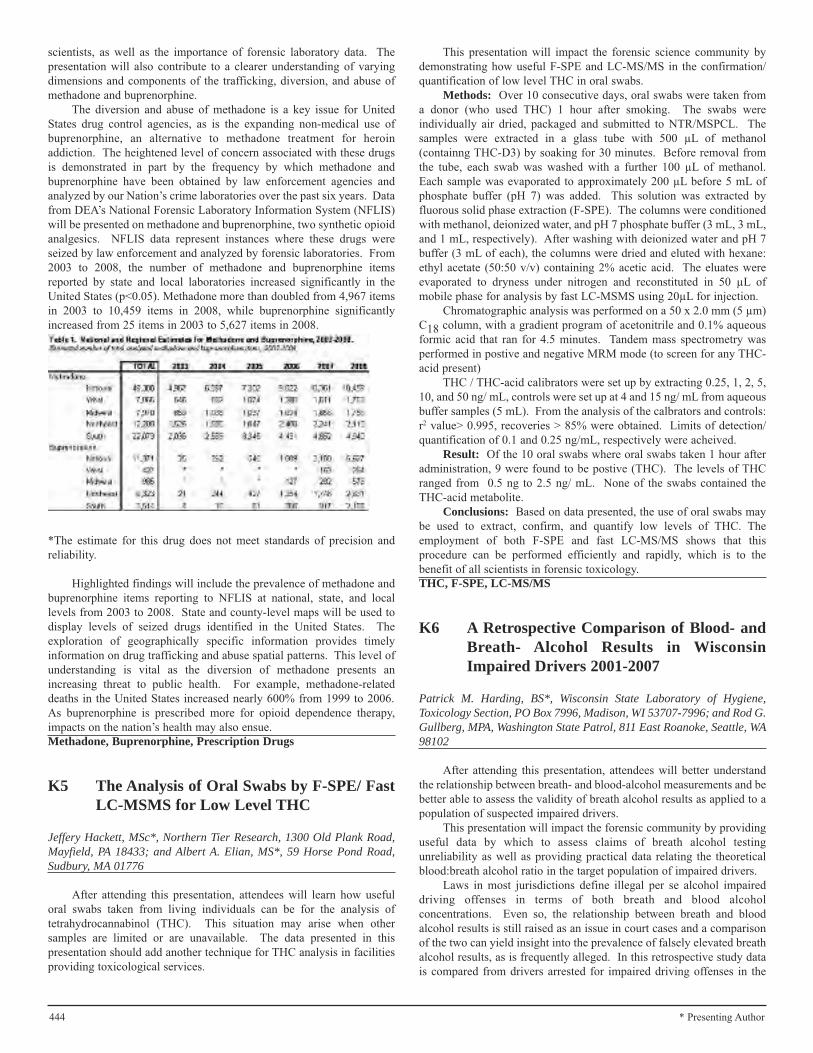
444 * Presenting Author
scientists, as well as the importance of forensic laboratory data. Thepresentation will also contribute to a clearer understanding of varyingdimensions and components of the trafficking, diversion, and abuse ofmethadone and buprenorphine.
The diversion and abuse of methadone is a key issue for UnitedStates drug control agencies, as is the expanding non-medical use ofbuprenorphine, an alternative to methadone treatment for heroinaddiction. The heightened level of concern associated with these drugsis demonstrated in part by the frequency by which methadone andbuprenorphine have been obtained by law enforcement agencies andanalyzed by our Nation’s crime laboratories over the past six years. Datafrom DEA’s National Forensic Laboratory Information System (NFLIS)will be presented on methadone and buprenorphine, two synthetic opioidanalgesics. NFLIS data represent instances where these drugs wereseized by law enforcement and analyzed by forensic laboratories. From2003 to 2008, the number of methadone and buprenorphine itemsreported by state and local laboratories increased significantly in theUnited States (p<0.05). Methadone more than doubled from 4,967 itemsin 2003 to 10,459 items in 2008, while buprenorphine significantlyincreased from 25 items in 2003 to 5,627 items in 2008.
*The estimate for this drug does not meet standards of precision andreliability.
Highlighted findings will include the prevalence of methadone andbuprenorphine items reporting to NFLIS at national, state, and locallevels from 2003 to 2008. State and county-level maps will be used todisplay levels of seized drugs identified in the United States. Theexploration of geographically specific information provides timelyinformation on drug trafficking and abuse spatial patterns. This level ofunderstanding is vital as the diversion of methadone presents anincreasing threat to public health. For example, methadone-relateddeaths in the United States increased nearly 600% from 1999 to 2006.As buprenorphine is prescribed more for opioid dependence therapy,impacts on the nation’s health may also ensue.Methadone, Buprenorphine, Prescription Drugs
K5 The Analysis of Oral Swabs by F-SPE/ FastLC-MSMS for Low Level THC
Jeffery Hackett, MSc*, Northern Tier Research, 1300 Old Plank Road,Mayfield, PA 18433; and Albert A. Elian, MS*, 59 Horse Pond Road,Sudbury, MA 01776
After attending this presentation, attendees will learn how usefuloral swabs taken from living individuals can be for the analysis oftetrahydrocannabinol (THC). This situation may arise when othersamples are limited or are unavailable. The data presented in thispresentation should add another technique for THC analysis in facilitiesproviding toxicological services.
This presentation will impact the forensic science community bydemonstrating how useful F-SPE and LC-MS/MS in the confirmation/quantification of low level THC in oral swabs.
Methods: Over 10 consecutive days, oral swabs were taken froma donor (who used THC) 1 hour after smoking. The swabs wereindividually air dried, packaged and submitted to NTR/MSPCL. Thesamples were extracted in a glass tube with 500 µL of methanol(containng THC-D3) by soaking for 30 minutes. Before removal fromthe tube, each swab was washed with a further 100 µL of methanol.Each sample was evaporated to approximately 200 µL before 5 mL ofphosphate buffer (pH 7) was added. This solution was extracted byfluorous solid phase extraction (F-SPE). The columns were conditionedwith methanol, deionized water, and pH 7 phosphate buffer (3 mL, 3 mL,and 1 mL, respectively). After washing with deionized water and pH 7buffer (3 mL of each), the columns were dried and eluted with hexane:ethyl acetate (50:50 v/v) containing 2% acetic acid. The eluates wereevaporated to dryness under nitrogen and reconstituted in 50 µL ofmobile phase for analysis by fast LC-MSMS using 20µL for injection.
Chromatographic analysis was performed on a 50 x 2.0 mm (5 µm)C18 column, with a gradient program of acetonitrile and 0.1% aqueousformic acid that ran for 4.5 minutes. Tandem mass spectrometry wasperformed in postive and negative MRM mode (to screen for any THC-acid present)
THC / THC-acid calibrators were set up by extracting 0.25, 1, 2, 5,10, and 50 ng/ mL, controls were set up at 4 and 15 ng/ mL from aqueousbuffer samples (5 mL). From the analysis of the calbrators and controls:r2 value> 0.995, recoveries > 85% were obtained. Limits of detection/quantification of 0.1 and 0.25 ng/mL, respectively were acheived.
Result: Of the 10 oral swabs where oral swabs taken 1 hour afteradministration, 9 were found to be postive (THC). The levels of THCranged from 0.5 ng to 2.5 ng/ mL. None of the swabs contained theTHC-acid metabolite.
Conclusions: Based on data presented, the use of oral swabs maybe used to extract, confirm, and quantify low levels of THC. Theemployment of both F-SPE and fast LC-MS/MS shows that thisprocedure can be performed efficiently and rapidly, which is to thebenefit of all scientists in forensic toxicology.THC, F-SPE, LC-MS/MS
K6 A Retrospective Comparison of Blood- andBreath- Alcohol Results in WisconsinImpaired Drivers 2001-2007
Patrick M. Harding, BS*, Wisconsin State Laboratory of Hygiene,Toxicology Section, PO Box 7996, Madison, WI 53707-7996; and Rod G.Gullberg, MPA, Washington State Patrol, 811 East Roanoke, Seattle, WA98102
After attending this presentation, attendees will better understandthe relationship between breath- and blood-alcohol measurements and bebetter able to assess the validity of breath alcohol results as applied to apopulation of suspected impaired drivers.
This presentation will impact the forensic community by providinguseful data by which to assess claims of breath alcohol testingunreliability as well as providing practical data relating the theoreticalblood:breath alcohol ratio in the target population of impaired drivers.
Laws in most jurisdictions define illegal per se alcohol impaireddriving offenses in terms of both breath and blood alcoholconcentrations. Even so, the relationship between breath and bloodalcohol results is still raised as an issue in court cases and a comparisonof the two can yield insight into the prevalence of falsely elevated breathalcohol results, as is frequently alleged. In this retrospective study datais compared from drivers arrested for impaired driving offenses in the
445* Presenting Author
State of Wisconsin who had both breath and blood alcohol specimensanalyzed. Breath alcohol testing was conducted in the field on EC/IRand EC/IR II (Intoximeters, Inc., St. Louis, MO) breath alcoholanalyzers. Breath alcohol results are obtained on duplicate breathsamples and must agree with +/- 0.020 g/210L. The lower of the twothree decimal place acceptable results is truncated to two decimal placesas the reported result. Blood specimens were collected by medicalpersonnel and submitted by the arresting agency to the Wisconsin StateLaboratory of Hygiene (WSLH). Blood analysis was performed bydirect injection gas chromatography, with 10% done in duplicate per thetesting protocol in effect during the study period. All testing was doneas part of the routine investigation of impaired driving cases. Onlypositive breath and blood alcohol results obtained within three hours ofeach other were included in this study.
During the study period of 2001-2007 there were 1,744 cases thatmet the inclusion criteria. Of these cases there were 1,545 males with amean age of 34.9 (16-84) and 199 females with a mean age of 35.8 (16-77). The mean reported breath alcohol concentration (BrAC) was 0.144g/210L (0.01 – 0.40). The mean of the un-truncated individual BrACresults was 0.1510 g/210L (0.010-0.400). The mean blood alcoholconcentration (BAC) was 0.1624 g/100ml (0.010-0.450). The meandifference (BAC-BrAC) between reported BrAC and reported BACresults was 0.018 (-0.046-0.100). There were 869 cases where the BrACwas collected before the BAC with a mean elapsed time of 0.89 hours(0.13 – 2.75). There were 875 cases where the BAC was collected beforethe BrAC with a mean elapsed time of 0.80 hours and range of 0.05 to2.27 hours.
BAC results were adjusted for the alcohol elimination occurringbetween the breath test time and the time of blood collection using a rateof 0.019 g/100ml per hour. After adjustment the BAC – BrACdifferences were a mean of 0.019 (-0.029-0.082). The reported BrACexceeded the adjusted BAC in 105 cases (6.0%). Of these, only 23(1.3%) exceeded the BAC by 0.010 or more (range 0.010-0.029). Therewere only five cases where the BrAC was 0.08 and the adjusted BACwas below 0.080. Of these only one differed by more than 0.010.Blood:Breath alcohol ratios were calculated using the time-correctedBAC and reported BrAC. The mean (SD) ratio was 2428:1 (295)(range1622:1 – 6822:1.
The findings of this study are in agreement with others that havefound that evidentiary BrAC results generally underestimate BAC in thedriving population. The absence of significant overestimations of BACby BrAC in this study provides strong evidence that alleged significantelevations of BRAC by mouth alcohol, GERD, potential interferingsubstances, variations in lung capacity, breathing disorders, etc. do notoccur in the context of a well-regulated evidential breathtesting program.Forensic Toxicology, Blood Alcohol, Breath Alcohol
K7 Cocaine Detection in Postmortem SamplesFollowing Therapeutic Administration
Kristen M. Bailey, MS*, David J. Clay, BA, and Myron A. Gebhardt, MS,Office of the Chief Medical Examiner, 619 Virginia Street West,Charleston, WV 25302; Matrina J. Schmidt, MD, West VirginiaUniversity, Department of Pathology, PO Box 9203, Morgantown, WV26506; and James C. Kraner, PhD, Office of the Chief MedicalExaminer, 619 Virginia Street West, Charleston, WV 25302
After viewing this presentation, attendees will understand theimportance of a thorough investigation, medical records review, andinterpretation of autopsy and toxicology results in postmortem cases inwhich controlled substances such as cocaine are detected followingadministration in a clinical setting.
This presentation will impact the forensic science community byemphasizing that the detection of illicit drugs in postmortem samples isnot always indicative of abuse.
Cocaine is a drug which is notorious for its high potential forrecreational abuse, and the detection of cocaine in postmortem sampleswould most often lead toxicologists and forensic pathologists to believethat the drug was abused. However, cocaine is an effective localanesthetic and vasoconstrictor of mucous membranes and has been usedclinically in surgeries of the eye, ear, nose, and throat for over 100 years.The persistent popularity of the clinical use of this drug is clearly
attributable to its unique ability to simultaneously limit epistaxis andinduce local anesthesia. Therefore, it is important to note that thepresence of cocaine and its metabolites in postmortem samples cannotalways be attributed to abuse and that a thorough investigation andreview of clinical records is warranted before an informed conclusioncan be made.
Presented here is the case of a 54-year-old male who was involvedin an altercation during which he suffered multiple injuries. Three dayslater, a surgical procedure involving closed reduction of bilateral nasalbone fractures was performed and the man was released from thehospital. Approximately eleven hours post-surgery, the man was foundunresponsive in bed and EMS responded and pronounced him dead onthe scene. Given the circumstances leading up to the demise, a fullpostmortem examination was performed in order to elucidate thecontribution of external factors such as physical injury, surgicalintervention, and/or drug use to his death. In addition to natural diseaseand injuries documented at autopsy, toxicological analysis revealed thepresence of cocaine metabolites in the man’s urine. A comprehensivereview of subsequently received surgical records revealed that the manwas administered cocaine during the procedure to repair his nasal bonefractures.
If not for this review of surgical records, the assumption of cocaineabuse might have otherwise been made and the well-known cardiotoxiceffects associated with cocaine considered a contributory factor incertification of cause and manner of death. Additionally, an erroneouspresumption of illicit drug use may have significant implications in alegal setting and may cause family members of the decedent undueanguish. Toxicology results, investigative reports, clinical records, andpathologic findings must be collectively taken into consideration toensure accurate explanations for the presence of cocaine, as well as otherdrugs that may be administered clinically, in postmortem samples.Postmortem, Cocaine, Therapeutic
K8 Gastric Fentanyl Concentrations inFentanyl-Related Deaths: A Study of11 Cases
Ruth E. Kohlmeier, MD*, Werner Jenkins, MS, MPA, Robert C. Bux, MD,Jennifer M. Hoffman, MS, and Christopher B. Clarke, BS, El PasoCounty Coroner’s Office, 2743 East Las Vegas Street, Colorado Springs,CO 80906; and Andrea N. Phelps, and Emily D. Morrison, BS,Univeristy of Colorado at Colorado Springs, 1420 Austin BluffsParkway, Colorado Springs, CO 80918
After attending this presentation, attendees will have a greaterunderstanding of the role that the analysis of gastric concentrations offentanyl has in fentanyl-related deaths.
This presentation will impact the forensic community by providingtoxicological data and insights on potential relationships between gastricfentanyl concentration, blood fentanyl concentration, route ofadministration, and cause and manner of death.
Given that fentanyl is a short acting and potent narcotic, there ispotential risk for abuse and fatalities. Interpreting postmortem

446 * Presenting Author
toxicology in suspected narcotic overdoses, including fentanyl, can bedifficult for medical examiners due to the variety of drug use and abuseand the development of tolerance in the user/abuser. The unfortunate“creative” abuses of the patch (including snorting, smoking andchewing) only complicate the issue. A strategy utilized in postmortemtoxicological evaluation has been to analyze gastric contents for theamount of drug present to ascertain a potential route of administrationand/or the cause and manner of death. One would expect that oralconsumption would lead to higher levels of gastric concentrations ingeneral. If that were so, would one be able to use the gastricconcentrations to determine the route of administration (includinginappropriate use of the transdermal patch) as well as the manner ofdeath (intentional versus accidental overdose)? The purpose of thiscurrent study was to determine the gastric concentrations of fentanyl andnorfentanyl in fentanyl-related deaths and to attempt to relate theselevels with blood concentrations, route of administration, and cause andmanner of death.
From January 2007 to June 2009, eleven fentanyl-related deaths inwhich gastric samples were available were identified through routinetoxicology testing in the El Paso County Coroner’s Office toxicologylaboratory in Colorado Springs, Colorado. Routine toxicological testingwas performed on all cases. Ethanol and related alcohols were detectedusing headspace Gas Chromatography/Flame Ionization Detection(GC/FID), urine was screened for drugs of abuse by ELISA and GasChromatography-Mass Spectrometry (GC/MS), and GC/MS was used toquantitate the blood and gastric contents after a liquid-liquid basicextraction.
The age of the decedents ranged from 23 to 60 years and consistedof three men and eight women. The blood concentration of fentanylranged from 2.1 to 30.7 µg/L (mean 17.5 µg/L) while the total gastricfentanyl concentration ranged from 2.9 to 432.4 µg (mean 85.1 µg). Theanalytical data for norfentanyl concentrations in the gastric samples wereinconclusive as the samples calculated below the detection level of 5µg/L. The cause of death was acute fentanyl intoxication in six out ofeleven cases while five cases were ruled mixed drug overdoses. Themanner of death was accidental in eight cases and undetermined in threecases. The route of administration was by transdermal patch in ninecases, oral (by chewing the patch) in one case, and unknown in one case.
In conclusion, there did not appear to be any correlations betweenthe gastric and blood concentrations of fentanyl, the route ofadministration, or the cause and manner of death. It did not appear to behelpful to determine if the individual had intentionally or accidentallyoverdosed, nor did it provide insight into the route of administration (e.g.inappropriate use of the patch). Although the case with oral route ofadministration had the highest gastric concentration of fentanyl, the levelwas not impressively higher than the next highest concentration, wherethe route of administration was transdermal application. Additionally,the oral route of administration did not yield the highest total gastricfentanyl concentration. A limiting factor in our study was the smallsubject number. Perhaps a larger study (e.g., a multi-centered study)focusing on the analysis of gastric concentration of fentanyl would beuseful and illuminate any useful patterns or trends.Gastric Fentanyl, Fentanyl Overdose, Forensic Toxicology
K9 Exposure to Limonene: A Case Report
Zeinab Mohamed Mostafa, BSc*, Medicolegal Administration, Ministryof Justice, Egypt, Cairo, 002, EGYPT
After attending this presentation, attendees will learn the relationbetween orange oil (Limonene) and respiratory failure.
This presentation will impact the forensic community by providingthe relation between orange oil (Limonene) and respiratory failure.
D-limonene (4-Isopropenyl-1-methylcyclohexene) is the chemicalname for orange oil. It’s a renewable resource and is a by-product of
orange juice manufacturing. Orange oil is the oily substance found in therinds of oranges. Orange oil is used in cleaning solutions, pet shampoos,soaps and perfumes. Limonene and its products are skin and respiratoryirritants. Acute exposure to D-limonene has rarely been reported indeaths.
In this case report, we present a case of a previously healthy 30-year-old man who presented to the emergency department with acuterespiratory failure. Non-toxicological causes were excluded. Thepurpose of this work was to demonstrate a toxicological cause of therespiratory failure and to recommend full toxicological screening forclinical and postmortem cases, especially those under suspiciouscircumstances.
A complete history was taken and a comprehensive clinicalexamination was performed. Toxicological analysis was performed. Forthe analysis of limonene in blood, gas chromatography/massspectrometry (Shimadzu 2010) was utilized in the splitless mode ofinjection. The initial temperature was 170◦C for 2 min. and thenprogrammed at 16◦C /min. to 270◦C and held for 8 minutes.
The assay was found to be linear in the concentration range of 0.5-20 ng/ml for limonene. Repeatability and intermediate precision werefound to be less than 12% for all concentrations tested. Under standardchromatographic conditions the run cycle time would have been 13minutes. By using fast chromatographic separation conditions, the assayanalysis time could be reduced to 7 minutes without compromising thechromatographic resolution.
This developed procedure was also used to determine the limoneneconcentration levels for more than a hundred real forensic cases. Thecase was diagnosed as toxic exposure to limonene dissolved in organicsolvent. The patient was exposed to limonene at home for many years inair fresheners. The patient survived after supportive treatment. Theclinical and laboratory findings are discussed.
Limonene and its oxidation products are skin and respiratoryirritants. Inhalation of these chemicals carries the risk of toxicity, whichcould be missed in diagnosis and hence treatment. This shouldencourage physicians working in emergency units to analysis for allavailable chemicals to avoid misdiagnosis.Forensic, Chemical, GC/MS
K10 Study of the Effects of pH, Temperature,and Time on the Migration of EndocrineDisrupting Compounds From PolyethyleneTerephthalate (PET) Bottles
Sara E. Smith, BS*, Pennsylvania State University, 107 WhitmoreLaboratory, University Park, PA 16802; and Dan G. Sykes, PhD, ThePennsylvania State University, 330 Whitmore Laboratory, UniveristyPark, PA 16802
After attending this presentation, attendees will be informed of animportant toxicological topic that has an effect on everyone. Theattendees will also be exposed to new optimized extraction and analysismethods of several endocrine disrupting compounds which they canapply to further this aspect of work or use in their own current researchprojects.
This presentation will impact the forensic community by informingthe audience of the constant presence of endocrine disruptingcompounds (EDCs) and their possible effects on our health andenvironment. Although this work is only a start, it may lead to newsafety measures to ensure that all levels of EDCs are below the oralreference dose. This work could have an effect on humanity because ifthese new measures were established, it could affect our everyday lives.Also, several extraction and analysis methods that are commonly used
in forensic laboratories, have been used in this study. These methodsinclude solid phase extraction (SPE) and gas chromatography-mass
447* Presenting Author
spectrometry (GC-MS). This presentation may allow the audience tobecome more knowledgeable about these different techniques whichthey could possibly apply to their own work. Finally, a major challengein this work was preventing external contamination. The cleantechniques that were utilized in this study could also be useful in aforensic laboratory.
Several toxicological studies have shown that many commonendocrine disrupting compounds (EDC), specifically those that displayestrogenic properties, could cause toxicity from chronic exposure tolevels as low as 20 µg/kg/day. Any compound that has the ability to alterhormonal homeostasis is considered to be an endocrine disruptingcompound. Effects of exposure include abnormal cell growth,teratogenicity, liver injury, abnormal thyroid function, and reproductivetoxicity. Two types of EDCs, alkylphenols and phthalates, have beenfound to migrate from plastic containers into the food supply. Recentstudies have suggested that phthalates may have a cumulative effect,which has lead to a great interest in studying their presence in theenvironment. Two different extraction methods, solid phase extraction(SPE) and liquid liquid extraction (LLE), have been performed andcompared. LLE has been found to be the most effective extractiontechnique for studying trace levels of EDCs in water. Gaschromatography- mass spectrometry (GC-MS) was chosen as theanalysis method because of the reduced risk of contamination whencompared with liquid chromatography- mass spectrometry (LC-MS).Many studies have suggested that LC-MS is not an efficient analysismethod when studying EDCs, such as phthalates, because of the use oforganic solvents and plastic tubing which can increase the risk of samplecontamination. The specific compounds that are being studied includedimethyl phthalate (DMP), dimethyl terephthalate (DMT), diethylphthalate (DEP), dibutyl phthalate (DBP), butyl benzyl phthalate (BBP),bis-(2-ethylhexyl) phthalate (DEHP), nonylphenol, and octylphenol.Several conditions were established in order to study the effects oftemperature, plastic thickness, and pH on migration. When studying theeffects of temperature, two plastic bottles were filled with drinkingfountain water and were heated to 60°C for six hours. Samples werecollected after every two hours. The second bottle had a thickness thatwas 50% less than the first bottle. Another bottle was filled withfountain water and stored at room temperature. This was used as acontrol for the temperature experiment and was sampled after three days.The effect of two different pH values was also studied. The two values
used were 3.75 and 6.6. These values were chosen because they arecomparable to different beverages that are commonly stored in plasticcontainers. The pH of each sample was adjusted by adding glacial aceticacid until the desired pH value was obtained. A third bottle was filledwith fountain water and was not adjusted. This bottle was used as acontrol for the pH experiment and had a pH value of approximately 7.2.The three pH samples were collected after one day. When heated to
60°C for two hours, five of the eight compounds were detected. Thesecompounds include DMP, DMT, DEP, OP, and DEHP, at concentrationsof 2.144, 1.78, 1.258, 0.783, and 1.539 µg/L, respectively. Thesecompounds were found in the control sample at concentrations of 0.619,0.875, 0.18, 0.475, and 0.483 µg/L, respectively. Therefore, heatingplastic to 60°C increased the amount of migration occurring. When thethickness of the plastic bottle was reduced by 50% these compoundswere found at concentrations of 1.627, 0.979, 0.779, 0.601, and 1.562µg/L, when heated at 60°C for two hours. Although lowerconcentrations were found to migrate from thinner plastics when heated,the migration occurred over a longer period of time when compared tothe 50% thicker plastic. Migration was also found to increase as the pHof the water decreased, when stored at room temperature. All reportedconcentrations were determined using calibration curves prepared fromstandard solutions. Standard solutions containing all eight compoundswere prepared at concentrations of 5 x 10⁵, 5 x 10⁴, 5 x 10³, 500, 50, 25,12.5, and 6.25 µg/L. These standard solutions were also used as controlsin order to determine the retention times of each of the studiedcompounds. A selected ion monitoring (SIM) method was also
developed and used to analyze trace levels of these compounds. Whencompared to a NIST library the highest matches obtained were 80%(DMP), 81% (DMT), 81% (DEP), 73% (OP), and 72% (DEHP). Allidentified compounds had approximately the same retention times as thestandard solutions. The average concentration found from allexperiments was 1.046 µg/L. When comparing to the previouslyreported value of 20 µg/kg/day, a 70 kg person would have to drinkapproximately 1339 L/day in order to be exposed to this toxic level.Overall, temperature, pH, and plastic thickness have been found to havean effect on EDC migration from PET bottles; however, allconcentrations have been below any known toxic level. Further workwill be performed in order to study the effect time may have onmigration and to test the reproducibility of this method. It is importantto understand the effects any of these factors may have on EDCmigration because it may or may not suggest that certain safety measuresneed to be established in order to ensure that all levels are below the oralreference dose (RfD) values set by the United States EnvironmentalProtection Agency.Toxicology, Endocrine Disrupting Compounds, GasChromatography-Mass Spectrometry
K11 Mixed Prescription Drug Death
George F. Jackson, PhD*, Edwin H. Albano Institute of ForensicScience, 325 Norfolk Street, Newark, NJ 07103; William A. Dunn, MS,Regional Medical Examiner’s Office, 325 Norfolk Street, Newark, NJ07103; Zhongxue Hua, MD, PhD, 300 North Avenue, East, Westfield, NJ07090; and William E. Johnston, MD, 10 Plumtree Drive, Sewell, NJ08080
After attending this presentation, attendees will become aware ofthe effects of mixing prescription drugs.
This presentation will impact the forensic community by increasingthe awareness of the role of prescription drugs in deaths.
This laboratory has been detecting an increase in the prevalence ofmultiple prescribed opioid compounds in drug related postmortem cases.In most of our cases, the decedents have histories of addiction tooxycontin and xanax.
A case of postmortem analysis will be presented of a 36-year-oldsingle white male who consumed Oxycontin, Xanax, and Actiq (fentanyllollipop). The decedent had morbid obesity (BMI of 46) with a longhistory of lower back pain for at least six years. His clinical work-up forhis lower back pain was inconclusive, although a spinal surgery for discfusion was suggested at one point. He was known to visit various painclinics and acquired prescription Actiq for at least 18 months. Accordingto multiple co-workers, he demonstrated a typical pattern of opioidcompound addition which interfered with his job performancesignificantly. During his last year, he was admitted twice at a localrehabilitation center for his addition to Oxycontin and Xanax. Of note,per multiple co-workers and family members, the decedent had no priorsuicidal ideation or attempt.
The decedent was discovered in his residence with earlydecomposition after failing to report to work as an accountant. Sceneinvestigation found a fentanyl lollipop inside his shirt pocket as well ascrushed pill fragments scattered on the floor and table. Multiple bottlesof prescription oxycodone and alprazolam, some empty and others near-full, were located at the scene. Autopsy revealed hypertensivecardiovascular disease and focal bronchopneumonia, and both were notconsidered to be medically significant. Comprehensive analysis wasperformed on various postmortem tissues, including femoral blood,urine, stomach content and liver.
All submitted tissues were subject to standard analytical screeningand mass spectrometry confirmation protocols. Positive findings of theanalysis are as follows:
448 * Presenting Author
BloodOxycodone 1.86 mg/LOxymorphone 0.072 mg/LFentanyl 64 mcg/L
LiverOxycodone 1.83 mg/KgOxymorphone 0.12 mg/KgFentanyl 96 mcg/Kg
Stomach ContentsTotal weight received: 244 gms
Oxycodone 147.30 mg/KgOxymorphone 0.62 mg/KgFentanyl 3648 mcg/Kg
UrineOxycodone 5.82 mg/LOxymorphone 0.36 mg/LFentanyl 890 mcg/La-hydroxyalprazolam 1.60 mg/LAlprazolam 0.35 mg/L
Note: Alprazolam and its metabolite were only detected in the urinespecimen.
The medical examiner ruled the cause as multiple drug intoxicationand manner as accidental.Oxycodone, Fentanyl, Prescription
K12 Analytical Method Development forDetermining the Biomarker,2-Aminothiazoline-4-Carboxylic Acid (ATCA),in Mice Liver After Cyanide Exposure
Katelyn A. Stafford*, 6407 Kury Lane, Houston, TX 77008; RandyJackson, MS, Sam Houston State University, Room 221, 1003 BowersBoulevard, Huntsville, TX 77341; Kelsie D. Simons, MS, Sam HoustonState University, PO Box 2525, Huntsville, TX 77340; Jorn Chi ChungYu, PhD, Sam Houston State University, 1003 Bowers Boulevard, Box2525, College of Criminal Justice, Huntsville, TX 77341; and IlonaPetrickovics, PhD, Sam Houston State University, Department ofChemistry, PO Box 2117, 1003 Bowers Boulevard, Huntsville, TX 77341
After attending the presentation, attendees will learn about newmethods of detecting cyanide exposure and the analytical techniques thatare being developed to test the presence in mice livers. The attendeeswill also learn about 2-Aminothiazoline-4-Carboxylic Acid (ATCA) as abiomarker for cyanide.
This presentation will impact the forensic science community bydemonstrating a forensic application being developed to detect cyanidepoisoning postmortem. This research will be able to be applied to humanremains that are in autopsy under investigation for poisoning. ATCA isa stable biomarker for cyanide so this technique will be able to be appliedto cold cases.
The objective of this research was to develop a new analyticaltechnique to determine the chemically stable urinary metabolite ofcyanide, 2-aminothiazoline-4-carboxylic acid (ATCA), in mice liversamples. Two extraction techniques, solid phase extraction (SPE) cationexchange and molecular imprinted polymer stir bar (MIP-SB), weretested to determine the efficiency of ATCA extraction from mice liversamples. Mice were exposed to different doses of cyanide, and a methodwas developed to dissect, preserve organs, and homogenize the livers.
This research will be able to be applied to human remains that arein autopsy under investigation for poisoning. ATCA is a stable
biomarker for cyanide so this technique will be able to be applied to coldcases. For forensic casework, a stable and quantifiable marker is neededto determine an accurate level of exposure postmortem. This methodwill be able to be used in cold cases because ATCA is a stable metabolitethat stays in the body after the initial dose of cyanide is depleted.
Endogenous ATCA is always present in the body in low quantityoriginated from dietary intake of cyanide, smoking, fires or the normalmetabolism of amino acids. A selective and sensitive analytical methodis needed to determine the endogenous level of ATCA or identify cyanidepoisoning. The use of ATCA as a biomarker for cyanide poisoning ispromising due to its stability at ambient, as well as freezing temperaturesand its production is directly related to cyanide exposure.
The molecularly imprinted polymers (MIPs) are made on thesurface of a silica cylinder to serve as a selective stir bar sorptionextraction (MISBSE) device. From an external calibration, the capacityof one MISBSE for ATCA was about 31 ng. The data showed that 700rpm was the optimum stir speed and that sorption plateau was reachedafter 30 minutes of extraction time. Under the optimal extractionconditions, the MISBSE could selectively extract ATCA from urinesamples. The MISBSE has improved the ability to extract lowerconcentrations of ATCA. Combining MISBSE with LiquidChromatography Mass Spectrometry (LC/MS/MS), ATCA was detectedwithout the use of any derivatization process. The solid phase extractioncation exchange was preformed with Oasis® MCX (mixed-mode cationexchange) columns and underwent several washes to prepare thecartridges for absorbing the ATCA and then was eluted with ammoniumhydroxide with the assistance of a vacuum pump.
An effective method of preparing liver samples from the cyanideexposed mice for extraction will be presented. In additions, the twoextraction methods (SPE vs. MIP-SB) will be compared. Theeffectiveness of the extraction techniques will be determined byemploying known concentrations of ATCA evaluated by the LC/MS/MS.Liver ATCA contents will be compared to the dose of cyanide mice weregiven. This new analytical method may serve as great potential benefitsfor the toxicology field and forensics in general.Molecularly Imprinted Polymer (MIP), 2-aminothiazoline-4-carboxylic acid (ATCA), Cyanide
K13 Pesticide Intoxications in Cukurova,Turkey: A Three Year Analysis
Nebile Daglioglu, PhD*, and Mete K. Gulmen, PhD, MD, CukurovaUniversity, School of Medicine, Department of Forensic Medicine,Adana, 01330, TURKEY
The goal of this study is to show the distribution of pesticides in theCukurova region and alert the forensic toxicologists to notice pesticidesin all autopsy cases at this region.
This presentation will impact the forensic science community bydemonstrating how organochlorine pesticides are still a serious threat forpublic health although the WHO has forbidden the use of thesesubstances all over the world.
Cukurova region is one of the most important agricultural areas forTurkey. As a consequence of wide pesticide use, acute pesticidepoisoning cases are quite common, in this region. These poisonings aregenerally suicidal self poisonings, while can be accidental or homicidalas well. In Cukurova, pesticide poisonings still remain as a considerablecause of death, which lead the present retrospective evaluation.
The autopsy records of Adana Group Authority of the Council ofForensic Medicine, between 2006 and 2008, were evaluatedretrospectively. Deaths that are attributed to pesticide poisoningincluded in the scope of the study in order to identify the type ofpesticide, as well as the etiology. The frequency and distribution ofintoxications were also analyzed in terms of sex and age.

449* Presenting Author
In the studied period, a total of 4,199 autopsies had referred to theforensic toxicology laboratory for pesticide analysis. Pesticide analyseswere performed in the Forensic Toxicology Laboratory of Adana GroupAuthority of the Council of Forensic Medicine, using different biologicalsamples (blood, stomach, liver, lung, and kidney) by chromatographicmethods, gas chromatography with electron capture detection (GC-ECD), and gas chromatography with nitrogen phosphorus detection(GC-NPD) and gas chromatography-mass spectrometry (GC-MS).
Seventy-two out of all cases were positive for pesticide analysis. Ofthese 72 cases, 42 (58.33%) were male and 30 (41.66%) were female,with a mean age of 38.8 ± 20.6 years. Among the inspected pesticides,endosulfan was found to be the most common with 47.2% prevalence,followed by an organophosphorus insecticide dichlorvos with aprevalence of 16.7%. Majority of deaths due to pesticide poisonings (37,51.38%) were suicidal while (17, 23.61%) of them were accidental. Thehigh ratio of suicidal deaths due to pesticides was a consequence of easyavailability and accessibility of uncontrolled pesticides in households atcity centers and in villages of countryside.
This report showed that endosulfan, an organochlorine pesticide, iscommonly used in Cukurova region. Moreover, frequency of acute andchronic exposure to endosulfan is considerably high in Cukurova region.Recently, strict regulations have been enacted for restricting andcontrolling the use of endosulfan, of which use was previously allowed.Furthermore, authorities should set more efficient educational facilitiesfor agricultural workers in order to reduce the number of accidentalpesticide poisonings.Forensic Toxicology, Pesticide Poisoning, Cukurova (ADANA)
K14 Identification of GHB and Morphine inHair in a Case of Drug FacilitatedSexual Assault
Riccardo Rossi, MD*, Institute of Forensic Medicine, CatholicUniversity, Largo Francesco Vito 1, Rome, ITALY; Antonio Oliva, MD,PhD, Institute of Forensic Medicine, Catholic University, School ofMedicine, Ro, Largo Francesco Vito 1, Rome, ITALY; Fabio De Giorgio,MD, PhD, Institute of Legal Medicine, Rome, 00168, ITALY; MassimoLancia, MD, and Cristina Gambelunghe, MD, Institute of ForensicMedicine, University of Perugia, Perugia, ITALY; and Nadia Fucci,PhD, Forensic Toxicology Laboratories, Catholic University, Rome,Italy, largo f. vito 1, Rome, ITALY
After attending this presentation, attendees will appreciate theimportance of an accurate toxicological analysis in sexual assault cases.
This presentation will impact the forensic science community bydetailing a unique circumstance of sexual assault and the drug GHBGamma-hydroxybutyric acid, a substance naturally present inmammalian species, which has been utilized to commit the crime.
Gamma-hydroxybutyric acid GHB is qualified as a “predatorydrug.” Doses of 10 mg/kg cause amnesia, 20-30 mg/kg induce sleep anddoses of 50 mg/kg or higher produce anesthesia. It is attractive forrapists because it can be found easily (on the street, fitness centers, andinternet) and moreover because it can be delivered mainly as an odorless,colorless liquid and so it is often assumed unwittingly, mixed in spikeddrinks.
Case Report: The case of a 24-year-old girl who was sexuallyassaulted after administration of Gamma-hydroxybutyric acid (GHB)and morphine will be presented. She had been living in an internationalcollege for foreign students for about one year and often complained ofa general unhealthy feeling in the morning. At the end of the collegeperiod she returned to Italy and received at home some video clips shotby a mobile phone camera. In these videos she was having sex with aboy she met when she was studying abroad.
Materials and Methods: Toxicological analysis of the victim’shair was done: the hair was 20 cm long. A 2 cm segmentation of all thelength of the hair was performed. Morphine and GHB were detected inhair segments related to the period of time she was abroad. The analysesof hair segments were performed by gas chromatography/massspectrometry (GC/MS) and the concentration of morphine and GHBwere calculated.
Conclusions: A higher value of GHB was revealed in the period ofthe criminal event and the presence of morphine was also detected for thesame period. According to previous observations our case shows thathair analysis is the only method used to prove repetitive exposure to atoxic substance. This case demonstrates also that a high concentration ofGHB in hair reflects an acute overexposure to GHB and can bedocumented several months after the sexual assault. In general it mustbe specified that the possibility given by hair analysis should not preventthe victim and the medical examiner from taking urine, blood, and sweatsamples as soon as possible after the event. Hair analysis may be auseful adjunct to conventional drug testing in sexual assaults and itshould not be considered an alternative to urine analysis, but acomplement. It is possible that hair analysis could be a useful additionto conventional drug testing in sexual assault, but it is believed thatfurther studies may confirm the usefulness of this technique andestablish the definition of legally defensible cut-off values.GHB, Hair Analysis- GC/MS, Drug Facilitated Sexual Assault
K15 Death by Potassium Chloride IntravenousInjection and Analytical Detection
Elisabetta Bertol, Lucia Politi, and Maria Grazia Di Milia, VialeMorgagni 85, Florence, ITALY;, and Francesco Mari*, Istituto diMedicina Legale, D, Policlinico Careggi, Viale Mor, Firenze, ITALY
After attending this presentation, attendees will understand theresults of the determination of blood potassium in a case of suicide bypotassium poisoning. The meaning of the blood potassium concentrationis questioned and discussed.
This presentation will impact the forensic science community bydemonstrating how potassium concentration resulted to be significantlyhigher in heart blood in a case of suicide by potassium chlorideintravenous injection and, therefore, the general issue of consideringpotassium poisoning hardly demonstrable by the toxicology needs to bequestioned and thoroughly studied in the future.
Potassium chloride intravenous injection is reported as a means insuicide attempts and also in lethal procedures for state-sanctioned capitalpunishment. Owing to its relatively high concentrations in hemolyzedblood (25-80 mmol/l) as compared to serum (about 4 mmol/l), potassiumpoisoning has often been considered hardly detectable in postmortemblood specimens.
In considerations of the results of the determination of bloodpotassium in a case of suicide by potassium poisoning, the meaning ofblood potassium concentration is questioned and discussed.
A 41-year-old man, working as a nurse at the local intensive careunit, was found dead at his workplace. A recent injection site wasobserved on his left foot and a syringe retrieved close to the corpse. Atthe autopsy no particular signs were noted.
Biological specimens (blood, bile, and urine) were submitted to thescreening procedures for drugs and poisons in use in the laboratory,consisting of general unknown screening by solid phase extraction andgas chromatography mass spectrometry for blood and bile, head-spacegas chromatography for blood and immunoenzymatic screening forurine. The syringe content was submitted to Feigl spot tests for inorganicions and, in particular, for potassium. Finally, blood potassiumconcentration was determined by ion selective electrode measurement(linear over the range 3.0-150 mmol/l).
450 * Presenting Author
According to the routine screening procedure, blood was foundpositive for diazepam at therapeutic level (0.21 mg/l) and urine resultedpositive only for benzodiazepines. No other substances were identifiedin blood and urine and all other samples tested negative. Potassiumconcentration was found at 160.0 mmol/l in cardiac blood and 87.3mmol/l in femoral blood (mean of three determinations in both cases).On the other hand, hemolized blood samples obtained from autopsieswith no relevant toxicological findings had much lower potassiumconcentration, i.e., between 32.2 and 43.0 mmol/l (median: 38.6 mmol/l,n=6).
Death by potassium intravenous injection is often consideredundetectable by toxicological analyses when only hemolized blood isavailable, and, consequently, literature is relatively scant. These resultshow that potassium concentrations were significantly higher in heartblood in a case of suicide by potassium chloride intravenous injectionand, therefore, the general issue of considering potassium poisoninghardly demonstrable by the toxicology testing needs to be questionedand thoroughly studied in the future.Potassium Chloride, Intravenous Injection, Suicide
K16 A HPLC/MS/MS Method for SimultaneousDetermination of Three Opiates and ThreeBenzodiazepines in Postmortem Blood
Shaohan Zhao, PhD*, Terry Danielson, PhD, and Ashraf Mozayani,PhD, PharmD, Harris County Medical Examiner’s Office, 1885 OldSpanish Trail, Houston, TX 77054
After attending this presentation attendees will have newknowledge of a Liquid Chromatography/Tandem Mass Spectrometry(LC/MS/MS) method for simultaneous determinations of morphine,hydromorphone, hydrocodone, alprazolam, diazepam, and nordiazepamin postmortem blood using multiple reaction monitoring (MRM)techniques and corresponding deuterated internal standards.Simultaneous analysis of multiple opiates and benzodiazepines has notpreviously been reported in postmortem blood.
This presentation will impact the forensic community bydemonstrating an additional application of LC/MS/MS to the analysis ofcomplex drug mixtures in postmortem specimens.
Analyses were performed with an ABI 3200 Q-Trap instrumentoperating in a positive polarity mode. For these analyses, one mLspecimens of blood were basified by addition of saturated sodium boratebuffer, and then extracted once with four volumes of the mixture of 1-chlorobutane and 2-propanol. The organic layer was evaporated and theresidue was reconstituted into 0.5 mL of reconstitution solvent (aqueousbuffer/acetonitrile; 9:1). Reconstitution solutions were filtered andchromatographed in an acetonitrile/ammonium formate gradient.Instrument parameters were optimized by infusion of solutions of eachdrug. The chromatographic column was maintained at 25˚C and the runtime was 12.5 minutes.
The method was validated by examining selectivity, precision,accuracy, linearity, recovery, suppression, and limits of quantitation anddetection. Calibration curves were quadratic for all analytes over theconcentration range 10–1000 ng/mL, and correlation coefficients (R2)were better than 0.999. Intraday and interday precision for all analytesat concentrations of 50, 200, and 500 ng/mL was between 4.1% and10.6%, intraday and interday accuracy for all analytes at the threeconcentration levels was between 88% to 114%. Recoveries werebetween 13% and 52%. Limits of detection and quantitation were 3 and10 ng/mL, respectively. Selectivity results demonstrate that theprecision and accuracy of the analytes were not affected by the presenceof 14 other common drugs. Only diazepam showed ion suppression inpostmortem blood, and morphine and hydromorphone showed ionenhancement. Four postmortem blood specimens were analyzed by this
method. The four specimens were also analyzed by alternate, individualLC/MS/MS opiate, and benzodiazepine methods. The results obtainedby this new combined opiate/benzodiazepine method match well withthe results run by the individual methods.
A method is described that is applicable to simultaneousdetermination of at least six opiates and benzodiazepines over a broadrange of concentrations. Individual analytes were well separated,suggesting that the method is amenable to addition of other opiatesor benzodiazepines.LC/MS/MS, Benzodiazepines, Opiates
K17 Covalent Protein Adduction by Drugsof Abuse
Kevin J. Schneider, BS*, 900 Euclid Avenue, #22, Miami Beach, FL33139; Anthony P. DeCaprio, PhD, 11200 Southwest 8th Street,Miami, FL 33174
After attending this presentation, attendees will glean some of theprinciples of covalent protein adduct formation, previous advances ingenerating and detecting protein adducts, how drugs of abuse formprotein adducts, and the potential importance of protein adducts in thefields of clinical and forensic toxicology.
This presentation will impact the forensic science community byopening a new subset of toxicological analysis for clinical and forensicinquiries. It will allow for the development of new biomarkers ofexposure to detect the use of addictive and illegal substances even afterthe parent compounds and major metabolites have been eliminated frombiological samples. These persistent adducts can be employed to expandthe range of time a substance can be detected in biological samples; abeneficial advance for both clinical and forensic applications.
Introduction: Protein adducts are formed by the covalent bindingof an electrophilic, metabolically activated xenobiotic to nucleophilicsites on endogenous proteins or protein precursors. These permanentcovalent bonds remain for the lifetime of the protein. However, not allnucleophilic sites on a given protein are equally reactive to activatedxenobiotics. Certain amino acid residues are more susceptible toelectrophilic attack, due to both steric and electronic factors. Eachxenobiotic will interact with a given protein in a characteristic waydetermined by the combination of these factors.
While only a few studies have examined protein adduction by drugsof abuse, results suggest that this is a valuable issue to explore further.In this report, initial data is presented on such adducts by evaluating therelative binding affinity of several common drugs of abuse to aminoacids known to be reactive under biological conditions. Initial researchusing a controlled in vitro exposure system is required prior to expandingstudies into complex matrices and case studies involving biologicalsamples from drug users. This initial step is germane to theunderstanding of how and where these drugs of abuse covalently bind toendogenous proteins and is imperative to the understanding of in vivoformation of protein adducts. This approach involved utilizing HPLC-MS, which allows for the sensitive and discriminating analysis ofadducted peptide structure.
Methods: In order to generate protein adducts with drugs of abuse,an in vitro metabolic system was used. This system has previously beenemployed to assess hepatic metabolism of xenobiotics under controlledconditions. The system consisted of purified human cytochrome P4503A4, human cytochrome b5, and human NADPH cytochrome P450reductase in conjunction with required lipid cofactors (1,2-dilauroyl-sn-glycero-3-phosphocholine, 1,2-dioleoyl-sn-glycero-3-phosphocholine,and 1,2-dilauroyl-sn-glycero-3-phospho-L-serine). To each mixture, oneof three test peptides was added: Ac-PAAHAA-OH, Ac-PAAKAA-OH,and Ac-PAACAA-OH. These short peptides allowed for the analysis ofmodifications of the reactive amino acid residue (His, Lys, and Cys,

451* Presenting Author
respectively). Each peptide was tested with each of three commonsubstances of abuse: cocaine, morphine, and methamphetamine (each at200 µM final xenobiotic concentration). Peptides were incubated at37°C in 200 mM phosphate buffered saline (pH 7.4) for 15 - 60 min toallow for metabolic activation of the xenobiotics and adduction to thepeptides. Following incubation, 10 µL aliquots of the mixtures wereacidified with TFA and introduced into a Varian 1000 LC-ion trap MSequipped with Polaris C18 column and optimized for the analysis of theindividual peptides.
Results: Stable, time-dependent covalent adduction of all threemodel peptides at varying efficiencies was noted for each drug. Adductformation was confirmed by the appearance of new peptide peaks withMS molecular ion and fragmentation data consistent with covalentlybound drug. MS/MS with de novo peptide sequencing results confirmedthe location of adduct at the putative reactive site for each peptide.Additional MS studies are currently ongoing to identify the reactivemetabolite of each drug and the chemical structure of peptide-bounddrug moiety. Data will be presented detailing the relative bindingaffinities for each tested drug of abuse and peptide.
Conclusions: The data presented in this study demonstrates theability of metabolically activated cocaine, morphine, andmethamphetamine to form viable adducts with nucleophilic residues ofmodel peptides. The analytical detection of these adducts in in vitrostudies provides the groundwork for further studies of in vivo productionand analytical detection of these adducts in biological specimens.Protein, Adduct, Toxicology
K18 Lethal Tachycardia Following a Low Doseof Clozapine
Cristian Palmiere, MD*, Centre Universitaire Romand de MédecineLégale, 21 rue du Bugnon, Lausanne, 1011, SWITZERLAND; ChristianStaub, PhD, Centre Universitaire Romand de Médecine légale, 1 rueMichel-Servet, Geneva, 1211, SWITZERLAND; and Patrice Mangin,MD, PhD, Centre Universitaire, Romand de Medecine Legale, Rue duBugnon 21, Lausanne, CH-1011, SWITZERLAND
The goal of this presentation is to report a case of lethal tachycardiathat developed in a 99-year-old woman, who mistakenly received a lowdose (50 mg) of clozapine.
This presentation will impact the forensic science community byshowing how fatal cardiovascular side effect reported in association withclozapine may occur in very elderly patients even at low doses.
Clozapine is a well-proven antipsychotic agent with a wide atypicalreceptor profile. It is effective against both positive and negativesymptoms, with sympatholytic, anticholinergic and antiserotoninergicside effects. It is particularly useful for the management of patients withschizophrenia who are either unresponsive to or intolerant ofconventional antipsychotic agent. Use of clozapine is restricted topatients with treatment-refractory schizophrenia because of the drug’sassociation with agranulocytosis, seen in about 1% of patients in the firstyear of treatment. Additional side effects that may occur with clozapinetreatment include sleepiness, dizziness, seizures, pulmonary embolismand respiratory depression.
Recently, attention has focused on cardiovascular complicationsreported in association with clozapine. Cardiovascular side effects havebeen less commonly reported but have included orthostatic hypotension,tachycardia, electrocardiogram changes, myocarditis, andcardiomyopathy, which in some cases have resulted in the death ofyoung people with no prior cardiac history.
Clozapine increases heart rate in the majority of patients and around25% of individuals on therapeutic doses develop a mean increase of 10-15 bpm. Seemingly, the main causes are anticholinergic vagal inhibitionand an increase in circulating catecholamines caused by α-1 adrenergicblockade.
Major symptoms in severe clozapine overdose are altered states ofconsciousness, agitation, confusion, delirium, coma, convulsions,tachycardia, arrhythmias and respiratory depression.
A case will be presented of lethal tachycardia following a low dose(50 mg) clozapine administration, occurred in a 99-year-old woman,after a nurse mistakenly gave her another patient’s drug.
Thirty minutes after the administration, the patient was consciousand vital signs were normal. Blood pressure was 118/61 mmHg, heartrate was 85 bpm. One hour later, the patient was still conscious, bloodpressure was 108/58, and heart rate was 85 bpm. One hour later, bloodpressure was 171/101, heart rate was 101 bpm. Because of deteriorationin her spontaneous respiration, the patient was endotracheally intubatedand artificial respiration was applied. Gastric lavage could not beperformed. All attempts to reanimate the patient did not lead to theclinical improvement and she died due to cardiac arrest after three hoursof intensive care. An autopsy was performed at the University Center ofLegal Medicine in Geneva. External examination was unremarkable.Internal examination showed congestion of internal organs andpulmonary oedema. Neuropathological investigation was negative.Histological examination showed moderate generalized congestion andbroncho-aspiration of foreign material. Toxicological tests includedblood ethanol levels and screening for common drugs and illegalsubstances by gas chromatography and mass spectrometry.Toxicological analysis showed midazolam (blood concentration 40 µg/l),which was administered to the patient at the hospital, and clozapine(blood concentration 140 µg/l), whose level was consistent with theadministration of a 50 mg dose of clozapine.
Conclusion: The cause of death was determined to beclozapine intoxicationAdverse Drug Reaction, Clozapine, Tachycardia
K19 Preparation of Oral Fluid for QuantitativeDetermination of Opiates andAmphetamines by LC-MSMS
Ray H. Liu, PhD, LLB*, 4858 Riverwood Place, Birmingham, AL 35242;Dong-Liang Lin, PhD, Institute of Forensic Medicine, 16, Lane 175,Tung-Hwa Street, Taipei, 10677, TAIWAN, ROC; Hsiu-Chuan Liu, MS,Taipei, TAIWAN, ROC; Yu-Shan Wang, Institute of Forensic Medicine,16, Lane 175, Tung-Hwa Street, Taipei, TAIWAN, ROC; Shih-Ku Lin,MD, Taipei, TAIWAN, ROC; and Meng-Yan Wu, MS, Department ofMedical Technology, Fooyin University, Kaohsiung, Kaohsiung, ,TAIWAN, ROC
After attending this presentation, attendees will better understandcritical issues related to the analysis and quantitation of drugs of abusein oral fluid.
This presentation will impact the forensic sciences community byexpanding the current knowledge on issues related to the analysis ofdrugs in oral fluid and the distribution of drugs in oral fluid.
Keeping in mind that: (a) substitution therapy policy has recentlybeen implemented in Taiwan; and, (b) oral fluid as an alternate specimenfor monitoring drug use has attracted considerable interest, this projectwas carried out to develop a sample preparation method for effectiveanalysis of opiates and amphetamines in oral fluid by the liquidchromatography-tandem mass spectrometry (LC-MSMS) technology.
Various heating and deproteinization parameters were evaluated fortheir effectiveness in: (a) removing forth, contaminations, and protein;(b) preserving original drug composition in the specimen; and, (c)carrying out direct electrospray LC-MSMS analysis. Oral fluidspecimens were first processed by the sample preparation protocol, thenanalyzed by a LC-MSMS system (Agilent 6410 Triple Quadrupole MassSpectrometer with an electrospray interface and an Agilent 1200 RRLCSystem) using an Agilent Zorbax SB-Aq (2.1 mm ´ 150 mm, 3.5 µm

452 * Presenting Author
particle) analytical column operated at 40 oC. The mobile phasesadapted for gradient elution are: (A) methanol and (B) 0.1% (v/v) formicacid in water.
The established protocol achieved 1 ng/mL as the method’s limit ofdetection for amphetamine, methamphetamine, 6-acetylmorphine, 6-acetylcodeine, morphine, and codeine. The method’s limit ofquantitation was 1 ng/mL for the first four compounds listed above and2.5 ng/mL for morphine and codeine. The method was also successfullyapplied to the analysis of 34 oral fluid specimens collected from patientsparticipating in the substitution therapy program following theinstitution’s IRB guidelines. Data generating by the “samplepreparation/direct LC-MSMS” protocol were superior to those obtainedby portable testing devices and gas chromatography-mass spectrometryapproaches. For example, one portable testing device could onlyidentify 3 amphetamines and 1 opiates positives, while this methodhereby developed quantitated the presence of methamphetamine,amphetamine, morphine, 6-acetylmorphine, and codeine in 20, 17, 7, 3,and 1 specimens. With the limited size of specimen available, the GC-MS approach could not detect the presence of the drugs of interest inmany of the specimens that were found (by the newly developedmethodology) to contain these dugs at low ng/mL concentration levels.Oral Fluid, Drugs of Abuse, Liquid Chromatography-Tandem MassSpectrometry
K20 Changed Contrast Agent Like Imagopaquethe Concentration CNS Active Drugsby Cadavers?
Ulrich Preiss, MD* ,and Werner Bernhard, DSc, University of Berne,Institute of Forensic Medicine, Buehlstrasse 20, Bern, 3012,SWITZERLAND; Hans Sachs, MD, FTC Muenchen, Bayerstr. 53,Muenchen, 80335, GERMANY; and Michael Thali, MD, University ofBern, Institute of Forensic Medicine, Buehlstrasse 20, Bern, 3012,SWITZERLAND
After attending this presentation, attendees will understand thebasics of the downspout by a computed tomography angiography (CTA)and the effects from the contrast agent imagopaque on the concentrationof the analyzed CNS active drugs.
After reading this poster presentation, the observer will understandthe basics of the downspout by a computed tomography angiography(CTA) and the effects from the contrast agent imagopaque on theconcentration of CNS active drugs to be analyzed. At the Centre ofForensic Imaging and Virtopsy in Bern, 22 selected decedents underwenta whole body computed tomography angiography (CTA). The caseswere from 9 casualties, 10 natural deaths, 1 homicide, and 1 error inmedical treatment. The gender of the cases were 7 women and 15 men.The mean age at time of death was 48.2 years, ranging from 3 to 85years. The mean interval between estimated times of death and imagingwas 29.5 hours, ranging from 5.5 to 70 hours.
A conventional autopsy, which started in the 5 hours later (mean)was performed in every case for a direct comparison with the radiologicfindings. In all cases a mixture of a water-soluble, hydrophilic mediumwith polyethylene glycol (PEG) as a large molecular carrier substanceand iohexol as the contrast agent in a mix ratio of 15:1 was used. Duringthe angiography, we needed between 12.5 and 78.9 ml/kg KG from thecontrast agent. Prior to the CT angiography, 10 ml peripheral venousblood was sampled from the femoral vein from each cadaver. On theoccasion of the autopsy, the second venous blood sample was taken. Inthis study a comprehensive screening for central nervous system (CNS)active drugs was performed by LC/MS/MS. All analysis were carriedout using an 1100 LC system (binary pump and autosampler) coupled toan API 4000 mass spectrometer equipped with a Turbo-Ion Spray source.
The instrument software Analyst (ver. 1.4.2) was used for dataprocessing. The multi target screening strategy is in principal describedat Thieme & Sachs (2003).1
In five of the cases opioids, antidepressants, and benzodiazepineswere detected in therapeutic ranges. The results of this study shows, thatthere are no new volumes of distribution and that the applied analyticalmethod is practical. The most important result of the study is that aqualitative and quantitative analysis for drugs could be performed after aCTA with injected contrast volume.Reference:1 Thieme D, Sachs H, Improved Screening capabilities in forensic
toxicology by application of liquid chromatography-tandem massspectrometry. Anal Chim Acta 483,171-186, 2003
CTA, Drug Concentration, Toxicological Analysis
K21 Death Caused by Fentanyl Smoking
Kristen M. Bailey, MS, David J. Clay, BA, Myron A. Gebhardt, MS*,James A. Kaplan, MD, and James C. Kraner, PhD, Office of the ChiefMedical Examiner, 619 Virginia Street, West, Charleston, WV 25302;and Carrie J. Kirkpatrick, BS, West Virginia State Police, 725 JeffersonRoad, South Charleston, WV 25309
After attending this presentation, attendees will have greaterawareness that smoking should be considered as a potential route ofadministration in cases of fatal fentanyl intoxication.
This presentation will impact the forensic science community byinforming attendees of an uncommon route of administration for a drugthat has become increasingly important in drug overdose deaths.
Fentanyl is a highly efficacious synthetic opioid analgesic marketedunder a variety of proprietary names. Fentanyl is commonly implicatedin deaths attributed to recreational drug abuse in West Virginia; a statewhich currently endures the highest per capita rate of opioid fatality inthe U.S. Fentanyl is clinically employed in combination with otheranalgesics for post-surgical pain control as well as in the treatment ofchronic pain. When employed for outpatient chronic pain control,fentanyl is characteristically prescribed as a transdermal patchpreparation. It is in this form that fentanyl is often diverted and fatallyabused, with patch mastication and/or ingestion as well-describedpatterns of drug misuse. A case will be reported of fatal recreationalfentanyl abuse by a method of drug intake rarely described in forensicliterature: smoking of fentanyl patch material.
Authorities were notified when two unresponsive individuals werediscovered in a parked car. The man in the driver’s seat was pronounceddead on the scene. The investigating officer noted a piece of singedaluminum foil in the decedent’s lap. The foil was retained as evidenceand the decedent was transported to the medical examiner’s office.
The body was that of a 33-year-old white male with a height of 74.5inches and weight of 223 lbs. A complete autopsy revealed markedpulmonary edema but no apparent anatomical cause of death. Review ofthe decedent’s medical records failed to demonstrate current or priorprescription access to fentanyl or other opioid pharmaceuticals.Specimens submitted for toxicological analysis included subclavianblood, gastric contents, urine, liver, and vitreous humor.
The following tests were performed by the toxicology laboratory:blood alcohol by GC-FID, blood precipitate immunoassay for drugs ofabuse (EMIT), and acid/neutral and alkaline drug screens of blood byGC-MS. No alcohol or drugs were detected by these methods. Adirected assay was then performed for fentanyl using liquid-liquidextraction of subclavian blood. Analysis was performed on an HP 1100series LC-MSD using a Zorbax SB-CN column, APCI+ ionization, anddata collection in SIM mode.
Fentanyl was identified and quantitated in the blood at 9.3 ng/mL,a concentration at which fatal toxicity has been reported. Norfentanyl, an

453* Presenting Author
active metabolite, was not detected suggesting rapid accumulation oflethal fentanyl blood levels and respiratory arrest. No commercialfentanyl preparations were present in the vehicle where the decedent wasdiscovered; however, the piece of aluminum foil was analyzed and foundto be positive for fentanyl with no other drugs detected. Cause of death,as determined by peer review, was the result of non-prescribed fentanylabuse and the manner was accidental.
The majority of fatal overdoses involving fentanyl use in WestVirginia occur in the setting of multiple drug toxicity. This case of fatalfentanyl intoxication in a 33-year-old man was unusual in that no otherdrugs or alcohol were detected and that smoking was concluded to be theroute of administration.Fentanyl, Smoking, Postmortem
K22 Methcathinone Formation During Analysisof Ephedrine or Pseudoephedrine
Wendy R. Adams, PhD*, Joseph Homan, MS, and Joseph Corvo, BS,NMS Labs, Inc., 3701 Welsh Road, Willow Grove, PA 19090
After attending this presentation, attendees will gain awareness ofhow analytical artifacts can lead to false positive results, as illustrated bythe formation of a schedule I substance from over-the-counter coldmedicines present in blood and urine specimens. Suggested methods fordetecting and avoiding this artifact will also be provided.
This presentation will impact the forensic science community byraising awareness of the potential for controlled substances to form asartifacts in specimens containing high concentrations of ephedrine orpseudoephedrine. The in vitro formation of methamphetamine haspreviously resulted in false positive reports on proficiency tests. Now itappears that methcathinone may also be formed in vitro with similarspecimens and analytical techniques. Determining the source of themethcathinone in biological samples is essential for correctinterpretation in postmortem and DWI investigations.
Methcathinone is a schedule I controlled substance easilysynthesized by oxidation of ephedrine or pseudoephedrine. Use ofmethcathinone peaked briefly in the 1990s, but has since declined; inlarge part due to stricter control of pseudoephedrine. Methcathinoneproduces euphoric and stimulant effects similar to, but less intense than,methamphetamine. Methcathinone is not nearly as popular asmethamphetamine, but is easier to synthesize, and may serve as a startingpoint for clandestine chemists. A recent raid in Valdez, AK uncovered amethcathinone lab in the home of an 18-year-old and a 16-year-old wasarrested in Irvine, CA for experimenting with a methcathinone recipe shefound online. Due to its rarity, methcathinone findings in biologicalspecimens usually arouse suspicions.
Low levels have occasionally been found of methcathinone in bloodor urine during forensic drug screening by gas chromatography massspectrometry (GCMS). There is usually an overload of ephedrine orpseudoephedrine present in these cases, and many do not confirm whenmethcathinone is tested directly. These observations suggest thatmethcathinone can form as an artifact during GCMS analysis if highconcentrations of ephedrine or pseudoephedrine are present.
The Navy Drug Screening Laboratory reported a similar issue in1993. Proficiency urines spiked with pseudoephedrine were reported aspositive for methamphetamine. Further investigation revealed thatGCMS injection above 220oC promoted the loss of a hydroxyl fromderivatized pseudoephedrine to form methamphetamine. The addition ofa preparatory acetylation or oxidation step was suggested to removeephedrine and pseudoephedrine in order to avoid false positive resultsfor methamphetamine. While the oxidation of pseudoephedrine doeseliminate the possibility of methamphetamine formation, it can alsocreate methcathinone by converting the hydroxyl to a carbonyl group.
Pharmacokinetic studies of methcathinone have established that it isprimarily reduced to form ephedrine. This is one explanation for whyephedrine (or its stereoisomer pseudoephedrine) is almost alwaysdetected when methcathinone is present in biological specimens.Unfortunately, this creates a chicken-and-egg situation, making itdifficult to determine if methcathinone was intentionally ingested or if itmight have formed in vitro due to oxidation of ingestedephedrine/pseudoephedrine. A previous report established that ingestionof 60 mg pseudoephedrine did not produce detectable levels ofmethcathinone in urine. Higher concentrations were not tested.
A series of experiments were conducted to determine the role ofanalytical conditions in methcathinone formation. Spiked blood sampleswere analyzed by GCMS and liquid chromatography tandem massspectrometry (LCMSMS). Neat standards of pseudoephedrine andephedrine were also tested to exclude the possibility that methcathinonewas present as a contaminant in the ephedrine and pseudoephedrinestandard materials.
Methcathinone was detected by GCMS from 20 mcg/mL ephedrineand 40 mcg/mL pseudoephedrine in spiked blood samples. Traceamounts were present at ten-fold lower concentrations. Surprisingly,methcathinone was also detected by LCMSMS in blood samples spikedwith 40 mcg/mL pseudoephedrine. The methcathinone did not comefrom the standard material because neat injections of 50 and 100mcg/mL ephedrine and pseudoephedrine were negative formethcathinone (LOD 1 ng/mL). Instead, methcathinone appears to formas an artifact due to interactions with the biological matrix.
It is important to consider the possibility of in vitro oxidation whenmethcathinone is detected in the presence of pseudoephedrine/ephedrine.This combination looks very similar to actual methcathinone ingestion
since ephedrine is the major metabolite of methcathinone. However, itis possible for methcathinone to form during analysis of specimens thatcontain high concentrations of pseudoephedrine/ephedrine.Methcathinone formation can be minimized by using analyticalprocedures that avoid excessive heat (LCMSMS).Methcathinone, Artifact, Stability
K23 Thiosulfate Antemortem and PostmortemBlood Concentrations Following SuspectedHydrogen Sulfide Exposures - AnEvaluation of Ten Positive Cases
Lee M. Blum, PhD*, Laura M. Labay, PhD, and Marianne T. Flanagan,NMS Labs, Inc., 3701 Welsh Road, Willow Grove, PA 19090
After attending this presentation, attendees will learn abouthydrogen sulfide as a toxic agent, the recommended specimen types thatshould be collected for toxicological analysis following a possibleexposure and the application of thiosulfate as a biomarker of exposure.
This presentation will impact the forensic community by providingadditional information that may be of benefit when evaluating cases ofhydrogen sulfide gas exposure.
Situations involving hydrogen sulfide gas exposures are frequentlyencountered in the practice of forensic toxicology and the interpretationof analytical findings is directly dependent upon selecting theappropriate marker of exposure. Hydrogen sulfide is a colorless,flammable gas that is highly toxic. It is a natural gas that can beproduced by decaying organic matter or as a by-product of variousindustrial processes including petroleum refining and mining. Hydrogensulfide is insidious in that even though the gas has a distinctive rotten-egg odor that may be detectable at concentrations as low as 0.5 ppb,olfactory fatigue, depending upon concentration and length of exposure,may also occur. At room temperature hydrogen sulfide is a gas and,since it is heavier than oxygen, it tends to accumulate in poorly

454 * Presenting Author
ventilated low-lying areas. Inhalation is the major route of exposure, andits mechanism of action is such that it causes disruption of themitochondrial electron transport system. In the body hydrogen sulfide israpidly metabolized to its major oxidation product thiosulfate and it ismostly for this reason that this metabolite has often been used as abiomarker in the evaluation of non-fatal and fatal hydrogen sulfideexposure cases. Also, even though sulfide may also be used as anindicator of exposure, the detection of sulfide is difficult especially innon-fatal cases since it undergoes rapid metabolism in the body. Anotherpotential complexity involving the interpretation of a postmortem sulfidelevel is that sulfide may be formed during the decomposition process.
Over the course of a 5-year period several blood specimenscollected for investigative purposes where hydrogen sulfide exposurewas suspected, were submitted to NMS Labs for thiosulfate analysis.The analytical technique utilized for this work is ion chromatography(IC) and the test, as it specifically relates to the analysis of a bloodspecimen, is briefly described as follows: specimens first undergo a two-fold dilution using deionized water followed by vortexing andcentrifugation. Specimens are passed through an ultrafiltration deviceand the filtrates transferred to autosampler vials that are crimped withTeflon-lined caps. Identification is based upon retention time (RT) andpeak shape (e.g., area to height ratio) as compared to that of calibratorsand quality control samples. All analytical work is performed using thetechnique of standard addition and the final analytical result is reportedbased upon this calculation. The lower limit of quantification (LLOQ) is2 mcg/mL.
A review of our testing results revealed ten positive cases wherethiosulfate was detected in antemortem and/or postmortem bloodspecimens. Out of these ten cases, postmortem blood was tested in eightcases while antemortem blood was tested in three cases. It should benoted that one case had both antemortem and postmortem bloodsubmitted for testing. The thiosulfate postmortem blood concentrationsin the eight death cases ranged from 2.3 to 100 mcg/mL (average 25 ± 37mcg/mL, median 7.0 mcg/mL). The three antemortem specimens werepositive at a concentration slightly less than the reporting limit of theassay (approximately 1.6 mcg/mL), 5.8 mcg/mL and 17 mcg/mL. Thespecimen reported at 5.8 mcg/mL was collected from an individualbelieved to have been chronically exposed to hydrogen sulfide gas in theworkplace. The 17 mcg/mL specimen involved the death of anindividual where the sample was collected antemortem. As a point ofreference, whole blood thiosulfate concentrations in healthy persons arenormally less than 0.3 mcg/mL.
Although the majority of the above cases involved suspectedoccupational exposures to hydrogen sulfide, there was one distinctivecase where two commonly used household cleaning products werepurposefully mixed together so that hydrogen sulfide gas was generatedin an apparent suicide. It was this case where the highest concentration(100 mcg/mL) of thiosulfate was detected.
Even though thiosulfate is not a unique marker of hydrogen sulfideexposure its detection and measurement may aid in the verification of ahydrogen sulfide exposure especially when case history supports theanalytical finding.Thiosulfate, Hydrogen Sulfide, Gas Exposures
K24 The Application of CE- and CEC-TOF/MSto the Analysis of Non-Traditional DrugsUsed to Facilitate Sexual Assaults
Jennifer Greaux, BS*, and Bruce R. McCord, PhD, Florida InternationalUniversity, Department of Chemistry, 11200 Southwest 8th Street,Miami, FL 33199
After attending this presentation, attendees will become aware of awide range of “non-traditional” drugs which have the potential to be used
to facilitate sexual assaults as well as newer techniques for analyzingthese drugs.
This presentation will impact the forensic science community bydetailing the analysis of various drug mixtures to show that CE and CECare both efficient and reliable techniques for the detection of drugs insexual assault samples. It is hopeful that the techniques can then be usedto aid authorities in prosecuting criminals accused of sexual assault in aquick but efficient manner.
The overall purpose of this project is to develop and optimizemethods for the analysis of drugs which may be found in blood and urinespecimens from sexual assault cases. Capillary electrophoresis coupledto electrospray ionization time-of-flight mass spectrometry (ESI-TOF-MS) permits the rapid separation and identification of these drugs. Inaddition, CE provides high efficiency due to its plug-like flow, which isvaluable when separating mixtures. The use of capillaryelectrochromatography (CEC) coupled to time of flight massspectrometry was also investigated. Because CEC is a hybrid techniqueof CE and HPLC, it offers both high efficiency and stationary phaseselectivity. This is important when separating drugs with similarphysical and chemical properties.
The first part of this project involved an optimization of injectionparameters for the CE system. The drugs studied belonged to thefollowing classes: anticholinergic (scopolamine), anticonvulsant(valproic acid), antidepressants (citalopram, doxepin, fluoextine,imipramine, paroxetine, sertraline, desipramine, nortriptyline),antihistamines (diphenhydramine, doxylamine, brompheniramine),antihypertensive (clonidine), cough suppressants (dextromethorphan),and muscle relaxants (carisoprodol, cyclobenzaprine). Mixtures ofdifferent DFSA candidate drugs and their metabolites in their salt formwere prepared via a simple three step process: addition of 1% HCl inmethanol, evaporation to dryness, and reconstitution of sample in water(Hudson). This provided higher sensitivities when compared to previousmethods where mixtures were prepared in buffer and deionized water. Inaddition, a water plug was added prior to sample injection to helppreconcentrate the sample via an in-line stacking process. Buffer systemsexamined for these analyses included phosphate and more volatilebuffers, such ammonium phosphate, ammonium bicarbonate andammonium acetate. Controlled studies were performed to determinetheir effect on MS signal intensity. The pH and concentrations of thebuffers as well as the run voltage were adjusted to optimize CEseparations. The mixtures were then separated by CE-MS on a fusedsilica capillary (50 µm internal diameter, 84.5 cm total length). Runtimes provided by the system were under 15 minutes, with UV detectionpossible in the first 5 minutes of the analysis. There appeared to be someoverlap between peaks; however, the drugs were still able to be identifiedby the mass spectrometer based on their mass-to-charge ratio.
In the second part of this project, monolithic capillaryelectrochromatographic (CEC) stationary phases were developed toimprove the selectivity and efficiency of the analysis of this group ofcompounds. These stationary phases were prepared in-situ viapolymerization of various monomers in the presence of porogenicmaterials, creating stationary phases with high surface areas and goodporosity. These properties also permit drug preconcentration prior toanalysis. Stationary phases were tailored to provide specificity bychanging the type of retentive monomers and porogenic solvent usedduring the polymerization process.
The analysis of various drug mixtures will be detailed to show thatCE and CEC are both efficient and reliable techniques for the detectionof drugs in sexual assault samples. It is hopeful that the techniques canthen be used to aid authorities in prosecuting criminals accused of sexualassault in a quick but efficient manner.Capillary Electrophoresis, Mass Spectrometry, DFSA

455* Presenting Author
K25 Inline Derivatization and Detection ofPrimary and Secondary Amine ContainingDrugs Via CE-LIF
Britt E. Turnquest, BSc*, and Bruce R. McCord, PhD, FloridaInternational University, Department of Chemistry & Biochemistry,Florida International University, 11200 Southwest 8th Street,Miami, FL 33199
After attending this presentation, attendees will be able tounderstand the mechanism by which drugs containing primary andsecondary amine groups can be derivatized on-capillary using 4-fluoro-7-nitro-2,1,3-benzoxadiazole (NBD-F) for the purpose of detection usingcapillary electrophoresis with laser-induced fluorescence.
This presentation will impact the forensic science community byproviding a method that can be an excellent screening procedure for tracelevels of amphetamines and other drugs in body fluids.
In forensics capillary electrophoresis has become an increasinglycommon analytical method due to its ability to be coupled to a variety ofdetection systems. One such method of detection is through laser-induced fluorescence which can provide high sensitivity and specificityin spite of the short path length used. This is particularly useful for thedetection of compounds that undergo extensive first-pass metabolismand thus are present at trace levels in biological matrices. Unfortunately,compounds which fluoresce naturally are few and in order for them to bedetected by fluorescence derivatization is necessary. This presentationwill permit attendees to understand the mechanism by which drugscontaining primary and secondary amine groups can be derivatized on-capillary using 4-fluoro-7-nitro-2,1,3-benzoxadiazole (NBD-F) for thepurpose of detection using CE-LIF. NBD-F is a non-fluorescentcompound which reacts to primary and secondary amines through anucleophilic reaction whereby the fluorine attached at the benzene ringis lost and the amine group of the analyte loses a hydrogen atom andsubsequently binds at that site. The resulting derivative is stronglyfluorescent and has an emission wavelength around 530 nm.
Many drugs commonly feature amine groups within their structureswhich can be primary, secondary or tertiary in nature. The authors havefocused on four specific compounds (3, 4-methylenedioxyamphetamine,3, 4-methylenedioxymethamphetamine, norephedrine and ephedrine)that represent primary and secondary amines as well as two distinctchemical structures. It is to be noted that these compounds are eitherpre-cursors to or commonly encountered illicit “designer” drugs. Todetermine the capability of the selected fluorescent tag derivatize theanalytes of interest in this study and optimize the reaction kinetics, anoffline procedure was used based on work previously done by Lurie.These kinetic studies looked at derivatization temperature and time, drugconcentration, molar ratio of tag to analyte, tag concentration andbuffer pH.
Drug standards for each drug were obtained from Cerilliant anddiluted to concentrations of 1 µg/mL. 75 µL aliquots of the drug, 20 mMNBD-F freshly prepared in ethanol and 50 mM sodium tetraborate bufferat pH 6.5 were combined in a 1:1:1 ratio and placed in a thermocyclerfor 10 minutes at 60°C. Samples were then hydrodynamically injectedat 0.3 psi for 5 seconds into a fused silica capillary of 50 µm innerdiameter, 40 cm length, 30 cm effective length. Separation took placeusing 50 mM Na2B4O7 with 10 mM sodium dodecyl sulfate buffer atpH 8.5 at an applied potential of -15kV for 5 minutes. All steps in theCE method used reverse flow and polarity in order to shorten theeffective length of the capillary to 10 cm. Fluorescence is then inducedusing an argon laser at 488 nm and separation is done using the BeckmanCoulter P/ACE MDQ system. This procedure produced detection limitsin the pg/µL range for each of the mentioned analytes.
Given the lack of fluorescence of NBD-F prior to derivatization, thedetected fluorescence intensity can be used to quantify the amount of theanalyte present. Differences in mobility between compounds andsubsequently elution time can be used to determine the identity of the
analyte(s) in question. The method proposed by the authors wouldincorporate an electrokinetic mixing step to facilitate a fast reactionbetween the analyte(s) and the tag after a hydrodynamic sandwichinjection. As this method is geared towards urine samples whereanalytes of interest are in trace quantities the drug samples would beisolated via a liquid-liquid extraction procedure and reconstituted in runbuffer prior to injection into the instrument.On-Capillary derivatization, Amines, Drugs
K26 Sample Collection Tips for Automated andComprehensive Drug Analysis in BiologicalSpecimens Using LC/MS/MS
William E. Brewer, PhD*, University of South Carolina, Department ofChemistry and Biochemistry, 631 Sumter Street, Columbia, SC 29208;and Fred D. Foster, BS, GERSTEL, Inc., 701 Digital Drive, Suite J,Linthicum, MD 21090
After attending this presentation, attendees will learn how toautomate sample preparation for biological specimens using minimalmanual labor. Most importantly, the method is comprehensive for basic,acidic, and neutral drugs.
This presentation will impact the forensic science community bydemonstrating how this automated method permits the possibility offorensic labs to improve chain-of-custody of samples, and increaseconfidence of results due to automation and tandem mass spectrometry.
The SC-Tips for biological fluids are pipette tips that contain ascreen, an absorbent material, and a cap. The screen is used to containthe absorbent material and to filter subsequent solutions duringautomated extractions. The absorbent material serves two majorpurposes, to absorb and to remove sample matrix components includingsalts and proteins. The cap comprises a thin membrane to contain thebiological samples, has grooves for robotic transportation, and an entryfor a syringe needle. A multi-purpose sampler (MPS) is used to processextractions directly from the SC-Tips into LC vials and performautomated injections into the LC/MS/MS instrumentation.
The automated liquid extraction process takes approximately 1minute to perform. By using an automated dry down station, the totalextraction time is less than 6 minutes, which is less than thechromatographic analysis time. Therefore, the extraction of one sampleis being performed during the chromatographic analysis of the previoussample, so high throughput is achieved one sample at a time.
For analysis of urine specimens, the samples are first pre-treatedwith enzymatic hydrolysis. Afterwards, 0.5 mL of acetonitrile (withspiked internal standards) and 0.26 mL of the hydrolyzed urine solution(0.2 mL equivalent of urine) are transferred to the top of the SC-Tip, andthen the cap is added to close the tip. The tip is then placed on thesample tray of the MPS for robotic liquid handling. There is no othermanual labor for this sample preparation. Oral fluid is prepared similarlyto urine except hydrolysis pre-treatment is not required.
For blood specimens, 0.2 mL is transferred onto the absorbentmaterial of the SC-Tip, then 0.5 mL of acetonitrile with spiked internalstandard is dispensed into the SC-Tip either manually (before adding thecap) or robotically (after adding the cap). The use of the absorbentmaterial and acetonitrile combine to precipitate proteins, remove salts,and provide a clean extract with reduced ion suppression.
High recoveries are shown for the analysis of opiates and opioids,benzodiazepines, barbiturates, stimulants (amphetamines and cocaine),analgesics (propoxyphene, tramadol), hallucinogens (PCP, THC), andmuscle relaxants (carisoprodol, meprobamate). The comprehensivenature of this extraction is exemplified by simultaneous extraction ofover 40 pain management drugs in a single specimen.
Duplicate or triplicate analyses of specimens can be readilyperformed by using 2 to 3 SC-Tips per sample without significantlyincreasing labor, providing better quality and confidence of results. This

456 * Presenting Author
may be relevant to forensic toxicological specimens, where the analyticalquantitative results include the error associated with the analysis.Furthermore, the use of bar code labels on the SC-Tips ensures sampleintegrity and minimizes possible mishandling errors and chain-of-custody issues.
Recoveries and %RSDs for over 60 drugs are shown, with mostrecoveries and %RSDs being greater than 70% and less than10%, respectively.Sample Preparation, LC/MS/MS, Automation
K27 Ricin-Binding Proteins in Buccal Cells andSalivary Fluid
Oluseyi A. Vanderpuye, PhD*, Albany State University, ForensicScience, 504 College Drive, Room 118, Hartnett Building,Albany, GA 31705
After attending this presentation, attendees will learn how specificbinding proteins for the toxin ricin can be identified in human buccalcells and cell free saliva.
This presentation will impact the forensic science community bydemonstrating how the characterization of ricin binding proteins insalivary fluid and buccal cells proteins may facilitate discovery ofmethods for diagnosis of ricin poisoning and clarify additional details ofmechanisms involved in ricin toxicity.
The plant protein ricin is one of the most poisonous knownsubstances, is subject to biological and chemical weapons bans and is ofconcern as a tool of terrorists. There is no cure for ricin poisoning anddiagnostic difficulty in distinguishing its effects from other harmfulagents. Routes of exposure include ingestion, inhalation and injection.There are gaps in the knowledge of specific molecular identities of cellsurface ricin-binding proteins. This research describes binding of ricinand the related lectin RCA-I to proteins in buccal cells and salivary fluidwhich are biological material that could be exposed to ricin duringpoisoning.
This study investigated if binding of ricin could be detected tobuccal cell surfaces, salivary and buccal proteins and identification ofmolecular masses of ricin ligands. Whole saliva was collected byexpectoration and salivary fluid and buccal cell fractions isolated bycentrifugation. Ricin and RCA-I-binding proteins were detected bylectin blotting after SDS gel electrophoresis of saliva and buccal cellproteins and also measured by Enzyme-linked microtiter plate bindingassays. Fluorescence microscopy with biotinylated ricin and RCA-I wasused to visualize localization of ricin and RCA-I binding to buccal cellsurfaces.
After electrophoresis, lectin blots identified a 170kDa buccal cellprotein band in reduced samples that bound to ricin, binding was absentor decreased in non-reduced samples. Major ricin-binding proteins insalivary fluid included 170-150kDa, 75kDa, 50kDa, 40kDa and 25kDAmolecules. Neuraminidase from Clostridium perfringens increased thebinding of ricin to blots of salivary fluid proteins but had less effect onthe binding of RCA-I. Treatment with neuraminidase from Vibriocholerae did not affect the binding of ricin and RCA-I to buccal cellproteins in lectin blots. In fluorescence microscopy and microtiter platebinding assays, ricin bound only weakly to buccal cells in contrast tostrong staining and binding seen with RCA-I.
Specific ricin and RCA-I –binding salivary and buccal cell proteinscan be detected by lectin blotting after electrophoresis including acommon 170kDa protein. There are differences in the reactivity patternsof the related molecules RCA-I and ricin with buccal cells and saliva,even though in the literature both are reported to bind to galactose-terminated oligosaccharide structures on proteins and glycolipids.Binding to buccal cells and salivary proteins could be relevant to the
bioavailability of ricin or dose reaching other tissues in the event ofpoisoning by the oral route.Ricin, Toxin, Saliva
K28 Workplace Toxicity In the Archives ofOttoman Empire
Salih Cengiz, PhD*, and Selda Mercan, MS, Istanbul University,Institute of Forensic Science, Cerrahpasa, Istanbul, 34303, TURKEY; T.Mehmet Karayel, BS, Istanbul University, Institute of Forensic Sciences,Istanbul Universitesi, Adli Tip Enstitusu, Cerrahpasa Kampusu, PK.10,34303, Istanbul, 34303, TURKEY; and Zeynep Turkmen, MS, IstanbulUniversity, Institute of Forensic Sciences, Cerrahpasa, Istanbul,34303, TURKEY
After attending this presentation, attendees will understand theresidual effects of multiple applications of chemical products, includingheavy metals, pesticides, and rodenticides, over a five century period andits affects on archive employees.
This presentation will impact the forensic science community bydemonstrating workplace toxicity due to multiple applications ofpesticides and rodenticides.
The employees of the Ottoman Archives are exposed to differentmolds and chemical products such as heavy metals, pesticides, androdenticides. The goal of this study is to investigate the inorganicelemental composition of archived papers to predict whether if there isany toxicity or not in the 100 to 500 years old Ottoman Archives as awork place.
Material and Method: Five ml of 70 % HNO3 and 1 ml ofconcentrated HCl were added to the 0.1 g aliquots of the collected papersamples from randomly chosen fifteen departments of the archive andnails from the randomly selected ten employees and digested inmicrowave oven under 170º C/400 watt/15 minutes. Thirty-fiveelements of the collected pieces of papers of each of fifteen archiverooms and a blank plain paper have been analyzed and compared byusing ICP MS technique.
The ICP-MS conditions were as follows: Rf power:1200 w;Nebuliser gas flow: 0.87 ml/min; Auxiliary gas flow: 0.75 ml/min;cooling gas flow: 13.8 ml/min. sample uptake: 60 s; Dwell time: 10 ms.
Results: Average values in ppm of fifteen archive rooms for relatedelements have been found as follows Li : 0,1 Be: 0,174 B: 29,1 Na:1659,0 Mg: 3104,7 Al: 9538,2 P: 652,5 S: 0,0 K: 3047,7 Ca: 10573,4 V:3,8 Cr: 97,7 Mn:68,0 Fe: 13857,1 Co: 7,8 Ni: 24,3 Cu:82,6
Zn:312,9 As: 24,6 Se: 0,3 Sr: 51,3 Zr: 1,5 Mo: 1,4 Cd: 0,3 Sn: 14,2Sb: 1,4 Ba: 384,6 W: 0,1 Pt: 0,0 Hg: 4,9 Tl: 0,1 Pb: 282,8 Bi: 5,1 Th:0,676 U: 0,344 respectively.
Conclusion: This study showed that, toxic metals such as As, Cu,and Pb varied between 100 and 1,000 folds of the nowadays producedplain (blank) paper. Employees that working for long times inrestoration or examination of the archived papers inside the archiverooms subjected to chronic workplace heavy metal toxicity. Furthermorefrom the analysis of their nail samples, employees are under the risk ofheavy metal toxicity. On the other hand, 160 employees of the archiveshave been sent to the department of thoracic medicine where breathingfunctions were administered. When compared, the values in the patientsfiles of the hospital; Forced Expiratory Volume in One Second (FEV1)breathing function although statistically not significant, decline in tenyears was greater in the achieve employees, in spite of smoking wasmore common in the control group while other functions such as FVC,FEV1/FVC, MEF 25-75%, DLCO/VA still in normal values.Workplace, Toxicity, Archives

457* Presenting Author
K29 Preliminary Drug Screening on PostmortemUrine: An Impractical Practice
Henry J. Carson, MD*, Office of the Medical Examiner of JacksonCounty, 660 East 24th Street, Kansas City, MO 64108
After attending this presentation attendees will: (1) become awareof the practice of “urine-first” screening of decedents for toxicologicalstudy; (2) learn the drugs commonly detected by urine and blood drugscreening; (3) be familiar with the overall sensitivity and specificity ofurine drug screening on postmortem specimens; and, (4) observe a costanalysis of different protocols for postmortem drug screening; and 5. beable to form an opinion about “urine-first” drug screening based on costand effectiveness.
This presentation will impact the forensic science community byshowing how initial drug screening of urine specimens followed byreflex screening of positive urine specimens as a protocol for assessingtoxicological factors in the autopsy is financially beneficial, but is tooinaccurate to be of adequate quality for forensic use.
Background: In serving rural communities with limited budgets,the request often comes from the county medical examiners thatscreening for drugs on decedents be performed initially on urine, and ifpositive, reflex testing be performed on blood for confirmation. Therationale for the request is the rapid turnaround of the urine drug screenand the significant difference in cost. These requests are generallyhonored, since in rural counties where these data were obtained, medicalexaminers are very conscious of cost containment for county services.Therefore, most drug screens were performed on urine, and negativeresults were not further evaluated. Trying to find literature to supportthis “urine-first” screening practice was not successful. Therefore, weelected to perform a study of known urine and blood drug screens withdiscrepant results. By so doing, we hope to bring reliable data to thediscussion of whether the “urine first” policy is scientifically orfinancially prudent.
Materials and Methods: Results of 501 autopsies were reviewedfrom the years 1997-2009. All cases with discrepant urine and drugblood screens were collected and analyzed. The urine drug screens werea seven-item panel that screens for cannabinoids and their metabolites,benzodiazepines, amphetamines, opiates, cocaine and its metabolite,tricyclics, and barbiturates. Blood specimens from decedents withpositive urine drug screens were sent to a reference laboratory forconfirmation.
Results: In all, 11 decedents had both urine and blood drug screensperformed, approximately 2% of the group studied. The decedents’demographics showed 7 men and 4 women, mean age for both 39 years.Men had more positive drugs detected on urine screen than did women(20 versus 6), although this difference was not significant. There were 9true positive tests, 17 false positives, 45 true negatives, and 5 falsenegatives. Thus, the sensitivity was 64% and the specificity was 73%.
Discussion: Screening urine for drugs on postmortem specimensdoes not appear to be an accurate way to determine which drugs werepresent at the time of death. It is inexpensive, however, given anexample of 100 autopsies in a given period, the costs incurred for the“urine-first” protocol would be: ($36 initial urine work-up cost + $180follow-up blood work-up cost)(36 false positives) = $7,776 spent onfurther working-up specimens that were positive in the absence of drugs;($180 blood work-up cost)(27 false negatives) = $4,860 “saved” on falsenegatives, i.e. specimens not worked-up because they were negative inthe presence of drugs; and (27 false negatives + 51 all otherspecimens)($36 urine work-up cost) = $2,808; total cost, $7,776 – 4,860+ 2,808 = $5,724. Compared to the price of initial screening by bloodwork- up for all cases ($180 blood work-up cost)(100 specimens) =$18,000, the savings are substantial, $12,276. However, the reliability ofthe results must be considered as well. In a screening protocol for which
nearly half of the results are not accurate because of poor sensitivity andlow specificity, one must make a judgment as to whether the moneysaved is worth the information lost, or scrambled by misleading results.Considering how many critical decisions about manner and cause ofdeath are based on the presence or absence of drugs in a decedent, itwould seem inappropriate to choose such a protocol as a routine practice.Urine, Postmortem, Drug Screen
K30 Fatal Caffeine Intoxication: A Review ofSeven Cases From 1999-2009
Priya Banerjee, MD*, Office of Chief Medical Examiner, State ofMaryland, 111 Penn Street, Baltimore, MD 21201; Zabiullah Ali, MD,Office of the Chief Medical Examiner, 111 Penn Street, Baltimore, MD21201; Barry S. Levine, PhD, Office of Chief Medical Examiner, 111Penn Street, Baltimore, MD 21201; David R. Fowler, MD, Office ofChief Medical Examiner, 111 Penn Street, Baltimore, MD 21201
After attending this presentation, attendees will understand thesymptoms and postmortem toxicological assessment of caffeineintoxication. The purpose of this study was to retrospectively study allcaffeine intoxication deaths over a ten year period.
This presentation will impact the forensic community by analyzingthe largest series of caffeine intoxication deaths to date and highlightingthe importance of testing for caffeine in postmortem samples.
Caffeine, 1,3,7-trimethylxanthine, is the most widely consumedlegal stimulant given its natural occurrence in foods including coffee,tea, chocolate, yerba matte and guarana. The average content per servingis 30-60 mg per 12 ounces of a soft drink, 50 mg in 8 ounces of iced orhot black tea, 50-70 mg in 8 ounces of iced coffee and 80-120 mg in 8ounces of hot coffee. It is estimated that average daily adult caffeineconsumption is 300 mg and that moderate consumption for most adultsis thought to be safe. Caffeine has been widely used in pharmaceuticalsincluding treatment of neonatal sleep apnea, acute respiratorydepression, anorectant, and most commonly for headaches andmigraines. The most potent forms are available as over the counter oralcaffeine tablets, each containing 100-200 mg per tablet, taken for fatigueand alertness. Rarely, serious toxicities are seen with caffeine excess, atplasma levels of 15 mg/L or higher. Toxic symptoms include weakness,vomiting, fever, seizures, cerebral edema, cardiac arrhythmias(supraventricular tachycardia or ventricular fibrillation), hypokalemia,hypocalcemia, hyperglycemia, coma, and even death. Caffeineconcentrations of 80 mg/ L are considered lethal. We report seven fatalcases of caffeine intoxication listed over the past 10 years.
A retrospective database search of cases with “caffeine” in the causeof death was performed 1999 to present. All available medical recordsand scene investigation data were reviewed. At autopsy, heart blood andperipheral blood were collected for routine toxicological screen of 12classes of alkaline drugs. Caffeine was detected during routinecomprehensive drug testing by gas chromatography-nitrogen phosphorusdetection following an alkaline extraction of the biological specimens.The presence of caffeine was confirmed by full scan electron ionizationgas chromatography-mass spectrometry. Caffeine was quantified usinggas chromatography-mass selective detector using spikedcaffeine calibrators.
A total of seven cases were identified over the ten-year period. Thesubject demographics were 4 women and 3 men. There were fiveCaucasians, one Hispanic and one African American subjects. Theaverage age was 49 years (range 37-57). The manner of death for twocases was classified as suicide while the remaining five cases wereundetermined. The average postmortem caffeine level was 117 mg/L(range 33-320 mg/L). Isolated caffeine intoxication occurred in fivecases, combined caffeine and butalbital intoxication was seen in one case

458 * Presenting Author
and one case had combined caffeine and alcohol intoxication. Sourcesof caffeine included over the counter caffeine tablets andprescription medication.
This study is the largest case series reported to date of lethalcaffeine intoxication. Although caffeine is generally regarded as safe forroutine use, this study clearly demonstrates that lethal intoxications canoccur. Both clinical and postmortem awareness must be maintained andcomprehensive toxicological testing should screen for methylxanthinesto detect caffeine.Caffeine Intoxication, Methylxanthine, Toxicology
K31 Determination of Organochlorine PesticidesResidues in Human SubcutaneousAdipose Tissue
Mete K. Gulmen, PhD, MD*, Cukurova University, School of Medicine,Department of Forensic Medicine, Adana, 01330, TURKEY
The goal of this presentation is to demonstrate OCP residues inhuman adipose tissue as a result of chronic exposure innon-agricultural people.
This presentation will impact the forensic science community bydescribing how chronic exposures to OCP’S may cause serious damagesto an individual, in means of cancer mechanisms, as also may explain themechanism, cause of deaths. Environmental policies will soon be aforensic discussion and insurance problem.
Cukurova region is one of the most important agricultural areas ofTurkey and approximately 32% of agrochemicals is consumed in thisregion. Negligence of performing required safety measures and lack ofappropriate equipment for the preparation, and use of pesticidesfrequently cause accidental, acute, or ignored chronic exposure ofpesticides in Turkey. Thus, in our region, biological monitoring of thepesticides, environmentally more persistent ones in particular, has a greatimportance. This study was aimed to monitor the chronic exposure oforganochlorine pesticide (OCs) which are highly persistent inenvironment, and are tend to accumulate in human tissues due to theirlipophilicity and resistance to metabolism.
Gas chromatography with electron capture detector (GC-ECD) wasused to identify and quantify residue levels on a lipid basis of OCs. Theminimum detection limits on fat basis for the studied organochlorinepesticides were as follows: 0.48 ng/g for α-BHC, β-BHC, and δ-BHC,0.24 ng/g for HCB, p,p’-DDE and 0.97 ng/g for o,p’-DDE, o,p’-DDT,and p,p’ –DDT. Recovery studies were performed on fortified blankanimal fat samples at 50 and 100 ng/g concentrations. Ten samples wereexamined for each concentration. Depending on the pesticide, repeatedanalyses showed mean values from 74 to 107% of recovery. Theconcentrated sulfuric acid used in the clean- up step of adipose tissueextracts in order to degrade the phthalate esters that interfere in the gaschromatographic identification of organochlorine pesticides.
The average results (±S.D) for females and males were as follows;For females: HCB 5.47 ± 6.21, α-BHC 11.27±9.89, β-BHC 3.99±5.36,Σ-BHC 12.13±10.19, p,p’-DDE 106.68 ±90.31, o,p’-DDT 1.09±0.0, p,p’-DDT 8.49±10.54, Σ-DDT 113.43 ±94.99 ppb. For males: HCB 5.32±5.57, α-BHC 6.96±5.86, β-BHC 1.22 ±0.94, Σ-BHC 4.65 ±5.67, o,p’-DDT 1.41 ±0.59, p,p’-DDE 41.07 ±38.45, o,p’-DDT 2.34 ±0.15, p,p’-DDT 3.31 ±3.33, Σ-DDT 44.02 ±40.33 ppb.
We determined dichlorodiphenytrichloroethane, and its metabolites(DDTs), hexachlorobenzene (HCB), Benzenehexachloride (BHC)residues in human subcutaneous adipose tissues of 82 autopsy casesfrom the Morgue Department of Adana Branch of the Council ofForensic Medicine. Of all cases, 14 were female, and 68 were male andthe average age was 40.51. The relationships between the age, gender,and body mass indexes of cases, and the accumulation of OCs residueswere also investigated. Detectable concentrations of p,p’-DDE were
found in 100% of adipose tissue samples. Among the remaining p,p’-DDT (84.1%), HCB ( 62.2%) were followed by α-BHC, β-BHC, o,p’-DDT, o,p’-DDE.
Concentrations of OCs in female adipose tissues were significantlyhigher than male adipose tissues (p<0.05). Positive correlations werefound between concentrations of OCs in human adipose tissues and ageof cases. The obtained results were compared to the results of studiesconducted in countries where pesticide use is prohibited or allowed andwith similar studies performed in our country.
The present study revealed that although the use OCs is forbiddenbiologic monitoring still shows residues in human tissues, in Turkey.This work is highly significant, being the first study pointing out thechronic exposure to organochlorine pesticides in our region.Organochlorine Pesticide, Subcutaneous Adipose Tissue, GasChromatography
K32 Concentration Distributions of the DrugsMost Frequently Identified in PostmortemFemoral Blood Representing All Causesof Death
A.W. Jones, PhD, DSc*, National Lab Forensic Chemistry, 12Artillerigatan, Linkoping, 58758, SWEDEN
The goal of this presentation is to provide quantitative informationabout the types of drugs most commonly identified in postmortemfemoral blood samples representing all causes of death. Each drug wascharacterized by its mean, mean and upper 90, 95, and 97.5 percentileconcentrations.
This presentation will impact the forensic science community bycomparing the types of drugs used and abused in Sweden withother countries.
The compilation of drugs will prove useful to compare with futurecases from the same population of death cases. This allows forensicpractitioners “to flag” for an unusually high concentration of a certaindrug, which might be important to consider as a contributing factor in thedeath.
Interpreting the concentration of drugs determined in postmortemblood in terms of toxicity and whether overdosing and drug poisoningwas a likely cause of death is not always easy. The circumstancessurrounding the death, the police reports, eye-witness statements, thefindings at autopsy and not least the toxicology report all need to beconsidered. People differ widely in their response to the same dose of adrug depending on pattern of absorption, dosage form, route ofadministration, ethnicity, enzyme polymorphism and not least previousexperience with the drug and the development of tolerance. Poly-pharmacy is widespread in today’s society, which increases the risk of anadverse drug-drug or drug-alcohol interaction. The concentration of asingle drug might be within an accepted therapeutic range, althoughtoxicity is exaggerated owing to concomitant use of other psychoactivesubstances, or because of an idiosyncratic or allergic reaction. Somedrugs share the same metabolic pathways and compete for binding siteson hepatic enzymes, whereas others have similar mechanisms of actionin the brain occupying receptor sites or opening or blocking anion-channel.
Many factors determine the types of drugs identified in post-mortem specimens, including life-style, social norms and customs,availability of pharmaceutical products, media reports and advertising aswell as the prescribing practices of family physicians. The popularity ofrecreational drugs in society, the number of forensic autopsies performedand the comprehensiveness of the analytical toxicology performed areimportant considerations. Drugs available on prescription in one countrymight not even be registered in another, as exemplified by the hypnoticflunitrazepam, which is not approved in United States but is available on

459* Presenting Author
prescription in many European nations. Scheduled substances aregenerally more dangerous and carry a greater risk of toxicity comparedwith non-scheduled or over-the-counter (OTC) medication. Thecombined use of alcohol and central nervous system depressants, bothlicit and illicit, often require emergency hospital treatment.
An in-house database (TOXBASE) was used to compile a list of thedrugs most frequently identified in over 25,000 forensic autopsiesrepresenting all causes of death. The age and gender of the deceasedwere noted as well as the types of drugs determined in femoral venousblood samples. Ethanol (> 0.1 g/L) topped the list of psychoactivesubstances (N = 8,108 thus 32% of cases) at mean, median and highestconcentrations of 1.43 g/L, 1.20 g/L and 8.0 g/L, respectively.Acetaminophen was in second position in 11% of cases. Amphetamineand cannabis (identified as tetrahydrocannabinol in blood) were themajor illicit drugs at 13th and 15th positions, respectively. Newerantidepressants, citalopram (nr 3), sertraline (nr 14), venlafaxine (nr 16)were prominent prescription drugs as were sedative-hypnoticsexemplified by diazepam (nr 4), zopiclone (nr 5) and zolpidem (nr 18).Many findings of morphine and codeine in blood were heroin-relateddeaths as evidenced by the presence of heroin’s unique metabolite 6-acetyl morphine. Finding a high morphine/codeine concentration ratio(> 2.0) in blood gives compelling evidence for a heroin-related death.
Results of post-mortem toxicology are complicated by poly-druguse, adverse drug-drug interactions, as well as a host of pre-analyticalfactors. This compilation of drugs and the concentration distributionsshould prove useful in helping toxicologists and medical examiners indeciding if a certain drug might be implicated as likely cause of death.There is only a 1 in 40 chance of the drug concentration being above theupper 97.5 percentile of the distribution. This information along with theautopsy findings and police reports will prove useful when the cause andmanner of death are determined.Autopsy, Drugs, Toxicology
K33 Determination of Lidocaine in PostmortemCases: Direct Implication in the Cause ofDeath vs. Incidental Detection
Brad J. Hall, PhD*, Travis County Medical Examiner’s Office, 1213Sabine Street, PO Box 1748, Austin, TX 78767
The goals of this presentation are to provide a review of the toxicityassociated with lidocaine by multiple means of administration, highlightthree postmortem examinations cases since 2007 in which lidocaineplayed a role in the cause of death, and briefly present an LC/MS/MSanalytical method for both lidocaine and its primary metabolite, MEGX.
This presentation will impact the forensic community by providinginformation from actual case studies to better understand and interpretthe role of lidocaine in the cause of death. Additionally, comparativedata between intentional ingestion versus administration of lidocaine byemergency medical services personnel will be presented to determine ifthere are any distinguishing factors in the measurements of lidocaineand MEGX.
Lidocaine was discovered in 1948 and today has gained widespreaduse as a local anesthetic and antidysrhythmic. Lidocaine poisoningresults in central nervous system toxicity primarily manifested byseizures and potentially respiratory arrest. Cardiac toxicity may followto include atrioventricular block, arrhythmias, and cardiac arrest. Toxicevents have been reported via multiple routes of administration includingsubcutaneous, intravenous, and topical. Oral ingestion is considered tobe particularly toxic due to extensive first-pass metabolism to MEGX,which is as or more toxic than lidocaine itself and may accumulate dueto slower elimination.
Cases are screened for lidocaine by a standard alkaline liquid-liquidand back extraction procedure and analysis by gas-chromatography-
mass spectrometry. Quantitative analysis employed a single-step liquid-liquid extraction with data collection performed by LC/MS/MS on anApplied Biosystems API2000. Separations were conducted using anisocratic mobile phase on a Phenomex Synergi Polar RP column (75 mm,2 mm, 4 micron). The LC effluent at 0.250 ml/min was introduced to themass spectrometer via electrospray ionization.
Of the three cases investigated by our office, the first is a suspectedcase of lidocaine substitution for cocaine. The decedent was a 19-year-old Hispanic male, moderately decomposed, found at his place ofresidence nude and in front of a laptop computer. Large amounts of whitepowder and marijuana were observed in the kitchen. Two samples of thepowder were tested and both contained lidocaine, benzocaine, andprocaine. A trace of cocaine was found in one of the powders. It isunknown if the decedent knew that the primary component waslidocaine. The cause of death was ruled as lidocaine intoxication,manner accident.
The second case involved a 30-year-old Caucasian male with ahistory of ulcerative colitis who had been hospitalized for six days dueto oral ulcers and pain and difficulty swallowing. Treatment includedlidocaine and he was discharged with medications including 2% oralviscous lidocaine and Lortab elixir. Early the next morning he waswitnessed to consume shots of Gatorade mixed with GHB. He wasfound unresponsive later that morning. The cause of death was ruledbronchopneumonia due to multiple drug intoxication manner accident.
The final case in the series involved a 27-year-old Caucasian femalewho was found unresponsive lying in the driver’s seat of her car parkedon a residential street. A box of diphenhydramine HCl and Bactineantiseptic liquid were recovered from the scene. Cause and manner ofdeath are pending at the time this abstract was drafted; however, there isindication of lidocaine as the primary intoxicant leading to death.
Femoral blood lidocaine and MEGX concentrations the above casesranged from 3.6 – 39 mg/L and 1.1 – 7.3 mg/L, respectively. Additionaldata from vitreous samples in all cases and tissue samples from case onewill be presented. Lidocaine and MEGX concentrations from caseswhere administration by emergency medical services personnel isdocumented will be presented as a basis of comparison.Lidocaine, Postmortem Toxicology, MEGX
K34 Illicit Drugs Surveillance System andKetamine-Related Fatalities in Taiwan,2001-2008
Kai-Ping Shaw, MD, PhD*, Chih-Chiang Chiu, MS, Fang-Chun Chung,MS, Wen-Ling Lin, MS, and Chih-Hsin Pan, MD, Institute of ForensicMedicine, Ministry of Justice, Taiwan, 166-1, Sec. 2, Keelung Road,Taipei, 106, TAIWAN, ROC
After attending this presentation, attendees will learn about theillicit drug surveillance system in Taiwan and target new trend of theemerging illicit drug ketamine.
This presentation will impact the forensic science community bybuilding on the achievements of the government’s tough on drugsinitiative and measuring the emerging illicit ketamine by the illicit drugsurveillance system.
Ketamine, a dissociative anesthetic agent that has acquired aunique, unpleasant emergence reaction with a cardiovascular stimulantproperties since synthesized in 1961 by Calvin Stevens. Recentdevelopment of pharmacology and clinical anesthesia evolves conceptsof its mechanism of action and advantage of alternative routes ofadministration, may arise the attention of illicit drug abusers.Epidemiological studies accompanied with illicit drugs surveillancesystem by using illicit-drug monitor system of illicit drug-relatedfatalities reveal heroin (35.2%), methamphetamine (19.2%), zolipidem

460 * Presenting Author
(16.9%), flunitrazepam (15.7%) and ketamine (13.8%) are top five inTaiwan in 2008, and ketamine-related fatalities are only three cases in2001 with sequential increased from 2002 (11 cases), 2003 (10 cases),2004 (9 cases), 2005 (11 cases), 2006 (18 cases), 2007 (16 cases) and to36 cases in 2008. Total 114 ketamine-related fatalities with 75 male(65.8%) and 39 female (34.2%) of 14391 autopsy cases during 2001-2008 are discovered. The manners of deaths of ketamine-relatedfatalities of accidental, homicidal, suicidal and natural cause of deathsare 57 cases (50.0%), 27 cases (23.7%), 14 cases (12.3%) and 5 cases(4.4%), respectively. Average age of ketamine-related fatalities is 27.3years old with peak around 15-24 years old range. Increasing the multi-drugs abuse with flunitrazepam, MDMA and methamphetamine caneither reduce the unpleasant or increase the risk of ketamine-relatedtoxicity is hypothesized. The total 114 cases with incidence of ketaminetaken concomitantly, was 4.4% (5 cases) for flunitrazepam, 7.0% (8cases) for methamphetamine, and 27.2% (31 cases) for MDMA. Theketamine concentrations (mean±Std. deviation) in blood, urine andgastric content were 2.40±4.84, 3.56±5.25 and 29.34±70.43 µg/mL. Thesurveillance system of forensic fatalities with illicit drug monitor systemcan identify the emerging trend of illicit drug. Ketamine is one of thenew surveillance drugs of emerging trends since 2001 that thegovernment will continue to monitor as part of their “anti-drug” efforts.Ketamine, Illicit Drug, Drug Abuse
K35 You Don’t Look/You Don’t See:Delayed Death Due to Suboxone IngestionInvolving Analysis of AlternateNon-Biological Specimens (Clothing) –The Cleveland Experience
Elizabeth K. Balraj, MD*, Eric S. Lavins, BS, and Curtiss L. Jones, MS,Cuyahoga County Coroner’s Office, 11001 Cedar Avenue, Cleveland,OH 44106; Lee M. Blum, PhD, NMS Labs, Inc., 3701 Welsh Road,Willow Grove, PA 19090; and Deborah Y. Shaffer, BS, Laura D. Wilkins,BS, Mohammed Sarwar, PhD, and Frank P. Miller III, MD, CuyahogaCounty Coroner’s Office, 11001 Cedar Road, Cleveland, OH 44106
After attending this presentation, attendees will have a betterunderstanding of the utilization of alternate non-biological specimens(clothing and fabric) in addition to traditional matrices (blood, urine,bile, vitreous humor, gastric) as part of the death investigation process.Utilization of clothing or relevant material is important when biologicalspecimens from the time of death are not available.
This presentation will impact the forensic science community byreminding attendees to think outside of the normal paradigms oftoxicological analysis of only utilizing biological specimens from thebody or from hospital admission. In this case, the decedent survivedmore than a month in a coma and antemortem specimens from the drugincident were not available for analysis. Alternate non-biologicalspecimens (clothing) impregnated with drugs from urine and vomituswere utilized as the nexus needed in clarifying the cause of death.
In 2008, a 24-year-old white female was found unresponsive at herhome by her family. That evening she entered her home and asked hersister to make her a cup of tea. The sister then retired for the evening.The decedent was last seen to be seated in a chair in front of a computer.When she came into the house that evening she told her sister that she
had smoked marijuana, took SuboxoneÒ and drank 3/4 of a can of beer.The next morning she was found unresponsive on the floor next to
the chair. The previous day she was in the accompaniment of a friendoutside of the home. She was conveyed to the hospital with an initialdiagnosis of anoxic encephalopathy secondary to possible drugoverdose. The decedent had a history of drug and ethanol abuse anddepression. Heroin, Suboxoneâ, and alprazolam were suspected in theoverdose. She remained in an unconscious state and died approximatelyone month later.
An autopsy was performed at the Cuyahoga County Coroner’sOffice, Cleveland, Ohio. Postmortem blood: heart and femoral, urine,vitreous humor, bile and gastric were submitted for a comprehensivetoxicology analysis. The initial post-mortem toxicological analysisproduced a paucity of information. The blood was positive forOxycodone 1.85 mg/L, Acetaminophen 41.9 mg/L, Diazepam 0.16 mg/Land Nordiazepam 0.08 mg/L. Fluconazole and Oxymorphone werereported as positive. All of these drugs were administered during herhospital stay. No antemortem admission blood samples were availablefor reanalysis. Hospital admission urine toxicology only revealed thepresence of benzodiazepines, testing for Suboxoneâ was not conducted.
The family had retained the clothing that the decedent was wearingon the morning she was found. They provided to law enforcement a pairof underwear, a hooded sweatshirt and a cushion cover on which she waslast seen to be seated. Previous studies have demonstrated the ability toisolate various drugs from the fibers of textile fabrics. The decedent hadvomited on her sweatshirt and had urinated when she was foundunconscious.
From these materials, swatches of the stained areas were cut andsubjected to a comprehensive Toxicology analysis. The initialToxicology screens of the clothing and seat cushion cover were positivefor fluoxetine and caffeine, respectively.
Further testing by liquid chromatography with tandem massspectrometry (LC-MS/MS) on the stained fabric extracts for non-routinely covered drugs was performed at NMS Labs, Willow Grove,Pennsylvania. The underwear and sweatshirt were positive foralprazolam, naloxone, buprenorphine, and norbuprenorphine. The seatcushion swatches were “negative.”
In 2002 the FDA approved Suboxoneâ to treat opiate addiction. Itcontains Buprenorphine and Naloxone and has both analgesic and opioidantagonist properties. Suboxoneâ may dangerously increase the effectsof drug-drug interactions; this includes some antidepressants,antihistamines, benzodiazepines, sedatives, analgesics, antianxiety, andmuscle relaxants. Coma and death has been associated with theconcomitant intravenous misuse of buprenorphine and benzodiazepines.Selective serotonin reuptake inhibitors also inhibit buprenorphinemetabolism. Cytochrome CYP 3A4 interactions with azole antifungaldrugs, macrolide antibiotics, and HIV protease inhibitors and may alsoincrease concentrations of plasma buprenorphine.
The cause of death was ruled anoxic encephalopathy due to acuteintoxication by combined use of Suboxoneâ, Alprazolam, andFluoxetine. The manner of death was ruled as accidental self-administered overdose by drugs. The case is still under investigation;police are now considering criminal charges on the friend forunauthorized distribution of Suboxoneâ.Suboxone Fatality, Death Investigation, Alternate non-biologicalSpecimens
K36 NMR Analysis of 3,4-methylenedioxy-N-methylamphetamine (MDMA or Ecstasy)and its Metabolites in Urine
Elise Champeil, PhD*, John Jay College of Criminal Justice, 445 West59th Street, New York, NY 10019; Gloria Proni, PhD, John Jay Collegeof Criminal Justice, Science Department, 445 West 59th Street, NewYork, NY 10019; and Jonathan Liu, MSc, John Jay College, 445 West59th Street, New York, NY, 10019
After attending the presentation, attendees will learn about the useof NMR spectroscopy for the detection of drugs of abuse in urine. Realcase studies are also presented.
This presentation will impact the forensic science community byintroducing a new technique to detect and quantify the presence ofMDMA (ecstasy) in human urine.

461* Presenting Author
Drug testing in urine is a common technique used today. Currentmethods of testing urine for drugs and their metabolites include HPLC,GC-MS, or immunoassay analysis. These methods all have theirdrawbacks.1 Recently, nuclear magnetic resonance, (NMR), hasemerged as a means of analyzing drugs and drug metabolites in urine.There is literature precedence describing the use of NMR spectroscopyto identify compounds in urine from intoxication.2 There are manybenefits to using NMR spectroscopy: NMR is non-destructive andsamples can be analyzed as many times as desired. There is also littlesample preparation required.
3,4-methylenedioxy-N-methylamphetamine, more commonlycalled MDMA or “ecstasy”, is a synthetic drug similar in structure tomethamphetamine.
In this project, we investigated the practicality of using NMRspectroscopy to detect and quantify the presence of 3,4-methylenedioxy-N-methylamphetamine (MDMA or ecstasy) in human urine.
First, a calibration curve was established with spiked samples ofreal urine. To determine the standard deviation, seven independent urinesamples spiked with the different compounds at a concentration of0.05mg/mL were run. Variance (S2) and standard deviation (S) of themeasurements were calculated.
As for the LOD, the very nature of NMR makes it impossible todetermine as it depends on the amount of scans used for the experiment.In this study the experimental time was limited to overnight experiments,allowing a quantification in the 0.01 mg/mL concentrations range.
Following this, real urine samples from MDMA users wereanalyzed. The real samples were collected following an IRB approvedprotocol. Five different samples were collected. This presentationdiscusses the spectra of the urine obtained from these 5 volunteers.Figure 1 shows the spectrum of the first sample. Surimposed in gray isthe spectrum of MDMA spiked urine (0.50 mg/mL). All peaks for theprotons of MDMA are clearly visible.
Figure 1: Sample 1, 256 scans. Superimposed in gray is the spectrum ofMDMA spiked urine (0.50 mg/mL).
The results are summarized in the following table:
These results suggest the 1H NMR spectroscopy could provide aconvenient tool for the rapid detection of MDMA in human urine. Thismethod presents the advantage of a rapid diagnosis with little of urineneeded and no sample preparation. Furthermore, samples were analyzedwithin 20-30 minutes. The NMR method should be useful in rapidlyconfirming the diagnosis of poisoning.
The limitation of using NMR for the identification of MDMA is thatat lower concentrations, the presence of small amounts of metabolites orother therapeutic agents can interfere. In that case, the quantificationprocedure can be difficult.References:1 Smith, M. (2006). Principles of ForensicToxicology, Washington,
D.C.: AACC Press. (pp. 119-139).2 Imbenotte, M., Azaroual, N., Cartigny, B., Vermeersch, G.,
Lhermitte, M. (2003). Forensic Sci Int, 133, 132-135. Komoroski,E. M., Komoroski, R. A., Valentine, J. L., Pearce, J. M., Kearns, G.L. (2000). J Anal Toxicol, 24, 180-187. Meshitsuka, S., Inoue, M.,Seki, A., Koeda, T., Takeshita, K. (1999). Clin Chim Acta, 279, 47-54. Savin, S., Cartigny, B., Azaroual, N., Humbert, L., Imbenotte,M., Tsouria, D., Vermeersch, G., Lhermitte, M. (2003). J AnalToxicol, 27, 156-161. Wahl, A., Azaroual, N., Imbenotte, M.,Mathieu, D., Forzy, G., Cartigny, B., Vermeersch, G., Lhermitte, M.(1998). Toxicology, 128(1), 73-81.
NMR, Ecstasy, Urine
K37 Applications of Fire Debris Analysis toProblems in Toxicology
Wayne Moorehead, MSc*, and Ines Collison, PhD, Orange CountySheriff-Coroner, 320 North Flower Street, Santa Ana, CA 92703
After attending this presentation, attendees will understand howmethods and procedures used in one of the disciplines of trace evidence,fire debris analysis, applies to problem solving in toxicology. Attendeeswill learn of methods and solvents to use with a case example showingthe usefulness of the application.
This presentation will impact the forensic community by describinghow methods and procedures in fire debris analysis could be useful to thetoxicologist in certain cases. Cooperation between laboratory sectionsbrings results unattainable by staying with existent expertise in a section.
The static adsorption-elution approach should be considered bytoxicologists when faced with volatile or ignitable liquid substances.
Toxicologists often use liquid-liquid or solid phase extraction tosolve the majority of their case work. When faced with a poisoning casewhere the agent is gasoline, charcoal starter fluid, or other ignitableliquids, these typical methods fail. Ignitable liquids consist of variousmixtures of alkanes, isoparaffins, aromatics, and cycloparaffins. Thesehydrocarbons would extract with tissue matrix consisting of fats,proteins, and cellular decomposition products soluble in organicsolvents. These co-extracting matrix compounds could mask or interferewith interpreting the ignitable liquid chromatograms.
Fire debris analysts have similar problems separating the ignitableliquids from condensed pyrolysates and post-burn residues which extractsimilarly. By using the static adsorption-elution (SAE) methodborrowed from industrial hygienists and used in fire debris analysis, amajority of the matrix materials can be separated from the compound(s)of interest. To capture the volatiles, the method uses activated charcoalstrips and warming the sample to approximately 65 0C.
To elute the captured liquid, an appropriate solvent such as carbondisulfide (CS2) or n-decane can be used depending on the anticipatedanalyte(s). Most ignitable liquids will elute after CS2 allowing theanalyst to categorize the captured liquid without the solvent interfering.Using a GC-FID or GC/MS with a DB-1 column (or equivalent) with alength of 15 meters, 0.25mm diameter and 0.25um film thickness, theGC method ranges from 40O C to 300O C for typical samples, with a 2minute hold at the lower and upper limits. A rapid 25O per minute ramprate allows for a short 14.4 minute analysis. Inject 1 uL of the elutedliquid. The MS is turned on at injection, turned off just before the CS2solvent elutes, and then turned on immediately after CS2 elutes until the

462 * Presenting Author
end of the run. Comparison of the resulting total ion chromatogram andextracted ion chromatograms against n-alkane series and previouslycategorized laboratory standards of consumer ignitable liquids analyzedon the same instrument will permit classification of the ignitable liquidaccording to ASTM E-1618.
If the potential liquid consists primarily of low boiling compounds,a later eluting solvent such as n-decane would be suggested. Use anisothermal temperature program at low temperatures (e.g., 35O C) withthe MS active from the time of injection until just before the n-decane (orother chosen solvent) elutes. Using different analytical strategies canimprove detection of the volatile analytes of interest. The method can beused to determine the presence of toluene and similar compounds inhuffing cases.
In one case of petroleum consumption suicide, the SAE methodwith CS2 was used. The male decedent a one-gallon and a one-quartpaint thinner cans in his vehicle nearby. No trauma or significant levelsof drugs were found. The SAE method was used to extract the lung,liver, brain, stomach contents, blood, and vitreous fluid then analyzed byGC-FID and GC/MS. The blood and vitreous did not have recognizablechromatograms. The chromatograms of the lung, liver, stomachcontents, and brain eluted from the octane (C8) to the dodecane (C12) n-alkanes with at least two significant n-alkane peaks, and unresolvedcompounds creating a Gaussian like peak over the C8 to C12 rangewhich was categorized in the medium petroleum distillate category.Many paint thinners are included in this category. The lung, liver, andbrain each showed slight variations in their chromatograms from eachother and the stomach contents. Close inspection of the stomachcontents chromatograms with the liquid from each paint thinner can fromthe car revealed the decedent likely drank from the one-gallon container.
A comparison against corresponding tissues from non-petroleumconsumption deaths showed no similar compounds naturally occurring.The death was ruled a suicide by consumption of the paint thinner.
Using the SAE method can help extract volatile liquids cleanlyfrom most toxicological matrix materials. Accomplishing identificationor categorization of the captured liquid can be performed easier with achromatogram containing fewer matrix peaks.Ignitable Liquid, Toxicology, Analysis
K38 Development of a Method for DetectingPapain in Adulterated Urine Samples
Marianne E. Staretz, PhD, Departments of Chemical & PhysicalScience, Cedar Crest College, 100 College Drive, Allentown, PA 18104;and Kelsey Dougherty, BS*, 112 Hill Street, South Abington Township,PA 18411
After attending this presentation, attendees will become familiarwith a newly developed enzymatic assay for detecting papain inadulterated urine samples.
This presentation will impact the forensic community by providinga method to detect papain, a novel adulterant, in urine samples. Papaintesting by this method can contribute to determining the validity of urinesamples and will diminish the likelihood of individuals obtaining falsenegatives during drug screening due to the presence of papain in urine.
Papain is a novel urine adulterant being used to interfere with thecommon drug screening methods used in urine drug testing. In a studyby Burrows et al., papain was found to interfere with the analysis ofsome drugs and was not detected in urine using current guidelines ofspecimen validity testing.1 Thus, a method is needed to detect papain inurine and contribute to rendering the urine sample invalid. The currentresearch developed an enzymatic assay for detecting papain in urinesamples.
Papain is a cysteine protease that has a broad specificity, cleavingpeptide bonds involving basic amino acids, leucine, and glycine. It
hydrolyzes both esters and amides. A synthetic substrate, Nα-Benzoyl-DL-arginine-4-nitroanilide (BANI), was used in assay development.Papain acts on BANI to release p-nitroaniline which absorbs at 410 nm.The rate of formation of this product is easily monitored by following thechange in absorbance at 410 nm. Assay conditions were established.Experiments examining the rate of product formation with varyingpapain concentrations and found that a linear relationship existedbetween the rate of product formation and concentration of papain.Papain activity as low as 0.003 units could be detected by this method.Unknown blind samples of papain were analyzed and were accuratelydetermined as being either positive or negative for papain using thismethod. Papain itself is available from a variety of vendors and thereare currently no restrictions on possession or use of papain, whichmakes it an easily accessible urine adulterant. However, there are alsocommon consumer products that contain papain such as Adolph’s®
Meat Tenderizer and Beverly International® Multiple Enzyme Complexwhich is marketed as a digestive supplement. The assay could alsodetect papain in urine when these consumer products were the source ofpapain. The effect of storage conditions on papain activity in urine wasalso examined. Storage for one hour at room temperature had nosignificant effect on papain activity. Storage at room temperature for 2h to 24 h led to a decrease in activity ranging from a 22% decrease at 2h to a 48% decrease at 24 h. Storage at 4º C for 2h to 24 h led to adecrease in activity ranging from 22% at 2 h to 52% at 24 h. The resultsof testing for potential interference by common drugs of abuse in thispapain assay will also be presented.Reference:1 Burrows, DL, Nicolaides, A, Rice, PJ, Dufforc, M, Johnson, DA.
Papain: A Novel Urine Adulterant. Journal of AnalyticalToxicology 2005;29:275-295.
Papain, Adulterant, Urine Drug Testing
K39 Propofol Analytical Challengesand Interpretation
Sherri L. Kacinko, PhD*, and Barry K. Logan, PhD, NMS Labs, 3701Welsh Road, Willow Grove, PA 19090
After attending this presentation, attendees will have an overview ofpropofol concentrations reported in 71 cases between July 2008 and July2009 along with relevant case history where available.
Propofol is a sedative-hypnotic widely used as an intravenousanesthetic agent. There is also increasing incidences of propofol abuse,especially among healthcare workers. Recently, propofol has garneredmedia attention and the DEA has indicated it is considering classifyingpropofol as a “scheduled drug”, tightening restrictions on its distributionand use. Some lots of propofol were recently recalled due tocontamination with suspected endotoxins. Propofol concentrations inpreviously reported overdose deaths were 0.22 – 5.3 µg/mL and theconcentration in a homicide case was 4.3 µg/mL.
Cases submitted to NMS Labs that included a positive propofolfinding were included in the analysis. Quantitative propofol analysiswas performed if indicated by a gas chromatography-mass spectrometryscreen performed at NMS Labs or if ordered directly by the submittingagency. Propofol quantification was conducted using capillary gaschromatography with flame ionization detection. The method has a limitof quantification of 0.05 µg/mL. Once identified, cases were reviewedand sorted according to the information initially available at the time ofabstract preparation.
During a one-year period, there were 71 cases with quantifiablepropofol. In two cases, propofol quantification was approximated due tosample matrix problems; these were excluded from further analysis. Inthe remaining 69 cases, propofol concentrations were 0.05 – 110 µg/mL(mean= 2.55 ± 13.35 µg/mL; median= 0.35 µg/mL). Forty specimens

463* Presenting Author
were identified to have been collected during autopsy; the averagepropofol concentration in these cases was 3.29 ± 17.31 µg/mL. Theelevated mean and standard deviations can be attributed to two caseswith propofol concentrations of 20 and 110 µg/mL. The mean propofolconcentration from central blood (0.84 ± 1.03 µg/mL) was higher thanperipheral blood (0.44 ± 0.31 µg/mL) in the post-mortem cases whereblood source was identified.
Of the 40 specimens collected during autopsy, case historiesindicated that 12 patients were hospitalized at the time of death, five ofwhich died while under or recovering from anesthesia. Lack ofsufficient case history prohibits identification of anesthesia use in theother seven hospital deaths. The average propofol concentrations weresimilar for the hospitalized patients overall and those for whichanesthesia use was indicated, 0.79 ± 1.03 and 0.98 ± 1.52 µg/mL,respectively. One patient who died during anesthesia induction had apropofol concentration of 3.7 µg/mL and history indicated the patienthad hepatitis C. If this individual is excluded, the average propofolconcentration for the remaining 4 patients was 0.30 ± 0.16 µg/mL.
Two cases were described as “suspected propofol overdose” and ina third case syringes containing propofol and fentanyl were included,though it is unclear if the patient in this case was dead. Propofolconcentrations were 1.2 and 1.0 µg/mL in the suspected overdose casesand 0.20 �g/mL in the case where syringes were present. In one case,propofol testing was performed based on “reasonable suspicion/cause”and a serum propofol concentration of 1.4 µg/mL was reported.
The data provided is based on information provided with casesubmission and thus available at the time this abstract was prepared.Propofol, Blood Concentrations, Death
K40 Detection of Fentanyl and Lidocaine inDried Blood Spots Using High PerformanceLiquid Chromatography TandemMass Spectrometry
Roman Karas, BS*, FBI Laboratory, 2501 Investigation Parkway,Chemistry Unit, Quantico, VA 22135-0001
After attending this presentation, attendees will understand aunique toxicological analysis on an alternate sample matrix usingHPLC/MS/MS.
The goal of this presentation is to communicate the validation andanalysis of dried blood spots which were deposited onto household tissuepaper. The circumstances of the donor’s death were suspicious; a uniqueanalytical opportunity was presented to the laboratory to either confirmor refute a suspect’s version of the events.
This presentation will impact the forensic toxicology community inseveral ways. Firstly, it demonstrates that even in the absence oftraditional toxicological samples such as liquid blood or urine, valuableinformation can be extracted from materials that more closely resembletrace evidence. Secondly, it demonstrates the sensitivity capable whenstate of the art instrumentation is used, suggesting that high performanceliquid chromatography tandem mass spectrometry can allow fordrastically reduced sample amounts. Lastly, the simplicity of the samplepreparation points to streamlined extraction procedures for generaldrug screens.
Dried blood spots (DBS) on paper have been routinely used in DNAanalysis for some time. Stain cards are routinely used by forensiclaboratories for both analysis and archival of blood samples. Recently,the use of DBS samples has been also applied to clinical toxicologicalstudies. A primary advantage of DBS analysis is that much smallerblood volumes are necessary; 15 microliters of blood can be sufficient.Another advantage of DBS analysis is the collection, transport, and
storage of the stained paper material. A final benefit of the DBS analysisis the opportunity for significantly simplified sample treatmentprocedures due to the reduced matrix effects. The extraction employeda simple methanolic solvent extraction followed by centrifugation of apunch taken from the DBS. The success of clinical DBS analysissuggests that utility may also be found in the forensic toxicology arena.
An application of the DBS paper analysis technique was used toanswer a unique analytical question posed by law enforcement duringthe investigation of a suspicious death. Blood stained tissue paper wasrecovered from the victim during the process of emergency medicaltreatment and law enforcement investigation. DNA analysis confirmedthat the dried blood was from the expired donor. Statements from thevictim’s spouse seemed to be in conflict with the observed events.Establishing the veracity of the spouse’s statements was dependent upondetermining the precise timing of the blood deposition on the tissuepaper. Emergency room medical treatment included the application of 8drugs, including fentanyl and lidocaine. The presence or absence ofthese drugs in the dried blood deposited on the tissue would eitherconfirm or refute the suspect’s statements.
The analytical approach, validation, and outcome will be presentedhighlighting the advantages of the technique and suggesting directionsfor simplified drug screening methods.Dried Blood Spots, Alternate Matrix, Tandem Mass Spectrometry
K41 Postmortem Pediatric Toxicology
Robert A. Middleberg, PhD, NMS Labs, 3701 Welsh Road, WillowGrove, PA 19090; Nikolas P. Lemos, PhD, San Francisco Office of theChief Medical Examiner, Hall of Justice, North Terrace, 850 BryantStreet, San Francisco, CA 94103; Wendy R. Adams, PhD*, NMS Labs,Inc., 3701 Welsh Road, Willow Grove, PA 19090; Margaret Greenwald,MD*, Office of Chief Medical Examiner, Maine, 37 State House Station,Augusta, ME 04333; Eric L. Kiesel, MD, PhD*, Pierce County MedicalExaminer’s Office, 3619 Pacific Avenue, Tacoma, WA 98418; andLoralie J. Langman, PhD*, Hilton 761-B, 200 1st Street, Southwest,Rochester, MN 55905
After attending this presentation, attendees will gain an appreciationfor the challenges unique to toxicological findings in postmortempediatric cases. Attendees will learn interpretive guidelines for pediatriccases involving forensic toxicology in both a general and case-specificsense.
This presentation will impact the forensic science community byfurther delineating the interpretive aspects of toxicological findings inthe pediatric population.
In this 11th Annual Special Session within the Toxicology Section,pediatric cases involving toxicological findings are discussed. As arelative dearth exists of interpretive information involving toxicologicalfindings in the pediatric population, this session is a forum to helpelucidate and clarify such issues. The format is a short case presentationincluding pharmaco-toxicokinetic data and other relevant ancillaryinformation followed by audience participation to provide interpretiveclarity around the case-specific impact of the toxicological findings.Pediatric, Toxicology, Postmortem

464 * Presenting Author
K42 Melendez-Diaz and Other 6th Amendment,Confrontational Clause Cases - TheirImpacts and Perspectives
Peter R. Stout, PhD, Center for Forensic Sciences, RTI International,3040 Cornwallis Road, Research Triangle Park, NC 27709; Roderick T.Kennedy, JD*, New Mexico Court of Appeals, PO Box 25306,Albuquerque, NM 87504-2008; Robert A. Middleberg, PhD*, NMS Labs,3701 Welsh Road, Willow Grove, PA 19090; Corey L. Maze, JD*, Stateof Alabama, 500 Dexter Avenue, Montgomery, AL 36130; and RichardD. Friedman, DPhil, JD*, University of Michigan Law School, 625South State Street, Ann Arbor, MI 48109
After attending this presentation, the attendee can expect to learnabout the impacts that the Melendez-Diaz decision from the SupremeCourt has had on laboratory practice and also the perspectives of theJudiciary, prosecution and defense.
The presentations will impact the forensic science community byproviding knowledge about the Melendez-Diaz decision and otherrelated cases and provide important perspectives on the impacts to thelaboratory, defense, and prosecution.
Throughout the years there have been many decisions that affecthow scientific evidence can be entered into court proceedings with themost recognized being the Frye and Daubert decisions. The right of acriminal defendant to confront and question evidence that establishes anelement of the crime of which he is accused exists in tension with theimpact on laboratories that provide scientific testimony aboutstandardized methods and results whose analysts must now personallytestify about their work. The Melendez-Diaz v. Massachusetts decisionwas issued by the United States Supreme Court on June 25, 2009. Whileit is the latest in a line of Sixth Amendment or Confrontation Clause typecases it is the most recent decision to affect how scientific evidence canbe entered into court proceedings. Specifically this case addressed theadmission of evidence identifying controlled substances by affidavit.The ruling that the Massachusetts statues allowing for the admission ofthis evidence by affidavit was an unconstitutional violation of adefendant’s right to confront and cross examine witnesses impacts everyfederal and state court in the United States. In doing so, the court furtherelucidated its position about “testimonial evidence” begun in theCrawford case. The implications for laboratories producing results fromanalytical tests and evidence generated through other disciplines willhave significant impacts in some jurisdictions.
Before the end of the 2008-09 term, the US Supreme Court grantedargument on another case concerning whether an witness can testify toanother’s work from the original analyst’s report. This case, Briscoe v.Virginia again looks at a very similar case. Where the central question inMelendez Diaz is whether the Confrontation Clause requires treatingcrime lab reports as testimonial evidence, the central question of Briscoeis if a state allows a prosecutor to introduce a certificate of a forensiclaboratory analysis, without presenting the testimony of the analyst whoprepared the certificate, does the state avoid violating the ConfrontationClause of the Sixth Amendment by providing that the accused has a rightto call the analyst as his own witness.
A goal of the session is to provide a forum for the discussion of theneeds of forensic science evidence for each of the justicesystem components.Confrontation Clause, Melendez-Diaz, Briscoe
K43 Brain Serotonin Transporter Reduction inHuman Polydrug MDMA (Ecstasy) Users
Stephen J. Kish, PhD*, Human Neurochem Pathology Lab, Addiction &Mental Health, 250 College Street, Toronto, ON M5T 1R8, CANADA;Yoshiaki Furukawa, MD, PhD, Juntendo Tokyo Koto Geriatric MedicalCenter, 3-3-20 Shinsuna, Koto, Tokyo, 136-0075, JAPAN; Diana G.Wilkins, PhD, Center for Human Toxicology, Biomed Research PolymersBuilding, Room 490, Salt Lake City, UT 84112; and Isabelle Boileau,PhD, Centre for Addiction and Mental Health, 250 College Street,Toronto, ON M5T1R8
After attending the presentation, attendees will be alerted to thequestion whether use of MDMA (ecstasy) might cause damage to thebrain.
This presentation will impact the forensic and general communityby advising on the possible risks of MDMA use and by illustrating theneed to conduct drug hair analyses to confirm use or lack of use ofMDMA and other recreational drugs when investigating possible effectsof MDMA on the human brain.
Background: MDMA (3,4-methylenedioxymethamphetamine,ecstasy) is an analog of methamphetamine that is widely usedrecreationally and is also being tested in clinical trials for the treatmentof post-traumatic stress disorder. Recreational interest in MDMA isrelated in part to the ability of the drug to cause increased energy andsociability. Animal data indicate that chronic ecstasy exposure can causea long term reduction in brain serotonin neurone markers, raising thepublic health issue of actual damage to brain serotonin neurones andassociated behavioral problems in the human. However, brainneuroimaging studies in MDMA users measuring levels of a serotoninneurone marker, the serotonin transporter (SERT), have beencontradictory, with most investigations not confirming by drug testinguse of MDMA or other drugs.
Objective and Hypothesis: The objective was to test thehypothesis, based on animal data, that brain levels of SERT aredecreased in living human MDMA users.
Methods and Subjects: SERT levels were estimated by measuringbinding of 11C-DASB in a Positron Emission Tomography (PET)procedure in 50 normal subjects and 49 MDMA users. MDMA userswere withdrawn from the drug for approximately 7 weeks and reportedusing approximately two tablets/session, two sessions/month, and 200lifetime tablets over four years duration.
Results: All MDMA users tested positive for MDMA in hair. Asexpected, of the 49 MDMA users, most (40) tested positive in hair formethylenedioxyamphetamine (MDA), a metabolite of MDMA. Thelevels of MDA in 39 of these subjects were lower than that of MDMA,suggesting that MDA had derived from metabolism of MDMA.However, one subject demonstrated higher levels of MDA than MDMAin hair, suggesting that this subject might have ingested both MDA andMDMA.
Many MDMA users also used other stimulant drugs and there wasa discrepancy between self-reported use of other stimulants vs. drug hairfindings (e.g., 32/49 subjects testing positive for methamphetamine inhair vs. only 9 reporting use by self-report; 23 vs. 14 for cocaine). Thisdiscrepancy is likely explained in part by inclusion of other stimulants intablets marketed as “ecstasy” and possibly by the expectation of theecstasy user that he/she would more likely be included for study if otherdrugs were not reported as used.
Most MDMA users reported increased sociability and bodytemperature while on the drug (typically in a club setting) and partialtolerance developing to the behavioral effects of MDMA. Consistentwith the literature, most MDMA users reported a dysphoric drugdiscontinuation/withdrawal syndrome (sometimes severe) occurring oneor more days following last use of the drug. There was no consistentresponse when MDMA users were asked to report whether they were

465* Presenting Author
more empathetic (caring) to others while on the drug.Brain SERT binding was significantly decreased in the MDMA
users as compared with control values, but the regional pattern washighly selective with the cerebral cortical brain regions (frontal, -27%;temporal, -27%, insular, -26%, anterior cingulate, -20%; occipital, -46%)bearing the brunt of the loss. High SERT density subcortical regions(caudate, putamen) were strikingly normal. There was marked overlapbetween the ranges of the control and MDMA user values. SERTbinding was similar in those who tested and did not test positive formethamphetamine in hair.
Conclusions: The PET findings showing a cerebral cortical loss ofSERT in MDMA users are similar to those recently obtained by a JohnsHopkins group and may help bring some consistency to this confusingliterature.
Taken together, our data suggest that cerebral cortical SERT levelswill be decreased, for at least two months after last use of the drug, inchronic MDMA users who use, on average, two tablets/session and twosessions/month. However, use of other drugs (methamphetamine,cocaine, cannabis) is a potentially important confound that could modifythis conclusion. The observed discrepancy between recent use of drugs(other than MDMA) by self-report vs. drug hair testing also raises thepossibility that other studies that do not conduct drug hair testing forstimulant drugs such as methamphetamine and cocaine may well haveunderestimated use of these drugs.
The special sensitivity of cerebral cortex vs. subcortical brain areasto SERT loss might be explained by differences in serotonin nerveending characteristics or proximity to cell bodies. Finally, we emphasizethat our findings cannot distinguish between actual loss of serotoninneurones and loss of SERT within intact neurones.MDMA, Ecstasy, Serotonin Transporter
K44 Toxicological Findings in Cases of SexualAssault in the Netherlands
Ingrid J. Bosman, PhD*, Netherlands Forensic Institute, Laan vanYpenburg 6, The Hague, 2497GB, NETHERLANDS
After attending this presentation, attendees will be provided anoverview of the extent and types of drugs found in forensic cases ofalleged drug facilitated sexual assault in the Netherlands.
This presentation will impact the forensic community by providingdata on the most common drugs found in victims of alleged sexualassault in the Netherlands.
Reports on cases of alleged drug facilitated sexual assault haveincreased since the 1990s. The prevalence and types of drugsencountered during investigations of alleged sexual assault are likely todiffer between countries depending on social norms and the use of drugsin society. Alcohol alone, or together with other drugs has been acommon finding in many previous studies in America, Australia, theUnited Kingdom, and Sweden.
In this paper, the toxicological results from cases of drug-facilitatedsexual assault were examined that were presented at the NetherlandsForensic Institute (NFI) between January 2004 and December 2006. Theaim of this study was to identify the extent and types of drugs found incases of alleged sexual assault. Included were those cases with anindication for sexual assault in the archives and the presence of a bloodsample, a urine sample or both for analysis. Depending on the casedescription, information from the police, the type of biological materialand the amount of sample available, analysis of alcohol, GHB and/orscreening methods for drugs of abuse and prescription drugs wereperformed. For confirmation analysis, specific methods for the differentclasses of drugs were used, mostly involving GC-MS after SPE andderivatisation.
In total, 135 cases of alleged drug facilitated sexual assault wereidentified; 35 cases were included in 2004 and in the years 2005 and
2006 fifty cases per year were included. In 28 of the submitted casesonly blood samples were available, in 50 cases only urine samples, andin 57 cases both blood and urine samples were present. Along the year,there was no clear seasonal variation in the number of reported cases ofalleged sexual assault. Although the total number of reported cases issmall, this is in contrast with other literature reports showing peaksduring summer months or in December during the festive season. Mostof the victims were female (94%) and the mean age of the victims was25 years (range 4 – 69 years, median age 24 years).
In 27% (36 out of 135) of the cases no alcohol and/or drugs werefound. The relationship between these negative toxicology results andtime delay between alleged sexual assault and sampling was examined.This showed that with a time delay of less than 12 hours 11% of the caseswere negative, with a time delay between 12 and 24 hours 25% and witha time delay of more than 24 hours 47% showed negative results.Therefore, some cases may represent false negative results due to thetime delay.
In 108 cases, blood or urine samples were tested for both alcoholand drugs. In 47% of these cases alcohol was detected: in 22% of thecases alcohol was the only drug identified and in 25% of the casesalcohol and at least one drug were tested positive.
In 134 cases, blood or urine samples were screened for drugs. In54% of these cases at least one drug was identified. The most commongroup of drugs identified was the analgesic group with paracetamol andibuprofen being the most frequently found in respectively 27 and 16cases. Cocaine, MDMA and THC or metabolites were the mostcommonly illicit drugs found. The so-called date-rape drug GHB wasdetected in only two cases (out of the 109 tested). Benzodiazepines weredetected in 14 cases.
In conclusion, the results show that alcohol is the most commonlyfound drug in alleged sexual assault cases followed by analgesics, illicitdrugs and benzodiazepines. Although it was not possible to distinguishbetween voluntary and involuntary ingestion, the presence of drugs maycontribute to the victim’s vulnerability. In some cases the absence ofalcohol and drugs may represent false negative results due to the timedelay between alleged sexual assault and sampling.Sexual Assault, Alcohol, Drugs
K45 Validation of a High Performance LiquidChromatography Tandem MassSpectrometry Method for the Detection ofOpioids in Hair
Roman Karas, BS*, FBI Laboratory, 2501 Investigation Parkway,Chemistry Unit, Quantico, VA 22135-0001
After attending this presentation, attendees may evaluate thevalidation of an HPLC/MS/MS method for the detection of opioids inhair, and whether application of this method in their own laboratoriesmay enhance their investigative capabilities.
This presentation will impact the forensic science community byproviding another technique for the screening and confirmation of animportant drug class in hair, particularly useful for the investigation ofdrug facilitated sexual assaults.
The detection of drugs in hair specimens poses a unique set ofanalytical challenges for the forensic toxicologist: limited sampleamount, often vague target lists, and instrumental characteristics allimpose limitations on the types of exams that may be performed upon thehair matrix. A well-validated method for the detection of opioids in haircan then serve as yet another technique for determining an individual’spossible exposure to a drug, perhaps most meaningfully in drug-facilitated sexual assault (DFSA) cases.
While benzodiazepines are commonly associated with DFSA case,opioids are also used to render victims unconscious or less able to resist.

In developing a full panel of DFSA examinations, opioids should not beoverlooked when the type of drug used is less clear.
The validation of an HPLC/MS/MS method for the detection ofopioids in hair is presented. The method is adapted from an existingstandard operating procedure previously validated by the FBI Laboratoryfor matrices such as blood, urine, and tissue. The adaptations necessaryfor the preparation of the hair matrix are discussed. The types of hairmatrices included in the validation as well as various sample trial samplepretreatments are also discussed. Optimization of the HPC/MS/MSparameters is described.
The procedure allows for the screening and confirmation ofmorphine, codeine, hydromorphone, hydrocodone, oxymorphone,oxycodone, 6-acetylmorphine, normorphine, norcodeine, noroxycodone,dihydromorphine, and dihydrocodeine. Hair specimens are qualitativelyscreened and quantitated if necessary. The hair is pulverized using afreezer mill cooled by liquid nitrogen, rendering the hair to a fine powderlike consistency. The specimens are mixed with buffer and internalstandards, and extracted using mixed mode hydrophobic/cationexchange solid phase extraction cartridges. Target drugs are eluted usinga mixed solvent system of methylene chloride, isopropanol, andammonium hydroxide. The eluent is taken to dryness and reconstitutedprior to analysis by HPLC/MS/MS.
Case studies will be presented in which the laboratory’s drugscreening standard operating procedures were useful in an investigation.LC/MS/MS, Opioids, Hair
466 * Presenting Author

467* Presenting Author
LW1 The Great White Dope: Was Jim JeffriesDrugged Before his Heavyweight Title FightWith Jack Johnson?
James A. Filkins, JD, PhD*, Office of the Medical Examiner, 2121 WestHarrison Street, Chicago, IL 60612
The goal of this presentation is to inform the forensic community ofa little known, but interesting case involving an alleged poisoning.
This presentation will impact the forensic science community bydiscussing the signs and symptoms of alleged poisoning in a historicalcontext.
In 2007, George Foreman announced that he lost his 1974heavyweight title fight with Muhammad Ali – the “Rumble in theJungle” – because he had been drugged. Foreman’s revelation was notthe first time that a former heavyweight champion claimed that he losthis crown because he had been “doped.”
On July 4, 1910, heavyweight champion Jack Johnson fought the“Great White Hope,” former heavyweight champion Jim Jeffries, inReno, Nevada. Although the “tale of the tape” (a boxer’s weight, height,arm length, and other measurements) favored Jeffries, age anddiminishing skills did not.
Jeffries had been born in 1875. He won the heavyweight title onJune 9, 1899 by knocking out Bob Fitzsimmons in the eleventh round atConey Island, New York. Jeffries stood six feet two inches and weighed206 pounds when he won the title. He had a reach of seventy-six andone-half inches. Jeffries retired in 1904 with a record of seventeen wins,two draws, and no losses. Fourteen of his victories came by knockout.After his retirement, Jeffries did not fight again until he began trainingfor his comeback against Johnson. In the intervening years, his weightballooned to almost three hundred pounds. Jeffries died in 1953 ofnatural causes.
Jack Johnson was the younger man by three years. He took the titlefrom Tommy Burns on December 26, 1908 in Sydney, Australia.Johnson won a unanimous decision when police stopped the bout afterfourteen rounds. Johnson was six feet one and-a-half inches andweighted 192 pounds when he beat Burns. His reach was seventy-fourinches. When Johnson finally retired in 1938 at the age of 60, his recordstood at eighty-two wins, fourteen losses, and ten draws. He scoredfifty-one knockouts. Johnson died in 1946 in an automobile accident.Significantly, in the six years between Jeffries’s retirement and Johnson’smatch with the former champion, Johnson’s boxing skills and reflexeshad been sharpened by thirty-seven fights, of which only two were lost.
On the day of the fight, Jeffries described himself as having become“weaker and more sluggish” every day for a week, although heexperienced no pain. In his own words, he was “dull,” “listless,”“numb,” and suffered from “dysentery.” On the morning of the fight,Jeffries overslept. His handlers had difficulty waking him and when theyfinally did, noted that his body was as “cold as ice.” When Jeffries triedto warm up, first by walking and then by running, he stumbled so muchthat some thought he was intoxicated. By his own admission, Jeffrieshad had nothing to drink that day – not even water – except a glass ofchampagne. As the fight was about to begin, Jeffries became chilledagain, even though the temperature in the ring approached 100 degreesFahrenheit. Not surprisingly, Johnson easily dominated Jeffries andretained his title with a fifteenth round technical knockout.
A few days later Jeffries’s brother and others claimed that theformer champion had been “doped” before the fight. Jeffries initiallydenied the rumors, but reversed himself with the publication of his
authorized biography in 1929. He claimed that his physician, a leadingLos Angeles internist, confirmed that he been drugged and that he hadcontinued to suffer from the drug’s ill effects until 1927. AlthoughJeffries described his symptoms, he never identified his doctor or thedrug, and only vaguely hinted at who may have given the mysterioussubstance to him.
Was Jeffries’s claim simply a proud, old champion’s attempt torationalize his only defeat or was he really “doped”? If so, what mightthe drug have been, who gave it to him, and how might it have beenadministered?Boxing, Poisoning, Doping
LW2 A Mummy’s Tale: Surprising, NewEvidence Refutes Oral Tradition
Mary H. Manhein, MA*, and Ginesse A. Listi, PhD, Louisiana StateUniversity, Department of Geography & Anthropology, 227 Howe-Russell Building, Baton Rouge, LA 70803; Nicole D. Harris, MA, 3059Singletary Drive, Baton Rouge, LA 70809; Eileen Barrow, BA, LouisianaState University, Department of Geography and Anthropology, 227Howe-Russell Geoscience Building, Baton Rouge, LA 70803; andJonathan Elias, PhD, Akhmim Mummy Studies Consortium, PO Box 84,Harrrisburg, PA 17108
After attending this presentation, attendees will have greater insightinto how forensic anthropologists and other forensic scientists can assistin answering questions posed by historians regarding ancient humanremains.
This presentation will impact the forensic science community bydemonstrating how the use of non-invasive investigative technology,such as x-rays and CT scans, can overturn previously held beliefs andreveal a wealth of secrets about a two-thousand-year-old Egyptianmummy.
Some time during the 1920s, a mummy from ancient Thebes madeits way to the United States with little known of its history. Wellpreserved, its linen wrappings with accompanying cartouche indicatedthat it was from the Ptolemaic period, dating from 323 B.C. to 30 B.C.In 1964, the mummy was transported to the Louisiana Arts and ScienceCenter in Baton Rouge. Visitors saw a well-preserved mummy with ashock of curly hair, wrapped in linen, and encased in glass. Accordingto sparse records, the mummy had always been known as “A Princess ofThebes.” Forty-three years later in an effort to display the mummy withaccurate detail, museum curators asked experts from multiple disciplinesto assist in learning more about the mummy’s origin, her age-at-death,and anything about her health that could be determined.
Researchers at Louisiana State University’s Forensic Anthropologyand Computer Enhancement Services (FACES) Laboratory and theAkhmim Mummy Studies Consortium in Pennsylvania participated inthe project. The curator at The Louisiana Art and Science Museum, asthe center is now known, allowed the LSU FACES Laboratoryresearchers to transport the mummy to the lab’s facility and to x-ray theentire body. No invasive procedure was permitted. X-rays and skeletalanalysis by FACES Lab anthropologists and subsequent CT scansinterpreted by Egyptologist Elias revealed aspects of the Egyptianmummy that have made it unusual among its peers.
First, and foremost, the “she” is a “he.” Radiographic detailsclearly revealed a pelvis only a male could possess. The CT scanconfirmed it. He was a young adult, somewhere between 25 and 30
LASTWORD SOCIETYSeattle 2010 Seattle 2010

468 * Presenting Author
years of age and was approximately five-feet, seven inches tall. Skeletaltrauma, possibly peri-mortem in origin, was evident.
Additionally, an intriguing set of mysteries surrounding the youngman’s death can only be understood partially. The mummy was notprepared for burial in the typical Egyptian fashion; his internal organs,including his brain, were still intact. This atypical physical condition andthe presence of an ancient stretcher incorporated into the mummy’swrappings suggest he may have died some distance from the site wherehis final burial preparation occurred.
In an effort to provide him with a face, an imaging specialist at theLSU FACES Laboratory, used an anterior/posterior skull x-ray to assistin creating a two-dimensional approximation of what he may havelooked like in life.
Finally, in recent years, each new discovery of mummies in Egyptclaims national headlines and evokes images of gods and goddesses,pharaohs, and movie monsters. Yet, other, unexamined mummiesreclining in the chambers and warehouses of local museums may provideevidence for untold, fascinating stories of that ancient civilization’sculture.Egyptian Mummy, Forensic Anthropology, Facial Approximation
LW3 Philadelphia’s Forgotten Abolitionist:Resurrecting the Legacy of ReverendStephen H. Gloucester
Thomas A. Crist, PhD*, 657 Partridge Hill Road, Barneveld, NY 13304-3011; and Douglas B. Mooney, MA, and Kimberly A. Morrell, BS, URSCorporation, 437 High Street, Burlington, NJ 08016
The goal of this presentation is to describe the excavation andidentification of the remains of Reverend Stephen H. Gloucester (1802-1850), a formerly prominent and now virtually forgotten African-American abolitionist, educator, and community leader fromPhiladelphia.
This presentation will impact the forensic science communitydescribing the compelling yet controversial life of Reverend Stephen H.Gloucester and his re-emergence from the shadows of history throughthe methods of forensic anthropology.
Forensic anthropology often provides a pathway back to publicrecognition for forgotten historical figures. This was the case inNovember 2008 in Philadelphia where, not eight blocks fromIndependence Hall, lay the remains of the remarkable Stephen H.Gloucester in his vault beneath the narrow front yard of the defunctchurch he had founded in 1844. Long obscured after the congregationhad moved to the other side of the city in 1939, the vault was discoveredduring the conversion of his former church building into a 10,000-sq.-ft.luxury home. Excavation of the vault revealed the commingled remainsof three adults: Gloucester, his wife Ann (Crusoe) Gloucester, and a thirdadult whose identity was a mystery.
Stephen H. Gloucester’s father, Reverend John Gloucester (1776-1822), was the first African-American Presbyterian minister ordained inthe United States. He arrived in Philadelphia in 1807 at the urging ofReverend Archibald Alexander of the Third (“Old Pine”) PresbyterianChurch to establish the First African Presbyterian Church, the earliestAfrican-American Presbyterian congregation in the county. Borrowingmoney from various donors, John amassed sufficient funds to purchasethe freedom of his wife and six children. All four of his sons laterbecame ministers.
Born enslaved in Tennessee, Stephen H. Gloucester was purchasedby his father for $400 and arrived in Philadelphia in 1814. Following hisfather’s ministry, Gloucester was a founder and secretary of theAmerican Moral Reform Society and an officer of the Union Missionary
Society. Between 1820 and 1840, he ran a school for African-Americanchildren and established a reading room for adults. He also became oneof the primary operators of the Underground Railroad in Philadelphia.
In April 1842, Gloucester became the minister of the SecondAfrican Presbyterian Church. His success there, however, was short-lived: on August 1, 1842, a mob attacked and destroyed the churchduring race riots that followed an African-American parade organized tocommemorate the abolition of slavery in the British West Indies.Although deeply traumatized by the destruction of his church, in 1844Gloucester established the Lombard Street Central Presbyterian Churchat 832-836 Lombard Street. The church building was completed there in1848.
Among antebellum African Americans, ministry, abolitionism, andeducation were often deeply entwined. African-American churchesserved as centers for education and activism, and it is perhaps in thiscontext as an activist clergyman that the greatest part of Stephen H.Gloucester’s historical significance is found.
The abolitionist movement was not monolithic – opinions rangedwidely on topics including African-American colonization of Africa,feminism, temperance, the pace of emancipation, and whether theabolitionists should form their own national political party. In the1830’s, Gloucester represented Philadelphia at the annual meetings ofthe American Anti-Slavery Society and was initially an ally of WilliamLloyd Garrison and Frederick Douglass. In 1840, however, deepdivisions over numerous issues led Gloucester and seven other African-American pastors to join a less politically radical splinter group calledthe American and Foreign Anti-Slavery Society, in part because ofGarrison’s strong disavowal of the Constitution as a “covenant withdeath” that perpetuated slavery. In 1838, Gloucester had become a co-publisher of the Colored American, an influential African-Americanpublication that often presented counterpoints to Garrison’s positions,especially on the topic of the Constitution. This newspaper providedGloucester with a national voice and his own influence grew.
Everything changed for Stephen H. Gloucester, however, when hischurch was destroyed in 1842. Believing that his own aggressive stanceon abolition had contributed to the attack on his church, Gloucesterbegan to approach the topic more cautiously. To the dismay of his morestrident colleagues in the abolitionist community, Gloucester avoidedmaking antagonistic statements during what they termed his “tepid” anti-slavery speeches for fear that his comments might spark new violenceaimed at his congregation and his family.
Gloucester’s more low-key public approach was opposed by thefiery Frederick Douglass and his supporters, including Garrison. Thereal issue, however, was that Douglass had vehemently criticized theFree Church of Scotland in Britain for accepting financial support fromsouthern U.S. Presbyterian congregations, many members of which wereslaveholders. Douglass subsequently became an international superstarin abolitionist circles when he went to Scotland and denounced the FreeChurch for accepting this “blood money” from the southerncongregations. In 1847, only a few months after Douglass’ return fromScotland, Gloucester made a series of speeches at several Free Church ofScotland services seeking financial support for his new Lombard StreetChurch. Douglass equated Gloucester’s appeals with seeking moneyfrom slaveholders, igniting his wrath in print and during his publicspeeches. It was at this point that many of Gloucester’s formercolleagues in the abolitionist movement ostracized him, with Douglasspublicly calling Gloucester “one of the vilest traitors to his race that everlived.”
Stung by this criticism and remaining cautious in his publicremarks, for the next three years Gloucester focused his energies onbuilding the Lombard Street Church, dying of pneumonia on a churchtrip to Reading, Pennsylvania on May 21, 1850 at age 48. He wasinterred in the large, subterranean brick vault in the front yard of hisLombard Street Church and was joined by his wife Ann when she died
469* Presenting Author
in 1880 at 67 years old. The Central Lombard Presbyterian Churchstayed at its location until 1939 when the congregation moved out ofcenter-city to West Philadelphia, where it still holds services today.
Excavation of the Gloucesters’ vault in November 2008 revealedthe commingled skeletal remains of two individuals in the northern halfof the crypt and, unexpectedly, a third adult resting in its southern half.Since their grave marker had been missing for decades, coordinatedosteological analyses and archival research were required to firstdistinguish and identify the Gloucesters and then determine the name ofthe mysterious third person buried with them.
Results of the skeletal analysis indicated that the third set of remainsrepresented a male of African descent who was in his early 40s when hedied. Additional research ultimately identified Reverend BenjaminFranklin Templeton, the church’s third pastor and an accomplishedAfrican-American activist in his own right, as the person buried in thevault with the Gloucesters. Benjamin Templeton had died on February6, 1858 at age 40. It remains unknown why he was buried with StephenGloucester but the vault was probably designated for the church’spastors, although none were subsequently buried there.
One month after their excavation, the Gloucesters and ReverendTempleton were reburied in the graveyard of the Third Presbyterian(“Old Pine”) Church in Philadelphia’s Old City, where John Adams andnumerous other Revolutionary-era icons worshipped. In a true historicalirony it was at the Old Pine Church where, in 1807, Reverend ArchibaldAlexander began the saga of the Gloucester family’s ministry when hearranged to bring John Gloucester from Tennessee to Philadelphia.Today, Reverend Stephen H. Gloucester shares a graveyard with threerepresentatives to the Continental Congress as well as Jared Ingersoll, asigner of the U.S. Constitution in which Gloucester so strongly believed.Reverend Stephen H. Gloucester,Abolitionism, ForensicAnthropology
LW4 The Scientific Genius of Archimedes:How Do We Know That Much of ItWas Real?
Abraham T. Philip, MD*, Onondaga County, Medical Examiner’s Office,100 Elizabeth Blackwell Street, Syracuse, NY 13210
The goal of this presentation is to familiarize attendees with the lifeand work of Archimedes, a native of Syracuse. Though he isacknowledged to be the father of many branches of science, there is littleby way of direct proof of his legacy. This presentation will inform theaudience how a new discovery has shed light on the authenticity of hisachievements.
This presentation will impact the forensic science community byfilling in several gaps regarding the research that this great man ofscience undertook. Despite numerous commentaries on his work, therehas not been any direct evidence of his writings – that is until a discoverymade about a 100 years ago.
Archimedes reportedly was born in a Greek colony on the island ofSicily, in the year 287 B.C. and died in 212 B.C. While the year of hisdeath was documented by historical events, the evidence of his year ofbirth, parental lineage, and early childhood are unknown and subject toconjecture. Archimedes is best known for resolving the issue ofadulteration of gold, used in a crown, with baser metal, using thenavailable non-destructive technology. How he accomplished thisdifficult quandary is certainly the stuff that legends are made of. Therole of this scientific genius in the planning of the defenses aroundSyracuse, and his participation during the Second Punic War is lessknown. There are only speculations about the manner of his death duringthe rout and rape of Syracuse and the carnage that followed.
The reason for these large gaps in the knowledge and appreciationfor the works ofArchimedes stems from the lack of documentation abouthis publications. Almost nothing of his original writings or publications
has survived the vagaries of nature, time, and war. Archimedes wroteabout his research and sent it out to associates, none of which hassurvived. Some of his material was translated and preserved in otherlanguages and these have provided limited insights into the mind andwork of this great man of science. There is some additional informationin commentaries by scholars who wrote about him many years after hedied and somewhere along the line facts blurred and truth was supplantedwith fiction and evolved into legends.
The first comprehensive compilation of the mathematical writingsof Archimedes was made in 530 A.D. and commentaries of his worksmade in the sixth century opened his work to scientists during theRenaissance period. The discovery in 1906 of previously unknownworks - The Archimedes Palimpsest - was the first modern day look intohow this great man of science obtained the amazing results ofmathematical calculation far ahead of his time. This manuscript onparchment was first written, then scraped over, and rewritten (as was theliterary fashion of the time). Professor Johan Ludvig Heiberg realizedthe 174-page parchment, with thirteenth century prayers overlay a tenthcentury writing about the previously unknown works of Archimedes.After spending hundreds of years in a monastery library, the first moderntranslation of the writings, with numerous limitations, was madeapproximately a hundred years ago.
The limitation of Professor Heiberg’s work was that it interpretedonly the words of the text without the diagrams. The documentdisappeared again, and surfaced in 1998, and was sold to an anonymousbuyer. As described in the book, The Archimedes Codex, the Palimpsesthas now been reinterpreted using the more modern scientific techniquesof image analysis, paleographical (script deciphering), and philological(text – analyzing) skills of modern day scholars. The Codex itself is nowstored at a museum in Baltimore, Maryland and is an amazing exampleof bringing to life ancient texts.
In summary, this presentation will review the available information,confirmatory tests to prove its veracity, and attempts to separate the truthfrom legends. There is no good forensic tale complete without a deathand a mystery - the circumstances of the death of Archimedes and thespeculations about the manner of his dying will be briefly outlined. Theeducational impact of this presentation will be to demonstrate a linkbetween use of science and history to build an archeological detectivestory.Archimedes, Scientist, Mathematician
LW5 Money, Malice, or Meat? The Whys andWherefores of Horse Murder
Jennie C. Meade, JD*, The George Washington University Law Library,716 20th Street, Northwest, Washington, DC 20052
After attending this presentation, attendees will understand thepredominant motives underlying the intentional killing of horses throughexamination of several cases of horse murder. Attendees will also learnhow some instances of horse death resemble murder, yet actually resultfrom human mistake or natural events.
This presentation will impact the forensic science community byilluminating the motives behind horse murder and by exploring casesthat appear to be horse murder, but were actually caused by human erroror natural events.
Historically in horse-owning cultures, the horse has been bothcharmed and cursed. Cherished by humans for its noble character, heart,and generosity in its working relationship with man, the horse was alsohighly prized and well-treated due to the economic benefit it couldproduce when in the prime of its strength and power. Conversely, thewaning of a horse’s ability to produce income could ensure its lot as apassenger in the knacker’s wagon, or worse, a victim of its owner’s plotto realize economic benefit when the horse was worth more dead than

470 * Presenting Author
alive. Ironically, the noble equine species because of its uniquedependence on humans and consequent vulnerability, is a magnet for acoterie of human parasites bent on exploitation. Money can drive horsemurder, as in the long string of equine murders on the American nationalhorse show circuit from the 1980s and into the early 1990s.
Malice in all its forms is a potent horse murder stimulus.Ownership of top performance horses, and realizing success with them,can make one’s horses a target, as in the case of the world championSaddlebred horses poisoned in Kentucky in 2003.
Recently, meat appears to have become a major motivator in killinghorses. Since January 2009, Florida police have been investigating aseries of horse butcherings and dismemberments, mostly on or near theproperties of the owners of the horses, which they speculate are tied toan illegal horsemeat market in Florida. With the closure of the three U.S.horse slaughter plants in 2007, USDA horsemeat inspections werehalted, effectively closing the door on the legal availability of horsemeatin the United States, in the states where the sale of horsemeat is stilllegal.
Despite having been domesticated since perhaps 3500 B.C., thehorse has not adapted fully to its artificial living conditions, and it isknown as a relatively fragile, reactive animal, predisposed to a surprisingarray of maladies. It can respond quickly to a variety of stimuli,sometimes with fatal results, the earmarks of which can mimic murder.In April 2009, the nearly simultaneous deaths of 21 world-class poloponies in Wellington, Florida, on the eve of a championship match ledmany to suspect foul play. However, veterinarians determined that anincorrectly-compounded vitamin supplement killed the animals, areminder that often horses dying en masse do so as a result of humanerror, or a feed or environmental contaminant, rather than as a result ofintentional malfeasance.Horses, Murder, Polo Ponies
LW6 The Tom Horn Affair: A Ballistic Review
Lucien C. Haag, BS*, Forensic Science Services, Incorporated, PO Box5347, Carefree, AZ 85377
The goals of this presentation are to present the 1901 case againstTom Horn that led to his hanging for the murder of 14-year-old WillieNickell near Cheyenne, Wyoming, and to show how modern methodscould resolve the various questions and issues related to this historicincident.
This presentation will impact the forensic science community byexposing attendees to contemporary forensic methods associated withshooting incident reconstruction.
The 1980 Steve McQueen film, Tom Horn brought to life the storyof one of the West’s most interesting and complex characters. Tom Hornleft his Missouri home in 1875 at age the age of 14 and went to Arizonawhere he ultimately ended up working for the U.S. Army as a scout,mule packer, and interpreter during the Apache Wars. He spoke bothSpanish and Apache and participated in the final capture and surrenderof Geronimo. He was also a champion rodeo rider and roper in theGlobe, Arizona area before he moved north to Colorado and Wyomingwhere he would work for a year as a Pinkerton detective and then movedon to take a job as a cattle detective in the Cheyenne-Laramie area. Hewas hired for this latter position by wealthy Wyoming cattlemen whowere having little success stopping rustlers. It is not in dispute that TomHorn used his superior marksmanship skills to either kill or frighten offmost of the cattle thieves. Two of his most famous quotes on the subjectof dissuading or dispatching rustlers were “Whenever everything elsefails, I have a system that never does,” and “Killing men is my specialty;I look at it as a business proposition, and I think I have a corner on themarket.”
On the early morning of July 18, 1901 Willie Nickell, the 14-year-old son of a sheep rancher was shot twice by an unknown gunman as he
was opening a gate about ¾-mile from the Nickell ranch. He waswearing his father’s yellow slicker and hat at the insistence of his motherdue to the weather. He was also riding his father’s horse raising thelikelihood that this was a case of mistaken identity on the part of theshooter.
There were a number of likely suspects but no eye witnesses orphysical evidence beyond the two perforating gunshot wounds and thebullet holes in the boy’s clothing. A single .30-30 cartridge case wasfound by Willie’s brother near the scene 10 days after the murder butcomparison microscopy was about 30 years in the future. Lawenforcement officials were frustrated for many months until theinfamous “confession” elicited from Horn by Deputy U.S. Marshal JoeLeFors and recorded by a stenographer listening and writing on theopposite side of a door. Tom Horn was arrested the next day on January13, 1902 and convicted on October 25, 1902 after a lengthy trial and over1,000 pages of testimony. A year later after exhausting all of his appeals,he was hanged on specially constructed gallows on November 20, 1903,a day before his 43rd birthday. While waiting execution, he wrote hisautobiography, Life of Tom Horn: Government Scout and Interpreter,first published in 1904 by his close friend and employer, rancher JohnCoble. Several books have been written since Horn’s autobiography,with the most recent by Wyoming rancher and author, Chip Carlson.
This presentation will provide a view of the murder scene and amodern-day assessment and application of the exterior ballisticcapabilities of the firearms and ammunition of that time to the physicalevidence that existed and the gunshot wounds suffered by Willie Nickell.The State’s theory of the case as well as certain “facts” asserted in TomHorn’s famous confession will also be presented and evaluated.Tom Horn, Willie Nickell, Trial
LW7 Who Let the Dogs Out? How LombrosoLaunched the Morbid Marketplace
Katherine Ramsland, PhD*, DeSales University, 2755 Station Avenue,Center Valley, PA 18034
After attending this presentation, attendees will have heard it arguedthat when professional criminological exhibits went public during thenineteenth century, the intent to improve society by teaching laypeopleabout the causes and instruments of crime opened a door to ghoulishfascination and a market for “crime kitsch.”
This presentation will impact the forensic science community bydiscussing the fact that while professionals today opine against the “CSIEffect” and the thriving business in crime-related items, they will learnthat commercial crime kitsch has roots in a nineteenth-centuryprofessional movement. International crime exhibits and criminologicalmuseums offered titillating fare that reached beyond education’s intent.Visiting laypeople sought more details about the macabre side of crime,and once entrepreneurs spotted this market there was no stopping theburgeoning business in depravity.
Serial killer John Wayne Gacy earned over $100,000 from his post-conviction paintings, thanks to dealers who found paying customers.Charles Manson sold sock puppets, Albert de Salvo made chokers, andothers found buyers for their hair and toenail clippings. Even during thenineteenth century, crime souvenirs were in demand. When TheodoreDurrant, the “Monster of the Belfry,” was tried in 1895 for the murder oftwo young women, six area newspapers competed for sensational detailsand the wealthy held “Durrant parties.” Around the same time inChicago, after the public learned about the “murder castle” that H.H.Holmes had built to trap and kill young women, an enterprising policeofficer tried to transform it into a house of horrors.
Relatives of victims are sickened that some people think it’s fun toown a Jeffrey Dahmer doll with dismembered human parts in its belly,play a serial killer board game with dead babies as tokens, or don an Ed

Gein apron for a barbeque, but there’s no end in sight for such grislycommercial ventures. Websites for “murderabilia” are easy to find andCSI-type television shows proliferate at an unprecedented rate on primetime. Crime kitsch is here to stay.
The media could be the blame for fanning the flames, but in fact anurge among professionals to educate the masses had a hand in thisbusiness. Positivist theories during the late nineteenth century inspiredthe first criminological museums as teaching institutions. Objects andpictures were exhibited that best showcased current theories about crimeand its perpetrators. WhenAustrian criminologist Hans Gross attested tohow quickly knowledge about criminology was growing obsolete,museum developers decided that a display of actual objects wouldestablish a visual history and provide a fuller education. Into thesemuseums went microscopes, weapons, poisons, blood samples,uniforms, crime tools, crime reconstructions, photographs, handwritingsamples, criminal disguises, and even human remains.
Such museums popped up in different cities under the supervisionof prominent figures in criminology such as Hans Gross and CesareLombroso. Initially meant for professionals, they soon opened to thepublic. In Rome, for example, the Prison Administration acknowledgedthat “the public is enormously interested in the vicissitudes and thephenomena of criminal life” when it set up the Criminology Museum.These officials realized that statistical gazettes for professionals were“dead letters,” so they decided to put crime and criminals on opendisplay. The exhibits of torture implements, executions, and criminalescapades were intended to show in general “what science brings to thetreatment of crime” to give people a “font of culture and guidance.” Asa result, they might grow wiser about their own safety. While thecollections did fulfill this function, they also introduced viewers totitillating tales about dangerous people that inspired curiosity. Thepublic wanted more, so vendors devised morbid products to sell. Oncethe market was established, its content was difficult to control.Crime, Museum, Murderabilia
LW8 Where There’s a Will There’s a Way:The Howland Will Case
Walter F. Rowe, PhD*, Department of Forensic Science, GeorgeWashington University, 2036 H Street, Washington, DC 20052
After attending this presentation, attendees will understand howtestimony, based on probabilities, was first introduced into Americancourts, how questioned document examinations were conducted in mid-nineteenth CenturyAmerica, and the basis for testing probability models.
This presentation will impact the forensic science community bymaking forensic science practitioners aware of the hazards of the use ofunvalidated probability models as the basis for expert testimony.
This presentation will discuss the so-called Howland Will Case(Robinson v. Mandell et al., 20 F. Cas. 1027; 1868 U.S. App.), a casesometimes appealed to by handwriting examiners to support the notionthat no one writes his/her signature exactly the same way twice. TheHowland Will Case is also the first case in the United States in whichtestimony based on probabilities was given.
The basic facts of the case are convoluted. Sylvia Ann Howlanddied in 1865, leaving an estate of slightly more than $2 million.According to her will signed in 1863 and a codicil signed in 1864 halfthe estate was to go to a number of individuals and institutions; theremainder was to be held in trust for Ms. Howland‘s niece, Henrietta(Hetty) Howland Robinson. Hetty Robinson would receive the incomeof the trust but would not have access to the principal. The terms of thewill also provided that upon Hetty’s death, the remainder of the trust wasto be distributed to lineal descendants of Gideon Howland (Hetty’sgrandfather and SylviaAnn Howland’s father). Hetty Robinson filed suitin Federal court against the executor of her aunt‘s estate seeking to setaside the will and its later codicil in favor of an 1862 will that left the
entirety of Ms. Howland‘s estate to her without restriction. Hetty allegedthat she and her aunt had entered into a contract according to the termsof which each would make out a will disinheriting Hetty’s father. Thewills were to be exchanged and both parties agreed not to execute a newwill without informing the other party. These stipulations were set outon a second page of the aunt’s earlier will which was supposedly signedby the aunt in two places. According to Hetty, the pages of the 1862 willwere arranged so that the witnesses to the will could not read the secondpage. Thomas Mandell, the executor of Sylvia Ann Howland’s estate,insisted that the second page had never been part of the 1862 will andthat it was a forgery in its entirety. The suit was filed in Federal courtbecause Sylvia Ann Howland was a resident of New Bedford,Massachusetts and Hetty Robinson of New York City.
Much of the testimony in this case was devoted to the issue of theexistence of a contract between Hetty Robinson and her aunt. Testimonyrelating to the authenticity of the two signatures on the second page ofthe 1862 will was also offered by twenty-nine expert witnesses: twelvefor plaintiff Hetty Robinson and seventeen for the defendant executors.The witnesses included photographers, heads of commercial colleges,bank managers, bank cashiers, accountants, engravers, scientists, andmathematicians. Several of these witnesses were frequent expertwitnesses on the examination of handwritings. The testimony of thesewitnesses gives us a window into forensic questioned documentexaminations in the middle to the nineteenth century. One of the estate’sexperts presented photographic enlargements of the questionedsignatures and showed that they could be exactly superimposed on theauthentic signature on the 1862 will. His conclusion was that thequestioned signatures were traced forgeries of the authentic signature onthe 1862 will. Another of the estate’s witnesses offered a scenario ofhow the traced forgeries were created: one questioned signature wastraced with lead pencil from the authentic signature, and then filled inwith ink, while the other questioned signature was traced in ink directlyfrom the authentic signature. While two microscopists testifying for theestate supported the tracing theory, Hetty Robinson was able to deployheavier artillery than the executors of the estate: Louis Agassiz,professor of zoology and geology at Harvard College, and OliverWendell Holmes, Sr., Parkman Professor of Anatomy and Physiology atHarvard Medical School, both testified that their microscopicexaminations of the questioned signatures did not reveal any traces ofpencil marks.
The most compelling evidence that the questioned signatures weretracings was the almost exact correspondence of these signatures withthe authentic signature. Several witnesses for the estate maintained thatwriters cannot produce two signatures that correspond as closely as thequestioned and authentic signatures. The issue of the probability of arandom or coincidental match between the authentic signature and one ofthe questioned signatures was addressed in the testimony of BenjaminPeirce, formerly Perkins Professor of Astronomy and Mathematics atHarvard College and at the time of the trial Superintendent of the UnitedStates Coast Survey. Professor Peirce had had his son, Charles SandersPeirce, then an employee of the Coast Survey, do pair-wise comparisonsof overlays of forty-two authentic signatures of Sylvia Ann Howland andtally the coincidence of the thirty downward strokes in the twosignatures. Professor Peirce concluded that the probability of twocorresponding downward strokes overlapping was 1/5. Charles SandersPeirce repeated the overlaying and tallying procedure with the authenticsignature on the 1862 will and one of the questioned signatures. Allthirty downward strokes in the two signatures matched up, allowingProfessor Peirce to opine that the probability of such a match of authenticsignatures was (1/5)30 or one in 2,666,000,000,000, 000,000,000.Professor Peirce went on to say: “…I declare that the coincidence whichhas here occurred must have had its origin in an intention to produce it.”Modern statisticians have pointed out that Professor Peirce’s probabilitymodel fits the data derived from the forty-two authentic signatures sopoorly that it would be rejected by a standard chi squared goodness-of-fit test. The assumption that it was valid to compare the coincidence of
471* Presenting Author

downward strokes in authentic signatures written over a span of timewith the coincidence of strokes in signatures supposedly written thesame day is also open to challenge. Hetty Robinson’s attorneys took thetestimony of the Peirces head on with the testimony of another of theirexpert witnesses. This witness had obtained one hundred and tencancelled checks of President John Quincy Adams and overlaidphotographic enlargements of the signatures. He found several matchesamong theAdams signatures that were, in his judgment, better than thoseobtained with the questioned and authentic Howland signatures. Thiswitness had the authentic Howland signatures re-photographed andproduced overlays of admittedly authentic signatures which he claimeddemonstrated the uniformity of Ms. Howland in signing her name.
Faced with a mass of contradictory testimony what was the Court todo? In his decision, Judge Nathan Clifford excluded Hetty Robinson’stestimony regarding the existence of a pact or contract between herselfand her aunt to write mutual wills. Other than Hetty’s testimony therewas no evidence that the second page of the 1862 will had ever been partof that will: Hetty had not been able to produce other witnesses tosupport her claim. As a beneficiary of the aunt’s 1862 will she wasexcluded by Massachusetts law from testifying regarding thecircumstances surrounding its preparation. Judge Clifford dismissedHetty’s suit and assessed her court costs. In the end, Hetty Robinson(who by the time the Court‘s decision was handed down was Mrs.Edward H. Green) did not fare too badly. She reached an out-of-courtsettlement with the executors of her aunt’s estate which covered her trialexpenses, court costs and attorneys’ fees. This settlement may haveentailed her dropping her appeal to the United States Supreme Court.Hetty’s share of the estate provided her with an annual income of$65,000. Using her shrewd business sense and parsimony, Hetty Greenwent on to become “The Witch of Wall Street.” When she died in 1916she left behind a fortune worth $100 million, which makes her thewealthiest woman in American history.Probability, Handwriting, Questioned Documents
* Presenting Author472

473Financial Disclosure
FINANCIAL DISCLOSURESeattle 2010Seattle 2010
ABradley J. Adams, PhD – ReviewerDiscloses no financial relationships with commercial entities.
Holly A. Adams, BS – Committee MemberDiscloses no financial relationships with commercial entities.
Peter Alexander, PhD – Committee MemberDiscloses no financial relationships with commercial entities.
Dana Austin, PhD – ReviewerDiscloses no financial relationships with commercial entities.
BMichael M. Baden, MD – Committee MemberDiscloses no financial relationships with commercial entities.
Joan E. Baker, PhD – ReviewerDiscloses no financial relationships with commercial entities.
Susan M. Ballou, MS – Committee MemberDiscloses no financial relationships with commercial entities.
Scott D. Batterman, PhD – Committee MemberDiscloses no financial relationships with commercial entities.
Paula C. Brumit, DDS – Committee MemberDiscloses no financial relationships with commercial entities.
CWilliam Cardasis, MD – Committee MemberDiscloses no financial relationships with commercial entities.
Eoghan Casey, MA – Committee MemberCMD Labs, John Hopkins University (Employee)
Roy R. Crawford, BSME – Committee MemberDiscloses no financial relationships with commercial entities.
DLucy A. Davis, BHS – Committee MemberApplied Biosystems, Inc. (Consultant)
J. Scott Denton, MD – Committee MemberCIGNA Insurance Company (Consultant)
Betty Layne DesPortes, JD, MS – Committee MemberDiscloses no financial relationships with commercial entities.
Sondra B. Doolittle, BS – AAFS StaffDiscloses no financial relationships with commercial entities.
FKenneth E. Ferslew, PhD – Committee MemberDiscloses no financial relationships with commercial entities.
Christine Funk, JD – Committee MemberDiscloses no financial relationships with commercial entities.
GAngela A. Geis, RN, BSN – Committee MemberDiscloses no financial relationships with commercial entities.
Salena Grant – AAFS StaffDiscloses no financial relationships with commercial entities.
HJulie A. Howe, MBA – Committee MemberDiscloses no financial relationships with commercial entities.
JSusan H. Johns, MA – Committee MemberLockheed Martin, NFSTC, Urban InstituteUniversity of Illinois (Consultant)
KPhilip M. Kemp, PhD – Committee MemberDiscloses no financial relationships with commercial entities.
LBarbara E. Llewellyn, PhD – Committee MemberPromega Corporation, Strand Analytical Labs (Consultant)
MMark I. Marpet, PhD, PE – Committee MemberDiscloses no financial relationships with commercial entities.
Daniel A. Martell, PhD – Committee MemberDiscloses no financial relationships with commercial entities.
Susan M.T. Myster, PhD – Committee MemberDiscloses no financial relationships with commercial entities.
NAndrew Northrup, JD – Committee MemberDiscloses no financial relationships with commercial entities.
PJ. Keith Pinckard, MD, PhD – Committee MemberDiscloses no financial relationships with commercial entities.
As a sponsor of continuing education, the American Academy of Forensic Sciences must insure balance, independence, objectivity, and scientific rigor in all itseducational activities. All faculty participating in a sponsoring activity are expected to disclose to the activity audience any significant financial interest or otherrelationship: (1) with the manufacturer(s) of any commercial product(s) and/or provider(s) of commercial services discussed in an educational presentation, and(2) with any commercial supporters of the activity. (Significant financial interest or other relationship can include such things as grants or research support,employee, consultant, major stockholder, member of speaker’s bureaus, etc.) The intent of this disclosure is not to prevent a speaker with a significant financialor other relationship from making a presentation, but rather to provide listeners with information on which they can make their own judgments. It remains forthe audience to determine whether the speaker’s interest or relationships may influence the presentation with regard to the exposition or conclusion. The exe-cuted Financial Disclosure Forms are on file in the AAFS Office.
To serve on the 2009/2010 Program Committees, it is required that relevant AAFS staff members, program committee members, and/or reviewers to complete aFinancial Disclosure form before they were provided access to review submissions for the program. For continuing education accreditation purposes, the dis-closed relationships are published below so that learners are aware of the nature of any relationships that may impact the selection of presentations for the pro-gram. If a committee member failed to provide complete disclosure of a relevant financial interest or relationship, the committee member or reviewer was notallowed to serve.
474 Financial Disclosure
RJessica Reust-Smith, MFS – Committee MemberStroz Friedberg, Smith Brothers General Contracting(Employee), Elsevier (Honorarium)
Jeri D. Ropero-Miller, PhD – ReviewerDiscloses no financial relationships with commercial entities.
Ann H. Ross, PhD – ReviewerDiscloses no financial relationships with commercial entities.
Karen S. Runyon, BA – Committee MemberDiscloses no financial relationships with commercial entities.
SClaire E. Shepard, MS – Committee MemberDiscloses no financial relationships with commercial entities.
Paul L. Singer, JD – Committee MemberDiscloses no financial relationships with commercial entities.
James E. Starrs, LLM – Committee MemberGeorge Washington University (Employee)
Vincent H. Stephan, PhD – ReviewerDiscloses no financial relationships with commercial entities.
Kathleen M. Storer, MFS – Committee MemberDiscloses no financial relationships with commercial entities.
Peter R. Stout, PhD – ReviewerDiscloses no financial relationships with commercial entities.
Andrew Sulner, MSFS, JD – Committee MemberDiscloses no financial relationships with commercial entities.
Anjali R. Swienton, MFS, JD – Committee MemberDiscloses no financial relationships with commercial entities.
TChristopher R. Thompson, MD – Committee MemberDiscloses no financial relationships with commercial entities.
WDaniel J. Wescott, PhD – ReviewerDiscloses no financial relationships with commercial entities.
Ken Williams, MS, JD – Committee MemberDiscloses no financial relationships with commercial entities.
Ruth Winecker – ReviewerDiscloses no financial relationships with commercial entities.
As an accredited provider of Continuing Medical Education, the American Academy of Forensic Sciences requires speakers to disclose any real or apparentconflict of interest they may have as related to the contents of their presentation(s). The existence of commercial or financial interests of authors related to thesubject matter of their presentation(s) should not be construed as implying bias or decreasing the value of their presentation(s); however, disclosure should helpparticipants form their own judgments.
If an author failed to provide a complete disclosure of the discussion of commercial products, a relationship with the manufacturer including anemployee/employer relationship, and/or the discussion of unlabeled or unapproved uses of pharmaceuticals/medical devices, the presentation was not accepted.
AEmily Adams, BA - D4Fitzco Inc, Gladware, Hefty, Rubermaid, Ziploc (Discussion ofCommercial Products or Services)
Julie Adams, DO - G8Discloses no financial relationships with commercial entities.
Tiffany Adams, BS - A17Applied Biosystems, Microsoft Corporation (Discussion of CommercialProducts or Services)Las Vegas Metropolitan Police Department (Employee)
Wendy R. Adams, PhDNMS Labs (Employee) - K22Discloses no financial relationships with commercial entities. - K41
James M. Adcock, PhD - BS3Discloses no financial relationships with commercial entities.
Gina M. Agostini, MA - H104Discloses no financial relationships with commercial entities.
Cliff Akiyama, MA, MPH - E1Discloses no financial relationships with commercial entities.
Ivo Alberink, PhD - A119, W8Discloses no financial relationships with commercial entities.
Keith J. Albert, PhDArtel (Discussion of Commercial Products or Services) - A34, A112Discloses no financial relationships with commercial entities. - A110
Djordje M. Alempijevic, PhD - G45Discloses no financial relationships with commercial entities.
Peter Alexander, PhD - C29Discloses no financial relationships with commercial entities.
Samuel F. Algozin, JD - E20McGill University (Employee)
Kristin L. Allard, BS - A173Discloses no financial relationships with commercial entities.
April Allgood, MS - A37Discloses no financial relationships with commercial entities.
Jose R. Almirall, PhD - J22A3-Technologies (Discussion of Commercial Products or Services)Florida International University (Grant Support)
May Jennifer Amolat-Apiado, MD - D49, D67Discloses no financial relationships with commercial entities.
Gail S. Anderson, PhD - H71Discloses no financial relationships with commercial entities.
Robert D. Anderson, MS - C1, C4, C37Discloses no financial relationships with commercial entities.
Robert L. Anderson, MS - C7, C39Discloses no financial relationships with commercial entities.
Robert N. Anderson, PhD - C27Discloses no financial relationships with commercial entities.
Russell L. Anderson, MS - C43Discloses no financial relationships with commercial entities.
Sanford A. Angelos, MSc, MEd - E2, E5Discloses no financial relationships with commercial entities.
Susan C. Anton, PhD - H47National Science Foundation, New York University, University of Oregon(Grant Support)
Ursula M. Arndt, MA - D13Discloses no financial relationships with commercial entities.
Kerstin Aschenbroich, MD – G39Discloses no financial relationships with commercial entities.
Kenneth W. Aschheim, DDS - F51Discloses no financial relationships with commercial entities.
Erika L. Asselin, MS - A108Discloses no financial relationships with commercial entities.
Daniel Ayers, MSc - B28AccessData Corp., Guidance Software, Inc. (Discussion of CommercialProducts or Services)

475Financial Disclosure
Laura E. Ayers, BA - H76Grady Early Scholarship Program (Grant Support)
Thomas Ayres, PhD - B12Polaroid (Discussion of Commercial Products or Services)
BHeather Backo, MA - H34Discloses no financial relationships with commercial entities.
James A. Bailey, PhD - A1943M Scotch, Brymill, Moticam (Discussion of Commercial Productsor Services)
Kristen M. Bailey, MS - K7Discloses no financial relationships with commercial entities.
Joan E. Baker, PhD - H36Joint POW/MIAAccounting Command Central IdentificationLaboratory (Employee)
Lori E. Baker, PhD - H67Discloses no financial relationships with commercial entities.
Amy Baldwin, MSFS - A80New York City Office of Chief Medical Examiner (Employee)
Susan M. Ballou, MS - A3, SS2Discloses no financial relationships with commercial entities.
Elizabeth K. Balraj, MD - K35Discloses no financial relationships with commercial entities.
Priya Banerjee, MD - K30Discloses no financial relationships with commercial entities.
Rebekah K. Baranoff, BA - H93Discloses no financial relationships with commercial entities.
Lenore Barbian, PhD - H10Edinboro University Senate Faculty Research grant (Grant Support)
Bicka Barlow, JD - E31, E34Discloses no financial relationships with commercial entities.
Olga L. Barragan Amaya - F11, F59Discloses no financial relationships with commercial entities.
Edward G. Bartlick, PhD – W6Ahura Scientific (Grant Support, Consultant, Discussion of CommercialProducts or Services)
Richard Bassed, BDS - F58PlassData (Discussion of Commercial Products or Services)Victorian Institute of Forensic Medicine (Paid Consultant)
Megan Bassendale, MSc, MA - D60Discloses no financial relationships with commercial entities.
Amanda Battaglia, MS - A59Discloses no financial relationships with commercial entities.
Daniel M. Baxter, BA - A204Zefon International Corporation (Discussion of Commercial Productsor Services)Environmental Analysis Associates, Inc. (Shareholder)Zefron International Corporation (Discussion of Unlabeled/Investigational Use of Product/Device)
Seth W. Bayer, BS - C42Discloses no financial relationships with commercial entities.
Mark O. Beary, MS - W10Discloses no financial relationships with commercial entities.
Keith S. Belfry, JD - E6Discloses no financial relationships with commercial entities.
Jamie L. Belrose, MS - A79Applied Biosystems, Inc., Cybergenetics (Discussion of CommercialProducts or Services)New York State Police (Other Financial/Material Support)Applied Biosystems, Inc. (Discussion of Unlabeled/Investigational Use ofProduct/Device)
Katylynn Beltz, BS - A28Discloses no financial relationships with commercial entities.
M. Eric Benbow, PhDBiolog (Discussion of Commercial Products or Services) - G71Discloses no financial relationships with commercial entities. - G72
Mark Benecke, PhD - I6Discloses no financial relationships with commercial entities.
Fawzi A. Benomran, MD - G10Discloses no financial relationships with commercial entities.
William Bernet, MD - I9Discloses no financial relationships with commercial entities.
Mark L. Bernstein, DDS - F57Discloses no financial relationships with commercial entities.
Marcus P. Besser, PhD - C20Discloses no financial relationships with commercial entities.
Jonathan D. Bethard, MA - H55Discloses no financial relationships with commercial entities.
Jurrien Bijhold, PhD - W8Discloses no financial relationships with commercial entities.
Peter Bilous, PhD - A182Abacus Diagnostics, Tri-Tech Inc. (Discussion of CommercialProducts or Services)Eastern Washington University (Other Financial/Material Support)
Cate E. Bird, BA - H2Discloses no financial relationships with commercial entities.
Duane Blackburn, MS - ES2Discloses no financial relationships with commercial entities.
Mark G. Blanchette, MS - D6Discloses no financial relationships with commercial entities.
Soren Blau, PhD - H118Victorian Institute of Forensic Medicine (Employee)
Lee M. Blum, PhD - K23Discloses no financial relationships with commercial entities.
Tania Blyth, MHS - D27Discloses no financial relationships with commercial entities.
Jane H. Bock, PhD - D38Discloses no financial relationships with commercial entities.
Melissa A. Bodnar, BS - A83Discloses no financial relationships with commercial entities.
Lene W. Boel, PhD - G37Discloses no financial relationships with commercial entities.
Cecilia Bohan, BA - J14Discloses no financial relationships with commercial entities.
Thomas L. Bohan, PhD, JD - SS1, SS2Discloses no financial relationships with commercial entities.
Michelle Boileau, MS, MA, MPhil - J20Canon, Epson, Hewlett Packard, Kodak, Okidata, Xerox (OtherFinancial/Material Support)
Alexandria M. Bondra, BS - A103Agilent (Discussion of Commercial Products or Services)National Institute of Justice Grant (Grant Support)
Rosanne Bongiovanni, BA - H15Discloses no financial relationships with commercial entities.
Antonino Bonifacio - D66Discloses no financial relationships with commercial entities.
William J. Bonner, BA - G91Discloses no financial relationships with commercial entities.
Joseph P. Bono, MA - ES1, ES2, W18Discloses no financial relationships with commercial entities.
Alessandro Bonsignore, MD - G61Discloses no financial relationships with commercial entities.
Ingrid J. Bosman, PhD - K44Discloses no financial relationships with commercial entities.

476 Financial Disclosure
Michael J. Bosse, MFS - D36Discloses no financial relationships with commercial entities.
Robin Bowen, MA - SS2Discloses no financial relationships with commercial entities.
Brittany L. Box, MFS - A73Discloses no financial relationships with commercial entities.
Cliff Boyd, PhD - H43Discloses no financial relationships with commercial entities.
Donna C. Boyd, PhD - H43Discloses no financial relationships with commercial entities.
Thomas V. Brady, DMD - F29Discloses no financial relationships with commercial entities.
Charles H. Brenner, PhD - W9DNAView (Discussion of Commercial Products or Services & Employee)
Thomas A. Brettell, PhD - W23Discloses no financial relationships with commercial entities.
William E. Brewer, PhD - A196, K26DPX Labs, LLC (Discussion of Commercial Products orServices & Shareholder)
Lisa N. Bright, BS - H77, H95Discloses no financial relationships with commercial entities.
Alan E. Brill, MBA - B4Kroll Ontrack (Employee)
Kelly M. Brinsko, MS - C13McCrone Research Institute (Employee)
Altovise Broaden - A193Discloses no financial relationships with commercial entities.
B.G. Brogdon, MD - D19, E17Discloses no financial relationships with commercial entities.
Samuel I. Brothers, BBA - B9Apple Computer, CelleBrite, Fernico LLC, Logicube, MicroSystemation, MobileEdit Forensic, Oxygen Software, ParabenCorp., Project-a-Phone, Inc.Susteen (Discussion of Commercial Products or Services)U.S. Customs and Border Protection (Paid Consultant)
Jordan C. Brough, BS - J23Adobe, Apple, Inc., Foxit Corporation, ImageXpert, Inc.,Microsoft Corporation (Discussion of Commercial Products or Services)U.S. Secret Service (Other Financial/Material Support)
Carrie A. Brown, MA - H100Oak Ridge Institute for Science and Education (Grant Support)
Richard S. Brown, MS - C10Discloses no financial relationships with commercial entities.
Thomas J. Bruno, PhD - A93Discloses no financial relationships with commercial entities.
Cynthia Brzozowski, DMD - F17Discloses no financial relationships with commercial entities.
Rebecca E. Bucht, BSc - A105The Academy of Finland (Grant Support)
Sebastien Budes, MD - D73Discloses no financial relationships with commercial entities.
Bruce Budowle, PhD - W9Discloses no financial relationships with commercial entities.
John D. Bullock, MD - D12Discloses no financial relationships with commercial entities.
Kelly L. Burke, MSc - H11Oak Ridge Institute for Science and Education (Other Financial/Material Support)
Rachel M. Burke, MA - H17Discloses no financial relationships with commercial entities.
Michael A. Burson, PhD - G101Discloses no financial relationships with commercial entities.
JoAnn Buscaglia, PhD - A180Federal Bureau of Investigations (Employee)
Mary A. Bush, DDSCanon (Discussion of Commercial Products or Services) - F19Discloses no financial relationships with commercial entities. - F18
Peter J. Bush, BS - F423M, Dentsply Caulk, GC America, Hereaus Kulzer, Ivoclar Vivadent,Kerr, Parkell, SDI, Ultradent, Vident, VOCO, Whaledent (Discussion ofCommercial Products or Services) - F42Discloses no financial relationships with commercial entities. - F18
Patrick Buzzini, PhD - W6Discloses no financial relationships with commercial entities.
Jason H. Byrd, PhD - G70Discloses no financial relationships with commercial entities.
John E. Byrd, PhD - W20Discloses no financial relationships with commercial entities.
Jennifer F. Byrnes, MA - H82State University of New York (Grant Support)
Joan A. Bytheway, PhDSam Houston State (Grant Support) - H13Discloses no financial relationships with commercial entities - H4, H78
CMary E. Cablk, PhD - D9Desert Research Institute (Grant Support)
Oscar G. Cabrices, BS - A113Discloses no financial relationships with commercial entities.
Erica M. Cahoon, BS - A205Discloses no financial relationships with commercial entities.
Stephanie E. Calce, BSc - H102University of Toronto (Grant Support)
Michael Caligiuri, PhD - W7Neuroscriptsoftware.com, Wacom.com (Discussion of CommercialProducts or Services)
Martha L. Camargo, BS - A165International Criminal Investigative Training AssistanceProgram (Grant Support)
Isla Yolima Campos Varela - H116Discloses no financial relationships with commercial entities.
Felice Carabellese, MD - I2Discloses no financial relationships with commercial entities.
Anthony Cardoza, DDS - F56Discloses no financial relationships with commercial entities.
Amy Y. Carney, MS, MFS - D57Discloses no financial relationships with commercial entities.
Douglas J. Carpenter, MS - W17Discloses no financial relationships with commercial entities.
Henry J. Carson, MD - K29Discloses no financial relationships with commercial entities.
Mary E.S. Case, MD - W5Discloses no financial relationships with commercial entities.
Eoghan Casey, MA - B6, B17Discloses no financial relationships with commercial entities.
Lisa Casey, BS - A40Washington State Patrol (Employee)
Salih Cengiz, PhD - K28Istanbul University (Grant Support)
Ranajit Chakraborty, PhD - W9U.S. National Institute of Health Research (Grant Support)
Elise Champeil, PhD - K36Discloses no financial relationships with commercial entities.

477Financial Disclosure
Chien-Wei Chang, PhD - A162Life Technologies (Discussion of Commercial Products or Services& Employee)
Wen-Ruey Chang, PhD - C24Liberty Mutual Group (Employee)
Carlos F. Chavez-Arias, MD - G104Discloses no financial relationships with commercial entities.
Linda L. Chezem, JD - W18Discloses no financial relationships with commercial entities.
Stephanie L. Child, MA - H58Discloses no financial relationships with commercial entities.
Mi-Jung Choi - J10Discloses no financial relationships with commercial entities.
Alexander F. Christensen, PhD - A132U.S. Department of Defense (Employee)
Angi M. Christensen, PhD - H7, H38, H68Discloses no financial relationships with commercial entities.
Sarah Chu, MS - E39Discloses no financial relationships with commercial entities.
Renaud Clement, MD - G58, G75Discloses no financial relationships with commercial entities.
Michael D. Coble, PhD - W9Applied Biosystems, Millipore, Qiagen (Discussion of CommercialProducts or Services)American Registry of Pathology, Armed Forces DNAIdentification Laboratory (Employee)
Kenneth F. Cohrn, DDS - F3Discloses no financial relationships with commercial entities.
Peter J. Colleran, BS - H73Discloses no financial relationships with commercial entities.
William J. Collins, MD - I18Discloses no financial relationships with commercial entities.
Michael R. Condron, MD - G80Discloses no financial relationships with commercial entities.
Derek Congram, MSc - H81Simon Fraser University, Social Sciences and Humanities ResearchCouncil of Canada (Other Financial/Material Support)
Gerald J. Conlogue, MHS - D25Discloses no financial relationships with commercial entities.
Patrick J. Connor, MFS - D34Discloses no financial relationships with commercial entities.
Jillian Conte, BS - A85Discloses no financial relationships with commercial entities.
Michael R. Corbett, PhD - K1, K3Discloses no financial relationships with commercial entities.
Inge Corbin, BS - A96Discloses no financial relationships with commercial entities.
Cristina G. Cordeiro, MD - G27Discloses no financial relationships with commercial entities.
Stephen Cordner, MB - W20Discloses no financial relationships with commercial entities.
Gilbert E. Corrigan, PhD - G26Discloses no financial relationships with commercial entities.
Meghan-Tomasita J. Cosgriff-Hernandez, MS - H54Discloses no financial relationships with commercial entities.
Carrie Costello, BA - D50Discloses no financial relationships with commercial entities.
Andre Costopoulos, PhD - D23McGill University, Social Sciences and Humanities Research Council(Grant Support)
Sulekha Coticone, PhDLonghorn Diagnostics (Discussion of Commercial Products orServices & Discussion of Unlabeled/Investigational Use ofProduct/Device) - A62Discloses no financial relationships with commercial entities. - A139
Cristopher A. Cowan, PhD - A125Applied Biosystems, Beckman, Promega Corp., Tecan (Discussion ofCommercial Products or Services)
Heather M. Coyle, PhD - A89Applied Biosystems, New England Biolabs, Qiagen, Whatmann(Discussion of Commercial Products or Services)Office of National Drug Control Policy (Grant Support)
Patricia A. Crane, PhD - D45Discloses no financial relationships with commercial entities.
Stephanie M. Crider, BA - H92Louisiana State University (Grant Support)
Thomas A. Crist, PhD - LW3Utica College (Employee)
Shannon L. Crock, BS - A120University of Alabama (Other Financial/Material Support)
Peter A. Cross, MSc - H79Discloses no financial relationships with commercial entities.
Cecelia A. Crouse, PhD - W9Applied Biosystems, Beckman, Biomatrica, Qiagen (Discussion ofCommercial Products or Services)Palm Beach County Sheriff’s Office (Employee)
Sharon R. Crowley, MN - G68CooperSurgical/Leisegang, Inc. (Other Financial/Material Support)
Phillip M. Curran, MFS - D37Discloses no financial relationships with commercial entities.
A. Joanne Curtin, PhD - H23Discloses no financial relationships with commercial entities.
Briana K. Curtin, BA - H20Grady G. Early Graduate Fellowship (Other Financial/Material Support)
Caroline Curtis, BS - F57Discloses no financial relationships with commercial entities.
Chesterene L. Cwiklik, BS - W2Discloses no financial relationships with commercial entities.
DStefano D’Errico, MD - G108Discloses no financial relationships with commercial entities.
Ian Dadour, PhD - E35University of Western Australia (Employee)
Nebile Daglioglu, PhD - K13Discloses no financial relationships with commercial entities.
Elizabeth S. Daly, BA - W10Discloses no financial relationships with commercial entities.
Franklin E. Damann, MA - H70National Institute of Justice (Grant Support)
Hanumantharao Damerla, MD - I21Discloses no financial relationships with commercial entities.
Sheila Dashkow, DDS - F41Discloses no financial relationships with commercial entities.
Heidi S. Davis, BA, BS - H25Discloses no financial relationships with commercial entities.
Lucy A. Davis, BHS - SS2Discloses no financial relationships with commercial entities.

478 Financial Disclosure
Christopher W. Day, BS - B20Citrix, VMWare (Discussion of Commercial Products or Services)Terremark Worldwide, Inc. (Employee)
Peter de B. Harrington, PhD - W15Ohio University, National Institute of Justice Grant (Employee)
Dean De Crisce, MD - I13Discloses no financial relationships with commercial entities.
Peter R. De Forest, DCrim - A9Discloses no financial relationships with commercial entities.
Summer J. Decker, MA, MS - H63University of South Florida (Other Financial/Material Support)
Fabrice Dedouit, PhD - D55Discloses no financial relationships with commercial entities.
Lauryn E. DeGreeff, BA, BS - A27Discloses no financial relationships with commercial entities.
Ana Del Alamo, BA - H52Discloses no financial relationships with commercial entities.
Claudia Delgado Aguacia, MSc - D3AFFIC Foundation (Employee)
John P. Demas, DDS - F38OdontoSearch, WinID (Discussion of Commercial Products or Services)
Sharon M. Derrick, PhD - G105Discloses no financial relationships with commercial entities.
Vincent J. Desiderio, MS - A97, E36, SS2, W3Discloses no financial relationships with commercial entities.
Sylvain DesranleauAdobe Inc. (Discussion of Commercial Products or Services) – F36Discloses no financial relationships with commercial entities. – F27
Christina DeWoehrel, BS - A38Discloses no financial relationships with commercial entities.
Sabina Di Donato, MD - G56, G102Discloses no financial relationships with commercial entities.
Vincent J.M. Di Maio, MD - W19Discloses no financial relationships with commercial entities.
Giulio Di Mizio, PhD - A150Discloses no financial relationships with commercial entities.
Ciro Di Nunzio - A12Discloses no financial relationships with commercial entities.
Peter J. Diaczuk, BS - A199, W3Discloses no financial relationships with commercial entities.
Gemma C. Dickson, BSc - H40Tertiary Education Commission (Other Financial/Material Support)
Kathleen Diebold Hargrave, MA - D59Discloses no financial relationships with commercial entities.
Park E. Dietz, MD, PhD - I8Park Dietz & Associates, Inc. (Shareholder)
Elizabeth A. DiGangi, PhD - H116Discloses no financial relationships with commercial entities.
Daniel L. DiMichele, BS - H86Discloses no financial relationships with commercial entities.
Dennis C. Dirkmaat, PhD - H80National Institute of Justice (Grant Support)
Lawrence A. Dobrin, DMD - F51Discloses no financial relationships with commercial entities.
Francesco Doenz - G88Bundesamt für Berufsbildung und Technologie, Förderagentur fürInnovation KTI, Fumedica AG, Switzerland (Grant Support)
Laura E. Dolezal, MFS - A61Cosmos Club Foundation (Grant Support)
Alexendre Dominguez - G38Bundesamt für Berufsbildung und Technologie, Förderagentur fürInnovation KTI, Fumedica AG, Switzerland (Grant Support)
Stephanie Domitrovich, JD, PhD - E45, W18Discloses no financial relationships with commercial entities.
Henry J. Dondero, DDS - F43Discloses no financial relationships with commercial entities.
Robert B.J. Dorion, DDS - F24Discloses no financial relationships with commercial entities.
Robert T. Dorion, BA - A55Discloses no financial relationships with commercial entities.
Kelsey Dougherty, BS - K38Beverly International (Discussion of Commercial Products or Services)
Liotta N. Dowdy, BA, BS - H98Discloses no financial relationships with commercial entities.
Steven Dowell, BS - W13Mideo Systems (Discussion of Commercial Products or Services &Discussion of Unlabeled/Investigational Use of Product/Device)
J.C. Upshaw Downs, MD - W17Discloses no financial relationships with commercial entities.
Matthew Doyle, BSN - D17Discloses no financial relationships with commercial entities.
Shuala M. Drawdy, MA – W20Discloses no financial relationships with commercial entities.
Brian Drewry, BS - G17Discloses no financial relationships with commercial entities.
Arliss Dudley-Cash, BA – SS2Discloses no financial relationships with commercial entities.
Lauren J. Duhaime, BSc - H97Discloses no financial relationships with commercial entities.
Nedim Durakovic, BSc - H128Discloses no financial relationships with commercial entities.
Marie E. Durina, BBA - J3Discloses no financial relationships with commercial entities.
Aric Dutelle, MFS - SS2Discloses no financial relationships with commercial entities.
Alexis R. Dzubak, BS - W10Discloses no financial relationships with commercial entities.
ESally Edwards, BS - A147Applied Biosystems (Discussion of Commercial Products or Services)Marshall University (Grant Support)
Charles L. Eggleston, MFS - J12Oklahoma State University Center for Health Sciences (Employee)
Harry R. Ehmann, MS - A200Sam Houston State University (Other Financial/Material Support)
Christopher J. Ehrhardt, PhD - A157Oak Ridge Institute for Science and Education (Employee)
Arthur J. Eisenberg, PhD - W9Applied Biosystems, Promega Corporation (Discussion of CommercialProducts or Services)National Institute of Justice (Grant Support)
Albert A. Elian, MS - K5Discloses no financial relationships with commercial entities.
Kelly M. Elkins, PhD - A138Metropolitan State College (Employee)
Douglas Elrick, BA - B25Microsoft Corporation (Discussion of Commercial Products or Services)Digital Intelligence (Employee)
Ashley Emery, PhD - W12Discloses no financial relationships with commercial entities.
Sarah Engen - A40Discloses no financial relationships with commercial entities.

479Financial Disclosure
Rod Englert, BS - D44Discloses no financial relationships with commercial entities.
Doug Epperson, PhD - I7Discloses no financial relationships with commercial entities.
Robert Epstein, JD - W18Discloses no financial relationships with commercial entities.
Jeffrey D. Erno, MS - H108General Electric (Employee)
Mary Fran Ernst, BLS - D1, W5Discloses no financial relationships with commercial entities.
Jordan N. Espenshade, BS - H18Duquesne University (Other Financial/Material Support)
Thomas Evans, MA - D2Discloses no financial relationships with commercial entities.
FPaolo Fais, MD - G28Discloses no financial relationships with commercial entities.
Anthony B. Falsetti, PhD - D62Discloses no financial relationships with commercial entities.
James P. Fancher, DDS, PhD - F13Discloses no financial relationships with commercial entities.
Elizabeth N. Farnham, BS - A106Discloses no financial relationships with commercial entities.
Helen M. Farrell, MD - I1Discloses no financial relationships with commercial entities.
William L. Farrell, DDS - F56Discloses no financial relationships with commercial entities.
Audrey Farrugia, MD - G81Discloses no financial relationships with commercial entities.
Diana K. Faugno, MSN - D45Discloses no financial relationships with commercial entities.
Genevieve L. Ferguson, BSc - A16Discloses no financial relationships with commercial entities.
Tricia A. Fernandes, BSc - H29Discloses no financial relationships with commercial entities.
James A.J. Ferris, MD - G2Discloses no financial relationships with commercial entities.
Marcella F. Fierro, MD – G69Discloses no financial relationships with commercial entities.
Benjamin J. Figura, MA - H57National Institute of Justice (Grant Support)
James A. Filkins, JD, PhD - LW1Discloses no financial relationships with commercial entities.
Laura Filograna, MD - G51, G57Discloses no financial relationships with commercial entities.
Stephany Fiore, MD – G59Pierce Chemicals/Royal Bond, Inc., The Dodge Company, Ultra 27,University of California (Discussion of Commercial Products or Services)
Barry A.J. Fisher, MS, MBA - A6Discloses no financial relationships with commercial entities.
Cara M. Fisher, BA - A44Discloses no financial relationships with commercial entities.
Patricia M. Flach, MD - D72Discloses no financial relationships with commercial entities.
Frederick W. Fochtman, PhD - E12Duquesne University (Employee)
Maureen A. Fogarty, RN - D28, D47University of Louisville Hospital (Employee)
Christina L. Fojas, BA - H56Discloses no financial relationships with commercial entities.
Luis Fondebrider - H118, W20Discloses no financial relationships with commercial entities.
Judith Fordham, BSc, LLB - E27Discloses no financial relationships with commercial entities.
Peggy J. Forney, BS - C12Discloses no financial relationships with commercial entities.
A. Robert W. Forrest, LLM - E21Chauvin Bausch & Lomb (Discussion of Commercial Products or Services)
Manish Fozdar, MD - I10, I11Discloses no financial relationships with commercial entities.
Harold E. Franck, MSEE, PE - C28Discloses no financial relationships with commercial entities.
Branka Franicevic, MSc - H74Discloses no financial relationships with commercial entities.
Kathryn L. Frazee, MS - H84National Institute of Justice (Grant Support)
Jamie D. Fredericks, MSc - H28EPSRC, Micropathology, Ltd. (Grant Support)
Michael Freeman, PhD - C36, C38, E26Discloses no financial relationships with commercial entities.
Richard D. Friedman, DPhil, JD - K42Discloses no financial relationships with commercial entities.
Rob Friedman, JD - BS1, I20Discloses no financial relationships with commercial entities.
Marie Frigolette, BA - A38Discloses no financial relationships with commercial entities.
Dan Frumkin, PhD - A81Nucleix, Ltd. (Employee)
Amanda Fujikawa, MS - A175Bayer (Discussion of Commercial Products or Services)University of Nebraska-Lincoln (Grant Support)
Maribel E. Funes-Huacca, PhD - A146Applied Biosystems, Promega Corporation (Discussion of CommercialProducts or Services
Christine Funk, JD - E22Discloses no financial relationships with commercial entities.
Christopher J. Furbish, BSc - C34Discloses no financial relationships with commercial entities.
Kenneth G. Furton, PhD - A169National Institute of Justice (Grant Support)
GL. Sue Gabriel, EdD - D46Discloses no financial relationships with commercial entities.
Robert C. Gaffney, MFS, MBA - D33, D35Discloses no financial relationships with commercial entities.
Tamas Gal - J1, J16Institute for Forensic Science (Employee)
Mario Galioto, BS - A64, A145Discloses no financial relationships with commercial entities.
Donald T. Gantz, PhD - J5Gannon Technologies Group (Discussion of Commercial Products orServices & Grant Support)
Jan C. Garavaglia, MD - BS8Discloses no financial relationships with commercial entities.
Alex M. Garvin, PhD - A67Discloses no financial relationships with commercial entities.
Heather M. Garvin, MS - H99Discloses no financial relationships with commercial entities.
Vernon J. Geberth, MS, MPSCRC Press, LLC (Discussion of Commercial Products or Services) - W4Discloses no financial relationships with commercial entities. - SS2
480 Financial Disclosure
Myron A. Gebhardt, MS - K21West Virginia Office of the Chief Medical Examiner (Employee)
Zeno J. Geradts, PhDDefraser (Discussion of Commercial Products or Services) – B11Yahoo (Discussion of Commercial Products or Services) – B14Ministry of Justice Netherlands (Employee) – B11, B14, B17, W8
Dean M. Gialamas, MS - A1, ES1Discloses no financial relationships with commercial entities.
Paul C. Giannelli, JD - E9Discloses no financial relationships with commercial entities.
Daniele Gibelli, MD - H16, H44Discloses no financial relationships with commercial entities.
Stephen K. Gicale, BS - A41Applied Biosystems (Discussion of Commercial Products or Services)
Jason R. Gilder, PhD - E30, E33Applied Biosystems (Discussion of Commercial Products or Services)Forensic Bioinformatics (Employee)
James R. Gill, MD - G119Discloses no financial relationships with commercial entities.
M.G.F. Gilliland, MD - G4Discloses no financial relationships with commercial entities.
Kerstin M. Gleim, BA, BS - W2Discloses no financial relationships with commercial entities.
Kanya Godde, PhD - H90William M. Bass Endowment (Grant Support)
Jeffery J. Gofton, MD - W11Discloses no financial relationships with commercial entities.
Bruce A. Goldberger, PhD - W22Journal of Analytical Toxicology (Discussion of Commercial Products orServices & Employee)
Ariel Goldschmidt, MD - G12Discloses no financial relationships with commercial entities.
Diana Gonzalez, MS - A50Clorox (Discussion of Commercial Products or Services)Harris County Medical Examiner’s Office (Employee)
John V. Goodpaster, PhD - W15Agilent Technologies, Microsoft Corporation, Origin Labs, PRS, Inc.,Starpoint, Inc. (Discussion of Commercial Products or Services)
Mark E. Goodson, PE - G84Discloses no financial relationships with commercial entities.
William Goodwin, PhD - W20Discloses no financial relationships with commercial entities.
Silke Grabherr - G87Forim X-AG, FumedicaAG (Discussion of Commercial Products or Services)Bundesamt für Berufsbildung und Technologie, Förderagentur fürInnovationKTI, Fumedica AG (Grant Support)Fumedica AG (Discussion of Unlabeled/Investigational Use ofProduct/Device)
Robert Granacher, MD - I10, I11Discloses no financial relationships with commercial entities.
Andrew H. Grange, PhD - C15Dart, NoDoz (Discussion of Commercial Products or Services)U.S. Environmental Protection Agency (Employee)
Taylor Grazulewicz, BS - A207Cedar Crest College (Employee)
Jennifer Greaux, BS - K24Discloses no financial relationships with commercial entities.
Nathan Greeneltch, BS - C14Delta Nu (Discussion of Commercial Products or Services)Northwestern University (Grant Support)
Kristen E. Greenwald, MA - H72Discloses no financial relationships with commercial entities.
Margaret Greenwald, MD - K41Discloses no financial relationships with commercial entities.
Catherine M. Grgicak, PhD - A68Applied Biosystems (Discussion of Commercial Products or Services)Biomedical Forensic Sciences, Boston University School ofMedicine (Employee)
Thomas A. Gromling, DDS - F37Discloses no financial relationships with commercial entities.
Ariel M. Gruenthal, BA - H30Discloses no financial relationships with commercial entities.
Fessessework Guale, DVM - K2Discloses no financial relationships with commercial entities.
Mete K. Gulmen, PhD, MDDiscloses no financial relationships with commercial entities - I14Cukurova University (Grant Support) - K31Standard Toxicology Analyses (Discussion of Unlabeled/InvestigationalUse of Product/Device) - K31
Wendy M. Gunther, MD - G113, G114, W11Discloses no financial relationships with commercial entities.
Neha Gupta, MSc - D15Social Sciences and Humanities Research Council of Canada, McGillUniversity (Grant Support)
Gerald Guzy, DDS - F22Emissive Energy Corp., Nichia Corp. (Discussion of CommercialProducts or Services)
Erich Gygax – G89Forim-XAG, FumedicaAG (Discussion of Commercial Products or Services)Bundesamt für Berufsbildung und Technologie, Förderagentur fürInnovation KTI, Fumedica AG (Grant Support)
HLucien C. Haag, BS - LW6Discloses no financial relationships with commercial entities.
Jeffery Hackett, MSc - K5Discloses no financial relationships with commercial entities.
Kathryn H. Haden-Pinneri, MD - G3Discloses no financial relationships with commercial entities.
Brad J. Hall, PhD - K33Discloses no financial relationships with commercial entities.
Derek L. Hammond, BA - J17U.S. Army Criminal Investigation Lab (Employee)
Brett E. Harding, MBA - D61Discloses no financial relationships with commercial entities.
Patrick M. Harding, BS - K6Discloses no financial relationships with commercial entities.
Neal H. Haskell, PhD - W5, W21Discloses no financial relationships with commercial entities.
Caitlyn Hauke - A171Discloses no financial relationships with commercial entities.
Jennifer Hayden, BS - A101National Institute of Justice (Grant Support)
Carol Henderson, JD - SS2Discloses no financial relationships with commercial entities.
Lori Hennessy, PhD - A166Life Technologies (Discussion of Commercial Products or Services& Employee)
Amanda Hepler, PhD - J4Intelligence Community Postdoctoral Fellowship (Grant Support)
Brian R. Herbst, PE - C33American Glass Products (Discussion of Commercial Products or Services)
Kenneth P. Hermsen, DDS - F49Aribex (Discussion of Commercial Products or Services)

481Financial Disclosure
Emily B. Herren, MFS - A61Cosmos Club Foundation (Grant Support)
Christen E. Herrick, BS - H64Discloses no financial relationships with commercial entities.
Edward E. Herschaft, DDS - F50Aribex (Discussion of Commercial Products or Services)
Barbara P. Hervey, JD - W18Discloses no financial relationships with commercial entities.
Carrie Herzog, BA - D14Social Sciences and Humanities Research Council of Canada, McGillUniversity (Grant Support)
Charles M. Heurich, MFS - E3ORA Inc. (Discussion of Commercial Products or Services)National Institute of Justice (Employee, Grant Support)
Dean P. Hildebrand, PhD - A49British Columbia Institute of Technology (Employee)
Becky Hill, MS - A45Applied Biosystems, Promega Corp. (Discussion of Commercial Productsor Services)National Institute of Justice (Grant Support)
Dayle L. Hinman, BS - D44Discloses no financial relationships with commercial entities.
William L. Hinton, BS - B15Discloses no financial relationships with commercial entities.
Phyllis Ho, DDS - F23Discloses no financial relationships with commercial entities.
Michelle R. Hoffman, MS - C32Discloses no financial relationships with commercial entities.
Walter I. Hofman, MD - W1Discloses no financial relationships with commercial entities.
Ute Hofmeister, MA - W20International Committee of the Red Cross (Employee)
Thomas D. Holland, PhD - H68Discloses no financial relationships with commercial entities.
Leslie A. Holmes, BS - G73University of Windsor (Grant Support)
Howard K. Holness, MBA - A31Discloses no financial relationships with commercial entities.
Roxanne Holowienka, BS - A176Cedar Crest College (Other Financial/Material Support)
Bart Hoogeboom, MS - W8Discloses no financial relationships with commercial entities.
Andy Hopwood, PhD - A124Discloses no financial relationships with commercial entities.
Kevin J. Horn, JD - H38Discloses no financial relationships with commercial entities.
Stephanie M. Horner, BA - A198Discloses no financial relationships with commercial entities.
Mary A. Hotchkiss, JD - W22Discloses no financial relationships with commercial entities.
Max M. Houck, MAWest Virginia University (Employee) - W12
Susanne E. Howlett, BA - A84Discloses no financial relationships with commercial entities.
Meaghan A. Huculak, BSc - H109Discloses no financial relationships with commercial entities.
Rene Huel, BA - H124Promega Corp., Qiagen (Discussion of Commercial Products or Services)
Lesley A. Huggings, BS - A99Discloses no financial relationships with commercial entities.
Michele L. Hunt, BS - D63Discloses no financial relationships with commercial entities.
Cheryl D. Hunter, AS - SS2Discloses no financial relationships with commercial entities.
Sarah M. Huntington, MSc - H39GNU (Discussion of Commercial Products or Services)
Timothy E. Huntington, PhD - G92Discloses no financial relationships with commercial entities.
Katherine Hutches, MSFS - A95Forensic Sciences Foundation (Grant Support)
James B. Hyzer, PhD - C6Discloses no financial relationships with commercial entities.
IKatherine Igowsky, BS - A69Discloses no financial relationships with commercial entities.
Keith E. Inman, MCrim - E23, W16Discloses no financial relationships with commercial entities.
Matthew A. Ivory, BS - C5, C43Discloses no financial relationships with commercial entities.
Iyare Izevbaye, PhD - G21State University of New York (Employee)
JGeorge F. Jackson, PhD - K11Discloses no financial relationships with commercial entities.
Jana A. James, BS - A90Pennsylvania State University, Supelco (Grant Support)
Miranda M. Jans, PhD - W10Discloses no financial relationships with commercial entities.
Richard Jantz, PhD - H91Discloses no financial relationships with commercial entities.
Sarah C. Jantzi, MSc - A109Army Research Office STIR Program, Florida International University,International Forensic Research Institute, National Institute of Justice(Grant Support)
Edin Jasaragic, BA - H125Discloses no financial relationships with commercial entities.
Alexander Jason, BA - D21, D22Discloses no financial relationships with commercial entities.
Jacquelyn M. Jenkins, PhD - A57Applied Biosystems (Discussion of Commercial Products or Services)
Ashley E. Jessup, BS - A161Applied Biosystems, Invitrogen, Promega Corp. (Discussion ofCommercial Products or Services)
Susan H. Johns, MA - E24Discloses no financial relationships with commercial entities.
Donald J. Johnson, MS - A66Applied Biosystems, Qiagen, Roche Applied Science (Discussion ofUnlabeled/Investigational Use of Product/Device)
Michael W. Johnson, MD, PhD - G35Discloses no financial relationships with commercial entities.
A.W. Jones, PhD, DScThompson Reuters (Discussion of Commercial Products or Services) - W22National Lab Forensic Chemistry (Employee) - W22Discloses no financial relationships with commercial entities. - K32
Sarah A. Jones, BS - D5Pennsylvania State University (Other Financial/Material Support)
Alison E. Jordan, BS - H114Discloses no financial relationships with commercial entities.
Ubisha Joshi, BS - A192University of North Texas (Grant Support)

482 Financial Disclosure
Thomas H. Jourdan, PhD - A87Discloses no financial relationships with commercial entities.
Katherine H. Judson, JD - E19Discloses no financial relationships with commercial entities.
Mary I. Jumbelic, MD - W4Discloses no financial relationships with commercial entities.
Pamela Jurgens-Toepke, DDS - F12Discloses no financial relationships with commercial entities.
KSherri L. Kacinko, PhD - K39NMS Labs (Employee)
Raelynn E. Kadunc - A138Discloses no financial relationships with commercial entities.
Margaret Kalacska, PhD - D24Social Science and Humanities Research Council of Canada, NaturalScience and Engineering Research Council of Canada (Grant Support)
Michal R. Kaliszan, PhD - G9Medical University of Gdansk (Employee)
Brooke W. Kammrath, MA, MS - A115Discloses no financial relationships with commercial entities.
Tanuj Kanchan, MD - G55, G98Discloses no financial relationships with commercial entities.
Sawait Kanluen, MD - BS6Discloses no financial relationships with commercial entities.
Murray Kapell, MD - I25Discloses no financial relationships with commercial entities.
Roman Karas, BS - K40, K45Thermo Finnigan (Discussion of Unlabeled/Investigational Use ofProduct/Device)
Eric Katz, BS - B24Discloses no financial relationships with commercial entities.
Cheryl Katzmarzyk, MAPromega Corp. (Discussion of Commercial Products or Services) - H122Discloses no financial relationships with commercial entities. - H127
Jeffrey D. Kelly, MS - D16, D56Discloses no financial relationships with commercial entities.
Ronald L. Kelly, BS - A91Agilent (Discussion of Commercial Products or Services)
Joseph N. Kenan, MD - I9Discloses no financial relationships with commercial entities.
Ashley E. Kendell, BS - H95Discloses no financial relationships with commercial entities.
Roderick T. Kennedy, JD - E22, E23, K42Discloses no financial relationships with commercial entities.
Michael W. Kenyhercz, BA - H94Discloses no financial relationships with commercial entities.
Rifat Kesetovic, MD - H121Discloses no financial relationships with commercial entities.
Kazuhiko Kibayashi, MD - G52Discloses no financial relationships with commercial entities.
Eric L. Kiesel, MD, PhD - K41Discloses no financial relationships with commercial entities.
Chang-Seong Kim - J10Discloses no financial relationships with commercial entities.
Deog-Im Kim, PhD - H87Discloses no financial relationships with commercial entities.
Jung-Ho Kim - J25Discloses no financial relationships with commercial entities.
Yi-Suk Kim, MD, PhD - H48Ewha Womans University (Grant Support)
Erin H. Kimmerle, PhD - H105National Institute of Justice (Grant Support)
Pamela A.W. King, JD - E8Discloses no financial relationships with commercial entities.
Paul E. Kish, MS - D1, W5Discloses no financial relationships with commercial entities.
Stephen J. Kish, PhD - K43U.S. National Institute on Drug Abuse, National Institutes of Health(Discussion of Unlabeled/Investigational Use of Product/Device)
Alexandra R. Klales, MS - H5, W10Discloses no financial relationships with commercial entities.
Maranda A. Kles, MA - H37Discloses no financial relationships with commercial entities.
Ruth E. Kohlmeier, MD - G13, K8Discloses no financial relationships with commercial entities.
J. Steve Kohne - D7Discloses no financial relationships with commercial entities.
Debra Komar, PhD - H117Discloses no financial relationships with commercial entities.
Roger G. Koppl, PhD - E24Discloses no financial relationships with commercial entities.
Renee Kosalka, MA - H120, H127Discloses no financial relationships with commercial entities.
Dan E. Krane, PhD - E30, E33Applied Biosystems (Discussion of Commercial Products or Services)Wright State University (Employee)
Jeanette D.H. Kristiansen, MSc - F46Discloses no financial relationships with commercial entities.
Anne Kroman, PhD - H31Discloses no financial relationships with commercial entities.
Alicja K. Kutyla, MS - H14William M. Bass Endowment (Grant Support)
LRomano La Harpe, MD - G106Discloses no financial relationships with commercial entities.
Laura M. Labay, PhD - W1NMS Labs (Employee)
Ericka N. L’Abbe, PhD - H66Discloses no financial relationships with commercial entities.
Sylvain Laforte, DMD - F36Discloses no financial relationships with commercial entities.
Lindsay M. Lambert, MS - D30Discloses no financial relationships with commercial entities.
Kevin Lancaster, JD - C27Discloses no financial relationships with commercial entities.
James P. Landers, PhD - A131National Institute of Justice, National Institutes of Health (Grant Support)
Guihua L. Lang, PhD - A202Discloses no financial relationships with commercial entities.
Glenn M. Langenburg, MS - E38, J6Discloses no financial relationships with commercial entities.
Loralie J. Langman, PhD - K41Discloses no financial relationships with commercial entities.
Meredith A. Lann, MD - G24Discloses no financial relationships with commercial entities.
Patrick E. Lantz, MD - G6Discloses no financial relationships with commercial entities.
Maiken K. Larsen, MD - G19Discloses no financial relationships with commercial entities.

483Financial Disclosure
Gregory E. Laskowski, MPA - D43Kern County District Attorney’s Office, The Coverdell Program(Grant Support)
Krista E. Latham, PhD - A140Discloses no financial relationships with commercial entities.
Jodie J. Leditschke, PhD - G63Victorian Institute of Forensic Medicine (Employee)
Robert T. Lee, MBA – B17Discloses no financial relationships with commercial entities.
Seung Hwan Lee, PhD - A141, A142Discloses no financial relationships with commercial entities.
Steven B. Lee, PhDBiomatrica (Discussion of Commercial Products or Services) - A148, W9Applied Biosystems (Discussion of Commercial Products or Services) - A148San Jose State University (Employee) - W9California State University, Howard Hughes Medical Institute, NationalScience Foundation (Grant Support) - W9Bridge to Employment, National Science Foundation (Grant Support) - A148
U-Young Lee, MD - H49Discloses no financial relationships with commercial entities.
John J. Lentini, BAASTM International (Discussion of Commercial Products or Services) - A4ASTM International, National Fire ProtectionAssociation (Employee) - W17ASTM International (Discussion of Unlabeled/Investigational Use ofProduct/Device) - A4Discloses no financial relationships with commercial entities. - E41, W16
Juan C. Leon Lagos - A135Discloses no financial relationships with commercial entities.
Lowell J. Levine, DDS - F30Discloses no financial relationships with commercial entities.
Jane A. Lewis, MFS - J13Discloses no financial relationships with commercial entities.
Kristen E. Lewis, MS - A158Applied Biosystems (Discussion of Commercial Products or Services)King Laboratory, University of Washington (Other Financial/Material Support)
Richard Li, PhD - A58The City University of New York (Grant Support)
Thomas Lintner, BS - E4Discloses no financial relationships with commercial entities.
Laura L. Liptai, PhD - C30Discloses no financial relationships with commercial entities.
Elisa Liszewski, BS - A179Discloses no financial relationships with commercial entities.
Mallory S. Littman, BS - H41Discloses no financial relationships with commercial entities.
Jason Liu, PhD - A127Life Technologies (Discussion of Commercial Products or Services& Employee)
Ray H. Liu, PhD, LLB - K19Discloses no financial relationships with commercial entities.
Benjamin R. Livelsberger, BA - B22Microsoft Corporation (Discussion of Commercial Products or Services)National Institute of Standards and Technology (Employee)
Randall Lockwood, PhD - SS2Discloses no financial relationships with commercial entities.
Donna Lodek - W23Henry Troemner, LLC (Employee)
Barry K. Logan, PhD - W1NMS Labs (Employee)
John Lombardi, PhD - W6Discloses no financial relationships with commercial entities.
Kim M. Look, DDS – D26Discloses no financial relationships with commercial entities.
Peter W. Loomis, DDS - F53Discloses no financial relationships with commercial entities.
Erica Lotspeich, BS, BA - A168Technical Support Working Group (Grant Support)
Jenny A. Lounsbury, MSFSUniversity of Virginia (Employee) - A18Discloses no financial relationships with commercial entities. - A152
Jennifer C. Love, PhD - H33National Institute of Justice (Grant Support)
Tara M. Lovestead, PhD - A178Discloses no financial relationships with commercial entities.
Scott R. Lucas, PhD - C35TASER International (Discussion of Commercial Products or Services)Exponent, Inc. (Employee)
Todd M. Luckasevic, DO - G117Discloses no financial relationships with commercial entities.
James R. Lyle, PhD - B26DBAN, Diskjocky, Logicabe, TABLEAU, VOOM, WiebeTech(Discussion of Commercial Products or Services)
David S. Lynn, DDS - F23Discloses no financial relationships with commercial entities.
Kalan S. Lynn, BSc - H50Discloses no financial relationships with commercial entities.
Albert H. Lyter, PhD - J19Tascon USA Laboratories (Discussion of Commercial Products or Services)
MDaniel E. Mabel, BS - A189Oak Ridge Institute for Science and Education (Other Financial/Material Support)
Donald C. MacFarland, BS - C40Discloses no financial relationships with commercial entities.
Kevin J. MacMillan, MS - A72Applied Biosystems (Discussion of Commercial Products or Services)
Elena M. Madaj, BA - A60Wyeth (Discussion of Commercial Products or Services)
Paola A. Magni, MS - G96, G97University of Turin (Grant Support)
Xanth Mallett, PhD - H112Center for Anatomy & Human Identification, University ofDundee (Employee)
Christina A. Malone, MFS - SS2Discloses no financial relationships with commercial entities.
Zerah M. Malone, MS - A37Discloses no financial relationships with commercial entities.
Joseph J. Maltese, JD - W18Discloses no financial relationships with commercial entities.
Sergey Mamedov, PhD - A122Horiba (Discussion of Commercial Products or Services)
Holland Maness, DMDOrthoCad (Discussion of Commercial Products or Services) - F14Discloses no financial relationships with commercial entities. - F15
Mary H. Manhein, MA - LW2Discloses no financial relationships with commercial entities.
Wesley Maram, PhD - I20Discloses no financial relationships with commercial entities.
Francesco Mari - K15Discloses no financial relationships with commercial entities.
Michael A. Markey, MD – G79American Academy of Forensic Sciences (Grant Support)

484 Financial Disclosure
Murray K. Marks, PhD - W10Discloses no financial relationships with commercial entities.
Peter M. Marone, MS - ES1, SS1Discloses no financial relationships with commercial entities.
Mark I. Marpet, PhD - C19Discloses no financial relationships with commercial entities.
Kenneth Marr, MS - B15AEMC Instruments (Discussion of Commercial Products or Services)
Ingrid J. Marrero, BA - F10Hunter Lab, Microsoft Corporation, Vident (Discussion of CommercialProducts or Services)Texas State University-San Marcos (Grant Support)
Daniel A. Martell, PhD - I12, W17Discloses no financial relationships with commercial entities.
Michael Martin, BS - H3National Institute of Justice (Grant Support)
Evan Matshes, MD - G1Discloses no financial relationships with commercial entities.
Aldo A. Mattei - A19Applied Biosystems, Biorad, Qiagen (Discussion of Commercial Productsor Services)
Lise A.M. Matzke, MSc - G46Discloses no financial relationships with commercial entities.
Géraldine Maulean, MD - G54Discloses no financial relationships with commercial entities.
Sophia Mavroudas, BA - H53Discloses no financial relationships with commercial entities.
Shannon E. May, MA - H9Discloses no financial relationships with commercial entities.
Corey L. Maze, JD - K42Discloses no financial relationships with commercial entities.
Erin McCombs, BS - A163Applied Biosystems, Promega Corp. (Discussion of Commercial Productsor Services)
Mark R. McCoy, EdD - SS2Discloses no financial relationships with commercial entities.
Keith M. McCullen, MFS - D34Discloses no financial relationships with commercial entities.
Hilary S. McElligott, MD - G107Discloses no financial relationships with commercial entities.
James McGivney, DMD - F21Discloses no financial relationships with commercial entities.
Kelly McHugh, BS - A24National Institute of Justice (Grant Support)
John W. McIlroy, BS - G48Discloses no financial relationships with commercial entities.
Ashley H. McKeown, PhD - H62Discloses no financial relationships with commercial entities.
Gerald R. McMenamin, PhD - D53, J8Discloses no financial relationships with commercial entities.
Joshua R. McMillen, BS - G95Discloses no financial relationships with commercial entities.
Breanna Mead, BS - A14Discloses no financial relationships with commercial entities.
Jennie C. Meade, JD - LW5Discloses no financial relationships with commercial entities.
Howard P. Medoff, PhD - C21Discloses no financial relationships with commercial entities.
Kenneth E. Melson, JD - E10, ES1, ES2, SS1, W18Discloses no financial relationships with commercial entities.
Terry Melton, PhD - A156Mitotyping Technologies (Discussion of Commercial Products or Services& Employee)
Mallory Mest, BS - A63Applied Biosystems, Promega Corp., Qiagen (Discussion of CommercialProducts or Services)National Institute of Justice (Grant Support)
Roger D. Metcalf, DDS, JD - F45Discloses no financial relationships with commercial entities.
Ronald G. Meyer, MFS - D34, D37Discloses no financial relationships with commercial entities.
Steven E. Meyer, PE - C31Discloses no financial relationships with commercial entities.
Kimberly A. Michalik, MSFS - A80New York City Office of Chief Medical Examiner (Employee)
Anastasia D. Micheals, MS - C26Discloses no financial relationships with commercial entities.
Robert A. Middleberg, PhD - K42Discloses no financial relationships with commercial entities.
Harry L. Miles, JD - E44Discloses no financial relationships with commercial entities.
Elizabeth A. Miller, PhD - H61Discloses no financial relationships with commercial entities.
John J. Miller, PhD - J5Gannon Technologies Group (Discussion of Commercial Products orServices & Grant Support)
R. Vincent Miller, PhD - A75Chromosomal Labs (Employee)
Raymond G. Miller, DDS - F54Dentsply, Heraeus Kulzer, Ivoclar (Discussion of Commercial Productsor Services)
James Millette, PhD - C11MVA Scientific Consultants (Employee)
Jolen Anya Minetz, MA - D8Discloses no financial relationships with commercial entities.
Michelle D. Miranda, MS - W3Discloses no financial relationships with commercial entities.
Adele A. Mitchell, PhD - A76Discloses no financial relationships with commercial entities.
Linton Mohammed, MFS - W7Neuroscript Software, Wacom (Discussion of Commercial Productsor Services)
Kimberley Molina, MD - W19Discloses no financial relationships with commercial entities.
Kerri L. Moloughney, BS - D32Oak Ridge Institute for Science and Education (Other Financial/Material Support)
Ann Monasky, DMD - F5Image Science International (Discussion of Commercial Productsor Services)
Robert Monsour, BA - B27Discloses no financial relationships with commercial entities.
Christopher P. Montagna, MS, MPA - E42Discloses no financial relationships with commercial entities.
Wayne Moorehead, MSc - K37Discloses no financial relationships with commercial entities.
Joesph Moran, BS - W23Henry Troemner, LLC (Employee)
Tamyra R. Moretti, PhD - A47Applied Biosystems, Qiagen Inc. (Discussion of Commercial Productsor Services)
Daniel Morgan, MS - D11Discloses no financial relationships with commercial entities.

485Financial Disclosure
Stephen L. Morgan, PhD - W15Agilent Technologies, Microsoft Corporation, Origin Labs, PRS, Inc.,Starpoint, Inc. (Discussion of Commercial Products or Services)National Institute of Justice Grant (Employee)
Robert J. Morton, MS - BS5Discloses no financial relationships with commercial entities.
Susan E. Morton, BA - BS2Discloses no financial relationships with commercial entities.
Kathryn E. Moss, BS - H13Discloses no financial relationships with commercial entities.
Zeinab Mohamed Mostafa, BSc - K9Discloses no financial relationships with commercial entities.
Marzena H. Mulawka, MFS - A201, G64Discloses no financial relationships with commercial entities.
Denise C. Murmann, DDS - F48Aribex, Kodak (Discussion of Commercial Products or Services)
Sigurd Murphy, BA - B19Discloses no financial relationships with commercial entities.
NMohan Nair, MD - BS1, I7, I12, I20Discloses no financial relationships with commercial entities.
Gary H. Naisbitt, PhD - A111, A183Sirchie (Discussion of Commercial Products or Services)
Marcela Najarro, MSFS - A33Transportation Security Laboratory (Discussion of Commercial Productsor Services)Transportation Security Laboratory (Other Financial/Material Support)
Gerald N. Nance, BA - G65Discloses no financial relationships with commercial entities.
Lillian A. Nawrocki, DDS - F17Discloses no financial relationships with commercial entities.
Samantha H. Neal, BS - A184National Institute of Justice (Grant Support)
Sara E. Nedley, BA, MS - J2ChemImage Corp. (Discussion of Commercial Products or Services)ChemImage Corp. (Employee)
Barbara L. Needell, DMD - F2Discloses no financial relationships with commercial entities.
Hannah C. Nelson, BS - A86Discloses no financial relationships with commercial entities.
Margherita Neri, PhD - G47Discloses no financial relationships with commercial entities.
Allison M. Nesbitt, BS - W10Discloses no financial relationships with commercial entities.
Peter Neufeld, JD - E24, ES1, SS1Discloses no financial relationships with commercial entities.
Cedric Neumann, PhDFSS (Employee) - E38Discloses no financial relationships with commercial entities. - J6, J21
Maryam Nickooshiam, BS - A66Applied Biosystems, Qiagen, Roche Applied Science (Discussion ofUnlabeled/Investigational Use of Product/Device)
John R. Nixon, MBA - C16, C18Discloses no financial relationships with commercial entities.
Jon J. Nordby, PhD - J27Discloses no financial relationships with commercial entities.
Donald R. Norrell, BA - W11Discloses no financial relationships with commercial entities.
Andrew Northrup, JD - E22Discloses no financial relationships with commercial entities.
Teresa G. Nugent, BA - H21Discloses no financial relationships with commercial entities.
Brian D. Nunamaker, BS - B21Drug Enforcement Administration (Speakers Bureau)
Ada N. Nunez, MS - A46Epicentre Biotechnologies (Discussion of Commercial Products or Services)
H. Dale Nute, PhD - D65Discloses no financial relationships with commercial entities.
W. Milton Nuzum, JD - W18Discloses no financial relationships with commercial entities.
Emilio Nuzzolese, DDS, PhD - F1, F44, G14Discloses no financial relationships with commercial entities.
OJenna L. Oaks-Smith, MFS – SS2Discloses no financial relationships with commercial entities.
John J. O’Brien, MA - B8Discloses no financial relationships with commercial entities.
Sean P. O’Brien, BS - B8Discloses no financial relationships with commercial entities.
Jennifer E. O’Callaghan, MFS - A134Armed Forces DNA Identification Laboratory (Employee)
Philip C. O’Donnell, PhD - I22Discloses no financial relationships with commercial entities.
Hye Hyun Oh, MS - A149Discloses no financial relationships with commercial entities.
Edwin O. Olaya Molina - D10Discloses no financial relationships with commercial entities.
Antonio Oliva, PhD - G16Discloses no financial relationships with commercial entities.
William Oliver, MD, MPAFujifilm, General Electric, RayTech (Discussion of Commercial Productsor Services) - G111East Carolina University (Employee) - G77, G111
William R. Oliver, MD - W8, W11Discloses no financial relationships with commercial entities.
Merissa Olmer, BA - H53Discloses no financial relationships with commercial entities.
Stephanie Olofson, BS - A188Beckman Coulter (Discussion of Commercial Products or Services)Oklahoma State University (Other Financial/Material Support)
Alane Olson, MD - G67Discloses no financial relationships with commercial entities.
David K. Ord, DDS - F31, W13Mideo Systems (Discussion of Commercial Products or Services)Mideo Systems (Discussion of Unlabeled/Investigational Use ofProduct/Device)
Terri O’Shea, MSFS - D52Discloses no financial relationships with commercial entities.
Scott R. Oulton, BS - A88DOJ/Drug Enforcement Administration Southwest Laboratory (Employee)
Stephen D. Ousley, PhDFordisc (Discussion of Commercial Products or Services) - H66National Institute of Justice (Grant Support) - H84
Martin K. Overly, MSc - D31National Institute of Justice (Grant Support)
Justin Owens, BS - A187Accurate Arms, Accustandard, Inc., Alliant Powder Company, EurencoVihtaVuori Oy, Hodgdon Powder Company, IMR, National Institute ofStandards and Technology, Restek, Inc., Varian, Inc., Western PowderCompany, Winchester (Discussion of Commercial Products or Services)National Institute of Justice (Grant Support)

486 Financial Disclosure
Jimmie C. Oxley, PhD - W17Discloses no financial relationships with commercial entities.
PNicole M. Paes, BS - A53, A100Discloses no financial relationships with commercial entities.
Carla D. Paintner, MS, MFS - A133NSK Nakanishi, Inc. (Discussion of Commercial Products or Services)Armed Forces DNA Identification Laboratory (Employee)
Christopher S. Palenik, PhD - A116Discloses no financial relationships with commercial entities.
Skip Palenik, BS - E24Discloses no financial relationships with commercial entities.
Timothy M. Palmbach, JD - W5Discloses no financial relationships with commercial entities.
Cristian Palmiere, MD - G53, G118, K18Discloses no financial relationships with commercial entities.
Josephine M. Paolello, MS - W10Discloses no financial relationships with commercial entities.
Jacqueline L. Parai, MD - G110Discloses no financial relationships with commercial entities.
Joseph M. Parise, JD - E29Discloses no financial relationships with commercial entities.
Sung-Woo Park - J9, J10Discloses no financial relationships with commercial entities.
Nicolette M. Parr, BA, MS - H8Discloses no financial relationships with commercial entities.
Thomas Parsons, PhD - H119, H130, W20Discloses no financial relationships with commercial entities.
Nicholas V. Passalacqua, MS - H32Office of Justice Programs National Institute of Justice (Grant Support)
Garth E. Patterson, PhD - A203Discloses no financial relationships with commercial entities.
Julia R. Patterson, BA - A74Forensic Sciences Foundation, National Institute of Justice (Grant Support)
Sean Patterson, MS - A143Armed Forces DNA Identification Laboratory (Employee)
Michael A. Peat, PhD - W22Discloses no financial relationships with commercial entities.
Tanya R. Peckmann, PhD - F46Discloses no financial relationships with commercial entities.
Diane T. Penola, BS, MA - F20Discloses no financial relationships with commercial entities.
Mark W. Perlin, PhD - A77, A78Cybergenetics (Discussion of Commercial Products or Services& Shareholder)
Marie-Josee Perron, DDS - F25, F26Discloses no financial relationships with commercial entities.
DeMia E. Peters, MS - K4Discloses no financial relationships with commercial entities.
Donn N. Peterson, MSME - BS7, C2Discloses no financial relationships with commercial entities.
Joseph L. Peterson, DCrim - A7National Institute of Justice (Grant Support)
Amy Phenix, PhD - I7Discloses no financial relationships with commercial entities.
Abraham T. Philip, MD - G115, LW4Discloses no financial relationships with commercial entities.
David Pienkowski, PhD - C41Discloses no financial relationships with commercial entities.
João S. Pinheiro, MS - G44Discloses no financial relationships with commercial entities.
Andrea Pinzon, MSc - A164International Criminal Investigative Training Assistance Program(Grant Support)
Michael Piper, BA - B10Discloses no financial relationships with commercial entities.
Marvin S. Platt, MD, JD - G7Discloses no financial relationships with commercial entities.
Christopher J. Plourd, JD - F34Discloses no financial relationships with commercial entities.
John Plunkett, MD - E18Discloses no financial relationships with commercial entities.
Daniele S. Podini, PhD - A61The George Washington University (Employee)
James T. Pokines, PhD - H35, W10Discloses no financial relationships with commercial entities.
Jocelyn Pollard, MD - D54, D71Discloses no financial relationships with commercial entities.
Mark Pollitt, MSUniversity of Central Florida (Employee) - B1Discloses no financial relationships with commercial entities. - B5
Elayne J. Pope, PhD - H24Discloses no financial relationships with commercial entities.
Melissa A. Pope, BA - H42Nebraska Institute of Forensic Sciences, Inc. (Paid Consultant)
Federica Portunato - G22Discloses no financial relationships with commercial entities.
Nader Pourmand, PhD - W9National Institutes of Health, National Science Foundation (Grant Support)
Paola A. Prada, BS - A170Technical Support Working Group (Grant Support)
Natasha K. Pranger, BS - A55Discloses no financial relationships with commercial entities.
Ulrich Preiss, MD - K20Discloses no financial relationships with commercial entities.
Jaime S. Prevolsek, BSc - G50Anderson, Ministry of Advanced Education (Grant Support)
Monica M. Price, BS - A55Discloses no financial relationships with commercial entities.
Carolina Puerto Valdivieso - D10AFFIC Foundation (Employee)
Dana M. Punte, MSFS - A23Discloses no financial relationships with commercial entities.
RJeffrey K. Racette, MD - G120Discloses no financial relationships with commercial entities.
David S. Rad, MD - I4Discloses no financial relationships with commercial entities.
Yvette Rada, BS - A25Discloses no financial relationships with commercial entities.
Allan A. Raden, DMD - F40Discloses no financial relationships with commercial entities.
Katherine Ramsland, PhD - E7, LW7Discloses no financial relationships with commercial entities.
Patrick Randolph-Quinney, PhD - H26, H113University of Dundee (Employee)
J. Graham Rankin, PhD - W15Agilent Technologies, Microsoft Corporation, Origin Labs, PRS, Inc.,Starpoint, Inc. (Discussion of Commercial Products or Services)National Institute of Justice (Grant Support)Marshall University (Employee)

487Financial Disclosure
Karimireddy J. Reddy, MD - G33Discloses no financial relationships with commercial entities.
Nikia S. Redmond, MSFS - A43Harris County Medical Examiner’s Office (Employee)
Carmen R. Reedy, BS - A130Discloses no financial relationships with commercial entities.
Sarah L. Reeve, MFS - D40Discloses no financial relationships with commercial entities.
Santiago Reina Camacho - D3AFFIC Foundation (Employee)
Robin C. Reineke, MA - H59Discloses no financial relationships with commercial entities.
Jessica J. Reust Smith, MFSAccessData, Guidance Software (Discussion of Commercial Productsor Services) - B23, E37Apple, Blackbag Technologies, Microsoft Corporation, X-Ways(Discussion of Commercial Products or Services) - B23Stroz Friedberg (Employee) - B23, E37
Malina L. Reveal, MSc - H12Discloses no financial relationships with commercial entities.
Carolyn H. Revercomb, MD - G29District of Columbia Office of the Chief Medical Examiner (Employee)
Golden G. Richard III, PhD - B7National Science Foundation (Grant Support)
Ray Richmond, Dr - F47Discloses no financial relationships with commercial entities.
Emily G. Riddell - A102Discloses no financial relationships with commercial entities.
Andrea D. Rieger - D29Bayer (Discussion of Commercial Products or Services & Discussion ofUnlabeled/Investigational Use of Product/Device)University of Nebraska-Lincoln (Grant Support)
Lowell Riemer, DDS - F35Discloses no financial relationships with commercial entities.
Irene Riezzo, MD - G15Discloses no financial relationships with commercial entities.
Mary G. Ripple, MD - G86TASER International (Discussion of Commercial Products or Services)
D. Michael Risinger, JDSeton Hall University (Employee) - E25Discloses no financial relationships with commercial entities. - W16
Xiomara Rivera, DMD - F4Discloses no financial relationships with commercial entities.
Adnan Rizvic, MA - H126Discloses no financial relationships with commercial entities.
Katherine A. Roberts, PhDRoche Molecular Systems (Discussion of Commercial Productsor Services) - A98Roche Molecular Systems (Discussion of Unlabeled/Investigational Use ofProduct/Device) - A66, A98Applied Biosystems Qiagen (Discussion of Unlabeled/Investigational Useof Product/Device) - A66Discloses no financial relationships with commercial entities. – A45
James M. Robertson, PhD - A36Federal Bureau of Investigation (Employee)
Rhonda K. Roby, PhD - A136Software (Discussion of Unlabeled/Investigational Use of Product/Device)
William C. Rodriguez III, PhD - F52, H27Discloses no financial relationships with commercial entities.
Sandra E. Rodriguez-Cruz, PhD - A118Discloses no financial relationships with commercial entities.
Marcus Rogers, PhD - B3, B17Discloses no financial relationships with commercial entities.
Jennifer Y. Rosati, BSc - G74Natural Sciences and Engineering Research Council of Canada(Grant Support)
Kelly L. Rose, MD - G103, G116Discloses no financial relationships with commercial entities.
Karen B. Rosenbaum, MD - I3Discloses no financial relationships with commercial entities.
Ann H. Ross, PhD - H4Discloses no financial relationships with commercial entities.
Riccardo Rossi, MD - K14Discloses no financial relationships with commercial entities.
Barbara J Rothstein, JD - SS1Discloses no financial relationships with commercial entities.
Vassil Roussev, PhD - B2Louisiana Board of Regents (Grant Support)
Brian Roux, MS - B18Discloses no financial relationships with commercial entities.
Walter F. Rowe, PhD - A8, LW8Discloses no financial relationships with commercial entities.
Carrie Rowland, MSc - E11FBS (Employee)
Reena Roy, PhD - A48, A52Applied Biosystem, Qiagen (Discussion of Commercial Products or Services)
Norah Rudin, PhD - E32, W16Discloses no financial relationships with commercial entities.
Ann Rule, BA - L1Free Press (Discussion of Commercial Products or
Karen S. Runyon, BA - J11, J24Discloses no financial relationships with commercial entities.
Robert E. Ryberg - A183Sirchie (Discussion of Commercial Products or Services)
SPauline Saint-Martin, MD - G112Discloses no financial relationships with commercial entities.
Michael D. Saks, PhD - E23Discloses no financial relationships with commercial entities.
Fabian M. Saleh, MD - I7Discloses no financial relationships with commercial entities.
Michael J. Salyards, PhD - B19, BS4Discloses no financial relationships with commercial entities.
Warren C. Samms, PhD - A123IonSense (Discussion of Commercial Products or Services)
Marianna Sandomirsky, MD - G31Discloses no financial relationships with commercial entities.
Sonia P. Sant, BSc - D42Discloses no financial relationships with commercial entities.
Gil Sapir, JD - E46Discloses no financial relationships with commercial entities.
Frank P. Saul, PhD - H19Discloses no financial relationships with commercial entities.
Julie M. Saul, BA - H19Discloses no financial relationships with commercial entities.
Anny Sauvageau, MD - G41, G42, G76Discloses no financial relationships with commercial entities.
Lakshmi Savitala-Damerla - I21Discloses no financial relationships with commercial entities.
Steve Scarborough, BS - W13Mideo Systems (Discussion of Commercial Products or Services& Discussion of Unlabeled/Investigational Use of Product/Device)

488 Financial Disclosure
Cheryl A. Schaeper, BS - A54Beckman (Discussion of Unlabeled/Investigational Use ofProduct/Device)
Marvin Schechter, JD - SS1Discloses no financial relationships with commercial entities.
Emily R. Schenk, BSc - A114Florida International University and the International Forensic ResearchInstitute (Grant Support)
Brandi Schmitt, MS - E13Discloses no financial relationships with commercial entities.
Gregory A. Schmunk, MD - SS2Discloses no financial relationships with commercial entities.
Kevin J. Schneider, BS - K17Florida International University (Other Financial/Material Support)
Thomas Schnibbe, PhDApplied Biosystems, Qiagen (Discussion of Commercial Productsor Services) - A126, A151
Qiagen (Employee & Discussion of Unlabeled/Investigational Use ofProduct/Device) - A126, A151
Candace H. Schoppe, MD - G36Wake Forest University School of Medicine (Grant Support)
Bettina Schrag - G82Discloses no financial relationships with commercial entities.
Sarah C. Schultheis, MS - A82Applied Biosystems (Discussion of Commercial Products or Services)National Institute of Justice (Grant Support)
Andrew J. Schweighardt, MA - A153Luminex Corp. (Discussion of Commercial Products or Services)
Charles L. Scott, MD - I19Discloses no financial relationships with commercial entities.
Douglas D. Scott, PhD - D20Discloses no financial relationships with commercial entities.
Ronald R. Scott, MA, MS - C17, E28Discloses no financial relationships with commercial entities.
Billie L. Seet, MA - H55Discloses no financial relationships with commercial entities.
Kathryn C. Seigfried-Spellar, MA - B3Discloses no financial relationships with commercial entities.
Yoko Seki, MA - J7Japanese Government (Grant Support)
Richard B. Serchuk, DDS - F39Discloses no financial relationships with commercial entities.
Javier Serrano, MD - G100Discloses no financial relationships with commercial entities.
Heather J. Seubert, BS - A5Discloses no financial relationships with commercial entities.
Heather B. Shacker, BSc - A70Promega Corp. (Discussion of Commercial Products or Services)
Catherine Shaffer, JD - W18Discloses no financial relationships with commercial entities.
Douglas K. Shaffer, MS - J15Discloses no financial relationships with commercial entities.
Kai-Ping Shaw, PhD - A172, A195, K34Discloses no financial relationships with commercial entities.
H. David Sheets, PhD - F16Discloses no financial relationships with commercial entities.
Thomas P. Shefchick, BSEE - W17Discloses no financial relationships with commercial entities.
Claire E. Shepard, MS - D39Discloses no financial relationships with commercial entities.
Katy L. Shepherd, BS - H107Discloses no financial relationships with commercial entities.
B. Suresh K. Shetty, MD – G99Discloses no financial relationships with commercial entities.
Jaiprakash G. Shewale, PhD - W9Applied Biosystems, Life Technologies (Discussion of CommercialProducts or Services)Life Technologies (Employee)
Natalie R. Shirley, PhD - H60, H96Discloses no financial relationships with commercial entities.
Vivian Shnaidman, MD - I3Discloses no financial relationships with commercial entities.
Suzanne L. Shunn, MS - A65Armed Forces DNA Identification Laboratory (Employee)
Gary G. Shutler, PhD - A71Applied Biosystems, Promega Corp., Roche (Discussion of CommercialProducts or Services)
Jennifer L. Shuttlesworth, MD - G109Harris County Medical Examiner’s Office (Employee)
Donald Siegel, PhD - A167National Institute of Justice (Shareholder)
Jay A. Siegel, PhD - A6, W22Discloses no financial relationships with commercial entities.
Gunter P. Siegmund, PhD - C25Bertec Corporation (Discussion of Commercial Products or Services)
Michael E. Sigman, PhD - A94State of Florida (Employee)
Jerónimo F.S. Silva - G43Discloses no financial relationships with commercial entities.
Laura D. Silva, MS, MPH - D64Discloses no financial relationships with commercial entities.
Shawn A. Silver - G66Discloses no financial relationships with commercial entities.
Tal Simmons, PhD - H51Discloses no financial relationships with commercial entities.
Terrie L. Simmons, MA - H45, H46Oak Ridge Institute for Science and Education (Other Financial/Material Support)
Nicole Singer, BS - G23Drexel University College of Medicine (Other Financial/Material Support)
Anil K. Sinha, PhD - A22Discloses no financial relationships with commercial entities.
Senem Skulj, BSc - H123Discloses no financial relationships with commercial entities.
Paul S. Sledzik, MS - W20Discloses no financial relationships with commercial entities.
Alexis Smith, BA - A20Discloses no financial relationships with commercial entities.
Crystal D. Smith, MSFS - D69Discloses no financial relationships with commercial entities.
Ethan S.T. Smith, BS - A154Applied Biosystems (Discussion of Commercial Products or Services)
James S. Smith, PhD - C9Discloses no financial relationships with commercial entities.
Jeffrey M. Smith, MSc - B16Matlab (Discussion of Commercial Products or Services)Department of Justice (Grant Support)
O’Brian C. Smith, MD - E43Discloses no financial relationships with commercial entities.
Sara E. Smith, BS - K10Pennsylvania State University (Other Financial/Material Support)
Shay M. Smith, BS - A107Discloses no financial relationships with commercial entities.
Susan Q. Smith, BA, BS - A26Discloses no financial relationships with commercial entities.

489Financial Disclosure
Victoria A. Smith, MA - H7Discloses no financial relationships with commercial entities.
William S. Smock, MD - D41, D47, G78University of Louisville (Employee)
James J. Snodgrass, PhD - H47National Science Foundation, New York University, University of Oregon(Grant Support)
Clyde C. Snow, PhD – W20Discloses no financial relationships with commercial entities.
David J. Snyder, BS - B15Discloses no financial relationships with commercial entities.
Rachel C. Soda, BS - A174Cedar Crest College (Other Financial/Material Support)
Gregory Sokolov, MD - I19Astra Zeneca, Janssen (Speakers Bureau)
Biagio Solarino, PhD - G20Discloses no financial relationships with commercial entities.
Tore T. Solheim - F9Discloses no financial relationships with commercial entities.
Alexis N. Sommers, PhD - C8Discloses no financial relationships with commercial entities.
Marcella H. Sorg, PhD - H69Office of Justice Programs, U.S. Department of Justice (Grant Support)
Richard R. Souviron, DDS - F28Discloses no financial relationships with commercial entities.
Nicole A. Spaun, PhD - W8Discloses no financial relationships with commercial entities.
Patricia M. Speck, DNSc - D45Discloses no financial relationships with commercial entities.
Camilla F. Speller, MA - A10Discloses no financial relationships with commercial entities.
Victoria Springer, MA - A185Discloses no financial relationships with commercial entities.
Katelyn A. StaffordRobert A. Welsh Foundation, U.S. Army Medical Research Institute(Grant Support) - K12Discloses no financial relationships with commercial entities. - H13
Michele Stallone, BSN - D48Discloses no financial relationships with commercial entities.
Trevor I. Stamper, PhD - G94Discloses no financial relationships with commercial entities.
Rameen S. Starling-Roney, MD - G34Discloses no financial relationships with commercial entities.
Pamela J. Staton, PhD - A21Discloses no financial relationships with commercial entities.
Jessica L. Staymates, MFS - A121Microfab, Smiths Detection (Discussion of Commercial Products or Services)National Institute of Standards and Technology/Office of LawEnforcement Standards (Other Financial/Material Support)
Dawnie W. Steadman, PhD - H115Discloses no financial relationships with commercial entities.
Morgan Steele Schmidt, MS - A181Discloses no financial relationships with commercial entities.
Vincent H. Stefan, PhD - H6Discloses no financial relationships with commercial entities.
Sarah L. Stein, MFS - BS3Discloses no financial relationships with commercial entities.
Carl N. Stephan, PhD - H110Adobe, Heraeus Kulzer, HOLOGIC, (Discussion of Commercial Productsor Services)JPAC - CIL, Oak Ridge Institute for Science and Education (Employee)
Jessica E. Stevens, MFS - A160Applied Biosystems, Promega Corp., (Discussion of Commercial Productsor Services)
Paul G. Stimson, DDS - F32Discloses no financial relationships with commercial entities.
Mark D. Stolorow, MBA – ES2Discloses no financial relationships with commercial entities.
Leanne M. Stoneking, MD - I15Discloses no financial relationships with commercial entities.
R.J. Straight, JD - B4Kroll Ontrack (Employee)
Katie Strzempka, BS - B24Discloses no financial relationships with commercial entities.
Phoebe R. Stubblefield, PhD - H47National Science Foundation, New York University, University of Oregon(Grant Support)
Kyra E. Stull, MS - H84National Institute of Justice (Grant Support)
Kimberly A. Sturk, MFS - A15Applied Biosystems (Discussion of Commercial Products or Services)
Yale-Shik Sun - J10Discloses no financial relationships with commercial entities.
Mark A. Super, MD - G33Discloses no financial relationships with commercial entities.
Edward M. Suzuki, PhDBASF, Ciba, DuPont, Ferro (Discussion of Commercial Productsor Services) - A177Discloses no financial relationships with commercial entities. - W6
Anjali R. Swienton, MFS, JD - SS2Discloses no financial relationships with commercial entities.
Jennifer L. Sycalik, BS - A144Discloses no financial relationships with commercial entities.
Steven A. Symes, PhD - W10Discloses no financial relationships with commercial entities.
TEdmund D. Tamburini, MFS - D35Discloses no financial relationships with commercial entities.
Silvia Tambuscio - D70Discloses no financial relationships with commercial entities.
Eugene Tan, PhDApplied Biosystems, Bode Technology (Discussion of CommercialProducts or Services) - A128Applied Biosystems, Invitrogen (Discussion of Commercial Productsor Services) - A129National Institute of Justice, Office of Justice, U.S. Department of Justice(Grant Support) - A128, A129Applied Biosystems (Discussion of Unlabeled/Investigational Use ofProduct/Device) - A128, A129Bode Technology (Discussion of Unlabeled/Investigational Use ofProduct/Device) - A128
Irene Tan - A29Discloses no financial relationships with commercial entities.
Courtney M. Tate, PhD - A11Ambion, Applied Biosystems, Fisher Scientific, Macherey Nagel(Discussion of Commercial Products or Services)Oak Ridge Institute for Science and Education (Employee)
Jeffrey B. Teitelbaum, MLIS - W22Elsevier, Google, National Library of Medicine (Discussion ofCommercial Products or Services)Forensic Laboratory Services Bureau (Employee)
490 Financial Disclosure
Lucile B. Tennant, JD – G49Discloses no financial relationships with commercial entities.
Claudio Terranova - D68Discloses no financial relationships with commercial entities.
Michael Thali, MD - G62Discloses no financial relationships with commercial entities.
Patrick W. Thevissen, DDSAdobe Inc. (Discussion of Commercial Products or Services) – F8Discloses no financial relationships with commercial entities. – F7
Lindsey C. Thomas, MD - E15Discloses no financial relationships with commercial entities.
Sarah M. Thomasma, BA - A39Applied Biosystems, Clorox Company, Sunshine Makers, Inc. (Discussionof Commercial Products or Services)Kalamazoo College (Grant Support)
Christopher R. Thompson, MDGerson Lehrman Group (Paid Consultant) - I19Discloses no financial relationships with commercial entities. - I23
Morris Tidball-Binz, MD - W20International Committee of the Red Cross (Employee)
Jian Tie, MD - A56Discloses no financial relationships with commercial entities.
Anna Timanova, PhD - A186JusticeTrax (Discussion of Commercial Products or Services)Harris County Medical Examiner’s Office (Employee)
Meredith L. Tise, BA - H88Discloses no financial relationships with commercial entities.
Jeffery K. Tomberlin, PhD - G70, G72Discloses no financial relationships with commercial entities.
Terrill Top, MD - G40Discloses no financial relationships with commercial entities.
Michael A. Trimpe, BS - W3Discloses no financial relationships with commercial entities.
Shih-Hao Tseng - D58Ministry of Justice Investigation Bureau (Employee)
Frederic A. Tulleners, MA - A197Discloses no financial relationships with commercial entities.
Dee A. Turner, BS - A92Indiana University Purdue University Indianapolis (Grant Support)
Britt E. Turnquest, BSc - K25Discloses no financial relationships with commercial entities.
Peter V. Tytell, BA - E40Discloses no financial relationships with commercial entities.
UDouglas H. Ubelaker, PhD - W17Discloses no financial relationships with commercial entities.
Ruth N. Udey, BS - G83Remington Arms Company, Inc., Smith & Wesson Corp. (Discussion ofCommercial Products or Services)
Natalie Uhl, MS - H85Discloses no financial relationships with commercial entities.
Lars Uhrenholt, PhD - G11, G90Discloses no financial relationships with commercial entities.
Edward J. Ungvarsky, JD - E23, SS1Discloses no financial relationships with commercial entities.
Paul Uribe, MD - G32Discloses no financial relationships with commercial entities.
VPeter Michael Vallone, PhD - W9Applied Biosystems, Cepheid, Effendorf, Promega Corp. (Discussion ofCommercial Products or Services)Federal Bureau of Investigations, National Institute of Justice(Grant Support)
Tiffany P. Van De Mark, BS - A104Midwest Forensics Resource Center (Grant Support)
Oluseyi A. Vanderpuye, PhD - K27Albany State University (Employee)
Martha E. Vargas, RN - I13Discloses no financial relationships with commercial entities.
Lauralee A. Veitch, ADN - D51Discloses no financial relationships with commercial entities.
Keith E. Vidal, MSME - C22Discloses no financial relationships with commercial entities.
Duarte N.P. Vieira, PhD, MD - I16, I17, W20Discloses no financial relationships with commercial entities.
Guido Viel, MD - G25Discloses no financial relationships with commercial entities.
Daisy D.M. Vincent, MA - H22Discloses no financial relationships with commercial entities.
Jennifer M. Vollner, MS - H89Discloses no financial relationships with commercial entities.
Richard Vorder Bruegge, PhDAdobe, X-Ways Forensics (Discussion of Commercial Productsor Services) - B13Government (Employee) - B13Metadata Extractor (Discussion of Unlabeled/Investigational Use ofProduct/Device) - B13Discloses no financial relationships with commercial entities. - SS2, W8
Ted W. Vosk, JD - W12Discloses no financial relationships with commercial entities.
WCharlotte A. Wacker, MS – G59Beckman Coulter, Pierce Chemicals/Royal Bond, Inc., The DodgeCompany, Ultra 27 (Discussion of Commercial Products or Services)
Jarrad R. Wagner, PhD - A188Beckman Coulter (Discussion of Commercial Products or Services)Oklahoma State University (Employee)
Sarah Wagner, PhD - H129Discloses no financial relationships with commercial entities.
James S. Walker, PhD - I9Discloses no financial relationships with commercial entities.
Nicole M. Wallace, BS - A190Discloses no financial relationships with commercial entities.
Clara Wang - A148Applied Biosystems, Biomatrica (Discussion of Commercial Productsor Services)Bridge to Employment (Grant Support)
Dawei Wang, PhD - G5Discloses no financial relationships with commercial entities.
Jane C. Wankmiller, MA - H111Discloses no financial relationships with commercial entities.
Allan J. Warnick, DDS - W17Discloses no financial relationships with commercial entities.
Janaki Warushahennadi, MD - G18, G60Discloses no financial relationships with commercial entities.
Lelia Watamaniuk, BSc - H83Discloses no financial relationships with commercial entities.

491Financial Disclosure
Nicole L. Waters, PhD - SS1Discloses no financial relationships with commercial entities.
Ewelin C. Wawrzyniak, MSc - I6Discloses no financial relationships with commercial entities.
Nicole M. Webb, BS - H106Discloses no financial relationships with commercial entities.
Benjamin E. Wecht, MA - E12Duquesne University (Employee)
Cyril H. Wecht, MD, JD - E23Discloses no financial relationships with commercial entities.
Vicki Wedel, PhD - F6Forensic Sciences Foundation (Grant Support)
Victor W. Weedn, JD - A2Discloses no financial relationships with commercial entities.
Robert Weinstock, MD - I24Discloses no financial relationships with commercial entities.
Bruce S. Weir, PhD - A159Discloses no financial relationships with commercial entities.
Kurt D. Weiss, MS - C3Discloses no financial relationships with commercial entities.
Katherine Welch, MSFS - A35Harris County Medical Examiner’s Office (Employee)
Jeffrey D. Wells, PhD - W21Discloses no financial relationships with commercial entities.
Michael Welner, MD - E14Discloses no financial relationships with commercial entities.
Jan Westberry, DMD - F3Discloses no financial relationships with commercial entities.
Lindsey N. Westlund, MSFS - D69Discloses no financial relationships with commercial entities.
Charles V. Wetli, MD - E16Discloses no financial relationships with commercial entities.
Carrie M. Whitcomb, MSFS - B17National Center for Forensic Science (Employee)
Dollett T. White, MD - G93Discloses no financial relationships with commercial entities.
Katie M. White, BS - A30State of Florida (Other Financial/Material Support)
Suzanne White, MD – W1Wayne State University (Employee)
Dennis J. Wickham, MD – W1Discloses no financial relationships with commercial entities.
Elizabeth Wictum, BS - A137University of California (Employee)
Marcella M.C. Widya, BSc - H75Discloses no financial relationships with commercial entities.
Danielle A.M. Wieberg, MA - H1Discloses no financial relationships with commercial entities.
Carl Wigren, MD – L2Discloses no financial relationships with commercial entities.
David Wikoff, BA – BS4Discloses no financial relationships with commercial entities.
Kathryn L. Wilberding - A117Discloses no financial relationships with commercial entities.
Mary R. Williams, MS - A32National Institute of Justice (Grant Support)
Ralph E. Williams, PhD - W21Discloses no financial relationships with commercial entities.
Rhonda C. Williams, PhD - A42Harris County Medical Examiner’s Office (Employee)
Shanna E. Williams, PhD - H65The National Institute of Justice (Grant Support)
Alyssa Wilson, BS - D4Fitzco Inc., Gladware, Hefty, Rubbermaid, Ziplock (Discussion ofCommercial Products or Services)
Teresa V. Wilson, MA - H101Louisiana State University (Other Financial/Material Support)
Allysha P. Winburn, MA, BA - H103JPAC-CIL (Employee)
Ruth E. Winecker, PhD - W1Office of the Chief Medical Examiner (Employee)
David J. Winkel, MS - A191Big T LLC (Discussion of Commercial Products or Services)Battelle (Other Financial/Material Support)
Jessica S. Wirks, BS - A206Technical Support Working Group (Grant Support)
Barbara C. Wolf, MD - W4Discloses no financial relationships with commercial entities.
Hang Yee Wong, MSc - A13Health Sciences Authority (Employee)
Robert E. Wood, DDS, PhD - F33Discloses no financial relationships with commercial entities.
Kristinza R. Woodard, MD - G30Discloses no financial relationships with commercial entities.
Steve Wu, MD - I19Discloses no financial relationships with commercial entities.
YSuzanne Yang, MD - W17U.S. National Institute of Mental Health (Grant Support)
Michael Yoo, MD, MPH - I5Discloses no financial relationships with commercial entities.
Thomas W. Young, MD - G85Discloses no financial relationships with commercial entities.
ZDavid J. Zeliff, MFS - D36Discloses no financial relationships with commercial entities.
Changpin Zhang, MD - A155Discloses no financial relationships with commercial entities.
Shaohan Zhao, PhD - K16Discloses no financial relationships with commercial entities.
Joel A. Zlotnick, MSFS - J18Discloses no financial relationships with commercial entities.
Harry K. Zohn, DMD - F55Discloses no financial relationships with commercial entities.
Ronald F. Zollo, PhD - C23Discloses no financial relationships with commercial entities.
Brian C. Zubel, JD - J26Discloses no financial relationships with commercial entities.

492 Key Word Index
KEY WORD INDEXSeattle 2010
22-aminothiazoline-4-carboxylic Acid(ATCA)-K122-D Analysis-F17
33D Modeling-D21
44 Non-CODIS Loci-A142
664 Bit-B24
AA Path Forward-E10Abandoned Child-F11Abdominal Injury-G11Abolitionism-LW3Abuse-D51, D57Abusive Head Trauma-G36Abusive Mothers-I3Accelerants-A93, H18Accelerated Solvent Extraction-H37Accident Reconstruction-BS7, C2, C6, C29Accidental Death-G30Accidental Falls-G2Accidental Shooting-D7Accidents-G55, K2Accommodation Syndrome-BS1Accreditation-E24Accreditation and Certification-E16Accreditation / ISO-A1Accumulated Degree Days-H30, H75Acetabulum-H102Acetaminophen-G95Acetaminophen Overdose-G120Actuarial Instruments-I7Acute Promyelocytic Leukemia-G112Adamantane-A207Adduct-K17ADHD-I19Adhesives-J15Adipocere-H75Adjudicative Competence-I22Admissibility-E9Adolescent-I23, I24Adrenal-G110Adsorption-A178Adult Age Estimation-H100Adult Male Age Estimation-H103Adulterant-K38Adulterants-A86Adults-D71
Adversarial System-D3Adverse Drug Reaction-K18African Canadian-H109Age-H48Age Estimation-F4, F5, F9, F10, H53, H54,H98, H101, H105Age-at-Death-H99Age-at-Death Assessment-H56Aged Latent Prints-A26Age-Dating-C9Air Analysis-A203Air Sampling-A181Airbag-A49, G78Airbag Exhaust-G78Aircraft Crash-D73Airplane-BS6Alcohol-I18, K1, K3, K44Alcohol Dependence-D68Alcoholism-H34, I16Algorithm-H108Alternate Matrix-K40Alternate Non-biological Specimens-K35Alternative Tissues-A64Amines-K25Amniotic Fluid-G79Amniotic Fluid Embolism-G109Amyloid Precursor Protein-G35Analysis-K37Analysis of Drugs-A88Analytical Chemistry-A94Anaphylactoid Syndrome of Pregnancy-G109Ancestry-H62, H65, H66, H92, H93Ancestry Estimation-H95, H96Ancient DNA-D13Aneurysm-G91Angle & Trajectory-E28Angle of Impact-A173Animal Model-G36Anions-A188Antagonism-E23Antemortem Information-F59Anterior Communicating Cerebral Artery-G58Anthropological Examination-H123Anthropology-G97, H1, H12, H59, H61Antipsychotic Refusal-I15Antisocial-I5Antisocial Behavior-I23Antisocial Personality Disorder-I4Aquatic Environment-G97Aquatic-H72Aqueous Environments-H41, H73Archimedes-LW4Archives-K28Armed Conflict-H116Arrhythmias-G5Arson-A138, E41Artifact-K22Artificial DNA-A81
ARVC/ Fatty Cardiomyopathy-G46Asbestos-C12ASCLD-A1ASCLD/LAB Accreditation-ES1ASCLD/LAB- International-A23Aspects-I18Asperger’s Disorder-I25Asphyxia-G20, G41, G75, I6Asphyxial Deaths-G8Assay Errors-A110Asylum Seekers-F44Atrio-Ventricular Node-G54Attorneys-J26Atypical Asphyxia-G47Atypical Injury-G107Atypical Pneumonia-G56Audio-B15Audio Forensics-B16Autoerotic-G20, G103Automatic-G32Automation-A17, A21, A125, A126, A127,A145, A151, K26Automotive-C40Autopsy-D73, G27, G59, G77, G86, K32Autopsy Investigation-G16Aviation Disaster-BS6Aviation Snips-F35Ax-D54Axial Developmental Defects-H107Axonal Injury-G35
BBacteria-A36, H40Bacterial (Rhizobial) Fingerprinting-A154Balances-W23Ballpoint Pen Ink-J10Bank Dye Packs-A106Baroreflex-G82Bathtub-C25Battlefield-D37Battlefield Forensics-BS4, D35Battlefield Trauma-H35Bayes’ Law-E26Belt Pack-C3Benzodiazepine-A108Benzodiazepines-K16Bias-BS7, D11, W2, W16Bibliometry-D70Bifurcation-E41Binomial-A120Bioaerosols-A181Biochip-A128, A129Biocrime-A157Biolog EcoPlatesTM-G71Biological Profile-H17, H21, H50, H92,H99, H117Biological Specimens-A206Biological Terrorism-F12
Seattle 2010

493Key Word Index
Biomarkers-G95Biomechanical-C30Biomechanical Study-A172, A195Biomechanics-E18, H104Bioterrorism-A59, A153Bird Feathers-A10Bite Mark-F24, F25, F26, F29, F33, F34, F46Bite Mark Analysis-F15, F21, F28Bite Mark Classification-F28Bite Mark Distortion-F19Bite Mark Errors-F3Bite Mark Profiling-F19Bite Mark Research-F16, F18Bite Marks-F17, F20, F27, F30, F31Bite Profile-F28Bite/Bite Marks-F14Black Powder Substitute-A202Black Soldier Fly-G73Blackhorn 209-A202Blast Trauma-H7Blindness-D12Blood-D4Blood Alcohol-K6Blood Aspiration-G51Blood Concentrations-K39Blood Spatter-A37Blood Volume-A174Bloodstain Pattern Analysis-A173, A175,D18, D29, D42Bloodstain Research-D18Bloodstains-A171Blow Flies-G50Blow Fly-G74Bluestar©,-A182Blunt-G44Blunt Force Trauma-H6Blunt Head Trauma-G42Blunt Impact-C35Blunt Trauma-H31Body Donation-E13Body Fluids-A167Body Mapping-H112Body Mass Index (BMI)-C34Body Packer-D72Bonding-J15Bone-A58, H28Bone Biomechanics-H32Bone Destruction-W10Bone Fracture-H16Bone Histology-H54Bone Morphology-H104Bone Preservation-H38Bone Toxicology-H37Bone Trauma-H31Boot-B22Bottleneck-A79Bovine Serum Albumin-A73Boxing-LW1BPA-A150Brain SPECT-I11Brain Tumor-G43Brainwashing-E14Breath Alcohol-K6Briscoe-K42
Bronchopneumonia-G115Bruising-F46Brush Fires-H29Bullet-A22, A199, D20Bullet Wounds-G116Bumper-C1Bundy-L1Buprenorphine-K4Burial-H44Burn Debris-A117Burned Bone-H20, H25Burned Human Remains-H24Burned Remains-H21Burning-F57Burnout-I14Burnt Bone-H26Bush Fires-G63
CCaffeine Intoxication-K30Calcaneus-H86, H87Calcined-F56Calibration-A28Camera Comparison-W8Camera Identification-B14Canine-A156, A168, A191Cannabis-A89Capacity Building-D49Capillary Electrophoresis-A113, A188, K24Car Accident-G100Car Heat Soak-C39Car Temperature-C39Carbon Dioxide-G106Carbon Monoxide-G30, G84Cardiovascular Pathology-G46Case Closure-H128Case Review-E33Case Study-D46Casework-A82Cast-Off Spatter-D30Cathecolamine Toxicity-G108Cathinone-A85Cathodoluminescence-A180Cause of Death-H14CBRNE-C13CBVT-F5CD-Burning-B25CEC-TOF-A96Cell Elution-A18Cell Phone-B10Cell Phone Forensics-B6Cenosphere-C11Cerebral Tuberous Sclerosis CardiacRhabdomymata-G21Certification-B17, E24Certifying Bodies-A2Cesarean Section-G117Changing Range Of Hangul Signature-J25Characterization-D58Charlatan-E46Charred Bone-H18Charred Remains-H22, H30Check List-D10Cheiloscopy-F36
Chemical-K9Chemical Agent-A92Chemical Characterization-A205Chemical Development-A26Chemical Reaction Bomb (CRB)-A97Chemometrics-A83, A104, A179, G48,W15Chest Trauma-G100Child-E19Child Abuse-D10, D27, D48, E17, G2,H32Child Death-D40Child Deaths-G8Child Exploitation-I20Child Neglect-D40Child Pornography-B19Childhood-D61Children-G7Children Victims-D64Chimera-A55Chiral-A31Chlorine Tablet/Isopropyl Alcohol-A97Chop Marks-A195Chosin Reservoir-H35Chromatography-J20Cigarette Butt Filter-A40CITES-D13Civil Litigation-B4Clamp-C27Clandestine Grave Prospection-H81Clandestine Graves-D14, D24Clandestine Laboratories-E5Classification-B9, G41Clear Coat-A179Clinical Age-F11Clinical Forensic Nursing-D48Clinical Research-I21Closed Populations-F31Cloud Computing-B4, B20Clozapine-K18Cluster Analysis-J7Cocaine-A86, C14, D72, K7Cocaine Residue Recovery-A87CODIS-A55, A80CODIS Database-E34CODIS Hits-A43Cognitive Contamination-D11Cohen’s Kappa-H5Cold Case-A137Cold Cases-E3Cold Cases/Homicides-BS3Cold Hit-E31, E34Collinear-C38Collision-C41Collisions-C38Colombia-H116Colon-G80Colposcopy-G68Commemoration-H129Commercially Available Kits-A63Commingled Field-H49Commingled Remains-H52, H122Commingled Skeletal Remains-A65, A134Communication-D31, D64

494 Key Word Index
Community-C12Community Based Approaches-D23Comparison-W13Competitive Adsorption-A91Complex-G27Complex Kinship-A158Complex Mixtures-A32Computed Radiography-D25Computed Tomography-H26Computer-B28Computer Deviance-B3Computer Forensics-B19, E37Computer Interpretation-A77, A78Computer Modeling-H63Computers-B8Concentration-K3Concrete Block-D55Condoms-A98Condom’s Residue-A99Conducted Energy Device-G86Conducted Energy Weapons-W11Confidence in Pipetting-A110Conflict Of Interest-D43Confrontation Clause-E31, K42Consensus Profiles-A51Conspicuity Tape-C6Contact Evidence-C43Contact Lenses-A140Containment-C33Contamination-A42, A50Context Effect-E32Contextual Bias-E40Contrast Detection-B12Controlled Graves-H3Controlled Substances-A118, A123Coordinate Landmarks-H4Coronary Artery Disease-G16Corpse-D16Correctional Settings-I21Co-Sleeping-G3Cotton-A114Courses-C8Courtroom-W12Cranial Morphology-H91Cranial Trepanation-G14Cranio Encephalic Injury-D54Craniometric Variation-H94Craniometrics-H66Crash-F56Cremains-H21, H27Cricket-E21Crime-D69, LW7Crime Scene-D7, D36Crime Scene Investigation-D39, E4Crime Scene Reconstruction-D44Criminal Investigation-A9, D3, D10, D33,D35Criminal Justice-E8Criminalistics-D37, W6Crossing-Lines-J1CTA-K20CT Scan-C37Cukurova (ADANA)-K13Culture-W12
Cut Marks-G44Cyanide-K12Cyprus-D34
DDaehak-Ro-H49DART-TOF-A123Data Analysis-B15Databanking-A57Database-A80, A156, H126Daubert-A111, A183, C29, F29, H84Daubert Frye-E22Daubert Standards-H60Death-G82, G106, K39Death in Custody-W11Death Investigation-D17, F40, G29, G66,G67, K35Death Notification-D31Death Scenes-W5Decomposed Cadaver-A64Decomposition-H13, H29, H41, H51, H72,H76, H79Decomposition Ecology-H70Decomposition in Restricted Environments-E35Deconstruction-E43Defense Investigation-D3Deformation-C26Defraser-B11Degradation-A62Degraded DNA-A74, A76Degraded Skeletal Remains-A15Degraded/LCN DNA-A46Delayed Cardiac Rupture-G25Dementia-I5Dental Age Estimation-F7, F8, F44, H101Dental Case Studies-F2Dental Cementum-F6Dental Identification-F38, F53, F57, F59Dental Morphology-H64Dental Radiographs-F48Dental Records-F54Dental Students-F12Dentine-F10Dentition-F17Dentition Similarity-F18Denture Marking-F47Design-D2Detection-A168, D24Detection Canines-A28Detection Limits-A28Detector Dog-A169Developmental Immaturity-I22Developmental Life Cycles-E35Deventer Murder-E33DFCB-B17DFSA-K24Diagnostics-G53Diane Downs-L1Diastemas-F20DICOM-F39Diffuse Axonal Injury-I10Digital-B9
Digital & Multimedia Sciences-B13Digital Artifacts-B27Digital Dental X-ray-F39Digital Evidence-B6, B19Digital Evidence Investigations-B3Digital Forensics-B2, B7, B8, B20, B23,B26, E37Digital Forensic Analysis-B18Digital Forensic Frameworks-B18Digital Image Authentication-B13Digital Media-E37Digital Media Authentication-B16Digital Radiography-F40Dilated Cardiomyopathy-G15Diptera-G94Direct Amplification-A56Direct Analysis in Real Time-A84Direct PCR-A141Direct Quantitation-A141Director Survey-A185Disaster-BS2, F58, G63Disaster Response-D60Disaster Victim Identification-D60Discharged Cartridge Case Ejection Pattern-E28Discrete Data-H5Discriminant Analysis-H96Discriminant Function-H86Discriminant Function Analysis-H66, H85,H89, H94Discrimination-A109Dismemberment-D50, H8Dispersed Chemical-C15Distance Determinations-W3Distance-Based Learning-B5Distributed Processing-B18Divorce-I9DNA-A5, A11, A12, A17, A41, A47, A48,A49, A52, A54, A55, A60, A61, A62, A68,A69, A80, A89,DNAAnalysis-A10, A63, A127, A155DNAAuthentication-A81DNA Database-E30DNA Distribution-A40DNA Extraction-A16, A57, A127, A152DNA Extraction and Purification-A128DNA Identification-H129DNAMatch-A78DNAMatching-H125DNAMixture-A77, A78DNA Polymerase-A166DNA Quantification-A71DNA Quantity and Quality-A39DNA Stability-A64DNA Typing Of Skeletal Remains-H124Document-J1, J16Document Examination-W7Documentation-D39, F24Documents-J15Dog-A137Dolls-D52Domestic Violence-D50, E14, G18Doping-E21, LW1Double Shot Pyrolysis-D56

495Key Word Index
Dowry Deaths-G98Draft-G9Dried Blood Spots-K40Drivers-K2Drowning-G55, G65Drug Abuse-K34Drug Chemistry-A108Drug Concentration-K20Drug Facilitated Sexual Assault-K14Drug Identification-A122Drug Related Fatalities-G66Drug Sampling-A119Drug Screen-K29Drugs-K25, K32, K44Drugs of Abuse-K19Dry Weight-A174DSM-I9Dual Human/Male Quantitative-A70Dubai-G10Duct Tape-A105, A194, A197Ductus Arteriosus-G91Dust-C10
EEarly Spring-D16EBER-ISH-G33Ecology-G70Ecstasy-K36, K43E-Discovery-B23Education-J12Education Requirements-A185Efficiency-A35Egyptian Mummy-LW2Elder-D45, D51, D57Electostatic Lifts-A190Electric Network Frequency-B16Electronic Discovery-B4Elemental Analysis-A109, A114Elimination-K1Embalm-G59Embezzlement-E13Emotional Stress-Related Death-G108Empirical Analysis-F38Enantiomer-A31Encoding-E7End of Life-D66Endocrine Disrupting Compounds-K10Energy Dispersive Spectroscopy (EDS)-H9ENF-B15Enforced Disappearance-A165Engineering-C8Entomology-G96, G97Entomotoxicology-E35Environmental-C10Environmental Assessment-H69Environmental Forensics-C9Epidemic-D12Epidemiology-G105Epstein-Barr Virus-G33Equivocal Death-W4Error-G77, H100, H103, J24Error Odds Test-E26Error Rate-J13
Error Rate Estimation-B2Error Rates-E25Ethanol-G34Ethical Obligations-J26Ethics-E43, F2Ethics and Experts-E45Ethnic Groups-A13Ethylene Glycol-A29Evaluation Model-BS3Evaporation Patterns-A93Event Reconstruction-H127Evidence-I8Evidence Collection-D9, D28Evidence Interpretation-J4Evidence Packaging-A42Evidence Recovery-A169Evidence Rejection-F32Examination-G80Examination Platform-B21Excavation Methodology-D15Excavation Protocols-H80Excavation Techniques-H23Excursion-C31Exocytotic Machinery-I16Experiment-D2Experimental Model-G52Expert-E15Expert System-A79Expert Testimony-C29Expert Witness-E46Expert Witness Testimony-W18Expert Witnesses-E44Expertise-J11Experts-E23, J26Explosions-C27, W17Explosive-A202Explosives-A33, A168, A189Explosives Trace Detectors-A33Exsanguination-D17Extraction-A12, A68, A133Extraction Chemistries-A63Extraction Robot-A145
FFacial Approximation-H45, H46, LW2Facial Recognition-H45Facial Reconstruction-H109Failure Analysis-C40Faked-J14Falls-E18, G42False Conviction-F34False Memories-BS1False Positive-G85Familial Hypercholesterolemia-G19Family Interactions-H128Fast PCR-A166FAT32-B22Fatal Fall-A172Fatal Fire Investigation-H24Fatal Left Ventricular Rupture-G61Fatal Oral Ingestion-G26Fatal Road Traffic Crash-G11Fatal Sexual Violence-G68
Fatty Acid-A157Fatty Infiltration of the Right Ventricle-G25FBI ERT-E4Female Homicides-G18Female Offenders-D44Femoral Antetorsion and Coxa Valga-H58Femur-H48Fentanyl-K11, K21Fentanyl Overdose-K8Fertilization-D14Fetal-I18Fetal Complications-G120Fetish-BS5Fiber Dyes-A30Fiber Identification-A122Fibers-A107, A115Filament-A200File Attributes-B25File Type Classification-B2Filesystem-B22Filter Metrics-A136Final Fatal Mechanism of Death-G21Financial Fraud-E14Fingerprint-A111, A183, A201Fingerprint Database-A201Fingerprint Detection-A25Fingerprint Identification-A184Fingerprints-A44, A53, E38Finite Population-A119Fire Deaths-G28Fire Debris-A24, A32, A91, A92, A93Fire Debris Analysis-A103Fire Suppression-H20Firearm-C16, D20, G34Firearm Injuries-G18Firearms-A20, G83, W3, W19Fired Shotgun Shells-A74Fires-W17Fireworks-G107Flight 3407-F55Florescent Labeling-A101Flowable Composite-F22Fluorescence-A25, F42Fluorescent Brighteners-A113Fluorescent Resins-F43Fluoromethcathinone-A85Fly Ash-C11Fly Spots-A175Footwear-A66Foramen Magnum-H92Force-F46Forced Disappearance Investigation-H81Forensic-A7, C8, D26, D47, E45, K9, L2Forensic Analysis-A6Forensic Anthropology-D55, H7, H14, H15,H26, H27, H33, H37, H41, H44, H54, H55,H58, H60, H64, H68,Forensic Archaeology-F37, H3, H80, H120Forensic Botany-D38Forensic Casework-A149Forensic Certification-A2Forensic Clinical Nurse Specialist-G68

496 Key Word Index
Forensic Dentistry-F39Forensic DNA-A58, A124, W9Forensic Document Examination-J11, J12,J13, J24Forensic Entomology-A155, D16, G50, G70,G71, G72, G73, G92, G94, W21Forensic Epidemiology-E26Forensic Errors-F3Forensic Ethics-E27Forensic Evidence-E12Forensic Examinations-E40Forensic Experts-E12Forensic Facts-L2Forensic Field School-H23Forensic Fraud-E46Forensic Hand Comparison-H112Forensic Imaging-D25, G14, G38, G62Forensic Information-W22Forensic Insects-W21Forensic Investigation-D9Forensic Journals-W22Forensic Linguistics-D53Forensic Medicine-G10Forensic Neuropathology-G52Forensic Odontology-F2, F3, F4, F14, F15,F16, F18, F19, F22, F37, F42, F45, F48,F55Forensic Pathology-E16, G24, G41, G90,G112Forensic Psychiatry-I8Forensic Radiography-D25Forensic Radiology-D19, E17, G87, G88,G89Forensic Reports-B1Forensic Science-A4, BS5, D19, D58, E8,E11, E25, F31, F47, F49, F50, G14, W13Forensic Science Education-A8, D65Forensic Science Laboratory-A9Forensic Sciences-D67, J13Forensic Significance-H83Forensic Statistics-A119Forensic Stylistics-J8Forensic Taphonomy-H42Forensic Tools-W5Forensic Toxicology-K6, K8, K13Forensic Wildlife-D13Forensics-B9, D37, E43Fork-G102Formalin-Fixed and Paraffin-EmbeddedTissue-G81Fourier Transform Mass Spectrometry-A95Fourth Rib End Aging-H106Fractal-F21Fractal Analysis-D42Fracture-C36, G76, H6Fracture Biomechanics-H31Fracture Healing-H10Fracture Incidence-H34Fracture Match-A197Fracture Patterns-H32Fractured Long Bones-H25Fractures-H36
Fragment-A22Fraud-J16Freezing-F25Friction Ridge-A184Friction-C25F-SPE-K5FTA Cards-A57FT-IR-A177FTIR-F23, J1Furniture-D61Fusarium-D12
GGabaergic System-I17Gamma-Hydroxybutyrate-G113Gas Chromatography-A203, K31Gas Chromatography - Mass Spectrometry-A106Gas Chromatography/Mass Spectroscopy-A85Gas Chromatography-Mass Spectrometry-K10Gas Exposures-K23Gasoline-A102Gastric Fentanyl-K8Gate-Keeping-F33GC/MS-A207, K9GC-MS-A83, A102, A196Genetic Heart Disease-G19Genetics-G5, I16, I17Genotyping-A38, A56, A98Geographic Information Systems-H69Geographical Comparison-F7Geometric Morphometric Analysis-F16Geometric Morphometrics-H65, H89, H113Geopedology-H44Geriatric-W1GHB-K14Glass-A205Glioblastoma-G43Glock-G60Gloves-A66Glucose-G59Glutamate Decarboxylase-D68Glycophorine-H16Graduate Studies-J12Grant Pitfalls-G69Grant Solicitations-G69Grant Tips-G69Gravesite-A178Ground-Penetrating Radar-H3Growth Media-A36GSR-W3GSW to Head-G40Guides-C28Gunshot Analysis-D21Gunshot Residue-G83Gunshot Wound-D41Gunshot Wounds-G103, H10, W19Gunshots-G32
HH1N1 Virus-G115Haemorrhagic Infiltration-H16Hair-A69, K45Hair Analysis- GC/MS-K14Hair and DNA-F30Hair Pigmentation-A54Hammer-D41Hand Biometrics-H113Handgun-G60Handguns-W19Handwriting-J9, LW8Handwriting Evidence-J4Handwriting Identification-J5, J7Handwriting Interpretation-J6Handwriting Reporting Conclusion-J6Handwriting Systems-J3Hanging-G8, G75, G76Hangul-J9Hangul Handwriting Identification-J25Hangul Signature-J25Haplotype-A13Hara-Kiri-G114Head Injuries-G7Head Injury-G2, G52Headlamp-C40Headlight-A200Heads-H51Headspace-A178Healing-F26Healthcare-I13Height Estimation-H97Height of Fall-A174Hemascein-A182Hemodialysis-D17Hemopericardium-G25Hemorrhage-D54Heroin-D58Heuristic-E7High Heels-D6High Performance Liquid Chromatography-A106High Performance Liquid Chromatography(HPLC)-A187High Profile Murder Case-D43High Resolution Melting-G81Highway Hardware-C43Hispanic Populations-F10Hispanics-H88Histomorphology-H53Histomorphometry-H48Historic-D20Historic Archeology-D8History-A5HIV-G105Hockey Stick-G99Homicide-D7, D50, E19, F57, G42, G65,G85, H27Homicides-G3Homogenous-J3Horses-LW5Hot/Cold Pack-A29HRD Canine-A27

497Key Word Index
Human Bite Mark-F13Human Decomposition-D5, H78Human Factors-D22Human Identification-A164, D62, H45, H46,H97, H126Human Injury-C35, C41Human Odor-A170Human Osteology-H2Human Pattern Recognition-J7Human Remains-A12, D32Human Remains Odor-A27Human Rights-D15, H105, H115Human Scent-A191, A206Human Specific DNA Quantitation-A129Hybrid III Neck-C32Hydration Technique-A170Hydrogen Sulfide-K23Hyoid Bone Fracture-G104Hyperspectral Imaging-A44, A189, D32, J2,J17Hyperthermia-G84Hypothesis Formation-W2Hypothyroid-G24
IIbogaine-G119Ice-E2ICMP-H124ICP-MS-G83Identification-A143, D63, F37, F41, F52,F58, H67, H110, H111, J9Identification Coordination-H125Identification Sciences-E36Ignitable Liquid-A24, A92, K37Ignitable Liquids-A91, A104Iliac Artery Occlusion-G61Illegal Substances-G49Illicit Drug-A190, K34Illicit Drugs-A196Image Analysis-W8Image Manipulation Detection-B13Imaging Occult-G90Immigration-H67Immunohistochemistry-G28, G79Impact-A7Impression Evidence Analysis-H33Improvised Explosive Device-A41, A96Improvised Explosive Devices-BS4Improvised Firearm-D41IMS-A33Incarcerated Persons-D38Incident Response-B5Inconclusive-C18In-Custody Deaths-G17Indentification-F48Independence-E15Indigo Dye-A148Individual vs. Class Evidence-A198Indoor Environments-H42Ineffective Counsel-E6Infant Death Evaluation-G45Infant Death Investigation-D59
Infant Deaths-G4Infant Head Injury-E18Inference-W2Influence-E15Information Management-H126Informed Consent-D66Infrared-G111Infrastructure-A4Inhibition-A73, A146, A148, A163Injury Mechanism-C37Ink Analysis-A192, J19Ink Dating-J10Ink Discrimination-J2, J17Ink Evidence-J21Inkjet-J20Inkjet Printers-J23Inkjet Printing-A121Inks-J22Insect Artifacts-D29Insect Stains-A175, D29Instrument Analysis-F54Intedisciplinary-D23Intentional Stabbing-D19Intepretation-A171, J21Interdisciplinary-H47Interdisciplinary Symposium-SS1Inter-Family Violence-I2Intermediate Targets-G116Internal Validation-A101International Commission on MissingPersons-H119, H125, H130International Human Rights-H117Interpretation-A47Interpretation of Cocaine CurrencyContamination-A87Interpretation Thresholds-A72Interpreting Evidence-A6Interval Since Death-D5Intimate Partner Violence-D46Intoxication-G118, G119Intrapartum Death-G109Intravenous Injection-K15Investigation-A144, D36, D51, E19, G17,G64, G86, G96, L1, W17Investigators-BS3Ion Mobility Spectrometry-A31, A121Issues-E9
JJail-I14, I19Jeopardy-L2Job Satisfaction-I14Judges’ Perspective-W18Judgment Assessment-D65Judicial Outcomes-D71Judicial Perspective-E45Jump-C42Junk Science-C9Jurispurdence-E40, E42Jury-E7Juveniles-I22
KKCNQ1-G81Kerosene-A103Ketamine-K34Keyword Searches-B27KIA-H36Kidnapping-G117Kinetic Energy Munitions-C35Kinship Analysis-E30Kinship Probability-A164Kit-F32Knife-G102Known Sample Searching-B27Korean-H87Korean DNA-A142Korean War-A65, H35
LLaboratory Quality-A1Laboratory Results-A34Laminated Glazing-C33Large Bowel-G80Large Scale Victim Identification-H128Laryngeal Fracture-G104Laser-A95Latent Finger Prints-BS4Latent Impressions-A194Latent Prints-A19Lateral Component of Shear Force-C24Law-E20, E42Law Commission Consultation Paper-E11Lawsone’s Derivatives-A25Lawyers-E23LC/MS/MS-K16, K26, K45LC-MS/MS-K5LC-MSMS-G101LCN-A47, A52, A53LCN DNA-A160LCN Methodology-A48Lead Core-A22Lead Free-D56Legal Ethics-E27Legislation-E39, G67Lethal Intoxication-G49Levamisole-A193LIBS-A205, J22Lidocaine-K33Life Course Persistent-I23Light-A37Likelihood Ratio-A76, B14Likelihood Ratios-J4Limitations-J8LIMS-A186Line Crossing-J19Lineage Markers-A159Linked Autosomal STRs-A158Linux-B7Lip Balm-A60Lip Prints-F36Liquefied Petroleum Gas-H74Liquid Chromatography-Tandem MassSpectrometry-K19

498 Key Word Index
Liquid Handler Behavior-A112Liquid Handling Error-A112Liquid Nitrogen-A194Live Forensics-B7Live Victim-F26Location of Shooter from Physical Evidence-E28Locomotive Event Data Recorder-C3Logistic Regression-C21Long Bone Fracture-H10Low Cell Number-A18Low Copy Number DNA-A39Low Quality DNA-A161Low Template DNA-A76Lower Developmental Threshold-G73Lower Speed-C38Lucis-F29Luminex®-A153Luminol-A182Lung Pathology-G37Lupus-G15
MMacintosh Computers-B23MADYMO-C5, C32, C34Maggot Crop-A155Malingering Neuropsychological-I12Mammalons-F20Management-A135Mandatory-D69Mandible Fracture-G104Manipulated-J14Manner of Death-G31, G103, G114, W4Marine Decomposition-H39, H40Marine-H71Masochism-G20Mass Disaster-BS6, F12Mass Fatalities-E4Mass Fatality-D60Mass Grave-H120, H124Mass Graves-D15, D23, E20, H127Mass Spectrometry-A84, A203, C15, K24Mass Spectroscopy-A167Massacre-F59Mastoid Volume-H90Material Evidence-H59Maternal and/or Paternal Lineage-A164Maternal Mortality-G79Mathematician-LW4Matrix Interferences-A104Maxillary Sutures-H100Maxillo Facial Surgery-F1MCDTAutopsy-G40MDMA-K43Measurement of Uncertainty-A23Mechanical Engineering-C16, C18Media Erasure-B26Media Relations-BS8Medical Examiner-G105Medical Examiner and the Press-BS8Medical Examiner Systems-E16Medicolegal Death Investigation-W11Medicolegal Death Investigations-D11
Medicolegal Death Investigators-D1Meeting Abstracts-D70MEGX-K33Melendez-Diaz-K42Meningitis-G56MESORT-F51Mesothelioma-G54Metabonomics-G95Metadata-B10Metal Corrosion-H2Methadone-G118, K4Methamphetamine-E2, E5Methcathinone-K22Method and Technique-J27Method Validation-A123Methylation Analysis-A81Methylxanthine-K30MFT-B25Microbe Community Structure-H70Microbes-G72Microbial Communities-G71Microbiology-A157Microcrystal-A86, A193Microfluidic-A124Microfluidics-A131Microscopic Lesions-G90Microscopy-A115, A116, A180, A204, C10,C11, C13Microspectrophotometry-A30Migration-H59Mild TBI-I11Military Casualties-A132Mini STR-A163Minimum Quant-A72Minimum Time of Colonization-G74MiniSTR-A149MiniSTRs-A53Minors Rights-I15Miscarriage Of Justice-E27Misfire-G60Missing Persons-A15, D62, E3, F41, F53,G64, H57, H130Missing Persons Identification-H119Mitochondrial DNA-A65, A134, A143, H82Mixture-A162Mixture Deconvolution-A79MnSOST-I7Mobile Device Forensics-B6Mobile Laboratory-C13Mobility-H58Model-K3Molar Crenulation-H64Molecularly Imprinted Polymer (MIP)-K12Morbidity Patterns-H98Morphometry-H47Morphpometrics-H62Mortuary-G63Mortuary Database Application-H123Motor Control-W7Motor Vehicle Accident-G16MRNA Profiling-A11MRSA-G23MtDNA-A132, A133, A156, A159, A165,G96, H67
MtDNA and STR-A73MtDNA Sequences-A136MtDNA Sequencing-A141Multidisciplinary Teams-A135Multi-Drug Overdose-G113Multimedia-B10Multiple-G93Multiple Injuries-G99Multiplex-A142, A162Multiplexed Detection-A59Multislice Computed Tomography-D55Multivariate Statistics-A105Murderabilia-LW7Murder-I1, LW5Museum-LW7MVI-A49Myxedema-G24
NNail Gun-G107NamUs-D62, E3Nandrolone-E21Nanomanipulation-A190, A192NAS-A2, E36NAS Recommendations-ES1NAS Report-A184, E10, E11, ES1, SS1, W18National Academy of Sciences Report-E9, E39National Institute of Forensic Science (NIFS)-E39Neck Trauma-G82Neuroanatomy-I10Neuropathology-G29New York City-H57Next Of Kin-D63NIFS-E24Nigeria-H98NIST-A3NIST SRM 2372-A71Nitroaromatic Explosive Compounds-A90NMR-K36Nocturnal Colonization-G92Nocturnal Oviposition-G50Non-Accidental Injury-G4Non-Destructive-A192Nonmetrics-H90Non-Radiographic Based Identification-F38Non Toxic Ammunition-D56Notes-D33NRC Report-A8Nuclease Treatment-A67Nucleic Acid Extraction-A126, A151Number of Individuals-H49Nurse Examiner-D47
OObesity-H104Observer Effect-E32Observer Effects-W16Observer Error-H5Ocular Injury-C4Odontology-D9, F6, F21, F36, F52, F56, F58Odor Detection-A27

499Key Word Index
Older Person-D45Online Learning-B5Online Role-Playing Addiction-D40On-Capillary Derivatization-K25On-Scene Investigation-D28On-Site Inspection-A150Open Set Problem-J5Operator Pipette Technique-A34Opiates-K16Opioids-K45Oral Fluid-K19Oral Swabbing-A45Organ Procurement-G67Organochlorine Pesticide-K31Orthodontic Changes-F13Orthodontics-F1Orthopantomogram-F8Os Coxa-H4Osmolytes-A139Osteometric Sorting-H52Osteon Population Density-H53Osteopenia-H34Outdoor Hanging-H14Overlapping-A171Overturn Stability-C7Oxycodone-K11Oxygen-H71
PPaint-A177Paired Elements-H52Paleopathology-H11Palmer System-J3Pancarditis-G15Papain-K38Paper Currency-A16Paperless-A186Paraphilias-I25Parental Alienation-I9Parole-I24Particle-A204Pathogens-A59, A153Pathology-H121Patient Saftey-I21Pattern Matching-W15Patterns-H1PCR-A138, A148, A152PCR Amplification-A16, A140PCR Cycle Number-A161PCR Reaction Setup-A126Pediatric-K41, W1Pediatric Autopsy-G39Pediatric Death Investigation-G31Pediatric Deaths-G3Pediatric Forensic Pathology-G1Peer Review-B1PEG Tube-G23Pelvis-H103Penetrating Trauma-G58Percutaneous Needle Biopsy-G57Percutaneous Transluminal Angioplasty-G61Peri-mortem Heat Exposure-G113Peri-mortem Skeletal Trauma-H25
Peri-mortem Trauma-H43Peri-natal Autopsy-G39Period of Insect Activity-G70Peritonitis-G23Peroxide Explosives-A188Personal Identification-H117Personnel Safety-E1Pervasive Developmental Disorders-I25Pesticide Poisoning-K13Petechiae-G75Petroleum Distillates-A207Phencyclidine-K2Pheochromocytoma-G17, G110Phosgene Intoxications-G28Photo Response Non Uniformity-B14Photogrammetry-W8Photographic Comparison-F52Photographic Interpretation-F24Photographs-J14Photography-D52, H12Physical Anthropology Examination-H121Physical Match-A197Pickup-C1Piezo-Electric-A121Pigment-A107, A116, J18Pigmentation-A14Pilot-D73Pipettes-W23Pipetting Error-A34Pipetting Errors-A110Plane Crash-H80Plant-A89Plastic-D4PMI-G94Poisoning-G30, LW1Pollen-A144Polo Ponies-LW5Pool Filters-C27Popular Culture and Pathology-BS8Population-A14Population Databases-A132Population Specific AnthropologicalStandards-H123Population Studies-G26Population Substructure-E30Population Variation-H105Pornography-I20Porous Surfaces-A26Portable Radiation Emitting Device-F49, F50Portable X-Ray Fluorescence-H82Positive Identification-H114, H118Post Collision Spin-C2Postage Stamp-A61Post-Blast Residues-A189Postconcussive Disorder-I11Postconcussive Syndrome-I12Postmortem-D34, K7, K21, K29, K41, W10Postmortem Analysis-G101Postmortem Angiography-G38, G87, G88,G89Postmortem Body Cooling-G9Postmortem Chemistry-G53Postmortem CT-G39, G51, G87, G88Postmortem CT Scanning-G37
Postmortem Decay-H43Postmortem Imaging-G57Postmortem Interval-F6, G48, G74, G92, H30,H39, H42, H43, H75, H76, H77, W21Postmortem Investigation-G11Postmortem Modification-H95Postmortem Needle Biopsy-G51Postmortem Perfusion-G89Postmortem Submersion Interval-H40Postmortem Toxicology-K33Postpartum Hemorrhage-G13Potassium Chloride-K15Potts vs. Zettel-F45PowerPlex®16 HS-A161POW-H36Pre and Post Blast Comparison-A187Precision-G77, H4Predicting DNA-H28Preferential Lysis-A18Pregnancy-G117Prejudice-BS7Prepubescent-A100Prescription-K11Prescription Drugs-K4Preservation-A139Prevention-D49, D67Principal Component Analysis-H50Principal Components Analysis-A102Printing Ink-J18Printing Software-J23Probabilistic Framework-J6Probability-LW8Probable Identification-F43Production-F32Productivity-A17Professional-B17Professional Liabiility-F1Professional Standards-H68Prognathism-H93Projectiles-G116Propagation of Error-A23Property Crime-A43Propofol-K39Prosecutorial Misconduct-E6Protein-K17Protein Profiles-A36Proteins-A167Protocol-H47Protocols-C28, G65Provenancing-A114Psychiatry-I13Psychological Assessments-B3Psychopathic Mothers-I3Psychopathological Factors-I2Psychopathy-I3Public Death-G26Public Health-D49, D67, G45Public Security-A90Publication Ratio-D70Puerto Rican Population-F4Puget Sound-H39Pulmonary Fat Embolism-G57Pulmonary Laceration-G100Pulp Canal-F9

500 Key Word Index
Punishment-I24Purity-A118Putrefaction-H72Pyrolysis-A117Pyrolysis-Gas Chromatography/MassSpectrometry-A176
QQPCR-A21QPCR Error-A68QRT-PCR-A71Quality Assessment-A136Quality Assurance-J21Quality Assurance Standards-A5Quality Management-B1Quality Standards-A72Quantifiler® Duo-A82Quantitation of Cocaine on Currency-A87Quantitative PCR-A154Questioned Authorship-D53, J8Questioned Documents-J10, J17, J23, LW8
RRadiation Safety-F49Radiation Safety Regulatory Policies-F50Radiographer-G38Radiographs-H110, H111Radiography-D27, F47Radiology-D72Raman-A116, J16, W6Raman Spectroscopy-A107Raman Spectroscopy and Microscopy-A122Random Matches-A75Random Wear Patterns-A198Rapid Analysis-C15Reaction Mechanism-A97Reaction Time-D22Real-Time PCR-A146Rear-End Impact-C5Reassociation-H122Reconstruct Scenes-W5Reconstruction-H108Record Keeping-F40Recovery-B11Reenactment-D52Reform-SS1Refugees-F44Registration-D69Relatedness Testing-A158Relationship-D57Relative Motion of Vehicle Occupants-C2Reliability-B28Religious Communities-D66Remote Sensing-D24, D32, E20Required Coefficient Of Friction-C24Rescue Burns-G98Rescuer-G98Research-E25Restraint-C31Restraint Effectiveness-C34Retinal Hemorrhages-G1, G6, G36Reverend Stephen H. Gloucester-LW3
Rib Fractures-D27Ricin-K27Ricochet-A199Riese and Minors-I15Rights and Obligations-E44Ring Adhesion to Skin-F27Risk-I1Ritual Abuse-BS1RNA-A130Roadside-C14Roadway Departures-C43Rodent Gnawing-D8Role-A7Role of Laboratory-A9Rolling Circle Amplification-A46Rolling Pin-G47Rollover-C30, C31, C32, C33Root-A69RT-PCR-A130RT-PCR Assay Transfer-A112Rubber-C26Rupture-G91Rush to Judgement-D44
SSacrum-H61Sadomasochism-I6Safety-J24Saliva-K27Salvia Divinorum-A83Salivary A-Amylase-A61Sample Preparation-A196, K26Sampling-A40, A120, A204SAN-B21Scanned Images-F15Scanning Electron Microscope-H22Scanning Electron Microscopy (SEM)-H9Scapula-H17, H85Scavenging-H77Scene Investigation-D63Scene Re-Creation-D59Scene Reenactment-G31Scent Transfer-A191Schizopath-I4Schizophrenia-I4Schmorl’s Node-H11School-C12Science-E42, W12Science vs. Pseudoscience-J27Scientific Legal Standard-E22Scientific Method-A8, E22, J27Scientific Working Group-H68Scientist-LW4Scoliosis-H106Screwdriver-G102SDO-A3Searching Techniques-W22Seatback Collapse-C5Seatbelt-C4Seckel Syndrome-F11Second Cervical Vertebra-H55Secondary Fractures-H20Secular Change-H56, H91
Secular Changes-H84Sediment-A180Seized Drugs-A88SEM/EDS Analysis-F23Semen Identification-A98Sensitivity-A160Sentencing Guidelines-E2, E5Sepsis-G53Sequential Unmasking-E32, W16Serial Crimes-D36Serial Killers-I13Serology-A35, A186Serotonin Transporter-K43Session-SS2Severing Relationship Ties-D46Sex-H62Sex Assessment-H113Sex Crime-I20Sex Determination-H61, H85, H87Sex Estimation-H15, H63, H86, H88, H89,H91Sexual Abuse-D48Sexual Activity-G22Sexual Assault-A35, A70, A100, D45, D64,D71, K44Sexually Motivated Murder-BS5Shadow Copy-B24Shaken Baby-E41Shaken Baby Syndrome-G1Sharp Force Trauma-H9, H22Shoe Prints-A198Shooting Reconstruction-D21, D22Short Amplicon-A149Short Fall-G6Short Tandem Repeat DNA Typing-A51Short Tandem Repeats-A15Shotgun Shells-A20Shovel-G44Shroud of Turin-D26Sibling Variability-A143SIBS-A181Side-by-Side Blood Drops-D30SIDS-G4Signatures-W7Single Nucleotide Polymorphism-A54Sinus-G12Skeletal-H110Skeletal Abnormalities-E17Skeletal Age Estimation-H102Skeletal Biology-H55Skeletal Fractures-H7Skeletal Preservation-H19Skeletal Recovery-H19Skeletal Remains-A135, F53, H118Skeletal Transport-H19Sketching-D39Skull-C36, H108Skull Fracture-C37Slack-C4Slip and Fall-C19, C20, C22, C25Slip and Fall, Logistic Regression-C21Slip Resistance-C22, C23Slip-Resistant-C19Slips-D6

501Key Word Index
Slips and Falls-C24Smokeless Powder-A96Smokeless Powder Analysis-A187Smoking-K21Snitch Testimony-E29Snowboard-C42SNP-A14SNPStream-D68Social Pressure-I2Social Repair-H129Socio-Legal Influences-A185Sodium Hypochlorite-A50Soft Tissue-H8Soft Tissues-A56Software Testing-B26Soil-A109Soil Analysis-A154Solar Car Heating-C39Solid Phase Extraction-A130Solid Phase Microextraction-A90Solid-Phase Microextraction (SPME)-A206Solid State-B8SOMDI-D1Soot-A95South India-G55Southeast Asia-H94Southeast Texas-H13Spanish Civil War-H115Spatial Analysis-H81Special-SS2Specialization-F45Spectral Analysis-W15Spectrophotometry-J20Spectroscopy-A115, D14, W6Spencer Cartridges-D8Sperm DNA-A67Sperm Identification-A101Sperm Search-A70Spermatozoa-A45SPME-GC/MS-A170Sports Tool-G99Srebrenica-H119, H120, H121, H122, H127,H130Stab Wound-G93, G114Stability-K22Stains-H1Standard Methods-A6Standardization-A4, D65Standardized Protocols-A150Standards-A3, C28, H12, H99, H118Standing Jump-A172Static 99-I7Statistical Methods-H60Statistics-A30, A120, E34, E38Stature-H17, H97Stature Estimation-H50, H107Sternum-H15Stimulant-I19Storage-A62, D4STR-A42, A50, A124, A131, A162STR Analysis-A11, A125, A128, A129, A152,A163STR DNA-A48, A52STR Multiplex-A160, A166
STR Multiplex Kits-A51STR, Y-STR-A146Strangulation-G85, I6Stress Management-D31Stylistics-D53Subadult-H65Subadult Age Estimation-H84Subadults-H56Subarachnoid Hemorrhage-G22Subcommittee on Forensic Science-E10Subcutaneous Adipose Tissue-K31Subdural Hematoma-G6, G29Subinvolution-G13Subjectivity of Opinions-C17Suboxone Fatality-K35Substance Abuse-G49, G118, G119Sudden Cardiac Death-G46Sudden Cardiac Death in Youth-G19Sudden Death-G22, G43, G54, G56, G110,G112Sudden Infant Death Syndrome-G45Sudden Unexplained Deaths-G5Sufficient Agreement-C17Sugar-A193Suicide-D33, D34, G27, G32, G40, G84, G93,G120, I17, K15Suicide vs. Homicide-D28SUID-D59Summed Ion Spectra-A32Summed Ion Spectra Method-A24Superglue-A19Super Glue Fuming-A41Surface-Enhanced Raman Spectroscopy-A108,C14Surface Remains-H13Surfactant-A38Surgical Implants-H114Survey-H46Survey Methods-H23Swabbing Solutions for DNA-A39SWGDOG-A169SWGDRUG-A88SWGSTAIN-D18Swine Flu-G115
TTachycardia-K18Tag & Spur-H6Tako Tsubo Cardiomyopathy-G108Tandem Mass Spectrometry-K40Taphonomic Research Facility-H79Taphonomy-D2, H2, H24, H29, H51, H69,H70, H71, H74, H77, W10Target Compound Ratios-A103Tattoo-G111TBI-I10, I12T-cell Lymphoproliferative Disorder-G33Television Injuries-G7Temperature-H28Template Circularization-A46Temporal Variation-G34Terminology-F33Terrorism-D35
Testimony-BS2, E38, E44Testing-C1Textile Fibers-A113Thawing-F25THC-K5The Society of Medicolegal DeathInvestigators-D1Theoretical Models-H83Therapeutic-K7Thermal Injury-G78Thin Layer Chromatography-A84, J2Thiosulfate-K23Three Dimensional Imaging-F5Thromboembolism-G12Thrombosis-G12Thrombotic Process-G58Thyroid Cartilage-G76Tibia-H96Time of Death-D38Time of Death (TOD)-G9Time-of-Flight-A99Time Series-H90Time Since Death-H73, H74Tip-Over-D61Tire Rubber-A176Tissue Depth-H109ToF-SIMS-J19Tom Horn-LW6Toner-J18Tool Mark-A195, C16, C18Tool Mark Analysis-H8Tool Mark Orgins-C17Tool Marks-H33Tooth-A133Tooth Length Ratio-F8Totality of Evidence-F30Touch DNA-A19, A20, A38, A43, A44, A66Toxic-A29Toxicity-K28Toxicological Analysis-K20Toxicology-G66, K10, K17, K30, K32, K37,K41, W1Toxin-K27Trace Dental Evidence-F23Trace DNA, LCN, Biometrics, Statistics-W9Trace Elemental Analysis-J22Trace Evidence-A94, A117, A176, E36Traction Tribometry-C23Trailer Controlability-C7Trailer Towing-C7Train Yard-C3Training-J11Training Program-D47Trajectory-C42Transfer-A60Trauma-G35, H11Triacetone Triperoxide-A94Trial-LW6Tribometer-C19, C20, C21, C22Trigger Scan-F27Trophic Interactions-G72Trophy Skulls-H95Trypsin-A58

502 Key Word Index
UUCLA-E13UDIM - UVIS-F51Ultraviolet-G111Unaccompanied Asylum Seekers-F7Unbelted-C30Uncertainty-A118Undetermined Manner of Death-G21Unidentified Decedents-H57Unidentified Human Remains-A201, G64Unidentified Persons-F41Unique Dentition-F13Uniqueness-F14United Arab Emirates-G10Unreliability-E29Urine-K29, K36Urine Drug Testing-K38Used Toothbrush-F35User Interface-A125USERT-H38USS Oklahoma-A134Uteroplacental Arteries-G13Utilized Friction-D6UV Fluorescence-D26UV LED-F42UV LED Lights-F22UV Light-F43UV Microspectrophotometry-A179UVIS-F51
VVaginal Swab-A67, A99Validation-A21, A82, A111, A145, A147,A183, B28, H111Validation Study-A77Validity-A173Variability-K1Vault-C36Vector of Counts-J5Verification-C20Victim Identification-F54, F55Victimology-W4Video Streams-B11Videography-B12Videotaping-I8Violence-I1, I5Virtopsy-G37, G62Virtual Anthropology-H63Virtual Autopsy-G62Virtualization-B20, B21Visibility-B12, C6Vista-B24Vitality Lesions-G47Vitamin D-G101Void Patterns-A37Volatile Organic Compounds-D5, G48Volume Estimation-D42
WWalk-Off Mats-C26Walkway Safety-C23Water-A199Water PH-H38Weapon Width-D30Wecht Institute-E12Weights-W23Whole Genome Amplification-A74Wildlife Forensics-A10Willed-Body Donor Program-H78Willie Nickell-LW6Wine Vat-G106Wisdom-BS2Wisdom Teeth-F9Wood Fence-C41Workflow-W13Workplace-K28Wrongful Convictions-E6, E8, E29
XXenon-A200X-Ray Diffraction-A105, H101XRF-A177
YY Chromosome-A165Y-filer-A147YFSF-SS2Youth Gangs-E1Youth Violence-E1Y-STR-A13, A147, A159Y-STR Analysis Tool-A75Y-STR Mixtures-A75

503Presenting Author Index
AAdams, Emily-D4Adams, Julie-G8Adams, Tiffany-A17Adams, Wendy R.-K22, K41Adcock, James M.-BS3Agostini, Gina M.-H104Akiyama, Cliff-E1Alberink, Ivo-A119, W8Albert, Keith J.-A34, A110, A112Alempijevic, Djordje M.-G45Alexander, Peter-C29Algozin, Samuel F.-E20Allard, Kristin L.-A173Allgood, April-A37Almirall, Jose R.-J22Amolat-Apiado, May Jennifer-D49, D67Anderson, Gail S.-H71Anderson, Robert D.-C1, C4, C37Anderson, Robert L.-C7, C39Anderson, Robert N.-C27Anderson, Russell L.-C43Angelos, Sanford A.-E2, E5Anton, Susan C.-H47Arndt, Ursula M.-D13Aschenbroich, Kerstin-G39Aschheim, Kenneth W.-F51Asselin, Erika L.-A108Ayers, Daniel-B28Ayers, Laura E.-H76Ayres, Thomas-B12
BBacko, Heather-H34Bailey, James A.-A194Bailey, Kristen M.-K7Baker, Joan E.-H36Baker, Lori E.-H67Baldwin, Amy-A80Ballou, Susan M.-A3, SS2Balraj, Elizabeth K.-K35Banerjee, Priya-K30Baranoff, Rebekah K.-H93Barbian, Lenore-H10Barlow, Bicka-E31, E34Barragán, Olga L.- F11, F59Bartick, Edward G.-W6Bassed, Richard-F58Bassendale, Megan-D60Battaglia, Amanda-A59Baxter, Daniel M.-A204Bayer, Seth W.-C42Beary, Mark O.-W10Belfry, Keith S.-E6Belrose, Jamie L.-A79Beltz, Katylynn-A28Benbow, M. Eric-G71, G72Benecke, Mark-I6
Benomran, Fawzi A.-G10Bernet, William-I9Bernstein, Mark L.-F57Besser, Marcus P.-C20Bethard, Jonathan D.-H55Bijhold, Jurrien-W8Bilous, Peter-A182Bird, Cate E.-H2Blackburn, Duane-ES2Blanchette, Mark G.-D6Blau, Soren-H118Blum, Lee M.-K23Blyth, Tania-D27Bock, Jane H.-D38Bodnar, Melissa A.-A83Boel, Lene W.-G37Bohan, Cecilia-J14Bohan, Thomas L.-SS1, SS2Boileau, Michelle-J20Bondra, Alexandria M.-A103Bongiovanni, Rosanne-H15Bonifacio, Antonino-D66Bonner, William J.-G91Bono, Joseph P.-ES1, ES2, W18Bonsignore, Alessandro-G61Bosman, Ingrid J.-K44Bosse, Michael J.-D36Bowen, Robin-SS2Box, Brittany L.-A73Boyd, Cliff-H43Boyd, Donna C.-H43Brady, Thomas V.-F29Brenner, Charles H.-W9Brettell, Thomas A.-W23Brewer, William Edward-A196, K26Bright, Lisa N.-H77, H95Brill, Alan E.-B4Brinsko, Kelly M.-C13Broaden, Altovise-A193Brogdon, B.G.-D19, E17Brothers, Samuel I.-B9Brough, Jordan C.-J23Brown, Carrie A.-H100Brown, Richard S.-C10Bruno, Thomas J.-A93Brzozowski, Cynthia-F17Bucht, Rebecca E.-A105Budes, Sebastien-D73Budowle, Bruce-W9Bullock, John D.-D12Burke, Kelly L.-H11Burke, Rachel M.-H17Burson, Michael A.-G101Buscaglia, JoAnn-A180Bush, Mary A.-F18, F19Bush, Peter J.-F18, F42Buzzini, Patrick-W6Byrd, Jason H.-G70Byrd, John E.-W20
Byrnes, Jennifer F.-H82Bytheway, Joan A.-H4, H13, H78
CCablk, Mary E.-D9Cabrices, Oscar G.-A113Cahoon, Erica M.-A205Calce, Stephanie E.-H102Caligiuri, Michael-W7Camargo, Martha L.-A165Campos Varela, Isla Yolima-H116Carabellese, Felice-I2Cardoza, Anthony-F56Carney, Amy Y.-D57Carpenter, Douglas J.-W17Carson, Henry J.-K29Case, Mary E.S.-W5Casey, Eoghan-B6, B17Casey, Lisa-A40Cengiz, Salih-K28Chakraborty, Ranajit-W9Champeil, Elise-K36Chang, Chien-Wei-A162Chang, Wen-Ruey-C24Chavez-Arias, Carlos F.-G104Chezem, Linda L.-W18Child, Stephanie L.-H58Choi, Mi-Jung-J10Christensen, Alexander F.-A132Christensen, Angi M.-H7, H38, H68Chu, Sarah-E39Clement, Renaud-G58, G75Coble, Michael D.-W9Cohrn, Kenneth F.-F3Colleran, Peter J.-H73Collins, William J.-I18Condron, Michael R.-G80Congram, Derek-H81Conlogue, Gerald J.-D25Connor, Patrick J.-D34Conte, Jillian-A85Corbett, Michael R.-K1, K3Corbin, Inge-A96Cordeiro, Cristina G.-G27Cordner, Stephen-W20Corrigan, Gilbert E.-G26Cosgriff-Hernandez, Meghan-Tomasita J.-H54Costello, Carrie-D50Costopoulos, Andre-D23Coticone, Sulekha-A62, A139Cowan, Cristopher A.-A125Coyle, Heather M.-A89Crane, Patricia A.-D45Crider, Stephanie M.-H92Crist, Thomas A.-LW3Crock, Shannon L.-A120Cross, Peter A.-H79
PRESENTING AUTHOR INDEXSeattle 2010Seattle 2010

504 Presenting Author Index
Crouse, Cecelia A.-W9Crowley, Sharon R.-G68Curran, Phillip M.-D37Curtin, A. Joanne-H23Curtin, Briana K.-H20Curtis, Caroline-F57Cwiklik, Chesterene L.-W2
DD’ Errico, Stefano-G108Dadour, Ian-E35Daglioglu, Nebile-K13Daly, Elizabeth S.-W10Damann, Franklin E.-H70Damerla, Hanumantharao-I21Dashkow, Sheila-F41Davis, Heidi S.-H25Davis, Lucy A.-SS2Day, Christopher W.-B20de B. Harrington, Peter-W15De Crisce, Dean-I13De Forest, Peter R.-A9Decker, Summer J.-H63Dedouit, Fabrice-D55DeGreeff, Lauryn E.-A27Del Alamo, Ana-H52Delgado Aguacia, Claudia-D3Demas, John P.-F38Derrick, Sharon M.-G105Desiderio, Vincent J.-A97, E36, SS2, W3DesPortes, Betty Layne-SS1Desranleau, Sylvain-F27, F36DeWoehrel, Christina-A38Di Donato, Sabina-G56, G102Di Maio, Vincent J.M.-W19Di Mizio, Giulio-A150Di Nunzio, Ciro-A12Diaczuk, Peter J.-A199, W3Dickson, Gemma C.-H40Diebold Hargrave, Kathleen-D59Dietz, Park E.-I8DiGangi, Elizabeth A.-H116DiMichele, Daniel L.-H86Dirkmaat, Dennis C.-H80Dobrin, Lawrence A.-F51Doenz, Francesco-G88Dolezal, Laura E.-A61Dominguez, Alexendre-G38Domitrovich, Stephanie-E45, W18Dondero, Henry J.-F43Dorion, Robert B.J.-F24Dorion, Robert T.-A55Dougherty, Kelsey-K38Dowdy, Liotta N.-H98Dowell, Steven-W13Downs, JC Upshaw-W17Doyle, Matthew-D17Drawdy, Shuala M.-W20Drewry, Brian-G17Dudley-Cash, Arliss I.-SS2Duhaime, Lauren J.-H97Durakovic, Nedim-H128Durina, Marie E.-J3
Dutelle, Aric-SS2Dzubak, Alexis R.-W10
EEdwards, Sally-A147Eggleston, Charles L.-J12Ehmann, Harry R.-A200Ehrhardt, Christopher J.-A157Eisenberg, Arthur J.-W9Elian, Albert A.-K5Elkins, Kelly M.-A138Elrick, Douglas-B25Emery, Ashley-W12Engen, Sarah-A40Englert, Rod-D44Epperson, Doug-I7Epstein, Robert-W18Erno, Jeffrey D.-H108Ernst, Mary Fran-D1, W5Espenshade, Jordan N.-H18Evans, Thomas-D2
FFais, Paolo-G28Falsetti, Anthony B.-D62Fancher, James P.-F13Farnham, Elizabeth N.-A106Farrell, Helen M.-I1Farrell, William L.-F56Farrugia, Audrey-G81Faugno, Diana K.-D45Ferguson, Genevieve L.-A16Fernandes, Tricia A.-H29Ferris, James A.J.-G2Fierro, Marcella F.-G69Figura, Benjamin J.-H57Filkins, James A.-LW1Filograna, Laura-G51, G57Fiore, Stephany-G59Fisher, Barry A.J.-A6Fisher, Cara M.-A44Flach, Patricia M.-D72Fochtman, Frederick W.-E12Fogarty, Maureen A.-D28, D47Fojas, Christina L.-H56Fondebrider, Luis-H118, W20Fordham, Judith-E27Forney, Peggy J.-C12Forrest, A. Robert W.-E21Fozdar, Manish-I10, I11Franck, Harold E.-C28Franicevic, Branka-H74Frazee, Kathryn L.-H84Fredericks, Jamie D.-H28Freeman, Michael-C36, C38, E26Friedman, Richard D.-K42Friedman, Rob-BS1, I20Frigolette, Marie-A38Frumkin, Dan-A81Fujikawa, Amanda-A175Funes-Huacca, Maribel E.-A146Funk, Christine-E22
Furbish, Christopher J.-C34Furton, Kenneth G.-A169
GGabriel, L. Sue-D46Gaffney, Robert C.-D33, D35Gal, Tamas-J1, J16Galioto, Mario-A64, A145Gantz, Donald T.-J5Garavaglia, Jan C.-BS8Garvin, Alex M.-A67Garvin, Heather M.-H99Geberth, Vernon J.-SS2, W4Gebhardt, Myron A.-K21Geradts, Zeno J.-B11, B14, B17, W8Gialamas, Dean M.-A1, ES1Giannelli, Paul C.-E9Gibelli, Daniele-H16, H44Gicale, Stephen K.-A41Gilder, Jason R.-E30, E33Gill, James R.-G119Gilliland, M.G.F.-G4Gleim, Kerstin M.-W2Godde, Kanya-H90Gofton, Jeffery J.-W11Goldberger, Bruce A.-W22Goldschmidt, Ariel-G12Gonzalez, Diana-A50Goodpaster, John V.-W15Goodson, Mark E.-G84Goodwin, William-W20Grabherr, Silke-G87Granacher, Robert-I10, I11Grange, Andrew H.-C15Grazulewicz, Taylor-A207Greaux, Jennifer-K24Greeneltch, Nathan-C14Greenwald, Kristen E.-H72Greenwald, Margaret-K41Grgicak, Catherine M.-A68Gromling, Thomas A.-F37Gruenthal, Ariel M.-H30Guale, Fessessework-K2Gulmen, Mete K.-I14, K31Gunther, Wendy M.-G113, G114, W11Gupta, Neha-D15Guzy, Gerald-F22Gygax, Erich-G89
HHaag, Lucien C.-LW6Hackett, Jeffery-K5Haden-Pinneri, Kathryn H.-G3Hall, Brad J.-K33Hammond, Derek L.-J17Harding, Brett E.-D61Harding, Patrick M.-K6Haskell, Neal H.-W5, W21Hauke, Caitlyn-A171Hayden, Jennifer-A101Henderson, Carol-SS2Hennessy, Lori-A166

505Presenting Author Index
Hepler, Amanda-J4Herbst, Brian R.-C33Hermsen, Kenneth P.-F49Herren, Emily B.-A61Herrick, Christen E.-H64Herschaft, Edward E.-F50, W13Hervey, Barbara P.-W18Herzog, Carrie-D14Heurich, Charles M.-E3Hildebrand, Dean P.-A49Hill, Becky-A51Hinman, Dayle L.-D44Hinton, William L.-B15Ho, Phyllis-F23Hoffman, Michelle R.-C32Hofman, Walter I.-W1Hofmeister, Ute-W20Holland, Thomas D.-H68Holmes, Leslie A.-G73Holness, Howard K.-A31Holowienka, Roxanne-A176Hoogeboom, Bart-W8Hopwood, Andy-A124Horn, Kevin J.-H38Horner, Stephanie M.-A198Hotchkiss, Mary A.-W22Houck, Max M.-W12Howlett, Susanne E.-A84Huculak, Meaghan A.-H109Huel, Rene-H124Huggings, Lesley A.-A99Hunt, Michele L.-D63Hunter, Cheryl D-SS2Huntington, Sarah M.-H39Huntington, Timothy E.-G92Hutches, Katherine-A95Hyzer, James B.-C6
IIgowsky, Katherine-A69Inman, Keith E.-E23, W16Ivory, Matthew A.-C5, C43Izevbaye, Iyare-G21
JJackson, George F.-K11James, Jana A.-A90Jans, Miranda M.-W10Jantz, Richard-H91Jantzi, Sarah C.-A109Jasaragic, Edin-H125Jason, Alexander-D21, D22Jenkins, Jacquelyn M.-A57Jessup, Ashley E.-A161Johns, Susan H.-E24Johnson, Donald J.-A66Johnson, Michael W.-G35Jones, A.W.-K32, W22Jones, Sarah A.-D5Jordan, Alison E.-H114Joshi, Ubisha-A192Jourdan, Thomas H.-A87
Judson, Katherine H.-E19Jumbelic, Mary I.-W4Jurgens-Toepke, Pamela-F12
KKacinko, Sherri L.-K39Kadunc, Raelynn E.-A138Kalacska, Margaret-D24Kaliszan, Michal R.-G9Kammrath, Brooke W.-A115Kanchan, Tanuj-G55, G98Kanluen, Sawait-BS6Kapell, Murray-I25Karas, Roman-K40, K45Katz, Eric-B24Katzmarzyk, Cheryl-H122, H127Kelly, Jeffrey D.-D16, D56Kelly, Ronald L.-A91Kenan, Joseph N.-I9Kendell, Ashley E.-H95Kennedy, Roderick T.-E22, E23, K42Kenyhercz, Michael W.-H94Kešetovic, Rifat-H121Kibayashi, Kazuhiko-G52Kiesel, Eric L.-K41Kim, Chang-Seong-J10Kim, Deog-Im-H87Kim, Jung-Ho-J25Kim, Yi-Suk-H48Kimmerle, Erin H.-H105King, Pamela A.W.-E8Kish, Paul E.-D18, W5Kish, Stephen J.-K43Klales, Alexandra R.-H5, W10Kles, Maranda A.-H37Kohlmeier, Ruth E.-G13, K8Kohne, J. Steve-D7Komar, Debra-H117Koppl, Roger G.-E24Kosalka, Renee C.-H120, H127Krane, Dan E.-E30, E33Kristiansen, Jeanette D.H.-F46Kroman, Anne-H31Kutyla, Alicja K.-H14
LLa Harpe, Romano-G106Labay, Laura M.-W1L’Abbe, Ericka N.-H66Laforte, Sylvain-F36Lambert, Lindsay M.-D30Lancaster, Kevin-C27Landers, James P.-A131Lang, Guihua L.-A202Langenburg, Glenn M.-E38, J6Langman, Loralie J.-K41Lann, Meredith A.-G24Lantz, Patrick E.-G6Larsen, Maiken K.-G19Laskowski, Gregory E.-D43Latham, Krista E.-A140Leditschke, Jodie J.-G63
Lee, Robert T.-B17Lee, Seung Hwan-A141, A142Lee, Steven B.-A148, W9Lee, U-Young-H49Lentini, John J.-A4, E41, W16, W17Leon Lagos, Juan C.-A135Levine, Lowell J.-F30Lewis, Jane A.-J13Lewis, Kristen E.-A158Li, Richard-A58Lintner, Thomas-E4Liptai, Laura L.-C30Liszewski, Elisa-A179Littman, Mallory S.-H41Liu, Jason-A127Liu, Ray H.-K19Livelsberger, Benjamin R.-B22Lockwood, Randall-SS2Lodek, Donna-W23Logan, Barry K.-W1Lombardi, John-W6Look, Kim M.-D26Loomis, Peter W.-F53Lotspeich, Erica-A168Lounsbury, Jenny A.-A18, A152Love, Jennifer C.-H33Lovestead, Tara M.-A178Lucas, Scott R.-C35Luckasevic, Todd M.-G117Lyle, James R.-B26Lynn, David S.-F23Lynn, Kalan S.-H50Lyter, Albert H.-J19
MMabel, Daniel E.-A189MacFarland, Donald C.-C40MacMillan, Kevin J.-A72Madaj, Elena M.-A60Magni, Paola A.-G96, G97Mallett, Xanth-H112Malone, Christina A.-SS2Malone, Zerah M.-A37Maltese, Joseph J.-W18Mamedov, Sergey-A122Maness, Holland-F14, F15Manhein, Mary H.-LW2Maram, Wesley-I20Mari, Francesco-K15Markey, Michael A.-G79Marks, Murray K.-W10Marone, Peter M.-ES1, SS1Marpet, Mark I.-C19Marr, Kenneth-B15Marrero, Ingrid J.-F10Martell, Daniel A.-I12, W17Martin, Michael-H3Matshes, Evan-G1Mattei, Aldo A.-A19Matzke, Lise A.M.-G46Maulean, Géraldine-G54Mavroudas, Sophia-H53

506 Presenting Author Index
May, Shannon E.-H9Maze, Corey L.-K42McCombs, Erin-A163McCoy, Mark R-SS2McCullen, Keith M.-D34McElligott, Hilary S.-G107McGivney, James-F21McHugh, Kelly-A24McIlroy, John W.-G48McKeown, Ashley H.-H62McMenamin, Gerald R.-D53, J8McMillen, Joshua R.-G95Mead, Breanna-A14Meade, Jennie C.-LW5Medoff, Howard P.-C21Melson, Kenneth E.-E10, ES1, ES2, SS1,W18Melton, Terry-A156Mest, Mallory-A63Metcalf, Roger D.-F45Meyer, Ronald G.-D34, D37Meyer, Steven E.-C31Michalik, Kimberly A.-A80Micheals, Anastasia D.-C26Middleberg, Robert A.-K42Miles, Harry L.-E44Miller, Elizabeth A.-H61Miller, John J.-J5Miller, R. Vincent-A75Miller, Raymond G.-F54Millette, James-C11Minetz, Jolen Anya-D8Miranda, Michelle D.-W3Mitchell, Adele A.-A76Mohammed, Linton-W7Molina, Kimberley-W19Moloughney, Kerri L.-D32Monasky, Ann-F5Monsour, Robert-B27Montagna, Christopher P.-E42Moorehead, Wayne-K37Moran, Joesph-W23Moretti, Tamyra R.-A47Morgan, Daniel-D11Morgan, Stephen L.-W15Morton, Robert J.-BS5Morton, Susan E.-BS2Moss, Kathryn E.-H13Mostafa, Zeinab Mohamed-K9Mulawka, Marzena H.-A201, G64Murmann, Denise C.-F48Murphy, Sigurd-B19
NNair, Mohan-BS1, I7, I12, I20Naisbitt, Gary H.-A111, A183Najarro, Marcela-A33Nance, Gerald N.-G65Nawrocki, Lillian A.-F17Neal, Samantha H.-A184Nedley, Sara Elizabeth-J2Needell, Barbara L.-F2Nelson, Hannah C.-A86
Neri, Margherita-G47Nesbitt, Allison M.-W10Neufeld, Peter-E24, ES1, SS1Neumann, Cedric-E38, J6, J21Nickooshiam, Maryam-A66Nixon, John R.-C16, C18Nordby, Jon J.-J27Norrell, Donald R.-W11Northrup, Andrew-E22Nugent, Teresa G.-H21Nunamaker, Brian D.-B21Nunez, Ada N.-A46Nute, H. Dale-D65Nuzum, W. Milton-W18Nuzzolese, Emilio-F1, F44, G14
OOakes-Smith, Jenna L.-SS2O’Brien, John J.-B8O’Brien, Sean P.-B8O’Callaghan, Jennifer E.-A134O’Donnell, Philip C.-I22Oh, Hye Hyun-A149Olaya Molina, Edwin O.-D10Oliva, Antonio-G16Oliver, William R.-G77, G111, W8, W11Olmer, Merissa-H53Olofson, Stephanie-A188Olson, Alane-G67Ord, David K.-F31, W13O’Shea, Terri-D52Oulton, Scott R.-A88Ousley, Stephen D.-H66, H84Overly, Martin K.-D31Owens, Justin-A187Oxley, Jimmie C.-W17
PPaes, Nicole M.-A53, A100Paintner, Carla D.-A133Palenik, Christopher S.-A116Palenik, Skip-E24Palmbach, Timothy M.-W5Palmiere, Cristian-G53, G118, K18Paolello, Josephine M.-W10Parai, Jacqueline L.-G110Parise, Joseph M.-E29Park, Sung-Woo-J9, J10Parr, Nicolette M.-H8Parsons, Thomas-H119, H130, W20Passalacqua, Nicholas V.-H32Patterson, Garth E.-A203Patterson, Julia R.-A74Patterson, Sean-A143Peat, Michael A.-W22Peckmann, Tanya R.-F46Penola, Diane T.-F20Perlin, Mark W.-A77, A78Perron, Marie-Josee-F25, F26Peters, DeMia E.-K4Peterson, Donn N.-BS7, C2Peterson, Joseph L.-A7
Phenix, Amy-I7Philip, Abraham T.-G115, LW4Pienkowski, David-C41Pinheiro, João S.-G44Pinzon, Andrea-A164Piper, Michael-B10Platt, Marvin S.-G7Plourd, Christopher J.-F34Plunkett, John-E18Podini, Daniele S.-A61Pokines, James T.-H35, W10Pollard, Jocelyn-D54, D71Pollitt, Mark-B1, B5Pope, Elayne J.-H24Pope, Melissa A.-H42Portunato, Federica-G22Pourmand, Nader-W9Prada, Paola A.-A170Pranger, Natasha K.-A55Preiss, Ulrich-K20Prevolsek, Jaime S.-G50Price, Monica M.-A55Puerto Valdivieso, Carolina-D10Punte, Dana M.-A23
RRacette, Jeffrey K.-G120Rad, David S.-I4Rada, Yvette-A25Raden, Allan A.-F40Ramsland, Katherine-E7, LW7Randolph-Quinney, Patrick-H26, H113Rankin, J. Graham-W15Reddy, Karimireddy J.-G33Redmond, Nikia S.-A43Reedy, Carmen R.-A130Reeve, Sarah L.-D40Reina Camacho, Santiago-D3Reineke, Robin Christine-H59Reust Smith, Jessica J.-B23, E37Reveal, Malina L.-H12Revercomb, Carolyn H.-G29Richard III, Golden G.-B7Richmond, Ray-F47Riddell, Emily G.-A102Rieger, Andrea D.-D29Riemer, Lowell-F35Riezzo, Irene-G15Ripple, Mary G.-G86Risinger, D. Michael-E25, W16Rivera, Xiomara-F4Rizvic, Adnan-H126Roberts, Katherine A.-A45, A66, A98Robertson, James M.-A36Roby, Rhonda K.-A136Rodriguez III, William C.-F52, H27Rodriguez-Cruz, Sandra E.-A118Rogers, Marcus-B3, B17Rosati, Jennifer Y.-G74Rose, Kelly L.-G103, G116Rosenbaum, Karen B.-I3Ross, Ann H.-H4Rossi, Riccardo-K14

507Presenting Author Index
Rothstein, Barbara J-SS1Roussev, Vassil-B2Roux, Brian-B18Rowe, Walter F.-A8, LW8Rowland, Carrie-E11Roy, Reena-A48, A52Rudin, Norah-E32, W16Rule, Ann-L1Runyon, Karen S.-J11, J24Ryberg, Robert E.-A183
SSaint-Martin, Pauline-G112Saks, Michael D.-E23Saleh, Fabian M.-I7Salyards, Michael J.-B19, BS4Samms, Warren C.-A123Sandomirsky, Marianna-G31Sant, Sonia P.-D42Sapir, Gil-E46Saul, Frank P.-H19Saul, Julie M.-H19Sauvageau, Anny-G41, G42, G76Savitala-Damerla, Lakshmi-I21Scarborough, Steve-W13Schaeper, Cheryl A.-A54Schechter, Marvin-SS1Schenk, Emily R.-A114Schmitt, Brandi-E13Schmunk, Gregory A.-SS2Schneider, Kevin J.-K17Schnibbe, Thomas-A126, A151Schoppe, Candace H.-G36Schrag, Bettina-G82Schultheis, Sarah C.-A82Schweighardt, Andrew J.-A153Scott, Charles L.-I19Scott, Douglas D.-D20Scott, Ronald R.-C17, E28Seet, Billie L.-H55Seigfried-Spellar, Kathryn C.-B3Seki, Yoko-J7Serchuk, Richard B.-F39Serrano, Javier-G100Seubert, Heather J.-A5Shacker, Heather B.-A70Shaffer, Catherine-W18Shaffer, Douglas K.-J15Shaw, Kai-Ping-A172, A195, K34Sheets, H. David-F16Shefchick, Thomas P.-W17Shepard, Claire E.-D39Shepherd, Katy L.-H107Shetty, B. Suresh K.-G99Shewale, Jaiprakash G.-W9Shirley, Natalie R.-H60, H96Shnaidman, Vivian-I3Shunn, Suzanne L.-A65Shutler, Gary G.-A71Shuttlesworth, Jennifer L.-G109Siegel, Donald-A167Siegel, Jay A.-A6, W22Siegmund, Gunter P.-C25
Sigman, Michael E.-A94Silva, Jerónimo F.S.-G43, I16, I17Silva, Laura D.-D64Silver, Shawn A.-G66Simmons, Tal-H51Simmons, Terrie L.-H45, H46Singer, Nicole-G23Sinha, Anil K.-A22Skulj, Senem-H123Sledzik, Paul S.-W20Smith, Alexis-A20Smith, Crystal D.-D69Smith, Ethan S.T.-A154Smith, James S.-C9Smith, Jeffrey M.-B16Smith, O’Brian C.-E43Smith, Sara E.-K10Smith, Shay M.-A107Smith, Susan Q.-A26Smith, Victoria A.-H7Smock, William S.-D41, D47, G78Snodgrass, James J.-H47Snyder, David J.-B15Soda, Rachel C.-A174Sokolov, Gregory-I19Solarino, Biagio-G20Solheim, Tore T.-F9Sommers, Alexis N.-C8Sorg, Marcella H.-H69Souviron, Richard R.-F28Spaun, Nicole A.-W8Speck, Patricia M.-D45Speller, Camilla F.-A10Springer, Victoria-A185Stafford, Katelyn A.-H13, K12Stallone, Michele-D48Stamper, Trevor I.-G94Starling-Roney, Rameen S.-G34Staton, Pamela J.-A21Staymates, Jessica L.-A121Steadman, Dawnie W.-H115Steele Schmidt, Morgan-A181Stefan, Vincent H.-H6Stein, Sarah L.-BS3Stephan, Carl N.-H110Stevens, Jessica E.-A160Stimson, Paul G.-F32Stoneking, Leanne M.-I15Straight, R.J.-B4Stolorow, Mark-ES2Strzempka, Katie-B24Stubblefield, Phoebe R.-H47Stull, Kyra E.-H84Sturk, Kimberly A.-A15Sun, Yale-Shik-J10Super, Mark A.-G33Suzuki, Edward M.-A177, W6Swienton, Anjali R.-SS2Sycalik, Jennifer L.-A144Symes, Steven A.-W10
TTamburini, Edmund D.-D35
Tambuscio, Silvia-D70Tan, Eugene-A128, A129Tan, Irene-A29Tate, Courtney M.-A11Teitelbaum, Jeffrey B-W22Tennant, Lucile B.-G49Terranova, Claudio-D68Thali, Michael-G62Thevissen, Patrick W.-F7, F8Thomas, Lindsey C.-E15Thomasma, Sarah M.-A39Thompson, Christopher R.-I19, I23Tidball-Binz, Morris-W20Tie, Jian-A56Timanova, Anna-A186Tise, Meredith L.-H88Tomberlin, Jeffery K.-G70, G72Top, Terrill-G40Trimpe, Michael A.-W3Tseng, Shih-Hao-D58Tulleners, Frederic A.-A197Turner, Dee A.-A92Turnquest, Britt E.-K25Tytell, Peter V.-E40
UUbelaker, Douglas H.-W17Udey, Ruth N.-G83Uhl, Natalie-H85Uhrenholt, Lars-G11, G90Ungvarsky, Edward J.-E23, SS1Uribe, Paul-G32
VVallone, Peter Michael-W9Van De Mark, Tiffany P.-A104Vanderpuye, Oluseyi A.-K27Vargas, Martha E.-I13Veitch, Lauralee A.-D51Vidal, Keith E.-C22Vieira, Duarte N.P.-I16, I17, W20Viel, Guido-G25Vincent, Daisy D.M.-H22Vollner, Jennifer M.-H89Vorder Bruegge, Richard W.-B13, SS2, W8Vosk, Ted W.-W12
WWacker, Charlotte A.-G59Wagner, Jarrad R.-A188Wagner, Sarah-H129Walker, James S.-I9Wallace, Nicole M.-A190Wang, Clara-A148Wang, Dawei-G5Wankmiller, Jane C.-H111Warnick, Allan J.-W17Warushahennadi, Janaki-G18, G60Watamaniuk, Lelia-H83Waters, Nicole L.-SS1Wawrzyniak, Ewelin C.-I6

Webb, Nicole M.-H106Wecht, Benjamin E.-E12Wecht, Cyril H.-E23Wedel, Vicki-F6Weedn, Victor W.-A2Weinstock, Robert-I24Weir, Bruce S.-A159Weiss, Kurt D.-C3Welch, Katherine-A35Wells, Jeffrey D.-W21Welner, Michael-E14Westberry, Jan-F3Westlund, Lindsey N.-D69Wetli, Charles V.-E16Whitcomb, Carrie M.-B17White, Dollett T.-G93White, Katie M.-A30White, Suzanne-W1Wickham, Dennis J.-W1Wictum, Elizabeth-A137Widya, Marcella M.C.-H75Wieberg, Danielle A.M.-H1Wigren, Carl-L2Wikoff, David-BS4Wilberding, Kathryn L.-A117Williams, Mary R.-A32Williams, Ralph E.-W21Williams, Rhonda C.-A42Williams, Shanna E.-H65Wilson, Alyssa-D4Wilson, Teresa V.-H101Winburn, Allysha P.-H103Winecker, Ruth E.-W1Winkel, David J.-A191Wirks, Jessica S.-A206Wolf, Barbara C.-W4Wong, Hang Yee-A13Wood, Robert E.-F33Woodard, Kristinza R.-G30Wu, Steve-I19
YYang, Suzanne-W17Yoo, Michael-I5Young, Thomas W.-G85
ZZeliff, David J.-D36Zhang, Changpin-A155Zhao, Shaohan-K16Zlotnick, Joel A.-J18Zohn, Harry K.-F55Zollo, Ronald F.-C23Zubel, Brian C.-J26
Presenting Author Index508

509
_____________________________________________________________________________
_____________________________________________________________________________
_____________________________________________________________________________
_____________________________________________________________________________
_____________________________________________________________________________
_____________________________________________________________________________
_____________________________________________________________________________
_____________________________________________________________________________
_____________________________________________________________________________
_____________________________________________________________________________
_____________________________________________________________________________
_____________________________________________________________________________
_____________________________________________________________________________
_____________________________________________________________________________
_____________________________________________________________________________
_____________________________________________________________________________
_____________________________________________________________________________
_____________________________________________________________________________
_____________________________________________________________________________
_____________________________________________________________________________
_____________________________________________________________________________
_____________________________________________________________________________
NOTESSeattle 2010 Seattle 2010

510
_____________________________________________________________________________
_____________________________________________________________________________
_____________________________________________________________________________
_____________________________________________________________________________
_____________________________________________________________________________
_____________________________________________________________________________
_____________________________________________________________________________
_____________________________________________________________________________
_____________________________________________________________________________
_____________________________________________________________________________
_____________________________________________________________________________
_____________________________________________________________________________
_____________________________________________________________________________
_____________________________________________________________________________
_____________________________________________________________________________
_____________________________________________________________________________
_____________________________________________________________________________
_____________________________________________________________________________
_____________________________________________________________________________
_____________________________________________________________________________
_____________________________________________________________________________
_____________________________________________________________________________
_____________________________________________________________________________
_____________________________________________________________________________

511
_____________________________________________________________________________
_____________________________________________________________________________
_____________________________________________________________________________
_____________________________________________________________________________
_____________________________________________________________________________
_____________________________________________________________________________
_____________________________________________________________________________
_____________________________________________________________________________
_____________________________________________________________________________
_____________________________________________________________________________
_____________________________________________________________________________
_____________________________________________________________________________
_____________________________________________________________________________
_____________________________________________________________________________
_____________________________________________________________________________
_____________________________________________________________________________
_____________________________________________________________________________
_____________________________________________________________________________
_____________________________________________________________________________
_____________________________________________________________________________
_____________________________________________________________________________
_____________________________________________________________________________
_____________________________________________________________________________
_____________________________________________________________________________

512