FORENSIC SCIENCE AND FORENSIC MEDICINE Unit-I
112
1 DKG24 - FORENSIC SCIENCE AND FORENSIC MEDICINE Unit-I: Forensic Science Forensic Science-definition, history, development and scope. Principles and Methods of Forensic Science. State and Central Forensic Science Laboratories. Mobile Forensic Science Laboratory. Counterfeit Coins and Currency notes. Scene of Crime: General crime scene procedure – notes of observation, photography, sketching. Questioned documents-writing materials. Ballistics. Unit-II: Physical Evidence Classification of physical evidence – class and individual characteristics. Identification and individualization of physical evidence. Locards Principle of exchange Varieties of trace evidence. Footwear impressions: Tyre marks, skid marks – tool marks and their significance. Unit-III: Personal Identification Finger Prints-history, development, patterns, ridge characteristics, primary and single digit classification, counting and importance. Developing, Lifting, Foot prints comparison and identification development, lifting and comparison of Track prints. The study of blood, semen etc. body fluids. Blood tests, Inheritance of blood groups. Structure and Identification of Human. Medico legal importance of Age. Unit-IV: Forensic Medicine Forensic Medicine - Definition, nature and scope. Inquests. Medico Legal documents- Evidences- Dying declarations- Identification of dead and living persons. Medico-legal autopsy. Infamous conduct, Consent – Euthanasia. Examination of decomposed and mutilated bodies. Death, types, modes and signs. Death-medico-legal importance. Medico- legal aspect of violent deaths. Post mortem changes: Immediate, Early and Late changes after death. Preservation of bodies- Presumption of death- Exhumation. Toxicology. Unit-V: Injuries and Sex related issues Wounds and injuries. Definitions- Mechanical Injuries: abrasions, contusions, Lacerations, Incisions, Cut Wounds, Punctured wounds, Thermal Injuries, Electrical Injuries, Asphyxial death: Hanging, Strangulation, Smothering, Gagging, Choking, Dry and wet Drowning. Sex related issues: Potency- Sterility- virginity. RECOMMENDED READINGS 1. Apurba Nandy (2002) Principles of Forensic Medicine. 2. Bann, Polson C.J., Knight Bernard, Essentials of Forensic medicine 3. Barry A.J. Fisher., (2000) Techniques of Crime Scene Investigation, 6 th Edition, CRC Press, New York 4. Basu S.C., Handbook of Forensic Medicine and Toxicology
-
Upload
khangminh22 -
Category
Documents
-
view
0 -
download
0
Transcript of FORENSIC SCIENCE AND FORENSIC MEDICINE Unit-I

1
DKG24 - FORENSIC SCIENCE AND FORENSIC MEDICINE
Unit-I: Forensic Science
Forensic Science-definition, history, development and scope. Principles and Methods of
Forensic Science. State and Central Forensic Science Laboratories. Mobile Forensic Science
Laboratory. Counterfeit Coins and Currency notes. Scene of Crime: General crime scene
procedure – notes of observation, photography, sketching. Questioned documents-writing
materials. Ballistics.
Unit-II: Physical Evidence
Classification of physical evidence – class and individual characteristics. Identification and
individualization of physical evidence. Locards Principle of exchange Varieties of trace
evidence. Footwear impressions: Tyre marks, skid marks – tool marks and their significance.
Unit-III: Personal Identification
Finger Prints-history, development, patterns, ridge characteristics, primary and single digit
classification, counting and importance. Developing, Lifting, Foot prints comparison and
identification development, lifting and comparison of Track prints. The study of blood,
semen etc. body fluids. Blood tests, Inheritance of blood groups. Structure and Identification
of Human. Medico legal importance of Age.
Unit-IV: Forensic Medicine
Forensic Medicine - Definition, nature and scope. Inquests. Medico Legal documents-
Evidences- Dying declarations- Identification of dead and living persons. Medico-legal
autopsy. Infamous conduct, Consent – Euthanasia. Examination of decomposed and
mutilated bodies. Death, types, modes and signs. Death-medico-legal importance. Medico-
legal aspect of violent deaths. Post mortem changes: Immediate, Early and Late changes
after death. Preservation of bodies- Presumption of death- Exhumation. Toxicology.
Unit-V: Injuries and Sex related issues
Wounds and injuries. Definitions- Mechanical Injuries: abrasions, contusions, Lacerations,
Incisions, Cut Wounds, Punctured wounds, Thermal Injuries, Electrical Injuries, Asphyxial
death: Hanging, Strangulation, Smothering, Gagging, Choking, Dry and wet Drowning. Sex
related issues: Potency- Sterility- virginity.
RECOMMENDED READINGS 1. Apurba Nandy (2002) Principles of Forensic Medicine. 2. Bann, Polson C.J., Knight Bernard, Essentials of Forensic medicine 3. Barry A.J. Fisher., (2000) Techniques of Crime Scene Investigation, 6th Edition, CRC
Press, New York 4. Basu S.C., Handbook of Forensic Medicine and Toxicology

2
5. Brian H. Kaye (2995) Science and the Detective, VCH, Weinbeim, Federal Republic of Germany.
6. Camps F.E, Gradwohl’s Legal medicine 7. Peter R. De Forest et.al (1983) Forensic Science: An introduction to Criminalistics,
McGraw Hill Book Company, New York. 8. Peter White (Ed.,) (1998) Crime Scene to Court – The essentials of Forensic Science,
The Royal Society of Chemistry, UK. 9. Saferstein R., (2001) Criminalistics: An introduction to Forensic Science, Prentice Hall,
Eaglewood Cliffs, New Jersey, 2001. 10. William G. Eckert., (1997) Introduction to Forensic Sciences, CRC press New York.

3
UNIT- 1
FORENSIC SCIENCE
The term ―forensic science‖ refers to a group of scientific disciplines which are concerned
with the application of their particular scientific area of expertise to law enforcement,
criminal, civil, legal, and judicial matters.
Forensic science (often shortened to forensics) is the application of a broad spectrum of
sciences to answer questions of interest to a legal system. This may be in relation to a crime
or a civil action. The word forensic comes from the Latin forēnsis, meaning "of or before the
forum."
The Simplest definition of Forensic science is any science used within the Criminal justice
system. Crime scene investigators and lab technicians use specialized skills and tools to
collect, analyse and present evidence in order to solve a crime or successfully convict the
offender. The increased use of scientific methods to collect and examine evidence has led to
the closure of many criminal cases that could not be solved through old fashioned detctive
work alone. New testing methods are even being applied to cold cases, or cases from many
years ago that remain unsolved.
Forensic Science can be used to
1. Prove elements of crime
2. Verify or discredit victim or suspect statements
3. Identify decedents or suspects
4. Establish a connection to a crime or crime scene
History and development of forensic science in India.
The application of science and technology to the detection and investigation of crime and
administration of justice is not new to India. Although our ancestors did not know forensic
science in its present form, scientific methods in one way or the other seem to have been
followed in the investigation of crime. Its detailed reference is found in Kautilya‘s
‗Arthashastra,‘ which was written about 2300 years ago. Indians studied various patterns of
the papillary lines, thousands of years ago. It is presumed that they knew about the
persistency and individuality of fingerprints, which they used as signatures. Even Mr. KM
Kata, a frequent contributor to ‗Nature‘, stated that the Chinese records proved the use of
fingerprints in an ancient kingdom of southern India. The Indians knew for long that the
handprints, known as the Tarija‘, were inimitable. The use of fingerprints as signatures by
illiterate people in India, introduced centuries ago, was considered by some people as

4
ceremonial only, till it was scientifically proved that identification from fingerprints was
infallible.
Chemical examiner’s laboratories:
During the nineteenth century, when the cases of death due to poisoning posed a problem to
the law enforcement agencies, a need was felt for isolating, detecting and estimating various
poisons absorbed in the human system. The first Chemical Examiner‘s Laboratory was,
therefore, set up for this purpose at the then Madras Presidency, under the Department of
Health, during 1849. Later, similar laboratories were set up at Calcutta (1853), followed by
one each at Agra (1864) and Bombay (1870). These laboratories were equipped to handle
toxicological analysis of viscera, biological analysis of stains of blood, semen, etc. and
chemical analysis of food, drugs, and various excisable materials to provide scientific support
to the criminal justice delivery system within their limited means. These laboratories also
provided analytical facilities to the neighbouring States and Union Territories.
Anthropometric bureau:
While some progress was made in the identification of poisons, the identification of people,
specifically criminals, was still being done in a rather haphazard manner. Policemen would
try to memorize convict‘s face so that they could recognize him if he got involved in another
crime later. With the introduction of Photography, the Criminal Investigation Department
(CID) maintained records of every known criminal including a detailed description of his
appearance. With the invention of Bertillon‘s anthropometric system in 1878, India, along
with the other countries of the world, adapted Bertillon‘s system of personnel identification
and thus an Anthropometric Bureau, for maintaining anthropornetric records of criminals,
was established in 1892 at Calcutta.
Finger print bureau:
William Herschel, the Collector of the District of Hooghly (Bengal) found that markings on
the fingertips of a person never changed during his lifetime. Herschel applied his knowledge
and skill in devising a system of registration of finger or thumb impressions of native
contractors to safeguard the interests of the Govermnent against the repudiation of contracts
by them. Thereafter, he extended his registration procedure to prison regulations for
identifying convicted criminals. In 1877, Herschel sought the consent of his superior officers
in putting his ideas into practice, but did not succeed. In 1891, Edward Richard Henry‘s
appointment, the Inspector General of Police in Bengal, introduced the thumb impressions in
the record slips, containing anthropometric data, to avoid wrong identification. Long before
1897, he introduced maintenance of duplicate criminal records with impressions of 10 fingers

5
separately.
Henry employed few selected Indian police officers, viz. Khan Bahadur Azizul Huq and Rai
Bahadur Hem Chandra Bose to work under his general supervision till the classification was
evolved, which remains the basic system even to-day. It was Khan Bahadur Azizul Huq who
evolved a mathematical formula to supplement Henry‘s idea of sorting slips in 1024 pigeon
holes, based on fingerprint patterns. Rai Bahadur Hem Chandra Bose made further
contribution to the fingerprint science by evolving an extended systern of sub-classification, a
telegraphic code for finger impression and a system of single-digit classification.
Henry approached the Government to seek approval for replacing the anthropometric data by
fingerprints for the identification of habitual criminals. Government readily agreed, and the
first fingerprint bureau in the world was officially declared open at Calcutta in July 1897,
although the collection of record slips had started a few years earlier. Thus, the personnel
identification solely on the basis of fingerprints commenced in India.
Department of explosives:
When the use of explosives for subversive activities became common, it was found necessary
to detect the causes of explosion, either accidental or intentional. The foundation of the
Department of Explosives was laid when the first chief inspector of explosives was appointed
in the year 1898, with his headquarters at Nagpur. Later, five regional offices at Calcutta,
Bombay, Agra, Madras and Gwalior, and three sub-offices at Shivkashi. Gomia and Asallsol
were opened. They developed competence to provide scientific clues in respect of explosives
as well as the possible causes of explosions. Their expertise came handy in police
investigations in the crimes related to explosions and for evolving various provisions under
the Explosives & Petroleum Act.
Government examiner of questioned document, Shimla:
The British Government of Bengal felt the necessity of identifying the handwritings on the
secret documents connected with the Indian independence movement and, therefore, created
the post of Government Handwriting Expert of Bengal. Mr. CR Hardless, the then
Superintendent in the A.G.‘s office in Bengal, was appointed to this post in 1904. This set-up
was shifted to Shimla in the year 1906 and was placed under the control of the Director, CID.
A post of Handwriting Expert for the Government of India was created and Mr. CR Hardless
was appointed to this post. He was replaced by Mr. F Brewester, a police officer from the
West Bengal CID, and was designated as the Government Examiner of Questioned
Documents (GEQD). At first, the work of this office was mainly confined to the
identification of writings on secret documents. Later, as the application of this branch of

6
science was felt in many other cases, the services of this office were thrown open to criminal
as well as civil court cases. During the World War II, this organization took up the additional
work of secret censorship, including the detection of invisible writings and training of
military personnel in this field of science.
Serologist to the Government of India:
When the science of examining human blood developed in India, it became possible
to examine blood and seminal stains in criminal investigations. Realising the importance of
Forensic Serology, an institute named as Serology Department‘ was established in Calcutta in
1910. The head of this institute was designated as Imperial Serologist to the Government of
India. Dr. Hankin helped in establishing this department. Though the scientific techniques for
serological examination were at the infancy stage, this institute provided valuable scientific
support by analyzing biological materials for crime investigations. After independence, the
department was renamed as ‗Office of the Serologist and Chemical Examiner to the
Government of India‘.
Footprint section of criminal investigation department:
During the year 1915, a Footprint Section was established under the CID,
Government of Bengal, which helped the police authorities to identify criminals through the
examination of footprints collected from the scene of crime. SM Edwardes recorded the
following instance in his book ‗Bombay City Police‘ showing the use of the footmarks in
police work. ‗On several occasions, Indian constables distinguished themselves by acts of
bravery and examples of professional acumen. The detection of a burglary in the showroom
of an English firm was entirely due to the action of a Hindu constable, who noticed on a piece
of furniture the marks of a foot possessing certain peculiarities, which he remembered having
seen before in the foot of an ex-convict.‘
Note forgery section in criminal investigation department:
During 1917, a Note Forgery Section was set up under the CID, Government of Bengal, to
undertake the examination of forged currency notes. The Revenue Department also started its
own laboratory for identification of opium and narcotics, liquor analysis and estimation of
purity levels of precious metals like gold, silver, etc. Similarly, Government Mint and
Security Printing Departments at Nasik also established their own laboratories for detecting
cases of counterfeit and forged currency notes.
Ballistics laboratory:
In 1930, an Arms Expert was appointed and a small ballistic laboratory was set up
under the Calcutta Police to deal with the examination of firearms. As the menace of firearms
7
grew, other State CIDs also established small ballistics laboratories to help them in the
criminal investigation.
Scientific sections in the criminal investigation department:
During 1936, a Scientific Section was set up under the CID in Bengal and facilities were
created for examination of bullets, cartridge cases, firearms, etc., used in committing crime.
Few other states also started scientific sections in their CID, where investigations on
fingerprints, footprints, firearms and questioned documents were also carried out. Gradually,
more and more branches of science were embraced and the laboratories gained maturity over
the years.
State forensic science laboratory, Calcutta:
The first state forensic science laboratory in India was established in the year 1952 at
Calcutta. This laboratory became fully operational in the year 1953. The Medico-legal
Section of the Chemical Examiner‘s Laboratory was also transferred to this laboratory.
During the year 1955, a small unit of Physics was established in the West Bengal State
Forensic Science Laboratory to deal with various physical examinations of exhibits
encountered in crime investigation. During the year 1957, the Physics unit developed into a
full-fledged Physics Section. In the same year, the Footprint and the Note Forgery Sections of
Criminal Investigation Department were transferred to this laboratory and in the following
year General Chemistry Section of the Chemical Examiner‘s Laboratory was also transferred
to this laboratory. Thus the first multidisciplinary forensic science laboratory came into
existence in the country.
Central finger print bureau:
On the recommendations of the Royal Police Commission of 1902-03, the first Central Finger
Print Bureau (CFPB) in India was established in 1905 at Shimla. It, however, suffered a
setback and was abolished in 1922 as a result of retrenchment proposals of the Inchape
Committee. The CFPB restarted functioning from 1955 in Delhi under the administrative
control of Intelligence Bureau (IB). The major role envisaged for CFPB was to coordinate the
activities of State FPBx in tracing/locating inter-state criminals. During August 1956, the
CFPB was shifted to Calcutta and remained under the administrative control of IB. During
September 1973, it was transferred to the Central Bureau of Investigation and during July
1986, the administrative control of the CFPB was transferred to the National Crime Records
Bureau (NCRB) and was again shifted to New Delhi.

8
Central detective training school at Calcutta:
CDTS, Calcutta, a premier detective training school in India, was established during 1956
and was located (in the same premises) with the CFPB, Calcutta. The aim of establishing
such a school was to impart training in scientific investigation of crimes like drug abuse,
terrorism, explosion, crime against women, investigation of road accidents and enforcement
of traffic laws, etc.
Central forensic science laboratories:
The first Central Forensic Science Laboratory was established at Calcutta during 1957. To
begin with, this laboratory was organised into four basic disciplines viz. Forensic Physics,
Forensic Chemistry, Forensic Biology and Forensic Ballistics. For application of nuclear
methods of analysis to criminal investigation, the Neutron Activation Analysis Unit of CFSL,
Calcutta was set up in 1970 at the Baba Atomic Research Centre, Bombay. During the year
1965, the second central forensic science laboratory was established at Hyderabad, The
CFSL, Hyderabad initially established analytical facilities in the disciplines of Forensic
Physics, Forensic Chemistry and Forensic Biology. The Central Forensic Science Laboratory,
Chandigarh, was established, in the year 1933 at Lahore was shifted to Chandigarh during
1961. Over the years many full-fledged forensic science laboratories were established in
various states.
Central forensic institute, Calcutta:
With the establishment of CDTS and CFSL, (later on GEQD also) in the same premises,
under the control of Intelligence Bureau, the whole set up was named as the Central Forensic
Institute (CFI), Calcutta. A post of Commandant was created during 1958 to look after the
overall functioning of all these establishments, which had different roles but with the
common larger goal of providing appropriate scientific inputs to the criminal investigation
process and administration of criminal justice in the country.
CDTS at Hyderabad & Chandigarh:
The Central Detective Training School, Hyderabad was established in 1964, on the pattern of
the CDTS, Calcutta, followed by another one at Chandigarh, during 1973. Their main
objective was to train the operational police personnel in modern scientific techniques of
crime investigation, with a view to improve their professional standard and efficiency.
The role of central advisory committees:
The Union Government, during 1959, appointed two committees for the purpose of giving a
lead to all the States in establishing new forensic science laboratories and improving the
existing ones, and for improving the study and application of Forensic Medicine. These

9
committees were (i) Central Forensic Science Advisory Committee and (ii) Central Medico-
legal Advisory Committee. The Central Medico-legal Advisory Committee was to advise the
Central and the State Governments on matters pertaining to medico-legal procedures and
practices. The Central Advisory Committee on Forensic Science considered the issues related
to the sphere of Forensic Science (excluding forensic medicine). The Central Medico-legal
Advisory Committee was discontinued whereas the Central Forensic Science Advisory
Committee was converted into Standing Committee on Forensic Science during the year
1972, which is functional even today in BPR&D.
Indian academy of forensic science:
The Indian Academy of Forensic Sciences (IAFS) was established in the year 1960. This
academy started a biennial scientific journal, which served as a forum for the exchange of
ideas in forensic science with the other international bodies. The role of the Academy was
also to hold annual scientific meetings/seminars or assist in holding seminars in forensic
science. In fact, it was at the instance of this Academy that the Government of India
established the Neutron Activation Analysis Unit to cater for the forensic needs in the
country.
Teaching of forensic science in the universities:
The question of introducing criminology and forensic science as the courses of study at the
university level in India was taken up with the Vice-Chancellors of various universities
during 1950, but the progress made in this direction was not encouraging. The need for
university teaching of criminology and forensic science was also stressed in various annual
meetings of the Central Advisory Committee on Forensic Science. A deputation headed by
Shri KF Rustamji met the Chairman, University Grants Commission in August 1961 and the
matter was again taken up by Shri DP Kohli, the then Director, Central Bureau of
Investigation in 1967. As a result of these discussions, Dr DS Kothari, the then Chairman,
University Grants Commission set up a high level committee to advise the Commission on
the steps to be taken for introduction of Criminology and Forensic Sciences in university
education. It recommended that universities should be encouraged to introduce courses in
Criminology at the under-graduate level and postgraduate courses in Criminology and
Forensic Science should be started only in a central autonomous institution, which should be
affiliated to a university. Consequently three Universities viz., University of Sagar, Madras
and Patiala started undergraduate and post-graduate courses in forensic science. It was further
suggested that, as an initial step in this direction, one institute under the Central Government
should be established in Delhi. The Committee recommended those two courses viz. Master‘s

10
Degree in Criminology and Master‘s Degree in Forensic Science should be organised in this
Institute, besides Diploma courses for in-service personnel. The institute should also be
developed as a center for research in Criminology and Forensic Science and should act as a
clearinghouse of up-to-date information in these fields.
Institute of criminology & forensic science at New Delhi:
After a series of debates at the Government level, it was decided that initially the Institute of
Criminology and Forensic Science should be established only for training the in-service
personnel and for conducting research in the field of forensic science. It was felt that unless
the State governments and the consumer organizations agreed to participate in the scheme, it
would not be wise to start courses for granting postgraduate degrees. However, the ultimate
objective of the Institute was to develop into a full-fledged academic institution affiliated to a
university. With the above aim in view, the Institute of Criminology and Forensic Science
(ICFS) was established in Delhi during 1971 with the limited objectives of imparting training
to the in-service personnel and conducting research in Criminology and Forensic Science. It
was also envisaged that the Institute should have two distinct faculties viz. the Faculty of
Criminology and the Faculty of Forensic Science and both should have a number of eminent
teachers and researchers with adequate background and field experience.
Creation of forensic science division at BPR&D:
On an invitation from the Government of India, Dr. VK Street, an eminent forensic scientist
from the Department of Forensic Medicine, University of Edinburgh, UK, visited different
Indian forensic science institutions during 1972 and submitted a report to the Ministry of
Home Affairs, Government of India. He strongly recommended for creation of a post of
Chief Forensic Scientist in the Ministry of Home Affairs to look after its forensic science
activities and to pay whole time attention for the development of this science in India. The
Standing Committee on Forensic Science, during 1973, also recommended for the creation of
a post of Chief Forensic Scientist so that the activities, which needed scientific inputs at the
Union Government level, could be properly coordinated. The post of Chief Forensic Scientist
was finally sanctioned during 1983, and the Forensic Science Directorate was created in
BPR&D.
Recommendations of scientific advisory committee to the cabinet:
During 1983, the then Scientific Advisory Committee to the Cabinet (SAC-C) under the
overall guidance of an Expert Committee chaired by Prof. M. M. Sharma, FRS,
recommended that the laboratories in Delhi, Calcutta and Hyderabad must be developed as S
& T institutions, functioning in an autonomous fashion, with complete modernization of

11
equipment and manpower capabilities. In pursuance of these recommendations, the
Government of India declared the forensic science institutions, at the central Government
level as Science and technology institution.
Based on the observations of the Expert Group of the SAC-C, BPR&D evolved a master plan
for restructuring each CFSL of the BPR&D into fifteen scientific divisions. In the first phase,
the three Central Forensic Science Laboratories at Calcutta, Hyderabad, and Chandigarh were
restructured into six scientific division viz. Biology, Ballistics, Chemistry, Explosive,
Physics, and Toxicology. Similarly, the offices of the Government Examiners of Questioned
Documents at Shimla, Calcutta, and Hyderabad were strengthened in terms of manpower.
Besides augmentation of staff, all the BPR&D laboratories registered significant progress in
the acquisition of sophisticated analytical equipment and updating/modernizing the
laboratory and library facilities for smooth working of these institutions.
A new mandate to the CFSLs of BPR &D:
During mid 1990‘s, it was realised that most of the States have established their own forensic
science laboratories and hence the role of CFSLs to provide forensic analytical support to
different states has got diluted. Hence the utility of three CFSLs at the national level was
questioned.
During 1997, this realization led to the process of defining the role of the CFSLs of BPR&D,
de novo. The justification for the existence of the three Central Forensic Science Laboratories
under the BPR&D was thought to be two folds. One, they should act as epitomes of quality
and high standards for the State Laboratories to emulate. They should not only set visibly
higher standards in quality of analytical processes and reporting accuracy, but also should be
the repository of Standards and benchmarks against which the performance of all the State
FSLs can be judged. BPR&D should, therefore, have a decisive say in the process of
accreditation, not only of its own CFSLs/GEsQD, but also of all the State FSLs. Secondly,
since forensic science is one of the most dynamic sciences, CFSLs should provide R&D
support to this field of science. Every new research, development and invention in any
discipline of science should have a potential of application in forensic science. Newer, better
and more reliable technologies developed in all the disciplines need to be harnessed for the
fight against crime. The BPR&D CFSLs should scout around for new developments outside
the realm of forensic science and adapt them for use in Ms, standardize the processes and
disseminate them to the State FSLs. In order to perform this yeomen service, the CFSLs need
to maintain very high standards and specialization, way beyond what is possible in the State
FSLs.

12
Strategy was evolved to bring about a complete paradigm change in the structure of the three
BPR&D CFSLs and provide them a new focussed mandate of R&D and specialized training.
It emerged that while preserving their composite structure, the three laboratories should have
subject-specific exclusiveness and be developed as the ‗Centers of Excellence‘ for research
and development and specialized training in the designated fields. Consequently, during
1998, the three CFSLs were reorganized with an aim to generate synergy and focus attention
on research and development activities in the thrust areas of forensic science. This was
possible only if all the available resources are pooled in the designated Centers of Excellence,
rather than spread them very thin on the whole ground. Besides focussing on their core
activity of R&D and specialized training in the designated field of forensic science, these
laboratories also undertake crime case examination in all the fields of forensic science.
However, the routine forensic analysis case work has now been restricted to those received
from the Central Government organizations and State Governments/Union Territories, which
have not yet established their own forensic science facilities. These laboratories also act as
the referral centers for handling forensic analysis of crime cases requiring extensive
investigation and high expertise, received from the courts of law, state and central forensic
science institutions and other crime investigating agencies in India. The designated fields
were chosen as follows:
CFSL, Calcutta Forensic Biological Sciences
CFSL, Hyderabad Forensic Chemical Sciences
CFSL, Chandigarh Forensic Physical Sciences
The Neutron Activation Analysis Unit of CFSL, Calcutta, operating at the BARC, Mumbai,
was brought under the administrative control of CFSL, Hyderabad.
Establishment of DNA typing laboratory at CFSL Calcutta:
In response to the rising demands of providing high technology to the crime investigation
process, BPR&D established the first Forensic DNA Typing facility at CFSL, Calcutta,
during 1998. The implementation of this state-of-the-art technique represents significant
advancements in the forensic biology in the country. The DNA Typing Unit at CFSL Calcutta
is equipped with the most contemporary techniques of DNA typing, namely, Polymerize
Chain Reaction (PCR) based method, HLADQ alpha and Polymarker technique, and Locus
Specific Restricted Fragment Length Polymorphism technique. This laboratory, after being
functional, has been referred many crime cases pertaining to murder, rape, rape and murder,

13
paternity disputes, organ transplant, exchange of babies in hospitals etc. DNA Typing facility
has further been upgraded to conduct ‗Short Tandem Repeats Sequence based DNA Typing.
Mobile Forensic Van (MFV)
MVU helps in reaching crime scene at the earliest to assist the investigating officers (IOs) in
identifying and collecting relevant forensic evidence from crime scene, victims and from
suspects which are the primary sources of forensic evidence. The mobile forensic team assists
the Investigating officer in linking the crime with the perpetrator through the forensic
evidence, collected from the crime scene, victims and suspects.
Counterfeit coins and currency notes:
Counterfeiting most commonly applies to currency and coins. It is illegal to manufacture,
possess, or sell equipment or materials for use in producing counterfeit coins and currency.
The production of counterfeit money is a form of fraud. Counterfeit money has been around
since the intervention of money. Before paper money, the main method of counterfeiting was
to mix gold or silver with other base metals to form ―fool‘s gold.‖ During World War 2, the
Germans would produce counterfeit American dollars and British pounds. In 2002, after the
launch of the Euro, the amount of counterfeiting increased considerably. Euro banknotes and
coins were mostly forged.
Detection of Counterfeit Currency:
First Line Inspection Methods: These methods are used on the spot by vendors and retailers
to determine the authenticity of the currency. However, this method is not always accurate.
The counterfeit marks are both visible to the counterfeiters and the verifier therefore resulting
in the spotting of a fraudulent note.
First Line inspection techniques includes
Varied Density Watermarks: Thin watermark can be applied to the paper of banknotes due to
the varied density. Watermarks are visible when a bright light shines on the back of a
banknote. The varied paper density causes the light to intensify resulting in the watermark to
appear on the other side.
Ultraviolet Fluorescence: Embedding fluorescent fibres or printing ultra-violet ink paper
creates an optical verification for easy on the spot detection. Exposing the paper to an ultra-
violet light results in the embedded pattern becoming visible.
Intaglio Printing: The banknote undergoes a high-pressure printing raises the paper structure.
A latent image can be produced by using different alignments of the lines. The appearance
should change based on the angle that the note is viewed.

14
Microtext: Banknotes commonly have small text printed at high resolutions. This resolution
cannot be achieved by a commercial copier, scanner or printer. When a forgery attempt is
made, the small text becomes blurred because of the change in resolution. This ultimately
proves a banknote is counterfeited.
Second Line Inspection Methods: A detection of counterfeit that cannot be verified by the
naked eye and requires an extra device for detection.
Second Line Inspection technique includes:
Isocheck/Isogram: This method relies on a certain pattern of dots or lines to cause a specific
type of pattern when printed or scanned. The hidden verification proves the authenticity of
the note.
Fibre-Based Certificates of Authenticity: Using a scanner to illuminate one end of the
embedded fibre, the other corresponding end will illuminate. Once illuminated, a fibre string
can be identified. This string can be converted into a bit string and combined with other data
and a cryptographic hash of itself and is signed using a private key. This can be encoded onto
the banknote in the form of a bar code or verification number.
Color and Feature analysis: New image-processing softwares include secret detection
algorithm to prevent banknotes from being altered.
Punishment for Counterfeiting Currency in India:
1) Counterfeiting currency note or bank notes or knowingly performing any part of the
process of counterfeiting such notes- Imprisonment upto 10 years and fine (Section 489-A)
2) Knowingly using a genuine or otherwise trafficking in forged or counterfeit currency
note or bank notes- Imprisonment for life or imprisonment upto 10 years and fine (Section
489-B)
3) Possession with knowledge of such notes for such use, etc- Imprisonment upto 7 years or
fine or both (Section 489-C)
4) Making or possessing instruments or materials for forging or counterfeiting currency
notes or bank notes - Imprisonment for life or upto 10 years and fine (Section 489-D)
5) Making or causing to be made or using or delivering documents resembling currency
notes or bank notes – Fine upto Rs. 100 (Section 489-E)
Punishment for Counterfeiting Coins in India:
1) Counterfeiting coins or knowingly performing any part of the process of counterfeiting
such coins- Imprisonment upto 7 years and fine (Section 231)
2) Counterfeiting Indian coins or knowingly performing any part of the process of
counterfeiting - Imprisonment upto 10 years and fine (Section 232)
15
3) Making or selling or disposing of instrument for counterfeiting coin imprisonment for 3
years and fine (Section 233); for instrument for Indian coin- Imprisonment upto 7yrs and
fine(Section 234)
4) Possession of instrument or material for counterfeiting coin with knowledge or reason to
believe that it may be used for such purpose 3 years imprisonment and fine: for Indian Coins
10 years imprisonment and fine ( Section 235)
5) Abetting counterfeiting coin outside India punished as if abetting counterfeiting inside
India(Section 236)
6) Import or export of counterfeiting coins -3 years of imprisonment and fine9 Section237);
Indian coin – Imprisonment for life, or imprisonment for 10 years, and fine( Section 238)
SCENE OF CRIME:
CRIME SCENE: Any physical location in which a crime has occurred or is suspected of
having occurred.
PRIMARY CRIME SCENE: The original location of a crime or accident
SECONDARY CRIME SCENE: An alternate location where additional evidence
may be found.
Securing the Crime Scene:
First priority is medical assistance to individuals & arresting the perpetrator.
Ropes or barricades and guards will prevent unauthorized access to the area.
Every person who enters the crime scene has the potential to destroy physical evidence.
The lead investigator evaluates the scene & determines the boundaries. They do an
initial walk through & develop a strategy.
Once the scene is secured, lead investigator establishes:
a. boundaries of the scene
b. perpetrator‘s path of entrance and exit
c. documentation of photographs of physical evidence
d. strategy for systematic examination and documentation of entire crime scene
Recording the Crime Scene:
There are three methods of crime-scene recording. They are
photography,
sketches, &
notes
Ideally all the three should be used

16
Photography:
The crime scene should be unaltered, unless injured people are involved, objects must
not be moved until they have been photographed from all necessary angles.
If things are removed, added, or positions changed the photographs may not be
admissible evidence.
Photograph should be taken completely.
Area where crime took place & adjacent areas, various angles should be covered in
photography
If crime scene includes a body:
Take photos to show body‘s location & position relative to the whole crime scene
Take close-up photos of injuries & weapons lying near the body
After the body is removed, photograph the surface underneath.
When size is significant, use a ruler or other measuring scale
Digital cameras allow for enhancement & examination in fine detail.
Videotaping a scene is also becoming popular.
Sketches
Once photos are taken, sketch the scene.
A rough sketch is a sketch, drawn at the crime scene,that contains an accurate depiction
of the dimensions of the scene & shows the location of all objects having a bearing on the
case.
All measurements are made with a tape measure
Show all items of physical evidence
Assign each item a number or letter and list it in the legend
Show a compass heading designating north
A finished sketch is a precise rendering of the crime scene, usually drawn to scale.
Computer-aided drafting (CAD) has become the standard.
Rough-sketch diagram of a crime scene. Courtesy Sirchie Finger Print Laboratories,
Inc., Youngsville, N.C., www.sirchie.com.

17
Finished-sketch diagram of a crime scene. Courtesy Sirchie Finger Print
Laboratories, Inc., Youngsville, N.C., www.sirchie.com.
Notes
Note taking must be a constant activity throughout the processing of the crime scene.

18
The notes may be the only source of information to refresh memory.
Tape-recording notes at a scene can be advantageous – detailed notes can be taped much
faster than they can be written.
Searching the Crime Scene
One person should supervise & coordinate.
Include all probable entry & exit points in search
What to search for will be determined by the particular circumstances of the crime.
Examples:Homicide, Hit-and-run
In most crimes, a search for latent fingerprints is required.
Systematic Search
Tools for Evidence Collection:
Forceps
Unbreakable plastic pill bottles w/ pressure lids
Manila envelopes, glass vials, pill boxes
Fire evidence must be kept in an airtight container to prevent evaporation of petroleum
residues
Clothing must be air-dried & placed in individual paper bags.
Chain of Custody:
Chain of custody is a list of all people who came into possession of an item of evidence.
19
Chain must be established whenever evidence is presented in court as an exhibit.
Failure to do so may lead to? Regarding authenticity & integrity of evidence.
All items should be carefully packaged and marked upon their retrieval at crime sites.
Normally, the collector‘s initials & date of collection are inscribed directly on the article.
The evidence container must also be marked with collector‘s initials, location of evidence,
& date of collection.
Standard/Reference Samples:
The examination of evidence often requires comparison with a known standard/reference
sample.
A standard/reference sample is physical evidence whose origin is known, such as blood or
hair from a suspect, that can be compared to crime scene evidence.
Such materials may be obtained from the victim, a suspect, or other known sources.
The presence of standard/reference samples greatly facilitates the work of the forensic
scientist.
Bloodstained evidence must be accompanied by a whole-blood or buccal swab s/r sample
obtained from all relevant crime-scene participants.
A buccal swab is a swab of the inner cheek, performed to collect cells for use in
determining the DNA profile of an individual.
Some types of evidence must also be accompanied by the collection of substrate controls.
Normally collected at arson scenes.
A substrate control is uncontaminated surface material close to an area where physical
evidence has been deposited; used to ensure that the surface on which a sample has been
deposited does not interfere with laboratory tests.
Submitting Evidence to the Lab:
Evidence is submitted to the lab either by personal delivery or by mail shipment.
Most labs require that an evidence submission form accompany all evidence submitted.
Enables the lab analyst to make an intelligent & complete examination of the evidence
must be included.
Provide a brief description of the case history so the examiner can analyze in a logical
sequence.
The particular kind of examination requested for each type of evidence should be
delineated.
A list of all items should be submitted.

20
Questioned documents:
In forensic science, Questioned Document Examination (QDE) is the examination of
documents potentially disputed in a court of law. Its primary purpose is to provide evidence
about a suspicious or questionable document using scientific processes and methods.
Evidence might include alterations, the chain of possession, damage to the document,
forgery, origin, authenticity, or other questions that come up when a document is challenged
in court.
Principles of Forensic Document Examination:
Forensic document Examiners often deal with questions of document authenticity. To
determine whether a document is genuine, an examiner may attempt to confirm who created
the document, determine the timeframe in which it was created, identify the materials used in
its preparation or uncover modifications to the original text. Documents can be examined for
evidence of alterations, obliterations, erasures and page substitutions. Or the examiner can
Study the methods, materials or machines that created the document, providing key
Information that can identify or narrow the possible sources of the document. The ink, paper,
writing tools, ribbons, stamps and seals Used in production of the document may all reveal
important clues. The examiner may even discover valuable evidence in a document‘s
invisible impressions. A key element of document examination focuses on handwriting.
Forensic examination and comparison of handwriting, which includes hand printing and
signatures, is based on three main principles:
(1) Given a Sufficient amount of handwriting, no Two Skilled writers exhibit identical
handwriting features;
(2) Every person has a range of natural variation to his or her writing;
(3) No writer can exceed his or her skill level (i.e., it would not be possible for a marginally
literate person who has only learned to produce very basic hand-printed letters to execute
perfectly formed, highly skilled cursive writing).
Why and When is Forensic Document examination used?
Since documents are part of daily life, forensic document examiners work a wide variety of
cases. Forensic document examiners are called to investigate the authenticity of documents in
situations such as:
• counterfeiting
• forgeries
• identity theft
• fraud

21
• suicides
• homicides
• bank robberies
• kidnappings
• extortion
• stalking
• contested wills
• contested contracts
• medical malpractice
• title/deed lawsuits
Forensic document examiners are most frequently asked to resolve questions of authorship. Is
the signature on the mortgage loan genuine? Who wrote the anonymous note? Did the
deceased sign the will? By comparing documents found at a crime scene to a suspect‘s
known writing samples, the forensic document examiner can help confirm who wrote the
note and include or exclude suspects from the investigation.
Limitations of forensic document examination:
The examination of questioned documents may be hampered or limited by the following
factors:
Non-original evidence
Insufficient quantity of questioned material
Insufficient quality
Insufficient known specimens submitted for comparison
Lack of comparability between the questioned documents and the known samples
Lack of contemporaneous writings submitted for comparison
Distortion or disguised writing
Ballistics:
In forensic science, the study of ballistics is the study of motion, dynamics, angular
movement, and effects of projectile units (bullets, missiles and bombs). There are many
applications of ballistics within a criminal investigation.
Bullets that are fires at the scene of a crime will be examined in the hopes of discovering
several pieces of information. The actual bullets can identify what type of gun the criminals
used and whether or not the firearm is connected to any other crime. The amount of damage a
bullet has sustained upon hitting a hard surface xccan help determine approximately where
the shooter was standing, what angle the gun was fired from, and when the gun was fired.

22
Any residue on the bullet can be studied and compared to residue on the hand of a suspect,
the gun that fired, or any object that was close by when the firearm was used. This
information helps examiner uncover the identity of the shooter. When the bullets are missing,
the type of impact they made can still lead investigators to ascertain what kind of bullet that
criminal used, and therefore the type of gun as well.
Studying the marking found on a bullet or the impact a bullet made on any surface can
establish exactly which gun the criminal used. Every firearm produces slightly different and
unique pattern on the shell-casing it fires; the bullets will therefore imprint a distinct pattern
upon anything it hits. Once scientists have identified these markings they can easily match
them to the appropriate firearm.
There are many experts deeply involved in this study, and they are frequently called upon to
help solve crimes. Ballistics details are also commonly input into a large database that can be
accessed by law enforcement agencies all across the country. This information can lead to the
discovery of the owner of a particular weapon, and assist in tracking down the guilty party
who fired the gun.

23
UNIT- 2
PHYSICAL EVIDENCE
Physical Evidence:
Any material either in gross or trace quantities that can establish through scientific
examination and Analysis that a crime has been committed is known as Physical evidence.
Physical evidence utilization in other areas of forensic investigation:
•Provides investigative leads for a case
• Ties one crime to a similar crime or connects one suspect with another
• Corroborates statements from witnesses to or victims of a crime
Common types of Physical evidence:
• Blood, semen, saliva, hair, human or animal, biological samples
• Documents-handwriting, type, ink, indented, obliterations, burned
• Drugs-illegal substance-sale , manufacture, distribution, use
• Explosives- explosive charge material and residues
• Fibers, Hair, Paint
• Fingerprints, latent and visible
• Firearms and ammunition
• Glass-particles, fragments
• Impressions- tire marks, shoeprints, tracks, bite marks
• Organs and physiological fluids-existence of drugs or poisons, alcohol
• Petroleum products-e.g. gas residues, grease or oil
• Plastic bags-e.g. garbage bag in homicide or drug case
• Rubber, other polymers- remnants linked to objects recovered in suspects possession
• Powder residues- gun powder
• Serial numbers- ID numbers
• Soil and minerals-e.g. soil in shoes or safe insulation
• Tool marks-object containing impression of another object
• Vehicle lights- filament condition
• Wood and other vegetative matter wood, sawdust, plant material, linking person or object to
the crime scene
Full Service Crime Lab
• Physical Science Unit- chemistry, physics, geology on drugs, glass, paint explosives and
soil

24
• Biology Unit- biologist and biochemists conduct serology and DNA testing of biological
material (Fluids)
• Firearms Unit- Examination of firearms, discard bullets, cartridge cases, shotgun shells,
ammo, and clothing for residues are performed
• Document Examination Unit- handwriting and typewriting studies to ascertain
authenticity or source
• Photography Unit- Digital imaging, IR, UV X ray
• Other units: Toxicology, Latent Fingerprints, Polygraph, Voiceprint, and Evidence
collection units
Identification:
Identification has its purpose as the determination of the physical or chemical identity of a
substance with near absolute certainty as existing analytical techniques will permit.
Comparison:
Comparison analysis subjects a suspect specimen and a standard/reference specimen to the
same tests and examinations for the ultimate purpose of determining whether or not they have
a common origin.
Individual characteristics
Individual characteristics- Properties of evidence that can be attributed to a common source
with an extremely high degree of certainty
• Ears- Rudin
• Snowflakes- 3 x 1031
• Fingerprints- Victor Balthazard mathematically determined the probability of two
individuals having the same fingerprints is 1 in1060
Class characteristics
• Properties of evidence that can only be associated with a group and never with a single
source
• Probability is important- Paint chip- one layer (one car model) vs.. multiple layers (one
specific car)
• Blood example- Product rule- multiply the product of all frequencies = probability one
individual possesses a combination of blood factors= 0.44% or 1 in 200
• DNA technology provides sufficient factors to permit individualization of biological
materials to a person –However the results and interpretation are dependant on other factors
• Weakness is lack of ability to assign exact or approximate probability to most class physical
evidence – (Even for DNA/blood

25
evidence frequencies of populations are known- still requires approximations- Consider also
relatives as potential suspects?)
• Rely on personal experience called upon to interpret significance
• Some evidence is subjective- e.g. eyewitness, confessions, informants
• Value of class lies in the ability to provide corroboration of events with data free of human
error and bias.
• Most situations, defining significance of class evidence in exact mathematical terms is
difficult to impossible
• Collective presence of more than one type of class evidence may lead to extremely high
certainty that they originated from the same source
Significance of Physical Evidence
• The weight of physical evidence is left entirely to the jury of laypersons
• Scientifically evaluated evidence take on an aura of special reliability and trustworthiness
• Need to take proper safeguards to avoid unfairly prejudicing a case against the accused
• Can be used to exclude or exonerate – Equally as important as conviction
Physical evidence collection and documentation:Foundation for reconstruction:
• Reconstruction supports a likely sequence of events by the observation and evaluation of
physical evidence as well as statements made by witnesses and those involved
• Secure- preserve evidence, safety
• Search – Critical v Supporting v Property
• Record – sketching, measuring, photography, videography etc
• Reconstruct- final goal
• Team

26
Locard’s Exchange Principle:
"Every Contact Leaves a Trace"
The value of trace (or contact) forensic evidence was first recognized by Edmund Locard in
1910. He was the director of the very first crime laboratory in existence, located in Lyon,
France.
The Locard‘s Exchange Principle states that "with contact between two items, there will be
an exchange." For example, burglars will leave traces of their presence behind and will also
take traces with them. They may leave hairs from their body or fibers from their clothing
behind and they may take carpet fibers away with them.
Evidence Examples:
Paint
• Physical and chemical analysis of paint evidence (chips or residue) can indicate it‘s class,
such as automobile paint, house paint, nail polish, etc. The evidence can be compared to
40,000 different types of paint classified in a database, which can be used to identify a
particular make or model of car or brand of tool.
• Paint evidence can also indicate individual characteristics if an investigator is able to find
similarities between two samples, such as the color, number of layers, chemical composition,
or a physical match between the edges of two paint chips – one from a tool and one from a
crime scene.
Glass
• Glass particles can be found at various crime scenes, such as breaking and entering, hit and
run, vandalism, or murder.
• Glass at a crime scene is analyzed to determine its color, surface characteristics, tint,
thickness, density, chemical composition, and refractive index (RI).
• The results of the tests provide clues about the crime and help investigators connect the
evidence to a suspect or other object used in a crime, such as matching glass from a crime
scene to a headlight to a suspect‘s car.
Explosives
• Explosive substances can be examined to determine its chemical composition to identify
the type of explosive used and its origin.
• Traces of explosives found on a suspect‘s clothing, skin, hair, or other objects may be
matched to explosives from the crime scene.
• Materials used to make an explosive device will be compared to evidence found in the
suspect‘s possession to confirm a match.

27
Fracture Matches
• When an object broken, torn, or cut, two unique edges are formed, which are referred to as
fracture lines.
• These edges can be compared by the naked eye or with microscopes to see if they fit
together , which indicates that they may have been part of the same object at one time.
• Investigators may compare the edges on pieces of tape, glass fragments, paint chips, pieces
of a car from an accident, paper bag, etc. to find possible matches.
Wounds
• Wounds can often be matched to weapons or tool marks on the weapon. Investigators may
also be able to determine the weapon's size, shape, and length.
• Analysis of a wound may provides clues to a victim‘s injuries, characteristics of the suspect
(left-handed, right-handed, height, etc.), and positions of the victim and suspect at the time of
the incident.
Questioned Documents
• Examiners will analyze a ransom note or other document to find clues to link it to a crime
scene or a specific suspect. They will analyze the type of paper used, printing method or
handwriting style, and type of ink.
• Other unique features, such as watermarks on stationary or indentations made as someone
wrote on a page in a notebook, may provide useful clues.
Insects
• Flies, beetles, and other insects can provide useful clues about a corpse.
• Forensic entomologists use factors such as weather conditions, the location and condition
of the body, and their knowledge of the life cycles of insects to help them estimate the
postmortem interval or PMI (the time between death and the discovery of the body).
DNA
• Investigators can extract DNA from almost any tissue, including hair, fingernails, bones,
teeth and body fluids. The DNA is used to create a profile that can be compared to profiles
from suspects or victims.
• CODIS (Combined DNA Index System) is a database maintained by the FBI that is used to
find matches to unknown DNA samples from a crime scene.
Skeletal Remains
• Forensic anthropologists analyze skeletal remains to determine four characteristics for a
victim: age, sex, race, and stature (height/build).
Sex - Determined by examining the pelvis, humerus, and

28
femur
Age and stature – Determined by analyzing the development
of the teeth, bone growth, and the length of specific bones,
such as the femur.
Race – Determined by analyzing the skull for characteristics
that are common among people of different races.
• DNA samples can be collected from bone, teeth, and hair to provide clues to a person‘s
identity. Scientists may also be able to gain clues as to a person‘s past, recent injuries, or the
cause of death based on bone fractures and other signs of trauma.
Body Fluids
• Blood, semen, saliva, sweat, and urine can be analyzed to give investigators information
about the crime as well as its victim or the suspect.
• Chemicals and ultra violet light can be used at a crime scene to find body fluid evidence.
Areas with potential evidence are swabbed, bagged and collected in vials, which are air tight
and have a low risk of cross contamination.
Examples:
Vomit and urine can be used to test for alcohol, drugs, and poisons.
Cigarette butts may contain dried saliva.
Semen containing sperm is valuable for DNA analysis.
Blood can provide DNA evidence and blood spatter can provide clues about the crime.
Hair & Fibers
• Hair and fibers may be transferred from the suspect or the suspect‘s clothes to the victims‘
and vice versa. For example, a suspect may pick up carpet fibers on his shoes or leave hairs
behind at a crime scene.
• Hair can be examined to identify their origin, such as human or animal. Hairs with roots
intact can be tested for DNA.
• Fibers are used to make clothing, carpeting, furniture, beds, and blankets. They may be
natural fibers from plants or animals or synthetic fibers that are man-made.
Footwear and Tire track examination:
Footwear and tire track can be deposited on almost any surface, from paper to the human
body. Prints are divided into three types: Visible, plastic and latent.
29
A Visible print is a transfer of material from the shoe or tire to the surface. This tyre can be
seen by the naked eye without additional aids. For example: bloody shoe prints left on
flooring or tracks left by muddy tires on a driveway.
A Plastic print is a three dimensional impression left on a soft surface. This includes shoe or
tire tracks left in sand, mud or snow.
A Latent print is one that is not readily visible to the naked eye. This type is created through
static charges between the sole or tread and the surface. Examiners or investigators use
powders, chemicals or alternate light sources to find these prints. Examples include shoeprint
detected on a tile or hardwood floor, window sill, or metal counter, or tire tracks detected on
road surfaces, driveways or sidewalks.
Sample collection:
Examiners use several methods for collecting footwear and tire track evidence depending on
the type of impression found. For impression in soil, snow or other soft surfaces, casting is
the most commonly used collection method. For imprints, examiners generally try to collect
the entire object containing the imprint, such as a whole short of paper or cardboard with a
shoe print. When that is not possible, for instance, if the print is on a bank counter, the
examiner would use a lifting technique to transfer the imprint to a medium that can be sent to
the laboratory.
As with any evidence found at the crime scene, shoeprints and tire tracks must ne properly
documented, collected and preserved in order to maintain the integrity of the evidence.
Impression evidence is easily damaged, so steps must be taken to avoid damage to the
evidence. This includes securing and documenting the scene prior to collecting any evidence.
In the case of impression evidence, general photographs of the evidence location in relation
to the rest of the scene are taken, along with high resolution images of the individual imprints
or impressions. Examiners may use alternate light sources or chemical enhancers to capture
as much detail as possible, especially with latent imprints.
Properly photographing impressions in crucial. Since there is only a slight difference between
different shoe sizes, if the photographs are not taken at a 90o
angle to the impression, then the true size cannot be produced in order to compare to the
actual shoe.
Whenever possible, impression evidence is collected as is and submitted to the laboratory for
examination. For shoeprints and tire tracks that cannot be picked up, various lifting
techniques are used to recover the evidence. These include: Adhesive lifter, Gelatin lifter,
Electrostatic dust-print lifting device etc.

30
Tools and techniques:
During the examination and comparison, examiners use tools such as dividers, callipers,
special lighting and low magnification. Examiners measure the various elements within the
tread design as well as the length and width of the impressions, and then compare those
measurements to what is seen in the crime scene print or impressions. Low magnification and
special lighting are sometimes used to determine if various characteristics are accidental or
something that was created during the manufacturing process.
Examiners perform side-by-side comparisons by placing the known shoe or tire alongside the
crime scene print so that corresponding areas can be examined. Tests prints are also
compared to the crime scene print. Digital images on double or triple computer monitors can
also be used during the comparison.

31
UNIT – 3
PERSONAL IDENTIFICATION
Personal identification is the process of establishing the identity of any individual whether
living or dead. In living, identification is important in cases of amnesia, unconscious,
imposters, issue of identity cards, passports, driving license, legal documents etc.
Identification in living is done by the law enforcement agencies to identify perpetrators of
crime through trace evidence such as bloodstains, hair, foot/shoe print, fingerprint, bite marks
etc. In few cases by knowing the function such as speech, handwriting, gait, voice etc
recognition of living can be done.
In cases of natural mass disasters like earthquakes, cyclone, floods, tsunamis etc., or man-
made like aircraft disaster, bomb blast, terrorist attacks, murders, victims of war crime etc
where the unidentified body or bodies (either dismembered or decomposed or intact body)
are in question, the need to identify the identity of an individual is essential for social as well
as medico-legal purpose. Personal identification of an unknown deceased is important for
both legal as well as on humanitarian grounds. It is important legally for matters related to
wills, inheritance, insurance policies, and prosecution of homicides, detection of fraudulent
deaths, estate, debts, accident reconstruction and remarriage. Morally it is essential for
closure and declaration by surviving relations and friends. Also accurate identification of
dead is required for personal and religious, completion of official records and burial or
cremation purpose. Visual identification of deceased becomes hard in cases of fire, explosion,
advanced stage of decomposition, mutilation, aircraft accidents or bomb blast. In certain
cases charred remains are also recovered from a burnt vehicle or building. Identification of
dead body can be done during stages of fresh and intact body, putrefied body, mutilated or
dismembered, charred remains and lastly with skeletonised remains.
The prime duty in personal identification is to ascertain whether the remains belong to human
or not. If the answer is affirmative of being human, further techniques are employed to
identity the unidentified body. Personal identification is a field where various branches of
science like pathology, genetics, biology, anthropology, physics unite together to derive
objective source of identification.
The process of identification begins with collection of ante mortem data i.e. any information
or document collected, age, hair colour, sex, tattoos, recent photographs etc that would assist
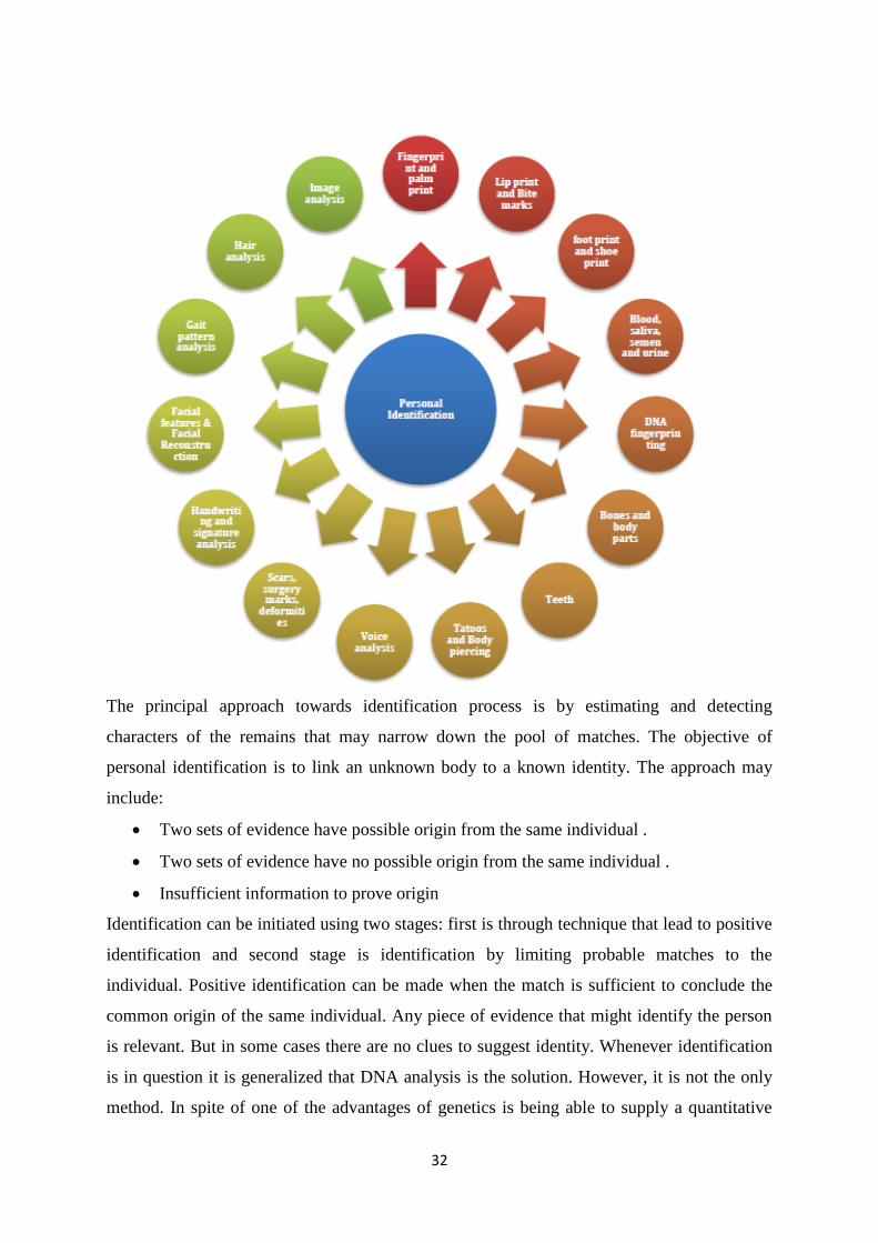
in identification. The following figure explains the various evidences through which personal
identification is done.
32
The principal approach towards identification process is by estimating and detecting
characters of the remains that may narrow down the pool of matches. The objective of
personal identification is to link an unknown body to a known identity. The approach may
include:
Two sets of evidence have possible origin from the same individual .
Two sets of evidence have no possible origin from the same individual .
Insufficient information to prove origin
Identification can be initiated using two stages: first is through technique that lead to positive
identification and second stage is identification by limiting probable matches to the
individual. Positive identification can be made when the match is sufficient to conclude the
common origin of the same individual. Any piece of evidence that might identify the person
is relevant. But in some cases there are no clues to suggest identity. Whenever identification
is in question it is generalized that DNA analysis is the solution. However, it is not the only
method. In spite of one of the advantages of genetics is being able to supply a quantitative

33
result, the lack of relatives to compare with sometimes invalidates its usefulnes . In these
circumstances the classical forensic anthropological examination is still useful and
exceptional. At that time the anthropological examination is used to assess biological
information like age, sex, ancestry and living stature and generates a biological profile of an
individual which results in tentative identification. Without a biological profile given by an
anthropologist, DNA could be useless.
Though genetics is really an excellent tool for identification, in many circumstances the
classical anthropological analysis remains as valid as ever, as emphasized by different
authors in many situations within the context of human rights violation such in Bosnia-
Herzegovina or in mass disasters like the World Trade Centre victims. There is always an
urge to develop new scientific method for personal identification by studying various
morphological and metrical approaches among different population.
Anthropological technique leads to the identification through bones, teeth, body and facial
features. The persons specializing in anthropometry are familiar with the biological variations
in the population, well informed in comparative osteology, craniometry, racial morphology,
skeletal anatomy and functions. Hence anthropologists study the origin, behaviour and the
physical, social and cultural development of human in all its aspects.
FORENSIC ANTHROPOLOGY
Anthropometry has a key role in many human growth and identification studies by measuring
human body and its parts for several years. The application of the knowledge in solving
practical problems has helped in the investigation of human variations among living
population. These results have introduced a new subject of interest with its practical
applicability known as ‗Forensic Anthropology‘. Forensic anthropology thus represents the
application of knowledge and techniques of physical anthropology to problems of medico
legal significance. Forensic anthropologists help he legal agencies to identify the unknown
individual (living/dead) during calamities and crime investigation. In cases of unimpaired
veracity of bodies, there are not so much problems in identification but identification is much
more difficult in impaired veracity of cadaver by different reasons such as an earthquake,
tsunami, floods, a war, any terrorist activities or a brutal murder.
Forensic anthropology is considered to be an applied subfield of physical anthropology.
Traditionally, forensic anthropology deals with the analysis of human remains. Occasionally
the remains are fragmentary or charred or sometimes decomposed and forensic
anthropologists are called to opine about its origin as well as biological characteristics that

34
assists in positive identification. Role of forensic anthropologists in investigation of unknown
is fundamental in field of forensic science.
A current definition for forensic anthropology can be found on the American Board of
Forensic Anthropology web site, ―Forensic anthropology is the application of the science of
physical or biological anthropology to the legal process. Physical or biological
anthropologists who specialize in forensics primarily focus their studies on the human
skeleton‖
Today, forensic anthropology is gaining massive popularity and has come into much public
attention with need to identify unknown individual. In 2008, Federal Bureau of Investigation
(FBI) and the Department of Defence Central Identification Laboratory under a joint venture
formed a working group ‘The Scientific Working Group for Forensic Anthropology
(SWGANTH)’ to develop and propagate best practice guidelines and standard for the
discipline. The SWGANTH consists of representatives from forensic, industrial, commercial
and academic communities, including international participants. Forensic anthropologists are
specialized and educated to interpret findings from skeletal remains in order to understand
variations between humans throughout the globe, across geography, between sexes, during
life span and between individuals. It is also important to understand the legal, cultural and
scientific challenges towards forensic anthropological analysis.
PHOTOANTHROPOMETRY
Alphonse Bertillon (1853-1914) a French criminologist developed a new method which uses
―a system of description and characterisation‖ which could be used with photographs for
identification which he called ‗Bertillonage‘. Bertillonage used enlarged photographs to
match with the photographs of the skull by using same focal length at a standard distance.
Bertillon, who was also a biometric researcher developed a system of description and
classification by use of anthropometry and developed an identification system based on
physical measurements of head and body. This measurement could be applied to one and
only one person and that could be specific to him. Alphonse Bertillon (1890) was first who
mentioned, photographs were futile for identification if they were not standardized by using
the proper lightening, scale and angles. There is a great degree of variation that occurs in the
face during aging of adults that will affect outcomes for face- based systems.
Face is the most distinctive and distinguishable part of the human body. Usually we recognise
people by their face and their facial features which help to provide information regarding
their age, sex and ethnic background. This uniqueness of face eventually helps us to identify
a person and differentiate from another face. Face has its individual shape, dimensions and

35
features which can be assessed morphologically as well as metrically. Recently, a question
towards the identity through surveillance is on rise with increase in frauds. With the raise in
number of ascertain cases of disputed identification in CCTV images and other photographic
evidence, there arise a need to develop a new scientific methods using facial comparison
technique. In cases related to CCTV images, it is mandatory to provide opinion evidence of
identity from examination of evidential images commonly it is termed as ―facial mapping‖ or
―facial comparison technique‖ . Facial mapping is a tool for identification of living that
involves morphological and metrical analysis. Generally permutation of three approaches is
made by expert:
Morphological Comparison.
Photoanthropometry.
Superimposition
This type of image evidence is permissible globally. Example in India, Italy, South Africa,
USA and in the UK more than 500 court reports on image evidences are prepared per annum .
In morphological comparison, facial features with distinct shape and size are categorised.
This technique gives reliable results even with poor quality images. Further statistical
analysis is conducted and face with similar feature is differentiated . Various studies have
showed that the personal identification process can also be done by using anatomical
landmarks and measurements or proportions obtained using landmarks from the photograph
or CCTV evidence. This form of identification can be termed as ‗Facial Image Identification‘
or ‗Photoanthropometry‘. Photo anthropometry is a metric based facial image comparison
technique which requires predetermined facial landmarks for measurement of the face from
an image. Here proportional analysis of the distances and angles between anatomical
landmarks in the images are calculated and compared. Photo anthropometry is now an
acceptable tool in the identification with a manual technique. In the present research to
overcome the manual method for identification, a new scientific approach towards photo
anthropometry has been made by developing a SAAS (Software at a Service) which works on
the principle of facial mapping using photographs which reduces time and labour. This type
of analysis can be applied to the large amount of identity document fraud, photo analysis,
manipulated or disguised facial photographs etc.With superimposition, one image is projected
over another in order to study facial similarities and dissimilarities. Superimposition using 3-
D (three dimensional) images is more successful. Despite of any method, only confirmation
36
of non-identity is probable. A distinct consistent difference has more influence than an
abundant of similarities.
SEX DETERMINATION:
It is the first step towards a development of any biological profile. Estimating sex involves
identification and evaluation of characteristics that shows differences between male and
female which can variably be noted. Sex is primarily determined by reproductive organs and
hormones, secondly by muscles and distribution of hair and lastly by physiological
differences of skeleton like pelvic bone and skull. Sex can be differentiated by sexual
dimorphism or through phenotype. Sexual dimorphism usually relates to the differences in
morphology (size and shape), physiology and behaviour. In forensic anthropology,
morphological differences are very useful for sex determination. Differences in the size and
robusticity display a degree of sexual dimorphism. In the skeleton the best criteria for
determining sex are in the pelvic bone and secondly from the skull .These differences reflect
the fact that females are smaller, less muscular and more gracile. Standards for sex
determination from different bones from several human groups can be found in sources on
human osteology and forensic anthropology. The range or distribution of sex variation differs
between populations with females tending to be more male-like in some group and males
more female like in others. The term ‗sex‘ and ‗gender‘ have distinct meaning. For any
analysis and estimations done by anthropologists based on skeletal characteristics, ‗sex‘ is
appropriately used. Methods that are developed for estimating sex falls into two categories:
Non-metric (macroscopic or visual) analysis and metric analysis. Metric analysis includes
measurements based on the landmarks to quantitatively evaluate size and shape difference
between two sexes. Metric analysis sometimes involves evaluation of single measurements or
numerous measurements. Currently from skeletonised remains, sex is being determined
through:
Humeral head diameter
Deltoid tub. Robusticity
Gonial eversion
Genial tubercle robust.
Browridge
Mastoid process size
Temporal line
Ischio-pubic index
Femoral head diameter

37
Bicondylar breadth
Sacrum curvature
Ventral arc
Ridge on sub-pubic angle
Sub-pubic concavity
Shape of symphyseal block
Ascending ramus width
Acetabulum size
When any foot or arm or face or other fragmented body parts are recovered at scene of crime,
their dimensions can provide valuable information about the gender of the unknown body.
There is a statistical correlation ship between body length and body segment which can be
calculated using various statistical equations to estimate gender. These dimensional
relationships have been of interest to many artists, scientists, anatomists, anthropologists and
in medico-legal for many long years. These body segments proportion vary between
populations due to genetics and the environment in which they live. Various parts of the body
segments respond and develop differently due to the environment and genetics.
AGE DETERMINATION:
Accurate estimation of age represents an important component in forensic anthropological
analysis. This determination of age is important in confirming the correct identity during
mass disaster when numbers of possible matches are greater. Determining age in children and
young people depends on morphological methods such as radiological examination of
skeletal and dental growth. In adults, age determination is difficult.
The estimation of biological age in a specimen is related to several biological processes, but
particularly the growth, modelling and remodelling of the skeleton, result in age-related
changes in the morphology of skeleton components. Age can be estimated by studying the
changes in bone growth, development and fusion, formation and eruption of dentals by use of
the pattern at known rates. From birth through the growth phase, the criteria for estimating
age include: the sequence of dental eruption as well as crown and root development, fusion of
epiphyses and apophyses and long bone length . By 20s the growth of bone is complete and
age can be determined from degenerative changes in the body. Joints are prone to
deterioration and undergoes consistent change over the period of one‘s life, thus age can be
estimated by studying this changes. Age can be determined through:
Dental eruption
Bone size and maturity

38
Epiphyseal closure
Cranial sutures
Sterna-rib end changes
Pubic symphysis
Current methods of age assessment include precise morphological methods with error
margins of greater than ±10 years or complex laboratory methods such as racemisation of
aspartic acid in dentin or tooth enamel or radiocarbon dating of tooth enamel with accuracy
of ±3 and ±1–2 years [28]. Both of the latter methods result in high precision age estimation.
As such, aspartic acid racemisation and radiocarbon dating currently represent the methods of
choice for precise age estimation.
FINGER PRINTS:
No two people have exactly the same fingerprints. Even identical twins, with identical DNA,
have different fingerprints. This uniqueness allows fingerprints to be used in all sorts of ways,
including for background checks, biometric security, mass disaster identification, and of
course, in criminal situations. Fingerprint analysis has been used to identify suspects and
solve crimes for more than 100 years, and it remains an extremely valuable tool for law
enforcement. One of the most important uses for fingerprints is to help investigators link one
crime scene to another involving the same person. Fingerprint identification also helps
investigators to track a criminal‘s record, their previous arrests and convictions, to aid in
sentencing, probation, parole and pardoning decisions.
Fingerprints can be found on practically any solid surface, including the human body.
Analysts classify fingerprints into three categories according to the type of surface on which
they are found and whether they are visible or not: Fingerprints on soft surfaces (such as
soap, wax, wet paint, fresh caulk, etc.) are likely to be three-dimensional plastic prints; those
on hard surfaces are either patent (visible) or latent (invisible) prints. Visible prints are
formed when blood, dirt, ink, paint, etc., is transferred from a finger or thumb to a surface.
Patent prints can be found on a wide variety of surfaces: smooth or rough, porous (such as
paper, cloth or wood) or nonporous (such as metal, glass or plastic).
Latent prints are formed when the body‘s natural oils and sweat on the skin are deposited
onto another surface. Latent prints can be found on a variety of surfaces; however, they are
not readily visible and detection often requires the use of fingerprint powders, chemical
reagents or alternate light sources. Generally speaking, the smoother and less porous a
surface is, the greater the potential that any latent prints present can be found and developed.

39
Principles of Fingerprint Analysis
Fingerprints are unique patterns, made by friction ridges (raised) and furrows (recessed),
which appear on the pads of the fingers and thumbs. Prints from palms, toes and feet are also
unique; however, these are used less often for identification, so this guide focuses on prints
from the fingers and thumbs.
The fingerprint pattern, such as the print left when an inked finger is pressed onto paper, is
that of the friction ridges on that particular finger. Friction ridge patterns are grouped into
three distinct types—loops, whorls, and arches—each with unique variations, depending on
the shape and relationship of the ridges:
Loops - prints that recurve back on themselves to form a loop shape. Divided into radial
loops (pointing toward the radius bone, or thumb) and ulnar loops (pointing toward the ulna
bone, or pinky), loops account for approximately 60 percent of pattern types.
Whorls - form circular or spiral patterns, like tiny whirlpools. There are four groups of
whorls: plain (concentric circles), central pocket loop (a loop with a whorl at the end), double
loop (two loops that create an S-like pattern) and accidental loop (irregular shaped). Whorls
make up about 35 percent of pattern types.
Arches - create a wave-like pattern and include plain arches and tented arches. Tented arches
rise to a sharper point than plain arches. Arches make up about five percent of all pattern
types.
Uses of Fingerprint analysis:
Fingerprints can be used in all sorts of ways:
Providing biometric security (for example, to control access to secure areas or systems)
Identifying amnesia victims and unknown deceased (such as victims of major disasters, if
their fingerprints are on file)
Conducting background checks (including applications for government employment,
defense security clearance, concealed weapon permits, etc.).
Fingerprints are especially important in the criminal justice realm. Investigators and analysts
can compare unknown prints collected from a crime scene to the known prints of victims,
witnesses and potential suspects to assist in criminal cases. For example:
A killer may leave their fingerprints on the suspected murder weapon
A bank robber‘s fingerprints may be found on a robbery note
In an assault case, the perpetrator may have left fingerprints on the victim‘s skin
A burglar may leave fingerprints on a broken window pane
A thief‘s fingerprints may be found on a safe
40
In addition, fingerprints can link a perpetrator to other unsolved crimes if investigators have
reason to compare them, or if prints from an unsolved crime turn up as a match during a
database search. Sometimes these unknown prints linking multiple crimes can help
investigators piece together enough information to zero in on the culprit.
In the absence of DNA, fingerprints are used by the criminal justice system to verify a
convicted offender‘s identity and track their previous arrests and convictions, criminal
tendencies, known associates and other useful information. Officers of the court can also use
these records to help make decisions regarding a criminal‘s sentence, probation, parole or
pardon.
Collecting Fingerprints:
Collecting Patent Prints
Patent prints are collected using a fairly straightforward method: photography. These prints
are photographed in high resolution with a forensic measurement scale in the image for
reference. Investigators can improve the quality of the images by using low-angle or alternate
light sources and/or certain chemicals or dyes during photography, but this is usually not
necessary.
Collecting Latent Prints
One of the most common methods for discovering and collecting latent fingerprints is by
dusting a smooth or nonporous surface with fingerprint powder (black granular, aluminum
flake, black magnetic, etc.). If any prints appear, they are photographed as mentioned above
and then lifted from the surface with clear adhesive tape. The lifting tape is then placed on a
latent lift card to preserve the print.
However, fingerprint powders can contaminate the evidence and ruin the opportunity to
perform other techniques that could turn up a hidden print or additional information.
Therefore, investigators may examine the area with an alternate light source or apply
cyanoacrylate (super glue) before using powders.
Alternate Light Source (ALS): It is becoming more commonplace for investigators to
examine any likely surfaces (doors, doorknobs, windows, railings, etc.) with an alternate light
source. These are laser or LED devices that emit a particular wavelength, or spectrum, of
light. Some devices have different filters to provide a variety of spectra that can be
photographed or further processed with powders or dye stains. For example, investigators
may use a blue light with an orange filter to find latent prints on desks, chairs, computer
equipment or other objects at the scene of a break-in.

41
Cyanoacrylate: Investigators often perform cyanoacrylate (superglue) processing, or fuming,
of a surface before applying powders or dye stains. This process, typically performed on non-
porous surfaces, involves exposing the object to cyanoacrylate vapors. The vapors (fumes)
will adhere to any prints present on the object allowing them to be viewed with oblique
ambient light or a white light source.
Chemical Developers: Porous surfaces such as paper are typically processed with chemicals,
including ninhydrin and physical developer, to reveal latent fingerprints. These chemicals
react with specific components of latent print residue, such as amino acids and inorganic
salts. Ninhydrin causes prints to turn a purple color, which makes them easily photographed.
DFO (1,2-diazafluoren-9-one) is another chemical used to locate latent fingerprints on porous
surfaces; it causes fingerprints to fluoresce, or glow, when they are illuminated by blue-green
light.
Other Collection Methods: In addition to the methods identified above, there are special
techniques for capturing prints from skin, clothing and other difficult surfaces. Amido Black,
a non-specific protein stain that reacts with any protein present, is typically used for
developing or enhancing bloody impressions on human skin. To reveal prints on clothing,
high-tech methods such as vacuum metal deposition using gold and zinc are showing promise
for the investigator. AccuTrans, a liquid casting compound, can be used to lift powdered
latent prints from rough, textured or curved surfaces. AccuTrans is basically a very thick
liquid that fills in the nooks and crannies of rough or textured areas where conventional print
lifting tape encounters difficulty.
Like fingerprint powders, chemical processing can reduce the investigator‘s ability to
perform other techniques that could reveal valuable information. Therefore, any
nondestructive investigations are performed before the evidence is treated with chemicals.
For example, a ransom or hold-up note will be examined by a questioned documents expert
before being treated with ninhydrin, since some formulations of ninhydrin will cause certain
inks to run, thus destroying the writing.
Footprints:
For years, criminal investigators and forensic scientists have used fingerprints to determine
identity. More recently, footprints have been discovered to be an equally reliable identifier.
Every person‘s foot has a unique set of ridges that make up a print unmatched by any other
human being. As with fingerprints, the footprint‘s pattern is a unique characteristic that can
pinpoint any one particular person.
42
While footprints can be used as a method of forensic science, more often the prints at a crime
scene do not come from a bare foot. To compensate, scientists have created methods of
identifying shoeprints. The indentations on the ground made by any shoe can be studied,
recorded and matched to prints found at other locations. Therefore, if one set of shoeprints is
found at the scene of a crime, it can be compared to another set of prints found at a separate
crime scene. A matching print tells investigators that the same person was involved in both
crimes.
To preserve the prints, detectives take detailed photographs that can be scanned into
computers for analysis. In many cases, technicians will make a casting of the print. So that a
duplicate may be produced. The casting process involves pouring a dense liquid into the
actual shoe imprint and allowing it to set. After the liquid solidifies, the casting can be picked
up as one piece so that an exact replica will be available for future reference.
Forensic scientists can also make other determinations about a suspect based on their shoe
print. The size of the shoe allows them to estimate the height of the person in question. By
comparing how deep the print is in connection with how solid the soil was and whether or not
the ground was damp at the time, investigators can gain a better idea of the weight of the
person. All of these methods can help determine the criminal‘s identity.
Process of fingerprint/ Footprint analysis:
Examiners use the ACE-V (analysis, comparison, evaluation and verification) method to
reach a determination on each print.
Analysis involves assessing a print to determine if it can be used for a comparison. If the
print is not suitable for comparison because of inadequate quality or quantity of features, the
examination ends and the print is reported as not suitable. If the print is suitable, the analysis
indicates the features to be used in the comparison and their tolerances (the amount of
variation that will be accepted). The analysis may also uncover physical features such as
recurves, deltas, creases and scars that help indicate where to begin the comparison.
Comparisons are performed by an analyst who views the known and suspect prints side-by-
side. The analyst compares minutiae characteristics and locations to determine if they match.
Known prints are often collected from persons of interest, victims, others present at the scene
or through a search of one or more fingerprint databases such as the FBI‘s Integrated
Automated Fingerprint Identification System (IAFIS). IAFIS is the largest fingerprint
database in the world and, as of June 2012, held more than 72 million print records from
criminals, military personnel, government employees and other civilian employees.

43
Evaluation is where the examiner ultimately decides if the prints are from the same source
(identification or individualization), different sources (exclusion) or is inconclusive.
Inconclusive results may be due to poor quality samples, lack of comparable areas, or
insufficient number of corresponding or dissimilar features to be certain.
Verification is when another examiner independently analyzes, compares and evaluates the
prints to either support or refute the conclusions of the original examiner. The examiner may
also verify the suitability of determinations made in the analysis phase.
DNA Analysis
The DNA collection and analysis gives the criminal justice field a powerful tool for
convicting the guilty and exonerating the innocent. Only one-tenth of one percent of DNA
differs from person to person. Although the DNA between people is so similar the small
differences can be used to identify an individual, victim or suspect. This DNA evidence is the
most important evidence to lawyers, because it is the most accurate method of identifying
someone. The Federal Bureau of Investigation (FBI) has chosen 13 points of Short Tandem
Repeats to be used as the base for DNA analysis and aside from identical twins, the chances
of these points being the same in two individuals is 1 in 1 billion or greater.
DNA analysis is a fairly new technology. The first use of DNA analysis was in the 1980s to
convict a man from Portland, Oregon of raping a 13 year old. In this case, DNA samples were
taken from the suspect and from the fetus which had been aborted. The DNA was then
analyzed and the results were conclusive. The results were conclusive because a fetus has the
mother‘s and the father‘s DNA and common sequences between were found between the
accused man‘s DNA and the fetus. The accused also confessed and was convicted.
There are different techniques to analyze DNA. Most labs have the ability to test Nuclear
DNA, which is DNA that is found in the Nucleus of every cell. There are other labs that have
the ability to use mitochondrial DNA analysis or Y-chromosome analysis. One of the first
techniques used to analyze DNA was Restriction Fragment Length Polymorphism or RFLP.
This technique analyzes the length of repeating bases in strands of DNA. The repetitions are
known as Variable Number Tandem Repeats, or VNTRs, due to the fact that they can repeat
from up to thirty times per strand, though the number of time it repeats varies from strand to
strand. The RFLP technique also requires the DNA to be dissolved in a specific enzyme that
helps break the DNA at specific points. In the case of RFLPs, a large sample of DNA that has
not been contaminated is needed.
Another method of DNA analysis is Short Tandem Repeat (STR) Analysis . Unlike RFLP,
STR analysis can use a smaller sample of DNA. This is because scientists can use a process
44
known as Polymerase Chain Reaction or PCR to amplify or make copies of the DNA and
produce the desired amount of material needed. Once the DNA has been amplified STR
analysis looks at how many times base pairs repeat at a specific location. This can be
repetitions of two (dinucleotide), three (trinucleotide), four (tetranucleotide) or five
(pentanucleotide) base pairs. Investigators have found that base pairs of four or five seem to
be the most accurate.
A special method to analyze DNA is through Y-Marking analysis or analysis of the Y
chromosome. This type of analysis helps identify DNA from different males. Another method
that is special is Mitochondrial DNA Analysis. This type of analysis uses DNA from the
mitochondria of a cell. It has been found that this type of DNA is most useful is helping to
solve cold cases because degraded DNA ( DNA that has been improperly stored or stored too
long)can be analyzed and can help find a perpetrator. A new special technique Single
Nucleotide Polymorphism or SNP are most often used to determine a person‘s chance of
contracting a certain disease. Forensic scientists rarely use SNP, but did to help successfully
identify victims of several September 11th victims after other identification methods failed.
Blood Evidence: Basics and Patterns
The discovery of blood in a case opens up a mini investigation within the investigation. This
is because an investigator must initially determine if a crime has been committed. It‘s
important to determine if a crime has been committed because the presence of blood does not
necessarily mean that there ever was a crime. This determination has to be made in a case
where a person is reported missing since it will help investigators. The blood found can then
be tested and see if it belongs to the victim; if the blood does belong to the victim there is a
possibility that a crime has been committed and that the case could change. Blood evidence
also comes in to play in criminal cases. Blood found on the blade of a knife could mean that a
crime was committed and someone was stabbed- but it could also mean that the victim sliced
their own finger. Even though there may be a crime where someone has been stabbed, it has
to be determined that a crime was committed with that particular knife. The red substance
that has been found is tested. Initially the blood is tested to determine if it is blood, and then
if it is human blood. Once the substance has been tested and it‘s been determined that it is
blood and it is human blood, it can be determined if the blood came from the victim or the
suspect. Blood evidence isn‘t just collected off of weapons, but can also be collected off of
the floor or other surfaces in a crime scene. This blood is also tested to determine if the blood
came from the victim or the suspect.
45
Besides testing, investigators use blood stain patterns to help determine if a crime was
committed. There are different types of blood stain patterns that an investigator looks for,
these patterns are as follows:
Drip Stains/Patterns – blood stain patterns that are created due to the force of gravity
acting on liquid blood.
Blood Dripping into Blood
Splashed (Spilled) Blood
Projected Blood (with a syringe)
Transfer Stains/Patterns -A transfer bloodstain pattern is created when a wet, bloody
surface contacts a surface that is not bloody. With this type of pattern, part or the entire
original surface may be recognizable, a full or a partial shoe print, for example.
Spatter Patterns- Blood spatter patterns are created when an exposed blood source is
subjected to an action or force greater than gravity (internally or externally)
Castoff- A blood stain pattern that is created when blood is released or thrown from a
bloody object in motion.
Impact –A blood stain pattern resulting from an object striking liquid blood
Projected-A blood stain pattern that is produced by blood being released under pressure–
for example, arterial spurting.
Investigators also look for the following blood stain patterns:
• Shadowing/ Ghosting- When there is an empty space or ―void‖ in the spatter. This
indicates that there was an object in the way.
• Swipes and Wipes- Swipes occur when blood on a surface is smeared. Wipes occur when
a bloody object brushes against a surface.
• Expiratory Blood – Blood that is coughed or breathed out. This is indicated by a misty
pattern that resembles high velocity spatter results.
Blood Stain Pattern Analysis
There are many different factors to consider when analyzing blood stain patterns. The first
thing that an investigator wants to determine is what kind of pattern is being presented. Blood
stain patterns can be presented as:
• Drip Stains/Patterns
– Blood Dripping into Blood
– Splashed (Spilled) Blood
– Projected Blood (with a syringe)
• Transfer Stains/Patterns

46
• Blood Spatter
– Castoff
– Impact
– Projected
• Shadowing/ Ghosting
• Swipes and Wipes
• Expiratory Blood
When an investigator is analyzing drip stains/ patterns, blood spatter, shadowing/ghosting,
and expiratory blood there are different factors they have to look at, these factors include:
– Whether the velocity of the spatter is low, medium or high
– The angle of impact
A low velocity spatter is usually four to eight millimeters in size and is often a result of
dripping blood after a victim sustains an injury such as a stab or in some cases a punch. For
example, if a victim is stabbed and then walks around bleeding, the drops of blood that are
left behind are low velocity. The low velocity drops in this example are passive spatters. Low
velocity spatter can also result from pools of blood around the body and transfers. A medium
velocity spatter is a result of a force anywhere from five to a hundred feet per second .This
type of splatter can be caused by a blunt force such as a baseball bat or an intense beating.
This type of spatter is usually no more than four millimeters. This type of spatter can also be
a result of a stabbing. This is because arteries can be hit if they are close to the skin and blood
can spurt from these wounds. This is classified as projected blood. A High velocity spatter is
generally caused by a gunshot wound but can be from a wound from another type of weapon
if enough force is used.
Once the type of velocity is determined it‘s important to determine the angle of impact. These
two factors are important to find so that it‘s possible for a point of origin to be determined. A
general observation that can be made by investigators about the angle without any
calculations being involved is that the sharper the angle, the longer the ―tail‖ of the drop is.
The angle of impact is determined by dividing the width by the length of the drop. Once the
angle is determined investigators then take the arcsine (inverse sine function)of that number
and then use stringing ( the use of strings to chart the trajectories of all the blood droplets in
the air) to determine the point of origin ( where the stings converge).
Collection and Preservation:
The collection and preservation of blood stain evidence is important because this evidence
can be used to type blood or run DNA analysis.
47
There are two different types of blood that can be collected at a crime scene: liquid and dried
blood. Liquid blood evidence is generally collected from blood pools but can be collected off
of clothing as well, using a gauze pad or a sterile cotton cloth. Once the sample is collected it
must be refrigerated or frozen and brought to the laboratory as quickly as possible. The
sample must first be dried thoroughly at room temperature. It is important to get the sample
to the laboratory as quickly as possible because after 48 hours the sample might be useless. If
the sample has to be mailed it should be completely air dried before packaging. If the sample
isn‘t completely dry when it needs to be packaged, the sample should be rolled in paper and
labeled and then put into either a brown paper bag or a box. The paper bag or box is then
sealed and labeled again. It is important to only place one item per container to avoid
contamination and samples should not be placed in plastic containers. Samples shouldn‘t be
in plastic containers because if the sample is still damp the moisture from the sample can
cause microorganisms that can destroy the evidence. Also, due to this fact, samples should
not be in any container for more than two hours.
Dried bloodstains can be found on small objects, larger objects and on clothing. When dried
blood is found on a small object the entire object can be sent to the lab after it‘s been properly
packaged and labeled. When dried blood is found on a larger object that is transportable, an
investigator should cover the stained area with paper and tape the paper to the object to avoid
contamination. If the object that is stained is not transportable there are different ways that an
investigator can collect the sample. One option is to cut out the stained area of the large
object. If the portion is cut out the sample is packaged the same way as described above but a
control sample should also be provided in a separate package. Another option is to use
fingerprint tape and lift the sample as well as the surrounding control area. If this method is
used it is important for investigators to not touch the sticky side of the tape with bare hands
and the investigator should run an eraser or some type of blunt object over the placed tape to
ensure that contact is made with the dried stain. The lifted stain is then be packaged and
labelled, then delivered to the laboratory. Another way to collect a sample off of an object is
to use a clean sharp object to scrape flakes of the stain into a paper packet. The last two
methods of collection of a dried bloodstain on a large object require the use of distilled water
to dampen the stain before rolling a thread in the stain or absorbing the stain with a cotton
square. These two methods are not recommended due to the risk of contamination. When
dried blood is found on clothing the entire article of clothing should be packaged and labelled
and delivered to the lab.

48
It is important for the investigator to remember to keep each sample separated so that there is
no contamination between samples.
Semen Analysis
In semen analysis, an alternative lights source like a laser is used to find stains on bed sheets
or clothing. Once a stain is found it is sent to be tested to determine the type of biological
fluid it is. If the biological fluid is not blood the other options are saliva, urine or semen.
DNA profiling starts from the identification of a stain as seminal fluid. Once this is
determined it is important for the DNA analyst to determine if sperm is present in the fluid.
It‘s important to determine if sperm is present because it contains a great deal of DNA, which
could help to identify a suspect. Once it has been determined that sperm is present, the
analyst must determine which item of evidence or swab sample has the most sperm so that
getting a DNA profile is more likely.
Sperm is identified by using a combination of general cell staining and bright field
microscopy. This has been around for forty years. This is the most popular method to detect
sperm but it is not the best method because it takes hours for an analyst to search for sperm.
However, it is more common because sperm that has been isolated from a sexual assault has
lost most if not all of the sub-cells that are needed for morphological identification. There is a
method that has been developed called SPERM HY-LITER™. This method is a fluorescent
monoclonal antibody-based kit which is used for microscopic identification. ―Forensic
Detection of Sperm from Sexual Assault Evidence‖ describes the kit as follows:
SPERM HY-LITER™ is designed to provide positive identification of sperm using a unique
monoclonal antibody that has been chemically tagged with an Alexa 488 fluorophore. The kit
incorporates a second fluorescent dye, 4′,6-diamidino-2-phenylindole (DAPI) that will stain
all cell nuclei; this is a fluorescent analogue of the KPIC stain currently used in most DNA
forensic laboratories. By combining both fluorescent dyes, SPERM HY-LITER™ provides
several visually confirmatory steps for the identification of sperm. Sperm can be visualized in
the fluorescein channel (the fluorescent spectra of Alexa 488 falls conveniently within the
emission maximum for fluorescein); all cell nuclei can be seen in the DAPI channel; and
using specialized dual filter ‗cubes,‘ epithelial nuclei and sperm can be visualized
simultaneously.
Although semen can easily be identified through testing, finding sperm may not always be
possible. Forensicscience.ie explains that semen in men who have had a vasectomy does not
contain sperm, so in cases like this further confirmatory tests must be done.
49
Medico legal importance of Age:
In medico legal cases, the age of a person is determined by medical officer on requisition
from the police officer or magistrate. Age is an important factor in following situations:
Judicial punishment
Juvenile punishment
Rape cases
Kidnapping cases
Employment
Attainment of majority
Evidence
Marriage
Identification
50
UNIT- 4
FORENSIC MEDICINE
Forensic Medicine or Legal Medicine deals with the application of Medical Knowledge to
AID in the administration of Justice. It‘s a Practical Science Bridging the LAW &
MEDICINE, Which are the EYES of the Society. (Medical Aspects of Law).
Forensic Medicine can be broadly divided into :-
Clinical Forensic Medicine :- deals with the examination of the LIVING individuals.
Forensic Pathology :- Deals with interpretation of Autopsy Findings in the Medico-Legal
investigation of Death (Forensic Thanatology – study of Death).
Medical jurisprudence: Deals with the legal responsibilities of the Physician with particular
reference to those arising from Physician-Patient relationship, such as Medical Negligence,
Consent, Rights and Duties of Doctors, Serious Professional Misconduct, etc., (in short, deals
with the Legal Aspects of Practice of Medicine)
Medical Ethics : Deals with the Moral Principals which should guide the members of the
Medical Profession in their dealings with each other, their Patients and the State.
Medical Etiquette : Deals with the conventional laws of courtesy observed between the
members of the medical profession.
Medico-Legal work in Ancient India:
The first treatise on Indian Medicine was the ―Agnivesa Charaka Samhita‖
―Charaka Samhita‖ lays down an elaborate Code regarding the training, duties, privilege
and Social Status of Physicians.
prescribes punishment for wrong treatment.
History of Forensic Medicine
Kautilya‘s Arthashastra states that Death can be caused by :-
Stopping the Breathing by 4 ways (Strangling, Hanging, Asphyxiation or Drowning)
Physical Injuries in 2 ways (by Beating or by Throwing from a Height)
Poisoning (poisons, snake or insect bite, or narcotic Drugs)
Kautilya‘s Arthashastra describes the necessity of Autopsy in establishing the Cause of
Death. It states that a Post-mortem shall be conducted on any case of Sudden
(Unnatural) Death, after smearing the body with oil to bring out Bruises, Swellings
and other injuries.
Medico-Legal work in British INDIA
The earliest Hospital was Established by Sir Edward Winter in Chennai in 1664 for
treatment of Sick Soldiers.
51
The incidence of Custodial Death and it‘s Certification by Medical Practitioners was
reported in Chennai in 1678.
The earliest Medical Certificate was issued by Edward Bulkley and Samuel Browne,
after examining Mr. John Nicks on Aug 16th
1693.
Medico-Legal Autopsy
1st post-mortem was conducted at Bologna, Italy
In India, The first Recorded Medico-Legal Autopsy was conducted in Chennai by Dr
Edward Bulkley on 28th
Aug 1693 on the body of Mr. James Wheeler, Member of Council.
In 1914, Lt. Colonel D. G. Crawford in his ―A History of Indian Medical Service‖ states
that ―A Very important part of a Civil Surgeon‘s duties is performance of Medico-Legal Post-
mortem‖.
Scope of Forensic Medicine
• Forensic Medicine deals almost entirely with Crime against Persons.
• In present days almost all the offences against the Human Body requires medical opinion
in order to deliver an impeccable justice in the court of law.
• The primary interest is to provide the source of information about medical science to
confine the needs of the Law.
• Forensic Medicine is mostly an exercise of common sense combined with the application
of Knowledge and experience acquired in the other branches of medicine
• Forensic Medicine involves Observation of Facts, Collection of evidence for the
construction of right inference based on Medico-Legal Examination.
CONSENT
o Voluntary agreement, compliance or permission for a specified act or purpose.
o Indian Contracts Act, Section 13 states that ―two or more persons are said to consent
when they agree upon the same thing in the same sense‖.
o To be legally Valid, the consent given must be intelligent and informed.
Types of Consent
IMPLIED CONSENT
EXPRESSED CONSENT :-
- Oral
- Written
- INFOMED CONSENT (The corner stone of medical practice)

52
Post-Mortem Examination (Autopsy):
Post mortem examination is an investigative scientific Dissection of Dead Body.
TYPES
Clinical or Academic Autopsy:- Performed with the consent of Relatives.
Medico-Legal Autopsy:- is performed on the Authorization of the Legal Authorities
in circumstances relating to suspicious, sudden, obscure, unnatural and Criminal Deaths.
Aims & Objectives of Medico legal Autopsy:
To find out the cause of Death, whether natural or unnatural.
To find out the manner of Death, whether accidental, suicidal or homicidal.
Estimate the Time Since Death.
To establish the identity when not known.
Document all the injuries, find out how the injuries occurred and their time of infliction.
To collect evidence in order to identify the object causing Death and to identify the
Criminal.
To retain relevant organs and tissues as Evidence.
In newborn to determine whether Stillborn, Deadborn or liveborn.
Rules of Medico-Legal Autopsy:
Body should be labeled as soon as it arrives in the mortuary.
Conducted only when there is an official order Authorizing the Autopsy.
Autopsy should be conducted as early as possible, with out undue delay.
Autopsy should be conducted in day light as for as possible.
The medical officer must go through the inquest report & Hospital Records and collect as
much information as possible pertaining to the circumstances of Death.
The body must be identified by the Police Constable, and his name must be recorded.
In unidentified bodies, the marks of identification, photographs, finger prints, etc., must be
taken
No unauthorized persons should be present at the autopsy.
Details of examination should be noted verbatim by an assistant.
Nothing should be erased and all alterations must be initialed.
Even if the body is decomposed, complete autopsy is a must.
EVIDENCE (sec 3 of IEA 1872)
Evidence means and includes all statements which the court permits or requires to be
made before it by witnesses, in relation to the matter of fact under inquiry.
53
For the evidence to be accepted by the courts, it must be properly identified as to what it
is, and where it was found.
Chain of custody : It is a method to verify the actual possession of an object from the
time it was first identified, until it is offered as evidence in the court.
Types of Evidence
Direct Evidence :- The witness testifies directly of his own knowledge as to the facts in
dispute.
Circumstantial Evidence :- Tends to Prove the ultimate fact in issue. (Presumptive
Evidence) – it‘s the Evidence derived from Circumstances as distinguished from direct and
positive proof.
Substantial Evidence :- The Evidence which a reasonable man will accept as adequate
for arriving at the decision in a case.
Corroborative Evidence :- The Evidence that concurs with another evidence. The
witness of the eye is positive.
The Medical witness or the Expert witness is only corroborative.
Documentary Evidence :- includes all the documents produced for inspection of the
court. Document means any matter expressed by means of letter, figures or marks. Ex :-
Medical Certificate, Medico-Legal Report, Dying declaration.
ORAL EVIDENCE :- In all cases Oral Evidence must be Direct. It must be evidence of a
person who Saw, Heard or Perceived.
Exception to Oral Evidence :- dying declaration, Expert opinion expressed in a treatise,
Evidence of a doctor recorded in a lower court, Evidence of a witness given in a previous
judicial proceedings,
Hearsay Evidence :- Any statement made by any person other than the Witness, giving
evidence in the court.
Dying Declaration:
It‘s the statement ORAL or WRITTEN made by a person who is Dying as a result of
some unlawful act, relating to the material facts of his death or bearing on the circumstances.
(Section 32 I.E.A.)
Before recording the statement the Doctor should certify that the person is conscious &
his mental faculties are normal (COMPOS MENTIS)
WITNESS:
Sections 118 to 134 of I.E.A. deals with Witnesses
All persons are competent to Testify unless the prevented from doing so.
54
TYPES OF WITNESSES :-
Common Witness :- First hand knowledge rule.
Expert Witness :- Is a person who has been trained or is skilled in technical and
Scientific subject, and Capable of drawing inferences, opinions and conclusions from the
facts observed by himself, or noticed by others. Ex :- doctors, firearm expert, finger printing
expert, etc.,
INQUEST:
Legal inquiry into the cause, manner and circumstances of any unnatural, sudden and
suspicious deaths.
TYPES OF INQUEST:
Police Inquest: This is the usual type of inquest conducted in India. The officer incharge of a
police station, not below the rank of Sub-Inspector (called the investigation officer of that
particular case), on receipt of information of death informs the executive magistrate and
proceeds to the place where the dead body is found. He conducts the inquest in presence of
two witnesses, who are in respectable position in the society (Panchayat head). He comes to
a conclusion of the apparent cause of death, as judged by him and prepares a Inquest report.
If death is purely due to disease he may hand over the body to the relatives of the deceased to
bury the body according t their religion customs. If he suspects some foul play or the cause of
death remains obsure, he sends the body to the nearest authorized centre for post-mortem
examination, along with a copy of the inquest report.
Magistrate Inquest: The executive magistrate appointed by the state government(Collector,
R.D.O or Thasildar) conducts the inquest in the following circumstances.
1. Dowry deaths
2. Death in police custody
3. Death during police interrogation
4. Death due to police firing
5. Death in a psychiatric hospital
6. Exhumation
Coroner’s Inquest: Coroner is a person qualified either in Medicine or Law or Both,
appointed by the Government. He conducts the inquest in all unnatural and suspicious deaths.
He is empowered to summon any person for enquiry. Coroner‘s court is a court of enquiry,
but he is not empowered to conduct a trial. This type of inquest is done in United Kingdom
and in some states of USA. This type of inquest is no more in India. Lastly withdrawn from
Bombay in 1999.
55
Medical Examiner System of Inquest: This is the type of inquest prevalent in most states of
USA and also in many advanced countries. A medical examiner (forensic expert) is appointed
to perform the functions of coroner. One receipt of information of a death, the medical
examiner visits the scene of crime and conducts the inquest. Thus he is able to gather first –
hand evidence which is interpreted in proper perspective owing to his knowledge of medical
science and autopsy is conducted by him. This is said to be the superior most type of Inquest.
CONSENT
Voluntary agreement, compliance, permission or acceptance for the act proposed by another,
for a specified act or purpose is known as Consent.
Indian Contracts Act, Section 13 states that ―Two or more persons are said to consent when
they agree upon the same thing in the same sense‖, to be legally valid, the consent given must
be intelligent and informed.
Types of consent:
Implied consent: The behaviour or the act of another person itself indicated that he has
consented for the act; you can take it for granted that he/she has given the consent.
Expressed consent:
Oral consent
Written Consent
Informed consent (The coroner stone of medical practice) – The doctor examining r
treating a patient without the consent amounts to the offence of assault. A doctor after
examining a patient must explain to the patient and/or the relatives:
1) What is the disease or problem he is suffering from?
2) What is his treatment plan, for that disease or condition?
3) What are the alternative treatments available?
4) What are the advantages and disadvantage of his proposed treatment?
5) What are the advantage and disadvantage of alternate line of treatment?
Then allow the patient to choose what type of treatment he needs; the law believes that the
patient himself is one who os best cared of his health and so he is the one to decide what is
good or bad to him and what is to be done to his health, which is he feels the best.
Informed refusal: When the doctor feels that, a particular type of treatment will suit the
patient best and may also result in some other complications at later course of life if the
patient does not accept for his proposed treatment plan, then the doctor has to inform in detail
all these and still if the patient refuses, the doctor has to bring all in writing and get it signed
by the patient
56
THANATOLOGY
Thanatology is the study of Death.
DEATH : Death is the complete and irreversible cessation of Circulation, Respiration and
Brain Functions. As long as circulation of oxygenated blood is maintained to the Brain Stem,
Life continues to exist.
Suspended Animation : The vital Functions of the body comes down to a minimum level,
just compatible with Life. The person appears apparently DEAD. Seen in:
1) New born
2) Electrocution
3) Drowning
4) Hypothermia
5) Induced voluntarily by Practice (yoga)
6) Drugs – barbiturates
Vegetative State : Breaths Spontaneously. Stable Circulation. But unaware of Self and
Environment. Seen in :
Diffuse Bilateral Cerebral Hemisphere Disturbance, with an Intact Brain Stem.
Brain Stem Death :
Irreversibly Unconscious
Irreversibly Apnoe
Diagnosis of Brain Stem Death
British Code or Harvard Criteria:
1) Preconditions:
a) Comatose Patient on a ventilator
b) Positive Diagnosis of Cause of
Coma – Irreversible Structural Brain Damage.
2) Exclusion:
a) Hypothermia
b) Drugs
c) Severe Metabolic or Endocrine Disturbances
3) Tests:
a) Brain Stem Reflexes
b) Apnoea Test
Types of Death:
Somatic or Clinical Death

57
Molecular or Cellular Death - Death of cells and tissues individually.
According to Causative Agent:
1) Natural
2) Unnatural – Homicidal, Suicidal, Accidental
MODE of DEATH:
COMA: Insensibility which involves the central portion of the Brain Stem and may cause
Death. Coma is clinical symptom and not a cause of Death.
- Compression of Brain
- Drugs - opium, cocaine, alcohol.
- Metabolic – uremia, diabetes.
- Infection –pneumonia, infectious fever
- Others – thrombosis & embolism epilepsy, hysteria, etc.
SYNCOPE: Sudden stoppage of action of Heart due to vasovagal attack resulting from
reflex parasympathetic stimulation
BP falls suddenly causing cerebral anaemia & rapid unconsciousness.
CAUSES : 1) Anaemia – sudden blood loss
2) Deficient power of Heart
- MI, Poisons, Fatty degeneration
3) Vagal inhibition
4) Exhausting diseases.
ASPHYXIA: Interference with respiration, or Lack of oxygen in respired air
- Due to which the Organs and Tissues are deprived of Oxygen (together with failure to
eliminate Carbon dioxide)
- Leading to unconsciousness & Death
ANOXIA: Gordon and his co-workers in 1994 claimed that the only mode of Death is
ANOXIA.
1) Anoxic Anoxia
2) Anaemic Anoxia
3) Stagnant Anoxia
4) Histotoxic Anoxia
POST-MORTEM CHANGES
Time Since Death:
Interval between time of Death and the time of conducting Post-Mortem.

58
This question arises most commonly in cases of Unwitnessed or Unreliably witnessed
Deaths.
Reliably witnessed Simultaneous Deaths.
Determination of TSD is an indispensable component of Corpus Deliciti.
EARLY CHANGES:
Changes in the Skin
Changes in the Eyes
Algor mortis
Livor mortis
Rigor mortis
LATE CHANGES:
Decomposition:
Putrefaction & its modifications:
- Adipocere
- Mummification
IMMEDIATE CHANGES:
- Complete and irreversible cessation of functions of Brain, Heart and Lungs.
- Muscles of the body become flaccid. (Primary flaccidity)
- Corneal and Pupillary reflexes are abolished.
1) INSENSIBILITY & PRIMARY FLACIDITY of MUSCLES
- Earliest Sign of Death, but not considered positive because it is also seen in cases of deep
coma and Narcosis.
2) PERMANENT STOPPAGE of CIRCULATION
- Stoppage of Circulation for more than 5min, but under certain circumstances, even after
stoppage of Respiration Heart may continue to beat for more than 10 min. E.g.: Hanging
Tests for Stoppage of Circulation:
1) Auscultation:
2) Icard‘s Test :
- 1% alkaline solution of fluorescin, if injected subcutaneously, will gradually spread
around with a greenish hue, if there is a minute capillary circulation.
3) Ligature Test :
A ligature is tied around the root of a finger, If there is little amount of circulation present,
the finger will become swollen & red and the proximal part of the ligature will become
blanched.

59
4) Magnus‘s Test :
A light is placed between webs of fingers, if there is capillary circulation still present there
will be a reddish-glow, But if there is no circulation, the light will have a yellow, waxy hue.
5) Finger Nail Test :
A finger is pressed with another finger, if there is circulation, it will become blanched and
when pressure is released, it will again become red.
6) Arterial Spurting Test :
If there is littlest circulation still present and a small artery is nicked, the arterial blood will
spurt.
7) Heat Test :
When heat is applied momentarily over the skin, there will be formation of a blister, with
congestion at that place, if there is slightest circulation present.
8) ECG :
Continued flat wave in all the leads, for continuous 5min signifies stoppage of Heart.
Tests for Stoppage of Respiration:
1) Feather Test
2) Mirror Test
3) Winslow‘s Test - A pot containing water or mercury is placed over the chest and a beam
of light is focused on it from a fixed source. The reflected image seen on the opposite side
will move if there is slightest movement of the chest.
Tests for stoppage of Brain function
1) Absence of motor and sensory functions.
2) Testing brain stem reflexes.
3) EEG.
Changes in the EYES:
CORNEA :
- Looses its lustre
- Becomes dull, hazy, and finally opaque and wrinkled.
- TACHE NOIR SCLEROTICA (A film of cell debris and mucus forms two yellow
triangles of desiccated discolouration on the sclera each at the side of the iris, which becomes
brown and then black)
INTRA OCCULAR TENSION :
- Falls so rapidly that by 2hrs after death, the tension is nearly zero.
- Eye balls appears sunken.

60
PUPILS : Dilated and fixed (no reaction to light), later becomes constricted (with onset of
rigor-mortis).
CHANGES in RETINA:
Fragmentation or segmentation of blood columns appear with in minutes after death
called as ―TRUCKING‖.
Retina is Pale for the 1st two hrs.
At about 6hrs, the disc outline is hazy and becomes blurred in 7 to 10 hrs.
Chemical changes in the vitreous humor
- There is a steady raise in the potassium level after death.
ALGOR MORTIS :(Cooling of the Body)
There is a constant balance between heat production and heat loss during life. After death
heat production is lost, hence the body starts cooling.
The body looses heat by Conduction, Convection and Radiation.
Body surface starts loosing heat rapidly, but the inner body Core temperature does not
alter until a gradient is established between the core body temperature and the environment.
There is no significant change in the Core Body temperature for sometime (1 to 2 hrs)
Once the gradient is established, then the body starts loosing heat at a constant rate.
The rate of cooling is proportional to the difference in temperature between the body
and the surface (Newton‘s Law)
Temperature is measured by using a Chemical thermometer from RECTUM or
ABDOMEN.
TIME SINCE DEATH :
Postmortem Caloricity:
The temperature of the body raises for the first few hours after Death.
- When the regulation of heat production has been severely disturbed before death, as in
sun stroke.
61
- When there is great increase in heat production in the muscles due to convulsions, Ex:
tetanus, strychnine poisoning.
- Excessive bacterial activity, as in septicaemic conditions, cholera and other fevers.
LIVOR MORTIS(Post mortem staining, Post mortem Hypostasis)
After stoppage of circulation, the blood which is fluid in nature, gradually settles down in
the toneless capillaries of the dependent parts of the body.
Seen externally as purplish discolouration through the skin.
Those parts of the body which is in actual contact with the surface, do not show PMS, As
the capillaries are pressed by the weight of the body- known as areas of ―contact flattening‖.
Postmortem lividity begins as mottled patches ranging from 1 to 2 cms in diameter
These patches gradually increase in size and coalesce with each other to form an uniform
area of staining – 2 to 3 hrs.
If the position of the body is altered, these patches gradually disappear, and appear in the
new dependent parts.
Fixation of PMS in 6 to 8 hrs.
PMS disappears with the onset of putrefaction.
PMS may not develop at all if the body is tossed and turned continuously as seen in fast
flowing water in rivers.
PMS can not be appreciated well in dark coloured persons.
PMS may not be appreciated even in fair skinned bodies if they have bled profusely or
were severely anaemic.
PMS has to be differentiated from Contusion.
Medicolegal importance
It is a sign of death
Helps in estimating the time of death
Indicates the posture of the body at the time of death
Deliberate disturbance in the posture of the body
Some times the colour may indicate the cause of death
RIGOR MORTIS
A state of stiffening of the muscles with slight shortening of the fibers.
Appears in all the muscles, both skeletal and smooth muscles.
Mechanism : ATP depletion
Order of appearance :
- Proximo-distal spread

62
Time of appearance : starts in 3 to 6 hrs
- takes 12 hrs to complete
- stays for another 12 hrs
- disappears in next 12 hrs
Conditions simulating rigor mortis
Heat stiffening
Cold stiffening
Gas stiffening
Cadaveric spasm or instantaneous rigor :
- the group of muscles which were at strenuous work during life goes into a sudden state
of stiffening, instead of passing to primary flaccidity after death.
Its of great Medico Legal Importance.
Conditions altering the onset and duration of rigor mortis
Age : - Does not occur in a foetus of less than 7 months old
- in healthy adult it develops slowly and well marked
- in children and old it is feeble and rapid
Muscular state : onset is slow and duration is long in cases where muscles are healthy and
at rest before death.
NATURE OF DEATH : In deaths from diseases causing great exhaustion and wasting,
the onset is early and duration is short.
Atmospheric condition :
- onset is slow and duration is long in cold winter
- onset is fast and duration is short in hot weather
Medico Legal Importance
It‘s a sign of death
Helps in estimation of Time Since Death
Indicates the position of the body at the time of Death.
Decompositions (Putrefaction)
It is a process of gradual destruction of body tissues by combined lytic action the body
enzymes & microorganism after death.
It takes place in 2 steps -
1) Autolysis
2) Putrefaction

63
Autolysis
Cell becomes permeable after death with release of cytoplasm containing enzymes.
The proteolytic enzymes causes chemical digestion and disintegration of the organs.
Autolysis is increased by heat & stopped by freezing.
Autolysis is an aseptic process result in maceration of dead foetus in the uterus.
Auto disintegration occurs in
brain – liquefaction
stomach & GI treat, pancreas
Putrefaction
Colour changes
Evolution of gases
Liquefaction of tissues
Colour change
1st sign of decomposition -Greenish discolouration in the right lilac fosse
which become evident in 12-24 hrs in summer and 1 to 3 days in winter
Ceacum – Hb + H2S ---> Sulphmethaemoglobin
(greenish discolouration)
Than spreads to the entire abdomen, then extends to the
chest, neck, face & last to the limbs.
Colour gradually changes to dark green then finally black.
The putrefaction bacteria spread easily in fluid and colonize the venous system
The superficial veins of the limbs, chest, abdomen, & neck are stained greenish blue
due to haemolysis of red cells and stains the walls of the vessels – MARBLING – (36
to 48 hrs)
Which should be – differentiated from filigree burns due to lightening.
Evolution of foul smelling gases
The main gases are ammonia, carbon dioxide, hydrogen sulphide, phosphorated
hydrogen & methane.
In early stages, these gases are non –inflammable, later with the formation of enough
hydrogen sulphide the gases become inflammable.
Abdomen gets distended
Diaphragm is pushed up compressing the lungs and heart. Blood stained frothy fluid
exudes from the mouth and nostrils

64
(Postmortem purge) – which can be mistaken for pulmonary oedema or drowning.
(which is due rupture of pulmonary and pharyngeal vessels)
Distention of breast in females and penis and scrotum in males.
Involuntary urination, defaecation, seminal ejaculation and delivery of dead foetus
occurs in this stage.
Gas bubbles accumulate in all the tissues
Subcutaneous tissues becomes emphysematous
Eyes bulge from their sockets
Tongue is forced out between swollen lips
Blisters are formed between the epidermis and dermis, first and lower surface of trunk
and thighs.
Epidermis becomes loosened (skin slippage) producing large, fragile sacs of clear or
pink red serous fluid.
These blisters gradually enlarge, join together and rupture, exposing large areas of
slimy pink dermis
By three days the face is so discoloured and bloated that identification becomes very
difficult.
Liquefaction of tissues
Colliquative putrefaction begins from 5 to 10 days
The abdomen bursts and contents of abdomen comes out of the cavity
The omental, mesentric and perineal fat liquefy into a translucent, yellow fluid filling
the body cavities between the organs.
All encapsulated internal organs are converted into bags of putrid fluid and
subsequently burst open into the thoraco abdominal cavity
First organ to putrefy:
adrenals , pancreas, spleen, liver, lungs, heart.
Kidneys and urinary bladder putrefy late
Prostrate in males and uterus in females are the last internal organs to undergo
putrefaction
The muscles become soft, loose and are converted into a thick semisolid pinkish mass
and are gradually separated from the bones.
The cartilages and ligaments are softened in the final stage.
Skeletonisation
65
In an exposed body flies, maggots, ants, cockroaches, rats, dogs, vultures etc., may
reduce the body to skeleton in a few days.
When the body is in water it may be attacked by fishes, crabs etc., and the body is
skeletonised within a few days.
Uncoffined body buried in a shallow grave-moderate delay of putrefaction
If deeply buried, low temperature, exclusion of air, markedly delays putrefaction
The main factors affecting skeletonisation are:
Seasonal, Climatic variations, Amount of soil water, Access of air, Acidity of soil
water
In India, an unconfined buried body is reduced to skeleton within one year
Bodies placed in airtight coffins, decay process may not occur for several decades.
Conditions affecting putrefaction
Temperature
Begins above 10°c, optimum between 21°c to 38°c
Arrested below 0°c and above 48°c
Moisture
Moisture is necessary for putrefaction
Bodies recovered from water, if left is the air decompose rapidly
Organs which contain Water decompose more readily than the dry ones
Air - Free air access hastens putrefaction
Clothings
Initially clothing's hasten putrefaction by maintaining body temperature
Putrefaction is delayed under tight garments
(ex : belts, socks, undergarments, boots, etc)
which dries out the blood from that part and there by prevents entry of internal organisms
Clothing's prevent the access of airborne organisms, flies, insects, etc
Manner of buried
Bodies buried soon after death, putrefaction is less
Delayed if buried is dry, sandy soil,
or in a grave deeper than 2 meters
Delayed in coffined bodies (exclusion of air, water and access of insects)
Salt and lime delay putrefaction
Putrefaction is rapid
In a body buried in a damp, marshy or shallow grave
66
In porous sandy soil, than in soils with excess of clay
Putrefaction is more rapid, if changes of decomposition are already present at the time of
burial
In acid peaty soils, even the bones may be destroyed.
Internal
1. Age
Bodies of children putrefy readily than the old people.
Bodies of new born who have not been
fed, decompose slowly
If fed before death or if there are any injuries on the body, decomposition is rapid
2. Sex
no difference of effect
3. Condition of the body
Fat bodies putrefy quickly than lean bodies, due to larger amount of fluid, excess
fat and greater retention of heat.
4. Cause of death
bodies of persons dying from Septicaemia, Peritonitis, Inflammatory and septic
conditions, Asphyxia, Putrefaction is rapid in infection due to Cl.Welchii (ex), Acute
intestinal obstruction, Abortion, Gas gangrene
Putrefaction is delayed if death due to wasting diseases, Anaemia ,Debility, Poisoning by
phenol, zinc chloride, strychnine, chronic heavy metal poisoning (arsenic, lead, mercury)
5. Mutilation:
Bodies with wounds putrefy rapidly
In dismemberment –
1. limbs putrefy slowly
2. Trunk putrefy rapidly
In advanced putrefaction, no opinion can be given as to the cause of death, except in
causes of poisoning, fractures, fire arm injuries etc.
Medico-legal Importance
When involves face, the features are well preserved which helps to establish the identity
To know cause of death, injuries are preserved
Time since death is calculated
Mummification
Modified putrefaction
67
Dehydration, drying and shriveling of the cadaver
Occurs due to evaporation of water
Begins in exposed parts of the body – face, limbs then extends to the entire body
including internal organs
Skin is shrunken and contracted, dry, brittle, leathery and rusty – brown in colour
Stretched tightly over the body prominences such as cheek, chin, ribs, hip and adheres
closely to the bone.
Mummification may be partial in some cases, with only head or limb being affected
Internal organs become shrunken, hard, black and become a single mass or may
disappear
Body loses weight, becomes thin, stiff and brittle
Mummified bodies if protected, can be preserved for years.
Mummified bodies are practically odourless
It takes 3 months to 1 year to develop
Absence of moisture, continuous action of dry warm air are necessary for
mummification.
Mummification of new born may occur if left in a trunk or cupboard (warm and dry
atmosphere)
Marked dehydration favours mummification
Mummification occurs in bodies buried in shallow graves in dry sandy soils
Chronic arsenic and antimony poisoning favours the process of mummification.
Occasionally some parts of the body shows mummification and some parts show
adipocere.
TIME SINCE DEATH
To know when the crime has been committed
A starting point for the Police
Exclude some subjects & to search for the likely culprit
To check on a subject‘s statement
The exact time of death can not be fixed, rather a range of time between which death
was presumed to have taken place, can be estimated
Longer the Postmortem interval, Less accurate is the estimated TSD
TO GIVE A OPINION:
All the available history,
68
Local physical & environmental factors at the scene of Crime, such as fire, open
windows, environmental temp, etc
The range of TIME is at best an educated guess, based on Scientific Knowledge and
Experience.
The following are considered to give a opinion of Time Since Death:
Cooling of the body
Postmortem staining
Rigor mortis
Decomposition and it‘s modifications : Mummification & Adipocere
Presumption of Death:
Arise in connection with civil cases
A person is presumed to be ALIVE, if there is nothing to suggest the probability of
his Death with in 30 years
But, if proof is produced that the same person is not been heard of for 7 yrs by his
Relatives & Friends, Death is Presumed.
Presumption of Survivorship:
Arise in connection with INHERITANCE of Property, when 2 or more persons DIE
in a common Disaster. Ex : Earth quake, Plane Crash, etc.
The Question of who SURVIVED Longer, may arise.
The case is decided on facts & Evidence available.
In the absence of such evidence, Age, Sex, Constitution Nature & Severity of injuries
and the Mode of Death are taken into Consideration
TOXICOLOGY
The toxicology is defined as science which deals with knowledge of source, character and
properties of poisons, their symptoms in the body, their pharmacological actions and
treatment.
Definition of Poison
Poison is a substance introduced in the body to produce ill-effect, disease or death. It may be
of any origin like synthetic, mineral, animal or vegetable. It may be introduced through any
route like mouth, nostril, anus, vagina, ears, eyes, or by injection or inhalation.
Definition of Drug
Drug is defined as any substance to be introduced in the human body for diagnosis,
investigation, treatment or prevention of any disease.

69
MEDICO-LEGAL ASPECTS OF POISONING
Poisoning in India is quite common. There are instances of human and cattle poisoning.
Cattle poisoning is mostly intentional to kill the cattle of the neighbour. Human poisoning
can be accidental, suicidal or homicidal:
1. Accidental Poisoning: Most of the poisoning is accidental. In children most of the
poisoning is accidental as they consume tablets or syrup in the household accidentally.
Accidental poisoning can occur in agricultural workers when spraying insecticides. It can
occur if a person drinks unsafe water contaminated with arsenic, etc.
2. Suicidal Poisoning: Nowadays, suicides by poisoning is very common among farmers,
especially in Andhra Pradesh and Punjab. They consume insecticides when they are unable to
pay agricultural loans. Aluminium phosphide and organophosphorus compounds are first
choice followed by organochrolo compounds. Suicide by poisoning is also common in
housewives.
3. Homicidal Poisoning: Poison is very commonly used for homicides, especially on young
children who are done away with this easily. Arsenic is commonly used followed by opium,
aconite, dhatura, strychnine and potassium cyanide. If the intention is not to kill but rob,
stupefying agents like dhatura, Cannabis indica or chloral hydrate are used.
CLASSIFICATION OF POISONS
Poisons are classified according to the symptoms which they produce in the body. Generally,
poisons are classified as follows:
Classification 1: Based on Structure
1. Corrosives: Strong acids and alkalis.
2. Irritants: They may be of three types:
(a) Inorganic
(i) Non-metallic: Phosphorus, chlorine, bromine, iodine, etc.
(ii) Metallic: Arsenic, antimony, mercury, copper, lead, etc.
(b) Organic
(i) Vegetable: Castor oil seeds, croton oil, Madar.
(ii) Animal: Snakes, insect bites, cantharides.
(c) Mechanical: Glass, hair, diamonds, etc.
Classification 2: Based on Area of Action and Method
Systemic Poisons
(a) CNS acting
I. Affecting brain:
70
(i) Somniferous: Opium, barbiturates, etc.
(ii) Inebriant:Alcohol,ether,chloroform, etc.
(iii) Deliriant: Dhatura, belladonna, Cannabis, etc.
II. Affecting spinal cord: Nux vomica, etc.
(b) Affecting peripheral nerve: Conium, Curare,etc..
(c) Cardiac poisons: Aconite, digitalis, oleander, tobacco, hydrocyanic acid, etc.
(d) Asphyxiants: Carbon dioxide, carbon monoxide, coal gases, etc.
ROUTES OF ADMINISTRATION
The following are the routes of administration of poisons:
1. Oral, i.e. through mouth.
2. Through inhalation, i.e. by nose/mouth.
3. Through application on skin, mucous membrane like mouth, vagina, anus, nose.
4. Through injection like intramuscular or intravenous.
5. On application on injuries like abrasion, cut wounds or lacerated wounds.
ACTION OF POISON
The actions of poisons on the body are classified as:
1. Local
2. Remote
3. Systemic
4. General.
When the poison acts on the site of application it is called local effect. Sometimes, poisons
may act at a particular place like liver or kidney, then it may be called remote or remote local
effect. When a poison affects a particular system like CNS, cardiovascular or GI tract, its
effects are called systemic. When multiple organ systems are affected, the effects are called
general. It may be possible that poison may affect in more than one way like local and
systemic too or affect all systems as in general effect.
Factors Modifying Action of Poisons
The following factors modify the action of poisons in the body:
1. Quantity: It is a general rule that more the quantity of poison in the body, more will be its
effect. But in some cases, too much of poison can also trigger vomiting which may throw out
majority of the poison. A large quantity of poison may sometimes change the mode of action
of the poison.Arsenic in large doses may cause death by shock while in small doses it may
produce only gastrointestinal symptoms.

71
2. Form: If a poison is administered in gaseous form it acts fastest, while solution acts faster
than solid form. Sometimes, the effect of one poison may be countered by another poison
which is also taken along with it. If somebody takes silver nitrate and hydrochloric acid
together, he will not have any poisonous effect as both poisons would react with each other
and produce nonpoisonous silver chloride. Solubility of the poison is also important as some
poison may not be water soluble but may dissolve in acid secretion of the stomach. The
action of poison may also be altered if an inert substance is mixed with poison as it may
reduce absorption from the stomach. If a poison is highly diluted with water, it may not affect
at all.
3. Mode of Administration: If a poison is introduced into the body via gaseous route it acts
the fastest. Parenteral route acts faster than oral route. The mode of administration should be
appropriate for the desired effect. Snake venom is very poisonous if injected but harmless if
taken by mouth. Application of poison on intact skin acts quite slow as compared to poison
applied on mucous membrane.
4. Condition of the Body: It has been found that less doses of poison is required for children
and old people as compared to young adults. Males tolerate poison better than females. The
body may have anaphylactic reaction with any drug or poison and death may be reported
even with small doses. Allergies are genetically inherited and may run in families. If a person
is already habituated to a particular poison like alcohol, opium, dhatura or barbiturates, etc.,
he may be able to tolerate higher doses as compared to a normal non-addicted person. Drug
reaction may occur in body and may potentiate the effect of poison or render it useless. The
sedative, hypnotic drugs have positive potentiating effect on level of alcohol once taken.A
healthy person can tolerate poison better than a sick person or persons having systemic
disorders like hypertension, or diabetes.
FEATURES OF POISONING
Features of Poisoning in Living
It depends upon the nature of poison taken by the person to cause certain characteristics
suggesting
poisoning but generally following features may be seen:
1. The onset of illness is sudden in a previously healthy person except in chronic poisoning in
which it is insidious.
2. The symptoms usually start within 1 hour of consumption of some food, water or drug, if
the poison has been given by oral route.

72
3. The symptoms start increasing and may cause severe bodily disorders even leading to
death. Symptoms may start decreasing once a certain time is passed.
4. It may be possible sometimes that persons who have consumed food or water along with a
sick person may also show similar symptoms.
5. The final proof of poisoning lies in detection of poison in blood, urine, vomits or faeces.
Similar poison can also be detected in left over food or water.
Features of Poisoning in the Dead:
The poisoning in a dead person can be established by post-mortem findings, analysis of
viscera and circumstantial evidence.
1. Post-mortem Findings: The post-mortem should be done very carefully in cases of
poisoning. There may be stains of poison on the body or on clothes. Some parts of vomitus
may also be seen on clothes or on body. The colour of skin may also point toward the kind of
poison. Cherry red appearance in cases of carbon monoxide poisoning may be seen. The skin
may appear jaundiced in phosphorus poisoning. If corrosives have been taken, mouth or lips
may show changes. Marks of injection may be seen if a poison has been given by injection.
But in some poisoning cases, no external findings may be seen. The internal examination of
the body may reveal the following features in the alimentary canal:
(a) Congestion
(b) Softening
(c) Ulceration of mucous membrane
(d) Perforation.
Congestion: Congestion in the stomach is caused by all irritant poisons. It is more marked at
cardiac end. It may appear in patches or may be diffused all over the mucous membrane.
Congestion in intestine may be confused with post-mortem staining. Post-mortem staining is
always present at the dependent portion of stomach/intestine while congestion may be
uniform all over mucous membrane.
Softening: Due to effect of corrosive poisons, the mucous membrane may be softened
usually at cardiac end and greater curvature. In softening the mucosa is not detached.
Ulceration: Corrosive or irritant poison can cause erosion of mucous membrane.The margins
become thin and friable.
Perforation: Perforation of the wall of stomach may be possible in sulphuric acid poisoning
or other irritant poisoning.
2. Viscera Analysis: It is very important to preserve viscera to establish poisoning. The
whole stomach along with contents, part of small intestine with contents, part of liver, one

73
half of each kidney and spleen (whole) along with samples of blood are preservedin saturated
solution of common salt for viscera analysis. The qualitative and quantitative analysis of
viscera must be done at the forensic sciences laboratory. Sometimes, the poison may not be
detected in the viscera. It may be due to:
(a) poison having already been extracted out by vomiting or gastric lavage.
(b) poison having already been metabolised in the body and death may be due to other
sequelae. In such cases, the clinical findings should be taken into consideration to decide the
cause of death.
3. Circumstantial Evidence: Sometimes, a bottle of poison may be detected from the site or
parts of poison may be seen on clothes. Vomitus may be seen on the scene of crime. The
suicide note may be found in some cases.
POISON INFORMATION CENTERS
In 1994, National Poison Information Centre was started at theAll India Institute of Medical
Sciences, New Delhi. WHO has provided its software on poisons (INTOX) from where
complete information about poison regarding its composition, diagnosis, clinical features and
treatment is made available to public. Second centre was opened at the National Institute of
Occupational Health, Ahmedabad. New centres have also come up at the Government
General Hospital, Chennai, Industrial Toxicology Research Centre, Lucknow and Amrita
Institute of Medical Sciences, Cochin. The Government of India plans to open more centres
all over India. All these centres can be assessed by common public or medical community to
know about nature and treatment of any poison.

74
UNIT- 5
INJURIES AND SEX RELATED ISSUES
WOUNDS
The wound is defined as forcible disruption of continuity by mechanical violence of tissue of
the body like skin, cornea, or mucous membrane.
Classification of wounds:
Commonly, wounds are classified as follows:
1. Incised Wound: The incised wound is produced by a sharp weapon such as knife, razor,
etc. It is always broader than the edge of the weapon causing it due to restriction of cut
tissues. It is spindle-shaped and gaping. The length of the incised wound is greater than its
depth. The edges are smooth, clean cut and everted. If an incised wound is caused by a heavy
weapon like gandasa, the edges of the wound may show contusion. The direction of incised
wound should always be noted. The commencement of incised wound is deeper and it
gradually becomes shallower and tails off towards the end. The tailing off of an incised
wound shows the direction by which the weapon was drawn off. The incised wound bleeds
more, as the blood vessels are clean cut and hence bleed more. Small incised wounds on wrist
may cause death due to excessive bleeding.
2. Stab or Punctured Wounds: Stab wound is an incised wound where depth is more as
compared to breadth. It is caused by sharp, pointed and cutting instruments. Stab may have
both edges clean cut if the cutting instrument has sharp edges on both sides, or one side blunt
and another clean cut if instrument had one sharp and other blend side. Stab wounds are
called penetrating wounds when they pass through tissues, enter a body cavity like thorax or
abdomen. A sharp, pointed, cylindrical or conical instrument may produce a wound with
circular margins. A blunt pointed instrument may produce circular margins with laceration.
The depth of stab wound may be equal to or less than the length of the blade of the
instrument causing it. In rare cases depth may be even more than the length of the blade as
while forcing the instrument inside, the blow may depress the tissues of the part struck and
the blade may reach deeper tissues. This is usually seen in stabs over the abdomen.
Perforating Wounds: When punctured wound perforates the body there may be entry and
exit wound. The wound of entry is usually larger with inverted margins and wound of exit is
smaller with everted edges.
3. Lacerated Wounds: The lacerated wounds are caused by application of great blunt force
on the body, e.g. hit by lathi or blow, or in road traffic accidents. These wounds do not
generally correspond with the shape or size of the instrument causing them. The margins are
75
irregular, torn, swollen and contused. The underlying tissues show extravasation of blood
with muscle tears. Fracture of bones may be seen. The lacerated wound as a rule do not bleed
as heavily as incised wound because in this vessels are crushed and they bleed less. The
lacerations are of the following types:
(a) Split: Split laceration occurs when the soft tissues are caught between hard inside surface
like bone and the force applied. Scalp lacerations are common. These lacerations sometimes
look like incised wound with naked eye examination. So, they may be called incised-looking
wound‘. But on examination by lens, the margins can be observed. They are irregular, not
clean cut as in case of incised wound. The hair follicles would be seen crushed, not cut.
(b) Stretch laceration: They are due to over-stretching of skin by blunt force.
(c) Avulsions: These glacerations are caused due to grinding compression of force such as
the wheel of a car passing over limbs, shearing a large area of skin.
(d) Tears: When the body hits a hard object, it can produce tears, e.g.hit by a projected
handle of a car in road traffic accidents.
(e) Cut laceration: These are produced by heavy cutting weapon like gandasa. Here, careful
examination would reveal margins that are irregular and not clean cut. The contusions may be
seen around margins with crushed hair follicles.
INJURY:
Injury is damage to the body caused by external force. This may be caused by accidents, falls,
hits, weapons, and other causes. Major trauma is injury that has the potential to cause
prolonged disability or death
Injuries by Mechanical Violence
DEFINITION OF MECHANICAL INJURY
The mechanical injury is defined as ―damage to any part of the body due to application of
mechanical force‖. This damage may cause loss of tissue. The injury which is associated with
loss of tissue is usually referred to as wound. The other common types of injuries are
contusions or bruises and/abrasions.
Bruises or Contusions
These are caused by application of blunt force like lathi, fall from height, road traffic
accident, hit with a hard object like stone, hammer (Fig. 12.1), etc. Due to the application of
force on the skin, the underlying subcutaneous blood vessels rupture, which causes
extravasation of blood in subcutaneous tissues.This is called ‗ecchymosis‘. There is no
discontinuity in the outer layer of the skin. Bruises are quite painful. The sites of bruises are
quite tender and swelling can be seen. Ecchymosis is observed over the seat of injury in 1-2
76
hours, although it may appear early where the skin is very thin as of scrotum and eyelids.
Sometimes, ecchymosis is seen after 1 or 2 days if the deeper tissues are involved.
Sometimes ecchymosis is seen quite away from the seat of injury, e.g. in scalp. Here they
may gravitate into eyelids, commonly called black eye.
Extent of ecchymosis is dependent on followingfactors:
1. Nature and severity of force.
2. Vascularity of area.
3. Amount of subcutaneous fat.
4. Looseness of underlying cellular tissues.
5. Medical condition of the victim.
The more severe the force is, more severe are the contusions. Ecchymosis is severe in soft
tissues like eyelids, scrotum and vulva where vascularity is very high. In places where
subcutaneous fat is more like abdomen, ecchymosis is less appreciated. If underlying tissues
are loose like those in scalp, ecchymosis may be seen at distal places. In children, bruises are
easily produced due to delicate skin. In women and old persons the bruises are easily
produceable as compared toyoung men. In bleeding disorders like purpura, scurvy,
haemophilia and pathological condition like leukaemia, there is a tendency to bleed more in
bruises.
Abrasions
Abrasions are injuries where there is discontinuity in the skin due to loss of superficial
epithelial layer
of the skin. These are produced as a result of blow, fall, slide or being dragged.
The following are the different type of abrasions:
1. Scratches: They are produced by a sharp weapon like needle or pin.
2. Grazes: They are produced as a result of friction like fall on a rough surface.
3. Pressure Abrasions: These are due to sustained pressure on the area just like the ligature
mark in hanging, strangulation, etc.
4. ImprintAbrasions: Sometimes,the pattern of the object is seen on the skin just like tyre
marks or marks of radiator in road traffic accidents.
5. Other Abrasions: Sometimes natural abrasions also occur like ‗nappy abrasion‘ which is
seen in infants due to excoriation of skin over areas usually covered by nappy like groin and
buttocks. It may be confused with mechanical violence.
6. Contused Abrasions: If more mechanical violence is used, the abrasion may be contused
too, in such cases it may be referred to as contused abrasion or abraded contusion.
77
ELECTRICITY
Deaths due to electricity are quite common in rainy season. The deleterious effects of
electricity depend on following factors:
1. The Nature of Current: High voltage currents are very dangerous to life. Even low
voltage currents of high amperes are dangerous. Alternating currents are more dangerous than
direct currents as they produce contraction of muscles by which a person is not able to release
the grip on the wire through which the current is flowing. The domestic supply is 210–220
volts alternating current. The current up to 50 volts does not produce deleterious effects.
Various cases have been reported where people have survived very high voltage current also.
Judicial electrocution or electric chair:
This form of execution is quite common in Europe, and America and other countries. In this,
the person is made to sit on an iron chair with shaven head and is strapped. A high voltage
current, about 1800 volts, is passed through his head twice for about 60 seconds. One
electrode is kept on his shaven head and one on leg.
2. Resistance of Body: The deleterious effects of electricity also depend upon the amount of
resistance offered by the body. The human body is a bad conductor of electricity, but if wet,
resistance is decreased. If a person is wearing wet clothes or carrying a metallic article
touching the ground or where he is not wearing shoes, the body offers less resistance and the
person may die easily with less current.
3. Duration: Greater the duration of current, more the damage. Even low voltage current for
a long duration can cause spasm of muscles and the person may not be able to release himself
and die.
Causes of Death
Low voltage currents up to 220 volts cause death by ventricular fibrillation while midvoltage
currents up to 1000 volts cause ventricular fibrillation and respiratory centres failure. High
voltage current above 1000 volts causes direct respiratory centres failure. Delayed death may
occur due to infection, paralysis, etc.
Post-mortem Findings
1. Joule Burn or Endogenous Burns: These are characteristic electric marks found on
the skin. They are round with a shallow crater surrounded by a slightly raised ridge of skin
with a grey ashy base. Clouding of cornea, cataract, retinal haemorrhages, memory loss,
convulsions, paralysis, deafness, dumbness, headache, etc. may be seen in many cases.

78
2. Filigree Burns: They are reddish brown arborescent markings (superficial burns) on the
skin. These are erythemas which may show the path of passage of electric current in dendritic
fashion. These are typical of lightning. They disappear in a few days. Besides these, singeing
of hair, blisters etc. may be seen. The clothes where lightning enters may be burnt. Metallic
articles like keys, belt buckle or part of pen may be found magnetised. Rigor mortis may start
early and pass off early. The findings mentioned above may be seen. The internal findings are
not characteristic. There may be extensive congestion in brain. Internal organs are found to be
congested. Rupture of ear drums may be seen and it may resemble the shape of the object
through which electricity enters. Microscopically, honeycomb vacuolisation of keratin may
be seen. Bullae may be present in and under the epidermis. Epidermal nuclei at the periphery
appear hyperchromatic, distorted and fusiform showing a streaming pattern. Joule Burn is
typical of electrocution. Metallic particles may be demonstrated in the burn area.
Flash Burns
When a person comes near the high voltage current due to proximity, the current jumps from
source to body area and involves large area of skin. These are called ‗flash burns‘. The face is
usually pale, eyes are congested in most cases. Burns may be seen at the point of entrance
and exit. Sometimes, burn at the point of exit may not be there as it is large in area. Internally
most of the organs are found to be congested. Tardieu‘s spots may be seen in brain and other
organs. There are no other significant internal findings.
ASPHYXIA:
Asphyxia literally means Pulselessness.
In Forensic Medicine, Asphyxia means Lack of Oxygen.
Asphyxia is best described as an interference with the respiration due to any cause –
mechanical, environmental or toxic.
Causes of Asphyxia
SMOTHERING: Closure of external orifices.
HANGING, STRANGULATION or THROTTLING: Occlusion of air passage by
pressure on the neck.
CHOKING : Occlusion of air passage from with in.
SUFFOCATION: Lack of oxygen in the Atmosphere, or inhalation of irrespirable gases.
TRAUMATIC ASPHYXIA: Restriction of respiratory movements of the chest or
Abdomen.
DROWNING: Prevention of Gas exchange in the lung by FLUIDS.
CYANIDE POSIONING: Inability to utilize oxygen by peripheral tissues .

79
SEXUAL ASPHYXIA
GAGGING
BURKING
OVERLYING
CAFÉ CORONARY
Mechanical Asphyxia:
→ Obstructive – Smothering, gagging, Choking, Café coronary.
→ Constrictive – hanging, Strangulation, Throttling, bansdola
→ Restrictive – Traumatic, Burking.
→ Replacement – Drowning.
SIGNS OF ASPHYXIA:
CLASSICAL SIGNS :-
1) Oedema and Congestion of organs :
2) Petechial Haemorrhages :
3) Cyanosis :
NON-SPECIFIC SIGNS :-
1) Abnormal fluidity of blood
2) Dilatation of Right Chambers of Heart
SPECIFIC SIGN :- Indicates the exact way in which the FATAL chain of events was
initiated.
ligature mark on the neck in HANGING.
Finger nail abrasion on the neck in Manual Strangulation ( Throttling )
fluid in the air passage in DROWNING.
Congestion and Oedema
When the neck is compressed as in hanging, the venous return gets obstructed, leading to
CONGESTION.
Congestion is associated with tissue swelling, if there is continued venous obstruction.
When circulation stops completely, the walls of the capillaries become permeable
resulting in exudation of fluid from the capillaries into the neighbouring tissues leading to
OEDEMA.
Petechial Haemorrhages
Due to impaired integrity of capillary walls, the Red Blood Cells escape through,
producing small bleeding points, varying from Pinpoint to Pinhead size.
80
They are commonly seen in the sclera, conjunctiva and serous membranes such as pleura
& pericardium.
Cyanosis
The colour of oxygenated blood is SCARLET RED. When the Haemoglobin is not fully
saturated with Oxygen i.e., haemoglobin is reduced, the blood assumes a bluish colour, called
Cyanosis.
for cyanosis to be evident, there should be at least 5 grams of reduced Haemoglobin per
100 ml of blood.
Suffocation
→ Suffocation is a type of Mechanical Asphyxia cause by obstruction to the passage of air
into the lungs. [ Excluding Constriction of the Neck and Drowning ]
→ Environmental Causes :- by reduction of oxygen in the atmosphere. This occurs in
decompression, high altitude and vitiated atmosphere ( irrespirable gases )
Hanging
→ Hanging is a form of asphyxia caused by suspension of the body by a ligature around the
neck. The constricting force being the weight of the body.
→ Hanging is the preferred method of choice for committing suicide.
→ Hanging is almost always suicidal, unless proved otherwise.
Types of Hanging
→ On the basis of the degree of suspension
- Complete Hanging
- Partial Hanging.
→ On the basis of the Position of KNOT :
- Typical Hanging
- Atypical Hanging.
Cause of death in Hanging
Asphyxia
Venous congestion
Combined asphyxia & Venous congestion
Cerebral Anaemia
Reflux Vagal inhibition
Fracture dislocation of cervical vertebra
Delayed deaths
Infection

81
Oedema of Lungs / Larynx
Hypoxic encephalopathy
Infarction of Brain
Brain Abscess
Cerebral softening
Post-mortem Appearance
Ligature Mark :- The ligature leaves a furrow of it‘s own width and pattern.
The bed of the ligature mark is PALE.
The edges are abraded ( reddish-brown)
Ecchymosis and congestion of adjacent skin may be seen.
The pattern of the ligature material often gets imprinted on the skin as a Pressure
Abrasion.
The ligature mark is usually situated above the Hyoid bone, oblique, passing backwards
and upwards symmetrically on either sides to the point of suspension
Microscopically, the ligature mark displays the usual characteristics of abrasion, showing
desquamation and flattening of cells of the epidermis.
If death has occurred quickly, vital reactions may be quite difficult to demonstrate.
Other external findings
Face congested.
Cyanosis of lips and nail beds.
Post-mortem staining : Glove and Stockings Fashion
Petechial Haemorrhages in the lower limbs if the body is suspended for a long time.
Head is tilted away from the knot.
Tongue is protruded and may be bitten.
Salivary stains at the angle of mouth.
If the knot presses on the cervical sympathetic ganglia, the eye on that side remains open,
and the pupil dilated. [ Le facie sympathique ]
Neck Dissection
Neck is examined after removal of brain and thoracic contents [blood less field of
dissection]
superficial incisions of the grooves may show small haemorrhages in the underlying
layers of the skin, caused by direct trauma caused by the ligature.
Contusion of the muscles of the neck, especially sternocleidomastoid and platysma.

82
The intima of the carotid arteries show transverse splits with extravasation of blood in
their wall.
Hyoid bone
Occasionally hyoid bone is fractured.
Fracture is more common above the age of 40 years.
Abduction fracture [antero-posterior compression fracture]
The broken piece of bone have an outward angulation.
The subcutaneous tissues immediately above and below the ligature mark are congested.
The tissues underneath the ligature mark are pale and parchment like.
The lymph nodes of the neck above and below the ligature mark, shows evidence of
congestion, stasis & haemorrhage.
Lynching
It‘s a form of homicidal hanging where the person suspected to be involved in dacoit,
murder or rape is overpowered by a mob, and hanged forcibly in public.
Post-mortem hanging
Occasionally, after a victim has been murdered, and the body may be suspended to
simulate suicidal hanging.
Findings of asphyxia will not be evident and the actual cause of death may be easily
found out on Autopsy.
Antemortem Hanging Postmortem Hanging
Ligature Mark Prominent furrow
Not characteristic
Salivary dribbling present
absent
Drag marks absent
present
Rope fibres May be seen absent
83
STRANGULATION
Definition:
Mode of violent asphyxial Death accomplished by constricting the neck, externally by hands
(manual strangulation / throttling) or by a ligature (ligature strangulation) or any other means
but without suspending the body.
Mugging – foot, knee, bend of elbow – compressing force.
Bansdola
Garroting
Never suicidal, mostly homicidal; occasionally accidental.
Mild playful tweak on neck – vagal inhibition – No signs in autopsy
Symptoms of Strangulation
Compression neck is sudden & symptoms of occlusion of air passage is complete and
victim is powerless to call up insensible (unconscious) die instantaneously.
Incomplete occlusion:- face cyanosed, bleeding from the mouth, nostrils and ears, hands
clenched, convulsions - death
Rapid Insensibility---- Death almost Painless
Causes of Death
1. Asphyxia (Anoxic hypoxia)
2. Cerebral Ischaemia
3. Venous congestion
4. 50% cases cardiac arrest
5. Pale face – Rapid death (cardiac arrest) cyanosed face – slow death
Postmortem appearance in ligature strangulation
Ligature mark
Well defined & grooved slightly.
Level – below the thyroid cartilage,
Completely encircling the neck
Horizontal
Multiple marks - several rounds of ligature & more than one knot
Oblique: Dragging or the victim was sitting & assailant standing behind the victim
Base of the ligature mark: Pale with reddish ecchymosed margins (hrs after death – dry,
hard, parchment like)
Imprint pressure abrasion of ligature material
Abrasions & ecchymosis of adjacent skin
84
Some times no marks ligature is present if the ligature material is soft & yielding like
scarf/stockings or if removed soon after death
Minute fibers from the ligature material on the ligature mark - adhesive cello tape
Asphyxial signs – face swollen, cyanosed, Petechiae over the face and conjunctiva,
eyeballs prominent , open and congested, pupil dilated, lips blue, frothy blood coming out
through the mouth & nose.
Larynx & trachea – congested, frothy mucus
Lungs marked congestion haemorrhagic patches, Petechiae, dark fluid blood on cut section
Emphysematous bullae
Viscera congested and Petechiae may be present
MANUAL STARNGULATION (THROTTLING)
Autopsy findings: Signs of asphyxia
Bruising of neck due to assailants fingers grasping the neck. Bruises: circular unusually,
dark red or purple in color 1-2 cms size due to finger tips if fingers slide over neck then
elongated bruise.
Abrasions (linear & Crescentic) – by finger nails of assailant (victim in an attempt to get
loosen the hand grip of assailant from the neck).
Rope / loincloth – suddenly thrown over head & quickly tightened around neck – loses
consciousness instantly so there may not be signs of struggle, then tie ligature to kill. This
way it is possible to over power a healthy strong man & kill him.
Accidental – scarf, dupatta, caught in moving machinery belt / vehicle
When one hand is used: bruise mark by thumb on one side & four fingertips – obliquely
downwards, out wards & placed one below the other
sometimes 4 finger marks clustered together. Red / purple bruise marks sometime turn into
brown, dry & parchment like afterwards
When both hands used to throttle:- Thumb marks of one hand & finger marks of other
hands on either side of throat will be seen.
Bruise & Abrasions also on mouth, nose, check forehead, lower Jew or any other part of
the body –marks of struggle.
Some times ribs & injuries to organs in side the chest & abdomen, when assailant kneels
over chest.
DROWNING
Type of asphyxial death caused by displacement of air from the lungs by any fluid, usually
water.

85
Classification
According to the type of water
- fresh water drowning
- sea or salt water drowning
According to water detection in the lungs
- wet droning
- dry drowning
According to the period of survival
Drowning – immediate
Near drowning – recued but died with in 24 hours
Secondary drowning – death after 24 hours due to complications
POSTMORTEM FINDINGS
A. NON SPECIFIC SIGNS
I. External :
Wet hair and clothing
Adherent particles such as sludge ,mud or sand
Wrinkled skin over palms & soles
Cutis anserina (goose skin)
Cyanosis
Bleeding from ear
Broken nails
Injuries: abrasion, laceration, contusion or by Aquatic animals.
Hypostasis – face, chest limbs.
SPECIFIC SIGNS
1. Froth over nose and mouth (the sign of drowning)
fine, tenacious, white or blood tinged, recurrent repeatedly
2. Cadaveric spasm: weed and plants may be in tight grip.
NON SPECIFIC
Internal:
Congested organs
Oedema easily appreciable in lungs
Fluid blood in heart and vessels
Petechial heamorrhage (Tardieu spots) over pleura & pericardium
Sand & mud particles in nose, mouth or in oropharynx

86
Water in stomach
SPECIFIC SIGNS:
Air passage
fine leathery or tenacious white blood stained froth in trachea & bronchi
Sand, mud or sludge particles in trachea, bronchi or terminal bronchiole
Laboratory tests:
Gettler’s test:
- To find out the type of water with chloride concentration from right and left ventricular
blood.
To find out ante mortem drowning
1. Diatom test
2. Serum magnesium
3. Serum Strontium
Medico Legal importance:
1. Accidental drowning
2. Suicidal drowning
3. Homicidal drowning
Floatation time:
- 18 to 24 hours in summer
- 24 to 48 hours in winter
Sex related issues:
Sexual violence is a significant cause of physical and psychological harm and suffering for
women and children. Although sexual violence mostly affects women and girls, boys are also
subject to child sexual abuse. Adult men, especially in police custody or prisons may also be
subject to sexual violence, as also sexual minorities, especially the transgender community.
Sexual violence takes various forms and the perpetrators range from strangers to state
agencies to intimate partners; evidence shows that perpetrators are usually persons known to
the survivor.
The World Health Organisation (WHO) defines Sexual Violence as "any sexual act, attempt
to obtain a sexual act, unwanted sexual comments/ advances and acts to traffic, or otherwise
directed against a person's sexuality, using coercion, threats of harm, or physical force, by
any person regardless of relationship to the victim in any setting, including but not limited to
home and work." (WHO, 2003) Sexual assault, a form of sexual violence, is a term often
used synonymously with rape. However, sexual assault could include anything from

87
touching another person's body in a sexual way without the person's consent to forced sexual
intercourse --- oral and anal sexual acts, child molestation, fondling and attempted rape.
Forms of Sexual Violence include:
• Coerced/forced sex in marriage or live in relationships or dating relationships.
• Rape by strangers.
• Systematic rape during armed conflict, sexual slavery.
• Unwanted sexual advances or sexual harassment.
• Sexual abuse of children.
• Sexual abuse of people with mental and physical disabilities.
• Forced prostitution and trafficking for the purpose of sexual exploitation.
• Child and forced marriage.
• Denial of the right to use contraception or to adoptothermeasures to protectagainstSTIs.
• Forced abortion and forced sterilization.
• Female genital cutting.
• Inspections for virginity.
• Forced exposure to pornography.
• Forcibly disrobing and parading naked any person.
Health consequences of sexual violence
Sexual violence, in addition to being a violation of human rights, is an important public
health issue as it has several direct and indirect health consequences. Survivors of sexual
violence may present to health care services with varying signs and symptoms. For those
survivors who do not reveal a history of sexual violence, the following signs and symptoms
should prompt one to suspect the possibility of sexual abuse/assault:
Physical health consequences:
• Severe abdominal pain.
• Burning micturition.
• Sexual dysfunction.
• Dyspareunia.
• Menstrual disorders.
• Urinary tract infections.
• Unwanted pregnancy.
• Miscarriage of an existing fetus.
• Exposure to sexually transmitted infections (including HIV/AIDS).
• Pelvic inflammatory disease.

88
• Infertility.
• Unsafe abortion.
• Mutilated genitalia.
• Self-mutilation as a result of psychological trauma.
Psychological health consequences:
Short term psychological effects:
• Fear and shock.
• Physical and emotional pain.
• Intense self-disgust, powerlessness.
• Worthlessness.
• Apathy.
• Denial.
• Numbing.
• Withdrawal.
• An inability to function normally in their daily lives.
Long term psychological effects:
• Depression and chronic anxiety.
• Feelings of vulnerability.
• Loss of control/loss of self-esteem.
• Emotional distress.
• Impaired sense of self.
• Nightmares.
• Self-blame.
• Mistrust.
• Avoidance and post-traumatic stress disorder.
• Chronic mental disorders.
• Committing suicide or endangering their lives.
STERILITY:
Sterility is defined as an ability of a living organism to effect sexual reproduction.
MEDICAL EXAMINATION FOR STERILITY
In male-Semen is examined. It is collected by masturbation, after one week of abstinence. If
normala)
The volume in each ejaculated is 3-5 ml.
b) There are60-120 millions of spermatozoa in each ml of semen.
89
c) Of the total -80% spermatozoa are motile.
d) Of the total – 80% spermatozoa have complete and intact morphology (Each spermatozoon
is 50-55 micron
long, his head, Neck, body & tail, head being 5x3micron2)-
In female-In fertile, she hasa)
Normal secondary sexual characters.
b) Normal development of genitals.
c) Normal menstruation
d) Ovulation present
e) Uterus of normal size
f) Patent fallopian tubes
g) Normal hormonal balance
POTENCY:
Potency is defined as an ability to carry out and consummate sexual intercourse, usually
referring to the male.
Impotence is defined as physical incapability to sustain erection to accomplish sexual
intercourse. In males, penile erection may be very feeble or absent. Even if erection is
maintained for some time, it may not last till the Sexual act is completed. Sterility means
inability to produce children. An impotent person may or may not be sterile while a sterile
person may or may not be impotent. But in some cases, both conditions are present.
Examination of a Male Person for Impotence
The court may direct a doctor to examine a person and report whether he is impotent or not.
Such
examinations are usually ordered in rape cases, unnatural sexual offences or divorce cases
where impotence is alleged, or in cases of disputed paternity. The doctor should conduct
complete physical examination. History of systemic diseases like hypertension, diabetes
mellitus should be elicited. In local examination, development of penis, scrotum, pubic hair,
etc. should be noted. If possible, cremasteric reflex should be elicited. A brief neurological
examination should be done. If nothing wrong is found, the opinion regarding impotence
should be given in negative format stating that there is nothing to suggest that this person is
not capable of performing sexual intercourse.
VIRGINITY:
Virginity is the state of a person who has never engaged in sexual intercourse.

90
Signs of Virginity:
Breasts: hemispherical, firm and rounded
Nipple: small, undeveloped, areola is pink
Labia majora: firm, elastic, rounded and completely
Labia minora: soft, smooth, small. Sensitive and pink in colour
Fourchette & posterior commisure: Intact
Vaginal Wall: closely approximated
Vaginal mucosa: Rugose, reddish, sensitive to touch.
Hymen: intact, deeply situated in children and superficial after puberty, soft to firm in
consistency and usually annular or semilunar in shape.
True Virgin: Hymen is intact and the woman has had no sexual intercourse. It only admits
tip of little finger in Vagina
False Virgin: Hymen is intact but the woman has had sexual intercourse. It can easily admit
two fingers in Vagina.
Medico legal importance of Virginity:
Divorce, Defamation(Chastity), Rape
NATURAL SEXUAL OFFENCES
Rape
Definition
Rape in India is defined (under Section 375 of the I.P.C.) as an unlawful sexual intercourse
by a man,
1. With his own wife under the age of 15 years, or
2. With any other woman under the age of 16 years with or without her consent, or
3. With any other woman above the age of 16 years, against her will, without her consent, or
4. With her consent—when her consent has been obtained by putting her or any person in
whom she is interested in fear of death or hurt, or
5. With her consent—when the man knows that he is not her husband and the consent is
given because she believes that he is another man to whom she is or believes herself to be
lawfully married, or
6. With her consent—when at the time of giving such consent, by reason of unsoundness of
mind, intoxication or the administration of any stupefying or unwholesome substance, she is
unable to
understand the nature and consequence of what she has given consent to.

91
Age
No age is safe for rape. Children are easily abused as they can offer less resistance. Small
infants even at the age of 4–6 months have also been abused. Even older women are not safe
from rape. For committing rape, the law of India does not presume any limit under which a
boy can be considered physically incapable of committing rape. In such cases, the
development of child along with development of sexual organs has to be taken into
consideration while deciding if he is capable of performing rape or not.
Socio-economic Status
Incidences of rape is reported more from lower socio-economic strata, as they tend to live in
unsafe and crowded areas.
Examination of the Victim
The examination of victim should be done carefully as per provisions of law as they are
different from one state to another in India. As per the recent judgement of Punjab and
Haryana High Court, it is mandatory to get the rape victim examined only by a female doctor.
In Delhi, only a gynaecologist does the medical examination of a rape victims. The
examination of a rape victim should be under the supervision of a female medical
practitioner.
Consent
The consent to examine a rape victim should be taken before commencement of examination.
It should be in writing. As per the provisions of law, police or court has no power to compel a
woman to submit private parts for examination to a medical practitioner, male or female.
Examination of the Victim
After taking consent, the medical examination should be started in presence of a female
attendant or witness if a male doctor examines the patient.
No attempt should be made to undress the woman. She should be politely asked to remove
clothes. The exact time of examination, name of the person who brought the victim, a short
factual summary of incidence should be recorded in medico-legal report in the register, which
has been approved by the state administration.
Two marks of identification of the victim should also be noted. A short description of the
place of occurrence of the event, details of the act, relative position of parties, whether
ejaculation occurred or not, pain during the act, loss of consciousness during the act or efforts
to resist should be recorded. The general behaviour and mental state of the victim should be
noted. The detailed examination should begin in the following order:

92
1. Clothes: If clothes are same as ones worn by her at the time of sexual assault, they should
be carefully examined for the presence of blood or seminal stain or any other discharge. The
clothes especially undergarments should be preserved for examination by Forensic Science
Laboratory.
2. Injuries: The physical examination of the body especially forearms, wrist, face, breasts,
chest, inner aspects of thighs, and back should be done to look for scratches, abrasions or
bruises caused as a result of struggle/compression. Teeth marks if any may be observed on
breasts, nipples, lips, or cheeks. Swabs from teeth bite should be taken for the presence of
saliva.
3. Genitals: The examination should be preferably done in lithotomy position. The pubic hair
should be examined first, if they are found to be matted, they should be cut off with a pair of
scissors to look for
spermatozoa. They should be preserved in a dry bottle for examination at Forensic Science
Laboratory. Dried seminal stains on external genitals/thighs can be scrapped carefully, or
moistened with normal saline and slides may be made for microscopic examination. If
bloodstains are present,they should also be preserved in a similar manner. Bruise or
laceration, if any, on external genitalia may be carefully noted. The examination of hymen
should be carefully done now. In a case of rape, hymen may have fresh radiate tears (more in
posterior half), the edges of which may be red, swollen or painful if the examination of the
victim is done within 24 hours. These tears heal within 5 or 6 days, and look like small tags
of tissue after 10 days. Frequent sexual intercourse/delivery destroys hymen completely.
There may be cases where hymen may be found to be intact and not lacerated. In such cases,
the distensibility of hymen can be recorded. The fourchette and posterior commissure are not
usually injured in cases of sexual assault. The degree of injury is dependent on the force used.
In small children, the hymen usually escapes injury, as it is deep seated but becomes red and
inflamed. The vaginal secretions from the posterior fornix should be taken either by
introducing a plain sterile cotton swab or by introducing 1 ml pipette and sucking the
contents. The contents should be immediately transferred to a microscopic slide in the form
of a thin film, and should be fixed. The slide can be viewed for spermatozoa. In married
women, spermatozoa may be present because of previous sexual intercourse. He spermatozoa
can be seen up to 1–7 days in vagina after the last sexual intercourse. Even if spermatozoa are
not present, the estimation of acid phosphatase level can be done in fluid obtained from
posterior fornix to detect presence of seminal fluid.

93
Examination of the Accused
In India, the examination of the accused is done on a written request of the police. The person
is brought, under the custody of police, to a medical officer for examination. As per the law,
whenever a person is arrested for committing sexual assault, a doctor should medically
examine him as early as possible. In most states in India, the examination of the accused is
conducted either by medical officers working in emergency services or a dermatologist and
venereologist is called upon for examination. In some centres where forensic experts are
available, such cases are referred to them. The examination of the accused should be recorded
in medico-legal register duly authorised by the state government. The police constable who
has brought him should identify the accused. This should be recorded in the report. The
consent of the accused is not necessary for examination as per the provisions of the law of
India. In fact, a reasonable amount of force can also be applied to collect evidence from this
person. The marks of identification should be noted and left thumb impression of the accused
may be taken on medico legal report itself. The medical officer should record preliminary
data and then proceed for complete examination. The examination of clothes should be done
to detect semen/bloodstain or tears. Undergarments should be especially looked for stains and
should be preserved for examination by Forensic Science Laboratory. A complete physical
examination involving all systems like cardiovascular, alimentary, respiratory and nervous
should follow. The complete body, especially inner aspects of thighs should be examined for
mud, blood or seminal stains. The genitalia should be examined. Pubic hair, if matted, should
be cut and preserved. The penis should be examined for injury or some stain, circumcision,
presence of smegma or discharge. The cremasteric reflex may be elicited to rule out neuronal
loss. If it is suspected that a person is suffering from STD, relevant evidence may be
collected.
After the examination is over, the doctor has to give opinion on two accounts:
1. Whether the person is capable of performing sexual intercourse or not?
2. Whether there is an evidence of recent sexual intercourse? The capability to perform
sexual intercourse depends on erection of the penis. It is naturally assumed that all normal
males who have well developed sexual organs are capable of erection, thus can perform
sexual intercourse. So,the opinion about capability to perform sexual intercourse is given in a
double negative form like ―there is nothing to suggest that this person is not capable of
performing sexual intercourse.‖If it is suspected that the person may have some erectile
dysfunction, he should be examined for chronic diseases like diabetes, hypertension, chronic
alcoholism, neuropathies, or some psychic reasons. The opinion about recent sexual activity

94
can be given if some stain/injury/redness is seen on the penis/scrotum. Previously, it was
thought that absence of smegma could indicate recent sexual activity. Now it is not relied
upon, as smegma collection depends on personal hygiene and circumcision. Samples may be
collected of vaginal epithelial cells, which adhere to penis during sexual intercourse, by
taking a wet swab around penis and making microscopic slides. These vaginal cells are rich
in glycogen and stain readily with iodine and can easily be inspected microscopically.
Previously, it was common to preserve semen in accused for which accused used to be asked
to provide sample by masturbation. In non cooperative accused, it was obtained by doing
prostatic massage. Now, this is not done. Sample of blood obtained from finger is preserved
on a gauze piece, and is dried and then sealed for examination by Forensic Science
Laboratory.
SEXUAL DEVIATIONS
The common sexual deviations are described as follows:
1. Sadism: This is a sexual perversion where infliction of pain, torture and humiliation to
partner act as sexual stimulants. It may be seen in both the sexes but is common in males.
Male may inflict injuries by beating with ands or sticks, or sometimes sexual organs may be
targeted, foreign bodies may be inserted in vagina and breast may be contused or sometimes,
the sadist may get so excited, that he may murder the victim (lust murder) or he may eat her
body (necrophagia) after raping her corpse (necrophilia).
2. Masochism: It is just opposite of sadism where gratification is obtained by getting beaten,
tormented or humiliated by the sexual partner. It is common in males but occurs in females
also. The females may invite males to inflict pain on her or abuse her.
3. Fetishism: This perversion is seen in males only. In this, male gets sexual gratification just
by seeing some part of the woman or her article like undergarment, shoes, clothes, etc.
4. Transvestism: It is the desire to wear the clothes of the opposite sex. It is quite common in
homosexuals. Some transvestites may seek medical treatment to change their gender.
5. Exhibitionism: It is a deviation in which exhibitionist gets pleasure by showing his
genitals to women, girls, or small children He may also make some lewd gesture. It is a
punishable offence under Section 294 of the I.P.C.
6. Scoptophilia: It is deriving sexual pleasure in anticipation of exposure of one‘s genitalia
or observing genitalia of another with orgasm brought by masturbation during or after the
event. Some people get excitement by listening to or making obscene phone calls.
95
7. Voyeurism (Peeping tom): It refers to repetitive seeking for situations where person of
the opposite sex is undressing, taking bath or having sex. It is also like watching porn movies.
The males who are involved in this are often called peeping toms.
8. Paedophilia: It is defined as preferential sexual activity with children. It may be limited to
watching them nude or showing genitalia to them or touching young ones to their nubile sex
organs. If sexual intercourse is done, it comes into the category of rape or sodomy as the case
may be. Many celebrities have, in the recent past, been accused of paedophilia. Some perverts
enjoy watching porn movies involving children.
9. Necrophilia: It is a perversion when a person attains sexual gratification by watching dead
nude body or doing sex with a dead body. Some people are so pervert that they eat some parts
of the dead body too. It is called as ‗necrophagia‘. This is a punishable offence where term
may extend up to one year or a fine is charged or both are implemented.
10. Troilism: In this, a person gets sexual gratification by watching his own wife performing
sexual intercourse with some other man.
11. Nymphomania: It is often described as excessive sexual desire in woman where she
enjoys having multiple sex partners or desires excessive sexual activity.
12. Satyriasis: It is an excessive sexual desire in males who may seek multiple sex partners
or need sex more frequently as compared to normal men. Such men often visit brothels to
seek sexual gratification, or have multiple sex partners.
13. Frotteurism: It is a sexual deviation in which the person gets pleasure by rubbing with
bodies of opposite sex like in crowded bus, train or fairs. They may try to rub their genitalia
against bodies of opposite sex in such situations. It is punishable in India.
14. Undinism: This is a sexual deviation where the person gets pleasure by watching a
person of opposite sex doing urination or defecation in public or asking them to urinate on
him or her.
MEDICO-LEGAL ASPECTS OF SEXUAL OFFENCES
Rape Trauma Syndrome :After rape, woman passes through great mental trauma. According
to Sutherland and Scherl, rape trauma syndrome consists of psychological responses in
victims of rape, of shock, dismay and non-specific anxiety. Burgess et al. defined rape trauma
syndrome in two stages:
(a) Immediate or an acute disorganisation phase consists of emotional reactions of several
kinds like feeling of guilt and humiliation.
(b) A long term reorganisation phase during which victim readjusts her life as far as possible.
She may have nightmares and phobia during such time. In western countries, rape crisis

96
centres are set up to deal with rape trauma syndrome. In such centres, psychological support
is offered through counselling.
GUIDELINES FOR MEDICAL EXAMINATION AND REPORTING FOR SEXUAL
VIOLENCE
The following guidelines are for health professionals when a survivor of sexual violence
reports to a hospital. The guidelines describe in detail the stepwise approach to be used for a
comprehensive response to the sexual violence survivor as follows:
I. Initial resuscitation/ first Aid
ii. Informed consent for examination, evidence collection, police procedures
iii. Detailed History taking
iv. Medical Examination
v. Age Estimation (physical/dental/radiological) – if requested by the investigating agency.
vi. Evidence Collection as per the protocol
vii. Documentation
viii. Packing, sealing and handing over the collected evidence to police
ix. Treatment of Injuries
x. Testing/prophylaxis for STIs, HIV, Hepatitis B and Pregnancy
xi. Psychological support & counseling
xii. Referral for further help (shelter, legal support)
Informed consent: A survivor may approach a health facility under three
circumstances:
a) on his/her own only for treatment for effects of assault;
b) with a police requisition after police complaint; or
c) with a court directive.
Doctors shall inform the person being examined about the nature and purpose of
examination and in case of child to the child's parent/guardian/ or a person in whom the child
reposes trust. This information should include:
a) The medico-legal examination is to assist the investigation, arrest and prosecution of those
who committed the sexual offence. This may involve an examination of the mouth, breasts,
vagina, anus and rectum as necessary depending on the particular circumstances.
b) To assist investigation, forensic evidence may be collected with the consent of the
survivor. This may include removing and isolating clothing, scalp hair, foreign substances
from the body, saliva, pubic hair, samples taken from the vagina, anus, rectum, mouth and
collecting a blood sample.
97
c) The survivor or in case of child, the parent/guardian/or a person in whom the child reposes
trust, has the right to refuse either a medico-legal examination or collection of evidence or
both, but that refusal will not be used to deny treatment to survivor after sexual violence.
d) As per the law, the hospital/ examining doctor is required/duty bound to inform the police
about the sexual offence. However, if the survivor does not wish to participate in the police
investigation, it should not result in denial of treatment for sexual violence.
Emphasize that seeking treatment is critical for the survivor's well-being.
• The survivor or guardian may refuse to give consent for any part of examination. In this
case the doctor should explain the importance of examination and evidence
collection, however the refusal should be respected. It should also be explained that refusal
for such examination will not affect/compromise treatment. Such informed efusal for
examination and evidence collection must be documented.
• In case there is informed refusal for police intimation, then that should be
documented. At the time of MLC intimation being sent to the police, a clear note stating
―informed refusal for police intimation‖ should be made.
• Only in situations, where it is life threatening the doctor may initiate treatment without
consent as per section 92 of IPC.
• The consent form must be signed by the person him/herself if s/he is above 12 yrs. Of age.
Consent must be taken from the guardian/ parent if the survivor is under the age of 12 years.
• In case of persons with mental disability, please refer to section on ―Persons with
Disabilities‖
• The consent form must be signed by the survivor, a witness and the examining
doctor.
• Any major 'disinterested', person may be considered a witness
Medical/surgical history
• Menstrual history (Cycle length and duration, Date of last menstrual period).If the survivor
is menstruating at the time of examination then a second examination is required on a later
date in order to record the injuries clearly. Some amount of evidence is lost because of
menstruation. Hence it is important to record whether the survivor was menstruating at the
time of assault/examination
• Vaccination history is important with regard to tetanus and hepatits B, so as to ascertain if
prophylaxis is required.
98
Sexual violence history
• Be sensitive to the survivor as she has experienced a traumatic episode and s/he may not be
able to provide all the details. Explain to him/her that the process of history taking is
important for further treatment and for filing a case if needed.
• Create an environment of trust so that the survivor is able to speak out. Do not pass
judgmental remarks.
• A relative could be present with the consent of the survivor, if s/he is comfortable.
• Details of the date, time and location of incident of sexual violence should be recorded.
• In case of more than one assailant, their number should be recorded along with the names
and relation if known.
• One must note who is narrating the incident- survivor or an informant. If history is narrated
by a person other than the survivor herself, his/her name should be noted. Especially if the
identity of assailants is revealed it is better to also have a countersignature of the informant.
• The doctor should record the complete history of the incident, in survivor's own words as it
has evidentiary value in the court of law.
• Use of any Physical violence during assault must be recorded with detailed description of
the type of violence and its location on the body (eg. Beating on the legs, biting cheeks,
pulling hair, kicking the abdomen etc.).
• Note history of injury marks that the survivor may state to have left on the assailant's body
as it can be matched eventually with the findings of the assailant's examination.
• If any weapon(s) were used such as sticks, acid burns, gun shots, knife attacks etc.; if the
use of drugs/alcohol was involved. Verbal threats should be recorded in survivor's words, eg.
harming her or her near and dear ones.
• Information regarding attempted or completed penetration by penis/ finger/ object in
vagina/ anus/ mouth should be properly recorded. There could also be other acts such as
masturbation of the assailant by the survivor, masturbation of the survivor by the assailant,
oral sex by the assailant on the survivor or sucking, licking, kissing of body parts.
Information about emission of semen, use of condom, sucking or spitting along with the
location should be clearly stated. Information about emission of semen outside the orifices
should be elicited as swabs taken from such sites can have evidentiary value. Information
regarding use of condom during the assault is relevant because in such cases, vaginal swabs
and smears would be negative for sperm/semen.
• While recording history of sexual violence, it is important to enquire and record in simple
language whether these acts occurred or not. A clear differentiation should be made between

99
a 'negative' and 'not sure' history. If the survivor does not know if a particular act occurred, it
should be recorded as ―did not know‖'.
• One should not feel awkward in asking for history of the sexual act. If details are not
entered it may weaken the survivor's testimony. The details of history are what will also
guide the examination, treatment and evidence collection and therefore seeking a complete
history is critical to the medical examination process, sample collection for clinical &
forensic examination, treatment and police intimation.
• In case of children, illustrative books, body charts or a doll can be used if available, to elicit
the history of the assault. When it is difficult to elicit history from a child, please call an
expert.
• Details of clothing worn at the time of assault should be recorded.
• Post assault Information should be collected on activities like changed clothes, cleaned
clothes, bathed/ urinated/ defecated/ showered/ washed genitals (in all cases) and rinsing
mouth, drinking, eating (in oral sexual violence)/ had sexual intercourse after the incident of
sexual violence. This would have a bearing on the trace evidence collected from these sites.
• If vaginal swabs for detection of semen are being taken then record history of last
consensual sexual intercourse in the week preceding the examination. It should be recorded
because detection of sperm/semen is a valuable evidence. While seeking such history, explain
to the survivor why this information is being sought, because the survivor may not want to
disclose such history as it may seem invasive.
• Information related to past abuse (physical/sexual/emotional) should be recorded in order to
understand if there is any health consequence related to the assault. This information should
be kept in mind during examination & interpretation of findings.
• Relevant Medical & Surgical History: Relevant medical history in relation to sexually
transmitted infections (gonorrhea, HIV, HBV etc.) can be elicited by asking about discharge
per-urethra/per-anus, warts, ulcers, burning micturition, lower abdominal pain etc. Based on
this information reexamination/ investigations can be done after incubation period of that
disease. If there is vaginal discharge, record its type, i.e., texture, colour, odour, etc.
· Relevant surgical history in relation to treatment of fissures/injuries/scars of ano-genital
area should be noted.
Examination for injuries
• Presence of injuries is only observed in one third cases of forced sexual intercourse.
Absence of injuries does not mean the survivor has consented to sexual activity. As per law,
if resistance was not offered that does not mean the person has consented.

100
• The entire body surface should be inspected carefully for signs of bruises, physical torture
injuries, nail abrasions, teeth bite marks, cuts, lacerations, fracture, tenderness, any other
injury, boils, lesions, discharge specially on the scalp, face, neck, shoulders, breast, wrists,
forearms, medial aspect of upper arms, thighs and buttocks
• Describe all the injuries. Describe the type of injury (abrasion, laceration, incised, contusion
etc.), site, size, shape, colour, swelling, signs of healing, simple/grievous, dimensions.
Mention possible weapon of infliction such as - hard, blunt, rough, sharp, etc. Refer to
Annexure 2 for noting time of injury
• Injuries are best represented when marked on body charts. They must be numbered on the
body charts and each must be described in detail.
• Describe any stains seen on the body - the type of stain (blood, semen, lubricant, etc.) its
actual site, size and colour. Mention the number of swabs collected and their sites.
Local examination of genital parts/other orifices
A. External genital area and Perineum is observed carefully for evidence of injury, seminal
stains and stray pubic hair. Pubic hair is examined for any seminal deposits/ stray hair.
Combing is done to pick up any stray hair or foreign material, and sample of pubic hair, and
matted pubic hair is taken and preserved. If pubic hair is shaven, a note is made.
B. In case of female survivors, the vulva is inspected systematically for any signs of recent
injury such as bleeding, tears, bruises, abrasions, swelling, or discharge and infection
involving urethral meatus & vestibule, labia majora and minora, fourchette, introitus and
hymen.
• Examination of the vagina of an adult female is done with the help of a sterile
speculum lubricated with warm saline/ sterile water. Gentle retraction allows for
inspection of the vaginal canal. Look for bruises, redness, bleeding and tears, which may
even extend onto the perineum, especially in the case of very young girls. In case injuries are
not visible but suspected; look for micro injuries using good light and a magnifying glass/
colposcope whatever is available. If 1% Toluidine blue is available it is sprayed and excess is
wiped out. Micro injuries will stand out in blue. Care should be taken that all these tests are
done only after swabs for trace
evidence are collected.
• Per speculum examination is not a must in the case of children/young girls when there is no
history of penetration and no visible injuries. The examination and treatment as needed may
have to be performed under general anaesthesia in case of minors and when injuries inflicted
are severe. If there is vaginal discharge, note its texture, colour, odour.

101
• Per-Vaginum examination commonly referred to by lay persons as 'two-finger test', must
not be conducted for establishing rape/sexual violence and the size of the vaginal introitus has
no bearing on a case of sexual violence. Per vaginum
examination can be done only in adult women when medically indicated.
• The status of hymen is irrelevant because the hymen can be torn due to several
reasons such as cycling, riding or masturbation among other things. An intact hymen does not
rule out sexual violence, and a torn hymen does not prove previous sexual intercourse.
Hymen should therefore be treated like any other part of the genitals while documenting
examination findings in cases of sexual violence. Only those that are relevant to the episode
of assault (findings such as fresh tears, bleeding, edema etc.) are to be documented.
• Genital findings must also be marked on body charts and numbered accordingly.
C. Bleeding/swelling/tears/discharge/stains/warts around the anus and anal orifice must be
documented. Per-rectal examination to detect tears/stains/fissures/hemorrhoids in the anal
canal must be carried out and relevant swabs from these sites should be
collected.
D. Oral cavity should also be examined for any evidence of bleeding, discharge, tear, odema,
tenderness.
Collection of samples for hospital laboratory/ clinical laboratory
• If requested by police radiographs of wrist, elbow, shoulders, dental examination etc. can be
advised for age estimation.
• For any suspected fracture/injury- appropriate investigation for the relevant part of the body
is advised.
• Urine Pregnancy test should be performed by the doctor on duty and the report should be
entered.
• Blood is collected for evidence of baseline HIV status, VDRL and HbsAg.
Collection of samples for central/ State forensic science laboratory
• After assessment of the case, determine what evidence needs to be collected. It would
depend upon nature of assault, time lapsed between assault and examination and if the person
has bathed/washed herself since the assault.
• If a woman reports within 96 hours (4 days) of the assault, all evidence including swabs
must be collected, based on the nature of assault that has occured. The likelihood of finding
evidence after 72 hours (3 days) is greatly reduced; however it is better to collect evidence up
to 96 hours in case the survivor may be unsure of the number of hours lapsed since the
assault.

102
• The spermatozoa can be identified only for 72 hours after assault. So if a survivor has
suffered the assault more than three days ago, please refrain from taking swabs for
spermatozoa. In such cases swabs should only be sent for tests for identifying semen.
• Evidence on the outside of the body and on materials such as clothing can be collected even
after 96 hours.
• The nature of swabs taken is determined to a large extent by the history and nature of
assault and time lapse between incident and examination. For example, if the survivor is
certain that there is no anal intercourse; anal swabs need not be taken.
• Request the survivor to stand on a large sheet of paper, so as to collect any specimens of
foreign material e.g. grass, mud, pubic or scalp hair etc. which may have been left on her
person from the site of assault/ from the accused. This sheet of paper is folded carefully and
preserved in a bag to be sent to the FSL for trace evidence detection.
• Clothes that the survivor was wearing at the time of the incident of sexual violence are of
evidentiary value if there is any stains/tears/trace evidence on them. Hence they must be
preserved. Please describe each piece of clothing separately with proper labeling. Presence of
stains - semen, blood, foreign material etc - should be properly noted. Also note if there are
any tears or other marks on the clothes. If clothes are already changed then the survivor must
be asked for the clothes that were worn at the time of assault and these must be preserved.
• Always ensure that the clothes and samples are air dried before storing them in their
respective packets. Ensure that clothing is folded in such a manner that the stained parts are
not in contact with unstained parts of the clothing. Pack each piece of clothing in a separate
bag, seal and label it duly.
Body evidence:
• Swabs are used to collect bloodstains on the body, foreign material on the body surfaces
seminal stains on the skin surfaces and other stains. Detection of scalp hair and pubic hair of
the accused on the survivor's body (and vice-versa) has evidentiary value. Collect loose scalp
and pubic hair by combing. Intact scalp and pubic hair is also collected from the survivor so
that it can be matched with loose hair collected from the accused. All hair must be collected
in the catchment paper which is then folded and sealed.
• If there is struggle during the sexual violence, with accused and survivor scratching each
other, then epithelial cells of one may be present under the nails of the other that can be used
for DNA detection. Nail clippings and scrapings must be taken for both hands and packed
separately. Ensure that there is no underlying tissue contamination while clipping nails.
103
• Blood is collected for grouping and also helps in comparing and matching blood stains at
the scene of crime.
• Collect blood and urine for detection of drugs/alcohol as the influence of drugs/ alcohol has
a bearing on the outcome of the entire investigation. If such substances are found in the
blood, the validity of consent is called into question. In a given case, for instance, there may
not be any physical or genital injuries. In such a situation, ascertaining the presence of
drug/alcohol in the blood or urine is important since this may have affected the survivor's
ability to offer resistance. Urine sample may be collected in a container to test for drugs and
alcohol levels as required.
• Venous blood is collected with the sterile syringe and needle provided and transferred to 3
sterile vials/ vaccutainers for the following purposes: Plain Vial/Vaccutainer – Blood
grouping and drug estimation, Sodium Fluoride - Alcohol estimation, EDTA – DNA
Analysis.
· Collect oral swab for detection of semen and spermatozoa. Oral swabs should be taken from
the posterior parts of the buccal cavity, behind the last molars where the chances of finding
any evidence are highest.
Genital and anal evidence
• In the case of any suspected seminal deposits on the pubic hair of the woman, clip matted
portion of the pubic hair; allow drying in the shade and placing in an envelope.
• Pubic hair of the survivor is then combed for specimens of the offender's pubic hair. A
comb must be used for this purpose and a catchment paper must be used to collect and
preserve the specimens. Cuttings of the pubic hair are also taken for the purpose of
comparison or to serve as control samples. If pubic hair has been shaved, do not fail to make
a mention of it in the records.
• Take two swabs from the vulva, vagina, anal opening for ano-genital evidence. Swabs must
be collected depending on the history and examination. Swabs from orifices must be
collected only if there is a history of penetration. Two vaginal smears are to be prepared on
the glass slide provided, air-dried in the shade and sent for seminal fluid/spermatozoa
examination.
• Often lubricants are used in penetration with finger or object, so relevant swabs must be
taken for detection of lubricant. Other pieces of evidence such as tampons (may be available
as well), which should be preserved.
• Swab sticks for collecting samples should be moistened with distilled water provided.

104
• Swabs must be air dried, but not dried in direct sunlight. Drying of swabs is absolutely
mandatory as there may be decomposition/degradation of evidence which can render it un-
usable.
• Vaginal washing is collected using a syringe and a small rubber catheter. 2-3 ml of saline is
instilled in the vagina and fluid is aspirated. Fluid filled syringe is sent to FSL laboratory
after putting a knot over the rubber catheter.
• While handing over the samples, a requisition letter addressed to the FSL, stating what all
samples are being sent and what each sample needs to be tested for should be
stated. For example, "Vaginal swab to be tested for semen". This form must be signed by the
examining doctor as well as the officer to whom the evidence is handed over.
• Please ensure that the numbering of individual packets is in consonance with the numbering
on the requisition form. Specimens sent to the Forensic Science laboratory will not be
received unless they are packed separately, sealed, labeled and handed over.
Sample collection:
Materials commonly preserved from accused: His clothings, his pubic hairs, foreign pubic
hairs, scalp hair, swab from glans, urethral swab, swab from teeth bite mark if any, blood.
How the possibility of corroboration-
1) DNA profile & Blood Group factors collected from victim matches with the DNA/ Blood
Gr. of the accused,
2) Group factor from saliva of bite mark can match with that of victim,
3) Matching of pubic hairs,
4) Presence of Sexually Transmitted Disease in accused matching with victim,
5) Presence of Semen in vagina & vaginal cell on glans penis,
6) Rupture of Hymen & Rupture of frenulum of penis,
7) Sign of struggle on body and garments,
8) Matching of teeth bite mark pattern,
Minimum list of articles required for sample collection-
As generally urethral swab is the sample to be collected, here is the short list for the items
required for the collection-
1. Two glass slides (if smear is required),
2. One swab stick
3. One vial/ test tube,
4. Leucoplast,
5. Match box

105
6. Gala stick to seal and brass seal of hosp/Dept,
7. Own named stamp
8. cotton/blotting paper,
9. Paper envelop- large, small,
10.Thread
Reference Books:
A Brief Background of Forensic Science Archived 2009-12-16 at the Wayback Machine.
Alexander Bird (27 June 2006). "Abductive Knowledge and Holmesian Inference". In Tamar
Szabo Gendler and John Hawthorne. Oxford studies in epistemology. p. 11. ISBN 978-
0-19-928590-7.
Alldredge, John "The 'CSI Effect' and Its Potential Impact on Juror Decisions," Archived
2016-09-02 at the Wayback Machine. (2015) Themis: Research Journal of Justice
Studies and Forensic Science: Vol. 3: Iss. 1, Article 6., retrieved May 31, 2017
Apurba Nandy (2002) Principles of Forensic Medicine.
Archived copy. Archived from the original on 2015-10-07. Retrieved 2015-11-04.
As reported in, "A Fingerprint Fable: The Will and William West Case". "Archived copy".
Archived from the original on 2005-11-10. Retrieved 2005-12-19.
Badly Fragmented' Forensic Science System Needs Overhaul. The National Academies.
February 18, 2009. Archived from the original on August 26, 2009. Retrieved 2009-03-
09.
Ballistics. Archived from the original on 2014-10-17.
Bann, Polson C.J., Knight Bernard, Essentials of Forensic medicine
Barry A.J. Fisher., (2000) Techniques of Crime Scene Investigation, 6th
Edition, CRC
Press, New York
Basu S.C., Handbook of Forensic Medicine and Toxicology
Bell, Suzanne. Drugs, Poisons, and Chemistry. New York: Facts on File. p. 8. ISBN
0816055106. Archived from the original on 2016-04-27.
Brian H. Kaye (2995) Science and the Detective, VCH, Weinbeim, Federal Republic of
Germany.
Camps F.E, Gradwohl’s Legal medicine
Canter, David (1994), Criminal Shadows: Inside the Mind of the Serial Killer, London:
HarperCollins, pp. 12–13, ISBN 0-00-255215-9
Canter, pp. 5–6

106
Evans and Rumbelow, pp. 186–187; Evans and Skinner, The Ultimate Jack the Ripper
Sourcebook, pp. 359–360
Explore Forensics. (2016). Forensic Cases in UK. Retrieved from
http://www.exploreforensics.co.uk/forensic-cases-colin-pitchfork-first-exoneration-
through-dna.html
Fabiola Carletti (August 21, 2012). "How body parts evidence gets from crime scene to
courtroom". CBC News, PostScript. Archived from the original on August 21, 2012.
Retrieved 2012-08-21.
Faulds, Henry (October 28, 1880). "On the skin-furrows of the hand" (PDF). Nature. 22
(574): 605. Bibcode:1880Natur..22..605F. doi:10.1038/022605a0. Archived (PDF) from
the original on September 12, 2008.
Flavin, Brianna (quoting Brian McKenna, retired police Lieutenant and Crime Scene
Investigator), "How Accurate are Crime Shows on TV? Debunking 7 Common Myths,"
Archived 2017-05-31 at the Wayback Machine. February 7, 2017, Blog, School of
Justice Studies, Rasmussen College, Inc., Oak Brook, IL, retrieved May 31, 2017
Forensic archeology and anthropology (2012-02-28). "Forensic archeology and
anthropology". Ic-mp.org. Archived from the original on 2014-06-24. Retrieved 2014-
08-03.
Forensic science and humanitarian action. ICRC. Archived from the original on 2014-07-07.
Retrieved 2014-08-03.
Forensic Science. U.S. Bureau of Labor Statistics. U.S. Bureau of Labor Statistics, 17 Dec.
2015. Web.
Forensic serology. Forensic-medecine.info. Archived from the original on 2010-05-06.
Retrieved 2010-06-08.
Forensics Timeline. Cbsnews.com. Archived from the original on 2011-06-29. Retrieved
2011-12-20.
Galton, Francis (1892). "Finger Prints" (PDF). London: MacMillan and Co. Archived from
the original (PDF) on 2006-10-12.
Ginzburg, Carlo (1984). "Morelli, Freud, and Sherlock Holmes: Clues and Scientific
Method". In Eco, Umberto; Sebeok, Thomas. The Sign of Three: Dupin, Holmes,
Peirce. Bloomington, IN: History Workshop, Indiana University Press. p. 105. ISBN
978-0-253-35235-4. LCCN 82049207. OCLC 9412985.
Green, Martin (1999). Otto Gross, Freudian Psychoanalyst, 1877–1920. Lampeter,: Edwin
Mellen Press. ISBN 0-7734-8164-8.

107
Hawkins, Stuart (November 2016). "Using a Drone and Photogrammetry Software to Create
Orthomosaic Images and 3D Models of Aircraft Accident Sites" (PDF). UK AAIB.
Archived (PDF) from the original on 13 January 2017. Retrieved 4 February 2017 – via
International Society of Air Safety Investigators.
Herschel, William J (1916). The Origin of Finger-Printing (PDF). Oxford University Press.
ISBN 978-1-104-66225-7. Archived (PDF) from the original on 2011-07-25.
Herschel, William James (November 25, 1880). "Skin furrows of the hand" (PDF). Nature.
23 (578): 76. Bibcode:1880Natur..23...76H. doi:10.1038/023076b0. Archived (PDF)
from the original on June 15, 2011.
Holmgren, Janne A.; Fordham, Judith (January 2011). "The CSI Effect and the Canadian and
the Australian Jury". Journal of Forensic Sciences. 56 (S1): S63–S71.
doi:10.1111/j.1556-4029.2010.01621.x
Medicine, science and the law. Archived from the original on 27 June 2015. Retrieved 26
June 2015.
National Academy of Sciences Finds 'Serious Deficiencies' in Nation's Crime Labs. National
Association of Criminal Defense Lawyers. February 18, 2009. Retrieved 2009-03-07.
New Doubt Cast on Testing in Houston Police Crime Lab. August 5, 2004. Archived from
the original on October 31, 2014. Retrieved October 24, 2014.
Sections. American Academy of Forensic Sciences. August 27, 2015. Archived from the
original on August 30, 2015. Retrieved August 28, 2015.
Strengthening Forensic Science in the United States: A Path Forward. Nap.edu. Archived
from the original on 2010-05-27. Retrieved 2010-06-08.
The British Academy of Forensic Sciences. Archived from the original on 27 June 2015.
Retrieved 26 June 2015.
Why become an Accredited Laboratory? (PDF). American Society of Crime Laboratory
Directors / Laboratory Accreditation Board. Archived (PDF) from the original on
October 25, 2014. Retrieved October 20, 2014.
Inspector Donald Swanson's report to the Home Office, 19 October 1888, HO
144/221/A49301C, quoted in Begg, Jack the Ripper: The Definitive History, p. 205;
Evans and Rumbelow, p. 113; Evans and Skinner, The Ultimate Jack the Ripper
Sourcebook, p. 125
Inspector Donald Swanson's report to the Home Office, 19 October 1888, HO
144/221/A49301C, quoted in Begg, Jack the Ripper: The Definitive History, p. 206 and
Evans and Skinner, The Ultimate Jack the Ripper Sourcebook, p. 125
108
Introduction to U.S. Dec. 23,1905 New York – The City Record Volume 33
James Marsh (1836). "Account of a method of separating small quantities of arsenic from
substances with which it may be mixed". Edinburgh New Philosophical Journal. A. and
C. Black. 21: 229–236. Archived from the original on 2015-09-04.
Jessica Gabel, lawyer and lecturer from NOVA "Forensics on Trial" Archived 2017-06-27 at
the Wayback Machine.
Job Description for Forensic Laboratory Scientists". Crime Scene Investigator EDU.
Archived from the original on September 6, 2015. Retrieved August 28, 2015.
Jonathan Smith (1994). Fact and feeling: Baconian science and the nineteenth-Century
literary imagination. p. 214. ISBN 978-0-299-14354-1.
Journal of Forensic Sciences Archived 2010-11-23 at the Wayback Machine.
Katherine Ramsland (March 6, 2009). "CSI: Without a clue; A new report forces Police and
Judges to rethink forensic science". The New York Post, PostScript. Archived from the
original on March 10, 2009. Retrieved 2009-03-07.
Keith Inman, Norah Rudin, Principles and Practice of Criminalistics: The Profession of
Forensic Science (p. 32), CRC Press, 2000
Kelly, Jack. Gunpowder: Alchemy, Bombards, and Pyrotechnics: The History of the
Explosive ... New York: Basic Books. p. 79. ISBN 0465037186. Archived from the
original on 2016-07-23.
Kind S, Overman M (1972). Science Against Crime. New York City: Doubleday. pp. 12–13.
ISBN 0-385-09249-0.
Kirsten Moana Thompson, Crime Films: Investigating the Scene. London: Wallflower Press
(2007): 10
Letter from Thomas Bond to Robert Anderson, 10 November 1888, HO 144/221/A49301C,
quoted in Evans and Skinner, The Ultimate Jack the Ripper Sourcebook, pp. 360–362
and Rumbelow, pp. 145–147
Lindemann, Mary. Medicine and Society in Early Modern Europe. Cambridge: University of
Cambridge. p. 135. ISBN 0521412544.
Madea, Burkhard. Handbook of Forensic Medicine. Sussex: Wiley Blackwell. p. 10. ISBN
9780470979990. Archived from the original on 2016-05-05.
Marriott, John, "The Imaginative Geography of the Whitechapel murders", in Werner, p. 48
Matthew Bunson (19 October 1994). Encyclopedia Sherlockiana. p. 50. ISBN 978-0-671-
79826-0.

109
McCrery, Nigel. Silent Witnesses. London: Random House Books. p. 51. ISBN
9781847946836. Archived from the original on 2016-05-13.
McCrie, Robert D. "General Managerial Fundamentals and Competencies." Security
Operations Management. 1st ed. Amsterdam: Butterworth-Heinemann/Elsevier, 2007.
93. Print.
McMuigan, Hugh (1921). An Introduction to Chemical Pharmacology. Philadelphia: P.
Blakiston's Son & Co. pp. 396–397. Retrieved 2007-12-16.
McRoberts, Flynn (2004-11-29). "Bite-mark verdict faces new scrutiny". Chicago Tribune.
Retrieved 2008-03-05.
McRoberts, Flynn (October 19, 2004). "From the start, a faulty science". Chicago Tribune.
Archived from the original on December 10, 2015. Retrieved August 26, 2014.
Michael Kurland, Irrefutable Evidence: A History of Forensic Science (p. 200), Dee, 2009,
ISBN 9781461662396
Newton, Giles (4 February 2004). "Discovering DNA fingerprinting: Sir Alec Jeffreys
describes its development". Wellcome Trust. Archived from the original on 15
November 2010. Retrieved 23 December 2007.
OHCHR. "Minnesota Protocol on the Investigation of Potentially Unlawful Death (2016)"
(PDF). Retrieved 2017-06-30.
p.611 Jahankhani, Hamid; Watson, David Lilburn; Me, Gianluigi Handbook of Electronic
Security and Digital Forensics World Scientific, 2009
Parker, R; Ruffell, A; Hughes, D; Pringle, JK (2010). "Geophysics and the search of
freshwater bodies: A review". Science & Justice. 50: 141–149.
doi:10.1016/j.scijus.2009.09.001.
Parker, RJ (2015). Forensic Analysis and DNA in Criminal Investigations: INCLUDING
COLD CASES SOLVED. RJ Parker Publishing. p. 66. ISBN 9781514348369 – via
Google Books.
Parmeshwaranand, Swami. Encyclopaedic Dictionary of the Dharmaśāstra, Volume 1. New
Delhi: Sarup & Sons. p. 499. ISBN 8176253650.
Peter White (Ed.,) (1998) Crime Scene to Court – The essentials of Forensic Science, The
Royal Society of Chemistry, UK.
Peter, R. De Forest et.al (1983) Forensic Science: An Introduction to Criminalistics,
McGraw Hill Book Company, New York.
Polloack, Andrew (August 17, 2009). " DNA Evidence Can Be Fabricated, Scientists Show"
Archived 2017-02-06 at the Wayback Machine.. The New York Times.

110
Porter, Roy; Lorraine Daston; Katharine Park. The Cambridge History of Science: Volume
3, Early Modern Science. p. 805.
Pringle, JK; Ruffell, A; Jervis, JR; Donnelly, L; McKinley, J; Hansen, J; Morgan, R; Pirrie,
D; Harrison, M (2012). "The use of geoscience methods for terrestrial forensic
searches". Earth Science Reviews. 114: 108–123. doi:10.1016/j.earscirev.2012.05.006.
Raloff, Janet (2008-01-19). "Judging Science". Science News. p. 42 (Vol. 173, No. 3).
Archived from the original on 2008-02-28. Retrieved 2008-03-05.
Reid, Donald L. (2003). "Dr. Henry Faulds – Beith Commemorative Society". Journal of
Forensic Identification. 53 (2). See also this on-line article on Henry Faulds: Tredoux,
Gavan (December 2003). "Henry Faulds: the Invention of a Fingerprinter". galton.org.
Archived from the original on 2013-06-02.
Robert Anderson to Home Office, 10 January 1889, 144/221/A49301C ff. 235–6, quoted in
Evans and Skinner, The Ultimate Jack the Ripper Sourcebook, p. 399
Ruffell, A; McKinley, J (2014). "Forensic geomorphology". Geomorphology. 206: 14–22.
doi:10.1016/j.geomorph.2013.12.020.
Rumbelow, p. 93; Daily Telegraph, 10 November 1888, quoted in Evans and Skinner, The
Ultimate Jack the Ripper Sourcebook, p. 341
Saferstein R., (2001) Criminalistics: An introduction to Forensic Science, Prentice Hall,
Eaglewood Cliffs, New Jersey, 2001.
Saks, Michael J.; Faigman, David L. (2008). "Failed forensics: how forensic science lost its
way and how it might yet find it". Annual Review of Law and Social Science. 4: 149–
171. doi:10.1146/annurev.lawsocsci.4.110707.172303.
Santos, Fernanda (2007-01-28). "Evidence From Bite Marks, It Turns Out, Is Not So
Elementary". The New York Times. Archived from the original on 2011-04-10.
Retrieved 2008-03-05.
Schafer, Elizabeth D. (2008). "Ancient science and forensics". In Ayn Embar-seddon, Allan
D. Pass (eds.). Forensic Science. Salem Press. p. 40. ISBN 978-1-58765-423-7.
Shorter Oxford English Dictionary (6th ed.), Oxford University Press, 2007, ISBN 978-0-19-
920687-2
Sodhi, J.S.; Kaur, asjeed (2005). "The forgotten Indian pioneers of finger print science"
(PDF). Current Science. 88 (1): 185–191. Archived from the original (PDF) on 2005-
02-08.
Solomon, John (2007-11-18). "FBI's Forensic Test Full of Holes". The Washington Post. p.
A1. Archived from the original on 2008-07-25. Retrieved 2008-03-05.

111
Song, Ci, and Brian E. McKnight. The washing away of wrongs: forensic medicine in
thirteenth-century China. Ann Arbor: Center for Chinese Studies, U of Michigan, 1981.
Print. p 3.
Song, Ci, and Brian E. McKnight. The washing away of wrongs: forensic medicine in
thirteenth-century China. Ann Arbor: Center for Chinese Studies, U of Michigan, 1981.
Print. p 161.
Song, Ci, and Brian E. McKnight. The washing away of wrongs: forensic medicine in
thirteenth-century China. Ann Arbor: Center for Chinese Studies, U of Michigan, 1981.
Print. p.76-82.
Song, Ci, and Brian E. McKnight. The washing away of wrongs: forensic medicine in
thirteenth-century China. Ann Arbor: Center for Chinese Studies, U of Michigan, 1981.
Print. p 95.
Song, Ci, and Brian E. McKnight. The washing away of wrongs: forensic medicine in
thirteenth-century China. Ann Arbor: Center for Chinese Studies, U of Michigan, 1981.
Print. p 86.
Song, Ci, and Brian E. McKnight. The washing away of wrongs: forensic medicine in
thirteenth-century China. Ann Arbor: Center for Chinese Studies, U of Michigan, 1981.
Print. p 87.
Song, Ci, and Brian E. McKnight. The washing away of wrongs: forensic medicine in
thirteenth-century China. Ann Arbor: Center for Chinese Studies, U of Michigan, 1981.
Print. p 79-85.
Southeast Europe (2012-02-28). "Southeast Europe". Ic-mp.org. Archived from the original
on 2014-08-13. Retrieved 2014-08-03.
Stanton, Dawn (quoting Robert Shaler, Ph.D., prof. of biochemistry and molecular biology,
dir., forensic science program, Penn. State Univ.. formerly at Pittsburgh Crime
Laboratory, New York City Office of Chief Medical Examiner, and Lifecodes Corp
(nation's first forensic DNA laboratory)), "Probing Question: Is forensic science on TV
accurate?," Archived 2016-12-06 at the Wayback Machine. November 10, 2009, Eberly
College of Science, Penn. State Univ., retrieved May 31, 2017
Suter, Patricia; Russell D. Earnest; Corinne P. Earnest. The Hanging of Susanna Cox: The
True Story of Pennsylvania's Most Notorious Infanticide and the Legend that Kept it
Alive. Mechanicsberg: Stackpole Books. p. 20.
Tewari, RK; Ravikumar, KV (2000). "History and development of forensic science in India".
J. Postgrad Med (46): 303–308.

112
Using a Drone and Photogrammetry Software to Create Orthomosaic Images and 3D Models
of Aircraft Accident Sites Archived 2017-01-13 at the Wayback Machine.
William G. Eckert., (1997) Introduction to Forensic Sciences, CRC press New York.
*****
Compiled by
Mr. E. Enanalap Periyar, M.Sc., M.A., M.Phil., PGDRDA, PGDSW.,Ph.D
Assistant Professor
Dept. of Criminology and Criminal Justice
Manonmaniam Sundaranar University