An Augmented Reality Microscope with Real-time Artificial ...
Upload
independentCategory
view
3download
0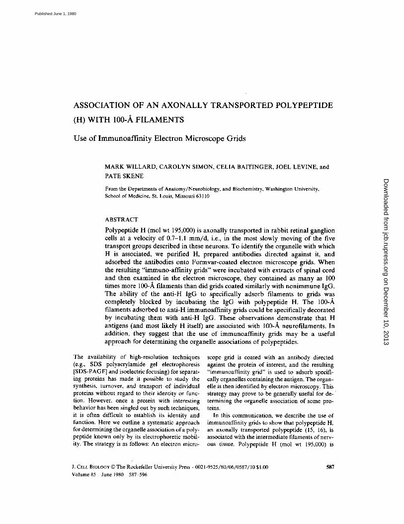
ASSOCIATION OF AN AXONALLY TRANSPORTED POLYPEPTIDE(H) WITH 100-A FILAMENTS
Use of Immunoaffinity Electron Microscope Grids
MARK WILLARD, CAROLYN SIMON, CELIA BAITINGER, JOEL LEVINE, andPATE SKENE
From the Departments of Anatomy/Neurobiology, and Biochemistry, Washington University,School of Medicine, St . Louis, Missouri 63110
ABSTRACT
Polypeptide H (mol wt 195,000) is axonally transported in rabbit retinal ganglioncells at a velocity of 0.7-1 .1 mm/d, i.e ., in the most slowly moving of the fivetransport groups described in these neurons . To identify the organelle with whichH is associated, we purified H, prepared antibodies directed against it, andadsorbed the antibodies onto Formvar-coated electron microscope grids . Whenthe resulting "immuno-affinity grids" were incubated with extracts of spinal cordand then examined in the electron microscope, they contained as many as 100times more 100-A filaments than did grids coated similarly with nonimmune IgG.The ability of the anti-H IgG to specifically adsorb filaments to grids wascompletely blocked by incubating the IgG with polypeptide H. The 100filaments adsorbed to anti-H immunoaffinity grids could be specifically decoratedby incubating them with anti-H IgG . These observations demonstrate that Hantigens (and most likely H itself) are associated with 100-A neurofilaments . Inaddition, they suggest that the use of immunoaffinity grids may be a usefulapproach for determining the organelle associations of polypeptides.
The availability of high-resolution techniques(e .g ., SDS polyacrylamide gel electrophoresis[SDS-PAGE] and isoelectric focusing) for separat-ing proteins has made it possible to study thesynthesis, turnover, and transport of individualproteins without regard to their identity or func-tion . However, once a protein with interestingbehavior has been singled out by such techniques,it is often difficult to establish its identity andfunction . Here we outline a systematic approachfor determining the organelle association of a poly-peptide known only by its electrophoretic mobil-ity . The strategy is as follows: An electron micro-
J . CELL BIOLOGY © The Rockefeller University Press " 0021-9525/80/06/0587/10 $1 .00
Volume 85
June 1980
587-596
scope grid is coated with an antibody directedagainst the protein of interest, and the resulting"immunoaffinity grid" is used to adsorb specifi-cally organelles containing the antigen . The organ-elle is then identified by electron microscopy . Thisstrategy may prove to be generally useful for de-termining the organelle association of some pro-teins.
In this communication, we describe the use ofimmunoaffinity grids to show that polypeptide H,an axonally transported polypeptide (15, 16), isassociated with the intermediate filaments of nerv-ous tissue . Polypeptide H (mol wt 195,000) is
587
on Decem
ber 10, 2013jcb.rupress.org
Dow
nloaded from
Published June 1, 1980

synthesized in the cell bodies of neurons; in theretinal ganglion cells of rabbits, we have observedits subsequent transport down the axons at a ve-locity of 0.7-1 .1 mm/d (16) . At least two otherpolypeptides, 45 (mol wt 145,000) and 46 (mol wt73,000), appear to be transported at this samevelocity ; at least four groups of proteins are trans-ported more rapidly in rabbit retinal ganglion cells(16, 17) . One characteristic of polypeptide H thatis important for the conclusion of our experimentsis that it is subject to a genetic polymorphism inrabbits (15) . Two forms (H1 and H2) of the poly-peptide can be differentiated by electrophoresis ;they appear to differ in molecular weight by10,000, with H1 having the higher molecularweight (195,000). Different inbred strains of rab-bits contain only H1 or H2, whereas differentindividuals from outbred strains contain only H 1,both H 1 and H2, or, very rarely, only H2.
MATERIALS AND METHODS
Purification ofHApproximately 5 g of rabbit spinal cord (freshly dissected, or
frozen) was homogenized in 30 ml of TED buffer (10 mM Tris,5 mM EDTA, 2 mM dithiothreitol [DTT]), containing phenyl-methylsulfonyl fluoride (PMSF, l mM), and diluted to 360 mlwith the same buffer lacking PMSF . The homogenate was soni-cated for 5 min, stirred for I h at 4°C, centrifuged for I h at100,000 g, and H was precipitated from the supernate by theaddition of 0.32 g/ml of (NH,)2SO 4 . The precipitate was sedi-mented by centrifugation for I h at 100,000 g and then resus-pended in TED buffer (5 ml) containing 10 mM DTT and 3%SDS. The suspension was heated at 90°C for 5 min, centrifugedat 100,000 g for 45 min, and the supernate was subjected to gelfiltration chromatography on a column (2 .5 x 120 cm) of Seph-arose-6B equilibrated with TED buffer containing SDS (1%) .The eluant fractions containing H (identified by analytical SDS-PAGE) were combined and dialyzed against a solution of EDTA(5 mM) and DTT (0 .1 mM)and precipitated with 20% TCA; theprecipitate was collected by centrifugation and washed withdiethyl ether to remove residual TCA. The precipitate was resus-pended in TED (1 ml) containing 1.5% SDS and 10 mM DTT.The solution was incubated at 90°C for 5 min, centrifuged at100,000 g for 45 min at 15°C, and subjected to preparative SDS-PAGE on a gel with a diameter of 1.5 cm in an apparatus fromSavant Instruments, Inc. (Hicksville, N. Y.) . The running gel (4cm high) contained 4.5% acrylamide, 0.135% bis-acrylamide, 8M urea, whereas the stacking gel (1 .2 cm high) contained 3%arylamide, 0.09% bis-acrylamide, 8 M urea. The buffer systemswere those of Laemmli (6), and the SDS was sequanol gradefrom Pierce Chemical Co. (Rockford, 111 .) . The fractions contain-ing only H (as judged by analytical SDS-PAGE) were combinedand concentrated by pressure filtration (XM 100 filter, AmiconCorp., Scientific Sys. Div., Lexington, Mass .) and dialyzedagainst a solution of SDS (0 .1%) and DTT (0 .1 mM). Theresulting preparation was emulsified with an equal volume ofFreund's adjuvant (complete for the first injection, incompletefor subsequent injections) and injected subcutaneously into a
5W
THE JOURNAL OF CELL BIOLOGY " VOLUME 85, 1980
goat . The goat received four injections (-100 tlg each) over aperiod of 6 mo; it was bled 10 d after the last injection.
Purification ofIgGIgG was partially purified from the anti-H serum by precipi-
tating it three times with (NH4)2SO4 (40% of saturation at 4°C) .The resulting preparation was dialyzed against phosphate-buffered saline (PBS, 0.15 M NaCl, 0.01 M NaH2POa , pH 7),and further purified by affinity chromatography on a column ofSepharose-4B to which proteins (including H) from the(NH4)2SO4 step of the H purification (see above) had beencoupled by the procedure of Parikh et al . (8) . The specificantibodies were eluted from the column with a solution of glycine(0 .2 M) and NaCl (0 .15 M), pH 2.4.Nonimmune IgG was purified from the serum of a nonim-
munized goat by (NH4)2SO4 precipitation (45% of saturation at4°C) followed by chromatography on DEAF cellulose (CellexD, from Bio-Rod Laboratories, Richmond, Calif.).
Immunoaffinity Grid PreparationCopper electron microscope grids (400 mesh) coated with
Formvar (0 .4% in ethylene dichloride) were floated on drops ofPBS containing either anti-H or nonimmune IgG for 4.3 h atroom temperature . The grids were rinsed in one drop ofPBS andthen incubated in a solution (2 .5% wt/vol) of polyglutamic acid(Sigma Chemical Co ., St . Louis, Mo .) for 50 min at 4°C. Thegrids were rinsed in two drops of PBS, and washed by floatationin 3 ml of PBS for 1 .6 h at 4°C. The coated grids were floated ondrops of cell fractions for 30 min at room temperature, thenrinsed in nine drops of PBS and one drop of H2O at 4°C, andnegatively stained with unbuffered uranyl acetate (1%). Aftereach rinse, the liquid was drawn offthe grid by touching it to thetorn edge of a piece of filter paper (Whatman No . 1).
Filament PreparationFreshly dissected segments of rabbit spinal cord were incu-
bated with a 100-fold excess of a solution containing EDTA (2 .5mM), EGTA (2.5 mM), and Na2HP0, (t mM), pH 7, for 2 h atroom temperature as described by Schlaepfer (10) . The swollentissue was removed from the solution and homogenized with aloose-fitting pestle ofa Dounce homogenizer (Kontes Co., Vine-land, N. J .) . NaCI (1 M) was added to the disrupted tissue to givea final concentration of 0.15 M, and the tissue homogenate wascentrifuged at 12,000 rpm for 30 min in the SS-34 rotor of aSorvall RC2B centrifuge (DuPont Instruments-Sorvall, DuPontCo., Newtown, Conn.) (10) . The supernate was applied to adiscontinuous sucrose gradient containing layers (3 .3 ml each) of2, 1 .5, and 1 .0 M sucrose in a solution containing NaCl (0 .15 M),EDTA (2 .5 mM), and EGTA (2.5 mM). The gradients werecentrifuged for 3 h at 35,000 rpm in an SW 40 .1 rotor (BeckmanInstruments, Inc., Spinco Div., Palo Alto, Calif.) . The material atthe 1- to 1 .5-M interface served as a source of filaments .
RESULTS
Purification ofH
When rabbit spinal cord was homogenized in alow ionic strength medium, approximately one-half of the H was extracted. In cases where theremaining particulate fraction was reextracted, ad-ditional H was solubilized, suggesting that soluble
on Decem
ber 10, 2013jcb.rupress.org
Dow
nloaded from
Published June 1, 1980
and insoluble H are not separate species but canbe interconverted . Fig. l shows the progressivepurification of Hby gel filtration chromatographyand preparative SDS-PAGE. The gel filtrationserved to increase the relative amounts ofproteinsof high molecular weights, thereby allowing moreH to be applied to the preparative gel; the prepar-ative gel had a limited capacity (-I mg) for totalprotein. Fig. 1 b shows that preparative SDS-PAGE separated H from polypeptide 45 and allother stained polypeptides. The yield of H was100-200 IAg from 3 g of spinal cord .It was important to establish that the poly-
peptide isolated by the procedure was in fact theaxonally transported polypeptide H and containedno other polypeptides that coelectrophoresed withH. Because H can be recognized by its strain-dependent electrophoretic mobility, we performedthe purification procedure on the spinal cords of
FIGURE l
Purification ofpolypeptide H. a and b showthe polypeptides in fractions (sequential fractions in a,fractions 17, 20, 23, 25, 27, followed by sequential frac-tions in b) from the gel filtration and electrophoresiscolumns, respectively . p indicates a sample ofthe mate-rial that was applied to the column or gel . c shows thefinal purified preparation of H from a rabbit that hadonly H l (New Zealand White strain) on the left, andfrom a rabbit that had only H2 (strain X/J) on the right.The latter rabbit was obtained from The Jackson Labo-ratory (Bar Harbor, Maine) . The samples in c wereelectrophoresed side by side on a slab gel .
both H 1- and H2-containing rabbits . Fig. I c showsthat the resulting purified polypeptides had thestrain-dependent mobility expected for H. Fur-thermore, the lack of staining material at the H lposition in the H2 preparation and vice versaargues strongly that there was no significantamount ofprotein other than H that coelectropho-resed with either H I or H2 .
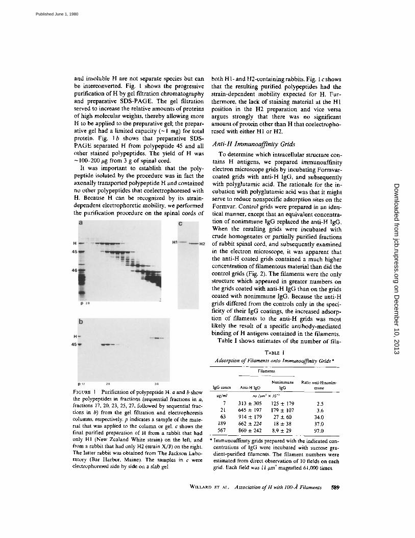
Anti-H Immunoaffinity GridsTo determine which intracellular structure con-
tains H antigens, we prepared Immunoafffnityelectron microscope grids by incubating Formvar-coated grids with anti-H IgG, and subsequentlywith polyglutamic acid . The rationale for the in-cubation with polyglutamic acid was that it mightserve to reduce nonspecific adsorption sites on theFormvar. Control grids were prepared in an iden-tical manner, except that an equivalent concentra-tion of nonimmune IgG replaced the anti-H IgG.When the resulting grids were incubated withcrude homogenates or partially purified fractionsof rabbit spinal cord, and subsequently examinedin the electron microscope, it was apparent thatthe anti-H coated grids contained a much higherconcentration of filamentous material than did thecontrol grids (Fig. 2) . The filaments were the onlystructure which appeared in greater numbers onthe grids coated with anti-H IgG than on the gridscoated with nonimmune IgG. Because the anti-Hgrids differed from the controls only in the speci-ficity of their IgG coatings, the increased adsorp-tion of filaments to the anti-H grids was mostlikely the result of a specific antibody-mediatedbinding of H antigens contained in the filaments .Table I shows estimates of the number of fila-
TABLE IAdsorption of Filaments onto lmmunoaffnity Grids'
' Immunoaffinity grids prepared with the indicated con-centrations of IgG were incubated with sucrose gra-dient-purified filaments. The filament numbers wereestimated from direct observation of 10 fields on eachgrid . Each field was I 1 um 2 magnified 61,000 times.
WILLARD ET AL .
Association ofH with 100-Á Filaments
589
Filaments
IgG concn
gg/ml
Anti-H IgG
n0./Im'
NonimmuneIgG
x 10-,
Ratio anti-Knonim-mune
7 313 ± 305 125 t 179 2.521 645 ± 197 179 t 107 3 .663 914 ± 179 27 ± 60 34 .0189 662 ± 224 18 ± 38 37 .0567 860 ± 242 8.9 t 29 97 .0
on Decem
ber 10, 2013jcb.rupress.org
Dow
nloaded from
Published June 1, 1980
FIGURE 2
Adsorption of filaments from various fractions of spinal cord . The left and right columnsshow immunoaffinity grids prepared with 567 hg/ml ofanti-H IgG and nonimmune IgG, respectively. (a)Whole homogenate of spinal cord . (b) Supernate of a 3 .5 x 10' g��g x min centrifugation of spinal cordextract prepared according to the method of Schlaepfer (l0) . (c and d) Sucrose gradient fraction of spinalcord prepared as described in Materials and Methods. (a-c) x 25,000 ; (d) x 7,100 .
590
on Decem
ber 10, 2013jcb.rupress.org
Dow
nloaded from
Published June 1, 1980

ments adsorbed on grids coated with increasingconcentrations of anti-H and nonimmune IgG . Itis apparent that the number ofadsorbed filamentsincreased with increasing concentrations ofanti-HIgG, as might be expected if the filament bindingwere mediated by the antibody . On the otherhand, the number of filaments bound to the non-immune IgG-coated grids decreased with increas-ing IgG concentration . This latter effect mostlikely represents the blocking of the remainingnonspecific adsorption sites on the Formvar by thenonimmune IgG . At the highest IgG concentrationused (567 tLg/ml), nearly 100 times more filamentsbound to the anti-H-coated grid than to the con-trol .Fig . 3 shows a higher magnification of a typical
filament on an anti-H immunoaffinity grid. Itsdiameter (90-100 f1) indicates that the specificallyadsorbed filaments belong to the class of inter-mediate (100 Á) filaments of nervous tissue .
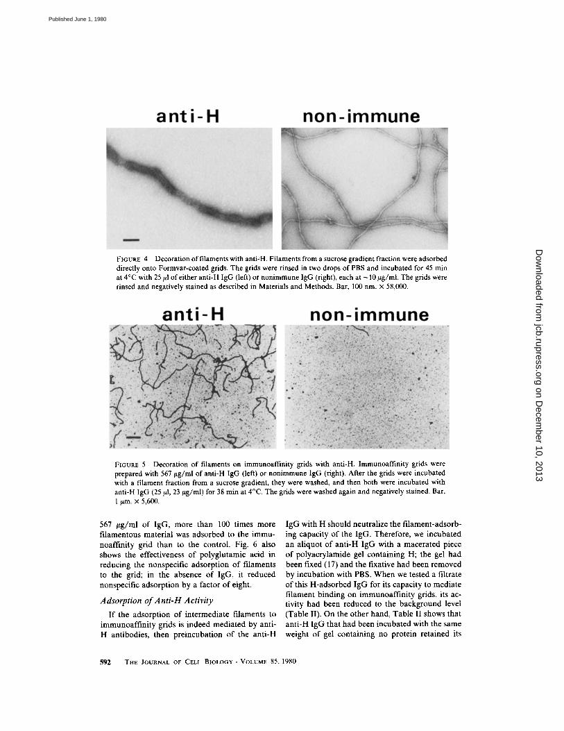
Filaments Postdecorated with
Anti-H IgG
Schlaepfer has shown that intermediate fila-ments can be decorated with specific antifilamentantibodies in a manner that can be observed byelectron microscopy (9); the decoration results inan apparent increase in the filament diameter aswell as an increase in contrast . When intermediatefilaments adsorbed to Formvar-coated grids wereincubated with anti-H IgG, the filaments becamedecorated, as shown in Fig . 4 . The decoration wasspecific for anti-H IgG ; it did not occur whenfilaments on grids were incubated with nonim-
mune IgG (Fig . 4) . Furthermore, the filamentsadsorbed both to anti-H immunoaffinity grids andto nonimmune-coated grids became decoratedwhen postincubated with anti-H IgG (Fig . 5) .These observations serve three purposes : first, theyprovide additional evidence that H antigens arecontained in the intermediate filaments; second,they serve to rule out the possibility that thedifference between anti-H and control immunoaf-finity grids might be in the visibility (because ofdecoration by the antibody on the grid) of thefilaments rather than in the number adsorbed. Ifthis had been the case, the difference in the num-ber of filaments (Fig . 5) would have been elimi-nated by the postdecoration of both the immuneand control grids ; and third, the increased contrastof the decorated filaments allowed them to beobserved at lower magnifications, where theycould be counted more accurately, because theirends were more often included in the field ofobservation .
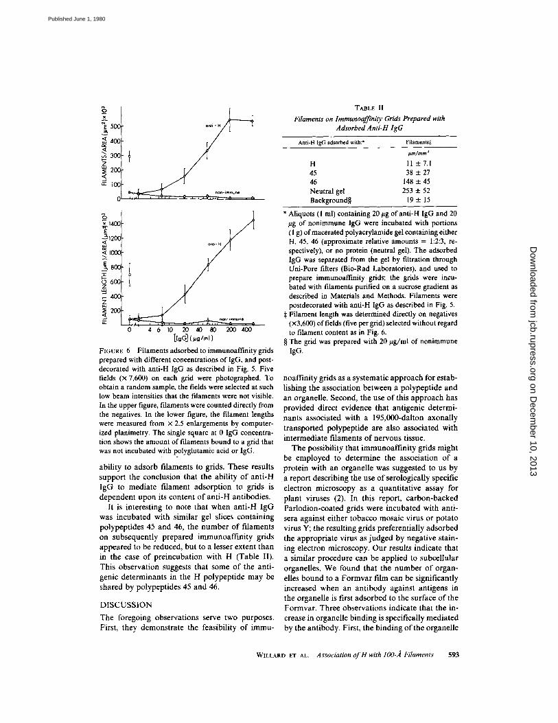
Fig. 6 shows the amount of filamentous materialadsorbed to grids prepared by incubating themwith different concentrations of immune or non-immune IgG, and postdecorating them with anti-H IgG . The filaments were quantitated both bycounting them (Fig . 6, upper curve) and determin-ing their total length (Fig . 6, lower curve) fromphotographs of randomly chosen fields . As wassuggested by Table 1, it is apparent from Fig . 6that the number of adsorbed filaments increasedwith increasing concentrations of anti-H IgG, anddecreased with increasing concentrations of non-immune IgG . In the case of the grids coated with
FIGURE 3
A typical filament from the immunoafnity grid shown in Fig . 1 c. Bar, 100 nm . x 97,000 .
WILLARD ET AL .
Association ofH with 100- .4 Filaments 59 1
on Decem
ber 10, 2013jcb.rupress.org
Dow
nloaded from
Published June 1, 1980
567 wg/ml of IgG, more than 100 times morefilamentous material was adsorbed to the immu-noaffinity grid than to the control. Fig. 6 alsoshows the effectiveness of polyglutamic acid inreducing the nonspecific adsorption of filamentsto the grid; in the absence of IgG, it reducednonspecific adsorption by a factor of eight.
Adsorption ofAnti-H Activity
592
FIGURE 4
Decoration offilaments with anti-H . Filaments from a sucrose gradient fraction were adsorbeddirectly onto Formvar-coated grids. The grids were rinsed in two drops of PBS and incubated for 45 minat 4°C with 25 ,ul of either anti-H IgG (left) or nonimmune IgG (right), each at -10 wg/ml. The grids wererinsed and negatively stained as described in Materials and Methods. Bar, 100 nm. x 58,000 .
FIGURE 5
Decoration of filaments on immunoaffinity grids with anti-H . Immunoaffinity grids wereprepared with 567 jig/ml of anti-H IgG (left) or nonimmune IgG (right) . After the grids were incubatedwith a filament fraction from a sucrose gradient, they were washed, and then both were incubated withanti-H IgG (25 [1, 23 trg/ml) for 38 min at 4°C . The grids were washed again and negatively stained . Bar,1 pm . x 5,600 .
If the adsorption of intermediate filaments toimmunoaffinity grids is indeed mediated by anti-H antibodies, then preincubation of the anti-H
THE JOURNAL OF CELL BIOLOGY " VOLUME 85, 1980
IgG with H should neutralize the filament-adsorb-ing capacity of the IgG. Therefore, we incubatedan aliquot of anti-H IgG with a macerated pieceof polyacrylamide gel containing H; the gel hadbeen fixed (17) and the fixative had been removedby incubation with PBS. When we tested a filtrateof this H-adsorbed IgG for its capacity to mediatefilament binding on immunoaffinity grids, its ac-tivity had been reduced to the background level(Table II). On the other hand, Table II shows thatanti-H IgG that had been incubated with the sameweight of gel containing no protein retained its
on Decem
ber 10, 2013jcb.rupress.org
Dow
nloaded from
Published June 1, 1980
óIZ, 14001
Q1200,Wá 1000
-3 800xr0 600zW- 400zWa 200
FIGURE 6
Filaments adsorbed to immunoaffinity gridsprepared with different concentrations of IgG, and post-decorated with anti-H IgG as described in Fig. 5 . Fivefields (x 7,600) on each grid were photographed . Toobtain a random sample, the fields were selected at suchlow beam intensities that the filaments were not visible .In the upper figure, filaments were counted directly fromthe negatives . In the lower figure, the filament lengthswere measured from x 2.5 enlargements by computer-ized planimetry. The single square at 0 IgG concentra-tion shows the amount of filaments bound to a grid thatwas not incubated with polyglutamic acid or IgG.
ability to adsorb filaments to grids . These resultssupport the conclusion that the ability of anti-HIgG to mediate filament adsorption to grids isdependent upon its content of anti-H antibodies .
It is interesting to note that when anti-H IgGwas incubated with similar gel slices containingpolypeptides 45 and 46, the number of filamentson subsequently prepared immunoaffinity gridsappeared to be reduced, but to a lesser extent thanin the case of preincubation with H (Table II) .This observation suggests that some of the anti-genic determinants in the H polypeptide may beshared by polypeptides 45 and 46 .
DISCUSSIONThe foregoing observations serve two purposes .First, they demonstrate the feasibility of immu-
TABLE IIFilaments on Immunoaff:nity Grids Prepared with
AdsorbedAnti-H IgG
" Aliquots (l ml) containing 20 jig of anti-H IgG and 20yg of nonimmune IgG were incubated with portions(1 g) ofmacerated polyacrylamide gel containing eitherH, 45, 46 (approximate relative amounts = 1 :2:3, re-spectively), or no protein (neutral gel) . The adsorbedIgG was separated from the gel by filtration throughUni-Pore filters (Bio-Rad Laboratories), and used toprepare immunoaffmity grids; the grids were incu-bated with filaments purified on a sucrose gradient asdescribed in Materials and Methods. Filaments werepostdecorated with anti-H IgG as described in Fig. 5 .
$ Filament length was determined directly on negatives(x3,600) of fields (five per grid) selected without regardto filament content as in Fig. 6.
§ The grid was prepared with 20 ttg/ml of nonimmuneIgG .
noaffinity grids as a systematic approach for estab-lishing the association between a polypeptide andan organelle. Second, the use of this approach hasprovided direct evidence that antigenic determi-nants associated with a 195,000-dalton axonallytransported polypeptide are also associated withintermediate filaments of nervous tissue .The possibility that immunoaffinity grids might
be employed to determine the association of aprotein with an organelle was suggested to us bya report describing the use of serologically specificelectron microscopy as a quantitative assay forplant viruses (2) . In this report, carbon-backedParlodion-coated grids were incubated with anti-sera against either tobacco mosaic virus or potatovirus Y; the resulting grids preferentially adsorbedthe appropriate virus as judged by negative stain-ing electron microscopy . Our results indicate thata similar procedure can be applied to subcellularorganelles . We found that the number of organ.-elles bound to a Formvar film can be significantlyincreased when an antibody against antigens inthe organelle is first adsorbed to the surface of theFormvar. Three observations indicate that the in-crease in organelle binding is specifically mediatedby the antibody . First, the binding of the organelle
WILLARD ET AL .
Association ofH with i00-,ß Filaments 593
Anti-H IgG adsorbed with :' Filaments*
pm/mm'
H 11 t 7.145 38 t 2746 148 ± 45Neutral gel 253 ± 52Background§ 19 ± 15
on Decem
ber 10, 2013jcb.rupress.org
Dow
nloaded from
Published June 1, 1980

increased with increasing concentrations of theantibody. Second, nonimmune IgG used in placeofthe antibody did not cause increased binding ofthe organelle; on the contrary, the nonimmuneIgG decreased the binding of the organelle, pre-sumably by occupying the remaining nonspecificadsorption sites on the Formvar . (In the case ofthe intermediate filaments studied here, we foundthat nearly 90% of the nonspecific adsorptioncould be eliminated by incubating the grids withpolyglutamic acid .) Third, the increased bindingofthe organelle was eliminated when the antibodywas neutralized by its antigen .An attractive feature ofthe immunoaffmity grid
strategy for determining the association of a pro-tein with an organelle is that, in principle, it re-quires a minimum number of speculations con-cerning the nature of the organelle . For example,it was possible to demonstrate the specific adsorp-tion of 100-A filaments onto anti-H IgG-coatedgrids even in crude homogenates ofnervous tissue ;therefore, the nature ofthe H-containing organellecould be inferred without assumptions regardinghow the organelle would respond to fractionationprocedures . The technique might also prove usefulfor examining associations between organelles be-cause it provides a means of concentrating anorganelle and its associated structures on an elec-tron microscope grid with minimum preliminaryfractionation . The limitation of the immunoaffm-ity grid approach is that the antigen of interestmust be located in an exposed position on anorganelle that can be recognized by electron mi-croscopy. Although filaments are probably partic-ularly favorable organelles in these regards, itseems likely that with appropriate variations in theprocedure, a variety of organelles would be acces-sible to this approach .
The subunit composition of the 100-A neurofil-aments has been a subject of considerable contro-versy . When filaments have been prepared by aprocedure designed to purify myelinated axon seg-ments and subsequently liberate their neurofila-ments by means of osmotic shock, the resultingpreparations have consistently contained a majorpolypeptide with a mol wt in the range of 49,000-60,000 (1, 3, 5, 12, 14, 18) . In contrast, Hoffmanand Lasek (4) inferred that three polypeptides(mol wt 200,000, 145,000, and 68,000) that aretransported slowly down the axons ofthe rat sciaticnerve are associated with neurofilaments . Hoff-man and Lasek's hypothesis has been supported
594
THE JOURNAL OF CELL BIOLOGY " VOLUME 85, 1980
by reports that, during experimentally inducedneuronal degeneration (11, 13), polypeptides re-sembling this "slow component triplet" accumu-late and disappear in synchrony with the neurofil-aments . In addition, recent preparations of inter-mediate filaments from peripheral and centralnervous tissue (7, 10) have been enriched in poly-peptides resembling the triplet, and Schlaepfer (9)has reported that antibodies against a 68,000-dal-ton polypeptide (which may correspond to one ofthe triplet polypeptides) are reactive with inter-mediate filaments .The experiments reported here use two criteria
to show that antigens contained in the axonallytransported protein H (whose molecular weight[195,000] is similar to one of the triplet polypep-tides) are physically associated with intermediatefilaments . First, immunoaffmity grids coated withanti-H IgG specifically adsorbed 100-A filaments,and second, anti-H IgG specifically decorated in-termediate filaments. We can be unusually confi-dent in equating the axonally transported poly-peptide H (identified as a radioactive electropho-retic band [ 161) with the protein used to producethe antibody, as both show the same strain de-pendence in electrophoretic mobility .Although the observation that antigens are
shared between polypeptide H and intermediatefilaments does not necessarily require that H isitself associated with the filaments, the followingconsiderations strongly support this conclusion :The most purified filament fraction used in ourexperiments contains only three major polypep-tides (Fig . 7), one of which can be identified as Hby its strain-dependent electrophoretic mobility .While the other two major polypeptides (45 and46 in Fig. 7) appear to share antigenic determi-nants with H (reference 7 and Table II), anti-HIgG which had been exhaustively adsorbed with45 and 46 retained its ability to decorate interme-diate filaments and to form filament-specific im-munoaffinity grids (data not shown) . Thus, thefilaments contain antigens unique to H among themajor polypeptides in the filament fraction ; ittherefore seems safe to conclude that H itself isassociated with the filaments. The nature of theassociation between H and the filaments (i .e .,whether it is an integral subunit of the filament ora subunit of some structure peripherally attachedto the filament) is not yet clear . Our experimentsalso do not rule out the possibility that H isassociated with additional structures not discerned
on Decem
ber 10, 2013jcb.rupress.org
Dow
nloaded from
Published June 1, 1980

FIGURE 7
Afilament fraction from a sucrose gradient,electrophoresed and stained with Coomassie Blue. Thesymbols indicate polypeptides discussed in the text .
by electron microscopy .Although nervous tissue includes filaments from
both neurons and glial cells, the H-containingfilaments are most likely of neuronal origin be-cause axons, but not glial cells, can be intenselystained with anti-H IgG in sections of nervoustissue by means of indirect immunofluorescence(J . Levine and M. Willard, manuscript in prepa-ration) . The conclusion that H is associated withneurofilaments supports the idea (originally pro-
posed by Hoffman and Lasek [4]) that the trans-port of neurofilaments is a major function of themost slowly moving group ofaxonally transportedproteins . Our preliminary experiments with anti-bodies directed against polypeptides 45 and 46,the two additional group V polypeptides that re-semble the triplet polypeptides in molecularweight, support the conclusion that these polypep-tides are also associated with intermediate fila-ments.
We thank D. Gluckstein for his participation in thepurification of antigen; Dr . M. Cochran, R. Steiner, S.Hendry, and S. Plurad for their advice on electronmicroscopy ; and Drs. D. Bray, W. M. Cowan, and D.Gottlieb for helpful comments.
This work was supported by grant EYO-2682, a JerryLewis Neuromuscular Research Center grant, and Re-search Career Development Award NS00170.
Receivedfor publication 11 September 1979, and in revisedform 11 February 1980.
REFERENCES
1 . DAVISON, P. F., and B. WINSLOW. 1974 . The protein subunit of calfbrain neurofilaments . J. Neurobiol. 5:119-133 .
2. DERRICK, K. S. 1973 . Quantitative assay for plant viruses using sero-logicaBy specific electron microscopy. Virology. 5&652-653.
3. GOLDMAN, J. E., H. SCHAUMBURG, andW. T. NORTON . 1978 . Isolationand characterization of glial filaments from human brain. J Cell Biol.7&426-440.
4. HOFFMAN, P. N., and R. J. LASER. 1975 . Theslow componentof axonaltransport. Identification of major structural polypeptides of the axonand their generality among mammalian neurons. J. Cell Biol. 6&351--366.
S. IQBAL, K., 1 . GRUNDRE-IQBAL, H. M. WISNIEWSKI, and R. D. TERRY.1977. On the neurofilament and neurotubule proteins from humanautopsy tissue. J. Neurochem. 29:417-424 .
6. LAEMMLI, U. K. 1970 . Cleavage of structural proteins during theassembly of the head of bacteriophage T4 . Nature (Land.) . 227:680-685.
7. LIEM, R. K. H., S. YEN, G. D. SALOMON, and M. L. SHELANSKI . 1978 .Intermediate filaments in nervous tissue . J. Cell Biol. 79 :637-645 .
8. PARIKH, I., S. MARCH, and P. CUATRECASAS. 1974. Topics in themethodology of substitution reactions with agarose . Methods Enzymol.34:77-102 .
9. SCHLAEPFER,W. W. 1977. Immunological and ultrastructuralstudiesofneurofilaments isolated from rat peripheral nerve . J Cell Biol. 74:226-240.
10. SCHLAEPFER, W. W., and L. A. FREEMAN. 1978 . Polypeptides of neu-rofilaments isolated from rat peripheral and central nervoussystems. J.Cell Biol. 78:653-662 .
It . SCHLAEPFER, W. W., and S. MICRO. 1978 . Chemical and structuralchanges of neurofilamenls in transected rat sciatic nerves. J. Cell Biol.78:369-378.
12 . SCHOOK,W. J., and W. T. NORTON. 1976 . Neurofilaments account forthe lipid in myelin-tree axons. Brain Res. 118:517-522 .
13. SELKOE, D. J., R. K. LIEM, S. YEN, and M. L. SHELANSKI . 1979.Biochemical and immunological characterization of neurofilaments inexperimental neurofibrillarydegeneration induced by aluminum . BrainRes. 163:235-252 .
14 . SHELANSKI, M. L., S. ALBERT, G. H. DEVRIES, and W. T. NORTON.1971 . Isolatio n of filaments from brain. Science (Wash. D. C.). 174:1242-1245.
15 . WILLARD, M. B. 1976 . Genetically determined protein polymorphismin the rabbit nervous system. Proc. Nall. Acad. Sci. U. S. A. 73:3641-3645 .
16 . WILLARD, M. B., and K. L. HULEBAK. 1977 . Th e intra-axonal transport
WILLARD ET AL .
Association OfH with 100-A Filaments
595
on Decem
ber 10, 2013jcb.rupress.org
Dow
nloaded from
Published June 1, 1980
of polypeptide H : Evidence for a fifth (very slow) group of transportedproteins in the retinal ganglion cells of the rabbit . Brain Res. 136:289-306.
17. WILLARD, M., W. M. COWAN, and P . R. VAGELOS. 1974. The poly-peptide composition of intra-axonally transported proteins: evidence
5%
THE JOURNAL OF CELL BIOLOGY - VOLUME 85, 1980
for four transport velocities. Proc . Nail. Acad. Sci., U. S. A. 64:191-195 .
18 . YEN, S. H., D. DAHL, M. SCHACHNER, and M. L. SHELANSKI. 1976.Biochemistry of the filaments of brain. Proc. Natl. Acad. Sci. U. S. A.73:529-533 .
on Decem
ber 10, 2013jcb.rupress.org
Dow
nloaded from
Published June 1, 1980
Copyright © 2022 FDOKUMEN