Bahan kuliah Scanning Electron Microscope pak agus
28
Scanning Electron Microscope Diambil dari berbagai sumber
Transcript of Bahan kuliah Scanning Electron Microscope pak agus

Scanning Electron Microscope
Diambil dari berbagai sumber

What is a SEM?
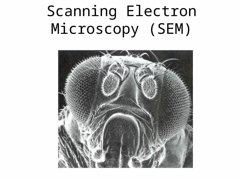
SEM stands for scanning electron microscope. The SEM is a microscope that uses electrons instead of light to form an image. Since their development in the early 1950's, scanning electron microscopes have developed new areas of study in the medical and physical science
communities. The SEM has allowed researchers to examine a much bigger variety of specimens.


Perjalanan elektron pada sem

diagram sem
Scanning Electron Microscopy (SEM)

Keterangan diagram The SEM is an instrument that produces a largely magnified image by using electrons instead of light to form an image. A beam of electrons is produced at the top of the microscope by an electron gun. The electron beam follows a vertical path through the microscope, which is held within a vacuum. The beam travels through electromagnetic fields and lenses, which focus the beam down toward the sample. Once the beam hits the sample, electrons and X-rays are ejected from the sample.

Perjalanan elektron pada sem

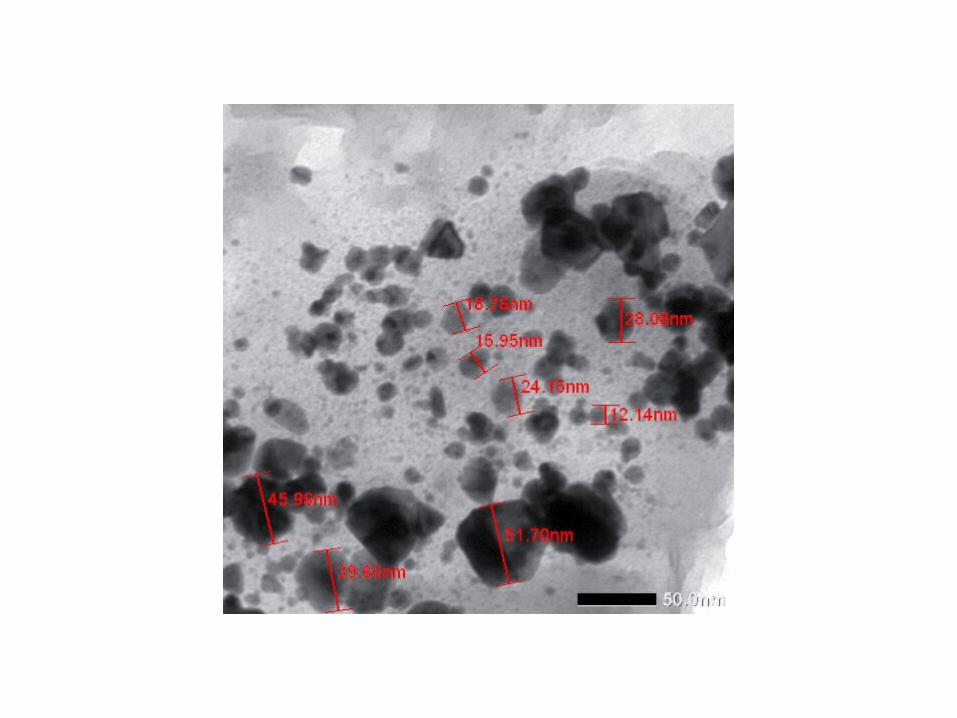
Scanning electron microscopy is used for inspecting topographies of specimens at very high magnifications using a piece of equipment called the scanning electron microscope. SEM magnifications can go to more than 300,000 X but most semiconductor manufacturing applications require magnifications of less than 3,000 X only. SEM inspection is often used in the analysis of die/package cracks and fracture surfaces, bond failures, and physical defects on the die or package surface.During SEM inspection, a beam of electrons is focused on a spot volume of the specimen, resulting in the transfer of energy to the spot. These bombarding electrons, also referred to as primary electrons, dislodge electrons from the specimen itself. The dislodged electrons, also known as secondary electrons, are attracted and collected by a positively biased grid or detector, and then translated into a signal. To produce the SEM image, the electron beam is swept across the area being inspected, producing many such signals. These signals are then amplified, analyzed, and translated into images of the topography being inspected. Finally, the image is shown on a CRT.
Scanning Electron Microscopy (SEM)

Scanning Electron Microscopy (SEM)
• The energy of the primary electrons determines the quantity of secondary electrons collected during inspection. The emission of secondary electrons from the specimen increases as the energy of the primary electron beam increases, until a certain limit is reached. Beyond this limit, the collected secondary electrons diminish as the energy of the primary beam is increased, because the primary beam is already activating electrons deep below the surface of the specimen. Electrons coming from such depths usually recombine before reaching the surface for emission.
• • Aside from secondary electrons, the primary
electron beam results in the emission of backscattered (or reflected) electrons from the specimen. Backscattered electrons possess more energy than secondary electrons, and have a definite direction. As such, they can not be collected by a secondary electron detector, unless the detector is directly in their path of travel. All emissions above 50 eV are considered to be backscattered electrons.

How is a sample prepared?
Because the SEM utilizes vacuum conditions and uses electrons to form an image, special preparations must be done to the
sample. All water must be removed from the samples because the water would vaporize in the vacuum. All metals are conductive and require no preparation before being used. All non-metals need to be made conductive by covering the sample with a thin layer of conductive material. This is done by using a device
called a "sputter coater."The sputter coater uses an electric field and argon gas. The
sample is placed in a small chamber that is at a vacuum. Argon gas and an electric field cause an electron to be removed from the argon, making the atoms positively charged. The argon ions then become attracted to a negatively charged gold foil. The
argon ions knock gold atoms from the surface of the gold foil. These gold atoms fall and settle onto the surface of the
sample producing a thin gold coating

What are the radiation safety concerns?
The radiation safety concerns are related to the electrons that are backscattered from the sample, as well as X-rays produced in the process. Most SEMs are extremely well shielded and do not produce exposure rates greater than background. However, scanning electron microscopes are radiation-generating devices and should be at least inventoried. The Indiana State Department of Health requires that the machines be registered with their office using State Form 16866, Radiation Machine Registration Application. It is also important that the integrity of the shielding is maintained, that all existing interlocks are functioning, and that workers are aware of radiation safety considerations.

Scanning Electron Microscope Radiation Safety Guidelines
• Safety evaluations will be performed initially when machine is purchased and after machine has been moved.
• Each machine should be key controlled when not in use. Interlocks, if present, must remain operational unless approved by the RSO.
• Shielding must be sufficient to maintain exposure rates less than 0.5 mrem/hr at 5 cm.
• The Radiation Safety Office will keep inventory and survey information on file in their offices. The SEM user should keep logbook of any maintenance done on machine. RSO must be notified if any modifications are made to the interlocks or any other safety devices. The SEM user should also keep a copy of operating and emergency procedures at the accelerator panel.
• No survey meters or personnel dosimetry are required.

UICC Asbestos Chrysotile 'A' standard

Tremolite asbestos, Death Valley, California

Anthophyllite asbestos, Georgia

Winchite-richterite asbestos, Libby, Montana


Transmission Electron Microscopy (TEM)


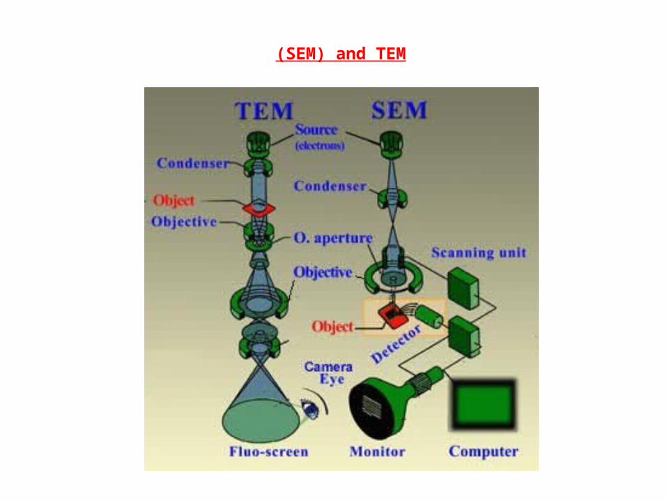
(SEM) and TEM