Up-regulation of TWIST in prostate cancer and its implication as a therapeutic target
11
Up-Regulation of TWIST in Prostate Cancer and Its Implication as a Therapeutic Target Wai Kei Kwok, 1 Ming-Tat Ling, 1 Tak-Wing Lee, 1 Tracy C.M. Lau, 1 Chun Zhou, 1 Xiaomeng Zhang, 1 Chee Wai Chua, 1 Kwok W. Chan, 2 Franky L. Chan, 3 Carlotta Glackin, 4 Yong-Chuan Wong, 1 and Xianghong Wang 1 Departments of 1 Anatomy and 2 Pathology, Faculty of Medicine, The University of Hong Kong; 3 Department of Anatomy, The Chinese University of Hong Kong, Hong Kong, SAR, China; and 4 Division of Molecular Medicine, Beckman Research Institute of the City of Hope, Duarte, California Abstract Androgen-independent metastatic prostate cancer is the main obstacle in the treatment of this cancer. Unlike a majority of solid cancers, prostate cancer usually shows poor response to chemotherapeutic drugs. In this study, we have shown a potential novel target, TWIST, a highly conserved bHLH transcription factor, in the treatment of prostate cancer. Using malignant and nonmalignant prostate tissues, we found that TWIST expression was highly expressed in the majority (90%) of prostate cancer tissues but only in a small percentage (6.7%) of benign prostate hyperplasia. In addi- tion, the TWIST expression levels were positively correlated with Gleason grading and metastasis, indicating its role in the development and progression of prostate cancer. Furthermore, down-regulation of TWIST through small interfering RNA in androgen-independent prostate cancer cell lines, DU145 and PC3, resulted in increased sensitivity to the anticancer drug taxol-induced cell death which was associated with decreased Bcl/Bax ratio, leading to activa- tion of the apoptosis pathway. More importantly, inactivation of TWIST suppressed migration and invasion abilities of androgen-independent prostate cancer cells, which was correlated with induction of E-cadherin expression as well as morphologic and molecular changes associated with mesenchymal to epithelial transition. These results were further confirmed on the androgen-dependent LNCaP cells ectopically expressing the TWIST protein. Our results have identified TWIST as a critical regulator of prostate cancer cell growth and suggest a potential therapeutic approach to inhibit the growth and metastasis of androgen-independent prostate cancer through inactivation of the TWIST gene. (Cancer Res 2005; 65(12): 5153-62) Introduction Prostate cancer is the most commonly diagnosed cancer in American men representing one third of all new cancer cases each year. This means that one of six men is being diagnosed with prostate cancer over their lifetime. Thus, over 30,000 men die each year from prostate cancer–related illness in the United States (1). Early-stage androgen-sensitive prostate cancer is manageable with androgen depletion therapy. However, approx- imately one third of the prostate cancer patients have micro- metastatic disease at the time of presentation, this group of patients will eventually progress to clinically detectable metastatic and androgen-independent diseases at which point the median survival is only 12 to 15 months (2). Therefore, metastatic prostate cancer remains the main cause of prostate cancer– related death in men. Identification of key factors that are specifically expressed in prostate cancer and essential for cancer cell growth is a crucial step towards understanding prostate cancer progression and developing of new anticancer therapies. Recently, TWIST, a highly conserved basic helix-loop-helix transcription factor, is suggested as an oncogene (3, 4). For ex- ample, exogenous TWIST expression promotes colony formation of mouse embryonic fibroblasts in soft agar and this process is mediated through suppression of the ARF/MDM2/p53 pathway (4). Recently, using gene profiling analysis, up-regulation of TWIST is found associated with malignant transformation of melanoma and T-cell lymphoma (5, 6). TWIST is also shown to positively regulate the expression of the GLI1 gene, which is overexpressed in many types of human carcinoma (7). In addition, increased TWIST expression is found in over 50% of the rhabdomyosarcoma and 40% of gastric carcinomas compared with nonmalignant tissues (4, 8). The oncogenic effect of TWIST is suggested mediated through its role as an antiapoptotic factor (4). Recently, evidence also indicates TWIST as a key factor responsible for metastasis of breast cancer by promoting epithelial-to-mesenchymal transition (EMT) in an in vivo system and down-regulation of TWIST is able to suppress metastatic ability by inducing mesenchymal-to-epithelial transition (MET; ref. 9). In addition, in primary melanoma patients, increased TWIST is correlated with poor outcome and shorter survival (5). These results implicate that TWIST may be a novel oncogene that induces tumorigenesis in nonmalignant cells and promotes tumor progression in malignant cells. Previously, we have found that TWIST gene amplification is associated with the develop- ment of acquired resistance to an anticancer drug (taxol), and ectopic expression of TWIST leads to resistance to microtubule disrupting agents (10). These lines of evidence further suggest that TWIST may also be a positive factor in promoting resistance to anticancer drugs, which is one of the character- istics of advanced cancers. In this study, we found up-regulation of TWIST in human prostate cancer and increased TWIST protein level was positively correlated with Gleason grading. Inactivation of TWIST in androgen-independent prostate cancer cells resulted in increased chemodrug-induced apoptosis and suppression of invasion ability. Our results identified a novel key Note: Supplementary data for this article are available at Cancer Research Online (http://cancerres.aacrjournals.org/). Requests for reprints: Xianghong Wang or Yong-Chuan Wong, Department of Anatomy, Laboratory Block Faculty of Medicine, The University of Hong Kong, 21 Sassoon Road, Hong Kong, SAR, China. Phone: 852-2819-2867; Fax: 852-2817-0857; E-mail: [email protected] or [email protected]. I2005 American Association for Cancer Research. www.aacrjournals.org 5153 Cancer Res 2005; 65: (12). June 15, 2005 Research Article Research. on February 7, 2016. © 2005 American Association for Cancer cancerres.aacrjournals.org Downloaded from
-
Upload
independent -
Category
Documents
-
view
2 -
download
0
Transcript of Up-regulation of TWIST in prostate cancer and its implication as a therapeutic target
Up-Regulation of TWIST in Prostate Cancer and Its Implication
as a Therapeutic Target
Wai Kei Kwok,1Ming-Tat Ling,
1Tak-Wing Lee,
1Tracy C.M. Lau,
1Chun Zhou,
1
Xiaomeng Zhang,1Chee Wai Chua,
1Kwok W. Chan,
2Franky L. Chan,
3
Carlotta Glackin,4Yong-Chuan Wong,
1and Xianghong Wang
1
Departments of 1Anatomy and 2Pathology, Faculty of Medicine, The University of Hong Kong; 3Department of Anatomy,The Chinese University of Hong Kong, Hong Kong, SAR, China; and 4Division of Molecular Medicine,Beckman Research Institute of the City of Hope, Duarte, California
Abstract
Androgen-independent metastatic prostate cancer is themain obstacle in the treatment of this cancer. Unlike amajority of solid cancers, prostate cancer usually shows poorresponse to chemotherapeutic drugs. In this study, we haveshown a potential novel target, TWIST, a highly conservedbHLH transcription factor, in the treatment of prostatecancer. Using malignant and nonmalignant prostate tissues,we found that TWIST expression was highly expressed in themajority (90%) of prostate cancer tissues but only in a smallpercentage (6.7%) of benign prostate hyperplasia. In addi-tion, the TWIST expression levels were positively correlatedwith Gleason grading and metastasis, indicating its role inthe development and progression of prostate cancer.Furthermore, down-regulation of TWIST through smallinterfering RNA in androgen-independent prostate cancercell lines, DU145 and PC3, resulted in increased sensitivity tothe anticancer drug taxol-induced cell death which wasassociated with decreased Bcl/Bax ratio, leading to activa-tion of the apoptosis pathway. More importantly, inactivationof TWIST suppressed migration and invasion abilities ofandrogen-independent prostate cancer cells, which wascorrelated with induction of E-cadherin expression as wellas morphologic and molecular changes associated withmesenchymal to epithelial transition. These results werefurther confirmed on the androgen-dependent LNCaP cellsectopically expressing the TWIST protein. Our results haveidentified TWIST as a critical regulator of prostate cancercell growth and suggest a potential therapeutic approach toinhibit the growth and metastasis of androgen-independentprostate cancer through inactivation of the TWIST gene.(Cancer Res 2005; 65(12): 5153-62)
Introduction
Prostate cancer is the most commonly diagnosed cancer inAmerican men representing one third of all new cancer caseseach year. This means that one of six men is being diagnosedwith prostate cancer over their lifetime. Thus, over 30,000 mendie each year from prostate cancer–related illness in the United
States (1). Early-stage androgen-sensitive prostate cancer ismanageable with androgen depletion therapy. However, approx-imately one third of the prostate cancer patients have micro-metastatic disease at the time of presentation, this group ofpatients will eventually progress to clinically detectable metastaticand androgen-independent diseases at which point the mediansurvival is only 12 to 15 months (2). Therefore, metastaticprostate cancer remains the main cause of prostate cancer–related death in men. Identification of key factors that arespecifically expressed in prostate cancer and essential for cancercell growth is a crucial step towards understanding prostatecancer progression and developing of new anticancer therapies.Recently, TWIST, a highly conserved basic helix-loop-helix
transcription factor, is suggested as an oncogene (3, 4). For ex-ample, exogenous TWIST expression promotes colony formationof mouse embryonic fibroblasts in soft agar and this process ismediated through suppression of the ARF/MDM2/p53 pathway(4). Recently, using gene profiling analysis, up-regulation ofTWIST is found associated with malignant transformation ofmelanoma and T-cell lymphoma (5, 6). TWIST is also shown topositively regulate the expression of the GLI1 gene, which isoverexpressed in many types of human carcinoma (7). Inaddition, increased TWIST expression is found in over 50% ofthe rhabdomyosarcoma and 40% of gastric carcinomas comparedwith nonmalignant tissues (4, 8). The oncogenic effect of TWISTis suggested mediated through its role as an antiapoptotic factor(4). Recently, evidence also indicates TWIST as a key factorresponsible for metastasis of breast cancer by promotingepithelial-to-mesenchymal transition (EMT) in an in vivo systemand down-regulation of TWIST is able to suppress metastaticability by inducing mesenchymal-to-epithelial transition (MET;ref. 9). In addition, in primary melanoma patients, increasedTWIST is correlated with poor outcome and shorter survival (5).These results implicate that TWIST may be a novel oncogenethat induces tumorigenesis in nonmalignant cells and promotestumor progression in malignant cells. Previously, we have foundthat TWIST gene amplification is associated with the develop-ment of acquired resistance to an anticancer drug (taxol), andectopic expression of TWIST leads to resistance to microtubuledisrupting agents (10). These lines of evidence further suggestthat TWIST may also be a positive factor in promotingresistance to anticancer drugs, which is one of the character-istics of advanced cancers. In this study, we found up-regulationof TWIST in human prostate cancer and increased TWISTprotein level was positively correlated with Gleason grading.Inactivation of TWIST in androgen-independent prostate cancercells resulted in increased chemodrug-induced apoptosis andsuppression of invasion ability. Our results identified a novel key
Note: Supplementary data for this article are available at Cancer Research Online(http://cancerres.aacrjournals.org/).
Requests for reprints: Xianghong Wang or Yong-Chuan Wong, Department ofAnatomy, Laboratory Block Faculty of Medicine, The University of Hong Kong, 21Sassoon Road, Hong Kong, SAR, China. Phone: 852-2819-2867; Fax: 852-2817-0857;E-mail: [email protected] or [email protected].
I2005 American Association for Cancer Research.
www.aacrjournals.org 5153 Cancer Res 2005; 65: (12). June 15, 2005
Research Article
Research. on February 7, 2016. © 2005 American Association for Cancercancerres.aacrjournals.org Downloaded from
factor in the growth of androgen-independent prostate cancerand provided a new target for improving the treatment efficiencyof androgen-independent metastatic prostate cancer throughinactivation of the TWIST gene.
Materials and Methods
Cell culture conditions. Human prostate adenocarcinoma cel1 lines
(LNCaP, PC-3, and DU145) were obtained from American Type CultureCollection (Rockville, MD) and maintained in RPMI 1640 (Sigma, St. Louis,
MO) supplemented with 5% FCS and penicillin/streptomycin.
Tissue specimens. All the formalin-fixed and paraffin-embedded
prostate tissues were obtained from prostate tissue banks in theDepartment of Pathology, West China University of Medical Sciences (11)
and the Institute of Urology, Peking University. All the tissues were either
obtained from prostatic needle biopsies or radical prostatectomy. Thehistopathology of the specimens was examined and classified by
pathologists in the Department of Pathology in the same hospitals. The
mean patient age for benign prostate hyperplasia (BPH) tissues was 62 F2.5 years, who were diagnosed with BPH by both clinical and pathologicexaminations. All BPH specimens (n = 45) showed histologically epithelial
and/or stromal cell hyperplasia with no evidence of malignant cells. For the
prostate cancer specimens (n = 46), the mean patient age at the time of
surgery was 67 F 8.7 years. All the patients were diagnosed with prostatecancer by histopathologic examination. The prostate cancer patients were
divided into two groups: the Gleason score of <7 (n = 25) and z7 (n = 21).
None of the cases have been treated with preoperative androgen ablation.The two cases of metastatic prostate cancer specimens were obtained from
archival paraffin blocks in the Department of Pathology, Queen Mary
Hospital, The University of Hong Kong and the Department of Anatomy,
The Chinese University of Hong Kong, respectively.Immunohistochemistry. Detailed experimental procedures were
described previously (11). Briefly, tissues were fixed in 10% buffered
formalin and embedded in paraffin. Five-micrometer-thick sections were
deparaffinized in xylene and rehydrated in graded alcohols and distilledwater. After antigen retrieval, endogenous peroxidase activity was
blocked with 0.3% hydrogen peroxide in methanol for 30 minutes
followed by rehydration in PBS and incubation with 5% rabbit serum for30 minutes to bind nonspecific antigens. The slides were then incubated
overnight at 4jC with polyclonal antibodies against TWIST (1:200, Santa
Cruz Biotechnology, Santa Cruz, CA) in TBS containing 2% rabbit serum
and 1% bovine serum albumin (BSA). This was followed by incubationwith biotinylated anti-rabbit immunoglobulin G at dilution of 1:200 for
30 minutes at room temperature followed by peroxidase-conjugated
avidin-biotin complexes and 3,3V-diaminobenzidine (DAKO, Glostrup,
Denmark). The sections were then counterstained with Mayer’shematoxylin and analyzed by standard light microscopy. Sections were
incubated with TBS containing 2% rabbit serum and 1% BSA without
primary antibody as negative controls.
Evaluation of TWIST immunostaining. Staining intensity was semi-quantified in 10 randomly selected fields per tumor at 400� magnifications
by computer scan and the intensity reading of each specimen was recorded.
The average intensity reading of each slide was calculated and comparedwith the average staining intensity reading from BPH tissues which was
assigned as 1. For each experiment, both BPH and prostate cancer
specimens were included so that the relative staining intensity between
these two groups could be justified and compared. Data presented in Fig. 1Brepresented the average fold increase in staining intensity in prostate cancer
compared with BPH tissues and error bars indicated SD.
Statistical analysis. Prostate cancer specimens were grouped according
to predetermined Gleason scores (<7 or z7). Statistical analysis was doneon the staining intensity between BPH and cancer, Gleason score of z7 and
Gleason score of <7 using the SPSS statistical software (SPSS, Inc., Chicago,
IL). The analysis of the association between TWIST expression levels andGleason scores for prostate cancer was done by m2 test for categorical
variables and by Spearman correlation test for continuous variables. A level
of P < 0.05 was considered statistically significant.
Generation of stable Si-TWIST transfectants. TWIST-siRNA vectorwas generated using the GeneSuppressor System kit (Imgenex, San Diego,
CA) according to the manufacturer’s instruction. Briefly, the primers
containing the short hairpin RNA (shRNA) sequence targeting the TWIST
gene-coding region were annealed and cloned into pSuppressor-Retrovector to generate the small interfering RNA (siRNA) expression vector. The
sequences of the Si-TWIST primers were Si-TWIST-F, TCGAGCTGAGCAA-
GATTCAGACCCTGAAGCTTGAGGGTCTGAATCTTGCTCAGCTTTTT and
Si1TWIST-R, CTAGAAAAAGCTGAGCAAGATTCAGACCCTCAAGCTT-CAGGGTCTGAATCTTGCTCAGC. The control vector was generated using
the same procedures as the siRNA vector except that the shRNA sequences
were replaced with nonsense sequences that are not homologous to the
human genome. The sequences of the control primers were Si-Con-F,TCGAGCGTATTGCCTAGCATTACGTGATGCTTGACGTAATGCTAGGCAA-
TACGCTTTTT and Si-Con-R, CTAGAAAAAGCGTATTGCCTAGCATTSCGT-
CAAGCTTCACGTAATGCTAGGCAATACGC. The resulting vectors werethen transfected into the packaging cell line 293HEK using Fugene 6
transfection reagent and the procedures was described by the manufacturer
(Roche Diagnostics, Indianapolis, IN). The retroviruses were collected 48
hours later, filtered, and mixed with polybrene (8 Ag/mL) and added directlyinto DU145 and PC3 cells for infection. Positive clones were then selected in
Neomycin (400 Ag/mL) 48 hours after infection. Stable Si-TWIST-1
transfectants were isolated after f14 days drug selection. A vector control
was generated from a pool of >20 individual clones transfected with thecontrol vector.
Generation of stable TWIST transfectants. The pLXIN-TWIST vector
was constructed by recloning the TWIST cDNA from pcDNA3-TWISTexpression vector (a gift from Dr. C. Glackin, Division of Molecular
Medicine, Beckman Research Institute of the City of Hope, CA; ref. 12)
into the pLXIN vector. The pLXIN-TWIST and p-LXIN-neo were
transfected into the PT67 packaging cell line (obtained from AmericanType Culture Collection) respectively using the Fugene 6 transfection
reagent according to the procedures described by the manufacturer
(Roche Diagnostics). The supernatant was harvested, filtered, and
incubated with LNCaP cells together with 8 Ag/mL polybrene. Neomycin(600 Ag/mL) was added 48 hours later and stable transfectants were
generated. Vector control was generated from a pool of >20 individual
clones transfected with pLXIN.3-(4,5-Dimethyl thiazol-2-yl)-2,5-diphenyl tetrazolium bromide
assay. Cell growth rate was measured using an 3-(4,5-dimethyl thiazol-2-
yl)-2,5-diphenyl tetrazolium bromide (MTT) proliferation assay kit accord-
ing to the manufacturer’s instructions (Roche Diagnostics). Briefly, 3,000cells were seeded in 96-well plates and cultured in 5% FCS for 24 hours.
Taxol (0, 10, 20, 30, and 40 ng/mL) was added 24 hours after plating and cell
viability was examined at 24 hours post-exposure time point. Before testing,
10 AL of MTT labeling reagent (5 mg/mL MTT in PBS) were added and thecells were incubated for a further 4 hours at 37jC. Then 100 AL of
solubilizing reagent (10% SDS in 0.01 mol/L HCl) were added and the plate
was incubated overnight at 37jC to dissolve the formazan crystals. The
absorbance was measured at a wavelength of 570 nm on a Labsystemmultiskan microplate reader (Merck Eurolab, Dietikon, Schweiz). Each time
point was done in triplicate wells and each experiment was repeated at
least twice. Each data point represented the mean and SD.Bromodeoxyuridine staining. Detailed experimental procedures have
been described previously (13). Briefly, monolayer DU145, PC3 and LNCaP
cells and their transfectants were grown on 4-mm Chamber slides (ICN,
Biomedicals, Aurora, OH) and the cells were incubated with bromodeox-
yuridine (BrdUrd, 10 mmol/L) for 2 hours and washed once with PBS. The
cells were then fixed in cold methanol for 5 minutes at room temperature
and washed in PBS. The cells were incubated with mouse monoclonal
antibody against BrdUrd (1:10, Roche Diagnostics) for 1 hour and with anti-
mouse IgG-FITC for 1 hour at 37jC after washing with PBS. The
percentages of FTIC-positive cells were evaluated after counting at least
1,000 cells. The error bars represent the SD generated from three
independent experiments.
Cell cycle analysis. Detailed experimental procedures were describedpreviously (13).
Cancer Research
Cancer Res 2005; 65: (12). June 15, 2005 5154 www.aacrjournals.org
Research. on February 7, 2016. © 2005 American Association for Cancercancerres.aacrjournals.org Downloaded from
Western blotting. Detailed experimental procedures have been
described previously (14). Briefly, cell lysates were prepared by suspending
the cell pellets in a modified radioimmunoprecipitation buffer [50 mmol/LTris-HCl (pH 8.0), 150 mmol/L NaCl, 1% NP40, 0.5% sodium deoxycholate,
not deoxycholic acid, 0.1% SDS] including proteinase inhibitors (1 mg/mL
aprotinin, 1 mg/mL leupeptin, 1 mmol/L phenylmethylsulfonyl fluoride),and protein concentration was measured using the protein assay kit (Bio-
Rad, Hercules, CA). Equal amount of protein (20 Ag) was separated by
electrophoresis on a 12.5% SDS-polyacrylamide gel and blotted onto the
nitrocellulose membrane (Amersham, Piscataway, NJ). After blocking with5% nonfat dry milk/2% BSA in TBS-T for 1 hour, the membrane was
incubated with primary antibodies for 1 hour at room temperature against
TWIST (1:250, Santa Cruz Biotechnology), MDM2 (1:100, Santa Cruz
Biotechnology), Bcl-2 (1:250, Santa Cruz Biotechnology), Bcl-xL (1:1,000, CellSignaling Biotechnology, Beverly, MA), Bax (1:500, Cell Signaling Biotech-
nology), p21Waf1(1:500, Santa Cruz Biotechnology), p27Kip1(1:1,000, Santa
Cruz biotechnology), p53 (1:2,000, Oncogene, San Diego, CA), a-catenin(1:3,000, Transduction Laboratories, Lexington, KY), h-catenin (1:3,000,
Transduction Laboratories, Palo Alto, CA), g-catenin (1:3000, Transduction
Laboratories), E-cadherin (1:3,000, Transduction Laboratories), fibronectin
(1: 250, BD Biosciences PharMingen, San Diego, CA), and vimentin (1:2000,Chemicon International, Temecula, CA). After washing with TBS-T, the
membrane was incubated with secondary antibodies against rabbit
immunoglobulin G or mouse immunoglobulin G and the signals werevisualized by enhanced chemiluminescence Western blotting system
(Amersham). Expression of actin was also measured as an internal loading
control using a specific antibody (Santa Cruz Biotechnology). Results
represent three independent experiments. Protein (50 Ag) was examined forBcl-2 expression. Protein (60 Ag) was separated for MDM2 detection by
electrophoresis on a 7.5% SDS-PAGE.
Detection of DNA ladder. Detailed experimental procedures were
described previously (15).Immunofluorescent staining of E-cadherin. LNCaP and DU145 cells
(3,000 cells per well) were grown on Chamber slides (ICN, Biomedicals) in
RPMI and 5% fetal bovine serum for 24 hours. The cells were then fixed with4% paraformaldehyde in 1� PBS for 10 minutes at room temperature. After
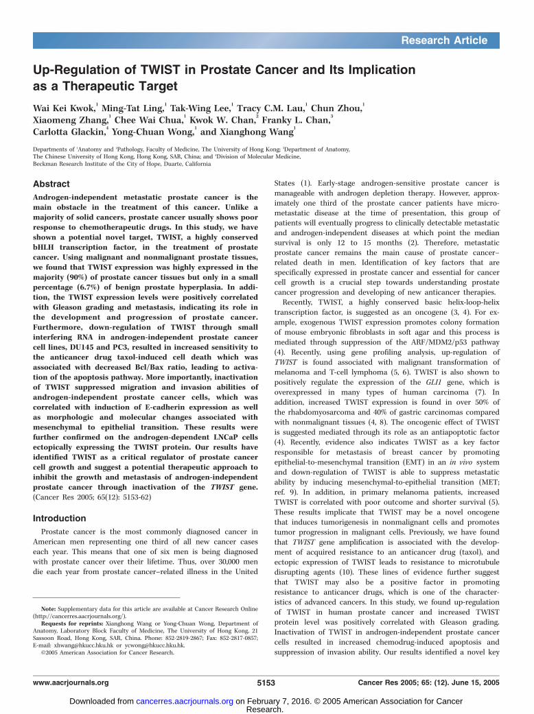
Figure 1. Differential expression of TWIST in malignant and nonmalignant prostate tissues. A, representative results of immunohistochemical staining ofTWIST protein in BPH (1 and 2) and prostate cancer tissues (3-10 ). 11 and 12, negative controls with primary antibody omitted. Photos were taken under 200�(1, 3, 5, 7, 9, 11) and 400� (2, 4, 6, 8, 10, 12) magnifications. Note that TWIST protein expression is positively correlated with increased Gleason grading.B, summary of differential TWIST expression between BPH and prostate cancer tissues (*, P < 0.001) and between prostate cancer Gleason grading <7 and z7specimens (**, P < 0.05). Note that TWIST expression is high in prostate cancer tissues compared with BPH, and increased TWIST expression is associated with higherGleason grading tumors. C, prostate-specific antigen (PSA ) and TWIST expression in prostate cancer specimens derived from bone (1-2) and lymph node (3-4 )metastatic lesions. Photos were taken under 100� magnifications.
Significance of TWIST in Prostate Cancer
www.aacrjournals.org 5155 Cancer Res 2005; 65: (12). June 15, 2005
Research. on February 7, 2016. © 2005 American Association for Cancercancerres.aacrjournals.org Downloaded from
washing with 1� PBS, the cells were permeabilized with 1% Triton X-100(in 1� PBS) for 15 minutes at room temperature. The cells were then
washed with 1� PBS and blocked with 1% BSA for 30 minutes at room
temperature. After blocking, the cells were incubated with antibody against
E-cadherin (BD Bioscience 610181, 1:100, in 0.1% BSA) for 1 hour at 37jC.The cells were washed thrice with 1� PBS followed by incubation with
FITC-labeled secondary antibody (DakoCytomation, Carpinteria, CA, 1:40 in
0.1% BSA) for 1 hour at room temperature. After three washes with 1� PBS,
the slides were mounted and covered with coverslip. Images of the cellsignal were captured by a laser confocal microscope under 400�magnifications.
Three-dimensional collagen colony-forming assay. Two hundredmicroliters of cell suspension (3 � 104 cells/mL) were mixed with 200 AL ofcold rat tail collagen, type I (3.60 mg/mL, BD Biosciences, Bedford, MA). Themixed cell solution in collagen was plated as droplets (at least three for each
sample) in 60-mm Petri dish until set. RPMI 1640 containing 5% of FCS wasadded in each dish containing the semisolid collagen cell droplets and thenthe cells were cultured for 3 days ( for PC3) or 5 days ( for DU145). Cellmorphology was observed under a phase-contrast microscope and pictureswere captured under 200� magnifications. At least 500 colonies were
counted from each sample and percentage of colonies that consisted ofelongated or scattered morphology was calculated.
Wound closure assay. A wound was induced on the confluentmonolayer cells by scraping a gap using a micropipette tip and the speed
of wound closure was monitored every 24 hours. Photographs were taken
under 100� magnifications using phase-contrast microscopy immediatelyafter wound incision and at later time points.
Results
Up-regulation of TWIST in prostate cancer specimens. Toinvestigate if there was a differential TWIST expression betweenbenign and malignant prostate tissues, we did immunohistochem-ical staining on BPH (n = 45) and prostate cancer tissues (n = 46).As shown in Fig. 1A (1 and 2), we found that high percentage ofBPH tissues (42 of 45, 93.3%) showed undetectable TWIST proteinstaining, whereas the majority of malignant prostate cancer tissues(40 of 46, 90%) were stained positive for TWIST protein (3-10). Thestaining pattern mainly agrees with a previous report onrhabdomyosarcoma tissues that TWIST protein was detected inthe nucleus of majority of cancer cells (4). However, we also notedthat a number of cases of high Gleason grading tumors showedboth cytoplasmic and nuclear staining (e.g., see, Fig. 1A, 9 and 10).In addition, the staining intensity was also significantly increasedwith increased Gleason grading (Fig. 1A, 3-10). Statistical analysisshowed (Fig. 1B) that overall TWIST expression level wassignificantly higher in prostate cancer specimens compared withthe BPH tissues (P < 0.001). In addition, TWIST expression wasmuch higher in the tissues with higher Gleason scores (z7) thanthe ones with lower Gleason scores (<7; P < 0.05). Furthermore,immunostaining results on bone and lymph node metastaticprostate cancer tissues also showed high TWIST expression (bothcytoplasmic and nuclear staining; Fig. 1C, 2 and 4). Prostate-specific antigen staining results were also shown to indicateprostate cancer cells (Fig. 1C, 1 and 3). These results indicate thatTWIST protein expression is increased in prostate cancer tissuescompared with nonmalignant tissues and its expression ispositively correlated with Gleason grades.Inactivation of TWIST leads to increased taxol-induced
apoptosis in androgen-independent prostate cancer cells. Tofurther confirm the significance of TWIST expression on prostatecancer cell growth, we transfected a retroviral vector containingthe Si-TWIST into DU145 and PC3 cells which showed relativelyhigh TWIST protein expression and generated stable transfectants
(see Supplementary Fig. S1A and B). We then studied if down-regulation of TWIST could lead to any changes in the expression ofp53 and MDM2 as well as apoptosis related proteins such as Bcl-2,Bax, and Bcl-xL. Down-regulation of MDM2 was observed in the Si-TWIST transfectants, whereas p53 protein was undetectable in PC3(containing a deleted p53 gene) and unchanged in DU145(containing a mutated p53 gene; refs. 16, 17; see SupplementaryFig. S1B). In addition, decreased Bcl-2 protein expression was foundin the Si-TWIST transfectants but there were no significantalterations in the Bax and Bcl-XL levels between the cells with highand low levels of TWIST. The differential Bax protein expressionbetween DU145 (barely detectable) and PC3 (relatively highexpression) cells may be due to the presence of a mutated Baxgene in DU145 cells but a functional Bax in PC3 cells as reportedpreviously (18). When we treated the cells with taxol, we found thatthe Si-TWIST transfected cells were much more sensitive to taxol-induced apoptosis shown by the increased percentage of sub-G1
phase cells in the flow cytometric studies (see SupplementaryFig. S1C, arrows). In addition, the appearance of DNA ladder wasalso more evident in the Si-TWIST transfectants when the cells weretreated with five doses of taxol (10, 20, 30, 40, and 50 ng/mL;representative results are shown in Supplementary Fig. S1D)compared with the vector controls. MTTassay also confirmed theseresults that Si-TWIST transfectants showedmuch lower cell viability( filled columns) compared with the vector controls (open columns)when both cell types were treated with same concentrations of taxol(Supplementary Fig. S1E). These results indicate that down-regulation of TWIST in androgen-independent prostate cancer cellshas led to increased sensitivity to taxol through activation of druginduced apoptosis. Further Western blotting analysis showed(Supplementary Fig. S1F) that after exposure to taxol, bothdecreased TWIST and Bcl-2 expression was found in the Si-TWISTtransfectants but alterations of Bax and Bcl-xL protein levels wereless significant. In contrast, the relative amount of the cleaved PARPwas higher in the Si-TWIST transfectants compared with the vectorcontrols (Supplementary Fig. S1G). Because the presence of cleavedPARP is another indicator of activation of the apoptosis pathway(19), these results further indicate that inactivation of TWIST hasled to activation of the apoptosis pathway in Si-TWIST transfectantsin response to taxol. However, down-regulation of TWIST was notassociated with decreased cell growth rate in the transfectantsevidenced by the similar BrdUrd incorporation rates (Fig. 2A) andpercentage of cell cycle S-phase fraction (Fig. 2B) between the Si-TWIST transfectants and the vector controls, which agrees with arecent study on breast cancer cells that inactivation of TWIST didnot alter cell proliferation rate (9). These results suggest that theincreased sensitivity to taxol in the Si-TWIST transfectants wasnot due to its effect on cell proliferation.Down-regulation of TWIST leads to suppression of invasion
ability through induction of mesenchymal-to-epithelial tran-sition–like changes. Recently, it was reported that in a breastcancer animal model, high levels of TWIST were associated withincreased tumor metastatic ability and ectopic TWISTexpression inhuman epithelial cells led to epithelial-to-mesenchymal transition(EMT; ref. 9), which is thought to contribute to the invasion abilityof cancer cells (20). Although few assay systems are available todirectly show MET, several morphologic as well as molecularalterations have been implicated accompanied with EMT (20, 21).For example, when epithelial cells are undergoing EMT, theirmorphology changes from well organized cell-cell adhesion and cellpolarity to loss of cell-cell contacts and cell scattering which is often
Cancer Research
Cancer Res 2005; 65: (12). June 15, 2005 5156 www.aacrjournals.org
Research. on February 7, 2016. © 2005 American Association for Cancercancerres.aacrjournals.org Downloaded from
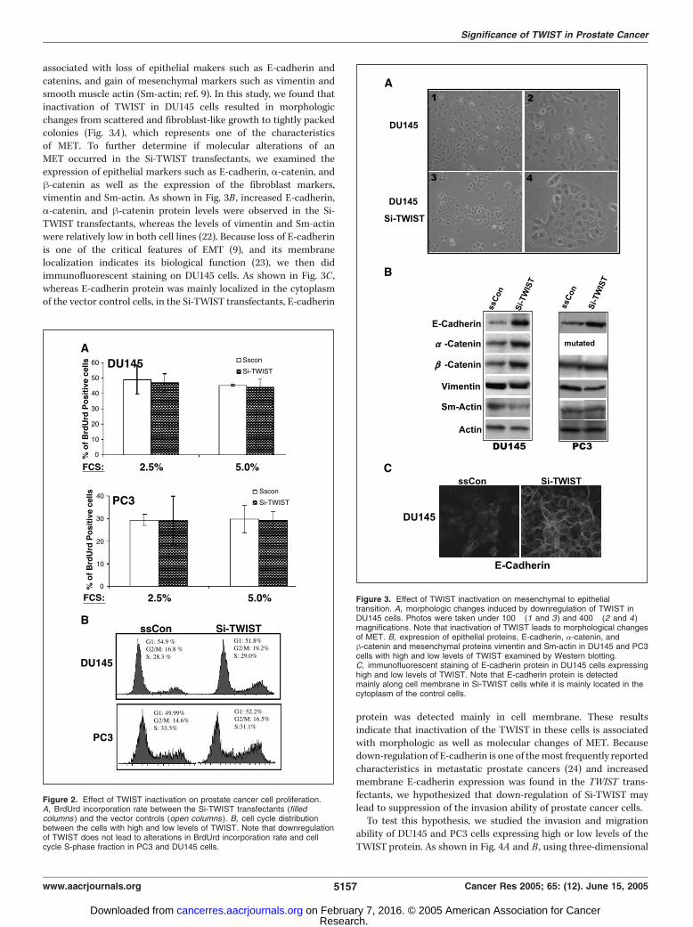
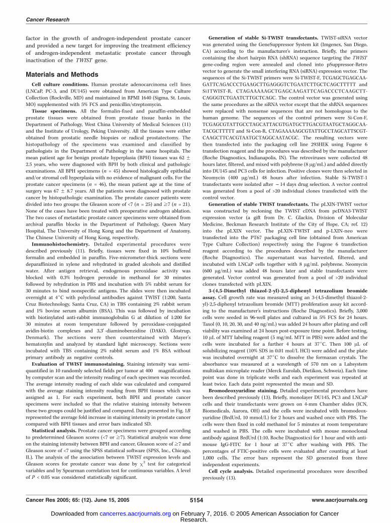
associated with loss of epithelial makers such as E-cadherin andcatenins, and gain of mesenchymal markers such as vimentin andsmooth muscle actin (Sm-actin; ref. 9). In this study, we found thatinactivation of TWIST in DU145 cells resulted in morphologicchanges from scattered and fibroblast-like growth to tightly packedcolonies (Fig. 3A), which represents one of the characteristicsof MET. To further determine if molecular alterations of anMET occurred in the Si-TWIST transfectants, we examined theexpression of epithelial markers such as E-cadherin, a-catenin, andh-catenin as well as the expression of the fibroblast markers,vimentin and Sm-actin. As shown in Fig. 3B , increased E-cadherin,a-catenin, and h-catenin protein levels were observed in the Si-TWIST transfectants, whereas the levels of vimentin and Sm-actinwere relatively low in both cell lines (22). Because loss of E-cadherinis one of the critical features of EMT (9), and its membranelocalization indicates its biological function (23), we then didimmunofluorescent staining on DU145 cells. As shown in Fig. 3C ,whereas E-cadherin protein was mainly localized in the cytoplasmof the vector control cells, in the Si-TWIST transfectants, E-cadherin
protein was detected mainly in cell membrane. These resultsindicate that inactivation of the TWIST in these cells is associatedwith morphologic as well as molecular changes of MET. Becausedown-regulation of E-cadherin is one of themost frequently reportedcharacteristics in metastatic prostate cancers (24) and increasedmembrane E-cadherin expression was found in the TWIST trans-fectants, we hypothesized that down-regulation of Si-TWIST maylead to suppression of the invasion ability of prostate cancer cells.To test this hypothesis, we studied the invasion and migration
ability of DU145 and PC3 cells expressing high or low levels of theTWIST protein. As shown in Fig. 4A and B , using three-dimensional
Figure 3. Effect of TWIST inactivation on mesenchymal to epithelialtransition. A, morphologic changes induced by downregulation of TWIST inDU145 cells. Photos were taken under 100� (1 and 3) and 400� (2 and 4)magnifications. Note that inactivation of TWIST leads to morphological changesof MET. B, expression of epithelial proteins, E-cadherin, a-catenin, andh-catenin and mesenchymal proteins vimentin and Sm-actin in DU145 and PC3cells with high and low levels of TWIST examined by Western blotting.C, immunofluorescent staining of E-cadherin protein in DU145 cells expressinghigh and low levels of TWIST. Note that E-cadherin protein is detectedmainly along cell membrane in Si-TWIST cells while it is mainly located in thecytoplasm of the control cells.
Figure 2. Effect of TWIST inactivation on prostate cancer cell proliferation.A, BrdUrd incorporation rate between the Si-TWIST transfectants (filledcolumns ) and the vector controls (open columns ). B, cell cycle distributionbetween the cells with high and low levels of TWIST. Note that downregulationof TWIST does not lead to alterations in BrdUrd incorporation rate and cellcycle S-phase fraction in PC3 and DU145 cells.
Significance of TWIST in Prostate Cancer
www.aacrjournals.org 5157 Cancer Res 2005; 65: (12). June 15, 2005
Research. on February 7, 2016. © 2005 American Association for Cancercancerres.aacrjournals.org Downloaded from
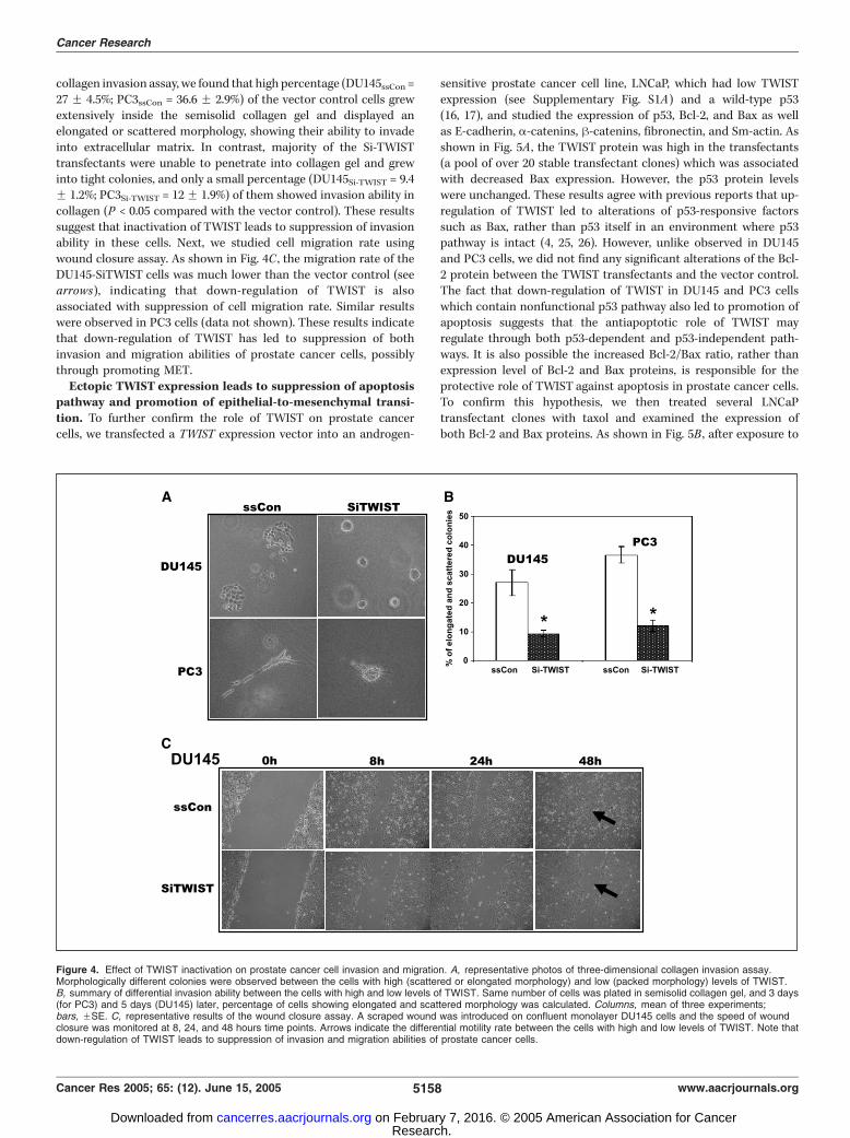
collagen invasion assay, we found that high percentage (DU145ssCon =27 F 4.5%; PC3ssCon = 36.6 F 2.9%) of the vector control cells grewextensively inside the semisolid collagen gel and displayed anelongated or scattered morphology, showing their ability to invadeinto extracellular matrix. In contrast, majority of the Si-TWISTtransfectants were unable to penetrate into collagen gel and grewinto tight colonies, and only a small percentage (DU145Si-TWIST = 9.4F 1.2%; PC3Si-TWIST = 12F 1.9%) of them showed invasion ability incollagen (P < 0.05 compared with the vector control). These resultssuggest that inactivation of TWIST leads to suppression of invasionability in these cells. Next, we studied cell migration rate usingwound closure assay. As shown in Fig. 4C , the migration rate of theDU145-SiTWIST cells was much lower than the vector control (seearrows), indicating that down-regulation of TWIST is alsoassociated with suppression of cell migration rate. Similar resultswere observed in PC3 cells (data not shown). These results indicatethat down-regulation of TWIST has led to suppression of bothinvasion and migration abilities of prostate cancer cells, possiblythrough promoting MET.Ectopic TWIST expression leads to suppression of apoptosis
pathway and promotion of epithelial-to-mesenchymal transi-tion. To further confirm the role of TWIST on prostate cancercells, we transfected a TWIST expression vector into an androgen-
sensitive prostate cancer cell line, LNCaP, which had low TWISTexpression (see Supplementary Fig. S1A) and a wild-type p53(16, 17), and studied the expression of p53, Bcl-2, and Bax as wellas E-cadherin, a-catenins, h-catenins, fibronectin, and Sm-actin. Asshown in Fig. 5A , the TWIST protein was high in the transfectants(a pool of over 20 stable transfectant clones) which was associatedwith decreased Bax expression. However, the p53 protein levelswere unchanged. These results agree with previous reports that up-regulation of TWIST led to alterations of p53-responsive factorssuch as Bax, rather than p53 itself in an environment where p53pathway is intact (4, 25, 26). However, unlike observed in DU145and PC3 cells, we did not find any significant alterations of the Bcl-2 protein between the TWIST transfectants and the vector control.The fact that down-regulation of TWIST in DU145 and PC3 cellswhich contain nonfunctional p53 pathway also led to promotion ofapoptosis suggests that the antiapoptotic role of TWIST mayregulate through both p53-dependent and p53-independent path-ways. It is also possible the increased Bcl-2/Bax ratio, rather thanexpression level of Bcl-2 and Bax proteins, is responsible for theprotective role of TWIST against apoptosis in prostate cancer cells.To confirm this hypothesis, we then treated several LNCaPtransfectant clones with taxol and examined the expression ofboth Bcl-2 and Bax proteins. As shown in Fig. 5B , after exposure to
Figure 4. Effect of TWIST inactivation on prostate cancer cell invasion and migration. A, representative photos of three-dimensional collagen invasion assay.Morphologically different colonies were observed between the cells with high (scattered or elongated morphology) and low (packed morphology) levels of TWIST.B, summary of differential invasion ability between the cells with high and low levels of TWIST. Same number of cells was plated in semisolid collagen gel, and 3 days(for PC3) and 5 days (DU145) later, percentage of cells showing elongated and scattered morphology was calculated. Columns, mean of three experiments;bars, FSE. C, representative results of the wound closure assay. A scraped wound was introduced on confluent monolayer DU145 cells and the speed of woundclosure was monitored at 8, 24, and 48 hours time points. Arrows indicate the differential motility rate between the cells with high and low levels of TWIST. Note thatdown-regulation of TWIST leads to suppression of invasion and migration abilities of prostate cancer cells.
Cancer Research
Cancer Res 2005; 65: (12). June 15, 2005 5158 www.aacrjournals.org
Research. on February 7, 2016. © 2005 American Association for Cancercancerres.aacrjournals.org Downloaded from
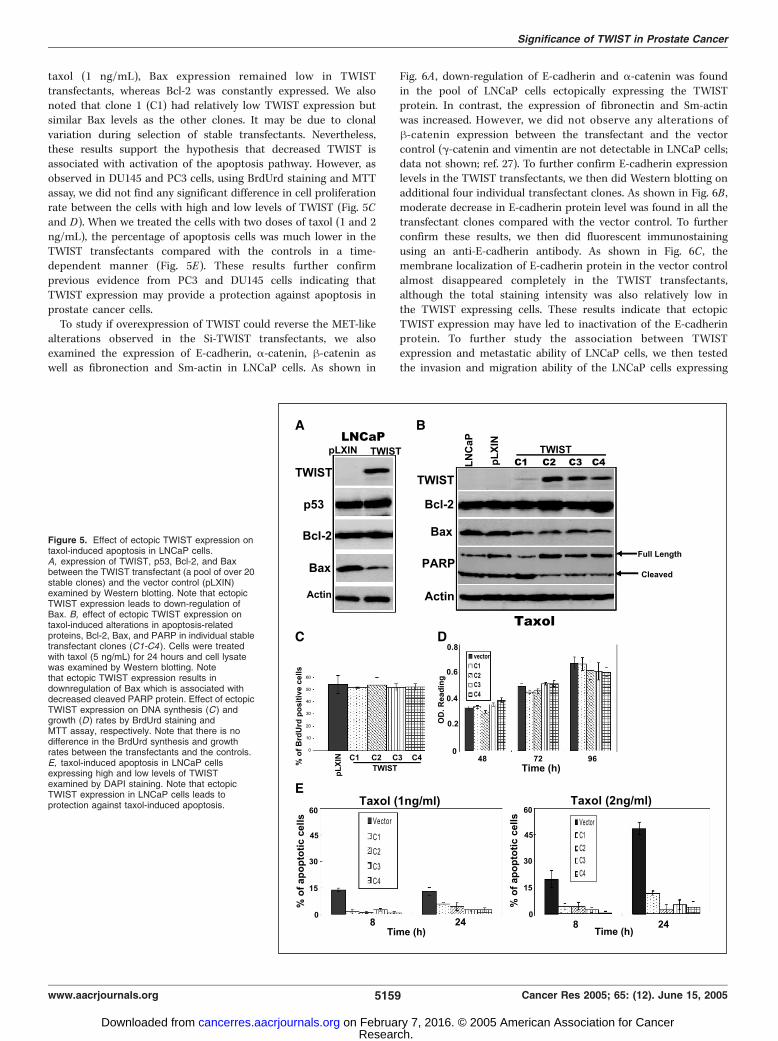
taxol (1 ng/mL), Bax expression remained low in TWISTtransfectants, whereas Bcl-2 was constantly expressed. We alsonoted that clone 1 (C1) had relatively low TWIST expression butsimilar Bax levels as the other clones. It may be due to clonalvariation during selection of stable transfectants. Nevertheless,these results support the hypothesis that decreased TWIST isassociated with activation of the apoptosis pathway. However, asobserved in DU145 and PC3 cells, using BrdUrd staining and MTTassay, we did not find any significant difference in cell proliferationrate between the cells with high and low levels of TWIST (Fig. 5Cand D). When we treated the cells with two doses of taxol (1 and 2ng/mL), the percentage of apoptosis cells was much lower in theTWIST transfectants compared with the controls in a time-dependent manner (Fig. 5E). These results further confirmprevious evidence from PC3 and DU145 cells indicating thatTWIST expression may provide a protection against apoptosis inprostate cancer cells.To study if overexpression of TWIST could reverse the MET-like
alterations observed in the Si-TWIST transfectants, we alsoexamined the expression of E-cadherin, a-catenin, h-catenin aswell as fibronection and Sm-actin in LNCaP cells. As shown in
Fig. 6A , down-regulation of E-cadherin and a-catenin was foundin the pool of LNCaP cells ectopically expressing the TWISTprotein. In contrast, the expression of fibronectin and Sm-actinwas increased. However, we did not observe any alterations ofh-catenin expression between the transfectant and the vectorcontrol (g-catenin and vimentin are not detectable in LNCaP cells;data not shown; ref. 27). To further confirm E-cadherin expressionlevels in the TWIST transfectants, we then did Western blotting onadditional four individual transfectant clones. As shown in Fig. 6B ,moderate decrease in E-cadherin protein level was found in all thetransfectant clones compared with the vector control. To furtherconfirm these results, we then did fluorescent immunostainingusing an anti-E-cadherin antibody. As shown in Fig. 6C , themembrane localization of E-cadherin protein in the vector controlalmost disappeared completely in the TWIST transfectants,although the total staining intensity was also relatively low inthe TWIST expressing cells. These results indicate that ectopicTWIST expression may have led to inactivation of the E-cadherinprotein. To further study the association between TWISTexpression and metastatic ability of LNCaP cells, we then testedthe invasion and migration ability of the LNCaP cells expressing
Figure 5. Effect of ectopic TWIST expression ontaxol-induced apoptosis in LNCaP cells.A, expression of TWIST, p53, Bcl-2, and Baxbetween the TWIST transfectant (a pool of over 20stable clones) and the vector control (pLXIN)examined by Western blotting. Note that ectopicTWIST expression leads to down-regulation ofBax. B, effect of ectopic TWIST expression ontaxol-induced alterations in apoptosis-relatedproteins, Bcl-2, Bax, and PARP in individual stabletransfectant clones (C1-C4 ). Cells were treatedwith taxol (5 ng/mL) for 24 hours and cell lysatewas examined by Western blotting. Notethat ectopic TWIST expression results indownregulation of Bax which is associated withdecreased cleaved PARP protein. Effect of ectopicTWIST expression on DNA synthesis (C ) andgrowth (D ) rates by BrdUrd staining andMTT assay, respectively. Note that there is nodifference in the BrdUrd synthesis and growthrates between the transfectants and the controls.E, taxol-induced apoptosis in LNCaP cellsexpressing high and low levels of TWISTexamined by DAPI staining. Note that ectopicTWIST expression in LNCaP cells leads toprotection against taxol-induced apoptosis.
Significance of TWIST in Prostate Cancer
www.aacrjournals.org 5159 Cancer Res 2005; 65: (12). June 15, 2005
Research. on February 7, 2016. © 2005 American Association for Cancercancerres.aacrjournals.org Downloaded from
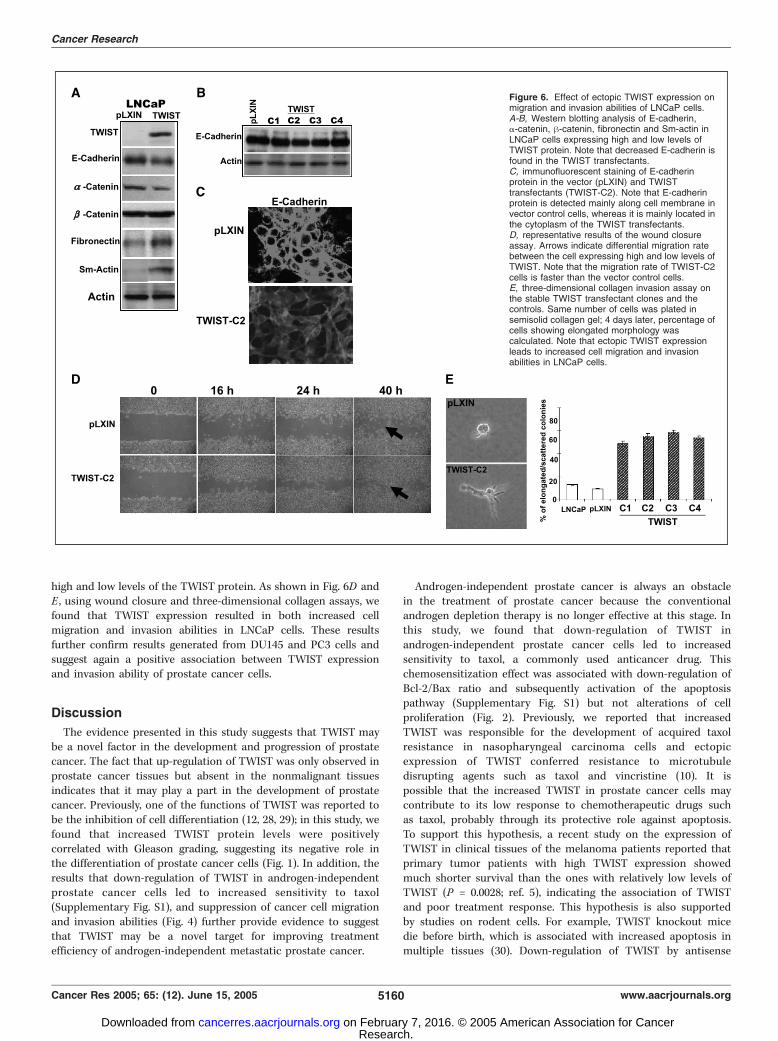
high and low levels of the TWIST protein. As shown in Fig. 6D andE , using wound closure and three-dimensional collagen assays, wefound that TWIST expression resulted in both increased cellmigration and invasion abilities in LNCaP cells. These resultsfurther confirm results generated from DU145 and PC3 cells andsuggest again a positive association between TWIST expressionand invasion ability of prostate cancer cells.
Discussion
The evidence presented in this study suggests that TWIST maybe a novel factor in the development and progression of prostatecancer. The fact that up-regulation of TWIST was only observed inprostate cancer tissues but absent in the nonmalignant tissuesindicates that it may play a part in the development of prostatecancer. Previously, one of the functions of TWIST was reported tobe the inhibition of cell differentiation (12, 28, 29); in this study, wefound that increased TWIST protein levels were positivelycorrelated with Gleason grading, suggesting its negative role inthe differentiation of prostate cancer cells (Fig. 1). In addition, theresults that down-regulation of TWIST in androgen-independentprostate cancer cells led to increased sensitivity to taxol(Supplementary Fig. S1), and suppression of cancer cell migrationand invasion abilities (Fig. 4) further provide evidence to suggestthat TWIST may be a novel target for improving treatmentefficiency of androgen-independent metastatic prostate cancer.
Androgen-independent prostate cancer is always an obstaclein the treatment of prostate cancer because the conventionalandrogen depletion therapy is no longer effective at this stage. Inthis study, we found that down-regulation of TWIST inandrogen-independent prostate cancer cells led to increasedsensitivity to taxol, a commonly used anticancer drug. Thischemosensitization effect was associated with down-regulation ofBcl-2/Bax ratio and subsequently activation of the apoptosispathway (Supplementary Fig. S1) but not alterations of cellproliferation (Fig. 2). Previously, we reported that increasedTWIST was responsible for the development of acquired taxolresistance in nasopharyngeal carcinoma cells and ectopicexpression of TWIST conferred resistance to microtubuledisrupting agents such as taxol and vincristine (10). It ispossible that the increased TWIST in prostate cancer cells maycontribute to its low response to chemotherapeutic drugs suchas taxol, probably through its protective role against apoptosis.To support this hypothesis, a recent study on the expression ofTWIST in clinical tissues of the melanoma patients reported thatprimary tumor patients with high TWIST expression showedmuch shorter survival than the ones with relatively low levels ofTWIST (P = 0.0028; ref. 5), indicating the association of TWISTand poor treatment response. This hypothesis is also supportedby studies on rodent cells. For example, TWIST knockout micedie before birth, which is associated with increased apoptosis inmultiple tissues (30). Down-regulation of TWIST by antisense
Figure 6. Effect of ectopic TWIST expression onmigration and invasion abilities of LNCaP cells.A-B, Western blotting analysis of E-cadherin,a-catenin, h-catenin, fibronectin and Sm-actin inLNCaP cells expressing high and low levels ofTWIST protein. Note that decreased E-cadherin isfound in the TWIST transfectants.C, immunofluorescent staining of E-cadherinprotein in the vector (pLXIN) and TWISTtransfectants (TWIST-C2). Note that E-cadherinprotein is detected mainly along cell membrane invector control cells, whereas it is mainly located inthe cytoplasm of the TWIST transfectants.D, representative results of the wound closureassay. Arrows indicate differential migration ratebetween the cell expressing high and low levels ofTWIST. Note that the migration rate of TWIST-C2cells is faster than the vector control cells.E, three-dimensional collagen invasion assay onthe stable TWIST transfectant clones and thecontrols. Same number of cells was plated insemisolid collagen gel; 4 days later, percentage ofcells showing elongated morphology wascalculated. Note that ectopic TWIST expressionleads to increased cell migration and invasionabilities in LNCaP cells.
Cancer Research
Cancer Res 2005; 65: (12). June 15, 2005 5160 www.aacrjournals.org
Research. on February 7, 2016. © 2005 American Association for Cancercancerres.aacrjournals.org Downloaded from
technology in mouse fibroblasts increased sensitivity to etopo-side (a DNA-damaging anticancer drug)– induced apoptosis (31).In addition, fibroblasts from TWIST2 knockout mice, which arehighly homologous to human TWIST, are hypersensitive totumor necrosis factor-a–induced apoptosis (32). These results incombination with the results presented in the current studyfurther suggest that down-regulation of TWIST may be a targetto increase cellular sensitivity to drug-induced apoptosis.The first evidence on the role of TWIST in cancer metastasis was
reported in a breast cancer animal model and it was suggested thatthe TWIST-induced EMT in the highly metastatic breast cancercells played a key role in promoting tumor cell invasion (9),although the role of TWIST in EMT has been suggested previouslyduring limb morphogenesis in mice (33). In this study, we foundthat inactivation of the TWIST gene conferred morphologictransition of prostate cancer cells from a fibroblastic to epithelialappearance which was accompanied by a gain of epithelial cellmarkers such as E-cadherin and catenins and loss of themesenchymal markers such as vimentin, fibronectin, and/or Sm-actin (Fig. 3). In addition, these changes were accompanied by thetranslocation of the E-cadherin protein from cytoplasm to cellmembrane (Fig. 3C), indicating its functional activation. TheseMET-like changes were correlated with decreased invasion andmigration abilities (Fig. 4). These results were further confirmed onLNCaP cells ectopically expressing the TWIST protein (Fig. 6).Although similar results were reported in nonmalignant epithelialcells (9), the present study was the first to show the role of TWISTin metastasis of human cancer cells. Previously, increased TWISTwas reported in the more invasive defused gastric carcinomatissues, which was associated with decreased E-cadherin expres-sion (8). In addition, in human breast cancer, elevated TWIST wasfound in 70% invasive lobular carcinomas, whereas a much smallerportion (30%) was found in less invasive tumors (9). In this study,we also found that the TWIST expression was high in tissuesderived from metastatic lesions (Fig. 1C). These results further
indicate the importance of TWIST in metastatic ability of humancancers. Although further studies are required to show the directassociation of TWIST and metastasis of human cancer, theseresults indicate that TWIST may be a target for inhibition ofmetastatic ability of cancer cells.In summary, in the past few years, increasing evidence has
suggested that TWIST may be a new positive factor in thedevelopment and progression of human cancer. Its functionsinclude promoting soft agar colony formation (4) and inhibitingcell differentiation (12, 28, 29) in nonmalignant cells andprotecting against anticancer drug-induced apoptosis (10) andpromoting metastasis (9) in malignant cells in in vitro systems. Inaddition, high levels of TWIST are found in gastric carcinoma (8),rhabdomyosarcoma (4), melanoma (5), breast cancer (9), SezarySyndrome (a leukemia variant of T-cell lymphoma; ref. 6), andprostate cancer (present study). Furthermore, increased TWIST iscorrelated with decreased E-cadherin expression and poorsurvival and more aggressive phenotypes of cancer cells (refs.5, 9 and present study). Although the molecular mechanismsresponsible for the action of TWIST in human cancer requirefurther investigation, the present study in combination withprevious results suggest that it may be a novel oncogene.However, more recently, promoter hypermethylation of theTWIST gene has been reported in breast cancer, especially inthe metastatic lesions (34). It is also possible that the oncogeniceffect of TWIST may be cell type specific. Nevertheless, the role ofTWIST in the development and progression of human cancerdeserves further attention.
Acknowledgments
Received 10/20/2004; revised 3/13/2005; accepted 4/7/2005.Grant support: Research Grants Council grants HKU7478/03M (X. Wang) and
HKU7490/03M (Y.C. Wong).The costs of publication of this article were defrayed in part by the payment of page
charges. This article must therefore be hereby marked advertisement in accordancewith 18 U.S.C. Section 1734 solely to indicate this fact.
References1. Denmeade SR, Isaacs JT. Development of prostatecancer treatment: the good news. Prostate 2004;58:211–24.
2. Dowling AJ, Tannock IF. Systemic treatment forprostate cancer. Cancer Treat Rev 1998;24:283–301.
3. Olson EN, Klein WH. bHLH factors in muscledevelopment: dead lines and commitments, what toleave in and what to leave out. Genes Dev 1994;8:1–8.
4. Maestro R, Dei Tos AP, Hamamori Y, et al. Twist is apotential oncogene that inhibits apoptosis. Genes Dev1999;13:2207–17.
5. Hoek K, Rimm DL, Williams KR, et al. Expressionprofiling reveals novel pathways in the transformationof melanocytes to melanomas. Cancer Res 2004;64:5270–82.
6. van DR, Dijkman R, Vermeer MH, et al. Aberrantexpression of the tyrosine kinase receptor EphA4 andthe transcription factor twist in Sezary syndromeidentified by gene expression analysis. Cancer Res2004;64:5578–86.
7. Villavicencio EH, Yoon JW, Frank DJ, Fuchtbauer EM,Walterhouse DO, Iannaccone PM. Cooperative E-boxregulation of human GLI1 by TWIST and USF. Genesis2002;32:247–58.
8. Rosivatz E, Becker I, Specht K, et al. Differentialexpression of the epithelial-mesenchymal transition
regulators snail, SIP1, and twist in gastric cancer. Am JPathol 2002;161:1881–91.
9. Yang J, Mani SA, Donaher JL, et al. Twist, a masterregulator of morphogenesis, plays an essential role intumor metastasis. Cell 2004;117:927–39.
10. Wang X, Ling MT, Guan XY, et al. Identification of anovel function of TWIST, a bHLH protein, in thedevelopment of acquired taxol resistance in humancancer cells. Oncogene 2004;23:474–82.
11. Ouyang XS, Wang X, Lee DT, Tsao SW, Wong YC. Overexpression of ID-1 in prostate cancer. J Urol 2002;167:2598–602.
12. Bialek P, Kern B, Yang X, et al. A twist codedetermines the onset of osteoblast differentiation. DevCell 2004;6:423–35.
13. Wang X, Jin DY, Ng RW, et al. Significance of MAD2expression to mitotic checkpoint control in ovariancancer cells. Cancer Res 2002;62:1662–8.
14. Ouyang XS, Wang X, Ling MT, Wong HL, Tsao SW,Wong YC. Id-1 stimulates serum independent prostatecancer cell proliferation through inactivation ofp16(INK4a)/pRB pathway. Carcinogenesis 2002;23:721–5.
15. Wang X, Wang J, Wong SC, et al. Cytotoxic effect ofgossypol on colon carcinoma cells. Life Sci 2000;67:2663–71.
16. Isaacs WB, Carter BS, Ewing CM. Wild-type p53suppresses growth of human prostate cancer cellscontaining mutant p53 alleles. Cancer Res 1991;51:4716–20.
17. Carroll AG, Voeller HJ, Sugars L, Gelmann EP. p53oncogene mutations in three human prostate cancercell lines. Prostate 1993;23:123–34.
18. Honda T, Kagawa S, Spurgers KB, et al. Arecombinant adenovirus expressing wild-type Baxinduces apoptosis in prostate cancer cells independent-ly of their Bcl-2 status and androgen sensitivity. CancerBiol Ther 2002;1:163–7.
19. Ame JC, Spenlehauer C, de Murcia G. The PARPsuperfamily. Bioessays 2004;26:882–93.
20. Thiery JP. Epithelial-mesenchymal transitions intumour progression. Nat Rev Cancer 2002;2:442–54.
21. Boyer B, Thiery JP. Epithelium-mesenchyme inter-conversion as example of epithelial plasticity. APMIS1993;101:257–68.
22. Ewing CM, Ru N, Morton RA, et al. Chromosome 5suppresses tumorigenicity of PC3 prostate cancer cells:correlation with re-expression of a-catenin and resto-ration of E-cadherin function. Cancer Res 1995;55:4813–7.
23. Hazan RB, Qiao R, Keren R, Badano I, Suyama K.Cadherin switch in tumor progression. Ann N Y Acad Sci2004;1014:155–63.
24. Umbas R, Schalken JA, Aalders TW, et al. Expressionof the cellular adhesion molecule E-cadherin is reducedor absent in high-grade prostate cancer. Cancer Res1992;52:5104–9.
25. Yousfi M, Lasmoles F, El Ghouzzi V, Marie PJ.Twist haploinsufficiency in Saethre-Chotzen syndrome
Significance of TWIST in Prostate Cancer
www.aacrjournals.org 5161 Cancer Res 2005; 65: (12). June 15, 2005
Research. on February 7, 2016. © 2005 American Association for Cancercancerres.aacrjournals.org Downloaded from

induces calvarial osteoblast apoptosis due to in-creased TNFa expression and caspase-2 activation.Hum Mol Genet 2002;11:359–69.
26. Ota MS, Loebel DA, O’Rourke MP, Wong N, Tsoi B,Tam PP. Twist is required for patterning the cranialnerves and maintaining the viability of mesodermalcells. Dev Dyn 2004;230:216–28.
27. Singh S, Sadacharan S, Su S, Belldegrun A, Persad S,Singh G. Overexpression of vimentin: role in the invasivephenotype in an androgen-independent model ofprostate cancer. Cancer Res 2003;63:2306–11.
28. Spicer DB, Rhee J, Cheung WL, Lassar AB.Inhibition of myogenic bHLH and MEF2 transcription
factors by the bHLH protein Twist. Science 1996;272:1476–80.
29. Lee MS, Lowe GN, Strong DD, Wergedal JE,Glackin CA. TWIST, a basic helix-loop-helix transcrip-tion factor, can regulate the human osteogenic lineage.J Cell Biochem 1999;75:566–77.
30. Chen ZF, Behringer RR. twist is required in headmesenchyme for cranial neural tube morphogenesis.Genes Dev 1995;9:686–99.
31. Dupont J, Fernandez AM, Glackin CA, Helman L,LeRoith D. Insulin-like growth factor 1 (IGF-1)-inducedtwist expression is involved in the anti-apoptotic effectsof the IGF-1 receptor. J Biol Chem 2001;276:26699–707.
32. Sosic D, Richardson JA, Yu K, Ornitz DM, Olson EN.Twist regulates cytokine gene expression through anegative feedback loop that represses NF-nB activity.Cell 2003;112:169–80.
33. Zuniga A, Quillet R, Perrin-Schmitt F, Zeller R.Mouse Twist is required for fibroblast growth factor-mediated epithelial-mesenchymal signalling and cellsurvival during limb morphogenesis. Mech Dev 2002;114:51–9.
34. Mehrotra J, Vali M, McVeigh M, et al. Very highfrequency of hypermethylated genes in breast cancermetastasis to the bone, brain, and lung. Clin Cancer Res2004;10:3104–9.
Cancer Research
Cancer Res 2005; 65: (12). June 15, 2005 5162 www.aacrjournals.org
Research. on February 7, 2016. © 2005 American Association for Cancercancerres.aacrjournals.org Downloaded from
2005;65:5153-5162. Cancer Res Wai Kei Kwok, Ming-Tat Ling, Tak-Wing Lee, et al. Implication as a Therapeutic TargetUp-Regulation of TWIST in Prostate Cancer and Its
Updated version
http://cancerres.aacrjournals.org/content/65/12/5153
Access the most recent version of this article at:
Material
Supplementary
http://cancerres.aacrjournals.org/content/suppl/2005/08/25/65.12.5153.DC1.html
Access the most recent supplemental material at:
Cited articles
http://cancerres.aacrjournals.org/content/65/12/5153.full.html#ref-list-1
This article cites 34 articles, 15 of which you can access for free at:
Citing articles
http://cancerres.aacrjournals.org/content/65/12/5153.full.html#related-urls
This article has been cited by 70 HighWire-hosted articles. Access the articles at:
E-mail alerts related to this article or journal.Sign up to receive free email-alerts
Subscriptions
Reprints and
To order reprints of this article or to subscribe to the journal, contact the AACR Publications
Permissions
To request permission to re-use all or part of this article, contact the AACR Publications
Research. on February 7, 2016. © 2005 American Association for Cancercancerres.aacrjournals.org Downloaded from