Genetic Alterations, DNA Methylation, Alloantibodies ... - MDPI
This article appeared in a journal published by Elsevier. The attachedcopy is furnished to the author for internal non-commercial researchand education use, including for instruction at the authors institution
and sharing with colleagues.
Other uses, including reproduction and distribution, or selling orlicensing copies, or posting to personal, institutional or third party
websites are prohibited.
In most cases authors are permitted to post their version of thearticle (e.g. in Word or Tex form) to their personal website orinstitutional repository. Authors requiring further information
regarding Elsevier’s archiving and manuscript policies areencouraged to visit:
http://www.elsevier.com/authorsrights

Author's personal copy
Mini-review
Understanding genomic alterations in cancer genomes using anintegrative network approach
Edwin WangLab of Bioinformatics and Systems Biology, National Research Council Canada, Montreal, CanadaMcGill University Center for Bioinformatics, Montreal, Canada
a r t i c l e i n f o
Keywords:ChromothripsisKataegisCancer genome evolutionNetworkSystems biologyCancer genome sequencing
a b s t r a c t
In recent years, cancer genome sequencing and other high-throughput studies of cancer genomes havegenerated many notable discoveries. In this review, novel genomic alteration mechanisms, such as chro-mothripsis (chromosomal crisis) and kataegis (mutation storms), and their implications for cancer arediscussed. Genomic alterations spur cancer genome evolution. Thus, the relationship between cancer clo-nal evolution and cancer stems cells is commented. The key question in cancer biology concerns howthese genomic alterations support cancer development and metastasis in the context of biological func-tioning. Thus far, efforts such as pathway analysis have improved the understanding of the functionalcontributions of genetic mutations and DNA copy number variations to cancer development, progressionand metastasis. However, the known pathways correspond to a small fraction, plausibly 5–10%, ofsomatic mutations and genes with an altered copy number. To develop a comprehensive understandingof the function of these genomic alterations in cancer, an integrative network framework is proposed anddiscussed. Finally, the challenges and the directions of studying cancer omic data using an integrativenetwork approach are commented.
Crown Copyright � 2012 Published by Elsevier Ireland Ltd. All rights reserved.
1. Introduction
Because of the ongoing development of new, fast, and inexpen-sive DNA sequencing technologies, the cost of genome sequencingtechnology has rapidly declined. Eventually, genome sequencingtechnology may allow doctors to decoding the entire genetic codeof a patient disease sample in a clinical setting. Cancer is rooted ingenetic and epigenetic alterations, which are either inherited or ac-quired (i.e., mutated or methylated) during our lives. Cancer gen-omes contain two classes of mutations: driver mutations, whichare positively selected because they are essential for tumor growthand development, and passenger mutations, which are not subjectto selection because they do not offer a growth advantage. Positiveselection indicative of driver mutations is evidenced by a higherprobability than by chance of amino acid-changing nonsynony-mous mutations to synonymous mutations that do not involveamino acid changes.
However, we are still no closer to uncovering the molecularunderpinnings of the disease. If we could catalog cancer driver-mutating genes, we could link these genes to biological pathways,biological processes and cellular networks. Cancer genomesequencing and omic studies will improve the understanding ofthe genetic/epigenetic underpinnings of major types and subtypesof human cancer, thus generating new insights concerning how to
address and to manage cancer in clinics. These studies will eventu-ally enable more personalized treatments that will improve patientcare.
Significant effort has focused on compressive omic analysis,including genome sequencing, SNPs profiling, gene expression, epi-genetic changes and proteomic profiling of major tumor types.These efforts represent a decade-long program that has aimed togenerate high-quality omic data on more than 25,000 tumors forup to 50 types of cancer of clinical and societal importance. Threemajor organizations, the Wellcome Trust Sanger Institute in theUnited Kingdom, the Cancer Genome Atlas (TCGA) in the UnitedStates and the International Cancer Genome Consortium (ICGC)[1] in Canada, have advanced this program. Furthermore, severalcountries are working on this project, and the ICGC is collecting,merging and managing all of the data. In fact, the sequencing of25,000 tumors is being performed around the world in 40 projectsfocused on the bladder, the blood, the bone, the brain, the breast,the cervix, the colon, the head and the neck, the kidney, the liver,the lung, the oral cavity, the ovary, the pancreas, the prostate,the rectum, the skin, the soft tissues, the stomach, and the uterus.The ICGC has collected omic data on more than 3500 tumors.
Among these efforts, St. Jude Children’s Research Hospital tookan initiative to make cancer genome information freely accessibleto the global scientific community at www.ebi.ac.uk/ega/organisa-tions/EGAO00000000046 [2]. This site mainly includes the genomesequences (entire genome sequencing with up to a 30-fold cover-
0304-3835/$ - see front matter Crown Copyright � 2012 Published by Elsevier Ireland Ltd. All rights reserved.http://dx.doi.org/10.1016/j.canlet.2012.11.050
E-mail address: [email protected]
Cancer Letters 340 (2013) 261–269
Contents lists available at ScienceDirect
Cancer Letters
journal homepage: www.elsevier .com/locate /canlet
Author's personal copy
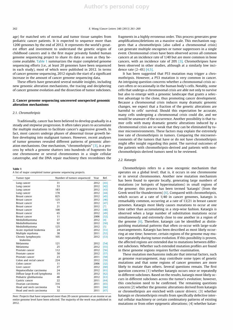
age) for matched sets of normal and tumor tissue samples frompediatric cancer patients. It is expected to sequence more than1200 genomes by the end of 2012. It represents the world’s great-est effort and investment to understand the genetic origins ofchildhood cancers and is the first major privately funded humangenome sequencing project to share its data as soon as they be-come available. Table 1 summarizes the major completed genomesequencing efforts (i.e., at least 20 genomes have been sequencedin each study), most of which were published in 2012. In termsof cancer genome sequencing, 2012 signals the start of a significantincrease in the amount of cancer genome sequencing data.
These efforts have generated several notable insights, includingnew genomic alteration mechanisms, the tracing and decipheringof cancer genome evolution and the dissection of tumor subclones.
2. Cancer genome sequencing uncovered unexpected genomicalteration mechanisms
2.1. Chromothripsis
Traditionally, cancer has been believed to develop gradually in asteady and stepwise progression. It often takes years to accumulatethe multiple mutations to facilitate cancer’s aggressive growth. Infact, most cancers undergo phases of abnormal tissue growth be-fore developing into malignant tumors. However, recent analysesof tumor genomes revealed different patterns of genomic alter-ation mechanisms. One mechanism, ‘‘chromothripsis’’ [3], is a pro-cess by which a genome shatters into hundreds of fragments forone chromosome or several chromosomes in a single cellularcatastrophe, and the DNA repair machinery then recombines the
fragments in a highly erroneous order. This process generates geneamplifications/deletions on a massive scale. This mechanism sug-gests that a chromothripsis (also called a chromosomal crisis)can generate multiple oncogenes or tumor suppressors in a singleevent. Chromosomal crises have been observed across all commoncancers at an incidence rate of 1/40 but are more common in bonecancers, with an incidence rate of 20% [3]. Chromothripses havebeen observed in other studies, although at a similarly low inci-dence rate (2–4%) [4,5].
It has been suggested that P53 mutation may trigger a chro-mothripsis. However, a P53 mutation is very common in cancer.An intriguing question concerns whether a chromosomal crisis oc-curs often or occasionally in the human body’s cells. Notably, somecells that undergo a chromosomal crisis are able not only to survivebut also to emerge with a genomic landscape that grants a selec-tive advantage to the clone, thus promoting cancer development.Because a chromosomal crisis induces many dramatic genomicchanges, we expect that a fraction of the genetic alterations areharmful to cells’ survival. Should this expectation be accurate,many cells undergoing a chromosomal crisis could die, and wewould be unaware of the occurrence. Another possibility is that tu-mor cells with many dramatic genetic alterations induced by achromosomal crisis are so weak that they can be eliminated by tu-mor microenvironments. These factors may explain the extremelylow rate of chromothripsis in tumors. Comparing the microenvi-ronments of the tumors that have or do not have chromothripsismight offer insight regarding this point. The survival outcomes ofthe patients with chromothripsis-derived and patients with non-chromothripsis-derived tumors need to be reviewed.
2.2. Kataegis
Chromothripsis refers to a new oncogenic mechanism thatoperates on a global level; that is, it occurs in one chromosomeor in several chromosomes. Another new mutation mechanismhas been found to operate locally, generating large numbers ofmutations (or hotspots of hypermutations) in small regions ofthe genome; this process has been termed ‘kataegis’ (from theGreek word for thunderstorm) [6]. Compared with chromothripsis,which occurs at a rate of 1/40 in cancer genomes, kataegis isremarkably common, occurring at a rate of 13/21 in breast cancergenomes. Kataegis most likely causes mutations to occur at onetime rather than accumulating in a step-wise fashion. Kataegis isobserved when a large number of substitution mutations occursimultaneously and extremely close to one another in a region ofthe genome [6]. Therefore, kataegis can be identified as distin-guishing mutational patterns that often co-occur with large-scalerearrangements. Kataegis has been described as most likely occur-ring at one time; however, certain regions of the genome may mu-tate repeatedly during tumor evolution. If this possibility is proven,the affected regions are extended due to mutations between differ-ent subclones. Whether such extended mutation profiles are foundin these genome regions requires further examination.
These mutation mechanisms indicate that internal factors, suchas genome rearrangement, may contribute some types of geneticmutations and that some regions of cancer genomes are morelikely to mutate than others. Several questions remain. The firstquestion concerns (1) whether kataegis occurs once or repeatedlyin different subclones. Based on the results, kataegis most likely oc-curs in different subclones across the tumor’s evolution; however,this conclusion need to be confirmed. The remaining questionsconcern (2) whether the genomic alterations derived from kataegisor chromothripsis are enriched with cancer drivers; (3) whetherkataegis or chromothripsis results from specifically defective inter-nal cellular machinery or certain combinatory patterns of existingmutations or from other epigenetic alterations; (4) whether katae-
Table 1A list of major completed tumor genome sequencing projects.
Tumor type Number of tumors sequenced Year Ref.
Lung cancer 31 2012 [41]Lung cancer 53 2012 [42]Lung cancer 183 2012 [43]Lung cancer 178 2012 [44]Breast cancer 825 2012 [45]Breast cancer 125 2012 [46]Breast cancer 77 2012 [47]Breast cancer 21 2012 [7]Breast cancer 100 2012 [48]Breast cancer 65 2012 [49]Breast cancer 11 2006 [32]Medulloblastoma 37 2012 [4]Medulloblastoma 92 2012 [50]Medulloblastoma 125 2012 [5]Acute myeloid leukemia 24 2012 [51]Multiple myeloma 38 2011 [52]Chronic lymphocytic
leukemia105 2012 [53]
Melanoma 121 2012 [54]Melanoma 25 2012 [55]Prostate cancer 112 2012 [56]Prostate cancer 50 2012 [57]Prostate cancer 23 2011 [58]Colon and rectal cancer 224 2012 [59]Colon cancer 11 2006 [32]Liver cancer 27 2012 [60]Hepatocellular carcinoma 24 2012 [61]Diffuse large B-cell lymphoma 55 2012 [62]Pediatric glioblastoma 48 2012 [63]Gastric cancer 22 2011 [64]Ovarian carcinoma 316 2011 [65]Head and neck carcinoma 74 2011 [66]Head and neck carcinoma 32 2011 [67]
Note: Projects that have sequenced more than 20 cancer genomes at an exome or anentire genome level have been selected. The majority of the work was published in2012.
262 E. Wang / Cancer Letters 340 (2013) 261–269
Author's personal copy
gis-derived cancer genomes are more aggressive than the tumorsderived from other genomic alteration mechanisms; and (5)whether the DNA copy number change represents an importantgenetic alteration in cancer cells. A series of chromothripses mightconvert a cell into a cancer cell quickly. However, these cells maynot mutate but rather undergo many copy number changes. Fur-ther investigations of these novel genomic alteration mechanismsmay deepen the understanding of factors influencing these muta-tions/copy number variations and cancer development. Neverthe-less, early mutations, copy number changes or alterations ofchromosomal structures caused by other factors, such as smoking,sunlight, or stress, may be conducive to kataegis or chromothripsis.
3. Genomic alterations and cancer genome sequencing
3.1. Deciphering cancer genomic alterations in an evolutionary context
Tumors are widely known to be heterogeneous. Therefore, a tu-mor is often expected to comprise a set of clones of cancer cells.Understanding the mechanisms that cause cancer requires com-prehensive insight into the processes underlying cancer genetics.To obtain this information, the Sanger group sequenced 21 breastcancer genomes, cataloged their mutations and attempted to tracethe evolution of cancer mutation profiles between different sub-clones in the context of timing [7]. Using appropriate bioinformaticapproaches, they were able to uncover the genetic mutation marksand the inscribed record of the latter’s emergence over time, thusenabling the divergence of a cell in forming the various clones tobe traced.
Cancer mutational processes evolve over a tumor’s lifespan.Each time a gene is mutated in a cell, the mutation event encryptsa distinctive stamp inscribed in its genome. Such a distinctivestamp can be considered a mutation signature or a distinctive markon the genome. From a historical perspective, an archaeologicalhistory of mutations results from the accumulations of marks uponone another. Therefore, collecting, cataloging and sorting the or-ders of these marks allow the cancer’s life history to be deciphered.
Cells in a single subclone possess an identical genetic mutationprofile. The cells in late-occurring subclones add new mutations tothe genetic mutation profiles of the early subclones. Only a muta-tion causes a clone to dramatically expand, after which the cloneconstitutes the tumor’s dominant population and the tumor growslarge enough to be diagnosed. During this process, the history ofthe genetic mutations and the emergence and the expansion ofthe new clone is imprinted upon the cancer genome. Therefore,these genetic mutation profiles can be used to build an evolution-ary tree of subclones. This analysis allows the tracing of the occur-rence of a new mutation profile, the biological processes involvedin each mutation profile or subclone, and the proportion of eachsubclone present in the tumors. The study showed that as themutations, or genetic marks, accumulate in a cell, the correspond-ing changes caused the cancer to diverge into different clones [7].The emergence of different subclones occurred in all of the exam-ined cancer genomes. This analysis suggests that when a new sub-clone has grown sufficiently to constitute the dominant populationof cancer cells (i.e., more than 50% of tumor cells), the tumor be-comes clinically detectable. The genetic mutation profiles of sub-clones and dominant subclones differ markedly, implying thatthe dominant subclone may require a significant amount of timeto develop.
This study has several implications. First, it confirms that a tu-mor contains many subclones; thus, the heterogeneity can be sam-pled by sequencing, as shown in Gerlinger et al. [7]. Second, thestudy suggests that a cancer patient may benefit from the use ofcombinatory drugs for different subclones. One perspective is that
treatment may proceed by targeting the early clones because theirmutation profiles are identical to those of the late subclones [8].However, this approach might not work well. The late subclones,particularly the dominant subclones, acquire new traits for cellproliferation and survival. These new traits could grant the latesubclones ‘‘new’’ survival capabilities, thus implying that the ‘drugtargets’ for the early subclones are unsuitable for the late sub-clones. However, subclones [9] can successfully be dissected fromtumors, and the RNA-seq or sequencing analysis of these subclonescan be performed [10]. Profiling these subclones helps to identifydrug targets for each subclone. Third, each cancer appears to begenerated by a distinct combination of mutational processes.Fourth, the study confirms the prediction that transforming a nor-mal cell to a cancer cell is a lengthy process that has been shown tolast 10 years [11]. This fact supports the search for mutation signa-tures or molecular biomarkers for early diagnosis and the possibil-ity of personalized early preventions to potential cancer patients.
3.2. Which subclone contains the cancer stem cell?
Although many implications have been found from decipheringcancer genomes, a key question remains: can we identify or inferwhich subclone contains the cancer stem cell?
The cancer stem cell hypothesis suggests that tumors are orga-nized into aberrant cell hierarchies. In such a hierarchy, a cancerstem cell capable of replicating indefinitely is the parent cell thatgenerates differentiated daughter cells with a limited capacity toproliferate. If proven for a tumor, this hypothesis suggests that can-cer is curable if drugs are available to kill cancer stem cells. Re-cently, three studies have provided the first evidence that cancerstem cells exist [12]. These studies have demonstrated that cancerstem cells reproduce cancer cells when the tumor is extinguishedby anticancer drugs, a result that applies to benign and malignanttumors.
These results suggest that cancer stems cells are inherentlycapable of propagating genetic or epigenetic alterations through-out a tumor and driving cancer evolution. The cancer stem cellsconstitute the stem of tumor cell production. During a cancer’sevolution, a certain genetic mutation profile may define cancerstem cell subclones. The parental subclones of the cancer stem cellsubclone are non-stem cells, thus indicating that genetic/epige-netic changes could convert a non-stem cell into a cancer stem cell.Such a stem cell produces many daughter cells that may furtherevolve into diversified cancer cells. If this process occurs, thenthe dominant subclones of the tumors would not be cancer stemcells but would emerge from cancer stem cells.
Thus, many questions remain. First, should every tumor containa subset of cancer stem cells? All tumors may not contain cancerstem cells, for example, if a chromothripsis occurs to generate en-ough fitting combinations of mutations to generate a new subclonecapable of becoming dominant in a tumor. However, generally,sustaining the cancer’s evolution requires generating enough can-cer cells using cancer stem cells. In this case, a cancer stem cellmust evolve and be defined by a set of mutations. Second, if a can-cer stem cell must exist in a tumor, what are the characteristics ofits genetic mutation profiles? The evolution of cancer genomes im-plies that a cancer stem cell subclone should be an early subclonein the history of the tumor’s development. We assume that thecancer stem cell subclone generates new cells but at a limitedand slow rate because of the microenvironment’s unfavorable con-ditions. These daughter cells are further mutated and generate newsubclones.
This hypothesis is supported by the discoveries of Driessenset al. [12–14] and Chen et al. [13] that show that the cellular orga-nization of early (precancerous) skin and intestinal tumors arecomposed of both stem cells and non-stem cells. Furthermore,
E. Wang / Cancer Letters 340 (2013) 261–269 263
Author's personal copy
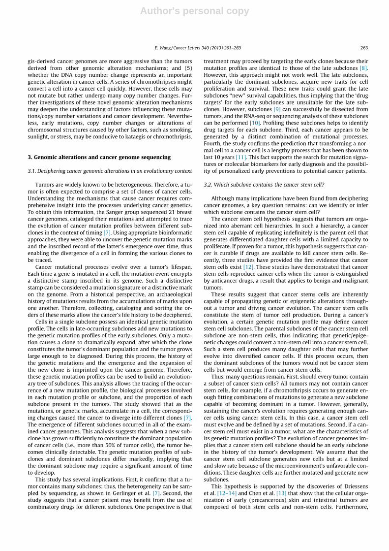
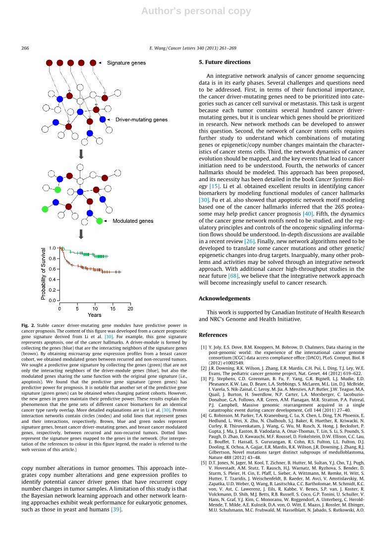
Driessens et al. observed that progression to cancer in benign skintumors is associated with an increased cancer stem cell population.They found that every cancer stem cell within a tumor is equallyprobable of clonally expanding or dying, possibly even in the ab-sence of new mutations, thus suggesting that a cancer stem cellsubclone most likely occurs in an early stage of cancer develop-ment (Fig. 1).
The most important finding from the above discussion is thatsubclonal diversity in cancer genomes may be dissected. To treatcancer more efficiently, it requires the dissection of not only thedominant subclones but also the minor subclones, which areamong the top subclones in the hierarchy and most likely cancerstem cells. The key challenges are determining the functions ofthe mutations in these subclones and linking these mutations todrug targets for each subclone and, subsequently, identifyingmutational signatures as diagnostic tools that predict the earlydevelopment of cancer.
4. The functional understanding of genomic alterations incancer genomes at a systems level
As discussed above, in recent years, sequencing efforts havegenerated many genomic alterations of cancer. Although someimportant insights, such as new genomic alteration mechanisms,cancer genome evolution, and clonal dissection, have been gleanedfrom the corresponding data, a lack of frameworks and tools nev-ertheless persists with respect to how genomic alterations drivecancer evolution, clonal expansion, and cancer development andmetastasis. Understanding these processes requires investigatingthe function of these genomic alterations. However, currently,the greatest challenge is understanding the functions of drivermutations in cancer genomes. On average, several hundred drivermutations have been cataloged from each tumor, but interpretingthese mutations is inefficient, thus limiting the understanding ofwhy and how these mutation genes work. Most current cancergenome sequencing work has mapped the mutations onto biolog-ical pathways, including signaling pathways.
A biological pathway contains a set of linear relationships be-tween genes or proteins and is an important model to describe bio-logical relationships. From an abstract perspective, a pathway is amodel that represents humans’ efforts to explore, describe, andorganize biological relationships in cells [15]. Such efforts allowus to further understand biological systems and to predict cellbehaviors. However, less than 5–10% of mutations can be ex-plained by these ‘core pathways’ in which a set of genes is rela-tively highly mutated [16,17]. Several problems are associatedwith this approach. First, pathway gene members are fuzzy andpoorly defined. For example, if different databases such as KEGGor BioCarta are queried for ‘EGFR signaling pathway,’ each databasewill provide a different gene list. Second, only a small set of muta-
tion genes (less than 5–10%) can be mapped onto the ‘core path-ways,’ thus, most mutation genes are unmapped. Third, for eachstudy, only a few ‘core pathways’ such as RAS-PI3K were foundrepeatedly. These pathways are well known in cancer studies,and drugs have been developed for these pathways or key genes.In this respect, the pathway approach does not provide new in-sights but captures the existing knowledge in the field.
To comprehensively understand the drivers of cancer genomes,computational methods should be developed because of the largenumber of driver mutation genes in each tumor. The developmentof effective computational methods first requires understandingwhat subjects biology addresses. Over many years, biologists haveexplored several types of relationships among genes, proteins,RNAs and other molecules. The molecular mechanisms of biologi-cal processes or diseases have also been studied. A molecularmechanism often discloses an event of A regulating B, B inducingor inhibiting C and generating a phenotype or disease. Each mech-anism comprises a set of relationships between molecules such asgene regulation, protein interaction, activation, genetic interactionand inhibitory action. In this context, biology is a science that de-scribes relationships [15].
Traditionally, biologists use pathways to describe the relation-ships between a limited number of genes. As shown above, numer-ous driver mutations have been found in each tumor, but thepathway approach explains only 5–10% of the genetic mutationsin the cancer genome. It is expected that biologists must considerthousands of biological relationships with respect to these genes inone tumor, for which task the traditional methods of describingbiological relationships are inadequate. Furthermore, as thecross-talk between pathways has been documented extensivelyin recent years, scientists have come to recognize that a networkmodel may provide a solution. A network model describes theinteractions and relationships between genes or proteins in a non-linear manner. The pathway and network approaches are concep-tual models that reflect an avenue of exploring biology byhumans. Both models project living cell molecules onto conceptualframeworks, but the molecular network model more closelyapproximates the processes that occur in cells.
4.1. Basic concepts of molecular networks
A molecular network can be represented using nodes and links.Nodes represent genes or proteins, whereas links represent therelationships between genes and proteins. Cancer omic data canbe integrated into a molecular network to pose questions and ob-tain corresponding insights. However, different types of networkshave distinct properties and thus can be used to address differentquestions.
There are four basic molecular networks: signaling, gene regu-latory, metabolic and protein interaction networks. A networkhub consists of the nodes that have the most links (top 5–10% of
Fig. 1. A hypothesis of a stem cell-based cancer genome evolution. The production of a cancer stem cell requires a long time (5–10 years) and a series of changes with respectto somatic mutations, copy number variations and epigenetic factors from a normal cell. The cancer stem cell generates cancer cells. Some mutations or copy number changescould lead one cancer cell derived from the cancer stem cell to acquire significant growth advantages. This cancer cell will propagate to dominate the tumor and eventuallymake the tumor clinically detectable.
264 E. Wang / Cancer Letters 340 (2013) 261–269

Author's personal copy
the links) within the network. However, a hub assumes a differentbiological meaning in different networks: hubs in gene regulatorynetworks represent global transcription factors, which assume amajor role in responding to stimuli and coordinating the regulatedgenes. Consistent with this mechanism, the transcripts of the hubtranscription factors often rapidly decay [18]. The rapidly decayingtranscripts of global hub transcription factors might encode‘‘switch’’ functions, which are used under different conditionsand stimuli. Hubs in cellular signaling and metabolic networkshelp to organize information flows within the networks. Hubs insignaling networks assume a major role in integrating different sig-nals and pathways [19]. However, hubs in protein interactionsindicate that hub proteins are involved in considerably more bio-logical processes/protein complexes than other proteins [20].
Most targets of transcription factors in regulatory networks are‘‘workers.’’ They directly execute the tasks of biological processes.Most nodes in gene regulatory network have no regulatory role,whereas most nodes in cellular signaling networks have regulatoryroles. In signaling networks, approximately all nodes are ‘‘regula-tors.’’ Therefore, logical regulatory relationships are extensivelyencoded in signaling networks. In another view, nodes in signalingnetworks are information carriers. Similarly, nodes in metabolicnetworks are carriers of the metabolic flux. Nodes in protein inter-action networks are members of protein complexes, or ‘co-work-ers,’ that interact and collaborate to complete specific tasks [15].
Generally, signaling networks contain several logical codes ofregulation, whereas protein interaction networks do not encoderegulatory logics. Gene regulatory networks encode both regula-tory logic and gene ‘‘workers.’’ Gene regulatory networks help toidentify key cancer regulators, the co-expression of genes, andthe sets of ‘‘workers’’ involved in cancer processes, whereas signal-ing networks help to identify cancer causal genes and the regula-tory logic in cancer processes [15]. Protein interaction networksprovide a source for identifying co-workers in cancer progressionor metastasis [15]. In recent years, notable discoveries have alsoaddressed cancer metabolism. For example, hypoxia and isocitratedehydrogenase 1 (IDH1) function in the citric acid cycle pathway,an energy-generating pathway. The IDH1 mutations cause theaccumulation of 2-hydroxyglutarate, a metabolite in the metabolicpathway, and thus cause cancer [21–23]. These studies generateinterest in studying cancer in metabolic networks. Metabolic net-works could help identify key enzymes or metabolites that couldinduce metabolic chaos and thus cause cancer.
When incorporating cancer omic data into these networks, theexpression levels of major regulators (i.e., kinases) in signaling net-works do not necessarily change dramatically during cancer pro-gression and metastasis because signaling cascades aremodulated through protein modifications, not gene regulation. In-deed, examining phosphoproteomic data in signaling networks isinformative [24].
4.2. An integrative network analysis of cancer omic data
Prior to the advent of cancer genome sequencing, researcherspursued network analyses of cancer-related data. Additional de-tails regarding this research are available in reviews [25,26] andin the book Cancer Systems Biology [15]. For example, an integrativeanalysis of tumor gene expression profiles and protein interactionnetworks permits the identification of network modules as cancerbiomarkers. A network module contains a set of closely interactedgenes/proteins that collaborate to perform certain biological pro-cess. In a network module, the interactions between the modulemembers are more frequent than the interactions between themodule members with the protein/nodes outside the module.The resulting network module markers are more reproducible than
traditionally obtained biomarkers that are identified without usingprotein interaction networks [27,28].
Another study found that the protein network modules of geneco-expression are more reliable predictors than the biomarkersthat are identified without considering network modules [29].Exhaustively describing human molecular networks is a long-termtask. Therefore, although the network module approach hasyielded promising results, the accuracy of its predictions is never-theless low (less than 80%). However, excellent results (i.e., over90% prediction accuracy when tested in multiple independent pa-tient cohorts) have been obtained with biomarkers through can-cer-hallmark-based functional modules that extend beyond thenetwork module [30].
A functional module biomarker is linked to a network moduleof cancer driver-mutating genes on the protein interaction net-work. Notably, as long as the genes, which perform the samefunction of the module and act as the interacting partners of pro-teins in the cancer-driver network module, are modulated, theyshow predictive power (Fig. 2) [30]. Most importantly, the mem-bers of the cancer-driver network module are fixed, but the mod-ules’ modulated interacting partners change with patient cohorts[30]. These results explain why many potential biomarker sets ex-hibit limited overlap, a well-known phenomenon in the cancerbiomarker field.
The first study that attempted to interpret the drivers behindcancer-mutating genes in networks was published in 2007. In thiswork, Cui et al. [31] first examined the traditionally discoveredcancer-mutating genes, which were considered to be cancer driv-ers and which were mined using the COSMIC database, using amanually curated human signaling network. They identified 12cancer signaling network modules in which cancer-mutating geneswere densely connected. Such modules cannot be discovered usinga pathway approach or by examining mutating genes individually.Cui et al. applied these modules to the drivers behind the mutatinggenes derived from the first genome-wide sequencing of cancer[32] and found that these mutations became meaningful if as-signed to 2 or 3 signaling network modules [31].
This result resolved the controversial arguments concerningwhether tumor genome sequencing should continue [33] becausethe first study of tumor genome sequencing showed that cancermutations are highly complex: driver-mutating genes are rare,even in the tumors of sample origin, such as breast cancer [34].In addition, these genes are not organized into pathways. Subse-quently, some scientists argued that cancer genome sequencingis time- and resource-consuming and provides unclear results[33]. Cui et al.’s network analysis provides evidence that cancerdriver-mutating genes are organized into network modules to as-sume roles. Lincoln Stein and colleagues [35] used an extended sig-naling network to examine cancer driver-mutating genes derivedfrom the genome sequencing of two glioblastoma multiforme datasets [17,36] and confirmed that cancer driver-mutating genes areorganized into network modules.
Given that current networks are incomplete, Ciriello et al. [37]used correlation analysis and statistical approaches to develop amethod for identifying cancer gene modules, specifically, MutualExclusivity Modules in cancer (MEMo). MEMo considers the fre-quency of cancer gene mutation across a set of tumor samples.Cancer mutation genes are most likely to participate in the samebiological process, and cancer-mutating genes that are involvedin the same biological process are mutually exclusive in a module.This approach may help detect cancer gene modules that cannot befound with existing networks. However, a limitation of the methodis that the sample number significantly affects the results.
Akavia et al. [38] used a Bayesian network approach to developa network-module-based method, CONEXIC (computational algo-rithm, copy number and expression in cancer) to examine DNA
E. Wang / Cancer Letters 340 (2013) 261–269 265
Author's personal copy
copy number alterations in tumor genomes. This approach inte-grates copy number alterations and gene expression profiles toidentify potential cancer driver genes that have recurrent copynumber changes in tumor samples. A limitation of this study is thatthe Bayesian network learning approach and other network learn-ing approaches exhibit weak performance for eukaryotic genomes,such as those in yeast and humans [39].
5. Future directions
An integrative network analysis of cancer genome sequencingdata is in its early phases. Several challenges and questions needto be addressed. First, in terms of their functional importance,the cancer driver-mutating genes need to be prioritized into cate-gories such as cancer cell survival or metastasis. This task is urgentbecause each tumor contains several hundred cancer driver-mutating genes, but it is unclear which genes should be prioritizedin research. New network methods can be developed to answerthis question. Second, the network of cancer stems cells requiresfurther study to understand which combinations of mutatinggenes or epigenetic/copy number changes maintain the character-istics of cancer stems cells. Third, the network dynamics of cancerevolution should be mapped, and the key events that lead to cancerinitiation need to be understood. Fourth, the networks of cancerhallmarks should be modeled. This approach has been proposed,and its necessity has been detailed in the book Cancer Systems Biol-ogy [15]. Li et al. obtained excellent results in identifying cancerbiomarkers by modeling functional modules of cancer hallmarks[30]. Fu et al. also showed that apoptotic network motif modelingbased one of the cancer hallmarks inferred that the 26S protea-some may help predict cancer prognosis [40]. Fifth, the dynamicsof the cancer gene network motifs need to be studied, and the reg-ulatory principles and controls of the oncogenic signaling informa-tion flows should be understood. In-depth discussions are availablein a recent review [26]. Finally, new network algorithms need to bedeveloped to translate some cancer mutations and other genetic/epigenetic changes into drug targets. Inarguably, many other prob-lems and activities may be solved through an integrative networkapproach. With additional cancer high-throughput studies in thenear future [68], we believe that the integrative network approachwill become increasingly useful to cancer research.
Acknowledgements
This work is supported by Canadian Institute of Health Researchand NRC’s Genome and Health Initiative.
References
[1] Y. Joly, E.S. Dove, B.M. Knoppers, M. Bobrow, D. Chalmers, Data sharing in thepost-genomic world: the experience of the international cancer genomeconsortium (ICGC) data access compliance office (DACO), PLoS. Comput. Biol. 8(2012) e1002549.
[2] J.R. Downing, R.K. Wilson, J. Zhang, E.R. Mardis, C.H. Pui, L. Ding, T.J. Ley, W.E.Evans, The pediatric cancer genome project, Nat. Genet. 44 (2012) 619–622.
[3] P.J. Stephens, C.D. Greenman, B. Fu, F. Yang, G.R. Bignell, L.J. Mudie, E.D.Pleasance, K.W. Lau, D. Beare, L.A. Stebbings, S. McLaren, M.L. Lin, D.J. McBride,I. Varela, S. Nik-Zainal, C. Leroy, M. Jia, A. Menzies, A.P. Butler, J.W. Teague, M.A.Quail, J. Burton, H. Swerdlow, N.P. Carter, L.A. Morsberger, C. Iacobuzio-Donahue, G.A. Follows, A.R. Green, A.M. Flanagan, M.R. Stratton, P.A. Futreal,P.J. Campbell, Massive genomic rearrangement acquired in a singlecatastrophic event during cancer development, Cell 144 (2011) 27–40.
[4] G. Robinson, M. Parker, T.A. Kranenburg, C. Lu, X. Chen, L. Ding, T.N. Phoenix, E.Hedlund, L. Wei, X. Zhu, N. Chalhoub, S.J. Baker, R. Huether, R. Kriwacki, N.Curley, R. Thiruvenkatam, J. Wang, G. Wu, M. Rusch, X. Hong, J. Becksfort, P.Gupta, J. Ma, J. Easton, B. Vadodaria, A. Onar-Thomas, T. Lin, S. Li, S. Pounds, S.Paugh, D. Zhao, D. Kawauchi, M.F. Roussel, D. Finkelstein, D.W. Ellison, C.C. Lau,E. Bouffet, T. Hassall, S. Gururangan, R. Cohn, R.S. Fulton, L.L. Fulton, D.J.Dooling, K. Ochoa, A. Gajjar, E.R. Mardis, R.K. Wilson, J.R. Downing, J. Zhang, R.J.Gilbertson, Novel mutations target distinct subgroups of medulloblastoma,Nature 488 (2012) 43–48.
[5] D.T. Jones, N. Jager, M. Kool, T. Zichner, B. Hutter, M. Sultan, Y.J. Cho, T.J. Pugh,V. Hovestadt, A.M. Stutz, T. Rausch, H.J. Warnatz, M. Ryzhova, S. Bender, D.Sturm, S. Pleier, H. Cin, E. Pfaff, L. Sieber, A. Wittmann, M. Remke, H. Witt, S.Hutter, T. Tzaridis, J. Weischenfeldt, B. Raeder, M. Avci, V. Amstislavskiy, M.Zapatka, U.D. Weber, Q. Wang, B. Lasitschka, C.C. Bartholomae, M. Schmidt, K.C.von, V. Ast, C. Lawerenz, J. Eils, R. Kabbe, V. Benes, S.P. van, J. Koster, R.Volckmann, D. Shih, M.J. Betts, R.B. Russell, S. Coco, G.P. Tonini, U. Schuller, V.Hans, N. Graf, Y.J. Kim, C. Monoranu, W. Roggendorf, A. Unterberg, C. Herold-Mende, T. Milde, A.E. Kulozik, D.A. von, O. Witt, E. Maass, J. Rossler, M. Ebinger,M.U. Schuhmann, M.C. Fruhwald, M. Hasselblatt, N. Jabado, S. Rutkowski, A.O.
Fig. 2. Stable cancer driver-mutating gene modules have predictive power incancer prognosis. The content of this figure was developed from a cancer prognosticgene signature derived from Li et al. [30]. For example, this gene signaturerepresents apoptosis, one of the cancer hallmarks. A driver-module is formed bycollecting the genes (blue) that are the interacting neighbors of the signature genes(brown). By obtaining microarray gene expression profiles from a breast cancercohort, we obtained modulated genes between recurred and non-recurred tumors.We sought a predictive gene signature by collecting the genes (green) that are notonly the interacting neighbors of the driver-module genes (blue), but also themodulated genes sharing the same function with the original gene signature (i.e.,apoptosis). We found that the predictive gene signature (green genes) haspredictive power for prognosis. It is notable that another set of the predictive genesignature (green genes) can be obtained when changing patient cohorts. However,the new genes in green maintain their predictive power. These results explain thephenomenon that the gene sets of different cancer biomarkers for an identicalcancer type rarely overlap. More detailed explanations are in Li et al. [30]. Proteininteraction networks contain circles (nodes) and solid lines that represent genesand their interactions, respectively. Brown, blue and green nodes representsignature genes, breast cancer driver-mutating genes, and breast cancer modulatedgenes, respectively, between recurred and non-recurred tumors. Dotted linesrepresent the signature genes mapped to the genes in the network. (For interpre-tation of the references to colour in this figure legend, the reader is referred to theweb version of this article.)
266 E. Wang / Cancer Letters 340 (2013) 261–269
Author's personal copy
von Bueren, D. Williamson, S.C. Clifford, M.G. McCabe, V.P. Collins, S. Wolf, S.Wiemann, H. Lehrach, B. Brors, W. Scheurlen, J. Felsberg, G. Reifenberger, P.A.Northcott, M.D. Taylor, M. Meyerson, S.L. Pomeroy, M.L. Yaspo, J.O. Korbel, A.Korshunov, R. Eils, S.M. Pfister, and P. Lichter, Dissecting the genomiccomplexity underlying medulloblastoma, Nature 488 (2012) 100–105.
[6] S. Nik-Zainal, L.B. Alexandrov, D.C. Wedge, L.P. Van, C.D. Greenman, K. Raine, D.Jones, J. Hinton, J. Marshall, L.A. Stebbings, A. Menzies, S. Martin, K. Leung, L.Chen, C. Leroy, M. Ramakrishna, R. Rance, K.W. Lau, L.J. Mudie, I. Varela, D.J.McBride, G.R. Bignell, S.L. Cooke, A. Shlien, J. Gamble, I. Whitmore, M.Maddison, P.S. Tarpey, H.R. Davies, E. Papaemmanuil, P.J. Stephens, S.McLaren, A.P. Butler, J.W. Teague, G. Jonsson, J.E. Garber, D. Silver, P. Miron,A. Fatima, S. Boyault, A. Langerod, A. Tutt, J.W. Martens, S.A. Aparicio, A. Borg,A.V. Salomon, G. Thomas, A.L. Borresen-Dale, A.L. Richardson, M.S. Neuberger,P.A. Futreal, P.J. Campbell, M.R. Stratton, Mutational processes molding thegenomes of 21 breast cancers, Cell 149 (2012) 979–993.
[7] S. Nik-Zainal, L.P. Van, D.C. Wedge, L.B. Alexandrov, C.D. Greenman, K.W. Lau,K. Raine, D. Jones, J. Marshall, M. Ramakrishna, A. Shlien, S.L. Cooke, J. Hinton,A. Menzies, L.A. Stebbings, C. Leroy, M. Jia, R. Rance, L.J. Mudie, S.J. Gamble, P.J.Stephens, S. McLaren, P.S. Tarpey, E. Papaemmanuil, H.R. Davies, I. Varela, D.J.McBride, G.R. Bignell, K. Leung, A.P. Butler, J.W. Teague, S. Martin, G. Jonsson, O.Mariani, S. Boyault, P. Miron, A. Fatima, A. Langerod, S.A. Aparicio, A. Tutt, A.M.Sieuwerts, A. Borg, G. Thomas, A.V. Salomon, A.L. Richardson, A.L. Borresen-Dale, P.A. Futreal, M.R. Stratton, P.J. Campbell, The life history of 21 breastcancers, Cell 149 (2012) 994–1007.
[8] T.A. Yap, M. Gerlinger, P.A. Futreal, L. Pusztai, C. Swanton, Intratumorheterogeneity: seeing the wood for the trees, Sci. Transl. Med. 4 (2012)127ps10.
[9] D. Ramskold, S. Luo, Y.C. Wang, R. Li, Q. Deng, O.R. Faridani, G.A. Daniels, I.Khrebtukova, J.F. Loring, L.C. Laurent, G.P. Schroth, R. Sandberg, Full-lengthmRNA-Seq from single-cell levels of RNA and individual circulating tumorcells, Nat. Biotechnol. (2012).
[10] S.L. Carter, K. Cibulskis, E. Helman, A. McKenna, H. Shen, T. Zack, P.W. Laird,R.C. Onofrio, W. Winckler, B.A. Weir, R. Beroukhim, D. Pellman, D.A. Levine, E.S.Lander, M. Meyerson, G. Getz, Absolute quantification of somatic DNAalterations in human cancer, Nat. Biotechnol. 30 (2012) 413–421.
[11] S. Yachida, S. Jones, I. Bozic, T. Antal, R. Leary, B. Fu, M. Kamiyama, R.H. Hruban,J.R. Eshleman, M.A. Nowak, V.E. Velculescu, K.W. Kinzler, B. Vogelstein, C.A.Iacobuzio-Donahue, Distant metastasis occurs late during the geneticevolution of pancreatic cancer, Nature 467 (2010) 1114–1117.
[12] G. Driessens, B. Beck, A. Caauwe, B.D. Simons, C. Blanpain, Defining the mode oftumour growth by clonal analysis, Nature 488 (2012) 527–530.
[13] J. Chen, Y. Li, T.S. Yu, R.M. McKay, D.K. Burns, S.G. Kernie, L.F. Parada, Arestricted cell population propagates glioblastoma growth afterchemotherapy, Nature 488 (2012) 522–526.
[14] A.G. Schepers, H.J. Snippert, D.E. Stange, M. van den Born, J.H. van Es, M. van deWetering, H. Clevers, Lineage tracing reveals Lgr5+ stem cell activity in mouseintestinal adenomas, Science 337 (2012) 730–735.
[15] E. Wang, Cancer Systems Biology, CRC Press, Boca Raton, FL, 2010.[16] S. Jones, X. Zhang, D.W. Parsons, J.C. Lin, R.J. Leary, P. Angenendt, P. Mankoo, H.
Carter, H. Kamiyama, A. Jimeno, S.M. Hong, B. Fu, M.T. Lin, E.S. Calhoun, M.Kamiyama, K. Walter, T. Nikolskaya, Y. Nikolsky, J. Hartigan, D.R. Smith, M.Hidalgo, S.D. Leach, A.P. Klein, E.M. Jaffee, M. Goggins, A. Maitra, C. Iacobuzio-Donahue, J.R. Eshleman, S.E. Kern, R.H. Hruban, R. Karchin, N. Papadopoulos, G.Parmigiani, B. Vogelstein, V.E. Velculescu, K.W. Kinzler, Core signalingpathways in human pancreatic cancers revealed by global genomic analyses,Science 321 (2008) 1801–1806.
[17] Cancer Genome Atlas Research Network, Comprehensive genomiccharacterization defines human glioblastoma genes and core pathways,Nature 455 (2008) 1061–1068.
[18] E. Wang, E. Purisima, Network motifs are enriched with transcription factorswhose transcripts have short half-lives, Trends Genet. 21 (2005) 492–495.
[19] A. Awan, H. Bari, F. Yan, S. Moksong, S. Yang, S. Chowdhury, Q. Cui, Z. Yu, E.O.Purisima, E. Wang, Regulatory network motifs and hotspots of cancer genes ina mammalian cellular signalling network, IET. Syst. Biol. 1 (2007) 292–297.
[20] Q. Cui, E.O. Purisima, E. Wang, Protein evolution on a human signalingnetwork, BMC. Syst. Biol. 3 (2009) 21.
[21] P. Koivunen, S. Lee, C.G. Duncan, G. Lopez, G. Lu, S. Ramkissoon, J.A. Losman, P.Joensuu, U. Bergmann, S. Gross, J. Travins, S. Weiss, R. Looper, K.L. Ligon, R.G.Verhaak, H. Yan, W.G. Kaelin Jr., Transformation by the (R)-enantiomer of 2-hydroxyglutarate linked to EGLN activation, Nature 483 (2012) 484–488.
[22] C. Lu, P.S. Ward, G.S. Kapoor, D. Rohle, S. Turcan, O. Abdel-Wahab, C.R.Edwards, R. Khanin, M.E. Figueroa, A. Melnick, K.E. Wellen, D.M. O’Rourke, S.L.Berger, T.A. Chan, R.L. Levine, I.K. Mellinghoff, C.B. Thompson, IDH mutationimpairs histone demethylation and results in a block to cell differentiation,Nature 483 (2012) 474–478.
[23] S. Turcan, D. Rohle, A. Goenka, L.A. Walsh, F. Fang, E. Yilmaz, C. Campos, A.W.Fabius, C. Lu, P.S. Ward, C.B. Thompson, A. Kaufman, O. Guryanova, R. Levine, A.Heguy, A. Viale, L.G. Morris, J.T. Huse, I.K. Mellinghoff, T.A. Chan, IDH1mutation is sufficient to establish the glioma hypermethylator phenotype,Nature 483 (2012) 479–483.
[24] L. Li, C. Tibiche, C. Fu, T. Kaneko, M.F. Moran, M.R. Schiller, S.S. Li, E. Wang, Thehuman phosphotyrosine signaling network: evolution and hotspots ofhijacking in cancer, Genome Res. 22 (2012) 1222–1230.
[25] E. Wang, A. Lenferink, M. O’Connor-McCourt, Cancer systems biology:exploring cancer-associated genes on cellular networks, Cell Mol. Life Sci. 64(2007) 1752–1762.
[26] M. Cloutier, E. Wang, Dynamic modeling and analysis of cancer cellularnetwork motifs, Integr. Biol. (Camb.) 3 (2011) 724–732.
[27] H.Y. Chuang, E. Lee, Y.T. Liu, D. Lee, T. Ideker, Network-based classification ofbreast cancer metastasis, Mol. Syst. Biol. 3 (2007) 140.
[28] M. Paliouras, N. Zaman, R. Lumbroso, L. Kapogeorgakis, L.K. Beitel, E. Wang, M.Trifiro, Dynamic rewiring of the androgen receptor protein interactionnetwork correlates with prostate cancer clinical outcomes, Integr. Biol.(Camb.) 3 (2011) 1020–1032.
[29] I.W. Taylor, R. Linding, D. Warde-Farley, Y. Liu, C. Pesquita, D. Faria, S. Bull, T.Pawson, Q. Morris, J.L. Wrana, Dynamic modularity in protein interactionnetworks predicts breast cancer outcome, Nat. Biotechnol. 27 (2009) 199–204.
[30] J. Li, A.E. Lenferink, Y. Deng, C. Collins, Q. Cui, E.O. Purisima, M.D. O’Connor-McCourt, E. Wang, Identification of high-quality cancer prognostic markersand metastasis network modules, Nat. Commun. 1 (2010) 34.
[31] Q. Cui, Y. Ma, M. Jaramillo, H. Bari, A. Awan, S. Yang, S. Zhang, L. Liu, M. Lu, M.O’Connor-McCourt, E.O. Purisima, E. Wang, A map of human cancer signaling,Mol. Syst. Biol. 3 (2007) 152.
[32] T. Sjoblom, S. Jones, L.D. Wood, D.W. Parsons, J. Lin, T.D. Barber, D. Mandelker,R.J. Leary, J. Ptak, N. Silliman, S. Szabo, P. Buckhaults, C. Farrell, P. Meeh, S.D.Markowitz, J. Willis, D. Dawson, J.K. Willson, A.F. Gazdar, J. Hartigan, L. Wu, C.Liu, G. Parmigiani, B.H. Park, K.E. Bachman, N. Papadopoulos, B. Vogelstein,K.W. Kinzler, V.E. Velculescu, The consensus coding sequences of humanbreast and colorectal cancers, Science 314 (2006) 268–274.
[33] W.J. Chng, Limits to the Human Cancer Genome Project?, Science 315 (2007)762–765
[34] J. Kaiser, Cancer. First pass at cancer genome reveals complex landscape,Science 31 (3) (2006) 1370.
[35] G. Wu, X. Feng, L. Stein, A human functional protein interaction network andits application to cancer data analysis, Genome Biol. 11 (2010) R53.
[36] D.W. Parsons, S. Jones, X. Zhang, J.C. Lin, R.J. Leary, P. Angenendt, P. Mankoo, H.Carter, I.M. Siu, G.L. Gallia, A. Olivi, R. McLendon, B.A. Rasheed, S. Keir, T.Nikolskaya, Y. Nikolsky, D.A. Busam, H. Tekleab, L.A. Diaz Jr., J. Hartigan, D.R.Smith, R.L. Strausberg, S.K. Marie, S.M. Shinjo, H. Yan, G.J. Riggins, D.D. Bigner,R. Karchin, N. Papadopoulos, G. Parmigiani, B. Vogelstein, V.E. Velculescu, K.W.Kinzler, An integrated genomic analysis of human glioblastoma multiforme,Science 321 (2008) 1807–1812.
[37] G. Ciriello, E. Cerami, C. Sander, N. Schultz, Mutual exclusivity analysisidentifies oncogenic network modules, Genome Res. 22 (2012) 398–406.
[38] U.D. Akavia, O. Litvin, J. Kim, F. Sanchez-Garcia, D. Kotliar, H.C. Causton, P.Pochanard, E. Mozes, L.A. Garraway, D. Pe’er, An integrated approach touncover drivers of cancer, Cell 143 (2010) 1005–1017.
[39] D. Marbach, J.C. Costello, R. Kuffner, N.M. Vega, R.J. Prill, D.M. Camacho, K.R.Allison, M. Kellis, J.J. Collins, G. Stolovitzky, Wisdom of crowds for robust genenetwork inference, Nat. Methods 9 (2012) 796–804.
[40] C. Fu, J. Li, E. Wang, Signaling network analysis of ubiquitin-mediated proteinssuggests correlations between the 26S proteasome and tumor progression,Mol. Biosyst. 5 (2009) 1809–1816.
[41] M. Peifer, L. Fernandez-Cuesta, M.L. Sos, J. George, D. Seidel, L.H. Kasper, D.Plenker, F. Leenders, R. Sun, T. Zander, R. Menon, M. Koker, I. Dahmen, C.Muller, C. Di, V, H.U. Schildhaus, J. Altmuller, I. Baessmann, C. Becker, W.B. de, J.Vandesompele, D. Bohm, S. Ansen, F. Gabler, I. Wilkening, S. Heynck, J.M.Heuckmann, X. Lu, S.L. Carter, K. Cibulskis, S. Banerji, G. Getz, K.S. Park, D.Rauh, C. Grutter, M. Fischer, L. Pasqualucci, G. Wright, Z. Wainer, P. Russell, I.Petersen, Y. Chen, E. Stoelben, C. Ludwig, P. Schnabel, H. Hoffmann, T. Muley,M. Brockmann, W. Engel-Riedel, L.A. Muscarella, V.M. Fazio, H. Groen, W.Timens, H. Sietsma, E. Thunnissen, E. Smit, D.A. Heideman, P.J. Snijders, F.Cappuzzo, C. Ligorio, S. Damiani, J. Field, S. Solberg, O.T. Brustugun, M. Lund-Iversen, J. Sanger, J.H. Clement, A. Soltermann, H. Moch, W. Weder, B. Solomon,J.C. Soria, P. Validire, B. Besse, E. Brambilla, C. Brambilla, S. Lantuejoul, P.Lorimier, P.M. Schneider, M. Hallek, W. Pao, M. . Meyerson, J. Sage, J. Shendure,R. Schneider, R. Buttner, J. Wolf, P. Nurnberg, S. Perner, L.C. Heukamp, P.K.Brindle, S. Haas, and R.K. Thomas, Integrative genome analyses identify keysomatic driver mutations of small-cell lung cancer, Nat. Genet. 44 (2012)1104–1110.
[42] C.M. Rudin, S. Durinck, E.W. Stawiski, J.T. Poirier, Z. Modrusan, D.S. Shames,E.A. Bergbower, Y. Guan, J. Shin, J. Guillory, C.S. Rivers, C.K. Foo, D. Bhatt, J.Stinson, F. Gnad, P.M. Haverty, R. Gentleman, S. Chaudhuri, V. Janakiraman, B.S.Jaiswal, C. Parikh, W. Yuan, Z. Zhang, H. Koeppen, T.D. Wu, H.M. Stern, R.L.Yauch, K.E. Huffman, D.D. Paskulin, P.B. Illei, M. Varella-Garcia, A.F. Gazdar, F.J.de Sauvage, R. Bourgon, J.D. Minna, M.V. Brock, S. Seshagiri, Comprehensivegenomic analysis identifies SOX2 as a frequently amplified gene in small-celllung cancer, Nat. Genet. 44 (2012) 1111–1116.
[43] M. Imielinski, A.H. Berger, P.S. Hammerman, B. Hernandez, T.J. Pugh, E. Hodis, J.Cho, J. Suh, M. Capelletti, A. Sivachenko, C. Sougnez, D. Auclair, M.S. Lawrence,P. Stojanov, K. Cibulskis, K. Choi, W.L. de, T. Sharifnia, A. Brooks, H. Greulich, S.Banerji, T. Zander, D. Seidel, F. Leenders, S. Ansen, C. Ludwig, W. Engel-Riedel,E. Stoelben, J. Wolf, C. Goparju, K. Thompson, W. Winckler, D. Kwiatkowski,B.E. Johnson, P.A. Janne, V.A. Miller, W. Pao, W.D. Travis, H.I. Pass, S.B. Gabriel,E.S. Lander, R.K. Thomas, L.A. Garraway, G. Getz, M. Meyerson, Mapping thehallmarks of lung adenocarcinoma with massively parallel sequencing, Cell150 (2012) 1107–1120.
[44] P.S. Hammerman, D.N. Hayes, M.D. Wilkerson, N. Schultz, R. Bose, A. Chu, E.A.Collisson, L. Cope, C.J. Creighton, G. Getz, J.G. Herman, B.E. Johnson, R.Kucherlapati, M. Ladanyi, C.A. Maher, G. Robertson, C. Sander, R. Shen, R. Sinha,A. Sivachenko, R.K. Thomas, W.D. Travis, M.S. Tsao, J.N. Weinstein, D.A. Wigle,
E. Wang / Cancer Letters 340 (2013) 261–269 267
Author's personal copy
S.B. Baylin, R. Govindan, M. Meyerson, Comprehensive genomiccharacterization of squamous cell lung cancers, Nature 489 (2012) 519–525.
[45] Cancer Genome Atlas Research Network, Comprehensive molecular portraitsof human breast tumours, Nature 490 (2012) 61–70.
[46] S. Banerji, K. Cibulskis, C. Rangel-Escareno, K.K. Brown, S.L. Carter, A.M.Frederick, M.S. Lawrence, A.Y. Sivachenko, C. Sougnez, L. Zou, M.L. Cortes, J.C.Fernandez-Lopez, S. Peng, K.G. Ardlie, D. Auclair, V. Bautista-Pina, F. Duke, J.Francis, J. Jung, A. Maffuz-Aziz, R.C. Onofrio, M. Parkin, N.H. Pho, V. Quintanar-Jurado, A.H. Ramos, R. Rebollar-Vega, S. Rodriguez-Cuevas, S.L. Romero-Cordoba, S.E. Schumacher, N. Stransky, K.M. Thompson, L. Uribe-Figueroa, J.Baselga, R. Beroukhim, K. Polyak, D.C. Sgroi, A.L. Richardson, G. Jimenez-Sanchez, E.S. Lander, S.B. Gabriel, L.A. Garraway, T.R. Golub, J. Melendez-Zajgla,A. Toker, G. Getz, A. Hidalgo-Miranda, M. Meyerson, Sequence analysis ofmutations and translocations across breast cancer subtypes, Nature 486(2012) 405–409.
[47] M.J. Ellis, L. Ding, D. Shen, J. Luo, V.J. Suman, J.W. Wallis, B.A. Van Tine, J. Hoog,R.J. Goiffon, T.C. Goldstein, S. Ng, L. Lin, R. Crowder, J. Snider, K. Ballman, J.Weber, K. Chen, D.C. Koboldt, C. Kandoth, W.S. Schierding, J.F. McMichael, C.A.Miller, C. Lu, C.C. Harris, M.D. McLellan, M.C. Wendl, K. DeSchryver, D.C. Allred,L. Esserman, G. Unzeitig, J. Margenthaler, G.V. Babiera, P.K. Marcom, J.M.Guenther, M. Leitch, K. Hunt, J. Olson, Y. Tao, C.A. Maher, L.L. Fulton, R.S. Fulton,M. Harrison, B. Oberkfell, F. Du, R. Demeter, T.L. Vickery, A. Elhammali, H.Piwnica-Worms, S. McDonald, M. Watson, D.J. Dooling, D. Ota, L.W. Chang, R.Bose, T.J. Ley, D. Piwnica-Worms, J.M. Stuart, R.K. Wilson, E.R. Mardis, Whole-genome analysis informs breast cancer response to aromatase inhibition,Nature 486 (2012) 353–360.
[48] P.J. Stephens, P.S. Tarpey, H. Davies, L.P. Van, C. Greenman, D.C. Wedge, S. Nik-Zainal, S. Martin, I. Varela, G.R. Bignell, L.R. Yates, E. Papaemmanuil, D. Beare, A.Butler, A. Cheverton, J. Gamble, J. Hinton, M. Jia, A. Jayakumar, D. Jones, C.Latimer, K.W. Lau, S. McLaren, D.J. McBride, A. Menzies, L. Mudie, K. Raine, R.Rad, M.S. Chapman, J. Teague, D. Easton, A. Langerod, M.T. Lee, C.Y. Shen, B.T.Tee, B.W. Huimin, A. Broeks, A.C. Vargas, G. Turashvili, J. Martens, A. Fatima, P.Miron, S.F. Chin, G. Thomas, S. Boyault, O. Mariani, S.R. Lakhani, d. van, V, ‘.van, V, J. Foekens, C. Desmedt, C. Sotiriou, A. Tutt, C. Caldas, J.S. Reis-Filho, S.A.Aparicio, A.V. Salomon, A.L. Borresen-Dale, A.L. Richardson, P.J. Campbell, P.A.Futreal, M.R. Stratton, The landscape of cancer genes and mutational processesin breast cancer, Nature 486 (2012) 400–404.
[49] S.P. Shah, A. Roth, R. Goya, A. Oloumi, G. Ha, Y. Zhao, G. Turashvili, J. Ding, K.Tse, G. Haffari, A. Bashashati, L.M. Prentice, J. Khattra, A. Burleigh, D. Yap, V.Bernard, A. McPherson, K. Shumansky, A. Crisan, R. Giuliany, A. Heravi-Moussavi, J. Rosner, D. Lai, I. Birol, R. Varhol, A. Tam, N. Dhalla, T. Zeng, K. Ma,S.K. Chan, M. Griffith, A. Moradian, S.W. Cheng, G.B. Morin, P. Watson, K.Gelmon, S. Chia, S.F. Chin, C. Curtis, O.M. Rueda, P.D. Pharoah, S. Damaraju, J.Mackey, K. Hoon, T. Harkins, V. Tadigotla, M. Sigaroudinia, P. Gascard, T. Tlsty,J.F. Costello, I.M. Meyer, C.J. Eaves, W.W. Wasserman, S. Jones, D. Huntsman, M.Hirst, C. Caldas, M.A. Marra, S. Aparicio, The clonal and mutational evolutionspectrum of primary triple-negative breast cancers, Nature 486 (2012) 395–399.
[50] T.J. Pugh, S.D. Weeraratne, T.C. Archer, D.A. Pomeranz Krummel, D. Auclair, J.Bochicchio, M.O. Carneiro, S.L. Carter, K. Cibulskis, R.L. Erlich, H. Greulich, M.S.Lawrence, N.J. Lennon, A. McKenna, J. Meldrim, A.H. Ramos, M.G. Ross, C. Russ,E. Shefler, A. Sivachenko, B. Sogoloff, P. Stojanov, P. Tamayo, J.P. Mesirov, V.Amani, N. Teider, S. Sengupta, J.P. Francois, P.A. Northcott, M.D. Taylor, F. Yu,G.R. Crabtree, A.G. Kautzman, S.B. Gabriel, G. Getz, N. Jager, D.T. Jones, P.Lichter, S.M. Pfister, T.M. Roberts, M. Meyerson, S.L. Pomeroy, Y.J. Cho,Medulloblastoma exome sequencing uncovers subtype-specific somaticmutations, Nature 488 (2012) 106–110.
[51] J.S. Welch, T.J. Ley, D.C. Link, C.A. Miller, D.E. Larson, D.C. Koboldt, L.D.Wartman, T.L. Lamprecht, F. Liu, J. Xia, C. Kandoth, R.S. Fulton, M.D. McLellan,D.J. Dooling, J.W. Wallis, K. Chen, C.C. Harris, H.K. Schmidt, J.M. Kalicki-Veizer,C. Lu, Q. Zhang, L. Lin, M.D. O’Laughlin, J.F. McMichael, K.D. Delehaunty, L.A.Fulton, V.J. Magrini, S.D. McGrath, R.T. Demeter, T.L. Vickery, J. Hundal, L.L.Cook, G.W. Swift, J.P. Reed, P.A. Alldredge, T.N. Wylie, J.R. Walker, M.A. Watson,S.E. Heath, W.D. Shannon, N. Varghese, R. Nagarajan, J.E. Payton, J.D. Baty, S.Kulkarni, J.M. Klco, M.H. Tomasson, P. Westervelt, M.J. Walter, T.A. Graubert,J.F. DiPersio, L. Ding, E.R. Mardis, R.K. Wilson, The origin and evolution ofmutations in acute myeloid leukemia, Cell 150 (2012) 264–278.
[52] M.A. Chapman, M.S. Lawrence, J.J. Keats, K. Cibulskis, C. Sougnez, A.C.Schinzel, C.L. Harview, J.P. Brunet, G.J. Ahmann, M. Adli, K.C. Anderson, K.G.Ardlie, D. Auclair, A. Baker, P.L. Bergsagel, B.E. Bernstein, Y. Drier, R. Fonseca,S.B. Gabriel, C.C. Hofmeister, S. Jagannath, A.J. Jakubowiak, A. Krishnan, J.Levy, T. Liefeld, S. Lonial, S. Mahan, B. Mfuko, S. Monti, L.M. Perkins, R.Onofrio, T.J. Pugh, S.V. Rajkumar, A.H. Ramos, D.S. Siegel, A. Sivachenko, A.K.Stewart, S. Trudel, R. Vij, D. Voet, W. Winckler, T. Zimmerman, J. Carpten, J.Trent, W.C. Hahn, L.A. Garraway, M. Meyerson, E.S. Lander, G. Getz, T.R.Golub, Initial genome sequencing and analysis of multiple myeloma, Nature471 (2011) 467–472.
[53] V. Quesada, L. Conde, N. Villamor, G.R. Ordonez, P. Jares, L. Bassaganyas, A.J.Ramsay, S. Bea, M. Pinyol, A. Martinez-Trillos, M. Lopez-Guerra, D. Colomer, A.Navarro, T. Baumann, M. Aymerich, M. Rozman, J. Delgado, E. Gine, J.M.Hernandez, M. Gonzalez-Diaz, D.A. Puente, G. Velasco, J.M. Freije, J.M. Tubio, R.Royo, J.L. Gelpi, M. Orozco, D.G. Pisano, J. Zamora, M. Vazquez, A. Valencia, H.Himmelbauer, M. Bayes, S. Heath, M. Gut, I. Gut, X. Estivill, A. Lopez-Guillermo,X.S. Puente, E. Campo, C. Lopez-Otin, Exome sequencing identifies recurrentmutations of the splicing factor SF3B1 gene in chronic lymphocytic leukemia,Nat. Genet. 44 (2012) 47–52.
[54] E. Hodis, I.R. Watson, G.V. Kryukov, S.T. Arold, M. Imielinski, J.P. Theurillat, E.Nickerson, D. Auclair, L. Li, C. Place, D. Dicara, A.H. Ramos, M.S. Lawrence, K.Cibulskis, A. Sivachenko, D. Voet, G. Saksena, N. Stransky, R.C. Onofrio, W.Winckler, K. Ardlie, N. Wagle, J. Wargo, K. Chong, D.L. Morton, K. Stemke-Hale,G. Chen, M. Noble, M. Meyerson, J.E. Ladbury, M.A. Davies, J.E. Gershenwald,S.N. Wagner, D.S. Hoon, D. Schadendorf, E.S. Lander, S.B. Gabriel, G. Getz, L.A.Garraway, L. Chin, A landscape of driver mutations in melanoma, Cell 150(2012) 251–263.
[55] M.F. Berger, E. Hodis, T.P. Heffernan, Y.L. Deribe, M.S. Lawrence, A. Protopopov,E. Ivanova, I.R. Watson, E. Nickerson, P. Ghosh, H. Zhang, R. Zeid, X. Ren, K.Cibulskis, A.Y. Sivachenko, N. Wagle, A. Sucker, C. Sougnez, R. Onofrio, L.Ambrogio, D. Auclair, T. Fennell, S.L. Carter, Y. Drier, P. Stojanov, M.A. Singer, D.Voet, R. Jing, G. Saksena, J. Barretina, A.H. Ramos, T.J. Pugh, N. Stransky, M.Parkin, W. Winckler, S. Mahan, K. Ardlie, J. Baldwin, J. Wargo, D. Schadendorf,M. Meyerson, S.B. Gabriel, T.R. Golub, S.N. Wagner, E.S. Lander, G. Getz, L. Chin,L.A. Garraway, Melanoma genome sequencing reveals frequent PREX2mutations, Nature 485 (2012) 502–506.
[56] C.E. Barbieri, S.C. Baca, M.S. Lawrence, F. Demichelis, M. Blattner, J.P. Theurillat,T.A. White, P. Stojanov, A.E. Van, N. Stransky, E. Nickerson, S.S. Chae, G. Boysen,D. Auclair, R.C. Onofrio, K. Park, N. Kitabayashi, T.Y. MacDonald, K. Sheikh, T.Vuong, C. Guiducci, K. Cibulskis, A. Sivachenko, S.L. Carter, G. Saksena, D. Voet,W.M. Hussain, A.H. Ramos, W. Winckler, M.C. Redman, K. Ardlie, A.K. Tewari,J.M. Mosquera, N. Rupp, P.J. Wild, H. Moch, C. Morrissey, P.S. Nelson, P.W.Kantoff, S.B. Gabriel, T.R. Golub, M. Meyerson, E.S. Lander, G. Getz, M.A. Rubin,L.A. Garraway, Exome sequencing identifies recurrent SPOP, FOXA1 andMED12 mutations in prostate cancer, Nat. Genet. 44 (2012) 685–689.
[57] C.S. Grasso, Y.M. Wu, D.R. Robinson, X. Cao, S.M. Dhanasekaran, A.P. Khan, M.J.Quist, X. Jing, R.J. Lonigro, J.C. Brenner, I.A. Asangani, B. Ateeq, S.Y. Chun, J.Siddiqui, L. Sam, M. Anstett, R. Mehra, J.R. Prensner, N. Palanisamy, G.A. Ryslik,F. Vandin, B.J. Raphael, L.P. Kunju, D.R. Rhodes, K.J. Pienta, A.M. Chinnaiyan, S.A.Tomlins, The mutational landscape of lethal castration-resistant prostatecancer, Nature 487 (2012) 239–243.
[58] A. Kumar, T.A. White, A.P. MacKenzie, N. Clegg, C. Lee, R.F. Dumpit, I. Coleman,S.B. Ng, S.J. Salipante, M.J. Rieder, D.A. Nickerson, E. Corey, P.H. Lange, C.Morrissey, R.L. Vessella, P.S. Nelson, J. Shendure, Exome sequencing identifies aspectrum of mutation frequencies in advanced and lethal prostate cancers,Proc. Natl. Acad. Sci. USA 108 (2011) 17087–17092.
[59] Cancer Genome Atlas Research Network, Comprehensive molecularcharacterization of human colon and rectal cancer, Nature 487 (2012) 330–337.
[60] A. Fujimoto, Y. Totoki, T. Abe, K.A. Boroevich, F. Hosoda, H.H. Nguyen, M. Aoki,N. Hosono, M. Kubo, F. Miya, Y. Arai, H. Takahashi, T. Shirakihara, M. Nagasaki,T. Shibuya, K. Nakano, K. Watanabe-Makino, H. Tanaka, H. Nakamura, J.Kusuda, H. Ojima, K. Shimada, T. Okusaka, M. Ueno, Y. Shigekawa, Y.Kawakami, K. Arihiro, H. Ohdan, K. Gotoh, O. Ishikawa, S. Ariizumi, M.Yamamoto, T. Yamada, K. Chayama, T. Kosuge, H. Yamaue, N. Kamatani, S.Miyano, H. Nakagama, Y. Nakamura, T. Tsunoda, T. Shibata, H. Nakagawa,Whole-genome sequencing of liver cancers identifies etiological influences onmutation patterns and recurrent mutations in chromatin regulators, Nat.Genet. 44 (2012) 760–764.
[61] C. Guichard, G. Amaddeo, S. Imbeaud, Y. Ladeiro, L. Pelletier, I.B. Maad, J.Calderaro, P. Bioulac-Sage, M. Letexier, F. Degos, B. Clement, C. Balabaud, E.Chevet, A. Laurent, G. Couchy, E. Letouze, F. Calvo, J. Zucman-Rossi, Integratedanalysis of somatic mutations and focal copy-number changes identifies keygenes and pathways in hepatocellular carcinoma, Nat. Genet. 44 (2012) 694–698.
[62] J.G. Lohr, P. Stojanov, M.S. Lawrence, D. Auclair, B. Chapuy, C. Sougnez, P. Cruz-Gordillo, B. Knoechel, Y.W. Asmann, S.L. Slager, A.J. Novak, A. Dogan, S.M.Ansell, B.K. Link, L. Zou, J. Gould, G. Saksena, N. Stransky, C. Rangel-Escareno,J.C. Fernandez-Lopez, A. Hidalgo-Miranda, J. Melendez-Zajgla, E. Hernandez-Lemus, C. Schwarz, I. Imaz-Rosshandler, A.I. Ojesina, J. Jung, C.S. Pedamallu, E.S.Lander, T.M. Habermann, J.R. Cerhan, M.A. Shipp, G. Getz, T.R. Golub, Discoveryand prioritization of somatic mutations in diffuse large B-cell lymphoma(DLBCL) by whole-exome sequencing, Proc. Natl. Acad. Sci. USA 109 (2012)3879–3884.
[63] J. Schwartzentruber, A. Korshunov, X.Y. Liu, D.T. Jones, E. Pfaff, K. Jacob, D.Sturm, A.M. Fontebasso, D.A. Quang, M. Tonjes, V. Hovestadt, S. Albrecht, M.Kool, A. Nantel, C. Konermann, A. Lindroth, N. Jager, T. Rausch, M. Ryzhova, J.O.Korbel, T. Hielscher, P. Hauser, M. Garami, A. Klekner, L. Bognar, M. Ebinger,M.U. Schuhmann, W. Scheurlen, A. Pekrun, M.C. Fruhwald, W. Roggendorf, C.Kramm, M. Durken, J. Atkinson, P. Lepage, A. Montpetit, M. Zakrzewska, K.Zakrzewski, P.P. Liberski, Z. Dong, P. Siegel, A.E. Kulozik, M. Zapatka, A. Guha,D. Malkin, J. Felsberg, G. Reifenberger, D.A. von, K. Ichimura, V.P. Collins, H.Witt, T. Milde, O. Witt, C. Zhang, P. Castelo-Branco, P. Lichter, D. Faury, U.Tabori, C. Plass, J. Majewski, S.M. Pfister, N. Jabado, Driver mutations in histoneH3.3 and chromatin remodelling genes in paediatric glioblastoma, Nature 482(2012) 226–231.
[64] K. Wang, J. Kan, S.T. Yuen, S.T. Shi, K.M. Chu, S. Law, T.L. Chan, Z. Kan, A.S. Chan,W.Y. Tsui, S.P. Lee, S.L. Ho, A.K. Chan, G.H. Cheng, P.C. Roberts, P.A. Rejto, N.W.Gibson, D.J. Pocalyko, M. Mao, J. Xu, S.Y. Leung, Exome sequencing identifiesfrequent mutation of ARID1A in molecular subtypes of gastric cancer, Nat.Genet. 43 (2011) 1219–1223.
[65] Cancer Genome Atlas Research Network, Integrated genomic analyses ofovarian carcinoma, Nature 474 (2011) 609–615.
[66] N. Stransky, A.M. Egloff, A.D. Tward, A.D. Kostic, K. Cibulskis, A. Sivachenko,G.V. Kryukov, M.S. Lawrence, C. Sougnez, A. McKenna, E. Shefler, A.H. Ramos, P.
268 E. Wang / Cancer Letters 340 (2013) 261–269

Author's personal copy
Stojanov, S.L. Carter, D. Voet, M.L. Cortes, D. Auclair, M.F. Berger, G. Saksena, C.Guiducci, R.C. Onofrio, M. Parkin, M. Romkes, J.L. Weissfeld, R.R. Seethala, L.Wang, C. Rangel-Escareno, J.C. Fernandez-Lopez, A. Hidalgo-Miranda, J.Melendez-Zajgla, W. Winckler, K. Ardlie, S.B. Gabriel, M. Meyerson, E.S.Lander, G. Getz, T.R. Golub, L.A. Garraway, J.R. Grandis, The mutationallandscape of head and neck squamous cell carcinoma, Science 333 (2011)1157–1160.
[67] N. Agrawal, M.J. Frederick, C.R. Pickering, C. Bettegowda, K. Chang, R.J. Li,C. Fakhry, T.X. Xie, J. Zhang, J. Wang, N. Zhang, A.K. El-Naggar, S.A.
Jasser, J.N. Weinstein, L. Trevino, J.A. Drummond, D.M. Muzny, Y. Wu,L.D. Wood, R.H. Hruban, W.H. Westra, W.M. Koch, J.A. Califano, R.A.Gibbs, D. Sidransky, B. Vogelstein, V.E. Velculescu, N. Papadopoulos, D.A.Wheeler, K.W. Kinzler, J.N. Myers, Exome sequencing of head and necksquamous cell carcinoma reveals inactivating mutations in NOTCH1,Science 333 (2011) 1154–1157.
[68] Y.-F. Guan, G.-R. Li, R.-J. Wang, Y.-T. Yi, L. Yang, D. Jiang, X.-P. Zhang, Y. Ping,Application of next-generation sequencing in clinical oncology to advancepersonalized treatment of cancer, Chinese J. Cancer 31 (2012) 463–470.
E. Wang / Cancer Letters 340 (2013) 261–269 269
Copyright © 2022 FDOKUMEN