PDF - Integrative and Comparative Biology
12
SYMPOSIUM The Evolutionary Continuum of Functional Homodonty to Heterodonty in the Dentition of Halichoeres Wrasses Karly E. Cohen,* ,†,1 Hannah I. Weller, ‡ Mark W. Westneat § and Adam P. Summers † *Department of Biology, University of Washington, Seattle, WA 98195, USA; † Friday Harbor Labs, University of Washington, Friday Harbor, WA 98250, USA; ‡ Department of Ecology and Evolutionary Biology, Brown University, Providence, RI 02912, USA; § Department of Organismal Biology and Anatomy, University of Chicago, Chicago, IL 60637, USA From the symposium “Biology at the Cusp: Teeth as a Model Phenotype for Integrating Developmental Genomics, Biomechanics, and Ecology” presented at the annual meeting of the Society for Integrative and Comparative Biology, January 3–7, 2020 at Austin, Texas. 1 E-mail: [email protected] Synopsis Vertebrate dentitions are often collapsed into a few discrete categories, obscuring both potentially important functional differences between them and insight into their evolution. The terms homodonty and heterodonty typically conflate tooth morphology with tooth function, and require context-dependent subcategories to take on any specific meaning. Qualifiers like incipient, transient, or phylogenetic homodonty attempt to provide a more rigorous definition but instead highlight the difficulties in categorizing dentitions. To address these issues, we recently proposed a method for quantifying the function of dental batteries based on the estimated stress of each tooth (inferred using surface area) standardized for jaw out-lever (inferred using tooth position). This method reveals a homodonty–heterodonty functional continuum where small and large teeth work together to transmit forces to a prey item. Morphological homodonty or heterodonty refers to morphology, whereas functional homodonty or heterodonty refers to transmission of stress. In this study, we use Halichoeres wrasses to explore how a functional continuum can be used in phylogenetic analyses by generating two continuous metrics from the functional homodonty–heterodonty continuum. Here we show that func- tionally heterodont teeth have evolved at least 3 times in Halichoeres wrasses. There are more functionally heterodont teeth on upper jaws than on lower jaws, but functionally heterodont teeth on the lower jaws bear significantly more stress. These nuances, which have functional consequences, would be missed by binning entire dentitions into discrete categories. This analysis points out areas worth taking a closer look at from a mechanical and developmental point of view with respect to the distribution and type of heterodonty seen in different jaws and different areas of jaws. These data, on a small group of wrasses, suggest continuous dental variables can be a rich source of insight into the evolution of fish feeding mechanisms across a wider variety of species. Introduction Vertebrates are spectacularly diverse in their ecology, behavior, and morphology, yet most vertebrates rely on the same structures to capture food: jaws and teeth (Estes and Williams 1984; Massare 1987; Davit-Beal et al. 2007; Jones 2009). The constraints and opportunities generated by, and from these structures, have been generated with both physical and mathematical modeling (Lucas and Luke 1984; Evans and Sanson 1998; Shergold and Fleck 2005; Anderson and LaBarbera 2008; Ungar 2014). Predictions based on tooth shape alone produce in- complete anecdotes of functional diversity in the same dentition. The dentition of ambush predators such as Ophiodon elongatus are a great example; these fishes rely on a large tooth surrounded by many smaller teeth to maximize the damage deliv- ered to a prey item (Galloway et al. 2016; Mihalitsis and Bellwood 2019). The conical shape of their teeth does not matter as much for function as their ß The Author(s) 2020. Published by Oxford University Press on behalf of the Society for Integrative and Comparative Biology. All rights reserved. For permissions please email: [email protected]. Integrative and Comparative Biology Integrative and Comparative Biology, pp. 1–12 doi:10.1093/icb/icaa137 Society for Integrative and Comparative Biology Downloaded from https://academic.oup.com/icb/advance-article/doi/10.1093/icb/icaa137/5911108 by guest on 04 July 2022
-
Upload
khangminh22 -
Category
Documents
-
view
2 -
download
0
Transcript of PDF - Integrative and Comparative Biology

SYMPOSIUM
The Evolutionary Continuum of Functional Homodonty toHeterodonty in the Dentition of Halichoeres WrassesKarly E. Cohen,*,†,1 Hannah I. Weller,‡ Mark W. Westneat§ and Adam P. Summers†
*Department of Biology, University of Washington, Seattle, WA 98195, USA; †Friday Harbor Labs, University of
Washington, Friday Harbor, WA 98250, USA; ‡Department of Ecology and Evolutionary Biology, Brown University,
Providence, RI 02912, USA; §Department of Organismal Biology and Anatomy, University of Chicago, Chicago, IL
60637, USA
From the symposium “Biology at the Cusp: Teeth as a Model Phenotype for Integrating Developmental Genomics,
Biomechanics, and Ecology” presented at the annual meeting of the Society for Integrative and Comparative Biology,
January 3–7, 2020 at Austin, Texas.
1E-mail: [email protected]
Synopsis Vertebrate dentitions are often collapsed into a few discrete categories, obscuring both potentially important
functional differences between them and insight into their evolution. The terms homodonty and heterodonty typically
conflate tooth morphology with tooth function, and require context-dependent subcategories to take on any specific
meaning. Qualifiers like incipient, transient, or phylogenetic homodonty attempt to provide a more rigorous definition
but instead highlight the difficulties in categorizing dentitions. To address these issues, we recently proposed a method
for quantifying the function of dental batteries based on the estimated stress of each tooth (inferred using surface area)
standardized for jaw out-lever (inferred using tooth position). This method reveals a homodonty–heterodonty functional
continuum where small and large teeth work together to transmit forces to a prey item. Morphological homodonty or
heterodonty refers to morphology, whereas functional homodonty or heterodonty refers to transmission of stress. In this
study, we use Halichoeres wrasses to explore how a functional continuum can be used in phylogenetic analyses by
generating two continuous metrics from the functional homodonty–heterodonty continuum. Here we show that func-
tionally heterodont teeth have evolved at least 3 times in Halichoeres wrasses. There are more functionally heterodont
teeth on upper jaws than on lower jaws, but functionally heterodont teeth on the lower jaws bear significantly more
stress. These nuances, which have functional consequences, would be missed by binning entire dentitions into discrete
categories. This analysis points out areas worth taking a closer look at from a mechanical and developmental point of
view with respect to the distribution and type of heterodonty seen in different jaws and different areas of jaws. These
data, on a small group of wrasses, suggest continuous dental variables can be a rich source of insight into the evolution
of fish feeding mechanisms across a wider variety of species.
Introduction
Vertebrates are spectacularly diverse in their ecology,
behavior, and morphology, yet most vertebrates rely
on the same structures to capture food: jaws and
teeth (Estes and Williams 1984; Massare 1987;
Davit-Beal et al. 2007; Jones 2009). The constraints
and opportunities generated by, and from these
structures, have been generated with both physical
and mathematical modeling (Lucas and Luke 1984;
Evans and Sanson 1998; Shergold and Fleck 2005;
Anderson and LaBarbera 2008; Ungar 2014).
Predictions based on tooth shape alone produce in-
complete anecdotes of functional diversity in the
same dentition. The dentition of ambush predators
such as Ophiodon elongatus are a great example;
these fishes rely on a large tooth surrounded by
many smaller teeth to maximize the damage deliv-
ered to a prey item (Galloway et al. 2016; Mihalitsis
and Bellwood 2019). The conical shape of their teeth
does not matter as much for function as their
� The Author(s) 2020. Published by Oxford University Press on behalf of the Society for Integrative and Comparative Biology.
All rights reserved. For permissions please email: [email protected].
Integrative and Comparative BiologyIntegrative and Comparative Biology, pp. 1–12
doi:10.1093/icb/icaa137 Society for Integrative and Comparative Biology
Dow
nloaded from https://academ
ic.oup.com/icb/advance-article/doi/10.1093/icb/icaa137/5911108 by guest on 04 July 2022

position. Generating a biomechanical framework for
how particular tooth shapes and dentitions function
is essential to understanding how they evolve (Evans
and Sanson 1998; Lucas 2004; Evans and Sanson
2005; Anderson and Labarbera 2008).
Teeth represent an information-rich opportunity
to explore tendencies in developmental transitions,
functional divergence, and even sexual selection.
For example, the unilateral tooth replacement in
pacus and piranhas is a synapomorphy for the clade
and represents a constructional constraint in main-
taining an interlocked dentition (Berkovitz 1975,
1980; Kolmann et al. 2019). The morphology of
the dentine in fossils of shark teeth provides new
synapomorphies across orders and even pushes us
to reconsider the origin of particular groups
(Jambura et al. 2020). Characterizing the entire den-
tal battery as either homodont or heterodont has
been used as a tool to understand dental evolution
for nearly a century (Cope 1888; Simpson 1936 ;
Keene 1991; Schwartz 2013; Bertrand 2014; Conway
et al. 2015; D’Amore 2015; Cullen and Marshall
2019). Most bony fishes are considered morpholog-
ically homodont and have a battery of similarly
shaped and sized teeth (i.e., Fig. 1A; Kenne 1991;
Ungar 2014; Berkovitz and Shellis 2016).
Morphological heterodonty is reserved for dentitions
that have noticeable differences in shape or size (i.e.,
Fig. 1B; Kenne 1991). For example, pacu, the herbiv-
orous relatives of piranhas, have both incisiform and
molariform teeth that are used to distinguish genera
(Berkovitz 1980; Kolmann et al. 2019). The evolution
of morphological heterodonty is consistently used in
systematics often to infer differences in prey process-
ing (Kenne 1991; Becerra et al. 2018). We explicitly
do not assume that size and shape are the same as
function—instead, we pose the question: do teeth
that look alike actually function in the same way
(Mihalitsis and Bellwood 2019; Cohen et al. 2020;
Hulsey et al. 2020)?
A lack of size or shape morphological diversity in
a dental battery does not necessarily indicate a lack
of functional diversity (Gregory 1933; Evans and
Sanson 1998; Whitenack and Gottfried 2010;
Anderson and Rayfeild 2012; Schwartz 2013;
Mihalitsis and Bellwood 2019; Cohen et al. 2020).
The morphologically homodont dentition of pisciv-
orous coral-reef fishes does not appear functionally
diverse, but there is room for important functional
decoupling in the jaws (Mihalitsis and Bellwood
2019). A fang at the front of the jaw moves at
high velocity and ensures prey capture, and the large
forces at the posterior fangs do the most damage
(Mihalitsis and Bellwood 2019). Here, two similarly
shaped teeth are functionally very different, owing to
their position along the jaw. Morphological homo-
donty conceals phenotypic variation by only consid-
ering the dental battery in terms of shape and size.
But if you care about dental function then stress is
the important parameter, because stress predicts how
much damage a tooth can do to a prey item
(Frazzetta 1988; Freeman and Weins 1997; Shimizu
et al. 2005; Dean et al. 2008; Clark and Summers
2012; Smits and Evans 2012; Schofield et al. 2016;
Bergman et al. 2017; Marcus et al. 2017; Mihalitsis
and Bellwood 2019; Cohen et al. 2020). If stress is
critical for understanding dental function, then it
should also be critical for understanding the evolu-
tion of dental batteries. To address this problem, we
need to understand that broad dental characteriza-
tion misses critical information in evolutionary com-
parisons, and we need a tooth by tooth metric that
Fig. 1 Morphological versus functional homodonty. (A) Morphologically homodont dentition; all teeth are similar in shape or size
compared to (B) morphologically heterodont dentition have some sort of regionalization in shape or size. A morphological cut off is
hard to come by as significant changes in tooth shape are biased by our interpretation of dentitions. Functional homodonty and
heterodonty (C and D) uses changes in stress to draw an unambiguous line between tooth shape and tooth function.
2 K. E. Cohen et al.
Dow
nloaded from https://academ
ic.oup.com/icb/advance-article/doi/10.1093/icb/icaa137/5911108 by guest on 04 July 2022

incorporates stress into our quantification of
dentitions.
Qualitative categorizations of dental characters,
such as morphological homodont, macrodont, or
edentulate, do not capture functional variation in
dentitions (Ruber et al. 1999; Shimada 2002;
Sansom 2016). Discrete categorizations summarize
and discard information in an effort to simplify den-
titions for comparative analysis. Continuous charac-
ters allow a finer scale comparison among
continuous environmental, ecological, and gene ex-
pression data than can be achieved with categorical
functional characters (Garamszegi 2014; Adams and
Collyer 2018). The concept of functional homodonty
can transform anatomical measures of individual
teeth into a quantitative metric of dental battery
function. A morphological homodont has teeth of
“similar” shape and size, but functionally homodont
teeth exert similar stresses regardless of shape or size.
Where morphological heterodonty captures shape re-
gionalization across individual teeth, functional het-
erodonty highlights teeth with stresses that exceed a
threshold across a dentition (Cohen et al. 2020).
Functional homodonty is a biomechanical descrip-
tion of the dentition that has the potential to be
combined with phylogenetic information to reveal
evolution of specific dentitions or feeding strategies.
To demonstrate that functional homodonty–heter-
odonty continuum provides meaningful insight into
the evolution of dentitions, we applied the method
to 11 of the �80 species of Halichoeres wrasses (fam-
ily Labridae). In this study, we use three metrics to
summarize the functional homodonty method. We
calculate (1) the proportion of functionally homo-
dont to functionally heterodont teeth to identify
individuals with the most potential for function
diversity across their dental battery, (2) the average
squared residual of functional homodonty as a mea-
sure of functional diversity in the dental battery, and
(3) we compare the average squared residual of func-
tional homodonty to the proportion of functionally
heterodont teeth to highlight different types of het-
erodonty. Our goal is to lay the groundwork for the
further exploration of a biomechanical metric that
can be extended to other taxa and detect functional
diversity that is otherwise lost in discrete categoriza-
tions of dentitions.
Methods
Study species
The genus Halichoeres is common on rocky or soft
reefs in the Indian, Atlantic, and Pacific oceans
where they feed primarily on small benthic inverte-
brates (Randall 1967; Clifton and Motta 1998). They
have a full dentition on both upper and lower jaws
ranging from 10 to 15 teeth, including upper and
lower anteriorly pointing fangs, and a pair on the
posterior process of the maxilla. The modest size
of the group, a well resolved phylogeny, similar hab-
itat, and purported generalist diet make Halichoeres a
useful group to explore the functional homodonty
metric in an evolutionary context.
Measurements and scans
All CT scans were downloaded from the Scan All
Fishes project on Morphosource.org (Table 1) these
scans were originally presented in Evans et al. (2019).
Each specimen was scanned at Friday Harbor
Laboratories’ Karel F. Liem Memorial Bio-Imaging
Facility using a Bruker 1173 SkyScan (Micro
Photonics Inc., Allentown, PA, USA). All specimens
Table 1 Specimen table: list of species represented in this study
Species Museum Cat. number Morphosource DOI Scanning facility
Halichoeres argus FMNH FMNH 124452 doi : 10.17602/M2/M57770 Karel F. Liem Bioimaging center
Halichoeres binotopsis FMNH FMNH 75982 doi : 10.17602/M2/M48820 Karel F. Liem Bioimaging center
Halichoeres dispilus FMNH FMNH 72294 doi : 10.17602/M2/M56335 Karel F. Liem Bioimaging center
Halichoeres hartzfeldii FMNH FMNH 110701 doi : 10.17602/M2/M57428 Karel F. Liem Bioimaging center
Halichoeres hortulanus FMNH FMNH 126864 doi : 10.17602/M2/M58197 Karel F. Liem Bioimaging center
Halichoeres leucurus FMNH FMNH 126976 doi : 10.17602/M2/M58195 Karel F. Liem Bioimaging center
Halichoeres maculipinna FMNH FMNH 65217 doi : 10.17602/M2/M56339 Karel F. Liem Bioimaging center
Halichoeres melanochir FMNH FMNH 126991 doi : 10.17602/M2/M58200 Karel F. Liem Bioimaging center
Halichoeres podistigma FMNH FMNH 110709 doi : 10.17602/M2/M57006 Karel F. Liem Bioimaging center
Halichoeres prosepeion FMNH FMNH 120160 doi : 10.17602/M2/M57775 Karel F. Liem Bioimaging center
Halichoeres richmondi FMNH FMNH 124120 doi : 10.17602/M2/M57772 Karel F. Liem Bioimaging center
All CT scans are free to download from morphosource.org and represented here by DOI. FMNH (Field Museum of Natural History, Chicago,
IL, USA).
Evolution of functional heterodonty in Halichoeres 3
Dow
nloaded from https://academ
ic.oup.com/icb/advance-article/doi/10.1093/icb/icaa137/5911108 by guest on 04 July 2022

were scanned between 34 and 35.5 lm voxel size at
65 kV and 123 uA. A total of 11 Halichoeres wrasse
species were chosen for this study representing indi-
viduals with varying tooth morphologies across the
three Halichoeres radiations (Fig. 2). Digital surface
models of the jaws and teeth were created in Amira
version 5.2.2 (Visage Imaging, Inc., Richmond, VIC,
Australia). Surfaces were then exported to Meshlab
(Visual Computing Lab, ISTI-CNR, Pisa, Italy)
where jaw length, tooth height, radius, and tooth
positions were measured.
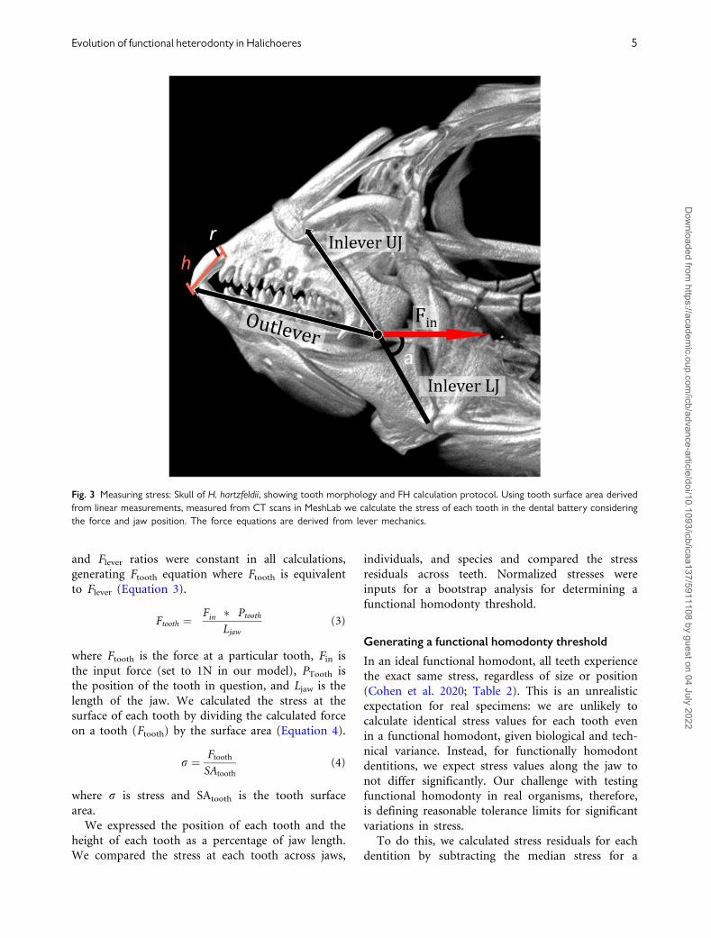
The stress of each tooth was calculated following
the principles of simple lever mechanics. Using the
surface area of a cone we approximate the surface
area of each tooth by measuring tooth height—
defined by the total length from tooth tip until
the tooth meets the jawbone—and tooth radius
(Fig. 3):
Surface area ¼ pr ðr þ ðffiffiffiffiffiffiffiffiffiffiffiffiffiffiffiffiffiffiffih2 þ r2p
ÞÞ (1)
To establish a basic relationship between surface
area and tooth stress we use the entire height of the
tooth. Following the guide of puncturing mechanics,
stresses should be concentrated at the tip of the
tooth; however, our metric is not meant to establish
where an individual tooth bears the greatest stress
but instead to identify patterns in stress across teeth
in the same dentition. Therefore, we ignore expected
changes in stress from the tip to the base of each
tooth in order to establish a relationship between the
variation of tooth morphologies in a dental battery
and stresses along the jaw. The standardization of
tooth morphology allowed us to ignore curvature
and damage yielding an idealized tooth surface
area, which we used to calculate stress under differ-
ent conditions (Crofts and Summers 2014).
Considering that teeth are on a lever, we had to
first establish the amount of force a tooth is pre-
dicted to exert based on its position.
Flever ¼ Fin � sinðaÞ � Inlever
Outlever
� �(2)
Our model assumes that the mechanical advan-
tage, and force at the input lever is the same across
jaws and that the input muscle force and input lever
arm are the same for all teeth on the jaw. We mod-
eled the upper jaw (premaxilla) with a static bite
point assuming that it was in the most retracted
position of its sliding position in the anterior jaws
linkage (Westneat 2004). The model also assumes a
single angle of muscle input force, with jaws in
closed position, and no changes in gape were mod-
eled. We simplified our system by assuming that Fin
Fig. 2 CT scan renderings of 11 species of Halichoeres wrasses representing different tooth morphologies. (A) H. hartzfeldii, (B) H.
richmondi, (C) H. podostigma, (D) H. melanochir, (E) H. dispilus, (F) H. argus, (G) H. prosopeion, (H) H. maculipinna, (I) H. hortulanus, (J)
H.leucurus, and (K) H. binotopsis. Scale bar is set to 1000 lm.
4 K. E. Cohen et al.
Dow
nloaded from https://academ
ic.oup.com/icb/advance-article/doi/10.1093/icb/icaa137/5911108 by guest on 04 July 2022
and Flever ratios were constant in all calculations,
generating Ftooth equation where Ftooth is equivalent
to Flever (Equation 3).
Ftooth ¼Fin � Ptooth
Ljaw
(3)
where Ftooth is the force at a particular tooth, Fin is
the input force (set to 1N in our model), PTooth is
the position of the tooth in question, and Ljaw is the
length of the jaw. We calculated the stress at the
surface of each tooth by dividing the calculated force
on a tooth (Ftooth) by the surface area (Equation 4).
r ¼ Ftooth
SAtooth
(4)
where r is stress and SAtooth is the tooth surface
area.
We expressed the position of each tooth and the
height of each tooth as a percentage of jaw length.
We compared the stress at each tooth across jaws,
individuals, and species and compared the stress
residuals across teeth. Normalized stresses were
inputs for a bootstrap analysis for determining a
functional homodonty threshold.
Generating a functional homodonty threshold
In an ideal functional homodont, all teeth experience
the exact same stress, regardless of size or position
(Cohen et al. 2020; Table 2). This is an unrealistic
expectation for real specimens: we are unlikely to
calculate identical stress values for each tooth even
in a functional homodont, given biological and tech-
nical variance. Instead, for functionally homodont
dentitions, we expect stress values along the jaw to
not differ significantly. Our challenge with testing
functional homodonty in real organisms, therefore,
is defining reasonable tolerance limits for significant
variations in stress.
To do this, we calculated stress residuals for each
dentition by subtracting the median stress for a
Fig. 3 Measuring stress: Skull of H. hartzfeldii, showing tooth morphology and FH calculation protocol. Using tooth surface area derived
from linear measurements, measured from CT scans in MeshLab we calculate the stress of each tooth in the dental battery considering
the force and jaw position. The force equations are derived from lever mechanics.
Evolution of functional heterodonty in Halichoeres 5
Dow
nloaded from https://academ
ic.oup.com/icb/advance-article/doi/10.1093/icb/icaa137/5911108 by guest on 04 July 2022

dentition from each stress value and dividing by me-
dian stress (i.e., centering and scaling to median
stress), allowing for comparison across dentitions.
An idealized functional homodont would experience
the same stress on each tooth regardless of size or
position, resulting in residuals of 0 for each tooth in
that dentition. To determine a threshold for func-
tional homodonty, we bootstrapped a residual stress
distribution per Cohen et al. (2020). Half of the
teeth were randomly subsampled without replace-
ment from a dentition and normalized residuals cal-
culated; this procedure was repeated 10,000 times for
each dentition. The resulting distribution of residuals
was centered around 0, with small clusters of ex-
treme values representing teeth that experienced
�1.8 times or more stress than the median for a
given dentition (Fig. 4). The multimodal structure
of bootstrapped residuals pointed us to clustering
techniques to determine threshold values for func-
tionally homodont/heterodont teeth. Our first imple-
mentation of this method (Cohen et al. 2020), used
k-means to distinguished between high and low re-
sidual peaks (Maechler et al. 2019). In Halichoeres
the wider range of bootstrapped residuals with ex-
treme values resulted in poor fits using k-means.
Instead, we used the more robust k-medoids cluster-
ing algorithm which uses data points as cluster cen-
ters (Maechler et al. 2019). Because k-medoids is
considerably slower than k-means and could not be
run on our entire sample of bootstrapped residuals
at once, we performed the k-medoids clustering
(with n¼ 2 clusters) on a random subsample of
5000 residuals 100 times, defining the threshold as
the mean of the two resulting cluster centers. The
resulting threshold ranged from 1.64 to 1.88 times
median stress, with an average of 1.8 times median
stress (Fig. 4, inset).
Summary statistics and phylogeny comparison
We generated two metrics from the normalized stress
calculations, each designed to emphasize a specific
aspect of the dentition: the average squared residual
and proportion of functional heterodont teeth. To
measure the degree of functional difference among
teeth from the same battery, we calculated the aver-
age squared residual (where residuals¼ stress � me-
dian stress) for stress across a jaw. We also calculated
the proportion of individual teeth in each battery
that differed in function from the majority; unlike
the variation in stress, this metric ignores the fold-
difference of stress but emphasizes regionalization.
We visualized these continuous traits on a time-
calibrated phylogeny (Aiello et al. 2017;
Supplementary Fig. S1) using the contMap function
from the phytools R package contMap (Revell 2012).
The phylogeny of 340 labrid species was time-
calibrated using fossil data (Aiello et al. 2017),
pruned down to the local clade containing the set
of taxa studied here (Supplementary Fig. S1) and
then pruned to the set of species studied here for
comparative analysis using the R package ape
(Paradis et al. 2004).
Results
Functional homodonty method
In 11 species and more than 440 teeth, 17 teeth bear
stresses that exceed the homodonty threshold (Fig. 4,
dashed line). While all of the 11 species of
Halichoeres represented in this dataset have at least
one tooth that exceeds the functional homodonty
threshold, three species had especially high values
for at least one of the metrics we calculated:
Halichoeres dispilus, Halichoeres melanochir, and
Halichoeres maculipinna (Fig. 6) . Each have a single
Table 2 Important definitions for the functional homodonty method
Term Definition
Functional homodonty
method
Method detailed in Cohen et al. (2020) for (1) using tooth surface area and position to calculate tooth stresses
through the transmission of force and (2) estimating a threshold for functionally heterodont teeth by boot-
strapping stress values from multiple dentitions.
Functionally homodont
teeth
All of the teeth in a dentition have statistically similar stresses that do not exceed a set threshold of stress
Functionally heterodont
teeth
One or more of the teeth in a dentition has statistically different stresses that exceed a set threshold of stress
from the majority of the dentition
Idealized functional
homodont
All of the teeth in a dentition bear the exact same median stress
Idealized functional
heterodont
All of the teeth in a dentition bear stresses that exceed the functional heterodonty threshold
Functional homodonty–
heterodonty continuum
A continuum ranging from the idealized functional homodont to the idealized functional heterodont onto which
all dentitions can be mapped using the functional homodonty method
6 K. E. Cohen et al.
Dow
nloaded from https://academ
ic.oup.com/icb/advance-article/doi/10.1093/icb/icaa137/5911108 by guest on 04 July 2022

tooth on the lower jaw that bears 12 times the me-
dian stress of that dentition, far exceeding the func-
tional homodonty threshold of 1.8 times median
stress. While H. maculipinna had no teeth bearing
stresses that high, the specimen we analyzed had 2
of 12 teeth on the upper jaw exceed the functional
homodonty threshold, the highest proportion of any
of the dentitions we measured (Fig. 4A). The upper
and lower jaw fangs of H. macculipinna occlude far
beyond the anterior end of the jaws with hardly any
curvature. The species H. melanochir most closely
resembles a morphological homodont and H. dispilus
has two sets of large canines that extend far beyond
the end of the premaxilla or dentary while the rest of
their teeth are similar in size (Fig. 2C–E).
Across all 11 species, functionally heterodont teeth
were more frequent on the upper jaws. While fewer
teeth on the lower jaws exceeded our functional het-
erodonty threshold, those that did typically had
much higher residuals, in some cases bearing up to
Fig. 4 Functional homodonty metric. (A) teeth from the jaws of 11 Halichoeres. Teeth above the dashed line exceed the functional
homodonty threshold (B) defined by the bootstrapping of residuals. We then use k-mediods to generate a biologically relevant
threshold (red dashed line). (C) Comparison of the proportion of functionally heterodont teeth to the average squared residual in four
species of Halichoeres wrasse to highlight the tendencies emerging from this comparison. (1) High proportion functionally heterodont
teeth: low residual, (2) Low proportion of heterodont teeth: high residual, and (3) High proportion of functionally heterodont teeth:
high residual.
Fig. 5 Phylogeny comparing the upper and lower jaws in 11 species of Halichoeres. There are more functionally heterodont teeth in the
upper jaws than the lower jaws, represented by a greater number of heterodont teeth (A). But functionally heterodont teeth in the
lower jaws have a bigger impact and are represented by a larger residual (B). Note, H. binoptisis is removed from the phylogenetic
comparisons as stresses were only calculated in the lower jaw.
Evolution of functional heterodonty in Halichoeres 7
Dow
nloaded from https://academ
ic.oup.com/icb/advance-article/doi/10.1093/icb/icaa137/5911108 by guest on 04 July 2022

twice the stress of the teeth on the upper jaw of the
same fish (Figs. 4C and 5, especially H. dispilus and
H. melanochir).
There are differences in the magnitude of func-
tionally heterodont teeth across all 11 species. For
instance, H. podostigma has a singular functionally
heterodont tooth on its lower jaw that barely exceeds
the heterodonty threshold. This tooth bears 2.4 times
the median stress of the rest of the dentition. By
comparison, H. melanochir has several teeth that
bear between 4 and 12 times the stress of function-
ally homodont teeth in the same dentition.
Proportion versus average squared residual of
functionally heterodont teeth
Ancestral state reconstruction suggests the ancestral
Halichoeres had a small number of functionally het-
erodont teeth, and these were more likely on the
upper jaw than the lower. The three species with
high numbers of heterodont teeth or high average
squared residuals were in two of the three
Halichoeres clades. Along the continuum of func-
tional homodonty to heterodonty, we find three ten-
dencies (Fig. 5). First, some taxa departed from
functional homodonty because of one or two excep-
tional teeth in a battery of otherwise functionally
homodont teeth. This is represented by large resid-
uals and a small proportion of functionally hetero-
dont teeth (H. dispilus). Next, are fishes with
regionalization of function, these have small resid-
uals but a high proportion of functionally hetero-
dont teeth (H. maculippina). Finally, a dental
battery could have large and small teeth interspersed
evenly across the jaws as in H. melanochir
(Fig. 4Aand C) which leads to a high proportion
of functionally heterodont teeth combined with
high residuals.
Discussion
The biomechanical function of teeth is largely depen-
dent on tooth morphology, orientation, position
along the jaw lever from front to back, and the dy-
namic forces of the jaw muscles driving them into a
prey item (Barel 1982; Westneat 2003; Anderson
et al. 2016). Here we present a way to calculate the
biomechanical function of teeth in jaws of labrid
fishes from the perspective of their geometry and
the relative bite stresses they exert. Our central con-
clusion is that Halichoeres wrasses have a wide diver-
sity of tooth arrangements that lend to functional
homodonty. However, some species have strikingly
functionally heterodont teeth—either a few teeth or
Fig. 6 Functionally heterodont species of Halichoeres. (A–C) H. dispilus, (D–F) H. malenochir, (G–I) H. podostigma. Live photos for all
three species downloaded from FishBase.org and provided by J.E. Randall. Scale bar is set to 1000 lm.
8 K. E. Cohen et al.
Dow
nloaded from https://academ
ic.oup.com/icb/advance-article/doi/10.1093/icb/icaa137/5911108 by guest on 04 July 2022

regions of teeth with disparate function. There will
always be some level of heterodonty at the tips of the
teeth across any dentition, but our metric draws a
clear line between functional homodonty and func-
tional heterodonty.
There are dentitions that have distinct regionali-
zation, where many teeth are quite similar to one
another, but patches are performing very different
tasks (Cohen et al. 2020; Mihalitsis and Bellwood
2019). In contrast, there are dentitions where one
or a few teeth are radically different from the rest
of the dental battery. In the first case, there will be a
large number of functionally heterodont teeth, but a
low average squared residual, and in the second case
there will be a few functionally heterodont teeth, but
they will have a very high squared residual. In
Halichoeres wrasses two species have extremely func-
tionally heterodont teeth, and they arrive at hetero-
donty by different means. In H. melanochir, the teeth
are strongly regionalized with a high proportion of
functionally heterodont teeth, whereas in H. dispilus
just two large canines in the upper jaw dictate func-
tional heterodonty leading to high residuals (Figs. 4A
and 5).
Our method converts a categorical trait, morpho-
logical homodonty, into a continuous one, func-
tional homodonty, creating opportunities for more
nuanced analyses and directing phylogenetic and
biomechanical hypotheses in new directions
(Shimada 2002; Westneat and Alfaro 2005;
Kolmann et al. 2019; Hulsey et al. 2020). Adding
the perspective of phylogeny bears two different
fruits. First, we can identify three apparently inde-
pendent evolutions of functional heterodonty: H.
dispilus, H. melanochir, and H. maculipinna
(Fig. 6). Second, our ancestral state reconstruction
implies an ancestral Halichoeres wrasse had a small
number of functionally heterodont teeth, and the
proportion of functionally heterodont teeth is vari-
able over evolutionary time. A metric for functional
heterodonty also allows us to generate computed
derivatives that reveal trends and potentially impor-
tant information about selective pressures (Linde
et al. 2004; Kolmann et al. 2019). The higher pro-
portion of functionally heterodont teeth on upper
jaws than lower jaws may be due to the upper jaw
being supported by the cranium, while the lower jaw
is a cantilever beam (Powlik 1995; Linde et al. 2004;
Westneat 2004; Westneat and Alfaro 2005; Grubich
et al. 2008; Smits and Evans 2012; Olsen and
Westneat 2016). Also, the magnitude of functionally
heterodont teeth is larger in the lower jaw than up-
per, perhaps because of mobility in the lower jaw
relative to the entire body of the fish (Figs. 4 and
5). How a species arrives at functional heterodonty
should point biomechanists in very different direc-
tions when asking about the functional consequences
of teeth.
We were excited to find such disparate dentitions,
and three independent derivations of heterodonty, in
just 11 species of a genus of wrasse that all occupy
similar nearshore, shallow water, coral reef habitats,
and have all been assigned the same broad dietary
niche (Randall and Bohlke 1965; Clifton and Motta
1998; Fulton and Bellwood 2002; Jones 2007).
Implementing this analysis across a broader phyloge-
netic and ecological range of wrasses will lead to
discovery of further, heretofore cryptic variation in
dental function that we expect will inform natural
history and diet studies. The Halichoeres radiation
may be an intriguing area of the labrid phylogeny
to explore in more detail, as the genus is not mono-
phyletic, with 80 species spread across three clades,
with other genera such as Macropharyngodon,
Thalassoma, and Coris interspersed among them
(Westneat and Alfaro 2005). The hogfishes and tusk-
fishes (Bodianus, Choerodon, and relatives) often
have extraordinarily large, recurved canines and re-
gionally specialized teeth, yet their close relatives
such as Pseudodax and Clepticus possess jaws special-
ized for browsing or planktivory, suggesting an in-
teresting trajectory of tooth evolution. The cheiline
wrasses are also diverse in tooth morphology, jaw
mechanics and dietary preferences (Westneat 1995)
and are the sister-clade to the parrotfishes, offering
another area in which to explore the evolution along
the functional homdonty–heterodonty continuum.
Even in this small, and not particularly diverse
clade of wrasses, there are some examples of denti-
tions that warrant further examinations. Halichoeres
leucurus is morphologically homodont, but the teeth
in the upper and lower jaw occlude with a degree of
precision that is not common in fishes (Kolmann
et al. 2019). Physical models of teeth, made from
high-resolution CT scans of actual dentitions, could
highlight advantages of this unusual tooth arrange-
ment (Evans and Sanson 2003; Qian et al. 2013;
Crofts and Summers 2014). There are visually arrest-
ing fanged dentitions, such as H. hortulanus, which
do not cross the threshold for functional hetero-
donty, but nevertheless have teeth that suggest
some differences in function (Fig. 2A). Careful nat-
ural history observations of differences in behavior
of species with prominent fangs may reveal how and
when these fangs are deployed. Other biomechanical
models, such as Mandiblever, take into account
tooth angle and its interaction with gape angle across
a realistic bite (Westneat 2003). Teeth are, in some
Evolution of functional heterodonty in Halichoeres 9
Dow
nloaded from https://academ
ic.oup.com/icb/advance-article/doi/10.1093/icb/icaa137/5911108 by guest on 04 July 2022

sense, an endpoint in a series of modules which de-
termine function (Evans and Sanson 2003; Lucas
2004; Anderson and Rayfeild 2012; Kolmann et al.
2019)—the powerful, subdivided adductor muscula-
ture, through tendons and ligaments connecting jaw
elements, to the levers and linkages of the jaws, and
even the seemingly insignificant dental ligaments se-
curing tooth to jaw combine to ensure that the tip of
the tooth transmits sufficient stress to penetrate prey.
Acknowledgments
Special thanks to Dr Kory Evans who CT scanned all
specimens represented in this study and uploaded
them as free to download in morphosource.org.
Special thanks to the facilities provided by the Karl
F. Liem imaging center at Friday Harbor Labs, WA,
and oVert for open access to CT scans. Thank you to
Dr Darrin Hulsey and Dr Gareth Fraser for organiz-
ing the associated symposium.
Funding
Funding attributed to National Science foundation
Division of Biological Infrastructure 1759637 and
Division of Environmental Biology 1701665, Friday
Harbor Laboratories Research Fellowship
Endowment, and the Ragen Friday Harbor Labs
Endowed Scholar. A.P.S. was supported by the
Seaver Institute. Funding for attendance and partic-
ipation in the associated symposium was provided
by the Society for Integrative and Comparative
Biology.
Data availability statement
All CT data used in this study are publicly available
and free to download from morphosource.org in the
Scan All Fishes project.
Supplementary data
Supplementary data available at ICB online.
REFERENCES
Adams DC, Collyer ML. 2018. Multivariate phylogenetic
comparative methods: evaluations, comparisons, and rec-
ommendations. Syst Biol 67:14–31.
Aiello BR, Westneat MW, Hale ME. 2017. Mechanosensation
is evolutionarily tuned to locomotor mechanics. Proc Natl
Acad Sci U S A 114:4459–64.
Anderson PSL, LaBarbera M. 2008. Functional consequences
of tooth design: effects of blade shape on energetics of
cutting. J Exp Biol 211:3619–26.
Anderson PSL, LaCosse J, Pankow M. 2016. Point of impact:
the effect of size and speed on puncture mechanics.
Interface Focus 6:20150111.
Anderson PSL, Rayfield EJ. 2012. Virtual experiments,
physical validation: dental morphology at the intersection
of experiment and theory. J Royal Soc Interface
10:1846–55.
Barel C. 1982. Towards a Constructional Morphologyof
Cichlid Fishes (Teleostei, Perciformes). Neth J Zool
33:357–424.
Becerra MG, Pol D, Rossner GE, Rauhut OWM. 2018.
Heterodonty and double occlusion in Manidens condoren-
sis: a unique adaptation in an Early Jurassic ornithischian
improving masticatory efficiency. Sci Nat 105:41.
Bergman JN, Lajeunesse MJ, Motta PJ. 2017. Teeth penetra-
tion force of the tiger shark Galeocerdo cuvier and sandbar
shark Carcharhinus plumbeus: shark tooth penetration
force. J Fish Biol 91:460–72.
Berkovitz BK, Shellis RP. 2016. The Teeth of Non-
Mammalian Vertebrates. 1st ed. London: Academic Press.
Berkovitz BKB. 1975. Observations on tooth replacement in
piranhas (Characidae). Arch Oral Biol 20:53–7.
Berkovitz BKB. 1980. The effect of age on tooth replacement
patterns in piranhas (Pisces: Characidae). Arch Oral Biol
25:833–5.
Bertrand NG. 2014. Distribution and evolution of hetero-
donty in the ray-finned fishes (Actinopterygii).
[Unpublished Master’s thesis]. Texas: Texas A&M
University.
Clark AJ, Summers AP. 2012. Ontogenetic scaling of the
morphology and biomechanics of the feeding apparatus
in the Pacific hagfish Eptatretus stoutii. J Fish Biol
80:86–99.
Clifton KB, Motta PJ. 1998. Feeding morphology, diet, and
ecomorphological relationships among five Caribbean
Labrids (Teleostei, Labridae). Copeia 1998:953–66.
Cohen KE, Weller HI, Summers AP. 2020. Not your father’s
homodonty—stress, tooth shape, and the functional homo-
dont. J Anat 237:837.
Conway KW, Bertrand NG, Browning Z, Lancon TW, Clubb
FJ. 2015. Heterodonty in the new world: an SEM investi-
gation of oral Jaw dentition in the clingfishes of the sub-
family gobiesocinae (Teleostei: Gobiesocidae). Copeia
103:973–98.
Cope ED. 1888. On the tritubercular molar in human denti-
tion. J Morphol 2:7–23.
Crofts SB, Summers AP. 2014. How to best smash a snail: the
effect of tooth shape on crushing load. J Royal Soc
Interface 11:20131053.
Cullen JA, Marshall CD. 2019. Do sharks exhibit heterodonty
by tooth position and over ontogeny? A comparison using
elliptic Fourier analysis. J Morphol 280:687–700.
D’Amore DC. 2015. Illustrating ontogenetic change in the
dentition of the Nile monitor lizard, Varanus niloticus: a
case study in the application of geometric morphometric
methods for the quantification of shape–size heterodonty. J
Anat 226:403–19.
Davit-B�eal T, Chisaka H, Delgado S, Sire JY. 2007. Amphibian
teeth: current knowledge, unanswered questions, and some
directions for future research. Biol Rev 82:49–81.
Dean MN, Ramsay JB, Schaefer JT. 2008. Tooth reorientation
affects tooth function during prey processing and tooth
ontogeny in the lesser electric ray, Narcine brasiliensis.
Zoology 111:123–34.
10 K. E. Cohen et al.
Dow
nloaded from https://academ
ic.oup.com/icb/advance-article/doi/10.1093/icb/icaa137/5911108 by guest on 04 July 2022

Estes R, Williams EE. 1984. Ontogenetic variation in the
molariform teeth of lizards. J Vertebr Paleontol 4:96–107.
Evans AR, Sanson GD. 2003. The tooth of perfection: func-
tional and spatial constraints on mammalian tooth shape.
Biol J Linn Soc 78:173–91.
Evans AR, Sanson GD. 2005. Correspondence between tooth
shape and dietary biomechanical properties in insectivo-
rous microchiropterans. Evol Ecol Res 7:453–78.
Evans AR, Sanson GD. 1998. The effect of tooth shape on the
breakdown of insects. J Zool 246:391–400.
Evans KM, Williams KL, Westneat MW. 2019. Do coral reefs
promote morphological diversification? Exploration of hab-
itat effects on Labrid pharyngeal jaw evolution in the era of
big data. Integr Comp Biol 59:696–704.
Frazzetta TH. 1988. The mechanics of cutting and the form of
shark teeth (Chondrichthyes, Elasmobranchii).
Zoomorphology 108:93–107.
Freeman PW, Weins WN. 1997. Puncturing ability of bat
canine teeth: the tip. Lincoln: University of Nebraska.
Mammalogy Papers: University of Nebraska State
Museum. 9.
Fulton CJ, Bellwood DR. 2002. Patterns of foraging in labrid
fishes. Marine Ecol Prog Ser 226:135–42.
Galloway KA, Anderson PSL, Wilga CD, Summers AP. 2016.
Performance of teeth of lingcod, Ophiodon elongatus, over
ontogeny. J Exp Zool Part A Ecol Genet Physiol
325:99–105.
Garamszegi L. 2014. Modern phylogenetic comparative meth-
ods and their application in evolutionary biology: concepts
and practice. Berlin, Germany: Springer.
Gregory WK. 1933. Fish skulls; a study of the evolution of
natural mechanisms. Malabar, FL: Krieger Publishing
Company.
Grubich JR, Rice AN, Westneat MW. 2008. Functional mor-
phology of bite mechanics in the great barracuda
(Sphyraena barracuda). Zoology 111:16–29.
Harmon LJ. 2018. Phylogenetic comparative methods. 1st ed.
Vol. 1.
Hulsey CD, Cohen KE, Johanson Z, Karagic N, Meyer A,
Miller CT, Sadier A, Summers AP, Fraser GJ. 2020.
Grand challenges in comparative tooth biology. Integr
Comp Biol (doi:10.1093/icb/icaa038).
Jambura PL, Turtscher J, Kindlimann R, Metscher B, Pfaff C,
Stumpf S, Weber GW, Kriwet J. 2020. Evolutionary trajec-
tories of tooth histology patterns in modern sharks
(Chondrichthyes, Elasmobranchii). J Anat 236:753–71.
Jones KMM. 2007. Distribution of behaviours and species
interactions within home range contours in five
Caribbean reef fish species (Family Labridae). Environ
Biol Fish 80:35–49.
Jones MEH. 2009. Dentary tooth shape in Sphenodon and its
fossil relatives (Diapsida: Lepidosauria: Rhynchocephalia).
Comparative dental morphology. Vol. 13. Basel,
Switzerland: Karger Publishers. p. 9–15.
Keene HJ. 1991. On heterochrony in heterodonty: a review of
some problems in tooth morphogenesis and evolution. Am
J Phys Anthropol 34:251–82.
Kolmann MA, Cohen KE, Bemis KE, Summers AP, Irish FJ,
Hernandez LP. 2019. Tooth and consequences: heterodonty
and dental replacement in piranhas and pacus
(Serrasalmidae). Evol Dev 21:247–62.
Linde M, Palmer M, G�omez-Zurita J. 2004. Differential cor-
relates of diet and phylogeny on the shape of the premax-
illa and anterior tooth in sparid fishes (Perciformes:
Sparidae). J Evol Biol 17:941–52.
Lucas PW. 2004. Dental functional morphology: how teeth
work. Cambridge: Cambridge University Press.
Lucas PW, Luke DA. 1984. Chewing it over: basic principles
of food breakdown. In: Chivers DJ, Wood BA, Bilsborough
A, editors. Food acquisition and processing in primates.
Boston (MA): Springer. p. 283–301.
Maechler M, Rousseeuw P, Struyf A, Hubert M, Hornik K.
2019. Cluster: cluster analysis basics and extensions. R
Package Version 2.1.0.
Marcus MA, Amini S, Stifler CA, Sun CY, Tamura N, Bechtel
HA, Parkinson DY, Barnard HS, Zhang XXX, Chua JQI, et
al. 2017. Parrotfish teeth: stiff biominerals whose micro-
structure makes them tough and abrasion-resistant to bite
stony corals. ACS Nano 11:11856–65.
Massare JA. 1987. Tooth morphology and prey preference of
Mesozoic marine reptiles. J Vertebr Paleontol 7:121–37.
Mihalitsis M, Bellwood D. 2019. Functional implications of
dentition-based morphotypes in piscivorous fishes. R Soc
Open Sci 6:190040.
Olsen AM, Westneat MW. 2016. Linkage mechanisms in the
vertebrate skull: Structure and function of three-dimen-
sional, parallel transmission systems. J
Morphol 277:1570–83.
Paradis E, Claude J, Strimmer K. 2004. APE: Analyses of
Phylogenetics and Evolution in R language.
Bioinformatics 20:289–90.
Powlik JJ. 1995. On the geometry and mechanics of tooth
position in the white shark, Carcharodon carcharias. J
Morphol 226:277–88.
Qian Y, Zhou X, Yang J. 2013. Correlation between cuspal
inclination and tooth cracked syndrome: a three-
dimensional reconstruction measurement and finite ele-
ment analysis. Dent Traumatol 29:226–33.
Randall JE. 1967. Food habits of reef fishes of the West
Indies. Stud Trap Oceanogr 5:665–847.
Randall JE, Bohlke JE. 1965. Review of the Atlantic Labrid
fishes of the genus Halichoeres. Proc Acad Nat Sci
Philadelphia 117:235–59.
Revell LJ. 2012. Phytools: an R package for phylogenetic com-
parative biology (and other things): phytools: r package.
Method Ecol Evol 3:217–23.
Ruber L, Verheyen E, Meyer A. 1999. Replicated evolution of
trophic specializations in an endemic cichlid fish lineage
from Lake Tanganyika. Proc Natl Acad Sci U S A
96:10230–35.
Sansom RS, Wills MA, Williams T. 2016. Dental data perform
relatively poorly in reconstructing mammal phylogenies:
morphological partitions evaluated with molecular bench-
marks. Syst Biol 66:813–822.
Schofield RMS, Choi S, Coon JJ, Goggans MS, Kreisman TF,
Silver DM, Nesson MH. 2016. Is fracture a bigger problem
for smaller animals? Force and fracture scaling for a simple
model of cutting, puncture and crushing. Interface Focus
6:20160002.
Schwartz JH. 2013. Emergence of shape. Biol Theory 8:209–10.
Shergold OA, Fleck NA. 2005. Experimental investigation into
the deep penetration of soft solids by sharp and blunt
Evolution of functional heterodonty in Halichoeres 11
Dow
nloaded from https://academ
ic.oup.com/icb/advance-article/doi/10.1093/icb/icaa137/5911108 by guest on 04 July 2022

punches, with application to the piercing of skin. J
Biomech Eng 127:838–48.
Shimada K. 2002. Dental homologies in lamniform sharks
(Chondrichthyes: Elasmobranchii). J Morphol 251:38–72.
Shimizu D, Macho GA, Spears IR. 2005. Effect of prism ori-
entation and loading direction on contact stresses in pris-
matic enamel of primates: implications for interpreting
wear patterns. Am J Phys Anthropol 126:427–34.
Simpson GG. 1936. Studies of the earliest mammalian denti-
tions. Dent Cosmos 78:791–800.
Smits PD, Evans AR. 2012. Functional constraints on tooth
morphology in carnivorous mammals. BMC Evol Biol
12:146.
Ungar PS. 2014. Teeth: a very short introduction. Oxford:
Oxford University Press.
Westneat MW. 1995. Feeding, function, and phylogeny: anal-
ysis of historical biomechanics in labrid fishes using com-
parative methods. Syst Biol 44:361–383.
Westneat MW. 2003. A biomechanical model for analysis of
muscle force, power output and lower jaw motion in fishes.
J Theor Biol 223:269–281.
Westneat MW. 2004. Evolution of levers and linkages in the
feeding mechanisms of fishes. Integr Compar Biol 44:378–89.
Westneat MW, Alfaro ME. 2005. Phylogenetic relationships
and evolutionary history of the reef fish family Labridae.
Mol Phylogenet Evol 36:370–90.
Whitenack LB, Gottfried MD. 2010. A morphometric ap-
proach for addressing tooth-based species delimitation in
fossil mako sharks, Isurus (Elasmobranchii: Lamniformes). J
Vertebr Paleontol 30:17–25.
12 K. E. Cohen et al.
Dow
nloaded from https://academ
ic.oup.com/icb/advance-article/doi/10.1093/icb/icaa137/5911108 by guest on 04 July 2022