Insights on the association of American Cetoniinae beetles with ants

© Koninklijke Brill NV, Leiden, 2009 DOI 10.1163/157075609X437745
Animal Biology 59 (2009) 241–262 brill.nl/ab
Ultrastructural analysis of the fat body in workers of Attini ants (Hymenoptera: Formicidae)
Gislaine C. Roma 1 , Odair C. Bueno 1,2 , Maria I. Camargo-Mathias 1,*
1 Departamento de Biologia 2 Centro de Estudos de Insetos Sociais
Instituto de Biociências, UNESP, Universidade Estadual Paulista, Avenida 24 A n° 1515, Rio Claro, SP, Brasil, CEP: 13506-900, C. P. 0199
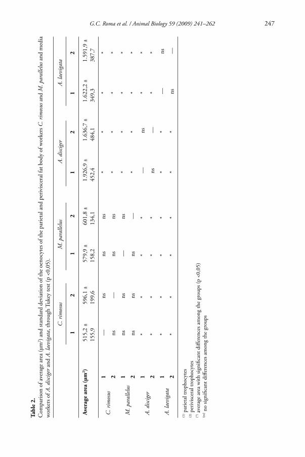
Abstract In the present study a comparative morphological analysis of the fat body cells of ant workers of the basal Attini species Cyphomyrmex rimosus and Mycetarotes parallelus , and the derived species Acromyrmex disciger and Atta laevigata was conducted. Th e results revealed that the fat body is located mainly in the abdomen around organs (perivisceral) and near the integument (parietal). Th e main cells observed are spherical or polygonal trophocytes with a slightly rough surface. Th e oenocytes, another cell type found, are closely associated with trophocytes, and present a spherical or polygonal shape and a smoother surface. Th e mor-phometric analysis showed that the area of trophocytes and oenocytes of C. rimosus and M. parallelus is signifi cantly smaller when compared to those of A. disciger and A. laevigata . In the cytoplasm of parietal and perivisceral trophocytes and oenocytes, electronlucent droplets (probably lipids) and electrondense granules (probably proteins) indicate the participation of these cells in the storage of these elements, while digestive vacuoles, residual bodies, and multivesicular bodies suggest a role in intracellular digestion. In perivisceral trophocytes and oenocytes of C. rimosus , the presence of mitochondria, lamellar rough endoplasmic reticulum, and Golgi complex suggests that these cells synthesize proteins. Based on these data, no signifi cant diff erences were observed between the fat body cells of basal and derived ants, except regarding the larger size of trophocytes and oenocytes of the derived species A. disciger and A. laevigata . © Koninklijke Brill NV, Leiden, 2009
Keywords Trophocyte; oenocyte; morphology; morphometry; ultrastructure
Introduction
Th e Attini tribe consists of approximately 217 ant species that grow symbiotic fungi, the staple food of their colonies (Hölldobler and Wilson, 1990 ). Th ese species are distributed between basal genera, such as Cyphomyrmex, Mycetophylax, Mycocepurus, Myrmicocrypta, Mycetagroicus, Apterostigma, Sericomyrmex, Mycetosoritis, Mycetarotes
*) Corresponding author; e-mail: [email protected]

242 G.C. Roma et al. / Animal Biology 59 (2009) 241–262
and Trachymyrmex , and derived genera, such as Atta and Acromyrmex . Th e latter are commonly named leaf-cutting ants and are serious pests considered dominant herbiv-ores in the neotropics (Hölldobler and Wilson, 1990 ; Bolton, 2003 ). Th e genus Pseudoatta consists of parasite species (Bolton, 2003 ).
Th e fat body is a fi lling tissue that besides fi lls the cavities of the body is the site of intermediary metabolism (group of chemical reactions that occurs in the fat body to provide the necessaries of the own cell or of the organism) of several compounds. It also plays roles in the synthesis and storage of proteins, lipids, and carbohydrates (Palli and Locke, 1988 ), and is characterized by sheets or string of cells, usually one or two cells thick, or even forming small nodes in the hemocele connected to one another or to other tissues by tracheal branches (Keeley, 1985 ; Rosell and Wheeler, 1995 ; Chapman, 1998 ).
Trophocytes are the main cells of the insect fat body. Th ese cells may give rise to other cell types, as a result of a functional specialization, such as urate cells or urocytes, mycetocytes, and chromatocytes (Dean et al., 1985 ; Chapman, 1998 ).
Oenocytes, another cell type that constitutes the fat body, play roles in lipid meta-bolic processes (Keeley, 1985 ; Chapman, 1998 ).
Given the scarce data available on the internal morphology of ants of the basal and derived genera of the Attini tribe, the present study comparatively described the mor-phology, morphometry, and ultrastructure of parietal and perivisceral trophocytes and oenocytes of workers of the basal species Cyphomyrmex rimosus Spinola and Mycetarotes parallelus Emery and media workers of the derived species Acromyrmex disciger Mayr and Atta laevigata Smith, attempting to new information that could contribute to the understanding of their physiology and biology, and on diff erences that could lead to phylogenetic implications of this tribe.
Material and methods
Th e present study used the parietal and perivisceral fat body from workers of Cyphomyrmex rimosus and Mycetarotes parallelus (monomorphic species) and media workers of Acromyrmex disciger and Atta laevigata (polimorphic species), collected out-side the nests in the fi eld and immediately submitted to the techniques. Th e individuals were anesthetized by cooling in a freezer and dissected on Petri dishes with physiologi-cal saline solution for insects (NaCl 7.5 g/L, Na 2 HPO 4 2.38 g/L, KH 2 PO 4 2.72 g/L).
Scanning Electron Microscopy (SEM)
Th e parietal and perivisceral fat body from workers was removed, fi xed in Karnovsky for 24 hours and processed by Critical Point Drying, sputtered with gold and exam-ined by PHILIPS 505 SEM.
Morphometry
Th e morphometric analysis of trophocytes and oenocytes was performed using median histological sections. For the present study were utilized 05 individuals of each species.

G.C. Roma et al. / Animal Biology 59 (2009) 241–262 243
For each individual, were obtained area measurements (μm 2 ) of 10 parietal trophocytes and 10 perivisceral ones, and 10 parietal oenocytes and 10 perivisceral ones, with a LDMB LEICA microscope with the software Leica Qwin. Th e statistical test applied was the Tukey (p <0,05) (Siegel and Castellan, 1988) performed in order to verify signifi cant diff erences among the parietal and perivisceral trophocytes and among the parietal and perivisceral oenocytes in all species studied here. Besides this, the statistic test evaluated the signifi cant diff erences among the parietal and perivisceral trophocytes among individuals at the same species. Th e same procedure was applied to the oenocytes.
Ultrastructure - Transmission Electron Microscopy (TEM)
Th e fat body was fi xed in 2,5% glutaraldehyde fi xative solution in 0.1M cacodylate buff er (pH 7.2) for 2 hours, at 4°C. Post-fi xation was performed in 1% osmium tetrox-ide in 0.1M cacodylate buff er 0.1M (pH 7.2) for 2 hours, in the darkness. For contrast, the material was immersed in a solution of 2% uranyl acetate in 10% acetone, for 4 hours, in the darkness. Th en, the material was embedded in Epon resin diluted in acetone (1:1) for 12 hours, included in pure Epon resin and incubated at 60°C for 24 hours. After polymerized semithin sections were obtained with ultramicrotome Sorvall – Porter Blum MT2-B and stained with Azur II (1%) and Methylene Blue (1%).
Grids containing ultrathin sections of the material were contrasted with uranyl acetate and lead citrate for 45 minutes and 10 minutes, respectively. Th en, they were analysed and photographed in a PHILIPS CM 100 Transmission Electron Microscopy.
Results
Scanning Electron Microscopy
Th e morphological analysis of the fat body of workers of 1. C. rimosus and M. parallelus and media workers of A. disciger and A. laevigata shows that this tissue is characterized by a loose clump of cells located among organs (periv-isceral) and adjacent to the integument (parietal) ( Figs. 1B ; 2A , C , G ). In all species examined, parietal and perivisceral trophocytes are the main 2. cell types observed ( Figs. 1A – F ; 2B , D - H ). Th ese cells are mostly spherical in shape, but may be polygonal due to the cell arrangement that compresses cells against each other ( Figs. 1B , F ; 2B , E , G ). Th e surface of trophocytes of all worker species examined is slightly rough 3. ( Figs. 1F ; 2D ). In workers of 4. M. parallelus and A. laevigata , some trophocytes are ruptured, releasing large quantities of cytoplasmic granules of varied size and possibly contents ( Figs. 1D ; 2G ). In parietal and perivisceral fat bodies, round or spherical oenocytes are 5. dispersed among and associated with trophocytes. Oenocytes may also be

244 G.C. Roma et al. / Animal Biology 59 (2009) 241–262
Figu
re 1
. Sc
anni
ng E
lect
ron
Mic
rosc
opy
(SEM
) of t
he fa
t bod
y ce
lls fr
om w
orke
rs o
f Cyp
hom
yrm
ex ri
mos
us a
nd M
ycet
arot
es pa
ralle
lus .
(A) D
etai
l of t
he C
. rim
osus
pa
rieta
l fat
bod
y ex
hibi
ting
troph
ocyt
e (t)
and
oeno
cyte
(o).
(B) G
ener
al v
iew
and
(C) d
etai
l of t
he C
. rim
osus
per
ivisc
eral
fat b
ody
show
ing
the o
enoc
yte m
embr
anou
s ex
pans
ion
( ➝ ).
(D, E
) Det
ail o
f the
par
ieta
l fat
bod
y fro
m M
. par
allel
us e
xhib
iting
the
gran
ules
in th
e cy
topl
asm
trop
hocy
te (w
hite
arr
ow).
(F) M
. par
allel
us p
eriv
is-ce
ral f
at b
ody
show
ing
troph
ocyt
e m
embr
anou
s exp
ansio
n ( ➝
) and
the
sligh
tly ro
ugh
surfa
ce o
f the
trop
hocy
tes.

G.C. Roma et al. / Animal Biology 59 (2009) 241–262 245
polygonal in shape due to the compression of trophocytes ( Figs. 1A - F , 2B , D , E , G ). Th ese are smaller cells with a smoother surface and present in fewer numbers compared to trophocytes ( Figs. 1A – F ; 2B , D , E , G ). Among tissue cells, membranous expansions from trophocytes surround and 6. bind them to one another ( Figs. 1F ; 2F , H ). Similarly, these expansions may also originate in oenocytes extending to trophocytes and binding these two cell types ( Fig. 1C ).
Morphometry
Th e morphometric analysis of the fat body cells of workers reveals that the areas of parietal and perivisceral trophocytes of C. rimosus and M. parallelus are signifi cantly smaller compared to those of A. disciger and A. laevigata . Similarly, oenocytes of media workers of the derived species are signifi cantly larger than those of basal workers. However, no signifi cant diff erences were observed in the areas of parietal and perivis-ceral trophocytes of ants of the same species. Th e same was obtained for oenocytes.
Th e morphometric results of parietal and perivisceral fat body cells of workers exam-ined in this study are summarized in tables 1 and 2 .
Ultrastructure
Parietal and perivisceral trophocytes of all species exhibit individual or fused basal lamina when two or more adjacent trophocytes are observed. Th e same may also occur between trophocyte and oenocyte ( Figs. 3A , B , N ; 4A ; 5C , E , J ; 6F ; 7C , D ). In parietal and perivisceral trophocytes of A. laevigata , the plasma membrane reticular system (PMRS) ( Figs. 6F ; 7C ), result of membrane invaginations, is observed.
In parietal and perivisceral trophocytes, electronlucent droplets of varied shapes and sizes, probably containing lipids, are present ( Figs. 3C - E ; 4B , C ; 5C , F ; 6A , B , D , F , G ). In addition to these, very electrondense granules are observed in parietal tro-phocytes of M. parallelus , and perivisceral trophocytes of A. laevigata ( Figs. 4B ; 6E , H ), probably containing proteins.
Th e ultrastructural analysis of trophocytes of all species examined also shows the presence of digestive vacuoles ( Figs. 3G , H ; 6A , D , G ), characterized by structures delimited by membranes, containing compounds being digested, which may be cell organelles or endocytized molecules.
In perivisceral trophocytes of C. rimosus , mitochondria ( Figs. 3F , N ), lamellar rough endoplasmic reticulum ( Figs. 3H , N ), and Golgi complex ( Fig. 3F ) forming the syn-thesis apparatus of these cells, in addition to free ribosomes ( Fig. 3F ).
Th e nuclei of parietal and perivisceral trophocytes of A. disciger and A. laevigata are very irregular ( Figs. 5A , B , D ; 6A ). In C. rimosus and M. parallelus , nuclei of tro-phocytes are star or oval shaped ( Figs. 3A , C , E ).
Parietal and perivisceral oenocytes of the species examined exhibit individual or fused basal lamina in certain regions of adjacent oenocytes. Th e same is observed between oenocyte and trophocyte ( Figs. 3M , N ; 5J ; 7A , C , D ).

246 G.C. Roma et al. / Animal Biology 59 (2009) 241–262 Ta
ble
1.
Com
paris
on o
f ave
rage
are
a (μ
m 2 )
and
stand
ard
devi
atio
n of
the
troph
ocyt
es o
f the
par
ieta
l and
per
ivisc
eral
fat b
ody
of w
orke
rs o
f C. r
imos
us a
nd M
. par
allel
us a
nd
med
ia w
orke
rs o
f A. d
iscig
er a
nd A
. lae
viga
ta , t
hrou
gh T
ukey
test
(p <
0,05
).
C. r
imos
usM
. par
allel
usA.
disc
iger
A. la
evig
ata
12
12
12
12
Aver
age
area
(μm
2 )1.
774,
1 ±
720,
41.
770,
4 ±
647,
21.
480,
2 ±
346,
51.
647,
6 ±
431,
93.
876,
6 ±
1265
,64.
026,
8 ±
1174
,84.
664,
6 ±
1897
,74.
772,
1 ±
1830
,4
C. r
imos
us1
—ns
nsns
**
**
2ns
—ns
ns*
**
*
M. p
aral
lelus
1ns
ns—
ns*
**
*
2ns
nsns
—*
**
*
A. d
iscig
er1
**
**
—ns
**
2*
**
*ns
—*
*
A. la
evig
ata
1*
**
**
*—
ns
2*
**
**
*ns
—
(1) p
arie
tal t
roph
ocyt
es
(2) p
eriv
iscer
al tr
opho
cyte
s (*
) ave
rage
are
a w
ith si
gnifi
cant
diff
eren
ces a
mon
g th
e gr
oups
(p <
0,05
) (n
s) n
o sig
nifi c
ant d
iff er
ence
s am
ong
the
grou
ps
G.C. Roma et al. / Animal Biology 59 (2009) 241–262 247 Ta
ble
2.
Com
paris
on o
f ave
rage
area
(μm
2 ) an
d sta
ndar
d de
viat
ion
of th
e oen
ocyt
es o
f the
par
ieta
l and
per
ivisc
eral
fat b
ody
of w
orke
rs C
. rim
osus
and
M. p
aral
lelus
and
med
ia
wor
kers
of A
. disc
iger
and
A. l
aevi
gata
, thr
ough
Tuk
ey te
st (p
<0,
05).
C. r
imos
usM
. par
allel
usA.
disc
iger
A. la
evig
ata
12
12
12
12
Aver
age
area
(μm
2 )51
5,2
±15
5,9
596,
1 ±
199,
657
9,9
±15
8,2
601,
8 ±
134,
11.
926,
9 ±
452,
41.
636,
7 ±
484,
11.
622,
2 ±
349,
31.
591,
9 ±
387,
7
C. r
imos
us1
—ns
nsns
**
**
2ns
—ns
ns*
**
*
M. p
aral
lelus
1ns
ns—
ns*
**
*
2ns
nsns
—*
**
*
A. d
iscig
er1
**
**
—ns
**
2*
**
*ns
—*
*
A. la
evig
ata
1*
**
**
*—
ns
2*
**
**
*ns
—
(1) p
arie
tal t
roph
ocyt
es
(2) p
eriv
iscer
al tr
opho
cyte
s (*
) ave
rage
are
a w
ith si
gnifi
cant
diff
eren
ces a
mon
g th
e gr
oups
(p <
0,05
) (n
s) n
o sig
nifi c
ant d
iff er
ence
s am
ong
the
grou
ps

248 G.C. Roma et al. / Animal Biology 59 (2009) 241–262
Figu
re 2
. Sc
anni
ng E
lect
ron
Mic
rosc
opy
(SEM
) of t
he fa
t bod
y ce
lls fr
om m
edia
wor
kers
of A
cro m
yrm
ex d
iscig
er an
d At
ta la
evig
ata .
(A) G
ener
al v
iew
of t
he A
. disc
iger
pa
rieta
l fa
t bo
dy
(fb)
and
(B)
deta
il of
th
e tro
phoc
ytes
(t)
an
d oe
nocy
tes
(o).
(C)
Gen
eral
vi
ew
of
the
A.
disci
ger
periv
iscer
al
fat
body
an
d (D
) det
ail,
exhi
bitin
g th
e inn
er as
soci
atio
n be
twee
n tro
phoc
ytes
(t) a
nd o
enoc
ytes
(o) a
nd th
e slig
htly
roug
h su
rface
of t
he tr
opho
cyte
s. (E
, F) D
etai
l of t
he A
. lae
viga
ta
parie
tal f
at b
ody,
show
ing
the t
roph
ocyt
e mem
bran
ous e
xpan
sion
( ➝ ).
(G) G
ener
al v
iew
and
(H) d
etai
l of t
he A
. lae
viga
ta p
eriv
iscer
al fa
t bod
y ex
hibi
ting
the g
ranu
les
in th
e tro
phoc
yte
cyto
plas
m (w
hite
arr
ow) a
nd th
e tro
phoc
yte
mem
bran
ous e
xpan
sion
( ➝ ).

G.C. Roma et al. / Animal Biology 59 (2009) 241–262 249
Figu
re 3
. El
ectro
n m
icro
grap
h of
the f
at b
ody
cells
from
wor
kers
of t
he b
asal
spec
ies o
f Cyp
hom
yrm
ex ri
mos
us. (
A-D
) Det
ail o
f the
C. r
imos
us p
arie
tal t
roph
ocyt
es (t
) ex
hibi
ting
the
fusio
n of
bas
al la
min
a (b
l) fro
m a
djac
ent t
roph
ocyt
es. O
bser
ve th
e in
ner a
ssoc
iatio
n be
twee
n tro
phoc
ytes
and
cut
icle
(c),
star o
r ova
l sha
ped
nucl
eus
(n) a
nd th
e el
ectro
nluc
ent d
ropl
ets (
d). (
E-H
) C. r
imos
us p
eriv
iscer
al tr
opho
cyte
s sho
win
g th
e ov
al sh
aped
nuc
leus
(n),
Gol
gi c
ompl
ex (G
c) a
nd d
iges
tive
vacu
oles
(d
v). (
I-L)
Par
ieta
l oen
ocyt
es (o
) exh
ibiti
ng th
e rou
nd sh
aped
nuc
leus
(*) a
nd g
lyco
gen
depo
sits (
gl).
(M-P
) Per
ivisc
eral
oen
ocyt
es fr
om C
. rim
osus
. Obs
erve
the f
usio
n of
bas
al la
min
a (b
l) fro
m a
djac
ent o
enoc
ytes
(o) a
nd/o
r am
ong
oeno
cyte
s and
trop
hocy
tes (
t). ➝
= tr
opho
cyte
nuc
leol
us, m
i = m
itoch
ondr
ia, r
= fr
ee ri
boss
omes
, lre
r = la
mel
lar r
ough
end
opla
smic
retic
ulum
, rb
= re
sidua
l bod
ies.

250 G.C. Roma et al. / Animal Biology 59 (2009) 241–262
Figu
re 4
. El
ectro
n m
icro
grap
h of
the
fat b
ody
cells
from
wor
kers
of t
he b
asal
spec
ies o
f Myc
etar
otes
para
llelu
s . ( A
, B ) D
etai
l of t
he M
. par
allel
us p
arie
tal t
roph
ocyt
es
exhi
bitin
g tw
o in
divi
dual
bas
al la
min
a (b
l) of
adj
acen
t tro
phoc
ytes
(t).
( C
, D )
M. p
aral
lelus
per
ivisc
eral
tro
phoc
ytes
. ( E-
H )
M. p
aral
lelus
par
ieta
l oen
ocyt
es (
o).
Obs
erve
the f
usio
n of
bas
al la
min
a (bl
) of t
wo
adja
cent
oen
ocyt
es w
ithou
t cyt
opla
sm fu
sion
and
the t
rach
eole
s (tr
) pre
senc
e. ( I
, J ) M
. par
allel
us p
eriv
iscer
al o
enoc
ytes
. m
i = m
itoch
ondr
ia, r
b =
resid
ual b
odie
s, gr
= e
lect
rond
ense
gra
nule
s , d
= e
lect
ronl
ucen
t dro
plet
s, dv
= d
iges
tive
vacu
oles
, * =
oen
ocyt
e nu
cleu
s.

G.C. Roma et al. / Animal Biology 59 (2009) 241–262 251
Figu
re 5
. El
ectro
n m
icro
grap
h of
the f
at b
ody
cells
from
wor
kers
of t
he d
eriv
ed sp
ecie
s of A
crom
yrm
ex d
iscig
er . (
A-C
) Par
ieta
l tro
phoc
ytes
(t) e
xhib
iting
the i
rreg
ular
nu
cleu
s (n)
and
the t
wo
indi
vidu
al b
asal
lam
ina (
bl) f
rom
adja
cent
trop
hocy
tes.
(D-F
) Det
ail o
f the
A. d
iscig
er p
eriv
iscer
al tr
opho
cyte
s. O
bser
ve th
e nuc
leus
, the
bas
al
lam
ina a
nd el
ectro
nluc
ent d
ropl
ets (
d). (
G-I
) Par
ieta
l oen
ocyt
es (o
) fro
m A
. disc
iger
. Obs
erve
the r
ound
shap
ed n
ucle
us (*
), th
e mul
tives
icul
ar b
odie
s (m
b), o
enoc
ytes
nu
cleo
lus
( ➝ )
and
dige
stive
vac
uole
s (d
v).
(J,
K)
A. d
iscig
er p
eriv
iscer
al o
enoc
ytes
sho
win
g th
e tw
o in
divi
dual
bas
al l
amin
a of
the
adj
acen
t tro
phoc
ytes
and
oe
nocy
tes.

252 G.C. Roma et al. / Animal Biology 59 (2009) 241–262
Figu
re 6
. El
ectro
n m
icro
grap
h of
the f
at b
ody
cells
from
wor
kers
of t
he d
eriv
ed sp
ecie
s of A
tta la
evig
ata .
(A-D
) A. l
aevi
gata
par
ieta
l tro
phoc
ytes
exhi
bitin
g th
e irr
egu-
lar n
ucle
us (n
) and
elec
tronl
ucen
t dro
plet
s (d)
. (E-
H) P
eriv
iscer
al tr
opho
cyte
s (t).
Obs
erve
the e
lect
rond
ense
gra
nule
s (gr
) pre
senc
e and
the f
usio
n of
the b
asal
lam
ina
(bl)
of tw
o tro
phoc
ytes
. dv
= di
gesti
ve v
acuo
les,
m =
mem
bran
ous r
emna
nts,
mi =
mito
chon
dria
, whi
te a
rrow
= p
lasm
a m
embr
ane
retic
ular
syste
m (P
MR
S).

G.C. Roma et al. / Animal Biology 59 (2009) 241–262 253
Figu
re 7
. El
ectro
n m
icro
grap
h of
the
fat b
ody
cells
from
wor
kers
of t
he d
eriv
ed sp
ecie
s of A
tta la
evig
ata .
(A-C
) Par
ieta
l oen
ocyt
es (o
) fro
m A
. lae
viga
ta sh
owin
g th
e fu
sion
of th
e ba
sal l
amin
a (b
l) am
ong
oeno
cyte
s an
d tro
phoc
ytes
(t),
end
ocyt
ic v
esic
les (
ev)
and
plas
ma
mem
bran
e re
ticul
ar s
yste
m (
PMR
S) (
whi
te a
rrow
). (D
-G)
Periv
iscer
al oe
nocy
tes s
how
ing
the o
val s
hape
d nu
cleus
with
irre
gular
edge
s (*)
, the
fusio
n am
ong
basa
l lam
ina
of o
enoc
yte a
nd tr
opho
cyte
s and
dig
estiv
e vac
uoles
(dv)
.

254 G.C. Roma et al. / Animal Biology 59 (2009) 241–262
In M. parallelus , parietal oenocytes show basal lamina with long prolongations with cytoplasm in their interior. Th ese structures, termed here membranous expansions, connect oenocytes to oenocytes, and oenocytes to trophocytes, without fusion of the cytoplasm of the involved cells ( Fig. 4E ).
In parietal oenocytes of A. laevigata the plasma membrane reticular system (PMRS) is also present ( Fig. 7B ).
In general, the cytoplasm of parietal and perivisceral oenocytes contains electronlu-cent droplets ( Figs. 3I , M , O ; 4G , J ; 5I , J ; 7A ), digestive vacuoles ( Figs. 3P ; 4H , I ; 5H ; 7F , G ), residual bodies ( Figs. 3K ; 4G ) and multivesicular bodies ( Figs. 5I , K ). In parietal oenocytes of A. laevigata , endocytic vesicles are also observed ( Fig. 7B ). In C. rimosus , these cells present lamellar rough endoplasmic reticulum ( Fig. 3P ), mito-chondria ( Figs. 3K , N ), and free ribosomes ( Fig. 3J ), in addition to abundant glycogen deposits ( Fig. 3L ). Th e nuclei of these cells are round or oval shaped ( Figs. 3I ; 4F ; 5G , H ; 7D ), and in some of them, the edges are irregular ( Figs. 3I ; 5G ; 7E ).
In the parietal fat body of M. parallelus , several tracheoles are also present ( Fig. 4F ). Comparative analyses of the ultrastructural features of parietal and perivisceral oen-
ocytes of workers of C. rimosus and M. parallelus and media workers of A. disciger and A. laevigata are summarized in tables 3 and 4 .
Discussion
Th e morphological, morphometric, and ultrastructural study on the fat body of work-ers of C. rimosus and M. parallelus (basal species) and media workers of A. disciger and A. laevigata (derived species) revealed that this tissue is located mainly in the abdomen around organs (perivisceral) and right underneath the integument (parietal), as described for gynes and queens of these same species (Roma et al., 2006b ) and insects in general (Chapman, 1998 ; Gullan and Cranston, 2000 ).
Th e morphometric analysis of the fat body cells of the examined species showed that the areas of parietal and perivisceral trophocytes are signifi cantly larger in workers of derived species compared to those of basal ones, confi rming the obtained by Roma et al. ( 2006b ) for gynes and queens of these same species. Th is suggests that during the evolution of these insects, there was a gradual increase in the volume of trophocytes of A. disciger and A. laevigata , probably due to greater presence of substances, such as proteins and lipids, and consequently an increase in their body size.
Regarding parietal and perivisceral oenocytes of workers, the morphometric results confi rm those above described for trophocytes, as oenocytes of basal species are signifi -cantly smaller than those of derived ones, also leading to an increase in the volume of cells of Attini ants during evolution.
No signifi cant morphometric diff erences were found between the areas of parietal and perivisceral trophocytes inside each species. Th e same was obtained for oenocytes. Th is suggests that in basal species as well as derived ones, there is not a functional com-partmentalization between trophocytes and oenocytes, parietal as well as perivisceral,

G.C. Roma et al. / Animal Biology 59 (2009) 241–262 255
confi rming the reported by Ignatti ( 2001 ) and Paes de Oliveira and Cruz-Landim ( 2003 ) for other insect species.
Among parietal fat body cells of M. parallelus , the presence of several tracheoles may facilitate respiratory and oxidative processes occurring in the fat body (Chapman, 1998 ). Th is has also been reported for gynes and queens of Pachycondyla striata (Th iele and Camargo-Mathias, 2003 ) and queens of Atta sexdens (Ignatti, 2001 ). In addition, the ramifi cations of tracheoles in between fat body cells may help support this tissue, as it is extremely slack. Th e membranous expansions of trophocytes and oenocytes of the species examined in this study could also be playing this role, as well as binding fat body cells, as observed by Cruz-Landim ( 1976 ) in Apis mellifera bees. As a result of this arrangement, basal lamina may fuse, although the plasma membrane and the cyto-plasm of adjacent cells remain separated.
Roma et al. ( 2006b ) reported that these membranous expansions consisted of only membranous extensions. In the present study, however, cytoplasm was present in these expansions, indicating that they are extensions of the cell itself.
Th e ultrastructural analysis of the fat body cells showed fused basal lamina in certain regions or individual basal lamina between two trophocytes, between oenocytes and trophocytes, or between two oenocytes. In both cases, they may act as a selective per-meable barrier, controlling the transport of material in both directions (intra and extra-cellular) in oenocytes and especially in trophocytes, which are specialized in the synthesis and export of specifi c proteins that frequently cross the basal lamina. On the other hand, fused basal lamina may play a role in maintaining cells together, and con-sequently providing more cohesion to the tissue.
As already reported in the literature for other insects, the plasma membrane reticular system (PMRS) was present in parietal and perivisceral trophocytes of A. laevigata . Th is structure is a result of membrane invaginations forming channels that penetrate the cytoplasm, and consequently increase the surface area for absorbing compounds, mainly proteins, from the hemolymph. Th ese data confi rm those obtained by Ignatti ( 2001 ) for trophocytes of A. sexdens .
Th e present study supports the hypothesis that perivisceral trophocytes of workers of C. rimosus are responsible for an intense protein synthesis, since they have the entire apparatus to perform this activity. Th is is contrary to the previously proposed by Roma et al. ( 2006a ) that proteins observed in trophocytes of workers of these species are a leavings not used during the metamorphosis.
Th e presence of electrondense granules, possibly containing proteins, in trophocytes of other species analyzed here also suggests that these cells could be a site of protein synthesis, or that proteins are being absorbed from the hemolymph.
In addition, the ultrastructural analysis of parietal and perivisceral trophocytes revealed the presence of electronlucent droplets in the cytoplasm, probably lipid source. Th is has been reported for trophocytes of other insects by Kilby ( 1963 ), which sug-gested, similarly to Roma et al. ( 2006a ), that these structures could provide the energy needed during foraging activities of workers. Based on the literature (Zara et al., 2003 ) fat body cells, especially trophocytes, are thought to be the center of the intermediary

256 G.C. Roma et al. / Animal Biology 59 (2009) 241–262 Ta
ble
3.
Ultr
astr
uctu
re fe
atur
es o
f the
par
ieta
l and
per
ivisc
eral
fat b
ody
cells
from
wor
kers
of C
ypho
myr
mex
rim
osus
and
Myc
etar
otes
para
llelu
s
Spec
ies
Stru
ctur
es
Trop
hocy
tes
Oen
ocyt
es
Pari
etal
Peri
visc
eral
Pari
etal
Peri
visc
eral
Bas
al L
amin
a- i
ndiv
idua
l and
/or f
used
to th
e ot
her t
roph
ocyt
es
an
d/or
oen
ocyt
es
(F
igs.
3A, B
)
- ind
ivid
ual a
nd/o
r fus
ed
to
the
othe
r tro
phoc
ytes
and/
or o
enoc
ytes
- ind
ivid
ual a
nd/o
r fus
ed to
the
othe
r tro
phoc
ytes
and/
or o
enoc
ytes
- ind
ivid
ual a
nd/o
r fus
ed
to
the
othe
r tro
phoc
ytes
and/
or o
enoc
ytes
(Fig
s. 3M
, N)
C.
rim
osu
sC
ytop
lasm
- ele
ctro
nluc
ent d
ropl
ets
fro
m d
iff er
ent s
hape
s
and
sizes
, som
e of
them
app
aren
tly
un
derg
oing
fusio
n
(Fig
s. 3C
, D)
- ele
ctro
nluc
ent d
ropl
ets
(F
ig. 3
E)- d
iges
tive
vacu
oles
(Fig
s. 3G
, H)
- mito
chon
dria
(Fig
. 3F)
- Gol
gi c
ompl
ex (F
ig. 3
F)- f
ree
ribos
omes
(Fig
. 3F)
- lam
ella
r rou
gh e
ndop
lasm
ic
re
ticul
um (F
igs.
3F, H
)
- ele
ctro
nluc
ent d
ropl
ets
(F
ig. 3
I)- r
esid
ual b
odie
s (Fi
g. 3
K)
- mito
chon
dria
(Fig
. 3K
)- f
ree
ribos
omes
(Fig
. 3J)
- gly
coge
n de
posit
s
(Fig
. 3L)
- ele
ctro
nluc
ent d
ropl
ets
(F
igs.
3M, O
)- m
itoch
ondr
ia (F
ig. 3
N)
- lam
ella
r rou
gh en
dopl
asm
ic
re
ticul
um (F
ig. 3
P)- d
iges
tive
vacu
oles
(Fig
. 3P)
Nuc
leus
- sta
r or o
val s
hape
d
(Fig
s. 3A
, C)
- sta
r or o
val s
hape
d w
ith
ev
iden
t nuc
leol
us
(F
ig. 3
E)
- ova
l sha
ped
with
irre
gula
r
edge
s (Fi
g. 3
I)- r
ound
or o
val s
hape
d
(Con
tinue
d )

G.C. Roma et al. / Animal Biology 59 (2009) 241–262 257 Ta
ble
3. (C
ont.)
Ultr
astr
uctu
re fe
atur
es o
f the
par
ieta
l and
per
ivisc
eral
fat b
ody
cells
from
wor
kers
of C
ypho
myr
mex
rim
osus
and
Myc
etar
otes
para
llelu
s
Spec
ies
Stru
ctur
es
Trop
hocy
tes
Oen
ocyt
es
Pari
etal
Peri
visc
eral
Pari
etal
Peri
visc
eral
Bas
al L
amin
a- i
ndiv
idua
l and
/or f
used
to th
e ot
her t
roph
ocyt
es
an
d/or
oen
ocyt
es
(Fig
. 4A)
- ind
ivid
ual a
nd/o
r fus
ed
to
the
othe
r tro
phoc
ytes
and/
or o
enoc
ytes
- with
mem
bran
ous
ex
pans
ion
whi
ch c
onne
ct
to th
e ot
her o
enoc
ytes
or
tro
phoc
ytes
, how
ever
with
out c
ytop
lasm
ic
fusio
n (F
ig. 4
E)
- ind
ivid
ual a
nd/o
r fus
ed
to
the
othe
r tro
phoc
ytes
and/
or o
enoc
ytes
M.
pa
rall
elu
s
Cyt
opla
sm- e
lect
ronl
ucen
t dop
lets
fro
m d
iff er
ent s
hape
s
and
sizes
, som
e of
them
appa
rent
ly
un
derg
oing
fusio
n
(Fig
. 4B)
- ele
ctro
nden
se g
ranu
les
(
Fig.
4B)
- mito
chon
dria
(Fig
. 4A)
- ele
ctro
nluc
ent d
ropl
ets
fro
m d
iff er
ent s
hape
s
and
sizes
, som
e of
them
app
aren
tly
un
derg
oing
fusio
n
(Fig
. 4C
)- m
itoch
ondr
ia (F
ig. 4
D)
- ele
ctro
nluc
ent d
ropl
ets
(F
ig. 4
G)
- dig
estiv
e va
cuol
es (F
ig. 4
H)
- res
idua
l bod
ies (
Fig.
4G
)
- ele
ctro
nluc
ent d
ropl
ets
(F
ig. 4
J)- d
iges
tive
vacu
oles
(Fig
. 4I)
Nuc
leus
- sta
r sha
ped
- sta
r sha
ped
- rou
nd o
r ova
l sha
ped
(F
ig. 4
F)- r
ound
or o
val s
hape
d

258 G.C. Roma et al. / Animal Biology 59 (2009) 241–262 Ta
ble
4.
Ultr
astr
uctu
re fe
atur
es o
f the
par
ieta
l and
per
ivisc
eral
fat b
ody
cells
from
med
ia w
orke
rs o
f Acr
omyr
mex
disc
iger
and
Atta
laev
igat
a
Trop
hocy
tes
Oen
ocyt
es
Spec
ies
Stru
ctur
esPa
riet
alPe
rivi
scer
alPa
riet
alPe
rivi
scer
al
Bas
al L
amin
a- i
ndiv
idua
l and
/or f
used
to th
e ot
her t
roph
ocyt
es
an
d/or
oen
ocyt
es
(F
ig. 5
C)
- ind
ivid
ual a
nd/o
r fus
ed
to
the
othe
r tro
phoc
ytes
and/
or o
enoc
ytes
(Fig
. 5E)
- ind
ivid
ual a
nd/o
r fus
ed
to
the
othe
r tro
phoc
ytes
and/
or o
enoc
ytes
(Fig
. 5G
)
- ind
ivid
ual a
nd/o
r fus
ed
to
the
othe
r tro
phoc
ytes
and/
or o
enoc
ytes
,
thin
ner t
he tr
opho
cyte
s
(Fig
. 5J)
A.
dis
cige
rC
ytop
lasm
- ele
ctro
nluc
ent d
ropl
ets
fro
m d
iff er
ent s
hape
s
and
sizes
(Fig
. 5C
)
- ele
ctro
nluc
ent d
ropl
ets
fro
m d
iff er
ent s
hape
s
and
sizes
(Fig
. 5F)
- ele
ctro
nluc
ent d
ropl
ets
(F
ig. 5
I)- d
iges
tive
vacu
oles
(Fi
g. 5
H)
- mul
tives
icul
ar b
odie
s
(F
ig. 5
I)
- ele
ctro
nluc
ent d
ropl
ets
(F
ig. 5
J)- m
ultiv
esic
ular
bod
ies
(
Fig.
5K
)
Nuc
leus
- ext
rem
ely
ram
ifi ed
, with
fi ne
and
long
prol
onga
tions
(Fig
s. 5A
, B)
- ext
rem
ely
ram
ifi ed
, with
fi ne
and
long
prol
onga
tions
(Fig
. 5D
)
- rou
nd o
r ova
l sha
ped,
som
e w
ith ir
regu
lar l
imit
(F
igs.
5G, H
)- e
vide
nt n
ucle
olus
(Fi
g. 5
G)
- rou
nd o
r ova
l sha
ped
(Con
tinue
d )

G.C. Roma et al. / Animal Biology 59 (2009) 241–262 259 Ta
ble
4. (C
ont.)
U
ltras
truc
ture
feat
ures
of t
he p
arie
tal a
nd p
eriv
iscer
al fa
t bod
y ce
lls fr
om m
edia
wor
kers
of A
crom
yrm
ex d
iscig
er a
nd A
tta la
evig
ata
Trop
hocy
tes
Oen
ocyt
es
Spec
ies
Stru
ctur
esPa
riet
alPe
rivi
scer
alPa
riet
alPe
rivi
scer
al
Bas
al L
amin
a- i
ndiv
idua
l and
/or f
used
to th
e ot
her t
roph
ocyt
es
an
d/or
oen
ocyt
es
- ind
ivid
ual a
nd/o
r fus
ed
to
the
othe
r tro
phoc
ytes
and/
or o
enoc
ytes
(Fig
. 6F)
- ind
ivid
ual a
nd/o
r fus
ed
to
the
othe
r tro
phoc
ytes
and/
or o
enoc
ytes
(Fig
s. 7A
, C)
- ind
ivid
ual a
nd/o
r fus
ed
to
the
othe
r tro
phoc
ytes
and/
or o
enoc
ytes
(Fig
. 7D
)
A.
laev
iga
taC
ytop
lasm
- ele
ctro
nluc
ent d
ropl
ets
fro
m d
iff er
ent s
hape
s
and
sizes
, som
e of
them
appa
r ent
ly u
nder
goin
g
fusio
n (F
igs.
6A, B
, D)
- ele
ctro
nluc
ent d
ropl
ets
(F
igs.
6F, G
)- e
lect
rond
ense
gra
nule
s
Figs
. 6E,
H)
- ele
ctro
nluc
ent d
ropl
ets
(F
ig. 7
A)- e
ndoc
ytic
ves
icle
s
(Fig
. 7B)
- dig
estiv
e va
cuol
es
(F
igs.
7F, G
)
- dig
estiv
e va
cuol
es
(F
igs.
6A, D
)- m
embr
anou
s rem
nant
s
(Fig
. 6C
)
- dig
estiv
e va
cuol
es
(F
ig. 6
G)
- mito
chon
dria
(Fig
. 6H
)
Nuc
leus
- irr
egul
ar, e
xtre
mel
y
ram
ifi ed
, with
fi ne
and
long
pro
long
atio
ns
(F
ig. 6
A)
- irr
egul
ar, e
xtre
mel
y
ram
ifi ed
, with
fi ne
and
long
pro
long
atio
ns
- rou
nd o
r ova
l sha
ped
- rou
nd o
r ova
l sha
ped,
som
e w
ith ir
regu
lar
ed
ges (
Fig.
7E)

260 G.C. Roma et al. / Animal Biology 59 (2009) 241–262
metabolism of insects, providing lipids and proteins for other tissues. Th us, in the species examined in this study, lipids may be mobilized from trophocytes to the hemo-lymph for the maintenance of their metabolism.
Regarding the ultrastructure of oenocytes, a well-developed lamellar rough endo-plasmic reticulum was observed in C. rimosus . Th is suggests that these cells may syn-thesize proteins, supporting cytochemical and ultrastructural studies reporting the presence of protein granules in their cytoplasm (Roma et al., 2008 ). On the other hand, our fi ndings contradict studies reporting that oenocytes are characterized mainly by extensive areas of smooth endoplasmic reticulum, due to the synthesis of ecdyster-oids (Dean et al., 1985 ), as this organelle was not observed in the present study. It is also possible that oenocytes absorb proteins from the hemolymph, since plasma mem-brane invaginations (PMRS) were observed, in addition to endocytic vesicles in the parietal oenocytes of A. laevigata .
Th e cytoplasm of parietal and perivisceral oenocytes of all species examined contained electronlucent droplets that, according to Blomquist and Dillwith ( 1985 ), consist of lipids and may contain hydrocarbons. Th ese compounds are eventually deposited in the insect cuticle for impermeability, used in the maintenance and recognition of castes, in addition to serving as precursors of pheromones used in scent trails, often long, especially in derived species (Blomquist and Dillwith, 1985 ).
In the parietal oenocytes of C. rimosus , abundant glycogen deposits were found, sug-gesting the participation of this compound as a source of energy for several processes, such as the biosynthesis or catabolism of several cell molecules (Candy and Kilby, 1975 ). In workers of C. rimosus examined in this study, glycogen may be providing energy mainly for muscles during foraging activities.
In trophocytes of C. rimosus and A. laevigata and in oenocytes of C. rimosus , M. parallelus , A. disciger , and A. laevigata , many digestive vacuoles of varied shapes and sizes were observed. Th ey might be associated with protein digestion, since part of these elements may have an exogenous origin, from the hemolymph, as mentioned previously. Similarly, digestive vacuoles may also be autophagic, responsible for autodi-gestion, a process that allows fat body cells to maintain well-functioning components (Dean et al., 1985 ).
Th e residual bodies found in parietal oenocytes of C. rimosus and M. parallelus could also play a role in digestive processes of compounds from the hemolymph or cell organelles (Dean et al., 1985 ).
Th e presence of multivesicular bodies in the parietal oenocytes of A. disciger might be involved mainly in the recycling of membranes and digestion of extracellular pro-teins, which are used in several metabolic processes, as reported for other insects (Locke, 1984 ; Dean et al., 1985 ).
Based on the presented data, no signifi cant diff erences were observed between the fat body cells of basal and derived ants, except regarding the larger size of trophocytes and oenocytes of the derived species A. disciger and A. laevigata . Th is suggests that the structural and functional features of this tissue remained unaltered during the evolu-tion of this group of insects.

G.C. Roma et al. / Animal Biology 59 (2009) 241–262 261
Acknowledgments
Th is work was fi nanced by the FAPESP (Fundação de Amparo à Pesquisa do Estado de São Paulo), Grant n° 04/01768-0. We are grateful to CEIS (Centro de Estudos de Insetos Sociais) and Eduardo Arrivabene Diniz for the insects supply and to Antonio Teruyoshi Yabuki, Mônica Iamonte and Rogerio Sueshiro Hatore for the technical support.
References
Blomquist , G. J. & Dillwith , J. W. (1985) Cuticular lipids . In: G. A. Kerkut & L. I. Gilbert (Eds.), Comprehensive Insect Physiology, Biochemistry And Pharmacology , vol. 3 , pp. 117 - 154 . Pergamon Press , Oxford .
Bolton , B. (2003) Synopsis and Classifi cation of Formicidae . Memoirs of the American Entomological Institute , Gainesville .
Candy , D. J. & Kilby , B. A. (1975) Insect Biochemistry and Function . Chapman and Hall , London . Chapman , R. F. (1998) Th e Insect: Structure and Function . University Press , Cambridge . Cruz-Landim , C. (1976) Connective tissue of Apis mellifera : an ultrastructural study . Insectes Soc. , 23 ,
263 - 276 . Dean , R. L. , Locke , M. & Collins , J. V. (1985) Structure of fat body . In: G. A. Kerkut & L. I. Gilbert
(Eds.), Comprehensive Insect Physiology, Biochemistry and Pharmacology , vol. 3 , pp. 155 - 210 . Pergamon Press , Oxford .
Gullan , P. J. & Cranston , P. S. (2000) Th e Insects: An Outline of Entomology . Blackwell Science , Oxford . Hölldobler , B. & Wilson , E. O. (1990) Th e Ants . Harvard University Press , Cambridge . Ignatti , A. C. (2001) Histoquímica e Citoquímica Ultra-Estrutural do Corpo Gorduroso de Rainhas de
Formigas Atta sexdens (L., 1758) (Hymenoptera: Formicidae) . Tese (Doutorado em Biologia Celular e Molecular). Instituto de Biociências, Universidade Estadual Paulista , Rio Claro .
Keeley , L. L. (1985) Physiology and biochemistry of the fat body . In: G. A. Kerkut & L. I. Gilbert (Eds.), Comprehensive Insect Physiology, Biochemistry and Pharmacology , vol. 3 , pp. 211 - 248 . Pergamon Press , Oxford .
Kilby , B. A. (1963) Th e biochemistry of the insect fat body . In: J. W. L. Beament , J. E. Treherne & V. B. Wigglesworth (Eds.), Advances in Insect Physiology , pp. 111 - 174 . Academic Press , New York .
Locke , M. (1984) Th e structure and development of the vacuolar system in the fat body of insects . In: R. C. King & H. Akai (Eds.), Insect Ultrastructure , pp. 151 - 197 . Plenum Press , New York .
Paes de Oliveira , V. T. & Cruz-Landim , C. (2003) Size of fat body trophocytes and the ovarian develop-ment in workers and queens of Melipona quadrifasciata anthidioides . Sociobiology , 41 , 701 - 709 .
Palli , S. R. & Locke , M. (1988) Th e synthesis of hemolymph proteins by the larval fat body of an insect Calpodes ethlius (Lepidoptera: Hesperiidae) . Insect. Biochem ., 18 , 405 - 413 .
Roma , G. C. , Camargo-Mathias , M. I. & Bueno , O. C. (2006a) Fat body in some genera of leaf-cutting ants (Hymenoptera: Formicidae). Proteins, lipids and polysaccharides detection . Micron , 37 , 234 - 242 .
Roma , G. C. , Camargo-Mathias , M. I. & Bueno , O. C. (2006b) Fat body cells of gynes and queens of four species of fungus growing ants (Hymenoptera: Formicidae: Attini). Relationship with the vitel-logenesis . Am. J. Agric. Bio. Sci. , 1 , 48 - 57 .
Roma , G. C. , Camargo-Mathias , M. I. & Bueno , O. C. (2008) Chemical detection of the proteins and lipids in the fat body cells from workers of Attini ants (Hymenoptera: Formicidae) . Cell Biol. Int., 32, 406-416.
Rosell , R. C. & Wheeler , D. E. (1995) Storage function and ultrastructure of the adult fat body in workers of the ant Camponotus festinatus (Buckley) (Hymenoptera) . Int. J. Insect Morphol. Embryolo ., 24 , 413 - 426 .

262 G.C. Roma et al. / Animal Biology 59 (2009) 241–262
Siegel , S. & Castellan , Jr., N. J. (1988) Nonparametric Statistics for the Behavioral Sciences . McGraw-Hill , New York .
Th iele , E. & Camargo-Mathias , M. I. (2003) Morphology, ultramorphology and morphometry of the fat body of virgin females and queens of the ants Pachycondyla striata (Hymenoptera: Formicidae) . Sociobiology , 42 , 243 - 254 .
Zara , F. J. , Caetano , F. H. , Cabrera , A. C. G. & Jaff é , K. (2003) Ultrastructure of last larval instar fat body cells of Pachycondyla (= Neoponera ) villosa (Formicidae: Ponerinae): cytochemical and chemical analysis . Anim. Biol. , 53 , 1 - 16 .
Copyright © 2022 FDOKUMEN