Type VIII adenylyl cyclase in rat beta cells: coincidence signal detector/generator for glucose and...
11
Abstract Aims/hypothesis. The secretory function of pancreatic beta cells is synergistically stimulated by two signalling pathways which mediate the effects of nutrients and hormones such as glucagon-like peptide 1 (GLP-1), glucose-dependent insulinotropic peptide (GIP) or glu- cagon. These hormones are known to activate adenylyl cyclase in beta cells. We examined the type of adenylyl cyclase that is associated with this synergistic inter- action. Methods. Insulin release, cAMP production, adenylyl cyclase activity, mRNA and protein expression were measured in fluorescence-activated cell sorter-purified rat beta cells and in the rat beta-cell lines RINm5F, INS-1 832/13 and INS-1 832/2. Results. In primary beta cells, glucagon and GLP-1 synergistically potentiate the stimulatory effect of 20 mmol/l glucose on insulin release and cAMP pro- duction. Both effects are abrogated in the presence of the L-type Ca 2+ -channel blocker verapamil. The cAMP-producing activity of adenylyl cyclase in mem- branes from RINm5F cells is synergistically increased by Ca 2+ -calmodulin and recombinant GTP γ S-activated G sα -protein subunits. This type of regulation is char- acteristic for type I and type VIII AC isoforms. Con- sistent with this functional data, AC mRNA analysis shows abundant expression of type VI AC, four splice variants of type VIII AC and low expression level of type I AC in beta cells. Type VIII AC expression at the protein level was observed using immunoblots of RINm5F cell extracts. Conclusion/interpretation. This study identifies type VIII AC in insulin-secreting cells as one of the poten- tial molecular targets for synergism between GLP-1 receptor mediated and glucose-mediated signalling. [Diabetologia (2003) 46:1383–1393] Keywords Adenylyl cyclase, beta cells, cyclic AMP, coincidence detection, insulin release. Received: 22 July 2002 / Revised: 2 June 2003 Published online: 17 September 2003 © Springer-Verlag 2003 Corresponding author: F. Schuit, Molecular Pharmacology Unit, Diabetes Research Center, Faculty of Medicine, Vrije Universiteit Brussel, Laarbeeklaan 103, 1090 Brussels, Belgium E-mail: [email protected] Abbreviations: AC, Adenylyl cyclase; G sα , alpha subunit of G-protein stimulatory to adenylyl cyclase; AKAP, A-kinase anchoring protein; GLP-1, glucagon-like peptide 1; GIP, glu- cose-dependent insulinotropic peptide; IBMX, 3-isobutyl- 1-methylxanthine; PDE, phosphodiesterase; PKA, protein ki- nase A; PP2B, protein phosphatase 2B. Diabetologia (2003) 46:1383–1393 DOI 10.1007/s00125-003-1203-8 Type VIII adenylyl cyclase in rat beta cells: coincidence signal detector/generator for glucose and GLP-1 D. Delmeire 1 , D. Flamez 1 , S. A. Hinke 1 , J. J. Cali 2 , D. Pipeleers 1 , F. Schuit 1 1 Molecular Pharmacology Unit, Diabetes Research Center, Faculty of Medicine, Vrije Universiteit Brussel, Brussels, Belgium 2 Promega Corporation, Madison, Wisconsin, USA We have shown previously that a synergism exists be- tween nutrient signals and cellular cAMP concentra- tions in the stimulation of insulin release [1]. Stimula- tion of beta cells with nutrients alone elicits only a partial secretory response that can be potentiated by adding glucagon or other agents which raise intracel- lular cAMP [2, 3]. In contrast, glucose alone cannot increase cAMP concentration in beta cells, but the sugar has a modest effect on glucagon-induced cAMP production [4]. Subsequent work has shown that glu- cagon-like peptide 1 (GLP-1) is also a potent stimulus for insulin release that is fully dependent on the pres- ence of glucose [5]. Glucose elicits its best known molecular actions on beta cells via cellular uptake and mitochondrial metabolism, increased ATP to ADP ra-
Transcript of Type VIII adenylyl cyclase in rat beta cells: coincidence signal detector/generator for glucose and...

Abstract
Aims/hypothesis. The secretory function of pancreaticbeta cells is synergistically stimulated by two signallingpathways which mediate the effects of nutrients andhormones such as glucagon-like peptide 1 (GLP-1),glucose-dependent insulinotropic peptide (GIP) or glu-cagon. These hormones are known to activate adenylylcyclase in beta cells. We examined the type of adenylylcyclase that is associated with this synergistic inter-action.Methods. Insulin release, cAMP production, adenylylcyclase activity, mRNA and protein expression weremeasured in fluorescence-activated cell sorter-purifiedrat beta cells and in the rat beta-cell lines RINm5F,INS-1 832/13 and INS-1 832/2.Results. In primary beta cells, glucagon and GLP-1synergistically potentiate the stimulatory effect of20 mmol/l glucose on insulin release and cAMP pro-duction. Both effects are abrogated in the presence
of the L-type Ca2+-channel blocker verapamil. ThecAMP-producing activity of adenylyl cyclase in mem-branes from RINm5F cells is synergistically increasedby Ca2+-calmodulin and recombinant GTPγS-activatedGsα-protein subunits. This type of regulation is char-acteristic for type I and type VIII AC isoforms. Con-sistent with this functional data, AC mRNA analysisshows abundant expression of type VI AC, four splicevariants of type VIII AC and low expression level oftype I AC in beta cells. Type VIII AC expression atthe protein level was observed using immunoblots ofRINm5F cell extracts.Conclusion/interpretation. This study identifies typeVIII AC in insulin-secreting cells as one of the poten-tial molecular targets for synergism between GLP-1receptor mediated and glucose-mediated signalling.[Diabetologia (2003) 46:1383–1393]
Keywords Adenylyl cyclase, beta cells, cyclic AMP,coincidence detection, insulin release.
Received: 22 July 2002 / Revised: 2 June 2003Published online: 17 September 2003© Springer-Verlag 2003
Corresponding author: F. Schuit, Molecular PharmacologyUnit, Diabetes Research Center, Faculty of Medicine, VrijeUniversiteit Brussel, Laarbeeklaan 103, 1090 Brussels, BelgiumE-mail: [email protected]: AC, Adenylyl cyclase; Gsα, alpha subunit of G-protein stimulatory to adenylyl cyclase; AKAP, A-kinaseanchoring protein; GLP-1, glucagon-like peptide 1; GIP, glu-cose-dependent insulinotropic peptide; IBMX, 3-isobutyl-1-methylxanthine; PDE, phosphodiesterase; PKA, protein ki-nase A; PP2B, protein phosphatase 2B.
Diabetologia (2003) 46:1383–1393DOI 10.1007/s00125-003-1203-8
Type VIII adenylyl cyclase in rat beta cells: coincidence signal detector/generator for glucose and GLP-1D. Delmeire1, D. Flamez1, S. A. Hinke1, J. J. Cali2, D. Pipeleers1, F. Schuit1
1 Molecular Pharmacology Unit, Diabetes Research Center, Faculty of Medicine, Vrije Universiteit Brussel, Brussels, Belgium2 Promega Corporation, Madison, Wisconsin, USA
We have shown previously that a synergism exists be-tween nutrient signals and cellular cAMP concentra-tions in the stimulation of insulin release [1]. Stimula-tion of beta cells with nutrients alone elicits only apartial secretory response that can be potentiated byadding glucagon or other agents which raise intracel-lular cAMP [2, 3]. In contrast, glucose alone cannotincrease cAMP concentration in beta cells, but thesugar has a modest effect on glucagon-induced cAMPproduction [4]. Subsequent work has shown that glu-cagon-like peptide 1 (GLP-1) is also a potent stimulusfor insulin release that is fully dependent on the pres-ence of glucose [5]. Glucose elicits its best knownmolecular actions on beta cells via cellular uptake andmitochondrial metabolism, increased ATP to ADP ra-

tios, closure of K+ATP-channels and calcium influx via
L-type Ca2+-channels [6, 7, 8]. A less well character-ised pathway occurs independently of the closure ofK+
ATP channels [9, 10, 11]. Pharmacological activationof the cAMP signalling pathway can be achieved byvarious means such as cAMP analogues [2], stimula-tors of adenylyl cyclase (AC) such as forskolin [12],phosphodiesterase (PDE) inhibitors such as methyl-xanthines [13], and peptides that stimulate protein ki-nase A (PKA) [14]. Effects are partially mediated viaPKA activation [15], requiring subcellular concentra-tion of the kinase in signalling complexes [16]. Recentevidence suggests that another part of cAMP signal-ling in beta cells is mediated via the cAMP-bindingprotein GEFII (Epac2) complexed with Rim2 andRab3 [17, 18]. Two important physiological stimula-tors of cAMP production in beta cells are glucose-de-pendent insulinotropic peptide (GIP) and GLP-1 [19].Their physiological relevance in the incretin responseof the gut on glucose-induced insulin secretion hasbeen underlined by mouse models that have targeteddisruption of the corresponding receptor genes [20,21, 22]. Glucagon also increases cAMP in isolated be-ta cells [13, 19, 23], and can act as a paracrine stimu-lator of beta cells. At supraphysiological concentra-tions, it is able to potentiate glucose-induced insulinrelease from isolated beta cells [2] as well as in vivo[24]. However, the physiological relevance of gluca-gon-stimulated cAMP in the beta cell is uncertain, aslocally released peptide did not activate beta cell func-tion in the intact perfused rat pancreas [25].
A molecular pathway of synergistic crosstalk be-tween glucose and GLP-1 involves an increase of cytosolic Ca2+, as has recently been illustrated at thetranscription factor NFAT (nuclear factor of activatedT-cells), which strongly promotes the transcription ofthe insulin gene [26]. Transcriptional activation in-creases markedly when the protein is dephosphorylat-ed by calcineurin (PP2B), a Ca2+-calmodulin-depen-dent protein phosphatase. Both glucose and GLP-1contributed to the rise in cellular calcium required fortranscriptional activation [27]. A second illustration ofthe concept of synergism is that PP2B and PKA areco-localized in the beta cell in signalling complexesthat are anchored by the scaffolding proteinAKAP79/150 [16], an organisation proposed to be rel-evant for exocytosis. We provide another molecularsite of interaction between glucose-and GLP-1 signal-ling by showing that beta cells express type VIII AC,an isoform that is synergistically activated byCa2+/calmodulin and Gsα.
Materials and methods
Preparation of tissues and cells and culture of beta cells. Allstudies involving animal cells or tissues were carried out ac-cording to the Belgian regulation of animal welfare and after
approval by the institution’s commission for animal experi-ments. Control tissues (brain, liver and lung) were dissectedfrom male Wistar rats (±3 months old) washed in phosphate-buffered saline, frozen in liquid nitrogen and stored at −80°Cbefore adenylyl cyclase (AC) expression analysis. Purified ratalpha cells and beta cells were obtained as described [28] frommale Wistar rat pancreata. Purity of beta cells (>97%) and alpha cells (>95%) was analysed by immunocytochemistry and electron microscopy. Culture of purified beta cells startedby 2-h reaggregation at 37°C in a rotary shaking incubator(95%O2/5%CO2; Braun, Melsungen, Germany), followed by16-h static incubation in suspension cultured dishes (Nunc,Roskilde, Denmark) in Ham’s F10 medium (Gibco BRL,Grand Island, N.Y., USA) supplemented with 2 mmol/l glutamine, 10 mmol/l glucose (Merck, Darmstadt, Germany),1% charcoal treated type V bovine serum albumin (BSA;Boehringer Mannheim, Germany), 0.075 g/l penicillin (Sigma,St Louis, Mo., USA), and 0.1 g/l streptomycin (Sigma). Therat insulinoma cell line RINm5F was grown as confluent cul-tures in RPMI-1640 medium with L-glutamax (Gibco BRL)supplemented with 10% (v/v) fetal calf serum (Life Technolo-gies, Paisley, UK), 0.1 mg/ml streptomycin, and 0.075 mg/mlpenicillin, using plastic culture flasks (Falcon, Becton Dickin-son). The medium was changed every 2 days and the cellswere detached by 5-min incubation at 37°C in phosphate-buf-fered saline, 1 mmol/l EDTA and 0.5% BSA. The clonal celllines INS-1 832/13 and 832/2 (obtained from C.B. Newgard,Duke University, Durham, N.C., USA) expressing human insu-lin and being, respectively, strongly and weakly responsive toglucose [29] were cultured as RINm5F cells, except that INS-1cell growth medium was additionally supplemented with10 mmol/l HEPES, 50 µmol/l beta-mercaptoethanol, 2 mmol/lglutamine and 1 mmol/l pyruvate, as described previously[29]. The viability of the primary and tumoral beta cells wasestimated by neutral red uptake [28] and was routinely above90%.
Analysis of cAMP production in purified pancreatic beta cells.Cellular cAMP content from pancreatic beta cells was mea-sured during static incubations as described before [13] using5×104 cells per sample. Unless stated otherwise, the PDE in-hibitors 3-isobutyl-1-methylxanthine (IBMX; Aldrich-JanssenChimica, Beerse, Belgium) or Ro 20-1724 (Roche, Basel,Switzerland) were added to prevent cAMP breakdown. The L-type Ca2+-channel blocker verapamil was purchased fromKnoll (Brussels, Belgium). The cellular cAMP content wasmeasured after sonication of the cell pellets in 8% trichloro-acetic acid and ether extraction. Samples were lyophilized andacetylated before measurement in duplicate via a commerciallyavailable 125I-cAMP radioimmunoassay kit (Amersham, LittleChalfont, UK).
Measurement of insulin release. Flow-sorted rat beta cells thatwere cultured overnight in Ham’s F10 medium were used formeasurements of insulin release in static incubations [2] andperifusions [22]. Perifusion experiments were carried out in amultiple microchamber module (Endotronics, Coon Rapids,Minn., USA) using 2.5×10⁵ beta cells. Samples were collectedevery minute and assayed for immunoreactive insulin withguinea pig anti-insulin serum [2]. Results were expressed asthe percentage of the insulin content, measured in each indi-vidual batch of rat beta cells by sonicating the Biogel P2-con-taining beta cells in 2 mmol/l acetic acid with 0.25% BSA. Inall experiments, the sum of insulin released in the perifusatewas 10% or less of the total insulin content in the cells. Staticinsulin release experiments on INS-1 cell subclones were car-ried out essentially as described in [29]. Briefly, the cells were
1384 D. Delmeire et al.: Type VIII adenylyl cyclase in rat beta cells
seeded in 24-well plates (Falcon) and grown to confluence.Twenty hours before testing, medium was changed withRPMI-1640 growth medium (including serum and supple-ments) containing 5 mmol/l glucose. Cells were rinsed twice inKreb’s Ringer bicarbonate HEPES (KRBH) buffer (pH 7.4)with 2 mmol/l glucose, and allowed to pre-incubate for 60 minat 37°C. Insulin release was subsequently measured over60 min in KRBH with 2 or 20 mmol/l glucose in the presenceor absence of 100 µmol/l IBMX.
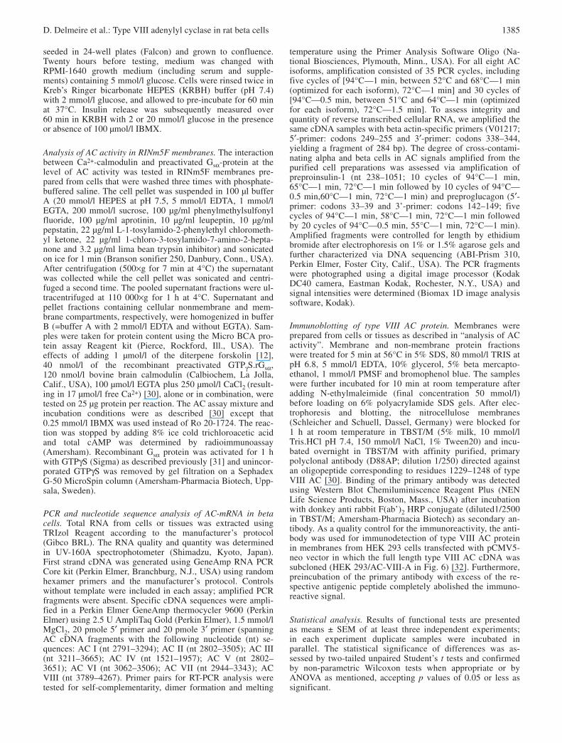
Analysis of AC activity in RINm5F membranes. The interactionbetween Ca2+-calmodulin and preactivated Gsα-protein at thelevel of AC activity was tested in RINm5F membranes pre-pared from cells that were washed three times with phosphate-buffered saline. The cell pellet was suspended in 100 µl bufferA (20 mmol/l HEPES at pH 7.5, 5 mmol/l EDTA, 1 mmol/lEGTA, 200 mmol/l sucrose, 100 µg/ml phenylmethylsulfonylfluoride, 100 µg/ml aprotinin, 10 µg/ml leupeptin, 10 µg/mlpepstatin, 22 µg/ml L-1-tosylamido-2-phenylethyl chlorometh-yl ketone, 22 µg/ml 1-chloro-3-tosylamido-7-amino-2-hepta-none and 3.2 µg/ml lima bean trypsin inhibitor) and sonicatedon ice for 1 min (Branson sonifier 250, Danbury, Conn., USA).After centrifugation (500×g for 7 min at 4°C) the supernatantwas collected while the cell pellet was sonicated and centri-fuged a second time. The pooled supernatant fractions were ul-tracentrifuged at 110 000×g for 1 h at 4°C. Supernatant andpellet fractions containing cellular nonmembrane and mem-brane compartments, respectively, were homogenized in bufferB (=buffer A with 2 mmol/l EDTA and without EGTA). Sam-ples were taken for protein content using the Micro BCA pro-tein assay Reagent kit (Pierce, Rockford, Ill., USA). The effects of adding 1 µmol/l of the diterpene forskolin [12],40 nmol/l of the recombinant preactivated GTPγS.rGsα,120 nmol/l bovine brain calmodulin (Calbiochem, La Jolla,Calif., USA), 100 µmol/l EGTA plus 250 µmol/l CaCl2 (result-ing in 17 µmol/l free Ca2+) [30], alone or in combination, weretested on 25 µg protein per reaction. The AC assay mixture andincubation conditions were as described [30] except that0.25 mmol/l IBMX was used instead of Ro 20-1724. The reac-tion was stopped by adding 8% ice cold trichloroacetic acidand total cAMP was determined by radioimmunoassay(Amersham). Recombinant Gsα protein was activated for 1 hwith GTPγS (Sigma) as described previously [31] and unincor-porated GTPγS was removed by gel filtration on a SephadexG-50 MicroSpin column (Amersham-Pharmacia Biotech, Upp-sala, Sweden).
PCR and nucleotide sequence analysis of AC-mRNA in betacells. Total RNA from cells or tissues was extracted using TRIzol Reagent according to the manufacturer’s protocol (Gibco BRL). The RNA quality and quantity was determinedin UV-160A spectrophotometer (Shimadzu, Kyoto, Japan).First strand cDNA was generated using GeneAmp RNA PCRCore kit (Perkin Elmer, Branchburg, N.J., USA) using randomhexamer primers and the manufacturer’s protocol. Controlswithout template were included in each assay; amplified PCRfragments were absent. Specific cDNA sequences were ampli-fied in a Perkin Elmer GeneAmp thermocycler 9600 (PerkinElmer) using 2.5 U AmpliTaq Gold (Perkin Elmer), 1.5 mmol/lMgCl2, 20 pmole 5′ primer and 20 pmole 3′ primer (spanningAC cDNA fragments with the following nucleotide (nt) se-quences: AC I (nt 2791–3294); AC II (nt 2802–3505); AC III(nt 3211–3665); AC IV (nt 1521–1957); AC V (nt 2802–3651); AC VI (nt 3062–3506); AC VII (nt 2944–3343); ACVIII (nt 3789–4267). Primer pairs for RT-PCR analysis weretested for self-complementarity, dimer formation and melting
temperature using the Primer Analysis Software Oligo (Na-tional Biosciences, Plymouth, Minn., USA). For all eight ACisoforms, amplification consisted of 35 PCR cycles, includingfive cycles of [94°C—1 min, between 52°C and 68°C—1 min(optimized for each isoform), 72°C—1 min] and 30 cycles of[94°C—0.5 min, between 51°C and 64°C—1 min (optimizedfor each isoform), 72°C—1.5 min]. To assess integrity andquantity of reverse transcribed cellular RNA, we amplified thesame cDNA samples with beta actin-specific primers (V01217;5′-primer: codons 249–255 and 3′-primer: codons 338–344,yielding a fragment of 284 bp). The degree of cross-contami-nating alpha and beta cells in AC signals amplified from thepurified cell preparations was assessed via amplification ofpreproinsulin-1 (nt 238–1051; 10 cycles of 94°C—1 min,65°C—1 min, 72°C—1 min followed by 10 cycles of 94°C—0.5 min,60°C—1 min, 72°C—1 min) and preproglucagon (5′-primer: codons 33–39 and 3’-primer: codons 142–149; five cycles of 94°C—1 min, 58°C—1 min, 72°C—1 min followedby 20 cycles of 94°C—0.5 min, 55°C—1 min, 72°C—1 min).Amplified fragments were controlled for length by ethidiumbromide after electrophoresis on 1% or 1.5% agarose gels andfurther characterized via DNA sequencing (ABI-Prism 310,Perkin Elmer, Foster City, Calif., USA). The PCR fragmentswere photographed using a digital image processor (KodakDC40 camera, Eastman Kodak, Rochester, N.Y., USA) andsignal intensities were determined (Biomax 1D image analysissoftware, Kodak).
Immunoblotting of type VIII AC protein. Membranes wereprepared from cells or tissues as described in “analysis of ACactivity”. Membrane and non-membrane protein fractionswere treated for 5 min at 56°C in 5% SDS, 80 mmol/l TRIS atpH 6.8, 5 mmol/l EDTA, 10% glycerol, 5% beta mercapto-ethanol, 1 mmol/l PMSF and bromophenol blue. The sampleswere further incubated for 10 min at room temperature afteradding N-ethylmaleimide (final concentration 50 mmol/l) before loading on 6% polyacrylamide SDS gels. After elec-trophoresis and blotting, the nitrocellulose membranes (Schleicher and Schuell, Dassel, Germany) were blocked for1 h at room temperature in TBST/M (5% milk, 10 mmol/lTris.HCl pH 7.4, 150 mmol/l NaCl, 1% Tween20) and incu-bated overnight in TBST/M with affinity purified, primarypolyclonal antibody (D88AP; dilution 1/250) directed againstan oligopeptide corresponding to residues 1229–1248 of typeVIII AC [30]. Binding of the primary antibody was detectedusing Western Blot Chemiluminiscence Reagent Plus (NENLife Science Products, Boston, Mass., USA) after incubationwith donkey anti rabbit F(ab’)2 HRP conjugate (diluted1/2500in TBST/M; Amersham-Pharmacia Biotech) as secondary an-tibody. As a quality control for the immunoreactivity, the anti-body was used for immunodetection of type VIII AC proteinin membranes from HEK 293 cells transfected with pCMV5-neo vector in which the full length type VIII AC cDNA wassubcloned (HEK 293/AC-VIII-A in Fig. 6) [32]. Furthermore,preincubation of the primary antibody with excess of the re-spective antigenic peptide completely abolished the immuno-reactive signal.
Statistical analysis. Results of functional tests are presentedas means ± SEM of at least three independent experiments; in each experiment duplicate samples were incubated in parallel. The statistical significance of differences was as-sessed by two-tailed unpaired Student’s t tests and confirmedby non-parametric Wilcoxon tests when appropriate or byANOVA as mentioned, accepting p values of 0.05 or less assignificant.
D. Delmeire et al.: Type VIII adenylyl cyclase in rat beta cells 1385

Results
Receptor-mediated cAMP accumulation in beta cellsis potentiated by glucose in a calcium-dependent man-ner. During static incubations, cAMP content in ratbeta cells was not influenced by glucose alone; how-ever, the sugar enhanced the stimulatory effect ofGLP-1 on cAMP accumulation in the cells (Fig. 1).This effect of glucose was not specific for GLP-1-induced cAMP accumulation, since it was also ob-served with glucagon (Fig. 2, Table 1). The potentia-tion seems mediated at the stage of cAMP production,rather than cAMP breakdown, because the phenome-non was only observed in the presence of the phos-
phodiesterase inhibitors such as the potent inhibitor 3-isobutyl-1-methylxanthine (IBMX) and the more se-lective inhibitor Ro-20-1724 (Fig. 2). The synergismbetween glucose and GLP-1 on cAMP production inbeta cells (Fig. 1) was completely suppressed by co-incubation with 50 µmol/l of the L-type Ca2+-channelblocker verapamil, a well known inhibitor of glucose-induced insulin release [33, 34]. On the contrary, ace-tylcholine (Table 1), a neurotransmitter that mobilizescalcium ions from intracellular stores in beta cells [35]
1386 D. Delmeire et al.: Type VIII adenylyl cyclase in rat beta cells
Table 1. Interaction between glucose, glucagon and acetylcholine (1 µmol/l) in the regulation of cAMP production and insulin release from beta cells
Condition n cAMP content Insulin release
(fmol·103 cells−1) (ng·103 cells−1)
Control Acetylcholine Control Acetylcholine
1.4 mmol/l glucose 4 2.6±0.3 3.1±0.5 0.14±0.04 0.17±0.011.4 mmol/l glucose + 10 nmol/l glucagon 3 6.0±0.9 6.4±0.4 0.19±0.04 0.23±0.0820 mmol/l glucose 4 3.6±0.5 3.8±0.3 0.55±0.08 1.38±0.13b
20 mmol/l glucose + 10 nmol/l glucagon 4 15.5±1.0c 13.9±1.7 3.3±0.3d 4.7±0.4a,d
Flow-sorted beta cells were incubated for 30 min in Earle’sHEPES buffer supplemented with different stimuli as indicat-ed. During the last 5 min of culture, the cells were exposed tothe PDE inhibitor Ro-20-1724 (100 µmol/l) in order to mea-sure cAMP accumulation concomitantly to insulin release. Thedata represent mean values ± SEM of n experiments. Statistical
significance was calculated by the unpaired Student’s t-test.Differences between control cells and acetylcholine-treatedcells: ap<0.05; bp<0.005. Differences between 20 mmol/l glucose + 10 nmol/l=glucagon vs sum of effects induced by20 mmol/l glucose and 1.4 mmol/l glucose plus 10 nmol/l glucagon: cp<0.05; dp<0.005
Fig. 1. Synergism between glucose and GLP-1 on cAMP accu-mulation in beta cells is calcium-dependent. FACS-purified ratbeta cells were incubated for 15 min at 37°C in Earle’s HEPESbuffer with 250 µmol/l IBMX and 1.4 mmol/l glucose (whitebars) or 20 mmol/l glucose (black bars) either without or withGLP-1 (10 nmol/l) or GLP-1 plus verapamil (50 µmol/l). Datarepresent mean values ± SEM of five experiments; ap<0.01
Fig. 2A–C. Synergism between glucose and glucagon oncAMP accumulation in beta cells is not mediated by interac-tion at the level of phosphodiesterases. Rat beta cells were pre-incubated for 5 min in Earle’s HEPES buffer containing1.4 mmol/l glucose alone (A) or 1.4 mmol/l glucose in thepresence of 100 µmol/l of the PDE inhibitors Ro-20-1724 (B)or IBMX (C). Subsequent 5 min incubations occurred in1.4 mmol/l (open bars) or 20 mmol/l glucose (closed bars), either alone or in combination with 10 nmol/l glucagon as indi-cated. Data represent mean values ± SEM of four experiments;*p<0.01; **p<0.001

D. Delmeire et al.: Type VIII adenylyl cyclase in rat beta cells 1387
did not influence cAMP content in beta cells. How-ever, potentiating effects of acetylcholine on insulin release from purified cells were observed (p<0.005;Table 1).
The L-type Ca2+-channel blocker verapamil abro-gates the synergism between GLP-1 and glucose uponinsulin release. A marked synergism exists in thestimulation of insulin release by glucose on the onehand and hormones that raise cAMP on the other hand
[2]. Such synergism can be observed both in static in-cubations (Table 1) and in dynamic perifusions(Fig. 3). In static incubations and in the presence ofRo-20-1724, the combination of glucagon (10 nmol/l)and glucose (20 mmol/l) increased insulin release5±1-fold over the calculated sum of insulin releasedfrom each individual stimulus (p<0.005). Insulin re-leased from perifused beta cells was higher duringcombined GLP-1 and glucose stimulation (data abovebasal rates: 3.2±0.3% of insulin content over 15 min)than the summation of the effects of the two separatedstimuli (20 mmol/l glucose alone: 1.2±0.2% of cellu-lar insulin content and GLP-1 at low glucose:0.2±0.1% of content; p<0.001 vs combined stimula-tion). This synergism was equally present when theorder of stimuli was reversed. It was not the result of amemory effect caused by repetitive beta cell stimula-tion with GLP-1, since a second stimulation with glu-cose plus GLP-1 elicited the same insulin release asduring the first period of co-stimulation (Fig. 3B). Inparallel to its effect on cAMP accumulation in thecells, Fig. 3A shows that verapamil effectively coun-teracted the combined stimulatory effect of glucoseand GLP-1 upon insulin release (1.1±0.2% of content;p<0.001 vs control without verapamil), raising thequestion as to whether the effects of this drug on insu-lin release can be attributed solely to a decrease in[Ca2+]i or to both a decrease in [Ca2+]i and in [cAMP].
AC activity in RINm5F cells is activated by both Gsαand Ca2+-calmodulin. To examine the molecularmechanism underlying the observed synergism be-tween glucose-stimulation and hormone-stimulationof cAMP production in rat beta cells, we measured ad-enylyl cyclase activity in membranes extracted fromthe insulinoma cell line RINm5F (Fig. 4). Functionalintegrity of the membranes was assessed by adding 1 µmol/l forskolin [12] which enhanced AC activitybetween two- and three-fold (13±3 vs 32±5 pmolcAMP·mg protein−1·min−1 in basal and forskolin-stim-ulated membranes respectively; p<0.05 vs basal). Themeasured AC activity in RINm5F membranes was notinfluenced by raising the free [Ca2+] to 17 µmol/l or byadding calmodulin alone up to 600 nmol/l. The com-bination of 17 µmol/l [Ca2+] and 120 nmol/l calmodu-lin increased AC activity to similar values as observedupon forskolin stimulation (p<0.05 vs basal). Incuba-tion of RINm5F membranes with 40 nmol/l of the re-combinant preactivated G protein GTPγS.rGsα stimu-lated AC activity about threefold above basal(26±8 pmol cAMP·mg protein−1·min−1 above basal;p<0.01 vs basal; p<0.05 vs Ca2+-calmodulin). Simul-taneous addition of GTPγS.rGsα and calmodulin at17 µmol/l [Ca2+] resulted in a further increase of ACactivity (50±8 pmol cAMP mg protein−1·min−1 abovebasal; p<0.01 vs basal; p<0.01 vs GTPγS.rGsα). Thecombined stimulatory effect was higher than whatcould be expected from the sum of adding Ca2+-cal-
Fig. 3A, B. Synergism between glucose and GLP-1 on insulinrelease is abrogated by the L-type Ca2+-channel blocker vera-pamil. Beta cells were perifused with 1.4 mmol/l glucose (bas-al medium) which alternated with 10 min stimulations with20 mmol/l glucose, 10 nmol/l GLP-1 (gray box) or both. Sec-ond stimulation with 20 mmol/l glucose plus 10 nmol/l GLP-1occurred in the presence (A) or absence (B) of 50 µmol/l vera-pamil (black box). Data represent mean values ± SEM of eight(A) or three (B) experiments

modulin or GTPγS.rGsα separately (39±10 pmolcAMP mg protein−1·min−1 above basal; p<0.05). Thisindicates that Ca2+-calmodulin and Gsα act synergisti-cally upon adenylyl cyclase(s) present in RINm5Fcells, although this effect is much more modest thanthat described in type VIII AC-transfected HEK293cells [30].
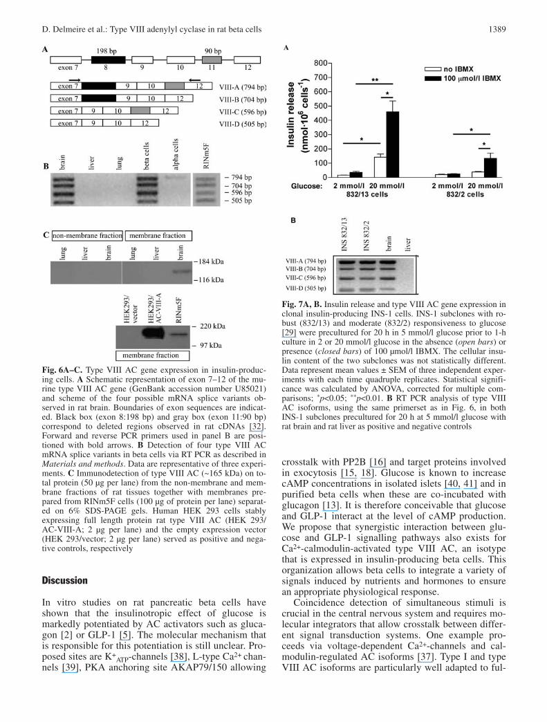
AC type VIII is expressed in insulin-producing cells.Among the cloned and characterized members of themammalian AC family, two isoforms—i.e. type I ACand type VIII AC—are synergistically activated byCa2+-calmodulin and Gsα [30, 36, 37]. To assess themRNA expression of the various AC isoforms in pri-mary beta cells as well as other pancreatic and non-pancreatic cells, we did a RT-PCR analysis on totalcellular RNA using isoform-specific primers. This pro-cedure yielded cDNA fragments of the expected length(Figs. 5, 6) and nucleotide sequence. Nonspecific PCRsignals were not detected. Type I AC mRNA wasabundant in brain but only present at low copy num-bers in alpha cells and beta cells (Fig. 5) and very lowlevels in RINm5F cells. Strong PCR signals for typeVI and type VIII AC were obtained from mRNA ex-tracted from brain and flow-sorted beta cells. The otherisoforms were either not detectable (type II and typeVII AC) or were present at low levels (type III, type IVand type V AC). In addition to the three type VIII AC
transcripts which have been described in rat brain [32]we detected a fourth transcript in which both exon 8and exon 11 were absent (Fig. 6A,B). The same foursplice products were present in cDNA prepared frompurified rat beta cells and RINm5F cells (Fig. 6B). Toassess if mRNA encoding type VIII AC was translatedin insulin-producing cells, we examined type VIII ACprotein abundance via immunoblots using a polyclonalantiserum that has been validated before [30]. As expected, this antibody detected a 165 kDa protein inthe membrane fraction of rat brain, whereas the non-membrane cellular extract was negative (Fig. 6C).Type VIII AC immunoreactive protein was present inthe membrane fraction prepared from RINm5F cells(Fig. 6C). Since RINm5F cells are known to be glu-cose-unresponsive, we compared expression of thetype VIII AC splice variants in the INS-1 subclones832/13 and 832/2 which markedly differ in glucose re-sponsiveness as indicated by the differences in glu-cose-induced insulin release, being, respectively nine-fold and two-fold above basal secretion (Fig. 7A) [29].A potent glucose-responsiveness was observed in thetwo subclones (13- and five-fold basal release, in832/13- and 832/2-cells respectively) in the presenceof the phosphodiesterase inhibitor IBMX (Fig. 7A). Inagreement with a role of type VIII AC in beta cellfunction, the four splice variants were present in bothINS-1 subclones (Fig. 7B).
1388 D. Delmeire et al.: Type VIII adenylyl cyclase in rat beta cells
Fig. 4. Regulation of RINm5F adenylyl cyclase activity by cal-cium-calmodulin and Gsα. Membrane protein (25 µg) was incu-bated for 10 min at 30°C in AC assay mixture with or withoutCa2+-calmodulin (17 µmol/l and 120 nmol/l, respectively) andrecombinant GTPγS.Gsα(40 nmol/l), either alone or in combina-tion, as indicated. Positive control for AC stimulation was for-skolin (1 µmol/l). Data represent mean AC activity values abovebasal AC activity ± SEM of seven experiments. Basal AC activ-ity in RINm5F cell membranes was 13±3 pmol cAMP·mg pro-tein−1·min−1. Statistical significance was calculated by one-wayanalysis of variance (ANOVA). ap<0.05; bp<0.01
Fig. 5. mRNA analysis via RT-PCR of AC isoforms in betacells with specific primer sets as explained in Materials andmethods. Data are representative of four experiments. cDNAfragments amplified from preproinsulin-1 and preproglucagonmRNA’s were controls for, respectively, contamination in thepurified alpha- and beta-cell preparation
Discussion
In vitro studies on rat pancreatic beta cells haveshown that the insulinotropic effect of glucose ismarkedly potentiated by AC activators such as gluca-gon [2] or GLP-1 [5]. The molecular mechanism thatis responsible for this potentiation is still unclear. Pro-posed sites are K+
ATP-channels [38], L-type Ca2+ chan-nels [39], PKA anchoring site AKAP79/150 allowing
crosstalk with PP2B [16] and target proteins involvedin exocytosis [15, 18]. Glucose is known to increasecAMP concentrations in isolated islets [40, 41] and inpurified beta cells when these are co-incubated withglucagon [13]. It is therefore conceivable that glucoseand GLP-1 interact at the level of cAMP production.We propose that synergistic interaction between glu-cose and GLP-1 signalling pathways also exists forCa2+-calmodulin-activated type VIII AC, an isotypethat is expressed in insulin-producing beta cells. Thisorganization allows beta cells to integrate a variety ofsignals induced by nutrients and hormones to ensurean appropriate physiological response.
Coincidence detection of simultaneous stimuli iscrucial in the central nervous system and requires mo-lecular integrators that allow crosstalk between differ-ent signal transduction systems. One example pro-ceeds via voltage-dependent Ca2+-channels and cal-modulin-regulated AC isoforms [37]. Type I and typeVIII AC isoforms are particularly well adapted to ful-
D. Delmeire et al.: Type VIII adenylyl cyclase in rat beta cells 1389
Fig. 6A–C. Type VIII AC gene expression in insulin-produc-ing cells. A Schematic representation of exon 7–12 of the mu-rine type VIII AC gene (GenBank accession number U85021)and scheme of the four possible mRNA splice variants ob-served in rat brain. Boundaries of exon sequences are indicat-ed. Black box (exon 8:198 bp) and gray box (exon 11:90 bp)correspond to deleted regions observed in rat cDNAs [32].Forward and reverse PCR primers used in panel B are posi-tioned with bold arrows. B Detection of four type VIII ACmRNA splice variants in beta cells via RT PCR as described inMaterials and methods. Data are representative of three experi-ments. C Immunodetection of type VIII AC (~165 kDa) on to-tal protein (50 µg per lane) from the non-membrane and mem-brane fractions of rat tissues together with membranes pre-pared from RINm5F cells (100 µg of protein per lane) separat-ed on 6% SDS-PAGE gels. Human HEK 293 cells stably expressing full length protein rat type VIII AC (HEK 293/AC-VIII-A; 2 µg per lane) and the empty expression vector(HEK 293/vector; 2 µg per lane) served as positive and nega-tive controls, respectively
Fig. 7A, B. Insulin release and type VIII AC gene expression inclonal insulin-producing INS-1 cells. INS-1 subclones with ro-bust (832/13) and moderate (832/2) responsiveness to glucose[29] were precultured for 20 h in 5 mmol/l glucose prior to 1-hculture in 2 or 20 mmol/l glucose in the absence (open bars) orpresence (closed bars) of 100 µmol/l IBMX. The cellular insu-lin content of the two subclones was not statistically different.Data represent mean values ± SEM of three independent exper-iments with each time quadruple replicates. Statistical signifi-cance was calculated by ANOVA, corrected for multiple com-parisons; *p<0.05; **p<0.01. B RT PCR analysis of type VIIIAC isoforms, using the same primerset as in Fig. 6, in bothINS-1 subclones precultured for 20 h at 5 mmol/l glucose withrat brain and rat liver as positive and negative controls

fill this role since they are synergistically activated byCa2+-calmodulin and Gsα [30, 32, 42, 43]. Consistentwith their functional importance in the brain is the ob-servation that mice rendered deficient in type I AC bytargeted gene disruption present learning defects as aresult of impaired long-term potentiation in hippocam-pal neurons [44] and in the cerebellum [45]. Type VIIIAC is particularly abundant in hippocampal neurons[30] which exhibit a large capacity for long-term po-tentiation and altered synaptic plasticity. Accordingly,type VIII AC knock-out mice show defects in the anx-iety response after repetitive exposure to stress [46].
A synergistic regulation by glucose and glucagonwas observed at the juncture of cellular cAMP accu-mulation in flow-sorted beta cells, on condition thatinhibitors of cAMP breakdown such as IBMX or Ro-20-1724 were present. This suggests that gluca-gon- and glucose-induced signalling interact at thelevel of cAMP production rather than at the point ofcAMP breakdown. Glucagon and GLP-1 acutely am-plify the glucose-dependent signal to a similar extent,so that their common target of interaction is likely tobe distal from the activated receptors. Previous workon whole islets emphasised the importance of calciumfor islet adenylyl cyclase activity. Two studies [47, 48]have shown that homogenates of rat isolated isletscontain Ca2+-calmodulin-dependent AC activity. Weshow that the Ca2+-calmodulin effects interact syner-gistically with that of Gsα. As such, our data providean explanation for the glucose potentiation on hor-mone-induced cAMP accumulation in intact beta cells[13]. Our observation that the L-type Ca2+-channelblocker verapamil [33] can abrogate the potentiatingeffect of glucose on GLP-1-induced intracellularcAMP accumulation supports the idea that the open-ing of L-type Ca2+-channels is required for the effectof glucose on AC activity in intact beta cells. Alreadyat low glucose concentration, we observe a small, butborderline significant difference between cAMP accu-mulation in the presence and absence of verapamilsubsequent to GLP-1 stimulation. It can be speculatedthat this effect is mediated by direct GLP-1 action on L-type Ca2+ channels. Activation of the GLP-1 re-ceptor has been observed before to cause opening ofL-type voltage-dependent Ca2+ channels under non-stimulatory glucose conditions [49, 50, 51] and to in-crease electrical activity by slowing Ca2+ channel in-activation [52]. The GLP-1 induced increase in intra-cellular Ca2+ concentrations may be able to stimulateCa2+-calmodulin-regulated AC isoforms (type I andVIII AC) resulting in the production of cAMP [37],which can be antagonized by adding the L-type Ca2+-channel blocker verapamil with the preservation of theGs-protein stimulated cAMP production by ACs. Theeffect of glucose on glucagon receptor cAMP produc-tion was not mimicked or amplified by acetylcholine.This indicates that mobilization of Ca2+ from intracel-lular stores is not as effective as influx through L-type
Ca2+-channels in the activation of AC, consistent withexisting literature [53].
Amplification of cDNA using the type VIII ACprimer set resulted in strong signals both with primaryflow-sorted rat beta cells and the rat beta cell linesRINm5F or INS-1. While it can be argued for FACS-purified beta cells that the PCR signals are derivedfrom the few non-beta cells that are present in thesepreparations [28], this possibility is highly unlikely.First, the signal intensity of type VIII AC mRNA inFACS-purified alpha cells was around 20% of the sig-nal intensity of flow sorted beta cells. Consequently,the type VIII AC signal in pure beta cells cannot beexplained by the 5% or fewer alpha cells present inthis cell preparation. Second, type VIII AC expressionwas observed in three different beta cell lines, withstrong (INS-1 832/13), moderate (INS-1 832/2) andno (RINm5F) glucose responsiveness. This indicatesthat type VIII AC is not directly responsible for con-veying glucose-responsiveness to a beta cell, but di-rect evidence involving gene-overexpression or gene-silencing will be required to further address this issue.
The present observations do not infer that AC typeVIII is the only or even the main adenylyl cyclasecontributing to overall cellular cAMP accumulation.In fact, our RT-PCR analysis clearly identifies thepresence of several isoforms in RNA prepared frompurified primary beta cells. It is possible that the rather weak type III, IV, and V AC signals are due tocontaminating non-beta cells, because signals weremuch stronger in FACS-purified alpha cells, respect-ing the beta to alpha signal intensity ratio that wasalso observed for glucagon. Amongst the five Ca2+-calmodulin-regulated AC isoforms, only type I andtype VIII are synergistically activated by Ca2+-cal-modulin and Gsα [37]. Previous expression analyses inisolated islets [54, 55] suggested that the AC isoformsIII, V and VI are also present in beta cells. From aregulatory perspective, the possible presence of typeIII AC in beta cells seems interesting. In isolatedmembranes, type III AC can be stimulated by Ca2+-calmodulin on condition that other activators, e.g. for-skolin or activated Gsα are present at the same time[56]. However, (i) this stimulation is additive ratherthan synergistic; (ii) in intact cells, type III AC is in-hibited by raised cytosolic [Ca2+], possibly as a conse-quence of the activation of Ca2+-calmodulin-depen-dent kinases or phosphatases [57]. It was reported thatthe spontaneously diabetic rat strain Goto-Kakizakihas defects both in the KATP-dependent and -indepen-dent pathways of glucose stimulation [58], abnormalglucose activation of exocytosis [59] as well as in-creased expression of AC type III [54] and type VIIIin islets [60] and increased cAMP generation after for-skolin stimulation [59]. Congruently, interaction be-tween glucose and glucagon (or related peptides) can be disturbed in other models of Type 2 diabetes[61].
1390 D. Delmeire et al.: Type VIII adenylyl cyclase in rat beta cells

Besides overall cellular expression, subcellular localisation in signalling complexes could be crucialfor the integrated response of beta cells to glucose andGLP-1. Co-localisation of Ca2+-sensitive AC and cap-acitative Ca2+-entry channels was found in embryonickidney cells [62, 63], as well as in cardiac myocytes[64] and in parotid cells [65]. It will be a challenge forfuture studies to study such co-localisation in betacells, since this spatial organization may contribute tofoci of cellular cAMP formation when cells are ex-posed to hormone as observed in other cell types [66,67]. In RINm5F cells, cAMP signalling complexeswith relevance for exocytosis have been observed recently [16]. Important questions that remain to beanswered are: (i) whether GLP-1 receptors of primarybeta cells in situ are preferentially coupled to typeVIII AC isoforms; (ii) if the cAMP that is thus formedpreferentially signals to protein complexes involved inregulation of exocytosis.
Is type VIII AC the only site at which cAMP-gen-erating receptors and glucose interact synergistically?As was mentioned above, other molecular targets ofinteraction have been proposed [16, 27], followingcrosstalk between cAMP and Ca2+. In isolated mouseislets that were studied under depolarizing conditionsin the presence of diazoxide (i.e. under high tonic in-tracellular [Ca2+]) the synergism between glucose andGLP-1 is still present [68]. We suggest that at leastone of the molecuar targets are regulated by other mediators than Ca2+, possibly by signals generated bycataplerosis [11].
In conclusion, our study provides evidence for theexpression at RNA and protein level of the type VIIIAC gene in pancreatic beta cells, as well as its func-tional activity in isolated membranes or in intact cells.The regulatory properties of this enzyme make it awell adapted molecular integrator of nutrient and hor-monal stimuli that control insulin release.
Acknowledgements. The authors wish to thank E. Quartier, A.Van Breusegem and V. Berger for technical assistance. We aregrateful to the personnel of the Department of Metabolism andEndocrinology for providing purified rat islet alpha and betacells and to L. Kaufman for help with the statistical analysis.The clonal cell lines INS1 832/13 and 832/2 [29] were gener-ously supplied by Dr. C. Newgard, S. Stedman Center for Nutritional and Metabolic Studies, Duke University, Durham,N.C., USA. This study was supported by grant No. 9.0130.99from the Flemish Fund for Scientific Research (FWO Vlaande-ren), the Ministerie van de Vlaamse Gemeenschap, Departe-ment Onderwijs (Geconcerteerde Onderzoeksactie 1807), andthe Research Council of the Vrije Universiteit Brussel. S.A.Hinke is Visiting Postdoctoral Fellow at the FWO Vlaanderen.
References
1. Pipeleers D (1987) The biosociology of pancreatic B cells.Diabetologia 30:277–291
2. Pipeleers DG, Schuit FC, In’t Veld PA et al. (1985) Inter-play of nutrients and hormones in the regulation of insulinrelease. Endocrinology 117:824–833
3. Huypens P, Ling Z, Pipeleers D, Schuit F (2000) Glucagonreceptors on human islet cells contribute to glucose compe-tence of insulin release. Diabetologia 43:1012–1019
4. Schuit FC, Pipeleers DG (1986) Differences in adrenergicrecognition by pancreatic A and B cells. Science 232:875–877
5. Mojsov S, Weir GC, Habener JF (1987) Insulinotropin:glucagon-like peptide I (7–37) co-encoded in the glucagongene is a potent stimulator of insulin release in the perfusedrat pancreas. J Clin Invest 79:616–619
6. Newgard CB, McGarry JD (1995) Metabolic coupling fac-tors in pancreatic beta-cell signal transduction. Annu RevBiochem 64:689–719
7. Maechler P, Wollheim CB (2001) Mitochondrial functionin normal and diabetic beta-cells. Nature 414:807–812
8. Hellman B, Gylfe E, Bergsten P et al. (1994) Glucose induces oscillatory Ca2+ signalling and insulin release inhuman pancreatic beta cells. Diabetologia 37:S11–S20
9. Henquin JC (2000) Triggering and amplifying pathways ofregulation of insulin secretion by glucose. Diabetes49:1751–1760
10. Prentki M, Tornheim K, Corkey BE (1997) Signal trans-duction mechanisms in nutrient-induced insulin secretion.Diabetologia 40:S32–S41
11. Flamez D, Berger V, Kruhoffer M, Orntoft T, Pipeleers D,Schuit FC (2002) Critical role for cataplerosis via citrate inglucose-regulated insulin release. Diabetes 51:2018–2024
12. Henquin JC, Meissner HP (1984) The ionic, electrical, andsecretory effects of endogenous cyclic adenosine mono-phosphate in mouse pancreatic B cells: studies with forskolin. Endocrinology 115:1125–1134
13. Schuit FC, Pipeleers DG (1985) Regulation of adenosine3′,5′-monophosphate levels in the pancreatic B cell. Endo-crinology 117:834–840
14. Lester LB, Langeberg LK, Scott JD (1997) Anchoring ofprotein kinase A facilitates hormone-mediated insulin secretion. Proc Natl Acad Sci USA 94:14942–14947
15. Ämmälä C, Eliasson L, Bokvist K et al. (1994) Activationof protein kinases and inhibition of protein phosphatasesplay a central role in the regulation of exocytosis in mousepancreatic β cells. Proc Natl Acad Sci USA 91:4343–4347
16. Lester LB, Faux MC, Nauert JB, Scott JD (2001) Tar-geted protein kinase A and PP-2B regulate insulin secre-tion through reversible phosphorylation. Endocrinology142:1218–1227
17. Ozaki N, Shibasaki T, Kashima Y et al. (2000) cAMP-GEFII is a direct target of cAMP in regulated exocytosis.Nat Cell Biol 2:805–811
18. Kashima Y, Miki T, Shibasaki T et al. (2001) Critical roleof cAMP-GEFII-Rim2 complex in incretin-potentiated in-sulin secretion. J Biol Chem 276:46046–46053
19. Moens K, Heimberg H, Flamez D et al. (1996) Expressionand functional activity of glucagon, glucagon-like peptideI, and glucose-dependent insulinotropic peptide receptorsin rat pancreatic islet cells. Diabetes 45:257–261
20. Scrocchi LA, Brown TJ, MaClusky N et al. (1996) Glucoseintolerance but normal satiety in mice with a null mutationin the glucagon-like peptide 1 receptor gene. Nat Med2:1254–1258
21. Miyawaki K, Yamada Y, Yano H et al. (1999) Glucose intolerance caused by a defect in the entero-insular axis: astudy in gastric inhibitory polypeptide receptor knockoutmice. Proc Natl Acad Sci USA 96:14843–14847
22. Flamez D, Van Breusegem A, Scrocchi LA et al. (1998)Mouse pancreatic beta-cells exhibit preserved glucosecompetence after disruption of the glucagon-like peptide-1receptor gene. Diabetes 47:646–652
D. Delmeire et al.: Type VIII adenylyl cyclase in rat beta cells 1391

23. Moens K, Flamez D, Van Schravendijk C, Ling Z, Pipeleers D, Schuit F (1998) Dual recognition by pan-creatic β-cells via glucagon and glucagon-like peptide 1 receptors. Diabetes 47:66–72
24. Scheen AJ, Castillo MJ, Lefebvre PJ (1996) Assessment ofresidual insulin secretion in diabetic patients using the intravenous glucagon stimulatory test: methodological as-pects and clinical applications. Diabetes Metab 22:397–406
25. Moens K, Berger V, Ahn JM et al. (2002) Assessment ofthe role of interstitial glucagon in the acute glucose secre-tory responsiveness of in situ pancreatic beta-cells. Diabe-tes 51:669–675
26. Lawrence MC, Bhatt HS, Watterson JM, Easom RA (2001)Regulation of insulin gene transcription by a Ca(2+)-responsive pathway involving calcineurin and nuclear fac-tor of activated T cells. Mol Endocrinol 15:1758–1767
27. Lawrence MC, Bhatt HS, Easom RA (2002) NFAT regu-lates insulin gene promoter activity in response to synergis-tic pathways induced by glucose and glucagon-like pep-tide-1. Diabetes 51:691–698
28. Pipeleers DG, In ‘t Veld PA, Van De Winkel M, Maes E,Schuit F, Gepts W (1985) A new in vitro method for the study of pancreatic A and B cells. Endocrinology117:806–816
29. Hohmeier HE, Mulder H, Chen G, Henkel-Rieger R, Prentki M, Newgard CB (2000) Isolation of INS-1-derivedcell lines with robust ATP-sensitive K+ channel-dependentand-independent glucose-stimulated insulin secretion. Dia-betes 49:424–430
30. Cali JJ, Zwaagstra JC, Mons N, Cooper DMF, Krupinski J(1994) Type VIII adenylyl cyclase. A Ca2+/calmodulin-stimulated enzyme expressed in discrete regions of ratbrain. J Biol Chem 269:12190–12195
31. Krupinski J, Coussen F, Bakalyar HA et al. (1989) Ade-nylyl cyclase amino acid sequence: possible channel- ortransporter-like structure. Science 244:1558–1564
32. Cali JJ, Parekh RS, Krupinski J (1996) Splice variants oftype VIII adenylyl cyclase: differences in glycosylation andregulation by Ca2+/calmodulin. J Biol Chem 271:1089–1095
33. Devis G, Somers G, Van Obberghen E, Malaisse WJ(1975) Calcium antagonists and islet function. I. Inhibitionof insulin release by verapamil. Diabetes 24:247–251
34. Lebrun P, Malaisse WJ, Herchuelz A (1982) Evidence fortwo distinct modalities of Ca2+ influx into pancreatic Bcell. Am J Physiol 242:E59–E66
35. Garcia M-C, Hermans MP, Henquin J-C (1988) Glucose-,calcium- and concentration-dependence of acetylcholinestimulation of insulin release and ionic fluxes in mouse islets. Biochem J 254:211–218
36. Xia Z, Storm DR (1997) Calmodulin-regulated adenylylcyclases and neuromodulation. Curr Opin Cell Biol 7:391–396
37. Cooper DM, Karpen JW, Fagan KA, Mons NE (1998)Ca(2+)-sensitive adenylyl cyclases. Adv Second Messen-ger Phosphoprotein Res 32:23–51
38. Beguin P, Nagashima K, Nishimura M, Gonoi T, Seino S(1999) PKA-mediated phosphorylation of the humanK(ATP) channel: separate roles of Kir6.2 and SUR1 sub-unit phosphorylation. EMBO J 18:4722–4732
39. Rajan AS, Hill RS, Boyd III AE (1989) Effect of rise incAMP levels on Ca2+influx through voltage-dependentCa2+ channels in HIT cells. Second-messenger synarchy inβ-cells. Diabetes 38:874–880
40. Charles MA, Fanska R, Schmid FG, Forsham PH, GrodskyGM (1973) Adenosine 3′,5′-monophosphate in pancreatic islets: glucose-induced insulin release. Science 179:569–571
41. Grill V, Cerasi E (1973) Activation by glucose of adenylcyclase in pancreatic islets of the rat. FEBS Letters33:311–314
42. Krupinski J, Cali JJ (1998) Molecular diversity of the adenylyl cyclases. Adv Second messenger PhosphoproteinRes 32:53–79
43. Wayman GA, Impey S, Wu Z, Kindsvogel W, Prichard L,Storm DR (1994) Synergistic activation of the type I ade-nylyl cyclase by Ca2+ and Gs-coupled receptors in vivo. J Biol Chem 269:25400–25405
44. Wu Z-L, Thomas SA, Villacres EC et al. (1995) Alteredbehavior and long-term potentiation in type I adenylyl cyclase mutant mice. Proc Natl Acad Sci USA 92:220–224
45. Storm DR, Hansel C, Hacker B, Parent A, Linden DJ(1998) Impaired cerebellar long-term potentiation in type I adenylyl cyclase mutant mice. Neuron 20:1199–210
46. Schaefer ML, Wong ST, Wozniak DF et al. (2000) Alteredstress-induced anxiety in adenylyl cyclase type VIII-defi-cient mice. J Neurosci 20:4809–4820
47. Valverde I, Vandermeers A, Anjaneyulu R, Malaisse WJ(1979) Calmodulin activation of adenylate cyclase in pan-creatic islets. Science 206:225–227
48. Sharp GWG, Wiedenkeller DE, Kaelin D, Siegel EG, Wollheim CB (1980) Stimulation of adenylyl cyclase byCa2+ and calmodulin in rat islets of Langerhans: explana-tion for the glucose-induced increase in cyclic AMP levels.Diabetes 29:74–77
49. Gromada J, Bokvist K, Ding WG, Holst JJ, Nielsen JH,Rorsman P (1998) Glucagon-like peptide 1 (7–36) amidestimulates exocytosis in human pancreatic beta-cells byboth proximal and distal regulatory steps in stimulus-secre-tion coupling. Diabetes 47:57–65
50. Kanno T, Suga S, Wu J, Kimura M, Wakui M (1998) Intra-cellular cAMP potentiates voltage-dependent activation ofL-type Ca2+ channels in rat islet beta-cells. Pflugers Arch435:578–580
51. Ämmälä C, Ashcroft FM, Rorsman P (1993) Calcium-independent potentiation of insulin release by cyclic AMPin single β-cells. Nature 363:356–358
52. Britsch S, Krippeit-Drews P, Lang F, Gregor M, Drews G(1995) Glucagon-like peptide-1 modulates Ca2+ currentbut not K+ATP current in intact mouse pancreatic B-cells.Biochem Biophys Res Commun 207:33–39
53. Fagan KA, Graft RA, Tolman S, Schaack J, Cooper DMF(2000) Regulation of a Ca2+-sensitive adenylyl cyclase inan excitable cell. Role of voltage-gated versus capacitativeCa2+ entry. J Biol Chem 275:40187–40194
54. Abdel-Halim SM, Guenifi A, He B et al. (1998) Mutationsin the promotor of adenylyl cyclase (AC)-III gene, overex-pression of AC-III mRNA, and enhanced cAMP generationin islets from the spontaneosly diabetic GK rat model oftype 2 diabates. Diabetes 47:498–504
55. Leech CA, Castonguay MA, Habener JF (1999) Expressionof adenylyl cyclase subtypes in pancreatic β-cells. Bio-chem Biophys Res Comm 254:703–706
56. Choi E-J, Xia Z, Storm DR (1992) Stimulation of the typeIII olfactory adenylyl cyclase by calcium and calmodulin.Biochemistry 31:6492–6498
57. Wayman GA, Impey S, Storm DR (1995) Ca2+- inhibitionof type III adenylyl cyclase in vivo. J Biol Chem270:21480–21486
58. Portha B, Serradas P, Bailbé D, Suzuki K-I, Goto Y, GiroixM-H (1991) β-cell insensitivity to glucose in the GK rat, aspontaneous nonobese model for type II diabetes. Diabetes40:486–491
1392 D. Delmeire et al.: Type VIII adenylyl cyclase in rat beta cells

59. Abdel-Halim SM, Guenifi A, Khan A et al. (1996) Impaired coupling of glucose signal to the exocytotic machinery in diabetic GK rats. Diabetes 45:934–940
60. Guenifi A, Portela-Gomes GM, Grimelius L, Efendic S,Abdel-Halim SM (2000) Adenylyl cyclase isoform expres-sion in non-diabetic and diabetic Goto-Kakizaki (GK) ratpancreas. Evidence for distinct overexpression of type-8adenylyl cyclase in diabetic GK rat islets. Histochem CellBiol 113:81–89
61. Cerasi E, Luft R (1970) Diabetes mellitus-A disorder ofcellular information transmission? Horm Metab Res 2:246–249
62. Chiono M, Mahey R, Tate G, Cooper DMF (1995) Capacitative Ca2+ entry exclusively inhibits cAMP synthe-sis in C6-2B glioma cells. J Biol Chem 270:1149–1155
63. Fagan KA, Mahey R, Cooper DMF (1996) Functional co-localization of transfected Ca2+-stimulable adenylylcyclases with capacitative Ca2+ entry sites. J Biol Chem271:12438–12444
64. Gao T, Puri TS, Gerhardstein BL, Chien AJ, Green RD,Hosey MM (1997) Identification and subcellular localizationof the subunits of L-type calcium channels and adenylyl cyclase in cardiac myocytes. J Biol Chem 272:19401–19407
65. Watson EL, Jacobson KL, Singh JC et al. (2000) The type 8 adenylyl cyclase is critical for Ca2+ stimulation ofcAMP accumulation in mouse parotid acini. J Biol Chem275:14691–14699
66. Steinberg SF, Brunton LL (2001) Compartmentation of Gprotein-coupled signaling pathways in cardiac myocytes.Annu Rev Pharmacol Toxicol 41:751–773
67. Zaccolo M, Pozzan T (2002) Discrete microdomains withhigh concentration of cAMP in stimulated rat neonatal cardiac myocytes. Science 295:1711–1715
68. Sato Y, Nenquin M, Henquin JC (1998) Relative contribu-tion of Ca2+-dependent and Ca2+-independent mechanismsto the regulation of insulin secretion by glucose. FEBS Lett421:115–119
D. Delmeire et al.: Type VIII adenylyl cyclase in rat beta cells 1393