GLP-1 Action and Glucose Tolerance in Subjects With Remission of Type 2 Diabetes After Gastric...
8
GLP-1 Action and Glucose Tolerance in Subjects With Remission of Type 2 Diabetes After Gastric Bypass Surgery AMANDA JIMÉNEZ, MD 1 ROSER CASAMITJANA, PHD 1,2,3 JUDITH VIAPLANA-MASCLANS, RN 3 ANTONIO LACY, MD, PHD 1,3 JOSEP VIDAL, MD, PHD 1,2,3 OBJECTIVEdGlucagon like peptide-1 (GLP-1) has been suggested as a major factor for the improved glucose tolerance ensuing after Roux-en-Y gastric bypass (RYGBP) surgery. We exam- ined the effect of blocking endogenous GLP-1 action on glucose tolerance in subjects with sustained remission of type 2 diabetes mellitus (T2DM) present before RYGBP. RESEARCH DESIGN AND METHODSdBlood glucose, insulin, C-peptide, glucagon, GLP-1, and glucose-dependent insulinotropic peptide levels were measured after a meal chal- lenge with either exendin-(9–39) (a GLP-1r antagonist) or saline infusion in eight subjects with sustained remission of T2DM after RYGBP and seven healthy controls. RESULTSdInfusion of exendin-(9–39) resulted in marginal deterioration of the 2-h plasma glucose after meal intake in RYGBP subjects [saline 78.4 6 15.1 mg/dL compared with exendin- (9–39) 116.5 6 22.3 mg/dL; P , 0.001]. Furthermore, glucose response to meal intake was similarly enlarged in the two study groups [percent change in the area under the curve of glucose exendin-(9–39) infusion versus saline infusion: controls 10.84 6 8.8% versus RYGBP 9.94 6 8.4%; P = 0.884]. In the RYGBP group, the blockade of the enlarged GLP-1 response to meal intake resulted in reduced insulin (P = 0.001) and C-peptide (P , 0.001), but no change in glucagon (P = 0.258) responses. CONCLUSIONSdThe limited deterioration of glucose tolerance on blockade of GLP-1 ac- tion in our study suggests the resolution of T2DM after RYGBP may be explained by mechanisms beyond enhancement of GLP-1 action. Diabetes Care 36:2062–2069, 2013 T he beneficial effect of Roux-en-Y gastric bypass (RYGBP) surgery on glycemic control in morbidly obese subjects with type 2 diabetes mellitus (T2DM) is well established (1,2). How- ever, the precise mechanisms mediating T2DM remission after RYGBP are not yet clear (3–5). Although it traditionally has been asserted that bariatric operations are associated with improvement of glucose tolerance merely by caloric restriction and weight loss, several lines of evidence support weight-independent mecha- nisms are involved (6–11). An enhanced postsurgical glucagon-like peptide-1 (GLP-1) secretion, inducing a normalized or exaggerated insulin secretion after meal intake, has been hypothesized to play a major role in the improved glucose toler- ance after RYGBP (3). Association studies have demonstrated larger improvements of glucose tolerance early after RYGBP be- ing associated with a larger GLP-1 re- sponse to nutrient intake as compared with other surgical or nonsurgical inter- ventions resulting in equivalent weight loss (7–9). Likewise, an exaggerated GLP-1 response has been reported up to 10 years after RYGB in subjects with sus- tained T2DM remission, suggesting a key role of GLP-1 in maintaining normal glu- cose tolerance in the long term after this type of surgery (12). However, because association does not prove causation, these data do not definitely prove GLP-1 plays a critical role in T2DM remission after RYGBP. Understanding the role of endoge- nous GLP-1 in metabolic physiology has been greatly enhanced by the availability of a potent GLP-1 receptor antagonist, exendin-(9–39). Exendin-(9–39) block- ade of GLP-1 action in healthy volunteers results in a signi ficant enlargement of postprandial glucose excursions (13– 17). Moreover, using hyperglycemic clamp technique in combination with a mixed meal test, Salehi et al. (18) demon- strated that blocking GLP-1 action results in a larger decrease in the insulin secretion rate in RYGBP-operated subjects (233%) as compared with nonoperated controls ( 216%). This study clearly supports GLP-1 as an important determinant of in- sulin secretion after RYGBP. However, the use of hyperglycemic clamp limited the ability of the study to investigate the rel- ative importance of GLP-1 secretion on glucose tolerance. Furthermore, because only one-third of the study participants presented with T2DM before surgery, the study also was limited in establishing the role of GLP-1 secretion in the remis- sion of T2DM. Of note, in Goto-Kakizaki rats (a nonobese rat model of T2DM) administration of exendin-(9–39) has been shown to totally reverse the im- proved glucose tolerance resulting from duodeno-jejunal exclusion surgery (an experimental metabolic surgery simi- lar to RYGBP) (19). Against this background, the main aim of our study was to examine the effect of endogenous GLP-1 blockade by exendin- (9–39) on glucose tolerance in subjects who had undergone RYGBP and with T2DM antedating surgery that had remit- ted after the surgical procedure. As sec- ondary aims, we evaluated the effect of ccccccccccccccccccccccccccccccccccccccccccccccccc From the 1 Obesity Unit, Hospital Clinic Universitari, Barcelona, Spain; the 2 Centro de Investigación Biomédica en Red de Diabetes y Enfermedades Metabólicas Asociadas (CIBERDEM), Barcelona, Spain; and the 3 Institut d’Investigacions Biomèdiques August Pi Sunyer (IDIBAPS), Barcelona, Spain. Corresponding author: Josep Vidal, [email protected]. Received 31 July 2012 and accepted 27 December 2012. DOI: 10.2337/dc12-1535 © 2013 by the American Diabetes Association. Readers may use this article as long as the work is properly cited, the use is educational and not for profit, and the work is not altered. See http://creativecommons.org/ licenses/by-nc-nd/3.0/ for details. 2062 DIABETES CARE, VOLUME 36, JULY 2013 care.diabetesjournals.org Pathophysiology/Complications O R I G I N A L A R T I C L E
-
Upload
independent -
Category
Documents
-
view
1 -
download
0
Transcript of GLP-1 Action and Glucose Tolerance in Subjects With Remission of Type 2 Diabetes After Gastric...

GLP-1 Action and Glucose Tolerancein SubjectsWith Remission of Type 2Diabetes After Gastric BypassSurgeryAMANDA JIMÉNEZ, MD
1
ROSER CASAMITJANA, PHD1,2,3
JUDITH VIAPLANA-MASCLANS, RN3
ANTONIO LACY, MD, PHD1,3
JOSEP VIDAL, MD, PHD1,2,3
OBJECTIVEdGlucagon like peptide-1 (GLP-1) has been suggested as a major factor for theimproved glucose tolerance ensuing after Roux-en-Y gastric bypass (RYGBP) surgery. We exam-ined the effect of blocking endogenous GLP-1 action on glucose tolerance in subjects withsustained remission of type 2 diabetes mellitus (T2DM) present before RYGBP.
RESEARCH DESIGN AND METHODSdBlood glucose, insulin, C-peptide, glucagon,GLP-1, and glucose-dependent insulinotropic peptide levels were measured after a meal chal-lenge with either exendin-(9–39) (a GLP-1r antagonist) or saline infusion in eight subjects withsustained remission of T2DM after RYGBP and seven healthy controls.
RESULTSdInfusion of exendin-(9–39) resulted in marginal deterioration of the 2-h plasmaglucose after meal intake in RYGBP subjects [saline 78.46 15.1 mg/dL compared with exendin-(9–39) 116.5 6 22.3 mg/dL; P , 0.001]. Furthermore, glucose response to meal intake wassimilarly enlarged in the two study groups [percent change in the area under the curve of glucoseexendin-(9–39) infusion versus saline infusion: controls 10.84 6 8.8% versus RYGBP 9.94 68.4%; P = 0.884]. In the RYGBP group, the blockade of the enlarged GLP-1 response to mealintake resulted in reduced insulin (P = 0.001) and C-peptide (P , 0.001), but no change inglucagon (P = 0.258) responses.
CONCLUSIONSdThe limited deterioration of glucose tolerance on blockade of GLP-1 ac-tion in our study suggests the resolution of T2DM after RYGBPmay be explained bymechanismsbeyond enhancement of GLP-1 action.
Diabetes Care 36:2062–2069, 2013
The beneficial effect of Roux-en-Ygastric bypass (RYGBP) surgery onglycemic control in morbidly obese
subjects with type 2 diabetes mellitus(T2DM) is well established (1,2). How-ever, the precise mechanisms mediatingT2DM remission after RYGBP are not yetclear (3–5). Although it traditionally hasbeen asserted that bariatric operations areassociated with improvement of glucosetolerancemerely by caloric restriction andweight loss, several lines of evidencesupport weight-independent mecha-nisms are involved (6–11). An enhanced
postsurgical glucagon-like peptide-1(GLP-1) secretion, inducing a normalizedor exaggerated insulin secretion aftermealintake, has been hypothesized to play amajor role in the improved glucose toler-ance after RYGBP (3). Association studieshave demonstrated larger improvementsof glucose tolerance early after RYGBP be-ing associated with a larger GLP-1 re-sponse to nutrient intake as comparedwith other surgical or nonsurgical inter-ventions resulting in equivalent weightloss (7–9). Likewise, an exaggeratedGLP-1 response has been reported up to
10 years after RYGB in subjects with sus-tained T2DM remission, suggesting a keyrole of GLP-1 in maintaining normal glu-cose tolerance in the long term after thistype of surgery (12). However, becauseassociation does not prove causation,these data do not definitely prove GLP-1plays a critical role in T2DM remissionafter RYGBP.
Understanding the role of endoge-nous GLP-1 in metabolic physiology hasbeen greatly enhanced by the availabilityof a potent GLP-1 receptor antagonist,exendin-(9–39). Exendin-(9–39) block-ade of GLP-1 action in healthy volunteersresults in a significant enlargement ofpostprandial glucose excursions (13–17). Moreover, using hyperglycemicclamp technique in combination with amixed meal test, Salehi et al. (18) demon-strated that blocking GLP-1 action resultsin a larger decrease in the insulin secretionrate in RYGBP-operated subjects (233%)as compared with nonoperated controls(216%). This study clearly supportsGLP-1 as an important determinant of in-sulin secretion after RYGBP. However, theuse of hyperglycemic clamp limited theability of the study to investigate the rel-ative importance of GLP-1 secretion onglucose tolerance. Furthermore, becauseonly one-third of the study participantspresented with T2DM before surgery,the study also was limited in establishingthe role of GLP-1 secretion in the remis-sion of T2DM. Of note, in Goto-Kakizakirats (a nonobese rat model of T2DM)administration of exendin-(9–39) hasbeen shown to totally reverse the im-proved glucose tolerance resultingfrom duodeno-jejunal exclusion surgery(an experimental metabolic surgery simi-lar to RYGBP) (19).
Against this background, the main aimof our study was to examine the effect ofendogenous GLP-1 blockade by exendin-(9–39) on glucose tolerance in subjectswho had undergone RYGBP and withT2DM antedating surgery that had remit-ted after the surgical procedure. As sec-ondary aims, we evaluated the effect of
c c c c c c c c c c c c c c c c c c c c c c c c c c c c c c c c c c c c c c c c c c c c c c c c c
From the 1ObesityUnit, Hospital ClinicUniversitari, Barcelona, Spain; the 2Centro de Investigación Biomédicaen Red deDiabetes y EnfermedadesMetabólicas Asociadas (CIBERDEM), Barcelona, Spain; and the 3Institutd’Investigacions Biomèdiques August Pi Sunyer (IDIBAPS), Barcelona, Spain.
Corresponding author: Josep Vidal, [email protected] 31 July 2012 and accepted 27 December 2012.DOI: 10.2337/dc12-1535© 2013 by the American Diabetes Association. Readers may use this article as long as the work is properly
cited, the use is educational and not for profit, and thework is not altered. See http://creativecommons.org/licenses/by-nc-nd/3.0/ for details.
2062 DIABETES CARE, VOLUME 36, JULY 2013 care.diabetesjournals.org
P a t h o p h y s i o l o g y / C o m p l i c a t i o n sO R I G I N A L A R T I C L E

exendin-(9–39) on the insulin, C-peptide,glucagon, GLP-1, and glucose-dependentinsulinotropic peptide (GIP) responses tomeal intake.We evaluated individuals dur-ing the long-term after surgery to avoid thepotential confounding effect of intense ca-loric restriction or rapid weight loss or bothon glucose tolerance.
RESEARCH DESIGN ANDMETHODS
SubjectsEight Caucasian women who had under-gone a standardized laparoscopic RYGBP(20) and seven Caucasian, age-matched,normal-weight, healthy controls partici-pated in our study (Table 1). Eligibility cri-teria for the RYGBP group included thefollowing: history of T2DM with duration.6 months and using pharmacologicaltreatment before surgery; complete remis-sion of T2DM at the time of evaluation;and postsurgical follow-up period $24months. T2DM remission was defined asfasting plasma glucose ,100 mg/dL plusglycated hemoglobin (HbA1c) ,6.0% inthe absence of active pharmacological ther-apy and lasting at least 1 year (21). Nor-mal glucose tolerance in the controlgroup was established based on a fastingplasma glucose and HbA1c in the normalrange. All study participants had stablebody weight for at least 1 month beforethe studies. The study was approved bythe Hospital Ethics Committee and writteninformed consent was obtained from allthe participants.
Experimental proceduresSubjects attended the research facilityat 8:30 A.M. after an overnight fast ontwo occasions, in random order, and
separated by at least 72 h. On admission, acanula was inserted into a forearm forblood sample collection and another onewas inserted in the opposite forearm forinfusion of synthetic exendin-(9–39) (Cli-nalfa Basic, Bachem, Germany) or saline.After withdrawal of baseline blood sam-ples (time230 min), subjects received ei-ther an intravenous bolus of syntheticexendin-(9–39) (7,500 pmol/kg) in 1min followed by continuous infusion(750 pmol/kg/min) for the remainder ofthe study or saline (up to 120 min). At30 min, subjects ingested a standardizedliquid meal (SLM; 250 mL, 398 kcal,50% carbohydrates, 35% fat, 15% pro-tein; Isosource Energy; Novartis, Swit-zerland) over 5 min. The SLM was welltolerated by all study participants. Sub-jects were maintained in the recum-bent position with the backside of thebed inclined at 30 degrees throughoutthe test.
Venous samples were obtained every10 min (from time230 to time 120 min)for the measurement of plasma glucose,insulin, and C-peptide. Glucagon, GLP-1, and GIP were assessed in blood sam-ples obtained at baseline, every 10 minfrom 0 to 40min, and every 20min there-after. Samples for the determination ofglucose and insulin were collected intubes containing heparin. Blood samplesfor the measurement of C-peptide, gluca-gon, GLP-1, and GIP were obtained inchilled EDTA tubes containing 500 unitsof aprotinin per milliliter of blood.Plasma samples were centrifuged imme-diately at148C and stored at2808C un-til assayed.
The area under the curve (AUC) forglucose, insulin, C-peptide, glucagon,and total GLP-1 after the ingestion of the
SLMwere calculated using the trapezoidalmethod. The insulinogenic index wascalculated as the increment of insulin(mU/L) or C-peptide (ng/mL) betweentime 0 and time 30 divided by thechange in plasma glucose (mg/dL) inthe same time frame. Insulin sensitivitywas estimated from the homeostasismodel assessment of insulin resistance(HOMA-IR) according to the formula:HOMA-IR = [insulin (mU/L) z glucose(mmol/L) / 22.5].
AssaysPlasma glucose and insulin levels weremeasured respectively using a glucose ox-idase method (Bayer Diagnostics, Munich,Germany) and a monoclonal immunor-adiometric assay (Medgenix Diagnostics,Fleunes, Belgium) as previously de-scribed (22,23). Plasma C-peptide wasmeasured by RIA (Millipore, Billerica,MA). Human plasma total GLP-1 andglucagon were measured by radioimmu-noassays (Glucagon-Like Peptide [Total]RIA Kit, and Glucagon RIA Kit; Millipore),and GIP was measured with an ELISA(Human GIP [total] ELISA; Millipore)as previously reported (22,23). Theglucagon assay uses an antibody that isspecific for pancreatic glucagon, with,0.1% cross-reactivity to oxytomodulinand no cross-reactivity with the exendin-(9–39) used in our study (data notshown).
Statistical analysisData are expressed as mean (SD) unlessspecified otherwise. The sample size ofour study was powered (b error,0.1) todetect an enlargement of the glucose ex-cursion after the SLM of 75.0% in theRYGBP group with an a error,0.05. Un-published observations from our grouphave shown a 75.4% larger AUC0–120 ofglucose after the same SLM used in thisstudy when subjects with T2DM beforeRYGBP but with either sustained remis-sion or T2DM relapse were compared.Normality of study variables was assessedwith Kolmogorov-Smirnov normalitytest. All variables followed Gaussian dis-tribution (P. 0.05); consequently, para-metric test were used in statisticalanalysis. Parameters obtained in studieswith exendin-(9–39) or saline were com-pared within each group using paired ttest. Differences between groups were as-sessed with t test for independent sam-ples. Statistical analysis was performedusing SPSS 17.0. Statistical significancewas set at P , 0.05.
Table 1dCharacteristics of the study subjects
RYGBP Control P
n 8 7Age (years) 54.1 6 8.4 47.0 6 10.8 0.178T2DM duration before RYGBP (years) 2.1 6 1.1 dT2DM therapy before RYGBP (OA/insulin, n) 8/0 dBMI before surgery (kg/m2) 46.8 6 6.6 dBMI (current, kg/m2) 30.8 6 4.7 21.1 6 1.3 ,0.001Fasting plasma glucose before surgery (mg/dL) 125 6 27.5 dHbA1c before surgery (%) 6.6 6 0.9 dWeight loss (% from baseline) 33.9 6 5.6 dFasting plasma glucose (current, mg/dL) 85.7 6 6.7 88.1 6 8.3 0.548HOMA-IR (current) 1.15 6 0.3 1.10 6 0.4 0.790HbA1c (current, %) 5.7 6 0.2 5.6 6 0.1 0.561
Values are presented as mean 6 SE unless otherwise specified. OA, oral agents.
care.diabetesjournals.org DIABETES CARE, VOLUME 36, JULY 2013 2063
Jiménez and Associates

RESULTSdThe clinical characteristicsof study participants are shown in Table 1.
Glucose response to an SLMThe effects of exendin-(9–39) on the glu-cose response to SLM in subjects in theRYGBP and control groups are shown inFig. 1 and Table 2. Before exendin-(9–39)or saline infusion (time230min), plasmaglucose was not significantly different ei-ther within or between study groups.Exendin-(9–39) infusion resulted in aslight, albeit significant, increase in plasmaglucose beforemeal ingestion (time 0min)in both groups (RYGBP: P = 0.001; con-trol: P = 0.053) relative to before the initi-ation of the test.
As shown in Table 2, infusion ofexendin-(9–39) resulted in a significantenlargement of the AUC of glucose aftermeal intake (AUC glucose0–120) in bothstudy groups (RYGBP: P = 0.013; control:P = 0.020). A similar relative increase ofthe AUC glucose0–120 in the exendin-(9–39) day relative to the saline day wasfound when the two groups were com-pared (RYGBP: 9.94 6 8.4%; control:10.84 6 8.8%; P = 0.884). In the RYGBPgroup, the 2-h plasma glucose after SLM
intake was larger after exendin-(9–39) in-fusion (116.56 22.3mg/dL) as comparedwith that in the saline study (78.46 15.1mg/dL; P , 0.001). However, the 2-hplasma glucose was ,200 mg/dL in allstudy participants in the RYGBP group.Two subjects in the RYGBP group pre-sented 2-h plasma glucose between 140and 200 mg/dL (respectively, 141 mg/dLand 143 mg/dL).
A distinct temporal pattern of theglucose response was found in the twostudy groups in each experimental con-dition. During saline studies, patients inthe RYGBP group showed an earlier (P =0.001) and higher glucose peak (P =0.001) as compared with the controlgroup. As a result, the AUC of glucoseduring the first hour after SLM ingestion(AUC glucose0–60) was larger in theRYGBP group (RYGBP: 10.28 6 1.41mg z dL21 z min z 103; control: 6.82 60.50 mg z dL21 z min z 103; P , 0.001).In contrast, plasma glucose levels werelower from 90min onwards in the RYGBPgroup as compared with controls (Fig. 1;P , 0.05 for the time points of 90, 100,110, and 120 min). Nonetheless, no sig-nificant difference was found when the
AUC of glucose during the second hourafter ingestion of the SLM (AUC glu-cose60–120) was compared between thetwo groups on the saline day (RYGBP:6.59 6 1.57 mg z dL21 z min z 103; con-trol: 7.99 6 1.67 mg z dL21 z min z 103;P = 0.120). Only one subject in the surgi-cal group presented with plasma glucoselevels ,60 mg/dL from 90 min onwardsin the absence of symptoms on the day ofsaline infusion. The differences betweenthe two study groups in the plasma glu-cose response to SLM on GLP-1 actionblockade are illustrated in Fig. 1. Asshown in Table 2, in the RYGBP groupexendin-(9–39) infusion did not resultin a significant change in peak glucose(P = 0.297), time to peak glucose (P =0.104), or the AUC glucose0–60 (10.15 61.15 103 z mg z dL21 z min; P = 0.597) ascompared with the saline study. Peak glu-cose (P = 0.090) and the AUC glucose0–60(8.026 0.92 mg z dL21 zmin; P = 0.056)were larger, albeit not significantly, afterexendin-(9–39) as compared with salineinfusion in the control group. Through-out the second hour after the SLM,exendin-(9–39) infusion resulted inhigher plasma glucose levels (P , 0.05for the time points of 90, 100, 110, and120 min) and larger AUC glucose60–120 inthe RYGBP group (8.426 1.62 103 zmg zdL21 zmin; P = 0.003) but not in the con-trol group as compared with the salineday.
Insulin response to an SLMAs shown in Table 2, fasting plasma in-sulin did not differ between the two studygroups or within each study group beforethe initiation of the tests performed inthe two experimental conditions. FastingC-peptide levels did not differ between thesaline and exendin-(9–39) studies withineach group (RYGBP group: P = 0.593; con-trol group: P = 0.868), although the surgi-cal group presented a significantly higherfasting C-peptide as compared with thecontrol group on both study days (Table2). During fasting, GLP-1 action blockadehad no effect on insulin or C-peptideplasma levels neither in the RYGBP group(insulin: P = 0.582; C-peptide: P = 0.130)nor in the control group (insulin: P= 0.963;C-peptide: P = 0.609).
In the saline condition, and despitecomparable HOMA-IR (Table 1), the in-sulin (AUC insulin0–120, P = 0.001) andC-peptide (AUC C-peptide0–120, P =0.001) responses ensuing after SLM in-gestion were larger in the RYGBP group.Corresponding to the glucose curve,
Figure 1dBlood glucose response to a standardizedmeal test with saline infusion (open squares)or exendin-(9–39) (black squares) in control (A) and RYGBP (B) subjects. Data are presented asmean 6 SE. *P , 0.05 relative to the saline condition.
2064 DIABETES CARE, VOLUME 36, JULY 2013 care.diabetesjournals.org
GLP-1 action and gastric bypass

differences in the insulin response be-tween the two study groups were ac-counted for differences during the firsthour (AUC insulin0–60: 11.686 4.33 mU zL21 zmin z 103 for RYGBP compared with2.11 6 1.57 mU z L21 z min z 1023 forcontrol; P , 0.001; AUC C-peptide0–60:422.5 6 91.5 ng z mL21 z min forRYGPB and 239.7 6 54.7 ng z mL21 zmin for control; P , 0.001) but not dur-ing the second hour (AUC insulin60–120:RYGBP compared with controls, P =0.205; AUC C-peptide60–120: RYGBPcompared with controls, P = 0.055) afterthe SLM challenge.
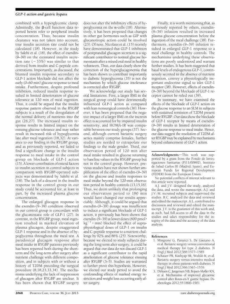
In the RYGBP group, exendin-(9–39)infusion resulted in blunted insulin re-sponse to SLM (Fig. 2 and Table 2),with a mean reduction of the AUC0–120
of insulin and C-peptide of 52.1 610.9% and 24.1 6 8.7%, respectively.The insulin-calculated (P = 0.009)and C-peptide–calculated (P = 0.017)insulinogenic indices were reduced inthe RYGBP group during exendin-(9–39) infusion. In the control group,exendin-(9–39) yielded no significant
effect on insulin (DAUC insulin0–120:212.2 6 35.2%) or C-peptide (DAUCinsulin0–120: 28.5 6 22.6%) postpran-dial response (Fig. 2 and Table 2).
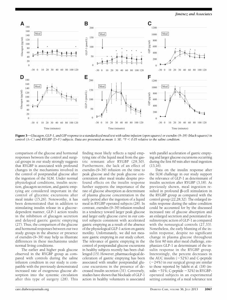
Glucagon, GLP-1, and GIP responseto an SLMAs shown in Table 2, no differences werefound in baseline plasma glucagon levelsbetween the 2 study days in the RYGBPgroup. Unexpectedly, slightly but statisti-cally elevated fasting glucagon levels werefound in the control group before salineinfusion as compared with previous toexendin-(9–39) infusion (P = 0.001). Asshown in Fig. 3, in the saline infusioncondition the SLM challenge resulted inslight decrease in glucagon plasma concen-tration (incremental AUC glucagon0–120:20.31 6 1.17 (pg z dL21 z min z 103).This physiological response was bluntedby exendin-(9–39) infusion (incrementalAUC glucagon0–120: 0.99 6 1.52 pg zdL21 z min z 103; P = 0.062). Incontrast, a paradoxical increase in the glu-cagon response was observed in theRYGBP group during the saline and
exendin-(9–39) conditions [incrementalAUC glucagon0–120: 6.35 6 2.96 pg zdL21 z min z 103 for saline and 7.34 63.60 pg z dL21 z min z 103 for exendin-(9–39)]. Despite glucose tolerance beingsimilar, the AUC glucagon0–120 was largerin the surgical group in both study days(Table 2).
Fasting GLP-1 plasma levels did notdiffer between or within the two studygroups in both experimental conditions(Table 2). As expected, GLP-1 response toSLM was larger in the RYGBP group ascompared with the control group on thesaline day (P , 0.001) (Fig. 3). Exendin-(9–39) resulted in a significant increase inthe GLP-1 response to SLM intake in thecontrol group (P = 0.005) but not in theRYGBP group (P = 0.358). Nonetheless,in that experimental condition the GLP-1response to the meal challenge also waslarger in the RYGBP group (P = 0.001).
The GIP response to meal intake isshown in Table 2 and Fig. 3. In controlsubjects, exendin-(9–39) infusion re-sulted in an earlier GIP response butno change in the AUC0–120 as compared
Table 2dEffect of SLM ingestion on plasma glucose and hormonal secretion in studies with or without exendin-(9–39)
Control group (n = 7) RYGBP group (n = 8)
Saline Exendin-(9–39) P Saline Exendin-(9–39) P
Glucose at time 230 min* (mg/dL) 88.1 6 8.3 93.8 6 7.8 0.153 85.7 6 6.7 85.7 6 8.5 1.000Glucose at time 0 min† (mg/dL) 91.8 6 6.6 100.4 6 10.2 0.133 87.9 6 5.8 92.6 6 8.1 0.008Peak glucose (mg/dL) 152.1 6 24.5 167.3 6 31.2 0.09 223.5 6 37.1x 211.7 6 22.9x 0.297Time to peak glucose after meal (min) 84.3 6 29.3 64.3 6 33.6 0.207 36.2 6 7.4x 31.2 6 3.5‡ 0.104AUC glucose0–120 (mg z dL21 z min z 103) 14.92 6 2.17 16.31 6 1.94 0.020 16.88.6 2.76 18.58 6 2.68 0.013Insulin at time 230 min (mU/L) 5.1 6 1.4 4.7 6 1.1 0.652 5.5 6 1.3 5.9 6 2.1 0.373Insulin at time 0 min (mU/L) 3.6 6 1.2 4.8 6 1.7 0.025 5.1 6 1.9 5.6 6 2.0 0.109DInsulin0–30/Dglucose0–30[(mU/L)/(mg/dL)] 1.26 6 0.56 1.38 6 0.39 0.591 2.19 6 1.18 1.20 6 0.66 0.009
AUCinsulin0–120 (mU z L21 z min z 103) 4.77.6 0.91 4.58 6 0.16 0.795 15.57 6 6.35x 7.19 6 2.88 0.001C-peptide at time 230 min (ng/mL) 1.27 6 0.48 1.24 6 0.24 0.868 1.88 6 0.54‡ 1.95 6 0.78‡ 0.593C-peptide at time 0 min (ng/mL) 1.05 6 0.39 1.19 6 0.29 0.253 1.74 6 0.53‡ 1.74 6 0.52‡ 0.992DC-peptide0–30/Dglucose0–30[(ng/mL)/(mg/dL)] 0.100 6 0.046 0.073 6 0.032 0.227 0.078 6 0.029 0.059 6 0.015 0.017
AUC C-peptide0–120 (ng z mL21 z min) 581.9 6 103.5 564.8 6 189.6 0.744 1,025.0 6 209.4‡ 769.8 6 191.9x ,0.001Glucagon at time 230 min (pg/mL) 51.2 6 10.8 46.1 6 11.4 0.001 59.6 6 14.9 54.22 6 12.22 0.147Glucagon at time 0 min (pg/mL) 45.6 6 10.1 46.9 6 11.3 0.794 59.7 6 10.3‡ 54.7 6 10.1 0.073AUC glucagon0–120 (pg z dL
21 z min z 103) 5.69 6 1.11 6.98 6 1.52 0.062 12.35 6 2.95x 13.34 6 3.59x 0.258GLP-1 at time 230 min (pmol/L) 11.4 6 3.1 11.4 6 4.7 0.985 9.4 6 4.3 9.1 6 4.4 0.823GLP-1 at time 0 min 1 (pmol/L) 12.0 6 4.7 19.9 6 4.3 ,0.001 9.9 6 5.2 20.2 6 4.9 ,0.001AUC GLP-10–120 (pmol z L21 z min z 103) 1.88 6 0.78 4.34 6 1.79 0.005 8.72 6 2.27x 9.61 6 2.69x 0.358GIP at time 230 min (pg/mL) 34.8 6 17.2 49.1 6 27.9 0.280 59.1 6 30.3 58.6 6 21.8‡ 0.959GIP at time 0 min (pg/mL) 35.7 6 14.4 40.9 6 23.3 0.594 56.6 6 27.9 54.9 6 30.3 0.809AUC GIP0–120 (pg z mL21 z min z 103) 38.64 6 14.38 35.96 6 9.30 0.511 50.70 6 22.09 46.56 6 13.60 0.383
P values refer to the comparison within the control or the RYGBP in the saline or exendin-(9–39) condition. *Time230 min corresponds to before the injection ofsaline or exendin-(9–39); †Time 0min corresponds to 30min after saline of exendin-(9–39) infusion initiation; ‡P, 0.05 for the comparison between the control andthe RYGBP group with the saline or exendin-(9–39) condition; xP, 0.01 for the comparison between the control and the RYGBP group with the saline or exendin-(9–39) condition.
care.diabetesjournals.org DIABETES CARE, VOLUME 36, JULY 2013 2065
Jiménez and Associates

with the saline condition. The early(AUC0–60) response of GIP was larger(P = 0.014) in the RYGBP group as com-pared with the control group, but it wasnot changed on infusion of exendin-(9–39).
CONCLUSIONSdOur data showthat in the long-term after RYGBP, block-ing the action of GLP-1 with exendin-(9–39) results in limited deterioration ofthe glucose response to a mixed meal insubjects with remission of T2DM antedat-ing surgery. Thus, our data suggest thatthe dramatic increase in GLP-1 secretionobserved during the long-term afterRYGBP surgery is not the key determinantof the resolution of T2DM after this type ofsurgery.
Previous studies implicating GLP-1 asmechanism for the improvement in glu-cose tolerance after RYGBP have relied onassociation. RYGBP has been associatedwith a large GLP-1 response to mealintake that parallels diabetes remission(7), or that is larger than that observedafter other surgical techniques (9) or di-etary interventions (8) associated withsmaller ameliorations of glucose tolerance.
Furthermore, it has been reported that peroral feeding versus feeding through a gas-trostomy catheter inserted in the gastricremnant in RYGBP-operated patients isassociated with an exaggerated GLP-1 re-sponse along with better glucose tolerance(24). However, because association stud-ies are not well suited to prove causality, toassess the degree to which increased GLP-1release in RYGBP improved glucose toler-ance we performed a mixed-meal toler-ance test in the presence or absence ofexendin-(9–39). If antagonizing GLP-1 re-ceptors had been critical for the mainte-nance of a normal glucose tolerance in ourRYGBP-operated cohort, then meal inges-tion would have resulted in plasma glu-cose levels in the diabetic range or,alternatively, in a much larger deteriora-tion of the glucose response to SLM ascompared with normal-weight healthycontrols. The 2-h plasma glucose afterthe SLM in our cohort was much lowerthan the threshold of 200 mg/dL usedfor the diagnosis of diabetes, and onlytwoout the eight RYGBPpatients presentedwith plasma glucose slightly higher than140 mg/dL, which defines normal glu-cose tolerance. Moreover, deterioration of
glucose excursions after meal ingestion inthe surgical group was comparable withthat in controls. Using hyperglycemicclamp combined with SLM, Salehi et al.(18) evaluated the effects of GLP-1 actionblockade on insulin secretion in a cohortof subjects who had undergone RYGBP. Atvariance with our series, only three out thenine study participants presented withT2DM before surgery. Noteworthy, theglucose infusion rate after meal ingestionhad to be decreased;13% with exendin-(9–39) infusion as compared with salineinfusion to maintain the glucose clampedat the predefined target level. On the otherhand, Deane et al. (13) reported a 10%deterioration of glucose tolerance associ-ated with exendin-(9–39) infusion afterthe ingestion of a solid meal in a series ofeight healthy subjects. Thus, the data re-ported herein concord with those in pre-vious studies in humans using differentexperimental approaches.
We acknowledge that by not control-ling for changes in blood glucose, ourstudy is not optimal to disentangle therelative contributions of glucose andGLP-1 in the control of postprandialglycemia after RYGB (25). Nonetheless,
Figure 2dInsulin and C-peptide response to a standardized meal test with saline infusion (open squares) or exendin-(9–39) (black squares) incontrol (A, B) and RYGBP (C, D) subjects. Data are presented as mean 6 SE. *P , 0.05 relative to the saline condition.
2066 DIABETES CARE, VOLUME 36, JULY 2013 care.diabetesjournals.org
GLP-1 action and gastric bypass
comparison of the glucose and hormonalresponses between the control and surgi-cal groups in our study strongly suggeststhat RYGBP is associated with profoundchanges in the mechanisms involved inthe control of postprandial glucose afterthe ingestion of the SLM. Under normalphysiological conditions, insulin secre-tion, glucagon secretion, and gastric emp-tying are considered important in thecontrol of glycemic excursions aftermeal intake (15,26). Noteworthy, it hasbeen demonstrated that in addition tostimulating insulin release in a glucose-dependent manner, GLP-1 action resultsin the inhibition of glucagon secretionand delayed gastric gastric emptying(27). Thus, the comparison of the glucoseand hormonal responses between our twostudy groups in the absence or presenceof exendin-(9–39) may help us illustratedifferences in these mechanisms undernormal living conditions.
The earlier and higher peak glucoseobserved in the RYGBP group as com-pared with controls during the salineinfusion condition in our study is com-patible with the previously demonstratedincreased rate of exogenous glucose ab-sorption into the systemic circulationafter this type of surgery (28). This
finding most likely reflects a rapid emp-tying rate of the liquid meal from the gas-tric remnant after RYGBP (29,30).Furthermore, the lack of an effect ofexendin-(9–39) infusion on the time topeak glucose and the peak glucose con-centration after meal intake despite pro-found effects on the insulin responsefurther supports the importance of therate of glucose absorption as determinantof plasma glucose concentration in theearly period after the ingestion of a liquidmeal in RYGBP operated subjects (28). Incontrast, exendin-(9–39) infusion resultedin a tendency toward larger peak glucoseand larger early glucose curve in our con-trol subjects consistent with acceleratedgastric emptying as a result of the absenceof the physiological GLP-1 action on gastricmotility. Unfortunately, we did not mea-sure gastric emptying in our study cohort.The relevance of gastric emptying in thecontrol of postprandial glucose excursionsin healthy subjects recently has been chal-lenged (15).However, pharmacological de-celeration of gastric emptying has beenassociated with smaller postprandial glu-cose excursions in the presence of de-creased insulin secretion (31). Conversely,studies have shown that blockade of GLP-1action in healthy volunteers is associated
with parallel acceleration of gastric empty-ing and larger glucose excursions occurringduring the first 60 min after meal ingestion(13,16).
Data on the insulin response afterthe SLM challenge in our study supportthe relevance of GLP-1 as determinant ofinsulin secretion after RYGBP (3,18). Aspreviously shown, meal ingestion re-sulted in profound b-cell stimulation inthe RYGBP group as compared with thecontrol group (22,28,32). The enlarged in-sulin response during the saline conditioncould be accounted for by the discussedincreased rate of glucose absorption andan enlarged secretion and potentiated in-sulinotropic action of GLP-1 as comparedwith the nonsurgical controls (27,33).Nonetheless, the early blunting of the in-sulin response, despite no significantchange in plasma glucose throughoutthe first 90 min after meal challenge, em-phasizes GLP-1 as determinant of the in-sulin response in the RYGBP group.Interestingly, the percent decreases inthe AUC insulin (252%) and C-peptide(224%) in our surgical group are similarto those reported by Salehi et al. (18) (in-sulin251%; C-peptide232%) in RYGBP-operated subjects in an experimentalsetting consisting of a meal tolerance test
Figure 3dGlucagon, GLP-1, andGIP response to a standardizedmeal test with saline infusion (open squares) or exendin-(9–39) (black squares) incontrol (A–C) and RYGBP (D–F) subjects. Data are presented as mean 6 SE. *P , 0.05 relative to the saline condition.
care.diabetesjournals.org DIABETES CARE, VOLUME 36, JULY 2013 2067
Jiménez and Associates
combined with a hyperglycemic clamp.Admittedly, the b-cell function data re-ported herein refer to peripheral insulinconcentration. Thus, because insulinclearance was not taken into account,true insulin secretion rate could not becalculated (18). However, in the studyby Salehi et al. (18), the effect of exendin-(9–39) on the estimated insulin secre-tion rate (233%) was similar to thatderived from insulin and C-peptide con-centrations. Importantly, as discussed, theblunted insulin response secondary toGLP-1 action blockade did not affect theearly (0–60min) glucose response to mealintake. Furthermore, despite profoundinhibition, reduced insulin response re-sulted in limited deterioration of glucosetolerance at 120 min of meal ingestion.Thus, it could be argued that the insulinresponse pattern observed in the RYGBPsubjects is a result of the deregulation ofthe normal delivery of nutrients into thegut (26,27). The increased insulin re-sponse results in limited impact on theensuing glucose tolerance and may ratherresult in increased risk of hypoglycemialate after meal ingestion (18,32). At vari-ance to our finding in the RYGBP group,and as previously reported, we failed tofind a significant change in the insulinresponse to meal intake in the controlgroup on blockade of GLP-1 action(15). A lower contribution of enteral factorsto insulin secretion in control subjects incomparison with RYGBP-operated sub-jects was demonstrated by Salehi et al.(18). The lack of a descent in the insulinresponse in the control group in ourstudy could be accounted for, at least inpart, by the increased plasma glucoseconcentration.
The enlarged glucagon response inthe exendin-(9–39) condition observedin our control group is concordant withthe glucanostatic role of GLP-1 (27). Incontrast, in the RYGBP group, meal inges-tion resulted in marked elevation ofplasma glucagon, despite exaggeratedGLP-1 response and in the absence of hy-poglycemia throughout the meal test. Aparadoxical glucagon response aftermeal intake in RYGBP patients previouslyhas been reported both during the short-term and the long-term after surgery afternutrient challenge with different compo-sition, and in subjects with or without ahistory of T2DM preceding the surgicalprocedure (8,18,23,33,34). The mecha-nisms underlying the lack of suppressionof glucagon after RYGBP are unclear. Ithas been shown that RYGBP surgery
does not alter the inhibitory effects of hy-perglycemia on the a-cells (18). Alterna-tively, it has been proposed that changesin other gut hormones such as GIP withglucanotropic action could be involved(23). Of note, Nicolaus et al. (15) recentlyhave demonstrated that GLP-1 inhibitionof postprandial glucagon secretion is a sig-nificant contributor to normal glucose ho-meostasis after amixed oral meal in healthyvolunteers. Thus, our data clearly show thecorrection of the hyperglucagonemia thathas been shown to contribute importantlyto diabetic hyperglycemia (35) is not themechanism by which glucose intoleranceis corrected after RYGBP.
We acknowledge our study has sev-eral limitations. First, a larger BMI in thesurgical group could have detrimentallyinfluenced GLP-1 action as comparedwith lean nonsurgical controls (36). How-ever, it has been proposed that the nega-tive impact of a larger BMI on the incretineffect is accounted for by impaired insulinsensitivity, and HOMA-IR was compa-rable between our study groups (37). Sec-ond, although current bariatric surgeryseries mainly comprise females, furtherstudies are needed to extrapolate ourfindings to the male gender. Third, ourobservation period of 120 min aftermeal intake resulted in glucose returningto baseline values in the RYGBP group butnot in the control group. However, pre-vious studies have not shown further am-plification of the effect of exendin-(9–39)on the glucose and insulin responses tomeal intake beyond the 120-min observa-tion period in healthy controls (13,15,16).Thus, we deem unlikely that prolongingour observation period to 180 minwould have changed our results appre-ciably. Although, it could be argued thatexendin-(9–39) dosage was insufficientto induce a significant blockade of GLP-1action, it previously has been shown thatexendin-(9–39) at lower doses (600 pmol zkg21 z min) blocked the effect of supra-physiological doses of GLP-1 on insulinand C-peptide response to a nutrient chal-lenge by 92% and 86% (25). Noteworthy,because we elected to study subjects dur-ing the long-term after surgery, it could beargued that our data do not discard GLP-1as a significant contributor to the rapidamelioration of glucose tolerance ensuingafter RYGBP (3–5). Studies are warrantedto further prove this hypothesis. However,we elected our study period to avoid theconfounding effects of marked energy re-striction and weight loss occurring early af-ter surgery.
Finally, it is worthmentioning that, aspreviously reported by others, exendin-(9–39) infusion resulted in increasedplasma glucose concentration before theingestion of the meal challenge (38). Fur-thermore, exendin-(9–39) infusion re-sulted in enlarged GLP-1 response to ameal challenge in healthy controls. Themechanisms underlying these observa-tions are poorly understood and warrantfurther studies. It has been suggested thatbasal levels of endogenous GLP-1, contin-uously secreted in the absence of nutrientingestion, convey a physiologically im-portant endocrine signal to islet GLP-1receptor (38). However, effects of exendin-(9–39) beyond the blockade of GLP-1 re-ceptor cannot be ruled out (39).
In summary, we have examined theeffects of the blockade of GLP-1 action onthe glucose response to an SLM in subjectswith sustained remission of T2DM presentbefore RYGBP.Our data show the blockadeof GLP-1 receptor by means of exendin-(9–39) results in limited deterioration ofthe glucose response to meal intake. Hence,our data suggest the resolution of T2DM af-terRYGBPmaybe explainedbymechanismsbeyond enhancement of GLP-1 action.
AcknowledgmentsdThis work was sup-ported by a grant from the Fondo de Inves-tigaciones Sanitarias (PI11/00892), Institutode Salud Carlos III (Madrid, Spain), and Eu-ropean Funds for Regional Development(FEDER) from the European Union.No potential conflicts of interest relevant to
this article were reported.A.J. and J.V. designed the study, analyzed
the data, and wrote the manuscript. A.J. andJ.V.-M. recruited subjects and supervised thestudies. R.C. analyzed the data and reviewedand edited the manuscript. A.L. contributed todiscussion and reviewed and edited the man-uscript. J.V. is the guarantor of this work and,as such, had full access to all the data in thestudies and takes responsibility for the in-tegrity of the data and the accuracy of the dataanalysis.
References1. Mingrone G, Panunzi S, De Gaetano A,
et al. Bariatric surgery versus conventionalmedical therapy for type 2 diabetes. NEngl J Med 2012;366:1577–1585
2. Schauer PR, Kashyap SR, Wolski K, et al.Bariatric surgery versus intensive medicaltherapy in obese patients with diabetes. NEngl J Med 2012;366:1567–1576
3. DirksenC, JørgensenNB, Bojsen-Møller KN,et al. Mechanisms of improved glycaemiccontrol after Roux-en-Y gastric bypass. Di-abetologia 2012;55:1890–1901
2068 DIABETES CARE, VOLUME 36, JULY 2013 care.diabetesjournals.org
GLP-1 action and gastric bypass

4. Rubino F, R’bibo SL, del Genio F,Mazumdar M, McGraw TE. Metabolicsurgery: the role of the gastrointestinaltract in diabetes mellitus. Nat Rev Endo-crinol 2010;6:102–109
5. Scott WR, Batterham RL. Roux-en-Y gas-tric bypass and laparoscopic sleeve gas-trectomy: understanding weight loss andimprovements in type 2 diabetes afterbariatric surgery. Am J Physiol Regul In-tegr Comp Physiol 2011;301:R15–R27
6. Pories WJ, Swanson MS, MacDonald KG,et al. Who would have thought it? Anoperation proves to be the most effectivetherapy for adult-onset diabetes mellitus.Ann Surg 1995;222:339–350; discussion350–352
7. Laferrère B,Heshka S,WangK, et al. Incretinlevels and effect are markedly enhanced 1month after Roux-en-Y gastric bypass sur-gery in obese patients with type 2 diabetes.Diabetes Care 2007;30:1709–1716
8. Laferrère B, Teixeira J, McGinty J, et al.Effect of weight loss by gastric bypasssurgery versus hypocaloric diet on glucoseand incretin levels in patients with type 2diabetes. J Clin Endocrinol Metab 2008;93:2479–2485
9. Pournaras DJ, Osborne A, Hawkins SC,et al. Remission of type 2 diabetes aftergastric bypass and banding: mechanismsand 2 year outcomes. Ann Surg 2010;252:966–971
10. Torquati A, Lutfi R, Abumrad N, RichardsWO. Is Roux-en-Y gastric bypass surgerythe most effective treatment for type 2diabetes mellitus in morbidly obese pa-tients? J Gastrointest Surg 2005;9:1112–1116; discussion 1117–1118
11. Cohen RV, Pinheiro JC, Schiavon CA,Salles JE, Wajchenberg BL, CummingsDE. Effects of gastric bypass surgery inpatients with type 2 diabetes and only mildobesity. DiabetesCare 2012;35:1420–1428
12. Dar MS, Chapman WH 3rd, Pender JR,et al. GLP-1 response to a mixed meal:what happens 10 years after Roux-en-Ygastric bypass (RYGB)? Obes Surg 2012;22:1077–1083
13. Deane AM, Nguyen NQ, Stevens JE, et al.Endogenous glucagon-like peptide-1 slowsgastric emptying in healthy subjects, atten-uating postprandial glycemia. J Clin Endo-crinol Metab 2010;95:215–221
14. Edwards CM, Todd JF, Mahmoudi M,et al. Glucagon-like peptide 1 has a physi-ological role in the control of postprandialglucose in humans: studies with the an-tagonist exendin 9-39. Diabetes 1999;48:86–93
15. Nicolaus M, Brödl J, Linke R, Woerle HJ,Göke B, Schirra J. Endogenous GLP-1regulates postprandial glycemia in hu-mans: relative contributions of insulin,
glucagon, and gastric emptying. J ClinEndocrinol Metab 2011;96:229–236
16. Schirra J, Nicolaus M, Roggel R, et al.Endogenous glucagon-like peptide 1 controlsendocrine pancreatic secretion and antro-pyloro-duodenal motility in humans. Gut2006;55:243–251
17. Witte AB, Grybäck P, Jacobsson H, et al.Involvement of endogenous glucagon-likepeptide-1 in regulation of gastric motilityand pancreatic endocrine secretion. Scand JGastroenterol 2011;46:428–435
18. Salehi M, Prigeon RL, D’Alessio DA. Gas-tric bypass surgery enhances glucagon-like peptide 1-stimulated postprandialinsulin secretion in humans. Diabetes2011;60:2308–2314
19. Kindel TL, Yoder SM, Seeley RJ, D’AlessioDA, Tso P. Duodenal-jejunal exclusionimproves glucose tolerance in the diabetic,Goto-Kakizaki rat by a GLP-1 receptor-mediated mechanism. J Gastrointest Surg2009;13:1762–1772
20. Mor�ınigo R, Vidal J, Lacy AM, Delgado S,Casamitjana R, Gomis R. Circulatingpeptide YY, weight loss, and glucose ho-meostasis after gastric bypass surgery inmorbidly obese subjects. Ann Surg 2008;247:270–275
21. Buse JB, Caprio S, Cefalu WT, et al. Howdo we define cure of diabetes? DiabetesCare 2009;32:2133–2135
22. Vidal J, Nicolau J, Romero F, et al. Long-term effects of Roux-en-Y gastric bypasssurgery on plasma glucagon-like peptide-1 and islet function in morbidly obesesubjects. J Clin Endocrinol Metab 2009;94:884–891
23. Romero F, Nicolau J, Flores L, et al.Comparable early changes in gastrointes-tinal hormones after sleeve gastrectomyand Roux-En-Y gastric bypass surgery formorbidly obese type 2 diabetic subjects.Surg Endosc 2012;26:2231–2239
24. Dirksen C, Hansen DL, Madsbad S, et al.Postprandial diabetic glucose tolerance isnormalized by gastric bypass feeding asopposed to gastric feeding and is associatedwith exaggerated GLP-1 secretion: a casereport. Diabetes Care 2010;33:375–377
25. Salehi M, Vahl TP, D’Alessio DA. Regula-tion of islet hormone release and gastricemptying by endogenous glucagon-likepeptide 1 after glucose ingestion. J ClinEndocrinol Metab 2008;93:4909–4916
26. Horowitz M, Edelbroek MA, Wishart JM,Straathof JW. Relationship between oralglucose tolerance and gastric emptying innormal healthy subjects. Diabetologia1993;36:857–862
27. Aaboe K, Krarup T, Madsbad S, Holst JJ.GLP-1: physiological effects and potentialtherapeutic applications. Diabetes ObesMetab 2008;10:994–1003
28. Rodieux F, Giusti V, D’Alessio DA, SuterM, Tappy L. Effects of gastric bypass andgastric banding on glucose kinetics andgut hormone release. Obesity (Silver Spring)2008;16:298–305
29. Horowitz M, Collins PJ, Harding PE,Shearman DJ. Gastric emptying after gas-tric bypass. Int J Obes 1986;10:117–121
30. Mor�ınigo R, Moizé V, Musri M, et al.Glucagon-likepeptide-1, peptideYY,hunger,and satiety after gastric bypass surgery inmorbidly obese subjects. J Clin EndocrinolMetab 2006;91:1735–1740
31. Woerle HJ, Albrecht M, Linke R, et al.Importance of changes in gastric empty-ing for postprandial plasma glucose fluxesin healthy humans. Am J Physiol Endo-crinol Metab 2008;294:E103–E109
32. Goldfine AB, Mun EC, Devine E, et al.Patients with neuroglycopenia after gas-tric bypass surgery have exaggerated in-cretin and insulin secretory responses to amixed meal. J Clin Endocrinol Metab2007;92:4678–4685
33. Vilsbøll T, Toft-Nielsen MB, Krarup T,Madsbad S, Dinesen B, Holst JJ. Evalua-tion of beta-cell secretory capacity usingglucagon-like peptide 1. Diabetes Care2000;23:807–812
34. Falkén Y, Hellström PM,Holst JJ, NäslundE. Changes in glucose homeostasis afterRoux-en-Y gastric bypass surgery for obe-sity at day three, two months, and oneyear after surgery: role of gut peptides.J Clin Endocrinol Metab 2011;96:2227–2235
35. Dunning BE, Gerich JE. The role of alpha-cell dysregulation in fasting and post-prandial hyperglycemia in type 2 diabetesand therapeutic implications. Endocr Rev2007;28:253–283
36. Muscelli E, Mari A, Casolaro A, et al.Separate impact of obesity and glucosetolerance on the incretin effect in normalsubjects and type 2 diabetic patients. Di-abetes 2008;57:1340–1348
37. Knop FK, Aaboe K, Vilsbøll T, et al. Im-paired incretin effect and fasting hy-perglucagonaemia characterizing type 2diabetic subjects are early signs of dys-metabolism in obesity. Diabetes ObesMetab 2012;14:500–510
38. Woerle HJ, Carneiro L, Derani A, Göke B,Schirra J. The role of endogenous in-cretin secretion as amplifier of glucose-stimulated insulin secretion in healthysubjects and patients with type 2 diabetes.Diabetes 2012;61:2349–2358
39. Ban K, Kim KH, Cho CK, et al. Glucagon-like peptide (GLP)-1(9-36)amide-mediatedcytoprotection is blocked by exendin(9-39)yet does not require the known GLP-1receptor. Endocrinology 2010;151:1520–1531
care.diabetesjournals.org DIABETES CARE, VOLUME 36, JULY 2013 2069
Jiménez and Associates