Two New Phenylhydrazone Derivatives from the Pearl River ...
12
Citation: Liu, W.; Ma, L.; Zhang, L.; Chen, Y.; Zhang, Q.; Zhang, H.; Zhang, W.; Zhang, C.; Zhang, W. Two New Phenylhydrazone Derivatives from the Pearl River Estuary Sediment-Derived Streptomyces sp. SCSIO 40020. Mar. Drugs 2022, 20, 449. https://doi.org/10.3390/ md20070449 Academic Editor: Bill J. Baker Received: 13 June 2022 Accepted: 7 July 2022 Published: 9 July 2022 Publisher’s Note: MDPI stays neutral with regard to jurisdictional claims in published maps and institutional affil- iations. Copyright: © 2022 by the authors. Licensee MDPI, Basel, Switzerland. This article is an open access article distributed under the terms and conditions of the Creative Commons Attribution (CC BY) license (https:// creativecommons.org/licenses/by/ 4.0/). marine drugs Article Two New Phenylhydrazone Derivatives from the Pearl River Estuary Sediment-Derived Streptomyces sp. SCSIO 40020 Wei Liu 1,2 , Liang Ma 1,3 , Liping Zhang 1,3 , Yuchan Chen 2 , Qingbo Zhang 1,3 , Haibo Zhang 1,3 , Weimin Zhang 2 , Changsheng Zhang 1,3,4, * and Wenjun Zhang 1,3,4, * 1 Key Laboratory of Tropical Marine Bio-Resources and Ecology, Guangdong Key Laboratory of Marine Materia Medica, South China Sea Institute of Oceanology, Innovation Academy for South China Sea Ecology and Environmental Engineering, Chinese Academy of Sciences, 164 West Xingang Road, Guangzhou 510301, China; [email protected] (W.L.); [email protected] (L.M.); [email protected] (L.Z.); [email protected] (Q.Z.); [email protected] (H.Z.) 2 State Key Laboratory of Applied Microbiology Southern China, Institute of Microbiology, Guangdong Academy of Sciences, 100 Central Xianlie Road, Guangzhou 510070, China; [email protected] (Y.C.); [email protected] (W.Z.) 3 Southern Marine Science and Engineering Guangdong Laboratory (Guangzhou), No.1119, Haibin Road, Guangzhou 511458, China 4 University of Chinese Academy of Sciences, Beijing 100049, China * Correspondence: [email protected] (C.Z.); [email protected] (W.Z.); Tel.: +86-20-8902-3038 (C.Z.) Abstract: Two new phenylhydrazone derivatives and one new alkaloid, penzonemycins A–B (1–2) and demethylmycemycin A (3), together with three known compounds including an alkaloid (4) and two sesquiterpenoids (5–6), were isolated from the Streptomyces sp. SCSIO 40020 obtained from the Pearl River Estuary sediment. Their structures and absolute configurations were assigned by 1D/2D NMR, mass spectroscopy and X-ray crystallography. Compound 1 was evaluated in four human cancer cell lines by the SRB method and displayed weak cytotoxicity in three cancer cell lines, with IC 50 values that ranged from 30.44 to 61.92 μM, which were comparable to those of the positive control cisplatin. Bioinformatic analysis of the putative biosynthetic gene cluster indicated a Japp–Klingemann coupling reaction involved in the hydrazone formation of 1 and 2. Keywords: penzonemycins; phenylhydrazones; Streptomyces sp.; Japp–Klingemann coupling reaction; marine natural products 1. Introduction Hydrazones are a class of compounds with the characteristic structure R 1 R 2 C=NNR 3 R 4 . Hydrazones display various bioactivities, including antimicrobial, antitubercular, anti-HIV, anti-inflammatory, anticonvulsant, analgesic and antitumor properties [1]. The unique chemical properties and bioactivities of hydrazones have inspired organic chemists to develop numerous methods for the synthesis of their derivatives [2,3]. In contrast to the large number of synthetic hydrazones, only a handful of naturally occurring hydrazones are found [4–9] (Figure S1). To date, 38 natural hydrazones have been reported, including 21 from fungi [5,9], 13 from actinomycetes [4,6–8], and 4 from other resources [9] (Figure S1). Marine-derived actinomycetes have been a prolific resource for novel natural prod- uct discovery [10,11]. Previously, we reported three novel hydrazone derivatives, pyra- zolofluostatins A–C, which were isolated from South China Sea-derived Micromonospora rosaria SCSIO N160 [7]. In this study, we identified two novel phenylhydrazone deriva- tives, penzonemycins A–B (1–2), together with one new compound, dibenzoxazepinone demethylmycemycin A (3), and three known compounds (4–6) (Figure 1) from marine- derived Streptomyces sp. SCSIO 40020 [12]. Herein, we report the isolation and structural elucidation of penzonemycins A–B (1–2) and demethylmycemycin A (3), as well as a biological evaluation and a possible biosynthetic pathway of penzonemycins A–B (1–2). Mar. Drugs 2022, 20, 449. https://doi.org/10.3390/md20070449 https://www.mdpi.com/journal/marinedrugs
-
Upload
khangminh22 -
Category
Documents
-
view
3 -
download
0
Transcript of Two New Phenylhydrazone Derivatives from the Pearl River ...
Citation: Liu, W.; Ma, L.; Zhang, L.;
Chen, Y.; Zhang, Q.; Zhang, H.;
Zhang, W.; Zhang, C.; Zhang, W. Two
New Phenylhydrazone Derivatives
from the Pearl River Estuary
Sediment-Derived Streptomyces sp.
SCSIO 40020. Mar. Drugs 2022, 20,
449. https://doi.org/10.3390/
md20070449
Academic Editor: Bill J. Baker
Received: 13 June 2022
Accepted: 7 July 2022
Published: 9 July 2022
Publisher’s Note: MDPI stays neutral
with regard to jurisdictional claims in
published maps and institutional affil-
iations.
Copyright: © 2022 by the authors.
Licensee MDPI, Basel, Switzerland.
This article is an open access article
distributed under the terms and
conditions of the Creative Commons
Attribution (CC BY) license (https://
creativecommons.org/licenses/by/
4.0/).
marine drugs
Article
Two New Phenylhydrazone Derivatives from the Pearl RiverEstuary Sediment-Derived Streptomyces sp. SCSIO 40020Wei Liu 1,2 , Liang Ma 1,3 , Liping Zhang 1,3, Yuchan Chen 2, Qingbo Zhang 1,3, Haibo Zhang 1,3 ,Weimin Zhang 2 , Changsheng Zhang 1,3,4,* and Wenjun Zhang 1,3,4,*
1 Key Laboratory of Tropical Marine Bio-Resources and Ecology, Guangdong Key Laboratory of MarineMateria Medica, South China Sea Institute of Oceanology, Innovation Academy for South China Sea Ecologyand Environmental Engineering, Chinese Academy of Sciences, 164 West Xingang Road,Guangzhou 510301, China; [email protected] (W.L.); [email protected] (L.M.);[email protected] (L.Z.); [email protected] (Q.Z.); [email protected] (H.Z.)
2 State Key Laboratory of Applied Microbiology Southern China, Institute of Microbiology, GuangdongAcademy of Sciences, 100 Central Xianlie Road, Guangzhou 510070, China; [email protected] (Y.C.);[email protected] (W.Z.)
3 Southern Marine Science and Engineering Guangdong Laboratory (Guangzhou), No.1119, Haibin Road,Guangzhou 511458, China
4 University of Chinese Academy of Sciences, Beijing 100049, China* Correspondence: [email protected] (C.Z.); [email protected] (W.Z.); Tel.: +86-20-8902-3038 (C.Z.)
Abstract: Two new phenylhydrazone derivatives and one new alkaloid, penzonemycins A–B (1–2)and demethylmycemycin A (3), together with three known compounds including an alkaloid (4)and two sesquiterpenoids (5–6), were isolated from the Streptomyces sp. SCSIO 40020 obtained fromthe Pearl River Estuary sediment. Their structures and absolute configurations were assigned by1D/2D NMR, mass spectroscopy and X-ray crystallography. Compound 1 was evaluated in fourhuman cancer cell lines by the SRB method and displayed weak cytotoxicity in three cancer celllines, with IC50 values that ranged from 30.44 to 61.92 µM, which were comparable to those of thepositive control cisplatin. Bioinformatic analysis of the putative biosynthetic gene cluster indicated aJapp–Klingemann coupling reaction involved in the hydrazone formation of 1 and 2.
Keywords: penzonemycins; phenylhydrazones; Streptomyces sp.; Japp–Klingemann couplingreaction; marine natural products
1. Introduction
Hydrazones are a class of compounds with the characteristic structure R1R2C=NNR3R4.Hydrazones display various bioactivities, including antimicrobial, antitubercular, anti-HIV,anti-inflammatory, anticonvulsant, analgesic and antitumor properties [1]. The uniquechemical properties and bioactivities of hydrazones have inspired organic chemists todevelop numerous methods for the synthesis of their derivatives [2,3]. In contrast to thelarge number of synthetic hydrazones, only a handful of naturally occurring hydrazonesare found [4–9] (Figure S1). To date, 38 natural hydrazones have been reported, including 21from fungi [5,9], 13 from actinomycetes [4,6–8], and 4 from other resources [9] (Figure S1).
Marine-derived actinomycetes have been a prolific resource for novel natural prod-uct discovery [10,11]. Previously, we reported three novel hydrazone derivatives, pyra-zolofluostatins A–C, which were isolated from South China Sea-derived Micromonosporarosaria SCSIO N160 [7]. In this study, we identified two novel phenylhydrazone deriva-tives, penzonemycins A–B (1–2), together with one new compound, dibenzoxazepinonedemethylmycemycin A (3), and three known compounds (4–6) (Figure 1) from marine-derived Streptomyces sp. SCSIO 40020 [12]. Herein, we report the isolation and structuralelucidation of penzonemycins A–B (1–2) and demethylmycemycin A (3), as well as abiological evaluation and a possible biosynthetic pathway of penzonemycins A–B (1–2).
Mar. Drugs 2022, 20, 449. https://doi.org/10.3390/md20070449 https://www.mdpi.com/journal/marinedrugs
Mar. Drugs 2022, 20, 449 2 of 12
Mar. Drugs 2022, 20, x FOR PEER REVIEW 2 of 13
derived Streptomyces sp. SCSIO 40020 [12]. Herein, we report the isolation and structural
elucidation of penzonemycins A–B (1–2) and demethylmycemycin A (3), as well as a bio-
logical evaluation and a possible biosynthetic pathway of penzonemycins A–B (1–2).
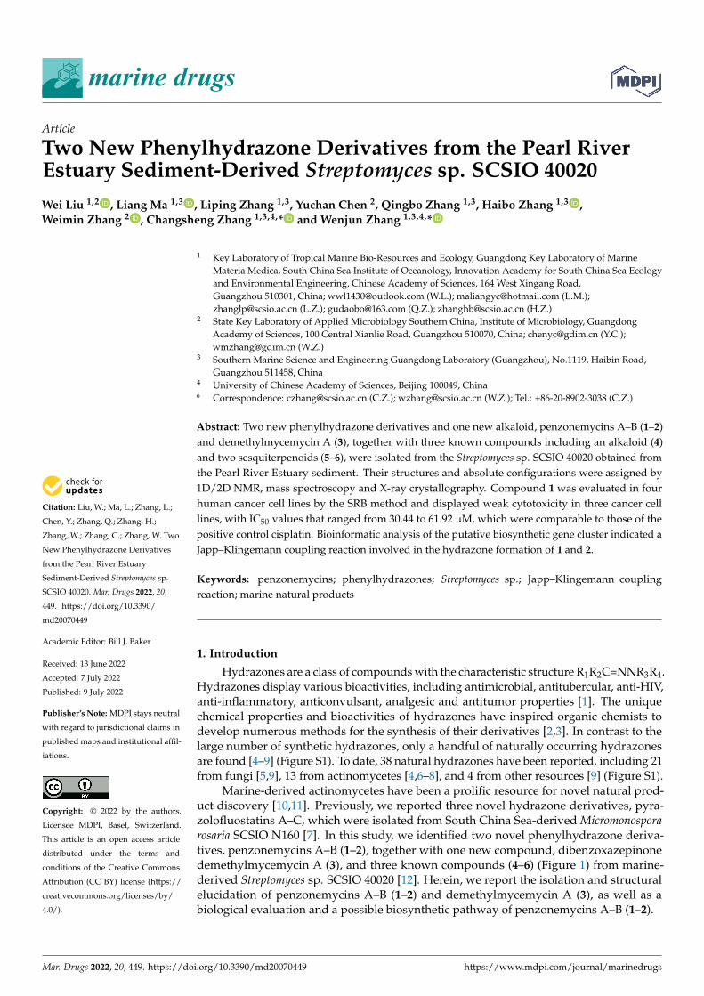
Figure 1. Chemical structures of compounds 1–6.
2. Results and Discussion
2.1. Structural Determination
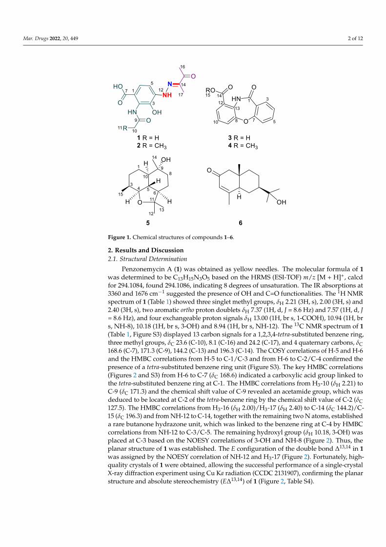
Penzonemycin A (1) was obtained as yellow needles. The molecular formula of 1 was
determined to be C13H15N3O5 based on the HRMS (ESI-TOF) m/z [M + H]+, calcd for 294.1084,
found 294.1086, indicating 8 degrees of unsaturation. The IR absorptions at 3360 and 1676
cm−1 suggested the presence of OH and C=O functionalities. The 1H NMR spectrum of 1
(Table 1) showed three singlet methyl groups, δH 2.21 (3H, s), 2.00 (3H, s) and 2.40 (3H, s),
two aromatic ortho proton doublets δH 7.37 (1H, d, J = 8.6 Hz) and 7.57 (1H, d, J = 8.6 Hz),
and four exchangeable proton signals δH 13.00 (1H, br s, 1-COOH), 10.94 (1H, br s, NH-8),
10.18 (1H, br s, 3-OH) and 8.94 (1H, br s, NH-12). The 13C NMR spectrum of 1 (Table 1, Figure
S3) displayed 13 carbon signals for a 1,2,3,4-tetra-substituted benzene ring, three methyl
groups, δC 23.6 (C-10), 8.1 (C-16) and 24.2 (C-17), and 4 quaternary carbons, δC 168.6 (C-7),
171.3 (C-9), 144.2 (C-13) and 196.3 (C-14). The COSY correlations of H-5 and H-6 and the
HMBC correlations from H-5 to C-1/C-3 and from H-6 to C-2/C-4 confirmed the presence of
a tetra-substituted benzene ring unit (Figure S3). The key HMBC correlations (Figures 2 and
S3) from H-6 to C-7 (δC 168.6) indicated a carboxylic acid group linked to the tetra-substi-
tuted benzene ring at C-1. The HMBC correlations from H3-10 (δH 2.21) to C-9 (δC 171.3) and
the chemical shift value of C-9 revealed an acetamide group, which was deduced to be lo-
cated at C-2 of the tetra-benzene ring by the chemical shift value of C-2 (δC 127.5). The HMBC
correlations from H3-16 (δH 2.00)/H3-17 (δH 2.40) to C-14 (δC 144.2)/C-15 (δC 196.3) and from
NH-12 to C-14, together with the remaining two N atoms, established a rare butanone hy-
drazone unit, which was linked to the benzene ring at C-4 by HMBC correlations from NH-
12 to C-3/C-5. The remaining hydroxyl group (δH 10.18, 3-OH) was placed at C-3 based on
the NOESY correlations of 3-OH and NH-8 (Figure 2). Thus, the planar structure of 1 was
established. The E configuration of the double bond Δ13,14 in 1 was assigned by the NOESY
correlation of NH-12 and H3-17 (Figure 2). Fortunately, high-quality crystals of 1 were ob-
tained, allowing the successful performance of a single-crystal X-ray diffraction experiment
using Cu Ka radiation (CCDC 2131907), confirming the planar structure and absolute stere-
ochemistry (EΔ13,14) of 1 (Figure 2, Table S4).
Figure 1. Chemical structures of compounds 1–6.
2. Results and Discussion2.1. Structural Determination
Penzonemycin A (1) was obtained as yellow needles. The molecular formula of 1was determined to be C13H15N3O5 based on the HRMS (ESI-TOF) m/z [M + H]+, calcdfor 294.1084, found 294.1086, indicating 8 degrees of unsaturation. The IR absorptions at3360 and 1676 cm−1 suggested the presence of OH and C=O functionalities. The 1H NMRspectrum of 1 (Table 1) showed three singlet methyl groups, δH 2.21 (3H, s), 2.00 (3H, s) and2.40 (3H, s), two aromatic ortho proton doublets δH 7.37 (1H, d, J = 8.6 Hz) and 7.57 (1H, d, J= 8.6 Hz), and four exchangeable proton signals δH 13.00 (1H, br s, 1-COOH), 10.94 (1H, brs, NH-8), 10.18 (1H, br s, 3-OH) and 8.94 (1H, br s, NH-12). The 13C NMR spectrum of 1(Table 1, Figure S3) displayed 13 carbon signals for a 1,2,3,4-tetra-substituted benzene ring,three methyl groups, δC 23.6 (C-10), 8.1 (C-16) and 24.2 (C-17), and 4 quaternary carbons, δC168.6 (C-7), 171.3 (C-9), 144.2 (C-13) and 196.3 (C-14). The COSY correlations of H-5 and H-6and the HMBC correlations from H-5 to C-1/C-3 and from H-6 to C-2/C-4 confirmed thepresence of a tetra-substituted benzene ring unit (Figure S3). The key HMBC correlations(Figures 2 and S3) from H-6 to C-7 (δC 168.6) indicated a carboxylic acid group linked tothe tetra-substituted benzene ring at C-1. The HMBC correlations from H3-10 (δH 2.21) toC-9 (δC 171.3) and the chemical shift value of C-9 revealed an acetamide group, which wasdeduced to be located at C-2 of the tetra-benzene ring by the chemical shift value of C-2 (δC127.5). The HMBC correlations from H3-16 (δH 2.00)/H3-17 (δH 2.40) to C-14 (δC 144.2)/C-15 (δC 196.3) and from NH-12 to C-14, together with the remaining two N atoms, establisheda rare butanone hydrazone unit, which was linked to the benzene ring at C-4 by HMBCcorrelations from NH-12 to C-3/C-5. The remaining hydroxyl group (δH 10.18, 3-OH) wasplaced at C-3 based on the NOESY correlations of 3-OH and NH-8 (Figure 2). Thus, theplanar structure of 1 was established. The E configuration of the double bond ∆13,14 in 1was assigned by the NOESY correlation of NH-12 and H3-17 (Figure 2). Fortunately, high-quality crystals of 1 were obtained, allowing the successful performance of a single-crystalX-ray diffraction experiment using Cu Ka radiation (CCDC 2131907), confirming the planarstructure and absolute stereochemistry (E∆13,14) of 1 (Figure 2, Table S4).
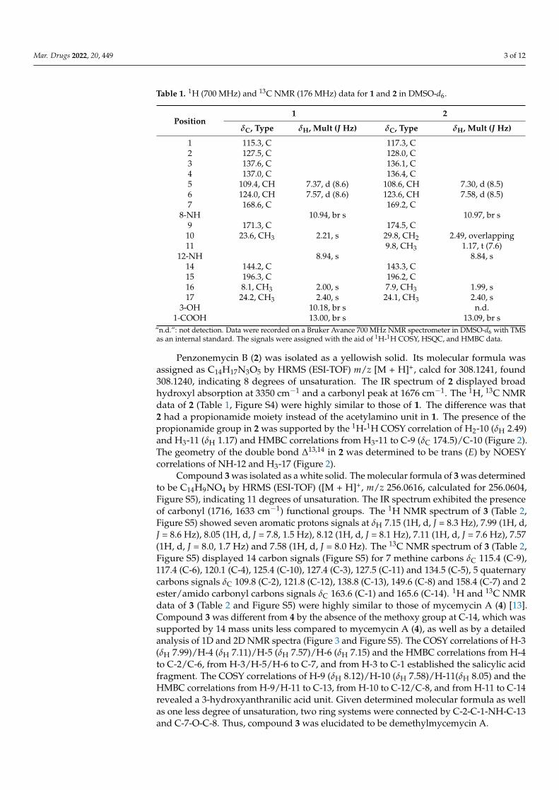
Mar. Drugs 2022, 20, 449 3 of 12
Table 1. 1H (700 MHz) and 13C NMR (176 MHz) data for 1 and 2 in DMSO-d6.
Position1 2
δC, Type δH, Mult (J Hz) δC, Type δH, Mult (J Hz)
1 115.3, C 117.3, C2 127.5, C 128.0, C3 137.6, C 136.1, C4 137.0, C 136.4, C5 109.4, CH 7.37, d (8.6) 108.6, CH 7.30, d (8.5)6 124.0, CH 7.57, d (8.6) 123.6, CH 7.58, d (8.5)7 168.6, C 169.2, C
8-NH 10.94, br s 10.97, br s9 171.3, C 174.5, C
10 23.6, CH3 2.21, s 29.8, CH2 2.49, overlapping11 9.8, CH3 1.17, t (7.6)
12-NH 8.94, s 8.84, s14 144.2, C 143.3, C15 196.3, C 196.2, C16 8.1, CH3 2.00, s 7.9, CH3 1.99, s17 24.2, CH3 2.40, s 24.1, CH3 2.40, s
3-OH 10.18, br s n.d.1-COOH 13.00, br s 13.09, br s
“n.d.”: not detection. Data were recorded on a Bruker Avance 700 MHz NMR spectrometer in DMSO-d6 with TMSas an internal standard. The signals were assigned with the aid of 1H-1H COSY, HSQC, and HMBC data.
Penzonemycin B (2) was isolated as a yellowish solid. Its molecular formula wasassigned as C14H17N3O5 by HRMS (ESI-TOF) m/z [M + H]+, calcd for 308.1241, found308.1240, indicating 8 degrees of unsaturation. The IR spectrum of 2 displayed broadhydroxyl absorption at 3350 cm−1 and a carbonyl peak at 1676 cm−1. The 1H, 13C NMRdata of 2 (Table 1, Figure S4) were highly similar to those of 1. The difference was that2 had a propionamide moiety instead of the acetylamino unit in 1. The presence of thepropionamide group in 2 was supported by the 1H-1H COSY correlation of H2-10 (δH 2.49)and H3-11 (δH 1.17) and HMBC correlations from H3-11 to C-9 (δC 174.5)/C-10 (Figure 2).The geometry of the double bond ∆13,14 in 2 was determined to be trans (E) by NOESYcorrelations of NH-12 and H3-17 (Figure 2).
Compound 3 was isolated as a white solid. The molecular formula of 3 was determinedto be C14H9NO4 by HRMS (ESI-TOF) ([M + H]+, m/z 256.0616, calculated for 256.0604,Figure S5), indicating 11 degrees of unsaturation. The IR spectrum exhibited the presenceof carbonyl (1716, 1633 cm−1) functional groups. The 1H NMR spectrum of 3 (Table 2,Figure S5) showed seven aromatic protons signals at δH 7.15 (1H, d, J = 8.3 Hz), 7.99 (1H, d,J = 8.6 Hz), 8.05 (1H, d, J = 7.8, 1.5 Hz), 8.12 (1H, d, J = 8.1 Hz), 7.11 (1H, d, J = 7.6 Hz), 7.57(1H, d, J = 8.0, 1.7 Hz) and 7.58 (1H, d, J = 8.0 Hz). The 13C NMR spectrum of 3 (Table 2,Figure S5) displayed 14 carbon signals (Figure S5) for 7 methine carbons δC 115.4 (C-9),117.4 (C-6), 120.1 (C-4), 125.4 (C-10), 127.4 (C-3), 127.5 (C-11) and 134.5 (C-5), 5 quaternarycarbons signals δC 109.8 (C-2), 121.8 (C-12), 138.8 (C-13), 149.6 (C-8) and 158.4 (C-7) and 2ester/amido carbonyl carbons signals δC 163.6 (C-1) and 165.6 (C-14). 1H and 13C NMRdata of 3 (Table 2 and Figure S5) were highly similar to those of mycemycin A (4) [13].Compound 3 was different from 4 by the absence of the methoxy group at C-14, which wassupported by 14 mass units less compared to mycemycin A (4), as well as by a detailedanalysis of 1D and 2D NMR spectra (Figure 3 and Figure S5). The COSY correlations of H-3(δH 7.99)/H-4 (δH 7.11)/H-5 (δH 7.57)/H-6 (δH 7.15) and the HMBC correlations from H-4to C-2/C-6, from H-3/H-5/H-6 to C-7, and from H-3 to C-1 established the salicylic acidfragment. The COSY correlations of H-9 (δH 8.12)/H-10 (δH 7.58)/H-11(δH 8.05) and theHMBC correlations from H-9/H-11 to C-13, from H-10 to C-12/C-8, and from H-11 to C-14revealed a 3-hydroxyanthranilic acid unit. Given determined molecular formula as wellas one less degree of unsaturation, two ring systems were connected by C-2-C-1-NH-C-13and C-7-O-C-8. Thus, compound 3 was elucidated to be demethylmycemycin A.
Mar. Drugs 2022, 20, 449 4 of 12Mar. Drugs 2022, 20, x FOR PEER REVIEW 3 of 13
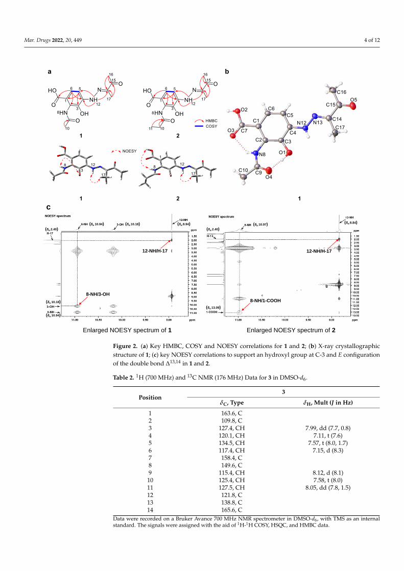
Figure 2. (a) Key HMBC, COSY and NOESY correlations for 1 and 2; (b) X-ray crystallographic
structure of 1; (c) key NOESY correlations to support an hydroxyl group at C-3 and E configuration
of the double bond Δ13,14 in 1 and 2.
Enlarged NOESY spectrum of 1 Enlarged NOESY spectrum of 2
c
(δH 10.18)(δH 10.94)
(δH 10.94)
(δH 10.18)
(δH 8.94)
(δH 2.40)(δH 10.97)
(δH 2.40)
(δH 13.09)
8-NH/3-OH
12-NH/H-17
8-NH/1-COOH
12-NH/H-17
(δH 8.84)
Figure 2. (a) Key HMBC, COSY and NOESY correlations for 1 and 2; (b) X-ray crystallographicstructure of 1; (c) key NOESY correlations to support an hydroxyl group at C-3 and E configurationof the double bond ∆13,14 in 1 and 2.
Table 2. 1H (700 MHz) and 13C NMR (176 MHz) Data for 3 in DMSO-d6.
Position3
δC, Type δH, Mult (J in Hz)
1 163.6, C2 109.8, C3 127.4, CH 7.99, dd (7.7, 0.8)4 120.1, CH 7.11, t (7.6)5 134.5, CH 7.57, t (8.0, 1.7)6 117.4, CH 7.15, d (8.3)7 158.4, C8 149.6, C9 115.4, CH 8.12, d (8.1)10 125.4, CH 7.58, t (8.0)11 127.5, CH 8.05, dd (7.8, 1.5)12 121.8, C13 138.8, C14 165.6, C
Data were recorded on a Bruker Avance 700 MHz NMR spectrometer in DMSO-d6, with TMS as an internalstandard. The signals were assigned with the aid of 1H-1H COSY, HSQC, and HMBC data.
Mar. Drugs 2022, 20, 449 5 of 12
Mar. Drugs 2022, 20, x FOR PEER REVIEW 5 of 13
revealed a 3-hydroxyanthranilic acid unit. Given determined molecular formula as well
as one less degree of unsaturation, two ring systems were connected by C-2-C-1-NH-C-13
and C-7-O-C-8. Thus, compound 3 was elucidated to be demethylmycemycin A.
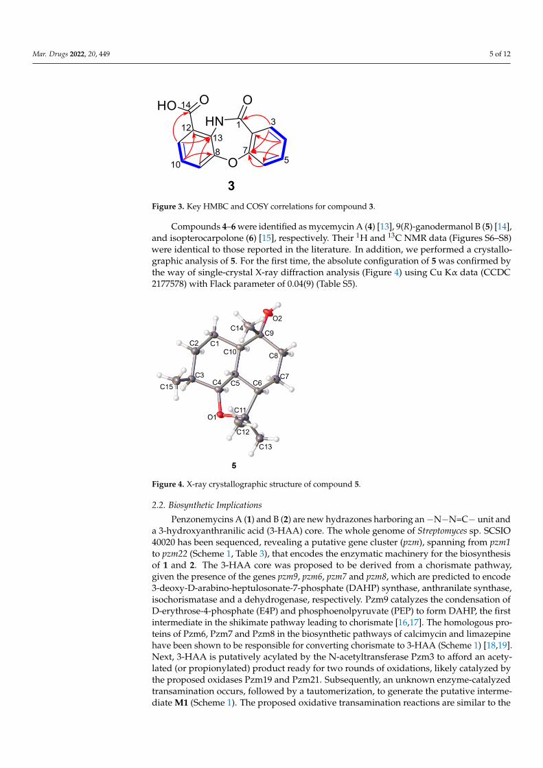
Figure 3. Key HMBC and COSY correlations for compound 3.
Table 2. 1H (700 MHz) and 13C NMR (176 MHz) Data for 3 in DMSO-d6.
Position 3
δC, Type δH, Mult (J in Hz)
1 163.6, C
2 109.8, C
3 127.4, CH 7.99, dd (7.7, 0.8)
4 120.1, CH 7.11, t (7.6)
5 134.5, CH 7.57, t (8.0, 1.7)
6 117.4, CH 7.15, d (8.3)
7 158.4, C
8 149.6, C
9 115.4, CH 8.12, d (8.1)
10 125.4, CH 7.58, t (8.0)
11 127.5, CH 8.05, dd (7.8, 1.5)
12 121.8, C
13 138.8, C
14 165.6, C
Data were recorded on a Bruker Avance 700 MHz NMR spectrometer in DMSO-d6, with TMS as an
internal standard. The signals were assigned with the aid of 1H-1H COSY, HSQC, and HMBC data.
Compounds 4–6 were identified as mycemycin A (4) [13], 9(R)-ganodermanol B (5)
[14], and isopterocarpolone (6) [15], respectively. Their 1H and 13C NMR data (Figures S6–
S8) were identical to those reported in the literature. In addition, we performed a crystal-
lographic analysis of 5. For the first time, the absolute configuration of 5 was confirmed
by the way of single-crystal X-ray diffraction analysis (Figure 4) using Cu Kα data (CCDC
2177578) with Flack parameter of 0.04(9) (Table S5).
Figure 3. Key HMBC and COSY correlations for compound 3.
Compounds 4–6 were identified as mycemycin A (4) [13], 9(R)-ganodermanol B (5) [14],and isopterocarpolone (6) [15], respectively. Their 1H and 13C NMR data (Figures S6–S8)were identical to those reported in the literature. In addition, we performed a crystallo-graphic analysis of 5. For the first time, the absolute configuration of 5 was confirmed bythe way of single-crystal X-ray diffraction analysis (Figure 4) using Cu Kα data (CCDC2177578) with Flack parameter of 0.04(9) (Table S5).
Mar. Drugs 2022, 20, x FOR PEER REVIEW 6 of 13
Figure 4. X-ray crystallographic structure of compound 5.
2.2. Biosynthetic Implications
Penzonemycins A (1) and B (2) are new hydrazones harboring an −N−N=C− unit and
a 3-hydroxyanthranilic acid (3-HAA) core. The whole genome of Streptomyces sp. SCSIO
40020 has been sequenced, revealing a putative gene cluster (pzm), spanning from pzm1 to
pzm22 (Scheme 1, Table 3), that encodes the enzymatic machinery for the biosynthesis of
1 and 2. The 3-HAA core was proposed to be derived from a chorismate pathway, given
the presence of the genes pzm9, pzm6, pzm7 and pzm8, which are predicted to encode 3-
deoxy-D-arabino-heptulosonate-7-phosphate (DAHP) synthase, anthranilate synthase,
isochorismatase and a dehydrogenase, respectively. Pzm9 catalyzes the condensation of
D-erythrose-4-phosphate (E4P) and phosphoenolpyruvate (PEP) to form DAHP, the first
intermediate in the shikimate pathway leading to chorismate [16,17]. The homologous
proteins of Pzm6, Pzm7 and Pzm8 in the biosynthetic pathways of calcimycin and limaze-
pine have been shown to be responsible for converting chorismate to 3-HAA (Scheme 1)
[18,19]. Next, 3-HAA is putatively acylated by the N-acetyltransferase Pzm3 to afford an
acetylated (or propionylated) product ready for two rounds of oxidations, likely catalyzed
by the proposed oxidases Pzm19 and Pzm21. Subsequently, an unknown enzyme-cata-
lyzed transamination occurs, followed by a tautomerization, to generate the putative in-
termediate M1 (Scheme 1). The proposed oxidative transamination reactions are similar
to the recent reported reactions in the formation of 8-amino-flaviolin during the biosyn-
thesis of naphthoquinone-based meroterpenoid [20]. Notably, the three enzymes, namely,
the nitrosuccinate lyase Pzm11, the monooxygenase Pzm12 and the AMP-binding protein
Pzm18, are predicted to be the orthologues of CreD, CreE and CreM, respectively, which
are responsible for the diazo formation in cremeomycin biosynthesis, utilizing L-aspartic
acid as a nitrogen source [21]. Therefore, Pzm11 and Pzm12 are postulated to generate
nitrite from L-aspartic acid, which is subsequently incorporated to M1 by Pzm18, leading
to the diazo group in M2 after dehydration and tautomerization, similar to the biosynthe-
sis of cremeomycin and triacsin [21,22]. Recently, an electrophilic diazo-anthranilate has
been reported to undergo nonenzymatic Japp−Klingemann coupling with a β-keto alde-
hyde-containing precursor to furnish the hydrazone group in the biosynthesis of tasi-
kamides [4]. Interestingly, Pzm16 encodes an acetolactate synthase and putatively cata-
lyzes the coupling of two pyruvates to form 2-acetolactate [23], which becomes 4-hydroxy-
3-methyl-3-buten-2-one (M3) after two rounds of carboxyl reduction (probably catalyzed
by the F420-dependent oxidoreductase Pzm4) and spontaneous dehydration (Scheme 1).
Finally, the nonenzymatic Japp−Klingemann coupling reaction between M2 and M3 con-
structs the hydrazone moiety in 1 and 2 after subsequent hydration and decarboxylation
(Scheme 1).
Figure 4. X-ray crystallographic structure of compound 5.
2.2. Biosynthetic Implications
Penzonemycins A (1) and B (2) are new hydrazones harboring an−N−N=C− unit anda 3-hydroxyanthranilic acid (3-HAA) core. The whole genome of Streptomyces sp. SCSIO40020 has been sequenced, revealing a putative gene cluster (pzm), spanning from pzm1to pzm22 (Scheme 1, Table 3), that encodes the enzymatic machinery for the biosynthesisof 1 and 2. The 3-HAA core was proposed to be derived from a chorismate pathway,given the presence of the genes pzm9, pzm6, pzm7 and pzm8, which are predicted to encode3-deoxy-D-arabino-heptulosonate-7-phosphate (DAHP) synthase, anthranilate synthase,isochorismatase and a dehydrogenase, respectively. Pzm9 catalyzes the condensation ofD-erythrose-4-phosphate (E4P) and phosphoenolpyruvate (PEP) to form DAHP, the firstintermediate in the shikimate pathway leading to chorismate [16,17]. The homologous pro-teins of Pzm6, Pzm7 and Pzm8 in the biosynthetic pathways of calcimycin and limazepinehave been shown to be responsible for converting chorismate to 3-HAA (Scheme 1) [18,19].Next, 3-HAA is putatively acylated by the N-acetyltransferase Pzm3 to afford an acety-lated (or propionylated) product ready for two rounds of oxidations, likely catalyzed bythe proposed oxidases Pzm19 and Pzm21. Subsequently, an unknown enzyme-catalyzedtransamination occurs, followed by a tautomerization, to generate the putative interme-diate M1 (Scheme 1). The proposed oxidative transamination reactions are similar to the
Mar. Drugs 2022, 20, 449 6 of 12
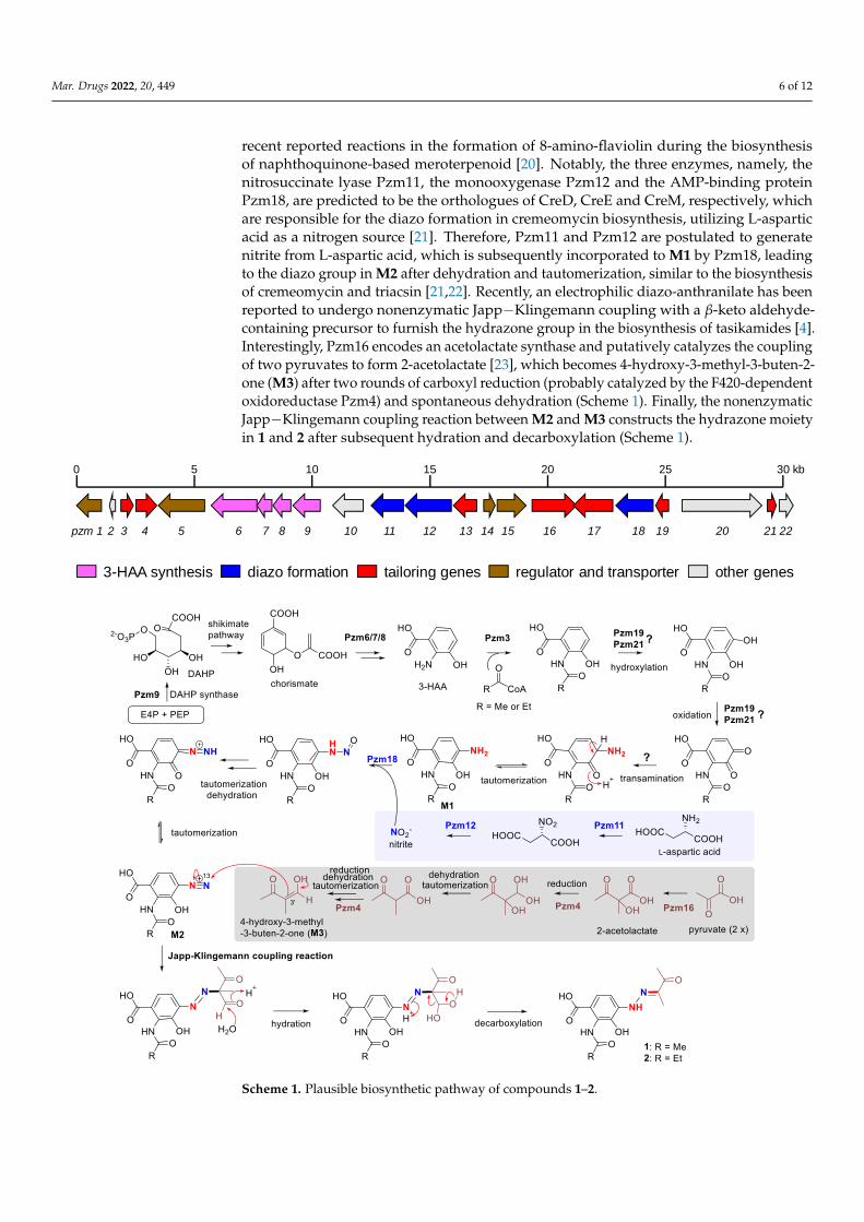
recent reported reactions in the formation of 8-amino-flaviolin during the biosynthesisof naphthoquinone-based meroterpenoid [20]. Notably, the three enzymes, namely, thenitrosuccinate lyase Pzm11, the monooxygenase Pzm12 and the AMP-binding proteinPzm18, are predicted to be the orthologues of CreD, CreE and CreM, respectively, whichare responsible for the diazo formation in cremeomycin biosynthesis, utilizing L-asparticacid as a nitrogen source [21]. Therefore, Pzm11 and Pzm12 are postulated to generatenitrite from L-aspartic acid, which is subsequently incorporated to M1 by Pzm18, leadingto the diazo group in M2 after dehydration and tautomerization, similar to the biosynthesisof cremeomycin and triacsin [21,22]. Recently, an electrophilic diazo-anthranilate has beenreported to undergo nonenzymatic Japp−Klingemann coupling with a β-keto aldehyde-containing precursor to furnish the hydrazone group in the biosynthesis of tasikamides [4].Interestingly, Pzm16 encodes an acetolactate synthase and putatively catalyzes the couplingof two pyruvates to form 2-acetolactate [23], which becomes 4-hydroxy-3-methyl-3-buten-2-one (M3) after two rounds of carboxyl reduction (probably catalyzed by the F420-dependentoxidoreductase Pzm4) and spontaneous dehydration (Scheme 1). Finally, the nonenzymaticJapp−Klingemann coupling reaction between M2 and M3 constructs the hydrazone moietyin 1 and 2 after subsequent hydration and decarboxylation (Scheme 1).
Mar. Drugs 2022, 20, x FOR PEER REVIEW 7 of 13
Scheme 1. Plausible biosynthetic pathway of compounds 1–2.
0 5 10 15 20 25 30 kb
pzm 1 2 3 4 5 6 7 8 9 10 11 12 13 14 15 16 17 18 19 20 21 22
3-HAA synthesis regulator and transporterdiazo formation other genestailoring genes
Scheme 1. Plausible biosynthetic pathway of compounds 1–2.
Mar. Drugs 2022, 20, 449 7 of 12
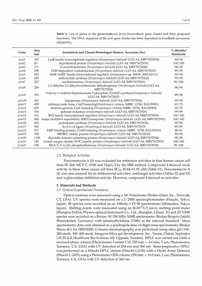
Table 3. List of genes in the penzonemycin (pzm) biosynthetic gene cluster and their proposedfunctions. The DNA sequence of the pzm gene cluster has been deposited in GenBank (accession:ON345781).
Gene Size(aa) Annotation and Closest Homologue (Source, Accession No.) % Identity/
Similarity
pzm1 353 LysR family transcriptional regulator (Streptomyces halstedii LGO-A4, MBV7672918) 94/94pzm2 83 hypothetical protein (Streptomyces halstedii LGO-A4, MBV7672919) 100/100pzm3 171 N-acetyltransferase (Streptomyces halstedii LGO-A4, MBV7672920) 98/98pzm4 298 F420-dependent oxidoreductase (Streptomyces halstedii LGO-A4, MBV7672921) 99/99pzm5 653 AfsR/SARP family transcriptional regulator (Streptomyces sp. SM18, AWL43113) 99/99pzm6 650 anthranilate synthase (Streptomyces halstedii LGO-A4, MBV7672922) 99/99pzm7 207 isochorismatase (Streptomyces halstedii LGO-A4, MBV7672923) 99/100
pzm8 256 2,3-dihydro-2,3-dihydroxybenzoate dehydrogenase (Streptomyces halstedii LGO-A4,MBV7672924) 98/99
pzm9 393 3-deoxy-D-arabino-heptulosonate 7-phosphate (DAHP) synthase(Streptomyces halstediiLGO-A4, MBV7672925) 99/98
pzm10 432 transposase (Streptomyces halstedii LGO-A4, MBV7672926) 99/99pzm11 459 nitrosuccinate lyase, CreD homolog(Streptomyces cremeus NBRC 12760, BAU09301) 67/73pzm12 659 monooxygenase, CreE homolog (Streptomyces cremeus NBRC 12760, BAU09302) 62/70pzm13 334 quinone reductase (Streptomyces halstedii LGO-A4, MBV7672929) 99/99pzm14 161 Rrf2 family transcriptional regulator (Streptomyces halstedii LGO-A4, MBV7672930) 100/100pzm15 406 major facilitator superfamily (MFS) transporter (Streptomyces halstedii LGO-A4, MBV7672931) 100/100pzm16 622 acetolactate synthase (Streptomyces halstedii LGO-A4, MBV7672932) 99/99pzm17 560 Acyl-CoA ligase (Streptomyces halstedii LGO-A4, MBV7672933) 99/99pzm18 527 AMP-binding protein, CreM homolog (Streptomyces cremeus NBRC 12760, BAU09310) 48/61pzm19 180 SRPBCC family protein (Streptomyces halstedii LGO-A4, MBV7672935) 99/99pzm20 1128 discoidin domain-containing protein (Streptomyces halstedii LGO-A4, MBV7672936) 99/99pzm21 126 vicinal oxygen chelate (VOC) family protein (Streptomyces halstedii LGO-A4, MBV7672937) 100/100pzm22 198 RNA 2′,3′-cyclic phosphodiesterase (Streptomyces halstedii LGO-A4, MBV7672938) 99/100
2.3. Biological Activities
Penzonemycin A (1) was evaluated for antitumor activities in four human cancer celllines SF-268, MCF-7, A549 and HepG-2 by the SRB method. Compound 1 showed weakactivity in these three cancer cell lines (IC50 30.44–61.92 µM) (Table S1). Penzonemycin A(1) was also assayed for its antibacterial activities, antifungal activities (Tables S2 and S3)and α-glucosidase inhibition activity. However, compound 1 showed no activities.
3. Materials and Methods3.1. General Experimental Procedures
Optical rotations were measured using a 341 Polarimeter (Perkin-Elmer, Inc., Norwalk,CT, USA). UV spectra were measured on a U-2900 spectrophotometer (Hitachi, Tokyo,Japan). IR spectra were recorded on an Affinity-1 FT-IR spectrometer (Shimadzu, Tokyo,Japan). Melting points were measured using an SGW®X-5 micro melting point meter(Shanghai INESA Physico optiacal Instrument Co., Ltd., Shanghai, China). 1D and 2D NMRspectra were recorded on a Bruker AV-700 MHz NMR spectrometer (Bruker Biospin GmbH,Rheinstetten, Germany) with tetramethylsilane (TMS) as the internal standard. Massspectrometric data were obtained on a quadrupole-time-of-flight mass spectrometer (BrukerMaxis 4G) for HRESIMS. Column chromatography was performed using silica gel (100–200 mesh, 300–400 mesh; Jiangyou Silica gel development, Inc., Yantai, China), SephadexLH-20 (GE Healthcare Bio-Sciences AB, Uppsala, Sweden). HPLC was carried out while areversed-phase column (Phenomenex Gemini C18, 250 mm × 4.6 mm, 5 µm; Phenomenex,Torrance, CA, USA) with UV detection at 254 nm and 360 nm. Semi-preparative HPLCwas performed on a Hitachi HPLC station (Hitachi-L2130) with a Diode Array Detector(Hitachi L-2455) using a Phenomenex ODS column (250 mm× 10.0 mm, 5 µm; Phenomenex,Torrance, CA, USA) with UV detection at 360 nm.
Mar. Drugs 2022, 20, 449 8 of 12
3.2. Strain, Screening and Culture Methods
Streptomyces sp. SCSIO 40020 (original number LW701) was isolated from a marinesediment sample (E 114.0432◦, N 22.0194◦, Pearl River Estuary in China, at the depthof 28 m), and was identified by 16S rDNA sequence analysis (GenBank accession no.MW582618). The strain SCSIO 40020 was maintained in a 40% glycerol aqueous solutionat −80 ◦C at the Research Center for Marine Microbiology Culture Collection Center ofSouth China Sea Institute of Oceanology, Chinese Academy of Sciences. It was found thatthe strain SCSIO 40020 was best maintained on 38#-agar medium containing 3% sea saltfor optimal growth and sporulation. A single colony was inoculated into 50 mL of fourdifferent media, including modifed-A1BFe+C [24] (soluble starch 1.0%, yeast extract 0.4%,tryptone 0.2%, CaCO3 0.2%, sea salts 3%, pH 7.2–7.4), ZM7 medium (glycerol 0.5%, arginine0.1%, glucose 0.1%, K2HPO4 0.03%, MgSO4·7H2O 0.02%, NaCl 0.03%, vitamin B complex0.5 mL, pH 7.2–7.4), AM6-4 [25] (glycerol 0.1%, bacterial peptone 0.5%, glycine 0.01%,alanine 0.01%, CaCO3 0.5%, sea salts 3%, pH 7.2–7.4) and modifed-ISP3 [25] (oat meal1.5%, FeSO4 0.0001%, MnCl2 0.0001%, ZnSO4 0.0001%, sea salts 3%, pH 7.2–7.4), in 250 mLErlenmeyer flasks and then incubated on a rotary shaker (200 rpm) at 28 ◦C for seven days.The culture broths were extracted with an equal volume of n-butanone, and the extractswere then monitored by HPLC-DAD. HPLC analyses were carried out using the followingprogram: solvent system (solvent A, 10% acetonitrile in water supplemented with 0.08%formic acid; solvent B, 90% acetonitrile in water); 5% B to 100% B (linear gradient, 0–18 min),100% B (18–23 min), 100% B to 5% B (23–27 min), 5% B (27–32 min); flow rate of 1 mL/min.A single colony was inoculated into 30 mL of modified A1BFe+C medium and incubated at28 ◦C for 2–3 days. Then, 20 L of fermentation culture was prepared by inoculating 30 mLof the seed culture into a 1000 mL Erlenmeyer flask containing 200 mL of the modified ISP3medium followed by cultivation on a rotary shaker (200 rpm) at 28 ◦C for 7 days.
3.3. Extraction, Isolation and Purification
The 20 L of culture broth of Streptomyces sp. SCSIO 40020 was centrifuged at 3900 rpmfor 15 min at 25 ◦C. The mycelia were extracted three times, each with 2 L of acetone. Theacetone extracts were concentrated under reduced pressure to afford an aqueous residue,which was extracted four times with an equal volume of n-butanone. The supernatantswere extracted four times with an equal volume of n-butanone [12,24]. The butanoneextracts were combined and concentrated under reduced pressure to afford the crudeextracts (7.5 g). The crude extracts were subjected to column chromatography over silicagel, eluting with a gradient of CHCl3/MeOH mixtures i.e., 100/0, 95/5, 90/10, 80/20,50/50 and 0/100 (v/v). We obtained five fractions (Fr.1–Fr.5). Then Fr.2 (3.50 g) was furtherpurified via MPLC (Medium-Pressure Preparative Liquid Chromatography) with a reverse-phased C-18 column (14.5 cm × 2.5 cm i.d., 40–60 µm Agela Technologies, Torrance, CA,USA) by eluting with a linear gradient of H2O/MeOH (0–100%, 15 mL/min, 300 min),obtaining fractions Fr.2.1–Fr.2.10. Fractions Fr.2.5–Fr.2.6 (20 mg) were further purified bysemi-preparative HPLC using a mobile phase of MeCN/H2O (50:50, v/v), which yieldedcompound 3 (1.8 mg) and compound 4 (1.5 mg). Fractions Fr.2.7–Fr.2.8 (200 mg) werefurther purified by semi-preparative HPLC using a mobile phase of MeCN/H2O (60:40,v/v), obtaining compound 1 (10.4 mg), compound 2 (2.2 mg), compound 5 (1.2 mg), andcompound 6 (1.0 mg).
3.4. Physical and Chemical Properties of the New Compounds 1–3 and 5
Penzonemycin A (1): yellow needles; m.p. 205.8–209.3 ◦C; [α]25D : −1.8 (c 0.11, MeCN);
UV (MeCN) λmax (log ε): 358 (4.69) nm, 249 (4.50) nm, 210 (4.59) nm; IR νmax 3360, 2947,2837, 2359, 2332, 1676 cm−1, 1H NMR (700 MHz, DMSO-d6) and 13C NMR (176 MHz,DMSO-d6), see Table 1; (+)-HRESIMS m/z [M + H]+ 294.1086 (calculated for C13H16N3O5,294.1084), [M + Na]+ 316.0908 (calculated for C13H15N3NaO5, 316.0904).
Crystal data for penzonemycin A (1): C13H15N3O5 (M = 293.28 g/mol): mono-clinic, space group P21/n (no. 14), a = 4.8385 (2) Å, b = 19.6128 (7) Å, c = 14.5170 (6) Å,
Mar. Drugs 2022, 20, 449 9 of 12
β = 98.487 (4), V = 1362.53 (9) Å3, Z = 4, T = 103 (5) K, µ (Cu Kα) = 0.945 mm−1,Dcalc = 1.430 g/cm3, 6318 reflections measured (9.018◦ ≤ 2Θ ≤ 148.158◦), 2631 unique(Rint = 0.0376, Rsigma = 0.0368) which were used in all calculations. The final R1 was 0.0576(I > 2σ(I)), and wR2 was 0.1809 (all data).
Penzonemycin B (2): a yellowish solid; [α]25D : −1.1 (c 0.19, MeCN); UV (MeCN) λmax
(log ε): 358 (4.66) nm, 249 (4.48) nm, 210 (4.56) nm; IR νmax 3350, 2358, 1676, 1267, 667 cm−1;1H and 13C NMR data, see Table 1; (+)-HRESIMS m/z [M + H]+ 308.1240 (calculated forC14H18N3O5, 308.1241), [M + Na]+ 330.1054 (calculated for C13H15N3NaO5, 330.1060).
Compound 3: a white solid; [α]25D : +8.9 (c 0.20, MeCN); UV (CHCl3) λmax (log ε): 346
(4.07) nm, 335 (4.09) nm; 306 (3.92) nm; 264 (3.94) nm, 247 (3.89) nm; IR νmax 2953, 2914,1716, 1633, 1548, 1489, 1303, 1259, 1244, 1192, 1159, 1060, 875, 786, 748, 717, 686 cm−1,1H and 13C NMR data, see Table 2; (+)-HRESIMS m/z [M + H]+ 256.0616 (calculated forC14H18N3O5, 256.0604).
Crystal data for 9(R)-ganodermanol B (5): C15H26O2 (M = 238.36 g/mol): orthorhom-bic, space group P21/n (no. 19), a = 8.35172 (11) Å, b = 11.17809 (15) Å, c = 14.82535 (19) Å,V = 1384.04 (3) Å3, Z = 4, T = 99.8 (8) K, µ (Cu Kα) = 0.570 mm−1, Dcalc = 1.144 g/cm3, 7345reflections measured (9.91◦ ≤ 2Θ ≤ 148.416◦), 2727 unique (Rint = 0.0278, Rsigma = 0.0319)which were used in all calculations. The final R1 was 0.0355 (I > 2σ(I)), and wR2 was 0.0946(all data).
3.5. X-Ray Crystallographic Analysis Data of Penzonemycin A (1) and 5
Single crystals of C13H15N3O5 (penzonemycin A 1) and C15H26O2 (9(R)-ganodermanolB 5) were obtained in MeOH solution. Their data were collected on an XtaLAB AFC12(RINC): Kappa single diffractometer. The crystals were kept at 100.00 (10) K during datacollection. Using Olex2 [26], their structures were solved with the ShelXT [27] structuresolution program using Intrinsic Phasing and refined with the ShelXL [28] refinement pack-age, using Least Squares minimization. Crystallographic data for 1 and 5 were depositedat the Cambridge Crystallographic Data Center (CCDC) with the deposition numbers2,131,907 and 2,177,578, respectively.
3.6. Bioactivity Assays3.6.1. Cytotoxic Activity Assay
The in vitro cytotoxic activities of penzonemycin A (1) were evaluated in four tumorcell lines, i.e., SF-268 (human glioma cell line), HepG-2 (human liver carcinoma cell line),MCF-7 (human breast adenocarcinoma cell line), and A549 (human lung adenocarcinomacell) by the SRB method [24], according to a previously described protocol [29]. The cellswere cultivated in RPMI 1640 medium [30]. Cells (180 µL) with a density of 3× 10⁴ cells/mLwere seeded onto 96-well plates and incubated for 24 h at 37 ◦C, 5% CO2. Subsequently,20 µL of different concentrations of penzonemycin A (1), ranging from 0 to 100 µM indimethyl sulfoxide (DMSO), was added to each plate well. An equal volume of DMSO wasused as a negative control. After further incubation for 72 h, the cell monolayers were fixedwith 50% (wt/v) trichloroacetic acid (50 µL) and then stained for 30 min with 0.4% (wt/v)SRB dissolved in 1% acetic acid. The unbound dye was removed by repeatedly washingwith 1% acetic acid. The protein-bound dye was dissolved in 10 mM Tris-base solution(200 µL) for the determination of the optical density (OD) at 570 nm using a microplatereader. The cytotoxic compound cisplatin was used as a positive control. All data wereobtained in triplicate and are presented as means ± S.D. IC50 values were calculated withthe SigmaPlot 14.0 software using the non-linear curve-fitting method.
3.6.2. Antifungal Activity Assay
The antifungal activity of compound 1 was measured against three phytopathogenicfungi, i.e., Colletotrichum gloeosporioides Penz, Physalospora piricola Nose, Bipolaris sorokiniana,by the broth microdilution method [31]. Compound 1 was dissolved in DMSO at thefinal concentration of 2.56 mg mL−1. The indicator fungi strains were grown for 48 h on
Mar. Drugs 2022, 20, 449 10 of 12
a rotary shaker at 28 ◦C. The cultures were diluted with sterilized medium to achieve apredetermined optical absorbance at 600 nm, then diluted 1000-fold before being addedinto 96-well microtiter plates. Three replicates of each compound were tested in dilutionseries, ranging from 128 to 0.25 µg mL−1. The lowest concentrations that completelyinhibited the visible growth of the tested strains were recorded after 48 h of cultivationfrom two independent experiments. Nystatin was used as a positive control against thethree phytopathogenic fungi.
3.6.3. Antibacterial Activity Assay
The antibacterial activity of compound 1 was assayed against Escherichia coli ATCC 25922,Acinetobacter baumannii ATCC 19606, Staphyloccocus aureus ATCC 29213, and Methicillin-resistant S. aureus (MRSA) ATCC 43300 by the broth microdilution method [31]. Compound1 was dissolved in DMSO at the final concentration of 2.56 mg mL−1. The indicator strainwas grown for 24 h on a rotary shaker at 37 ◦C. The cultures were diluted with sterilizedmedium to achieve a predetermined optical absorbance at 600 nm, then diluted 1000-foldbefore being placed into 96-well microtiter plates. Three replicates of each compound weretested in dilution series, ranging from 128 to 0.25 µg mL−1. The MIC values were recordedafter 24 h of cultivation from two independent experiments. Ciprofloxacin and vancomycinwere used as positive controls.
3.6.4. Alpha-glucosidase Inhibition Activity
A general inhibiting reaction assay contained 0.04 U/mL, 2.0 mM p-nitrophenyl-α-D-glucopyranoside solution (Shanghai Macklin Biochemical Co., Ltd., Shanghai, China) anda specific concentration of 1 and was carried out in PBS buffer (50 mM, pH 7.0), in a totalvolume of 100 µL. In detail, first, sample 1 was mixed with α-glucosidase and incubatedat 37 ◦C for 10 min, and then, the reactions were initiated by adding the p-nitrophenyl-α-D-glucopyranoside solution and continued at 37 ◦C for another 15 min. After that,the absorbance at 405 nm was recorded on a microplate reader (Enspire, PerkinElmer,Singapore) within 30 min. In the assay, DMSO was used as a negative control, and acarbose(Shanghai Aladdin Biochemical Technology Co., Ltd., Shanghai, China) was used as apositive control. All treatments were performed in triplicate, and the IC50 value wasdetermined by regression analysis [32].
3.7. Bioinformatics Analysis
The genomic DNA of Streptomyces sp. SCSIO 40020 was isolated according to a priorstudy [12]. The whole genome was sequenced and assembled by Nextomics Biosciences Co.,Ltd. (Wuhan, China) using Oxford Nanopore GridION and canu. The putative biosyntheticgene clusters in the genome were predicted with antiSMASH 6.0 [33]. The deducedORFs were analyzed using online 2ndFind software (https://biosyn.nih.go.jp/2ndfind/,accessed on 13 January 2022), and their functional predictions were accomplished with anonline BLAST program (https://blast.ncbi.nlm.nih.gov/Blast.cgi, accessed on 13 January2022). The DNA sequences of the penzonemycins gene cluster were deposited in GenBank,accession number ON345781.
4. Conclusions
In conclusion, two new phenylhydrazone derivatives and a new alkaloid, penzone-mycins A–B (1–2) and demethylmycemycin A (3), together with three known compounds(4–6) were obtained from the Pearl River Estuary sediment-derived Streptomyces sp. SCSIO40020. Penzonemycins feature a hydrazone unit (−N−N=C−) that is rare in natural prod-ucts. Biosynthetically, a Japp−Klingemann reaction is proposed as a key reaction leadingto the hydrazone moiety in 1 and 2, which is worth of further investigations.
Mar. Drugs 2022, 20, 449 11 of 12
Supplementary Materials: The following supporting information can be downloaded at: https://www.mdpi.com/article/10.3390/md20070449/s1, Table S1: Cytotoxicity of penzonemycin A (1).Table S2: Antifungal activity of penzonemycin A (1). Table S3: Antibacterial activity of penzonemycinA (1). Table S4: Crystal data and structure refinement for penzonemycin A (1). Table S5: Crystaldata and structure refinement for 9(R)-ganodermanol B (5). Figure S1: Natural hydrazone-containingcompounds reported in the literature. Figure S2: HPLC analysis of the metabolic profiles of Strepto-myces sp. SCSIO 40020 cultured in different media. Figure S3: Spectral data for penzonemycin A (1).Figure S4: Spectral data for penzonemycin B (2). Figure S5: Spectral data for demethylmycemycinsA (3). Figure S6: Spectral data for mycemycin A (4). Figure S7: Spectral data for 9(R)-ganodermanolB (5). Figure S8: Spectral data for isopterocarpolone (6).
Author Contributions: W.L. contributed to compounds isolation and structure elucidation. L.Z.analyzed the X-ray crystallographic diffraction data. Q.Z., Y.C. and W.Z. (Weimin Zhang) performedthe cytotoxicity assays. W.L., L.M., H.Z., W.Z. (Wenjun Zhang) and C.Z. analyzed the data and wrotethe manuscript. C.Z. supervised the study. All authors have read and agreed to the published versionof the manuscript.
Funding: This work was supported in part by NSFC (42176127, 22193072, 21907098), GuangdongBasic and Applied Basic Research Foundation (2021A1515110284, 2021A1515010839), by the Scienceand Technology Planning Project of Guangzhou (202102020513), K. C. Wong Education Foundation(GJTD-2020-12), by the State Key Laboratory of Applied Microbiology, Southern China (Grant No.SKLAM004-2021).
Institutional Review Board Statement: Not applicable.
Informed Consent Statement: Not applicable.
Data Availability Statement: The data presented in this study are available on request from thecorresponding author.
Acknowledgments: We appreciate the help of Xuan Ma in X-ray diffraction analyses and of ZhihuiXiao, Aijun Sun, Yun Zhang and Xiaohong Zheng in recording MS and NMR data. We are grateful toZhengchao Wu for the collection of Pearl River Estuary sediment.
Conflicts of Interest: The authors declare no conflict of interest.
References1. Wang, Z.; Kang, D.; Chen, M.; Wu, G.; Feng, D.; Zhao, T.; Zhou, Z.; Huo, Z.; Jing, L.; Zuo, X.; et al. Design, synthesis, and antiviral
evaluation of novel hydrazone-substituted thiophene [3,2-d]pyrimidine derivatives as potent human immunodeficiency virus-1inhibitors. Chem. Biol. Drug Des. 2018, 92, 2009–2021. [CrossRef] [PubMed]
2. Bolognino, I.; Giangregorio, N.; Tonazzi, A.; Martínez, A.L.; Altomare, C.D.; Loza, M.I.; Sablone, S.; Cellamare, S.; Catto, M. Synthe-sis and biological evaluation of dantrolene-like hydrazide and hydrazone analogues as multitarget agents for neurodegenerativediseases. ChemMedChem 2021, 16, 2807–2816. [CrossRef] [PubMed]
3. Achagar, R.; Elmakssoudi, A.; Dakir, M.; Elamrani, A.; Zouheir, Y.; Zahouily, M.; Jamaleddine, J. A green and efficient protocolfor the synthesis of phenylhydrazone derivatives catalyzed by nanostructured diphosphate Na2CaP2O7 and screening of theirantibacterial activity. ChemistrySelect 2021, 6, 1366–1371. [CrossRef]
4. Ma, G.; Candra, H.; Pang, L.; Xiong, J.; Ding, Y.; Tran, H.; Low, Z.; Ye, H.; Liu, M.; Zheng, J.; et al. Biosynthesis of tasikamides viapathway coupling and diazonium-mediated hydrazone formation. J. Am. Chem. Soc. 2022, 144, 1622–1633. [CrossRef] [PubMed]
5. Liu, Y.; Fang, S.; Shi, Z.; Wang, B.; Li, X.; Ji, N. Phenylhydrazone and quinazoline derivatives from the cold-seep-derived fungusPenicillium oxalicum. Mar. Drugs 2021, 19, 10. [CrossRef] [PubMed]
6. Cheng, P.; Xu, K.; Chen, Y.; Wang, T.; Chen, Y.; Yang, C.; Ma, S.; Liang, Y.; Ge, H.; Jiao, R. Cytotoxic aromatic polyketides from aninsect derived Streptomyces sp. NA4286. Tetrahedron Lett. 2019, 60, 1706–1709. [CrossRef]
7. Zhang, W.; Yang, C.; Huang, C.; Zhang, L.; Zhang, H.; Zhang, Q.; Yuan, C.; Zhu, Y.; Zhang, C. Pyrazolofluostatins A–C,pyrazole-fused benzo[a]fluorenes from south China sea-derived Micromonospora rosaria SCSIO N160. Org. Lett. 2017, 19, 592–595.[CrossRef]
8. Matsuda, K.; Hasebe, F.; Shiwa, Y.; Kanesaki, Y.; Tomita, T.; Yoshikawa, H.; Shin-ya, K.; Kuzuyama, T.; Nishiyama, M. Genomemining of amino group carrier protein-mediated machinery: Discovery and biosynthetic characterization of a natural productwith unique hydrazone unit. ACS Chem. Biol. 2017, 12, 124–131. [CrossRef]
9. Blair, L.M.; Sperry, J. Natural products containing a nitrogen–nitrogen bond. J. Nat. Prod. 2013, 76, 794–812. [CrossRef]10. Ren, X.; Xie, X.; Chen, B.; Liu, L.; Jiang, C.; Qian, Q. Marine natural products: A potential source of anti-hepatocellular carcinoma
drugs. J. Med. Chem. 2021, 64, 7879–7899. [CrossRef]
Mar. Drugs 2022, 20, 449 12 of 12
11. Jagannathan, S.V.; Manemann, E.M.; Rowe, S.E.; Callender, M.C.; Soto, W. Marine actinomycetes, new sources of biotechnologicalproducts. Mar. Drugs 2021, 19, 365. [CrossRef]
12. Zhai, S.; Liu, W.; Wu, Z.; Zhu, Y.; Zhang, W.; Zhang, C.; Ma, L. Identification of bafilomycins and heterologous expression of itsbiosynthetic gene cluster from the Pearl River Estuary sediment-derived Streptomyces sp. SCSIO 40020. Acta Microbiol. Sin. 2021,61, 3991–4005.
13. Liu, N.; Song, F.; Shang, F.; Huang, Y. Mycemycins A-E, new dibenzoxazepinones isolated from two different Streptomycetes.Mar. Drugs 2015, 13, 6247–6258. [CrossRef] [PubMed]
14. Tan, Z.; Zhao, J.; Liu, J.; Zhang, M.; Chen, R.; Xie, K.; Dai, J. Sesquiterpenoids from the cultured mycelia of Ganoderma capense.Fitoterapia 2017, 118, 73–79. [CrossRef] [PubMed]
15. Shaaban, K.A.; Singh, S.; Elshahawi, S.I.; Wang, X.; Ponomareva, L.V.; Sunkara, M.; Copley, G.C.; Hower, J.C.; Morris, A.J.; Kharel,M.K.; et al. The native production of the sesquiterpene isopterocarpolone by Streptomyces sp. RM-14-6. Nat. Prod. Res. 2014, 28,337–339. [CrossRef] [PubMed]
16. Cano-Prieto, C.; García-Salcedo, R.; Sánchez-Hidalgo, M.; Braña, A.; Fiedler, H.; Méndez, C.; Salas, J.; Olano, C. Genome mining ofStreptomyces sp. Tü 6176: Characterization of the nataxazole biosynthesis pathway. Chembiochem 2015, 16, 1461–1473. [CrossRef]
17. Ikeda, M. Towards bacterial strains overproducing L-tryptophan and other aromatics by metabolic engineering. Appl. Microbiol.Biotechnol. 2006, 69, 615–626. [CrossRef]
18. Pavlikova, M.; Kamenik, Z.; Janata, J.; Kadlcik, S.; Kuzma, M.; Najmanova, L. Novel pathway of 3-hydroxyanthranilic acidformation in limazepine biosynthesis reveals evolutionary relation between phenazines and pyrrolobenzodiazepines. Sci. Rep.2018, 8, 7810. [CrossRef]
19. Gou, L.; Wu, Q.; Lin, S.; Li, X.; Liang, J.; Zhou, X.; An, D.; Deng, Z.; Wang, Z. Mutasynthesis of pyrrole spiroketal compoundusing calcimycin 3-hydroxy anthranilic acid biosynthetic mutant. Appl. Microbiol. Biotechnol. 2013, 97, 8183–8191. [CrossRef]
20. Noguchi, T.; Isogai, S.; Terada, T.; Nishiyama, M.; Kuzuyama, T. Cryptic oxidative transamination of hydroxynaphthoquinone innatural product biosynthesis. J. Am. Chem. Soc. 2022, 144, 5435–5440. [CrossRef]
21. Sugai, Y.; Katsuyama, Y.; Ohnishi, Y. A nitrous acid biosynthetic pathway for diazo group formation in bacteria. Nat. Chem. Biol.2016, 12, 73–75. [CrossRef] [PubMed]
22. Del Rio Flores, A.; Twigg, F.F.; Du, Y.; Cai, W.; Aguirre, D.Q.; Sato, M.; Dror, M.J.; Narayanamoorthy, M.; Geng, J.; Zill, N.A.;et al. Biosynthesis of triacsin featuring an N-hydroxytriazene pharmacophore. Nat. Chem. Biol. 2021, 17, 1305–1313. [CrossRef][PubMed]
23. Chipman, D.; Barak, Z.; Schloss, J.V. Biosynthesis of 2-aceto-2-hydroxy acids: Acetolactate synthases and acetohydroxyacidsynthases. Biochim. Biophys. Acta. 1998, 1385, 401–419. [CrossRef]
24. Liu, W.; Zhang, W.; Jin, H.; Zhang, Q.; Chen, Y.; Jiang, X.; Zhang, G.; Zhang, L.; Zhang, W.; She, Z.; et al. Genome mining ofmarine-derived Streptomyces sp. SCSIO 40010 leads to cytotoxic new polycyclic tetramate macrolactams. Mar. Drugs 2019, 17, 663.[CrossRef]
25. Zhang, W.; Zhang, G.; Zhang, L.; Liu, W.; Jiang, X.; Jin, H.; Liu, Z.; Zhang, H.; Zhou, A.; Zhang, C. New polycyclic tetramatemacrolactams from marine-derived Streptomyces sp. SCSIO 40060. Tetrahedron 2018, 74, 6839–6845. [CrossRef]
26. Dolomanov, O.V.; Bourhis, L.J.; Gildea, R.J.; Howard, J.A.K.; Puschmann, H. OLEX2: A complete structure solution, refinementand analysis program. J. Appl. Crystallogr. 2009, 42, 339–341. [CrossRef]
27. Sheldrick, G.M. SHELXT—Integrated space-group and crystal-structure determination. Acta Cryst. A Found Adv. 2015, 71, 3–8.[CrossRef]
28. Sheldrick, G.M. Crystal structure refinement with SHELXL. Acta. Cryst. C Struct. Chem. 2015, 71, 3–8. [CrossRef]29. Skehan, P.; Storeng, R.; Scudiero, D.; Monks, A.; McMahon, J.; Vistica, D.; Warren, J.T.; Bokesch, H.; Kenney, S.; Boyd, M.R. New
colorimetric cytotoxicity assay for anticancer-drug screening. J. Natl. Cancer. Inst. 1990, 82, 1107–1112. [CrossRef]30. Nakabayashi, H.; Taketa, K.; Miyano, K.; Yamane, T.; Sato, J. Growth of human hepatoma cells lines with differentiated functions
in chemically defined medium. Cancer Res. 1982, 42, 3858–3863.31. Fang, Z.; Zhang, Q.; Zhang, L.; She, J.; Li, J.; Zhang, W.; Zhang, H.; Zhu, Y.; Zhang, C. Antifungal macrolides kongjuemycins from
coral-associated rare actinomycete Pseudonocardia kongjuensis SCSIO 11457. Org. Lett. 2022, 24, 3482–3487. [CrossRef] [PubMed]32. Yang, C.; Huang, C.; Fang, C.; Zhang, L.; Chen, S.; Zhang, Q.; Zhang, C.; Zhang, W. Inactivation of flavoenzyme-encoding gene
flsO1 in fluostatin biosynthesis leads to diversified angucyclinone derivatives. J. Org. Chem. 2021, 86, 11019–11028. [CrossRef][PubMed]
33. Blin, K.; Shaw, S.; Kloosterman, A.M.; Charlop-Powers, Z.; Van Wezel, G.P.; Medema, M.H.; Weber, T. AntiSMASH 6.0: Improvingcluster detection and comparison capabilities. Nucleic Acids Res. 2021, 49, W29–W35. [CrossRef] [PubMed]