Numerical Analysis of Multifunctional Biosensor with ... - MDPI
Upload
independentCategory
view
0download
0
Journal of Controlled Release 146 (2010) 219–227
Contents lists available at ScienceDirect
Journal of Controlled Release
j ourna l homepage: www.e lsev ie r.com/ locate / jconre l
Tumor-homing multifunctional nanoparticles for cancer theragnosis: Simultaneousdiagnosis, drug delivery, and therapeutic monitoring
Kwangmeyung Kim a,1, Jong Ho Kim b,1, Hyungkyu Park a, Yoo-Shin Kim c, Kyeongsoon Park a,b,Heayun Nam a,d, Seulki Lee a, Jae Hyung Park d, Rang-Woon Park c, In-San Kim c, Kuiwon Choi a,Sang Yoon Kim e, Kinam Park b,⁎, Ick Chan Kwon a,⁎a Biomedical Research Center, Korea Institute of Science and Technology, 39-1 Haweolgog-Dong, Sungbook-Gu, Seoul 136-791, South Koreab Departments of Biomedical Engineering and Pharmaceutics, Purdue University, West Lafayette, IN 47907, United Statesc Advanced Medical Technology Cluster for Diagnosis and Prediction, Kyungpook National University, Daegu 700-422, South Koread Department of Life and Nanopharmaceutical Sciences, Kyung Hee University, Dongdaemun-gu, Seoul 130-701, South Koreae Department of Otolaryngology, Asan Medical Center, College of Medicine, University of Ulsan, 388-1 Pungnap-dong, Songpa-gu, Seoul 138-736, South Korea
⁎ Corresponding authors. Kwon is to be contacted aKorea Institute of Science and Technology, 39-1 Haweolg136-791, South Korea. Park, Departments of Biomedicatics, Purdue University, West Lafayette, IN 47907, Unite
E-mail addresses: [email protected] (K. Park), ikwo1 These authors contributed equally to this paper.
0168-3659/$ – see front matter © 2010 Elsevier B.V. Aldoi:10.1016/j.jconrel.2010.04.004
a b s t r a c t
a r t i c l e i n f oArticle history:Received 3 December 2009Accepted 4 April 2010Available online 24 April 2010
Keywords:TheragnosisChitosan nanoparticlePaclitaxelTumor homingDrug deliveryNon-invasive imaging
Theragnostic multifunctional nanoparticles hold great promise in simultaneous diagnosis of disease, targeteddrug delivery with minimal toxicity, and monitoring of treatment. One of the current challenges in cancertreatment is enhancing the tumor-specific targeting of both imaging probes and anticancer agents. Herein,we report tumor-homing chitosan-based nanoparticles (CNPs) that simultaneously execute cancer diagnosisand therapy (cancer theragnosis). These CNPs are unique for their three distinctive characteristics, such asstability in serum, deformability, and rapid uptake by tumor cells. These properties are critical in increasingtheir tumor targeting specificity and reducing their nonspecific uptake by normal tissues. To develop theseCNPs into novel theragnostic nanoparticles, we labeled them with Cy5.5, a near-infrared fluorescent (NIRF)dye, for imaging and also loaded them with paclitaxel (PTX-CNPs), an anticancer drug, for cancer treatment.Cy5.5 labeled PTX-CNPs exhibited significantly increased tumor-homing ability with low nonspecific uptakeby other tissues in SCC7 tumor-bearing mice. Theragnostic nanoparticles, Cy5.5 labeled PTX-CNPs, are highlyuseful for simultaneous diagnosis of early-stage cancer and drug delivery.
t Biomedical Research Center,og-Dong, Sungbook-Gu, Seoull Engineering and Pharmaceu-d [email protected] (I.C. Kwon).
l rights reserved.
© 2010 Elsevier B.V. All rights reserved.
1. Introduction
Recently, nanotechnology and molecular imaging have beencombined to generate multifunctional nanoparticles that simulta-neously facilitate cancer theragnosis [1]. Theragnostic nanoparticlesshow great promise in the emerging field of personalized medicine,because they allow detection as well as monitoring of an individualpatient's cancer at an early-stage, and delivering anticancer agentsover an extended period for enhanced therapeutic efficacy. Moreover,real-time, non-invasive monitoring of the theragnostic nanoparticlesenables clinicians to rapidly decidewhether the regimen is effective inan individual patient or not [2]. For these reasons, cancer theragnosiswithmultifunctional nanoparticles presents a promising new strategy
in cancer treatment. Successful clinical applications of cancertheragnosis require discovery of highly efficient tumor-homingnanoparticles which can diagnose and deliver targeted therapy.
Cancer researchers have been actively exploring various tumortargeting nanoparticles made of lipid-based micelles, natural/syn-thetic polymeric particles, and inorganic particles for cancer ther-agnosis [3–5]. However, the results of tumor targeting have not beenas good as one would expect from the targeted delivery, and this maybe due to the factors that are not well understood in tumor targetingby nanodelivery systems. Nanoparticles with the size in the range of200 nm are known to accumulate at the solid tumor site by the so-called enhanced permeation and retention (EPR) effect, resulting inefficient accumulation in solid tumor tissues [6]. Unfortunately, if weexamine the reported literature data carefully, in vivo studies haveshown that the tumor specificity of the nanoparticles was only slightlybetter than the controls. Nanoparticles introduced into the circulatingblood are quickly removed by the immune system from the body [7,8].Indeed, a number of nanoparticles with different characteristics (e.g.,different surface chemistry, size, surface charge, and molecularweight) were proven unsatisfactory in in vivo tests with regard tostability, biodistribution, and tumor targeting specificity [9,10].Furthermore, the final in vivo destination of nanoparticles has been

220 K. Kim et al. / Journal of Controlled Release 146 (2010) 219–227
largely unknown, because it is difficult to acquire direct and non-invasive images on the nanoparticles in animal studies.
Herein, we introduce a new concept for creating theragnosticnanoparticles that was based on polymeric nanoparticle technologyand molecular imaging. We designed tumor-homing chitosan-basednanoparticles (CNPs) containing a near-infrared fluorescent (NIRF)dye and an anticancer drug (Fig. 1A) [11–13]. Unlike other nanopar-ticles commonly used in drug delivery, these CNPs aremore efficientlylocalized at tumor tissues by the EPR effect. These tumor-homingprofiles of the CNPs in vivoweremonitored and comparedwith controlcarrier systems, such as water-soluble polymer and polymeric beads,in tumor-bearing mice by non-invasive optical fluorescence image.Furthermore, the theragnostic potential of the drug-loaded CNPs wasevaluated by their tumor specificity, targeted delivery of the drug andin vivo monitoring of therapeutic responses, simultaneously.
2. Materials and methods
2.1. Synthesis of theragnostic chitosan-based nanoparticles
Synthetic details of Cy5.5-labeled and PTX-encapsulated chitosan-based nanoparticles (PTX-CNPs) are described in Supplementary
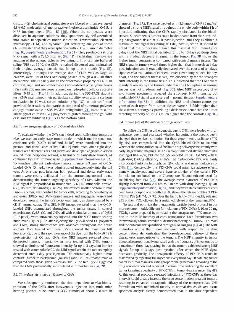
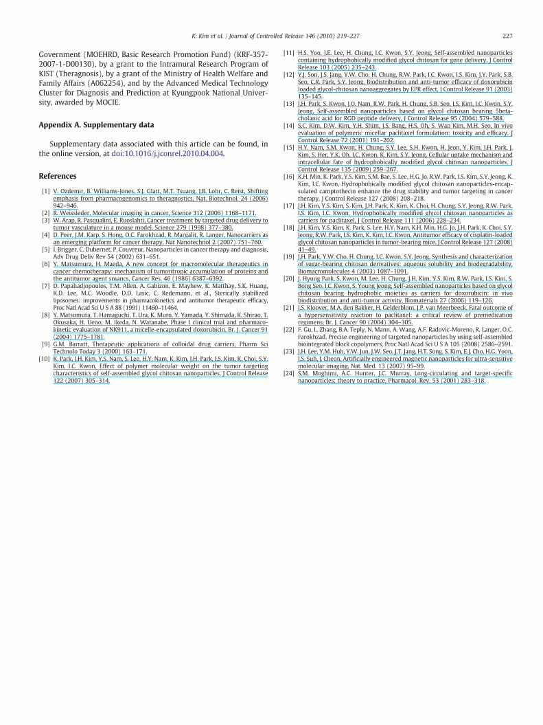
Fig. 1. (A) Conceptual description of a theragnostic nanoscale particle designed for cancepreferentially accumulate at the tumor tissue by the enhanced permeation and retention (EPcellular uptake. (B) Chemical structure of the glycol chitosan conjugates labeled with Cy5.5, a(C) A TEM image of Cy5.5-labeled CNPs (1 mg/ml) in distilled water. (D) Bright field and NIRfilter set (ex=674 nm, em=695 nm). (E) Time-dependant size distribution of Cy5.5-labelwater-soluble glycol chitosan (GC), CNPs, and polystyrene (PS) beads through filters of differthe filters was quantified through NIRF intensity of the filtrate. (G) In vitro stability of the Cand CNPs were incubated in 10% serum for 6 h at 37 °C and their migratory positions were
information. Briefly, first, water-soluble glycol chitosan (GC,Mw=250 kDa) were chemically modified with hydrophobic 5β-cholanic acid in the presence of N-hydroxysuccinimide (NHS) and 1-ethyl-3-(3-dimethylaminopropyl)-carbodiimide hydrochloride(EDAC), as previously reported [14–17]. The freshly synthesized glycolchitosan-5β-cholanic acid conjugates had 150±4.5 molecules ofhydrophobic 5β-cholanic acids per one glycol chitosan (glycolchitosan-5β-cholanic acid150, Mw=301 kDa). Second, the glycolchitosan-5β-cholanic acid conjugates were labeled with the NIRFdye, hydroxysuccinimide ester of Cy5.5. On average, each molecule ofglycol chitosan-5β-cholanic acid contained 4.8±0.7 molecules ofCy5.5 (Cy5.54.8-glycol chitosan-5β-cholanic aicd150, Mw=306 kDa).Finally, 10 wt.% of water insoluble paclitaxel (PTX) was encapsulatedinto the Cy5.5 labeled CNPs by a simple dialysis method (seeSupplementary information). The produced Cy5.5 labeled and PTX-encapsulated CNPs (PTX-CNPs) with a higher drug loading efficiencyof 92%werewell dispersed in distilledwater and PBS under sonication.As control nanoparticles, polystyrene bead (PS) containing aminemoieties (Polybead® Amino Micospheres 0.2 μm, Polysciences, Inc.Warrington, PA) were also labeled with monoreactive hydroxysucci-nimide ester of Cy5.5 and the Cy5.5 labeled PS particles were used asrigid and non-deformable particles in vitro and in vivo experiments.
r imaging and treatment. The theragnostic chitosan-based nanoparticles (CNPs) canR) effect due to their unique properties, such as stability in blood, deformability, and fastnear-infrared fluorescent (NIRF) dye, and modified with hydrophobic 5β-cholanic acid.F images of the Cy5.5-labeled CNPs in PBS. The NIRF image was obtained using a Cy5.5ed CNPs in PBS at 37 °C was confirmed using dynamic light scattering. (F) Filtration ofent pore sizes (0.8 μm, 0.45 μm, and 0.2 μm). The amount of each particle passed throughy5.5-labeled CNPs was determined using an SDS-PAGE test. Cy5.5-labeled GC polymersmonitored using a Cy5.5 filter set.

221K. Kim et al. / Journal of Controlled Release 146 (2010) 219–227
2.2. Characterizations of CNPs and PTX-CNPs
The average size and morphological shapes of Cy5.5 labeled CNPsand PTX-CNPs in distilled water or PBS were measured using dynamiclight scattering (1 mg/ml in PBS) (see Supplementary information, Fig.S1) and transmission electronmicroscopy (TEM) (1 mg/ml in distilledwater). The deformability of CNPs was evaluated using a simplefiltration test described as follows. 100 μl of Cy5.5-labeled CNPssolution (1 mg/ml) in PBS was continuously passed through syringefilter membranes (cellulose acetate, Millipore) with decreasing poresizes (0.8, 0.45, and 0.2 μm). After the filtration test, NIRF images of thefiltered solution were observed using a 12-bit CCD camera (KodakImaging Station 4000 MM, New Haven, CT) equipped with a Cy5.5bandpass emission filter set (680 to 720 nm; Omega Optical). Thestability of Cy5.5-labeled CNPs was confirmed by measuring themolecularweight changes of each sample following incubationwith orwithout 10 wt.% serum in PBS at 37 °C for 6 h. Both samples of Cy5.5-labeled GC polymer and Cy5.5-labeled CNPs (1 mg/ml)were dissolvedin reaction buffer (10 mM Tris–HCl (pH 7.5), 150 mMNaCl, and 1 mMEDTA)with orwithout 10 wt.% serumat 37 °C. After incubation for 6 h,each sample was loaded onto a vertical slab gel consisting of a 5%stacking gel and a 10% separating gel. All gels were run at a constantvoltage (120 V) in a Tris/glycine/SDS buffer. After the SDS-PAGE test,NIRF images of the gels were obtained with a 12-bit CCD cameraequipped with a Cy5.5 bandpass emission filter set.
2.3. In vitro drug release profile of the PTX-CNPs
To determine the in vitro drug release profile, 10 wt.% PTX-encapsulated CNPs were dispersed in 1 ml of PBS (pH 7.4) bysonication and were placed in cellulose ester membrane tubes(molecular weight cutoff=12 kDa–14 kDa, Spectrum®, RanchoDominquez, CA). The test tube was immersed in 10 ml of PBS andgently shaken at 37 °C in a water bath at 100 rpm. Samples of PBSsolution were taken at predetermined time intervals and analyzedwith isocratic reversed-phase HPLC [14].
2.4. Cytotoxicity of CNPs and PTX-CNPs
Murine squamous cell carcinomacells (SCC7)wereoriginally obtainedfrom the American Type Culture Collection (Rockville, MD) and culturedin RPMI 1640 (Gibco, Grand Island, NY) containing 10% (v/v) FBS (Gibco)and 1% (w/v) penicillin–streptomycin at 37 °C in a humidified 5% CO2–
95% air atmosphere. Cells were seeded at a density of 5×10³ cells/well in96-well flat-bottomed plates, and allowed to adhere overnight. After2 days post-incubation of each sample in the cell culture system, thecytotoxicity of free PTX dissolved in 50% (v/v) ethanol/Cremophor, CNPs,and PTX-CNPs was measured usingMTT assay. The data are expressed asthe percentages of viable cells compared to the survival of a control groupand were presented as mean±s.e. (n=5).
2.5. Animal models
All animal care and experimental procedures were performedaccording to the regulation of Kyungpook National University'sAnimal Care Committee. To generate an early-stage tumor model,athymic C3H-HeJ nude mice (average weight=20 g) were anesthe-tized in an isoflurane chamber and 1×10⁶ or 3×10⁶ SCC7 cells in 50 μlof saline were injected subcutaneously in the pectoral or dorsal sidesof mice. After 6, 8, 12, 15, and 18 days, mice were sacrificed and tumortissues were excised. The time-dependant angiogenic vessel forma-tion of each tumor was confirmed with a rat anti-mouse CD31monoclonal antibody (Pharmingen, San Diego, CA). After 8 days post-injection, the excised tumor tissues had grown to diameters ofbetween 2.6±0.3 and 6.2±0.5 mm and they showed newly formedangiogenic vessels at their peripheral regions (Supplementary
information, Fig. S2). The in vivo biodistribution of Cy5.5 labeledCNPs or PTX-CNPs were confirmed by inoculating 3×10⁶ SCC7 cells inthe dorsal side of male athymic C3H-HeJ nude mice (averageweight=20 g). After the injection of tumor cells, when the tumorreached 7–8 mm in diameter, the mice were used for in vivo imagingstudies within 10 days.
2.6. In vivo and ex vivo NIRF imaging
In vivo NIRF imaging was performed using a Kodak Image Station4000 MM. The illumination settings (lamp voltage, filter, andexposure time, etc.) used were identical to those in the animalimaging experiments and all the NIRF emission data were normalizedto photons per second per centimeter squared per steradian (p/s/cm2/sr) [12,18]. All fluorescence images were acquired with a ten-secondexposure time. The tumors and major organs were dissected andimaged again. For a quantitative comparison, tumor contrasts werecalculated by dividing the NIRF intensities at the tumor area by thoseof normal tissue areas. All data calculated using the regions of interest(ROIs) were drawn over tumor and normal tissues and the resultswere presented as mean±s.e. (n=3). For histological evaluation,excised tumors and other organs were frozen in Cryomatrix (frozenspecimen embedding medium) at −20 °C, and sectioned into 6 μmslices. The NIRF image of each tissue section was viewed byfluorescence microscopy (Carl Zeiss, Oberlcochem, Germany) with aCy5.5 filter set (Supplementary information, Fig. S4).
2.7. In vivo efficacy studies
The subcutaneous dorsa of C57BL/6 male mice (7 weeks old; 20 g)were inoculated with 3×10⁶ SCC7 cells. Mice were divided into fourgroups (n=10), and treated with one of the following treatments: (i)saline, (ii) CNPs, (iii) free PTX (20 mg/kg), and (iv) PTX-CNPs (20 mg/kg). Once the tumor diameter was approximately 7–8 mm, eachtreatment was administered via a tail vein every three days for12 days. The tumor volume was recorded for each tumor-bearingmouse for only 18 days since all the free PTX-treated mice were deadwithin 21 days. The length andwidth of the tumorsweremeasured bydigital calipers, calculating tumor volume using the followingformula: (width2×length)/2. Also, the survival rate of each groupwas recorded for 30 days. Eighteen days post-injection, the acutetoxicity of each formulationwas confirmed by counting the number ofWBC after collecting blood samples from normal mice, free PTX-treated mice, and PTX-CNP-treated mice (n=3). After 18 days, micewere sacrificed and tumor tissues were isolated. The excised tumorswere fixed with 4% (v/v) formaldehyde in PBS (pH 7.4) and sectionedinto 6 μm slices. Apoptotic and non-apoptotic cells in tumor tissueswere histologically evaluated with hematoxylin and eosin (H&E)staining, DAPI staining and terminal deoxynucleotidyl transferase-mediated nick end labeling (TUNEL) assays, with a commercialapoptosis detection kit (Promega Corp., WI).
2.8. Statistical analysis
Differences between experimental and control groups weredetermined using one-way ANOVA and deemed statistically signifi-cant (indicated by an asterisk (*) in figure) if p b0.05.
3. Results
3.1. Physicochemical properties, cellular uptake, and tumor specificity ofthe CNPs
Glycol chitosan (GC) (Mw=250 kDa) was modified in thepresence of chemical catalysts to introduce 150±4.5 molecules ofhydrophobic 5β-cholanic acid per polymer [19,20]. These glycol
222 K. Kim et al. / Journal of Controlled Release 146 (2010) 219–227
chitosan-5β-cholanic acid conjugates were labeled with an average of4.8±0.7 molecules of monoreactive hydroxysuccinimide Cy5.5, aNIRF imaging agent (Fig. 1B) [10]. When the conjugates weredissolved in aqueous solutions, they spontaneously self-assembledinto stable nanoparticles under sonication. Transmission electronmicroscopy (TEM) and dynamic light scattering analyses of theseCNPs revealed that they were spherical with 260±30 nm in diameter(Fig. 1C, Supplementary information, Fig S1). They produced a strongNIRF signal with a Cy5.5 filter set (Fig. 1D), facilitating non-invasiveimaging of the nanoparticles in live animals. In phosphate-bufferedsaline (PBS) at 37 °C, the CNPs remained dispersed and maintainedtheir original average particle size for up to one month (Fig. 1E).Interestingly, although the average size of CNPs was as large as260 nm, over 95% of the CNPs easily passed through a 0.2-μm filtermembrane. This is partly due to the deformable property of CNPs. Incontrast, rigid and non-deformable Cy5.5-labeled polystyrene beads(PSs) with 200 nm size were retained on hydrophilic cellulose acetatefilters (0.45 μm) (Fig. 1F). In addition, during the SDS-PAGE stabilitytest, CNPs maintained their particulate stability, even after a six-hourincubation in 10 wt.% serum solution (Fig. 1G), which confirmedprevious observations that particles composed of numerous polymerconjugates are stable in SDS-PAGE running buffer [21]. In contrast, thelinear glycol chitosan (GC) polymers migrated through the gel withease and are visible in Fig. 1G as the bottom band.
3.2. Tumor targeting efficacy of Cy5.5-labeled CNPs
To evaluatewhether the CNPs can indeed specifically target tumors invivo, we used an early-stage tumor model in which murine squamouscarcinoma cells (SCC7; 1×10⁶ and 3×10⁶) were inoculated into thepectoral and dorsal sides of live C3H-HeJ nude mice. After eight days,tumors with different sizes had grown and developed angiogenic vesselsaround their peripheral regions, a hallmark of early-stage tumors, asconfirmed by CD31 immunoassay (Supplementary information, Fig. S2).To visualize different early-stage tumors in mice, 3.3 μmol of Cy5.5-labeled CNPs (5 mg/kg) was administrated intravenously into the tailvein. At one day post-injection, both pectoral and dorsal early-stagetumors were clearly delineated from the surrounding normal tissue,demonstrating the tumor targeting specificity of CNPs, wherein theNIRF signal is proportional to tumor size (2.6±0.3 mm; solid arrows,6.2±0.5 mm, dot arrows) (Fig. 2A). The excised smaller pectoral tumor(size=2.6 mm) was positive for tumor cells, according to hematocxylinand eosin (H&E)- and DAPI-stained images, and angiogenic vessels haddeveloped around the tumor's peripheral region, as demonstrated by aCD-31 immunoassay (Fig. 2B). NIRF images revealed that the Cy5.5-labeled CNPs accumulated throughout the tumor tissue. In controlexperiments, Cy5.5, GC, and CNPs, all with equimolar amounts of Cy5.5(0.16 μmol), were intravenously injected into the SCC7 tumor-bearingnude mice (Fig. 2C). 1 h after injecting the Cy5.5-labeled GC polymersand CNPs, strong fluorescence was observed throughout the entireanimals. Mice treated with free Cy5.5 showed the minimum NIRfluorescence, due to the rapid clearance of the dye from the body. At 12 hpost-injection of GC and CNPs, the NIRF images revealed clearlydelineated tumors. Importantly, in mice treated with CNPs, tumorsshowed undiminished fluorescent intensity for up to 3 days, but in micetreated with water-soluble GC, the NIRF signal within the tumors rapidlydecreased after 1 day post-injection. The substantially higher tumorcontrast (tumor to background (muscle) ratio) in CNP-treated mice ascompared with those given water-soluble GC or free Cy5.5 suggestedthat the CNPs preferentially accumulated in tumor tissues (Fig. 2D).
3.3. Time-dependent biodistribution of CNPs
We subsequently monitored the time-dependent in vivo biodis-tribution of the CNPs after intravenous injection into nude micebearing pectoral subcutaneous SCC7 tumors of about 7–8 mm in
diameter (Fig. 3A). The mice treated with 3.3 μmol of CNP (5 mg/kg)showed a strong NIRF signal throughout the whole body within 1 h ofinjection, indicating that the CNPs rapidly circulated in the blood-stream. Subcutaneous tumors could be delineated from the surround-ing background tissue at 12 h post-injection, and they exhibited amaximum NIR signal beginning at 1 day post-injection. It should benoted that the tumors maintained this maximal NIRF intensity for3 days, but the NIRF signal persisted for up to 10 days post-injection,with a gradual decrease in signal in the tumor. Fig. 3B shows thehigher tumor contrasts as compared with control muscle tissues. TheNIRF signal in tumors was 6 times higher than that in muscle at 1 daypost-injection, and it gradually decreased over the course of ten days.Upon ex vivo evaluation of excised tissues (liver, lung, spleen, kidney,heart, and the tumors themselves), we observed by far the strongestNIRF intensity in the tumor tissue. This indicated that the CNPs weremainly taken up by the tumors, whereas the CNP uptake in normaltissues was not predominant (Fig. 3C). Also, NIRF microscopy of exvivo tumor specimens revealed the strongest NIRF intensity, butnegligible NIRF signal was observed in normal tissues (Supplementaryinformation, Fig. S3). In addition, the NIRF total photon counts pergram of each organ from tumor tissues were 4–7 folds higher thanthose from other organs, providing a decisive evidence that the tumortargeting property of CNPs is much higher than the controls (Fig. 3D).
3.4. In vivo fate of the anticancer drug-loaded CNPs
To utilize the CNPs as a theragnostic agent, CNPs were loadedwith ananticancer agent and evaluated whether harboring a therapeutic agentchanged their in vivo distribution. For these experiments, paclitaxel (PTX,Fig. 4A) was encapsulated into the Cy5.5-labeled CNPs to examinewhether thenanoparticles could facilitatedrugdelivery concurrentlywithearly-stage tumor imaging (Fig. 4A). A dialysismethod allowed successfulloadingof 10% (w/w)PTX into theCy5.5-labeledCNPs (PTX-CNPs)with ashigh drug loading efficiency as 92%. The hydrophobic PTX was easilyincorporated into the hydrophobic 5β-cholanic acid inner multicores ofCNPs [14]. Conceivably, this PTX-CNPs could minimize adverse effects,namely anaphylaxis and severe hypersensitivity, of the current PTXformulation attributed to the Cremophore EL and ethanol used forsolubilizing free PTX [22]. The average mean diameter of PTX-CNPsslightly increased from 260 nm to 310 nm with drug loading (Fig. 4B,Supplementary information, Fig. S1), and theywere stable under aqueousconditions for up to onemonth. Fig. 4C shows the release profiles of PTX-CNPs in PBS (pH 7.4, 37 °C). Over the first 6 h, the CNPs quickly released35% of their PTX, followed by a sustained release of the remaining PTX.
To test and optimize the theragnostic particle-based protocol in ourmurine tumormodel, different formulations of PTX-CNPs (5, 10, or 20 mgPTX/kg) were prepared by correlating the encapsulated PTX concentra-tion to the NIRF intensity of each nanoparticle. Each formulation wasintravenously administered tonudemiceharboring7–8 mmSCC7 tumorsand the outcomewas quantified throughNIRF imaging (Fig. 4E). TheNIRFintensities within the tumors increased with respect to the drugconcentration, demonstrating the dose-dependent delivery of thesetheragnostic nanoparticles to the tumors. The NIRF intensity in tumortissuesalsoproportionally increasedwith the frequencyof injectionsup toa maximum three-day spacing, in that the tumors exhibited strong NIRFsignals for up to 3 days post-injection, after which the NIRF signaldecreased gradually. The theragnostic efficacy of PTX-CNPs could bemaximized by repeating the injections every third day. Of note, the tumorcontrast (tumor tomuscle ratio)proportionally increasedaccording to thedrug concentration and repeated injection time, indicating the excellenttumor targeting specificity of PTX-CNPs in tumor-bearing mice (Fig. 4F).At this optimal protocol, repeated injections of PTX-CNPs at three-dayintervals could greatly increase the drug concentration in target tumors,resulting in enhanced therapeutic efficacy of the nanoparticulate CNPformulation with minimized toxicity to normal tissues. Ex vivo tissuespecimen analysis clearly demonstrated that in the dose-dependent

Fig. 2. (A) In vivo imaging of Cy5.5-labeled CNPs in tumor-bearing mice. The early-stage tumor models were generated by injecting subcutaneously 1×10⁶ or 3×10⁶ SCC7 cells intothe pectoral and dorsal sides of C3H-HeJ nude mice. After eight days, different size of tumors had grown to 2.6±0.3 mm (solid arrow) and 6.2±0.5 mm (dotted arrow). At 1-daypost-injection of 3.3 μmol of Cy5.5-labeled CNPs (5 mg/kg), the NIRF images were obtained using a 12-bit CCD camera equipped with a Cy5.5 bandpass emission filter (NIRF signalscale: 121–2087). (B) The smaller pectoral tumor was positive for tumor cells, as confirmed by H&E staining. Fluorescence microscopic images show the CD-31-positive angiogenicvessels (yellow) and DAPI-stained tumor cells (blue). Intravenously injected, Cy5.5-labeled CNPs were visualized in tumor tissues (red) (original magnification×100). (C) Time-dependent tumor targeting specificity of free Cy5.5, Cy5.5-labeled GC polymers, and Cy5.5-labeled CNPs, all with equimolar amounts of Cy5.5 (0.16 μmol), in SCC7 tumor-bearingmice (tumor diameter=7–8 mm). (NIRF signal scale: 52–2390). (D) Tumor to background (muscle) ratio as a function of time after administration of Cy5.5, Cy5.5-labeled GCpolymers, and Cy5.5-labeled CNPs in SCC7 tumor-bearing mice. All data represent mean±s.e.
223K. Kim et al. / Journal of Controlled Release 146 (2010) 219–227
tumor targeting ability of PTX-CNPs; even when high doses of PTX-CNPswere administered twice over a three-day period, the NIRF intensity fromthe normal tissue (except the liver) was barely detectable above thebackground levels (Fig. 4G). With regard to liver tissues, NIRF increasedwith the drug concentration, but the intensity was consistently muchlower than that from the tumors.
3.5. Theragnostic potential of PTX-CNPs
In addition to confirming tumor-specific targeting, NIRF imaging wasalso utilized to directly monitor tumor growth rate in response to Cy5.5-labeled PTX-CNP administration in live animals. This is to demonstratethat our theragnostic particles could noninvasively track therapeuticefficacy.
Fig. 5A shows the tumor specificity of the PTX-CNPs (20 mg/kg PTX)and the resulting tumor growth rates of the SCC7 tumors (about 7–8 mmin diameter). As before, the NIRF signal from the tumor tissue increasedproportionally with the number of Cy5.5-labeled PTX-CNP injections, upto 5 times in 3 days (solid arrows), confirming the tumor targetingspecificityof PTX-CNPs.When tumorgrowthwasmonitoredover aperiodof 18 days, the NIRF images showed that in control mice, the tumorgrowth rate rapidly increased (data not shown), but tumorsmaintained aconstant size inmice givenfive repeated injections of CNPs. After the finalinjection at day 15, the tumor growth subsided, due to the anticancer
effect of PTX within the CNPs. In these optimization studies, thetherapeutic nanoparticles successfully mediated both drug delivery andmolecular imaging in our nude mouse model.
The optimized treatment protocol, derived from our NIRF imagingdata, was used to evaluate the detailed therapeutic efficacy of CNPs inan immunocompetent mouse model, SCC7 tumor-bearing C57BL/6male mice. After their subcutaneous tumors grew to 7–8 mm indiameter, comparative efficacy studies were performed by dividingthe animals into five groups (n=10 per group) in a way thatminimized tumor size differences among the groups. Using thepreviously-reported maximal tolerated dose (MTD) of 20 mg/kg forPTX as a reference point [21], we administered the followingformulations, using four intravenous injections every third day: (i)saline, (ii) empty CNPs, (iii) PTX (20 mg/kg) in ethanol/Cremophoresolution, and (iv) PTX-CNPs (20 mg/kg encapsulated PTX) in saline.Therapeutic efficacy was examined bymeasuring tumor volumes overthe course of 18 days (Fig. 5B). The measurement was limited to18 days, because all mice treated with free PTX were dead within21 days. The results demonstrated that administration of free PTX andPTX-CNPs significantly suppressed tumor growth as compared withvector controls (saline and empty CNPs). Treatment with saline orempty CNPs did not mediate any therapeutic efficacy, and the meantumor volumes at the end of the study for these groups were8000 mm3 and 7900 mm3, respectively. The PTX-CNP-treated mice

Fig. 3. (A) In vivo biodistribution of Cy5.5-labeled CNPs in SCC7 tumor-bearing mice. After tumor diameters reached 7–8 mm, 3.3 μmol of Cy5.5-labeled CNPs (5 mg/kg) wereintravenously injected into the tumor-bearing C3H/HeN nude mice. (NIRF signal scale: 45–2680). (B) Time-dependent tumor contrast after administration of Cy5.5-labeled CNPsinto tumor-bearing mice (n=3). (C) NIRF images of the dissected major organs harvested from Cy5.5-labeled CNP-treated mice. The first row represents the bright field images ofindividual organs. A strong NIRF signal was observed in tumor tissues at all experimental times. (D) Ex vivo imaging of SCC7 xenograft tumor showed higher NIRF signal than otherorgans at all time points. A quantification of in vivo targeting characteristics of Cy5.5-labeled CNPs was recorded as total photons per centimeter squared per steradian (p/s/cm2/sr)per milligram of each organ at all time points (n=3 mice per group). All data represent mean±s.e.
224 K. Kim et al. / Journal of Controlled Release 146 (2010) 219–227
demonstrated that in the most dramatic reduction in tumor volume;the final mean tumor load was 1000 mm3 by the end of the study(mean±s.e., n=4, ANOVA at 95% confidence interval), significantlysmaller than in mice given the free PTX formulation (2400 mm3).These results suggested that the superior tumor specificity of PTX-CNPs might have enhanced the efficacy of the anticancer drug,increasing the therapeutic drug concentration in tumor tissues. Wealso performed histological staining of the excised tumors, and anindependent pathologist evaluated the slides. Median tumors excisedfrom PTX-CNP-treated mice exhibited a reduction in the number ofcancerous cells and an increase in apoptotic cell nuclei, as demon-strated by H&E staining and TUNEL assays, respectively (Fig. 5C). Incontrast, the saline- and empty CNP-treated tumors contained largernumbers of tumor cells and exhibited few signs of apoptosis.Importantly, PTX-CNP treatment greatly enhanced mouse survivalrates (Fig. 5D). Eight of the 10 PTX-CNP-treated mice survived the 30-day study, but all themice in the free PTX-treated groups had succumbedby day 21, most likely due to the severe toxicity of the free PTX
formulation.Within the control groups, only 6 and5mice from the saline-and empty CNP-treated mice, respectively, survived to the end of theexperiment, due to rapid unchecked tumor growth. These results,supported by our in vitro cytotoxicity studies, suggested that the PTX-CNPs enhanced the tumor-specific delivery of PTX while curtailing itsassociated cytotoxicity. In addition, to assess the acute toxicity of eachtreatment regimen in vivo, we analyzed WBC counts within eachexperimental group after 18 days of treatment (Fig. 5E). The free PTX-treated mice showed substantially reduced WBC counts, compared toPTX-CNP-treatedmice. These results indicated that encapsulating the PTXwithin the nanoscale particles greatly reduced its severe toxicity [14].
4. Discussion
There has been considerable interest in the development ofnanotechnology platforms to diagnose and treat cancer by usingtargeted delivery and controlled drug release. Many approaches havebeen proposed to develop nanoparticle constructs containing a

Fig. 4. (A) Chemical structure of paclitaxel (PTX). (B) A TEM image of 1 mg/ml of PTX-CNPs encapsulated with 10% PTX (w/w). (C) A PTX release profile of 10% PTX (w/w)-encapsulated PTX-CNPs in PBS (pH 7.4, 37 °C) (n=5). All data represent mean±s.e. (D) Cytotoxicity of free PTX and PTX-CNPs in cell culture. Predetermined concentrations of PTXand PTX-CNPs were incubated with 5×10³ SCC7 tumor cells/well in 96-well plates for 48 h. (E) Cy5.5-labeled PTX-CNP doses at different PTX concentrations (5 mg/kg, 10 mg/kg,and 20 mg/kg) were intravenously injected into the SCC7 tumor-bearing mice every third days. The black arrow indicates intravenous injection of Cy5.5-labeled PTX-CNPs. The NIRFsignal proportionally increased with dose and injection time (NIRF signal scale: 61–5523). (F) A quantification of the in vivo targeting characteristics of Cy5.5-labeled PTX-CNPs wasrecorded as tumor contrast to background (muscle) as a function of drug concentration and injection frequency (n=3). All data represent mean±s.e. (G) NIRF images of dissectedorgans harvested from Cy5.5-labeled PTX-CNP-treated mice.
225K. Kim et al. / Journal of Controlled Release 146 (2010) 219–227
targeting moiety, such as antibody, aptamer, and small molecularligand, for optimized tumor homing with reduced nonspecificaccumulation [22,23]. These targeting moieties showed selectivedelivery of nanoparticles into tumors or selective detection in tumors.Despite of their improved selectivity, they still have to overcome anobstacle that large percentages of nanoparticles continue to accumu-late in the liver and spleen. Conventional wisdom in tumor targeting isbased on the EPR effect of nanoparticles, and so it has been assumedthat all nanoparticles, as long as the size is in the range of 200 nm orless, will automatically go to the solid tumor by the EPR effect [3–5].However, the amount of a nanodelivery systemdelivered to the tumorsite is only slightly larger than that of the control. Apparently,nanoparticulate delivery systems are not working well as we allexpected. In contrast with the conventional nanocarriers, however,the CNPs used in this study progressively accumulated in the tumor
tissues by the EPR effect within 24 h and lasted up to 10 days withconsiderably low uptake in the liver and spleen.
The efficiency of the EPR effect in vivowas significantly affected bydifferent types of carriers regardless of size. In this study, thedeformable CNPs showed highest tumor-targeting efficiency. The invivo clearance rate was notably determined by the size and thedeformability of the nanoparticles. Generally, nanoparticles of 150–300 nm are mainly found in the liver and the spleen [24]. Therefore,the size of engineered long-circulatory nanoparticles must be eithersmaller than the critical size or deformable enough in order to avoidthe in vivo filtration systems. In vivo, this deformability might enablethese CNPs to pass through biological barriers, such as micro-sizeblood vessels and membranes in the body. In addition, the rapidcellular uptake characteristic of the CNPs facilitates delivery of anti-cancer drugs and imaging agents into the cytoplasmic compartment

Fig. 5. (A) In vivo images of tumor specificity and tumor growth rate in Cy5.5-labeled PTX-CNP (20 mg/kg of free PTX)-treated mice (NIRF signal scale: 34–8014). Cy5.5-labeled PTX-CNPs were injected to tumor-bearing mice four times every three days. The black arrow indicates intravenous injection of Cy5.5-labeled PTX-CNPs. (B) Comparative therapeuticefficacy studies of PTX-CNPs in SCC7 tumor-bearing C57BL/6 male mice (n=10, tumor diameter was 7–8 mm). Tumor size was observed for 18 days. Data represent mean±s.e. *,data points for the PTX-CNP-treated group that were statistically significant compared to all other groups by ANOVA at 95% confidence interval. (C) Representative images of excisedtumors treated with saline, CNPs, free PTX, and PTX-CNPs for 18 days. Histopathological analysis with H&E, DAPI, and TUNEL stainings of each dissected tumor tissue (originalmagnification×100). (D) Survival curves demonstrated that 90% of the PTX-CNPs group survived to day 30, whereas the entire PTX-treated group succumbedwithin 18 days. (E) Theacute toxicity of the different formulations was confirmed by WBC counting at seven days post-treatment (n=3 mice per group). All data represent mean±s.e.
226 K. Kim et al. / Journal of Controlled Release 146 (2010) 219–227
of target tumor cells [15]. The exact mechanism of rapid cellularuptake is not clearly understood yet, but the nanoparticulate structureis critical, as linear water-soluble GC polymer is not taken up by thecells. Taken together, we can suggest that the unique characteristics ofthe CNPs that we observed in vitro (i.e., stability in blood, deform-ability, and rapid cellular uptake) may have contributed significantlyto their tumor targeting by the EPR effect in vivo.
5. Conclusion
In conclusion, we developed and characterized the theragnostic CNPswhich deliver a fluorescent probe for live imaging and PTX for cancertreatment, simultaneously. These therapeutic CNPs accumulated at thetumor tissuesmuchmore efficiently thanwater-soluble linear polymer orpolystyrene beads. The fast cellular uptake of the CNPs resulted in highertherapeutic efficacy. The NIRF label allowed us to noninvasively monitor
the in vivo fate of the nanoparticles in live animals. Under the optimizedtreatment protocol, administration of PTX-loaded CNPs greatly dimin-ished tumor sizewhileminimizing the severe toxicity associatedwith freePTX administration. Furthermore, our results suggested that thismethodology could be used to evaluate the performance of othernanoscale drug carriers in cancer therapy, without sacrificing a largenumber of animals. We anticipate cancer theragnosis with these CNPspresents a new strategy in cancer treatment, in which early-stage cancerdiagnosis, drug delivery, and real-time non-invasive monitoring fortherapeutic efficacy are carried out simultaneously.
Acknowledgments
This work was financially supported by the Real-Time MolecularImaging Project, F104AA01003-06A0101-00310, the GRL Program ofMEST, by a Korea Research Foundation Grant funded by the Korean
227K. Kim et al. / Journal of Controlled Release 146 (2010) 219–227
Government (MOEHRD, Basic Research Promotion Fund) (KRF-357-2007-1-D00130), by a grant to the Intramural Research Program ofKIST (Theragnosis), by a grant of the Ministry of Health Welfare andFamily Affairs (A062254), and by the Advanced Medical TechnologyCluster for Diagnosis and Prediction at Kyungpook National Univer-sity, awarded by MOCIE.
Appendix A. Supplementary data
Supplementary data associated with this article can be found, inthe online version, at doi:10.1016/j.jconrel.2010.04.004.
References
[1] V. Ozdemir, B. Williams-Jones, S.J. Glatt, M.T. Tsuang, J.B. Lohr, C. Reist, Shiftingemphasis from pharmacogenomics to theragnostics, Nat. Biotechnol. 24 (2006)942–946.
[2] R. Weissleder, Molecular imaging in cancer, Science 312 (2006) 1168–1171.[3] W. Arap, R. Pasqualini, E. Ruoslahti, Cancer treatment by targeted drug delivery to
tumor vasculature in a mouse model, Science 279 (1998) 377–380.[4] D. Peer, J.M. Karp, S. Hong, O.C. Farokhzad, R. Margalit, R. Langer, Nanocarriers as
an emerging platform for cancer therapy, Nat Nanotechnol 2 (2007) 751–760.[5] I. Brigger, C. Dubernet, P. Couvreur, Nanoparticles in cancer therapy and diagnosis,
Adv Drug Deliv Rev 54 (2002) 631–651.[6] Y. Matsumura, H. Maeda, A new concept for macromolecular therapeutics in
cancer chemotherapy: mechanism of tumoritropic accumulation of proteins andthe antitumor agent smancs, Cancer Res. 46 (1986) 6387–6392.
[7] D. Papahadjopoulos, T.M. Allen, A. Gabizon, E. Mayhew, K. Matthay, S.K. Huang,K.D. Lee, M.C. Woodle, D.D. Lasic, C. Redemann, et al., Sterically stabilizedliposomes: improvements in pharmacokinetics and antitumor therapeutic efficacy,Proc Natl Acad Sci U S A 88 (1991) 11460–11464.
[8] Y. Matsumura, T. Hamaguchi, T. Ura, K. Muro, Y. Yamada, Y. Shimada, K. Shirao, T.Okusaka, H. Ueno, M. Ikeda, N. Watanabe, Phase I clinical trial and pharmaco-kinetic evaluation of NK911, a micelle-encapsulated doxorubicin, Br. J. Cancer 91(2004) 1775–1781.
[9] G.M. Barratt, Therapeutic applications of colloidal drug carriers, Pharm SciTechnolo Today 3 (2000) 163–171.
[10] K. Park, J.H. Kim, Y.S. Nam, S. Lee, H.Y. Nam, K. Kim, J.H. Park, I.S. Kim, K. Choi, S.Y.Kim, I.C. Kwon, Effect of polymer molecular weight on the tumor targetingcharacteristics of self-assembled glycol chitosan nanoparticles, J Control Release122 (2007) 305–314.
[11] H.S. Yoo, J.E. Lee, H. Chung, I.C. Kwon, S.Y. Jeong, Self-assembled nanoparticlescontaining hydrophobically modified glycol chitosan for gene delivery, J ControlRelease 103 (2005) 235–243.
[12] Y.J. Son, J.S. Jang, Y.W. Cho, H. Chung, R.W. Park, I.C. Kwon, I.S. Kim, J.Y. Park, S.B.Seo, C.R. Park, S.Y. Jeong, Biodistribution and anti-tumor efficacy of doxorubicinloaded glycol-chitosan nanoaggregates by EPR effect, J Control Release 91 (2003)135–145.
[13] J.H. Park, S. Kwon, J.O. Nam, R.W. Park, H. Chung, S.B. Seo, I.S. Kim, I.C. Kwon, S.Y.Jeong, Self-assembled nanoparticles based on glycol chitosan bearing 5beta-cholanic acid for RGD peptide delivery, J Control Release 95 (2004) 579–588.
[14] S.C. Kim, D.W. Kim, Y.H. Shim, J.S. Bang, H.S. Oh, S. Wan Kim, M.H. Seo, In vivoevaluation of polymeric micellar paclitaxel formulation: toxicity and efficacy, JControl Release 72 (2001) 191–202.
[15] H.Y. Nam, S.M. Kwon, H. Chung, S.Y. Lee, S.H. Kwon, H. Jeon, Y. Kim, J.H. Park, J.Kim, S. Her, Y.K. Oh, I.C. Kwon, K. Kim, S.Y. Jeong, Cellular uptake mechanism andintracellular fate of hydrophobically modified glycol chitosan nanoparticles, JControl Release 135 (2009) 259–267.
[16] K.H. Min, K. Park, Y.S. Kim, S.M. Bae, S. Lee, H.G. Jo, R.W. Park, I.S. Kim, S.Y. Jeong, K.Kim, I.C. Kwon, Hydrophobically modified glycol chitosan nanoparticles-encap-sulated camptothecin enhance the drug stability and tumor targeting in cancertherapy, J Control Release 127 (2008) 208–218.
[17] J.H. Kim, Y.S. Kim, S. Kim, J.H. Park, K. Kim, K. Choi, H. Chung, S.Y. Jeong, R.W. Park,I.S. Kim, I.C. Kwon, Hydrophobically modified glycol chitosan nanoparticles ascarriers for paclitaxel, J Control Release 111 (2006) 228–234.
[18] J.H. Kim, Y.S. Kim, K. Park, S. Lee, H.Y. Nam, K.H. Min, H.G. Jo, J.H. Park, K. Choi, S.Y.Jeong, R.W. Park, I.S. Kim, K. Kim, I.C. Kwon, Antitumor efficacy of cisplatin-loadedglycol chitosan nanoparticles in tumor-bearing mice, J Control Release 127 (2008)41–49.
[19] J.H. Park, Y.W. Cho, H. Chung, I.C. Kwon, S.Y. Jeong, Synthesis and characterizationof sugar-bearing chitosan derivatives: aqueous solubility and biodegradability,Biomacromolecules 4 (2003) 1087–1091.
[20] J. Hyung Park, S. Kwon, M. Lee, H. Chung, J.H. Kim, Y.S. Kim, R.W. Park, I.S. Kim, S.Bong Seo, I.C. Kwon, S. Young Jeong, Self-assembled nanoparticles based on glycolchitosan bearing hydrophobic moieties as carriers for doxorubicin: in vivobiodistribution and anti-tumor activity, Biomaterials 27 (2006) 119–126.
[21] J.S. Kloover, M.A. den Bakker, H. Gelderblom, J.P. van Meerbeeck, Fatal outcome ofa hypersensitivity reaction to paclitaxel: a critical review of premedicationregimens, Br. J. Cancer 90 (2004) 304–305.
[22] F. Gu, L. Zhang, B.A. Teply, N. Mann, A. Wang, A.F. Radovic-Moreno, R. Langer, O.C.Farokhzad, Precise engineering of targeted nanoparticles by using self-assembledbiointegrated block copolymers, Proc Natl Acad Sci U S A 105 (2008) 2586–2591.
[23] J.H. Lee, Y.M. Huh, Y.W. Jun, J.W. Seo, J.T. Jang, H.T. Song, S. Kim, E.J. Cho, H.G. Yoon,J.S. Suh, J. Cheon, Artificially engineered magnetic nanoparticles for ultra-sensitivemolecular imaging, Nat. Med. 13 (2007) 95–99.
[24] S.M. Moghimi, A.C. Hunter, J.C. Murray, Long-circulating and target-specificnanoparticles: theory to practice, Pharmacol. Rev. 53 (2001) 283–318.
Copyright © 2022 FDOKUMEN