Trypanosoma cruzi: Correlation of muscle lesions with contractile properties in the acute phase of...
9
This article appeared in a journal published by Elsevier. The attached copy is furnished to the author for internal non-commercial research and education use, including for instruction at the authors institution and sharing with colleagues. Other uses, including reproduction and distribution, or selling or licensing copies, or posting to personal, institutional or third party websites are prohibited. In most cases authors are permitted to post their version of the article (e.g. in Word or Tex form) to their personal website or institutional repository. Authors requiring further information regarding Elsevier’s archiving and manuscript policies are encouraged to visit: http://www.elsevier.com/copyright
-
Upload
independent -
Category
Documents
-
view
10 -
download
0
Transcript of Trypanosoma cruzi: Correlation of muscle lesions with contractile properties in the acute phase of...

This article appeared in a journal published by Elsevier. The attachedcopy is furnished to the author for internal non-commercial researchand education use, including for instruction at the authors institution
and sharing with colleagues.
Other uses, including reproduction and distribution, or selling orlicensing copies, or posting to personal, institutional or third party
websites are prohibited.
In most cases authors are permitted to post their version of thearticle (e.g. in Word or Tex form) to their personal website orinstitutional repository. Authors requiring further information
regarding Elsevier’s archiving and manuscript policies areencouraged to visit:
http://www.elsevier.com/copyright
Author's personal copy
Trypanosoma cruzi: Correlation of muscle lesions with contractile propertiesin the acute phase of experimental infection in mice (Mus musculus)
Mario V. Ramirez-Archila a,c,1, Jesús Muñiz a,1, Adolfo Virgen-Ortiz a,d,1,3, Oscar Newton-Sánchez b,2,Valery G. Melnikov b,2, Oxana R. Dobrovinskaya a,⇑a Centro Universitario de Investigaciones Biomédicas, Universidad de Colima, Av. 25 de Julio 965, C.P. 28045 Colima, Col., Mexicob Facultad de Medicina, Universidad de Colima, Av. Universidad # 333 Col. Las Viboras, C.P. 28040 Colima, Col., Mexicoc Facultad de Ciencias de la Educación, Universidad de Colima, Av. Universidad # 333 Col. Las Viboras, C.P. 28040 Colima, Col., Mexicod Departamento de Ciencias Químico Biológicas, Unidad Regional Sur, Universidad de Sonora, Blvd. Lázaro Cárdenas, No. 100 Col Francisco Villa, 85880 Novojoa, Sinaloa, Mexico
a r t i c l e i n f o
Article history:Received 8 October 2010Received in revised form 10 February 2011Accepted 14 February 2011Available online 18 February 2011
Keywords:Trypanosoma cruziMuscular histotropismContractile propertiesRectus abdominisPlantarisSingle twitchTetanus
a b s t r a c t
Parasitism in skeletal muscles and myositis are commonly observed during experimental Trypanosomacruzi infection. The effect of T. cruzi infection on contractile properties of skeletal muscles in consecutiveperiods of the acute infection in BALB/c mice was studied. Albarrada strain (clone 4) which was isolatedin Mexico and has demonstrated a high level of blood parasitemia and parasitism in skeletal muscles wasused. Isolated strips of rectus abdominis muscle were subjected to direct electrical field in vitro. Alterna-tively, plantaris muscles were stimulated in situ through the sciatic nerve. The peak amplitudes of a singletwitch and tetanus contractions were considered to estimate the mechanical properties of muscles. His-topathological analysis was performed to correlate functional changes with the evolution of tissue para-sitism and tissue injury. Contractile properties of muscles were significantly attenuated during acute T.cruzi infection. The percentage of damaged muscles rather than the character of tissue pathology affectedtheir contractile properties significantly.
� 2011 Elsevier Inc. All rights reserved.
1. Introduction
Chagas’ disease, caused by the protozoan Trypanosoma cruzi,represents the major cause of the heart pathology in the endemicregions of Latin America. Over 15 million people are affected, andapproximately 28 million people are at risk of T. cruzi infection(WHO, 2007). In recent years, due to tourism and migration, thecases of Chagas’ disease were also reported in non-endemic coun-tries (Kirchhoff, 1993; Gascón et al., 2007).
In infected mammals, including humans, Chagas’ disease goesthrough three characteristic phases: the acute, indeterminate andchronic. The main feature of the acute phase is the presence of freetrypomastigotes in the bloodstream (parasitemia). Subsequently,bloodstream forms invade the hosts’ tissues and convert into intra-cellular proliferating forms (amastigotes). The majority of T. cruzistrains possess cardiomiotropism, and 10–30% of T. cruzi-infected
individuals develop varying degree of acute to chronic myocarditis(Acquatella, 2008). Skeletal muscles were also shown to be readilyinfected by T. cruzi. Muscular pain and weakness were reported inpatients with Chagas’ disease (Köberle, 1968). Myositis of deltoidand gastrocnemius muscle was demonstrated in patients withchronic and acute Chagas’ disease and cardiopathy (Cenget and Ro-jas, 1959; Ponce, 1972). Cases of patients developing chagasic pol-ymyositis have also been reported (Cossermelli et al., 1978).Furthermore, structural alterations in myofibrils were found inmuscle biopsies of chronically infected individuals (Laguenset al., 1975). These alterations coincided with the presence of circu-lating antibodies against striated muscle fibers and the plasmamembrane of endothelial cells (Laguens et al., 1975; Laguens andCabeza Markert, 1991). Parasitism in different muscle groups,myositis, degeneration and necrosis of myofibrils were commonlyobserved during both acute and chronic phases of experimentalinfection in mice (Bijovsky et al., 1983; Molina et al., 1987; Losavioet al., 1989; Monteón et al., 1996). Myofibrillar breakdown andcytoskeleton alterations are most likely to be the result of differentpathogenic processes taking place in affected muscles, includingdirect destruction of myofibrils by the parasites, tissue damagecaused by inflammation and production of the nitric oxide (NO)and pro-inflammatory cytokines, microvascular lesions and anindirect effect of cross-reactive antibodies (revised in: Scharfstein
0014-4894/$ - see front matter � 2011 Elsevier Inc. All rights reserved.doi:10.1016/j.exppara.2011.02.012
⇑ Corresponding author. Address: Centro Universitario de Investigaciones Bio-médicas, Universidad de Colima, Av. 25 de Julio 965, C.P. 28045 Colima, Col.,Mexico. Fax: +52 312 41 61 129.
E-mail address: [email protected] (O.R. Dobrovinskaya).1 Fax: +52 312 41 61 129.2 Fax: +52 312 31 61 099.3 Fax: +1 642 425 9952.
Experimental Parasitology 128 (2011) 301–308
Contents lists available at ScienceDirect
Experimental Parasitology
journal homepage: www.elsevier .com/locate /yexpr
Author's personal copy
et al., 2009). There is also evidence of neuromuscular junctiondamage (Mirkin et al., 1994). In the late period of infection the par-asitism and inflammation in tissues decreased. However, the pro-cess of muscle regeneration in this period involves thereplacement of muscle tissues by fibrosis (Buckner et al., 1999).Although the detailed mechanism of this phenomenon is notknown yet, studies on cell culture models in vitro revealed thatthe myotropic Brazil strain of T. cruzi profoundly affected the abil-ity of L6E9 myoblasts to differentiate into mature muscle myotu-bes (Rowin et al., 1983). The expression of genes coding fordifferentiated muscle-specific proteins was inhibited as well. Onecan expect that the functional properties (contractility) of affectedmuscles could not be restored completely in T. cruzi infected ani-mals and individuals. Indeed, it was shown previously that thecontractility and pharmacological response were altered dramati-cally in myocardium isolated from T. cruzi infected mice (Fernán-dez-Culasso et al., 1991;Fernández et al., 1992). The changes inheart muscle contractility seem to be intimately related to struc-tural alterations of mitochondria and oxidative phosphorylationdeficiency in T. cruzi infected murine hearts (Garg et al., 2003;Báez et al., 2008).
The question, how T. cruzi infection affects the contractility ofskeletal muscles was not addressed yet, neither in the acute norin the chronic phases of the disease. Therefore, the aim of the pres-ent work was to study the effect of T. cruzi infection on contractileproperties of skeletal muscles in consecutive periods of acuteexperimental infection in mice. Thereafter, histopathological anal-ysis of muscle samples was performed to correlate functionalchanges with evolution of tissue parasitism and injury.
2. Materials and methods
Details of the experimental protocol were submitted to and ap-proved by the Committee for Bioethics and Biosafety at the Univer-sity of Colima. All procedures were carried out in compliance withthe ethical standards for investigation of experimental pain in ani-mals (Zimmerman, 1983).
2.1. Animals and parasites
Six to eight weeks old male BALB/c mice (weighing 25 ± 5 g)were obtained from our breeding facilities and housed in lightand temperature controlled conditions with water and food adlibitum.
Albarrada strain of T. cruzi was isolated by our group from theTriatominae intra-domestic vector recollected in urban area of thestate of Colima (Melnikov et al., 2005). The strain was cloned,and clone 4 (cl4) which demonstrated high parasitemia and para-sitism in skeletal muscles in BALB/c mice was used in the presentstudy. Blood stream trypomastigotes were obtained from previ-ously infected BALB/c mice by cardiac puncture and used for infec-tion of experimental animals.
2.2. Experimental models and groups
Albarrada cl4 clone caused severe parasitism and lesions in rec-tus abdominis muscle (previous experiments, not shown). How-ever, anatomical features of rectus abdominis muscle precludedits use for in situ measurements. Then this muscle was consideredfor in vitro experiments. Plantaris muscles were less affected by theparasite than rectus abdominis muscles, but the former is moreappropriate for in situ measurements. Both preparations, in situand in vitro, were used to determine whether T. cruzi infection af-fects contractile properties of affected muscles. First, 64 animalswere randomized into two sets, for in vitro (rectus abdominis)
and in situ (plantaris) studies. Taking into account the data of ourpreliminary experiments on muscle injury during different periodsof infection (not shown), the evaluation of contractile properties ofmuscles were performed in the following periods: 22–24 dayspost-infection (p.i.), when the peak of parasitemia was reachedand severe tissue parasitism was observed (Maximum ParasitemiaPhase, MPP); 30–32 days p.i., when the abundant inflammatoryinfiltrates were present in muscles (Inflammatory Phase, IP); and45–47 days p.i., when extensive areas of affected muscles weresubstituted with fibrous tissue (Fibrotic Phase, FP). Taking into ac-count these findings, the animals of each set were then divided infour groups (eight animals each): a control (C) and three experi-mental groups corresponding to three periods of acute infection:MPP, IP, and FP. Mice in experimental groups were injected intra-peritoneally (i.p.) with 105 of bloodstream trypomastigotes permice. Parasitemia was monitored every three days as previouslydescribed (Melnikov et al., 2005). Animals in control groups wereinjected i.p. with equal volume of physiologic solution.
2.3. Muscle isolation and contractility experiments in vitro
The animals were anesthetized with sodium pentobarbital(30 mg kg�1, i.p.). Tendon-to-tendon strips (2 mm wide, 1.0–1.5 mm thick and 3 mm long) from rectus abdominis muscle werequickly isolated and immersed in warm (37 �C) bath solution con-taining 154 mM NaCl, 5 mM KCl, 2 mM CaCl2, 1 mM MgCl2, 5 mMHepes, and 11 mM glucose; pH 7.4 (Stum et al., 2008). After muscleisolation, the animals were immediately killed with an overdose ofsodium pentobarbital (150 mg kg�1, i.p.). Each isolated musclestrip was mounted horizontally in a temperature-controlled cham-ber containing 4 ml of bath solution. The chamber was perfusedcontinuously with a gas mixture (95% O2–5% CO2) and maintainedat a temperature of 37 �C. One tendon of the muscle strip was an-chored inside the organ chamber and the other was connected to aforce transducer (Kent Sci. Corporation, TRN011) mounted on com-puter-commanded step motor, in order to change accurately themuscle length. The muscles were subjected to electrical field stim-ulation (EFS). Responses to EFS were elicited by applying squarewave pulses (1 ms duration) of supramaximal voltage (80 V), singlefor twitch and of varying frequencies (40–100 Hz, 2 s) for tetanus,delivered with an electric stimulator (Grass S88 plus stimulus-isolation unit Grass SIU5) through two platinum electrodes(2 mm � 10 mm) placed longitudinally 1–1.5 mm on either sideof the muscle strip. The rest interval between successive stimuliwas 2 min. The force signal was amplified, digitized (Digidata1200 series, Axon Instruments), and saved for analysis using Axo-scope and Sigmaplot software. At several muscle lengths, isometrictwitches were elicited by single supramaximal stimuli until themaximal twitch amplitude corresponding to the optimal length(Lo) was obtained. With the muscle length set to the Lo, repeatedstimulations at frequencies of 40–100 Hz were applied. The fusionof mechanical response was obtained at 50 Hz. This frequency wasused for tetanic contractions in all in vitro protocols (Huerta et al.,1986; Tatsuya et al., 1999). At the end of experiments, the musclesstrips were dried with absorbent paper and weighed on an analyt-ical balance (Sartorius, Edgewood, NY, USA). All muscles used incontractility experiments, were fixed and processed for subsequenthistological analysis.
2.4. Surgical preparation and contractility measurements in situ
The mice were anaesthetized with sodium pentobarbital(30 mg kg�1, i.p.). Throughout the experiment, animals were keptat a surgical level of anesthesia with supplemental injections.The right leg plantaris muscle was liberated from the surroundingconnective tissue, leaving the muscle proximal insertions and
302 M.V. Ramirez-Archila et al. / Experimental Parasitology 128 (2011) 301–308

Author's personal copy
blood supply intact. For indirect stimulation via the motor nerve,the sciatic nerve was isolated from the surrounding connective tis-sue and cut as far apart as possible from its entry into the muscle.During the surgery procedure, saline solution (containing in mM:125 NaCl; 5.4 KCl; 1.05 MgCl2; 1.8 CaCl2; 11 glucose; pH 7.4)was applied to keep the tissues moist. A transverse hole was madein the femur using a micro-drill (F.S.T. 18,000–17; Fine ScienceTools, Foster City, CA, USA), and the distal tendon of the musclewas tied to a hook. Subsequently the animal was transferred to amechanical recording apparatus consisting of a plate mounted onan inclined base that allowed the muscle to be placed perpendicu-larly to a load transducer (FT10; Grass Co., Quincy, MA, USA). Theplate had two posts to fix the steel rod passing through the holein the femur. The transducer was mounted on an effector thatwas driven by a computer-controlled stepper-motor and wired toan A/D converter to permit the force responses to be display andsaved. The tendon hook was attached to the transducer. Sciaticnerve was placed on stimulating electrodes wired to a stimulator(Grass S88 plus stimulus-isolation unit Grass SIU5) through twoplatinum electrodes (0.5 mm � 10 mm each). At several musclelengths, isometric twitches were elicited by single supramaximal(1.6 V) stimulation of the motor nerve until the maximal twitchamplitude was obtained, corresponding to the optimal length, Lo.From the Lo, tetanic contractions of all muscles were elicited byrepetitive stimulation at frequencies of 40–100 Hz. The frequencyof stimulation at which fusion of mechanical response was ob-tained was 50 Hz. Then the motor nerve was stimulated to producetwitches (single 1.6 V supramaximal electrical stimuli of 1 msduration) and tetanic contractions (1.6 V supramaximal stimuli of40–100 Hz frequency and 0.5 s duration). At the end of experi-ments, the mice received an overdose of sodium pentobarbital(150 mg kg�1, i.p.) (Virgen-Ortiz et al., 2008), and the muscles wereexcised from the animals, dried with absorbent paper, weighed onan analytical balance (Sartorius, Edgewood, NY, USA) and pro-cessed for following histological analysis.
2.5. Data analysis
The parameters measured were the peak tension of twitchesand tetani (P0) in isometric contraction at L0 that allows the evalu-ation of the maximum tension that could develop in the muscle(Gordon et al., 1966). In both cases, the force results were ex-pressed as force/CSA (N cm�2), where CSA was the cross-sectionalarea. CSA was calculated using the equation CSA = MW/1.056Lo,where MW was the weight of the muscle (g); Lo, the optimal lengthof the muscle (cm); and 1.056 is the muscle density (g cm�3). Final-ly, the results were expressed in kilopascals (kPa), where1 N cm�2 = 10 kPa (Virgen-Ortiz et al., 2008).
2.6. Morphometric studies
Muscle strips were fixed in 10% buffered formalin solution for3 days, and processed for paraffin embedding. Four micrometersof thick tissue sections were cut, stained with haematoxylin–eosinand analyzed by optical microscopy using digital imaging (Micro-scope Axioscop Carl Zeiss, 20� Achroplan Objective, Digital CameraMR5 Carl Zeiss). Morphometric studies of tissue parasitism, struc-tural damage, inflammation, vascular damage and fibrosis wereperformed by analyzing 20 images per animal taken in randomlyselected fields of tissue fragment sections (2.56 � 106 lm2 of totalanalyzed surface). All analyses were performed using a 20� Achro-plan objective, applying automatic scanning technique (Carl Zeiss).The images were captured and analyzed with Axiovision 4.8software (Interactive measurements and automatic measurementsmodules). Muscle fibers were considered as damaged if thefollowing pathologic changes were observed: edema, color change,
parasitism (nests of amastigotes o single amastigotes inside the fi-bers), loss of fibers’ striation and integrity. Inflammatory infiltra-tions in the muscles were quantified by counting the mononuclearcell nuclei, where accumulation of at least five mononuclear cellsin the area of 20,000 lm2 was considered as infiltrate. The area ofinfiltrate was measured, and the infiltrate type (focal, multifocal,and diffuse) was determined. When the infiltrates occupied less than40% of the field of view (at objective 20�), they were considered asfocal, and in the case of their extension more than 40% as diffuse. Fi-brotic areas were determined and measured in semiautomatic modewith use of Automeasure Plus module (Carl Zeiss). To estimate vesselinjury, the following pathologic changes were considered: loss ofintegrity of vessels’ wall, presence of intravascular thrombi, inflam-matory infiltrates and fibrosis in perivascular area. Areas affected bypathologic processes, and no-damaged areas were expressed as per-centage of all tissue area analyzed.
2.7. Statistical analysis
All values were reported as mean ± SE. Comparative analysis be-tween experimental groups was performed using the Mann–Whit-ney test. In all cases, differences were considered statisticallysignificant when P values were <0.05.
3. Results
3.1. Parasitemia profile
Parasitemia profile of BALB/c mice infected with Albarradastrain is shown on the Fig. 1. Bloodstream trypomastigotic formsstarted to be detected in the peripheral blood on the 11–12 daysafter inoculation. Peak of parasitemia was reached on 20th–22ndpost-infection day (88.33 ± 3.74 � 105 parasites/ml), then de-creased, and finally stabilized at a low level (0.2–5 � 103 para-sites/ml) at 35th–40th day of infection. The first 40 days post-inoculation were considered as acute phase.
3.2. Histopathological findings in rectus abdominis and plantarismuscles of infected mice
3.2.1. Rectus abdominisMaximum Parasitemia Phase, MPP. Tissue parasitism and damage
of rectus abdominis in different phases of acute infection is
Fig. 1. Parasitemia curve in male BALB/c mice infected i.p. with 105 of bloodstreamtrypomastigotes of T. cruzi (Albarrada cl4 clone). Results are expressed as themean ± SE (n = 3). Abbreviations: MPP, phase of maximal level of parasitemia andtissue parasitism; IP, inflammatory phase; FP, fibrotic phase.
M.V. Ramirez-Archila et al. / Experimental Parasitology 128 (2011) 301–308 303
Author's personal copy
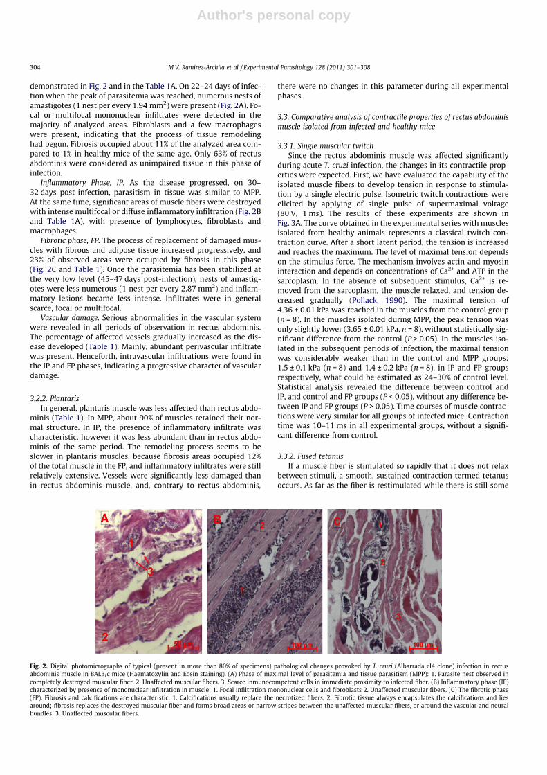
demonstrated in Fig. 2 and in the Table 1A. On 22–24 days of infec-tion when the peak of parasitemia was reached, numerous nests ofamastigotes (1 nest per every 1.94 mm2) were present (Fig. 2A). Fo-cal or multifocal mononuclear infiltrates were detected in themajority of analyzed areas. Fibroblasts and a few macrophageswere present, indicating that the process of tissue remodelinghad begun. Fibrosis occupied about 11% of the analyzed area com-pared to 1% in healthy mice of the same age. Only 63% of rectusabdominis were considered as unimpaired tissue in this phase ofinfection.
Inflammatory Phase, IP. As the disease progressed, on 30–32 days post-infection, parasitism in tissue was similar to MPP.At the same time, significant areas of muscle fibers were destroyedwith intense multifocal or diffuse inflammatory infiltration (Fig. 2Band Table 1A), with presence of lymphocytes, fibroblasts andmacrophages.
Fibrotic phase, FP. The process of replacement of damaged mus-cles with fibrous and adipose tissue increased progressively, and23% of observed areas were occupied by fibrosis in this phase(Fig. 2C and Table 1). Once the parasitemia has been stabilized atthe very low level (45–47 days post-infection), nests of amastig-otes were less numerous (1 nest per every 2.87 mm2) and inflam-matory lesions became less intense. Infiltrates were in generalscarce, focal or multifocal.
Vascular damage. Serious abnormalities in the vascular systemwere revealed in all periods of observation in rectus abdominis.The percentage of affected vessels gradually increased as the dis-ease developed (Table 1). Mainly, abundant perivascular infiltratewas present. Henceforth, intravascular infiltrations were found inthe IP and FP phases, indicating a progressive character of vasculardamage.
3.2.2. PlantarisIn general, plantaris muscle was less affected than rectus abdo-
minis (Table 1). In MPP, about 90% of muscles retained their nor-mal structure. In IP, the presence of inflammatory infiltrate wascharacteristic, however it was less abundant than in rectus abdo-minis of the same period. The remodeling process seems to beslower in plantaris muscles, because fibrosis areas occupied 12%of the total muscle in the FP, and inflammatory infiltrates were stillrelatively extensive. Vessels were significantly less damaged thanin rectus abdominis muscle, and, contrary to rectus abdominis,
there were no changes in this parameter during all experimentalphases.
3.3. Comparative analysis of contractile properties of rectus abdominismuscle isolated from infected and healthy mice
3.3.1. Single muscular twitchSince the rectus abdominis muscle was affected significantly
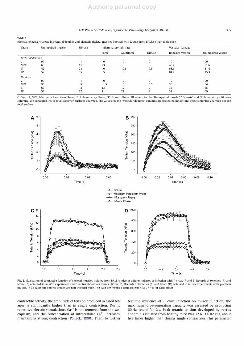
during acute T. cruzi infection, the changes in its contractile prop-erties were expected. First, we have evaluated the capability of theisolated muscle fibers to develop tension in response to stimula-tion by a single electric pulse. Isometric twitch contractions wereelicited by applying of single pulse of supermaximal voltage(80 V, 1 ms). The results of these experiments are shown inFig. 3A. The curve obtained in the experimental series with musclesisolated from healthy animals represents a classical twitch con-traction curve. After a short latent period, the tension is increasedand reaches the maximum. The level of maximal tension dependson the stimulus force. The mechanism involves actin and myosininteraction and depends on concentrations of Ca2+ and ATP in thesarcoplasm. In the absence of subsequent stimulus, Ca2+ is re-moved from the sarcoplasm, the muscle relaxed, and tension de-creased gradually (Pollack, 1990). The maximal tension of4.36 ± 0.01 kPa was reached in the muscles from the control group(n = 8). In the muscles isolated during MPP, the peak tension wasonly slightly lower (3.65 ± 0.01 kPa, n = 8), without statistically sig-nificant difference from the control (P > 0.05). In the muscles iso-lated in the subsequent periods of infection, the maximal tensionwas considerably weaker than in the control and MPP groups:1.5 ± 0.1 kPa (n = 8) and 1.4 ± 0.2 kPa (n = 8), in IP and FP groupsrespectively, what could be estimated as 24–30% of control level.Statistical analysis revealed the difference between control andIP, and control and FP groups (P < 0.05), without any difference be-tween IP and FP groups (P > 0.05). Time courses of muscle contrac-tions were very similar for all groups of infected mice. Contractiontime was 10–11 ms in all experimental groups, without a signifi-cant difference from control.
3.3.2. Fused tetanusIf a muscle fiber is stimulated so rapidly that it does not relax
between stimuli, a smooth, sustained contraction termed tetanusoccurs. As far as the fiber is restimulated while there is still some
Fig. 2. Digital photomicrographs of typical (present in more than 80% of specimens) pathological changes provoked by T. cruzi (Albarrada cl4 clone) infection in rectusabdominis muscle in BALB/c mice (Haematoxylin and Eosin staining). (A) Phase of maximal level of parasitemia and tissue parasitism (MPP): 1. Parasite nest observed incompletely destroyed muscular fiber. 2. Unaffected muscular fibers. 3. Scarce inmunocompetent cells in immediate proximity to infected fiber. (B) Inflammatory phase (IP)characterized by presence of mononuclear infiltration in muscle: 1. Focal infiltration mononuclear cells and fibroblasts 2. Unaffected muscular fibers. (C) The fibrotic phase(FP). Fibrosis and calcifications are characteristic. 1. Calcifications usually replace the necrotized fibers. 2. Fibrotic tissue always encapsulates the calcifications and liesaround; fibrosis replaces the destroyed muscular fiber and forms broad areas or narrow stripes between the unaffected muscular fibers, or around the vascular and neuralbundles. 3. Unaffected muscular fibers.
304 M.V. Ramirez-Archila et al. / Experimental Parasitology 128 (2011) 301–308
Author's personal copy
contractile activity, the amplitude of tension produced in fused tet-anus is significantly higher than in single contraction. Duringrepetitive electric stimulations, Ca2+ is not removed from the sar-coplasm, and the concentration of intracellular Ca2+ increases,maintaining strong contraction (Pollack, 1990). Then, to further
test the influence of T. cruzi infection on muscle function, themaximum force-generating capacity was assessed by producing60 Hz tetani for 2 s. Peak tetanic tension developed by rectusabdominis isolated from healthy mice was 12.61 ± 0.02 kPa, aboutfive times higher than during single contraction. This parameter
Table 1Histopathological changes in rectus abdominis and plantaris skeletal muscles infected with T. cruzi from BALB/c strain male mice.
Phase Unimpaired muscle Fibrosis Inflammatory infiltrate Vascular damage
Focal Multifocal Diffuse Impaired vessels Unimpaired vessels
Rectus abdominisC 99 1 0 0 0 0 100MPP 63 11 23 3 0 48.4 51.6IP 42 23 0 17.5 17.5 68.6 31.4FP 52 35 5 8 0 84.7 15.3
PlantarisC 99 1 0 0 0 0 100MPP 90 5 1.5 3 0.5 36 64IP 67 3 13 17 0 35 65FP 53 12 11 21 3 31 69
C: Control; MPP: Maximum Parasitism Phase; IP: Inflammatory Phase; FP: Fibrotic Phase. All values for the ‘‘Unimpaired muscle’’, ‘‘Fibrosis’’ and ‘‘Inflammatory infiltratescolumns’’ are presented in% of total specimen surfaces analyzed. The values for the ‘‘Vascular damage’’ columns are presented in% of total vessels number analyzed per thetotal surface.
Fig. 3. Evaluation of contractile function of skeletal muscles isolated from BALB/c mice in different phases of infection with T. cruzi. (A and B) Records of twitches (A) andtetani (B) obtained in in vitro experiments with rectus abdominis muscle. (C and D) Records of twitches (C) and tetani (D) obtained in in situ experiments with plantarismuscle. In all cases the control groups are non-infected mice. The data are means ± standard error (SE), n = 8 for each group.
M.V. Ramirez-Archila et al. / Experimental Parasitology 128 (2011) 301–308 305
Author's personal copy
was significantly lower in muscles isolated from all groups of in-fected mice (6.89 ± 0.04, 5.84 ± 0.02, 5.82 ± 0.04 kPa for MPP, IP,and FP groups, correspondingly (Fig. 3B), with statistically signifi-cant difference from the control (P < 0.05).
3.4. Comparative analysis of in situ contractile properties of plantarismuscle in infected and healthy mice
The amplitude of peak tension of a single muscular twitch ofcontrol plantaris muscle was 269.5 ± 17.5 kPa and the contractiontime was 30.37 ± 3.42 ms (Fig. 3C). Infection did not change thetime course of response, but the amplitude decreased considerablyin IP and FP groups (131.1 ± 9.7 and 155.5 ± 16.7 kPa, respectively,Fig. 3C). Similarly, the amplitude of tetanus was decreased in IPand FP, but not in MPP groups (Fig. 3D).
4. Discussion
In general, different T. cruzi strains demonstrate tropism forheart, skeletal muscles and nervous tissue. Polymyositis observedin patients with Chagas’ disease is usually overshadowed by themore clinically evident cardiac disease. Whereas correlations be-tween pathologic changes in tissue and functional abnormalitiesare described in relative detail for chagasic hearts, no studies wereundertaken to reveal the relationship between skeletal muscles in-jury with their contractile properties. In the present work we havecorrelated contractile properties of rectus abdominis and plantarismuscle with the degree and character of tissue damage analyzedon histological level, in three different stages of acute experimentaltrypanosomiasis in mice. To test the mechanic properties of mus-cles, isolated strips of rectus abdominis were subjected to directelectrical field stimulation in vitro, or, alternatively, plantaris mus-cles were stimulated in situ through the sciatic nerve. Single twitchand tetanus protocols were applied.
It was shown that contractile properties of muscles were signif-icantly attenuated during acute T. cruzi infection. The phenomenonwas confirmed in both in vitro (rectus abdominis) and in situ (plan-taris) experiments, indicating that it was due to muscle rather thanto motor nervous damage.
The peak of parasitemia was reached at 22–24 days of infec-tions (MPP stage). In the rectus abdominis severely affected bythe parasite, we have found numerous nests of amastigotes accom-panied by mononuclear inflammatory infiltrates, mostly scarce(87.5%), but moderate in rare cases (12.5%). More than 60% of ana-lyzed areas were unaffected, and 25% and 12% were occupied byinflammation or fibrosis, correspondingly. Half of the vessels wereinvolved in the inflammatory process (perivasculitis). However, onthe functional level, the amplitudes of single contractions showedonly a slight decrease compared to the control level, with the dif-ferences being statistically insignificant (n = 8, P > 0.05). At thesame time, the amplitude of tetanic contraction in this periodwas already considerably decreased (Fig. 3B). As far as the mito-chondrial activity is the important factor for sustained muscularactivity (Curtin and Woledge, 1978), both mitochondrial dysfunc-tion and oxidative imbalance in skeletal tissue reported recentlyfor acute T. cruzi infection in mice (Wen et al., 2008) could contrib-ute to this phenomenon.
On 30–32 days (PI stage) of infection, parasitemia decreased,but the extension of parasitism and inflammation in muscles in-creased drastically. More than 35% of analyzed areas were occu-pied by inflammatory infiltrate, and its degree shifted frommainly scarce in MPP stage to moderate (75%) and even severe(12.5%). 22% of muscle tissue was replaced by fibrous and adiposetissue. The percentage of unaffected tissue diminished from 63(in PP stage) to 42%. This change has had a strong impact on the
functional characteristics of rectus abdominis muscle, and theamplitudes of single contractions were two times lower, than inthe MPP phase. However, the amplitudes of tetanus were similarin MPP and IP phases. It is likely that the critical level of damagefor tetanic contraction was reached already in the first period ofinfection. On 45–47 days (PF stage) of infection, parasitemia stabi-lized at the lowest level, inflammatory reaction in tissues was sig-nificantly less pronounced (only 12.5% of examined areas wereoccupied by inflammatory infiltrate). On the other hand, the mus-cles were replaced by fibrous and adipose tissue in large areas(more than 30%), and the percentage of unaffected muscle tissuewas about 40–45%, similar to IP group. Contractile properties ofmuscles in this period were also very similar to those in the inflam-matory stage (IP) despite the fact that the processes taking place intissues were different, with the prevalence of inflammation in PFstage and of fibrosis in IP stage.
The above results show that skeletal muscles are capable, tosome extent, to maintain their basic functions during the infectionprocess. Although the degree of damage was significant in the firststage of infection characterized by maximal peak of parasitemia, itdoes not reach the critical level at this period, and the amplitude ofa single twitch was practically unaffected. The shift to anaerobicmetabolism in the affected skeletal muscles with decreased oxida-tive capacities could be one of the feasible adaptive mechanisms,as was described in patients with advanced Chagas’ disease (Mon-tes de Oca et al., 2004). At the same time, serious changes in func-tional properties were observed just overcoming this critical levelof tissue damage in the following stages of Chagas’ disease. Theability to develop tetanic contraction seems to be more affectedby pathologic processes in muscles, so this function was changedalready in MPP phase.
Similar processes took place in the plantaris muscle. Howevertissue damage developed slower in this muscle (Table 1), and bothsingle and tetanic amplitudes were not affected in MPP phase,when only 10% of tissue was damaged. The variations in parasitismand pathologic changes between rectus abdominis and plantarismuscles might be correlated with different factors, the moreimportant of which are the structure of muscular fibers and differ-ences in regional blood supply. The diversity of the content andcomposition of heavy myosin chain in the skeletal muscular fiberswas reported earlier (Eng et al., 2008). The muscle fibers are di-vided into fast-twitch glycolytic (FG), fast-twitch oxidative-glyco-lytic (FOG) and slow-twitch oxidative (SO), which presentsignificant differences in oxidative-glycolytic metabolism. FOGand SO fibers show high activity of enzymes of aerobic metabolismwhen comparing to FG fibers (Schwartz-Giblin et al., 1983). At thesame time, T. cruzi is known to be very sensitive to oxidative stress(Bachega et al., 2009). Then the muscles with high percentage of SOand/or FOG fibers are likely to be less suitable for parasite survivaland, consequently, would be less affected during infection. Indeed,as was shown earlier, plantaris muscle in rats contains more than55% of SO + FOG fibers (Ishihara et al., 1998), whereas this value inrectus abdominis is less than 30% (Alvarez Rosa et al., 2007).
Furthermore, muscle contractions are well known to be regu-lated by intracellular Ca2+ ions, which switch thin filaments intoan active state by binding to troponin. ATP is important for thecontraction process as well, since Ca2+ activates the myosin–ATPcomplex, which drives the sliding action between actin and myosin(Szent-Györgyi, 1975). Although it is widely accepted that the pro-cess of the host cell invasion by T. cruzi depends on the energyaccumulated by parasite (Schenkman et al., 1991), which specificsource of energy is used is not known yet (Martins et al., 2009).At the same time, no changes in the activity of the mitochondrialATP synthase was observed in myocardium of rats infected withthe Colombian strain of T. cruzi (Rendón et al., 2007). Ca2+
mobilization at the single cell level was detected in cultured
306 M.V. Ramirez-Archila et al. / Experimental Parasitology 128 (2011) 301–308

Author's personal copy
L6E6 myoblasts during their interaction with T. cruzi trypomastig-otes (Moreno et al., 1994). Subsequent studies reported that T. cruziinfection induce repetitive cytosolic-free Ca2+ transients in rat kid-ney fibroblasts (Tardieux et al., 1994), affects intracellular Ca2+ lev-els in neonatal cardiomyocytes (Taniwaki et al., 2006), and mayinvolve Ca2+ mobilization from intracellular stores in differentmammalian cells (Yoshida and Cortez, 2008). Despite several pub-lished reports about effect of T. cruzi infection on Ca2+ signaling indifferent types of host cells, more studies are needed to link thesechanges to muscle contractile properties.
In the present work we have shown that T. cruzi infection im-paired significantly contractile properties of skeletal muscles inmice. The percentage of damaged muscles was important, inde-pendently on the character of tissue pathology. Even during lateacute infection, when parasitism and inflammation were de-creased, and the process of tissue remodeling took place in themuscles, the contractile properties remained attenuated signifi-cantly due to the fact that muscles were replaced by fibrous tissueand fat.
Acknowledgments
This work was funded by the Mexican National Council for Sci-ence and Technology (CONACyT-SEP-2004-CO1-46731, CONACyT-SEP-2008-CO1-104272) and the Ramon Alvarez-Buylla Foundation(University of Colima 332/05 and 593/09).
Authors thank Dr. Alex McKay for critical reading of themanuscript.
References
Acquatella, H., 2008. Predicción de insuficiencia cardiaca y mortalidad pormiocardiopatía crónica chagásica. Una enfermedad nueva en España. RevistaEspañola de Cardiología 61, 105–107.
Alvarez Rosa, M.J., Dal Pai, V., Bremen Neto, H., Azoubel, R., Andree Nouailhetas, V.L.,2007. Effect of swimming training on the rectus abdominis muscle of rats:morphological and histochemical aspects. International Journal of Morphology25, 631–638.
Bachega, J.F., Navarro, M.V., Bleicher, L., Bortoleto-Bugs, R.K., Dive, D., Hoffmann, P.,Viscogliosi, E., Garratt, R.C., 2009. Systematic structural studies of ironsuperoxide dismutases from human parasites and a statistical couplinganalysis of metal binding specificity. Proteins 77, 26–37.
Báez, A.L., lo Presti, M.S., Rivarola, H.W., Pons, P., Fretes, R., Paglini-Oliva, P., 2008.Trypanosoma cruzi: cardiac mitochondrial alterations produced by differentstrains in the acute phase of the infection. Experimental Parasitology 120, 397–402.
Bijovsky, T., Elizari, M.V., Muller, L.A., Katzin, V.J., Gonzalez Cappa, S.M., 1983.Chronic infection in mice with Trypanosoma cruzi. Revista do Instituto deMedicina Tropical de São Paulo 25, 207–214.
Buckner, F.S., Wilson, A.J., van Voorhis, C., 1999. Detection of live Trypanosoma cruziin tissues of infected mice by using histochemical stain for b-galactosidase.Infection and Immunity 67, 403–409.
Cenget, D.D., Rojas, R., 1959. La biopsia de músculo delt6ides en la enfermedad deChagas. Revista da Facultad de Medicina de Tucuman 2, 27–37.
Cossermelli, W., Friedman, H., Pastor, E.H., Nobre, M.R., Manzione, A., Camargo, M.E.,Shiroma, M., 1978. Polymyositis in Chagas’s disease. Annals of the RheumaticDiseases 37, 277–280.
Curtin, N.A., Woledge, R.C., 1978. Energy changes and muscular contraction.Physiological Reviews 58, 690–761.
Eng, C.M., Smallwood, L.H., Rainiero, M.P., Lahey, M., Ward, S.R., Lieber, R.L., 2008.Scaling of muscle architecture and fiber types in the rat hindlimb. Journal ofExperimental Biology 211, 2336–2345.
Fernández, A.R., Paglini-Oliva, P., Palma, J.A., Lacuara, J.L., 1992. Isometric developedtension and histopathology of myocardium of chagasic mice. II. ActaPhysiologica, Pharmacologica et Therapeutica Latinoamericana 42, 197–204.
Fernández-Culasso, A., Paglini-Oliva, P., Palma, J.A., Lacuara, J.L., 1991. Isometricdeveloped tension and histopathology of myocardium of chagasic mice. I. ActaPhysiologica, Pharmacologica et Therapeutica Latinoamericana 41, 397–404.
Garg, N., Popov, V.L., Papaconstantinou, J., 2003. Profiling gene transcription revealsa deficiency of mitochondrial oxidative phosphorylation in Trypanosoma cruzi-infected murine hearts: implication in chagasic myocarditis development.Biochemistry and Biophysics Acta 1638, 106–120.
Gascón, J., Albajar, P., Cañas, E., Flores, M., Gómez-Prat, J., Herrera, R.N., Lafuente,C.A., Luciardi, H.L., Moncayo, A., Molina, L., Muñoz, J., Puente, S., Sanz, G.,Treviño, B., Salles, X.S., 2007. Diagnóstico, manejo y tratamiento de la
cardiopatía chagásica crónica en áreas donde la infección por Trypanosomacruzi no es endémica. Revista Española de Cardiología 60, 285–293.
Gordon, A.M., Huxley, A.F., Julian, F.J., 1966. The variation in isometric tension withsarcomere length in vertebrate muscle fibres. Journal of Physiology 184, 170–192.
Huerta, M., Muñiz, J., Stefani, E., 1986. The effects of external calcium on potassiumcontractures in tonic muscle fibres of the frog. Journal of Physiology 376, 219–230.
Ishihara, A., Roy, R.R., Ohira, Y., Ybata, Y., Edgerton, V.R., 1998. Hypertrophy of ratplantaris muscle fibers alter voluntary running with increased loads. Journal ofApplied Physiology 84, 2183–2189.
Kirchhoff, L.V., 1993. American trypanosomiasis (Chagas’ disease) – A topicaldisease now in United States. New England Journal of Medicine 329, 634–644.
Köberle, F., 1968. Chagas’ disease and Chagas’ syndromes: the pathology ofAmerican trypanosomiasis. Advances in Parasitology 6, 63–116.
Laguens, R.P., Cossio, P.M., Diez, C., Segal, A., Vasquez, C., Kreutzer, E., Khouri, E.,Arana, R.M., 1975. Immunopathologic and morphologic studies of skeletalmuscle in Chagas disease. American Journal of Pathology 80, 153–162.
Laguens, R.P., Cabeza Markert, P., 1991. Origin and significance of anti-heart andanti-skeletal muscle autoantibodies in Chagas’ disease. Research inImmunology 142, 160–163.
Losavio, A., Jones, M.C., Sanz, O.P., Mirkin, G., Gonzalez Cappa, S.M., Muchnick, S.,Sica, R.E.P., 1989. A sequential study of the peripheral nervous systeminvolvement in experimental Chagas’ disease. American Journal of tropicalMedicine and Hygiene 41, 539–547.
Martins, R.M., Covarrubias, C., Rojas, R.G., Silber, A.M., Yoshida, N., 2009. Attachmentof Trypanosoma cruzi to mammalian cells requires parasite energy, and invasioncan be independent of the target cell cytoskeleton. Infection and Immunity 77,3023–3032.
Melnikov, V., Fierro, F., Espinoza, F., Guzmán, F., Dobrovinskaya, O., 2005. Pathologicchanges in lungs caused by mexican isolates of Trypanosoma cruzi in the acutephase of infection in mice. American Journal of Tropical Medicine and Hygiene73, 301–306.
Mirkin, G.A., Jones, M., Sanz, O.P., Rey, R., Sica, R.E., González Cappa, S.M., 1994.Experimental Chagas’ disease: electrophysiology and cell composition of theneuromyopathic inflammatory lesions in mice infected with a myotropic and apantropic strain of Trypanosoma cruzi. Clinical Immunology andImmunopathology 73, 69–79.
Molina, H.A., Cardoni, R.L., Rimoldi, M.T., 1987. The neuromuscular pathology ofexperimental Chagas’ disease. Journal of the Neurological Sciences 81, 287–300.
Monteón, V.M., Furuzawa-Carballeda, J., Alejandre-Aguilar, R., Aranda-Fraustro, A.,Rosales-Encina, J.L., Reyes, P.A., 1996. American trypanosomiasis: in situ andgeneralized features of parasitism and inflammation kinetics in a murinemodel. Experimental Parasitology 83, 267–274.
Montes de Oca, M., Torres, S.H., Loyo, J.G., Vazquez, F., Hernández, N., Anchustegui,B., Puigbó, J.J., 2004. Exercise performance and skeletal muscles in patients withadvanced Chagas disease. Chest 125, 1306–1314.
Moreno, S.N., Silva, J., Vercesi, A.E., Docampo, R., 1994. Cytosolic-free calciumelevation in Trypanosoma cruzi is required for cell invasion. Journal ofExperimental Medicine 180, 1535–1540.
Pollack, G.H., 1990. Muscles and Molecules: Uncovering the Principles of BiologicalMotion. Editorial: Ebner and Sons, Seattle, USA.
Ponce, L.A.F.Z., 1972. Miopatia chagisica esquelética. Thesis, Universidad NacionalMayor de San Marcos, Lima, Peru.
Rendón, D.A., Genes, C.M., Triana, O., 2007. Myocardial cellular damage and theactivity of the mitochondrial ATP synthase in rats infected with a Colombianstrain of Trypanosoma cruzi. Biomedica 1, 40–49.
Rowin, K.S., Tanowitz, H.B., Wittner, M., Nguyen, H.T., Nadal-Ginard, B., 1983.Inhibition of muscle differentiation by Trypanosome cruzi. Proceedings ofNational Academy of Science United States of America 80, 6390–6394.
Szent-Györgyi, A.G., 1975. Calcium regulation of muscle contraction. BiophysicalJournal 15, 707–723.
Scharfstein, J., de Gomes, J.A., Correa-Oliveira, R., 2009. Back to the feature inChagas’ disease: from animal models to patient cohort studies, progress inimmunopathogenesis research. Memórias do Instituto Oswaldo Cruz 104, 187–198.
Schenkman, S., Robbins, E.S., Nussenzweig, V., 1991. Attachment of Trypanosomacruzi to mammalian cells requires parasite energy, and invasion can beindependent of the target cell cytoskeleton. Infection and Immunity 59, 645–654.
Schwartz-Giblin, S., Rosello, L., Pfaff, D.W., 1983. A histochemical study of laterallongissimus muscle in rat. Experimental Neurology 79, 497–518.
Stum, M., Girard, E., Bangratz, M., Bernard, V., Herbin, M., Vignaud, A., Ferry, A.,Davoine, C.S., Echaniz-Laguna, A., René, F., Marcel, C., Molgó, J., Fontaine, B.,Krejci, E., Nicole, S., 2008. Evidence of a dosage effect and a physiologicalendplate acetylcholinesterase deficiency in the first mouse models mimickingSchwartz–Jampel syndrome neuromyotonia. Human Molecular Genetic 17,3166–3179.
Taniwaki, N.N., Machado, F.S., Massensini, A.R., Mortara, R.A., 2006. Trypanosomacruzi disrupts myofibrillar organization and intracellular calcium levels inmouse neonatal cardiomyocytes. Cell and Tissue Research 324 (3), 489–496.
Tardieux, I., Nathanson, M.H., Andrews, N.W., 1994. Role in host cell invasion ofTrypanosoma cruzi-induced cytosolic-free Ca2+ transients. Journal ofExperimental Medicine 179, 1017–1022.
Tatsuya, H., Hirshman, M.F., Dufresne, S.D., Goodyear, L.J., 1999. Skeletal musclecontractile activity in vitro stimulates mitogen-activated protein kinasesignaling. American Journal of Physiology 277, C701–C707.
M.V. Ramirez-Archila et al. / Experimental Parasitology 128 (2011) 301–308 307
Author's personal copy
Wen, J.J., Dhiman, M., Whorton, E.B., Garg, N.J., 2008. Tissue-specific oxidativeimbalance and mitochondrial dysfunction during Trypanosoma cruzi infection inmice. Microbes and Infection 10, 1201–1209.
Virgen-Ortiz, A., Marin, J.L., Trujillo, X., Huerta, M., Muñiz, J., 2008. Sprint trainingattenuates the deficits of force and dynamic stiffness in rat soleus musclecaused by eccentric contractions. Journal of Biomechanics 41, 2533–2538.
WHO, 2007. Special programme for research and training in tropical diseases (TDR),Report of Scientific Group in Chagas Disease TDR/SWG/09, World HealthOrganization, Buenos Aires, Argentina, 2007.
Yoshida, N., Cortez, M., 2008. Trypanosome cruzi: parasite and host cell signallingduring the invasion process. Subcellular Biochemistry 47, 82–91.
Zimmerman, M., 1983. Ethical guideline for investigation of experimental pain inconscious animals. Pain 16, 109–110.
Mario V. Ramirez-Archila M.D. Family Practice, PhD:Physiology. Affiliation: Faculty of Educational Sciences,Physical Education Department, University of Colima,Mexico. Research area: impact of T. cruzi infection onthe development of muscular fatigue; anaerobicmetabolism in diabetic patients and effect of endurancetraining on it; study of polimorphisms implicated inexercise resistance and obesity genesis.
Jesús Muñiz M.D., PhD: Physiology and Biophysics.Affiliation: Full Time Professor, Centre for BiomedicalResearch, University of Colima, Mexico. Research area:effects of sprint and endurance training on themechanical properties of skeletal muscle; passivemechanical properties of skeletal muscle; mechanismsinvolved in functional cardiac hypertrophy; free fattyacid metabolism and lactate kinetics in children; anal-ysis of polymorphism interaction on fatty acid catabo-lism induced by aerobic exertion.
Adolfo Virgen-Ortiz M.D., PhD: Physiology. Affiliation:Department of Chemistry and Biological Sciences Uni-versity of Sonora, Navojoa, Mexico; Full Time Professor.Research area: Functional cardiac remodeling inducedby pregnancy and exercise; molecular mechanisms ofthe physiopathology of tropical diseases to find thera-peutic targets; physiopathology of contractile systems.
Valery G. Melnikov M. D.: General physician (RussianPeople Friendship University, Russian Federation 1998);PhD: Medical Sciences. Affiliation: School of Medicine,University of Colima, Full Time Professor. Research area:Pathologic changes in mammalian host in the course ofvector transmitted diseases.
Oscar Newton-Sánchez M.D.: Pediatric Infectious Dis-eases, PhD: Medical Sciences. Affiliation: School ofMedicine, University of Colima, Full Time Professor.Research area: Infectious Diseases.
Oxana R. Dobrovinskaya Candidate of Science (Mos-cow State University, 1993) – PhD equivalent: CellBiology and Physiology. Affiliation: Centre for Biomed-ical Research and School of Medicine, University ofColima, Full Time Professor and Researcher. Researcharea: cell signaling and membrane transport in lym-phocytes, Chagas’ disease: host–parasite interaction.
308 M.V. Ramirez-Archila et al. / Experimental Parasitology 128 (2011) 301–308


![Finale 2003 - [Boleros Medley for 2 pianos (arr. Lito Valle).MUS]](https://static.fdokumen.com/doc/165x107/6332b61808f6dcde650828e2/finale-2003-boleros-medley-for-2-pianos-arr-lito-vallemus.jpg)