Differentiation analyses of adult suspension mononucleated peripheral blood cells of Mus musculus
7
RESEARCH Open Access Differentiation analyses of adult suspension mononucleated peripheral blood cells of Mus musculus Shahrul Hisham Zainal Ariffin 1*† , Intan Zarina Zainol Abidin 1† , Muhammad Dain Yazid 1 , Rohaya Megat Abdul Wahab 2 Abstract Background: The purpose of this study is to determine whether isolated suspension mouse peripheral mononucleated blood cells have the potential to differentiate into two distinct types of cells, i.e., osteoblasts and osteoclasts. Results: Differentiation into osteoblast cells was concomitant with the activation of the Opn gene, increment of alkaline phosphatase (ALP) activity and the existence of bone nodules, whereas osteoclast cells activated the Catk gene, increment of tartrate resistant acid phosphatase (TRAP) activity and showed resorption activities via resorption pits. Morphology analyses showed the morphology of osteoblast and osteoclast cells after von Kossa and May-Grunwald-Giemsa staining respectively. Conclusions: In conclusion, suspension mononucleated cells have the potentiality to differentiate into mature osteoblasts and osteoclasts, and hence can be categorized as multipotent stem cells. Background Stem cells, or mother cells, are cells with the ability to divide for indefinite periods in time and to give rise to specific cells. The primitive stage of stem cells can be divided into three types; i.e., totipotent, pluripotent and multipotent cells [1,2]. Totipotent cells are the most pri- mitive cells, followed by pluripotent cells. The multipo- tent cell type is the most differentiated type of stem cells [3]. Osteoblasts and osteoclasts are cells that are responsible for bone formation and resorption respec- tively [4]. During bone formation, osteoblasts deposit the organic and inorganic matrix, whilst osteoclasts remove bone matrix [5]. Osteoblast and osteoclast cells are originated from different lineage, i.e., osteoblasts arise from mesenchymal stem cells and osteoclasts origi- nate from haematopoietic stem cells [6]. The objective of this study is to determine the potenti- ality of isolated suspension mononucleated cells to differ- entiate into osteoblast and osteoclast cells. Osteoblasts were originated from different lineage (mesenchymal stem cells) but osteoclasts were originated from haemato- poietic stem cells. This indicates that the isolated mono- nucleated cells capable to differentiate into more than one type of cells. Our results show that suspension mononucleated cells isolated from peripheral mouse blood have the potential to develop into more than one type of mature cell. The results also demonstrate the plasticity of adult stem cells isolated from peripheral blood. These cells are capable of fully differentiating into osteoblast and osteoclast cells. The presences of osteo- blast and osteoclast cells were determined using molecu- lar biology, cells activity and morphology analyses. Results and Discussion Proliferation of Mononucleated Cells The heterogenic populations were characterised using cell culture selection. After 15 days cultured in selection medium, majority of differentiated and precursor cells died due to short lifespans, e.g., granulocytes (30-40 min- utes in the peripheral blood with a total lifespan of 7-13 days that varied under certain pathological conditions) [7], monocytes (5-7 days) [8] and platelets (3-5 days) [9]. * Correspondence: [email protected] † Contributed equally 1 School of Biosciences and Biotechnology, Faculty of Science and Technology, Universiti Kebangsaan Malaysia, 43600 Bangi, Selangor, Malaysia Full list of author information is available at the end of the article Ariffin et al. Cell Communication and Signaling 2010, 8:29 http://www.biosignaling.com/content/8/1/29 © 2010 Ariffin et al; licensee BioMed Central Ltd. This is an Open Access article distributed under the terms of the Creative Commons Attribution License (http://creativecommons.org/licenses/by/2.0), which permits unrestricted use, distribution, and reproduction in any medium, provided the original work is properly cited.
Transcript of Differentiation analyses of adult suspension mononucleated peripheral blood cells of Mus musculus
RESEARCH Open Access
Differentiation analyses of adult suspensionmononucleated peripheral blood cells ofMus musculusShahrul Hisham Zainal Ariffin1*†, Intan Zarina Zainol Abidin1†, Muhammad Dain Yazid1,Rohaya Megat Abdul Wahab2
Abstract
Background: The purpose of this study is to determine whether isolated suspension mouse peripheral mononucleatedblood cells have the potential to differentiate into two distinct types of cells, i.e., osteoblasts and osteoclasts.
Results: Differentiation into osteoblast cells was concomitant with the activation of the Opn gene, increment ofalkaline phosphatase (ALP) activity and the existence of bone nodules, whereas osteoclast cells activated the Catkgene, increment of tartrate resistant acid phosphatase (TRAP) activity and showed resorption activities viaresorption pits. Morphology analyses showed the morphology of osteoblast and osteoclast cells after von Kossaand May-Grunwald-Giemsa staining respectively.
Conclusions: In conclusion, suspension mononucleated cells have the potentiality to differentiate into matureosteoblasts and osteoclasts, and hence can be categorized as multipotent stem cells.
BackgroundStem cells, or mother cells, are cells with the ability todivide for indefinite periods in time and to give rise tospecific cells. The primitive stage of stem cells can bedivided into three types; i.e., totipotent, pluripotent andmultipotent cells [1,2]. Totipotent cells are the most pri-mitive cells, followed by pluripotent cells. The multipo-tent cell type is the most differentiated type of stemcells [3]. Osteoblasts and osteoclasts are cells that areresponsible for bone formation and resorption respec-tively [4]. During bone formation, osteoblasts depositthe organic and inorganic matrix, whilst osteoclastsremove bone matrix [5]. Osteoblast and osteoclast cellsare originated from different lineage, i.e., osteoblastsarise from mesenchymal stem cells and osteoclasts origi-nate from haematopoietic stem cells [6].The objective of this study is to determine the potenti-
ality of isolated suspension mononucleated cells to differ-entiate into osteoblast and osteoclast cells. Osteoblasts
were originated from different lineage (mesenchymalstem cells) but osteoclasts were originated from haemato-poietic stem cells. This indicates that the isolated mono-nucleated cells capable to differentiate into more thanone type of cells. Our results show that suspensionmononucleated cells isolated from peripheral mouseblood have the potential to develop into more than onetype of mature cell. The results also demonstrate theplasticity of adult stem cells isolated from peripheralblood. These cells are capable of fully differentiating intoosteoblast and osteoclast cells. The presences of osteo-blast and osteoclast cells were determined using molecu-lar biology, cells activity and morphology analyses.
Results and DiscussionProliferation of Mononucleated CellsThe heterogenic populations were characterised usingcell culture selection. After 15 days cultured in selectionmedium, majority of differentiated and precursor cellsdied due to short lifespans, e.g., granulocytes (30-40 min-utes in the peripheral blood with a total lifespan of 7-13days that varied under certain pathological conditions)[7], monocytes (5-7 days) [8] and platelets (3-5 days) [9].
* Correspondence: [email protected]† Contributed equally1School of Biosciences and Biotechnology, Faculty of Science andTechnology, Universiti Kebangsaan Malaysia, 43600 Bangi, Selangor, MalaysiaFull list of author information is available at the end of the article
Ariffin et al. Cell Communication and Signaling 2010, 8:29http://www.biosignaling.com/content/8/1/29
© 2010 Ariffin et al; licensee BioMed Central Ltd. This is an Open Access article distributed under the terms of the Creative CommonsAttribution License (http://creativecommons.org/licenses/by/2.0), which permits unrestricted use, distribution, and reproduction inany medium, provided the original work is properly cited.
As a result, only primitive mononucleated cells (stemcells) survive after 15 days cultured in selection medium.Besides differentiation potential, self-renewal is the
main property of stem cells. The suspension mononu-cleated cells population doubling times are 3.1 days[10]. Passages were done every doubling time i.e., 3-4days. Within 15 days, 4-5 passages were used for eachisolates which produce a total cell number of approxi-mately 3-5 × 10⁶ cells/mL for every isolates which startfrom approximately 1 × 10⁵ cells/mL. As conclusion,within 15 days the suspension mononucleated cellswere shown to proliferate in in vitro conditions.
Activation of Molecular Markers in DifferentiatedMononucleated CellsPrevious studies have demonstrated that there are sev-eral specific gene markers for osteoblast cells, such asosteopontin, alkaline phosphatase, osteocalcin and col-lagen type I (Col1) [11,12]. In this study, molecular ana-lysis was performed to determine in vitro differentiationof peripheral blood mononucleated cells into matureosteoblast cells after induction by differentiation factors,i.e., ascorbic acid and b-glycerophosphate, for 14 days.The activation of the Opn gene shows that mononu-
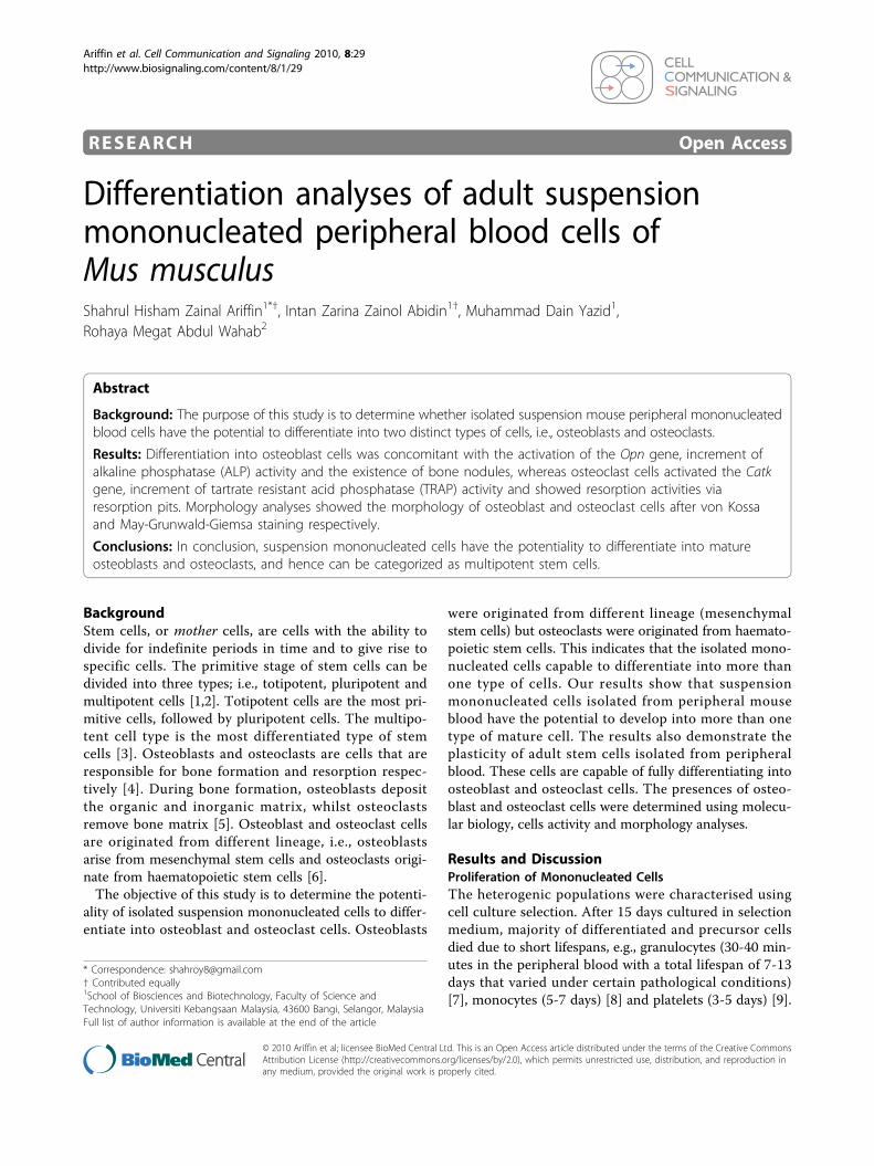
cleated cells have differentiated into osteoblast cells. RT-PCR analysis showed different levels of Opn geneexpression in mononucleated cells cultured in prolifera-tion medium compared to osteoblast differentiationmedium (Figure 1B). Both media produced the expectedproducts; i.e., the Opn gene with size ~234 bp whenamplified with RT-PCR. However, the expression of theOpn gene for mononucleated cells cultured in osteoblastdifferentiation medium were much higher as comparedto mononucleated cells cultured in proliferation medium(Figure 1B). The increase in Opn gene transcriptionshowed that peripheral blood mononucleated cells dif-ferentiate into mature osteoblast cells. Opn gene canproduce low grade expression in variety of cells, such asfibroblast, differentiated osteoblast and osteoclast cellsand bone marrow derived cells [13]. Low level expres-sion of Opn gene in this study might be due to the exis-tence a few of all these differentiated cells types andalso primitive cells that can survive after 15 days ofmedium selection. However, the increment of expressionlevel after been induced in specialized osteoblast med-ium was contributed by the primitive cell (stem cells)that have been differentiated into osteoblast cells.RT-PCR analysis was done on day 14 based on the
fact that the mononucleated cell line (MC3T3-E1) isfully differentiated into osteoblast cells after 14 days indifferentiation medium [14]. Similarly, based on studiesin rat calvarial osteoblast-like (ROB) cell, the Opn genewas also expressed during 13 to 15 days of differentia-tion [15]. Furthermore, the study showed that ROB cells
differentiate morphologically into mature osteoblast cellsafter 13 days of differentiation. Therefore, this studydemonstrates that mononucleated cells cultured inosteoblast differentiation medium express the Opn genewhen differentiated into osteoblast cells. In addition,alkaline phosphatase (ALP) assay also showed the incre-ment of ALP activity after 10 and 14 days of mononu-cleated cells cultured in osteoblast differentiationmedium compared to cells cultured in control medium(proliferation medium) (Figure 2). MC3T3-E1 cell line(pre-osteoblast) that cultured in differentiation medium(presence of ascorbic acid and b-glycerophosphate) exhi-bits high ALP activity after 10 and 14 days in the med-ium [14,16]. This is an indication that ALP is a markerfor the bone formation. The expression of Opn geneand increment of ALP activity showed that mononu-cleated cells are differentiated into osteoblast cells.In this study, the housekeeping gene Gapdh was used
as a positive control for mononucleated cells in bothtypes of medium, i.e., proliferation and differentiationmedium. Figure 1A shows that RT-PCR amplificationproduces a Gapdh band (~717 bp) from mononucleatedcells in both types of media. The activation of the Gapdhgene proves that cells perform basic metabolic processesrequired for cell survival. An inherent assumption in theuse of housekeeping genes is that expression of the genes
Figure 1 Expression of specific gene markers. RT-PCR analysiswas performed using total RNA isolated from mononucleated cellscultured in proliferation medium and mononucleated cells culturedin osteoblast and osteoclast differentiation medium. (A) Gapdh (717bp) was used as a positive control. (B) The low expression of Opn(234 bp) in undifferentiated mononucleated cells while highexpression of Opn (234 bp) indicates differentiation into matureosteoblasts. (C) Catk (350 bp) expression shows osteoclastdifferentiation. Equal volumes of the products were separated inagarose gels and stained with ethidium bromide.
Ariffin et al. Cell Communication and Signaling 2010, 8:29http://www.biosignaling.com/content/8/1/29
Page 2 of 7

remains constant in the cells or in the tissues underinvestigation [17].Differentiated osteoclast cells express specific markers,
such as tartrate resistant acid phosphatase and cathepsinK [18,19]. Therefore, the Catk gene will only be acti-vated in osteoclast cells. Molecular analysis wasperformed to determine whether the isolated mononu-cleated cells had indeed differentiated into matureosteoclast cells. The activation of the Catk gene hasbeen observed in mononucleated cells induced in vitrofor osteoclast differentiation over a period of 10 days.RT-PCR analysis was done on day 10 since mononu-cleated cells are believed to differentiate into matureosteoclasts at this time point, thus suitable for molecularanalysis [20-23].The experimental results are shown in Figure 1C. The
RT-PCR analysis of the transcripts from mononucleatedcells cultured in osteoclast differentiation mediumshowed the activation of the Catk gene. The RT-PCRproduct showed amplification of a band with theexpected size for the Catk gene, i.e., ~350 bp (Figure 1C).However, no band was found from mononucleated cellscultured in proliferation medium, which served as thenegative control experiment (Figure 1C). Therefore, acti-vation of the Catk gene does not occur during cells pro-liferation. This study showed that mononucleated cellscultured in osteoclast differentiation medium for 10 daysactivate the Catk gene, similar to mature osteoclast cells.In addition, the increment of tartrate resistant acid phos-phatase (TRAP) activity in mononucleated cells culturedin osteoclast differentiation medium after 10 days com-pared to cells cultured in control medium (proliferationmedium) also indicates that the cells are differentiatedinto osteoclast cells (Figure 3).
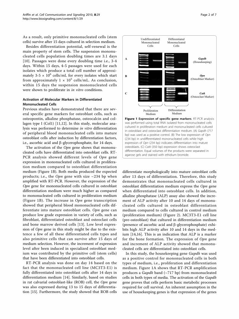
Osteoblastic and Osteoclastic Cell ActivitiesCell activities determination using osteologic discs showedthat discs cultured in osteoblast differentiation mediumwere positive for von Kossa staining, an indication of the
existence of calcium nodules (Figure 4). Recent study bySindrey et al. (1999) showed that generated calciummineral by osteoblast cells is stained by von Kossa as blackor yellow to brown based on the amount of secreted cal-cium matrices, whereas negative control disc is stained aslight brown or gray [24]. The control disc cultured in pro-liferation medium did not show the formation of calciumnodules on the disc surface (Figure 4A). During differen-tiation, osteoblasts express the ALP enzyme, which utilizesphosphate from the medium deposits it on the disc sur-face, which then accumulates to become calcium nodulesstained by von Kossa [12,25]. In our study, we comparedour discs to the background colour (control disc; cell cul-tured without osteoblast differentiation factors), where theincrement of intensity for the background during celldevelopment has been observed. The background colourand quantity of bone nodules increase starting from day 5until 14 (Figure 4B-E). Pre-osteoblasts produce high activ-ities after culturing in differentiation medium consisting ofascorbic acid and b-glycerophosphate for 10 and 14 days[14,16].During our osteoclast differentiation assay, the for-
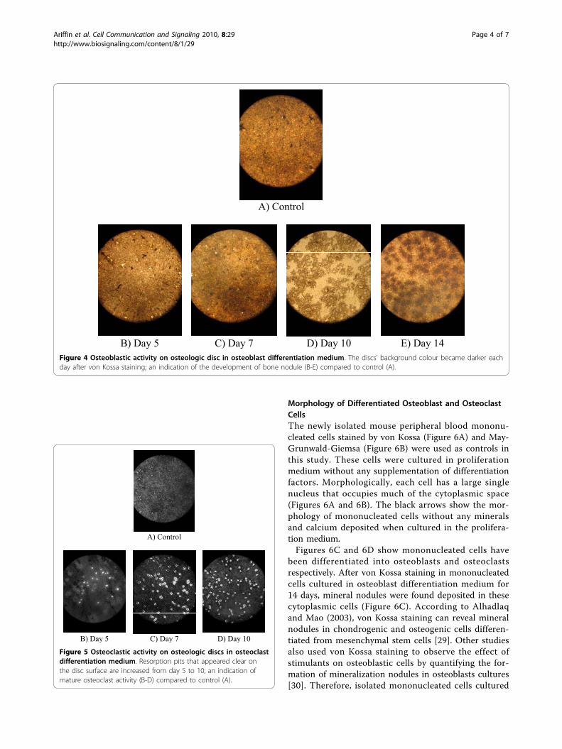
mation of clear resorption pits on the disc surface dueto the resorption of the calcium phosphate layer byosteoclast cells was observed (Figure 5). The resorptionpits on the disc surface increased from day 5 until 10(Figure 5B-D). Other studies also showed that osteo-clasts are differentiated in culture by day 10 [20-22,26].Studies by Valverde et al. showed that osteoclasticactivity can be determined using bone resorption pitsanalysis that was performed on calcium phosphate disc(BD BioCoat™ Osteologic™ Disc) [27,28]. Therefore, ourstudy involving similar disc was performed to deter-mine the existence of in vitro osteoclastic activity. Inaddition, there were no resorption pits found on thedisc surface for the control disc, indicating the absenceof osteoclast activity in cells cultured in proliferationmedium (Figure 5A).
Figure 2 ALP specific activity for mononucleated cells culturedin osteoblast differentiation medium. ALP specific activityshowed an increment after 14 days mononucleated cells cultured inosteoblast differentiation medium.
Figure 3 TRAP specific activity for mononucleated cellscultured in osteoclast differentiation medium. TRAP specificactivity showed an increment after 10 days mononucleated cellscultured in osteoclast differentiation medium.
Ariffin et al. Cell Communication and Signaling 2010, 8:29http://www.biosignaling.com/content/8/1/29
Page 3 of 7
Morphology of Differentiated Osteoblast and OsteoclastCellsThe newly isolated mouse peripheral blood mononu-cleated cells stained by von Kossa (Figure 6A) and May-Grunwald-Giemsa (Figure 6B) were used as controls inthis study. These cells were cultured in proliferationmedium without any supplementation of differentiationfactors. Morphologically, each cell has a large singlenucleus that occupies much of the cytoplasmic space(Figures 6A and 6B). The black arrows show the mor-phology of mononucleated cells without any mineralsand calcium deposited when cultured in the prolifera-tion medium.Figures 6C and 6D show mononucleated cells have
been differentiated into osteoblasts and osteoclastsrespectively. After von Kossa staining in mononucleatedcells cultured in osteoblast differentiation medium for14 days, mineral nodules were found deposited in thesecytoplasmic cells (Figure 6C). According to Alhadlaqand Mao (2003), von Kossa staining can reveal mineralnodules in chondrogenic and osteogenic cells differen-tiated from mesenchymal stem cells [29]. Other studiesalso used von Kossa staining to observe the effect ofstimulants on osteoblastic cells by quantifying the for-mation of mineralization nodules in osteoblasts cultures[30]. Therefore, isolated mononucleated cells cultured
Figure 4 Osteoblastic activity on osteologic disc in osteoblast differentiation medium. The discs’ background colour became darker eachday after von Kossa staining; an indication of the development of bone nodule (B-E) compared to control (A).
Figure 5 Osteoclastic activity on osteologic discs in osteoclastdifferentiation medium. Resorption pits that appeared clear onthe disc surface are increased from day 5 to 10; an indication ofmature osteoclast activity (B-D) compared to control (A).
Ariffin et al. Cell Communication and Signaling 2010, 8:29http://www.biosignaling.com/content/8/1/29
Page 4 of 7
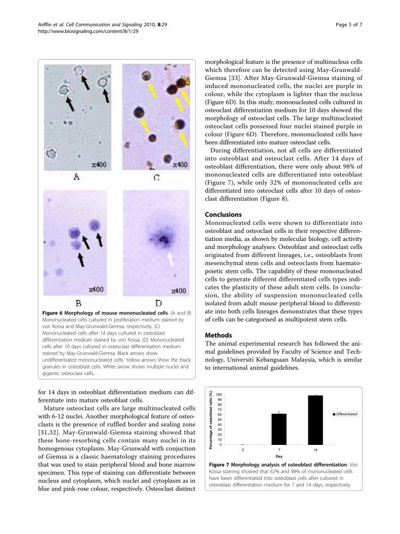
for 14 days in osteoblast differentiation medium can dif-ferentiate into mature osteoblast cells.Mature osteoclast cells are large multinucleated cells
with 6-12 nuclei. Another morphological feature of osteo-clasts is the presence of ruffled border and sealing zone[31,32]. May-Grunwald-Giemsa staining showed thatthese bone-resorbing cells contain many nuclei in itshomogenous cytoplasm. May-Grunwald with conjuctionof Giemsa is a classic haematology staining proceduresthat was used to stain peripheral blood and bone marrowspecimen. This type of staining can differentiate betweennucleus and cytoplasm, which nuclei and cytoplasm as inblue and pink-rose colour, respectively. Osteoclast distinct
morphological feature is the presence of multinucleus cellswhich therefore can be detected using May-Grunwald-Giemsa [33]. After May-Grunwald-Giemsa staining ofinduced mononucleated cells, the nuclei are purple incolour, while the cytoplasm is lighter than the nucleus(Figure 6D). In this study, mononucleated cells cultured inosteoclast differentiation medium for 10 days showed themorphology of osteoclast cells. The large multinucleatedosteoclast cells possessed four nuclei stained purple incolour (Figure 6D). Therefore, mononucleated cells havebeen differentiated into mature osteoclast cells.During differentiation, not all cells are differentiated
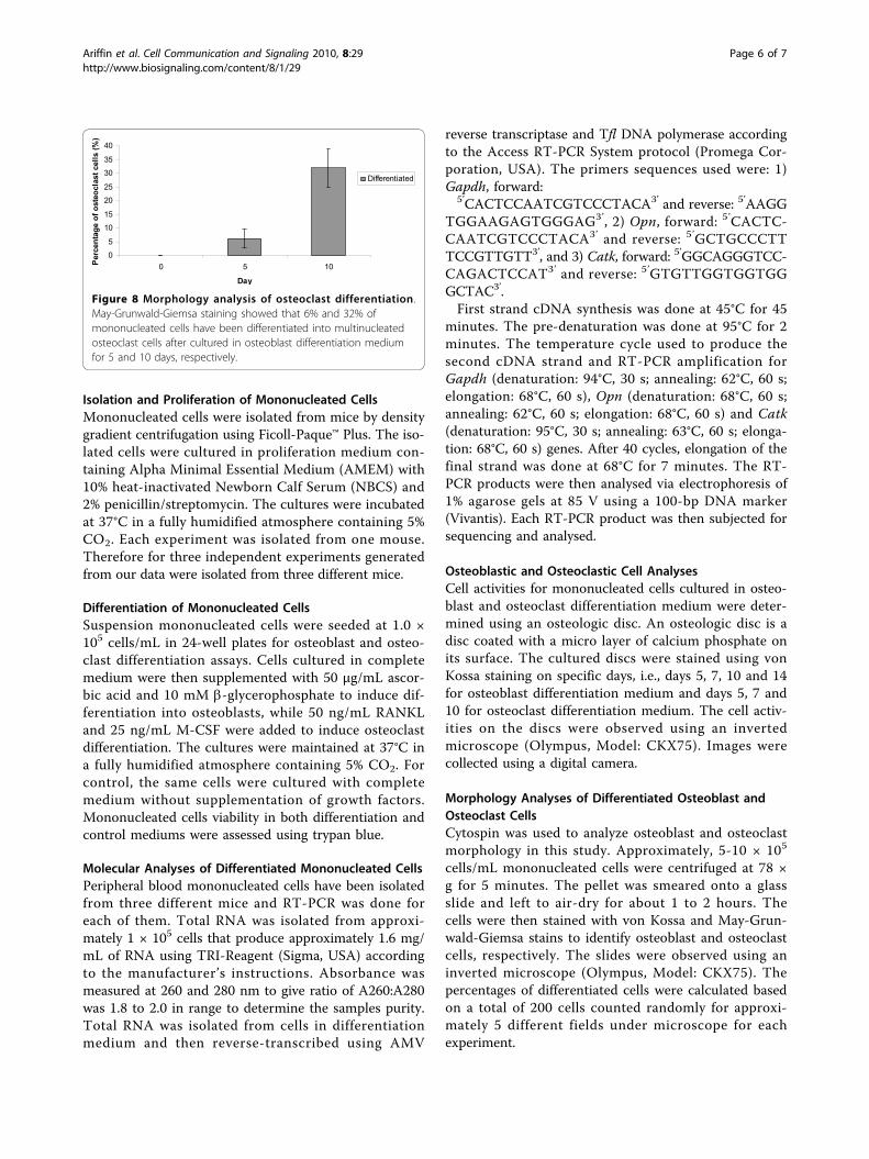
into osteoblast and osteoclast cells. After 14 days ofosteoblast differentiation, there were only about 98% ofmononucleated cells are differentiated into osteoblast(Figure 7), while only 32% of mononucleated cells aredifferentiated into osteoclast cells after 10 days of osteo-clast differentiation (Figure 8).
ConclusionsMononucleated cells were shown to differentiate intoosteoblast and osteoclast cells in their respective differen-tiation media, as shown by molecular biology, cell activityand morphology analyses. Osteoblast and osteoclast cellsoriginated from different lineages, i.e., osteoblasts frommesenchymal stem cells and osteoclasts from haemato-poietic stem cells. The capability of these mononucleatedcells to generate different differentiated cells types indi-cates the plasticity of these adult stem cells. In conclu-sion, the ability of suspension mononucleated cellsisolated from adult mouse peripheral blood to differenti-ate into both cells lineages demonstrates that these typesof cells can be categorised as multipotent stem cells.
MethodsThe animal experimental research has followed the ani-mal guidelines provided by Faculty of Science and Tech-nology, Universiti Kebangsaan Malaysia, which is similarto international animal guidelines.
Figure 6 Morphology of mouse mononucleated cells. (A and B)Mononucleated cells cultured in proliferation medium stained byvon Kossa and May-Grunwald-Giemsa, respectively. (C)Mononucleated cells after 14 days cultured in osteoblastdifferentiation medium stained by von Kossa. (D) Mononucleatedcells after 10 days cultured in osteoclast differentiation mediumstained by May-Grunwald-Giemsa. Black arrows showundifferentiated mononucleated cells. Yellow arrows show the blackgranules in osteoblast cells. White arrow shows multiple nuclei andgigantic osteoclast cells.
Figure 7 Morphology analysis of osteoblast differentiation. VonKossa staining showed that 62% and 98% of mononucleated cellshave been differentiated into osteoblast cells after cultured inosteoblast differentiation medium for 7 and 14 days, respectively.
Ariffin et al. Cell Communication and Signaling 2010, 8:29http://www.biosignaling.com/content/8/1/29
Page 5 of 7
Isolation and Proliferation of Mononucleated CellsMononucleated cells were isolated from mice by densitygradient centrifugation using Ficoll-Paque™ Plus. The iso-lated cells were cultured in proliferation medium con-taining Alpha Minimal Essential Medium (AMEM) with10% heat-inactivated Newborn Calf Serum (NBCS) and2% penicillin/streptomycin. The cultures were incubatedat 37°C in a fully humidified atmosphere containing 5%CO2. Each experiment was isolated from one mouse.Therefore for three independent experiments generatedfrom our data were isolated from three different mice.
Differentiation of Mononucleated CellsSuspension mononucleated cells were seeded at 1.0 ×10⁵ cells/mL in 24-well plates for osteoblast and osteo-clast differentiation assays. Cells cultured in completemedium were then supplemented with 50 μg/mL ascor-bic acid and 10 mM b-glycerophosphate to induce dif-ferentiation into osteoblasts, while 50 ng/mL RANKLand 25 ng/mL M-CSF were added to induce osteoclastdifferentiation. The cultures were maintained at 37°C ina fully humidified atmosphere containing 5% CO2. Forcontrol, the same cells were cultured with completemedium without supplementation of growth factors.Mononucleated cells viability in both differentiation andcontrol mediums were assessed using trypan blue.
Molecular Analyses of Differentiated Mononucleated CellsPeripheral blood mononucleated cells have been isolatedfrom three different mice and RT-PCR was done foreach of them. Total RNA was isolated from approxi-mately 1 × 10⁵ cells that produce approximately 1.6 mg/mL of RNA using TRI-Reagent (Sigma, USA) accordingto the manufacturer’s instructions. Absorbance wasmeasured at 260 and 280 nm to give ratio of A260:A280was 1.8 to 2.0 in range to determine the samples purity.Total RNA was isolated from cells in differentiationmedium and then reverse-transcribed using AMV
reverse transcriptase and Tfl DNA polymerase accordingto the Access RT-PCR System protocol (Promega Cor-poration, USA). The primers sequences used were: 1)Gapdh, forward:
5’CACTCCAATCGTCCCTACA3’ and reverse: 5’AAGGTGGAAGAGTGGGAG3’, 2) Opn, forward: 5’CACTC-CAATCGTCCCTACA3’ and reverse: 5’GCTGCCCTTTCCGTTGTT3’, and 3) Catk, forward: 5’GGCAGGGTCC-CAGACTCCAT3’ and reverse: 5’GTGTTGGTGGTGGGCTAC3’.First strand cDNA synthesis was done at 45°C for 45
minutes. The pre-denaturation was done at 95°C for 2minutes. The temperature cycle used to produce thesecond cDNA strand and RT-PCR amplification forGapdh (denaturation: 94°C, 30 s; annealing: 62°C, 60 s;elongation: 68°C, 60 s), Opn (denaturation: 68°C, 60 s;annealing: 62°C, 60 s; elongation: 68°C, 60 s) and Catk(denaturation: 95°C, 30 s; annealing: 63°C, 60 s; elonga-tion: 68°C, 60 s) genes. After 40 cycles, elongation of thefinal strand was done at 68°C for 7 minutes. The RT-PCR products were then analysed via electrophoresis of1% agarose gels at 85 V using a 100-bp DNA marker(Vivantis). Each RT-PCR product was then subjected forsequencing and analysed.
Osteoblastic and Osteoclastic Cell AnalysesCell activities for mononucleated cells cultured in osteo-blast and osteoclast differentiation medium were deter-mined using an osteologic disc. An osteologic disc is adisc coated with a micro layer of calcium phosphate onits surface. The cultured discs were stained using vonKossa staining on specific days, i.e., days 5, 7, 10 and 14for osteoblast differentiation medium and days 5, 7 and10 for osteoclast differentiation medium. The cell activ-ities on the discs were observed using an invertedmicroscope (Olympus, Model: CKX75). Images werecollected using a digital camera.
Morphology Analyses of Differentiated Osteoblast andOsteoclast CellsCytospin was used to analyze osteoblast and osteoclastmorphology in this study. Approximately, 5-10 × 105
cells/mL mononucleated cells were centrifuged at 78 ×g for 5 minutes. The pellet was smeared onto a glassslide and left to air-dry for about 1 to 2 hours. Thecells were then stained with von Kossa and May-Grun-wald-Giemsa stains to identify osteoblast and osteoclastcells, respectively. The slides were observed using aninverted microscope (Olympus, Model: CKX75). Thepercentages of differentiated cells were calculated basedon a total of 200 cells counted randomly for approxi-mately 5 different fields under microscope for eachexperiment.
Figure 8 Morphology analysis of osteoclast differentiation.May-Grunwald-Giemsa staining showed that 6% and 32% ofmononucleated cells have been differentiated into multinucleatedosteoclast cells after cultured in osteoblast differentiation mediumfor 5 and 10 days, respectively.
Ariffin et al. Cell Communication and Signaling 2010, 8:29http://www.biosignaling.com/content/8/1/29
Page 6 of 7

List of abbreviations(ALP): Alkaline Phosphatase; (AMEM): Alpha Minimal Essential Medium; (Catk):Cathepsin K; (Col1): Collagen type I; (Gapdh): Glyceraldehyde-3-phosphatedehydrogenase; (M-CSF): Macrophage Colony-Stimulating Factor; (NBCS):Newborn Calf Serum; (Opn): Osteopontin; (PBS): Phosphate Buffer Saline;(RANKL): Receptor Activator NF-�B Ligand; (RT-PCR): Reverse Transcriptase-Polymerase Chain Reactions; (TRAP): Tartrate Resistant Acid Phosphatase.
AcknowledgementsThe authors would like to thank the Universiti Kebangsaan Malaysia for thefinancial grants (UKM-GUP-BTK-07-15-197 and UKM-OUP-KPB-33-170/2010)during this study.
Author details1School of Biosciences and Biotechnology, Faculty of Science andTechnology, Universiti Kebangsaan Malaysia, 43600 Bangi, Selangor, Malaysia.2Department of Orthodontic, Faculty of Dentistry, Universiti KebangsaanMalaysia, Jalan Raja Muda Abdul Aziz, 50300 Kuala Lumpur, Malaysia.
Authors’ contributionsSHZA and RMAW developed the concept, designed and supervised theexperiments. IZZA performed and developed experimental study and doingthe analysis as part of PhD Thesis by research. MDY carried out part of themolecular and morphology analyses. All experiments and analysis wereperformed by IZZA and MDY, and approved by SHZA and RMAW. Allauthors read and approved the final manuscript before publication.
Competing interestsThe authors declare that they have no competing interests.
Received: 7 July 2010 Accepted: 23 October 2010Published: 23 October 2010
References1. Shahrul Hisham ZA, Rohaya MAW, Ismanizan I, Nor Muhammad M,
Zaidah ZA: Stem Cells, Cytokines and Their Receptors. APJMBB 2005,13(1):1-13.
2. Shanthly N, Aruva MR, Zhang K, Mathew B, Thakur ML: Stem cells: aregenerative pharmaceutical. Q J Nucl Med Mol Imaging 2006,50(3):205-216.
3. Shahrul Hisham ZA, Rohaya MAW, Intan Zarina ZA, Sahidan S, NorMuhammad M, Zaidah Z: Stem Cell in Blood Development. SainsMalaysiana 2005, 34(1):21-26.
4. Yamane T, Okuyama H, Tsuneto M, Hemmi H, Yamazaki H, Hayashi SI:Osteoclast lineage. In Handbook of Stem Cells. Edited by: Lanza R, GearhartJ, Hogan B, Melton D, Pedersen R, Thomson J, West M. London, ElsevierAcademic Press; 2004:295-303.
5. Karsdal MA, Fjording MS, Foged NT, Delaissé JM, Lochter A: TransformingGrowth Factor-beta-induced Osteoblast Elongation RegulatesOsteoclastic Bone Resorption through a p38 Mitogen-activated ProteinKinase-and Matrix Metalloproteinase-dependent Pathway. J Biol Chem2001, 276(42):39350-39358.
6. Miyamoto T, Suda T: Differentiation and function of osteoclasts. Keio JMed 2003, 52(1):1-7.
7. Filyushina ZG: Intravascular life span of granulocytes in thyrotoxicosisand diabetis mellitus. Bull Exp Biol Med 1969, 68(10):48-49.
8. Whitelaw DM: The Intravascular Lifespan of Monocytes. Blood 1966,28(3):455-464.
9. Sørensen AL, Rumjantseva V, Nayeb-Hashemi S, Clausen H, Hartwig JH,Wandall HH, Hoffmeister KM: Role of sialic acid for platelet life span:exposure of β-galactose results in the rapid clearance of platelets fromthe circulation by asialoglycoprotein receptor-expressing livermacrophages and hepatocytes. Blood 2009, 114(8):1645-1654.
10. Intan Zarina ZA, Shahrul Hisham ZA, Zaidah ZA, Rohaya MAW: PotentialDifferentiation of Three Types of Primitive Cells Originated fromDifferent Proliferation Terms of Mouse Blood. Sains Malaysiana 2010,39(2):305-313.
11. Swaminathan R: Biochemical markers of bone turnover. Clin Chim Acta2001, 313(1-2):95-105.
12. Kartsogiannis V, Ng KW: Cell lines and primary cell cultures in the studyof bone cell biology. Mol Cell Endocrinol 2004, 228(1-2):79-102.
13. Sodek J, Ganss B, McKee MD: Osteopontin. Crit Rev Oral Biol Med 2000,11(3):279-303.
14. Wang D, Christensen K, Chawla K, Xiao G, Krebsbach PH, Franceschi RT:Isolation and Characterization of MC3T3-E1 Preosteoblast Subcloneswith distinct In Vitro and In Vivo Differentiation/Mineralization Potential.J Bone Miner Res 1999, 14(6):893-903.
15. Ziolkowskaa A, Rucinski M, Pucher A, Tortorella C, Nussdorfer GG,Malendowicz LK: Expression of osteoblast marker genes in rat calvarialosteoblast-like cells, and effects of the endocrine disruptersdiphenylolpropane, benzophenone-3, resveratrol and silymarin. ChemBiol Interact 2006, 164(3):147-156.
16. Zhao Y, Guan H, Liu SF, Wu RC, Wang Z: Overexpression of QM inducescell differentiation and mineralization in MC3T3-E1. Biol Pharm Bull 2005,28(8):1371-1376.
17. Barber RD, Harmer DW, Coleman RA, Clark BJ: GAPDH as a housekeepinggene: analysis of GAPDH mRNA expression in a panel of human tissues.Physiol Genomics 2005, 21(3):389-395.
18. Hie M, Shimono M, Fujii K, Tsukamoto I: Increased cathepsin K andtartrate-resistant acid phosphatase expression in bone of streptozotocin-induced diabetic rats. Bone 2007, 41(6):1045-1050.
19. Pang M, Martinez AF, Fernandez I, Balkan W, Troen BR: AP-1 stimulates thecathepsin K promoter in RAW 264.7 cells. Gene 2007, 403(1-2):151-158.
20. Faccio R, Takeshita S, Zallone A, Ross FP, Teitelbaum SL: c-FMS and theαvβ3 integrin collaborate during osteoclast differentiation. J Clin Invest2003, 111(5):749-758.
21. Perez-Amodio S, Vogels IMC, Schoenmaker T, Jansen DC, Alatalo SL,Halleen JM, Beertsen W, Everts V: Endogenous expression and endocytosisof tartrate-resistant acid phosphatase (TRACP) by osteoblast-like cells.Bone 2005, 36(6):1065-1077.
22. Roodman GD: Cell biology of the osteoclast. Exp Hematol 1999,27(8):1229-1241.
23. Woo KM, Kim H-M, Ko JS: Macrophage colony-stimulating factorpromotes the survival of osteoclast precursors by up-regulating Bcl-XL.Exp Mol Med 2002, 34(5):340-346.
24. Sindrey DR, Kusljie D, Kwong PC: Human parathyroid hormone (1-84)stimulates bone formation in rate bone marrow cultures duringspaceflight. ASBMR 21st Annual Meeting 1999, SA424.
25. Cerovic A, Miletic I, Sobajic S, Blagojevic D, Radusinovic M, El-Sohemy A:Effects of Zinc on the Mineralization of Bone Nodules from HumanOsteoblast-like Cells. Biol Trace Elem Res 2007, 116(1):61-71.
26. Voronov I, Heersche JNM, Casper RF, Tenenbaum HC, Manolson MF:Inhibition of osteoclast differentiation by polycyclic aryl hydrocarbons isdependent on cell density and RANKL concentration. Biochem Pharmacol2005, 70(2):300-307.
27. Valverde P, Tu Q, Chen J: BSP and RANKL induce osteoclastogenesis andbone resorption synergistically. J Bone Miner Res 2005, 20:1669-1679.
28. Valverde P, Zhang J, Fix A, Zhu J, Ma W, Tu Q, Chen J: Overexpression ofBone Sialoprotein Leads to an Uncoupling of Bone Formation and BoneResorption in Mice. J Bone Miner Res 2008, 23(11):1775-1788.
29. Alhadlaq A, Mao JJ: Tissue-engineered Neogenesis of Human-shapedMandibular Condyle from Rat Mesenchymal Stem Cells. J Dent Res 2003,82(12):951-956.
30. Tsuang YH, Sun JS, Chen LT, Sun SCK, Chen SC: Direct effects of caffeineon osteoblastic cells metabolism: the possible causal effect of caffeineon the formation of osteoporosis. J Orthop Surg Res 2006, 1:7.
31. Yagi M, Miyamoto T, Sawatani Y, Iwamoto K, Hosogane N, Fujita N,Morita K, Ninomiya K, Suzuki T, Miyamoto K, Oike Y, Takeya M, Toyama Y,Suda T: DC-STAMP is essential for cell-cell fusion in osteoclasts andforeign body giant cells. J Exp Med 2005, 202(3):345-351.
32. Li Z, Kong K, Qi W: Osteoclast and its roles in calcium metabolism andbone development and remodeling. Biochem Biophys Res Commun 2006,343(2):345-350.
33. Servet-Delprat C, Arnaud S, Jurdic P, Nataf S, Grasset MF, Soulas C,Domenget C, Destaing O, Rivollier A, Perret M, Dumontel C, Hanau D,Gilmore GL, Belin MF, Rabourdin-Combe C, Mouchiroud G: Flt3+macrophage precursors commit sequentially to osteoclasts, dendriticcells and microglia. BMC Immunol 2002, 3:15.
doi:10.1186/1478-811X-8-29Cite this article as: Ariffin et al.: Differentiation analyses of adultsuspension mononucleated peripheral blood cells of Mus musculus. CellCommunication and Signaling 2010 8:29.
Ariffin et al. Cell Communication and Signaling 2010, 8:29http://www.biosignaling.com/content/8/1/29
Page 7 of 7