Value of transesophageal echocardiography during repair of congenital heart defects
Upload
khangminh22Category
view
0download
0
Z.U.M.J.Vol.20; N.2; March; 2014 Transesophageal Echo Study In……
-245-
TRANSESOPHAGEAL ECHO STUDY IN PATIENTS WITH ACUTE
ISCHEMIC STROKE
Kamal Saad Mansour, Manar Mostafa Elzaki, Khaled A. M. Elsharkawy* ,Waleed Elawady and
Al-Shaimaa Ali Mohamad Al-Sadek
Cardiology and Neurology* Department, Faculty of Medicine, Zagazig University.
ABSTRACT Background: Stroke is one of the major causes of death and disability and due to the high mortality and
morbidity rates associated with stroke ,it is becoming a major community health problem worldwide. It has
been estimated that cardiogenic emboli are a source of transient ischemic attack (TIA) or stroke in 20% to
40% of all cases. Transesophageal echocardiography (TEE) has been proven superior to transthoracic
echocardiography (TTE) for the detection of potential sources of cardiac embolism in patients with previous
stroke. In particular, the additional value of TEE in the patients above 45 years of age, in whom TEE is still
far from routine procedure, was evaluated. In addition, emphasis was placed on therapeutic consequences, ie,
the indication for anticoagulation, based on the results of TEE.
Aim of the work : The aim of our study is to evaluate the role of TEE (transesophagealecho) to find
indications of anticoagulation despite false absence of its benefit by clinical and transthoracicecho (TTE)
examination.
Patient and methods: The study was conducted in Zagazig university hospitals and included 100 patients
diagnosed with ischemic stroke The diagnosis of stroke was made based on clinical data and brain CT. A
full TEE was done and the presence of the following potential sources of embolism was specifically
examined (a)left atrial spontaneous echo contrast and thrombus , (b)Impaired Left atrial appendage velocity
and the presence of LAA (Left atrial appendage )thrombi , (c)Atheroma in the thoracic aorta, (d)Patent
foramen ovale and atrial septal aneurysm (e) others, including valvular vegetations and intracardiac masses .
Results:Among the different age groups studied , the prevalence of PFO and MVP (mitral valve prolapse)
were highest in patients ˂ 45 years old while the prevalence of complex aortic atheroma was highest in
patients ˃45 years old .Risk of stroke recurrence was highest in patients with complex aortic atheroma
.Among patients with dyslipidemia,prevalance of ischemic stroke was highest in those older than 45 years
old and was significantly higher in low HDL (high density lipoprotein ) (˂40 mg/dl ) and high cholesterol
(˃200 mg/dl) group.Furthermore, the risk of stroke recurrence was significantly higher in patients with
hypercholesterolemia.
Conclusion: TEE has proved to be a very useful tool in diagnosing causes of ischemic stroke .The benefit
was not only seen in young age (˂45 years old), but also in those in older age groups .
Key words: Mitral valve prolapse, patent foramen ovale, interatrial septal aneurysm , complex aortic
ahteroma ,Transesophageal Echocardiography.
INTRODUCTION
troke is the leading cause of disability and
the second most common cause of death
worldwide(1)
. Accurate definition of the
mechanism of stroke is crucial as this will
guide the most effective care and therapy.
Cardioembolic stroke accounts for 14-30% of
all cerebral infarctions(2)
. In most cases,
recurrence of cardioembolism can be
prevented by oral anticoagulants. Therefore,
for a patient with a cerebral infarct, early
confirmation of a diagnosis of cardioembolic
infarction is extremely important in order to
initiate anticoagulation therapy for an adequate
secondary prevention(3)
.
It has been estimated that cardiogenic
emboli are a source of transient ischemic
attack (TIA) or stroke in 20% to 40% of all
cases. Transesophageal echocardiography
(TEE) has been proven superior to
transthoracic echocardiography (TTE) for the
detection of potential sources of cardiac
embolism in patients with previous stroke(4)
.
The echocardiographic diagnosis of cardiac
thrombi, vegetations and tumors as well as the
identification of predisposing conditions such
as patent foramen ovale, aortic atherosclerosis
and other minor causes (e.g., mitral valve
prolapse, mitral and aortic valve calcification)
have crucial clinical relevance, affecting the
choice of surgery and/or of pharmaceutical
therapy in the setting of patients presenting
embolism. The echocardiographic assessment
helps not only for the retrospective diagnosis
S

Z.U.M.J.Vol.20; N.2; March; 2014 Transesophageal Echo Study In……
-246-
of sources of embolism but also for the
prevention of events in asymptomatic patients.
Echocardiography can also distinguish normal
variants and artifacts from cardiac masses and
tumors(5)
.
AIM OF THE WORK
The aim of our study is to evaluate the
role of TEE (transesophagealecho) to find
indications of anticoagulation despite false
absence of its benefit by clinical and
transthoracicecho (TTE) examination.
PATIENTS AND METHODS
This study was carried out in Zagazig
University Hospitals , it was conducted from
January 2011 to August 2013. It included
(100) patients diagnosed with acute ischemic
stroke ,the diagnosis of ischemic was based on
clinical data and brain CT.
Inclusion criteria: Acute ischemic stroke
patients without indication for anticoagulation
based on clinical, electrocardiographic and
TTE findings.
Exclusion criteria: Patients with a pre-
existing contraindication to anticoagulation.
Patients with a pre-existing indication to
anticoagulation (these include: mitral valve
stenosis ,Mechanical valve prosthesis, left
ventricular systolic dysfunction ,dilated &
restrictive cardiomyopathy, previous
myocardial infarction,left atrial or left
ventriclular masses that where easily detected
by TTE) Carotid Dupplex revealing carotid
stenosis more than or equal to ≥70%. Patients
with hypertension or diabetes mellitus.
Our patients were subjected to the
following: Thorough history taking, physical
examination & investigations : this included
thorough neurological and cardiac examination
CT –brain standard 12-leads electrocardiogram
to exclude the presence of atrial fibrillation
laboratory tests for hyperlipidemia (HDL
,LDL ,Serum Total Cholestrol,serum
triglyceries) according to ATP3 guidelines in
2004.
* Conventional Transthoracic EchoDoppler
study: Transthoracic echocardiographic
examination was done by using HP Sonos
5500 set with a 2.5 MHz transducer. Images
were taken while the patient in supine or in the
left lateral position utilizing left parasternal
long axis , apical 4 and apical 2 chamber
views. Recordings and calculations of different
parameters were performed according to the
recommendations of the American Society of
Echocardiography M-mode, 2D, and Doppler,
according to the recommendations of the
American Echocardiography Society.
* Transesophageal echocardiographic
examination using (Hewlette Packard) Sonos
5500 set: with a transesophageal echo
transducer and with the following preparation
Patient was informed to fast for minimum of 4
hours before the procedure,we examined the
mouth for loose teeth ,Patients were asked
whether there were any problems with
swallowing ,local anesthesia was given to
decrease gag reflex.TEE was performed under
conscious sedation
Detailed Transesophageal
Echocardiography data:
1-left atrial spontaneous echo contrast and
thrombus: Left atrial spontaneous echo
contrast was diagnosed by the presence of
characteristic dynamic smoke-like swirling
echo in the left atrium or the left atrial
appendage. Spontaneous echo contrast was
graded as 0 = no smoke, 1+ = mild smoke
visible in some portion of the LA, 2+ = dense
smoke that appeared throughout the LA.
2-Left atrial thrombus was diagnosed by the
presence of an echodense mass in the left
atrium or the left atrial appendage, distinct
from the endocardium and the pectinate
muscles of the left atrial appendage.
3-Atheroma in the thoracic aorta: The
transesophageal probe is set at 0° and is
rotated counterclockwise to demonstrate the
thoracic aorta in cross section . The probe is
advanced until the aorta is no longer visualized
(approximately 44 – 50 cm from the patient's
incisors) and is then slowly withdrawn as the
aorta is inspected for evidence of
atherosclerotic plaque and dissection. To
further survey plaque and other vessel wall
changes, the imaging plane was increased to
90° to visualize the long axis of the aorta. As
the probe is withdrawn to the upper esophagus,
the aortic arch was inspected . The probe was
then turned counterclockwise and withdrawn
further to better visualize the distal ascending
aorta. At the level of the arch, the imaging
plane was then increased to 90°.

Z.U.M.J.Vol.20; N.2; March; 2014 Transesophageal Echo Study In……
-247-
Atheroma that is mobile,
pedunculated, or protruding ≥5 mm into the
lumen are classified as complex atheroma .All
other sessile atheroma <5 mm in thickness are
classified as simple atheroma.
4-Patent foramen ovale and atrial septal
aneurysm : Image acquisition at 90° Imaging
plane with clockwise turning demonstrated
the bicaval view . In the bicaval view, the
interatrial septum traverses horizontally across
the screen, and an atrial septal defect can be
appreciated with color Doppler. We typically
choose this view for agitated saline injection
(rest, post-Valsalva, and cough) for a
suspected patent foramen ovale, but the 0° and
135° orientations, or the 60° orientation may
also be used .
Maintaining the imaging angle at 135°,
the probe was turned clockwise to demonstrate
the right and left atria and interatrial septum
The interatrial septum appears horizontally
across the screen, and the fossa ovalis is well
visualized. Color Doppler is applied to identify
atrial septal defects. Agitated saline injections
for patent foramen ovale detection may be
performed at this or other orientationsAgitated
saline contrast was often performed during
TEE in patients with suspected paradoxical
embolism due to a patent foramen ovale . For
these patients, we usually interrogated the
interatrial septum from 0–180° to identify the
best orientation to visualize the thinnest
portion of the interatrial septum/fossa ovalis.
Saline is administered at rest, followed by
injections with cough and post-Valsalva
release. Except for cases of suspected
persistence of a left sided superior vena cava,
our preference is to inject agitated saline from
the right arm so as to avoid potential
obstruction to venous flow related to the
patient's left lateral decubitus position .Atrial
septal aneurysm is defined as a thin-walled
area in the region of the fossa ovalis with a
base of at least 1.5 cm and an excursion with
the cardiac cycle of at least 1.5cm. Care will
be taken to distinguish a true atrial septal
aneurysm from a hypermobile interatrial
septum .
5-Left atrial appendage (LAA) velocity and
LAA thrombi :While maintaining the
imaging plane at 90°, the image depth is
increased and the TEE probe is turned
counterclockwise to obtain the two-chamber
view, including the LA, the LV, and the mitral
valve .The probe is then withdrawn slightly,
anteflexed, and turned more counterclockwise
to visualize the LAA at 90° . Pulse wave
Doppler is applied to assess emptying
velocities (6)
Statistical analysis:
Continuous variables are summarized
as mean ±SD. Variables measured between the
3 age groups, were compared by repeated 1-
way analysis of variance (ANOVA).
Categorical variables were compared by Chi-
squre. Paired-t test was used to compare
continuous variables between groups. All was
calculated SPSS version 12 software program
(7)
RESULTS
The 100 stroke patients were
classified by 3 classifications :1) according to
age into 3 groups ˂45 years old (Group A=32
patients) , 45-65yrs. (Group B=36 patients)
and ˃65years (Group C=32 patients) .2)
according to history of stroke recurrence
:Group I ( patients with first time ischemic
stroke) and group II (patients with recurrent
ischemic stroke). 3) Finally, according to
gender into female (F) group and male (M)
group.
Demographic and clinical data: the study
included 48 females (48%) and 52 males
(52%). Their ages ranged from 18 to 82; 38
(38%) patients had a recurrent stroke as
opposed to 62 (62%) first time ischemic
stroke patients.
Comparative analysis of TEE parameters in
different age groups (table 1):
38% of all stroke patients had a normal TEE.
However 62% (26 male, 36 female) had a
cardiac abnormality detected, despite normal
TTE examination.
A-As regard miral valve prolapse (MVP):
The prevalence of mitral valve prolapse
(MVP) was 10% of all our patients(4 male, 6
female) and the highest in patients <45 years
of age (25% of group A).There is a
statistically significant difference in
prevalence of MVP between group A & both
group B& C with the highest prevalence seen
in group A (age ˂45years).
B-PFO: In our study PFO was seen in 10% of
our study population(2 male, 8 female). The

Z.U.M.J.Vol.20; N.2; March; 2014 Transesophageal Echo Study In……
-248-
prevalence was highest in patients < 45 years
old (25% of group A) with a significant
difference between groups.
C-ASD: Was found in 4%(2 male, 2 female)
of the study population, (4%) of group A , no
patients in group B and (6%) in group C with
no significant difference in prevalence
between groups.
D-IASA: Was seen in 4%(2 male, 2 female) of
the study population with no significant
difference in prevalence between groups.
E-Complex aortic atheroma : 28% of our
study population(12 male, 16 female) had a
complex aortic atheroma in the ascending
aorta and arch. The prevalence was highest in
patients older than 45 years(2% in group A,
22% of group B and 56% of group C) with a
significant difference between groups.
F-Intracardiac masses: Intracardiac masses
are seen in 6% of our study population(4 male,
2 female) all of them in group A, represent
19% (3 patients had small vegetations over
the aortic valve cusps & 3 patients with an
Aschoff nodule over the mitral valve), none of
group B or C .with a statistically significant
difference between groups.
Table (1): Comparative analysis of TEE parameters in different age groups.
TEE parameters
Age group
Chisq. P1 P2 P3 A N=32
B N=36
C N=32
MVP No .(%) 8(25%) 2(36%) 0(0%) 12.346 <0.05 < 0.05 >0.05
PFO No .(%) 8(25%) 2(36%) 0 (0%) 12.346 <0.05 <0.05 >0.05
ASD No .(%) 2 (6%) 0 (0%) 2 (6%) 2.344 >0.05 .>0.05. >0.05
IASA No. (%) 2 (6%) 2 (5.6%) 0 1.982 >0.05 >0.05 >0.05
Complex aortic
atheroma 2 (6%) 8 (22%) 18 (56%) 20.77 >0.05 <0.05 <0.05
Intracardiac
masses 6 (19%) 0 0 13.56 <0.05 >0.05 >0.05
*MVP=mitral valve prolapse , PFO=patent foramen ovale ,ASD=atrial septal defect, IASA=interatrial septal
defect,P1:P-value group A vs. Group B ,P2:group A vs. group C ,P3:group B vs. group C
Comparative analysis of lipid profile in
different age groups (table 2):
Low HDL (<40 mg/dl): Prevalence of low
HDL (<40 mg/dl ) was 50% in all our study
population,(24 male, 26 female), increased
significantly in patients older than 45 years
(31% of group A, 56% of group B, and 63 %
of group C). There is a statistically
significantly difference between group A &
the other 2 groups group B &C with the
highest prevalence in group C.
Elevated LDL ( >130 mg/dl): Elevated LDL
is detected in 20% in all our study
population,(8 male, 12 female), 12.5% of
group A , 27.8.% of group B and (18.8%)
of group C. With no statistically significant
difference seen between the groups.
Hypercholesterolemia(>200mg/dl): Prevalence of hypercholesterolemia was 11%
in all our study population,(6 male, 5 female),
increased with age (0 in group A, 22 % of
group B and 9% of group C). A statistically
significant difference is seen between group
A& B with the highest prevalence in group B
(age 45-65 years).
Hypertriglyceridemia(>150mg/dl):
Hypertriglyceridemia is detected in was 22%
in all our study population,(10 male, 12
female), 19 % of group A, 27.8% of group B
and 19% of group C. No statistically
significant difference is recorded between the
3 groups .

Z.U.M.J.Vol.20; N.2; March; 2014 Transesophageal Echo Study In……
-249-
Table (2): Comparative analysis of lipid profile in different age groups.
Age group
Chisq. P1 P2 P3 A N=32
B N=36
C N=32
Low HDL No .(%) 10(31%) 20(56%) 20(63%) 18.9 <0.05 <0.05 >0.05
Elevated LDL No .(%) 4 (12.5%) 10 (27.8%) 6(18.8) 2.5 >0.05 >0.05 >0.05
Hypercholesterolemia No .(%)
0 8 (22%) 3 (9%) 8.7 <0.05 >0.05 >0.05
Hypertriglyceridemia. No. (%)
6 (19%) 10 (28%) 6 (19%) 1.1 >0.05 >0.05 >0.05
*HDL: high density lipoprotein, LDL: low density lipoprotein. P1:P-value group. A vs. Group B ,P2:group A vs.
group C ,P3:group B vs. group C
Comparative analysis of TEE parameters of
group I (first time ischemic stroke) & group
II(recurrent stroke) (table 3):
1-MVP: Mitral valve prolapse was seen in
12.9% of first time stroke group as opposed to
5.3% of recurrent stroke group. There was no
statistically significant difference seen
between both groups.
2-PFO: PFO was seen in 13% of first time
stroke group as opposed to 5% in recurrent
group. No statistically significant difference
was seen between groups .
3- Atrial septal defect: Was seen in (3.2%) in
group I compared to (5.3%) in
group II. No statistically significant difference
was seen between both groups .
4-IASA: IASA was seen in 3.2% of first time
stroke group as opposed to 5.3%of recurrent
stroke group with no statistically significant
difference seen between groups.
5-Complex aortic atheroma: There was a
higher prevalence of complex aortic atheroma
in the recurrent stroke group(42%) as opposed
to 19% in first time ischemic stroke group with
significant difference.
6-Intracardiac masses: Were seen in (3.2%)
of group I and (10.5%) of group II. No
statistically significant difference was seen
between both groups.
Table (3): Comparative analysis of TEE parameters of group I (first time ischemic stroke) & group
II (recurrent stroke).
TEE Parameters
Recurrent stroke
Chisq.
P.value
I
(first time ischemic
stroke)
(N=62)
II
(recurrent stroke)
(N=38)
MVP No. (%) 8 (13%) 2 (5%) Fischer Exact >0.05
PFO No. (%) 8 (13%) 2 (5%) Fisher Exact >0.05
ASD 2 (3.2%) 2 (5%) Fischer Exact >0.05
IASA 2 (3.2%) 2 (5%) Fischer Exact >0.05
Complex aortic atheroma 12 (19%) 16 (42%) 6 0.01
Intracardiac masses 2 (3.2%) 4 (10.5%) Fisher Exact >0.05
*MVP=mitral valve prolapse , PFO=patent foramen ovale ,ASD=atrial septal defect,IASA=interatrial septal
defect
Comparative analysis of TIAs in different
age groups (table 4):
TIAs were seen in 38% of all patients in
our study: 31% of group A, 39 % of group B
and 44 % of group C. No statistically
significant difference was seen between the 3
groups.
History of TIAs was seen in (29.2%) of
females & (46.2%) of males. No statistically
significant difference was seen between
groups.

Z.U.M.J.Vol.20; N.2; March; 2014 Transesophageal Echo Study In……
-250-
Table 4: Comparative analysis of TIAs in different age groups
Age group
Chisq. P.value A
N=32
B
N=36
C
N=32
TIAs No .(%) 10(31%) 14(39%) 14(44%) 1.1 >0.05
*TIAs: transient ischemic attacks
Comparative analysis of patients with
vascular territory infarction in different
age groups (table 5):
1-Anterior circulation infarction: 93.8% of
group A, 94.4% of Group B and 100% of
group C. Also equal percentage of males and
females (96%) had anterior circulation
infarction. There was no statistically
significant difference between the 3 age
groups and both sexes.
2-Posterior circulation infarction:13% of
group A ; 6% of the group B and 13% of
group C. Also 13% of females and 8% of
males had posterior circulation infarction. No
statistically significant difference was found
between the three groups and both sexes.
Table (5): Comparative analysis of patients with vascular territory infarction in different age
groups.
Vascular territory
Age group
Chisq. P.value A
N=32
B
N=36
C
N=32
Anterior circulation
No .(%) 30(94%) 34(94%) 32(100%) 1.9 >0.05
Posterior circulation
No .(%) 4(13%) 2 (6%) 4(13) 1.2 >0.05
Comparative analysis of neurological
parameters in different age groups(table 6):
**Aphasia: Was found in 16% of all our
patients(4 male, 12 female), 13% of group A,
22% of group B and 13% of group C. No
significant difference was seen between
groups
**Ataxia: Was in 10% of all our patients(4
male, 6 female), 13% of group A had ataxia ,
6% of group B and 13% of group C. No
statistically significant difference was seen
between the groups
**Sensory Deficit: Was in 64% of all our
patients(30 male,34female) 56.2% of group A
had a sensory deficit ,50% of group B and 88
% of group C . A statistically , significant
difference was found between the group C
&both group A &B with a higher prevalence
of sensory deficit in group C.
**Motor Deficit: Was in 40% of all our
patients(24 male, 16 female), 50% of group A
had a motor deficit ,44% of group B and 25%
of group C. No statistically significant
difference was seen between the groups.
**Conscious Level: Percentage of fully
conscious patients was 76% of all our
patients(42 male, 34 female), 94% in group A,
83% of group B and 50% of group C. The
percentage of confused patients was 20% of all
our patients(8 male, 12 female), 6% of group
A ,11% of group B and 44% of group C were
confused. No patients were comatosed in
group A, but 5.5% of group B and 6.2% of
group C were comatosed (2 male and 2
female). There is a statistically significant
difference between the groups as regard the
conscious level. More conscious patients were
in the younger age group while the older group
had a larger number of confused patients.

Z.U.M.J.Vol.20; N.2; March; 2014 Transesophageal Echo Study In……
-251-
(table 6):Comparative analysis of neurological parameters in different age groups
Neurological examination
Age group
Chisq. P1 P2 P3 A N=32
B N=36
C N=32
Aphasia No. (%) 4 (13) 8 (22) 4 (13) 1.620 >0.05 >0.05 >0.05
Ataxia No.(%) 4 (13) 2 (6) 4 (13) 1.235 >0.05 >0.05 >0.05
Sensory Deficit No.(%) 18 (56) 18 (50) 28 (88) 11.6 >0.05 <0.05 <0.05
Motor Deficit No.(%) 16 (50) 16 (44) 8 (25) 1.7 >0.05 >0.05 >0.05
C.L.
Conscious 30 (94) 30 (83) 16 (50)
19.8 <.01 >0.05 >0.05 Confused 2 (6) 4 (11) 14 (44)
Coma 0 (0.0) 2 (5.5) 2 (6.2)
P1:P-value group A vs. Group B ,P2:group A vs. group C , P3:group B vs. group C
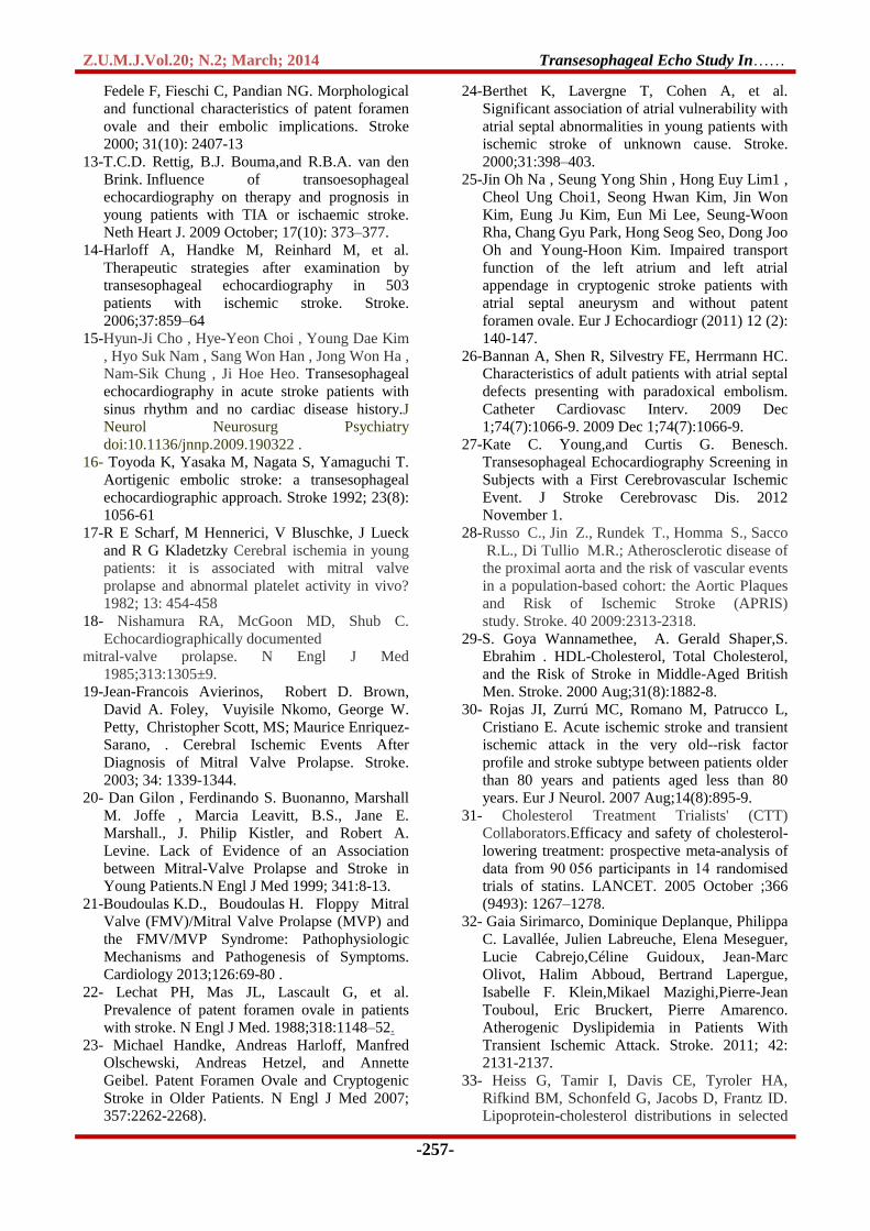
Midesophageal ascending aorta short axis
view:ascending aortic atheroma protruding into the
lumen measuring 7.5mm
Axial CT scan of the brain demonstrating left
temproparietal infarction.
DISCUSSION
Our study included the following TEE
(transesophagealecho) parameters: MVP,
PFO, IASA, ASD, complex atheroma,
Intracardiac masses.
In our study, 62% of study population
showed posistive findings with TEE that
require anticoagulation without other
indications for anticoagulation by clinical,
electrocardiographic or TTE data.This
percentage was higher than the percentage
detected by Tiago et al(8)
which was
32.1%.However Tiago et al included
hypertensive and diabetic patients which were
excluded from our study.
The superiority of TEE over TTE for
diagnosing cardiac sources of embolism has
also been demonstrated by several other
studies McNamara et al.(9)
; Blum et al.(10)
and confirmed in our study. They concluded
that “TEE is indispensable in all patients
being candidates for oral anticoagulation”.Our
results seem to confirm their findings, but on
a more selected population of patients with no
other indication for anticoagulation.
The issue of age is an important one in
the discussion on whether TTE or TEE should
be used in patients with ischemic stroke. In
our study 28 cases ( 87.5%) of the patients
<45 years old had a cardiac source of
embolism detected by TEE. this was higher
than that detected by Sebastian et al (4),
they
demonstrated that 33% of their population
<45 years old had a cardioembolic source
detected by TEE. However ,they enrolled all
patients with a TIA or ischemic stroke
regardless of any major risk factors or
arrhythmias. This may have contributed to
smaller percentage of findings.
In our study 34 cases (50%) of the
patients >45 years old had a cardiac source
detected by TEE. This figure was bigger than

Z.U.M.J.Vol.20; N.2; March; 2014 Transesophageal Echo Study In……
-252-
that detected by Sebastian et al (4)
.In their
study , TEE demonstrated a risk factor of
ischemic stroke in 17% (n=32 patients).
These patients would have been incorrectly
denied anticoagulation therapy if only TTE
was used. Thus, the current observations
support the additional value of TEE in both
younger and older patients.
In our study PFO was seen in 10% of
our study population .This was greater than
that detected by Tiago et al(8),
they
performed TEE for 84 ischemic stroke
patients and 3.6% only had a PFO.
Strandberg, et al(11)
, studied 441 ischemic
stroke patients by TEE and detected 67 PFOs
(15.2%) of their population .
However, De Castro et al(12)
studied
350 ischemic stroke patients, and 21% of their
population had a PFO .
This variability in number of PFOs
detected by different studies has been
explained by Decastro et al(12)
, they stated
that the prevalence of a PFO as assessed by
transesophageal contrast echocardiography
varies considerably among echocardiographic
laboratories (ranging from 8 to 54%). This
striking difference may be due to several
reasons: (1) the population studied (age,
healthy persons versus patients, history of
embolism); (2) the technique used (contrast
agent, provocative maneuvers, ultrasonic
instrumentation including transducer
frequency); and (3) the criteria applied for
diagnosis.
In our study ASA was seen in 4% of
the study population . This was similar to De
Castro et al(12)
& Strandberg, et al (11)
Using
TEE, they identified atrial septal aneurysms
in approximately 4% of all patients studied. It
has been postulated that perhaps these
abnormalities provide a nidus for thrombus
formation and blood stasis and therefore
constitute a risk factor for cerebral
embolization.
A higher figure was detected by
Rettig et al(13)
,they recorded 7 ASA in 84
ischemic stroke patients (8% of their
population). However this difference may be
attributed to the difference in population
studied. Rettig et al (13)
included patients with
TIAs also which were not part of our study
population. A higher percentage was also seen
in study held by Tiago et al (8)
found 10% had
an IASA. However, Tiago et al(8)
didn’t
exclude any of the traditional risk factors so
the probability that the ASA is incidental and
not the cause of ischemic stroke is higher .
In our study, 28% of our study
population had a complex aortic atheroma in
the ascending aorta and arch, this was higher
than that detected by Harloff et al(14)
. In their
study, TEE was performed on 503 ischemic
stroke patients.They detected complex aortic
atheroma in 19.7%. it was also higher than
those detected by Hyun-Ji Cho et al(15)
,they performed TEE for 1833 consecutive
ischemic stroke patients with normal sinus
rhythm and no history of cardiac disease
.They investigated the frequency of aortic
plaques in such patients and detected 157
patient (8.5%).On the other hand, , our
results were smaller than those detected by
Toyoda et al(16)
using TEE they evaluated
complicated lesions in the thoracic aortas of
62 patients who met clinical criteria for
embolic stroke. Twenty-six patients (42%)
showed complicated aortic arch lesions on
transesophageal echocardiogram.
We believe that this variability in
results arouse from interobserver variability in
interpretation of arch atheromasand
measuring them ,However ,all of the studies
confirmed the importance of TEE in detecting
atheromas in both the ascending aorta and
arch.
In our study ,the prevalence of mitral valve
prolapse (MVP) was 10% of all our patients
and the highest in patients <45 years of age
(25% of group A) with significant difference.
This was in agreement with Scharf et al(17),
they studied the incidence of MVP in 47
patients under 45 years of age with TIA or
stroke of unknown cause and in an age- and
sex-matched control group. They concluded
that the incidence of MVP is higher in young
patients with cerebral ischemia of unknown
cause than in asymptomatic controls.
Similarly Nishimura et al(18)
followed 237
minimally symptomatic or asymptomatic
patients by echocardiography for a mean of
6.2 years. 97 patients with redundant mitral
valve leaflets identified by
echocardiographically, 10.3% had sudden
death, infective endocarditis, or a cerebral

Z.U.M.J.Vol.20; N.2; March; 2014 Transesophageal Echo Study In……
-253-
embolic event. During the follow-up period,
10 patients (mean age 50 years) sustained a
cerebral embolic event.
Jean-Francois et al(19)
, studied 777
patients with a mean age of 45 years and
they found a high risk of stroke during
follow-up of these patients. The high stroke
risk among patients with MVP and in sinus
rhythm can be explained by mitral thickening
(which is a major part of the pathology in
MVP) which carries a high risk of thrombus
formation.
Our results however were discordant
with Gilon et al.(20)
,they performed a case–
control study, and reviewed data on 213
consecutive patients 45 years of age or
younger with documented ischemic stroke or
transient ischemic attack. The prevalence of
prolapse in these patients was compared with
that in 263 control subjects without known
heart disease. Mitral-valve prolapse was
present in 4 of the 213 young patients with
stroke (1.9 percent), as compared with 7 of
the 263 controls (2.7 percent); prolapse was
present in 2 of 71 patients (2.8 percent) with
otherwise unexplained stroke. They
concluded that Mitral-valve prolapse is
considerably less common than previously
reported among young patients with stroke or
transient ischemic attack, including
unexplained stroke, and no more common
than among controls.
An elaborate explanation was given
by Boudoulas and Boudoulas in (21)
about
the mechanisms by which mitral valve
prolapse can cause thromboembolism. The
pathology of the FMV ( floppy mitral valve
)differs significantly from the normal mitral
valve. The most specific histopathologic
characteristics are collagen dissolution and
disruption in the pars fibrosa of the mitral
valve leaflets. Also, there is a replacement of
the dense collagenous fibrosa by loose
myxomatous connective tissue .Similar
histologic abnormalities have been seen in
chordae tendineae. Scanning electron
photomicrographs have demonstrated surface
folds and focal loss of endothelial cells of
mitral valve leaflets, and these surface
abnormalities may predispose to
thromboembolic complications and infective
endocarditis. Continuous pressure and stress
due to left ventricular systole on the mitral
valve leaflets and chordae tendineae
contribute to the gradual progression of these
histologic changes .
We also found that the prevalence of
PFO was highest in patients < 45 years old
(25%). These results were in agreement with
Lechat et al(22)
they studied the prevalence
of patent foramen ovale as detected by
contrast echocardiography in a population of
60 adults under 55 years old with ischemic
stroke and a normal cardiac examination. they
compared the results with those in a control
group of 100 patients. The prevalence of
patent foramen ovale was significantly higher
in the patients with stroke (40 percent) than in
the control group (10 percent).Thy concluded
that These results suggest that because of the
high prevalence of clinically latent venous
thrombosis, paradoxical embolism through a
patent foramen ovale may be responsible for
stroke more often than is usually suspected.
Similarly Handke et al (23)
stated that
there’s a strong significant association
between cryptogenic stroke and PFO in
younger patients .This can be explained by
the fact that younger patients are less likely
to have conventional ischemic stroke risk
factors (hypertension, high cholesterol,
diabetes mellitus, smoking) and so the
probability that a PFO is the cause of stroke
increases.
Several postulated mechanisms were
described by Berthet et al(24)
; The most
famous is “paradoxical embolism”, which is
the passage of a thrombus from the peripheral
venous system to the left cardiac cavities
through the PFO which seems to be the most
likely mechanism in patients with cryptogenic
stroke associated to PFO, other potential
mechanisms should also be considered. One
proposed mechanism is in situ thrombus
formation within the foramen ovale channel
itself. Alternatively, patients with PFO may
be more susceptible to atrial fibrillation, due
to an altered atrial electrical substrate
We found no statistically significant
difference in the prevalence of interatrial
septal aneurysm between the 3 age
groups(6% of group A, 5.6% of group B and
none in group C).This was in disagreement
with Jin Oh Na et al (25)
,who detected 114

Z.U.M.J.Vol.20; N.2; March; 2014 Transesophageal Echo Study In……
-254-
patients (16.7%) with interatrial septal
aneurysms ,among 682 patients diagnosed
with cryptogenic stroke.
Although Jin Oh Na et al(25)
,claimed
to study 114 subjects with an ASA. Out of
these 114 subjects, PFO was found in 71
subjects and AF was detected in 5 subjects
during a 6-month follow-up. Therefore, the
study finally consisted of 38 subjects who had
ASA alone and 38 matched controls.
Furthermore asymptomatic episodes of
paroxysmal AF may have contributed to the
occurrence of stroke and not the IASA itself.
In our study no statistically significant
difference in the prevalence of ASD was
seen between the age groups( 6% of group A
, none of group B and 6% of group C).This
was not matched with Bannan et al(26)
who
studied 329 adult patients ,all of them had a
secundum type ASD . They found that (14%)
of the study population presented with a
paradoxical embolism. These patients tended
to be younger and had smaller defects (both
by size and shunt ratio) than ASD patients
without paradoxical embolism .In our study
we had only 4% ASDs detected in all patients,
the marked difference in sample size may
explain the difference in results
The prevalence of complex aortic
atheroma was highest in patients older than
45 years(6% in group A, 22% of group B and
56% of group C) with significant difference.
These results were in concordance with
Kate et al (27)
,who stated that older age was
likely associated with complex aortic plaque
as a high risk source .However they defined
older age as patients ≥66 years old .The gap
left between groups (45-66 years ) can be
explained by the higher life expectancy in
developed countries and higher quality of
medical services which guarantees earlier
diagnosis and management of risk factors .
In discordance with our results was
one population-based study by Russo et
al(28)
after adjustment for risk factors, large
aortic arch plaques were not associated with
combined vascular events. Associated
cofactors, notably hyperlipidemia, may be the
underlying cause for the previously described
association between plaque and stroke.
In our study low HDL (˂40 mg/dl )
was seen as a significant risk factor of stroke
in patients older than 45 years .This was in
agreement with Goya et al(29)
, who studied
7735 British men,whose age ranged from 40
to 59 years of age and followed –up these
patients for 16.8 years.They stated that those
who developed ischemic stroke were
significantly older and had lower HDL
concentrations (most of them were smokers
and less physically active ) . However, our
findings were different from Rojas et al (30)
who studied 535 patients with ischemic stroke
or transient ischemic attack (TIA) ; A total of
179 were over 80 years (33.5%).They
concluded that hyperlipidemia was more
frequent in the group younger than 80.
Our study also showed that prevalence of
hypercholesterolemia increased with age .This
was in agreement with Cholesterol
Treatment Trialists' (CTT)
Collaborators(31)
, who studied 90 056
patients with hypercholesterolemia & they
found that the proportional reduction in major
vascular events differed significantly
according to the absolute reduction in
cholesterol achieved.
Our study in all patients also showed that
prevalence of low HDL was 50%,
hypertriglyceridemia was 22%, elevated LDL
was 20% and hypercholesterolemia was 11%.
Symptomatic intracranial atherosclerosis is
strongly associated with low HDL-C, high
triglyceride and hypercholesterolemia
whereas extracranial atherosclerosis is not.
Mechanisms by which these risk factors
contribute to the high risk of stroke in patients
with intracranial stenosis still remain unclear.
One explanation may be that the association
of atherogenic dyslipidemia with a
proinflammatory state and oxidative stress
contribute to the residual vascular risk. (32)
In our study ,we found no significant
difference between groups regarding
prevalence of low LDL- cholesterol.This was
discordant with Heiss et al(33)
, Moulopoulos
et al(34)
, Heitmann et al (35)
and Schaefer et al
(36).They performed cross-sectional studies
and concluded that LDL cholesterol levels
tend to increase with age. on the other hand,
Newschaffer et al (37)
, Frishman et al (38)
and
Garry et al (39)
all performed prospective
studies of participants ≥65 years of age and

Z.U.M.J.Vol.20; N.2; March; 2014 Transesophageal Echo Study In……
-255-
reported that total and LDL cholesterol levels
decrease with age.
In our study , there was no significant
difference between the age groups .regarding
hypertriglyceridemia (19% of group A, 28%
of group B and 9% of group C) .This was in
agreement with Bowman et al(40)
;They found
no correlation between non fasting
triglycerides and ischemic stroke was found.
In another case-control study conducted by
Sridharan et al (41)
fasting triglyceride level
were studied in 204 patients with acute
ischemic stroke and 204 controls, they did
not find any significant association between
fasting triglycerides and acute ischemic
stroke.
A third prospective study, the
Atherosclerosis Risk in
Communities(ARIC) study (42)
, showed
only a weak and inconsistent association
between fasting triglyceride levels and
ischemic stroke. The ARIC study included a
cohort of 14,175 men and women who were
free of prior clinical cardiovascular disease
and who were followed prospectively for a
mean of 10 years. In that study, there was no
significant association between IS and fasting
lipid levels (LDL-C, HDL-C, and
triglycerides) reported in either gender.
Our results were not matched with the
Blood Lipids and First-Ever Ischemic
Stroke/Transient IschemicAttack in the
Bezafibrate Infarction Prevention (BIP)
registry(43)
, a large prospective trial of 11,177
patients with known underlying coronary
heart disease, fasting hypertriglyceridemia
was found to be an independent risk factor for
the development of first-ever ischemic stroke
or TIA.
Similarly, the meta-analysis of
prospective studies by the Asia-Pacific
Cohort Studies Collaboration (APCSC) (44)
in 2004 , included 96,224 individuals,. In
90% of the participants the triglyceride levels
were measured after fasting. The results
indicated a significant association between
elevated serum triglycerides and ischemic
stroke that was independent of other major
measured risk factors.
However, our study didn’t include
patients with major risk factors for ischemic
stroke including hypertension and diabetes
mellitus. All the above studies included these
patients and this may explain the difference in
results .
Anette et al (45)
, measured baseline
non-fasting triglycerides in 7,579 women and
6,372 men and followed them up to 33 years;
They concluded that stepwise increasing
levels of non fasting triglycerides were
associated with increased risk of ischemic
stroke. The difference in results can be
attributed to the discrepancy in methodology
applied.
In our study, there was a higher
prevalence of patients with complex aortic
atheroma in the recurrent stroke group. This
was matched with Marco et al(46)
who
concluded that complex aortic atheroma
were associated with a significantly increased
risk of recurrent events .This can be explained
easily as Large aortic arch plaques have a
definite embolic potential, demonstrated by
the frequent microembolic signals observed
by transcranial Doppler.The stroke
mechanism in patients with large or complex
aortic plaques is believed to be predominantly
thromboembolic.
TIAs were seen in 38% of all patients
in our study: 31% of group A, 39 % of group
B and 44 % of group C. There was no
statistically significant difference seen
between groups but indicate that TIAs
increase with age. This was in agreement with
Kleindorfer et al(47)
,who stated that the
incidence of TIAs increases with age, from 1-
3 cases per 100,000 in those younger than 35
years to as many as 1500 cases per 100,000 in
those older than 85 years.
Furthermore, Ayeesha et al (48)
studied
five hundred and forty five individuals 35
years of age or older . the patients were
screened for past stroke or TIA, (19.1%) were
observed to have a prior stroke while TIA was
found in (9.7%). They thus concluded that
older age, was significantly associated with a
higher prevalence of TIAs.
In our study, no statistically
significant difference was seen between
different age groups in the prevalence of
anterior circulation infarction (94% of group
A, 94 % of group B and 100% of group C.
This was not matched with Huang et
al(49)
.They studied 1,068 asymptomatic

Z.U.M.J.Vol.20; N.2; March; 2014 Transesophageal Echo Study In……
-256-
subjects over 50 years of age by transcranial
Doppler. Middle Cerebral Artery stenosis
was found in 63 subjects (5.9%). They
concluded that older age is an independent
risk factor of middle cerebral artery
infarction. Similarly, Jukka et al(50)
who
performed a retrospective observational study
that evaluated 1008 patients aged 15-49 years.
They concluded that anterior circulation
infarcts were more common among older
patients.
We found no statistically significant
difference between age groups regarding the
prevalence of posterior circulation infarction
(13 % of group A, 6% of group B and 13 %
of group C).This was not matched with
Jukka et al(50)
. They concluded that posterior
circulation infarcts were more common
among patients <45 years of age, the latter
being mostly attributable to cerebellar lesions
They explained this finding by the fact that
more vertebral artery dissections are causing
stroke in this age group.
We found no significant difference in
prevalence of sensory deficit, motor deficit,
ataxia, aphasia or impaired conscious level
between the different age groups. this was not
matched with Margaret et al(51)
. They
studied patients using the Framingham Study
cohort, they concluded that .Older age at
stroke onset, not gender or stroke subtype,
was associated with greater disability .
CONCLUSION
TEE has proved to be a very useful
tool in diagnosing causes of ischemic stroke.
The benefit was not only seen in young age
(˂45 years old), but also in those in older age
groups. TEE can have therapeutic
implications in ischemic stroke patients in
sinus rhythm and with a previous TTE with
no indication for anticoagulation. We
recommend using transesophageal
edchocardiogram in patients with ischemic
stroke in whom ,ECG and transthoracic echo
failed to identify a cardiac source of
embolism . We also recommend the use of
TEE on a larger sector of patients & in
interventional and follow-up studies to assess
whether or not anticoagulants decrease the
size of complex aortic atheroma
REFERENCES 1-Ferro JM. Brain embolism. Answers to practical
questions. J Neurol. 2003;250:139–47.
2- Khoo CW, Lip GYH. Clinical outcomes of acute
stroke patients with atrial fibrillation. Expert Rev
Neurother. 2009;7:371–4.
3-Adrià Arboix and Josefina Alió . Cardioembolic
Stroke: Clinical Features, Specific Cardiac
Disorders and Prognosis. Curr Cardiol Rev. 2010
August; 6(3): 150–161.
4-Sebastiaan F.T.M. de Bruijn, Willem R.P. Agema,
Gert Jan Lammers, Ernst E. van der Wall,
Transesophageal Echocardiography Is Superior
to Transthoracic Echocardiography in
Management of Patients of Any Age With
Transient Ischemic Attack or Stroke. Stroke.
2006;37:2531-2534.
5- Pepi M, Evangelista A, Nihoyannopoulos P et al.
For the European Association of
Echocardiography (EAE).Recommendations for
echocardiography use in the diagnosis and
management of cardiac sources of embolism.
Eur. J. Echocardiogr.11,461–476 (2010).
6-Min JK, Spencer KT, Furlong KT, DeCara JM,
Sugeng L, Ward RP, et al. Clinical features of
complications from transesophageal
echocardiography: a single-center case series of
10,000 consecutive examinations. J Am Soc
Echocardiogr. 2005;18:925–9.
7- Dean JA and Coulabier D. A word processing
database and statistic program for epidemiology
on microcomputer CDC, Atlanta, Gorgia, USA
(2000).
8-Tiago Tribolet de Abreu, Sonia Mateus, Cecilia
Carreteiro, and Jose Correia. Therapeutic
implications of transesophageal
echocardiography after transthoracic
echocardiography on acute stroke patients. Vasc
Health Risk Manag. 2008 February; 4(1): 167–
172.
9--McNamara RL, Lima JAC, Whelton PK, et al.
Echocardiographic identification of
cardiovascular sources of emboli to guide
clinical management of stroke: a cost-
effectiveness analysis. Ann Intern Med.
1997;127:775–87
10- Blum A, Reisner S, Farbstein Y.
Transesophageal echocardiography vs.
transthoracic echocardiography in assessing
cardio-vascular sources of emboli in patients
with acute ischemic stroke. Med Sci Monit.
2004;10:CR521–3.
11- Strandberg M, Marttila RJ, Helenius H, Hartiala
J. Transoesophageal echocardiography in
selecting patients for anticoagulation after
ischaemic stroke or transient ischaemic attack.
JNNP. 2002; 73: 29–33.
12- De Castro S, Cartoni D, Fiorelli M, Rasura M,
Anzini A, Zanette EM, Beccia M, Colonnese C,
Z.U.M.J.Vol.20; N.2; March; 2014 Transesophageal Echo Study In……
-257-
Fedele F, Fieschi C, Pandian NG. Morphological
and functional characteristics of patent foramen
ovale and their embolic implications. Stroke
2000; 31(10): 2407-13
13-T.C.D. Rettig, B.J. Bouma,and R.B.A. van den
Brink. Influence of transoesophageal
echocardiography on therapy and prognosis in
young patients with TIA or ischaemic stroke.
Neth Heart J. 2009 October; 17(10): 373–377.
14-Harloff A, Handke M, Reinhard M, et al.
Therapeutic strategies after examination by
transesophageal echocardiography in 503
patients with ischemic stroke. Stroke.
2006;37:859–64
15-Hyun-Ji Cho , Hye-Yeon Choi , Young Dae Kim
, Hyo Suk Nam , Sang Won Han , Jong Won Ha ,
Nam-Sik Chung , Ji Hoe Heo. Transesophageal
echocardiography in acute stroke patients with
sinus rhythm and no cardiac disease history.J
Neurol Neurosurg Psychiatry
doi:10.1136/jnnp.2009.190322 .
16- Toyoda K, Yasaka M, Nagata S, Yamaguchi T.
Aortigenic embolic stroke: a transesophageal
echocardiographic approach. Stroke 1992; 23(8):
1056-61
17-R E Scharf, M Hennerici, V Bluschke, J Lueck
and R G Kladetzky Cerebral ischemia in young
patients: it is associated with mitral valve
prolapse and abnormal platelet activity in vivo?
1982; 13: 454-458
18- Nishamura RA, McGoon MD, Shub C.
Echocardiographically documented
mitral-valve prolapse. N Engl J Med
1985;313:1305±9.
19-Jean-Francois Avierinos, Robert D. Brown,
David A. Foley, Vuyisile Nkomo, George W.
Petty, Christopher Scott, MS; Maurice Enriquez-
Sarano, . Cerebral Ischemic Events After
Diagnosis of Mitral Valve Prolapse. Stroke.
2003; 34: 1339-1344.
20- Dan Gilon , Ferdinando S. Buonanno, Marshall
M. Joffe , Marcia Leavitt, B.S., Jane E.
Marshall., J. Philip Kistler, and Robert A.
Levine. Lack of Evidence of an Association
between Mitral-Valve Prolapse and Stroke in
Young Patients.N Engl J Med 1999; 341:8-13.
21-Boudoulas K.D., Boudoulas H. Floppy Mitral
Valve (FMV)/Mitral Valve Prolapse (MVP) and
the FMV/MVP Syndrome: Pathophysiologic
Mechanisms and Pathogenesis of Symptoms.
Cardiology 2013;126:69-80 .
22- Lechat PH, Mas JL, Lascault G, et al.
Prevalence of patent foramen ovale in patients
with stroke. N Engl J Med. 1988;318:1148–52.
23- Michael Handke, Andreas Harloff, Manfred
Olschewski, Andreas Hetzel, and Annette
Geibel. Patent Foramen Ovale and Cryptogenic
Stroke in Older Patients. N Engl J Med 2007;
357:2262-2268).
24-Berthet K, Lavergne T, Cohen A, et al.
Significant association of atrial vulnerability with
atrial septal abnormalities in young patients with
ischemic stroke of unknown cause. Stroke.
2000;31:398–403.
25-Jin Oh Na , Seung Yong Shin , Hong Euy Lim1 ,
Cheol Ung Choi1, Seong Hwan Kim, Jin Won
Kim, Eung Ju Kim, Eun Mi Lee, Seung-Woon
Rha, Chang Gyu Park, Hong Seog Seo, Dong Joo
Oh and Young-Hoon Kim. Impaired transport
function of the left atrium and left atrial
appendage in cryptogenic stroke patients with
atrial septal aneurysm and without patent
foramen ovale. Eur J Echocardiogr (2011) 12 (2):
140-147.
26-Bannan A, Shen R, Silvestry FE, Herrmann HC.
Characteristics of adult patients with atrial septal
defects presenting with paradoxical embolism.
Catheter Cardiovasc Interv. 2009 Dec
1;74(7):1066-9. 2009 Dec 1;74(7):1066-9.
27-Kate C. Young,and Curtis G. Benesch.
Transesophageal Echocardiography Screening in
Subjects with a First Cerebrovascular Ischemic
Event. J Stroke Cerebrovasc Dis. 2012
November 1.
28-Russo C., Jin Z., Rundek T., Homma S., Sacco
R.L., Di Tullio M.R.; Atherosclerotic disease of
the proximal aorta and the risk of vascular events
in a population-based cohort: the Aortic Plaques
and Risk of Ischemic Stroke (APRIS)
study. Stroke. 40 2009:2313-2318.
29-S. Goya Wannamethee, A. Gerald Shaper,S.
Ebrahim . HDL-Cholesterol, Total Cholesterol,
and the Risk of Stroke in Middle-Aged British
Men. Stroke. 2000 Aug;31(8):1882-8.
30- Rojas JI, Zurrú MC, Romano M, Patrucco L,
Cristiano E. Acute ischemic stroke and transient
ischemic attack in the very old--risk factor
profile and stroke subtype between patients older
than 80 years and patients aged less than 80
years. Eur J Neurol. 2007 Aug;14(8):895-9.
31- Cholesterol Treatment Trialists' (CTT)
Collaborators.Efficacy and safety of cholesterol-
lowering treatment: prospective meta-analysis of
data from 90 056 participants in 14 randomised
trials of statins. LANCET. 2005 October ;366
(9493): 1267–1278.
32- Gaia Sirimarco, Dominique Deplanque, Philippa
C. Lavallée, Julien Labreuche, Elena Meseguer,
Lucie Cabrejo,Céline Guidoux, Jean-Marc
Olivot, Halim Abboud, Bertrand Lapergue,
Isabelle F. Klein,Mikael Mazighi,Pierre-Jean
Touboul, Eric Bruckert, Pierre Amarenco.
Atherogenic Dyslipidemia in Patients With
Transient Ischemic Attack. Stroke. 2011; 42:
2131-2137.
33- Heiss G, Tamir I, Davis CE, Tyroler HA,
Rifkind BM, Schonfeld G, Jacobs D, Frantz ID.
Lipoprotein-cholesterol distributions in selected

Z.U.M.J.Vol.20; N.2; March; 2014 Transesophageal Echo Study In……
-258-
North America populations: the Lipid Research
Clinics Program Prevalence Study. Circulation..
1980;61:302-315.
34-Moulopoulos SD, Andamopoulos PN,
Diamantopoulos EI, Nanas SN, Anthopoulos LN,
Iliadi-Alexandrou M. Coronary heart disease risk
factors in a random sample of Athenian adults:
the Athens Study. Am J Epidemiol..
1987;126:882-892.
35-Heitmann BL. The effects of gender and age on
associations between blood lipid levels and
obesity in Danish men and women aged 35-65
years. J Clin Epidemiol.. 1992;45:693-702.
36-Schaefer EJ, Lamon-Fava S, Cohn SD, Schaefer
MM, Ordovas JM, Castelli WP, Wilson PWF.
Effects of age, gender, and menopausal status on
plasma low density lipoprotein cholesterol and
apolipoprotein B levels in the Framingham
Offspring Study. J Lipid Res.. 1994;35:779-792.
37-Newschaffer CJ, Bush TL, Hale W: Aging and
total cholesterol levels: cohort, period, and
survivorship effects. Am J Epidemiol..
1992;136:23-34.
38-Frishman WH, Ooi WL, Derman MP, Eder HE,
Gidez LI, Ben-Zeev D, Zimetbaun P, Heiman M,
Aronson M. Serum lipids and lipoprotein in
advanced age: intraindividual changes. Ann
Epidemiol.. 1992;2:43-50.
39-Garry PJ, Hunt WC, Koehler KM, VanderJagt
DJ, Vellas BJ. Longitudinal study of dietary
intakes and plasma lipids in healthy elderly men
and women. Am J Clin Nutr.. 1992;55:682-688
40-Bowman TS, Sesso HD, Ma J, Kurth T, Kase CS,
Stampfer MJ, Gaziano JM: Cholesterol
and the risk of ischemic stroke. Stroke 2003; 34:
2930–2934.
41-Sridharan R: Risk factors for ischemic stroke: a
case control analysis. Neuroepidemiology 1992;
11: 24–30.
42- Shahar E, Chambless LE, Rosamond WD,
Bolland LL, Ballantyne CM, McGovern PG,
Sharrett AR: Plasma lipid profile and incident
ischemic stroke. The Atherosclerosis Risk in
Communities (ARIC) Study. Stroke 2003; 34:
623–631.
43- Tanne D, Koren-Morag N, Graff E, Goldbourt
U, the BIP Study Group: Blood lipids and first-
ever ischemic stroke/transient ischemic attack in
the Bezafibrate Infarction.Prevention (BIP)
Registry: high triglycerides constitute an
independent risk factor. Circulation 2001; 104:
2892–2897.
44- Patel A, Barzi F, Jamrozik K, Lam TH, Ueshima
H, Whitlock G, Woodward M, Asia Pacific
Cohort Studies Collaboration: Serumtriglycerides
as a risk factor for cardiovascular diseases in the
Asia-Pacific region. Circulation 2004; 110:
2678–2686.
45- Anette Varbo , Børge G. Nordestgaard , Anne
Tybjærg-Hansen , Peter Schnohr , Gorm B.
Jensen, Marianne Benn : Nonfasting
triglycerides, cholesterol, and ischemic stroke in
the general population. Annals of Neurology
(2011); 69(4): 628–634.
46-Marco R. Di Tullio, Cesare Russo,Zhezhen Jin,
Ralph L. Sacco, J.P. Mohr, Shunichi Homma.
Aortic Arch Plaques and Risk of Recurrent
Stroke and Death. Circulation. 2009; 119: 2376-
2382
47-Kleindorfer D, Panagos P, Pancioli A, et al.
Incidence and short-term prognosis of transient
ischemic attack in a population-based study.
Stroke. Apr 2005;36(4):720-3.
48- Ayeesha Kamran Kamal, Ahmed Itrat
,
Muhammed Murtaza
, Maria Khan
, Asif
Rasheed , Amin Ali
, Amna Akber
, Zainab
Akber , Naved Iqbal
, Sana Shoukat
, Farzin
Majeed and Danish Saleheen
. The burden of
stroke and transient ischemic attack in Pakistan:
a community-based prevalence study. BMC
Neurology 2009, 9:58 .
49- Huang H.W. · Guo M.H. · Lin R.J. · Chen Y.L. ·
Luo Q. · Zhang Y. · Wong K.S. : Prevalence and
Risk Factors of Middle Cerebral Artery Stenosis
in Asymptomatic Residents in Rongqi County,
Guangdong. Cerebrovasc Dis 2007;24:111–115.
50- Jukka Putaala, Antti J. Metso, Tiina M. Metso,
Nina Konkola, Yvonn Kraemer, Elena
Haapaniemi, Markku Kaste and Turgut
Tatlisumak: Analysis of 1008 Consecutive
Patients Aged 15 to 49 With First-Ever Ischemic
Stroke: The Helsinki Young Stroke Registry.
Stroke. 2009;40:1195-1203.
51-Margaret Kelly-Hayes, Alexa Beiser, Carlos S
Kase, Amy Scaramucci, Ralph B
D’Agostino,Philip A Wolf. The influence of
gender and age on disability following ischemic
stroke: the Framingham study. Volume 12, Issue
3, May–June 2003, Pages 119–126

Z.U.M.J.Vol.20; N.2; March; 2014 Transesophageal Echo Study In……
-259-
فحص القلب ببلموجبث فوق الصوتيه ببستخذام المجس المريئي فى مرضى السكتت الذمبغيت
االحتشبئيه الحبدة
انعكزخ انديبغخ ي ث األظجبة األكضس شػب نهسض انفبد ف انؼبنى. ي قاػد انجببد انخبصخ ثسظ انعكزخ انديبغخ
-59 زى اكزشب األظجبة انديخ نب حز ثؼد ػم انحح انزحبنم انكبيهخ و أ حان % ي انحبالد ال 84االحزشبئ عد أ
ػبيب ) نك عجخ انعكزبد انديبغخ االحزشبئ انز رزظ ػ عهطبد انقهت 99%ي حبالد انعكزخ انديبغخ االحزشبئ ال رزؼد أػبزى 64
نى زى رحددب ثدقخ حز اال(.
انعكزخ انديبغخ االحزشبئ نقهخ ركهحز حعبظز ف ثؼط اندزاظبد ثؼم انعبد فق انصرخ ثبنغط انسئ نكم حبالد أصذ قد
رشخص ػالط حبالد انعكزخ انديبغخ االحزشبئ )ثبػطبء ػقبز األظجس انكيبي (و نرا فب انحبئدح ي اظزخداو انعبد فق
انسئ نسظ انعكزخ انديبغخ رظم غس اظحخ حز اال و فبنجؼط شغغ اظزخدايب ف كم حبالد انعكزخ انديبغخ انصرخ ثبنغط
انجؼط االخس س أ اظزخدايب غد فقػ اذا اكزشف أ شرذ ثبنعبد فق انصرخ.
ئ ف اكزشب يدشساد رعزدػ اظزخداو يعبياد انزغهػ ف د انجحش ان رقى اندز انر رهؼج انعبد فق انصرخ ثبنغط انس
حبالد انعكزخ انديبغخ نى زى اكزشبفب ثبنكشف انعسس وزظى انقهت انكسثبئ أ انعبد فق انصرخ.
معبيير االدراج: انكشف انعسس و زظى انقهت يسط ؼب ي ظكزخ ييبغخ احزشبئ ي يالئم رعزدػ اظزخداو يؼبد اندو )ثبء ػه 544
انكسثبئ أ األشؼخ ثبنعبد فق انصرخ ػه انقهت( .
معبيير االستبعبد: اذا كب اظزخداو يؼبد اندو يحظزا ػه انسط أل ظجت. -
اذا عدد أظجبة الظزخداو يؼبد اندو ثبألشؼخ ثبنعبد فق انصرخ رشم االر: -
نزسان. *عي صبيبد انقهت انصبػخ.*يسض ظق انصبو ا
*االػزالل االقجبظ االجعبغ نؼعهخ انقهت انعس. *احزشبء ػعهخ انقهت.
*عهطبد األذ أ انجط األعس انز رى اكزشبفب ثعنخ ثبنعبد فق انصرخو
%.04 ≤كط ػه انشسب انعجبر انز رشس ان ظق انشسب انعجبر *أشؼخ اندثه
المرضى و الطرق المستخذمت:
رى ػم را انجحش ف يعزشحبد عبيؼخ انصقبشق شم يبئخ يسط ؼب ي ظكزخ ييبغخ احزشبئ رى ػم االر نكم
يسط:
أخر انزبزخ انسظ ثبنكبيم انححص االكهك انشبيمو -6اعساء انححص . أخر يافقخ انسط ػه -5
فحص انقهت ثبنعبد فق انصرخ ػجس انصدزو -7زظى انقهت انكسثبئو -6
زانحزهخ انزبنخ نالعداي:فحص انقهت ثبنعبد فق انصرخ ثبنغط انسئ ظ شم انححص انجحش ػ انصبي -8
عهطبد األذ األعسو حغى األذ األعس قطس و عي رصهت شسب ثبألثس انصدزو -
عي انضقجخ انجعبخ انزدي ث األذ و رجزبد أ عهطبد ػه انصبيبد و -
ػ عهطبد ثهحق انألذ األعس.قبض ظسػخ ردفق اندو انجحش -
نتبئج البحث: يؼدل ازشبز جغ انصبو انزسان انضقجخ انجعخ انجساءح أػه ف يسظ انعكزخ انديبغخ االحزشبئ صغبز انع . -
يؼدل ازشبز ػت انحبعص األذ ردي انحبعص انج أذ نى خزهف ث انغبيغ انؼسخ انخزهحخ. -
ل ازشبز ػصدح انشسب األزغ انؼقدح أػه ف يسظ انعكزخ انديبغخ االحزشبئ كجبز انع ي خالل انجحش عدب يؼد -
أ ػصدح انشسب األزغ رصد ي ركساز االصبثخ ثبنعكزخ انديبغخ االحزشبئ.
انكنعزسل ثبندو رصدا ي يؼدل ازشبز انعكزخ انديبغخ كب عدب أ اخحبض عجخ انجسر اند ػبن انكضبفخ ازرحبع عجخ -
االحزشبئ و أ ازرحبع عجخ انكنعزسل رصد ي ركساز االصبثخ ثبنعكزخ انديبغخ االحزشبئ.
نى غد أ ػالقخ ث ازرحبع عت اند انضالصخ يؼدل ازشبز انعكزخ انديبغخ االحزشبئ . -
:و في الختبمأ ظح أ ر انزبئظ ردل ػه أ األشؼخ ثبنعبد انصرخ ػ غسق انغط انسئ نب أخ كجس ف رشخص أظجبة انغهطخ حت
انديبغخ االحزشبئ اخزبز انؼالط انبظت خصصب اظزخداو يؼبد اندو ف يخزهف األػبز. نرنك حت أ ص ثززطس انجحش
ػه ػدي أكجس ي انسظ. ػم اثحبس رداخهخ ثبظزخداو يؼبد اندو يزبثؼخ انسظ ػ غسف أشؼخ انعبد ػه طبق أظغ
. انصرخ ثبنغط انسئ
Copyright © 2022 FDOKUMEN