Transepithelial bioelectrical properties of rabbit acinar cell monolayers on polyester membrane...
35
1 Transepithelial Bioelectrical Properties of Rabbit Acinar Cell Monolayers on Polyester Membrane Scaffolds Shivaram Selvam, 1,2 Padmaja B. Thomas, 2 Hovhannes J. Gukasyan, 3 Alan S. Yu, 4 Douglas Stevenson, 2 Melvin D. Trousdale, 2,5 Austin K. Mircheff, 5,6 Joel E. Schechter, 5,7 Ronald E. Smith, 2,5 Samuel C. Yiu. 2,5,6* 1 Mork Family Department of Chemical Engineering and Materials Science, Viterbi School of Engineering, University of Southern California, Los Angeles, California 2 Ocular Surface Center, Doheny Eye Institute, Los Angeles, California 3 La Jolla Laboratories, Pfizer Inc., San Diego, California Departments of 4 Medicine, 5 Ophthalmology, 6 Physiology & Biophysics, and 7 Cell & Neurobiology, Keck School of Medicine of the University of Southern California, Los Angeles, California Running head: Bioelectical Properties of Rabbit Acinar Cell Monolayers Address correspondence to: Samuel C. Yiu, Ph.D., M.D. Doheny Eye Institute 1450 San Pablo Street, Los Angeles, CA, 90033 USA E-mail: [email protected] Page 1 of 35 Articles in PresS. Am J Physiol Cell Physiol (August 15, 2007). doi:10.1152/ajpcell.00200.2007 Copyright © 2007 by the American Physiological Society.
Transcript of Transepithelial bioelectrical properties of rabbit acinar cell monolayers on polyester membrane...

1
Transepithelial Bioelectrical Properties of Rabbit Acinar Cell Monolayers on Polyester Membrane Scaffolds
Shivaram Selvam,1,2 Padmaja B. Thomas,2 Hovhannes J. Gukasyan,3 Alan S. Yu,4
Douglas Stevenson,2 Melvin D. Trousdale,2,5 Austin K. Mircheff,5,6 Joel E. Schechter,5,7
Ronald E. Smith,2,5 Samuel C. Yiu.2,5,6*
1Mork Family Department of Chemical Engineering and Materials Science, Viterbi School of Engineering, University of Southern California, Los Angeles, California2Ocular Surface Center, Doheny Eye Institute, Los Angeles, California3La Jolla Laboratories, Pfizer Inc., San Diego, CaliforniaDepartments of 4Medicine, 5Ophthalmology, 6Physiology & Biophysics, and 7Cell &Neurobiology, Keck School of Medicine of the University of Southern California, LosAngeles, California
Running head: Bioelectical Properties of Rabbit Acinar Cell Monolayers
Address correspondence to: Samuel C. Yiu, Ph.D., M.D. Doheny Eye Institute
1450 San Pablo Street,Los Angeles, CA, 90033 USA
E-mail: [email protected]
Page 1 of 35Articles in PresS. Am J Physiol Cell Physiol (August 15, 2007). doi:10.1152/ajpcell.00200.2007
Copyright © 2007 by the American Physiological Society.

2
In our quest to develop a tissue engineered tear secretory system we have tried to
demonstrate active transepithelial ion fluxes across rabbit lacrimal acinar cell monolayers
on polyester membrane scaffolds to evaluate the bioelectrical properties of the cultured
cells. Purified lacrimal gland acinar cells were seeded onto polyester membrane inserts
and cultured to confluency. Morphological properties of the cell monolayers were
evaluated by transmission electron microscopy (TEM) and immunofluorescence staining
for Na,K-ATPase and the tight junction associated protein, occludin. Sections revealed
cell monolayers with well-maintained epithelial cell polarity, i.e., presence of apical (AP)
secretory granules, microvilli and junctional complexes. Na,K-ATPase was localized on
both the basal-lateral and apical plasma membranes. The presence of tight cell junctions
was demonstrated by a positive circumferential stain for occludin. Bioelectrical
properties of the cell monolayers were studied in Ussing chambers under short-circuit
conditions. Active ion fluxes were evaluated by inhibiting the short circuit current (Isc)
with a Na,K-ATPase inhibitor, ouabain (100 µM, basal-lateral, BL), and under Cl −-free
buffer conditions after carbachol stimulation (CCh, 100 µM). The directional apical
secretion of Cl− was demonstrated through pharmacological analysis, using amiloride
(1mM, BL) and bumetanide (0.1mM, BL), respectively. Regulated protein secretion was
evaluated by measuring the β-hexosaminidase catalytic activity in the AP culture medium
in response to 100 µM basal CCh. In summary, rabbit lacrimal acinar cell monolayers
generate a Cl− -dependent, ouabain-sensitive AP→BL Isc in response to CCh, consistent
with current models for Na+-dependent Cl- secretion.
Page 2 of 35

3
Keywords: lacrimal gland; short-circuit current; epithelial ion channels; Na+/H+
exchangers; Na+-K+-2Cl− symporters
Page 3 of 35

4
The tear film lubricates the eye and helps protect the ocular surface from bacterial and
viral infections. Insufficient production of tear fluid leads to a chronic, potentially
disabling condition known as keratoconjunctivitis sicca (KCS) or dry eye (24). Some
severe forms of this condition are associated with autoimmunity in the lacrimal gland
which is characterized by lymphocytic infiltrates containing large number of CD4+ T
cells and immunoglobulin G (IgG)+ B cells, and smaller numbers of IgM+ B cells,
macrophages, and dendritic cells (1, 35, 51, 57). These infiltrates produce
proinflammatory cytokines, including interleukins-1, -6, -12, and -18; interferon-γ; and
tumour necrosis factor-α (13, 26, 32, 49, 52). It generally is thought that these immune
mediators are responsible for parenchymal atrophy and dysfunction of the surviving
tissue(17, 53, 60). However, it also has been proposed that chronic stimulation of M3
muscarinic acetylcholine receptors by agonistic autoantibodies causes functional
quiescence by down-regulating downstream signaling mediators, such as Gq and
G11(38). KCS is one of the most commonly-treated eye conditions in the United States,
affecting as many as ten million people, approximately two-thirds of whom are women.
Treatment strategies aimed at rehydrating the ocular surface with electrolyte-balanced
lubricant eye drops and ointments provide temporary relief but usually do not arrest or
reverse eye damage (15, 20, 29).
The secretory parenchyma of the lacrimal gland consists of acini and a converging
system of ducts. The acinar epithelium accounts for about 80% of the volume of the
gland and is believed to produce most of the fluid that flows through the ducts to the
ocular surface. Acinar cells employ classic exocytotic mechanisms to secrete tear-specific
proteins, secretory component, and secretory IgA, and they employ an array of
Page 4 of 35

5
transmembrane ion transporters and aquaporins to secrete electrolytes and water. Ex vivo
models have been devised to study the release of proteins and secretory component into
culture media bathing isolated- and primary cultured acini. The availability of these ex
vivo models has made it possible to formulate a detailed understanding of the signal
transduction pathways that regulate protein secretion on a moment-to-moment basis and
to pose specific hypotheses to explain why these mechanisms become quiescent during
states of autoimmune activation associated with lacrimal insufficiency (38, 60). In
contrast, it has not been possible to study transepithelial electrolyte and water transport
processes in ex vivo models. Consequently, many features of the mechanisms acinar cells
use to produce fluid remain uncertain, and little is known about how these mechanisms
are acutely regulated or why they become quiescent in chronic, pathophysiological states.
The reason it has heretofore been impossible to study lacrimal epithelial
electrolyte and water transport ex vivo is that isolated acinar cells have a powerful
tendency to re-organize themselves into acinus-like structures, recapitulating their
histiotypic structure. We have tried to devise methods for establishing primary cultured
lacrimal acinar cells as confluent monolayers. Such a model would be extremely useful
for efforts to address important, unanswered questions about lacrimal epithelial transport
physiology. Over the longer term, it could represent a critical step forward in the
bioengineering of a functional lacrimal gland prosthesis (45).
Our previous study demonstrated that rabbit lacrimal gland acinar cells
established subconfluent monolayers but otherwise retained histiotypic morphology and
cell function when cultured on various polymeric substrata in the presence of an
extracellular matrix protein, Matrigel® (44). In the present study, we established epithelial
Page 5 of 35

6
cell monolayers on polyester membrane TranswellTM inserts and used the classic Ussing
short-circuit methods to evaluate their transepithelial electrophysiological behavior . We
based our experimental design on the currently accepted working hypothesis of how
primary active transport of sodium (Na+) and potassium (K+) and secondary active
transport of chloride (Cl−) produce an osmotic gradient that drives a flow of water across
the acinar epithelium (Fig. 1) (11, 33, 48, 50, 54). According to this hypothesis, the Na+
pump enzyme, Na,K-ATPase, uses energy derived from the hydrolysis of ATP to drive
the electrochemically unfavorable efflux of Na+ and influx of K+ through the basal-lateral
membrane. Na+-H+ (proton) exchangers and Cl−-HCO3− (anion) exchangers use energy of
the Na+ electrochemical gradient to drive Cl− influx and establish an outwardly directed
Cl− electrochemical gradient. Cl−-selective channels in the apical membrane then
facilitate efflux of Cl−, which generates a lumen-negative transepithelial voltage
difference, driving a secretory flux of Na+ through the paracellular pathway. K+-selective
channels allow K+ ions to recycle across the basal-lateral membrane, so that the acini
produce a Na+-Cl−-rich fluid. The corresponding working hypothesis describing
transepithelial transport in the ducts differs from this model primarily in that it places K+-
selective channels in parallel with Cl−-selective channels in the apical membrane,
allowing formation of a K+-Cl−-rich fluid (33).
MATERIALS AND METHODS
Animals
Female, New Zealand white rabbits, each weighing approximately 4 kg, were
purchased from Irish Farms (Norco, CA, USA). All animals used in this study were
Page 6 of 35

7
treated in accordance with the ARVO Resolution on Use of Animals in Ophthalmology
and Vision Research. Animals and were maintained in a facility fully accredited by the
American Association for Laboratory Animal Science. Animals were narcotized with a
mixture of Ketamine (20-40 mg/ml-1) and xylazine (5-9 mg/ml-1), 1-1.5 ml, and
euthanized with an overdose of Eutha-6CII (120 mg/ml-1).
Materials
Standard 6-well SnapwellTM and 12-well TranswellTM polyester membrane cell
culture inserts were obtained from Costar (Corning Inc., Corning, NJ), and Hepato-
STIM® culture medium (HSM) was purchased from BD Biosciences (Medford, MA)
(43). Fetal bovine serum (FBS) was purchased from Omega Scientific, Inc. (Tarzana,
CA).
Purified lacrimal gland acinar cell monolayers (pLGACMs)
The procedures for isolation of purified lacrimal acinar cells (pLGACs) were as
previously described (21). Briefly, after anesthesia, inferior lacrimal glands were
removed aseptically and finely minced with a pair of scalpel blades (No.18) in a petri
dish with Ham’s complete medium. Ham’s complete medium consists of Ham’s F-12
supplemented with 100 U ml-1 penicillin, 0.1 mg ml-1 streptomycin, 2 mM glutamine, 2
mM butyrate, 0.084 mg l-1 linoleic acid, 0.05 mg ml-1 trypsin inhibitor, 10 mM HEPES,
and 5 mg ml-1 bovine serum albumin (BSA). The minced tissue was then washed and
digested with collagenase, DNAse, and hyaluronidase for 25 min at 37°C in 95% O2–5%
CO2. Gland digests were then centrifuged (700 rpm, 5 min), filtered through a 70-µm
Page 7 of 35

8
pore size cell strainer, and washed twice with Hanks’ and Ham’s complete media. The
cell suspension was then further purified by centrifuging in a Ficoll gradient (190 rpm, 15
min) to reduce contamination by fibroblasts. The resultant cellular fraction was re-
suspended in HSM at a density of 1 x 10⁶ cells/ml. HSM is a serum-free, defined medium
containing dexamethasone, ITS (insulin, transferrin and selenium) and a proprietary
formulation of hormones and metabolites. For cell culture procedures, the media was
supplemented with 10% FBS, 2 mM L-glutamine, 100 U ml-1 penicillin, 100 U ml-1
streptomycin, 0.25 µg ml-1 fungizone and 1 ng/ml of epidermal growth factor (EGF).
The upper chambers of 6-well culture inserts, which were used for transport
studies in Ussing chambers, received 0.4 ml of the cell suspension, and the lower
chambers received 2.5 ml of the culture media. The upper chambers of 12-well culture
inserts, which were used for all other studies, received 0.5 ml of the cell suspension and
the lower chambers received 1.5 ml of the culture media. The plates were then left
undisturbed in a 37°C incubator in 95% O2–5% CO2 for 2 days to facilitate cell
attachment. After 2 days, cells were observed under an inverted light microscope (Nikon
Inc., Garden City, NY). Subconfluent cell monolayers received fresh culture media with
EGF every 2 days. Inserts with confluent monolayers received fresh media without EGF.
Confluent pLGACMs were typically obtained in 3-5 days. The monolayers were pre-
screened for transepithelial resistances (TERs) with a Millicell ERS epithelial volt-
ohmmeter (Millipore, Allen TX). TER readings from culture inserts without cells were
subtracted from readings obtained from inserts with pLGACMs. Cell monolayers with
TERs in the range of 200 ~ 1500 ohms/cm2 were used for transport studies.
Page 8 of 35

9
Transmission electron microscopy
On day 5, pLGACM on culture inserts were fixed with ½-strength Karnovsky’s
fixative solution and left at 4°C overnight for TEM. After fixing, the samples were
washed three times with PBS solution, rinsed three times in 0.1 M cacodylate buffer, and
then post-fixed in 2% OsO4 for 1 h. After three rinses at 10 min intervals in 100 mM
sodium acetate buffer, the samples were stained en bloc with 1% uranyl acetate in 50 mM
sodium acetate buffer overnight. The samples were rinsed with buffer and dehydrated
through a graded series of ethanol rinses, then infiltrated and embedded in Eponate 12
resin (Ted Pella Inc., Redding, CA). Thin sections (~70 nm) were cut with a diamond
knife and placed on copper grids. The sections were later stained with uranyl acetate and
lead citrate for viewing in a JEOL 1200 EX transmission electron microscope (Tokyo,
Japan).
Immunofluorescence staining
pLGACMs on culture inserts were sectioned and fixed with 100% methyl alcohol
for 15 min. They were then washed three times with PBS at 5 min intervals and blocked
with 5% BSA in PBS for 15 min to decrease nonspecific antibody binding. The samples
were then incubated with primary antibodies against Na,K-ATPase (Upstate USA, Inc.,
Chicago, IL) at a dilution of 1:10 in 1% BSA solution for 1 h at 37°C. Sections of
monolayers were also incubated with either anti-occludin antibody (Invitrogen, Carlsbad,
CA) at a dilution of 1:50 or mouse IgG1,κ isotype control antibody (Sigma-Aldrich, St.
Louis, MO) at a dilution of 1:100 in 1% BSA solution overnight at 4°C. The samples
were later washed with PBS and incubated with rhodamine red-conjugated secondary
Page 9 of 35

10
antibody (Jackson ImmunoResearch Laboratories, Inc., West Grove, PA) at a dilution of
1:100 in PBS for 45 min at room temperature. After they were washed with PBS, the
samples were mounted onto slides with antifade mounting media (Vectashield, Vector
Laboratories, Burlingame, CA) and analyzed under a confocal imaging system (Carl
Zeiss Meditec, Oberkochen, Germany).
Ussing chamber studies
Transport studies were conducted in bicarbonated Ringer's buffer solutions
maintained at 37°C and equilibrated with 95% O2–5% CO2. The composition of the
buffer solutions was as follows: normal bicarbonated Ringer’s buffer solution (BRS)
contained (in mM) 116.4 NaCl, 5.4 KCl, 25 NaHCO3, 0.78 NaH2PO4, 1.8 CaCl2·2H2O,
0.81 MgCl2·6H2O, 5.55 D-glucose and 15 HEPES (N-[2-hydroxyethyl] piperazine-N′-[2-
ethanesulfonic acid]). The Cl− -free bicarbonated Ringer's buffer solution was prepared
by replacing NaCl, KCl, and CaCl2·2H2O in BRS with (in mM) 116.4 Na-isethionate, 1.5
K2SO4 and 1 Ca-gluconate, respectively. The pH of the Ringer's solutions was adjusted to
~7.5 using 2N hydrochloric acid. Chemicals and inhibitors used in this study were
purchased from Sigma-Aldrich.
The setup of the Ussing chamber has been previously described (3). Briefly, 6-
well culture inserts with pLGACMs were equilibrated in BRS buffer for ~1 hr and gently
mounted in Ussing chambers (Physiologic Instruments, San Diego, CA). The samples
were allowed to stabilize for 15–30 min. The reservoir on each side of the cell monolayer
was filled with 4 ml BRS fluid. The chambers were warmed to 37°C by a water jacket fed
Page 10 of 35

11
from a circulating water bath and continuously bubbled and stirred with 95% O2–5% CO2
gas lifts.
Voltage-sensing electrodes consisting of Ag–AgCl pellets and current-passing
electrodes of Ag wire were connected by agar bridges containing 3 M KCl and interfaced
via headstage amplifiers (DM-MC6, Physiologic Instruments) to a microcomputer-
controlled voltage/current clamp (VCC-MC6, Physiologic Instruments). Voltage-sensing
electrodes were matched to within 1 mV asymmetry and corrected by an offset-removal
circuit. The fluid resistance was determined in the absence of a filter; resistances of blank
filter inserts were negligible and were electrically compensated using the series
compensation circuit on the clamp. Following convention, transepithelial voltages
reported are referenced to the basal side. Short circuit current flowing in the apical to
basal direction was considered negative. All experiments were performed under short-
circuit conditions with voltage clamped to zero.
Once the bioelectric properties of the pLGACMs attained steady state values, a
cholinergic agonist, carbachol, was added (100 µM) to the basal chambers. Active efflux
of Na+ across the cell monolayer, driven by Na+,K+-ATPase, was evaluated by the
addition of 100 µM of ouabain to the bathing fluid on the basal chamber with four
replicate samples.
The effects of Cl−-free conditions on bioelectric properties were evaluated using
the superfusion technique described previously (8). This method of buffer exchange
reduces the adverse effects induced by sudden changes in hydrostatic pressure
encountered when bathing fluids were completely removed and then replenished. The
bathing fluids in both sides of the Ussing chambers were replaced simultaneously by
Page 11 of 35

12
gravity feeding at a rate of 20 ml/min to the bottoms of the chambers. The apical bathing
fluid received Cl−-free BRS buffer while the basal side fluid received Cl−-free BRS buffer
containing 100 µM carbachol. The excess volume was suctioned off simultaneously at the
surface, thus keeping the bathing fluid volume constant. Isc and TER were continually
monitored during the whole superfusion process.
The Na+ - H+ exchanger inhibitor amiloride was dissolved in dimethylsulfoxide (1
mM), and the Na +-K+-2Cl− cotransporter inhibitor bumetanide (0.1 mM) was dissolved in
dimethylsulfoxide. Equal volumes of the vehicles were used as controls. Four replicate
samples were used for the control group, five for the amiloride group and six for
bumetanide and amiloride + bumetanide groups.
Measurement of β-hexosaminidase secretion
Secretion assays were carried out in 12-well culture inserts on day 4 with four
replicate samples. The culture medium in each well was aspirated, and 0.5 ml and 1.5 ml
of fresh, serum-free Dulbecco’s modified eagle medium was added on the upper (apical)
and lower (basal) chambers respectively. The plates were then incubated at 37°C for 2 to
3 h; 200 µl of the apical and basal medium (unstimulated) were then removed. The basal
medium was completely aspirated and 0.6 ml of freshly prepared 100 µM carbachol
solution was added to the basal chamber. The plate was incubated at 37°C under 5% CO2
for 30 min. After incubation, 150 µl of the medium from the apical and basal chambers
were collected and centrifuged at 700 rpm for 5 min at room temperature. The resulting
supernatants were removed and stored frozen until they could be analyzed for β-
hexosaminidase catalytic activity. The assay used a GENios Plus microplate reader
Page 12 of 35
13
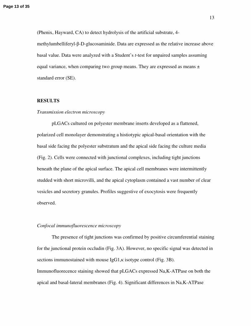
(Phenix, Hayward, CA) to detect hydrolysis of the artificial substrate, 4-
methylumbelliferyl-β-D-glucosaminide. Data are expressed as the relative increase above
basal value. Data were analyzed with a Student’s t-test for unpaired samples assuming
equal variance, when comparing two group means. They are expressed as means ±
standard error (SE).
RESULTS
Transmission electron microscopy
pLGACs cultured on polyester membrane inserts developed as a flattened,
polarized cell monolayer demonstrating a histiotypic apical-basal orientation with the
basal side facing the polyester substratum and the apical side facing the culture media
(Fig. 2). Cells were connected with junctional complexes, including tight junctions
beneath the plane of the apical surface. The apical cell membranes were intermittently
studded with short microvilli, and the apical cytoplasm contained a vast number of clear
vesicles and secretory granules. Profiles suggestive of exocytosis were frequently
observed.
Confocal immunofluorescence microscopy
The presence of tight junctions was confirmed by positive circumferential staining
for the junctional protein occludin (Fig. 3A). However, no specific signal was detected in
sections immunostained with mouse IgG1,κ isotype control (Fig. 3B).
Immunofluorecence staining showed that pLGACs expressed Na,K-ATPase on both the
apical and basal-lateral membranes (Fig. 4). Significant differences in Na,K-ATPase
Page 13 of 35

14
staining patterns were observed between carbachol-stimulated and unstimulated cells
(Fig. 5). Both the carbachol-stimulated and unstimulated cells expressed a plasma
membrane stain for Na,K-ATPase. However, a weak, diffuse intracellular staining for
Na,K-ATPase was observed in the cytoplasm of carbachol-stimulated cells, whereas no
intracellular staining for Na,K-ATPase was seen in the cytoplasm of unstimulated cells.
Ussing chamber studies
The bioelectric properties of pLGACMs on culture inserts were evaluated in
Ussing chambers. Transepithelial resistance and short circuit current stabilized within
15–30 min after mounting in the Ussing chamber. In the resting state, cells typically
generated a small negative, i.e., basal-lateral to apical, Isc. Addition of 100 µM carbachol
to the basal-lateral chamber rapidly reversed the polarity and increased the magnitude of
the Isc. The peak values of Isc varied between 20 – 60 µA/cm2 (33.93 ± 12.25 µA/cm2).
However, Isc did not attain stable steady-state values, decreasing gradually with time
following stimulation with carbachol.
A positive carbachol-dependent Isc would be consistent with either the Ussing
model for Na+ absorption, or the model for Cl− secretion first proposed by Silva et al.
(46). The shared feature of both models is the active efflux of Na+ across the basal-lateral
membrane, driven by Na+,K+-ATPase. Addition of 100 µM ouabain to the basal-lateral
chamber resulted in the complete return of carbachol-dependent Isc to baseline values
(Fig. 6). The rapid decrease in Isc was accompanied by a rapid increase in TER (data not
shown).
Page 14 of 35

15
In the classic Ussing model for Na+ absorption in tight epithelia, Na+ influx
through the apical membrane is mediated by Na+-selective channels, and Isc is
independent of the nature of the anion in the bathing media. In contrast, the model for
Na+-coupled Cl−-secretion predicts that a positive Isc depends on the presence of Cl−.
Consistent with the model for Na+-coupled Cl−-secretion, replacing both the apical and
basal bathing media with Cl−-free Ringer’s solution completely returned Isc to baseline
values (Fig. 7). This effect could be largely reversed by returning BRS to both chambers
(data not shown).
While coupled Na+-Cl− absorption is electroneutal, Na+-coupled substrate
absorption is electrogenic and may generate a measureable positive Isc, depending on the
leakiness of the pericellular pathway. Moreover, one may posit models in which Na+/H+
exchangers and Cl−/HCO3− exchangers, or Na+-K+-2Cl− symporters, mediate influx
across the apical membrane, and under short-circuit conditions, anion channels mediate
Cl− efflux, resulting in a Cl−-dependent Isc. A decisive prediction of the model for Na+-
coupled Cl−-secretion is that the coupled transporters are localized to the basal-lateral
membrane. To test this prediction, selective inhibitors were added to the medium in the
basal chamber. Addition of neither amiloride alone nor bumetanide alone caused a
significant change in steady state Isc (Fig. 8). However, addition of the two inhibitors in
combination reduced steady-state Isc by roughly 65%.
Page 15 of 35

16
β-hexosaminidase assay
Of the proteins rabbit lacrimal gland acinar cells secrete in response to cholinergic
stimulation, β-hexosaminidase is the most easily measured (2). In the unstimulated state,
secretion of β-hexosaminidase to the apical medium was nearly sixfold greater than
secretion to the basal medium. Stimulation of 100 µM carbachol doubled secretion of β-
hexosaminidase to the apical bathing medium (P ≤ 0.05) but had no significant effect on
secretion to the basal medium (Fig. 9).
DISCUSSION
To study vectorial epithelial transport in vitro, it is necessary not only that a
preparation maintain normal polarization of cytoplasmic constitutents and plasma
membrane domains, but that it also establish a continuous monolayer with significant
resistance to the passive movements of electrolytes solutes and cellular secretory
products. The introduction of the MDCK and LLC-PK1 cell lines with these
characteristics more than three decades ago (22) opened an era of rapid progress in
understanding the transport physiologies of cell lines and primary cultures from diverse
epithelia, including, in addition to the distal and proximal segments of the nephron, the
urinary bladder, small intestine, large intestine, cornea, conjunctiva, and retinal pigment
epithelium. While cells from many different exocrine glands can be readily isolated and
maintained in primary culture, the literature contains reports of transepithelial
bioelectrical properties only for the tracheal gland (9), human sweat gland (6), (30), and
mouse mammary gland (4). Indeed, acinar cells from the rabbit lacrimal gland survive
robustly in primary culture, but in all the models that have been studied heretofore, the
Page 16 of 35

17
cells organize themselves into tissue-like structures that express epithelial polarization
only at the microscopic level. We have now identified media and culture conditions
which override this proclivity and support organization of the cells into continuous
monolayers. Evaluation by immunofluorescence and electron microscopy reveals that the
cells maintain typical epithelial polarity and highly differentiated histiotypic
characteristics of lacrimal acinar cells in vivo. The ultrastructural features include apical
microvilli; apical clear vesicles; secretory granules with profiles of exocytosis at the
apical plasma membrane; junctional complexes; and basally-positioned nuclei. The cells
also express a positive circumferential stain for occludin, a key protein associated with
tight junctions. The expression of occludin and the appearance of an epithelial monolayer
suggested that the cells retained the ability to generate TERs and ionic fluxes conducive
for fluid transport.
Na,K-ATPase generates chemiosmotic energy that drives a variety of transport
processes in the plasma membranes of all vertebrate cells (42, 47). In most epithelia, it is
distributed asymmetrically, and immunocytochemical or immunofluorescence
microscopy usually detects it exclusively in the basal-lateral plasma membrane. The
lacrimal acinar cell appears to be an exception to this generalization. Both subcellular
fractionation analyses and immunolocalization methods indicate that, while it is
expressed at highest specific activity in the basal-lateral membrane, it also is expressed in
the apical plasma membrane (55), but it is preponderantly localized in intracellular pools
(7, 34) now known to be associated with the early endosome, recycling endosome, and
trans-Golgi network (58). Immunofluorescence staining of the cultured lacrimal acinar
cell monolayers demonstrated the localization of Na,K-ATPase at both the basal-lateral
Page 17 of 35

18
and apical plasma membranes, with greater intensity of labeling at the basal-lateral
plasma membranes.
Cholinergic stimulation triggers a number of events in isolated and primary
cultured lacrimal acinar cells, related to their diverse secretory functions. These include
activation of apical Cl− channels (14, 16, 39), activation of basal-lateral Na+/H+
antiporters (27, 40), release of macromolecular secretory products and internalization and
recycling of secretory vesicle membrane constituents (23, 56), release of the polymeric
IgA receptor secretory component (41), acceleration of the vesicle traffic recycling
between the plasma membranes and endosomes (19, 28), and a net translocation of Na,K-
ATPase pumps from the intracellular pools to the basal-lateral plasma membranes (59).
The detection of Na,K-ATPase in the cytoplasm of cells in the monolayers after
carbachol stimulation but not in non-stimulated cells may indicate that Na,K -ATPase
subcellular localization and traffic differs from the models that have been studied
previously; or it might result from technical issues related to the density of Na,K-ATPase
being near the threshold of detection in the intracellular compartments.
It is the Na,K-ATPase pump units expressed in the basal-lateral plasma membrane
that are thought to provide the energy necessary for net fluxes of electrolytes in either the
absorptive or the secretory direction (18, 46). The cardiac glycoside ouabain, an inhibitor
of Na,K-ATPase, has been shown to reduce fluid secretion by rabbit lacrimal gland in
vivo (5, 10), and our observation that addition of ouabain to the basal medium completely
abolishes the carbachol-induced Isc accords with this concept.
A positive short circuit current such as we observed after stimulation with
carbachol is formally consistent with either active electrogenic absorption of a cation, i.e.,
Page 18 of 35

19
Na+, or active electrogenic secretion of an anion, i.e., Cl−. Replacement of Cl− in the
apical and basal bathing media abolished the carbachol-induced Isc, as predicted by the
model of anion secretion. However, this observation could not exclude a model of Na+
absorption in which Na+ influx across the apical plasma membrane is a Cl−-dependent,
rather than Na+ channel-mediated, process. Models for absorption and secretion differ
only in the subcellular localization of the transporters that couple Na+ and Cl− influxes.
Two types of coupled Na+ and Cl− transport mechanism have been characterized, Na+-
K+-2Cl− symporters and a parallel array of Na+/H+ exchangers and Cl−/HCO3− exchangers
(31). Neither amiloride, an inhibitor of Na+/H+ exchangers, nor bumetanide, an inhibitor
of Na+-K+-2Cl− symporters, perceptibly altered Isc when added to the basal medium
singly. However, the two inhibitors in combination reduced Isc by 65%. This observation
indicates that Na+-coupled Cl− influx across the basal-lateral membrane is mediated both
by Na+/H+ exchangers and Cl−/HCO3− exchangers and by Na+-K+-2Cl− symporters. It also
suggests that acinar cells contain large intracellular reserves of the transporters and can
recruit one Na+ transporter to the plasma membrane when the other is inhibited.
Analytical subcellular fractionation studies demonstrated the existence of large
intracellular pools of Na+/H+ exchangers (59), and Cl−/HCO3− exchangers (27). Similar
experiments were unable to detect evidence for Na+-K+-2Cl− symporters in isolated
membrane vesicles but could not exclude their presence in intact cells. The failure of the
combination of inhibitors to completely inhibit Isc might have been due to relatively low
sensitivity of various Na+/H+ exchanger isoforms to amiloride (36, 37). Alternatively, it
might have been due to the ongoing recycling traffic, which internalizes transporters to
Page 19 of 35

20
acidic compartments, where they dissociate inhibitors, then returns them to the plasma
membrane in an active state.
Secretagogues are known to stimulate isolated and primary cultured lacrimal
acinar cells to release secretory proteins to their ambient media (59), but with those
models it was necessary to employ sophisticated live-cell imaging methods to verify that
the exocytotic process occurred preferentially at the apical plasma membrane (25). Our
studies with the acinar cell monolayer model confirm that carbachol-induced β-
hexosaminidase secretion is directed to the apical plasma membrane. However, our
observations also reveal that acinar cells maintain a constitutive secretory process
directed to the basal-lateral membrane, such as has been predicted on the basis of data
obtained through subcellular fractionation analyses (58).
In summary, we report the first successful ex vivo reconstitution of an
electrophysiologically functional lacrimal gland epithelial tissue. When grown on
polyester membrane scaffolds, acinar cells from rabbit lacrimal glands established
continuous monolayers with transepithelial resistances typical of the “leaky” epithelia.
The reconstituted epithelia generate a carbachol-induced, ouabain-senstive, Cl−-
dependent-, apical→basal-lateral Isc consistent with current cellular models in which
Na+-coupled co-transporters use the energy of Na+ electrochemical potential gradient,
established by Na,K-ATPase, to mediate active secretion of Cl− (12, 50). We anticipate
that this model will be useful for studying basic mechanisms in lacrimal cell physiology
in addition to electrolyte and water transport. It has been proposed that lacrimal acinar
cells exocytotically secrete partially processed autoantigen peptides to the underlying
tissue space, by way of the same membrane recycling traffic that mediates the
Page 20 of 35

21
constitutive secretion of β-hexosaminidase. According to this scenario, alterations or
reorientations of traffic during chronic stimulation with carbachol, prolactin or other
cytokines, or inflammatory mediators lead to the exposure of autoantigen epitopes that
potentially provoke autoimmune responses which will manifest clinically as Sjögren’s
syndrome. The reconstituted lacrimal epithelial monolayer should be extremely useful as
an experimental model for studying these mechanisms.
At the same time, harnessing the principles of tissue engineering to develop
bioengineered tissue constructs is a rapidly emerging alternative in a clinical setting when
tissue or organ transplantation is not possible or feasible. The lacrimal gland presents a
classical example, as no description of a surgical transplantation procedure for the gland
in humans has been reported in the literature. Hence, a tissue-engineered tear secretory
system would be an attractive alterative for patients suffering from severe dry eyes due to
nonfunctioning lacrimal glands or blocked ducts (such as those with Stevens-Johnson
syndrome), chemical or thermal injuries, or ocular cicatricial pemphigoid (an
inflammatory syndrome involving the ocular mucous membranes). For a tissue-
engineered system to produce fluid, the critical requirements that have to b e met (45)
include that the cellular component in the construct establishes continuous epithelial
monolayers with functional tight junctions, an asymmetric distribution of transport
proteins mediating active secretion of Cl−, and an apically-oriented pathway for
exocytotic secretion of proteins. The present study indicates that this goal may be
attainable.
Page 21 of 35

22
ACKNOWLEDGEMENTS
The authors thank Dr. Ram Kannan (Doheny Eye Institute) for the intellectual expertise
with the transport studies. The authors are also grateful for the expert technical assistance
of Tamako Nakamura with assay studies, Ernesto Barron and Antony Rodriguez with
TEM and confocal microscopy and Susan Clarke with editorial assistance.
GRANTS
This study was supported by NEI Grants EY-03040, EY-15457, EY-12689, and EY-
10550, and a Baxter Foundation Junior Faculty Award to Dr. Yiu. It was also supported
in part by NIH grant DK062283 to Dr. Yu and by an unrestricted grant from Research to
Prevent Blindness (RPB).
Page 22 of 35

23
REFERENCES
1. Amft N, Curnow SJ, Scheel-Toellner D, Devadas A, Oates J, Crocker J, Hamburger J, Ainsworth J, Mathews J, Salmon M, Bowman SJ, Buckley CD. Ectopic expression of the B cell-attracting chemokine BCA-1 (CXCL13) on endothelial cells and within lymphoid follicles contributes to the establishment of germinal center-like structures in Sjogren's syndrome. Arthritis Rheum 44: 2633-2641, 2001.
2. Andersson SV, Edman MC, Bekmezian A, Holmberg J, Mircheff AK, Peter Gierow J. Characterization of beta-hexosaminidase secretion in rabbit lacrimal gland. Exp Eye Res 83: 1081-1088, 2006.
3. Angelow S, Kim KJ, Yu AS. Claudin-8 modulates paracellular permeability to acidic and basic ions in MDCK II cells. J Physiol 571: 15-26, 2006.
4. Bisbee CA. Prolactin effects on ion transport across cultured mouse mammary epithelium. Am J Physiol 240: C110-115, 1981.
5. Botelho SY, Fuenmayor N. Lacrimal gland flow and potentials during dinitrophenol, ouabain, and ethacrynic acid perfusion. Invest Ophthalmol Vis Sci20: 515-521, 1981.
6. Bovell DL, Clunes MT, Elder HY, Wong CH, Ko WH. Nucleotide-evoked ion transport and [Ca(2+)](i) changes in normal and hyperhidrotic human sweat gland cells. Eur J Pharmacol 403: 45-48, 2000.
7. Bradley ME, Azuma KK, McDonough AA, Mircheff AK, Wood RL. Surface and intracellular pools of Na,K-ATPase catalytic and immuno-activities in rat exorbital lacrimal gland. Exp Eye Res 57: 403-413, 1993.
8. Chang-Lin JE, Kim KJ, Lee VH. Characterization of active ion transport across primary rabbit corneal epithelial cell layers (RCrECL) cultured at an air-interface. Exp Eye Res 80: 827-836, 2005.
9. Culp DJ, Lee DK, Penney DP, Marin MG. Cat tracheal gland cells in primary culture. Am J Physiol 263: L264-275, 1992.
10. Dartt DA, Moller M, Poulsen JH. Lacrimal gland electrolyte and water secretion in the rabbit: localization and role of (Na+ + K+)-activated ATPase. J Physiol 321: 557-569, 1981.
11. Do CW, To CH. Chloride secretion by bovine ciliary epithelium: a model of aqueous humor formation. Invest Ophthalmol Vis Sci 41: 1853-1860, 2000.
12. Dubyak GR. Ion homeostasis, channels, and transporters: an update on cellular mechanisms. Adv Physiol Educ 28: 143-154, 2004.
13. Esch TR. Pathogenetic factors in Sjogren's syndrome: recent developments. Crit Rev Oral Biol Med 12: 244-251, 2001.
14. Evans MG, Marty A, Tan YP, Trautmann A. Blockage of Ca-activated Cl conductance by furosemide in rat lacrimal glands. Pflugers Arch 406: 65-68, 1986.
15. Fante RG, Snyder RW, Metrikin DC. Treating dry eye. Clao J 18: 14, 1992.16. Findlay I, Petersen OH. Acetylcholine stimulates a Ca2+-dependent C1-
conductance in mouse lacrimal acinar cells. Pflugers Arch 403: 328-330, 1985.17. Fox RI, Kang HI. Pathogenesis of Sjogren's syndrome. Rheum Dis Clin North
Am 18: 517-538, 1992.
Page 23 of 35

24
18. Frizzell RA, Field M, Schultz SG. Sodium-coupled chloride transport by epithelial tissues. Am J Physiol 236: F1-8, 1979.
19. Gierow JP, Lambert RW, Mircheff AK. Fluid phase endocytosis by isolated rabbit lacrimal gland acinar cells. Exp Eye Res 60: 511-525, 1995.
20. Goto E, Dogru M, Fukagawa K, Uchino M, Matsumoto Y, Saiki M, Tsubota K. Successful tear lipid layer treatment for refractory dry eye in office workers by low-dose lipid application on the full-length eyelid margin. Am J Ophthalmol 142: 264-270, 2006.
21. Guo Z, Song D, Azzarolo AM, Schechter JE, Warren DW, Wood RL, Mircheff AK, Kaslow HR. Autologous lacrimal-lymphoid mixed-cell reactions induce dacryoadenitis in rabbits. Exp Eye Res 71: 23-31, 2000.
22. Handler JS, Perkins FM, Johnson JP. Studies of renal cell function using cell culture techniques. Am J Physiol 238: F1-9, 1980.
23. Herzog V, Farquhar MG. Luminal membrane retrieved after exocytosis reaches most golgi cisternae in secretory cells. Proc Natl Acad Sci U S A 74: 5073-5077, 1977.
24. Holly FJ, Lemp MA. Tear physiology and dry eyes. Surv Ophthalmol 22: 69-87, 1977.
25. Jerdeva GV, Wu K, Yarber FA, Rhodes CJ, Kalman D, Schechter JE, Hamm-Alvarez SF. Actin and non-muscle myosin II facilitate apical exocytosis of tear proteins in rabbit lacrimal acinar epithelial cells. J Cell Sci 118: 4797-4812, 2005.
26. Kolkowski EC, Reth P, Pelusa F, Bosch J, Pujol-Borrell R, Coll J, Jaraquemada D. Th1 predominance and perforin expression in minor salivary glands from patients with primary Sjogren's syndrome. J Autoimmun 13: 155-162, 1999.
27. Lambert RW, Bradley ME, Mircheff AK. pH-sensitive anion exchanger in rat lacrimal acinar cells. Am J Physiol 260: G517-523, 1991.
28. Lambert RW, Maves CA, Gierow JP, Wood RL, Mircheff AK. Plasma membrane internalization and recycling in rabbit lacrimal acinar cells. Invest Ophthalmol Vis Sci 34: 305-316, 1993.
29. Lamberts DW. Topical hyperosmotic agents and secretory stimulants. Int Ophthalmol Clin 20: 163-169, 1980.
30. Larsen EH, Novak I, Pedersen PS. Cation transport by sweat ducts in primary culture. Ionic mechanism of cholinergically evoked current oscillations. J Physiol424: 109-131, 1990.
31. Lau KR, Howorth AJ, Case RM. The effects of bumetanide, amiloride and Ba2+ on fluid and electrolyte secretion in rabbit salivary gland. J Physiol 425: 407-427, 1990.
32. Magnusson V, Nakken B, Bolstad AI, Alarcon-Riquelme ME. Cytokine polymorphisms in systemic lupus erythematosus and Sjogren's syndrome. Scand J Immunol 54: 55-61, 2001.
33. Mircheff AK. Lacrimal fluid and electrolyte secretion: a review. Curr Eye Res 8: 607-617, 1989.
Page 24 of 35

25
34. Mircheff AK, Lu CC, Conteas CN. Resolution of apical and basal-lateral membrane populations from rat exorbital gland. Am J Physiol 245: G661-667, 1983.
35. Mircheff AK, Wang Y, Jean MD, Ding C, Trousdale MD, Hamm-Alvarez SF, Schechter JE. Mucosal Immunity and Self-Tolerance in the Ocular Surface System. Ocul Surf 3: 182-192, 2005.
36. Noel J, Roux D, Pouyssegur J. Differential localization of Na+/H+ exchanger isoforms (NHE1 and NHE3) in polarized epithelial cell lines. J Cell Sci 109 (Pt 5): 929-939, 1996.
37. Park K, Olschowka JA, Richardson LA, Bookstein C, Chang EB, Melvin JE.Expression of multiple Na+/H+ exchanger isoforms in rat parotid acinar and ductal cells. Am J Physiol 276: G470-478, 1999.
38. Qian L, Wang Y, Xie J, Rose CM, Yang T, Nakamura T, Sandberg M, Zeng H, Schechter JE, Chow RH, Hamm-Alvarez SF, Mircheff AK. Biochemical changes contributing to functional quiescence in lacrimal gland acinar cells after chronic ex vivo exposure to a muscarinic agonist. Scand J Immunol 58: 550-565, 2003.
39. Saito Y, Ozawa T, Hayashi H, Nishiyama A. Acetylcholine-induced change in intracellular Cl- activity of the mouse lacrimal acinar cells. Pflugers Arch 405: 108-111, 1985.
40. Saito Y, Ozawa T, Nishiyama A. Acetylcholine-induced Na+ influx in the mouse lacrimal gland acinar cells: demonstration of multiple Na+ transport mechanisms by intracellular Na+ activity measurements. J Membr Biol 98: 135-144, 1987.
41. Schechter J, Stevenson D, Chang D, Chang N, Pidgeon M, Nakamura T, Okamoto CT, Trousdale MD, Mircheff AK. Growth of purified lacrimal acinar cells in Matrigel raft cultures. Exp Eye Res 74: 349-360, 2002.
42. Scheiner-Bobis G. The sodium pump. Its molecular properties and mechanics of ion transport. Eur J Biochem 269: 2424-2433, 2002.
43. Schonthal AH, Warren DW, Stevenson D, Schecter JE, Azzarolo AM, Mircheff AK, Trousdale MD. Proliferation of lacrimal gland acinar cells in primary culture. Stimulation by extracellular matrix, EGF, and DHT. Exp Eye Res70: 639-649, 2000.
44. Selvam S, Thomas PB, Trousdale MD, Stevenson D, Schechter JE, Mircheff AK, Jacob JT, Smith RE, Yiu SC. Tissue-engineered tear secretory system: Functional lacrimal gland acinar cells cultured on matrix protein-coated substrata. J Biomed Mater Res B Appl Biomater, 2006.
45. Selvam S, Thomas PB, Yiu SC. Tissue engineering: current and future approaches to ocular surface reconstruction. Ocul Surf 4: 120-136, 2006.
46. Silva P, Stoff J, Field M, Fine L, Forrest JN, Epstein FH. Mechanism of active chloride secretion by shark rectal gland: role of Na-K-ATPase in chloride transport. Am J Physiol 233: F298-306, 1977.
47. Skou JC. The sodium, potassium-pump. Scand J Clin Lab Invest Suppl 180: 11-23, 1986.
48. Smith JJ, Welsh MJ. Fluid and electrolyte transport by cultured human airway epithelia. J Clin Invest 91: 1590-1597, 1993.
Page 25 of 35

26
49. Solomon A, Dursun D, Liu Z, Xie Y, Macri A, Pflugfelder SC. Pro- and anti-inflammatory forms of interleukin-1 in the tear fluid and conjunctiva of patients with dry-eye disease. Invest Ophthalmol Vis Sci 42: 2283-2292, 2001.
50. Speake T, Whitwell C, Kajita H, Majid A, Brown PD. Mechanisms of CSF secretion by the choroid plexus. Microsc Res Tech 52: 49-59, 2001.
51. Stott DI, Hiepe F, Hummel M, Steinhauser G, Berek C. Antigen-driven clonal proliferation of B cells within the target tissue of an autoimmune disease. The salivary glands of patients with Sjogren's syndrome. J Clin Invest 102: 938-946, 1998.
52. Sun D, Emmert-Buck MR, Fox PC. Differential cytokine mRNA expression in human labial minor salivary glands in primary Sjogren's syndrome. Autoimmunity28: 125-137, 1998.
53. Tsubota K, Toda I, Yagi Y, Ogawa Y, Ono M, Yoshino K. Three different types of dry eye syndrome. Cornea 13: 202 -209, 1994.
54. Ussing HH, Lind F, Larsen EH. Ion secretion and isotonic transport in frog skin glands. J Membr Biol 152: 101-110, 1996.
55. Wood RL, Mircheff AK. Apical and basal-lateral Na/K-ATPase in rat lacrimal gland acinar cells. Invest Ophthalmol Vis Sci 27: 1293-1296, 1986.
56. Wu K, Jerdeva GV, da Costa SR, Sou E, Schechter JE, Hamm-Alvarez SF.Molecular mechanisms of lacrimal acinar secretory vesicle exocytosis. Exp Eye Res 83: 84-96, 2006.
57. Xanthou G, Tapinos NI, Polihronis M, Nezis IP, Margaritis LH, Moutsopoulos HM. CD4 cytotoxic and dendritic cells in the immunopathologic lesion of Sjogren's syndrome. Clin Exp Immunol 118: 154-163, 1999.
58. Xie J, Qian L, Wang Y, Rose CM, Yang T, Nakamura T, Hamm-Alvarez SF, Mircheff AK. Novel biphasic traffic of endocytosed EGF to recycling and degradative compartments in lacrimal gland acinar cells. J Cell Physiol 199: 108-125, 2004.
59. Yiu SC, Lambert RW, Tortoriello PJ, Mircheff AK. Secretagogue-induced redistributions of Na,K-ATPase in rat lacrimal acini. Invest Ophthalmol Vis Sci32: 2976-2984, 1991.
60. Zoukhri D. Effect of inflammation on lacrimal gland function. Exp Eye Res 82: 885-898, 2006.
Page 26 of 35

Fig. 1. Working hypothesis for electrolyte transport mechanisms driving water secretion across lacrimal gland acinar epithelium.
81x54mm (600 x 600 DPI)
Page 27 of 35

Fig. 2. Transmission electron micrographs of pLGACs cultured on polyester membrane inserts. (A) The cells exhibited features typical of a glandular epithelium with apical
microvilli (top arrowheads), secretory granules (g), and clear vesicles (v). The cells were joined by junctional complexes (boxed area) that included tight junctions near the apical surface. Double arrows at the bottom represent the polyester substratum. (B) A further
magnification of the boxed area with a series of desmosomes. 169x70mm (600 x 600 DPI)
Page 28 of 35

Fig. 3. (A) Confocal microscopic evaluation of occludin, a tight junction-associated protein, in pLGACs cultured on a 12-well polyester membrane insert using anti-occludin
antibody. Nuclei are visualized with DAPI (blue). Note that the nuclear stain and the occludin in a given cell could rarely both be captured in the same plane of focus due to apical level of the junctional complex. (B) Immunofluorescence staining with mouse
IgG1,κ for isotype control. 82x43mm (300 x 300 DPI)
Page 29 of 35

Fig. 4. Top. Confocal z-image staining for apical and basal-lateral localization of Na,K-ATPase using anti-Na,K-ATPase. Bottom. Immunofluorescence staining for Na,K-ATPase of pLGACs cultured on a 12-well polyester membrane insert. Nuclei are visualized with
DAPI (blue). 72x85mm (300 x 300 DPI)
Page 30 of 35

Fig. 5. Confocal micrographs of immunofluorescence staining for Na,K-ATPase in pLGACs cultured on a 12-well polyester membrane insert using anti-Na,K-ATPase antibody. Nuclei
are visualized with DAPI (blue). (a) Unstimulated cells. (b) Carbachol-stimulated cells (BL, 100 µM, 5 min).
80x39mm (300 x 300 DPI)
Page 31 of 35

Fig. 6. Actual time course of carbachol-stimulated apical-to-basal Isc and its inhibition by ouabain in pLGACM on a polyester membrane insert in an Ussing chamber. Addition of
100 µM ouabain to the basal-lateral compartment rapidly altered the bioelectric properties of the cells. The instantaneous decrease in Isc was accompanied by a rapid
increase in TER after treatment with ouabain. Data represented is an actual trace from one of four separate experiments (n=4).
53x35mm (600 x 600 DPI)
Page 32 of 35

Fig. 7. Actual time course of inhibition of the carbachol-stimulated Isc with apical and basal-lateral Cl -free buffer exchange following carbachol stimulation (100 µM, BL) in
pLGACM on a polyester membrane insert in an Ussing chamber. The Cl -free buffer exchange rapidly altered the bioelectric properties of the cells. The instantaneous
decrease in Isc was accompanied by a rapid increase in TER during buffer exchange. Data represented is an actual trace from one of four separate experiments (n=4).
52x33mm (600 x 600 DPI)
Page 33 of 35

Fig. 8. Inhibitory effects of amiloride (1 mM, BL) and bumetanide (0.1 mM, BL) on Isc in pLGACMs on polyester membrane inserts in Ussing chambers. The results represent
average ± SE of 4 separate determinations for control group, 5 separate determinations for amiloride group and 6 separate determinations each for bumetanide and amiloride + bumetanide groups. (*) represents significant decrease at P ≤ 0.05 from control values
and synergistic inhibition of amiloride and bumetanide of the cultured cells. 48x29mm (600 x 600 DPI)
Page 34 of 35

Fig. 9. β-hexosaminidase protein released on the basal and apical side of the culture medium from lacrimal acinar cells cultured on 12-well inserts when exposed with or
without carbachol (CCh, 100 µM, BL, 30 min). The results represent average ± SE expressed in arbitrary units (A.U.) of four separate determinations (n=4). (*) represents significant increase at P ≤ 0.05 from resting values and stimulated secretion on the apical
side of the cultured cells. 57x40mm (600 x 600 DPI)
Page 35 of 35