Transcriptional activation of a 37 kDa ethylene responsive cysteine protease gene, RbCP1, is...
10
Journal of Experimental Botany, Vol. 60, No. 7, pp. 2035–2044, 2009 doi:10.1093/jxb/erp076 Advance Access publication 3 April, 2009 This paper is available online free of all access charges (see http://jxb.oxfordjournals.org/open_access.html for further details) RESEARCH PAPER Transcriptional activation of a 37 kDa ethylene responsive cysteine protease gene, RbCP1, is associated with protein degradation during petal abscission in rose Siddharth Kaushal Tripathi* ,† , Amar Pal Singh † , Aniruddha P. Sane ‡ and Pravendra Nath Plant Gene Expression Laboratory, National Botanical Research Institute, Lucknow-226001, India Received 4 November 2008; Revised 15 February 2009; Accepted 23 February 2009 Abstract Cysteine proteases play an important role in several developmental processes in plants, particularly those related to senescence and cell death. A cysteine protease gene, RbCP1, has been identified that encodes a putative protein of 357 amino acids and is expressed in the abscission zone (AZ) of petals in rose. The gene was responsive to ethylene in petals, petal abscission zones, leaves, and thalamus. The expression of RbCP1 increased during both ethylene- induced as well as natural abscission and was inhibited by 1-MCP. Transcript accumulation of RbCP1 was accompanied by the appearance of a 37 kDa cysteine protease, a concomitant increase in protease activity and a substantial decrease in total protein content in the AZ of petals. Agro-injection of rose petals with a 2.0 kb region upstream of the RbCP1 gene could drive GUS expression in an abscission zone-specific manner and was blocked by 1-MCP. It is concluded that petal abscission is associated with a decrease in total protein content resulting from rapid transcription of RbCP1 and the expression of a 37 kDa protease. Key words: Abscission, agro-infiltration, cysteine protease, ethylene, petal, programmed cell death, RbCP1, rose, senescence. Introduction Abscission is a natural event that involves cell separation in response to developmental or environmental cues. Organs like flowers, petals, sepals, and leaves may be shed once they have served their purpose (Addicot, 1982, van Doorn and Stead, 1997). Several physiological studies have been performed to understand the factors involved in abscission and these have helped in identifying a major role for ethylene in regulating organ abscission (Abeles and Gahagan, 1968; reviewed by Roberts et al., 2002). Further studies on ethylene-insensitive mutants in Arabidopsis and tomato have led to the identification of key components required for the transduction of the ethylene signal during abscission (Lanahan et al., 1994; Bleecker and Patterson, 1997; White- law et al., 2002). In addition, the role of various enzymes involved in cell separation and the genes encoding these have been elucidated (Tucker et al., 1988; del Campillo and Bennett, 1996; Kalaitzis et al., 1997; Lashbrook et al., 1998; Gonzalez-Carranza et al., 2002; Belfield et al., 2005; Sane et al., 2007). In recent years, the roles of novel components of abscission such as HAESA, IDA, and IDL genes that may function and interact in an ethylene-independent manner have also been studied (Jinn et al., 2000; Butenko et al., 2003; Stenvik et al., 2006, 2008). Proteases play an important role in various developmen- tal processes in plants such as seed germination, xylo- genesis, and tapetum degradation as well as organ senescence and programmed cell death (reviewed by Schaller, 2004). Several cysteine protease genes have been found to be up-regulated during programmed cell death in senescing floral tissues of many plants while protease in-gel assays have revealed the appearance of novel proteases during the senescence of flowers (Jones et al., 1995, 2005; Valpuesta et al., 1995; Guerrero et al., 1998; Stephenson and Rubinstein, 1998; Eason et al., 2002; Wagstaff et al., 2002; * Present address: International Centre for Genetic Engineering and Biotechnology, Aruna Asaf Ali Marg, New Delhi -110067, India. y Both authors contributed equally to the paper. z To whom correspondence should be addressed. E-mail: [email protected] ª 2009 The Author(s). This is an Open Access article distributed under the terms of the Creative Commons Attribution Non-Commercial License (http://creativecommons.org/licenses/by- nc/2.0/uk/) which permits unrestricted non-commercial use, distribution, and reproduction in any medium, provided the original work is properly cited.
-
Upload
independent -
Category
Documents
-
view
3 -
download
0
Transcript of Transcriptional activation of a 37 kDa ethylene responsive cysteine protease gene, RbCP1, is...

Journal of Experimental Botany, Vol. 60, No. 7, pp. 2035–2044, 2009doi:10.1093/jxb/erp076 Advance Access publication 3 April, 2009This paper is available online free of all access charges (see http://jxb.oxfordjournals.org/open_access.html for further details)
RESEARCH PAPER
Transcriptional activation of a 37 kDa ethylene responsivecysteine protease gene, RbCP1, is associated with proteindegradation during petal abscission in rose
Siddharth Kaushal Tripathi*,†, Amar Pal Singh†, Aniruddha P. Sane‡ and Pravendra Nath
Plant Gene Expression Laboratory, National Botanical Research Institute, Lucknow-226001, India
Received 4 November 2008; Revised 15 February 2009; Accepted 23 February 2009
Abstract
Cysteine proteases play an important role in several developmental processes in plants, particularly those related to
senescence and cell death. A cysteine protease gene, RbCP1, has been identified that encodes a putative protein of
357 amino acids and is expressed in the abscission zone (AZ) of petals in rose. The gene was responsive to ethylene
in petals, petal abscission zones, leaves, and thalamus. The expression of RbCP1 increased during both ethylene-
induced as well as natural abscission and was inhibited by 1-MCP. Transcript accumulation of RbCP1 was
accompanied by the appearance of a 37 kDa cysteine protease, a concomitant increase in protease activity and
a substantial decrease in total protein content in the AZ of petals. Agro-injection of rose petals with a 2.0 kb region
upstream of the RbCP1 gene could drive GUS expression in an abscission zone-specific manner and was blocked by1-MCP. It is concluded that petal abscission is associated with a decrease in total protein content resulting from
rapid transcription of RbCP1 and the expression of a 37 kDa protease.
Key words: Abscission, agro-infiltration, cysteine protease, ethylene, petal, programmed cell death, RbCP1, rose, senescence.
Introduction
Abscission is a natural event that involves cell separation in
response to developmental or environmental cues. Organs
like flowers, petals, sepals, and leaves may be shed once
they have served their purpose (Addicot, 1982, van Doornand Stead, 1997). Several physiological studies have been
performed to understand the factors involved in abscission
and these have helped in identifying a major role for ethylene
in regulating organ abscission (Abeles and Gahagan, 1968;
reviewed by Roberts et al., 2002). Further studies on
ethylene-insensitive mutants in Arabidopsis and tomato
have led to the identification of key components required
for the transduction of the ethylene signal during abscission(Lanahan et al., 1994; Bleecker and Patterson, 1997; White-
law et al., 2002). In addition, the role of various enzymes
involved in cell separation and the genes encoding these
have been elucidated (Tucker et al., 1988; del Campillo and
Bennett, 1996; Kalaitzis et al., 1997; Lashbrook et al., 1998;
Gonzalez-Carranza et al., 2002; Belfield et al., 2005; Sane
et al., 2007). In recent years, the roles of novel components
of abscission such as HAESA, IDA, and IDL genes that
may function and interact in an ethylene-independentmanner have also been studied (Jinn et al., 2000; Butenko
et al., 2003; Stenvik et al., 2006, 2008).
Proteases play an important role in various developmen-
tal processes in plants such as seed germination, xylo-
genesis, and tapetum degradation as well as organ
senescence and programmed cell death (reviewed by Schaller,
2004). Several cysteine protease genes have been found to be
up-regulated during programmed cell death in senescingfloral tissues of many plants while protease in-gel assays
have revealed the appearance of novel proteases during the
senescence of flowers (Jones et al., 1995, 2005; Valpuesta
et al., 1995; Guerrero et al., 1998; Stephenson and
Rubinstein, 1998; Eason et al., 2002; Wagstaff et al., 2002;
* Present address: International Centre for Genetic Engineering and Biotechnology, Aruna Asaf Ali Marg, New Delhi -110067, India.y Both authors contributed equally to the paper.z To whom correspondence should be addressed. E-mail: [email protected]ª 2009 The Author(s).
This is an Open Access article distributed under the terms of the Creative Commons Attribution Non-Commercial License (http://creativecommons.org/licenses/by-nc/2.0/uk/) which permits unrestricted non-commercial use, distribution, and reproduction in any medium, provided the original work is properly cited.
Wang et al., 2004; Pak and van Doorn, 2005, Azeez et al.,
2007). However, so far, none have been reported during
abscission.
We are interested in understanding the molecular basis of
petal abscission and have been using the highly ethylene-
sensitive fragrant rose (Rosa bourboniana var. Gruss an
Teplitz) as a model for petal abscission. Since abscission
zone cells have previously been shown to undergo featuresof cell death (Evensen et al., 1993), we were interested in
finding out if cysteine proteases may play an important role
in the process of abscission as well. In this paper, it is
reported that the progression of petal abscission in ethylene-
treated and field-abscising flowers of rose is associated with
the rapid transcription of a cysteine protease gene, RbCP1,
the enhanced expression of a 37 kDa cysteine protease, and
a decrease in total protein content.
Materials and methods
Plant material
Flowers of the fragrant variety of rose (Rosa bourboniana
var. Gruss an Teplitz) were used in the present study. They
were picked early in the morning (before sun rise), cut with
a sharp blade and the stalks immediately placed in water.
Care was taken to ensure that flowers were of the same
developmental stage (unpollinated and with only one or two
of the outer most petals open).
Ethylene and 1-MCP treatments
Cut flowers were kept in water in a closed air-tight chamber,
treated with 0.5 ll l�1 ethylene for 16–18 h (until abscission
occurred) and petal abscission zones collected at 0 h
(ethylene untreated), 4 h, 8 h, and 12 h from the time of
initiating the ethylene treatment as described earlier (Sane
et al., 2007). For 1-methyl cyclopropene (1-MCP) treatment,cut flowers were exposed to 1 ll l�1 1-MCP (Ethylbloc from
Biotechnologies for Horticulture Inc, Walterboro, SC, USA)
for 12 h. For samples that underwent natural/developmental
abscission in the field (time of abscission, 38–45 h), flowers
were marked at the time of opening of the outermost whorl
and abscission zones collected at time intervals of 8 h, 12 h,
24 h, and 36 h. For early ethylene responsive accumulation
of RbCP1, cut flowers were exposed to 0.5 ll�1 ethylene forshort time intervals of 30, 60, and 120 min and abscission
zones collected and frozen immediately. For each sample 30–
40 flowers were chosen and abscission zones (2 mm2) were
collected from 10–15 petals per flower and pooled. For the
ethylene treatment of other floral and vegetative tissues, these
were exposed to 0.5 ll�1 ethylene for 8 h, as described earlier
and samples collected before and after ethylene treatment.
Isolation of RNA and cloning of cysteine protease cDNA
RNA was isolated from frozen petal abscission zones of
different samples as described by Asif et al. (2000). RNA
was also isolated from different tissues such as petal, sepal,
stamen, carpel, thalamus, pedicel, and leaf before and after
8 h ethylene treatment.
DNA-free RNA from 8 h ethylene-treated samples was
reverse transcribed using the Superscript II reverse tran-
scriptase from Invitrogen (Palo Alto, USA) and primed
with the 3# RACE adapter primer (5#-GGCCACGCGTC-
GACTAGTACTTTTTTTTTTTTTTTTT-3#). The rose
cysteine protease gene, RbCP1, was obtained initially as anartefact of PCR while performing 3# RACE for rose ETR1.
The amplified 450 nt fragment was cloned in pBluescrip-
tIISK (Stratagene, USA) and sequenced on an automated
DNA sequencer (ABI 373A from Applied Biosystems Inc,
USA) using the thermosequenase dye terminator cycle
sequencing kit from Amersham-Pharmacia. The cloned
fragment showed similarity to papain like cysteine proteases
and contained a 3# UTR of 285 nucleotides. Based on thesequence, specific primers CyPro-R1 5#-ACA AGT TGC
AAC ACC ACA CAT GTT C-3#, CyPro-R2 5#-ACA TCT
TGA AGT AGC CAT TGT CAC C-3#, CyPro-R3 5#-AGC
AAG AAC AGC ATG GTT CAC ATC C-3# and
CyProR4P1 5#-GTT CCT GTT GGT GGA TCG AAT
CAG CTT C-3# were designed for 5# RACE and genome
walking. For 5# RACE, cDNA was prepared using the
SMART cDNA synthesis kit (Clontech Laboratories Inc,Palo Alto, USA) from 8 h ethylene-treated petal abscission
zone RNA while a genome walking library was prepared
from total rose petal DNA using the Genome walker kit
(Clontech). Both 5# RACE and genome walking were
performed to obtain the complete cysteine protease ORF.
Although 5# RACE extended the RbCP1 cDNA partially, it
failed to provide the complete cDNA sequence. Further
extension towards the 5# end was carried out using theprimer CyproR4P1 in combination with the genome walker
adapter primer, to obtain a fragment of 2.4 kbp that
contained the 5# region and 2.0 kb of the sequence
upstream of the initiation codon. Based on the sequence,
a forward primer containing the initiation codon (RCypro-
OF 5#-ACA GGA TCC CAT GGC TCC TCC TCG TTT
G-3#) was designed and used in combination with the 3#adapter primer to amplify the cDNA containing thecomplete open reading frame of 1077 bases and the 285
bases 3# UTR (GenBank Accession No. EU057180).
Northern analyses and semi-quantitative RT-PCR
Total RNA (30 lg) from the ethylene-treated abscission
zones (0–12 h) and the 12 h 1-MCP treated abscission zones
was resolved on a 1.2% denaturing formaldehyde-agarose
gel as described by Sambrook et al. (1989) and modified in
the Qiagen Oligotex handbook. RNA was transferred to
nylon membranes (Hybond N, Amersham-Pharmacia Bio-
tech, Uppsala, Sweden) by vacuum transfer using the
vacugene apparatus (Pharmacia) and cross-linked by bakingfor 2 h. Radiolabelling of probes for hybridization was
performed using a-32PdCTP in an asymmetric PCR of the
full-length cysteine protease cDNA. Hybridization and
washings of blots were performed as described (Sambrook
et al., 1989). Signals obtained on the blots were visualized
2036 | Kaushal et al.
on an X-ray film (Fujifilm SuperRX) or quantified on
a phosphorimager (Molecular imager FX, BioRad) using
the software QuantityOne-4.2.3 version.
For the comparison of expression between ethylene-
treated and untreated carpel, sepal, petal, pedicel, thalamus,
and leaf tissues, and for studying early ethylene responsive
expression (30–120 min), 2 lg RNA from these samples was
reverse transcribed as described and amplified using theprimers Cypro-OF and CyproR4P1 to give an amplification
product of 300 nt. Actin was used as an internal control for
normalization using the primers RActF1 5#-ATG ACA
TGG AGA AGA TCT GGC ATC A-3# and RActR2 5#-AGC CTG GAT GGC AAC ATA CAT AGC-3#. PCR was
carried out for 27 cycles using the cycling parameters 94 oC,
for 3 min (first window, one cycle) followed by 27 cycles of
94 oC for 5 s; 55 oC for 10 s, and 72 oC for 20 s. At leastthree independent reactions were run to confirm the results.
Protein extraction and total protease activity
Frozen tissue samples from different stages of ethylene-
treated (0 h, 4 h, and 8h) and naturally abscising petal
abscission zones (0 h, 12 h, 24 h, and 36 h) were ground in
liquid nitrogen. The powder was suspended in 2 ml buffer
containing 50 mM TRIS-Cl pH 7.5, 1 mM EDTA pH 8.0,
0.1% SDS and centrifuged at 12 000 g for 5 min. Thesupernatant was used for the estimation of total proteins
from all samples (Peterson, 1977) and total protease assay (0
h, 8 h ethylene-treated and 24 h natural abscission zone
samples) using azocasein as a synthetic substrate as described
earlier (Holwerda and Rogers, 1992; Azeez et al., 2007). For
the total protease assay, 20 ll extract was mixed with 300 ll100 mM sodium phosphate buffer (pH 7.5) containing 50 ll0.6% (w/v) azocasein (Sigma) supplemented with 100 ll 0.1%Triton X-100, and the mixture was incubated at 37 �C for 3
h. The reaction was terminated by adding 200 ll 10% TCA,
incubated at 4 �C for 30 min, centrifuged at 10 000 g for 10
min and the absorbance of the supernatant was determined
at 366 nm (Ultrospec� 3000, Pharmacia Biotech). A dupli-
cate reaction for each sample was prepared where TCA was
added at the start to act as a control. One unit of protease
activity was defined as the amount of enzyme that gave anincrease of 0.01 absorbance units min�1. To study the
contribution of different proteases, protease inhibitors such
as E-64 [L-trans-epoxysuccinylleucylamide-(4-guanidino)-
butane, specific for cysteine proteases] and PMSF (phenyl-
methylsulphonylfluoride, specific for serine proteases) were
added separately at a concentration of 2 lM and 1 mM,
respectively, to the reactions. The enzyme aliquot was first
incubated with the requisite concentration of the inhibitor(without substrate) for 30 min and then assayed as described
above. All extractions and assays were carried out in
triplicates.
Protease in-gel assay
Protein samples were prepared by mixing the protein
aliquot (10 lg each) with an equal volume of a non-
reducing sample buffer [0.1 M TRIS-HCl, pH 6.8; 2% (w/v)
SDS, 10% (v/v) glycerol, 0.01% bromophenol blue], in-
cubated at 37 �C for 60 min and then electrophoresed in
cold on a 12% SDS-gelatin polyacrylamide gel embedded
with 0.10% gelatin (Hellmich and Schauz, 1988). After
electrophoresis, the gel was washed in renaturing buffer
(2.5% Triton X-100, 10 mM EDTA, 50 mM TRIS-HCl, pH
7.5) for 45 min with shaking and then incubated for 12–16 hin a buffer containing 10 mM Ca2+, 10 mM Mg2+, and 50
mM TRIS-HCl, pH 7.5, at 37 �C in an incubator-shaker.
The gel was stained with Coomassie Blue-R250 (0.1% R250
in 50% methanol/10% glacial acetic acid) and destained with
a solution containing 50% methanol/10% glacial acetic acid.
Clear bands observed in a blue background represented the
sites of protease activity.
To differentiate between cysteine and serine proteases,protease inhibitors, namely 2 lM E-64 and 1 mM PMSF,
specific against each class, were added to the protein
samples prior to electrophoresis and incubated for 30 min
at 37 oC along with non-reducing loading buffer. Following
renaturation, gels were incubated in the presence of
inhibitors in 10 mM Ca2+, 10 mM Mg2+, 50 mM TRIS-
HCl, pH 7.5 at 37 �C for 12–16 h.
Statistical analysis
Data from the total protein content and protease activity in
the abscission zones from three independent experimentswere analysed and expressed as mean 6standard deviation.
Data were subjected to analysis of variance at an alpha level
of 0.05 using the Microsoft Excel software. Values that
exhibited significant differences at P <0.05 were considered.
Isolation and analysis of the RbCP1 promoter
To obtain the proximal promoter of the RbCP1 gene,
a reverse primer RbCP1-PRO2 – GGATCCGTCAAAC-
GAGGAGGAGCCAT containing the initiation codon
(underlined), with a BamHI site was used to amplify a 2.0
kb fragment of the putative promoter from the 2.4 kb
fragment that had been obtained by genome walking. Thiswas cloned at the BamHI site of pBI101.2 in translational
fusion with the GUS gene. The plasmid was introduced into
Agrobacterium strain GV3101. The HAESA gene promoter
was cloned from Arabidopsis using the primers HAE-Pro-
F1 5#-GAG TAA CGA AAC AAG AAA TAG GAG-3#and HAE-Pro-F2 5#-ACT CTG TCA CCG TTT CCT
AAG AAG-3# sequentially in combination with the primer
HAE-R 5#-ATC TAG ATT TTT TGG AAA AGG AATCG-3# to obtain a fragment of 1708 nt. This was digested
with XbaI (present in the reverse primer and at a position
1596 nt upstream of the start codon) to obtain the promoter
fragment. This was cloned in pBI101.1 to obtain pHAESA.
Promoter analysis through agroinjection of the RbCP1promoter in rose buds
Recombinant agrobacteria containing the promoter of
RbCP1 (in pBI101.2) were grown in Luria broth in a 25 ml
Rose petal abscission zone cysteine protease | 2037

culture overnight and harvested at 3000 rpm (5 min) on an
SS34 rotor (RC-5C, Dupont-Sorvall). The pellet was
resuspended in LB to an OD of 1. Acetosyringone was
added to the suspension at a final concentration of 0.1 mg
ml�1. For agroinjection, young buds that would open in
two days time were chosen. A needle (size 23, dimensions
0.63325 mm) fitted on to a 2 ml syringe (filled with the
agrobacterial suspension) was lightly inserted in the centreof the petals. The agrobacterial suspension (0.5 ml, contain-
ing 0.01% acetosyringone), was slowly forced into the petal
and allowed to infiltrate all through the petal up to the
point of attachment of the petal with the thalamus. Agro-
injection was performed with three flowers per construct
and three petals per flower. The excess suspension was
wiped out and the buds were kept for 2 d on the plant. As
a control, agrobacteria containing pBI101 (no promoter),pBI121 (GUS driven by CaMV 35S promoter), and
pHAESA (GUS driven by the 1.6 kb abscission-specific
promoter of the Arabidopsis HAESA gene) were also used
for agroinfiltration of independent flowers. After 2 d from
agroinfiltration, flowers were carefully cut under water and
treated with ethylene for 8 h in a closed chamber as
described before. After 8 h of ethylene treatment, the petals
were detached and stained for GUS expression. Stainingwas carried out for 16 h at 37 �C as previously described
(Gattolin et al., 2006). The tissue was destained in 70%
ethanol at 37 �C until examination. Light microscopy was
performed on a Leica Wild M3Z microscope (Leica
Germany).
Results
Isolation of the RbCP1 gene
The full-length cDNA sequence of the cysteine protease
gene, RbCP1, was obtained by a combination of RACE andgenome walking, followed by cloning of the entire cDNA to
yield a sequence of 1359 bp that included an ORF of 1074
nt and a 3’ UTR of 285 nt (GenBank Accession No.
EU057180). The gene encoded a protein of 357 amino acids
that included an N-terminal signal peptide of 22 amino
acids as deduced by TargetP analysis (http://www.cbs.dtu.
dk/services/TargetP/). The protein had a predicted molecu-
lar mass of 39.5 kDa (for the pre-protein) and 37.2 kDa(for the mature protein) and a pI of 6.1 as determined using
the ProtParam tool of ExPASy (http://www.expasy.org/
tools/protparam.html). BLAST analysis of the predicted
protein (http://www.ncbi.nlm.nih.gov/BLAST/) showed
75–82% amino acid identity and 80–90% amino acid sim-
ilarity to various cysteine proteases from Prunus armeniaca,
Vitis vinifera, Actinidia deliciosa etc. and also revealed
a conserved Peptidase C1A domain from amino acids 141to 354 (Fig. 1). The peptidase C1A subfamily consists of
cysteine peptidases (CPs) such as papain and also includes
animal CPs. The conserved interspersed ERFNIN motif
[E-X3-R-X2-(I/V)-F-X2-N-X3-I-X3-N] that is a characteris-
tic feature of most papain-type cysteine proteases was
conserved in RbCP1 as was the GCXGG motif (Karrer
et al., 1993). The predicted active site catalytic residues C164
H304 N324 as well as other features, such as the presence ofa lysine before the catalytic asparagine, were also conserved.
The predicted protein possessed a site for possible SUMO
modification (FKME) at the C-terminal end of the poly-
peptide. SUMO modification in tomato cysteine protease
LeCP was shown to be important for its localization in the
nucleus and subsequent activation of the LeACS2 gene
(Matarasso et al., 2005).
Transcript accumulation of RbCP1 during petalabscission
In order to investigate if RbCP1 is involved during
abscission, its transcript accumulation pattern was exam-
ined in petal abscission zones obtained from ethylene-
treated and naturally abscising flowers. In ethylene-treated
flowers (time of abscission, 16–18 h; Sane et al., 2007),a rapid increase in transcript accumulation was observed
from 4 h after ethylene treatment. The increase continued
up to 8 h and remained steady until 12 h after ethylene
treatment (Fig. 2A). In flowers treated with 1-MCP (an
inhibitor of ethylene perception) a significant delay in petal
abscission was observed (time of abscission, 55–60 h, Sane
et al., 2007) and very low levels of the RbCP1 transcript
accumulated even after 12 h. Transcript accumulation wasalso studied by semi-quantitative RT-PCR during natural/
developmental abscission (time of abscission, 38–45 h).
Under these conditions, transcripts of RbCP1 could be
detected from the 8 h stage onwards and their levels
remained steady until 24 h. Thereafter transcript levels
increased substantially at 36 h prior to abscission (Fig. 2B).
In view of the rapid increase in transcript levels of RbCP1
in abscission zones of ethylene-treated flowers within 4 h ofethylene treatment, the ethylene responsiveness of RbCP1 in
abscission zones was studied by treating flowers with
ethylene for shorter time intervals (30, 60, and 120 min). A
significant increase in transcript accumulation was detected
within 30 min of ethylene treatment, indicating that the
Fig. 1. Complete amino acid sequence of the predicted rose
cysteine protease gene, RbCP1 (Accession No. EU057180). The
signal peptide is shown in italics with the arrow marking the site of
the likely cleavage. The C1A peptidase domain has been under-
lined. The ERFNIN motif residues are shown in bold while the
active site residues C164 H304 N324 have been boxed. The GCXGG
motif is shown in bold and italics. The putative SUMO modification
site FKME has been shown in bold and is boxed. An asterisk
indicates the termination of protein.
2038 | Kaushal et al.

gene was rapidly up-regulated in response to ethylene in
abscission zones (Fig. 2C). The expression of RbCP1 and its
response to ethylene in other tissues such as sepal, petal,
carpel, pedicel, thalamus, and leaves was also studied. An
increase in transcription of the gene upon ethylene treat-
ment was observed only in petals and leaves while a decrease
was observed in the thalamus (Fig. 2D).
Protein content and protease activity in petal abscissionzones
In view of the increasing levels of transcript accumulationof RbCP1 during the course of abscission in petals, the total
protein content in 0 h, 4 h, and 8 h ethylene-treated and 0 h,
12 h, 24 h, and 36 h field-abscising petal abscission zones
was estimated. As shown in Fig. 3A, there was a decrease in
total protein content in abscission zones of both ethylene-
treated and field-abscising flowers. In 4 h ethylene-treated
petal abscission zone tissues, the protein content decreased
to about 50% of the control, while in 8 h ethylene-treatedabscission zones it went further down to about 35% of the
control. In flowers undergoing natural (field) abscission, the
protein content decreased to 60% of the control within 12 h
of natural abscission and went further down to about 35–
37% in 24 h and 36 h natural abscission zones samples.
When total protease activity was measured in 0 h, 8 h
ethylene-treated and 24 h naturally abscising petal abscis-
sion zones, a concomitant increase in total protease activity
was observed during the course of abscission. There was
a 4.3-fold increase in total protease activity in abscission
zones of 8 h ethylene-treated flowers and a 2-fold increase in
total protease activity in abscission zones of 24 h field-
abscising flowers over the control (Fig. 3B). Interestingly,most of the proteolytic activity responsible for the observed
increase during abscission (both ethylene-induced and field-
abscising samples) could be inhibited by E-64, a specific
inhibitor of cysteine proteases, but not by PMSF, an
inhibitor of serine proteases. This indicated that the increase
in protease activity during abscission was due to the
expression of one (or more) cysteine proteases.
Detection of a 37 kDa cysteine protease in abscissionzones by in-gels assays
It was tested if the increase in proteolytic activity wasassociated with the expression/activation of specific cysteine
protease(s) by an in-gel assay. As shown in Fig. 4, low levels
of a protease of about 37 kDa (seen as clearing in the gel)
could be visualized in control (0 h) abscission zone samples.
The levels of this protease increased substantially in the 8 h
Fig. 2. (A) Transcript accumulation of RbCP1 during the course of petal abscission in rose as determined on a northern blot. RNA from
petal abscission zones of ethylene-treated (0–12 h) and 1-MCP treated (12 h) flowers was used for the study. The lower panel shows
ribosomal RNA as a loading control. AZ, abscission zone. (B) Semi-quantitative RT-PCR of RNA from petal abscission zones of field-
abscising flowers at different time periods (0–36 h). Reverse transcribed RNA from different samples was PCR amplified to give an
amplified fragment of 300 nt using RbCP1 specific primers CyproOF and CyproR4P1. Rose actin primers that amplified a fragment of
180 bp were included in the reaction mix as an internal control for normalization. (C) Semi-quantitative RT-PCR to show early ethylene
responsive accumulation (30, 60, and 120 min post-ethylene treatment) of RbCP1 transcripts during petal abscission in rose using the
same primers as described (B). Actin was used as an internal control in the same reactions for normalization. (D) Comparison of
expression of RbCP1 in various tissues before and after 8 h ethylene treatment by semi-quantitative RT-PCR (negative image). Reverse
transcribed RNA from different tissues was PCR amplified using the same primers as described in (B) with actin as an internal control.
The intensity of the actin band for each tissue set (with and without ethylene) was normalized and the relative intensity of the cysteine
protease band calculated accordingly. White bars, ethylene untreated samples; black bars, 8 h ethylene-treated samples; Ca, carpel; Se,
sepal; Pet, petal; Th, thalamus; Lf, leaf ; Pl, pedicel.
Rose petal abscission zone cysteine protease | 2039

ethylene-treated abscission zone samples and could also be
seen in abscission zones of flowers undergoing natural/
developmental abscission albeit at a slightly reduced level.The 37 kDa protease could be completely inhibited by E-64,
but not by PMSF, indicating that it was a cysteine protease.
No other protease could be seen in these samples either in
the high molecular weight range or in the low molecular
weight range except for a very faint band at 45 kDa.
Isolation of RbCP1 promoter and analysis of itsexpression in planta
A 2.0 kb region upstream of the RbCP1 initiation codon
was isolated using genome walking. Sequence analysis of
the promoter using the software programmes PlantCare
(http://bioinformatics.psb.ugent.be/webtools/plantcare/html)
and PLACE (http://www.dna.affrc.go.jp/PLACE/signalscan.
html) revealed the presence of ethylene responsive cis
elements such as AT/ATTCAAA as well as closely match-ing sequences with changes in one or the other nucleotide of
the eight nucleotide motif. Most of these were clustered
between 930–1550 nt upstream of the start codon. The
promoter was cloned into pBI101.2 as a translational fusion
with GUS and introduced into petals of intact rose buds by
agro-injection at a single point in the centre of the petal.
After 2 d, the buds were treated with ethylene and tested for
GUS expression. As shown in Fig. 5, no expression wasobserved in plants infiltrated with agrobacteria containing
pBI101 (containing no promoter to drive GUS). In plants
where pBI121 (containing the CaMV 35S promoter to drive
GUS expression) was used, GUS expression could be
observed all over the petal. Interestingly, flowers infiltrated
0
0.4
0.8
1.2
1.6
2
Time of AZ harvest and inhibitor treatments
Pro
tease activity
(u
nit.m
g-1F
W)
0h
C
0h
C
+
E
64
0h
C
+
P
MS
F
8h
E
8h
E
+
E
64
8h
E
+
P
MS
F
NA
Z (24h
)
NA
Z +
E
64
NA
Z +
P
MS
F
0h 4hE 8hE 12hNAZ 24hNAZ 36hNAZ
To
ta
l p
ro
te
in
(m
g p
ro
te
in
/g
Fw
t) a
b
c
d
e e
Time of abscission zone harvest
a
b
c
d
e e
a
b
c
d
e e
4
3.5
3
2.5
2
1.5
1
0.5
0
A B
Fig. 3. Total protein content in abscission zones after ethylene treatment and during field abscission. 0 h, control samples; 4hE, 8hE, 4 h
and 8 h ethylene-treated abscission zone samples; 12 h, 24 h, 36 h NAZ, 12 h, 24 h, and 36 h natural abscission zone samples. Data
from three independent experiments were analysed and expressed as mean 6standard deviation. Letters (a, b, c, d, and e) over the bars
indicate significant differences at P <0.05. (B) Total protease activity and contribution of cysteine proteases in abscission zone samples
(after ethylene treatment and in field-abscising samples). Black bars, protease activity in the absence of any protease inhibitor; Grey bars,
protease activity in the presence of the cysteine protease inhibitor E-64; White bars, protease activity in the presence of the serine
protease inhibitor PMSF. 0 h, 0h control; 8hE, 8 h ethylene-treated AZ samples; NAZ, 24 h natural abscission zone (field-abscising)
samples. Data from three independent experiments were analysed and expressed as mean 6standard deviation.
Fig. 4. Detection of proteases in petal abscission zones (after ethylene treatment and in field-abscising samples) by in-gel protease
assay on a 12% SDS-polyacrylamide gel. Samples were loaded without any protease inhibitor or with a cystein protease inhibitor (+E-64)
or with a serine protease inhibitor (+PMSF). The ;37 kDa cysteine protease induced after ethylene treatment and in naturally abscising
samples can be seen as clearing in the gelatin containing gel. 0 h, 0 h control; 8hE, 8 h ethylene-treated abscission zone samples;
24hNAZ, 24 h natural abscission zone (field-abscising) samples.
2040 | Kaushal et al.
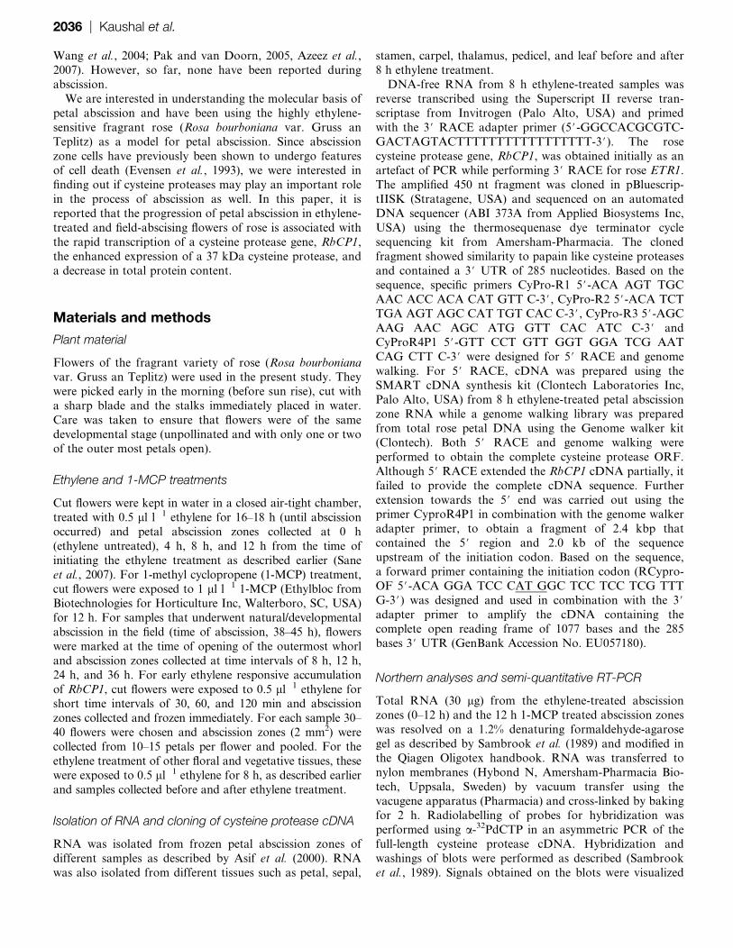
with the RbCP1 promoter:GUS fusion repeatedly showedGUS expression only in the tips of the excised petals in spite
of the fact that the point of agro-injection lay much above
the abscission zone at the centre of the petal. A closer look
revealed expression in cells lining the point of separation at
the junction of the petal and the thalamus. In flowers that
were treated with 1-MCP, no expression was visible. The 1.6
kbp Arabidopsis HAESA gene promoter was used, which is
known to be abscission specific in Arabidopsis (Jinn et al.,2000), to study if this abscission zone specificity was also
observed in rose. The expression of GUS under the HAESA
promoter was only observed in the tip of the petals at the
point of separation of the petals from the thalamus,
indicating that abscission zone specificity of the HAESA
promoter was maintained even in rose.
Discussion
Abscission and senescence of flower parts are important
processes in the developmental cycle of plants. While therehave been numerous studies to understand floral senescence,
the molecular basis of abscission in flowers remains to be
elucidated. Since abscission involves cell separation in
a small specialized zone of cells, there has been a focus on
the role of cell wall hydrolases that lead to floral abscission
(Lashbrook et al., 1994, 1998; del Campillo and Bennett,
1996; Gonzalez-Carranza et al., 2002; Sane et al., 2007).
However, the role of several other genes that could beimportant during the progression of abscission within this
specialized zone is still not clear. In this paper, the isolation
of a cysteine protease gene, RbCP1, from rose petal
abscission zones that is expressed during the course of
abscission has been described. Its transcription appears to
be responsive to ethylene and is accompanied with theappearance of a 37 kDa cysteine protease. The predicted
protein encoded by RbCP1 is a typical papain-type protease
based on the presence of the C1A peptidase domain and
other features such as the ERFNIN motif. Cysteine
proteases are involved in a variety of developmental pro-
cesses, particularly related to organ death such as nucellar
degradation, aleurone layer degradation, xylogenesis etc
(Schaller, 2004). They have been isolated from senescingflowers such as carnations (Jones et al., 1995) and petunia
(Jones et al., 2005) that are ethylene-sensitive as well as
Alstroemeria (Wagstaff et al., 2002), Hemerocallis (Valpuesta
et al., 1995; Guerrero et al., 1998), Sandersonia (Eason et al.,
2002), and Gladiolus (Arora and Singh, 2004) that are
ethylene-insensitive. None, so far, have been reported to be
involved in organ abscission.
Our studies reveal that transcription of RbCP1 is rapidlyup-regulated during ethylene induced abscission and the
earliest increase in transcript accumulation is seen within 30
min of ethylene treatment. Ethylene regulation of RbCP1
was also apparent from reduced expression in 1-MCP-
treated samples where there is a delay in petal abscission.
Sensitivity of RbCP1 to ethylene could be seen in petals as
well as leaves but not in all tissues. The activation of
cysteine protease genes by ethylene has previously beenobserved during senescence in carnations (Jones et al., 1995)
and petunia (Jones et al., 2005) and may be one of the
means by which ethylene triggers cell death. In contrast to
other tissues, there was a decrease in expression of RbCP1
in ethylene-treated samples of the thalamus. Since the
thalamus has to develop later into the fruit, it is possible
that the same ethylene-related signal(s) that triggers senes-
cence/abscission in other floral tissues may, by some as yetunknown mechanism, serve to inhibit expression of RbCP1
Fig. 5. Histochemical staining to test the activity of the RbCP1 promoter using a translational promoter–GUS fusion construct by
agroinfiltration. Petals in intact buds were infiltrated at the centre of the petal with agrobacteria containing different constructs (three
flowers per construct and three petals per flower) using a syringe. Buds were kept for 2 d on the plant and then cut under water as
described and treated with ethylene for 8 h before GUS staining. The constructs used for GUS expression study were pBI101
(promoterless), pBI121 (GUS driven by the CaMV 35S promoter), pHAESA (GUS driven by the 1.6 kb abscission-specific promoter of the
Arabidopsis HAESA gene), and pRbCP1 (GUS driven by the 2.0 kb rose RbCP1 promoter). GUS expression in the tips of the petals (at
the point of separation from the thalamus) is shown. White arrows indicate the point of separation of the petal from the thalamus.
Rose petal abscission zone cysteine protease | 2041

in the thalamus so as to enable fruit formation. Expression
of RbCP1 is also visible in undetached field-abscising
flowers where abscission is under developmental control.
Interestingly, their levels undergo a substantial increase at
36 h just prior to abscission. The late increase in expression
of RbCP1 in field-abscising flowers matches well with their
time of abscission (38–45 h; Sane et al., 2007). It is possible
that field-abscising flowers, unlike detached flowers, un-dergo differential regulation of the abscission process due to
a more dynamic interaction of ethylene/abscission-inducing
factors with other hormones and factors of the parent plant,
leading to a temporal delay in abscission. The late increase
in RbCP1 expression in these flowers indicates that, besides
ethylene, other abscission-related cues may also affect
RbCP1 expression.
Another major finding of our study was the significantdecrease in total protein content (down to 35–37%
of control) not only during ethylene-induced abscission
but also during developmental abscission. Previous
studies (Abeles and Holm, 1966; Lewis and Bakshi, 1968;
Valdovinos et al., 1971; del Campillo and Lewis, 1992)
have shown that abscission is accompanied by an increase
in RNA and protein synthesis (as measured by incorpora-
tion of 14C leucine in abscission zone explants and by two-dimensional gel electrophoresis) and an increase in rough
endoplasmic reticulum. However, the focus in those
studies was more on ethylene-induced de novo protein
synthesis rather than total protein content. Lewis and
Bakshi (1968) did detect a decrease in protein synthesis at
more advanced stages of abscission. Protein synthesis
would be required for de novo synthesis of wall hydrolases
that are involved in cell separation. In rose, it may also beinvolved in synthesis or activation of proteases that may
trigger the decrease in total protein content. Indeed, the
decrease in total protein during rose petal abscission was
associated with a 3–4-fold increase in total protease
activity. Moreover, all the increase in protease activity
could be attributed to cysteine proteases (as evident from
inhibition by E-64). In-gel assays clearly revealed the
abscission-related appearance of a 37 kDa cysteine pro-tease that was present in much reduced levels in control
abscission zones. Thus a large proportion (if not all) of the
increase in cysteine protease activity during abscission
appears be related to the appearance of the 37 kDa band.
Interestingly, the size of this protease matches the size of
the mature protease encoded by RbCP1 (37.2 kDa) and its
appearance during abscission follows the appearance of
the transcript during abscission. Nevertheless, we cannotat present unambiguously attribute the 37 kDa band to the
RbCP1 product. Although no other proteases could be
detected by in-gel assays, the assay itself is limited by its
ability to detect only those proteases that have the ability
to renature after denaturation on an SDS-polyacrylamide
gel. Thus, the presence of other abscission-related pro-
teases that are not able to renature cannot be ruled out
entirely. Our observations of an increase in RbCP1
expression, appearance of a 37 kDa cysteine protease,
increase in total protease activity and a decrease in total
protein content in abscission zones, collectively suggest
that progression of abscission in rose petals may be
associated with gradual cell death of the abscising cells. It
has been hypothesized that abscission zone cells, especially
those that line the separation point, may undergo pro-
grammed cell death (Roberts, 2000). Features of pro-
grammed cell death such as the breakdown of cellular
compartmentalization have previously been observed inabscission zone cells during abscission of Pelargonium
petals (Evensen et al., 1993) while abscission in Delphinium
belladonna was shown to be preceded by DNA degradation
and chromatin condensation in whole turgid petals,
although abscission zones were not studied (Yamada
et al., 2007). Recently, expression of the LX RNase was
shown to be associated with fruit and petiole abscission in
tomato (Lers et al., 2005) providing further indication ofthe degradative processes during abscission. In fact, some
cysteine protease genes such as the SAG12 in Arabidopsis
and its homologues in Brassica are known to act as
developmental markers of senescence (Noh and Amasino,
1999). These data lead us to believe that signals such as
ethylene that activate abscission may also activate cell
death in the abscission zone and that activation of RbCP1
and protein degradation in rose petal abscission zones maybe one of the means by which this is brought about. Indeed
the transcriptional activation of RbCP1 within 30 min of
ethylene treatment (at least under experimental non-
physiological concentrations of ethylene) coupled with the
rapid decrease in the total protein to a third of the total
protein content in just half the time required for complete
abscission (8 h in ethylene-treated and 24 h in naturally
abscising flowers) indicate that some aspects of cell death,such as protein loss, may begin well before cell separation
through hydrolysis of wall polymers is completed. The
ability of RbCP1 to respond rapidly to ethylene may not
be surprising, considering the fact that abscission is highly
sensitive to ethylene and high doses of ethylene can bring
about complete petal abscission even within 1–2 h (Sexton
et al., 1983). Thus, it may be expected that at least a few
components of the abscission machinery must show rapidethylene sensitivity to be able to mount the hastened
response brought about by ethylene. An alternative
possibility for the role of RbCP1 could be to bring about
hydrolysis of the wall-associated proteins that provide
strength to the adhering cells. The 22 amino acid secretory
peptide on RbCP1 (as deduced by TargetP) could direct it
to the secretory pathway and target it to the cell walls for
this purpose, aiding the wall hydrolases in rapidly bringingabout the dissolution of the middle lamella and the
progressive separation of cell walls.
Interestingly, the 2.0 kbp RbCP1 promoter was able to
drive expression of the GUS gene specifically in cells lining
the point of separation of the petal from the thalamus.
Expression was ethylene responsive since no expression was
observed in 1-MCP-treated flowers. Analysis of the pro-
moter revealed several known ethylene-responsive elements(ATTTCAAA and similar sequences, Itzhaki et al., 1994)
between 930–1550 nt upstream of the start codon that may
2042 | Kaushal et al.

confer ethylene responsiveness to the promoter. Further
studies with deletion of these elements may provide better
information on the functionality of these elements in the
RbCP1 promoter. No expression was visible in any other
part of the petal. This was surprising since RbCP1 is
expressed in several tissues including the petal. It probably
indicates that the 2.0 kb promoter only contains elements to
drive expression in the abscission zone with other cis
elements for petal-related expression being present upstream
of the 2.0 kbp region. The 1.6 kb Arabidopsis HAESA gene
promoter was also used as a control for abscission zone-
specific expression (Jinn et al., 2000) and abscission-specific
expression was observed, even in rose. The differential and
reproducible expression patterns of the agro-infiltrated
promoter constructs of pBI101, pBI121, and pRbCP1 and
the maintenance of abscission-specific expression ofpHAESA even in rose indicate that this technique may be
used more widely to study gene/promoter expression in the
plants of interest, in addition to heterologous model systems
such as Arabidopsis and tobacco.
In conclusion, it is shown that petal abscission in rose is
associated with the expression of an ethylene-responsive
cysteine protease gene, RbCP1, and the appearance of a 37
kDa cysteine protease that leads to a decrease in proteincontent during abscission and is possibly associated with
programmed cell death in the abscission zone. We believe
this is the first example of a protease that has a role in
organ abscission.
Acknowledgements
We would like to thank Dr SK Datta, NBRI for his help in
providing rose flowers and Mr Ram Awadh for taking care
of the plants. We are grateful to the Indian National
Science Academy and the Department of Biotechnology,India for financial support. Senior Research Fellowships
provided to Siddharth Kaushal Tripathi and Amar Pal
Singh by the Council of Scientific and Industrial Research,
India are gratefully acknowledged.
References
Abeles FB, Gahagan III HE. 1968. Abscission: the role of ethylene,
ethylene analogues, carbon dioxide, and oxygen. Plant Physiology 43,
1255–1258.
Abeles FB, Holm RE. 1966. Enhancement of RNA synthesis, protein
synthesis and abscission by ethylene. Plant Physiology 41, 1337–1342.
Addicott FT. 1982. Abscission. Berkeley, CA, USA: California
University Press.
Arora A, Singh VP. 2004. Cysteine protease gene expression and
proteolytic activity during floral development and senescence in
ethylene-insensitive Gladiolus grandiflora. Journal of Plant Biochemis-
try and Biotechnology 13, 123–126.
Asif MH, Dhawan P, Nath P. 2000. A simple procedure for the
isolation of high quality RNA from ripening banana fruit. Plant
Molecular Biology Reporter 18, 109–115.
Azeez A, Sane AP, Bhatnagar D, Nath P. 2007. Enhanced
expression of serine proteases during floral senescence in Gladiolus.
Phytochemistry 68, 1352–1357.
Belfield EJ, Ruperti B, Roberts JA, McQueen-Mason S. 2005.
Changes in expansin activity and gene expression during ethylene-
promoted leaflet abscission in Sambucus nigra. Journal of Experimen-
tal Botany 56, 817–823.
Bleecker AB, Patterson SE. 1997. Last exit: senescence, abscis-
sion, and meristem arrest in Arabidopsis. The Plant Cell 9, 1169–1179.
Butenko MA, Patterson SE, Grini PE, Stenvik GE, Amundsen SS,
Mandal A, Aalen RB. 2003. INFLORESCENCE DEFICIENT IN
ABSCISSION controls floral organ abscission in Arabidopsis and
identifies a novel family of putative ligands in plants. The Plant Cell 15,
2296–2307.
del Campillo E, Bennett AB. 1996. Pedicel breakstrength and
cellulase gene expression during tomato flower abscission. Plant
Physiology 111, 813–820.
del Campillo E, Lewis LL. 1992. Identification and kinetics of
accumulation of proteins induced by ethylene in bean abscission
zones. Plant Physiology 98, 955–961.
Eason JR, Ryan DJ, Pinkney TT, O’Donoghue EM. 2002.
Programmed cell death during flower senescence: isolation and
characterization of cysteine proteases from Sandersonia aurantiaca.
Functional Plant Biology 29, 1055–1064.
Evensen KB, Page AM, Stead AD. 1993. Anatomy of ethylene-
induced petal abscission in Pelargonium3hortorum. Annals of Botany
71, 559–566.
Gattolin S, Alandete-Saez M, Elliott K, Gonzalez-Carranza Z,
Naomab E, Powell C, Roberts JA. 2006. Spatial and temporal
expression of the response regulators ARR22 and ARR24 in Arabi-
dopsis thaliana. Journal of Experimental Botany 57, 4225–4233.
Gonzalez-Carranza ZH, Whitelaw CA, Swarup R, Roberts JA.
2002. Temporal and spatial expression of a polygalacturonase during
leaf and flower abscission in oilseed rape and Arabidopsis. Plant
Physiology 128, 534–543.
Guerrero C, de la Calle M, Reid MS, Valpuesta V. 1998. Analysis
of the expression of two thiolprotease genes from daylily (Hemerocallis
spp.) during flower senescence. Plant Molecular Biology 36, 565–571.
Hellmich S, Schauz K. 1988. Production of extracellular alkaline and
neutral proteases of Ustilago maydis. Experimental Mycology 12,
223–232.
Holwerda BC, Rogers JC. 1992. Purification and characterization of
aleurain. Plant Physiology 99, 848–855.
Itzhaki H, Maxson JM, Woodson WR. 1994. An ethylene-responsive
enhancer element is involved in the senescence-related expression of
the carnation glutathione S-transferase (GST1) gene. Proceedings of the
National Academy of Sciences, USA 91, 8925–8929.
Jinn T-L, Stone JM, Walker JC. 2000. HAESA, an Arabidopsis
leucine-rich receptor kinase, controls floral organ abscission. Genes
and Development 14, 108–117.
Jones ML, Chaffin GS, Eason JR, Clark DG. 2005. Ethylene-
sensitivity regulates proteolytic activity and cysteine protease gene
expression in petunia corollas. Journal of Experimental Botany 56,
2733–2744.
Rose petal abscission zone cysteine protease | 2043

Jones ML, Larsen PB, Woodson WR. 1995. Ethylene-regulated
expression of a carnation cysteine proteinase during flower petal
senescence. Plant Molecular Biology 28, 505–512.
Kalaitzis P, Solomos T, Tucker ML. 1997. Three different poly-
galacturonases are expressed in tomato leaf and flower abscission
each with a different temporal expression pattern. Plant Physiology
113, 1303–1308.
Karrer KL, Pfeifer SL, DiTomas ME. 1993. Two distinct gene
families within the family of cysteine protease genes. Proceedings of
the National Academy of Sciences, USA 90, 3063–3067.
Lanahan MB, Yen H-C, Giovannoni JJ, Klee H. 1994. The never
ripe mutation blocks ethylene perception in tomato. The Plant Cell 6,
521–530.
Lashbrook CC, Giovannoni JJ, Hall BD, Fischer RL, Bennett AB.
1998. Transgenic analysis of tomato endo-b-1,4-glucanase gene
function. Role of cel1 in floral abscission. The Plant Journal 13,
303–310.
Lashbrook CC, Gonzalez-Bosch C, Bennett AB. 1994. Two
divergent endo-b-1,4-glucanase genes exhibit overlapping expres-
sion in ripening fruit and abscising flowers. The Plant Cell 6,
1485–1493.
Lers A, Sonego L, Green PJ, Burd S. 2005. Suppression of LX
ribonuclease in tomato results in a delay of leaf senescence and
abscission. Plant Physiology 142, 710–721.
Lewis LL, Bakshi JC. 1968. Protein synthesis in abscission: the
distinctiveness of the abscission zone and its response to gibberlic
acid and indole acetic acid. Plant Physiology 43, 359–364.
Matarasso N, Schuster S, Avni A. 2005. A novel plant cysteine
protease has a dual function as a regulator of 1-aminocyclopropane-1-
carboxylic acid synthase gene expression. The Plant Cell 17,
1205–1216.
Noh Y–S, Amasino RM. 1999. Regulation of developmental senes-
cence is conserved between Arabidopsis and Brassica napus. Plant
Molecular Biology 41, 195–206.
Pak C, van Doorn WG. 2005. Delay of Iris flower senescence by
protease inhibitors. New Phytologist 165, 473–480.
Peterson GL. 1977. A simplification of the protein assay method of
Lowry et al. which is more generally applicable. Analytical Biochemistry
83, 346–356.
Roberts JA, Elliot KA, Gonzalez-Carranza ZH. 2002. Abscission,
dehiscence, and other cell separation processes. Annual Review of
Plant Biology 53, 131–158.
Roberts JA. 2000. Abscission and dehiscence. In: Bryant JA, Hughes
SG, Garland JM, eds. Programmed cell death in animals and plants.
Oxford: BIOS Scientific Publishers, 203–211.
Sambrook T, Fritsch EF, Maniatis T. 1989. Molecular cloning:
a laboratory manual. Cold Spring Harbour, New York: Cold Spring
Harbor Laboratory Press.
Sane AP, Tripathi SK, Nath P. 2007. Petal abscission in rose (Rosa
bourboniana var. Gruss an Teplitz) is associated with the enhanced
expression of an alpha expansin gene, RbEXPA1. Plant Science 172,
481–487.
Schaller A. 2004. A cut above the rest: the regulatory function of
plant proteases. Planta 220, 183–197.
Sexton R, Struthers WA, Lewis LN. 1983. Some observations on
the very rapid abscission of the petals of Geranium robertianum L.
Protoplasma 116, 179–186.
Stenvik GE, Butenko MA, Urbanowicz BR, Rose JKC, Aalen RB.
2006. Overexpression of INFLORESCENCE DEFICIENT IN ABSCIS-
SION activates cell separation in vestigial abscission zones in
Arabidopsis. The Plant Cell 18, 1467–1476.
Stenvik GE, Tandstad NM, Guo Y, Shi C-L, Kristiansen W,
Holmgren A, Clark SE, Aalen RB, Butenko MA. 2008. The EPIP
peptide of INFLORESCENCE DEFICIENT IN ABSCISSION is sufficient
to induce abscission in Arabidopsis through the receptor-like kinases
HAESA and HAESA-LIKE2. The Plant Cell 20, 1805–1817.
Stephenson P, Rubinstein B. 1998. Characterization of proteolytic
activity during senescence in daylilies. Physiologia Plantarum 104,
463–473.
Tucker ML, Sexton R, del Campillo E, Lewis L. 1988. Bean
abscission cellulase: characterisation of a cDNA clone and regulation
of gene expression by ethylene and auxin. Plant Physiology 88,
1257–1262.
Valdovinos JG, Jensen TE, Sticko LM. 1971. Ethylene induced
rough endoplasmic reticula in abscission cells. Plant Physiology 47,
162–163.
Valpuesta V, Lange NE, Guerrero C, Reid MS. 1995. Up-regulation
of a cysteine protease accompanies the ethylene-insensitive senes-
cence of daylily (Hemerocallis) flowers. Plant Molecular Biology 28,
575–582.
van Doorn WG, Stead AD. 1997. Abscission of flowers and floral
parts. Journal of Experimental Botany 48, 821–837.
Wagstaff C, Leverentz MK, Griffiths G, Thomas B, Chanasut U,
Stead AD, Rogers HJ. 2002. Cysteine protease gene expression and
proteolytic activity during senescence of Alstroemeria petals. Journal
of Experimental Botany 53, 233–240.
Wang YT, Yang CY, Chen Y-T, Lin Y, Shaw J-F. 2004.
Characterization of senescence-associated proteases in postharvest
broccoli florets. Plant Physiology and Biochemistry 42, 663–670.
Whitelaw CA, Lyssenko NN, Chen L, Zhou D, Mattoo AK,
Tucker ML. 2002. Delayed abscission and shorter internodes
correlate with a reduction in the ethylene receptor LeETR1 transcript in
transgenic tomato. Plant Physiology 128, 978–987.
Yamada T, Ichimura K, van Doorn WG. 2007. Relationship
between petal abscission and programmed cell death in Prunus
yedoensis and Delphinium belladonna. Planta 226, 1195–1205.
2044 | Kaushal et al.