Detection of the 20-kDa virulence-associated antigen of Rhodococcus equi in malakoplakia-like lesion...
7
PATHOLOGY RESEARCH AND PRACTICE e Urban & Fischer Ver l ag h ttp://www.urbanfischer. deljournalslprp , . Detection of the 20-kDa Virulence-associated Antigen of Rhodococcus equi in Malakoplakia-like Lesion in Pleural Tissue Obtained from an AIDS Patient* Adele Caterino-de-Araujo', Elizabeth de los Santos-Fortuna ', Monica Cristina Zandona-Meleiro', Edenilson Edu ar do Calore 2 and Nilda Maria Perez Calore 2 'Immunology Department, Instituto Adolfo Lutz, Sao Paulo, SP, Brazil ; ' Pathology Section , Emilio Ribas Institute, Sao Paulo, Sp, Brazil Summary A malakoplakia-like lesion was detected in a pleural biopsy from an AIDS patient presenting clinical a nd ra- diologic features of pneumonia. Cultures of bron- choalveolar lavage and pleural fluid evidenced Rhodococcus equi as the causative agent of pleuro-pul- monary infection. Immunochemical characterization of the R. equi isolate showed the presence of a strain simi- lar to the ATCC 33704 reference strain presenting the capsular antigen of serotype 4, and the intermediate vir- ulence-associated antigen of 20-kOa. Histopathology of the patient's pleural biopsy showed plaques of macrophages interspersed with lymphocyte s, and intra- cytoplasmic cocci and bacilli in macrophages, which were variably acid-fast positive. Immunohistochemistry of cocci, bacilli and their degradation products resulted strong ly positive when stained with a mouse monoclon- al antibody (MAb) produced against the 20-kDa anti- gen. This finding cou ld have important implications for the pathogenicity of R. eqlli for human beings. si nce we do not know yet all the factors involved in the forma- tion of malakoplakia. Indeed, the re sults obtained in the present stud y, taken together with the results obtained for pigs inoculated with R. equi strains of intermediate virulence (Madarame et al. 1998), raise the possibility that most strains presenting the 20-lOa an tig en may be *This work was partial ly su pported by de Amparo Pesqui sa do Estado de Sao Paulo (FAPESP), grant number 97/02866-0 . Pa th ol. Re s. Pract . 196: 32 1-327 (2000) capable of inducing malakoplakia. If this hyphothesis is confirmed by immunohistochemical analysis of human pu lmonary malakoplakia cases due to R. equi, the de- tection of this antigen may be extreme ly helpful in the diagnosis and treatment of such patients. This is the first report of R. eqlli infection in human beings that sug- gests a relationship between pleural malakoplakia and the virulence-associated antigen of 20-kOa. Key words: RhodocoCCllS equi - Malakoplakia - AIDS - Virulence-associated antigen of 20-kOa - Immuno- histochemistry - Macrophages Introduction Malakoplakia is a chronic granulomatous inflamma- tion characterized by accumulation of granular histio- cytes, possibly containing intracytoplasmic basophilic inclusions (Michaelis Gutmann bodies), at various body sites, mainly in the lower urinary tract {S/. It was first described by Michaelis and Gutmann in 1902 [l3} and named "malakoplakia" by von Hansemann in 1903 {31}. Until 1996, approximately 400 cases had been re- Address for correspo nd ence: Adel e Caterino-de-Araujo, In- stituto Adolfo Lutz, A v. Dr. Arnatd o 355. t I andar, 01246- 902. Sao Paulo, SP , Brazil. Phone : ext. 2065, Fax: ++55-11-8533505. E-mait: (catcrin [email protected]) (ca terin o@ ial. sp.gov.br) 03 44-0338/2000/196/5-32 1 $12.00/0
-
Upload
independent -
Category
Documents
-
view
1 -
download
0
Transcript of Detection of the 20-kDa virulence-associated antigen of Rhodococcus equi in malakoplakia-like lesion...
PATHOLOGY RESEARCH AND PRACTICE e Urban & Fischer Verlag http://www.urbanfischer.deljournalslprp
, .
Detection of the 20-kDa Virulence-associated Antigen of Rhodococcus equi in Malakoplakia-like
Lesion in Pleural Tissue Obtained from an AIDS Patient*
Adele Caterino-de-Araujo', Elizabeth de los Santos-Fortuna ', Monica Cristina Zandona-Meleiro', Edenilson Eduardo Calore2
and Nilda Maria Perez Calore2
'Immunology Department, Instituto Adolfo Lutz, Sao Paulo, SP, Brazil ; 'Pathology Section, Emilio Ribas Institute, Sao Paulo, Sp, Brazil
Summary
A malakoplakia-like lesion was detected in a pleural biopsy from an AIDS patient presenting clinical and radiologic features of pneumonia. Cultures of bronchoalveolar lavage and pleural fluid evidenced Rhodococcus equi as the causative agent of pleuro-pulmonary infection. Immunochemical characterization of the R. equi isolate showed the presence of a strain similar to the ATCC 33704 reference strain presenting the capsular antigen of serotype 4, and the intermediate virulence-associated antigen of 20-kOa. Histopathology of the patient's pleural biopsy showed plaques of macrophages interspersed with lymphocytes, and intracytoplasmic cocci and bacilli in macrophages, which were variably acid-fast positive. Immunohistochemistry of cocci, bacilli and their degradation products resulted strongly positive when stained with a mouse monoclonal antibody (MAb) produced against the 20-kDa antigen. This finding could have important implications for the pathogenicity of R. eqlli for human beings. since we do not know yet all the factors involved in the formation of malakoplakia. Indeed , the results obtained in the present study, taken together with the results obtai ned for pigs inoculated with R. equi strains of intermediate virulence (Madarame et al. 1998), raise the possibility that most strains presenting the 20-lOa antigen may be
*This work was partial ly supported by Funda~ao de Amparo ~ Pesquisa do Estado de Sao Paulo (FAPESP), grant number 97/02866-0.
Pathol. Res. Pract. 196: 32 1-327 (2000)
capable of inducing malakoplakia. If this hyphothesis is confirmed by immunohistochemical analysis of human pu lmonary malakoplakia cases due to R. equi, the detection of this antigen may be extremely helpful in the diagnosis and treatment of such patients. This is the first report of R. eqlli infection in human beings that suggests a relationship between pleural malakoplakia and the viru lence-associated antigen of 20-kOa.
Key words: RhodocoCCllS equi - Malakoplakia - AIDS - Virulence-associated antigen of 20-kOa - Immunohistochemistry - Macrophages
Introduction
Malakoplakia is a chronic granulomatous inflammation characterized by accumulat ion of granular histiocytes, possibly containing intracytoplasmic basophilic inclusions (Michaelis Gutmann bodies), at various body sites, mainly in the lower urinary tract {S/. It was first described by Michaelis and Gutmann in 1902 [l3} and named "malakoplakia" by von Hansemann in 1903 {31}. Until 1996, approximate ly 400 cases had been re-
Address for correspondence: Adele Caterino-de-Araujo, Instituto Adolfo Lutz, Av. Dr. Arnatdo 355. t I andar, 01246-902. Sao Paulo, SP, Brazil. Phone: ++55- 11 -30~ I0111 ext. 2065, Fax: ++55-11-8533505. E-mait: ([email protected]) (caterino@ ial.sp.gov.br)
0344-0338/2000/196/5-32 1 $12.00/0
322 . A. Caterino-de-Araujo et al.
ported, most of them associated with Escherichia coli infection [30J. Malakoplakia in those cases was associated with immunosuppression of patients with organ transplantation, hematopoietic malignancy, or alcoholism. Pulmonary malakoplakia is rare, and was mostly detected in Human Immunodeficiency Virus (HIV)infected patients, in which Rhodococcus equi could be isolated from respiratory specimens [11j.
R. equi is a Gram-positive coccobacillus present in soil which causes bronchopneumonia, enteritis or mesenteric adenitis in horses, cattle, swine and sheep [16]. In humans it is responsible for necrotizing pneumonia with or without cavitation or spindle cell proliferation [l6}. R. equi with at least two levels of virulence were detected among AIDS-infected patients: R. equi expressing the virulence-associated antigen of 15-to 17-kDa (similar to isolates from foals) , and R. equi expressing the intermediate virulence-associated antigen of 20 kDa (similar to isolates from pigs). However, some cases of rhodococcal infection in AIDS were due to strains considered avirulent [3, 18, 22}.
In veterinary medicine, malakoplakia is not found in lung tissues from infected foals, even when these animals are infected with virulent R. equi strains (presenting the 15- to 17-kDa virulence-associated antigen). On the other hand, two cases of spontaneous malakoplakia have been reported in pigs, one of which in lymph nodes from a slaughtered pig [8}, and the other one systemically disseminated in a breeding pig [27}. Cutaneous malakoplakia was also induced experimentally in pigs by subcutaneous inoculation of intermediate virulent R. equi strains, in which immunohistochemical analysis of the lesions showed the presence of the 20-kDa antigen in Michaelis Gutmann (MG) bodies, providing evidence of the involvement of R. equi and its virulence antigens in the process of malakoplakia [12}.
Some of us previouly described one case of pleural malakoplakia-like lesion in an AIDS patient in which R. equi could be isolated from pleural tluid and bronchoalveolar lavage, and suggested the use of histochemistry for a rapid and accurate diagnosis of rhodococcal infection in the lung [2}. In the present study we have used immunohistochemical analysis of the same biopsy material to search for an R. equi strain related to malakoplakia formation in humans. We also characterized immunochemically the R. equi isolated from this patient, and compared it with R. equi reference strains.
Materials and Methods
Case report
The patient was a 23-year-old heterosexual man, an intravenous drug user for the last three years, who came to Institu-
to de Infectologia Emilio Ribas (HER), Sao Paulo, complaining of bilateral chest pain and cough for 60 days, having had a weight loss of 10.0 kg in the last 16 days. He was treated with penicillin and second generation cephalosporin without success. A chest roentgenogram showed an abnormal opacity of the right lower lobe. The WBC was 15,500 (77% of neutrophils). His haemoglobin level was 11.0 gil and the platelet blood count was normal. Biochemical screening showed only a slight decrease in albumin level (1.9 mgldl). Serology for HIV was performed and resulted positive. At this time he presented also oral candidiasis. He was submitted to pleural drainage of the right hemithorax and to a pleural biopsy. The pleural effusion was characteristic of empyema. Rhodococcl4S equi grew in culture. The patient was treated with erythromycin for 14 days with success.
Bacterial strains
The clinical bacterial isolate was cultured from bronchoalveolar lavage and pleural fluid of the patient and sent to Instituto Adolfo Lutz (IAL) for analysis. The presence of R. equi was confirmed at the Culture Collection Sector on the basis of Gram-positive coccobacilli, salmon-pink mucoid colonies, oxidase negativity, CAMP test positivity, and also on the basis of the results of the API Corynebacterium test system (Biomerieux, Marcy l'Etoile, France). The isolate was lyophilized and stored with the identification number IAL-2043.
R. equi ATCC 33701 and ATCC 33704 were used as refercnce strains because their plasmid content and virulence level have already been described (26, 28/.
All strains were used in antigen preparations for serotyping and immunoblotting analysis.
Antibodies
Monoclonal antibodies (MAbs) against the 15- to 17-kDa (MAb, I OG5) /2/ and 20-kOa antigens [25J were provided by Or. S. Takai and used to identify virulence-associated antigens in immunoblotting and immunohistochemistry.
Seven rabbit sera (1-7) which react with the capsular antigens of R. equi were provided by Dr. J. Prescott and were used for serotyping the R. equi isolate [15].
Serum from a foal naturally infected with R. equi, and sera from the patient were analysed by immunoblot, along with serum from another infected human patient.
Strain characterization
R. equi was grown at 37 "C on Miiller Hinton agar (Oifco) for 48h and subsequently harvestcd in physiologic solution (0.9% NaCl). This suspension was maintained at 37 "C, under shaking, overnight, and then spun at 4,000 rpm. The capsular antigens in the supernatants were submitted to immunodiffusion in agar gel, as previously described {lSI.
To compare the major antigenic components of the human R. equi isolate with those of R. equi reference strains, wholecell antigens prepared by harvesting bacteria grown at 38 °C for 48 h from brain heart infusion broth (Oifco), and solubilized in sodium dodecyl sulphate (50S) reducing buffer, were submitted to SOS-polyacrylamide gel electrophoresis. The
20-kDa Antigen of Rhodococcus equi and Malakoplakia-like Lesion in AIDS . 323
components were subsequently elcctrotransferred to nitrocellulose sbeets for immunoblolling. Tbe immunoblOl analysis of the isolate was conducted according to the technique previously established in the Immunology Department of IAL {I 8, 33].
Histopathological and immunohistochemical procedures
The histopathology of the pleural tissue was conducted in paraffin wax-embedded sections submitted to hematoxyleosin staining and to Fite's method for acid-fast organisms as previously described (2/. Immunohistochemical reactions were performed according to Hsu et al. 1981 (9), using as primary antibodies, mouse monoclonal antibodies against the 15- to 17-kDa antigen and 20-kDa antigen. diluted I: I 00 and I: I 0 ,000 in phosphate buffered saline, pH 7.4, with 1% bovine serum albumin (PBS-BSA). Briefly. slices of paraffin embedded sections were treated with I % aqueous hydrogen peroxide and methanol (Sigma) for 20 minutes, and then incubated with the primary antibodies for 24 hours at 4 °C in a moist chamber. The slices were then incubated sequentially with biotinylated secondary antibody (Sigma) for 30 minutes at 37 °C, with the streptavidin-biotin-peroxidase complex (Sigma) for 30 minutes at 37 °C, and finally with aqueous diaminobenzidine solution (40 mg/ml) and 0.1 % (v/v) hydrogen peroxide (Sigma) in PBS. The slices were counterstained with hematoxylin, dehydrated, cleared and mounted with entellan. The incubation step with primary antibodies was skipped for the negative controls. In addition. to assure the specificity of the primary antibodies in the immunohistochemical reaction for R. equi , the same procedure was performed using paraffin wax-embedded sections of samples diagnosed as tuberculosis and other non-specific bacterial diseases.
Results
The results obtained in the characterization of the R. equ; isolate showed a strain similar to the ATCC 33704 reference strain, presenting the capsular antigen of type 4 and the intermediate virulence-associated anligen of
33101 33704
1 2 3 . , , ' 3 . , , , 3
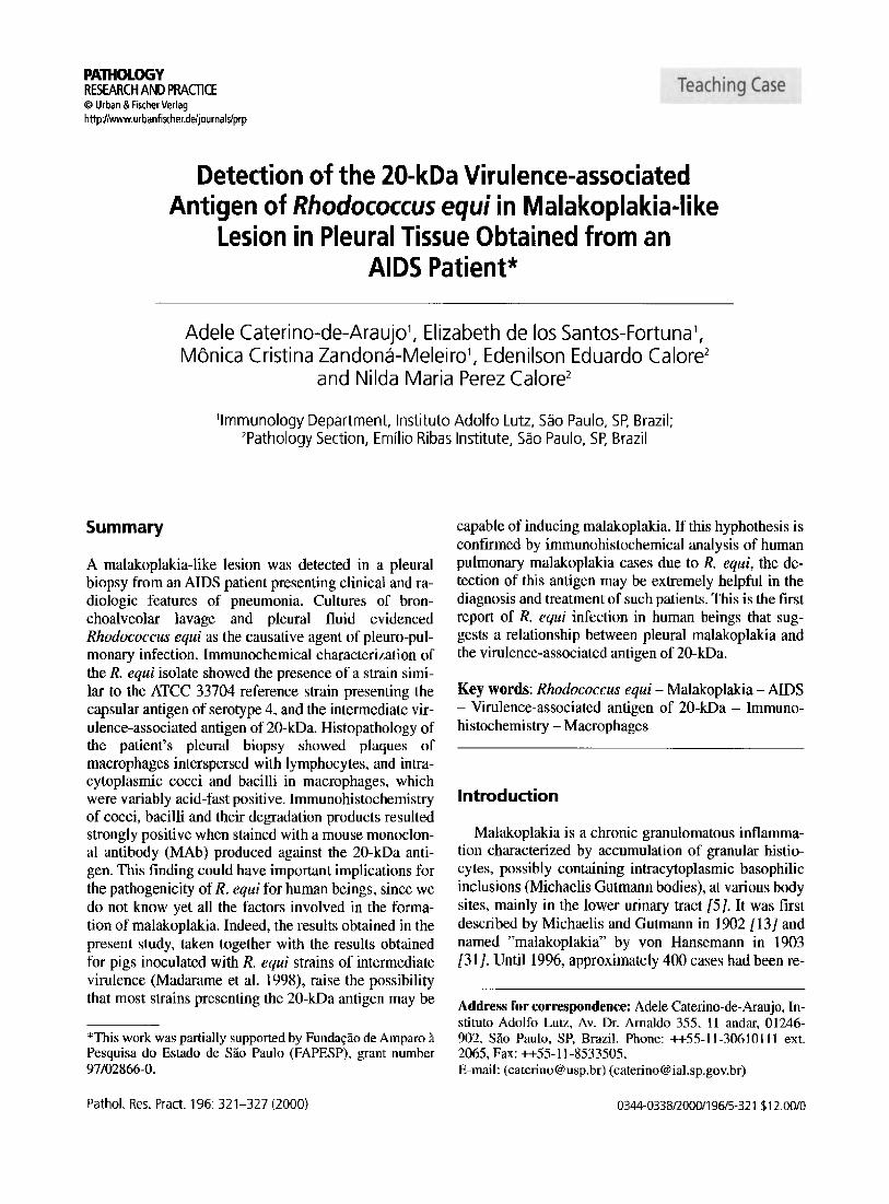
20-kDa. Immunoblotting showed reactivity of the infected foal serum with several antigens present in this isolate, although reactivity to low molecular weight antigens could not be detected (Fig. J, strain IAL-2043, lane I). On the other hand, serum samples from infected human patients strongly reacted with the low molecular weight antigens (Fig. 1, strain IAL-2043, lanes 4-5), a reactivity similar to that of the MAb to the 20-kDa antigen (Fig. 1, strain IAL-2043, lane 3).
Immunoblot analysis of reference strains confirmed the presence of the 15- to 17-kDa anligen in the ATCC 33701 strain (Fig. 1, strain ATCC 33701, lane 2), and of the 20-kDa antigen in the ATCC 33704 strain (Fig. I, strain ATCC 33704, lane 3). The foal serum sample tested with these reference strains demonstrated a better reactivity of antibodies to the antigens present in the ATCC 33701 strain (Fig. I, strains 33701 and 33704, lanes I). In contrast, when infected human serum samples were analysed by immunoblol, a better seroreactivity to several antigens, including the 20-kDa antigen, was observed in the ATCC 33704 reference strain (Fig. I, strains ATCC 3370 I and 33704, lanes 4-5).
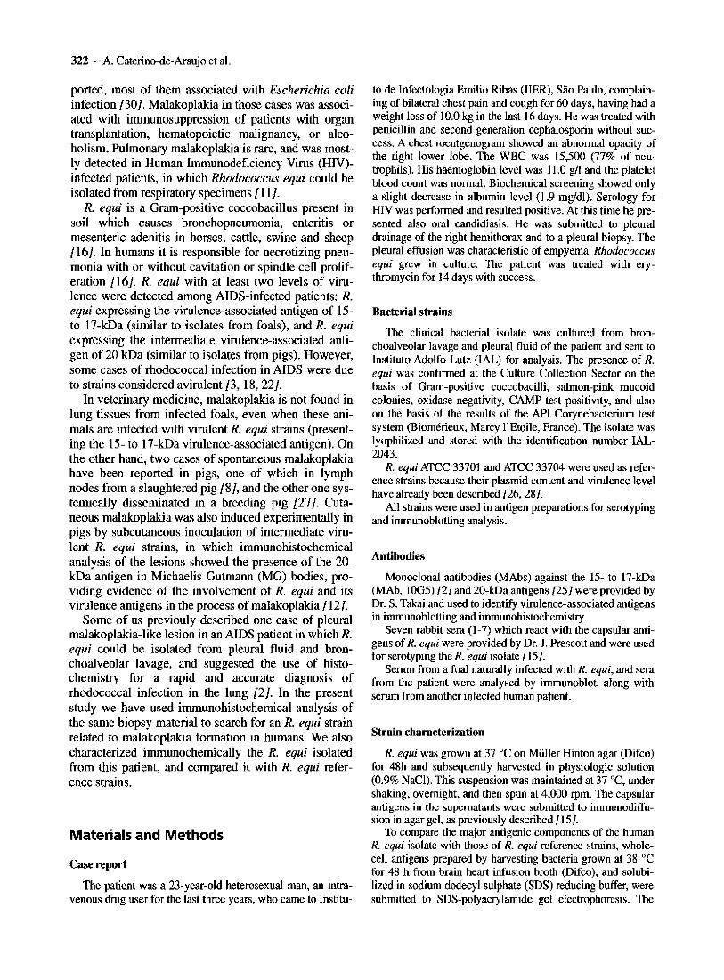
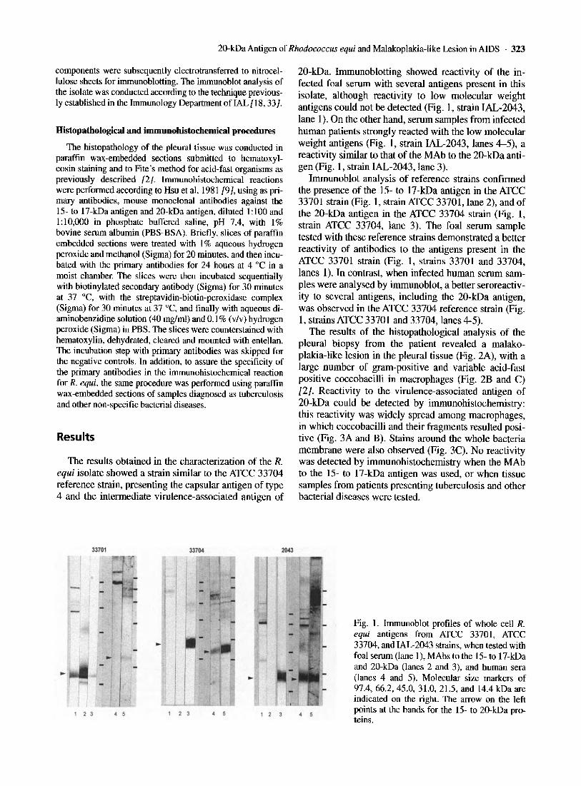
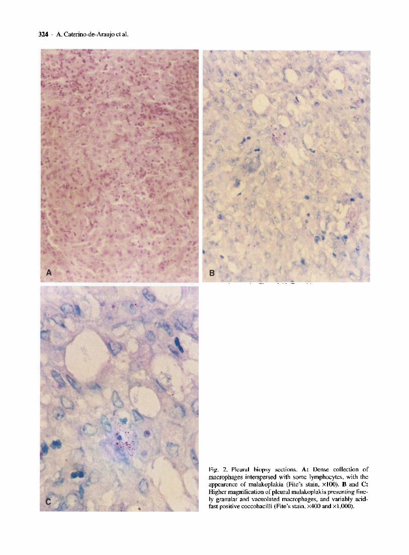
The results of the histopathological analysis of the pleural biopsy from the patient revealed a malakoplakia-like lesion in the pleural ti ssue (Fig. 2A), with a large number of gram-positive and variable acid-fast positive coccobacilli in macrophages (Fig. 2B and C) /2J. Reactivity to the virulence-associated antigen of 20-kDa could be detected by immunohistochemistry: this reactivity was widely spread among macrophages , in which coccobacilli and their fragments resulted positive (Fig. 3A and B). Stains around the whole bacteria membrane were also observed (Fig. 3C). No reactivity was detected by immunohistochemistry when the MAb to the 15- to 17 -kDa antigen was used, or when tissue samples from patients presenting tuberculosis and other bacterial diseases were tested.
. ,
Fig. I. Immunoblot profiles of whole cell R. equi antigens from ATCC 33701 , ATCC 33704. and IAL-2043 strains, when tested with foal serum (lane I), MAbs to the 15- to 17-kDa and 20-kDa (lanes 2 and 3), and human sera (lanes 4 and 5). Molecular size markers of 97.4, 66.2,45.0, 31.0, 21.5, and 14.4 kDa are indicated on the right. The arrow on the left points at the bands for the 15- to 20-kDa proteins.
324 ' A, Caterino-de-Araujo et aJ.
~," ' ..
. '
.• ;.. . . ~ '. 'J
Fig. 2. Pleural biopsy sections. A: Dense colleclion of macrophages interspersed with some lymphocytes, with the appearenee of malakoplakia (Fite's stain, xIOO), B and C: Higher magnification of pleural malakoplakia presenting finely granular and vacuolated macrophages, and variably acidfast positive coccobacilli (Fite's stain, x400 and xl ,000).

A
~ t' 'i ·" c
i
20-kDa Amigen of Rhodococcus equi and Malakoplaki a- like Lesion in AIDS 325
. :.. . ~
, , ....
. ~' .. " -.'
,. .~ ~ ,;' ",'
' .
Fig. 3. Immunohistochemistry of pleural malakoplakia, A: Numerous R. equi and their degradation products stained with the monoclonal antibody to the 20·kDa antigen of the R. equi strain of intermediate virulence (small star). The adjacent connective tissue stained negatively (big star) (Labeled avidin-biotin method, hematoxylin eosin counterstain, x40). B: Sheets of histiocytes, many containing R. equi stained positively by immunohistochemistry (Labeled avidin· biotin method using MAb to the 20-kOa antigen of R. equi, hematoxylin eosin counterstain, x400). C: Higher magnification showing positivity on the cellular membrane of bacteria (arrows) (Labeled avidin·biotin method using MAb to the 20-kDa antigen of R, equi, hematoxylin eosin counterstain, x 1,000),

326 . A. Caterino-de-Araujo et aJ.
Discussion
Pulmonary infection by R. equi in HIV-infected patients has been described [6, 20, 29/, and in several cases this infection was associated with malakoplakia [7, 11,14, 19- 21]. Usually, the diagnosis of R. equi infection is performed by bacterial isolation and subsequent identification in blood or bronchoalveolar lavage, but these procedures take several days. Alternative methods for the detection of R. equi have been taken into consideration, and the polymerase chain reaction (PCR) stands out as a fast, sensitive and accuratc technique to identify the 15- to 17-kDa virulence-associated antigens {24 J and the R. equi species {l]. However, these methodologies are expensive and need sophisticated laboratories in order to avoid contamination.
In the last few years the diagnosis of malakoplakia by fine needle aspiration and lung tissue biopsy was considered of some help {II, 17,21, 32J, and specific stains for acid-fast bacteria and for detecting Michaelis Gutmann (MG) bodies have also been used {2, 10, 11 , 32]. The number of MG bodies varies from case to case and appears to be inversely proportional to the number of bacteria in the tissue lesion {4, 10]. We found in the present study large numbers of bacteria and no MG bodies in tissue (Fig. 2).
Recently, an association between the 20-kDa virulence-associated antigen of R. equi and malakoplakia was demonstrated experimentally in pigs inoculated with R. equi of intermediate virulence, using immunohistochemical analysis {l2]. On the basis of this report, we may speculate about a possible role for the 20-kDa antigen in the malakoplakia process in human beings. Since we had stored the bronchoalveolar lavage isolate and the pleural biopsy specimen from an AIDS patient in whom a malakoplakia-like lesion had been detected, we compared the antigenic pattern of this patient's isolate with two R. equi reference strains, and searched for the virulence-associated antigens in the lesion.
The results obtained by gel agar immunodiffusion and immunoblotting confirmed the similarity between the human isolate and the ATCC 33704 reference strain (Fig. I), indicating that pigs and their environment are possible sources of R. equi infection for humans. Immunohistochemistry confirmed the association between the 20-kDa antigen and malakoplakia.
This is the first time that an association between the 20-kDa virulence-associated antigen and malakoplakia has been observed in humans. This finding, taken together with the same finding in pigs, raises the possibility that most R. equi strains presenting the 20-kDa antigen could induce malakoplakia. We suggest a search for the antigen in malakoplakia cases caused by R. equi detected around the world and described in the literature.
References
1. Bell KS, Phil JC, Aw DWJ (1996) Identification of Rhodococcu,\" equi using the polymerase chain reaclion_ Letters in Applied Microbial 23: 72-74
2. Calore EE, Vazquez CRM, Perez NM, Cavaliere MJ, Curti Jr R, Campos Salles PS, Araujo MF (1995) Empyema with malakoplakic-like lesions by Rhodococcus equi as a presentation of HIV infection. Pathologica 87: 525-527
3. Caterino-de-Araujo A, Santos-Fortuna E, ZandonaMeleiro MC, Mastroianni CM, Lichtner M, Mengoni F, Vullo V (1999) Searching for an antibody profile characteristic of Rhodococcus equi infection in AIDS patients in spite of the diversity of R. equi isolates and patient immune dysfunction. Microbes and Infection 1 (7): 663-670
4. Colby TV (1978) Malakoplakia: two unusual cases which presented diagnostic problems. Am J Surg Pathol 2: 377-382
5. Damjanow I, Katz SM (1981) Malakoplakia. Pathol Annu 16: 103-126
6. Fiaccadori F, Elia GF, Calzetti C, Degli Antoni A, Magnani G (1994) Rhodococcus equi infection in HIV patients: report of 5 cases and literature overview. Monaldi Arch Chest Di s 41: 38G-388
7. Ghnassia JP, Gasser B, Fraisse P, De Briel D. Philippe E (1992) Infection a RhodococclIs equi responsable d'une malacoplasie pulmonaire chez un patient porteur d'un syndrome d'immunodeficit acquis. Ann Pathol 12: 174-177
8. Gill BS, Ducatelle R, Coussement W, Hoorens J (1981) Malakoplakia-like lesion in the lymph node of a pig. J Comp Pathol91: 539-544
9. Hsu SM, Rainc L, Fanger H (1981) The use of anti avidin antibody and avidin-biotin peroxidase complex in immunoperoxidase techniques. Am J Clin Pathol 75: 816-820.
10. Kwon KY, Colby TV (1994) Rhodococcus equi pneumonia and pulmonary malakoplakia in acquired immunodeficiency syndrome. Pathologic Features. Arch Pathol Lab Med 118: 744-748
II. Lambert C, Gansler T, Mansour KA, Schwartzmann SW, Duffell GM, Gal AA (1997) Pulmonary malakoplakia diagnosed by fine needle aspiration. Acta Cytol 41: 1833- 1838
12. Madarame H, Matsuda H, Okada M, Yoshida S, Sasaki Y, Tsubaki S, Hasegawa Y, Takai S (1998) Cutaneous malakoplakia in pigs inoculated with Rhodococcus equi. FEMS Immunol Med Microbiol 22: 329-333
13. Michaelis L, Gutmann C (1902) Ueber Einschluesse in Blasentumoren. Z Klin Med 47: 208-215
14. Peralta-Venturina MN, Clubb FJ , Kielhofner MA (1993) Pulmonary malakoplakia associated with Rhodococcus equi infection in a patient with Acquired Immunodeficiency Syndrome. Am J Clin Pathol 102: 459-463
15. Prescott JF (1981) Capsular serotypes of Corynebacterium equi. Can J Comp Med 45: 13G-134
16. Prescott JF (1991) Rhodococcus equi: an animal and human pathogen. Clin Microbiol Rev 4: 2G-34
17. Reyes CV, Thompson KS, Jensen JA (1998) Multivesiculated macrophages: their implication in fine-needle aspiration cytology of lung mass lesions. Diagn Cytopathol 19: 98-101

20-kDa Antigen of Rhodococcus equi and Malakoplakia-like Lesion in AIDS . 327
18. Santos~Fortuna E, Zandona-Mclciro Me. Mastroianni CM, Lichtner M, Mengoni F, Vullo V, Caterino-de-Araujo A (1999) Search for virulence-associated antigens of Rhodococcus equi in strains isolated from patients with Acquired Immunodeficiency Syndrome. Brazilian J InfectDis3: 184-188.
19. Schwartz DA, Ogden PO. Blumberg HM, Honig E (1990) Pulmonary malakoplakia in a patient with the Acquired Immunoddiciency Syndrome. Differential diagnostic considerations. Arch Pathol Lab Med 114: 1267-1272
20. Scott MA, Graham BS, Verrall R, Dixon R. Schaffner W. Tharn KT (1995) Rhociococ{'us equi - An increasingly recognized opportunist pathogen. Report of 12 cases and review of 65 cases in the literature. Clin Microbiol Infect Dis 103: 649-655
21. Sughager M, Ali SZ, Erozan YS, Dunsmore N. Hall GS (1997) Pulmonary malakoplakia associated with Rhodacoccus equi infection in an AIDS patient. Report of a case with diagnosis by fine needle aspiration. Acta Cytoiogica 41:507-512
22. Takai S (1997) Epidemiology of Rhodococcus equi infections: A review. Vet Microbiol 56: 167-176
23. Takai S, lie M. Kobayashi C. Morishita T, Noshio T. Ishida T, Fujimura T. Sasaki Y, Tsubaki S (1993) Monoclonal antibody specific to virulence-associated 15- to 17-kilodalton antigens of Rhodococcus equi. J Clin Microbiol 31:2780-2782
24. Takai S, Ikcda T, Sasaki Y, Watanabe Y, Ozawa T. Tsubaki S, Sekisaki T (1995) Identification of virulent RllOdococcus equi by amplification of gene coding for 15- to 17-kilodalton antigens.) Clin Microbiol 32: 457-460
25. Takai S. Imai Y. Fukunaga N, Uchida Y, Kamisawa K. Sasaki Y. Tsubaki S, Sekizaki T (1995) Identification of virulence-associated antigens and pJasmids in Rhodococ-
ells equi from patients with AIDS. ) Infcct Dis 172: /306-1311
26. Takai S. Koike K. Ohbushi S,lwmi C, Tsubaki S (1991) Identification of 15- to 17 -kilodalton antigens associated with virulent R. equi.) Clin Microbiol29: 439-443
27. Taniyama H, Ono T. Matsui T (1985) Systemic malakoplakia in a breeding pig. J Comp Pathol95: 79-85
28. Tkachuk-Saad O. Prescott J (1991) RhodococeLls equi plasmids: isolation and partial characterizaLion. J Clin Microbil 29: 2696-2700
29. Verville TD, Huycke MM. Greenlleld RA, Fine DP, Kuhls TL, Slater LN (1994) RllOdoeoccLls equi infections of humans: 12 cases and review of the literature. Medicine 73: 119-132
30. von der Voort PH), ten Velden JJAM, Wassenaar RP, Silberbusch J (1996) Malakoplakia: two cases reports and a comparison of treatment modalities based on literature review. Arch Intern Med 156: 577-583
31. von Hansemann D (1903) Ueber Malakoplakieder Harnblase. Virchow Arch [A] 173: 302-308
32. von Hoeven KH. Dookhan DB. Petersen RO (1995) Cytologic features of pulmonary malakoplakia related to Rhodococcus eqlli in an immunocompromised host. Diagn Cytopathol 15: 325-328
33. Zandona-Meleiro MC (1998) Caracteriza,ao de ccpas de Rhodococcus equi isoladas de casas c1fnicos de infeccrao nas especies equina e humana. Estudo de componentes anligcnicos. marcadores de virulencia c fonte de infcccrao para a especie humana. /11 "Master Thesi s". Universidade de Sao Paulo, Sao Paulo. SP.
Received: April 12,1999 Accepted in revised version: November 9. 1999