Trafficking of green fluorescent protein tagged-vesicular acetylcholine transporter to varicosities...
10
Journal of Neurochemistry, 2001, 78, 1104–1113 Trafficking of green fluorescent protein tagged-vesicular acetylcholine transporter to varicosities in a cholinergic cell line M. S. Santos,* J. Barbosa Jr,² G. S. Veloso,² F. Ribeiro,* C. Kushmerick,² M. V. Gomez,² S. S. G. Ferguson,‡ V. F. Prado* and M. A. M. Prado² *Laborato ´rio de Neurobiologia Molecular, Departamento de Bioquı ´mica-Imunologia, ²Laborato ´rio de Neurofarmacologia, Departamento de Farmacologia, ICB, Universidade Federal de Minas Gerais, Belo Horizonte, Minas Gerais, Brazil ‡J.P. Robarts Research Institute, London, Ontario, Canada Abstract Synaptic vesicle proteins are suggested to travel from the trans-Golgi network to active zones via tubulovesicular organelles, but the participation of different populations of endosomes in trafficking remains a matter of debate. There- fore, we generated a green fluorescent protein (GFP)-tagged version of the vesicular acetylcholine transporter (VAChT) and studied the localization of VAChT in organelles in the cell body and varicosities of living cholinergic cells. GFP–VAChT is distributed to both early and recycling endosomes in the cell body and is also observed to accumulate in endocytic organelles within varicosities of SN56 cells. GFP–VAChT positive organelles in varicosities are localized close to plasma membrane and are labeled with FM4-64 and GFP– Rab5, markers of endocytic vesicles and early endosomes, respectively. A GFP–VAChT mutant lacking a dileucine endocytosis motif (leucine residues 485 and 486 changed to alanine residues) accumulated at the plasma membrane in SN56 cells. This endocytosis-defective GFP–VAChT mutant is localized primarily at the somal plasma membrane and exhibits reduced neuritic targeting. Furthermore, the VAChT mutant did not accumulate in varicosities, as did VAChT. Our data suggest that clathrin-mediated internalization of VAChT to endosomes at the cell body might be involved in proper sorting and trafficking of VAChT to varicosities. We conclude that genesis of competent cholinergic secretory vesicles depends on multiple interactions of VAChT with endocytic proteins. Keywords: endocytosis, neurotransmitter release, neuro- transmitter transporter, protein traffic, synaptic vesicle, vesi- cular acetylcholine transporter. J. Neurochem. (2001) 78, 1104–1113. Chemical transmission of signals at neuronal synapses requires the regulated release of neurotransmitters stored in synaptic vesicles. Synaptic vesicles are loaded with neurotransmitter by means of specific vesicular transporters. One family of vesicular transport proteins includes trans- porters for monoamines (VMAT1 and VMAT2) and acetylcholine (VAChT). The VAChT is a 12-transmembrane domain protein that transports acetylcholine (ACh) synthe- sized in cholinergic nerve terminals into synaptic vesicles in exchange for H 1 ions (for a review see Parsons 2000). Alteration of transporter expression or traffic to secretory vesicles can alter neurotransmission (Pothos et al. 2000). Targeting of synaptic vesicle proteins to synaptic sites is a complicated process that appears to involve multiple molecular determinants. For example, two distinct intra- cellular trafficking determinants are contained within the cytoplasmic domain of synaptobrevin. One determinant targets synaptobrevin to axons, whereas the other increases the incorporation of the protein into hippocampal neuron synaptic vesicles (Grote and Kelly 1996; West et al. 1997). Furthermore, the incorporation of proteins into synaptic 1104 q 2001 International Society for Neurochemistry, Journal of Neurochemistry, 78, 1104–1113 Received April 17, 2001; revised manuscript received June 13, 2001; accepted June 14, 2001. Address correspondence and reprint requests to M. A. M. Prado, Laborato ´rio de Neurofarmacologia, Departamento de Farmacologia, ICB, Universidade Federal de Minas Gerais, Av. Antonio Carlos, 6627, 31270-901 Belo Horizonte, Minas Gerais, Brazil. E-mail: [email protected] Abbreviations used: ACh, acetylcholine; AP1, adaptor protein complex 1; AP2, adaptor protein complex 2; DMEM, Dulbecco’s modified Eagle’s medium; GFP, green fluorescent protein; HB, Hepes-buffered solution; HBSS, Hepes-buffered salt solution; LDCVs, large dense core vesicles; SLMVs, synaptic-like microvesicles; Tfn-TxR, Texas Red-labeled transferrin; TGN, trans-Golgi network; VAChT, vesicular acetylcholine transporter; VMAT, vesicular mono- amine transporter.
Transcript of Trafficking of green fluorescent protein tagged-vesicular acetylcholine transporter to varicosities...

Journal of Neurochemistry, 2001, 78, 1104±1113
Traf®cking of green ¯uorescent protein tagged-vesicular
acetylcholine transporter to varicosities in a cholinergic cell line
M. S. Santos,* J. Barbosa Jr,² G. S. Veloso,² F. Ribeiro,* C. Kushmerick,² M. V. Gomez,²S. S. G. Ferguson,³ V. F. Prado* and M. A. M. Prado²
*LaboratoÂrio de Neurobiologia Molecular, Departamento de BioquõÂmica-Imunologia, ²LaboratoÂrio de Neurofarmacologia,
Departamento de Farmacologia, ICB, Universidade Federal de Minas Gerais, Belo Horizonte, Minas Gerais, Brazil
³J.P. Robarts Research Institute, London, Ontario, Canada
Abstract
Synaptic vesicle proteins are suggested to travel from the
trans-Golgi network to active zones via tubulovesicular
organelles, but the participation of different populations of
endosomes in traf®cking remains a matter of debate. There-
fore, we generated a green ¯uorescent protein (GFP)-tagged
version of the vesicular acetylcholine transporter (VAChT) and
studied the localization of VAChT in organelles in the cell body
and varicosities of living cholinergic cells. GFP±VAChT is
distributed to both early and recycling endosomes in the cell
body and is also observed to accumulate in endocytic
organelles within varicosities of SN56 cells. GFP±VAChT
positive organelles in varicosities are localized close to
plasma membrane and are labeled with FM4-64 and GFP±
Rab5, markers of endocytic vesicles and early endosomes,
respectively. A GFP±VAChT mutant lacking a dileucine
endocytosis motif (leucine residues 485 and 486 changed to
alanine residues) accumulated at the plasma membrane in
SN56 cells. This endocytosis-defective GFP±VAChT mutant
is localized primarily at the somal plasma membrane and
exhibits reduced neuritic targeting. Furthermore, the VAChT
mutant did not accumulate in varicosities, as did VAChT. Our
data suggest that clathrin-mediated internalization of VAChT
to endosomes at the cell body might be involved in proper
sorting and traf®cking of VAChT to varicosities. We conclude
that genesis of competent cholinergic secretory vesicles
depends on multiple interactions of VAChT with endocytic
proteins.
Keywords: endocytosis, neurotransmitter release, neuro-
transmitter transporter, protein traf®c, synaptic vesicle, vesi-
cular acetylcholine transporter.
J. Neurochem. (2001) 78, 1104±1113.
Chemical transmission of signals at neuronal synapses
requires the regulated release of neurotransmitters stored
in synaptic vesicles. Synaptic vesicles are loaded with
neurotransmitter by means of speci®c vesicular transporters.
One family of vesicular transport proteins includes trans-
porters for monoamines (VMAT1 and VMAT2) and
acetylcholine (VAChT). The VAChT is a 12-transmembrane
domain protein that transports acetylcholine (ACh) synthe-
sized in cholinergic nerve terminals into synaptic vesicles in
exchange for H1 ions (for a review see Parsons 2000).
Alteration of transporter expression or traf®c to secretory
vesicles can alter neurotransmission (Pothos et al. 2000).
Targeting of synaptic vesicle proteins to synaptic sites is a
complicated process that appears to involve multiple
molecular determinants. For example, two distinct intra-
cellular traf®cking determinants are contained within the
cytoplasmic domain of synaptobrevin. One determinant
targets synaptobrevin to axons, whereas the other increases
the incorporation of the protein into hippocampal neuron
synaptic vesicles (Grote and Kelly 1996; West et al. 1997).
Furthermore, the incorporation of proteins into synaptic
1104 q 2001 International Society for Neurochemistry, Journal of Neurochemistry, 78, 1104±1113
Received April 17, 2001; revised manuscript received June 13, 2001;
accepted June 14, 2001.
Address correspondence and reprint requests to M. A. M. Prado,
LaboratoÂrio de Neurofarmacologia, Departamento de Farmacologia,
ICB, Universidade Federal de Minas Gerais, Av. Antonio Carlos, 6627,
31270-901 Belo Horizonte, Minas Gerais, Brazil.
E-mail: [email protected]
Abbreviations used: ACh, acetylcholine; AP1, adaptor protein
complex 1; AP2, adaptor protein complex 2; DMEM, Dulbecco's
modi®ed Eagle's medium; GFP, green ¯uorescent protein; HB,
Hepes-buffered solution; HBSS, Hepes-buffered salt solution;
LDCVs, large dense core vesicles; SLMVs, synaptic-like microvesicles;
Tfn-TxR, Texas Red-labeled transferrin; TGN, trans-Golgi network;
VAChT, vesicular acetylcholine transporter; VMAT, vesicular mono-
amine transporter.

vesicles may represent a multistep process, as synaptophysin
passes from the trans-Golgi network (TGN) to the plasma
membrane and endosomes prior to being localized to
synaptic-like microvesicles (SLMVs) in non-differentiated
PC12 cells (ReÂgnier-Vigouroux et al. 1991; for a review see
Hannah et al. 1999).
The structural determinants that regulate the traf®cking of
VAChT and VMATs to SLMVs or large dense core vesicles
(LDCVs) appear to reside within their C-terminal tail
domains (Varoqui and Erickson 1998). In particular, both
VMAT2 and VAChT possess dileucine-like endocytosis
motifs in their C-terminal tails (Tan et al. 1998), whose
mutation reduces endocytosis and results in the accumula-
tion of both transporters in the plasma membrane, suggest-
ing a role for clathrin in their internalization (Tan et al.
1998). Amino acid residues proximal to the dileucine motif
appear to contribute to the differential sorting of VAChT
and VMATs in PC12 cells (Krantz et al. 2000). VAChT is
phosphorylated by protein kinase C (Barbosa et al. 1997;
Cho et al. 2000; Krantz et al. 2000), and phosphorylation
appears to regulate VAChT traf®c (Cho et al. 2000; Krantz
et al. 2000).
Although there is evidence that clathrin-mediated endo-
cytosis contributes to the regulation of VAChT distribution
in PC12 cells, the steps underlying sorting and traf®cking of
newly synthesized transporter protein from the TGN to
synaptic vesicles have yet to be determined. Biogenesis of
synaptic vesicles has been suggested to occur at nerve
endings (Nakata et al. 1998; Ahmari et al. 2000), with the
plasma membrane or endosomal compartments supposedly
as sources of synaptic vesicle proteins (FauÂndez et al. 1998;
Shi et al. 1998). However, it is not clear whether VAChT,
after exiting from the TGN, is targeted directly to nerve
terminals to be incorporated into endosomes or synaptic
vesicles or whether, as an alternative, the transporter uses
endocytic sorting machinery at the soma to be directed to its
®nal location. To examine these questions, we tagged
VAChT with the enhanced green ¯uorescent protein (GFP)
and studied its localization in living SN56 cells. Our results
provide evidence that cell body endocytic mechanisms and
organelles participate in targeting of VAChT to varicosities
in neuronal cells, and suggest that clathrin-mediated
endocytosis will have an important role in the genesis of
synaptic vesicles competent to release ACh.
Materials and methods
Cell culture
SN56 cells were a generous gift of Professor Bruce Wainer
(Department of Pathology, Emory University School of Medicine,
Atlanta, GA, USA). Cells were maintained in Dulbecco's modi®ed
Eagle's medium (DMEM; Sigma Chemical Co., St Louis, MO,
USA), 10% fetal bovine serum (Cultilab, Campinas, SP, Brazil),
2 mm l-glutamine, 1% penicillin/streptomycin in 50 mL culture
bottles at 378C in a 5% CO2 atmosphere. For differentiation we
incubated cells in the same medium as above but lacking fetal
bovine serum and supplemented with 1 mm dibutyril-cyclic-AMP
(Sigma) for at least 2 days. The medium was changed every 2 days.
For confocal analysis, cells were grown directly on coverslips and
were exposed to the same differentiation protocol as described
above. Cell passages up to 8 were used in the experiments.
Plasmid constructs
GFP±syntaxin 1A was a generous gift from Dr Ingo Leibiger
(Department of Molecular Medicine, Karolinska Institute, Stock-
holm, Sweden; Yang et al. 1999). GFP±Rab5 and Rab5:Q79L were
a gift of Professor Marc G. Caron (Department of Cell Biology,
Duke University and Howard Hughes Medical Institute).
The complete coding region of the mouse VAChT gene was
ampli®ed from genomic DNA by PCR using Pfu polymerase
(Barbosa et al. 1999). EcoRI and BamHI restriction sites were
added to 5 0 and 3 0 primers, respectively, in order to facilitate
cloning into the pEGFP±C2 vector (Living Colorse; Clontech
Laboratories, Palo Alto, CA, USA). The resulting clone (GFP±
VAChT) was used as a template to construct a mutant cDNA in
which the dileucine motifs previously shown to be important for
endocytosis (amino acid residues 485 and 486) were changed to
alanine residues (Fig. 1). Base changes were produced by
sequential PCR steps according to Ausubel et al. (1991). The
mutated cDNA was cloned in the EcoRI and BamHI restriction sites
of pEGFP±C2 vector and was denominated GFP±VAChT LLAA.
Sequences for all the primers are available upon request.
Plasmids were puri®ed using the Wizarde Plus Maxipreps DNA
Puri®cation System (Promega, Madison, WI, USA). To ensure that
no mutation had been introduced by the polymerase, all clones had
their sequence determined by automatic sequencing using the
dideoxynucleotide chain-termination method (Sanger et al. 1977).
Cell transfection
Approximately 5 � 104 SN56 cells were plated on coverslips 24 h
before transfection. Cells were transfected using 1 mg of DNA
employing liposome-mediated methods (either Tfxe-50 from
Promega or LipofectAMINE; Life Technologies, Gaithesburg,
Fig. 1 Schematic illustration of wild-type and mutant GFP±VAChT
fusion proteins. Hatched box corresponds to the N-terminal domain,
dotted box indicates the 12 transmembrane domains and open box
represents the C-terminal domain. Part of the C-terminal sequence
is indicated in order to show the dileucine to di-alanine mutation;
LLAA (L485A/L486A). Numbers indicate the position of residues that
border the C-terminal domain.
VAChT traf®c in living cells 1105
q 2001 International Society for Neurochemistry, Journal of Neurochemistry, 78, 1104±1113

MD, USA) according to manufacturer instructions. SN56 cells were
maintained in serum-free medium and differentiation started 4 h
after transfection. GFP expression was examined 48±60 h after
transfection as suggested by Clontech.
Confocal imaging
Experiments were performed at room temperature (20±258C). Cells
on coverslips were incubated in HEPES-buffered salt solution
(HBSS in mm: 124 NaCl, 4 KCl, 2 CaCl2, 1.2 MgCl2, 10 glucose,
25 HEPES, pH 7.4 adjusted with NaOH). The coverslips were
washed in HBSS, then transferred to a custom holder in which the
coverslip formed the bottom of a 400-mL bath. Imaging was
performed with a Bio-Rad MRC 1024 laser scanning confocal
system running the software Lasersharp 3.0 coupled to a Zeiss
microscope (Axiovert 100) with a water immersion objective (40�,
1.2 NA). The appropriate set of ®lters and laser lines was used to
avoid bleed-through between the different ¯uorophors used in this
work. Usually a Z-series of GFP-expressing cells was obtained
with an iris of , 3 mm. However, some optical sections of ®xed
cells were obtained with larger pinholes (6 mm). Image analysis
and processing were performed with the software Lasersharp
(Bio-Rad), Confocal Assistant, Adobe Photoshop and
Metamorph (Universal Imaging).
Labeling of organelles
In order to label endocytic organelles we used the styril dye
FM4-64. Cells were incubated with 6 mm of FM4-64 (Molecular
Probes, Eugene, OR, USA) for 30±90 min at 378C in 5% CO2 and
then visualized by confocal microscopy as described previously
(Barbosa et al. 1999). Labeling of endosomes was also performed
incubating cells with 30 mg/mL of Texas Red-labeled transferrin
(Tfn-TxR; Molecular Probes) at 378C in 5% CO2 for 5 min
(labeling of early endosomes) or 20 min (labeling of recycling
endosomes). After incubation, cells were washed three times with
ice-cold phosphate-buffered saline (PBS) and then ®xed with 3%
paraformaldehyde in PBS for 20 min
Western blotting
For preparation of post-nuclear supernatants, cells were collected in
homogenization buffer (HB) consisting of PBS (pH 7.4), 5 mm
EGTA and 200 mL of a protease inhibitor mixture (complete
protease inhibitor tablets; Boehringer Mannheim, Indianapolis, IN,
USA), and centrifuged for 10 min at 800 g. Cell pellets were
resuspended in HB, homogenized and nuclei and unbroken cells were
pelleted by centrifugation (800 g for 10 min at 48C). Aliquots of cell
extracts were frozen at 2808C until use. Proteins were resolved using
8% sodium dodecyl sulfate polyacrylamide gel electrophoresis, and
transferred overnight to a nitrocellulose membrane as described
(Towbin et al. 1979; Barbosa et al. 1997). Nitrocellulose membranes
were blocked with PBs containing 0.1% Tween 20 and 5% non-fat
milk, incubated with commercial anti-serum against VAChT
(1 : 400; Phoenix Pharmaceuticals, Mountain View, CA, USA),
washed, and then incubated with a secondary goat anti-rabbit
antibody conjugated with peroxidase (1 : 50 000). Staining was
revealed by enhanced chemiluminescence (ECL from Amersham
Pharmacia Biotech, Piscataway, NJ, USA).
Quanti®cation of the ¯uorescence
Image analysis was carried out to quantify the ¯uorescence
intensity of GFP±VAChT LLAA (n � 30) and GFP±syntaxin 1A
(n � 19) present in the plasma membrane at the cell body and
neurites. We obtained Z-series of the cells and chose optical slices
presenting the strongest ¯uorescent signal at the plasma membrane
for analysis. These regions were marked and the mean ¯uorescence
intensity was estimated using Metamorph software. Neurites were
examined only in a distance of at least 15 mm from the cell body.
In order to determine whether GFP±VAChT was accumulated in
varicosities, we analyzed ¯uorescence intensity in the brightest
early endosomes in the cell body and compared it with the
¯uorescence intensity of punctated labeling (presumably synaptic
vesicles or early endosomes) in the varicosity that presented the
strongest ¯uorescence signal for a given cell (n � 90). We chose to
analyze early endosomes (i.e. well de®ned ¯uorescent puncta
, 6 mm from the plasma membrane) because GFP±VAChT
present in the perinuclear region might represent newly synthesized
protein in addition to protein present in recycling endosomes.
Immuno¯uorescence
Cells were washed twice in 0.1 m phosphate buffer (pH 7.4) and
then ®xed with 3% paraformaldehyde in 0.1 m phosphate buffer for
20 min. Immunolabeling was performed as described elsewhere
(Barbosa et al. 1999). Primary antibody used was an anti-Syntaxin
(1 : 30) antibody (Alomone Labs, Jerusalem, Israel). The second-
ary antibody coupled to Alexa568e (Molecular Probes) was used
for immunostaining. Controls to test for speci®c labeling consisted
on omission of the primary antibody and adsorption of antibody
with a syntaxin peptide supplied by the manufacturer (not shown).
Results
Immunoblot analysis of SN56 cells transfected with GFP±
VAChT construct using an antibody against VAChT
indicated the presence of a major protein of 107 kDa. This
protein was not found in untransfected cells (Fig. 2a). In
addition, endogenous VAChT was identi®ed in both
transfected and non-transfected SN56 cells using the
VAChT antibody (Fig. 2a, arrow).
Confocal microscopy analysis of SN56 cells transfected
with GFP±VAChT (Fig. 2b) indicates that the protein is
expressed at the cell body in a perinuclear region and is
also accumulated in varicosities of neurites. Labeling of
GFP±VAChT was punctated and frequently found close to
the plasma membrane, specially in varicosities, where the
punctated labeling was most prominent in proximity to the
plasma membrane. Thus, GFP±VAChT labeling was con-
sistent with localization of the transporter in synaptic vesicle
or early endosomes. This pattern of labeling was similar to
that observed for native VAChT in SN56 cells (Barbosa
et al. 1999) and in PC12 cells (Liu and Edwards 1997) and it
was completely distinct from the pattern observed in SN56
cells expressing the GFP protein alone (Fig. 2d).
In order to investigate the nature of the compartments
visualized by GFP±VAChT we performed a series of
double-labeling experiments. Incubation of cells for
30 min with FM4-64, an endocytic dye used to visualize
synaptic vesicles and other endocytic organelles, showed
1106 M. S. Santos et al.
q 2001 International Society for Neurochemistry, Journal of Neurochemistry, 78, 1104±1113

labeling of the same punctated structures labeled by VAChT
in varicosities (Figs 3a and b, arrow). Higher magni®cation
view of a varicosity showed that the pattern of FM4-64 and
GFP±VAChT labeling was very similar (Figs 3c and d) and
illustrates the localization of GFP±VAChT labeled orga-
nelles in the proximity of the plasma membrane. Few
organelles labeled with GFP±VAChT did not incorporate
FM4-64 suggesting that some GFP±VAChT labeled organ-
elles in varicosities did not undergo spontaneous cycling
during incubation with the styril dye. At the cell body,
constitutive staining with FM4-64 was predominant at a
perinuclear region, and it also showed signi®cant colocaliz-
ation with GFP±VAChT (Figs 3a and b, arrowhead).
The above experiments suggest the presence of
GFP±VAChT in endocytic organelles. However, endosomes
are functionally heterogeneous organelles with distinct
morphological and biochemical features. We thus used
transferrin-Texas Red (Tfn-TxR) to investigate the nature of
endocytic compartments labeled by GFP±VAChT. Expo-
sure of SN56 cells to Tfn-TxR for 5 min at 378C labeled
Fig. 3 GFP2VAChT is present in endocytic organelles in varico-
sities and in a perinuclear region. (a) GFP±VAChT fusion protein
was transiently expressed in differentiated SN56 cells. Cells were
optically sectioned and the maximum Z projection is shown. Bar,
20 mm. (b) ± FM4-64 ¯uorescence of the same ®eld in A. Arrows
indicate colocalization of GFP±VAChT and FM4-64 in a growth cone
and arrowhead indicates colocalization of GFP±VAChT and FM4-64
in the soma of a transfected cell. The result is representative of 17
cells from 4 different experiments. (c) ± Magni®ed view of a varicos-
ity from another cell expressing GFP±VAChT. The ¯uorescence is
distributed close to the plasma membrane. (d) ± Staining of the
same varicosity with FM4-64. Bar, 5 mm.Fig. 2 Expression of VAChT and GFP2VAChT fusion protein in
SN56 cells. (a) Post-nuclear supernatant proteins of SN56 cells
transfected (line 1) or not transfected (line 2) with GFP2VAChT
were separated by SDS2PAGE and immunostained with the VAChT
antibody as described in the text. Major bands of < 80 kDa were
detected in both extracts corresponding to endogenous VAChT
(arrow). In transfected cells, a major band of < 107 kDa correspond-
ing to the predicted mobility of GFP2VAChT was detected. (b) Distri-
bution of GFP2VAChT transiently expressed in SN56 cells. Living
cells were examined using laser scanning confocal microscopy by
optical sectioning and the maximum Z-projection is shown. These
cells are representative of over 90 cells examined. (c) Differential
interference contrast (DIC) image of the cell in (b). (d) Distribution of
GFP protein transiently expressed in SN56 cells. (e) DIC image of
the cell in (d). Bars, 20 mm.
Fig. 4 GFP2VAChT is present in early and recycling endosomes.
SN56 cells were transfected with GFP2VAChT and examined by
confocal microscopy (a and c). (b) Labeling of cells in (a) with Tfn-
TxR for 5 min at 378C. Arrows indicate some of the structures
labeled by both GFP2VAChT and Tfn-TxR. Bar, 10 mm. (d) Label-
ing of cells in (c) with Tfn-TxR for 20 min at 378C. Arrows indicate
colocalization of GFP2VAChT and transferrin in recycling endo-
somes. The arrowhead shows colocalization in early endosomes.
Bar, 20 mm.
VAChT traf®c in living cells 1107
q 2001 International Society for Neurochemistry, Journal of Neurochemistry, 78, 1104±1113

organelles close to the plasma membrane, i.e. early
endosomes (Fig. 4b). A 20-min incubation with Tfn-TxR
led to labeling of a perinuclear region in SN56 cells that
has been described in other cells as the recycling
endosomal compartment (Fig. 4d, Yamashiro and Max®eld
1984).
Analysis of GFP±VAChT transfected cells that were
exposed for 5 min to Tfn-TxR showed that GFP±VAChT
was colocalized with Tfn-TxR ¯uorescence in punctated
intracellular structures (Figs 4a and b). Moreover, a large
part of perinuclear GFP±VAChT was also present within the
recycling endosomal compartment visualized after 20 min
incubation with Tfn-TxR (Figs 4c and d). Although weak
labeling of varicosities with Tfn-TxR was observed in some
cells (not shown), Tfn-TxR labeling was predominantly
localized to the soma of SN56 cells. Thus, different from
FM4-64, Tfn-TxR could not be used to identify VAChT
positive organelles in varicosities. Lack of labeling of
neurites with Tfn-TxR is consistent with the absence of
transferrin receptors in neurites as described before
(Chilcote et al. 1995).
Early endosomes have been shown to participate in the
genesis of synaptic vesicles. Therefore, to gain further
insight into the nature of the intracellular organelles labeled
by GFP±VAChT and FM4-64 in neurites of SN56 cells, we
tested whether Rab5, a GTP-binding protein involved in
early endosomal function and traf®cking, was also present in
VAChT-positive intracellular organelles. In differentiated
SN56 cells, GFP±Rab5 was located in punctated structures
Fig. 5 GFP2VAChT is present in Rab5-positive endosomes. (a)
Cells were transfected with GFP2Rab5 construct, and living cells
were examined by serial Z-sectioning. The maximum Z projection is
presented. (b) Image of the same cell as in (a) stained with FM4-64
for 30 min. Arrows indicate colocalization of GFP2Rab5 and FM4-
64. (c) Coexpression of GFP2VAChT and the constitutively acti-
vated Rab5 mutant Q79L. Cells were examined by serial Z-section-
ing and the maximum Z projection is shown. Expression of
Rab5:Q79L results in larger endosomes visualized after incubation
with FM4-64 (c2). GFP2VAChT (c1) also appears in structures lar-
ger than that observed normally (e.g. Figs 1, 2, 3 and 4) which have
FM4-64 (c2). Bars, 20 mm.
Fig. 6 Subcellular distribution of GFP2VAChT LLAA in SN56 cells.
(Upper) Digital reconstruction (maximum Z projection) of cells
expressing GFP2VAChT LLAA. Note that ¯uorescence is present at
the plasma membrane. Cell A has ¯uorescence in neurites, whereas
cell B does not. Arrow indicates a varicosity labeled with the mutant
protein and arrowhead a neurite from cell B that is not labeled.
(Lower) DIC image of the same cells. Bar, 20 mm.
1108 M. S. Santos et al.
q 2001 International Society for Neurochemistry, Journal of Neurochemistry, 78, 1104±1113
at the cell body and in varicosities of neurites (Fig. 5a).
Organelles labeled with GFP±Rab5 in varicosities showed
colocalization with FM4-64 (Figs 5a and b, arrows). Thus,
the localization of GFP±Rab5 is consistent with previous
reports demonstrating that Rab5 is localized to the early
endosomal membrane compartment of neurons and may also
be distributed to synaptic vesicles (de Hoop et al. 1994;
Fischer von Mollard et al. 1994; SoÈnnichsen et al. 2000).
In the cell body, there was also some colocalization of
GFP±Rab5 and FM4-64, however, it was dif®cult to
determine if the organelles that had GFP±Rab5 were
the same as those that had GFP±VAChT. To test whether
in cell bodies GFP±VAChT organelles also recruit Rab5,
we cotransfected GFP±VAChT with a Rab5 mutant
(Rab5:Q79L) defective in GTP hydrolysis which has been
shown to facilitate endosome homotypic fusion (Stenmark
et al. 1994). This experiment shows that GFP±VAChT
positive organelles at the cell body are greatly enlarged in
this condition (Fig. 5c1), consistent with the action of the
Rab5:Q79L protein. In these enlarged endosomal vesicles,
GFP±VAChT ¯uorescence (Fig. 5c2) is easily discernible at
the endosomal membrane, whereas FM4-64 appears to be
restricted to the interior of the endosome (Fig. 5c2).
The presence of GFP±VAChT in endocytic organelles at
the cell body of differentiated SN56 cells suggests that,
similar to synaptophysin, GFP±VAChT may cycle through
the plasma membrane before reaching endocytic organelles.
That is, VAChT may be targeted to the somal plasma
membrane, internalized by endocytosis and only then be
sorted to varicosities. However, the presence of GFP±
VAChT in endosomal compartments in the soma does not
exclude the possibility that GFP±VAChT endocytosis
occurs primarily at varicosities. Therefore, to test whether
VAChT endocytosis is required for the localization of
VAChT to somal endosomes, we generated a GFP±VAChT
mutant in which the dileucine motif (amino acid residues
485 and 486) previously shown to be necessary for
endocytosis was replaced by di-alanine (Tan et al. 1998;
GFP±VAChT LLAA). Previous experiments have examined
the localization of VAChT presenting a similar mutation by
immuno¯uorescence in non-neuronal cells (Tan et al. 1998).
However, to our knowledge the distribution of this VAChT
mutant has not been investigated in living neuronal cells.
Consistent with the results of Tan et al. (1998), GFP±VAChT
LLAA was located mainly at the plasma membrane (Fig. 6).
Surprisingly, we observed two distinct labeling patterns; in
< 60% of the cells, varicosities were labeled at the plasma
membrane (Fig. 6, cell A), whereas in the remaining 40%
little or no GFP±VAChT LLAA labeling was detected in
varicosities or processes (Fig. 6, cell B, arrowheads).
A detailed analysis of ¯uorescence in a large number of
cells transfected with GFP±VAChT or the mutant (Table 1)
indicated that there were signi®cantly fewer cells with the
protein in varicosities (identi®ed morphologically) when the
GFP±VAChT LLAA construct was expressed compared with
GFP±VAChT. GFP±VAChT LLAA labeling of recycling
endosomes was also well diminished when compared with
GFP±VAChT.
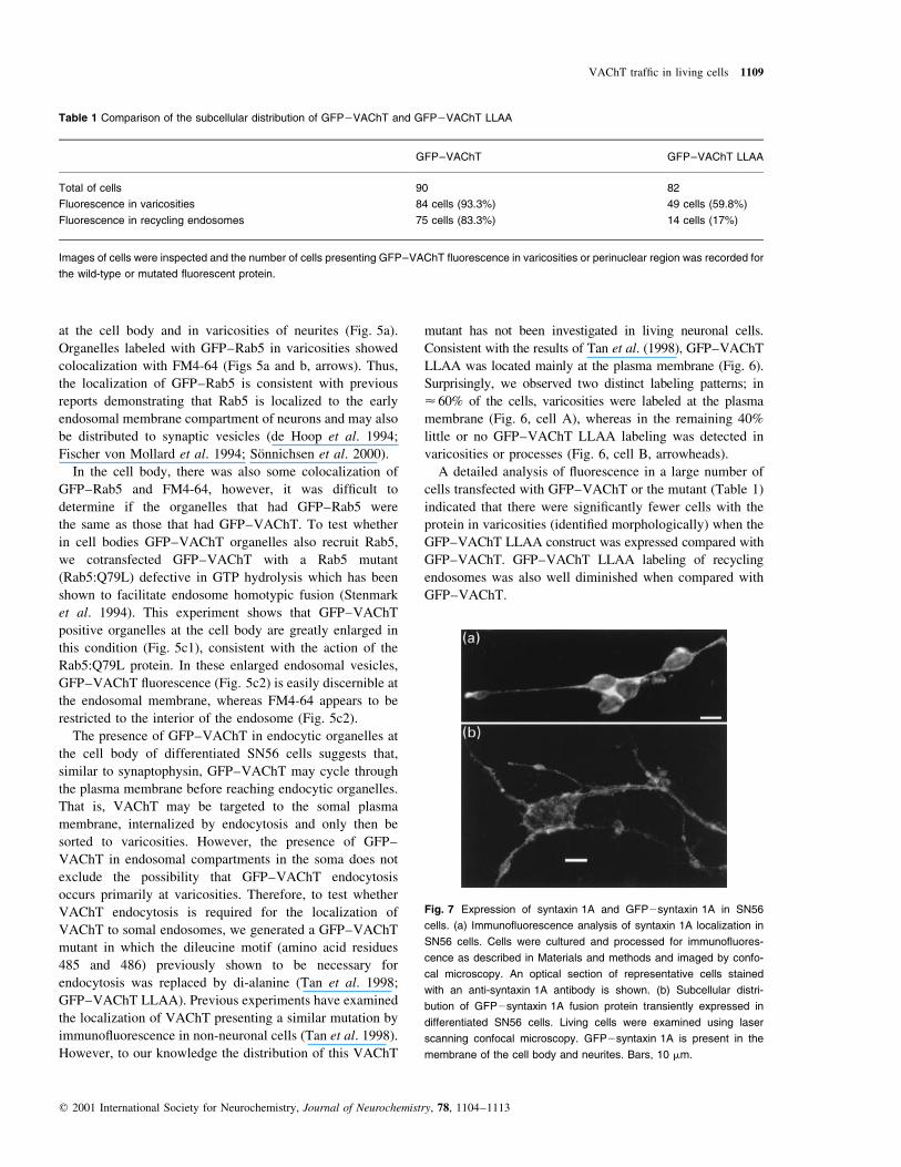
Fig. 7 Expression of syntaxin 1A and GFP2syntaxin 1A in SN56
cells. (a) Immuno¯uorescence analysis of syntaxin 1A localization in
SN56 cells. Cells were cultured and processed for immuno¯uores-
cence as described in Materials and methods and imaged by confo-
cal microscopy. An optical section of representative cells stained
with an anti-syntaxin 1A antibody is shown. (b) Subcellular distri-
bution of GFP2syntaxin 1A fusion protein transiently expressed in
differentiated SN56 cells. Living cells were examined using laser
scanning confocal microscopy. GFP2syntaxin 1A is present in the
membrane of the cell body and neurites. Bars, 10 mm.
Table 1 Comparison of the subcellular distribution of GFP2VAChT and GFP2VAChT LLAA
GFP±VAChT GFP±VAChT LLAA
Total of cells 90 82
Fluorescence in varicosities 84 cells (93�.3%) 49 cells (59�.8%)
Fluorescence in recycling endosomes 75 cells (83�.3%) 14 cells (17%)
Images of cells were inspected and the number of cells presenting GFP±VAChT ¯uorescence in varicosities or perinuclear region was recorded for
the wild-type or mutated ¯uorescent protein.
VAChT traf®c in living cells 1109
q 2001 International Society for Neurochemistry, Journal of Neurochemistry, 78, 1104±1113

We measured the ¯uorescence intensity of the brightest
region at the plasma membrane in the soma and neurites of
cells expressing GFP±VAChT LLAA to ascertain whether
the protein was evenly distributed or whether one of these
compartments presented preferential accumulation of the
protein. We excluded from the analysis cells presenting no
¯uorescence in neurites, i.e. 40% of the cells. The remaining
cells expressing the mutant were analyzed and the
¯uorescence intensity was compared individually. The data
indicate that ¯uorescence in the soma plasma membrane
was on average 36% higher than in neurites (Fig. 8a).
In order to test whether this analysis would discriminate
if the ¯uorescence intensity in plasma membrane of the
cell body and varicosities were homogeneous, we analyzed
the expression pattern of GFP2syntaxin 1A. We chose
syntaxin 1A because this plasma membrane protein is
present in axons and soma of cultured hippocampal neurons
without evidence of preferential accumulation (Garcia et al.
1995; reviewed by Galli et al. 1995).
Immuno¯uorescence analysis of SN56 cells shows that
endogenous syntaxin 1A is found at the plasma membrane
(Fig. 7a) in a pattern similar to that described for cultured
hippocampal neurons (Galli et al. 1995; Garcia et al. 1995).
Cells transfected with a GFP±syntaxin 1A construct showed
the same subcellular distribution of endogenous syntaxin
(Fig. 7b). Importantly, in contrast to the results obtained
with the GFP±VAChT LLAA, the intensity of ¯uorescence
at the plasma membrane for GFP±syntaxin was identical for
the brightest regions of the cell body and processes of SN56
cells (Fig. 8b).
We also analyzed whether GFP±VAChT ¯uorescence
was similar in early endosomes in the cell body and
organelles in varicosities (endocytic organelles or synaptic
vesicles). GFP±VAChT labeled organelles in varicosities
were on average 33% more ¯uorescent than early endo-
somes in the cell body. This suggests that contrary to the
experiments with the mutant GFP±VAChT LLAA, which is
more concentrated in the cell body, GFP±VAChT appears to
be more concentrated in varicosities.
Discussion
In neurons, synaptic vesicle genesis appears to occur at
nerve terminals (reviewed by Hannah et al. 1999). It has
been suggested that distinct synaptic proteins travel via
tubulovesicular or irregular organelles by axonal transport to
their ®nal destination (Nakata et al. 1998; Ahmari et al.
2000). In one of the models proposed, proteins travel
independently using tubulovesicular organelles from the
TGN to nerve endings (Nakata et al. 1998). However, recent
results suggest that synaptic proteins may travel together, as
a `pre-assembled' proto-terminal (Ahmari et al. 2000).
Whereas in the ®rst proposal, the participation of cell body
endocytic organelles is supposedly negligible (Nakata et al.
1998), the suggestion of formation of proto-terminals may
involve the participation of endosomes and sophisticated
sorting process in the soma. In agreement with the latter
model of synaptic vesicle protein traf®cking, some synaptic
Fig. 8 Quanti®cation of ¯uorescence intensity of GFP2VAChT
LLAA, GFP2syntaxin 1A and GFP2VAChT in the soma or neurite
of SN56 cells. (a) Average ¯uorescence intensity of the brightest
region in the cell body membrane for 30 randomly chosen cells
expressing GFP2VAChT LLAA (dark bars) and average of the ¯uor-
escence intensity of the brightest region in the membrane of neurites
(clear bars). The intensity of ¯uorescence of GFP2VAChT LLAA in
neurites is < 36% less than that at the cell body (see details in
methodology). This difference is statistically signi®cant ( p , 0.05,
Student's t-test). (b) Similar analysis was performed in 19 cells
expressing GFP2syntaxin 1A. The ¯uorescence intensity in the
membrane of the cell body or neurites is similar; error bars represent
SEM. (c) Analysis of ¯uorescence intensity of GFP2VAChT present
in the brightest early endosome in the cell body and in endocytic
organelles in varicosities (n � 90). Fluorescence in varicosities was
33% higher than in early endosomes ( p , 0.05, Student's t-test).
1110 M. S. Santos et al.
q 2001 International Society for Neurochemistry, Journal of Neurochemistry, 78, 1104±1113

vesicle proteins sort to endosomes in non-neuronal cells
(reviewed by Hannah et al. 1999).
The VAChT provides an attractive model protein to both
characterize and understand the pathways involved with
synaptic vesicle biogenesis, as the transporter is predomi-
nantly distributed to synaptic vesicles (Gilmor et al. 1996;
Weihe et al. 1996; Liu and Edwards 1997; reviewed by
Erickson and Varoqui 2000). Moreover, the determinants
required for synaptic vesicle targeting of VAChT appear to
be localized at the C-terminal domain of the transport
protein (Varoqui and Erickson 1998; Krantz et al. 2000). In
this study, we used a GFP-tagged version of the transporter
and a cholinergic model system to explore the pathways
followed by the protein in living cells. GFP±VAChT was
found predominantly in endocytic organelles at the cell body
and in varicosities of neurites from SN56 cells. The protein
labeled punctated organelles that could be double stained by
short incubation periods with FM4-64, a general endocytic
and synaptic vesicle marker. Mutation of the dileucine motif
at positions 485±486 to alanine changed the traf®cking of
the recombinant protein, suggesting that VAChT amino
acid sequences are primarily responsible for targeting of
GFP±VAChT in SN56 cells.
Previous experiments have shown that brain cholinergic
neurons present somatic staining for VAChT at a perinuclear
region (SchaÈfer et al. 1998). Strong soma staining for
endogenous VAChT is also observed in SN56 cells (Barbosa
et al. 1999). In addition, in PC12 cells stably expressing low
amounts of VAChT, the protein is also found in intracellular
structures at a perinuclear location and has been found in
endosomes by subcellular fractionation (Liu and Edwards
1997). Hence, tagging VAChT with GFP does not appear to
impair or change the traf®c of the protein in SN56 cells. It is
thus unlikely that the presence of GFP±VAChT in endo-
somes is a result of transient overexpression of the protein.
Endosomal populations show differences in their mole-
cular composition and localization in cells (SoÈnnichsen et al.
2000). In polarized cells, endosomal sorting of proteins can
help to distribute them to their appropriate subcellular
location and to maintain cell polarity (reviewed by Winckler
and Mellman 1999). In our experiments, the incubation of
SN56 cells with Tfn-TxR for 5 and 20 min was used to
discriminate the identity of early and recycling endosomal
vesicular populations as described previously in other model
systems (SoÈnnichsen et al. 2000). We found that there was
clear colocalization of GFP±VAChT and Tfn-TxR in
populations of early and recycling endosomes at the cell
body. The localization of GFP±VAChT in early endosomes
was also con®rmed by cotransfection of GFP±VAChT with
a Rab5 mutant constitutively activated that enhances
homotypic endosome fusion. The perinuclear presence of
GFP±VAChT seems to indicate that at least part of the
protein is found in the recycling endosomal population
(labeled with Tfn-TxR for 20 min), but it does not discount
the possibility that part of the perinuclear labeling is
composed of newly synthesized protein passing through
the Golgi.
GFP±Rab5 stained FM4-64 positive organelles in both
varicosities and neurites of SN56 cells. It appears that
FM4-64 labeling in varicosities is primarily contained in
organelles that recruit Rab5. As GFP±VAChT in varicos-
ities is present in organelles that are labeled with FM4-64,
we conclude that GFP±VAChT is localized to Rab5-positive
organelles.
It is not clear at present whether differentiated SN56 cells
would recognize axonal targeting signals, but several lines
of evidence support the notion that these cells present a
neuronal phenotype. Differentiated SN56 cells project long
neurites containing varicosities, and this is dependent on
dynamin 1 activity (J. Barbosa Jr et al. unpublished
observations), as reported for extension of axons from
cultured neurons (Masur et al. 1990; Torre et al. 1994). The
majority of organelles present in varicosities of SN cells are
identical to true synaptic vesicles (Hammond et al. 1990).
Moreover, varicosities accumulate synaptotagmin, synapto-
physin and also VAChT as observed by confocal micro-
scopy (Barbosa et al. 1999), suggesting that the vesicles
contain the necessary proteins involved in transmitter
release. This study has shown that the organelles present
in varicosities (presumably synaptic vesicles seen by
Hammond et al. 1990) undergo cycles of spontaneous
exo-endocytosis and recruit Rab5, a marker or early
endosomes which is also found in synaptic vesicles.
Together these observations provide support for the use of
SN56 cells as a model to study traf®cking of cholinergic
proteins. However, it should be noted that SN56 cells are a
model of immature cholinergic neurons (Hammond et al.
1990), and thus may present small differences in exo-
endocytosis activity.
The fact that GFP±VAChT is present in early and
recycling endosomes in the cell body and in early
endosomes or synaptic vesicles in varicosities does not
reveal the site(s) of GFP±VAChT endocytosis in SN56
cells. However, two alternative mechanisms can be
proposed. VAChT could leave the TGN in tubulovesicular
organelles that are targeted to growth cones and varicosities,
and by constitutive exo-endocytosis the protein would reach
synaptic vesicles before being returned to endosomes in the
cell body by retrograde transport (Nakata et al. 1998). One
alternative view is that VAChT leaves the TGN in
constitutive vesicles, fuses to the plasma membrane at the
cell body and then internalizes to early and recycling
endosomes before sorting to organelles that are targeted to
growth cones and varicosities. By following the localization
of GFP±VAChT LLAA, we gained novel insights on the
journey of VAChT. The mutated protein appeared to
preferentially accumulate at the cell body, although the
presence of the protein in neurites was also detected. There
VAChT traf®c in living cells 1111
q 2001 International Society for Neurochemistry, Journal of Neurochemistry, 78, 1104±1113

were some striking features observed with this mutant. First,
labeling in neurites was uniformly distributed at the plasma
membrane, which is in sharp contrast to the predominant
labeling of varicosities and punctated structures seen with
GFP±VAChT. Secondly, there were fewer cells with GFP
labeling in varicosities when the mutant was expressed
compared with the wild-type protein. Thirdly, just a small
percentage of cells expressing the mutant presented some
degree of perinuclear labeling, suggesting that GFP±
VAChT LLAA failed to reach recycling endosomes. Finally,
the maximum ¯uorescence intensity in neurites for the
mutant was 36% lower than that at the plasma membrane in
the soma. The lack of difference for the ¯uorescence
measurements with GFP±syntaxin 1A suggests that prefer-
ential accumulation of GFP±VAChT in the somal plasma
membrane is not an artifact. Moreover, this result is in sharp
contrast with the accumulation of GFP±VAChT in varicos-
ities when compared with early endosome ¯uorescence
(33% more GFP±VAChT in varicosities). Thus, interference
with GFP±VAChT endocytosis will lead to signi®cant
accumulation of the protein in the soma. We thus conclude
that part of GFP±VAChT primarily recycles at the cell body
before targeting to varicosities. However, we cannot discount
the existence of other mechanisms that will carry some of the
protein directly to neurites without passing by soma endocytic
intermediates as suggested by Nakata et al. (1998). Com-
plementary approaches such as metabolic labeling and
subcellular fractionation may have to be used in future
experiments to discriminate between these two possibilities.
Dileucine motifs similar to that found in VAChT have
been shown to interact with adaptor protein complexes 1
and 2 (AP1 and AP2, Dietrich et al. 1997), in particular
with the b subunit (Rapoport et al. 1998). Interaction with
other AP complexes (AP3 for example) has also been
suggested (Blagoveshchenskaya et al. 1999; Bonifacino and
Dell'Angelica 1999). The lack of VAChT mutant internal-
ization perhaps results from impaired interaction of VAChT
with the AP2 complex, as VAChT remains in the plasma
membrane. However, it is possible that, as suggested before,
the dileucine motif in VAChT may have multiple functions
(Krantz et al. 2000).
The lack of intracellular labeling with the GFP±VAChT
LLAA also suggests that clathrin-mediated endocytosis will
have a fundamental role in the incorporation of VAChT into
synaptic vesicles. Internalization of plasma membrane
occurs in a very fast way in varicosities of SN56 cells, as
observed by FM4-64 labeling. However, GFP±VAChT
LLAA is excluded from internalization. Thus, it seems
that no alternative mechanism (such as non-clathrin
mediated endocytosis) is available for the internalization
of GFP±VAChT.
We have extended previous observations on the role of
cell body organelles for synaptic vesicle protein traf®cking
in living cells. Our data indicate that in cholinergic cells, at
least part of VAChT may traf®c to varicosities via
endosomal intermediates, after passage through the plasma
membrane in the neuronal cell body. Much discussion has
been presented in the last few years in favor (reviewed by
Cremona and De Camilli 1997) or against (reviewed by
Palfrey and Artalejo 1998) the role of clathrin mediated
process in synaptic vesicle recycling. Our results suggest,
albeit indirectly, that formation of synaptic vesicles
competent for ACh release will depend on the interaction
of VAChT with clathrin associated proteins.
Acknowledgements
This work received support from CNPq, FAPEMIG, PRONEX,
PADCT and a CAPES fellowship to M.S.S. We thank Ms A. A.
Pereira and E. E. P. Silva for technical help. We thank Drs
Bruce Wainer, Marc Caron and Ingo Leibiger for the gift of
cells and plasmids, respectively.
References
Ahmari S. E., Buchanan J. and Smith S. J. (2000) Assembly of
presynaptic active zones from cytoplasmic transport packets. Nat.
Neurosci. 3, 445±451.
Ausubel F. M., Brent R., Kingston R. E., Moore D. D., Seidman J. G.,
Smith J. A. and Struhl K. (1991) Mutagenesis of cloned DNA, in
Current Protocols in Molecular Biology, Section 8.5, 5±9.Greene
Publishing Associates and Wiley-Interscience, New York.
Barbosa J. Jr, Clarizia A. D., Gomez M. V., Romano-Silva M. A., Prado
V. F. and Prado M. A. M. (1997) Effect of protein kinase C
activation on the release of [3H]acetylcholine in the presence of
vesamicol. J. Neurochem. 69, 2608±2611.
Barbosa J. Jr, Massensini A. R., Santos M. S., Meireles S. I., Gomez
R. S., Gomez M. V., Romano-Silva M. A., Prado V. F. and Prado
M. A. M. (1999) Expression of the vesicular acetylcholine
transporter, proteins involved in exocytosis, and functional
calcium signaling in varicosities and soma of a murine septal
cell line. J. Neurochem. 73, 1881±1893.
Blagoveshchenskaya A. D., Hewitt E. W. and Cutler D. F. (1999)
Di-leucine signals mediate targeting of tyrosinase and synapto-
tagmin to synaptic-like microvesicles within PC12 cells. Mol.
Biol. Cell 10, 3979±3990.
Bonifacino J. S. and Dell'Angelica E. C. (1999) Molecular bases for the
recognition of tyrosine-based sorting signals. J. Cell Biol. 145,
923±926.
Chilcote T. J., Galli T., Mundigl O., Edelmann L., McPherson P. S.,
Takei K. and De Camilli P. (1995) Cellubrevin and synapto-
brevins: similar subcellular localization and biochemical proper-
ties in PC12 cells. J. Cell Biol. 129, 219±231.
Cho G. W., Kim M. H., Chai Y. G., Gilmor M. L., Levey A. I. and
Hersh L. B. (2000) Phosphorylation of the rat vesicular acetylcho-
line transporter. J. Biol. Chem. 275, 19942±19948.
Cremona O. and De Camilli P. (1997) Synaptic vesicle endocytosis.
Curr. Opin. Neurobiol. 7, 323±330.
Dietrich J., Kastrup J., Nielsen B. L., Odum N. and Geisler C. (1997)
Regulation and function of the CD3gamma DxxxLL motif: a
binding site for adaptor protein-1 and adaptor protein-2 in vitro.
J. Cell Biol. 138, 271±281.
Erickson J. D. and Varoqui H. (2000) Molecular analysis of vesicular
amine transporter function and targeting to secretory organelles.
FASEB J. 14, 2450±2458.
1112 M. S. Santos et al.
q 2001 International Society for Neurochemistry, Journal of Neurochemistry, 78, 1104±1113

FauÂndez V., Horng J. T. and Kelly R. B. (1998) A function for the AP3
coat complex in synaptic vesicle formation from endosomes. Cell
93, 423±432.
Fischer von Mollard G., Stahl B., Walch-Solimena C., Takei K.,
Daniels L., Khoklatchev A., De Camilli P., SuÈdhof T. C. and Jahn
R. (1994) Localization of Rab5 to synaptic vesicles identi®es
endosomal intermediate in synaptic vesicle recycling pathway.
Eur. J. Cell Biol. 65, 319±326.
Galli T., Garcia E. P., Mundigl O., Chilcote T. J. and De Camilli P.
(1995) v- and t-SNAREs in neuronal exocytosis: a need for
additional components to de®ne sites of release. Neuropharma-
cology 34, 1351±1360.
Garcia E. P., McPherson P. S., Chilcote T. J., Takei K. and De Camilli P.
(1995) rbSec1A and B colocalize with syntaxin 1 and SNAP-25
throughout the axon, but are not in a stable complex with syntaxin.
J. Cell Biol. 129, 105±120.
Gilmor M. L., Nash N. R., Roghani A., Edwards R. H., Yi H., Hersch
S. M. and Levey A. I. (1996) Expression of the putative vesicular
acetylcholine transporter in rat brain and localization in
cholinergic synaptic vesicles. J. Neurosci. 16, 2179±2190.
Grote E. and Kelly R. B. (1996) Endocytosis of VAMP is facilitated by
a synaptic vesicle targeting signal. J. Cell Biol. 132, 537±547.
Hammond D. N., Lee H. J., Tonsgard J. H. and Wainer B. H. (1990)
Development and characterization of clonal cell lines derived
from septal cholinergic neurons. Brain Res. 512, 190±200.
Hannah M. J., Schimd A. A. and Huttner W. B. (1999) Synaptic vesicle
biogenesis. Annu. Rev. Cell. Biol. 15, 733±798.
de Hoop M. J., Huber L. A., Stenmark H., Williamson E., Zerial M.,
Parton R. G. and Dotti C. G. (1994) The involvement of the small
GTP-binding protein Rab5a in neuronal endocytosis. Neuron 13,
11±22.
Krantz D. E., Waites C., Oorschot V., Liu Y., Wilson R. I., Tan P. K.,
Klumperman J. and Edwards R. H. (2000) A phosphorylation site
regulates sorting of the vesicular acetylcholine transporter to
dense core vesicles. J. Cell Biol. 149, 379±396.
Liu Y. and Edwards R. H. (1997) Differential localization of vesicular
acetylcholine and monoamine transporters in PC12 cells but not
CHO cells. J. Cell Biol. 139, 907±916.
Masur S. K., Kim Y. T. and Wu C. F. (1990) Reversible inhibition of
endocytosis in cultured neurons from the Drosophila temperature-
sensitive mutant shibirets1. J. Neurogenet. 6, 191±206.
Nakata T., Terada S. and Hirokawa N. (1998) Visualization of the
dynamics of synaptic vesicle and plasma membrane proteins in
living axons. J. Cell Biol. 140, 659±674.
Palfrey H. C. and Artalejo C. R. (1998) Vesicle recycling revisited: rapid
endocytosis may be the ®rst step. Neuroscience 83, 969±989.
Parsons S. M. (2000) Transport mechanisms in acetylcholine and
monoamine storage. FASEB J. 14, 2423±2434.
Pothos E. N., Larsen K. E., Krantz D. E., Liu Yj Haycock J. W., Setlik
W., Gershon M. D., Edwards R. H. and Sulzer D. (2000) Synaptic
vesicle transporter expression regulates vesicle phenotype and
quantal size. J. Neurosci. 20, 7297±7306.
Rapoport I., Chen Y. C., Cupers P., Shoelson S. E. and Kirchhausen T.
(1998) Dileucine-based sorting signals bind to the beta chain of
AP-1 at a site distinct and regulated differently from the tyrosine-
based motif-binding site. EMBO J. 17, 2148±2155.
ReÂgnier-Vigouroux A., Tooze S. A. and Huttner W. B. (1991) Newly
synthesized synaptophysin is transported to synaptic-like micro-
vesicles via constitutive secretory vesicles and the plasma
membrane. EMBO J. 10, 3589±3601.
Sanger F., Nicklen S. and Coulson A. R. (1977) DNA sequencing with
chain-terminating inhibitors. Proc. Natl Acad. Sci. USA 74,
5463±5467.
SchaÈfer M. K., Eiden L. E. and Weihe E. (1998) Cholinergic neurons
and terminal ®elds revealed by immunohistochemistry for the
vesicular acetylcholine transporter. I. Central nervous system.
Neuroscience 84, 331±359.
Shi G., FauÂndez V., Roos J., Dell'Angelica E. C. and Kelly R. B. (1998)
Neuroendocrine synaptic vesicles are formed in vitro by both
clathrin-dependent and clathrin-independent pathways. J. Cell
Biol. 143, 947±955.
SoÈnnichsen B., De Renzis S., Nielsen E., Rietdorf J. and Zerial M.
(2000) Distinct membrane domains on endosomes in the recycling
pathway visualized by multicolor imaging of Rab4, Rab5, and
Rab11. J. Cell Biol. 149, 901±914.
Stenmark H., Parton R. G., Steele-Mortimer O., Lutcke A., Gruenberg J.
and Zerial M. (1994) Inhibition of rab5 GTPase activity stimulates
membrane fusion in endocytosis. EMBO J. 13, 1287±1296.
Tan P. K., Waites C., Liu Y., Krantz D. E. and Edwards R. H. (1998) A
leucine-based motif mediates the endocytosis of vesicular
monoamine and acetylcholine transporters. J. Biol. Chem. 273,
17351±17360.
Torre E., McNiven M. A. and Urrutia R. (1994) Dynamin 1 antisense
oligonucleotide treatment prevents neurite formation in cultured
hippocampal neurons. J. Biol. Chem. 269, 32411±32417.
Towbin H., Staehelin T. and Gordon J. (1979) Electrophoretic transfer
of proteins from polyacrylamide gels to nitrocellulose sheets,
procedure and some applications. Proc. Natl Acad. Sci. USA 76,
4350±4354.
Varoqui H. and Erickson J. D. (1998) The cytoplasmic tail of the
vesicular acetylcholine transporter contains a synaptic vesicle
targeting signal. J. Biol. Chem. 273, 9094±9098.
Weihe E., Tao-Cheng J. H., Schafer M. K., Erickson J. D. and Eiden
L. E. (1996) Visualization of the vesicular acetylcholine
transporter in cholinergic nerve terminals and its targeting to a
speci®c population of small synaptic vesicles. Proc. Natl Acad.
Sci. USA 93, 3547±3552.
West A. E., Neve R. L. and Buckley K. M. (1997) Targeting of the
synaptic vesicle protein synaptobrevin in the axon of cultured
hippocampal neurons: evidence for two distinct sorting steps.
J. Cell Biol. 139, 917±927.
Winckler B. and Mellman I. (1999) Neuronal polarity: controlling the
sorting and diffusion of membrane components. Neuron 23,
637±640.
Yamashiro D. J. and Max®eld F. R. (1984) Acidi®cation of endocytic
compartments and the intracellular pathways of ligands and
receptors. J. Cell Biochem. 26, 231±246.
Yang S. N., Larsson O., Branstrom R., Bertorello A. M., Leibiger B.,
Leibiger I. B., Moede T., Kohler M., Meister B. and Berggren
P. O. (1999) Syntaxin 1 interacts with the L(D) subtype of
voltage-gated Ca(21) channels in pancreatic beta cells. Proc. Natl
Acad. Sci. USA 96, 10164±10169.
VAChT traf®c in living cells 1113
q 2001 International Society for Neurochemistry, Journal of Neurochemistry, 78, 1104±1113