Pulmonary Inflammation Is Regulated by the Levels of the Vesicular Acetylcholine Transporter
22
RESEARCH ARTICLE Pulmonary Inflammation Is Regulated by the Levels of the Vesicular Acetylcholine Transporter Nathalia M. Pinheiro 1☯ , Claudia J. C. P. Miranda 1☯ , Adenir Perini 1 , Niels O. S. Câmara 3 , Soraia K. P. Costa 4 , Maria Isabel C. Alonso-Vale 2 , Luciana C. Caperuto 2 , Iolanda F. L. C. Tibério 1 , Marco Antônio M. Prado 5 , Mílton A. Martins 1 , Vânia F. Prado 5 , Carla M. Prado 1,2 * 1 Department of Medicine, School of Medicine, University of Sao Paulo, São Paulo, Brazil, 2 Department of Biological Science, Federal University of Sao Paulo, Diadema, Brazil, 3 Department of Immunology, Institute of Biomedical Sciences, University of Sao Paulo, São Paulo, Brazil, 4 Department of Pharmacology Institute of Biomedical Sciences, University of Sao Paulo, São Paulo, Brazil, 5 Molecular Medicine Group, Robarts Research Institute, Department of Physiology & Pharmacology and Department of Anatomy & Cell Biology, University of Western Ontario, London, Canada ☯ These authors contributed equally to this work. * [email protected] Abstract Acetylcholine (ACh) plays a crucial role in physiological responses of both the central and the peripheral nervous system. Moreover, ACh was described as an anti-inflammatory me- diator involved in the suppression of exacerbated innate response and cytokine release in various organs. However, the specific contributions of endogenous release ACh for inflam- matory responses in the lung are not well understood. To address this question we have used mice with reduced levels of the vesicular acetylcholine transporter (VAChT), a protein required for ACh storage in secretory vesicles. VAChT deficiency induced airway inflamma- tion with enhanced TNF-α and IL-4 content, but not IL-6, IL-13 and IL-10 quantified by ELISA. Mice with decreased levels of VAChT presented increased collagen and elastic fi- bers deposition in airway walls which was consistent with an increase in inflammatory cells positive to MMP-9 and TIMP-1 in the lung. In vivo lung function evaluation showed airway hyperresponsiveness to methacholine in mutant mice. The expression of nuclear factor- kappa B (p65-NF-kB) in lung of VAChT-deficient mice were higher than in wild-type mice, whereas a decreased expression of janus-kinase 2 (JAK2) was observed in the lung of mu- tant animals. Our findings show the first evidence that cholinergic deficiency impaired lung function and produce local inflammation. Our data supports the notion that cholinergic sys- tem modulates airway inflammation by modulation of JAK2 and NF-kB pathway. We pro- posed that intact cholinergic pathway is necessary to maintain the lung homeostasis. PLOS ONE | DOI:10.1371/journal.pone.0120441 March 27, 2015 1 / 22 OPEN ACCESS Citation: Pinheiro NM, Miranda CJCP, Perini A, Câmara NOS, Costa SKP, Alonso-Vale MIC, et al. (2015) Pulmonary Inflammation Is Regulated by the Levels of the Vesicular Acetylcholine Transporter. PLoS ONE 10(3): e0120441. doi:10.1371/journal. pone.0120441 Academic Editor: Xiao Su, Chinese Academy of Sciences, CHINA Received: October 28, 2014 Accepted: January 22, 2015 Published: March 27, 2015 Copyright: © 2015 Pinheiro et al. This is an open access article distributed under the terms of the Creative Commons Attribution License, which permits unrestricted use, distribution, and reproduction in any medium, provided the original author and source are credited. Data Availability Statement: All relevant data are within the paper. Funding: This study was supported by Sao Paulo State Research Foundation (FAPESP-2008/55359-5) and National Council for Technologic and Development (CNPq) (471224/2009-0), Brazil. The funders had no role in study design, data collection and analysis, decision to publish, or preparation of the manuscript. Competing Interests: The authors have declared that no competing interests exist.
Transcript of Pulmonary Inflammation Is Regulated by the Levels of the Vesicular Acetylcholine Transporter

RESEARCH ARTICLE
Pulmonary Inflammation Is Regulated by theLevels of the Vesicular AcetylcholineTransporterNathalia M. Pinheiro1☯, Claudia J. C. P. Miranda1☯, Adenir Perini1, Niels O. S. Câmara3,Soraia K. P. Costa4, Maria Isabel C. Alonso-Vale2, Luciana C. Caperuto2, Iolanda F. L.C. Tibério1, Marco Antônio M. Prado5, Mílton A. Martins1, Vânia F. Prado5, CarlaM. Prado1,2*
1 Department of Medicine, School of Medicine, University of Sao Paulo, São Paulo, Brazil, 2 Department ofBiological Science, Federal University of Sao Paulo, Diadema, Brazil, 3 Department of Immunology, Instituteof Biomedical Sciences, University of Sao Paulo, São Paulo, Brazil, 4 Department of Pharmacology Instituteof Biomedical Sciences, University of Sao Paulo, São Paulo, Brazil, 5 Molecular Medicine Group, RobartsResearch Institute, Department of Physiology & Pharmacology and Department of Anatomy & Cell Biology,University of Western Ontario, London, Canada
☯ These authors contributed equally to this work.* [email protected]
AbstractAcetylcholine (ACh) plays a crucial role in physiological responses of both the central and
the peripheral nervous system. Moreover, ACh was described as an anti-inflammatory me-
diator involved in the suppression of exacerbated innate response and cytokine release in
various organs. However, the specific contributions of endogenous release ACh for inflam-
matory responses in the lung are not well understood. To address this question we have
used mice with reduced levels of the vesicular acetylcholine transporter (VAChT), a protein
required for ACh storage in secretory vesicles. VAChT deficiency induced airway inflamma-
tion with enhanced TNF-α and IL-4 content, but not IL-6, IL-13 and IL-10 quantified by
ELISA. Mice with decreased levels of VAChT presented increased collagen and elastic fi-
bers deposition in airway walls which was consistent with an increase in inflammatory cells
positive to MMP-9 and TIMP-1 in the lung. In vivo lung function evaluation showed airway
hyperresponsiveness to methacholine in mutant mice. The expression of nuclear factor-
kappa B (p65-NF-kB) in lung of VAChT-deficient mice were higher than in wild-type mice,
whereas a decreased expression of janus-kinase 2 (JAK2) was observed in the lung of mu-
tant animals. Our findings show the first evidence that cholinergic deficiency impaired lung
function and produce local inflammation. Our data supports the notion that cholinergic sys-
tem modulates airway inflammation by modulation of JAK2 and NF-kB pathway. We pro-
posed that intact cholinergic pathway is necessary to maintain the lung homeostasis.
PLOS ONE | DOI:10.1371/journal.pone.0120441 March 27, 2015 1 / 22
OPEN ACCESS
Citation: Pinheiro NM, Miranda CJCP, Perini A,Câmara NOS, Costa SKP, Alonso-Vale MIC, et al.(2015) Pulmonary Inflammation Is Regulated by theLevels of the Vesicular Acetylcholine Transporter.PLoS ONE 10(3): e0120441. doi:10.1371/journal.pone.0120441
Academic Editor: Xiao Su, Chinese Academy ofSciences, CHINA
Received: October 28, 2014
Accepted: January 22, 2015
Published: March 27, 2015
Copyright: © 2015 Pinheiro et al. This is an openaccess article distributed under the terms of theCreative Commons Attribution License, which permitsunrestricted use, distribution, and reproduction in anymedium, provided the original author and source arecredited.
Data Availability Statement: All relevant data arewithin the paper.
Funding: This study was supported by Sao PauloState Research Foundation (FAPESP-2008/55359-5)and National Council for Technologic andDevelopment (CNPq) (471224/2009-0), Brazil. Thefunders had no role in study design, data collectionand analysis, decision to publish, or preparation ofthe manuscript.
Competing Interests: The authors have declaredthat no competing interests exist.
IntroductionPulmonary diseases such as asthma, acute lung inflammation and chronic obstructive pulmo-nary disease (COPD) represent major threats to human health. In common, they involve com-plex immune responses in which inflammatory and epithelial cells release elevated levels ofpro-inflammatory Th1/Th2 cytokines such as IL-6, TNF-α, IL-4 and also the regulatory cyto-kine IL-10 [1]. The persistence of inflammatory processes associated with an imbalance ofmetalloproteinases (MMP) and their tissue inhibitors (TIMP) induce pulmonary structuralchanges including fibroblasts/myofibroblasts activation, and extracellular matrix (ECM) fibersdeposition [2] which impair lung function.
The cholinergic anti-inflammatory pathway is a modulator of innate immune responses byneuronal and non-neuronal mechanisms [3–6]. This statement is supported by the fact thatduring the inflammatory process, ACh released by the vagus nerve and acting via α7 nicotinicreceptors (α7nAChR) present on macrophage and other immune cells, seems to inhibit cyto-kine production and thus contributing to counteract an ongoing state of inflammation [6–9].α7nAChR activation not only inhibits the nuclear translocation of transcription factor NF-kB[10] but it also activates the Janus kinase-2 and signal transducer and activator of transcription3 pathway (JAK-STAT3). This pathway is involved in the regulation of several cellular func-tions and is part of the essential chemical signaling to induce cytokine production. However,JAK2-STAT3 can in turn counteracts inflammation by regulating the activity of suppressor ofcytokine signaling 3 (SOCS-3) [11].
ACh is synthesized, stored, and released from cholinergic nerve terminals [12, 13] and regu-lates physiological functions by interaction with two classes of cholinergics receptors, nicotinicand muscarinic [14]. ACh storage in secretory vesicles depends on the activity of the vesicularacetylcholine transporter (VAChT) [15] which is absolutely required for ACh release in the pe-ripheral and central nervous system [16, 17]. Importantly, ACh is also synthesized and releasedby non-neuronal cells, including immune and epithelial cells [18–22] and the exactly mecha-nisms involved in non-neuronal ACh release is not fully elucidated. VAChT and other cholin-ergic components are expressed in non-neuronal cells [12, 19, 20, 23] and at least incardiomyocytes and in α-cells of the pancreas, the ACh release seems to be VAChT-dependent[12, 24].
In the lung, ACh is known to be released from parasympathetic nerve fibers and to inducebronchoconstriction by biding to muscarinic receptors present in airway smooth muscle andglands [25]. ACh release from lung epithelial cells seems to depend on organic cation trans-porters (OCT 1 and OCT2) [21, 26]. However, VAChT is expressed in airways and VAChT-positive neurons were found in lung [19, 27]. Rodents with acute lung inflammation showedreduced expression of the cholinergic markers choline acetyltransferase [28], high affinity cho-line transporter (CHT1) and VAChT in the lung, suggesting a down-regulation of cholinergicactivity in asthma physiopathology [19, 21]. Additionally, it has been suggested that the cholin-ergic system might participate in the pathogenesis of some lung diseases [19] since vagotomyworsened lung inflammation whereas pharmacological stimulation of α7nAChR amelioratelung inflammation in models of acute lung injury [4, 5]. However, it remains unclear whetherlung inflammation is regulated by levels of VAChT.
Here we addressed, using genetic manipulation of VAChT levels [homozygous VAChTknockdown mice (VAChT KDHOM) [16], whether release of ACh mediated by this transporteris involved in regulation of lung inflammatory responses as well as lung remodeling and hyper-responsiveness in mice. We also evaluated if VAChT deficiency affected the expression levelsof p65-NF-kB and JAK2-STAT3 pathway.
Long-Term Cholinergic Deficiency Induces Lung Inflammation
PLOS ONE | DOI:10.1371/journal.pone.0120441 March 27, 2015 2 / 22

MethodsVAChT mutant mice were generated as previously described in detail [16]. Heterozygous micewere intercrossed to generate VAChT KDHOM mice and wild-type littermate controls (WT)used in these experiments. These mice were knock-down to VAChT gene and present a reduc-tion in 65% in VAChT levels with correspondent decrease in ACh release which causes myas-thenia, cognitive deficits and cardiac dysfunction [15, 17, 29, 30]. The activity of cholineacetyltransferase was not modified in these mice [16]. All mice used in this study received hu-mane care in compliance with the “Guide for Care and Use of Laboratory Animals” (NIH pub-lication 85–23, revised 1985).The animals were kept on a 12 h light/dark cycle in atemperature-controlled room at 21–23°C, with free access to water and food. All experimentsdescribed in this study were approved by the Internal Ethical Committee of Faculty of Medi-cine of the University of São Paulo (São Paulo, Brazil) (Document number 0766/08).
Experimental groupsMale mice (6–8 weeks) of the correct genotypes were randomly assigned to two groups: a. mu-tant homozygous animals (VAChT KDHOM), b. wild-type animals (WT).
Wire-Hang testTo evaluate the motor function and confirm phenotype of mutant mice, the wire-hang test wasperformed. Mice were placed on cage top lid and then were slowly inverted for a maximumtime of 60 s. The time upside down was measured [31].
Pulmonary mechanics evaluationAnimals were anesthetized with thiopental sodium (50 mg.kg-1, i.p.), tracheostomized and con-nected to a ventilator for small animals (FlexiVent, SCIREQ, Montreal, Canada). Animals wereventilated at 150 breaths/min with a tidal volume of 10 mL.kg-1. The jugular vein was dissectedand a polyethylene tubing (Intramedic, Batavia, IL) was tied to infuse different doses of metha-choline (10–3,000 μg/kg). Methacholine was infused each 2 minutes and the data were re-corded at 30 sec after the end of infusion. The respiratory system resistance (Rrs) and elastance(Ers) were measured according to the linear equation of motion of the respiratory system, aspreviously described [32] in baseline and after each dose. We analyzed each value and the per-centage of maximal response related to baseline.
Bronchoalveolar Lavage Fluid (BALF)At the end of the mechanical evaluation, the anterior chest wall was opened, animals were ex-sanguinated via the abdominal aorta and the BALF was collected as previously described [33].The trachea was cannulated and BALF was obtained by washing the airway lumina with 3 x0.5 mL of sterile saline. The recovery volume was over 95% of the instilled fluid and was putinto a test tube on ice. To perform total and differential cell counting, the BALF was centri-fuged at 800x for 10 min and the cell pellet was ressuspended in 0.2 ml of sterile saline. Thetotal number of viable cells was determined in a Neubauer hemocytometer counting chamber.Differential cell counts were performed in cytocentrifuge preparations of BALF (450 rpm for6min) (Cytospin, Cheshire, UK) stained with Diff-Quick (Biochemical Sciences Inc., Swedes-boro, NJ). At least 300 cells were counted according to standard morphologic criteria.
The amount of total protein in BALF was assayed using BradFord’s method (Protein Assay,Bio-rad, California, USA) with bovine serum albumin (BSA, Sigma-Aldrich, Missouri, EUA) as
Long-Term Cholinergic Deficiency Induces Lung Inflammation
PLOS ONE | DOI:10.1371/journal.pone.0120441 March 27, 2015 3 / 22
a standard curve. Absorbance was read at 595 nm (Epoch—Bioteck, Vermont, EUA) and pro-tein levels in mg/mL of BALF were calculated.
Lung MorphometryAfter BALF collect, lungs were infused with 2mL of formaldehyde and were removed en bloc.The lungs were fixed with 4% formaldehyde for 24 h and then transferred to 70% ethanol priorto paraffin embedding. Five-micrometer thick sections from embedded paraffin lungs werestained and submitted to histopathological analysis [34].
Peribronchovascular Edema and Inflammatory CellsWe evaluated the edema area and airway inflammatory cells around the airway using an inte-grating eyepiece with a known area (104 μm2 of total area) in H&E stain section by point-counting technique [34]. To determine the area of edema we counted the number of points ofthe integrating eyepiece falling on areas of peribronchovascular edema in three to four areas ofeach airway wall (3–5 airways per animal, 20 fields per animal). The results were showed asedema/area. To determine the polymorphonuclear (PMN) and mononuclear cells (MN)around the airway (between the bronchial epithelium and the adventitia), we counted the num-ber of points of the integrating eyepiece falling on areas of peribronchial inflammation in threeto four areas of three to five airway wall and the number of cells in this same area (20 fields peranimal). The results were expressed by cell/area. All analyses were performed in randomly se-lected transversely sectioned airways at a magnification of 1,000x.
Extracelular Matrix Fibers DepositionHistological sections were stained for collagen fibers using Sirius-Red (Direct Red 80, C.I.35780, Aldrich, USA) and for elastic fibers using Oxidate Weigert’s Resorcin-Fuchsin. Using aLeica DM4000B microscope (Leica Microsystems, Wetzlar, Germany), a digital camera (LeicaDFC420 Leica Microsystems) and the image analyses software Image Proplus 4.5 (Media Cy-bernetics, Bethesda, USA), we measured collagen and elastic fibers deposition in the area com-pressed between epithelial basal membranes until airway adventitia. Five airways at 400xmagnification were evaluated for each animal. The positive area of collagen and elastic fiberswere expressed as a percentage of the total airway wall area [34].
Immunohistochemistry evaluationImmunohistochemical staining was performed using anti-p-65-NF-kB (1:300), anti-MMP-9(1:600) and anti-TIMP-1 (1:100) antibodies (Santa Cruz Biotechnology, Santa Cruz, CA), bythe biotin–streptavidin–peroxidase method. Using the same point counting technique de-scribed above, we assessed cells positive for p65-NF-kB, MMP-9 and TIMP-1. Counting wasperformed in 20 fields of airway wall samples for each animal (5 airways per animal) at 1,000magnification. Results were expressed as positive cells per area (104μm2) [34, 35]. All morpho-metric analysis was performed by two researchers blind to the genotype.
Cytokine MeasurementsLung were removed and quickly frozen to perform cytokine measurements by ELISA. We testsamples for the presence of TNF-α, IL-6, IL-4, IL-13 e IL-10 in lung homogenate using com-mercially available kits and according to instructions supplied by the manufacturer (R&D Sys-tems, Minneapolis, MN, USA). All cytokines in lung homogenate were expressed in pg ofcytokines/mg of total protein.
Long-Term Cholinergic Deficiency Induces Lung Inflammation
PLOS ONE | DOI:10.1371/journal.pone.0120441 March 27, 2015 4 / 22

Western Blot AnalysisWestern blot was performed using the protocol modified from [36]. Fragments of lung con-taining approximately 20 μg of spinal cord or lung tissue were homogenized in a boiling extrac-tion buffer [10% SDS, 100 mM Tris (pH 7.4), 10 mM EDTA, 10 mM sodium pyrophosphate,100 mM sodium fluoride, 10 mM sodium vanadate] with a Polytron PTA 20S generator(model PT 10/35, Brinkmann Instruments, Inc., Westbury, NY) operated at maximum speedfor 30 sec. The extracts were centrifuged at 15,000xg, 4° C, for 40 min to remove insoluble ma-terial. Protein concentrations of the supernatants were determined by the Bradford assay andan equal amount of total protein from each sample (75 μg) was treated with Laemmli buffercontaining dithiothreitol 100 mM. Samples were heated in water bath for 5 min, after whichthey were subjected to SDS-PAGE (10% bis-acrylamide). Electrotransfer of proteins from gelto nitrocellulose membrane was performed for 90 min at 15V (constant). Nonspecific proteinbinding to nitrocellulose was reduced by pre incubating the membrane overnight at 4° C inblocking buffer (2.5% milk/TBST). Antibodies used for immunoblot were: anti-VAChT(Abcam, Cambridge, Massachusetts) (1:1,000), anti-M2 mAChR (1:10,000), anti- α7 nAChR(1:500), anti-CHT1 (1:1,000), anti-AChE (1:1,000) (Abcam, Cambridge, Massachusetts), anti-JAK-2 (1:1,000) anti-SOCS-3 (1:1,000), anti-STAT-3 (1:1,000), anti-p65-NF-kB (1:200) (SantaCruz Biotechnology, Santa Cruz, CA), anti-phosphorylate STAT-3 (1:1,000) (Cells Signaling,Danver, Massachusetts) and anti-β-actin (1:1,000) (Sigma Aldrich, St. Louis, MO) diluted inblocking buffer overnight at 4° C. The membranes were then washed for 30 min with TBST.Bound antibodies were detected with horseradish peroxidade-conjugated (HRP-conjugated)anti-IgG (1:10,000) and visualized by chemiluminescence using UVItec (UVItec Limited, Cam-bridge, Massachusetts). Band intensities were quantified using UVItec Image Program. TheCHT1 and α7 nAChR, the JAK-2, SOCS-3 and p-65-NF-κB and the STAT3 and p-STAT3were performed in the same gels, respectively, and for this reason the representative β-actin foreach of these group was the same.
RNA extraction, reverse transcription and quantitative real-time PCR(Real-Time qRT-PCR)Total RNA from lung or spinal cord was extracted with Trizol (Invitrogen Life Technologies, Carsl-bad, CA), analyzed for quality on agarose gel and absorbance ratios of 260/280 and 260/230 nm,and reverse transcribed to cDNA using the SuperScript III cDNA kit (Invitrogen Life Technolo-gies). Gene expression was evaluated by real-time qRT-PCR using a Rotor Gene (Qiagen, Roer-mond, Netherlands) and SYBR Green as fluorescent dye (Qiagen) with GAPDH as a housekeepinggene. The reaction conditions were as follows: 95°C for 5 minutes, then 40 cycles of 95°C for 5 sec-onds and 60°C for 10 seconds. PCR products were run on agarose gel to confirm the size of thefragment and specificity of amplification. Primers used and annealing temperatures are presented:GAPDH (5’-3’ sense: CCACCACCCTGTTGCTGTAG; 5’-3’antisense: CTTGGGCTACACTGAGGACC; 60°C; NM_008084) and VAChT (5’-3’ sense: CCCTTTTGATGGCTGTG; 5’-3’ anti-sense: GGGCTAGGGTACTCATTAGA; 60°C; NM_10167164). Data were obtained as ct values(ct = cycle number at which logarithmic PCR plots cross a calculated threshold line) and used todetermine Δct values (Δct = (ct of the target gene)—(ct of the housekeeping gene). Data were ex-pressed as arbitrary units using the following transformation [expression = 1000x(2-Δct) arbitraryunits (AU)].
Long-Term Cholinergic Deficiency Induces Lung Inflammation
PLOS ONE | DOI:10.1371/journal.pone.0120441 March 27, 2015 5 / 22

Statistical AnalysisStatistical analysis was performed using SigmaStat software (SPSS Inc., California, USA). Alldata were expressed as mean ± SEM. T-test student was performed between groups. The signif-icance level was adjusted to 5%.
Results
VAChT levels are reduced in lung and spinal cordIn order to confirm the phenotype of mutant mice, we quantified VAChT mRNA and proteinlevel in spinal cord (1A and 1B) or in lungs (1C and 1D). As expected we found approximately80 and 60% of the wild-type VAChT mRNA and protein content in spinal cord and in the lungof VAChT mutant mice, respectively (Fig. 1A-D) (p<0.05). These mutant mice also presentedreduction in body weight and in the time spent upside down in the wire hang test (Fig. 1 E-F,respectively) (p<0.001).
VAChT-deficiency did not affect AChE, M2, CHT1 and α7nAChR in lunghomogenateIn order to understand if the reduction of VAChT induced any modification in other choliner-gic compounds in lung, we quantified protein content of M2, CHT-1, α7nAChR and AChE.We did not find any significantly difference between the groups (Fig. 2A to D, respectively).
VAChT-deficiency increases leucocytes in lungIn order to examine the possibility that VAChT deficiency affects lung inflammatory re-sponses, we evaluated plasma extravasation edema, one of the primary signals of inflammationdue to increase vascular permeability. We found that VAChT deficiency induced an intenseperibronchovascular edema (Fig. 3A) in mutant mice compared to WT (p<0.001). Representa-tive photomicrographs of lung slices stained with H&E are shown in panels 3B, C and D. Air-ways obtained fromWT group showed scarce peribronchial infiltrate (Fig. 3B) whereasVAChT KDHOM lung slices presented peribronchovascular edema with inflammatory cells(Fig. 3C and D).
We also evaluated the amount of total protein in BALF, an indirect measurement of lungedema. The mutant mice showed increased levels of total protein in BALF compared to WTanimals (p<0.01, Fig. 3E).
We then investigated cellular infiltration in both BALF (Fig. 3F) and in airway wall (3G andH). In agreement with the edema data, mutant mice had increased macrophages, lymphocytes,eosinophils and neutrophils recovered in the BALF (F) compared to WT (p<0.05). In airways,we found an increase in mononuclear cells (G) around airways compared to WT (p<0.05). Al-though there seems to be a tendency for an increase in the number of polymorphonuclear cells(H) in airways in mutant mice, this was no statically significant.
VAChT-deficiency increases TNF-α and IL-4 in lung tissueTo further investigate inflammatory responses in cholinergic deficient mice in the absence ofany insult we measured pro-inflammatory (TNF-α, IL-6, IL-4, and IL-13) and regulatory cyto-kine IL-10 (Fig. 4A to E, respectively). Interestingly, TNF-α and IL-4 values were higher inVAChT KD-HOM animals compared to control wild-type (p<0.05). However, the lung contentof IL-6, IL-13 and IL-10 was similar between WT and mutant mice.
Long-Term Cholinergic Deficiency Induces Lung Inflammation
PLOS ONE | DOI:10.1371/journal.pone.0120441 March 27, 2015 6 / 22
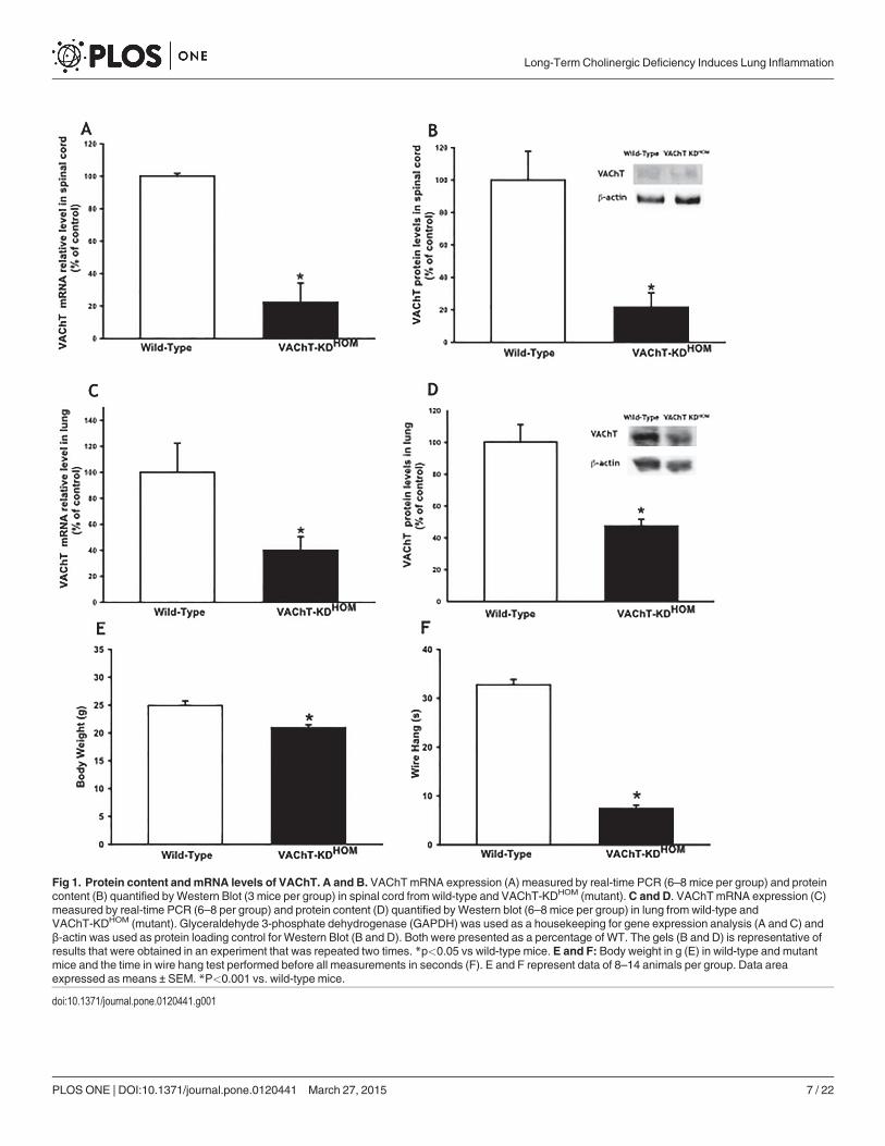
Fig 1. Protein content andmRNA levels of VAChT. A and B. VAChTmRNA expression (A) measured by real-time PCR (6–8 mice per group) and proteincontent (B) quantified byWestern Blot (3 mice per group) in spinal cord from wild-type and VAChT-KDHOM (mutant).C and D. VAChTmRNA expression (C)measured by real-time PCR (6–8 per group) and protein content (D) quantified byWestern blot (6–8 mice per group) in lung from wild-type andVAChT-KDHOM (mutant). Glyceraldehyde 3-phosphate dehydrogenase (GAPDH) was used as a housekeeping for gene expression analysis (A and C) andβ-actin was used as protein loading control for Western Blot (B and D). Both were presented as a percentage of WT. The gels (B and D) is representative ofresults that were obtained in an experiment that was repeated two times. *p<0.05 vs wild-type mice. E and F: Body weight in g (E) in wild-type and mutantmice and the time in wire hang test performed before all measurements in seconds (F). E and F represent data of 8–14 animals per group. Data areaexpressed as means ± SEM. *P<0.001 vs. wild-type mice.
doi:10.1371/journal.pone.0120441.g001
Long-Term Cholinergic Deficiency Induces Lung Inflammation
PLOS ONE | DOI:10.1371/journal.pone.0120441 March 27, 2015 7 / 22

Fig 2. VAChT deficiency did not affect other cholinergic component in lung.Muscarinic receptor 2 (M2),high-affinity choline transporter (CHT1), α7 nicotinic acetylcholine receptor (α7nAChR) andAcetylcholinesterase protein expression was analyzed byWestern Blot. The gel is representative of resultsthat were obtained in an experiment that was repeated two times. The graphs represent the valuesnormalized by β-actin.
doi:10.1371/journal.pone.0120441.g002
Long-Term Cholinergic Deficiency Induces Lung Inflammation
PLOS ONE | DOI:10.1371/journal.pone.0120441 March 27, 2015 8 / 22
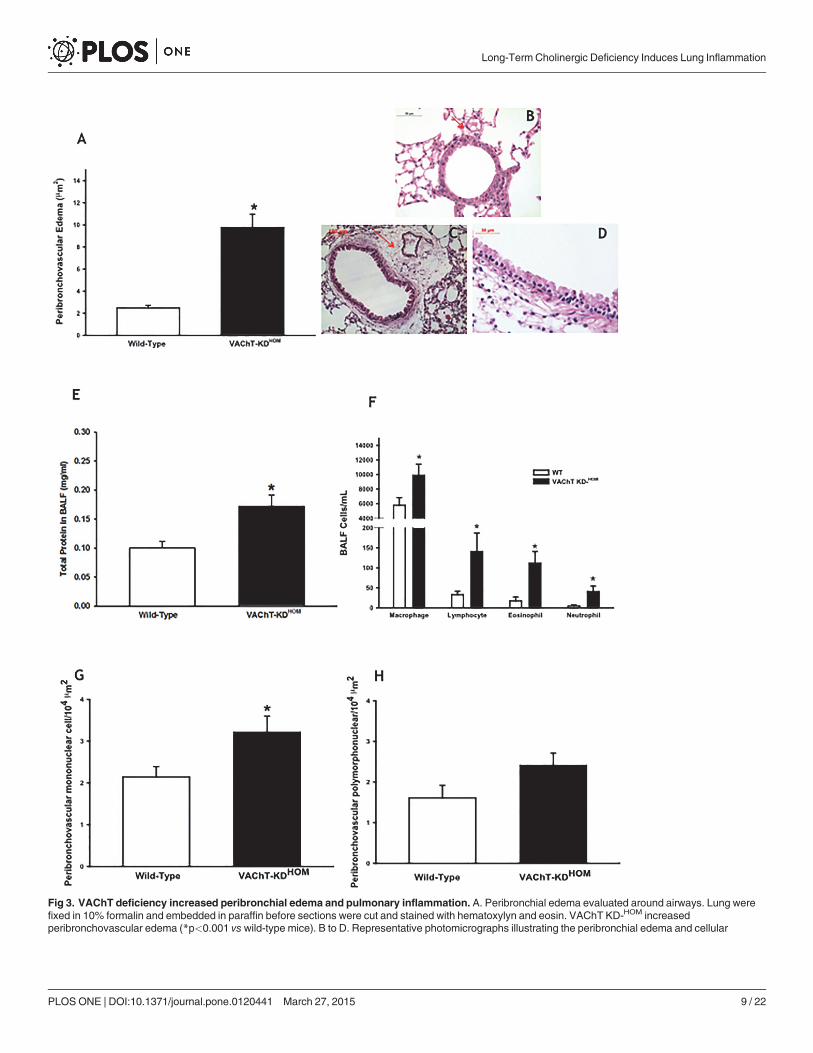
Fig 3. VAChT deficiency increased peribronchial edema and pulmonary inflammation. A. Peribronchial edema evaluated around airways. Lung werefixed in 10% formalin and embedded in paraffin before sections were cut and stained with hematoxylyn and eosin. VAChT KD-HOM increasedperibronchovascular edema (*p<0.001 vs wild-type mice). B to D. Representative photomicrographs illustrating the peribronchial edema and cellular
Long-Term Cholinergic Deficiency Induces Lung Inflammation
PLOS ONE | DOI:10.1371/journal.pone.0120441 March 27, 2015 9 / 22
VAChT-deficiency modulates pulmonary subunit p65-NF-kB and JAK-2-STAT-3 pathwaysNF-kB is a pro-inflammatory nuclear factor and the p-65 subunit plays a crucial role in inflam-matory and immune responses and recent evidence suggests that the cholinergic system con-trols inflammation by inhibiting the NF-kB activation through stimulation of α7-nACHR [8]and activation of JAK-STAT pathway [37].
We evaluated the protein expression of p-65-NF-kB in lung homogenate. We found an in-crease in lung expression of NF-kB in mutant mice compared to WT group (p<0.05) (Fig. 5A).In order to observe if the airway wall cells expressed p-65-NF-kB, we also quantified the NF-kBpositive cells stained by immunohistochemistry around airways wall (Fig. 5B) by morphome-try. VAChT KDHOM mice had increased number of NF-kB-positive cells around airways com-pared to WT (p<0.001). All together, these data suggest an activation of this pathway incholinergic deficient mice. In photomicrographs from wild-type (C and D) and VAChT mu-tant mice (E and F), we can observe positive and negative cells to NF-kB around airways. Anincrease in the number of cells positive to NF-kB is observed in airway wall obtained from mu-tant mice (E and F).
We also evaluated in lung homogenate the protein expression of JAK-2, STAT-3, pSTAT-3and SOCS-3. We found a reduction in lung expression of JAK-2 in mutant mice compared toWT group (p<0.05) (Fig. 6A). There was no significantly difference in STAT-3, pSTAT-3 andSOCS-3 content (Fig. 6B, 6C and 6D). Although a tendency in a reduction of total STAT-3 ex-pression could be observed, the ratio of p-STAT3/STAT3 was not different between the groups,suggesting that STAT-3 was not differently activated in this model.
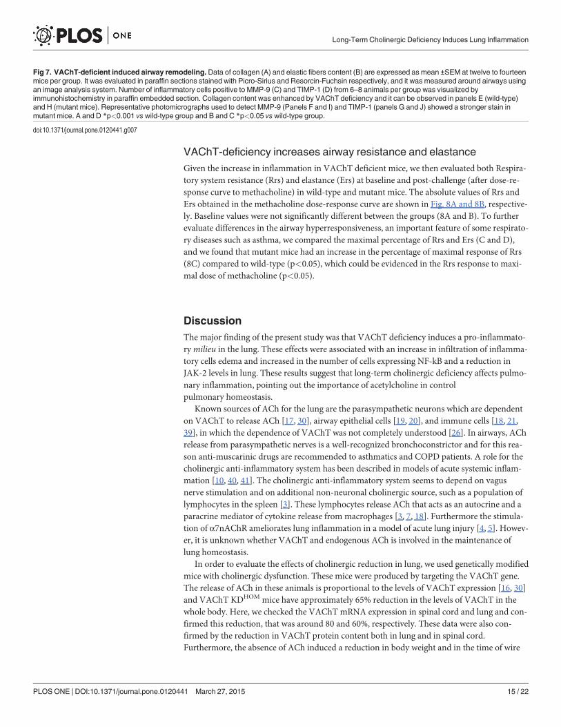
Effects of VAChT deficiency in airway ECM fibers depositionIt is well-know that airway remodeling is an important feature of lung disease, and it is usuallyassociated to chronic inflammation [2]. Given that VAChT mutant mice appear to have higherinflammatory response even in the absence of any injury, we asked whether remodeling of air-ways was also affected. In Fig. 7A and 7B, we found that VAChT deficiency induced an increasein both collagen and elastic fiber content around airways compared to WT group (p<0.001and p<0.05, respectively).
An imbalance in MMP-9 and TIMP-1 expression level is involved in ECMmatrix deposi-tion [38]. Therefore, we used immunohistochemistry analysis to investigate the cells positivefor MMP-9 and TIMP-1 expression in airways. Mutant mice (VAChT KDHOM) showed in-creased number of MMP-9 (7C) and TIMP-1 (7D) positive cells when compared to WT,p<0.05 and p<0.001 respectively]. MMP-9 was less than twice greater in VAChT KDHOM,whereas the increase in TIMP-1 was approximately three times higher than observed in WTgroup, tipping in favor of decrease proteolysis supporting the turn-over of the extracellular ma-trix fibers and the remodeling [38].
Representative photomicrographs stained with Picro-Sirius to detect collagen fibers wereshown in Fig. 7E and 7H. We noted that airways obtained fromWT group showed a weakstaining for collagen fibers around the airway wall in the tissue section (Fig. 7E), coincidentwith the maintenance of the histoarchitecture of the ECM (arrows). In contrast, lung slices ob-tained from mutant mice presented increased collagen fiber deposition around airways
infiltration around airways obtained from a VAChTmutant mice (C and D) compared to wild-type (B). E. Amount of total protein measured in bronchoalveolarlavage (BALF) (n = 7–8 per group, *p<0.01 vs wild-type mice). F. Mean and standard error of macrophages, lymphocytes, eosinophils and neutrophilscounted in bronchial alveolar lavage fluid (BALF) (n = 7–8 per group, *P<0.05 vs wild-type mice). G and H represent peribronchovascular mononuclear(*p<0.05 vs wild-type mice) and polymorphonuclear cells, respectively, evaluated around airways.
doi:10.1371/journal.pone.0120441.g003
Long-Term Cholinergic Deficiency Induces Lung Inflammation
PLOS ONE | DOI:10.1371/journal.pone.0120441 March 27, 2015 10 / 22
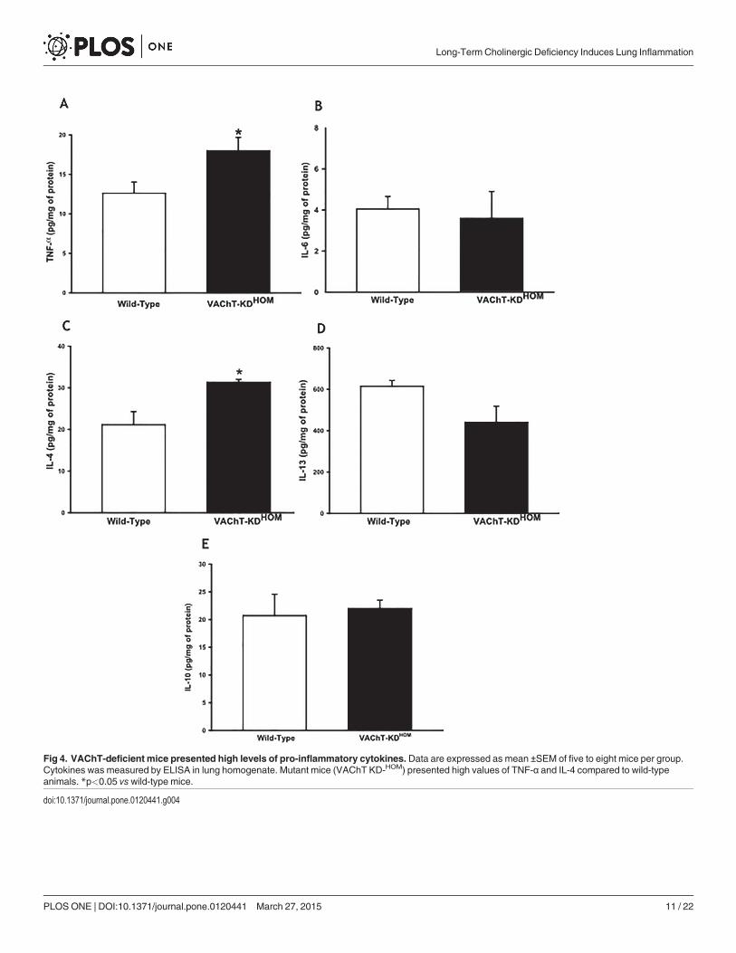
Fig 4. VAChT-deficient mice presented high levels of pro-inflammatory cytokines. Data are expressed as mean ±SEM of five to eight mice per group.Cytokines was measured by ELISA in lung homogenate. Mutant mice (VAChT KD-HOM) presented high values of TNF-α and IL-4 compared to wild-typeanimals. *p<0.05 vswild-type mice.
doi:10.1371/journal.pone.0120441.g004
Long-Term Cholinergic Deficiency Induces Lung Inflammation
PLOS ONE | DOI:10.1371/journal.pone.0120441 March 27, 2015 11 / 22

Fig 5. VAChT deficiency increased p65-NF-kB expression in lung. Subunit p65-NF-kB protein expression was measured by western blot (A). The gel isrepresentative of results that were obtained in an experiment that was repeated two times. The graphs represent the values normalized by β-actin (n = 5 pergroup). *p<0.05 vs wild-type group. Number of inflammatory cells positive to p65-NF-kB (B) from 6–8 animals per group was visualized byimmunohistochemistry in paraffin embedded section. Representative photomicrographs used to detect NF-kB (Panels C to F) showed a stronger stain inmutant mice (E and F) compared to wild-type mice (C and D). Arrows indicate positive cells around airway wall. *p<0.001 vswild-type group.
doi:10.1371/journal.pone.0120441.g005
Long-Term Cholinergic Deficiency Induces Lung Inflammation
PLOS ONE | DOI:10.1371/journal.pone.0120441 March 27, 2015 12 / 22

(Fig. 7H). Panels F, I, G, and J showed positive cells to MMP-9 and TIMP-1, respectively in thetwo groups studied. VAChT-KDHOM presented an increase in positive cells for MMP-9 andTIMP-1 (arrows) around airways compared to WT.
Fig 6. VAChT deficiency reduced JAK-2 expression in lung. Janus kinase 2 (JAK-2) (A), signaltransducer and activator of transcription 3 (STAT3) (B) and phosphorylated STAT3 (C) and suppressor ofcytokine signaling 3 (SOCS-3) (D) protein expression was measured byWestern Blot. The gel isrepresentative of results that were obtained in an experiment that was repeated two times. The graphsrepresent the values normalized by β-actin. *p<0.05 vs wild-type group.
doi:10.1371/journal.pone.0120441.g006
Long-Term Cholinergic Deficiency Induces Lung Inflammation
PLOS ONE | DOI:10.1371/journal.pone.0120441 March 27, 2015 13 / 22

Long-Term Cholinergic Deficiency Induces Lung Inflammation
PLOS ONE | DOI:10.1371/journal.pone.0120441 March 27, 2015 14 / 22
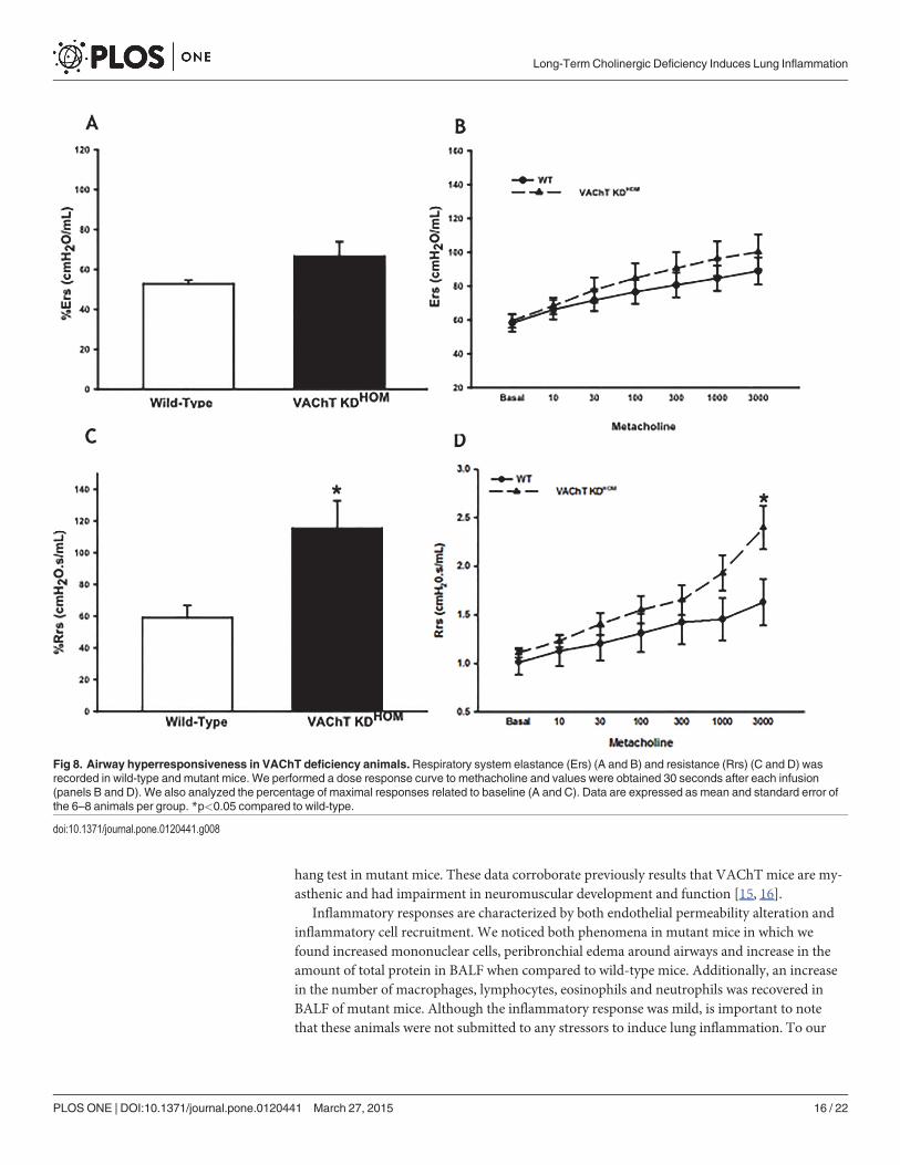
VAChT-deficiency increases airway resistance and elastanceGiven the increase in inflammation in VAChT deficient mice, we then evaluated both Respira-tory system resistance (Rrs) and elastance (Ers) at baseline and post-challenge (after dose-re-sponse curve to methacholine) in wild-type and mutant mice. The absolute values of Rrs andErs obtained in the methacholine dose-response curve are shown in Fig. 8A and 8B, respective-ly. Baseline values were not significantly different between the groups (8A and B). To furtherevaluate differences in the airway hyperresponsiveness, an important feature of some respirato-ry diseases such as asthma, we compared the maximal percentage of Rrs and Ers (C and D),and we found that mutant mice had an increase in the percentage of maximal response of Rrs(8C) compared to wild-type (p<0.05), which could be evidenced in the Rrs response to maxi-mal dose of methacholine (p<0.05).
DiscussionThe major finding of the present study was that VAChT deficiency induces a pro-inflammato-rymilieu in the lung. These effects were associated with an increase in infiltration of inflamma-tory cells edema and increased in the number of cells expressing NF-kB and a reduction inJAK-2 levels in lung. These results suggest that long-term cholinergic deficiency affects pulmo-nary inflammation, pointing out the importance of acetylcholine in controlpulmonary homeostasis.
Known sources of ACh for the lung are the parasympathetic neurons which are dependenton VAChT to release ACh [17, 30], airway epithelial cells [19, 20], and immune cells [18, 21,39], in which the dependence of VAChT was not completely understood [26]. In airways, AChrelease from parasympathetic nerves is a well-recognized bronchoconstrictor and for this rea-son anti-muscarinic drugs are recommended to asthmatics and COPD patients. A role for thecholinergic anti-inflammatory system has been described in models of acute systemic inflam-mation [10, 40, 41]. The cholinergic anti-inflammatory system seems to depend on vagusnerve stimulation and on additional non-neuronal cholinergic source, such as a population oflymphocytes in the spleen [3]. These lymphocytes release ACh that acts as an autocrine and aparacrine mediator of cytokine release from macrophages [3, 7, 18]. Furthermore the stimula-tion of α7nAChR ameliorates lung inflammation in a model of acute lung injury [4, 5]. Howev-er, it is unknown whether VAChT and endogenous ACh is involved in the maintenance oflung homeostasis.
In order to evaluate the effects of cholinergic reduction in lung, we used genetically modifiedmice with cholinergic dysfunction. These mice were produced by targeting the VAChT gene.The release of ACh in these animals is proportional to the levels of VAChT expression [16, 30]and VAChT KDHOM mice have approximately 65% reduction in the levels of VAChT in thewhole body. Here, we checked the VAChT mRNA expression in spinal cord and lung and con-firmed this reduction, that was around 80 and 60%, respectively. These data were also con-firmed by the reduction in VAChT protein content both in lung and in spinal cord.Furthermore, the absence of ACh induced a reduction in body weight and in the time of wire
Fig 7. VAChT-deficient induced airway remodeling.Data of collagen (A) and elastic fibers content (B) are expressed as mean ±SEM at twelve to fourteenmice per group. It was evaluated in paraffin sections stained with Picro-Sirius and Resorcin-Fuchsin respectively, and it was measured around airways usingan image analysis system. Number of inflammatory cells positive to MMP-9 (C) and TIMP-1 (D) from 6–8 animals per group was visualized byimmunohistochemistry in paraffin embedded section. Collagen content was enhanced by VAChT deficiency and it can be observed in panels E (wild-type)and H (mutant mice). Representative photomicrographs used to detect MMP-9 (Panels F and I) and TIMP-1 (panels G and J) showed a stronger stain inmutant mice. A and D *p<0.001 vs wild-type group and B and C *p<0.05 vs wild-type group.
doi:10.1371/journal.pone.0120441.g007
Long-Term Cholinergic Deficiency Induces Lung Inflammation
PLOS ONE | DOI:10.1371/journal.pone.0120441 March 27, 2015 15 / 22
hang test in mutant mice. These data corroborate previously results that VAChT mice are my-asthenic and had impairment in neuromuscular development and function [15, 16].
Inflammatory responses are characterized by both endothelial permeability alteration andinflammatory cell recruitment. We noticed both phenomena in mutant mice in which wefound increased mononuclear cells, peribronchial edema around airways and increase in theamount of total protein in BALF when compared to wild-type mice. Additionally, an increasein the number of macrophages, lymphocytes, eosinophils and neutrophils was recovered inBALF of mutant mice. Although the inflammatory response was mild, is important to notethat these animals were not submitted to any stressors to induce lung inflammation. To our
Fig 8. Airway hyperresponsiveness in VAChT deficiency animals.Respiratory system elastance (Ers) (A and B) and resistance (Rrs) (C and D) wasrecorded in wild-type and mutant mice. We performed a dose response curve to methacholine and values were obtained 30 seconds after each infusion(panels B and D). We also analyzed the percentage of maximal responses related to baseline (A and C). Data are expressed as mean and standard error ofthe 6–8 animals per group. *p<0.05 compared to wild-type.
doi:10.1371/journal.pone.0120441.g008
Long-Term Cholinergic Deficiency Induces Lung Inflammation
PLOS ONE | DOI:10.1371/journal.pone.0120441 March 27, 2015 16 / 22

knowledge, these data show for the first time that VAChT reduction inducespulmonary inflammation.
We evaluated pro-inflammatory cytokines (TNF-α, IL-6, IL-4 and IL-13) and the regulatorycytokine IL-10. Mutant mice presented higher levels of TNF-α and IL-4 in lung homogenateswhen compared to wild-type mice, while no difference was observed in the levels of IL-6, IL-13and IL-10 between these two groups. IL-6 is an important cytokine involved in infection and intraumas, and is secreted primary by T cells and macrophages. IL-13 as IL-4 are more involvedin allergic inflammation and are increase in experimental model of asthma [33] and IL-13 isstrongly associated to mucus production be epithelial cells. The increased levels of TNF-α andIL-4 could explain the inflammation and peribronchiolar edema observed in the lung of mu-tant mice. Plasma extravasation is one of the first characteristics of inflammation and it is asso-ciated with TNF-α an acute mediator of inflammation [42]. Mazzon et al. [43] showed thatTNF-α knockout mice present a reduction in lung inflammation and in paw edema induced bycarrageenan. IL-10 is a pro-inflammatory cytokine, however, other authors have not been ableto show that cholinergic anti-inflammatory system acts in IL-10 [6], corroborating our results.
Several studies have previously indicated that cholinergic activity has a fundamental role inanti-inflammatory responses in different experimental models. Vagotomized mice presentedan increase in inflammatory cells in peritoneal fluid after septic peritonitis, enhancing earlyand late inflammatory responses [40]. However, it should be noted that vagotomy will impairnot only the release of ACh, but also the secretion of peptides and potential co-transmitters[44]. Hofer et al. [45] showed that inhibition of acetylcholinesterase by physostigmine reducedlethality and circulating pro-inflammatory cytokines TNF-α, IL-1β, and IL-6 as wells as down-regulated NF-kB activity in a sepsis model. Additionally, corroborating this data, Borovikovaet al. [6] suggested that ACh attenuates inflammation by a direct effect in pro-inflammatory cy-tokines inhibition, instead of an effect in anti-inflammatory cytokines. In the lung, Kox et al.[46] showed that vagotomy enhanced pulmonary inflammation induced by LPS, however theauthors did not find any effect of vagus nerve stimulation ameliorating this response. Collec-tively our data expand these observations showing that VAChT function, and consequent en-dogenous release of ACh, is involving in controlling local inflammatory response in the lungand avoiding exacerbated inflammation even on the absence of any injury. This suggests thatinflammation is kept in check by cholinergic activity.
The persistence of chronic inflammation induces tissue repair [2, 34, 35]. Tissue repair hasan important clinical significance in respiratory diseases as it contributes to the worsening oflung function over the years in asthmatic and COPD patients [47]. VAChT mutant miceshowed increased collagen content when compared to WT mice. Chronic inflammation in-duced by VAChT deficiency could explain per se the ECM remodeling since macrophages andother cells release different types of profibrotic mediators [48]. An imbalance between MMPand TIMP has a role in the immunomodulatory mechanisms regulating ECM composition.Additionally, MMPs are involved in inflammatory cell recruitment and tissue repair [49].
We also found higher number of positive inflammatory cells to MMP-9 and TIMP-1 in air-ways in VAChT mutants compared to wild-type mice, with the increase in TIMP-1 moreprominent than the increase in MMP-9. That is, while MMP-9, which degrades ECM, in-creased less that twice in VAChT KDHOM group related to baseline values (WT group), TIMP-1 increased approximately more than three fold, consistent with the dynamic turnover of ECMcomponents that can occurs in inflammatory disease [38]. These results suggest that the chron-ic inflammatory unbalance due to cholinergic dysfunction may drive remodeling in the lungs.
Although no difference in basal lung function was observed, VAChT KD mice presented anincreased in maximal response of respiratory system resistance to a bronchoconstrictor ago-nist. Airway smooth muscle contraction is an important determinant of lung mechanical
Long-Term Cholinergic Deficiency Induces Lung Inflammation
PLOS ONE | DOI:10.1371/journal.pone.0120441 March 27, 2015 17 / 22

alterations in a presence of a bronchoconstrictor. However, it should be noted that the presenceof inflammation, edema and airway remodeling can also increase Rrs responses by reducingairway lumen diameter. In this context, mutant animals presented increased airway inflamma-tion, edema and remodeling which could explain the hyperresponsiveness observed. Anotherpossibility is that changes in muscarinic receptors that could appear in mutant mice as a com-pensatory mechanism due to ACh deficiency. In this regard, we evaluated M2 protein in lungand we did not found any difference between the groups. Verbout et al. [50] showed that atro-pine treatment increased antigen challenge-induced airway hyperreactivity and this effect wasdependent on inflammation, particularly eosinophils, corroborating in part our findings.
Considering the mechanisms involved in VAChT deficiency-induced pulmonary inflamma-tion, recently it has been suggested that stimulation of cholinergic receptors suppresses acutelung inflammation in mice, probably activating α7nAChR [4]. Some authors identified thepresence of α7nAChR in the surface of immune cells which, following activation, regulates in-flammation in a cholinergic dependent manner [9]. Activation of α7nAChR and triggers theJAK-2-STAT-3 pathway seems to inhibit the nuclear translocation of NF-kB [8]. We then eval-uated the lung expression of p65-NF-kB as well as the number of cells that express p65-NF-kBand we found that mutant mice presented an increase in p65-NF-kB expression in lung, sug-gesting an activation of this pathway.
JAK-2 expression in lung of VAChT mutant mice was reduced. Janus kinases (JAKs) areregulators of signaling through cytokine receptors and the role of JAK2-STAT3 pathway in in-flammation was not completely elucidated. Particularly in lung, this pathway was poorly inves-tigated. The inhibition of JAK-2 prevents LPS-induced STAT-3 tyrosine phosphorylation [51].In turn, the activation of STAT-3, induces the increase in SOCS-3 which is related to an anti-inflammatory action since it counteracts macrophage (M1) proinflammatory phenotypes [52]and inhibits NF-kB translocation [53]. Interestingly, we found that total and phosphorylateSTAT-3 and SOCS-3 expression was not different between WT and mutant mice, although atendency in reduction of STAT-3 was observed. These results suggest that in the lung, VAChTreduction and inhibition of cholinergic tone induced a reduction in JAK-2 activation thatcould prevent the activation of STAT-3, which in turn did not stimulate an increase in SOCS-3in order to counteract lung inflammation. Corroborating part of this idea, de Jonge et al. [37]showed that vagal nerve stimulation improved inflammation in a model of intestinal surgeryby activating STAT3 in intestinal macrophages and the authors concluded that the anti-inflam-matory effects of vagal stimulation is mediated by JAK2-STAT3 activation thoroughα7nAChR stimulation.
Considering that both neuronal (parasympathetic nerves) and non-neuronal (epithelial andimmune cells) sources of ACh co-exist in lung, the limitation of the present study performed invivo is the difficult to distinguish the effect of each pathway. In lung, is not totally clear if thesource of ACh by non-neuronal cells is dependent of VAChT. Other mechanism of ACh secre-tion in epithelial cells has been suggested mainly related to ACh release directly from the cyto-plasm [26, 54]. Is important to note that we evaluated CHT1, AChE, M2 and α7nAChR andnone of them is altered in mutant mice. In addition, Lips et al. [19] showed immunoreactivityto VAChT in airways and also showed a reduction of the cholinergic machinery, includingVAChT, in a model of acute airway inflammation. Recently, in an elegant review, Yang et al.[55] pointed out the importance of pulmonary parasympathetic inflammatory reflex as a regu-lator of lung inflammation and immunity and suggest that neuronal ACh is important to in-duce the release of non-neuronal ACh by immune cells in order to produce anti-inflammatoryeffects. On the basis of the current state of knowledge, there is no unequivocal evidence thatVAChT deficiency both in the nervous system and in the lung contribute to the control oflung inflammation.
Long-Term Cholinergic Deficiency Induces Lung Inflammation
PLOS ONE | DOI:10.1371/journal.pone.0120441 March 27, 2015 18 / 22

In conclusion, we showed for the first time that long-term VAChT deficiency induced air-way hyperresponsiveness, inflammation and remodeling in a murine model of allergic airwayinflammation. The pro-inflammatorymilieu observed was associated to increased p65-NF-kBand inhibition of JAK-2 expression. Importantly, these data suggest that intact cholinergic toneis an important mechanism to keep in check exacerbated inflammatory responses in order tomaintain the lung homeostasis. Because the lung are constantly exposed to compounds fromthe environmental that can induce inflammatory responses, this regulatory mechanism may berelevant to avoid aberrant reactions in response to irrelevant stimuli.
AcknowledgmentsThe authors would like to thank the Sao Paulo State Research Foundation and National Coun-cil for Technologic and Scientific Development (CNPq) for financial support. MAMP and VFPwere supported by grant from the Heart & Stroke Foundation of Ontario. We also thank theAnimal Facility from School of Medicine/USP/SP for the careful help with theanimal maintenance.
Author ContributionsConceived and designed the experiments: NMP CJCPM AP NOSC SKPCMICAV LCC IFLCTMAMPMAMVFP CMP. Performed the experiments: NMP JCPM APMICAV LCC CMP.Analyzed the data: NMP CJCPM APMICAV LCC CMP. Contributed reagents/materials/anal-ysis tools: NOSC SKPCMICAV LCC IFLCT MAM CMP. Wrote the paper: NMP CJCPM APNOSC SKPC MICAV LCC IFLCTMAMPMAMVFP CMP.
References1. Moldoveanu B, Otmishi P, Jani P, Walker J, Sarmiento X, Guardiola J, et al. Inflammatory mechanisms
in the lung. J InflammRes. 2009; 2: 1–11. PMID: 22096348
2. Postma DS, Timens W. Remodeling in asthma and chronic obstructive pulmonary disease. Proc AmThorac Soc. 2006; 3: 434–439. PMID: 16799088
3. Rosas-Ballina M, Olofsson PS, Ochani M, Valdes-Ferrer SI, Levine YA, Reardon C, et al. Acetylcho-line-synthesizing T cells relay neural signals in a vagus nerve circuit. Science. 2011; 334: 98–101. doi:10.1126/science.1209985 PMID: 21921156
4. Su X, Matthay MA, Malik AB. Requisite role of the cholinergic alpha7 nicotinic acetylcholine receptorpathway in suppressing Gram-negative sepsis-induced acute lung inflammatory injury. J Immunol.2010; 184: 401–410. doi: 10.4049/jimmunol.0901808 PMID: 19949071
5. Su X, Lee JW, Matthay ZA, Mednick G, Uchida T, Fang X, et al. Activation of the alpha7 nAChR re-duces acid-induced acute lung injury in mice and rats. Am J Respir Cell Mol Biol. 2007; 37: 186–192.PMID: 17431097
6. Borovikova LV, Ivanova S, Zhang M, Yang H, Botchkina GI, Watkins LR, et al. Vagus nerve stimulationattenuates the systemic inflammatory response to endotoxin. Nature. 2000; 405: 458–462. PMID:10839541
7. Pavlov VA, Tracey KJ. Controlling inflammation: the cholinergic anti-inflammatory pathway. BiochemSoc Trans. 2006; 34: 1037–1040. PMID: 17073745
8. Rosas-Ballina M, Tracey KJ. Cholinergic control of inflammation. J Intern Med. 2009; 265: 663–679.doi: 10.1111/j.1365-2796.2009.02098.x PMID: 19493060
9. Parrish WR, Gallowitsch-Puerta M, Czura CJ, Tracey KJ. Experimental therapeutic strategies for se-vere sepsis: mediators and mechanisms. Ann N Y Acad Sci. 2008; 1144: 210–236. doi: 10.1196/annals.1418.011 PMID: 19076379
10. Wang H, Liao H, Ochani M, Justiniani M, Lin X, Yang L, et al. Cholinergic agonists inhibit HMGB1 re-lease and improve survival in experimental sepsis. Nat Med. 2004; 10: 1216–1221. PMID: 15502843
11. Murray PJ. The JAK-STAT signaling pathway: input and output integration. J Immunol. 2007; 178:2623–2629. PMID: 17312100
Long-Term Cholinergic Deficiency Induces Lung Inflammation
PLOS ONE | DOI:10.1371/journal.pone.0120441 March 27, 2015 19 / 22

12. Prado VF, Roy A, Kolisnyk B, Gros R, Prado MA. Regulation of cholinergic activity by the vesicular ace-tylcholine transporter. Biochem J. 2013; 450: 265–274. doi: 10.1042/BJ20121662 PMID: 23410039
13. Prado MA, Reis RA, Prado VF, de Mello MC, GomezMV, de Mello FG. Regulation of acetylcholine syn-thesis and storage. Neurochem Int. 2002; 41: 291–299. PMID: 12176069
14. Racke K, Juergens UR, Matthiesen S. Control by cholinergic mechanisms. Eur J Pharmacol. 2006;533: 57–68. PMID: 16458288
15. de Castro BM, De Jaeger X, Martins-Silva C, Lima RD, Amaral E, Menezes C, et al. The vesicular ace-tylcholine transporter is required for neuromuscular development and function. Mol Cell Biol. 2009; 29:5238–5250. doi: 10.1128/MCB.00245-09 PMID: 19635813
16. Prado VF, Martins-Silva C, de Castro BM, Lima RF, Barros DM, Amaral E, et al. Mice deficient for thevesicular acetylcholine transporter are myasthenic and have deficits in object and social recognition.Neuron. 2006; 51: 601–612. PMID: 16950158
17. Guzman MS, De Jaeger X, Raulic S, Souza IA, Li AX, Schmid S, et al. Elimination of the vesicular ace-tylcholine transporter in the striatum reveals regulation of behaviour by cholinergic-glutamatergic co-transmission. PLoS Biol. 2011; 9: e1001194. doi: 10.1371/journal.pbio.1001194 PMID: 22087075
18. Kawashima K, Fujii T. Expression of non-neuronal acetylcholine in lymphocytes and its contribution tothe regulation of immune function. Front Biosci. 2004; 9: 2063–2085. PMID: 15353271
19. Lips KS, Luhrmann A, Tschernig T, Stoeger T, Alessandrini F, Grau V, et al. Down-regulation of thenon-neuronal acetylcholine synthesis and release machinery in acute allergic airway inflammation ofrat and mouse. Life Sci. 2007; 80: 2263–2269. PMID: 17328924
20. Proskocil BJ, Sekhon HS, Jia Y, Savchenko V, Blakely RD, Lindstrom J, et al. Acetylcholine is an auto-crine or paracrine hormone synthesized and secreted by airway bronchial epithelial cells. Endocrinolo-gy. 2004; 145: 2498–2506. PMID: 14764638
21. Lips KS, Volk C, Schmitt BM, Pfeil U, Arndt P, Miska D, et al. Polyspecific cation transporters mediateluminal release of acetylcholine from bronchial epithelium. Am J Respir Cell Mol Biol. 2005; 33: 79–88.PMID: 15817714
22. Kawashima K, Fujii T, Moriwaki Y, Misawa H. Critical roles of acetylcholine and the muscarinic and nic-otinic acetylcholine receptors in the regulation of immune function. Life Sci. 2012; 91: 1027–1032. doi:10.1016/j.lfs.2012.05.006 PMID: 22659391
23. Wessler I, Kilbinger H, Bittinger F, Unger R, Kirkpatrick CJ. The non-neuronal cholinergic system in hu-mans: expression, function and pathophysiology. Life Sci. 2003; 72: 2055–2061. PMID: 12628456
24. Roy A, Fields WC, Rocha-Resende C, Resende RR, Guatimosim S, Prado VF, et al. Cardiomyocyte-secreted acetylcholine is required for maintenance of homeostasis in the heart. FASEB J. 2013; 27:5072–5082. doi: 10.1096/fj.13-238279 PMID: 24018063
25. Ishii M, Kurachi Y. Muscarinic acetylcholine receptors. Curr Pharm Des. 2006; 12: 3573–3581. PMID:17073660
26. KummerW, Krasteva-Christ G. Non-neuronal cholinergic airway epithelium biology. Curr Opin Pharma-col. 2014; 16: 43–49. doi: 10.1016/j.coph.2014.03.001 PMID: 24681350
27. Arvidsson U, Riedl M, Elde R, Meister B. Vesicular acetylcholine transporter (VAChT) protein: a noveland unique marker for cholinergic neurons in the central and peripheral nervous systems. J Comp Neu-rol. 1997; 378: 454–467. PMID: 9034903
28. Ganesan S, Faris AN, Comstock AT, Chattoraj SS, Chattoraj A, Burgess JR, et al. Quercetin preventsprogression of disease in elastase/LPS-exposed mice by negatively regulating MMP expression.Respir Res. 2010; 11: 131. doi: 10.1186/1465-9921-11-131 PMID: 20920189
29. Lara A, Damasceno DD, Pires R, Gros R, Gomes ER, Gavioli M, et al. Dysautonomia due to reducedcholinergic neurotransmission causes cardiac remodeling and heart failure. Mol Cell Biol. 2010; 30:1746–1756. doi: 10.1128/MCB.00996-09 PMID: 20123977
30. Lima R de F, Prado VF, Prado MA, Kushmerick C. Quantal release of acetylcholine in mice with re-duced levels of the vesicular acetylcholine transporter. J Neurochem. 2010; 113: 943–951. doi: 10.1111/j.1471-4159.2010.06657.x PMID: 20202084
31. Sango K, McDonald MP, Crawley JN, Mack ML, Tifft CJ, Skop E, et al. Mice lacking both subunits of ly-sosomal beta-hexosaminidase display gangliosidosis and mucopolysaccharidosis. Nat Genet. 1996;14: 348–352. PMID: 8896570
32. Hantos Z, Adamicza A, Janosi TZ, Szabari MV, Tolnai J, Suki B. Lung volumes and respiratory me-chanics in elastase-induced emphysema in mice. J Appl Physiol. 2008; 105: 1864–1872. doi: 10.1152/japplphysiol.90924.2008 PMID: 18845778
33. Toledo AC, Sakoda CP, Perini A, Pinheiro NM, Magalhaes RM, Grecco S, et al. Flavonone treatmentreverses airway inflammation and remodelling in an asthmamurine model. Br J Pharmacol. 2013; 168:1736–1749. doi: 10.1111/bph.12062 PMID: 23170811
Long-Term Cholinergic Deficiency Induces Lung Inflammation
PLOS ONE | DOI:10.1371/journal.pone.0120441 March 27, 2015 20 / 22
34. Prado CM, Leick-Maldonado EA, Yano L, Leme AS, Capelozzi VL, Martins MA, et al. Effects of nitricoxide synthases in chronic allergic airway inflammation and remodeling. Am J Respir Cell Mol Biol.2006; 35: 457–465. PMID: 16709960
35. Vieira RP, Claudino RC, Duarte AC, Santos AB, Perini A, Faria Neto HC, et al. Aerobic exercise de-creases chronic allergic lung inflammation and airway remodeling in mice. Am J Respir Crit Care Med.2007; 176: 871–877. PMID: 17690332
36. Caperuto LC, Anhe GF, Cambiaghi TD, Akamine EH, do Carmo Buonfiglio D, Cipolla-Neto J, et al.Modulation of bone morphogenetic protein-9 expression and processing by insulin, glucose, and gluco-corticoids: possible candidate for hepatic insulin-sensitizing substance. Endocrinology. 2008; 149:6326–6335. doi: 10.1210/en.2008-0655 PMID: 18703636
37. de JongeWJ, van der Zanden EP, The FO, Bijlsma MF, vanWesterloo DJ, Bennink RJ, et al. Stimula-tion of the vagus nerve attenuates macrophage activation by activating the Jak2-STAT3 signaling path-way. Nat Immunol. 2005; 6: 844–851. PMID: 16025117
38. Araujo BB, Dolhnikoff M, Silva LF, Elliot J, Lindeman JH, Ferreira DS, et al. Extracellular matrix compo-nents and regulators in the airway smooth muscle in asthma. Eur Respir J. 2008; 32: 61–69. doi: 10.1183/09031936.00147807 PMID: 18321931
39. Wessler I, Bittinger F, KaminW, Zepp F, Meyer E, Schad A, et al. Dysfunction of the non-neuronal cho-linergic system in the airways and blood cells of patients with cystic fibrosis. Life Sci. 2007; 80: 2253–2258. PMID: 17346753
40. vanWesterloo DJ, Giebelen IA, Florquin S, Daalhuisen J, Bruno MJ, de Vos AF, et al. The cholinergicanti-inflammatory pathway regulates the host response during septic peritonitis. J Infect Dis. 2005; 191:2138–2148. PMID: 15898001
41. Song XM, Li JG, Wang YL, Hu ZF, Zhou Q, Du ZH, et al. The protective effect of the cholinergic anti-in-flammatory pathway against septic shock in rats. Shock. 2008; 30: 468–472. doi: 10.1097/SHK.0b013e31816d5e49 PMID: 18391858
42. Catal F, Mete E, Tayman C, Topal E, Albayrak A, Sert H. A human monoclonal anti-TNF alpha antibody(adalimumab) reduces airway inflammation and ameliorates lung histology in a murine model of acuteasthma. Allergol Immunopathol (Madr). 2014; 43: 14–18.
43. Mazzon E, Esposito E, Di Paola R, Muia C, Crisafulli C, Genovese T, et al. Effect of tumour necrosisfactor-alpha receptor 1 genetic deletion on carrageenan-induced acute inflammation: a comparisonwith etanercept. Clin Exp Immunol. 2008; 153: 136–149. doi: 10.1111/j.1365-2249.2008.03669.xPMID: 18505433
44. Schelegle ES, WalbyWF. Vagal afferents contribute to exacerbated airway responses following ozoneand allergen challenge. Respir Physiol Neurobiol. 2012; 181: 277–285. doi: 10.1016/j.resp.2012.04.003 PMID: 22525484
45. Hofer S, Eisenbach C, Lukic IK, Schneider L, Bode K, Brueckmann M, et al. Pharmacologic cholinester-ase inhibition improves survival in experimental sepsis. Crit Care Med. 2008; 36: 404–408. PMID:18091537
46. Kox M, Vaneker M, van der Hoeven JG, Scheffer GJ, Hoedemaekers CW, Pickkers P. Effects of vagusnerve stimulation and vagotomy on systemic and pulmonary inflammation in a two-hit model in rats.PLoS One. 2012; 7: e34431. doi: 10.1371/journal.pone.0034431 PMID: 22493690
47. Phipps S, Benyahia F, Ou TT, Barkans J, Robinson DS, Kay AB. Acute allergen-induced airway remod-eling in atopic asthma. Am J Respir Cell Mol Biol. 2004; 31: 626–632. PMID: 15333330
48. Chetta A, Foresi A, Del Donno M, Bertorelli G, Pesci A, Olivieri D. Airways remodeling is a distinctivefeature of asthma and is related to severity of disease. Chest. 1997; 111: 852–857. PMID: 9106559
49. Greenlee KJ, Werb Z, Kheradmand F. Matrix metalloproteinases in lung: multiple, multifarious, andmultifaceted. Physiol Rev. 2007; 87: 69–98. PMID: 17237343
50. Verbout NG, Lorton JK, Jacoby DB, Fryer AD. Atropine pretreatment enhances airway hyperreactivityin antigen-challenged guinea pigs through an eosinophil-dependent mechanism. Am J Physiol LungCell Mol Physiol. 2007; 292: L1126–1135. PMID: 17220376
51. Pena G, Cai B, Deitch EA, Ulloa L. JAK2 inhibition prevents innate immune responses and rescues ani-mals from sepsis. J Mol Med (Berlin). 2010; 88: 851–859. doi: 10.1007/s00109-010-0628-z PMID:20393690
52. Qin H, Holdbrooks AT, Liu Y, Reynolds SL, Yanagisawa LL, Benveniste EN. SOCS3 deficiency pro-motes M1macrophage polarization and inflammation. J Immunol. 2012; 189: 3439–3448. PMID:22925925
53. Karlsen AE, Heding PE, Frobose H, Ronn SG, Kruhoffer M, Orntoft TF, et al. Suppressor of cytokinesignalling (SOCS)-3 protects beta cells against IL-1beta-mediated toxicity through inhibition of multiple
Long-Term Cholinergic Deficiency Induces Lung Inflammation
PLOS ONE | DOI:10.1371/journal.pone.0120441 March 27, 2015 21 / 22
nuclear factor-kappaB-regulated proapoptotic pathways. Diabetologia. 2004; 47: 1998–2011. PMID:15578154
54. KummerW, Lips KS, Pfeil U. The epithelial cholinergic system of the airways. Histochem Cell Biol.2008; 130: 219–234. doi: 10.1007/s00418-008-0455-2 PMID: 18566825
55. Yang X, Zhao C, Gao Z, Su X. A novel regulator of lung inflammation and immunity: pulmonary para-sympathetic inflammatory reflex. QJM: monthly journal of the Association of Physicians. 2014;107:789–792. doi: 10.1093/qjmed/hcu005 PMID: 24440925
Long-Term Cholinergic Deficiency Induces Lung Inflammation
PLOS ONE | DOI:10.1371/journal.pone.0120441 March 27, 2015 22 / 22