Synthesis and characterization of ruthenium dioxide nanorods
Upload
khangminh22Category
view
1download
0
ARTICLE
TPGS-based and S-thanatin functionalizednanorods for overcoming drug resistance inKlebsiella pneumoniaXiaojuan Wang1, Xiaoling Xu2, Shaojun Zhang1, Na Chen1, Yunfeng Sun2, Kuifen Ma1, Dongsheng Hong1, Lu Li1,
Yongzhong Du 2✉, Xiaoyang Lu 1✉ & Saiping Jiang 1✉
Tigecycline is regarded as the last line of defense to combat multidrug-resistant Klebsiella
pneumoniae. However, increasing utilization has led to rising drug resistance and treatment
failure. Here, we design a D-alpha tocopheryl polyethylene glycol succinate-modified and
S-thanatin peptide-functionalized nanorods based on calcium phosphate nanoparticles for
tigecycline delivery and pneumonia therapy caused by tigecycline-resistant Klebsiella pneu-
moniae. After incubation with bacteria, the fabricated nanorods can enhance tigecycline
accumulation in bacteria via the inhibitory effect on efflux pumps exerted by D-alpha toco-
pheryl polyethylene glycol succinate and the targeting capacity of S-thanatin to bacteria. The
synergistic antibacterial capacity between S-thanatin and tigecycline further enhances the
antibacterial activity of nanorods, thus overcoming the tigecycline resistance of Klebsiella
pneumoniae. After intravenous injection, nanorods significantly reduces the counts of white
blood cells and neutrophils, decreases bacterial colonies, and ameliorates neutrophil infil-
tration events, thereby largely increasing the survival rate of mice with pneumonia. These
findings may provide a therapeutic strategy for infections caused by drug-resistant bacteria.
https://doi.org/10.1038/s41467-022-31500-3 OPEN
1 Department of Clinical Pharmacy, The First Affiliated Hospital, School of Medicine, Zhejiang University, 79 Qingchun Road, Hangzhou 310003, China.2 Institute of Pharmaceutics, College of Pharmaceutical Sciences, Zhejiang University, 866 Yu-Hang-Tang Road, Hangzhou 310058, China.✉email: [email protected]; [email protected]; [email protected]
NATURE COMMUNICATIONS | (2022) 13:3731 | https://doi.org/10.1038/s41467-022-31500-3 | www.nature.com/naturecommunications 1
1234
5678
90():,;
Antibiotics have been an epochal discovery for bacterialinfection therapies in the past few decades. However,long-term and excessive use of antibiotics has caused the
spread of antibiotic resistance1. Subsequently, the emergence ofsuperbugs has been growing to become a dominant challenge inhuman health2. It is predicted that bacterial infections will lead to10 million deaths each year by 2050, exceeding those presentlycaused by cancer3. Bacteria resist the effects of antibiotics mainlythrough the following molecular mechanisms: modification of thetarget site, destruction of the antibiotic, antibiotic efflux via effluxtransporters and reduced antibiotic influx through decreasingmembrane permeability4. Since its initial discovery in the 1980s,many efflux pumps have been characterized in pathogens such asStaphylococcus aureus, Enterococcus faecium, Acinetobacter bau-manii, Pseudomonas aeruginosa, and Klebsiella pneumonia5.Overexpression of efflux pumps can lead to clinically relevantlevels of resistance to antibiotic in Gram-negative bacteria6.
Currently, Klebsiella pneumonia (KPN) is regarded as one ofthe most serious nosocomial pathogens threatening publichealth7. As a gram-negative bacterium, KPN can induce multipleinfections, such as pneumonia, liver abscesses, urinary tractinfections, meningitis, and bacteremia in hospitalized patientswith insufficient immune systems8. In recent years, KPN strainswith high virulence and mucus phenotypes have graduallybecome an important pathogen clinically that can cause seriousinfections in healthy people and significantly threaten theirhealth9,10. Moreover, KPN is prone to generating multidrugresistance11. Carbapenem antibiotics used to be the first-linedrugs for infection caused by multidrug-resistant KPN (MDR-KPN). However, with the wide use of carbapenem antibiotics inclinical therapy, carbapenem-resistant KPN (CRKP) has distinctlyincreased and become a serious public health issue12. Increasingdrug resistance and high mortality have posed dominant chal-lenges for clinical therapy. There are currently few alternativedrugs available for treating CRKP infections, with tigecycline(TIG) being one of the few remaining antibiotics13. Therefore,TIG is generally considered to be one of the last defensive lineagainst CRKP. However, a decreased sensitivity and growingresistance of CRKP to TIG were observed with increased clinicalapplications, significantly threatening last-line therapy14. Thetime- and cost-consuming process of new antibiotic developmentresults in the much slower emergence of new antibacterial drugsthan that of bacterial resistance15.
Recently, nanodrug delivery systems (DDSs) have emerged asnovel therapeutic means for deadly infections16,17. DDS canprovide an increased drug retention time in blood, a reducednonspecific distribution, and targeted delivery of drugs at the siteof infection18. Combination therapy of nanomaterials and anti-biotics might contribute to a better therapeutic index. Therefore,they are regarded as promising candidates for combating MDRbacteria19. Numerous studies have constructed DDSs based onAg, Au, Cu, Fe, Ti, and mesoporous silica nanoparticles to treatinfectious diseases17, and their clinical use in vivo is hindered bysafety. More efficient nanomaterial-based delivery strategies, suchas pH-triggered, enzyme-sensitive, and bacterial toxin-triggeredDDSs, could potentially allow the release of antibiotics in a spa-tiotemporally controlled fashion and have gain attentionworldwide20. However, this process is complex and sophisticated,and the released antibiotics lack specificity for bacteria. Therefore,a safe DDS with targeting efficacy to bacteria might be an effectivestrategy for the treatment of infection caused by TRKP.
As of now, several mechanisms have been identified that areassociated with tigecycline-resistant Klebsiella pneumonia(TRKP). Most commonly, non-specific active resistance-nodulation-cell division (RND) efflux pumps such as AcrAB-TolC are overproduced21. Tigecycline MICs of > 2 mg/L have
been associated with significantly increased ramA levels22. It wasreported that the majority of CRKP isolates were resistant totigecycline due to increased expression of the efflux pump geneacrB21. A retrospective study in China displayed that the elevatedexpression of acrB and ramA was found in ~90% of thetigecycline-resistant isolates clinically23.
D-alpha tocopheryl polyethylene glycol succinate (TPGS), anonionic surfactant, is extensively applied in DDSs. Numerousstudies have revealed that TPGS functions as an inhibitor andsubstrate of P-glycoprotein and dramatically decreases the effluxof drugs24. Therefore, TPGS could reverse the multidrug resis-tance of tumor cells. These features make TPGS a feasible alter-native biomaterial in DDSs for tumor therapy, especially intumors resistant to chemotherapy drugs25. Our previous studieshave confirmed the inhibitory activity of TPGS on efflux pumpsin bacteria26,27. Therefore, the application of TPGS in DDSsmight be an effective means to enhance TIG accumulation inTRKP and achieve effective therapy for TRKP infections.
S-thanatin (Ts) peptide is a short antimicrobial peptide (AMP)that exhibits lipopolysaccharide (LPS) binding affinity28, sug-gesting its potential as a therapeutic strategy for infection causedby gram-negative bacteria. The interaction between the Ts pep-tide and LPS could promote the intercalation of Ts into thecytoplastic membrane, subsequently resulting in a leaky cyto-plastic membrane and the disintegration of bacterial respirationand energization29. Ts peptide can kill bacteria in a membrane-dependent manner and has been shown to exhibit active anti-bacterial activity towards numerous gram-negative bacteria,including MDR bacteria30,31. Ts peptide-functionalized levo-floxacin-loaded liposomes showed targeted drug delivery tobacteria and exhibited an excellent therapeutic effect in the septicmouse model induced by MDR-KPN32. Calcium phosphatenanoparticles (Cap) have drawn wide attention as potential car-riers due to their inherent superior properties, including highdrug loading efficiency, good biocompatibility, and excellentbiodegradability33,34. Ts-modified Cap DDSs might furtherenhance TIG accumulation in bacteria. Pneumonia caused byKPN and TRKP is one of the most common infectious diseasesclinically.
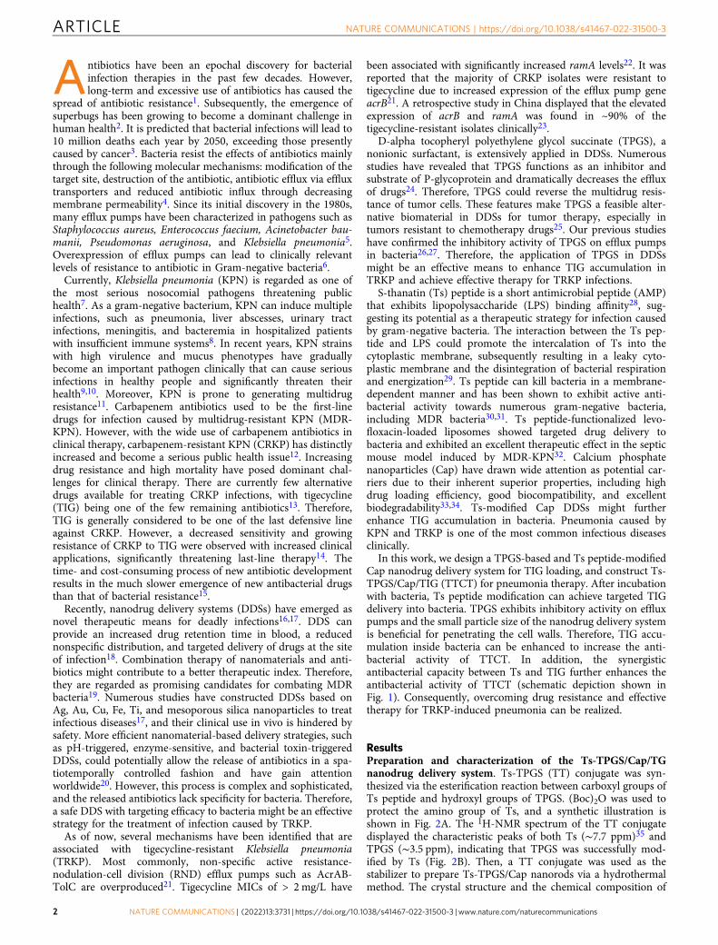
In this work, we design a TPGS-based and Ts peptide-modifiedCap nanodrug delivery system for TIG loading, and construct Ts-TPGS/Cap/TIG (TTCT) for pneumonia therapy. After incubationwith bacteria, Ts peptide modification can achieve targeted TIGdelivery into bacteria. TPGS exhibits inhibitory activity on effluxpumps and the small particle size of the nanodrug delivery systemis beneficial for penetrating the cell walls. Therefore, TIG accu-mulation inside bacteria can be enhanced to increase the anti-bacterial activity of TTCT. In addition, the synergisticantibacterial capacity between Ts and TIG further enhances theantibacterial activity of TTCT (schematic depiction shown inFig. 1). Consequently, overcoming drug resistance and effectivetherapy for TRKP-induced pneumonia can be realized.
ResultsPreparation and characterization of the Ts-TPGS/Cap/TGnanodrug delivery system. Ts-TPGS (TT) conjugate was syn-thesized via the esterification reaction between carboxyl groups ofTs peptide and hydroxyl groups of TPGS. (Boc)2O was used toprotect the amino group of Ts, and a synthetic illustration isshown in Fig. 2A. The 1H-NMR spectrum of the TT conjugatedisplayed the characteristic peaks of both Ts (∼7.7 ppm)35 andTPGS (∼3.5 ppm), indicating that TPGS was successfully mod-ified by Ts (Fig. 2B). Then, a TT conjugate was used as thestabilizer to prepare Ts-TPGS/Cap nanorods via a hydrothermalmethod. The crystal structure and the chemical composition of
ARTICLE NATURE COMMUNICATIONS | https://doi.org/10.1038/s41467-022-31500-3
2 NATURE COMMUNICATIONS | (2022) 13:3731 | https://doi.org/10.1038/s41467-022-31500-3 | www.nature.com/naturecommunications
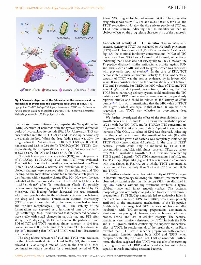
the nanorods were confirmed by comparing the X-ray diffraction(XRD) spectrum of nanorods with the typical crystal diffractionpeaks of hydroxylapatite crystals (Fig. 3A). Afterwards, TIG wasencapsulated into the Ts-TPGS/Cap and TPGS/Cap nanorods bythe dialysis method. When the drug feeding ratio was 20%, thedrug loading (DL %) was 12.47 ± 1.48 for TPGS/Cap/TIG (TCT)nanorods and 12.31 ± 0.94 for Ts-TPGS/Cap/TIG (TTCT). Cor-respondingly, the encapsulation efficiency (EE%) was calculatedat 62.33 ± 6.92 for TCT and 61.53 ± 4.70 for TTCT.
The particle size, polydispersity index (PDI), and zeta potentialof TPGS/Cap, Ts-TPGS/Cap, TCT, and TTCT were evaluated.The particle size of the formulations was maintained at ∼25 nm(Table 1) and showed a narrow size distribution (Fig. 3B). Noobvious changes were observed after Ts modification and TIGloading. All the formulations exhibited monomodal zeta potentialdistributions with a negative charge (Fig. 3C). However, the zetapotential of the nanorods decreased from −18.56 ± 1.66 mV to−14.99 ± 1.66 mV after Ts modification (Table 1), possiblybecause some hydroxyl groups of TPGS were replaced by Ts.Moreover, TIG loading further decreased the zeta potential,which was possibly due to the electrostatic interaction betweenthe drug and nanorods. Transmission electron microscopy(TEM) images showed that all of the formulations had uniformand rod-like morphologies. (Fig. 3D). The stability of theformulations was explored as a function of time by dynamiclight scattering (DLS). It was observed that the prepared nanorodswere stable with small changes in particle size and PDI afterstorage for 20 days (Fig. 3E, F). Furthermore, the particle size andPDI of TCT and TTCT remained almost constant in 10% fetalbovine serum (FBS)-containing PBS within 24 h (as shown inFig. 3G), indicating that TCT and TTCT would not disassemblein circulation.
The drug release behaviors of TCT and TTCT were evaluatedby the dialysis method. As displayed in Fig. 3H, the nanorodsreleased TIG at a rapid rate of ~25% in the first 0.5 h, thencontinued to release the drug for a sustained period of 72 h.
About 50% drug molecules got released at 8 h. The cumulativedrug release was 86.89 ± 0.76 % and 87.80 ± 0.39 % for TCT andTTCT, respectively. Notably, the drug release profiles of TCT andTTCT were similar, indicating that Ts modification had noobvious effects on the drug release characteristics of the nanorods.
Antibacterial activity of TTCT in vitro. The in vitro anti-bacterial activity of TTCT was evaluated on Klebsiella pneumonia(KPN) and TIG-resistant KPN (TRKP) in our study. As shown inFig. 4A, the minimal inhibitory concentrations (MICs) of TIGtowards KPN and TRKP were 1 µg/mL and 4 µg/mL, respectively,indicating that TRKP was not susceptible to TIG. However, theTs peptide displayed similar antibacterial activity against KPNand TRKP, with an MIC value of 16 µg/mL, which was consistentwith previously reported studies36. In the case of KPN, TCTdemonstrated similar antibacterial activity to TIG. Antibacterialcapacity of TTCT was the best as evidenced by its lowest MICvalue. It was possibly related to the combinational effect betweenTIG and Ts peptide. For TRKP, the MIC values of TIG and TCTwere 4 µg/mL and 2 µg/mL, respectively, indicating that theTPGS-based nanodrug delivery system could ameliorate the TIGresistance of TRKP. Similar results were observed in previouslyreported studies and could be related to the activity of effluxpumps26,27. It is worth mentioning that the MIC value of TTCTwas 1 µg/mL, which was equal to that of free TIG against KPN,suggesting that TTCT was effective in overcoming drugresistance.
We further investigated the effect of the formulations on thegrowth curves of KPN and TRKP. During the incubation periodof 24 h with free TIG, TCT, and Ts-TPS/Cap (TIG concentration:0.5 µg/mL; Ts-TPGS/Cap concentration: 5 µg/mL), a remarkableincrease of the OD600 nm values of KPN was observed, indicatingthat they could not prevent the growth of bacteria (Fig. 4B).However, visible growth of bacteria was completely inhibited byTTCT (TIG concentration: 0.5 µg/mL). In the case of TRKP,bacterial growth could only be inhibited by TTCT (TIGconcentration: 1 µg/mL), with almost constant OD600 nm valuesover 24 h of incubation. Growth of TRKP was not prevented byTIG (1 µg/mL, 2 µg/mL), TCT (TIG concentration: 1 µg/mL), andTs-TPGS/Cap (10 µg/mL) (Fig. 4C). The result was in accordancewith that shown in Fig. 4A. As a whole, TTCT demonstratedbetter antibacterial activity than TIG and TCT in both KPNand TRKP.
To further evaluate the antibacterial activity of TTCT, changesin bacterial morphology following the different treatments wereobserved by scanning electron microscopy (SEM). As displayed inFig. 4D, bacteria without any treatment exhibited a typicalclubbed shape and intact smooth surface. The bacterialmorphology was obviously changed after treatment with variouspreparations. Ts-TPGS/Cap nanorods led to noticeable holes intheir cell walls in both KPN and TRKP, which was possiblyattributed to the antibacterial mechanisms of the Ts peptide.Additionally, the magnified SEM images of bacteria afterincubation with TIG-containing preparations demonstratedsignificant morphological changes, such as broken cell mem-branes, debris, and loss of cellular integrity. The bacterialstructures were massively destroyed by TTCT in both the KPNand TRKP groups, further confirming the superior antibacterialeffect of TTCT. In conclusion, all of the results shown in Fig. 4revealed that TTCT was a superior preparation with excellentantibacterial function against both KPN and TRKP whencompared with TIG, TCT, and Ts-TPGS/Cap nanorods. Further-more, the data suggested that TTCT was capable of overcomingthe drug resistance of TRKP and achieved effective antibacterialcapacity towards multidrug-resistant bacteria.
PO42-
Cap Ts-TPGS/Cap Ts-TPGS/Cap/TIG
Ts-TPGS
Entry
TRKP LPS efflux pump ribosome Ts-TPGS TIG
TIG
Proteinsynthesis
tRNA
30S
50S
Ca2+
+
Fig. 1 Schematic depiction of the fabrication of the nanorods and themechanism of overcoming the tigecycline resistance of TRKP. TIGtigecycline, Ts-TPGS/Cap/TIG tigecycline-loaded TPGS and S-thanatinfunctionalized calcium phosphate nanorods, TRKP tigecycline-resistantKlebsiella pneumonia, LPS lipopolysaccharide.
NATURE COMMUNICATIONS | https://doi.org/10.1038/s41467-022-31500-3 ARTICLE
NATURE COMMUNICATIONS | (2022) 13:3731 | https://doi.org/10.1038/s41467-022-31500-3 | www.nature.com/naturecommunications 3

Mechanism of overcoming drug resistance. We speculated thatthe enhanced antibacterial activity of TTCT was attributed to thetargeting capacity given by Ts, inhibitory effect on efflux pumpendowed by TPGS, and synergy antibacterial activity between Tsand TIG. Below are the experiments we conducted to verify ourhypotheses.
In vitro targeting efficiency of Ts-TPGS/Cap nanorods. It wasreported that the β-hairpin structure of thanatin is stabilized bythe disulfide bond between Cys11 and Cys1837, which is con-sidered integral for its activity38. An analog of thanatin with thetwo Cys residues replaced by Ala was found to be largelyinactive39, indicating that the disulfide bond is important for Ts
Ts
TPGS
Ts-TPGS
A
B
Fig. 2 Synthesis and characterization of Ts-TPGS (TT). A Synthetic scheme of the TT conjugate. B 1H-NMR spectra of Ts peptide, TPGS, and TT from topto bottom. The red circles represented the characteristic peaks of the structure displayed in red squares, in which ~ 3.5 ppm represented the -O-CH2-CH2-of TPGS and ~ 7.7 ppm indicated the -CONH2 of Ts peptide. Ts S-thanatin peptide, TPGS tocopheryl polyethylene glycol succinate.
ARTICLE NATURE COMMUNICATIONS | https://doi.org/10.1038/s41467-022-31500-3
4 NATURE COMMUNICATIONS | (2022) 13:3731 | https://doi.org/10.1038/s41467-022-31500-3 | www.nature.com/naturecommunications

0 20 40 60 800
1000
2000
3000
4000
5000
Two-Theta (deg)
)SPC(
ytisnetnI
1 10 100 1000 100000
10
20
30
40
Size (d.nm)
)%(reb
muN
1 10 100 1000 100000
10
20
30
Size (d.nm)
Num
ber (
%)
1 10 100 1000 100000
10
20
30
Size (d.nm)
Num
ber (
%)
1 10 100 1000 100000
10
20
30
Size (d.nm)
Num
ber (
%)
-200 -100 0 100 2000.0
0.2
0.4
0.6
0.8
Zeta potential (mV)
)%(
ycneuqer fe vitaleR
-200 -100 0 100 2000.0
0.2
0.4
0.6
0.8
Zeta potential (mV)
Rela
tive
frequ
ency
(%)
-200 -100 0 100 2000.0
0.2
0.4
0.6
0.8
Zeta potential (mV)
Rela
tive
frequ
ency
(%)
-200 -100 0 100 2000.0
0.2
0.4
0.6
0.8
Zeta potential (mV)
Rela
tive
frequ
ency
(%)
5 10 15 200
10
20
30
40
0.0
0.2
0.4
0.6
Days
Part
icle
siz
e (n
m)
PDI
TPGS/Cap (size)TCT (size)
TPGS/Cap (PDI)TCT (PDI)
5 10 15 200
10
20
30
40
0.0
0.2
0.4
0.6
Time (d)
Part
icle
siz
e (n
m)
PDI
Ts-TPGS/Cap (size)TTCT (size)
Ts-TPGS/Cap (PDI)TTCT (PDI)
0 20 40 60 800
20
40
60
80
100
Time (h)
Cum
lativ
e dr
ug re
leas
e (%
)
TIGTCTTTCT
5 10 15 200
10
20
30
40
50
0.0
0.2
0.4
0.6
Time (h)
)mn(
eziselcitraP
PDI
TCT (size)TTCT (size)
TCT (PDI)TTCT (PDI)
TCTpaC/SGPT Ts-TPGS/Cap TTCT
A
B
C
D
E F
G H
50 nm 50 nm 50 nm 50 nm
Fig. 3 Preparation and characterization of TTCT. A X-ray diffraction of TPGS/Cap nanorods. B Size distribution measured by DLS, C zeta potentialdistribution, and D representative TEM images of TPGS/Cap, Ts-TPGS/Cap, TCT, and TTCT (Scale bar= 50 nm). Particle size and PDI changes ofE TPGS/Cap, Ts-TPGS/Cap, (F) TCT, and TTCT during 1 week of storage (n=3 independent experiments). G Changes in particle size and PDI of TCT andTTCT in 10% fetal bovine serum (FBS)-containing PBS (n= 3 independent experiments). H In vitro drug release profiles of TCT and TTCT in 10% fetalbovine serum (FBS)-containing PBS at pH 7.4 and 37 °C (n= 3 independent experiments). TIG free tigecycline, TPGS/Cap TPGS-functionalized calciumphosphate nanorods, TCT tigecycline loaded TPGS/Cap nanorods, TTCT tigecycline-loaded Ts-TPGS/Cap nanorods. Data are expressed as the mean ± SD,and the mean value is the average of three independent experiments. The experiments in B–D were repeated independently for three times with similarresults. Source data are provided as a Source Data file.
NATURE COMMUNICATIONS | https://doi.org/10.1038/s41467-022-31500-3 ARTICLE
NATURE COMMUNICATIONS | (2022) 13:3731 | https://doi.org/10.1038/s41467-022-31500-3 | www.nature.com/naturecommunications 5

activity. Considering this, an analog of Ts peptide with two Cysresidues replaced by Ala (GSKKPVPIIYANRRSGKAQRM) wassynthesized and served as the non-targeting peptide. The non-targeting peptide-conjugated TPGS/Cap (NT-TPGS/Cap) wasprepared according to the preparation process of Ts-TPGS/Capand used as control to evaluate the in vitro targeting efficiency ofTs-TPGS/Cap nanorods. As shown in Fig. 5A, both NT-TPGS/
Cap and Ts-TPGS/Cap nanorods displayed a gradual increase inthe intra-bacteria fluorescent signal with a prolonged incubationtime. Ts-TPGS/Cap nanorods displayed stronger bacterial inter-nalization capacity than NT-TPGS/Cap nanorods in both KPNand TRKP at 2 and 6 h. The results were consistent with thoseobtained by flow cytometry (Fig. 5B, C). The fluorescent signalsin KPN after incubation with Ts-TPGS/Cap nanorods were ~2-fold greater than those of NT-TPGS/Cap nanorods at both 2 hand 6 h (Fig. 5D). For TRKP, similar results were observed,suggesting that Ts-TPGS/Cap nanorods exhibited better bacterialinternalization activity, which was possibly attributed to the tar-geting moiety Ts peptide.
The inhibitory effect of TPGS on efflux pumps. It has beenreported that the accumulation of ethidium bromide (EB) isinversely correlated with the activity of efflux pumps27. Therefore,EB was exploited as a substrate of the efflux pump40.
paC/SGPT-sTTCTTTCTGITlortnoC
paC/SGPT-sTTCTTTCT)L( GITlortnoC TIG (H)
KPN
TRK
P
PKRTNPKA
D
CB
Fig. 4 Evaluation of the antibacterial activity of TTCT towards KPN and TRKP in vitro. A MIC susceptibility semiquantitative profiles of TTCT via themicroplate broth dilution method (n= 3 independent experiments, and the results were similar). Growth curves of KPN (B) and TRKP (C) co-incubatedwith various preparations. D Representative SEM images of KPN and TRKP after treatment with TIG, TCT, TTCT, and Ts-TPGS/Cap nanorods. Scale bar onthe original SEM images and the enlarged images were 10 μm and 2 μm, respectively. TIG free tigecycline, Ts-TPGS/Cap Ts-TPGS functionalized calciumphosphate nanorods, TCT tigecycline loaded TPGS/Cap nanorods, TTCT tigecycline-loaded Ts-TPGS/Cap nanorods, KPN Klebsiella pneumonia, TRKPtigecycline-resistant Klebsiella pneumonia. The experiments in A–D were repeated independently for three times with similar results. Source data areprovided as a Source Data file.
Table 1 Characterization of TPGS/Cap, Ts-TPGS/Cap, TCT,and TTCT.
Preparation dn (nm) PDI ζ (mV)
TPGS/Cap 27.1 ± 4.8 0.29 ± 0.03 −18.56 ± 1.66Ts-TPGS/Cap 25.9 ± 2.3 0.27 ± 0.01 −14.99 ± 1.66TCT 26.6 ± 3.9 0.24 ± 0.03 −14.83 ± 2.28TTCT 28.2 ± 3.3 0.29 ± 0.03 −11.64 ± 1.97
ARTICLE NATURE COMMUNICATIONS | https://doi.org/10.1038/s41467-022-31500-3
6 NATURE COMMUNICATIONS | (2022) 13:3731 | https://doi.org/10.1038/s41467-022-31500-3 | www.nature.com/naturecommunications

The inhibitory effect of TPGS on efflux pumps was investigatedby comparing it to carbonyl cyanide m-chlorophenylhydrazone(CCCP), a well-known inhibitor of proton pump. As shown inFig. 5E, EB accumulation in TRKP was negligible in the controlgroup. After incubation with TPGS, the fluorescent signal of EBinside bacteria was significantly stronger than that in the control
group. Quantitative analysis showed that the increased percen-tages of EB accumulation grew from 22.11 ± 3.67% to64.75 ± 5.91% as the concentration of TPGS increased from 20 to100 µM (Fig. 5F), indicating that TPGS treatment manifested anobvious increase in EB accumulation. Similar results wereobserved in previously reported studies27.
Ts-TPGS/Cap 2h
NT-TPGS/Cap 2h
NT-TPGS/Cap 6h
Ts-TPGS/Cap 6h
Negeative control
KPN TRKP
637
2543
5283
6845
10674
646
2453
5332
7140
10867
A
B
KPN (2 h)Ts-TPGS/Cap NT-TPGS/Cap
TRKP (2 h)Ts-TPGS/Cap NT-TPGS/Cap
D
Control TPGS (20 M) TPGS (50 M) TPGS (100 M) CCCP (100 M)E
KPN (6 h)Ts-TPGS/Cap NT-TPGS/Cap
TRKP (6 h)Ts-TPGS/Cap NT-TPGS/Cap
IPA
DCTIF
degreM
C
Ts-TPGS/Cap (2 h)
NT-TPGS/Cap (2 h)
Ts-TPGS/Cap (6 h)
NT-TPGS/Cap (6 h)
F G TIG 2 h
TCT 2 h
TTCT 2 h
TIG 6 h
TCT 6 h
TTCT 6 h
KPN TRKP0
3000
6000
9000
12000
Fluo
resc
ence
inte
nsity
p=3.2e-5 p=2.6e-5
p=0.0048 p=0.0042
TPGS (20M)
TPGS (50M)
TPGS (100
M)
CCCP (100
M)0
20
40
60
80
100
Incr
ease
ofEB
accu
mu l
atio
n(%
)
KPN TRKP0
20
40
60
80
TIG
conc
entr
atio
n(n
g/m
L)
p=1.5e-8
p=7.1e-8p=2.4e-10
p=2.0e-8
p=2.3e-10
p=3.4e-10 p=4.7e-9
p=6.1e-5
NATURE COMMUNICATIONS | https://doi.org/10.1038/s41467-022-31500-3 ARTICLE
NATURE COMMUNICATIONS | (2022) 13:3731 | https://doi.org/10.1038/s41467-022-31500-3 | www.nature.com/naturecommunications 7

In addition, tigecycline resistance in TRKP has been demon-strated to be associated with the overexpression of RND-typeefflux pump genes (acrA and acrB) and ramA41. Real-time reversetranscription-PCR (RT-PCR) was used to determine the levels ofacrA, acrB, and ramA. After bacteria harvesting in the mid-exponential phase, KPN and TRKP were exposed to TPGS, Ts,Ts-TPGS, and Ts-TPGS/Cap nanorods, respectively. As shown insupplementary Fig. 1, the expression levels of acrA, acrB, andramA were up-regulated significantly compared with those inKPN, which confirmed that the AcrAB-TolC were involved in theTIG resistance of TRKP. Intervenation of TPGS, Ts, Ts-TPGS,and Ts-TPGS/Cap obviously down-regulated the expressionlevels. It is worth mentioning that Ts treatment also down-regulated the expression levels of efflux pumps, and we speculatedthat the phenomenon was associated with the Ts activity on theouter membrane of the bacteria29,42. Ts-TPGS and Ts-TPGS/Capnanorods displayed the highest reduction of efflux pump geneexpression, possibly owing to the combinational effect of TPGSand Ts. Based on these results, we confirmed that TPGS had aninhibitory effect on the activity and expression levels of effluxpumps of TRKP. The results were consistent with those reportedpreviously26. However, the underlying mechanisms remainpoorly determined. As reported, surfactants can induce mem-brane fluidity alterations and ATPase inhibition43. Therefore, weconjectured that TPGS inhibited activity of efflux pump possiblyvia its surfactant characteristic.
TIG concentration inside bacteria. The targeting efficacy andthe inhibitory effect on efflux pumps were beneficial for TIGdelivery into bacteria. Therefore, the TIG concentration insidebacteria after KPN and TRKP incubation with free TIG, TCT,and TTCT was measured by high performance liguid chroma-tography- mass spectroscopy (HPLC-MS). As shown in Fig. 5G,the TIG concentration increased over time after the bacteriaincubation with TIG, TCT, and TTCT. In the case of KPN, bothTCT and TTCT led to a large increase in the TIG concentrationthan TIG group, and TTCT exhibited a higher concentration thanTCT at both 2 h and 6 h. These results indicated that TIG deliveryinto bacteria was better after encapsulation in nanorods, and theeffect was further enhanced by Ts peptide modification. Similarresults were observed for TRKP. The TIG concentration in theTRKP group was only ~30% of that in the KPN group when thebacteria were incubated with TIG, which could be a reasonableexplanation for the TIG resistance of TRKP. Combined with theresults shown in Fig. 5E, we inferred that this was possiblybecause the efflux pumps would induce transport of TIG frominside to outside bacteria. This phenomenon was obviouslyameliorated by the TPGS-based nanodrug delivery system. TheTs peptide modification further increased the TIG concentrationinside bacteria. Overall, the higher TIG concentration of TTCTamong these groups in TRKP was possibly due to the targeting
efficiency induced by Ts, inhibitory effect on efflux pump activity,and TIG encapsulation in nanorods.
Synergy antibacterial activity between Ts and TIG. The inter-action between Ts and tigecycline was investigated by the micro-dilution checkerboard technique according to CLSI guidelines.The data exhibited that the fractional inhibitory concentrationindices between Ts and tigecycline against KPN and TRKP were0.75 and 0.375, respectively, indicating that Ts exerted synergisticeffect with tigecycline against TRKP and additive effect againstKPN (shown in supplementary Table 1).
Overall, the underlying mechanism of TTCT overcoming thedrug resistance might include the following aspects: (1) The smallparticle size of the nanorods was beneficial for penetrating the cellwalls; (2) TIG accumulation in bacteria was significantlyenhanced via the targeting capacity of Ts to bacteria and theinhibitory effect on efflux pumps exerted by TPGS; (3) Thesynergistic antibacterial capacity between Ts and TIG furtherenhanced the antibacterial activity TTCT.
Biodistribution in vivo. Using tracheal injection of KPN orTRKP bacteria into the lungs of healthy mice, we created acutepneumonia mouse models and investigated the in vivo targetingefficiency of Ts-TPGS/Cap nanorods. The mice were intrave-nously injected with indocyanine green (ICG)-labeled nanorodsat 5 h postoperation. Upon administration of Ts-TPGS/Capnanorods, we evaluated the in vivo biodistribution of thenanorods 5 h and 24 h later. As shown in Fig. 6A, B, Ts-TPGS/Cap nanorods displayed similar bio-distribution characteristicsbetween pneumonia mice infected by KPN and TRKP. The samephenomenon was also observed in mice treated with TPGS/Capnanorods. Moreover, the fluorescence signals were much strongerin lungs treated with Ts-TPGS/Cap nanorods than in thosetreated with TPGS/Cap, implying that more Ts-TPGS/Capnanorods accumulated in the infected lungs (Fig. 6A, B). Inpneumonia mice caused by both KPN and TRKP, the quantitativesignals of fluorescence (expressed by fluorescence intensity pergram of tissue, ID/g) of Ts-TPGS/Cap nanorods in the lung were~1.5-fold and ~2.5-fold higher than those of TPGS/Cap at 5 h and24 h, respectively (Fig. 6C, D). Notably, the Ts-TPGS/Capnanorods were primarily distributed to the liver, lung, and kidneyof the pneumonia mice at 24 h. The ID/g of the lungs were higherthan those of the liver and kidneys.
Pneumonia may evoke disruption of pulmonary endothelialbarrier integrity44, and the vascular permeability may besignificantly increased at the sites of bacterial infection45. It hasbeen reported that the enhanced permeability and retention(EPR) effect might operate to deliver nanoparticles to bacterial-infected tissues46. We speculated that the nanorods we preparedmight be distributed into the infected lung via EPR effect afterintravenous administration. Then the nanorods could be
Fig. 5 Investigation of the mechanisms by which TTCT overcomes drug resistance. Fluorescence images of KPN and TRKP after 2 h and 6 h of incubationwith NT-TPGS/Cap and Ts-TPGS/Cap nanorods. The bacteria were labeled with DAPI (blue), and the green fluorescence signal indicated the nanorods.Scale bar= 20 μm. Fluorescence within KPN (B) and TRKP (C) detected by flow cytometry after incubation with NT-TPGS/Cap and Ts-TPGS/Cap.D Quantitative analysis of fluorescence inside KPN and TRKP after co-culture with nanorods. Data are expressed as the mean ± SD (n= 3 independentexperiments). E Confocal fluorescent images of TRKP after exposure to EB for 4 h, following pretreatment with TPGS and CCCP. Scale bar= 10 µm.F Quantitative analysis of the increase in EB accumulation compared with the negative control groups (n= 3 independent experiments). G TIGconcentration detected by HPLC-MS inside KPN and TRKP after incubation with TIG-containing preparations (n= 6 independent experiments). Ts-TPGS/Cap Ts-TPGS functionalized calcium phosphate nanorods, NT-TPGS/Cap non-targeting peptide-TPGS functionalized calcium phosphate nanorods, TIGfree tigecycline, TCT tigecycline loaded TPGS/Cap nanorods, TTCT tigecycline-loaded Ts-TPGS/Cap nanorods, CCCP carbonyl cyanide m-chlorophenylhydrazone, EB ethidium bromide, KPN Klebsiella pneumonia, TRKP tigecycline-resistant Klebsiella pneumonia. Data are expressed as themean ± SD. Unpaired two-tailed T-test was performed in D. One-way analysis of variance (ANOVA) with post hoc Tukey tests were performed in G. Theexperiments in A and E were repeated independently for three times with similar results. Source data are provided as a Source Data file.
ARTICLE NATURE COMMUNICATIONS | https://doi.org/10.1038/s41467-022-31500-3
8 NATURE COMMUNICATIONS | (2022) 13:3731 | https://doi.org/10.1038/s41467-022-31500-3 | www.nature.com/naturecommunications

internalized by vascular endothelial cells (Data in supplementaryFig. 2 demonstrated that the nanorods had excellent cellularinternalization capacity) and be transported across the bloodvessel. The binding affinity between Ts and LPS promoted theaccumulation of Ts-functionalized nanorods in the infected lung.Overall, an in vivo distribution study revealed that Ts-TPGS/Capnanorods could realize targeting accumulation in the lungs ofpneumonia mice, which was beneficial for achieving better anti-infective efficacy.
Anti-infective efficacy of TTCT in vivo. Acute KPN- and TRKP-infected pneumonia model was used to investigate the in vivoanti-infective activity of TTCT. After 5 h of modeling, the micewere intravenously administered TIG, TCT, TTCT, and Ts-TPGS/Cap nanorods. Then, the survival rate of mice receivingdifferent treatments was recorded within 5 days. As shown inFig. 7A, B, the pneumonia mice infected by TRKP survived 33.3%at 5 days, compared with the KPN-infected mice at 16.7%. Thisdifference could be explained by the fact that after resistance isacquired, the bacteria may become less virulent47,48. KPN-infected mice receiving TIG and TCT treatment (TIG dose:15 mg/kg) survived 66.7% and 83.3%, respectively, indicating thatencapsulating TIG in nanorods boosted its antibacterial activity.Moreover, Ts-TPGS/Cap nanorod treatment also enhanced thesurvival rate of KPN-infected mice.
In contrast, the TRKP pneumonia mice receiving therapy ofTIG (15 mg/kg) demonstrated a lower survival rate than theKPN-infected mice, possibly attributed to the nonsusceptibility ofTRKP to TIG. 66.7% of TRKP-infected mice were alive after45 mg/kg TIG treatment, but the rate was still lower than that of
mice given TCT (TIG dose: 15 mg/kg). This phenomenonsuggested that TPGS-based nanodrug delivery systems couldovercome bacterial resistance and achieve effective therapeuticeffects against infection induced by drug-resistant bacteria.Combined with the data shown in Fig. 5E, F, the results couldbe explained by the finding that TPGS was capable of inhibitingthe activity of efflux pumps. Additionally, the TTCT treatmentgroup had the highest survival rate of 100% in pneumonia micecaused by both KPN and TRKP. The superior therapeutic effectcompared with other groups might be attributed to the targetingdistribution, inhibitory activity of efflux pumps, and Tsantibacterial peptide.
Inflammatory markers (white blood cell count, neutrophilcount, and level of C reactive protein) are important indicators ofinflammation that can be used to evaluate infection. Therefore,the survived mice receiving therapy of various preparations weresacrificed after 48 h for collection of blood samples andbronchoalveolar lavage fluid (BALF). First, we evaluated thelevels of white blood cells (WBCs) and neutrophils in bloodsamples. The results showed that bacterial infection caused anobvious increase of WBCs and neutrophils (Fig. 7C–D). Asignificant reduction in the levels of WBCs and neutrophils wasobserved when the KPN- and TRKP-infected mice were treatedwith TIG, TCT, and TTCT, indicating that the infection wasapparently alleviated. Moreover, a greater reduction in the levelsof WBCs and neutrophils was observed in the TTCT group inboth KPN- and TRKP-infected mice, demonstrating that TTCThad a superior therapeutic effect than the other treatments. Itshould be mentioned that Ts-TPGS/Cap nanorods alone hadpositive anti-infective activity towards KPN- and TRKP-infectedmice. The reduction in WBC and neutrophil levels in the
KPN (24 h)TPGS/Cap Ts-TPGS/Cap
TRKP (24 h)TPGS/Cap Ts-TPGS/Cap
Heart
Liver
Spleen
Lung
Kidney
Low High
KPN (5 h)TPGS/Cap Ts-TPGS/Cap
TRKP (5 h)TPGS/Cap Ts-TPGS/Cap
Heart
Liver
Spleen
Lung
Kidney
Low High
A B
C D
KPN + TPGS/Cap
KPN + Ts-TPGS/Cap
TRKP + TPGS/Cap
TRKP + Ts-TPGS/CapHeart Liver Spleen Lung Kidney
0
20
40
60
80
ID/g
(105 )
p=0.0011 p=0.0014
Heart Liver Spleen Lung Kidney0
20
40
60
80ID
/g(1
05 )p=0.0001 p=2.1E-6
Fig. 6 Evaluation of the biodistribution and targeting efficacy of Ts-TPGS/Cap in vivo. Fluorescence images of major organs collected from KPN- andTRKP-infected mice with pneumonia (A) at 5 h (B) and 24 h postinjection of TPGS/Cap and Ts-TPGS/Cap nanorods. Semiquantitative analysis of thefluorescence intensity in major organs displayed in (C) Fig. 6A and (D) Fig. 6B (n= 3 mice). Ts-TPGS/Cap Ts-TPGS functionalized calcium phosphatenanorods, TPGS/Cap TPGS-functionalized calcium phosphate nanorods, KPN Klebsiella pneumonia, TRKP tigecycline-resistant Klebsiella pneumonia. Data areshown as the mean ± SD (n= 3 mice for each group). Unpaired two-tailed T-test was performed in C, D. Source data are provided as a Source Data file.
NATURE COMMUNICATIONS | https://doi.org/10.1038/s41467-022-31500-3 ARTICLE
NATURE COMMUNICATIONS | (2022) 13:3731 | https://doi.org/10.1038/s41467-022-31500-3 | www.nature.com/naturecommunications 9
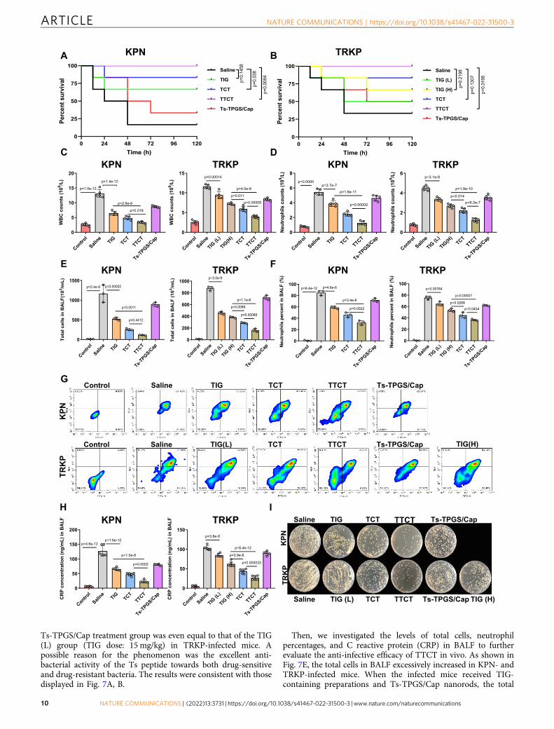
Ts-TPGS/Cap treatment group was even equal to that of the TIG(L) group (TIG dose: 15 mg/kg) in TRKP-infected mice. Apossible reason for the phenomenon was the excellent anti-bacterial activity of the Ts peptide towards both drug-sensitiveand drug-resistant bacteria. The results were consistent with thosedisplayed in Fig. 7A, B.
Then, we investigated the levels of total cells, neutrophilpercentages, and C reactive protein (CRP) in BALF to furtherevaluate the anti-infective efficacy of TTCT in vivo. As shown inFig. 7E, the total cells in BALF excessively increased in KPN- andTRKP-infected mice. When the infected mice received TIG-containing preparations and Ts-TPGS/Cap nanorods, the total
PK
RTNP
K
Control TIGSaline TCT TTCT Ts-TPGS/Cap
Control TIG(L)Saline TCT TTCT Ts-TPGS/Cap TIG(H)
TRK
PK
PN
TIG (L)Saline TCT TTCT TIG (H)Ts-TPGS/Cap
TIGSaline TCT TTCT Ts-TPGS/Cap
A B
C DKPN TRKP KPN TRKP
E FKPN TRKP KPN TRKP
G
KPN TRKPH I
KPN TRKP
Control
Saline
TIG TCTTTCT
Ts-TPGS/C
ap0
5
10
15
20
WB
Cco
unts
(109 /L
) p=1.4e-12
p=0.019
p=2.6e-6
p=1.0e-12
Control
Saline
TIG TCTTTCT
Ts-TPGS/C
ap0
2
4
6
8
Neu
troph
ilsc o
u nt s
(109 /L
)
p=0.0000p=3.7e-7
p=0.00002
p=1.6e-11
Control
Saline
TIG(L)
TIG(H
)TCT
TTCT
Ts-TPGS/C
ap0
2
4
6
Neu
troph
ilsc o
unt s
( 109 /L
)
p=0.014
p=1.9e-10
p=3.1e-8
p=9.2e-7
Control
Saline
TIG(L)
TIG(H
)TCT
TTCT
Ts-TPGS/C
ap0
5
10
15W
BC
coun
ts(1
09 / L)
p=0.011
p=4.0e-9
p=0.00014
p=0.00005
Contro
l
Saline TIG TC
TTT
CT
Ts-TP
GS/Cap
0
500
1000
1500
Tota
lcel
lsin
BA
LF(1
03 /mL)
p=2.4e-8 p=0.00002
p=0.4412
p=0.0011
Control
Saline
TIG(L)
TIG(H
)TCT
TTCT
Ts-TPGS/C
ap0
200
400
600
800
1000
Tota
lcel
lsi n
BA
LF( 1
03 /mL)
p=0.0069
p=1.1e-6
p=3.0e-9
p=0.00068
Control
Saline
TIG TCTTTCT
Ts-TPGS/C
ap0
20
40
60
80
100N
eutr
ophi
l spe
rcen
tin
BA
LF(%
)p=6.4e-12 p=4.6e-6
p=3.4e-6
p=0.0022
Control
Saline
TIG(L)
TIG(H
)TCT
TTCT
Ts-TPGS/C
ap0
20
40
60
80
100
Neu
trop
hils
perc
e nti
nB
ALF
(%)
p=0.0268
p=0.00007
p=0.00764
p=0.0454
Control
Saline
TIG TCTTTCT
Ts-TPGS/C
ap0
50
100
150
200
CR
Pco
ncen
trat
ion
(ng/
mL)
inB
ALF
p=4.6e-12p=1.6e-12
p=0.0002
p=1.0e-8
Control
Saline
TIG(L)
TIG(H
)TCT
TTCT
Ts-TPGS/C
ap0
50
100
150
CR
Pco
ncen
trat
i on
(ng/
mL )
inB
ALF
p=3.9e-6
p=6.4e-12
p=3.8e-6
p=0.000033
0 24 48 72 96 1200
25
50
75
100
Time (h)
Perc
ents
urvi
val
Saline
TIG
TCT
TTCT
Ts-TPGS/Cap
p=0.
1458
p =0.
038
p=0.
0 06 4
0 24 48 72 96 1200
25
50
75
100
Time (h)
Perc
ents
urvi
val
Saline
TIG (L)
TIG (H)
TCT
TTCT
Ts-TPGS/Cap
p=0.
2198
p=0.
0195
p=0 .
1 307
ARTICLE NATURE COMMUNICATIONS | https://doi.org/10.1038/s41467-022-31500-3
10 NATURE COMMUNICATIONS | (2022) 13:3731 | https://doi.org/10.1038/s41467-022-31500-3 | www.nature.com/naturecommunications
cells exhibited a significant reduction, and TTCT displayed thehighest reduction among these groups. Neutrophil infiltrationinto pulmonary interstitial fluid could result in even furtherpulmonary edema and deteriorating inflammation49,50. Thus, weadditionally evaluated pulmonary neutrophil infiltration. Theneutrophils were labeled with PE-labeled anti-Ly-6G/Ly-6C andFITC-labeled anti-CD11b antibodies. The percentages of neu-trophils in total cells were determined by flow cytometry. It wasobserved that neutrophils accounted for ~85% and ~75% of thetotal cells in KPN-infected and TRKP-infected mice, respectively(Fig. 7F). The percentages of neutrophils were apparentlydecreased after administration of TIG, TCT, TTCT, and Ts-TPGS/Cap nanorods, clearly indicating that pulmonary neutro-phil infiltration was ameliorated. It is worth mentioning thatTTCT treatment still exhibited the best effects among thetreatments. Moreover, Ts-TPGS/Cap nanorods displayed aninhibitory effect on pulmonary neutrophil infiltration similar tothat of TIG (15 mg/kg) in TRKP-infected mice. Representativeimages of flow cytometry were shown in Fig. 7G.
CRP is a well-documented indicator of bacterial infection51.The levels of CRP in BALF were determined using a commercialELISA kit. As shown in Fig. 7H, the levels of CRP were markedlyelevated in KPN- and TRKP-infected mice. However, its level wasapparently decreased after treatment with TIG, TCT, and Ts-TPGS/Cap nanorods, and the effect was further enhanced byTTCT. In addition, BALF was utilized for bacterial colonyanalysis, and TTCT showed the lowest bacterial colonies inpneumonia mice infected by KPN and TRKP among the micereceiving various therapy (Fig. 7I and quantitative analysis resultsshown in supplementary Fig. 3). Overall, TTCT therapyconsistently displayed the best therapeutic effect, with lowercounts of WBCs and neutrophils in blood, lower pulmonaryneutrophil infiltration, and higher reductions in CRP levels andbacterial colonies. These results could possibly be explained byantibacterial activity of Ts peptides and inhibitory effect of effluxpumps, thereby increasing the TIG concentration in the lungsand bacteria.
Positron emission tomography-computed tomography (PET-CT) imaging. PET/CT imaging systems have been widely used inclinical diagnosis and treatment52. Increased capillary perme-ability at the initial stage of infection could lead to the accumu-lation and activation of inflammatory cells, and activatedinflammatory cells mainly metabolize glucose as an energysource53. Therefore, to provide more evidence of the antibacterialeffect, PET imaging with 18F-labeled fluorodeoxyglucose (18F-FDG) was employed to evaluate inflammation and infection indifferent groups. As shown in Fig. 8A, KPN and TRKP infectioncaused a significant increase in 18FDG uptake in the lung. Thelevels of 18FDG uptake were reduced after the infected mice weretreated with TIG, TTCT, and Ts-TPGS/Cap, as reflected bydecreased standardized uptake values (SUVs). This phenomenon
was further ameliorated by TTCT intervention, demonstratingthat TTCT exhibited a superior effect compared with the othertreatment groups, which was consistent with the results displayedin Fig. 7.
Histopathology. To evaluate the effects of different treatments onpathological changes, H&E staining was performed at 5 dayspost-administration. As expected, the lungs of the KPN- andTRKP-infected mice displayed typical pathological characteristics,such as alveolar wall thickening, edema, and leukocyte infiltration(Fig. 8B). Histological improvements were apparently observedafter receiving TIG, TCT, and Ts-TPGS/Cap nanorods. TTCTdisplayed more pronounced amelioration of pulmonary inflam-matory infiltration, edema, and alveolar wall thickening than theother groups. Furthermore, lungs were stained with myeloper-oxidase (MPO) antibody to investigate neutrophil infiltration.Confocal images showed that bacterial infection resulted in sig-nificant neutrophil infiltration in the lungs, and this phenomenonwas obviously ameliorated after the different treatments (Fig. 8C).Consistent with the results shown in Fig. 8A, TTCT stilldemonstrated better therapeutic effects than the other groups.
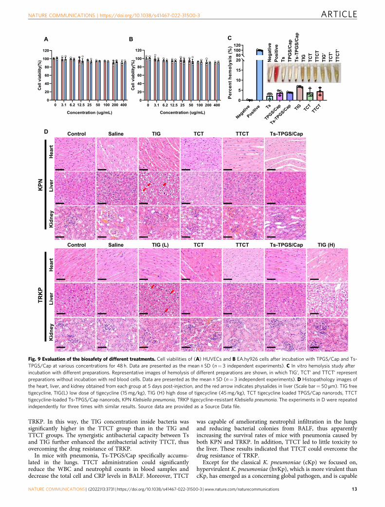
Biosafety of TTCT. The cytotoxicity of TPGS/Cap and Ts-TPGS/Cap nanorods, a critical factor for their further applicationin vivo, was evaluated by the MTT method in HUVECs andEA.hy926 cells. The cell viability was over 90% when the con-centration of nanorods reached 0.4 mg/mL, implying that bothTPGS/Cap and Ts-TPGS/Cap had excellent biocompatibility(Fig. 9A, B). The hemolysis percentage was ~ 3% for Ts, TPGS/Cap, and Ts-TPGS/Cap nanorods. For TIG, TCT, and TTCT, thehemolysis percentages were 6.80 ± 0.11%, 3.92 ± 0.52%, and4.13 ± 0.96%, respectively, implying that TIG encapsulationincreased its biosafety (Fig. 9C). When the pneumonia micecaused by KPN and TRKP received TIG therapy, apparent fattyand hydropic degeneration in the liver was observed (Fig. 9D).However, no obvious pathologic changes in the collected organswere observed in the TCT, TTCT, and Ts-TPGS/Cap nanorodgroups, indicating that the adverse effect of TIG was decreasedafter encapsulation into nanodrug delivery systems. These resultswere consistent with those shown in Fig. 9C.
DiscussionIn summary, a TPGS-based and Ts-modified nanodrug deliverysystem with LPS targeting and synergistic antibacterial activitywas designed for TIG delivery and therapy of acute pneumoniacaused by MDR bacteria. The prepared Ts-TPGS/Cap nanorodscould effectively encapsulate TIG and achieve sustained drugrelease. Through the binding between Ts and LPS, Ts-TPGS/Capexhibited targeting and enhanced accumulation in both KPN andTRKP. TPGS could exert its inhibitory capacity on the activity ofefflux pumps and the expression of acrA, acrB and ramA in
Fig. 7 Investigation of the anti-infective efficacy of TTCT in vivo. A Survival rates of acute KPN-infected and (B) TRKP-infected mice with pneumoniatreated with TIG, TCT, TTCT, or Ts-TPGS/Cap nanorods for 5 days (n= 6 mice per group). Mice with pneumonia treated with saline were used as anegative control. C WBC and D neutrophil counts in blood samples from mice with pneumonia receiving various preparations (n= 5 mice for each group).E Total cell counts and F neutrophil percentages in BALF obtained from KPN- and TRKP-infected mice with pneumonia administered different preparations(n= 3 mice for each group). G Flow cytometry results of anti-Ly-6G/Ly-6C and anti-CD11b double-stained cells obtained from the BALF of mice withpneumonia. Double-positive cells represented neutrophils in BALF. H Levels of CRP in the BALF of mice with pneumonia after different treatments (n= 6mice for each group). I Representative KPN and TRKP bacterial colonies formed on LB agar plates from the BALF of mice with pneumonia receiving varioustreatments. TIG free tigecycline, TIG(L) low dose of tigecycline (15 mg/kg), TIG (H) high dose of tigecycline (45mg/kg), TCT tigecycline loaded TPGS/Cap nanorods, TTCT tigecycline-loaded Ts-TPGS/Cap nanorods, KPN Klebsiella pneumonia, TRKP tigecycline-resistant Klebsiella pneumonia. Data arepresented as the mean ± SD. Log-rank (Mantel-Cox) test was performed in A and B. One-way analysis of variance (ANOVA) with post hoc Tukey testswere performed in C, D, E, F, and H. Source data are provided as a Source Data file.
NATURE COMMUNICATIONS | https://doi.org/10.1038/s41467-022-31500-3 ARTICLE
NATURE COMMUNICATIONS | (2022) 13:3731 | https://doi.org/10.1038/s41467-022-31500-3 | www.nature.com/naturecommunications 11

A
C
NPK
PK
RT
Control TIGSaline TCT TTCT Ts-TPGS/Cap
Control TIG (L)Saline TCT TTCT Ts-TPGS/Cap TIG (H)
Control TIGSaline TCT TTCT Ts-TPGS/Cap
TIG (L)Saline TCT TTCT Ts-TPGS/CapTIG (H)
NPK
PK
RTNP
KP
KRT
Control TIGSaline TCT TTCT Ts-TPGS/Cap
Control TIG (L)Saline TCT TTCT Ts-TPGS/Cap TIG (H)
B
Fig. 8 Histopathology observation of lung tissue. A Representative images of the lungs observed by PET-CT after administration to various groups (Scalebar= 5mm). B Representative images of lung tissue from different groups after H&E staining (scale bar= 50 µm). C Fluorescent images of the lung tissuecollected from mice with pneumonia and stained with anti-MPO antibody, in which the green channel indicates neutrophil infiltration and the blue channelrepresents the nucleus. Scale bar= 50 µm. TIG free tigecycline, TIG(L) low dose of tigecycline (15 mg/kg), TIG (H) high dose of tigecycline (45mg/kg),TCT tigecycline loaded TPGS/Cap nanorods, TTCT tigecycline-loaded Ts-TPGS/Cap nanorods, KPN Klebsiella pneumonia, TRKP tigecycline-resistantKlebsiella pneumonia. The experiments in B, C were repeated independently for three times with similar results.
ARTICLE NATURE COMMUNICATIONS | https://doi.org/10.1038/s41467-022-31500-3
12 NATURE COMMUNICATIONS | (2022) 13:3731 | https://doi.org/10.1038/s41467-022-31500-3 | www.nature.com/naturecommunications
TRKP. In this way, the TIG concentration inside bacteria wassignificantly higher in the TTCT group than in the TIG andTTCT groups. The synergistic antibacterial capacity between Tsand TIG further enhanced the antibacterial activity TTCT, thusovercoming the drug resistance of TRKP.
In mice with pneumonia, Ts-TPGS/Cap specifically accumu-lated in the lungs. TTCT administration could significantlyreduce the WBC and neutrophil counts in blood samples anddecrease the total cell and CRP levels in BALF. Moreover, TTCT
was capable of ameliorating neutrophil infiltration in the lungsand reducing bacterial colonies from BALF, thus apparentlyincreasing the survival rates of mice with pneumonia caused byboth KPN and TRKP. In addition, TTCT led to little toxicity tothe liver. These results indicated that TTCT could overcome thedrug resistance of TRKP.
Except for the classical K. pneumoniae (cKp) we focused on,hypervirulent K. pneumoniae (hvKp), which is more virulent thancKp, has emerged as a concerning global pathogen, and is capable
0 3.1 6.2 12.5 25 50 100 200 4000
20
40
60
80
100
120
Concentration (ug/mL)
Cel
lvia
bilit
y(%
)
0 3.1 6.2 12.5 25 50 100 200 4000
20
40
60
80
100
120
Concentration (ug/mL)
Cel
lvia
bili t
y(%
)
Negati
ve
Positive Ts
TPGS/Cap
Ts-TPGS/C
ap TIG TCTTTCT
0
5
10
15
2080
100120
Per
cent
hem
olys
i s( %
)
Neg
ativ
ePo
sitiv
eTs TP
GS/
Cap
Ts-T
PGS/
Cap
TIG
TCT
TTC
TTI
G’
TCT’
TTC
T’
ANP
K
Control TIGSaline TCT TTCT Ts-TPGS/Cap
Hea
rtLi
ver
yendiK
Control TIG (L)Saline TCT TTCT Ts-TPGS/Cap TIG (H)
Hea
rtLi
ver
y en diK
PK
RTB C
D
Fig. 9 Evaluation of the biosafety of different treatments. Cell viabilities of (A) HUVECs and B EA.hy926 cells after incubation with TPGS/Cap and Ts-TPGS/Cap at various concentrations for 48 h. Data are presented as the mean ± SD (n= 3 independent experiments). C In vitro hemolysis study afterincubation with different preparations. Representative images of hemolysis of different preparations are shown, in which TIG’, TCT’ and TTCT’ representpreparations without incubation with red blood cells. Data are presented as the mean ± SD (n= 3 independent experiments). D Histopathology images ofthe heart, liver, and kidney obtained from each group at 5 days post-injection, and the red arrow indicates physalides in liver (Scale bar= 50 μm). TIG freetigecycline, TIG(L) low dose of tigecycline (15 mg/kg), TIG (H) high dose of tigecycline (45mg/kg), TCT tigecycline loaded TPGS/Cap nanorods, TTCTtigecycline-loaded Ts-TPGS/Cap nanorods, KPN Klebsiella pneumonia, TRKP tigecycline-resistant Klebsiella pneumonia. The experiments in D were repeatedindependently for three times with similar results. Source data are provided as a Source Data file.
NATURE COMMUNICATIONS | https://doi.org/10.1038/s41467-022-31500-3 ARTICLE
NATURE COMMUNICATIONS | (2022) 13:3731 | https://doi.org/10.1038/s41467-022-31500-3 | www.nature.com/naturecommunications 13

of causing community-acquired infections, often in healthyindividuals9. It was reported that similar to cKp, hvKp strains arebecoming increasingly resistant to antimicrobials via acquisitionof mobile elements carrying resistance determinants. Whenextensively drug-resistant cKp strains acquire hvKp-specificvirulence determinants, new hvKp strains emerge and result innosocomial infection10. Considering the potential severity ofhvKp infection and its propensity for metastatic spread, tigecy-cline plays important roles in the empirical treatment ofcarbapenem-resistant hvKp strains. However, tigecycline resis-tance has been reported for an cKp strain that evolved into anhvKP strain via acquisition of a portion of an hvKp virulenceplasmid. Overexpression of the efflux pump gene acrR and itsregulatory gene ramA has been demonstrated to be associatedwith tigecycline resistance54. Based on our data, we speculate thatTTCT system still has advantages against carbapenem-resistanthvKp strains. For tigecycline-sensitive hvKp, TTCT exerts morethan one distinct mechanism and the synergistic/addition activitybetween Ts and TIG might reduce the likelihood of bacteriadeveloping resistance.
As a time-dependent antimicrobial agent with delayed post-antibiotic effect (PAE), the pharmacokinetics/pharmacodynamics(PK/PD) parameter associated with the clinical efficacy of tige-cycline is AUC/MIC. For tigecycline-resistant hvKp, the increasedMIC and the limited tigecycline dosage were the main reasons forthe poor clinical efficacy55. The MIC reduction endow by TTCTwould be beneficial for maximizing the efficacy and minimizingthe drug-related adverse events of tigecycline.
In conclusion, TTCT might be a promising therapeutic can-didate for infectious diseases caused by MDR gram-negativebacteria. However, the detailed mechanism underlying thebinding between LPS and the Ts peptide requires furtherinvestigation.
MethodsThe research complied with all relevant ethical regulations and got approval fromZhejiang University. The in vivo study was approved by the Animal Care and UseCommittee of the First Affiliated Hospital, College of Medicine, ZhejiangUniversity.
Materials. The D-α-tocopherol polyethylene glycol 1000 succinate (TPGS1000)was provided by Sigma Aldrich Co., Ltd. (USA). The di-tert-butyl dicarbonate((Boc)2O) was purchased from Shanghai Medpep (Shanghai, China). TheS-thanatin (Ts) peptide (GSKKPVPIIYCNRRSGKCQRM) and non-targetingpeptide (GSKKPVPIIYANRRSGKAQRM) were synthesized by GuangzhouSinoasis Pharmaceuticals Inc. (Guangzhou, China). Anhydrous calcium chloride,disodium hydrogen phosphate dodecahydrate, sodium citrate, sodium hydroxide,2.5% glutaraldehyde solution and hydrochloric acid were obtained from SinopharmChemical Reagent Co., Ltd. (China). 6-diamidino-2-phenylindole (DAPI) and 3-(4,5-Dimethyl-thiazol-2-yl)−2,5-diphenyltetrazoli-nbromide (MTT) was suppliedby Sigma Chem. Co., Ltd (USA). The KPN and TRKP strains we used were clinicalisolates and kindly provided by State Key Laboratory for Diagnosis and Treatmentof Infectious Diseases, The First Affiliated Hospital, College of Medicine, ZhejiangUniversity (Hangzhou, China). Tigecycline (TIG) was supplied by Aladdin(Shanghai, China). Internal standard of tigecycline (D9-TIG) was supplied by J&KChemical Co., Ltd. (Beijing, China). Nutrient agar and LB broth were provided byHangzhou Microbial Reagent Co., Ltd. (Hangzhou, China). Fluorescein iso-thiocyanate (FITC), indocyanine green (ICG) and 4% paraformaldehyde (PFA)were brought from Meilun Biotechnology Co., Ltd. (Dalian, China). Fetal bovineserum (FBS) was obtained from Sijiqing Biological Engineering Materials Co. Ltd.(Hangzhou, China). Ethidium bromide (EB) and carbonyl cyanidem-chlorophenylhydrazone (CCCP) was obtained from Sigma Aldrich Co., Ltd.(USA). Dulbecco’s Modified Eagle’s Medium (DMEM) was supplied by Corning(USA). The oligonucleotide primers of acrA, acrB, ramA and 16S rRNA employedfor real-time RT-PCR were synthesized by Sangon Biotech (Shanghai) Co., Ltd.PrimeScript™ RT reagent Kit and SYBR Premix Ex Taq Kit were purchased fromTakara Biomedical Technology Co., Ltd. (Beijing, China). Anti-myeloperoxidase(MPO) antibody was purchased from Abcam (UK). CD11b Monoclonal Antibody(M1/70) and Ly-6G/Ly-6C Monoclonal Antibody (RB6-8C5) were purchased fromeBioscience. The ELISA kits of CRP were obtained from Boster Biological Tech-nology Co., Ltd (Wuhan, China). All other chemical reagents were of analytical orchromatographic grade.
Cell lines and animals. EA.hy926 (catalog number: GNHu39) was obtained fromChinese Academy of Sciences Cell Bank (Shanghai, China). Human umbilical veinendothelial cells (HUVECs) was provided by Cell Resource Center,IBMS, CAMS/PUMC (catalog number: 1101HUM-PUMC000437)
Male ICR mice (6 to 8 weeks old, 22–25 g) were provided by Zhejiang MedicalAnimal Center and had free access to food and water. Animals were housed atapproximately 22 ± 2°C, humidity 50 ± 10% on a 12 h light/ 12 h dark cycle. Thesurgical procedures and in vivo experiments were performed according to theNational Institutes of Health Guide for the Care and Use of Laboratory Animalsand were approved by the Animal Care and Use Committee of the First AffiliatedHospital, College of Medicine, Zhejiang University (Reference number: 2019-227).
Synthesis and characterization of Ts-TPGS. Ts-TPGS (TT) was synthesized viaan esterification reaction according to a previously reported method, as shown inFig. 2A56. Briefly, Ts peptide (20 mg) and (Boc)2O (11.2 µL) were added to 2 mLanhydrous DMSO in an ice bath, followed by stirring at room temperature for 12 hThen, 5 mL DMSO containing TPGS, DCC, and DMAP was added to the reactionsystem, and stirring was continued at 60 °C for 24 h. The Boc protecting group wasremoved with 2 M HCl. Subsequently, the reaction solution was placed into a1.0 kDa MWCO dialysis bag and dialyzed by de-ionized water for 2 days. Then, theTT conjugate was obtained after lyophilization. 1H-NMR was used to confirm thestructures of Ts, TPGS, and TT.
Preparation and characterization of Ts-TPGS/Cap nanorods. Ts-TPGS/Capwere prepared by a hydrothermal method57. Briefly, 1 mL of Na2HPO4.12H2O(30 mM) was added dropwise into a mixed solution consisting of 1 mL of CaCl2solution (50 mM) and 1mL of TT solution (5 mg/mL). Then, 10 mg sodium citratewas added to the reaction system, and the pH was adjusted to 8 with NaOH. Theresulting mixture was stirred at 100 °C for 4 h in a water bath, followed by dialysisagainst distilled water for 24 h. Then, Ts-TPGS/Cap nanorods were obtained afterlyophilization. The TPGS/Cap nanorod was prepared by the same method. Theparticle size, size distribution, and zeta potential of the prepared nanorods weredetermined using a Zetasizer (3000HS, Malvern Instruments Ltd., UK). Themorphology was observed by transmission electron microscopy (TEM, JEOL JEM-1230, Japan). The crystallinity of the prepared Ts-TPGS/Cap nanorods wasdetermined by small-angle XRD.
Preparation and characterization of the TTCT nanodrug delivery system. TIGencapsulation was performed by adding a TIG/H2O solution (2 mg/mL) dropwiseto an aqueous Ts-TPGS/Cap solution (2 mg/mL) under constant stirring. Afterstirring for another 2 h, the mixed solution underwent dialysis (MWCO: 3.50 kDa)against DI water for 4 h. After centrifugation at 3500 g for 10 min, a TTCTnanodrug delivery system was obtained. The particle size, size distribution, and zetapotential were determined using a Zetasizer, and the morphology was examined byTEM. The stability of the nanorods was evaluated by the changes in particle sizesand PDI in both water and 10% FBS-containing PBS. The total drug content of TIGin the TTCT suspension and the unencapsulated TIG dissolved in water separatedby an ultrafiltration tube (3.5 kDa, Millipore, Massachusetts, USA) were detectedby a UV-vis spectrophotometer (TU-1080, Beijing, China) with a wavelength set at245 nm. Then, the following equations were applied to determine the drug loading(DL) and encapsulation efficiency (EE).
DL% ¼ mass of TIG encapsulated in TTCTmass of TTCT
´ 100% ð1Þ
EE% ¼ mass of TIG encapsulated in TTCTmass of TIG added
´ 100% ð2Þ
The in vitro drug release behavior of TTCT nanodrug delivery system was eval-uated by the dialysis method58. Briefly, 1 mL of TTCT solution was added to adialysis bag (MW: 3.5 kDa) and dialyzed against PBS containing FBS (pH 7.4)under continuously shaking (60 rpm) at 37 °C. At predetermined time points, afresh buffer solution was added after 0.1 mL collection of release medium. Then,HPLC-MS analysis was performed for determination of TIG concentration, andD9-TIG was employed as an internal standard (IS). Briefly, the collected sampleswere diluted 10-fold with blank matrix, followed by addition of 0.3 mL methanolcontaining IS (500 ng/mL). After vortex for 3 min, the mixture was centrifuged at2500 g for 12 min at 4 °C, and the obtained supernatants were used for TIG con-centration determination by HPLC-MS. Each measurement was performed intriplicate.
Chromatographic separation was performed on a Waters BEH-C18 column(50 mm × 2.1 mm, 1.7 μm). The temperature of the column was set at 35 °C.Gradient elution with a constant flow rate of 0.50 mL/min was performed. Mobilephase A consisted of 0.1% formic acid (v/v) and 10 mM ammonium formate inwater, and mobile phase B was methanol. The gradient was set as follows: initiate at5% B, equilibrate for 0.3 min, increase to 95% B over the next 1.7 min, maintain for0.5 min, change back to 5% B, and finally re-equilibrate for 1.5 min prior to thenext injection. The total run time was 4.0 min, and the injection volume was 2 μL.Mass spectrometry was performed in positive ESI mode, and the capillary voltagewas 2500 kV. The collision energies were optimized at 64 V for TIG and 54 V forIS. The cracking voltage was 30 V for TIG and 32 V for IS. Quantification analysis
ARTICLE NATURE COMMUNICATIONS | https://doi.org/10.1038/s41467-022-31500-3
14 NATURE COMMUNICATIONS | (2022) 13:3731 | https://doi.org/10.1038/s41467-022-31500-3 | www.nature.com/naturecommunications

was conducted using multiple reaction monitoring (MRM). The protonatedprecursor → product ion combinations m/z were 586.31→ 456.21 for TIG and595.30→ 514.30 for IS59.
Antibacterial activity of TTCT in vitro. The in vitro antibiotic efficacy of theTTCT nanodrug delivery system against KPN and TRKP was evaluated by mea-suring the MICs and growth curves. MIC was the lowest concentration of theagents that inhibited visible bacterial growth. The determination of MIC values ofvarious formulations (TIG, TCT, TTCT, Ts peptide, Ts-TPGS/Cap nanorods) wasachieved via the microplate broth dilution method. The bacteria obtained in themid-exponential growth phase was diluted to 5 × 10⁵ CFU/mL (5 × 10⁴ CFU/well)and incubation with the formulations for 20 h for MIC tests. As for growth curveanalyses, KPN and TRKP (5 × 10⁵ CFU/mL) were incubated with TIG, Ts peptide,TCT, Ts-TPGS/Cap, and TTCT. At predetermined time points, the absorbance ofthese cultures at a wavelength of 620 nm was detected, and the correspondingbacterial growth curves were plotted. The experiments were repeated three times.
SEM was used to observe the morphologies of bacteria receiving differenttreatments. Briefly, cultures of KPN and TRKP (1 × 10⁸ CFU/mL) were incubatedwith TIG, TCT, TTCT, Ts peptide, and Ts-TPGS/Cap nanorods for 4 h at 37 °C.The corresponding TIG and Ts concentrations were 2 µg/mL and 32 µg/mL,respectively. Then, the suspensions were centrifuged (2500 g, 10 min) for collectionof the bacteria. The samples were fixed with 2.5% glutaraldehyde solution, washedwith PBS, dehydrated with ethanol, dried under vacuum, coated with platinum,and then imaged by SEM (SEM; Hitachi SU-8010, Japan).
Targeting efficacy of Ts-TPGS/Cap nanorod in vitro. Fluorescein isothiocyanate(FITC) dissolved in ethanol (2 mg/mL) was added dropwise to Ts-TPGS/Capsolution (2 mg/mL). After stirring at room temperature for 2 h and dialyzingagainst deionized water for 6 h, FITC-labeled nanorods were obtained for furtheruse. FITC-labeled NT-TPGS/Cap nanorods were prepared using the same method.Cultures of KPN and TRKP (1 × 10⁸ CFU/mL) were incubated with 0.1 mg/mLFITC-labeled Ts-TPGS/Cap or NT-TPGS/Cap nanorods for 2 h and 6 h, respec-tively. Then, the samples were harvested by centrifugation, stained with DAPI tolabel bacteria, washed with PBS, fixed with 4% paraformaldehyde (PFA), andimaged by confocal laser scanning microscopy (CLSM). Furthermore, the fluor-escent signal inside bacteria was quantitatively detected by flow cytometry toinvestigate the targeting efficacy of Ts-TPGS/Cap nanorods.
Activity of efflux pump evaluation. To evaluate the activity of the efflux pump,EB and CCCP were employed as an efflux pump substrate and positive control,respectively27. Briefly, TRKP cultures (1 × 10⁸ CFU/mL) were incubated with dif-ferent concentrations of TPGS and CCCP (100 μM) for 1 h, followed by incubationwith EB for another 3 h for EB accumulation. Afterwards, the bacteria andsupernatant were harvested by centrifugation (2500 g, 10 min). The fluorescentsignal of EB accumulated inside bacteria was qualitatively observed by CLSM. TheEB content in the supernatant was detected by a spectrofluorometer (λex= 530nm; λem= 600 nm) for quantitative analysis of EB accumulation. Then, in com-parison with the negative group, we calculated the increase of EB accumulationafter bacteria pretreated by TPGS or CCCP.
Real-time RT-PCR. Real-time RT-PCR was used to investigate the levels of acrA,acrB and ramA, using 16 S rRNA gene as the reference. Briefly, KPN and TRKPcultures (2 × 10⁸ CFU/mL) were incubated with TPGS, Ts, Ts-TPGS and Ts-TPGS/Cap nanorods for 20 h at 37 °C (final TPGS and Ts concentrations at 0.1 mg/mLand 8 μg/mL, respectively). RNAiso Plus was applied to extract the DNase-treatedRNA templates of the obtained bacterial cultures. After centrifugation, the con-centrations of collected RNA were detected by Nanodrop spectrophotometer(Thermo Scientific, MA, USA). Subsequently, a PrimeScript RT-PCR kit (TaKaRaBio) was utilized to reversely transcribe mRNA into cDNA. Afterwards, real-timePCR was conducted utilizing a SYBR Premix Ex Taq kit (TaKaRa Bio) on anApplied Biosystems StepOnePlus Real-Time PCR system. The 2−ΔΔCq methodwas used to estimate the relative expression of acrA, acrB and ramA. The details ofall primer sequences are shown in Supplementary Table 2.
TIG concentration inside bacteria. We hypothesized that the targeting efficacyendowed by the Ts peptide and the inhibitory activity of the efflux pump endowedby TPGS would increase the TIG concentration inside bacteria and improve theantibacterial capacity of TIG. To confirm our hypothesis, the TIG concentrationinside bacteria was detected by HPLC-MS, and D9-TIG was employed as aninternal standard (IS). In brief, KPN and TRKP (1 × 10⁸ CFU/mL) were incubatedwith TIG, TCT, and TTCT (TIG concentration: 2 µg/mL) for 2 h and 6 h,respectively. Then, the bacteria were collected, washed with PBS, and lysed with0.15 mL of methanol. After vortexing and centrifugation, a 0.1 mL aliquot of thesamples was transferred to Eppendorf tubes, and 0.1 mL of IS methanol solution(50 ng/mL) was added and vortexed for 0.5 min. Then, the mixture was centrifugedat 2500 g for 12 min at 4 °C, and the obtained supernatants were used for TIGconcentration determination by HPLC-MS.
Fractional inhibitory concentration index testing. The interaction between Tsand tigecycline was investigated by the micro-dilution checkerboard techniqueaccording to CLSI guidelines. Briefly, twofold dilutions of TIG and Ts were pre-pared. A 96-well microtiter plate was then filled with 50 µL of the appropriateconcentrations of TIG solution, and this step repeated with Ts (total final volumeof 0.1 mL). A bacterial suspension containing either KPN or TRKP was then dis-pensed into the wells with final bacterial concentration of ~5 × 10⁴ CFU/well. TheMIC referred to the lowest concentration of an agent that could inhibit the bacterialgrowth determined by both visual reading and OD600 values. Following incuba-tion, the synergistic/additive effect was determined by calculating the fractionalinhibitory concentration index according to the following equation:
FICI ¼ MICof A in combinationMIC of A
þMICof B in combinationMIC of B
ð3ÞThe synergy or additive was defined according to standard criteria (FICI ≤ 0.5
was defined as synergistic; 0.5 < FICI ≤ 1 was defined as additive; 1 < FICI ≤ 4 wasdefined as indifference; FICI > 4 was defined as antagonism)42.
Cellular uptake. EA.hy926 cells seeded in 24 well plates were allowed to adhereovernight. After incubation with 50 μg of FITC-labelled NT-TPGS/Cap or Ts-TPGS/Cap nanorods for different times, the cell nuclei were stained with DAPI.Then, cells were washed thrice with PBS, fixed with 4% paraformaldehyde, andobserved by confocal laser scanning microscopy.
Biodistribution in vivo. Acute pneumonia mice model caused by KPN and TRKPwas conducted by tracheal injection of 30 μL KPN or TRKP bacteria into the lungsof healthy mice (1 × 10⁹ CFU/mL) into the lungs of mice (25 ± 3 g). To observe thebiodistribution, ICG-labeled Ts-TPGS/Cap nanorods were first prepared using aprocess similar to that of drug loading. At 5 h postoperation, the mice wereintravenously injected with ICG-labeled nanorods. Then, the mice were sacrificed,and the organs (heart, liver, spleen, lung, and kidney) were collected 5 h and 24 hafter administration. An IVIS® spectrum system (Caliper, Hopkinton, MA, USA)was applied for qualitative observation and semi-quantitative analysis of the theaccumulation of the fluorescent signal inside the tissue.
Experimental design. The KPN-infected mice with pneumonia were randomlydivided into five groups (n= 6). Saline, TIG, TCT, TTCT, and Ts-TPGS/Cap (TIGdose: 15 mg/kg, nanorod dose: 100 mg/kg) were intravenously injected 5 h aftermodeling. The TRKP-infected mice with pneumonia were randomly divided intosix groups and intravenously administered saline, TIG (low dose: 15 mg/kg, highdose: 45 mg/kg), TCT (TIG: 15 mg/kg), TTCT (TIG: 15 mg/kg), and Ts-TPGS/Capnanorods. Normal healthy mice injected with 30 μL of saline into the lungs via thetrachea were used as controls.
Anti-infective efficacy of TTCT in vivo. Recording of a five-day survival rate ofthe mice receiving different therapy was conducted for preliminary evaluation theanti-infective efficacy of TTCT in vivo. Blood specimens were collected at 24 h afterthe various treatments, and hematology analysis was performed by an automatedBeckman Analyzer (Beckman Instruments GmbH, Munich, Germany). For furtherevaluation of in vivo anti-infective efficacy, BALF was collected after 24 h accordingto a method reported previously. In brief, tracheal intubation was primarilyestablished accompanied by a ligature on the trachea by surgical lines. Cold PBSwas injected via the endotracheal tube and gently withdrawn five times. A total of2 mL PBS was used, and the recovery of BALF reached 80%. Then, after cen-trifuging the BALF (100 g, 10 min), the supernatant and the cell pellet wereharvested.
The CRP levels in the supernatant were determined by enzyme-linked immunesorbent assay (ELISA). Serial dilutions of the supernatant obtained from BALFwere cultured in MH agar plates, and the number of colonies was counted. The cellpellet was treated with ACK lysis buffer to remove the red blood cells, and the totalnumber of cells was determined by flow cytometry. Furthermore, after stainingwith PE-labeled anti-Ly-6G/Ly-6C (Cat#: 12-5931-82, 0.03 µg/test, eBioscience™)and FITC-labeled anti-CD11b (Cat#: 11-0112-41, 0.25 µg/test, eBioscience™)antibodies, the ratios of neutrophils in total cells were detected by flow cytometry49.The data were analyzed by FlowJo software.
For pathological observation, the lungs in each group were harvested, fixed with4% PFA for 2 days, embedded in paraffin, sectioned at a thickness of 4 μm, andmounted onto glass slides. Then, the sections were stained with hematoxylin andeosin (H&E) and imaged using an optical microscope. In addition, the sectionswere stained with primary anti-MPO antibody (Cat#: ab208670, 1:500, Abcam,UK) and the appropriate secondary antibody. After staining with DAPI to label thenuclei, the sections were imaged by CLSM.
PET-CT imaging. 18F-FDG was synthesized and kindly provided by the PETCentre of our hospital. The animals were fasted overnight before the experiment.18F-FDG (250 µCi in 0.2 mL) was intravenously injected 0.5 h prior to PETscanning. Then, the mice were anesthetized and placed prone in the center of aSiemens Inveon combined micro PET-CT scanner (Siemens Preclinical SolutionUSA, Inc., Knoxville, TN, USA) with limbs stretched. MicroCT scanning was
NATURE COMMUNICATIONS | https://doi.org/10.1038/s41467-022-31500-3 ARTICLE
NATURE COMMUNICATIONS | (2022) 13:3731 | https://doi.org/10.1038/s41467-022-31500-3 | www.nature.com/naturecommunications 15

conducted with the following parameters: 80-kV X-ray tube voltage, 500-μA sourcecurrent, 120-ms exposure time, and 120 rotation steps. A 10-min PET staticacquisition was performed, and the corresponding images were reconstructed bythe OSEM (ordered set expectation maximization) algorithm for 3D PET recon-struction. Inveon Research Workplace 4.1 (Siemens, Erlangen, Germany) was usedto analyze the acquired images. The standardized uptake value (SUV, the unit ofSUV is g/ml) was calculated by the formula:
SUV ¼ RTA=cm3
RID´BW ð4Þ
RTA represents the measured radiotracer tissue activity (mCi), RID refers to theradiotracer injected dose (mCi), and BW is the body weight (g) of the modelmouse. The maximum SUV (SUVmax) in the lung was recorded.
Biosafety of TTCT. The cytotoxicity of Ts-TPGS/Cap and TPGS/Cap nanorodsagainst HUVECs and EA.hy926 was evaluated by MTT method. Briefly, cellsseeded into 96-well plates at a density of 1 × 10⁴ cells/well were incubated withvarious concentrations of Ts-TPGS/Cap and TPGS/Cap nanorods. After a 48 hincubation period, 20 μL MTT aqueous solution (5 mg/mL) was added and thenincubated for another 4 h. After that, the medium was replaced with 100 μLDMSO, and the absorbance of each well was recorded using a Bio-Rad 680microplate reader at a wavelength of 570 nm. Cell viability was calculated inreference to negative cells without exposure to test agents.
An in vitro hemolysis assay was performed to investigate the biosafety of thenanodrug delivery system. First, fresh rat blood samples were collected from theorbit and stabilized with ethylene-diamine tetraacetic acid. Then, red blood cells(RBCs) were obtained after centrifugation (100 g, 10 min), washed with saline fivetimes, and diluted to a 2% RBC suspension. Next, 0.5 mL of the RBC suspensionwas mixed with 0.5 mL of Ts peptide, TPGS/Cap, Ts-TPGS/Cap, TIG, TCT, andTTCT. Saline was used as a negative control, and pure water was used as a positivecontrol. In addition, 0.5 mL of TIG, TCT, and TTCT mixed with saline served asthe sample control. All the samples were kept at room temperature for 4 h. Next,the mixtures were centrifuged (100 g, 10 min), and the absorbance of thesupernatant at a wavelength of 541 nm was detected by an ultravioletspectrophotometer. Finally, the hemolysis percentages were calculated by thefollowing formula:
Percent hemolysis ð%Þ ¼ Asample � Anegative
Apositive � Anegative´ 100% ð5Þ
To further investigate the biosafety of the various preparations in vivo, tissues,including heart, liver, lung and kidney, were harvested after the mice weresacrificed. The tissues were stained with H&E and imaged using an opticalmicroscope.
Statistical analysis. All the values are expressed as the mean ± SD, as described inthe figure legends. A comparative analysis of the difference between groups wasconducted using one-way analysis of variance (ANOVA) with post hoc Tukey testsusing SPSS 22.0 (95% confidence interval). Differences were considered statisticallysignificant when the p value was less than 0.05.
Reporting summary. Further information on research design is available in the NatureResearch Reporting Summary linked to this article.
Data availabilityData are available within the Article, Supplementary Information or from thecorresponding author upon reasonable request. Source data are provided with this paper.
Received: 24 July 2021; Accepted: 20 June 2022;
References1. De Oliveira, D. M. P. et al. Antimicrobial Resistance in ESKAPE Pathogens.
Clin. Microbiol. Rev. 33, e00181-19 (2020).2. Hernando-Amado, S., Coque, T. M., Baquero, F. & Martínez, J. L. Defining
and combating antibiotic resistance from One Health and Global Healthperspectives. Nat. Microbiol. 4, 1432–1442 (2019).
3. Band, V. I. et al. Carbapenem-Resistant Klebsiella pneumoniae exhibitingclinically undetected colistin heteroresistance leads to treatment failure in amurine model of infection. mBio 9, e02448-17 (2018).
4. Laws, M., Shaaban, A. & Rahman, K.M. Antibiotic resistance breakers:current approaches and future directions. FEMS Microbiol. Rev. 43, 490–516(2019).
5. Lamut, A., Peterlin Mašič, L., Kikelj, D. & Tomašič, T. Efflux pump inhibitorsof clinically relevant multidrug resistant bacteria. Med. Res. Rev. 39,2460–2504 (2019).
6. Blair, J. M., Richmond, G. E. & Piddock, L. J. Multidrug efflux pumps inGram-negative bacteria and their role in antibiotic resistance. FutureMicrobiol. 9, 1165–1177 (2014).
7. Bengoechea, J. A. & Sa Pessoa, J. Klebsiella pneumoniae infection biology:living to counteract host defences. FEMS Microbiol. Rev. 43, 123–144 (2019).
8. Wyres, K. L., Lam, M. M. C. & Holt, K. E. Population genomics of Klebsiellapneumoniae. Nat. Rev. Microbiol. 18, 344–359 (2020).
9. Choby, J. E., Howard-Anderson, J. & Weiss, D. S. Hypervirulent Klebsiellapneumoniae—clinical and molecular perspectives. J. Intern. Med. 287,283–300 (2020).
10. Russo T. A. & Marr C. M. Hypervirulent Klebsiella pneumoniae. Clin.Microbiol. Rev. 32, e00001-19 (2019).
11. Navon-Venezia, S., Kondratyeva, K. & Carattoli, A. Klebsiella pneumoniae: amajor worldwide source and shuttle for antibiotic resistance. FEMS Microbiol.Rev. 41, 252–275 (2017).
12. Zhang, P. et al. Emergence of ceftazidime/avibactam resistance incarbapenem-resistant Klebsiella pneumoniae in China. Clin. Microbiol. Infect.:Off. Publ. Eur. Soc. Clin. Microbiol. Infect. Dis. 26, 124.e121–124.e124 (2020).
13. Durante-Mangoni, E., Andini, R. & Zampino, R. Management of carbapenem-resistant Enterobacteriaceae infections. Clin. Microbiol. Infect.: Off. Publ. Eur.Soc. Clin. Microbiol. Infect. Dis. 25, 943–950 (2019).
14. Sun, S. et al. Co-existence of a novel plasmid-mediated efflux pump withcolistin resistance gene mcr in one plasmid confers transferable multidrugresistance in Klebsiella pneumoniae. Emerg. Microbes Infect. 9, 1102–1113(2020).
15. Theuretzbacher, U., Outterson, K., Engel, A. & Karlén, A. The globalpreclinical antibacterial pipeline. Nat. Rev. Microbiol. 18, 275–285 (2020).
16. Wang, Y., Yang, Y., Shi, Y., Song, H. & Yu, C. Antibiotic-free antibacterialstrategies enabled by nanomaterials: progress and perspectives. Adv. Mater.32, e1904106 (2020).
17. Ruddaraju, L. K., Pammi, S. V. N., Guntuku, G. S., Padavala, V. S. & Kolapalli,V. R. M. A review on anti-bacterials to combat resistance: From ancient era ofplants and metals to present and future perspectives of green nanotechnological combinations. Asian J. Pharm. Sci. 15, 42–59 (2020).
18. Gupta, A., Mumtaz, S., Li, C. H., Hussain, I. & Rotello, V. M. Combattingantibiotic-resistant bacteria using nanomaterials. Chem. Soc. Rev. 48, 415–427(2019).
19. Natan, M. & Banin, E. From Nano to Micro: using nanotechnology to combatmicroorganisms and their multidrug resistance. FEMS Microbiol. Rev. 41,302–322 (2017).
20. Chen, H. et al. Design of smart targeted and responsive drug delivery systemswith enhanced antibacterial properties. Nanoscale 10, 20946–20962 (2018).
21. Park, Y., Choi, Q., Kwon, G. C. & Koo, S. H. Molecular epidemiology andmechanisms of tigecycline resistance in carbapenem-resistant Klebsiellapneumoniae isolates. J. Clin. Lab. Anal. 34, e23506 (2020).
22. Pournaras, S., Koumaki, V., Spanakis, N., Gennimata, V. & Tsakris, A.Current perspectives on tigecycline resistance in Enterobacteriaceae:susceptibility testing issues and mechanisms of resistance. Int J AntimicrobesAgents 48, 11–18 (2016).
23. Jiang Y., Yang S., Deng S., Lu W., Huang Q., Xia Y. Epidemiology andresistance mechanisms of tigecycline- and carbapenem-resistant Enterobactercloacae in Southwest China: a 5-year retrospective study. J. Glob. Antimicrob.Resist. 28, 161–167 (2022).
24. Yang, C., Wu, T., Qi, Y. & Zhang, Z. Recent advances in the application ofVitamin E TPGS for drug delivery. Theranostics 8, 464–485 (2018).
25. Cheng, W. et al. TPGS-Functionalized polydopamine-modified mesoporoussilica as drug nanocarriers for enhanced lung cancer chemotherapy againstmultidrug resistance. Small 13, 1700623 (2017).
26. Kang, X. Q. et al. Effective targeted therapy for drug-resistant infection byICAM-1 antibody-conjugated TPGS modified β-Ga(2)O(3):Cr(3+)nanoparticles. Theranostics 9, 2739–2753 (2019).
27. Kang, X. Q. et al. Tocopherol polyethylene glycol succinate-modified hollowsilver nanoparticles for combating bacteria-resistance. Biomater. Sci. 7,2520–2532 (2019).
28. Fiorentino, F. et al. Dynamics of an LPS translocon induced by substrate andan antimicrobial peptide. Nat. Chem. Biol. 17, 187–195 (2021).
29. Wu G, et al. The activity of antimicrobial peptide S-thanatin is independenton multidrug-resistant spectrum of bacteria. Peptides 32, 1139–1145 (2011).
30. Sinha, S., Zheng, L., Mu, Y., Ng, W. J. & Bhattacharjya, S. Structure andinteractions of a host defense antimicrobial peptide thanatin inlipopolysaccharide micelles reveal mechanism of bacterial cell agglutination.Sci. Rep. 7, 17795 (2017).
31. Tsuji, B. T. et al. International Consensus Guidelines for the Optimal Use ofthe Polymyxins: Endorsed by the American College of Clinical Pharmacy(ACCP), European Society of Clinical Microbiology and Infectious Diseases
ARTICLE NATURE COMMUNICATIONS | https://doi.org/10.1038/s41467-022-31500-3
16 NATURE COMMUNICATIONS | (2022) 13:3731 | https://doi.org/10.1038/s41467-022-31500-3 | www.nature.com/naturecommunications
(ESCMID), Infectious Diseases Society of America (IDSA), InternationalSociety for Anti-infective Pharmacology (ISAP), Society of Critical CareMedicine (SCCM), and Society of Infectious Diseases Pharmacists (SIDP).Pharmacotherapy 39, 10–39 (2019).
32. Fan, X., Fan, J., Wang, X., Wu, P. & Wu, G. S-thanatin functionalizedliposome potentially targeting on Klebsiella pneumoniae and its application insepsis mouse model. Front. Pharmacol. 6, 249 (2015).
33. Khalifehzadeh, R. & Arami, H. Biodegradable calcium phosphatenanoparticles for cancer therapy. Adv. Colloid Interface Sci. 279, 102157(2020).
34. Wu, V. M., Tang, S. & Uskoković, V. Calcium phosphate nanoparticles asintrinsic inorganic antimicrobials: the antibacterial effect. ACS Appl. Mater.Interfaces 10, 34013–34028 (2018).
35. Mandard, N. et al. Solution structure of thanatin, a potent bactericidal andfungicidal insect peptide, determined from proton two-dimensional nuclearmagnetic resonance data. Eur. J. Biochem. 256, 404–410 (1998).
36. Wu, G. et al. Interaction of antimicrobial peptide s-thanatin withlipopolysaccharide in vitro and in an experimental mouse model of septicshock caused by a multidrug-resistant clinical isolate of Escherichia coli. Int. J.Antimicrob. Agents 35, 250–254 (2010).
37. Sinha, S., Zheng, L., Mu, Y., Ng, W. J. & Bhattacharya, S. Structure andinteractions of a host defense antimicrobial peptide thanatin inLipopolysaccharide Micelles reveal mechanism of bacterial cell agglutination.Sci. Rep. 7, 1–13 (2017).
38. Dash R., Bhattacharjya S. Thanatin: An emerging host defense antimicrobialpeptide with multiple modes of action. Int. J. Mol. Sci. 22, 1522 (2021).
39. Lee M-K, Cha L-N, Lee S-H, Hahm K-S. Role of amino acid residues withinthe disulfide loop of thanatin, a potent antibiotic peptide. J. Biochem. Mol.Biol. 35, 291–296 (2002).
40. Peleg, A. Y., Adams, J. & Paterson, D. L. Tigecycline efflux as a mechanism fornonsusceptibility in acinetobacter baumannii. Antimicrobial agentsChemother. 51, 2065–2069 (2007).
41. Kang, X-Q. et al. Effective targeted therapy for drug-resistant infection byICAM-1 antibody-conjugated TPGS modified β-Ga2O3: Cr3+ nanoparticles.Theranostics 9, 2739–2753 (2019).
42. Ma, B. et al. The antimicrobial peptide thanatin disrupts the bacterial outermembrane and inactivates the NDM-1 metallo-β-lactamase. Nat. Commun.10, 3517 (2019).
43. Natsheh, H. & Touitou, E. Phospholipid vesicles for dermal/transdermal andnasal administration of active molecules: the effect of surfactants and alcoholson the fluidity of their lipid bilayers and penetration enhancement properties.Molecules 25, 2959 (2020).
44. Gutbier, B. et al. Prognostic and pathogenic role of Angiopoietin-1 and -2 inPneumonia. Am. J. Respir. Crit. Care Med. 198, 220–231 (2018).
45. Gebremariam, T. et al. Preserving vascular integrity protects mice againstmultidrug-resistant gram-negative bacterial infection. Antimicrob. AgentsChemother. 64, e00303-20 (2020).
46. Fenaroli, F. et al. Enhanced permeability and retention-like extravasation ofnanoparticles from the vasculature into tuberculosis granulomas in Zebrafishand Mouse models. ACS Nano 12, 8646–8661 (2018).
47. Da Silva, G. J. & Mendonça, N. Association between antimicrobial resistanceand virulence in Escherichia coli. Virulence 3, 18–28 (2012).
48. Beceiro, A., Tomás, M. & Bou, G. Antimicrobial resistance and virulence: asuccessful or deleterious association in the bacterial world? Clin. Microbiol.Rev. 26, 185–230 (2013).
49. Jin, F. et al. Sialic acid-functionalized PEG-PLGA microspheres loadingmitochondrial-targeting-modified curcumin for acute lung injury therapy.Mol. Pharm. 16, 71–85 (2019).
50. Cui, A. et al. VCAM-1-mediated neutrophil infiltration exacerbates ambientfine particle-induced lung injury. Toxicol. Lett. 302, 60–74 (2019).
51. von Dach, E. et al. Effect of C-reactive protein-guided antibiotic treatmentduration, 7-day treatment, or 14-day treatment on 30-day clinical failure ratein patients with uncomplicated gram-negative bacteremia: a randomizedclinical trial. JAMA 323, 2160–2169 (2020).
52. Li, J., Zheng, H., Fodah, R., Warawa, J. M. & Ng, C. K. Validation of 2-(18)F-Fluorodeoxysorbitol as a Potential Radiopharmaceutical for Imaging BacterialInfection in the Lung. J. Nucl. Med.: Off. Publ., Soc. Nucl. Med. 59, 134–139(2018).
53. Capitanio, S., Nordin, A. J., Noraini, A. R. & Rossetti, C. PET/CT innononcological lung diseases: current applications and future perspectives.Eur. Respir. Rev.: Off. J. Eur. Respir. Soc. 25, 247–258 (2016).
54. Huang, Y. H. et al. Emergence of an XDR and carbapenemase-producinghypervirulent Klebsiella pneumoniae strain in Taiwan. J. Antimicrob.Chemother. 73, 2039–2046 (2018).
55. Yang, T., Mei, H., Wang, J. & Cai, Y. Therapeutic drug monitoring oftigecycline in 67 infected patients and a population pharmacokinetics/microbiological evaluation of A. baumannii study. Front. Microbiol. 12,678165 (2021).
56. Zhang, N. N. et al. Gadolinium-loaded calcium phosphate nanoparticles formagnetic resonance imaging of orthotopic hepatocarcinoma and primaryhepatocellular carcinoma. Biomater. Sci. 8, 1961–1972 (2020).
57. Xu, X. L. et al. Sialic acid-modified chitosan oligosaccharide-based biphasiccalcium phosphate promote synergetic bone formation in rheumatoid arthritistherapy. J. Control. Rel.: Off. J. Control. Rel. Soc. 323, 578–590 (2020).
58. Wang, X. J. et al. Polysialic-acid-based micelles promote neural regenerationin spinal cord injury therapy. Nano Lett. 19, 829–838 (2019).
59. Shao, R. et al. Determination of tigecycline in human plasma by LC-MS/MS andits application to population pharmacokinetics study in Chinese patients withhospital-acquired pneumonia. Biomed. Chromatogr.: BMC 32, e4045 (2018).
AcknowledgementsX.Y.L. acknowledges the National Natural Science Foundation of China (81971981).X.J.W. acknowledges the National Natural Science Foundation of China (82003660) andthe Nature Science Foundation of Zhejiang province (LQ20H300003 andLYY21H300004). S.P.J acknowledges the National Natural Science Foundation of China(82170725). N.C acknowledges the Nature Science Foundation of Zhejiang province(LYY18H310003).
Author contributionsJ.S.P., D.Y.Z., and L.X.Y. conceived the idea and provided guidance. W.X.J. and X.X.L.prepared the samples and conducted the experiment. W.X.J., Z.S.J., C.N, S.Y.F., M.K.F.,H.D.S., and Li.L. discussed and analyzed the data. W.X.J. wrote the manuscript withassistance from J.S.P., D.Y.Z., and L.X.Y. D.Y.Z.provided all the experimental conditions.The authors were all involved in interpreting and constructing the figures.
Competing interestsThe authors declare no competing interests.
Additional informationSupplementary information The online version contains supplementary materialavailable at https://doi.org/10.1038/s41467-022-31500-3.
Correspondence and requests for materials should be addressed to Yongzhong Du,Xiaoyang Lu or Saiping Jiang.
Peer review information Nature Communications thanks Maria Tomas and the otheranonymous reviewer(s) for their contribution to the peer review of this work. Peer reviewreports are available.
Reprints and permission information is available at http://www.nature.com/reprints
Publisher’s note Springer Nature remains neutral with regard to jurisdictional claims inpublished maps and institutional affiliations.
Open Access This article is licensed under a Creative CommonsAttribution 4.0 International License, which permits use, sharing,
adaptation, distribution and reproduction in any medium or format, as long as you giveappropriate credit to the original author(s) and the source, provide a link to the CreativeCommons license, and indicate if changes were made. The images or other third partymaterial in this article are included in the article’s Creative Commons license, unlessindicated otherwise in a credit line to the material. If material is not included in thearticle’s Creative Commons license and your intended use is not permitted by statutoryregulation or exceeds the permitted use, you will need to obtain permission directly fromthe copyright holder. To view a copy of this license, visit http://creativecommons.org/licenses/by/4.0/.
© The Author(s) 2022
NATURE COMMUNICATIONS | https://doi.org/10.1038/s41467-022-31500-3 ARTICLE
NATURE COMMUNICATIONS | (2022) 13:3731 | https://doi.org/10.1038/s41467-022-31500-3 | www.nature.com/naturecommunications 17
Copyright © 2022 FDOKUMEN