
Toxicity of 11 Metal Oxide Nanoparticles to Three Mammalian Cell Types In V\u0002itro
16
Send Orders for Reprints to [email protected] 1914 Current Topics in Medicinal Chemistry, 2015, 15, 1914-1929 Toxicity of 11 Metal Oxide Nanoparticles to Three Mammalian Cell Types In itro Angela Ivask 1,2,* , Tiina Titma 1,3 , Meeri Visnapuu 1,4 , Heiki Vija 1 , Aleksandr Käkinen 1 , Mariliis Sihtmäe 1 , Suman Pokhrel 5 , Lutz Mädler 5 , Margit Heinlaan 1 , Vambola Kisand 4 , Ruth Shimmo 3 and Anne Kahru 1 1 Laboratory of Environmental Toxicology, National Institute of Chemical Physics and Biophysics, Tallinn, Estonia; 2 Mawson Institute, University of South Australia, Mawson Lakes, Australia; 3 Institute of Mathematics and Natural Sciences, Tallinn University, Tallinn, Estonia; 4 Institute of Physics, University of Tartu, Tartu, Estonia; 5 Foundation Institute of Materials Science (IWT), De- partment of Production Engineering, University of Bremen, Germany Abstract: The knowledge on potential harmful effects of metallic nanomaterials lags behind their in- creased use in consumer products and therefore, the safety data on various nanomaterials applicable for risk assessment are urgently needed. In this study, 11 metal oxide nanoparticles (MeOx NPs) pre- pared using flame pyrolysis method were analyzed for their toxicity against human alveolar epithelial cells A549, human epithelial colorectal cells Caco2 and murine fibroblast cell line Balb/c 3T3. The cell lines were ex- posed for 24 h to suspensions of 3-100 μg/mL MeOx NPs and cellular viability was evaluated using. Neutral Red Uptake (NRU) assay. In parallel to NPs, toxicity of soluble salts of respective metals was analyzed, to reveal the possible cellular effects of metal ions shedding from the NPs. The potency of MeOx to produce reactive oxygen species was evaluated in the cell-free assay. The used three cell lines showed comparable toxicity responses to NPs and their metal ion counterparts in the current test setting. Six MeOx NPs (Al 2 O 3 , Fe 3 O 4 , MgO, SiO 2 , TiO 2 , WO 3 ) did not show toxic effects below 100 μg/mL. For five MeOx NPs, the averaged 24 h IC 50 values for the three mammalian cell lines were 16.4 μg/mL for CuO, 22.4 μg/mL for ZnO, 57.3 μg/mL for Sb 2 O 3 , 132.3 μg/mL for Mn 3 O 4 and 129 μg/mL for Co 3 O 4 . Comparison of the disso- lution level of MeOx and the toxicity of soluble salts allowed to conclude that the toxicity of CuO, ZnO and Sb 2 O 3 NPs was driven by release of metal ions. The toxic effects of Mn 3 O 4 and Co 3 O 4 could be attributed to the ROS-inducing ability of these NPs. All the NPs were internalized by the cells according to light microscopy studies but also proven by TEM, and internalization of Co 3 O 4 NPs seemed to be most prominent in this aspect. In conclusion, this work provides valuable toxicological data for a library of 11 MeOx NPs. Combining the knowledge on toxic or non-toxic nature of nanomaterials may be used for safe-by-design approach. Keywords: In vitro toxicity, Metals, QSAR, Reactive oxygen species, Risk assessment, Safe by design, Solubilization. 1. INTRODUCTION Metal oxide nanoparticles (MeOx NPs) belong to a group of most highly used nanoparticles in various consumer goods as well as industrial applications. MeOx NPs used in con- sumer applications include mainly TiO 2 , ZnO, SiO 2 which are used as pigments and UV-absorption filters as well as filling materials in food products [1, 2]. Moreover, due to their semiconductor properties, MeOx NPs have found use in sensors [3], solar cells, electronics and semiconductors [4]. Many of the MeOx NPs applications are possible due to their small size and novel properties acquired at the nanoscale (1- 100 nm [5]). On the other hand, there is growing evidence that the small particle size and respectively, large surface area, may also evoke undesired side-effects such as increased reactivity with biomolecules and ultimately, toxicity. Thus, to ensure the safe development of nanoparticles containing *Address correspondence to this author at the Laboratory of Environmental Toxicology, National Institute of Chemical Physics and Biophysics, Tallinn, Estonia; Tel: +372 6398373; Fax: +372 6398382; E-mails: [email protected] products, scientific knowledge on potential hazards posed by different nanoparticles need to be studied hand-in hand with the progress in nanotechnological industry. Toxicological testing of NPs has proven remarkably more challenging than testing of regular chemicals. First, potential interferences of NPs with the test have to be ruled out and the test should be carried out in a reliable manner. A recent review by Bondarenko et al. [6] indicated that the results obtained for the same NP in various laboratories var- ied up to three orders of magnitude. One reason for such a high variability in NPs toxicity data is difference in NPs tested in different studies. This suggests that reports on toxi- cological characterization of NPs should include also thor- ough characterization of the particles (size, coating, surface properties etc.). In case of metal-containing NPs, it is also important to include data on particles dissolution as this could be one of the main mechanisms of toxicity of these particles [7]. Thus, to create useful nanotoxicological data for risk assessors, the dataset should contain a thorough characterization of the NPs and test conditions so that the potential differences in test results obtained in different stud- 1873-5294/15 $58.00+.00 © 2015 Bentham Science Publishers
-
Upload
independent -
Category
Documents
-
view
0 -
download
0
Transcript of Toxicity of 11 Metal Oxide Nanoparticles to Three Mammalian Cell Types In V\u0002itro

Send Orders for Reprints to [email protected]
1914 Current Topics in Medicinal Chemistry, 2015, 15, 1914-1929
Toxicity of 11 Metal Oxide Nanoparticles to Three Mammalian Cell Types In �itro
Angela Ivask1,2,*, Tiina Titma1,3, Meeri Visnapuu1,4, Heiki Vija1, Aleksandr Käkinen1, Mariliis Sihtmäe1, Suman Pokhrel5, Lutz Mädler5, Margit Heinlaan1, Vambola Kisand4, Ruth Shimmo3 and Anne Kahru1
1Laboratory of Environmental Toxicology, National Institute of Chemical Physics and Biophysics, Tallinn, Estonia; 2Mawson Institute, University of South Australia, Mawson Lakes, Australia; 3Institute of Mathematics and Natural Sciences, Tallinn University, Tallinn, Estonia; 4Institute of Physics, University of Tartu, Tartu, Estonia; 5Foundation Institute of Materials Science (IWT), De-partment of Production Engineering, University of Bremen, Germany
Abstract: The knowledge on potential harmful effects of metallic nanomaterials lags behind their in-creased use in consumer products and therefore, the safety data on various nanomaterials applicable for risk assessment are urgently needed. In this study, 11 metal oxide nanoparticles (MeOx NPs) pre-pared using flame pyrolysis method were analyzed for their toxicity against human alveolar epithelial cells A549, human epithelial colorectal cells Caco2 and murine fibroblast cell line Balb/c 3T3. The cell lines were ex-posed for 24 h to suspensions of 3-100 μg/mL MeOx NPs and cellular viability was evaluated using. Neutral Red Uptake (NRU) assay. In parallel to NPs, toxicity of soluble salts of respective metals was analyzed, to reveal the possible cellular effects of metal ions shedding from the NPs. The potency of MeOx to produce reactive oxygen species was evaluated in the cell-free assay. The used three cell lines showed comparable toxicity responses to NPs and their metal ion counterparts in the current test setting. Six MeOx NPs (Al2O3, Fe3O4, MgO, SiO2, TiO2, WO3) did not show toxic effects below 100 μg/mL. For five MeOx NPs, the averaged 24 h IC50 values for the three mammalian cell lines were 16.4 μg/mL for CuO, 22.4 μg/mL for ZnO, 57.3 μg/mL for Sb2O3, 132.3 μg/mL for Mn3O4 and 129 μg/mL for Co3O4. Comparison of the disso-lution level of MeOx and the toxicity of soluble salts allowed to conclude that the toxicity of CuO, ZnO and Sb2O3 NPs was driven by release of metal ions. The toxic effects of Mn3O4 and Co3O4 could be attributed to the ROS-inducing ability of these NPs. All the NPs were internalized by the cells according to light microscopy studies but also proven by TEM, and internalization of Co3O4 NPs seemed to be most prominent in this aspect. In conclusion, this work provides valuable toxicological data for a library of 11 MeOx NPs. Combining the knowledge on toxic or non-toxic nature of nanomaterials may be used for safe-by-design approach.
Keywords: In vitro toxicity, Metals, QSAR, Reactive oxygen species, Risk assessment, Safe by design, Solubilization.
1. INTRODUCTION
Metal oxide nanoparticles (MeOx NPs) belong to a group of most highly used nanoparticles in various consumer goods as well as industrial applications. MeOx NPs used in con-sumer applications include mainly TiO2, ZnO, SiO2 which are used as pigments and UV-absorption filters as well as filling materials in food products [1, 2]. Moreover, due to their semiconductor properties, MeOx NPs have found use in sensors [3], solar cells, electronics and semiconductors [4]. Many of the MeOx NPs applications are possible due to their small size and novel properties acquired at the nanoscale (1-100 nm [5]). On the other hand, there is growing evidence that the small particle size and respectively, large surface area, may also evoke undesired side-effects such as increased reactivity with biomolecules and ultimately, toxicity. Thus, to ensure the safe development of nanoparticles containing
*Address correspondence to this author at the Laboratory of Environmental Toxicology, National Institute of Chemical Physics and Biophysics, Tallinn, Estonia; Tel: +372 6398373; Fax: +372 6398382; E-mails: [email protected]
products, scientific knowledge on potential hazards posed by different nanoparticles need to be studied hand-in hand with the progress in nanotechnological industry.
Toxicological testing of NPs has proven remarkably more challenging than testing of regular chemicals. First, potential interferences of NPs with the test have to be ruled out and the test should be carried out in a reliable manner. A recent review by Bondarenko et al. [6] indicated that the results obtained for the same NP in various laboratories var-ied up to three orders of magnitude. One reason for such a high variability in NPs toxicity data is difference in NPs tested in different studies. This suggests that reports on toxi-cological characterization of NPs should include also thor-ough characterization of the particles (size, coating, surface properties etc.). In case of metal-containing NPs, it is also important to include data on particles dissolution as this could be one of the main mechanisms of toxicity of these particles [7]. Thus, to create useful nanotoxicological data for risk assessors, the dataset should contain a thorough characterization of the NPs and test conditions so that the potential differences in test results obtained in different stud-
1873-5294/15 $58.00+.00 © 2015 Bentham Science Publishers
Toxicity of 11 Metal Oxide Nanoparticles Current Topics in Medicinal Chemistry, 2015, Vol. 15, No. 18 1915
ies could be traced down and explained by differences in certain characteristic of NPs or test conditions. In addition, homogenous toxicological datasets would serve as an input for computational modeling methods that may speed up the risk assessment of the myriad of different types of nanoparti-cles that are already being produced.
Currently, homogenous toxicological datasets on various nanoparticles are rare and this is also true for MeOx NPs. Although hundreds of papers are available on NPs toxic ef-fects, usually, the papers cover two or three NPs and only a handful papers have tested higher number of NPs. Baek et al. [8] tested the toxicity of four MeOx NPs to three different bacteria and concluded that the toxicity of those NPs was due to intrinsic toxic properties of the metal component. Hu et al. [9] concluded after testing the effects of seven MeOx NPs to E.coli that the toxicity depends on cation charge of NPs. Puzyn et al. [10] presented a QSAR model based on toxicity data of 17 MeOx NPs to E.coli in which dissolution of the NP was the main determinant of toxicity. Also, a handful of papers describing MeOx NPs effects on mammal-ian cells, are available. Chusuei et al. [11] analyzed the tox-icity of TiO2, Cr2O3, Mn2O3, Fe2O3,NiO, CuO, and ZnO to human lung cells (BEAS-2B) and human bronchoalveolar carcinoma-derived cells (A549), and found that the toxicity of these fourth period MeOx NPs increased with the atomic number of the metal. Zhang et al. [12] showed that the toxic-ity of Al2O3, CuO,CeO2, Co3O4, CoO, Cr2O3, Fe2O3, Fe3O4, Gd2O3, HfO2, In2O3, La2O3, Mn2O3, NiO, Ni2O3, Sb2O3, SiO2, SnO2, TiO2, WO3, Y2O3, Yb2O3, ZnO, ZrO2 to BEAS-2B as well as murine myeloid (RAW 264.7) cells was driven by MeOx NPs conduction band energy. In addition, dissolu-tion of NPs induced toxicity of ZnO and CuO NPs. Simi-larly, Horie et al. [13] concluded after testing 24 MeOx NPs (ZnO, CuO, NiO, Sb2O3, CoO, MoO3, Y2O3, MgO, Gd2O3, SnO2, WO3, ZrO2, Fe2O3, TiO2, CeO2, Al2O3, Bi2O3, La2O3, ITO, and cobalt blue pigments) with human lung cells and keratinocytes that dissolution of those NPs was the main reason for their toxicity. Finally, Gajewicz et al. [14] built QSAR models for the effects of 17 MeOx NPs in human keratinocytes and in bacterial cells and concluded that the mode of action of these NPs in human and bacterial cells was different.
For this study, we selected a library of metal oxide nanoparticles (Al2O3, Co3O4, CuO, Fe3O4, MgO, Mn3O4,
Sb2O3, SiO2, ZnO, TiO2, WO3) that was synthesized using flame pyrolysis method yielding NPs of primary sizes from 10 to 20 nm. The choice of nanoparticles was driven by the needs of FP7 project MODERN (MODeling the EnviRon-mental and human health effects of Nanomaterials; http://modern-fp7.biocenit.cat/) which focuses on develop-ment of a new robust modeling framework suitable for evaluating the environmental and health impact of engi-neered nanoparticles. Also, 6 of the selected NPs fall among the most important MeOx NPs as suggested in a recent re-view by Karlsson et al. [7]. The nanoparticles were charac-terized for their primary and hydrodynamic size as well as ζ-potential. In addition, we also used several functional as-says for the physico-chemical characterization of this MeOx NPs library: metal ion shedding and reactive oxygen generat-ing potential. The MeOx NPs library was tested for the tox-icity using three mammalian cell lines (Table 1): A549 cells
originating from carcinomatous tissue of human lungs, Caco2 cells from colorectal adenocarcinomatous tissue of human and Balb/c 3T3 fibroblast cell line from mouse em-bryonic tissue. These cell lines were chosen to represent the main routes of exposure of nanoparticles to humans (lungs, GI tract, epithelial surfaces). Cellular viability after 24 h of exposure to NPs was evaluated using Neutral Red Uptake assay. NPs interactions with cells were evaluated under light microscope as well as with Transmission Electron Micro-scope. The obtained toxicological results (IC50 values) were used to categorize the hazard of the tested MeOx NPs apply-ing the WHO GHS of categorization and labeling for chemi-cals on acute toxicity using theoretical LD50 concentrations derived from in vitro IC50 values. The results are expected to provide the training set for computational toxicity modeling (QSARs) and ultimately, enable predictive toxicology and risk assessment.
2. MATERIALS AND METHODS
2.1. Chemicals
Autoclaved deionized (DI) water (18 MΩ, Millipore) was used throughout the study. Media components for cell culture were from following companies: Minimum Essential Medium (MEM) with GlutaMAX and Dulbecco's Modified Eagle's Medium (DMEM), Newborn Calf Serum (NBCS), Sodium Pyruvate, Non-Essential Amino Acids (NEAA), streptomycin-penicillin (10 000 U/mL) were from Gibco, Life Technolo-gies; Fetal Bovine Serum (FBS) was from Biological Indus-tries. Phosphate Buffered Saline (PBS) was from Lonza. Neu-tral Red (NR) dye was from AppliChem GmbH. Acetic acid was from Sigma-Aldrich. Sodium Dodecyl Sulphate (SDS) was from Sigma-Aldrich. Chemicals for electron microscopy were of following origins: glutaraldehyde (50% in H2O) was from Fluka, OsO4 (4% in H2O) agarose and propylene oxide (≥ 99.5%) were from Sigma-Aldrich. Resin components: EPON 812, DDSA and DMP-30 were from SERVA, MNA (~97%) was from Fluka. The used metal salts were as follows: CuSO4 (reagent grade) was purchased from Alfa Aesar GmbH & Co KG, ZnSO4x7H2O (pure), SbCl3 (pure), MgCl2 (pure), AlCl3x6H2O (pure), CoCl2x6H2O (pure), MnCl2x4H2O (pure) and FeSO4x7H2O (pure) from Reachim; Na2WO4x2H2O (>99%), Na2SiO3 (pure) and TiOSO4 (reagent grade) from Sigma-Aldrich. 2′,7′-dichlodihydrofluorescein diacetate (H2DCF-DA) was from Life Technologies.
2.2. Synthesis and Characterization of Metal Oxide Nanoparticles
Altogether 11 metal oxide NPs (MeOx NPs) were tested in this study: Al2O3, Co3O4, CuO, Fe3O4, MgO, Mn3O4, Sb2O3, SiO2, ZnO, TiO2 and WO3. All MeOx NPs were syn-thesized using flame pyrolysis as described earlier [15, 16]. In brief, the metal-organic precursors such as zinc naphthen-ate, copper naphthenate, cobalt naphthenate, iron naphthen-ate, manganese naphthenate, titanium (IV) isopropoxide, antimony (III) isopropoxide, aluminium secondary butoxide, tetraorthosilicate (TEOS), and magnesium napthenate (all precursors purchased from Sigma Aldrich with > 99% pu-rity) were dissolved in highly combustible organic solvent such as xylene to dilute the precursor and keep the metal concentration to 0.5 M. Each liquid precursor (having 0.5M by metal) was delivered to the nozzle tip by a syringe pump

1916 Current Topics in Medicinal Chemistry, 2015, Vol. 15, No. 18 Ivask et al.
at a flow rate of 5 mL/min by atomising the precursor solu-tion with dispersant O2 at a flow rate of 5mL/min and main-taining a pressure drop of 1.5 bar at the nozzle tip [16]. Combustion of the dispersed droplets is initiated by the co-delivery of CH4 and O2 (1.5 L/min, 3.2 L/min, respectively) to form a flame [15, 17-20].
Primary size and morphology of particles was visualized using Transmission Electron Microscope FEI-Philips Tecnai 10. For electron microscopy, a small aliquot of NPs suspen-sion in DI water (200 μg/mL) was dropped onto a formvar/carbon-coated 300 mesh Cu grid (Electron Micros-copy Sciences) and imaged at 80 kV. 30 particles were meas-ured from TEM images using image processing software Im-ageJ (NIH, USA), to evaluate the primary size of the NPs.
2.2.1. Preparation and Characterization of Metal Oxide NPs Suspensions in Water and Test Media
The stock suspensions of MeOx NPs (200 μg/mL) were prepared in autoclaved DI water. Immediately after prepara-tion the stock suspensions were sonicated for 4 minutes (40W, Branson probe sonicator). Hydrodynamic size and ζ-potential of 1 mL 100 μg/mL NPs suspensions in DI water was checked using Malvern Zetasizer Nano-ZS (Malvern Instruments). The experiment was performed with 3 records per sample and the average value is presented.
Before suspending aqueous NP stocks to cell culture me-dia, the stocks were checked for microbial contamination by spreading 100 μL of the stock suspension onto LB agar plates and subsequently incubating the plates at 37 °C for 24 hours. To introduce NPs suspensions to cell culture media, the aqueous nanoparticles stock suspensions were sonicated as described above for 2.5 minutes and appropriate dilutions in respective cell culture media were prepared. pH of MeOx NPs in cell culture media was checked using pH electrode: none of the NPs changed the pH of cell culture media by more than 0.5 pH units. Hydrodynamic diameter and ζ-potential of 100 μg/mL NPs suspensions in cell culture me-dia was measured after their 24-hour incubation at 37 °C and 5% CO2 using Malvern Zetasizer (Malvern Instruments).
The stability of MeOx NPs was evaluated and visualized by incubating 2 mL 100 μg/mL NPs suspensions in Caco2 medium without phenol red in 4 ml polypropylene spectro-photometer cuvettes at room temperature and the photos were taken at t=0, t=0.5 h, t=2 h and t=24 h.
2.2.2. Analysis of the Dissolution of Metal Oxide Nanopar-ticles in Test Media
The dissolution of the studied MeOx NPs was analyzed in the conditions mimicking the toxicity tests. 100 μg/mL dispersions of nanoparticles in respective cell culture media (Table 1) were prepared as described above and incubated at 37°C and 5% CO2 for 24 h. Then, 4 mL of the suspension was pipetted into a centrifuge tube (7/16X2-3/8) and centri-fuged using Beckman ultracentrifuge L8-M with SW60 rotor at 45000 rpm for 90 min. After centrifugation, 3 mL of the supernatant was carefully removed (not to disturb the sedi-ment), acidified with ultrapure HNO3 (puriss, Sigma-Aldrich), heated for 3 h at 80 °C and analyzed for respective metals using graphite furnace atomic absorption spectros-copy GF-AAS Varian SpectrAA 220 (analysis was per-formed in TUT Department of Chemistry, Laboratory of Chemical Analysis, Tallinn, Estonia ). In parallel, the con-centration of metals in the samples was quantified using X-ray fluorescence spectroscopy Picofox S2 (Bruker AXS Mi-croanalysis GmbH). For this, NP suspension was mixed with gallium (Ga) internal standard 1:1 and 5 μL of this mixture was pipetted onto a quartz carrier disc (www.bruker.com). Concentration of metals was quantified with Spectra soft-ware (AXS Microanalysis GmbH). Dissolved fraction of a metal from MeOx NPs was expressed as % of the total metal in MeOx NP.
2.2.3. Analysis of Nanoparticles-Induced Abiotic Reactive Oxygen Species
The fluorescent dye 2′,7′-dichlorodihydrofluorescein di-acetate (H2DCF-DA) was used to assess intrinsic reactive oxygen species (ROS) generation by nanoparticles as essen-tially described in Rushton et al. [21]. Briefly, H2DCF-DA
Table 1. Cell cultures and used cultivation/toxicity testing conditions.
A549 Caco 2 Balb/c 3T3
Characteristics Human adult lung carcinoma, al-
veolar epithelial cells
Human colorectal adenocarcinoma,
epithelial cells Mouse embryo, fibroblasts
ATCC code ATCC CCL-185 ATCC HTB-37 ATCC CCL-163
Growth/test medium used MEM with GlutaMAX and 10%
FBS
MEM with 15% FBS, 1% NEAA
and 1% Sodium Pyruvate DMEM with 10% NBCS
Antibiotics added 100 U/mL and 100 µg/mL Penicil-
lin- Streptomycin
100 U/mL and 100 µg/mL Penicil-
lin- Streptomycin
100 U/mL and 100 µg/mL Penicil-
lin- Streptomycin
Growth rate (doubling time) in the
test mediuma 22 h 62 h 18 h
24-h Neutral Red Uptake Assay 11 MeOx NPs and respective solu-
ble salts
11 MeOx NPs and respective solu-
ble salts
11 MeOx NPs and respective
soluble salts
Positive control used in toxicity tests Sodium Dodecyl Sulphate (SDS) SDS SDS
a according to American Type Culture Collection (ATCC)
Toxicity of 11 Metal Oxide Nanoparticles Current Topics in Medicinal Chemistry, 2015, Vol. 15, No. 18 1917
(dissolved in ethanol at 1.3 mM) was first freshly deatcety-lated to 2′,7′-dichlorodihydrofluorescein (H2DCF) by react-ing 1 ml with 4 mL 0.01 N NaOH for 30 min in the dark. The reaction was then halted by adding 20 ml of 25 mM so-dium phosphate buffer (pH 7.4) to form 52 μM H2DCF solu-tion. The mixture was immediately placed on ice and pro-tected from light until use. The nanoparticles were dispersed in Caco2 cell test medium (Table 1) without phenol red; 100 μL of NP suspension and 100 μL of H2DCF solution was added to a well of a 96-well black microplate and the mix-ture was incubated at room temperature for 1 h in the dark. It has been shown that 1 h incubation leads to stable rankings [22]. At the end of 1h incubation, fluorescence (excitation at 485 nm and emission at 527 nm) was quantitated using a fluorescence plate reader (Fluoroskan Ascent FL, Thermo Scientific). As a positive control to induce the oxidation of H2DCF to a fluorescent 2',7'-dichlorofluorescein (DCF), Fen-ton reaction was used. In this case, a similar procedure as the one described in case of NPs was used but the mixture of H2O2 (1.27 mM) and FeSO4x7H2O (1 mM) dilutions were used instead of NPs suspensions. To take into account the potential inherent impact of serum components to Fenton reaction, controls involving only FeSO4x7H2O and only H2O2 were included in parallel. Abiotic ROS level was cal-culated as follows:
l)t60(contro
)t60(sample
F
FF =
(eq. 1)
where Ft60(sample) is the fluorescence of NP in Caco2 test me-dium after 1 h incubation with H2DCF dye (t = 60 min) and Ft60(control) is the fluorescence of Caco2 test medium after 1 h incubation with H2DCF dye. Fluorescence is given in rela-tive fluorescence units (RFU).
2.3. Cell Lines and their Maintenance
Cell lines used for the toxicity testing were obtained from American Type Culture Collection (ATCC): A549 cells originate from carcinomatous tissue of human lungs (ATCC CCL-185) and Caco2 cells from colorectal adenocarcinoma-tous tissue of human colon (ATCC HTB-37). Balb/c 3T3, the mouse fibroblast type cell line from embryonic tissue (ATCC CCL-163) was kindly provided by Finnish Centre for Alternative Methods (FICAM, Tampere). The cell cul-tures were maintained according to ATCC guidance. A549 cells were cultured in MEM with GlutaMAX and 10% FBS. Caco2 cells were cultured in MEM with 15% FBS, 1% NEAA, 1% sodium pyruvate. Balb/c 3T3 cells were cultured in DMEM with 10% NBCS. All cell cultures were provided with 100 µ g/mL and 100 U/mL of streptomycin-penicillin, respectively (Table 1).
2.4. Toxicity Tests
2.4.1. Neutral Red Uptake Assay
Neutral Red Uptake (NRU) assay [23] in which Neutral Red dye is taken up only by viable cells and is accumulated in lysosomes was carried out essentially according to the protocol by Sigma Aldrich [24]. Briefly, the cells were seeded on 96-well plates (Cellstar, Greiner) at various initial densities: A549 and Balb/c 3T3 cells at 5 x10³ cells per well
and Caco2 cells at 1 x10⁴ cells per well. The cells were incu-bated in respective media (Table 1) at 37 oC (95% humidity and 5% CO2). After 24 hours the growth medium was re-moved and 100 μL of 3.1 μg/mL to 100 μg/mL MeOx NPs suspended in cell culture media were added to cells adhered to the bottom of the plates. In parallel to NPs, soluble salts of respective metals – CuSO4, ZnSO4x7H2O, FeSO4x7H2O, Na2WO4x2H2O, SbCl3, MgCl2, AlCl3x6H2O, CoCl2x6H2O, Na2SiO3, MnCl2x4H2O and TiOSO4 were tested for their toxic effects, to evaluate the impact of the potentially shed metal ions. The stocks (200 μg metal/mL, only SbCl3 was prepared at 100 μg/mL) of soluble metal salts were prepared in DI water. Further dilutions at concentrations between 3.125 and 100 μg metal/mL were prepared in cell culture media. Cells supplied with the growth medium or growth medium containing the same amount of water as the amount of NPs or metal salt in treated samples served as negative controls. Cells exposed to 10 to 400 μg/mL of sodium dode-cyl sulphate (SDS) were used as positive controls. NP dis-persions or solutions of soluble metal salts without cells were used as controls for their interference with the Neutral Red dye. The plates were further incubated at 37 oC (95% humidity and 5% CO2) for 24 h. After that, test chemicals or cell culture medium were removed, the plate-adhered cells were washed with PBS and 100 μL of fresh cell medium (See Table 1) supplemented with 0.0825% NR dye was added to each well. After 3 h incubation at 37o C (95% hu-midity and 5% CO2) the medium with NR was removed, cells were washed with PBS and 100 µL of NR solubiliza-tion solution (1% acetic acid in 50% ethanol) was added. The plates were gently shaken for 1 h at room temperature and the absorbance was measured at 540 nm using a mi-croplate reader (Multiskan Spectrum, Thermo Scientific). IC50 values (either μg NPs/mL or μg metal/mL) were calcu-lated using program GraphPad Prism. To find out the contri-bution of metal ions in MeOx NPs toxicity, we calculated the theoretical IC50 of metal oxide if all its toxicity would be driven by dissolved metals. For that, we used the following formula:
C50, MeOX NPs, theoretical =IC50, Me ion, observed 100*100
% dissolved metals in MeOx*% Me ion in MeOx NP (eq. 2)
(for metal dissolution, see section “Analysis of dissolution of metals in test media”). The theoretical IC50 values of MeOx NPs were compared with observed IC50 values of MeOx NPs.
2.5. Settling of Nanoparticles in Cell Culture Media
Settling of NPs in cell culture media was studied in 2 mL of 100 μg/mL suspensions of NPs in Caco2 cell medium (Table 1) without phenol red in 1 cm polypropylene cuvettes. At t=0, t=0.5 h, t=2 h and t=24 h, photos from the samples were taken.
2.6. Microscopic Visualization of Nanoparticles Interac-tions with Cells
In parallel to toxicological assays, intracellularization of NPs was analyzed in Transmission Electron Microscopy. Caco2 cells that are suitable replacements for animal alterna-tives to study intestinal absorption, were chosen as a model cell-line for this purpose.

1918 Current Topics in Medicinal Chemistry, 2015, Vol. 15, No. 18 Ivask et al.
For electron microscopy study, cells were prepared ac-cording to a modified protocol from Schrand et al. [25]. Cells exposed to 24 h IC20 concentrations of CuO, Sb2O3, Co3O4, Mn3O4, and ZnO or 100 μg/mL of TiO2 NPs for 24 h, were collected by centrifugation (900 g, 4 min; similar cen-trifugation conditions were used between all the following washing steps), washed with 0.1 M phosphate buffer (pH 7.2) and fixed with 2.5 % glutaraldehyde overnight at 4 °C. After fixation the cells were washed with phosphate buffer and treated with 1 % osmium tetroxide for 1 h at room tem-perature. After washing with DI water the cells were immo-bilized in 2.5 % warm agarose and samples in agarose cubes were dehydrated by washes in graded ethanol solutions. The dehydrated samples were embedded in EPON 812 resin and ultrathin sections were cut with a Reichert U3 ultramicro-tome. The sections were viewed in TEM (Tecnai G2 Spirit BioTwin) at 120 kV. Staining of the ultra-thin sections with uranyl acetate and lead citrate was omitted to minimize po-tential artefacts due to heavy metal precipitation on the sec-tions. One sample of each MeOx NP –exposed Caco2 cells was prepared and two grids of each sample were observed in TEM. Elemental analysis of the viewed sections was con-ducted with SEM-FIB-EDX instrument (FEI Helios Nano-Lab 600, USA) using Energy-Dispersive X-ray Spectroscopy (EDX) function (Oxford Instruments, UK) to confirm MeOx NPs internalization by cells in vitro.
3. RESULTS AND DISCUSSION
3.1. Choice of Metal Oxide Nanoparticles and Cell Lines
for the Toxicity Analysis
The choice of the MeOx NPs was driven by the needs of the FP7 project MODERN (http://modern-fp7.biocenit.cat/) which primary objective is to develop a new robust frame-work suitable for evaluating the environmental and health impact of engineered NPs. The number and type of NPs was chosen to obtain a suitable training set of metallic NPs for further quantitative nanostructure-activity relationship (QNAR) analysis. The choice relied on analysis of existing toxicity literature on MeOx libraries (e.g., [9, 10, 12, 26] and intended to include both, toxic and non-toxic MeOx. As a
result, 11 MeOx NPs including Al2O3, Co3O4, CuO, Fe3O4, MgO, Mn3O4, Sb2O3, SiO2, ZnO, TiO2 and WO3 were cho-sen. For the toxicity analysis, three cell lines were chosen: two human epithelial cell lines (Caco2 and A549) and one murine fibroblast cell line Balb/c 3T3 (Table 1). The choice was done as (i) epithelium of lung alveolae (A549) and intes-tine (Caco2) are important biological barriers upon exposure of organisms to NPs, (ii) fibroblasts (Balb/c 3T3) are the most common model cells of connective tissue in mammals and this cell line is involved in two OECD in vitro toxicity testing guidelines (OECD guidance document No. 129 [27] and OECD Testing Guideline No. 432 [28]) and (iii) all these three cell lines have been extensively used in in vitro toxicology (Fig. 1).
The bibliometrical information on in vitro toxicity studies (in vitro toxic*) concerning the use of 3T3, Caco2 and A549 cell lines showed that the share of respective papers was almost comparable (38 % 3T3, 26 % Caco2 and 35 % A549) (Fig. 1B). Interestingly, A549 cell line was used about twice as often as the two other cell lines in nanotoxicological studies (54 % papers on A549, 22 % on Caco2 and 24 % on 3T3) (Fig. 1A). The wider use of the lung cell line A549 in nanotoxicology is quite expected as inhalation is the most likely occupational exposure route to NPs.
3.2. Existing Nanotoxicological Information on the Li-brary of Metal Oxide Nanoparticles Chosen for this
Study
Bibliometrical survey made on August 28, 2014 in ISI WoS showed that altogether 10,281 papers concerned in vitro toxicology of nanomaterials (search terms ’In vitro toxic*’AND ’nano*’). When this pool of papers was refined using keywords describing our NP library (search terms ’alum* ’, ’cobalt ’, ’copper ’, ’iron ’, ’magnesium ’, ’mangan ’, ’antimon*’, silica’, ’zinc ’, ’titan*’, ’tungsten ’) we obtained about 2,500 papers (Fig. 2). About 25 % of these 2,500 papers concerned titan*, 23 % concerned iron, 22 % concerned silica and 13 % concerned nano zinc. The smallest amount of information was on nano antimony - just 9 papers (0.3 %) (Fig. 2).
Fig. (1). Current toxicological information in ISI WoS concerning A549, 3T3 and Caco2 cell lines in vitro. A: share of papers concern-ing each cell line among 137936 papers revealed using keyword “In vitro toxic*”, B: of papers concerning each cell line among 10281 pa-pers revealed using keywords “In vitro toxic*” AND “nano*”. Search was performed in August 2014.

Toxicity of 11 Metal Oxide Nanoparticles Current Topics in Medicinal Chemistry, 2015, Vol. 15, No. 18 1919
Fig. (2). Existing nanotoxicological information in Thomson
Reuters ISI Web of Science (WoS) (search performed on
August 28th
2014) concerning in vitro assays performed on NPs
library of the current study. Number of papers revealed by using keywords ‘In vitro toxic*’ and ‘nano*’ and specific keyword for the material (shown in Figure). 10,281 papers were found altogether.
Six papers that described the effects of NPs matching ’our’ MeOx library on mammalian cells [12, 13, 29-32] were investigated in more detail (Table S2). Based on IC50 values of different mammalian cell lines, the most toxic particles were ZnO (IC50 from 5 to <100 μg/mL) and CuO (IC50 from 3 to <100 μg/mL), followed by Mn3O4 (IC50 from 20 to 50 μg/mL), Co3O4 (IC50 from 30 to >200 μg/mL) and TiO2 (IC50 values ranged from 30 to >200 μg/mL). Fe2O3 and/or Fe3O4 showed toxicity (IC50 value about 15 μg/mL) only in one study and only with human cardiac microvascular endothelial cells [29]. No toxicity (IC50 values not retrievable) was reported for Sb2O3, Al2O3, MgO, WO3 and SiO2 NPs (Table S2). Therefore, our MeOx ’library’ seems to be relevant for future toxicity modeling tasks as according to available information it involves both, toxic, borderline and nontoxic oxides. It is important to note that the literature data presented in Table S2 are for seven different cell lines that were analyzed using three different viability assays and therefore, are highly variable. Perhaps one of the most extensive studies on various mammalian cell lines and MeOx NPs has been made by Chusuei et al. [11]. Using two different cell lines (A549 and BEAS-2B) and seven NPs (TiO2, Cr2O3, Mn2O3, Fe2O3, NiO, CuO, and ZnO) this study concluded that the higher the atomic number of transition metal in oxide nanoparticles – the oxides listed above are arranged accordingly - the higher cytotoxicity despite of the cell type. The authors also reported that there was a good correlation between cytotoxicity and metal dissolution [11]. Ability to draw such conclusions on NPs physico-chemical properties and biological activity can only be done if rela-tively large series of NPs are analyzed using similar test setup and endpoint. This is one of the advantages of the present study - we systematically studied the effects of 11
MeOx NPs to three different cell lines using similar experimental setup and toxicity endpoint. This allows us to (i) compare the sensitivity of different mammalian cell lines towards the selected library of MeOx NPs, (ii) obtain the toxicity data that can be used for the in vivo-in vitro comparisons as most of the current regulations on chemicals rely still on in vivo data [33]. We believe that this set of data will provide useful input for future QNAR models and allows to reveal physico-chemical properties related mechanisms for biological effects MeOx NPs. The latter yields also information for novel biological applications of synthetic nanomaterials.
3.3. Physico-Chemical Characteristics of the Metal Oxide
Nanoparticles
When nanomaterials are studied for their toxic effects their physico-chemical characterization is crucial. However, detailed characterization of nanomaterials is complicated, time consuming and needs expensive equipment. It has been suggested that the basic set of characteristics of nanomateri-als for toxicological studies must include size, shape, state of dispersion, surface charge, chemical composition, surface area, and surface chemistry. In addition, the characterization of nanoparticles in the actual biological test medium is es-sential since NPs changes in the agglomeration state, surface charge and solubilization in the test environment can modu-late the toxic response [34, 35].
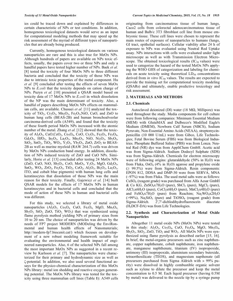
The 11 MeOx NPs used in the current study were pro-duced by flame pyrolysis method, their primary size varied between 10 and 20 nm (Fig. 3). The primary size of MgO was not measured due to the cloudy appearance of the parti-cles under TEM (Fig. 3). This was most likely due to high dissolution of MgO particles and subsequent formation of Mg(OH)2.
Despite their small primary sizes, agglomerates of parti-cles formed when dispersed in DI water or in cellular growth media. The average hydrodynamic size of the studied NPs after 24 h incubation in DI water and cell culture media is shown in Fig. (4) and Table S3.
In DI water, the hydrodynamic diameters (Dh) of NPs varied mostly between 60 and 200 nm and the suspensions were relatively stable (did not settle, Fig. 4A). Only MgO and Mn3O4 NPs formed larger aggregates the size of which reached several micrometers (Fig. 4A). In cell culture test media, NPs behaved differently than in DI water, usually forming larger agglomerates (Fig. 4). Only for two NPs - Al2O3 and WO3 NPs, the Dh remained close or in the 'nano' size range (<100 nm). Co3O4, CuO, Fe2O3 and ZnO were of ‘moderate sizes’ remaining below 250 nm. The largest hy-drodynamic sizes in test media were observed for TiO2 NPs (> 1000 nm) followed by Mn3O4 (>800 nm) and MgO (>600 nm) (Fig. 4A). The general increase in NPs hydrodynamic size in cell culture media was likely due to the binding of various cell culture media components onto the NPs exhibit-ing relatively high specific surface area (for the given NPs library, the specific surface area ranged from 52-290 m2/g; data not shown) and subsequent formation of macromolecule corona [36]. Importantly, biocorona forming on NPs surface
1920 Current Topics in Medicinal Chemistry, 2015, Vol. 15, No. 18 Ivask et al.
Fig. (3). TEM images of metal oxide nanoparticles used in the study. NP primary sizes shown on the images are calculated from given TEM images using program ImageJ. In addition, primary sizes were calculated using BET methodology [20] and the results were coherent with those from TEM images. can not be ignored as e.g., protein corona [20] has been con-sidered more important than the NPs intrinsic physical prop-erties in ‘encoding’ the nano-bio interactions [37]. Interest-ingly, the hydrodynamic diameter of MgO NPs in cell cul-ture media was smaller than in DI water, probably due to coating of NPs with organic components of culture media and increment in particles dispersibility.
In addition to size, NPs surface charge (reflected by ζ-potential) was measured. As a rule of thumb, ζ-potential about ±30 mV is a threshold value to ensure particles disper-sion by charge stabilization alone [38]. Five NPs from our library (Al2O3, SiO2, WO3, Sb2O3 and Fe3O4) exhibited > +30 mV or < -30 mV ζ-potential values in DI water (Fig. 4B) and these NPs had also excellent dispersion properties in DI wa-ter (Fig. 4A). Also other NPs which ζ-potentials in DI water ranged from about ±20 till ±10mV formed relatively stable dispersions. Only MgO NPs that in DI water had surface charge close to neutral (6.8 mV; Table S1) showed also the most extensive agglomeration in DI water (Fig. 4A). When particles ζ-potential was measured in cell culture medium, all the studied 11 NPs gained the surface charge (average value of -12 mV) (Fig. 4B) similar to that of the medium (-8.5 mV) (Table S3). This demonstrates once again the for-mation of biocorona on NPs surface and shows universal coating of NPs by cell culture components (serum proteins). The sizes of the agglomerates in cell culture medium varied (Fig. 4A) and also the stability of the NP suspensions in cell culture medium was different: the most rapid settling was observed for CuO NPs and Mn3O4 NPs but also WO3 NPs
(Fig. S2). The suspensions of Co3O4 and TiO2 NPs were relatively stable (Fig. 4, inset in panel A). Therefore, there was no clear correlation between the stability of NP suspen-sions and their hydrodynamic size in cell culture media.
3.4. Toxicity Testing of Metal Oxide Nanoparticles
3.4.1. Challenges in Nanoparticles Toxicity Analysis
We used three different cell lines: two human epithelial cell lines (A549, Caco2) and murine fibroblasts (Balb/c 3T3) to determine the cytotoxicity values for the 11 MeOx NPs. In case of all the three cell lines a Neutral Red Uptake (NRU) assay (a viability test that has been shown usable for toxico-logical testing of various chemicals [39] and is also a guide-line for in vitro and phototoxicity evaluation of chemicals, OECD draft guidance 126 and OECD TG 432), was used to evaluate the toxicity of 11 MeOx NPs. Due to the presence of metals in the tested NPs and thus, possibility for metal ion release, we also measured their dissolution and performed simultaneous experiments with soluble metal salts given the metal oxides proved toxic. Due to the fact that reactive oxy-gen species (ROS) have been considered as one of the main causes for nano-toxicity [40], we also analyzed the potential of the studied MeOx NPs to form ROS by incubating the MeOx suspensions in cell culture medium without cells pre-sent and quantified the production of abiotic ROS by using 2′,7′-dichlorodihydrofluorescein (H2DCF), that can be oxi-dized to fluorescent 2',7'-dichlorofluorescein (DCF) by a wide variety of ROS.

Toxicity of 11 Metal Oxide Nanoparticles Current Topics in Medicinal Chemistry, 2015, Vol. 15, No. 18 1921
Fig. (4). Hydrodynamic diameter (A) and ζζ-potential (B) of 11 metal oxide nanoparticles in DI water and cell culture (test) media. Data for NPs hydrodynamic diameter in DI water are from Table S1. A: Average hydrodynamic diameters in DI water and in three cell growth media (Table 1) after 24-h incubation at 37 °C, 5% CO2. Photos in the inset show the visual appearance of NPs in Caco2 cell culture medium (Table 1) after 24 h incubation; B: ζ-potentials in DI water and in medium used for Balb/c 3T3 cell line (Table 1). 3.4.2. IC50 Values of 11 Metal Oxide Nanoparticles in the NRU Assay for Three Different Cell Lines
In general, the responses of all the three cell lines to-wards 11 MeOx NPs were similar (Fig. 5 and Fig. S3).
To compare the sensitivities of the three cell lines to-wards standard chemicals, we analyzed their response to SDS that is often used as a positive control in toxicity tests [41]. The 24-h IC50 values for A549, Balb/c 3T3 and Caco2 cells in NRU assay proved quite similar (respective values were 98±12.4, 96±7 and 146±15 μg/mL) (dose-response curves are shown in Fig. (5) lower right corner).
Only five of the tested eleven MeOx NPs showed toxicity to studied mammalian cell lines in 24-h NRU assay in the nominal concentrations below 100 μg/mL. As an average for all the three cell lines, the lowest 24-h IC50 values were ob-served for CuO NPs followed by ZnO, Sb2O3, Mn3O4 and Co3O4 NPs (Table S4). In general, these data are consistent with the existing literature data (Table S2) which showed that ZnO and CuO particles were the most toxic particles in toxicity assays with mammalian cell lines followed by Mn3O4, Co3O4 and TiO2. Interestingly, Sb2O3 particles that were shown non-toxic (IC50>200 μg/mL) in the papers summarized in Table S2, proved toxic (IC50 40-80 μg/mL) to all the three tested cell lines in our experiments (Fig. 5,
Table 2). This difference could be explained by different cell type (BEAS 2B), different toxicity assays/endpoints (MTT, ATP, LDH) and/or different types of Sb2O3 particles used in earlier studies and in the current study. We attempted to ex-plain the differential toxicities of MeOx NPs using hypothe-sis given by Chusuei et al. [11] where it was suggested that higher toxicity of CuO and ZnO compared to Mn2O3, Fe2O3
,and TiO2 was due to high atomic number of Cu and Zn compared to the other metals. However, we did not find a clear correlation between the atomic number of metal in MeOx NP and toxicity (the atomic number of metals in MeOx NPs decreased in order W>Sb>Zn>Cu>Co>Fe>Mn> Ti>Si>Al>Mg while the toxicity of MeOx NPs decreased in order Cu>Zn>Sb>Mn>Co all the rest of NPs being non-toxic).
3.4.3. Metal Dissolution-Driven Toxicity of Metal Oxide Nanoparticles
As emphasized above, and shown already in 2006 by Brunner et al. [42] dissolution and shedding of metal ions from MeOx NPs may be the main determinant of toxicity of metal-containing NPs. This is true not only for animal cell lines in vitro but also for various ecotoxicological test spe-cies (algae, bacteria, daphnids, protozoa, fish) as shown in our recent review of the literature on CuO, ZnO and silver nanoparticles [6]. Therefore, we analyzed the dissolution of

1922 Current Topics in Medicinal Chemistry, 2015, Vol. 15, No. 18 Ivask et al.
Fig. (5). Toxicity of 11 studied nanoparticles to three different mammalian cell lines in vitro in 24-h Neutral Red Uptake (NRU) assay:
dose-response curves. Human lung epithelial cells A549, human intestinal epithelial cells Caco2 and mouse fibroblasts Balb/c 3T3 cells (see the symbols on the upper left graph) were exposed to suspensions of metal oxides for 24-h at 37 °C, 5% CO2. Highest exposure concentra-tions were 100 μg/mL. Note the logarithmic x-axis (toxicity data plotted on linear x-axis are shown in Fig. S3). Concentrations of metal ox-ides are nominal and presented as mg metal oxide/L. The respective 24 h IC50 and IC20 values are presented in Table S4. The presented data are average values of three independent measurements +/-SD (see Table 2). the five toxic MeOx NPs (ZnO, CuO, Sb2O3, Mn3O4 and Co3O4) and studied the toxic effect of soluble salts of these metals (Table 2).
The dissolution of the studied metal oxides was analyzed in the conditions mimicking the toxicity tests, i.e. at 100 μg/mL dispersions of nanoparticles in respective cell culture media (Table 1) incubated for 24 h at 37 °C and 5 % CO2. After 24 h, the amount of metal that was not in particulate form, was separated by ultracentrifugation, and quantified. As shown in Fig. (6), dissolved amount of metals from MeOx NPs was comparable in different cell culture media.
The highest dissolution was observed for CuO (41-50%), followed by Sb2O3 (10-15%) and ZnO (11-14%). Mn3O4 and especially Co3O4 had very low solubility (below 4 and 1 %, respectively) (Fig. 6). Similar behaviour – high level dissolu-tion of ZnO and CuO particles compared to other (Fe3O4, TiO2, NiO, CeO2) nanoparticles has been shown by Karlsson et al. [43]. Remarkable dissolution of CuO NPs in cell cul-ture medium was also shown in Semish et al. [44] where the kinetics of CuO nano- and microparticles dissolution in sev-eral aqueous media was evaluated. The authors showed that while the dissolution of CuO NPs in DI water was <2.4%, in DMEM medium supplemented with serum, the dissolution was time-related and remarkable: reaching 44 % within 24 h.
To evaluate the level of toxicity caused by the dissolved fraction of metal ions, we determined the IC50 concentrations for soluble salts of all the five toxic metal oxides. Compari-
son between the cytotoxicities of metal oxides and respective metal ions is shown in Table 2 and Fig. (S4). Interestingly, IC50 values of MeOx NPs and metal salts correlated, i.e., ions of those oxide NPs that were more toxic were also rela-tively more toxic (Table S4). Except in case of copper, all the studied metal salts were more toxic than respective nanoparticles.
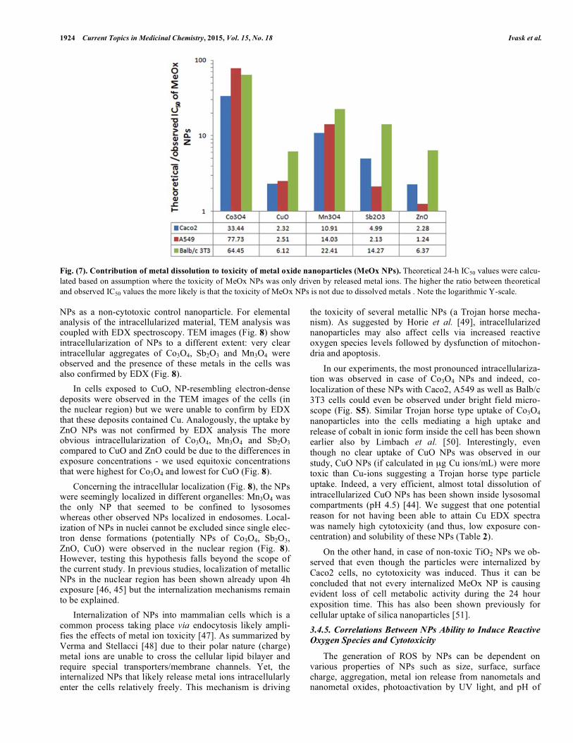
The knowledge on NPs dissolution and toxicity of re-spective metal ions allowed us to derive a theoretical IC50 value for the MeOx NPs assuming that the toxic effects are solely due to the metal ions released from the NPs. Such derivation of theoretical 24 h IC50 values for the five toxic MeOx NPs (Co3O4, CuO, Mn3O4, Sb2O3, ZnO) are shown in Table 2 and the ratio between theoretical and observed IC50 values of MeOx NPs is shown in Fig. (7).
The ratio between calculated and observed IC50 values ranged from slight to moderate differences, i.e. 2.3 to 6.4 (ZnO), 2.3-6.1 (CuO), 2.1-14.2 (Sb2O3) to remarkable differ-ences, i.e. 10.9-22.1 (Mn3O4) and 33.4-64.4 (Co3O4) (Fig. 7).
This result indicated that likely in case of ZnO and CuO, and partially also Sb2O3, the observed toxic effects were likely due metal ions released from metal oxides to the cell culture media. As discussed above, release of metal ions to mammalian cells may also take place after the uptake of the NPs by the cells, e.g. by Trojan horse mechanism, as demon-strated for CuO NPs by Cronholm et al. [45] in A549 and
Toxicity of 11 Metal Oxide Nanoparticles Current Topics in Medicinal Chemistry, 2015, Vol. 15, No. 18 1923
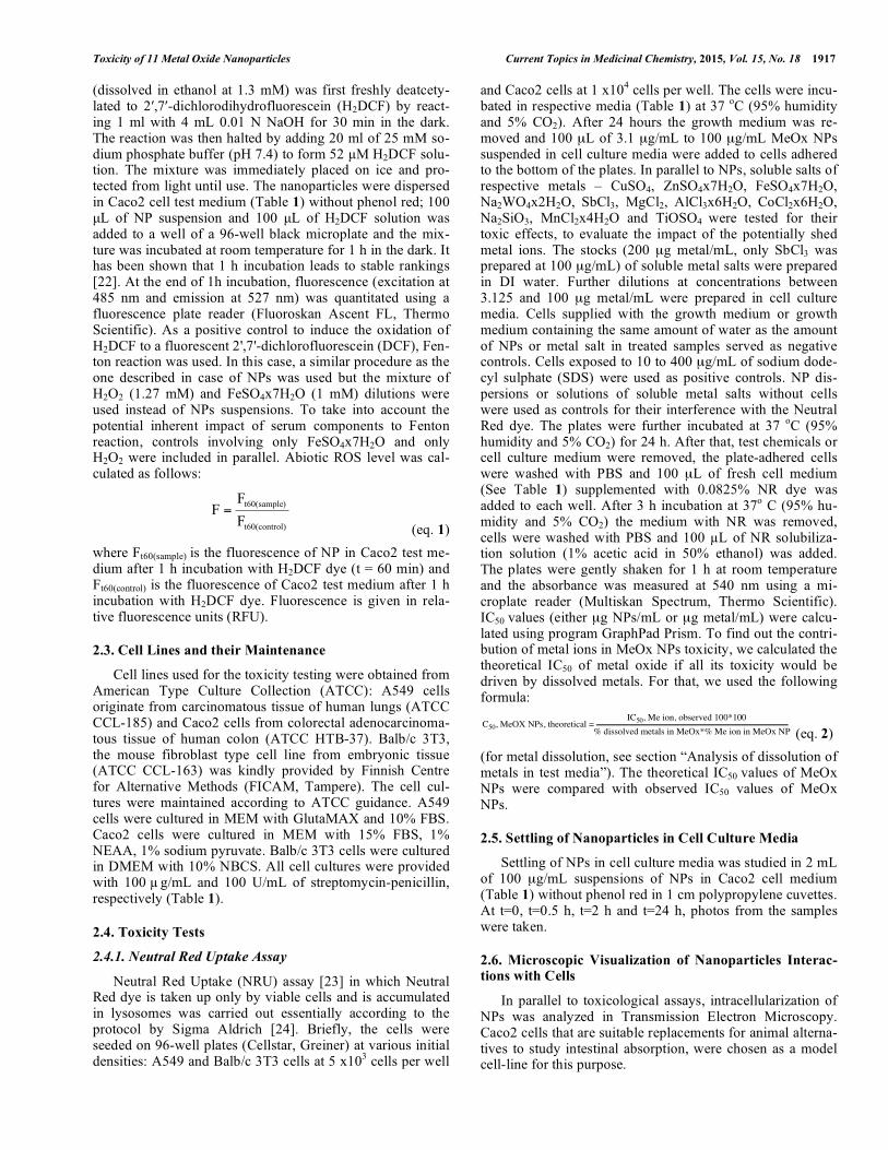
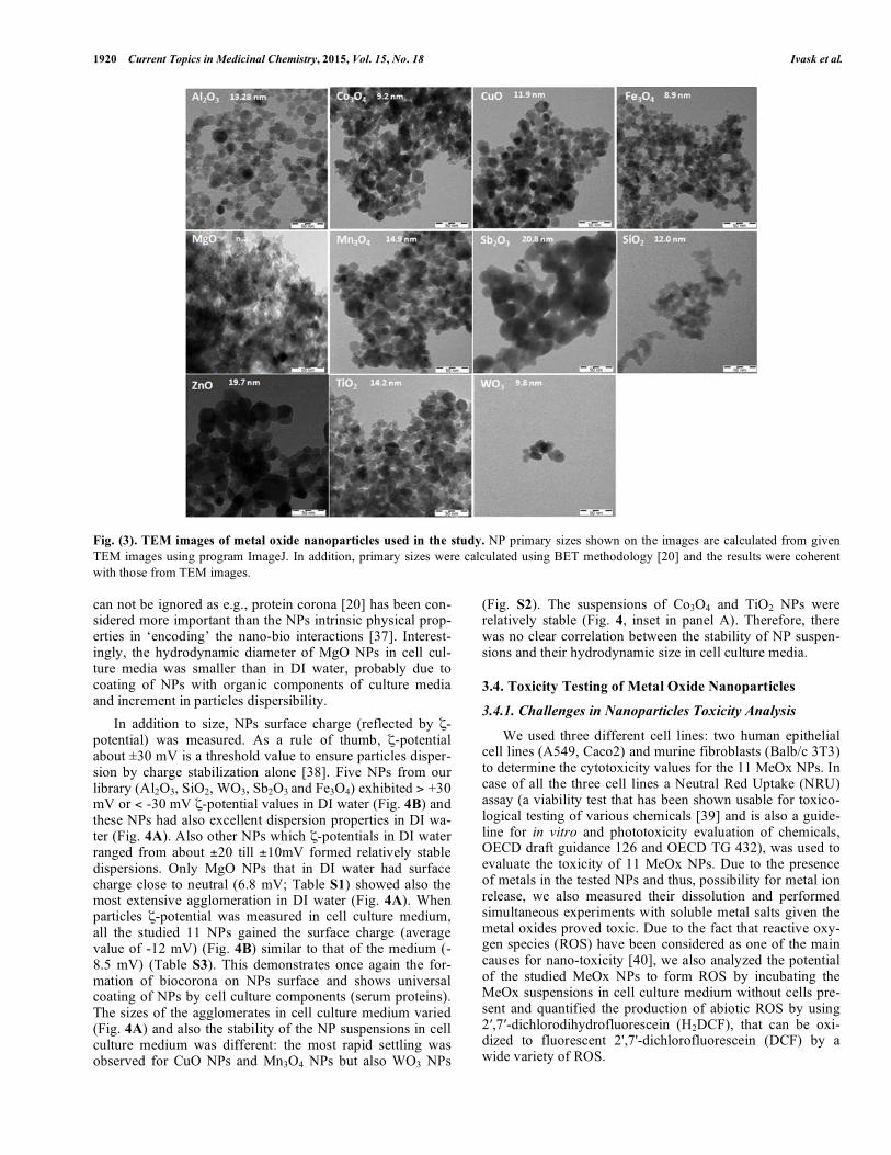
Table 2. Dissolution of selected metal oxide nanoparticles (MeOx NPs) and dissolution-driven hypothetical toxicity of these NPs.
Theoretical 24-h IC50 values of MeOx NPs in case their toxicity would be driven by released metal ions were calculated
based on dissolution of MeOx NPs and IC50 values of respective metal ions according to eq. 2. Overlap on observed and
theoretical 24-h IC50 values of MeOx NPs would indicate that the toxicity of those nanoparticles was solely driven by dis-
solution. The ratios between theoretical and observed IC50 values are presented in Fig. (7).
Metal Ion a
24-h IC50 μμg Metal
Ion /mL Metal Oxide
Observed 24-h
IC50 μg Metal
Oxide /mL
Dissolved Metal
Ions, %
Theoretical 24-h
IC50 μg Metal Oxide /mL
b
Co3+
25.7 Co3O4 123.0 0.9 4112
Cu2+
7.9 CuO 12.6 41.0 29.2
Mn2+
22.5 Mn3O4 66.7 4.4 729
Sb3+
31.7 Sb2O3 57.2 13.3 285
Caco 2
Zn2+
6.5 ZnO 30.2 13.7 69.0
Co3+
65.6 Co3O4 130.9 1.0 10183
Cu2+
31.1 CuO 18.9 43.5 47.4
Mn2+
89.7 Mn3O4 136.2 3.0 1911
Sb3+
68.0 Sb2O3 79.9 14.6 170
A549
Zn2+
5.9 ZnO 24.9 11.3 31.0
Co3+
50.9 Co3O4 135.5 0.7 8732
Cu2+
15.6 CuO 17.7 49.9 108
Mn2+
34.4 Mn3O4 195.0 2.6 4369
Sb3+
14.9 Sb2O3 39.1 10.5 557
Balb/c 3T3
Zn2+
2.4 ZnO 12.1 11.3 77.1
a CoCl2x6H2O, CuCl2x4H2O, MnCl2, SbCl3, ZnSO4x6H2O were used for testing b IC50 calculated based on 24-h IC50 of respective metal ions and dissolution of metal oxide nanoparticle in cell culture media (Fig. 6). Dissolution of metal oxide nanoparticles at 100 mg MeOx NP/L is shown in the Table as % of total metal.
Fig. (6). Dissolution of five metal oxide nanoparticles in three cell culture media (see composition of the media in Table 1) used in the
current study. Data are presented as % of metals dissolved from MeOx NPs. BEAS-2B cells. Due to the fact that,in case of certain oxides in this study, especially in case of Mn3O4 and Co3O4, the amount of dissolved ions (Fig. 6) was not high enough to explain the observed toxic effects. We further studied the degree of intracellularization and ROS induction potential of the toxic MeOx NPs.
3.4.4. Internalization of Nanoparticles by Cells In vitro
Co-localization and intracellularization experiments were carried out with MeOx NPs that showed cytotoxicity (Co3O4, CuO, Mn3O4, Sb2O3, ZnO). The chosen cell-line for observa-tions was Caco2 that was exposed for 24-h to IC20 levels (Table S4) of the cytotoxic MeOx NPs or to 100 μg /mL of TiO2
1924 Current Topics in Medicinal Chemistry, 2015, Vol. 15, No. 18 Ivask et al.
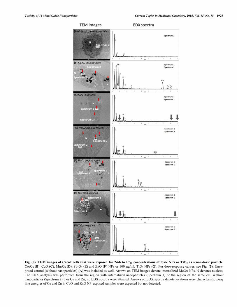
Fig. (7). Contribution of metal dissolution to toxicity of metal oxide nanoparticles (MeOx NPs). Theoretical 24-h IC50 values were calcu-lated based on assumption where the toxicity of MeOx NPs was only driven by released metal ions. The higher the ratio between theoretical and observed IC50 values the more likely is that the toxicity of MeOx NPs is not due to dissolved metals . Note the logarithmic Y-scale. NPs as a non-cytotoxic control nanoparticle. For elemental analysis of the intracellularized material, TEM analysis was coupled with EDX spectroscopy. TEM images (Fig. 8) show intracellularization of NPs to a different extent: very clear intracellular aggregates of Co3O4, Sb2O3 and Mn3O4 were observed and the presence of these metals in the cells was also confirmed by EDX (Fig. 8).
In cells exposed to CuO, NP-resembling electron-dense deposits were observed in the TEM images of the cells (in the nuclear region) but we were unable to confirm by EDX that these deposits contained Cu. Analogously, the uptake by ZnO NPs was not confirmed by EDX analysis The more obvious intracellularization of Co3O4, Mn3O4 and Sb2O3 compared to CuO and ZnO could be due to the differences in exposure concentrations - we used equitoxic concentrations that were highest for Co3O4 and lowest for CuO (Fig. 8).
Concerning the intracellular localization (Fig. 8), the NPs were seemingly localized in different organelles: Mn3O4 was the only NP that seemed to be confined to lysosomes whereas other observed NPs localized in endosomes. Local-ization of NPs in nuclei cannot be excluded since single elec-tron dense formations (potentially NPs of Co3O4, Sb2O3, ZnO, CuO) were observed in the nuclear region (Fig. 8). However, testing this hypothesis falls beyond the scope of the current study. In previous studies, localization of metallic NPs in the nuclear region has been shown already upon 4h exposure [46, 45] but the internalization mechanisms remain to be explained.
Internalization of NPs into mammalian cells which is a common process taking place via endocytosis likely ampli-fies the effects of metal ion toxicity [47]. As summarized by Verma and Stellacci [48] due to their polar nature (charge) metal ions are unable to cross the cellular lipid bilayer and require special transporters/membrane channels. Yet, the internalized NPs that likely release metal ions intracellularly enter the cells relatively freely. This mechanism is driving
the toxicity of several metallic NPs (a Trojan horse mecha-nism). As suggested by Horie et al. [49], intracellularized nanoparticles may also affect cells via increased reactive oxygen species levels followed by dysfunction of mitochon-dria and apoptosis.
In our experiments, the most pronounced intracellulariza-tion was observed in case of Co3O4 NPs and indeed, co-localization of these NPs with Caco2, A549 as well as Balb/c 3T3 cells could even be observed under bright field micro-scope (Fig. S5). Similar Trojan horse type uptake of Co3O4 nanoparticles into the cells mediating a high uptake and release of cobalt in ionic form inside the cell has been shown earlier also by Limbach et al. [50]. Interestingly, even though no clear uptake of CuO NPs was observed in our study, CuO NPs (if calculated in μg Cu ions/mL) were more toxic than Cu-ions suggesting a Trojan horse type particle uptake. Indeed, a very efficient, almost total dissolution of intracellularized CuO NPs has been shown inside lysosomal compartments (pH 4.5) [44]. We suggest that one potential reason for not having been able to attain Cu EDX spectra was namely high cytotoxicity (and thus, low exposure con-centration) and solubility of these NPs (Table 2).
On the other hand, in case of non-toxic TiO2 NPs we ob-served that even though the particles were internalized by Caco2 cells, no cytotoxicity was induced. Thus it can be concluded that not every internalized MeOx NP is causing evident loss of cell metabolic activity during the 24 hour exposition time. This has also been shown previously for cellular uptake of silica nanoparticles [51].
3.4.5. Correlations Between NPs Ability to Induce Reactive Oxygen Species and Cytotoxicity
The generation of ROS by NPs can be dependent on various properties of NPs such as size, surface, surface charge, aggregation, metal ion release from nanometals and nanometal oxides, photoactivation by UV light, and pH of
Toxicity of 11 Metal Oxide Nanoparticles Current Topics in Medicinal Chemistry, 2015, Vol. 15, No. 18 1925
Fig. (8). TEM images of Caco2 cells that were exposed for 24-h to IC20 concentrations of toxic NPs or TiO2 as a non-toxic particle.
Co3O4 (B), CuO (C), Mn3O4 (D), Sb2O3 (E) and ZnO (F) NPs or 100 μg/mL TiO2 NPs (G). For dose-response curves, see Fig. (5). Unex-posed control (without nanoparticles) (A) was included as well. Arrows on TEM images denote internalized MeOx NPs. N denotes nucleus. The EDX analysis was performed from the region with internalized nanoparticles (Spectrum 1) or the region of the same cell without nanoparticles (Spectrum 2). For Cu and Zn, no EDX spectra were attained. Arrows on EDX spectra denote locations were characteristic x-ray line energies of Cu and Zn in CuO and ZnO NP-exposed samples were expected but not detected.
1926 Current Topics in Medicinal Chemistry, 2015, Vol. 15, No. 18 Ivask et al.
the medium (for more information and references therein, see [40]). It is important to note, however, that in living cells, the reactive oxygen species (ROS) are normal products of oxidative metabolism and are mostly generated as by-products of electron transport in mitochondria. The main ROS involve superoxide, hydrogen peroxide, hydroxyl radi-cal and hydroxyl ion. Exposure to excess levels of ROS may cause however oxidation of cellular macromolecules and finally, cytotoxicity. To determine whether increased levels of ROS could be additional reason driving the toxicity of MeOx NPs (in addition to their dissolution), we studied the levels of ROS that were induced by the five toxic metal ox-ides using 2′,7′-dichlorodihydrofluorescein (H2DCF). This dye can be oxidized to fluorescent 2',7'-dichlorofluorescein (DCF) by a wide variety of ROS e.g., hydroxyl (•OH) radi-cals, HOCl and ONOO– but not O2
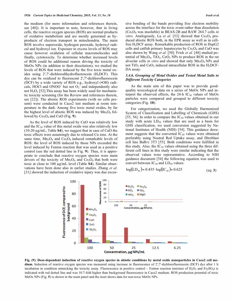
.- and independently also not H2O2 [52].This assay has been widely used for mechanis-tic toxicity screening (for the Review and references therein, see [22]). The abiotic ROS experiments (with no cells pre-sent) were conducted in Caco2 test medium at room tem-perature in the dark. Among five toxic metal oxides, by far the highest level of abiotic ROS was induced by Mn3O4 fol-lowed by Co3O4 and CuO (Fig. 9).
As the level of ROS induced by CuO was relatively low and the IC50 value of this metal oxide was also relatively low (10-20 μg/mL; Table S4), we suggest that in case of CuO the toxic effects were assumingly due to released Cu ions. At the same time, Mn3O4 and Co3O4 induced remarkable levels of ROS: the level of ROS induced by those NPs exceeded the level induced by Fenton reaction that was used as a positive control (see the red dotted line in Fig. 9). Thus, it is appro-priate to conclude that reactive oxygen species were main drivers of the toxicity of Mn3O4 and Co3O4 that both were toxic at close to 100 μg/mL level (Table S4). Similar obser-vations have been done also in earlier studies. Zhang et al. [31] showed the induction of oxidative injury was due exces-
sive bending of the bands providing free electron mobility across the interface for the toxic event rather than dissolution (Co3O4 was insoluble) in BEAS-2B and RAW 264.7 cells in vitro. Analogously, Lu et al. [53] showed that Co3O4 pro-duced abiotic ROS both, in the EPR assay as well as in cell-free H2DCF assay. Remarkable production of ROS in HepG2 cells and catfish primary hepatocytes by Co3O4 and CuO was also shown by Wang et al. [50]. Frick et al. [48] studied po-tential of Mn3O4, TiO2, CeO2 NPs to produce ROS in the rat alveolar cells in vitro and showed that only Mn3O4 NPs and not TiO2 and CeO2 induced intracellular ROS in the H2DCF-DA assay.
3.4.6. Grouping of Metal Oxides and Tested Metal Salts to Different Toxicity Categories
As the main aim of this paper was to provide good-quality toxicological data on a series of MeOx NPs and in-terpret the observed effects, the 24-h IC50 values of MeOx particles were compared and grouped to different toxicity categories (Fig. 10).
For categorization, we used the Globally Harmonized System of Classification and Labelling of Chemicals (GHS) [55, 56]. In order to compare the IC50 values obtained in our study with acute LD50 values that are used as a basis for GHS classification, we used conversion suggested by Na-tional Institutes of Health (NIH) [54]. This guidance docu-ment suggests that the converted IC50 values were obtained preferably using Neutral Red Uptake assay, and fibroblast cell line Balb/c 3T3 [55]. Both conditions were fulfilled in this study. Also, the IC50 values obtained using the three dif-ferent cell lines in this study were similar indicating that the observed values were representative. According to NIH guidance document [54] the following equation was used to convert between IC50 and LD50 values:
( ) ( ) 0.625IClog0.435LDlog 5050 +⋅= (eq. 3)
Fig. (9). Dose-dependent induction of reactive oxygen species in abiotic conditions by metal oxide nanoparticles in Caco2 cell me-
dium. Induction of reactive oxygen species was measured using increase in fluorescence of 2',7'-dichlorofluorescein (DCF) dye after 1 h incubation in condition mimicking the toxicity assay. Fluorescence in positive control – Fenton reaction (mixture of H2O2 and Fe2SO4) is indicated with red dotted line and was 10.7-fold higher than background fluorescence in Caco2 medium. ROS production potential of toxic MeOx NPs (Fig. 5) is shown in the main panel and the inset shows data for non-toxic MeOx NPs.

Toxicity of 11 Metal Oxide Nanoparticles Current Topics in Medicinal Chemistry, 2015, Vol. 15, No. 18 1927
Fig. (10). Toxicity (24-h IC50) of 11 metal oxide nanoparticles (A) and respective metal ions (B) to human lung epithelial cells A549,
human intestinal epithelial cells Caco2 and murine fibroblasts Balb/c 3T3 in vitro. Note the logarithmic y-axis and different units for oxides (μg compound/mL) and soluble salts (μg metal/mL). The values are means of three independent measurements and calculated from the dose-response curves presented in Fig. (5). IC50 values for Al, Fe, Mg, Si, Ti and W oxides and ions could not be calculated due to their low toxicity and the maximum tested concentration (100 μg/mL) is shown. IC50 values were converted to acute LD50 values based on guidance document by NIH [54] and classified to Globally Harmonized Classification System for Chemicals [55] [56]. Chemicals which IC50 values that fall to area shaded with darker background can be classified as Category 3 (acute LD50 values between 50 and 300 mg/kg; Toxic if swal-lowed) and chemicals which IC50 values fall into area shaded with lighter background can be classified as Category 4 (acute LD50 values between 300 and 2000 mg/kg; Harmful if swallowed).
According to our in vitro data and conversion to LD50, only ZnO and CuO fall into the Category 3 (LD50 > 50 mg/kg < 300 mg/kg; categorized „Toxic if swallowed“; [55]) (area shaded with darker background, Fig. 10). Similarly, Zn2+ and Cu2+ ions were classified under the same category. Among metal ions, also Sb3+, Mn2+ and Co2+ could be cate-gorized under Category 3. However, the toxicity values of these metals fall to the higher end of this hazard category (Fig. 10). Three other metal oxides for which we could cal-culate the IC50 values (Sb2O3, Mn3O4, Co3O4) can be classi-fied as hazard Category 4 (LD50 > 300 mg/kg < 2000 mg/kg; categorized “Harmful if swallowed”; [55]) (area shaded with lighter background in Fig. 10). Al, Fe, Mg, Si, Ti and W ions and oxides for which IC50 values were not retrieved, could not be categorized.
CONCLUSION
Here, we studied the toxicity of a set of 11 metal oxide nanoparticles (Al2O3, Co3O4, CuO, Fe3O4, MgO, Mn3O4, Sb2O3, SiO2, ZnO, TiO2, WO3) to three mammalian cell lines in Neutral Red Uptake assay. The results showed that only
five NPs (CuO, ZnO, Sb2O3, Mn3O4, Co3O4,) were toxic at nominal concentrations below 100 μg/mL and 24-h IC50 values of these NPs ranged from 10 to 100 μg/mL. According to Globally Harmonized Hazard Categorization system, only CuO and ZnO could be classified as „Toxic if swallowed“. The other three particles were classified as “Harmful if swallowed” and they belong to hazard Category 4.
The fact that both the toxic and non-toxic NPs studied had comparable primary size and most of the particles exhib-ited similar hydrodynamic size allows us to conclude that the small size of particles or their agglomeration pattern is probably not the main driving force of the toxic effects of the studied metal oxide nanoparticles. Parallel toxicological evaluation of MeOx NPs and their soluble salts showed that interestingly, only these five oxides that proved toxic <100 μg/mL were also toxic as soluble salts. This suggested that metal ions released from the particles contributed to cytotox-icity of MeOx NPs. The contribution of released metal ions was especially evident in case of CuO and ZnO but also Sb2O3. Although the released metal ions might have had
1928 Current Topics in Medicinal Chemistry, 2015, Vol. 15, No. 18 Ivask et al.
some role in toxic effects of Mn3O4 and Co3O4, the genera-tion of ROS is probably the main driver of toxicity of these MeOx given the remarkable accumulation of these MeOx in the cells. The lack of toxic effects from the rest of MeOx NPs (Al2O3, Fe3O4, MgO, SiO2, TiO2, WO3) indicates that they might be good candidates for nano-enabled applications where toxic side-effects cannot be tolerated.
In conclusion, in this study we produced a homogenous toxicological dataset for 11 metal oxide NPs. These data can further be used for modelling and safety assessment purposes.
CONFLICT OF INTEREST
The authors confirm that this article content has no con-flict of interest.
ACKNOWLEDGEMENTS
This research was supported by EU FP7 Project MOD-ERN under Grant Agreement No. 309314, EU FP7 grant NanoValid under Grant Agreement No. 263147, European Social Fund´s Doctoral Studies and Internationalisation Pro-gramme DoRa, Estonian Research Council grants PUT748, IUT23-5, Estonian Science Foundation project ETF9347, Development Fund of University of Tartu, and SA Ar-chimedes project Functional Food Ingredients.
SUPPLEMENTARY MATERIAL
Supplementary material is available on the publishers web site along with the published article.
REFERENCES
[1] http://nanodb.dk/. [2] www.nanotechproject.com. [3] Solanki, P.R.; Kaushik, A.; Agrawal, V.V.; Malhotra, B.D.
Nanostructured metal oxide-based biosensors. NPG Asia Mater., 2011, 3, 17–24.
[4] Ogale, S. B.; Venkatesan, T.V.; Blamire, M.E., Functional Metal Oxides. New Science and Novel Applications. Wiley-VCH: 2013.
[5] Commission. Recommendation on the definition of a nanomaterial. 2011/696/EU Official J. Eur. Union [Online], 2011.
[6] Bondarenko, O.; Juganson, K.; Ivask, A.; Kasemets, K.; Mortimer, M.; Kahru, A. Toxicity of Ag, CuO and ZnO nanoparticles to selected environmentally relevant test organisms and mammalian cells in vitro: a critical review. Arch. Toxicol., 2013, 87 (7), 1181-1200.
[7] Karlsson, H.; Toprak, M.; Fadeel, B. Toxicity of Metal and Metal Oxide Nanoparticles. In Handbook on the Toxicology of Metals, Nordberg, G. F.; Fowler, B. A.; Nordberg, M., Eds. Academic Press: 2015; pp 75-112.
[8] Baek, Y.-W.; An, Y.-J. Microbial toxicity of metal oxide nanoparticles (CuO, NiO, ZnO, and Sb2O3) to Escherichia coli, Bacillus subtilis, and Streptococcus aureus. Sci. Tot. Environ., 2011, 409 (8), 1603-1608.
[9] Hu, X.; Cook, S.; Wang, P.; Hwang, H.-m. In vitro evaluation of cytotoxicity of engineered metal oxide nanoparticles. Sci. Tot. Environ., 2009, 407 (8), 3070-3072.
[10] Puzyn, T.; Rasulev, B.; Gajewicz, A.; Hu, X.; Dasari, T.P.; Michalkova, A.; Hwang, H.-M.; Toropov, A.; Leszczynska, D.; Leszczynski, J. Using nano-QSAR to predict the cytotoxicity of metal oxide nanoparticles. Nat. Nano, 2011, 6 (3), 175-178.
[11] Chusuei, C.C.; Wu, C.-H.; Mallavarapu, S.; Hou, F. Y. S.; Hsu, C.-M.; Winiarz, J.G.; Aronstam, R.S.; Huang, Y.-W. Cytotoxicity in the age of nano: The role of fourth period transition metal oxide nanoparticle physicochemical properties. Chem. Biol. Interact., 2013, 206 (2), 319-326.
[12] Zhang, H.; Ji, Z.; Xia, T.; Meng, H.; Low-Kam, C.; Liu, R.; Pokhrel, S.; Lin, S.; Wang, X.; Liao, Y.-P.; Wang, M.; Li, L.; Rallo, R.; Damoiseaux, R.; Telesca, D.; Mädler, L.; Cohen, Y.; Zink, J. I.; Nel, A. E. Use of Metal Oxide Nanoparticle Band Gap To Develop a Predictive Paradigm for Oxidative Stress and Acute Pulmonary Inflammation. ACS Nano, 2012, 6 (5), 4349-4368.
[13] Horie, M.; Fujita, K.; Kato, H.; Endoh, S.; Nishio, K.; Komaba, L. K.; Nakamura, A.; Miyauchi, A.; Kinugasa, S.; Hagihara, Y.; Niki, E.; Yoshida, Y.; Iwahashi, H. Association of the physical and chemical properties and the cytotoxicity of metal oxide nanoparticles: metal ion release, adsorption ability and specific surface area. Metallomics, 2012, 4 (4), 350-360.
[14] Gajewicz, A.; Schaeublin, N.; Rasulev, B.; Hussain, S.M.; Leszczynska, D.; Puzyn, T.; Leszczynski, J. Towards understanding mechanisms governing cytotoxicity of metal oxides nanoparticles: Hints from nano-QSAR studies. Nanotoxicology, 2014, 9, 313-325.
[15] George, S.; Pokhrel, S.; Xia, T.; Gilbert, B.; Ji, Z.; Schowalter, M.; Rosenauer, A.; Damoiseaux, R.; Bradley, K. A.; Mädler, L.; Nel, A. E. Use of a Rapid Cytotoxicity Screening Approach To Engineer a Safer Zinc Oxide Nanoparticle through Iron Doping. ACS Nano, 2009, 4 (1), 15-29.
[16] Xia, T.; Zhao, Y.; Sager, T.; George, S.; Pokhrel, S.; Li, N.; Schoenfeld, D.; Meng, H.; Lin, S.; Wang, X.; Wang, M.; Ji, Z.; Zink, J. I.; Mädler, L.; Castranova, V.; Lin, S.; Nel, A. E. Decreased Dissolution of ZnO by Iron Doping Yields Nanoparticles with Reduced Toxicity in the Rodent Lung and Zebrafish Embryos. ACS Nano, 2011, 5 (2), 1223-1235.
[17] George, S.; Pokhrel, S.; Ji, Z.; Henderson, B. L.; Xia, T.; Li, L.; Zink, J. I.; Nel, A.E.; Mädler, L. Role of Fe Doping in Tuning the Band Gap of TiO2 for the Photo-Oxidation-Induced Cytotoxicity Paradigm. J. Amer. Chem.l Soc., 2011, 133 (29), 11270-11278.
[18] Pokhrel, S.; Nel, A.E.; Mädler, L. Custom-Designed Nanomaterial Libraries for Testing Metal Oxide Toxicity. Acc. Chem. Res., 2012, 46 (3), 632-641.
[19] Zhang, H.; Pokhrel, S.; Ji, Z.; Meng, H.; Wang, X.; Lin, S.; Chang, C.H.; Li, L.; Li, R.; Sun, B.; Wang, M.; Liao, Y.-P.; Liu, R.; Xia, T.; Mädler, L.; Nel, A. E. PdO Doping Tunes Band-Gap Energy Levels as Well as Oxidative Stress Responses to a Co3O4 p-Type Semiconductor in Cells and the Lung. J. Am. Chem. Soc., 2014, 136 (17), 6406-6420.
[20] Pokhrel, S.; Birkenstock, J.; Schowalter, M.; Rosenauer, A.; Mä dler, L. Growth of Ultrafine Single Crystalline WO3 Nanoparticles Using Flame Spray Pyrolysis. Cryst. Growth Des., 2009, 10 (2), 632-639.
[21] Rushton, E. K.; Jiang, J.; Leonard, S.S.; Eberly, S.; Castranova, V.; Biswas, P.; Elder, A.; Han, X.; Gelein, R.; Finkelstein, J.; Oberdörster, G. Concept of Assessing Nanoparticle Hazards Considering Nanoparticle Dosemetric and Chemical/Biological Response Metrics. J. Toxicol. Environ. Health, Part A, 2010, 73 (5-6), 445-461.
[22] Pal, A. K.; Bello, D.; Budhlall, B.; Rogers, E.; Milton, D. K. Screening for Oxidative Stress Elicited by Engineered Nanomaterials: Evaluation of Acellular DCFH Assay. Dose-Response, 2012, 10 (3), 308-330.
[23] Borenfreund, E.; Puerner, J. A. Toxicity determined in vitro by morphological alterations and neutral red absorption. Toxicol. Lett., 1985, 24 (2–3), 119-124.
[24] Sigma-Aldrich In vitro toxicology assay kit. Neutral Red Based. [25] Schrand , A.M.; Schlager, J.J.; Dai, L.; Hussain, S.M. Preparation
of cells for assessing ultrastructural localization of nanoparticles with transmission electron microscopy. Nat. Protoc., 2010, 5, 744–757.
[26] Rallo, R.; France, B.; Liu, R.; Nair, S.; George, S.; Damoiseaux, R.; Giralt, F.; Nel, A.; Bradley, K.; Cohen, Y. Self-Organizing Map Analysis of Toxicity-Related Cell Signaling Pathways for Metal and Metal Oxide Nanoparticles. Environ. Sci. Technol., 2011, 45 (4), 1695-1702.
[27] OECD. No. 129. Guidance Document On Using Cytotoxicity Tests To Estimate Starting Doses For Acute Oral Systemic Toxicity Tests. 2010.
[28] OECD. Test No. 432: In Vitro 3T3 NRU Phototoxicity Test 2004. [29] Sun, J.; Wang, S.; Zhao, D.; Hun, F.; Weng, L.; Liu, H.
Cytotoxicity, permeability, and inflammation of metal oxide nanoparticles in human cardiac microvascular endothelial cells. Cell Biol. Toxicol., 2011, 27 (5), 333-342.

Toxicity of 11 Metal Oxide Nanoparticles Current Topics in Medicinal Chemistry, 2015, Vol. 15, No. 18 1929
[30] Jeng, H. A.; Swanson, J. Toxicity of Metal Oxide Nanoparticles in Mammalian Cells. J. Environ. Sci. Health, Part A, 2006, 41 (12), 2699-2711.
[31] Zhang, X.Q.; Yin, L.H.; Tang, M.; Pu, Y.P. ZnO, TiO2, SiO2, and Al2O3 Nanoparticles-induced Toxic Effects on Human Fetal Lung Fibroblasts. Biomed. Environ. Sci., 2011, 24 (6), 661-669.
[32] Choi, S.-J.; Oh, J.-M.; Choy, J.-H. Toxicological effects of inorganic nanoparticles on human lung cancer A549 cells. J. Inorg. Biochem., 2009, 103 (3), 463-471.
[33] Krewski, D.; Acosta, D.; Andersen, M.; Anderson, H.; Bailar, J. C.; Boekelheide, K.; Brent, R.; Charnley, G.; Cheung, V.G.; Green, S.; Kelsey, K.T.; Kerkvliet, N.I.; Li, A.A.; McCray, L.; Meyer, O.; Patterson, R.D.; Pennie, W.; Scala, R. A.; Solomon, G. M.; Stephens, M.; Yager, J.; Zeise, L.; Staff of Committee on Toxicity, T.; Assessment of Environmental, A. Toxicity Testing in the 21st Century: A Vision and a Strategy. J. Toxicol. Environ. Health, Part B, 2010, 13 (2-4), 51-138.
[34] Powers, K.W.; Brown, S.C.; Krishna, V.B.; Wasdo, S.C.; Moudgil, B.M.; Roberts, S.M. Research Strategies for Safety Evaluation of Nanomaterials. Part VI. Characterization of Nanoscale Particles for Toxicological Evaluation. Toxicol. Sci., 2006, 90 (2), 296-303.
[35] Jiang, J.; Oberdörster, G.; Biswas, P. Characterization of size, surface charge, and agglomeration state of nanoparticle dispersions for toxicological studies. J. Nanopart. Res., 2009, 11 (1), 77-89.
[36] Lundqvist, M.; Stigler, J.; Cedervall, T.; Berggård, T.; Flanagan, M.B.; Lynch, I.; Elia, G.; Dawson, K. The Evolution of the Protein Corona around Nanoparticles: A Test Study. ACS Nano, 2011, 5 (9), 7503-7509.
[37] Walkey, C.D.; Olsen, J.B.; Song, F.; Liu, R.; Guo, H.; Olsen, D.W.H.; Cohen, Y.; Emili, A.; Chan, W.C.W. Protein Corona Fingerprinting Predicts the Cellular Interaction of Gold and Silver Nanoparticles. ACS Nano, 2014, 8 (3), 2439-2455.
[38] Hitchman, A.; Sambrook Smith, G. H.; Ju-Nam, Y.; Sterling, M.; Lead, J. R. The effect of environmentally relevant conditions on PVP stabilised gold nanoparticles. Chemosphere, 2013, 90 (2), 410-416.
[39] Repetto, G.; del Peso, A.; Zurita, J. L. Neutral red uptake assay for the estimation of cell viability/cytotoxicity. Nat. Protocols, 2008, 3 (7), 1125-1131.
[40] Fu, P.P.; Xia, Q.; Hwang, H.-M.; Ray, P.C.; Yu, H. Mechanisms of nanotoxicity: Generation of reactive oxygen species. J. Food Drug Anal., 2014, 22 (1), 64-75.
[41] Schirmer, K.; Tanneberger, K.; Kramer, N. I.; Völker, D.; Scholz, S.; Hafner, C.; Lee, L. E. J.; Bols, N. C.; Hermens, J. L. M. Developing a list of reference chemicals for testing alternatives to whole fish toxicity tests. Aquat. Toxicol., 2008, 90 (2), 128-137.
[42] Brunner, T.J.; Wick, P.; Manser, P.; Spohn, P.; Grass, R.N.; Limbach, L.K.; Bruinink, A.; Stark, W.J. In Vitro Cytotoxicity of Oxide Nanoparticles: Comparison to Asbestos, Silica, and the Effect of Particle Solubility. Environ. Sci. Technol., 2006, 40 (14), 4374-4381.
[43] Karlsson, H.; Gliga, A.; Calleja, F.; Goncalves, C.; Wallinder, I.; Vrieling, H.; Fadeel, B.; Hendriks, G. Mechanism-based
genotoxicity screening of metal oxide nanoparticles using the ToxTracker panel of reporter cell lines. Part. Fibre Toxicol., 2014, 11 (1), 41.
[44] Semisch, A.; Ohle, J.; Witt, B.; Hartwig, A. Cytotoxicity and genotoxicity of nano - and microparticulate copper oxide: role of solubility and intracellular bioavailability. Part. Fibre Toxicol., 2014, 11 (1), 10.
[45] Cronholm, P.; Karlsson, H. L.; Hedberg, J.; Lowe, T. A.; Winnberg, L.; Elihn, K.; Wallinder, I. O.; Möller, L. Intracellular Uptake and Toxicity of Ag and CuO Nanoparticles: A Comparison Between Nanoparticles and their Corresponding Metal Ions. Small, 2013, 9 (7), 970-982.
[46] Ahlinder, L.; Ekstrand-Hammarström, B.; Geladi, P.; Österlund, L. Large Uptake of Titania and Iron Oxide Nanoparticles in the Nucleus of Lung Epithelial Cells as Measured by Raman Imaging and Multivariate Classification. Biophys. J., 2013, 105 (2), 310-319.
[47] Gilbert, B.; Fakra, S.C.; Xia, T.; Pokhrel, S.; Mädler, L.; Nel, A.E. The Fate of ZnO Nanoparticles Administered to Human Bronchial Epithelial Cells. ACS Nano, 2012, 6 (6), 4921-4930.
[48] Verma, A.; Stellacci, F. Effect of Surface Properties on Nanoparticle–Cell Interactions. Small, 2010, 6 (1), 12-21.
[49] Horie, M.; Kato, H.; Fujita, K.; Endoh, S.; Iwahashi, H. In Vitro Evaluation of Cellular Response Induced by Manufactured Nanoparticles. Chem. Res. Toxicol., 2011, 25 (3), 605-619.
[50] Limbach, L. K.; Wick, P.; Manser, P.; Grass, R. N.; Bruinink, A.; Stark, W. J. Exposure of Engineered Nanoparticles to Human Lung Epithelial Cells: Influence of Chemical Composition and Catalytic Activity on Oxidative Stress. Environ. Sci. Technol., 2007, 41 (11), 4158-4163.
[51] Slowing, I. I.; Vivero-Escoto, J. L.; Wu, C.-W.; Lin, V. S. Y. Mesoporous silica nanoparticles as controlled release drug delivery and gene transfection carriers. Adv. Drug Deliv, Rev., 2008, 60 (11), 1278-1288.
[52] Doak, S.H.; Griffiths, S.M.; Manshian, B.; Singh, N.; Williams, P. M.; Brown, A. P.; Jenkins, G. J. S. Confounding experimental considerations in nanogenotoxicology. Mutagenesis, 2009, 24 (4), 285-293.
[53] Lu, S.; Duffin, R.; Poland, C.; Daly, P.; Murphy, F.; Drost, E.; MacNee, W.; Stone, V.; Donaldson, K. Efficacy of Simple Short-Term in Vitro Assays for Predicting the Potential of Metal Oxide Nanoparticles to Cause Pulmonary Inflammation. Environ. Health Perspect., 2009, 117 (2), 241–247.
[54] National Institute of Health (NIH). Guidance Document on Using In Vitro Data to Estimate In Vivo Starting Doses for Acute Toxicity Publication No: 01-4500 [Online].
[55] US Environmental Protection Agency (EPA). Chemical Hazard Classification and Labelling: Comparison of OPP REquirements and the GHS. Draft 7/7/04 [Online].
[56] United Nations. Globally Harmonized System of Classification and Labelling of Chemicals (GHS). Health Hazards. Acute Toxicity 2013 [Online].
Received: December 15, 2014 Revised: February 23, 2015 Accepted: March 22, 2015