HPA axis responsivity to dexamethasone and cognitive impairment in dementia
Upload
independentCategory
view
2download
0
Topical N-acetyl-S-farnesyl-L-cysteine Inhibits MouseSkin Inflammation, and Unlike Dexamethasone,its Effects Are Restricted to the Application SiteJoel S. Gordon1, Peter M. Wolanin1, Arnold V. Gonzalez1, David A. Fela1, Gopal Sarngadharan1,Karl Rouzard1, Eduardo Perez1, Jeffry B. Stock1,2,3 and Maxwell B. Stock1
N-Acetyl-S-farnesyl-L-cysteine (AFC), a modulator of G protein and G-protein coupled receptor signaling,inhibits neutrophil chemotaxis and other inflammatory responses in cell-based assays. Here, we showtopical AFC inhibits in vivo acute inflammation induced by 12-O-tetradecanoyl-phorbol-13-acetate (TPA) andarachidonic acid using the mouse ear model of inflammation. AFC inhibits edema, as measured by earweight, and also inhibits neutrophil infiltration as assayed by direct counting in histological sections and bymeasuring myeloperoxidase (MPO) activity as a neutrophil marker. In addition, AFC inhibits in vivo allergiccontact dermatitis in a mouse model utilizing sensitization followed by a subsequent challenge with 2,4-dinitrofluorobenzene. Unlike the established anti-inflammatories dexamethasone and indomethacin, AFC’saction was restricted to the site of application. In this mouse model, both dexamethasone and indomethacininhibited TPA-induced edema and MPO activity in the vehicle-treated, contralateral ear. AFC showed nocontralateral ear inhibition for either of these end points. A marginally significant decrease due to AFCtreatment was seen in TPA-induced epidermal hyperplasia at 24 hours. This was much less than the 90%inhibition of neutrophil infiltration, suggesting that AFC does not act by directly inhibiting protein kinase C.
Journal of Investigative Dermatology (2008) 128, 643–654; doi:10.1038/sj.jid.5701061; published online 20 September 2007
INTRODUCTIONThe C-terminal addition of farnesyl (15-carbon side chain) orgeranylgeranyl (20-carbon side chain) polyisoprenoids isessential to the membrane targeting of heterotrimeric andRas-related G proteins that mediate receptor signaling ineukaryotic cells. This post-translational modification involvesthe formation of a thioether bond between the polyisoprenoidand a cysteine four residues from the C terminus of theprotein within a so-called CAAX-tail motif (Stimmel et al.,1990). Prenylation is followed by membrane localization andsubsequent cleavage of the three AAX residues C-terminal tothe prenylated cysteine (Hancock et al., 1989; Reiss et al.,1990). The resulting exposed prenylcysteine a-carboxyl isthen subject to methylation and demethylation (Clarke, 1988;
Stimmel et al., 1990). Small-molecule prenylcysteine analogssuch as N-acetyl-S-farnesyl-L-cysteine (AFC) enter cells andcompete with prenylated G proteins for sites of interactionwith receptors in the membrane or compete with prenylatedG proteins as a substrate for isoprenylcysteine methyltrans-ferase (Volker et al., 1991b; Scheer and Gierschik, 1993;Philips et al., 1993). In vitro studies have shown AFC andsimilar analogs to be particularly effective inhibitors ofinflammatory responses, which are mediated by G-proteincoupled receptors (GPCRs) in macrophages, neutrophils, andplatelets (Volker et al., 1991a; Huzoor-Akbar et al., 1993;Philips et al., 1993; Scheer and Gierschik, 1993). In theseassays, AFC was shown to inhibit inflammatory cell activa-tion such as neutrophil aggregation and macrophagechemotaxis (Volker et al., 1991a).
The targeting of GPCR and G-protein signaling ininflammatory cell activation is a mechanism that is distinctfrom the two standard classes of small-molecule anti-inflammatories, glucocorticoids, and the classical nonsteroi-dal anti-inflammatory drugs (NSAIDs). Glucocorticoids act bybinding to and activating their specific nuclear hormonereceptor (Evans, 1988; Gronemeyer, 1992; Beato et al.,1995). This results in ligand-dependent transcriptionaltransactivation of genes coding for negative regulators ofinflammation such as secretory leukocyte protease inhibitor(Abbinante-Nissen et al., 1995), Clara cell 10-kDa protein(Zhang et al., 1997b) and IL receptor antagonist (Levine et al.,1996; Sousa et al., 1996). As an alternative mechanism of
& 2007 The Society for Investigative Dermatology www.jidonline.org 643
ORIGINAL ARTICLE
Received 11 November 2006; revised 11 June 2007; accepted 13 July 2007;published online 20 September 2007
1Signum Biosciences, Monmouth Junction, New Jersey, USA; 2Department ofMolecular Biology, Princeton University, Princeton, New Jersey, USA and3Department of Chemistry, Princeton University, Princeton, New Jersey, USA
Correspondence: Maxwell B. Stock, 1 Deer Park Drive suite L2, MonmouthJunction, New Jersey 08852, USA.E-mail: [email protected]
Abbreviations: AA, arachidonic acid; AFC, N-acetyl-S-farnesyl-L-cysteine;AGC, N-acetyl-S-geranyl-L-cysteine; ACD, allergic contact dermatitis;COX, cyclooxygenase; DNFB, 2,4-dinitrofluorobenzene; ED50, 50% effectivedose; GPCR, G-protein coupled receptor; ICMT, isoprenylcysteinemethyltransferase; MPO, myeloperoxidase; NSAID, nonsteroidalanti-inflammatory drug; PKC, protein kinase C;TPA, 12-O-tetradecanoyl-phorbol-13-acetate
transrepression, the liganded receptor can interact with andinactivate positive transcription factors such as activatorprotein 1 (Diamond et al., 1990; Jonat et al., 1990; Lucibelloet al., 1990; Schule et al., 1990; Yang-Yen et al., 1990) ormembers of the Rel pathway, which regulate genes codingfor inflammatory mediators (Mukaida et al., 1994; Ray andPrefontaine, 1994; Caldenhoven et al., 1995). The secondclass of small-molecule anti-inflammatories, NSAIDsare inhibitors of enzymes involved in eicosanoid synthesis(Ferreira et al., 1971; Smith and Willis, 1971; Vane, 1971); inparticular, cyclooxygenases (reviewed in Michaelidou andHadjipavlou-Litina, 2005). Thus, AFC and its analogs mayhave in vivo anti-inflammatory properties that distinguish itfrom glucocorticoids and classical NSAIDs.
Owing to the likely inactivation of AFC in serum (Volker,1995) and uncertainty whether sufficient amounts couldpermeate the skin to affect GPCR or G-protein signaling, inthis study we set out to investigate the in vivo anti-inflammatory activity of AFC, using a mouse model ofcontact irritation. The mouse ear model has been routinelyused to test whether topically applied anti-inflammatoriesinhibit the development of acute, chemically induced dermalirritation (Van Arman, 1974; Young et al., 1983, 1984;Carlson et al., 1985; De Young et al., 1989; Kotyuk et al.,1993; Rao et al., 1993; Tramposch, 1999) or adaptiveimmune system-based responses such as allergic contactdermatitis (ACD) (Crowle, 1975; Garrigue et al., 1994; Saint-Mezard et al., 2003). Moreover, this model has been used byvarious groups to identify and compare the members ofdiffering classes of anti-inflammatory agents with multiplemechanisms of action (reviewed in Tramposch, 1999). Themore commonly used end points of acute inflammation areedema (Tramposch, 1999), which is assayed by increase inear thickness or weight and neutrophil infiltration that ismeasured by assaying for the neutrophil marker myeloperoxi-dase (MPO) (Bradley et al., 1982). By exposing both earsto a chemical irritant while applying the test compound tojust one ear, the vehicle-treated contralateral ear serves as aninternal control (Van Arman, 1974). The validity of the resultsusing this internally controlled protocol, however, is depen-dent on the demonstration that the test agent applied to oneear does not have an effect on the contralateral ear.
We report that, in this in vivo model, topically appliedAFC inhibits development of edema and neutrophil infiltra-tion induced by the phorbol ester 12-O-tetradecanoyl-phorbol-13-acetate (TPA), arachidonic acid (AA) as well asACD edema induced by 2,4-dinitrofluorobenzene (DNFB).Finally, we have found, unexpectedly, that AFC’s inhibitionof edema and neutrophil infiltration remains restricted to thetreated ear, whereas the glucocorticoid dexamethasone andthe NSAID indomethacin applied to one ear reduces edemaand neutrophil infiltration in the contralateral ear, even atdoses that are only partially effective on the ipsilateral ear.
RESULTSInhibition of edema and neutrophil infiltration by AFC
The mouse ear model was used to assess whether topicallydelivered AFC could inhibit inflammation in vivo. Acute
contact inflammation has been well characterized in thismodel (reviewed by Tramposch, 1999). Upon topicalapplication of an irritating concentration of TPA, the resultingear edema reaches a maximum at 5.5–6 hours (Young et al.,1983; Gschwendt et al., 1984). Subsequent neutrophilinfiltration of the ear reaches a maximum at 20–24 hours(Young et al., 1983; De Young et al., 1989; Rao et al., 1993)and epidermal thickness increases substantially between 24and 48 hours (Griffiths et al., 1988).
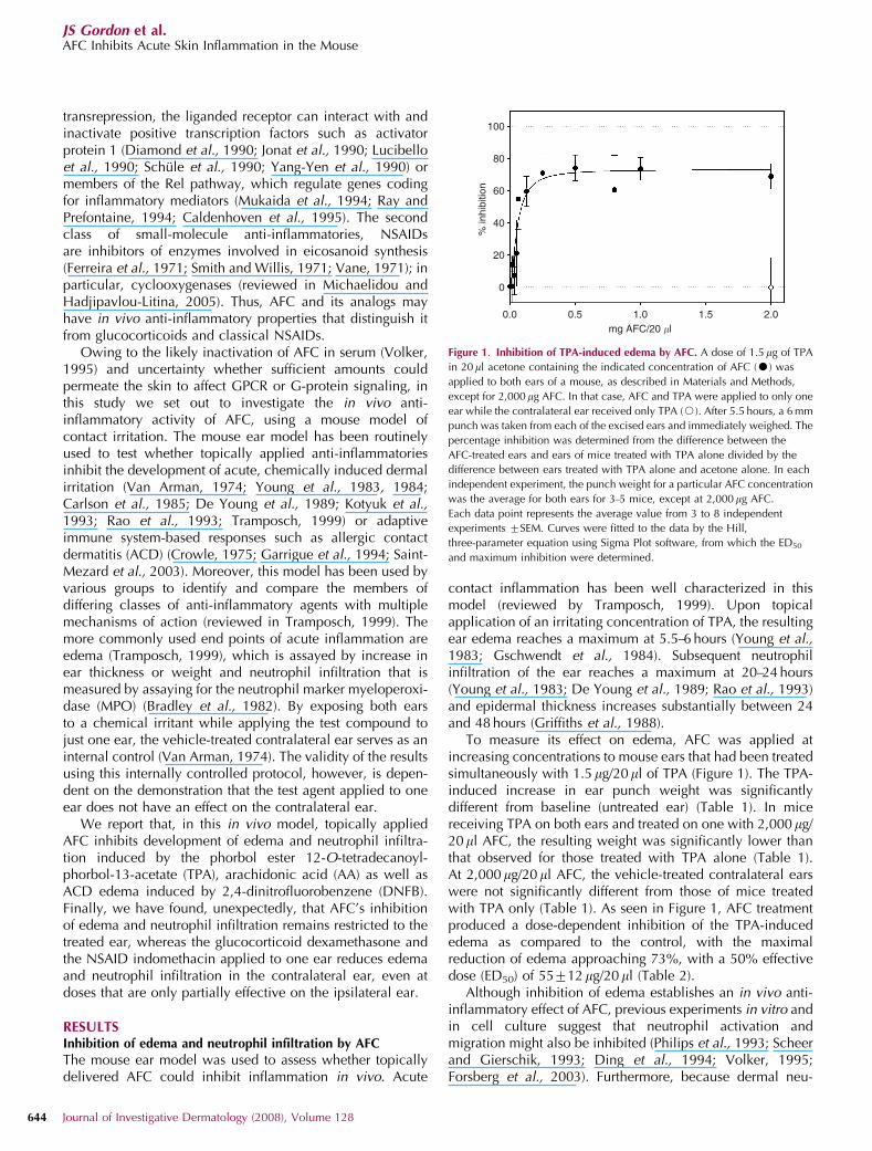
To measure its effect on edema, AFC was applied atincreasing concentrations to mouse ears that had been treatedsimultaneously with 1.5 mg/20ml of TPA (Figure 1). The TPA-induced increase in ear punch weight was significantlydifferent from baseline (untreated ear) (Table 1). In micereceiving TPA on both ears and treated on one with 2,000mg/20 ml AFC, the resulting weight was significantly lower thanthat observed for those treated with TPA alone (Table 1).At 2,000mg/20 ml AFC, the vehicle-treated contralateral earswere not significantly different from those of mice treatedwith TPA only (Table 1). As seen in Figure 1, AFC treatmentproduced a dose-dependent inhibition of the TPA-inducededema as compared to the control, with the maximalreduction of edema approaching 73%, with a 50% effectivedose (ED50) of 55712 mg/20 ml (Table 2).
Although inhibition of edema establishes an in vivo anti-inflammatory effect of AFC, previous experiments in vitro andin cell culture suggest that neutrophil activation andmigration might also be inhibited (Philips et al., 1993; Scheerand Gierschik, 1993; Ding et al., 1994; Volker, 1995;Forsberg et al., 2003). Furthermore, because dermal neu-
100
80
60
40
20
0
0.0 0.5 1.0 1.5 2.0mg AFC/20 �l
% in
hibi
tion
Figure 1. Inhibition of TPA-induced edema by AFC. A dose of 1.5 mg of TPA
in 20ml acetone containing the indicated concentration of AFC (K) was
applied to both ears of a mouse, as described in Materials and Methods,
except for 2,000 mg AFC. In that case, AFC and TPA were applied to only one
ear while the contralateral ear received only TPA (J). After 5.5 hours, a 6 mm
punch was taken from each of the excised ears and immediately weighed. The
percentage inhibition was determined from the difference between the
AFC-treated ears and ears of mice treated with TPA alone divided by the
difference between ears treated with TPA alone and acetone alone. In each
independent experiment, the punch weight for a particular AFC concentration
was the average for both ears for 3–5 mice, except at 2,000 mg AFC.
Each data point represents the average value from 3 to 8 independent
experiments 7SEM. Curves were fitted to the data by the Hill,
three-parameter equation using Sigma Plot software, from which the ED50
and maximum inhibition were determined.
644 Journal of Investigative Dermatology (2008), Volume 128
JS Gordon et al.AFC Inhibits Acute Skin Inflammation in the Mouse

trophil infiltration and edema can respond with differentspecificities to different anti-inflammatory agents in thismodel (Young et al., 1983; De Young et al., 1989; Raoet al., 1993), we investigated the effect of AFC on dermalneutrophil infiltration. Although neutrophil infiltration inresponse to topical TPA reaches a maximum at 24 hours(Young et al., 1983; De Young et al., 1989; Rao et al., 1993),we initially assayed AFC’s effects histologically at 5.5 hoursafter the application of TPA when infiltration is beginning andthe lower density of cells allows for their unequivocalidentification and counting. Fewer neutrophils were observedin ears 5.5 hours after TPA exposure if the AFC treatment was
applied 30 minutes after the TPA, and AFC reduced thenumber of neutrophils in the AFC-treated ear in a dose-dependent manner (Figure 2).
The initial histological findings showed that AFC inhibitsthe infiltration of cells meeting the morphological criteria,thus, validating the routine quantitation of neutrophilinfiltration by assaying the ear tissue biochemically forMPO 24 hours after TPA application (when neutrophilinfiltration is known to reach a maximum) (Young et al.,1983; De Young et al., 1989; Rao et al., 1993). Our resultsshow TPA induced significantly greater MPO activity over thebaseline measurement of acetone-only-treated ears (Table 1).
Table 1. Summary of absolute measurements and significance
End point Irritant Treatment Acetone control Irritant control Treated ear P-value Contralateral ear P-value
Edema AA AFC 8.870.3 mg (18) 15.770.6 mg (18) 10.570.4 mg (18) Po0.0001 15.670.8 mg (18) P=0.89
— TPA AFC 8.470.1 mg (33) 17.070.4 mg (37) 11.270.4 mg (34) Po0.0001 16.170.7 mg (20) P=0.25
— — Dexa-
methasone
8.270.1 mg (17) 15.170.4 mg (22) 8.570.2 mg (20) Po0.0001 9.570.3 mg (20) Po0.0001
MPO AA AFC 571 mAu/min (16) 4078 mAu/min (16) 1473 mAu/min (17) P=0.005 4678 mAu/min (18) P=0.55
— TPA AFC 371 mAu/min (21) 172728 mAu/min (22) 872 mAu/min (23) Po0.0001 145720 mAu/min (23) P=0.43
— — AGC 671 mAu/min (9) 375750 mAu/min (20) 273743 mAu/min (11) P=0.12 414758 mAu/min (11) P=0.61
— — Dexa-
methasone
471 mAu/min (12) 162729 mAu/min (30) 31710 mAu/min (13) P=0.0001 61721 mAu/min (13) P=0.006
— — Indomethacin 671 mAu/min (12) 250735 mAu/min (24) 1573 mAu/min (22) Po0.0001 42718 mAu/min (21) P=0.009
AA, arachidonic acid; AFC, N-acetyl-S-farnesyl-L-cysteine; AGC, N-acetyl-S-geranyl-L-cysteine; MPO, myeloperoxidase; TPA, 12-O-tetradecanoyl-phorbol-13-acetate.Edema was measured as the weight of a 6 mm punch, whereas MPO enzyme activity measurements are represented as the change in absorbance per minuteat 460 nm. The data for the treated and contralateral ears are at the highest concentration for each agent (2,000 mg AFC, 16mg dexamethasone, 2,000 mgAGC, and 1,000 mg indomethacin per 20ml). The number in parentheses is the number of mice used to derive each average. To compare the statisticalsignificance of the results, we averaged the absolute values of irritation for each individual mouse from all experiments used at the reported concentration,calculated the SE, and used the two-tailed Student’s t-test to calculate the significance relative to the irritant control. The TPA-in-acetone vehicle-treatedcontralateral ears of mice treated with dexamethasone or indomethacin exhibit significant inhibition of edema and neutrophil infiltration, but those treatedwith AFC do not. A few individual mouse ears deviating more than three-fold from the mean value of their group (indicating experimental error or pre-existing inflammation) were excluded from the analysis.
Table 2. Summary of ED50 and maximum inhibition values
End point Irritant TreatmentED50 (lg/20 ll)
(potency)Maximum % inhibition
(activity)
Edema AA AFC 314798 100
— TPA AFC 55712 73
— — Dexamethasone 0.2570.03 100
MPO AA AFC 184782 87
— TPA AFC 118724 100
— — AGC 2779 27
— — Dexamethasone 0.4970.16 100
— — Indomethacin 1117140 100
AA, arachidonic acid; AFC, N-acetyl-S-farnesyl-L-cysteine; AGC, N-acetyl-S-geranyl-L-cysteine; MPO, myeloperoxidase; TPA, 12-O-tetradecanoyl-phorbol-13-acetate.The parameters extracted from calculating the data as the percentage inhibition in each separate experiment. The data were fit to the Hill equation asdescribed in the Materials and Methods (see Figures 1, 3, 4 and 5 and Figures S2–S4). The maximum inhibition represents the asymptotic value of the curvefit, whereas the ED50 is the concentration of agent needed to produce half of the maximum inhibition.
www.jidonline.org 645
JS Gordon et al.AFC Inhibits Acute Skin Inflammation in the Mouse

AFC was applied at increasing concentrations to mouse earsthat had been treated simultaneously with 0.8 mg/20 ml ofTPA. Mouse ears simultaneously exposed to 2,000mg/20 mlAFC had an MPO activity, which was highly significantlydifferent from that observed for treatment with TPA only.Again, at the highest concentration of 2,000 mg/20ml AFC, thevehicle-treated contralateral ears were not significantlydifferent from the TPA control (Table 1). A dose-dependentinhibition was observed with a maximum activity approach-ing 100% and an ED50 of 118724 mg/20ml (Figure 3 andTable 2). Consequently, AFC was capable of inhibiting thetwo hallmarks of acute contact irritation, edema, andneutrophil recruitment, with greater potency against edemaas compared to that versus neutrophil infiltration.
Direct activation of epidermal protein kinase C (PKC) hasbeen shown to be involved in TPA-induced epidermalhyperplasia, based on the effect of specific PKC inhibitors(Griffiths et al., 1988; Reynolds et al., 1997), as well asoverexpression (Wang and Smart, 1999; Jansen et al., 2001)and/or knockout of PKC in mouse skin (Hara et al., 2005).Therefore we sought to determine if AFC could also inhibitTPA-induced neutrophil infiltration independent of TPA-induced hyperplasia. AFC was simultaneously applied withTPA, and the effect of AFC on the TPA-induced increase inepidermal thickness was measured at 24 hours after treatmentrather than 48 hours when the maximal increase has beenobserved previously (Griffiths et al., 1988; Reynolds et al.,
1997). This end point was selected to maximize the effect ondirect TPA induction of epidermal hyperplasia and minimizeany indirect, secondary hyperplasia due to neutrophilinfiltration. A statistically significant increase in epidermalthickness of 37% (Table 3) was observed in ears dosed with0.8 mg/20ml of TPA (same dose used in the induction of MPOactivity). In the presence of TPA and 800mg/20ml AFC, therewas a 22% increase in epidermal thickness (Table 3). Thus,
10
8
6
4
2
0
Neu
trop
hils
per
×40
0 fie
ld
TPAAFC
0
02 2 2 2
10.250.050
(�g/20 �l)
(mg/20 �l)
a c
b d
Figure 2. Histology of the AFC effect on neutrophil infiltration. A dose of
2.0mg of TPA in 20 ml acetone was applied to mice on both ears. Thirty
minutes later, AFC in 20 ml acetone was applied to one ear and 20 ml of
acetone (vehicle) was applied to the contralateral ear of each mouse. After
5.5 hours, a 6 mm punch from each of the excised ears was fixed in 10%
formalin. Neutrophils (-) were identified by their lobular nuclear
morphology. (a) Control animal not treated with TPA. Bar¼50 mm. (b) Skin
treated with TPA and vehicle show substantial neutrophil infiltration. (c) Skin
treated with TPA and 1,000 mg of AFC has a significantly reduced neutrophil
infiltration. (d) Quantitation of neutrophils by manual counting. The TPA and
vehicle result (0 AFC) represents the average of the contralateral ears from the
mice treated with the listed concentrations of AFC.
100
80
60
40
20
0
0.0 0.5 1.0 1.5 2.0mg AFC/20 �l
% in
hibi
tion
Figure 3. AFC inhibition of TPA-induced MPO activity. Neutrophil
infiltration was assayed biochemically by measuring MPO activity. A dose of
0.8mg of TPA in 20 ml acetone containing the indicated concentration of AFC
(K) was applied to both ears of a mouse. For 2,000 mg AFC, both AFC and
TPA were applied to one ear while the contralateral ear received only TPA
(J). After 24 hours, a 6 mm punch was taken from each of the excised ears
and frozen immediately. Enzyme assays were carried out as described in
Materials and Methods. In each independent experiment, the MPO activity for
a particular AFC concentration was the average of the values from ears for 3–5
mice. In each experiment, the percentage inhibition was determined from the
difference between the AFC-treated ears and ears of mice treated with TPA
alone divided by the difference between ears treated with TPA alone and
acetone alone. Each data point represents the average value from 3 to 4
independent experiments 7SEM. The curves were fitted to the data by the
Hill equation with two free parameters and with the maximum inhibition
fixed at 100% using the Sigma Plot software, from which the ED50 was
determined.
Table 3. Epidermal hyperplasia in response to TPA
Treatment Epidermal thickness %
20 ml acetone vehicle 10073
800 mg/20 ml AFC 10676
0.8 mg/20ml TPA 13877
0.8 mg TPA plus 800mg/20 ml AFC 12276
AFC, N-acetyl-S-farnesyl-L-cysteine; MPO, myeloperoxidase; TPA, 12-O-tetradecanoyl-phorbol-13-acetate.The relative increase in epidermal thickness was measured, as detailed inthe Materials and Methods at 24 h to match the end point of the MPOassay. Each measurement represents the average of measurements on 12mice7SE. The results are expressed as a percentage of the averageepidermal thickness measured for acetone-treated control ears. Theincrease for both groups of TPA-treated ears was significantly differentfrom that of the control ears at the level of Po0.05 using the Student’s t-test. The difference between the AFC-treated ear plus TPA and the TPAalone was not significant at the 0.05 level but was at the 0.1 level.
646 Journal of Investigative Dermatology (2008), Volume 128
JS Gordon et al.AFC Inhibits Acute Skin Inflammation in the Mouse

although AFCþTPA reduced epidermal thickness as com-pared to that seen with TPA alone, the difference was at themargin of statistical significance (0.104P40.05). AFC at thisconcentration produced B90% inhibition of neutrophilinfiltration as measured by MPO activity (Table 2) and inthe absence of TPA did not produce a statistically significantchange in epidermal thickness (Table 3).
Although TPA-induced inflammation involves directactivation of PKC (Liu and Heckman, 1998), downstreameffectors may possibly include members of the AA cascadethat utilize GPCRs (Kostenis, 2004). To investigate this, wetested the ability of AFC to inhibit the inflammatory responseinduced by AA in this model measuring edema and MPOactivity 1 hour post-application (Young et al., 1983; Opaset al., 1985; De Young et al., 1989; Rao et al., 1993). Ourresults show that AFC inhibits AA-induced edema andneutrophil infiltration in a dose-dependent manner (FiguresS1 and S2). As shown in Table 1, the AA-induced ear weightwas significantly above the baseline of the acetone-onlycontrol. AFC at 2,000mg/20 ml produced a significantlydecreased ear weight when compared to ears treated withthe AA vehicle alone (Tables 1 and 2). The maximuminhibitory effect on AA-induced edema approaches 100%,with an ED50 of 314798 mg/20ml (Table 2). For both endpoints, AFC at the highest concentrations had no effect onAA-induced inflammation in the contralateral vehicle-treatedear. Thus, although AFC activity against AA was similar toresults obtained against TPA, AFC appeared to be five-foldless potent against AA-induced edema than TPA-inducededema. Conversely, AFC significantly reduced both AA- andTPA-induced neutrophil infiltration with equal potency(ED50¼ 184782 and 118724 mg/20ml, respectively; Tables1 and 2). Consequently, these results show AFC is activeagainst the acute inflammatory response of two differentagents that may function through GPCR signaling. Further-more, the difference in the ratio of potency for edema andneutrophil infiltration was consistent with AFC acting againstdifferent GPCR pathways activated by the different stimuli.
Similar to AFC, the prenylcysteine analog N-acetyl-S-geranyl-L-cysteine (AGC) possesses an N-acetyl-cysteinemoiety, differing only in the length of the prenyl chain. Inprevious experiments, this analog was shown to be inactivein reducing GPCR activity and only slightly active ininhibiting inflammatory cell responses in vitro (Volkeret al., 1991b; Philips et al., 1993). AGC was tested in theTPA-treated mouse ear assay to compare its in vivo inhibitionof neutrophil infiltration with AFC. At 2,000mg/20 ml, ourresults show that AGC produced a slight reduction in TPA-induced MPO activity, with a maximum effect approachingonly 27% and only marginally statistically distinguishablefrom the control (Tables 1 and 2, Figure S3). As with AFC,AGC caused no significant reduction in the TPA-inducedMPO activity in the contralateral ear.
Topical AFC’s anti-inflammatory activity is restricted to the siteof application
Other anti-inflammatory agents including the glucocorticoiddexamethasone and the cyclooxygenase inhibitor indomethacin
are also known to be active in the mouse ear model assay(Michaelidou and Hadjipavlou-Litina, 2005), despite eachhaving a different mechanism of action than that postulatedfor AFC (Schafer-Korting et al., 2005). To further characterizeAFC, we investigated the similarities and differences betweenthese anti-inflammatory agents. In preliminary studies it wasimmediately apparent that both dexamethasone and indo-methacin, but not AFC, inhibited edema and neutrophilinfiltration in both the ipsilateral ear treated with compoundand the contralateral ear that only received TPA. Fordexamethasone, we observed a dose-dependent inhibitionof TPA-induced edema that could be discerned in both thedexamethasone-treated ears and the contralateral ears towhich only vehicle was applied (Tables 1 and 2). Inhibition ofedema can be discerned in the contralateral ear at higherconcentrations, as compared with the ipsilateral ear to whichdexamethasone was directly applied (Figure 4). A maximalinhibition of 100% was observed in the dexamethasone-treated ear with 50% of the maximal inhibition of edema seenat a dose of 0.2570.03mg/20ml (Figure 4, Tables 1 and 2).Results also showed that dexamethasone inhibited neutrophilinfiltration (as measured by MPO activity) in both treated andcontralateral ears (Figure 5, Tables 1 and 2), with an ED50 inthe treated ear of 0.4970.16mg.
The cyclooxygenase inhibitor, indomethacin, similar todexamethasone showed an inhibitory effect on TPA-inducedMPO activity of the contralateral ear to which only vehiclewas applied with an ED50 of 1117140 mg/20ml for indo-methacin on the ipsilateral ear (Tables 1 and 2, Figure S4). Incontrast to dexamethasone, the maximum effect of indo-methacin on the vehicle-treated contralateral ear was moreattenuated. Thus, unlike AFC, dexamethasone and indo-methacin affect the edema and neutrophil infiltration in thecontralateral ear. Comparison of the responses of the treated
100
80
60
40
20
0
0 2 4 6 8 1816141210�g dexamethasone/20 �l
% in
hibi
tion
Figure 4. Inhibition of TPA-induced edema by dexamethasone. A dose of
1.5mg of TPA in 20 ml acetone containing the indicated amount of
dexamethasone (K) was applied to one ear of a mouse, whereas 1.5mg of
TPA alone was applied to the contralateral ear (J). 5.5 hours later, a 6 mm
punch was taken from each of the excised ears and immediately weighed.
Groups of 3–5 mice were used for each experiment, and each data point
represents the average value from 3 to 4 independent experiments 7SEM. The
calculations and curve fitting for the dexamethasone-treated ear were as
described in for Figure 3.
www.jidonline.org 647
JS Gordon et al.AFC Inhibits Acute Skin Inflammation in the Mouse
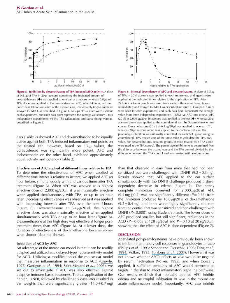
ears (Table 2) showed AFC and dexamethasone to be equallyactive against both TPA-induced inflammatory end points onthe treated ear. However, based on ED50 values, thecorticosteroid was significantly more potent. AFC andindomethacin on the other hand, exhibited approximatelyequal activity and potency (Table 2).
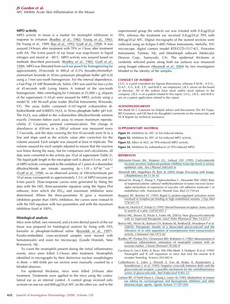
Effectiveness of AFC applied at different times relative to TPA
To determine the effectiveness of AFC when applied atdifferent time intervals relative to irritant, we applied AFC anhour before, simultaneously with and various times after TPAtreatment (Figure 6). When AFC was assayed at is highesteffective dose of 2,000mg/20ml, it was maximally effectivewhen applied simultaneously with TPA, or up to an hourlater. Decreasing effectiveness was observed as it was appliedwith increasing intervals after TPA over the next 6 hours(Figure 6). Dexamethasone at 6.4 mg/20ml, the highesteffective dose, was also maximally effective when appliedsimultaneously with TPA or up to an hour later (Figure 6).Dexamethasone at this high dose was effective at longer post-treatment times than AFC (Figure 6). At a lower dose, theduration of effectiveness of dexamethasone became some-what shorter (data not shown).
Inhibition of ACD by AFC
An advantage of the mouse ear model is that it can be readilyadapted and utilized as a delayed-type hypersensitivity modelfor ACD. Utilizing a modification of the mouse ear modelthat measures inflammation in response to ACD (Crowle,1975; Garrigue et al., 1994; Saint-Mezard et al., 2003), weset out to investigate if AFC was also effective againstadaptive immune-based responses. Topical application of thehapten, DNFB, followed by challenge 5 days later resulted inear weights that were significantly greater (14.070.7 mg)
than that observed in ears from mice that had not beensensitized but were challenged with DNFB (9.270.3 mg).Results showed that AFC applied to the ear surfacesimultaneously with the DNFB challenge produced a dose-dependent decrease in edema (Figure 7). The nearlycomplete inhibition observed for 2,000mg/20ml AFC(9.4 mg70.2) was not significantly different (P¼0.66) fromthe inhibition produced by 16.0 mg/20ml of dexamethasone(9.570.4 mg) and both were highly significantly differentfrom the control that was sensitized and then challenged withDNFB (Po0.0001 using Student’s t-test). The lower doses ofAFC produced smaller, but still significant, reductions in theACD (P¼0.005 at 128mg/20 ml and P¼0.10 at 20 mg/20 ml),showing that the effect of AFC is dose-dependent (Figure 7).
DISCUSSIONAcetylated polyprenylcysteines have previously been shownto inhibit inflammatory cell responses in granulocytes in vitro(Philips et al., 1993; Scheer and Gierschik, 1993; Ding et al.,1994; Volker, 1995; Forsberg et al., 2003). However, it wasnot known whether AFC’s effects in vivo would be negatedby serum inactivation (Volker, 1995), and when topicallyapplied, if sufficient amounts of AFC would permeate totargets in the skin to affect inflammatory signaling pathways.Our results establish that topically applied AFC inhibitsedema and neutrophil infiltration in vivo in the mouse earacute inflammation model. Importantly, AFC also inhibits
100
80
60
40
20
0
0 2 4 6 8 1816141210
�g dexamethasone/20 �l
% in
hibi
tion
Figure 5. Inhibition by dexamethasone of TPA-induced MPO activity. A dose
of 0.8mg of TPA in 20 ml acetone containing the indicated amount of
dexamethasone (K) was applied to one ear of a mouse, whereas 0.8mg of
TPA alone was applied to the contralateral ear (J). After 24 hours, a 6 mm
punch was taken from each of the excised ears, immediately frozen and later
assayed for MPO, as described in Figure 3. Groups of 3–5 mice were used for
each experiment, and each data point represents the average value from 3 to 4
independent experiments 7SEM. The calculations and curve fitting were as
described in Figure 3.
100
80
60
40
20
0
0 2 4 6 8 141210Hours relative to TPA application
% in
hibi
tion
Figure 6. Interval dependence of AFC and dexamethasone. A dose of 1.5 mg
of TPA in 20 ml acetone was applied to each mouse ear, and agents were
applied at the indicated times relative to the application of TPA. After
24 hours, a 6 mm punch was taken from each of the excised ears, frozen
immediately and assayed for MPO, as described in Figure 3. Groups of 5 mice
were used for each experiment, and each data point represents the average
value from three independent experiments 7SEM. (a) AFC time course. AFC
(20ml) at 2,000 mg/20ml in acetone was applied to one ear (K), whereas 20 ml
acetone alone was applied to the contralateral ear. (b) Dexamethasone time
course. Dexamethasone (20 ml) at 6.4 mg/20ml was applied to one ear (J),
whereas 20 ml acetone alone was applied to the contralateral ear. The
percentage inhibition was internally controlled for each AFC group using the
contralateral, TPA-treated ears of the same mice to calculate the TPA-only
value. For dexamethasone, separate groups of mice treated with TPA alone
were used as the TPA control. The percentage inhibition was determined from
the difference between the treated ears and the TPA control divided by the
difference between the TPA control and ears treated with acetone alone.
648 Journal of Investigative Dermatology (2008), Volume 128
JS Gordon et al.AFC Inhibits Acute Skin Inflammation in the Mouse

inflammation associated with ACD in this mouse model,showing AFC is effective against both innate and adaptiveimmune-based responses. Similar to acute contact inflamma-tion, AFC is equally effective but less potent than dexa-methasone in reducing delayed-type hypersensitivity. In theseassays edema was used as the end point. Whether cellularinflammation is inhibited as well await assays measuringinfiltration of T-cell lymphocytes.
Our observed in vivo inhibition data indicate AGC wassubstantially less active than AFC, consistent with priorresults showing inhibition of GPCR signaling and inflamma-tory cell activation in culture (Volker et al., 1991b; Philipset al., 1993; Scheer and Gierschik, 1993; Ding et al., 1994;Volker, 1995; Forsberg et al., 2003). The low activity of AGCalso clearly indicates that the N-acetyl-cysteine moiety alone,a well-known antioxidant (Santangelo, 2003), does not play apredominant role in the anti-inflammatory response. Overall,our results are consistent with AFC acting in vivo by a Gprotein or GPCR-dependent mechanism as it does in cellculture models.
AA-derived eiconosoids are known to signal throughGPCRs (Kostenis, 2004). Our results showing AFC activitytoward AA-induced edema and neutrophil infiltration is thusconsistent with its in vitro modulatory activity of GPCR andG-protein signaling (Volker et al., 1991a; Huzoor-Akbaret al., 1993; Philips et al., 1993; Scheer and Gierschik, 1993).The exact cellular and molecular targets triggering topicalAFC suppression of inflammation in vivo remain to beelucidated. It is unlikely that topical AFC directly inhibits TPAactivation of migrating neutrophils. For instance, it has beenshown previously that AFC does not block TPA-initiatedevents in cultured granulocytes (Ding et al., 1994; Volker,
1995). Thus, to inhibit inflammation, in vivo, topicallyapplied AFC most probably acts on a GPCR-activated eventthat is downstream of the TPA target in the epidermis.
TPA is an analog of diacylglycerol, the activator of variousPKC isoform signaling pathways (Liu and Heckman, 1998).The growth and differentiation of epidermal keratinocyteshave been shown to be PKC regulated (Jetten et al., 1989;Dlugosz and Yuspa, 1993). Moreover, PKC is known to beinvolved in the observed induction by TPA of epidermalhyperplasia in vivo. (Griffiths et al., 1988; Reynolds et al.,1997; Wang and Smart, 1999; Jansen et al., 2001; Hara et al.,2005). We discovered AFC has a partial, but marginallystatistically significant effect, on TPA-induced epidermalhyperplasia in the mouse at concentrations that maximallyinhibit neutrophil infiltration. The AFC-sensitive componentof the TPA-induced hyperplasia probably reflects a portion ofthe hyperplasia that is induced indirectly by the inflammatoryprocess. Establishing unequivocally that AFC does notdirectly inhibit TPA activation of PKC signaling in keratino-cytes will require measurement of specific PKC-dependentkeratinocyte molecular markers. No direct effect by AFC onTPA-induced hyperplasia is consistent with its lack of effecton TPA-induced responses in neutrophils in vitro (Ding et al.,1994; Volker, 1995). However, direct effects on keratinocyteGPCR signaling remain an open possibility. In mice,keratinocytes express CXCR2, a homolog to the human IL-8receptor (Cacalano et al., 1994), which is a GPCR thatmediates inflammatory responses (Oppenheim et al., 1991).Interestingly, CXCR2 levels are increased in keratinocyteinflammatory diseases such as psoriasis (Kulke et al., 1998).Consequently, AFC may contribute in modulating autocrineor intercellular signaling by keratinocytes during inflammation.
Despite this intriguing possibility, inhibition by AFC ofinflammation is most probably due to AFC interacting withtargets downstream of PKC activation in keratinocytes.Previous results have shown a massive increase in epidermalneutrophil infiltration in response to TPA in transgenic micein which PKC-a under the regulation of a keratin-5 promoteris overexpressed in their epidermal keratinocytes (Wang andSmart, 1999; Cataisson et al., 2003; Cataisson et al., 2005).These, results clearly show involvement of PKC-a intransduction of the TPA inflammatory signal in keratinocytes.Moreover, neutrophil infiltration is regulated by diffusiblecytokines or chemokines released by the keratinocytes inresponse to TPA. Cataisson et al. (2006) have shown that inthe transgenic mouse these cytokines signal via CXCR2.Inhibition by AFC of TPA-induced neutrophil infiltrationin non-transgenic mice suggests that AFC blocks GPCR-activated processes such as induction of integrins (Bargatzeand Butcher, 1993; Wu et al., 1993; Laudanna et al., 1996),which are regulated by one or more of these keratinocyte-released factors that act directly on the neutrophils throughthe CXCR2 receptor or other cytokine receptors. Alterna-tively, keratinocyte-released mediators could affect GPCRsignaling in other dermal cells that are involved in neutrophilattraction and extravasation. For example, induction ofvascular cell adhesion molecule-1, which is required forneutrophil adhesion in cultured dermal vascular endothelial
14
13
12
11
10
9
Pun
ch w
eigh
t (m
g)
Unsensitized
Dexamethasone
Sensitized
AFC (20 �g/20 �l)
AFC (128 �g/20 �l)
AFC (2,000 �g/20 �l)
Figure 7. AFC reduces ACD. Mice were sensitized twice on the flank
challenged on the ears with DNFB as described in Material and Methods.
Dexamethasone in acetone (16 mg/20 ml) or AFC (at indicated concentration)
in H2O was applied immediately after the challenge. Twenty-four hours later,
a 6 mm punch was taken from each ear and weighed. Groups of five mice
were used for each of four experiments and each data point represents the
average over the values for each animal 7SEM. The baseline of the graph
(8.3 mg) represents the average mass of 6 mm ear punches from matched
control mice that were not challenged with DNFB.
www.jidonline.org 649
JS Gordon et al.AFC Inhibits Acute Skin Inflammation in the Mouse

cells has been shown to be inhibited by AFC (Ahmad et al.,2002).
AFC, dexamethasone, and indomethacin have distinctmolecular targets, but are equally active in these assays. It isinteresting to note that AFC is more potent in reducing edemathan neutrophil infiltration, whereas dexamethasone has theopposite effect, likely reflecting the utilization of differentmechanisms of action. However, topical AFC shows asurprising pharmacodynamic difference from the other twoagents: its action is restricted to the site of topical application.Both dexamethasone and indomethacin when applied to oneear at doses at the upper half of the dose–response curve,suppress edema and neutrophil infiltration in the contralateralear that was directly treated only with TPA in acetone. AFC,on the other hand, does not significantly affect the edema orneutrophil infiltration in the contralateral ear even at thehighest dose tested. The effect of dexamethasone andindomethacin on the contralateral ear can be best explainedby their entering the systemic circulation at levels highenough to inhibit inflammation at remote sites, and poten-tially inhibiting neutrophil migration systemically. Theobserved dose–response curve in the contralateral ear issimilar to that seen in the treated ear, but shifted to higherconcentrations, consistent with dilution in the systemiccirculation.
Systemic effects on internal organs, particularly theadrenal gland, have been reported for topical corticosteroidsin both humans (Mackie and Bye, 2000) and mice (Reichardtet al., 2001; Coghlan et al., 2003; Schacke et al., 2004).These reports are the basis for several of the known concernsabout systemic adverse effects of topical glucocorticoids,particularly in pediatric practice. It is unclear why gluco-corticoid effects on the contralateral ear have not beenpreviously reported for the mouse ear assay. Perhaps it hasbecome routine practice that these types of compounds arepharmacologically tested on a single ear or both ears withoutthe internal vehicle control (Gschwendt et al., 1984).Furthermore, in several published reports, it is not clear ifone or both ears are being treated (Reichardt et al., 2001;Schottelius et al., 2002; Coghlan et al., 2003; Schacke et al.,2004). Another possibility is that the effect simply has notbeen recognized when an internal control is used. The lowerbaseline result would result in an artificially increasedpotency (Van Arman, 1974).
Restriction of AFC’s action to the site of application couldbe due to either specificity for targets localized to the skin, itslocal sequestration, or inactivation within the systemiccirculation. Inactivation of AFC that penetrates the skinwithin the systemic circulation is consistent with the previousobservation that the activity of AFC on cultured granulocytesis suppressed in serum protein-containing culture medium,presumably due its inactivation or binding by serum protein(Volker, 1995). Because AFC as a topical anti-inflammatory isrestricted to the site of application it has a potentially strongsafety advantage. Given that AFC does not affect thecontralateral ear, we propose that AFC either does not enterthe systemic circulation, or if it does it is immediatelysequestered or metabolized. Thus, in either case, AFC will
likely not have the ability to affect GPCR signaling in anyinternal organ systems.
Although the alteration of GPCR signaling in the skin otherthan that required for the mounting of an inflammatoryresponse needs to be investigated, preliminary results suggestthat it is benign. Our results here show that there is noobservable change in epidermal thickness or morphologyupon treatment with AFC alone. Also, our preliminarytoxicology data show that AFC has no irritation potential onits own. This is consistent with the fact that AFC is, inessence, a naturally occurring amino acid. It has beenestimated that as much as 2% of all proteins in mammaliancells contain a prenylcysteine modification (Casey et al.,1989; Epstein et al., 1991). Moreover, in vivo pathways existfor metabolizing farnesylcysteine (Zhang et al., 1997a;Lu and Hofmann, 2006). Topical application of AFC of 16-foldgreater doses than the maximum used here (32 mg dose perear) elicited no edema response. Mice receiving daily topicalapplications of 1 mg AFC for 3 months on one ear showed noobservable ill effects locally or systemically. In addition, AFCwas a nonirritant in preclinical models of acute skin irritation,and skin sensitization in standard preclinical models (SignumBiosciences, unpublished data).
AFC’s restriction to the site of application has unexpectedadvantages for use instead of or in combination with steroidsor NSAIDs for topical treatment. The key for all treatment is toidentify more potent steroids that achieve the same activitywith a lower dose (making them less toxic), which gives abetter therapeutic index. So far, even the most potent steroidsretain substantial negative effects (Mackie and Bye, 2000).The so-called ‘‘designer glucocorticoids’’ have been devel-oped which selectively inhibit transrepression, withoutaffecting steroid hormone-mediated gene transactivation,with the hope of inhibiting inflammation specifically(Vayssiere et al., 1997; Reichardt et al., 2001; Coghlanet al., 2003; Schacke et al., 2004). Although they have beeneffective in animal models (Reichardt et al., 2001; Coghlanet al., 2003; Schacke et al., 2004), they have not shown anadvantage in a human clinical use (Belvisi et al.,2001a, 2001b). AFC, conversely, because of its restriction tothe site of application and nontoxic nature has the advantageof being used at maximum activity without putative systemicactivity. AFC, similar to dexamethasone, is most effectivewhen applied simultaneously with TPA, and both lose theireffectiveness when applied at increasing intervals after theirritant. Dexamethasone is effective over a longer intervalthan AFC, but our results suggest that dexamethasone, andpresumably other steroids, would need to be applied at doseswell below 50% of maximal activity to avoid any systemi-cally driven consequences. Thus, the opportunity arises ofusing AFC at maximum activity or of using AFC incombination with a low dose of glucocorticoids to retainsome of the steroid effects without generating adversesystemic or dermal side effects.
In summary, we have established that topically appliedAFC inhibits both acute chemically induced inflammationand ACD in vivo. AFC has a different predicted mechanism ofaction than glucocorticoids and cyclooxygenase inhibitors,
650 Journal of Investigative Dermatology (2008), Volume 128
JS Gordon et al.AFC Inhibits Acute Skin Inflammation in the Mouse

and unexpectedly, in contrast to these agents, AFC’s effectsare limited to the site of application. This raises the possibilitythat AFC or other prenylcysteine analogs retaining AFC’sunique properties will be novel anti-inflammatory therapeu-tics with an improved side-effect spectrum over presentlyused agents.
MATERIALS AND METHODSReagents
TPA, AA, dexamethasone, indomethacin, farnesyl bromide, DNFB,
and geranyl bromide were purchased from Sigma-Aldrich (St Louis,
MO). Organic solvents and reagents including acetone, acetonitrile,
ethanol, L-cysteine, methanol, methylene chloride, pentane, and
tetrahydrofuran were purchased from Fisher Scientific (Hampton,
NH). Reagent-grade water was produced using a Barnstead 18
Mohm system (Barnstead International, Dubuque, IA). Dexametha-
sone was dissolved in acetone, and the concentration measured by
diluting in MeOH, measuring the height of the absorbance at
239 nm, and using an extinction coefficient of 390 for a 1% solution.
Stocks of dexamethasone in acetone were stored at 41C for no more
than 1 week. DNFB was diluted in acetone immediately before
application. AGC was synthesized according to previously published
methods (Volker et al., 1990, 1991a). AFC and AGC were analyzed
as described below and confirmed to be of 495% purity.
Analysis of synthesized compounds
A model 1050 HPLC (Agilent Technologies, Santa Clara, CA) was
used for all high-performance liquid chromatography analysis. For
each analytical high-performance liquid chromatography run,
approximately 5 mg of AFC or AGC was injected in a volume of
5ml of acetonitrile. A 4.6� 50 mm Luna C-18(2) column (Pheno-
menex, Torrance, CA) was eluted with a gradient from 60% water,
40% acetonitrile with 0.05% TFA to 100% acetonitrile with 0.05%
TFA over a volume of 20 ml at 2 ml/minute. The absorbance at
214 nm was monitored, and the purity of the product was assessed
by taking the ratio of the integrated area of the main peak to the
integrated area of the main peak and all additional peaks in the
chromatogram.
Purity was additionally confirmed by NMR using an INOVA
series NMR (Varian, Palo Alto, CA). Approximately 50 mg of AFC or
AGC was dissolved in CDCl3 and one-dimensional 1H-NMR spectra
acquired. Purity was assessed by assigning the characteristic peaks in
the product and then attributing any other peaks to impurities and
calculating the fraction of the total peak area belonging to the product.
Mice
Outbred male Swiss Webster (ICR) mice were purchased from
Hilltop Lab Animals (Scottdale, PA) or Harlan (Indianapolis, IN) and
were used for experiments between 10 and 12 weeks of age. Mice
were housed under standard conditions and experimental protocols
were approved by the Signum Biosciences Institutional Committee
on Use and Care of Animals.
Treatment protocols
For acute irritation trials, compounds were dissolved in acetone and
10 ml applied both to the dorsal and ventral surfaces of the mouse ear
(20 ml total) using a solvent pipette. On the basis of preliminary
experiments, irritant agents were applied to the ears at doses that
elicited 50% of the maximum response for each of the respective
end points. For TPA-induced edema, 1.5 mg of TPA was applied,
whereas for MPO and epidermal hyperplasia measurements, 0.8mg
of TPA was applied (except for time interval experiments where
1.5 mg TPA was used to ensure comparisons at maximal irritation).
For AA-induced edema and MPO, 2 mg of AA was applied. At these
irritant doses, no change was seen in the vehicle-treated contra-
lateral ear. For edema and MPO assays, tested anti-inflammatory
agents were applied simultaneously with the irritant at the indicated
doses by dissolving them in acetone containing TPA or AA. For the
histology experiments, the AFC was applied 30 minutes after the
TPA. For determining the duration of effect, agents were applied at
the times indicated in Figure 6.
It was revealed in preliminary experiments performed to establish
the effective dose range for each test compound that the assumption
that dexamethasone and indomethacin had no effect on the
contralateral vehicle-treated ear throughout the dose-dependent
range did not hold (see Results and Discussion). Thus, the use of the
vehicle-treated ear as an internal control to calculate the percentage
inhibition was not valid. Therefore, the independent percentage
inhibition at the test agent-treated ear and the vehicle-treated ear for
each dose was calculated by taking the difference between separate
control groups of mice with ears treated only with TPA or acetone to
determine the 0 and 100% inhibition points for each assay.
Adaptation of the mouse ear model to assay ACD has been
described in detail elsewhere (Garrigue et al., 1994; Saint-Mezard
et al., 2003). Mice were sensitized on day 0 and 1 on fur-shaven
flank at an irritant concentration of 0.5% of DNFB diluted in acetone
(25 ml). Five days later animals were challenged on each side of both
ears by applying a nonirritant concentration of 0.2% of DNFB in
acetone (10 ml). After B10 seconds, either AFC ranging from 0.002 to
2 mg/20 ml in H2O was applied using a saturated cotton swab or
10 ml of Dex (16mg/20ml) in acetone was applied by pipette to both
the front and back surface of the experimental ears. Water was used
as the vehicle for AFC because preliminary experiments revealed
that acetone interfered with AFC inhibition of the DNFB challenge.
Positive and negative control groups received only H2O on both ears
after initially being challenged with DNFB.
Ear weight
Edema was measured 5.5. hours after treatment with TPA, 1 hour
after treatment with AA, or 24 hours after treatment with DNFB. The
mice were killed and the ears were removed. A 6 mm dermal biopsy
punch was used to remove a uniform sample from each ear. Each
6 mm ear punch was weighed using an analytical balance. For the
ACD experiments, ear thickness was measured before taking the
punch using a micrometer, and the thickness measurements were
well correlated with the mass of the punch. The average masses of
the punches from the vehicle-only treated ears and the punches from
ears treated with irritant were used, respectively, to define 0 and
100% inhibition of edema. Dose–response curves were generated by
fitting the data with the Hill, three-parameter equation using the
Sigma Plot software (Systat Software Inc., San Jose, CA), from which
the ED50 (potency) and maximum inhibition (activity) were
determined. Where the three-parameter fit gave a maximum
inhibition greater than 100% inhibition, the curves were instead fit
with the Hill equation with two parameters and with the maximum
inhibition fixed at 100%.
www.jidonline.org 651
JS Gordon et al.AFC Inhibits Acute Skin Inflammation in the Mouse
MPO activityMPO activity in tissue is a marker for neutrophil infiltration in
response to irritation (Bradley et al., 1982; Young et al., 1983;
De Young et al., 1989; Rao et al., 1993; Graff et al., 1998). It was
assayed 24 hours after treatment with TPA or 1 hour after treatment
with AA. The 6 mm punch of ear tissue was snap-frozen in liquid
nitrogen and stored at �801C. MPO activity was assayed based on
methods described previously (Bradley et al., 1982; Graff et al.,
1998). MPO was liberated from each ear punch by homogenizing for
approximately 20 seconds in 400ml of 0.5% hexadecyltrimethyl-
ammonium bromide in 50 mM potassium phosphate buffer (pH 6.0)
using a 7 mm saw-tooth homogenizer. For the interval dependence,
a Fast Prep 24 (MP Biomedicals, Solon, OH) was used for two cycles
of 45 seconds with Lysing Matrix A instead of the saw-tooth
homogenizer. After centrifuging for 5 minutes at 35,000� g, aliquots
of the supernatant (1–10 ml) were assayed for MPO, activity using a
model EL 340 96-well plate reader (BioTek Instruments, Winooski,
VT). The assay buffer contained 0.167 mg/ml o-dianisidine di-
hydrochloride and 0.0005% H2O2 in 50mM phosphate buffer (pH 6.0).
The H2O2 was added to the o-dianisidine dihydrochloride solution
exactly 2 minutes before each assay to ensure maximum reprodu-
cibility (C Cataisson, personal communication). The change in
absorbance at 450 nm in a 200 ml volume was measured every
7.5 seconds, and the data covering the first 45 seconds were fit to a
line and slope used as the activity value after correction for the
volume assayed. Each sample was assayed at least in triplicate. The
volume assayed for each sample adjusted to ensure that the reaction
was linear during the assay, but for comparison and calculations, all
data were converted to the activity per 10ml of supernatant (Table 2).
The liquid path length in the microplate well is about 0.5 cm, and 1 U
of MPO activity corresponds to the oxidation of 1mmol of o-dianisidine
dihydrochloride per minute assuming De¼ 1.01� 104 M�1 cm�1
(Graff et al., 1998), so an observed activity of 100 mAu/minute per
10 ml assay corresponds to approximately 1.5 U of MPO enzyme per
6 mm punch. Dose–response curves were generated by fitting the
data with the Hill, three-parameter equation using the Sigma Plot
software, from which the ED50 and maximum inhibition were
determined. Where the three-parameter fit gave a maximum
inhibition greater than 100% inhibition, the curves were instead fit
with the Hill equation with two parameters and with the maximum
inhibition fixed at 100%.
Histological analysis
Mice were killed, ears removed, and a 6 mm dermal punch of the ear
tissue was prepared for histological analysis by fixing with 10%
formalin in phosphate-buffered saline (Reynolds et al., 1997).
Parafin-embedded, cross-sectioned samples were stained with
hematotoxalin and eosin for microscopy (Goode Histolab, New
Brunswick, NJ).
To count the neutrophils present during the initial inflammatory
events, mice were killed 5.5 hours after treatment. Neutrophils
identified in micrographs by their distinctive nuclear morphologies
in three � 400 fields per ear section were manually counted by a
blinded observer.
For epidermal thickness, mice were killed 24 hours after
treatment. Treatments were applied to the mice using the contra-
lateral ear as an internal control. A control group received only
acetone on one ear and 800mg/20ml AFC on the other ear, and in the
experimental group the vehicle ear was irritated with 0.8 mg/20ml
TPA, whereas the treatment ear received 0.8 mg/20 ml TPA with
800mg/20 ml AFC. Digital micrographs of the stained sections were
collected using an Eclipse E-800 (Nikon Instruments, Melville, NY)
microscope, digital camera (model RTE/CCD-1517-K/1, Princeton
Instruments, Trenton, NJ), and MetaMorph software (Molecular
Devices Corp., Sunnyvale, CA). The epidermal thickness at
randomly selected points along both ear surfaces was measured
using ImageJ software (Abramoff et al., 2004) by two investigators
blinded to the identity of the samples.
CONFLICT OF INTERESTJ.S.G. is a paid consultant for Signum Biosciences, whereas P.M.W., A.V.G.,D.A.F., G.S., K.R., E.P., and M.B.S. are employees. J.B.S. serves on the boardof directors. All of the authors have stock and/or stock options in thecompany. J.B.S. is on a patent related to this report. J.S.G., J.B.S., and M.B.S.are on a patent application related to this report.
ACKNOWLEDGMENTSWe thank Dr C Cataisson for helpful advice and discussions; Drs SH Yuspa,RD Granstein, and GB Stock for thoughtful comments on the manuscript; andDr K Rapole for technical assistance.
SUPPLEMENTARY MATERIAL
Figure S1. Inhibition by AFC of AA-induced edema.
Figure S2. Inhibition by AFC of AA-induced MPO activity.
Figure S3. Effect of AGC on TPA-induced MPO activity.
Figure S4. Inhibition by indomethacin of TPA-induced MPO.
REFERENCES
Abbinante-Nissen JM, Simpson LG, Leikauf GD (1995) Corticosteroidsincrease secretory leukocyte protease inhibitor transcript levels in airwayepithelial cells. Am J Physiol 268:L601–6
Abramoff MD, Magelhaes PJ, Ram SJ (2004) Image Processing with Image.J Biophotonics Int 11:36–42
Ahmad M, Zhang Y, Zhang Y, Papharalambus C, Alexander RW (2002) Roleof isoprenylcysteine carboxyl methyltransferase in tumor necrosis factor-alpha stimulation of expression of vascular cell adhesion molecule-1 inendothelial cells. Arterioscler Thromb Vasc Biol 22:759–64
Bargatze RF, Butcher EC (1993) Rapid G protein-regulated activation eventinvolved in lymphocyte binding to high endothelial venules. J Exp Med178:367–72
Beato M, Herrlich P, Schutz G (1995) Steroid hormone receptors: many actorsin search of a plot. Cell 83:851–7
Belvisi MG, Brown TJ, Wicks S, Foster ML (2001a) New glucocorticosteroidswith an improved therapeutic ratio? Pulm Pharmacol Ther 14:221–7
Belvisi MG, Wicks SL, Battram CH, Bottoms SE, Redford JE, Woodman P et al.(2001b) Therapeutic benefit of a dissociated glucocorticoid and therelevance of in vitro separation of transrepression from transactivationactivity. J Immunol 166:1975–82
Bradley PP, Priebat DA, Christensen RD, Rothstein G (1982) Measurement ofcutaneous inflammation: estimation of neutrophil content with anenzyme marker. J Invest Dermatol 78:206–9
Cacalano G, Lee J, Kikly K, Ryan AM, Pitts-Meek S, Hultgren B et al. (1994)Neutrophil and B cell expansion in mice that lack the murine IL-8receptor homolog. Science 265:682–4
Caldenhoven E, Liden J, Wissink S, Van de Stolpe A, Raaijmakers J,Koenderman L et al. (1995) Negative cross-talk between RelA and theglucocorticoid receptor: a possible mechanism for the antiinflammatoryaction of glucocorticoids. Mol Endocrinol 9:401–12
Carlson RP, O’Neill-Davis L, Chang J, Lewis AJ (1985) Modulation of mouseear edema by cyclooxygenase and lipoxygenase inhibitors and otherpharmacologic agents. Agents Actions 17:197–204
652 Journal of Investigative Dermatology (2008), Volume 128
JS Gordon et al.AFC Inhibits Acute Skin Inflammation in the Mouse
Casey PJ, Solski PA, Der CJ, Buss JE (1989) p21ras is modified by a farnesylisoprenoid. Proc Natl Acad Sci USA 86:8323–7
Cataisson C, Joseloff E, Murillas R, Wang A, Atwell C, Torgerson S et al.(2003) Activation of cutaneous protein kinase C alpha induceskeratinocyte apoptosis and intraepidermal inflammation by independentsignaling pathways. J Immunol 171:2703–13
Cataisson C, Pearson AJ, Torgerson S, Nedospasov SA, Yuspa SH (2005)Protein kinase C alpha-mediated chemotaxis of neutrophils requires NF-kappa B activity but is independent of TNF alpha signaling in mouse skinin vivo. J Immunol 174:1686–92
Cataisson C, Pearson AJ, Tsien MZ, Mascia F, Gao JL, Pastore S et al. (2006)CXCR2 ligands and G-CSF mediate PKCalpha-induced intraepidermalinflammation. J Clin Invest 116:2757–66
Clarke S (1988) Perspectives on the biological function and enzymology ofprotein carboxyl methylation reactions in eucaryotic and procaryoticcells. Adv Exp Med Biol 231:213–28
Coghlan MJ, Jacobson PB, Lane B, Nakane M, Lin CW, Elmore SW et al.(2003) A novel antiinflammatory maintains glucocorticoid efficacy withreduced side effects. Mol Endocrinol 17:860–9
Crowle AJ (1975) Delayed hypersensitivity in the mouse. Adv Immunol20:197–264
De Young LM, Kheifets JB, Ballaron SJ, Young JM (1989) Edema and cellinfiltration in the phorbol ester-treated mouse ear are temporally separateand can be differentially modulated by pharmacologic agents. AgentsActions 26:335–41
Diamond MI, Miner JN, Yoshinaga SK, Yamamoto KR (1990) Transcriptionfactor interactions: selectors of positive or negative regulation from asingle DNA element. Science 249:1266–72
Ding J, Lu DJ, Perez-Sala D, Ma YT, Maddox JF, Gilbert BA et al. (1994)Farnesyl-L-cysteine analogs can inhibit or initiate superoxide release byhuman neutrophils. J Biol Chem 269:16837–44
Dlugosz AA, Yuspa SH (1993) Coordinate changes in gene expression whichmark the spinous to granular cell transition in epidermis are regulated byprotein kinase C. J Cell Biol 120:217–25
Epstein WW, Lever D, Leining LM, Bruenger E, Rilling HC (1991)Quantitation of prenylcysteines by a selective cleavage reaction. ProcNatl Acad Sci USA 88:9668–70
Evans RM (1988) The steroid and thyroid hormone receptor superfamily.Science 240:889–95
Ferreira SH, Moncada S, Vane JR (1971) Indomethacin and aspirin abolishprostaglandin release from the spleen. Nat New Biol 231:237–9
Forsberg M, Druid P, Zheng L, Stendahl O, Sarndahl E (2003) Activation ofRac2 and Cdc42 on Fc and complement receptor ligation in humanneutrophils. J Leukoc Biol 74:611–9
Garrigue JL, Nicolas JF, Fraginals R, Benezra C, Bour H, Schmitt D(1994) Optimization of the mouse ear swelling test for in vivo andin vitro studies of weak contact sensitizers. Contact Dermatitis 30:231–237
Graff G, Gamache DA, Brady MT, Spellman JM, Yanni JM (1998) Improvedmyeloperoxidase assay for quantitation of neutrophil influx in a ratmodel of endotoxin-induced uveitis. J Pharmacol Toxicol Methods39:169–78
Griffiths RJ, Wood BE, Li S, Blackham A (1988) Pharmacological modificationof 12-0-tetradecanoylphorbol-13-acetate induced inflammation andepidermal cell proliferation in mouse skin. Agents Actions 25:344–51
Gronemeyer H (1992) Control of transcription activation by steroid hormonereceptors. FASEB J 6:2524–9
Gschwendt M, Kittstein W, Furstenberger G, Marks F (1984) The mouse earedema: a quantitatively evaluable assay for tumor promoting compoundsand for inhibitors of tumor promotion. Cancer Lett 25:177–85
Hancock JF, Magee AI, Childs JE, Marshall CJ (1989) All ras proteins arepolyisoprenylated but only some are palmitoylated. Cell 57:1167–77
Hara T, Saito Y, Hirai T, Nakamura K, Nakao K, Katsuki M et al. (2005)Deficiency of protein kinase Calpha in mice results in impairmentof epidermal hyperplasia and enhancement of tumor formation intwo-stage skin carcinogenesis. Cancer Res 65:7356–62
Huzoor-Akbar, Wang W, Kornhauser R, Volker C, Stock JB (1993) Proteinprenylcysteine analog inhibits agonist-receptor-mediated signal trans-duction in human platelets. Proc Natl Acad Sci USA 90:868–72
Jansen AP, Dreckschmidt NE, Verwiebe EG, Wheeler DL, Oberley TD,Verma AK (2001) Relation of the induction of epidermal ornithinedecarboxylase and hyperplasia to the different skin tumor-promotionsusceptibilities of protein kinase C alpha, -delta and -epsilon transgenicmice. Int J Cancer 93:635–43
Jetten AM, George MA, Pettit GR, Herald CL, Rearick JI (1989) Action ofphorbol esters, bryostatins, and retinoic acid on cholesterol sulfatesynthesis: relation to the multistep process of differentiation in humanepidermal keratinocytes. J Invest Dermatol 93:108–15
Jonat C, Rahmsdorf HJ, Park KK, Cato AC, Gebel S, Ponta H et al. (1990)Antitumor promotion and antiinflammation: down-modulation of AP-1(Fos/Jun) activity by glucocorticoid hormone. Cell 62:1189–204
Kostenis E (2004) A glance at G-protein-coupled receptors for lipid mediators:a growing receptor family with remarkably diverse ligands. PharmacolTher 102:243–57
Kotyuk B, Raychaudhuri A, DiPasquale G (1993) Effect of anti-inflammatorycompounds on edema formation and myeloperoxidase activity in thearachidonic acid-induced ear model in the mouse. Agents Actions39:C46–8
Kulke R, Bornscheuer E, Schluter C, Bartels J, Rowert J, Sticherling M et al.(1998) The CXC receptor 2 is overexpressed in psoriatic epidermis.J Invest Dermatol 110:90–4
Laudanna C, Campbell JJ, Butcher EC (1996) Role of Rho in chemoattractant-activated leukocyte adhesion through integrins. Science 271:981–3
Levine SJ, Benfield T, Shelhamer JH (1996) Corticosteroids induce intracel-lular interleukin-1 receptor antagonist type I expression by a humanairway epithelial cell line. Am J Respir Cell Mol Biol 15:245–51
Liu WS, Heckman CA (1998) The sevenfold way of PKC regulation. CellSignal 10:529–42
Lu JY, Hofmann SL (2006) Thematic review series: lipid posttranslationalmodifications. Lysosomal metabolism of lipid-modified proteins. J LipidRes 47:1352–7
Lucibello FC, Slater EP, Jooss KU, Beato M, Muller R (1990) Mutualtransrepression of Fos and the glucocorticoid receptor: involvementof a functional domain in Fos which is absent in FosB. EMBO J 9:2827–34
Mackie AE, Bye A (2000) The relationship between systemic exposure tofluticasone propionate and cortisol reduction in healthy male volunteers.Clin Pharmacokinet 39(Suppl 1):47–54
Michaelidou AS, Hadjipavlou-Litina D (2005) Nonsteroidal anti-inflammatorydrugs (NSAIDs): a comparative QSAR study. Chem Rev 105:3235–3271
Mukaida N, Morita M, Ishikawa Y, Rice N, Okamoto S, Kasahara T et al.(1994) Novel mechanism of glucocorticoid-mediated gene repression.Nuclear factor-kappa B is target for glucocorticoid-mediated interleukin8 gene repression. J Biol Chem 269:13289–95
Opas EE, Bonney RJ, Humes JL (1985) Prostaglandin and leukotrienesynthesis in mouse ears inflamed by arachidonic acid. J Invest Dermatol84:253–6
Oppenheim JJ, Zachariae CO, Mukaida N, Matsushima K (1991) Properties ofthe novel proinflammatory supergene "intercrine" cytokine family. AnnuRev Immunol 9:617–48
Philips MR, Pillinger MH, Staud R, Volker C, Rosenfeld MG, Weissmann Get al. (1993) Carboxyl methylation of Ras-related proteins during signaltransduction in neutrophils. Science 259:977–80
Rao TS, Currie JL, Shaffer AF, Isakson PC (1993) Comparative evaluation ofarachidonic acid (AA)- and tetradecanoylphorbol acetate (TPA)-induceddermal inflammation. Inflammation 17:723–41
Ray A, Prefontaine KE (1994) Physical association and functional antagonismbetween the p65 subunit of transcription factor NF-kappa B and theglucocorticoid receptor. Proc Natl Acad Sci USA 91:752–6
Reichardt HM, Tuckermann JP, Gottlicher M, Vujic M, Weih F, Angel P et al.(2001) Repression of inflammatory responses in the absence of DNAbinding by the glucocorticoid receptor. EMBO J 20:7168–73
www.jidonline.org 653
JS Gordon et al.AFC Inhibits Acute Skin Inflammation in the Mouse

Reiss Y, Goldstein JL, Seabra MC, Casey PJ, Brown MS (1990) Inhibition ofpurified p21ras farnesyl:protein transferase by Cys-AAX tetrapeptides.Cell 62:81–8
Reynolds NJ, McCombie SW, Shankar BB, Bishop WR, Fisher GJ (1997) SCH47112, a novel staurosporine derivative, inhibits 12-O-tetradecanoyl-phorbol-13-acetate-induced inflammation and epidermal hyperplasia inhairless mouse skin. Arch Dermatol Res 289:540–6
Saint-Mezard P, Krasteva M, Chavagnac C, Bosset S, Akiba H, Kehren J et al.(2003) Afferent and efferent phases of allergic contact dermatitis (ACD)can be induced after a single skin contact with haptens: evidence using amouse model of primary ACD. J Invest Dermatol 120:641–7
Santangelo F (2003) Intracellular thiol concentration modulating inflamma-tory response: influence on the regulation of cell functions throughcysteine prodrug approach. Curr Med Chem 10:2599–610
Schacke H, Schottelius A, Docke WD, Strehlke P, Jaroch S, Schmees N et al.(2004) Dissociation of transactivation from transrepression by a selectiveglucocorticoid receptor agonist leads to separation of therapeutic effectsfrom side effects. Proc Natl Acad Sci USA 101:227–32
Schafer-Korting M, Kleuser B, Ahmed M, Holtje HD, Korting HC (2005)Glucocorticoids for human skin: new aspects of the mechanism ofaction. Skin Pharmacol Physiol 18:103–14
Scheer A, Gierschik P (1993) Farnesylcysteine analogues inhibit chemotacticpeptide receptor-mediated G-protein activation in human HL-60granulocyte membranes. FEBS Lett 319:110–4
Schottelius AJ, Giesen C, Asadullah K, Fierro IM, Colgan SP, Bauman J et al.(2002) An aspirin-triggered lipoxin A4 stable analog displays a uniquetopical anti-inflammatory profile. J Immunol 169:7063–70
Schule R, Rangarajan P, Kliewer S, Ransone LJ, Bolado J, Yang N et al. (1990)Functional antagonism between oncoprotein c-Jun and the glucocorti-coid receptor. Cell 62:1217–26
Smith JB, Willis AL (1971) Aspirin selectively inhibits prostaglandinproduction in human platelets. Nat New Biol 231:235–7
Sousa AR, Lane SJ, Nakhosteen JA, Lee TH, Poston RN (1996) Expressionof interleukin-1 beta (IL-1beta) and interleukin-1 receptor antagonist(IL-1ra) on asthmatic bronchial epithelium. Am J Respir Crit Care Med154:1061–6
Stimmel JB, Deschenes RJ, Volker C, Stock J, Clarke S (1990) Evidence for anS-farnesylcysteine methyl ester at the carboxyl terminus of theSaccharomyces cerevisiae RAS2 protein. Biochemistry (Mosc) 29:9651–9
Tramposch KM (1999) Skin inflamation. In In vivo Models of Inflammation.(Morgan DW, Marshall LA eds), Birkhauser Verlag: Basel, pp 179–204
Van Arman CG (1974) Anti-inflammatory drugs. Clin Pharmacol Ther16:900–4
Vane JR (1971) Inhibition of prostaglandin synthesis as a mechanism of actionfor aspirin-like drugs. Nat New Biol 231:232–5
Vayssiere BM, Dupont S, Choquart A, Petit F, Garcia T, Marchandeau C et al.(1997) Synthetic glucocorticoids that dissociate transactivation and AP-1transrepression exhibit antiinflammatory activity in vivo. Mol Endocrinol11:1245–55
Volker C, Lane P, Kwee C, Johnson M, Stock J (1991a) A single activitycarboxyl methylates both farnesyl and geranylgeranyl cysteine residues.FEBS Lett 295:189–94
Volker C, Miller RA, Stock JB (1990) S-Farnesylcysteine methyltransferase inbovine brain. Methods 1:283–7
Volker C, Miller RA, McCleary WR, Rao A, Poenie M, Backer JM et al.(1991b) Effects of farnesylcysteine analogs on protein carboxyl methyl-ation and signal transduction. J Biol Chem 266:21515–22
Volker CR (1995) Carboxyl methylation at C-terminal S-prenylcysteineresidues. Department of Chemistry, Princeton University PhD Thesis.571pp
Wang HQ, Smart RC (1999) Overexpression of protein kinase C-alphain the epidermis of transgenic mice results in striking alterationsin phorbol ester-induced inflammation and COX-2, MIP-2 andTNF-alpha expression but not tumor promotion. J Cell Sci 112(Part 20):3497–506
Wu D, LaRosa GJ, Simon MI (1993) G protein-coupled signal transductionpathways for interleukin-8. Science 261:101–3
Yang-Yen HF, Chambard JC, Sun YL, Smeal T, Schmidt TJ, Drouin J et al.(1990) Transcriptional interference between c-Jun and the glucocorticoidreceptor: mutual inhibition of DNA binding due to direct protein–proteininteraction. Cell 62:1205–15
Young JM, Spires DA, Bedord CJ, Wagner B, Ballaron SJ, De Young LM (1984)The mouse ear inflammatory response to topical arachidonic acid.J Invest Dermatol 82:367–71
Young JM, Wagner BM, Spires DA (1983) Tachyphylaxis in 12-O-tetradecanoylphorbol acetate- and arachidonic acid-induced ear edema.J Invest Dermatol 80:48–52
Zhang L, Tschantz WR, Casey PJ (1997a) Isolation and characterization of aprenylcysteine lyase from bovine brain. J Biol Chem 272:23354–9
Zhang Z, Zimonjic DB, Popescu NC, Wang N, Gerhard DS, Stone EMet al. (1997b) Human uteroglobin gene: structure, subchromosomallocalization, and polymorphism. DNA Cell Biol 16:73–83
654 Journal of Investigative Dermatology (2008), Volume 128
JS Gordon et al.AFC Inhibits Acute Skin Inflammation in the Mouse
Copyright © 2022 FDOKUMEN