Top-Down approach to nanotechnology for cell-on-chip applications
23
TOP-DOWN APPROACH TO NANOTECHNOLOGY FOR CELL-ON-CHIP APPLICATIONS A. De Ninno (1,2) , A. Gerardino (2) , G. Birarda (3) , G. Grenci (4) , L. Businaro (2) (1) Sapienza, University of Rome, CISB Research Center (3) ELETTRA Synchrotron, SISSI Beamline, S.S. 14 Km 163.500 - 34149 Basovizza - Trieste (4) CNR- Istituto per l’officina Dei Materiali, S.S. 14 Km 163.500 - 34149 Basovizza - Trieste (2) CNR- Institute for Photonics and Nanotechnology, Via Cineto Romano 42, 00156 Rome email: [email protected] Abstract: Standard cell culture techniques have remained basically unchanged for almost a century; today, thanks to advances in micro and nanotechnology related techniques, the toolbox available to biolo- gists is rapidly growing, enabling them to increase their experimental capabilities up to the level of manipulating single cells and controlling cells environment. The transition from macro to microscale gives unprecedented possibilities to generate gradients and patterns that cannot be captured in Petri dishes and well plates. Micro and nanofabrication integrating micropatterning techniques with advanced surface chemistry makes it possible to reproducibly tailor the cell microenvironment at cellular resolution. Nanopatterned surfaces that investigate the extracellular matrix with microfluidic networks for soluble factor signaling delivery have the potential to modulate spatially and temporally differentiated cellular phenotypes. Moreover, properly engineered micro and nanopatterns allow a great enhancement in the detection techniques both from the spatial and spectroscopic point of view. Future perspective will lead to develop platforms for cocolture of different populations on a single chip, in order to mimic cellular communication systems in vivo. In this paper we explore the importance of reducing the observation scale of the cellular environment and provide an essential overview of the micro and nanofabrication potentials and capabilities which are today available. KEYWORDS : Nanodevices; Microfluidics; Photolitography ABBREVIATIONS AND ACRONYMS USED IN THE TEXT: μCP microcontact printing μFTIR Fourier Transform Infrared microspectroscopy μTAS micro-total analysis system μTM microtransfer molding bioMEMS biomedical microelectromechanical systems AC alternating current CE capillary electrophoresis DC direct current DUV deep ultraviolet EBL electron-beam lithography ECM extracellular matrix EUV extreme ultraviolet EUVL extreme-ultraviolet lithography FTIR Fourier Transform Infrared Spectroscopy IR infrared LIC lab in a cell LOC lab on chip LSP localized surface plasmons MEA Multi Electrodes Arrays MEMS microelectromechanical system MIMIC micromolding in capillaries MIR Mid-Infrared MIRS Mid-Infrared Spectrum MST micro system technology nCP nanocontact printing NIL nanoimprint Lithography PC polycarbonate PCL poly(caprolactone) PEG poly(ethylene glycol) PDMS polydimethylsiloxane PGA poly(glycolic acid) PLA poly(L-lactic acid) PLGA poly(L-lactic-co-glycolic acid) PMMA poly(methyl methacrylate) PS polystyrene QCL Quantum Cascade Laser SAM self-assembled monolayer SAMIM solvent-assisted micromolding SEM scanning electron microscopy SERS surface enhanced Raman scattering SPP surface plasmon polaritons SPR surface plasmon resonance SR Sinchrotron Radiation XRL X - ray Lithography
Transcript of Top-Down approach to nanotechnology for cell-on-chip applications

TOP-DOWN APPROACH TO NANOTECHNOLOGY FORCELL-ON-CHIP APPLICATIONS
A. De Ninno(1,2), A. Gerardino(2), G. Birarda(3), G. Grenci(4), L. Businaro(2)
(1) Sapienza, University of Rome, CISB Research Center(3) ELETTRA Synchrotron, SISSI Beamline, S.S. 14 Km 163.500 - 34149 Basovizza - Trieste
(4) CNR- Istituto per l’officina Dei Materiali, S.S. 14 Km 163.500 - 34149 Basovizza - Trieste(2) CNR- Institute for Photonics and Nanotechnology, Via Cineto Romano 42, 00156 Rome
email: [email protected]
Abstract:
Standard cell culture techniques have remained basically unchanged for almost a century; today,thanks to advances in micro and nanotechnology related techniques, the toolbox available to biolo-gists is rapidly growing, enabling them to increase their experimental capabilities up to the level ofmanipulating single cells and controlling cells environment. The transition from macro to microscalegives unprecedented possibilities to generate gradients and patterns that cannot be captured in Petridishes and well plates. Micro and nanofabrication integrating micropatterning techniques withadvanced surface chemistry makes it possible to reproducibly tailor the cell microenvironment atcellular resolution. Nanopatterned surfaces that investigate the extracellular matrix with microfluidicnetworks for soluble factor signaling delivery have the potential to modulate spatially and temporallydifferentiated cellular phenotypes. Moreover, properly engineered micro and nanopatterns allowa great enhancement in the detection techniques both from the spatial and spectroscopic point ofview. Future perspective will lead to develop platforms for cocolture of different populations on asingle chip, in order to mimic cellular communication systems in vivo. In this paper we explore theimportance of reducing the observation scale of the cellular environment and provide an essentialoverview of the micro and nanofabrication potentials and capabilities which are today available.
KEYWORDS : Nanodevices; Microfluidics; Photolitography
ABBREVIATIONS AND ACRONYMS USED IN THE TEXT:
µCP microcontact printingµFTIR Fourier Transform Infrared microspectroscopyµTAS micro-total analysis systemµTM microtransfer moldingbioMEMS biomedical microelectromechanical systemsAC alternating currentCE capillary electrophoresisDC direct currentDUV deep ultravioletEBL electron-beam lithographyECM extracellular matrixEUV extreme ultravioletEUVL extreme-ultraviolet lithographyFTIR Fourier Transform Infrared SpectroscopyIR infraredLIC lab in a cellLOC lab on chipLSP localized surface plasmonsMEA Multi Electrodes ArraysMEMS microelectromechanical systemMIMIC micromolding in capillariesMIR Mid-InfraredMIRS Mid-Infrared SpectrumMST micro system technologynCP nanocontact printingNIL nanoimprint LithographyPC polycarbonatePCL poly(caprolactone)PEG poly(ethylene glycol)PDMS polydimethylsiloxanePGA poly(glycolic acid)PLA poly(L-lactic acid)PLGA poly(L-lactic-co-glycolic acid)PMMA poly(methyl methacrylate)PS polystyreneQCL Quantum Cascade LaserSAM self-assembled monolayerSAMIM solvent-assisted micromoldingSEM scanning electron microscopySERS surface enhanced Raman scatteringSPP surface plasmon polaritonsSPR surface plasmon resonanceSR Sinchrotron RadiationXRL X - ray Lithography
1 INTRODUCTION
In our experiences of everyday life we are accostumed to the omnipresence and importance of gravitationalforces and inertia. However, when going down in size equilibria that take place are easily dominated by surfaceeffects negligible at the macroscopic scale. In fact, at microscale volume the above forces become irrilevantbecause of the huge increase in surface volume ratio, often by several orders of magnitude.
For example in fluid dynamics, phenomena such as diffusion, capillarity, surface tension and viscosity becomeever more important. This leads us to modify our way of thinking in a world that operates very differently fromthe one we perceive and live in[1]. In practice, it is necessary to determine first the new balance of forces activein microsystems, in order to decide whether or not miniaturizing will provide any advantage. Controlling orovercoming such forces is a basic goal of microfluidic systems. Microfluidic devices employ fluids with Reynoldsnumbers that are small enough for inertial effects to be irrelevant. Due to their small dimensions, microfluidicsystems have flow patterns that tend to be dominated by viscous forces, thus allowing precise control and use oflaminar flows. In a pioneering work Purcell provided an excellent description of this counterintuitive world [2]showing, among other things, that bacteria travel more slowly than diffusing nutrients and waste. This implies,as an example, that E. coli does not need to actively search for food since it can efficiently gather nutrients bysimply waiting for them to diffuse nearby.
Exploiting available micro and nano scale engineering and scale dependence of several physical phenomena1
is revolutionizing our ability to precisely tailor matter properties to our needs with application spanning frommaterials processing and analytical chemistry to biology and medicine. Just to provide a few glimpses, limitedto the scope of this paper, of the possible fields of applications, we cite here: i) the possibility to control thechemical micro-environment of a tissue, or even of a single cell, by microfabricated fluidic channels [3], ii) themodification of single cell substrates interface by changing the topography and/or the chemistry at the nanoscaleby nanopatterning techniques, iii) the improved capabilities of some spectroscopic or optical techniques both fromthe spatial resolution and the sensitivity point of view (in some cases up to 14 orders of magnitudes, allowing thedetection even of single molecules). Such items, to be discussed in detail hereafter, will illustrate how, by meansof nanodevices we can realize in-vitro environments more closely mimicking the in-vivo situation. By controllingphysical or chemical cues with resolution comparable to the location of proteins on the cellular membrane wecan, in principle, talk even with single cells.
In order to get the highest possible benefit from this fascinating technology, however, the main difficultyremains the set up of a common language among biologists, physicists, chemists and engineers.
2 CELLS ON CHIPS: MICRO AND NANOENGINEERING CELLMICROENVIRONMENT
A main challenge of modern biology is to explore how we can finely manipulate cells and tissue and directthe resulting behavior of individual cells and multicellular biological systems. In vivo cells respond to spatiallyand temporally organized signals in their microenvironment. In addition, cellular processes such as adhesion,migration, growth, secretion, and gene expression are triggered, controlled, or influenced by the biomolecularthree-dimensional organization of neighboring surfaces. In a Petri dish, however, cells experience a quite differentenvironment from their natural milieu. Nanotechnology 2, in the end is a multidisciplinar approach involvingengineers, physical scientists and biologists devoted to the fabrication of devices with precise control of thenanometric and micrometric characteristics. It demonstrates how one can mimic more closely the chemistry andphysics of biological systems in ways that reveal hitherto unknown information (see for example [6,7]).
A wide range of micro-nanofabrication techniques has been developed to produce miniature components anddevices with micrometer and nanometer-scale resolution. Nanoscale materials and devices can be fabricatedusing either bottom-up or top-down approaches and strategies that have elements of both. Bottom-up approaches
1A complete description of scaling laws is beyond the scope of this paper; however some comprehensive and quantitativereviews dealing with the topic may be found in [4,5].
2According to the definiton given by the U.S National Nanotechnoloy Initiative, Nanotechnology includes: Nanometerscale science (Systematized knowledge of nature and physical world); Engineering (Application of science to the need ofhumanity accomplished through knowledge), Mathematics, and practical experience applied to the design of useful objects orprocesses); Technology ( Knowledge of using tools and machines to do tasks efficiently.

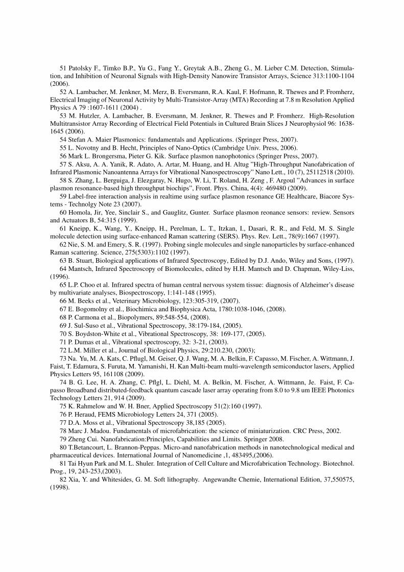
start from certain processes that result in a higher-ordered and -organized structure. Examples of bottom-upapproaches include systems that self-assemble, a process that is triggered by a local change in a chemical orphysical condition. Related techniques include templating and scaffolding methods, such as biomineralization,which rely on backbone structures to support and guide the nucleation and growth of a nanomaterial. In a top-down approach complex structures are built up by patterning layers upon layers from the surface of a planarsubstrate[78]. The realization of the single layer is based on the lithographic process, which in order to define thewhole fabrication process can be rougly summarized in three steps: patterning, etching and depositing (Figure 1).
Figure 1: A typical lithographic process. In a lithographic process a flat substrate is covered by a photosensitive polymer,called resist, which, after exposure to a certain radiation (electrons, photons or ions) become soluble and can be removedusing the appropriate developer (normally a weak base) . The obtained pattern can be considered the final device, for examplea microfluidic channel, or can be used to add or remove material. In the first case we talk of deposition process and we fill theobtained holes with some other material, for example a metal by an electrolytic process. In the second case, called etching,we use the resist as a mask to remove material from the substrate using some chemical attack.
Culturing cells in a nanotechnological devices requires understanding fundamental principles that span mul-tiple disciplines, including biology, biochemistry, physics and engineering. Several groups have developed plat-forms to create microenvironments of greater physiological relevance for high-throughput culturing and analysisof cells under a large number of conditions. Micro and nano fabrication integrating patterning techniques withadvanced surface chemistry makes it possible to reproducibly engineer cell microenvironment at cellular resolu-tion and below, allowing a better understanding of the response of a cell towards outer stimuli. These featuresinclude the chemical nature of the surface adhesive molecules, their precise spatial distribution at nanometer andmicrometre levels, and the physical properties of the surface, such as its topography, stiffness and dimensionality.This understanding leads to more reproducible and meaningful assays.
Since cells normally live in a wet environment, in the following sections we focus mainly on the physics ofmicrofluidics as well as on applications addressing leading questions in biology, and discuss how microfluidicscan contribute to control cellular microenvironments. We will also discuss how micro and nanofabrication canhelp in allowing or enhancing our detection ability. In Appendix 1 we highlight the key elements of the cellularmicroenvironment helpful to design in vitro models more closely mimicking the in vivo conditions.

2.1 Fluids at microscale: the peculiar physics of the micro-environment.
The main advantage of microfluidics is utilizing scaling laws for new effects and better performance[37]. In orderto manipulate and work with microfluidic flows, one must understand the physical phenomena that govern at themicroscale. High surface-to-volume ratios are key in defining fluid-flow characteristics at the microscale. Thepassage from a bulk-dominated flow to a surface-dominated flow gradually occurs when approaching and goingbelow the millimeter scale. As a system shrinks, surface tension can dominate gravity and viscosity can dominateinertia [4]. The ratio of inertial to viscous forces on fluids is characterized by the Reynolds number (Re), oneof the most mentioned dimensionless numbers in studying fluids flowing in microsystems. The dominance ofviscosity determines flow to be laminar, providing predictable streamlines through microchannels. In fact, inthis regime of low Re, fluids do not mix convectively: two adjacent streams flowing side-by-side down a singlechannel maintain a well-defined interface without eddies or turbulences [2,4,17,30,32]. Therefore, diffusion isthe only effective mechanism to mix solutes that are present at different locations in the channel cross-section.Since diffusion is a slow process, microfluidic channels can be used to create cross-sectional gradients in soluteconcentration to study the effect of these gradients on distinct areas of cells within this channel, or to pattern celladhesives and repellents on the substrate prior to introduction of cells[38]. By creating networks of these diffusivecontacts, this controlled mixing may be used to generate complex and temporally stable gradients; this can beapplied in the study of chemotaxis. Strategies for accelerating mixing in microfluidic devices rely on reducingthe characteristic length of the laminar streams by repetitive folding of the fluid onto itself, by induced chaoticadvection or by hydrodynamic focusing[37].
The relative importance of diffusion and convective bulk flow for transporting solute and solvent molecules isgiven by the Peclet number, Pe, and can be readily adjusted through the choice of flow velocity and the dimensionsof the system used. The dominance of viscous forces makes microfluidic systems also ideally suited for realizingpurely diffusive (Pe = 0), convection-free environments that cannot be obtained on macroscale. Only undersuch conditions solute released from a source diffuse in all directions with equal probability, its concentrationdecreasing with increasing distance from the source. The extent of this diffusive layer, a secondary interface,depends on the rate of solute release (or production) at the source and on the solutes diffusivity. If microchannelwalls are close, solute will accumulate in a predictable way[3]. Surface tension forces at the microscale are alsosignificant. They are the result of cohesion between liquid molecules at the liquid/gas interface. As an example,consider that a water spider can easily walk on the surface of water, whereas a human cannot . The effect allowsfor more efficient mass and heat transfer in microsystems. If, for example, surface tension varies along a surface orinterface as a result of thermal or concentration gradients the so-called Marangoni flows can arise and effectivelyhomogenize the thermal or concentration gradients. Since relatively more interface is available for transfer tooccur, and less total mass or energy needs to be transferred to reach the final state. Therefore, both the creationand the homogenization of solute and temperature gradients are faster as system size is reduced[32,34]. Soexothermic and/or high temperature reactions are performed in an efficient and controllable (isothermal) mannerand high specificity of chemical and physical properties (concentration, pH, temperature) can also be ensured.
Surface tension on the microscale can be also used for pumping fluid through channels. According to theYoungLaplace equation, droplets with smaller radii of curvature have a higher internal pressure than those oflarger curvature of the same fluid. The resulting pressure difference is adequate to move microliters in 1s,even when working against gravity, and has been referred to as passive-pumping. Similarly, capillarity forcescan play a significant role in micro-device behavior[39]. Surface properties can be selected to influence thecompetition between viscous forces and capillary forces. This makes it possible to adjust the competition (whichis quantified by the capillary number Ca ). Finally, the large surface-to-volume ratio achieved in microfabricatedflow structures may be exploited to enhance detector sensitivity by greatly increasing the interaction of a sensingelement with a large sample.
2.2 Macroscale vs microscale cell cultures
The significant differences between several physical phenomena at the micro- and macroscale have been exploitedto provide new types of cellular assays 3 impossible to achieve by conventional laboratory techniques [14].
3A cell assay is defined as the measure of cellular response to chemical and/or physical stimuli. Diverse responses charac-terize the cell phenotype, such as alterations of intracellular and extracellular biochemical pathways, cell morphology, motility,and growth properties. When culturing cells in vitro, a number of variables can affect cell phenotype (e.g. contamination,

The ability to culture cells in vitro is one of the cornerstones of modern biology and has revolutionized hy-pothesis testing in basic cell research, becoming a standard methodology for massive production of proteinsand vaccines, drug screening and toxicology assays. It essentially consists of growing cells on a large surface(polystyrene or glass dishes or multiwells) able to stimulate cell binding and immersed in a homogeneous medium[8,16]. In macroscale cultures, large volumes of medium are available to ensure the access to necessary metabo-lites. Continuous secretion of signal molecules by cells causes spatial variability in the concentration of solutesin the culture media. Convection by stirring or shaking generates a rapid homogenous distribution of metaboliteswithin the entire cell culture medium with minimized waste accumulation. As a result, autocrine and paracrinesignaling is, at least temporarily, impaired[17]. Therefore, many of the factors that induce or stabilize differen-tiated phenotypes are poorly understood and difficult to mimic by in vitro standard 2D culture: soluble growthfactors are present at abnormally high concentrations, 3D cues are largely absent, oxygen tension is too high,long distances between other cells and cell volume/medium volume ratios less than one[12,16].
It is worth stressing that culturing cells at microscales allows control over microenvironmental cues, suchas cell-cell and cell-matrix interactions; the use of small culture volumes; and the ability to integrate with mi-crosystem technologies for on-chip experimentation. Moreover, by better control of fluid flow and mass transportmechanisms, microfluidic techniques can provide temporal and spatial patterning of soluble factors or cells nototherwise possible. Microfabrication technologies have enabled researchers to design, with nanometer control,the biochemical composition and topology of the substrate, the medium composition, as well as the type ofneighboring cells surrounding the microenvironment of the cell. They also can be used to link cell culture withintegrated analytical devices that can probe the biochemical processes that govern cell behaviour.
Some cell-based microsystems simply represent miniaturized versions of conventional laboratory techniques,whereas others exploit the advantages of small length scales and low Reynolds numbers, such as favourablescaling of electrical fields and the ability to create well-controlled laminar flows. Studies are emerging thatexamine the combinatorial effects of soluble factor signaling and cellmatrix interactions on cell behavior. Theymay have a great impact especially for drug screening tests and for optimization of microenvironments in tissueengineering applications where long-term performance of tissue constructs are dependent upon many factorsworking synergistically[11,18].
Nowadays, analysis of the cellular microenvironment in microfluidic platforms remains an issue to be ad-dressed, and general progress has been somehow hindered by the lack of a complete understanding of why livingcells behave differently when moved from macroscale culture to confined microscale geometries[11]. Althoughmicrofluidics holds enormous potential to provide a platform for relevant cellular assays, in depth investigationof the biological influence of the engineered microenvironments are still required for this potential to be fullyrealized [19].
2.3 Miniaturization of cell assays in microfluidics
Introduced in the early 90s by activities in microfabrication techniques as a spin-off from Micro-Electro-Mechanical Systems (MEMS) or Micro System Technology (MST) research, the concept of microfluidicswas dedicated on analytical chemistry applications, with a major role for capillary electrophoresis (CE) on achip[20,21]. Since then, a large variety of microfluidic phenomena has been explored based upon fast thermaland species diffusion, large surface-to-volume ratio or large field gradients (electrical, acoustic, optical), resultingfrom the small structural dimensions. Besides these new phenomena, perhaps the most important driving forcefor microfluidics is the ability to handle minute amounts (sub-nanoliter) of liquid in a controlled and reproduciblemanner.
One may state that microfluidics devices prepared by soft-lithographic procedures really entered life sci-ence after the introduction of polydimethylsiloxane (PDMS)-based (a biocompatible, easy to carve silicone rub-ber)[22,23]. PDMS devices can be easily replicated from standard photolithographically fabricated molds (seeAppendix 2), and enable a wide variety of applications. Initially, two approaches were followed in this field:one aiming at combining microsensors with fluidic components (pumps, flow sensors) into systems; the other,which had a much greater impact, focused on miniaturization of analytical chemical methods, in particular sep-arations, with a lot of emphasis on DNA analysis[24,25]. As genetic analysis has now become a more or lessroutine method, the new focus has been for some time on using micro Total Analysis Systems (µTAS) for pro-teomics[17]. In addition, in the past few years, the analysis of even more complex biological systems such as
degree of confluence, presence of cell-cell adhesion, and seeding density) [15].

living cells with the use of microfabricated structures has attracted increased attention[26,27,28]. From a bi-ological standpoint, microfluidics seems especially relevant considering that most biological processes involvesmall-scale fluidic transport at some point. Examples stem from molecular transfer across cellular membranes,to oxygen diffusivity through the lungs, to blood flow through microscale arterial networks[17].
Microscale techniques for cell biology have been applied from single cell analyses and flow cytometry-liketechniques, to treating fields of cells in gradient generating devices, patterned 3-dimensional cultures, to mi-croscale versions of more traditional assay types such as cell culture (via perfusion, or static cultures)[14] . Thetoolbox for biologists is rapidly growing, enabling them to carry out different kinds of sophisticated experimentson the cellular level. An example of such potentiality is the concept of Lab-in-a-Cell, LIC, [24,29], where asophisticated cell-environment interface transform a single cell in a complete chemical and biological laboratory.In the next section the components of a microfluidic platform are briefly discussed.
2.4 Components of a microfluidic device.
An operative definition of microfluidics is necessarily based on the ability to process or manipulate small (10−9 to10−18 litres) amounts of fluids, using a series channels and chambers ranging from one to a few hundred micronsin size [30]. Microfluidics is today the core technology to realize smart cell culturing chambers with fine spatialand temporal chemical control of cellular microenvironment. Inlet and outlet ports of the system serve as pointsof interaction between the culture region and the external world. The size of loading reservoirs with one endopen to the ambient environment and the other end connected to the microchannel network is typically in themillimeter scale to facilitate manual or automatic liquid loading by means of syringe needles or pipettes. Theliquid loaded in the reservoir can then be drawn into the microchannels by capillary force, differential pressure,or electrokinetic force.
Howewer, a microfluidic platform is not a simply network of channels. One must add components to increasefunctionality of the system suited for the specific application. So the design of a microfluidic device typicallyconsists of a combination of fluidic control (method of introducing reagents and samples and for moving thesefluids around on the chip ) and sensing components for combining and mixing them(e.g mixers, heaters, andsensors).Therefore, besides components for on chip handling of nano and pico liter volumes, a complete sys-tem includes others tools interacting with fluids such as micropneumatic systems, i.e. microsystems for the han-dling of off-chip fluids (pumps,valves,etc) and sensors(electrodes, phodetectors, chemical sensors) [31,32,33,34].While selective integration of these components is largely application dependent, all devices require an interfacebetween the microdevice and the macro world.
Microfluidic devices designed specifically for cell culture have certain requirements that distinguish themfrom microscale systems used for other applications in chemistry or physics[11]. To optimize the system and theexperimental design according to the particular application the engineer and the biologist have to evaluate: (1)the choice of material for device fabrication; (2) the geometry and dimensions of the culture region (lithographictechniques allow extreme flexibility but not infinite possibilities to modify the geometry of the platform); (3)the method of pumping and controlling fluid flow[35]; (4) the detection techniques which will be used to studybiological systems. We will discuss point (4) more deeply in the next, but we want to spend some more wordson point (3) which is often extremely critical: it deals with how the microfluidic device is connected to externalcomponents of the overall system. A system typically require multiple interconnects and in many cases thepackaging components are much larger than the microsystems they interface[17,36]. A successful microfluidicinterface must be reliable, mechanically robust, and leak free, allowing to introduce samples into or out of systemwithout contamination. In addition, structural mismatches between the interface and microchannel, create easilydead volumes reducing the efficiency of the microfluidic platform. The difficulty derives from the fact thatsamples and reagents are typically loaded in quantities of microliters to milliliters, whereas microfluidic devicesconsume only nanoliters or picoliters of samples/ reagents due to the micrometric size of reaction chambers andchannels. But the primary requirement is the creation of a sealing channels network to form flow conduits[22].The fundamental trade-off here is between cell accessibility for downstream assays, which requires reversiblysealed or puncturable channels, and channel robustness, which implies a permanently sealed channel. Thoughnumerous passive and active microfluidic components have been realized, the microfluidic interface remains anarea that is relatively overlooked and under-studied. As a matter of fact, a reliable robust packaging is crucialfor the success of microfluidic perfusion culture systems and has been identified as one of the great limiters onacceptability of mycrosystems into the broad world market[17,22].

2.5 Active and passive pumping.
Transporting fluid volumes in microfluidic cell culture devices can be done by active pumping, using valvesand pumps that are either externally linked up to the device, or directly built into the system. Many perfusionsystems employ external pumps connected to the access ports via tubing to pump constant fluid flow for nutrientssupply and waste products removal (e.g., syringe pumps for non-recirculatory flow, and peristaltic roller pumpsfor recirculatory flow) [11]. Multilayer soft lithography, with multiple layers of structures in soft materials,make possible to assemble pneumatic valves into the system to produce fully automated, high-throughput culturesystems[40].The large number of connections required and the potential for leakages at those connections arethe major issues with these integrated components. A microfluidic plaform that utilizes programmed movementof arrays of pins of a Braille display as integrated valves and pumps was developed by the Takayama group[11,17,41]. Valving action is generated by movement of a Braille pin up and down to deform thin PDMS sheetinto microchannels in specific sequences to generate peristaltic flow. A new method of fluid replacement wasintroduced by the Beebe group in the early 2000s [42].The surface tension of different-sized droplets placed at theinlet and outlet ports is exploited to drive fluid from one port to the other[42].The differential pressure betweenports due to the difference in droplet volumes generates flow in the microchannel. Passive pumping can beperformed simply by pipetting the appropriate volumes of droplets at the ports, without need for interconnectionsto external interface.
The ability to design experiments in a simple way with ”tubeless” microfluidics may attract an increasingnumber of biologists but it is limited to low volume flows and low pressures. Steady continuous perfusionof microchannels is better controlled by external pumps[11]. Alternatively, electrokinetics is now studied ina variety of forms for controlling microflows. Electro-osmosis, where the fluid moves relative to stationarycharged boundaries, dielectrophoresis, which moves an interface (often a particle) in a gradient of electric field,and electrowetting, where the electric field modifies wetting properties, have all been exploited. AC and DC fieldscan be considered and the system response then depends on frequency and amplitude of the field [17,35].Othermeans can be used to control flows. In particular, external fields can be used to induce motion of objects embeddedin the fluid, or the channel walls can be systematically distorted: magnetic fields can influence flows directly ormanipulate dispersed magnetic particles, sound fields can produce acoustic streaming motions,cyclic deformationof a wall can induce peristaltic pumping, etc. For each manner of driving a fluid motion, the surface characteristicsof the device can also be exploited to provide additional control for optimal mixing, reacting, detecting, analyzing,and separating.
3 Control of the in vitro microenvironment
The most influential benefit of using microfluidics for biological applications is the ability to engineer thecellular microenvironment. Cues of the microenvironment are integrated by the cell and affect cellular phenotype(i.e.,gene expression and cell behavior), while at the same time the cell can also affect its microenvironment. Thebalance and interplay between the microenvironment and cellular phenotype is therefore an important and com-plex problem in cell biology[39]. Microfluidics can be exploited as a platform for microenvironmental screeningto study cellular response to the microenvironment by helping to create a map between environmental conditionsand cellular phenotype. Some applications search for ways to produce phenotypes of interest. For example, itcould be useful to develop optimal conditions for stem cell self-renewal in order to expand cell resources for usein cell based therapies. Other approaches aim to identify the mechanisms responsible for the produced pheno-types. Using the same stem cell analogy, they can evaluate signaling pathways by self-renewal of cells inducedby extracellular cues for insight into development and disease. Moreover, microenvironmental screening couldgive a contribute in the identification of biochemical targets for therapy development, by exploring which in vivoenvironmental conditions are related to disease (e.g in tumor microenvironments). Other types of screening havebeen applied to observe the effect of a large number of parameters one-at-a-time (e.g., the use of chemical li-braries on particular cell phenotypes in drug screening) or control a single parameter on many different readouts(e.g., micro-arrays for gene expression).The simultaneous combination of multiple microenvironmental factorssuch as soluble molecules, extracellular matrix (ECM) composition, and cellcell contact have been relatively lessexplored. Therefore the core of spatial and temporal modulation of the cell growth and stimulation is to integratenanopatterned surfaces mimicking the complex features of the extracellular matrix, with microfluidic channels

regulating transport of fluids and signaling by soluble factors [12].
3.1 Microfluidcs for precise spatio-temporal chemical concentration gradients:chemotaxis study
Gradients of soluble signaling molecules play an important role in mediating a number of physiological pro-cesses in vivo such as chemotaxis, proliferation and differentiation, axon guidance and embryogenesis. Insightinto the interplay between a chemical gradient treatment and the corresponding cellular response may help todetermine the cues responsible for regulating specific cellular activities. Traditional macroscale devices (e.g,Boyden chamber, Zigmond chamber, Dunn chamber, under-agarose assay and micro-pipette assays ) generallylack the ability to maintain and manipulate stable gradients, have poor reproducibility and require a large numberof cells [17,38,43]. Additionally the incompatibility with real-time optical imaging methods further limit theapplication in modern biological experiments. Understanding the importance of these chemical cues may helpresearchers develop controlled microenvironments where the desired cellular response is produced by combiningthe effects of exogenous controlled gradient treatments with ongoing endogenous cell-cell signaling.
Microfluidic technology has brought about a broad range of methods for exposing cells to engineered gra-dients (time invariant gradients, subcellular resolution gradients, continuos or discrete gradients, fast responsedynamic gradients), each method having a unique set of attributes and disadvantages. In general, gradient-generating microfluidic platform can be divided into two classes: laminar flow and flow-free based systems. Thefirst class create chemical gradients by taking advantage of diffusional mixing across the interface of adjacentlymultiple laminar flows in a single channel or microchannels network. Diffusion between the stream produce agradient across the channel, perpendicular to the flow direction having a diffusing profile that is stable at everyfixed point in the channel but evolved along the channel. The laminar nature of fluid flow through microcon-duits permits many streams containing not only different substances but also different concentrations of the samesubstance to flow side by side. With these devices, it is possible to treat a cell population with a controlledchemical gradient and observe the biochemical and morphological responses in vitro. The continuously flowingstreams of fluid provide precise control over the stability, gradient profile, concentration range and slope of achemical gradient. The stimulus of interest can be changed on the fly to create a sequential chemical gradienttreatment scheme. Gradient generation typically yields close agreement with theoretical predictions and allowsgradients with defined simple or complex shapes to be produced and manipulated. The second type generates timeevolving or stable gradients by free-diffusion of chemicals in a free-flow environment such as membrane-basedand hydrogel-based assay. In cell motility and migration studies, special attention has been paid to chemotaxisstudies, in many areas of biology, including microbiology and immunology[15]. Many cell types exhibit theability to sense certain chemicals and move, or taxis, toward higher concentrations of chemoattractants known aschemokines. As an example, leukocytes respond to chemokines released from damaged cells.
Chemotaxis studies require a way to deliver chemicals to cells in a controlled gradient because cells areresponsive to concentration differences down to 2% across their diameter to direct their motion. Spatial complex-ity and localization in intracellular systems is important in chemotaxis and polarized signaling and could thusbenefit greatly from the high-specificity of microfluidics. Modeling approaches to chemotaxis signaling havedemonstrated robustness and adaptation as properties of the bacterial chemotaxis signaling system. Jeon et al.[44] developed a microfluidic device capable of creating a single cell-resolution concentration gradient by feedinga small number of input fluid streams with initial concentrations of diffusible substances . The system consists ofan embedded network of microchannels with two main regions: the gradient generation region that is a pyramidalbranched microchannel structure to split, mix and combine fluid streams as they flow through and the observa-tion region in which cells are placed and analyzed. Initially, a network of serpentine channels allowed adjacentlaminar streams to flow together long enough for full diffusive mixing. As the streams travel down the network,they are repeatedly split, and some combined with neighbouring streams and allowed to mix by diffusion withina channel before being split and combined again. At the end of the network, many individual streams of differentconcentrations were flowed adjacently into a single outlet that will have a complex concentration profile perpen-dicular to its flow direction. If the bottom level output channels are not converged into a single outlet, the devicecould be used to create and isolate several differently concentrated mixtures of the input streams. The gradientshape was adjustable in width and shifted from side to side by changing the individual input stream flow rates,which can be performed dynamically. With little modification, both linear and nonlinear concentration gradientswere realized.

Figure 2: Schematics of the chemotaxis assay. Left: Top view of the embedded network of microchannels with two mainregions: the gradient generation region that is a pyramidal branched array of microchannels to split, mix and combine fluidstreams as they flow through and the observation region in which cells are placed. Right: 3D exploded view of the gradientgeneration through the PDMS microfluidic platform bonded to a glass coverslip. Courtesy of [43.]
Chemotaxis of human neutrophils (immune cells that migrate to sites of infection or injury) in interleukin-8(IL-8) gradients and breast cancer cell chemotaxis was investigated using the gradient generator (Figure 1).
The continuously flowing streams that are necessary to maintain chemical gradients make the type of devicesin Figure 1 unsuitable for addressing biological questions where soluble factors are important in regulating cellbehavior. One way that cells respond to chemical cues in their environment is by secreting signaling factorsthat either affect the secreting cell itself (autocrine), or affect other types of cells (paracrine). In flow basedsystems, autocrine/paracrine factors cannot accumulate because flowing fluid streams immediately carry awaysecreted factors. In situations where cellcell communication (via soluble factors) plays a critical role in regulat-ing biochemical activity, the removal or accumulation of secreted factors may lead to distinctly different cellularbehavior. Beebe and coworkers [45] monitored neutrophil chemotaxis under concentration gradients formed stat-ically, i. e.,without flowing fluid . The characterization of this system is an essential step that lays the foundationfor quantitative use of this device to determine when cellcell communication (via soluble factors) is an impor-tant consideration. The chemical gradient is maintained by using a source/sink construct that includes a highfluidic resistance membrane. Chemical species diffused into a cell microchamber through a membrane whichimposed high fluid resistance to minimize convective flows. In this manner, soluble cues including autocrinefactors and paracrine factors were not washed away with a laminar flow but rather accumulated in the microen-vironment. This design allows for examining the influence of cellcell communication on chemotaxis or on othercell responses to chemical stimuli (Figure 2).
Flow-free microfluidic systems also offer intriguing opportunities for the study of processes such as cell divi-sion and migration, intercellular communication and the emergence of cell polarity during development (wheremolecular gradients are known to play an important role). For instance, cell proliferation studies on a numberof different cell types have revealed that the proliferation characteristics are markedly different when using mi-crochannels instead of traditional mass culture systems. To understand this difference in behaviour, considerthe rather different environments experienced by the cells: in the constrained medium within a microchannel,signalling molecules produced by a given cell (autocrine signals) or surrounding cells (paracrine signals) canaccumulate, whereas such signaling molecules will be diluted or even removed by the convective flows that in-evitably arise in mass culture systems or flowing microfluidic systems. Culturing in microchannels in the absenceof flow, where transport is purely by diffusion and the size of the system prevents extensive dilution, appears toincrease the sensitivity of proliferating cells to the effects of soluble factors. Similar effects may explain why theefficiency of embryo development improves in microchannels under no-flow conditions. So microfluidics shouldopen new opportunities for studying cell signaling, where convection-free culture conditions allow signallingmolecules secreted by a cell to form diffusive layers and influence the secreting and surrounding cells [3].

Figure 3: Schematics of gradient-generation microdevice. The chemical stimulus of interest is added over the membrane inthe source region. The molecules diffuse through the membrane and into the channel creating a chemical gradient along theaxial (x) direction of the channel. Reproduced by permission of The Royal Society of Chemistry [45].
3.2 Engineering chemically and nanotopographic patterned substrates.
In standard cultures of adherent cells, cells are randomly seeded on the surface of the culture substrate. Chemicaland topographic substrate surface patterning can control the size, shape and spatial position of cells anchoredto a surface. Spatial control of cell adhesive substrates allow us to mimic signaling tracks which are naturallypresent in vivo on the ECM. For example, gradients of surface properties are used to study the behavior of motilecells or neuronal path findings. Moreover forcing cells to follow a given adhesion pattern can be exploited tostudy interplay between the geometrical constrains and cell behavior such as changes in cell polarity. Elas-tomeric microchannels can also be used as microfluidic templates to chemically pattern surfaces generated bydelivering adhesion promoting proteins or cell suspension to desired regions of a substrate. In most devices, cellsadhere on glass surfaces via incubation of the microchannels with ionic polypeptides like poly-lysine or proteinslike fibronectin .In some systems cell binding domain are patterned on proteinresistant (”non-fouling”) surfaces.Poly(ethylene glycol) (PEG) immobilized onto surfaces is widely used to confer high resistance to protein adsorp-tion and cell adhesion[46]. Nanotopographic cues are a subset of substrate engineering, representing signalingmodality to direct complex cellular processes including the specificity and efficiency of lineage-specific stem celldifferentiation and tissue organization. Cells interact with native structures on the micron and sub-micron scalenative ECM structures in many ways, often through a phenomenon known as contact guidance. This mecha-nism is an essential component in regulating cell migration, such as axonal guidance and growth cone motility.Synthetically nanofabricated topography can also influence cell morphology, alignment, adhesion, migration,proliferation, and cytoskeleton organization. Cell-nanotopography interactions also vary across cell type, featuresize, and feature geometry as well. There are virtually an infinite number of potential combinations of cell types,biomaterial composition, and topographic feature arrangements.
Three basic nanotopographic geometries are nanogratings, nanopost and nanopit arrays. The nanofabrica-tion of substrates with a long-range order across a wide range of feature sizes and geometries has been realizedusing a variety of methods including traditional photolithography, e-beam photolithography, and interferencelithography. Nanotopography could be utilized in addition to other types of cell-biomaterial surface conditionsincluding microcontact printed chemistries and bulk mechanical properties of the substrate. Bulk materials pro-cessing and nanofabrication strategies for many nanotopographic surfaces are typically fine-tuned for silicon,silicon oxide, polycrystalline silicon, and other inorganic material systems such as titanium. These substrates canbe used directly or serve as masters for replica-molding of organic polymers such as PDMS, polystyrene (PS),

poly(methyl methacrylate) (PMMA), polycarbonate (PC), and poly(ethylene glycol) (PEG) for in vitro applica-tions or biodegradable polymers such as poly(caprolactone) (PCL), poly(L-lactic acid) (PLA), poly(glycolic acid)(PGA), and poly(L-lactic-co-glycolic acid) (PLGA) for potential use in vivo. Advanced fabrication techniquesand compatible materials must eventually be synergized as a means to integrate cell-nanotopography interactionsinto advanced tissue e engineering scaffolds[47]. An overview of these techniques can be found in Appendix 2.
4 Detection of biological processes in living cells
Nowadays micro and nanofabrication potentialities have been extensively used to improve our ability to mea-sure biological quantities. The production of standard micro arrays for genomics and, more recently, proteomics,is based or take advantage from one or more of the lithographic processes described in Appendix 2. Probing thechemistry and physics in a living cell is one of the basic goals in modern cellular biophysics and an importantprerequisite for progress in both molecular and nanomedicine, thus it has been one of the major research subjectof the past few years. When we focus on the detection of living cells process, the most mature applications ofnanotechnology, and probably the first ones coming to mind, deal with reading electrical signals from neurons,as in the case of Multi Electrodes Arrays (MEA)[49,50], or, more recently, Multi Transistor Array[51-53]). Thisis due to the enormous amount of expertise developed in electrodes and electrical amplifiers fabrication duringthe past forty years by microelectronic industry.
When dealing with cells other than neurons or when looking for biological processes not associated withelectrical signals, optical and spectroscopic techniques are more general and record more complete informationbeing able to detect changes at the molecular level. In particular, in the past decade there has been a signifi-cant increase in the use of vibrational spectroscopies (Raman and Mid-Infrared) as tools for single cell analysisthanks to their being minimally invasive and label free. These properties are useful particularly when looking atunknown processes activated by external agents as, for example, in drug-cells interaction studies or toxicology,or if fast cell screening is needed as in a biopsy where diseased cells must be discriminated from the healthyones. However, Raman or Mid-Infrared (MIR) investigations of living cells are not complication-free becauseprobe volumes are small and concentrations of intrinsic cellular molecules low. Moreover, just to make someexamples, Raman scattering has very low cross section and, hence, signals. Thus, a trade off between the celldamage and applicable maximum intensity of the excitation laser, must be made. Moreover, MIR suffer fromthe lack of transparency of the most common materials used in biology (plastics, glass, water). Thus, as alreadystated, the choice of detection technique which will be used in our experiments is one of the four keystones forthe design and fabrication of the devices to be used, and will determine many of its properties. Just to make someexamples, the detection techniques will contribute to define the material to be used as a substrate (for exampletaking in account the transparency to certain electromagnetic wavelength), physical constraints of our measure-ments apparatus (parameters like the depth of focus or numerical aperture of our microscope, or the overall sizesallowed by the microscope stage) will define the final geometrical setup of our measurement tools). In the nexttwo sections we will briefly introduce some applications of micro and nanotechnologies to enhance or permit thedetection of cellular processes in in-vitro experiments.
4.1 Enhancing signals for vibrational spectroscopies: plasmonics4
Surface plasmon polaritons (SPP) are electromagnetic excitations propagating at the interface between a dielectricand a conductor, evanescently confined in the perpendicular direction. These electromagnetic surface waves arisevia the coupling of the electromagnetic fields to oscillations of the conductors electron plasma. This meansthat if we send a laser beam onto a dielectric plate (glass, plastic, etc.) over which we have deposited a thin(few nanometers) metallic fim, at a certain angle between the laser beam and the plate surface, we can inducean oscillation of the metal electrons at a very specific frequency. Obviously, this phenomena subtract part ofenergy to the laser beam, and, if we look at the reflected laser beam, we will observe a diminution in the reflectedintensity. Since the electron oscillation frequency is linearly connected to the laser/plate angle we are able tomeasure any change in the frequency oscillations just measuring at which angle we observe the minimum in the
4The interested reader can find detailed information about theory, fabrication and applications of plasmonic structures asbiosensors in may textbooks and scientific paper. Most of the material of this section is derived from [54-56]

laser beam reflected intensity. In figure 4 a surface plasmon polariton excitation is schematized in the case of thedielectric plate substituted by a prism.
Figure 4: Scheme of excitation of a metal electrons plasmonic resonance. The laser beam make the electron oscillates, varyingthe electric field on the metallic surfaces indicated by the + and - symbols. Oscillation frequency is strongly related to therefraction index of the material in contact with the metallic film, thus the presence of a molecular film on the metallic surfacecan be detected monitoring the reflected laser beam intensity.
SPP are today widely used to realize commercial and laboratory biosensors (see for example [57-59]). In fact,most of surface plasmon sensing work carried out so far has been based on the interrogation of propagating SPPwaves at a metal/air interface. Using surface functionalization, agent-specific binding can be achieved, changingthe refractive index of the metal surface superstrate and thus the dispersion relation of the propagating SPPs.Binding events can then be monitored by studying the changing phase-matching condition via either wavelengthorangular interrogation. Many reviews of these techniques in a sensing context can be found in literature (see forexample [60]).
Micro and nanofabrication plays a fundamental role in engineering plasmonic based biosensors, both in en-hancing the coupling between the excitation laser and the metal plasma elctrons, by ad hoc structures (gratings)structures, and in enhancing the electrical fields on certain points of the surface. The second point is necessary towork with the second fundamental excitation of plasmonics: localized surface plasmons (LSP). Localized surfaceplasmons are non-propagating excitations of the conduction electrons of metallic nanostructures coupled to theelectromagnetic field. These modes arise naturally from the scattering problem of a small, sub-wavelength con-ductive nanoparticle in an oscillating electromagnetic field. The curved surface of the particle excerts an effectiverestoring force on the driven electrons, so that a resonance can arise, leading to field amplification both inside andin the near-field zone outside the particle. This resonance is called the localized surface plasmon or short local-ized plasmon resonance. Another consequence of the curved surface is that plasmon resonances can be excitedby direct light illumination, in contrast to propagating SPPs. One of the most spectacular applications of LSP todate is surface enhanced Raman scattering (SERS), which exploits the generation of highly localized light fieldsin the near-field of metallic nanostructures for enhancing spontaneous Raman scattering of suitable molecules.Using chemically roughened silver surfaces, Raman scattering events of single molecules have been recorded[61,62], with estimated enhancements of the scattering cross section by factors up to 1014. The majority of thisenhancement is believed to arise from the highly enhanced fields in metal nanoparticle junctions due to localizedsurface plasmon resonances. Termed hot spots, these highly confined fields also enable an increase of fluorescentemission, albeit with more modest enhancement factors. A proper understanding and control over the generationof these hot spots, for example in the form of nanoscale plasmonic cavities, is currently one of the major drivingforces behind the design of nanoparticle ensembles with tuned optical properties both with nanofabrication and

chemical (top-down and bottom-up) techniques.
5 Mid Infrared Spectroscopy of Bio-systems
Infrared spectroscopy, and more specifically Fourier Transform Infrared Spectroscopy (FTIR) is widely usedfor probing the chemical composition and molecular structures of complex bio-systems such as tissues, bodyfluids or cells. A Mid-IR spectrum (MIRS) provides the vibrational fingerprints of the all constituents of a bio-system, namely proteins, carbohydrates, nucleic acids and lipids, in one single experiment. On the base of thespecific biochemistry of the system under investigation, specific group vibration can be assigned primarily to onecellular constituent as detailed in many scientific reports and books [63,64] and schematized in Figure 5, referredto an eukaryotic cell. MIRS is a fast, label-free, non destructive analytical technique whose sensitivity have beenexploited for diagnostic purposes, such as characterization of amyloid plaques in brain tissues of Alzheimersdisease patients [65] and prion infected tissues [66], for monitoring cancer progression [67] and the drug-responseof tumoral cells [68, 69], for the characterization of cell-cycle stages [70] just to give a few examples.
Figure 5: Representative IR spectrum of an eukaryotic cell. Bands of major interest for diagnostic purposes are shown: lipids,phospholipids, proteins, nucleic acids and carbohydrates. Band assignment of major functional bio-groups is also reported(ν=stretching; γ=bending; s.=symmetric; as.= asymmetric). During cellular processes the band structure changes revealingthat something in the cell is changing. For example, during a differentiation process we first observe a strong change in thenucleic acid band (change in the chromatine structure, then increase the RNA band indicating a transcription process, then achange in the protein band)
However, the possibility to work at single cell and sub-cellular level at diffraction limit, maintaining an ac-ceptable data quality in terms of signal to noise ratio, is nowadays guaranteed only by the brightness advantageof synchrotron radiation (SR) sources, defined as the photon flux per source area and solid angle [72,71]. The in-creasing number of infrared beamlines dedicated to biological applications is evidencing the great interest of thebio-medical community for this technique: at the beginning of the 90s, only two IR beamlines where operativein Europe while their number is today 10. Despite this, their number is still limited and the restricted access toSR facilities can not satisfying the huge community of interested scientists. In the next future broad band sourcebased in mid-infrared quantum cascade lasers (QCL) [73,74] could transfor FTIR microspectroscopy (µ-FTIR)at the single cell level a competitive techniques to be used at the single biolab level. Nowadays, commonly mea-sured samples by µ-FTIR are fixed or dried, which allow only acquiring still frames of the phenomenon underinvestigation. This is the feasible way to avoid the strong absorbance of water-based cellular media, since thecompensation of the buffer contribution from collected spectra, for disclosing the Amide I band, limits the path-length of measurements cells to 10 µ m or less [75]. Due to these technical difficulties associated with living

cells measurements in physiological environment there are still only a few pioneering reports [76,77] where thefluidic chambers are realized by spacing apart 2 optical windows by using few microns thick plastic spacers. Thisapproach cannot avoid seal problems and inaccurate pathlength control and also imposes limitations on the devicedesign flexibility. The application of micropatterning and microfluidic concepts to IR transparent materials opensnew opportunities for µ-FTIR spectroscopy of living cells. The implementation of both IR and visible transparent3D microfluidic devices will allow the real time observation by µ-FTIR of biochemical rearrangements undergoneby living cells upon chemical and/or mechanical stimulations. Moreover, by exploiting the brightness of Syn-chrotron Radiation (SR), diffraction limited spatial resolution can be achieved to collect individual cell spectraand chemical maps at diffraction-limited spatial resolution.
REFERENCES
1 J. Castillo, M. Dimaki and W. E. Svendsen. Manipulation of biological samples using micro and nanotechniques. Integr. Biol., 3042 (2009).
2 E. M.Purcell. Life at low Reynolds number. Am. J. Phys.,45, 311 (1977).3 J. Atencia and D. J. Beebe. Controlled microfluidic interfaces. Nature, 437, 648-655, (2005).4 Squires, T. M. and Quake, S. R. Microfluidics: fluid physics on the nanoliter scale. Rev. Mod. Phys., 77(3),
(2005).5 M.Wautelet. Scaling laws in the macro-, micro- and nanoworlds. Eur. J. Phys. ,22 ,601611(2001).6 Silva, G.A. Neuroscience nanotechnology: progress, opportunities and challenges, Nature Rev. Neuro-
science, 7, 65-74 (2006)7 C. H. Arnaud. Mimicking Biological Systems. Control of the chemistry and physics of cell-populated
microenvironments reveals new biology. CandEN, Volume 85, Number 37.8 N.Li, A.Tourovskaia, and A. Folch. Biology on a Chip: Microfabrication for Studying the Behavior of
Cultured Cells. Critical Reviews in Biomedical Engineering, 31,423488,(2003).9 A.Khademhosseini, J. Borenstein, M.Toner and S.Takayama. Micro and nanoengineering of the cell mi-
croenvironment. Artech House 2008.10 M.P. Lutolf, J.A. Hubbell. Synthetic biomaterials as instructive extracellular microenvironments for mor-
phogenesis in tissue engineering. Nat Biotechnol,23,47-55,(2005).11 W. K. Edmond Young and D. J. Beebe. Fundamentals of microfluidic cell culture in controlled microenvi-
ronments. Chem. Soc.Rev.,39,10361048, (2010).12 J. El-Ali, P. K. Sorger and K.F. Jensen. Cells on chips. Nature 442, 403-411 , (2006).13 B.Geiger, J.P. Spatz and A.D. Bershadsky. Environmental sensing through focal adhesions. Nature
reviews,10,21-33,(2009).14 A.L. Paguirigan and D. J. Beebe. Microfluidics meet cell biology: bridging the gap by validation and
application of microscale techniques for cell biological assays. Bioessays, ( 2008).15 I. Barbulovic-Nad, A. R. Wheeler. Cell Assays in Microfluidics. In Encyclopedia of Micro- and Nanoflu-
idics; Li, D. Q., Ed.; Springer: Heidelberg, Germany; Vol. 1, 209-216, 2008 .16 G. M. Walker, H. C. Zeringue and D. J. Beebe. Microenvironment design considerations for cellular scale
studies. Lab on C h i p, 4 , 91-97 (2004).17 Wei-Cheng Tian. Microfluidics for Biological Applications. Springer (2009).18 J. Y. Park,S. Takayama andand S. Lee. Regulating microenvironmental stimuli for stem cells and cancer
cells using microsystems. Integr. Biol., 2, 229240, (2010).19 A.L. Paguirigan and D. J. Beebe. From the cellular perspective: exploring differences in the cellular
baseline in macroscale and microfluidic cultures. Integr. Biol., 1, 182195, (2009).20 A.van den Berg, H.G. Craighead andnd P. Yang. From microfluidic applications to nanofluidic. Chem.
Soc. Rev., 39, 899900, (2010).21 A.J. deMello. Control and detection of chemical reactions in microfluidic systems. Nature 442, 394-402,
(2006).22 L. Kim, Yi-Chin Toh, J. Voldman and H. Yu. A practical guide to microfluidic perfusion culture of adherent
mammalian cells. Lab Chip, 7, 681694,(2007).23 D.B. Weibel, W.R. DiLuzio and G. M. Whitesides. Microfabrication meets microbiology. Nature
Reviews,5,210-18,(2007).

24 S. Le Gac and A. van den Berg. Single cells as experimentation units in lab-on-a-chip devices. Trends inBiotechnology Vol.28 No.2.
25 S. Haeberle and R. Zengerle. Microfluidic platforms for lab-on-a-chip applications. Lab Chip,7, 10941110,(2007).
26 H. Andersson , A. van den Berg. Microfluidic devices for cellomics: a review. Sensors and Actuators B,92, 315325,(2003).
27 G.Velve-Casquillas , M. Le Berrea, M. Piel, P.T. Tran. Microfluidic tools for cell biological research. NanoToday , (2009).
28 M. Ni , W. Hao Tong , D. Choudhury , N. A. A. Rahim , C. Iliescu and H.Yu .Cell Culture on MEMSPlatforms: A Review .Int. J. Mol. Sci., 10, 5411-5441,(2009).
29 H. Andersson and A. van den Berg. Microtechnologies and nanotechnologies for single-cell analysis.Current Opinion in Biotechnology, 15, 4449,(2004).
30 George M. Whitesides. The origins and the future of microfluidics. Nature 442, 368-73,(2006).31 P Abgrall and A-M Gue. Lab-on-chip technologies: making a microfluidic network and coupling it into a
complete microsystema review. J. Micromech. Microeng. ,17, (2007).32 D.J.Beebe, G.A.Mensing and G.M.Walker. Physics and Applications of Microfluidics in Biology. Annu.
Rev. Biomed. Eng, 4, 26186, (2002).33 E.Delamarche, D. Juncker and H. Schmid. Microfluidics for Processing Surfaces and Miniaturizing Bio-
logical Assays. Adv. Mater, 17, 29112933,(2005).34 P.S. Dittrich and A. Manz. Lab-on-a-chip: microfluidics in drug discovery. Nature, 5, 210-18,(2006).35 H.A. Stone, A.D. Stroock and A. Ajdar. Engineering flows in small devices. Annu. Rev. Fluid Mech., 36,
381411,(2004).36 S. Marre and Klavs F. Jensen. Synthesis of micro and nanostructures in microfluidic systems. Chem. Soc.
Rev., 39, 11831202, (2010).37 D.Janasek, J.Franzke and A. Manz. Scaling and the design of miniaturized chemical-analysis systems,
Nature 442,374-380, (2006).38 T.M. Keenan and A. Folch. Biomolecular gradients in cell culture systems. Lab Chip, 8, 3457,( 2008).39 J.W. Warrick, W.L.Murphy and D.J.Beebe. Screening the Cellular Microenvironment: A Role for Mi-
crofluidics. IEEE Rev Biomed Eng., 75-93,(2008).40 Unger MA, Chou HP, Thorsen T, Scherer A, Quake SR. Monolithic microfabricated valves and pumps by
multilayer soft lithography. Science,288, 113-6,(2000).41 W. Gu, X. Y. Zhu, N. Futai, B. S. Cho and S. Takayama. Proc.Natl. Acad. Sci. U. S. A., 101, 1586115866,
(2004).42 E. Berthier and D. J. Beebe. Flow rate analysis of a surface tension driven passive micropump. Lab Chip,
7, 14751478,(2007).43 S. Toetsch, P. Olwell,A. Prina-Mellob and Y. Volkov. The evolution of chemotaxis assays from static
models to physiologically relevant platforms. Integr. Biol., 1, 170181, (2009).44 Jeon, N.L. et al. Neutrophil chemotaxis in linear and complex gradients of interleukin-8 formed in a
microfabricated device. Nature Biotechnol. 20, 826830, (2002).45 V. V. Abhyankar, M.A. Lokuta, A. Huttenlocherb and D.J. Beebe. Characterization of a membrane-based
gradient generator for use in cell-signaling studies. Lab on Chip, 6, 389393, (2006).46 C. J Bettinger, R. Langer and J. T Borenstein. Engineering Substrate Micro- and Nanotopography to
Control Cell Function. Angew Chem Int Ed Engl.,48, (2009).47 G. M. Whitesides and C.Love. The Art of Building Small. SCIENTIFIC AMERICAN REPORTS. Septem-
ber 2007.48 Sun, J., Masterman-Smith M.D., Graham N. A., Jiao J., Mottahedeh J., Laks D.R., Ohashi M., DeJesus
J., Kamei K.I., Lee K.B., Wang H., Yu Z.T.F., Lu Y.T., Hou S., Li K., Liu M., Zhang N., Wang S., AngenieuxB., Panosyan E., Samuels E.R., Park J., Williams D., Konkankit V., Nathanson D., van Dam R.D., Phelps M.E.,Wu H., Liau L.M., Mischel P.S., Lazareff J.A., Kornblum H.I., Yong W.H., Graeber T.G. and Tseng H.R. AMicrofluidic Platform for Systems Pathology: Multiparameter Single-Cell Signaling Measurements of ClinicalBrain Tumor Specimens, Cancer Res 70:6128-6138 (2010).
49 Potter SM.. Distributed processing in cultured neuronal networks. Prog Brain Res 130: 49-62 (2001).50 Baudry M, Taketani M, eds. Advances in Network Electrophysiology Using Multi-Electrode Arrays. New
York: Springer Press;: 24-37 (2006).
51 Patolsky F., Timko B.P., Yu G., Fang Y., Greytak A.B., Zheng G., M. Lieber C.M. Detection, Stimula-tion, and Inhibition of Neuronal Signals with High-Density Nanowire Transistor Arrays, Science 313:1100-1104(2006).
52 A. Lambacher, M. Jenkner, M. Merz, B. Eversmann, R.A. Kaul, F. Hofmann, R. Thewes and P. Fromherz,Electrical Imaging of Neuronal Activity by Multi-Transistor-Array (MTA) Recording at 7.8 m Resolution AppliedPhysics A 79 :1607-1611 (2004) .
53 M. Hutzler, A. Lambacher, B. Eversmann, M. Jenkner, R. Thewes and P. Fromherz. High-ResolutionMultitransistor Array Recording of Electrical Field Potentials in Cultured Brain Slices J Neurophysiol 96: 1638-1645 (2006).
54 Stefan A. Maier Plasmonics: fundamentals and Applications. (Springer Press, 2007).55 L. Novotny and B. Hecht, Principles of Nano-Optics (Cambridge Univ. Press, 2006).56 Mark L. Brongersma, Pieter G. Kik. Surface plasmon nanophotonics (Springer Press, 2007).57 S. Aksu, A. A. Yanik, R. Adato, A. Artar, M. Huang, and H. Altug ”High-Throughput Nanofabrication of
Infrared Plasmonic Nanoantenna Arrays for Vibrational Nanospectroscopy” Nano Lett., 10 (7), 25112518 (2010).58 S. Zhang, L. Berguiga, J. Elezgaray, N. Hugo, W. Li, T. Roland, H. Zeng , F. Argoul ”Advances in surface
plasmon resonance-based high throughput biochips”, Front. Phys. China, 4(4): 469480 (2009).59 Label-free interaction analysis in realtime using surface plasmon resonance GE Healthcare, Biacore Sys-
tems - Technolgy Note 23 (2007).60 Homola, Jir, Yee, Sinclair S., and Gauglitz, Gunter. Surface plasmon reonance sensors: review. Sensors
and Actuators B, 54:315 (1999).61 Kneipp, K., Wang, Y., Kneipp, H., Perelman, L. T., Itzkan, I., Dasari, R. R., and Feld, M. S. Single
molecule detection using surface-enhanced Raman scattering (SERS). Phys. Rev. Lett., 78(9):1667 (1997).62 Nie, S. M. and Emery, S. R. (1997). Probing single molecules and single nanoparticles by surface-enhanced
Raman scattering. Science, 275(5303):1102 (1997).63 B. Stuart, Biological applications of Infrared Spectroscopy, Edited by D.J. Ando, Wiley and Sons, (1997).64 Mantsch, Infrared Spectroscopy of Biomolecules, edited by H.H. Mantsch and D. Chapman, Wiley-Liss,
(1996).65 L.P. Choo et al. Infrared spectra of human central nervous system tissue: diagnosis of Alzheimer’s disease
by multivariate analyses, Biospectroscopy, 1:141-148 (1995).66 M. Beeks et al., Veterinary Microbiology, 123:305-319, (2007).67 E. Bogomolny et al., Biochimica and Biophysica Acta, 1780:1038-1046, (2008).68 P. Carmona et al., Biopolymers, 89:548-554, (2008).69 J. Sul-Suso et al., Vibrational Spectroscopy, 38:179-184, (2005).70 S. Boydston-White et al., Vibrational Spectroscopy, 38: 169-177, (2005).71 P. Dumas et al., Vibrational spectroscopy, 32: 3-21, (2003).72 L.M. Miller et al., Journal of Biological Physics, 29:210.230, (2003);73 Na. Yu, M. A. Kats, C. Pflugl, M. Geiser, Q. J. Wang, M. A. Belkin, F. Capasso, M. Fischer, A. Wittmann, J.
Faist, T. Edamura, S. Furuta, M. Yamanishi, H. Kan Multi-beam multi-wavelength semiconductor lasers, AppliedPhysics Letters 95, 161108 (2009).
74 B. G. Lee, H. A. Zhang, C. Pflgl, L. Diehl, M. A. Belkin, M. Fischer, A. Wittmann, Je. Faist, F. Ca-passo Broadband distributed-feedback quantum cascade laser array operating from 8.0 to 9.8 um IEEE PhotonicsTechnology Letters 21, 914 (2009).
75 K. Rahmelow and W. H. Bner, Applied Spectroscopy 51(2):160 (1997).76 P. Heraud, FEMS Microbiology Letters 24, 371 (2005).77 D.A. Moss et al., Vibrational Spectroscopy 38,185 (2005).78 Marc J. Madou. Fundamentals of microfabrication: the science of miniaturization. CRC Press, 2002.79 Zheng Cui. Nanofabrication:Principles, Capabilities and Limits. Springer 2008.80 T.Betancourt, L. Brannon-Peppas. Micro-and nanofabrication methods in nanotechnological medical and
pharmaceutical devices. International Journal of Nanomedicine ,1, 483495,(2006).81 Tai Hyun Park and M. L. Shuler. Integration of Cell Culture and Microfabrication Technology. Biotechnol.
Prog., 19, 243-253,(2003).82 Xia, Y. and Whitesides, G. M. Soft lithography. Angewandte Chemie, International Edition, 37,550575,
(1998).

83 Byron D. Gates, Qiaobing Xu, J. Christopher Love,Daniel B.Wolfe, and George M.Whitesides. Uncon-ventional Nanofabrication. Annu. Rev. Mater. Res., 34, 33972,(2004).
84 J.C.McDonald and George M. Whitesides. Poly(dimethylsiloxane) as a Material for Fabricating Microflu-idic Devices. Accounts of Chemical Research , 35, No. 7, ( 2002).
85 S.A. Lange,V. Benes, D.P. Kern, J. K. Heinrich Horber and A. Bernard. Microcontact Printing of DNAMolecules. Anal. Chem.,76, 1641-1647(2004).
86 E. Delamarche. Microcontact printing of proteins. Nanobiotechnol., 3152, (2004).87 M.Geissler and Y.Xia.Patterning: Principles and some new developments. Advanced Materials, 16,
No.7,(2004).88 Ravi S. Kane, S. Takayama, E. Ostuni,Donald E. Ingber, George M. Whitesides. Patterning proteins and
cells using soft lithography. Biomaterials, 20, 2363-2376, (1999).89 Didier Falconnet, Gabor Csucs, H. M. Grandin, M. Textor. Surface engineering approaches to micropattern
surfaces for cell-based assays. Biomaterials,27,30443063,(2006).90 G. M. Whitesides,E. Ostuni, S. Takayama, X. Jiang, and D. E. Ingber. Soft lithography in biology and
biochemistry. Annu. Rev. Biomed. Eng. 3, 335373 (2001).91 S. A.Ruiz and C. S. Chen. Microcontact printing: A tool to pattern. Soft Matter, 3, 111,(2007).92 J. C. Love , L. A. Estroff, J. K. Kriebel, R. G. Nuzzo and G. M. Whitesides. Self-assembled monolayers
of thiolates on metals as a form of nanotechnology. Chemical Reviews ,105, 11031169, (2005).93 E.Ostuni, G.M. Whitesides , D.E. Ingber, C.S. Chen. Using Self-Assembled Monolayers to Pattern ECM
Proteins and Cells on Substrates. Methods .Mol Biol. ,522,183-94,(2009).94 Clivia M. Sotomayor Torres. Alternative lithography:Unleashing the Potentials of Nanotechnology.
Springer, 2003.95 Van N. Truskett and M.P.C. Watts. Trends in imprint lithography for biological applications. Trends in
Biotechnology, 24 No.7, (2006).
Appendix 1The cellular microenvironment
Cell reside in a complex milieu with 3D features instead of the 2D environment experienced in standard culturedishes. Although cell culture has been performed for over a century, many of the present tissue systems dont mim-ick the native microenvironment[8]. Thus, the ability to understand and recreate the local conditions for a cellis of great interest in areas such basic bilogy, drug discovery and tissue engineering. In general, cell behaviourand phenotype are modulated by intrinsic and extrinsic factors. The intrinsic factors are the internal geneticmakeup defined by transcription factors and gene regulatory networks. Although intracellular events sometimesregulate cell behavior, much of the related signals are extrinsic and derived from the surrounding microenviron-ment [9]. Inside the body, cells are exposed to a controlled microenvironment with specific physicochemicalproperties (pH, oxygen tension, temperature, and osmolality) that is tightly regulated with respect to interactionswith the surrounding cells, soluble factors, and ECM (extracellular matrix) molecules as schematized in figure6. The spatial and temporal distribution of these chemical, electrical and mechanical signals constitutes the cellmicroenvironment.
The set of extracellular cues, integrated with intracellular signaling pathways, rules cell structure, functionand physiology, and, ultimately, influences growth, development, and tissue regeneration. For stem cells, thelocal microenvironment, or stem cell niche, holds the key for regulating stem cell survival,self-renewal, anddifferentiation. In cancer biology, tumor and organ microenvironments can give rise to cancer cells that areconditioned for metastasis at ectopic locations[11]. With respect to biochemical components, cell signal eachother through small molecules like hormones as well as larger molecules such as signaling proteins, includingcytokines, growth factors and chemokines. Specific binding of these soluble signalling factors to cell surfacereceptors induces a signal transduction cascade transferring information to the nucleus and influencing geneexpression.
Soluble factor signaling occurs mainly via autocrine (produced by a signal-releasing cell) and paracrine (bythe surrounding cells) processes. Endocrine signaling relies more on convective transport of hormonal signalsfrom distant locations in the body to the local microenvironment[12]. The effects of soluble factors on cell regu-lation depend on the concentration, half life, and receptor binding affinities of the ligand of interest. The complexfibrillar architecture of proteins on the outside of cells form the Extracellular matrix (ECM), which provides

Figure 6: The cellular microenvironment Cellular processes are regulated by intracellular signaling pathways and by theextracellular microenvironment. Cells are exposed to spatial and temporal variations in these local extracellular cues,such as:soluble signaling molecules and dissolved gases, the chemistry and mechanics of the extracellular matrix, and the proximityand behavior of neighboring cells. Cells are continually faced with the complex tasks of sensing these inputs, processingthe signals through complex signal transduction and gene regulation networks. Reproduced with permission from Ref 9.Copyright 2005 Nature Biotechnology) [10].
a structural scaffold to which cell can anchor and generate tissue. ECM components are organized in nanoto-pographic structures presenting mechanotransductive cues that contribute to local migration, cell polarization,and other functions. Cells sense ECM features via cell-surface receptors transducing mechanical signals intochemicals ones from focal adhesion sites to the cytoskeletal machinery [13].
Direct cell-cell interaction is another important mechanism of cellmicroenvironment communication. Neigh-bouring cells of similar (homotypic events) or different type (heterotypic) can realize long-lasting mechanical andbiochemical connections to each other through adhesion proteins that interact with the cellular cytoskeleton. Ho-motypic interactions stabilize epithelia, which typically lie upon a sheet of specialized ECM called the basementmembrane. Heterotypic interactions govern many normal cellular phenomena including embryouterine implanta-tion, immune surveillance, cell migration during embryogenesis, and neurotransmission. They also characterize

the pathological states of cancer metastasis, rejection of transplanted tissues and organs, and inflammations[9].
Appendix 2Top-down micro and nano scale technologies
for biochemical and cell biology research
The typical tools of a nanofabrication facility can be classified into two categories, those that precisely engi-neer matter (etching and depositing) and those that define the shape of the elements to be engineered (patterning,mainly lithographic techniques). Although most of the top-down techniques were initially developed for thesemiconductor industry to fabricate integrated circuits, they have been adopted and modified to manufacture alarge variety of tools and materials for biological research. Integration of glass and silicon technology with litho-graphic approaches for processing soft materials is at the heart of many labs-on chips and has been a drivingforce for the development of new types of microsystems combining electrical and mechanical functions withmicrofluidic functions (valves, pumps, microwells, microchannels). The use of plastics and polymers is now astrong component of micromanifacturing and opens new prospects in terms of cost, flexibility and performancegain by making a complete new range of materials with a wide variety of properties accessible. The followingis a brief overview of the state-of-the-art of the different technologies used for fluidics, biochemical patterningand biomedical applications, intended for the purpose of introducing terminology. For more detailed coverage ontraditional microfabrication methods see [79].
Microfabrication methods: Photolithography
Photolithography is historically the most widely used micropatterning technique; the size of the features can beprecisely controlled (depending on the photomask resolution) down to micrometer dimensions, a size domaincomparable or smaller than a single cell. The working principle is basically an extension of photography. Thephotolithographic process consists of a number of steps in which a desired pattern is generated on the surface ofa substrate through the selective exposure of regions of a material sensitive to ultraviolet (UV) light. Figure 6summarizes the main steps followed in photolithography[78-80].
The resulting photoresist patterns can be used as lift-off masks for patterning of biomoleculese.g., to createcell-repellent and/or cell-adhesive substrates. The material of interest is deposited on the photoresist pattern, andthe photoresist is lifted off (often by sonication in acetone) leaving the desired biomolecule pattern. This approachhas the drawbacks that exposure to organic solvent (if the photoresist is to be removed) leads to denaturation ofmost biomolecules, which may or may not be a concern, and makes it incompatible with many polymers widelyused in cell culture (such as polystyrene, which dissolves in acetone). Although water-soluble photoresists exist,there is still a concern that photoresist residues may be left on the surface if the surface cannot be cleanedthoroughly after processing, because it is partially covered by biomolecules.
Nanofabrication methods:Electron Beam Lithograpy
The heart of the top-down approach of miniaturization processing is the nanolithography technique, such asElectron Beam Lithography (EBL), Nanoimprint Lithography (NIL), X-ray Lithography (XRL), and ExtremeUltraviolet Lithography (EUVL). Among them, the EBL approach is the front-runner in the quest for ultimatenanostructures due to its ability to precisely focus and control electron beams onto various substrates.
EBL is the principal nanofabrication technique used to create features at the nanoscale level on a materialby exposing electrically sensitive resists to an electron beam. It utilizes the fact that certain chemicals changetheir properties when irradiated with electrons just as photographic film changes its properties when irradiatedwith light. Additionally, its precision and nanolithographic capabilities generally make it the tool of choice formaking masks for other advanced lithography methods[80,81]. An EBL instrument is a result of working ascanning electron microscope (SEM) in reverse, that is, using it for writing instead of reading. Its view fieldand throughput are, therefore, limited by the nature of this working principle. Similar as in the SEM, an EBLinstrument consists of three essential parts: an electron gun, a vacuum system and a control system. Compared

Figure 7: Photolithographic process. Generally, photoresist solution is dispensed onto a flat substrate, usually a silicon orglass wafer, spun into a thin film, and dried. When this photosensitive layer is exposed to UV light through a photomask, atransparent plate with the desired opaque metallic film (usually chrome) pattern on its surface, the regions of the photoresistexposed to the light undergo a chemical modification. Depending on the type of photoresist utilized, the photoresist polymerwill undergo one of two possible transformations upon exposure to light. In the case of a positive photoresist (by definition), the irradiated polymer molecules break down and become much more soluble in a specific developer solution than theunexposed regions. On the other hand, in a negative photoresist (e.g., the widely used SU-8 photoresist developed by IBMto produce tall structures), light induces photochemical crosslinking of the photoresist, which renders the exposed regionsvirtually insoluble in the developer.
with other lithographic instruments, the use of electron guns to scan a material and form the desired pattern is thecore characteristic of the electron beam lithography technique.
The principles of electron guns can be understood by analogy with light rays optics. The electron opticalelements act simply as their optical counterparts. Based on the physical laws of electron emission and the desiredenergy conversion at the work point, almost all guns are of similar design, although they might differ widely withrespect to beam power, acceleration voltage, and electron current. In the gun, free electrons are first generatedfrom emitters, or cathodes, and are then shaped into a well-defined beam, which is ultimately projected ontothe work point. Magnetic lenses are used to focus the beam. Commonly used electron sources are thermoionicemitters and thermal field emitters which have outputs in the range of 1 to 200 keV, but are most commonlyused in the range of 50100 keV[82].The resolution obtained through this type of lithography is greatly influencedby the beam spot size. Specimen position and beam characteristics are electronically controlled to achieve thedesired nanoscale resolution. With computer control of the position of the electron beam it is possible to writearbitrary structures onto a surface, thereby allowing the original digital image to be transferred directly to thesubstrate of interest. Various researchers have demonstrated resolutions on the order of less than 10 nm.
Soft lithography
The patterning on nanoscale dimensions using conventional techniques means higher equipment cost, and goingto smaller dimensions requires scanning beam serial techniques, which have a low throughput. The unconven-tional methods provide an alternative way to fabricate micro and nanostructures because instead of radiationexposure they use physical deforming or material transfer to replicate the patterns. One of these alternativelithographic techniques is called soft lithography. It was pioneered at Harvard University by Prof. George White-
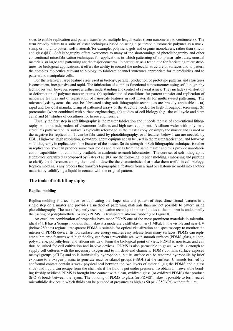
sides to enable replication and pattern transfer on multiple length scales (from nanometers to centimeters). Theterm broadly refers to a suite of sister techniques based on using a patterned elastomeric polymer as a mask,stamp or mold, to pattern soft materials(for example, polymers, gels and organic monolayers, rather than siliconand glass)[83]. Soft lithography offers overcomes to many of the shortcomings of photolithography and otherconventional microfabrication techniques for applications in which patterning of nonplanar substrates, unusualmaterials, or large area patterning are the major concerns. In particular, as a technique for fabricating microstruc-tures for biological applications, it offers the ability to control the molecular structure of surfaces and to patternthe complex molecules relevant to biology, to fabricate channel structures appropriate for microfluidics and topattern and manipulate cells.
For the relatively large feature sizes used in biology, parallel production of prototype patterns and structuresis convenient, inexpensive and rapid. The fabrication of complex functional nanostructures using soft lithographytechniques will, however, require a further understanding and control of several issues. They include (a) distortionor deformation of polymer nanostructures, (b) optimization of conditions for pattern transfer and replication ofnanoscale features and c) registration of nanoscale features in soft materials for multilayered patterning. Themicroanalysis systems that can be fabricated using soft lithographic techniques are broadly applicable to (a)rapid and low-cost manufacturing of patterned arrays of the structure needed for high-throughput screening, (b)proteomics (when combined with surface engineering), (c) studies of cell biology (e.g. the cell cycle and stemcells) and (d ) studies of cocultures for tissue engineering.
Usually the first step in soft lithography is the master fabrication and it needs the use of conventional lithog-raphy, so is not independent of cleanroom facilities and high-cost equipment. A silicon wafer with polymericstructures patterned on its surface is typically referred to as the master copy, or simply the master and is used asthe negative for replication. It can be fabricated by photolithography, or if features below 1 µm are needed, byEBL . High-cost, high resolution, slow-throughput equipment can be used in the master fabrication, and low-costsoft lithography in replication of the features of the master. So the strength of Soft lithographic techniques is ratherin replication :you can produce numerous molds and replicas from the same master and thus provide nanofabri-cation capabilities not commonly available in academic research laboratories. The core set of soft-lithographictechniques, organized as proposed by Gates et al. [83] are the following: replica molding, embossing and printingto clarify the differences among them and to describe the characteristics that make them useful in cell biology.Replica molding is any process that transfers topographical features from a rigid or elastomeric mold into anothermaterial by solidifying a liquid in contact with the original pattern.
The tools of soft lithography
Replica molding
Replica molding is a technique for duplicating the shape, size and pattern of three-dimensional features in asingle step on a master and provides a method of patterning materials than are not possible to pattern usingphotolithography. The most frequently used replication technique in microfluidics at the moment is undoubtedlythe casting of poly(dimethylsiloxane) (PDMS), a transparent silicone rubber (see Figure 8).
An excellent combination of properties have made PDMS one of the most prominent materials in microflu-idics[84]. It has a Youngs modulus that makes it a moderately stiff elastomer (1 MPa). In the visible and near-UV(below 280 nm) regions, transparent PDMS is suitable for optical visualization and spectroscopy to monitor theinterior of PDMS device. Its low surface free energy enables easy release from many surfaces. PDMS can repli-cate submicron features with high fidelity, can form a reversible seal with smooth surfaces (PDMS, glass, silicon,polystyrene, polyethylene, and silicon nitride). From the biological point of view, PDMS is non-toxic and canthus be suited for cell cultivation and in-vivo devices. PDMS is also permeable to gases, which is enough tosupply cell cultures with the necessary oxygen and to fill dead-end channels. PDMS contains surface-exposedmethyl groups (-CH3) and so is intrinsically hydrophobic, but its surface can be rendered hydrophilic by briefexposure to a oxygen plasma to generate reactive silanol groups (-SiOH) at the surface. Channels formed byconformal contact contain a weak physical seal between the two layers of material (e.g the PDMS and a glassslide) and liquid can escape from the channels if the fluid is put under pressure. To obtain an irreversible bond-ing freshly oxidized PDMS is brought into contact with clean, oxidized glass (or oxidized PDMS) that produceSi-O-Si bonds between the layers. The bonding of PDMS to glass (or PDMS) makes it possible to form sealedmicrofluidic devices in which fluids can be pumped at pressures as high as 50 psi ( 350 kPa) without failure.

Figure 8: Printing and molding process of soft lithography [46] Upper panel: The process begins by manufacturing of themold ,frequently by bulk machining of silicon or by thick SU-8 photolithography. A two-part mixture containing PDMS pre-polymer and its curing agent (suggested ratio of 10:1(v/v) is poured over the positive relief master and allowed to cure turningthe liquid into rubber. For all microfabrication purposes it is important to degas PDMS in a dessicator to remove air bubblesbefore use. After cross-linking,the elastomer is carefully peeled off the substrate , access ports are then bonded to a flatsubstrate, enclosing microchannel structures. The master mold can be reused multiple times to replicate devices. Thus, thismethod is especially desirable due to its low cost, flexibility, and rapid prototyping. Bottom panel: The transferred molecules,or inks, are coated onto the stamp before contact with the substrate and deposited in a pattern defined by the raised surfaces ofthe stamp. The stamp is then brought into conformal contact with the surface for a period ranging from 30 s to several minutes,depending on the application. A large variety of types of molecules (including small biomolecules, DNA[85], proteins[86],polyelectrolytes and suspensions of cells) can act as inks and have been patterned directly on a number of different substrateusing CP .
Microcontact printing
Microcontact printing (CP) is a soft-lithography technique used to transfer chemical and biological moleculesonto a surface using a topographically patterned elastomeric mold or stamp, which is typically fabricated fromPDMS (see Figure 7).
The CP process is attractive because of the ease and low cost of generating the stamp with commerciallyavailable precursors. However,some of the disadvantages include the blurring of the pattern by lateral diffusionof the ink, the distortion in the pattern that reflect deformations in the stamp and the substantial number of de-fects in the film [87-91]. The best-known and extensively studied system for this technique is the printing of selfassembled monolayer (SAM) of alkanethiolates (SH-(CH2)n-X) onto the areas of noble and near-noble metalfilms (e.g gold, silver, palladium, platinum)[92]. SAMs are structures generated by the spontaneous chemisorp-tions and self-organization of alkanethiols. Alkanethiols with long alkyl chains (n = 16 or 18) form hydrophobicmonolayers, whereas those with different terminal functional groups (X) can form hydrophilic, hydrophobic orcharged SAMs.
Many synthetic methods are available for attaching ligands to functional groups (X) on SAMs. If required bythe intended application, the substrate can be dipped into a solution of a second alkanethiol to form a differentSAM on bare regions of the metal surface that were not in contact with the stamp. This technique has beenused to make islands of SAMs that adsorb proteins and cells, and which are surrounded by SAMs that resist theadsorption of biomaterial. Other examples of applications of CP are silanes on oxides or alkyl phosphates on

metal oxides and proteins on silicon, glass or polystirene. The ability to engineer the properties of surfaces withspatially defined pattern with SAMs has made it possible to study biological processes, such as cellular responsesto various adhesive environments, that occur at interfaces as well as to conduct miniaturized and high-throughputassays [93]. Most current biological uses of micropatterning require features with sizes below 100 nm.
Nanoimprint lithography
Nanometer surface topologies can be fabricated by imprint lithography (about 5 nm minimum feature, 30 nmresolution)[94]; in addition, it is possible to control the spatial distribution of chemical species on the structuredsurface. Nanoimprint lithography (NIL), also called hot embossing, is the term coined for a procedure thattransfers a pattern using a hard rigid master to physically deform a solid polymer film coated on a rigid surface.The material, typically a polymer, is heated above its glass-transition temperature (e.g., 90200C), while themaster is pressed against it (e.g., 50130 bar). The polymer is deformed filling the voids in the master, then themold is removed after cooling the substrate to below this glass-transition temperature (Tg) revealing a patternthat is the inverse of the master). Imprinting can replicate structures with aspect ratios up to 10:1 and as smallas 10 nm. Pattern transfer takes about 5 to 10 min (or sometimes longer) depending on the efficiency of heattransfer within the system. Photolithography is typically used to generate the surface structure but is limited inapplications where bio-specific adsorption and functionalities are required to mimic the microenvironments forcellular development. Thus, imprint methods are useful because they permit direct construction of a variety ofshapes with varying physicochemical properties and are accessible methods for academic research.