Tongue flicking in agamid lizards: Morphology, kinematics, and muscle activity patterns
15
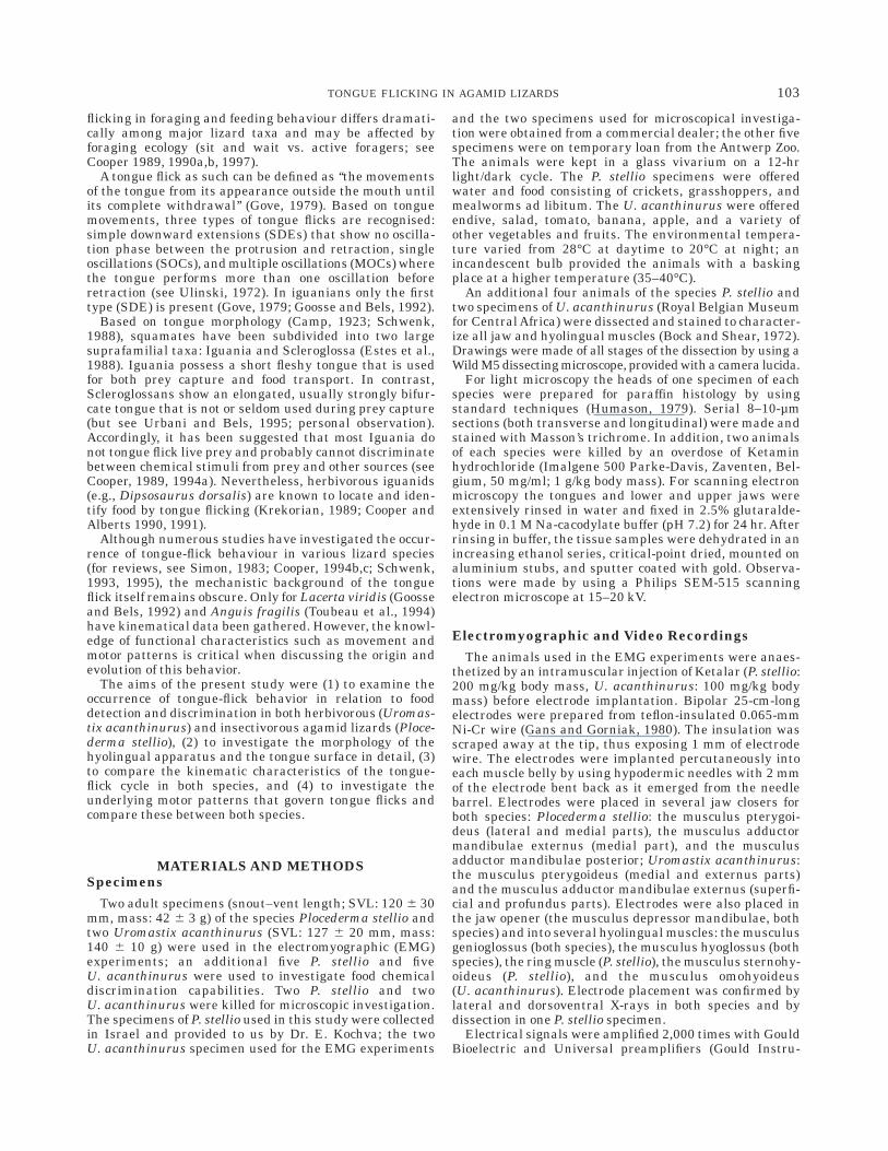
Tongue Flicking in Agamid Lizards: Morphology, Kinematics, and Muscle Activity Patterns ANTHONY HERREL, 1 * JEAN-PIERRE TIMMERMANS, 2 AND FRITS DE VREE 1 1 Laboratory for Functional Morphology, Department of Biology, University of Antwerp (UIA), B-2610 Antwerp, Belgium 2 Laboratory of Cell Biology and Histology, Faculty of Medecine, University of Antwerp (RUCA), B-2020 Antwerp, Belgium ABSTRACT We wanted to examine whether a relation between foraging strategy, morphology, the mechanics of tongue protrusion, and prey chemical detection and discrimination exists in agamid lizards. Tongue-flick behavior was observed in two species of this family: Uromastix acanthinurus and Ploce- derma stellio. Potential prey chemical discrimination by means of tongue flicking was examined by using applicator tests. Tongue flicks were subse- quently recorded by high-speed video in combination with the electrical activity of a number of jaw and hyolingual muscles. The kinematics of jaws and tongue and the muscle activity patterns were quantified. To investigate if the observed differences in tongue-flick behavior (mainly in the frequency of use) are translated into corresponding differences in tongue morphology, the tongues of both species were examined by light and scanning electron microscopy. The species differed mainly in the surface morphology of the foretongue and in the abundance and distribution of taste buds on the tongue and oral cavity. These differences can be related to behavioural observations; whereas U. acanthinurus readily uses tongue flicks to detect and discriminate between food items, P. stellio does not. However, differences in tongue-flick mechanics (kinematics, electromyograms) between both species were minor. Based on the data gathered in this study and from previously published data, an evolutionary transformation series leading to the complex tongue-flick cycles as observed in snakes is proposed. The required morphological and mechanical changes that accompany such an evolutionary sequence are discussed. Anat. Rec. 252:102–116, 1998. r 1998 Wiley-Liss, Inc. Key words: tongue flicking; morphology; kinematics; EMG; chemical prey discrimination; Plocederma stellio; Uromastix acanthinurus The tongue of lizards is used for diverse functions such as lingual prey prehension in iguanian lizards (Schwenk and Throckmorton, 1989; Herrel et al., 1995), prey trans- port and swallowing in most lizards (Delheusy and Bels, 1992; Herrel et al., 1996, 1997), defensive display in some scincids (e.g., shingleback lizards, Gans et al., 1985), spectacle cleaning in geckoes (Simon, 1983), and chemical sampling from the external environment by means of tongue flicks or tongue touches (for overviews, see Schwenk, 1993; Cooper, 1994b,c, 1995a). Tongue extrusions, such as flicks or touches, can be used in the detection of conspecif- ics (Cooper and Vitt, 1984; Simon, 1985), kin recognition (Werner et al., 1987), sex recognition and courtship (Coo- per and Vitt, 1984; Cooper et al., 1986), general explora- tion (Bissinger and Simon, 1979; Simon et al., 1981), predator detection (Thoen et al., 1986; Van Damme et al., 1990), and food detection and discrimination (see Schwenk, 1993; Cooper 1994b,c, 1995a). However, tongue Grant sponsor: IWT; Grant number: 943039; Grant sponsor: FKFO; Grant number: G.0221.96. *Correspondence to: Anthony Herrel, Department of Biology, University of Antwerp (UIA), Universiteitsplein 1, B-2610 Ant- werp, Belgium. E-mail: [email protected] Received 23 December 1997; Accepted 28 April 1998 THE ANATOMICAL RECORD 252:102–116 (1998) r 1998 WILEY-LISS, INC.
Transcript of Tongue flicking in agamid lizards: Morphology, kinematics, and muscle activity patterns
Tongue Flicking in Agamid Lizards:Morphology, Kinematics, and Muscle
Activity PatternsANTHONY HERREL,1* JEAN-PIERRE TIMMERMANS,2
AND FRITS DE VREE1
1Laboratory for Functional Morphology, Department of Biology,University of Antwerp (UIA), B-2610 Antwerp, Belgium
2Laboratory of Cell Biology and Histology, Faculty of Medecine,University of Antwerp (RUCA), B-2020 Antwerp, Belgium
ABSTRACTWe wanted to examine whether a relation between foraging strategy,
morphology, the mechanics of tongue protrusion, and prey chemical detectionand discrimination exists in agamid lizards. Tongue-flick behavior wasobserved in two species of this family: Uromastix acanthinurus and Ploce-derma stellio. Potential prey chemical discrimination by means of tongueflicking was examined by using applicator tests. Tongue flicks were subse-quently recorded by high-speed video in combination with the electricalactivity of a number of jaw and hyolingual muscles. The kinematics of jawsand tongue and the muscle activity patterns were quantified. To investigate ifthe observed differences in tongue-flick behavior (mainly in the frequency ofuse) are translated into corresponding differences in tongue morphology, thetongues of both species were examined by light and scanning electronmicroscopy. The species differed mainly in the surface morphology of theforetongue and in the abundance and distribution of taste buds on the tongueand oral cavity. These differences can be related to behavioural observations;whereas U. acanthinurus readily uses tongue flicks to detect and discriminatebetween food items, P. stellio does not. However, differences in tongue-flickmechanics (kinematics, electromyograms) between both species were minor.Based on the data gathered in this study and from previously published data,an evolutionary transformation series leading to the complex tongue-flickcycles as observed in snakes is proposed. The required morphological andmechanical changes that accompany such an evolutionary sequence arediscussed. Anat. Rec. 252:102–116, 1998. r 1998 Wiley-Liss, Inc.
Key words: tongue flicking; morphology; kinematics; EMG; chemical preydiscrimination; Plocederma stellio; Uromastix acanthinurus
The tongue of lizards is used for diverse functions suchas lingual prey prehension in iguanian lizards (Schwenkand Throckmorton, 1989; Herrel et al., 1995), prey trans-port and swallowing in most lizards (Delheusy and Bels,1992; Herrel et al., 1996, 1997), defensive display in somescincids (e.g., shingleback lizards, Gans et al., 1985),spectacle cleaning in geckoes (Simon, 1983), and chemicalsampling from the external environment by means oftongue flicks or tongue touches (for overviews, see Schwenk,1993; Cooper, 1994b,c, 1995a). Tongue extrusions, such asflicks or touches, can be used in the detection of conspecif-ics (Cooper and Vitt, 1984; Simon, 1985), kin recognition(Werner et al., 1987), sex recognition and courtship (Coo-
per and Vitt, 1984; Cooper et al., 1986), general explora-tion (Bissinger and Simon, 1979; Simon et al., 1981),predator detection (Thoen et al., 1986; Van Damme etal., 1990), and food detection and discrimination (seeSchwenk, 1993; Cooper 1994b,c, 1995a). However, tongue
Grant sponsor: IWT; Grant number: 943039; Grant sponsor:FKFO; Grant number: G.0221.96.
*Correspondence to: Anthony Herrel, Department of Biology,University of Antwerp (UIA), Universiteitsplein 1, B-2610 Ant-werp, Belgium. E-mail: [email protected]
Received 23 December 1997; Accepted 28 April 1998
THE ANATOMICAL RECORD 252:102–116 (1998)
r 1998 WILEY-LISS, INC.
flicking in foraging and feeding behaviour differs dramati-cally among major lizard taxa and may be affected byforaging ecology (sit and wait vs. active foragers; seeCooper 1989, 1990a,b, 1997).
A tongue flick as such can be defined as ‘‘the movementsof the tongue from its appearance outside the mouth untilits complete withdrawal’’ (Gove, 1979). Based on tonguemovements, three types of tongue flicks are recognised:simple downward extensions (SDEs) that show no oscilla-tion phase between the protrusion and retraction, singleoscillations (SOCs), and multiple oscillations (MOCs) wherethe tongue performs more than one oscillation beforeretraction (see Ulinski, 1972). In iguanians only the firsttype (SDE) is present (Gove, 1979; Goosse and Bels, 1992).
Based on tongue morphology (Camp, 1923; Schwenk,1988), squamates have been subdivided into two largesuprafamilial taxa: Iguania and Scleroglossa (Estes et al.,1988). Iguania possess a short fleshy tongue that is usedfor both prey capture and food transport. In contrast,Scleroglossans show an elongated, usually strongly bifur-cate tongue that is not or seldom used during prey capture(but see Urbani and Bels, 1995; personal observation).Accordingly, it has been suggested that most Iguania donot tongue flick live prey and probably cannot discriminatebetween chemical stimuli from prey and other sources (seeCooper, 1989, 1994a). Nevertheless, herbivorous iguanids(e.g., Dipsosaurus dorsalis) are known to locate and iden-tify food by tongue flicking (Krekorian, 1989; Cooper andAlberts 1990, 1991).
Although numerous studies have investigated the occur-rence of tongue-flick behaviour in various lizard species(for reviews, see Simon, 1983; Cooper, 1994b,c; Schwenk,1993, 1995), the mechanistic background of the tongueflick itself remains obscure. Only for Lacerta viridis (Goosseand Bels, 1992) and Anguis fragilis (Toubeau et al., 1994)have kinematical data been gathered. However, the knowl-edge of functional characteristics such as movement andmotor patterns is critical when discussing the origin andevolution of this behavior.
The aims of the present study were (1) to examine theoccurrence of tongue-flick behavior in relation to fooddetection and discrimination in both herbivorous (Uromas-tix acanthinurus) and insectivorous agamid lizards (Ploce-derma stellio), (2) to investigate the morphology of thehyolingual apparatus and the tongue surface in detail, (3)to compare the kinematic characteristics of the tongue-flick cycle in both species, and (4) to investigate theunderlying motor patterns that govern tongue flicks andcompare these between both species.
MATERIALS AND METHODSSpecimens
Two adult specimens (snout–vent length; SVL: 120 6 30mm, mass: 42 6 3 g) of the species Plocederma stellio andtwo Uromastix acanthinurus (SVL: 127 6 20 mm, mass:140 6 10 g) were used in the electromyographic (EMG)experiments; an additional five P. stellio and fiveU. acanthinurus were used to investigate food chemicaldiscrimination capabilities. Two P. stellio and twoU. acanthinurus were killed for microscopic investigation.The specimens of P. stellio used in this study were collectedin Israel and provided to us by Dr. E. Kochva; the twoU. acanthinurus specimen used for the EMG experiments
and the two specimens used for microscopical investiga-tion were obtained from a commercial dealer; the other fivespecimens were on temporary loan from the Antwerp Zoo.The animals were kept in a glass vivarium on a 12-hrlight/dark cycle. The P. stellio specimens were offeredwater and food consisting of crickets, grasshoppers, andmealworms ad libitum. The U. acanthinurus were offeredendive, salad, tomato, banana, apple, and a variety ofother vegetables and fruits. The environmental tempera-ture varied from 28°C at daytime to 20°C at night; anincandescent bulb provided the animals with a baskingplace at a higher temperature (35–40°C).
An additional four animals of the species P. stellio andtwo specimens of U. acanthinurus (Royal Belgian Museumfor Central Africa) were dissected and stained to character-ize all jaw and hyolingual muscles (Bock and Shear, 1972).Drawings were made of all stages of the dissection by using aWild M5 dissecting microscope, provided with a camera lucida.
For light microscopy the heads of one specimen of eachspecies were prepared for paraffin histology by usingstandard techniques (Humason, 1979). Serial 8–10-µmsections (both transverse and longitudinal) were made andstained with Masson’s trichrome. In addition, two animalsof each species were killed by an overdose of Ketaminhydrochloride (Imalgene 500 Parke-Davis, Zaventen, Bel-gium, 50 mg/ml; 1 g/kg body mass). For scanning electronmicroscopy the tongues and lower and upper jaws wereextensively rinsed in water and fixed in 2.5% glutaralde-hyde in 0.1 M Na-cacodylate buffer (pH 7.2) for 24 hr. Afterrinsing in buffer, the tissue samples were dehydrated in anincreasing ethanol series, critical-point dried, mounted onaluminium stubs, and sputter coated with gold. Observa-tions were made by using a Philips SEM-515 scanningelectron microscope at 15–20 kV.
Electromyographic and Video RecordingsThe animals used in the EMG experiments were anaes-
thetized by an intramuscular injection of Ketalar (P. stellio:200 mg/kg body mass, U. acanthinurus: 100 mg/kg bodymass) before electrode implantation. Bipolar 25-cm-longelectrodes were prepared from teflon-insulated 0.065-mmNi-Cr wire (Gans and Gorniak, 1980). The insulation wasscraped away at the tip, thus exposing 1 mm of electrodewire. The electrodes were implanted percutaneously intoeach muscle belly by using hypodermic needles with 2 mmof the electrode bent back as it emerged from the needlebarrel. Electrodes were placed in several jaw closers forboth species: Plocederma stellio: the musculus pterygoi-deus (lateral and medial parts), the musculus adductormandibulae externus (medial part), and the musculusadductor mandibulae posterior; Uromastix acanthinurus:the musculus pterygoideus (medial and externus parts)and the musculus adductor mandibulae externus (superfi-cial and profundus parts). Electrodes were also placed inthe jaw opener (the musculus depressor mandibulae, bothspecies) and into several hyolingual muscles: the musculusgenioglossus (both species), the musculus hyoglossus (bothspecies), the ring muscle (P. stellio), the musculus sternohy-oideus (P. stellio), and the musculus omohyoideus(U. acanthinurus). Electrode placement was confirmed bylateral and dorsoventral X-rays in both species and bydissection in one P. stellio specimen.
Electrical signals were amplified 2,000 times with GouldBioelectric and Universal preamplifiers (Gould Instru-
103TONGUE FLICKING IN AGAMID LIZARDS

ment Systems, Inc., Valley View, OH) (range 5 100 Hz to10 kHz) and Honeywell Accudata 117 DC amplifiers andwere recorded on a Honeywell 96 FM 14 channel taperecorder (Honeywell Inc., Denver, CO) (medium bandpass)at a speed of 19.05 cm/sec.
Tongue flicks were simultaneously recorded by using aNAC-1000 high-speed video system set at 500 frames persecond using video lights (TRI-LITE, Cool Light Co. Inc.,Hollywood, CA). The animals were filmed in a plexiglasscage (50 cm 3 15 cm 3 10 cm). The output of a Tectronixwave pattern generator (square wave) was recorded on theFM tape recorder and sent to a LED (light emitting diode)kept in the visual field. This allowed synchronisation of theelectromyographic and kinematic records.
Video Data and Electromyographical AnalysisClearly visible external markers (colour spots) were
digitized by using the NAC1000 X–Y Coordinator (Fig. 1).
Horizontal (x) and vertical (y) coordinates were recordedfor each digitized point at intervals of two frames. Aspectscalculated were changes in gape profile (distance: 2–4,angle: 2–3–4), displacement of the upper and the lowerjaws (y coordinate of points 2 and 4, respectively), cranialelevation (angle subtended by the line interconnectingpoints 2–3 and the horizontal), lower jaw depression (anglesubtended by the line interconnecting points 3–4 and thehorizontal), and tongue protrusion (x coordinate of point 5,x coordinate of point 4).
The recorded EMG signals were digitized at 10 kHz byusing a Keithley DAS series 500 12-bit A-D converter(Keithley Instruments, Inc., Cleveland, OH). After digitiza-tion, the signals were integrated by following the proce-dure of Beach et al. (1982; 100 data 5 10 msec/bin), and thenumber of spikes and the average amplitude per intervalwere calculated. The exact duration of muscular activityand the onset of each muscle relative to the m. depressor
Fig. 1. Uromastix acanthinurus. Jaw and hyolingual musculature.Upper left: Lateral view after removal of the skin, the m. pterygoideusexternus, the quadratojugal ligament, and the m. depressor mandibulae.Lower left: Deeper lateral view, after the additional removal of thecomplete m. adductor mandibulae externus. Upper right: Superficialventral view of the hyolingual muscles after removal of the skin, part of them. intermandibularis anterior, the m. constrictor colli, and the m. omohyoi-deus. Lower right: Deeper dissection; after removal of the hyoid retractorand protractor muscles, the m. genioglossus medialis and them. branchiohyoideus. CBI, ceratobranchiale I; CBII, ceratobranchiale II;
CH, ceratohyale; MAMEM, m. adductor mandibulae externus medialis;MAMEP, m. adductor mandibulae externus profundus; MAMESA,MAMESP, mm. adductor mandibulae externus superficialis anterior andposterior; MAMP, m. adductor mandibulae posterior; MEM, m. episterno-cleidomastoideus; MGGL, MGGM, mm. genioglossus medialis and latera-lis; MHG, m. hyoglossus; MIMASP, m. intermandibularis anterior superfi-cialis posterior; MLAO, m. levator anguli oris; MMH1, MMH2, MMH3, mm.mandibulohyoideus 1, 2, and 3; MPsTS, m. pseudotemporalis superficia-lis; MPtext, m. pterygoideus externus; MRAO, m. retractor anguli oris;MSH, m. sternohyoideus; MST, m. sternothyroideus.
104 HERREL ET AL.

mandibulae (MDM) were then measured. Several muscleshad more than one activity burst. Different bursts weredefined on the basis of abrupt amplitude differences. TheEMG variables were then related to the kinematic data.
Prey Chemical Discrimination TestsThe responses of the lizards to chemical stimuli from
prey and control substances presented on the cotton swabsof wooden applicators (25 cm) were recorded. In theexperiments each lizard responded to each stimulus oncein a randomized block design (see Cooper et al., 1996). Thelizards responded to all stimuli in counterbalanced se-quence to avoid sequential bias. The responses of thelizards to cricket surface chemicals, squashed endive,cologne (Caractere, Daniel Hechter, Paris, France), fish,and distilled water were recorded.
Cologne and fish (for a discussion on the validity of thesestimuli, see Dial and Schwenk, 1996) were used as apungency control to examine the effects of a (highlyvolatile) stimulus lacking relevance to feeding behaviour.Distilled water served as an odourless control for thereactions to the swab and the experimental setting. Beforetesting, the cotton tip of an applicator was first dipped indistilled water and then dipped in cologne, rolled over thecricket integument, rolled over fish, or rolled over squashedendive. Excess water was blotted with tissue paper.
The experiments were conducted when the lizards weresufficiently active and alert (between 11.00 and 17.00 hr).Temperatures varied between 35°C and 40°C during test-ing. During a trial the experimenter approached a lizard’scage slowly and positioned a swab 1–2 cm anterior to thelizard’s snout. The number of tongue flicks was recordedfor 60 sec after the first tongue flick unless the lizard bitthe swab. In that case the trial was terminated. If a lizarddid not tongue flick within 40 sec, the swab was broughtgently into contact with its snout for about 1 sec. If thelizard did not tongue flick in the next 20 sec, the trial wasterminated. Each lizard was tested for all stimuli on thesame day. The intertrial interval was at least 1 hr. Lizardswere not fed for 3 days before testing.
Responsiveness to chemical stimuli was measured bythe tongue-flick attack score by using repeated measuredesigns (see Burghardt, 1967; Cooper and Burghardt,1990). Nonparametric tests were used due to the extremenonnormality (large number of zero values). Data wereanalyzed by the Friedman analysis of variance and/or signtests (P 5 0.05; Sokal and Rolff, 1995; Zar, 1996).
RESULTSMorphology
Gross morphology of the feeding apparatus. Theskull of Plocederma stellio and Uromastix has been ad-equately described by El Toubi (1945, 1947) and Jollie(1960). The hyoid apparatus (Figs. 1 & 2) has a distinctlytapered entoglossal process (Fig. 2c), one pair of ceratohy-als, and two pairs of ceratobranchials (see Furbringer,1922; Richter, 1933; Gnanamuthu, 1937; Sondhi, 1958;Tilak, 1964; Smith, 1988). The ceratohyal (CH) is similarin both species and consists of three parts. The proximalpart articulates with the basihyoid (Fig. 2d), runs anteri-orly, and is connected to the posteriorly directed intermedi-ate part by means of a cartilaginous jointlike structure.The distal part is short in both species and is connected to
the base of the quadrate. The first ceratobranchial (CBI;Fig. 2d), situated at the posterolateral side of the basihy-oid, is also similar in both species. However, the secondceratobranchial (CBII) is very different in both species. InP. stellio, the CBIIs, which touch each other along themidline of the trachea, are long and taper gradually alongtheir length. In U. acanthinurus, the CBII is short andends in a flat cartilaginous part. In addition, in U. acanthi-nurus, the CBIIs are situated on both sides of the tracheaand do not touch. In contrast to U. acanthinurus, whereonly the entoglossal process (Fig. 2c, 2e, & 2f) and the mostdistal parts of the hyoidarches are cartilaginous, in P. stellioonly the CBIs are ossified.
The jaw and hyolingual muscles in agamids have beendescribed by several researchers (Sanders, 1872; De Vis,1884; Gandolfi, 1908; Lakjer, 1926; Gnanamuthu, 1937;Haas, 1973; Gomes, 1974; Schwenk, 1988; Smith, 1988; forP. stellio more specifically, see also Herrel et al., 1995,1997). The jaw adductors in agamid lizards can be de-scribed as consisting of three major groups: the m. adduc-tor mandibulae externus, the m. adductor mandibulaeinternus, and the m. adductor mandibulae posterior (seeFig. 1). A full, detailed description of the jaw musculaturein both species will be published elsewhere.
The fleshy tongue of Plocederma stellio is composed ofintrinsic and extrinsic muscles. The tongue musculaturein agamid lizards has been described in detail elsewhere(Gandolfi, 1908; Gnanamuthu, 1937; Smith, 1988). Themost important differences in intrinsic tongue muscula-ture between both species examined are (1) the absence ofa true ring muscle surrounding the entoglossal process(Fig. 2e & 2f) and (2) the absence of vertical septa inUromastix (see also Smith, 1988).
The extrinsic lingual musculature (Fig. 1) originates onthe mandible or the hyoid and consists of distinct genioglos-sus and hyoglossus muscles (Fig. 2) and is similar in bothspecies. The genioglossus (MGG) originates on the antero-medial part of the mandible and runs posteriorly to inserton the tongue (Fig. 2a & 2c). Near its insertion it separatesinto medial, lateral, and internal parts. The hyoglossus(MHG) originates on the first ceratobranchial and runsinto the tongue, dorsally to the MGG. Anterior of thebasihyoid, the MHG is recognisable as a distinct musclebundle (Fig. 2e).
Several hyoid muscles lie ventral to the tongue. Themandibulohyoideus (MMH) consists of three portions thatoriginate on the medial side of the mandible and insert onthe ceratohyal and the first ceratobranchial. The sternohy-oideus (MSH) and sternothyroideus (MST) muscles bothoriginate on or near the episternum and run anteriorly toinsert on the basihyal and first ceratobranchial. Theomohyoideus muscle (MOH) originates on the anterioredge of the interclavicula and suprascapula and its fibresrun anteroventrally to insert on the first ceratobranchial.
Morphology of the tongue and oral cavity: Occur-rence and distribution of taste buds. In the followingdescription of tongue surface morphology, we adopt theterminology of Schwenk (1985) and Rabinowitz and Tand-ler (1986). The tongue of the examined lizards can besubdivided into three distinct areas (Fig. 3): the tongue tip,the foretongue, and the hindtongue; each is characterizedby a specific surface topology (Table 1). The tongue tip isthe bifurcated anterior end of the tongue with a small area
105TONGUE FLICKING IN AGAMID LIZARDS

immediately posterior to the bifurcation (Schwenk, 1985).The bifurcation is more prominent in U. acanthinurusthan in P. stellio (compare Fig. 3b with 3e). The anterior-most part is characterized by a smooth surface and ab-sence of taste buds in both species (Fig. 3b & 3e). Theadjacent, more posteriorly situated part of the tongue tip
bears groups of anteriorly flattened, short, and closelypacked cylindriform papillae. The surface of such a groupappears smooth at low magnification and often carriestaste buds. At the posterior part of the tongue tip, thestructure of the tongue surface gradually changes (througha small area bearing plicae in U. acanthinurus) into a
Fig. 2. Light microscopic view of transverse and sagittal sections indifferent areas of the heads of P. stellio and U. acanthinurus. a: Medialsagittal section of the head of P. stellio. 38. Numerous secretory cells arepresent on the foretongue. b: Detail of an illustrating muscle fibre (arrow)running into the dorsal papillae. 3250. c: Sagittal section illustrating thearrangement of the tongue, entoglossal process, and intrinsic tonguemuscles in P. stellio. 316. d: Sagittal section through the hyoid apparatusof P. stellio illustrating the joint between the basihyoid, the ceratohyal, and
first ceratobranchial. 3100. e: Transverse section through the hindtonguein U. acanthinurus illustrating the arrangement of the hyoglosus andgenioglossus muscles. 316. f: Detail of e at higher magnification (3100)showing the entoglossal process. Note the large amount of connectivetissue surrounding the entoglossal process. BH, basihyoid; CBI, cerato-branchiale I; CH, ceratohyale; E, entoglossal process; G, m. genioglos-sus; H, m. hyoglossus; T, trachea.
106 HERREL ET AL.

Fig. 3.

typical foretongue structure, with an increase in thenumber of filamentous cylindrical and/or plumose papil-lae. These papillae are characterized by a large number ofsecretory cells and several muscle fibres running into them(Fig. 2b). Whereas the tongue tip of both species is rathersimilar, the foretongue structure of both species differsmarkedly. In P. stellio, the surface of the foretongue iscomposed of transversely oriented rows of plumose papil-lae (Fig. 3c) bearing apparently randomly disposed plumecells (see also Rabinowitz and Tandler, 1986). This area ofthe tongue is characterized by the absence of taste buds. InU. acanthinurus, the foretongue can be subdivided intotwo distinct areas. The medial part is composed of denselypacked triangular papillae, characterized by a surfacewith a prominent microstructure (Fig. 3g & 3h). Thelateral part consists of rectangular papillae; at the edge ofsuch a papilla, free-standing plume cells are observed (Fig.3f). Whereas the medial part (Fig. 3g) is devoid of tastebuds, the lateral part shows a limited number of taste budsanteriorly and a high density of taste buds (one or more perpapilla) posteriorly (see arrows on Fig. 3f). The hindtonguein both species somewhat resembles the posterior part of
the tongue tip and is composed of typical papillae consist-ing of groups of concentrically arranged cylindrical cells,generally bearing microvilli or microridges (Fig. 3d & 3i).Papillae are interspersed with plumose cells and usuallycarry one taste bud (Fig. 3d & 3i).
The oral epithelium was also examined for the presenceof taste buds (see Fig. 4). In P. stellio, only on the tissue foldjust medial to the teeth of the lower jaw were any tastebuds observed (Fig. 4c). The surface of this tissue fold iscomposed of flattened cylindriform epithelial cells. Tastebuds are abundant in the anterior part, present in themiddle part, and absent from the posterior part. No tastebuds were observed on the epithelium on the dorsal side ofthe oral cavity. In U. acanthinurus, taste buds were notonly present along the entire medial dental tissue fold butalso abundant on various parts of the dorsal side of the oralcavity (see Fig. 4a, 4b, & 4d).
Prey chemical discrimination and observationsof tongue-flick behavior.
Plocederma stellio. In P. stellio, tongue-flick behavior isonly occasionally observed and usually consists of tonguetouches. In one of the recorded sequences, the lizardattempted to capture a prey item but lost it. The prey itemfell to the bottom of the cage and showed no furthermovements. After a few seconds, the lizard approached theprey again, tongue flicked, and subsequently captured theprey. In all other recorded sequences, the lizards ap-proached the live prey, tongue flicked, and captured theprey item. Nevertheless, only six tongue flicks were ob-served in the more than 50 feeding sequences recorded.These observations are supported by the prey chemicaldiscrimination tests. None of the five animals testedtongue flicked an applicator (Table 2).
Uromastix acanthinurus. Uromastix acanthinurusreadily used tongue flicks when placed in a new environ-ment or when brought in contact with other lizards. Thelizards also invariably tongue flicked food introduced intothe cage before eating it. Prey chemical discriminationtests indeed showed clear responses to prey chemicals(Table 2). Animals attacked only after tongue flicks, during
Fig. 3. Scanning electron micrographs of the tongue tip, the fore-tongue, and hindtongue in both species. a: Schematic representation ofthe tongue (U. acanthinurus) illustrating the subdivision of the tongue intothree distinct areas. b: Tongue tip in P. stellio showing only a minorbifurcation, covered with smooth epithelium and carrying several tastebuds (arrows). c: Typical foretongue structure in P. stellio showingtransversely oriented rows of plumose cells (PL and arrow). d: Typicalhindtongue structure in P. stellio, with closely packed cylindriform papillaewith relatively few taste buds (arrows) and free-standing plume cells at theedge. e: Tongue tip in U. acanthinurus. Note the more prominentbifurcation; taste buds are present just posterior to the plicae (P andarrow) at the posterior edge of the bifurcation. f: Lateral part of theforetongue in U. acanthinurus showing several taste buds (arrows). g:Medial part of the foretongue in U. acanthinurus with seemingly smoothpapillae. h: Detail of g showing the presence of a very distinct microstruc-ture. i: Hindtongue in U. acanthinurus bearing papillae with usually notmore than one taste bud associated (arrows) and with loose plumelikecells (PL and arrow) at the periphery. Scale bars 1 mm in b,e, 0.1 mm inc,d,f,g,i, 0.01 mm in h.
TABLE 1. Summary of Lingual and Oral Morphology and the Presenceand Abundance of Taste Buds
Species Region No. taste budsa Structure
Plocederma stellio Tongue tip 1 (posterior part) Smooth epitheliumForetongue
Lateral 1 Transversely oriented rowsof plume cells
Medial 2 Plumose papillaeHindtongue 1 Densely packed plumose
papillaeOral epithelium 1
Uromastix acanthinurus Tongue tip 1 (posterior part) Smooth epitheliumForetongue
Lateral 11 Dense papillae with promi-nent microstructure
Medial 2 Papillae with loose plumecells at the edges
Hindtongue 1 Densely packed cylindri-form papillae
Oral epithelium 11
aNo. taste buds indicates presence and abundance of taste buds as follows: 2, absent; 1, present;11, abundant.
108 HERREL ET AL.

which the swab was touched (i.e., tongue touches). Ani-mals tongue flicked the control swab an average of threetimes (66.2); stimulus control swabs were flicked even less(0 and 0.4 6 0.9). Controls did not differ significantly fromone another (x2 5 2, df 5 2; P . 0.05). Swabs bearingfood stimuli were flicked more (cricket: 5.2 6 10.0, endive:5.6 6 10.3), but responses to cricket surface chemicals did
not differ from those of the control (x2 5 0.33, df 5 1; P .0.05). However, swabs bearing endive chemicals elicitedsignificantly more tongue flicks (x2 5 4, df 5 1; P , 0.05)than did control swabs. However, as one of the animalsattacked a swab bearing cricket chemicals, this can beconsidered as a clear tongue-flick-mediated recognition offood stimuli.
Fig. 4. Scanning electron micrographs illustrating the occurrence anddistribution of extralingual taste buds. a: Ventral view of the anterior mouthroof in U. acanthinurus at low magnification; arrows indicate areas withlarge numbers of taste buds. b: Detail of a showing a taste bud (arrow)and prominent microstructure. c: Paralingual epithelium in P. stellio
showing several taste buds (arrows), indicating the high number present.d: Anterior paralingual epithelium in U. acanthinurus illustrating thepresence of taste buds (arrows). Scale bars 5 1 mm in a, 0.01 mm in b,d,0.1 mm in c.
TABLE 2. Prey Chemical Descrimination in Plocederma stellio (PS)and Uromastix acanthinurus (UA)a
Stimulus Response PS1 UA1 PS2 UA2 PS3 UA3 PS4 UA4 PS5 UA5
Control # TF 0 14 0 1 0 0 0 0 0 0TTA 0 0 0 0 0 0 0 0 0 0
Cologne # TF 0 0 0 0 0 0 0 0 0 0TTA 0 0 0 0 0 0 0 0 0 0
Fish # TF 0 0 0 0 0 0 0 0 0 2TTA 0 0 0 0 0 0 0 0 0 0
Cricket # TF 0 0 0 3 0 0 0 3 0 0TTA 0 0 0 40 0 0 0 0 0 0
Endive # TF 0 2 0 2 0 0 0 1 0 1TTA 0 50 0 0 0 0 0 0 0 0
aTable entries are number of tongue flicks (# TF) and time to attack (TTA).
109TONGUE FLICKING IN AGAMID LIZARDS

KinematicsThe tongue flicks seen in both lizards are always of the
most simple type: the SDEs. In P. stellio, tongue flicksusually involve touching the prey or the substrate with thetongue (tongue touch). In U. acanthinurus, both tonguetouches and tongue flicks, where the tongue does not touchthe prey or substrate, are observed.
Tongue touches in both species usually involve wholehead movements, but tongue flicks usually consist ofsimple tongue extrusions without head movements (Fig. 5).The jaw movements during a tongue flick consist of anopening and a closing phase that cannot be subdivided anyfurther into separate slow and fast phases. The openingphase lasts an average of 446 6 234 msec in U. acanthinu-rus and 258 6 59 msec in P. stellio. Jaw opening is mainlyachieved by a depression of the lower jaw in both species(Fig. 5, Table 3). The maximal gape angle is rather similarin both species (27° in U. acanthinurus vs. 24° in P. stellio).Jaw closing lasts only half as long as jaw opening in bothspecies (Table 3).
Tongue movements start as soon as the jaws are slightlyparted and consist of an anterior and downward displace-ment of the tongue. The maximal extrusion of the tongue,
both horizontal and vertical, is achieved around a maximalgape in both species. The tongue protraction phase lastsabout twice as long as tongue retraction in P. stellio andabout three times as long in U. acanthinurus (Table 3).
ElectromyographyThe first muscles that become active during a tongue-
flick cycle in both species (Fig. 6) are the MGG and theMHG. In P. stellio, the ring muscle becomes active simulta-neously with the onset of activity in the MGG and theMHG (Fig. 6, Table 4). The activity level of these musclesgradually increases to reach a maximum at maximal gape.In P. stellio, the hyoid protractor becomes active later onand shows only low-level activity that ends at maximalgape. The jaw opener in both species occasionally shows ashort activity peak of low intensity just before and endingat maximal gape (Fig. 6, Table 4). This activity maycoincide with an activity in the deeper parts of the externaladductor in U. acanthinurus (Fig. 6). At maximal gape, thehyoid retractor becomes active and simultaneously theMHG shows a second activity burst. The jaw closers(medial and deep parts of the external adductor; them. pterygoideus and the posterior adductor) occasionally
Fig. 5. Kinematic profiles (based on high-speed video recordings, 500frames sec21) of tongue and jaw movements during tongue flicking. Onthe left, the vertical and horizontal displacements of the tongue, thevertical displacement of the upper and lower jaws, and the changes ingape angle are represented for one tongue-flick cycle in P. stellio. On the
right, the vertical and horizontal displacements of the tongue, the tongueprotrusion relative to the lower jaw, the vertical displacements of upperand lower jaws, and the changes in gape distance are represented for onetongue-flick cycle in U. acanthinurus. Note the difference in time scalebetween the two series of profiles.
110 HERREL ET AL.

show a short activity peak of low intensity just aftermaximal gape (Fig. 6, Table 4).
DISCUSSIONBehavioral Implications
The agamid lizards used in this study used tonguetouches, but their frequency of use differed drastically.Whereas the herbivorous species (Uromastix acanthinu-rus) tongue flicks readily and is able to discriminatebetween food and other stimuli, the insectivorous one(Plocederma stellio) hardly ever tongue flicks and no foodchemical discrimination could be demonstrated. Neverthe-less, our observations based on video recordings suggestthat these animals are able to and in reality sometimes douse tongue flicks to judge possible food items. Differencesbetween both situations may be due to the absence ofvisual stimuli in applicator tests (see Chiszar et al., 1988;Ford and Burghardt, 1993). It is likely that in theselizards, gustation and vomerolfaction are triggered byvisual rather than olfactory cues. Although applicatortests provide useful information on the frequency of use,caution is required when making statements about theabsence of such behaviours based solely on such methods.
The traditional hypothesis states that a strong relation-ship exists between the foraging mode and the ability todiscriminate prey by chemical stimuli (Cooper, 1995a,1997). Active foragers more than do ambush foragerswould use chemical senses to detect food. This wouldsuggest that ambush foraging lizards lack the ability to
detect food and discriminate between food items. Becausemost lizards belonging to the Iguania are ambush foragers(Vitt and Price, 1982), these lizards, with the exception ofthe herbivorous lizard Dipsosaurus dorsalis (Krekorian,1989; Cooper and Alberts, 1990, 1991), are supposed to beincapable of food detection and discrimination by means ofchemical sampling (Cooper, 1989, 1994a,b). Moreover, notongue-flick mediated food detection and discriminationcould be demonstrated in agamid lizards (Cooper, 1994a).In the present study, it is shown that agamids do tongueflick and that at least the herbivorous species U. acanthi-nurus uses this behavior to detect and discriminate be-tween food items. Because herbivorous lizards have tosearch actively for their food, lingual chemical samplingmay be an important sensory mode in these animals(Cooper and Alberts, 1990; Cooper 1995a).
The proposed diphyletic behavioral specialization of thetongue (Schwenk and Throckmorton, 1989), whereby itsuse is restricted to prey capture and drinking in iguaniansand to drinking and chemoreception in scleroglossans, isthus not supported by our data. The tongue structure ofiguanid lizards apparently does not impose a limitation onsimple tongue extrusion capability as observed duringSDE. However, whether this behavior is used to transferchemicals to the vomeronasal organ (VNO) is uncertain.The exact mechanism by which chemicals are detected bysquamates in general remains obscure; usually, it is as-sumed that upon tongue protrusion volatile molecules inthe air and nonvolatiles on substrates adhere to its moistsurface. When the tongue is retracted, the molecules passinto the vomeronasal ducts and are transferred to theVNOs, where they may stimulate the epithelial receptorcells (Halpern and Kubie, 1980; Gillingham and Clark,1981; Graves and Halpern, 1989; Young, 1990). Althoughsuch vomerolfaction is theoretically possible in this family,the simple downward extensions as observed in theselizards are presumably not suited for chemical collectionfor the VNO, which may be required for chemosensorysearching (scanning of a larger volume of air; see alsoGove, 1979; Goosse and Bels, 1992) as observed in sclero-glossan lizards (see Cooper, 1994a).
Morphological CorrelatesOn a morphological level, differences between both
agamids are mainly situated in the tongue surface topogra-phy and the abundance and distribution of taste buds. Thesurface topology of the tongue tip and the hindtongue israther similar in both species. Differences are mainlysituated in the foretongue structure. In Plocederma stellio,the whole foretongue is composed of transverse rows ofplumoselike papillae carrying microvilli and bears numer-ous secretory cells (Fig. 2). Near the edges of the tongue,the apparent random disposition of plume cells changesinto a more papillary organization. In contrast, in Uromas-tix acanthinurus, the central part of the foretongue shows
Fig. 6. Overleaf. Representative original electromyograms of a num-ber of jaw and hyolingual muscles during tongue flicking in the lizards P.stellio (left) and U. acanthinurus (right). Vertical lines indicate the occur-rence of maximal mouth opening. MAMEM, m. adductor mandibulaeexternus medialis; MAMEP, m. adductor mandibulae externus profundus;MDM, m. depressor mandibulae; MGG, m. genioglossus; MHG, m.hyoglossus; MMH1, m. mandibulohyoideus 1; MPt, m. pterygoideus;MRing, ring muscle; MSH, m. sternohyoideus.
TABLE 3. Tongue-Flick Kinematics of Uromastixacanthinurus and Plocederma stellioa
U. acanthinurus P. stellio
Mean S.D. Mean S.D.
OPEN (msec) 446 234 258 59CLOSE (msec) 204 56 102 19TC (msec) 649 269 361 49TTP (msec) 331 243 104 22TTR (msec) 113 33 69 16GP (degrees) 27 1 24 4LJD (mm) 3.8 1.2 7.3 3.1CREL (mm) 2.1 1.2 1.2 0.6TPRDX (mm) 14.6 3.8 7.3 2.6TPRDY (mm) 10.9 4.2 9.7 2.8TMG (msec) 446 234 258 59TMDLJ (msec) 1.6 3.2 2.4 10.3TMEUJ (msec) 211 11 6 6TMHDT (msec) 21 23 22.4 17.6TMVDT (msec) 237 36 24.8 26.9
aTMDLJ, TMEUJ, TMHDT and TMVDT are expressed rela-tive to maximal gape; TMG is expressed relative to thebeginning of the cycle. (n 5 5 for P. stellio; n 5 15 forU. acanthinurus). CLOSE, duration of the closing phase; CR,maximal elevation of the neurocranium; GP, maximal gape;LJD, maximal depression of the lower jaw; OPEN, duration ofthe opening phase; TC, duration of the total cycle; TMDLJ,time to the maximal depression of the lower jaw; TMEUJ,time to the maximal elevation of the neurocranium; TMG,time to maximal gape; TMHDT, time to maximal horizontaldisplacement of the tongue; TMVDT, time to maximal verticaldisplacement of the tongue; TPRDX, maximal extension of thetongue in the horizontal plane; TPRDY, maximal extension ofthe tongue in the vertical plane; TTP, duration of tongueprotraction; TTR, duration of tongue retraction.
111TONGUE FLICKING IN AGAMID LIZARDS

Fig. 6.

a completely different papillary surface. In this species,the central area of the foretongue is composed of rathersmooth papillae, but with extensive microstructure whenexamined at higher magnification. In addition, taste budsare more abundant in Uromastix and occur in rather highdensities on the mouth roof and paralingual epithelium.The distribution of taste buds on the tongue is rather
similar in both species, except for the central part of theforetongue, where they are absent in P. stellio but presentat the lateral parts of the foretongue in Uromastix.
The vegetable diet of U. acanthinurus clearly has impli-cations on its tongue structure. Not only does the tonguetip show a stronger bifurcation, which in lizards seems tobe related to a greater dependence on the use of tongue-mediated chemoreception (see Bissinger and Simon, 1979;Cooper, 1996), but the number of taste buds is also greatlyincreased. In addition, the surface topology of the centralpart of the foretongue, which is used during prey captureand transport (see Delheusy et al., 1994; Herrel et al.,1995, 1996), is different in both species. Apparently, thetransport of vegetable matter requires a predominantlyrough tongue surface (adhesion between tongue and food ismediated mainly through interlocking of the tongue sur-face with the prey; see Fahrenbach and Knutson, 1975;Sperry and Wassersug, 1976), whereas the transport ofinsects requires both interlocking and wet adhesionthrough the secretion of mucous substances. Based on thepresence of numerous taste buds and the absence ofcomplex tongue flicks (involving an oscillation phase; seeGove, 1979), it is proposed that in U. acanthinurus,P. stellio, and presumably also in other iguanians lingualchemical sampling is mediated predominantly throughgustation and much less through vomerolfaction (see alsoSchwenk, 1985).
Tongue-Flick MechanicsRemarkably, the jaw and hyolingual movement and
motor patterns are very similar in both species despite thepreviously cited behavioral and morphological differences.Given our data, we propose that a tongue flick in agamidsconsist of both hydrostatic elongation and whole tonguemovements (activity in MGG and the ring muscle isobserved during a tongue flick). Although no bulging of thetongue occurs upon protrusion, the MHG, which causesbulging as observed during prey capture (Herrel et al.,1995), is active. Nevertheless, during tongue flicking itsactivity is lower than that of the MGG and muscle ring(compare Fig. 7 in Herrel et al., 1995 with Fig. 6 in thepresent study), which may explain the absence of bulging.Jaw opening is mainly achieved by tongue protrusion, asindicated by the low levels of activity in the MDM and thestrong correlation between maximal gape and maximaltongue protrusion (see Table 3). Because gape anglesremain very small and no prey is situated between thejaws, little force is required to close the jaws and accord-ingly activity levels in the jaw closers are low.
Based on the kinematical and EMG data of this andother studies, speculations about the origin and evolutionof tongue flicking are possible. Because tongue flicking ischaracterised by an extraoral lingual extrusion, the follow-ing have been proposed to lie at the basis of this behavior:lingual prey capture and drinking (both showing an extra-oral lingual component). Previously, several researchers(Gove, 1979; Goosse and Bels, 1992) argued in favour ofdrinking rather than prey capture as the mechanisticprecursor of tongue flicking. The results gathered in thepresent study support this hypothesis. Not only is a slowopen phase absent during tongue flicking (see also Goosseand Bels, 1992), but the EMG patterns differ considerablybetween both behaviors in the species examined (compareEMGs in Herrel et al., 1995, with those of the presentstudy). Moreover, in contrast to what is commonly believed
TABLE 4. Muscle Activities During Tongue Flickingin Plocederma stellio and Uromastix acanthinurusa
Burstpresence
(%)Onset 6 S.D.
(msec)Duration 6 S.D.
(msec)
P. stellioMDM 80 260 6 0 103 6 42Preburst 60 144 6 201 160 6 163MAMEM 80 305 6 101 70 6 46Preburst 60 147 6 147 153 6 42MAMP 100 720 6 141 208 6 66MPtlat Not activeMPtmed 50 460 6 0 80 6 0MSH 100 330 6 0 140 6 0MGG 100 4 6 184 514 6 85MRing 100 0 6 0 410 6 0MHG 100 157 6 159 410 6 90
U. acanthinurusPreburst 23 69.06 6 53.44 68.75 6 59.38R. MDM 23 492.81 6 41.56 95.00 6 73.75L. MDM 14 506.88 6 0.00 51.25 6 0.00R. MPtext Not activeL. MPtext Not activePreburst 14 0.00 6 0.00 40.63 6 0.00R. MPtmed 29 651.25 6 107.50 78.13 6 15.63Postburst 14 690.63 6 0.00 67.50 6 0.00L. MAMEP Not activeR. MAMESP Not activeL. MAMESP Not activePreburst 46 28.91 6 28.07 115.39 6 63.40R. MGG 100 169.35 6 85.56 316.05 6 74.40Postburst 77 583.57 6 148.08 84.33 6 46.19Preburst 100 188.40 6 143.49 386.88 6 154.13R. MHG 100 593.56 6 129.54 129.65 6 52.83Postburst 57 678.29 6 69.65 66.72 6 20.02Preburst 67 73.91 6 128.01 67.19 6 55.53R. MSH 100 330.73 6 84.79 296.35 6 65.89L. MOH 86 671.04 6 130.12 38.54 6 11.29Postburst 29 669.07 6 15.93 92.82 6 14.69
aOnset variables were measured from the beginning of thecycle (first sign of muscle activity) to the onset of the muscleburst. Different bursts in the same muscle within one tongueflick are referred to as pre-, main, and postbursts. The mainburst is the first activity burst in which muscles are fullyactive; sometimes this burst is preceded by an activity burst oflow intensity (pre) and followed by a burst of either high or lowintensity (post). P. stellio: n 5 5 for the MGG (m. genioglossus),the MAMP (m. adductor mandibulae posterior), the MDM (m.depressor mandibulae), and the MAMEM (m. adductor man-dibulae externus medialis); n 5 4 for the MPTIat (m. pterygoi-deus lateralis); n 5 3 for the MHG (m. hyoglossus); n 5 2 forthe MPtmed (m. pterygoideus medialis); n 5 1 for the MSH(m. sternohyoideus), the MRing (ring muscle). U. acanthinu-rus: n 5 15 for the R.MGG and the R.MDM; n 5 8 for theL.MDM, the LMAMEP (m. adductor mandibulae externusprofundus), the RMHG, the R.MPtmed, the L.MOH (m.omohyoideus); n 5 7 for the R. and L.MPtext (m. pterygoideusexternus), the R. and L.MAMESP (m. adductor mandibulaeexternus superficialis posterior), and the R.MSH.
113TONGUE FLICKING IN AGAMID LIZARDS

(see Schwenk and Throckmorton, 1989), lingual preycapture may not be a plesiomorphic character for lizards(see Herrel et al., 1995, 1996). Therefore, it can hardlyunderlie a behavior observed in nearly all scleroglossanlizard families (Cooper, 1995a). Alternatively, it is pro-posed in our study that the extraoral lingual behaviorassociated with late swallowing (sometimes termed labiallicking; Cooper, 1989) may provide a suitable basis for theevolution of tongue flicking. Both the movement and motorpatterns associated with this behavior are similar to thoseobserved during tongue flicking (compare the EMGs andmovement profiles from the present study with those inHerrel et al., 1996, 1997).
Presumably, tongue flicking originated from a behaviourwhereby the tongue was protruded and chemicals werecollected. Although this was a purely gustatorial situationin the primitive stage, the opening of the vomeronasalducts paved the way for an additional vomerolfactorycomponent. Such chemical assessment allows a rapid andcontinuous sampling of the environment. Nonetheless,tongue touches in lizards with relatively short tonguessuch as agamids are potentially dangerous because thehead is often directed toward the substratum. Tongueflicks without substrate contact eliminate this drawback,but lingual sampling without substrate contact is probablyless effective and thus an additional component is builtinto the behavior: an oscillation phase. These oscillationphases are essential because the rate of diffusion ofchemical molecules into the seromucous coating of thetongue is directly related to the velocity gradient of the airlayers surrounding the tongue (Schwenk, 1996). Tongueoscillations increase the velocity gradient and thus en-hance diffusion of molecules. In its most complex andderived state (as observed in colubrid snakes; see Ulinsky,1972; Gove, 1979), tongue flicking involves multiple oscilla-tion phases. These complex behaviors evolved most likelyby a sequential addition of oscillation phases from an SDEto an SOC to MOCs. The submultiple oscillation tongueflicks (as observed in lacertid lizard; Goosse and Bels,1992) are presumably a special case of MOC tongue flicks.
The incorporation of one or more oscillations in atongue-flick cycle requires a special muscular arrange-ment. Demands for tongue extensibility are completelydifferent from those for tongue protrusiveness. Elongationin muscular hydrostats such as tongues is achieved byreducing the diameter of the tongue (Kier and Smith,1985). Ideally suited for this purpose are the transversemuscles surrounding the longitudinal hyoglossal bundlesin the tongues of scleroglossan lizards (Smith, 1984, 1986).However, to produce bending in a muscular hydrostat, thelongitudinal muscles should be situated at the periphery ofthe tongue (Kier and Smith, 1985; Smith and Kier, 1989),as has been observed in the anterior part of snake tongues(Smith and Kier, 1989).
The structural rearrangement of the lingual muscula-ture enabling the inclusion of oscillations in a tongue-flickcycle can be summarized as follows. Departing from theprimitive state as observed in iguanians, where tonguemovements are the result of both an elongation and wholetongue movement through sliding on the entoglossal pro-cess (Smith, 1988; Herrel et al., 1995; present study),muscular rearrangements include the expansion of thetransverse musculature around the longitudinal musclebundles of the MHG, resulting in a greater elongationcapacity (Smith, 1984, 1986; Smith and Kier, 1989). In the
most advanced squamates, which are capable of perform-ing a wide array of different tongue flicks includingnumerous oscillations, the anterior part of the tongue ismodified and the arrangement of longitudinal and trans-verse musculature inversed (Smith and Kier, 1989).
Evolutionary PathwaysBased on our results, an evolutionary transformation
pathway is proposed. Starting from a primitive gustatorycondition (no contact between the vomeronasal organ andthe oral cavity) as observed for Sphenodon punctatus, theopening of the vomeronasal ducts in the oral cavity createsthe possibility for vomerolfaction. In this stage, similar towhat is observed in extant insectivorous iguanans, tongueextrusions still occur mainly in the functions of drinkingand prey capture. The next intermediate and still stronglygustatory state would be like the one observed in herbivo-rous iguanians, where tongue extrusions are common andthe dependence on vomerolfaction is already greater, butstill little or no modification of the motor and movementpatterns are noted. To allow for ‘‘true’’ tongue flicks (includ-ing an oscillation phase; see Gove, 1979; Goosse and Bels,1992) as observed in scleroglossans, where chemicals aretransferred to the VNO and vomerolfaction has become animportant mode of chemical sampling, the tongue struc-ture and accompanying motor patterns have to be modi-fied. Changes in tongue structure would involve a back-ward displacement of the insertion site of the genioglossusmuscle, a stronger bifurcation of the tongue tip, and anelongation of the tongue. Associated with tongue elonga-tion and bifurcation is an increase in tongue surface areaand thus an increase in the potential sampling area. Inaddition to an elongation of the tongue tip, the relativearea of the tongue covered with a smooth epithelialstructure increases. However, the extensibility of thewhole tongue decreases (Cooper, 1995b).
In its most derived state, gustation is greatly reduced(absence of lingual taste buds but note that taste buds arestill present on the oral epithelia; see Schwenk, 1984,1985; Toubeau et al., 1994) and vomerolfaction is thepredominant mode of chemical sampling (e.g., as in vara-nids and snakes).
ACKNOWLEDGMENTSWe thank Dr. P. Aerts for comments on an earlier version
of the manuscript, Dr. R. Van Damme for help with thestatistical analysis, Prof. Dr. D.W. Scheuermann for provid-ing histological facilities, Dr. E. Kochva for providing uswith the first specimens of P. stellio, Dr. D. Meirte (RoyalBelgian Museum for Central Africa) for providing us withthe preserved specimens of U. acanthinurus, The AntwerpZoo for the loan of the U. acanthinurus specimen used inthe prey chemical discrimination tests, and Mrs. J. Fret forthe technical assistance. This study was supported by IWTgrant 943039 to A.H. and FKFO grant G.0221.96 to F.D.V.
LITERATURE CITEDBeach J, Gorniak GC, Gans C. A method for quantifying electromyo-
grams. J. Biomech. 1982;15:611–617.Bissinger BE, Simon CE. Comparison of tongue extrusions in represen-
tatives of six families of lizards. J. Herpetol. 1979;13:133–139.Bock WJ, Shear CR. A staining method for gross dissection. Anat. Anz.
1972;130:222–227.Burghardt GM. Chemical-cue preferences of inexperienced snakes:
Comparative aspects. Science 1967;157:718–721.
114 HERREL ET AL.
Camp CL. Classification of the lizards. Bull. Am. Mus. Nat. Hist.1923;48:289–481.
Chiszar D, Kandler K, Smith HM. Stimulus control of predatoryattack in the brown tree snake (Boiga irregularis). 1. Effects ofvisual cues arising from the prey. Snake 1988;20:151–155.
Cooper WE Jr. Absence of prey odor discrimination by iguanid andagamid lizards in applicator tests. Copeia 1989;472–478.
Cooper WE Jr. Prey odor detection by teiid and lacertid lizards and therelationship of prey odor detection to foraging mode in lizardfamilies. Copeia 1990a;237–242.
Cooper WE Jr. Prey odor discrimination by lizards and snakes.MacDonald DW, Muller-Schwaze D, and Natynauk SE, eds., In:Chemical Signals in Vertebrates 5. Oxford: Oxford University Press,1990b: 533–538.
Cooper WE Jr. Multiple functions of extraoral lingual behavior iniguanian lizards: Prey capture, grooming and swallowing, but notprey detection. Anim. Behav. 1994a;47:765–775.
Cooper WE Jr. Chemical discrimination by tongue-flicking in lizards: Areview with hypotheses on, its origin and its ecological and phyloge-netic relationships. J. Chem. Ecol. 1994b;20:439–487.
Cooper WE Jr. Prey chemical discrimination, foraging mode, andphylogeny. Vitt LJ and Pianka ER, eds. In: Lizard Ecology: Histori-cal and Experimental Perspectives. Princeton: Princeton UniversityPress, 1994c: 95–116.
Cooper WE Jr. Foraging mode, prey chemical discrimination, andphylogeny in lizards. Anim. Behav. 1995a;50:973–985.
Cooper WE Jr. Evolution and function of lingual shape in lizards, withemphasis on elongation, extensability, and chemical sampling. J.Chem. Ecol. 1995b;21:477–505.
Cooper WE Jr. Variation and evolution of forked tongues. Herpetol.Nat. Hist. 1996;4:135–150.
Cooper WE Jr. Correlated evolution of prey chemical discriminationwith foraging, lingual morphology and vomeronasal chemoreceptorabundance in lizards. Behav. Ecol. Sociobiol. 1997;41:257–265.
Cooper WE Jr, Alberts AC. Responses to chemical food stimuli by anherbivorous actively foraging lizard, Dipsosaurus dorsalis. Herpeto-logica 1990;46:259–266.
Cooper WE Jr, Alberts AC. Tongue-flicking and biting in response tochemical food stimuli by an iguanid lizard (Dipsosaurus dorsalis)having sealed vomeronasal ducts: Vomerolfaction may mediatethese behavioral responses. J. Chem. Ecol. 1991;17:135–146.
Cooper WE Jr, Burghardt GM. A comparative analysis of scoringmethods for chemical discrimination of prey by squamate reptiles. J.Chem. Ecol. 1990;16:45–65.
Cooper WE Jr, Vitt LJ. Conspecific odor detection by the male broadheaded skink, Eumeces laticeps: Effects of sex and site odor sourceand of male reproductive condition. J. Exp. Zool. 1984;230:199–209.
Cooper WE Jr, Garstka WR, Vitt LJ. Female sex pheromone in thelizard Eumeces laticeps. Herpetologica 1986;42:361–366.
Cooper WE Jr, DePerno CS, Fox SF. Prey chemical discrimination andstrike-induced chemosensory searching in lizards: Their absence ina crotaphytid lizard (Crotaphytus collaris) and a proposal forresearch in zoos. Zoo Biol. 1996;15:239–253.
Delheusy V, Bels VL. Kinematics of feeding behaviour in Opluruscuvieri (Reptilia: Iguanidae). J. Exp. Biol. 1992;170:155–186.
Delheusy V, Toubeau G, Bels VL. Tongue structure and function inOplurus cuvieri (Reptilia: Iguanidae) Anat. Rec. 1994;238:263–276.
De Vis CW. Myology of Chlamydosaurus kingii. Proc. Linn. Soc. N.S.W.1883;8:300–320.
Dial BE, Schwenk K. Olfaction and predator detection in Coleonyxbrevis (Squamata: Eublepharidae), with comments on the signifi-cance of buccal pulsing in Geckos. J. Exp. Zool. 1996;276:415–424.
El Toubi MR. Notes on the cranial osteology of Uromastyx aegyptia(Forskal). Bull. Facul. Sci. Cairo Fouad I Univ. 1945;25:1–10.
El Toubi MR. Some observations on the osteology of the lizard, Agamastellio. J. Morphol. 1947;81:135–149.
Estes R, de Queiroz K, Gauthier J. Phylogenetic relationships withinSquamata. Estes R and Pregill G, eds. In: Phylogenetic Relation-ships of the Lizard Families: Essays Commemorating Charles L.Camp. Stanford: Stanford University Press, 1988: 569–598.
Fahrenbach WH, Knutson DD. Surface adaptations of the vertebrateepidermis to friction. J. Invest. Dermatol. 1975;65:39–44.
Ford NB, Burghardt GM. Perceptual mechanisms and the behavioralecology of snakes. Seigel RA and Collin JT, eds. In: Snakes, Ecologyand Behavior. New York: McGraw-Hill, 1993: 117–164.
Furbringer M. Das Zungenbein der Wirbeltiere insbesondere derReptilien und Vogel. Abh. Heidelb. Akad. Wis. 1922;11:1–164.
Gandolfi H. Die zunge der Agamidae und Iguanidae. Zool. Anz.1908;32:569–580.
Gans C, Gorniak G. Electromyograms are repeatable: Precautions andlimitations. Science 1980;210:795–797.
Gans C, De Vree F, Carrier D. Usage pattern of the complex mastica-tory muscles in the shingleback lizard, Trachydosaurus rugosus: Amodel for muscle placement. Am. J. Anat. 1985;173:219–240.
Gillingham JC, Clark DL. Snake tongue-flicking: Transfer mechanicsto Jacobson’s organ. Can. J. Zool. 1981;59:1651–1657.
Gnanamuthu CP. Comparative study of the hyoid and tongue of sometypical genera of reptiles. Proc. Zool. Soc. Lond Ser. 107(B) 1937;1–63.
Gomes NMB. Anatomie comparee de la musculature trigeminale deslacertiliens. Mem. Mus. Hist. Nat. Ser. A (Zool.) 1974;90:1–107.
Goosse V, Bels VL. Kinematic analysis of tongue movements duringchemosensory behaviour in the European green lizard, Lacertaviridis (Reptilia: Lacertidae). Can. J. Zool. 1992;70:1886–1896.
Gove D. A comparative study of snake and lizard tongue-flicking, withan evolutionary hypothesis. Z. Tierpsychol. 1979;51:58–76.
Graves BM, Halpern M. Chemical access to the vomeronasal organs ofthe lizard Chalcides ocellatus. J. Exp. Zool. 1989;249:150–157.
Haas G. Muscles of the jaws and associated structures in the Rhyncho-cephalia and Squamata. Gans C and Parsons T, eds. In: Biology ofthe Reptilia, Vol. 4. London: Academic Press, 1973: 285–490.
Halpern M, Kubie JL. Chemical access to the vomeronasal organs ofgarter snakes. Physiol. Behav. 1980;24:367–371.
Herrel A, Cleuren J, De Vree F. Prey capture in the lizard Agamastellio. J. Morphol. 1995;224:313–329.
Herrel A, Cleuren J, De Vree F. Kinematics of feeding in the lizardAgama stellio. J. Exp. Biol. 1996;199:1727–1742.
Herrel A, Cleuren J, De Vree F. Quantitative analysis of jaw andhyolingual muscle activity during feeding in the lizard Agamastellio. J. Exp. Biol. 1997;200:101–115.
Humason GL. Animal Tissue Techniques, 4th ed. San Francisco: W.H.Freeman and Co., 1979.
Jollie MT. The head skeleton of the lizard. Acta Zool. 1960;41:1–64.Kier WM, Smith KK. Tongues tentacles and trunks: The biomechanics
of movement in muscular-hydrostats. Zool. J. Linn. Soc. 1985;83:307–324.
Krekorian CO. Field and laboratory observations on chemoreceptionin the desert iguana, Dipsosaurus dorsalis. J. Herpetol. 1989;23:267–273.
Lakjer T. Studien uber die Trigeminus-versorgte Kaumuskulatur derSauropsiden. Copenhagen: C.A. Reitzel, 1926.
Rabinowitz T, Tandler B. Papillary morphology of the tongue of theAmerican chameleon: Anolis carolinensis. Anat. Rec. 1986;216:483–489.
Richter H. Das Zungenbein und seine Muskulatur bei den Lacertiliavera. Jena Zeitschr. Naturw. 1933;66:396–480.
Sanders A. Notes on the myology of Liolepis belli. Proc. Zool. Soc. Lond.1872;154–183.
Schwenk K. Evolutionary Morphology of the Lepidosaur Tongue.Ph.D. Thesis. Berkeley: University of California, 1984.
Schwenk K. Occurence, distribution and functional significance oftaste buds in lizards. Copeia 1985;91–101.
Schwenk K. Comparative morphology of the lepidosaur tongue and itsrelevance to squamate phylogeny. In: Essays CommemoratingCharles L. Camp. Stanford: Stanford University Press, 1988: 569–598.
Schwenk K. The evolution of chemoreception in squamate reptiles: Aphylogenetic approach. Brain. Behav. Evol. 1993;41:124–137.
115TONGUE FLICKING IN AGAMID LIZARDS

Schwenk K. Of tongues and noses: Chemoreception in lizards andsnakes. T.R.E.E., 1995;10:7–12.
Schwenk K. Why snakes flick their tongues. Am. Zool. 1996;36:84A.Schwenk K, Throckmorton GS. Functional and evolutionary morphol-
ogy of lingual feeding in squamate reptiles: Phylogenetics andkinematics. J. Zool. Lond. 1989;219:153–175.
Simon CA. A review of lizard chemoreception. Vitt LJ and Pianka ER,eds. In: Lizard Ecology. Cambridge: Harvard University Press,1983: 119–133.
Simon CA. Chemoreception in Sceloporus jarrovi: does olfactionactivate the vomeronasal system? Copeia 1985;239–242.
Simon CA, Gravelle K, Bissinger BE, Eiss I, Ruibal R. The role ofchemoreception in the iguanid lizard Sceloporus jarrovi. Anim.Behav. 1981;29:46–54.
Smith KK. The use of the tongue and hyoid apparatus during feedingin lizards (Ctenosaura similis and Tupinambis nigropunctatus). J.Morphol. 1984;202:115–143.
Smith KK. Morphology and function of the tongue and hyoid appara-tus in Varanus (Varanidae, Lacertilia). J. Morphol. 1986;187:261–287.
Smith KK. Form and function of the tongue in agamid lizards withcomments on its phylogenetic significance. J. Morphol. 1988;196:157–171.
Smith KK, Kier WM. Trunks, tongues, and tentacles: Moving withskeletons of muscle. Am. Sci. 1989;77:28–35.
Sokal RR, Rohlf FJ. Biometry, 3rd ed. New York: W.H. Freeman andCompany, 1995.
Sondhi KC. The hyoid and associated structures in some Indianreptiles. Ann. Zool. Agra. 1958;2:155–240.
Sperry DG, Wassersug RJ. A proposed function for microridges onepithelial cells. Anat. Rec. 1976;185:253–258.
Thoen C, Bauwens R, Verheyen RF. Chemoreceptive and behaviouralresponses of the common lizard Lacerta vivipara to snake chemicaldeposits. Anim. Behav. 1986;34:1805–1813.
Tilak R. The hyoid apparatus of Uromastix hardwickii Gray. Sci. Cult.1964;30:244–245.
Toubeau G, Cotman C, Bels VL. Morphological and kinematic study ofthe tongue and buccal cavity in the lizard Anguis fragilis (Reptilia:Anguidae). Anat. Rec. 1994;240:423–433.
Ulinski PS. Tongue movements in the common boa (Constrictorconstrictor). Anim. Behav. 1972;20:373–382.
Urbani JM, Bels VL. Feeding behaviour in two scleroglossan lizards:Lacerta viridis (Lacertidae) and Zonosaurus laticaudatus (Cordyli-dae). J. Zool. Lond. 1995;236:265–290.
Van Damme R, Bauwens D, Vanderstighelen D, Verheyen RF. Re-sponses of the lizard Lacerta vivipara to predator chemical cues: Theeffects of temperature. Anim. Behav. 1990;40:298–305.
Vitt LJ, Price H. Ecological and evolutionary determinants of relativeclutch mass in lizards. Herpetologica 1982;38:237–255.
Werner DI, Baker EM, Gonzales EC, Sosa IR. Kinship recognition andgrouping in hatchling green iguanas. Behav. Ecol. Sociobiol. 1987;21:83–89.
Young BA. Is there a direct link between the ophidian tongue andJacobson’s organ? Amphibia-Reptilia 1990;11:263–276.
Zar JH. Biostatistical Analysis, 3rd ed. London: Prentice-Hall Interna-tional Inc., 1996.
116 HERREL ET AL.