Tongue position for lingual frenulum assessment - SciELO
8
Tongue position for lingual frenulum assessment Roberta Lopes de Castro Martinelli 1 https://orcid.org/0000-0002-5791-2575 Irene Queiroz Marchesan 2 https://orcid.org/0000-0001-6483-7457 Giédre Berretin-Felix 3 https://orcid.org/0000-0002-8614-2805 ABSTRACT Purpose: to compare the movements of elevation and protrusion of the tongue in order to determine which position provides better lingual frenulum assessment. Methods: a database of 92 audiovisual recordings of subjects over six-years of age diagnosed with ankyloglossia was used to verify the shape of the tip of the tongue during tongue elevation and protrusion. The Chi-Square Test for Proportion was applied to verify possible differences between the postures of elevation and protrusion. The significance level of 5% (p<0.05) was adopted. Results: the statistical analysis demonstrated that both shapes, i.e., the V-shaped one and the heart-shaped one are more visible during tongue elevation than during tongue protrusion. Conclusion: elevation is the position that allows the best observation of the shape of the tip of the tongue. Keywords: Lingual Frenulum; Tongue; Ankyloglossia; Speech, Language and Hearing Sciences Original articles Rev. CEFAC. 2020;22(1):e0120 http://dx.doi.org/10.1590/1982-0216/20202210120 1/8 1 Hospital Santa Therezinha – Brotas, São Paulo, Brasil 2 CEFAC Saúde e Educação – São Paulo, São Paulo, Brasil 3 Universidade de São Paulo, Faculdade de Odontologia de Bauru - FOB, Bauru, São Paulo, Brasil. Conflict of interests: Nonexistent Received on: January 1, 2020 Accepted on: January 28, 2020 Corresponding address: Roberta Lopes de Castro Martinelli Avenida Rui Barbosa, 703 – Centro CEP: 17380-000 – Brotas, São Paulo, Brasil E-mail:[email protected]
-
Upload
khangminh22 -
Category
Documents
-
view
1 -
download
0
Transcript of Tongue position for lingual frenulum assessment - SciELO
Tongue position for lingual frenulum assessmentRoberta Lopes de Castro Martinelli1
https://orcid.org/0000-0002-5791-2575
Irene Queiroz Marchesan2
https://orcid.org/0000-0001-6483-7457
Giédre Berretin-Felix3
https://orcid.org/0000-0002-8614-2805
ABSTRACTPurpose: to compare the movements of elevation and protrusion of the tongue in order to determine which position provides better lingual frenulum assessment. Methods: a database of 92 audiovisual recordings of subjects over six-years of age diagnosed with ankyloglossia was used to verify the shape of the tip of the tongue during tongue elevation and protrusion. The Chi-Square Test for Proportion was applied to verify possible differences between the postures of elevation and protrusion. The significance level of 5% (p<0.05) was adopted. Results: the statistical analysis demonstrated that both shapes, i.e., the V-shaped one and the heart-shaped one are more visible during tongue elevation than during tongue protrusion. Conclusion: elevation is the position that allows the best observation of the shape of the tip of the tongue. Keywords: Lingual Frenulum; Tongue; Ankyloglossia; Speech, Language and Hearing Sciences
Original articles
0120
Rev. CEFAC. 2020;22(1):e0120 http://dx.doi.org/10.1590/1982-0216/20202210120
1/8
1 Hospital Santa Therezinha – Brotas, São Paulo, Brasil
2 CEFAC Saúde e Educação – São Paulo, São Paulo, Brasil
3 Universidade de São Paulo, Faculdade de Odontologia de Bauru - FOB, Bauru, São Paulo, Brasil.
Conflict of interests: Nonexistent
Received on: January 1, 2020Accepted on: January 28, 2020
Corresponding address: Roberta Lopes de Castro MartinelliAvenida Rui Barbosa, 703 – CentroCEP: 17380-000 – Brotas, São Paulo, BrasilE-mail:[email protected]

Rev. CEFAC. 2020;22(1):e0120 | doi: 10.1590/1982-0216/20202210120
2/8 | Martinelli RLC, Marchesan IQ, Berretin-Felix G
concerning the lingual frenulum tend to make the diagnosis difficult and doubtful, interfering with the reliability of the diagnosis28. Having standardized assessment criteria is essential to avoid such misunderstandings.
Based on those theoretical assumptions, the formulated hypothesis was that the tongue elevation is the posture that allows the best observation of the anatomical characteristic of the lingual frenulum during its assessment. Thus, this study aims to compare the movements of elevation and protrusion of the tongue, in order to determine which posture provides a better lingual frenulum assessment.
METHODSPrior authorization of services for the realization of
this study was requested, the signing of the Consent and Informed Form being exempted, for using secondary data, and the research was approved by the Research Ethics Committee of CEFAC Health and Education, CAAE 48132015.3.0000.5538, under opinion number 1,181,172.
This is a cross-sectional exploratory descriptive retrospective study, in which a database of 92 audio-visual recordings of subjects over six-years of age diagnosed with ankyloglossia was used. The lingual frenulum assessment was performed by two speech language pathologies highly experienced in lingual frenulum evaluation. Both therapists assessed the lingual frenulum by means of the protocol proposed by Marchesan26.
All archives that did not allow accurate analysis of the tongue’s elevation and protrusion position, as well as those belonging to subjects previously submitted to surgery for release of the lingual frenulum, or with a history of neurological and oncological diseases were excluded.
Only audiovisual recordings of tongue elevation and protrusion were considered for the study. The video recordings were analyzed frame by frame by using the Media Player Classic software, version 1.7.13. For the latter analysis, the sections of maximum elevation and maximum protrusion were selected. The cropped video imagens were inserted on a PowerPoint slide.
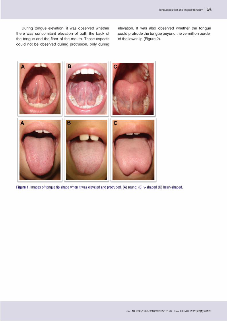
The following step consist of a blind evaluation performed by two other speech language therapists, who analyzed the shape of the tongue when elevated and protruded. They classified the shapes as round, V-shaped or heart-shaped ones (Figure 1).
INTRODUCTIONThe lingual frenulum is a dynamic three-dimensional
structure formed by a central fold in a layer of fascia that extends across the floor of the mouth. The range in the lingual frenulum morphology is created by variability – on a spectrum – of several factors1,2. Ankyloglossia is characterized by a restriction of tongue movements1,3.
Ankyloglossia is a complex condition impacting both mothers and infants. This requires interdisciplinary approach for assessment and treatment. When ankylo-glossia is characterized by a lingual frenulum attached to the apex of the tongue and visible from the inferior alveolar crest it can be easily diagnosed. Controversy around the diagnosis of ankyloglossia occur when the alteration is not so visible. That requires a structural and functional assessment of the oral cavity4.
Several lingual frenulum assessment tools for infants, children, adolescents, and adults have been published by professionals, such as dentists, lactation consultants, midwives, speech-language pathologists5-22.
Most authors recommend tongue elevation for diagnosing ankyloglossia 5-6,8,12-14,17-22. A few authors propose tongue protrusion for assessing the lingual frenulum7,11,15. Only one study states that both the symptoms reported by the mother and clinical evalu-ation during breastfeeding may be sufficient to diagnose ankyloglossia, that is, there is no need to put the finger in the infant´s mouth10.
Tongue movements are essential for the perfor-mance of orofacial functions. It must be highlighted that tongue elevation is needed for proper tongue rest posture, for adequate nasal breathing, swallowing and production of the sounds [t], [d], [n], [l] e [ɾ]3,4. An important aspect to remember is that protrusion of the tongue is not required for sucking, swallowing, breathing, chewing, and speaking.
In the clinical practice we come across profes-sionals from several fields who assess lingual frenulum by asking the patient to protrude the tongue. If the subject can protrude the tongue beyond the vermillion border of the lower lip the professionals do not consider frenotomy to be indicated23-25. However, the literature cites a set of anatomical and functional characteristics that must be considered for ankyloglossia diagnosis22-26.
Brito et al. state that when both the assessment of lingual frenulum and therapy plan are performed through personal criteria, they may fail, and an inter-disciplinary approach may be difficult27. Controversies among health professionals from different fields
Martinelli RLC, Marchesan IQ, Berretin-Felix G Tongue position and lingual frenulum
doi: 10.1590/1982-0216/20202210120 | Rev. CEFAC. 2020;22(1):e0120
Tongue position and lingual frenulum | 3/8
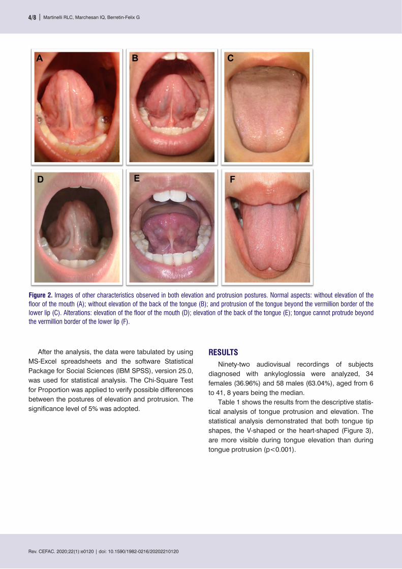
elevation. It was also observed whether the tongue could protrude the tongue beyond the vermillion border of the lower lip (Figure 2).
During tongue elevation, it was observed whether there was concomitant elevation of both the back of the tongue and the floor of the mouth. Those aspects could not be observed during protrusion, only during
Figure 1. Images of tongue tip shape when it was elevated and protruded. (A) round; (B) v-shaped (C) heart-shaped.
Rev. CEFAC. 2020;22(1):e0120 | doi: 10.1590/1982-0216/20202210120
4/8 | Martinelli RLC, Marchesan IQ, Berretin-Felix G
RESULTSNinety-two audiovisual recordings of subjects
diagnosed with ankyloglossia were analyzed, 34 females (36.96%) and 58 males (63.04%), aged from 6 to 41, 8 years being the median.
Table 1 shows the results from the descriptive statis-tical analysis of tongue protrusion and elevation. The statistical analysis demonstrated that both tongue tip shapes, the V-shaped or the heart-shaped (Figure 3), are more visible during tongue elevation than during tongue protrusion (p<0.001).
After the analysis, the data were tabulated by using MS-Excel spreadsheets and the software Statistical Package for Social Sciences (IBM SPSS), version 25.0, was used for statistical analysis. The Chi-Square Test for Proportion was applied to verify possible differences between the postures of elevation and protrusion. The significance level of 5% was adopted.
Figure 2. Images of other characteristics observed in both elevation and protrusion postures. Normal aspects: without elevation of the floor of the mouth (A); without elevation of the back of the tongue (B); and protrusion of the tongue beyond the vermillion border of the lower lip (C). Alterations: elevation of the floor of the mouth (D); elevation of the back of the tongue (E); tongue cannot protrude beyond the vermillion border of the lower lip (F).

doi: 10.1590/1982-0216/20202210120 | Rev. CEFAC. 2020;22(1):e0120
Tongue position and lingual frenulum | 5/8
This finding demonstrates that the protrusion position does not allow for an accurate identification of ankyloglossia.
Table 2 shows that most subjects with ankyloglossia (97.83%) can extend the tongue beyond the vermillion border of the lower lip during protrusion (Figure 3).
Table 1. Frequency and percentage of occurrence of the aspects analyzed in the position of tongue protrusion and elevation in subjects with ankyloglossia
VariableProtrusion Elevation
p-valueFrequency Percentage Frequency Percentage
V-shaped 21 22.80% 44 47.80% 0.035*Heart-shaped 8 8.70% 45 48.90% 0.001*Round 63 68.50% 3 3.30% < 0.001*Tongue protrudes beyond the vermillion border of the lower lip
90 97.83% - - -
Elevation of the back of the tongue concomitant with elevation of the tip of the tongue
- - 40 43.50% -
Elevation of the floor of the mouth concomitant with elevation of the tip of the tongue
- - 29 31.50% -
Chi-Square Test for Proportions*Statistical Significance
Figure 3. Characteristics of the tip of the tongue observed in 3 subjects diagnosed with ankyloglossia and their possibility of extending the tongue beyond the vermillion border of the lower lip, during protrusion. In A and D, tongue protrusion and elevation of subject 1; in B and E, tongue protrusion and elevation of subject 2; in C and F, tongue protrusion and elevation of subject 3.
Rev. CEFAC. 2020;22(1):e0120 | doi: 10.1590/1982-0216/20202210120
6/8 | Martinelli RLC, Marchesan IQ, Berretin-Felix G
state that elevation is the best posture to assess the lingual frenulum.
Another relevant datum is that 97.83% of the subjects with ankyloglossia were able to extend the tongue beyond the vermillion border of the lower lip, during protrusion (Table 1).
A comparison of all variables between tongue elevation and the extending of the tongue beyond the vermillion border of the lower lip, during protrusion, in subjects with ankyloglossia, demonstrated a statisti-cally significant difference (p<0.001), as shown in Table 2. Thus, protrusion is not a relevant posture for ankyloglossia diagnosis.
An important aspect to remember is that tongue protusion is not required for oral functions, such as sucking, swallowing, breathing, chewing, and speaking. On the other hand, tongue elevation is essential for swallowing, for producing many speech sounds, and for proper tongue rest posture28,34-36.
It is important to emphasize that a proper lingual frenulum assessment must consider a set of charac-teristics for the diagnosis of tongue movement restriction36. Unlike tongue protrusion, tongue elevation allows for the assessment of the lingual frenulum, since this position favors the visualization of the anatomical characteristics regarding thickness, attachment of the tongue to the floor of the mouth, and shape of the tip of the tongue36.
DISCUSSION
This study aimed to compare the position of elevation and protrusion of the tongue to determine which position provides better lingual frenulum assessment.
Of the 92 subjects aged from 6 to 41 diagnosed with ankyloglossia, whose audiovisual recordings were analyzed, 58 were males (63.04%). This finding agrees with studies that report higher prevalence of ankylo-glossia in males than in females29,30.
The statistical analysis demonstrated that both tongue tip shapes, the V-shaped and the heart-shaped, (Figure 3) are more visible during tongue elevation than during tongue protrusion (p<0.001). These findings demonstrated that tongue elevation is the position that allows for the best observation of the shape of the tip of the tongue, being possible to identify, more accurately, the presence of V-shaped or heart-shaped tip in subjects with ankyloglossia. Thus, those findings agree with proposals of most assessment tools5,6,8,12-14,17-22,31-33.
A study on the anatomy of the lingual frenulum states that the elevation of the anterior and middle parts of the tongue and/or retraction creates tension in the fascial layer of the floor of the mouth, drawing the fascia and the overlying mucosa up into a midline sagittal fold that forms the lingual frenulum1. Thus, it is possible to
Table 2. Comparison of all variables between tongue elevation and the extending of the tongue beyond the vermillion border of the lower lip, during protrusion in subjects with ankyloglossia
Variable n Percentage p-valueV-shaped observed during tongue elevation 44 47.83%
< 0.001*Extending the tongue beyond the vermilion border of lower lip during protrusion
90 97.83%
Heart-shaped observed during tongue elevation 45 48.91%< 0.001*Extending the tongue beyond the vermilion border of lower lip
during protrusion90 97.83%
Round observed during tongue elevation 3 3.26%< 0.001*Extending the tongue beyond the vermilion border of lower lip
during protrusion90 97.83%
Elevation of the back of the tongue concomitant with elevation of the tip of the tongue
40 43.48%< 0.001*
Extending the tongue beyond the vermilion border of lower lip during protrusion
90 97.83%
Elevation of the floor of the mouth concomitant with elevation of the tip of the tongue
29 31.52%< 0.001*
Extending the tongue beyond the vermilion border of lower lip during protrusion
90 97.83%
Chi-Square Test for Proportions*Statistical Significance
doi: 10.1590/1982-0216/20202210120 | Rev. CEFAC. 2020;22(1):e0120
Tongue position and lingual frenulum | 7/8
12. Martinelli RLC, Marchesan IQ, Lauris JR, Honório HM, Gusmão RJ, Berretin-Felix G. Validation of the Lingual Frenulum Protocol for infants. Int J Orofacial Myology. 2016;42:5-13.
13. Martinelli RLC, Marchesan IQ, Lauris JR, Honório HM, Gusmão RJ, Berretin-Felix G. Validade e confiabilidade da triagem: “teste da linguinha”. Rev. CEFAC. 2016;18(6):1323-31.
14. Walker RD, Messing S, Rosen-Carole C, McKenna Benoit M. Defining tip-frenulum length for ankyloglossia and its impact on breastfeeding: a prospective cohort study. Breastfeed Med. 2018:13(3):204-10.
15. Ingram J, Copeland M, Johnson D, Emond A. The development and evaluation of a picture tongue assessment tool for tongue-tie in breastfed babies (TABBY). Int Breastfeed J. 2019;14:31.
16. Fernando C. Tongue-tie - from confusion to clarity: a guide to the diagnosis and treatment of ankyloglossia. Tandem. 1998.
17. Lee SK, Kim YS, Lim CY. A pathological consideration of ankyloglossia and lingual myoplasty. Taehan Chikkwa Uisa hyophoe Chi 1989;27:287-308.
18. Kotlow LA. Ankyloglossia (tongue-tie): a diagnostic and treatment quandary. Quintessence Int 1999;30:259-62.
19. Marchesan IQ. Frênulo lingual: proposta de avaliação quantitativa. Rev. CEFAC. 2004;6(3):288-93.
20. Ruffoli R, Giambelluca MA, Scavuzzo MC, Bonfigli D, Cristofani R, Gabriele M et al. Ankyloglossia: a morphofunctional investigation in children. Oral Diseases. 2005;11:170-4.
21. Marchesan IQ. Protocolo de avaliação do frênulo da língua. Rev. CEFAC. 2010;12(6):977-89.
22. Yoon A, Zaghi S, Weitzman R, Ha S, Law CS, Guilleminault C, Liu SY. Toward a functional definition of ankyloglossia: validating current grading scales for lingual frenulum length and tongue mobility in 1052 subjects. Sleep Breath. Sleep Breath. 2017 Sep;21(3):767-775.
23. Wallace AF. Tongue-tie. Lancet. 1963;2:377-8.24. Sedano HO, Carreon Freyre I, Garza de la Garza
ML, Gomar Franco CM, Grimaldo Hernandez C, Hernandez Montoya ME et al. Clinical orodental abnormalities in Mexican children. Oral Surg Oral Med Oral Pathol.1989;68:300-11.
25. Obrigatoriedade do Teste da Linguinha em recém-nascidos entra em vigor. TV Senado,
CONCLUSIONElevation is the position that allows the best obser-
vation of the shape of the tip of the tongue, allowing for a more accurate identification of the presence of V-shaped and heart-shaped tips in subjects with ankyloglossia.
REFERENCES1. Mills N, Pransky SM, Geddes DT, Mirjalili SA. What
is a tongue tie? Defining the anatomy of the in-situ lingual frenulum. Clin Anat. 2019;32(6):749-61.
2. Mills N, Keough N, Geddes DT, Pransky SM, Mirjalili SA. Defining the anatomy of the neonatal lingual frenulum. Clin Anat. 2019;32(6):824-35.
3. Zaghi S, Valcu-Pinkerton S, Jabara M, Norouz-Knutsen L, Govardhan C, Moeller J et al. Lingual frenuloplasty with myofunctional therapy: Exploring safety and efficacy in 348 cases. Laryngoscope Investig Otolaryngol. 2019;4(5):489-96.
4. Walsh J, McKenna Benoit M. Ankyloglossia and other oral ties. Otolaryngol Clin North Am. 2019;52(5):795-811.
5. Hazelbaker AK. The assessment tool for lingual frenulum function (ATLFF): use in a lactation consultant private practice [thesis]. Pasadena, CA: Pacific Oaks College; 1993.
6. Coryllos E, Genna CW, Salloum AC. Congenital tongue-tie and its impact in breastfeeding. Breastfeeding: Best for mother and baby Newsletter. 2004;1-6
7. Griffiths DM. Do tongue ties affect breastfeeding? J Hum Lact. 2004;20(4):409-14.
8. Hogan M, Westcott C, Griffiths M. Randomized, controlled trial of division of tongue-tie in infants with feeding problems. J Paediatr Child Health. 2005;41(5-6):246-50.
9. Srinivasan A, Dobrich C, Mitnick H, Feldman P. Ankyloglossia in breastfeeding infants: the effect of frenotomy on maternal nipple pain and latch. Breastfeed Med. 2006;1(4):216-24.
10. Baeza CE, de Alba Romero C, Morales AM, Querol N. Diagnosing Ankyloglossia: do we need to put the finger in the mouth? Journal of Human Lactation. 2013;29(1):95-6
11. Ingram J, Johnson D, Copeland M, Churchill C, Taylor H, Emond A. The development of a tongue assessment tool to assist with tongue-tie identification. Arch Dis Child Fetal Neonatal Ed. 2015;100(4):F344-8.
Rev. CEFAC. 2020;22(1):e0120 | doi: 10.1590/1982-0216/20202210120
8/8 | Martinelli RLC, Marchesan IQ, Berretin-Felix G
2014. Disponível em: https://www.youtube.com/watch?v=NqHAGjukTCo Acesso em: 29 nov. 2019.
26. Marchesan IQ. Protocolo de avaliação do frênulo da língua. Rev. CEFAC. 2010;12(6):977-89.
27. Brito SF, Marchesan IQ, Bosco CM, Carrilho ACA, Rehder MI. Frênulo lingual: classificação e conduta segundo ótica fonoaudiológica, odontológica e otorrinolaringológica. Rev. CEFAC. 2008;10(3):343-51.
28. Marchesan IQ, Teixeira AN, Cattoni DM. Correlações entre diferentes frênulos linguais e alterações na fala. Distúrb. Comun. 2010;22(3):195-200.
29. Ata N, Alataş N, Yılmaz E, Adam AB, Gezgin B. The relationship of ankyloglossia with gender in children and the ideal timing of surgery in ankyloglossia. Ear Nose Throat J. 2019 Sep 26:145561319867666. [Epub ahead of print]
30. Caloway C, Hersh CJ, Baars R, Sally S, Diercks G, Hartnick CJ. Association of feeding evaluation with frenotomy rates in infants with breastfeeding difficulties. JAMA Otolaryngol Head Neck Surg. 2019 Jul 11. [Epub ahead of print]
31. Martinelli RLC. Relação entre as características anatômicas do frênulo lingual e as funções de sucção e deglutição em bebês [dissertação]. Bauru (SP): Faculdade de Odontologia de Bauru, Universidade de São Paulo; 2013.
32. Cunha RF, Silva JZ, Faria MD. Clinical approach of ankyloglossia in babies: report of two cases. J Clin Pediatr Dent. 2008;32(4):277-81.
33. Karabulut R, Sönmez K, Türkyilmaz Z, Demirogullari B, Ozen IO, Bagbanci B et al. Ankyloglossia and effects on breastfeeding, speech problems and mechanical/social issues in children. B-ENT. 2008;4(2):81-5.
34. Cockley L, Lehman A. The Ortho missing link: could it be tied to the tongue. JAOS. 2015;15(1):18-21.
35. Martinelli RLC, Marchesan IQ, Berretin-Felix G. Rest position of the tongue in infants with and without lingual frenulum alteration. Int J Orofacial Myology. 2016;42:43-8.
36. Martinelli RLC, Marchesan IQ. Frênulo lingual. In: Silva HJ, Tessitore A, Motta AR, Cunha DA, Berretin-Felix G, Marchesan IQ (orgs). Tratado de Motricidade Orofacial. São José dos Campos: Pulso Editorial. 2019.