Toll-Like Receptor 2 Regulates Organic Dust–Induced Airway Inflammation
9
Toll-Like Receptor 2 Regulates Organic Dust–Induced Airway Inflammation Jill A. Poole 1,2 , Todd A. Wyatt 1,2,4 , Tammy Kielian 3 , Peter Oldenburg 2 , Angela M. Gleason 1,2 , Ashley Bauer 1,2 , Gregory Golden 2 , William W. West 3 , Joseph H. Sisson 2 , and Debra J. Romberger 1,2 1 Omaha Veterans Affairs Medical Center, Omaha, Nebraska; and 2 Pulmonary, Critical Care, Sleep & Allergy Division, Department of Internal Medicine, 3 Department of Pathology and Microbiology, and 4 Department of Environmental, Agricultural, and Occupational Health, University of Nebraska Medical Center, the Nebraska Medical Center, Omaha, Nebraska Organic dust exposure in agricultural environments results in signif- icant airway inflammatory diseases. Gram-positive cell wall compo- nents are present in high concentrations in animal farming dusts, but their role in mediating dust-induced airway inflammation is not clear. This study investigated the role of Toll-like receptor (TLR) 2, a pattern recognition receptor for gram-positive cell wall products, in regulating swine facility organic dust extract (DE)–induced airway inflammation in mice. Isolated lung macrophages from TLR2 knock- out mice demonstrated reduced TNF-a, IL-6, keratinocyte chemo- attractant/CXCL1, but not macrophage inflammatory protein-2/ CXCL2 expression, after DE stimulation ex vivo. Next, using an established mouse model of intranasal inhalation challenge, we analyzed bronchoalveolar lavage fluid and lung tissue in TLR2- deficient and wild-type (WT) mice after single and repetitive DE challenge. Neutrophil influx and select cytokines/chemokines were significantly lower in TLR2-deficient mice at 5 and 24 hours after single DE challenge. After daily exposure to DE for 2 weeks, there were significant reductions in total cellularity, neutrophil influx, and TNF-a, IL-6, CXCL1, but not CXCL2 expression, in TLR2-deficient mice as compared with WT animals. Lung pathology revealed that bronchiolar inflammation, but not alveolar inflammation, was re- duced in TLR2-deficient mice after repetitive exposure. Airway hyperresponsiveness to methacholine after dust exposure was similar in both groups. Finally, airway inflammatory responses in WT mice after challenge with a TLR2 agonist, peptidoglycan, re- sembled DE-induced responses. Collectively, these results demon- strate that the TLR2 pathway is important in regulating swine facility organic dust–induced airway inflammation, which suggests the importance of TLR2 agonists in mediating large animal farming– induced airway inflammatory responses. Keywords: Toll-like receptor 2; swine/pig facility; peptidoglycan; org- anic dust; lung pathology Agricultural workers, particularly swine farmers, exhibit a high prevalence of airway diseases, including chronic bronchitis, exacerbation of asthma, and obstructive lung disease, which is thought to be due to repeated exposure to inhaled organic dusts or bioaerosols (1). A challenge in defining mechanisms of or- ganic dust–induced inflammatory responses lies in the inherent complexity of the dust. One established inflammatory compo- nent present in organic dust is endotoxin; however, epidemio- logic and multiple laboratory-based studies have failed to definitively link endotoxin exposure to disease manifestation (2–8). Consistent with these observations, we and others have found a strong predominance of gram-positive (rather than gram- negative) bacteria in modern swine confinement facility organic dust samples (6, 9). Furthermore, mass spectrometry analysis has demonstrated high concentrations of muramic acid, a com- ponent of peptidoglycan (PGN), which originates from the bacterial cell wall of gram-positive bacteria (i.e., 85% of total cell wall) and, to a lesser degree, gram-negative bacteria (i.e., 5% of cell wall) in large animal farming environments (e.g., swine and dairy barns) (6, 10). In addition, prior reports have suggested that nonendotoxin components, such as PGN, are responsible for driving the innate immune inflammatory re- sponses to swine facility animal farming dusts in vitro (6, 10, 11). However, it is not known if targeting innate immune pattern recognition receptors of gram-positive cell wall products will affect complex organic dust–induced airway inflammation, which was the objective of the current study. One family of innate immune receptors responsible for rec- ognizing highly conserved microbial motifs are the Toll-like receptors (TLRs) (12). Of the 10 TLRs, TLR2 has been implicated as a critical receptor of gram-positive bacteria, because TLR2 recognizes PGNs, lipoteichoic acid, and lipopro- teins that are associated with the cell wall of gram-positive bacteria (12, 13). TLR2 is highly expressed on antigen-presenting cells, such as macrophages, cells important in mediating the innate immune response to inhaled organic dust (14, 15). On airway epithelial cells, TLR2 expression is up-regulated after swine facility organic dust exposure, and blocking epithelial cell TLR2 results in a dampening of proinflammatory cytokine release after organic dust exposure in vitro (16, 17). In general, the role of TLR2 in mediating airways disease is controversial. In various lung infection models, deficiencies in TLR2 have been associated with increased proinflammatory mediator re- lease and resistance to infection (18), decreased proinflammatory CLINICAL RELEVANCE This work demonstrates that the Toll-like receptor (TLR) 2 pathway plays an important role in regulating swine facility organic dust–induced airway inflammation in vivo. These findings have important implications for future envi- ronmental sampling strategies to focus on, and to reduce exposure of, TLR2 agonists present in agricultural environ- ments. Targeting the TLR2 pathway might also be an important adjunctive therapy when investigating prevention and therapeutic interventions in humans. (Received in original form October 18, 2010 and in final form December 17, 2010) This work was supported by National Institute of Environmental Health Sciences grants K08 ES015522-01 and ES015522-03S1[ARRA], and R01 ES019325 (J.A.P.), National Institute of Occupational Safety Health grant R01 OH008539-01 (D.J.R.), National Institute on Alcohol Abuse and Alcoholism grant R01 AA017993 (T.A.W.), and Kuhl Testamentary Fund University of Nebraska Medical Center Intramural grant (G.G.). Correspondence and requests for reprints should be addressed to Jill A. Poole, M.D., Pulmonary, Critical Care, Sleep & Allergy Division; Department of Internal Medicine; University of Nebraska Medical Center; 985300 The Nebraska Medical Center Omaha, NE 68198-5300. E-mail: [email protected] This article has an online supplement, which is accessible from this issue’s table of contents at www.atsjournals.org Am J Respir Cell Mol Biol Vol 45. pp 711–719, 2011 Originally Published in Press as DOI: 10.1165/rcmb.2010-0427OC on January 28, 2011 Internet address: www.atsjournals.org
Transcript of Toll-Like Receptor 2 Regulates Organic Dust–Induced Airway Inflammation
Toll-Like Receptor 2 Regulates Organic Dust–InducedAirway Inflammation
Jill A. Poole1,2, Todd A. Wyatt1,2,4, Tammy Kielian3, Peter Oldenburg2, Angela M. Gleason1,2, Ashley Bauer1,2,Gregory Golden2, William W. West3, Joseph H. Sisson2, and Debra J. Romberger1,2
1Omaha Veterans Affairs Medical Center, Omaha, Nebraska; and 2Pulmonary, Critical Care, Sleep & Allergy Division, Department of Internal
Medicine, 3Department of Pathology and Microbiology, and 4Department of Environmental, Agricultural, and Occupational Health, University ofNebraska Medical Center, the Nebraska Medical Center, Omaha, Nebraska
Organic dust exposure in agricultural environments results in signif-icant airway inflammatory diseases. Gram-positive cell wall compo-nentsare present in high concentrations in animal farmingdusts,buttheir role in mediating dust-induced airway inflammation is notclear. This study investigated the role of Toll-like receptor (TLR) 2,a pattern recognition receptor for gram-positive cell wall products,in regulating swine facility organic dust extract (DE)–induced airwayinflammation in mice. Isolated lung macrophages from TLR2 knock-out mice demonstrated reduced TNF-a, IL-6, keratinocyte chemo-attractant/CXCL1, but not macrophage inflammatory protein-2/CXCL2 expression, after DE stimulation ex vivo. Next, using anestablished mouse model of intranasal inhalation challenge, weanalyzed bronchoalveolar lavage fluid and lung tissue in TLR2-deficient and wild-type (WT) mice after single and repetitive DEchallenge. Neutrophil influx and select cytokines/chemokines weresignificantly lower in TLR2-deficient mice at 5 and 24 hours aftersingle DE challenge. After daily exposure to DE for 2 weeks, therewere significant reductions in total cellularity, neutrophil influx, andTNF-a, IL-6, CXCL1, but not CXCL2 expression, in TLR2-deficientmice as compared with WT animals. Lung pathology revealed thatbronchiolar inflammation, but not alveolar inflammation, was re-duced in TLR2-deficient mice after repetitive exposure. Airwayhyperresponsiveness to methacholine after dust exposure wassimilar in both groups. Finally, airway inflammatory responses inWT mice after challenge with a TLR2 agonist, peptidoglycan, re-sembled DE-induced responses. Collectively, these results demon-strate that the TLR2 pathway is important in regulating swine facilityorganic dust–induced airway inflammation, which suggests theimportance of TLR2 agonists in mediating large animal farming–induced airway inflammatory responses.
Keywords: Toll-like receptor 2; swine/pig facility; peptidoglycan; org-anic dust; lung pathology
Agricultural workers, particularly swine farmers, exhibit a highprevalence of airway diseases, including chronic bronchitis,exacerbation of asthma, and obstructive lung disease, which isthought to be due to repeated exposure to inhaled organic dustsor bioaerosols (1). A challenge in defining mechanisms of or-
ganic dust–induced inflammatory responses lies in the inherentcomplexity of the dust. One established inflammatory compo-nent present in organic dust is endotoxin; however, epidemio-logic and multiple laboratory-based studies have failed todefinitively link endotoxin exposure to disease manifestation(2–8). Consistent with these observations, we and others havefound a strong predominance of gram-positive (rather than gram-negative) bacteria in modern swine confinement facility organicdust samples (6, 9). Furthermore, mass spectrometry analysishas demonstrated high concentrations of muramic acid, a com-ponent of peptidoglycan (PGN), which originates from thebacterial cell wall of gram-positive bacteria (i.e., 85% of totalcell wall) and, to a lesser degree, gram-negative bacteria (i.e.,5% of cell wall) in large animal farming environments (e.g.,swine and dairy barns) (6, 10). In addition, prior reports havesuggested that nonendotoxin components, such as PGN, areresponsible for driving the innate immune inflammatory re-sponses to swine facility animal farming dusts in vitro (6, 10, 11).However, it is not known if targeting innate immune patternrecognition receptors of gram-positive cell wall products willaffect complex organic dust–induced airway inflammation,which was the objective of the current study.
One family of innate immune receptors responsible for rec-ognizing highly conserved microbial motifs are the Toll-likereceptors (TLRs) (12). Of the 10 TLRs, TLR2 has beenimplicated as a critical receptor of gram-positive bacteria,because TLR2 recognizes PGNs, lipoteichoic acid, and lipopro-teins that are associated with the cell wall of gram-positivebacteria (12, 13). TLR2 is highly expressed on antigen-presentingcells, such as macrophages, cells important in mediating theinnate immune response to inhaled organic dust (14, 15). Onairway epithelial cells, TLR2 expression is up-regulated afterswine facility organic dust exposure, and blocking epithelial cellTLR2 results in a dampening of proinflammatory cytokinerelease after organic dust exposure in vitro (16, 17). In general,the role of TLR2 in mediating airways disease is controversial.In various lung infection models, deficiencies in TLR2 havebeen associated with increased proinflammatory mediator re-lease and resistance to infection (18), decreased proinflammatory
CLINICAL RELEVANCE
This work demonstrates that the Toll-like receptor (TLR)2 pathway plays an important role in regulating swinefacility organic dust–induced airway inflammation in vivo.These findings have important implications for future envi-ronmental sampling strategies to focus on, and to reduceexposure of, TLR2 agonists present in agricultural environ-ments. Targeting the TLR2 pathway might also be animportant adjunctive therapy when investigating preventionand therapeutic interventions in humans.
(Received in original form October 18, 2010 and in final form December 17, 2010)
This work was supported by National Institute of Environmental Health Sciences
grants K08 ES015522-01 and ES015522-03S1[ARRA], and R01 ES019325 (J.A.P.),
National Institute of Occupational Safety Health grant R01 OH008539-01
(D.J.R.), National Institute on Alcohol Abuse and Alcoholism grant R01
AA017993 (T.A.W.), and Kuhl Testamentary Fund University of Nebraska Medical
Center Intramural grant (G.G.).
Correspondence and requests for reprints should be addressed to Jill A. Poole,
M.D., Pulmonary, Critical Care, Sleep & Allergy Division; Department of Internal
Medicine; University of Nebraska Medical Center; 985300 The Nebraska Medical
Center Omaha, NE 68198-5300. E-mail: [email protected]
This article has an online supplement, which is accessible from this issue’s table of
contents at www.atsjournals.org
Am J Respir Cell Mol Biol Vol 45. pp 711–719, 2011
Originally Published in Press as DOI: 10.1165/rcmb.2010-0427OC on January 28, 2011
Internet address: www.atsjournals.org
mediator release and failure to control infection (19–21), or nosignificant effect with lung inflammation indices (22). In com-parison, airway inflammatory consequences after inhalationchallenge with pure selective TLR2 agonists are reduced inTLR2-deficient mice (23). It is not known if there is a role forTLR2 in modulating airway inflammatory responses to a com-plex microbial exposure, such as large animal farming dusts.However, these previous observations suggested to us thatTLR2 might play an important role in mediating airway in-flammatory responses after large animal farming dust expo-sures, an environment rich in gram-positive cell wall products.
In this study, we hypothesized that TLR2 loss would sig-nificantly reduce airway inflammatory responses to swine facil-ity organic dust extract (DE) exposure. To test this hypothesis,we first determined whether there would be a reduction incytokine/chemokine release from primary lung macrophagesisolated from TLR2-deficient mice compared with wild-type(WT) animals. Next, we investigated, in an established in vivomurine model (14), if airway inflammatory responses to in-tranasal inhalation of organic DE in TLR2-deficient mice woulddiffer as compared with WT animals at various time points andconcentrations of DE. Finally, we determined if a TLR2 ago-nist, PGN, would induce similar airway inflammatory responsesas DE. Collectively, we found an important role for TLR2 inmediating airway inflammatory responses to swine facilityorganic dust in vivo. Namely, the absence of TLR2 signalingresulted in an attenuation of dust-dependent airway inflamma-tion and lung pathology, but not acute airway hyperresponsive-ness (AHR) and alveolar inflammation.
MATERIALS AND METHODS
DE
Swine confinement animal feeding operation facility organic dust wascollected and prepared as DE as previously described (7, 8) and asdescribed in the online supplement.
Mice
C57BL/6 WT mice were purchased from the Jackson Laboratory (BarHarbor, ME) and TLR2 knockout (KO; same C57BL/6 background)mice were provided by S. Akira (Osaka, Japan). Animal studies wereapproved by the Institutional Animal Care and Use Committee ofthe Omaha Veterans Affairs Medical Center and the University ofNebraska Medical Center, according to National Institutes of Healthguidelines for the use of rodents.
Lung Macrophages
Lung macrophages were isolated as described in the METHODS section inthe online supplement. After isolation, lung macrophages were stim-ulated with 1% DE. After 24 hours, cell-free supernatants werecollected and stored at 2808C.
Animal Model
Mice received saline or DE by an intranasal exposure method, aspreviously established (14). A concentration of 12.5% DE is an optimalconcentration for eliciting lung inflammation (14). In some experi-ments, a suboptimal 2.5% DE concentration was also used to de-termine the importance of TLR2 at a relatively high and low organicdust concentration exposure. Animals also received PGN, LPS, andheat-inactivated DE, which is described in the METHODS in the onlinesupplement.
Bronchoalveolar Lavage
Bronchoalveolar lavage (BAL) fluid was collected as previously de-scribed (14). Total cell number was enumerated and differential cellcounts were determined on cytospin-prepared slides (Cytopro Cyto-centrifuge, Wescor Inc., Logan, UT) stained with DiffQuick (DadeBehring, Newark, DE).
Cytokine/Chemokine Assays
Murine TNF-a, IL-6, keratinocyte chemoattractant (KC; CXCL1), andmacrophage inflammatory protein (MIP)–2 (CXCL2) concentrationswere determined in BAL fluid according to manufacturer’s instructionsusing commercially available ELISA kits (R&D Systems, Minneapolis,MN).
Nitric Oxide Analysis
Nitric oxide (NO) production in BAL fluid was assessed via thedetection of NO by a gas-phase chemiluminescent reaction betweenNO and ozone (Sievers Instruments Model 280i; GE AnalyticalInstruments, Boulder, CO) according to previous published proce-dures, because this is a highly sensitive assay, detecting NO at lowlevels (24).
Lung Collection
After lung lavage, whole lungs were excised and inflated to 10 cm H2Opressure with 10% formalin (Sigma, St. Louis, MO) solution to pre-serve pulmonary architecture. Lungs were embedded in paraffin, andsections (4–5 M) were cut and stained with hematoxylin and eosin.Lung slides were reviewed and semiquantitatively assessed for thedegree of inflammation as well as the distribution of the inflammationby a reviewer (pathologist [W.W.W.]) blinded to the treatment con-ditions using a previously published scoring system (14).
Pulmonary Function Measurement
At 3 hours after a single intranasal instillation of 12.5% DE or PGN(14), mice were anesthetized, tracheostomized, and mechanicallyventilated at a rate of 160 breaths/min and a tidal volume of 0.15 ml,using a computerized small animal ventilator (Finepoint; BuxcoElectronic, Wilmington, NC) (14, 25). Dose responsiveness to aero-solized methacholine (1.5–48.0 mg/ml) was obtained and resultsreported as total lung resistance.
Statistical Analysis
Data are presented as the mean (6SEM). Statistical significance wasassessed by one-way ANOVA and two-tailed unpaired and pairedt test, where appropriate, to determine significant changes amongtreatment groups using GraphPad Prism version 5.0 (GraphPad Inc.,La Jolla, CA) software.
RESULTS
Organic Dust–Induced Inflammatory Mediator Production
in Primary Lung Macrophages Is Predominately
TLR2 Dependent
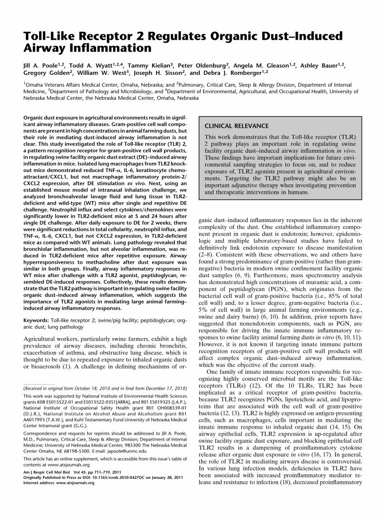
To define if there were functional roles of TLR2 in mediatinglung macrophage response to organic DE, lung macrophageswere isolated from TLR2-deficient and WT mice (C57BL/6background) and ex vivo stimulated with 1% DE for 24 hours.There were significant reductions in TNF-a (255%), IL-6(264%), and CXCL1 (287%) production in TLR2-deficientlung macrophages; however, there was no significant changein CXCL2 expression (Figure 1; n 5 4 mice per group). Trypanblue exclusion analysis demonstrated that the effects ofTLR2 loss were not due to differences in cell viability ornumber (data not shown). Collectively, these results suggestthat TLR2 signaling is important for regulating the expressionof select cytokines/chemokines in lung macrophages after DEchallenge.
Acute Dust-Induced Airway Cellular Inflammation and
Cytokine/Chemokine Release Is Reduced in TLR2-Deficient Mice
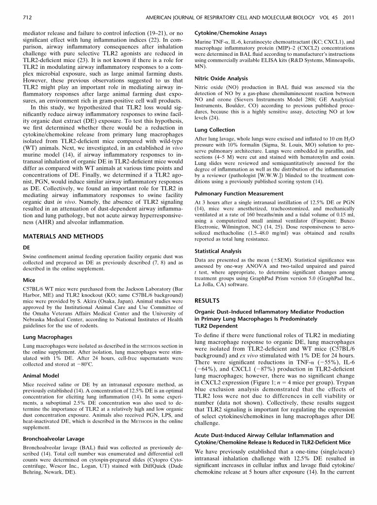
We have previously established that a one-time (single/acute)intranasal inhalation challenge with 12.5% DE resulted insignificant increases in cellular influx and lavage fluid cytokine/chemokine release at 5 hours after exposure (14). In the current
712 AMERICAN JOURNAL OF RESPIRATORY CELL AND MOLECULAR BIOLOGY VOL 45 2011
study, we sought to determine if TLR2 deficiency would dampenairway inflammatory responses after a one-time DE challengein mice. The increase in total leukocyte counts in the lavagefluid after DE challenge was significantly reduced in TLR2-deficient mice as compared with WT mice at both low (248%)and high (255%) DE concentrations (Figure 2A). Consistentwith our previous work, DE induced the rapid influx of air-way neutrophils (Figure 2B); however, airway neutrophils
were significantly reduced (255%) in TLR2-deficient mice re-ceiving 12.5% DE as compared with WT animals (Figure 2B).
Organic dust–induced airway disease has also been associ-ated with increases in TNF-a, IL-6, and neutrophil chemo-attractants (human IL-8, murine KC/CXCL1, and MIP-2/CXCL2), which are particularly important after an acute(single) exposure in naive humans and mice (14, 26, 27). At 5hours after a 2.5% DE exposure, there was a significant re-
Figure 1. Isolated lung mac-
rophages from Toll-like recep-tor (TLR) 2 knockout (KO) mice
demonstrate dampened TNF-
a, IL-6, keratinocyte chemoat-
tractant (KC)/CXCL1, but notmacrophage inflammatory
protein (MIP)–2/CXCL2, pro-
duction after ex vivo stimula-
tion with 1% swine facility dustextract (DE) stimulation for
24 hours as compared with
lung macrophages from DE-stimulated wild-type (WT)
mice. Mean results are pre-
sented per 200,000 cells
(6SEM); n 5 4 mice pergroup. Statistically significance:
*P , 0.05, **P , 0.01.
Figure 2. (A–C ). Lung lavage cellularity and cytokine/chemokine release is acutely reduced in TLR2-deficient mice after a one-time challenge with
low- and high-concentration DE. Lung lavage fluid mean concentration of total cells (A) and cell differential (B) 5 hours after single intranasal
inhalation exposure of DE (0% [saline], 2.5%, and 12.5%) in WT and TLR2 KO mice. (C ) Cell-free mean levels of lung lavage fluid supernatantcytokines/chemokines collected 5 hours after exposure to DE. Error bars represent SE (n 5 4–6 mice per group). #Statistically significant between
respective DE-treated and saline-treated group. *P , 0.05 and **P , 0.01 indicate statistical significance between WT and KO DE-treated mice.
Poole, Wyatt, Kielian, et al.: TLR2 Regulates Dust-Induced Airway Inflammation 713
duction in TNF-a, CXCL2, and CXCL1, but not IL-6, in TLR2-deficient mice (Figure 2C). In comparison, 5 hours after a 12.5%DE exposure, there was a significant reduction in IL-6 andCXCL1, but not TNF-a and CXCL2 (Figure 2C). Collectively,these data demonstrate that TLR2-dependent pathways areinvolved in rapidly responding to swine facility organic dustexposure in the airway.
Airway Cellular Influx and Cytokine/Chemokine Release
Remains Dampened over Time after a Single DE Challenge
in TLR2-Deficient Mice
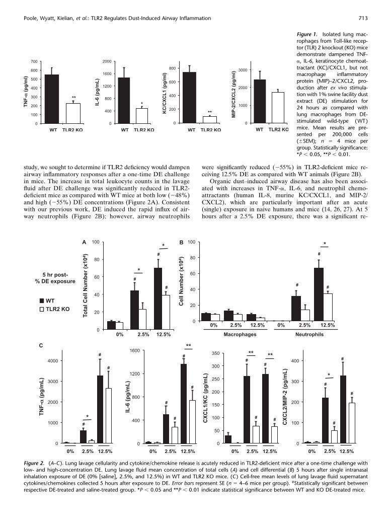
Because the cytokine/chemokine response to DE was impairedacutely (at 5 h) in the TLR2-deficient mice, we hypothesizedthat there could be a delayed, compensatory increase in airwaycellularity influx and cytokine/chemokine release in the TLR2-deficient mice after DE challenge. However, we found nodelayed compensatory increase in these proinflammatory out-comes. Using a high concentration of DE (12.5%), TLR2-deficient and WT mice were killed at 24 and 48 hours aftersingle intranasal inhalation DE challenge. There was no signif-icant delayed increase of inflammatory cellular influx in TLR2-deficient mice at 24 hours after DE exposure. Total lavagecellularity (Figure 3A) and airway neutrophils (Figure 3B)remained significantly reduced in TLR2 KO mice as comparedwith WT mice. At 48 hours after DE exposure, there was nosignificant difference between groups, which may be explained
by a clearing of the cells from the airway in the WT mice. At alltimes and dose concentrations, the percentage of lymphocytesand/or eosinophils ranged from 0 to 1% in all animals (data notshown).
Cytokine/chemokine levels in BAL fluid were also measuredat 24 and 48 hours after a single DE (12.5%) challenge, withresults depicted in Figure 3C. Overall, levels of mediators wereless at 24 and 48 hours as compared with 5 hours after DEchallenge in WT and TLR2 KO mice, which is consistent withthe kinetics of these mediators (28, 29). Compared with WTmice, TNF-a and IL-6, but not neutrophil chemoattractants(CXCL1, CXCL2), were significantly reduced in TLR2 KOmice after 12.5% DE at 24 hours after challenge. At 48 hoursafter DE (12.5%) inhalation challenge, all mediators wereeither undetected or at the lower limit of assay sensitivity/detection. Together, these studies demonstrate that TLR2-deficient mice have reduced inflammatory cellular influx andselect cytokine/chemokine release in the lavage fluid aftera one-time challenge with organic dust, and that there is nodelayed or compensatory recovery in these inflammatory indices.
TLR2 Impacts Dust-Induced NO Production
in a Time-Dependent Manner
In these experiments, NO levels in lavage fluid were investi-gated because NO is known to modify airway inflammation(30), and there are subtle, but statistically significant, increases
Figure 3. (A–C ). Lung lavage cellular influx and selective cytokine/chemokine release remains dampened over time after a single DE challenge in
TLR2-deficient mice. Lung lavage fluid mean total cells (A) and cell differential (B) after 12.5% DE at 5, 24, and 48 hours after intranasal inhalationchallenge. (C ) Cell-free mean levels of lung lavage fluid supernatant cytokines/chemokines collected at 5, 24, and 48 hours after exposure to 12.5%
DE. Error bars are SE (n 5 6–8 mice per group). *P , 0.05 and **P , 0.01 indicate statistical significance between WT and KO DE-treated mice.
714 AMERICAN JOURNAL OF RESPIRATORY CELL AND MOLECULAR BIOLOGY VOL 45 2011
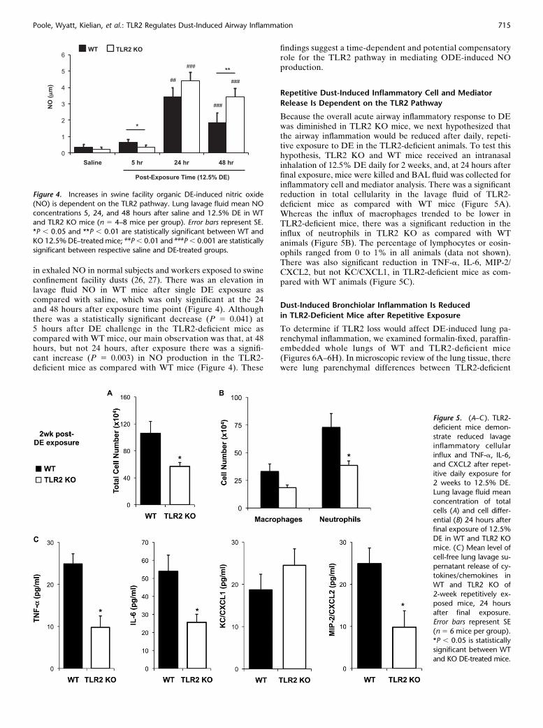
in exhaled NO in normal subjects and workers exposed to swineconfinement facility dusts (26, 27). There was an elevation inlavage fluid NO in WT mice after single DE exposure ascompared with saline, which was only significant at the 24and 48 hours after exposure time point (Figure 4). Althoughthere was a statistically significant decrease (P 5 0.041) at5 hours after DE challenge in the TLR2-deficient mice ascompared with WT mice, our main observation was that, at 48hours, but not 24 hours, after exposure there was a signifi-cant increase (P 5 0.003) in NO production in the TLR2-deficient mice as compared with WT mice (Figure 4). These
findings suggest a time-dependent and potential compensatoryrole for the TLR2 pathway in mediating ODE-induced NOproduction.
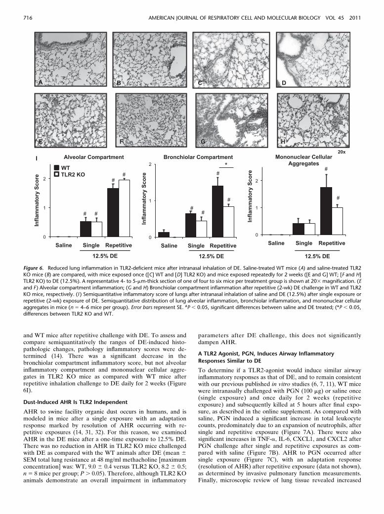
Repetitive Dust-Induced Inflammatory Cell and Mediator
Release Is Dependent on the TLR2 Pathway
Because the overall acute airway inflammatory response to DEwas diminished in TLR2 KO mice, we next hypothesized thatthe airway inflammation would be reduced after daily, repeti-tive exposure to DE in the TLR2-deficient animals. To test thishypothesis, TLR2 KO and WT mice received an intranasalinhalation of 12.5% DE daily for 2 weeks, and, at 24 hours afterfinal exposure, mice were killed and BAL fluid was collected forinflammatory cell and mediator analysis. There was a significantreduction in total cellularity in the lavage fluid of TLR2-deficient mice as compared with WT mice (Figure 5A).Whereas the influx of macrophages trended to be lower inTLR2-deficient mice, there was a significant reduction in theinflux of neutrophils in TLR2 KO as compared with WTanimals (Figure 5B). The percentage of lymphocytes or eosin-ophils ranged from 0 to 1% in all animals (data not shown).There was also significant reduction in TNF-a, IL-6, MIP-2/CXCL2, but not KC/CXCL1, in TLR2-deficient mice as com-pared with WT animals (Figure 5C).
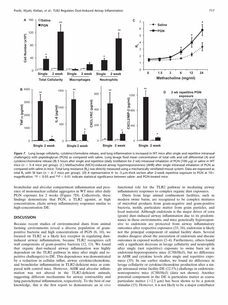
Dust-Induced Bronchiolar Inflammation Is Reduced
in TLR2-Deficient Mice after Repetitive Exposure
To determine if TLR2 loss would affect DE-induced lung pa-renchymal inflammation, we examined formalin-fixed, paraffin-embedded whole lungs of WT and TLR2-deficient mice(Figures 6A–6H). In microscopic review of the lung tissue, therewere lung parenchymal differences between TLR2-deficient
Figure 4. Increases in swine facility organic DE-induced nitric oxide
(NO) is dependent on the TLR2 pathway. Lung lavage fluid mean NO
concentrations 5, 24, and 48 hours after saline and 12.5% DE in WTand TLR2 KO mice (n 5 4–8 mice per group). Error bars represent SE.
*P , 0.05 and **P , 0.01 are statistically significant between WT and
KO 12.5% DE–treated mice; ##P , 0.01 and ###P , 0.001 are statistically
significant between respective saline and DE-treated groups.
Figure 5. (A–C ). TLR2-
deficient mice demon-
strate reduced lavageinflammatory cellular
influx and TNF-a, IL-6,
and CXCL2 after repet-
itive daily exposure for2 weeks to 12.5% DE.
Lung lavage fluid mean
concentration of totalcells (A) and cell differ-
ential (B) 24 hours after
final exposure of 12.5%
DE in WT and TLR2 KOmice. (C ) Mean level of
cell-free lung lavage su-
pernatant release of cy-
tokines/chemokines inWT and TLR2 KO of
2-week repetitively ex-
posed mice, 24 hours
after final exposure.Error bars represent SE
(n 5 6 mice per group).
*P , 0.05 is statisticallysignificant between WT
and KO DE-treated mice.
Poole, Wyatt, Kielian, et al.: TLR2 Regulates Dust-Induced Airway Inflammation 715
and WT mice after repetitive challenge with DE. To assess andcompare semiquantitatively the ranges of DE-induced histo-pathologic changes, pathology inflammatory scores were de-termined (14). There was a significant decrease in thebronchiolar compartment inflammatory score, but not alveolarinflammatory compartment and mononuclear cellular aggre-gates in TLR2 KO mice as compared with WT mice afterrepetitive inhalation challenge to DE daily for 2 weeks (Figure6I).
Dust-Induced AHR Is TLR2 Independent
AHR to swine facility organic dust occurs in humans, and ismodeled in mice after a single exposure with an adaptationresponse marked by resolution of AHR occurring with re-petitive exposures (14, 31, 32). For this reason, we examinedAHR in the DE mice after a one-time exposure to 12.5% DE.There was no reduction in AHR in TLR2 KO mice challengedwith DE as compared with the WT animals after DE (mean 6
SEM total lung resistance at 48 mg/ml methacholine [maximumconcentration] was: WT, 9.0 6 0.4 versus TLR2 KO, 8.2 6 0.5;n 5 8 mice per group; P . 0.05). Therefore, although TLR2 KOanimals demonstrate an overall impairment in inflammatory
parameters after DE challenge, this does not significantlydampen AHR.
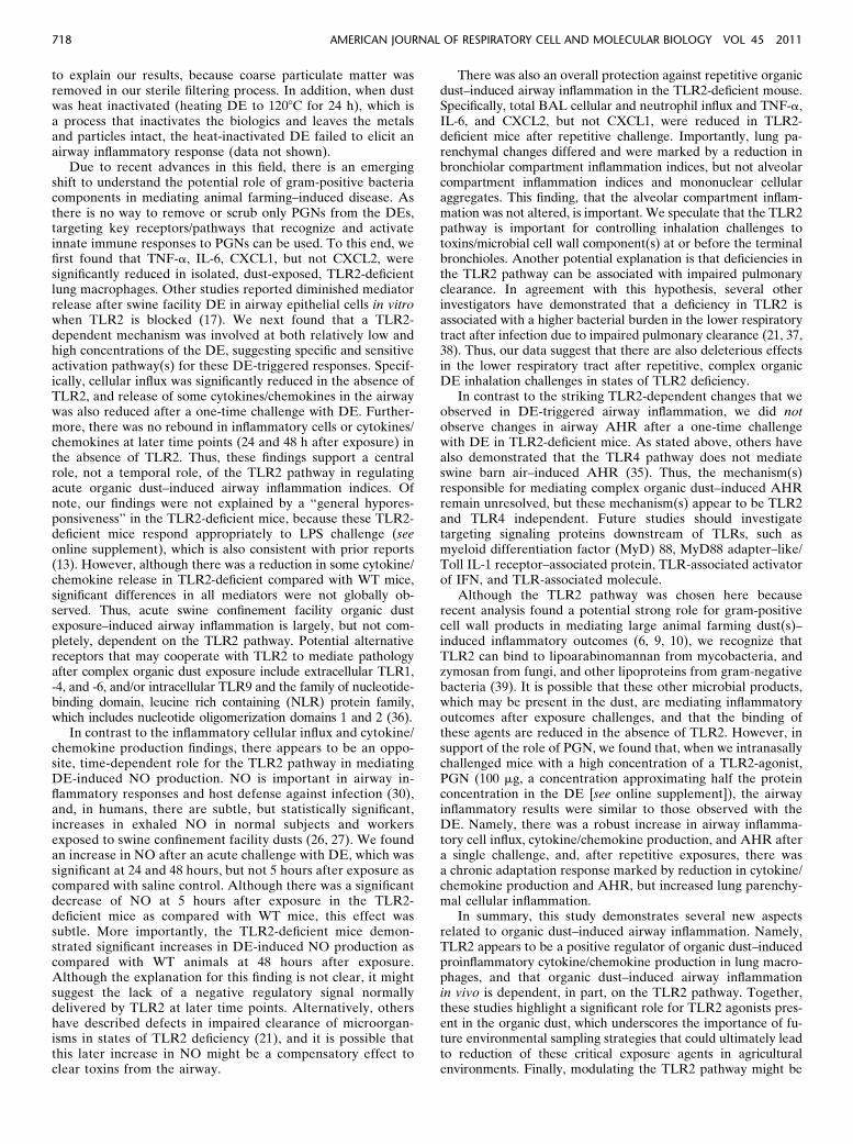
A TLR2 Agonist, PGN, Induces Airway Inflammatory
Responses Similar to DE
To determine if a TLR2-agonist would induce similar airwayinflammatory responses as that of DE, and to remain consistentwith our previous published in vitro studies (6, 7, 11), WT micewere intranasally challenged with PGN (100 mg) or saline once(single exposure) and once daily for 2 weeks (repetitiveexposure) and subsequently killed at 5 hours after final expo-sure, as described in the online supplement. As compared withsaline, PGN induced a significant increase in total leukocytecounts, predominately due to an expansion of neutrophils, aftersingle and repetitive exposure (Figure 7A). There were alsosignificant increases in TNF-a, IL-6, CXCL1, and CXCL2 afterPGN challenge after single and repetitive exposures as com-pared with saline (Figure 7B). AHR to PGN occurred aftersingle exposure (Figure 7C), with an adaptation response(resolution of AHR) after repetitive exposure (data not shown),as determined by invasive pulmonary function measurements.Finally, microscopic review of lung tissue revealed increased
Figure 6. Reduced lung inflammation in TLR2-deficient mice after intranasal inhalation of DE. Saline-treated WT mice (A) and saline-treated TLR2
KO mice (B) are compared, with mice exposed once ([C] WT and [D] TLR2 KO) and mice exposed repeatedly for 2 weeks ([E and G] WT; [F and H]TLR2 KO) to DE (12.5%). A representative 4- to 5-mm-thick section of one of four to six mice per treatment group is shown at 203 magnification. (E
and F ) Alveolar compartment inflammation; (G and H) Bronchiolar compartment inflammation after repetitive (2-wk) DE challenge in WT and TLR2
KO mice, respectively. (I ) Semiquantitative inflammatory score of lungs after intranasal inhalation of saline and DE (12.5%) after single exposure or
repetitive (2-wk) exposure of DE. Semiquantitative distribution of lung alveolar inflammation, bronchiolar inflammation, and mononuclear cellularaggregates in mice (n 5 4–6 mice per group). Error bars represent SE. #P , 0.05, significant differences between saline and DE treated; (*P , 0.05,
differences between TLR2 KO and WT.
716 AMERICAN JOURNAL OF RESPIRATORY CELL AND MOLECULAR BIOLOGY VOL 45 2011
bronchiolar and alveolar compartment inflammation and pres-ence of mononuclear cellular aggregates in WT mice after dailyPGN exposure for 2 weeks (Figure 7D). Collectively, thesefindings demonstrate that PGN, a TLR2 agonist, at highconcentration, elicits airway inflammatory responses similar tohigh-concentration DE.
DISCUSSION
Because recent studies of environmental dusts from animalfarming environments reveal a diverse population of gram-positive bacteria and high concentrations of PGN (6, 10), wefocused on TLR2 as a likely key receptor in regulating dust-induced airway inflammation, because TLR2 recognizes cellwall components of gram-positive bacteria (12, 13). We foundthat organic dust–induced airway inflammation was highlydependent on the TLR2 pathway in mice after single and re-petitive challenge(s) to DE. This dependence was demonstratedby a reduction in cellular influx, airway cytokine/chemokine,and bronchiolar inflammation in TLR2-deficient mice as com-pared with control mice. However, AHR and alveolar inflam-mation was not altered in the TLR2-deficient animals,suggesting different mechanisms for airway contractility andlung parenchymal inflammation, respectively. To the best of ourknowledge, this is the first report to demonstrate an in vivo
functional role for the TLR2 pathway in mediating airwayinflammatory responses to complex organic dust exposures.
Dusts from large animal confinement facilities, such asmodern swine barns, are recognized to be complex mixturesof microbial products from gram-negative and gram-positivebacteria, molds, particulate matter from grain particles, andfecal material. Although endotoxin is the major driver of corn(grain) dust–induced airway inflammation due to its predomi-nance in these environments, and mice genetically hyporespon-sive to endotoxin are protected from airway inflammatoryoutcomes after respective exposures (33, 34), endotoxin is likelynot the principal component of animal facility dusts. Severalstudies disagree about the association of endotoxin and diseaseoutcomes in exposed workers (2–4). Furthermore, others foundonly a significant decrease in lavage cellularity and neutrophilsafter single (not repetitive) exposure to swine barn air inendotoxin-hyporesponsive mice (C3H/HeJ), but no differencein AHR and cytokine levels after single and repetitive expo-sures (35). In our earlier studies, we found no difference inlavage cellularity or cytokine/chemokine production after a sin-gle intranasal swine facility DE (12.5%) challenge in endotoxin-nonresponsive mice (C3H/HeJ) (data not shown). Anotherpotential component in the DE is particulate matter as coarseparticulate matter (.2.5 mm) has been shown to be a potentstimulus (15). However, it is not likely to be a major contributor
Figure 7. Lung lavage cellularity, cytokine/chemokine release, and lung inflammation is increased in WT mice after single and repetitive intranasalchallenge(s) with peptidoglycan (PGN) as compared with saline. Lung lavage fluid mean concentration of total cells and cell differential (A) and
cytokine/chemokine release (B) 5 hours after single and repetitive (daily instillation for 2 wk) intranasal inhalation of PGN (100 mg) or saline in WT
mice (n 5 3–4 mice per group). (C ) Methacholine (MCh)-induced airway hyperresponsiveness (AHR) after single intranasal inhalation of PGN ascompared with saline in mice. Total lung resistance (RL) was directly measured using a mechanically ventilated mouse system. Data are expressed as
total RL with SE bars (n 5 6–7 mice per group). (D) A representative 4- to -5-mm-thick section after 2-week repetitive exposure to PGN at 103
magnification. *P , 0.05 and **P , 0.01 indicate statistical significance between saline- and PGN-treated mice.
Poole, Wyatt, Kielian, et al.: TLR2 Regulates Dust-Induced Airway Inflammation 717
to explain our results, because coarse particulate matter wasremoved in our sterile filtering process. In addition, when dustwas heat inactivated (heating DE to 1208C for 24 h), which isa process that inactivates the biologics and leaves the metalsand particles intact, the heat-inactivated DE failed to elicit anairway inflammatory response (data not shown).
Due to recent advances in this field, there is an emergingshift to understand the potential role of gram-positive bacteriacomponents in mediating animal farming–induced disease. Asthere is no way to remove or scrub only PGNs from the DEs,targeting key receptors/pathways that recognize and activateinnate immune responses to PGNs can be used. To this end, wefirst found that TNF-a, IL-6, CXCL1, but not CXCL2, weresignificantly reduced in isolated, dust-exposed, TLR2-deficientlung macrophages. Other studies reported diminished mediatorrelease after swine facility DE in airway epithelial cells in vitrowhen TLR2 is blocked (17). We next found that a TLR2-dependent mechanism was involved at both relatively low andhigh concentrations of the DE, suggesting specific and sensitiveactivation pathway(s) for these DE-triggered responses. Specif-ically, cellular influx was significantly reduced in the absence ofTLR2, and release of some cytokines/chemokines in the airwaywas also reduced after a one-time challenge with DE. Further-more, there was no rebound in inflammatory cells or cytokines/chemokines at later time points (24 and 48 h after exposure) inthe absence of TLR2. Thus, these findings support a centralrole, not a temporal role, of the TLR2 pathway in regulatingacute organic dust–induced airway inflammation indices. Ofnote, our findings were not explained by a ‘‘general hypores-ponsiveness’’ in the TLR2-deficient mice, because these TLR2-deficient mice respond appropriately to LPS challenge (seeonline supplement), which is also consistent with prior reports(13). However, although there was a reduction in some cytokine/chemokine release in TLR2-deficient compared with WT mice,significant differences in all mediators were not globally ob-served. Thus, acute swine confinement facility organic dustexposure–induced airway inflammation is largely, but not com-pletely, dependent on the TLR2 pathway. Potential alternativereceptors that may cooperate with TLR2 to mediate pathologyafter complex organic dust exposure include extracellular TLR1,-4, and -6, and/or intracellular TLR9 and the family of nucleotide-binding domain, leucine rich containing (NLR) protein family,which includes nucleotide oligomerization domains 1 and 2 (36).
In contrast to the inflammatory cellular influx and cytokine/chemokine production findings, there appears to be an oppo-site, time-dependent role for the TLR2 pathway in mediatingDE-induced NO production. NO is important in airway in-flammatory responses and host defense against infection (30),and, in humans, there are subtle, but statistically significant,increases in exhaled NO in normal subjects and workersexposed to swine confinement facility dusts (26, 27). We foundan increase in NO after an acute challenge with DE, which wassignificant at 24 and 48 hours, but not 5 hours after exposure ascompared with saline control. Although there was a significantdecrease of NO at 5 hours after exposure in the TLR2-deficient mice as compared with WT mice, this effect wassubtle. More importantly, the TLR2-deficient mice demon-strated significant increases in DE-induced NO production ascompared with WT animals at 48 hours after exposure.Although the explanation for this finding is not clear, it mightsuggest the lack of a negative regulatory signal normallydelivered by TLR2 at later time points. Alternatively, othershave described defects in impaired clearance of microorgan-isms in states of TLR2 deficiency (21), and it is possible thatthis later increase in NO might be a compensatory effect toclear toxins from the airway.
There was also an overall protection against repetitive organicdust–induced airway inflammation in the TLR2-deficient mouse.Specifically, total BAL cellular and neutrophil influx and TNF-a,IL-6, and CXCL2, but not CXCL1, were reduced in TLR2-deficient mice after repetitive challenge. Importantly, lung pa-renchymal changes differed and were marked by a reduction inbronchiolar compartment inflammation indices, but not alveolarcompartment inflammation indices and mononuclear cellularaggregates. This finding, that the alveolar compartment inflam-mation was not altered, is important. We speculate that the TLR2pathway is important for controlling inhalation challenges totoxins/microbial cell wall component(s) at or before the terminalbronchioles. Another potential explanation is that deficiencies inthe TLR2 pathway can be associated with impaired pulmonaryclearance. In agreement with this hypothesis, several otherinvestigators have demonstrated that a deficiency in TLR2 isassociated with a higher bacterial burden in the lower respiratorytract after infection due to impaired pulmonary clearance (21, 37,38). Thus, our data suggest that there are also deleterious effectsin the lower respiratory tract after repetitive, complex organicDE inhalation challenges in states of TLR2 deficiency.
In contrast to the striking TLR2-dependent changes that weobserved in DE-triggered airway inflammation, we did notobserve changes in airway AHR after a one-time challengewith DE in TLR2-deficient mice. As stated above, others havealso demonstrated that the TLR4 pathway does not mediateswine barn air–induced AHR (35). Thus, the mechanism(s)responsible for mediating complex organic dust–induced AHRremain unresolved, but these mechanism(s) appear to be TLR2and TLR4 independent. Future studies should investigatetargeting signaling proteins downstream of TLRs, such asmyeloid differentiation factor (MyD) 88, MyD88 adapter–like/Toll IL-1 receptor–associated protein, TLR-associated activatorof IFN, and TLR-associated molecule.
Although the TLR2 pathway was chosen here becauserecent analysis found a potential strong role for gram-positivecell wall products in mediating large animal farming dust(s)–induced inflammatory outcomes (6, 9, 10), we recognize thatTLR2 can bind to lipoarabinomannan from mycobacteria, andzymosan from fungi, and other lipoproteins from gram-negativebacteria (39). It is possible that these other microbial products,which may be present in the dust, are mediating inflammatoryoutcomes after exposure challenges, and that the binding ofthese agents are reduced in the absence of TLR2. However, insupport of the role of PGN, we found that, when we intranasallychallenged mice with a high concentration of a TLR2-agonist,PGN (100 mg, a concentration approximating half the proteinconcentration in the DE [see online supplement]), the airwayinflammatory results were similar to those observed with theDE. Namely, there was a robust increase in airway inflamma-tory cell influx, cytokine/chemokine production, and AHR aftera single challenge, and, after repetitive exposures, there wasa chronic adaptation response marked by reduction in cytokine/chemokine production and AHR, but increased lung parenchy-mal cellular inflammation.
In summary, this study demonstrates several new aspectsrelated to organic dust–induced airway inflammation. Namely,TLR2 appears to be a positive regulator of organic dust–inducedproinflammatory cytokine/chemokine production in lung macro-phages, and that organic dust–induced airway inflammationin vivo is dependent, in part, on the TLR2 pathway. Together,these studies highlight a significant role for TLR2 agonists pres-ent in the organic dust, which underscores the importance of fu-ture environmental sampling strategies that could ultimately leadto reduction of these critical exposure agents in agriculturalenvironments. Finally, modulating the TLR2 pathway might be
718 AMERICAN JOURNAL OF RESPIRATORY CELL AND MOLECULAR BIOLOGY VOL 45 2011
an important adjunctive therapy when investigating potentialprevention and therapeutic interventions in humans.
Author Disclosure: None of the authors has a financial relationship with acommercial entity that has an interest in the subject of this manuscript.
Acknowledgments: The authors thank Chris Bauer, Jacqueline Pavlik, and JaneDeVasure for assisting in the experiments. The authors also thank Dr. Shizuo Akirafor generously providing Toll-like receptor 2 knockout mice for experimental studies,Thomas Jerrells, Ph.D., for assistance with digital microscopy images prepared forthe manuscript, and Lisa Chudomelka for manuscript preparation assistance.
References
1. Eduard W, Pearce N, Douwes J. Chronic bronchitis, COPD, and lung
function in farmers: the role of biological agents. Chest 2009;136:716–725.2. Rask-Andersen A, Malmberg P, Lundholm M. Endotoxin levels in
farming: absence of symptoms despite high exposure levels. Br J IndMed 1989;46:412–416.
3. Cormier Y, Israel-Assayag E, Racine G, Duchaine C. Farming practices
and the respiratory health risks of swine confinement buildings. EurRespir J 2000;15:560–565.
4. Vogelzang PF, van der Gulden JW, Folgering H, Heederik D, Tielen MJ,
van Schayck CP. Longitudinal changes in bronchial responsiveness asso-ciated with swine confinement dust exposure. Chest 2000;117:1488–1495.
5. Muller-Suur C, Larsson K, Grunewald J. Organic dust–induced
interleukin-12 production activates T- and natural killer cells. EurRespir J 2002;20:686–690.
6. Poole JA, Alexis NE, Parks C, MacInnes AK, Gentry-Nielsen MJ, Fey
PD, Larsson L, Allen-Gipson D, Von Essen SG, Romberger DJ.Repetitive organic dust exposure in vitro impairs macrophage differ-entiation and function. J Allergy Clin Immunol 2008;122:375–382.
7. Poole JA, Wyatt TA, Von Essen SG, Hervert J, Parks C, Mathisen T,
Romberger DJ. Repeat organic dust exposure–induced monocyteinflammation is associated with protein kinase C activity. J AllergyClin Immunol 2007;120:366–373.
8. Romberger DJ, Bodlak V, Von Essen SG, Mathisen T, Wyatt TA. Hog
barn dust extract stimulates IL-8 and IL-6 release in human bronchialepithelial cells via PKC activation. J Appl Physiol 2002;93:289–296.
9. Nehme B, Letourneau V, Forster RJ, Veillette M, Duchaine C. Culture-
independent approach of the bacterial bioaerosol diversity in thestandard swine confinement buildings, and assessment of the seasonaleffect. Environ Microbiol 2008;10:665–675.
10. Poole JA, Dooley GP, Saito R, Burrell AM, Bailey KL, Romberger DJ,
Mehaffy J, Reynolds SJ. Muramic acid, endotoxin, 3-hydroxy fattyacids, and ergosterol content explain monocyte and epithelial cellinflammatory responses to agricultural dusts. J Toxicol EnvironHealth A 2010;73:684–700.
11. Poole JA, Thiele GM, Alexis NE, Burrell AM, Parks C, Romberger DJ.
Organic dust exposure alters monocyte-derived dendritic cell differ-entiation and maturation. Am J Physiol Lung Cell Mol Physiol 2009;297:L767–L776.
12. Akira S, Uematsu S, Takeuchi O. Pathogen recognition and innate
immunity. Cell 2006;124:783–801.13. Takeuchi O, Hoshino K, Kawai T, Sanjo H, Takada H, Ogawa T,
Takeda K, Akira S. Differential roles of TLR2 and TLR4 inrecognition of gram-negative and gram-positive bacterial cell wallcomponents. Immunity 1999;11:443–451.
14. Poole JA, Wyatt TA, Oldenburg PJ, Elliott MK, West WW, Sisson JH,
Von Essen SG, Romberger DJ. Intranasal organic dust exposure-induced airway adaptation response marked by persistent lung in-flammation and pathology in mice. Am J Physiol Lung Cell MolPhysiol 2009;296:L1085–L1095.
15. Alexis NE, Lay JC, Zeman K, Bennett WE, Peden DB, Soukup JM,
Devlin RB, Becker S. Biological material on inhaled coarse fractionparticulate matter activates airway phagocytes in vivo in healthyvolunteers. J Allergy Clin Immunol 2006;117:1396–1403.
16. Bailey KL, Poole JA, Mathisen TL, Wyatt TA, Von Essen SG,
Romberger DJ. Toll-like receptor 2 is upregulated by hog confine-ment dust in an IL-6–dependent manner in the airway epithelium. AmJ Physiol Lung Cell Mol Physiol 2008;294:L1049–L1054.
17. von Scheele I, Larsson K, Palmberg L. Budesonide enhances Toll-like
receptor 2 expression in activated bronchial epithelial cells. InhalToxicol 2010;22:493–499.
18. Vargas-Inchaustegui DA, Tai W, Xin L, Hogg AE, Corry DB, Soong L.Distinct roles for MyD88 and Toll-like receptor 2 during Leishmaniabraziliensis infection in mice. Infect Immun 2009;77:2948–2956.
19. Rodriguez N, Wantia N, Fend F, Durr S, Wagner H, Miethke T.Differential involvement of TLR2 and TLR4 in host survival duringpulmonary infection with Chlamydia pneumoniae. Eur J Immunol2006;36:1145–1155.
20. Fuse ET, Tateda K, Kikuchi Y, Matsumoto T, Gondaira F, Azuma A,Kudoh S, Standiford TJ, Yamaguchi K. Role of Toll-like receptor 2 inrecognition of Legionella pneumophila in a murine pneumonia model.J Med Microbiol 2007;56:305–312.
21. Hajishengallis G, Wang M, Bagby GJ, Nelson S. Importance of TLR2 inearly innate immune response to acute pulmonary infection withPorphyromonas gingivalis in mice. J Immunol 2008;181:4141–4149.
22. Dessing MC, van der Sluijs KF, Florquin S, Akira S, van der Poll T. Toll-like receptor 2 does not contribute to host response during post-influenza pneumococcal pneumonia. Am J Respir Cell Mol Biol 2007;36:609–614.
23. Knapp S, von Aulock S, Leendertse M, Haslinger I, Draing C,Golenbock DT, van der Poll T. Lipoteichoic acid–induced lung in-flammation depends on TLR2 and the concerted action of TLR4and the platelet-activating factor receptor. J Immunol 2008;180:3478–3484.
24. Wyatt TA, Sisson JH, Von Essen SG, Poole JA, Romberger DJ.Exposure to hog barn dust alters airway epithelial ciliary beating.Eur Respir J 2008;31:1249–1255.
25. Oldenburg PJ, Wyatt TA, Factor PH, Sisson JH. Alcohol feeding blocksmethacholine-induced airway responsiveness in mice. Am J PhysiolLung Cell Mol Physiol 2009;296:L109–L114.
26. Wang Z, Larsson K, Palmberg L, Malmberg P, Larsson P, Larsson L.Inhalation of swine dust induces cytokine release in the upper andlower airways. Eur Respir J 1997;10:381–387.
27. Von Essen SG, Scheppers LA, Robbins RA, Donham KJ. Respiratorytract inflammation in swine confinement workers studied using in-duced sputum and exhaled nitric oxide. J Toxicol Clin Toxicol 1998;36:557–655.
28. Deetz DC, Jagielo PJ, Quinn TJ, Thorne PS, Bleuer SA, Schwartz DA.The kinetics of grain dust–induced inflammation of the lower re-spiratory tract. Am J Respir Crit Care Med 1997;155:254–259.
29. Sundblad BM, von Scheele I, Palmberg L, Olsson M, Larsson K.Repeated exposure to organic material alters inflammatory andphysiological airway responses. Eur Respir J 2009;34:80–88.
30. Sittipunt C, Steinberg KP, Ruzinski JT, Myles C, Zhu S, Goodman RB,Hudson LD, Matalon S, Martin TR. Nitric oxide and nitrotyrosine inthe lungs of patients with acute respiratory distress syndrome. Am JRespir Crit Care Med 2001;163:503–510.
31. Charavaryamath C, Janardhan KS, Townsend HG, Willson P, Singh B.
Multiple exposures to swine barn air induce lung inflammation andairway hyper-responsiveness. Respir Res 2005;6:50–63.
32. Palmberg L, Larssson BM, Malmberg P, Larsson K. Airway responses ofhealthy farmers and nonfarmers to exposure in a swine confinementbuilding. Scand J Work Environ Health 2002;28:256–263.
33. Jagielo PJ, Thorne PS, Watt JL, Frees KL, Quinn TJ, Schwartz DA.Grain dust and endotoxin inhalation challenges produce similarinflammatory responses in normal subjects. Chest 1996;110:263–270.
34. George CL, Jin H, Wohlford-Lenane CL, O’Neill ME, Phipps JC,O’Shaughnessy P, Kline JN, Thorne PS, Schwartz DA. Endotoxinresponsiveness and subchronic grain dust–induced airway disease. AmJ Physiol Lung Cell Mol Physiol 2001;280:L203–L213.
35. Charavaryamath C, Juneau V, Suri SS, Janardhan KS, Townsend H,
Singh B. Role of Toll-like receptor 4 in lung inflammation followingexposure to swine barn air. Exp Lung Res 2008;3:19–35.
36. Strober W, Murray PJ, Kitani A, Watanabe T. Signalling pathways andmolecular interactions of NOD1 and NOD2. Nat Rev Immunol 2006;6:9–20.
37. Love W, Dobbs N, Tabor L, Simecka JW. Toll-like receptor 2 (TLR2)
plays a major role in innate resistance in the lung against murinemycoplasma. PLoS ONE 2010;5:e10739.
38. Abplanalp AL, Morris IR, Parida BK, Teale JM, Berton MT. TLR-dependent control of Francisella tularensis infection and host in-flammatory responses. PLoS ONE 2009;4:e7920.
39. Kawai T, Akira S. The role of pattern-recognition receptors in innateimmunity: update on Toll-like receptors. Nat Immunol 2010;11:373–384.
Poole, Wyatt, Kielian, et al.: TLR2 Regulates Dust-Induced Airway Inflammation 719