House dust mite induces direct airway inflammation in vivo: implications for future disease therapy
11
House dust mite induces direct airway inflammation in vivo: implications for future disease therapy? J. De Alba* ,# , K. Raemdonck* ,# , A. Dekkak* ,# , M. Collins* ,# , S. Wong* ,# , A.T. Nials " , R.G. Knowles " , M.G. Belvisi* ,# and M.A. Birrell* ,# ABSTRACT: House dust mite (HDM) is the major source of allergen in house dust and is strongly associated with the development of asthma. HDM can evoke a direct, nonallergic inflammatory reaction in vitro. We aimed to determine whether this apparent nonallergic, inflammatory response can be observed in a more complex in vivo setting. Vehicle, Alum TM or HDM (Dermatophagoides pteronyssinus 5 mg, i.p. with Alum) sensitised Brown-Norway rats were challenged intratracheally with vehicle (saline), HDM (Der p 10 mg) or heat-inactivated HDM on day 21. Lung function changes and the associated inflammatory response were evaluated. Tissue and bronchoalveolar lavage from Alum TM sensitised Der p challenged animals exhibited strong eosinophilia and neutrophilia associated with an early release of pro-inflammatory cytokines (interleukin-13 and 1b, eotaxin and thymus and activation-regulated chemokine). This response was not attenuated by removal of HDM-associated protease activity. Interestingly, the vehicle sensitised group (no Alum TM ) lacked this inflammatory response. HDM allergen evokes nonallergic airways inflammation with an inflammatory profile similar to that of the asthmatic airway. This response, independent of the protease activity of the HDM extract, appeared to be linked to prior administration of the adjuvant Alum TM and the subsequent increase in total immunoglobulin E. This finding could have important implications in the development of future asthma therapies. KEYWORDS: Allergy, asthma, cytokines, house dust mite, inflammation, rodent T he exposure to house dust mite (HDM) allergens is strongly associated with the development of allergic diseases, such as asthma, rhinitis or atopic dermatitis [1, 2]. Mites from the genus Dermatophagoides, such as D. pteronyssinus (Der p) and D. farinae (Der f), are thought to be the most important source of indoor allergens associated with human asthma [1, 3]. Over 30 proteins produced by Dermatophagoides mites induce immunoglobulin (Ig)E in allergic patients, but there are dominant antigens, espe- cially the group 1 and 2 allergens, which can account for the bulk of allergenicity of HDM extracts [1]. Among the group 1 allergens is Der p 1 from D. pteronyssinus. Der p 1 belongs to the papain-like cysteine protease family and exhibits cysteine protease activity [4]. This proteolytic activity has been suggested to be involved in the pathogenesis of allergies by: increasing the perme- ability of epithelial cells and allowing the passage of their own and other allergens across the epithelium [5–7]; cleaving and/or interacting with cell surface molecules and intrinsic protease inhibitors [8–11]; and modulating the function of a range of cells, such as basophils, mast cells, alveolar macrophages and airway epithelial cells [12–15]. The effect on the airway epithelium is especially interesting, as it is likely to be the first cell type to interact with the allergen. Several authors have reported that Der p 1 and other Der p antigens are able to directly induce the release of pro-inflammatory cytokines and che- mokines from primary human bronchial epithelial cells and airway epithelial cell lines [14, 16–20] by protease-dependent and independent mechan- isms [14, 16, 19, 21]. To our knowledge, however, this pro-inflamma- tory effect of HDM has not been shown in a more complex in vivo system. Therefore, our objective was to investigate whether a commercial HDM extract from D. pteronyssinus was able to elicit a direct nonallergic, pro-inflammatory effect in the Brown-Norway (BN) rat. The BN rat was chosen AFFILIATIONS *Respiratory Pharmacology, Pharmacology and Toxicology, Imperial College London, Faculty of Medicine, National Heart and Lung Institute, and # Centre for Integrative Mammalian Physiology and Pharmacology, Imperial College, London, and " Respiratory Centre of Excellence for Drug Discovery, GlaxoSmithKline, Research and Development, Medicines Research Centre, Stevenage, UK. CORRESPONDENCE M. Belvisi Respiratory Pharmacology, Pharmacology and Toxicology, Faculty of Medicine, National Heart and Lung Institute, Imperial College Sir Alexander Fleming Building Exhibition Road London SW7 2AZ UK E-mail: [email protected] Received: Feb 13 2008 Accepted after revision: Sept 29 2009 First published online: Oct 19 2009 European Respiratory Journal Print ISSN 0903-1936 Online ISSN 1399-3003 EUROPEAN RESPIRATORY JOURNAL VOLUME 35 NUMBER 6 1377 Eur Respir J 2010; 35: 1377–1387 DOI: 10.1183/09031936.00022908 CopyrightßERS 2010 c
-
Upload
independent -
Category
Documents
-
view
1 -
download
0
Transcript of House dust mite induces direct airway inflammation in vivo: implications for future disease therapy
House dust mite induces direct airway
inflammation in vivo: implications for future
disease therapy?J. De Alba*,#, K. Raemdonck*,#, A. Dekkak*,#, M. Collins*,#, S. Wong*,#, A.T. Nials",R.G. Knowles", M.G. Belvisi*,# and M.A. Birrell*,#
ABSTRACT: House dust mite (HDM) is the major source of allergen in house dust and is strongly
associated with the development of asthma. HDM can evoke a direct, nonallergic inflammatory
reaction in vitro. We aimed to determine whether this apparent nonallergic, inflammatory
response can be observed in a more complex in vivo setting.
Vehicle, AlumTM or HDM (Dermatophagoides pteronyssinus 5 mg, i.p. with Alum) sensitised
Brown-Norway rats were challenged intratracheally with vehicle (saline), HDM (Der p 10 mg) or
heat-inactivated HDM on day 21. Lung function changes and the associated inflammatory
response were evaluated.
Tissue and bronchoalveolar lavage from AlumTM sensitised Der p challenged animals exhibited
strong eosinophilia and neutrophilia associated with an early release of pro-inflammatory
cytokines (interleukin-13 and 1b, eotaxin and thymus and activation-regulated chemokine). This
response was not attenuated by removal of HDM-associated protease activity. Interestingly, the
vehicle sensitised group (no AlumTM) lacked this inflammatory response.
HDM allergen evokes nonallergic airways inflammation with an inflammatory profile similar to
that of the asthmatic airway. This response, independent of the protease activity of the HDM
extract, appeared to be linked to prior administration of the adjuvant AlumTM and the subsequent
increase in total immunoglobulin E. This finding could have important implications in the
development of future asthma therapies.
KEYWORDS: Allergy, asthma, cytokines, house dust mite, inflammation, rodent
The exposure to house dust mite (HDM)allergens is strongly associated with thedevelopment of allergic diseases, such as
asthma, rhinitis or atopic dermatitis [1, 2]. Mitesfrom the genus Dermatophagoides, such as D.pteronyssinus (Der p) and D. farinae (Der f), arethought to be the most important source of indoorallergens associated with human asthma [1, 3].Over 30 proteins produced by Dermatophagoidesmites induce immunoglobulin (Ig)E in allergicpatients, but there are dominant antigens, espe-cially the group 1 and 2 allergens, which canaccount for the bulk of allergenicity of HDMextracts [1]. Among the group 1 allergens is Der p 1from D. pteronyssinus. Der p 1 belongs to thepapain-like cysteine protease family and exhibitscysteine protease activity [4]. This proteolyticactivity has been suggested to be involved in thepathogenesis of allergies by: increasing the perme-ability of epithelial cells and allowing the passageof their own and other allergens across theepithelium [5–7]; cleaving and/or interacting with
cell surface molecules and intrinsic proteaseinhibitors [8–11]; and modulating the functionof a range of cells, such as basophils, mastcells, alveolar macrophages and airway epithelialcells [12–15]. The effect on the airway epitheliumis especially interesting, as it is likely to be thefirst cell type to interact with the allergen.Several authors have reported that Der p 1 andother Der p antigens are able to directly induce therelease of pro-inflammatory cytokines and che-mokines from primary human bronchial epithelialcells and airway epithelial cell lines [14, 16–20] byprotease-dependent and independent mechan-isms [14, 16, 19, 21].
To our knowledge, however, this pro-inflamma-tory effect of HDM has not been shown in a morecomplex in vivo system. Therefore, our objectivewas to investigate whether a commercial HDMextract from D. pteronyssinus was able to elicit adirect nonallergic, pro-inflammatory effect in theBrown-Norway (BN) rat. The BN rat was chosen
AFFILIATIONS
*Respiratory Pharmacology,
Pharmacology and Toxicology,
Imperial College London, Faculty of
Medicine, National Heart and Lung
Institute, and#Centre for Integrative Mammalian
Physiology and Pharmacology,
Imperial College, London, and"Respiratory Centre of Excellence for
Drug Discovery, GlaxoSmithKline,
Research and Development,
Medicines Research Centre,
Stevenage, UK.
CORRESPONDENCE
M. Belvisi
Respiratory Pharmacology,
Pharmacology and Toxicology,
Faculty of Medicine, National Heart
and Lung Institute, Imperial College
Sir Alexander Fleming Building
Exhibition Road
London
SW7 2AZ
UK
E-mail: [email protected]
Received:
Feb 13 2008
Accepted after revision:
Sept 29 2009
First published online:
Oct 19 2009
European Respiratory Journal
Print ISSN 0903-1936
Online ISSN 1399-3003
EUROPEAN RESPIRATORY JOURNAL VOLUME 35 NUMBER 6 1377
Eur Respir J 2010; 35: 1377–1387
DOI: 10.1183/09031936.00022908
Copyright�ERS 2010
c
because we, and others, have previously demonstrated that anallergic reaction to other antigens, i.e. ovalbumin (OVA), isachievable in this strain [22].
METHODSAnimalsMale BN rats (200–225 g) were obtained from Charles RiverLaboratories Inc. (Lyon, France) and housed for 1 week beforeinitiating experiments.
HDM extractPurified HDM extract from Der p (GREER laboratories, Lenoir,NC, USA) with a known content of Der p 1 (39.77 mg?mg-1 dryweight) was used in these experiments. The doses of HDMused in this manuscript refer to the amount of Der p 1.
Measurement of protease activity in the extractProtease activity in the extract was assessed by a continuousrate (kinetic) assay using the fluorogenic peptide substrate Boc-Gln-Ala-Arg-AMC (AMC, 7-amino-4-methylcoumarin; Boc, N-tert-butoxycarbonyl) as previously described [23]. Briefly, aknown amount of HDM extract (final concentration Der p 110 nM) was added to a substrate standard curve (0–1 mM), in50 mM sodium phosphate buffer (pH 7.0) containing 1 mMEDTA and 1 mM dithiothreitol at 25uC. Fluorescence asso-ciated with the hydrolysis of the substrate was monitoredusing a SynergyTM HT Multi-Mode Microplate Reader (BiotekUK, Potton, UK) with lex5380nm and lem5460nm. Datawere fitted to a Michaelis–Menten equation, y5V 6 x/(Km+x), and the kinetic constants Km (Michaelis–Mentenconstant) and maximal rate of conversion (V9max) werecalculated. HDM extract showed a positive protease activitywith a V9max of 76,636 mmol?min-1 and a Km of 304.9 mM. As acontrol, a similar amount of HDM extract was inactivated byheating to 65uC for 30 min, as previously described [19].
Dosing route selectionThe majority of our previous work into allergic airwayinflammation has involved aerosolised challenging with theantigen. Unfortunately, we were unable to utilise this methodbecause of the cost implications. Therefore, our initial aim wasto determine the optimum topical route of administration forthe allergen by comparing the intranasal and intratrachealroutes. This was carried out by using lipopolysaccharide (LPS)-induced tumour necrosis factor (TNF)-a release in the lung as abiomarker of appropriate delivery. Male BN rats (200–225 g)were anaesthetised (4% halothane in oxygen for 3 min) andvehicle (saline) or LPS (100 mg?rat-1, i.e. the same amount ineach case) were administered in various volumes eitherintranasally (5, 10, 20, 50 or 100 mL in each nostril), using apipette in a drop-wise fashion, or intratracheally (250 mL).After 90 mins blood, lavage and lung tissue samples weretaken. TNF-a was measured by ELISA (R&D systems Inc.,Minneapolis, MN, USA) as described in the study by BIRRELL etal. [24].
HDM sensitising dose selectionTimes for sensitisation challenge were extrapolated fromour OVA allergic inflammation model in the BN rat [24].An allergen dose–response study was performed to evaluatethe optimum sensitising dose by measuring the levels of
HDM-specific IgE in plasma 14 days after i.p. dosing. Male BNrats (200–225 g) were sensitised on day 0 with saline, AlumTM
(Fisher Scientific UK Ltd, Loughborough, UK) or HDM extract(0.5–500 mg?rat-1, i.p.; GREER laboratories) administered withAlumTM. At day 14 blood samples were collected and theplasma separated.
ELISA plates (Nunc MAXsorb; Fisher Scientific) were coatedwith 50 mL of 5mg?mL-1 HDM protein in PBS and left overnightat room temperature. Plates were then washed with washbuffer (Invitrogen, Paisley, UK) and blocked for at least 30 minwith 4% bovine serum albumin (BSA; Sigma Aldrich, Poole,UK) in PBS. Plasma samples were then added to duplicatewells diluted 1:10 in PBS containing 4% BSA and 0.05% Tween20. They were then incubated overnight at room temperatureprior to adding biotinylated anti-IgE (1:10; Serotec, Kidlington,UK) at 2mg?mL-1 (in PBS containing 4% BSA and 0.05% Tween20) and further incubated for 1 h at room temperature. Afterwashing, plates were incubated for 30 min at room tempera-ture with horseradish peroxidase-streptavidin (1:4000;Amersham Biosciences, Uppsala, Sweden). Following a lastwash, tetramethylbenzidine substrate (Sigma Aldrich) wasadded to the plates and they were incubated at roomtemperature until colour developed. The reaction was stoppedwith 0.25 M sulphuric acid and plates were read at 450 nM(Biotek Powerwave XS plate reader; Biotek UK). Total IgE wasmeasured using a commercial rat IgE quantitative ELISA kit(Bethyl Laboratories, Montgomery, TX, USA) following theprotocol of the manufacturer and expressed as mg?mg-1 of lungtissue total protein.
HDM challenging dose selectionFollowing the determination of the optimal sensitising dose,we aimed to determine an appropriate HDM dose forchallenging previously sensitised animals. Male BN rats(200–225 g) were sensitised at day 0 and 14 with HDM(5 mg?rat-1, i.p.) administered with AlumTM. Rats were thenchallenged with intratracheal vehicle (saline) or HDM (0.1–100 mg?rat-1 in 250 mL saline) on day 21.
48 h after HDM challenge, rats were lavaged (263 mL RPMI).Total cell counts recovered from the airway lumen werequantified using a Sysmex cell counter (Sysmex UK Ltd,Milton Keynes, UK). Differential cell counts (eosinophils,neutrophils and lymphomononuclear cells expressed asabsolute cell counts) of cells recovered from the airway lumenwere made by light microscopy after Wright-Giemsa stainingusing standard morphological criteria.
Time course after HDM sensitisation/challenge on cellrecruitment in the BN modelTo determine the effect of the acute exposure to HDM onasthma-like end-points, male BN rats (150–180 g) weresensitised on day 0 and 14 with vehicle (saline) or HDMextract (5 mg?rat-1, i.p.) administered with AlumTM. Rats werechallenged with intratracheal vehicle (saline) or HDM extract(10 mg intratracheal) on day 21.
Animals were lavaged 6, 24 or 48 h later and differential whitecell counts were performed as described above. Similarly whitecell numbers and type in the lung tissue were determinedas described previously [24]. The remaining lung was either
CELL AND ANIMAL STUDIES J. DE ALBA ET AL.
1378 VOLUME 35 NUMBER 6 EUROPEAN RESPIRATORY JOURNAL
snap-frozen or fixed in formalin for histological assessment.Inflammatory mRNA/protein expression was assessed byreal–time PCR using fully validated primers and probes asdetailed in MCCLUSKIE et al. [25] or by specific ELISA (R&DSystems Inc.), respectively. Cytokine location in the lung wasvisualised using immunohistochemistry.
Briefly, insufflated formalin fixed lung tissues were processedand wax embedded. 4-mm sections of paraffin embedded tissuewere dewaxed and rehydrated. Intrinsic peroxidase activitywas blocked by incubating the sections with 3% hydrogenperoxide (interleukin (IL)-13; 25 min) or 2% hydrogen per-oxide (eotaxin and IL-1b; 20 min). IL-13 and IL-1b slides werethen subjected to antigen retrieval by pressure cooking insodium citrate buffer (10 mM; pH 6.0). Nonspecific bindingwas reduced with 10% normal rabbit serum (VectorLaboratories, Burlingame, CA, USA) in PBS containing 0.1%BSA and 0.025% Tween 80 for 20 min for IL-13 immunohisto-chemistry and 1% normal rabbit serum (Dako UK Ltd, Ely,UK) in PBS containing 0.1% BSA and 0.025% Tween 80 for20 min for eotaxin and IL-1b immunohistochemistry. Sectionswere then rinsed and the primary antibody (goat polyclonalanti-mouse IL-13 antibody, 1:250 dilution (Santa CruzBiotechnology, Santa Cruz, CA, USA); goat polyclonal anti-mouse eotaxin/CCL11, 1:50 dilution (R&D Systems Inc); orgoat polyclonal anti-rat IL-1b, 1:200 dilution (Santa Cruz
Biotechnology)) diluted in PBS containing 0.1% BSA and0.025% Tween 80 was applied for 1 h at room temperature.Sections were then incubated with a secondary biotinylatedrabbit anti-goat IgG (IL-13: 1:150 dilution for 30 min; eotaxin:1:100 for 60 min; IL-1b: 1:120 for 60 min; Vector Laboratories)followed by detection with a Vectastain Elite ABC kit for
00.1 1 10 100
50
Cel
ls 1
03 ·m
L-1
100
150
200* *
00.50 5 50 500
10
5Pla
sma
tota
l IgE
μg·
mL-1
20
15
30
25
35a)
c) d)
b)
*
*
*
*
*
00.1 1 10 100
25
Cel
ls 1
03 ·m
L-1
50
75
100 *
00.5
+Alum+Alum
5 50 500
0.1
Pla
sma
IgE
mO
D
0.2
0.3
**
*
0
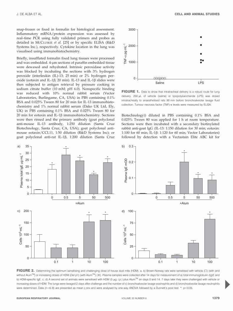
FIGURE 2. Determining the optimum sensitising and challenging dose of house dust mite (HDM). a, b) Brown-Norway rats were sensitised with vehicle (h) (with and
without AlumTM) or increasing doses of HDM (Der p1) (with AlumTM) (&). Plasma samples were collected after 14 days for measurement of a) total immunoglobulin (Ig)E and
b) HDM-specific IgE. c, d) A second set of animals were sensitised with HDM (5 mg, i.p.) plus AlumTM on days 0 and 14. 7 days later they were challenged with vehicle or
increasing doses of HDM. The lungs were lavaged 2 days after challenge and the number of c) bronchoalveolar lavage eosinophils and d) bronchoalveolar lavage neutrophils
were determined. Data (n56–8) are presented as mean¡SEM and were analysed by one-way ANOVA followed by a Dunnett’s post test. *: p,0.05.
●
●
●
●●●
3000
2000
1000
0LPSSaline
TNF-
α pg
·mL-1
FIGURE 1. Data to show that intratracheal delivery is a robust route for lung
delivery. 250 mL of vehicle (saline) or lipopolysaccharide (LPS) was dosed
intratracheally to anaesthetised rats 90 min before bronchoalveolar lavage fluid
collection. Tumour necrosis factor (TNF)-a levels were measured by ELISA.
J. DE ALBA ET AL. CELL AND ANIMAL STUDIES
cEUROPEAN RESPIRATORY JOURNAL VOLUME 35 NUMBER 6 1379
mouse IgG (PK6102; Vector Laboratories). The staining wasrevealed using the diaminobenzidine (Sigma Aldrich) pro-cedure counterstaining with Mayer’s haematoxylin. Tissuesincubated with a blocking peptide instead of the primarypeptide were used as negative controls.
Images were captured using an Olympus BX-51 microscope(Olympus UK Ltd, Southend-on-sea, UK) fitted for bothtransmitted light and fluorescence imaging and a Qicamdigital camera (Qimaging, Surrey, BC, Canada).
Noninvasive determination of the late bronchoconstrictorresponse to HDM challengeAfter HDM challenge, conscious and unrestrained vehicle orHDM sensitised rats were placed in a whole-body plethysmo-graph (Buxco Research Systems, UK) and pressure changeswere continuously registered by a Buxco XA-analyser (BuxcoResearch Systems). Enhanced pause (Penh) was recorded forf5 h and mean values were taken in 10-min intervals. Abaseline value was obtained by recording the Penh for 10 min24 h before the challenge and baseline corrected values wereused for analysis.
Invasive determination of changes in airway resistance inresponse to HDM challengeBecause of the controversy associated with using whole-bodyplethysmograph (Penh) as a lung function measurement [26],
we confirmed our results using conventional invasive lungmechanics. Vehicle or HDM sensitised male BN rats wereanaesthetised (ketamine and xylazine 144 and 10 mg?kg-1,respectively, i.p.) and instrumented 48 h after challenge.Briefly, the trachea was cannulated and the animal artificiallyrespired with a tidal volume of 2 mL?kg-1 at a frequency of90 breaths?min-1. A water-filled oesophageal cannula wasinserted such that an estimate of transpulmonary pressurecould be recorded. Pulmonary resistance (cmH2O?mL-1?s-1)and dynamic compliance (mL?cmH2O-1) were continuouslycomputed on a Buxco XA-analyser (Buxco Electronics).Administration of aerosolised spasmogen was via a nebuliser(Buxco Electronics) connected in line with the ventilator.Airway responsiveness was assessed by nebulising increasingconcentrations of methacholine (100 mL of vehicle (saline) 2, 4,8, 16 and 32 mg?mL-1). Mean changes in airway resistancewere monitored for 2 min after each administration ofmethacholine.
Recall challenge of splenocytes from sensitised animalsMale BN rats (150–180 g) were ‘‘sensitised’’ on days 0 and 14with saline (i.p.) or AlumTM (20 mg?rat-1 aluminium hydroxideand 20 mg?rat-1 magnesium hydroxide, i.p.). In parallel, as acontrol for the recall challenge, animals were sensitised withOVA as previously described [22]. Spleens were removed onday 21 after an overdose of pentobarbitone (200 mg?kg-1, i.p.),
5000
4000
3000
2000
1000
0
6000
Cel
l num
ber 1
03 .m
g of
tiss
ue-1 *
*
b)
3000
2500
2000
1500
1000
500
0
3500
Alum-only sensitised Der p sensitised
Cel
l num
ber 1
03 .m
g of
tiss
ue-1
*
*100
908070605040302010
0
110
Cel
l num
ber 1
03 .m
L-1
Alum-only sensitised Der p sensitised
*
*
c) d)
Cel
l num
ber 1
03 .m
L-1
150
125
100
75
50
25
0
175 *
*
a)
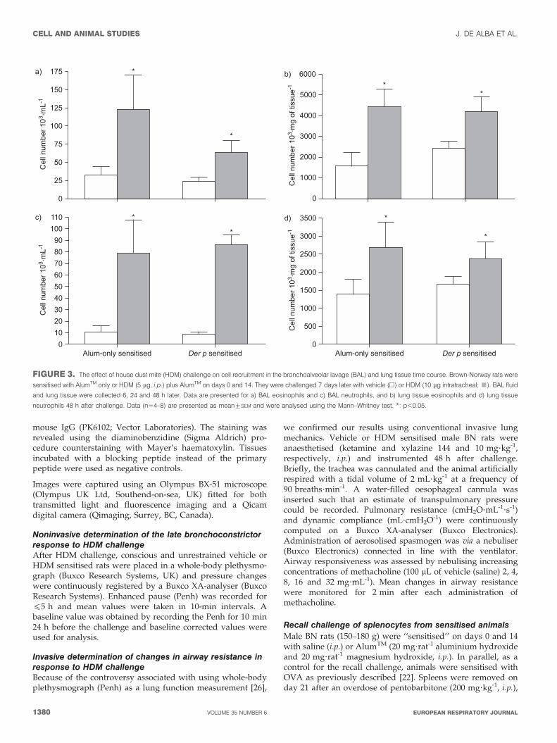
FIGURE 3. The effect of house dust mite (HDM) challenge on cell recruitment in the bronchoalveolar lavage (BAL) and lung tissue time course. Brown-Norway rats were
sensitised with AlumTM only or HDM (5 mg, i.p.) plus AlumTM on days 0 and 14. They were challenged 7 days later with vehicle (h) or HDM (10 mg intratracheal; &). BAL fluid
and lung tissue were collected 6, 24 and 48 h later. Data are presented for a) BAL eosinophils and c) BAL neutrophils, and b) lung tissue eosinophils and d) lung tissue
neutrophils 48 h after challenge. Data (n54–8) are presented as mean¡SEM and were analysed using the Mann–Whitney test. *: p,0.05.
CELL AND ANIMAL STUDIES J. DE ALBA ET AL.
1380 VOLUME 35 NUMBER 6 EUROPEAN RESPIRATORY JOURNAL
and transferred to cold PBS. They were then flushed with PBSusing a needle and syringe (without calcium and magnesium;Sigma Aldrich), in order to obtain a splenocyte suspension.The pooled cell suspension was passed through a 70-mm cellsieve and centrifuged at 2506g for 5 min at 4uC in a chill spin(Mistral 3000i; MSE, London, UK). The supernatant wasdiscarded and the cell pellets were resuspended in PBS andthen centrifuged. Splenocytes were then resuspended in RPMI1640, supplemented with 10% fetal calf serum (FCS) and 1%antibiotic and antimycotic solution (penicillin/streptomycin;Sigma Aldrich), counted and cell viability was determinedusing Trypan Blue. The splenocytes were diluted in RPMI1640, supplemented with 10% FCS and 1% antibiotic andantimycotic solution, and 225 mL of cells (86105) were culturedin 24-well plates. To each well, 250 mL of RPMI 1640, supple-mented with 10% FCS and 1% antibiotic and antimycoticsolution, with 0.1% (volume/volume) dimethyl sulfoxide, wasadded. 25 mL of varying concentrations of HDM (der p 0.01–50 mg?mL-1), OVA (1 mg?mL-1) or saline were added to thecorresponding wells. 200 mL of cells were added to chamberslides. The plates and chamber slides were incubated for72 h at 37uC in a humidified atmosphere (95% air, 5% (v/v)CO2). Supernatants were collected and cytokine releasemeasured by specific ELISA (R&D Systems Inc.).
Data analysisAll the values in the figures and text are presented asmean¡SEM of n observations. An unpaired t-test was used
when comparing two groups and a one-way ANOVA whencomparing multiple groups with the appropriate post test.When data were found not to be normally distributed theMann–Whitney test was used when comparing two groupsand a Kruskal–Wallis test when comparing multiple groupswith the appropriate post test. All treatments were comparedto relevant vehicle control groups, differences were deemedsignificant when p,0.05.
RESULTSDosing route selectionWe found that in the rat, using LPS-induced TNF-a as a markerof successful lung delivery, intranasal dosing gave variableresults (data not shown). Intratracheal delivery of LPS,however, resulted in a larger and more robust signal in allanimals (fig. 1). Similar data were obtained when we analysedthe lung tissue (not shown) and interestingly we could notdetect any increase in plasma TNF-a, even though if givenintraperitoneally this dose would cause a massive increase incirculating TNF-a (data not shown).
Due to the variability associated with intranasal delivery weopted for the intratracheal route to deliver the HDM.
HDM sensitising dose selectionPlasma levels of total IgE were significantly increased 14 daysafter administration of AlumTM compared with vehicle only(saline) but were not further increased by co-administrationwith HDM extract (fig. 2a). HDM extract co-administration
●
1110
9876543210
0 25 50 75 100 125 150 175 200 225 250 275 300Time after challenge min
Aver
age
Pen
h
■●
● ● ● ●●
● ● ● ● ● ● ●● ● ● ● ● ● ● ● ● ● ● ● ● ● ● ●
■ ■ ■ ■ ■ ■ ■ ■ ■ ■ ■ ■■ ■ ■ ■ ■ ■ ■ ■ ■ ■ ■ ■
■ ■■
■●
a)
0 25 50 75 100 125 150 175 200 225 250 275 300Time after challenge min
▲▲ ▲
▲▲ ▲ ▲
▲▲
▲
▲
▲
▲ ▲
▲
▲▲
▲
▲ ▲ ▲▲
▲▲
▲
▲▲
▲▲ ▲
▲
b)
1.31.21.11.00.90.80.70.60.50.40.30.20.10.0
Saline 2 4 8 16 32
Methacholine mg·mL-1
RIa
vera
ge
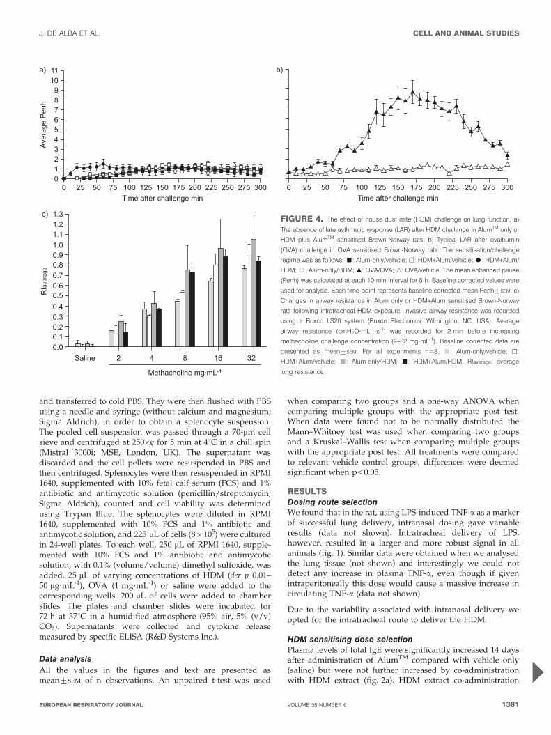
c)FIGURE 4. The effect of house dust mite (HDM) challenge on lung function. a)
The absence of late asthmatic response (LAR) after HDM challenge in AlumTM only or
HDM plus AlumTM sensitised Brown-Norway rats. b) Typical LAR after ovalbumin
(OVA) challenge in OVA sensitised Brown-Norway rats. The sensitisation/challenge
regime was as follows: &: Alum-only/vehicle; h: HDM+Alum/vehicle; $: HDM+Alum/
HDM; #: Alum-only/HDM; m: OVA/OVA; n: OVA/vehicle. The mean enhanced pause
(Penh) was calculated at each 10-min interval for 5 h. Baseline corrected values were
used for analysis. Each time-point represents baseline corrected mean Penh¡SEM. c)
Changes in airway resistance in Alum only or HDM+Alum sensitised Brown-Norway
rats following intratracheal HDM exposure. Invasive airway resistance was recorded
using a Buxco LS20 system (Buxco Electronics, Wilmington, NC, USA). Average
airway resistance (cmH2O?mL-1?s-1) was recorded for 2 min before increasing
methacholine challenge concentration (2–32 mg?mL-1). Baseline corrected data are
presented as mean¡SEM. For all experiments n58. &: Alum-only/vehicle; h:
HDM+Alum/vehicle; &: Alum-only/HDM; &: HDM+Alum/HDM. RIaverage: average
lung resistance.
J. DE ALBA ET AL. CELL AND ANIMAL STUDIES
cEUROPEAN RESPIRATORY JOURNAL VOLUME 35 NUMBER 6 1381
did, however, cause a significant increase in HDM-specific IgEplasma levels compared with AlumTM alone (fig. 2b).
5 mg of Der p 1 induced a robust increase in specific IgE and weadopted this dose for further studies.
HDM challenging dose selectionIntratracheal challenge with HDM caused significant broncho-alveolar (BAL) fluid eosinophilia at the top two doses tested(fig. 2c). The eosinophilia was accompanied by a significantincrease in neutrophil numbers at the highest dose (fig. 2d).
As 10 mg of Der p 1 induced eosinophilia with less markedneutrophilia we adopted this dose to further our investigations.
Effect of HDM sensitisation/challenge on cell recruitment inBAL fluid and lung tissue6 and 48 h after challenge with HDM extract we could detectan increase in airway neutrophilia and eosinophilia, respect-ively, in both the BAL fluid and the lung tissue (fig. 3).Interestingly we observed similar magnitudes of inflammationin the AlumTM-only sensitised animals to that seen in the ratspreviously sensitised with HDM. Animals sensitised only withvehicle (saline) showed no sign of inflammation in either BALor tissue (data not shown). This observation would suggest
that AlumTM is important in the direct inflammatory responseto HDM.
Effect of HDM sensitisation/challenge on airway functionmeasurementsThere were no changes in airway function after challenge.Monitoring of late asthmatic response (LAR) after HDM/vehicle challenge failed to show any significant differencesbetween the groups studied (fig. 4a), whereas using ourstandard OVA driven model we can measure significantLAR in this strain of rats (fig. 4b) [24]. Similarly, there wereno significant changes in the airway responsiveness to inhaledmethacholine when measured 48 h after the challenge (fig. 4c).
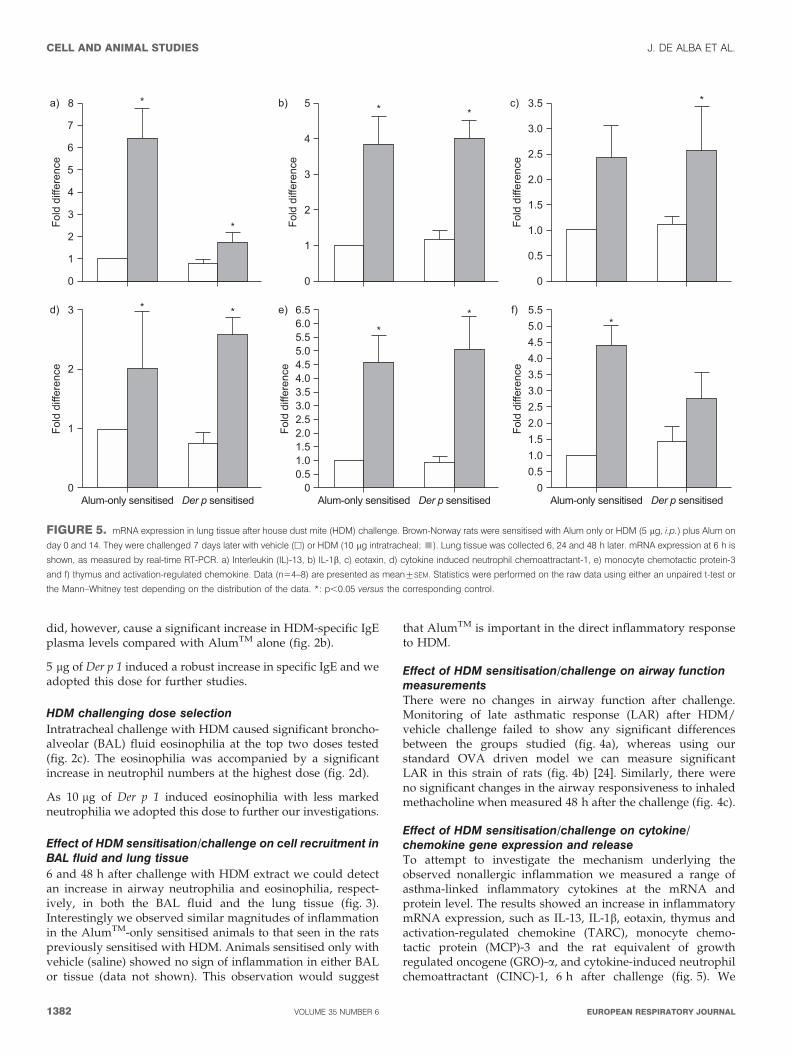
Effect of HDM sensitisation/challenge on cytokine/chemokine gene expression and releaseTo attempt to investigate the mechanism underlying theobserved nonallergic inflammation we measured a range ofasthma-linked inflammatory cytokines at the mRNA andprotein level. The results showed an increase in inflammatorymRNA expression, such as IL-13, IL-1b, eotaxin, thymus andactivation-regulated chemokine (TARC), monocyte chemo-tactic protein (MCP)-3 and the rat equivalent of growthregulated oncogene (GRO)-a, and cytokine-induced neutrophilchemoattractant (CINC)-1, 6 h after challenge (fig. 5). We
0Alum-only sensitised
Fold
diff
eren
ce
Der p sensitised
3
1
2
* *
00.51.01.52.02.53.03.54.04.55.05.56.0
Alum-only sensitised
Fold
diff
eren
ce
Der p sensitised
6.5
**
00.51.01.52.02.53.03.54.04.55.05.5
Alum-only sensitisedFo
ld d
iffer
ence
Der p sensitised
*
0
Fold
diff
eren
ce
8a)
d) e) f)
b) c)
1
2
3
4
5
6
7
*
*
0Fo
ld d
iffer
ence
5
1
2
3
4
* *
0
Fold
diff
eren
ce
3.5
0.5
1.0
1.5
2.0
2.5
3.0
*
FIGURE 5. mRNA expression in lung tissue after house dust mite (HDM) challenge. Brown-Norway rats were sensitised with Alum only or HDM (5 mg, i.p.) plus Alum on
day 0 and 14. They were challenged 7 days later with vehicle (h) or HDM (10 mg intratracheal; &). Lung tissue was collected 6, 24 and 48 h later. mRNA expression at 6 h is
shown, as measured by real-time RT-PCR. a) Interleukin (IL)-13, b) IL-1b, c) eotaxin, d) cytokine induced neutrophil chemoattractant-1, e) monocyte chemotactic protein-3
and f) thymus and activation-regulated chemokine. Data (n54–8) are presented as mean¡SEM. Statistics were performed on the raw data using either an unpaired t-test or
the Mann–Whitney test depending on the distribution of the data. *: p,0.05 versus the corresponding control.
CELL AND ANIMAL STUDIES J. DE ALBA ET AL.
1382 VOLUME 35 NUMBER 6 EUROPEAN RESPIRATORY JOURNAL
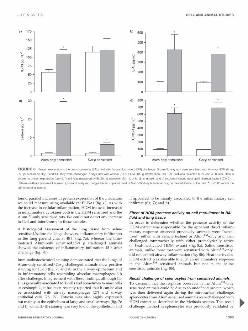
found parallel increases in protein expression of the mediatorswe could measure using available rat ELISAs (fig. 6). As withthe increase in cellular inflammation, HDM induced increasesin inflammatory cytokines both in the HDM sensitised and theAlumTM-only sensitised rats. We could not detect any increasein IL-4 and interferon-c in these samples.
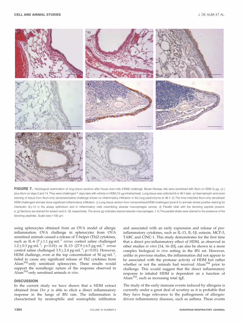
A histological assessment of the lung tissue from salinesensitised/saline challenge shows no inflammatory infiltrationin the lung parenchyma at 48 h (fig. 7a); whereas the time-matched Alum-only sensitised/Der p challenged animalsshowed the existence of inflammatory infiltration 48 h afterchallenge (fig. 7b).
Immunohistochemical staining demonstrated that the lungs ofAlum-only sensitised/Der p challenged animals show positivestaining for IL-13 (fig. 7c and d) in the airway epithelium andin inflammatory cells resembling alveolar macrophages 6 hafter challenge. In agreement with these findings, although IL-13 is generally associated to T-cells and sometimes to mast cellsor eosinophils, it has been recently reported that it can be alsobe associated with airway macrophages [27] and airwayepithelial cells [28, 29]. Eotaxin was also highly expressedbut mainly in the epithelium of large and small airways (fig. 7eand f), while IL-1b staining was very low in the epithelium and
it appeared to be mainly associated to the inflammatory cellinfiltrate (fig. 7g and h).
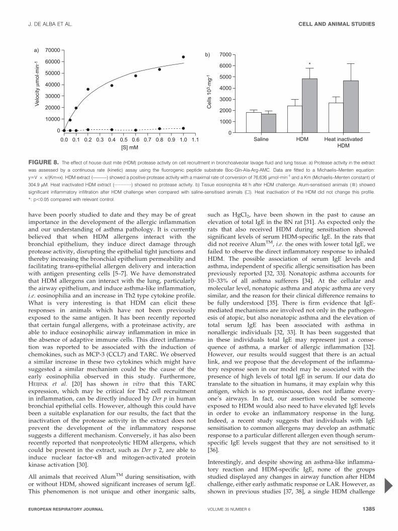
Effect of HDM protease activity on cell recruitment in BALfluid and lung tissueIn order to determine whether the protease activity of theHDM extract was responsible for the apparent direct inflam-matory response observed previously, animals were ‘‘sensi-tised’’ either with vehicle (saline) or AlumTM-only and thenchallenged intratracheally with either proteolytically activeor heat-inactivated HDM extract (fig. 8a). Saline sensitisedanimals, unlike those that were sensitised with AlumTM-only,did not exhibit airway inflammation (fig. 8b). Heat inactivatedHDM extract was also able to elicit an inflammatory responsein the AlumTM sensitised animals but not in the salinesensitised animals (fig. 8b).
Recall challenge of splenocytes from sensitised animalsTo discount that the response observed in the AlumTM-onlysensitised animals could be due to an undefined protein, whichwas then delivered again during the intratracheal challenge,splenocytes from Alum sensitised animals were challenged withHDM extract as described in the Methods section. This recallchallenge method in splenocytes was previously validated by
175
150
125
100
75
50
25
0
*
600
500
400
300
200
100
0
*
*
IL-1
3 pg
. mL-1
IL-1
β pg
. mL-1
a) b)
30
10
20
0
*
*
800
700
600
500
400
300
200
100
0
**
Eot
axin
pg.
mL-1
CIN
C-1
pg.
mL-1
Alum-only sensitisedAlum-only sensitised Der p sensitised Der p sensitised
c) d)
FIGURE 6. Protein expression in the bronchoalveolar (BAL) fluid after house dust mite (HDM) challenge. Brown-Norway rats were sensitised with Alum or HDM (5 mg,
i.p.) plus Alum on day 0 and 14. They were challenged 7 days later with vehicle (h) or HDM (10 mg intratracheal; &). BAL fluid was collected 6, 24 and 48 h later. Data is
shown for protein expression (pg?mL-1) at 6 h as measured by ELISA. a) Interleukin (IL)-13, b) IL-1b, c) eotaxin and d) cytokine induced neutrophil chemoattractant (CINC)-1.
Data (n54–8) are presented as mean¡SEM and analysed using either an unpaired t-test or Mann–Whitney test depending on the distribution of the data. *: p,0.05 versus the
corresponding control.
J. DE ALBA ET AL. CELL AND ANIMAL STUDIES
cEUROPEAN RESPIRATORY JOURNAL VOLUME 35 NUMBER 6 1383
using splenocytes obtained from an OVA model of allergicinflammation. OVA challenge in splenocytes from OVAsensitised animals caused a release of T-helper (Th)2 cytokines,such as IL-4 (7¡1.1 pg?mL-1 versus control saline challenged1.2¡0.3 pg?mL-1; p,0.01) or IL-13 (27.9¡6.5 pg?mL-1 versuscontrol saline challenged 3.5¡2.4 pg?mL-1; p,0.01). However,HDM challenge, even at the top concentration of 50 mg?mL-1,failed to cause any significant release of Th2 cytokines fromAlumTM-only sensitised splenocytes. These results wouldsupport the nonallergic nature of the response observed inAlumTM-only sensitised animals in vivo.
DISCUSSIONIn the current study we have shown that a HDM extractobtained from Der p is able to elicit a direct inflammatoryresponse in the lungs of BN rats. The inflammation ischaracterised by neutrophilic and eosinophilic infiltration
and associated with an early expression and release of pro-inflammatory cytokines, such as IL-13, IL-1b, eotaxin, MCP-3,TARC and CINC-1. This study demonstrates for the first timethat a direct pro-inflammatory effect of HDM, as observed inother studies in vitro [14, 16–20], can also be shown in a morecomplex biological in vivo setting in the BN rat. However,unlike in previous studies, the inflammation did not appear tobe associated with the protease activity of HDM but ratherwhether or not the animals had received AlumTM prior tochallenge. This would suggest that the direct inflammatoryresponse to inhaled HDM is dependent on a function ofAlumTM, such as increasing total IgE.
The study of the early immune events induced by allergens iscurrently under a great deal of scrutiny as it is probable thatthey have huge relevance to the pathogenesis of allergen-driven inflammatory diseases, such as asthma. These events
a) b) c)
d) e) f)
g) h)
FIGURE 7. Histological examination of lung tissue sections after house dust mite (HDM) challenge. Brown-Norway rats were sensitised with Alum or HDM (5 mg, i.p.)
plus Alum on days 0 and 14. They were challenged 7 days later with vehicle or HDM (10 mg intratracheal). Lung tissue was collected 6 or 48 h later. a) Haematoxylin and eosin
staining of tissue from Alum-only sensitised/saline challenge shows no inflammatory infiltration in the lung parenchyma at 48 h. b) The time-matched Alum-only sensitised/
HDM challenged animals show significant inflammatory infiltration. c) Lung tissue section from nonsensitised/HDM challenged (post-6 h) animals shows positive staining for
interleukin (IL)-13 in the airway epithelium and in inflammatory cells resembling alveolar macrophages (arrow). d) Parallel slide with the blocking peptide present.
e, g) Sections are stained for eotaxin and IL-1b, respectively. The arrow (g) indicates stained alveolar macrophages. f, h) The parallel slides were stained in the presence of the
blocking peptides. Scale bars5100 mm.
CELL AND ANIMAL STUDIES J. DE ALBA ET AL.
1384 VOLUME 35 NUMBER 6 EUROPEAN RESPIRATORY JOURNAL
have been poorly studied to date and they may be of greatimportance in the development of the allergic inflammationand our understanding of asthma pathology. It is currentlybelieved that when HDM allergens interact with thebronchial epithelium, they induce direct damage throughprotease activity, disrupting the epithelial tight junctions andthereby increasing the bronchial epithelium permeability andfacilitating trans-epithelial allergen delivery and interactionwith antigen presenting cells [5–7]. We have demonstratedthat HDM allergens can interact with the lung, particularlythe airway epithelium, and induce asthma-like inflammation,i.e. eosinophilia and an increase in Th2 type cytokine profile.What is very interesting is that HDM can elicit theseresponses in animals which have not been previouslyexposed to the same antigen. It has been recently reportedthat certain fungal allergens, with a proteinase activity, areable to induce eosinophilic airway inflammation in mice inthe absence of adaptive immune cells. This direct inflamma-tion was reported to be associated with the induction ofchemokines, such as MCP-3 (CCL7) and TARC. We observeda similar increase in these two cytokines which might havesuggested a similar mechanism could be the cause of theearly eosinophilia observed in this study. Furthermore,HEIJINK et al. [20] has shown in vitro that this TARCexpression, which may be critical for Th2 cell recruitmentin inflammation, can be directly induced by Der p in humanbronchial epithelial cells. However, although this could havebeen a suitable explanation for our results, the fact that theinactivation of the protease activity in the extract does notprevent the development of the inflammatory responsesuggests a different mechanism. Conversely, it has also beenrecently reported that nonproteolytic HDM allergens, whichcould be present in the extract, such as Der p 2, are able toinduce nuclear factor-kB and mitogen-activated proteinkinase activation [30].
All animals that received AlumTM during sensitisation, withor without HDM, showed significant increases of serum IgE.This phenomenon is not unique and other inorganic salts,
such as HgCl2, have been shown in the past to cause anelevation of total IgE in the BN rat [31]. As expected only therats that also received HDM during sensitisation showedsignificant levels of serum HDM-specific IgE. In the rats thatdid not receive AlumTM, i.e. the ones with lower total IgE, wefailed to observe the direct inflammatory response to inhaledHDM. The possible association of serum IgE levels andasthma, independent of specific allergic sensitisation has beenpreviously reported [32, 33]. Nonatopic asthma accounts for10–33% of all asthma sufferers [34]. At the cellular andmolecular level, nonatopic asthma and atopic asthma are verysimilar, and the reason for their clinical difference remains tobe fully understood [35]. There is firm evidence that IgE-mediated mechanisms are involved not only in the pathogen-esis of atopic, but also nonatopic asthma and the elevation oftotal serum IgE has been associated with asthma innonallergic individuals [32, 33]. It has been suggested thatin these individuals total IgE may represent just a conse-quence of asthma, a marker of allergic inflammation [32].However, our results would suggest that there is an actuallink, and we propose that the development of the inflamma-tory response seen in our model may be associated with thepresence of high levels of total IgE in serum. If our data dotranslate to the situation in humans, it may explain why thisantigen, which is so promiscuous, does not inflame every-one’s airways. In fact, our assertion would be someoneexposed to HDM would also need to have elevated IgE levelsin order to evoke an inflammatory response in the lung.Indeed, a recent study suggests that individuals with IgEsensitisation to common allergens may develop an asthmaticresponse to a particular different allergen even though serum-specific IgE levels suggest that they are not sensitised to it[36].
Interestingly, and despite showing an asthma-like inflamma-tory reaction and HDM-specific IgE, none of the groupsstudied displayed any changes in airway function after HDMchallenge, either early asthmatic response or LAR. However, asshown in previous studies [37, 38], a single HDM challenge
70000
60000
50000
40000
30000
20000
10000
7000
6000
5000
4000
3000
2000
1000
00
0.0 0.1 0.2 0.3 0.4 0.5 0.6 0.7 0.8 0.9 1.0 1.1[S] mM
*
Velo
city
μm
ol. m
in-1
Cel
ls 1
03. m
g-1
●● ● ● ● ● ● ● ●
●
●
●●
●
●
●
Saline HDM Heat inactivatedHDM
a)b)
FIGURE 8. The effect of house dust mite (HDM) protease activity on cell recruitment in bronchoalveolar lavage fluid and lung tissue. a) Protease activity in the extract
was assessed by a continuous rate (kinetic) assay using the fluorogenic peptide substrate Boc-Gln-Ala-Arg-AMC. Data are fitted to a Michaelis–Menten equation:
y5V 6 x/(Km+x). HDM extract (––––––) showed a positive protease activity with a maximal rate of conversion of 76,636 mmol?min-1 and a Km (Michaelis–Menten constant) of
304.9 mM. Heat inactivated HDM extract (???????????) showed no protease activity. b) Tissue eosinophilia 48 h after HDM challenge. Alum-sensitised animals (&) showed
significant inflammatory infiltration after HDM challenge when compared with saline-sensitised animals (h). Heat inactivation of the HDM did not change this profile.
*: p,0.05 compared with relevant control.
J. DE ALBA ET AL. CELL AND ANIMAL STUDIES
cEUROPEAN RESPIRATORY JOURNAL VOLUME 35 NUMBER 6 1385

may not be enough to achieve reliable lung function changes inthe rat and indeed it appears that in the mouse modelsmultiple HDM challenges may be necessary [39–41].
One possible caveat to the suggestion that it is the HDM that isdirectly causing the inflammation is that something else withinthe HDM mixture is responsible, i.e. the small amount ofendotoxin. Furthermore, a recent study proposes that Toll likereceptor-4 receptor activation could play a crucial part in HDMinduced responses [41]. Whilst endotoxin can cause some ofthe inflammation observed in the study we feel it is veryunlikely to be responsible. The levels are very low (36 EU?mL-1
as measured with a Pyrogen 5000 LAL endotoxin assay;Cambrex Bioscience Ltd, Wokingham, UK), the time of cellularinflux is much later than one would expect after LPS, some ofthe mediators measured are not thought to be upregulated byendotoxin (i.e. TARC) and, probably most convincingly, thereis no inflammation present in the HDM challenged but non-AlumTM sensitised animals. Another possible explanation isthat the AlumTM-only sensitised animals had been exposed toan undefined protein, which was then delivered again duringthe intratracheal challenge, causing an allergic Th2 response.However, the negative results from the recall challengeexperiment would seem to rule out this possibility.Nonetheless, we cannot completely discard that some of theinflammation observed is through a non-HDM mechanism.
In conclusion, these data demonstrate that HDM can, afterexposure to an adjuvant which increases serum total IgE,evoke direct inflammation in the airways which in many waysis characteristic of the asthmatic airway. We feel this is a veryinteresting finding in that it might help unravel the complexassociation between high IgE levels, atopy and the develop-ment of the asthma phenotype. Indeed these data maysignificantly alter our understanding of the role of IgE in thepathogenesis of asthma.
SUPPORT STATEMENTFunding for the study was provided by GlaxoSmithKline.
STATEMENT OF INTERESTStatements of interest for R.G. Knowles and M.G. Belvisi, and forthe study itself, can be found at www.erj.ersjournals.com/misc/statements.dtl
ACKNOWLEDGEMENTSWe would like to thank C. Dean (Respiratory Pharmacology,Pharmacology and Toxicology, Imperial College London, Faculty ofMedicine, National Heart and Lung Institute, London, UK) for helpwith the preparation of this manuscript.
REFERENCES1 Thomas WR, Smith WA, Hales BJ, et al. Characterization and
immunobiology of house dust mite allergens. Int Arch AllergyImmunol 2002; 129: 1–18.
2 Platt-Mills TA. The role of indoor allergens in asthma. Allergy1995; 50: 5.
3 Platts-Mills TA, Vervloet D, Thomas WR, et al. Indoor allergensand asthma: report of the Third International Workshop. J Allergy
Clin Immunol 1997; 100: S2–S24.4 Takai T, Kato T, Sakata Y, et al. Recombinant Der p 1 and Der f 1
exhibit cysteine protease activity but no serine protease activity.Biochem Biophys Res Commun 2005; 328: 944–952.
5 Herbert CA, King CM, Ring PC, et al. Augmentation of
permeability in the bronchial epithelium by the house dust
mite allergen Der p1. Am J Respir Cell Mol Biol 1995; 12: 369–
378.
6 Wan H, Winton HL, Soeller C, et al. Quantitative structural and
biochemical analyses of tight junction dynamics following
exposure of epithelial cells to house dust mite allergen Der p 1.
Clin Exp Allergy 2000; 30: 685–698.
7 Wan H, Winton HL, Soeller C, et al. Der p 1 facilitates
transepithelial allergen delivery by disruption of tight junctions.
J Clin Invest 1999; 104: 123–133.
8 Sakata Y, Arima K, Takai T, et al. The squamous cell carcinoma
antigen 2 inhibits the cysteine proteinase activity of a major mite
allergen, Der p 1. J Biol Chem 2004; 279: 5081–5087.
9 Takai T, Kato T, Ota M, et al. Recombinant Der p 1 and Der f 1 with
in vitro enzymatic activity to cleave human CD23, CD25 and a1-
antitrypsin, and in vivo IgE-eliciting activity in mice. Int Arch
Allergy Immunol 2005; 137: 194–200.
10 Kikuchi Y, Takai T, Ota M, et al. Application of immunoreaction
enhancer solutions to an enzyme-linked immunosorbent assay for
antigen-specific IgE in mice immunized with recombinant major
mite allergens or ovalbumin. Int Arch Allergy Immunol 2006; 141:
322–330.
11 Furmonaviciene R, Ghaemmaghami AM, Boyd SE, et al. The
protease allergen Der p 1 cleaves cell surface DC-SIGN and DC-
SIGNR: experimental analysis of in silico substrate identification
and implications in allergic responses. Clin Exp Allergy 2007; 37:
231–242.
12 Machado DC, Horton D, Harrop R, et al. Potential allergens
stimulate the release of mediators of the allergic response from
cells of mast cell lineage in the absence of sensitization with
antigen-specific IgE. Eur J Immunol 1996; 26: 2972–2980.
13 Peake HL, Currie AJ, Stewart GA, et al. Nitric oxide production by
alveolar macrophages in response to house dust mite fecal pellets
and the mite allergens, Der p 1 and Der p 2. J Allergy Clin Immunol
2003; 112: 531–537.
14 King C, Brennan S, Thompson PJ, et al. Dust mite proteolytic
allergens induce cytokine release from cultured airway epithe-
lium. J Immunol 1998; 161: 3645–3651.
15 Phillips C, Coward WR, Pritchard DI, et al. Basophils express a
type 2 cytokine profile on exposure to proteases from helminths
and house dust mites. J Leukoc Biol 2003; 73: 165–171.
16 Asokananthan N, Graham PT, Stewart DJ, et al. House dust mite
allergens induce proinflammatory cytokines from respiratory
epithelial cells: the cysteine protease allergen, Der p 1, activates
protease-activated receptor (PAR)-2 and inactivates PAR-1.
J Immunol 2002; 169: 4572–4578.
17 Hales BJ, Shen H, Thomas WR. Cytokine responses to Der p 1 and
Der p 7: house dust mite allergens with different IgE-binding
activities. Clin Exp Allergy 2000; 30: 934–943.
18 Sun G, Stacey MA, Schmidt M, et al. Interaction of mite allergens
Der P3 and Der P9 with protease-activated receptor-2 expressed
by lung epithelial cells. J Immunol 2001; 167: 1014–1021.
19 Adam E, Hansen KK, Astudillo OF, et al. The house dust mite
allergen Der p 1, unlike Der p 3, stimulates the expression of
interleukin-8 in human airway epithelial cells via a proteinase-
activated receptor-2-independent mechanism. J Biol Chem 2006;
281: 6910–6923.
20 Heijink IH, Marcel Kies P, van Oosterhout AJM, et al. Der p, IL-4,
and TGF-b cooperatively induce EGFR-dependent TARC expres-
sion in airway epithelium. Am J Respir Cell Mol Biol 2007; 36: 351–
359.
21 Kauffman H, Tamm M, Timmerman JA, et al. House dust mite
major allergens Der p 1 and Der p 5 activate human airway-
derived epithelial cells by protease-dependent and protease-
independent mechanisms. Clin Mol Allergy 2006; 4: 5.
CELL AND ANIMAL STUDIES J. DE ALBA ET AL.
1386 VOLUME 35 NUMBER 6 EUROPEAN RESPIRATORY JOURNAL
22 Underwood SL, Haddad E, Birrell MA, et al. Functionalcharacterization and biomarker identification in the BrownNorway model of allergic airway inflammation. Br J Pharmacol
2002; 137: 263–275.23 Schulz O, Sewell HF, Shakib F. A sensitive fluorescent assay for
measuring the cysteine protease activity of Der p 1, a majorallergen from the dust mite Dermatophagoides pteronyssinus. Mol
Pathol 1998; 51: 222–224.24 Birrell MA, Hardaker E, Wong S, et al. Ik-B kinase-2 inhibitor
blocks inflammation in human airway smooth muscle and arat model of asthma. Am J Respir Crit Care Med 2005; 172:962–971.
25 McCluskie K, Birrell MA, Wong S, et al. Nitric oxide as anoninvasive biomarker of lipopolysaccharide-induced airwayinflammation: possible role in lung neutrophilia. J Pharmacol Exp
Therap 2004; 311: 625–633.26 Lundblad LKA, Irvin CG, Hantos Z, et al. Penh is not a measure of
airway resistance! Eur Respir J 2007; 30: 805.27 Kim EY, Battaile JT, Patel AC, et al. Persistent activation of an
innate immune response translates respiratory viral infection intochronic lung disease. Nat Med 2008; 14: 633–640.
28 Allahverdian S, Harada N, Singhera GK, et al. Secretion of IL-13 byairway epithelial cells enhances epithelial repair via HB-EGF. Am JRespir Cell Mol Biol 2008; 38: 153–160.
29 Temann UA, Laouar Y, Eynon EE, et al. IL9 leads to airwayinflammation by inducing IL13 expression in airway epithelialcells. Int Immunol 2007; 19: 1–10.
30 Osterlund C, Gronlund H, Polovic N, et al. The non-proteolytichouse dust mite allergen Der p 2 induce NF-kB and MAPKdependent activation of bronchial epithelial cells. Clin Exp Allergy
2009; 39: 1199–1208.
31 White KL, David DW, Butterworth LF, et al. Assessment ofautoimmunity-inducing potential using the brown Norway ratchallenge model. Toxicol Lett 2000; 112–113: 443–451.
32 Sunyer J, Anto JM, Castellsague J, et al. Total serum IgE is associatedwith asthma independently of specific IgE levels. The Spanish Groupof the European Study of Asthma. Eur Respir J 1996; 9: 1880–1884.
33 Beeh KM, Ksoll M, Buhl R. Elevation of total serum immunoglo-bulin E is associated with asthma in nonallergic individuals. Eur
Respir J 2000; 16: 609–614.34 Novak N, Bieber T. Allergic and nonallergic forms of atopic
diseases. J Allergy Clin Immunol 2003; 112: 252–262.35 Jayaratnam A, Corrigan CJ, Lee TH. The continuing enigma of
non-atopic asthma. Clin Exp Allergy 2005; 35: 835–837.36 Chinn S, Heinrich J, Anto JM, et al. Bronchial responsiveness in
atopic adults increases with exposure to cat allergen. Am J Respir
Crit Care Med 2007; 176: 20–26.37 Singh P, Daniels M, Winsett DW, et al. Phenotypic comparison of
allergic airway responses to house dust mite in three rat strains.Am J Physiol Lung Cell Mol Physiol 2003; 284: L588–L598.
38 Dong W, Selgrade MK, Gilmour MI. Systemic administration ofBordetella pertussis enhances pulmonary sensitization to house dustmite in juvenile rats. Toxicol Sciences 2003; 72: 113–121.
39 Fuchs B, Braun A. Improved mouse models of allergy and allergicasthma: chances beyond ovalbumin. Curr Drug Targets 2008; 9:495–502.
40 Gregory LG, Causton B, Murdoch JR, et al. Inhaled house dustmite induces pulmonary T helper 2 cytokine production. Clin ExpAllergy 2009; 39: 1597–1610.
41 Hammad H, Chieppa M, Perros F, et al. House dust mite allergeninduces astham via Toll-like receptor 4 triggering of airwaystructural cells. Nat Med 2009; 15: 410–416.
J. DE ALBA ET AL. CELL AND ANIMAL STUDIES
EUROPEAN RESPIRATORY JOURNAL VOLUME 35 NUMBER 6 1387