Cystic echinococcosis in a single tertiary care center in Rome, Italy.

512
Journal of Pharmacological Sciences© The Japanese Pharmacological Society
J Pharmacol Sci 118, 512 – 520 (2012)
Introduction
Cystic fibrosis (CF) is the most common lethal inher-ited disorder in the Caucasian population and is caused by mutations in the gene encoding the CF transmembrane
regulator (CFTR) (1). CF is characterized by recurrent pulmonary infections and chronic inflammation that ul-timately lead to death from respiratory failure (1, 2). Defects in expression of CFTR in the airway epithelial cells induces chronic airway inflammation that appears to be related to an intrinsic defect in regulation of the immune response and/or a hyperesponsiveness to infec-tion by bacterial strains such as Pseudomonas, Staphylo-coccus, Haemophilus, Aspergillus, and Burkholderia species. This hyperesponsiveness correlates with im-
†These authors contributed equally to this study.*Corresponding author. [email protected] online in J-STAGE on March 29, 2012 (in advance)doi: 10.1254/jphs.11240FP
Synergism Between Interleukin (IL)-17 and Toll-like Receptor 2 and 4 Signals to Induce IL-8 Expression in Cystic Fibrosis Airway Epithelial CellsShota Mizunoe1,2,†, Tsuyoshi Shuto1,†, Shingo Suzuki1, Chizuru Matsumoto1, Kenji Watanabe1,2, Keiko Ueno-Shuto3, Mary Ann Suico1, Kouhei Onuki1,2, Dieter C. Gruenert4,5,6,7,8,9,10, and Hirofumi Kai1,*1Department of Molecular Medicine, Graduate School of Pharmaceutical Sciences, Global COE “Cell Fate Regulation Research and Education Unit”, Kumamoto University, Kumamoto 862-0973, Japan
2The Japan Society for the Promotion of Science (JSPS), Tokyo 102-8472, Japan3Laboratory of Pharmacology, Faculty of Pharmaceutical Sciences, Sojo University, Kumamoto 860-0082, Japan4Department of Otolaryngology - Head and Neck Surgery, 5Department of Laboratory Medicine, 6Eli and Edythe Broad Center for Regenerative Medicine and Stem Cell Research, 7Helen Diller Family Comprehensive Cancer Center, 8Institute for Human Genetics, 9Cardiovascular Research Institute, University of California, San Francisco, San Francisco, CA 94115, USA
10Department of Pediatrics, University of Vermont College of Medicine, Burlington, VT 05405, USA
Received December 18, 2011; Accepted February 20, 2012
Abstract. Cystic fibrosis (CF) is the most common lethal inherited disorder and is caused by mutations in the gene encoding the CF transmembrane regulator (CFTR). The CF lung expresses a profound proinflammatory phenotype that appears to be related to a constitutive hypersecretion of interleukin (IL)-8 from airway epithelial cells in response to microbial infection. Since overpro-duction of IL-8 in CF contributes to massive bronchial infiltrates of neutrophils, identification of the pathways underlying IL-8 induction could provide novel drug targets for treatment of neutro-phil-dominated inflammatory diseases such as CF. Here, we show that IL-17A synergistically increases IL-8 production induced by a toll-like receptor (TLR) 2 agonist, peptidoglycan (PGN), or TLR4 agonist, lipopolysaccharide (LPS), in a human CF bronchial epithelial cell line ( CFBE41o-). A strong synergism was also observed in primary human CF bronchial epithelial cells, but not in human non-CF cell lines and primary cells. Notably, despite the induction of nuclear factor-κB and MAP kinases during TLR2 or TLR4 activation in CFBE41o-, IL-17A-dependent synergism appears to be the result of enhanced PGN- or LPS-induced phosphorylation of p38. Taken together, these studies provide evidence that IL-17A is a critical factor in increasing IL-8 expression in bacteria-infected CF airways via a pathway that regulates p38 phosphorylation.
Keywords: interleukin (IL)-17, toll-like receptor 2 and 4 (TLR2/TLR4), IL-8, cystic fibrosis (CF), p38
Full Paper
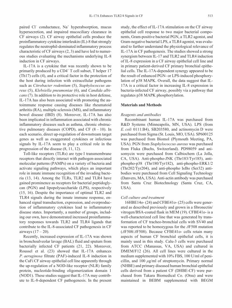
513IL-17A Enhances TLR2/4 Signals in CF
paired Cl− conductance, Na+ hyperabsorption, mucus hypersecretion, and impaired mucociliary clearance in CF airways (2). CF airway epithelial cells produce the proinflammatory cytokine interleukin (IL)-8 that strongly regulates the neutrophil-dominated inflammatory process characteristic of CF airways (2, 3) and have led to numer-ous studies evaluating the mechanisms underlying IL-8 induction in CF airways.
IL-17A is a cytokine that was recently shown to be primarily produced by a CD4+ T cell subset, T helper 17 (Th17) cells (4), and a critical factor in the protection of the host during infection with extracellular pathogens such as Citrobacter rodentium (5), Staphylococcus au-reus (5), Klebsiella pneumoniae (6), and Candida albi-cans (7). In addition to its protective role in host defense, IL-17A has also been associated with promoting the au-toimmune response causing diseases like rheumatoid arthritis (RA), multiple sclerosis (MS), and inflammatory bowel disease (IBD) (8). Moreover, IL-17A has also been implicated in inflammation associated with chronic inflammatory diseases such as asthma, chronic obstruc-tive pulmonary diseases (COPD), and CF (8 – 10). In each scenario, direct up-regulation of downstream target genes as well as exaggerated cytokines or infectious signals by IL-17A seem to play a critical role in the progression of the disease (8, 11, 12).
Toll-like receptors (TLRs) are type I transmembrane receptors that directly interact with pathogen-associated molecular patterns (PAMPs) on a variety of bacteria and activate signaling pathways, which plays an important role in innate immune recognition of the invading bacte-ria (13, 14). Among the TLRs, TLR2 and TLR4 have gained prominence as receptors for bacterial peptidogly-can (PGN) and lipopolysaccharide (LPS), respectively (15, 16). Despite the importance of optimal TLR2 and TLR4 signals during the innate immune response, en-hanced signal transduction, expression, and overproduc-tion of inflammatory cytokines lead to inflammatory disease states. Importantly, a number of groups, includ-ing our own, have demonstrated increased proinflamma-tory responses towards TLR2 and TLR4 ligands that contribute to the IL-8-associated CF pathogenesis in CF airways (17 – 20).
Recently, increased expression of IL-17A was shown in bronchoalveolar lavage (BAL) fluid and sputum from bacterially infected CF patients (21, 22). Moreover, Roussel et al. (23) showed that IL-17A enhances P. aeruginosa filtrate (PAF)-induced IL-8 induction in the CuFi CF airway epithelial cell line apparently through the up-regulation of a NOD-like receptor (NLR) family protein, nucleotide-binding oligomerization domain 1 (NOD1). These studies suggest that IL-17A may contrib-ute to IL-8-dependent CF pathogenesis. In the present
study, the effect of IL-17A stimulation on the CF airway epithelial cell response to two major bacterial compo-nents, Gram-positive bacterial PGN, a TLR2 agonist, and Gram-negative bacterial LPS, a TLR4 agonist, was evalu-ated to further understand the physiological relevance of IL-17A in CF pathogenesis. The studies showed a strong synergism between IL-17 and TLR2 and TLR4 induction of IL-8 expression in a CF airway epithelial cell line and in primary patient-derived CF primary bronchial epithe-lial cells. The IL-17A-dependent synergy appeared to be the result of enhanced PGN- or LPS-induced phosphory-lation of p38 MAPK. Overall, the data suggest that IL-17A is a critical factor in increasing IL-8 expression in bacteria-infected CF airway, possibly via a pathway that regulates p38 MAPK phosphorylation.
Materials and Methods
Reagents and antibodiesRecombinant human IL-17A was purchased from
R&D Systems (Minneapolis, MN, USA). LPS (from E. coli 0111:B4), SB203580, and actinomycin D were purchased from Sigma (St. Louis, MO, USA). SP600125 was purchased from Biomol (Plymouth Meeting, PA, USA). PGN from Staphylococcus aureus was purchased from Fluka (Buchs, Switzerland). PD98059 and ani-somycin were purchased from Calbiochem (La Jolla, CA, USA). Anti-phospho-JNK (Thr183/Tyr185), anti-phospho-p38 (Thr180/Tyr182), anti-phospho-ERK1/2 (Thr202/Tyr204), and anti-phospho IκBα (Ser32) anti-bodies were purchased from Cell Signaling Technology (Danvers, MA, USA). Anti-actin antibody was purchased from Santa Cruz Biotechnology (Santa Cruz, CA, USA).
Cell culture and treatment16HBE14o- (24) and CFBE41o- (25) cells were gener-
ated as described previously and grown in a fibronectin/vitrogen/BSA-coated flask in MEM (19). CFBE41o- is a well-characterized cell line that was generated by trans-formation of CF tracheo-bronchial cells with SV40 and was reported to be homozygous for the ΔF508 mutation (ΔF508/ΔF508). Because CFBE41o- cells retain many aspects of human CF bronchial epithelial cells, it is mainly used in this study. Calu-3 cells were purchased from ATCC (Manassas, VA, USA) and cultured in DMEM/F12 (26). All cell lines were cultured in the medium supplemented with 10% FBS, 100 U/ml of peni-cillin, and 100 μg/ml of streptomycin. Primary normal (NHBE) and primary diseased human bronchial epithelial cells derived from a patient CF (DHBE-CF) were pur-chased from Takara Biomedical Co. (Otsu) and were maintained in BEBM supplemented with BEGM

514 S Mizunoe et al
SingleQuots including Bovine Pituitary Extract, insulin, hydrocortisone, GA-1000, retinoic acid, transferrin, tri-iodothyronine, epinephrine, and human epidermal growth factor (Takara Biomedical Co.) according to the manu-facturer’s instruction. All cells were cultured in a humidi-fied atmosphere of 5% CO2 in air at 37°C. For the detec-tion of IL-8 induction, all cell lines were subjected to serum starvation 12 h prior to the treatment with PGN or LPS to reduce the background activation of IL-8 expres-sion, while primary cells were cultured in BEBM con-taining only insulin, GA-1000, transferrin, and tri-iodothyronine 12 h prior to the treatment with PGN or LPS.
Quantitative real-time RT-PCR analysisQuantitative real-time RT-PCR for human IL-8, TLR2,
TLR4, and 18s ribosomal RNA (18srRNA) was carried out using the primers shown in Table 1 as previously described (27). Briefly, total RNA from the cells was isolated using RNAisoPlus and synthesis of cDNA was performed using PrimeScript RT regent kit (Takara Biomedical Co.). Real-time quantitative RT-PCR analy-sis was performed using SYBR Premix Ex Taq (Takara Biomedical Co.). The relative quantity of target genes’ mRNA was normalized using 18srRNA as the internal control and expressed as the relative quantity of target genes’ mRNA (fold induction).
SDS-PAGE and western blottingCells grown to 90% – 100% confluence on 6-cm plates
were used to collect the cell protein lysates. The protein lysates were analyzed by SDS-PAGE and western blot-ting as described previously (28).
ELISA analysisThe IL-8 concentration in the culture supernatant was
assessed by an ELISA kit for human IL-8 (CXCL8/IL-8 Duo Set) from R&D Systems (Minneapolis, MN, USA) according to the manufacturer’s instruction. Briefly, 100 μl of sample was added to each well coated with a murine monoclonal antibody against IL-8. To be within range, supernatants from cell cultures were optimally diluted in serum-free medium. A biotinylated goat anti-human IL-8
was added, and samples were incubated for 2 h, followed by the incubation with a streptavidin conjugated to horseradish-peroxidase. After three washes between each step, substrate solution was added for 20 min. Absor-bance was measured on a microplate reader at 450 nm, and IL-8 concentration was determined in each sample by comparison to a standard curve.
Statistical analysisThe data were analyzed by one-way ANOVA with the
Tukey-Kramer multiple comparison test (JMP software; SAS Institute, Cary, NC, USA) as indicated in each fig-ure legend.
Results
IL-17A-dependent IL-8 induction in human CFBE41o- cells
To confirm the effect of IL-17A on the regulation of IL-8 gene expression in CF airways, CFBE41o- cells were first treated with IL-17A and the IL-8 mRNA levels were assessed by quantitative real-time RT-PCR. As shown in Fig. 1A, after the treatment with IL-17A, the IL-8 mRNA expression in CFBE41o- cells was slightly but significantly up-regulated. A significant increase in IL-8 gene expression was observed at IL-17A concentra-tions in the range 10 – 100 ng/ml. IL-17A-dependent up-regulation of IL-8 protein was also observed by ELISA (Fig. 1B). To further elucidate the signaling pathways involved in IL-8 up-regulation, NF-κB activa-tion and the MAPKs signaling pathways in CFBE41o- cells were assayed because they are downstream signals of IL-17A activation in numerous cell types (8). Expo-sure to IL-17A results in phosphorylation of the JNK, p38, and ERK pathways (Fig. 1C), but not in IκBα, an activator of NF-κB. The role of these MAPK signaling pathways in the regulation of IL-17A-dependent IL-8 induction in CFBE41o- cells was elucidated with specific inhibitors of JNK, p38, and ERK pathways. SB203580 and PD98059, but not SP600125, significantly sup-pressed IL-17A-dependent IL-8 induction (Fig. 1D). These data suggest that p38 and ERK affect the IL-17A-dependent signaling pathway for the IL-8 induction in
Table 1. Primers used for quantitative RT-PCR
Human mRNA Forward primer (5′ to 3′) Reverse primer (5′ to 3′)
IL-8 CTGGCCGTGGCTCTCTTG CCTTGGCAAAACTGCACCTT
TLR2 GGCCAGCAAATTACCTGTGT AGGCGGACATCCTGAACCT
TLR4 CTGCAATGGATCAAGGACCA TTATCTGAAGGTGTTGCACATTCC
18s ribosomal RNA CGGCTACCACATCCAAGGAA GCTGGAATTACCGCGGCT

515IL-17A Enhances TLR2/4 Signals in CF
CFBE41o- cells.
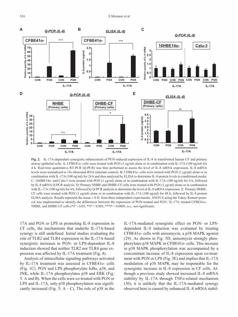
IL-17A synergizes with PGN and LPS to promote IL-8 induction in human CF airway epithelial cell lines and primary cells
Since IL-17A has been shown to affect other signaling pathways (11), the modulatory role of IL-17A in signal-ing during bacterial infection in CF airways. The effect of co-treatment with IL-17A and Gram-positive bacterial PGN was initially assayed. PGN, a TLR2 agonist, alone up-regulates IL-8 in CFBE41o- cells (Fig. 2: A and B). There was a notable synergy in the induction of IL-8 gene and protein expressions when PGN was used in combination with IL-17A (Fig. 2: A and B). Similar analyses performed using non-CF airway epithelial cell lines, 16HBE14o- and Calu-3 cells, showed no synergism with the combination of IL-17A and PGN in either cell line (Fig. 2C). To exclude the possibility that effect of IL-17A is only observed in cell lines, the effect of IL-17A on the IL-8 gene expression was evaluated in NHBE and DHBE-CF cells. A low of IL-8 gene induction and no synergy was observed when IL-17A and PGN were
used together for IL-8 induction in NHBE cells. How-ever, there was significant synergy in IL-8 gene and protein induction in DHBE-CF cells when IL-17A and PGN were used together (Fig. 2: D and E).
The effect of IL-17A co-treatment with a Gram-nega-tive bacterial LPS was then assessed and showed a sig-nificant synergistic induction of IL-8 gene and protein observed with the combined exposure to IL-17A and LPS, a TLR4 agonist (Fig. 3: A and B). Moreover, no synergy was observed when the IL-17A and LPS combi-nation was used to treat the non-CF cell lines, 16HBE14o- and Calu-3 cells (Fig. 3C). As was observed in the pri-mary cells treated with PGN, the expression of IL-8 by co-treatment with IL-17A and LPS was dramatically higher in DHBE-CF cells when compared to that in NHBE cells (Fig. 3: D and E). Taken together, these data suggest an immunostimulatory role for IL-17A during bacterial infection of CF airway.
p38 MAPK activation is involved in the synergistic in-duction of IL-8 in CF cells
While having demonstrated the synergy between IL-
A
0100200300400500600700800
1 10 100
IL-17A (ng/ml) IL
-8 p
rote
in (
pg/m
l )con
ELISA (IL-8)
00.5
11.5
22.5
33.5
Rel
ativ
e qu
antit
y of
IL-8
mR
NA
Q-PCR (IL-8)
IL-17A (ng/ml)
0
0.5
1
1.5
2
2.5
+
SB203580
+
PD98059
+
SP600125
+-IL-17A
MAPKinhibitors - -
n.s.
B
C D
-p-JNK
-p-p38
-p-ERK
-actin
0 7.5 15 30 60 120
Phosphorylation of MAPK
0 15 30 60 120 240
Phosphorylation of I B
-actin
-p-I B
Rel
ativ
e qu
antit
y of
IL-8
mR
NA
con 1 10 100
p-p54p-p46
IL-17A (min)
IL-17A (min)
p-p44p-p42
p-I B
actin
p-p38
actin(10 μM)
Q-PCR (IL-8)
***
**
****
**
***
Fig. 1. Effect of IL-17A on IL-8 gene expression in human CF bronchial epithe-lial cells. A: CFBE41o- cells were stimu-lated with IL-17A (1, 10, 100 ng/ml) for 4 h. Real-time quantitative RT-PCR analy-sis (Q-PCR) was used to determine IL-8 mRNA expression. IL-8 mRNA levels were normalized to 18s ribosomal RNA (internal control). B: CFBE41o- cells were stimulated with IL-17A (1, 10, 100 ng/ml) for 36 h and then analyzed by ELISA to determine the IL-8 protein lev-els in conditioned media. C: CFBE41o- cells were stimulated with IL-17A (100 ng/ml) at the indicated times and then ly-sed in sample buffer. IκBα and MAPK phosphorylation was assessed by western blotting. D) CFBE41o- cells were pre-treated with 10 μM SB203580 (p38 path-way inhibitor), 10 μM PD98059 (ERK pathway inhibitor), 10 μM SP600125 (JNK pathway inhibitor), or vehicle con-trol (DMSO) before a 4-h treatment with IL-17A (100 ng/ml). Real-time Q-PCR was then performed to quantify IL-8 mRNA levels. In both the Q-PCR and ELISA analyses, results represent the mean ± S.D. from three independent ex-periments. ANOVA using the Tukey-Kramer procedure was used to identify the differences of cytokines expression among the groups indicated in the figure (**P < 0.005, ***P < 0.0005; n.s., not significant).
516 S Mizunoe et al
17A and PGN or LPS in promoting IL-8 expression in CF cells, the mechanisms that underlie IL-17A-based synergy is still undefined. Initial studies evaluating the role of TLR2 and TLR4 expression in the IL-17A-based synergistic increases in PGN- or LPS-dependent IL-8 induction showed that neither TLR2 nor TLR4 gene ex-pression was affected by IL-17A treatment (Fig. 4).
Analysis of intracellular signaling pathways activated by IL-17A treatment was assessed in CFBE41o- cells (Fig. 1C). PGN and LPS phosphorylate IκBα, p38, and JNK, while IL-17A phosphorylates p38 and ERK (Fig. 5: A and B). When the cells were co-treated with PGN or LPS and IL-17A, only p38 phosphorylation was signifi-cantly increased (Fig. 5: A – C). The role of p38 in the
IL-17A-mediated synergistic effect on PGN- or LPS-dependent IL-8 induction was evaluated by treating CFBE41o- cells with anisomycin, a p38 MAPK agonist (29). As shown in Fig. 5D, anisomycin strongly phos-phorylates p38 MAPK in CFBE41o- cells. This increase in p38 MAPK phosphorylation was accompanied by a concomitant increase of IL-8 expression upon co-treat-ment with PGN or LPS (Fig. 5E) and implies that IL-17A modulation of p38 MAPK may be responsible for the synergistic increase in IL-8 expression in CF cells. Al-though a previous study showed increased IL-8 mRNA stability by IL-17A through TNFα-related mechanism (30), it is unlikely that the IL-17A-mediated synergy observed here is caused by enhanced IL-8 mRNA stabil-
A
010203040506070
CON PGN CON PGN
IL-17A
CFBE41o-
Rel
ativ
e qu
antit
y of
IL-8
mR
NA
CFBE41o-
Q-PCR (IL-8) B ELISA (IL-8)
0
0.5
1
1.5
2
2.5
CON PGN CON PGN
IL-17AIL
-8 p
rote
in (n
g/m
l)
C
16HBE14o-
00.5
11.5
22.5
3
Rel
ativ
e qu
antit
y of
IL-8
mR
NA
Q-PCR (IL-8)
CON PGN CON PGN
IL-17A
CON PGN CON PGN
Calu-3
IL-17A
D Q-PCR (IL-8)
05
101520253035
Rel
ativ
e qu
antit
y of
IL-8
mR
NA
CON PGN CON PGN
IL-17A
DHBE-CF(Primary cells)
n.s.
CON PGN CON PGN
IL-17A
NHBE(Primary cells)
EELISA (IL-8)
00.5
11.5
22.5
33.5
44.5
CON PGN CON PGN
IL-17A
IL-8
pro
tein
(ng/
ml)
DHBE-CF(Primary cells)
*** ***
Fig. 2. IL-17A-dependent synergistic enhancement of PGN-induced expression of IL-8 in transformed human CF and primary airway epithelial cells. A: CFBE41o- cells were treated with PGN (1 μg/ml) alone or in combination with IL-17A (100 ng/ml) for 4 h. Real-time quantitative RT-PCR (Q-PCR) was then performed to assess the level of IL-8 mRNA expression. IL-8 mRNA levels were normalized to 18s ribosomal RNA (internal control). B: CFBE41o- cells were treated with PGN (1 μg/ml) alone or in combination with IL-17A (100 ng/ml) for 24 h and then analyzed by ELISA to determine IL-8 protein levels in conditioned media. C: 16HBE14o- and Calu-3 were treated with PGN (1 μg/ml) alone or in combination with IL-17A (100 ng/ml) for 4 h, followed by IL-8 mRNA Q-PCR analysis. D: Primary NHBE and DHBE-CF cells were treated with PGN (1 μg/ml) alone or in combination with IL-17A (100 ng/ml) for 4 h, followed by Q-PCR analysis to determine the level of IL-8 mRNA expression. E: Primary DHBE-CF cells were treated with PGN (1 μg/ml) alone or in combination with IL-17A (100 ng/ml) for 48 h, followed by IL-8 protein ELISA analysis. Results represent the mean ± S.D. from three independent experiments. ANOVA using the Tukey-Kramer proto-col was implemented to identify the differences between the expression of PGN-treated and PGN / IL-17A–treated CFBE41o-, NHBE, and DHBE-CF cells (*P < 0.05, **P < 0.005, ***P < 0.0005; n.s., not significant).

517IL-17A Enhances TLR2/4 Signals in CF
ity, since treatment of cells with actinomycin D, an RNA synthesis inhibitor, had no effect on the expression of
IL-8 in PGN- or LPS-co-treated with IL-17A over 180 min under incubation with actinomycin D (Fig. 5F).
020406080
100120140160
CON LPS CON LPS
IL-17A
Rel
ativ
e qu
antit
y of
IL-8
mR
NA
Q-PCR (IL-8) ELISA (IL-8)
0
0.5
1
1.5
2
2.5
CON LPS CON LPS
IL-17AIL
-8 p
rote
in (n
g/m
l)
0
20
40
60
80
100
CON LPS CON LPS
IL-17A
Rel
ativ
e qu
antit
y of
IL-8
mR
NA
ELISA (IL-8)
0
5
10
15
20
25
CON LPS CON LPS
IL-17A
IL-8
pro
tein
(ng/
ml)
A B C
D E
00.5
11.5
22.5
3
Rel
ativ
e qu
antit
y of
IL-8
mR
NA
Q-PCR (IL-8)
CON LPS CON LPS
IL-17A
CON LPS CON LPS
IL-17A
Q-PCR (IL-8)
CFBE41o- CFBE41o-16HBE14o- Calu-3
DHBE-CF(Primary cells)
NHBE(Primary cells) DHBE-CF
(Primary cells)
CON LPS CON LPS
IL-17A
*** ***
Fig. 3. IL-17A-dependent synergistic enhancement of LPS-induced expression of IL-8 in transformed human CF and primary airway epithelial cells. A: CFBE41o- cells were treated with LPS (10 μg/ml) alone or in combination with IL-17A (100 ng/ml) for 4 h. Real-time quantitative RT-PCR (Q-PCR) was then performed to determine IL-8 mRNA expression levels. IL-8 mRNA levels were normalized to the level of 18s ribosomal RNA (internal control). B: CFBE41o- cells were treated with LPS (10 μg/ml) alone or in combination with IL-17A (100 ng/ml) for 24 h, and then IL-8 protein levels in conditioned media were determined by ELISA analysis. C: 16HBE14o- and Calu-3 were treated with LPS (10 μg/ml) alone or in combination with IL-17A (100 ng/ml) for 4 h, followed by Q-PCR. D: Primary NHBE and DHBE-CF cells were treated with LPS (10 μg/ml) alone or in combination with IL-17A (100 ng/ml) for 4 h and then analyzed by Q-PCR. E: Primary DHBE-CF cells were treated with LPS (10 μg/ml) alone or in combination with IL-17A (100 ng/ml) for 48 h. IL-8 protein levels in condition media were then determined by ELISA analysis. Results represent the mean ± S.D. from three independent experiments. ANOVA using the Tukey-Kramer protocol was imple-mented to assess the significance of the differences in expression between LPS-treated and LPS / IL-17A–treated CFBE41o-, NHBE, and DHBE-CF cells (*P < 0.05, **P < 0.005, ***P < 0.0005; n.s., not significant).
0
0.5
1
1.5
2
2.5
3
Rel
ativ
e qu
antit
y of
TLR
2 m
RN
A
con 1 10 100
Q-PCR (TLR2)
IL-17A (ng/ml)
0
0.5
1
1.5
2
2.5
3
Rel
ativ
e qu
antit
y of
TLR
4 m
RN
A
con 1 10 100
IL-17A (ng/ml)
Q-PCR (TLR4)
Fig. 4. Expression of TLR2 and TLR4 in CFBE41o- cells with IL-17A treatment. CFBE41o- cells were stimulated with IL-17A (1, 10, 100 ng/ml) for 4 h. Real-time quantita-tive RT-PCR (Q-PCR) was used to assess TLR2 or TLR4 mRNA expression and normal-ized to the level of 18s ribosomal RNA (inter-nal control).

518 S Mizunoe et al
Discussion
There has been an increasing interest on the biological activity of members of the IL-17 family, especially IL-17A, because of the diversity in its expression and its functional pleiotropism. Numerous pre-clinical and clini-cal studies identified IL-17A as a critical component in host defense responses and inflammatory diseases and
suggest that IL-17A might be a promising therapeutic target (8 – 10). In CF, the elevated levels of IL-17A in the sputum of adult patients are thought to contribute to the bacterial infection related respiratory exacerbations that decrease with antibiotic therapy (6, 21, 22). The re-sults presented here are the first to indicate that IL-17A is a critical factor in the increase in IL-8 expression re-sulting from TLR2- and TLR4-dependent activation in
Quantification of p38 phosphorylation level
Phosphorylation of MAPKs
Phosphorylation of I BA
B
C
Rel
ativ
e a
mou
nt o
f p38
phos
phor
ylat
ion
00.5
11.5
22.5
33.5
44.5
5
CON PGN CON PGNIL-17A
0
0.5
1
1.5
2
2.5
3
IL-17A
CON LPS CON LPS
ASM
CON PGN CON PGN
ASM
0
50
100
150
200
250
0
50
100
150
200
250
300
350
Rel
ativ
e qu
antit
y of
IL-8
mR
NA
PGNIL-17A+PGN
60 1800
20
40
60
80
100
Time after ActD exposure (min)
% o
f IL-
8 m
RN
A a
t tim
e ze
roIL-8 mRNA stabilization
p-p54p-p46
p-p44p-p42
p-p38
actin
-p-JNK
-p-p38
-p-ERK
-actin
-actin
-p-I B p-I B
actin
+- +- +- +-
IL-17A +- +- +- +-
CON LPS CON LPSIL-17A
*PGN LPS
p-p38
actin
-p-p38
-actin
DASMCON
+-+-PGN
+-+-LPS
+-+-PGN
+-+-LPS
E
******PGN LPS
Q-PCR (IL-8)
F
PGN
LPSIL-17A+LPS
60 1800
20
40
60
80
100
Time after ActD exposure (min)
% o
f IL-
8 m
RN
A a
t tim
e ze
ro
LPS
0 0
Fig. 5. Involvement of p38 MAPK signaling in the synergistic induction of IL-8 in CF cells. A – C: Co-treatment with IL-17A and PGN/LPS enhanced p38 MAPK phosphorylation in CFBE41o- cells. CFBE41o- cells were treated with PGN (1 μg/ml) or LPS (10 μg/ml) alone or in combination with IL-17A (100 ng/ml) for 1 h and lysed in sample buffer. The phosphorylation of IκBα (A) and MAPKs (B) was determined by western blotting. C: The relative amount of phosphorylated p38 was quantified using Image Gauge software. Results represent the mean ± S.D. from three independent experiments. ANOVA with the Tukey-Kramer protocol was used to assess the significance of the differences in cytokine expression between TLR ligand–treated and TLR ligand / IL-17–treated CFBE41o- cells (*P < 0.05). D: CFBE41o- cells were treated with anisomycin (ASM) (100 ng/ml) for 1 h to activate p38 and then lysed in sample buffer. The degree of p38 phosphorylation was determined by western blotting. E: CFBE41o- cells were treated with PGN (1 μg/ml) or LPS (10 μg/ml) alone or in combination with ASM (100 ng/ml) for 4 h. Real-time quantitative RT-PCR (Q-PCR) was then performed to determine IL-8 mRNA levels. Results represent the mean ± S.D. from three independent experiments. ANOVA with the Tukey-Kramer protocol was used to assess the significance of the differ-ences in expression of cytokines between TLR ligand–treated and TLR ligand / ASM–treated CFBE41o- cells (**P < 0.005). F: CFBE41o- cells were stimulated for 2 h with the indicated stimulus (PGN or LPS), and then exposed to actinomycin D (ActD) (5 μg/ml). CFBE41o- cells were harvested at 0, 60, and 180 min after ActD exposure and IL-8 mRNA was assayed by Q-PCR. Data are expressed as a percentage of the IL-8 mRNA expressed at time zero. Results represent the mean ± S.D. from three independent experiments.
519IL-17A Enhances TLR2/4 Signals in CF
CF airway. These studies are consistent with a previous clinical report showing that patients chronically infected with P. aeruginosa have higher IL-17A mRNA expres-sion levels when compared to patients who were not chronically infected (21). The fact that those chronically infected patients had a trend towards a lower forced expiratory volume in 1 s (FEV1) suggests that the IL-17A – TLR2/4 – IL-8 axis may be physiologically im-portant. Moreover, preliminary studies showing IL-17A up-regulation after intratracheal exposure of PGN or LPS resulted in subsequent increases in keratinocyte chemoat-tractant (KC) (an IL-8 homologue) expression and in neutrophil recruitment to the lungs in Cftr mutant (ΔF508/ΔF508) mice also support the physiological relevance of IL-17A – TLR2/4 – IL-8 axis (S. Mizunoe et al., unpub-lished data).
It is widely accepted that the p38 MAPK pathway is activated by IL-17A signalling; although, it is still not clear how p38 MAPK modulates other signaling path-ways. Henness et al. showed that IL-17A augments TNFα-induced secretion of IL-8 from human airway smooth muscle cells via a p38 MAPK–dependent post-transcriptional pathway (30). Our results suggest that there are likely other IL-17A-dependent mechanisms in airway epithelial cells that modulate p38 MAPK (Fig. 5F). Although the possibility that the mechanism may involve the enhanced activation of MKK3/6, an immedi-ate upstream regulator of p38 MAPK in various types of signals as well as the IL-17A signal (31 – 33), with IL-17A co-treatment cannot be excluded. An interesting recent report by Valente et al., relevant to the studies presented here, showed that a unique signaling molecule MAPK phosphatase-1 (MKP-1), a negative regulator of p38 MAPK, is down-regulated by IL-17A in adult mouse primary cardiac fibroblast (34). This suggests that IL-17A-dependent MKP-1 suppression will contribute to the enhanced p38 MAPK signaling. In the context of the studies presented here, one can envision a mechanism where IL-17A-mediated inhbition of MKP-1 contributes to the synergism between IL-17A and TLR2/4 signals by enhnacing phosphorylation of p38 MAPK in CF airway epithelial cells.
While it is still unclear whether TLR2 and TLR4 sig-naling pathways are solely responsible for modulating inflamation in CF airways, IL-17A has been shown to prime CF airway epithelial cells (Cufi cells) to increase IL-8 expression in response to bacterial infection through the up-regulation of one of the pattern recognition recep-tors (PRRs), NOD1 (23). Furthermore, there are numer-ous reports that demonstrate IL-17A synergistic enhance-ment of pro-inflammatory responses when in combination with other cytokines such as TNFα, IL-1β, and interferon-γ (11) that are associated with inflammation in
CF (35). Thus, there may be other inflammatory signal-ing pathways that can be modulated by IL-17A. Further study will be important to disect the mechanisms whereby IL-17A enhances CF airway inflammatory patho-genesis.
Whether p38 MAPK can be a potential therapeutic target in CF is an important issue that will require further study. Increasing evidence suggests that the p38 MAPK signal is activated by various pro-inflammatory and stress-related stimuli under abnormal pulmonary condi-tions such as asthma, COPD, and CF (36, 37). Since p38 MAPK up-regulates the expression of factors that are critically involved in the pathogenesis of these pulmonary diseases such as inflammatory cytokines, mucins (38), proteases (39), and reactive oxygen species (3), it is a plausible therapeutic target for the treatment of CF. Clearly, further study using in vivo models is necessary to evaluate the potential of targeting IL-17A or p38 for the treatment of CF airway inflammation. The data from the present study suggest that IL-17A is a critical factor in increasing IL-8 expression in bacteria-infected CF airway possibly via p38 MAPK activation.
Acknowledgments
This work was supported in part by grants from the Ministry of Education, Culture, Sports, Science, and Technology (MEXT) of Japan; the Japan Society for the Promotion of Science (JSPS) Program (Research Fellowships for Young Scientists) of Japan; and the Global COE Program (Cell Fate Regulation Research and Education Unit), MEXT of Japan. DCG was supported by funds from CF Research, Inc. (USA) and Pennsylvania CF, Inc. (USA).
References
1 Ratjen F, Doring G. Cystic fibrosis. Lancet. 2003;361:681–689.2 Pier GB. CFTR mutations and host susceptibility to Pseudomo-
nas aeruginosa lung infection. Curr Opin Microbiol. 2002;5:81–86.
3 Jacquot J, Tabary O, Le Rouzic P, Clement A. Airway epithelial cell inflammatory signalling in cystic fibrosis. Int J Biochem Cell Biol. 2008;40:1703–1715.
4 Wright JF, Guo Y, Quazi A, Luxenberg DP, Bennett F, Ross JF, et al. Identification of an interleukin 17F/17A heterodimer in ac-tivated human CD4+ T cells. J Biol Chem. 2007;282:13447–13455.
5 Ishigame H, Kakuta S, Nagai T, Kadoki M, Nambu A, Komiyama Y, et al. Differential roles of interleukin-17A and -17F in host defense against mucoepithelial bacterial infection and allergic responses. Immunity. 2009;30:108–119.
6 Aujla SJ, Chan YR, Zheng M, Fei M, Askew DJ, Pociask DA, et al. IL-22 mediates mucosal host defense against Gram-negative bacterial pneumonia. Nat Med. 2008;14:275–281.
7 Huang W, Na L, Fidel PL, Schwarzenberger P. Requirement of interleukin-17A for systemic anti-Candida albicans host defense in mice. J Infect Dis. 2004;190:624–631.
8 Iwakura Y, Nakae S, Saijo S, Ishigame H. The roles of IL-17A in

520 S Mizunoe et al
inflammatory immune responses and host defense against patho-gens. Immunol Rev. 2008;226:57–79.
9 Alcorn JF, Crowe CR, Kolls JK. TH17 cells in asthma and COPD. Annu Rev Physiol. 2010;72:495–516.
10 Aujla SJ, Dubin PJ, Kolls JK. Interleukin-17 in pulmonary host defense. Exp Lung Res. 2007;33:507–518.
11 Miossec P, Korn T, Kuchroo VK. Interleukin-17 and type 17 helper T cells. N Engl J Med. 2009;361:888–898.
12 Kim KR, Park KK, Chun KS, Chung WY. Honokiol inhibits the progression of collagen-induced arthritis by reducing levels of pro-inflammatory cytokines and matrix metalloproteinases and blocking oxidative tissue damage. J Pharmacol Sci. 2010;114:69–78.
13 Aderem A, Ulevitch RJ. Toll-like receptors in the induction of the innate immune response. Nature. 2000;406:782–787.
14 Hornef MW, Bogdan C. The role of epithelial Toll-like receptor expression in host defense and microbial tolerance. J Endotoxin Res. 2005;11:124–128.
15 Kirschning CJ, Schumann RR. TLR2: cellular sensor for micro-bial and endogenous molecular patterns. Curr Top Microbiol Immunol. 2002;270:121–144.
16 Beutler B. TLR4 as the mammalian endotoxin sensor. Curr Top Microbiol Immunol. 2002;270:109–120.
17 Muir A, Soong G, Sokol S, Reddy B, Gomez MI, Van Heeckeren A, et al. Toll-like receptors in normal and cystic fibrosis airway epithelial cells. Am J Respir Cell Mol Biol. 2004;30:777–783.
18 Firoved AM, Ornatowski W, Deretic V. Microarray analysis re-veals induction of lipoprotein genes in mucoid Pseudomonas aeruginosa: implications for inflammation in cystic fibrosis. In-fect Immun. 2004;72:5012–5018.
19 Shuto T, Furuta T, Oba M, Xu H, Li JD, Cheung J, et al. Promoter hypomethylation of Toll-like receptor-2 gene is associated with increased proinflammatory response toward bacterial peptido-glycan in cystic fibrosis bronchial epithelial cells. Faseb J. 2006;20:782–784.
20 Furuta T, Shuto T, Shimasaki S, Ohira Y, Suico MA, Gruenert DC, et al. DNA demethylation-dependent enhancement of toll-like receptor-2 gene expression in cystic fibrosis epithelial cells involves SP1-activated transcription. BMC Mol Biol. 2008;9:39.
21 Decraene A, Willems-Widyastuti A, Kasran A, De Boeck K, Bullens DM, Dupont LJ. Elevated expression of both mRNA and protein levels of IL-17A in sputum of stable Cystic Fibrosis pa-tients. Respir Res. 2010;11:177.
22 Brodlie M, McKean MC, Johnson GE, Anderson AE, Hilkens CM, Fisher AJ, et al. Raised interleukin-17 is immunolocalised to neutrophils in cystic fibrosis lung disease. Eur Respir J. 2011;37:1378–1385.
23 Roussel L, Rousseau S. IL-17 primes airway epithelial cells lack-ing functional Cystic Fibrosis Transmembrane conductance Regulator (CFTR) to increase NOD1 responses. Biochem Bio-phys Res Commun. 2010;391:505–509.
24 Cozens AL, Yezzi MJ, Kunzelmann K, Ohrui T, Chin L, Eng K, et al. CFTR expression and chloride secretion in polarized im-mortal human bronchial epithelial cells. Am J Respir Cell Mol Biol. 1994;10:38–47.
25 Kunzelmann K, Schwiebert EM, Zeitlin PL, Kuo WL, Stanton
BA, Gruenert DC. An immortalized cystic fibrosis tracheal epi-thelial cell line homozygous for the delta F508 CFTR mutation. Am J Respir Cell Mol Biol. 1993;8:522–529.
26 Hashimoto Y, Shuto T, Mizunoe S, Tomita A, Koga T, Sato T, et al. CFTR-deficiency renders mice highly susceptible to cuta-neous symptoms during mite infestation. Lab Invest. 2011;91:509–518.
27 Shimasaki S, Koga T, Shuto T, Suico MA, Sato T, Watanabe K, et al. Endoplasmic reticulum stress increases the expression and function of toll-like receptor-2 in epithelial cells. Biochem Bio-phys Res Commun. 2010;402:235–240.
28 Sugahara T, Koga T, Ueno-Shuto K, Shuto T, Watanabe E, Maekawa A, et al. Calreticulin positively regulates the expres-sion and function of epithelial sodium channel. Exp Cell Res. 2009;315:3294–3300.
29 Hazzalin CA, Le Panse R, Cano E, Mahadevan LC. Anisomycin selectively desensitizes signalling components involved in stress kinase activation and fos and jun induction. Mol Cell Biol. 1998;18:1844–1854.
30 Henness S, van Thoor E, Ge Q, Armour CL, Hughes JM, Ammit AJ. IL-17A acts via p38 MAPK to increase stability of TNF- alpha-induced IL-8 mRNA in human ASM. Am J Physiol Lung Cell Mol Physiol. 2006;290:L1283–L1290.
31 Azuma Y, Watanabe K, Date M, Daito M, Ohura K. Possible in-volvement of p38 in mechanisms underlying acceleration of proliferation by 15-deoxy-Delta(12,14)-prostaglandin J2 and the precursors in leukemia cell line THP-1. J Pharmacol Sci. 2004;94:261–270.
32 Rossa C, Ehmann K, Liu M, Patil C, Kirkwood KL. MKK3/6-p38 MAPK signaling is required for IL-1beta and TNF-alpha-induced RANKL expression in bone marrow stromal cells. J In-terferon Cytokine Res. 2006;26:719–729.
33 Faour WH, Mancini A, He QW, Di Battista JA. T-cell-derived interleukin-17 regulates the level and stability of cyclooxyge-nase-2 (COX-2) mRNA through restricted activation of the p38 mitogen-activated protein kinase cascade: role of distal sequences in the 3′-untranslated region of COX-2 mRNA. J Biol Chem. 2003;278:26897–26907.
34 Valente AJ, Yoshida T, Gardner JD, Somanna N, Delafontaine P, Chandrasekar B. Interleukin-17A stimulates cardiac fibroblast proliferation and migration via negative regulation of the dual-specificity phosphatase MKP-1/DUSP-1. Cell Signal. 2011;24:560–568.
35 Chmiel JF, Konstan MW. Anti-inflammatory medications for cystic fibrosis lung disease: selecting the most appropriate agent. Treat Respir Med. 2005;4:255–273.
36 Chung KF. p38 mitogen-activated protein kinase pathways in asthma and COPD. Chest. 2011;139:1470–1479.
37 Adcock IM, Chung KF, Caramori G, Ito K. Kinase inhibitors and airway inflammation. Eur J Pharmacol. 2006;533:118–132.
38 Rogers DF, Barnes PJ. Treatment of airway mucus hypersecre-tion. Ann Med. 2006;38:116–125.
39 Griese M, Kappler M, Gaggar A, Hartl D. Inhibition of airway proteases in cystic fibrosis lung disease. Eur Respir J. 2008;32:783–795.
Copyright © 2022 FDOKUMEN